CarbohydrateResearch, 173(1988)113-132
Elsevier Science Publishers B.V., Amsterdam - Printed in The Netherlands
113
LOCATION OF THE O-ACETYL SUBSTITUENTS ON A NONASACCHARIDE REPEATING UNIT OF SYCAMORE EXTRACELLULAR XYLOGLUCAN*”
WILLIAMS. YORK**,JANJZE. OAT&, HERMANVANHALBEEK**, ALANG. DAR~ILL**,PETER
ALBmmrM**f
Complex Carbohydrate Rexarch Center and School of Chemical Sciences, University of Georgia,
Athens, Georgia, 30613 (U.S.A.)
PHUIP R. TILLER+,ANDANNEDELL+
Department of Biochemistry, Imperial College, London 5 W7 2AZ (Great Britain)
(Received April 27th, 1987; accepted for publication, June 12th, 1987)
ABSTRACT
The locations of the 0-acetyl substituents on the major nonasaccharide repeating unit of the xyloglucan isolated from sycamore extracellular polysaccharides
were determined by a combination of analytical methods, including f.a.b.-m.s. and
‘H-n.m.r. spectroscopy. The O-&linked-/3-D-galactosylresidue of the nonasaccharide
was found to be the dominant site of 0-acetyl substitution. Both mono-0-acetylated
and di-0-acetylated P-D-galactosylresidues were detected. The degree of O-acetylation of the /3-D-galactosylresidue, was estimated by ‘H-n.m.r. spectroscopy to be
55-60% at O-6, 15-20% at O-4, and 20-25% at O-3. ‘H-n.m.r. spectroscopy also
indicated that - 50% of the P-D-galactosylresidues are mono-0-acetylated, 25-30%
are di-O-acetylated, and 20% are not acetylated.
INTRODUCTION
Xyloglucans are hemicellulosic polysaccharides present in the cell walls of
higher plants’. These polysaccharides consist of a backbone of 4-linked fi-D-glucosyl
residues, 75% of which are substituted at O-6 with an cr-D-xylosylgroup or residue.
Some of the ar-D-xylosylresidues are substituted at O-2 with /3-D-galactosyl,2-0-U-Lfucosyl-8-D-galactosyl, or, less often, L-arabinosyl groups. The type and degree of
substitution of the D-xylosylresidues varies, depending both on the plant species and
* Funded by NSF grant No. 8545798.
* Current Address: Carbohydrate Center, Russell Lab, P. 0. Box 5677, Athens, GA 30613,
U.S.A.
g Structure of Plant Cell Walls XXI.
+Current Address: Dept. of Biochemistry, Imperial College, Exhibition Road, London SW7 2AZ,
U.K.
1 To whom correspondence should be addressed.
l
0008-6215/88/$03.50
@ 1988Elsevier Science Publishers B.V.
�114
W.S. YORK,et zyxwvutsrq
al.
the tissue of origin.
Xyloglucan isolated from sycamore extracellular polysaccharides (SEPS
XG)2V3is composed of L-arabinose, L-fucose, D-galactose, D-xylose, and u-glucose in
the ratios of 0.3:1.0:1.5:6.0:8.0, and is also substituted with 0-acetyl groups4.
Although common substituents of polysaccharides, 0-acetyl groups were not until
recently observed in xyloglucans because the strongly alkaline conditions needed to
solubilize xyloglucans from cell walls’*’ hydrolyze acetic esters. However, SEPS is a
source of soluble xyloglucans that can be isolated without using alkaline extraction3.
The general structure of SEPS XG has been determined by characterizing
oligosaccharide fragments of the polysaccharide released by a fungal endo-o-1,4glucanase. Two major oligosaccharide fractions, the so-called “nonasaccharide”
and “heptasaccharide” fractions, named for their preponderant components, were
isolated by gel-permeation chromatography of the endoglucanase digestion-products. The primary structure of the main heptasaccharide was deduced by glycosyllinkage analysis2. The primary structure of the major nonasaccharide product was
rigorously determined6 in 1980. Recently, the nonasaccharide fraction was further
examined by both ‘H-n.m.r. and fast-atom-bombardment mass spectrometry
(f.a.b.-m.s.), which indicated that the nonasaccharide and decasaccharide components of this fraction have one or two O-acetyl substituents4.
We were motivated to examine the structures of the various 0-acetylated
forms of the SEPS XG nonasaccharide by experimental results that suggest that one
or more of the components of the nonasaccharide fractions are involved in the
control of auxin-induced elongation of excised pea-stems. Growth of excised peastem segments is enhanced by 2,4-D, and analog of the phytohormone auxin’. The
2,4-D-stimulated growth is inhibited4 by the addition of SEPS nonasaccharide
fraction at a concentration of 1 to 10 nM. Recently, this inhibitory effect has been
independently observed in another laboratory’.
It is likely that O-acetylation of cell-wall polysaccharides is an important
factor in other biological processes of plants. For example, the cohesiveness of
cell-wall polysaccharides might be affected by 0-acetylation, as are the ability of
certain bacterial polysaccharides to form gels’ and the water solubility of various
fungal” and plant” polysaccharides. Therefore, 0-acetylation could play a direct
role in the control of plant-cell elongation. Furthermore, the susceptibility of cellwall polysaccharides to certain glycanases can be affected by the presence of Oacetyl substituents”. By determining the location of the 0-acetyl substituents on the
xyloglucan oligosaccharides, we may gain a better understanding of the biological
roles of xyloglucans. We now report the results of experiments designed to locate the
sites of O-acetylation of the components of the xyloglucan nonasaccharide fraction.
EXPERIMENTAL
Preparationof poly saccharides,oligosaccharides,and oligosaccharidederivatives. - Nonasaccharide- and heptasaccharide-rich fractions were obtained2-4 by
treatment of purified SEPS xyloglucan with a P-1,Cendoglucanase isolated from
�SYCAMOREBXTRACELLULARXYLOClLUCAN
115
I#ichoderma viride, and subsequent gel-permeation chromatography on Bio-Gel
P-2. SEPS XG nonasaccharide fraction (2 mg) was O-deacetylated by dissolving in
0.05~ aqueous NaOH (1 mL) and keeping for 2 h at room temperature. Sodium
hydroxide was removed from the 0-deacetylated oligosaccharides by passing the
solution through Amberlite CG-50 (H+) cation-exchange resin.
Tamarind xyloglucan was purified zyxwvutsrqponmlkjihgfedcbaZYXWVUTSRQPONM
by precipitation of commercial tamarind
gum (Dycol Chemicals) with 70% ethanol. Tamarind xyloglucan oligosaccharides
were prepared from this material by using the same techniques2-4that were used to
obtain SEPS xyloglucan oligosaccharides. (Deuterioacetyl)ation of oligosaccharides
was carried out as previously describedr3.
Periodate oxidation. - Periodate oxidationsI were performed as follows:
oligosaccharides (200 pg) were dissolved in 0.04~ NaI04 (150 pL), and incubated in
the dark for 120 h at room temperature. Ethylene glycol (10 sL) was then added,
and, after 2 h, 500 PL of NaBD4 (10 mg/mL) was added, and the sample incubated
overnight. An excess of glacial acetic acid was then added, and evaporated under a
stream of air. Borates were removed as their methyl esters”, and salts were removed
from aqueous solutions of the oxidized-reduced oligosaccharides with Amberlite
MB-3 ion-exchange resin. Alditol acetate derivatives of the surviving glycosyl
residues were formed15, separated by isothermal (220’) gas-liquid chromatography
(g.1.c.) in a fused-silica capillary column (SP-2330, Supelco), and detected and
quantitated by flame ionization (f.i.d.).
Glycosyl and glycosyl-linkage compositions were respectively determined
according to Albersheim et al.” and Waeghe et a1.16,except that 0-methylation was
catalyzed by lithium methylsulfinyl carbanion prepared by the method of Blakeney
and Stone”. Partially methylated, partially acetylated alditols were characterized by
g.l.c.-m.s. (electron impacQ3 and quantitated by g.1.c. (f.i.d.) according to Sweet et
Ql.“.
Fast-atom-bombardment mass spectrometry. - F.a.b.-mass spectra were
obtained by using a VG Analytical high-field ZAB 1F mass spectrometer operated at
accelerating voltages of 7 kV (negative ion mode) and 8 kV (positive ion mode). The
M-Scan f.a.b. gun was operated at 10 kV, and xenon was used as the bombarding
gas. Spectra were recorded on u.v.-sensitive oscillographic paper and counted
manually. The instrument was operated in a mass-controlled mode and linear scans
were performed at a scan rate of 300 s for complete coverage of the mass range
defined by the accelerating voltage used. Samples (l-10 pg) were dissolved in 5%
aqueous acetic acid (underivatized sample) or methanol (derivatized sample), and
loaded into 1:1 (v:v) glycerol-l-thioglycerol (1 pL) on the stainless-steel target.
‘H-N.m.r. spectroscopy. - Spectra, at 500 MHz, of all samples (l-2-nnd in
DaO) were recorded at either 27 or 75” by using a Bruker AM500 spectrometer. For
high-resolution 1D experiments, a spectral width of 2500 Hz was used with 16 k data
points. Up to 256 scans were averaged and the free induction decays were multiplied
by a Gaussian resolution-enhancement function. Chemical shifts are reported in
p.p.m. relative to 4,4-dimethyl-4~silapentane-I-sulfonate (DSS). Acetone was used
�w. s. yomc, eCal.zyxwvutsrq
116
as an internal standard (6 2.225).
The pulse sequence applied for the 2D shift correlation (COSY-90) experiments was (90”)-(t,)-(90”)~(tz, acquisition). Quadrature detection using 2k data
points per free induction decay was used. The spectral width in both dimensions was
2250 Hz, and 256 experiments were carried out, with 32 scans each. Prior to Fourier
transformation, the data were multiplied by an unshifted sine-bell window function.
Two-dimensionai J-resolved experiments used the pulse sequence (90”)-(ri/2)(180”)-(t,/2)-(tz, acquisition). The spectral width in the t2 dimension was set to 2000
Hz (quadrature detection with 8k data points per free induction decay). The
carrier frequency was chosen such that the signals of the CH3 protons of the ar-fucosyi residue folded over into a spectral region with no other signals. Sixty-four experiments were performed, each involving 32 scans.
REStJLeTS AND DISCUSSION
The products of digestion of SEPS xyloglucan with p-1,4-endo~ucanase were
separated by 8eI-pe~eation chromatography on Bio-Gel P-2, as described2-4,
yielding nonasacch~de-rich and hept~acch~de-rich
fractions. The glycosyl composition and linkage analyses of these fractions (see Tables I and II), in conjunction
with their f.a.b.-mass spectra, indicated that their main components are the nonasaccharide (see Fig. IA) described by Went et al.” and the hept~accha~de (see Fig.
1C) described by Bauer et al.‘, respectively. ‘H-N.m.r. and f.a.b.-m.s. analysis of
these fractions indicated that the components of the non~acch~ide fraction contain endogeneous O-acetyl substituents, whereas those of the hept~acch~de
fraction do not. The most intense high-mass signals observed in the negative ion
f.a.b. mass spectrum of the native (endogeneously O-acetylated) nonasaccharide
fraction correspond to the (M - H) - qu~imolecular ions for the mono- (m/z
1411) and di-0-acetylated @r/z 1453) nonasaccharide. Less-intense signals were
observed at m/z 1573 and 1615, corresponding to the mono- and di-O-acetylated
forms of a decasaccharide containing one more hexose residue than the nonasaccharide. It is likely that the decasaccharide is that (Fig. 1B) described by Kate ef
TABLE I zyxwvutsrqponmlkjihgfedcbaZYXWVUTSRQPONMLKJIHGFEDCBA
OLYCOSYL COM POSITKW OF THE OLIGOSACCHARIDBS
.~
~~
Glycosylgroup
zyxwvutsrqponmlkjihgfedcbaZYXWVUTSRQPONMLKJIHGFEDCBA
zyxwvutsrqponmlkjihgfedcbaZYXWVUTSRQPONMLKJIHGFEDCBA
or
SEPS
SEPS
midue
heptasacchurid~
nonasaccharid@
Fucosyl
Arabinosyl
0.0
1.2
Xylosyl
i::,
ii
Galactosyl
Glucosyl
0.1
4.0
1.3
4.0
b
@Fractions containing a mixture of closely related oligosaccharides. btr = trace.
�SYCAM ORE EXTRACELLULAR
117
XYLOGLUCAN
TABLE II
GLYCOSYL LINKAGE COMPOSITION
Linkage
T-Fuc=
T-Xyld
2-Xyld
T-Gal
2-Gal
4-Glc
6.Glc
4,6-Glc
zyxwvutsrqponmlkjihgfedcbaZYXWVUTSRQPONMLKJIHG
SEPS
heptasaccharidg
SEPS
nonasaccharidfl
0.0
0.9
2.4
0.1
I?
1.8
1.0
0.2
1.0
1.0
1.0
2.0
o”.O
0.9
1.0
2.0
“Deduced linkage in intact oligosaccharide fraction: e.g., T-Fuc was detected as I.5di-O-acetyl2,3,4-tri-0-methyl-fucitol. %ractions containing a mixture of related oligosaccharides. T = terminal. dRecoveries of xylosyl derivatives are usually somewhat low and variable, because these
compounds are partially degraded during hydrolysis15. ‘tr = trace.
al. 1g920.
A
signal at zyxwvutsrqponmlkjihgfedcbaZYXWVUTSRQPONMLKJIHGFEDCBA
m/z 1369indicated that some nonasaccharide components of the
fraction have no 0-acetyl substituents. The methods that were used to locate the
O-acetyl substituents on the XG nonasaccharide are described later.
Chemical ana@es ofXG zyxwvutsrqponmlkjihgfedcbaZYXWVUTSRQPONMLKJIHG
nonasacchuride fraction. - Attempts to determine
the location of 0-acetyl substituents in the SEPS nonasaccharide fraction by
formation of I-methoxyethyl protecting groups and subsequent methylation” failed
to give meaningful results. Likewise, attempts to use methyl trifluoromethane sulfonate to O-methylate the nonasaccharide without displacing 0-acetyl substituent?
were unsuccessful.
Glycosyl-composition analysis of periodate-oxidized SEPS nonasaccharide
fraction indicated that some of the D-galactosyl residues in this sample had 0-acetyl
substituents. Native and O-deacetylated nonasaccharide samples were treated with
periodic acid14. Over 95% of the D-xylosyland L-furanosyl residues, and - 75% of
the D-glucosyl residues in both samples were degraded by this treatment. The incomplete degradation of the D-glucosyl residues by periodic acid could be attributed
to the resistance of these residues to oxidation, as the extent of their degradation did
not depend on whether the nonasaccharide had been O-deacetylated. On the other
hand, 96% of the D-galactosyl residues were decomposed if the nonasaccharide had
been 0-deacetylated prior to periodate treatment, but only 67% if the endogeneously O-acetylated nonasaccharide was treated with periodate. The partial protection of D-galactosyl residues from periodate oxidation when endogenous 0-acetyl
substituents were present suggests that approximately one-third of the D-galactosyl
residues are acetylated at O-3 or O-4.
F.a.b.-m.s. of per(deuterioacety l)ated nonasaccharidefraction. - In order to
investigate further which glycosyl residues have 0-acetyl substituents, the oligosaccharides of the nonasaccharide fraction were converted into their per(deuterio-
�w. s. YORK,et zyxwvutsrq
al.
118
acetyl)ated derivatives, and these were examined by f.a.b.-m.s.‘3. (Deuterioacetyl)ation of a model compound, methyl 4-O-acetyl-6-0-trityl+D-galactoside,
and
‘H-n.m.r. spectroscopy of the product, were performed in order to determine
whether displacement or migration of the endogenous O-acetyl group occurred
during the (deuterioacetyl)ation procedure. No significant migration or loss of the
endogenous 0-acetyl group of the model compound was observed, as the ‘H-n.m.r.
spectrum of the (deuterioacetyl)ated D-galactose derivative included only one major
O-acetyl methyl signal whose integral was equal to that of the aglyconic O-methyl
signal.
The positive ion f.a.b.-mass spectrum of the material recovered after O(deuterioacetyl)ation of the native (endogenously O-acetylated) nonasaccharide
fraction is shown in Fig. 2. The signals at m/z 2430 and 2427 correspond to the
A+-type” quasimolecular ions for the (deuterioacetyl)ated nonasaccharide containing one and two endegenous 0-acetyl groups, respectively. The signals correspond- zyxwvutsr
-D-Glc*
~o-G~-(l-c 4 )-P-D-G~-~~4 ,-p-D-G~-(l~4 )
t
t zyxwvutsrqponmlkjihgfedcbaZYXWVUTSRQPONM
t
1
a -D-X ,,1
1
1
a -D-X yi
U- D- Xyl
2
a - L- Fuc
A
~- D- G ~- (~- ~~)- ~- D- G ~- (~~~)- / ~- D- G Ic - ~~~)- D- G ~c *
t
t
1
O r- D- Xyi
7
1
1
C r- D- Xyl
a - D- Xyl
i
i
P- D- G :I
/ ? - D- G il
5
~- L- F&
0
~- D- G lC - (1- 4)- / ? - D- G k- (1+4)+- D- G l~- ~1~4~- D- G lc *
i
1
U- D- Xyl
v
1
F
1
a -D-x yl
a - D- Xyi
C
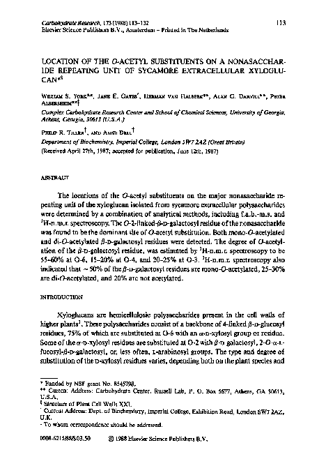
Fig. 1. Structures of the major oligosaccharide components of Bio-Gel P-2 fractions obtained after
B-1,4-endoglucanase digestion of SEPS xyloglucan. (A) Predominant component of the nonasaccharide fraction. This fraction also contains - 15% of a decasaccharide (B). (C) Predominant
component of the heptasaccharide fraction. The Tamarind XC oligosaccharides (also used in this
study) are composed of this heptasaccharide with B-Gal residues at O-2 of some of the cy-Xyl
residues. The asterisk (*) indicates the reducing residue.
�SYCAMORE EXTRACELLULAR XYLOOLUCAN
119
Fig. 2. Positive f.a.b.-mass spectrum of the material recovered after per-O-(deuterioacetyl)ation of
the native zyxwvutsrqponmlkjihgfedcbaZYXWVUTSRQPONMLKJIHGFEDCBA
(i.e., endogenously 0-acetylated) nonasaccharide fraction,
ing to the [M + HI’ (m/z 2493 and 2490) and [M + NH,J+ (m/z 2510 and 2507)
quasimolecular ions are of very low intensity. The dominance of A+-type ions in the
molecular-ion region of the f.a.b.-mass spectra of acetylated oligosaccharides had
been pr~iously reportedz3. Other A+-type fragment ions derived from no~~u~ing termini were observed, allowing glycosy1residues bearing endogenous O-acetyl
groups to be distinguished from those fully substituted with O-(deuterioacetyl)
groups. The ion at m/z 282 arises from [Fuc]+ (no acetate), whereas those at m/z
573 and 576 correspond to [(Fu~-G~)A~~+ and [(Fuc-Gal)Ac]+, respectively. The
�120
w. s. zyxwvutsrqponml
YORK, et al.
intensity of the signal at m/z 282 is considerably greater than that of the signals at
m/z 279 (potentially [(Fuc)Ac]+, see later) and m/z 579 (i.e., [Fuc-Gal]+). Therefore, the quantitatively dominant 0-acetylation site is the n-galactosyl residue, with
one or two 0-acetyl substituents. The minor signal observed at m/z 279, corresponding in mass to the fragment ion [(Fuc)Ac]+ expected if the fucosyl residue is
endogenously 0-acetylated, suggests that a small proportion of the fucosyl residues
are mono-O-acetylated. However, an ion of undetermined structure is routinely
observed at m/z 279 in positive mode f.a.b.-mass spectra of samples that have been
0-(deuterioacetyl)ated under acidic conditions’3s24. This ion is observed when
various different matrices are used to obtain spectra, even when the sample contains
no carbohydrate. Furthermore, in successive scans of the 0-(deuterioacetyl)ated
SEPS nonasaccharide fraction, the signal intensity at m/z 279 increased relative to
the intensities of known carbohydrate signals. This suggests that the ion at m/z 279
is due to the reagent impurity rather than to the carbohydrate sample. This interpretation is supported by ‘H-n.m.r. data, presented later, which indicate that the
L-fucosyl group is not significantly 0-acetylated.
Analysis by f.a.b.-m.s. also demonstrated that the D-xylosyl residue to which
the n-galactosyl residue is attached does not bear any endogenous 0-acetyl groups.
The presence of endogenous O-acetyl substituents on this D-xylosyl residue would be
expected to cause the ratio of the [(Fuc-Gal-Xyl)Ac] + signal (m/z 798) to the [(FucGal-Xyl)Ac2] + signal (m/z 795) to differ from the ratio of the [(Fuc-Gal)Ac] +
signal (m/z 576) to the [(Fuc-Gal)Ac2] + signal (m/z 573). However, the ratios of
these signals are not significantly different, indicating that the 2-linked D-xylosyl
residue is not 0-acetyfated.
The f.a.b.-m.s. data indicate that the nonasaccharide might have another,
quantitatively minor 0-acetylation site. A small but reproducible signal was observed at m/z 1081, corresponding to a nonreducing [(Xyl~Glc2)Ac]+ fragment.
We conclude from the f.a.b.-m.s. data that the major site of 0-acetyl substitution of the SEPS XG nonasaccharide is the n-galactosyl residue, which may be
either mono- or di-0-acetylated. Signal intensities in the f.a.b.-mass spectrum (see
Fig. 2) indicate that the ratio of mono- to di-0-acetylated n-galactosyl residues is
- 1.3: 1.O, and that small proportions of non-acetylated D-galactosyl residues are
also present.
‘H-N.m.r. spectroscopy of SEPS XG oligosaccharide fractions. - ‘H-N.m.r.
spectroscopy can be used to determine the position of 0-acetyl substituents on
carbohydrates, as these substituents strongly influence the chemical shifts of nearby
protons. Specifically, protons that are attached to carbon atoms bearing an 0-acetyl
group are deshielded by -0.5 to 1.5 p.p.m. relative to the corresponding protons in
the unsubstituted alcoho125. The signals arising from such protons are often
observed in the anomeric region of the ‘H-n.m.r. spectrum (6 4.2 to 5.5)25. The
locations of 0-acetyl substituents on the components of the native nonasaccharide
fraction were determined by assigning all of the signals between 6 4.2 and 5.5 in the
‘H-n.m.r. spectrum of the native nonasaccharide fraction, and determining which
�121
SYCAMOREEXTRACELLULARXYLOGLUCAN
of these signals were shifted out of this region by O-deacetylation.
‘H-N.m.r. (500 MHz) spectra of native and 0-deacetylated SEPS XG nonasaccharide fraction (see Figs. 3 and 4) in D20 were recorded at 27 and 75’. The
signal assignments were based on comparison of these spectra with those of closely
related xyloglucan oligosaccharides, and to published spectra of oligosaccharides
containing similar substructures, such as the human blood-group H antigen [a-~Fuc~-(1~2)-~-~-Gal&
Most of the spectra presented herein were recorded at 75” because, at this
temperature, the HDO signal was shifted out of the anomeric region. Also, considerably better resolution of certain complex groups of signals was obtained at 75O
than at 27”. However, it was observed that significant O-deacetylation occurred
during prolonged exposure of the native nonasaccharide fraction to the higher temperature. The chemical shifts of some of the signals exhibited slight temperaturedependence, but those of others did not. Thus, examination of the spectra recorded
at 27” allowed differentiation spin of systems that gave rise to overlapping signals in
the spectra at 75”. The conclusion regarding the sites of 0-acetylation in the native
XG nonasaccharide fraction were reached by combining the results obtained at 75O
and 27”. zyxwvutsrqponmlkjihgfedcbaZYXWVUTSRQPONMLKJIHGFEDCBA
‘H- N.m.r. spectroscopy of the 0- deacety lated SEPS XG nonasaccharidefraction. - The anomeric-proton signals of the 0-deacetylated SEPS XG nonasaccharide fraction (see Fig. 3B, and Table III) were assigned as follows. The doublets at 6
r”l’l”,‘II”‘II”‘1”“1
5.50
5.25
5.00
4.75
4.50
4.25
P.P.m,
Fig. 3. Anomeric region of the resolution-enhanced 500 MHz ‘H-n.m.r. spectra (90, 75”) of (A)
the native nonasaccharide fraction and (B) the Qdeacetylated nonasaccharide fraction. The
asterisk (*) indicates a reducing residue.
�w.
2.30
2.20
2.10
p.p.m.
2.00
s. YORK,
et al. zyxwvut
1.90
Fig. 4. Resolution-enhanced, partial, 5O&MHz ‘H-n.m.r. spectra of native nonasaccharide fraction
at 25” (A) and 75” (B). [All O-acetyl methyl proton signals are assigned as 0-acetyl substituents of
the 2-linked /3-wgaiactosyl residue. The location of the 0-acetyl substituent is indicated outside the
parentheses, and the substitution pattern for the residue is indicated in parentheses. For example,
the methyl proton signal of the acetyl group at O-6 of the 4,6-di-O-acetyl &o-galactosyi residue is
labelled 6-Ac(4,6-di-AC).]
5.224 (J,,z 4.0 Hz) and 4.659 (J1,2 7.9 Hz) were respectively assigned to H-l of the (Y
and @anomers of the reducing D-glucose residue, based on their chemical shifts and
coupling constants%, and the observation that these signaIs disappear upon reduction of the oligosaccharide with NaBH4. The doublets at 6 4.927 and 4.939 (J1,2 3.6
Hz) were assigned to H-l of the terminal a-D-xylosyl groups, based on the observation of signals with nearly identical couplng constants and chemical shifts in the
spectrum of a closely related heptasaccharide (see Fig. lC, and Table III) from
SEPS XG, which contains terminal cr-D-xylosyl groups as its only a-linked components2*27. Similarly, the doublet at 6 4.536 (J,,z 7.9 Hz) was assigned to H-l of the
nonreducing @-D-glucoseresidues. The signal at 6 5.13 1 (J1,2 3.6 Hz) was assigned to
H-l of the a-linked ar-D-xylosyl residue, based on the observation of signals with
similar chemical shifts and coupling constants in the spectra of related oligosaccharides obtained from Tamarind XG. The Tamarind XG oligosaccharides consist of
the heptasaccharide structure (see Fig. 1C) with terminal /3-D-galactosyl residues
�SYCAMORE EXTRACELLULAR
123 zyxwvutsrqp
XYLOOLUCAN
TABLE III zyxwvutsrqponmlkjihgfedcbaZYXWVUTSRQPONMLKJIHGFEDCBA
ASSIGNMENT OF SIGNALP IN THE ANOMER~C REGION OF TEIE ‘H-N.M.R.
SACCHARIDB FRACTIONS
Assignment
SPECTRA OF
SEPS XG
OLIOO-
zyxwvutsrqponmlkjihgfedcbaZYXWVUTSRQPONMLKJIHGFEDCBA
Heptasaccharide
fraction
O-Deacetylated
nonasaccharide
fraction
Native
nonasaccharide
fraction
5.283/5.295
(3.6,1.2)
4-O-AC-B-Gal*
H-4
5.282
(4-O)
5.259-5.284
(4.0)
5,224
(4.0)
5.222
(3.8)
2-Linked’ a-Xyl
H-l
5.131
(3.6)
5.127
(3.6)
2Liikedd CY-Xyl
H-l
5.111
(3.6)
5.030-5.116
(3.3)-(3.6)
U-FIX
H-l
Reducing or-Glc
H-l
5.223
(3.8)
3-O-Ac+Galb
H-3
T-a-Xyl
H-l
5.003/5.009
(3.3,lO.O)
4.921/4.941
(3.6)
4.927-4.939
(3.6)
4.927/4.939
(3.6)
3-0-AC-/3-Galb
H-l
4.726/4.727
(7.5)/(7.7)
CO-AC-&Galb
H-l
4.703/4.714
(7.7)
Reducing B-Glc
H-l
4.654
(7.9)
2-Linked &Gal
H-l
4.659
(7.9)
4.657
(8.0)
4.629
(7.6)
4.628
(7.9)
4.612
(8.2)
6-Mono-0-Ac+Galb
H-l
T-&Gal
H-l
&Glc
H-l
4.53814.563
4.564
(7.0)
4.564
(7.1)
4.536
4.531-4.549
(7.8)-(8.0)
(7.9)/(8.0)
zyxwvutsrqponmlkjihgfedcbaZYXWVUTSRQPONMLKJIHGFEDCBA
(7.9)
U-FUC
H-S
6-0-AC+Galb
H-6
4.453
(6.4,2.1)
4.456
(6.4,2.0)
4.369/4.375
(8.0,11.7)
�W. S. YORK,
et Q/.
124 zyxwvutsrqponmlkjihgfedcbaZYXWVUTSRQPONMLKJIHGFEDCBA
TABLE zyxwvutsrqponmlkjihgfedcbaZYXWVUTSRQPONMLKJIHGFEDCBA
III (continued)
6-O-AC-~-Gal6
H-6’
4.288/4.298
(4.4,11.8)
‘%hemical shifts are zyxwvutsrqponmlkjihgfedcbaZYXWVUTSRQPONMLKJIHGFEDCBA
given to three decimal places. Coupling constants are in parentheses, separated by a comma when more than one coupling constant applies to a given signal. A hyphen (-)
between values indicates that a range of values is given. A virgule {slash, /) separates discrete values
when more than one is given. b”3-0-Ac-fl-Gal”,
“4-O-AC-&Gal”, and “6-O-Ac-&Gal” refer to
fl-Gal residues having an acetyl group at O-3, -4, -6, respectively, regardless of whether the residue
has a second O-acetyl substituent, or not. However, H-l of 6-mono-0-acetyl-B-Gal is specifically
assigned (S 4.612). %-Xyl with terminal &Gal attached at O-2 (decasaccharide, see text}. %-Xyl
with rr-Fuc-(l-+2)-&Gal attached at O-2.
attached to zyxwvutsrqponmlkjihgfedcbaZYXWVUTSRQPONMLKJIHGFEDCBA
O- 2 of some of the a-D-xylosyl groups2’. The anomeric protons of the
%-linked cu-D-xylosylresidues give rise to doublets at positions ranging from 6 5.11 to
5.15 (J1,2 -4 Hz). On the basis of reported ~si~ments of the ‘H-n.m.r. spectra of
mammalian glycoprotein oligosaccharides containing the 2-O-cy-L-fucosyl-P-Dgalactosyl moietf’, the doublet (8 5.282, Jr,2 4.0 Hz) and the multiplet (S 4.453,
J4,5 1, J5,6 6.4 Hz) were respectively assigned to H-l and to H-5 of the (Y-Lfucosyl residue. Similarly, the doublet (8 4.629, Jr.2 7.6 Hz) was assigned to H-l
of the 2-linked-P-o-galactosyl residue.
The O-deacetylated SEPS nonasaccharide fraction contains - 15% of a decasaccharide’9820(see Fig. lB, f.a.b.-m.s_ section, and Tables I and II). The low intensity signals at S 5.131 (JIa2 3.6 Hz) and S 4.564 (Jr.2 7.0 Hz) can be attributed to
the anomeric protons of the second 2-linked cu-D-xylosylresidue and the terminal
/3-o-galactosyl group, respectively, of this decasaccharide.
‘H-N.m.r. spectroscopy of the native SEPS XG nonasaccharide fraction. The ‘H-n.m.r. spectra of the native SEPS XG nonasacch~ide fraction were
recorded at 27” and 75” (see Figs. 3A and 4). The degree of 0-acetylation of the
oligosaccharides was estimated by comparing (see Table IV) the signal integrals of
the 0-acetyl methyl protons (6 2.07 to 6 2.25) to those of the methyl protons of the
cr-L-fucosyl residue (S 1.26). The 27Ospectrum (see Fig. 4A) of a sample that had not
previously been exposed to high temperature was recorded, and the 75” spectrum
(see Fig. 4I3) of the same sample was subsequently recorded. The slight increase in
the intensity of the free acetate peak (S 1.9) indicated that some O-deacetylation
(- 10%) had occurred during exposure to high temperature. The estimated number
of 0-acetyl groups per oligosaccharide decreased from 1.l to 1.0 upon heating.
However, the same O-acetyl methyl signals were still present after heating, in
approximately the same proportions. The degree of O-acetylation of the nonasaccharide fraction varies slightly from preparation to prep~ation, but is typically
from 1.O to 1.2 O-acetyl groups per molecule.
The large number of 0-acetyl methyl singlets having non-stoichiometric intensity ratios indicates significant heterogeneity in the native SEPS nonasaccharide
fraction. Multiple signals for H-l of the 2-linked p-n-galactosyl residue (bearing
�SYCAM ORE EXTRACELLULAR XYLOGLUCAN
125
TABLE IV
INTIZ~RALS
ANDASWEDIENTSFOR'H-N.M.R.SIONALSDIA~JNOSTIC
FORTHE OACEWLATIONPA'ITBRN
lNllIENATIVES~PSXGNONASACCHARWEFlUCTlON
Assignment
6
IntegraF
4,6-di-O-AC-Gala
CO-AC-methyl protons*
2.113
0.30
3,6-di-O-AC-Gal
~-O-AC methyl protons
2.131
6-mono-OAc-Gal
6-O-AC-methyl protons
2.134 I
3-mono-O-AC-Gal
~-O-AC methyl protons
2.185
1.5od
3,Cdi-O-AC-Gal
3-O-AC-methyl protons
1
0.67d
2.188 zyxwvutsrqponmlkjihgfedcbaZYXWVUTSRQPONM
Cmono-O-AC-Gal
~-O-AC methyl protons
2.223
0.17
4,Cdi-O-AC-Gal
~-O-AC methyl protons
2.236
0.27
6-O-AC-Gal (ah)
H-6
4.288-4.296
0.57
(0.6W
6-0-AC-Gal (all)
H-6’
4.369-4.375f
0.55
(0.6W
3-0-AC-Gal (all)=
H-3
5.003KW
0.21
(0.22)s
4-0-AC-Gal (all)’
H-4
5.283/5.295/
0.15h
(0.15)s
“Molecular species. *Location of assigned proton. integrals relative to the signal of the methyl
protons of the a-L-fucosyl residue (3 1.26), taken as 3.0. dCertahr groups of signals (indicated by
the brackets) were not sufficiently resolved to permit individual determination of their integrals.
The value given is the total integral for the group of signals. The wordt ‘all’ in parentheses
indicates that the assigned signals arise from all of the 2-linked b-Gal residues having an O-acetyl
group at the indicated position, including both mono- and di-O-acetylated residues. ‘A hyphen (-)
between values indicates that a range of values is given. A virgule (slash, /) separates discrete values
when more than one is given. sTheoretical values for the integral, calculated from the integrals of
the corresponding O-acetyl methyl protons, are in parentheses. “The signal corresponding to H-4
of 4-O-acetyl- and 4,6-di-O-acetyl-n-galactosyl residues was obscured by H-l of the a-L-fucosyl
group in the 75’ spectrum of the nonasaccharide fraction. Therefore, the signal integral for this
proton was calculated from the 27” spectrum, where the signal was resolved. The other signal
integrals in this Table were taken from the 75’ spectrum, as more signals were resolved at this
temperature.
�W . S. YORK, t?t d. zyxwvutsrqp
r
5.5
5.0
4.5
4.0
3.5
3.0
p.Bm.
Fig. 5. Partial 2-D {‘H’H} CGSY spectrum and resolution-enhanced I-D spectrum of the native
SEFS XG nonasaccharide fraction recorded for a solution in 40 at 75’. [The connectivities for
the CO-acetylated &Gal systems are depicted with dashed lines (---), those for the 3-O-acetylated
@-Gal systems with dotted lines f****),and those for the ~~acetyiat~
&Gal systems with solid
lines (-).
The off~agon~
signals indicated by arrows were not assigned as part of these spin
systems, because they were shifted ~fferentially at the two temperatures at which CGSY spectra
were recorded. Off~agon~ reson~c~ due to JO,5in &Gal residues were not observed under the
experimental conditions used. See text for details.]
U-acetyl substituents), and for H-l of the adjacent a-l-fucosyf group and a-linked
cw-D-xylosyf.residue were also observed (see Fig. 3A)_ The assignment of the ano-
�SYCAMORE EXTRACELLULAR
XYLOGLUCAN
127
merit signals of the ar-r.-fucosylgroup and 2-linked U-D-xylosylresidue was based on
the assignment of the corresponding signals in the spectrum of the 0-deacetylated
nonasaccharide fraction (see Fig. 3 and Table III). The observation of characteristic
coupling patterns zyxwvutsrqponmlkjihgfedcbaZYXWVUTSRQPONMLKJIHGFEDC
(i.e., doublets with Jr,2 -4 Hz) in the J-resolved spectrum, and
characteristic connectivities in the COSY spectrum (see Fig. 5) of the native nonasaccharide fraction confirmed these assignments. The assignment of the signals
arising from the various O-acetylated, 2-linked P-n-galactosyl residues is described
later.
Several non-anomeric signals were observed (see Fig. 3A) in the anomeric
region (6 4.2 to 5.5) of the spectrum of the native nonasaccharide fraction. Only
anomeric protons and H-5 of the L-fucosylgroup were observed in this region of the
spectrum of the 0-deacetylated nonasaccharide fraction (see Fig. 3B). The origins of
the non-anomeric signals were assigned as follows. Two doublets of doublets at 6
4.37 (Je,6111.7 JsBa8.0 Hz) and 6 4.30 (Jc6’ 11.8, Js,al 4.4 Hz) were assigned to H-6
and H-6’ of 6-O-acetyl+?-D-galactosylresidues, based on the coupling patterns of
these signals and the establishment by f.a.b.-m.s. that the 6-n-galactosyl residue is
the dominant site of O-acetyl substitution (vide wpra). The 2-D {‘H’H} COSY
spectrum of the native SEPS XG nonasaccharide (see Fig. 5) indicated that these
two signals are J-coupled to each other (Je,6J- 12 Hz), as expected for the geminal
H-6 and H-6’ of aldohexoses. Inspection of these signals in the resolution-enhanced
1D spectrum (see Fig. 3A) showed that they are each composed of at least two
multiplets. The COSY spectrum shows that the high-intensity /3-n-galactosyl H-6
and H-6’ signals (6 4.368 and 4.288) are J-coupled to a proton resonant at 6 3.85,
while the low-intensity /3-n-galactosyl H-6 and H-6’ signals (S 4.375 and 4.290) are
J-coupled to a proton resonant at 6 3.93. These resonances at 6 3.85 and 3.93 were
therefore assigned to H-5 of the 6-O-acetyl-P-D-galactosylresidues. The detection of
two distinct resonances for H-5 of the 6-O-acetyl-/3-D-galactosylresidues can be
explained by assuming that some of the 6-O-acetyl-/3-D-galactosyl
residues also have
a second 0-acetyl substituent, strongly affecting the resonance of H-5 but only
weakly affecting the resonances of H-6 and H-6’. Other signals in the ‘H-n.m.r.
spectra (see later) confirmed that the native nonasaccharide fraction contains components having 4,6-di-0-acetyl-n-galactosyl and 3,6di-O-acetyl-n-galactosyl
residues.
Another complex group of non-anomeric signals appears, at 6 5.01, in the
‘H-n .m.r. spectrum of the native SEPS nonasaccharide fraction. These signals,
assigned to /3-D-galactosylH-3 atoms, were shifted downfield due to acetylation at
O-3. Two-dimensional J-resolved spectroscopy (data not shown) revealed that this
group of signals is composed of two doublets of doublets at 6 5.003 and 5.009 (each
with J3,4 3.3 Hz and J2,3 10.0 Hz). Off-diagonal signals in the COSY spectrum (see
Fig. 5) indicated that these signals are J-coupled to signals at 6 4.11 and 6 4.00 (H-4
and H-2, respectively, of 3-0-acetyl-b-p-galactosyl residues). In addition, the
signal(s) at 6 4.00 (H-2) show a connectivity to doublets at 6 4.72 (JQ 7.7 Hz). These
doublets (6 4.727 and 4.714) could, by virtue of their indirect connectivity to the
�128
w. s. YORK, et al.
multiplets at 6 5.01 and the coupling patterns of the ring protons of this system, be
assigned to the anomeric protons of 3-O-acetylated P-D-galactosyl residues. Thus,
‘H-n.m.r. analysis showed that -22% (see Table IV) of the P-D-galactopyranosyl
residue of the native XG nonasaccharide fraction are acetylated at O-3.
The presence of two similar but distinct multiplets at 65.01 suggested that
some of the 3-O-acetyl-P-D-galactosyl residues have a second 0-acetyl substituent,
which, when present, shifts the position of the H-3 signal by 0.006 p.p.m. It was
concluded that O-6 is the principal site of the second acetyl substituent on the
3-0-acetyl-D-galactosyl residues, because significant acetoxylation of the P-D-galactopyranosyl residues occurred at C-6, and some of the 6-0-acetylated D-galactopyranosyl residues appear to contain a second 0-acetyl substituent (vide supra). In
addition, no signals were detected that could be assigned to the ring protons of
3,4-di-0-acetylated /3-D-galactosyl residues. Therefore, the two multiplets at 6 5.009
and 5.003 arise from H-3 of 3-O-acetyl- and 3,6-di-0-acetyl-/3-D-galactopyranosyl
residues present in the native XG nonasaccharide fraction.
The only other possible site of 0-acetyl substitution of the 2-linked /3-D-galactosyl residues is O-4. A chemical shift between 6 3.85 and 4.0 for H-4 in nonacetylated 2-linked /3-D-galactosyl residues was expected2’. It was therefore expected
that the signal of H-4 of 4-0-acetylated @-D-galactosyl residues would exhibit a
chemical shift between 6 4.8 and 5.5, due to the a-effect of 0-acetylatior? (i.e.,
deshielding by some 1.O to 1.5 p.p.m. for protons attached to 0-acetylated ringcarbon atoms). It was also expected that the signal arising from H-4 of P-D-galactosyl residues would be a doublet of doublets with coupling constants of -3 to 4 Hz
(JJJ) and 1 Hz (J&. Signals that could be assigned to H-4 of 4-0-acetylated
/3-D-galactosyl residues, although quite weak, were observed at 6 5.295 and 5.283 in
the 75” spectrum of the native SEPS XG nonasaccharide fraction. These signals,
partially obscured by signals arising from H-l of the cr-L-fucosyl groups, were
detected because their connectivity in the COSY spectrum does not match that of the
cr-L-fucosyl H-l signals. Specifically, low-intensity signals at 6 - 5.29 (H-4) are
J-coupled, via signals at 6 4.06 (H-3) and 3.76 (H-2), to low-intensity signals at 6
4.70 (H-l). The chemical shifts (6 4.70) and coupling constants (Jr,2 7.7 Hz) of these
signals indicated that they arise from the anomeric protons of P-pyranosyl residues.
Thus, the pattern of J-connectivity evident in the COSY spectrum indicates that the
/3-anomeric protons at 6 4.70 are on the same residues as protons that are deshielded
by acetylation at O-4. The H-4 signals (6 5.295 and 5.283) are partially obscured by
the signals of the anomeric proton of the cr-L-fucosyl group, and so were examined
by 2D J-resolved spectroscopy. The coupling constants of these multiplets (& 3.6,
J4,5 1.2 Hz) make them difficult to distinguish from the adjacent ar-L-fucosyl H-l
signals, but are consistent with their assignment as H-4 of fi-D-galactosyl residues.
For the reasons already described (i.e., multiple signals for H-4 of 4-0-acetyl-P-Dgalactopyranosyl residues, the presence of 6-0-acetyl-O-D-galactopyranosyl residues
having a second 0-acetyl substituent, and the lack of signals that could be associated
with 3,4-di-O-acetyl-@-D-galactopyranosyl residues), it was concluded that the native
�SYCAMOREEXTRACELLULARXYLOGLUCAN
129
XG nonasaccharide fraction contains both 4-O-acetyl- and 4,6-di-O-acetyl-/3-Dgalactopyranosyl residues.
The f.a.b.-m.s. analysis (vide supra) indicated that the major site of O-acetylation on the native XG nonasaccharide fraction is the fl-D-galactosylresidue, but
does not preclude the possibility that the terminal ar+fucosyl group also bears
some 0-acetyl substituents. The n.m.r. data indicated that there are few, if any,
0-acetyl groups on the L-fucosyl group. The coupling pattern and chemical shift of
the signals at S 4.29 and 4.37 in the spectrum of the native nonasaccharide indicated
that these signals arise from H-6 of a hexosyl residue acetylated at O-6. These signals
obviously cannot be due to H-6 of the L-fucosyl (6-deoxy+galactosyl) group. The
only other signals of significant intensity that are shifted into the anomeric region of
the spectrum by 0-acetylation were assigned to H-3 and H-4 of 0-acetylated
D-galactosyl residues rather than L-fucosyl groups. This assignment was made
because the COSY spectra (at 27” and 75’) of the native nonasaccharide fraction
shows indirect connectivity between these signals and fl-anomeric proton signals
(Jr,2 7.7 Hz), ruling out the possibility that the signals arise from the fucosyl group,
which is a-linked. The integrals of the signals assigned to protons shifted into the
anomeric region by 0-acetylation correlate well with those of the 0-acetyl methyl
protons (ratios of 3:l; see later and Table IV); therefore, all of the significant
O-acetylation sites have been accounted for. It was concluded that 0-acetyl substitution of the L-fucosyl group is not quantitatively significant. zyxwvutsrqponmlkjihgfedc
Assignment of the 0- acety l methyl protons of the native SEPS XG nona-
The signals arising from the methyl protons of 0-acetyl
substituents were assigned on the basis of the following criteria. (1) The signal
integral for each proton attached to an acetoxylated carbon atom is one-third that of
the signal for the methyl protons of the corresponding 0-acetyl group. (2) The signal
integrals for the methyl protons of the two 0-acetyl groups on a particular di-Oacetyl-D-galactosylresidue are equal. (3) When two 0-acetyl substituents are present
on the same residue, the positions of signals arising from the 0-acetyl methyl
protons may be shifted, relative to the positions of the corresponding signals arising
from the mono-0-acetylated residue. Because the 0-acetyl substituents of 4,6-di-Oacetyl-D-galactopyranosyl residues are closer together in space than those of 3,6-di0-acetyl-D-galactopyranosyl residues, this shift should be of greater magnitude for
4,6-di-o-acetyl- than for 3,6-di-0-acetyl-D-galactosyl residues.
The most intense 0-acetyl methyl signal in the resolution-enhanced 75”
spectrum of the native nonasaccharide (see Fig. 4B) is the singlet at 6 2.134.
Therefore, this signal was assigned to the methyl protons of the 0-acetyl group on a
6-mono-O-acetyl-D-galactopyranosyl residue (see Table IV). The adjacent signals, at
6 2.131 and 2.113, were assigned to the methyl protons of the acetyl group on O-6 of
3,6-di-o-acetyl- and 4,6-di-0-acetyl-D-galactopyranosyl residues, respectively. As
expected, the 6-0-acetyl methyl signal is shifted more by the presence of the nearby
4-0-acetyl group of the 4,6-di-0-acetylated residue than by the relatively distant
3-O-acetyl group of the 3,6-di-0-acetylated residue. The total integral for the signals
saccharide fraction. -
�W. S. YORK, et id.
130 zyxwvutsrqponmlkjihgfedcbaZYXWVUTSRQPONMLKJIHGFEDCBA
assigned to H-6 of the various 6-O-acetylated /3-D-galactosyl residues (see Fig. 3A
and Table IV) is, within the accuracy of the measurements, one-third that of the
total integral of the signals assigned to the methyl protons of the acetyl groups at
O-6 of the D-galactosyl residues (see Table IV). Also, the integrals of the signals
assigned to the 4-0-acetyl methyl protons and 6-0-acetyl methyl protons of the
4,6-di-0-acetyI-D-galactosyl residues are equal (see Table IV). Thus, the criteria
already described for the assignment of the 0-acetyl methyl proton signals are
fulfilled for the acetyl substituents at O-6 of the @-D-galactosylresidues. Similar
reasoning was used to assign the signals of the methyl protons of other 0-acetyl
groups in the sample. In order to confirm the signal assignments for the 0-acetyl
methyl protons, the 1-D spectrum of a SEPS XC nonasaccharide subfraction that
was enriched in di-O-acetylated components was also examined (data not shown).
The ratio of signal intensities in this spectrum was also found consistent with the
assignments already given.
The assignment of the 0-acetyl methyl proton signals alIows estimation of the
proportion of the various 0-acetylated species present in the native SEPS XG
nonasaccharide fraction (see Table IV). The main site of 0-acetyl substitution of the
SEPS nonasaccharide is O-6 of the @-galactosyl residue (55-60%), with lesser
amounts of acetylation at O-3 (20-25%) and at O-4 (15%) of the D-galactosyl
residue. Of the oligosaccharides in the SEPS XG nonasaccharide fraction used for
this study, -5O-55% have one 0-acetyl substituent, 25-30% have two 0-acetyl
substituents, and U-20% are not acetylated. This is in rough agreement with the
estimates for the total degree of acetylation arrived at by f.a.b.-m.s. analysis (see
earlier).
Interestingly, although 6-mono-O-acetyl-D-galactosyl residues are abundant
in the nonasaccharide fraction, the fraction contains fewer 3-mono-O-acetyl-ogalactosyl residues than 3,6-di-0-acetyl-o-galactosyl residues, and fewer 4-mono-Oacetyl-D-galactosyl residues than 4,6-di-O-acetyl-o-galactosyl residues (see Fig. 4).
The changes in signal intensity observed upon heating the sample to 75” suggest that
the acetyl group at O-6 of the D-galactosyl residue is the most easily hydrolyzed (i.e.,
the ratio of 4,6-di-O-acetyl to 4-mono-0-acetyl signals, and the ratio of 3,6-di-Oacetyl to 3-mono-0-acetyl signals, both decreased). If significant 0-deacetylation at
O-6 of the D-galactosyl residue occurred during sample preparation, it is possible
that the 3-mono- and 4-mono-0-acetyl-D-galactosyl residues were derived from
residues originally acetylated at O-6 as well. That is, the polysaccharide produced by
the plant cells might not have significant levels of 3-mono-O-acetyl- and 4-mono-Oacetyl-D-galactosyl residues.
The heterogeneity with respect to the 0-acetylation pattern of the SEPS XG
nonasaccharide fraction could also result from migration of 0-acetyl groups on the
oligosaccharides. 0-Acetyl groups have been known to migrate on a given glycosyl
residue under physiological conditions2’. Therefore, it is possible that the native XG
nonasaccharide fraction may contain an equilibrium mixture of the various Oacetylated forms.
�SYCAMORE EXTRACELLULAR
XYLOGLUCAN zyxwvutsrqponmlkjihgfedcbaZYXWVUTSRQPO
131
The results presented herein demonstrate that a combination of f.a.b.-m.s.
and ‘H-n.m.r. analyses can be used to locate and quantitate the sites of 0-acetyl
substitution in a complex mixture of closely related oligosaccharides. The ability to
carry out such analyses is important because in some cases it may be impractical,
due to the migration of 0-acetyl groups, to isolate unique O-acetylated species for
chemical study.
Analysis by ‘H-n.m.r. and f.a.b.-m.s. of the oligosaccharides derived from
SEPS xyloglucan has shown that xyloglucans can exhibit more structural complexity
than previously imagined. During the course of this study, heterogeneity in the
glycosyl sequence of various XG oligosaccharides was also observed.
ACKNOWLEDGMENTS
The authors acknowledge the support of NSF grant No. 8545798. A.D. is
grateful for financial support from an MRC program grant. P.R.T. is a recipient of
an SERC studentship. The authors also thank Jerry R. Thomas for preparing the
B-1,6endoglucanase and Lisa Maynard for editorial assistance. zyxwvutsrqponmlkjihgfed
REFERENCES zyxwvutsrqponmlkjihgfedcbaZYXWVUTSRQPONMLKJIHGFEDCBA
1. M. McNm, A. Cl. DARVILL,
S. C. FRY,ANDP. ALBERSHEIM, zyxwvutsrqponmlkjihgfedcbaZYXW
Annu. Rev. Biochem., 53 (1984)
625-663.
2. W. D. BAUER,K. W. T ALMAWB,K. KEEOSTRA,
ANDP. ALEER~IEU, PlantPhy siol., 51 (1973)
174-187.
3. W. S. YORK,A. G. DARVLL,M. MCNEIL, T. T. SIWENSON, AND P. ALBEIUHEIM,
M etho&
Enzy mol., 118(1986)3-40.
4. W. S. YORK,A. G. DARVUL,
ANDP. ALBERSHHIM,
PlantPhy siol.,75 (1984) 295-297.
5. T. HAYASHIANDG. MACLACHLAN, PlantPhy siol.,75 (1984) 596- 604.
6. B. S. VALENT,A. G. DARVILL,
M. MCNEIL,B. K. ROBERTSEN,
ANDP. A~mmrmi, Carbohy dr.
Res., 79 (1980) 165-192.
7. J. LABA~ITCHANDP. RAY, PIantPhy siof.,53 (1974)669-673.
8. G. J. MCDOUGALL
ANDS. C. FRY,personalcommunication.
9. M. A. O’NEILL, V. J. MORRIS,R. R. SRLWNDIWN, I. W. SUTHERLAND,
ANDI. T. TAYLXXL
Carbohy dr.Res., 148 (1986) 63-69.
10. C. HARA,T. KIHO,ANDS. UKAI,Carbohy dr.Res., lll(1982) 143-150.
11.T. MATSUO
ANDT. MIZUNO,
Agric. Biol. Chem., 38 (1974) 465-466.
12. P. BIELY,C. R. MACKENZIE,J. PVLS,ANDH. SCHNEIIXR,Bio/Technofogy , 4 (1986)731-733.
~~.A.DBLLANDP. R.TELER, Biochem. Biophy s.Res. Commun., 135 (1986) 11261134.
14. I. J. GOLDSTEIN,
G. W. HAY,B. A. LEWIS,ANDF. SMITH,M ethodsCarbohy dr.Chem., 5 (1965)
361-370.
15.P. ALBERSHEIM,
D. J. NEVINS,P. D. ENGLISH,
ANDA. KARR,Carbohy dr. Res., 5 (1% 7)
340- 345.
16. T. J. WAEGIXE,
A. G. DARVILL,M. MCNEJL,ANDP. ALBERSHEIM,
Carbohy dr. Res., 123 (1983)
281-304.
17. A. B. BLAKENEY
AND B. A. STONE,Carbohy dr.Res., 140 (1985) 319-324.
18. D. P. SWEET,
R. H. SEAPIRO,ANDP. ALBERWEIM,Carbohy dr.Res., 40(1975)217-225.
19. Y. KATOANDK. MATSUDA,Agric. Biol. Chem., 44 (1980) 1759-1766.
20.Y. KATO,J. MATSUSHITA,
T. KUB~DBU, AND K. MATSUDA,
J. Biochem. (Toky o), 97 (1985)
801-810.
21. P. PREHM,Carbohy dr.Res., 78 (1980) 372-374.
22. N. K. KOC~TKOV AND 0. S. CHIWOV, Adv. Carbohy dr.Chem., 21(1966) 39-92.
�132
w. s. YORK,et al.
23. P.-K. TSAI,A. DELL,AND
zyxwvutsrqponmlkjihgfedcbaZYXWVUTSRQPONMLKJIHGFEDCB
C. E. BALLOU, Proc. Nod. Acad. Sci. U.S.A., 83 (1986) 4119-4123.
24. J. E. OATES,unpublished results.
25. J. HAVERUMP, H. VAN HALBEEK, L. DORLAND, AND J. F. G. VLIEGENTHART,Eur. J. Biochem.,
122 (1982) 305-311.
26. J. P. KAUERLING, L. DORLAND, H. VAN HALBEEK, J. F. G. VLIEGENTHART,M. MESSER,AND R.
SCHAUER, Carbohydr. Res., 100 (1982) 33 I-340.
27. P. KOOIMAN,
Reel. Trav. Chim. Pays-Bas, 80 (1961) 849-865.
28. H. VANHALBEEK, L. DORLAND, J. F. G. VLIEOENTHART,N. K. KOCHETKOV,N. P. ARBATSKY,
AND V. A. DEREVITSICAYA,Eur. J. Biochem., 127 (1982) 21-29.
29. J. P, KAMERLING, R. SCWJER, A. K. SIWKLA, A. STOLL, H, VAN HALBEEK, AND J. F. G.
VLIEGENTHART,Eur. J. Biochem., in press.
�
 Philip Tiller