WO2025075824A1 - Urine sediment interface with probabilistic classification binning and methods of producing and using same - Google Patents
Urine sediment interface with probabilistic classification binning and methods of producing and using same Download PDFInfo
- Publication number
- WO2025075824A1 WO2025075824A1 PCT/US2024/048154 US2024048154W WO2025075824A1 WO 2025075824 A1 WO2025075824 A1 WO 2025075824A1 US 2024048154 W US2024048154 W US 2024048154W WO 2025075824 A1 WO2025075824 A1 WO 2025075824A1
- Authority
- WO
- WIPO (PCT)
- Prior art keywords
- group
- particles
- unknown
- primary
- qualified
- Prior art date
- Legal status (The legal status is an assumption and is not a legal conclusion. Google has not performed a legal analysis and makes no representation as to the accuracy of the status listed.)
- Pending
Links
Classifications
-
- G—PHYSICS
- G06—COMPUTING OR CALCULATING; COUNTING
- G06T—IMAGE DATA PROCESSING OR GENERATION, IN GENERAL
- G06T7/00—Image analysis
- G06T7/0002—Inspection of images, e.g. flaw detection
- G06T7/0012—Biomedical image inspection
-
- G—PHYSICS
- G01—MEASURING; TESTING
- G01N—INVESTIGATING OR ANALYSING MATERIALS BY DETERMINING THEIR CHEMICAL OR PHYSICAL PROPERTIES
- G01N15/00—Investigating characteristics of particles; Investigating permeability, pore-volume or surface-area of porous materials
- G01N15/01—Investigating characteristics of particles; Investigating permeability, pore-volume or surface-area of porous materials specially adapted for biological cells, e.g. blood cells
-
- G—PHYSICS
- G01—MEASURING; TESTING
- G01N—INVESTIGATING OR ANALYSING MATERIALS BY DETERMINING THEIR CHEMICAL OR PHYSICAL PROPERTIES
- G01N15/00—Investigating characteristics of particles; Investigating permeability, pore-volume or surface-area of porous materials
- G01N15/02—Investigating particle size or size distribution
- G01N15/0205—Investigating particle size or size distribution by optical means
- G01N15/0227—Investigating particle size or size distribution by optical means using imaging; using holography
-
- G—PHYSICS
- G01—MEASURING; TESTING
- G01N—INVESTIGATING OR ANALYSING MATERIALS BY DETERMINING THEIR CHEMICAL OR PHYSICAL PROPERTIES
- G01N15/00—Investigating characteristics of particles; Investigating permeability, pore-volume or surface-area of porous materials
- G01N15/10—Investigating individual particles
- G01N15/14—Optical investigation techniques, e.g. flow cytometry
- G01N15/1429—Signal processing
- G01N15/1433—Signal processing using image recognition
-
- G—PHYSICS
- G16—INFORMATION AND COMMUNICATION TECHNOLOGY [ICT] SPECIALLY ADAPTED FOR SPECIFIC APPLICATION FIELDS
- G16H—HEALTHCARE INFORMATICS, i.e. INFORMATION AND COMMUNICATION TECHNOLOGY [ICT] SPECIALLY ADAPTED FOR THE HANDLING OR PROCESSING OF MEDICAL OR HEALTHCARE DATA
- G16H10/00—ICT specially adapted for the handling or processing of patient-related medical or healthcare data
- G16H10/40—ICT specially adapted for the handling or processing of patient-related medical or healthcare data for data related to laboratory analysis, e.g. patient specimen analysis
-
- G—PHYSICS
- G16—INFORMATION AND COMMUNICATION TECHNOLOGY [ICT] SPECIALLY ADAPTED FOR SPECIFIC APPLICATION FIELDS
- G16H—HEALTHCARE INFORMATICS, i.e. INFORMATION AND COMMUNICATION TECHNOLOGY [ICT] SPECIALLY ADAPTED FOR THE HANDLING OR PROCESSING OF MEDICAL OR HEALTHCARE DATA
- G16H50/00—ICT specially adapted for medical diagnosis, medical simulation or medical data mining; ICT specially adapted for detecting, monitoring or modelling epidemics or pandemics
- G16H50/20—ICT specially adapted for medical diagnosis, medical simulation or medical data mining; ICT specially adapted for detecting, monitoring or modelling epidemics or pandemics for computer-aided diagnosis, e.g. based on medical expert systems
-
- G—PHYSICS
- G01—MEASURING; TESTING
- G01N—INVESTIGATING OR ANALYSING MATERIALS BY DETERMINING THEIR CHEMICAL OR PHYSICAL PROPERTIES
- G01N15/00—Investigating characteristics of particles; Investigating permeability, pore-volume or surface-area of porous materials
- G01N15/02—Investigating particle size or size distribution
- G01N2015/0294—Particle shape
-
- G—PHYSICS
- G01—MEASURING; TESTING
- G01N—INVESTIGATING OR ANALYSING MATERIALS BY DETERMINING THEIR CHEMICAL OR PHYSICAL PROPERTIES
- G01N15/00—Investigating characteristics of particles; Investigating permeability, pore-volume or surface-area of porous materials
- G01N15/10—Investigating individual particles
- G01N2015/1006—Investigating individual particles for cytology
-
- G—PHYSICS
- G01—MEASURING; TESTING
- G01N—INVESTIGATING OR ANALYSING MATERIALS BY DETERMINING THEIR CHEMICAL OR PHYSICAL PROPERTIES
- G01N15/00—Investigating characteristics of particles; Investigating permeability, pore-volume or surface-area of porous materials
- G01N15/10—Investigating individual particles
- G01N15/14—Optical investigation techniques, e.g. flow cytometry
- G01N2015/1486—Counting the particles
-
- G—PHYSICS
- G01—MEASURING; TESTING
- G01N—INVESTIGATING OR ANALYSING MATERIALS BY DETERMINING THEIR CHEMICAL OR PHYSICAL PROPERTIES
- G01N15/00—Investigating characteristics of particles; Investigating permeability, pore-volume or surface-area of porous materials
- G01N15/10—Investigating individual particles
- G01N15/14—Optical investigation techniques, e.g. flow cytometry
- G01N2015/1493—Particle size
-
- G—PHYSICS
- G01—MEASURING; TESTING
- G01N—INVESTIGATING OR ANALYSING MATERIALS BY DETERMINING THEIR CHEMICAL OR PHYSICAL PROPERTIES
- G01N15/00—Investigating characteristics of particles; Investigating permeability, pore-volume or surface-area of porous materials
- G01N15/10—Investigating individual particles
- G01N15/14—Optical investigation techniques, e.g. flow cytometry
- G01N2015/1497—Particle shape
Definitions
- Urinalysis is essential to the diagnosis and treatment of patients suspected of various renal and urinary tract disorders.
- Urine samples of such suspected patients contain various analytes that are indicative of a pathological condition or stage of disease progression.
- urine samples from patients with kidney stones or urinary tract injury contain red blood cells; urine samples from patients with various inflammatory conditions or urinary tract infections contain white blood cells and/or bacteria.
- Accurate and timely results of urinalysis is critical for patient care.
- urinalysis is the physical, chemical, and microscopic examination of urine, and urinalysis involves a number of tests to detect and measure various analytes that pass through urine.
- Physical examination of urine samples provides information regarding its appearance, such as color, odor, and turbidity.
- Chemical examination of urine provides information regarding acidity (pH), specific gravity, and presence of proteins, sugars, ketones, bilirubin, nitrite, white blood cell products (leukocyte esterase), and blood.
- Microscopic examination of urine provides information regarding sediments present in urine, such as, for example, red blood cells (RBC), white blood cells (WBC), bacteria, epithelial cells, pathological casts, hyaline casts, crystals, yeast, mucus, and sperm.
- RBC red blood cells
- WBC white blood cells
- bacteria epithelial cells
- pathological casts hyaline casts
- crystals yeast, mucus, and sperm.
- Microscopic examination includes microscopy-based sediment urinalysis, which is a common detection technology utilized by clinical laboratories.
- Microscopy-based urine sediment analyzers evaluate urine samples for the presence of analytes based on an analyte's unique morphological characteristics including size, shape, and textural appearance.
- Microscopy-based detection technologies depend entirely on the morphological aspects of the formed elements present in a urine sample— no chemical reactions are involved in microscopy-based detection of urine sediment particles.
- Urine sediment elements may be detected manually using microscopy, or detected automatically using various automated sediment urine analyzers that utilize microscopy-based image technology.
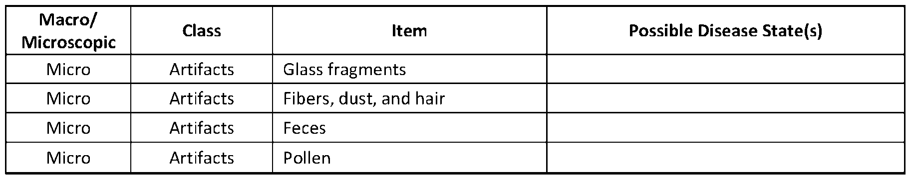
- Table 1 includes a list of the various macro/microscopic qualities/objects present in urine and their associated disease states.
- Standard automated urine sediment analyzers are based on either (1) imagebased or (2) flow cytometry technological and analytical principles.
- Image-based analysis systems depend solely on the microscopic morphological aspects of the sediment elements present in a urine sample and require no chemical reactions to detect analytes.
- Flow cell image-based automated urine sediment analyzers analyze urine sediment in dynamic fluid (ratherthan a static surface as in the cuvette-based method, as discussed below). A urine sample is forced to flow through a flow cell, and digital images are captured of the flowing urine sample when stroboscopic light flashes. An algorithm is then used to count and classify sediment particles based on morphological characteristics.
- cuvette image-based automated urine sediment analyzers a camera captures microscopic images of a urine sample in a cuvette and similarly relies on an algorithm to count and classify sediment particles based on morphological characteristics.
- the IRIS iQ200 system involves a flow cell, 500 images, and 12 particle types, and the operator can sub-classify particles into J sub-groups.
- Particles that are flagged by the IRIS iQ200 system include crystals, casts, yeast, non-squamous epithelial cells, artifacts, and other unclassified particles.
- the system sorts unknown material into an "Others" tab on the results interface and then sorts the unknown material only by perceived object size. Then an operator views images of the unknown material on the results interface and attempts to identify the particles with no input from the system.
- the Sysmex UF-lOOOi system also involves a urine flow cell, as well as two stains with fluorescent dye, and a separate bacterial channel. Particles that are flagged by the Sysmex UF-lOOOi system include casts, crystals, small round cells, sperm, yeast, and mucus. However, this system only provides a notification through the results interface that one or more unknowns are present; this system does not produce or display any images and does not have the capability to allow a user to even view— more less identify— the unknowns present.
- the Cobas u 701 system identifies 11 particle types and displays up to 15 images, and provides quantitative results for RBC and WBC and semi-quantitative results for bacteria, squamous epithelial cells, non-squamous epithelial cells, and hyaline casts. Particles that are flagged by the Cobas u 701 system include casts, crystals, yeast, sperm, and mucus. The images are labeled with the quantitative and semi-quantitative results but do not indicate the unknown particles or provide a way to label them.
- the Sedimax Contrust Pro system is similar to the Cobas u 701 system in that it displays on the results interface images labeled with identified particles, including RBC, acanthocytes (RBC-Aca), ghost RBC, WBC, WBC clumps, hyaline casts, pathological casts, squamous epithelial cells, non-squamous epithelial cells, bacteria cocci/rods, yeast, crystals, calcium-oxalate monohydrate, calcium-oxalate dihydrate, uric acid, triple phosphate, mucus, sperm, and amorphous material.
- unknown particles are not indicated or provided with a mechanism to label them; any attempt to indicate and/or identify unknown particles is performed completely manually.
- CLIA moderate complexity testing designation places requirements and restrictions for clinical laboratories performing the testing, as well as specific personnel requirements for testing personnel, laboratory director, clinical consultant, and technical consultant, in order to maintain CLIA licensing and FDA approval.
- FIG. 1 is a flow chart of one non-limiting embodiment of a method performed in accordance with the present disclosure.
- FIG. 2 is a graphic view of one non-limiting embodiment of a display of a results interface for an automated urinalysis system constructed in accordance with the present disclosure.
- FIG. 3 is a block diagram of one non-limiting embodiment of a results interface constructed in accordance with the present disclosure.
- non-transitory computer readable mediums, results interfaces, automated analyzers, and/or methods disclosed herein can be made and executed without undue experimentation in light of the present disclosure. While the non-transitory computer readable mediums, results interfaces, automated analyzers, and/or methods disclosed herein have been described in terms of particular embodiments, it will be apparent to those of skill in the art that variations may be applied to the non-transitory computer readable mediums, results interfaces, automated analyzers, and/or methods and in the steps or in the sequence of steps of the methods described herein without departing from the concept, spirit, and scope of the present disclosure. All such similar substitutes and modifications apparent to those skilled in the art are deemed to be within the spirit, scope, and concept of the inventive concept(s) as defined by the present disclosure and/or appended claims.
- the use of the term "at least one” will be understood to include one as well as any quantity more than one, including but not limited to, 2, 3, 4, 5, 10, 15, 20, 30, 40, 50, 100, etc.
- the term “at least one” may extend up to 100 or 1000 or more, depending on the term to which it is attached; in addition, the quantities of 100/1000 are not to be considered limiting, as higher limits may also produce satisfactory results.
- the use of the term "at least one of X, Y, and Z" will be understood to include X alone, Y alone, and Z alone, as well as any combination of X, Y, and Z.
- ordinal number terminology i.e., “first,” “second,” “third,” “fourth,” etc. is solely for the purpose of differentiating between two or more items and is not meant to imply any sequence or order or importance to one item over another or any order of addition, for example.
- any reference to "one embodiment,” “an embodiment,” “some embodiments,” “one example,” “for example,” or “an example” means that a particular element, feature, structure, or characteristic described in connection with the embodiment is included in at least one embodiment.
- the appearance of the phrase “in some embodiments” or “one example” in various places in the specification is not necessarily all referring to the same embodiment, for example. Further, all references to one or more embodiments or examples are to be construed as non-limiting to the claims.
- the terms “about” and “approximately” are used to indicate that a value includes the inherent variation of error for a composition/apparatus/ device, the method being employed to determine the value, or the variation that exists among the study subjects. That is, the terms “about” and “approximately” and variations thereof are intended to include not only the exact value qualified by the term, but to also include some slight deviations therefrom, such as deviations caused by measuring error, manufacturing tolerances, wear and tear on components or structures, settling or precipitation of cells or particles out of suspension or solution, chemical or biological degradation of solutions over time, stress exerted on structures, and combinations thereof, for example.
- the designated value may vary by plus or minus twenty percent, or fifteen percent, or twelve percent, or eleven percent, or ten percent, or nine percent, or eight percent, or seven percent, or six percent, or five percent, or four percent, or three percent, or two percent, or one percent from the specified value, as such variations are appropriate to perform the disclosed methods and as understood by persons having ordinary skill in the art.
- compositions, process, method, article, or apparatus that comprises a list of elements is not necessarily limited to only those elements, but may also include other elements not expressly listed or inherently present therein.
- the term “substantially” means that the subsequently described event or circumstance completely occurs or that the subsequently described event or circumstance occurs to a great extent or degree.
- the term “substantially” means that the subsequently described event or circumstance occurs at least 80% of the time, or at least 85% of the time, or at least 90% of the time, or at least 95% of the time.
- the term “substantially adjacent” may mean that two items are 100% adjacent to one another, or that the two items are within close proximity to one another but not 100% adjacent to one another, or that a portion of one of the two items is not 100% adjacent to the other item but is within close proximity to the other item.
- any reference to "one embodiment,” “an embodiment,” “some embodiments,” “one example,” “for example,” or “an example” means that a particular element, feature, structure, or characteristic described in connection with the embodiment is included in at least one embodiment.
- the appearance of the phrase “in some embodiments” or “one example” in various places in the specification is not necessarily all referring to the same embodiment, for example. Further, all references to one or more embodiments or examples are to be construed as non-limiting to the claims.
- Circuitry may be analog and/or digital components, or one or more suitably programmed processors (e.g., microprocessors) and associated hardware and software, or hardwired logic.
- processors e.g., microprocessors
- a “processing component” may perform one or more functions.
- the term “processing component,” may include hardware, such as a processor (e.g., microprocessor), an application specific integrated circuit (ASIC), field programmable gate array (FPGA), a combination of hardware and software, and/or the like.
- Software may include one or more computer readable instructions that when executed by one or more processing components cause the processing component to perform a specified function. It should be understood that the algorithms described herein may be stored on one or more non-transitory memory. Exemplary non-transitory memory may include random access memory, read only memory, flash memory, and/or the like. Such non-transitory memory may be electrically based, optically based, and/or the like.
- range of numerical values is recited or established herein, the range includes the endpoints thereof and all the individual integers and fractions within the range, and also includes each of the narrower ranges therein formed by all the various possible combinations of those endpoints and internal integers and fractions to form subgroups of the larger group of values within the stated range to the same extent as if each of those narrower ranges was explicitly recited.
- range of numerical values is stated herein as being greater than a stated value, the range is nevertheless finite and is bounded on its upper end by a value that is operable within the context of the invention as described herein.
- range of numerical values is stated herein as being less than a stated value, the range is nevertheless bounded on its lower end by a non-zero value.
- the term "user” includes but is not limited to a human being, and may comprise, a computer, a server, a website, a processor, a network interface, a human, a user terminal, a virtual computer, combinations thereof, and the like, for example.
- calibration parameters refers to a collection of data points or one or more functions used to derive a collection of data points that correlates the signals from the sensor to known analyte concentrations.
- the calibration parameters can be derived by a calibration algorithm, such as a linear algorithm, a spline-based algorithm, exponential algorithm, a least squares algorithm, a logarithmic algorithm, or the like that is configured to fit a function to at least two calibration points.
- calibration logic refers to the program logic used by a processing component to interpret data measured by one or more electrodes.
- calibration logic is the program logic used by a processing component to interpret data from an electrochemical sensor having at least a working electrode and a reference electrode.
- the present disclosure is directed to a method of performing urine sedimentation analysis, as well as various media, interfaces, and automated devices for performing at least a portion of said method.
- a urine sample is collected from a patient and brought to an automated analyzer.
- a small aliquot of the urine sample is exposed to a preprocessing step involving centrifugation to collect the solids present in the urine sample (wherein the solids include a variety of different types of particles).
- Images of the different particles are collected by the automated analyzer, and a count/identification takes place.
- difficult to assign particles are either thrown out, provided as a qualitative result, or handed over to a human for individual, manual review thereof (with no input from the system on the possible identity of the particles).
- qualified objects i.e., RBC, WBC, bacteria, squamous epithelial cells, non-squamous epithelial cells, and hyaline casts
- RBC Reactive Flow Control
- WBC Wideband Code Division Multiple Access
- bacteria squamous epithelial cells
- non-squamous epithelial cells e.g., squamous epithelial cells
- hyaline casts e.g., aline casts
- the remaining particles are then grouped (i.e., based on size, shape, color, and/or grouping) by similarity with one of the primary archetypes of casts, crystals, small round cells, sperm, yeast, and mucus.
- a representative example i.e., mean identification fit
- an image of a reference sample of the primary archetype group a "mean image recognition confidence value” (0-100%) and range (95 th confidence #% to #%) of confidence values for the group.
- a human operator may then confirm the identity of the unknown group, deny the suggested identity of the unknown group, or reassign each unknown group to a different group; optionally, the human operator can choose to view each individual image present in the group and then act (i.e., confirm, deny, reassign) on each image individually.
- the methods and devices of the present disclosure provide greater throughput when compared to the alternative of identifying difficult material by hand and also provide lower user ability requirements through machine assistance.
- the methods and devices have less liability risk than a fully automated system that assigns all values while providing better particle assignment than a fully automated system.
- the methods and devices of the present disclosure also provide better results with the use of full/partial automation of qualitative results for some materials.
- the use of reference images provided with the human input portion of the methods and devices may allow for a CLIA waiver, which is a great advantage over the weighty laboratory and personnel requirements associated with the current systems that have a CLIA moderate complexity designation.
- Certain non-limiting embodiments of the present disclosure are directed to a method of performing urine sedimentation analysis.
- the method includes the steps of: exposing a collected urine sample to a centrifugation preprocessing step to collect a plurality of particles present in the urine sample, wherein the plurality of particles contains qualified particles identifiable by an automated analyzer and unknown particles; imaging the plurality of particles; quantitatively counting qualified particles and displaying information related to the quantitatively counted qualified particles; grouping each unknown particle present based on at least one of size, shape, color, and grouping when compared to primary archetypes; calculating a mean image recognition confidence value and range of confidence values for at least one unknown particle when compared to one or more representative examples from the primary archetype; comparing an image of the at least one unknown particle to at least one reference image of one or more representative examples of the primary archetype in view of the calculated mean image recognition confidence value and range of confidence values; confirming an identity of the at least one unknown particle by comparison of the representative unknown particle image to the one or more reference example images and the
- Certain non-limiting embodiments of the present disclosure are directed to a method of performing urine sedimentation analysis.
- the method includes the steps of: exposing a collected urine sample to a centrifugation preprocessing step to collect a plurality of particles present in the urine sample, wherein the plurality of particles contains qualified particles identifiable by an automated analyzer and unknown particles; imaging the plurality of particles; quantitatively counting qualified particles and displaying information related to the quantitatively counted qualified particles; grouping each unknown particle present based on at least one of size, shape, color, and grouping when compared to primary archetypes to provide at least one group of unknown particles; calculating a mean image recognition confidence value and range of confidence values for each group of unknown particles when compared to one or more representative examples from the primary archetype; comparing a representative image of each group of unknown particles to at least one reference image of one or more representative examples of the primary archetype in view of the calculated mean image recognition confidence value and range of confidence values; confirming an identity of each group of unknown particles by comparison of the representative unknown particle image to
- the qualified particles are selected from the group comprising red blood cells, white blood cells, bacteria, squamous epithelial cells, non-squamous epithelial cells, and hyaline casts.
- non-qualified particles are selected from the group consisting of casts, crystals, small round cells, microbes, and mucus.
- Non-limiting examples of crystals that can be identified in accordance with the present disclosure include cystine, uric acid, leucine, tyrosine, cholesterol, urate, calcium oxalate, amorphous phosphate, triphosphate, ammonium magnesium phosphate, calcium phosphate, ammonium urate, drug crystals, and combinations thereof.
- Non-limiting examples of casts that can be identified in accordance with the present disclosure include hyaline, granular, waxy, renal, erythrocyte, leukocyte, fatty, lipid, hemoglobin and myoglobin, bacterial, mucus threads, and combinations thereof.
- Non-limiting examples of microbes that can be identified in accordance with the present disclosure include bacteria, yeast, trichomonads, Schistosoma haematobium eggs, Enterobius vermicularis, and combinations thereof.
- Certain non-limiting embodiments of the present disclosure include a non- transitory computer-readable medium having program elements which, when executed by a processing component of an automated analyzer, cause the automated analyzer to perform a method comprising the steps of: imaging a plurality of particles from a urine sample, wherein the plurality of particles contains qualified particles identifiable by an automated analyzer and unknown particles; quantitatively counting qualified particles and displaying information related to the quantitatively counted qualified particles on a portion of a results interface; grouping each unknown particle present based on at least one of size, shape, color, and grouping when compared to primary archetypes to provide at least one group of unknown particles; calculating a mean image recognition confidence value and range of confidence values for at least one unknown particle when compared to one or more representative examples from the primary archetype; displaying on a portion of the results interface an image of the at least one unknown particle along with at least one reference image of one or more representative examples from the primary archetype and the mean image recognition confidence value and range of confidence values; allowing a user
- Certain non-limiting embodiments of the present disclosure include a non- transitory computer-readable medium having program elements which, when executed by a processing component of an automated analyzer, cause the system to perform a method comprising the steps of: imaging a plurality of particles from a urine sample, wherein the plurality of particles contains qualified particles identifiable by an automated analyzer and unknown particles; quantitatively counting qualified particles and displaying information related to the quantitatively counted qualified particles on a portion of a results interface; grouping each unknown particle present based on at least one of size, shape, color, and grouping when compared to primary archetypes to provide at least one group of unknown particles; calculating a mean image recognition confidence value and range of confidence values for each group of unknown particles when compared to one or more representative examples from the primary archetype group; displaying on a portion of the results interface at least one representative image of each group of unknown particles along with at least one reference image of one or more representative examples from the primary archetype group and the mean image recognition confidence value and range of confidence values for each representative example
- Certain non-limiting embodiments of the present disclosure include a results interface for an automated analyzer configured to analyzer urine sedimentation, the results interface comprising a processing component, such as a processor and a display, wherein: the processing component is configured to: quantitatively count qualified particles in images of a urine sample; group at least one unknown particles present in the urine sample based on at least one of size, shape, color, and grouping when compared to primary archetypes; calculate a mean image recognition confidence value and range of confidence values for the at least one unknown particle when compared to one or more representative examples from the primary archetype group; and send signals to the display; and wherein the display is configured to: display information related to the quantitatively counted qualified particles; display at least one representative image of the at least one unknown particle along with at least one reference image of one or more representative examples from the primary archetype group and the mean image recognition confidence value and range of confidence values for each representative example; and receive confirmation from a user of an identity of the at least one unknown particle by comparison of the image of at least one unknown
- Certain non-limiting embodiments of the present disclosure include a results interface for an automated analyzer configured to analyze urine sedimentation, the results interface comprising a processing component, such as a processor and a display, wherein: the processing component is configured to: quantitatively count qualified particles in images of a urine sample; group images of unknown particles present in the urine sample based on at least one of size, shape, color, and grouping when compared to primary archetypes to provide at least one group of unknown particles; calculate a mean image recognition confidence value and range of confidence values for each group of unknown particles when compared to one or more representative examples from the primary archetype group; and send signals to the display; and wherein the display is configured to: display information related to the quantitatively counted qualified particles; display at least one representative image of each group of unknown particles along with at least one reference image of one or more representative examples from the primary archetype group and the mean image recognition confidence value and range of confidence values; and receive confirmation from user of an identity of each group of unknown particles by comparison of the at least one representative image of each group
- the display contains a separate tab for accessing information for each group of non-qualified particles.
- the processing component and display are housed within the same housing.
- the processing component and display may be housed separately.
- the processing component and display of the results interface may both be incorporated into the automated analyzer.
- the display may be provided in the form of an external monitor that is connected to the automated analyzer.
- Certain non-limiting embodiments of the present disclosure include an automated analyzer for performing any of the methods disclosed or otherwise contemplated herein.
- the automated analyzer may comprise any of the non-transitory computer-readable medium and/or results interfaces disclosed or otherwise contemplated herein.
- the automated analyzer comprises at least one additional component, such as (but not limited to), an imaging device and/or a centrifugation device.
- additional component such as (but not limited to), an imaging device and/or a centrifugation device.
- Imaging agents and centrifugation devices capable of functioning in accordance with the present disclosure are well known in the art and commercially available; therefore, no further description thereof is deemed necessary.
- the different components of the automated analyzer may be housed within the same housing (i.e., a single automated analyzer housing having a display for the results interface incorporated therein).
- one or more components of the automated analyzer may be housed separately from (but in communication with) the other components.
- the display of the results interface may be provided in the form of an external monitor that is connected to the remainder of the automated analyzer.
- FIG. 1 depicts one non-limiting embodiment of a flow chart for performing one non-limiting embodiment of the methods of the present disclosure.
- the method involves sample collection and sample preprocessing (i.e., centrifugation) followed by imaging of the particles present.
- the imaged particles are then categorized into particle types that a processing component of the automated analyzer is qualified to recognize and count on its own (i.e., qualified particles), and particle types that the processing component of the automated analyzer is not qualified to identify and that require human review thereof.
- particle types that the processing component of the automated analyzer is qualified to recognize and count, quantitative results are obtained and displayed on a results interface of the automated analyzer.
- the remaining "unknown" particles are then qualitatively analyzed and grouped (i.e., based on size, shape, color, and/or grouping) by similarity with the primary archetypes of casts, crystals, small round cells, sperm, yeast, and mucus. Then, four elements associated with the categorizing and qualitative review are displayed on a results interface related to each unique group of unknown particles: (1) an image of at least one unknown particle (or a representative example each group of unknown particles); (2) one or more
- Binned human review is then performed by a user comparing (1) to (2) while considering the values of (3) and (4), and the results interface provides a mechanism for the user to accept or reject the assignment of the identity of the unknown particle based on (2)-(4). Following the binned human review, quantitative results for the flagged particles are also provided in the results interface.
- FIG. 2 illustrates one non-limiting example of a display 10 of a results interface 11a (see FIG. 3) of an automated analyzer lib that performs the method of the present disclosure.
- the display 10 includes a fact box 12 that contains links to the quantitative results 14 for qualified particles and quantitative results 16 for non-qualified particles. Note that the quantitative results 16 are indicated as "pending" until binned human review is performed as described herein.
- the display 10 includes tabs 18 for toggling through each of the non-qualified particle groups (i.e., casts, crystals, small round cells, sperm, yeast, and mucus); when one or more unknown particles are present in a urine sample that is grouped into one of these non-qualified particle groups, the corresponding tab 18 will be activated to allow selection of that particular tab 18. Selection of the particular tab 18 causes display of a page 20 for the selected non-qualified particle group.
- the page 20 contains at least one section 22 containing at least one image or a group of images 24 of unknown particles for comparison to one or more reference images 26 from the primary archetype group, and also displays a mean image recognition confidence value 28 and a range (95 th confidence) 30 for the displayed reference image 26.
- the section 22 also includes an operator 32 for indicating whether or not the particle(s) in image(s) 24 belong to the proposed particle type of the displayed reference image 26. While this operator 32 is shown as comprising an arrow and an X for indicating that the proposed result is correct or not correct, respectively, it will be understood that these particular structures are for purposes of illustration only; any type of operatorthat allows a user to provide a "yes/no" response can be utilized as the operator 32.
- the page 20 may be provided with a single section 22, or multiple sections 22, depending on the particular non-qualified particle group being displayed. For example (but not by way of limitation), multiple different types of crystals may be present in the urine sample, and the display 10 can provide a mechanism for considering each crystal type individually. As illustrated in FIG.
- the page 20 may contain a second section 22' containing a single image or group of images 24' of unknown particles for comparison to one or more reference images 26', along with a mean image recognition confidence value 28' and a range (95 th confidence) 30' for the displayed reference image 26', as well as an operator 32' for indicating whether or not the pa rticle(s) in image(s) 24' belong to the proposed particle type of the displayed reference image 26'.
- results interface 11a may be provided with any quantitative results labels, tab labels, etc., so long as the results interface is capable of functioning as described herein.
- FIG. 3 illustrates a block diagram of an automated analyzer lib comprising the results interface 11a constructed in accordance with the present disclosure.
- the automated analyzer lib may be provided with the results interface 11a including a processing component 102 (e.g., processor), a non-transitory memory 104, a user interface / display 106.
- the automated analyzer lib may also be provided with a centrifugation device 108, an imaging device 110, and one or more communication path(s) 112.
- the processing component 102 e.g., processor
- the non-transitory memory 104, the user interface / display 106, the centrifugation device 108, and the imaging device 110 may communicate bidirectionally using the one or more communications path(s) 112.
- the one or more communications path(s) 112 include a data bus.
- the processing component 102 may perform a variety of higher-level processing functions of the automated analyzer lib. Such higher- level functions may be performed by executing computer-executable instructions (e.g., program code) stored in a non-transitory computer-readable medium, such as within the processing component 102 or within the memory 104.
- computer-executable instructions e.g., program code
- the processing component 102 is programmed, or otherwise configured to: quantitatively count qualified particles in images of a urine sample; group images of unknown particles present in the urine sample based on at least one of size, shape, color, and grouping when compared to primary archetypes to provide at least one unknown particle or group of unknown particles; calculate a mean image recognition confidence value and range of confidence values for each at least one unknown particle/group of unknown particles when compared to one or more representative examples from the primary archetype group; and send signals to the display 106.
- the memory 104 stores instructions that cause the processor 102 to analyze the sample in the manner described herein.
- the memory 104 may be a non-transitory computer- readable medium containing a variety of information, such as, but not limited to, a database of representative examples for each group of primary archetypes that are retrieved for display.
- the functionality of the display 106 may be implemented, at least in part, by computer-executable instructions (i.e., program code or software) stored in the memory 104 and executed by the processing component 102.
- the display 106 is configured to: display information related to the quantitatively counted qualified particles; display at least one representative image of at least one unknown particle or each group of unknown particles along with at least one reference image of one or more representative examples from the primary archetype group and the mean image recognition confidence value and range of confidence values for each representative example; and receive confirmation from user of an identity of each unknown particle or group of unknown particles by comparison of the at least one representative image of each unknown particle or group of unknown particles to the at least one reference image of one or more representative examples from the primary archetype group in view of the mean image recognition confidence values and range of confidence values for each reference example.
- the results interface 11a may be incorporated into a sidewall of a housing of the automated analyzer lib; alternatively, the user interface / display 106 of the results interface 11a may include a separate monitor that is attached to the communication path 112 of the automated analyzer lib.
- the processing component 102 may comprise one or more processing components 102 working together, or independently, to read and/or execute processor executable code and/or data, such as stored in the memory 104.
- the processing component(s) 102 may be capable of creating, manipulating, retrieving, altering, and/or storing data structures into the memory 104.
- Each element of the automated analyzer lib may be partially or completely network-based or cloud-based, and may or may not be located in a single physical location.
- Exemplary implementations of the processing component 102 may include, but are not limited to, a digital signal processor (DSP), a central processing unit (CPU), a field programmable gate array (FPGA), a microprocessor, a multi-core processor, an application specific integrated circuit (ASIC), combinations, thereof, and/or the like, for example.
- the processing component 102 may be capable of communicating with the memory 104 via the communication path 112 (e.g., data bus).
- the processing component 102 may be capable of communicating with the centrifugation device 108, the imaging device 110, the memory 104 and the user interface / display 106.
- the processing component 102 may instruct the centrifugation device 108 and the imaging device 110 to perform actions in accordance with the present disclosure (i.e., instruct the centrifugation device 108 to centrifuge a urine sample to collect solids, and instruct the imaging device 110 to capture images of particles present in the solids).
- Illustrative embodiment 1 Certain non-limiting embodiments of the present disclosure are directed to a method of performing urine sedimentation analysis. The method includes the steps of: exposing a collected urine sample to a centrifugation preprocessing step to collect a plurality of particles present in the urine sample, wherein the plurality of particles contains qualified particles identifiable by an automated analyzer and unknown particles; imaging the plurality of particles; quantitatively counting qualified particles and displaying information related to the quantitatively counted qualified particles; grouping each unknown particle present based on at least one of size, shape, color, and grouping when compared to primary archetypes; calculating a mean image recognition confidence value and range of confidence values for at least one unknown particle when compared to one or more representative examples from the primary archetype; comparing an image of the at least one unknown particle to at least one reference image of one or more representative examples of the primary archetype in view of the calculated mean image recognition confidence value and range of confidence values; confirming an identity of the at least one unknown particle by comparison of the representative unknown particle image to the one or
- Illustrative embodiment 2 A method of performing urine sedimentation analysis, the method comprising the steps of: exposing a collected urine sample to a centrifugation preprocessing step to collect a plurality of particles present in the urine sample, wherein the plurality of particles contains qualified particles identifiable by an automated analyzer and unknown particles; imaging the plurality of particles; quantitatively counting qualified particles and displaying information related to the quantitatively counted qualified particles; grouping each unknown particle present based on at least one of size, shape, color, and grouping when compared to primary archetypes to provide at least one group of unknown particles; calculating a mean image recognition confidence value and range of confidence values for each group of unknown particles when compared to one or more representative examples from the primary archetype; comparing a representative image of each group of unknown particles to at least one reference image of one or more representative examples of the primary archetype in view of the calculated mean image recognition confidence value and range of confidence values; confirming an identity of each group of unknown particles by comparison of the representative unknown particle image to the one or more reference example images
- Illustrative embodiment 3 The method of Illustrative embodiment 1 or 2, wherein the qualified particles are selected from the group comprising red blood cells, white blood cells, bacteria, squamous epithelial cells, non-squamous epithelial cells, and hyaline casts.
- Illustrative embodiment 4 The method of any of Illustrative embodiments 1-3, wherein the non-qualified particles are selected from the group consisting of casts, crystals, small round cells, microbes, and mucus.
- Illustrative embodiment 5 The method of Illustrative embodiment 4, wherein the crystals are selected from the group consisting of cystine, uric acid, leucine, tyrosine, cholesterol, urate, calcium oxalate, amorphous phosphate, triphosphate, ammonium magnesium phosphate, calcium phosphate, ammonium urate, drug crystals, and combinations thereof.
- Illustrative embodiment 6 The method of Illustrative embodiment 4 or5, wherein the casts are selected from the group consisting of hyaline, granular, waxy, renal, erythrocyte, leukocyte, fatty, lipid, hemoglobin and myoglobin, bacterial, mucus threads, and combinations thereof.
- Illustrative embodiment 7. The method of any of Illustrative embodiments 4-6, wherein the microbes are selected from the group consisting of bacteria, yeast, trichomonads, Schistosoma haematobium eggs, Enterobius vermicularis, and combinations thereof.
- Illustrative embodiment 8 A non-transitory computer-readable medium having program elements which, when executed by a computing unit of a system, cause the system to perform a method comprising the steps of: imaging a plurality of particles from a urine sample, wherein the plurality of particles contains qualified particles identifiable by an automated analyzer and unknown particles; quantitatively counting qualified particles and displaying information related to the quantitatively counted qualified particles on a portion of a results interface; grouping each unknown particle present based on at least one of size, shape, color, and grouping when compared to primary archetypes to provide at least one group of unknown particles; calculating a mean image recognition confidence value and range of confidence values for at least one unknown particle when compared to one or more representative examples from the primary archetype; displaying on a portion of the results interface an image of the at least one unknown particle along with at least one reference image of one or more representative examples from the primary archetype and the mean image recognition confidence value and range of confidence values; allowing a user to confirm an identity of the at least one
- Illustrative embodiment 9 A non-transitory computer-readable medium having program elements which, when executed by a computing unit of a system, cause the system to perform a method comprising the steps of: imaging a plurality of particles from a urine sample, wherein the plurality of particles contains qualified particles identifiable by an automated analyzer and unknown particles; quantitatively counting qualified particles and displaying information related to the quantitatively counted qualified particles on a portion of a results interface; grouping each unknown particle present based on at least one of size, shape, color, and grouping when compared to primary archetypes to provide at least one group of unknown particles; calculating a mean image recognition confidence value and range of confidence values for each group of unknown particles when compared to one or more representative examples from the primary archetype; displaying on a portion of the results interface at least one representative image of each group of unknown particles along with at least one reference image of one or more representative examples from the primary archetype and the mean image recognition confidence value and range of confidence values; allowing a user to confirm an identity of each group
- Illustrative embodiment 10 The medium of Illustrative embodiment 8 or 9, wherein the qualified particles are selected from the group comprising red blood cells, white blood cells, bacteria, squamous epithelial cells, non-squamous epithelial cells, and hyaline casts.
- Illustrative embodiment 11 The medium of any of Illustrative embodiments 8-10, wherein the non-qualified particles are selected from the group consisting of casts, crystals, small round cells, microbes, and mucus.
- Illustrative embodiment 12 The medium of Illustrative embodiment 11, wherein the crystals are selected from the group consisting of cystine, uric acid, leucine, tyrosine, cholesterol, urate, calcium oxalate, amorphous phosphate, triphosphate, ammonium magnesium phosphate, calcium phosphate, ammonium urate, drug crystals, and combinations thereof.
- Illustrative embodiment 13 The medium of Illustrative embodiment 11 or 12, wherein the casts are selected from the group consisting of hyaline, granular, waxy, renal, erythrocyte, leukocyte, fatty, lipid, hemoglobin and myoglobin, bacterial, mucus threads, and combinations thereof.
- Illustrative embodiment 14 The medium of any of Illustrative embodiments 11- 13, wherein the microbes are selected from the group consisting of bacteria, yeast, trichomonads, Schistosoma haematobium eggs, Enterobius vermicularis, and combinations thereof.
- a results interface for an automated urine sedimentation analyzer comprising a processor and a display, wherein: the processor is configured to: quantitatively count qualified particles in images of a urine sample; group at least one unknown particles present in the urine sample based on at least one of size, shape, color, and grouping when compared to primary archetypes; calculate a mean image recognition confidence value and range of confidence values for the at least one unknown particle when compared to one or more representative examples from the primary archetype group; and send signals to the display; and wherein the display is configured to: display information related to the quantitatively counted qualified particles; display at least one representative image of the at least one unknown particle along with at least one reference image of one or more representative examples from the primary archetype group and the mean image recognition confidence value and range of confidence values for each representative example; and receive confirmation from user of an identity of the at least one unknown particle by comparison of the image of at least one unknown particle to the at least one reference image of one or more representative examples from the primary archetype
- a results interface for an automated urine sedimentation analyzer comprising a processor and a display, wherein: the processor is configured to: quantitatively count qualified particles in images of a urine sample; group images of unknown particles present in the urine sample based on at least one of size, shape, color, and grouping when compared to primary archetypes to provide at least one group of unknown particles; calculate a mean image recognition confidence value and range of confidence values for each group of unknown particles when compared to one or more representative examples from the primary archetype group; and send signals to the display; and wherein the display is configured to: display information related to the quantitatively counted qualified particles; display at least one representative image of each group of unknown particles along with at least one reference image of one or more representative examples from the primary archetype group and the mean image recognition confidence value and range of confidence values; and receive confirmation from user of an identity of each group of unknown particles by comparison of the at least one representative image of each group of unknown particles to the at least one reference image of one or more representative examples from
- Illustrative embodiment 17 The results interface of Illustrative embodiment 15 or 16, wherein the qualified particles are selected from the group comprising red blood cells, white blood cells, bacteria, squamous epithelial cells, non-squamous epithelial cells, and hyaline casts.
- Illustrative embodiment 18 The results interface of any of Illustrative embodiments 15-17, wherein the non-qualified particles are selected from the group consisting of casts, crystals, small round cells, microbes, and mucus.
- Illustrative embodiment 19 The results interface of Illustrative embodiment 18, wherein the crystals are selected from the group consisting of cystine, uric acid, leucine, tyrosine, cholesterol, urate, calcium oxalate, amorphous phosphate, triphosphate, ammonium magnesium phosphate, calcium phosphate, ammonium urate, drug crystals, and combinations thereof.
- Illustrative embodiment 20 The results interface of Illustrative embodiment 18 or 19, wherein the casts are selected from the group consisting of hyaline, granular, waxy, renal, erythrocyte, leukocyte, fatty, lipid, hemoglobin and myoglobin, bacterial, mucus threads, and combinations thereof.
- Illustrative embodiment 21 The results interface of any of Illustrative embodiments 18-20, wherein the microbes are selected from the group consisting of bacteria, yeast, trichomonads, Schistosoma haematobium eggs, Enterobius vermicularis, and combinations thereof.
- Illustrative embodiment 22 The results interface of any of Illustrative embodiments 15-21, wherein the display contains a separate tab for accessing information for each group of non-qualified particles.
- Illustrative embodiment 23 An automated analyzer comprising: an imaging device; and the results interface of any of Illustrative embodiments 15-22.
- Illustrative embodiment 24 The automated analyzer of Illustrative embodiment 23, further comprising a centrifugation device.
Landscapes
- Engineering & Computer Science (AREA)
- Health & Medical Sciences (AREA)
- Chemical & Material Sciences (AREA)
- General Health & Medical Sciences (AREA)
- General Physics & Mathematics (AREA)
- Physics & Mathematics (AREA)
- Pathology (AREA)
- Medical Informatics (AREA)
- Biochemistry (AREA)
- Immunology (AREA)
- Analytical Chemistry (AREA)
- Life Sciences & Earth Sciences (AREA)
- Dispersion Chemistry (AREA)
- Public Health (AREA)
- Epidemiology (AREA)
- Biomedical Technology (AREA)
- Primary Health Care (AREA)
- Databases & Information Systems (AREA)
- Data Mining & Analysis (AREA)
- Signal Processing (AREA)
- Nuclear Medicine, Radiotherapy & Molecular Imaging (AREA)
- Radiology & Medical Imaging (AREA)
- Quality & Reliability (AREA)
- Computer Vision & Pattern Recognition (AREA)
- Theoretical Computer Science (AREA)
- Investigating Or Analysing Biological Materials (AREA)
Abstract
Methods of performing urine sedimentation analysis that utilize an interface with probabilistic classification binning are disclosed, along with non-transitory computer-readable media, results interfaces, and automated analyzers for performing the methods.
Description
TITLE
URINE SEDIMENT INTERFACE WITH PROBABILISTIC CLASSIFICATION BINNING AND METHODS OF PRODUCING AND USING SAME
REFERENCE TO RELATED APPLICATIONS
[0001] This application claims benefit under 35 USC § 119(e) of U.S. Provisional Application No. 63/587,175, filed October 2, 2023. The entire contents of the abovereferenced patent application are hereby expressly incorporated herein by reference.
FEDERALLY SPONSORED RESEARCH OR DEVELOPMENT
[0002] Not applicable.
BACKGROUND
[0003] Urinalysis is essential to the diagnosis and treatment of patients suspected of various renal and urinary tract disorders. Urine samples of such suspected patients contain various analytes that are indicative of a pathological condition or stage of disease progression. For example, urine samples from patients with kidney stones or urinary tract injury contain red blood cells; urine samples from patients with various inflammatory conditions or urinary tract infections contain white blood cells and/or bacteria. Accurate and timely results of urinalysis is critical for patient care.
[0004] In particular, urinalysis is the physical, chemical, and microscopic examination of urine, and urinalysis involves a number of tests to detect and measure various analytes that pass through urine. Physical examination of urine samples provides information regarding its appearance, such as color, odor, and turbidity. Chemical examination of urine provides information regarding acidity (pH), specific gravity, and presence of proteins, sugars, ketones, bilirubin, nitrite, white blood cell products (leukocyte esterase), and blood. Microscopic examination of urine provides information regarding sediments present in urine, such as, for example, red blood cells (RBC), white blood cells (WBC), bacteria, epithelial cells, pathological casts, hyaline casts, crystals, yeast, mucus, and sperm.
[0005] Microscopic examination includes microscopy-based sediment urinalysis, which is a common detection technology utilized by clinical laboratories. Microscopy-based urine sediment analyzers evaluate urine samples for the presence of analytes based on an analyte's
unique morphological characteristics including size, shape, and textural appearance. Microscopy-based detection technologies depend entirely on the morphological aspects of the formed elements present in a urine sample— no chemical reactions are involved in microscopy-based detection of urine sediment particles. Urine sediment elements may be detected manually using microscopy, or detected automatically using various automated sediment urine analyzers that utilize microscopy-based image technology.
[0006] Table 1 includes a list of the various macro/microscopic qualities/objects present in urine and their associated disease states.
TABLE 1


[0007] Standard automated urine sediment analyzers are based on either (1) imagebased or (2) flow cytometry technological and analytical principles. Image-based analysis systems depend solely on the microscopic morphological aspects of the sediment elements present in a urine sample and require no chemical reactions to detect analytes. There are two basic designs for image-based systems: flow cell and cuvette (sample holder) systems. Flow cell image-based automated urine sediment analyzers analyze urine sediment in dynamic fluid (ratherthan a static surface as in the cuvette-based method, as discussed below). A urine sample is forced to flow through a flow cell, and digital images are captured of the flowing urine sample when stroboscopic light flashes. An algorithm is then used to count and classify sediment particles based on morphological characteristics. In cuvette image-based automated urine sediment analyzers, a camera captures microscopic images of a urine sample in a cuvette and similarly relies on an algorithm to count and classify sediment particles based on morphological characteristics.
[0008] Current urine sediment analysis methods have several drawbacks. When urine sediment analysis is performed manually, the Clinical Laboratory Improvement Amendments (CLIA) Act mandates that personnel are certified. When urine sediment analysis is automated, the results can be unreliable. Although automated urinalysis systems with a certified central laboratory performing microscopy are time saving, standardized, and cost-effective, they are not reliable to diagnose various kidney diseases such as ATN, glomerulonephritis, vasculitis, or crystalline-related kidney diseases (Cavanaugh, et al. (2018) Core Curriculum, 73(2):P258- 272). This is due to the presence of flagged particles within the sample that cannot be resolved by the automated analyzer.
[0009] Various automated analyzer systems have been developed with mechanisms to potentially flag particles and/or provide a flagged particle interface. These systems include IRIS iQ200 (Beckman Coulter, Brea, CA), UF-lOOOi (Sysmex, Lincolnshire, IL), Cobas u 701 (Roche Diagnostics, Indianapolis, IN), Sedimax Contrust Pro (A. Menarini Diagnostics, Florence
Italy), and Hemoscreen™ (PixCell Medical Technologies, Yokneam lllit, Israel). A brief summary of each system is provided herein below.
[0010] The IRIS iQ200 system involves a flow cell, 500 images, and 12 particle types, and the operator can sub-classify particles into J sub-groups. Particles that are flagged by the IRIS iQ200 system include crystals, casts, yeast, non-squamous epithelial cells, artifacts, and other unclassified particles. The system sorts unknown material into an "Others" tab on the results interface and then sorts the unknown material only by perceived object size. Then an operator views images of the unknown material on the results interface and attempts to identify the particles with no input from the system.
[0011] The Sysmex UF-lOOOi system also involves a urine flow cell, as well as two stains with fluorescent dye, and a separate bacterial channel. Particles that are flagged by the Sysmex UF-lOOOi system include casts, crystals, small round cells, sperm, yeast, and mucus. However, this system only provides a notification through the results interface that one or more unknowns are present; this system does not produce or display any images and does not have the capability to allow a user to even view— more less identify— the unknowns present.
[0012] The Cobas u 701 system identifies 11 particle types and displays up to 15 images, and provides quantitative results for RBC and WBC and semi-quantitative results for bacteria, squamous epithelial cells, non-squamous epithelial cells, and hyaline casts. Particles that are flagged by the Cobas u 701 system include casts, crystals, yeast, sperm, and mucus. The images are labeled with the quantitative and semi-quantitative results but do not indicate the unknown particles or provide a way to label them.
[0013] The Sedimax Contrust Pro system is similar to the Cobas u 701 system in that it displays on the results interface images labeled with identified particles, including RBC, acanthocytes (RBC-Aca), ghost RBC, WBC, WBC clumps, hyaline casts, pathological casts, squamous epithelial cells, non-squamous epithelial cells, bacteria cocci/rods, yeast, crystals, calcium-oxalate monohydrate, calcium-oxalate dihydrate, uric acid, triple phosphate, mucus, sperm, and amorphous material. However, like the previous systems, unknown particles are not indicated or provided with a mechanism to label them; any attempt to indicate and/or identify unknown particles is performed completely manually.
[0014] In the PixCell Hemoscreen™ system, whole blood material flows into a single moving plane, and counts are provided for the following: RBC, WBC, platelets, neutrophils,
lymphocytes, monocytes, eosinophils, and basophils, alone and with various ratios. Like the Sysmex UF-lOOOi system, this system does not produce or display any images and does not have the capability to allow a user to even view— more less identify— the unknowns present. The results display for this system simply indicates a value for each particle type and indicates if this value falls outside the reference interval therefor.
[0015] With respect to the complexity of these systems, all of the above systems have received a "CLIA moderate complexity" designation from the FDA (with the exception of the Sedimax Contrust Pro system, which has not received an FDA Designation). A CLIA moderate complexity testing designation places requirements and restrictions for clinical laboratories performing the testing, as well as specific personnel requirements for testing personnel, laboratory director, clinical consultant, and technical consultant, in order to maintain CLIA licensing and FDA approval.
BRIEF DESCRIPTIONS OF THE DRAWINGS
[0016] FIG. 1 is a flow chart of one non-limiting embodiment of a method performed in accordance with the present disclosure.
[0017] FIG. 2 is a graphic view of one non-limiting embodiment of a display of a results interface for an automated urinalysis system constructed in accordance with the present disclosure.
[0018] FIG. 3 is a block diagram of one non-limiting embodiment of a results interface constructed in accordance with the present disclosure.
DETAILED DESCRIPTION
[0019] Before explaining at least one embodiment of the present disclosure in detail, it is to be understood that the present disclosure is not limited in its application to the details of construction and the arrangement of the components or steps or methodologies set forth in the following description or illustrated in the drawings. The present disclosure is capable of other embodiments or of being practiced or carried out in various ways. Also, it is to be understood that the phraseology and terminology employed herein is for the purpose of description and should not be regarded as limiting in any way.
[0020] Independent of the grammatical term usage, individuals with male, female, or other gender identities are included within the term.
[0021] Unless otherwise defined herein, scientific and technical terms used in connection with the present disclosure shall have the meanings that are commonly understood by those of ordinary skill in the art. Further, unless otherwise required by context, singular terms shall include pluralities and plural terms shall include the singular. The foregoing techniques and procedures are generally performed according to conventional methods well known in the art and as described in various general and more specific references that are cited and discussed throughout the present specification. The nomenclatures utilized in connection with, and the laboratory procedures and techniques of, analytical chemistry, synthetic organic chemistry, and medicinal and pharmaceutical chemistry described herein are those well- known and commonly used in the art. Standard techniques are used for chemical syntheses and chemical analyses.
[0022] All patents, published patent applications, and non-patent publications mentioned in the specification are indicative of the level of skill of those skilled in the art to which the present disclosure pertains. All patents, published patent applications, and non-patent publications referenced in any portion of this application are herein expressly incorporated by reference in their entirety to the same extent as if each individual patent or publication was specifically and individually indicated to be incorporated by reference.
[0023] All of the non-transitory computer readable mediums, results interfaces, automated analyzers, and/or methods disclosed herein can be made and executed without undue experimentation in light of the present disclosure. While the non-transitory computer readable mediums, results interfaces, automated analyzers, and/or methods disclosed herein have been described in terms of particular embodiments, it will be apparent to those of skill in the art that variations may be applied to the non-transitory computer readable mediums, results interfaces, automated analyzers, and/or methods and in the steps or in the sequence of steps of the methods described herein without departing from the concept, spirit, and scope of the present disclosure. All such similar substitutes and modifications apparent to those skilled in the art are deemed to be within the spirit, scope, and concept of the inventive concept(s) as defined by the present disclosure and/or appended claims.
[0024] As utilized in accordance with the present disclosure, the following terms, unless otherwise indicated, shall be understood to have the following meanings:
[0025] The use of the term "a" or "an" when used in conjunction with the term "comprising" in the claims and/or the specification may mean "one," but it is also consistent
with the meaning of "one or more/' "at least one," and "one or more than one." As such, the terms "a," "an," and "the" include plural referents unless the context clearly indicates otherwise. Thus, for example, reference to "a compound" may refer to one or more compounds, two or more compounds, three or more compounds, four or more compounds, or greater numbers of compounds. The term "plurality" refers to "two or more."
[0026] The use of the term "at least one" will be understood to include one as well as any quantity more than one, including but not limited to, 2, 3, 4, 5, 10, 15, 20, 30, 40, 50, 100, etc. The term "at least one" may extend up to 100 or 1000 or more, depending on the term to which it is attached; in addition, the quantities of 100/1000 are not to be considered limiting, as higher limits may also produce satisfactory results. In addition, the use of the term "at least one of X, Y, and Z" will be understood to include X alone, Y alone, and Z alone, as well as any combination of X, Y, and Z. The use of ordinal number terminology (i.e., "first," "second," "third," "fourth," etc.) is solely for the purpose of differentiating between two or more items and is not meant to imply any sequence or order or importance to one item over another or any order of addition, for example.
[0027] The use of the term "or" in the claims is used to mean an inclusive "and/or" unless explicitly indicated to refer to alternatives only or unless the alternatives are mutually exclusive. For example, a condition "A or B" is satisfied by any of the following: A is true (or present) and B is false (or not present), A is false (or not present) and B is true (or present), and both A and B are true (or present).
[0028] As used herein, any reference to "one embodiment," "an embodiment," "some embodiments," "one example," "for example," or "an example" means that a particular element, feature, structure, or characteristic described in connection with the embodiment is included in at least one embodiment. The appearance of the phrase "in some embodiments" or "one example" in various places in the specification is not necessarily all referring to the same embodiment, for example. Further, all references to one or more embodiments or examples are to be construed as non-limiting to the claims.
[0029] Throughout this application, the terms "about" and "approximately" are used to indicate that a value includes the inherent variation of error for a composition/apparatus/ device, the method being employed to determine the value, or the variation that exists among the study subjects. That is, the terms "about" and "approximately" and variations thereof are intended to include not only the exact value qualified by the term, but to also
include some slight deviations therefrom, such as deviations caused by measuring error, manufacturing tolerances, wear and tear on components or structures, settling or precipitation of cells or particles out of suspension or solution, chemical or biological degradation of solutions over time, stress exerted on structures, and combinations thereof, for example. In particular, for example, but not by way of limitation, when the term "about" is utilized, the designated value may vary by plus or minus twenty percent, or fifteen percent, or twelve percent, or eleven percent, or ten percent, or nine percent, or eight percent, or seven percent, or six percent, or five percent, or four percent, or three percent, or two percent, or one percent from the specified value, as such variations are appropriate to perform the disclosed methods and as understood by persons having ordinary skill in the art. [0030] As used in this specification and claim(s), the words "comprising" (and any form of comprising, such as "comprise" and "comprises"), "having" (and any form of having, such as "have" and "has"), "including" (and any form of including, such as "includes" and "include"), or "containing" (and any form of containing, such as "contains" and "contain") are inclusive or open-ended and do not exclude additional, unrecited elements or method steps. For example, unless otherwise noted, a composition, process, method, article, or apparatus that comprises a list of elements is not necessarily limited to only those elements, but may also include other elements not expressly listed or inherently present therein.
[0031] The term "or combinations thereof" as used herein refers to all permutations and combinations of the listed items preceding the term. For example, "A, B, C, or combinations thereof" is intended to include at least one of: A, B, C, AB, AC, BC, or ABC, and if order is important in a particular context, also BA, CA, CB, CBA, BCA, ACB, BAC, or CAB. Continuing with this example, expressly included are combinations that contain repeats of one or more item or term, such as BB, AAA, AAB, BBC, AAABCCCC, CBBAAA, CABABB, and so forth. The skilled artisan will understand that typically there is no limit on the number of items or terms in any combination, unless otherwise apparent from the context.
[0032] As used herein, the term "substantially" means that the subsequently described event or circumstance completely occurs or that the subsequently described event or circumstance occurs to a great extent or degree. For example, when associated with a particular event or circumstance, the term "substantially" means that the subsequently described event or circumstance occurs at least 80% of the time, or at least 85% of the time, or at least 90% of the time, or at least 95% of the time. The term "substantially adjacent" may
mean that two items are 100% adjacent to one another, or that the two items are within close proximity to one another but not 100% adjacent to one another, or that a portion of one of the two items is not 100% adjacent to the other item but is within close proximity to the other item.
[0033] As used herein, any reference to "one embodiment," "an embodiment," "some embodiments," "one example," "for example," or "an example" means that a particular element, feature, structure, or characteristic described in connection with the embodiment is included in at least one embodiment. The appearance of the phrase "in some embodiments" or "one example" in various places in the specification is not necessarily all referring to the same embodiment, for example. Further, all references to one or more embodiments or examples are to be construed as non-limiting to the claims.
[0034] Circuitry, as used herein, may be analog and/or digital components, or one or more suitably programmed processors (e.g., microprocessors) and associated hardware and software, or hardwired logic. Also, a "processing component" may perform one or more functions. The term "processing component," may include hardware, such as a processor (e.g., microprocessor), an application specific integrated circuit (ASIC), field programmable gate array (FPGA), a combination of hardware and software, and/or the like.
[0035] Software may include one or more computer readable instructions that when executed by one or more processing components cause the processing component to perform a specified function. It should be understood that the algorithms described herein may be stored on one or more non-transitory memory. Exemplary non-transitory memory may include random access memory, read only memory, flash memory, and/or the like. Such non-transitory memory may be electrically based, optically based, and/or the like.
[0036] Where a range of numerical values is recited or established herein, the range includes the endpoints thereof and all the individual integers and fractions within the range, and also includes each of the narrower ranges therein formed by all the various possible combinations of those endpoints and internal integers and fractions to form subgroups of the larger group of values within the stated range to the same extent as if each of those narrower ranges was explicitly recited. Where a range of numerical values is stated herein as being greater than a stated value, the range is nevertheless finite and is bounded on its upper end by a value that is operable within the context of the invention as described herein. Where a
range of numerical values is stated herein as being less than a stated value, the range is nevertheless bounded on its lower end by a non-zero value.
[0037] It is to be further understood that, as used herein, the term "user" includes but is not limited to a human being, and may comprise, a computer, a server, a website, a processor, a network interface, a human, a user terminal, a virtual computer, combinations thereof, and the like, for example.
[0038] The term "calibration parameters" as used herein refers to a collection of data points or one or more functions used to derive a collection of data points that correlates the signals from the sensor to known analyte concentrations. The calibration parameters can be derived by a calibration algorithm, such as a linear algorithm, a spline-based algorithm, exponential algorithm, a least squares algorithm, a logarithmic algorithm, or the like that is configured to fit a function to at least two calibration points.
[0039] The term "calibration logic" as used herein refers to the program logic used by a processing component to interpret data measured by one or more electrodes. In particular, the term "calibration logic" is the program logic used by a processing component to interpret data from an electrochemical sensor having at least a working electrode and a reference electrode.
[0040] The present disclosure is directed to a method of performing urine sedimentation analysis, as well as various media, interfaces, and automated devices for performing at least a portion of said method. In the method, a urine sample is collected from a patient and brought to an automated analyzer. A small aliquot of the urine sample is exposed to a preprocessing step involving centrifugation to collect the solids present in the urine sample (wherein the solids include a variety of different types of particles). Images of the different particles are collected by the automated analyzer, and a count/identification takes place. In the prior art methods, difficult to assign particles are either thrown out, provided as a qualitative result, or handed over to a human for individual, manual review thereof (with no input from the system on the possible identity of the particles). In contrast, in the present method, qualified objects (i.e., RBC, WBC, bacteria, squamous epithelial cells, non-squamous epithelial cells, and hyaline casts) are quantitatively counted without human input, and the data related thereto provided, e.g., stored in non-transitory memory and then displayed or otherwise communicated, to the user. The remaining particles are then grouped (i.e., based on size, shape, color, and/or grouping) by similarity with one of the primary archetypes of
casts, crystals, small round cells, sperm, yeast, and mucus. Then, a representative example (i.e., mean identification fit) is displayed for each unknown group presented, along with an image of a reference sample of the primary archetype group and a "mean image recognition confidence value" (0-100%) and range (95th confidence #% to #%) of confidence values for the group. A human operator may then confirm the identity of the unknown group, deny the suggested identity of the unknown group, or reassign each unknown group to a different group; optionally, the human operator can choose to view each individual image present in the group and then act (i.e., confirm, deny, reassign) on each image individually.
[0041] The methods and devices of the present disclosure provide greater throughput when compared to the alternative of identifying difficult material by hand and also provide lower user ability requirements through machine assistance. The methods and devices have less liability risk than a fully automated system that assigns all values while providing better particle assignment than a fully automated system. The methods and devices of the present disclosure also provide better results with the use of full/partial automation of qualitative results for some materials. Lastly, the use of reference images provided with the human input portion of the methods and devices may allow for a CLIA waiver, which is a great advantage over the weighty laboratory and personnel requirements associated with the current systems that have a CLIA moderate complexity designation.
[0042] These advantageous features of the methods and devices of the present disclosure are achieved at least in part by factors such as (but not limited to) assigning confidence values to all particle types screened; binning the material by 'like'; representing the material with a mean confidence assignment visual sample; representing the material with an assignment confidence interval; representing the material with reference images of best guess and probable alternative material assignment; and the like.
[0043] Certain non-limiting embodiments of the present disclosure are directed to a method of performing urine sedimentation analysis. The method includes the steps of: exposing a collected urine sample to a centrifugation preprocessing step to collect a plurality of particles present in the urine sample, wherein the plurality of particles contains qualified particles identifiable by an automated analyzer and unknown particles; imaging the plurality of particles; quantitatively counting qualified particles and displaying information related to the quantitatively counted qualified particles; grouping each unknown particle present based on at least one of size, shape, color, and grouping when compared to primary archetypes;
calculating a mean image recognition confidence value and range of confidence values for at least one unknown particle when compared to one or more representative examples from the primary archetype; comparing an image of the at least one unknown particle to at least one reference image of one or more representative examples of the primary archetype in view of the calculated mean image recognition confidence value and range of confidence values; confirming an identity of the at least one unknown particle by comparison of the representative unknown particle image to the one or more reference example images and the mean image recognition confidence values and range of confidence values for the representative example.
[0044] Certain non-limiting embodiments of the present disclosure are directed to a method of performing urine sedimentation analysis. The method includes the steps of: exposing a collected urine sample to a centrifugation preprocessing step to collect a plurality of particles present in the urine sample, wherein the plurality of particles contains qualified particles identifiable by an automated analyzer and unknown particles; imaging the plurality of particles; quantitatively counting qualified particles and displaying information related to the quantitatively counted qualified particles; grouping each unknown particle present based on at least one of size, shape, color, and grouping when compared to primary archetypes to provide at least one group of unknown particles; calculating a mean image recognition confidence value and range of confidence values for each group of unknown particles when compared to one or more representative examples from the primary archetype; comparing a representative image of each group of unknown particles to at least one reference image of one or more representative examples of the primary archetype in view of the calculated mean image recognition confidence value and range of confidence values; confirming an identity of each group of unknown particles by comparison of the representative unknown particle image to the one or more reference example images and the mean image recognition confidence values and range of confidence values for the representative example.
[0045] The qualified particles are selected from the group comprising red blood cells, white blood cells, bacteria, squamous epithelial cells, non-squamous epithelial cells, and hyaline casts.
[0046] The non-qualified particles are selected from the group consisting of casts, crystals, small round cells, microbes, and mucus.
[0047] Non-limiting examples of crystals that can be identified in accordance with the present disclosure include cystine, uric acid, leucine, tyrosine, cholesterol, urate, calcium oxalate, amorphous phosphate, triphosphate, ammonium magnesium phosphate, calcium phosphate, ammonium urate, drug crystals, and combinations thereof.
[0048] Non-limiting examples of casts that can be identified in accordance with the present disclosure include hyaline, granular, waxy, renal, erythrocyte, leukocyte, fatty, lipid, hemoglobin and myoglobin, bacterial, mucus threads, and combinations thereof.
[0049] Non-limiting examples of microbes that can be identified in accordance with the present disclosure include bacteria, yeast, trichomonads, Schistosoma haematobium eggs, Enterobius vermicularis, and combinations thereof.
[0050] Certain non-limiting embodiments of the present disclosure include a non- transitory computer-readable medium having program elements which, when executed by a processing component of an automated analyzer, cause the automated analyzer to perform a method comprising the steps of: imaging a plurality of particles from a urine sample, wherein the plurality of particles contains qualified particles identifiable by an automated analyzer and unknown particles; quantitatively counting qualified particles and displaying information related to the quantitatively counted qualified particles on a portion of a results interface; grouping each unknown particle present based on at least one of size, shape, color, and grouping when compared to primary archetypes to provide at least one group of unknown particles; calculating a mean image recognition confidence value and range of confidence values for at least one unknown particle when compared to one or more representative examples from the primary archetype; displaying on a portion of the results interface an image of the at least one unknown particle along with at least one reference image of one or more representative examples from the primary archetype and the mean image recognition confidence value and range of confidence values; allowing a user to confirm an identity of the at least one unknown particle by comparison of the image of the at least one unknown particle to the at least one reference image of one or more representative examples from the primary archetype in view of the mean image recognition confidence values and range of confidence values.
[0051] Certain non-limiting embodiments of the present disclosure include a non- transitory computer-readable medium having program elements which, when executed by a processing component of an automated analyzer, cause the system to perform a method
comprising the steps of: imaging a plurality of particles from a urine sample, wherein the plurality of particles contains qualified particles identifiable by an automated analyzer and unknown particles; quantitatively counting qualified particles and displaying information related to the quantitatively counted qualified particles on a portion of a results interface; grouping each unknown particle present based on at least one of size, shape, color, and grouping when compared to primary archetypes to provide at least one group of unknown particles; calculating a mean image recognition confidence value and range of confidence values for each group of unknown particles when compared to one or more representative examples from the primary archetype group; displaying on a portion of the results interface at least one representative image of each group of unknown particles along with at least one reference image of one or more representative examples from the primary archetype group and the mean image recognition confidence value and range of confidence values for each representative example; allowing a user to confirm an identity of each group of unknown particles by comparison of the at least one representative image of each group of unknown particles to the at least one reference image of one or more representative examples from the primary archetype group in view of the mean image recognition confidence values and range of confidence values for each reference example.
[0052] Certain non-limiting embodiments of the present disclosure include a results interface for an automated analyzer configured to analyzer urine sedimentation, the results interface comprising a processing component, such as a processor and a display, wherein: the processing component is configured to: quantitatively count qualified particles in images of a urine sample; group at least one unknown particles present in the urine sample based on at least one of size, shape, color, and grouping when compared to primary archetypes; calculate a mean image recognition confidence value and range of confidence values for the at least one unknown particle when compared to one or more representative examples from the primary archetype group; and send signals to the display; and wherein the display is configured to: display information related to the quantitatively counted qualified particles; display at least one representative image of the at least one unknown particle along with at least one reference image of one or more representative examples from the primary archetype group and the mean image recognition confidence value and range of confidence values for each representative example; and receive confirmation from a user of an identity of the at least one unknown particle by comparison of the image of at least one unknown
particle to the at least one reference image of one or more representative examples from the primary archetype group in view of the mean image recognition confidence values and range of confidence values for each representative example.
[0053] Certain non-limiting embodiments of the present disclosure include a results interface for an automated analyzer configured to analyze urine sedimentation, the results interface comprising a processing component, such as a processor and a display, wherein: the processing component is configured to: quantitatively count qualified particles in images of a urine sample; group images of unknown particles present in the urine sample based on at least one of size, shape, color, and grouping when compared to primary archetypes to provide at least one group of unknown particles; calculate a mean image recognition confidence value and range of confidence values for each group of unknown particles when compared to one or more representative examples from the primary archetype group; and send signals to the display; and wherein the display is configured to: display information related to the quantitatively counted qualified particles; display at least one representative image of each group of unknown particles along with at least one reference image of one or more representative examples from the primary archetype group and the mean image recognition confidence value and range of confidence values; and receive confirmation from user of an identity of each group of unknown particles by comparison of the at least one representative image of each group of unknown particles to the at least one reference image of one or more representative examples from the primary archetype group in view of the mean image recognition confidence values and range of confidence values.
[0054] In particular (but non-limiting) embodiments, the display contains a separate tab for accessing information for each group of non-qualified particles.
[0055] In certain particular (but non-limiting) embodiments, the processing component and display are housed within the same housing. In other particular embodiments, the processing component and display may be housed separately. For example (but not by way of limitation), the processing component and display of the results interface may both be incorporated into the automated analyzer. Alternatively, the display may be provided in the form of an external monitor that is connected to the automated analyzer.
[0056] Certain non-limiting embodiments of the present disclosure include an automated analyzer for performing any of the methods disclosed or otherwise contemplated herein. In
addition, the automated analyzer may comprise any of the non-transitory computer-readable medium and/or results interfaces disclosed or otherwise contemplated herein.
[0057] In certain particular (but non-limiting) embodiments, the automated analyzer comprises at least one additional component, such as (but not limited to), an imaging device and/or a centrifugation device. Imaging agents and centrifugation devices capable of functioning in accordance with the present disclosure are well known in the art and commercially available; therefore, no further description thereof is deemed necessary.
[0058] In certain particular (but non-limiting) embodiments, the different components of the automated analyzer may be housed within the same housing (i.e., a single automated analyzer housing having a display for the results interface incorporated therein). In other particular embodiments, one or more components of the automated analyzer (including, but not limited to, the results interface or a portion thereof, the imaging device, and/or the centrifugation device) may be housed separately from (but in communication with) the other components. For example (but not by way of limitation), the display of the results interface may be provided in the form of an external monitor that is connected to the remainder of the automated analyzer.
[0059] Turning now to the various non-limiting embodiments of the present disclosure, FIG. 1 depicts one non-limiting embodiment of a flow chart for performing one non-limiting embodiment of the methods of the present disclosure. As can be seen, the method involves sample collection and sample preprocessing (i.e., centrifugation) followed by imaging of the particles present. The imaged particles are then categorized into particle types that a processing component of the automated analyzer is qualified to recognize and count on its own (i.e., qualified particles), and particle types that the processing component of the automated analyzer is not qualified to identify and that require human review thereof. For the particle types that the processing component of the automated analyzer is qualified to recognize and count, quantitative results are obtained and displayed on a results interface of the automated analyzer. The remaining "unknown" particles are then qualitatively analyzed and grouped (i.e., based on size, shape, color, and/or grouping) by similarity with the primary archetypes of casts, crystals, small round cells, sperm, yeast, and mucus. Then, four elements associated with the categorizing and qualitative review are displayed on a results interface related to each unique group of unknown particles: (1) an image of at least one unknown particle (or a representative example each group of unknown particles); (2) one or more
Y1
reference innages of a closest match (from the primary archetype group) for each unknown pa rticle/particle group to be reviewed; (3) a "mean image recognition confidence value" (0- 100%) when compared to the representative example of (2); and (4) a range (95th confidence #% to #%) of the confidence value of (3). Binned human review is then performed by a user comparing (1) to (2) while considering the values of (3) and (4), and the results interface provides a mechanism for the user to accept or reject the assignment of the identity of the unknown particle based on (2)-(4). Following the binned human review, quantitative results for the flagged particles are also provided in the results interface.
[0060] FIG. 2 illustrates one non-limiting example of a display 10 of a results interface 11a (see FIG. 3) of an automated analyzer lib that performs the method of the present disclosure. As can be seen, the display 10 includes a fact box 12 that contains links to the quantitative results 14 for qualified particles and quantitative results 16 for non-qualified particles. Note that the quantitative results 16 are indicated as "pending" until binned human review is performed as described herein. The display 10 includes tabs 18 for toggling through each of the non-qualified particle groups (i.e., casts, crystals, small round cells, sperm, yeast, and mucus); when one or more unknown particles are present in a urine sample that is grouped into one of these non-qualified particle groups, the corresponding tab 18 will be activated to allow selection of that particular tab 18. Selection of the particular tab 18 causes display of a page 20 for the selected non-qualified particle group. The page 20 contains at least one section 22 containing at least one image or a group of images 24 of unknown particles for comparison to one or more reference images 26 from the primary archetype group, and also displays a mean image recognition confidence value 28 and a range (95th confidence) 30 for the displayed reference image 26. The section 22 also includes an operator 32 for indicating whether or not the particle(s) in image(s) 24 belong to the proposed particle type of the displayed reference image 26. While this operator 32 is shown as comprising an arrow and an X for indicating that the proposed result is correct or not correct, respectively, it will be understood that these particular structures are for purposes of illustration only; any type of operatorthat allows a user to provide a "yes/no" response can be utilized as the operator 32. [0061] The page 20 may be provided with a single section 22, or multiple sections 22, depending on the particular non-qualified particle group being displayed. For example (but not by way of limitation), multiple different types of crystals may be present in the urine sample, and the display 10 can provide a mechanism for considering each crystal type
individually. As illustrated in FIG. 2, the page 20 may contain a second section 22' containing a single image or group of images 24' of unknown particles for comparison to one or more reference images 26', along with a mean image recognition confidence value 28' and a range (95th confidence) 30' for the displayed reference image 26', as well as an operator 32' for indicating whether or not the pa rticle(s) in image(s) 24' belong to the proposed particle type of the displayed reference image 26'.
[0062] The specific information, labels, and exemplary data depicted in FIG. 2 (i.e., result labels, tab labels, unknown particles, reference examples, confidence values and ranges, etc.) are for purposes of illustration only and are not to be construed as limiting. The results interface 11a may be provided with any quantitative results labels, tab labels, etc., so long as the results interface is capable of functioning as described herein.
[0063] FIG. 3 illustrates a block diagram of an automated analyzer lib comprising the results interface 11a constructed in accordance with the present disclosure. The automated analyzer lib may be provided with the results interface 11a including a processing component 102 (e.g., processor), a non-transitory memory 104, a user interface / display 106. The automated analyzer lib may also be provided with a centrifugation device 108, an imaging device 110, and one or more communication path(s) 112. The processing component 102 (e.g., processor), the non-transitory memory 104, the user interface / display 106, the centrifugation device 108, and the imaging device 110 may communicate bidirectionally using the one or more communications path(s) 112. In one non-limiting embodiment, the one or more communications path(s) 112 include a data bus.
[0064] In some non-limiting embodiments, the processing component 102 may perform a variety of higher-level processing functions of the automated analyzer lib. Such higher- level functions may be performed by executing computer-executable instructions (e.g., program code) stored in a non-transitory computer-readable medium, such as within the processing component 102 or within the memory 104. In addition, the processing component 102 is programmed, or otherwise configured to: quantitatively count qualified particles in images of a urine sample; group images of unknown particles present in the urine sample based on at least one of size, shape, color, and grouping when compared to primary archetypes to provide at least one unknown particle or group of unknown particles; calculate a mean image recognition confidence value and range of confidence values for each at least one unknown particle/group of unknown particles when compared to one or more
representative examples from the primary archetype group; and send signals to the display 106.
[0065] The memory 104 stores instructions that cause the processor 102 to analyze the sample in the manner described herein. The memory 104 may be a non-transitory computer- readable medium containing a variety of information, such as, but not limited to, a database of representative examples for each group of primary archetypes that are retrieved for display.
[0066] The functionality of the display 106 may be implemented, at least in part, by computer-executable instructions (i.e., program code or software) stored in the memory 104 and executed by the processing component 102. In particular, the display 106 is configured to: display information related to the quantitatively counted qualified particles; display at least one representative image of at least one unknown particle or each group of unknown particles along with at least one reference image of one or more representative examples from the primary archetype group and the mean image recognition confidence value and range of confidence values for each representative example; and receive confirmation from user of an identity of each unknown particle or group of unknown particles by comparison of the at least one representative image of each unknown particle or group of unknown particles to the at least one reference image of one or more representative examples from the primary archetype group in view of the mean image recognition confidence values and range of confidence values for each reference example.
[0067] The results interface 11a may be incorporated into a sidewall of a housing of the automated analyzer lib; alternatively, the user interface / display 106 of the results interface 11a may include a separate monitor that is attached to the communication path 112 of the automated analyzer lib.
[0068] In some implementations, the processing component 102 may comprise one or more processing components 102 working together, or independently, to read and/or execute processor executable code and/or data, such as stored in the memory 104. The processing component(s) 102 may be capable of creating, manipulating, retrieving, altering, and/or storing data structures into the memory 104. Each element of the automated analyzer lib may be partially or completely network-based or cloud-based, and may or may not be located in a single physical location.
[0069] Exemplary implementations of the processing component 102 may include, but are not limited to, a digital signal processor (DSP), a central processing unit (CPU), a field programmable gate array (FPGA), a microprocessor, a multi-core processor, an application specific integrated circuit (ASIC), combinations, thereof, and/or the like, for example. The processing component 102 may be capable of communicating with the memory 104 via the communication path 112 (e.g., data bus). The processing component 102 may be capable of communicating with the centrifugation device 108, the imaging device 110, the memory 104 and the user interface / display 106. The processing component 102 may instruct the centrifugation device 108 and the imaging device 110 to perform actions in accordance with the present disclosure (i.e., instruct the centrifugation device 108 to centrifuge a urine sample to collect solids, and instruct the imaging device 110 to capture images of particles present in the solids).
NON-LIMITING ILLUSTRATIVE EMBODIMENTS
[0070] The following is a list of non-limiting illustrative embodiments disclosed herein:
[0071] Illustrative embodiment 1. Certain non-limiting embodiments of the present disclosure are directed to a method of performing urine sedimentation analysis. The method includes the steps of: exposing a collected urine sample to a centrifugation preprocessing step to collect a plurality of particles present in the urine sample, wherein the plurality of particles contains qualified particles identifiable by an automated analyzer and unknown particles; imaging the plurality of particles; quantitatively counting qualified particles and displaying information related to the quantitatively counted qualified particles; grouping each unknown particle present based on at least one of size, shape, color, and grouping when compared to primary archetypes; calculating a mean image recognition confidence value and range of confidence values for at least one unknown particle when compared to one or more representative examples from the primary archetype; comparing an image of the at least one unknown particle to at least one reference image of one or more representative examples of the primary archetype in view of the calculated mean image recognition confidence value and range of confidence values; confirming an identity of the at least one unknown particle by comparison of the representative unknown particle image to the one or more reference example images and the mean image recognition confidence values and range of confidence values for the representative example.
[0072] Illustrative embodiment 2. A method of performing urine sedimentation analysis, the method comprising the steps of: exposing a collected urine sample to a centrifugation preprocessing step to collect a plurality of particles present in the urine sample, wherein the plurality of particles contains qualified particles identifiable by an automated analyzer and unknown particles; imaging the plurality of particles; quantitatively counting qualified particles and displaying information related to the quantitatively counted qualified particles; grouping each unknown particle present based on at least one of size, shape, color, and grouping when compared to primary archetypes to provide at least one group of unknown particles; calculating a mean image recognition confidence value and range of confidence values for each group of unknown particles when compared to one or more representative examples from the primary archetype; comparing a representative image of each group of unknown particles to at least one reference image of one or more representative examples of the primary archetype in view of the calculated mean image recognition confidence value and range of confidence values; confirming an identity of each group of unknown particles by comparison of the representative unknown particle image to the one or more reference example images and the mean image recognition confidence values and range of confidence values for the representative example.
[0073] Illustrative embodiment 3. The method of Illustrative embodiment 1 or 2, wherein the qualified particles are selected from the group comprising red blood cells, white blood cells, bacteria, squamous epithelial cells, non-squamous epithelial cells, and hyaline casts.
[0074] Illustrative embodiment 4. The method of any of Illustrative embodiments 1-3, wherein the non-qualified particles are selected from the group consisting of casts, crystals, small round cells, microbes, and mucus.
[0075] Illustrative embodiment 5. The method of Illustrative embodiment 4, wherein the crystals are selected from the group consisting of cystine, uric acid, leucine, tyrosine, cholesterol, urate, calcium oxalate, amorphous phosphate, triphosphate, ammonium magnesium phosphate, calcium phosphate, ammonium urate, drug crystals, and combinations thereof.
[0076] Illustrative embodiment 6. The method of Illustrative embodiment 4 or5, wherein the casts are selected from the group consisting of hyaline, granular, waxy, renal, erythrocyte, leukocyte, fatty, lipid, hemoglobin and myoglobin, bacterial, mucus threads, and combinations thereof.
[0077] Illustrative embodiment 7. The method of any of Illustrative embodiments 4-6, wherein the microbes are selected from the group consisting of bacteria, yeast, trichomonads, Schistosoma haematobium eggs, Enterobius vermicularis, and combinations thereof.
[0078] Illustrative embodiment 8. A non-transitory computer-readable medium having program elements which, when executed by a computing unit of a system, cause the system to perform a method comprising the steps of: imaging a plurality of particles from a urine sample, wherein the plurality of particles contains qualified particles identifiable by an automated analyzer and unknown particles; quantitatively counting qualified particles and displaying information related to the quantitatively counted qualified particles on a portion of a results interface; grouping each unknown particle present based on at least one of size, shape, color, and grouping when compared to primary archetypes to provide at least one group of unknown particles; calculating a mean image recognition confidence value and range of confidence values for at least one unknown particle when compared to one or more representative examples from the primary archetype; displaying on a portion of the results interface an image of the at least one unknown particle along with at least one reference image of one or more representative examples from the primary archetype and the mean image recognition confidence value and range of confidence values; allowing a user to confirm an identity of the at least one unknown particle by comparison of the image of the at least one unknown particle to the at least one reference image of one or more representative examples from the primary archetype in view of the mean image recognition confidence values and range of confidence values.
[0079] Illustrative embodiment 9. A non-transitory computer-readable medium having program elements which, when executed by a computing unit of a system, cause the system to perform a method comprising the steps of: imaging a plurality of particles from a urine sample, wherein the plurality of particles contains qualified particles identifiable by an automated analyzer and unknown particles; quantitatively counting qualified particles and displaying information related to the quantitatively counted qualified particles on a portion of a results interface; grouping each unknown particle present based on at least one of size, shape, color, and grouping when compared to primary archetypes to provide at least one group of unknown particles; calculating a mean image recognition confidence value and range of confidence values for each group of unknown particles when compared to one or more
representative examples from the primary archetype; displaying on a portion of the results interface at least one representative image of each group of unknown particles along with at least one reference image of one or more representative examples from the primary archetype and the mean image recognition confidence value and range of confidence values; allowing a user to confirm an identity of each group of unknown particles by comparison of the at least one representative image of each group of unknown particles to the at least one reference image of one or more representative examples from the primary archetype in view of the mean image recognition confidence values and range of confidence values.
[0080] Illustrative embodiment 10. The medium of Illustrative embodiment 8 or 9, wherein the qualified particles are selected from the group comprising red blood cells, white blood cells, bacteria, squamous epithelial cells, non-squamous epithelial cells, and hyaline casts.
[0081] Illustrative embodiment 11. The medium of any of Illustrative embodiments 8-10, wherein the non-qualified particles are selected from the group consisting of casts, crystals, small round cells, microbes, and mucus.
[0082] Illustrative embodiment 12. The medium of Illustrative embodiment 11, wherein the crystals are selected from the group consisting of cystine, uric acid, leucine, tyrosine, cholesterol, urate, calcium oxalate, amorphous phosphate, triphosphate, ammonium magnesium phosphate, calcium phosphate, ammonium urate, drug crystals, and combinations thereof.
[0083] Illustrative embodiment 13. The medium of Illustrative embodiment 11 or 12, wherein the casts are selected from the group consisting of hyaline, granular, waxy, renal, erythrocyte, leukocyte, fatty, lipid, hemoglobin and myoglobin, bacterial, mucus threads, and combinations thereof.
[0084] Illustrative embodiment 14. The medium of any of Illustrative embodiments 11- 13, wherein the microbes are selected from the group consisting of bacteria, yeast, trichomonads, Schistosoma haematobium eggs, Enterobius vermicularis, and combinations thereof.
[0085] Illustrative embodiment 15. A results interface for an automated urine sedimentation analyzer, the results interface comprising a processor and a display, wherein: the processor is configured to: quantitatively count qualified particles in images of a urine sample; group at least one unknown particles present in the urine sample based on at least
one of size, shape, color, and grouping when compared to primary archetypes; calculate a mean image recognition confidence value and range of confidence values for the at least one unknown particle when compared to one or more representative examples from the primary archetype group; and send signals to the display; and wherein the display is configured to: display information related to the quantitatively counted qualified particles; display at least one representative image of the at least one unknown particle along with at least one reference image of one or more representative examples from the primary archetype group and the mean image recognition confidence value and range of confidence values for each representative example; and receive confirmation from user of an identity of the at least one unknown particle by comparison of the image of at least one unknown particle to the at least one reference image of one or more representative examples from the primary archetype group in view of the mean image recognition confidence values and range of confidence values for each representative example.
[0086] Illustrative embodiment 16. A results interface for an automated urine sedimentation analyzer, the results interface comprising a processor and a display, wherein: the processor is configured to: quantitatively count qualified particles in images of a urine sample; group images of unknown particles present in the urine sample based on at least one of size, shape, color, and grouping when compared to primary archetypes to provide at least one group of unknown particles; calculate a mean image recognition confidence value and range of confidence values for each group of unknown particles when compared to one or more representative examples from the primary archetype group; and send signals to the display; and wherein the display is configured to: display information related to the quantitatively counted qualified particles; display at least one representative image of each group of unknown particles along with at least one reference image of one or more representative examples from the primary archetype group and the mean image recognition confidence value and range of confidence values; and receive confirmation from user of an identity of each group of unknown particles by comparison of the at least one representative image of each group of unknown particles to the at least one reference image of one or more representative examples from the primary archetype group in view of the mean image recognition confidence values and range of confidence values.
[0087] Illustrative embodiment 17. The results interface of Illustrative embodiment 15 or 16, wherein the qualified particles are selected from the group comprising red blood cells,
white blood cells, bacteria, squamous epithelial cells, non-squamous epithelial cells, and hyaline casts.
[0088] Illustrative embodiment 18. The results interface of any of Illustrative embodiments 15-17, wherein the non-qualified particles are selected from the group consisting of casts, crystals, small round cells, microbes, and mucus.
[0089] Illustrative embodiment 19. The results interface of Illustrative embodiment 18, wherein the crystals are selected from the group consisting of cystine, uric acid, leucine, tyrosine, cholesterol, urate, calcium oxalate, amorphous phosphate, triphosphate, ammonium magnesium phosphate, calcium phosphate, ammonium urate, drug crystals, and combinations thereof.
[0090] Illustrative embodiment 20. The results interface of Illustrative embodiment 18 or 19, wherein the casts are selected from the group consisting of hyaline, granular, waxy, renal, erythrocyte, leukocyte, fatty, lipid, hemoglobin and myoglobin, bacterial, mucus threads, and combinations thereof.
[0091] Illustrative embodiment 21. The results interface of any of Illustrative embodiments 18-20, wherein the microbes are selected from the group consisting of bacteria, yeast, trichomonads, Schistosoma haematobium eggs, Enterobius vermicularis, and combinations thereof.
[0092] Illustrative embodiment 22. The results interface of any of Illustrative embodiments 15-21, wherein the display contains a separate tab for accessing information for each group of non-qualified particles.
[0093] Illustrative embodiment 23. An automated analyzer comprising: an imaging device; and the results interface of any of Illustrative embodiments 15-22.
[0094] Illustrative embodiment 24. The automated analyzer of Illustrative embodiment 23, further comprising a centrifugation device.
[0095] Thus, in accordance with the present disclosure, there have been provided non- transitory computer readable mediums, results interfaces, automated analyzers, and/or methods which fully satisfy the objectives and advantages set forth hereinabove. Although the present disclosure has been described in conjunction with the specific drawings, experimentation, results, and language set forth hereinabove, it is evident that many alternatives, modifications, and variations will be apparent to those skilled in the art.
Accordingly, it is intended to embrace all such alternatives, modifications, and variations that fall within the spirit and broad scope of the present disclosure.
T1
Claims
1. A method of performing urine sedimentation analysis, the method comprising the steps of: exposing a collected urine sample to a centrifugation preprocessing step to collect a plurality of particles present in the urine sample; imaging the plurality of particles; quantitatively counting qualified particles and displaying information related to the quantitatively counted qualified particles; grouping each unknown particle present based on at least one of size, shape, color, and grouping when compared to primary archetypes to provide at least one group of unknown particles; calculating a mean image recognition confidence value and range of confidence values for each group of unknown particles when compared to one or more representative examples from the primary archetype group; comparing a representative image of each group of unknown particles to at least one reference image of one or more representative examples of the primary archetype group in view of the calculated mean image recognition confidence value and range of confidence values for each representative example; confirming an identity of each group of unknown particles by comparison of the representative unknown particle image to the one or more reference example images and the mean image recognition confidence values and range of confidence values for the representative example.
2. The method of claim 1, wherein the qualified particles are selected from the group comprising red blood cells, white blood cells, bacteria, squamous epithelial cells, non- squamous epithelial cells, and hyaline casts.
3. The method of claim 1, wherein the non-qualified particles are selected from the group consisting of casts, crystals, small round cells, microbes, and mucus.
4. The method of claim 3, wherein at least one of:
the crystals are selected from the group consisting of cystine, uric acid, leucine, tyrosine, cholesterol, urate, calcium oxalate, amorphous phosphate, triphosphate, ammonium magnesium phosphate, calcium phosphate, ammonium urate, drug crystals, and combinations thereof; the casts are selected from the group consisting of hyaline, granular, waxy, renal, erythrocyte, leukocyte, fatty, lipid, hemoglobin and myoglobin, bacterial, mucus threads, and combinations thereof; and the microbes are selected from the group consisting of bacteria, yeast, trichomonads, Schistosoma haematobium eggs, Enterobius vermicularis, and combinations thereof.
5. A non-transitory computer-readable medium having program elements which, when executed by a processing component of an automated analyzer, cause the processing component to perform a method comprising the steps of: receiving an image of a plurality of particles from a urine sample; quantitatively counting qualified particles and displaying information related to the quantitatively counted qualified particles on a display; grouping each unknown particle present based on at least one of size, shape, color, and grouping when compared to primary archetypes to provide at least one group of unknown particles; calculating a mean image recognition confidence value and range of confidence values for each group of unknown particles when compared to one or more representative examples from the primary archetype group; displaying on the display at least one representative image of each group of unknown particles along with at least one reference image of one or more representative examples from the primary archetype group and the mean image recognition confidence value and range of confidence values for each representative example; allowing a user to confirm an identity of each group of unknown particles by comparison of the at least one representative image of each group of unknown particles to the at least one reference image of one or more representative examples from the primary archetype group in view of the mean image
recognition confidence values and range of confidence values for each reference example.
6. The non-transitory computer-readable medium of claim 5, wherein the qualified particles are selected from the group comprising red blood cells, white blood cells, bacteria, squamous epithelial cells, non-squamous epithelial cells, and hyaline casts.
7. The non-transitory computer-readable medium of claim 6, wherein the non-qualified particles are selected from the group consisting of casts, crystals, small round cells, microbes, and mucus.
8. The non-transitory computer-readable medium of claim 7, wherein at least one of: the crystals are selected from the group consisting of cystine, uric acid, leucine, tyrosine, cholesterol, urate, calcium oxalate, amorphous phosphate, triphosphate, ammonium magnesium phosphate, calcium phosphate, ammonium urate, drug crystals, and combinations thereof; the casts are selected from the group consisting of hyaline, granular, waxy, renal, erythrocyte, leukocyte, fatty, lipid, hemoglobin and myoglobin, bacterial, mucus threads, and combinations thereof; and the microbes are selected from the group consisting of bacteria, yeast, trichomonads, Schistosoma haematobium eggs, Enterobius vermicularis, and combinations thereof.
9. A results interface for an automated analyzer, the results interface comprising a processing component and a display, wherein: the processing component is configured to: quantitatively count qualified particles in images of a urine sample; group images of unknown particles present in the urine sample based on at least one of size, shape, color, and grouping when compared to primary archetypes to provide at least one group of unknown particles;
calculate a mean image recognition confidence value and range of confidence values for each group of unknown particles when compared to one or more representative examples from the primary archetype group; and send signals to the display; and the display is configured to: display information related to the quantitatively counted qualified particles; display at least one representative image of each group of unknown particles along with at least one reference image of one or more representative examples from the primary archetype group and the mean image recognition confidence value and range of confidence values for each representative example; and receive confirmation from user of an identity of each group of unknown particles by comparison of the at least one representative image of each group of unknown particles to the at least one reference image of one or more representative examples from the primary archetype group in view of the mean image recognition confidence values and range of confidence values for each reference example.
10. The results interface of claim 9, wherein the qualified particles are selected from the group comprising red blood cells, white blood cells, bacteria, squamous epithelial cells, non- squamous epithelial cells, and hyaline casts.
11. The results interface of claim 9, wherein the non-qualified particles are selected from the group consisting of casts, crystals, small round cells, microbes, and mucus.
12. The results interface of claim 11, wherein at least one of: the crystals are selected from the group consisting of cystine, uric acid, leucine, tyrosine, cholesterol, urate, calcium oxalate, amorphous phosphate, triphosphate, ammonium magnesium phosphate, calcium phosphate, ammonium urate, drug crystals, and combinations thereof;
the casts are selected from the group consisting of hyaline, granular, waxy, renal, erythrocyte, leukocyte, fatty, lipid, hemoglobin and myoglobin, bacterial, mucus threads, and combinations thereof; and the microbes are selected from the group consisting of bacteria, yeast, trichomonads, Schistosoma haematobium eggs, Enterobius vermicularis, and combinations thereof.
13. The results interface of claim 9, wherein the display contains a separate tab for accessing information for each group of non-qualified particles.
14. An automated analyzer comprising: an imaging device; and the results interface of claim 9, wherein the processing component of the results interface is coupled to the imaging device.
15. The automated analyzer of claim 14, further comprising a centrifugation device coupled to the processing component.
Applications Claiming Priority (2)
| Application Number | Priority Date | Filing Date | Title |
|---|---|---|---|
| US202363587175P | 2023-10-02 | 2023-10-02 | |
| US63/587,175 | 2023-10-02 |
Publications (1)
| Publication Number | Publication Date |
|---|---|
| WO2025075824A1 true WO2025075824A1 (en) | 2025-04-10 |
Family
ID=95283679
Family Applications (1)
| Application Number | Title | Priority Date | Filing Date |
|---|---|---|---|
| PCT/US2024/048154 Pending WO2025075824A1 (en) | 2023-10-02 | 2024-09-24 | Urine sediment interface with probabilistic classification binning and methods of producing and using same |
Country Status (1)
| Country | Link |
|---|---|
| WO (1) | WO2025075824A1 (en) |
Citations (9)
| Publication number | Priority date | Publication date | Assignee | Title |
|---|---|---|---|---|
| US20050251347A1 (en) * | 2004-05-05 | 2005-11-10 | Pietro Perona | Automatic visual recognition of biological particles |
| US20070179715A1 (en) * | 2005-09-14 | 2007-08-02 | Sysmex Corporation | Analyzer, information processing device and computer program product |
| US20090004059A1 (en) * | 2002-02-26 | 2009-01-01 | Siemens Healthcare Diagnostics Inc. | Method and apparatus for precise transfer and manipulation of fluids by centrifugal and or capillary forces |
| WO2016154613A1 (en) * | 2015-03-26 | 2016-09-29 | President And Fellows Of Harvard College | Methods for biological analytes separation and identification |
| US20190072556A1 (en) * | 2006-02-09 | 2019-03-07 | University Of South Florida | Detection of Cancer by Elevated Levels of Bcl-2 |
| US20200390422A1 (en) * | 2019-06-17 | 2020-12-17 | Medic, Inc. | Toilet for Analysis of Settled Urine |
| WO2022213049A1 (en) * | 2021-03-31 | 2022-10-06 | Siemens Healthcare Diagnostics Inc. | Ready to use analyzer control product for cua urinalysis instruments |
| US20230183672A1 (en) * | 2021-07-19 | 2023-06-15 | JBS Science Inc. | Methods for isolating circulating nucleic acids from urine samples |
| US20230236409A1 (en) * | 2016-02-18 | 2023-07-27 | Optofluidics, Inc. | System And Method For Characterizing Particulates In A Fluid Sample |
-
2024
- 2024-09-24 WO PCT/US2024/048154 patent/WO2025075824A1/en active Pending
Patent Citations (9)
| Publication number | Priority date | Publication date | Assignee | Title |
|---|---|---|---|---|
| US20090004059A1 (en) * | 2002-02-26 | 2009-01-01 | Siemens Healthcare Diagnostics Inc. | Method and apparatus for precise transfer and manipulation of fluids by centrifugal and or capillary forces |
| US20050251347A1 (en) * | 2004-05-05 | 2005-11-10 | Pietro Perona | Automatic visual recognition of biological particles |
| US20070179715A1 (en) * | 2005-09-14 | 2007-08-02 | Sysmex Corporation | Analyzer, information processing device and computer program product |
| US20190072556A1 (en) * | 2006-02-09 | 2019-03-07 | University Of South Florida | Detection of Cancer by Elevated Levels of Bcl-2 |
| WO2016154613A1 (en) * | 2015-03-26 | 2016-09-29 | President And Fellows Of Harvard College | Methods for biological analytes separation and identification |
| US20230236409A1 (en) * | 2016-02-18 | 2023-07-27 | Optofluidics, Inc. | System And Method For Characterizing Particulates In A Fluid Sample |
| US20200390422A1 (en) * | 2019-06-17 | 2020-12-17 | Medic, Inc. | Toilet for Analysis of Settled Urine |
| WO2022213049A1 (en) * | 2021-03-31 | 2022-10-06 | Siemens Healthcare Diagnostics Inc. | Ready to use analyzer control product for cua urinalysis instruments |
| US20230183672A1 (en) * | 2021-07-19 | 2023-06-15 | JBS Science Inc. | Methods for isolating circulating nucleic acids from urine samples |
Non-Patent Citations (1)
| Title |
|---|
| MATTHIJS OYAERT, SENA MAGHARI , MARIJN SPEECKAERT, JORIS DELANGHE: "Improving clinical performance of urine sediment analysis by implementation of intelligent verification criteria", CLINICAL CHEMISTRY AND LABORATORY MEDICINE (CCLM), DE GRUYTER, BERLIN, vol. 60, no. 11, 8 September 2022 (2022-09-08), Berlin, pages 1772 - 1779, XP009562384, ISSN: 1434-6621, DOI: 10.1515/cclm-2022-0617 * |
Similar Documents
| Publication | Publication Date | Title |
|---|---|---|
| US9733233B2 (en) | Method and apparatus for analyzing individual cells or particulates using fluorescent quenching and/or bleaching | |
| JP5184697B2 (en) | Method and apparatus for detecting and counting platelets individually and as aggregates | |
| EP2265945B1 (en) | Method and apparatus for determining red blood cell indices of a blood sample utilizing the intrinsic pigmentation of hemoglobin contained within the red blood cells | |
| US8538119B2 (en) | Particle image analysis method and apparatus | |
| Khejonnit et al. | Optimal criteria for microscopic review of urinalysis following use of automated urine analyzer | |
| CN114280053A (en) | Classification of maturity of stained reticulocytes using light microscopy | |
| JPH06138120A (en) | Urine-sediment inspecting apparatus | |
| WO2025075824A1 (en) | Urine sediment interface with probabilistic classification binning and methods of producing and using same | |
| CN114152557B (en) | Blood cell counting method and system based on image analysis | |
| Rajurkar et al. | Comparison of Red Blood Cell (RBC) histogram and indices on peripheral blood smears for the detection of anemia: A study protocol | |
| Susianti et al. | Yoavita. Auto identify discrepancies between urine test strip and sediment results using cross check function on fully automated urine analyzer | |
| EP4597102B1 (en) | Urine sample analysis system and sample analysis method thereof | |
| Aden et al. | Comparison of Two Automated Urine Analyzers (URiSCAN Super+ YD Diagnostics and Sysmex UC-3500–UF-5000 Urine Chemistry Analyzer) with Routine Microscopy | |
| US20240029458A1 (en) | A method for automated determination of platelet count based on microscopic images of peripheral blood smears | |
| Lewis | Automated differential leucocyte counting: Present status and future trends |
Legal Events
| Date | Code | Title | Description |
|---|---|---|---|
| 121 | Ep: the epo has been informed by wipo that ep was designated in this application |
Ref document number: 24875159 Country of ref document: EP Kind code of ref document: A1 |