WO2025072888A2 - Anti-sars-cov-2 spike (s) antibodies and their use in treating covid-19 - Google Patents
Anti-sars-cov-2 spike (s) antibodies and their use in treating covid-19 Download PDFInfo
- Publication number
- WO2025072888A2 WO2025072888A2 PCT/US2024/049141 US2024049141W WO2025072888A2 WO 2025072888 A2 WO2025072888 A2 WO 2025072888A2 US 2024049141 W US2024049141 W US 2024049141W WO 2025072888 A2 WO2025072888 A2 WO 2025072888A2
- Authority
- WO
- WIPO (PCT)
- Prior art keywords
- antibody
- seq
- fragment
- cov
- amino acid
- Prior art date
- Legal status (The legal status is an assumption and is not a legal conclusion. Google has not performed a legal analysis and makes no representation as to the accuracy of the status listed.)
- Pending
Links
Classifications
-
- C07K16/104—
-
- G—PHYSICS
- G01—MEASURING; TESTING
- G01N—INVESTIGATING OR ANALYSING MATERIALS BY DETERMINING THEIR CHEMICAL OR PHYSICAL PROPERTIES
- G01N33/00—Investigating or analysing materials by specific methods not covered by groups G01N1/00 - G01N31/00
- G01N33/48—Biological material, e.g. blood, urine; Haemocytometers
- G01N33/50—Chemical analysis of biological material, e.g. blood, urine; Testing involving biospecific ligand binding methods; Immunological testing
- G01N33/53—Immunoassay; Biospecific binding assay; Materials therefor
- G01N33/569—Immunoassay; Biospecific binding assay; Materials therefor for microorganisms, e.g. protozoa, bacteria, viruses
- G01N33/56983—Viruses
-
- C—CHEMISTRY; METALLURGY
- C07—ORGANIC CHEMISTRY
- C07K—PEPTIDES
- C07K2317/00—Immunoglobulins specific features
- C07K2317/30—Immunoglobulins specific features characterized by aspects of specificity or valency
- C07K2317/33—Crossreactivity, e.g. for species or epitope, or lack of said crossreactivity
-
- C—CHEMISTRY; METALLURGY
- C07—ORGANIC CHEMISTRY
- C07K—PEPTIDES
- C07K2317/00—Immunoglobulins specific features
- C07K2317/50—Immunoglobulins specific features characterized by immunoglobulin fragments
- C07K2317/55—Fab or Fab'
-
- C—CHEMISTRY; METALLURGY
- C07—ORGANIC CHEMISTRY
- C07K—PEPTIDES
- C07K2317/00—Immunoglobulins specific features
- C07K2317/70—Immunoglobulins specific features characterized by effect upon binding to a cell or to an antigen
- C07K2317/76—Antagonist effect on antigen, e.g. neutralization or inhibition of binding
-
- C—CHEMISTRY; METALLURGY
- C07—ORGANIC CHEMISTRY
- C07K—PEPTIDES
- C07K2317/00—Immunoglobulins specific features
- C07K2317/90—Immunoglobulins specific features characterized by (pharmaco)kinetic aspects or by stability of the immunoglobulin
- C07K2317/92—Affinity (KD), association rate (Ka), dissociation rate (Kd) or EC50 value
-
- G—PHYSICS
- G01—MEASURING; TESTING
- G01N—INVESTIGATING OR ANALYSING MATERIALS BY DETERMINING THEIR CHEMICAL OR PHYSICAL PROPERTIES
- G01N2333/00—Assays involving biological materials from specific organisms or of a specific nature
- G01N2333/005—Assays involving biological materials from specific organisms or of a specific nature from viruses
- G01N2333/08—RNA viruses
- G01N2333/165—Coronaviridae, e.g. avian infectious bronchitis virus
-
- G—PHYSICS
- G01—MEASURING; TESTING
- G01N—INVESTIGATING OR ANALYSING MATERIALS BY DETERMINING THEIR CHEMICAL OR PHYSICAL PROPERTIES
- G01N2469/00—Immunoassays for the detection of microorganisms
- G01N2469/10—Detection of antigens from microorganism in sample from host
-
- G—PHYSICS
- G01—MEASURING; TESTING
- G01N—INVESTIGATING OR ANALYSING MATERIALS BY DETERMINING THEIR CHEMICAL OR PHYSICAL PROPERTIES
- G01N2469/00—Immunoassays for the detection of microorganisms
- G01N2469/20—Detection of antibodies in sample from host which are directed against antigens from microorganisms
Definitions
- Said XML copy, created on September 27, 2024, is named 1450_106WO1_Sequence_Listing_09_27_2024 and is 105,759 bytes in size.
- FIELD [0003] The present disclosure is generally related to anti-sudden acute respiratory syndrome coronavirus 2 (SARS-CoV-2) Spike (S) antibodies and fragments thereof, which are useful for treating viral infections.
- SARS-CoV-2 Spike (S) antibodies and fragments thereof are used to treat coronavirus 19 disease (COVID-19).
- COVID-19 coronavirus 19 disease
- SARS-CoV-2 The outbreak of sudden acute respiratory syndrome coronavirus 2 (SARS-CoV-2) has infected more than 640 million people worldwide. Worldwide, the death toll has surpassed 6.6 million.
- SARS-CoV-2 coronavirus belongs to the same family of viruses as severe acute respiratory syndrome coronavirus (SARS-CoV) and Middle East respiratory syndrome coronavirus (MERS-CoV), which have killed hundreds of people in the past 17 years.
- SARS-CoV-2 causes the disease COVID-19. Mutations in the SARS-CoV-2 S spike protein enable SARS-CoV-2 variants to escape neutralizing monoclonal antibodies produced from previous infection with SARS-CoV- 2 or by vaccination. [0005] Thus, the development of broadly neutralizing antibodies that treat COVID-19 is desirable.
- antibodies or fragments thereof that bind to the SARS-CoV-2 S glycoprotein of the SARS-CoV-2 virus or a variant thereof.
- the antibodies of this disclosure are referred to as anti-SARS-CoV-2 S glycoprotein antibodies and anti-CoV S glycoprotein antibodies interchangeabley.
- antibodies or fragments thereof that bind to a sudden acute respiratory syndrome coronavirus 2 (CoV) Spike (S) glycoprotein, wherein the antibody or fragment thereof comprises: (i) a variable light chain complementarity-determining region 1 (VL CDR1) with at least 80 %, at least 85 %, at least 90 %, at least 95 %, or 100 % identity to a sequence selected from the group consisting of SEQ ID NOS: 9, 12, 15, and 18; (ii) a variable light chain complementarity-determining region 2 (VL CDR2) with at least 80 %, at least 85 %, at least 90 %, at least 95 %, or 100 % identity to a sequence selected from the group consisting of SEQ ID NOS: 10, 13, 16, and 19; (iii) a variable light chain complementarity-determining region 3 (VL CDR3) with at least 80 %, at least 85 %, at least 90 %, at least 95 %, or 100 % identity to a sequence
- antibodies or fragments thereof that bind to a sudden acute respiratory syndrome coronavirus 2 (SARS-CoV-2) Spike (S) protein, wherein the antibody or fragment thereof comprises: (i) a variable heavy (VH) domain comprising an amino acid sequence with at least 80 %, at least 81 %, at least 82 %, at least 83 %, at least 84 %, at least 85 %, at least 86 %, at least 87 %, at least 88 %, at least 89 %, at least 90 %, at least 91 %, at least 92 %, at least 93 %, at least 94 %, at least 95 %, at least 96 %, at least 97 %, at least 98 %, at least 99 %, or 100 % identity to a polypeptide of SEQ ID NOS: 5-8; and (ii) a variable light (VL) domain comprising an amino acid sequence with at least 80 %, at least 97 %, at
- FIG.1 is an illustration of an SARS-CoV-2 S glycoprotein (SEQ ID NO: 65) utilized in methods for making the anti-SARS-CoV-2 S glycoproteins and fragments thereof described herein.
- the image shows mutations of the SARS-CoV-2 S glycoprotein of SEQ ID NO: 65 relative to the SARS-CoV-2 S glycoprotein of SEQ ID NO: 74.
- Figs.2A-2D show the pseudovirus neutralizing activity of antibodies NVX.172.10 (Fig.2A), NVX.205.10 (Fig.2B), NVX.62.12 (Fig.2C), and NVX.324.6 (Fig.2D).
- Fig. 3 shows the binding kinetics of monoclonal antibody NVX.205.10 to SARS- CoV-2 S glycoproteins. Binding curves of this mAb to Prototype rS, XBB.1.5 rS, XBB.2.3 rS, XBB.1.16 rS, XBB.1.16.6 rS, EG.5.1 rS, and FL.1.5.1 rS are shown during the association and dissociation phases. [0010] Fig.
- Fig. 5 shows the bidning kinetics of monoclonal antibodies NVX.205.10 and NVX.324.6 to the SARS-CoV-2 S RBD and NTD, as determined by biolayer inferometry. k a indicates association rate and kd indicates dissociation rate.
- Fig. 6 is an image of SDS-PAGE gel and a Western blot, which shows that the antibodies NVX.205.10 and NVX.172.10 specifically bind to the receptor binding domain (RBD) of the SARS-CoV-2 S glycoprotein.
- Fig. 7 is an image of an SDS-PAGE gel and Western blot, which shows that the antibodies NVX.62.12 and NVX.324.6 specifically bind to the N-terminal domain (NTD) of the SARS-CoV-2 S glycoprotein.
- Figs. 8A-8B show 2D classification of Fabs generated from the mAbs NVX.62.12 (Fig. 8A) and NVX.324.6 (Fig.
- Figs.8C-8D show 2D classification of Fabs generated from the mAbs NVX.172.10 (Fig. 8C) and NVX.205.10 (Fig. 8D). The 2D classification represents binding to the intact SARS-CoV-2 XBB.1.5 Spike trimer on the RBD.
- Fig. 9 shows a 3D model (side and top) for 2D class averaged particles with map segmented for bound Fabs for each of NVX.172.10, NVX.205.10, NVX.62.12, and NVX.324.6.
- Fig.10 shows the binding interface of the XBB.1.5 NTD with the NVX.324.6 Fab.
- NTD-sequence and secondary structure below along with glycan dark grey spheres.
- a ribbon representation of XBB.1.5 NTD-subdomain bound to the NTD-specific NVX.324.6 Fab fragment (model) is shown (VH-blue and VL-pink, CDRs- navy blue).
- Fig.11 shows the binding interface of the XBB.1.5 RBD with the NVX.205.10 Fab.
- Surface representation of XBB.1.5 RBD (in grey) representing epitope classes (1-4) and ACE2 receptor binding interface with or without subdomain specific NVX.205.10 Fab fragment (VH- cyan and VL-yellow) as ribbon.
- a protein can refer to one protein or to mixtures of such protein
- the method includes reference to equivalent steps and/or methods known to those skilled in the art, and so forth.
- adjuvant refers to a compound that, when used in combination with an immunogen, augments or otherwise alters or modifies the immune response induced against the immunogen. Modification of the immune response may include intensification or broadening the specificity of either or both antibody and cellular immune responses.As used herein, the term “about” or “approximately” when preceding a numerical value indicates the value plus or minus a range of 10%. For example, “about 100” encompasses 90 and 110.
- the terms “immunogen,” “antigen,” and “epitope” refer to substances such as proteins, including glycoproteins, and peptides that are capable of eliciting an immune response.
- “substantially” refers to isolation of a substance (e.g. a compound, polynucleotide, or polypeptide) such that the substance forms the majority percent of the sample in which it is contained.
- a substantially purified component comprises 85%, preferably 85%-90%, more preferably at least 95%-99.5%, and most preferably at least 99% of the sample.
- beneficial or desired results may include inhibiting or suppressing the initiation or progression of an infection or a disease; ameliorating, or reducing the development of, symptoms of an infection or disease; or a combination thereof.
- prevention is used interchangeably with “prophylaxis” and can mean complete prevention of an infection or disease, or prevention of the development of symptoms of that infection or disease; a delay in the onset of an infection or disease or its symptoms; or a decrease in the severity of a subsequently developed infection or disease or its symptoms.
- an “effective dose” or “effective amount” refers to an amount of an antibody sufficient to induce an immune response that reduces at least one symptom of pathogen infection.
- an effective dose or effective amount may be determined e.g., by measuring amounts of neutralizing secretory and/or serum antibodies, e.g., by plaque neutralization, complement fixation, enzyme-linked immunosorbent (ELISA), or microneutralization assay.
- the term “subject” includes humans and other animals.
- the subject is a human.
- the subject may be an adult, a teenager, a child (2 years to 14 years of age), an infant (birth to 2 year), or a neonate (up to 2 months).
- the subject is up to 4 months old, or up to 6 months old.
- the adults are seniors about 65 years or older, or about 60 years or older.
- the subject is a pregnant woman or a woman intending to become pregnant.
- subject is not a human; for example a non-human primate; for example, a baboon, a chimpanzee, a gorilla, or a macaque.
- the subject may be a pet, such as a dog or cat.
- the subject is immunocompromised.
- the immunocompromised subject is administered a medication that causes immunosuppression.
- the immunocompromised subject is infected with a virus (e.g., human immunodeficiency virus or Epstein-Barr virus).
- the virus is a respiratory virus, such as respiratory syncytial virus, influenza, parainfluenza, adenovirus, or a picornavirus.
- the immunocompromised subject has acquired immunodeficiency syndrome (AIDS).
- the immunocompromised subject is a person living with human immunodeficiency virus (HIV).
- the immunocompromised subject is immunocompromised due to a treatment regiment designed to prevent inflammation or prevent rejection of a transplant.
- the immunocompromised subject is a subject who has received a transplant.
- the immunocompromised subject has undergone radiation therapy or a splenectomy.
- the immunocompromised subject has been diagnosed with cancer, an autoimmune disease, tuberculosis, a substance use disorder (e.g., an alcohol, opioid, or cocaine use disorder), stroke or cerebrovascular disease, a solid organ or blood stem cell transplant, sickle cell disease, thalassemia, autoimmune lymphoproliferative syndrome (ALPS), autoimmune polyglandular syndrome type 1 (APS-1), B-cell expansion with NF- ⁇ B and T-cell anergy (BENTA) disease, capsase eight deficiency state (CEDS), chronic granulomatous disease (CGD), common variable immunodeficiency (CVID), congenital neutropenia syndromes, a deficiency in the cytotoxic T-lymphocyte-associated antigen 4 (CTLA-4), a DOCK8 deficiency, a GATA2 deficiency, a glycosylation disorder with immunodefic
- the immunocompromised subject is a current or former cigarette smoker.
- the immunocompromised subject has a B-cell defect, T-cell defect, macrophage defect, cytokine defect, phagocyte deficiency, phagocyte dysfunction, complement deficiency or a combination thereof.
- the subject is overweight or obese.
- an overweight subject has a body mass index (BMI) ⁇ 25 kg/m 2 and ⁇ 30 kg/m 2 .
- an obese subject has a BMI that is ⁇ 30 kg/m 2 .
- the subject has a mental health condition.
- the mental health condition is depression, schizophrenia, or anxiety.
- the term "pharmaceutically acceptable” means being approved by a regulatory agency of a U.S. Federal or a state government or listed in the U.S. Pharmacopeia, European Pharmacopeia or other generally recognized pharmacopeia for use in mammals, and more particularly in humans. These compositions can be useful as a vaccine and/or antigenic compositions for inducing a protective immune response in a vertebrate.
- modification as it refers to a SARS-CoV-2 spike (S) polypeptide refers to mutation, deletion, or addition of one amino acid of the CoV S polypeptide.
- the location of a modification within a CoV S polypeptide can be determined based on aligning the sequence of the polypeptide to SEQ ID NO: 74 (CoV S polypeptide containing signal peptide) or SEQ ID NO: 73 (mature CoV S polypeptide lacking a signal peptide).
- SARS-CoV-2 “variant”, used interchangeably herein with a “heterogeneous SARS-CoV-2 strain,” refers to a SARS-CoV-2 virus comprising a CoV S polypeptide having one or more modifications as compared to a SARS-CoV S polypeptide having the amino acid sequence of SEQ ID NO: 73.
- a SARS-CoV-2 variant may have at least about 2, at least about 3, at least about 4, at least about 5, at least about 6, at least about 7, at least about 8, at least about 9, at least about 10, at least about 11, at least about 12, at least about 13, at least about 14, at least about 15, at least about 16, at least about 17, at least about 18, at least about 19, at least about 20, at least about 21, at least about 22, at least about 23, at least about 24, at least about 25, at least about 26, at least about 27, at least about 28, at least about 29, at least about 30, at least about 31, at least about 32, at least about 33, at least about 34, at least about 35, at least about 36, at least about 37, at least about 38, at least about 39, at least about 40, at least about 41, at least about 42, at least about 43, at least about 44, at least about 45, at least about 46, at least about 47, at least about 48, at least about 49, or at least about 50 modifications, as compared to a CoV S polypeptide having the amino acid sequence of SEQ ID NO:
- a SARS-CoV-2 variant may have at least one and up to 2, up to 3, up to 4, up to 5, up to 6, up to 7, up to 8, up to 9, up to 10, up to 11, up to 12, up to 13, up to 14, up to 15, up to 16, up to 17, up to 18, up to 19, up to 20, up to 21, up to 22, up to 23, up to 24, up to 25, up to 26, up to 27, up to 28, up to 29, up to 30, up to 31, up to 32, up to 33, up to 34, up to 35 modifications, up to 40 modifications, up to 45 modifications, up to 50 modifications, up to 55 modifications, up to 60 modifications, up to 65 modifications, up to 70 modifications, up to 75 modifications, up to 80 modifications, up to 85 modifications, up to 90 modifications, up to 95 modifications, or up to 100 modifications as compared to a CoV S polypeptide having the amino acid sequence of SEQ ID NO: 73.
- a SARS-CoV-2 variant may have from 1 to about 100 modifications, from about 2 to about 35 modifications, from about 5 to about 10 modifications, from about 5 to about 20 modifications, from about 10 to about 20 modifications, from about 15 to about 25 modifications, from about 20 to 30 modifications, from about 20 to about 40 modifications, from about 25 to about 45 modifications, from about 25 to about 100 modifications, from about 25 to about 45 modifications, from about 35 to about 100 modifications, as compared to a CoV S polypeptide having the amino acid sequence of SEQ ID NO: 73.
- the heterogeneous SARS-CoV-2 strain is a SARS-CoV-2 virus comprising a CoV S polypeptide with at least about 70 %, at least about 75 %, at least about 80 %, at least about 85 %, at least about 90 %, at least about 95 %, at least about 96 %, at least about 97 %, at least about 98 %, or at least about 99 % identity to a CoV S polypeptide having the amino acid sequence of SEQ ID NO: 73 or SEQ ID NO: 74.
- the heterogeneous SARS-CoV-2 strain is a SARS-CoV-2 virus comprising a CoV S polypeptide with between about 70 % and about 99.9 % identity to a CoV S polypeptide having the amino acid sequence of SEQ ID NO: 73 or SEQ ID NO: 74.
- the heterogeneous SARS-CoV-2 strain is a SARS-CoV-2 virus comprising a CoV S polypeptide with between about 70 % and about 99.5 % identity to a CoV S polypeptide having the amino acid sequence of SEQ ID NO: 73 or SEQ ID NO: 74.
- the heterogeneous SARS-CoV-2 strain is a SARS-CoV-2 virus comprising a CoV S polypeptide with between about 90 % and about 99.9 % identity to a CoV S polypeptide having the amino acid sequence of SEQ ID NO: 73 or SEQ ID NO: 74.
- the heterogeneous SARS-CoV-2 strain is a SARS-CoV-2 virus comprising a CoV S polypeptide with between about 90 % and about 99.8 % identity to a CoV S polypeptide having the amino acid sequence of SEQ ID NO: 73 or SEQ ID NO: 74.
- the heterogeneous SARS-CoV-2 strain is a SARS-CoV-2 virus comprising a CoV S polypeptide with between about 95 % and about 99.9 % identity to a CoV S polypeptide having the amino acid sequence of SEQ ID NO: 73 or SEQ ID NO: 74.
- the heterogeneous SARS-CoV-2 strain is a SARS-CoV-2 virus comprising a CoV S polypeptide with between about 95 % and about 99.8 % identity to a CoV S polypeptide having the amino acid sequence of SEQ ID NO: 73 or SEQ ID NO: 74.
- the heterogeneous SARS-CoV-2 strain is a SARS-CoV-2 virus comprising a CoV S polypeptide with between about 95 % and about 99 % identity to a CoV S polypeptide having the amino acid sequence of SEQ ID NO: 73 or SEQ ID NO: 74.
- the heterogeneous SARS-CoV-2 strain has a World Health Organization Label of alpha, beta, gamma, delta, epsilon, eta, iota, kappa, zeta, mu, or omicron.
- the heterogeneous SARS-CoV-2 strain has a World Health Organization Label of omicron. In embodiments, the heterogeneous SARS-CoV-2 strain with a World Health Organization Label of omicron has at least 35 modifications compared to the wild-type SARS-CoV-2 S polypeptide of SEQ ID NO: 73. In embodiments, the heterogeneous SARS-CoV-2 strain with a World Health Organization Label of omicron has from 35 to 55, from 35 to 65, from 35 to 75, from 35 to 85, from 35 to 95, or from 35 to 105 modifications compared to the wild-type SARS-CoV-2 S polypeptide of SEQ ID NO: 73.
- the modifications are selected from the group consisting of T6I, T6R, A14S, A54V, V70A, T82I, G129D, H133Q, K134E, W139R, E143G, F144L, Q170E, I197V, L199I, V200E, V200G, G239V, G244S, G326D, G326H, R333T, L355I, S358F, S358L, S360P, S362F, T363A, D392N, R395S, K404N, N427K, K431T, V432P, G433S, L439R, L439Q, N447K, S464N, T465K, E471A, F473V, F473S, F477S, Q480R, G483S, Q485R, N488Y, Y492H, T534K, T591I, D601G,
- the CoV S polypeptide of the variant comprises a combination of modifications selected from the group consisting of: (i) A54V, T82I, G129D, L199I, G326D, S358L, S360P, S362F, K404N, N427K, G433S, S464N, T465K, E471A, Q480R, G483S, Q485R, N488Y, Y492H, T534K, D601G, H642Y, N666K, P668H, N751K, D783Y, N843K, Q941H, N956K, L968F, deletion of amino acid 56, deletion of amino acid 57, deletion of amino acid 130, deletion of amino acid 131, deletion of amino acid 132, deletion of amino acid 198, and insertion of a tripeptide having the amino acid sequence of EPE between amino acids 214 and 215; (ii) T6I, A14
- antibody and “antibodies” (immunoglobulins) encompass monoclonal antibodies (including full-length monoclonal antibodies), polyclonal antibodies, multispecific antibodies (e.g., bispecific antibodies) formed from at least two intact antibodies, human antibodies, humanized antibodies, camelised antibodies, chimeric antibodies, single-chain Fvs (scFv), single-chain antibodies, single domain antibodies, domain antibodies, Fab fragments, F(ab’)2 fragments, antibody fragments that exhibit the desired biological activity, disulfide-linked Fvs (sdFv), and anti-idiotypic (anti-Id) antibodies (including, e.g., anti-Id antibodies to antibodies of the invention), intrabodies, and epitope- binding fragments of any of the above.
- multispecific antibodies e.g., bispecific antibodies
- scFv single-chain Fvs
- Fab fragments single-chain antibodies
- F(ab’)2 fragments fragments that exhibit the desired biological activity
- antibodies include immunoglobulin molecules and immunologically active fragments of immunoglobulin molecules, i.e., molecules that contain an antigen-binding site.
- Immunoglobulin molecules can be of any type (e.g., IgG, IgE, IgM, IgD, IgA and IgY), class (e.g., IgGl, IgG2, IgG3, IgG4, IgAl and 1gA2) or subclass.
- Native antibodies are usually heterotetrameric glycoproteins of about 150,000 daltons, composed of two identical light (L) chains and two identical heavy (H) chains.
- Each light chain is linked to a heavy chain by one covalent disulfide bond, while the number of disulfide linkages varies between the heavy chains of different immunoglobulin isotypes.
- Each heavy and light chain also has regularly spaced intrachain disulfide bridges.
- Each heavy chain has at one end a variable domain (VH) followed by a number of constant domains.
- Each light chain has a variable domain at one end (VL) and a constant domain at its other end; the constant domain of the light chain is aligned with the first constant domain of the heavy chain, and the light chain variable domain is aligned with the variable domain of the heavy chain.
- Light chains are classified as either lambda chains or kappa chains based on the amino acid sequence of the light chain constant region.
- variable domain of a kappa light chain may also be denoted herein as VK.
- variable region may also be used to describe the variable domain of a heavy chain or light chain. Particular amino acid residues are believed to form an interface between the light and heavy chain variable domains.
- Such antibodies may be derived from any mammal, including, but not limited to, humans, monkeys, pigs, horses, rabbits, dogs, cats, mice, etc.
- variable refers to the fact that certain portions of the variable domains differ extensively in sequence among antibodies and are responsible for the binding specificity of each particular antibody for its particular antigen. However, the variability is not evenly distributed through the variable domains of antibodies.
- CDRs Complementarity Determining Regions
- FW framework regions
- the variable domains of native heavy and light chains each comprise four FW regions, largely adopting a ⁇ -sheet configuration, connected by three CDRs, which form loops connecting, and in some cases forming part of, the ⁇ -sheet structure.
- the CDRs in each chain are held together in close proximity by the FW regions and, with the CDRs from the other chain, contribute to the formation of the antigen-binding site of antibodies (see, Kabat et al., Sequences of Proteins of Immunological Interest, 5th Ed.
- variable domains are generally not involved directly in antigen binding, but may influence antigen binding affinity and may exhibit various effector functions, such as participation of the antibody in ADCC, CDC, and/or apoptosis.
- hypervariable region when used herein refers to the amino acid residues of an antibody which are associated with its binding to antigen.
- the hypervariable regions encompass the amino acid residues of the “complementarity determining regions” or “CDRs” (e.g., residues 24-34 (VL CDR1), 50-56 (VL CDR2) and 89-97 (VL CDR3) of the light chain variable domain and residues 31-35 (VH CDR1), 50-65 (VH CDR2) and 95-102 (VH CDR3) of the heavy chain variable domain; Kabat et al., Sequences of Proteins of Immunological Interest, 5th Ed.
- CDRs amino acid residues of the “complementarity determining regions” or “CDRs” (e.g., residues 24-34 (VL CDR1), 50-56 (VL CDR2) and 89-97 (VL CDR3) of the light chain variable domain and residues 31-35 (VH CDR1), 50-65 (VH CDR2) and 95-102 (VH CDR3) of the heavy chain variable domain; Kabat et al., Sequences of Protein
- FW residues are present in chimeric, humanized, human, domain antibodies, diabodies, vaccibodies, linear antibodies, and bispecific antibodies.
- the term “monoclonal antibody” as used herein refers to an antibody obtained from a population of substantially homogeneous antibodies, i.e., the individual antibodies comprising the population are identical except for possible naturally occurring mutations that may be present in minor amounts. Monoclonal antibodies are highly specific, being directed against a single antigenic site. Furthermore, in contrast to conventional (polyclonal) antibody preparations which typically include different antibodies directed against different determinants (epitopes), each monoclonal antibody is directed against a single determinant on the antigen.
- monoclonal antibodies are advantageous in that they can be synthesized by hybridoma cells that are uncontaminated by other immunoglobulin producing cells.
- Alternative production methods are known to those trained in the art, for example, a monoclonal antibody may be produced by cells stably or transiently transfected with the heavy and light chain genes encoding the monoclonal antibody.
- the modifier “monoclonal” indicates the character of the antibody as being obtained from a substantially homogeneous population of antibodies, and is not to be construed as requiring engineering of the antibody by any particular method.
- the term “monoclonal” is used herein to refer to an antibody that is derived from a clonal population of cells, including any eukaryotic, prokaryotic, or phage clone, and not the method by which the antibody was engineered.
- the monoclonal antibodies to be used in accordance with the present invention may be made by the hybridoma method first described by Kohler et al., Nature, 256:495 (1975), or may be made by any recombinant DNA method (see, e.g., U.S. Patent No.
- chimeric antibodies includes antibodies in which at least one portion of the heavy and/or light chain is identical with or homologous to corresponding sequences in antibodies derived from a particular species or belonging to a particular antibody class or subclass, and at least one other portion of the chain(s) is identical with or homologous to corresponding sequences in antibodies derived from another species or belonging to another antibody class or subclass, as well as fragments of such antibodies, so long as they exhibit the desired biological activity (U.S. Patent No. 4,816,567; Morrison et al., Proc. Natl. Acad. Sci. USA, 81:6851-6855 (1984)).
- Chimeric antibodies of interest herein include “primatized” antibodies comprising variable domain antigen-binding sequences derived from a nonhuman primate (e.g., Old World Monkey, such as baboon, rhesus or cynomolgus monkey) and human constant region sequences (U.S. Patent No.5,693,780).
- a nonhuman primate e.g., Old World Monkey, such as baboon, rhesus or cynomolgus monkey
- human constant region sequences U.S. Patent No.5,693,780
- humanized antibodies are human immunoglobulins (recipient antibody) in which the native CDR residues are replaced by residues from the corresponding CDR of a nonhuman species (donor antibody) such as mouse, rat, rabbit or nonhuman primate having the desired specificity, affinity, and capacity.
- donor antibody such as mouse, rat, rabbit or nonhuman primate having the desired specificity, affinity, and capacity.
- FW region residues of the human immunoglobulin are replaced by corresponding nonhuman residues.
- humanized antibodies may comprise residues that are not found in the recipient antibody or in the donor antibody. These modifications are made to further refine antibody performance.
- a humanized antibody heavy or light chain will comprise substantially all of at least one or more variable domains, in which all or substantially all of the CDRs correspond to those of a nonhuman immunoglobulin and all or substantially all of the FWs are those of a human immunoglobulin sequence.
- the humanized antibody will comprise at least a portion of an immunoglobulin constant region (Fe), typically that of a human immunoglobulin.
- Fe immunoglobulin constant region
- a “human antibody” can be an antibody derived from a human or an antibody obtained from a transgenic organism that has been “engineered” to produce specific human antibodies in response to antigenic challenge and can be produced by any method known in the art. In certain techniques, elements of the human heavy and light chain loci are introduced into strains of the organism derived from embryonic stem cell lines that contain targeted disruptions of the endogenous heavy chain and light chain loci. The transgenic organism can synthesize human antibodies specific for human antigens, and the organism can be used to produce human antibody-secreting hybridomas.
- a human antibody can also be an antibody wherein the heavy and light chains are encoded by a nucleotide sequence derived from one or more sources of human DNA.
- a fully human antibody also can be constructed by genetic or chromosomal transfection methods, as well as phage display technology, or in vitro activated B cells, all of which are known in the art.
- ADCC antibody-dependent cell-mediated cytotoxicity
- non-specific cytotoxic cells e.g., Natural Killer (NK) cells, neutrophils, and macrophages
- NK Natural Killer
- neutrophils neutrophils
- macrophages e.g., human neutrophils, and macrophages
- FcRs Fc receptors
- NK cells express Fc ⁇ RIII
- monocytes express Fc ⁇ RI, Fc ⁇ RII, Fc ⁇ RIII and/or Fc ⁇ RIV.
- FcR expression on hematopoietic cells is summarized in Ravetch and Kinet, Annu. Rev. Immunol., 9:457-92 (1991).
- an in vitro ADCC assay such as that described in U.S. Patent No. 5,500,362 or 5,821,337 may be performed.
- Useful effector cells for such assays include peripheral blood mononuclear cells (PBMC) and Natural Killer (NK) cells.
- ADCC activity of the molecules of interest may be assessed in vivo, e.g., in an animal model such as that disclosed in Clynes et al., Proc. Natl. Acad. Sci. (USA), 95:652-656 (1998).
- “Complement dependent cytotoxicity” or “CDC” refers to the ability of a molecule to initiate complement activation and lyse a target in the presence of complement.
- the complement activation pathway is initiated by the binding of the first component of the complement system (Clq) to a molecule (e.g., an antibody) complexed with a cognate antigen.
- a CDC assay e.g., as described in Gazzano-Santaro et al., J. Immunol. Methods, 202:163 (1996), may be performed.
- “Effector cells” are leukocytes which express one or more FcRs and perform effector functions. The cells express at least Fc ⁇ RI, FC ⁇ RII, Fc ⁇ RII and/or Fc ⁇ RIV and carry out ADCC effector function. Examples of human leukocytes which mediate ADCC include peripheral blood mononuclear cells (PBMC), natural killer (NK) cells, monocytes, cytotoxic T cells and neutrophils.
- PBMC peripheral blood mononuclear cells
- NK natural killer
- Fc receptor or “FcR” are used to describe a receptor that binds to the Fc region of an antibody.
- the FcR is a native sequence human FcR.
- the FcR is one which binds an IgG antibody (a gamma receptor) and includes receptors of the Fc ⁇ RI, Fc ⁇ RII, Fc ⁇ RII, and Fc ⁇ RIV subclasses, including allelic variants and alternatively spliced forms of these receptors.
- Fc ⁇ RII receptors include Fc ⁇ RIIA (an “activating receptor”) and Fc ⁇ RIIB (an “inhibiting receptor”), which have similar amino acid sequences that differ primarily in the cytoplasmic domains thereof.
- Activating receptor Fc ⁇ RIIA contains an immunoreceptor tyrosine-based activation motif (ITAM) in its cytoplasmic domain.
- Inhibiting receptor Fc ⁇ RIIB contains an immunoreceptor tyrosine-based inhibition motif (IT1M) in its cytoplasmic domain.
- ITAM immunoreceptor tyrosine-based activation motif
- IT1M immunoreceptor tyrosine-based inhibition motif
- FcR neonatal receptor
- Fv is the minimum antibody fragment which contains a complete antigen- recognition and binding site. This region consists of a dimer of one heavy and one light chain variable domain in tight, non-covalent or covalent association. It is in this configuration that the three CDRs of each variable domain interact to define an antigen-binding site on the surface of the VH-VL dimer. Collectively, the six CDRs confer antigen-binding specificity to the antibody. However, even a single variable domain (or half of an Fv comprising only three CDRs specific for an antigen) has the ability to recognize and bind antigen, although at a lower affinity than the entire binding site.
- affinity of an antibody for an epitope to be used in the treatment(s) described herein is a term well understood in the art and means the extent, or strength, of binding of antibody to epitope. Affinity may be measured and/or expressed in a number of ways known in the art, including, but not limited to, equilibrium dissociation constant (KD or Kd), apparent equilibrium dissociation constant (KD’ or Kd’), and IC50 (amount needed to effect 50% inhibition in a competition assay). It is understood that, for purposes of this invention, an affinity is an average affinity for a given population of antibodies which bind to an epitope.
- KD mg IgG per mL or mg/mL
- Values of KD’ reported herein in terms of mg IgG per mL or mg/mL indicate mg Ig per mL of serum, although plasma can be used.
- antibody affinity is used as a basis for administration of the treatment methods described herein, or selection for the treatment methods described herein, antibody affinity can be measured before and/or during treatment, and the values obtained can be used by a clinician in assessing whether a human patient is an appropriate candidate for treatment.
- the term “avidity” is a measure of the overall binding strength (i.e., both antibody arms) with which an antibody binds an antigen.
- Antibody avidity depends on three factors: (i) affinity of the antibody for the epitope on the antigen; (ii) valency of both the antibody and antigen; and (iii) structural arrangement of the parts that interact.
- Antibody avidity can be determined by measuring the dissociation of the antigen-antibody bond in antigen excess using any means known in the art, such as, but not limited to, by the modification of indirect fluorescent antibody as described by Gray et al., J. Virol. Meth., 44:11-24. (1993) [0051]
- neutralizing antibody refers to an antibody that reduces the ability of a pathogen to initiate or sustain infection in a host.
- a neutralizing anti-CoV S glycoprotein antibody is an antibody that reduces the ability of a SARS-CoV-2 virus or variant thereof to initiate or sustain infection in a host.
- An “epitope” is a term well understood in the art and means any chemical moiety that exhibits specific binding to an antibody.
- An “antigen” is a moiety or molecule that contains an epitope, and, as such, also specifically binds to antibody.
- antibody half-life as used herein means a pharmacokinetic property of an antibody that is a measure of the mean survival time of antibody molecules following their administration.
- Antibody half-life can be expressed as the time required to eliminate 50 percent of a known quantity of immunoglobulin from the patient’s body or a specific compartment thereof, for example, as measured in serum or plasma, i.e., circulating half-life, or in other tissues. Half-life may vary from one immunoglobulin or class of immunoglobulin to another. In general, an increase in antibody half-life results in an increase in mean residence time (MRT) in circulation for the antibody administered.
- MRT mean residence time
- the term “isotype” refers to the classification of an antibody’s heavy or light chain constant region. The constant domains of antibodies are not involved in binding to antigen, but exhibit various effector functions.
- a given human antibody or immunoglobulin can be assigned to one of five major classes of immunoglobulins: IgA, IgD, IgE, IgG, and IgM.
- IgA immunoglobulin
- IgD immunoglobulin
- IgE immunoglobulin
- IgG immunoglobulin
- IgM immunoglobulins
- subclasses e.g., IgG1 (gamma 1), IgG2 (gamma 2), IgG3 (gamma 3), and IgG4 (gamma 4)
- IgAl and IgA2 The heavy chain constant regions that correspond to the different classes of immunoglobulins are called ⁇ , ⁇ , ⁇ , ⁇ , and ⁇ , respectively.
- immunoglobulins The structures and three-dimensional configurations of different classes of immunoglobulins are well-known. Of the various human immunoglobulin classes, only human IgGl, IgG2, IgG3, IgG4, and IgM are known to activate complement. Human IgG1 and IgG3 are known to mediate ADCC in humans. Human light chain constant regions may be classified into two major classes, kappa and lambda. [0055] As used herein, the term “immunogenicity” means that a compound is capable of provoking an immune response (stimulating production of specific antibodies and/or proliferation of specific T cells).
- the term “broadly neutralizing antibody” refers to an antibody or fragment thereof that binds to the SARS-CoV-2 S glycoprotein of more than one heterogeneous SARS-CoV-2 strain.
- the broadly neutralizing antibody binds the SARS-CoV- 2 S glycoprotein of at least two, at least three, at least four, at least five, at least six, at least seven, at least eight, at least nine, at least ten, at least 11, at least 12, at least 13, at least 14, at least 15, at least 16, at least 17, at least 18, at least 19, or at least 20 heterogeneous SARS-CoV- 2 strains.
- Antibodies that bind to the SARS-CoV-2 Spike Polypeptide [0057] The present invention relates to antibodies that bind to the SARS-CoV-2 Spike polypeptides and variants thereof (anti-CoV S glycoprotein antibodies), as well as to compositions comprising those antibodies.
- a SARS-CoV-2 Spike polypeptide (“CoV S glycoprotein”) may comprise the amino acid sequence of: [0058] QCVNLTTRTQLPPAYTNSFTRGVYYPDKVFRSSVLHSTQDLFLPFFSNVTWFHAI HVSGTNGTKRFDNPVLPFNDGVYFASTEKSNIIRGWIFGTTLDSKTQSLLIVNNATNVVIKV CEFQFCNDPFLGVYYHKNNKSWMESEFRVYSSANNCTFEYVSQPFLMDLEGKQGNFKNLREF VFKNIDGYFKIYSKHTPINLVRDLPQGFSALEPLVDLPIGINITRFQTLLALHRSYLTPGDS SSGWTAGAAAYYVGYLQPRTFLLKYNENGTITDAVDCALDPLSETKCTLKSFTVEKGIYQTS NFRVQPTESIVRFPNITNLCPFGEVFNATRFASVYAWNRKRISNCVADYSVLYNSASFSTFK CYGVSPTKLNDLC
- the CoV S glycoprotein comprises an N-terminal signal peptide; this protein has the amino acid sequence of SEQ ID NO: 74.
- the signal peptide is underlined.
- the CoV S glycoprotein (SEQ ID NO: 73) is divided into a S1 subunit (amino acids 1-672 of SEQ ID NO: 73) and a S2 subunit (amino acids 673-1260 of SEQ ID NO: 73).
- the S1 subunit is further divided into an N-terminal domain (NTD, amino acids 1-318 of SEQ ID NO: 73), a receptor binding domain (RBD, amino acids 318-514 of SEQ ID NO: 739), subdomains 1 and 2 (SD1/2, amino acids 529-668 of SEQ ID NO: 73), and a furin cleavage site (amino acids 669-672 of SEQ ID NO: 73).
- the S2 subunit comprises an HR1 domain (amino acids 889-971 of SEQ ID NO: 73), an HR2 domain (amino acids 1150-1200 of SEQ ID NO: 73), a transmembrane domain (TM, amino acids 1201-1224 of SEQ ID NO: 73), and a cytoplasmic domain (CD, amino acids 1225-1260 of SEQ ID NO: 73).
- an anti-CoV S glycoprotein antibody binds to the S1 subunit, the S2 subunit, the NTD, the RBD, a furin cleavage site, an HR1 domain, a TM domain, a CD, or a combination thereof of a SARS- CoV 2 S glycoprotein.
- a CoV S glycoprotein has up to 1, 2, 3, 4, 5, 6, 7, 8, 9, 10, 11, 12, 13, 14, 15, 16, 17, 18, 19, 20, 21, 22, 23, 24, 25, 26, 27, 28, 29, 30, 31, 32, 33, 34, 35, 36, 37, 38, 39, 40, 41, 42, 43, 44, 45, 46, 47, 48, 49, 50, 51, 52, 53, 54, 55, 56, 57, 58, 59, 60, 61, 62, 63, 64, 65, 66, 67, 68, 69, 70, 71, 72, 73, 74, 75, 76, 77, 78, 79, 80, 81, 82, 83, 84, 85, 86, 87, 88, 89, 90, 91, 92, 93, 94, 95, 96, 97, 98, 99, or 100 modifications compared to the CoV S glycoprotein of SEQ ID NO: 73.
- the SARS-CoV-2 S glycoprotein has a sequence that is at least 80 %, at least 81 %, at least 82 %, at least 83 %, at least 84 %, at least 85 %, at least 86 %, at least 87 %, at least 88 %, at least
- an anti-SARS-CoV-2 Spike (S) glycoprotein antibody may mediate antigen-dependent-cell-mediated- cytotoxicity (ADCC).
- ADCC antigen-dependent-cell-mediated- cytotoxicity
- the present invention is directed toward anti-CoV S glycoprotein antibodies of the IgG1, IgG2, IgG3, IgG4, or IgG5 isotypes.
- the antibodies mediate human ADCC, CDC, and/or apoptosis.
- anti-SARS-CoV-2anti-SARS-CoV-2glycoprotein antibodies comprise a variable heavy chain (VH) and a variable light chain (VL).
- the anti-SARS-CoV-2 glycoprotein antibody comprises a VL having the amino acid sequence of any one of SEQ ID NOS: 1-4 or an amino acid sequence that is at least 90 %, at least 95 %, at least 96 %, at least 97 %, at least 98 %, at least 99 %, or 100 % identity to any one of SEQ ID NOS: 1-4.
- the anti-SARS-CoV-2 glycoprotein antibody comprises a VH having the amino acid sequence of any one of SEQ ID NOS: 5-8 or an amino acid sequence that is at least 90 %, at least 95 %, at least 96 %, at least 97 %, at least 98 %, at least 99 %, or 100 % identity to any one of SEQ ID NOS: 5-8.
- an anti-SARS-CoV-2 glycoprotein antibody comprises a VL of SEQ ID NO: 1 or a VL that is at least 90 %, at least 95 %, at least 96 %, at least 97 %, at least 98 %, at least 99 %, or 100 % identical to SEQ ID NO: 1 and a VH of SEQ ID NO: 5 or a VH that is at least 90 %, at least 95 %, at least 96 %, at least 97 %, at least 98 %, at least 99 %, or 100 % identical to SEQ ID NO: 5.
- an anti-SARS-CoV-2 glycoprotein antibody comprises a VL of SEQ ID NO: 2 or a VL that is at least 90 %, at least 95 %, at least 96 %, at least 97 %, at least 98 %, at least 99 %, or 100 % identical to SEQ ID NO: 2 and a VH of SEQ ID NO: 6 or a VH that is at least 90 %, at least 95 %, at least 96 %, at least 97 %, at least 98 %, at least 99 %, or 100 % identical to SEQ ID NO: 6.
- an anti-SARS-CoV-2 glycoprotein antibody comprises a VL of SEQ ID NO: 3 or a VL that is at least 90 %, at least 95 %, at least 96 %, at least 97 %, at least 98 %, at least 99 %, or 100 % identical to SEQ ID NO: 3 and a VH of SEQ ID NO: 7 or a VH that is at least 90 %, at least 95 %, at least 96 %, at least 97 %, at least 98 %, at least 99 %, or 100 % identical to SEQ ID NO: 7.
- an anti-SARS-CoV-2 glycoprotein antibody comprises a VL of SEQ ID NO: 4 or a VL that is at least 90 %, at least 95 %, at least 96 %, at least 97 %, at least 98 %, at least 99 %, or 100 % identical to SEQ ID NO: 4 and a VH of SEQ ID NO: 8 or a VH that is at least 90 %, at least 95 %, at least 96 %, at least 97 %, at least 98 %, at least 99 %, or 100 % identical to SEQ ID NO: 8.
- a VL of SEQ ID NOS: 1-4 comprises a N-terminal leader sequence.
- a VH of any one of SEQ ID NOS: 5-8 comprises a N-terminal leader sequence. Up to 1, 2, 3, 4, 5, 6, 7, 8, 9, 10, 11, 12, 13, 14, or 15 amino acids of the N-terminal leader sequence of any one of SEQ ID NOS: 5-8 may be removed. In embodiments, provided herein are antibodies comprising a VH without an N-terminal leader sequence.
- the antibodies described herein comprise up to 1, 2, 3, 4, 5, 6, 7, 8, 9, 10, 11, 12, 13, 14, or 15 amino acids of the N-terminal leader sequence of VH or VL.
- the antibodies NVX.62.12, NVX.172.10, NVX.205.10, and NVX 324.6 were identified from hybridomas produced from mice that were immunized with a two-dose primary series of XBB.1.5 rS.
- the VL and VH are selected from Table 1 below. Table 1.
- an anti-CoV S glycoprotein antibody comprises a a variable heavy chain complementarity-determining region 2 (VH CDR2) having an amino acid sequence of any one of SEQ ID NOS: 22, 25, 28, and 31.
- an anti-CoV S glycoprotein antibody comprises a a variable heavy chain complementarity-determining region 3 (VH CDR3) having an amino acid sequence of any one of SEQ ID NOS: 23, 26, 29, and 32.
- an anti-CoV S glycoprotein antibody comprises a variable light chain complementarity-determining region 1 (VL CDR1) having an amino acid sequence of any one of SEQ ID NOS: 9, 12, 15, and 18.
- an anti- CoV S glycoprotein antibody comprises a variable light chain complementarity-determining region 2 (VL CDR2) having an amino acid sequence of any one of SEQ ID NOS: 10, 13, 16, and 19.
- an anti-CoV S glycoprotein antibody comprises a variable light chain complementarity-determining region 3 (VL CDR3) having an amino acid sequence of any one of SEQ ID NOS: 11, 14, 17, and 20.
- VL CDR3 variable light chain complementarity-determining region 3
- an anti-CoV S glycoprotein antibody comprising a VL CDR 1 selected from the group consisting of SEQ ID NOS: 9, 12, 15, and 18; a VL CDR 2 selected from the group consisting of SEQ ID NOS: 10, 13, 16, and 19; a VL CDR 3 selected from the group consisting of SEQ ID NOS: 11, 14, 17, and 20; a VH CDR 1 selected from the group consisting of SEQ ID NOS: 21, 24, 27, and 30; a VH CDR 2 selected from the group consisting of SEQ ID NOS: 22, 25, 28, and 31; and a VH CDR 3 selected from the group consisting of SEQ ID NOS: 23, 26, 29, and 32.
- VH CDR1, VH CDR2, VH CDR3, VL CDR1, VL CDR2, and VL CDR3 are independently selected from Table 2.
- Table 2. CDR Sequences of anti-CoV S glycoprotein antibodies
- an anti-CoV S glycoprotein antibody comprises a VH CDR1 of SEQ ID NO: 21, a VH CDR2 of SEQ ID NO: 22, and a VH CDR3 of SEQ ID NO: 23.
- an anti-CoV S glycoprotein antibody comprises a VL CDR1 of SEQ ID NO: 9, a VL CDR2 of SEQ ID NO: 10, and a VL CDR3 of SEQ ID NO: 11.
- an anti- CoV S glycoprotein antibody comprises comprises a VH CDR1 of SEQ ID NO: 21, a VH CDR2 of SEQ ID NO: 22, and a VH CDR3 of SEQ ID NO: 23 and a VL CDR1 of SEQ ID NO: 9, a VL CDR2 of SEQ ID NO: 10, and a VL CDR3 of SEQ ID NO: 11.
- an anti-CoV S glycoprotein antibody comprises a VH CDR1 of SEQ ID NO: 24, a VH CDR2 of SEQ ID NO: 25, and a VH CDR3 of SEQ ID NO: 26.
- an anti-CoV S glycoprotein antibody comprises a VL CDR1 of SEQ ID NO: 12, a VL CDR2 of SEQ ID NO: 13, and a VL CDR3 of SEQ ID NO: 14.
- an anti- CoV S glycoprotein antibody comprises comprises a VH CDR1 of SEQ ID NO: 24, a VH CDR2 of SEQ ID NO: 25, and a VH CDR3 of SEQ ID NO: 26 and a VL CDR1 of SEQ ID NO: 12, a VL CDR2 of SEQ ID NO: 13, and a VL CDR3 of SEQ ID NO: 14.
- an anti-CoV S glycoprotein antibody comprises a VH CDR1 of SEQ ID NO: 27, a VH CDR2 of SEQ ID NO: 28, and a VH CDR3 of SEQ ID NO: 29.
- an anti-CoV S glycoprotein antibody comprises a VH CDR1 of SEQ ID NO: 30, a VH CDR2 of SEQ ID NO: 31, and a VH CDR3 of SEQ ID NO: 32.
- an anti-CoV S glycoprotein antibody comprises a VL CDR1 of SEQ ID NO: 18, a VL CDR2 of SEQ ID NO: 19, and a VL CDR3 of SEQ ID NO: 20.
- an anti- CoV S glycoprotein antibody comprises comprises a VH CDR1 of SEQ ID NO: 30, a VH CDR2 of SEQ ID NO: 31, and a VH CDR3 of SEQ ID NO: 32 and a VL CDR1 of SEQ ID NO: 18, a VL CDR2 of SEQ ID NO: 19, and a VL CDR3 of SEQ ID NO: 20.
- the present invention encompasses antibodies that bind to CoV S glycoproteins, comprising derivatives of the VH domains, VH CDR1s, VH CDR2s, VH CDR3s, VL domains, VL CDR1s, VL CDR2s, or VL CDR3s described herein that may bind to a SARS-CoV 2 S glycoprotein or a variant thereof.
- the anti-CoV S glycoprotein antibodies bind to a CoV S glycoprotein of a SARS-CoV-2 strain having a PANGO lineage selected from the group consisting of B.1.1.529; BA.1, BA.1.1, BA.2, BA.3, BA.4, BA.5, B.1.1.7, B.1.351, P.1, B.1.617.2, AY, B.1.427, B.1.429, B.1.525, B.1.526, B.1.617.1, B.1.617.3, P.2, B.1.621, or B.1.621.1.
- VH and/or VK CDRs derivatives may have conservative amino acid substitutions (e.g. supra) made at one or more predicted non-essential amino acid residues (i.e., amino acid residues which are not critical for the antibody to specifically bind to SARS-CoV- 2 S glycoprotein).
- Mutations can also be introduced randomly along all or part of the VH and/or VL CDR coding sequences, such as by saturation mutagenesis, and the resultant mutants can be screened for biological activity to identify mutants that retain activity. Following mutagenesis, the encoded antibody can be expressed and the activity of the antibody can be determined.
- the present invention further encompasses antibodies that bind to SARS-CoV-2 S glycoproteins, wherein said antibodies or antibody fragments comprising one or more CDRs wherein said CDRs comprise an amino acid sequence that is at least 45%, at least 50%, at least 55%, at least 60%, at least 65%, at least 70%, at least 75%, at least 80%, at least 85%, at least 90%, at least 91%, at least 92%, at least 93%, at least 94%, at least 95%, at least 96%, at least 97%, at least 98%, or at least 99% identical to the amino acid sequence of one or more CDRs described herein.
- the percent identity of two amino acid sequences can be determined by any method known to one skilled in the art, including, but not limited to, BLAST protein searches.
- Framework regions of anti-CoV S glycoprotein antibody [0081]
- the anti-CoV S glycoprotein antibodies comprise a VL and VH that each contain four framework regions (FW1, FW2, FW3, and FW4).
- FW1, FW2, FW3, and FW4 of VL are independently selected from Table 4.
- FW1, FW2, FW3, and FW4 of VH are independently selected from Table 5. Table 4.
- Kabat provides multiple sequence alignments of immunoglobulin chains from numerous species antibody isotypes.
- the aligned sequences are numbered according to a single numbering system, the Kabat numbering system.
- the Kabat sequences have been updated since the 1991 publication and are available as an electronic sequence database (latest downloadable version 1997). Any immunoglobulin sequence can be numbered according to Kabat by performing an alignment with the Kabat reference sequence. Accordingly, the Kabat numbering system provides a uniform system for numbering immunoglobulin chains. Unless indicated otherwise, all immunoglobulin amino acid sequences described herein are numbered according to the Kabat numbering system. Similarly, all single amino acid positions referred to herein are numbered according to the Kabat numbering system.
- an anti-CoV S glycoprotein antibody of the invention may have an affinity constant or Ka (kon/koff) of at least 10 2 M -1 , at least 5 X 10 2 M -1 , at least 10 3 M- 1, at least 5 X 10 3 M -1 , at least 10 4 M -1 , at least 5 X 10 4 M -1 , at least 10 5 M -1 , at least 5 X 10 5 M -1 , at least 10 6 M -1 , at least 5 X 10 6 M -1 , at least 10 7 M -1 , at least 5 X 10 7 M -1 , at least 10 8 M- 1, at least 5 X 10 8 M -1 , at least 10 9 M -1 , at least 5 X 10 9 M -1 , at least 10 10 M -1 , at least 5 X 10 10 M -1 , at least 5 X 10 10 M -1 , at least 10 11 M -1 at least 5 X 10 11 M -1 , at least 10 12 M -1 , at least 5
- an anti-CoV S glycoprotein antibody of the invention may have a dissociation constant or Kd (koff/kon) of less than 5x10 -2 M, less than 10 -2 M, less than 5x10 -3 M, less than 10 -3 M, less than 5x10 -4 M, less than 10 -4 M, less than 5x10 -5 M, less than 10 -5 M, less than 5x10 -6 M, less than 10 -6 M, less than 5x10 -7 M, less than 10 -7 M, less than 5x10 -8 M, less than 10 -8 M, less than 5x10 -9 M, less than 10 -9 M, less than 5x10 -10 M, less than 10 -10 M, less than 5x10 -11 M, less than 10 -11 M, less than 5x10 -12 M, less than 10 -12 M, less than 5x10 -13 M, less than 10 -13 M, less than 5x10 -14 M, less than 10 -14 M, less than 5x10 -15 M, or less than 10
- the invention further provides polynucleotides comprising a nucleotide sequence encoding an anti-CoV S glycoprotein antibody described herein or fragments thereof.
- the invention also encompasses polynucleotides that hybridize under stringent or lower stringency hybridization conditions, e.g., as defined herein, to polynucleotides that encode an anti-CoV S glycoprotein antibody.
- Stringent hybridization conditions include, but are not limited to, hybridization to filter-bound DNA in 6X sodium chloride/sodium citrate (SSC) at about 45°C followed by one or more washes in 0.2X SSC/0.1% SDS at about 50-65°C, highly stringent conditions such as hybridization to filter-bound DNA in 6X SSC at about 45°C followed by one or more washes in 0.1X SSC/0.2% SDS at about 60°C, or any other stringent hybridization conditions known to those skilled in the art (see, for example, Ausubel, F.M. et al., eds. 1989 Current Protocols in Molecular Biology, vol. 1, Green Publishing Associates, Inc.
- polynucleotides may be obtained, and the nucleotide sequence of the polynucleotides determined, by any method known in the art.
- a polynucleotide encoding the antibody may be assembled from chemically synthesized oligonucleotides (e.g., as described in Kutmeier et al., BioTechniques 17:242 (1994)), which, briefly, involves the synthesis of overlapping oligonucleotides containing portions of the sequence encoding the antibody, annealing and ligating of those oligonucleotides, and then amplification of the ligated oligonucleotides by PCR.
- a polynucleotide encoding an antibody may also be generated from nucleic acid from a suitable source.
- a nucleic acid encoding the immunoglobulin may be chemically synthesized or obtained from a suitable source (e.g., an antibody cDNA library, or a cDNA library generated from, or nucleic acid, preferably polyA+RNA, isolated from, any tissue or cells expressing the antibody, such as hybridoma cells selected to express an antibody) by PCR amplification using synthetic primers hybridizable to the 3’ and 5’ ends of the sequence or by cloning using an oligonucleotide probe specific for the particular gene sequence to identify, e.g., a cDNA clone from a cDNA library that encodes the antibody.
- a suitable source e.g., an antibody cDNA library, or a cDNA library generated from, or nucleic acid, preferably polyA+RNA, isolated from, any tissue or cells expressing the antibody, such as hybridoma cells selected to express an antibody
- Amplified nucleic acids generated by PCR may then be cloned into replicable cloning vectors using any method well known in the art.
- the present invention also provides polynucleotide sequences encoding VH and VL framework regions and CDRs of antibodies described herein as well as expression vectors for their efficient expression in mammalian cells.
- an anti-CoV S glycoprotein antibody described herein mediates antibody-dependent cellular cytotoxicity (ADCC), complement-dependent cell-mediated cytotoxicity (CDC), and/or apoptosis.
- an anti-CoV S glycoprotein antibody of the invention mediates antibody-dependent cellular cytotoxicity (ADCC) and/or apoptosis.
- an anti-CoV S glycoprotein antibody of the invention has enhanced antibody-dependent cellular cytotoxicity (ADCC).
- an anti-CoV S glycoprotein antibody of the invention comprises a variant Fc region that mediates enhanced antibody-dependent cellular cytotoxicity (ADCC).
- an anti-CoV S glycoprotein antibody of the invention comprises an Fc region having complex N-glycoside- linked sugar chains linked to Asn297 in which fucose is not bound to N-acetylglucosamine in the reducing end, wherein said Fc region mediates enhanced antibody-dependent cellular cytotoxicity (ADCC).
- Humanized antibodies described herein can be produced using a variety of techniques known in the art, including, but not limited to, CDR-grafting (see e.g., European Patent No. EP 239,400; International Publication No. WO 91/09967; and U.S. Patent Nos. 5,225,539, 5,530,101, and 5,585,089, each of which is incorporated herein in its entirety by reference), veneering or resurfacing (see, e.g., European Patent Nos.
- humanized antibodies are produced using phage display, framework homology germline-based humanization, or germline humanization with retaining the vernier zone.
- FW residues in the FW regions will be substituted with the corresponding residue from the CDR donor antibody to alter, preferably improve, antigen binding.
- FW substitutions are identified by methods well known in the art, e.g., by modeling of the interactions of the CDR and FW residues to identify FW residues important for antigen binding and sequence comparison to identify unusual FW residues at particular positions. (See, e.g., Queen et al., U.S. Patent No.
- a humanized anti-CoV S glycoprotein antibody has one or more amino acid residues introduced into it from a source which is nonhuman. These nonhuman amino acid residues are often referred to as “import” residues, which are typically taken from an “import” variable domain.
- humanized antibodies comprise one or more CDRs from nonhuman immunoglobulin molecules and framework regions from human.
- humanized chimeric antibodies substantially less than an intact human variable domain has been substituted by the corresponding sequence from a nonhuman species.
- humanized antibodies are typically human antibodies in which some CDR residues and possibly some FW residues are substituted by residues from analogous sites in rodent antibodies.
- Humanization of an anti-CoV S glycoprotein antibody can also be achieved by veneering or resurfacing (EP 592,106; EP 519,596; Padlan, 1991, Molecular Immunology 28(4/5):489-498; Studnicka et al., Protein Engineering, 7(6):805-814 (1994); and Roguska et al., Proc. Natl. Acad. Sci. , 91:969-973 (1994)) or chain shuffling (U.S. Patent No. 5,565,332), the contents of which are incorporated herein by reference in their entirety. [0092] The choice of human variable domains, both light and heavy, to be used in making the humanized antibodies is to reduce antigenicity.
- the sequence of the variable domain of a rodent antibody is screened against the entire library of known human variable-domain sequences.
- the human sequences which are most closely related to that of the rodent are then screened for the presences of specific residues that may be critical for antigen binding, appropriate structural formation and/or stability of the intended humanized mAb (Sims et al., J. Immunol., 151:2296 (1993); Chothia et al., J. Mol. Biol., 196:901 (1987), the contents of which are incorporated herein by reference in their entirety).
- the resulting FW sequences matching the desired criteria are then be used as the human donor FW regions for the humanized antibody.
- Another method uses a particular FW derived from the consensus sequence of all human antibodies of a particular subgroup of light or heavy chains. The same FW may be used for several different humanized anti-CoV S glycoprotein antibodies (Carter et al., Proc. Natl. Acad. Sci. USA, 89:4285 (1992); Presta et al., J. Immunol., 151:2623 (1993), the contents of which are incorporated herein by reference in their entirety).
- Anti-CoV S glycoprotein antibodies can be humanized with retention of high affinity for SARS-CoV-2 S glycoprotein and other favorable biological properties.
- humanized antibodies are prepared by a process of analysis of the parental sequences and various conceptual humanized products using three-dimensional models of the parental and humanized sequences.
- Three-dimensional immunoglobulin models are commonly available and are familiar to those skilled in the art.
- Computer programs are available which illustrate and display probable three-dimensional conformational structures of selected candidate immunoglobulin sequences. Inspection of these displays permits analysis of the likely role of the residues in the functioning of the candidate immunoglobulin sequence, i.e., the analysis of residues that influence the ability of the candidate immunoglobulin to bind SARS-CoV-2 S glycoprotein.
- FW residues can be selected and combined from the recipient and import sequences so that the desired antibody characteristic, for example affinity for SARS-CoV-2 S glycoprotein, is achieved.
- the CDR residues are directly and most substantially involved in influencing antigen binding.
- a “humanized” antibody may retain a similar antigenic specificity as the original antibody, i.e., in the present invention, the ability to bind the SARS-CoV-2 S glycoprotein.
- the affinity and/or specificity of binding of the antibody for the SARS-CoV-2 S glycoprotein may be altered using methods of “directed evolution,” as described by Wu et al., J. Mol.
- Such an antibody can be generated using a wide variety of techniques known in the art including the use of hybridoma, recombinant, and phage display technologies, or a combination thereof. Antibodies are highly specific, being directed against a single antigenic site.
- An engineered anti-CoV S glycoprotein antibody can be produced by any means known in the art, including, but not limited to, those techniques described below and improvements to those techniques. Large-scale high - yield production typically involves culturing a host cell that produces the engineered anti-CoV S glycoprotein antibody and recovering the anti-CoV S glycoprotein antibody from the host cell culture.
- Monoclonal antibodies can be produced using hybridoma techniques including those known in the art and taught, for example, in Harlow et al., Antibodies: A Laboratory Manual, (Cold Spring Harbor Laboratory Press, 2nd ed. 1988); Hammerling et al., in Monoclonal Antibodies and T Cell Hybridomas, 563-681 (Elsevier, N.Y., 1981) (said references incorporated herein by reference in their entireties).
- a mouse or other appropriate host animal such as a hamster or macaque monkey, is immunized to elicit lymphocytes that produce or are capable of producing antibodies that will specifically bind to the protein used for immunization.
- Lymphocytes may also be immunized in vitro. Lymphocytes then are fused with myeloma cells using a suitable fusing agent, such as polyethylene glycol, to form a hybridoma cell (Goding, Monoclonal Antibodies: Principles and Practice, pp.59-103 (Academic Press, 1986)).
- a suitable fusing agent such as polyethylene glycol
- the hybridoma cells thus prepared are seeded and grown in a suitable culture medium that contains one or more substances that inhibit the growth or survival of the unfuscd, parental mycloma cells.
- the culture medium for the hybridomas typically will include hypoxanthine, aminopterin, and thymidine (HAT medium), which substances prevent the growth of HGPRT-deficient cells.
- HAT medium hypoxanthine, aminopterin, and thymidine
- myeloma cell lines are murine myeloma lines, such as those derived from MOPC-21 and MPC-11 mouse tumors available from the Salk Institute Cell Distribution Center, San Diego, CA, USA, and SP-2 or X63-Ag8.653 cells available from the American Type Culture Collection, Rockville, MD, USA.
- Human myeloma and mouse-human heteromyeloma cell lines also have been described for the production of human monoclonal antibodies (Kozbor, J. Immunol., 133:3001 (1984); Brodeur et al., Monoclonal Antibody Production Techniques and Applications, pp. 51-63 (Marcel Dekker, Inc., New York, 1987)).
- Suitable culture media for this purpose include, for example, D-MEM or RPMI 1640 medium.
- the hybridoma cells may be grown in vivo as ascites tumors in an animal.
- the monoclonal antibodies secreted by the subclones are suitably separated from the culture medium, or serum by conventional immunoglobulin purification procedures such as, for example, protein A-Sepharose, hydroxylapatite chromatography, gel electrophoresis, dialysis, or affinity chromatography.
- DNA encoding an anti-CoV S glycoprotein antibody described herein is readily isolated and sequenced using conventional procedures (e.g., by using oligonucleotide probes that are capable of binding specifically to genes encoding the heavy and light chains of anti- CoV S glycoprotein antibodies).
- the hybridoma cells serve as a source of such DNA.
- the DNA may be placed into expression vectors, which are then transfected into host cells such as E. coli cells, simian COS cells, Chinese hamster ovary (CHO) cells, or myeloma cells that do not otherwise produce immunoglobulin protein, to obtain the synthesis of anti- CoV S glycoprotein antibodies in the recombinant host cells.
- phage display methods functional antibody domains are displayed on the surface of phage particles which carry the polynucleotide sequences encoding them.
- DNA sequences encoding V H and V L domains are amplified from animal cDNA libraries (e.g., human or murine cDNA libraries of affected tissues).
- the DNA encoding the VH and VL domains are recombined together with an scFv linker by PCR and cloned into a phagemid vector.
- the vector is electroporated in E. coli and the E. coli is infected with helper phage.
- Phage used in these methods is typically filamentous phage including fd and M13 and the V II and VL domains are usually recombinantly fused to either the phage gene III or gene VIII.
- the antibody coding regions from the phage can be isolated and used to generate whole antibodies, including human antibodies, or any other desired antigen-binding fragment, and expressed in any desired host, including mammalian cells, insect cells, plant cells, yeast, and bacteria, e.g., as described below.
- Antibodies may be isolated from antibody phage libraries generated using the techniques described in McCafferty et al., Nature, 348:552-554 (1990).
- PCR primers including VH or VL nucleotide sequences, a restriction site, and a flanking sequence to protect the restriction site can be used to amplify the VH or VL sequences in scFv clones.
- the PCR amplified VH domains can be cloned into vectors expressing a heavy chain constant region, e.g., the human gamma 4 constant region, and the PCR amplified VL domains can be cloned into vectors expressing a light chain constant region, e.g., human kappa or lambda constant regions.
- the vectors for expressing the VH or VL domains may comprise an EF-la promoter, a secretion signal, a cloning site for the variable domain, constant domains, and a selection marker such as neomycin.
- the VH and VL domains may also be cloned into one vector expressing the necessary constant regions.
- the heavy chain conversion vectors and light chain conversion vectors are then co-transfected into cell lines to generate stable or transient cell lines that express full-length antibodies, e.g., IgG, using techniques known to those of skill in the art.
- the DNA also may be modified, for example, by substituting the coding sequence for human heavy and light chain constant domains in place of the homologous murine sequences (U.S. Patent No. 4,816,567; Morrison et al., Proc. Natl. Acad. Sci.
- the anti-CoV S glycoprotein antibodies herein specifically include chimeric antibodies (immunoglobulins) in which a portion of the heavy and/or light chain is identical with or homologous to corresponding sequences in antibodies derived from a particular species or belonging to a particular antibody class or subclass, while another portion of the chain(s) is identical with or homologous to corresponding sequences in antibodies derived from another species or belonging to another antibody class or subclass, as well as fragments of such antibodies, so long as they exhibit the desired biological activity (U.S.
- Chimeric antibodies of interest herein include “primatized” antibodies comprising variable domain antigen-binding sequences derived from a nonhuman primate (e.g., Old World Monkey, such as baboon, rhesus or cynomolgus monkey) and human constant region sequences (U.S. Patent No.5,693,780).
- a nonhuman primate e.g., Old World Monkey, such as baboon, rhesus or cynomolgus monkey
- human constant region sequences U.S. Patent No.5,693,780
- the KD of anti-CoV S glycoprotein antibodies described herein, or an for a SARS-CoV-2 S glycoprotein may be 50 nM or less, 10 nM or less, 1 nM or less, 0.5 nM or less, 0.1 nM or less, 0.05 nM or less, 0.01 nM or less, or 0.001 nM or less.
- Methods and reagents suitable for determination of such binding characteristics of an antibody of the present invention, or an altered/mutant derivative thereof, are known in the art and/or are commercially available (se above and, e.g., U.S. Patent No.6,849,425, U.S. Patent No.6,632,926, U.S. Patent No.
- Identity or similarity with respect to a sequence is defined herein as the percentage of amino acid residues in the candidate sequence that are identical (i.e., same residue) or similar (i.e., amino acid residue from the same group based on common side-chain properties, see below) with anti-CoV S glycoprotein antibodies, after aligning the sequences and introducing gaps, if necessary, to achieve the maximum percent sequence identity.
- one or more amino acid alterations are introduced in one or more of the hypervariable regions of the species- dependent antibody.
- One or more alterations (e.g., substitutions) of framework region residues may also be introduced in anti-CoV S glycoprotein antibodies where these result in an improvement in the binding affinity of the antibody mutant for the antigen from the second mammalian species.
- framework region residues to modify include those which non-covalently bind antigen directly (Amit et al., Science, 233:747-753 (1986)); interact with/effect the conformation of a CDR (Chothia et al., J. Mol.
- modification of one or more of such framework region residues results in an enhancement of the binding affinity of the antibody for the antigen from the second mammalian species. For example, from about one to about five framework residues may be altered in this embodiment of the invention. Sometimes, this may be sufficient to yield an antibody mutant suitable for use in preclinical trials, even where none of the hypervariable region residues have been altered. Normally, however, an altered antibody will comprise additional hypervariable region alteration(s).
- the hypervariable region residues which are altered may be changed randomly, especially where the starting binding affinity of anti-CoV S glycoprotein antibodies for the antigen from the second mammalian species is such that such randomly produced altered antibody can be readily screened.
- One useful procedure for generating such an altered antibody is called “alanine scanning mutagenesis” (Cunningham and Wells, Science, 244:1081-1085 (1989)).
- one or more of the hypervariable region residue(s) are replaced by alanine or polyalanine residue(s) to affect the interaction of the amino acids with the antigen from the second mammalian species.
- hypervariable region sites e.g., 6-7 sites
- the antibody mutants thus generated are displayed in a monovalent fashion from filamentous phage particles as fusions to the gene 111 product of M13 packaged within each particle.
- the phage- displayed mutants are then screened for their biological activity (e.g., binding affinity) as herein disclosed.
- Mutations in antibody sequences may include substitutions, deletions, including internal deletions, additions, including additions yielding fusion proteins, or conservative substitutions of amino acid residues within and/or adjacent to the amino acid sequence, but that result in a “silent” change, in that the change produces a functionally equivalent anti-CoV S glycoprotein antibodies.
- non-polar (hydrophobic) amino acids include alanine, leucine, isoleucine, valine, proline, phenylalanine, tryptophan, and methionine
- polar neutral amino acids include glycine, serine, threonine, cysteine, tyrosine, asparagine, and glutamine
- positively charged (basic) amino acids include arginine, lysine, and histidine
- negatively charged (acidic) amino acids include aspartic acid and glutamic acid.
- glycine and proline are residues that can influence chain orientation. Non-conservative substitutions will entail exchanging a member of one of these classes for a member of another class.
- non-classical amino acids or chemical amino acid analogs can be introduced as a substitution or addition into the antibody sequence.
- Non-classical amino acids include, but are not limited to, the D-isomers of the common amino acids, ⁇ -amino isobutyric acid, 4-aminobutyric acid, Abu, 2-amino butyric acid, ⁇ -Abu, ⁇ -Ahx, 6-amino hexanoic acid, Aib, 2-amino isobutyric acid, 3-amino propionic acid, ornithine, norleucine, norvaline, hydroxyproline, sarcosine, citrulline, cysteic acid, t-butylglycine, t-butylalanine, phenylglycine, cyclohexylalanine, ⁇ -alanine, fluoro-amino acids, designer amino acids such as ⁇ -methyl amino acids, C ⁇ -methyl amino acids, N ⁇ -methyl amino acids, and amino acid analogs in general.
- the sites selected for modification are affinity matured using phage display (see above).
- Any technique for mutagenesis known in the art can be used to modify individual nucleotides in a DNA sequence, for purposes of making amino acid substitution(s) in the antibody sequence, or for creating/deleting restriction sites to facilitate further manipulations. Such techniques include, but are not limited to, chemical mutagenesis, in vitro site-directed mutagenesis (Kunkel, Proc. Natl. Acad. Sci. USA, 82:488 (1985); Hutchinson, C. et al., J. Biol. Chem., 253:6551 (1978)), oligonucleotide-directed mutagenesis (Smith, Ann. Rev.
- anti-CoV S glycoprotein antibodies can be modified to produce fusion proteins; i.e., the antibody, or a fragment thereof, fused to a heterologous protein, polypeptide or peptide.
- DNA shuffling may be employed to alter the activities of the anti-CoV S glycoprotein antibody (e.g., an antibody or a fragment thereof with higher affinities and lower dissociation rates). See, generally, U.S. Patent Nos. 5,605,793; 5,811,238; 5,830,721; 5,834,252; and 5,837,458, and Patten et al., 1997, Curr.
- the antibody can further be a binding-domain immunoglobulin fusion protein as described in U.S. Publication 20030118592, U.S. Publication 200330133939, and PCT Publication WO 02/056910, all to Ledbetter et al., which are incorporated herein by reference in their entireties.
- Anti-CoV S glycoprotein antibodies of compositions and methods of the invention can be domain antibodies, e.g., antibodies containing the small functional binding units of antibodies, corresponding to the variable regions of the heavy (V H ) or light (V L ) chains of human antibodies.
- domain antibodies include, but are not limited to, those available from Domantis Limited (Cambridge, UK) and Domantis Inc. (Cambridge, MA, USA) that are specific to therapeutic targets (see, for example, W004/058821; W004/003019; U.S. Patent Nos.6,291,158; 6,582,915; 6,696,245; and 6,593,081.
- anti-CoV S glycoprotein antibodies are “diabodies”.
- the term “diabodies” refers to small antibody fragments with two antigen- binding sites, which fragments comprise a heavy chain variable domain (VH) connected to a light chain variable domain (VL) in the same polypeptide chain (VH-VL).
- VH heavy chain variable domain
- VL light chain variable domain
- VH-VL polypeptide chain
- anti-CoV S glycoprotein antibodies are linear antibodies.
- Linear antibodies comprise a pair of tandem Fd segments (VH-CH1-VH-CH1) which form a pair of antigen-binding regions.
- Linear antibodies can be bispecific or monospecific. See, Zapata et al., Protein Eng., 8(10):1057-1062 (1995).
- Antibody Fragments [0126] “Antibody fragments” comprise a portion of a full-length antibody, generally the antigen binding or variable region thereof.
- antibody fragments include Fab, Fab ’ , F(ab ’ ) 2 , and Fv fragments; diabodies; linear antibodies; single-chain antibody molecules; and multispeci fie antibodies formed from antibody fragments.
- these fragments were derived via proteolytic digestion of intact antibodies (see, e.g., Morimoto et al., Journal of Biochemical and Biophysical Methods, 24:107-117 (1992) and Brennan et al., Science, 229:81 (1985)).
- these fragments can now be produced directly by recombinant host cells.
- the antibody fragments can be isolated from the antibody phage libraries discussed above.
- Fab’ -SH fragments can also be directly recovered from E.
- Bispecific antibodies are antibodies that have binding specificities for at least two different epitopes. [0129] Methods for making bispecific antibodies are known in the art.
- the anti-CoV S glycoprotein antibody may be human or humanized and may have specificity for SARS-CoV-2 S glycoprotein and an epitope on a T cell or may be capable of binding to a human effector cell such as, for example, a monocyte/macrophage and/or a natural killer cell to effect cell death.
- an anti-CoV S glycoprotein antibody of the invention is a bispecific antibody capable of specifically binding to a first and second antigen, wherein said first antigen is a SARS-CoV-2 S glycoprotein and said second antigen is an Fc gamma receptor selected from the group consisting of Fc ⁇ RI, Fc ⁇ RIIA, Fc ⁇ RIIB, Fc ⁇ RIIIA and/or Fc ⁇ RIV.
- an anti-CoV S glycoprotein antibody of the invention is a bispecific antibody capable of specifically binding to SARS-CoV-2 and Fc ⁇ RIIB.
- an anti-CoV S glycoprotein antibody of the invention is a bispecific antibody capable of specifically binding to SARS-CoV-2 S glycoprotein and human Fc ⁇ RIIB.
- Variant Fc Regions The present invention provides an anti-CoV S glycoprotein antibody with a variant Fc domain. That is, a non naturally occurring Fc region, for example an Fc region comprising one or more non naturally occurring amino acid residues. Also encompassed by the variant Fc regions of present invention are Fc regions which comprise amino acid deletions, additions and/or modifications. [0133] It will be understood that Fc region as used herein includes the polypeptides comprising the constant region of an antibody excluding the first constant region immunoglobulin domain.
- Fc refers to the last two constant region immunoglobulin domains of IgA, IgD, and IgG, and the last three constant region immunoglobulin domains of IgE and IgM, and the flexible hinge N-terminal to these domains.
- Fc may include the J chain.
- Fc comprises immunoglobulin domains Cgamma2 and Cgamma3 (C ⁇ 2 and C ⁇ 3) and the hinge between Cgammal (C ⁇ l) and Cgamma2 (C ⁇ 2).
- the human IgG heavy chain Fc region is usually defined to comprise residues C226 or P230 to its carboxyl-terminus, wherein the numbering is according to the EU index as in Kabat et al. (1991, NIH Publication 91-3242, National Technical Information Service, Springfield, VA).
- the “EU index as set forth in Kabat” refers to the residue numbering of the human IgG1 EU antibody as described in Kabat et al. supra.
- Fc may refer to this region in isolation, or this region in the context of an antibody, antibody fragment, or Fc fusion protein.
- An Fc variant protein may be an antibody, Fc fusion, or any protein or protein domain that comprises an Fc region including, but not limited to, proteins comprising variant Fc regions, which are non naturally occurring variants of an Fc.
- Polymorphisms have been observed at a number of Fc positions, including but not limited to Kabat 270, 272, 312, 315, 356, and 358, and thus slight differences between the presented sequence and sequences in the prior art may exist.
- the present invention encompasses anti-CoV S glycoprotein antibody with variant Fc domains.
- the variant Fc domains may have altered binding properties for an Fc ligand (e.g., an Fc receptor, Clq) relative to a comparable molecule (e.g., a protein having the same amino acid sequence except having a wild type Fc region).
- binding properties include but are not limited to, binding specificity, equilibrium dissociation constant (KD), dissociation and association rates (koff and kon respectively), binding affinity and/or avidity.
- KD equilibrium dissociation constant
- koff and kon dissociation and association rates
- a binding molecule e.g., a Fc variant protein such as an antibody
- the value of the icon or koff may be more relevant than the value of the K D .
- the affinities and binding properties of an Fc domain for its ligand may be determined by a variety of in vitro assay methods (biochemical or immunological based assays) known in the art for determining Fc-Fc ⁇ R interactions, i.e., specific binding of an Fc region to an Fc ⁇ R including but not limited to, equilibrium methods (e.g., enzyme-linked immunoabsorbent assay (ELISA), or radioimmunoassay (RIA)), or kinetics (e.g., BIACORE ® analysis), and other methods such as indirect binding assays, competitive inhibition assays, fluorescence resonance energy transfer (FRET), gel electrophoresis and chromatography (e.g., gel filtration).
- in vitro assay methods biochemical or immunological based assays
- ELISA enzyme-linked immunoabsorbent assay
- RIA radioimmunoassay
- kinetics e.g., BIACORE ® analysis
- indirect binding assays
- an anti-CoV S glycoprotein antibody with a variant Fc domain has enhanced binding to one or more Fc ligand relative to a comparable molecule.
- an anti-CoV S glycoprotein antibody with a variant Fc domain has an affinity for an Fc ligand that is at least 2 fold, or at least 3 fold, or at least 5 fold, or at least 7 fold, or a least 10 fold, or at least 20 fold, or at least 30 fold, or at least 40 fold, or at least 50 fold, or at least 60 fold, or at least 70 fold, or at least 80 fold, or at least 90 fold, or at least 100 fold, or at least 200 fold greater than that of a comparable molecule.
- an anti- CoV S glycoprotein antibody with a variant Fc domain has enhanced binding to an Fc receptor.
- an anti-CoV S glycoprotein antibody with a variant Fc domain has enhanced binding to the Fc receptor Fc ⁇ RIIIA.
- an anti- CoV S glycoprotein antibody with a variant Fc domain has enhanced biding to the Fc receptor Fc ⁇ RIIB.
- an anti-CoV S glycoprotein antibody with a variant Fc domain has enhanced binding to the Fc receptor FcRn.
- an anti-CoV S glycoprotein antibody with a variant Fc domain has enhanced binding to Clq relative to a comparable molecule.
- an anti-CoV S glycoprotein antibody of the invention comprises a variant Fc domain wherein said variant Fc domain has enhanced binding affinity to Fc gamma receptor IIB relative to a comparable non-variant Fc domain.
- an anti-CoV S glycoprotein antibody of the invention comprises a variant Fc domain wherein said variant Fc domain has an affinity for Fc gamma receptor IIB that is at least 2 fold, or at least 3 fold, or at least 5 fold, or at least 7 fold, or a least 10 fold, or at least 20 fold, or at least 30 fold, or at least 40 fold, or at least 50 fold, or at least 60 fold, or at least 70 fold, or at least 80 fold, or at least 90 fold, or at least 100 fold, or at least 200 fold greater than that of a comparable non-variant Fc domain.
- the serum half-life of proteins comprising Fc regions may be increased by increasing the binding affinity of the Fc region for FcRn.
- the antibody comprising a variant Fc domain has enhanced serum half life relative to comparable molecule.
- ADCC antibody-dependent cell-mediated cytotoxicity
- cytotoxic cells e.g., Natural Killer (NK) cells, neutrophils, and macrophages
- IgG antibodies directed to the surface of target cells “arm” the cytotoxic cells and are absolutely required for such killing. Lysis of the target cell is extracellular, requires direct cell-to-cell contact, and does not involve complement [0140]
- the ability of an antibody comprising a variant Fc domain to mediate lysis of the target cell by ADCC can be assayed.
- an Fc variant protein of interest is added to target cells in combination with immune effector cells, which may be activated by the antigen antibody complexes resulting in cytolysis of the target cell. Cytolysis is generally detected by the release of label (e.g. radioactive substrates, fluorescent dyes or natural intracellular proteins) from the lysed cells.
- label e.g. radioactive substrates, fluorescent dyes or natural intracellular proteins
- PBMC peripheral blood mononuclear cells
- NK Natural Killer
- an antibody having a variant Fc domain has enhanced ADCC activity relative to a comparable molecule.
- an antibody having a variant Fc domain has ADCC activity that is at least 2 fold, or at least 3 fold, or at least 5 fold or at least 10 fold or at least 50 fold or at least 100 fold greater than that of a comparable molecule.
- an antibody having a variant Fc domain has enhanced binding to the Fc receptor Fc ⁇ RIIIA and has enhanced ADCC activity relative to a comparable molecule.
- an antibody having a variant Fc domain has both enhanced ADCC activity and enhanced serum half life relative to a comparable molecule.
- an antibody having a variant Fc domain has reduced ADCC activity relative to a comparable molecule.
- an Fc variant protein has ADCC activity that is at least 2 fold, or at least 3 fold, or at least 5 fold or at least 10 fold or at least 50 fold or at least 100 fold lower than that of a comparable molecule.
- an antibody having a variant Fc domain has reduced binding to the Fc receptor Fc ⁇ RIIIA and has reduced ADCC activity relative to a comparable molecule.
- an antibody having a variant Fc domain has both reduced ADCC activity and enhanced serum half life relative to a comparable molecule.
- “Complement dependent cytotoxicity” and “CDC” refer to the lysing of a target cell in the presence of complement.
- the complement activation pathway is initiated by the binding of the first component of the complement system (Clq) to a molecule, an antibody for example, complexed with a cognate antigen.
- a CDC assay e.g. as described in Gazzano-Santoro et al., 1996, J. lmmunol. Methods, 202:163, may be performed.
- an antibody having a variant Fc domain has enhanced CDC activity relative to a comparable molecule.
- an Fc variant protein has CDC activity that is at least 2 fold, or at least 3 fold, or at least 5 fold or at least 10 fold or at least 50 fold or at least 100 fold greater than that of a comparable molecule.
- an antibody having a variant Fc domain has both enhanced CDC activity and enhanced serum half life relative to a comparable molecule.
- an antibody having a variant Fc domain has reduced binding to one or more Fc ligand relative to a comparable molecule.
- an antibody having a variant Fc domain has an affinity for an Fc ligand that is at least 2 fold, or at least 3 fold, or at least 5 fold, or at least 7 fold, or a least 10 fold, or at least 20 fold, or at least 30 fold, or at least 40 fold, or at least 50 fold, or at least 60 fold, or at least 70 fold, or at least 80 fold, or at least 90 fold, or at least 100 fold, or at least 200 fold lower than that of a comparable molecule.
- an antibody having a variant Fc domain has reduced binding to an Fc receptor.
- an antibody having a variant Fc domain has reduced binding to the Fc receptor Fc ⁇ RIIIA.
- an antibody having a variant Fc domain described herein has an affinity for the Fc receptor Fc ⁇ RIIIA that is at least about 5 fold lower than that of a comparable molecule, wherein said an antibody having a variant Fc domain has an affinity for the Fc receptor Fc ⁇ RIIB that is within about 2 fold of that of a comparable molecule.
- the Fc variant protein has reduced binding to the Fc receptor FcRn.
- an antibody having a variant Fc domain has reduced binding to Clq relative to a comparable molecule.
- the present invention provides an antibody having a variant Fc domain, wherein the Fc region comprises a non naturally occurring amino acid residue at one or more positions selected from the group consisting of 234, 235, 236, 237, 238, 239, 240, 241, 243, 244, 245, 247, 251, 252, 254, 255, 256, 262, 263, 264, 265, 266, 267, 268, 269, 279, 280, 284, 292, 296, 297, 298, 299, 305, 313, 316, 325, 326, 327, 328, 329, 330, 331, 332, 333, 334, 339, 341, 343, 370, 373, 378, 392, 416, 419, 421, 440 and 443 as numbered by the EU index as set forth in Kabat.
- the Fc region may comprise a non naturally occurring amino acid residue at additional and/or alternative positions known to one skilled in the art (see, e.g., U.S. Patents 5,624,821; 6,277,375; 6,737,056; PCT Patent Publications WO 01/58957; WO 02/06919; WO 04/016750; WO 04/029207; WO 04/035752; WO 04/074455; WO 04/099249; WO 04/063351; WO 05/070963; WO 05/040217, WO 05/092925 and WO 06/020114).
- the present invention provides formulations, wherein the Fc region comprises a non naturally occurring amino acid residue at one or more positions selected from the group consisting of 234, 235, 236, 237, 238, 239, 240, 241, 243, 244, 245, 247, 251, 252, 254, 255, 256, 262, 263, 264, 265, 266, 267, 268, 269, 279, 280, 284, 292, 296, 297, 298, 299, 305, 313, 316, 325, 326, 327, 328, 329, 330, 331, 332, 333, 334, 339, 341, 343, 370, 373, 378, 392, 416, 419, 421, 440 and 443 as numbered by the EU index as set forth in Kabat.
- the Fc region may comprise a non naturally occurring amino acid residue at additional and/or alternative positions known to one skilled in the art (see, e.g., U.S. Patents 5,624,821; 6,277,375; 6,737,056; PCT Patent Publications WO 01/58957; WO 02/06919; WO 04/016750; WO 04/029207; WO 04/035752; WO 04/074455; WO 04/099249; WO 04/063351; WO 05/070963; WO 05/040217, WO 05/092925 and WO 06/020114).
- the present invention provides an antibody having a variant Fc domain, wherein the Fc region comprises at least one non naturally occurring amino acid residue selected from the group consisting of 234D, 234E, 234N, 234Q, 234T, 234H, 234Y, 2341, 234V, 234F, 235A, 235D, 235R, 235W, 235P, 235S, 235N, 235Q, 235T, 235H, 235Y, 2351, 235V, 235F, 236E, 239D, 239E, 239N, 239Q, 239F, 239T, 239H, 239Y, 2401, 240A, 240T, 240M, 241W, 241 L, 241Y, 241E, 241 R.
- the Fc region comprises at least one non naturally occurring amino acid residue selected from the group consisting of 234D, 234E, 234N, 234Q, 234T, 234H, 234Y, 23
- the Fc region may comprise additional and/or alternative non naturally occurring amino acid residues known to one skilled in the art (see, e.g., U.S. Patents 5,624,821; 6,277,375; 6,737,056; PCT Patent Publications WO 01/58957; WO 02/06919; WO 04/016750; WO 04/029207; WO 04/035752 and WO 05/040217).
- the present invention provides an antibody having a variant Fc domain, wherein the Fc region comprises at least one non naturally occurring amino acid at one or more positions selected from the group consisting of 239, 330 and 332, as numbered by the EU index as set forth in Kabat.
- the present invention provides an Fc variant, wherein the Fc region comprises at least one non naturally occurring amino acid selected from the group consisting of 239D, 330L and 332E, as numbered by the EU index as set forth in Kabat.
- the Fc region may further comprise additional non naturally occurring amino acid at one or more positions selected from the group consisting of 252, 254, and 256, as numbered by the EU index as set forth in Kabat.
- the present invention provides an Fc variant, wherein the Fc region comprises at least one non naturally occurring amino acid selected from the group consisting of 239D, 330L and 332E, as numbered by the EU index as set forth in Kabat and at least one non naturally occurring amino acid at one or more positions selected from the group consisting of 252Y, 254T and 256E, as numbered by the EU index as set forth in Kabat.
- the present invention provides an antibody having a variant Fc domain, wherein the Fc region comprises at least one non naturally occurring amino acid at one or more positions selected from the group consisting of 234, 235 and 331, as numbered by the EU index as set forth in Kabat.
- the present invention provides an Fc variant, wherein the Fc region comprises at least one non naturally occurring amino acid selected from the group consisting of 234F, 235F, 235Y, and 331S, as numbered by the EU index as set forth in Kabat.
- an Fc variant of the invention comprises the 234F, 235F, and 331S non naturally occurring amino acid residues, as numbered by the EU index as set forth in Kabat.
- the Fc domain of the invention comprises the 234F, 235Y, and 331S non naturally occurring amino acid residues, as numbered by the EU index as set forth in Kabat.
- the Fc region may further comprise additional non naturally occurring amino acid at one or more positions selected from the group consisting of 252, 254, and 256, as numbered by the EU index as set forth in Kabat.
- the present invention provides an Fc variant, wherein the Fc region comprises at least one non naturally occurring amino acid selected from the group consisting of 234F, 235F, 235Y, and 331S, as numbered by the EU index as set forth in Kabat; and at least one non naturally occurring amino acid at one or more positions are selected from the group consisting of 252Y, 254T and 256E, as numbered by the EU index as set forth in Kabat.
- the present invention provides an antibody having a variant Fc domain, wherein the Fc region comprises at least a non naturally occurring amino acid at one or more positions selected from the group consisting of 239, 330 and 332, as numbered by the EU index as set forth in Kabat.
- the present invention provides an Fc variant protein formulation, wherein the Fc region comprises at least one non naturally occurring amino acid selected from the group consisting of 239D, 330L and 332E, as numbered by the EU index as set forth in Kabat.
- the Fc region may further comprise additional non naturally occurring amino acid at one or more positions selected from the group consisting of 252, 254, and 256, as numbered by the EU index as set forth in Kabat.
- the present invention provides an Fc variant protein formulation, wherein the Fc region comprises at least one non naturally occurring amino acid selected from the group consisting of 234F, 235F, 235Y, and 331S, as numbered by the EU index as set forth in Kabat.
- the Fc region may further comprise additional non naturally occurring amino acid at one or more positions selected from the group consisting of 252, 254, and 256, as numbered by the EU index as set forth in Kabat.
- the present invention provides an Fc variant protein formulation, wherein the Fc region comprises at least one non naturally occurring amino acid selected from the group consisting of 234F, 235F, 235Y, and 331S, as numbered by the EU index as set forth in Kabat; and at least one non naturally occurring amino acid at one or more positions are selected from the group consisting of 252Y, 254T and 256E, as numbered by the EU index as set forth in Kabat.
- the Fc variants of the present invention may be combined with other known Fc variants such as those disclosed in Ghetie et al., 1997, Nat Biotech.
- site-directed mutagenesis is performed by the overlap-extension PCR method (Higuchi, in “PCR Technology: Principles and Applications for DNA Amplification”, Stockton Press, New York, pp.61-70 (1989)).
- the technique of overlap-extension PCR can also be used to introduce any desired mutation(s) into a target sequence (the starting DNA).
- the first round of PCR in the overlap- extension method involves amplifying the target sequence with an outside primer (primer 1) and an internal mutagenesis primer (primer 3), and separately with a second outside primer (primer 4) and an internal primer (primer 2), yielding two PCR segments (segments A and B).
- the internal mutagenesis primer (primer 3) is designed to contain mismatches to the target sequence specifying the desired mutation(s).
- the products of the first round of PCR (segments A and B) are amplified by PCR using the two outside primers (primers 1 and 4).
- the resulting full-length PCR segment (segment C) is digested with restriction enzymes and the resulting restriction fragment is cloned into an appropriate vector.
- the starting DNA e.g., encoding an Fc fusion protein, an antibody or simply an Fc region
- the primers are designed to reflect the desired amino acid substitution.
- an antibody having a variant Fc domain comprises one or more engineered glycoforms, i.e., a carbohydrate composition that is covalently attached to the molecule comprising an Fc region.
- Engineered glycoforms may be useful for a variety of purposes, including but not limited to enhancing or reducing effector function.
- Engineered glycoforms may be generated by any method known to one skilled in the art, for example by using engineered or variant expression strains, by co-expression with one or more enzymes, for example DI N-acetylglucosaminyltransferase III (GnTI11), by expressing a molecule comprising an Fc region in various organisms or cell lines from various organisms, or by modifying carbohydrate(s) after the molecule comprising Fc region has been expressed.
- Methods for generating engineered glycoforms are known in the art, and include but are not limited to those described in Umana et al, 1999, Nat.
- glycosylation of antibodies utilized in accordance with the invention is modified.
- an aglycoslated antibody can be made (i.e., the antibody lacks glycosylation).
- Glycosylation can be altered to, for example, increase the affinity of the antibody for a target antigen.
- Such carbohydrate modifications can be accomplished by, for example, altering one or more sites of glycosylation within the antibody sequence.
- one or more amino acid substitutions can be made that result in elimination of one or more variable region framework glycosylation sites to thereby eliminate glycosylation at that site.
- Such aglycosylation may increase the affinity of the antibody for antigen.
- One or more amino acid substitutions can also be made that result in elimination of a glycosylation site present in the Fc region (e.g., Asparagine 297 of IgG).
- aglycosylated antibodies may be produced in bacterial cells which lack the necessary glycosylation machinery.
- An antibody can also be made that has an altered type of glycosylation, such as a hypofucosylated antibody having reduced amounts of fucosyl residues or an antibody having increased bisecting GlcNAc structures. Such altered glycosylation patterns have been demonstrated to increase the ADCC ability of antibodies.
- Such carbohydrate modifications can be accomplished by, for example, expressing the antibody in a host cell with altered glycosylation machinery. Cells with altered glycosylation machinery have been described in the art and can be used as host cells in which to express recombinant antibodies of the invention to thereby produce an antibody with altered glycosylation. See, for example, Shields, R.L. et al. (2002) J. Biol. Chem.
- the homodimeric antibody thus generated may have improved internalization capability and/or increased complement-mediated cell killing and/or antibody-dependent cellular cytotoxicity (ADCC).
- ADCC antibody-dependent cellular cytotoxicity
- Homodimeric antibodies with enhanced anti-tumor activity may also be prepared using heterobifunctional cross-linkers as described in Wolff et al., Cancer Research, 53:2560-2565 (1993).
- An antibody can also be engineered which has dual Fc regions and may thereby have enhanced complement lysis and ADCC capabilities.
- the invention provides replicable vectors comprising a nucleotide sequence encoding an antibody molecule, a heavy or light chain of an antibody, a heavy or light chain variable domain of an antibody or a portion thereof, or a heavy or light chain CDR, operably linked to a promoter.
- Such vectors may include the nucleotide sequence encoding the constant region of the antibody molecule (see, e.g., International Publication Nos. WO 86/05807 and WO 89/01036; and U.S.
- anti-CoV S glycoprotein antibodies can be made using targeted homologous recombination to produce all or portions of the anti-CoV S glycoprotein antibodies (see, U.S. Patent Nos. 6,063,630, 6,187,305, and 6,692,737).
- anti-CoV S glycoprotein antibody can be made using random recombination techniques to produce all or portions of the anti-CoV S glycoprotein antibody (see, U.S. Patent Nos.6,361,972, 6,524,818, 6,541,221, and 6,623,958).
- Anti-CoV S glycoprotein antibody can also be produced in cells expressing an antibody from a genomic sequence of the cell comprising a modified immunoglobulin locus using Cre-mediated site-specific homologous recombination (see, U.S. Patent No.6,091,001).
- the host cell line may be derived from human or nonhuman species including but not limited to mouse, and Chinese hamster. Where human or humanized antibody production is desired, the host cell line should be a human cell line. These methods may advantageously be used to engineer stable cell lines which permanently express the antibody molecule. [0164] Once the expression vector is transferred to a host cell by conventional techniques, the transfected cells are then cultured by conventional techniques to produce an antibody.
- the invention includes host cells containing a polynucleotide encoding an antibody of the invention or fragments thereof, or a heavy or light chain thereof, or portion thereof, or a single- chain antibody of the invention, operably linked to a heterologous promoter.
- vectors encoding both the heavy and light chains may be co-expressed in the host cell for expression of the entire immunoglobulin molecule, as detailed below.
- a variety of host-expression vector systems may be utilized to express an anti-CoV S glycoprotein antibody or portions thereof that can be used in the engineering and generation of anti-CoV S glycoprotein antibodies (see, e.g., U.S. Patent No. 5,807,715).
- mammalian cells such as Chinese hamster ovary cells (CHO), in conjunction with a vector such as the major intermediate early gene promoter element from human cytomegalovirus is an effective expression system for antibodies (Foecking et al., Gene, 45:101 (1986); and Cockett et al., Bio/Technology, 8:2 (1990)).
- a host cell strain may be chosen which modulates the expression of inserted antibody sequences, or modifies and processes the antibody gene product in the specific fashion desired. Such modifications (e.g., glycosylation) and processing (e.g., cleavage) of protein products may be important for the function of the protein.
- Different host cells have characteristic and specific mechanisms for the post- translational processing and modification of proteins and gene products. Appropriate cell lines or host systems can be chosen to ensure the correct modification and processing of the antibody or portion thereof expressed. To this end, eukaryotic host cells which possess the cellular machinery for proper processing of the primary transcript, glycosylation, and phosphorylation of the gene product may be used.
- Such mammalian host cells include but are not limited to CHO, VERY, BHK, Hela, COS, MDCK, 293, 3T3, W138, BT483, Hs578T, HTB2, BT2O and T47D, NSO (a murine myeloma cell line that does not endogenously produce any functional immunoglobulin chains), CRL7030 and HsS78Bst cells.
- CHO VERY, BHK, Hela, COS, MDCK, 293, 3T3, W138, BT483, Hs578T, HTB2, BT2O and T47D
- NSO a murine myeloma cell line that does not endogenously produce any functional immunoglobulin chains
- CRL7030 and HsS78Bst cells a number of expression vectors may be advantageously selected depending upon the use intended for the antibody molecule being expressed.
- vectors which direct the expression of high levels of fusion protein products that are readily purified may be desirable.
- vectors include, but are not limited to, the E. coli expression vector pUR278 (Ruther et al., EMBO, 12:1791 (1983)), in which the antibody coding sequence may be ligated individually into the vector in frame with the lac Z coding region so that a fusion protein is produced; pIN vectors (Inouye & Inouye, 1985, Nucleic Acids Res. 13:3101-3109 (1985); Van Heeke & Schuster, 1989, J. Biol.
- pGEX vectors may also be used to express foreign polypeptides as fusion proteins with glutathione-S- transferase (GST).
- GST glutathione-S- transferase
- fusion proteins are soluble and can easily be purified from lysed cells by adsorption and binding to glutathione-agarose affinity matrix followed by elution in the presence of free glutathione.
- the pGEX vectors are designed to introduce athrombin and/or factor Xa protease cleavage sites into the expressed polypeptide so that the cloned target gene product can be released from the GST moiety.
- AcNPV Autographa californica nuclear polyhedrosis virus
- the virus grows in Spocloptera frugiperda cells.
- the antibody coding sequence may be cloned individually into non-essential regions (for example, the polyhedrin gene) of the virus and placed under control of an AcNPV promoter (for example, the polyhedrin promoter).
- an AcNPV promoter for example, the polyhedrin promoter.
- the antibody coding sequence of interest may be ligated to an adenovirus transcription/translation control complex, e.g., the late promoter and tripartite leader sequence.
- This chimeric gene may then be inserted in the adenovirus genome by in vitro or in vivo recombination. Insertion into a non-essential region of the viral genome (e.g., region El or E3) will result in a recombinant virus that is viable and capable of expressing the antibody molecule in infected hosts (e.g., see, Logan & Shenk, Proc. Natl. Acad. Sci. USA, 81:355-359 (1984)).
- Specific initiation signals may also be required for efficient translation of inserted antibody coding sequences. These signals include the ATG initiation codon and adjacent sequences. Furthermore, the initiation codon should generally be in frame with the reading frame of the desired coding sequence to ensure translation of the entire insert. These exogenous translational control signals and initiation codons can be of a variety of origins, both natural and synthetic. The efficiency of expression may be enhanced by the inclusion of appropriate transcription enhancer elements, transcription terminators, etc. (see, e.g., Bittner et al., Methods in Enzymol., 153:51-544(1987)). [0169] Stable expression can be used for long-term, high-yield production of recombinant proteins.
- cell lines which stably express the antibody molecule may be generated.
- Host cells can be transformed with an appropriately engineered vector comprising expression control elements (e.g., promoter, enhancer, transcription terminators, polyadenylation sites, etc.), and a selectable marker gene.
- expression control elements e.g., promoter, enhancer, transcription terminators, polyadenylation sites, etc.
- selectable marker in the recombinant plasmid confers resistance to the selection and allows cells that stably integrated the plasmid into their chromosomes to grow and form foci which in turn can be cloned and expanded into cell lines.
- Plasmids that encode an anti- CoV S glycoprotein antibody can be used to introduce the gene/cDNA into any cell line suitable for production in culture.
- a number of selection systems may be used, including, but not limited to, the herpes simplex virus thymidine kinase (Wigler et al., Cell, 11:223 (1977)), hypoxanthineguanine phosphoribosyltransferase (Szybalska & Szybalski, Proc. Natl. Acad. Sci.
- dhfr which confers resistance to methotrexate (Wigler et al., Natl. Acad. Sci. USA, 77:357 (1980); O’Hare et al., Proc. Natl. Acad. Sci.
- the host cell may be co-transfected with two expression vectors, the first vector encoding a heavy chain derived polypeptide and the second vector encoding a light chain derived polypeptide.
- the two vectors may contain identical or different selectable markers.
- a single vector which encodes, and is capable of expressing, both heavy and light chain polypeptides may also be used.
- the light chain should be placed 5’ to the heavy chain to avoid an excess of toxic free heavy chain (Proudfoot, Nature 322:562-65 (1986); and Kohler, 1980, Proc. Natl. Acad. Sci. USA, 77:2197 (1980)).
- the coding sequences for the heavy and light chains may comprise cDNA or genomic DNA.
- the antibody can be produced intracellularly, in the periplasmic space, or directly secreted into the medium. If the antibody is produced intracellularly, as a first step, the particulate debris, either host cells or lysed fragments, is removed, for example, by centrifugation or ultrafiltration. Carter et al., Bio/Technology, 10:163-167 (1992) describe a procedure for isolating antibodies which are secreted into the periplasmic space of E. coli.
- cell paste is thawed in the presence of sodium acetate (pH 3.5), EDTA, and phenylmethylsulfonylfluoride (PMSF) over about 30 min.
- PMSF phenylmethylsulfonylfluoride
- Cell debris can be removed by centrifugation.
- supernatants from such expression systems are generally first concentrated using a commercially available protein concentration filter, for example, an Amicon or Millipore Pcllicon ultrafiltration unit.
- a protease inhibitor such as PMSF may be included in any of the foregoing steps to inhibit proteolysis and antibiotics may be included to prevent the growth of adventitious contaminants.
- the antibody composition prepared from the cells can be purified using, for example, hydroxylapatite chromatography, hydrophobic interaction chromatography, ion exchange chromatography, gel electrophoresis, dialysis, and/or affinity chromatography either alone or in combination with other purification steps.
- the suitability of protein A as an affinity ligand depends on the species and isotype of any immunoglobulin Fc domain that is present in the antibody mutant. Protein A can be used to purify antibodies that are based on human ⁇ l, ⁇ 2, or ⁇ 4 heavy chains (Lindmark et al., J. Immunol. Methods, 62:1-13 (1983)).
- Protein G is recommended for all mouse isotypes and for human ⁇ 3 (Guss et al., EMBO J., 5:15671575 (1986)).
- the matrix to which the affinity ligand is attached is most often agarose, but other matrices are available. Mechanically stable matrices such as controlled pore glass or poly(styrenedivinyl)benzene allow for faster flow rates and shorter processing times than can be achieved with agarose.
- the antibody comprises a CH 3 domain
- the Bakerbond ABX resin J.T. Baker, Phillipsburg, NJ
- the mixture comprising the antibody of interest and contaminants may be subjected to low pH hydrophobic interaction chromatography using an elution buffer at a pH between about 2.5-4.5, and performed at low salt concentrations (e.g., from about 0-0.25 M salt).
- An anti-CoV S glycoprotein antibody used in compositions and methods of the invention may be a human antibody or a humanized antibody that may treat COVID-19 or neutralize a SARS-CoV-2 virus or a variant thereof.
- anti-CoV S glycoprotein antibodies can be chimeric antibodies or mouse antibodies.
- anti-CoV S glycoprotein antibodies can be a monoclonal human, humanized, or chimeric antibodies.
- An anti-CoV S glycoprotein antibody used in compositions and methods of the invention may be a human antibody or a humanized antibody of the IgG1 or IgG3 human isotype or any IgG1 or IgG3 allele found in the human population.
- an anti-CoV S glycoprotein antibody used in compositions and methods of the invention can be a human antibody or a humanized antibody of the IgG2 or IgG4 human isotype or any IgG2 or IgG4 allele found in the human population.
- the antibody is an isotype switched variant of a known antibody (e.g., to an IgG1 or IgG3 human isotype) such as those described above.
- Anti-CoV S glycoprotein antibody used in compositions and methods of the disclosure can be naked antibodies, immunoconjugates or fusion proteins.
- Binding assays can be used to identify antibodies that bind the SARS-CoV-2 S glycoprotein. Binding assays may be performed either as direct binding assays or as competition-binding assays. Binding can be detected using standard ELISA or standard Flow Cytometry assays. In a direct binding assay, a candidate antibody is tested for binding to a SARS-CoV-2 S glycoprotein. Competition-binding assays, on the other hand, assess the ability of a candidate antibody to compete with a known anti-CoV S glycoprotein antibody or other compound that binds SARS-CoV-2 S glycoprotein.
- the SARS-CoV-2 S glycoprotein is contacted with a candidate antibody under conditions that allow binding of the candidate antibody to the SARS- CoV-2 S glycoprotein.
- the binding may take place in solution or on a solid surface.
- the candidate antibody may have been previously labeled for detection. Any detectable compound can be used for labeling, such as, but not limited to, a luminescent, fluorescent, or radioactive isotope or group containing same, or a nonisotopic label, such as an enzyme or dye.
- the reaction is exposed to conditions and manipulations that remove excess or non-specifically bound antibody. Typically, it involves washing with an appropriate buffer.
- a candidate antibody is evaluated for its ability to inhibit or displace the binding of a known anti-CoV S glycoprotein antibody (or other compound) to the SARS-CoV-2 S glycoprotein.
- a labeled known binder of SARS-CoV-2 S glycoprotein may be mixed with the candidate antibody, and placed under conditions in which the interaction between them would normally occur, with and without the addition of the candidate antibody.
- the amount of labeled known binder of SARS-CoV-2 glycoprotein that binds the SARS-CoV-2 glycoprotein may be compared to the amount bound in the presence or absence of the candidate antibody.
- the binding assay is carried out with one or more components immobilized on a solid surface to facilitate antibody antigen complex formation and detection.
- the solid support could be, but is not restricted to, polyvinylidene fluoride ,polycarbonate, polystyrene, polypropylene, polyethylene, glass, nitrocellulose, dextran, nylon, polyacrylamide and agarose.
- the support configuration can include beads, membranes, microparticles, the interior surface of a reaction vessel such as a microtiter plate, test tube or other reaction vessel.
- the immobilization of SARS-CoV-2 S glycoprotein or a fragment thereof, or other component can be achieved through covalent or non-covalent attachments.
- the attachment may be indirect, i.e., through an attached antibody.
- the SARS-CoV-2 S glycoprotein and negative controls are tagged with an epitope, such as glutathione S-transferase (GST) so that the attachment to the solid surface can be mediated by a commercially available antibody such as anti-GST (Santa Cruz Biotechnology).
- GST glutathione S-transferase
- an affinity binding assay may be performed using the SARS- CoV-2 S glycoprotein which is immobilized to a solid support.
- the non-mobilized component of the binding reaction in this case the candidate anti-CoV S glycoprotein antibody, is labeled to enable detection.
- the candidate anti- CoV S glycoprotein antibody antibody is labeled with a fluorophore such as fluorescein isothiocyanate (FITC, available from Sigma Chemicals, St. Louis).
- FITC fluorescein isothiocyanate
- Such an affinity binding assay may be performed using the SARS-CoV-2 S glycoprotein immobilized on a solid surface.
- the anti-CoV S glycoprotein antibody are then incubated with the antigen and the specific binding of antibodies is detected by methods known in the art including, but not limited to, BiaCore Analyses, ELISA, FMET and R1A methods.
- the label remaining on the solid surface may be detected by any detection method known in the art. For example, if the candidate anti-CoV S glycoprotein antibody is labeled with a fluorophore, a fluorimeter may be used to detect complexes.
- the SARS-CoV-2 S glycoprotein can be added to binding assays in the form of intact cells that express the SARS-CoV-2 S glycoprotein, or isolated membranes containing human the SARS-CoV-2 S glycoprotein.
- direct binding to SARS-CoV-2 glycoprotein may be assayed in intact cells in culture or in animal models in the presence and absence of the candidate anti-CoV S glycoprotein antibody.
- a labeled candidate anti-CoV S glycoprotein antibody may be mixed with cells that express the SARS-CoV-2 S glycoprotein, and the candidate anti-CoV S glycoprotein antibody may be added.
- Isolated membranes may be used to identify candidate anti-CoV S glycoprotein antibody that interact with SARS-CoV-2 S glycoprotein. For example, in a typical experiment using isolated membranes, cells may be genetically engineered to express a SARS-CoV-2 S glycoprotein. Membranes can be harvested by standard techniques and used in an in vitro binding assay.
- the recombinantly expressed human SARS-CoV-2 S glycoproteins are attached to a solid substrate such as a test tube, microliter well or a column, by means well-known to those in the art (see, Ausubel et al., supra).
- the test antibodies are then assayed for their ability to bind to SARS- CoV-2 S glycoprotein.
- the binding reaction may also be carried out in solution.
- the labeled component is allowed to interact with its binding partner(s) in solution.
- the separation can be achieved by passing the products of the binding reaction through an ultrafilter whose pores allow passage of unbound labeled component but not of its binding partner(s) or of labeled component bound to its partner(s). Separation can also be achieved using any reagent capable of capturing a binding partner of the labeled component from solution, such as an antibody against the binding partner and so on.
- a phage library can be screened by passing phage from a continuous phage display library through a column containing a SARS-CoV-2 S glycoprotein or portion thereof (e.g., the RBD of SARS-CoV-2 S glycoprotein), or derivative, analog, fragment, or domain, thereof, linked to a solid phase, such as plastic beads.
- a solid phase such as plastic beads.
- the solid support is membrane containing a SARS- CoV-2 S glycoprotein is attached to a microtiter dish.
- Candidate antibodies can bind cells that express library antibodies cultivated under conditions that allow expression of the library members in the microliter dish. Library members that bind to the SARS-CoV-2 are harvested.
- Antibodies identified as binding to SARS-CoV-2 S glycoprotein can be of any of the types or modifications of antibodies described above.
- Antibodies of the human IgG class which have functional characteristics such a long half-life in serum and the ability to mediate various effector functions are used in certain embodiments of the invention (Monoclonal Antibodies: Principles and Applications, Wiley- Liss, Inc., Chapter 1 (1995)).
- the human IgG class antibody is further classified into the following 4 subclasses: IgGl, IgG2, IgG3 and IgG4.
- Anti-CoV S glycoprotein antibodies can be modified with respect to effector function, e.g., so as to enhance ADCC and/or complement dependent cytotoxicity (CDC) of the antibody. This may be achieved by introducing one or more amino acid substitutions in the Fc region of an antibody.
- Cysteine residue(s) may also be introduced in the Fc region, allowing for interchain disulfide bond formation in this region. In this way a homodimeric antibody can be generated that may have improved internalization capability and or increased complement-mediated cell killing and ADCC (Caron et al., J. Exp. Med., 176:1191-1195 (1992) and Shopes, J. Immunol., 148:2918-2922 (1992)). Heterobifunctional cross-linkers can also be used to generate homodimeric antibodies with enhanced anti-tumor activity (Wolff et al., Cancer Research, 53:2560-2565 (1993)).
- Antibodies can also be engineered to have two or more Fc regions resulting in enhanced complement lysis and ADCC capabilities (Stevenson et al., Anti-Cancer Drug Design, (3)219-230 (1989)).
- Other methods of engineering Fc regions of antibodies so as to alter effector functions are known in the art (e.g., U.S. Patent Publication No. 20040185045 and PCT Publication No. WO 2004/016750, both to Koenig et al., which describe altering the Fc region to enhance the binding affinity for Fc ⁇ RIIB as compared with the binding affinity for FC ⁇ RIIA; see also PCT Publication Nos.
- Fc ⁇ RII and Fc ⁇ RIII are further classified into Fc ⁇ RIIa and Fc ⁇ RlIb, and Fc ⁇ RIIIa and Fc ⁇ RIIIb, respectively.
- Fc ⁇ R is a membrane protein belonging to the immunoglobulin superfamily
- Fc ⁇ RII, Fc ⁇ RIII, and Fc ⁇ RIV have an a chain having an extracellular region containing two immunoglobulin-like domains
- Fc ⁇ RI has an a chain having an extracellular region containing three immunoglobulin-like domains, as a constituting component, and the a chain is involved in the IgG binding activity.
- Fc ⁇ RI and Fc ⁇ RIII have a ⁇ chain or ⁇ chain as a constituting component which has a signal transduction function in association with the ⁇ chain (Annu. Rev. Immunol., 18, 709 (2000), Annu. Rev. Immunol., 19, 275 (2001)).
- Fc ⁇ RIV has been described by Bruhns et al., Clin. Invest. Med., (Canada) 27:3D (2004).
- an in vitro ADCC assay can be used, such as that described in U.S. Patent No. 5,500,362 or 5,821,337.
- the assay may also be performed using a commercially available kit, e.g. CytoTox 96 ® (Promega).
- Useful effector cells for such assays include, but are not limited to peripheral blood mononuclear cells (PBMC), Natural Killer (NK) cells, and NK cell lines.
- PBMC peripheral blood mononuclear cells
- NK Natural Killer
- NK cell lines expressing a transgenic Fc receptor (e.g. CD16) and associated signaling polypeptide (e.g. FC ⁇ RI- ⁇ ) may also serve as effector cells (see, e.g. WO 2006/023148 A2 to Campbell).
- a transgenic Fc receptor e.g. CD16
- FC ⁇ RI- ⁇ associated signaling polypeptide
- FC ⁇ RI- ⁇ FC ⁇ RI- ⁇
- the cells of interest are grown and labeled in vitro; the antibody is added to the cell culture in combination with immune cells which may be activated by the antigen antibody complexes; i.e., effector cells involved in the ADCC response.
- the antibody can also be tested for complement activation.
- cytolysis of the target cells is detected by the release of label from the lysed cells.
- the extent of target cell lysis may also be determined by detecting the release of cytoplasmic proteins (e.g. LDH) into the supernatant.
- cytoplasmic proteins e.g. LDH
- antibodies can be screened using the patient’s own serum as a source of complement and/or immune cells.
- the antibodies that are capable of mediating human ADCC in the in vitro test can then be used therapeutically in that particular patient.
- ADCC activity of the molecule of interest may also be assessed in vivo, e.g., in an animal model such as that disclosed in Clynes et al., Proc. Natl. Acad. Sci. (USA) 95:652-656 (1998).
- techniques for modulating (i.e., increasing or decreasing) the level of ADCC, and optionally CDC activity, of an antibody are well-known in the art. See, e.g., U.S. Patent No. 6,194,551.
- Antibodies of the present invention may be capable or may have been modified to have the ability of inducing ADCC and/or CDC.
- Assays to determine ADCC function can be practiced using human effector cells to assess human ADCC function.
- Such assays may also include those intended to screen for antibodies that induce, mediate, enhance, block cell death by necrotic and/or apoptotic mechanisms.
- Such methods including assays utilizing viable dyes, methods of detecting and analyzing caspases, and assays measuring DNA breaks can be used to assess the apoptotic activity of cells cultured in vitro with an anti-CoV S glycoprotein antibody of interest.
- Annexin V or TdT-mediated dUTP nick-end labeling (TUNEL) assays can be carried out as described in Decker et al., Blood (USA) 103:2718-2725 (2004) to detect apoptotic activity.
- the TUNEL assay involves culturing the cell of interest with fluorescein-labeled dUTP for incorporation into DNA strand breaks. The cells are then processed for analysis by flow cytometry.
- the Annexin V assay detects the appearance of phosphatidylserine (PS) on the outside of the plasma membrane of apoptotic cells using a fluorescein-conjugated Annexin V that specifically recognizes the exposed PS molecules. Concurrently, a viable dye such as propidium iodide can be used to exclude late apoptotic cells.
- the cells are stained with the labeled Annexin V and are analyzed by flow cytometry.
- the anti-CoV S glycoprotein antibodies described herein are neutralizing antibodies.
- the anti-CoV S glycoprotein antibodies neutralize a SARS-CoV-2 virus or variant thereof.
- Anti-CoV S Glycoprotein Antibody Conjugates [0198] According to certain aspects of the invention, compounds may be conjugated to anti-CoV S glycoprotein antibodies for use in compositions and methods of the invention. In certain embodiments, these conjugates can be generated as fusion proteins. [0199] Covalent modifications of anti-CoV S glycoprotein antibodies are included within the scope of this invention. They may be made by chemical synthesis or by enzymatic or chemical cleavage of the antibody, if applicable.
- Cysteinyl residues also are derivatized by reaction with bromotrifluoroacetone, ⁇ -bromo- ⁇ -(5- imidozoyl)propionic acid, chloroacetyl phosphate, N-alkylmaleimides, 3-nitro-2-pyridyl disulfide, methyl 2-pyridyl disulfide, p-chloromercuribenzoate, 2-chloromercuri-4- nitrophenol, or chloro-7-nitrobenzo-2-oxa-1,3-diazole.
- Histidyl residues are derivatized by reaction with diethylpyrocarbonate at pH 5.5- 7.0 because this agent is relatively specific for the histidyl side chain.
- Para-bromophenacyl bromide also is useful; the reaction can be performed in 0.1 M sodium cacodylate at pH 6.0.
- Lysyl and amino-terminal residues are reacted with succinic or other carboxylic acid anhydrides. Derivatization with these agents has the effect of reversing the charge of the lysinyl residues.
- Suitable reagents for derivatizing ⁇ -amino-containing residues and/or ⁇ -amino-containing residues include imidoesters such as methyl picolinimidate, pyridoxal phosphate, pyridoxal, chloroborohydride, trinitrobenzenesulfonic acid, 0-methylisourea, 2,4- pentanedione, and transaminase-catalyzed reaction with glyoxylate.
- Arginyl residues are modified by reaction with one or several conventional reagents, among them phenylglyoxal, 2,3-butanedione, 1,2-cyclohexanedione, and ninhydrin.
- arginyl residues generally requires that the reaction be performed in alkaline conditions because of the high pKa of the guanidine functional group. Furtheimore, these reagents may react with the ⁇ -amino groups of lysine as well as the arginine epsilon-amino group.
- the specific modification of tyrosyl residues may be made, with particular interest in introducing spectral labels into tyrosyl residues by reaction with aromatic diazonium compounds or tetranitromethane. Most commonly, N-acetylimidizole and tetranitromethane are used to form 0-acetyl tyrosyl species and 3-nitro derivatives, respectively.
- Tyrosyl residues are iodinated using 125 I or 131 I to prepare labeled proteins for use in radioimmunoassay.
- aspartyl and glutamyl residues are converted to asparaginyl and glutaminyl residues by reaction with ammonium ions.
- Glutaminyl and asparaginyl residues are frequently deamidated to the corresponding glutamyl and aspartyl residues, respectively. These residues are deamidated under neutral or basic conditions. The deamidated form of these residues falls within the scope of this invention.
- Other modifications include hydroxylation of proline and lysine, phosphorylation of hydroxyl groups of seryl or threonyl residues, methylation of the ⁇ -amino groups of lysine, arginine, and histidine side chains (T.E.
- Another type of covalent modification involves chemically or enzymatically coupling glycosides to the antibody. These procedures are advantageous in that they do not require production of the antibody in a host cell that has glycosylation capabilities for N- or 0- linked glycosylation.
- the sugar(s) may be attached to (a) arginine and histidine, (b) free carboxyl groups, (c) free sulfhydryl groups such as those of cysteine, (d) free hydroxyl groups such as those of serine, threonine, or hydroxyproline, (e) aromatic residues such as those of phenylalanine, tyrosine, or tryptophan, or (f) the amide group of glutamine.
- compositions comprising anti-CoV S glycoprotein antibodies and methods of using the aforementioned compositions for the treatment of COVID- 19 in human subjects.
- the present invention relates to pharmaceutical compositions comprising anti-CoV S glycoprotein antibodies of the IgG1 or IgG3 human isotype.
- the present invention also relates to pharmaceutical compositions comprising anti-CoV S glycoprotein antibodies of the IgG2 or IgG4 human isotype that may mediate human ADCC.
- the present invention also relates to pharmaceutical compositions comprising monoclonal human, humanized, or chimerized anti-CoV S glycoprotein antibodies that can be produced by means known in the art.
- anti-CoV S glycoprotein antibodies may mediate ADCC, complement-dependent cellular cytoxicity, or apoptosis.
- Antibody Half-Life In embodiments, the half-life of anti-CoV S glycoprotein antibodies described herein is about 1 hour to about 60 days.
- the half-life of an anti-CoV S glycoprotein antibody is up to about 1 hour, up to about 2 hours, up to about 3 hours, up to about 4 hours, up to about 5 hours, up to about 6 hours, up to about 7 hours, up to about 8 hours, up to about 9 hours, up to about 10 hours, up to about 11 hours, up to about 12 hours, up to about 13 hours, up to about 14 hours, up to about 15 hours, up to about 16 hours, up to about 17 hours, up to about 18 hours, up to about 19 hours, up to about 20 hours, up to about 21 hours, up to about 22 hours, up to about 23 hours, up to about 24 hours, up to about 2 days, up to about 3 days, up to about 4 days, up to about 5 days, up to about 6 days, up to about 7 days, up to about 8 days, up to about 9 days, up to about 10 days, up to about 11 days, up to about 12 days, up to about 13 days, up to about 14 days, up to about 15 days, up to about 16 days, up to about
- the half- lives of antibodies of compositions and methods of the invention can be prolonged by methods known in the art. Such prolongation can in turn reduce the amount and/or frequency of dosing of the antibody compositions.
- Antibodies with improved in vivo half-lives and methods for preparing them are disclosed in U.S. Patent No.6,277,375; and International Publication Nos. WO 98/23289 and WO 97/3461.
- the serum circulation of anti-CoV S glycoprotein antibodies in vivo may also be prolonged by attaching inert polymer molecules such as high molecular weight polyethyleneglycol (PEG) to the antibodies with or without a multifunctional linker either through site-specific conjugation of the PEG to the N— or C-terminus of the antibodies or via epsilon-amino groups present on lysyl residues.
- PEG polyethyleneglycol
- Linear or branched polymer derivatization that results in minimal loss of biological activity will be used.
- the degree of conjugation can be closely monitored by SDS-PAGE and mass spectrometry to ensure proper conjugation of PEG molecules to the antibodies.
- Unreacted PEG can be separated from antibody-PEG conjugates by size-exclusion or by ion-exchange chromatography.
- PEG-derivatized antibodies can be tested for binding activity as well as for in vivo efficacy using methods known to those of skill in the art, for example, by immunoassays described herein.
- the antibodies of compositions and methods of the invention can be conjugated to albumin in order to make the antibody more stable in vivo or have a longer half- life in vivo.
- the techniques are well known in the art, see, e.g., International Publication Nos. WO 93/15199, WO 93/15200, and WO 01/77137; and European Patent No.
- compositions of the invention contain as the active ingredient anti- CoV S glycoprotein antibodies.
- the formulations contain naked antibody, immunoconjugate, or fusion protein in an amount effective for producing the desired response in a unit of weight or volume suitable for administration to a human patient, and are preferably sterile.
- An anti-CoV S glycoprotein antibody composition may be formulated with a pharmaceutically acceptable carrier.
- pharmaceutically acceptable means one or more non-toxic materials that do not interfere with the effectiveness of the biological activity of the active ingredients. Such preparations may routinely contain salts, buffering agents, preservatives, compatible carriers, and optionally other therapeutic agents.
- Such pharmaceutically acceptable preparations may also routinely contain compatible solid or liquid fillers, diluents or encapsulating substances which are suitable for administration into a human.
- the salts should be pharmaceutically acceptable, but non- pharmaceutically acceptable salts may conveniently be used to prepare pharmaceutically acceptable salts thereof and are not excluded from the scope of the invention.
- Such pharmacologically and pharmaceutically acceptable salts include, but are not limited to, those prepared from the following acids: hydrochloric, hydrobromic, sulfuric, nitric, phosphoric, maleic, acetic, salicylic, citric, boric, formic, malonic, succinic, and the like.
- pharmaceutically acceptable salts can be prepared as alkaline metal or alkaline earth salts, such as sodium, potassium or calcium salts.
- carrier denotes an organic or inorganic ingredient, natural or synthetic, with which the active ingredient is combined to facilitate the application.
- the components of the pharmaceutical compositions also are capable of being co- mingled with the antibodies of the present invention, and with each other, in a manner such that there is no interaction which would substantially impair the desired pharmaceutical efficacy.
- anti-CoV S glycoprotein antibodies compositions can be prepared for storage by mixing the antibody or immunoconjugate having the desired degree of purity with optional physiologically acceptable carriers, excipients or stabilizers (Remington’s Pharmaceutical Sciences, 16th edition, Osol, A. Ed. (1999)), in the form of lyophilized formulations or aqueous solutions.
- Acceptable carriers, excipients, or stabilizers are nontoxic to recipients at the dosages and concentrations employed, and include buffers such as phosphate, citrate, and other organic acids; antioxidants including ascorbic acid and methionine; preservatives (such as octadecyldimethylbenzyl ammonium chloride; hexamethonium chloride; benzalkonium chloride, benzethonium chloride; phenol, butyl or benzyl alcohol; alkyl parabens such as methyl or propyl paraben; catechol; resorcinol; cyclohexanol; 3-pentanol; and m-cresol); low molecular weight (less than about 10 residues) polypeptide; proteins, such as serum albumin, gelatin, or immunoglobulins; hydrophilic polymers such as polyvinylpyrolidone; amino acids such as glycine, glutamine, asparagine, histidine, argin
- Anti-CoV S glycoprotein antibodies compositions also may contain, optionally, suitable preservatives, such as: benzalkonium chloride; chlorobutanol; parabens and thimerosal.
- Anti-CoV S glycoprotein antibodies compositions may conveniently be presented in unit dosage form and may be prepared by any of the methods well-known in the art of pharmacy. All methods include the step of bringing the active agent into association with a carrier which constitutes one or more accessory ingredients. In general, anti-CoV S glycoprotein antibodies compositions are prepared by uniformly and intimately bringing the active compound into association with a liquid carrier, a finely divided solid carrier, or both, and then, if necessary, shaping the product.
- compositions suitable for parenteral administration conveniently comprise a sterile aqueous or non-aqueous preparation of anti-CoV S glycoprotein antibodies, which is preferably isotonic with the blood of the recipient.
- This preparation may be formulated according to known methods using suitable dispersing or wetting agents and suspending agents.
- the sterile injectable preparation also may be a sterile injectable solution or suspension in a non-toxic parenterally acceptable diluent or solvent, for example, as a solution in 1,3-butanediol.
- acceptable vehicles and solvents that may be employed are water, Ringer’s solution, and isotonic sodium chloride solution.
- sterile, fixed oils are conventionally employed as a solvent or suspending medium.
- any bland fixed oil may be employed including synthetic mono-or di-glycerides.
- fatty acids such as oleic acid may be used in the preparation of injectables.
- Carrier formulation suitable for oral, subcutaneous, intravenous, intramuscular, etc. administration can be found in Remington’s Pharmaceutical Sciences, Mack Publishing Co., Easton, PA.
- the active ingredients may also be entrapped in microcapsule prepared, for example, by coacervation techniques or by interfacial polymerization, for example, hydroxymethylcellulose or gelatin-microcapsule and poly-(methylmethacylate) microcapsule, respectively, in colloidal drug delivery systems (for example, liposomcs, albumin microspheres, microemulsions, nano-particles and nanocapsules) or in macroemulsions.
- colloidal drug delivery systems for example, liposomcs, albumin microspheres, microemulsions, nano-particles and nanocapsules
- macroemulsions for example, liposomcs, albumin microspheres, microemulsions, nano-particles and nanocapsules
- the formulations to be used for in vivo administration are typically sterile. This is readily accomplished by filtration through sterile filtration membranes.
- sustained-release preparations may be prepared. Suitable examples of sustained- release preparations include semipermeable matrices of solid hydrophobic polymers containing anti-CoV S glycoprotein antibodies, which matrices are in the form of shaped articles, e.g., films, or microcapsule. Examples of sustained-release matrices include polyesters, hydrogels (for example, poly(2-hydroxyethyl-methacrylate), or poly(vinylalcohol)), polylactides (U.S. Patent No.
- copolymers of L-glutamic acid and ⁇ -ethyl-L-glutamate non- degradable ethylene-vinyl acetate
- degradable lactic acid-glycolic acid copolymers such as the LUPRON DEPOTTM (injectable microspheres composed of lactic acid-glycolic acid copolymer and leuprolide acetate)
- poly-D-(-)-3-hydroxybutyric acid While polymers such as ethylene-vinyl acetate and lactic acid-glycolic acid enable release of molecules for over 100 days, certain hydrogels release proteins for shorter time periods.
- encapsulated antibodies When encapsulated antibodies remain in the body for a long time, they may denature or aggregate as a result of exposure to moisture at 37°C, resulting in a loss of biological activity and possible changes in immunogenicity. Rational strategies can be devized for stabilization depending on the mechanism involved. For example, if the aggregation mechanism is discovered to be intermolecular S-S bond formation through thio-disulfide interchange, stabilization may be achieved by modifying sulthydryl residues, lyophilizing from acidic solutions, controlling moisture content, using appropriate additives, and developing specific polymer matrix compositions. In certain embodiments, the pharmaceutically acceptable carriers used in compositions of the invention do not affect human ADCC or CDC.
- Anti-CoV S glycoprotein antibodies disclosed herein may also be formulated as immunoliposomes.
- a “liposome” is a small vesicle composed of various types of lipids, phospholipids and/or surfactant which is useful for delivery of a drug (such as anti-CoV S glycoprotein antibodies disclosed herein) to a human.
- the components of the liposome are commonly arranged in a bilayer formation, similar to the lipid arrangement of biological membranes.
- Liposomes containing antibodies of the invention are prepared by methods known in the art, such as described in Epstein et al., Proc. Natl. Acad. Sci. USA, 82:3688 (1985); Hwang et al., Proc. Natl. Acad. Sci.
- Liposomes with enhanced circulation time are disclosed in U.S. Patent No. 5,013,556.
- Particularly useful liposomes can be generated by the reverse phase evaporation method with a lipid composition comprising phosphatidylcholine, cholesterol and PEG- derivatized phosphatidylethanolamine (PEG-PE). Liposomes are extruded through filters of defined pore size to yield liposomes with the desired diameter.
- the antibody of the present invention can be conjugated to the liposomes as described in Martin et al., J. Biol.
- compositions of the invention to a human patient can be by any route, including but not limited to intravenous, intradermal, transdermal, subcutaneous, intramuscular, inhalation (e.g., via an aerosol), buccal (e.g., sub-lingual), topical (i.e., both skin and mucosal surfaces, including airway surfaces), intrathecal, intraarticular, intraplural, intracerebral, intra-arterial, intraperitoneal, oral, intralymphatic, intranasal, rectal or vaginal administration, by perfusion through a regional catheter, or by direct intralesional injection.
- intravenous intradermal, transdermal, subcutaneous, intramuscular, inhalation (e.g., via an aerosol), buccal (e.g., sub-lingual), topical (i.e., both skin and mucosal surfaces, including airway surfaces), intrathecal, intraarticular, intraplural, intracerebral, intra-arterial, intraperitoneal, oral,
- compositions of the invention are administered by intravenous push or intravenous infusion given over defined period (e.g., 0.5 to 2 hours).
- Compositions of the invention can be delivered by peristaltic means or in the form of a depot, although the most suitable route in any given case will depend, as is well known in the art, on such factors as the species, age, gender and overall condition of the subject, the nature and severity of the condition being treated and/or on the nature of the particular composition (i.e., dosage, formulation) that is being administered.
- the dose of a composition comprising an anti-CoV S glycoprotein antibody is measured in units of mg/kg of patient body weight.
- compositions of the invention may be extrapolated from dose-response curves derived in vitro test systems or from animal model (e.g., the cotton rat or monkey) test systems. Models and methods for evaluation of the effects of antibodies are known in the art (Wooldridge et al., Blood, 89(8): 2994-2998 (1997)), incorporated by reference herein in its entirety).
- Examples of dosing regimens that can be used in methods of the invention include, but are not limited to, daily, three times weekly (intermittent), weekly, every 14 days, every month, every 6-8 weeks, every 2 months, every 6 months, or every year. [0230] In embodiments, the dose of anti-CoV S glycoprotein antibody ranges from 10 mg to about 2 g.
- the dose of anti-CoV S glycoprotein antibody may be about 10 mg, about 20 mg, about 30 mg, about 40 mg, about 50 mg, about 60 mg, about 70 mg, about 80 mg, about 90 mg, about 100 mg, about 150 mg, about 200 mg, about 250 mg, about 300 mg, about 350 mg, about 400 mg, about 450 mg, about 500 mg, about 550 mg, about 600 mg, about 650 mg, about 700 mg, about 750 mg, about 800 mg, about 850 mg, about 900 mg, about 950 mg, about 1 g, about 1.05 g, about 1.1 g, about 1.15 g, about 1.2 g, about 1.25 g, about 1.3 g, about 1.35 g, about 1.4 g, about 1.45 g, about 1.5 g, about 1.55 g, about 1.6 g, about 1.65 g, about 1.7 g, about 1.75 g, about 1.8 g, about 1.85 g, about 1.9 g, about 1.95 g, about 2 g, or
- the present disclosure provides methods for treating a subject infected with a SARS-CoV-2 virus or variant thereof, comprising administering a composition comprising the anti-CoV S glycoprotein antibodies described herein.
- the SARS-CoV-2 variant thereof has a PANGO lineage selected from the group consisting of B.1.1.529; BA.1, BA.1.1, BA.2, BA.3, BA.4, BA.5, B.1.1.7, B.1.351, P.1, B.1.617.2, AY, B.1.427, B.1.429, B.1.525, B.1.526, B.1.617.1, B.1.617.3, P.2, B.1.621, or B.1.621.1. .
- the tolerance, toxicity and/or efficacy of the compositions and/or treatment regimens of the present invention can be determined by standard pharmaceutical procedures in cell cultures or experimental animals, e.g., for determining the LD50 (the dose lethal to 50% of the population), the ED50 (the dose therapeutically effective in 50% of the population), and IC50 (the dose effective to achieve a 50% inhibition [0233]
- Data obtained from the cell culture assays and animal studies can be used in formulating a range of dosages of the compositions and/or treatment regimens for use in humans.
- the dosage of such agents may lie within a range of circulating concentrations that include the ED50 with little or no toxicity.
- the dosage may vary within this range depending upon the dosage form employed and the route of administration utilized.
- a therapeutically effective dose can be estimated by appropriate animal models.
- the dose can be scaled for human use according to art-accepted formulas, for example, as provided by Freireich et al., Quantitative comparison of toxicity of anticancer agents in mouse, rat, monkey, dog, and human, Cancer Chemotherapy Reports, NCI 196640:219-244. Data obtained from cell culture assays can be useful for predicting potential toxicity.
- Plasma drug levels may be measured, for example, by high performance liquid chromatography, ELISA, or by cell based assays. Diagnostic Assays, Product Identity Assays, Lot Release Assays [0234]
- the antibodies and fragments thereof described herein are utilized in diagnostic assays, product identity assays, and in manufacturing for lot release assays. In embodiments, the antibodies or fragments thereof are used in ELISA assays.
- the SARS-CoV-2 S glycoproteins described herein contain an inactive furin cleavage site having the amino acid sequence of QQAQ (SEQ ID NO: 76) and proline at amino acid positions 973 and 974, wherein the SARS-CoV-2 glycoprotein is numbered according to a SARS-CoV-2 S glycoprotein of SEQ ID NO: 73.
- the amino acid sequence of each SARS- CoV-2 S glycoprotein that was produced is found in Table A.
- the XBB 1.5 rS glycoprotein has 42 mutations relative to the SARS-CoV-2 S glycoprotein of SEQ ID NO: 74, 12 in the NTD and 22 in the RBD.
- the table below shows mutations in the SARS-CoV-2 S glycoprotein of SARS-CoV-2 variants compared to the SARS-CoV-2 S glycoprotein of SEQ ID NO: 74.
- the saponin adjuvant contained about 85 % Fraction A ISCOM matrix from Quillaja Saponaria Molina and about 15 % Fraction C ISCOM matrix from Quillaja Saponaria Molina by weight of the total weight amount of saponin adjuvant in the composition. Additional information about Fraction A ISCOM matrix from Quillaja Saponaria Molina and Fraction C ISCOM matrix from Quillaja Saponaria Molina is provided in International Publication No.2021/154812, which is incorporated by reference herein in its entirety for all purposes. Two mice with the highest anti- rS IgG and hACE2 receptor inhibition activity were selected and intraperitoneal fusion boosted with 5 ⁇ g XBB.1.5 rS on Day 29 (no adjuvant).
- NVX.205.10 contains a VL domain of SEQ ID NO: 3 and a VH domain of SEQ ID NO: 7.
- NVX.172.10 contains a VL domain of SEQ ID NO: 2 and a VH domain of SEQ ID NO: 6.
- NVX.324.6 contains a VL domain of SEQ ID NO: 4 and a VH domain of SEQ ID NO: 8.
- NVX.62.12 contains a VL domain of SEQ ID NO: 1 and a VH domain of SEQ ID NO: 5.
- the CDRs of each of NVX.205.10, NVX.172.10, NVX324.6, and NVX.62.12 are provided in Table 2 of this Application.
- mAbs were found to potently neutralize SARS-CoV-2 Omicron XBB.1.5, XBB.2.3, XBB.1.16, XBB.1.16.6, and EG.5.1 in a pseudovirus neutralization assay, and all mAbs except NVX.172.10 were found to neutralize FL.1.5.1. (Figs. 2A-2D).
- the monoclonal Fabs were incubated with XBB.1.5 antigen prior to imaging and 2D/3D classification was used to identify the predominant epitopes.
- Purified SARS-CoV-2 variant XBB.1.5 trimer showed binding to respective Fabs, with NTD- or RBD- specific binding in various conformations (Figs. 8A-8D).
- the 3D reconstruction from NS-TEM revealed that SARS-CoV-2 XBB.1.5 trimers could simultaneously bind two or three NTD-binding Fabs and one or two RBD-binding Fabs in the ’down or up’ conformation (Fig. 9).
- 3D reconstruction also showed that Fabs bound the RBD in a conformation that minimized any steric hindrance with the NTD (Fig.
- NTD-directed PDB structures we were able to produce a structural superposition to the NTD-C ⁇ to mimic the mAb binding site.
- the model shown in Fig.10 indicates that this NTD-specific antibody likely binds XBB.1.5 spike via the supersite N-3 and N-5 loop. This supersite is described in Lok et al. Cell Host Microbe. 2021, 29(5): 744-746, which is incorporated by reference herein in its entirety for all purposes.
- Fab-Spike complex homology model was generated to fit to spike trimer-bound Fab(s) for NVX.205.10 antibody in an RBD “up” position to illustrate the overall features of antibody recognition.
- the 3D reconstruction showed a major conformation of XBB.1.5 trimer with two-RBDs adopting the “up” position, with each bound to one Fab, and a third RBD was present in the ”down” conformation without Fab (Fig.11).
- Methods ELISA: 96-well microtiter were coated with 1.0 ⁇ g/mL of SARS-CoV-2 S proteins. After blocking non-specific binding, serial dilution of monoclonal antibodies were added and binding of antibodies were measured using horseradish peroxidase (HRP) conjugated anti- mouse. Substrate turnover was measured at OD 450nm. EC50 values were calculated by 4- parameter curve fitting.
- HRP horseradish peroxidase
- EC50 values were calculated by 4- parameter curve fitting.
- Methods hACE2 Receptor Inhibition: The ability of the antibodies to block the interaction between the human angiotensin-converting enzyme 2 (hACE2) receptor and the CoV S glycoproteins were evaluated by ELISA.
- 96-well plates were coated with 1.0 ⁇ g/mL CoV S glycoproteins overnight at 4°C. Plates were washed with PBS-T and nonspecific binding was blocked with TBS Startblock blocking buffer. Sera or mAb solutions were serially diluted 2- fold starting with a 1:20 dilution and added to coated wells for 1 hour at room temperature. After washing, 30 ng/mL of histidine-tagged hACE2 was added to wells for 1 hour at room temperature. HRP-conjugated anti-histidine IgG was added and incubated for 1 hour followed by addition of TMB substrate. Plates were read at OD 450 nm with a SpectraMax Plus plate reader and data analyzed with SoftMax Pro software.
- % Inhibition for each dilution for each sample was calculated using the following equation in the SoftMax Pro program: ⁇ 100- [(MeanResults/ControlValue@PositiveControl)*100]. ⁇ [0255] MAb concentration versus %Inhibition plot was generated and curve fitting was done by 4-parameter logistic curve fitting to data. MAb concentration at 50% binding inhibition (IC50) of hACE2 to SARS-CoV-2 rS protein was determined. [0256] Methods—Pseudovirus Neutralization Assay: SARS-CoV-2 Pseudoviruses encoding SARS-CoV-2 S glycoproteins were generated using a lentivirus platform.
- HEK293T cells were seeded one day prior to transfection, incubated at 37°C overnight and transfected when the cellular monolayer was 60- 75% confluent.
- the transfection uses a cationic-lipid delivery system with a set of plasmids encoding: a lentiviral backbone, a dual reporter plasmid expressing both luciferase and Zs green, a plasmid expressing SARS-CoV-2 glycoprotein and a plasmid expressing other HIV proteins for pseudovirion formation.
- pseudovirus stock obtained from eEnzyme® and incorporated only a luciferase reporter gene for detection of pseudoviral entry. Aliquots of pseudovirus stock were stored at -80°C. [0257] Each newly produced lot or new shipment of pseudovirus, if commercially obtained, was titered under assay conditions to determine the working dilution to target an RLU of 100,000 prior to testing serum.
- the pseudovirus neutralization assay was then performed using a HEK293T cell line stably expressing hACE2 (HEK293T/ACE2). Serum samples were heat-inactivated by placing in a 56°C water bath for 30 minutes, followed by cooling to 4°C immediately. Monoclonal antibodies were prepared at a starting concentration of either 1 or 5 ⁇ g/mL and serum samples were diluted 1:20 or 1:50 in reduced serum media. The prepared monoclonal antibodies or sera were added to a 96-well cell culture plate and diluted three-fold in duplicate.
- SARS-CoV-2 Pseudovirus stock (corresponding to 100,000 RLU, range from 50,000-250,000) was then added to each well, followed by incubation at 37°C for one hour. Then, 2.0 ⁇ 104 HEK293T/hACE2 cells in 100 ⁇ L of HEK293T cell culture medium (DMEM without phenol red + 5% FBS + 1% Penicillin + streptomycin + glutamine) containing 1.25 ⁇ g/ml puromycin were added to the wells, followed by incubation for 72 hours at 37°C. After incubation, 50 ⁇ L luciferase substrate was added to each well. Plates were incubated for 5 minutes at room temperature without ambient light.
- DMEM without phenol red + 5% FBS + 1% Penicillin + streptomycin + glutamine
- Viral entry into the cells was determined by measuring the luminescence with a microplate reader. Pseudovirus neutralizing antibody titer of the mAb or serum was determined through the absence or reduction of luminescence in a well. Data were analyzed and neutralization curves were generated in GraphPad Prism for each sample; 50% pseudovirus Neutralization Titers (pVN50) were calculated using 4-parameter curve fitting. No-sample wells were present on each plate along with at least one positive and negative monoclonal antibody for each pseudovirus tested.
- association of SARS-CoV-2 rS at various concentrations was measured for 600 seconds, followed by a 600 second dissociation step. Binding kinetics were analyzed using Octet Software Analysis Studio 12.2. To measure binding kinetics of MAbs to Spike RBD-His, the His-conjugated proteins (2 ⁇ g/mL) were coupled to Ni-NTA biosensors for 600 seconds. After baseline measurement, association of MAbs at various concentrations (20 ⁇ g/mL-0.31 ⁇ g/mL) was measured for 600 seconds, followed by dissociation for 600 seconds.
- Methods Western Blot—The antibodies (MAbs) were evaluated for binding to full- length XBB.1.5 rS, RBD, and NTD by western blot.
- the protein samples were prepared in 1X NuPAGE LDS sample buffer and heated at 95°C for 10 minutes. The samples were loaded at 2 ⁇ g per lane on a NuPAGE 4-12% Bis-Tris gel and electrophoresed in 1X NuPAGE MOPS running buffer at 200 V for 35 minutes. For SDS-PAGE, gels were stained with according to the manufacturer’s recommendations.
- For western blot the proteins separated by SDS-PAGE were transferred to a nitrocellulose membrane at 20 V for 7 minutes.
- Fab:spike was mixed at a 10:1 molar ratio for 30 minutes at ambient temperature. Approximately 4 ⁇ L of mixture was applied to a freshly glow discharged continuous carbon TEM grid and incubated for 1 minute. The grids were then washed with deionized water three times followed by floating a 35 ⁇ L water droplet over it.
- Image processing was performed using the cryoSPARC V.4 software package.X Images were imported, CTF-estimated and particles were picked using Blob picker to obtain initial particle stacks with a 300-600 ⁇ size limit and particles were extracted with a 512- pixel box subjected to Fourier crop down to 256 pixels. 2D classes averages were performed, the best classes resembling the intact structure of the spike protein were selected from the initial 2D classification and used to create templates, re-pick particles using template picker, and the resulting particle stack was subjected to several cycles of 2D classification and filtering to reconstruct 3D models. Map segmentation and model docking to the density were performed in UCSF ChimeraX.
- An antibody or fragment thereof that binds to a sudden acute respiratory syndrome coronavirus 2 (CoV) Spike (S) glycoprotein wherein the antibody or fragment thereof comprises: (i) a variable light chain complementarity-determining region 1 (VL CDR1) with at least 80 %, at least 85 %, at least 90 %, at least 95 %, or 100 % identity to a sequence selected from the group consisting of SEQ ID NOS: 9, 12, 15, and 18; (ii) a variable light chain complementarity-determining region 2 (VL CDR2) with at least 80 %, at least 85 %, at least 90 %, at least 95 %, or 100 % identity to a sequence selected from the group consisting of SEQ ID NOS: 10, 13, 16, and 19; (iii) a variable light chain complementarity-determining region 3 (VL CDR3) with at least 80 %, at least 85 %, at least 90 %, at least 95 %, or 100 % identity to a sequence selected from the group consisting of
- An antibody or fragment thereof that binds to a sudden acute respiratory syndrome coronavirus 2 (SARS-CoV-2) Spike (S) protein wherein the antibody or fragment thereof comprises: (i) a variable heavy (VH) domain comprising an amino acid sequence with at least 80 %, at least 81 %, at least 82 %, at least 83 %, at least 84 %, at least 85 %, at least 86 %, at least 87 %, at least 88 %, at least 89 %, at least 90 %, at least 91 %, at least 92 %, at least 93 %, at least 94 %, at least 95 %, at least 96 %, at least 97 %, at least 98 %, at least 99 %, or 100 % identity to a polypeptide of any one of SEQ ID NOS: 5-8; and (ii) a variable light (VL) domain comprising an amino acid sequence with at least 80 %, at least 81
- variable heavy (VH) domain comprising an amino acid sequence with at least 80 %, at least 81 %, at least 82 %, at least 83 %, at least 84 %, at least 85 %, at least 86 %, at least 87 %, at least 88 %, at least 89 %, at least 90 %, at least 91 %, at least 92 %, at least 93 %, at least 94 %, at least 95 %, at least 96 %, at least 97 %, at least 98 %, at least 99 %, or 100 % identity to a polypeptide of any one of SEQ ID NOS: 5-8; and (ii) a variable light (VL) domain comprising an amino acid sequence with at least 80 %, at least 81 %, at least 82 %, at least 83 %, at least 84 %
- the antibody or fragment thereof of enumerated embodiments 1-4 wherein the antibody or fragment thereof is selected from the group consisting of: a VH CDR1 according to any one of SEQ ID NOS: 21, 24, 27, and 30; a VH CDR2 according to any one of SEQ ID NO: 22, 25, 28, and 31; a VH CDR3 according to any one of SEQ ID NOS: 23, 26, 29, and 32; a VL CDR1 according to any one of SEQ ID NOS: 9, 12, 15, and 18; a VL CDR2 according to any one of SEQ ID NOS: 10, 13, 16, and 19; and a VL CDR3 according to any one of SEQ ID NOS: 11, 14, 17, and 20. 6.
- the antibody or fragment thereof of any one of enumerated embodiments 1-4 wherein the antibody or fragment thereof comprises:a VH CDR1 according to SEQ ID NO: 21, a VH CDR2 according to SEQ ID NO: 22, and a VH CDR3 according to SEQ ID NO: 23; a VL CDR1 according to SEQ ID NO: 9, a VL CDR2 according to SEQ ID NO: 10; and a VL CDR3 according to SEQ ID NO: 11. 7.
- the antibody or fragment thereof of any one of enumerated embodiments 1-4 wherein the antibody or fragment thereof comprises: a VH CDR1 according to SEQ ID NO: 24; a VH CDR2 according to SEQ ID NO: 25; a VH CDR3 according to SEQ ID NO: 26; a VL CDR1 according to SEQ ID NO: 12; a VL CDR2 according to SEQ ID NO: 13; and a VL CDR3 according to SEQ ID NO: 14. 8.
- the antibody or fragment thereof of any one of enumerated embodiments 1-4 wherein the antibody or fragment thereof comprises: a VH CDR1 according to SEQ ID NO: 30; a VH CDR2 according to SEQ ID NO: 31; a VH CDR3 according to SEQ ID NO: 32; a VL CDR1 according to SEQ ID NO: 18; a VL CDR2 according to SEQ ID NO: 19; and a VL CDR3 according to SEQ ID NO: 20. 10.
- the antibody or fragment thereof of enumerated embodiments 1-4 wherein the antibody or fragment thereof is selected from the group consisting of: (i) a VH comprising the amino acid sequence of any one of SEQ ID NOS:5-8; and (ii) a VL comprising the amino acid sequence of any one of SEQ ID NOS: 1-4.
- the antibody or fragment thereof of any one of enumerated embodiments 1-4, wherein the antibody or fragment thereof comprises: (i) a VH comprising the amino acid sequence of SEQ ID NO:5; and (ii) a VL comprising the amino acid sequence of SEQ ID NO: 1. 12.
- the antibody or fragment thereof of any one of enumerated embodiments 1-4 wherein the antibody or fragment thereof comprises: (i) a VH comprising the amino acid sequence of SEQ ID NO: 6; and (ii) a VL comprising the amino acid sequence of SEQ ID NO: 2. 13.
- the antibody or fragment thereof of any one of enumerated embodiments 1-4 wherein the antibody or fragment thereof comprises: (i) a VH comprising the amino acid sequence of SEQ ID NO: 8; and (ii) a VL comprising the amino acid sequence of SEQ ID NO: 4.
- K D equilibrium dissociation constant
- Kd equilibrium dissociation constant
- a method of treating a subject in need thereof infected with a SARS-CoV-2 virus or variant thereof comprising administering to the subject an antibody or fragment thereof according to any one of enumerated embodiments 1-23 or the pharmaceutical composition of any one of enumerated embodiments 27-28.
- 30. The method of enumerated embodiment 29, wherein the subject is aged 65 or older.
- 31. The method of any one of enumerated embodiments 29-30, wherein the subject is immunocompromised.
Landscapes
- Health & Medical Sciences (AREA)
- Life Sciences & Earth Sciences (AREA)
- Immunology (AREA)
- Engineering & Computer Science (AREA)
- Chemical & Material Sciences (AREA)
- Virology (AREA)
- Molecular Biology (AREA)
- Hematology (AREA)
- Biomedical Technology (AREA)
- Urology & Nephrology (AREA)
- Medicinal Chemistry (AREA)
- Biochemistry (AREA)
- General Health & Medical Sciences (AREA)
- Food Science & Technology (AREA)
- General Physics & Mathematics (AREA)
- Cell Biology (AREA)
- Physics & Mathematics (AREA)
- Analytical Chemistry (AREA)
- Biotechnology (AREA)
- Tropical Medicine & Parasitology (AREA)
- Microbiology (AREA)
- Pathology (AREA)
- Organic Chemistry (AREA)
- Peptides Or Proteins (AREA)
- Pulmonology (AREA)
- Biophysics (AREA)
- Genetics & Genomics (AREA)
- Proteomics, Peptides & Aminoacids (AREA)
- Medicines Containing Antibodies Or Antigens For Use As Internal Diagnostic Agents (AREA)
Abstract
The present invention provides antibodies that bind to the SARS-CoV-2 Spike (S) protein. The invention further relates to pharmaceutical compositions, immunotherapeutic compositions, and methods using the aforementioned antibodies that bind to the SARS-CoV-2 Spike (S) protein.
Description
ANTI-SARS-CoV-2 SPIKE (S) ANTIBODIES AND THEIR USE IN TREATING COVID-19 CROSS-REFERENCE TO RELATED APPLICATIONS [0001] This application claims priority to U.S. Provisional Application No. 63/586,184, filed on September 28, 2023. The aforementioned application is incorporated by reference herein in its entirety. REFERENCE TO AN ELECTRONIC SEQUENCE LISTING [0002] This application contains a Sequence Listing which has been submitted electronically in XML format and is hereby incorporated by reference in its entirety. Said XML copy, created on September 27, 2024, is named 1450_106WO1_Sequence_Listing_09_27_2024 and is 105,759 bytes in size. FIELD [0003] The present disclosure is generally related to anti-sudden acute respiratory syndrome coronavirus 2 (SARS-CoV-2) Spike (S) antibodies and fragments thereof, which are useful for treating viral infections. In particular, the anti-SARS-CoV-2 Spike (S) antibodies and fragments thereof are used to treat coronavirus 19 disease (COVID-19). BACKGROUND OF THE INVENTION [0004] Infectious diseases remain a problem throughout the world. The outbreak of sudden acute respiratory syndrome coronavirus 2 (SARS-CoV-2) has infected more than 640 million people worldwide. Worldwide, the death toll has surpassed 6.6 million. The SARS-CoV-2 coronavirus belongs to the same family of viruses as severe acute respiratory syndrome coronavirus (SARS-CoV) and Middle East respiratory syndrome coronavirus (MERS-CoV), which have killed hundreds of people in the past 17 years. SARS-CoV-2 causes the disease COVID-19. Mutations in the SARS-CoV-2 S spike protein enable SARS-CoV-2 variants to escape neutralizing monoclonal antibodies produced from previous infection with SARS-CoV- 2 or by vaccination. [0005] Thus, the development of broadly neutralizing antibodies that treat COVID-19 is desirable.
SUMMARY [0006] Provided herein are antibodies or fragments thereof that bind to the SARS-CoV-2 S glycoprotein of the SARS-CoV-2 virus or a variant thereof. The antibodies of this disclosure are referred to as anti-SARS-CoV-2 S glycoprotein antibodies and anti-CoV S glycoprotein antibodies interchangeabley. In embodiments, provided herein are antibodies or fragments thereof that bind to a sudden acute respiratory syndrome coronavirus 2 (CoV) Spike (S) glycoprotein, wherein the antibody or fragment thereof comprises: (i) a variable light chain complementarity-determining region 1 (VL CDR1) with at least 80 %, at least 85 %, at least 90 %, at least 95 %, or 100 % identity to a sequence selected from the group consisting of SEQ ID NOS: 9, 12, 15, and 18; (ii) a variable light chain complementarity-determining region 2 (VL CDR2) with at least 80 %, at least 85 %, at least 90 %, at least 95 %, or 100 % identity to a sequence selected from the group consisting of SEQ ID NOS: 10, 13, 16, and 19; (iii) a variable light chain complementarity-determining region 3 (VL CDR3) with at least 80 %, at least 85 %, at least 90 %, at least 95 %, or 100 % identity to a sequence selected from the group consisting of SEQ ID NOS: 11, 14, 17, and 20; (iv) a variable heavy chain complementarity- determining region 1 (VH CDR1) with at least 80 %, at least 85 %, at least 90 %, at least 95 %, or 100 % identity to a sequence selected from the group consisting of SEQ ID NOS: 21, 24, 27, and 30; (v) a variable heavy chain complementarity-determining region 2 (VH CDR2) with at least 80 %, at least 85 %, at least 90 %, at least 95 %, or 100 % identity to a sequence selected from the group consisting of SEQ ID NOS: 22, 25, 28, and 31; and (vi) a variable heavy chain complementarity-determining region 3 (VH CDR3) with at least 80 %, at least 85 %, at least 90 %, at least 95 %, or 100 % identity to a sequence selected from the group consisting of SEQ ID NOS: 23, 26, 29, and 32. In embodiments, provided herein are antibodies or fragments thereof that bind to a sudden acute respiratory syndrome coronavirus 2 (SARS-CoV-2) Spike (S) protein, wherein the antibody or fragment thereof comprises: (i) a variable heavy (VH) domain comprising an amino acid sequence with at least 80 %, at least 81 %, at least 82 %, at least 83 %, at least 84 %, at least 85 %, at least 86 %, at least 87 %, at least 88 %, at least 89 %, at least 90 %, at least 91 %, at least 92 %, at least 93 %, at least 94 %, at least 95 %, at least 96 %, at least 97 %, at least 98 %, at least 99 %, or 100 % identity to a polypeptide of SEQ ID NOS: 5-8; and (ii) a variable light (VL) domain comprising an amino acid sequence with at least 80 %, at least 81 %, at least 82 %, at least 83 %, at least 84 %, at least 85 %, at least 86 %, at least 87 %, at least 88 %, at least 89 %, at least 90 %, at least 91 %, at least 92 %, at least 93 %, at least 94 %, at least 95 %, at least 96 %, at least 97 %, at least 98 %, at least 99 %, or 100 % identity to a polypeptide of any one of SEQ ID NOS: 1-4.
BRIEF DESCRIPTION OF THE DRAWINGS [0007] Fig.1 is an illustration of an SARS-CoV-2 S glycoprotein (SEQ ID NO: 65) utilized in methods for making the anti-SARS-CoV-2 S glycoproteins and fragments thereof described herein. The image shows mutations of the SARS-CoV-2 S glycoprotein of SEQ ID NO: 65 relative to the SARS-CoV-2 S glycoprotein of SEQ ID NO: 74. [0008] Figs.2A-2D show the pseudovirus neutralizing activity of antibodies NVX.172.10 (Fig.2A), NVX.205.10 (Fig.2B), NVX.62.12 (Fig.2C), and NVX.324.6 (Fig.2D). The bars represent the concentration of mAb required to elicit 50% pseudovirus neutralization (pVN50). Note that a lower value indicates more potent pseudovirus neutralizing activity. [0009] Fig. 3 shows the binding kinetics of monoclonal antibody NVX.205.10 to SARS- CoV-2 S glycoproteins. Binding curves of this mAb to Prototype rS, XBB.1.5 rS, XBB.2.3 rS, XBB.1.16 rS, XBB.1.16.6 rS, EG.5.1 rS, and FL.1.5.1 rS are shown during the association and dissociation phases. [0010] Fig. 4 shows the binding kinetics of monoclonal antibody NVX.324.6 to SARS- CoV-2 S glycoproteins. Binding curves of this mAb to Prototype rS, XBB.1.5 rS, XBB.2.3 rS, XBB.1.16 rS, XBB.1.16.6 rS, EG.5.1 rS, and FL.1.5.1 rS are shown during the association and dissociation phases. [0011] Fig. 5 shows the bidning kinetics of monoclonal antibodies NVX.205.10 and NVX.324.6 to the SARS-CoV-2 S RBD and NTD, as determined by biolayer inferometry. ka indicates association rate and kd indicates dissociation rate. [0012] Fig. 6 is an image of SDS-PAGE gel and a Western blot, which shows that the antibodies NVX.205.10 and NVX.172.10 specifically bind to the receptor binding domain (RBD) of the SARS-CoV-2 S glycoprotein. [0013] Fig. 7 is an image of an SDS-PAGE gel and Western blot, which shows that the antibodies NVX.62.12 and NVX.324.6 specifically bind to the N-terminal domain (NTD) of the SARS-CoV-2 S glycoprotein. [0014] Figs. 8A-8B show 2D classification of Fabs generated from the mAbs NVX.62.12 (Fig. 8A) and NVX.324.6 (Fig. 8B). The 2D classification represents binding to the intact SARS-CoV-2 XBB.1.5 Spike trimer on the NTD. [0015] Figs.8C-8D show 2D classification of Fabs generated from the mAbs NVX.172.10 (Fig. 8C) and NVX.205.10 (Fig. 8D). The 2D classification represents binding to the intact SARS-CoV-2 XBB.1.5 Spike trimer on the RBD.
[0016] Fig. 9 shows a 3D model (side and top) for 2D class averaged particles with map segmented for bound Fabs for each of NVX.172.10, NVX.205.10, NVX.62.12, and NVX.324.6. [0017] Fig.10 shows the binding interface of the XBB.1.5 NTD with the NVX.324.6 Fab. Modeled structure representation of XBB.1.5 NTD subdomain in grey, with epitopic loops (N1-N5) in respective colors (N1-green, N2-blue, N3-magenta, N4-yellow, N5-orange). Similarly, these are labeled on NTD-sequence and secondary structure below along with glycan (dark grey spheres). On the right, a ribbon representation of XBB.1.5 NTD-subdomain bound to the NTD-specific NVX.324.6 Fab fragment (model) is shown (VH-blue and VL-pink, CDRs- navy blue). [0018] Fig.11 shows the binding interface of the XBB.1.5 RBD with the NVX.205.10 Fab. Surface representation of XBB.1.5 RBD (in grey) representing epitope classes (1-4) and ACE2 receptor binding interface with or without subdomain specific NVX.205.10 Fab fragment (VH- cyan and VL-yellow) as ribbon. DETAILED DESCRIPTION OF THE INVENTION Definitions [0019] As used herein, and in the appended claims, the singular forms “a”, “an”, and “the” include plural referents unless the context clearly dictates otherwise. Thus, for example, reference to “a protein” can refer to one protein or to mixtures of such protein, and reference to “the method” includes reference to equivalent steps and/or methods known to those skilled in the art, and so forth. [0020] As used herein, the term “adjuvant” refers to a compound that, when used in combination with an immunogen, augments or otherwise alters or modifies the immune response induced against the immunogen. Modification of the immune response may include intensification or broadening the specificity of either or both antibody and cellular immune responses.As used herein, the term “about” or “approximately” when preceding a numerical value indicates the value plus or minus a range of 10%. For example, “about 100” encompasses 90 and 110. [0021] As used herein, the terms “immunogen,” “antigen,” and “epitope” refer to substances such as proteins, including glycoproteins, and peptides that are capable of eliciting an immune response.
[0022] As used herein, “substantially” refers to isolation of a substance (e.g. a compound, polynucleotide, or polypeptide) such that the substance forms the majority percent of the sample in which it is contained. For example, in a sample, a substantially purified component comprises 85%, preferably 85%-90%, more preferably at least 95%-99.5%, and most preferably at least 99% of the sample. If a component is substantially replaced the amount remaining in a sample is less than or equal to about 0.5% to about 10%, preferably less than about 0.5% to about 1.0%. [0023] The terms “treat,” “treatment,” and “treating,” as used herein, refer to an approach for obtaining beneficial or desired results, for example, clinical results. For the purposes of this disclosure, beneficial or desired results may include inhibiting or suppressing the initiation or progression of an infection or a disease; ameliorating, or reducing the development of, symptoms of an infection or disease; or a combination thereof. [0024] “Prevention,” as used herein, is used interchangeably with “prophylaxis” and can mean complete prevention of an infection or disease, or prevention of the development of symptoms of that infection or disease; a delay in the onset of an infection or disease or its symptoms; or a decrease in the severity of a subsequently developed infection or disease or its symptoms. [0025] As used herein an “effective dose” or “effective amount” refers to an amount of an antibody sufficient to induce an immune response that reduces at least one symptom of pathogen infection. An effective dose or effective amount may be determined e.g., by measuring amounts of neutralizing secretory and/or serum antibodies, e.g., by plaque neutralization, complement fixation, enzyme-linked immunosorbent (ELISA), or microneutralization assay. [0026] As used herein, the term “subject” includes humans and other animals. Typically, the subject is a human. For example, the subject may be an adult, a teenager, a child (2 years to 14 years of age), an infant (birth to 2 year), or a neonate (up to 2 months). In particular aspects, the subject is up to 4 months old, or up to 6 months old. In aspects, the adults are seniors about 65 years or older, or about 60 years or older. In aspects, the subject is a pregnant woman or a woman intending to become pregnant. In other aspects, subject is not a human; for example a non-human primate; for example, a baboon, a chimpanzee, a gorilla, or a macaque. In certain aspects, the subject may be a pet, such as a dog or cat. [0027] In aspects, the subject is immunocompromised. In embodiments, the immunocompromised subject is administered a medication that causes immunosuppression. Non-limiting examples of medications that cause immunosuppression include corticosteroids
(e.g., prednisone), alkylating agents (e.g., cyclophosphamide), antimetabolites (e.g., azathioprine or 6-mercaptopurine), transplant-related immunosuppressive drugs (e.g., cyclosporine, tacrolimus, sirolimus, or mycophenolate mofetil), mitoxantrone, chemotherapeutic agents, methotrexate, tumor necrosis factor (TNF)-blocking agents (e.g., etanercept, adalimumab, infliximab). In embodiments, the immunocompromised subject is infected with a virus (e.g., human immunodeficiency virus or Epstein-Barr virus). In embodiments, the virus is a respiratory virus, such as respiratory syncytial virus, influenza, parainfluenza, adenovirus, or a picornavirus. In embodiments, the immunocompromised subject has acquired immunodeficiency syndrome (AIDS). In embodiments, the immunocompromised subject is a person living with human immunodeficiency virus (HIV). In embodiments, the immunocompromised subject is immunocompromised due to a treatment regiment designed to prevent inflammation or prevent rejection of a transplant. In embodiments, the immunocompromised subject is a subject who has received a transplant. In embodiments, the immunocompromised subject has undergone radiation therapy or a splenectomy. In embodiments, the immunocompromised subject has been diagnosed with cancer, an autoimmune disease, tuberculosis, a substance use disorder (e.g., an alcohol, opioid, or cocaine use disorder), stroke or cerebrovascular disease, a solid organ or blood stem cell transplant, sickle cell disease, thalassemia, autoimmune lymphoproliferative syndrome (ALPS), autoimmune polyglandular syndrome type 1 (APS-1), B-cell expansion with NF-κB and T-cell anergy (BENTA) disease, capsase eight deficiency state (CEDS), chronic granulomatous disease (CGD), common variable immunodeficiency (CVID), congenital neutropenia syndromes, a deficiency in the cytotoxic T-lymphocyte-associated antigen 4 (CTLA-4), a DOCK8 deficiency, a GATA2 deficiency, a glycosylation disorder with immunodeficiency, a hyper-immunoglobulin E syndrome (HIES), hyper-immunoglobulin M syndrome, diabetes, type 1 diabetes, type 2 diabetes, interferon gamma deficiency, interleukin 12 deficiency, interleukin 23 deficiency, leukocyte adhesion deficiency, lipopolysaccharide- responsive beige-like anchor (LRBA) deficiency, PI3 kinase disease, PLCG2-associated antibody deficiency and immune dysregulation (PLAID), severe combined immunodeficiency (SCID), STAT3 dominant-negative disease, STAT3 gain-of-function disease, warts, hypogammaglobulinemia, infections, and myelokathexis (WHIM) syndrome, Wisckott- Aldrich syndrome (WAS), X-linked agammaglobulinemia (XLA), X-linked lymphoproliferative disease (XLP), uremia, malnutrition, or XMEN disease. In embodiments, the immunocompromised subject is a current or former cigarette smoker. In embodiments, the immunocompromised subject has a B-cell defect, T-cell defect, macrophage defect, cytokine
defect, phagocyte deficiency, phagocyte dysfunction, complement deficiency or a combination thereof. [0028] In embodiments, the subject is overweight or obese. In embodiments, an overweight subject has a body mass index (BMI) ≥ 25 kg/m2 and < 30 kg/m2. In embodiments, an obese subject has a BMI that is ≥ 30 kg/m2. In embodiments, the subject has a mental health condition. In embodiments, the mental health condition is depression, schizophrenia, or anxiety. [0029] As used herein, the term "pharmaceutically acceptable" means being approved by a regulatory agency of a U.S. Federal or a state government or listed in the U.S. Pharmacopeia, European Pharmacopeia or other generally recognized pharmacopeia for use in mammals, and more particularly in humans. These compositions can be useful as a vaccine and/or antigenic compositions for inducing a protective immune response in a vertebrate. [0030] As used herein, the term “modification” as it refers to a SARS-CoV-2 spike (S) polypeptide refers to mutation, deletion, or addition of one amino acid of the CoV S polypeptide. The location of a modification within a CoV S polypeptide can be determined based on aligning the sequence of the polypeptide to SEQ ID NO: 74 (CoV S polypeptide containing signal peptide) or SEQ ID NO: 73 (mature CoV S polypeptide lacking a signal peptide). [0031] The term SARS-CoV-2 “variant”, used interchangeably herein with a “heterogeneous SARS-CoV-2 strain,” refers to a SARS-CoV-2 virus comprising a CoV S polypeptide having one or more modifications as compared to a SARS-CoV S polypeptide having the amino acid sequence of SEQ ID NO: 73. For example, a SARS-CoV-2 variant may have at least about 2, at least about 3, at least about 4, at least about 5, at least about 6, at least about 7, at least about 8, at least about 9, at least about 10, at least about 11, at least about 12, at least about 13, at least about 14, at least about 15, at least about 16, at least about 17, at least about 18, at least about 19, at least about 20, at least about 21, at least about 22, at least about 23, at least about 24, at least about 25, at least about 26, at least about 27, at least about 28, at least about 29, at least about 30, at least about 31, at least about 32, at least about 33, at least about 34, at least about 35, at least about 36, at least about 37, at least about 38, at least about 39, at least about 40, at least about 41, at least about 42, at least about 43, at least about 44, at least about 45, at least about 46, at least about 47, at least about 48, at least about 49, or at least about 50 modifications, as compared to a CoV S polypeptide having the amino acid sequence of SEQ ID NO: 73. For example, a SARS-CoV-2 variant may have at least one and up to 2, up to 3, up to 4, up to 5, up to 6, up to 7, up to 8, up to 9, up to 10, up to 11, up to 12, up to 13, up to 14, up to 15, up to 16, up to 17, up to 18, up to 19, up to 20, up to 21, up to 22, up to 23, up
to 24, up to 25, up to 26, up to 27, up to 28, up to 29, up to 30, up to 31, up to 32, up to 33, up to 34, up to 35 modifications, up to 40 modifications, up to 45 modifications, up to 50 modifications, up to 55 modifications, up to 60 modifications, up to 65 modifications, up to 70 modifications, up to 75 modifications, up to 80 modifications, up to 85 modifications, up to 90 modifications, up to 95 modifications, or up to 100 modifications as compared to a CoV S polypeptide having the amino acid sequence of SEQ ID NO: 73. In aspects, a SARS-CoV-2 variant may have from 1 to about 100 modifications, from about 2 to about 35 modifications, from about 5 to about 10 modifications, from about 5 to about 20 modifications, from about 10 to about 20 modifications, from about 15 to about 25 modifications, from about 20 to 30 modifications, from about 20 to about 40 modifications, from about 25 to about 45 modifications, from about 25 to about 100 modifications, from about 25 to about 45 modifications, from about 35 to about 100 modifications, as compared to a CoV S polypeptide having the amino acid sequence of SEQ ID NO: 73. [0032] In embodiments, the heterogeneous SARS-CoV-2 strain is a SARS-CoV-2 virus comprising a CoV S polypeptide with at least about 70 %, at least about 75 %, at least about 80 %, at least about 85 %, at least about 90 %, at least about 95 %, at least about 96 %, at least about 97 %, at least about 98 %, or at least about 99 % identity to a CoV S polypeptide having the amino acid sequence of SEQ ID NO: 73 or SEQ ID NO: 74. In embodiments, the heterogeneous SARS-CoV-2 strain is a SARS-CoV-2 virus comprising a CoV S polypeptide with between about 70 % and about 99.9 % identity to a CoV S polypeptide having the amino acid sequence of SEQ ID NO: 73 or SEQ ID NO: 74. In embodiments, the heterogeneous SARS-CoV-2 strain is a SARS-CoV-2 virus comprising a CoV S polypeptide with between about 70 % and about 99.5 % identity to a CoV S polypeptide having the amino acid sequence of SEQ ID NO: 73 or SEQ ID NO: 74. In embodiments, the heterogeneous SARS-CoV-2 strain is a SARS-CoV-2 virus comprising a CoV S polypeptide with between about 90 % and about 99.9 % identity to a CoV S polypeptide having the amino acid sequence of SEQ ID NO: 73 or SEQ ID NO: 74. In embodiments, the heterogeneous SARS-CoV-2 strain is a SARS-CoV-2 virus comprising a CoV S polypeptide with between about 90 % and about 99.8 % identity to a CoV S polypeptide having the amino acid sequence of SEQ ID NO: 73 or SEQ ID NO: 74. In embodiments, the heterogeneous SARS-CoV-2 strain is a SARS-CoV-2 virus comprising a CoV S polypeptide with between about 95 % and about 99.9 % identity to a CoV S polypeptide having the amino acid sequence of SEQ ID NO: 73 or SEQ ID NO: 74. In embodiments, the heterogeneous SARS-CoV-2 strain is a SARS-CoV-2 virus comprising a CoV S polypeptide with between about 95 % and about 99.8 % identity to a CoV S polypeptide having the amino
acid sequence of SEQ ID NO: 73 or SEQ ID NO: 74. In embodiments, the heterogeneous SARS-CoV-2 strain is a SARS-CoV-2 virus comprising a CoV S polypeptide with between about 95 % and about 99 % identity to a CoV S polypeptide having the amino acid sequence of SEQ ID NO: 73 or SEQ ID NO: 74. In embodiments, the heterogeneous SARS-CoV-2 strain has a World Health Organization Label of alpha, beta, gamma, delta, epsilon, eta, iota, kappa, zeta, mu, or omicron. In embodiments, the heterogeneous SARS-CoV-2 strain has a PANGO lineage selected from the group consisting of B.1.1.529; BA.1, BA.1.1, BA.2, BA.3, BA.4, BA.5, B.1.1.7, B.1.351, P.1, B.1.617.2, AY, B.1.427, B.1.429, B.1.525, B.1.526, B.1.617.1, B.1.617.3, P.2, B.1.621, or B.1.621.1. The following document describes the Pango lineage designation and is incorporated by reference herein in its entirety: O’Toole et al. BMC Genomics, 23, 121 (2022). [0033] In embodiments, the heterogeneous SARS-CoV-2 strain has a World Health Organization Label of omicron. In embodiments, the heterogeneous SARS-CoV-2 strain with a World Health Organization Label of omicron has at least 35 modifications compared to the wild-type SARS-CoV-2 S polypeptide of SEQ ID NO: 73. In embodiments, the heterogeneous SARS-CoV-2 strain with a World Health Organization Label of omicron has from 35 to 55, from 35 to 65, from 35 to 75, from 35 to 85, from 35 to 95, or from 35 to 105 modifications compared to the wild-type SARS-CoV-2 S polypeptide of SEQ ID NO: 73. In embodiments, the modifications are selected from the group consisting of T6I, T6R, A14S, A54V, V70A, T82I, G129D, H133Q, K134E, W139R, E143G, F144L, Q170E, I197V, L199I, V200E, V200G, G239V, G244S, G326D, G326H, R333T, L355I, S358F, S358L, S360P, S362F, T363A, D392N, R395S, K404N, N427K, K431T, V432P, G433S, L439R, L439Q, N447K, S464N, T465K, E471A, F473V, F473S, F477S, Q480R, G483S, Q485R, N488Y, Y492H, T534K, T591I, D601G, G626V, H642Y, N645S, N666K, P668H, S691L, N751K, D783Y, N843K, Q941H, N956K, L968F, D1186N, deletion of amino acid 11, deletion of amino acid 12, deletion of amino acid 13, deletion of amino acid 56, deletion of amino acid 57, deletion of amino acid 130, deletion of amino acid 131, deletion of amino acid 132, deletion of amino acid 144, deletion of amino acid 145, deletion of amino acid 198, and insertion of a tripeptide having the amino acid sequence of EPE between amino acids 214 and 215, and combinations thereof . [0034] In embodiments, the CoV S polypeptide of the variant comprises a combination of modifications selected from the group consisting of: (i) A54V, T82I, G129D, L199I, G326D, S358L, S360P, S362F, K404N, N427K, G433S, S464N, T465K, E471A, Q480R, G483S, Q485R, N488Y, Y492H, T534K, D601G,
H642Y, N666K, P668H, N751K, D783Y, N843K, Q941H, N956K, L968F, deletion of amino acid 56, deletion of amino acid 57, deletion of amino acid 130, deletion of amino acid 131, deletion of amino acid 132, deletion of amino acid 198, and insertion of a tripeptide having the amino acid sequence of EPE between amino acids 214 and 215; (ii) T6I, A14S, G129D, V200G, G326D, S358F, S360P, S362F, T363A, D392N, R395S, K404N, N427K, S464N, T465K, E471A, Q480R, Q485R, N488Y, Y492H, D601G, H642Y, N666K, P668H, N751K, D783Y, Q941H, N956K, deletion of amino acid 11, deletion of amino acid 12, and deletion of amino acid 13; (iii) T6R, A14S, T82I, G129D, E143G, L199I, G326D, S358L, S360P, K404N, N427K, G433S, S464N, T465K, E471A, Q480R, G483S, Q485R, N488Y, Y492H, T534K, D601G, H642Y, N666K, P668H, N751K, D783Y, N843K, Q941H, N956K, L968F, deletion of amino acid 144, deletion of amino acid 145, deletion of amino acid 198, and insertion of a tripeptide having the amino acid sequence of EPE between amino acids 214 and 215; (iv) T6I, A14S, G129D, V200G, G326D, S358F, S360P, S362F, T363A, D392N, K404N, N427K, L439Q, S464N, T465K, E471A, Q480R, Q485R, N488Y, Y492H, D601G, H642Y, N666K, P668H, S691L, N751K, D783Y, Q941H, N956K, deletion of amino acid 11, deletion of amino acid 12, and deletion of amino acid 13; (v) T6I, A14S, G129D, V200G, G326D, S358F, S360P, S362F, T363A, D392N, S464N, T465K, E471A, Q480R, Q485R, N488Y, Y492H, D601G, H642Y, N666K, P668H, D783Y, Q941H, N956K, deletion of amino acid 11, deletion of amino acid 12, and deletion of amino acid 13; (vi) T6I, A14S, G129D, V200G, G326D, S358F, S360P, S362F, T363A, D392N, R395S, K404N, D601G, H642Y, N645S, N666K, N751K, D783Y, Q941H, N956K, deletion of amino acid 11, deletion of amino acid 12, deletion of amino acid 13, deletion of amino acid 56, and deletion of amino acid 57; (vii) V3G, T6I, A14S, G129D, V200G, G326D, S358F, S360P, S362F, T363A, D392N, R395S, K404N, L439R, S464N, T465K, E471A, F473V, Q485R, N488Y, Y492H, D601G, G626V, H642Y, N666K, P668H, N751K, D783Y, Q941H, N956K, deletion of amino acid 11, deletion of amino acid 12, deletion of amino acid 13, deletion of amino acid 56, and deletion of amino acid 57; (viii) V3G, T6I, A14S, G129D, V200G, G326D, S358F, S360P, S362F, T363A, D392N, R395S, K404N, N427K, L439R, S464N, T465K, E471A, F473V, Q485R, N488Y, Y492H, D601G, H642Y, N666K, P668H, N751K, D783Y, Q941H, N956K, deletion of amino
acid 11, deletion of amino acid 12, deletion of amino acid 13, deletion of amino acid 56, and deletion of amino acid 57; (ix) T6I, A14S, G129D, V200G, G326D, S358F, S360P, S362F, T363A, D392N, R395S, K404N, N427K, L439R, S464N, T465K, E471A, F473V, Q485R, N488Y, Y492H, D601G, H642Y, N666K, P668H, N751K, D783Y, Q941H, N956K, deletion of amino acid 11, deletion of amino acid 12, deletion of amino acid 13, deletion of amino acid 56, and deletion of amino acid 57; (x) T6I, A14S, G129D, K134E, W139R, F144L, I197V, V200G, G244S, G326H, S358F, S360P, S362F, T363A, D392N, R395S, K404N, N427K, G433S, N447K, S464N, T465K, E471A, Q485R, N488Y, Y492H, D601G, H642Y, N666K, P668H, N751K, D783Y, Q941H, N956K, deletion of amino acid 11, deletion of amino acid 12, and deletion of amino acid 13; (xi) T6I, A14S, G129D, K134E, W139R, F144L, I197V, V200G, G244S, G326H, R333T, S358F, S360P, S362F, T363A, D392N, R395S, K404N, N427K, G433S, L439R, N447K, S464N, T465K, E471A, F473S, Q485R, N488Y, Y492H, T591I, D601G, H642Y, N666K, P668H, N751K, D783Y, Q941H, N956K, D1186N, deletion of amino acid 11, deletion of amino acid 12, and deletion of amino acid 13; (xii) T6I, A14S, G129D, V200G, G326D, R333T, S358F, S360P, S362F, T363A, D392N, R395S, K404N, N427K, L439R, S464N, T465K, E471A, F473V, Q485R, N488Y, Y492H, D601G, H642Y, N645S, N666K, P668H, N751K, D783Y, Q941H, N956K, deletion of amino acid 11, deletion of amino acid 12, deletion of amino acid 13, deletion of amino acid 56, and deletion of amino acid 57; (xiii) T6I, A14S, G129D, V200G, G326D, R333T, S358F, S360P, S362F, T363A, D392N, R395S, K404N, N427K, L439R, S464N, T465K, E471A, F473V, Q485R, N488Y, Y492H, D601G, H642Y, N666K, P668H, N751K, D783Y, Q941H, N956K, deletion of amino acid 11, deletion of amino acid 12, deletion of amino acid 13, deletion of amino acid 56, and deletion of amino acid 57; (xiv) T6I, A14S, V70A, G129D, H133Q, Q170E, V200E, G239V, G326H, R333T, L355I, S358F, S360P, S362F, T363A, D392N, R395S, K404N, N427K, V432P, G433S, N447K, S464N, T465K, E471A, F473S, F477S, Q485R, N488Y, Y492H, D601G, H642Y, N666K, P668H, N751K, D783Y, Q941H, N956K, deletion of amino acid 11, deletion of amino acid 12, deletion of amino acid 13, and deletion of amino acid 131; (xv) T6I, A14S, G129D, H133Q, Q170E, V200E, G326H, R333T, L355I, S358F, S360P, S362F, T363A, D392N, R395S, K404N, N427K, V432P, G433S, N447K, S464N,
T465K, E471A, F473S, F477S, Q485R, N488Y, Y492H, D601G, H642Y, N666K, P668H, N751K, D783Y, Q941H, N956K, deletion of amino acid 11, deletion of amino acid 12, deletion of amino acid 13, deletion of amino acid 56, deletion of amino acid 57, and deletion of amino acid 131; (xvi) T6I, A14S, G129D, V200G, G326D, R333T, S358F, S360P, S362F, T363A, D392N, R395S, K404N, N427K, K431T, L439R, N447K, S464N, T465K, E471A, F473V, Q485R, N488Y, Y492H, D601G, H642Y, N666K, P668H, N751K, D783Y, Q941H, N956K, deletion of amino acid 11, deletion of amino acid 12, deletion of amino acid 13, deletion of amino acid 56, and deletion of amino acid 57; (xvii) T6I, A14S, G129D, V200G, G326D, S358F, S360P, S362F, T363A, D392N, R395S, K404N, N427K, K431T, L439R, N447K, S464N, T465K, E471A, F473V, Q485R, N488Y, Y492H, D601G, H642Y, N666K, P668H, N751K, D783Y, Q941H, N956K, deletion of amino acid 11, deletion of amino acid 12, deletion of amino acid 13, deletion of amino acid 56, and deletion of amino acid 57; and (xviii) T6I, A14S, G129D, V200G, G326D, R333T, S358F, S360P, S362F, T363A, D392N, R395S, K404N, N427K, L439R, S464N, T465K, E471A, F473V, Q485R, N488Y, Y492H, D601G, H642Y, N666K, P668H, N751K, D783Y, Q941H, N956K, deletion of amino acid 11, deletion of amino acid 12, deletion of amino acid 13, deletion of amino acid 56, deletion of amino acid 57, and deletion of amino acid 131; (xix) deletion of amino acid 56, deletion of amino acid 57, and deletion of amino acid 131, N488Y, A557D, D601G, P668H or P668R, T703I, S969A, and D1105H; (xx) D67A, K404N, E471K, N488Y, D601G, and A688V; (xxi) D67A, D202G, L229H, K404N, E471K, N488Y, D601G, and A688V; (xxii) D67A, D202G, deletion of 1, 2, or 3 amino acids of amino acids 228-230, K404N, E471K, N488Y, D601G, and A688V; (xxiii) D67A, L229H, R233I, N488Y, K404N, E471K, D601G, and A688V; (xxiv) L5F, T7N, P13S, D125Y, R177S, K404T, E471K, N488Y, D601G, H642Y, T1014I, and V1163F; (xxv) W139C and L439; (xxvi) deletion of amino acid 144, deletion of amino acid 145, T6R, E143G, L439R, T465K, D601G, P668R, and D937N; (xxvii) deletion of amino acid 144, deletion of amino acid 145, T6R, G129D, E143G, L439R, T465K, D601G, P668R, and D937N;
(xxviii) deletion of amino acid 144, deletion of amino acid 145, T6R, T82I, G129D, Y132H, E143G, A209V, K404N L439R, T465K, D601G, P668R, and D937N; (xxix) deletion of amino acid 144, deletion of amino acid 145, T6R, G129D, E143G, W245I, K404N, N426K, L439R, T465K, E471K, N488Y, D601G, P668R, and D937N; (xxx) deletion of amino acid 144, deletion of amino acid 145, T6R, W51H, H53W, G129D, E143G, D200V, L201R, W245I, K404N, N426K, L439R, T465K, E471K, N488Y, D601G, P668R, and D937N; (xxxi) deletion of amino acid 144, deletion of amino acid 145, T6R, G129D, E143G, K404N, L439R, T465K, E471Q, D601G, P668R, and D937N; (xxxii) Q39R, A54V, E471K; D601G, Q664H, F875L, and deletion of 1, 2, 3, or 4 of amino acids 56, 57, 131, 132; (xxxiii) T82I, D240G, E471K, D601G, and A688V; (xxxiv) L439R, E471Q, D601G, P668R, and Q1058H; (xxxv) G62V, T63I, R233N, L439Q, F477S, D601G, T846N, and deletion of 1, 2, 3, 4, 5, or 6 of amino acids 234-240; (xxxvi) T82I, Y131S, Y132N, R333K, E471K, N488Y, D601G, P668H, and D937N; and (xxxvii) G129D, G326D, S360P, S362F, K404N, N427K, T465K, E471A or E471K, Q480K or Q480R, Q485R, N488Y, Y492H, D601G, H642Y, N666K, P668H, N751K, D783Y, Q941H, and N953K; wherein the amino acids of the CoV S glycoprotein are numbered with respect to a polypeptide having the sequence of SEQ ID NO: 73. [0035] As used herein, the terms “antibody” and “antibodies” (immunoglobulins) encompass monoclonal antibodies (including full-length monoclonal antibodies), polyclonal antibodies, multispecific antibodies (e.g., bispecific antibodies) formed from at least two intact antibodies, human antibodies, humanized antibodies, camelised antibodies, chimeric antibodies, single-chain Fvs (scFv), single-chain antibodies, single domain antibodies, domain antibodies, Fab fragments, F(ab’)2 fragments, antibody fragments that exhibit the desired biological activity, disulfide-linked Fvs (sdFv), and anti-idiotypic (anti-Id) antibodies (including, e.g., anti-Id antibodies to antibodies of the invention), intrabodies, and epitope- binding fragments of any of the above. In particular, antibodies include immunoglobulin molecules and immunologically active fragments of immunoglobulin molecules, i.e., molecules that contain an antigen-binding site. Immunoglobulin molecules can be of any type (e.g., IgG, IgE, IgM, IgD, IgA and IgY), class (e.g., IgGl, IgG2, IgG3, IgG4, IgAl and 1gA2) or subclass.
[0036] Native antibodies are usually heterotetrameric glycoproteins of about 150,000 daltons, composed of two identical light (L) chains and two identical heavy (H) chains. Each light chain is linked to a heavy chain by one covalent disulfide bond, while the number of disulfide linkages varies between the heavy chains of different immunoglobulin isotypes. Each heavy and light chain also has regularly spaced intrachain disulfide bridges. Each heavy chain has at one end a variable domain (VH) followed by a number of constant domains. Each light chain has a variable domain at one end (VL) and a constant domain at its other end; the constant domain of the light chain is aligned with the first constant domain of the heavy chain, and the light chain variable domain is aligned with the variable domain of the heavy chain. Light chains are classified as either lambda chains or kappa chains based on the amino acid sequence of the light chain constant region. The variable domain of a kappa light chain may also be denoted herein as VK. The term “variable region” may also be used to describe the variable domain of a heavy chain or light chain. Particular amino acid residues are believed to form an interface between the light and heavy chain variable domains. Such antibodies may be derived from any mammal, including, but not limited to, humans, monkeys, pigs, horses, rabbits, dogs, cats, mice, etc. [0037] The term “variable” refers to the fact that certain portions of the variable domains differ extensively in sequence among antibodies and are responsible for the binding specificity of each particular antibody for its particular antigen. However, the variability is not evenly distributed through the variable domains of antibodies. It is concentrated in segments called Complementarity Determining Regions (CDRs) both in the light chain and the heavy chain variable domains. The more highly conserved portions of the variable domains are called the framework regions (FW). The variable domains of native heavy and light chains each comprise four FW regions, largely adopting a β-sheet configuration, connected by three CDRs, which form loops connecting, and in some cases forming part of, the β-sheet structure. The CDRs in each chain are held together in close proximity by the FW regions and, with the CDRs from the other chain, contribute to the formation of the antigen-binding site of antibodies (see, Kabat et al., Sequences of Proteins of Immunological Interest, 5th Ed. Public Health Service, National Institutes of Health, Bethesda, MD (1991)). The constant domains are generally not involved directly in antigen binding, but may influence antigen binding affinity and may exhibit various effector functions, such as participation of the antibody in ADCC, CDC, and/or apoptosis. [0038] The term “hypervariable region” when used herein refers to the amino acid residues of an antibody which are associated with its binding to antigen. The hypervariable regions
encompass the amino acid residues of the “complementarity determining regions” or “CDRs” (e.g., residues 24-34 (VL CDR1), 50-56 (VL CDR2) and 89-97 (VL CDR3) of the light chain variable domain and residues 31-35 (VH CDR1), 50-65 (VH CDR2) and 95-102 (VH CDR3) of the heavy chain variable domain; Kabat et al., Sequences of Proteins of Immunological Interest, 5th Ed. Public Health Service, National Institutes of Health, Bethesda, MD (1991)) and/or those residues from a “hypervariable loop” (e.g., residues 26-32 (VL CDR1), 50-52 (VL CDR2) and 91-96 (VL CDR3) in the light chain variable domain and 26-32 (VH CDR1), 53- 55 (VH CDR2) and 96-101 (VH CDR3) in the heavy chain variable domain; Chothia and Lesk, J. Mol. Biol., 196:901-917 (1987)). “Framework” or “FW” residues are those variable domain residues flanking the CDRs. FW residues are present in chimeric, humanized, human, domain antibodies, diabodies, vaccibodies, linear antibodies, and bispecific antibodies. [0039] The term “monoclonal antibody” as used herein refers to an antibody obtained from a population of substantially homogeneous antibodies, i.e., the individual antibodies comprising the population are identical except for possible naturally occurring mutations that may be present in minor amounts. Monoclonal antibodies are highly specific, being directed against a single antigenic site. Furthermore, in contrast to conventional (polyclonal) antibody preparations which typically include different antibodies directed against different determinants (epitopes), each monoclonal antibody is directed against a single determinant on the antigen. In addition to their specificity, monoclonal antibodies are advantageous in that they can be synthesized by hybridoma cells that are uncontaminated by other immunoglobulin producing cells. Alternative production methods are known to those trained in the art, for example, a monoclonal antibody may be produced by cells stably or transiently transfected with the heavy and light chain genes encoding the monoclonal antibody. [0040] The modifier “monoclonal” indicates the character of the antibody as being obtained from a substantially homogeneous population of antibodies, and is not to be construed as requiring engineering of the antibody by any particular method. The term “monoclonal” is used herein to refer to an antibody that is derived from a clonal population of cells, including any eukaryotic, prokaryotic, or phage clone, and not the method by which the antibody was engineered. For example, the monoclonal antibodies to be used in accordance with the present invention may be made by the hybridoma method first described by Kohler et al., Nature, 256:495 (1975), or may be made by any recombinant DNA method (see, e.g., U.S. Patent No. 4,816,567), including isolation from phage antibody libraries using the techniques described in Clackson et al., Nature, 352:624-628 (1991) and Marks et al., J. Mol. Biol., 222:581-597 (1991), for example. These methods can be used to produce monoclonal mammalian, chimeric,
humanized, human, domain antibodies, diabodies, vaccibodies, linear antibodies, and bispecific antibodies. [0041] The term “chimeric” antibodies includes antibodies in which at least one portion of the heavy and/or light chain is identical with or homologous to corresponding sequences in antibodies derived from a particular species or belonging to a particular antibody class or subclass, and at least one other portion of the chain(s) is identical with or homologous to corresponding sequences in antibodies derived from another species or belonging to another antibody class or subclass, as well as fragments of such antibodies, so long as they exhibit the desired biological activity (U.S. Patent No. 4,816,567; Morrison et al., Proc. Natl. Acad. Sci. USA, 81:6851-6855 (1984)). Chimeric antibodies of interest herein include “primatized” antibodies comprising variable domain antigen-binding sequences derived from a nonhuman primate (e.g., Old World Monkey, such as baboon, rhesus or cynomolgus monkey) and human constant region sequences (U.S. Patent No.5,693,780). [0042] “Humanized” forms of nonhuman (e.g., murine) antibodies are chimeric antibodies that contain minimal sequence derived from nonhuman immunoglobulin. For the most part, humanized antibodies are human immunoglobulins (recipient antibody) in which the native CDR residues are replaced by residues from the corresponding CDR of a nonhuman species (donor antibody) such as mouse, rat, rabbit or nonhuman primate having the desired specificity, affinity, and capacity. In some instances, FW region residues of the human immunoglobulin are replaced by corresponding nonhuman residues. Furthermore, humanized antibodies may comprise residues that are not found in the recipient antibody or in the donor antibody. These modifications are made to further refine antibody performance. In general, a humanized antibody heavy or light chain will comprise substantially all of at least one or more variable domains, in which all or substantially all of the CDRs correspond to those of a nonhuman immunoglobulin and all or substantially all of the FWs are those of a human immunoglobulin sequence. In certain embodiments, the humanized antibody will comprise at least a portion of an immunoglobulin constant region (Fe), typically that of a human immunoglobulin. For further details, see, Jones et al., Nature, 321:522-525 (1986); Riechmann et al., Nature, 332:323-329 (1988); and Presta, Curr. Op. Struct. Biol., 2:593-596 (1992). [0043] A “human antibody” can be an antibody derived from a human or an antibody obtained from a transgenic organism that has been “engineered” to produce specific human antibodies in response to antigenic challenge and can be produced by any method known in the art. In certain techniques, elements of the human heavy and light chain loci are introduced into strains of the organism derived from embryonic stem cell lines that contain targeted disruptions
of the endogenous heavy chain and light chain loci. The transgenic organism can synthesize human antibodies specific for human antigens, and the organism can be used to produce human antibody-secreting hybridomas. A human antibody can also be an antibody wherein the heavy and light chains are encoded by a nucleotide sequence derived from one or more sources of human DNA. A fully human antibody also can be constructed by genetic or chromosomal transfection methods, as well as phage display technology, or in vitro activated B cells, all of which are known in the art. [0044] “Antibody-dependent cell-mediated cytotoxicity” and “ADCC” refer to a cell- mediated reaction in which non-specific cytotoxic cells (e.g., Natural Killer (NK) cells, neutrophils, and macrophages) recognize bound antibody on a target cell and subsequently cause lysis of the target cell. In one embodiment, such cells are human cells. While not wishing to be limited to any particular mechanism of action, these cytotoxic cells that mediate ADCC generally express Fc receptors (FcRs). The primary cells for mediating ADCC, NK cells, express FcγRIII, whereas monocytes express FcγRI, FcγRII, FcγRIII and/or FcγRIV. FcR expression on hematopoietic cells is summarized in Ravetch and Kinet, Annu. Rev. Immunol., 9:457-92 (1991). To assess ADCC activity of a molecule, an in vitro ADCC assay, such as that described in U.S. Patent No. 5,500,362 or 5,821,337 may be performed. Useful effector cells for such assays include peripheral blood mononuclear cells (PBMC) and Natural Killer (NK) cells. Alternatively, or additionally, ADCC activity of the molecules of interest may be assessed in vivo, e.g., in an animal model such as that disclosed in Clynes et al., Proc. Natl. Acad. Sci. (USA), 95:652-656 (1998). [0045] “Complement dependent cytotoxicity” or “CDC” refers to the ability of a molecule to initiate complement activation and lyse a target in the presence of complement. The complement activation pathway is initiated by the binding of the first component of the complement system (Clq) to a molecule (e.g., an antibody) complexed with a cognate antigen. To assess complement activation, a CDC assay, e.g., as described in Gazzano-Santaro et al., J. Immunol. Methods, 202:163 (1996), may be performed. [0046] “Effector cells” are leukocytes which express one or more FcRs and perform effector functions. The cells express at least FcγRI, FCγRII, FcγRII and/or FcγRIV and carry out ADCC effector function. Examples of human leukocytes which mediate ADCC include peripheral blood mononuclear cells (PBMC), natural killer (NK) cells, monocytes, cytotoxic T cells and neutrophils. [0047] The terms “Fc receptor” or “FcR” are used to describe a receptor that binds to the Fc region of an antibody. In one embodiment, the FcR is a native sequence human FcR.
Moreover, in certain embodiments, the FcR is one which binds an IgG antibody (a gamma receptor) and includes receptors of the FcγRI, FcγRII, FcγRII, and FcγRIV subclasses, including allelic variants and alternatively spliced forms of these receptors. FcγRII receptors include FcγRIIA (an “activating receptor”) and FcγRIIB (an “inhibiting receptor”), which have similar amino acid sequences that differ primarily in the cytoplasmic domains thereof. Activating receptor FcγRIIA contains an immunoreceptor tyrosine-based activation motif (ITAM) in its cytoplasmic domain. Inhibiting receptor FcγRIIB contains an immunoreceptor tyrosine-based inhibition motif (IT1M) in its cytoplasmic domain. (See, Daëron, Annu. Rev. Immunol., 15:203-234 (1997)). FcRs are reviewed in Ravetch and Kinet, Annu. Rev. Immunol., 9:457-92 (1991); Capel et al., Immunomethods, 4:25-34 (1994); and de Haas et al., J. Lab. Clin. Med., 126:330-41 (1995). Other FcRs, including those to be identified in the future, are encompassed by the term “FcR” herein. The term also includes the neonatal receptor, FcRn, which is responsible for the transfer of maternal IgGs to the fetus (Guyer et al., Immunol., 117:587 (1976) and Kim et al., J. Immunol., 24:249 (1994)). [0048] “Fv” is the minimum antibody fragment which contains a complete antigen- recognition and binding site. This region consists of a dimer of one heavy and one light chain variable domain in tight, non-covalent or covalent association. It is in this configuration that the three CDRs of each variable domain interact to define an antigen-binding site on the surface of the VH-VL dimer. Collectively, the six CDRs confer antigen-binding specificity to the antibody. However, even a single variable domain (or half of an Fv comprising only three CDRs specific for an antigen) has the ability to recognize and bind antigen, although at a lower affinity than the entire binding site. [0049] “Affinity” of an antibody for an epitope to be used in the treatment(s) described herein is a term well understood in the art and means the extent, or strength, of binding of antibody to epitope. Affinity may be measured and/or expressed in a number of ways known in the art, including, but not limited to, equilibrium dissociation constant (KD or Kd), apparent equilibrium dissociation constant (KD’ or Kd’), and IC50 (amount needed to effect 50% inhibition in a competition assay). It is understood that, for purposes of this invention, an affinity is an average affinity for a given population of antibodies which bind to an epitope. Values of KD’ reported herein in terms of mg IgG per mL or mg/mL indicate mg Ig per mL of serum, although plasma can be used. When antibody affinity is used as a basis for administration of the treatment methods described herein, or selection for the treatment methods described herein, antibody affinity can be measured before and/or during treatment,
and the values obtained can be used by a clinician in assessing whether a human patient is an appropriate candidate for treatment. [0050] As used herein, the term “avidity” is a measure of the overall binding strength (i.e., both antibody arms) with which an antibody binds an antigen. Avidity depends on three factors: (i) affinity of the antibody for the epitope on the antigen; (ii) valency of both the antibody and antigen; and (iii) structural arrangement of the parts that interact. Antibody avidity can be determined by measuring the dissociation of the antigen-antibody bond in antigen excess using any means known in the art, such as, but not limited to, by the modification of indirect fluorescent antibody as described by Gray et al., J. Virol. Meth., 44:11-24. (1993) [0051] As used herein, the term “neutralizing antibody” refers to an antibody that reduces the ability of a pathogen to initiate or sustain infection in a host. A neutralizing anti-CoV S glycoprotein antibody is an antibody that reduces the ability of a SARS-CoV-2 virus or variant thereof to initiate or sustain infection in a host. [0052] An “epitope” is a term well understood in the art and means any chemical moiety that exhibits specific binding to an antibody. An “antigen” is a moiety or molecule that contains an epitope, and, as such, also specifically binds to antibody. [0053] The term “antibody half-life” as used herein means a pharmacokinetic property of an antibody that is a measure of the mean survival time of antibody molecules following their administration. Antibody half-life can be expressed as the time required to eliminate 50 percent of a known quantity of immunoglobulin from the patient’s body or a specific compartment thereof, for example, as measured in serum or plasma, i.e., circulating half-life, or in other tissues. Half-life may vary from one immunoglobulin or class of immunoglobulin to another. In general, an increase in antibody half-life results in an increase in mean residence time (MRT) in circulation for the antibody administered. [0054] The term “isotype” refers to the classification of an antibody’s heavy or light chain constant region. The constant domains of antibodies are not involved in binding to antigen, but exhibit various effector functions. Depending on the amino acid sequence of the heavy chain constant region, a given human antibody or immunoglobulin can be assigned to one of five major classes of immunoglobulins: IgA, IgD, IgE, IgG, and IgM. Several of these classes may be further divided into subclasses (isotypes), e.g., IgG1 (gamma 1), IgG2 (gamma 2), IgG3 (gamma 3), and IgG4 (gamma 4), and IgAl and IgA2. The heavy chain constant regions that correspond to the different classes of immunoglobulins are called α, δ, ^, γ, and μ, respectively. The structures and three-dimensional configurations of different classes of immunoglobulins
are well-known. Of the various human immunoglobulin classes, only human IgGl, IgG2, IgG3, IgG4, and IgM are known to activate complement. Human IgG1 and IgG3 are known to mediate ADCC in humans. Human light chain constant regions may be classified into two major classes, kappa and lambda. [0055] As used herein, the term “immunogenicity” means that a compound is capable of provoking an immune response (stimulating production of specific antibodies and/or proliferation of specific T cells). [0056] As used herein, the term “broadly neutralizing antibody” refers to an antibody or fragment thereof that binds to the SARS-CoV-2 S glycoprotein of more than one heterogeneous SARS-CoV-2 strain. In embodiments, the broadly neutralizing antibody binds the SARS-CoV- 2 S glycoprotein of at least two, at least three, at least four, at least five, at least six, at least seven, at least eight, at least nine, at least ten, at least 11, at least 12, at least 13, at least 14, at least 15, at least 16, at least 17, at least 18, at least 19, or at least 20 heterogeneous SARS-CoV- 2 strains. In embodiments, the broadly neutralizing antibody binds the SARS-CoV-2 S glycoprotein of at least two and up to three, up to four, up to five, up to six, up to seven, up to eight, up to nine, up to ten, up to 11, up to 12, up to 13, up to 14, up to 15, up to 16, up to 17, up to 18, up to 19, or up to 20 heterogeneous SARS-CoV-2 strains. In embodiments, the broadly neutralizing antibody binds the SARS-CoV-2 S glycoprotein of between 2 and 10 heterogeneous SARS-CoV-2 strains. Antibodies that bind to the SARS-CoV-2 Spike Polypeptide [0057] The present invention relates to antibodies that bind to the SARS-CoV-2 Spike polypeptides and variants thereof (anti-CoV S glycoprotein antibodies), as well as to compositions comprising those antibodies. A SARS-CoV-2 Spike polypeptide (“CoV S glycoprotein”) may comprise the amino acid sequence of: [0058] QCVNLTTRTQLPPAYTNSFTRGVYYPDKVFRSSVLHSTQDLFLPFFSNVTWFHAI HVSGTNGTKRFDNPVLPFNDGVYFASTEKSNIIRGWIFGTTLDSKTQSLLIVNNATNVVIKV CEFQFCNDPFLGVYYHKNNKSWMESEFRVYSSANNCTFEYVSQPFLMDLEGKQGNFKNLREF VFKNIDGYFKIYSKHTPINLVRDLPQGFSALEPLVDLPIGINITRFQTLLALHRSYLTPGDS SSGWTAGAAAYYVGYLQPRTFLLKYNENGTITDAVDCALDPLSETKCTLKSFTVEKGIYQTS NFRVQPTESIVRFPNITNLCPFGEVFNATRFASVYAWNRKRISNCVADYSVLYNSASFSTFK CYGVSPTKLNDLCFTNVYADSFVIRGDEVRQIAPGQTGKIADYNYKLPDDFTGCVIAWNSNN LDSKVGGNYNYLYRLFRKSNLKPFERDISTEIYQAGSTPCNGVEGFNCYFPLQSYGFQPTNG VGYQPYRVVVLSFELLHAPATVCGPKKSTNLVKNKCVNFNFNGLTGTGVLTESNKKFLPFQQ FGRDIADTTDAVRDPQTLEILDITPCSFGGVSVITPGTNTSNQVAVLYQDVNCTEVPVAIHA
DQLTPTWRVYSTGSNVFQTRAGCLIGAEHVNNSYECDIPIGAGICASYQTQTNSPRRARSVA SQSIIAYTMSLGAENSVAYSNNSIAIPTNFTISVTTEILPVSMTKTSVDCTMYICGDSTECS NLLLQYGSFCTQLNRALTGIAVEQDKNTQEVFAQVKQIYKTPPIKDFGGFNFSQILPDPSKP SKRSFIEDLLFNKVTLADAGFIKQYGDCLGDIAARDLICAQKFNGLTVLPPLLTDEMIAQYT SALLAGTITSGWTFGAGAALQIPFAMQMAYRFNGIGVTQNVLYENQKLIANQFNSAIGKIQD SLSSTASALGKLQDVVNQNAQALNTLVKQLSSNFGAISSVLNDILSRLDKVEAEVQIDRLIT GRLQSLQTYVTQQLIRAAEIRASANLAATKMSECVLGQSKRVDFCGKGYHLMSFPQSAPHGV VFLHVTYVPAQEKNFTTAPAICHDGKAHFPREGVFVSNGTHWFVTQRNFYEPQIITTDNTFV SGNCDVVIGIVNNTVYDPLQPELDSFKEELDKYFKNHTSPDVDLGDISGINASVVNIQKEID RLNEVAKNLNESLIDLQELGKYEQYIKWPWYIWLGFIAGLIAIVMVTIMLCCMTSCCSCLKG CCSCGSCCKFDEDDSEPVLKGVKLHYT (SEQ ID NO: 73). [0059] In embodiments, the CoV S glycoprotein comprises an N-terminal signal peptide; this protein has the amino acid sequence of SEQ ID NO: 74. The signal peptide is underlined. [0060] MFVFLVLLPLVSSQCVNLTTRTQLPPAYTNSFTRGVYYPDKVFRSSVLHSTQDLF LPFFSNVTWFHAIHVSGTNGTKRFDNPVLPFNDGVYFASTEKSNIIRGWIFGTTLDSKTQSL LIVNNATNVVIKVCEFQFCNDPFLGVYYHKNNKSWMESEFRVYSSANNCTFEYVSQPFLMDL EGKQGNFKNLREFVFKNIDGYFKIYSKHTPINLVRDLPQGFSALEPLVDLPIGINITRFQTL LALHRSYLTPGDSSSGWTAGAAAYYVGYLQPRTFLLKYNENGTITDAVDCALDPLSETKCTL KSFTVEKGIYQTSNFRVQPTESIVRFPNITNLCPFGEVFNATRFASVYAWNRKRISNCVADY SVLYNSASFSTFKCYGVSPTKLNDLCFTNVYADSFVIRGDEVRQIAPGQTGKIADYNYKLPD DFTGCVIAWNSNNLDSKVGGNYNYLYRLFRKSNLKPFERDISTEIYQAGSTPCNGVEGFNCY FPLQSYGFQPTNGVGYQPYRVVVLSFELLHAPATVCGPKKSTNLVKNKCVNFNFNGLTGTGV LTESNKKFLPFQQFGRDIADTTDAVRDPQTLEILDITPCSFGGVSVITPGTNTSNQVAVLYQ DVNCTEVPVAIHADQLTPTWRVYSTGSNVFQTRAGCLIGAEHVNNSYECDIPIGAGICASYQ TQTNSPRRARSVASQSIIAYTMSLGAENSVAYSNNSIAIPTNFTISVTTEILPVSMTKTSVD CTMYICGDSTECSNLLLQYGSFCTQLNRALTGIAVEQDKNTQEVFAQVKQIYKTPPIKDFGG FNFSQILPDPSKPSKRSFIEDLLFNKVTLADAGFIKQYGDCLGDIAARDLICAQKFNGLTVL PPLLTDEMIAQYTSALLAGTITSGWTFGAGAALQIPFAMQMAYRFNGIGVTQNVLYENQKLI ANQFNSAIGKIQDSLSSTASALGKLQDVVNQNAQALNTLVKQLSSNFGAISSVLNDILSRLD KVEAEVQIDRLITGRLQSLQTYVTQQLIRAAEIRASANLAATKMSECVLGQSKRVDFCGKGY HLMSFPQSAPHGVVFLHVTYVPAQEKNFTTAPAICHDGKAHFPREGVFVSNGTHWFVTQRNF YEPQIITTDNTFVSGNCDVVIGIVNNTVYDPLQPELDSFKEELDKYFKNHTSPDVDLGDISG INASVVNIQKEIDRLNEVAKNLNESLIDLQELGKYEQYIKWPWYIWLGFIAGLIAIVMVTIM LCCMTSCCSCLKGCCSCGSCCKFDEDDSEPVLKGVKLHYT (SEQ ID NO: 74).
[0061] The CoV S glycoprotein (SEQ ID NO: 73) is divided into a S1 subunit (amino acids 1-672 of SEQ ID NO: 73) and a S2 subunit (amino acids 673-1260 of SEQ ID NO: 73). The S1 subunit is further divided into an N-terminal domain (NTD, amino acids 1-318 of SEQ ID NO: 73), a receptor binding domain (RBD, amino acids 318-514 of SEQ ID NO: 739), subdomains 1 and 2 (SD1/2, amino acids 529-668 of SEQ ID NO: 73), and a furin cleavage site (amino acids 669-672 of SEQ ID NO: 73). The S2 subunit comprises an HR1 domain (amino acids 889-971 of SEQ ID NO: 73), an HR2 domain (amino acids 1150-1200 of SEQ ID NO: 73), a transmembrane domain (TM, amino acids 1201-1224 of SEQ ID NO: 73), and a cytoplasmic domain (CD, amino acids 1225-1260 of SEQ ID NO: 73). In embodiments, an anti-CoV S glycoprotein antibody binds to the S1 subunit, the S2 subunit, the NTD, the RBD, a furin cleavage site, an HR1 domain, a TM domain, a CD, or a combination thereof of a SARS- CoV 2 S glycoprotein. [0062] In embodiments, a CoV S glycoprotein has up to 1, 2, 3, 4, 5, 6, 7, 8, 9, 10, 11, 12, 13, 14, 15, 16, 17, 18, 19, 20, 21, 22, 23, 24, 25, 26, 27, 28, 29, 30, 31, 32, 33, 34, 35, 36, 37, 38, 39, 40, 41, 42, 43, 44, 45, 46, 47, 48, 49, 50, 51, 52, 53, 54, 55, 56, 57, 58, 59, 60, 61, 62, 63, 64, 65, 66, 67, 68, 69, 70, 71, 72, 73, 74, 75, 76, 77, 78, 79, 80, 81, 82, 83, 84, 85, 86, 87, 88, 89, 90, 91, 92, 93, 94, 95, 96, 97, 98, 99, or 100 modifications compared to the CoV S glycoprotein of SEQ ID NO: 73. [0063] Exemplary modifications to the CoV S glycoprotein are shown in the table below. Position Position within within 0, r
 Position Position within within SE ID SE ID Potential Modifications
 Position Position within within SE ID SE ID Potential Modifications
 Position Position within within SE ID SE ID Potential Modifications
 Position Position within within SE ID SE ID Potential Modifications
 Position Position within within SE ID SE ID Potential Modifications
 Position Position within within SE ID SE ID Potential Modifications
 Position Position within within SE ID SE ID Potential Modifications
 Position Position within within SE ID SE ID Potential Modifications
 Position Position within within SE ID SE ID Potential Modifications p p
 Position Position within within SE ID SE ID Potential Modifications p p
 Position Position within within SE ID SE ID Potential Modifications
 [0064] In embodiments, the SARS-CoV-2 S glycoprotein has a sequence that is at least 80 %, at least 81 %, at least 82 %, at least 83 %, at least 84 %, at least 85 %, at least 86 %, at least 87 %, at least 88 %, at least 89 %, at least 90 %, at least 91 %, at least 92 %, at least 93 %, at least 94 %, at least 95 %, at least 96 %, at least 97 %, at least 98 %, at least 99 %, or 100 % identical to any one of SEQ ID NOS: 65-72. [0065] In embodiments an anti-SARS-CoV-2 Spike (S) glycoprotein antibody may mediate antigen-dependent-cell-mediated- cytotoxicity (ADCC). In embodiments, the present invention is directed toward anti-CoV S glycoprotein antibodies of the IgG1, IgG2, IgG3, IgG4, or IgG5 isotypes. In embodiments, the antibodies mediate human ADCC, CDC, and/or apoptosis.
[0066] In one embodiment, anti-SARS-CoV-2anti-SARS-CoV-2glycoprotein antibodies comprise a variable heavy chain (VH) and a variable light chain (VL). In embodiments, the anti-SARS-CoV-2 glycoprotein antibody comprises a VL having the amino acid sequence of any one of SEQ ID NOS: 1-4 or an amino acid sequence that is at least 90 %, at least 95 %, at least 96 %, at least 97 %, at least 98 %, at least 99 %, or 100 % identity to any one of SEQ ID NOS: 1-4. In embodiments, the anti-SARS-CoV-2 glycoprotein antibody comprises a VH having the amino acid sequence of any one of SEQ ID NOS: 5-8 or an amino acid sequence that is at least 90 %, at least 95 %, at least 96 %, at least 97 %, at least 98 %, at least 99 %, or 100 % identity to any one of SEQ ID NOS: 5-8. In embodiments, an anti-SARS-CoV-2 glycoprotein antibody comprises a VL of SEQ ID NO: 1 or a VL that is at least 90 %, at least 95 %, at least 96 %, at least 97 %, at least 98 %, at least 99 %, or 100 % identical to SEQ ID NO: 1 and a VH of SEQ ID NO: 5 or a VH that is at least 90 %, at least 95 %, at least 96 %, at least 97 %, at least 98 %, at least 99 %, or 100 % identical to SEQ ID NO: 5. In embodiments, an anti-SARS-CoV-2 glycoprotein antibody comprises a VL of SEQ ID NO: 2 or a VL that is at least 90 %, at least 95 %, at least 96 %, at least 97 %, at least 98 %, at least 99 %, or 100 % identical to SEQ ID NO: 2 and a VH of SEQ ID NO: 6 or a VH that is at least 90 %, at least 95 %, at least 96 %, at least 97 %, at least 98 %, at least 99 %, or 100 % identical to SEQ ID NO: 6. In embodiments, an anti-SARS-CoV-2 glycoprotein antibody comprises a VL of SEQ ID NO: 3 or a VL that is at least 90 %, at least 95 %, at least 96 %, at least 97 %, at least 98 %, at least 99 %, or 100 % identical to SEQ ID NO: 3 and a VH of SEQ ID NO: 7 or a VH that is at least 90 %, at least 95 %, at least 96 %, at least 97 %, at least 98 %, at least 99 %, or 100 % identical to SEQ ID NO: 7. In embodiments, an anti-SARS-CoV-2 glycoprotein antibody comprises a VL of SEQ ID NO: 4 or a VL that is at least 90 %, at least 95 %, at least 96 %, at least 97 %, at least 98 %, at least 99 %, or 100 % identical to SEQ ID NO: 4 and a VH of SEQ ID NO: 8 or a VH that is at least 90 %, at least 95 %, at least 96 %, at least 97 %, at least 98 %, at least 99 %, or 100 % identical to SEQ ID NO: 8. [0067] In embodiments, a VL of SEQ ID NOS: 1-4 comprises a N-terminal leader sequence. Up to 1, 2, 3, 4, 5, 6, 7, 8, 9, 10, 11, 12, 13, 14, or 15 amino acids of the N-terminal leader sequence of any one of SEQ ID NOS: 1-4 may be removed. In embodiments, provided herein are antibodies comprising a VL without an N-terminal leader sequence. In embodiments, a VH of any one of SEQ ID NOS: 5-8 comprises a N-terminal leader sequence. Up to 1, 2, 3, 4, 5, 6, 7, 8, 9, 10, 11, 12, 13, 14, or 15 amino acids of the N-terminal leader sequence of any one of SEQ ID NOS: 5-8 may be removed. In embodiments, provided herein are antibodies comprising a VH without an N-terminal leader sequence. In embodiments, the antibodies
described herein comprise up to 1, 2, 3, 4, 5, 6, 7, 8, 9, 10, 11, 12, 13, 14, or 15 amino acids of the N-terminal leader sequence of VH or VL. [0068] The antibodies NVX.62.12, NVX.172.10, NVX.205.10, and NVX 324.6 were identified from hybridomas produced from mice that were immunized with a two-dose primary series of XBB.1.5 rS. [0069] In embodiments, the VL and VH are selected from Table 1 below. Table 1. VL and VH Sequences of anti-CoV S glycoprotein antibodies Antibody Name VL VH NVX6212 MDFQVQIFSFLLISASVIVS MGWSCIIFFLVATATGVH S H T V S S IF L E G S S S D
[0064] In embodiments, the SARS-CoV-2 S glycoprotein has a sequence that is at least 80 %, at least 81 %, at least 82 %, at least 83 %, at least 84 %, at least 85 %, at least 86 %, at least 87 %, at least 88 %, at least 89 %, at least 90 %, at least 91 %, at least 92 %, at least 93 %, at least 94 %, at least 95 %, at least 96 %, at least 97 %, at least 98 %, at least 99 %, or 100 % identical to any one of SEQ ID NOS: 65-72. [0065] In embodiments an anti-SARS-CoV-2 Spike (S) glycoprotein antibody may mediate antigen-dependent-cell-mediated- cytotoxicity (ADCC). In embodiments, the present invention is directed toward anti-CoV S glycoprotein antibodies of the IgG1, IgG2, IgG3, IgG4, or IgG5 isotypes. In embodiments, the antibodies mediate human ADCC, CDC, and/or apoptosis.
[0066] In one embodiment, anti-SARS-CoV-2anti-SARS-CoV-2glycoprotein antibodies comprise a variable heavy chain (VH) and a variable light chain (VL). In embodiments, the anti-SARS-CoV-2 glycoprotein antibody comprises a VL having the amino acid sequence of any one of SEQ ID NOS: 1-4 or an amino acid sequence that is at least 90 %, at least 95 %, at least 96 %, at least 97 %, at least 98 %, at least 99 %, or 100 % identity to any one of SEQ ID NOS: 1-4. In embodiments, the anti-SARS-CoV-2 glycoprotein antibody comprises a VH having the amino acid sequence of any one of SEQ ID NOS: 5-8 or an amino acid sequence that is at least 90 %, at least 95 %, at least 96 %, at least 97 %, at least 98 %, at least 99 %, or 100 % identity to any one of SEQ ID NOS: 5-8. In embodiments, an anti-SARS-CoV-2 glycoprotein antibody comprises a VL of SEQ ID NO: 1 or a VL that is at least 90 %, at least 95 %, at least 96 %, at least 97 %, at least 98 %, at least 99 %, or 100 % identical to SEQ ID NO: 1 and a VH of SEQ ID NO: 5 or a VH that is at least 90 %, at least 95 %, at least 96 %, at least 97 %, at least 98 %, at least 99 %, or 100 % identical to SEQ ID NO: 5. In embodiments, an anti-SARS-CoV-2 glycoprotein antibody comprises a VL of SEQ ID NO: 2 or a VL that is at least 90 %, at least 95 %, at least 96 %, at least 97 %, at least 98 %, at least 99 %, or 100 % identical to SEQ ID NO: 2 and a VH of SEQ ID NO: 6 or a VH that is at least 90 %, at least 95 %, at least 96 %, at least 97 %, at least 98 %, at least 99 %, or 100 % identical to SEQ ID NO: 6. In embodiments, an anti-SARS-CoV-2 glycoprotein antibody comprises a VL of SEQ ID NO: 3 or a VL that is at least 90 %, at least 95 %, at least 96 %, at least 97 %, at least 98 %, at least 99 %, or 100 % identical to SEQ ID NO: 3 and a VH of SEQ ID NO: 7 or a VH that is at least 90 %, at least 95 %, at least 96 %, at least 97 %, at least 98 %, at least 99 %, or 100 % identical to SEQ ID NO: 7. In embodiments, an anti-SARS-CoV-2 glycoprotein antibody comprises a VL of SEQ ID NO: 4 or a VL that is at least 90 %, at least 95 %, at least 96 %, at least 97 %, at least 98 %, at least 99 %, or 100 % identical to SEQ ID NO: 4 and a VH of SEQ ID NO: 8 or a VH that is at least 90 %, at least 95 %, at least 96 %, at least 97 %, at least 98 %, at least 99 %, or 100 % identical to SEQ ID NO: 8. [0067] In embodiments, a VL of SEQ ID NOS: 1-4 comprises a N-terminal leader sequence. Up to 1, 2, 3, 4, 5, 6, 7, 8, 9, 10, 11, 12, 13, 14, or 15 amino acids of the N-terminal leader sequence of any one of SEQ ID NOS: 1-4 may be removed. In embodiments, provided herein are antibodies comprising a VL without an N-terminal leader sequence. In embodiments, a VH of any one of SEQ ID NOS: 5-8 comprises a N-terminal leader sequence. Up to 1, 2, 3, 4, 5, 6, 7, 8, 9, 10, 11, 12, 13, 14, or 15 amino acids of the N-terminal leader sequence of any one of SEQ ID NOS: 5-8 may be removed. In embodiments, provided herein are antibodies comprising a VH without an N-terminal leader sequence. In embodiments, the antibodies
described herein comprise up to 1, 2, 3, 4, 5, 6, 7, 8, 9, 10, 11, 12, 13, 14, or 15 amino acids of the N-terminal leader sequence of VH or VL. [0068] The antibodies NVX.62.12, NVX.172.10, NVX.205.10, and NVX 324.6 were identified from hybridomas produced from mice that were immunized with a two-dose primary series of XBB.1.5 rS. [0069] In embodiments, the VL and VH are selected from Table 1 below. Table 1. VL and VH Sequences of anti-CoV S glycoprotein antibodies Antibody Name VL VH NVX6212 MDFQVQIFSFLLISASVIVS MGWSCIIFFLVATATGVH S H T V S S IF L E G S S S D
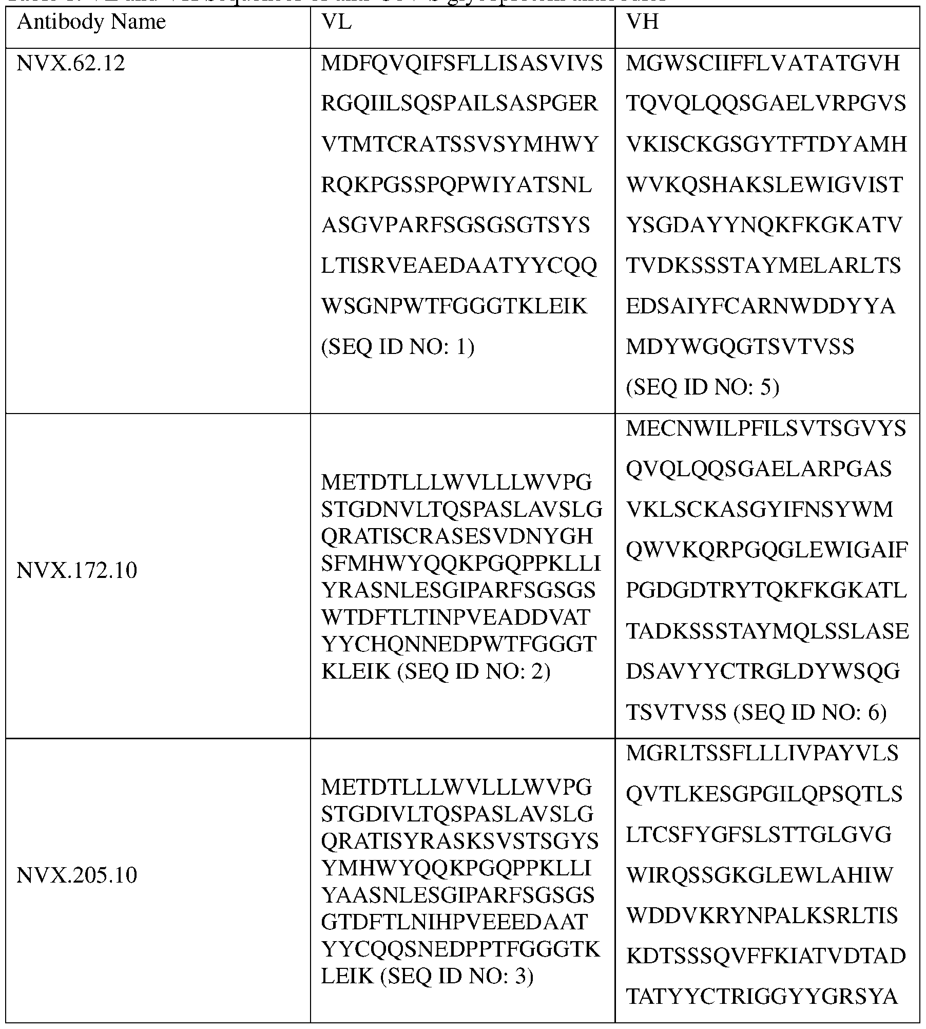 Antibody Name VL VH MDFWGQGTSVTVSS (SEQ H S N P T D
 p y g g g q any one of SEQ ID NOS: 21, 24, 27, and 30. In embodiments, an anti-CoV S glycoprotein antibody comprises a a variable heavy chain complementarity-determining region 2 (VH CDR2) having an amino acid sequence of any one of SEQ ID NOS: 22, 25, 28, and 31. In embodiments, an anti-CoV S glycoprotein antibody comprises a a variable heavy chain complementarity-determining region 3 (VH CDR3) having an amino acid sequence of any one of SEQ ID NOS: 23, 26, 29, and 32. In embodiments, an anti-CoV S glycoprotein antibody comprises a variable light chain complementarity-determining region 1 (VL CDR1) having an amino acid sequence of any one of SEQ ID NOS: 9, 12, 15, and 18. In embodiments, an anti- CoV S glycoprotein antibody comprises a variable light chain complementarity-determining region 2 (VL CDR2) having an amino acid sequence of any one of SEQ ID NOS: 10, 13, 16, and 19. In embodiments, an anti-CoV S glycoprotein antibody comprises a variable light chain complementarity-determining region 3 (VL CDR3) having an amino acid sequence of any one of SEQ ID NOS: 11, 14, 17, and 20. [0071] In embodiments, provided herein is an anti-CoV S glycoprotein antibody comprising a VL CDR 1 selected from the group consisting of SEQ ID NOS: 9, 12, 15, and 18; a VL CDR 2 selected from the group consisting of SEQ ID NOS: 10, 13, 16, and 19; a VL CDR 3 selected from the group consisting of SEQ ID NOS: 11, 14, 17, and 20; a VH CDR 1 selected from the group consisting of SEQ ID NOS: 21, 24, 27, and 30; a VH CDR 2 selected
from the group consisting of SEQ ID NOS: 22, 25, 28, and 31; and a VH CDR 3 selected from the group consisting of SEQ ID NOS: 23, 26, 29, and 32. [0072] In embodiments, the VH CDR1, VH CDR2, VH CDR3, VL CDR1, VL CDR2, and VL CDR3 are independently selected from Table 2. Table 2. CDR Sequences of anti-CoV S glycoprotein antibodies Antibody VL CDR1 VL CDR2 VL CDR3 VH CDR1 VH CDR2 VH CDR3 Y R
p y g g g q any one of SEQ ID NOS: 21, 24, 27, and 30. In embodiments, an anti-CoV S glycoprotein antibody comprises a a variable heavy chain complementarity-determining region 2 (VH CDR2) having an amino acid sequence of any one of SEQ ID NOS: 22, 25, 28, and 31. In embodiments, an anti-CoV S glycoprotein antibody comprises a a variable heavy chain complementarity-determining region 3 (VH CDR3) having an amino acid sequence of any one of SEQ ID NOS: 23, 26, 29, and 32. In embodiments, an anti-CoV S glycoprotein antibody comprises a variable light chain complementarity-determining region 1 (VL CDR1) having an amino acid sequence of any one of SEQ ID NOS: 9, 12, 15, and 18. In embodiments, an anti- CoV S glycoprotein antibody comprises a variable light chain complementarity-determining region 2 (VL CDR2) having an amino acid sequence of any one of SEQ ID NOS: 10, 13, 16, and 19. In embodiments, an anti-CoV S glycoprotein antibody comprises a variable light chain complementarity-determining region 3 (VL CDR3) having an amino acid sequence of any one of SEQ ID NOS: 11, 14, 17, and 20. [0071] In embodiments, provided herein is an anti-CoV S glycoprotein antibody comprising a VL CDR 1 selected from the group consisting of SEQ ID NOS: 9, 12, 15, and 18; a VL CDR 2 selected from the group consisting of SEQ ID NOS: 10, 13, 16, and 19; a VL CDR 3 selected from the group consisting of SEQ ID NOS: 11, 14, 17, and 20; a VH CDR 1 selected from the group consisting of SEQ ID NOS: 21, 24, 27, and 30; a VH CDR 2 selected
from the group consisting of SEQ ID NOS: 22, 25, 28, and 31; and a VH CDR 3 selected from the group consisting of SEQ ID NOS: 23, 26, 29, and 32. [0072] In embodiments, the VH CDR1, VH CDR2, VH CDR3, VL CDR1, VL CDR2, and VL CDR3 are independently selected from Table 2. Table 2. CDR Sequences of anti-CoV S glycoprotein antibodies Antibody VL CDR1 VL CDR2 VL CDR3 VH CDR1 VH CDR2 VH CDR3 Y R
 [0073] In embodiments, an anti-CoV S glycoprotein antibody comprises a VH CDR1 of SEQ ID NO: 21, a VH CDR2 of SEQ ID NO: 22, and a VH CDR3 of SEQ ID NO: 23. In
embodiments, an anti-CoV S glycoprotein antibody comprises a VL CDR1 of SEQ ID NO: 9, a VL CDR2 of SEQ ID NO: 10, and a VL CDR3 of SEQ ID NO: 11. In embodiments, an anti- CoV S glycoprotein antibody comprises comprises a VH CDR1 of SEQ ID NO: 21, a VH CDR2 of SEQ ID NO: 22, and a VH CDR3 of SEQ ID NO: 23 and a VL CDR1 of SEQ ID NO: 9, a VL CDR2 of SEQ ID NO: 10, and a VL CDR3 of SEQ ID NO: 11. [0074] In embodiments, an anti-CoV S glycoprotein antibody comprises a VH CDR1 of SEQ ID NO: 24, a VH CDR2 of SEQ ID NO: 25, and a VH CDR3 of SEQ ID NO: 26. In embodiments, an anti-CoV S glycoprotein antibody comprises a VL CDR1 of SEQ ID NO: 12, a VL CDR2 of SEQ ID NO: 13, and a VL CDR3 of SEQ ID NO: 14. In embodiments, an anti- CoV S glycoprotein antibody comprises comprises a VH CDR1 of SEQ ID NO: 24, a VH CDR2 of SEQ ID NO: 25, and a VH CDR3 of SEQ ID NO: 26 and a VL CDR1 of SEQ ID NO: 12, a VL CDR2 of SEQ ID NO: 13, and a VL CDR3 of SEQ ID NO: 14. [0075] In embodiments, an anti-CoV S glycoprotein antibody comprises a VH CDR1 of SEQ ID NO: 27, a VH CDR2 of SEQ ID NO: 28, and a VH CDR3 of SEQ ID NO: 29. In embodiments, an anti-CoV S glycoprotein antibody comprises a VL CDR1 of SEQ ID NO: 15, a VL CDR2 of SEQ ID NO: 16, and a VL CDR3 of SEQ ID NO: 17. In embodiments, an anti- CoV S glycoprotein antibody comprises comprises a VH CDR1 of SEQ ID NO: 27, a VH CDR2 of SEQ ID NO: 28, and a VH CDR3 of SEQ ID NO: 29, and a VL CDR1 of SEQ ID NO: 15, a VL CDR2 of SEQ ID NO: 16, and a VL CDR3 of SEQ ID NO: 17. [0076] In embodiments, an anti-CoV S glycoprotein antibody comprises a VH CDR1 of SEQ ID NO: 30, a VH CDR2 of SEQ ID NO: 31, and a VH CDR3 of SEQ ID NO: 32. In embodiments, an anti-CoV S glycoprotein antibody comprises a VL CDR1 of SEQ ID NO: 18, a VL CDR2 of SEQ ID NO: 19, and a VL CDR3 of SEQ ID NO: 20. In embodiments, an anti- CoV S glycoprotein antibody comprises comprises a VH CDR1 of SEQ ID NO: 30, a VH CDR2 of SEQ ID NO: 31, and a VH CDR3 of SEQ ID NO: 32 and a VL CDR1 of SEQ ID NO: 18, a VL CDR2 of SEQ ID NO: 19, and a VL CDR3 of SEQ ID NO: 20. [0077] The present invention encompasses antibodies that bind to CoV S glycoproteins, comprising derivatives of the VH domains, VH CDR1s, VH CDR2s, VH CDR3s, VL domains, VL CDR1s, VL CDR2s, or VL CDR3s described herein that may bind to a SARS-CoV 2 S glycoprotein or a variant thereof. In embodiments, the anti-CoV S glycoprotein antibodies bind to a CoV S glycoprotein of a SARS-CoV-2 strain having a PANGO lineage selected from the group consisting of B.1.1.529; BA.1, BA.1.1, BA.2, BA.3, BA.4, BA.5, B.1.1.7, B.1.351, P.1, B.1.617.2, AY, B.1.427, B.1.429, B.1.525, B.1.526, B.1.617.1, B.1.617.3, P.2, B.1.621, or B.1.621.1.
[0078] Standard techniques known to those of skill in the art can be used to introduce modifications (e.g., additions, deletions, and/or substitutions) in the nucleotide sequence encoding an antibody, including, for example, site-directed mutagenesis and PCR-mediated mutagenesis that are routinely used to generate amino acid substitutions. In another embodiment, the VH and/or VK CDRs derivatives may have conservative amino acid substitutions (e.g. supra) made at one or more predicted non-essential amino acid residues (i.e., amino acid residues which are not critical for the antibody to specifically bind to SARS-CoV- 2 S glycoprotein). Mutations can also be introduced randomly along all or part of the VH and/or VL CDR coding sequences, such as by saturation mutagenesis, and the resultant mutants can be screened for biological activity to identify mutants that retain activity. Following mutagenesis, the encoded antibody can be expressed and the activity of the antibody can be determined. [0079] The present invention further encompasses antibodies that bind to SARS-CoV-2 S glycoproteins, wherein said antibodies or antibody fragments comprising one or more CDRs wherein said CDRs comprise an amino acid sequence that is at least 45%, at least 50%, at least 55%, at least 60%, at least 65%, at least 70%, at least 75%, at least 80%, at least 85%, at least 90%, at least 91%, at least 92%, at least 93%, at least 94%, at least 95%, at least 96%, at least 97%, at least 98%, or at least 99% identical to the amino acid sequence of one or more CDRs described herein. The percent identity of two amino acid sequences can be determined by any method known to one skilled in the art, including, but not limited to, BLAST protein searches. [0080] Framework regions of anti-CoV S glycoprotein antibody [0081] In embodiments, the anti-CoV S glycoprotein antibodies comprise a VL and VH that each contain four framework regions (FW1, FW2, FW3, and FW4). In embodiments, FW1, FW2, FW3, and FW4 of VL are independently selected from Table 4. In embodiments, FW1, FW2, FW3, and FW4 of VH are independently selected from Table 5. Table 4. FW1, FW2, FW3, FW4 of VL of anti-CoV S glycoprotein antibodies Antibo FW1 FW2 FW3 FW4 T D )
[0073] In embodiments, an anti-CoV S glycoprotein antibody comprises a VH CDR1 of SEQ ID NO: 21, a VH CDR2 of SEQ ID NO: 22, and a VH CDR3 of SEQ ID NO: 23. In
embodiments, an anti-CoV S glycoprotein antibody comprises a VL CDR1 of SEQ ID NO: 9, a VL CDR2 of SEQ ID NO: 10, and a VL CDR3 of SEQ ID NO: 11. In embodiments, an anti- CoV S glycoprotein antibody comprises comprises a VH CDR1 of SEQ ID NO: 21, a VH CDR2 of SEQ ID NO: 22, and a VH CDR3 of SEQ ID NO: 23 and a VL CDR1 of SEQ ID NO: 9, a VL CDR2 of SEQ ID NO: 10, and a VL CDR3 of SEQ ID NO: 11. [0074] In embodiments, an anti-CoV S glycoprotein antibody comprises a VH CDR1 of SEQ ID NO: 24, a VH CDR2 of SEQ ID NO: 25, and a VH CDR3 of SEQ ID NO: 26. In embodiments, an anti-CoV S glycoprotein antibody comprises a VL CDR1 of SEQ ID NO: 12, a VL CDR2 of SEQ ID NO: 13, and a VL CDR3 of SEQ ID NO: 14. In embodiments, an anti- CoV S glycoprotein antibody comprises comprises a VH CDR1 of SEQ ID NO: 24, a VH CDR2 of SEQ ID NO: 25, and a VH CDR3 of SEQ ID NO: 26 and a VL CDR1 of SEQ ID NO: 12, a VL CDR2 of SEQ ID NO: 13, and a VL CDR3 of SEQ ID NO: 14. [0075] In embodiments, an anti-CoV S glycoprotein antibody comprises a VH CDR1 of SEQ ID NO: 27, a VH CDR2 of SEQ ID NO: 28, and a VH CDR3 of SEQ ID NO: 29. In embodiments, an anti-CoV S glycoprotein antibody comprises a VL CDR1 of SEQ ID NO: 15, a VL CDR2 of SEQ ID NO: 16, and a VL CDR3 of SEQ ID NO: 17. In embodiments, an anti- CoV S glycoprotein antibody comprises comprises a VH CDR1 of SEQ ID NO: 27, a VH CDR2 of SEQ ID NO: 28, and a VH CDR3 of SEQ ID NO: 29, and a VL CDR1 of SEQ ID NO: 15, a VL CDR2 of SEQ ID NO: 16, and a VL CDR3 of SEQ ID NO: 17. [0076] In embodiments, an anti-CoV S glycoprotein antibody comprises a VH CDR1 of SEQ ID NO: 30, a VH CDR2 of SEQ ID NO: 31, and a VH CDR3 of SEQ ID NO: 32. In embodiments, an anti-CoV S glycoprotein antibody comprises a VL CDR1 of SEQ ID NO: 18, a VL CDR2 of SEQ ID NO: 19, and a VL CDR3 of SEQ ID NO: 20. In embodiments, an anti- CoV S glycoprotein antibody comprises comprises a VH CDR1 of SEQ ID NO: 30, a VH CDR2 of SEQ ID NO: 31, and a VH CDR3 of SEQ ID NO: 32 and a VL CDR1 of SEQ ID NO: 18, a VL CDR2 of SEQ ID NO: 19, and a VL CDR3 of SEQ ID NO: 20. [0077] The present invention encompasses antibodies that bind to CoV S glycoproteins, comprising derivatives of the VH domains, VH CDR1s, VH CDR2s, VH CDR3s, VL domains, VL CDR1s, VL CDR2s, or VL CDR3s described herein that may bind to a SARS-CoV 2 S glycoprotein or a variant thereof. In embodiments, the anti-CoV S glycoprotein antibodies bind to a CoV S glycoprotein of a SARS-CoV-2 strain having a PANGO lineage selected from the group consisting of B.1.1.529; BA.1, BA.1.1, BA.2, BA.3, BA.4, BA.5, B.1.1.7, B.1.351, P.1, B.1.617.2, AY, B.1.427, B.1.429, B.1.525, B.1.526, B.1.617.1, B.1.617.3, P.2, B.1.621, or B.1.621.1.
[0078] Standard techniques known to those of skill in the art can be used to introduce modifications (e.g., additions, deletions, and/or substitutions) in the nucleotide sequence encoding an antibody, including, for example, site-directed mutagenesis and PCR-mediated mutagenesis that are routinely used to generate amino acid substitutions. In another embodiment, the VH and/or VK CDRs derivatives may have conservative amino acid substitutions (e.g. supra) made at one or more predicted non-essential amino acid residues (i.e., amino acid residues which are not critical for the antibody to specifically bind to SARS-CoV- 2 S glycoprotein). Mutations can also be introduced randomly along all or part of the VH and/or VL CDR coding sequences, such as by saturation mutagenesis, and the resultant mutants can be screened for biological activity to identify mutants that retain activity. Following mutagenesis, the encoded antibody can be expressed and the activity of the antibody can be determined. [0079] The present invention further encompasses antibodies that bind to SARS-CoV-2 S glycoproteins, wherein said antibodies or antibody fragments comprising one or more CDRs wherein said CDRs comprise an amino acid sequence that is at least 45%, at least 50%, at least 55%, at least 60%, at least 65%, at least 70%, at least 75%, at least 80%, at least 85%, at least 90%, at least 91%, at least 92%, at least 93%, at least 94%, at least 95%, at least 96%, at least 97%, at least 98%, or at least 99% identical to the amino acid sequence of one or more CDRs described herein. The percent identity of two amino acid sequences can be determined by any method known to one skilled in the art, including, but not limited to, BLAST protein searches. [0080] Framework regions of anti-CoV S glycoprotein antibody [0081] In embodiments, the anti-CoV S glycoprotein antibodies comprise a VL and VH that each contain four framework regions (FW1, FW2, FW3, and FW4). In embodiments, FW1, FW2, FW3, and FW4 of VL are independently selected from Table 4. In embodiments, FW1, FW2, FW3, and FW4 of VH are independently selected from Table 5. Table 4. FW1, FW2, FW3, FW4 of VL of anti-CoV S glycoprotein antibodies Antibo FW1 FW2 FW3 FW4 T D )
 Antibo FW1 FW2 FW3 FW4 dy T D ) T D ) T D )
 abe 5. , , 3, o o an -Co S gycoproen an bodes Antib FW1 FW2 FW3 FW4 T T S T S
 Antib FW1 FW2 FW3 FW4 ody T S
 s of Proteins of Immunological Interest, Publication No.91-3242, published as a three volume set by the National Institutes of Health, National Technical Information Service (hereinafter “Kabat”). Kabat provides multiple sequence alignments of immunoglobulin chains from numerous species antibody isotypes. The aligned sequences are numbered according to a single numbering system, the Kabat numbering system. The Kabat sequences have been updated since the 1991 publication and are available as an electronic sequence database (latest downloadable version 1997). Any immunoglobulin sequence can be numbered according to Kabat by performing an alignment with the Kabat reference sequence. Accordingly, the Kabat numbering system provides a uniform system for numbering immunoglobulin chains. Unless indicated otherwise, all immunoglobulin amino acid sequences described herein are numbered according to the Kabat numbering system. Similarly, all single amino acid positions referred to herein are numbered according to the Kabat numbering system. [0083] In another embodiment, an anti-CoV S glycoprotein antibody of the invention may have an affinity constant or Ka (kon/koff) of at least 102 M-1, at least 5 X 10² M-1, at least 103 M- 1, at least 5 X 10³ M-1, at least 104 M-1, at least 5 X 10⁴ M-1, at least 105 M-1, at least 5 X 10⁵ M-1, at least 106 M-1, at least 5 X 10⁶ M-1, at least 107 M-1, at least 5 X 10⁷ M-1, at least 108 M- 1, at least 5 X 10⁸ M-1, at least 109 M-1, at least 5 X 10⁹ M-1, at least 1010 M-1, at least 5 X 10¹⁰ M-1, at least 1011 M-1 at least 5 X 10¹¹ M-1, at least 1012 M-1, at least 5 X 10¹² M-1, at least 1013 M-1 at least 5 X 10¹³ M-1, at least 1014 M-1, at least 5 X 10¹⁴ M-1, at least 1015 M-1, or at least 5 X 10¹⁵ M-1. In embodiments, an anti-CoV S glycoprotein antibody of the invention may have a dissociation constant or Kd (koff/kon) of less than 5x10⁻² M, less than 10-2 M, less than 5x10⁻³ M, less than 10-3 M, less than 5x10⁻⁴ M, less than 10-4 M, less than 5x10⁻⁵ M, less than 10-5 M, less than 5x10⁻⁶ M, less than 10-6 M, less than 5x10⁻⁷ M, less than 10-7 M, less than 5x10⁻⁸ M, less than 10-8 M, less than 5x10⁻⁹ M, less than 10-9 M, less than 5x10⁻¹⁰ M, less than 10-10 M,
less than 5x10-11 M, less than 10-11 M, less than 5x10-12 M, less than 10-12 M, less than 5x10-13 M, less than 10-13 M, less than 5x10-14 M, less than 10-14 M, less than 5x10-15 M, or less than 10-15 M as assessed using a method described herein or known to one of skill in the art (e.g., a BIAcore assay, ELISA). [0084] The invention further provides polynucleotides comprising a nucleotide sequence encoding an anti-CoV S glycoprotein antibody described herein or fragments thereof. The invention also encompasses polynucleotides that hybridize under stringent or lower stringency hybridization conditions, e.g., as defined herein, to polynucleotides that encode an anti-CoV S glycoprotein antibody. [0085] Stringent hybridization conditions include, but are not limited to, hybridization to filter-bound DNA in 6X sodium chloride/sodium citrate (SSC) at about 45°C followed by one or more washes in 0.2X SSC/0.1% SDS at about 50-65°C, highly stringent conditions such as hybridization to filter-bound DNA in 6X SSC at about 45°C followed by one or more washes in 0.1X SSC/0.2% SDS at about 60°C, or any other stringent hybridization conditions known to those skilled in the art (see, for example, Ausubel, F.M. et al., eds. 1989 Current Protocols in Molecular Biology, vol. 1, Green Publishing Associates, Inc. and John Wiley and Sons, Inc., NY at pages 6.3.1 to 6.3.6 and 2.10.3). [0086] The polynucleotides may be obtained, and the nucleotide sequence of the polynucleotides determined, by any method known in the art. For example, if the nucleotide sequence of the antibody is known, a polynucleotide encoding the antibody may be assembled from chemically synthesized oligonucleotides (e.g., as described in Kutmeier et al., BioTechniques 17:242 (1994)), which, briefly, involves the synthesis of overlapping oligonucleotides containing portions of the sequence encoding the antibody, annealing and ligating of those oligonucleotides, and then amplification of the ligated oligonucleotides by PCR. [0087] A polynucleotide encoding an antibody may also be generated from nucleic acid from a suitable source. If a clone containing a nucleic acid encoding a particular antibody is not available, but the sequence of the antibody molecule is known, a nucleic acid encoding the immunoglobulin may be chemically synthesized or obtained from a suitable source (e.g., an antibody cDNA library, or a cDNA library generated from, or nucleic acid, preferably polyA+RNA, isolated from, any tissue or cells expressing the antibody, such as hybridoma cells selected to express an antibody) by PCR amplification using synthetic primers hybridizable to the 3’ and 5’ ends of the sequence or by cloning using an oligonucleotide probe specific for the particular gene sequence to identify, e.g., a cDNA clone from a cDNA library
that encodes the antibody. Amplified nucleic acids generated by PCR may then be cloned into replicable cloning vectors using any method well known in the art. [0088] The present invention also provides polynucleotide sequences encoding VH and VL framework regions and CDRs of antibodies described herein as well as expression vectors for their efficient expression in mammalian cells. [0089] In one embodiment, an anti-CoV S glycoprotein antibody described herein mediates antibody-dependent cellular cytotoxicity (ADCC), complement-dependent cell-mediated cytotoxicity (CDC), and/or apoptosis. In one embodiment, an anti-CoV S glycoprotein antibody of the invention mediates antibody-dependent cellular cytotoxicity (ADCC) and/or apoptosis. In one embodiment, an anti-CoV S glycoprotein antibody of the invention has enhanced antibody-dependent cellular cytotoxicity (ADCC). In one embodiment, an anti-CoV S glycoprotein antibody of the invention comprises a variant Fc region that mediates enhanced antibody-dependent cellular cytotoxicity (ADCC). In one embodiment, an anti-CoV S glycoprotein antibody of the invention comprises an Fc region having complex N-glycoside- linked sugar chains linked to Asn297 in which fucose is not bound to N-acetylglucosamine in the reducing end, wherein said Fc region mediates enhanced antibody-dependent cellular cytotoxicity (ADCC). Production of humanized anti-CoV S Glycoprotein Antibodies [0090] Humanized antibodies described herein can be produced using a variety of techniques known in the art, including, but not limited to, CDR-grafting (see e.g., European Patent No. EP 239,400; International Publication No. WO 91/09967; and U.S. Patent Nos. 5,225,539, 5,530,101, and 5,585,089, each of which is incorporated herein in its entirety by reference), veneering or resurfacing (see, e.g., European Patent Nos. EP 592,106 and EP 519,596; Padlan, 1991, Molecular Immunology 28(4/5):489-498; Studnicka et al., 1994, Protein Engineering, 7(6):805-814; and Roguska et al., 1994, Proc. Natl. Acad. Sci. , 91:969¬973, each of which is incorporated herein by its entirety by reference), chain shuffling (see, e.g., U.S. Patent No.5,565,332, which is incorporated herein in its entirety by reference), and techniques disclosed in, e.g., U.S. Patent No. 6,407,213, U.S. Patent No. 5,766,886, International Publication No. WO 9317105, Tan et al., J. Immunol., 169:1119-25 (2002), Caldas et al., Protein Eng., 13(5):353-60 (2000), Morea et al., Methods, 20(3):267-79 (2000), Baca et al., J. Biol. Chem., 272(16):10678-84 (1997), Roguska et al., Protein Eng., 9(10):895- 904 (1996), Couto et al., Cancer Res., 55 (23 Supp):5973s-5977s (1995), Couto et al., Cancer Res., 55(8):1717-22 (1995), Sandhu JS, Gene, 150(2):409-10 (1994), and Pedersen et al., J. Mol. Biol., 235(3):959-73 (1994), each of which is incorporated herein in its entirety by
reference. In embodiments, humanized antibodies are produced using phage display, framework homology germline-based humanization, or germline humanization with retaining the vernier zone. Often, FW residues in the FW regions will be substituted with the corresponding residue from the CDR donor antibody to alter, preferably improve, antigen binding. These FW substitutions are identified by methods well known in the art, e.g., by modeling of the interactions of the CDR and FW residues to identify FW residues important for antigen binding and sequence comparison to identify unusual FW residues at particular positions. (See, e.g., Queen et al., U.S. Patent No. 5,585,089; and Riechmann et al., 1988, Nature, 332:323, which are incorporated herein by reference in their entireties.) [0091] A humanized anti-CoV S glycoprotein antibody has one or more amino acid residues introduced into it from a source which is nonhuman. These nonhuman amino acid residues are often referred to as “import” residues, which are typically taken from an “import” variable domain. Thus, humanized antibodies comprise one or more CDRs from nonhuman immunoglobulin molecules and framework regions from human. Humanization of antibodies is well-known in the art and can essentially be performed following the method of Winter and co-workers (Jones et al., Nature, 321:522-525 (1986); Riechmann et al., Nature, 332:323-327 (1988); Verhoeyen et al., Science, 239:1534-1536 (1988)), by substituting rodent CDRs or CDR sequences for the corresponding sequences of a human antibody, i.e., CDR-grafting (EP 239,400; PCT Publication No. WO 91/09967; and U.S. Patent Nos. 4,816,567; 6,331,415; 5,225,539; 5,530,101; 5,585,089; 6,548,640, the contents of which are incorporated by reference herein in their entirety). In such humanized chimeric antibodies, substantially less than an intact human variable domain has been substituted by the corresponding sequence from a nonhuman species. In practice, humanized antibodies are typically human antibodies in which some CDR residues and possibly some FW residues are substituted by residues from analogous sites in rodent antibodies. Humanization of an anti-CoV S glycoprotein antibody can also be achieved by veneering or resurfacing (EP 592,106; EP 519,596; Padlan, 1991, Molecular Immunology 28(4/5):489-498; Studnicka et al., Protein Engineering, 7(6):805-814 (1994); and Roguska et al., Proc. Natl. Acad. Sci. , 91:969-973 (1994)) or chain shuffling (U.S. Patent No. 5,565,332), the contents of which are incorporated herein by reference in their entirety. [0092] The choice of human variable domains, both light and heavy, to be used in making the humanized antibodies is to reduce antigenicity. According to the so-called “best-fit” method, the sequence of the variable domain of a rodent antibody is screened against the entire library of known human variable-domain sequences. The human sequences which are most
closely related to that of the rodent are then screened for the presences of specific residues that may be critical for antigen binding, appropriate structural formation and/or stability of the intended humanized mAb (Sims et al., J. Immunol., 151:2296 (1993); Chothia et al., J. Mol. Biol., 196:901 (1987), the contents of which are incorporated herein by reference in their entirety). The resulting FW sequences matching the desired criteria are then be used as the human donor FW regions for the humanized antibody. [0093] Another method uses a particular FW derived from the consensus sequence of all human antibodies of a particular subgroup of light or heavy chains. The same FW may be used for several different humanized anti-CoV S glycoprotein antibodies (Carter et al., Proc. Natl. Acad. Sci. USA, 89:4285 (1992); Presta et al., J. Immunol., 151:2623 (1993), the contents of which are incorporated herein by reference in their entirety). [0094] Anti-CoV S glycoprotein antibodies can be humanized with retention of high affinity for SARS-CoV-2 S glycoprotein and other favorable biological properties. According to one aspect of the invention, humanized antibodies are prepared by a process of analysis of the parental sequences and various conceptual humanized products using three-dimensional models of the parental and humanized sequences. Three-dimensional immunoglobulin models are commonly available and are familiar to those skilled in the art. Computer programs are available which illustrate and display probable three-dimensional conformational structures of selected candidate immunoglobulin sequences. Inspection of these displays permits analysis of the likely role of the residues in the functioning of the candidate immunoglobulin sequence, i.e., the analysis of residues that influence the ability of the candidate immunoglobulin to bind SARS-CoV-2 S glycoprotein. In this way, FW residues can be selected and combined from the recipient and import sequences so that the desired antibody characteristic, for example affinity for SARS-CoV-2 S glycoprotein, is achieved. In general, the CDR residues are directly and most substantially involved in influencing antigen binding. [0095] A “humanized” antibody may retain a similar antigenic specificity as the original antibody, i.e., in the present invention, the ability to bind the SARS-CoV-2 S glycoprotein. However, using certain methods of humanization, the affinity and/or specificity of binding of the antibody for the SARS-CoV-2 S glycoprotein may be altered using methods of “directed evolution,” as described by Wu et al., J. Mol. Biol, 294:151 (1999), the contents of which are incorporated herein by reference herein in their entirety. [0096] Humanized anti-CoV S glycoprotein antibodies described herein can be constructed by the selection of distinct human framework regions for grafting of the 239.12, 322.3, 425.6, and 35.13 CDRs as described herein.
Monoclonal anti-CoV S Glycoprotein Antibodies [0097] A monoclonal anti-CoV S glycoprotein antibody exhibits binding specificity to SARS-CoV-2 antigen and may mediate human ADCC, CDC and/or apoptotic mechanisms. Such an antibody can be generated using a wide variety of techniques known in the art including the use of hybridoma, recombinant, and phage display technologies, or a combination thereof. Antibodies are highly specific, being directed against a single antigenic site. An engineered anti-CoV S glycoprotein antibody can be produced by any means known in the art, including, but not limited to, those techniques described below and improvements to those techniques. Large-scale high - yield production typically involves culturing a host cell that produces the engineered anti-CoV S glycoprotein antibody and recovering the anti-CoV S glycoprotein antibody from the host cell culture. Hybridoma Technique [0098] Monoclonal antibodies can be produced using hybridoma techniques including those known in the art and taught, for example, in Harlow et al., Antibodies: A Laboratory Manual, (Cold Spring Harbor Laboratory Press, 2nd ed. 1988); Hammerling et al., in Monoclonal Antibodies and T Cell Hybridomas, 563-681 (Elsevier, N.Y., 1981) (said references incorporated herein by reference in their entireties). For example, in the hybridoma method, a mouse or other appropriate host animal, such as a hamster or macaque monkey, is immunized to elicit lymphocytes that produce or are capable of producing antibodies that will specifically bind to the protein used for immunization. Lymphocytes may also be immunized in vitro. Lymphocytes then are fused with myeloma cells using a suitable fusing agent, such as polyethylene glycol, to form a hybridoma cell (Goding, Monoclonal Antibodies: Principles and Practice, pp.59-103 (Academic Press, 1986)). [0099] The hybridoma cells thus prepared are seeded and grown in a suitable culture medium that contains one or more substances that inhibit the growth or survival of the unfuscd, parental mycloma cells. For example, if the parental mycloma cells lack the enzyme hypoxanthine guanine phosphoribosyl transferase (HGPRT or HPRT), the culture medium for the hybridomas typically will include hypoxanthine, aminopterin, and thymidine (HAT medium), which substances prevent the growth of HGPRT-deficient cells. [0100] Specific embodiments employ myeloma cells that fuse efficiently, support stable high-level production of antibody by the selected antibody-producing cells, and are sensitive to a medium such as HAT medium. Among these myeloma cell lines are murine myeloma lines, such as those derived from MOPC-21 and MPC-11 mouse tumors available from the Salk Institute Cell Distribution Center, San Diego, CA, USA, and SP-2 or X63-Ag8.653 cells
available from the American Type Culture Collection, Rockville, MD, USA. Human myeloma and mouse-human heteromyeloma cell lines also have been described for the production of human monoclonal antibodies (Kozbor, J. Immunol., 133:3001 (1984); Brodeur et al., Monoclonal Antibody Production Techniques and Applications, pp. 51-63 (Marcel Dekker, Inc., New York, 1987)). [0101] Culture medium in which hybridoma cells are growing is assayed for production of monoclonal antibodies directed against the SARS-CoV-2 S glycoprotein. The binding specificity of monoclonal antibodies produced by hybridoma cells can be determined by immunoprecipitation or by an in vitro binding assay, such as radioimmunoassay (RIA) or enzyme-linked immunoabsorbent assay (ELISA). [0102] After hybridoma cells are identified that produce antibodies of the desired specificity, affinity, and/or activity, the clones may be subcloned by limiting dilution procedures and grown by standard methods (Goding, Monoclonal Antibodies: Principles and Practice, pp.59-103 (Academic Press, 1986)). Suitable culture media for this purpose include, for example, D-MEM or RPMI 1640 medium. In addition, the hybridoma cells may be grown in vivo as ascites tumors in an animal. [0103] The monoclonal antibodies secreted by the subclones are suitably separated from the culture medium, or serum by conventional immunoglobulin purification procedures such as, for example, protein A-Sepharose, hydroxylapatite chromatography, gel electrophoresis, dialysis, or affinity chromatography. Recombinant DNA Techniques [0104] DNA encoding an anti-CoV S glycoprotein antibody described herein is readily isolated and sequenced using conventional procedures (e.g., by using oligonucleotide probes that are capable of binding specifically to genes encoding the heavy and light chains of anti- CoV S glycoprotein antibodies). The hybridoma cells serve as a source of such DNA. Once isolated, the DNA may be placed into expression vectors, which are then transfected into host cells such as E. coli cells, simian COS cells, Chinese hamster ovary (CHO) cells, or myeloma cells that do not otherwise produce immunoglobulin protein, to obtain the synthesis of anti- CoV S glycoprotein antibodies in the recombinant host cells. [0105] In phage display methods, functional antibody domains are displayed on the surface of phage particles which carry the polynucleotide sequences encoding them. In particular, DNA sequences encoding VH and VL domains are amplified from animal cDNA libraries (e.g., human or murine cDNA libraries of affected tissues). The DNA encoding the VH and VL domains are recombined together with an scFv linker by PCR and cloned into a phagemid
vector. The vector is electroporated in E. coli and the E. coli is infected with helper phage. Phage used in these methods is typically filamentous phage including fd and M13 and the VII and VL domains are usually recombinantly fused to either the phage gene III or gene VIII. Phage expressing an antigen-binding domain that binds to a particular antigen can be selected or identified with antigen, e.g., using labeled antigen or antigen bound or captured to a solid surface or bead. Examples of phage display methods that can be used to make the antibodies of the present invention include those disclosed in Brinkman et al., 1995, J. Immunol. Methods, 182:41-50; Ames et al., 1995, J. Immunol. Methods, 184:177-186; Kettleborough et al., 1994, Eur. J. Immunol., 24:952-958; Persic et al., 1997, Gene, 187:9-18; Burton et al., 1994, Advances in Immunology, 57:191-280; International Application No. PCT/GB91/O1 134; International Publication Nos. WO 90/02809, WO 91/10737, WO 92/01047, WO 92/18619, WO 93/11236, WO 95/15982, WO 95/20401, and W097/13844; and U.S. Patent Nos. 5,698,426, 5,223,409, 5,403,484, 5,580,717, 5,427,908, 5,750,753, 5,821,047, 5,571,698, 5,427,908, 5,516,637, 5,780,225, 5,658,727, 5,733,743, and 5,969,108; each of which is incorporated herein by reference in its entirety. [0106] As described in the above references, after phage selection, the antibody coding regions from the phage can be isolated and used to generate whole antibodies, including human antibodies, or any other desired antigen-binding fragment, and expressed in any desired host, including mammalian cells, insect cells, plant cells, yeast, and bacteria, e.g., as described below. Techniques to recombinantly produce Fab, Fab’ and F(ab’)2 fragments can also be employed using methods known in the art such as those disclosed in PCT Publication No. WO 92/22324; Mullinax et al., 1992, BioTechniques, 12(6):864-869; Sawai et al., 1995, AJRI, 34:26-34; and Better et al., 1988, Science, 240:1041-1043 (said references incorporated by reference in their entireties). [0107] Antibodies may be isolated from antibody phage libraries generated using the techniques described in McCafferty et al., Nature, 348:552-554 (1990). Clackson et al., Nature, 352:624-628 (1991). Marks et al., J. Mol. Biol., 222:581-597 (1991) describe the isolation of murine and human antibodies, respectively, using phage libraries. Chain shuffling can be used in the production of high affinity (nM range) human antibodies (Marks et al., Bio/Technology, 10:779-783 (1992)), as well as combinatorial infection and in vivo recombination as a strategy for constructing very large phage libraries (Waterhouse et al., Nuc. Acids. Res., 21:2265-2266 (1993)). Thus, these techniques are viable alternatives to traditional monoclonal antibody hybridoma techniques for isolation of anti-CoV S glycoprotein antibodies.
[0108] To generate whole antibodies, PCR primers including VH or VL nucleotide sequences, a restriction site, and a flanking sequence to protect the restriction site can be used to amplify the VH or VL sequences in scFv clones. Utilizing cloning techniques known to those of skill in the art, the PCR amplified VH domains can be cloned into vectors expressing a heavy chain constant region, e.g., the human gamma 4 constant region, and the PCR amplified VL domains can be cloned into vectors expressing a light chain constant region, e.g., human kappa or lambda constant regions. The vectors for expressing the VH or VL domains may comprise an EF-la promoter, a secretion signal, a cloning site for the variable domain, constant domains, and a selection marker such as neomycin. The VH and VL domains may also be cloned into one vector expressing the necessary constant regions. The heavy chain conversion vectors and light chain conversion vectors are then co-transfected into cell lines to generate stable or transient cell lines that express full-length antibodies, e.g., IgG, using techniques known to those of skill in the art. [0109] The DNA also may be modified, for example, by substituting the coding sequence for human heavy and light chain constant domains in place of the homologous murine sequences (U.S. Patent No. 4,816,567; Morrison et al., Proc. Natl. Acad. Sci. USA, 81:6851 (1984)), or by covalently joining to the immunoglobulin coding sequence all or part of the coding sequence for a non-immunoglobulin polypeptide. Chimeric Antibodies [0110] The anti-CoV S glycoprotein antibodies herein specifically include chimeric antibodies (immunoglobulins) in which a portion of the heavy and/or light chain is identical with or homologous to corresponding sequences in antibodies derived from a particular species or belonging to a particular antibody class or subclass, while another portion of the chain(s) is identical with or homologous to corresponding sequences in antibodies derived from another species or belonging to another antibody class or subclass, as well as fragments of such antibodies, so long as they exhibit the desired biological activity (U.S. Patent No. 4,816,567; Morrison et al., Proc. Natl. Acad. Sci. USA, 81:6851-6855 (1984)). Chimeric antibodies of interest herein include “primatized” antibodies comprising variable domain antigen-binding sequences derived from a nonhuman primate (e.g., Old World Monkey, such as baboon, rhesus or cynomolgus monkey) and human constant region sequences (U.S. Patent No.5,693,780). [0111] In embodiments the KD of anti-CoV S glycoprotein antibodies described herein, or an for a SARS-CoV-2 S glycoprotein may be 50 nM or less, 10 nM or less, 1 nM or less, 0.5 nM or less, 0.1 nM or less, 0.05 nM or less, 0.01 nM or less, or 0.001 nM or less. Methods and reagents suitable for determination of such binding characteristics of an antibody of the present
invention, or an altered/mutant derivative thereof, are known in the art and/or are commercially available (se above and, e.g., U.S. Patent No.6,849,425, U.S. Patent No.6,632,926, U.S. Patent No. 6,294,391, and U.S. Patent No. 6,143,574, each of which is hereby incorporated by reference in its entirety). Moreover, equipment and software designed for such kinetic analyses are commercially available (e.g. Biacore® A100, and Biacore® 2000 instruments; Biacore International AB, Uppsala, Sweden). [0112] Identity or similarity with respect to a sequence is defined herein as the percentage of amino acid residues in the candidate sequence that are identical (i.e., same residue) or similar (i.e., amino acid residue from the same group based on common side-chain properties, see below) with anti-CoV S glycoprotein antibodies, after aligning the sequences and introducing gaps, if necessary, to achieve the maximum percent sequence identity. None of N-terminal, C- terminal, or internal extensions, deletions, or insertions into the antibody sequence outside of the variable domain shall be construed as affecting sequence identity or similarity. [0113] Methods for comparing the identity of two or more sequences are well known in the art. Percentage identity is calculated using the tool CLUSTALW2, which is available online. The following default parameters may be used for CLUSTALW2 Pairwise alignment: Protein Weight Matrix = Gonnet; Gap Open = 10; Gap Extension = 0.1. Unless described otherwise, the CLUSTALW2 tool is utilized to calculate percent identity herein. [0114] To generate an altered antibody, one or more amino acid alterations (e.g., substitutions) are introduced in one or more of the hypervariable regions of the species- dependent antibody. One or more alterations (e.g., substitutions) of framework region residues may also be introduced in anti-CoV S glycoprotein antibodies where these result in an improvement in the binding affinity of the antibody mutant for the antigen from the second mammalian species. Examples of framework region residues to modify include those which non-covalently bind antigen directly (Amit et al., Science, 233:747-753 (1986)); interact with/effect the conformation of a CDR (Chothia et al., J. Mol. Biol., 196:901-917 (1987)); and/or participate in the VL-VH interface (EP 239 400B1). In certain embodiments, modification of one or more of such framework region residues results in an enhancement of the binding affinity of the antibody for the antigen from the second mammalian species. For example, from about one to about five framework residues may be altered in this embodiment of the invention. Sometimes, this may be sufficient to yield an antibody mutant suitable for use in preclinical trials, even where none of the hypervariable region residues have been altered. Normally, however, an altered antibody will comprise additional hypervariable region alteration(s).
[0115] The hypervariable region residues which are altered may be changed randomly, especially where the starting binding affinity of anti-CoV S glycoprotein antibodies for the antigen from the second mammalian species is such that such randomly produced altered antibody can be readily screened. [0116] One useful procedure for generating such an altered antibody is called “alanine scanning mutagenesis” (Cunningham and Wells, Science, 244:1081-1085 (1989)). Here, one or more of the hypervariable region residue(s) are replaced by alanine or polyalanine residue(s) to affect the interaction of the amino acids with the antigen from the second mammalian species. Those hypervariable region residue(s) demonstrating functional sensitivity to the substitutions then are refined by introducing additional or other mutations at or for the sites of substitution. Thus, while the site for introducing an amino acid sequence variation is predetermined, the nature of the mutation per se need not be predetermined. The Ala-mutants produced this way are screened for their biological activity as described herein. [0117] Another procedure for generating such an altered antibody involves affinity maturation using phage display (Hawkins et al., J. Mol. Biol., 254:889-896 (1992) and Lowman et al., Biochemistry, 30(45):10832-10837 (1991)). Briefly, several hypervariable region sites (e.g., 6-7 sites) are mutated to generate all possible amino acid substitutions at each site. The antibody mutants thus generated are displayed in a monovalent fashion from filamentous phage particles as fusions to the gene 111 product of M13 packaged within each particle. The phage- displayed mutants are then screened for their biological activity (e.g., binding affinity) as herein disclosed. [0118] Mutations in antibody sequences may include substitutions, deletions, including internal deletions, additions, including additions yielding fusion proteins, or conservative substitutions of amino acid residues within and/or adjacent to the amino acid sequence, but that result in a “silent” change, in that the change produces a functionally equivalent anti-CoV S glycoprotein antibodies. Conservative amino acid substitutions may be made on the basis of similarity in polarity, charge, solubility, hydrophobicity, hydrophilicity, and/or the amphipathic nature of the residues involved. For example, non-polar (hydrophobic) amino acids include alanine, leucine, isoleucine, valine, proline, phenylalanine, tryptophan, and methionine; polar neutral amino acids include glycine, serine, threonine, cysteine, tyrosine, asparagine, and glutamine; positively charged (basic) amino acids include arginine, lysine, and histidine; and negatively charged (acidic) amino acids include aspartic acid and glutamic acid. In addition, glycine and proline are residues that can influence chain orientation. Non-conservative substitutions will entail exchanging a member of one of these classes for a member of another
class. Furthermore, if desired, non-classical amino acids or chemical amino acid analogs can be introduced as a substitution or addition into the antibody sequence. Non-classical amino acids include, but are not limited to, the D-isomers of the common amino acids, α -amino isobutyric acid, 4-aminobutyric acid, Abu, 2-amino butyric acid, γ-Abu, ^-Ahx, 6-amino hexanoic acid, Aib, 2-amino isobutyric acid, 3-amino propionic acid, ornithine, norleucine, norvaline, hydroxyproline, sarcosine, citrulline, cysteic acid, t-butylglycine, t-butylalanine, phenylglycine, cyclohexylalanine, β-alanine, fluoro-amino acids, designer amino acids such as β-methyl amino acids, Cα-methyl amino acids, Nα-methyl amino acids, and amino acid analogs in general. [0119] In another embodiment, the sites selected for modification are affinity matured using phage display (see above). [0120] Any technique for mutagenesis known in the art can be used to modify individual nucleotides in a DNA sequence, for purposes of making amino acid substitution(s) in the antibody sequence, or for creating/deleting restriction sites to facilitate further manipulations. Such techniques include, but are not limited to, chemical mutagenesis, in vitro site-directed mutagenesis (Kunkel, Proc. Natl. Acad. Sci. USA, 82:488 (1985); Hutchinson, C. et al., J. Biol. Chem., 253:6551 (1978)), oligonucleotide-directed mutagenesis (Smith, Ann. Rev. Genet., 19:423-463 (1985); Hill et al., Methods Enzymol., 155:558-568 (1987)), PCR-based overlap extension (Ho et al., Gene, 77:51-59 (1989)), PCR-based megaprimer mutagenesis (Sarkar et al., Biotechniques, 8:404-407 (1990)), etc. Modifications can be confirmed by double-stranded dideoxy DNA sequencing. [0121] In certain embodiments of the invention, anti-CoV S glycoprotein antibodies can be modified to produce fusion proteins; i.e., the antibody, or a fragment thereof, fused to a heterologous protein, polypeptide or peptide. [0122] Additional fusion proteins may be generated through the techniques of gene- shuffling, motif-shuffling, exon-shuffling, and/or codon-shuffling (collectively referred to as “DNA shuffling”). DNA shuffling may be employed to alter the activities of the anti-CoV S glycoprotein antibody (e.g., an antibody or a fragment thereof with higher affinities and lower dissociation rates). See, generally, U.S. Patent Nos. 5,605,793; 5,811,238; 5,830,721; 5,834,252; and 5,837,458, and Patten et al., 1997, Curr. Opinion Biotechnol., 8:724-33 ; Harayama, 1998, Trends Biotechnol. 16(2):76-82; Hansson et al., 1999, J. Mol. Biol., 287:265- 76; and Lorenzo and Blasco, 1998, Biotechniques 24(2):308- 313 (each of these patents and publications are hereby incorporated by reference in its entirety). The antibody can further be a binding-domain immunoglobulin fusion protein as described in U.S. Publication
20030118592, U.S. Publication 200330133939, and PCT Publication WO 02/056910, all to Ledbetter et al., which are incorporated herein by reference in their entireties. Domain Antibodies [0123] Anti-CoV S glycoprotein antibodies of compositions and methods of the invention can be domain antibodies, e.g., antibodies containing the small functional binding units of antibodies, corresponding to the variable regions of the heavy (VH) or light (VL) chains of human antibodies. Examples of domain antibodies include, but are not limited to, those available from Domantis Limited (Cambridge, UK) and Domantis Inc. (Cambridge, MA, USA) that are specific to therapeutic targets (see, for example, W004/058821; W004/003019; U.S. Patent Nos.6,291,158; 6,582,915; 6,696,245; and 6,593,081. Diabodies [0124] In certain embodiments of the invention, anti-CoV S glycoprotein antibodies are “diabodies”. The term “diabodies” refers to small antibody fragments with two antigen- binding sites, which fragments comprise a heavy chain variable domain (VH) connected to a light chain variable domain (VL) in the same polypeptide chain (VH-VL). By using a linker that is too short to allow pairing between the two domains on the same chain, the domains are forced to pair with the complementary domains of another chain and create two antigen-binding sites. Diabodies are described more fully in, for example, EP 404,097; WO 93/11161; and Hollinger et al., Proc. Natl. Acad. Sci. USA, 90:6444-6448 (1993). Linear Antibodies [0125] In certain embodiments of the invention, anti-CoV S glycoprotein antibodies are linear antibodies. Linear antibodies comprise a pair of tandem Fd segments (VH-CH1-VH-CH1) which form a pair of antigen-binding regions. Linear antibodies can be bispecific or monospecific. See, Zapata et al., Protein Eng., 8(10):1057-1062 (1995). Antibody Fragments [0126] “Antibody fragments” comprise a portion of a full-length antibody, generally the antigen binding or variable region thereof. Examples of antibody fragments include Fab, Fab’ , F(ab’)2, and Fv fragments; diabodies; linear antibodies; single-chain antibody molecules; and multispeci fie antibodies formed from antibody fragments. [0127] Traditionally, these fragments were derived via proteolytic digestion of intact antibodies (see, e.g., Morimoto et al., Journal of Biochemical and Biophysical Methods, 24:107-117 (1992) and Brennan et al., Science, 229:81 (1985)). However, these fragments can now be produced directly by recombinant host cells. For example, the antibody fragments can be isolated from the antibody phage libraries discussed above. Fab’ -SH fragments can also be
directly recovered from E. coli and chemically coupled to form F(ab‘)2 fragments (Carter et al., Bio/Technology, 10:163-167 (1992)). According to another approach, F(ab‘)2 fragments can be isolated directly from recombinant host cell culture. Other techniques for the production of antibody fragments will be apparent to the skilled practitioner. In other embodiments, the antibody of choice is a single-chain Fv fragment (scFv). See, for example, WO 93/16185. In certain embodiments, the antibody is not a Fab fragment. Bispecific Antibodies [0128] Bispecific antibodies are antibodies that have binding specificities for at least two different epitopes. [0129] Methods for making bispecific antibodies are known in the art. (See, for example, Millstein et al., Nature, 305:537-539 (1983); Traunecker et al., EMBO J., 10:3655-3659 (1991); Suresh et al., Methods in Enzymology, 121:210 (1986); Kostelny et al., J. Immunol., 148(5):1547-1553 (1992); Hollinger et al., Proc. Natl Acad. Sci. USA, 90:6444-6448 (1993); Gruber et al., J. Immunol., 152:5368 (1994); U.S. Patent Nos.4,474,893; 4,714,681; 4,925,648; 5,573,920; 5,601,81; 95,731,168; 4,676,980; and 4,676,980, WO 94/04690; WO 91/00360; WO 92/200373; WO 93/17715; WO 92/08802; and EP 03089.) [0130] In one embodiment, where an anti-CoV S glycoprotein antibody of compositions and methods of the invention is bispecific, the anti-CoV S glycoprotein antibody may be human or humanized and may have specificity for SARS-CoV-2 S glycoprotein and an epitope on a T cell or may be capable of binding to a human effector cell such as, for example, a monocyte/macrophage and/or a natural killer cell to effect cell death. [0131] In one embodiment, an anti-CoV S glycoprotein antibody of the invention is a bispecific antibody capable of specifically binding to a first and second antigen, wherein said first antigen is a SARS-CoV-2 S glycoprotein and said second antigen is an Fc gamma receptor selected from the group consisting of FcγRI, FcγRIIA, FcγRIIB, FcγRIIIA and/or FcγRIV. In a further embodiment, an anti-CoV S glycoprotein antibody of the invention is a bispecific antibody capable of specifically binding to SARS-CoV-2 and FcγRIIB. In another embodiment, an anti-CoV S glycoprotein antibody of the invention is a bispecific antibody capable of specifically binding to SARS-CoV-2 S glycoprotein and human FcγRIIB. Variant Fc Regions [0132] The present invention provides an anti-CoV S glycoprotein antibody with a variant Fc domain. That is, a non naturally occurring Fc region, for example an Fc region comprising one or more non naturally occurring amino acid residues. Also encompassed by the variant Fc
regions of present invention are Fc regions which comprise amino acid deletions, additions and/or modifications. [0133] It will be understood that Fc region as used herein includes the polypeptides comprising the constant region of an antibody excluding the first constant region immunoglobulin domain. Thus Fc refers to the last two constant region immunoglobulin domains of IgA, IgD, and IgG, and the last three constant region immunoglobulin domains of IgE and IgM, and the flexible hinge N-terminal to these domains. For IgA and IgM Fc may include the J chain. For IgG, Fc comprises immunoglobulin domains Cgamma2 and Cgamma3 (Cγ2 and Cγ3) and the hinge between Cgammal (Cγl) and Cgamma2 (Cγ2). Although the boundaries of the Fc region may vary, the human IgG heavy chain Fc region is usually defined to comprise residues C226 or P230 to its carboxyl-terminus, wherein the numbering is according to the EU index as in Kabat et al. (1991, NIH Publication 91-3242, National Technical Information Service, Springfield, VA). The “EU index as set forth in Kabat” refers to the residue numbering of the human IgG1 EU antibody as described in Kabat et al. supra. Fc may refer to this region in isolation, or this region in the context of an antibody, antibody fragment, or Fc fusion protein. An Fc variant protein may be an antibody, Fc fusion, or any protein or protein domain that comprises an Fc region including, but not limited to, proteins comprising variant Fc regions, which are non naturally occurring variants of an Fc. Note: Polymorphisms have been observed at a number of Fc positions, including but not limited to Kabat 270, 272, 312, 315, 356, and 358, and thus slight differences between the presented sequence and sequences in the prior art may exist. [0134] The present invention encompasses anti-CoV S glycoprotein antibody with variant Fc domains. The variant Fc domains may have altered binding properties for an Fc ligand (e.g., an Fc receptor, Clq) relative to a comparable molecule (e.g., a protein having the same amino acid sequence except having a wild type Fc region). Examples of binding properties include but are not limited to, binding specificity, equilibrium dissociation constant (KD), dissociation and association rates (koff and kon respectively), binding affinity and/or avidity. It is generally understood that a binding molecule (e.g., a Fc variant protein such as an antibody) with a low KD may be preferable to a binding molecule with a high KD. However, in some instances the value of the icon or koff may be more relevant than the value of the KD. One skilled in the art can determine which kinetic parameter is most important for a given antibody application. [0135] The affinities and binding properties of an Fc domain for its ligand may be determined by a variety of in vitro assay methods (biochemical or immunological based assays) known in the art for determining Fc-FcγR interactions, i.e., specific binding of an Fc region to
an FcγR including but not limited to, equilibrium methods (e.g., enzyme-linked immunoabsorbent assay (ELISA), or radioimmunoassay (RIA)), or kinetics (e.g., BIACORE® analysis), and other methods such as indirect binding assays, competitive inhibition assays, fluorescence resonance energy transfer (FRET), gel electrophoresis and chromatography (e.g., gel filtration). These and other methods may utilize a label on one or more of the components being examined and/or employ a variety of detection methods including but not limited to chromogenic, fluorescent, luminescent, or isotopic labels. A detailed description of binding affinities and kinetics can be found in Paul, W.E., ed., Fundamental Immunology, 4th Ed., Lippincott-Raven, Philadelphia (1999), which focuses on antibody-immunogen interactions. [0136] In one embodiment, an anti-CoV S glycoprotein antibody with a variant Fc domain has enhanced binding to one or more Fc ligand relative to a comparable molecule. In another embodiment, an anti-CoV S glycoprotein antibody with a variant Fc domain has an affinity for an Fc ligand that is at least 2 fold, or at least 3 fold, or at least 5 fold, or at least 7 fold, or a least 10 fold, or at least 20 fold, or at least 30 fold, or at least 40 fold, or at least 50 fold, or at least 60 fold, or at least 70 fold, or at least 80 fold, or at least 90 fold, or at least 100 fold, or at least 200 fold greater than that of a comparable molecule. In a specific embodiment, an anti- CoV S glycoprotein antibody with a variant Fc domain has enhanced binding to an Fc receptor. In another specific embodiment, an anti-CoV S glycoprotein antibody with a variant Fc domain has enhanced binding to the Fc receptor FcγRIIIA. In a further specific embodiment, an anti- CoV S glycoprotein antibody with a variant Fc domain has enhanced biding to the Fc receptor FcγRIIB. In still another specific embodiment, an anti-CoV S glycoprotein antibody with a variant Fc domain has enhanced binding to the Fc receptor FcRn. In yet another specific embodiment, an anti-CoV S glycoprotein antibody with a variant Fc domain has enhanced binding to Clq relative to a comparable molecule. [0137] In one embodiment, an anti-CoV S glycoprotein antibody of the invention comprises a variant Fc domain wherein said variant Fc domain has enhanced binding affinity to Fc gamma receptor IIB relative to a comparable non-variant Fc domain. In a further embodiment, an anti-CoV S glycoprotein antibody of the invention comprises a variant Fc domain wherein said variant Fc domain has an affinity for Fc gamma receptor IIB that is at least 2 fold, or at least 3 fold, or at least 5 fold, or at least 7 fold, or a least 10 fold, or at least 20 fold, or at least 30 fold, or at least 40 fold, or at least 50 fold, or at least 60 fold, or at least 70 fold, or at least 80 fold, or at least 90 fold, or at least 100 fold, or at least 200 fold greater than that of a comparable non-variant Fc domain.
[0138] The serum half-life of proteins comprising Fc regions may be increased by increasing the binding affinity of the Fc region for FcRn. In one embodiment, the antibody comprising a variant Fc domain has enhanced serum half life relative to comparable molecule. [0139] “Antibody-dependent cell-mediated cytotoxicity” or “ADCC” refers to a form of cytotoxicity in which secreted Ig bound onto Fc receptors (FcRs) present on certain cytotoxic cells (e.g., Natural Killer (NK) cells, neutrophils, and macrophages) enables these cytotoxic effector cells to bind specifically to an antigen-bearing target cell and subsequently kill the target cell with cytotoxins. Specific high-affinity IgG antibodies directed to the surface of target cells “arm” the cytotoxic cells and are absolutely required for such killing. Lysis of the target cell is extracellular, requires direct cell-to-cell contact, and does not involve complement [0140] The ability of an antibody comprising a variant Fc domain to mediate lysis of the target cell by ADCC can be assayed. To assess ADCC activity an Fc variant protein of interest is added to target cells in combination with immune effector cells, which may be activated by the antigen antibody complexes resulting in cytolysis of the target cell. Cytolysis is generally detected by the release of label (e.g. radioactive substrates, fluorescent dyes or natural intracellular proteins) from the lysed cells. Useful effector cells for such assays include peripheral blood mononuclear cells (PBMC) and Natural Killer (NK) cells. Specific examples of in vitro ADCC assays are described in Wisecarver et al., 198579:277-282; Bruggemann et al., 1987, J Exp Med 166:1351-1361; Wilkinson et al., 2001, J Immunol Methods 258:183- 191; Patel et al., 1995 J Tmmunol Methods 184:29-38. ADCC activity of the Fc variant protein of interest may also be assessed in vivo, e.g., in a animal model such as that disclosed in Clynes et al., 1998, Proc. Natl. Acad. Sci. USA 95:652-656. [0141] In one embodiment, an antibody having a variant Fc domain has enhanced ADCC activity relative to a comparable molecule. In a specific embodiment an antibody having a variant Fc domain has ADCC activity that is at least 2 fold, or at least 3 fold, or at least 5 fold or at least 10 fold or at least 50 fold or at least 100 fold greater than that of a comparable molecule. In another specific embodiment, an antibody having a variant Fc domain has enhanced binding to the Fc receptor FcγRIIIA and has enhanced ADCC activity relative to a comparable molecule. In other embodiments, an antibody having a variant Fc domain has both enhanced ADCC activity and enhanced serum half life relative to a comparable molecule. [0142] In one embodiment, an antibody having a variant Fc domain has reduced ADCC activity relative to a comparable molecule. In a specific embodiment, an Fc variant protein has ADCC activity that is at least 2 fold, or at least 3 fold, or at least 5 fold or at least 10 fold or at least 50 fold or at least 100 fold lower than that of a comparable molecule. In another specific
embodiment, an antibody having a variant Fc domain has reduced binding to the Fc receptor FcγRIIIA and has reduced ADCC activity relative to a comparable molecule. In other embodiments, an antibody having a variant Fc domain has both reduced ADCC activity and enhanced serum half life relative to a comparable molecule. [0143] “Complement dependent cytotoxicity” and “CDC” refer to the lysing of a target cell in the presence of complement. The complement activation pathway is initiated by the binding of the first component of the complement system (Clq) to a molecule, an antibody for example, complexed with a cognate antigen. To assess complement activation, a CDC assay, e.g. as described in Gazzano-Santoro et al., 1996, J. lmmunol. Methods, 202:163, may be performed. In one embodiment, an antibody having a variant Fc domain has enhanced CDC activity relative to a comparable molecule. In a specific embodiment, an Fc variant protein has CDC activity that is at least 2 fold, or at least 3 fold, or at least 5 fold or at least 10 fold or at least 50 fold or at least 100 fold greater than that of a comparable molecule. In other embodiments, an antibody having a variant Fc domain has both enhanced CDC activity and enhanced serum half life relative to a comparable molecule. [0144] In one embodiment, an antibody having a variant Fc domain has reduced binding to one or more Fc ligand relative to a comparable molecule. In another embodiment, an antibody having a variant Fc domain has an affinity for an Fc ligand that is at least 2 fold, or at least 3 fold, or at least 5 fold, or at least 7 fold, or a least 10 fold, or at least 20 fold, or at least 30 fold, or at least 40 fold, or at least 50 fold, or at least 60 fold, or at least 70 fold, or at least 80 fold, or at least 90 fold, or at least 100 fold, or at least 200 fold lower than that of a comparable molecule. In a specific embodiment, an antibody having a variant Fc domain has reduced binding to an Fc receptor. In another specific embodiment, an antibody having a variant Fc domain has reduced binding to the Fc receptor FcγRIIIA. In a further specific embodiment, an antibody having a variant Fc domain described herein has an affinity for the Fc receptor FcγRIIIA that is at least about 5 fold lower than that of a comparable molecule, wherein said an antibody having a variant Fc domain has an affinity for the Fc receptor FcγRIIB that is within about 2 fold of that of a comparable molecule. In still another specific embodiment, the Fc variant protein has reduced binding to the Fc receptor FcRn. In yet another specific embodiment, an antibody having a variant Fc domain has reduced binding to Clq relative to a comparable molecule. [0145] In one embodiment, the present invention provides an antibody having a variant Fc domain, wherein the Fc region comprises a non naturally occurring amino acid residue at one or more positions selected from the group consisting of 234, 235, 236, 237, 238, 239, 240, 241,
243, 244, 245, 247, 251, 252, 254, 255, 256, 262, 263, 264, 265, 266, 267, 268, 269, 279, 280, 284, 292, 296, 297, 298, 299, 305, 313, 316, 325, 326, 327, 328, 329, 330, 331, 332, 333, 334, 339, 341, 343, 370, 373, 378, 392, 416, 419, 421, 440 and 443 as numbered by the EU index as set forth in Kabat. Optionally, the Fc region may comprise a non naturally occurring amino acid residue at additional and/or alternative positions known to one skilled in the art (see, e.g., U.S. Patents 5,624,821; 6,277,375; 6,737,056; PCT Patent Publications WO 01/58957; WO 02/06919; WO 04/016750; WO 04/029207; WO 04/035752; WO 04/074455; WO 04/099249; WO 04/063351; WO 05/070963; WO 05/040217, WO 05/092925 and WO 06/020114). [0146] In one embodiment, the present invention provides formulations, wherein the Fc region comprises a non naturally occurring amino acid residue at one or more positions selected from the group consisting of 234, 235, 236, 237, 238, 239, 240, 241, 243, 244, 245, 247, 251, 252, 254, 255, 256, 262, 263, 264, 265, 266, 267, 268, 269, 279, 280, 284, 292, 296, 297, 298, 299, 305, 313, 316, 325, 326, 327, 328, 329, 330, 331, 332, 333, 334, 339, 341, 343, 370, 373, 378, 392, 416, 419, 421, 440 and 443 as numbered by the EU index as set forth in Kabat. Optionally, the Fc region may comprise a non naturally occurring amino acid residue at additional and/or alternative positions known to one skilled in the art (see, e.g., U.S. Patents 5,624,821; 6,277,375; 6,737,056; PCT Patent Publications WO 01/58957; WO 02/06919; WO 04/016750; WO 04/029207; WO 04/035752; WO 04/074455; WO 04/099249; WO 04/063351; WO 05/070963; WO 05/040217, WO 05/092925 and WO 06/020114). [0147] In a specific embodiment, the present invention provides an antibody having a variant Fc domain, wherein the Fc region comprises at least one non naturally occurring amino acid residue selected from the group consisting of 234D, 234E, 234N, 234Q, 234T, 234H, 234Y, 2341, 234V, 234F, 235A, 235D, 235R, 235W, 235P, 235S, 235N, 235Q, 235T, 235H, 235Y, 2351, 235V, 235F, 236E, 239D, 239E, 239N, 239Q, 239F, 239T, 239H, 239Y, 2401, 240A, 240T, 240M, 241W, 241 L, 241Y, 241E, 241 R. 243W, 243L 243Y, 243R, 243Q, 244H, 245A, 247L, 247V, 247G, 251F, 252Y, 254T, 255L, 256E, 256M, 2621, 262A, 262T, 262E, 2631, 263A, 263T, 263M, 264L, 2641, 264W, 264T, 264R, 264F, 264M, 264Y, 264E, 265G, 265N, 265Q, 265Y, 265F, 265V, 2651, 265L, 265H, 265T, 2661, 266A, 266T, 266M, 267Q, 267L, 268E, 269H, 269Y, 269F, 269R, 270E, 280A, 284M, 292P, 292L, 296E, 296Q, 296D, 296N, 296S, 296T, 296L, 2961, 296H, 269G, 297S, 297D, 297E, 298H, 2981, 298T, 298F, 2991, 299L, 299A, 299S, 299V, 299H, 299F, 299E, 3051, 313F, 316D, 325Q, 325L, 3251, 325D, 325E, 325A, 325T, 325V, 325H, 327G, 327W, 327N, 327L, 328S, 328M, 328D, 328E, 328N, 328Q, 328F, 3281, 328V, 328T, 328H, 328A, 329F, 329H, 329Q, 330K, 330G, 330T,
330C, 330L, 330Y, 330V, 3301, 330F, 330R, 330H, 331G, 331A, 331L, 331M, 331F, 331W, 331K, 331Q, 331E, 331S, 331V, 3311, 331C, 331Y, 331H, 331R, 331N, 331D, 331T, 332D, 332S, 332W, 332F, 332E, 332N, 332Q, 332T, 332H, 332Y, 332A, 339T, 370E, 370N, 378D, 392T, 396L, 416G, 419H, 421K, 440Y and 434W as numbered by the EU index as set forth in Kabat. Optionally, the Fc region may comprise additional and/or alternative non naturally occurring amino acid residues known to one skilled in the art (see, e.g., U.S. Patents 5,624,821; 6,277,375; 6,737,056; PCT Patent Publications WO 01/58957; WO 02/06919; WO 04/016750; WO 04/029207; WO 04/035752 and WO 05/040217). [0148] In a specific embodiment, the present invention provides an antibody having a variant Fc domain, wherein the Fc region comprises at least one non naturally occurring amino acid residue selected from the group consisting of 234D, 234E, 234N, 234Q, 234T, 234H, 234Y, 2341, 234V, 234F, 235A, 235D, 235R, 235W, 235P, 235S, 235N, 235Q, 235T, 235H, 235Y, 2351, 235V, 235F, 236E, 239D, 239E, 239N, 239Q, 239F, 239T, 239H, 239Y, 2401, 240A, 240T, 240M, 241W, 241 L, 241Y, 241E, 241 R. 243W, 243L 243Y, 243R, 243Q, 244H, 245A, 247L, 247V, 247G, 251F, 252Y, 254T, 255L, 256E, 256M, 2621, 262A, 262T, 262E, 2631, 263A, 263T, 263M, 264L, 2641, 264W, 264T, 264R, 264F, 264M, 264Y, 264E, 265G, 265N, 265Q, 265Y, 265F, 265V, 2651, 265L, 265H, 265T, 2661, 266A, 266T, 266M, 267Q, 267L, 268E, 269H, 269Y, 269F, 269R, 270E, 280A, 284M, 292P, 292L, 296E, 296Q, 296D, 296N, 296S, 296T, 296L, 2961, 296H, 269G, 297S, 297D, 297E, 298H, 2981, 298T, 298F, 2991, 299L, 299A, 299S, 299V, 299H, 299F, 299E, 3051, 313F, 316D, 325Q, 325L, 3251, 325D, 325E, 325A, 325T, 325V, 325H, 327G, 327W, 327N, 327L, 328S, 328M, 328D, 328E, 328N, 328Q, 328F, 3281, 328V, 328T, 328H, 328A, 329F, 329H, 329Q, 330K, 330G, 330T, 330C, 330L, 330Y, 330V, 3301, 330F, 330R, 330H, 331G, 331A, 331L, 331M, 331F, 331W, 331K, 331Q, 331E, 331S, 331V, 3311, 331C, 331Y, 331H, 331R, 331N, 331D, 3311, 332D, 332S, 332W, 332F, 332E, 332N, 332Q, 332T, 332H, 332Y, 332A, 339T, 370E, 370N, 378D, 392T, 396L, 416G, 419H, 421K, 440Y and 434W as numbered by the EU index as set forth in Kabat. Optionally, the Fc region may comprise additional and/or alternative non naturally occurring amino acid residues known to one skilled in the art (see, e.g., U.S. Patents 5,624,821; 6,277,375; 6,737,056; PCT Patent Publications WO 01/58957; WO 02/06919; WO 04/016750; WO 04/029207; WO 04/035752 and WO 05/040217). [0149] In another embodiment, the present invention provides an antibody having a variant Fc domain, wherein the Fc region comprises at least one non naturally occurring amino acid at one or more positions selected from the group consisting of 239, 330 and 332, as numbered by the EU index as set forth in Kabat. In a specific embodiment, the present invention provides
an Fc variant, wherein the Fc region comprises at least one non naturally occurring amino acid selected from the group consisting of 239D, 330L and 332E, as numbered by the EU index as set forth in Kabat. Optionally, the Fc region may further comprise additional non naturally occurring amino acid at one or more positions selected from the group consisting of 252, 254, and 256, as numbered by the EU index as set forth in Kabat. In a specific embodiment, the present invention provides an Fc variant, wherein the Fc region comprises at least one non naturally occurring amino acid selected from the group consisting of 239D, 330L and 332E, as numbered by the EU index as set forth in Kabat and at least one non naturally occurring amino acid at one or more positions selected from the group consisting of 252Y, 254T and 256E, as numbered by the EU index as set forth in Kabat. [0150] In another embodiment, the present invention provides an antibody having a variant Fc domain, wherein the Fc region comprises at least one non naturally occurring amino acid at one or more positions selected from the group consisting of 234, 235 and 331, as numbered by the EU index as set forth in Kabat. In a specific embodiment, the present invention provides an Fc variant, wherein the Fc region comprises at least one non naturally occurring amino acid selected from the group consisting of 234F, 235F, 235Y, and 331S, as numbered by the EU index as set forth in Kabat. In a further specific embodiment, an Fc variant of the invention comprises the 234F, 235F, and 331S non naturally occurring amino acid residues, as numbered by the EU index as set forth in Kabat. In another specific embodiment, the Fc domain of the invention comprises the 234F, 235Y, and 331S non naturally occurring amino acid residues, as numbered by the EU index as set forth in Kabat. Optionally, the Fc region may further comprise additional non naturally occurring amino acid at one or more positions selected from the group consisting of 252, 254, and 256, as numbered by the EU index as set forth in Kabat. In a specific embodiment, the present invention provides an Fc variant, wherein the Fc region comprises at least one non naturally occurring amino acid selected from the group consisting of 234F, 235F, 235Y, and 331S, as numbered by the EU index as set forth in Kabat; and at least one non naturally occurring amino acid at one or more positions are selected from the group consisting of 252Y, 254T and 256E, as numbered by the EU index as set forth in Kabat. [0151] In another embodiment, the present invention provides an antibody having a variant Fc domain, wherein the Fc region comprises at least a non naturally occurring amino acid at one or more positions selected from the group consisting of 239, 330 and 332, as numbered by the EU index as set forth in Kabat. In a specific embodiment, the present invention provides an Fc variant protein formulation, wherein the Fc region comprises at least one non naturally occurring amino acid selected from the group consisting of 239D, 330L and 332E, as numbered
by the EU index as set forth in Kabat. Optionally, the Fc region may further comprise additional non naturally occurring amino acid at one or more positions selected from the group consisting of 252, 254, and 256, as numbered by the EU index as set forth in Kabat. In a specific embodiment, the present invention provides an Fc variant protein formulation, wherein the Fc region comprises at least one non naturally occurring amino acid selected from the group consisting of 239D, 330L and 332E, as numbered by the EU index as set forth in Kabat and at least one non naturally occurring amino acid at one or more positions are selected from the group consisting of 252Y, 254T and 256E, as numbered by the EU index as set forth in Kabat. [0152] In another embodiment, the present invention provides an antibody having a variant Fc domain, wherein the Fc region comprises at least one non naturally occurring amino acid at one or more positions selected from the group consisting of 234, 235 and 331, as numbered by the EU index as set forth in Kabat. In a specific embodiment, the present invention provides an Fc variant protein formulation, wherein the Fc region comprises at least one non naturally occurring amino acid selected from the group consisting of 234F, 235F, 235Y, and 331S, as numbered by the EU index as set forth in Kabat. Optionally, the Fc region may further comprise additional non naturally occurring amino acid at one or more positions selected from the group consisting of 252, 254, and 256, as numbered by the EU index as set forth in Kabat. In a specific embodiment, the present invention provides an Fc variant protein formulation, wherein the Fc region comprises at least one non naturally occurring amino acid selected from the group consisting of 234F, 235F, 235Y, and 331S, as numbered by the EU index as set forth in Kabat; and at least one non naturally occurring amino acid at one or more positions are selected from the group consisting of 252Y, 254T and 256E, as numbered by the EU index as set forth in Kabat. [0153] In one embodiment, the Fc variants of the present invention may be combined with other known Fc variants such as those disclosed in Ghetie et al., 1997, Nat Biotech. 15:637- 40; Duncan et al, 1988, Nature 332:563-564; Lund et al., 1991, J. Immunol 147:2657¬2662; Lund et al, 1992, Mol Immunol 29:53-59; Alegre et al, 1994, Transplantation 57:1537¬1543; Hutchins et al., 1995, Proc Natl. Acad Sci USA 92:11980-11984; Jefferis et al, 1995, Immunol Lett. 44:111-117; Lund et al., 1995, Faseb J 9:115-119; Jefferis et al, 1996, Immunol Lett 54:101-104; Lund et al, 1996, J Immunol 157:4963-4969; Armour et al., 1999, Eur J Immunol 29:2613-2624; Idusogie et al, 2000, J Immunol 164:4178-4184; Reddy et al, 2000, J Immunol 164:1925-1933; Xu et al., 2000, Cell Immunol 200:16-26; Idusogie et al, 2001, J Immunol 166:2571-2575; Shields et al., 2001, J Biol Chem 276:6591-6604; Jefferis et al, 2002, Immunol Lett 82:57-65; Presta et al., 2002, Biochem Soc Trans 30:487-490); U.S. Patent Nos.5,624,821;
5,885,573; 5,677,425; 6,165,745; 6,277,375; 5,869,046; 6,121,022; 5,624,821; 5,648,260; 6,528,624; 6,194,551; 6,737,056; 6,821,505; 6,277,375; U.S. Patent Publication Nos. 2004/0002587 and PCT Publications WO 94/29351; WO 99/58572; WO 00/42072; WO 02/060919; WO 04/029207; WO 04/099249; WO 04/063351. Also encompassed by the present invention are Fc regions which comprise deletions, additions and/or modifications. Still other modifications/substitutions/additions/deletions of the Fc domain will be readily apparent to one skilled in the art. [0154] Methods for generating non naturally occurring Fc regions are known in the art. For example, amino acid substitutions and/or deletions can be generated by mutagenesis methods, including, but not limited to, site- directed mutagenesis (Kunkel, Proc. Natl. Acad. Sci. USA 82:488-492 (1985) ), PCR mutagenesis (Higuchi, in “PCR Protocols: A Guide to Methods and Applications”, Academic Press, San Diego, pp. 177-183 (1990)), and cassette mutagenesis (Wells et al., Gene 34:315-323 (1985)). Preferably, site-directed mutagenesis is performed by the overlap-extension PCR method (Higuchi, in “PCR Technology: Principles and Applications for DNA Amplification”, Stockton Press, New York, pp.61-70 (1989)). The technique of overlap-extension PCR (Higuchi, ibid.) can also be used to introduce any desired mutation(s) into a target sequence (the starting DNA). For example, the first round of PCR in the overlap- extension method involves amplifying the target sequence with an outside primer (primer 1) and an internal mutagenesis primer (primer 3), and separately with a second outside primer (primer 4) and an internal primer (primer 2), yielding two PCR segments (segments A and B). The internal mutagenesis primer (primer 3) is designed to contain mismatches to the target sequence specifying the desired mutation(s). In the second round of PCR, the products of the first round of PCR (segments A and B) are amplified by PCR using the two outside primers (primers 1 and 4). The resulting full-length PCR segment (segment C) is digested with restriction enzymes and the resulting restriction fragment is cloned into an appropriate vector. As the first step of mutagenesis, the starting DNA (e.g., encoding an Fc fusion protein, an antibody or simply an Fc region), is operably cloned into a mutagenesis vector. The primers are designed to reflect the desired amino acid substitution. Other methods useful for the generation of variant Fc regions are known in the art (see, e.g., U.S. Patent Nos. 5,624,821; 5,885,573; 5,677,425; 6,165,745; 6,277,375; 5,869,046; 6,121,022; 5,624,821; 5,648,260; 6,528,624; 6,194,551; 6,737,056; 6,821,505; 6,277,375; U.S. Patent Publication Nos. 2004/0002587 and PCT Publications WO 94/29351; WO 99/58572; WO 00/42072; WO 02/060919; WO 04/029207; WO 04/099249; WO 04/063351).
[0155] In some embodiments, an antibody having a variant Fc domain comprises one or more engineered glycoforms, i.e., a carbohydrate composition that is covalently attached to the molecule comprising an Fc region. Engineered glycoforms may be useful for a variety of purposes, including but not limited to enhancing or reducing effector function. Engineered glycoforms may be generated by any method known to one skilled in the art, for example by using engineered or variant expression strains, by co-expression with one or more enzymes, for example DI N-acetylglucosaminyltransferase III (GnTI11), by expressing a molecule comprising an Fc region in various organisms or cell lines from various organisms, or by modifying carbohydrate(s) after the molecule comprising Fc region has been expressed. Methods for generating engineered glycoforms are known in the art, and include but are not limited to those described in Umana et al, 1999, Nat. Biotechnol 17:176-180; Davies et al., 20017 Biotechnol Bioeng 74:288-294; Shields et al, 2002, J Biol Chem 277:26733-26740; Shinkawa et al., 2003, J Biol Chem 278:3466-3473) U.S. Pat. No. 6,602,684; U.S. Ser. No. 10/277,370; U.S. Ser. No. 10/113,929; PCT WO 00/61739A1; PCT WO 01/292246A1; PCT WO 02/311140A1; PCT WO 02/30954A1; Potillegent™ technology (Biowa, Inc. Princeton, N.J.); GlycoMAb™ glycosylation engineering technology (GLYCART biotechnology AG, Zurich, Switzerland). See, e.g., WO 00061739; EA01229125; US 20030115614; Okazaki et al., 2004, JMB, 336: 1239-49. Glycosylation of Antibodies [0156] In still another embodiment, the glycosylation of antibodies utilized in accordance with the invention is modified. For example, an aglycoslated antibody can be made (i.e., the antibody lacks glycosylation). Glycosylation can be altered to, for example, increase the affinity of the antibody for a target antigen. Such carbohydrate modifications can be accomplished by, for example, altering one or more sites of glycosylation within the antibody sequence. For example, one or more amino acid substitutions can be made that result in elimination of one or more variable region framework glycosylation sites to thereby eliminate glycosylation at that site. Such aglycosylation may increase the affinity of the antibody for antigen. Such an approach is described in further detail in U.S. Patent Nos. 5,714,350 and 6,350,861. One or more amino acid substitutions can also be made that result in elimination of a glycosylation site present in the Fc region (e.g., Asparagine 297 of IgG). Furthermore, aglycosylated antibodies may be produced in bacterial cells which lack the necessary glycosylation machinery. [0157] An antibody can also be made that has an altered type of glycosylation, such as a hypofucosylated antibody having reduced amounts of fucosyl residues or an antibody having
increased bisecting GlcNAc structures. Such altered glycosylation patterns have been demonstrated to increase the ADCC ability of antibodies. Such carbohydrate modifications can be accomplished by, for example, expressing the antibody in a host cell with altered glycosylation machinery. Cells with altered glycosylation machinery have been described in the art and can be used as host cells in which to express recombinant antibodies of the invention to thereby produce an antibody with altered glycosylation. See, for example, Shields, R.L. et al. (2002) J. Biol. Chem. 277:26733-26740; Umana et al. (1999) Nat. Biotech. 17:176-1, as well as U.S. Patent No: US 6,946,292; European Patent No: EP 1,176,195; PCT Publications WO 03/035835; WO 99/54342 each of which is incorporated herein by reference in its entirety. Engineering Effector Function [0158] It may be desirable to modify an anti-CoV S glycoprotein antibody of the invention with respect to effector function. For example, cysteine residue(s) may be introduced in the Fc region, thereby allowing interchain disulfide bond formation in this region. The homodimeric antibody thus generated may have improved internalization capability and/or increased complement-mediated cell killing and/or antibody-dependent cellular cytotoxicity (ADCC). See, Caron et al., J. Exp Med., 176:1191-1195 (1992) and Shopes, B., J. Immunol., 148:2918¬2922 (1992). Homodimeric antibodies with enhanced anti-tumor activity may also be prepared using heterobifunctional cross-linkers as described in Wolff et al., Cancer Research, 53:2560-2565 (1993). An antibody can also be engineered which has dual Fc regions and may thereby have enhanced complement lysis and ADCC capabilities. See, Stevenson et al., Anti-Cancer Drug Design, 3:219-230 (1989). [0159] Other methods of engineering Fc regions of antibodies so as to alter effector functions are known in the art (e.g., U .S. Patent Publication No. 20040185045 and PCT Publication No. WO 2004/016750, both to Koenig et al., which describe altering the Fc region to enhance the binding affinity for FcγRIIB as compared with the binding affinity for FcγRIIA; see, also, PCT Publication Nos. WO 99/58572 to Armour et al., WO 99/51642 to ldusogie et al., and U.S.6,395,272 to Deo et al.; the disclosures of which are incorporated herein in their entireties). Methods of modifying the Fc region to decrease binding affinity to FcγRIIB are also known in the art (e.g., U.S. Patent Publication No.20010036459 and PCT Publication No. WO 01/79299, both to Ravetch et al., the disclosures of which are incorporated herein in their entireties). Modified antibodies having variant Fc regions with enhanced binding affinity for FcγRIIIA and/or FcγRIIA as compared with a wildtype Fc region have also been described (e.g., PCT Publication Nos. WO 2004/063351, to Stavenhagen et al., the disclosure of which is incorporated herein in its entirety).
[0160] In vitro assays known in the art can be used to determine whether anti-CoV S glycoprotein antibody used in compositions and methods of the invention are capable of mediating ADCC, such as those described herein. Manufacture/Production of Anti-CoV S Glycoprotein Antibodies [0161] Once a desired anti-CoV S glycoprotein antibody is engineered, the anti-CoV S glycoprotein antibody can be produced on a commercial scale using methods that are well- known in the art for large scale manufacturing of antibodies. For example, this can be accomplished using recombinant expressing systems such as, but not limited to, those described below. Recombinant Expression Systems [0162] Recombinant expression of an antibody or variant thereof, generally requires construction of an expression vector containing a polynucleotide that encodes the antibody. Once a polynucleotide encoding an antibody molecule or a heavy or light chain of an antibody, or portion thereof, has been obtained, the vector for the production of the antibody molecule may be produced by recombinant DNA technology using techniques well-known in the art. See, e.g., U .S. Patent No.6,331,415, which is incorporated herein by reference in its entirety. Thus, methods for preparing a protein by expressing a polynucleotide containing an antibody encoding nucleotide sequence are described herein. Methods which are well-known to those skilled in the art can be used to construct expression vectors containing antibody coding sequences and appropriate transcriptional and translational control signals. These methods include, for example, in vitro recombinant DNA techniques, synthetic techniques, and in vivo genetic recombination. The invention, thus, provides replicable vectors comprising a nucleotide sequence encoding an antibody molecule, a heavy or light chain of an antibody, a heavy or light chain variable domain of an antibody or a portion thereof, or a heavy or light chain CDR, operably linked to a promoter. Such vectors may include the nucleotide sequence encoding the constant region of the antibody molecule (see, e.g., International Publication Nos. WO 86/05807 and WO 89/01036; and U.S. Patent No.5,122,464) and the variable domain of the antibody may be cloned into such a vector for expression of the entire heavy, the entire light chain, or both the entire heavy and light chains. [0163] In another embodiment, anti-CoV S glycoprotein antibodies can be made using targeted homologous recombination to produce all or portions of the anti-CoV S glycoprotein antibodies (see, U.S. Patent Nos. 6,063,630, 6,187,305, and 6,692,737). In certain embodiments, anti-CoV S glycoprotein antibody can be made using random recombination techniques to produce all or portions of the anti-CoV S glycoprotein antibody (see, U.S. Patent
Nos.6,361,972, 6,524,818, 6,541,221, and 6,623,958). Anti-CoV S glycoprotein antibody can also be produced in cells expressing an antibody from a genomic sequence of the cell comprising a modified immunoglobulin locus using Cre-mediated site-specific homologous recombination (see, U.S. Patent No.6,091,001). The host cell line may be derived from human or nonhuman species including but not limited to mouse, and Chinese hamster. Where human or humanized antibody production is desired, the host cell line should be a human cell line. These methods may advantageously be used to engineer stable cell lines which permanently express the antibody molecule. [0164] Once the expression vector is transferred to a host cell by conventional techniques, the transfected cells are then cultured by conventional techniques to produce an antibody. Thus, the invention includes host cells containing a polynucleotide encoding an antibody of the invention or fragments thereof, or a heavy or light chain thereof, or portion thereof, or a single- chain antibody of the invention, operably linked to a heterologous promoter. In certain embodiments for the expression of double-chained antibodies, vectors encoding both the heavy and light chains may be co-expressed in the host cell for expression of the entire immunoglobulin molecule, as detailed below. [0165] A variety of host-expression vector systems may be utilized to express an anti-CoV S glycoprotein antibody or portions thereof that can be used in the engineering and generation of anti-CoV S glycoprotein antibodies (see, e.g., U.S. Patent No. 5,807,715). For example, mammalian cells such as Chinese hamster ovary cells (CHO), in conjunction with a vector such as the major intermediate early gene promoter element from human cytomegalovirus is an effective expression system for antibodies (Foecking et al., Gene, 45:101 (1986); and Cockett et al., Bio/Technology, 8:2 (1990)). In addition, a host cell strain may be chosen which modulates the expression of inserted antibody sequences, or modifies and processes the antibody gene product in the specific fashion desired. Such modifications (e.g., glycosylation) and processing (e.g., cleavage) of protein products may be important for the function of the protein. Different host cells have characteristic and specific mechanisms for the post- translational processing and modification of proteins and gene products. Appropriate cell lines or host systems can be chosen to ensure the correct modification and processing of the antibody or portion thereof expressed. To this end, eukaryotic host cells which possess the cellular machinery for proper processing of the primary transcript, glycosylation, and phosphorylation of the gene product may be used. Such mammalian host cells include but are not limited to CHO, VERY, BHK, Hela, COS, MDCK, 293, 3T3, W138, BT483, Hs578T, HTB2, BT2O and
T47D, NSO (a murine myeloma cell line that does not endogenously produce any functional immunoglobulin chains), CRL7030 and HsS78Bst cells. [0166] In bacterial systems, a number of expression vectors may be advantageously selected depending upon the use intended for the antibody molecule being expressed. For example, when a large quantity of such an antibody is to be produced, for the generation of pharmaceutical compositions comprising an anti-CoV S glycoprotein antibody, vectors which direct the expression of high levels of fusion protein products that are readily purified may be desirable. Such vectors include, but are not limited to, the E. coli expression vector pUR278 (Ruther et al., EMBO, 12:1791 (1983)), in which the antibody coding sequence may be ligated individually into the vector in frame with the lac Z coding region so that a fusion protein is produced; pIN vectors (Inouye & Inouye, 1985, Nucleic Acids Res. 13:3101-3109 (1985); Van Heeke & Schuster, 1989, J. Biol. Chem., 24:5503-5509 (1989)); and the like. pGEX vectors may also be used to express foreign polypeptides as fusion proteins with glutathione-S- transferase (GST). In general, such fusion proteins are soluble and can easily be purified from lysed cells by adsorption and binding to glutathione-agarose affinity matrix followed by elution in the presence of free glutathione. The pGEX vectors are designed to introduce athrombin and/or factor Xa protease cleavage sites into the expressed polypeptide so that the cloned target gene product can be released from the GST moiety. [0167] In an insect system, Autographa californica nuclear polyhedrosis virus (AcNPV) is used as a vector to express foreign genes. The virus grows in Spocloptera frugiperda cells. The antibody coding sequence may be cloned individually into non-essential regions (for example, the polyhedrin gene) of the virus and placed under control of an AcNPV promoter (for example, the polyhedrin promoter). [0168] In mammalian host cells, a number of virus based expression systems may be utilized. In cases where an adenovirus is used as an expression vector, the antibody coding sequence of interest may be ligated to an adenovirus transcription/translation control complex, e.g., the late promoter and tripartite leader sequence. This chimeric gene may then be inserted in the adenovirus genome by in vitro or in vivo recombination. Insertion into a non-essential region of the viral genome (e.g., region El or E3) will result in a recombinant virus that is viable and capable of expressing the antibody molecule in infected hosts (e.g., see, Logan & Shenk, Proc. Natl. Acad. Sci. USA, 81:355-359 (1984)). Specific initiation signals may also be required for efficient translation of inserted antibody coding sequences. These signals include the ATG initiation codon and adjacent sequences. Furthermore, the initiation codon should generally be in frame with the reading frame of the desired coding sequence to ensure
translation of the entire insert. These exogenous translational control signals and initiation codons can be of a variety of origins, both natural and synthetic. The efficiency of expression may be enhanced by the inclusion of appropriate transcription enhancer elements, transcription terminators, etc. (see, e.g., Bittner et al., Methods in Enzymol., 153:51-544(1987)). [0169] Stable expression can be used for long-term, high-yield production of recombinant proteins. For example, cell lines which stably express the antibody molecule may be generated. Host cells can be transformed with an appropriately engineered vector comprising expression control elements (e.g., promoter, enhancer, transcription terminators, polyadenylation sites, etc.), and a selectable marker gene. Following the introduction of the foreign DNA, cells may be allowed to grow for 1-2 days in an enriched media, and then are switched to a selective media. The selectable marker in the recombinant plasmid confers resistance to the selection and allows cells that stably integrated the plasmid into their chromosomes to grow and form foci which in turn can be cloned and expanded into cell lines. Plasmids that encode an anti- CoV S glycoprotein antibody can be used to introduce the gene/cDNA into any cell line suitable for production in culture. [0170] A number of selection systems may be used, including, but not limited to, the herpes simplex virus thymidine kinase (Wigler et al., Cell, 11:223 (1977)), hypoxanthineguanine phosphoribosyltransferase (Szybalska & Szybalski, Proc. Natl. Acad. Sci. USA, 48:202 (1992)), and adenine phosphoribosyltransferase (Lowy et al., Cell, 22:8-17 (1980)) genes can be employed in tk-, hgprt- or aprrcells, respectively. Also, antimetabolite resistance can be used as the basis of selection for the following genes: dhfr, , which confers resistance to methotrexate (Wigler et al., Natl. Acad. Sci. USA, 77:357 (1980); O’Hare et al., Proc. Natl. Acad. Sci. USA, 78:1527 (1981)); gpt, which confers resistance to mycophenolic acid (Mulligan & Berg, Proc. Natl. Acad. Sci. USA, 78:2072 (1981)); neo, which confers resistance to the aminoglycoside G-418 (Wu and Wu, Biotherapy 3:87-95 (1991); Tolstoshev, Ann. Rev. Pharmacol. Toxicol. 32:573-596 (1993); Mulligan, Science 260:926-932 (1993); and Morgan and Anderson, Ann. Rev. Biochem. 62:191-217 (1993); May, TIB TECH 11(5):155-2 15 (1993)); and hygro, which confers resistance to hygromycin (Santerre et al., Gene, 30:147 (1984)). Methods commonly known in the art of recombinant DNA technology may be routinely applied to select the desired recombinant clone, and such methods are described, for example, in Ausubel et al. (eds.), Current Protocols in Molecular Biology, John Wiley & Sons, NY (1993); Kricgler, Gene Transfer and Expression, A Laboratory Manual, Stockton Press, NY (1990); and in Chapters 12 and 13, Dracopoli et al. (eds.), Current Protocols in Human
Genetics, John Wiley & Sons, NY (1994); Colberre-Garapin et al., 1981, J. Mol. Biol., 150:1, which are incorporated by reference herein in their entireties. [0171] The expression levels of an antibody molecule can be increased by vector amplification (for a review, see, Bebbington and Hentschel, The use of vectors based on gene amplification for the expression of cloned genes in mammalian cells in DNA cloning, Vol. 3. Academic Press, New York (1987)). When a marker in the vector system expressing antibody is amplifiable, increase in the level of inhibitor present in culture of host cell will increase the number of copies of the marker gene. Since the amplified region is associated with the antibody gene, production of the antibody will also increase (Crouse et al., Mol. Cell. Biol., 3:257 (1983)). Antibody expression levels may be amplified through the use recombinant methods and tools known to those skilled in the art of recombinant protein production, including technologies that remodel surrounding chromatin and enhance transgene expression in the form of an active artificial transcriptional domain. [0172] The host cell may be co-transfected with two expression vectors, the first vector encoding a heavy chain derived polypeptide and the second vector encoding a light chain derived polypeptide. The two vectors may contain identical or different selectable markers. A single vector which encodes, and is capable of expressing, both heavy and light chain polypeptides may also be used. In such situations, the light chain should be placed 5’ to the heavy chain to avoid an excess of toxic free heavy chain (Proudfoot, Nature 322:562-65 (1986); and Kohler, 1980, Proc. Natl. Acad. Sci. USA, 77:2197 (1980)). The coding sequences for the heavy and light chains may comprise cDNA or genomic DNA. [0173] Once an antibody molecule has been produced by recombinant expression, it may be purified by any method known in the art for purification of an immunoglobulin molecule, for example, by chromatography (e.g., ion exchange, affinity, particularly by affinity for the specific antigens Protein A or Protein G, and sizing column chromatography), centrifugation, differential solubility, or by any other standard technique for the purification of proteins. Further, the antibodies of the present invention or fragments thereof may be fused to heterologous polypeptide sequences described herein or otherwise known in the art to facilitate purification. Antibody Purification and Isolation [0174] When using recombinant techniques, the antibody can be produced intracellularly, in the periplasmic space, or directly secreted into the medium. If the antibody is produced intracellularly, as a first step, the particulate debris, either host cells or lysed fragments, is removed, for example, by centrifugation or ultrafiltration. Carter et al., Bio/Technology,
10:163-167 (1992) describe a procedure for isolating antibodies which are secreted into the periplasmic space of E. coli. Briefly, cell paste is thawed in the presence of sodium acetate (pH 3.5), EDTA, and phenylmethylsulfonylfluoride (PMSF) over about 30 min. Cell debris can be removed by centrifugation. Where the antibody mutant is secreted into the medium, supernatants from such expression systems are generally first concentrated using a commercially available protein concentration filter, for example, an Amicon or Millipore Pcllicon ultrafiltration unit. A protease inhibitor such as PMSF may be included in any of the foregoing steps to inhibit proteolysis and antibiotics may be included to prevent the growth of adventitious contaminants. [0175] The antibody composition prepared from the cells can be purified using, for example, hydroxylapatite chromatography, hydrophobic interaction chromatography, ion exchange chromatography, gel electrophoresis, dialysis, and/or affinity chromatography either alone or in combination with other purification steps. The suitability of protein A as an affinity ligand depends on the species and isotype of any immunoglobulin Fc domain that is present in the antibody mutant. Protein A can be used to purify antibodies that are based on human γl, γ2, or γ4 heavy chains (Lindmark et al., J. Immunol. Methods, 62:1-13 (1983)). Protein G is recommended for all mouse isotypes and for human γ3 (Guss et al., EMBO J., 5:15671575 (1986)). The matrix to which the affinity ligand is attached is most often agarose, but other matrices are available. Mechanically stable matrices such as controlled pore glass or poly(styrenedivinyl)benzene allow for faster flow rates and shorter processing times than can be achieved with agarose. Where the antibody comprises a CH3 domain, the Bakerbond ABX resin (J.T. Baker, Phillipsburg, NJ) is useful for purification. Other techniques for protein purification such as fractionation on an ion-exchange column, ethanol precipitation, Reverse Phase HPLC, chromatography on silica, chromatography on heparin, SEPHAROSE chromatography on an anion or cation exchange resin (such as a polyaspartic acid column), chromatofocusing, SDS-PAGE, and ammonium sulfate precipitation are also available depending on the antibody to be recovered. [0176] Following any preliminary purification step(s), the mixture comprising the antibody of interest and contaminants may be subjected to low pH hydrophobic interaction chromatography using an elution buffer at a pH between about 2.5-4.5, and performed at low salt concentrations (e.g., from about 0-0.25 M salt). Therapeutic Anti-CoV S Glycoprotein Antibodies [0177] An anti-CoV S glycoprotein antibody used in compositions and methods of the invention may be a human antibody or a humanized antibody that may treat COVID-19 or
neutralize a SARS-CoV-2 virus or a variant thereof. In certain embodiments, anti-CoV S glycoprotein antibodies can be chimeric antibodies or mouse antibodies. In certain embodiments, anti-CoV S glycoprotein antibodies can be a monoclonal human, humanized, or chimeric antibodies. An anti-CoV S glycoprotein antibody used in compositions and methods of the invention may be a human antibody or a humanized antibody of the IgG1 or IgG3 human isotype or any IgG1 or IgG3 allele found in the human population. In other embodiments, an anti-CoV S glycoprotein antibody used in compositions and methods of the invention can be a human antibody or a humanized antibody of the IgG2 or IgG4 human isotype or any IgG2 or IgG4 allele found in the human population. [0178] In certain embodiments, the antibody is an isotype switched variant of a known antibody (e.g., to an IgG1 or IgG3 human isotype) such as those described above. [0179] Anti-CoV S glycoprotein antibody used in compositions and methods of the disclosure can be naked antibodies, immunoconjugates or fusion proteins. Screening of Antibodies for SARS-CoV-2 S Glycoprotein Binding [0180] Binding assays can be used to identify antibodies that bind the SARS-CoV-2 S glycoprotein. Binding assays may be performed either as direct binding assays or as competition-binding assays. Binding can be detected using standard ELISA or standard Flow Cytometry assays. In a direct binding assay, a candidate antibody is tested for binding to a SARS-CoV-2 S glycoprotein. Competition-binding assays, on the other hand, assess the ability of a candidate antibody to compete with a known anti-CoV S glycoprotein antibody or other compound that binds SARS-CoV-2 S glycoprotein. [0181] In a direct binding assay, the SARS-CoV-2 S glycoprotein is contacted with a candidate antibody under conditions that allow binding of the candidate antibody to the SARS- CoV-2 S glycoprotein. The binding may take place in solution or on a solid surface. The candidate antibody may have been previously labeled for detection. Any detectable compound can be used for labeling, such as, but not limited to, a luminescent, fluorescent, or radioactive isotope or group containing same, or a nonisotopic label, such as an enzyme or dye. After a period of incubation sufficient for binding to take place, the reaction is exposed to conditions and manipulations that remove excess or non-specifically bound antibody. Typically, it involves washing with an appropriate buffer. Finally, the presence of a complex between the candidate antibody and SARS-CoV-2 S glycoprotein is detected. [0182] In a competition-binding assay, a candidate antibody is evaluated for its ability to inhibit or displace the binding of a known anti-CoV S glycoprotein antibody (or other compound) to the SARS-CoV-2 S glycoprotein. A labeled known binder of SARS-CoV-2 S
glycoprotein may be mixed with the candidate antibody, and placed under conditions in which the interaction between them would normally occur, with and without the addition of the candidate antibody. The amount of labeled known binder of SARS-CoV-2 glycoprotein that binds the SARS-CoV-2 glycoprotein may be compared to the amount bound in the presence or absence of the candidate antibody. [0183] In one embodiment, the binding assay is carried out with one or more components immobilized on a solid surface to facilitate antibody antigen complex formation and detection. In various embodiments, the solid support could be, but is not restricted to, polyvinylidene fluoride ,polycarbonate, polystyrene, polypropylene, polyethylene, glass, nitrocellulose, dextran, nylon, polyacrylamide and agarose. The support configuration can include beads, membranes, microparticles, the interior surface of a reaction vessel such as a microtiter plate, test tube or other reaction vessel. The immobilization of SARS-CoV-2 S glycoprotein or a fragment thereof, or other component, can be achieved through covalent or non-covalent attachments. In one embodiment, the attachment may be indirect, i.e., through an attached antibody. In another embodiment, the SARS-CoV-2 S glycoprotein and negative controls are tagged with an epitope, such as glutathione S-transferase (GST) so that the attachment to the solid surface can be mediated by a commercially available antibody such as anti-GST (Santa Cruz Biotechnology). [0184] For example, such an affinity binding assay may be performed using the SARS- CoV-2 S glycoprotein which is immobilized to a solid support. Typically, the non-mobilized component of the binding reaction, in this case the candidate anti-CoV S glycoprotein antibody, is labeled to enable detection. A variety of labeling methods are available and may be used, such as luminescent, chromophore, fluorescent, or radioactive isotope or group containing same, and nonisotopic labels, such as enzymes or dyes. In one embodiment, the candidate anti- CoV S glycoprotein antibody antibody is labeled with a fluorophore such as fluorescein isothiocyanate (FITC, available from Sigma Chemicals, St. Louis). Such an affinity binding assay may be performed using the SARS-CoV-2 S glycoprotein immobilized on a solid surface. anti-CoV S glycoprotein antibody are then incubated with the antigen and the specific binding of antibodies is detected by methods known in the art including, but not limited to, BiaCore Analyses, ELISA, FMET and R1A methods. [0185] Finally, the label remaining on the solid surface may be detected by any detection method known in the art. For example, if the candidate anti-CoV S glycoprotein antibody is labeled with a fluorophore, a fluorimeter may be used to detect complexes.
[0186] The SARS-CoV-2 S glycoprotein can be added to binding assays in the form of intact cells that express the SARS-CoV-2 S glycoprotein, or isolated membranes containing human the SARS-CoV-2 S glycoprotein. Thus, direct binding to SARS-CoV-2 glycoprotein may be assayed in intact cells in culture or in animal models in the presence and absence of the candidate anti-CoV S glycoprotein antibody. A labeled candidate anti-CoV S glycoprotein antibody may be mixed with cells that express the SARS-CoV-2 S glycoprotein, and the candidate anti-CoV S glycoprotein antibody may be added. Isolated membranes may be used to identify candidate anti-CoV S glycoprotein antibody that interact with SARS-CoV-2 S glycoprotein. For example, in a typical experiment using isolated membranes, cells may be genetically engineered to express a SARS-CoV-2 S glycoprotein. Membranes can be harvested by standard techniques and used in an in vitro binding assay. Labeled candidate anti-CoV S glycoprotein antibody (e.g., fluorescent labeled antibody) is bound to the membranes and assayed for specific activity; specific binding is determined by comparison with binding assays performed in the presence of excess unlabeled (cold) candidate anti-CoV S glycoprotein antibody. Polypeptides corresponding to one or more regions of the SARS-CoV-2 S glycoprotein (e.g., the RBD), or fusion proteins containing one or more regions of the SARS- CoV-2 S glycoprotein can also be used in non-cell based assay systems to identify antibodies that bind to portions of SARS-CoV-2 S glycoproteins. In non-cell based assays the recombinantly expressed human SARS-CoV-2 S glycoproteins are attached to a solid substrate such as a test tube, microliter well or a column, by means well-known to those in the art (see, Ausubel et al., supra). The test antibodies are then assayed for their ability to bind to SARS- CoV-2 S glycoprotein. [0187] The binding reaction may also be carried out in solution. In this assay, the labeled component is allowed to interact with its binding partner(s) in solution. If the size differences between the labeled component and its binding partner(s) permit such a separation, the separation can be achieved by passing the products of the binding reaction through an ultrafilter whose pores allow passage of unbound labeled component but not of its binding partner(s) or of labeled component bound to its partner(s). Separation can also be achieved using any reagent capable of capturing a binding partner of the labeled component from solution, such as an antibody against the binding partner and so on. [0188] In one embodiment, for example, a phage library can be screened by passing phage from a continuous phage display library through a column containing a SARS-CoV-2 S glycoprotein or portion thereof (e.g., the RBD of SARS-CoV-2 S glycoprotein), or derivative, analog, fragment, or domain, thereof, linked to a solid phase, such as plastic beads. By altering
the stringency of the washing buffer, it is possible to enrich for phage that express peptides with high affinity for the SARS-CoV-2 S glycoprotein. Phage isolated from the column can be cloned and affinities can be measured directly. Knowing which antibodies and their amino acid sequences confer the strongest binding to the SARS-CoV-2 S glycoprotein, computer models can be used to identify the molecular contacts between SARS-CoV-2 S glycoprotein and the candidate antibody. [0189] In another specific embodiment, the solid support is membrane containing a SARS- CoV-2 S glycoprotein is attached to a microtiter dish. Candidate antibodies, for example, can bind cells that express library antibodies cultivated under conditions that allow expression of the library members in the microliter dish. Library members that bind to the SARS-CoV-2 are harvested. Such methods, are generally described by way of example in Parmley and Smith, 1988, Gene, 73:305-318; Fowlkes et al., 1992, BioTechniques, 13:422-427; PCT Publication No. W094/18318; and in references cited hereinabove. Antibodies identified as binding to SARS-CoV-2 S glycoprotein can be of any of the types or modifications of antibodies described above. Screening of Antibodies for Human ADCC Effector Function [0190] Antibodies of the human IgG class, which have functional characteristics such a long half-life in serum and the ability to mediate various effector functions are used in certain embodiments of the invention (Monoclonal Antibodies: Principles and Applications, Wiley- Liss, Inc., Chapter 1 (1995)). The human IgG class antibody is further classified into the following 4 subclasses: IgGl, IgG2, IgG3 and IgG4. A large number of studies have so far been conducted for ADCC and CDC as effector functions of the IgG class antibody, and it has been reported that among antibodies of the human IgG class, the IgG1 subclass has the highest ADCC activity and CDC activity in humans (Chemical Immunology, 65, 88 (1997)). [0191] Expression of ADCC activity and CDC activity of the human lgG1 subclass antibodies generally involves binding of the Fc region of the antibody to a receptor for an antibody (hereinafter referred to as “FcγR”) existing on the surface of effector cells such as killer cells, natural killer cells or activated macrophages. Various complement components can be bound. Regarding the binding, it has been suggested that several amino acid residues in the hinge region and the second domain of C region (hereinafter referred to as “Cγ2 domain”) of the antibody are important (Eur. J. Immunol., 23, 1098 (1993), Immunology, 86, 319 (1995), Chemical Immunology, 65, 88 (1997)) and that a sugar chain in the Cγ2 domain (Chemical Immunology, 65, 88 (1997)) is also important.
[0192] Anti-CoV S glycoprotein antibodies can be modified with respect to effector function, e.g., so as to enhance ADCC and/or complement dependent cytotoxicity (CDC) of the antibody. This may be achieved by introducing one or more amino acid substitutions in the Fc region of an antibody. Cysteine residue(s) may also be introduced in the Fc region, allowing for interchain disulfide bond formation in this region. In this way a homodimeric antibody can be generated that may have improved internalization capability and or increased complement-mediated cell killing and ADCC (Caron et al., J. Exp. Med., 176:1191-1195 (1992) and Shopes, J. Immunol., 148:2918-2922 (1992)). Heterobifunctional cross-linkers can also be used to generate homodimeric antibodies with enhanced anti-tumor activity (Wolff et al., Cancer Research, 53:2560-2565 (1993)). Antibodies can also be engineered to have two or more Fc regions resulting in enhanced complement lysis and ADCC capabilities (Stevenson et al., Anti-Cancer Drug Design, (3)219-230 (1989)). [0193] Other methods of engineering Fc regions of antibodies so as to alter effector functions are known in the art (e.g., U.S. Patent Publication No. 20040185045 and PCT Publication No. WO 2004/016750, both to Koenig et al., which describe altering the Fc region to enhance the binding affinity for FcγRIIB as compared with the binding affinity for FCγRIIA; see also PCT Publication Nos. WO 99/58572 to Armour et al., WO 99/51642 to Idusogic et al., and U.S. 6,395,272 to Dco et al.; the disclosures of which are incorporated herein in their entireties). Methods of modifying the Fc region to decrease binding affinity to FcγRIIB are also known in the art (e.g., U.S. Patent Publication No.20010036459 and PCT Publication No. WO 01/79299, both to Ravetch et al., the disclosures of which are incorporated herein in their entireties). Modified antibodies having variant Fc regions with enhanced binding affinity for FcγRIIIA and/or FcγRIIA as compared with a wildtype Fc region have also been described (e.g., PCT Publication No. WO 2004/063351, to Stavenhagen et al.; the disclosure of which is incorporated herein in its entirety). [0194] At least four different types of FcγR have been found, which are respectively called FcγRI (CD64), FcγRII (CD32), FcγRIII (CD16), and FcγRIV. In human, FcγRII and FcγRIII are further classified into FcγRIIa and FcγRlIb, and FcγRIIIa and FcγRIIIb, respectively. FcγR is a membrane protein belonging to the immunoglobulin superfamily, FcγRII, FcγRIII, and FcγRIV have an a chain having an extracellular region containing two immunoglobulin-like domains, FcγRI has an a chain having an extracellular region containing three immunoglobulin-like domains, as a constituting component, and the a chain is involved in the IgG binding activity. In addition, FcγRI and FcγRIII have a γ chain or ζ chain as a constituting component which has a signal transduction function in association with the α chain (Annu. Rev.
Immunol., 18, 709 (2000), Annu. Rev. Immunol., 19, 275 (2001)). FcγRIV has been described by Bruhns et al., Clin. Invest. Med., (Canada) 27:3D (2004). [0195] To assess ADCC activity of an anti-CoV S glycoprotein antibody of interest, an in vitro ADCC assay can be used, such as that described in U.S. Patent No. 5,500,362 or 5,821,337. The assay may also be performed using a commercially available kit, e.g. CytoTox 96® (Promega). Useful effector cells for such assays include, but are not limited to peripheral blood mononuclear cells (PBMC), Natural Killer (NK) cells, and NK cell lines. NK cell lines expressing a transgenic Fc receptor (e.g. CD16) and associated signaling polypeptide (e.g. FCƐRI-γ) may also serve as effector cells (see, e.g. WO 2006/023148 A2 to Campbell). For example, the ability of any particular antibody to mediate lysis of the target cell by complement activation and/or ADCC can be assayed. The cells of interest are grown and labeled in vitro; the antibody is added to the cell culture in combination with immune cells which may be activated by the antigen antibody complexes; i.e., effector cells involved in the ADCC response. The antibody can also be tested for complement activation. In either case, cytolysis of the target cells is detected by the release of label from the lysed cells. The extent of target cell lysis may also be determined by detecting the release of cytoplasmic proteins (e.g. LDH) into the supernatant. In fact, antibodies can be screened using the patient’s own serum as a source of complement and/or immune cells. The antibodies that are capable of mediating human ADCC in the in vitro test can then be used therapeutically in that particular patient. ADCC activity of the molecule of interest may also be assessed in vivo, e.g., in an animal model such as that disclosed in Clynes et al., Proc. Natl. Acad. Sci. (USA) 95:652-656 (1998). Moreover, techniques for modulating (i.e., increasing or decreasing) the level of ADCC, and optionally CDC activity, of an antibody are well-known in the art. See, e.g., U.S. Patent No. 6,194,551. Antibodies of the present invention may be capable or may have been modified to have the ability of inducing ADCC and/or CDC. Assays to determine ADCC function can be practiced using human effector cells to assess human ADCC function. Such assays may also include those intended to screen for antibodies that induce, mediate, enhance, block cell death by necrotic and/or apoptotic mechanisms. Such methods including assays utilizing viable dyes, methods of detecting and analyzing caspases, and assays measuring DNA breaks can be used to assess the apoptotic activity of cells cultured in vitro with an anti-CoV S glycoprotein antibody of interest. [0196] For example, Annexin V or TdT-mediated dUTP nick-end labeling (TUNEL) assays can be carried out as described in Decker et al., Blood (USA) 103:2718-2725 (2004) to detect apoptotic activity. The TUNEL assay involves culturing the cell of interest with
fluorescein-labeled dUTP for incorporation into DNA strand breaks. The cells are then processed for analysis by flow cytometry. The Annexin V assay detects the appearance of phosphatidylserine (PS) on the outside of the plasma membrane of apoptotic cells using a fluorescein-conjugated Annexin V that specifically recognizes the exposed PS molecules. Concurrently, a viable dye such as propidium iodide can be used to exclude late apoptotic cells. The cells are stained with the labeled Annexin V and are analyzed by flow cytometry. Neutralizing Antibodies [0197] In embodiments, the anti-CoV S glycoprotein antibodies described herein are neutralizing antibodies. In embodiments, the anti-CoV S glycoprotein antibodies neutralize a SARS-CoV-2 virus or variant thereof. Anti-CoV S Glycoprotein Antibody Conjugates [0198] According to certain aspects of the invention, compounds may be conjugated to anti-CoV S glycoprotein antibodies for use in compositions and methods of the invention. In certain embodiments, these conjugates can be generated as fusion proteins. [0199] Covalent modifications of anti-CoV S glycoprotein antibodies are included within the scope of this invention. They may be made by chemical synthesis or by enzymatic or chemical cleavage of the antibody, if applicable. Other types of covalent modifications of anti- CoV S glycoprotein antibodies are introduced into the molecule by reacting targeted amino acid residues of the antibody with an organic derivatizing agent that is capable of reacting with selected side chains or the N- or C-terminal residues. [0200] Cysteinyl residues most commonly are reacted with a-haloacetates (and corresponding amines), such as chloroacetic acid or chloroacetamide, to give carboxymethyl or carboxyamidomethyl derivatives. Similarly, iodo-reagents may also be used. Cysteinyl residues also are derivatized by reaction with bromotrifluoroacetone, α-bromo-β-(5- imidozoyl)propionic acid, chloroacetyl phosphate, N-alkylmaleimides, 3-nitro-2-pyridyl disulfide, methyl 2-pyridyl disulfide, p-chloromercuribenzoate, 2-chloromercuri-4- nitrophenol, or chloro-7-nitrobenzo-2-oxa-1,3-diazole. [0201] Histidyl residues are derivatized by reaction with diethylpyrocarbonate at pH 5.5- 7.0 because this agent is relatively specific for the histidyl side chain. Para-bromophenacyl bromide also is useful; the reaction can be performed in 0.1 M sodium cacodylate at pH 6.0. [0202] Lysyl and amino-terminal residues are reacted with succinic or other carboxylic acid anhydrides. Derivatization with these agents has the effect of reversing the charge of the lysinyl residues. Other suitable reagents for derivatizing α-amino-containing residues and/or Ɛ-amino-containing residues include imidoesters such as methyl picolinimidate, pyridoxal
phosphate, pyridoxal, chloroborohydride, trinitrobenzenesulfonic acid, 0-methylisourea, 2,4- pentanedione, and transaminase-catalyzed reaction with glyoxylate. [0203] Arginyl residues are modified by reaction with one or several conventional reagents, among them phenylglyoxal, 2,3-butanedione, 1,2-cyclohexanedione, and ninhydrin. Derivatization of arginyl residues generally requires that the reaction be performed in alkaline conditions because of the high pKa of the guanidine functional group. Furtheimore, these reagents may react with the Ɛ-amino groups of lysine as well as the arginine epsilon-amino group. [0204] The specific modification of tyrosyl residues may be made, with particular interest in introducing spectral labels into tyrosyl residues by reaction with aromatic diazonium compounds or tetranitromethane. Most commonly, N-acetylimidizole and tetranitromethane are used to form 0-acetyl tyrosyl species and 3-nitro derivatives, respectively. Tyrosyl residues are iodinated using 125I or 131I to prepare labeled proteins for use in radioimmunoassay. [0205] Carboxyl side groups (aspartyl or glutamyl) are selectively modified by reaction with carbodiimides (R--N=C=N--R’), where R and R’ are different alkyl groups, such as 1- cyclohexy1-3-(2-morpholinyl-- 4-ethyl) carbodiimidc or 1-ethy1-3-(4-azonia-4,4- dimethylpentyl) carbodiimide. Furthermore, aspartyl and glutamyl residues are converted to asparaginyl and glutaminyl residues by reaction with ammonium ions. [0206] Glutaminyl and asparaginyl residues are frequently deamidated to the corresponding glutamyl and aspartyl residues, respectively. These residues are deamidated under neutral or basic conditions. The deamidated form of these residues falls within the scope of this invention. [0207] Other modifications include hydroxylation of proline and lysine, phosphorylation of hydroxyl groups of seryl or threonyl residues, methylation of the α-amino groups of lysine, arginine, and histidine side chains (T.E. Creighton, Proteins: Structure and Molecular Properties, W.H. Freeman & Co., San Francisco, pp. 79-86 (1983)), acetylation of the N- terminal amine, and amidation of any C-terminal carboxyl group. [0208] Another type of covalent modification involves chemically or enzymatically coupling glycosides to the antibody. These procedures are advantageous in that they do not require production of the antibody in a host cell that has glycosylation capabilities for N- or 0- linked glycosylation. Depending on the coupling mode used, the sugar(s) may be attached to (a) arginine and histidine, (b) free carboxyl groups, (c) free sulfhydryl groups such as those of cysteine, (d) free hydroxyl groups such as those of serine, threonine, or hydroxyproline, (e) aromatic residues such as those of phenylalanine, tyrosine, or tryptophan, or (f) the amide group
of glutamine. These methods are described in WO 87/05330 published 11 Sep. 1987, and in Aplin and Wriston, CRC Crit. Rev. Biochem., pp.259-306 (1981). Pharmaceutical Compositions [0209] The invention also relates to compositions comprising anti-CoV S glycoprotein antibodies and methods of using the aforementioned compositions for the treatment of COVID- 19 in human subjects. [0210] The present invention relates to pharmaceutical compositions comprising anti-CoV S glycoprotein antibodies of the IgG1 or IgG3 human isotype. The present invention also relates to pharmaceutical compositions comprising anti-CoV S glycoprotein antibodies of the IgG2 or IgG4 human isotype that may mediate human ADCC. In certain embodiments, the present invention also relates to pharmaceutical compositions comprising monoclonal human, humanized, or chimerized anti-CoV S glycoprotein antibodies that can be produced by means known in the art. [0211] In other particular embodiments, anti-CoV S glycoprotein antibodies may mediate ADCC, complement-dependent cellular cytoxicity, or apoptosis. Antibody Half-Life [0212] In embodiments, the half-life of anti-CoV S glycoprotein antibodies described herein is about 1 hour to about 60 days. For example, the half-life of an anti-CoV S glycoprotein antibody is up to about 1 hour, up to about 2 hours, up to about 3 hours, up to about 4 hours, up to about 5 hours, up to about 6 hours, up to about 7 hours, up to about 8 hours, up to about 9 hours, up to about 10 hours, up to about 11 hours, up to about 12 hours, up to about 13 hours, up to about 14 hours, up to about 15 hours, up to about 16 hours, up to about 17 hours, up to about 18 hours, up to about 19 hours, up to about 20 hours, up to about 21 hours, up to about 22 hours, up to about 23 hours, up to about 24 hours, up to about 2 days, up to about 3 days, up to about 4 days, up to about 5 days, up to about 6 days, up to about 7 days, up to about 8 days, up to about 9 days, up to about 10 days, up to about 11 days, up to about 12 days, up to about 13 days, up to about 14 days, up to about 15 days, up to about 16 days, up to about 17 days, up to about 18 days, up to about 19 days, up to about 20 days, up to about 21 days, up to about 22 days, up to about 23 days, up to about 24 days, up to about 25 days, up to about 26 days, up to about 27 days, up to about 28 days, up to about 29 days, up to about 30 days, up to about 31 days, up to about 32 days, up to about 33 days, up to about 34 days, up to about 35 days, up to about 36 days, up to about 37 days, up to about 38 days, up to about 39 days, up to about 40 days, up to about 41 days, up to about 42 days, up to about 43 days, up to about 44 days, up to about 45 days, up to about 46 days, up to about 47 days, up to about 48 days, up to
about 49 days, up to about 50 days, up to about 51 days, up to about 52 days, up to about 53 days, up to about 54 days, up to about 55 days, up to about 56 days, up to about 57 days, up to about 58 days, up to about 59 days, or up to about 60 days. In certain embodiments, the half- lives of antibodies of compositions and methods of the invention can be prolonged by methods known in the art. Such prolongation can in turn reduce the amount and/or frequency of dosing of the antibody compositions. Antibodies with improved in vivo half-lives and methods for preparing them are disclosed in U.S. Patent No.6,277,375; and International Publication Nos. WO 98/23289 and WO 97/3461. [0213] The serum circulation of anti-CoV S glycoprotein antibodies in vivo may also be prolonged by attaching inert polymer molecules such as high molecular weight polyethyleneglycol (PEG) to the antibodies with or without a multifunctional linker either through site-specific conjugation of the PEG to the N— or C-terminus of the antibodies or via epsilon-amino groups present on lysyl residues. Linear or branched polymer derivatization that results in minimal loss of biological activity will be used. The degree of conjugation can be closely monitored by SDS-PAGE and mass spectrometry to ensure proper conjugation of PEG molecules to the antibodies. Unreacted PEG can be separated from antibody-PEG conjugates by size-exclusion or by ion-exchange chromatography. PEG-derivatized antibodies can be tested for binding activity as well as for in vivo efficacy using methods known to those of skill in the art, for example, by immunoassays described herein. [0214] Further, the antibodies of compositions and methods of the invention can be conjugated to albumin in order to make the antibody more stable in vivo or have a longer half- life in vivo. The techniques are well known in the art, see, e.g., International Publication Nos. WO 93/15199, WO 93/15200, and WO 01/77137; and European Patent No. EP 413, 622, all of which are incorporated herein by reference. Pharmaceutical Formulations, Administration, and Dosing [0215] Pharmaceutical formulations of the invention contain as the active ingredient anti- CoV S glycoprotein antibodies. The formulations contain naked antibody, immunoconjugate, or fusion protein in an amount effective for producing the desired response in a unit of weight or volume suitable for administration to a human patient, and are preferably sterile. [0216] An anti-CoV S glycoprotein antibody composition may be formulated with a pharmaceutically acceptable carrier. The term “pharmaceutically acceptable” means one or more non-toxic materials that do not interfere with the effectiveness of the biological activity of the active ingredients. Such preparations may routinely contain salts, buffering agents,
preservatives, compatible carriers, and optionally other therapeutic agents. Such pharmaceutically acceptable preparations may also routinely contain compatible solid or liquid fillers, diluents or encapsulating substances which are suitable for administration into a human. When used in medicine, the salts should be pharmaceutically acceptable, but non- pharmaceutically acceptable salts may conveniently be used to prepare pharmaceutically acceptable salts thereof and are not excluded from the scope of the invention. Such pharmacologically and pharmaceutically acceptable salts include, but are not limited to, those prepared from the following acids: hydrochloric, hydrobromic, sulfuric, nitric, phosphoric, maleic, acetic, salicylic, citric, boric, formic, malonic, succinic, and the like. Also, pharmaceutically acceptable salts can be prepared as alkaline metal or alkaline earth salts, such as sodium, potassium or calcium salts. The term “carrier” denotes an organic or inorganic ingredient, natural or synthetic, with which the active ingredient is combined to facilitate the application. The components of the pharmaceutical compositions also are capable of being co- mingled with the antibodies of the present invention, and with each other, in a manner such that there is no interaction which would substantially impair the desired pharmaceutical efficacy. [0217] According to certain aspects of the invention, anti-CoV S glycoprotein antibodies compositions can be prepared for storage by mixing the antibody or immunoconjugate having the desired degree of purity with optional physiologically acceptable carriers, excipients or stabilizers (Remington’s Pharmaceutical Sciences, 16th edition, Osol, A. Ed. (1999)), in the form of lyophilized formulations or aqueous solutions. Acceptable carriers, excipients, or stabilizers are nontoxic to recipients at the dosages and concentrations employed, and include buffers such as phosphate, citrate, and other organic acids; antioxidants including ascorbic acid and methionine; preservatives (such as octadecyldimethylbenzyl ammonium chloride; hexamethonium chloride; benzalkonium chloride, benzethonium chloride; phenol, butyl or benzyl alcohol; alkyl parabens such as methyl or propyl paraben; catechol; resorcinol; cyclohexanol; 3-pentanol; and m-cresol); low molecular weight (less than about 10 residues) polypeptide; proteins, such as serum albumin, gelatin, or immunoglobulins; hydrophilic polymers such as polyvinylpyrolidone; amino acids such as glycine, glutamine, asparagine, histidine, arginine, or lysine; monosaccharides, disaccharides, and other carbohydrates including glucose, mannose, or dextrins; chelating agents such as EDTA; sugars such as sucrose, mannitol, trehalose or sorbitol; salt-forming counter-ions such as sodium; metal complexes (e.g., Zn-protein complexes); and/or non-ionic surfactants such as TWEEN, PLURONICS™ or polyethylene glycol (PEG).
[0218] Anti-CoV S glycoprotein antibodies compositions also may contain, optionally, suitable preservatives, such as: benzalkonium chloride; chlorobutanol; parabens and thimerosal. [0219] Anti-CoV S glycoprotein antibodies compositions may conveniently be presented in unit dosage form and may be prepared by any of the methods well-known in the art of pharmacy. All methods include the step of bringing the active agent into association with a carrier which constitutes one or more accessory ingredients. In general, anti-CoV S glycoprotein antibodies compositions are prepared by uniformly and intimately bringing the active compound into association with a liquid carrier, a finely divided solid carrier, or both, and then, if necessary, shaping the product. [0220] Compositions suitable for parenteral administration conveniently comprise a sterile aqueous or non-aqueous preparation of anti-CoV S glycoprotein antibodies, which is preferably isotonic with the blood of the recipient. This preparation may be formulated according to known methods using suitable dispersing or wetting agents and suspending agents. The sterile injectable preparation also may be a sterile injectable solution or suspension in a non-toxic parenterally acceptable diluent or solvent, for example, as a solution in 1,3-butanediol. Among the acceptable vehicles and solvents that may be employed are water, Ringer’s solution, and isotonic sodium chloride solution. In addition, sterile, fixed oils are conventionally employed as a solvent or suspending medium. For this purpose any bland fixed oil may be employed including synthetic mono-or di-glycerides. In addition, fatty acids such as oleic acid may be used in the preparation of injectables. Carrier formulation suitable for oral, subcutaneous, intravenous, intramuscular, etc. administration can be found in Remington’s Pharmaceutical Sciences, Mack Publishing Co., Easton, PA. [0221] The active ingredients may also be entrapped in microcapsule prepared, for example, by coacervation techniques or by interfacial polymerization, for example, hydroxymethylcellulose or gelatin-microcapsule and poly-(methylmethacylate) microcapsule, respectively, in colloidal drug delivery systems (for example, liposomcs, albumin microspheres, microemulsions, nano-particles and nanocapsules) or in macroemulsions. Such techniques are disclosed in Remington’s Pharmaceutical Sciences 16th edition, Osol, A. Ed. (1980). [0222] The formulations to be used for in vivo administration are typically sterile. This is readily accomplished by filtration through sterile filtration membranes. [0223] Sustained-release preparations may be prepared. Suitable examples of sustained- release preparations include semipermeable matrices of solid hydrophobic polymers containing
anti-CoV S glycoprotein antibodies, which matrices are in the form of shaped articles, e.g., films, or microcapsule. Examples of sustained-release matrices include polyesters, hydrogels (for example, poly(2-hydroxyethyl-methacrylate), or poly(vinylalcohol)), polylactides (U.S. Patent No. 3,773,919), copolymers of L-glutamic acid and γ-ethyl-L-glutamate, non- degradable ethylene-vinyl acetate, degradable lactic acid-glycolic acid copolymers such as the LUPRON DEPOT™ (injectable microspheres composed of lactic acid-glycolic acid copolymer and leuprolide acetate), and poly-D-(-)-3-hydroxybutyric acid. While polymers such as ethylene-vinyl acetate and lactic acid-glycolic acid enable release of molecules for over 100 days, certain hydrogels release proteins for shorter time periods. When encapsulated antibodies remain in the body for a long time, they may denature or aggregate as a result of exposure to moisture at 37°C, resulting in a loss of biological activity and possible changes in immunogenicity. Rational strategies can be devized for stabilization depending on the mechanism involved. For example, if the aggregation mechanism is discovered to be intermolecular S-S bond formation through thio-disulfide interchange, stabilization may be achieved by modifying sulthydryl residues, lyophilizing from acidic solutions, controlling moisture content, using appropriate additives, and developing specific polymer matrix compositions. In certain embodiments, the pharmaceutically acceptable carriers used in compositions of the invention do not affect human ADCC or CDC. [0224] Anti-CoV S glycoprotein antibodies disclosed herein may also be formulated as immunoliposomes. A “liposome” is a small vesicle composed of various types of lipids, phospholipids and/or surfactant which is useful for delivery of a drug (such as anti-CoV S glycoprotein antibodies disclosed herein) to a human. The components of the liposome are commonly arranged in a bilayer formation, similar to the lipid arrangement of biological membranes. Liposomes containing antibodies of the invention are prepared by methods known in the art, such as described in Epstein et al., Proc. Natl. Acad. Sci. USA, 82:3688 (1985); Hwang et al., Proc. Natl. Acad. Sci. USA, 77:4030 (1980); and U.S. Patent Nos.4,485,045 and 4,544,545. Liposomes with enhanced circulation time are disclosed in U.S. Patent No. 5,013,556. Particularly useful liposomes can be generated by the reverse phase evaporation method with a lipid composition comprising phosphatidylcholine, cholesterol and PEG- derivatized phosphatidylethanolamine (PEG-PE). Liposomes are extruded through filters of defined pore size to yield liposomes with the desired diameter. The antibody of the present invention can be conjugated to the liposomes as described in Martin et al., J. Biol. Chem., 257:286-288 (1982) via a disulfide interchange reaction. A therapeutic agent can also be contained within the liposome. See, Gabizon et al., J. National Cancer Inst., (19)1484 (1989).
[0225] In certain embodiments, apharmaceutical composition of the invention is stable at 4°C. In certain embodiments, a pharmaceutical composition of the invention is stable at room temperature. [0226] Administration of compositions of the invention to a human patient can be by any route, including but not limited to intravenous, intradermal, transdermal, subcutaneous, intramuscular, inhalation (e.g., via an aerosol), buccal (e.g., sub-lingual), topical (i.e., both skin and mucosal surfaces, including airway surfaces), intrathecal, intraarticular, intraplural, intracerebral, intra-arterial, intraperitoneal, oral, intralymphatic, intranasal, rectal or vaginal administration, by perfusion through a regional catheter, or by direct intralesional injection. In one embodiment, compositions of the invention are administered by intravenous push or intravenous infusion given over defined period (e.g., 0.5 to 2 hours). Compositions of the invention can be delivered by peristaltic means or in the form of a depot, although the most suitable route in any given case will depend, as is well known in the art, on such factors as the species, age, gender and overall condition of the subject, the nature and severity of the condition being treated and/or on the nature of the particular composition (i.e., dosage, formulation) that is being administered. [0227] In embodiments, the dose of a composition comprising an anti-CoV S glycoprotein antibody is measured in units of mg/kg of patient body weight. In other embodiments, the dose of a composition comprising anti-CoV S glycoprotein antibodies is measured in units of mg/kg of patient lean body weight (i.e., body weight minus body fat content). In yet other embodiments, the dose of a composition comprising anti-CoV S glycoprotein antibodies is measured in units of mg/m2 of patient body surface area. In yet other embodiments, the dose of a composition comprising anti-CoV S glycoprotein antibodies is measured in units of mg per dose administered to a patient. Any measurement of dose can be used in conjunction with compositions and methods of the invention and dosage units can be converted by means standard in the art. [0228] Those skilled in the art will appreciate that dosages can be selected based on a number of factors including the age, sex, species and condition of the subject. For example, effective amounts of compositions of the invention may be extrapolated from dose-response curves derived in vitro test systems or from animal model (e.g., the cotton rat or monkey) test systems. Models and methods for evaluation of the effects of antibodies are known in the art (Wooldridge et al., Blood, 89(8): 2994-2998 (1997)), incorporated by reference herein in its entirety).
[0229] Examples of dosing regimens that can be used in methods of the invention include, but are not limited to, daily, three times weekly (intermittent), weekly, every 14 days, every month, every 6-8 weeks, every 2 months, every 6 months, or every year. [0230] In embodiments, the dose of anti-CoV S glycoprotein antibody ranges from 10 mg to about 2 g. For example, the dose of anti-CoV S glycoprotein antibody may be about 10 mg, about 20 mg, about 30 mg, about 40 mg, about 50 mg, about 60 mg, about 70 mg, about 80 mg, about 90 mg, about 100 mg, about 150 mg, about 200 mg, about 250 mg, about 300 mg, about 350 mg, about 400 mg, about 450 mg, about 500 mg, about 550 mg, about 600 mg, about 650 mg, about 700 mg, about 750 mg, about 800 mg, about 850 mg, about 900 mg, about 950 mg, about 1 g, about 1.05 g, about 1.1 g, about 1.15 g, about 1.2 g, about 1.25 g, about 1.3 g, about 1.35 g, about 1.4 g, about 1.45 g, about 1.5 g, about 1.55 g, about 1.6 g, about 1.65 g, about 1.7 g, about 1.75 g, about 1.8 g, about 1.85 g, about 1.9 g, about 1.95 g, about 2 g, or any range or subrange therebetween. [0231] In embodiments, the present disclosure provides methods for treating a subject infected with a SARS-CoV-2 virus or variant thereof, comprising administering a composition comprising the anti-CoV S glycoprotein antibodies described herein. In embodiments, the SARS-CoV-2 variant thereof has a PANGO lineage selected from the group consisting of B.1.1.529; BA.1, BA.1.1, BA.2, BA.3, BA.4, BA.5, B.1.1.7, B.1.351, P.1, B.1.617.2, AY, B.1.427, B.1.429, B.1.525, B.1.526, B.1.617.1, B.1.617.3, P.2, B.1.621, or B.1.621.1. . Toxicity Testing [0232] The tolerance, toxicity and/or efficacy of the compositions and/or treatment regimens of the present invention can be determined by standard pharmaceutical procedures in cell cultures or experimental animals, e.g., for determining the LD50 (the dose lethal to 50% of the population), the ED50 (the dose therapeutically effective in 50% of the population), and IC50 (the dose effective to achieve a 50% inhibition [0233] Data obtained from the cell culture assays and animal studies can be used in formulating a range of dosages of the compositions and/or treatment regimens for use in humans. The dosage of such agents may lie within a range of circulating concentrations that include the ED50 with little or no toxicity. The dosage may vary within this range depending upon the dosage form employed and the route of administration utilized. For any therapy used in methods of the invention, a therapeutically effective dose can be estimated by appropriate animal models. Depending on the species of the animal model, the dose can be scaled for human use according to art-accepted formulas, for example, as provided by Freireich et al., Quantitative comparison of toxicity of anticancer agents in mouse, rat, monkey, dog, and
human, Cancer Chemotherapy Reports, NCI 196640:219-244. Data obtained from cell culture assays can be useful for predicting potential toxicity. Animal studies can be used to formulate a specific dose to achieve a circulating plasma concentration range that includes the IC50 (i.e., the concentration of the test compound that achieves a half-maximal inhibition of symptoms) as determined in cell culture. Such information can be used to more accurately determine useful doses in humans. Plasma drug levels may be measured, for example, by high performance liquid chromatography, ELISA, or by cell based assays. Diagnostic Assays, Product Identity Assays, Lot Release Assays [0234] In embodiments, the antibodies and fragments thereof described herein are utilized in diagnostic assays, product identity assays, and in manufacturing for lot release assays. In embodiments, the antibodies or fragments thereof are used in ELISA assays. In embodiments, the antibodies or fragments thereof are utilized in flow cytometry experiments. In embodiments, the antibodies or fragments thereof are utilized to determine if a sample contains a SARS-CoV-2 S glycoprotein. Examples Example 1: Production of SARS-CoV-2 S Glycoproteins and Subunits Thereof [0235] SARS-CoV-2 S glycoprotein nanoparticles were produced in Spodoptera frugiperda (Sf9) cells using the procedures for SARS-CoV-2 S glycoprotein nanoparticle production described in International Publication No.2021/154812, which is incorporated by reference herein in its entirety for all purposes. Compared to native SARS-CoV-2 S glycoproteins, the SARS-CoV-2 S glycoproteins described herein contain an inactive furin cleavage site having the amino acid sequence of QQAQ (SEQ ID NO: 76) and proline at amino acid positions 973 and 974, wherein the SARS-CoV-2 glycoprotein is numbered according to a SARS-CoV-2 S glycoprotein of SEQ ID NO: 73. The amino acid sequence of each SARS- CoV-2 S glycoprotein that was produced is found in Table A. Table A Name of Amino Acid Sequence SEQ ID
s of Proteins of Immunological Interest, Publication No.91-3242, published as a three volume set by the National Institutes of Health, National Technical Information Service (hereinafter “Kabat”). Kabat provides multiple sequence alignments of immunoglobulin chains from numerous species antibody isotypes. The aligned sequences are numbered according to a single numbering system, the Kabat numbering system. The Kabat sequences have been updated since the 1991 publication and are available as an electronic sequence database (latest downloadable version 1997). Any immunoglobulin sequence can be numbered according to Kabat by performing an alignment with the Kabat reference sequence. Accordingly, the Kabat numbering system provides a uniform system for numbering immunoglobulin chains. Unless indicated otherwise, all immunoglobulin amino acid sequences described herein are numbered according to the Kabat numbering system. Similarly, all single amino acid positions referred to herein are numbered according to the Kabat numbering system. [0083] In another embodiment, an anti-CoV S glycoprotein antibody of the invention may have an affinity constant or Ka (kon/koff) of at least 102 M-1, at least 5 X 10² M-1, at least 103 M- 1, at least 5 X 10³ M-1, at least 104 M-1, at least 5 X 10⁴ M-1, at least 105 M-1, at least 5 X 10⁵ M-1, at least 106 M-1, at least 5 X 10⁶ M-1, at least 107 M-1, at least 5 X 10⁷ M-1, at least 108 M- 1, at least 5 X 10⁸ M-1, at least 109 M-1, at least 5 X 10⁹ M-1, at least 1010 M-1, at least 5 X 10¹⁰ M-1, at least 1011 M-1 at least 5 X 10¹¹ M-1, at least 1012 M-1, at least 5 X 10¹² M-1, at least 1013 M-1 at least 5 X 10¹³ M-1, at least 1014 M-1, at least 5 X 10¹⁴ M-1, at least 1015 M-1, or at least 5 X 10¹⁵ M-1. In embodiments, an anti-CoV S glycoprotein antibody of the invention may have a dissociation constant or Kd (koff/kon) of less than 5x10⁻² M, less than 10-2 M, less than 5x10⁻³ M, less than 10-3 M, less than 5x10⁻⁴ M, less than 10-4 M, less than 5x10⁻⁵ M, less than 10-5 M, less than 5x10⁻⁶ M, less than 10-6 M, less than 5x10⁻⁷ M, less than 10-7 M, less than 5x10⁻⁸ M, less than 10-8 M, less than 5x10⁻⁹ M, less than 10-9 M, less than 5x10⁻¹⁰ M, less than 10-10 M,
less than 5x10-11 M, less than 10-11 M, less than 5x10-12 M, less than 10-12 M, less than 5x10-13 M, less than 10-13 M, less than 5x10-14 M, less than 10-14 M, less than 5x10-15 M, or less than 10-15 M as assessed using a method described herein or known to one of skill in the art (e.g., a BIAcore assay, ELISA). [0084] The invention further provides polynucleotides comprising a nucleotide sequence encoding an anti-CoV S glycoprotein antibody described herein or fragments thereof. The invention also encompasses polynucleotides that hybridize under stringent or lower stringency hybridization conditions, e.g., as defined herein, to polynucleotides that encode an anti-CoV S glycoprotein antibody. [0085] Stringent hybridization conditions include, but are not limited to, hybridization to filter-bound DNA in 6X sodium chloride/sodium citrate (SSC) at about 45°C followed by one or more washes in 0.2X SSC/0.1% SDS at about 50-65°C, highly stringent conditions such as hybridization to filter-bound DNA in 6X SSC at about 45°C followed by one or more washes in 0.1X SSC/0.2% SDS at about 60°C, or any other stringent hybridization conditions known to those skilled in the art (see, for example, Ausubel, F.M. et al., eds. 1989 Current Protocols in Molecular Biology, vol. 1, Green Publishing Associates, Inc. and John Wiley and Sons, Inc., NY at pages 6.3.1 to 6.3.6 and 2.10.3). [0086] The polynucleotides may be obtained, and the nucleotide sequence of the polynucleotides determined, by any method known in the art. For example, if the nucleotide sequence of the antibody is known, a polynucleotide encoding the antibody may be assembled from chemically synthesized oligonucleotides (e.g., as described in Kutmeier et al., BioTechniques 17:242 (1994)), which, briefly, involves the synthesis of overlapping oligonucleotides containing portions of the sequence encoding the antibody, annealing and ligating of those oligonucleotides, and then amplification of the ligated oligonucleotides by PCR. [0087] A polynucleotide encoding an antibody may also be generated from nucleic acid from a suitable source. If a clone containing a nucleic acid encoding a particular antibody is not available, but the sequence of the antibody molecule is known, a nucleic acid encoding the immunoglobulin may be chemically synthesized or obtained from a suitable source (e.g., an antibody cDNA library, or a cDNA library generated from, or nucleic acid, preferably polyA+RNA, isolated from, any tissue or cells expressing the antibody, such as hybridoma cells selected to express an antibody) by PCR amplification using synthetic primers hybridizable to the 3’ and 5’ ends of the sequence or by cloning using an oligonucleotide probe specific for the particular gene sequence to identify, e.g., a cDNA clone from a cDNA library
that encodes the antibody. Amplified nucleic acids generated by PCR may then be cloned into replicable cloning vectors using any method well known in the art. [0088] The present invention also provides polynucleotide sequences encoding VH and VL framework regions and CDRs of antibodies described herein as well as expression vectors for their efficient expression in mammalian cells. [0089] In one embodiment, an anti-CoV S glycoprotein antibody described herein mediates antibody-dependent cellular cytotoxicity (ADCC), complement-dependent cell-mediated cytotoxicity (CDC), and/or apoptosis. In one embodiment, an anti-CoV S glycoprotein antibody of the invention mediates antibody-dependent cellular cytotoxicity (ADCC) and/or apoptosis. In one embodiment, an anti-CoV S glycoprotein antibody of the invention has enhanced antibody-dependent cellular cytotoxicity (ADCC). In one embodiment, an anti-CoV S glycoprotein antibody of the invention comprises a variant Fc region that mediates enhanced antibody-dependent cellular cytotoxicity (ADCC). In one embodiment, an anti-CoV S glycoprotein antibody of the invention comprises an Fc region having complex N-glycoside- linked sugar chains linked to Asn297 in which fucose is not bound to N-acetylglucosamine in the reducing end, wherein said Fc region mediates enhanced antibody-dependent cellular cytotoxicity (ADCC). Production of humanized anti-CoV S Glycoprotein Antibodies [0090] Humanized antibodies described herein can be produced using a variety of techniques known in the art, including, but not limited to, CDR-grafting (see e.g., European Patent No. EP 239,400; International Publication No. WO 91/09967; and U.S. Patent Nos. 5,225,539, 5,530,101, and 5,585,089, each of which is incorporated herein in its entirety by reference), veneering or resurfacing (see, e.g., European Patent Nos. EP 592,106 and EP 519,596; Padlan, 1991, Molecular Immunology 28(4/5):489-498; Studnicka et al., 1994, Protein Engineering, 7(6):805-814; and Roguska et al., 1994, Proc. Natl. Acad. Sci. , 91:969¬973, each of which is incorporated herein by its entirety by reference), chain shuffling (see, e.g., U.S. Patent No.5,565,332, which is incorporated herein in its entirety by reference), and techniques disclosed in, e.g., U.S. Patent No. 6,407,213, U.S. Patent No. 5,766,886, International Publication No. WO 9317105, Tan et al., J. Immunol., 169:1119-25 (2002), Caldas et al., Protein Eng., 13(5):353-60 (2000), Morea et al., Methods, 20(3):267-79 (2000), Baca et al., J. Biol. Chem., 272(16):10678-84 (1997), Roguska et al., Protein Eng., 9(10):895- 904 (1996), Couto et al., Cancer Res., 55 (23 Supp):5973s-5977s (1995), Couto et al., Cancer Res., 55(8):1717-22 (1995), Sandhu JS, Gene, 150(2):409-10 (1994), and Pedersen et al., J. Mol. Biol., 235(3):959-73 (1994), each of which is incorporated herein in its entirety by
reference. In embodiments, humanized antibodies are produced using phage display, framework homology germline-based humanization, or germline humanization with retaining the vernier zone. Often, FW residues in the FW regions will be substituted with the corresponding residue from the CDR donor antibody to alter, preferably improve, antigen binding. These FW substitutions are identified by methods well known in the art, e.g., by modeling of the interactions of the CDR and FW residues to identify FW residues important for antigen binding and sequence comparison to identify unusual FW residues at particular positions. (See, e.g., Queen et al., U.S. Patent No. 5,585,089; and Riechmann et al., 1988, Nature, 332:323, which are incorporated herein by reference in their entireties.) [0091] A humanized anti-CoV S glycoprotein antibody has one or more amino acid residues introduced into it from a source which is nonhuman. These nonhuman amino acid residues are often referred to as “import” residues, which are typically taken from an “import” variable domain. Thus, humanized antibodies comprise one or more CDRs from nonhuman immunoglobulin molecules and framework regions from human. Humanization of antibodies is well-known in the art and can essentially be performed following the method of Winter and co-workers (Jones et al., Nature, 321:522-525 (1986); Riechmann et al., Nature, 332:323-327 (1988); Verhoeyen et al., Science, 239:1534-1536 (1988)), by substituting rodent CDRs or CDR sequences for the corresponding sequences of a human antibody, i.e., CDR-grafting (EP 239,400; PCT Publication No. WO 91/09967; and U.S. Patent Nos. 4,816,567; 6,331,415; 5,225,539; 5,530,101; 5,585,089; 6,548,640, the contents of which are incorporated by reference herein in their entirety). In such humanized chimeric antibodies, substantially less than an intact human variable domain has been substituted by the corresponding sequence from a nonhuman species. In practice, humanized antibodies are typically human antibodies in which some CDR residues and possibly some FW residues are substituted by residues from analogous sites in rodent antibodies. Humanization of an anti-CoV S glycoprotein antibody can also be achieved by veneering or resurfacing (EP 592,106; EP 519,596; Padlan, 1991, Molecular Immunology 28(4/5):489-498; Studnicka et al., Protein Engineering, 7(6):805-814 (1994); and Roguska et al., Proc. Natl. Acad. Sci. , 91:969-973 (1994)) or chain shuffling (U.S. Patent No. 5,565,332), the contents of which are incorporated herein by reference in their entirety. [0092] The choice of human variable domains, both light and heavy, to be used in making the humanized antibodies is to reduce antigenicity. According to the so-called “best-fit” method, the sequence of the variable domain of a rodent antibody is screened against the entire library of known human variable-domain sequences. The human sequences which are most
closely related to that of the rodent are then screened for the presences of specific residues that may be critical for antigen binding, appropriate structural formation and/or stability of the intended humanized mAb (Sims et al., J. Immunol., 151:2296 (1993); Chothia et al., J. Mol. Biol., 196:901 (1987), the contents of which are incorporated herein by reference in their entirety). The resulting FW sequences matching the desired criteria are then be used as the human donor FW regions for the humanized antibody. [0093] Another method uses a particular FW derived from the consensus sequence of all human antibodies of a particular subgroup of light or heavy chains. The same FW may be used for several different humanized anti-CoV S glycoprotein antibodies (Carter et al., Proc. Natl. Acad. Sci. USA, 89:4285 (1992); Presta et al., J. Immunol., 151:2623 (1993), the contents of which are incorporated herein by reference in their entirety). [0094] Anti-CoV S glycoprotein antibodies can be humanized with retention of high affinity for SARS-CoV-2 S glycoprotein and other favorable biological properties. According to one aspect of the invention, humanized antibodies are prepared by a process of analysis of the parental sequences and various conceptual humanized products using three-dimensional models of the parental and humanized sequences. Three-dimensional immunoglobulin models are commonly available and are familiar to those skilled in the art. Computer programs are available which illustrate and display probable three-dimensional conformational structures of selected candidate immunoglobulin sequences. Inspection of these displays permits analysis of the likely role of the residues in the functioning of the candidate immunoglobulin sequence, i.e., the analysis of residues that influence the ability of the candidate immunoglobulin to bind SARS-CoV-2 S glycoprotein. In this way, FW residues can be selected and combined from the recipient and import sequences so that the desired antibody characteristic, for example affinity for SARS-CoV-2 S glycoprotein, is achieved. In general, the CDR residues are directly and most substantially involved in influencing antigen binding. [0095] A “humanized” antibody may retain a similar antigenic specificity as the original antibody, i.e., in the present invention, the ability to bind the SARS-CoV-2 S glycoprotein. However, using certain methods of humanization, the affinity and/or specificity of binding of the antibody for the SARS-CoV-2 S glycoprotein may be altered using methods of “directed evolution,” as described by Wu et al., J. Mol. Biol, 294:151 (1999), the contents of which are incorporated herein by reference herein in their entirety. [0096] Humanized anti-CoV S glycoprotein antibodies described herein can be constructed by the selection of distinct human framework regions for grafting of the 239.12, 322.3, 425.6, and 35.13 CDRs as described herein.
Monoclonal anti-CoV S Glycoprotein Antibodies [0097] A monoclonal anti-CoV S glycoprotein antibody exhibits binding specificity to SARS-CoV-2 antigen and may mediate human ADCC, CDC and/or apoptotic mechanisms. Such an antibody can be generated using a wide variety of techniques known in the art including the use of hybridoma, recombinant, and phage display technologies, or a combination thereof. Antibodies are highly specific, being directed against a single antigenic site. An engineered anti-CoV S glycoprotein antibody can be produced by any means known in the art, including, but not limited to, those techniques described below and improvements to those techniques. Large-scale high - yield production typically involves culturing a host cell that produces the engineered anti-CoV S glycoprotein antibody and recovering the anti-CoV S glycoprotein antibody from the host cell culture. Hybridoma Technique [0098] Monoclonal antibodies can be produced using hybridoma techniques including those known in the art and taught, for example, in Harlow et al., Antibodies: A Laboratory Manual, (Cold Spring Harbor Laboratory Press, 2nd ed. 1988); Hammerling et al., in Monoclonal Antibodies and T Cell Hybridomas, 563-681 (Elsevier, N.Y., 1981) (said references incorporated herein by reference in their entireties). For example, in the hybridoma method, a mouse or other appropriate host animal, such as a hamster or macaque monkey, is immunized to elicit lymphocytes that produce or are capable of producing antibodies that will specifically bind to the protein used for immunization. Lymphocytes may also be immunized in vitro. Lymphocytes then are fused with myeloma cells using a suitable fusing agent, such as polyethylene glycol, to form a hybridoma cell (Goding, Monoclonal Antibodies: Principles and Practice, pp.59-103 (Academic Press, 1986)). [0099] The hybridoma cells thus prepared are seeded and grown in a suitable culture medium that contains one or more substances that inhibit the growth or survival of the unfuscd, parental mycloma cells. For example, if the parental mycloma cells lack the enzyme hypoxanthine guanine phosphoribosyl transferase (HGPRT or HPRT), the culture medium for the hybridomas typically will include hypoxanthine, aminopterin, and thymidine (HAT medium), which substances prevent the growth of HGPRT-deficient cells. [0100] Specific embodiments employ myeloma cells that fuse efficiently, support stable high-level production of antibody by the selected antibody-producing cells, and are sensitive to a medium such as HAT medium. Among these myeloma cell lines are murine myeloma lines, such as those derived from MOPC-21 and MPC-11 mouse tumors available from the Salk Institute Cell Distribution Center, San Diego, CA, USA, and SP-2 or X63-Ag8.653 cells
available from the American Type Culture Collection, Rockville, MD, USA. Human myeloma and mouse-human heteromyeloma cell lines also have been described for the production of human monoclonal antibodies (Kozbor, J. Immunol., 133:3001 (1984); Brodeur et al., Monoclonal Antibody Production Techniques and Applications, pp. 51-63 (Marcel Dekker, Inc., New York, 1987)). [0101] Culture medium in which hybridoma cells are growing is assayed for production of monoclonal antibodies directed against the SARS-CoV-2 S glycoprotein. The binding specificity of monoclonal antibodies produced by hybridoma cells can be determined by immunoprecipitation or by an in vitro binding assay, such as radioimmunoassay (RIA) or enzyme-linked immunoabsorbent assay (ELISA). [0102] After hybridoma cells are identified that produce antibodies of the desired specificity, affinity, and/or activity, the clones may be subcloned by limiting dilution procedures and grown by standard methods (Goding, Monoclonal Antibodies: Principles and Practice, pp.59-103 (Academic Press, 1986)). Suitable culture media for this purpose include, for example, D-MEM or RPMI 1640 medium. In addition, the hybridoma cells may be grown in vivo as ascites tumors in an animal. [0103] The monoclonal antibodies secreted by the subclones are suitably separated from the culture medium, or serum by conventional immunoglobulin purification procedures such as, for example, protein A-Sepharose, hydroxylapatite chromatography, gel electrophoresis, dialysis, or affinity chromatography. Recombinant DNA Techniques [0104] DNA encoding an anti-CoV S glycoprotein antibody described herein is readily isolated and sequenced using conventional procedures (e.g., by using oligonucleotide probes that are capable of binding specifically to genes encoding the heavy and light chains of anti- CoV S glycoprotein antibodies). The hybridoma cells serve as a source of such DNA. Once isolated, the DNA may be placed into expression vectors, which are then transfected into host cells such as E. coli cells, simian COS cells, Chinese hamster ovary (CHO) cells, or myeloma cells that do not otherwise produce immunoglobulin protein, to obtain the synthesis of anti- CoV S glycoprotein antibodies in the recombinant host cells. [0105] In phage display methods, functional antibody domains are displayed on the surface of phage particles which carry the polynucleotide sequences encoding them. In particular, DNA sequences encoding VH and VL domains are amplified from animal cDNA libraries (e.g., human or murine cDNA libraries of affected tissues). The DNA encoding the VH and VL domains are recombined together with an scFv linker by PCR and cloned into a phagemid
vector. The vector is electroporated in E. coli and the E. coli is infected with helper phage. Phage used in these methods is typically filamentous phage including fd and M13 and the VII and VL domains are usually recombinantly fused to either the phage gene III or gene VIII. Phage expressing an antigen-binding domain that binds to a particular antigen can be selected or identified with antigen, e.g., using labeled antigen or antigen bound or captured to a solid surface or bead. Examples of phage display methods that can be used to make the antibodies of the present invention include those disclosed in Brinkman et al., 1995, J. Immunol. Methods, 182:41-50; Ames et al., 1995, J. Immunol. Methods, 184:177-186; Kettleborough et al., 1994, Eur. J. Immunol., 24:952-958; Persic et al., 1997, Gene, 187:9-18; Burton et al., 1994, Advances in Immunology, 57:191-280; International Application No. PCT/GB91/O1 134; International Publication Nos. WO 90/02809, WO 91/10737, WO 92/01047, WO 92/18619, WO 93/11236, WO 95/15982, WO 95/20401, and W097/13844; and U.S. Patent Nos. 5,698,426, 5,223,409, 5,403,484, 5,580,717, 5,427,908, 5,750,753, 5,821,047, 5,571,698, 5,427,908, 5,516,637, 5,780,225, 5,658,727, 5,733,743, and 5,969,108; each of which is incorporated herein by reference in its entirety. [0106] As described in the above references, after phage selection, the antibody coding regions from the phage can be isolated and used to generate whole antibodies, including human antibodies, or any other desired antigen-binding fragment, and expressed in any desired host, including mammalian cells, insect cells, plant cells, yeast, and bacteria, e.g., as described below. Techniques to recombinantly produce Fab, Fab’ and F(ab’)2 fragments can also be employed using methods known in the art such as those disclosed in PCT Publication No. WO 92/22324; Mullinax et al., 1992, BioTechniques, 12(6):864-869; Sawai et al., 1995, AJRI, 34:26-34; and Better et al., 1988, Science, 240:1041-1043 (said references incorporated by reference in their entireties). [0107] Antibodies may be isolated from antibody phage libraries generated using the techniques described in McCafferty et al., Nature, 348:552-554 (1990). Clackson et al., Nature, 352:624-628 (1991). Marks et al., J. Mol. Biol., 222:581-597 (1991) describe the isolation of murine and human antibodies, respectively, using phage libraries. Chain shuffling can be used in the production of high affinity (nM range) human antibodies (Marks et al., Bio/Technology, 10:779-783 (1992)), as well as combinatorial infection and in vivo recombination as a strategy for constructing very large phage libraries (Waterhouse et al., Nuc. Acids. Res., 21:2265-2266 (1993)). Thus, these techniques are viable alternatives to traditional monoclonal antibody hybridoma techniques for isolation of anti-CoV S glycoprotein antibodies.
[0108] To generate whole antibodies, PCR primers including VH or VL nucleotide sequences, a restriction site, and a flanking sequence to protect the restriction site can be used to amplify the VH or VL sequences in scFv clones. Utilizing cloning techniques known to those of skill in the art, the PCR amplified VH domains can be cloned into vectors expressing a heavy chain constant region, e.g., the human gamma 4 constant region, and the PCR amplified VL domains can be cloned into vectors expressing a light chain constant region, e.g., human kappa or lambda constant regions. The vectors for expressing the VH or VL domains may comprise an EF-la promoter, a secretion signal, a cloning site for the variable domain, constant domains, and a selection marker such as neomycin. The VH and VL domains may also be cloned into one vector expressing the necessary constant regions. The heavy chain conversion vectors and light chain conversion vectors are then co-transfected into cell lines to generate stable or transient cell lines that express full-length antibodies, e.g., IgG, using techniques known to those of skill in the art. [0109] The DNA also may be modified, for example, by substituting the coding sequence for human heavy and light chain constant domains in place of the homologous murine sequences (U.S. Patent No. 4,816,567; Morrison et al., Proc. Natl. Acad. Sci. USA, 81:6851 (1984)), or by covalently joining to the immunoglobulin coding sequence all or part of the coding sequence for a non-immunoglobulin polypeptide. Chimeric Antibodies [0110] The anti-CoV S glycoprotein antibodies herein specifically include chimeric antibodies (immunoglobulins) in which a portion of the heavy and/or light chain is identical with or homologous to corresponding sequences in antibodies derived from a particular species or belonging to a particular antibody class or subclass, while another portion of the chain(s) is identical with or homologous to corresponding sequences in antibodies derived from another species or belonging to another antibody class or subclass, as well as fragments of such antibodies, so long as they exhibit the desired biological activity (U.S. Patent No. 4,816,567; Morrison et al., Proc. Natl. Acad. Sci. USA, 81:6851-6855 (1984)). Chimeric antibodies of interest herein include “primatized” antibodies comprising variable domain antigen-binding sequences derived from a nonhuman primate (e.g., Old World Monkey, such as baboon, rhesus or cynomolgus monkey) and human constant region sequences (U.S. Patent No.5,693,780). [0111] In embodiments the KD of anti-CoV S glycoprotein antibodies described herein, or an for a SARS-CoV-2 S glycoprotein may be 50 nM or less, 10 nM or less, 1 nM or less, 0.5 nM or less, 0.1 nM or less, 0.05 nM or less, 0.01 nM or less, or 0.001 nM or less. Methods and reagents suitable for determination of such binding characteristics of an antibody of the present
invention, or an altered/mutant derivative thereof, are known in the art and/or are commercially available (se above and, e.g., U.S. Patent No.6,849,425, U.S. Patent No.6,632,926, U.S. Patent No. 6,294,391, and U.S. Patent No. 6,143,574, each of which is hereby incorporated by reference in its entirety). Moreover, equipment and software designed for such kinetic analyses are commercially available (e.g. Biacore® A100, and Biacore® 2000 instruments; Biacore International AB, Uppsala, Sweden). [0112] Identity or similarity with respect to a sequence is defined herein as the percentage of amino acid residues in the candidate sequence that are identical (i.e., same residue) or similar (i.e., amino acid residue from the same group based on common side-chain properties, see below) with anti-CoV S glycoprotein antibodies, after aligning the sequences and introducing gaps, if necessary, to achieve the maximum percent sequence identity. None of N-terminal, C- terminal, or internal extensions, deletions, or insertions into the antibody sequence outside of the variable domain shall be construed as affecting sequence identity or similarity. [0113] Methods for comparing the identity of two or more sequences are well known in the art. Percentage identity is calculated using the tool CLUSTALW2, which is available online. The following default parameters may be used for CLUSTALW2 Pairwise alignment: Protein Weight Matrix = Gonnet; Gap Open = 10; Gap Extension = 0.1. Unless described otherwise, the CLUSTALW2 tool is utilized to calculate percent identity herein. [0114] To generate an altered antibody, one or more amino acid alterations (e.g., substitutions) are introduced in one or more of the hypervariable regions of the species- dependent antibody. One or more alterations (e.g., substitutions) of framework region residues may also be introduced in anti-CoV S glycoprotein antibodies where these result in an improvement in the binding affinity of the antibody mutant for the antigen from the second mammalian species. Examples of framework region residues to modify include those which non-covalently bind antigen directly (Amit et al., Science, 233:747-753 (1986)); interact with/effect the conformation of a CDR (Chothia et al., J. Mol. Biol., 196:901-917 (1987)); and/or participate in the VL-VH interface (EP 239 400B1). In certain embodiments, modification of one or more of such framework region residues results in an enhancement of the binding affinity of the antibody for the antigen from the second mammalian species. For example, from about one to about five framework residues may be altered in this embodiment of the invention. Sometimes, this may be sufficient to yield an antibody mutant suitable for use in preclinical trials, even where none of the hypervariable region residues have been altered. Normally, however, an altered antibody will comprise additional hypervariable region alteration(s).
[0115] The hypervariable region residues which are altered may be changed randomly, especially where the starting binding affinity of anti-CoV S glycoprotein antibodies for the antigen from the second mammalian species is such that such randomly produced altered antibody can be readily screened. [0116] One useful procedure for generating such an altered antibody is called “alanine scanning mutagenesis” (Cunningham and Wells, Science, 244:1081-1085 (1989)). Here, one or more of the hypervariable region residue(s) are replaced by alanine or polyalanine residue(s) to affect the interaction of the amino acids with the antigen from the second mammalian species. Those hypervariable region residue(s) demonstrating functional sensitivity to the substitutions then are refined by introducing additional or other mutations at or for the sites of substitution. Thus, while the site for introducing an amino acid sequence variation is predetermined, the nature of the mutation per se need not be predetermined. The Ala-mutants produced this way are screened for their biological activity as described herein. [0117] Another procedure for generating such an altered antibody involves affinity maturation using phage display (Hawkins et al., J. Mol. Biol., 254:889-896 (1992) and Lowman et al., Biochemistry, 30(45):10832-10837 (1991)). Briefly, several hypervariable region sites (e.g., 6-7 sites) are mutated to generate all possible amino acid substitutions at each site. The antibody mutants thus generated are displayed in a monovalent fashion from filamentous phage particles as fusions to the gene 111 product of M13 packaged within each particle. The phage- displayed mutants are then screened for their biological activity (e.g., binding affinity) as herein disclosed. [0118] Mutations in antibody sequences may include substitutions, deletions, including internal deletions, additions, including additions yielding fusion proteins, or conservative substitutions of amino acid residues within and/or adjacent to the amino acid sequence, but that result in a “silent” change, in that the change produces a functionally equivalent anti-CoV S glycoprotein antibodies. Conservative amino acid substitutions may be made on the basis of similarity in polarity, charge, solubility, hydrophobicity, hydrophilicity, and/or the amphipathic nature of the residues involved. For example, non-polar (hydrophobic) amino acids include alanine, leucine, isoleucine, valine, proline, phenylalanine, tryptophan, and methionine; polar neutral amino acids include glycine, serine, threonine, cysteine, tyrosine, asparagine, and glutamine; positively charged (basic) amino acids include arginine, lysine, and histidine; and negatively charged (acidic) amino acids include aspartic acid and glutamic acid. In addition, glycine and proline are residues that can influence chain orientation. Non-conservative substitutions will entail exchanging a member of one of these classes for a member of another
class. Furthermore, if desired, non-classical amino acids or chemical amino acid analogs can be introduced as a substitution or addition into the antibody sequence. Non-classical amino acids include, but are not limited to, the D-isomers of the common amino acids, α -amino isobutyric acid, 4-aminobutyric acid, Abu, 2-amino butyric acid, γ-Abu, ^-Ahx, 6-amino hexanoic acid, Aib, 2-amino isobutyric acid, 3-amino propionic acid, ornithine, norleucine, norvaline, hydroxyproline, sarcosine, citrulline, cysteic acid, t-butylglycine, t-butylalanine, phenylglycine, cyclohexylalanine, β-alanine, fluoro-amino acids, designer amino acids such as β-methyl amino acids, Cα-methyl amino acids, Nα-methyl amino acids, and amino acid analogs in general. [0119] In another embodiment, the sites selected for modification are affinity matured using phage display (see above). [0120] Any technique for mutagenesis known in the art can be used to modify individual nucleotides in a DNA sequence, for purposes of making amino acid substitution(s) in the antibody sequence, or for creating/deleting restriction sites to facilitate further manipulations. Such techniques include, but are not limited to, chemical mutagenesis, in vitro site-directed mutagenesis (Kunkel, Proc. Natl. Acad. Sci. USA, 82:488 (1985); Hutchinson, C. et al., J. Biol. Chem., 253:6551 (1978)), oligonucleotide-directed mutagenesis (Smith, Ann. Rev. Genet., 19:423-463 (1985); Hill et al., Methods Enzymol., 155:558-568 (1987)), PCR-based overlap extension (Ho et al., Gene, 77:51-59 (1989)), PCR-based megaprimer mutagenesis (Sarkar et al., Biotechniques, 8:404-407 (1990)), etc. Modifications can be confirmed by double-stranded dideoxy DNA sequencing. [0121] In certain embodiments of the invention, anti-CoV S glycoprotein antibodies can be modified to produce fusion proteins; i.e., the antibody, or a fragment thereof, fused to a heterologous protein, polypeptide or peptide. [0122] Additional fusion proteins may be generated through the techniques of gene- shuffling, motif-shuffling, exon-shuffling, and/or codon-shuffling (collectively referred to as “DNA shuffling”). DNA shuffling may be employed to alter the activities of the anti-CoV S glycoprotein antibody (e.g., an antibody or a fragment thereof with higher affinities and lower dissociation rates). See, generally, U.S. Patent Nos. 5,605,793; 5,811,238; 5,830,721; 5,834,252; and 5,837,458, and Patten et al., 1997, Curr. Opinion Biotechnol., 8:724-33 ; Harayama, 1998, Trends Biotechnol. 16(2):76-82; Hansson et al., 1999, J. Mol. Biol., 287:265- 76; and Lorenzo and Blasco, 1998, Biotechniques 24(2):308- 313 (each of these patents and publications are hereby incorporated by reference in its entirety). The antibody can further be a binding-domain immunoglobulin fusion protein as described in U.S. Publication
20030118592, U.S. Publication 200330133939, and PCT Publication WO 02/056910, all to Ledbetter et al., which are incorporated herein by reference in their entireties. Domain Antibodies [0123] Anti-CoV S glycoprotein antibodies of compositions and methods of the invention can be domain antibodies, e.g., antibodies containing the small functional binding units of antibodies, corresponding to the variable regions of the heavy (VH) or light (VL) chains of human antibodies. Examples of domain antibodies include, but are not limited to, those available from Domantis Limited (Cambridge, UK) and Domantis Inc. (Cambridge, MA, USA) that are specific to therapeutic targets (see, for example, W004/058821; W004/003019; U.S. Patent Nos.6,291,158; 6,582,915; 6,696,245; and 6,593,081. Diabodies [0124] In certain embodiments of the invention, anti-CoV S glycoprotein antibodies are “diabodies”. The term “diabodies” refers to small antibody fragments with two antigen- binding sites, which fragments comprise a heavy chain variable domain (VH) connected to a light chain variable domain (VL) in the same polypeptide chain (VH-VL). By using a linker that is too short to allow pairing between the two domains on the same chain, the domains are forced to pair with the complementary domains of another chain and create two antigen-binding sites. Diabodies are described more fully in, for example, EP 404,097; WO 93/11161; and Hollinger et al., Proc. Natl. Acad. Sci. USA, 90:6444-6448 (1993). Linear Antibodies [0125] In certain embodiments of the invention, anti-CoV S glycoprotein antibodies are linear antibodies. Linear antibodies comprise a pair of tandem Fd segments (VH-CH1-VH-CH1) which form a pair of antigen-binding regions. Linear antibodies can be bispecific or monospecific. See, Zapata et al., Protein Eng., 8(10):1057-1062 (1995). Antibody Fragments [0126] “Antibody fragments” comprise a portion of a full-length antibody, generally the antigen binding or variable region thereof. Examples of antibody fragments include Fab, Fab’ , F(ab’)2, and Fv fragments; diabodies; linear antibodies; single-chain antibody molecules; and multispeci fie antibodies formed from antibody fragments. [0127] Traditionally, these fragments were derived via proteolytic digestion of intact antibodies (see, e.g., Morimoto et al., Journal of Biochemical and Biophysical Methods, 24:107-117 (1992) and Brennan et al., Science, 229:81 (1985)). However, these fragments can now be produced directly by recombinant host cells. For example, the antibody fragments can be isolated from the antibody phage libraries discussed above. Fab’ -SH fragments can also be
directly recovered from E. coli and chemically coupled to form F(ab‘)2 fragments (Carter et al., Bio/Technology, 10:163-167 (1992)). According to another approach, F(ab‘)2 fragments can be isolated directly from recombinant host cell culture. Other techniques for the production of antibody fragments will be apparent to the skilled practitioner. In other embodiments, the antibody of choice is a single-chain Fv fragment (scFv). See, for example, WO 93/16185. In certain embodiments, the antibody is not a Fab fragment. Bispecific Antibodies [0128] Bispecific antibodies are antibodies that have binding specificities for at least two different epitopes. [0129] Methods for making bispecific antibodies are known in the art. (See, for example, Millstein et al., Nature, 305:537-539 (1983); Traunecker et al., EMBO J., 10:3655-3659 (1991); Suresh et al., Methods in Enzymology, 121:210 (1986); Kostelny et al., J. Immunol., 148(5):1547-1553 (1992); Hollinger et al., Proc. Natl Acad. Sci. USA, 90:6444-6448 (1993); Gruber et al., J. Immunol., 152:5368 (1994); U.S. Patent Nos.4,474,893; 4,714,681; 4,925,648; 5,573,920; 5,601,81; 95,731,168; 4,676,980; and 4,676,980, WO 94/04690; WO 91/00360; WO 92/200373; WO 93/17715; WO 92/08802; and EP 03089.) [0130] In one embodiment, where an anti-CoV S glycoprotein antibody of compositions and methods of the invention is bispecific, the anti-CoV S glycoprotein antibody may be human or humanized and may have specificity for SARS-CoV-2 S glycoprotein and an epitope on a T cell or may be capable of binding to a human effector cell such as, for example, a monocyte/macrophage and/or a natural killer cell to effect cell death. [0131] In one embodiment, an anti-CoV S glycoprotein antibody of the invention is a bispecific antibody capable of specifically binding to a first and second antigen, wherein said first antigen is a SARS-CoV-2 S glycoprotein and said second antigen is an Fc gamma receptor selected from the group consisting of FcγRI, FcγRIIA, FcγRIIB, FcγRIIIA and/or FcγRIV. In a further embodiment, an anti-CoV S glycoprotein antibody of the invention is a bispecific antibody capable of specifically binding to SARS-CoV-2 and FcγRIIB. In another embodiment, an anti-CoV S glycoprotein antibody of the invention is a bispecific antibody capable of specifically binding to SARS-CoV-2 S glycoprotein and human FcγRIIB. Variant Fc Regions [0132] The present invention provides an anti-CoV S glycoprotein antibody with a variant Fc domain. That is, a non naturally occurring Fc region, for example an Fc region comprising one or more non naturally occurring amino acid residues. Also encompassed by the variant Fc
regions of present invention are Fc regions which comprise amino acid deletions, additions and/or modifications. [0133] It will be understood that Fc region as used herein includes the polypeptides comprising the constant region of an antibody excluding the first constant region immunoglobulin domain. Thus Fc refers to the last two constant region immunoglobulin domains of IgA, IgD, and IgG, and the last three constant region immunoglobulin domains of IgE and IgM, and the flexible hinge N-terminal to these domains. For IgA and IgM Fc may include the J chain. For IgG, Fc comprises immunoglobulin domains Cgamma2 and Cgamma3 (Cγ2 and Cγ3) and the hinge between Cgammal (Cγl) and Cgamma2 (Cγ2). Although the boundaries of the Fc region may vary, the human IgG heavy chain Fc region is usually defined to comprise residues C226 or P230 to its carboxyl-terminus, wherein the numbering is according to the EU index as in Kabat et al. (1991, NIH Publication 91-3242, National Technical Information Service, Springfield, VA). The “EU index as set forth in Kabat” refers to the residue numbering of the human IgG1 EU antibody as described in Kabat et al. supra. Fc may refer to this region in isolation, or this region in the context of an antibody, antibody fragment, or Fc fusion protein. An Fc variant protein may be an antibody, Fc fusion, or any protein or protein domain that comprises an Fc region including, but not limited to, proteins comprising variant Fc regions, which are non naturally occurring variants of an Fc. Note: Polymorphisms have been observed at a number of Fc positions, including but not limited to Kabat 270, 272, 312, 315, 356, and 358, and thus slight differences between the presented sequence and sequences in the prior art may exist. [0134] The present invention encompasses anti-CoV S glycoprotein antibody with variant Fc domains. The variant Fc domains may have altered binding properties for an Fc ligand (e.g., an Fc receptor, Clq) relative to a comparable molecule (e.g., a protein having the same amino acid sequence except having a wild type Fc region). Examples of binding properties include but are not limited to, binding specificity, equilibrium dissociation constant (KD), dissociation and association rates (koff and kon respectively), binding affinity and/or avidity. It is generally understood that a binding molecule (e.g., a Fc variant protein such as an antibody) with a low KD may be preferable to a binding molecule with a high KD. However, in some instances the value of the icon or koff may be more relevant than the value of the KD. One skilled in the art can determine which kinetic parameter is most important for a given antibody application. [0135] The affinities and binding properties of an Fc domain for its ligand may be determined by a variety of in vitro assay methods (biochemical or immunological based assays) known in the art for determining Fc-FcγR interactions, i.e., specific binding of an Fc region to
an FcγR including but not limited to, equilibrium methods (e.g., enzyme-linked immunoabsorbent assay (ELISA), or radioimmunoassay (RIA)), or kinetics (e.g., BIACORE® analysis), and other methods such as indirect binding assays, competitive inhibition assays, fluorescence resonance energy transfer (FRET), gel electrophoresis and chromatography (e.g., gel filtration). These and other methods may utilize a label on one or more of the components being examined and/or employ a variety of detection methods including but not limited to chromogenic, fluorescent, luminescent, or isotopic labels. A detailed description of binding affinities and kinetics can be found in Paul, W.E., ed., Fundamental Immunology, 4th Ed., Lippincott-Raven, Philadelphia (1999), which focuses on antibody-immunogen interactions. [0136] In one embodiment, an anti-CoV S glycoprotein antibody with a variant Fc domain has enhanced binding to one or more Fc ligand relative to a comparable molecule. In another embodiment, an anti-CoV S glycoprotein antibody with a variant Fc domain has an affinity for an Fc ligand that is at least 2 fold, or at least 3 fold, or at least 5 fold, or at least 7 fold, or a least 10 fold, or at least 20 fold, or at least 30 fold, or at least 40 fold, or at least 50 fold, or at least 60 fold, or at least 70 fold, or at least 80 fold, or at least 90 fold, or at least 100 fold, or at least 200 fold greater than that of a comparable molecule. In a specific embodiment, an anti- CoV S glycoprotein antibody with a variant Fc domain has enhanced binding to an Fc receptor. In another specific embodiment, an anti-CoV S glycoprotein antibody with a variant Fc domain has enhanced binding to the Fc receptor FcγRIIIA. In a further specific embodiment, an anti- CoV S glycoprotein antibody with a variant Fc domain has enhanced biding to the Fc receptor FcγRIIB. In still another specific embodiment, an anti-CoV S glycoprotein antibody with a variant Fc domain has enhanced binding to the Fc receptor FcRn. In yet another specific embodiment, an anti-CoV S glycoprotein antibody with a variant Fc domain has enhanced binding to Clq relative to a comparable molecule. [0137] In one embodiment, an anti-CoV S glycoprotein antibody of the invention comprises a variant Fc domain wherein said variant Fc domain has enhanced binding affinity to Fc gamma receptor IIB relative to a comparable non-variant Fc domain. In a further embodiment, an anti-CoV S glycoprotein antibody of the invention comprises a variant Fc domain wherein said variant Fc domain has an affinity for Fc gamma receptor IIB that is at least 2 fold, or at least 3 fold, or at least 5 fold, or at least 7 fold, or a least 10 fold, or at least 20 fold, or at least 30 fold, or at least 40 fold, or at least 50 fold, or at least 60 fold, or at least 70 fold, or at least 80 fold, or at least 90 fold, or at least 100 fold, or at least 200 fold greater than that of a comparable non-variant Fc domain.
[0138] The serum half-life of proteins comprising Fc regions may be increased by increasing the binding affinity of the Fc region for FcRn. In one embodiment, the antibody comprising a variant Fc domain has enhanced serum half life relative to comparable molecule. [0139] “Antibody-dependent cell-mediated cytotoxicity” or “ADCC” refers to a form of cytotoxicity in which secreted Ig bound onto Fc receptors (FcRs) present on certain cytotoxic cells (e.g., Natural Killer (NK) cells, neutrophils, and macrophages) enables these cytotoxic effector cells to bind specifically to an antigen-bearing target cell and subsequently kill the target cell with cytotoxins. Specific high-affinity IgG antibodies directed to the surface of target cells “arm” the cytotoxic cells and are absolutely required for such killing. Lysis of the target cell is extracellular, requires direct cell-to-cell contact, and does not involve complement [0140] The ability of an antibody comprising a variant Fc domain to mediate lysis of the target cell by ADCC can be assayed. To assess ADCC activity an Fc variant protein of interest is added to target cells in combination with immune effector cells, which may be activated by the antigen antibody complexes resulting in cytolysis of the target cell. Cytolysis is generally detected by the release of label (e.g. radioactive substrates, fluorescent dyes or natural intracellular proteins) from the lysed cells. Useful effector cells for such assays include peripheral blood mononuclear cells (PBMC) and Natural Killer (NK) cells. Specific examples of in vitro ADCC assays are described in Wisecarver et al., 198579:277-282; Bruggemann et al., 1987, J Exp Med 166:1351-1361; Wilkinson et al., 2001, J Immunol Methods 258:183- 191; Patel et al., 1995 J Tmmunol Methods 184:29-38. ADCC activity of the Fc variant protein of interest may also be assessed in vivo, e.g., in a animal model such as that disclosed in Clynes et al., 1998, Proc. Natl. Acad. Sci. USA 95:652-656. [0141] In one embodiment, an antibody having a variant Fc domain has enhanced ADCC activity relative to a comparable molecule. In a specific embodiment an antibody having a variant Fc domain has ADCC activity that is at least 2 fold, or at least 3 fold, or at least 5 fold or at least 10 fold or at least 50 fold or at least 100 fold greater than that of a comparable molecule. In another specific embodiment, an antibody having a variant Fc domain has enhanced binding to the Fc receptor FcγRIIIA and has enhanced ADCC activity relative to a comparable molecule. In other embodiments, an antibody having a variant Fc domain has both enhanced ADCC activity and enhanced serum half life relative to a comparable molecule. [0142] In one embodiment, an antibody having a variant Fc domain has reduced ADCC activity relative to a comparable molecule. In a specific embodiment, an Fc variant protein has ADCC activity that is at least 2 fold, or at least 3 fold, or at least 5 fold or at least 10 fold or at least 50 fold or at least 100 fold lower than that of a comparable molecule. In another specific
embodiment, an antibody having a variant Fc domain has reduced binding to the Fc receptor FcγRIIIA and has reduced ADCC activity relative to a comparable molecule. In other embodiments, an antibody having a variant Fc domain has both reduced ADCC activity and enhanced serum half life relative to a comparable molecule. [0143] “Complement dependent cytotoxicity” and “CDC” refer to the lysing of a target cell in the presence of complement. The complement activation pathway is initiated by the binding of the first component of the complement system (Clq) to a molecule, an antibody for example, complexed with a cognate antigen. To assess complement activation, a CDC assay, e.g. as described in Gazzano-Santoro et al., 1996, J. lmmunol. Methods, 202:163, may be performed. In one embodiment, an antibody having a variant Fc domain has enhanced CDC activity relative to a comparable molecule. In a specific embodiment, an Fc variant protein has CDC activity that is at least 2 fold, or at least 3 fold, or at least 5 fold or at least 10 fold or at least 50 fold or at least 100 fold greater than that of a comparable molecule. In other embodiments, an antibody having a variant Fc domain has both enhanced CDC activity and enhanced serum half life relative to a comparable molecule. [0144] In one embodiment, an antibody having a variant Fc domain has reduced binding to one or more Fc ligand relative to a comparable molecule. In another embodiment, an antibody having a variant Fc domain has an affinity for an Fc ligand that is at least 2 fold, or at least 3 fold, or at least 5 fold, or at least 7 fold, or a least 10 fold, or at least 20 fold, or at least 30 fold, or at least 40 fold, or at least 50 fold, or at least 60 fold, or at least 70 fold, or at least 80 fold, or at least 90 fold, or at least 100 fold, or at least 200 fold lower than that of a comparable molecule. In a specific embodiment, an antibody having a variant Fc domain has reduced binding to an Fc receptor. In another specific embodiment, an antibody having a variant Fc domain has reduced binding to the Fc receptor FcγRIIIA. In a further specific embodiment, an antibody having a variant Fc domain described herein has an affinity for the Fc receptor FcγRIIIA that is at least about 5 fold lower than that of a comparable molecule, wherein said an antibody having a variant Fc domain has an affinity for the Fc receptor FcγRIIB that is within about 2 fold of that of a comparable molecule. In still another specific embodiment, the Fc variant protein has reduced binding to the Fc receptor FcRn. In yet another specific embodiment, an antibody having a variant Fc domain has reduced binding to Clq relative to a comparable molecule. [0145] In one embodiment, the present invention provides an antibody having a variant Fc domain, wherein the Fc region comprises a non naturally occurring amino acid residue at one or more positions selected from the group consisting of 234, 235, 236, 237, 238, 239, 240, 241,
243, 244, 245, 247, 251, 252, 254, 255, 256, 262, 263, 264, 265, 266, 267, 268, 269, 279, 280, 284, 292, 296, 297, 298, 299, 305, 313, 316, 325, 326, 327, 328, 329, 330, 331, 332, 333, 334, 339, 341, 343, 370, 373, 378, 392, 416, 419, 421, 440 and 443 as numbered by the EU index as set forth in Kabat. Optionally, the Fc region may comprise a non naturally occurring amino acid residue at additional and/or alternative positions known to one skilled in the art (see, e.g., U.S. Patents 5,624,821; 6,277,375; 6,737,056; PCT Patent Publications WO 01/58957; WO 02/06919; WO 04/016750; WO 04/029207; WO 04/035752; WO 04/074455; WO 04/099249; WO 04/063351; WO 05/070963; WO 05/040217, WO 05/092925 and WO 06/020114). [0146] In one embodiment, the present invention provides formulations, wherein the Fc region comprises a non naturally occurring amino acid residue at one or more positions selected from the group consisting of 234, 235, 236, 237, 238, 239, 240, 241, 243, 244, 245, 247, 251, 252, 254, 255, 256, 262, 263, 264, 265, 266, 267, 268, 269, 279, 280, 284, 292, 296, 297, 298, 299, 305, 313, 316, 325, 326, 327, 328, 329, 330, 331, 332, 333, 334, 339, 341, 343, 370, 373, 378, 392, 416, 419, 421, 440 and 443 as numbered by the EU index as set forth in Kabat. Optionally, the Fc region may comprise a non naturally occurring amino acid residue at additional and/or alternative positions known to one skilled in the art (see, e.g., U.S. Patents 5,624,821; 6,277,375; 6,737,056; PCT Patent Publications WO 01/58957; WO 02/06919; WO 04/016750; WO 04/029207; WO 04/035752; WO 04/074455; WO 04/099249; WO 04/063351; WO 05/070963; WO 05/040217, WO 05/092925 and WO 06/020114). [0147] In a specific embodiment, the present invention provides an antibody having a variant Fc domain, wherein the Fc region comprises at least one non naturally occurring amino acid residue selected from the group consisting of 234D, 234E, 234N, 234Q, 234T, 234H, 234Y, 2341, 234V, 234F, 235A, 235D, 235R, 235W, 235P, 235S, 235N, 235Q, 235T, 235H, 235Y, 2351, 235V, 235F, 236E, 239D, 239E, 239N, 239Q, 239F, 239T, 239H, 239Y, 2401, 240A, 240T, 240M, 241W, 241 L, 241Y, 241E, 241 R. 243W, 243L 243Y, 243R, 243Q, 244H, 245A, 247L, 247V, 247G, 251F, 252Y, 254T, 255L, 256E, 256M, 2621, 262A, 262T, 262E, 2631, 263A, 263T, 263M, 264L, 2641, 264W, 264T, 264R, 264F, 264M, 264Y, 264E, 265G, 265N, 265Q, 265Y, 265F, 265V, 2651, 265L, 265H, 265T, 2661, 266A, 266T, 266M, 267Q, 267L, 268E, 269H, 269Y, 269F, 269R, 270E, 280A, 284M, 292P, 292L, 296E, 296Q, 296D, 296N, 296S, 296T, 296L, 2961, 296H, 269G, 297S, 297D, 297E, 298H, 2981, 298T, 298F, 2991, 299L, 299A, 299S, 299V, 299H, 299F, 299E, 3051, 313F, 316D, 325Q, 325L, 3251, 325D, 325E, 325A, 325T, 325V, 325H, 327G, 327W, 327N, 327L, 328S, 328M, 328D, 328E, 328N, 328Q, 328F, 3281, 328V, 328T, 328H, 328A, 329F, 329H, 329Q, 330K, 330G, 330T,
330C, 330L, 330Y, 330V, 3301, 330F, 330R, 330H, 331G, 331A, 331L, 331M, 331F, 331W, 331K, 331Q, 331E, 331S, 331V, 3311, 331C, 331Y, 331H, 331R, 331N, 331D, 331T, 332D, 332S, 332W, 332F, 332E, 332N, 332Q, 332T, 332H, 332Y, 332A, 339T, 370E, 370N, 378D, 392T, 396L, 416G, 419H, 421K, 440Y and 434W as numbered by the EU index as set forth in Kabat. Optionally, the Fc region may comprise additional and/or alternative non naturally occurring amino acid residues known to one skilled in the art (see, e.g., U.S. Patents 5,624,821; 6,277,375; 6,737,056; PCT Patent Publications WO 01/58957; WO 02/06919; WO 04/016750; WO 04/029207; WO 04/035752 and WO 05/040217). [0148] In a specific embodiment, the present invention provides an antibody having a variant Fc domain, wherein the Fc region comprises at least one non naturally occurring amino acid residue selected from the group consisting of 234D, 234E, 234N, 234Q, 234T, 234H, 234Y, 2341, 234V, 234F, 235A, 235D, 235R, 235W, 235P, 235S, 235N, 235Q, 235T, 235H, 235Y, 2351, 235V, 235F, 236E, 239D, 239E, 239N, 239Q, 239F, 239T, 239H, 239Y, 2401, 240A, 240T, 240M, 241W, 241 L, 241Y, 241E, 241 R. 243W, 243L 243Y, 243R, 243Q, 244H, 245A, 247L, 247V, 247G, 251F, 252Y, 254T, 255L, 256E, 256M, 2621, 262A, 262T, 262E, 2631, 263A, 263T, 263M, 264L, 2641, 264W, 264T, 264R, 264F, 264M, 264Y, 264E, 265G, 265N, 265Q, 265Y, 265F, 265V, 2651, 265L, 265H, 265T, 2661, 266A, 266T, 266M, 267Q, 267L, 268E, 269H, 269Y, 269F, 269R, 270E, 280A, 284M, 292P, 292L, 296E, 296Q, 296D, 296N, 296S, 296T, 296L, 2961, 296H, 269G, 297S, 297D, 297E, 298H, 2981, 298T, 298F, 2991, 299L, 299A, 299S, 299V, 299H, 299F, 299E, 3051, 313F, 316D, 325Q, 325L, 3251, 325D, 325E, 325A, 325T, 325V, 325H, 327G, 327W, 327N, 327L, 328S, 328M, 328D, 328E, 328N, 328Q, 328F, 3281, 328V, 328T, 328H, 328A, 329F, 329H, 329Q, 330K, 330G, 330T, 330C, 330L, 330Y, 330V, 3301, 330F, 330R, 330H, 331G, 331A, 331L, 331M, 331F, 331W, 331K, 331Q, 331E, 331S, 331V, 3311, 331C, 331Y, 331H, 331R, 331N, 331D, 3311, 332D, 332S, 332W, 332F, 332E, 332N, 332Q, 332T, 332H, 332Y, 332A, 339T, 370E, 370N, 378D, 392T, 396L, 416G, 419H, 421K, 440Y and 434W as numbered by the EU index as set forth in Kabat. Optionally, the Fc region may comprise additional and/or alternative non naturally occurring amino acid residues known to one skilled in the art (see, e.g., U.S. Patents 5,624,821; 6,277,375; 6,737,056; PCT Patent Publications WO 01/58957; WO 02/06919; WO 04/016750; WO 04/029207; WO 04/035752 and WO 05/040217). [0149] In another embodiment, the present invention provides an antibody having a variant Fc domain, wherein the Fc region comprises at least one non naturally occurring amino acid at one or more positions selected from the group consisting of 239, 330 and 332, as numbered by the EU index as set forth in Kabat. In a specific embodiment, the present invention provides
an Fc variant, wherein the Fc region comprises at least one non naturally occurring amino acid selected from the group consisting of 239D, 330L and 332E, as numbered by the EU index as set forth in Kabat. Optionally, the Fc region may further comprise additional non naturally occurring amino acid at one or more positions selected from the group consisting of 252, 254, and 256, as numbered by the EU index as set forth in Kabat. In a specific embodiment, the present invention provides an Fc variant, wherein the Fc region comprises at least one non naturally occurring amino acid selected from the group consisting of 239D, 330L and 332E, as numbered by the EU index as set forth in Kabat and at least one non naturally occurring amino acid at one or more positions selected from the group consisting of 252Y, 254T and 256E, as numbered by the EU index as set forth in Kabat. [0150] In another embodiment, the present invention provides an antibody having a variant Fc domain, wherein the Fc region comprises at least one non naturally occurring amino acid at one or more positions selected from the group consisting of 234, 235 and 331, as numbered by the EU index as set forth in Kabat. In a specific embodiment, the present invention provides an Fc variant, wherein the Fc region comprises at least one non naturally occurring amino acid selected from the group consisting of 234F, 235F, 235Y, and 331S, as numbered by the EU index as set forth in Kabat. In a further specific embodiment, an Fc variant of the invention comprises the 234F, 235F, and 331S non naturally occurring amino acid residues, as numbered by the EU index as set forth in Kabat. In another specific embodiment, the Fc domain of the invention comprises the 234F, 235Y, and 331S non naturally occurring amino acid residues, as numbered by the EU index as set forth in Kabat. Optionally, the Fc region may further comprise additional non naturally occurring amino acid at one or more positions selected from the group consisting of 252, 254, and 256, as numbered by the EU index as set forth in Kabat. In a specific embodiment, the present invention provides an Fc variant, wherein the Fc region comprises at least one non naturally occurring amino acid selected from the group consisting of 234F, 235F, 235Y, and 331S, as numbered by the EU index as set forth in Kabat; and at least one non naturally occurring amino acid at one or more positions are selected from the group consisting of 252Y, 254T and 256E, as numbered by the EU index as set forth in Kabat. [0151] In another embodiment, the present invention provides an antibody having a variant Fc domain, wherein the Fc region comprises at least a non naturally occurring amino acid at one or more positions selected from the group consisting of 239, 330 and 332, as numbered by the EU index as set forth in Kabat. In a specific embodiment, the present invention provides an Fc variant protein formulation, wherein the Fc region comprises at least one non naturally occurring amino acid selected from the group consisting of 239D, 330L and 332E, as numbered
by the EU index as set forth in Kabat. Optionally, the Fc region may further comprise additional non naturally occurring amino acid at one or more positions selected from the group consisting of 252, 254, and 256, as numbered by the EU index as set forth in Kabat. In a specific embodiment, the present invention provides an Fc variant protein formulation, wherein the Fc region comprises at least one non naturally occurring amino acid selected from the group consisting of 239D, 330L and 332E, as numbered by the EU index as set forth in Kabat and at least one non naturally occurring amino acid at one or more positions are selected from the group consisting of 252Y, 254T and 256E, as numbered by the EU index as set forth in Kabat. [0152] In another embodiment, the present invention provides an antibody having a variant Fc domain, wherein the Fc region comprises at least one non naturally occurring amino acid at one or more positions selected from the group consisting of 234, 235 and 331, as numbered by the EU index as set forth in Kabat. In a specific embodiment, the present invention provides an Fc variant protein formulation, wherein the Fc region comprises at least one non naturally occurring amino acid selected from the group consisting of 234F, 235F, 235Y, and 331S, as numbered by the EU index as set forth in Kabat. Optionally, the Fc region may further comprise additional non naturally occurring amino acid at one or more positions selected from the group consisting of 252, 254, and 256, as numbered by the EU index as set forth in Kabat. In a specific embodiment, the present invention provides an Fc variant protein formulation, wherein the Fc region comprises at least one non naturally occurring amino acid selected from the group consisting of 234F, 235F, 235Y, and 331S, as numbered by the EU index as set forth in Kabat; and at least one non naturally occurring amino acid at one or more positions are selected from the group consisting of 252Y, 254T and 256E, as numbered by the EU index as set forth in Kabat. [0153] In one embodiment, the Fc variants of the present invention may be combined with other known Fc variants such as those disclosed in Ghetie et al., 1997, Nat Biotech. 15:637- 40; Duncan et al, 1988, Nature 332:563-564; Lund et al., 1991, J. Immunol 147:2657¬2662; Lund et al, 1992, Mol Immunol 29:53-59; Alegre et al, 1994, Transplantation 57:1537¬1543; Hutchins et al., 1995, Proc Natl. Acad Sci USA 92:11980-11984; Jefferis et al, 1995, Immunol Lett. 44:111-117; Lund et al., 1995, Faseb J 9:115-119; Jefferis et al, 1996, Immunol Lett 54:101-104; Lund et al, 1996, J Immunol 157:4963-4969; Armour et al., 1999, Eur J Immunol 29:2613-2624; Idusogie et al, 2000, J Immunol 164:4178-4184; Reddy et al, 2000, J Immunol 164:1925-1933; Xu et al., 2000, Cell Immunol 200:16-26; Idusogie et al, 2001, J Immunol 166:2571-2575; Shields et al., 2001, J Biol Chem 276:6591-6604; Jefferis et al, 2002, Immunol Lett 82:57-65; Presta et al., 2002, Biochem Soc Trans 30:487-490); U.S. Patent Nos.5,624,821;
5,885,573; 5,677,425; 6,165,745; 6,277,375; 5,869,046; 6,121,022; 5,624,821; 5,648,260; 6,528,624; 6,194,551; 6,737,056; 6,821,505; 6,277,375; U.S. Patent Publication Nos. 2004/0002587 and PCT Publications WO 94/29351; WO 99/58572; WO 00/42072; WO 02/060919; WO 04/029207; WO 04/099249; WO 04/063351. Also encompassed by the present invention are Fc regions which comprise deletions, additions and/or modifications. Still other modifications/substitutions/additions/deletions of the Fc domain will be readily apparent to one skilled in the art. [0154] Methods for generating non naturally occurring Fc regions are known in the art. For example, amino acid substitutions and/or deletions can be generated by mutagenesis methods, including, but not limited to, site- directed mutagenesis (Kunkel, Proc. Natl. Acad. Sci. USA 82:488-492 (1985) ), PCR mutagenesis (Higuchi, in “PCR Protocols: A Guide to Methods and Applications”, Academic Press, San Diego, pp. 177-183 (1990)), and cassette mutagenesis (Wells et al., Gene 34:315-323 (1985)). Preferably, site-directed mutagenesis is performed by the overlap-extension PCR method (Higuchi, in “PCR Technology: Principles and Applications for DNA Amplification”, Stockton Press, New York, pp.61-70 (1989)). The technique of overlap-extension PCR (Higuchi, ibid.) can also be used to introduce any desired mutation(s) into a target sequence (the starting DNA). For example, the first round of PCR in the overlap- extension method involves amplifying the target sequence with an outside primer (primer 1) and an internal mutagenesis primer (primer 3), and separately with a second outside primer (primer 4) and an internal primer (primer 2), yielding two PCR segments (segments A and B). The internal mutagenesis primer (primer 3) is designed to contain mismatches to the target sequence specifying the desired mutation(s). In the second round of PCR, the products of the first round of PCR (segments A and B) are amplified by PCR using the two outside primers (primers 1 and 4). The resulting full-length PCR segment (segment C) is digested with restriction enzymes and the resulting restriction fragment is cloned into an appropriate vector. As the first step of mutagenesis, the starting DNA (e.g., encoding an Fc fusion protein, an antibody or simply an Fc region), is operably cloned into a mutagenesis vector. The primers are designed to reflect the desired amino acid substitution. Other methods useful for the generation of variant Fc regions are known in the art (see, e.g., U.S. Patent Nos. 5,624,821; 5,885,573; 5,677,425; 6,165,745; 6,277,375; 5,869,046; 6,121,022; 5,624,821; 5,648,260; 6,528,624; 6,194,551; 6,737,056; 6,821,505; 6,277,375; U.S. Patent Publication Nos. 2004/0002587 and PCT Publications WO 94/29351; WO 99/58572; WO 00/42072; WO 02/060919; WO 04/029207; WO 04/099249; WO 04/063351).
[0155] In some embodiments, an antibody having a variant Fc domain comprises one or more engineered glycoforms, i.e., a carbohydrate composition that is covalently attached to the molecule comprising an Fc region. Engineered glycoforms may be useful for a variety of purposes, including but not limited to enhancing or reducing effector function. Engineered glycoforms may be generated by any method known to one skilled in the art, for example by using engineered or variant expression strains, by co-expression with one or more enzymes, for example DI N-acetylglucosaminyltransferase III (GnTI11), by expressing a molecule comprising an Fc region in various organisms or cell lines from various organisms, or by modifying carbohydrate(s) after the molecule comprising Fc region has been expressed. Methods for generating engineered glycoforms are known in the art, and include but are not limited to those described in Umana et al, 1999, Nat. Biotechnol 17:176-180; Davies et al., 20017 Biotechnol Bioeng 74:288-294; Shields et al, 2002, J Biol Chem 277:26733-26740; Shinkawa et al., 2003, J Biol Chem 278:3466-3473) U.S. Pat. No. 6,602,684; U.S. Ser. No. 10/277,370; U.S. Ser. No. 10/113,929; PCT WO 00/61739A1; PCT WO 01/292246A1; PCT WO 02/311140A1; PCT WO 02/30954A1; Potillegent™ technology (Biowa, Inc. Princeton, N.J.); GlycoMAb™ glycosylation engineering technology (GLYCART biotechnology AG, Zurich, Switzerland). See, e.g., WO 00061739; EA01229125; US 20030115614; Okazaki et al., 2004, JMB, 336: 1239-49. Glycosylation of Antibodies [0156] In still another embodiment, the glycosylation of antibodies utilized in accordance with the invention is modified. For example, an aglycoslated antibody can be made (i.e., the antibody lacks glycosylation). Glycosylation can be altered to, for example, increase the affinity of the antibody for a target antigen. Such carbohydrate modifications can be accomplished by, for example, altering one or more sites of glycosylation within the antibody sequence. For example, one or more amino acid substitutions can be made that result in elimination of one or more variable region framework glycosylation sites to thereby eliminate glycosylation at that site. Such aglycosylation may increase the affinity of the antibody for antigen. Such an approach is described in further detail in U.S. Patent Nos. 5,714,350 and 6,350,861. One or more amino acid substitutions can also be made that result in elimination of a glycosylation site present in the Fc region (e.g., Asparagine 297 of IgG). Furthermore, aglycosylated antibodies may be produced in bacterial cells which lack the necessary glycosylation machinery. [0157] An antibody can also be made that has an altered type of glycosylation, such as a hypofucosylated antibody having reduced amounts of fucosyl residues or an antibody having
increased bisecting GlcNAc structures. Such altered glycosylation patterns have been demonstrated to increase the ADCC ability of antibodies. Such carbohydrate modifications can be accomplished by, for example, expressing the antibody in a host cell with altered glycosylation machinery. Cells with altered glycosylation machinery have been described in the art and can be used as host cells in which to express recombinant antibodies of the invention to thereby produce an antibody with altered glycosylation. See, for example, Shields, R.L. et al. (2002) J. Biol. Chem. 277:26733-26740; Umana et al. (1999) Nat. Biotech. 17:176-1, as well as U.S. Patent No: US 6,946,292; European Patent No: EP 1,176,195; PCT Publications WO 03/035835; WO 99/54342 each of which is incorporated herein by reference in its entirety. Engineering Effector Function [0158] It may be desirable to modify an anti-CoV S glycoprotein antibody of the invention with respect to effector function. For example, cysteine residue(s) may be introduced in the Fc region, thereby allowing interchain disulfide bond formation in this region. The homodimeric antibody thus generated may have improved internalization capability and/or increased complement-mediated cell killing and/or antibody-dependent cellular cytotoxicity (ADCC). See, Caron et al., J. Exp Med., 176:1191-1195 (1992) and Shopes, B., J. Immunol., 148:2918¬2922 (1992). Homodimeric antibodies with enhanced anti-tumor activity may also be prepared using heterobifunctional cross-linkers as described in Wolff et al., Cancer Research, 53:2560-2565 (1993). An antibody can also be engineered which has dual Fc regions and may thereby have enhanced complement lysis and ADCC capabilities. See, Stevenson et al., Anti-Cancer Drug Design, 3:219-230 (1989). [0159] Other methods of engineering Fc regions of antibodies so as to alter effector functions are known in the art (e.g., U .S. Patent Publication No. 20040185045 and PCT Publication No. WO 2004/016750, both to Koenig et al., which describe altering the Fc region to enhance the binding affinity for FcγRIIB as compared with the binding affinity for FcγRIIA; see, also, PCT Publication Nos. WO 99/58572 to Armour et al., WO 99/51642 to ldusogie et al., and U.S.6,395,272 to Deo et al.; the disclosures of which are incorporated herein in their entireties). Methods of modifying the Fc region to decrease binding affinity to FcγRIIB are also known in the art (e.g., U.S. Patent Publication No.20010036459 and PCT Publication No. WO 01/79299, both to Ravetch et al., the disclosures of which are incorporated herein in their entireties). Modified antibodies having variant Fc regions with enhanced binding affinity for FcγRIIIA and/or FcγRIIA as compared with a wildtype Fc region have also been described (e.g., PCT Publication Nos. WO 2004/063351, to Stavenhagen et al., the disclosure of which is incorporated herein in its entirety).
[0160] In vitro assays known in the art can be used to determine whether anti-CoV S glycoprotein antibody used in compositions and methods of the invention are capable of mediating ADCC, such as those described herein. Manufacture/Production of Anti-CoV S Glycoprotein Antibodies [0161] Once a desired anti-CoV S glycoprotein antibody is engineered, the anti-CoV S glycoprotein antibody can be produced on a commercial scale using methods that are well- known in the art for large scale manufacturing of antibodies. For example, this can be accomplished using recombinant expressing systems such as, but not limited to, those described below. Recombinant Expression Systems [0162] Recombinant expression of an antibody or variant thereof, generally requires construction of an expression vector containing a polynucleotide that encodes the antibody. Once a polynucleotide encoding an antibody molecule or a heavy or light chain of an antibody, or portion thereof, has been obtained, the vector for the production of the antibody molecule may be produced by recombinant DNA technology using techniques well-known in the art. See, e.g., U .S. Patent No.6,331,415, which is incorporated herein by reference in its entirety. Thus, methods for preparing a protein by expressing a polynucleotide containing an antibody encoding nucleotide sequence are described herein. Methods which are well-known to those skilled in the art can be used to construct expression vectors containing antibody coding sequences and appropriate transcriptional and translational control signals. These methods include, for example, in vitro recombinant DNA techniques, synthetic techniques, and in vivo genetic recombination. The invention, thus, provides replicable vectors comprising a nucleotide sequence encoding an antibody molecule, a heavy or light chain of an antibody, a heavy or light chain variable domain of an antibody or a portion thereof, or a heavy or light chain CDR, operably linked to a promoter. Such vectors may include the nucleotide sequence encoding the constant region of the antibody molecule (see, e.g., International Publication Nos. WO 86/05807 and WO 89/01036; and U.S. Patent No.5,122,464) and the variable domain of the antibody may be cloned into such a vector for expression of the entire heavy, the entire light chain, or both the entire heavy and light chains. [0163] In another embodiment, anti-CoV S glycoprotein antibodies can be made using targeted homologous recombination to produce all or portions of the anti-CoV S glycoprotein antibodies (see, U.S. Patent Nos. 6,063,630, 6,187,305, and 6,692,737). In certain embodiments, anti-CoV S glycoprotein antibody can be made using random recombination techniques to produce all or portions of the anti-CoV S glycoprotein antibody (see, U.S. Patent
Nos.6,361,972, 6,524,818, 6,541,221, and 6,623,958). Anti-CoV S glycoprotein antibody can also be produced in cells expressing an antibody from a genomic sequence of the cell comprising a modified immunoglobulin locus using Cre-mediated site-specific homologous recombination (see, U.S. Patent No.6,091,001). The host cell line may be derived from human or nonhuman species including but not limited to mouse, and Chinese hamster. Where human or humanized antibody production is desired, the host cell line should be a human cell line. These methods may advantageously be used to engineer stable cell lines which permanently express the antibody molecule. [0164] Once the expression vector is transferred to a host cell by conventional techniques, the transfected cells are then cultured by conventional techniques to produce an antibody. Thus, the invention includes host cells containing a polynucleotide encoding an antibody of the invention or fragments thereof, or a heavy or light chain thereof, or portion thereof, or a single- chain antibody of the invention, operably linked to a heterologous promoter. In certain embodiments for the expression of double-chained antibodies, vectors encoding both the heavy and light chains may be co-expressed in the host cell for expression of the entire immunoglobulin molecule, as detailed below. [0165] A variety of host-expression vector systems may be utilized to express an anti-CoV S glycoprotein antibody or portions thereof that can be used in the engineering and generation of anti-CoV S glycoprotein antibodies (see, e.g., U.S. Patent No. 5,807,715). For example, mammalian cells such as Chinese hamster ovary cells (CHO), in conjunction with a vector such as the major intermediate early gene promoter element from human cytomegalovirus is an effective expression system for antibodies (Foecking et al., Gene, 45:101 (1986); and Cockett et al., Bio/Technology, 8:2 (1990)). In addition, a host cell strain may be chosen which modulates the expression of inserted antibody sequences, or modifies and processes the antibody gene product in the specific fashion desired. Such modifications (e.g., glycosylation) and processing (e.g., cleavage) of protein products may be important for the function of the protein. Different host cells have characteristic and specific mechanisms for the post- translational processing and modification of proteins and gene products. Appropriate cell lines or host systems can be chosen to ensure the correct modification and processing of the antibody or portion thereof expressed. To this end, eukaryotic host cells which possess the cellular machinery for proper processing of the primary transcript, glycosylation, and phosphorylation of the gene product may be used. Such mammalian host cells include but are not limited to CHO, VERY, BHK, Hela, COS, MDCK, 293, 3T3, W138, BT483, Hs578T, HTB2, BT2O and
T47D, NSO (a murine myeloma cell line that does not endogenously produce any functional immunoglobulin chains), CRL7030 and HsS78Bst cells. [0166] In bacterial systems, a number of expression vectors may be advantageously selected depending upon the use intended for the antibody molecule being expressed. For example, when a large quantity of such an antibody is to be produced, for the generation of pharmaceutical compositions comprising an anti-CoV S glycoprotein antibody, vectors which direct the expression of high levels of fusion protein products that are readily purified may be desirable. Such vectors include, but are not limited to, the E. coli expression vector pUR278 (Ruther et al., EMBO, 12:1791 (1983)), in which the antibody coding sequence may be ligated individually into the vector in frame with the lac Z coding region so that a fusion protein is produced; pIN vectors (Inouye & Inouye, 1985, Nucleic Acids Res. 13:3101-3109 (1985); Van Heeke & Schuster, 1989, J. Biol. Chem., 24:5503-5509 (1989)); and the like. pGEX vectors may also be used to express foreign polypeptides as fusion proteins with glutathione-S- transferase (GST). In general, such fusion proteins are soluble and can easily be purified from lysed cells by adsorption and binding to glutathione-agarose affinity matrix followed by elution in the presence of free glutathione. The pGEX vectors are designed to introduce athrombin and/or factor Xa protease cleavage sites into the expressed polypeptide so that the cloned target gene product can be released from the GST moiety. [0167] In an insect system, Autographa californica nuclear polyhedrosis virus (AcNPV) is used as a vector to express foreign genes. The virus grows in Spocloptera frugiperda cells. The antibody coding sequence may be cloned individually into non-essential regions (for example, the polyhedrin gene) of the virus and placed under control of an AcNPV promoter (for example, the polyhedrin promoter). [0168] In mammalian host cells, a number of virus based expression systems may be utilized. In cases where an adenovirus is used as an expression vector, the antibody coding sequence of interest may be ligated to an adenovirus transcription/translation control complex, e.g., the late promoter and tripartite leader sequence. This chimeric gene may then be inserted in the adenovirus genome by in vitro or in vivo recombination. Insertion into a non-essential region of the viral genome (e.g., region El or E3) will result in a recombinant virus that is viable and capable of expressing the antibody molecule in infected hosts (e.g., see, Logan & Shenk, Proc. Natl. Acad. Sci. USA, 81:355-359 (1984)). Specific initiation signals may also be required for efficient translation of inserted antibody coding sequences. These signals include the ATG initiation codon and adjacent sequences. Furthermore, the initiation codon should generally be in frame with the reading frame of the desired coding sequence to ensure
translation of the entire insert. These exogenous translational control signals and initiation codons can be of a variety of origins, both natural and synthetic. The efficiency of expression may be enhanced by the inclusion of appropriate transcription enhancer elements, transcription terminators, etc. (see, e.g., Bittner et al., Methods in Enzymol., 153:51-544(1987)). [0169] Stable expression can be used for long-term, high-yield production of recombinant proteins. For example, cell lines which stably express the antibody molecule may be generated. Host cells can be transformed with an appropriately engineered vector comprising expression control elements (e.g., promoter, enhancer, transcription terminators, polyadenylation sites, etc.), and a selectable marker gene. Following the introduction of the foreign DNA, cells may be allowed to grow for 1-2 days in an enriched media, and then are switched to a selective media. The selectable marker in the recombinant plasmid confers resistance to the selection and allows cells that stably integrated the plasmid into their chromosomes to grow and form foci which in turn can be cloned and expanded into cell lines. Plasmids that encode an anti- CoV S glycoprotein antibody can be used to introduce the gene/cDNA into any cell line suitable for production in culture. [0170] A number of selection systems may be used, including, but not limited to, the herpes simplex virus thymidine kinase (Wigler et al., Cell, 11:223 (1977)), hypoxanthineguanine phosphoribosyltransferase (Szybalska & Szybalski, Proc. Natl. Acad. Sci. USA, 48:202 (1992)), and adenine phosphoribosyltransferase (Lowy et al., Cell, 22:8-17 (1980)) genes can be employed in tk-, hgprt- or aprrcells, respectively. Also, antimetabolite resistance can be used as the basis of selection for the following genes: dhfr, , which confers resistance to methotrexate (Wigler et al., Natl. Acad. Sci. USA, 77:357 (1980); O’Hare et al., Proc. Natl. Acad. Sci. USA, 78:1527 (1981)); gpt, which confers resistance to mycophenolic acid (Mulligan & Berg, Proc. Natl. Acad. Sci. USA, 78:2072 (1981)); neo, which confers resistance to the aminoglycoside G-418 (Wu and Wu, Biotherapy 3:87-95 (1991); Tolstoshev, Ann. Rev. Pharmacol. Toxicol. 32:573-596 (1993); Mulligan, Science 260:926-932 (1993); and Morgan and Anderson, Ann. Rev. Biochem. 62:191-217 (1993); May, TIB TECH 11(5):155-2 15 (1993)); and hygro, which confers resistance to hygromycin (Santerre et al., Gene, 30:147 (1984)). Methods commonly known in the art of recombinant DNA technology may be routinely applied to select the desired recombinant clone, and such methods are described, for example, in Ausubel et al. (eds.), Current Protocols in Molecular Biology, John Wiley & Sons, NY (1993); Kricgler, Gene Transfer and Expression, A Laboratory Manual, Stockton Press, NY (1990); and in Chapters 12 and 13, Dracopoli et al. (eds.), Current Protocols in Human
Genetics, John Wiley & Sons, NY (1994); Colberre-Garapin et al., 1981, J. Mol. Biol., 150:1, which are incorporated by reference herein in their entireties. [0171] The expression levels of an antibody molecule can be increased by vector amplification (for a review, see, Bebbington and Hentschel, The use of vectors based on gene amplification for the expression of cloned genes in mammalian cells in DNA cloning, Vol. 3. Academic Press, New York (1987)). When a marker in the vector system expressing antibody is amplifiable, increase in the level of inhibitor present in culture of host cell will increase the number of copies of the marker gene. Since the amplified region is associated with the antibody gene, production of the antibody will also increase (Crouse et al., Mol. Cell. Biol., 3:257 (1983)). Antibody expression levels may be amplified through the use recombinant methods and tools known to those skilled in the art of recombinant protein production, including technologies that remodel surrounding chromatin and enhance transgene expression in the form of an active artificial transcriptional domain. [0172] The host cell may be co-transfected with two expression vectors, the first vector encoding a heavy chain derived polypeptide and the second vector encoding a light chain derived polypeptide. The two vectors may contain identical or different selectable markers. A single vector which encodes, and is capable of expressing, both heavy and light chain polypeptides may also be used. In such situations, the light chain should be placed 5’ to the heavy chain to avoid an excess of toxic free heavy chain (Proudfoot, Nature 322:562-65 (1986); and Kohler, 1980, Proc. Natl. Acad. Sci. USA, 77:2197 (1980)). The coding sequences for the heavy and light chains may comprise cDNA or genomic DNA. [0173] Once an antibody molecule has been produced by recombinant expression, it may be purified by any method known in the art for purification of an immunoglobulin molecule, for example, by chromatography (e.g., ion exchange, affinity, particularly by affinity for the specific antigens Protein A or Protein G, and sizing column chromatography), centrifugation, differential solubility, or by any other standard technique for the purification of proteins. Further, the antibodies of the present invention or fragments thereof may be fused to heterologous polypeptide sequences described herein or otherwise known in the art to facilitate purification. Antibody Purification and Isolation [0174] When using recombinant techniques, the antibody can be produced intracellularly, in the periplasmic space, or directly secreted into the medium. If the antibody is produced intracellularly, as a first step, the particulate debris, either host cells or lysed fragments, is removed, for example, by centrifugation or ultrafiltration. Carter et al., Bio/Technology,
10:163-167 (1992) describe a procedure for isolating antibodies which are secreted into the periplasmic space of E. coli. Briefly, cell paste is thawed in the presence of sodium acetate (pH 3.5), EDTA, and phenylmethylsulfonylfluoride (PMSF) over about 30 min. Cell debris can be removed by centrifugation. Where the antibody mutant is secreted into the medium, supernatants from such expression systems are generally first concentrated using a commercially available protein concentration filter, for example, an Amicon or Millipore Pcllicon ultrafiltration unit. A protease inhibitor such as PMSF may be included in any of the foregoing steps to inhibit proteolysis and antibiotics may be included to prevent the growth of adventitious contaminants. [0175] The antibody composition prepared from the cells can be purified using, for example, hydroxylapatite chromatography, hydrophobic interaction chromatography, ion exchange chromatography, gel electrophoresis, dialysis, and/or affinity chromatography either alone or in combination with other purification steps. The suitability of protein A as an affinity ligand depends on the species and isotype of any immunoglobulin Fc domain that is present in the antibody mutant. Protein A can be used to purify antibodies that are based on human γl, γ2, or γ4 heavy chains (Lindmark et al., J. Immunol. Methods, 62:1-13 (1983)). Protein G is recommended for all mouse isotypes and for human γ3 (Guss et al., EMBO J., 5:15671575 (1986)). The matrix to which the affinity ligand is attached is most often agarose, but other matrices are available. Mechanically stable matrices such as controlled pore glass or poly(styrenedivinyl)benzene allow for faster flow rates and shorter processing times than can be achieved with agarose. Where the antibody comprises a CH3 domain, the Bakerbond ABX resin (J.T. Baker, Phillipsburg, NJ) is useful for purification. Other techniques for protein purification such as fractionation on an ion-exchange column, ethanol precipitation, Reverse Phase HPLC, chromatography on silica, chromatography on heparin, SEPHAROSE chromatography on an anion or cation exchange resin (such as a polyaspartic acid column), chromatofocusing, SDS-PAGE, and ammonium sulfate precipitation are also available depending on the antibody to be recovered. [0176] Following any preliminary purification step(s), the mixture comprising the antibody of interest and contaminants may be subjected to low pH hydrophobic interaction chromatography using an elution buffer at a pH between about 2.5-4.5, and performed at low salt concentrations (e.g., from about 0-0.25 M salt). Therapeutic Anti-CoV S Glycoprotein Antibodies [0177] An anti-CoV S glycoprotein antibody used in compositions and methods of the invention may be a human antibody or a humanized antibody that may treat COVID-19 or
neutralize a SARS-CoV-2 virus or a variant thereof. In certain embodiments, anti-CoV S glycoprotein antibodies can be chimeric antibodies or mouse antibodies. In certain embodiments, anti-CoV S glycoprotein antibodies can be a monoclonal human, humanized, or chimeric antibodies. An anti-CoV S glycoprotein antibody used in compositions and methods of the invention may be a human antibody or a humanized antibody of the IgG1 or IgG3 human isotype or any IgG1 or IgG3 allele found in the human population. In other embodiments, an anti-CoV S glycoprotein antibody used in compositions and methods of the invention can be a human antibody or a humanized antibody of the IgG2 or IgG4 human isotype or any IgG2 or IgG4 allele found in the human population. [0178] In certain embodiments, the antibody is an isotype switched variant of a known antibody (e.g., to an IgG1 or IgG3 human isotype) such as those described above. [0179] Anti-CoV S glycoprotein antibody used in compositions and methods of the disclosure can be naked antibodies, immunoconjugates or fusion proteins. Screening of Antibodies for SARS-CoV-2 S Glycoprotein Binding [0180] Binding assays can be used to identify antibodies that bind the SARS-CoV-2 S glycoprotein. Binding assays may be performed either as direct binding assays or as competition-binding assays. Binding can be detected using standard ELISA or standard Flow Cytometry assays. In a direct binding assay, a candidate antibody is tested for binding to a SARS-CoV-2 S glycoprotein. Competition-binding assays, on the other hand, assess the ability of a candidate antibody to compete with a known anti-CoV S glycoprotein antibody or other compound that binds SARS-CoV-2 S glycoprotein. [0181] In a direct binding assay, the SARS-CoV-2 S glycoprotein is contacted with a candidate antibody under conditions that allow binding of the candidate antibody to the SARS- CoV-2 S glycoprotein. The binding may take place in solution or on a solid surface. The candidate antibody may have been previously labeled for detection. Any detectable compound can be used for labeling, such as, but not limited to, a luminescent, fluorescent, or radioactive isotope or group containing same, or a nonisotopic label, such as an enzyme or dye. After a period of incubation sufficient for binding to take place, the reaction is exposed to conditions and manipulations that remove excess or non-specifically bound antibody. Typically, it involves washing with an appropriate buffer. Finally, the presence of a complex between the candidate antibody and SARS-CoV-2 S glycoprotein is detected. [0182] In a competition-binding assay, a candidate antibody is evaluated for its ability to inhibit or displace the binding of a known anti-CoV S glycoprotein antibody (or other compound) to the SARS-CoV-2 S glycoprotein. A labeled known binder of SARS-CoV-2 S
glycoprotein may be mixed with the candidate antibody, and placed under conditions in which the interaction between them would normally occur, with and without the addition of the candidate antibody. The amount of labeled known binder of SARS-CoV-2 glycoprotein that binds the SARS-CoV-2 glycoprotein may be compared to the amount bound in the presence or absence of the candidate antibody. [0183] In one embodiment, the binding assay is carried out with one or more components immobilized on a solid surface to facilitate antibody antigen complex formation and detection. In various embodiments, the solid support could be, but is not restricted to, polyvinylidene fluoride ,polycarbonate, polystyrene, polypropylene, polyethylene, glass, nitrocellulose, dextran, nylon, polyacrylamide and agarose. The support configuration can include beads, membranes, microparticles, the interior surface of a reaction vessel such as a microtiter plate, test tube or other reaction vessel. The immobilization of SARS-CoV-2 S glycoprotein or a fragment thereof, or other component, can be achieved through covalent or non-covalent attachments. In one embodiment, the attachment may be indirect, i.e., through an attached antibody. In another embodiment, the SARS-CoV-2 S glycoprotein and negative controls are tagged with an epitope, such as glutathione S-transferase (GST) so that the attachment to the solid surface can be mediated by a commercially available antibody such as anti-GST (Santa Cruz Biotechnology). [0184] For example, such an affinity binding assay may be performed using the SARS- CoV-2 S glycoprotein which is immobilized to a solid support. Typically, the non-mobilized component of the binding reaction, in this case the candidate anti-CoV S glycoprotein antibody, is labeled to enable detection. A variety of labeling methods are available and may be used, such as luminescent, chromophore, fluorescent, or radioactive isotope or group containing same, and nonisotopic labels, such as enzymes or dyes. In one embodiment, the candidate anti- CoV S glycoprotein antibody antibody is labeled with a fluorophore such as fluorescein isothiocyanate (FITC, available from Sigma Chemicals, St. Louis). Such an affinity binding assay may be performed using the SARS-CoV-2 S glycoprotein immobilized on a solid surface. anti-CoV S glycoprotein antibody are then incubated with the antigen and the specific binding of antibodies is detected by methods known in the art including, but not limited to, BiaCore Analyses, ELISA, FMET and R1A methods. [0185] Finally, the label remaining on the solid surface may be detected by any detection method known in the art. For example, if the candidate anti-CoV S glycoprotein antibody is labeled with a fluorophore, a fluorimeter may be used to detect complexes.
[0186] The SARS-CoV-2 S glycoprotein can be added to binding assays in the form of intact cells that express the SARS-CoV-2 S glycoprotein, or isolated membranes containing human the SARS-CoV-2 S glycoprotein. Thus, direct binding to SARS-CoV-2 glycoprotein may be assayed in intact cells in culture or in animal models in the presence and absence of the candidate anti-CoV S glycoprotein antibody. A labeled candidate anti-CoV S glycoprotein antibody may be mixed with cells that express the SARS-CoV-2 S glycoprotein, and the candidate anti-CoV S glycoprotein antibody may be added. Isolated membranes may be used to identify candidate anti-CoV S glycoprotein antibody that interact with SARS-CoV-2 S glycoprotein. For example, in a typical experiment using isolated membranes, cells may be genetically engineered to express a SARS-CoV-2 S glycoprotein. Membranes can be harvested by standard techniques and used in an in vitro binding assay. Labeled candidate anti-CoV S glycoprotein antibody (e.g., fluorescent labeled antibody) is bound to the membranes and assayed for specific activity; specific binding is determined by comparison with binding assays performed in the presence of excess unlabeled (cold) candidate anti-CoV S glycoprotein antibody. Polypeptides corresponding to one or more regions of the SARS-CoV-2 S glycoprotein (e.g., the RBD), or fusion proteins containing one or more regions of the SARS- CoV-2 S glycoprotein can also be used in non-cell based assay systems to identify antibodies that bind to portions of SARS-CoV-2 S glycoproteins. In non-cell based assays the recombinantly expressed human SARS-CoV-2 S glycoproteins are attached to a solid substrate such as a test tube, microliter well or a column, by means well-known to those in the art (see, Ausubel et al., supra). The test antibodies are then assayed for their ability to bind to SARS- CoV-2 S glycoprotein. [0187] The binding reaction may also be carried out in solution. In this assay, the labeled component is allowed to interact with its binding partner(s) in solution. If the size differences between the labeled component and its binding partner(s) permit such a separation, the separation can be achieved by passing the products of the binding reaction through an ultrafilter whose pores allow passage of unbound labeled component but not of its binding partner(s) or of labeled component bound to its partner(s). Separation can also be achieved using any reagent capable of capturing a binding partner of the labeled component from solution, such as an antibody against the binding partner and so on. [0188] In one embodiment, for example, a phage library can be screened by passing phage from a continuous phage display library through a column containing a SARS-CoV-2 S glycoprotein or portion thereof (e.g., the RBD of SARS-CoV-2 S glycoprotein), or derivative, analog, fragment, or domain, thereof, linked to a solid phase, such as plastic beads. By altering
the stringency of the washing buffer, it is possible to enrich for phage that express peptides with high affinity for the SARS-CoV-2 S glycoprotein. Phage isolated from the column can be cloned and affinities can be measured directly. Knowing which antibodies and their amino acid sequences confer the strongest binding to the SARS-CoV-2 S glycoprotein, computer models can be used to identify the molecular contacts between SARS-CoV-2 S glycoprotein and the candidate antibody. [0189] In another specific embodiment, the solid support is membrane containing a SARS- CoV-2 S glycoprotein is attached to a microtiter dish. Candidate antibodies, for example, can bind cells that express library antibodies cultivated under conditions that allow expression of the library members in the microliter dish. Library members that bind to the SARS-CoV-2 are harvested. Such methods, are generally described by way of example in Parmley and Smith, 1988, Gene, 73:305-318; Fowlkes et al., 1992, BioTechniques, 13:422-427; PCT Publication No. W094/18318; and in references cited hereinabove. Antibodies identified as binding to SARS-CoV-2 S glycoprotein can be of any of the types or modifications of antibodies described above. Screening of Antibodies for Human ADCC Effector Function [0190] Antibodies of the human IgG class, which have functional characteristics such a long half-life in serum and the ability to mediate various effector functions are used in certain embodiments of the invention (Monoclonal Antibodies: Principles and Applications, Wiley- Liss, Inc., Chapter 1 (1995)). The human IgG class antibody is further classified into the following 4 subclasses: IgGl, IgG2, IgG3 and IgG4. A large number of studies have so far been conducted for ADCC and CDC as effector functions of the IgG class antibody, and it has been reported that among antibodies of the human IgG class, the IgG1 subclass has the highest ADCC activity and CDC activity in humans (Chemical Immunology, 65, 88 (1997)). [0191] Expression of ADCC activity and CDC activity of the human lgG1 subclass antibodies generally involves binding of the Fc region of the antibody to a receptor for an antibody (hereinafter referred to as “FcγR”) existing on the surface of effector cells such as killer cells, natural killer cells or activated macrophages. Various complement components can be bound. Regarding the binding, it has been suggested that several amino acid residues in the hinge region and the second domain of C region (hereinafter referred to as “Cγ2 domain”) of the antibody are important (Eur. J. Immunol., 23, 1098 (1993), Immunology, 86, 319 (1995), Chemical Immunology, 65, 88 (1997)) and that a sugar chain in the Cγ2 domain (Chemical Immunology, 65, 88 (1997)) is also important.
[0192] Anti-CoV S glycoprotein antibodies can be modified with respect to effector function, e.g., so as to enhance ADCC and/or complement dependent cytotoxicity (CDC) of the antibody. This may be achieved by introducing one or more amino acid substitutions in the Fc region of an antibody. Cysteine residue(s) may also be introduced in the Fc region, allowing for interchain disulfide bond formation in this region. In this way a homodimeric antibody can be generated that may have improved internalization capability and or increased complement-mediated cell killing and ADCC (Caron et al., J. Exp. Med., 176:1191-1195 (1992) and Shopes, J. Immunol., 148:2918-2922 (1992)). Heterobifunctional cross-linkers can also be used to generate homodimeric antibodies with enhanced anti-tumor activity (Wolff et al., Cancer Research, 53:2560-2565 (1993)). Antibodies can also be engineered to have two or more Fc regions resulting in enhanced complement lysis and ADCC capabilities (Stevenson et al., Anti-Cancer Drug Design, (3)219-230 (1989)). [0193] Other methods of engineering Fc regions of antibodies so as to alter effector functions are known in the art (e.g., U.S. Patent Publication No. 20040185045 and PCT Publication No. WO 2004/016750, both to Koenig et al., which describe altering the Fc region to enhance the binding affinity for FcγRIIB as compared with the binding affinity for FCγRIIA; see also PCT Publication Nos. WO 99/58572 to Armour et al., WO 99/51642 to Idusogic et al., and U.S. 6,395,272 to Dco et al.; the disclosures of which are incorporated herein in their entireties). Methods of modifying the Fc region to decrease binding affinity to FcγRIIB are also known in the art (e.g., U.S. Patent Publication No.20010036459 and PCT Publication No. WO 01/79299, both to Ravetch et al., the disclosures of which are incorporated herein in their entireties). Modified antibodies having variant Fc regions with enhanced binding affinity for FcγRIIIA and/or FcγRIIA as compared with a wildtype Fc region have also been described (e.g., PCT Publication No. WO 2004/063351, to Stavenhagen et al.; the disclosure of which is incorporated herein in its entirety). [0194] At least four different types of FcγR have been found, which are respectively called FcγRI (CD64), FcγRII (CD32), FcγRIII (CD16), and FcγRIV. In human, FcγRII and FcγRIII are further classified into FcγRIIa and FcγRlIb, and FcγRIIIa and FcγRIIIb, respectively. FcγR is a membrane protein belonging to the immunoglobulin superfamily, FcγRII, FcγRIII, and FcγRIV have an a chain having an extracellular region containing two immunoglobulin-like domains, FcγRI has an a chain having an extracellular region containing three immunoglobulin-like domains, as a constituting component, and the a chain is involved in the IgG binding activity. In addition, FcγRI and FcγRIII have a γ chain or ζ chain as a constituting component which has a signal transduction function in association with the α chain (Annu. Rev.
Immunol., 18, 709 (2000), Annu. Rev. Immunol., 19, 275 (2001)). FcγRIV has been described by Bruhns et al., Clin. Invest. Med., (Canada) 27:3D (2004). [0195] To assess ADCC activity of an anti-CoV S glycoprotein antibody of interest, an in vitro ADCC assay can be used, such as that described in U.S. Patent No. 5,500,362 or 5,821,337. The assay may also be performed using a commercially available kit, e.g. CytoTox 96® (Promega). Useful effector cells for such assays include, but are not limited to peripheral blood mononuclear cells (PBMC), Natural Killer (NK) cells, and NK cell lines. NK cell lines expressing a transgenic Fc receptor (e.g. CD16) and associated signaling polypeptide (e.g. FCƐRI-γ) may also serve as effector cells (see, e.g. WO 2006/023148 A2 to Campbell). For example, the ability of any particular antibody to mediate lysis of the target cell by complement activation and/or ADCC can be assayed. The cells of interest are grown and labeled in vitro; the antibody is added to the cell culture in combination with immune cells which may be activated by the antigen antibody complexes; i.e., effector cells involved in the ADCC response. The antibody can also be tested for complement activation. In either case, cytolysis of the target cells is detected by the release of label from the lysed cells. The extent of target cell lysis may also be determined by detecting the release of cytoplasmic proteins (e.g. LDH) into the supernatant. In fact, antibodies can be screened using the patient’s own serum as a source of complement and/or immune cells. The antibodies that are capable of mediating human ADCC in the in vitro test can then be used therapeutically in that particular patient. ADCC activity of the molecule of interest may also be assessed in vivo, e.g., in an animal model such as that disclosed in Clynes et al., Proc. Natl. Acad. Sci. (USA) 95:652-656 (1998). Moreover, techniques for modulating (i.e., increasing or decreasing) the level of ADCC, and optionally CDC activity, of an antibody are well-known in the art. See, e.g., U.S. Patent No. 6,194,551. Antibodies of the present invention may be capable or may have been modified to have the ability of inducing ADCC and/or CDC. Assays to determine ADCC function can be practiced using human effector cells to assess human ADCC function. Such assays may also include those intended to screen for antibodies that induce, mediate, enhance, block cell death by necrotic and/or apoptotic mechanisms. Such methods including assays utilizing viable dyes, methods of detecting and analyzing caspases, and assays measuring DNA breaks can be used to assess the apoptotic activity of cells cultured in vitro with an anti-CoV S glycoprotein antibody of interest. [0196] For example, Annexin V or TdT-mediated dUTP nick-end labeling (TUNEL) assays can be carried out as described in Decker et al., Blood (USA) 103:2718-2725 (2004) to detect apoptotic activity. The TUNEL assay involves culturing the cell of interest with
fluorescein-labeled dUTP for incorporation into DNA strand breaks. The cells are then processed for analysis by flow cytometry. The Annexin V assay detects the appearance of phosphatidylserine (PS) on the outside of the plasma membrane of apoptotic cells using a fluorescein-conjugated Annexin V that specifically recognizes the exposed PS molecules. Concurrently, a viable dye such as propidium iodide can be used to exclude late apoptotic cells. The cells are stained with the labeled Annexin V and are analyzed by flow cytometry. Neutralizing Antibodies [0197] In embodiments, the anti-CoV S glycoprotein antibodies described herein are neutralizing antibodies. In embodiments, the anti-CoV S glycoprotein antibodies neutralize a SARS-CoV-2 virus or variant thereof. Anti-CoV S Glycoprotein Antibody Conjugates [0198] According to certain aspects of the invention, compounds may be conjugated to anti-CoV S glycoprotein antibodies for use in compositions and methods of the invention. In certain embodiments, these conjugates can be generated as fusion proteins. [0199] Covalent modifications of anti-CoV S glycoprotein antibodies are included within the scope of this invention. They may be made by chemical synthesis or by enzymatic or chemical cleavage of the antibody, if applicable. Other types of covalent modifications of anti- CoV S glycoprotein antibodies are introduced into the molecule by reacting targeted amino acid residues of the antibody with an organic derivatizing agent that is capable of reacting with selected side chains or the N- or C-terminal residues. [0200] Cysteinyl residues most commonly are reacted with a-haloacetates (and corresponding amines), such as chloroacetic acid or chloroacetamide, to give carboxymethyl or carboxyamidomethyl derivatives. Similarly, iodo-reagents may also be used. Cysteinyl residues also are derivatized by reaction with bromotrifluoroacetone, α-bromo-β-(5- imidozoyl)propionic acid, chloroacetyl phosphate, N-alkylmaleimides, 3-nitro-2-pyridyl disulfide, methyl 2-pyridyl disulfide, p-chloromercuribenzoate, 2-chloromercuri-4- nitrophenol, or chloro-7-nitrobenzo-2-oxa-1,3-diazole. [0201] Histidyl residues are derivatized by reaction with diethylpyrocarbonate at pH 5.5- 7.0 because this agent is relatively specific for the histidyl side chain. Para-bromophenacyl bromide also is useful; the reaction can be performed in 0.1 M sodium cacodylate at pH 6.0. [0202] Lysyl and amino-terminal residues are reacted with succinic or other carboxylic acid anhydrides. Derivatization with these agents has the effect of reversing the charge of the lysinyl residues. Other suitable reagents for derivatizing α-amino-containing residues and/or Ɛ-amino-containing residues include imidoesters such as methyl picolinimidate, pyridoxal
phosphate, pyridoxal, chloroborohydride, trinitrobenzenesulfonic acid, 0-methylisourea, 2,4- pentanedione, and transaminase-catalyzed reaction with glyoxylate. [0203] Arginyl residues are modified by reaction with one or several conventional reagents, among them phenylglyoxal, 2,3-butanedione, 1,2-cyclohexanedione, and ninhydrin. Derivatization of arginyl residues generally requires that the reaction be performed in alkaline conditions because of the high pKa of the guanidine functional group. Furtheimore, these reagents may react with the Ɛ-amino groups of lysine as well as the arginine epsilon-amino group. [0204] The specific modification of tyrosyl residues may be made, with particular interest in introducing spectral labels into tyrosyl residues by reaction with aromatic diazonium compounds or tetranitromethane. Most commonly, N-acetylimidizole and tetranitromethane are used to form 0-acetyl tyrosyl species and 3-nitro derivatives, respectively. Tyrosyl residues are iodinated using 125I or 131I to prepare labeled proteins for use in radioimmunoassay. [0205] Carboxyl side groups (aspartyl or glutamyl) are selectively modified by reaction with carbodiimides (R--N=C=N--R’), where R and R’ are different alkyl groups, such as 1- cyclohexy1-3-(2-morpholinyl-- 4-ethyl) carbodiimidc or 1-ethy1-3-(4-azonia-4,4- dimethylpentyl) carbodiimide. Furthermore, aspartyl and glutamyl residues are converted to asparaginyl and glutaminyl residues by reaction with ammonium ions. [0206] Glutaminyl and asparaginyl residues are frequently deamidated to the corresponding glutamyl and aspartyl residues, respectively. These residues are deamidated under neutral or basic conditions. The deamidated form of these residues falls within the scope of this invention. [0207] Other modifications include hydroxylation of proline and lysine, phosphorylation of hydroxyl groups of seryl or threonyl residues, methylation of the α-amino groups of lysine, arginine, and histidine side chains (T.E. Creighton, Proteins: Structure and Molecular Properties, W.H. Freeman & Co., San Francisco, pp. 79-86 (1983)), acetylation of the N- terminal amine, and amidation of any C-terminal carboxyl group. [0208] Another type of covalent modification involves chemically or enzymatically coupling glycosides to the antibody. These procedures are advantageous in that they do not require production of the antibody in a host cell that has glycosylation capabilities for N- or 0- linked glycosylation. Depending on the coupling mode used, the sugar(s) may be attached to (a) arginine and histidine, (b) free carboxyl groups, (c) free sulfhydryl groups such as those of cysteine, (d) free hydroxyl groups such as those of serine, threonine, or hydroxyproline, (e) aromatic residues such as those of phenylalanine, tyrosine, or tryptophan, or (f) the amide group
of glutamine. These methods are described in WO 87/05330 published 11 Sep. 1987, and in Aplin and Wriston, CRC Crit. Rev. Biochem., pp.259-306 (1981). Pharmaceutical Compositions [0209] The invention also relates to compositions comprising anti-CoV S glycoprotein antibodies and methods of using the aforementioned compositions for the treatment of COVID- 19 in human subjects. [0210] The present invention relates to pharmaceutical compositions comprising anti-CoV S glycoprotein antibodies of the IgG1 or IgG3 human isotype. The present invention also relates to pharmaceutical compositions comprising anti-CoV S glycoprotein antibodies of the IgG2 or IgG4 human isotype that may mediate human ADCC. In certain embodiments, the present invention also relates to pharmaceutical compositions comprising monoclonal human, humanized, or chimerized anti-CoV S glycoprotein antibodies that can be produced by means known in the art. [0211] In other particular embodiments, anti-CoV S glycoprotein antibodies may mediate ADCC, complement-dependent cellular cytoxicity, or apoptosis. Antibody Half-Life [0212] In embodiments, the half-life of anti-CoV S glycoprotein antibodies described herein is about 1 hour to about 60 days. For example, the half-life of an anti-CoV S glycoprotein antibody is up to about 1 hour, up to about 2 hours, up to about 3 hours, up to about 4 hours, up to about 5 hours, up to about 6 hours, up to about 7 hours, up to about 8 hours, up to about 9 hours, up to about 10 hours, up to about 11 hours, up to about 12 hours, up to about 13 hours, up to about 14 hours, up to about 15 hours, up to about 16 hours, up to about 17 hours, up to about 18 hours, up to about 19 hours, up to about 20 hours, up to about 21 hours, up to about 22 hours, up to about 23 hours, up to about 24 hours, up to about 2 days, up to about 3 days, up to about 4 days, up to about 5 days, up to about 6 days, up to about 7 days, up to about 8 days, up to about 9 days, up to about 10 days, up to about 11 days, up to about 12 days, up to about 13 days, up to about 14 days, up to about 15 days, up to about 16 days, up to about 17 days, up to about 18 days, up to about 19 days, up to about 20 days, up to about 21 days, up to about 22 days, up to about 23 days, up to about 24 days, up to about 25 days, up to about 26 days, up to about 27 days, up to about 28 days, up to about 29 days, up to about 30 days, up to about 31 days, up to about 32 days, up to about 33 days, up to about 34 days, up to about 35 days, up to about 36 days, up to about 37 days, up to about 38 days, up to about 39 days, up to about 40 days, up to about 41 days, up to about 42 days, up to about 43 days, up to about 44 days, up to about 45 days, up to about 46 days, up to about 47 days, up to about 48 days, up to
about 49 days, up to about 50 days, up to about 51 days, up to about 52 days, up to about 53 days, up to about 54 days, up to about 55 days, up to about 56 days, up to about 57 days, up to about 58 days, up to about 59 days, or up to about 60 days. In certain embodiments, the half- lives of antibodies of compositions and methods of the invention can be prolonged by methods known in the art. Such prolongation can in turn reduce the amount and/or frequency of dosing of the antibody compositions. Antibodies with improved in vivo half-lives and methods for preparing them are disclosed in U.S. Patent No.6,277,375; and International Publication Nos. WO 98/23289 and WO 97/3461. [0213] The serum circulation of anti-CoV S glycoprotein antibodies in vivo may also be prolonged by attaching inert polymer molecules such as high molecular weight polyethyleneglycol (PEG) to the antibodies with or without a multifunctional linker either through site-specific conjugation of the PEG to the N— or C-terminus of the antibodies or via epsilon-amino groups present on lysyl residues. Linear or branched polymer derivatization that results in minimal loss of biological activity will be used. The degree of conjugation can be closely monitored by SDS-PAGE and mass spectrometry to ensure proper conjugation of PEG molecules to the antibodies. Unreacted PEG can be separated from antibody-PEG conjugates by size-exclusion or by ion-exchange chromatography. PEG-derivatized antibodies can be tested for binding activity as well as for in vivo efficacy using methods known to those of skill in the art, for example, by immunoassays described herein. [0214] Further, the antibodies of compositions and methods of the invention can be conjugated to albumin in order to make the antibody more stable in vivo or have a longer half- life in vivo. The techniques are well known in the art, see, e.g., International Publication Nos. WO 93/15199, WO 93/15200, and WO 01/77137; and European Patent No. EP 413, 622, all of which are incorporated herein by reference. Pharmaceutical Formulations, Administration, and Dosing [0215] Pharmaceutical formulations of the invention contain as the active ingredient anti- CoV S glycoprotein antibodies. The formulations contain naked antibody, immunoconjugate, or fusion protein in an amount effective for producing the desired response in a unit of weight or volume suitable for administration to a human patient, and are preferably sterile. [0216] An anti-CoV S glycoprotein antibody composition may be formulated with a pharmaceutically acceptable carrier. The term “pharmaceutically acceptable” means one or more non-toxic materials that do not interfere with the effectiveness of the biological activity of the active ingredients. Such preparations may routinely contain salts, buffering agents,
preservatives, compatible carriers, and optionally other therapeutic agents. Such pharmaceutically acceptable preparations may also routinely contain compatible solid or liquid fillers, diluents or encapsulating substances which are suitable for administration into a human. When used in medicine, the salts should be pharmaceutically acceptable, but non- pharmaceutically acceptable salts may conveniently be used to prepare pharmaceutically acceptable salts thereof and are not excluded from the scope of the invention. Such pharmacologically and pharmaceutically acceptable salts include, but are not limited to, those prepared from the following acids: hydrochloric, hydrobromic, sulfuric, nitric, phosphoric, maleic, acetic, salicylic, citric, boric, formic, malonic, succinic, and the like. Also, pharmaceutically acceptable salts can be prepared as alkaline metal or alkaline earth salts, such as sodium, potassium or calcium salts. The term “carrier” denotes an organic or inorganic ingredient, natural or synthetic, with which the active ingredient is combined to facilitate the application. The components of the pharmaceutical compositions also are capable of being co- mingled with the antibodies of the present invention, and with each other, in a manner such that there is no interaction which would substantially impair the desired pharmaceutical efficacy. [0217] According to certain aspects of the invention, anti-CoV S glycoprotein antibodies compositions can be prepared for storage by mixing the antibody or immunoconjugate having the desired degree of purity with optional physiologically acceptable carriers, excipients or stabilizers (Remington’s Pharmaceutical Sciences, 16th edition, Osol, A. Ed. (1999)), in the form of lyophilized formulations or aqueous solutions. Acceptable carriers, excipients, or stabilizers are nontoxic to recipients at the dosages and concentrations employed, and include buffers such as phosphate, citrate, and other organic acids; antioxidants including ascorbic acid and methionine; preservatives (such as octadecyldimethylbenzyl ammonium chloride; hexamethonium chloride; benzalkonium chloride, benzethonium chloride; phenol, butyl or benzyl alcohol; alkyl parabens such as methyl or propyl paraben; catechol; resorcinol; cyclohexanol; 3-pentanol; and m-cresol); low molecular weight (less than about 10 residues) polypeptide; proteins, such as serum albumin, gelatin, or immunoglobulins; hydrophilic polymers such as polyvinylpyrolidone; amino acids such as glycine, glutamine, asparagine, histidine, arginine, or lysine; monosaccharides, disaccharides, and other carbohydrates including glucose, mannose, or dextrins; chelating agents such as EDTA; sugars such as sucrose, mannitol, trehalose or sorbitol; salt-forming counter-ions such as sodium; metal complexes (e.g., Zn-protein complexes); and/or non-ionic surfactants such as TWEEN, PLURONICS™ or polyethylene glycol (PEG).
[0218] Anti-CoV S glycoprotein antibodies compositions also may contain, optionally, suitable preservatives, such as: benzalkonium chloride; chlorobutanol; parabens and thimerosal. [0219] Anti-CoV S glycoprotein antibodies compositions may conveniently be presented in unit dosage form and may be prepared by any of the methods well-known in the art of pharmacy. All methods include the step of bringing the active agent into association with a carrier which constitutes one or more accessory ingredients. In general, anti-CoV S glycoprotein antibodies compositions are prepared by uniformly and intimately bringing the active compound into association with a liquid carrier, a finely divided solid carrier, or both, and then, if necessary, shaping the product. [0220] Compositions suitable for parenteral administration conveniently comprise a sterile aqueous or non-aqueous preparation of anti-CoV S glycoprotein antibodies, which is preferably isotonic with the blood of the recipient. This preparation may be formulated according to known methods using suitable dispersing or wetting agents and suspending agents. The sterile injectable preparation also may be a sterile injectable solution or suspension in a non-toxic parenterally acceptable diluent or solvent, for example, as a solution in 1,3-butanediol. Among the acceptable vehicles and solvents that may be employed are water, Ringer’s solution, and isotonic sodium chloride solution. In addition, sterile, fixed oils are conventionally employed as a solvent or suspending medium. For this purpose any bland fixed oil may be employed including synthetic mono-or di-glycerides. In addition, fatty acids such as oleic acid may be used in the preparation of injectables. Carrier formulation suitable for oral, subcutaneous, intravenous, intramuscular, etc. administration can be found in Remington’s Pharmaceutical Sciences, Mack Publishing Co., Easton, PA. [0221] The active ingredients may also be entrapped in microcapsule prepared, for example, by coacervation techniques or by interfacial polymerization, for example, hydroxymethylcellulose or gelatin-microcapsule and poly-(methylmethacylate) microcapsule, respectively, in colloidal drug delivery systems (for example, liposomcs, albumin microspheres, microemulsions, nano-particles and nanocapsules) or in macroemulsions. Such techniques are disclosed in Remington’s Pharmaceutical Sciences 16th edition, Osol, A. Ed. (1980). [0222] The formulations to be used for in vivo administration are typically sterile. This is readily accomplished by filtration through sterile filtration membranes. [0223] Sustained-release preparations may be prepared. Suitable examples of sustained- release preparations include semipermeable matrices of solid hydrophobic polymers containing
anti-CoV S glycoprotein antibodies, which matrices are in the form of shaped articles, e.g., films, or microcapsule. Examples of sustained-release matrices include polyesters, hydrogels (for example, poly(2-hydroxyethyl-methacrylate), or poly(vinylalcohol)), polylactides (U.S. Patent No. 3,773,919), copolymers of L-glutamic acid and γ-ethyl-L-glutamate, non- degradable ethylene-vinyl acetate, degradable lactic acid-glycolic acid copolymers such as the LUPRON DEPOT™ (injectable microspheres composed of lactic acid-glycolic acid copolymer and leuprolide acetate), and poly-D-(-)-3-hydroxybutyric acid. While polymers such as ethylene-vinyl acetate and lactic acid-glycolic acid enable release of molecules for over 100 days, certain hydrogels release proteins for shorter time periods. When encapsulated antibodies remain in the body for a long time, they may denature or aggregate as a result of exposure to moisture at 37°C, resulting in a loss of biological activity and possible changes in immunogenicity. Rational strategies can be devized for stabilization depending on the mechanism involved. For example, if the aggregation mechanism is discovered to be intermolecular S-S bond formation through thio-disulfide interchange, stabilization may be achieved by modifying sulthydryl residues, lyophilizing from acidic solutions, controlling moisture content, using appropriate additives, and developing specific polymer matrix compositions. In certain embodiments, the pharmaceutically acceptable carriers used in compositions of the invention do not affect human ADCC or CDC. [0224] Anti-CoV S glycoprotein antibodies disclosed herein may also be formulated as immunoliposomes. A “liposome” is a small vesicle composed of various types of lipids, phospholipids and/or surfactant which is useful for delivery of a drug (such as anti-CoV S glycoprotein antibodies disclosed herein) to a human. The components of the liposome are commonly arranged in a bilayer formation, similar to the lipid arrangement of biological membranes. Liposomes containing antibodies of the invention are prepared by methods known in the art, such as described in Epstein et al., Proc. Natl. Acad. Sci. USA, 82:3688 (1985); Hwang et al., Proc. Natl. Acad. Sci. USA, 77:4030 (1980); and U.S. Patent Nos.4,485,045 and 4,544,545. Liposomes with enhanced circulation time are disclosed in U.S. Patent No. 5,013,556. Particularly useful liposomes can be generated by the reverse phase evaporation method with a lipid composition comprising phosphatidylcholine, cholesterol and PEG- derivatized phosphatidylethanolamine (PEG-PE). Liposomes are extruded through filters of defined pore size to yield liposomes with the desired diameter. The antibody of the present invention can be conjugated to the liposomes as described in Martin et al., J. Biol. Chem., 257:286-288 (1982) via a disulfide interchange reaction. A therapeutic agent can also be contained within the liposome. See, Gabizon et al., J. National Cancer Inst., (19)1484 (1989).
[0225] In certain embodiments, apharmaceutical composition of the invention is stable at 4°C. In certain embodiments, a pharmaceutical composition of the invention is stable at room temperature. [0226] Administration of compositions of the invention to a human patient can be by any route, including but not limited to intravenous, intradermal, transdermal, subcutaneous, intramuscular, inhalation (e.g., via an aerosol), buccal (e.g., sub-lingual), topical (i.e., both skin and mucosal surfaces, including airway surfaces), intrathecal, intraarticular, intraplural, intracerebral, intra-arterial, intraperitoneal, oral, intralymphatic, intranasal, rectal or vaginal administration, by perfusion through a regional catheter, or by direct intralesional injection. In one embodiment, compositions of the invention are administered by intravenous push or intravenous infusion given over defined period (e.g., 0.5 to 2 hours). Compositions of the invention can be delivered by peristaltic means or in the form of a depot, although the most suitable route in any given case will depend, as is well known in the art, on such factors as the species, age, gender and overall condition of the subject, the nature and severity of the condition being treated and/or on the nature of the particular composition (i.e., dosage, formulation) that is being administered. [0227] In embodiments, the dose of a composition comprising an anti-CoV S glycoprotein antibody is measured in units of mg/kg of patient body weight. In other embodiments, the dose of a composition comprising anti-CoV S glycoprotein antibodies is measured in units of mg/kg of patient lean body weight (i.e., body weight minus body fat content). In yet other embodiments, the dose of a composition comprising anti-CoV S glycoprotein antibodies is measured in units of mg/m2 of patient body surface area. In yet other embodiments, the dose of a composition comprising anti-CoV S glycoprotein antibodies is measured in units of mg per dose administered to a patient. Any measurement of dose can be used in conjunction with compositions and methods of the invention and dosage units can be converted by means standard in the art. [0228] Those skilled in the art will appreciate that dosages can be selected based on a number of factors including the age, sex, species and condition of the subject. For example, effective amounts of compositions of the invention may be extrapolated from dose-response curves derived in vitro test systems or from animal model (e.g., the cotton rat or monkey) test systems. Models and methods for evaluation of the effects of antibodies are known in the art (Wooldridge et al., Blood, 89(8): 2994-2998 (1997)), incorporated by reference herein in its entirety).
[0229] Examples of dosing regimens that can be used in methods of the invention include, but are not limited to, daily, three times weekly (intermittent), weekly, every 14 days, every month, every 6-8 weeks, every 2 months, every 6 months, or every year. [0230] In embodiments, the dose of anti-CoV S glycoprotein antibody ranges from 10 mg to about 2 g. For example, the dose of anti-CoV S glycoprotein antibody may be about 10 mg, about 20 mg, about 30 mg, about 40 mg, about 50 mg, about 60 mg, about 70 mg, about 80 mg, about 90 mg, about 100 mg, about 150 mg, about 200 mg, about 250 mg, about 300 mg, about 350 mg, about 400 mg, about 450 mg, about 500 mg, about 550 mg, about 600 mg, about 650 mg, about 700 mg, about 750 mg, about 800 mg, about 850 mg, about 900 mg, about 950 mg, about 1 g, about 1.05 g, about 1.1 g, about 1.15 g, about 1.2 g, about 1.25 g, about 1.3 g, about 1.35 g, about 1.4 g, about 1.45 g, about 1.5 g, about 1.55 g, about 1.6 g, about 1.65 g, about 1.7 g, about 1.75 g, about 1.8 g, about 1.85 g, about 1.9 g, about 1.95 g, about 2 g, or any range or subrange therebetween. [0231] In embodiments, the present disclosure provides methods for treating a subject infected with a SARS-CoV-2 virus or variant thereof, comprising administering a composition comprising the anti-CoV S glycoprotein antibodies described herein. In embodiments, the SARS-CoV-2 variant thereof has a PANGO lineage selected from the group consisting of B.1.1.529; BA.1, BA.1.1, BA.2, BA.3, BA.4, BA.5, B.1.1.7, B.1.351, P.1, B.1.617.2, AY, B.1.427, B.1.429, B.1.525, B.1.526, B.1.617.1, B.1.617.3, P.2, B.1.621, or B.1.621.1. . Toxicity Testing [0232] The tolerance, toxicity and/or efficacy of the compositions and/or treatment regimens of the present invention can be determined by standard pharmaceutical procedures in cell cultures or experimental animals, e.g., for determining the LD50 (the dose lethal to 50% of the population), the ED50 (the dose therapeutically effective in 50% of the population), and IC50 (the dose effective to achieve a 50% inhibition [0233] Data obtained from the cell culture assays and animal studies can be used in formulating a range of dosages of the compositions and/or treatment regimens for use in humans. The dosage of such agents may lie within a range of circulating concentrations that include the ED50 with little or no toxicity. The dosage may vary within this range depending upon the dosage form employed and the route of administration utilized. For any therapy used in methods of the invention, a therapeutically effective dose can be estimated by appropriate animal models. Depending on the species of the animal model, the dose can be scaled for human use according to art-accepted formulas, for example, as provided by Freireich et al., Quantitative comparison of toxicity of anticancer agents in mouse, rat, monkey, dog, and
human, Cancer Chemotherapy Reports, NCI 196640:219-244. Data obtained from cell culture assays can be useful for predicting potential toxicity. Animal studies can be used to formulate a specific dose to achieve a circulating plasma concentration range that includes the IC50 (i.e., the concentration of the test compound that achieves a half-maximal inhibition of symptoms) as determined in cell culture. Such information can be used to more accurately determine useful doses in humans. Plasma drug levels may be measured, for example, by high performance liquid chromatography, ELISA, or by cell based assays. Diagnostic Assays, Product Identity Assays, Lot Release Assays [0234] In embodiments, the antibodies and fragments thereof described herein are utilized in diagnostic assays, product identity assays, and in manufacturing for lot release assays. In embodiments, the antibodies or fragments thereof are used in ELISA assays. In embodiments, the antibodies or fragments thereof are utilized in flow cytometry experiments. In embodiments, the antibodies or fragments thereof are utilized to determine if a sample contains a SARS-CoV-2 S glycoprotein. Examples Example 1: Production of SARS-CoV-2 S Glycoproteins and Subunits Thereof [0235] SARS-CoV-2 S glycoprotein nanoparticles were produced in Spodoptera frugiperda (Sf9) cells using the procedures for SARS-CoV-2 S glycoprotein nanoparticle production described in International Publication No.2021/154812, which is incorporated by reference herein in its entirety for all purposes. Compared to native SARS-CoV-2 S glycoproteins, the SARS-CoV-2 S glycoproteins described herein contain an inactive furin cleavage site having the amino acid sequence of QQAQ (SEQ ID NO: 76) and proline at amino acid positions 973 and 974, wherein the SARS-CoV-2 glycoprotein is numbered according to a SARS-CoV-2 S glycoprotein of SEQ ID NO: 73. The amino acid sequence of each SARS- CoV-2 S glycoprotein that was produced is found in Table A. Table A Name of Amino Acid Sequence SEQ ID
 Name of Amino Acid Sequence SEQ ID SARS- NO: CV2 S
 Name of Amino Acid Sequence SEQ ID SARS- NO: CV2 S
 Name of Amino Acid Sequence SEQ ID SARS- NO: CV2 S
 Name of Amino Acid Sequence SEQ ID SARS- NO: CV2 S
 Name of Amino Acid Sequence SEQ ID SARS- NO: CV2 S
 Name of Amino Acid Sequence SEQ ID SARS- NO: C V 2 S
 ins containing 8X-His tags were sub-cloned, expressed and purified. Briefly, the N-terminal domain of SARS-CoV-2 XBB.1.5 spike (NTD, residues 1-294) was codon optimized for recombinant expression and synthetically produced into pcDNA3.1(+) mammalian expression plasmid (Genscript, Piscataway, NJ, USA). The XBB.1.5 NTD construct was transiently transfected into Expi293F® cells suspension culture in reduced serum medium using ExpiFectamine® 293 transfection reagent. Five days post transfection, the secreted protein was purified using Ni-NTA Sepharose 6 Fast Flow IMAC resin (CYTIVA®). Fractions containing
XBB.1.5 NTD were combined, buffer exchanged into 25mM NaPi, 150mM NaCl, 0.03% PS80, and concentrated to ~1.0mg/mL. Example 2: Production of anti-SARS-CoV-2 S Glycoprotein Antibodies [0237] To generate monoclonal antibodies against XBB.1.5 rS, female BALB/c mice (M. musculus; n = 6) were immunized with 5 µg XBB.1.5 rS (SEQ ID NO: 65) and 5 µg saponin adjuvant intramuscularly on Days 0 and 14. [0238] A schematic of the XBB 1.5 rS glycoprotein is found in Fig. 1. The XBB 1.5 rS glycoprotein has 42 mutations relative to the SARS-CoV-2 S glycoprotein of SEQ ID NO: 74, 12 in the NTD and 22 in the RBD. The table below shows mutations in the SARS-CoV-2 S glycoprotein of SARS-CoV-2 variants compared to the SARS-CoV-2 S glycoprotein of SEQ ID NO: 74.
ins containing 8X-His tags were sub-cloned, expressed and purified. Briefly, the N-terminal domain of SARS-CoV-2 XBB.1.5 spike (NTD, residues 1-294) was codon optimized for recombinant expression and synthetically produced into pcDNA3.1(+) mammalian expression plasmid (Genscript, Piscataway, NJ, USA). The XBB.1.5 NTD construct was transiently transfected into Expi293F® cells suspension culture in reduced serum medium using ExpiFectamine® 293 transfection reagent. Five days post transfection, the secreted protein was purified using Ni-NTA Sepharose 6 Fast Flow IMAC resin (CYTIVA®). Fractions containing
XBB.1.5 NTD were combined, buffer exchanged into 25mM NaPi, 150mM NaCl, 0.03% PS80, and concentrated to ~1.0mg/mL. Example 2: Production of anti-SARS-CoV-2 S Glycoprotein Antibodies [0237] To generate monoclonal antibodies against XBB.1.5 rS, female BALB/c mice (M. musculus; n = 6) were immunized with 5 µg XBB.1.5 rS (SEQ ID NO: 65) and 5 µg saponin adjuvant intramuscularly on Days 0 and 14. [0238] A schematic of the XBB 1.5 rS glycoprotein is found in Fig. 1. The XBB 1.5 rS glycoprotein has 42 mutations relative to the SARS-CoV-2 S glycoprotein of SEQ ID NO: 74, 12 in the NTD and 22 in the RBD. The table below shows mutations in the SARS-CoV-2 S glycoprotein of SARS-CoV-2 variants compared to the SARS-CoV-2 S glycoprotein of SEQ ID NO: 74.
Sub- Domai s Varian


 Wuhan Hu-1 XBB.1. XBB.1. 6 XBB.2. XBB.1. 6.6 EG.5.1
 FL.1.5.1 L R P X [0239] The table above highlights amino acid changes, deletions, and insertions among XBB lineage indicated for XBB.1.5, XBB.2.3, XBB.1.16.6, and EG.1.5 spike compared to those in a SARS-CoV-2 S glycoprotein of SEQ ID NO: 74 (“Wuhan-Hu-1”). Common mutations are boxed in X while mutation drifts are shown as O, +, or R for each respective variant.
[0240] The saponin adjuvant contained about 85 % Fraction A ISCOM matrix from Quillaja Saponaria Molina and about 15 % Fraction C ISCOM matrix from Quillaja Saponaria Molina by weight of the total weight amount of saponin adjuvant in the composition. Additional information about Fraction A ISCOM matrix from Quillaja Saponaria Molina and Fraction C ISCOM matrix from Quillaja Saponaria Molina is provided in International Publication No.2021/154812, which is incorporated by reference herein in its entirety for all purposes. Two mice with the highest anti- rS IgG and hACE2 receptor inhibition activity were selected and intraperitoneal fusion boosted with 5 µg XBB.1.5 rS on Day 29 (no adjuvant). Spleens were harvested on Day 33, followed by splenocyte collection, isolation of IgG-expressing cells by negative selection, followed by hybridoma fusion, expansion, and screening. Positive clones were expanded and hybridoma isotype was determined. [0241] The following antibodies were identified: NVX.205.10, NVX.172.10, NVX.324.6; and NVX.62.12. NVX.205.10 contains a VL domain of SEQ ID NO: 3 and a VH domain of SEQ ID NO: 7. NVX.172.10 contains a VL domain of SEQ ID NO: 2 and a VH domain of SEQ ID NO: 6. NVX.324.6 contains a VL domain of SEQ ID NO: 4 and a VH domain of SEQ ID NO: 8. NVX.62.12 contains a VL domain of SEQ ID NO: 1 and a VH domain of SEQ ID NO: 5. The CDRs of each of NVX.205.10, NVX.172.10, NVX324.6, and NVX.62.12 are provided in Table 2 of this Application. Example 3: Characterization of Binding of Antibodies NVX.205.10, NVX.172.10, NVX.324.6, and NVX.62.12 to SARS-CoV-2 S Glycoproteins [0242] Purpose: The ability of antibodies (“mAbs”) NVX.205.10, NVX.172.10, NVX.324.6, and NVX.62.12 to bind to CoV S glycoproteins derived from the parent SARS-CoV-2 virus (SARS-CoV-2 virus having a SARS-CoV-2 S glycoprotein of SEQ ID NO: 73) and multiple SARS-CoV-2 variants was probed. The ability of the aforementioned antibodies to inhibit the interaction between hACE2 and the SARS-CoV-2 S glycoprotein was also probed. Finally, the ability of the aforementioned antibodies to neutralize live SARS-CoV-2 viruses and pseudoviruses was probed. [0243] Results: None of the antibodies bound to the ancestral SARS-CoV-2 S glycoprotein (SEQ ID NO: 74). All four mAbs bound to XBB.1.5, XBB.1.16, XBB.2.3, and EG.5.1, and three
bound to XBB.1.16.6 and FL.1.5.1, as determined by ELISA. Only one of these four mAbs exhibited hACE2 binding inhibition activity toward all XBB sublineage variants tested (mAb NVX.205.10), and one other mAb exhibited hACE2 binding inhibition against XBB.1.5, XBB.2.3, and EG.5.1 (mAb NVX.172.10). The remaining two mAbs (NVX.324.6 and NVX.62.12) did not exhibit hACE2 binding inhibition activity against any variant tested. Despite these differences in hACE2 binding inhibiting activity, the mAbs were found to potently neutralize SARS-CoV-2 Omicron XBB.1.5, XBB.2.3, XBB.1.16, XBB.1.16.6, and EG.5.1 in a pseudovirus neutralization assay, and all mAbs except NVX.172.10 were found to neutralize FL.1.5.1. (Figs. 2A-2D). Anti-rS IgG ELISA (EC50: ng/mL) A ID XBB 1 FL151
 NVX.62.1 >3000 >3000 >3000 >3000 >3000 >3000 >3000 2 l,
 , ry (BLI) and binding kinetics by biolayer inferometry was performed for NVX.205.10 and NVX.324.6. First, binding to XBB.1.5, XBB.2.3, XBB.1.16, XBB.1.16.6, EG.5.1, and FL.1.5.1 spike proteins was evaluated (Fig. 3, Fig. 4). Association constants (ka values; a/Ms) for NVX.205.10 binding to these variant of concern (“VoC”) Spike glycoproteins ranged from 2.0×10⁴ to 5.6×10⁴, and ka values for NVX.324.6 binding ranged from 1.6×10⁴ to 4.5×10⁴, while all dissociation constants (kd; 1/dis) for both mAbs were less than 1.0×10⁻⁷ for all VoC spikes (see table below). Prototype XBB.1.5 XBB.2.3 XBB.1.16 XBB.1.16. FL.1.5.1 (W h n kd 1/d s) 1. 0 10 -7 1. 0 10 -7

 [0245] In contrast, both mAbs exhibited undetectable association and dissociation to Prototype spike (ancestral SARS-CoV-2; Wuhan-Hu-1), indicating a lack of binding. BLI by Octet also showed that NVX.205.10 binds the XBB.1.5 receptor binding domain (RBD), while NVX.324.6 binds the XBB.1.5 NTD (Fig. 5 and table below). mAb NVX.205.10 mAb NVX.324.6 SARS-CoV-2 Omicron A i ti Di i ti A i ti Di i tion [
 ese g o a s e e u e co e y - a es e otting (Fig. 6, Fig. 7). [0247] We next utilized negative staining (NS) transmission electron microscopy (TEM) to map the epitopes from Fabs isolated from hybridoma mAbs on to the SARS-CoV-2 XBB.1.5 rS (Figs. 8A-8D). The monoclonal Fabs were incubated with XBB.1.5 antigen prior to imaging and 2D/3D classification was used to identify the predominant epitopes. Purified SARS-CoV-2 variant XBB.1.5 trimer showed binding to respective Fabs, with NTD- or RBD- specific binding in various conformations (Figs. 8A-8D). Moreover, the 3D reconstruction from NS-TEM revealed that SARS-CoV-2 XBB.1.5 trimers could simultaneously bind two or three NTD-binding Fabs and one or two RBD-binding Fabs in the ’down or up’ conformation (Fig. 9). 3D reconstruction also showed that Fabs bound the RBD in a conformation that minimized any steric hindrance with the NTD (Fig. 9). [0248] We also found that Fab binding to their respective sub-domains (NTD or RBD) was independent of any clashes, suggesting specificity and broadly neutralizing mechanisms (Figs.8A- 8D, Fig. 9). Our results also demonstrate that NTD-specific NVX.324.6 and RBD-specific NVX.205.10 formed stable complexes with XBB.1.5 rS in a pre-fusion conformation. [0249] To map NTD-specific antibody binding, we compared the homology model of XBB.1.5 NTD with docked NVX.324.6 into four known neutralizing antibody's structure –PDB 2L2F, 7L2E, 7SWW, 7L2E, most of which are supersite-specific. Using NTD-directed PDB structures, we were able to produce a structural superposition to the NTD-Cα to mimic the mAb binding site. The model shown in Fig.10 indicates that this NTD-specific antibody likely binds XBB.1.5 spike
via the supersite N-3 and N-5 loop. This supersite is described in Lok et al. Cell Host Microbe. 2021, 29(5): 744-746, which is incorporated by reference herein in its entirety for all purposes. [0250] To verify the EM density in NS-TEM 3D reconstructions corresponding to RBD- specific Fab(s), a Fab-Spike complex homology model was generated to fit to spike trimer-bound Fab(s) for NVX.205.10 antibody in an RBD “up” position to illustrate the overall features of antibody recognition. The 3D reconstruction showed a major conformation of XBB.1.5 trimer with two-RBDs adopting the “up” position, with each bound to one Fab, and a third RBD was present in the ”down” conformation without Fab (Fig.11). When mapped using known structures of RBD- Fab complexes (PDBs-6XCN, 6XCM, 7WDO, 3HC3, 8HCA), the neutralizing epitope on the RBD for NVX.205.10 was found to be in the outer surface of the receptor binding motif (RBM) region, making this a class 1-2 antibody. [0251] Methods—recombinant CoV S glycoprotein production: SARS-CoV-2 S glycoprotein nanoparticles (also called CoV S glycoproteins) were produced according to the methods of Example 1. [0252] Methods –Antibody Production: Antibodies were generated according to the methods described in Example 2. [0253] Methods—ELISA: 96-well microtiter were coated with 1.0 µg/mL of SARS-CoV-2 S proteins. After blocking non-specific binding, serial dilution of monoclonal antibodies were added and binding of antibodies were measured using horseradish peroxidase (HRP) conjugated anti- mouse. Substrate turnover was measured at OD 450nm. EC50 values were calculated by 4- parameter curve fitting. [0254] Methods—hACE2 Receptor Inhibition: The ability of the antibodies to block the interaction between the human angiotensin-converting enzyme 2 (hACE2) receptor and the CoV S glycoproteins were evaluated by ELISA. Briefly, 96-well plates were coated with 1.0 μg/mL CoV S glycoproteins overnight at 4°C. Plates were washed with PBS-T and nonspecific binding was blocked with TBS Startblock blocking buffer. Sera or mAb solutions were serially diluted 2- fold starting with a 1:20 dilution and added to coated wells for 1 hour at room temperature. After washing, 30 ng/mL of histidine-tagged hACE2 was added to wells for 1 hour at room temperature. HRP-conjugated anti-histidine IgG was added and incubated for 1 hour followed by addition of TMB substrate. Plates were read at OD 450 nm with a SpectraMax Plus plate reader and data analyzed with SoftMax Pro software. The % Inhibition for each dilution for each sample was
calculated using the following equation in the SoftMax Pro program:^^ 100- [(MeanResults/ControlValue@PositiveControl)*100].^ [0255] MAb concentration versus %Inhibition plot was generated and curve fitting was done by 4-parameter logistic curve fitting to data. MAb concentration at 50% binding inhibition (IC50) of hACE2 to SARS-CoV-2 rS protein was determined. [0256] Methods—Pseudovirus Neutralization Assay: SARS-CoV-2 Pseudoviruses encoding SARS-CoV-2 S glycoproteins were generated using a lentivirus platform. All spike protein sequences included a deletion of the cytoplasmic tail. HEK293T cells were seeded one day prior to transfection, incubated at 37°C overnight and transfected when the cellular monolayer was 60- 75% confluent. The transfection uses a cationic-lipid delivery system with a set of plasmids encoding: a lentiviral backbone, a dual reporter plasmid expressing both luciferase and Zs green, a plasmid expressing SARS-CoV-2 glycoprotein and a plasmid expressing other HIV proteins for pseudovirion formation. Then, 48 hours following transfection, supernatants were collected, centrifuged, and filtered through a 0.45 µm filter to obtain a pseudovirus stock. Commercial pseudovirus for Omicron XBB.1.5, XBB.1.16, XBB.1.16.6, XBB.2.3, and EG.5.1 were obtained from eEnzyme® and incorporated only a luciferase reporter gene for detection of pseudoviral entry. Aliquots of pseudovirus stock were stored at -80°C. [0257] Each newly produced lot or new shipment of pseudovirus, if commercially obtained, was titered under assay conditions to determine the working dilution to target an RLU of 100,000 prior to testing serum. The pseudovirus neutralization assay was then performed using a HEK293T cell line stably expressing hACE2 (HEK293T/ACE2). Serum samples were heat-inactivated by placing in a 56°C water bath for 30 minutes, followed by cooling to 4°C immediately. Monoclonal antibodies were prepared at a starting concentration of either 1 or 5 µg/mL and serum samples were diluted 1:20 or 1:50 in reduced serum media. The prepared monoclonal antibodies or sera were added to a 96-well cell culture plate and diluted three-fold in duplicate. Fifty microliters of SARS-CoV-2 Pseudovirus stock (corresponding to 100,000 RLU, range from 50,000-250,000) was then added to each well, followed by incubation at 37°C for one hour. Then, 2.0 × 10⁴ HEK293T/hACE2 cells in 100 µL of HEK293T cell culture medium (DMEM without phenol red + 5% FBS + 1% Penicillin + streptomycin + glutamine) containing 1.25 µg/ml puromycin were added to the wells, followed by incubation for 72 hours at 37°C. After incubation, 50 µL luciferase substrate was added to each well. Plates were incubated for 5 minutes at room temperature without
ambient light. Viral entry into the cells was determined by measuring the luminescence with a microplate reader. Pseudovirus neutralizing antibody titer of the mAb or serum was determined through the absence or reduction of luminescence in a well. Data were analyzed and neutralization curves were generated in GraphPad Prism for each sample; 50% pseudovirus Neutralization Titers (pVN50) were calculated using 4-parameter curve fitting. No-sample wells were present on each plate along with at least one positive and negative monoclonal antibody for each pseudovirus tested. [0258] Methods—Evaluating Antibody Binding to SARS-CoV-2 Spike (S) Glycoproteins by Biolayer Inferometry—he biolayer interferometry (BLI) kinetics assay was performed using an Octet® RH96 instrument (Sartorius® AG, Germany) to measure mAb binding to rS. BLI studies were performed with antibodies NVX.205.10 and NVX.324.6 coupled to anti-Mouse Fc biosensor tips at 5 μg/mL for 600 seconds. Immobilization was followed by a washing step where a baseline measurement was taken. Association of SARS-CoV-2 rS at various concentrations (300 nM-4.7 nM) was measured for 600 seconds, followed by a 600 second dissociation step. Binding kinetics were analyzed using Octet Software Analysis Studio 12.2. To measure binding kinetics of MAbs to Spike RBD-His, the His-conjugated proteins (2 µg/mL) were coupled to Ni-NTA biosensors for 600 seconds. After baseline measurement, association of MAbs at various concentrations (20 µg/mL-0.31 µg/mL) was measured for 600 seconds, followed by dissociation for 600 seconds. [0259] Methods—Western Blot—The antibodies (MAbs) were evaluated for binding to full- length XBB.1.5 rS, RBD, and NTD by western blot. The protein samples were prepared in 1X NuPAGE LDS sample buffer and heated at 95°C for 10 minutes. The samples were loaded at 2 µg per lane on a NuPAGE 4-12% Bis-Tris gel and electrophoresed in 1X NuPAGE MOPS running buffer at 200 V for 35 minutes. For SDS-PAGE, gels were stained with according to the manufacturer’s recommendations. [0260] For western blot, the proteins separated by SDS-PAGE were transferred to a nitrocellulose membrane at 20 V for 7 minutes. The membranes were blocked in 5% non-fat dry milk prepared in 0.05% PBST at RT for 45–75 minutes. After blocking, the membranes were incubated with 1 µg/mL of mAb at RT for 45–75 minutes. After washing, the membranes were incubated at RT for 45–75 minutes with AP-conjugated anti-mouse IgG antibody. After washing, the membranes were developed with BCIP/NBT substrate.
[0261] Methods—Negative Stain Transmission Electron Microscopy (NS-TEM) Sample Preparation and Data Collection— To perform electron microscopy (EM), Fabs were generated by digesting Hybridoma mAbs respectively. Briefly, the mAbs were digested using a Pierce Fab or F(ab’)2 kit. Post-digestion cleaved Fab was buffer exchanged into 1X PBS pH 7.4 using a spin desalting columns. [0262] For imaging negatively stained SARS-CoV-2 rS in complex with mouse Fabs, Fab:spike was mixed at a 10:1 molar ratio for 30 minutes at ambient temperature. Approximately 4 µL of mixture was applied to a freshly glow discharged continuous carbon TEM grid and incubated for 1 minute. The grids were then washed with deionized water three times followed by floating a 35 µL water droplet over it. Grids were then floated with 35 µL of 1% uranyl formate twice for approximately 10 seconds incubation time and finally for 30 seconds. Excess stain was removed by gentle blotting and grids were air dried at room temperature overnight. [0263] All images were collected at FEI 200kV Arctica transmission electron microscope operated at 200 keV. Images were taken at approximately 73000X magnification with a pixel size of 1.997 Å per pixel in low-dose mode with a defocus of 0.5 µm. The total dose for the micrographs was around 59 e/Å2. Image processing was performed using the cryoSPARC V.4 software package.X Images were imported, CTF-estimated and particles were picked using Blob picker to obtain initial particle stacks with a 300-600 Å size limit and particles were extracted with a 512- pixel box subjected to Fourier crop down to 256 pixels. 2D classes averages were performed, the best classes resembling the intact structure of the spike protein were selected from the initial 2D classification and used to create templates, re-pick particles using template picker, and the resulting particle stack was subjected to several cycles of 2D classification and filtering to reconstruct 3D models. Map segmentation and model docking to the density were performed in UCSF ChimeraX.
ese g o a s e e u e co e y - a es e otting (Fig. 6, Fig. 7). [0247] We next utilized negative staining (NS) transmission electron microscopy (TEM) to map the epitopes from Fabs isolated from hybridoma mAbs on to the SARS-CoV-2 XBB.1.5 rS (Figs. 8A-8D). The monoclonal Fabs were incubated with XBB.1.5 antigen prior to imaging and 2D/3D classification was used to identify the predominant epitopes. Purified SARS-CoV-2 variant XBB.1.5 trimer showed binding to respective Fabs, with NTD- or RBD- specific binding in various conformations (Figs. 8A-8D). Moreover, the 3D reconstruction from NS-TEM revealed that SARS-CoV-2 XBB.1.5 trimers could simultaneously bind two or three NTD-binding Fabs and one or two RBD-binding Fabs in the ’down or up’ conformation (Fig. 9). 3D reconstruction also showed that Fabs bound the RBD in a conformation that minimized any steric hindrance with the NTD (Fig. 9). [0248] We also found that Fab binding to their respective sub-domains (NTD or RBD) was independent of any clashes, suggesting specificity and broadly neutralizing mechanisms (Figs.8A- 8D, Fig. 9). Our results also demonstrate that NTD-specific NVX.324.6 and RBD-specific NVX.205.10 formed stable complexes with XBB.1.5 rS in a pre-fusion conformation. [0249] To map NTD-specific antibody binding, we compared the homology model of XBB.1.5 NTD with docked NVX.324.6 into four known neutralizing antibody's structure –PDB 2L2F, 7L2E, 7SWW, 7L2E, most of which are supersite-specific. Using NTD-directed PDB structures, we were able to produce a structural superposition to the NTD-Cα to mimic the mAb binding site. The model shown in Fig.10 indicates that this NTD-specific antibody likely binds XBB.1.5 spike
via the supersite N-3 and N-5 loop. This supersite is described in Lok et al. Cell Host Microbe. 2021, 29(5): 744-746, which is incorporated by reference herein in its entirety for all purposes. [0250] To verify the EM density in NS-TEM 3D reconstructions corresponding to RBD- specific Fab(s), a Fab-Spike complex homology model was generated to fit to spike trimer-bound Fab(s) for NVX.205.10 antibody in an RBD “up” position to illustrate the overall features of antibody recognition. The 3D reconstruction showed a major conformation of XBB.1.5 trimer with two-RBDs adopting the “up” position, with each bound to one Fab, and a third RBD was present in the ”down” conformation without Fab (Fig.11). When mapped using known structures of RBD- Fab complexes (PDBs-6XCN, 6XCM, 7WDO, 3HC3, 8HCA), the neutralizing epitope on the RBD for NVX.205.10 was found to be in the outer surface of the receptor binding motif (RBM) region, making this a class 1-2 antibody. [0251] Methods—recombinant CoV S glycoprotein production: SARS-CoV-2 S glycoprotein nanoparticles (also called CoV S glycoproteins) were produced according to the methods of Example 1. [0252] Methods –Antibody Production: Antibodies were generated according to the methods described in Example 2. [0253] Methods—ELISA: 96-well microtiter were coated with 1.0 µg/mL of SARS-CoV-2 S proteins. After blocking non-specific binding, serial dilution of monoclonal antibodies were added and binding of antibodies were measured using horseradish peroxidase (HRP) conjugated anti- mouse. Substrate turnover was measured at OD 450nm. EC50 values were calculated by 4- parameter curve fitting. [0254] Methods—hACE2 Receptor Inhibition: The ability of the antibodies to block the interaction between the human angiotensin-converting enzyme 2 (hACE2) receptor and the CoV S glycoproteins were evaluated by ELISA. Briefly, 96-well plates were coated with 1.0 μg/mL CoV S glycoproteins overnight at 4°C. Plates were washed with PBS-T and nonspecific binding was blocked with TBS Startblock blocking buffer. Sera or mAb solutions were serially diluted 2- fold starting with a 1:20 dilution and added to coated wells for 1 hour at room temperature. After washing, 30 ng/mL of histidine-tagged hACE2 was added to wells for 1 hour at room temperature. HRP-conjugated anti-histidine IgG was added and incubated for 1 hour followed by addition of TMB substrate. Plates were read at OD 450 nm with a SpectraMax Plus plate reader and data analyzed with SoftMax Pro software. The % Inhibition for each dilution for each sample was
calculated using the following equation in the SoftMax Pro program:^^ 100- [(MeanResults/ControlValue@PositiveControl)*100].^ [0255] MAb concentration versus %Inhibition plot was generated and curve fitting was done by 4-parameter logistic curve fitting to data. MAb concentration at 50% binding inhibition (IC50) of hACE2 to SARS-CoV-2 rS protein was determined. [0256] Methods—Pseudovirus Neutralization Assay: SARS-CoV-2 Pseudoviruses encoding SARS-CoV-2 S glycoproteins were generated using a lentivirus platform. All spike protein sequences included a deletion of the cytoplasmic tail. HEK293T cells were seeded one day prior to transfection, incubated at 37°C overnight and transfected when the cellular monolayer was 60- 75% confluent. The transfection uses a cationic-lipid delivery system with a set of plasmids encoding: a lentiviral backbone, a dual reporter plasmid expressing both luciferase and Zs green, a plasmid expressing SARS-CoV-2 glycoprotein and a plasmid expressing other HIV proteins for pseudovirion formation. Then, 48 hours following transfection, supernatants were collected, centrifuged, and filtered through a 0.45 µm filter to obtain a pseudovirus stock. Commercial pseudovirus for Omicron XBB.1.5, XBB.1.16, XBB.1.16.6, XBB.2.3, and EG.5.1 were obtained from eEnzyme® and incorporated only a luciferase reporter gene for detection of pseudoviral entry. Aliquots of pseudovirus stock were stored at -80°C. [0257] Each newly produced lot or new shipment of pseudovirus, if commercially obtained, was titered under assay conditions to determine the working dilution to target an RLU of 100,000 prior to testing serum. The pseudovirus neutralization assay was then performed using a HEK293T cell line stably expressing hACE2 (HEK293T/ACE2). Serum samples were heat-inactivated by placing in a 56°C water bath for 30 minutes, followed by cooling to 4°C immediately. Monoclonal antibodies were prepared at a starting concentration of either 1 or 5 µg/mL and serum samples were diluted 1:20 or 1:50 in reduced serum media. The prepared monoclonal antibodies or sera were added to a 96-well cell culture plate and diluted three-fold in duplicate. Fifty microliters of SARS-CoV-2 Pseudovirus stock (corresponding to 100,000 RLU, range from 50,000-250,000) was then added to each well, followed by incubation at 37°C for one hour. Then, 2.0 × 10⁴ HEK293T/hACE2 cells in 100 µL of HEK293T cell culture medium (DMEM without phenol red + 5% FBS + 1% Penicillin + streptomycin + glutamine) containing 1.25 µg/ml puromycin were added to the wells, followed by incubation for 72 hours at 37°C. After incubation, 50 µL luciferase substrate was added to each well. Plates were incubated for 5 minutes at room temperature without
ambient light. Viral entry into the cells was determined by measuring the luminescence with a microplate reader. Pseudovirus neutralizing antibody titer of the mAb or serum was determined through the absence or reduction of luminescence in a well. Data were analyzed and neutralization curves were generated in GraphPad Prism for each sample; 50% pseudovirus Neutralization Titers (pVN50) were calculated using 4-parameter curve fitting. No-sample wells were present on each plate along with at least one positive and negative monoclonal antibody for each pseudovirus tested. [0258] Methods—Evaluating Antibody Binding to SARS-CoV-2 Spike (S) Glycoproteins by Biolayer Inferometry—he biolayer interferometry (BLI) kinetics assay was performed using an Octet® RH96 instrument (Sartorius® AG, Germany) to measure mAb binding to rS. BLI studies were performed with antibodies NVX.205.10 and NVX.324.6 coupled to anti-Mouse Fc biosensor tips at 5 μg/mL for 600 seconds. Immobilization was followed by a washing step where a baseline measurement was taken. Association of SARS-CoV-2 rS at various concentrations (300 nM-4.7 nM) was measured for 600 seconds, followed by a 600 second dissociation step. Binding kinetics were analyzed using Octet Software Analysis Studio 12.2. To measure binding kinetics of MAbs to Spike RBD-His, the His-conjugated proteins (2 µg/mL) were coupled to Ni-NTA biosensors for 600 seconds. After baseline measurement, association of MAbs at various concentrations (20 µg/mL-0.31 µg/mL) was measured for 600 seconds, followed by dissociation for 600 seconds. [0259] Methods—Western Blot—The antibodies (MAbs) were evaluated for binding to full- length XBB.1.5 rS, RBD, and NTD by western blot. The protein samples were prepared in 1X NuPAGE LDS sample buffer and heated at 95°C for 10 minutes. The samples were loaded at 2 µg per lane on a NuPAGE 4-12% Bis-Tris gel and electrophoresed in 1X NuPAGE MOPS running buffer at 200 V for 35 minutes. For SDS-PAGE, gels were stained with according to the manufacturer’s recommendations. [0260] For western blot, the proteins separated by SDS-PAGE were transferred to a nitrocellulose membrane at 20 V for 7 minutes. The membranes were blocked in 5% non-fat dry milk prepared in 0.05% PBST at RT for 45–75 minutes. After blocking, the membranes were incubated with 1 µg/mL of mAb at RT for 45–75 minutes. After washing, the membranes were incubated at RT for 45–75 minutes with AP-conjugated anti-mouse IgG antibody. After washing, the membranes were developed with BCIP/NBT substrate.
[0261] Methods—Negative Stain Transmission Electron Microscopy (NS-TEM) Sample Preparation and Data Collection— To perform electron microscopy (EM), Fabs were generated by digesting Hybridoma mAbs respectively. Briefly, the mAbs were digested using a Pierce Fab or F(ab’)2 kit. Post-digestion cleaved Fab was buffer exchanged into 1X PBS pH 7.4 using a spin desalting columns. [0262] For imaging negatively stained SARS-CoV-2 rS in complex with mouse Fabs, Fab:spike was mixed at a 10:1 molar ratio for 30 minutes at ambient temperature. Approximately 4 µL of mixture was applied to a freshly glow discharged continuous carbon TEM grid and incubated for 1 minute. The grids were then washed with deionized water three times followed by floating a 35 µL water droplet over it. Grids were then floated with 35 µL of 1% uranyl formate twice for approximately 10 seconds incubation time and finally for 30 seconds. Excess stain was removed by gentle blotting and grids were air dried at room temperature overnight. [0263] All images were collected at FEI 200kV Arctica transmission electron microscope operated at 200 keV. Images were taken at approximately 73000X magnification with a pixel size of 1.997 Å per pixel in low-dose mode with a defocus of 0.5 µm. The total dose for the micrographs was around 59 e/Å2. Image processing was performed using the cryoSPARC V.4 software package.X Images were imported, CTF-estimated and particles were picked using Blob picker to obtain initial particle stacks with a 300-600 Å size limit and particles were extracted with a 512- pixel box subjected to Fourier crop down to 256 pixels. 2D classes averages were performed, the best classes resembling the intact structure of the spike protein were selected from the initial 2D classification and used to create templates, re-pick particles using template picker, and the resulting particle stack was subjected to several cycles of 2D classification and filtering to reconstruct 3D models. Map segmentation and model docking to the density were performed in UCSF ChimeraX.
ENUMERATED EMBODIMENTS OF THE DISCLOSURE Specific enumerated embodiments <1> to <35> provided below are for illustration purposes only, and do not otherwise limit the scope of the disclosed subject matter, as defined by the claims. These enumerated embodiments encompass all combinations, sub-combinations, and multiply referenced (e.g., multiply dependent) combinations described therein. 1. An antibody or fragment thereof that binds to a sudden acute respiratory syndrome coronavirus 2 (CoV) Spike (S) glycoprotein, wherein the antibody or fragment thereof comprises: (i) a variable light chain complementarity-determining region 1 (VL CDR1) with at least 80 %, at least 85 %, at least 90 %, at least 95 %, or 100 % identity to a sequence selected from the group consisting of SEQ ID NOS: 9, 12, 15, and 18; (ii) a variable light chain complementarity-determining region 2 (VL CDR2) with at least 80 %, at least 85 %, at least 90 %, at least 95 %, or 100 % identity to a sequence selected from the group consisting of SEQ ID NOS: 10, 13, 16, and 19; (iii) a variable light chain complementarity-determining region 3 (VL CDR3) with at least 80 %, at least 85 %, at least 90 %, at least 95 %, or 100 % identity to a sequence selected from the group consisting of SEQ ID NOS: 11, 14, 17, and 20; (iv) a variable heavy chain complementarity-determining region 1 (VH CDR1) with at least 80 %, at least 85 %, at least 90 %, at least 95 %, or 100 % identity to a sequence selected from the group consisting of SEQ ID NOS: 21, 24, 27, and 30; (v) a variable heavy chain complementarity-determining region 2 (VH CDR2) with at least 80 %, at least 85 %, at least 90 %, at least 95 %, or 100 % identity to a sequence selected from the group consisting of SEQ ID NOS: 22, 25, 28, and 31; and (vi) a variable heavy chain complementarity-determining region 3 (VH CDR3) with at least 80 %, at least 85 %, at least 90 %, at least 95 %, or 100 % identity to a sequence selected from the group consisting of SEQ ID NOS: 23, 26, 29, and 32. 2. An antibody or fragment thereof that binds to a sudden acute respiratory syndrome coronavirus 2 (SARS-CoV-2) Spike (S) protein, wherein the antibody or fragment thereof comprises:
(i) a variable heavy (VH) domain comprising an amino acid sequence with at least 80 %, at least 81 %, at least 82 %, at least 83 %, at least 84 %, at least 85 %, at least 86 %, at least 87 %, at least 88 %, at least 89 %, at least 90 %, at least 91 %, at least 92 %, at least 93 %, at least 94 %, at least 95 %, at least 96 %, at least 97 %, at least 98 %, at least 99 %, or 100 % identity to a polypeptide of any one of SEQ ID NOS: 5-8; and (ii) a variable light (VL) domain comprising an amino acid sequence with at least 80 %, at least 81 %, at least 82 %, at least 83 %, at least 84 %, at least 85 %, at least 86 %, at least 87 %, at least 88 %, at least 89 %, at least 90 %, at least 91 %, at least 92 %, at least 93 %, at least 94 %, at least 95 %, at least 96 %, at least 97 %, at least 98 %, at least 99 %, or 100 % identity to a polypeptide of any one of SEQ ID NOS: 1-4. 3. The antibody or fragment thereof of enumerated embodiment 1, wherein the antibody or fragment thereof comprises: (i) a variable heavy (VH) domain comprising an amino acid sequence with at least 80 %, at least 81 %, at least 82 %, at least 83 %, at least 84 %, at least 85 %, at least 86 %, at least 87 %, at least 88 %, at least 89 %, at least 90 %, at least 91 %, at least 92 %, at least 93 %, at least 94 %, at least 95 %, at least 96 %, at least 97 %, at least 98 %, at least 99 %, or 100 % identity to a polypeptide of any one of SEQ ID NOS: 5-8; and (ii) a variable light (VL) domain comprising an amino acid sequence with at least 80 %, at least 81 %, at least 82 %, at least 83 %, at least 84 %, at least 85 %, at least 86 %, at least 87 %, at least 88 %, at least 89 %, at least 90 %, at least 91 %, at least 92 %, at least 93 %, at least 94 %, at least 95 %, at least 96 %, at least 97 %, at least 98 %, at least 99 %, or 100 % identity to a polypeptide of any one of SEQ ID NOS: 1-4. 4. The antibody or fragment thereof of enumerated embodiment 2, wherein the antibody or fragment thereof comprises: ((i) a variable light chain complementarity-determining region 1 (VL CDR1) with at least 80 %, at least 85 %, at least 90 %, at least 95 %, or 100 % identity to a sequence selected from the group consisting of SEQ ID NOS: 9, 12, 15, and 18;
(ii) a variable light chain complementarity-determining region 2 (VL CDR2) with at least 80 %, at least 85 %, at least 90 %, at least 95 %, or 100 % identity to a sequence selected from the group consisting of SEQ ID NOS: 10, 13, 16, and 19; (iii) a variable light chain complementarity-determining region 3 (VL CDR3) with at least 80 %, at least 85 %, at least 90 %, at least 95 %, or 100 % identity to a sequence selected from the group consisting of SEQ ID NOS: 11, 14, 17, and 20; (iv) a variable heavy chain complementarity-determining region 1 (VH CDR1) with at least 80 %, at least 85 %, at least 90 %, at least 95 %, or 100 % identity to a sequence selected from the group consisting of SEQ ID NOS: 21, 24, 27, and 30; (v) a variable heavy chain complementarity-determining region 2 (VH CDR2) with at least 80 %, at least 85 %, at least 90 %, at least 95 %, or 100 % identity to a sequence selected from the group consisting of SEQ ID NOS: 22, 25, 28, and 31; and (vi) a variable heavy chain complementarity-determining region 3 (VH CDR3) with at least 80 %, at least 85 %, at least 90 %, at least 95 %, or 100 % identity to a sequence selected from the group consisting of SEQ ID NOS: 23, 26, 29, and 32. 5. The antibody or fragment thereof of enumerated embodiments 1-4, wherein the antibody or fragment thereof is selected from the group consisting of: a VH CDR1 according to any one of SEQ ID NOS: 21, 24, 27, and 30; a VH CDR2 according to any one of SEQ ID NO: 22, 25, 28, and 31; a VH CDR3 according to any one of SEQ ID NOS: 23, 26, 29, and 32; a VL CDR1 according to any one of SEQ ID NOS: 9, 12, 15, and 18; a VL CDR2 according to any one of SEQ ID NOS: 10, 13, 16, and 19; and a VL CDR3 according to any one of SEQ ID NOS: 11, 14, 17, and 20. 6. The antibody or fragment thereof of any one of enumerated embodiments 1-4, wherein the antibody or fragment thereof comprises:a VH CDR1 according to SEQ ID NO: 21, a VH CDR2 according to SEQ ID NO: 22, and a VH CDR3 according to SEQ ID NO: 23; a VL CDR1 according to SEQ ID NO: 9, a VL CDR2 according to SEQ ID NO: 10; and a VL CDR3 according to SEQ ID NO: 11.
7. The antibody or fragment thereof of any one of enumerated embodiments 1-4, wherein the antibody or fragment thereof comprises: a VH CDR1 according to SEQ ID NO: 24; a VH CDR2 according to SEQ ID NO: 25; a VH CDR3 according to SEQ ID NO: 26; a VL CDR1 according to SEQ ID NO: 12; a VL CDR2 according to SEQ ID NO: 13; and a VL CDR3 according to SEQ ID NO: 14. 8. The antibody or fragment thereof of any one of enumerated embodiments 1-4, wherein the antibody or fragment thereof comprises: a VH CDR1 according to SEQ ID NO: 27; a VH CDR2 according to SEQ ID NO: 28; a VH CDR3 according to SEQ ID NO: 29; a VL CDR1 according to SEQ ID NO: 15; a VL CDR2 according to SEQ ID NO: 16; and a VL CDR3 according to SEQ ID NO: 17. 9. The antibody or fragment thereof of any one of enumerated embodiments 1-4, wherein the antibody or fragment thereof comprises: a VH CDR1 according to SEQ ID NO: 30; a VH CDR2 according to SEQ ID NO: 31; a VH CDR3 according to SEQ ID NO: 32; a VL CDR1 according to SEQ ID NO: 18; a VL CDR2 according to SEQ ID NO: 19; and a VL CDR3 according to SEQ ID NO: 20. 10. The antibody or fragment thereof of enumerated embodiments 1-4, wherein the antibody or fragment thereof is selected from the group consisting of: (i) a VH comprising the amino acid sequence of any one of SEQ ID NOS:5-8; and (ii) a VL comprising the amino acid sequence of any one of SEQ ID NOS: 1-4. 11. The antibody or fragment thereof of any one of enumerated embodiments 1-4, wherein the antibody or fragment thereof comprises: (i) a VH comprising the amino acid sequence of SEQ ID NO:5; and (ii) a VL comprising the amino acid sequence of SEQ ID NO: 1. 12. The antibody or fragment thereof of any one of enumerated embodiments 1-4, wherein the antibody or fragment thereof comprises: (i) a VH comprising the amino acid sequence of SEQ ID NO: 6; and
(ii) a VL comprising the amino acid sequence of SEQ ID NO: 2. 13. The antibody or fragment thereof of any one of enumerated embodiments 1-4, wherein the antibody or fragment thereof comprises: (i) a VH comprising the amino acid sequence of SEQ ID NO: 7; and (ii) a VL comprising the amino acid sequence of SEQ ID NO: 3. 14. The antibody or fragment thereof of any one of enumerated embodiments 1-4, wherein the antibody or fragment thereof comprises: (i) a VH comprising the amino acid sequence of SEQ ID NO: 8; and (ii) a VL comprising the amino acid sequence of SEQ ID NO: 4. 15. The antibody or fragment thereof of any one of enumerated embodiments 1-13, wherein the antibody or fragment thereof is a monoclonal antibody, a Fab, F(ab′)2, Fab′, a scFv, or a single domain antibody (sdAb). 16. The antibody or fragment thereof of any one of enumerated embodiments 1-15, wherein the antibody or fragment thereof comprises a IgG1 or IgG4 domain. 17. The antibody or fragment thereof of any one of enumerated embodiments 1-16, wherein the antibody or fragment thereof has an equilibrium dissociation constant (KD) for a CoV S glycoprotein or variant thereof of 50 nM or less, 10 nM or less, 1 nM or less, 0.5 nM or less, 0.1 nM or less, 0.05 nM or less, 0.01 nM or less, or 0.001 nM or less. 18. The antibody or fragment thereof of any one of enumerated embodiments 1-16, wherein the antibody or fragment thereof binds to a CoV S glycoprotein or variant thereof with an equilibrium dissociation constant (Kd) of less than 1.0 x 10⁻⁹ moles per liter (M), less than 1.0 x 10⁻¹⁰ M, less than 1.0 x 10⁻¹¹ M, or less than 1.0 x 10⁻¹² M. 19. The antibody or fragment thereof of any one of enumerated embodiments 1-18, wherein the antibody or fragment thereof binds to one or more CoV S polypeptides with at least
80 %, at least 81 %, at least 82 %, at least 83 %, at least 84 %, at least 85 %, at least 86 %, at least 87 %, at least 88 %, at least 89 %, at least 90 %, at least 91 %, at least 92 %, at least 93 %, at least 94 %, at least 95 %, at least 96 %, at least 97 %, at least 98 %, at least 99 %, or 100 % identity to a polypeptide according to any one of SEQ ID NOS: 65-72. 20. The antibody or fragment thereof of any one of enumerated embodiments 1-19, wherein the antibody or fragment thereof binds to from about 2 to about 20 CoV S glycoproteins. 21. The antibody or fragment thereof of any one of enumerated embodiments 1-20, wherein the antibody or fragment thereof is a broadly neutralizing antibody. 22. The antibody or fragment thereof of enumerated embodiments 1-21, wherein the antibody or fragment thereof binds to an epitope on a CoV S glycoprotein. 23. The antibody or fragment thereof of any one of enumerated embodiments 1-22, wherein the antibody or fragment thereof is human. 24. An isolated nucleic acid molecule encoding the antibody or fragment thereof of any one of enumerated embodiments 1-23. 25. An expression vector comprising the nucleic acid of enumerated embodiment 24. 26. A host cell comprising the expression vector of enumerated embodiment 25. 27. A pharmaceutical composition, comprising an antibody or fragment thereof of any one of enumerated embodiments 1-23 and a pharmaceutically-acceptable carrier. 28. The pharmaceutical composition of enumerated embodiment 27, comprising up to two, up to three, up to four, up to five, up to six, up to seven, up to eight, up to nine, or up to ten antibodies or fragments thereof of any one of enumerated embodiments 1-23.
29. A method of treating a subject in need thereof infected with a SARS-CoV-2 virus or variant thereof comprising administering to the subject an antibody or fragment thereof according to any one of enumerated embodiments 1-23 or the pharmaceutical composition of any one of enumerated embodiments 27-28. 30. The method of enumerated embodiment 29, wherein the subject is aged 65 or older. 31. The method of any one of enumerated embodiments 29-30, wherein the subject is immunocompromised. 32. The method of any one of enumerated embodiments 29-31, wherein the subject is a pregnant female. 33. A method of determining if a sample contains a SARS-CoV-2 Spike (S) glycoprotein: (a) exposing the sample to an antibody or fragment of any one of enumerated embodiments 1-23; (b) detecting the antibody or fragment thereof in the biological sample; wherein the sample contains the SARS-CoV-2 S glycoprotein if the antibody or fragment thereof is detected in the sample. 34. The method of enumerated embodiment 33, comprising washing the sample between steps (a) and (b). 35. The method of any one of enumerated embodiments 33-34, wherein detecting the antibody or fragment thereof in the sample comprises: (i) exposing the sample to a secondary antibody; and (ii) detecting the secondary antibody; wherein the secondary antibody binds to the antibody or fragment of any one of enumerated embodiments 1-23.
36. The method of any one of enumerated embodiments 33-35, wherein the sample is serum, plasma, blood, saliva, a nasopharyngeal swab, or mucus. 37. The method of any one of enumerated embodiments 33-35, wherein the sample is a composition comprising a SARS-CoV-2 S glycoprotein.
Claims
What is claimed is: 1. An antibody or fragment thereof that binds to a sudden acute respiratory syndrome coronavirus 2 (CoV) Spike (S) glycoprotein, wherein the antibody or fragment thereof comprises: (i) a variable light chain complementarity-determining region 1 (VL CDR1) with at least 80 %, at least 85 %, at least 90 %, at least 95 %, or 100 % identity to a sequence selected from the group consisting of SEQ ID NOS: 9, 12, 15, and 18; (ii) a variable light chain complementarity-determining region 2 (VL CDR2) with at least 80 %, at least 85 %, at least 90 %, at least 95 %, or 100 % identity to a sequence selected from the group consisting of SEQ ID NOS: 10, 13, 16, and 19; (iii) a variable light chain complementarity-determining region 3 (VL CDR3) with at least 80 %, at least 85 %, at least 90 %, at least 95 %, or 100 % identity to a sequence selected from the group consisting of SEQ ID NOS: 11, 14, 17, and 20; (iv) a variable heavy chain complementarity-determining region 1 (VH CDR1) with at least 80 %, at least 85 %, at least 90 %, at least 95 %, or 100 % identity to a sequence selected from the group consisting of SEQ ID NOS: 21, 24, 27, and 30; (v) a variable heavy chain complementarity-determining region 2 (VH CDR2) with at least 80 %, at least 85 %, at least 90 %, at least 95 %, or 100 % identity to a sequence selected from the group consisting of SEQ ID NOS: 22, 25, 28, and 31; and (vi) a variable heavy chain complementarity-determining region 3 (VH CDR3) with at least 80 %, at least 85 %, at least 90 %, at least 95 %, or 100 % identity to a sequence selected from the group consisting of SEQ ID NOS: 23, 26, 29, and 32.
2. An antibody or fragment thereof that binds to a sudden acute respiratory syndrome coronavirus 2 (SARS-CoV-2) Spike (S) protein, wherein the antibody or fragment thereof comprises: (i) a variable heavy (VH) domain comprising an amino acid sequence with at least 80 %, at least 81 %, at least 82 %, at least 83 %, at least 84 %, at least 85 %, at least 86 %, at least 87 %, at least 88 %, at least 89 %, at least 90 %, at least 91 %, at least 92 %, at least 93 %, at least 94 %, at least 95 %, at least 96 %, at least 97 %, at least 98 %, at least 99 %, or 100 % identity to a polypeptide of any one of SEQ ID NOS: 5-8; and
(ii) a variable light (VL) domain comprising an amino acid sequence with at least 80 %, at least 81 %, at least 82 %, at least 83 %, at least 84 %, at least 85 %, at least 86 %, at least 87 %, at least 88 %, at least 89 %, at least 90 %, at least 91 %, at least 92 %, at least 93 %, at least 94 %, at least 95 %, at least 96 %, at least 97 %, at least 98 %, at least 99 %, or 100 % identity to a polypeptide of any one of SEQ ID NOS: 1-4.
3. The antibody or fragment thereof of claim 1, wherein the antibody or fragment thereof comprises: (i) a variable heavy (VH) domain comprising an amino acid sequence with at least 80 %, at least 81 %, at least 82 %, at least 83 %, at least 84 %, at least 85 %, at least 86 %, at least 87 %, at least 88 %, at least 89 %, at least 90 %, at least 91 %, at least 92 %, at least 93 %, at least 94 %, at least 95 %, at least 96 %, at least 97 %, at least 98 %, at least 99 %, or 100 % identity to a polypeptide of any one of SEQ ID NOS: 5-8; and (ii) a variable light (VL) domain comprising an amino acid sequence with at least 80 %, at least 81 %, at least 82 %, at least 83 %, at least 84 %, at least 85 %, at least 86 %, at least 87 %, at least 88 %, at least 89 %, at least 90 %, at least 91 %, at least 92 %, at least 93 %, at least 94 %, at least 95 %, at least 96 %, at least 97 %, at least 98 %, at least 99 %, or 100 % identity to a polypeptide of any one of SEQ ID NOS: 1-4.
4. The antibody or fragment thereof of claim 2, wherein the antibody or fragment thereof comprises: ((i) a variable light chain complementarity-determining region 1 (VL CDR1) with at least 80 %, at least 85 %, at least 90 %, at least 95 %, or 100 % identity to a sequence selected from the group consisting of SEQ ID NOS: 9, 12, 15, and 18; (ii) a variable light chain complementarity-determining region 2 (VL CDR2) with at least 80 %, at least 85 %, at least 90 %, at least 95 %, or 100 % identity to a sequence selected from the group consisting of SEQ ID NOS: 10, 13, 16, and 19; (iii) a variable light chain complementarity-determining region 3 (VL CDR3) with at least 80 %, at least 85 %, at least 90 %, at least 95 %, or 100 % identity to a sequence selected from the group consisting of SEQ ID NOS: 11, 14, 17, and 20;
(iv) a variable heavy chain complementarity-determining region 1 (VH CDR1) with at least 80 %, at least 85 %, at least 90 %, at least 95 %, or 100 % identity to a sequence selected from the group consisting of SEQ ID NOS: 21, 24, 27, and 30; (v) a variable heavy chain complementarity-determining region 2 (VH CDR2) with at least 80 %, at least 85 %, at least 90 %, at least 95 %, or 100 % identity to a sequence selected from the group consisting of SEQ ID NOS: 22, 25, 28, and 31; and (vi) a variable heavy chain complementarity-determining region 3 (VH CDR3) with at least 80 %, at least 85 %, at least 90 %, at least 95 %, or 100 % identity to a sequence selected from the group consisting of SEQ ID NOS: 23, 26, 29, and 32.
5. The antibody or fragment thereof of claims 1-4, wherein the antibody or fragment thereof is selected from the group consisting of: a VH CDR1 according to any one of SEQ ID NOS: 21, 24, 27, and 30; a VH CDR2 according to any one of SEQ ID NO: 22, 25, 28, and 31; a VH CDR3 according to any one of SEQ ID NOS: 23, 26, 29, and 32; a VL CDR1 according to any one of SEQ ID NOS: 9, 12, 15, and 18; a VL CDR2 according to any one of SEQ ID NOS: 10, 13, 16, and 19; and a VL CDR3 according to any one of SEQ ID NOS: 11, 14, 17, and 20.
6. The antibody or fragment thereof of any one of claims 1-4, wherein the antibody or fragment thereof comprises:a VH CDR1 according to SEQ ID NO: 21, a VH CDR2 according to SEQ ID NO: 22, and a VH CDR3 according to SEQ ID NO: 23; a VL CDR1 according to SEQ ID NO: 9, a VL CDR2 according to SEQ ID NO: 10; and a VL CDR3 according to SEQ ID NO: 11.
7. The antibody or fragment thereof of any one of claims 1-4, wherein the antibody or fragment thereof comprises: a VH CDR1 according to SEQ ID NO: 24; a VH CDR2 according to SEQ ID NO: 25; a VH CDR3 according to SEQ ID NO: 26; a VL CDR1 according to SEQ ID NO: 12; a VL CDR2 according to SEQ ID NO: 13; and a VL CDR3 according to SEQ ID NO: 14.
8. The antibody or fragment thereof of any one of claims 1-4, wherein the antibody or fragment thereof comprises:
a VH CDR1 according to SEQ ID NO: 27; a VH CDR2 according to SEQ ID NO: 28; a VH CDR3 according to SEQ ID NO: 29; a VL CDR1 according to SEQ ID NO: 15; a VL CDR2 according to SEQ ID NO: 16; and a VL CDR3 according to SEQ ID NO: 17.
9. The antibody or fragment thereof of any one of claims 1-4, wherein the antibody or fragment thereof comprises: a VH CDR1 according to SEQ ID NO: 30; a VH CDR2 according to SEQ ID NO: 31; a VH CDR3 according to SEQ ID NO: 32; a VL CDR1 according to SEQ ID NO: 18; a VL CDR2 according to SEQ ID NO: 19; and a VL CDR3 according to SEQ ID NO: 20.
10. The antibody or fragment thereof of claims 1-4, wherein the antibody or fragment thereof is selected from the group consisting of: (i) a VH comprising the amino acid sequence of any one of SEQ ID NOS:5-8; and (ii) a VL comprising the amino acid sequence of any one of SEQ ID NOS: 1-4.
11. The antibody or fragment thereof of any one of claims 1-4, wherein the antibody or fragment thereof comprises: (i) a VH comprising the amino acid sequence of SEQ ID NO:5; and (ii) a VL comprising the amino acid sequence of SEQ ID NO: 1.
12. The antibody or fragment thereof of any one of claims 1-4, wherein the antibody or fragment thereof comprises: (i) a VH comprising the amino acid sequence of SEQ ID NO: 6; and (ii) a VL comprising the amino acid sequence of SEQ ID NO: 2.
13. The antibody or fragment thereof of any one of claims 1-4, wherein the antibody or fragment thereof comprises: (i) a VH comprising the amino acid sequence of SEQ ID NO: 7; and (ii) a VL comprising the amino acid sequence of SEQ ID NO: 3.
14. The antibody or fragment thereof of any one of claims 1-4, wherein the antibody or fragment thereof comprises: (i) a VH comprising the amino acid sequence of SEQ ID NO: 8; and (ii) a VL comprising the amino acid sequence of SEQ ID NO: 4.
15. The antibody or fragment thereof of any one of claims 1-13, wherein the antibody or fragment thereof is a monoclonal antibody, a Fab, F(ab′)2, Fab′, a scFv, or a single domain antibody (sdAb).
16. The antibody or fragment thereof of any one of claims 1-15, wherein the antibody or fragment thereof comprises a IgG1 or IgG4 domain.
17. The antibody or fragment thereof of any one of claims 1-16, wherein the antibody or fragment thereof has an equilibrium dissociation constant (KD) for a CoV S glycoprotein or variant thereof of 50 nM or less, 10 nM or less, 1 nM or less, 0.5 nM or less, 0.1 nM or less, 0.05 nM or less, 0.01 nM or less, or 0.001 nM or less.
18. The antibody or fragment thereof of any one of claims 1-16, wherein the antibody or fragment thereof binds to a CoV S glycoprotein or variant thereof with an equilibrium dissociation constant (Kd) of less than 1.0 x 10⁻⁹ moles per liter (M), less than 1.0 x 10⁻¹⁰ M, less than 1.0 x 10⁻¹¹ M, or less than 1.0 x 10⁻¹² M.
19. The antibody or fragment thereof of any one of claims 1-18, wherein the antibody or fragment thereof binds to one or more CoV S polypeptides with at least 80 %, at least 81 %, at least 82 %, at least 83 %, at least 84 %, at least 85 %, at least 86 %, at least 87 %, at least 88 %, at least 89 %, at least 90 %, at least 91 %, at least 92 %, at least 93 %, at least 94 %, at least 95 %, at least 96 %, at least 97 %, at least 98 %, at least 99 %, or 100 % identity to a polypeptide according to any one of SEQ ID NOS: 65-72.
20. The antibody or fragment thereof of any one of claims 1-19, wherein the antibody or fragment thereof binds to from about 2 to about 20 CoV S glycoproteins.
21. The antibody or fragment thereof of any one of claims 1-20, wherein the antibody or fragment thereof is a broadly neutralizing antibody.
22. The antibody or fragment thereof of claims 1-21, wherein the antibody or fragment thereof binds to an epitope on a CoV S glycoprotein.
23. The antibody or fragment thereof of any one of claims 1-22, wherein the antibody or fragment thereof is human.
24. An isolated nucleic acid molecule encoding the antibody or fragment thereof of any one of claims 1-23.
25. An expression vector comprising the nucleic acid of claim 24.
26. A host cell comprising the expression vector of claim 25.
27. A pharmaceutical composition, comprising an antibody or fragment thereof of any one of claims 1-23 and a pharmaceutically-acceptable carrier.
28. The pharmaceutical composition of claim 27, comprising up to two, up to three, up to four, up to five, up to six, up to seven, up to eight, up to nine, or up to ten antibodies or fragments thereof of any one of claims 1-23.
29. A method of treating a subject in need thereof infected with a SARS-CoV-2 virus or variant thereof comprising administering to the subject an antibody or fragment thereof according to any one of claims 1-23 or the pharmaceutical composition of any one of claims 27- 28.
30. The method of claim 29, wherein the subject is aged 65 or older.
31. The method of any one of claims 29-30, wherein the subject is immunocompromised.
32. The method of any one of claims 29-31, wherein the subject is a pregnant female.
33. A method of determining if a sample contains a SARS-CoV-2 Spike (S) glycoprotein: (a) exposing the sample to an antibody or fragment of any one of claims 1-23; (b) detecting the antibody or fragment thereof in the biological sample; wherein the sample contains the SARS-CoV-2 S glycoprotein if the antibody or fragment thereof is detected in the sample.
34. The method of claim 33, comprising washing the sample between steps (a) and (b).
35. The method of any one of claims 33-34, wherein detecting the antibody or fragment thereof in the sample comprises: (i) exposing the sample to a secondary antibody; and (ii) detecting the secondary antibody; wherein the secondary antibody binds to the antibody or fragment of any one of claims 1-23.
36. The method of any one of claims 33-35, wherein the sample is serum, plasma, blood, saliva, a nasopharyngeal swab, or mucus.
37. The method of any one of claims 33-35, wherein the sample is a composition comprising a SARS-CoV-2 S glycoprotein.
Applications Claiming Priority (2)
| Application Number | Priority Date | Filing Date | Title |
|---|---|---|---|
| US202363586184P | 2023-09-28 | 2023-09-28 | |
| US63/586,184 | 2023-09-28 |
Publications (2)
| Publication Number | Publication Date |
|---|---|
| WO2025072888A2 true WO2025072888A2 (en) | 2025-04-03 |
| WO2025072888A3 WO2025072888A3 (en) | 2025-06-12 |
Family
ID=93119764
Family Applications (1)
| Application Number | Title | Priority Date | Filing Date |
|---|---|---|---|
| PCT/US2024/049141 Pending WO2025072888A2 (en) | 2023-09-28 | 2024-09-28 | Anti-sars-cov-2 spike (s) antibodies and their use in treating covid-19 |
Country Status (2)
| Country | Link |
|---|---|
| US (1) | US20250109187A1 (en) |
| WO (1) | WO2025072888A2 (en) |
Citations (116)
| Publication number | Priority date | Publication date | Assignee | Title |
|---|---|---|---|---|
| US560181A (en) | 1896-05-12 | Elizabeth howard administratrix of said tom howard | ||
| US3773919A (en) | 1969-10-23 | 1973-11-20 | Du Pont | Polylactide-drug mixtures |
| EP0003089A1 (en) | 1978-01-06 | 1979-07-25 | Bernard David | Drier for silkscreen printed sheets |
| US4474893A (en) | 1981-07-01 | 1984-10-02 | The University of Texas System Cancer Center | Recombinant monoclonal antibodies |
| US4485045A (en) | 1981-07-06 | 1984-11-27 | Research Corporation | Synthetic phosphatidyl cholines useful in forming liposomes |
| US4544545A (en) | 1983-06-20 | 1985-10-01 | Trustees University Of Massachusetts | Liposomes containing modified cholesterol for organ targeting |
| WO1986005807A1 (en) | 1985-04-01 | 1986-10-09 | Celltech Limited | Transformed myeloma cell-line and a process for the expression of a gene coding for a eukaryotic polypeptide employing same |
| US4676980A (en) | 1985-09-23 | 1987-06-30 | The United States Of America As Represented By The Secretary Of The Department Of Health And Human Services | Target specific cross-linked heteroantibodies |
| WO1987005330A1 (en) | 1986-03-07 | 1987-09-11 | Michel Louis Eugene Bergh | Method for enhancing glycoprotein stability |
| EP0239400A2 (en) | 1986-03-27 | 1987-09-30 | Medical Research Council | Recombinant antibodies and methods for their production |
| US4714681A (en) | 1981-07-01 | 1987-12-22 | The Board Of Reagents, The University Of Texas System Cancer Center | Quadroma cells and trioma cells and methods for the production of same |
| WO1989001036A1 (en) | 1987-07-23 | 1989-02-09 | Celltech Limited | Recombinant dna expression vectors |
| US4816567A (en) | 1983-04-08 | 1989-03-28 | Genentech, Inc. | Recombinant immunoglobin preparations |
| WO1990002809A1 (en) | 1988-09-02 | 1990-03-22 | Protein Engineering Corporation | Generation and selection of recombinant varied binding proteins |
| US4925648A (en) | 1988-07-29 | 1990-05-15 | Immunomedics, Inc. | Detection and treatment of infectious and inflammatory lesions |
| EP0404097A2 (en) | 1989-06-22 | 1990-12-27 | BEHRINGWERKE Aktiengesellschaft | Bispecific and oligospecific, mono- and oligovalent receptors, production and applications thereof |
| WO1991000360A1 (en) | 1989-06-29 | 1991-01-10 | Medarex, Inc. | Bispecific reagents for aids therapy |
| EP0413622A1 (en) | 1989-08-03 | 1991-02-20 | Rhone-Poulenc Sante | Albumin derivatives with therapeutic functions |
| US5013556A (en) | 1989-10-20 | 1991-05-07 | Liposome Technology, Inc. | Liposomes with enhanced circulation time |
| WO1991009967A1 (en) | 1989-12-21 | 1991-07-11 | Celltech Limited | Humanised antibodies |
| WO1991010737A1 (en) | 1990-01-11 | 1991-07-25 | Molecular Affinities Corporation | Production of antibodies using gene libraries |
| WO1992001047A1 (en) | 1990-07-10 | 1992-01-23 | Cambridge Antibody Technology Limited | Methods for producing members of specific binding pairs |
| WO1992008802A1 (en) | 1990-10-29 | 1992-05-29 | Cetus Oncology Corporation | Bispecific antibodies, method of production, and uses thereof |
| US5122464A (en) | 1986-01-23 | 1992-06-16 | Celltech Limited, A British Company | Method for dominant selection in eucaryotic cells |
| WO1992018619A1 (en) | 1991-04-10 | 1992-10-29 | The Scripps Research Institute | Heterodimeric receptor libraries using phagemids |
| WO1992022324A1 (en) | 1991-06-14 | 1992-12-23 | Xoma Corporation | Microbially-produced antibody fragments and their conjugates |
| EP0519596A1 (en) | 1991-05-17 | 1992-12-23 | Merck & Co. Inc. | A method for reducing the immunogenicity of antibody variable domains |
| WO1993011236A1 (en) | 1991-12-02 | 1993-06-10 | Medical Research Council | Production of anti-self antibodies from antibody segment repertoires and displayed on phage |
| WO1993011161A1 (en) | 1991-11-25 | 1993-06-10 | Enzon, Inc. | Multivalent antigen-binding proteins |
| US5223409A (en) | 1988-09-02 | 1993-06-29 | Protein Engineering Corp. | Directed evolution of novel binding proteins |
| US5225539A (en) | 1986-03-27 | 1993-07-06 | Medical Research Council | Recombinant altered antibodies and methods of making altered antibodies |
| WO1993015199A1 (en) | 1992-01-31 | 1993-08-05 | Rhone-Poulenc Rorer S.A. | Novel biologically active polypeptides, preparation thereof and pharmaceutical composition containing said polypeptides |
| WO1993015200A1 (en) | 1992-01-31 | 1993-08-05 | Rhone-Poulenc Rorer S.A. | Antithrombotic polypeptides as antagonists of the binding of vwf to platelets or to subendothelium |
| WO1993016185A2 (en) | 1992-02-06 | 1993-08-19 | Creative Biomolecules, Inc. | Biosynthetic binding protein for cancer marker |
| WO1993017105A1 (en) | 1992-02-19 | 1993-09-02 | Scotgen Limited | Altered antibodies, products and processes relating thereto |
| WO1994004690A1 (en) | 1992-08-17 | 1994-03-03 | Genentech, Inc. | Bispecific immunoadhesins |
| EP0592106A1 (en) | 1992-09-09 | 1994-04-13 | Immunogen Inc | Resurfacing of rodent antibodies |
| WO1994018318A1 (en) | 1993-02-01 | 1994-08-18 | The University Of North Carolina At Chapel Hill | Totally synthetic affinity reagents |
| WO1994029351A2 (en) | 1993-06-16 | 1994-12-22 | Celltech Limited | Antibodies |
| WO1995015982A2 (en) | 1993-12-08 | 1995-06-15 | Genzyme Corporation | Process for generating specific antibodies |
| US5427908A (en) | 1990-05-01 | 1995-06-27 | Affymax Technologies N.V. | Recombinant library screening methods |
| WO1995020401A1 (en) | 1994-01-31 | 1995-08-03 | Trustees Of Boston University | Polyclonal antibody libraries |
| US5500362A (en) | 1987-01-08 | 1996-03-19 | Xoma Corporation | Chimeric antibody with specificity to human B cell surface antigen |
| US5516637A (en) | 1994-06-10 | 1996-05-14 | Dade International Inc. | Method involving display of protein binding pairs on the surface of bacterial pili and bacteriophage |
| US5530101A (en) | 1988-12-28 | 1996-06-25 | Protein Design Labs, Inc. | Humanized immunoglobulins |
| US5565332A (en) | 1991-09-23 | 1996-10-15 | Medical Research Council | Production of chimeric antibodies - a combinatorial approach |
| US5573920A (en) | 1991-04-26 | 1996-11-12 | Surface Active Limited | Antibodies, and methods for their use |
| WO1997003461A1 (en) | 1995-07-11 | 1997-01-30 | Minnesota Mining And Manufacturing Company | Semiconductor wafer processing adhesives and tapes |
| US5605793A (en) | 1994-02-17 | 1997-02-25 | Affymax Technologies N.V. | Methods for in vitro recombination |
| WO1997013844A1 (en) | 1995-10-06 | 1997-04-17 | Cambridge Antibody Technology Limited | Specific binding members for human transforming growth factor beta; materials and methods |
| US5624821A (en) | 1987-03-18 | 1997-04-29 | Scotgen Biopharmaceuticals Incorporated | Antibodies with altered effector functions |
| US5677425A (en) | 1987-09-04 | 1997-10-14 | Celltech Therapeutics Limited | Recombinant antibody |
| US5693780A (en) | 1991-07-25 | 1997-12-02 | Idec Pharmaceuticals Corporation | Recombinant antibodies for human therapy |
| US5698426A (en) | 1990-09-28 | 1997-12-16 | Ixsys, Incorporated | Surface expression libraries of heteromeric receptors |
| US5714350A (en) | 1992-03-09 | 1998-02-03 | Protein Design Labs, Inc. | Increasing antibody affinity by altering glycosylation in the immunoglobulin variable region |
| US5733743A (en) | 1992-03-24 | 1998-03-31 | Cambridge Antibody Technology Limited | Methods for producing members of specific binding pairs |
| US5750753A (en) | 1996-01-24 | 1998-05-12 | Chisso Corporation | Method for manufacturing acryloxypropysilane |
| WO1998023289A1 (en) | 1996-11-27 | 1998-06-04 | The General Hospital Corporation | MODULATION OF IgG BINDING TO FcRn |
| US5766886A (en) | 1991-12-13 | 1998-06-16 | Xoma Corporation | Modified antibody variable domains |
| US5780225A (en) | 1990-01-12 | 1998-07-14 | Stratagene | Method for generating libaries of antibody genes comprising amplification of diverse antibody DNAs and methods for using these libraries for the production of diverse antigen combining molecules |
| US5807715A (en) | 1984-08-27 | 1998-09-15 | The Board Of Trustees Of The Leland Stanford Junior University | Methods and transformed mammalian lymphocyte cells for producing functional antigen-binding protein including chimeric immunoglobulin |
| US5821047A (en) | 1990-12-03 | 1998-10-13 | Genentech, Inc. | Monovalent phage display |
| US5821337A (en) | 1991-06-14 | 1998-10-13 | Genentech, Inc. | Immunoglobulin variants |
| US5834252A (en) | 1995-04-18 | 1998-11-10 | Glaxo Group Limited | End-complementary polymerase reaction |
| US5837458A (en) | 1994-02-17 | 1998-11-17 | Maxygen, Inc. | Methods and compositions for cellular and metabolic engineering |
| US5869046A (en) | 1995-04-14 | 1999-02-09 | Genentech, Inc. | Altered polypeptides with increased half-life |
| US5885573A (en) | 1993-06-01 | 1999-03-23 | Arch Development Corporation | Methods and materials for modulation of the immunosuppressive activity and toxicity of monoclonal antibodies |
| WO1999051642A1 (en) | 1998-04-02 | 1999-10-14 | Genentech, Inc. | Antibody variants and fragments thereof |
| WO1999054342A1 (en) | 1998-04-20 | 1999-10-28 | Pablo Umana | Glycosylation engineering of antibodies for improving antibody-dependent cellular cytotoxicity |
| WO1999058572A1 (en) | 1998-05-08 | 1999-11-18 | Cambridge University Technical Services Limited | Binding molecules derived from immunoglobulins which do not trigger complement mediated lysis |
| US6063630A (en) | 1991-11-05 | 2000-05-16 | Transkaryotic Therapies, Inc. | Targeted introduction of DNA into primary or secondary cells and their use for gene therapy |
| US6091001A (en) | 1995-03-29 | 2000-07-18 | Abgenix, Inc. | Production of antibodies using Cre-mediated site-specific recombination |
| WO2000042072A2 (en) | 1999-01-15 | 2000-07-20 | Genentech, Inc. | Polypeptide variants with altered effector function |
| US6121022A (en) | 1995-04-14 | 2000-09-19 | Genentech, Inc. | Altered polypeptides with increased half-life |
| WO2000061739A1 (en) | 1999-04-09 | 2000-10-19 | Kyowa Hakko Kogyo Co., Ltd. | Method for controlling the activity of immunologically functional molecule |
| US6143574A (en) | 1995-11-14 | 2000-11-07 | Biacore Ab | Method of determining affinity or kinetic properties in solution |
| US6165745A (en) | 1992-04-24 | 2000-12-26 | Board Of Regents, The University Of Texas System | Recombinant production of immunoglobulin-like domains in prokaryotic cells |
| US6194551B1 (en) | 1998-04-02 | 2001-02-27 | Genentech, Inc. | Polypeptide variants |
| WO2001058957A2 (en) | 2000-02-11 | 2001-08-16 | Lexigen Pharmaceuticals Corp. | Enhancing the circulating half-life of antibody-based fusion proteins |
| US6277375B1 (en) | 1997-03-03 | 2001-08-21 | Board Of Regents, The University Of Texas System | Immunoglobulin-like domains with increased half-lives |
| US6291158B1 (en) | 1989-05-16 | 2001-09-18 | Scripps Research Institute | Method for tapping the immunological repertoire |
| US6294391B1 (en) | 1996-05-23 | 2001-09-25 | Unilever Patent Holdings B.V. | Specific binding assays |
| WO2001077137A1 (en) | 2000-04-12 | 2001-10-18 | Human Genome Sciences, Inc. | Albumin fusion proteins |
| WO2001079299A1 (en) | 2000-04-13 | 2001-10-25 | The Rockefeller University | Enhancement of antibody-mediated immune responses |
| WO2002006919A2 (en) | 2000-07-18 | 2002-01-24 | Aegis Analytical Corporation | System, method and computer program product for mapping data of multi-database origins |
| US6361972B1 (en) | 1997-09-26 | 2002-03-26 | Athersys, Inc. | Compositions and methods for non-targeted activation of endogenous genes |
| WO2002030954A1 (en) | 2000-10-06 | 2002-04-18 | Kyowa Hakko Kogyo Co., Ltd. | Method of purifying antibody |
| US6395272B1 (en) | 1995-06-07 | 2002-05-28 | Mederex, Inc. | Therapeutic compounds comprised of anti-Fc receptor antibodies |
| WO2002056910A1 (en) | 2001-01-17 | 2002-07-25 | Trubion Pharmaceuticals, Inc. | Binding domain-immunoglobulin fusion proteins |
| WO2002060919A2 (en) | 2000-12-12 | 2002-08-08 | Medimmune, Inc. | Molecules with extended half-lives, compositions and uses thereof |
| US6528624B1 (en) | 1998-04-02 | 2003-03-04 | Genentech, Inc. | Polypeptide variants |
| US6548640B1 (en) | 1986-03-27 | 2003-04-15 | Btg International Limited | Altered antibodies |
| WO2003035835A2 (en) | 2001-10-25 | 2003-05-01 | Genentech, Inc. | Glycoprotein compositions |
| US20030115614A1 (en) | 2000-10-06 | 2003-06-19 | Yutaka Kanda | Antibody composition-producing cell |
| US20030118592A1 (en) | 2001-01-17 | 2003-06-26 | Genecraft, Inc. | Binding domain-immunoglobulin fusion proteins |
| US20030133939A1 (en) | 2001-01-17 | 2003-07-17 | Genecraft, Inc. | Binding domain-immunoglobulin fusion proteins |
| US6632926B1 (en) | 1998-11-18 | 2003-10-14 | Genentech, Inc. | Antibody variants |
| US20040002587A1 (en) | 2002-02-20 | 2004-01-01 | Watkins Jeffry D. | Fc region variants |
| WO2004003019A2 (en) | 2002-06-28 | 2004-01-08 | Domantis Limited | Immunoglobin single variant antigen-binding domains and dual-specific constructs |
| US6692737B1 (en) | 1991-11-05 | 2004-02-17 | Transkaryotic Therapies, Inc. | In vivo protein production and delivery system for gene therapy |
| US6696245B2 (en) | 1997-10-20 | 2004-02-24 | Domantis Limited | Methods for selecting functional polypeptides |
| WO2004016750A2 (en) | 2002-08-14 | 2004-02-26 | Macrogenics, Inc. | FcϜRIIB-SPECIFIC ANTIBODIES AND METHODS OF USE THEREOF |
| WO2004029207A2 (en) | 2002-09-27 | 2004-04-08 | Xencor Inc. | Optimized fc variants and methods for their generation |
| WO2004035752A2 (en) | 2002-10-15 | 2004-04-29 | Protein Design Labs, Inc. | ALTERATION OF FcRn BINDING AFFINITIES OR SERUM HALF-LIVES OF ANTIBODIES BY MUTAGENESIS |
| US6737056B1 (en) | 1999-01-15 | 2004-05-18 | Genentech, Inc. | Polypeptide variants with altered effector function |
| WO2004058821A2 (en) | 2002-12-27 | 2004-07-15 | Domantis Limited | Dual specific single domain antibodies specific for a ligand and for the receptor of the ligand |
| WO2004063351A2 (en) | 2003-01-09 | 2004-07-29 | Macrogenics, Inc. | IDENTIFICATION AND ENGINEERING OF ANTIBODIES WITH VARIANT Fc REGIONS AND METHODS OF USING SAME |
| WO2004099249A2 (en) | 2003-05-02 | 2004-11-18 | Xencor, Inc. | Optimized fc variants and methods for their generation |
| US6849425B1 (en) | 1999-10-14 | 2005-02-01 | Ixsys, Inc. | Methods of optimizing antibody variable region binding affinity |
| WO2005040217A2 (en) | 2003-10-17 | 2005-05-06 | Cambridge University Technical Services Limited | Antibodies having a mutated amino acid residue at position 268 (ch2 region) in constant regions |
| WO2005070963A1 (en) | 2004-01-12 | 2005-08-04 | Applied Molecular Evolution, Inc | Fc region variants |
| WO2005092925A2 (en) | 2004-03-24 | 2005-10-06 | Xencor, Inc. | Immunoglobulin variants outside the fc region |
| WO2006020114A2 (en) | 2004-08-04 | 2006-02-23 | Applied Molecular Evolution, Inc. | Variant fc regions |
| WO2006023148A2 (en) | 2004-07-10 | 2006-03-02 | Fox Chase Cancer Center | Genetically modified human natural killer cell lines |
| WO2021154812A1 (en) | 2020-01-27 | 2021-08-05 | Novavax, Inc. | Coronavirus vaccine formulations |
| US11392905B2 (en) | 2009-12-23 | 2022-07-19 | Aristocrat Technologies Australia Pty Limited | System and method for cashless gaming |
-
2024
- 2024-09-28 WO PCT/US2024/049141 patent/WO2025072888A2/en active Pending
- 2024-09-28 US US18/900,674 patent/US20250109187A1/en active Pending
Patent Citations (142)
| Publication number | Priority date | Publication date | Assignee | Title |
|---|---|---|---|---|
| US560181A (en) | 1896-05-12 | Elizabeth howard administratrix of said tom howard | ||
| US3773919A (en) | 1969-10-23 | 1973-11-20 | Du Pont | Polylactide-drug mixtures |
| EP0003089A1 (en) | 1978-01-06 | 1979-07-25 | Bernard David | Drier for silkscreen printed sheets |
| US4474893A (en) | 1981-07-01 | 1984-10-02 | The University of Texas System Cancer Center | Recombinant monoclonal antibodies |
| US4714681A (en) | 1981-07-01 | 1987-12-22 | The Board Of Reagents, The University Of Texas System Cancer Center | Quadroma cells and trioma cells and methods for the production of same |
| US4485045A (en) | 1981-07-06 | 1984-11-27 | Research Corporation | Synthetic phosphatidyl cholines useful in forming liposomes |
| US4816567A (en) | 1983-04-08 | 1989-03-28 | Genentech, Inc. | Recombinant immunoglobin preparations |
| US6331415B1 (en) | 1983-04-08 | 2001-12-18 | Genentech, Inc. | Methods of producing immunoglobulins, vectors and transformed host cells for use therein |
| US4544545A (en) | 1983-06-20 | 1985-10-01 | Trustees University Of Massachusetts | Liposomes containing modified cholesterol for organ targeting |
| US5807715A (en) | 1984-08-27 | 1998-09-15 | The Board Of Trustees Of The Leland Stanford Junior University | Methods and transformed mammalian lymphocyte cells for producing functional antigen-binding protein including chimeric immunoglobulin |
| WO1986005807A1 (en) | 1985-04-01 | 1986-10-09 | Celltech Limited | Transformed myeloma cell-line and a process for the expression of a gene coding for a eukaryotic polypeptide employing same |
| US4676980A (en) | 1985-09-23 | 1987-06-30 | The United States Of America As Represented By The Secretary Of The Department Of Health And Human Services | Target specific cross-linked heteroantibodies |
| US5122464A (en) | 1986-01-23 | 1992-06-16 | Celltech Limited, A British Company | Method for dominant selection in eucaryotic cells |
| WO1987005330A1 (en) | 1986-03-07 | 1987-09-11 | Michel Louis Eugene Bergh | Method for enhancing glycoprotein stability |
| US5225539A (en) | 1986-03-27 | 1993-07-06 | Medical Research Council | Recombinant altered antibodies and methods of making altered antibodies |
| EP0239400A2 (en) | 1986-03-27 | 1987-09-30 | Medical Research Council | Recombinant antibodies and methods for their production |
| EP0239400B1 (en) | 1986-03-27 | 1994-08-03 | Medical Research Council | Recombinant antibodies and methods for their production |
| US6548640B1 (en) | 1986-03-27 | 2003-04-15 | Btg International Limited | Altered antibodies |
| US5500362A (en) | 1987-01-08 | 1996-03-19 | Xoma Corporation | Chimeric antibody with specificity to human B cell surface antigen |
| US5648260A (en) | 1987-03-18 | 1997-07-15 | Scotgen Biopharmaceuticals Incorporated | DNA encoding antibodies with altered effector functions |
| US5624821A (en) | 1987-03-18 | 1997-04-29 | Scotgen Biopharmaceuticals Incorporated | Antibodies with altered effector functions |
| WO1989001036A1 (en) | 1987-07-23 | 1989-02-09 | Celltech Limited | Recombinant dna expression vectors |
| US5677425A (en) | 1987-09-04 | 1997-10-14 | Celltech Therapeutics Limited | Recombinant antibody |
| US4925648A (en) | 1988-07-29 | 1990-05-15 | Immunomedics, Inc. | Detection and treatment of infectious and inflammatory lesions |
| WO1990002809A1 (en) | 1988-09-02 | 1990-03-22 | Protein Engineering Corporation | Generation and selection of recombinant varied binding proteins |
| US5403484A (en) | 1988-09-02 | 1995-04-04 | Protein Engineering Corporation | Viruses expressing chimeric binding proteins |
| US5571698A (en) | 1988-09-02 | 1996-11-05 | Protein Engineering Corporation | Directed evolution of novel binding proteins |
| US5223409A (en) | 1988-09-02 | 1993-06-29 | Protein Engineering Corp. | Directed evolution of novel binding proteins |
| US5585089A (en) | 1988-12-28 | 1996-12-17 | Protein Design Labs, Inc. | Humanized immunoglobulins |
| US5530101A (en) | 1988-12-28 | 1996-06-25 | Protein Design Labs, Inc. | Humanized immunoglobulins |
| US6291158B1 (en) | 1989-05-16 | 2001-09-18 | Scripps Research Institute | Method for tapping the immunological repertoire |
| EP0404097A2 (en) | 1989-06-22 | 1990-12-27 | BEHRINGWERKE Aktiengesellschaft | Bispecific and oligospecific, mono- and oligovalent receptors, production and applications thereof |
| WO1991000360A1 (en) | 1989-06-29 | 1991-01-10 | Medarex, Inc. | Bispecific reagents for aids therapy |
| EP0413622A1 (en) | 1989-08-03 | 1991-02-20 | Rhone-Poulenc Sante | Albumin derivatives with therapeutic functions |
| US5013556A (en) | 1989-10-20 | 1991-05-07 | Liposome Technology, Inc. | Liposomes with enhanced circulation time |
| WO1991009967A1 (en) | 1989-12-21 | 1991-07-11 | Celltech Limited | Humanised antibodies |
| WO1991010737A1 (en) | 1990-01-11 | 1991-07-25 | Molecular Affinities Corporation | Production of antibodies using gene libraries |
| US5780225A (en) | 1990-01-12 | 1998-07-14 | Stratagene | Method for generating libaries of antibody genes comprising amplification of diverse antibody DNAs and methods for using these libraries for the production of diverse antigen combining molecules |
| US5427908A (en) | 1990-05-01 | 1995-06-27 | Affymax Technologies N.V. | Recombinant library screening methods |
| US5580717A (en) | 1990-05-01 | 1996-12-03 | Affymax Technologies N.V. | Recombinant library screening methods |
| US5969108A (en) | 1990-07-10 | 1999-10-19 | Medical Research Council | Methods for producing members of specific binding pairs |
| WO1992001047A1 (en) | 1990-07-10 | 1992-01-23 | Cambridge Antibody Technology Limited | Methods for producing members of specific binding pairs |
| US5698426A (en) | 1990-09-28 | 1997-12-16 | Ixsys, Incorporated | Surface expression libraries of heteromeric receptors |
| WO1992008802A1 (en) | 1990-10-29 | 1992-05-29 | Cetus Oncology Corporation | Bispecific antibodies, method of production, and uses thereof |
| US5821047A (en) | 1990-12-03 | 1998-10-13 | Genentech, Inc. | Monovalent phage display |
| US5658727A (en) | 1991-04-10 | 1997-08-19 | The Scripps Research Institute | Heterodimeric receptor libraries using phagemids |
| WO1992018619A1 (en) | 1991-04-10 | 1992-10-29 | The Scripps Research Institute | Heterodimeric receptor libraries using phagemids |
| US5573920A (en) | 1991-04-26 | 1996-11-12 | Surface Active Limited | Antibodies, and methods for their use |
| EP0519596A1 (en) | 1991-05-17 | 1992-12-23 | Merck & Co. Inc. | A method for reducing the immunogenicity of antibody variable domains |
| US6407213B1 (en) | 1991-06-14 | 2002-06-18 | Genentech, Inc. | Method for making humanized antibodies |
| US5821337A (en) | 1991-06-14 | 1998-10-13 | Genentech, Inc. | Immunoglobulin variants |
| WO1992022324A1 (en) | 1991-06-14 | 1992-12-23 | Xoma Corporation | Microbially-produced antibody fragments and their conjugates |
| US5693780A (en) | 1991-07-25 | 1997-12-02 | Idec Pharmaceuticals Corporation | Recombinant antibodies for human therapy |
| US5565332A (en) | 1991-09-23 | 1996-10-15 | Medical Research Council | Production of chimeric antibodies - a combinatorial approach |
| US6063630A (en) | 1991-11-05 | 2000-05-16 | Transkaryotic Therapies, Inc. | Targeted introduction of DNA into primary or secondary cells and their use for gene therapy |
| US6692737B1 (en) | 1991-11-05 | 2004-02-17 | Transkaryotic Therapies, Inc. | In vivo protein production and delivery system for gene therapy |
| US6187305B1 (en) | 1991-11-05 | 2001-02-13 | Transkaryotic Therapies, Inc. | Targeted introduction of DNA into primary or secondary cells and their use for gene therapy and protein production |
| WO1993011161A1 (en) | 1991-11-25 | 1993-06-10 | Enzon, Inc. | Multivalent antigen-binding proteins |
| WO1993011236A1 (en) | 1991-12-02 | 1993-06-10 | Medical Research Council | Production of anti-self antibodies from antibody segment repertoires and displayed on phage |
| US6582915B1 (en) | 1991-12-02 | 2003-06-24 | Medical Research Council | Production of anti-self bodies from antibody segment repertories and displayed on phage |
| US6593081B1 (en) | 1991-12-02 | 2003-07-15 | Medical Research Council | Production of anti-self antibodies from antibody segment repertoires and displayed on phage |
| US5766886A (en) | 1991-12-13 | 1998-06-16 | Xoma Corporation | Modified antibody variable domains |
| WO1993015199A1 (en) | 1992-01-31 | 1993-08-05 | Rhone-Poulenc Rorer S.A. | Novel biologically active polypeptides, preparation thereof and pharmaceutical composition containing said polypeptides |
| WO1993015200A1 (en) | 1992-01-31 | 1993-08-05 | Rhone-Poulenc Rorer S.A. | Antithrombotic polypeptides as antagonists of the binding of vwf to platelets or to subendothelium |
| WO1993016185A2 (en) | 1992-02-06 | 1993-08-19 | Creative Biomolecules, Inc. | Biosynthetic binding protein for cancer marker |
| WO1993017105A1 (en) | 1992-02-19 | 1993-09-02 | Scotgen Limited | Altered antibodies, products and processes relating thereto |
| US5714350A (en) | 1992-03-09 | 1998-02-03 | Protein Design Labs, Inc. | Increasing antibody affinity by altering glycosylation in the immunoglobulin variable region |
| US6350861B1 (en) | 1992-03-09 | 2002-02-26 | Protein Design Labs, Inc. | Antibodies with increased binding affinity |
| US5733743A (en) | 1992-03-24 | 1998-03-31 | Cambridge Antibody Technology Limited | Methods for producing members of specific binding pairs |
| US6165745A (en) | 1992-04-24 | 2000-12-26 | Board Of Regents, The University Of Texas System | Recombinant production of immunoglobulin-like domains in prokaryotic cells |
| WO1994004690A1 (en) | 1992-08-17 | 1994-03-03 | Genentech, Inc. | Bispecific immunoadhesins |
| EP0592106A1 (en) | 1992-09-09 | 1994-04-13 | Immunogen Inc | Resurfacing of rodent antibodies |
| WO1994018318A1 (en) | 1993-02-01 | 1994-08-18 | The University Of North Carolina At Chapel Hill | Totally synthetic affinity reagents |
| US5885573A (en) | 1993-06-01 | 1999-03-23 | Arch Development Corporation | Methods and materials for modulation of the immunosuppressive activity and toxicity of monoclonal antibodies |
| WO1994029351A2 (en) | 1993-06-16 | 1994-12-22 | Celltech Limited | Antibodies |
| WO1995015982A2 (en) | 1993-12-08 | 1995-06-15 | Genzyme Corporation | Process for generating specific antibodies |
| WO1995020401A1 (en) | 1994-01-31 | 1995-08-03 | Trustees Of Boston University | Polyclonal antibody libraries |
| US5837458A (en) | 1994-02-17 | 1998-11-17 | Maxygen, Inc. | Methods and compositions for cellular and metabolic engineering |
| US5811238A (en) | 1994-02-17 | 1998-09-22 | Affymax Technologies N.V. | Methods for generating polynucleotides having desired characteristics by iterative selection and recombination |
| US5830721A (en) | 1994-02-17 | 1998-11-03 | Affymax Technologies N.V. | DNA mutagenesis by random fragmentation and reassembly |
| US5605793A (en) | 1994-02-17 | 1997-02-25 | Affymax Technologies N.V. | Methods for in vitro recombination |
| US5516637A (en) | 1994-06-10 | 1996-05-14 | Dade International Inc. | Method involving display of protein binding pairs on the surface of bacterial pili and bacteriophage |
| US6091001A (en) | 1995-03-29 | 2000-07-18 | Abgenix, Inc. | Production of antibodies using Cre-mediated site-specific recombination |
| US6121022A (en) | 1995-04-14 | 2000-09-19 | Genentech, Inc. | Altered polypeptides with increased half-life |
| US5869046A (en) | 1995-04-14 | 1999-02-09 | Genentech, Inc. | Altered polypeptides with increased half-life |
| US5834252A (en) | 1995-04-18 | 1998-11-10 | Glaxo Group Limited | End-complementary polymerase reaction |
| US6395272B1 (en) | 1995-06-07 | 2002-05-28 | Mederex, Inc. | Therapeutic compounds comprised of anti-Fc receptor antibodies |
| WO1997003461A1 (en) | 1995-07-11 | 1997-01-30 | Minnesota Mining And Manufacturing Company | Semiconductor wafer processing adhesives and tapes |
| WO1997013844A1 (en) | 1995-10-06 | 1997-04-17 | Cambridge Antibody Technology Limited | Specific binding members for human transforming growth factor beta; materials and methods |
| US6143574A (en) | 1995-11-14 | 2000-11-07 | Biacore Ab | Method of determining affinity or kinetic properties in solution |
| US5750753A (en) | 1996-01-24 | 1998-05-12 | Chisso Corporation | Method for manufacturing acryloxypropysilane |
| US6294391B1 (en) | 1996-05-23 | 2001-09-25 | Unilever Patent Holdings B.V. | Specific binding assays |
| WO1998023289A1 (en) | 1996-11-27 | 1998-06-04 | The General Hospital Corporation | MODULATION OF IgG BINDING TO FcRn |
| US6277375B1 (en) | 1997-03-03 | 2001-08-21 | Board Of Regents, The University Of Texas System | Immunoglobulin-like domains with increased half-lives |
| US6821505B2 (en) | 1997-03-03 | 2004-11-23 | Board Of Regents, The University Of Texas System | Immunoglobin-like domains with increased half lives |
| US6361972B1 (en) | 1997-09-26 | 2002-03-26 | Athersys, Inc. | Compositions and methods for non-targeted activation of endogenous genes |
| US6541221B1 (en) | 1997-09-26 | 2003-04-01 | Athersys, Inc. | Compositions and methods for non-targeted activation of endogenous genes |
| US6524818B1 (en) | 1997-09-26 | 2003-02-25 | Athersys, Inc. | Compositions and methods for non-targeted activation of endogenous genes |
| US6623958B1 (en) | 1997-09-26 | 2003-09-23 | Athersys, Inc. | Compositions and methods for non-targeted activation of endogenous genes |
| US6696245B2 (en) | 1997-10-20 | 2004-02-24 | Domantis Limited | Methods for selecting functional polypeptides |
| US6194551B1 (en) | 1998-04-02 | 2001-02-27 | Genentech, Inc. | Polypeptide variants |
| WO1999051642A1 (en) | 1998-04-02 | 1999-10-14 | Genentech, Inc. | Antibody variants and fragments thereof |
| US6528624B1 (en) | 1998-04-02 | 2003-03-04 | Genentech, Inc. | Polypeptide variants |
| US6602684B1 (en) | 1998-04-20 | 2003-08-05 | Glycart Biotechnology Ag | Glycosylation engineering of antibodies for improving antibody-dependent cellular cytotoxicity |
| WO1999054342A1 (en) | 1998-04-20 | 1999-10-28 | Pablo Umana | Glycosylation engineering of antibodies for improving antibody-dependent cellular cytotoxicity |
| WO1999058572A1 (en) | 1998-05-08 | 1999-11-18 | Cambridge University Technical Services Limited | Binding molecules derived from immunoglobulins which do not trigger complement mediated lysis |
| US6632926B1 (en) | 1998-11-18 | 2003-10-14 | Genentech, Inc. | Antibody variants |
| US6737056B1 (en) | 1999-01-15 | 2004-05-18 | Genentech, Inc. | Polypeptide variants with altered effector function |
| WO2000042072A2 (en) | 1999-01-15 | 2000-07-20 | Genentech, Inc. | Polypeptide variants with altered effector function |
| WO2000061739A1 (en) | 1999-04-09 | 2000-10-19 | Kyowa Hakko Kogyo Co., Ltd. | Method for controlling the activity of immunologically functional molecule |
| EP1176195A1 (en) | 1999-04-09 | 2002-01-30 | Kyowa Hakko Kogyo Co., Ltd. | Method for controlling the activity of immunologically functional molecule |
| US6849425B1 (en) | 1999-10-14 | 2005-02-01 | Ixsys, Inc. | Methods of optimizing antibody variable region binding affinity |
| WO2001058957A2 (en) | 2000-02-11 | 2001-08-16 | Lexigen Pharmaceuticals Corp. | Enhancing the circulating half-life of antibody-based fusion proteins |
| WO2001077137A1 (en) | 2000-04-12 | 2001-10-18 | Human Genome Sciences, Inc. | Albumin fusion proteins |
| US20010036459A1 (en) | 2000-04-13 | 2001-11-01 | Ravetch Jeffrey V. | Enhancement of antibody-mediated immune responses |
| WO2001079299A1 (en) | 2000-04-13 | 2001-10-25 | The Rockefeller University | Enhancement of antibody-mediated immune responses |
| WO2002006919A2 (en) | 2000-07-18 | 2002-01-24 | Aegis Analytical Corporation | System, method and computer program product for mapping data of multi-database origins |
| US20030115614A1 (en) | 2000-10-06 | 2003-06-19 | Yutaka Kanda | Antibody composition-producing cell |
| WO2002030954A1 (en) | 2000-10-06 | 2002-04-18 | Kyowa Hakko Kogyo Co., Ltd. | Method of purifying antibody |
| US6946292B2 (en) | 2000-10-06 | 2005-09-20 | Kyowa Hakko Kogyo Co., Ltd. | Cells producing antibody compositions with increased antibody dependent cytotoxic activity |
| WO2002060919A2 (en) | 2000-12-12 | 2002-08-08 | Medimmune, Inc. | Molecules with extended half-lives, compositions and uses thereof |
| US20030133939A1 (en) | 2001-01-17 | 2003-07-17 | Genecraft, Inc. | Binding domain-immunoglobulin fusion proteins |
| US20030118592A1 (en) | 2001-01-17 | 2003-06-26 | Genecraft, Inc. | Binding domain-immunoglobulin fusion proteins |
| WO2002056910A1 (en) | 2001-01-17 | 2002-07-25 | Trubion Pharmaceuticals, Inc. | Binding domain-immunoglobulin fusion proteins |
| WO2003035835A2 (en) | 2001-10-25 | 2003-05-01 | Genentech, Inc. | Glycoprotein compositions |
| US20040002587A1 (en) | 2002-02-20 | 2004-01-01 | Watkins Jeffry D. | Fc region variants |
| WO2004003019A2 (en) | 2002-06-28 | 2004-01-08 | Domantis Limited | Immunoglobin single variant antigen-binding domains and dual-specific constructs |
| US20040185045A1 (en) | 2002-08-14 | 2004-09-23 | Macrogenics, Inc. | FcgammaRIIB-specific antibodies and methods of use thereof |
| WO2004016750A2 (en) | 2002-08-14 | 2004-02-26 | Macrogenics, Inc. | FcϜRIIB-SPECIFIC ANTIBODIES AND METHODS OF USE THEREOF |
| WO2004029207A2 (en) | 2002-09-27 | 2004-04-08 | Xencor Inc. | Optimized fc variants and methods for their generation |
| WO2004035752A2 (en) | 2002-10-15 | 2004-04-29 | Protein Design Labs, Inc. | ALTERATION OF FcRn BINDING AFFINITIES OR SERUM HALF-LIVES OF ANTIBODIES BY MUTAGENESIS |
| WO2004058821A2 (en) | 2002-12-27 | 2004-07-15 | Domantis Limited | Dual specific single domain antibodies specific for a ligand and for the receptor of the ligand |
| WO2004063351A2 (en) | 2003-01-09 | 2004-07-29 | Macrogenics, Inc. | IDENTIFICATION AND ENGINEERING OF ANTIBODIES WITH VARIANT Fc REGIONS AND METHODS OF USING SAME |
| WO2004074455A2 (en) | 2003-02-20 | 2004-09-02 | Applied Molecular Evolution | Fc REGION VARIANTS |
| WO2004099249A2 (en) | 2003-05-02 | 2004-11-18 | Xencor, Inc. | Optimized fc variants and methods for their generation |
| WO2005040217A2 (en) | 2003-10-17 | 2005-05-06 | Cambridge University Technical Services Limited | Antibodies having a mutated amino acid residue at position 268 (ch2 region) in constant regions |
| WO2005070963A1 (en) | 2004-01-12 | 2005-08-04 | Applied Molecular Evolution, Inc | Fc region variants |
| WO2005092925A2 (en) | 2004-03-24 | 2005-10-06 | Xencor, Inc. | Immunoglobulin variants outside the fc region |
| WO2006023148A2 (en) | 2004-07-10 | 2006-03-02 | Fox Chase Cancer Center | Genetically modified human natural killer cell lines |
| WO2006020114A2 (en) | 2004-08-04 | 2006-02-23 | Applied Molecular Evolution, Inc. | Variant fc regions |
| US11392905B2 (en) | 2009-12-23 | 2022-07-19 | Aristocrat Technologies Australia Pty Limited | System and method for cashless gaming |
| WO2021154812A1 (en) | 2020-01-27 | 2021-08-05 | Novavax, Inc. | Coronavirus vaccine formulations |
Non-Patent Citations (140)
| Title |
|---|
| "Current Protocols in Human Genetics", 1994, JOHN WILEY & SONS |
| "Remington's Pharmaceutical Sciences", 1980, MACK PUBLISHING CO. |
| ALEGRE ET AL., TRANSPLANTATION, vol. 57, 1994, pages 1537 - 1543 |
| AMIT ET AL., SCIENCE, vol. 233, 1986, pages 747 - 753 |
| ANNU. REV. IMMUNOL., vol. 18, 2000, pages 709 |
| ANNU. REV. IMMUNOL., vol. 19, 2001, pages 275 |
| APLINWRISTON, CRC CRIT. REV. BIOCHEM., 1981, pages 259 - 306 |
| ARMOUR ET AL., EUR J IMMUNOL, vol. 29, 1999, pages 2613 - 2624 |
| BACA ET AL., J. BIOL. CHEM., vol. 272, no. 16, 1997, pages 10678 - 84 |
| BEBBINGTONHENTSCHEL: "The use of vectors based on gene amplification for the expression of cloned genes in mammalian cells in DNA cloning", vol. 3, 1987, ACADEMIC PRESS |
| BITTNER ET AL., METHODS IN ENZYMOL, vol. 153, 1987, pages 51 - 544 |
| BRENNAN ET AL., SCIENCE, vol. 229, 1985, pages 81 |
| BRINKMAN ET AL., J. IMMUNOL. METHODS, vol. 184, 1995, pages 177 - 186 |
| BRUGGEMANN ET AL., J EXP MED, vol. 166, 1987, pages 1351 - 1361 |
| BRUHNS ET AL., CLIN. INVEST. MED., vol. 27, 2004, pages 3 |
| BURTON ET AL., ADVANCES IN IMMUNOLOGY, vol. 57, 1994, pages 191 - 280 |
| CALDAS ET AL., PROTEIN ENG, vol. 13, no. 5, 2000, pages 353 - 60 |
| CAPEL ET AL., IMMUNOMETHODS, vol. 4, 1994, pages 25 - 34 |
| CARON ET AL., J. EXP MED., vol. 176, 1992, pages 1191 - 1195 |
| CARON ET AL., J. EXP. MED., vol. 176, 1992, pages 1191 - 1195 |
| CARTER ET AL., BIO/TECHNOLOGY, vol. 10, 1992, pages 163 - 167 |
| CARTER ET AL., BIOLTECHNOLOGY, vol. 10, 1992, pages 163 - 167 |
| CHEMICAL IMMUNOLOGY, vol. 65, 1997, pages 88 |
| CHOTHIA ET AL., J. MOL. BIOL., vol. 196, 1987, pages 901 - 917 |
| CLACKSON ET AL., NATURE, vol. 352, 1991, pages 624 - 628 |
| CLYNES ET AL., PROC. NATL. ACAD. SCI. (USA, vol. 95, 1998, pages 652 - 656 |
| CLYNES ET AL., PROC. NATL. ACAD. SCI. USA, vol. 95, 1998, pages 652 - 656 |
| COCKETT ET AL., BIO/TECHNOLOGY, vol. 8, 1990, pages 177 - 183 |
| COLBERRE-GARAPIN ET AL., J. MOL. BIOL., vol. 150, 1981, pages 1 |
| COUTO ET AL., CANCER RES, vol. 55, no. 8, 1995, pages 1717 - 5977 |
| CROUSE ET AL., MOL. CELL. BIOL., vol. 3, 1983, pages 257 |
| CUNNINGHAMWELLS, SCIENCE, vol. 244, 1989, pages 1081 - 1085 |
| DAËRON, ANNU. REV. IMMUNOL., vol. 15, 1997, pages 203 - 234 |
| DAVIES ET AL., BIOTECHNOL BIOENG, vol. 74, pages 288 - 294 |
| DE HAAS ET AL., J. LAB. CLIN. MED., vol. 126, 1995, pages 330 - 41 |
| DECKER ET AL., BLOOD, vol. 103, 2004, pages 2718 - 2725 |
| EPSTEIN ET AL., PROC. NATL. ACAD. SCI. USA, vol. 82, 1985, pages 3688 - 492 |
| EUR. J. IMMUNOL., vol. 23, 1993, pages 1098 |
| FOECKING ET AL., GENE, vol. 45, 1986, pages 101 |
| FREIREICH ET AL.: "Quantitative comparison of toxicity of anticancer agents in mouse, rat, monkey, dog, and human", CANCER CHEMOTHERAPY REPORTS, vol. 40, 1966, pages 219 - 244, XP008025817 |
| GABIZON ET AL., J. NATIONAL CANCER INST., no. 19, 1989, pages 1484 |
| GAZZANO-SANTORO ET AL., J. IMMUNOL. METHODS, vol. 202, 1996, pages 163 |
| GHETIE ET AL., NAT BIOTECH, vol. 15, 1997, pages 637 - 40 |
| GRAY ET AL., J. VIROL. METH., vol. 44, 1993, pages 11 - 24 |
| GRUBER ET AL., J. IMMUNOL., vol. 152, 1994, pages 5368 |
| GUSS ET AL., EMBO J, vol. 5, 1986, pages 15671575 |
| GUYER ET AL., IMMUNOL, vol. 117, 1976, pages 587 |
| HANSSON ET AL., J. MOL. BIOL., vol. 287, 1999, pages 265 - 76 |
| HARAYAMA, TRENDS BIOTECHNOL, vol. 16, no. 2, 1998, pages 76 - 82 |
| HAWKINS ET AL., J. MOL. BIOL., vol. 254, 1992, pages 889 - 896 |
| HILL ET AL., METHODS ENZYMOL, vol. 155, 1987, pages 558 - 568 |
| HO ET AL., GENE, vol. 77, 1989, pages 51 - 59 |
| HOLLINGER ET AL., PROC. NATL ACAD. SCI. USA, vol. 90, 1993, pages 6444 - 6448 |
| HOLLINGER ET AL., PROC. NATL. ACAD. SCI. USA, vol. 90, 1993, pages 6444 - 6448 |
| HUTCHINS ET AL., PROC NATL. ACAD SCI USA, vol. 92, 1995, pages 11980 - 11984 |
| HUTCHINSON, C. ET AL., J. BIOL. CHEM., vol. 253, 1978, pages 6551 |
| HWANG ET AL., PROC. NATL. ACAD. SCI. USA, vol. 77, 1980, pages 4030 |
| IDUSOGIE ET AL., J IMMUNOL, vol. 164, 2000, pages 1925 - 1933 |
| IDUSOGIE ET AL., J IMMUNOL, vol. 166, 2001, pages 2571 - 2575 |
| IMMUNOLOGY, vol. 86, 1995, pages 319 |
| INOUYEINOUYE, NUCLEIC ACIDS RES., vol. 13, 1985, pages 3101 - 3109 |
| JEFFERIS ET AL., IMMUNOL LETT, vol. 44, 1995, pages 111 - 117 |
| JEFFERIS ET AL., IMMUNOL LETT, vol. 54, 1996, pages 101 - 104 |
| JEFFERIS ET AL., IMMUNOL LETT, vol. 82, 2002, pages 57 - 65 |
| JONES ET AL., NATURE, vol. 322, 1986, pages 562 - 525 |
| KETTLEHOROUGH ET AL., EUR. J. IMMUNOL., vol. 24, 1994, pages 952 - 958 |
| KOHLER ET AL., NATURE, vol. 256, 1975, pages 495 |
| KOSTELNY ET AL., J. IMMUNOL., vol. 148, no. 5, 1992, pages 2918 - 2922 |
| KOZBOR, J. IMMUNOL., vol. 133, 1984, pages 3001 |
| KRICGLER: "Gene Transfer and Expression, A Laboratory Manual", 1990, STOCKTON PRESS |
| KUTMEIER ET AL., BIOTECHNIQUES, vol. 17, 1994, pages 242 |
| LINDMARK ET AL., J. IMMUNOL. METHODS, vol. 62, 1983, pages 1 - 13 |
| LOK ET AL., CELL HOST MICROBE, vol. 29, no. 5, 2021, pages 744 - 746 |
| LORENZOBLASCO, BIOTECHNIQUES, vol. 24, no. 2, 1998, pages 308 - 313 |
| LOWMAN ET AL., BIOCHEMISTRY, vol. 30, no. 45, 1991, pages 10832 - 10837 |
| LOWY ET AL., CELL, vol. 22, 1980, pages 8 - 17 |
| LUND ET AL., FASEB J, vol. 9, 1995, pages 115 - 119 |
| LUND ET AL., J IMMUNOL, vol. 157, 1996, pages 4963 - 4969 |
| LUND ET AL., J. IMMUNOL, vol. 147, 1991, pages 2657 - 2662 |
| LUND ET AL., MOL IMMUNOL, vol. 29, 1992, pages 53 - 59 |
| MARKS ET AL., J. MOL. BIOL., vol. 222, 1991, pages 581 - 597 |
| MARTIN ET AL., J. BIOL. CHEM., vol. 257, 1982, pages 286 - 288 |
| MAY, TIB TECH, vol. 11, no. 5, 1993, pages 155 - 215 |
| MCCAFFERTY ET AL., NATURE, vol. 348, 1990, pages 552 - 554 |
| MILLSTEIN ET AL., NATURE, vol. 305, 1983, pages 537 - 539 |
| MOREA ET AL., METHODS, vol. 20, no. 3, 2000, pages 267 - 79 |
| MORGANANDERSON, ANN. REV. BIOCHEM., vol. 62, 1993, pages 191 - 217 |
| MORIMOTO ET AL., JOURNAL OF BIOCHEMICAL AND BIOPHYSICAL METHODS, vol. 24, 1992, pages 107 - 117 |
| MORRISON ET AL., PROC. NATL. ACAD. SCI. USA, vol. 81, 1984, pages 6851 - 6855 |
| MULLIGAN, SCIENCE, vol. 260, 1993, pages 926 - 932 |
| MULLINAX ET AL., BIOTECHNIQUES, vol. 13, no. 6, 1992, pages 422 - 427 |
| O'HARE ET AL., PROC. NATL. ACAD. SCI. USA, vol. 78, 1981, pages 2072 - 681 |
| OKAZAKI ET AL., JMB, vol. 336, 2004, pages 1239 - 49 |
| O'TOOLE ET AL., BMC GENOMICS, vol. 23, 2022, pages 121 |
| PADLAN, MOLECULAR IMMUNOLOGY, vol. 28, no. 4/5, 1991, pages 489 - 498 |
| PARMLEYSMITH, GENE, vol. 73, 1988, pages 305 - 318 |
| PATEL ET AL., J TMMUNOL METHODS, vol. 184, 1995, pages 29 - 38 |
| PATTEN ET AL., CURR. OPINION BIOTECHNOL., vol. 8, 1997, pages 724 - 33 |
| PEDERSEN ET AL., J. MOL. BIOL., vol. 235, no. 3, 1994, pages 959 - 73 |
| PERSIC ET AL., GENE, vol. 187, 1997, pages 9 - 18 |
| PRESTA ET AL., BIOCHEM SOC TRANS, vol. 30, 2002, pages 487 - 490 |
| PRESTA ET AL., J. IMMUNOL., vol. 151, 1993, pages 2623 |
| PRESTA, CURR. OP. STRUCT. BIOL., vol. 2, 1992, pages 593 - 596 |
| RAVETCHKINET, ANNU. REV. IMMUNOL., vol. 9, 1991, pages 457 - 92 |
| RIECHMANN ET AL., NATURE, vol. 332, 1988, pages 563 - 564 |
| ROGUSKA ET AL., PROC. NATL. ACAD. SCI., vol. 91, 1994, pages 969 - 973 |
| ROGUSKA ET AL., PROTEIN ENG, vol. 9, no. 10, 1996, pages 895 - 904 |
| RUTHER ET AL., EMBO, vol. 12, 1983, pages 1791 |
| SANDHU JS, GENE, vol. 150, no. 2, 1994, pages 409 - 10 |
| SANTERRE ET AL., GENE, vol. 30, 1984, pages 147 |
| SARKAR ET AL., BIOTECHNIQUES, vol. 8, 1990, pages 404 - 407 |
| SAWAI ET AL., AJRI, vol. 34, 1995, pages 26 - 34 |
| SHIELDS ET AL., J BIOL CHEM, vol. 276, 2001, pages 6591 - 6604 |
| SHIELDS ET AL., J BIOL CHEM, vol. 277, 2002, pages 26733 - 26740 |
| SHIELDS, R.L. ET AL., J. BIOL. CHEM., vol. 277, 2002, pages 26733 - 26740 |
| SHINKAWA ET AL., J BIOL CHEM, vol. 278, 2003, pages 3466 - 3473 |
| SMITH, ANN. REV. GENET., vol. 19, 1985, pages 423 - 463 |
| STEVENSON ET AL., ANTI-CANCER DRUG DESIGN, vol. 3, no. 3, 1989, pages 219 - 230 |
| STUDNICKA ET AL., PROTEIN ENGINEERING, vol. 7, no. 6, 1994, pages 805 - 814 |
| SURESH ET AL., METHODS IN ENZYMOLOGY, vol. 121, 1986, pages 210 |
| SZYBALSKASZYBALSKI, PROC. NATL. ACAD. SCI. USA, vol. 48, 1992, pages 4285 |
| T.E. CREIGHTON: "Proteins: Structure and Molecular Properties", 1983, W.H. FREEMAN & CO., pages: 79 - 86 |
| TAN ET AL., J. IMMUNOL., vol. 169, 2002, pages 1119 - 25 |
| TOLSTOSHEV, ANN. REV. PHARMACOL. TOXICOL., vol. 32, 1993, pages 573 - 596 |
| TRAUNECKER ET AL., EMBO J., vol. 10, 1991, pages 3655 - 3659 |
| UMANA ET AL., NAT. BIOTECH., vol. 17, 1999, pages 176 - 1 |
| UMANA ET AL., NAT. BIOTECHNOL, vol. 17, 1999, pages 176 - 180 |
| VAN HEEKESCHUSTER, J. BIOL. CHEM., vol. 24, 1989, pages 5503 - 5509 |
| VERHOEYEN ET AL., SCIENCE, vol. 240, 1988, pages 1041 - 1043 |
| WATERHOUSE ET AL., NUC. ACIDS. RES., vol. 21, 1993, pages 2265 - 2266 |
| WELLS ET AL., GENE, vol. 34, 1985, pages 315 - 323 |
| WIGLER ET AL., CELL, vol. 11, 1977, pages 223 |
| WIGLER ET AL., NATL. ACAD. SCI. USA, vol. 77, 1980, pages 357 |
| WILKINSON ET AL., J IMMUNOL METHODS, vol. 258, 2001, pages 183 - 191 |
| WOLFF ET AL., CANCER RESEARCH, vol. 53, 1993, pages 2560 - 2565 |
| WOOLDRIDGE ET AL., BLOOD, vol. 89, no. 8, 1997, pages 2994 - 2998 |
| WU ET AL., J. MOL. BIOL, vol. 294, 1999, pages 151 |
| WUWU, BIOTHERAPY, vol. 3, 1991, pages 87 - 95 |
| XU ET AL., CELL IMMUNOL, vol. 200, 2000, pages 16 - 26 |
| ZAPATA ET AL., PROTEIN ENG, vol. 8, no. 10, 1995, pages 1057 - 1062 |
Also Published As
| Publication number | Publication date |
|---|---|
| US20250109187A1 (en) | 2025-04-03 |
| WO2025072888A3 (en) | 2025-06-12 |
Similar Documents
| Publication | Publication Date | Title |
|---|---|---|
| JP5651285B2 (en) | Anti-CD19 antibodies and use in oncology | |
| JP5047947B2 (en) | Anti-CD19 antibody treatment for autoimmune disease | |
| KR101666229B1 (en) | Anti-ifnar1 antibodies with reduced fc ligand affinity | |
| US20090246195A1 (en) | Anti-cd19 antibody therapy for transplantation | |
| JP6837434B2 (en) | Neutralization of anti-influenza antibody and its use | |
| KR20140148411A (en) | Treatment of multiple sclerosis with anti-cd19 antibody | |
| US20250109187A1 (en) | ANTI-SARS-CoV-2 SPIKE (S) ANTIBODIES AND THEIR USE IN TREATING COVID-19 | |
| US20250066456A1 (en) | ANTI-SARS-CoV-2 SPIKE (S) ANTIBODIES AND THEIR USE IN TREATING COVID-19 | |
| RU2784915C2 (en) | Neutralizing antibodies to influenza b virus and their application methods | |
| WO2010019565A2 (en) | Anti-ephrin b2 antibodies and their use in treatment of disease | |
| HK1180978A (en) | Anti-cd19 antibodies that mediate adcc for use in treating autoimmune diseases | |
| HK1110898B (en) | Anti-cd19 antibodies and uses in oncology |