WO2025038750A2 - Methods and compositions for treating cancer - Google Patents
Methods and compositions for treating cancer Download PDFInfo
- Publication number
- WO2025038750A2 WO2025038750A2 PCT/US2024/042316 US2024042316W WO2025038750A2 WO 2025038750 A2 WO2025038750 A2 WO 2025038750A2 US 2024042316 W US2024042316 W US 2024042316W WO 2025038750 A2 WO2025038750 A2 WO 2025038750A2
- Authority
- WO
- WIPO (PCT)
- Prior art keywords
- tigit
- agent
- cancer
- cells
- disease
- Prior art date
- Legal status (The legal status is an assumption and is not a legal conclusion. Google has not performed a legal analysis and makes no representation as to the accuracy of the status listed.)
- Pending
Links
Classifications
-
- C—CHEMISTRY; METALLURGY
- C07—ORGANIC CHEMISTRY
- C07K—PEPTIDES
- C07K16/00—Immunoglobulins [IGs], e.g. monoclonal or polyclonal antibodies
- C07K16/18—Immunoglobulins [IGs], e.g. monoclonal or polyclonal antibodies against material from animals or humans
- C07K16/28—Immunoglobulins [IGs], e.g. monoclonal or polyclonal antibodies against material from animals or humans against receptors, cell surface antigens or cell surface determinants
- C07K16/2803—Immunoglobulins [IGs], e.g. monoclonal or polyclonal antibodies against material from animals or humans against receptors, cell surface antigens or cell surface determinants against the immunoglobulin superfamily
-
- A—HUMAN NECESSITIES
- A61—MEDICAL OR VETERINARY SCIENCE; HYGIENE
- A61P—SPECIFIC THERAPEUTIC ACTIVITY OF CHEMICAL COMPOUNDS OR MEDICINAL PREPARATIONS
- A61P35/00—Antineoplastic agents
-
- C—CHEMISTRY; METALLURGY
- C07—ORGANIC CHEMISTRY
- C07K—PEPTIDES
- C07K16/00—Immunoglobulins [IGs], e.g. monoclonal or polyclonal antibodies
- C07K16/18—Immunoglobulins [IGs], e.g. monoclonal or polyclonal antibodies against material from animals or humans
- C07K16/28—Immunoglobulins [IGs], e.g. monoclonal or polyclonal antibodies against material from animals or humans against receptors, cell surface antigens or cell surface determinants
-
- A—HUMAN NECESSITIES
- A61—MEDICAL OR VETERINARY SCIENCE; HYGIENE
- A61K—PREPARATIONS FOR MEDICAL, DENTAL OR TOILETRY PURPOSES
- A61K39/00—Medicinal preparations containing antigens or antibodies
- A61K2039/505—Medicinal preparations containing antigens or antibodies comprising antibodies
Definitions
- checkpoint blockade has emerged as ground-breaking therapy for cancer treatment
- single target blockade successfully treats only a subset of cancer types and patients, and some patients experience adverse events associated with immune checkpoint inhibitor therapy.
- immune checkpoint inhibitor therapy there remains a critical need to develop novel therapies for combatting autoimmune-like and inflammatory adverse events in patients receiving immune modulating therapies.
- an immunotherapy or immune-modulating therapy e.g., immune checkpoint inhibitor therapy, cytokine therapy, a cytokine receptor agonist, an immune cell pro-inflammatory stimulator (such as a STING-cGAS pathway), inflammasome stimulators and other approaches the stimulate the maturation, specialization and activation of pro-inflammatory immune cells administered to an individual with cancer or other disorder disclosed herein), an adoptive cell therapy, or a T cell therapy (such as a CAR-T cell therapy) in a subject with cancer, autoimmune disease or other condition requiring treatment with immune- modulating agents, by administering to the subject an agent that modulates the activity or expression of TIGIT.
- an immunotherapy or immune-modulating therapy e.g., immune checkpoint inhibitor therapy, cytokine therapy, a cytokine receptor agonist, an immune cell pro-inflammatory stimulator (such as a STING-cGAS pathway), inflammasome stimulators and other approaches the stimulate the maturation, specialization and activation of pro-inflammatory immune cells administered to an individual
- the immunotherapy or immune-modulating therapy is being administered to the subject for the treatment of cancer. In some embodiments, the immunotherapy or immune modulating therapy is being administered to a subject for the treatment of an autoimmune disease or other condition requirement treatment with immune modulating agents.
- autoimmune disease may be rheumatoid arthritis, myasthenia gravis, multiple sclerosis, psoriasis, systemic lupus erythematosus, autoimmune thyroiditis (Hashimoto's thyroiditis), inflammatory bowel disease, autoimmune uveoretinitis, polymyositis, and type I diabetes, systemic lupus erythematosus, allergy, asthma, multiple sclerosis, autoimmune hemolytic, scleroderma and systemic sclerosis, Sjogren's syndrome, undifferentiated connective tissue syndrome, antiphospholipid syndrome, vasculitis (polyarteritis nodosa, allergic granulomatosis and angiitis, Wegner's granulomatosis, hypersensitivity vasculitis, polymyo
- the agent increases the activity or expression of TIGIT.
- the agent may be a small molecule agonist of TIGIT, an agonizing antibody of TIGIT, a polyclonal antibody, a monoclonal antibody, a chimeric antibody, a humanized antibody, an antibody fragment, a gRNA fused to a transcription activator (e.g., the gRNA comprises a region that is complementary to a portion of a gene that encodes a TIGIT protein), a vector encoding a TIGIT protein, such as a viral vector encoding a TIGIT protein.
- the agent increases the activity or expression of TIGIT by at least 10%, at least 20%, at least 30%, at least 40%, at least 50%, at least 60%, at least 70%, at least 80%, at least 90%, at least 100%, at least 200%, at least 500% or at least 1000%.
- the immunotherapy or immune-modulating therapy may be an immune checkpoint inhibitor.
- the immune checkpoint inhibitor therapy comprises an inhibitor of an immune checkpoint protein selected from CTLA-4, PD-1, VISTA, B7-H2, B7-H3, PD-L1, B7-H4, B7-H6, ICOS, HVEM, PD-L2, CD160, gp49B, PIR-B, KIR family receptors, TIM-1, TIM-3, TIM-4, LAG-3, BTLA, SIRPalpha (CD47), CD48, 2B4 (CD244), B7.1, B7.2, ILT-2, ILT-4, TIGIT, HHLA2, butyrophilins, A2aR, and combinations thereof.
- an immune checkpoint protein selected from CTLA-4, PD-1, VISTA, B7-H2, B7-H3, PD-L1, B7-H4, B7-H6, ICOS, HVEM, PD-L2, CD160, gp49B, PIR-
- provided herein are methods of treating cancer in a subject by administering to the subject an agent that modulates the activity or expression of TIGIT.
- the subject is conjointly receiving an immune checkpoint inhibitor.
- kits for treating cancer in a subject comprising administering to the subject an agent that modulates the activity or expression of TIGIT conjointly with an immunotherapy or an immune-modulating therapy (e.g., immune checkpoint inhibitor therapy, cytokine therapy, a cytokine receptor agonist, an immune cell pro- inflammatory stimulator (such as a STING-cGAS pathway, inflammasome stimulators and other approaches that stimulate the maturation, specialization and activation of pro-inflammatory immune cells administered to an individual with cancer or other disorder disclosed herein), an adoptive cell therapy, or a T cell therapy (such as a CAR-T cell therapy)).
- an immunotherapy or an immune-modulating therapy e.g., immune checkpoint inhibitor therapy, cytokine therapy, a cytokine receptor agonist, an immune cell pro- inflammatory stimulator (such as a STING-cGAS pathway, inflammasome stimulators and other approaches that stimulate the maturation, specialization and activation of pro-inflammatory immune cells administered to an individual with cancer or other disorder disclosed here
- the administration of the agent that modulates the activity or expression of TIGIT and the immunotherapy e.g., immune checkpoint inhibitor therapy, cytokine therapy, a cytokine receptor agonist, an immune cell pro-inflammatory stimulator (such as a STING-cGAS pathway, inflammasome stimulators and other approaches that stimulate the maturation, specialization and activation of pro-inflammatory immune cells administered to an individual with cancer or other disorder disclosed herein), an adoptive cell therapy, or a T cell therapy (such as a CAR-T cell therapy) act synergistically.
- the agent that modulates the activity or expression of TIGIT and the immunotherapy e.g., immune checkpoint inhibitor therapy, cytokine therapy, a cytokine receptor agonist, an immune cell pro-inflammatory stimulator (such as a STING-cGAS pathway, inflammasome stimulators and other approaches that stimulate the maturation, specialization and activation of pro-inflammatory immune cells administered to an individual with cancer or other disorder disclosed herein), an adoptive cell therapy, or
- the immune checkpoint inhibitor therapy comprises an inhibitor of an immune checkpoint protein selected from CTLA-4, PD-1, VISTA, B7-H2, B7-H3, PD-L1, B7-H4, B7-H6, ICOS, HVEM, PD-L2, CD160, gp49B, PIR-B, KIR family receptors, TIM-1, TIM-3, TIM-4, LAG-3, BTLA, SIRPalpha (CD47), CD48, 2B4 (CD244), B7.1, B7.2, ILT-2, ILT-4, TIGIT, HHLA2, butyrophilins, A2aR, and combinations thereof.
- an immune checkpoint protein selected from CTLA-4, PD-1, VISTA, B7-H2, B7-H3, PD-L1, B7-H4, B7-H6, ICOS, HVEM, PD-L2, CD160, gp49B, PIR-B, KIR family receptors, TIM-1, TIM-3, TIM-4
- T-cells that have been treated ex vivo with an agent that modulates the activity or expression of TIGIT.
- the T cell may be tumor infiltrating lymphocytes.
- the T cells may be autologous or allogeneic.
- the agent is an agent that inhibits the activity or expression of TIGIT.
- the agent may be a blocking antibody specific for a TIGIT peptide (e.g., a polyclonal antibody, a monoclonal antibody, a chimeric antibody, a humanized antibody, or an antibody fragment), a peptide that inhibits the activity of TIGIT (e.g., a peptide specifically binds to a TIGIT peptide), a small molecule that inhibits the activity of a TIGIT peptide, or an interfering nucleic acid specific for an mRNA encoding a TIGIT protein (e.g., an siRNA, a shRNA, a miRNA, or a peptide nucleic acid).
- a blocking antibody specific for a TIGIT peptide e.g., a polyclonal antibody, a monoclonal antibody, a chimeric antibody, a humanized antibody, or an antibody fragment
- a peptide that inhibits the activity of TIGIT e.g., a peptide
- an agent disclosed herein is administered to the subject systemically.
- the agent is administered intravenously, subcutaneously, intramuscularly or topically.
- the agent may be administered orally or locally (e.g., such as locally to a tumor of the cancer in the subject or topically to a tissue, such as on mucosal surfaces as in a subject with psoriatic lesions).
- Any agent disclosed herein may be administered to the subject in a pharmaceutically acceptable formulation.
- the method may further comprise administering an additional agent, including other immune-modulating therapies, or cancer therapy.
- the additional agent may be a chemotherapeutic agent or a cancer vaccine.
- the cancer therapy may be, for example, radiation.
- the cancer may be a primary cancer, a metastatic cancer, a melanoma or a colorectal cancer.
- the subject may have an autoimmune disease, such as rheumatoid arthritis, myasthenia gravis, multiple sclerosis, psoriasis, systemic lupus erythematosus, autoimmune thyroiditis (Hashimoto's thyroiditis), inflammatory bowel disease, autoimmune uveoretinitis, polymyositis, and type I diabetes, systemic lupus erythematosus, allergy, asthma, multiple sclerosis, autoimmune hemolytic, scleroderma and systemic sclerosis, Sjogren's syndrome, undifferentiated connective tissue syndrome, antiphospholipid syndrome, vasculitis (polyarteritis nodosa, allergic granulomatosis and angiitis, Wegner's granulomatosis, hypersensitivity vasculitis, polymyositis systemic lupus erythematosus, collagen diseases, autoimmune hepatitis, primary (autoimmune) sclerosing cholang
- Figure 1 shows that CD4 Cre Pdcdl flZtl TIGIT flZfl mice efficiently delete PD-1 and TIGIT without development of spontaneous autoimmunity.
- Figure 1 A spleens from CD4 Cre Pdcdl flzn TIGIT fl/a mice were harvested and analyzed. Efficient deletion of PD-1 (Figure 1A) and TIGIT ( Figure IB) was assessed with representative flow plots (left panel) and quantification of expression (right panel) on CD4+ Foxp3+ (Treg) cells.
- Figure 1C showsserum from 12-month-old aged mice was collected and analyzed using Cytometric Bead Array (CBA) assay for the indicated cytokines. Data are represented as means ⁇ SEM. *** p ⁇ 0.001, **** pcO.OOOl.
- FIG. 2 shows that, at baseline, CD4 Cre Pdcdl ⁇ TIGIT® ⁇ mice appear most similar to Pdcdl ⁇ mice.
- Frequencies of Foxp3+ (Treg) (Figure 2A) and Foxp3- CD4+ (Tcon) ( Figure 2B) cells of total CD4+ cells were assessed along with CD8+ cells (C).
- CD44+CD62L- cells were assessed in both Tcon ( Figure 2D) and CD8+ ( Figure 2E) T cell subsets.
- Figure 3 shows PD-1 11/11 TIGIT® ⁇ mice exhibit minor differences in the thymus. Thymocytes from 8-week-old CD4 Cre Pdcdl flZfl , TIGIT 1111 , Pdcdl fl/11 TIGIT flZfl and Cre- littermate control mice were analyzed. Frequencies of CD4 single positive (SP) cells (Figure 3A), CDS single positive (SP) cells ( Figure 3B), CD4-CD8- (double negative) (DN) cells ( Figure 3C) and CD4+ CD8+ (double positive) (DP) cells ( Figure 3D) were assessed. Data are represented as means ⁇ SEM. * p ⁇ 0.05, ** p ⁇ 0.01, *** p ⁇ 0.001, **** pO.OOOl.
- Figure 4 shows that PD-1 and TIGIT can synergize in regulating anti-tumor immunity.
- CD4 Cre Pdcdl flzn , TIGIT® ⁇ , Pdcdl ⁇ TIGIT® ⁇ and Cre- littermate control mice were used for tumor growth studies.
- Figure 4A IxlO 6 MC38 colorectal carcinoma cells were injected s.c. into one flank of the mice and tumor growth was measured for approximately 6 weeks. 3x10 5 B16 ( Figure 4B) or B16OVA ( Figure 4C) cells were injected s.c. and tumor growth was measured.
- Figure 4D Survival curve of data represented in ( Figure 4C). Mice were sacrificed if their tumor size was >2000mm 3 , if the tumors became ulcerated or if body weight loss was > 20%.
- Figure 5 shows that combined blockade of PD-1 and TIGIT increases tumor growth control and survival.
- C57BL/6 wild-type Jackson mice were injected with 3x10 5 B16-OVA cells s.c. on day 0.
- PD-1 blocking antibody (clone 29F.1A12) was administered on days 1, 3 and 5 post tumor injection.
- TIGIT blocking antibody (clone 1B4) was administered on days 0, 2, 4, 10, 17 post tumor injection.
- Isotype control antibodies for either anti -PD-1 or anti-TIGIT were administered on the same days as the corresponding therapeutic antibodies to mice that did not receive the receptor-blocking treatment.
- Tumor growth was measured ( Figure 5 A) and survival assessed ( Figure 5B). Mice were sacrificed if their tumor size was >2000mm 3 , if the tumors became ulcerated or body weight loss was > 20%.
- Log-rank test (Cox-Mantel) was performed in ( Figure 5B) to calculate statistical significance. * p ⁇ 0.05.
- Figure 6 shows loss of both PD-1 and TIGIT does not increase EAE disease severity compared to loss of either inhibitory receptor alone.
- mice were immunized with MOG35-55 in CFA and two doses of pertussis toxin were administered on days 0 and 3 to induce EAE.
- Figure 6A CD4 Cre Pdcdl flZtl , TIGIT ⁇ , Pdcdl ⁇ TIGITTM and Cre- littermate control mice were monitored for signs of EAE disease.
- Figure 7 shows PD-1-/- TIGIT-/- Treg cells and PD-1-/- Treg cells exhibit similar suppressive capacity in vitro.
- In vitro Treg suppression assay in which Cre - control Treg cells (black), PD-1-/- Treg cells from CD4 Cre Pdcdl flZfl (yellow) and PD-1-/-TIGIT-/- Treg cells from CD4 Cre Pdcdl flzn TIGIT fl/a (indicated as double fl/fl) (blue) were sorted and co-cultured with naive, wild-type Tcon cells (CD4+ Foxp3-) from Cre- control mice stained with CellTrace Violet (CTV) dye to assess proliferation.
- CTV CellTrace Violet
- Figure 8 shows Treg cells from CD4 Cre Pdcdl flZfl TIGIT flZfl mice show subtle differences from CD4 Cre Pdcdl flZtl mice during priming stage of EAE disease.
- CD4 Cre Pdcdl flZfl , TIGIT 1111 , Pdcdl ⁇ TIGIT® ⁇ and Cre- littermate control mice were immunized with MOG35-55 in CFA without administration of pertussis toxin. Draining lymph nodes (inguinal) were harvested day 8 post immunization and analyzed for Foxp3+ expression of total CD4+ cells (Figure 8A) and Ki67 expression in Treg cells ( Figure 8B) with representative flow plot in left panel.
- Ki67 expression was also assessed in the Tcon cell compartment (Figure 8C). Tcon cell compartments were further assessed for RORyt (Figure 8D) and Tbet (Figure 8E) expression. Data are represented as means ⁇ SEM. * p ⁇ 0.05, ** pcO.Ol.
- Figure 9 shows cellular analysis at peak of EAE disease, identifying subtle differences in the Tcon cell compartment.
- CD4 Cre Pdcdl flZtl , TIGIT® ⁇ , Pdcdl fVfl TIGIT fl/fl and Cre- littermate control mice were immunized with MOG35-55 in CFA and given two doses of pertussis toxin on day 0 and day 3 post immunization.
- CNS brain and spinal cord
- Presence of pathogenic Tcon cells was assessed by analyzing expression of RORyT (Figure 9C) and Tbet (Figure 9D) in the Tcon cell compartment. Data are represented as means ⁇ SEM. * p ⁇ 0.05, ** p ⁇ 0.01.
- FIG. 10 shows that TIGIT can be selectively agonized to reduce EAE disease severity in PD-1 deficient mice.
- Whole splenocytes were analyzed for expression of TIGIT on Treg cells (Figure 10A), Tcon cells (Figure 10B) and CD8+ T cells (Figure IOC) from CD4 c ”Pdcdl fl/fl , TIGIT ⁇ , Pdcdl-l ⁇ TIGIT 41 ' 0 and Cre- littermate control mice at baseline.
- Figure 11 shows PD-1 blockade increases EAE severity and results in increased TIGIT expression on Tregs.
- EAE was induced in C57BL/6J mice as described in Fig. 11. Mice were injected IP with lOOpg of anti-PD-1 or isotype control antibody on days 1,3, 5, 7.
- Figure 1 IB Splenocytes were analyzed by flow cytometry on day 10 upon EAE onset for TIGIT expression.
- Figure 11C Total splenic Tregs at various stages of disease were analyzed for TIGIT expression. Significance in A was determined via a Mann-Whitney U test, and in B and C via a Student’s t test (* p ⁇ 0.05, ** ⁇ 0.01, *** ⁇ 0.005, **** ⁇ 0.001).
- FIG 12 shows TIGIT agonist ameliorates EAE in the context of PD-1 -blockade.
- EAE induction in C57BL/6J mice was achieved by immunization with MOG33-55 peptide emulsified in CFA on day 0 followed by pertussis toxin injections IP on days 0 and 3.
- Mice were injected IP with lOOpg of anti-PD-1 or isotype control antibody on days 1,3,5 (gold arrows) and TIGIT agonist or isotype control antibody on days 0,2,4,10,14 (blue arrows).
- Figure 13 shows TIGIT agonist does not impair effects of PD-1 -blockade on tumor growth or mouse survival.
- Figure 14 shows transcriptional data suggesting that TIGIT agonism enhances activation and stability of Tregs when combined with PD-1 blockade.
- Figure 14A Pie charts showing the combination of PD-1 blockade and TIGIT agonism drives clonal expansion of Tregs.
- CD4 + CD44 + T cell were harvested from the CNS 15 days following EAE induction from mice treated with anti-PD-1 and TIGIT agonist, as in Fig. 3.
- Figure 14B Differential gene expression (x-axis: fold change, y-axis: negative logarithmic Mann-Whitney p-values) of CNS-derived- Tregs.
- Figure 16 shows TCR-sequencing enables use of the TCR as a molecular barcode to detect clonal expansion, T cell migration, and conversion between cellular transcriptional states.
- Figure 16A scRNA-/TCR-seq data are shown from CD4 + CD44 + cells collected from CNS and spleen of one EAE mouse at peak disease, following treatment with PD-1 blockade and TIGIT agonist mAb. Two exemplary clonotypes are indicated, showing clonal overlap between tissue and in both Treg and Tcon cells. Clonotypic groups containing cells that cluster transcriptionally with Tregs and Tcon may be ex-Treg (ex: clone 2).
- Figure 16B Violin plots demonstrating Morisita Overlap Indices demonstrate combined TIGIT agonism and anti-PD-1 modulates splenic/CNS Treg clonal repertoire overlap.
- FIG 17 shows TIGIT agonism does not impact PD-1 blockade-mediated CD8+ T cell cytotoxicity.
- Wild-type mice were treated with either isotype, PD-1 blocking antibody alone (days 10, 12 14), TIGIT agonizing antibody alone (days 9, 13) or both.
- Tumor-infiltrating lymphocytes were harvested day 10 and CD8+ T cells were analyzed for ( Figure 17A) granzyme B and ( Figure 17B) Ki67.
- FIG 18 shows TIGIT agonizing antibody can diminish EAE in PD-1-/- mice if given after EAE onset PD-1-/- mice were immunized with MOG35-55 peptide in Complete Freund’s Adjuvant (CFA) and given pertussis toxin on day 0 and day 2 to induce EAE.
- Mice were treated with TIGIT agonizing antibody for 5 doses once average disease score was 0.5 (injections started approximately day 12). Isotype was administered on the paired day for the control group of mice.
- FIG 19 shows TIGIT agonist ameliorates EAE when given to WT or PD-1-/- mice after EAE symptoms seen.
- PD-1-/- mice were immunized with MOG35-55 peptide in Complete Freund’s Adjuvant (CFA) and given pertussis toxin on day 0 and day 2 to induce EAE.
- CFA Complete Freund’s Adjuvant
- Mice were treated with TIGIT agonizing antibody for 5 doses once average disease score was 0.5 (injections started approximately day 12). Isotype was administered on the paired day for the control group of mice.
- the invention disclosed herein is based, in part, on the discovery that TIGIT is a viable therapeutic target not only for co-blockade with PD-1 in cancer but also for managing immune- related adverse events associated with PD-1 blockade therapy, other immune-modulating treatments or autoimmunity.
- TIGIT is a viable therapeutic target not only for co-blockade with PD-1 in cancer but also for managing immune- related adverse events associated with PD-1 blockade therapy, other immune-modulating treatments or autoimmunity.
- a combination of conditional knockout mice and antibody blockade strategies to study the synergy between PD-1 and TIGIT in the context of tumor immunity found that loss of PD-1 and TIGIT leads to enhanced tumor growth control.
- TIGIT agonism diminished autoimmune severity in mouse models of disease given anti-PD-1 antibodies to a greater extent than TIGIT agonism in mice without PD-1 blockade, demonstrating increased sensitivity to TIGIT-directed therapies in mice treated with PD-1 blocking therapies.
- TIGIT agonism combined with PD-1 blockade did not impair efficacy of PD-1 blockade in controlling tumors, identifying TIGIT as a target for treating immune-related adverse events (irAEs) associated with PD-1 blockade therapy.
- provided herein are methods of treating or preventing an autoimmune disease in a subject, the method comprising administering to the subject an agent that modulates the activity or expression of TIGIT.
- provided herein are methods of treating cancer in a subject, the method comprising administering to the subject an agent that modulates the activity or expression of TIGIT conjointly with an immune checkpoint inhibitor.
- the administration of the agent that modulates the activity or expression of TIGIT and the immune checkpoint inhibitor act synergistically.
- Also included herein are methods of treating cancer comprising administering to the subject T-cells that have been treated ex vivo with an agent that modulates the activity or expression of TIGIT.
- an element means one element or more than one element.
- agent is used herein to denote a chemical compound, a small molecule, a mixture of chemical compounds and/or a biological macromolecule (such as a nucleic acid, an antibody, an antibody fragment, a protein or a peptide).
- a biological macromolecule such as a nucleic acid, an antibody, an antibody fragment, a protein or a peptide.
- the activity of such agents may render them suitable as a “therapeutic agent” which is a biologically, physiologically, or pharmacologically active substance (or substances) that acts locally or systemically in a subject.
- antibody broadly encompass naturally-occurring forms of antibodies (e.g. IgG, IgA, IgM, IgE) and recombinant antibodies such as single-chain antibodies, chimeric and humanized antibodies and multi-specific antibodies, as well as fragments and derivatives of all of the foregoing, which fragments and derivatives have at least an antigenic binding site.
- Antibody derivatives may comprise a protein or chemical moiety conjugated to an antibody.
- antibody as used herein also includes an “antigen-binding portion” of an antibody (or simply “antibody portion”).
- antigen-binding portion refers to one or more fragments of an antibody that retain the ability to specifically bind to an antigen (e.g., a biomarker polypeptide or fragment thereof). It has been shown that the antigenbinding function of an antibody can be performed by fragments of a full-length antibody.
- binding fragments encompassed within the term “antigen-binding portion” of an antibody include (i) a Fab fragment, a monovalent fragment consisting of the VL, VH, CL and CHI domains; (ii) a F(ab')2 fragment, a bivalent fragment comprising two Fab fragments linked by a disulfide bridge at the hinge region; (iii) a Fd fragment consisting of the VH and CHI domains; (iv) a Fv fragment consisting of the VL and VH domains of a single arm of an antibody, (v) a dAb fragment (Ward et al., (1989) Nature 341 :544-546), which consists of a VH domain; and (vi) an isolated complementarity determining region (CDR).
- a Fab fragment a monovalent fragment consisting of the VL, VH, CL and CHI domains
- F(ab')2 fragment a bivalent fragment comprising two Fab fragments linked by a
- the two domains of the Fv fragment, VL and VH are coded for by separate genes, they can be joined, using recombinant methods, by a synthetic linker that enables them to be made as a single protein chain in which the VL and VH regions pair to form monovalent polypeptides (known as single chain Fv (scFv); see e.g., Bird et al. (1988) Science 242:423-426; and Huston et al. (1988) Proc. Natl. Acad. Sci. USA 85:5879-5883; and Osbourn etal. 1998, Nature Biotechnology 16: 778).
- scFv single chain Fv
- single chain antibodies are also intended to be encompassed within the term “antigen-binding portion” of an antibody.
- Any VH and VL sequences of specific scFv can be linked to human immunoglobulin constant region cDNA or genomic sequences, in order to generate expression vectors encoding complete IgG polypeptides or other isotypes.
- VH and VL can also be used in the generation of Fab, Fv or other fragments of immunoglobulins using either protein chemistry or recombinant DNA technology.
- Other forms of single chain antibodies, such as diabodies are also encompassed.
- Diabodies are bivalent, bispecific antibodies in which VH and VL domains are expressed on a single polypeptide chain, but using a linker that is too short to allow for pairing between the two domains on the same chain, thereby forcing the domains to pair with complementary domains of another chain and creating two antigen binding sites (see e.g., Holliger et al. (1993) Proc. Natl. Acad. Sci. U.S.A. 90:6444-6448; Poljak et al. (1994) Structure 2:1121-1123).
- An antibody for use in the instant invention may be a bispecific antibody.
- a bispecific antibody has binding sites for two different antigens within a single antibody polypeptide. Antigen binding may be simultaneous or sequential.
- Triomas and hybrid hybridomas are two examples of cell lines that can secrete bispecific antibodies. Examples of bispecific antibodies produced by a hybrid hybridoma or a trioma are disclosed in U.S. Patent 4,474,893. Bispecific antibodies have been constructed by chemical means (Staerz et al. (1985) Nature 314:628, and Perez et al. (1985) Nature 316:354) and hybridoma technology (Staerz and Bevan (1986) Proc. Natl. Acad. Set.
- bispecific antibodies are also described in U.S. Patent 5,959,084. Fragments of bispecific antibodies are described in U.S. Patent 5,798,229.
- Bispecific agents can also be generated by making heterohybridomas by fusing hybridomas or other cells making different antibodies, followed by identification of clones producing and co-assembling both antibodies. They can also be generated by chemical or genetic conjugation of complete immunoglobulin chains or portions thereof such as Fab and Fv sequences.
- an antibody or antigen-binding portion thereof may be part of larger immunoadhesion polypeptides, formed by covalent or noncovalent association of the antibody or antibody portion with one or more other proteins or peptides.
- immunoadhesion polypeptides include use of the streptavidin core region to make a tetrameric scFv polypeptide (Kipriyanov etal. (1995) Human Antibodies and Hybridomas 6:93-101) and use of a cysteine residue, biomaiker peptide and a C -terminal polyhistidine tag to make bivalent and biotinylated scFv polypeptides (Kipriyanov etal. (1994) Mol. Immunol.
- Antibody portions such as Fab and F(ab')2 fragments, can be prepared from whole antibodies using conventional techniques, such as papain or pepsin digestion, respectively, of whole antibodies.
- antibodies, antibody portions and immunoadhesion polypeptides can be obtained using standard recombinant DNA techniques, as described herein.
- Antibodies may also be “humanized,” which is intended to include antibodies made by a non-human cell having variable and constant regions which have been altered to more closely resemble antibodies that would be made by a human cell. For example, by altering the non- human antibody amino acid sequence to incorporate amino acids found in human germline immunoglobulin sequences.
- the humanized antibodies of the disclosure may include amino acid residues not encoded by human germline immunoglobulin sequences (e.g., mutations introduced by random or site-specific mutagenesis in vitro or by somatic mutation in vivo), for example in the CDRs.
- the term “humanized antibody”, as used herein, also includes antibodies in which CDR sequences derived from the germline of another mammalian species, such as a mouse, have been grafted onto human framework sequences.
- antigen-binding fragment and “antigen-binding portion” of an antibody, as used herein, refers to one or more fragments of an antibody that retain the ability to bind to an antigen.
- binding fragments encompassed within the term "antigen-binding fragment” of an antibody include Fab, Fab', F(ab')2, Fv, scFv, disulfide linked Fv, Fd, diabodies, single-chain antibodies, NANOBODIES®, isolated CDRH3, and other antibody fragments that retain at least a portion of the variable region of an intact antibody.
- These antibody fragments can be obtained using conventional recombinant and/or enzymatic techniques and can be screened for antigen binding in the same manner as intact antibodies.
- “Autoimmune diseases”, as used herein, include, but are not limited to, rheumatoid arthritis, myasthenia gravis, multiple sclerosis, psoriasis, systemic lupus erythematosus, autoimmune thyroiditis (Hashimoto's thyroiditis), inflammatory bowel disease, autoimmune uveoretinitis, polymyositis, and type I diabetes, systemic lupus erythematosus, allergy, asthma, multiple sclerosis, autoimmune hemolytic, scleroderma and systemic sclerosis, Sjogren's syndrome, undifferentiated connective tissue syndrome, antiphospholipid syndrome, vasculitis (polyarteritis nodosa, allergic granulomatosis and angiitis, Wegner's granulomatosis, hypersensitivity vasculitis, polymyositis systemic lupus erythematosus, collagen diseases, autoimmune hepatitis, primary (autoimmune
- autoimmune toxicity includes immune-related toxicities that can affect any organ in the body after immunotherapy administration or treatment with immune-modulating therapies, with molecular and clinical conditions that may be distinct from de novo autoimmune diseases.
- Immune-related or autoimmune toxicities can vary in terms of time of onset, severity, and underlying biology, and they affect a broad range of organs. They can occur at any time during a patient’s treatment course, commonly in the first 3 months of treatment, but also long after immunotherapy has been discontinued.
- Autoimmune toxicity includes, but is not limited to, non-specific and specific autoinflammation and other tissue directed autoimmune manifestations known as checkpoint toxicides or immune related adverse events but also can be seen with other immune modulatory agents. In some embodiments, these autoimmune toxicities can be identified or characterized by an increase in autoantibodies or B-cells in the affected tissue.
- CAR chimeric antigen receptor
- a desired antigen e.g., a tumor antigen
- T cell receptor-activating intracellular domain to generate a chimeric protein that exhibits a specific anti-target cellular immune activity.
- CARs can consist of an extracellular single chain antigen-binding domain (scFv) fused to the intracellular signaling domain of the T cell antigen receptor complex zeta chain, and have the ability, when expressed in T cells, to redirect antigen recognition based on the monoclonal antibody's specificity.
- scFv extracellular single chain antigen-binding domain
- a CAR comprises at least an extracellular antigen binding domain, a transmembrane domain and a cytoplasmic signaling domain (also referred to herein as ‘‘an intracellular signaling domain”) comprising a functional signaling domain derived from a stimulatory' molecule and/or costimulatory molecule.
- an intracellular signaling domain also referred to herein as ‘an intracellular signaling domain” comprising a functional signaling domain derived from a stimulatory' molecule and/or costimulatory molecule.
- the set of polypeptides are contiguous with each other.
- the set of polypeptides include a dimerization switch that, upon the presence of a dimerization molecule, can couple the polypeptides to one another, e g., can couple an antigen binding domain to an intracellular signaling domain.
- the stimulatory molecule is the zeta chain associated with the T cell receptor complex.
- the cytoplasmic signaling domain further comprises one or more functional signaling domains derived from at least one costimulatory molecule as defined below.
- the costimulatory molecule is chosen from the costimulatory molecules described herein, e.g., 4-1 BB (i.e., CD137), CD27 and/or CD28.
- the CAR comprises a chimeric fusion protein comprising an extracellular antigen binding domain, a transmembrane domain and an intracellular signaling domain comprising a functional signaling domain derived from a stimulatory' molecule. In one aspect, the CAR comprises a chimeric fusion protein comprising an extracellular antigen binding domain, a transmembrane domain and an intracellular signaling domain comprising a functional signaling domain derived from a costimulatory molecule and a functional signaling domain derived from a stimulatory molecule.
- the CAR comprises a chimeric fusion protein comprising an extracellular antigen binding domain, a transmembrane domain and an intracellular signaling domain comprising two functional signaling domains derived from one or more costimulatory molecule(s) and afunctional signaling domain derived from a stimulatory' molecule.
- the CAR comprises a chimeric fusion protein comprising an extracellular antigen binding domain, a transmembrane domain and an intracellular signaling domain comprising at least two functional signaling domains derived from one or more costimulatory molecule(s) and a functional signaling domain derived from a stimulatory molecule.
- CDR complementarity determining region
- CDRL1, CDRL2 and CDRL3 three CDRs are present in a light chain variable region
- CDRH1, CDRH2 and CDRH3 three CDRs are present in a heavy chain variable region.
- CDRs contribute to the functional activity of an antibody molecule and are separated by amino acid sequences that comprise scaffolding or framework regions.
- the CDR3 sequences, and particularly CDRH3 are the most diverse and therefore have the strongest contribution to antibody specificity.
- CDRs There are at least two techniques for determining CDRs: (1) an approach based on cross-species sequence variability (z.e., Rabat et al., Sequences of Proteins of Immunological Interest (National Institute of Health, Bethesda, Md. (1987), incorporated by reference in its entirety); and (2) an approach based on crystallographic studies of antigen-antibody complexes (Chothia et al., Nature, 342:877 (1989), incorporated by reference in its entirety).
- humanized antibody refers to an antibody that has at least one CDR derived from a mammal other than a human, and a FR region and the constant region of a human antibody.
- a humanized antibody is useful as an effective component in a therapeutic agent since antigenicity of the humanized antibody in human body is lowered.
- cancer or “tumor” refer to the presence of cells possessing characteristics typical of cancer-causing cells, such as uncontrolled proliferation, immortality, metastatic potential, rapid growth and proliferation rate, and certain characteristic morphological features.
- Cancer cells are often in the form of a tumor, but such cells may exist alone within an animal, or may be a non-tumorigenic cancer cell, such as a leukemia cell.
- cancer includes premalignant as well as malignant cancers.
- Cancers include, but are not limited to, B cell cancer, e.g., myelomas like multiple myeloma, Waldenstrom's macroglobulinemia, the heavy chain diseases, such as, for example, alpha chain disease, gamma chain disease, and mu chain disease, benign monoclonal gammopathy, and immunocytic amyloidosis, melanomas, breast cancer, lung cancer, bronchus cancer, colorectal cancer, prostate cancer, pancreatic cancer, stomach cancer, ovarian cancer, urinary bladder cancer, brain or central nervous system cancer, peripheral nervous system cancer, esophageal cancer, cervical cancer, uterine or endometrial cancer, cancer of the oral cavity or pharynx, liver cancer, kidney cancer, testicular cancer, biliary tract cancer, small bowel or appendix cancer, salivary gland cancer, thyroid gland cancer, adrenal gland cancer, osteosarcoma, chondrosarcoma, cancer of hematologic tissues, and the like.
- cancers include human sarcomas and carcinomas, e.g., fibrosarcoma, myxosarcoma, liposarcoma, chondrosarcoma, osteogenic sarcoma, chordoma, angiosarcoma, endotheliosarcoma, lymphangiosarcoma, lymphangioendotheliosarcoma, synovioma, mesothelioma, Ewing's tumor, leiomyosarcoma, rhabdomyosarcoma, colon carcinoma, colorectal cancer, pancreatic cancer, breast cancer, ovarian cancer, prostate cancer, squamous cell carcinoma, basal cell carcinoma, adenocarcinoma, sweat gland carcinoma, sebaceous gland carcinoma, papillary carcinoma, papillary adenocarcinomas, cystadenocarcinoma, medullary carcinoma, bronchogenic carcinoma,
- human sarcomas and carcinomas e.g.,
- cancers are epithelial in nature and include but are not limited to, bladder cancer, breast cancer, cervical cancer, colon cancer, gynecologic cancers, renal cancer, laryngeal cancer, lung cancer, oral cancer, head and neck cancer, ovarian cancer, pancreatic cancer, prostate cancer, or skin cancer.
- the cancer is breast cancer, prostate cancer, lung cancer, or colon cancer.
- the epithelial cancer is non-small-cell lung cancer, nonpapillary renal cell carcinoma, cervical carcinoma, ovarian carcinoma, or breast carcinoma.
- the epithelial cancers may be characterized in various other ways including, but not limited to, serous, endometrioid, mucinous, clear cell, Brenner, or undifferentiated.
- the cancer comprises a solid tumor.
- the tumor is an adenocarcinoma, an adrenal tumor, an anal tumor, a bile duct tumor, a bladder tumor, a bone tumor, a blood bom tumor, a brain/CNS tumor, a breast tumor, a cervical tumor, a colorectal tumor, an endometrial tumor, an esophageal tumor, an Ewing tumor, an eye tumor, a gallbladder tumor, a gastrointestinal, a kidney tumor, a laryngeal or hypopharyngeal tumor, a liver tumor, a lung tumor, a mesothelioma tumor, a multiple myeloma tumor, a muscle tumor, a nasopharyngeal tumor, a neuroblast
- the phrase “conjoint administration” refers to any form of administration of two or more different therapeutic agents such that the second agent is administered while the previously administered therapeutic agent is still effective in the body (e.g., the two agents are simultaneously effective in the subject, which may include synergistic effects of the two agents).
- the different therapeutic agents can be administered either in the same formulation or in separate formulations, either concomitantly or sequentially.
- the different therapeutic agents can be administered within about one hour, about 12 hours, about 24 hours, about 36 hours, about 48 hours, about 72 hours, or about a week of one another.
- a subject who receives such treatment can benefit from a combined effect of different therapeutic agents.
- a “guide RNA” or “gRNA” is an RNA molecule that binds to a Cas protein (e.g., Cas9 protein) and targets the Cas protein to a specific location within a target DNA.
- Guide RNAs can comprise two segments: a “DNA-targeting segment” and a “protein-binding segment.” “Segment” includes a section or region of a molecule, such as a contiguous stretch of nucleotides in an RNA.
- Some gRNAs, such as those for Cas9 can comprise two separate RNA molecules: an “activator-RNA” (e.g., tracrRNA) and a “targeter-RNA” (e.g., CRISPR RNA or crRNA).
- gRNAs are a single RNA molecule (single RNA polynucleotide), which can also be called a “single-molecule gRNA,” a “single-guide RNA,” or an “sgRNA.” See, e.g., WO 2013/176772, WO 2014/065596, WO 2014/089290, WO 2014/093622, WO 2014/099750, WO 2013/142578, and WO 2014/131833, each of which is herein incorporated by reference in its entirety for all purposes.
- a single-guide RNA can comprise a crRNA fused to a tracrRNA (e.g., via a linker).
- guide RNA target sequence refers specifically to the sequence on the non-complementary strand corresponding to (z.e., the reverse complement of) the sequence to which the guide RNA hybridizes on the complementary strand. That is, the guide RNA target sequence refers to the sequence on the non-complementary strand adjacent to the PAM (e.g., upstream or 5’ of the PAM in the case of Cas9).
- a guide RNA target sequence is equivalent to the DNA-targeting segment of a guide RNA, but with thymines instead of uracils.
- a guide RNA target sequence for a SpCas9 enzyme can refer to the sequence upstream of the 5’-NGG-3’ PAM on the non-complementary strand.
- Immunotherapy includes, any therapy that activates the patient’s immune system to attack cells.
- Immunotherapy includes antibodies, small molecules, peptides, and cell-based therapies that are effective for treating cancer, autoimmune disease, or other condition disclosed herein.
- Cell-based therapies can include immune effector cells such as lymphocytes, macrophages, dendritic cells, natural killer cells, and cytotoxic T lymphocytes.
- Immunotherapies include, but are not limited to, immune checkpoint inhibitor therapy, cytokine therapy, a cytokine receptor agonist, an immune cell pro- inflammatory stimulator (such as a STING-cGAS pathway, inflammasome stimulators and other approaches the stimulate the maturation, specialization and activation of pro-inflammatory immune cells administered to an individual with cancer), an adoptive cell therapy, or a T cell therapy (such as a CAR-T cell therapy).
- an immune cell pro- inflammatory stimulator such as a STING-cGAS pathway, inflammasome stimulators and other approaches the stimulate the maturation, specialization and activation of pro-inflammatory immune cells administered to an individual with cancer
- an adoptive cell therapy such as a CAR-T cell therapy
- cytokine therapy includes any therapy designed to alter the immune homeostasis with a tumor or provoke or disinhibit immune effectors.
- Cytokine therapy includes IL-2, IL-15, IL-21, IL-12, IFN- a, TNF-a, and IFN-y therapy.
- Such cytokines may be modified or engineered to extend their half-life and increase tumor targeting, including polyethylene glycol conjugation, fusion to tumor-targeting antibodies, and alteration of cytokine/cell receptorbinding affinity.
- Cytokine therapy also includes cytokine receptor agonists, which include any compound or agent that increases the expression or activity of a cytokine.
- a “cytokine receptor agonist” includes, but is not limited to, agents that potentiate the action of cytokines by acting directly on receptors (e.g., by binding to receptors) or by affecting (increasing) production of cytokines.
- TIGIT refers to a member of the PVR (poliovirus receptor) family of immunoglobin proteins.
- PVR poliovirus receptor
- the product of this gene is expressed on several classes of T cells including follicular helper T cells (TFH) and other effector T cells, and regulatory T cells.
- TFH follicular helper T cells
- the protein has been shown to bind PVR with high affinity; this binding is thought to assist interactions between TFH and dendritic cells to regulate T cell dependent B cell responses.
- Exemplary nucleotide and amino acid sequences of human TIGIT which correspond to GenBank Accession numbers, are listed below in Table 1.
- an agent described herein targets an amino acid sequence disclosed in Table 1.
- Exemplary agonizing TIGIT antibodies include IG9.
- IG9 and additional TIGIT antibodies For additional details regarding IG9 and additional TIGIT antibodies, please see 10.4049/jimmunol.1700407, incorporated by reference in its entirety.
- Exemplary antagonizing TIGIT antibodies include tiragolumab, vibostolimab, domvanalimab, ociperlimab, and etigilimab.
- an agent described herein targets a nucleic acid sequence described in Table 1.
- Additional exemplary TIGIT agonists include small molecules.
- TIGIT agonists also include activating cell membrane nanoparticles that activate TIGIT signaling, such as those described in Guo, Q., Chen, C., Wu, Z., Zhang, W., Wang, L., Yu, J., Li, L., Zhang, J., & Duan, Y. (2022).
- Biomaterials, 285, 121517 hereby incorporated by reference in its entirety.
- theterm “monoclonalantibody” referstoanantibodyobtainedfrom a populationofsubstantiallyhomogeneousantibodiesthatspecificallybindtothesameepitope, i.e.,theindividualantibodiescomprisingthepopulationare identicalexceptforpossible naturallyoccurringmutationsthatmaybepresentinminoramounts.
- Themodifier “monoclonal” indicatesthecharacteroftheantibodyasbeingobtainedfrom asubstantiallyhomogeneous populationofantibodies,andisnottobeconstruedasrequiringproductionoftheantibodyby anyparticularmethod.
- polynucleotide and “nucleic acid” are used interchangeably. They refer to a polymeric form of nucleotides of any length, either deoxyribonucleotides or ribonucleotides, or analogs thereof. Polynucleotides may have any three-dimensional structure, and may perform any function, known or unknown.
- polynucleotides coding or non-coding regions of a gene or gene fragment, loci (locus) defined from linkage analysis, exons, introns, messenger RNA (mRNA), transfer RNA, ribosomal RNA, ribozymes, cDNA, recombinant polynucleotides, branched polynucleotides, plasmids, vectors, isolated DNA of any sequence, isolated RNA of any sequence, nucleic acid probes, and primers.
- a polynucleotide may comprise modified nucleotides, such as methylated nucleotides and nucleotide analogs.
- modifications to the nucleotide structure may be imparted before or after assembly of the polymer.
- the sequence of nucleotides may be interrupted by nonnucleotide components.
- a polynucleotide may be further modified, such as by conjugation with a labeling component.
- the term “recombinant” polynucleotide means a polynucleotide of genomic, cDNA, semisynthetic, or synthetic origin which either does not occur in nature or is linked to another polynucleotide in a non-natural arrangement.
- prevent, ” “preventing, ” “prevention, ” and the like refer to reducing the probability of developing a disease, disorder, or condition in a subject, who does not have, but is at risk of or susceptible to developing a disease, disorder, or condition.
- small molecule is a term of the art and includes molecules that are less than about 1000 molecular weight or less than about 500 molecular weight. In one embodiment, small molecules do not exclusively comprise peptide bonds. In another embodiment, small molecules are not oligomeric. Exemplary small molecule compounds which can be screened for activity include, but are not limited to, peptides, peptidomimetics, nucleic acids, carbohydrates, small organic molecules (e.g., polyketides) (Cane et al. (1998) Science 282:63), and natural product extract libraries. In another embodiment, the compounds are small, organic non-peptidic compounds. In a further embodiment, a small molecule is not biosynthetic.
- subjecC means a human or non-human animal selected for treatment or therapy.
- T cell includes, but is not limited to, any T cell type listed herein, including CD4+ T cells and CD8+ T cells.
- the term “T cell” also includes both T helper 1 type T cells and T helper 2 type T cells.
- T cells express a cell surface receptor that recognizes a specific antigenic moiety on the surface of a target cell.
- the cell surface receptor may be a wild type or recombinant T cell receptor (TCR), a chimeric antigen receptor (CAR), or any other surface receptor capable of recognizing an antigenic moiety that is associated with the target cell.
- TCR has two protein chains (alpha- and beta-chain), which bind to specific peptides presented by an MHC protein on the surface of certain cells.
- TCRs recognize peptides in the context of MHC molecules expressed on the surface of a target cell.
- TCRs also recognize cancer antigens presented directly on the surface of cancer cells.
- T cells used in T cell therapy can be modified following isolation using known methods, or the T cells can be activated and expanded (or differentiated in the case of progenitors) in vitro prior to or after being modified.
- the T cells are genetically modified to have chimeric antigen receptors (e.g., transduced with a viral vector comprising a nucleic acid encoding a CAR) and then are activated and expanded in vitro.
- the T cells are genetically modified with the engineered T cell receptors (e.g., transduced with a viral vector comprising a nucleic acid encoding a TCR) and then are activated and expanded in vitro.
- T cells Methods for activating and expanding T cells are known in the art and are described, for example, in U.S. Pat. Nos. 6,905,874; 6,867,041; 6,797,514; W02012079000.
- a stimulatory agent and costimulatory agent such as anti-CD3 and anti-CD28 antibodies
- cytokines such as IL-2 (e.g., recombinant human IL-2).
- IL-2 e.g., recombinant human IL-2
- Anti-CD3 and anti-CD28 antibodies attached to the same bead serve as a “surrogate” antigen presenting cell (APC).
- APC antigen presenting cell
- the T cells may be activated and stimulated to proliferate with feeder cells and appropriate antibodies and cytokines using methods such as those described in U.S. Pat. Nos. 6,040,177; 5,827,642; and WO2012129514.
- T helper cells assist other white blood cells in immunologic processes, including maturation of B cells into plasma cells and memory B cells, and activation of cytotoxic T cells and macrophages. These cells are also known as CD4 + T cells because they express the CD4 glycoprotein on their surface.
- Helper T cells become activated when they are presented with peptide antigens by MHC class II molecules, which are expressed on the surface of antigen-presenting cells (APCs). Once activated, they divide rapidly and secrete small proteins called cytokines that regulate or assist in the active immune response. These cells can differentiate into one of several subtypes, including THI, TH2, TH3, TH17, TH9, or TFH, which secrete different cytokines to facilitate different immune responses.
- the T cell populations disclosed herein may comprise CTL cells.
- Cytotoxic T cells (Tc cells, or CTLs) destroy virally infected cells and tumor cells, and are also implicated in transplant rejection. These cells are also known as CD8 + T cells since they express the CDS glycoprotein at their surface. These cells recognize their targets by binding to antigen associated with MHC class I molecules, which are present on the surface of all nucleated cells.
- the T cell populations disclosed herein may comprise memory T cells.
- Memory T cells are a subset of antigen-specific T cells that persist long-term after an infection has resolved. They quickly expand to large numbers of effector T cells upon re-exposure to their cognate antigen, thus providing the immune system with “memory” against past infections.
- Memory cells may be either CD4 + or CD8 + .
- Memory T cells typically express the cell surface protein CD45RO.
- the T cell populations disclosed herein may comprise regulatory T cells.
- Regulatory T cells formerly known as suppressor T cells, are crucial for the maintenance of immunological tolerance. Their major role is to suppress T cell-mediated immunity following an immune reaction and to suppress auto-reactive T cells that escaped the process of negative selection in the thymus.
- CD4 + T reg cells Two major classes of CD4 + T reg cells have been described — naturally occurring Treg cells and adaptive T reg cells.
- the T cell populations disclosed herein may comprise Natural killer T (NKT) cells.
- Natural killer T (NKT) cells bridge the adaptive immune system with the innate immune system.
- NKT Natural killer T
- MHC major histocompatibility complex
- NKT cells recognize glycolipid antigen presented by a molecule called CDld.
- the T cells comprise a mixture of CD4 + cells. In other embodiments, the T cells are enriched for one or more subsets based on cell surface expression. For example, in some cases, the T comprise are cytotoxic CD8 + T lymphocytes.
- NK cells are CD56 + CD3 large granular lymphocytes that can kill virally infected and transformed cells, and constitute a critical cellular subset of the innate immune system (Godfrey J, et al. Leuk Lymphoma 201253:1666-1676). Unlike cytotoxic CD8 + T lymphocytes, NK cells launch cytotoxicity against tumor cells without the requirement for prior sensitization and can eradicate MHC-I-negative cells (Nami-Mancinelli E, et al. Int Immunol 2011 23:427-431). NK cells are considered to be safer effector cells, as they may avoid the potentially lethal complications of cytokine storms (Morgan RA, et al. Mol Ther 2010 18:843- 851), tumor lysis syndrome (Porter DL, et al. N Engl J Med 2011 365:725-733), and on-target, off-tumor effects.
- tumor microenvironment is an art-recognized term and refers to the cellular environment in which the tumor exists, and includes, for example, interstitial fluids surrounding the tumor, surrounding blood vessels, immune cells, other cells, fibroblasts, signaling molecules, and the extracellular matrix.
- therapeutically-effective amount and “effective amount” as used herein means the amount of an agent which is effective for producing the desired therapeutic effect in at least a sub-population of cells in a subject at a reasonable benefit/risk ratio applicable to any medical treatment.
- Treating’ a disease in a subject or “treating’ a subject having a disease refers to subjecting the subject to a pharmaceutical treatment, e.g., the administration of a drug, such that at least one symptom of the disease is decreased or prevented from worsening.
- a subject e.g., a subject with cancer, autoimmune disease or another condition requiring immune-modulating medications
- the method comprising administering to the subject an agent that agonizes the activity or expression of TIGIT.
- methods of treating or preventing an autoimmune disease in a subject comprising administering to the subject an agent that agonizes the activity or expression of TIGIT.
- An agent disclosed herein may increase the activity or expression of TIGIT by at least 5%, at least 10%, at least 15%, at least 20%, at least 25%, at least 30%, at least 35%, at least 40%, at least 45%, at least 50%, at least 60%, at least 65%, at least 70%, at least 75%, at least 80%, at least 85%, at least 90%, at least 95%, at least 100%, at least 150%, at least 200%, at least 250%, at least 300%, at least 350%, at least 400%, at least 450%, at least 500%, at least 550%, at least 600%, at least 650%, at least 700%, at least 750%, at least 800%, at least 850%, at least 900%, at least 950%, or at least 1000%.
- An agent disclosed herein may increase TIGIT mRNA by at least 5%, at least 10%, at least 15%, at least 20%, at least 25%, at least 30%, at least 35%, at least 40%, at least 45%, at least 50%, at least 60%, at least 65%, at least 70%, at least 75%, at least 80%, at least 85%, at least 90%, at least 95%, at least 100%, at least 150%, at least 200%, at least 250%, at least 300%, at least 350%, at least 400%, at least 450%, at least 500%, at least 550%, at least 600%, at least 650%, at least 700%, at least 750%, at least 800%, at least 850%, at least 900%, at least 950%, or at least 1000%.Measurement of TIGIT can be done in a biological sample or multiple biological samples taken from the subject over a period of time.
- the agent may be administered conjointly with an immunotherapy or immune- modulating therapy disclosed herein (e.g., an immune checkpoint inhibitor or other immune- modulating medication).
- an immunotherapy or immune- modulating therapy disclosed herein e.g., an immune checkpoint inhibitor or other immune- modulating medication.
- the agent may reduce the activity or expression of TIGIT by at least 5%, at least 10%, at least 15%, at least 20%, at least 25%, at least 30%, at least 35%, at least 40%, at least 45%, at least 50%, at least 60%, at least 65%, at least 70%, at least 75%, at least 80%, at least 85%, at least 90%, at least 95%, or 100%.
- the agent may reduce the expression of TIGIT by at least 5%, at least 10%, at least 15%, at least 20%, at least 25%, at least 30%, at least 35%, at least 40%, at least 45%, at least 50%, at least 60%, at least 65%, at least 70%, at least 75%, at least 80%, at least 85%, at least 90%, at least 95%, or 100%.
- An agent disclosed herein may reduce TIGIT mRNA by at least 5%, at least 10%, at least 15%, at least 20%, at least 25%, at least 30%, at least 35%, at least 40%, at least 45%, at least 50%, at least 60%, at least 65%, at least 70%, at least 75%, at least 80%, at least 85%, at least 90%, at least 95%, or 100%.
- nucleic acid or polynucleotide molecules that encode the TIGIT peptides, antibodies, antigen binding fragments thereof and/or polypeptides described herein.
- the polynucleotide may encode a TIGIT protein or fragment thereof.
- the nucleic acids may be present, for example, in whole cells, in a cell lysate, or in a partially purified or substantially pure form.
- Nucleic acids described herein can be obtained using standard molecular biology techniques. For example, nucleic acid molecules described herein can be cloned using standard PCR techniques or chemically synthesized. For antibodies obtained from an immunoglobulin gene library (e.g., using phage or yeast display techniques), nucleic acid encoding the antibody can be recovered from the library.
- an immunoglobulin gene library e.g., using phage or yeast display techniques
- vectors that contain the isolated nucleic acid molecules described herein (e.g., a nucleic acid of Table 1).
- the term “vector,” refers to a nucleic acid molecule capable of transporting another nucleic acid to which it has been linked.
- plasmid refers to a circular double stranded DNA loop into which additional DNA segments may be ligated.
- viral vector Another type of vector is a viral vector, wherein additional DNA segments may be ligated into the viral genome.
- Certain vectors are capable of autonomous replication in a host cell into which they are introduced (e.g., bacterial vectors having a bacterial origin of replication and episomal mammalian vectors).
- vectors e.g., non-episomal mammalian vectors
- Other vectors can be integrated into the genome of a host cell upon introduction into the host cell, and thereby be replicated along with the host genome.
- certain vectors are capable of directing the expression of genes.
- Such vectors are referred to herein as “recombinant expression vectors” (or simply, “expression vectors”).
- expression vectors or simply, “expression vectors”.
- cells that contain a nucleic acid described herein e.g., a nucleic acid encoding an antibody, antigen binding fragment thereof, antibody-like molecule, or polypeptide described herein.
- the cell can be, for example, prokaryotic, eukaryotic, mammalian, avian, murine and/or human.
- peptides disclosed herein are delivered to subjects by use of viral vectors.
- Viral vector systems which can be utilized with the methods and compositions described herein include, but are not limited to, (a) adenovirus vectors; (b) retrovirus vectors, including but not limited to lentiviral vectors, moloney murine leukemia virus, etc.
- adenoviruses can be used to deliver nucleic acids encoding a peptide.
- Adenoviruses have the advantage of being capable of infecting non-dividing cells. Kozarsky and Wilson, Current Opinion in Genetics and Development 3:499-503 (1993) present a review of adenovirus-based gene therapy.
- the adeno-associated virus is a non-pathogenic parvovirus, consisting of a 4.7 kb singlestranded DNA genome, with no envelope icosahedral capsid.
- the genome contains three open reading frames (ORFs) flanked by inverted terminal repeats (ITRs) that function as a replication and packaging signal of viral origin.
- Rep ORF encodes four non-structural proteins that play a role in virus replication, transcriptional regulation, site-specific integration, and virion assembly.
- Cap ORF encodes three structural proteins (VP 1-3), which are assembled to form a 60- dimensional viral capsid.
- ORF present as an alternative reading frame in the cap gene, produces assembly activating protein (AAP), a viral protein that localizes AAV capsid proteins into the nucleolus and functions during capsid assembly.
- AAP assembly activating protein
- AAV adeno-associated virus
- virus including, without limitation, the virus itself and its derivatives. Except where otherwise indicated, terminology refers to all subtypes or serotypes, and both replication- competent and recombinant forms.
- AAV includes, without limitation, AAV type 1 (AAV-1 or AAV1), AAV type 2 (AAV-2 or AAV2), AAV type 3A (AAV-3A or AAV3A), AAV type 3B (AAV-3B or AAV3B), AAV type 4 (AAV-4 or AAV4), AAV type 5 (AAV-5 or AAV5), AAV type 6 (AAV-6 or AAV6), AAV type 7 (AAV-7 or AAV7), type AAV 8 (AAV-8 or AAV8), AAV type 9 (AAV-9 or AAV9), and AAV type 10 (AAV-10 or AAV10 or AAVrhlO).
- AAV type 1 AAV-1 or AAV1
- AAV type 2 AAV-2 or AAV2
- AAV type 3A AAV-3A or AAV3A
- AAV type 3B AAV-3B or AAV3B
- AAV type 4 AAV-4 or AAV4
- AAV type 5 AAV-5 or AAV5
- AAV type 6
- a AAV vector that expresses a nucleic acid agent encoding an interferon peptide is a recombinant AAV vector having, for example, either an U6 or Hl RNA promoter, or a cytomegalovirus (CMV) promoter.
- Suitable AAV vectors for use in agents, compositions, and methods described include, but are not limited to AAVs described in Passini etal, Methods Mol. Biol. 246: 225-36 (2004).
- the agent described herein is an antibody specific for a TIGIT peptide.
- a blocking antibody disclosed herein may inhibit expression or activity of a TIGIT protein by at least 5%, at least 10%, at least 15%, at least 20%, at least 25%, at least 30%, at least 35%, at least 40%, at least 45%, at least 50%, at least 60%, at least 65%, at least 70%, at least 75%, at least 80%, at least 85%, at least 90%, at least 95%, or 100%.
- An agonizing antibody disclosed herein may increase the activity of a TIGIT protein by at least 5%, at least 10%, at least 15%, at least 20%, at least 25%, at least 30%, at least 35%, at least 40%, at least 45%, at least 50%, at least 60%, at least 65%, at least 70%, at least 75%, at least 80%, at least 85%, at least 90%, at least 95%, or 100%.
- a blocking or agonizing antibody provided herein may have at least 5%, at least 10%, at least 15%, at least 20%, at least 25%, at least 30%, at least 35%, at least 40%, at least 45%, at least 50%, at least 60%, at least 65%, at least 70%, at least 75%, at least 80%, at least 85%, at least 90%, at least 95%, or 100% specificity for a TIGIT protein.
- Antibodies may be polyclonal or monoclonal; xenogeneic, allogeneic, or syngeneic; or modified forms thereof (e.g., humanized, chimeric, etc.). Antibodies may also be fully human.
- a monoclonal antibody composition typically displays a single binding affinity for a particular antigen with which it immunoreacts.
- the methods and compositions provided herein relate to antibodies and antigen binding fragments thereof that bind specifically to a TIGIT protein.
- the antibodies inhibit the function of the protein, such as inhibiting the activity of the protein, or interfering with protein-protein interactions.
- the antibodies increase the function of the protein.
- Such antibodies can be polyclonal or monoclonal and can be, for example, murine, chimeric, humanized or fully human.
- the agent may be a recombinant antibodies specific for a TIGIT protein, such as chimeric or humanized monoclonal antibodies, can be made using standard recombinant DNA techniques.
- Such chimeric and humanized monoclonal antibodies can be produced by recombinant DNA techniques known in the art, for example using methods described in US Pat No. 4,816,567; US Pat. No. 5,565,332; Better et al. (1988) Science 240: 1041-1043; Liu et al. (1987) Proc. Natl. Acad. Sci. USA 84:3439-3443; Liu etaZ. (1987) J. Immunol. 139:3521-3526; Sun etaZ. (1987) Proc. Natl. Acad. Sci. 84:214-218; Nishimura et al. (1987) Cancer Res. 47:999-1005; Wood et al.
- Human monoclonal antibodies specific for a TIGIT protein can be generated using transgenic or transchromosomal mice carrying parts of the human immune system rather than the mouse system.
- “HuMAb mice” which contain a human immunoglobulin gene miniloci that encodes unrearranged human heavy (p. and y) and K light chain immunoglobulin sequences, together with targeted mutations that inactivate the endogenous p and K chain loci (Lonberg, N. et al. (1994) Nature 368(6474): 856859).
- mice exhibit reduced expression of mouse IgM or x, and in response to immunization, the introduced human heavy and light chain transgenes undergo class switching and somatic mutation to generate high affinity human IgGx monoclonal antibodies (Lonberg, N. et al. (1994), supra; reviewed in Lonberg, N. (1994) Handbook of Experimental Pharmacology 113:49 101; Lonberg, N. and Huszar, D. (1995) Intern. Rev. Immunol. Vol. 13: 65 93, and Harding, F. and Lonberg, N. (1995) Ann. N. Y Acad. Sci 764:536546).
- HuMAb mice The preparation of HuMAb mice is described in Taylor, L. et al. (1992) Nucleic Acids Research 20:6287 6295; Chen, J. etal. (1993) International Immunology 5: 647656; Tuaillon et al. (1993) Proc. Natl. Acad. Sci USA 90:37203724; Choi etal. (1993) Nature Genetics 4:117 123; Chen, J. etal. (1993) EMBO J. 12: 821 830; Tuaillon etal. (1994) J. Immunol. 152:2912 2920; Lonberg etal., (1994) Nature 368(6474): 856859; Lonberg, N.
- compositions comprising an agent that is an interfering nucleic acid specific for an mRNA product of a gene (e.g., a gene listed in Table 1).
- compositions comprising an agent that is an interfering nucleic acid specific for an mRNA transcript (e.g., a transcript listed in Table 1).
- the interfering nucleic acid may be a siRNA, shRNA, miRNA, or a peptide nucleic acid.
- Interfering nucleic acids generally include a sequence of cyclic subunits, each bearing a base-pairing moiety, linked by intersubunit linkages that allow the base-pairing moieties to hybridize to a target sequence in a nucleic acid (typically an RNA) by Watson-Crick base pairing, to form a nucleic acid: oligomer heteroduplex within the target sequence.
- Interfering RNA molecules include, but are not limited to, antisense molecules, siRNA molecules, singlestranded siRNA molecules, miRNA molecules and shRNA molecules.
- the interfering nucleic acid molecule is double-stranded RNA.
- the double-stranded RNA molecule may have a 2 nucleotide 3’ overhang.
- the two RNA strands are connected via a hairpin structure, forming a shRNA molecule.
- shRNA molecules can contain hairpins derived from microRNA molecules.
- an RNAi vector can be constructed by cloning the interfering RNA sequence into a pCAG-miR30 construct containing the hairpin from the miR30 miRNA.
- RNA interference molecules may include DNA residues, as well as RNA residues.
- Interfering nucleic acid molecules provided herein can contain RNA bases, non-RNA bases or a mixture of RNA bases and non-RNA bases.
- interfering nucleic acid molecules provided herein can be primarily composed of RNA bases but also contain DNA bases or non-naturally occurring nucleotides.
- the interfering nucleic acids can employ a variety of oligonucleotide chemistries.
- oligonucleotide chemistries include, without limitation, peptide nucleic acid (PNA), linked nucleic acid (LNA), phosphorothioate, 2’0-Me-modified oligonucleotides, and morpholino chemistries, including combinations of any of the foregoing.
- PNA and LNA chemistries can utilize shorter targeting sequences because of their relatively high target binding strength relative to 2’0-Me oligonucleotides.
- Phosphorothioate and 2’0-Me-modified chemistries are often combined to generate 2’0-Me-modified oligonucleotides having a phosphorothioate backbone. See, e.g., PCT Publication Nos. WO/2013/112053 and WO/2009/008725, incorporated by reference in their entireties.
- PNAs Peptide nucleic acids
- the backbone of PNAs is formed by peptide bonds rather than phosphodiester bonds, making them well-suited for antisense applications (see structure below).
- the backbone is uncharged, resulting in PNA/DNA or PNA/RNA duplexes that exhibit greater than normal thermal stability. PNAs are not recognized by nucleases or proteases.
- PNAs are capable of sequence-specific binding in a helix form to DNA or RNA.
- Characteristics of PNAs include a high binding affinity to complementary DNA or RNA, a destabilizing effect caused by singlebase mismatch, resistance to nucleases and proteases, hybridization with DNA or RNA independent of salt concentration and triplex formation with homopurine DNA.
- PANAGENETM. has developed its proprietary Bts PNA monomers (Bts; benzothiazole-2-sulfonyl group) and proprietary oligomerization process. The PNA oligomerization using Bts PNA monomers is composed of repetitive cycles of deprotection, coupling and capping.
- PNAs can be produced synthetically using any technique known in the art. See, e.g., U.S. Pat. Nos. 6,969,766, 7,211,668, 7,022,851, 7,125,994, 7,145,006 and 7,179,896. See also U.S. Pat. Nos. 5,539,082; 5,714,331; and 5,719,262 for the preparation of PNAs. Further teaching of PNA compounds can be found in Nielsen et al., Science, 254:1497-1500, 1991. Each of the foregoing is incorporated by reference in its entirety.
- Interfering nucleic acids may also contain “locked nucleic acid” subunits (LNAs).
- LNAs are a member of a class of modifications called bridged nucleic acid (BNA).
- BNA is characterized by a covalent linkage that locks the conformation of the ribose ring in a C30-endo (northern) sugar pucker.
- the bridge is composed of a methylene between the 2’-0 and the 4’-C positions. LNA enhances backbone preorganization and base stacking to increase hybridization and thermal stability.
- LNAs The structures of LNAs can be found, for example, in Wengel, et al., Chemical Communications (1998) 455; Tetrahedron (1998) 54:3607, and Accounts of Chem. Research (1999) 32:301); Obika, et al., Tetrahedron Letters (1997) 38:8735; (1998) 39:5401, and Bioorganic Medicinal Chemistry (2008) 16:9230.
- Compounds provided herein may incorporate one or more LNAs; in some cases, the compounds may be entirely composed of LNAs. Methods for the synthesis of individual LNA nucleoside subunits and their incorporation into oligonucleotides are described, for example, in U.S. Pat. Nos.
- intersubunit linkers include phosphodiester and phosphorothioate moieties; alternatively, non-phosphorous containing linkers may be employed.
- One embodiment is an LNA containing compound where each LNA subunit is separated by a DNA subunit. Certain compounds are composed of alternating LNA and DNA subunits where the intersubunit linker is phosphorothioate.
- Phosphorothioates are a variant of normal DNA in which one of the nonbridging oxygens is replaced by a sulfur.
- the sulfurization of the intemucleotide bond reduces the action of endo-and exonucleases including 5’ to 3’ and 3’ to 5’ DNA POL 1 exonuclease, nucleases SI and Pl, RNases, serum nucleases and snake venom phosphodiesterase.
- Phosphorothioates are made by two principal routes: by the action of a solution of elemental sulfur in carbon disulfide on a hydrogen phosphonate, or by the method of sulfurizing phosphite triesters with either tetraethylthiuram disulfide (TETD) or 3H-1, 2- bensodithiol-3-one 1, 1 -dioxide (BDTD) (see, e.g., Iyer et al., J. Org. Chem. 55, 4693-4699, 1990).
- TETD tetraethylthiuram disulfide
- BDTD 2- bensodithiol-3-one 1, 1 -dioxide
- the latter methods avoid the problem of elemental sulfur’s insolubility in most organic solvents and the toxicity of carbon disulfide.
- the TETD and BDTD methods also yield higher purity phosphorothioates.
- “2’0-Me oligonucleotides” molecules cany a methyl group at the 2’-OH residue of the ribose molecule.
- 2’-O-Me-RNAs show the same (or similar) behavior as DNA, but are protected against nuclease degradation.
- 2’-O-Me-RNAs can also be combined with phosphothioate oligonucleotides (PTOs) for further stabilization.
- PTOs phosphothioate oligonucleotides
- 2’0-Me oligonucleotides phosphodiester or phosphothioate
- can be synthesized according to routine techniques in the art see, e.g., Yoo et al., Nucleic Acids Res. 32:2008-16, 2004).
- interfering nucleic acids described herein may be contacted with a cell or administered to an organism (e.g., a human).
- constructs and/or vectors encoding the interfering RNA molecules may be contacted with or introduced into a cell or organism.
- a viral, retroviral or lentiviral vector is used.
- the vector has a tropism for cardiac tissue.
- the vector is an adeno-associated virus.
- the interfering nucleic acids contains a 1, 2 or 3 nucleotide mismatch with the target sequence.
- the interfering nucleic acid molecule may have a 2 nucleotide 3’ overhang. If the interfering nucleic acid molecule is expressed in a cell from a construct, for example from a hairpin molecule or from an inverted repeat of the desired sequence, then the endogenous cellular machinery will create the overhangs.
- shRNA molecules can contain hairpins derived from microRNA molecules.
- an RNAi vector can be constructed by cloning the interfering RNA sequence into a pCAG-miR30 construct containing the hairpin from the miR30 miRNA.
- RNA interference molecules may include DNA residues, as well as RNA residues.
- the interfering nucleic acid molecule is a siRNA molecule.
- siRNA molecules should include a region of sufficient homology to the target region, and be of sufficient length in terms of nucleotides, such that the siRNA molecule down-regulate target RNA.
- ribonucleotide or nucleotide can, in the case of a modified RNA or nucleotide surrogate, also refer to a modified nucleotide, or surrogate replacement moiety at one or more positions.
- an siRNA molecule may be modified or include nucleoside surrogates. Single stranded regions of an siRNA molecule may be modified or include nucleoside surrogates, e.g., the unpaired region or regions of a hairpin structure, e.g., a region which links two complementary regions, can have modifications or nucleoside surrogates.
- Modification to stabilize one or more 3'- or 5 '-terminus of an siRNA molecule, e.g., against exonucleases, or to favor the antisense siRNA agent to enter into RISC are also useful. Modifications can include C3 (or C6, C7, C12) amino linkers, thiol linkers, carboxyl linkers, non-nucleotidic spacers (C3, C6, C9, Cl 2, abasic, triethylene glycol, hexaethylene glycol), special biotin or fluorescein reagents that come as phosphoramidites and that have another DMT-protected hydroxyl group, allowing multiple couplings during RNA synthesis.
- C3 (or C6, C7, C12) amino linkers thiol linkers, carboxyl linkers, non-nucleotidic spacers (C3, C6, C9, Cl 2, abasic, triethylene glycol, hexaethylene glycol), special biot
- Each strand of an siRNA molecule can be equal to or less than 35, 30, 25, 24, 23, 22, 21, or 20 nucleotides in length. In some embodiments, the strand is at least 19 nucleotides in length. For example, each strand can be between 21 and 25 nucleotides in length. In some embodiments, siRNA agents have a duplex region of 17, 18, 19, 29, 21, 22, 23, 24, or 25 nucleotide pairs, and one or more overhangs, such as one or two 3' overhangs, of 2-3 nucleotides.
- a “small hairpin RNA” or “short hairpin RNA” or “shRNA” includes a short RNA sequence that makes a tight hairpin turn that can be used to silence gene expression via RNA interference.
- the shRNAs provided herein may be chemically synthesized or transcribed from a transcriptional cassette in a DNA plasmid. The shRNA hairpin structure is cleaved by the cellular machinery into siRNA, which is then bound to the RNA-induced silencing complex (RISC).
- RISC RNA-induced silencing complex
- shRNAs are about 15-60, 15-50, or 15-40 (duplex) nucleotides in length, about 15-30, 15-25, or 19-25 (duplex) nucleotides in length, or are about 20-24, 21-22, or 21-23 (duplex) nucleotides in length (e.g., each complementary sequence of the double-stranded shRNA is 15-60, 15-50, 15 ⁇ 0, 15-30, 15-25, or 19-25 nucleotides in length, or about 20-24, 21- 22, or 21-23 nucleotides in length, and the double-stranded shRNA is about 15-60, 15-50, 15-40, 15-30, 15-25, or 19-25 base pairs in length, or about 18-22, 19-20, or 19-21 base pairs in length).
- shRNA duplexes may comprise 3’ overhangs of about 1 to about 4 nucleotides or about 2 to about 3 nucleotides on the antisense strand and/or 5 ’-phosphate termini on the sense strand.
- the shRNA comprises a sense strand and/or antisense strand sequence of from about 15 to about 60 nucleotides in length (e.g., about 15-60, 15-55, 15-50, 15-45, 15-40, 15-35, 15-30, or 15-25 nucleotides in length), or from about 19 to about 40 nucleotides in length (e.g., about 19-40, 19-35, 19-30, or 19-25 nucleotides in length), or from about 19 to about 23 nucleotides in length (e.g., 19, 20, 21, 22, or 23 nucleotides in length).
- Non-limiting examples of shRNA include a double-stranded polynucleotide molecule assembled from a single-stranded molecule, where the sense and antisense regions are linked by a nucleic acid-based or non-nucleic acid-based linker; and a double-stranded polynucleotide molecule with a hairpin secondary structure having self-complementary sense and antisense regions.
- the sense and antisense strands of the shRNA are linked by a loop structure comprising from about 1 to about 25 nucleotides, from about 2 to about 20 nucleotides, from about 4 to about 15 nucleotides, from about 5 to about 12 nucleotides, or 1, 2, 3, 4, 5, 6, 7, 8, 9, 10, 11, 12, 13, 14, 15, 16, 17, 18, 19, 20, 21, 22, 23, 24, 25, or more nucleotides.
- miRNAs represent a large group of small RNAs produced naturally in organisms, some of which regulate the expression of target genes. miRNAs are formed from an approximately 70 nucleotide singlestranded hairpin precursor transcript by Dicer. miRNAs are not translated into proteins, but instead bind to specific messenger RNAs, thereby blocking translation. In some instances, miRNAs base-pair imprecisely with their targets to inhibit translation.
- antisense oligonucleotide compounds are provided herein.
- the degree of complementarity between the target sequence and antisense targeting sequence is sufficient to form a stable duplex.
- the region of complementarity of the antisense oligonucleotides with the target RNA sequence may be as short as 8-11 bases, but can be 12-15 bases or more, e.g., 10-40 bases, 12-30 bases, 12-25 bases, 15-25 bases, 12-20 bases, or 15-20 bases, including all integers in between these ranges.
- An antisense oligonucleotide of about 14-15 bases is generally long enough to have a unique complementary sequence.
- antisense oligonucleotides may be 100% complementary to the target sequence, or may include mismatches, e.g., to improve selective targeting of allele containing a disease-associated mutation, as long as a heteroduplex formed between the oligonucleotide and target sequence is sufficiently stable to withstand the action of cellular nucleases and other modes of degradation which may occur in vivo.
- certain oligonucleotides may have about or at least about 70% sequence complementarity, e.g., 70%, 71%, 72%, 73%, 74%, 75%, 76%, 77%, 78%, 79%, 80%, 81%, 82%, 83%, 84%, 85%, 86%, 87%, 88%, 89%, 90%, 91%, 92%, 93%, 94%, 95%, 96%, 97%, 98%, 99% or 100% sequence complementarity, between the oligonucleotide and the target sequence.
- Oligonucleotide backbones that are less susceptible to cleavage by nucleases are discussed herein.
- Mismatches are typically less destabilizing toward the end regions of the hybrid duplex than in the middle.
- the number of mismatches allowed will depend on the length of the oligonucleotide, the percentage of G:C base pairs in the duplex, and the position of the mismatches) in the duplex, according to well understood principles of duplex stability.
- Interfering nucleic acid molecules can be prepared, for example, by chemical synthesis, in vitro transcription, or digestion of long dsRNA by Rnase III or Dicer. These can be introduced into cells by transfection, electroporation, or other methods known in the art. See Hannon, GJ, 2002, RNA Interference, Nature 418: 244-251; Bernstein E et al., 2002, The rest is silence. RNA 7: 1509-1521; Hutvagner G et al., RNAi: Nature abhors a double-strand. Cun. Opin. Genetics & Development 12: 225-232; Brummelkamp, 2002, A system for stable expression of short interfering RNAs in mammalian cells.
- Short hairpin RNAs induce sequence-specific silencing in mammalian cells. Genes & Dev. 16:948-958; Paul CP, Good PD, Winer I, and Engelke DR (2002). Effective expression of small interfering RNA in human cells. Nature Biotechnol. 20:505-508; Sui G, Soohoo C, Affar E-B, Gay F, Shi Y, Forrester WC, and Shi Y. (2002). A DNA vector-based RNAi technology to suppress gene expression in mammalian cells. Proc. Natl. Acad. Sci. USA 99(6):5515-5520; Yu J-Y, DeRuiter SL, and Turner DL. (2002).
- an interfering nucleic acid molecule or an interfering nucleic acid encoding polynucleotide can be administered to the subject, for example, as naked nucleic acid, in combination with a delivery reagent, and/or as a nucleic acid comprising sequences that express an interfering nucleic acid molecule.
- the nucleic acid comprising sequences that express the interfering nucleic acid molecules are delivered within vectors, e.g. plasmid, viral and bacterial vectors.
- Suitable delivery reagents include, but are not limited to, e.g., the Minis Transit TKO lipophilic reagent; lipofectin; lipofectamine; cellfectin; polycations (e.g., polylysine), atelocollagen, nanoplexes and liposomes.
- the use of atelocollagen as a delivery vehicle for nucleic acid molecules is described in Minakuchi et al. Nucleic Acids Res., 32(13):el09 (2004); Hanai et al. Ann NY Acad Sci., 1082:9-17 (2006); and Kawata et al.
- liposomes are used to deliver an inhibitory oligonucleotide to a subject.
- Liposomes suitable for use in the methods described herein can be formed from standard vesicle-forming lipids, which generally include neutral or negatively charged phospholipids and a sterol, such as cholesterol. The selection of lipids is generally guided by consideration of factors such as the desired liposome size and half-life of the liposomes in the blood stream. A variety of methods are known for preparing liposomes, for example, as described in Szoka etal. (1980), Ann. Rev. Biophys. Bioeng. 9:467; and U.S. Pat. Nos. 4,235,871, 4,501,728, 4,837,028, and 5,019,369, the entire disclosures of which are herein incorporated by reference.
- the liposomes for use in the present methods can also be modified so as to avoid clearance by the mononuclear macrophage system ("MMS") and reticuloendothelial system (“RES").
- MMS mononuclear macrophage system
- RES reticuloendothelial system
- modified liposomes have opsonization-inhibition moieties on the surface or incorporated into the liposome structure.
- Opsonization-inhibiting moieties for use in preparing the liposomes described herein are typically large hydrophilic polymers that are bound to the liposome membrane.
- an opsonization inhibiting moiety is “bound” to a liposome membrane when it is chemically or physically attached to the membrane, e.g., by the intercalation of a lipid-soluble anchor into the membrane itself, or by binding directly to active groups of membrane lipids.
- These opsonizationinhibiting hydrophilic polymers form a protective surface layer that significantly decreases the uptake of the liposomes by the MMS and RES; e.g., as described in U.S. Pat. No. 4,920,016, the entire disclosure of which is herein incorporated by reference.
- opsonization inhibiting moieties suitable for modifying liposomes are water-soluble polymers with a number-average molecular weight from about 500 to about 40,000 daltons, or from about 2,000 to about 20,000 daltons.
- Such polymers include polyethylene glycol (PEG) or polypropylene glycol (PPG) derivatives; e.g., methoxy PEG or PPG, and PEG or PPG stearate; synthetic polymers such as polyacrylamide or poly N-vinyl pyrrolidone; linear, branched, or dendrimeric polyamidoamines; polyacrylic acids; polyalcohols, e.g., polyvinylalcohol and polyxylitol to which carboxylic or amino groups are chemically linked, as well as gangliosides, such as ganglioside GM1.
- PEG polyethylene glycol
- PPG polypropylene glycol
- synthetic polymers such as polyacrylamide or poly N-vinyl pyrrol
- Copolymers of PEG, methoxy PEG, or methoxy PPG, or derivatives thereof, are also suitable.
- the opsonization inhibiting polymer can be a block copolymer of PEG and either a polyamino acid, polysaccharide, polyamidoamine, polyethyleneamine, or polynucleotide.
- the opsonization inhibiting polymers can also be natural polysaccharides containing amino acids or carboxylic acids, e.g., galacturonic acid, glucuronic acid, mannuronic acid, hyaluronic acid, pectic acid, neuraminic acid, alginic acid, carrageenan; aminated polysaccharides or oligosaccharides (linear or branched); or carboxylated polysaccharides or oligosaccharides, e.g., reacted with derivatives of carbonic acids with resultant linking of carboxylic groups.
- the opsonization-inhibiting moiety is a PEG, PPG, or derivatives thereof. Liposomes modified with PEG or PEG-derivatives are sometimes called “PEGylated liposomes.”
- the agent provided herein is a polypeptide agent (e.g., a polypeptide that binds to a TIGIT protein).
- the polypeptide induces cytotoxicity in cells that express a TIGIT protein.
- a polypeptide agent disclosed herein may increase the activity or expression of a TIGIT by at least 5%, at least 10%, at least 15%, at least 20%, at least 25%, at least 30%, at least 35%, at least 40%, at least 45%, at least 50%, at least 60%, at least 65%, at least 70%, at least 75%, at least 80%, at least 85%, at least 90%, at least 95%, or 100%.
- a polypeptide agent disclosed herein may inhibit the expression or activity of TIGIT by at least 5%, at least 10%, at least 15%, at least 20%, at least 25%, at least 30%, at least 35%, at least 40%, at least 45%, at least 50%, at least 60%, at least 65%, at least 70%, at least 75%, at least 80%, at least 85%, at least 90%, at least 95%, or 100%.
- the agent may be a chimeric or fusion polypeptide.
- a fusion or chimeric polypeptide can be produced by standard recombinant DNA techniques.
- DNA fragments coding for the different polypeptide sequences are ligated together in-frame in accordance with conventional techniques, for example by employing blunt-ended or stagger-ended termini for ligation, restriction enzyme digestion to provide for appropriate termini, filling-in of cohesive ends as appropriate, alkaline phosphatase treatment to avoid undesirable joining, and enzymatic ligation.
- the fusion gene can be synthesized by conventional techniques including automated DNA synthesizers.
- PCR amplification of gene fragments can be carried out using anchor primers which give rise to complementary overhangs between two consecutive gene fragments which can subsequently be annealed and re-amplified to generate a chimeric gene sequence (see, for example, Current Protocols in Molecular Biology, Ausubel et al., eds., John Wiley & Sons: 1992).
- anchor primers which give rise to complementary overhangs between two consecutive gene fragments which can subsequently be annealed and re-amplified to generate a chimeric gene sequence.
- polypeptides described herein can be produced in prokaryotic or eukaryotic host cells by expression of polynucleotides encoding a polypeptide(s). Alternatively, such peptides can be synthesized by chemical methods. Methods for expression of heterologous polypeptides in recombinant hosts, chemical synthesis of polypeptides, and in vitro translation are well known in the art and are described further in Maniatis et al., Molecular Cloning: A Laboratory Manual (1989), 2nd Ed., Cold Spring Harbor, N. Y.; Berger and Kimmel, Methods in Enzymology, Volume 152, Guide to Molecular Cloning Techniques (1987), Academic Press, Inc., San Diego, Calif.; Merrifield, J.
- small molecule agents e.g., small molecule agents that modulate expression or activity of a TIGIT protein.
- agents include those known in the art.
- a small molecule provided herein may have at least 5%, at least 10%, at least 15%, at least 20%, at least 25%, at least 30%, at least 35%, at least 40%, at least 45%, at least 50%, at least 60%, at least 65%, at least 70%, at least 75%, at least 80%, at least 85%, at least 90%, at least 95%, or 100% specificity for a TIGIT protein.
- Agents useful in the methods disclosed herein may be obtained from any available source, including systematic libraries of natural and/or synthetic compounds. Agents may also be obtained by any of the numerous approaches in combinatorial library methods known in the art, including: biological libraries; peptoid libraries (libraries of molecules having the functionalities of peptides, but with a novel, non-peptide backbone which are resistant to enzymatic degradation but which nevertheless remain bioactive; see, e.g., Zuckermann et al., 1994, J. Med. Chem. 37:2678-85); spatially addressable parallel solid phase or solution phase libraries; synthetic library methods requiring deconvolution; the 'one-bead one-compound' library method; and synthetic library methods using affinity chromatography selection.
- the biological library and peptoid library approaches are limited to peptide libraries, while the other four approaches are applicable to peptide, non-peptide oligomer or small molecule libraries of compounds (Lam, 1997, Anticancer Drug Des. 12:145).
- composition e.g., a pharmaceutical composition, containing at least one agent (e.g., an antibody, an interfering nucleic acid, a peptide, or a small molecule disclosed herein) described herein together with a pharmaceutically acceptable carrier.
- agent e.g., an antibody, an interfering nucleic acid, a peptide, or a small molecule disclosed herein
- the composition includes a combination of multiple (e.g., two or more) agents described herein.
- the pharmaceutical composition is delivered locally or systemically. In some embodiments, the pharmaceutical composition may be administered to a tumor present in the subject. In some embodiments, the agent or pharmaceutical composition is administered with an additional cancer therapeutic agent. In some embodiments, the additional cancer therapeutic agent is a chemotherapeutic agent. In some embodiments, the pharmaceutical composition further comprises an additional agent for treatment of cancer. In some embodiments, the additional agent is a tumor vaccine.
- the additional therapeutic agent is a chemotherapeutic agents include alkylating agents such as thiotepa and cyclophosphamide (CytoxanTM); alkyl sulfonates such as busulfan, improsulfan and piposulfan; aziridines such as benzodopa, carboquone, meturedopa, and uredopa; emylerumines and memylamelamines including alfretamine, triemylenemelamine, triethylenephosphoramide, triethylenethiophosphoramide, and trimemylolomelamine; acetogenins (especially bullatacin and bullatacinone); a camptothecin (including synthetic analogue topotecan); bryostatin; callystatin; CC-1065 (including its adozelesin, carzelesin and bizelesin synthetic analogues); cryptophycins (articularly cryptophycin 1 and crypto
- chemotherapeutic agent include anti-hormonal agents that act to regulate or inhibit hormone action on tumors such as anti-estrogens and selective estrogen receptor modulators (SERMs), including, for example, tamoxifen (including NolvadexTM), raloxifene, droloxifene, 4- hydroxytamoxifen, trioxifene, keoxifene, LY117018, onapristone, and toremifene (FarestonTM); inhibitors of the enzyme aromatase, which regulates estrogen production in the adrenal glands, such as, for example, 4(5)-imidazoles, aminoglutethimide, megestrol acetate (MegaceTM), exemestane, formestane, fadrozole, vorozole (RivisorTM), letrozole (FemaraTM), and anastrozole (ArimidexTM); and anti-androgens such as flutamide, nilutamide
- SERMs selective
- the additional cancer therapeutic agent is an immune checkpoint inhibitor.
- immune checkpoint proteins are CTLA-4, PD-1, VISTA, B7-H2, B7-H3, PD-L1, B7-H4, B7-H6, ICOS, HVEM, PD-L2, CD160, gp49B, PIR-B, KIR family receptors, TIM-1, TIM-3, TIM-4, LAG-3, BTLA, SIRPalpha (CD47), CD48, 2B4 (CD244), B7.1, B7.2, ILT-2, ILT-4, TIGIT, HHLA2, butyrophilins, A2aR, and combinations thereof.
- Non-limiting examples of immune checkpoint inhibitors are cemiplimab (REGN2810), nivolumab (BMS-936558, MDX-1106, ONO-4538), pembrolizumab (MK-3475, SCH 900475), SHR1210, sintilimab (IBI308), spartalizumab (PDR001), tislelizumab (BGB-A317), pidilizumab, BCD-100, toripalimab (JS001), PF-06801591, AB122, AK105, AMG 404, BCD- 100, BI 754091, F520, HLXIO, HX008, JTX ⁇ 014, LZM009, MEDI0680, MGA012, Sym021, TSR-042, PSB205, MGD019, MGD013, AK104, XmAb20717, RO7121661, CX-188, atezolizumab (MPDL32
- compositions and/or agents disclosed herein may be specially formulated for administration in solid or liquid form, including those adapted for the following: (1) oral administration, for example, drenches (aqueous or non-aqueous solutions or suspensions), tablets, e.g., those targeted for buccal, sublingual, and systemic absorption, boluses, powders, granules, pastes for application to the tongue; or (2) parenteral administration, for example, by subcutaneous, intramuscular, intravenous, intrathecal, intracerebral, topical or epidural injection as, for example, a sterile solution or suspension, or sustained-release formulation.
- oral administration for example, drenches (aqueous or non-aqueous solutions or suspensions), tablets, e.g., those targeted for buccal, sublingual, and systemic absorption, boluses, powders, granules, pastes for application to the tongue
- parenteral administration for example, by subcutaneous, intramuscular, intravenous, intrathecal
- Methods of preparing pharmaceutical formulations or compositions include the step of bringing into association an agent described herein with the carrier and, optionally, one or more accessory ingredients.
- the formulations are prepared by uniformly and intimately bringing into association an agent described herein with liquid carriers, or finely divided solid carriers, or both, and then, if necessary, shaping the product.
- compositions suitable for parenteral administration comprise one or more agents described herein in combination with one or more pharmaceutically-acceptable sterile isotonic aqueous or nonaqueous solutions, dispersions, suspensions or emulsions, or sterile powders which may be reconstituted into sterile injectable solutions or dispersions just prior to use, which may contain sugars, alcohols, antioxidants, buffers, bacteriostats, solutes which render the formulation isotonic with the blood of the intended recipient or suspending or thickening agents.
- aqueous and nonaqueous carriers examples include water, ethanol, dimethyl sulfoxide (DMSO), polyols (such as glycerol, propylene glycol, polyethylene glycol, and the like), and suitable mixtures thereof, vegetable oils, such as olive oil, and injectable organic esters, such as ethyl oleate.
- DMSO dimethyl sulfoxide
- polyols such as glycerol, propylene glycol, polyethylene glycol, and the like
- vegetable oils such as olive oil
- injectable organic esters such as ethyl oleate.
- Proper fluidity can be maintained, for example, by the use of coating materials, such as lecithin, by the maintenance of the required particle size in the case of dispersions, and by the use of surfactants.
- provided herein are methods of treating a cancer or autoimmune disease by administering to a subject (e.g., to a tumor present in a subject) an agent and/or a pharmaceutical composition described herein.
- a subject e.g., to a tumor present in a subject
- methods of treating or ameliorating an immune-related adverse event associated with an immunotherapy or immune-modulating therapy in a subject with cancer, or an autoimmune disease or other condition requiring immune-modulating treatment comprising administering to the subject an agent that modulates the activity or expression of TIGIT.
- the methods described herein may be used to treat any cancerous or pre-cancerous tumor.
- the cancer includes a solid tumor.
- Cancers that may be treated by methods and compositions provided herein include, but are not limited to, cancer cells from the bladder, blood, bone, bone marrow, brain, breast, colon, esophagus, gastrointestinal, gum, head, kidney, liver, lung, nasopharynx, neck, ovary, prostate, skin, stomach, testis, tongue, or uterus.
- the cancer may specifically be of the following histological type, though it is not limited to these: neoplasm, malignant; carcinoma; carcinoma, undifferentiated; giant and spindle cell carcinoma; small cell carcinoma; papillary carcinoma; squamous cell carcinoma; lymphoepithelial carcinoma; basal cell carcinoma; pilomatrix carcinoma; transitional cell carcinoma; papillary transitional cell carcinoma; adenocarcinoma; gastrinoma, malignant; cholangiocarcinoma; hepatocellular carcinoma; combined hepatocellular carcinoma and cholangiocarcinoma; trabecular adenocarcinoma; adenoid cystic carcinoma; adenocarcinoma in adenomatous polyp; adenocarcinoma, familial polyposis coli; solid carcinoma; carcinoid tumor, malignant; branchiolo-alveolar adenocarcinoma; papillary adenocarcinoma; chromophobe carcinoma; acid
- the subject has cancer.
- the cancer comprises a solid tumor.
- the tumor is an adenocarcinoma, an adrenal tumor, an anal tumor, a bile duct tumor, a bladder tumor, a bone tumor, a blood bom tumor, a brain/CNS tumor, a breast tumor, a cervical tumor, a colorectal tumor, an endometrial tumor, an esophageal tumor, an Ewing tumor, an eye tumor, a gallbladder tumor, a gastrointestinal, a kidney tumor, a laryngeal or hypopharyngreal tumor, a liver tumor, a lung tumor, a mesothelioma tumor, a multiple myeloma tumor, a muscle tumor, a nasopharyngeal tumor, a neuroblastoma, an oral tumor, an osteosarcoma, an ovarian tumor, a pancreatic tumor, a penile tumor, a pituitary
- autoimmune disease may be rheumatoid arthritis, myasthenia gravis, multiple sclerosis, psoriasis, systemic lupus erythematosus, autoimmune thyroiditis (Hashimoto's thyroiditis), inflammatory bowel disease, autoimmune uveoretinitis, polymyositis, and type I diabetes, systemic lupus erythematosus, allergy, asthma, multiple sclerosis, autoimmune hemolytic, scleroderma and systemic sclerosis, Sjogren's syndrom, undifferentiated connective tissue syndrome, antiphospholipid syndrome, vasculitis (polyarteritis nodosa, allergic granulomatosis and angiitis, Wegner's granulomatosis, hypersensitivity vasculitis, poly
- Actual dosage levels of the active ingredients in the pharmaceutical compositions or agents to be administered may be varied so as to obtain an amount of the active ingredient (e.g., an agent described herein) which is effective to achieve the desired therapeutic response for a particular patient, composition, and mode of administration, without being toxic to the patient.
- an amount of the active ingredient e.g., an agent described herein
- compositions disclosed herein may be administered over any period of time effective to achieve the desired therapeutic response for a particular patient, composition, and mode of administration, without being toxic to the patient.
- the period of time may be at least 1 day, at least 10 days, at least 20 days, at least 30, days, at least 60 days, at least three months, at least six months, at least a year, at least three years, at least five years, or at least ten years.
- the dose may be administered when needed, sporadically, or at regular intervals. For example, the dose may be administered monthly, weekly, biweekly, triweekly, once a day, or twice a day.
- a dose of the composition is administered at regular intervals over a period of time.
- a dose of the composition is administered at least once a week. In some embodiments, a dose of the composition is administered at least twice a week. In certain embodiments, a dose of the composition is administered at least three times a week. In some embodiments, a dose of the composition is administered at least once a day. In some embodiments, a dose of the composition is administered at least twice a day.
- doses of the composition are administered for at least 1 week, for at least 2 weeks, for at least 3 weeks, for at least 4 weeks, for at least 1 month, for at least 2 months, for at least 3 months, for at least 4 months, for at least 5 months, for at least 6 months, for at least 1 year, for at least two years, at least three years, or at least five years.
- the selected dosage level will depend upon a variety of factors including the activity of the particular agent employed, the route of administration, the time of administration, the rate of excretion or metabolism of the particular compound being employed, the duration of the treatment, other drugs, compounds and/or materials used in combination with the particular compound employed, the age, sex, weight, condition, general health and prior medical history of the patient being treated, and like factors well known in the medical arts.
- a physician having ordinary skill in the art can readily determine and prescribe the effective amount of the pharmaceutical composition required.
- the physician could prescribe and/or administer doses of the compounds employed in the pharmaceutical composition at levels lower than that required in order to achieve the desired therapeutic effect and gradually increase the dosage until the desired effect is achieved.
- TIGIT and PD-1 enhances tumor growth control and survival but does not significantly increase severity of Experimental Autoimmune Encephalomyelitis (EAE) disease compared to loss of a single receptor alone.
- EAE Experimental Autoimmune Encephalomyelitis
- TIGIT agonism on PD-1-/- Treg cells would increase their suppressive capacity and make them better able to control highly pathogenic PD-1-/- effector cells.
- TIGIT was selectively agonized during priming and peak phases of EAE and diminished EAE severity was observed in PD-1-/- mice. These experiments were repeated using anti -PD-1 antibodies instead of PD-1-/- mice and showed similar findings.
- TIGIT is not only a promising candidate for co-blockade strategies with PD-1 to promote anti-tumor immunity, but is also a potential target for treatment of autoimmune diseases and immune-related adverse events associated with PD-1 blockade therapy and other therapies that induce autoimmune/autoimmune-like side effects/toxicities.
- mice were generated by transfecting C57BL/6 embryonic stem (ES) cells with a linearized targeting vector, which was created using C57BL/6 BAC clones and pieces of the TIGIT gene, flanked by loxP sites.
- ES cells carrying the desired homologous recombinant event were selected for neomycin-resistance and identified by Southern-blot analysis. These ES cells were injected into blastocysts that generated chimeras that gave rise to germ line transmission. Progeny were bred to C57BL/6 mice.
- the resulting litters were bred to Flpe mice, obtained from RIKEN (B6-Tg(CAG-FLPe)36/37), to remove the neomycin gene, which was flanked by frt sites.
- TIGnW mice were crossed to CD4 Cre mice.
- CD4 Cre TIGIlWfl strain was bred to in-house UBC ERT2 " Gre PD-W 1 with the Foxp3-GFP reporter mice to generate two strains: CD4 Cre PD-1M 1 TIGPI ⁇ , that delete PD-1 and TIGIT on CD4+ and CD8+ cells constitutively, and UBC ERT2 " Cre PD-1 ⁇ TIGPI ⁇ , that delete PD-1 and TIGIT upon Tamoxifen administration on all PD-1 and TIGIT-expressing cells. Both strains would have the Foxp3-GFP reporter.
- single PD-1 ⁇ 1 and TIGITM 1 mice were generated on both the UPQERi2-cre CD4 Cre background as controls for the double mutant mice.
- TCRa-/- mice (B6.129S2-7’cra /m/Afo "/J) and wild-type C57BL/6 mice were obtained from The Jackson Laboratory.
- Anti-CD3 145-2C11 for in vitro studies was obtained from BioXCell. Conjugated anti- CD4 (RM4-5), anti-CD8p (YTS156.7.7), anti-CD62L (MEL-14), anti-CD44 (IM7), anti-PD-1 (RMP1-30), anti-TIGIT (1G9), anti-TIM-3 (RMT3-23) and anti-T-bet (4B10) were purchased from BioLegend. Anti-Ki67 (B56) antibody was obtained from BD Biosciences. Anti-Foxp3 (FJK-16s) and anti-RORT (AFKJS-9) antibodies were purchased from eBioscience. Anti-Lag-3 antibody (C9B7W) was purchased from Bio-Rad Laboratories.
- High affinity in vivo anti-PD-1 antibody (29F.1A12) was used.
- Corresponding isotype control (IgG2a) was obtained from BioXCell.
- Low affinity in vivo anti-PD-1 antibody (RMP1-14) was purchased from BioXCell.
- Corresponding isotype control (IgG2a) was also obtained from BioXCell.
- Anti-TIGIT blocking antibody (1B4) was used.
- Corresponding isotype control antibody (IgGl) was purchased from Biolegend.
- High affinity TIGIT agonizing antibody (4D4) was provided by Dr. Vijay Kuchroo’s lab and its corresponding isotype control antibody, Armenian Hamster IgG, was purchased from Biolegend.
- Low affinity TIGIT agonizing antibody (1G9) was provided by Dr. Vijay Kuchroo’s lab and its corresponding isotype control antibody (IgGl) was purchased from Biolegend.
- CD4+ cells were first selected by positive selection (Miltenyi Biotec) according to manufacturer’s instructions.
- CD4+ Treg cells were sorted based on GFP reporter expression (CD4+ Foxp3+).
- CD4+Foxp3- cells were sorted using GFP expression as conventional T cells (Tcon).
- Tcon cells were first stained with cell trace violet (CTV) dye from the CellTrace Violet Cell Proliferation Kit (Thermo Fisher Scientific).
- 1x10 s Tcon were then cultured with Treg cells at the indicated ratios with soluble anti-CD3 (1 pg /ml) and irradiated APCs (from splenocytes) of TCRaf mice in a ratio of 4-5 : 1 of APCs to Tcon cells.
- Cells were then cultured in RPMI 1640 (Invitrogen) media supplemented with 10% FBS, 2 mM L-glutamine, 10 mM HEPES, 1% penicillin/streptomycin and 50 pM P- mercaptoethanol for 3 days. Proliferation was assessed as dilution of CTV dye via flow cytometry.
- wild-type Jackson mice were immunized as described above and given anti-PD-1 (days 1, 3, 5 and 7 post immunization), anti-TIGIT (Days 0, 2, 4, 10 and 17 post immunization), both antibodies (days outlined previously) or isotype control antibodies.
- Isotype antibodies were administered on the same days as their corresponding receptor blocking antibodies. Mice were monitored for signs of clinical disease as discussed above.
- TIGIT agonism wild-type Jackson mice or PD-1-/- mice were immunized and given either high affinity (clone 4D4) or low affinity (clone 1G9) TIGIT agonizing antibodies on days 1, 3, 5, 10 and 17 post immunization.
- Corresponding isotype control antibodies were administered on the same days as their corresponding receptor blocking antibodies. Mice were monitored for signs of clinical disease as discussed above.
- mice were immunized to induce EAE as above but not given Pertussis toxin, and inguinal lymph nodes were harvested d8 post immunization.
- EAE mice were harvested day 15 post immunization, and CNS (brain and spinal cord) were isolated. Tissue was digested using Collagenase D (Sigma Aldrich) and resuspended in 30% Percoll (diluted in PBS) and underlaid with 70% Percoll to generate a gradient.
- Lymphocytes were isolated at the interface between 30% and 70% Percoll following centrifugation at 450 x g x 20 minutes (with no brake) and resuspended in buffer (PBS with 1 % FBS and 2 mM EDTA) for staining.
- wild-type Jackson mice were injected s.c. with 3x10 s B16-OVA melanoma tumor cells. They were given anti-PD-1 (days 1, 3 and 5 post immunization), anti-TIGIT (Days 0, 2, 4, 10 and 17 post immunization), both antibodies (days outlined previously) or isotype control antibodies. Isotype antibodies were administered on the same days as their corresponding receptor blocking antibodies. Tumor growth was measured and followed for approximately 4 weeks. Mice were sacrificed if tumor grew to >2000mm 3 , if the tumor became ulcerated or if mice lost > 20% of starting weight.
- CD4F re Pdcdl ⁇ 1 TIGTW conditional knock out mice were generated, termed CD4F re Pdcdl ⁇ 1 TIGTW, that delete PD-1 and TIGIT in all T cells from embryonic development. Both PD-1 and TIGIT are efficiently deleted on T cells when compared to Cre- littermates ( Figure 1A-B). Validation of deletion focused on CD4+Foxp3+ (Treg) cells since they express high levels of both TIGIT and PD-1 at baseline. No gross signs of autoimmune disease in the initial litters were observed. Male and female litters were aged to 12 months of age to assess for spontaneous disease. After 12 months no gross signs of spontaneous disease were observed.
- mice lacking PD-1 and. TIGIT in T cells are qualitatively similar to PD-1-/- mice
- T cells in CD4 ⁇ re Pdcdl ⁇ and CD4P re PdcdlM 1 TIGII ⁇ A mice were more activated at baseline, as indicated by high CD44 expression and low CD62L expression, compared to Cre - controls in both Tcon ( Figure 2D) and CD8+ T cell compartments ( Figure 2E). Since co-inhibitoiy receptors are frequently expressed as a module on T cells, the impact of loss of PD-1 and TIGIT on the expression of other co-inhibitoiy receptors was assessed.
- CD4 Cre Pdcdl ⁇ TIGPI ⁇ mice display subtle differences from their Cre- littermate controls
- TIGIT alters tumor growth and increases survival compared to loss of either receptor alone
- the tumor phenotype in CD f 3 " mouse models can be replicated using antibody blockade
- EAE Experimental Autoimmune Encephalomyelitis
- CDf Jre Pdcdl ⁇ f 1 , TIGTI ⁇ , Pdcdl ⁇ TIGTI ⁇ and Cre- littermate control mice were immunized with MOG35-55 in CFA to induce EAE and the disease course was monitored. It was found ⁇ hstPdcdl ⁇ and Pdcdl ⁇ TIGTI ⁇ mice had a similar disease course, with earlier disease onset and higher severity at peak of disease compared to Cre- control mice ( Figure 6A). While disease severity may appear modestly higher in Pdcdl ⁇ TIGTI ⁇ 1 compared to PdcdlM 1 mice, the difference in their curves was subtle, within error at peak of disease, and the similarity in disease severity was confirmed with other approaches, described below.
- mice with combination blockade therapy had delayed onset and lower clinical disease through peak and resolution phases compared to PD-1 blockade alone (Figure 6C).
- TIGIT blockade alone led to a disease course similar to isotype control antibody but with a slight delay in resolution, as observed in the conditional knockout models.
- PD-1-/- TIGIT-/- Treg cells are as suppressive as PD-1-/- Treg cells in vitro
- Treg cells were isolate from these strains based on expression of the Foxp3-GFP reporter.
- the Treg cells were cultured with naive, wild-type CD4+ Foxp3- conventional T cells (Tcon), sorted from the Cre- control mice, stained with CellTrace Violet dye (CTV), and then activated the cells with anti-CD3 and irradiated TCRa-/- APCs for 3 days. Proliferation was assessed as CTV dye dilution ( Figure 7 A).
- Tcon cells from CD4 Cre Pdcdl flZfl TIGIT flZfl and CD4 Cre Pdcdl flZtl mice are distinct
- CD4 Cre strains were immunized with MOG35-55 in CFA to induce EAE and harvested CNS (brain and spinal cord) at peak of disease (day 15 post immunization). Only subtle differences in frequencies of Treg cells were detected, with CD ⁇ TdcdW 1 and CD4 ⁇ re Pdcdl ⁇ TIGrW l mice displaying slightly higher frequencies of Treg cells in the CNS compared to CDf ⁇ TIGnM 1 and Cre - control mice ( Figure 9 A).
- TIGIT may serve as a therapeutic target during autoimmunity in the context ofPD-1 loss
- TIGIT expression in T cells was compared in control and CD4 Cre Pdcdl ⁇ mice. It was surprising to find that loss ofPD-1 on Treg cells led to significantly increased expression of TIGIT on the cell surface, even more than Cre - control mice ( Figure 10 A). Because TIGIT is expressed at high levels at baseline in Treg cells, it was expected to see substantial TIGIT expression on Treg cells compared to effector cells. While effector cells, both Tcon ( Figure 10B) and CD8+ cells (Figure 10C), from xnPdcdl ⁇ mice expressed higher levels of TIGIT, the difference was much smaller than what was observed in Tregs. It was surprising to find how much higher TIGIT expression was on Pdcdl ⁇ A compared to any of the other strains.
- TIGIT directed therapies may disproportionately impact Treg cells over other cell types.
- TIGIT agonism could strengthen Treg function and suppress autoimmune disease severity in the context ofPD-1 loss, given the significantly increased expression of TIGIT following loss of PD-1.
- EAE severity was seen in wild-type mice after administration of the high affinity TIGIT agonizing antibody but the EAE phenotype was more subtle in the PD-1-/- mice ( Figure 10E). EAE severity did seem to correlate with administration of the antibody such that disease severity was well controlled immediately following administration of the agonizing antibody, for instance on day 10 and day 17.
- the high affinity antibody (4D4) was hamster and it is possible that the generation of anti-hamster antibodies may have limited the effectiveness of the TIGIT agonizing antibody.
- TIGIT agonism might be an approach to ameliorate autoimmune disease in the setting of PD-1 pathway blockade.
- TIGIT agonizing antibody does not alter CDS T cell cytotoxicity associated with PD-1 blockade therapy.
- TIGIT agonism does not alter PD-1 blockade-mediated tumor clearance.
- BIGOVA tumor cells were implanted in wild type mice and treated with anti-PD-1 blocking antibody alone, TIGIT agonizing antibody alone, both antibodies or isotype control antibodies as described in Figure 17.
- Tumor infiltrating lymphocytes were harvested on day 10 post implantation.
- CD8+ T cells were of particular interest, given their role in tumor clearance. It was found that tumor infiltrating CD8+ T cells from the PD-1 antibody alone or PD-1 antibody plus TIGIT agonist groups similarly expressed high levels of granzyme B. They also expressed higher Ki67 at a similar level, suggesting increased proliferation in response to antigen.
- TIGIT agonism does not significantly alter the increased CD8+ T cell cytotoxicity associated with PD-1 blockade therapy.
- TIGIT agonists can be used as therapeutics for irAEs as they reduce autoimmune-responses without altering enhanced anti-tumor CD8+ T cell responses seen with PD-1 blockade.
- TIGIT agonism can ameliorate EAE when combined with PD-1 blockade
- TIGIT agonism could diminish EAE severity during PD-1 blockade
- TIGIT agonist or anti-PD-1 blocking antibody was given in combination, or individually, to mice with EAE (Fig.12). Strikingly, while anti-PDl alone exacerbated EAE, combined administration of anti-PD-1 blocking antibody and TIGIT agonist attenuated EAE disease severity more than TIGIT agonism without PD-1 blockade. These data indicate that PD-1 blockade may make cells more sensitive to TIGIT agonism and in this context TIGIT agonism mitigates exacerbated EAE caused by PD-1 blockade.
- TIGIT agonism does not alter the anti-tumor efficacy of PD-1 blockade
- mice with B16-0VA tumors were administered anti-PD-1, TIGIT agonist, or isotype control antibodies alone or in combination.
- Anti-PD-1 antibody attenuated tumor growth and promoted survival when given alone, whereas tumor growth and survival were similar in mice given the TIGIT agonist or the isotype control.
- the combination of anti-PD-1 and TIGIT agonist led to similar attenuation of tumor growth and increased survival as in mice given anti-PD-1 alone (Fig. 13 A and 13B).
- TIGIT agonism does not impair the anti-tumor effects of PD-1 blockade.
- TIGIT agonism in combination with PD-1 blockade is associated with altered Treg expansion and stability
- mice administered isotype alone Iso/Iso
- only anti-PD-1 anti-PD-l/Iso
- only TIGIT agonist Iso/TIGIT agonist
- both anti-PD-l/TIGIT agonist
- TIGIT engagement may enhance IL-10 expression and suppression by TIGIV Tregs in the CNS during EAE and support the hypothesis that TIGIT agonism combined with PD-1 blockade strengthens and stabilizes Treg cells during tissue inflammation and CNS autoimmunity.
- TIGIT agonism combined with PD-1 blockade strengthens and stabilizes Treg cells during tissue inflammation and CNS autoimmunity.
- TIGIT agonist did not alter the anti-tumor efficacy of PD-1 blockade; Treg cells may not become as unstable in the tumor microenvironment, and thus TIGIT agonist does not impair anti-tumor immunity in this context.
- agonistic TIGIT mAbs can ameliorate EAE disease severity and enhance Treg suppressive function and IL- 10 production. It has been observed that PD-1 blockade not only exacerbates EAE, but also increases the frequency of TIGIV Treg cells. These findings led to the investigation of whether TIGIT agonism could ameliorate EAE in mice given anti-PD-1 blocking mAh. Remarkably, EAE was less severe in mice given a TIGIT agonist and PD-1 blocking mAb compared to TIGIT agonist alone. As shown herein, TIGIT agonism combined with PD-1 blockade strengthens and stabilizes Treg during tissue inflammation and CNS autoimmunity.
- TIGIT agonism combined with PD-1 blockade did not impair efficacy of PD-1 blockade in controlling tumors, demonstrating TIGIT as a potential target for autoimmune disorders and ameliorating irAEs without limiting efficacy of cancer immunotherapies.
- TIGIT agonizing antibody ameliorates EAE, when given to PD-1-/- mice after onset EAE symptoms
- TIGIT agonism during EAE induction can diminish the exacerbated EAE caused by PD-1 blockade. It was investigated whether TIGIT agonism also could diminish EAE severity in PD-1-/- mice if given after the onset of EAE symptoms. PD-1-/- mice were immunized to induce EAE and then treated with agonizing TIGIT antibody once mice had developed symptoms with an average score of 0.5 (late). As shown in Figure 18 and Figure 19, late administration of the TIGIT agonist led to a faster recovery compared to the isotype control group. These data show that the TIGIT agonizing antibody can ameliorate ongoing clinical EAE, suggesting its use as a therapeutic. Incorporation by Reference
- any polynucleotide and polypeptide sequences which reference an accession number correlating to an entry in a public database, such as those maintained by The Institute for Genomic Research (TIGR) on the World Wide Web and/or the National Center for Biotechnology Information (NCBI) on the World Wide Web.
- TIGR The Institute for Genomic Research
- NCBI National Center for Biotechnology Information
Landscapes
- Health & Medical Sciences (AREA)
- Chemical & Material Sciences (AREA)
- Immunology (AREA)
- Organic Chemistry (AREA)
- Medicinal Chemistry (AREA)
- Life Sciences & Earth Sciences (AREA)
- General Health & Medical Sciences (AREA)
- Proteomics, Peptides & Aminoacids (AREA)
- Genetics & Genomics (AREA)
- Molecular Biology (AREA)
- Biophysics (AREA)
- Biochemistry (AREA)
- Nuclear Medicine, Radiotherapy & Molecular Imaging (AREA)
- General Chemical & Material Sciences (AREA)
- Chemical Kinetics & Catalysis (AREA)
- Pharmacology & Pharmacy (AREA)
- Animal Behavior & Ethology (AREA)
- Public Health (AREA)
- Veterinary Medicine (AREA)
- Medicines That Contain Protein Lipid Enzymes And Other Medicines (AREA)
- Medicines Containing Material From Animals Or Micro-Organisms (AREA)
- Medicines Containing Antibodies Or Antigens For Use As Internal Diagnostic Agents (AREA)
Abstract
Provided herein are methods of treating or ameliorating an immune-related adverse event associated with an immunotherapy or immune-modulating therapy in a subject with cancer, autoimmune disease or other condition requiring immune-modulating agents, the method comprising administering to the subject an agent that modulates the activity or expression of TIGIT.
Description
METHODS AND COMPOSITIONS FOR TREATING CANCER
Related Applications
This application claims the benefit of the U.S. Provisional Application serial number 63/532516, filed August 14, 2023, the entire contents of which is incorporated herein by reference.
Government Support
This invention was made with government support under AI056299, CAI 01942, AI039671 and AI108545 awarded by the National Institutes of Health (NIH). The government has certain rights in this invention.
Background
While checkpoint blockade has emerged as ground-breaking therapy for cancer treatment, single target blockade successfully treats only a subset of cancer types and patients, and some patients experience adverse events associated with immune checkpoint inhibitor therapy. Thus, there remains a critical need to develop novel therapies for combatting autoimmune-like and inflammatory adverse events in patients receiving immune modulating therapies.
Summary
Provided herein are methods of treating or ameliorating an immune-related adverse event or autoimmune-like toxicity associated with an immunotherapy or immune-modulating therapy (e.g., immune checkpoint inhibitor therapy, cytokine therapy, a cytokine receptor agonist, an immune cell pro-inflammatory stimulator (such as a STING-cGAS pathway), inflammasome stimulators and other approaches the stimulate the maturation, specialization and activation of pro-inflammatory immune cells administered to an individual with cancer or other disorder disclosed herein), an adoptive cell therapy, or a T cell therapy (such as a CAR-T cell therapy) in a subject with cancer, autoimmune disease or other condition requiring treatment with immune- modulating agents, by administering to the subject an agent that modulates the activity or expression of TIGIT. In some embodiments, the immunotherapy or immune-modulating therapy
is being administered to the subject for the treatment of cancer. In some embodiments, the immunotherapy or immune modulating therapy is being administered to a subject for the treatment of an autoimmune disease or other condition requirement treatment with immune modulating agents.
In some embodiments, the immune-related adverse event is pulmonary toxicity, thyroid dysfunction, myasthenia gravis, acute kidney injury, inflammatory arthritis, colitis, hepatitis, hypophysitis, skin rash, myocarditis, pneumonitis, or autoimmune toxicity in tissue. An immune-related adverse event also includes, but is not limited to, any side effect attributed to immunotherapy or a therapy that causes autoimmune toxicity in tissue. Immune-related adverse events may or may not have an underlying autoimmune etiology. Such events may be inflammatory or autoimmune.
In some aspects, provided herein are methods of treating or preventing an autoimmune disease in a subject, the method comprising administering to the subject an agent that modulates the activity or expression of TIGIT. The autoimmune disease may be rheumatoid arthritis, myasthenia gravis, multiple sclerosis, psoriasis, systemic lupus erythematosus, autoimmune thyroiditis (Hashimoto's thyroiditis), inflammatory bowel disease, autoimmune uveoretinitis, polymyositis, and type I diabetes, systemic lupus erythematosus, allergy, asthma, multiple sclerosis, autoimmune hemolytic, scleroderma and systemic sclerosis, Sjogren's syndrome, undifferentiated connective tissue syndrome, antiphospholipid syndrome, vasculitis (polyarteritis nodosa, allergic granulomatosis and angiitis, Wegner's granulomatosis, hypersensitivity vasculitis, polymyositis systemic lupus erythematosus, collagen diseases, autoimmune hepatitis, primary (autoimmune) sclerosing cholangitis or other hepatic diseases, thyroiditis, glomerulonephritis, Devic's disease, autoimmune throbocytopenic purpura, pemphigus vulgaris, vasculitis caused by ANCA, Goodpasture’s syndrome, rheumatic fever, Graves’ disease (hyperthyroidism), insulin resistant diabetes, pernicious anemia, celiac disease, hemolytic disease of the newborn, cold aggutinin disease, IgA nephropathy, glomerulonephritis (including post-streptococcal), primary biliary cirrhosis, and serum sickness.
In some embodiments, the agent increases the activity or expression of TIGIT. For example, the agent may be a small molecule agonist of TIGIT, an agonizing antibody of TIGIT, a polyclonal antibody, a monoclonal antibody, a chimeric antibody, a humanized antibody, an antibody fragment, a gRNA fused to a transcription activator (e.g., the gRNA comprises a region
that is complementary to a portion of a gene that encodes a TIGIT protein), a vector encoding a TIGIT protein, such as a viral vector encoding a TIGIT protein. In some embodiments, the agent increases the activity or expression of TIGIT by at least 10%, at least 20%, at least 30%, at least 40%, at least 50%, at least 60%, at least 70%, at least 80%, at least 90%, at least 100%, at least 200%, at least 500% or at least 1000%.
The immunotherapy or immune-modulating therapy may be an immune checkpoint inhibitor. In some embodiments, the immune checkpoint inhibitor therapy comprises an inhibitor of an immune checkpoint protein selected from CTLA-4, PD-1, VISTA, B7-H2, B7-H3, PD-L1, B7-H4, B7-H6, ICOS, HVEM, PD-L2, CD160, gp49B, PIR-B, KIR family receptors, TIM-1, TIM-3, TIM-4, LAG-3, BTLA, SIRPalpha (CD47), CD48, 2B4 (CD244), B7.1, B7.2, ILT-2, ILT-4, TIGIT, HHLA2, butyrophilins, A2aR, and combinations thereof.
In some aspects, provided herein are methods of treating cancer in a subject by administering to the subject an agent that modulates the activity or expression of TIGIT. In some embodiments, the subject is conjointly receiving an immune checkpoint inhibitor.
In some aspects, provided herein are methods of treating cancer in a subject, the method comprising administering to the subject an agent that modulates the activity or expression of TIGIT conjointly with an immunotherapy or an immune-modulating therapy (e.g., immune checkpoint inhibitor therapy, cytokine therapy, a cytokine receptor agonist, an immune cell pro- inflammatory stimulator (such as a STING-cGAS pathway, inflammasome stimulators and other approaches that stimulate the maturation, specialization and activation of pro-inflammatory immune cells administered to an individual with cancer or other disorder disclosed herein), an adoptive cell therapy, or a T cell therapy (such as a CAR-T cell therapy)). In some embodiments, the administration of the agent that modulates the activity or expression of TIGIT and the immunotherapy (e.g., immune checkpoint inhibitor therapy, cytokine therapy, a cytokine receptor agonist, an immune cell pro-inflammatory stimulator (such as a STING-cGAS pathway, inflammasome stimulators and other approaches that stimulate the maturation, specialization and activation of pro-inflammatory immune cells administered to an individual with cancer or other disorder disclosed herein), an adoptive cell therapy, or a T cell therapy (such as a CAR-T cell therapy) act synergistically.
In some embodiments, the immune checkpoint inhibitor therapy comprises an inhibitor of an immune checkpoint protein selected from CTLA-4, PD-1, VISTA, B7-H2, B7-H3, PD-L1,
B7-H4, B7-H6, ICOS, HVEM, PD-L2, CD160, gp49B, PIR-B, KIR family receptors, TIM-1, TIM-3, TIM-4, LAG-3, BTLA, SIRPalpha (CD47), CD48, 2B4 (CD244), B7.1, B7.2, ILT-2, ILT-4, TIGIT, HHLA2, butyrophilins, A2aR, and combinations thereof.
Also included herein are methods of treating cancer comprising administering to the subject T-cells that have been treated ex vivo with an agent that modulates the activity or expression of TIGIT. The T cell may be tumor infiltrating lymphocytes. The T cells may be autologous or allogeneic. In some embodiments, the agent is an agent that inhibits the activity or expression of TIGIT. For example, the agent may be a blocking antibody specific for a TIGIT peptide (e.g., a polyclonal antibody, a monoclonal antibody, a chimeric antibody, a humanized antibody, or an antibody fragment), a peptide that inhibits the activity of TIGIT (e.g., a peptide specifically binds to a TIGIT peptide), a small molecule that inhibits the activity of a TIGIT peptide, or an interfering nucleic acid specific for an mRNA encoding a TIGIT protein (e.g., an siRNA, a shRNA, a miRNA, or a peptide nucleic acid).
In some embodiments, an agent disclosed herein is administered to the subject systemically. In some embodiments, the agent is administered intravenously, subcutaneously, intramuscularly or topically. The agent may be administered orally or locally (e.g., such as locally to a tumor of the cancer in the subject or topically to a tissue, such as on mucosal surfaces as in a subject with psoriatic lesions).
Any agent disclosed herein may be administered to the subject in a pharmaceutically acceptable formulation. The method may further comprise administering an additional agent, including other immune-modulating therapies, or cancer therapy. The additional agent may be a chemotherapeutic agent or a cancer vaccine. The cancer therapy may be, for example, radiation. The cancer may be a primary cancer, a metastatic cancer, a melanoma or a colorectal cancer.
The subject may have an autoimmune disease, such as rheumatoid arthritis, myasthenia gravis, multiple sclerosis, psoriasis, systemic lupus erythematosus, autoimmune thyroiditis (Hashimoto's thyroiditis), inflammatory bowel disease, autoimmune uveoretinitis, polymyositis, and type I diabetes, systemic lupus erythematosus, allergy, asthma, multiple sclerosis, autoimmune hemolytic, scleroderma and systemic sclerosis, Sjogren's syndrome, undifferentiated connective tissue syndrome, antiphospholipid syndrome, vasculitis (polyarteritis nodosa, allergic granulomatosis and angiitis, Wegner's granulomatosis, hypersensitivity vasculitis, polymyositis systemic lupus erythematosus, collagen diseases, autoimmune hepatitis,
primary (autoimmune) sclerosing cholangitis or other hepatic diseases, thyroiditis, glomerulonephritis, Devic's disease, autoimmune throbocytopenic purpura, pemphigus vulgaris, vasculitis caused by ANCA, Goodpasture’s syndrome, rheumatic fever, Graves’ disease (hyperthyroidism), insulin resistant diabetes, pernicious anemia, celiac disease, hemolytic disease of the newborn, cold aggutinin disease, IgA nephropathy, glomerulonephritis (including post-streptococcal), primary biliary cirrhosis, and serum sickness. The methods provided herein may include administering an additional therapy for an autoimmune disease.
The patent application file contains at least one drawing executed in color. Copies of this patent or patent application publication with color drawings will be provided by the office upon request and payment of the necessary fee.
Figure 1 shows that CD4CrePdcdlflZtlTIGITflZfl mice efficiently delete PD-1 and TIGIT without development of spontaneous autoimmunity. (Figure 1 A)-(Figure IB) spleens from CD4CrePdcdlflznTIGITfl/a mice were harvested and analyzed. Efficient deletion of PD-1 (Figure 1A) and TIGIT (Figure IB) was assessed with representative flow plots (left panel) and quantification of expression (right panel) on CD4+ Foxp3+ (Treg) cells. Figure 1C showsserum from 12-month-old aged mice was collected and analyzed using Cytometric Bead Array (CBA) assay for the indicated cytokines. Data are represented as means ± SEM. *** p<0.001, **** pcO.OOOl.
Figure 2 shows that, at baseline, CD4Cre Pdcdl^TIGIT®^ mice appear most similar to Pdcdl^mice. Whole splenocytes from CD4CrePdcdlflZtl, TIGIT®^, Pdcdl-l^TIGIT®^ and Cre- littermate control mice were harvested and analyzed. Frequencies of Foxp3+ (Treg) (Figure 2A) and Foxp3- CD4+ (Tcon) (Figure 2B) cells of total CD4+ cells were assessed along with CD8+ cells (C). CD44+CD62L- cells were assessed in both Tcon (Figure 2D) and CD8+ (Figure 2E) T cell subsets. Expression of TIM-3 on Treg cells (Figure 2F) and CD8+ cells (Figure 2G) was assessed as well as Lag-3 on Treg cells (Figure 2H) and CD8+ cells (Figure 21). Data are represented means ± SEM. * p<0.05, ** p<0.01, *** pcO.OOl, **** p<0.0001.
Figure 3 shows PD-111/11 TIGIT®^ mice exhibit minor differences in the thymus. Thymocytes from 8-week-old CD4CrePdcdlflZfl, TIGIT1111, Pdcdlfl/11TIGITflZfl and Cre- littermate control mice were analyzed. Frequencies of CD4 single positive (SP) cells (Figure 3A), CDS
single positive (SP) cells (Figure 3B), CD4-CD8- (double negative) (DN) cells (Figure 3C) and CD4+ CD8+ (double positive) (DP) cells (Figure 3D) were assessed. Data are represented as means ± SEM. * p<0.05, ** p<0.01, *** p<0.001, **** pO.OOOl.
Figure 4 shows that PD-1 and TIGIT can synergize in regulating anti-tumor immunity. CD4CrePdcdlflzn, TIGIT®^, Pdcdl^TIGIT®^ and Cre- littermate control mice were used for tumor growth studies. (Figure 4A) IxlO6 MC38 colorectal carcinoma cells were injected s.c. into one flank of the mice and tumor growth was measured for approximately 6 weeks. 3x10⁵ B16 (Figure 4B) or B16OVA (Figure 4C) cells were injected s.c. and tumor growth was measured. (Figure 4D) Survival curve of data represented in (Figure 4C). Mice were sacrificed if their tumor size was >2000mm3, if the tumors became ulcerated or if body weight loss was > 20%.
Figure 5 shows that combined blockade of PD-1 and TIGIT increases tumor growth control and survival. C57BL/6 wild-type Jackson mice were injected with 3x10⁵ B16-OVA cells s.c. on day 0. PD-1 blocking antibody (clone 29F.1A12) was administered on days 1, 3 and 5 post tumor injection. TIGIT blocking antibody (clone 1B4) was administered on days 0, 2, 4, 10, 17 post tumor injection. Isotype control antibodies for either anti -PD-1 or anti-TIGIT were administered on the same days as the corresponding therapeutic antibodies to mice that did not receive the receptor-blocking treatment. Tumor growth was measured (Figure 5 A) and survival assessed (Figure 5B). Mice were sacrificed if their tumor size was >2000mm3, if the tumors became ulcerated or body weight loss was > 20%. Log-rank test (Cox-Mantel) was performed in (Figure 5B) to calculate statistical significance. * p<0.05.
Figure 6 shows loss of both PD-1 and TIGIT does not increase EAE disease severity compared to loss of either inhibitory receptor alone. For (Figure 6A)-(Figure 6C) mice were immunized with MOG35-55 in CFA and two doses of pertussis toxin were administered on days 0 and 3 to induce EAE. (Figure 6A) CD4CrePdcdlflZtl, TIGIT^, Pdcdl^TIGIT™ and Cre- littermate control mice were monitored for signs of EAE disease. (B) UBC™12" c^Pdcdl^TIGn™ , UBC^-^Pdcdl™, UBCERT2<reTIGITfl/fl and Cre - littermate control mice were given 10 doses of Tamoxifen to induce deletion of PD-1 and/or TIGIT and immunized to induce EAE 5 days following the last dose of Tamoxifen. The mice were then followed for signs of EAE disease severity, with the left panel showing all genotypes and the right panel showing just Pdcdl^ and Pdcdlfl/flTIGITfl/fl mice. (Figure 6C) wild-type Jackson C57BL/6 mice were given anti-PD-1 (clone RMP 1-14) antibodies days 1, 3 and 5 post
immunization, anti-TIGIT (clone 1B4) antibodies days 0, 2, 4, 10, 17 post immunization, both antibodies or isotype control antibodies on the same days as the corresponding therapeutic antibodies. The mice were then followed for signs of EAE disease with the left panel showing all treatments and the right panel showing PD-1 antibody or PD-1 TIGIT combination treatments only. Statistical analysis in (Figure 6B) was conducted using Student’s t test. Statistical analysis in (Figure 6C) was conducted using linear regression analysis.
Figure 7 shows PD-1-/- TIGIT-/- Treg cells and PD-1-/- Treg cells exhibit similar suppressive capacity in vitro. In vitro Treg suppression assay in which Cre - control Treg cells (black), PD-1-/- Treg cells from CD4CrePdcdlflZfl(yellow) and PD-1-/-TIGIT-/- Treg cells from CD4CrePdcdlflznTIGITfl/a (indicated as double fl/fl) (blue) were sorted and co-cultured with naive, wild-type Tcon cells (CD4+ Foxp3-) from Cre- control mice stained with CellTrace Violet (CTV) dye to assess proliferation. Cells were cultured in the presence of anti-CD3 and irradiated TCRa-/- APCs in a ratio of 1 : 1 and 2: 1 Tcon to Treg cells. Proliferation was quantified after 3 days by assessing dye dilution of CTV with representative flow plot (Figure 7 A) and quantification of %proliferating Tcon cells (Figure 7B). Data are represented as means ± SEM. * p<0.05.
Figure 8 shows Treg cells from CD4CrePdcdlflZflTIGITflZfl mice show subtle differences from CD4CrePdcdlflZtl mice during priming stage of EAE disease. CD4CrePdcdlflZfl, TIGIT1111, Pdcdl^TIGIT®^ and Cre- littermate control mice were immunized with MOG35-55 in CFA without administration of pertussis toxin. Draining lymph nodes (inguinal) were harvested day 8 post immunization and analyzed for Foxp3+ expression of total CD4+ cells (Figure 8A) and Ki67 expression in Treg cells (Figure 8B) with representative flow plot in left panel. Ki67 expression was also assessed in the Tcon cell compartment (Figure 8C). Tcon cell compartments were further assessed for RORyt (Figure 8D) and Tbet (Figure 8E) expression. Data are represented as means ± SEM. * p<0.05, ** pcO.Ol.
Figure 9 shows cellular analysis at peak of EAE disease, identifying subtle differences in the Tcon cell compartment. CD4CrePdcdlflZtl, TIGIT®^, PdcdlfVflTIGITfl/fl and Cre- littermate control mice were immunized with MOG35-55 in CFA and given two doses of pertussis toxin on day 0 and day 3 post immunization. CNS (brain and spinal cord) were harvested at peak of disease (day 15 post immunization) and analyzed for Foxp3 expression of total CD4+ cells (Figure 9 A) and Ki67 expression in the Tcon cell compartment (Figure 9B). Presence of
pathogenic Tcon cells was assessed by analyzing expression of RORyT (Figure 9C) and Tbet (Figure 9D) in the Tcon cell compartment. Data are represented as means ± SEM. * p<0.05, ** p<0.01.
Figure 10 shows that TIGIT can be selectively agonized to reduce EAE disease severity in PD-1 deficient mice. Whole splenocytes were analyzed for expression of TIGIT on Treg cells (Figure 10A), Tcon cells (Figure 10B) and CD8+ T cells (Figure IOC) from CD4c”Pdcdlfl/fl, TIGIT^, Pdcdl-l^TIGIT41'0 and Cre- littermate control mice at baseline. For (Figure 10D)-( Figure 10E) either wild-type C57BL/6 mice from Jackson Laboratory or global PD-1-/- mice generated in house were immunized with MOG35-55 in CFA with administration of pertussis toxin on days 0 and 3 to induce EAE. They were administered either isotype control antibody or TIGIT agonizing antibody on Days 1, 3, 5, 10, and 17 post immunization. (Figure 10D) EAE disease score curves of all mice (left panel) and disease score curves of only PD-1-/- mice (right panel) following administration of the low affinity TIGIT agonizing antibody (clone 1G9). (Figure 10E) EAE disease score curves of all mice (left panel) and disease score curves of only PD-1-/- mice (right panel) following administration of the high affinity TIGIT agonizing antibody (clone 4D4). * p<0.05, ** p<0.01, *** ppC<O0..O0O0l1,, **** pO.OOOl. Data in (Figure 10D) are two combined, independent experiments.
Figure 11 shows PD-1 blockade increases EAE severity and results in increased TIGIT expression on Tregs. EAE was induced in C57BL/6J mice as described in Fig. 11. Mice were injected IP with lOOpg of anti-PD-1 or isotype control antibody on days 1,3, 5, 7. (Figure 11 A) Mean EAE clinical scores (n=7 per group). (Figure 1 IB) Splenocytes were analyzed by flow cytometry on day 10 upon EAE onset for TIGIT expression. (Figure 11C) Total splenic Tregs at various stages of disease were analyzed for TIGIT expression. Significance in A was determined via a Mann-Whitney U test, and in B and C via a Student’s t test (* p < 0.05, ** < 0.01, *** < 0.005, **** < 0.001).
Figure 12 shows TIGIT agonist ameliorates EAE in the context of PD-1 -blockade. EAE induction in C57BL/6J mice was achieved by immunization with MOG33-55 peptide emulsified in CFA on day 0 followed by pertussis toxin injections IP on days 0 and 3. Mice were injected IP with lOOpg of anti-PD-1 or isotype control antibody on days 1,3,5 (gold arrows) and TIGIT agonist or isotype control antibody on days 0,2,4,10,14 (blue arrows). Mean EAE clinical scores are shown (n = >5)
Figure 13 shows TIGIT agonist does not impair effects of PD-1 -blockade on tumor growth or mouse survival. C57BL/6J mice were injected subcutaneously with B16-OVA tumor cells and injected IP with lOOpg of anti-PD-1 or isotype control antibody on days 9,11, or 13 and TIGIT agonist or isotype control antibody on days 10,12,14. (Figure 13 A) Tumor growth and (Figure 13B) survival. Statistical significance in B was determined by an ANOVA (*p < 0.05, ** < 0.01, *** < 0.005, **** < 0.001).
Figure 14 shows transcriptional data suggesting that TIGIT agonism enhances activation and stability of Tregs when combined with PD-1 blockade. (Figure 14A) Pie charts showing the combination of PD-1 blockade and TIGIT agonism drives clonal expansion of Tregs. CD4+CD44+ T cell were harvested from the CNS 15 days following EAE induction from mice treated with anti-PD-1 and TIGIT agonist, as in Fig. 3. (Figure 14B) Differential gene expression (x-axis: fold change, y-axis: negative logarithmic Mann-Whitney p-values) of CNS-derived- Tregs.
Figure 15 shows TIGIT agonist increases IL- 10 gene signature score in Tregs. X axis represents mice treated with PD-1 blockade or isotype control. Key represents treatment with either isotype control or TIGIT agonist (as shown in key). Mean module score (averaged over cells) of the Reactome IL-10 signaling gene signature shown in isotype control vs PD-1-/- mice treated with either TIGIT agonist or isotype control. P-values were determined via a Student’s t test.
Figure 16 shows TCR-sequencing enables use of the TCR as a molecular barcode to detect clonal expansion, T cell migration, and conversion between cellular transcriptional states. (Figure 16A) scRNA-/TCR-seq data are shown from CD4+CD44+ cells collected from CNS and spleen of one EAE mouse at peak disease, following treatment with PD-1 blockade and TIGIT agonist mAb. Two exemplary clonotypes are indicated, showing clonal overlap between tissue and in both Treg and Tcon cells. Clonotypic groups containing cells that cluster transcriptionally with Tregs and Tcon may be ex-Treg (ex: clone 2). (Figure 16B) Violin plots demonstrating Morisita Overlap Indices demonstrate combined TIGIT agonism and anti-PD-1 modulates splenic/CNS Treg clonal repertoire overlap.
Figure 17 shows TIGIT agonism does not impact PD-1 blockade-mediated CD8+ T cell cytotoxicity. Wild-type mice were treated with either isotype, PD-1 blocking antibody alone (days 10, 12 14), TIGIT agonizing antibody alone (days 9, 13) or both. Tumor-infiltrating
lymphocytes were harvested day 10 and CD8+ T cells were analyzed for (Figure 17A) granzyme B and (Figure 17B) Ki67.
Figure 18 shows TIGIT agonizing antibody can diminish EAE in PD-1-/- mice if given after EAE onset PD-1-/- mice were immunized with MOG35-55 peptide in Complete Freund’s Adjuvant (CFA) and given pertussis toxin on day 0 and day 2 to induce EAE. Mice were treated with TIGIT agonizing antibody for 5 doses once average disease score was 0.5 (injections started approximately day 12). Isotype was administered on the paired day for the control group of mice.
Figure 19 shows TIGIT agonist ameliorates EAE when given to WT or PD-1-/- mice after EAE symptoms seen. PD-1-/- mice were immunized with MOG35-55 peptide in Complete Freund’s Adjuvant (CFA) and given pertussis toxin on day 0 and day 2 to induce EAE. Mice were treated with TIGIT agonizing antibody for 5 doses once average disease score was 0.5 (injections started approximately day 12). Isotype was administered on the paired day for the control group of mice.
Detailed Description
The invention disclosed herein is based, in part, on the discovery that TIGIT is a viable therapeutic target not only for co-blockade with PD-1 in cancer but also for managing immune- related adverse events associated with PD-1 blockade therapy, other immune-modulating treatments or autoimmunity. As shown herein, a combination of conditional knockout mice and antibody blockade strategies to study the synergy between PD-1 and TIGIT in the context of tumor immunity found that loss of PD-1 and TIGIT leads to enhanced tumor growth control. Also shown herein, TIGIT agonism diminished autoimmune severity in mouse models of disease given anti-PD-1 antibodies to a greater extent than TIGIT agonism in mice without PD-1 blockade, demonstrating increased sensitivity to TIGIT-directed therapies in mice treated with PD-1 blocking therapies. Importantly, TIGIT agonism combined with PD-1 blockade did not impair efficacy of PD-1 blockade in controlling tumors, identifying TIGIT as a target for treating immune-related adverse events (irAEs) associated with PD-1 blockade therapy.
Provided herein are methods of treating or ameliorating an immune-related adverse event, autoimmunity or autoimmune-like toxicity associated with an immunotherapy or an immune- modulating therapy in a subject with cancer, an autoimmune disease, autoimmunity or other off-
target autoimmune toxicity by administering to the subject an agent that modulates the activity or expression of TIGIT.
In some aspects, provided herein are methods of treating or preventing an autoimmune disease in a subject, the method comprising administering to the subject an agent that modulates the activity or expression of TIGIT.
In some aspects, provided herein are methods of treating cancer in a subject, the method comprising administering to the subject an agent that modulates the activity or expression of TIGIT conjointly with an immune checkpoint inhibitor. In some embodiments, the administration of the agent that modulates the activity or expression of TIGIT and the immune checkpoint inhibitor act synergistically.
Also included herein are methods of treating cancer comprising administering to the subject T-cells that have been treated ex vivo with an agent that modulates the activity or expression of TIGIT.
Definitions
The articles “a" and “an" are used herein to refer to one or to more than one (z.e. to at least one) of the grammatical object of the article. By way of example, “an element” means one element or more than one element.
The term “agent” is used herein to denote a chemical compound, a small molecule, a mixture of chemical compounds and/or a biological macromolecule (such as a nucleic acid, an antibody, an antibody fragment, a protein or a peptide). The activity of such agents may render them suitable as a “therapeutic agent” which is a biologically, physiologically, or pharmacologically active substance (or substances) that acts locally or systemically in a subject.
Unless otherwise specified here within, the terms “antibody” and “antibodies" broadly encompass naturally-occurring forms of antibodies (e.g. IgG, IgA, IgM, IgE) and recombinant antibodies such as single-chain antibodies, chimeric and humanized antibodies and multi-specific antibodies, as well as fragments and derivatives of all of the foregoing, which fragments and derivatives have at least an antigenic binding site. Antibody derivatives may comprise a protein or chemical moiety conjugated to an antibody.
The term “antibody" as used herein also includes an “antigen-binding portion" of an antibody (or simply “antibody portion”). The term “antigen-binding portion", as used herein,
refers to one or more fragments of an antibody that retain the ability to specifically bind to an antigen (e.g., a biomarker polypeptide or fragment thereof). It has been shown that the antigenbinding function of an antibody can be performed by fragments of a full-length antibody. Examples of binding fragments encompassed within the term “antigen-binding portion” of an antibody include (i) a Fab fragment, a monovalent fragment consisting of the VL, VH, CL and CHI domains; (ii) a F(ab')2 fragment, a bivalent fragment comprising two Fab fragments linked by a disulfide bridge at the hinge region; (iii) a Fd fragment consisting of the VH and CHI domains; (iv) a Fv fragment consisting of the VL and VH domains of a single arm of an antibody, (v) a dAb fragment (Ward et al., (1989) Nature 341 :544-546), which consists of a VH domain; and (vi) an isolated complementarity determining region (CDR). Furthermore, although the two domains of the Fv fragment, VL and VH, are coded for by separate genes, they can be joined, using recombinant methods, by a synthetic linker that enables them to be made as a single protein chain in which the VL and VH regions pair to form monovalent polypeptides (known as single chain Fv (scFv); see e.g., Bird et al. (1988) Science 242:423-426; and Huston et al. (1988) Proc. Natl. Acad. Sci. USA 85:5879-5883; and Osbourn etal. 1998, Nature Biotechnology 16: 778). Such single chain antibodies are also intended to be encompassed within the term “antigen-binding portion” of an antibody. Any VH and VL sequences of specific scFv can be linked to human immunoglobulin constant region cDNA or genomic sequences, in order to generate expression vectors encoding complete IgG polypeptides or other isotypes. VH and VL can also be used in the generation of Fab, Fv or other fragments of immunoglobulins using either protein chemistry or recombinant DNA technology. Other forms of single chain antibodies, such as diabodies are also encompassed. Diabodies are bivalent, bispecific antibodies in which VH and VL domains are expressed on a single polypeptide chain, but using a linker that is too short to allow for pairing between the two domains on the same chain, thereby forcing the domains to pair with complementary domains of another chain and creating two antigen binding sites (see e.g., Holliger et al. (1993) Proc. Natl. Acad. Sci. U.S.A. 90:6444-6448; Poljak et al. (1994) Structure 2:1121-1123).
An antibody for use in the instant invention may be a bispecific antibody. A bispecific antibody has binding sites for two different antigens within a single antibody polypeptide. Antigen binding may be simultaneous or sequential. Triomas and hybrid hybridomas are two examples of cell lines that can secrete bispecific antibodies. Examples of bispecific antibodies
produced by a hybrid hybridoma or a trioma are disclosed in U.S. Patent 4,474,893. Bispecific antibodies have been constructed by chemical means (Staerz et al. (1985) Nature 314:628, and Perez et al. (1985) Nature 316:354) and hybridoma technology (Staerz and Bevan (1986) Proc. Natl. Acad. Set. USA, 83:1453, and Staerz and Bevan (1986) Immunol. Today 7:241). Bispecific antibodies are also described in U.S. Patent 5,959,084. Fragments of bispecific antibodies are described in U.S. Patent 5,798,229.
Bispecific agents can also be generated by making heterohybridomas by fusing hybridomas or other cells making different antibodies, followed by identification of clones producing and co-assembling both antibodies. They can also be generated by chemical or genetic conjugation of complete immunoglobulin chains or portions thereof such as Fab and Fv sequences.
Still further, an antibody or antigen-binding portion thereof may be part of larger immunoadhesion polypeptides, formed by covalent or noncovalent association of the antibody or antibody portion with one or more other proteins or peptides. Examples of such immunoadhesion polypeptides include use of the streptavidin core region to make a tetrameric scFv polypeptide (Kipriyanov etal. (1995) Human Antibodies and Hybridomas 6:93-101) and use of a cysteine residue, biomaiker peptide and a C -terminal polyhistidine tag to make bivalent and biotinylated scFv polypeptides (Kipriyanov etal. (1994) Mol. Immunol. 31:1047-1058). Antibody portions, such as Fab and F(ab')2 fragments, can be prepared from whole antibodies using conventional techniques, such as papain or pepsin digestion, respectively, of whole antibodies. Moreover, antibodies, antibody portions and immunoadhesion polypeptides can be obtained using standard recombinant DNA techniques, as described herein.
Antibodies may also be “humanized,” which is intended to include antibodies made by a non-human cell having variable and constant regions which have been altered to more closely resemble antibodies that would be made by a human cell. For example, by altering the non- human antibody amino acid sequence to incorporate amino acids found in human germline immunoglobulin sequences. The humanized antibodies of the disclosure may include amino acid residues not encoded by human germline immunoglobulin sequences (e.g., mutations introduced by random or site-specific mutagenesis in vitro or by somatic mutation in vivo), for example in the CDRs. The term “humanized antibody”, as used herein, also includes antibodies in which
CDR sequences derived from the germline of another mammalian species, such as a mouse, have been grafted onto human framework sequences.
The terms “antigen-binding fragment” and “antigen-binding portion” of an antibody, as used herein, refers to one or more fragments of an antibody that retain the ability to bind to an antigen. Examples of binding fragments encompassed within the term "antigen-binding fragment" of an antibody include Fab, Fab', F(ab')2, Fv, scFv, disulfide linked Fv, Fd, diabodies, single-chain antibodies, NANOBODIES®, isolated CDRH3, and other antibody fragments that retain at least a portion of the variable region of an intact antibody. These antibody fragments can be obtained using conventional recombinant and/or enzymatic techniques and can be screened for antigen binding in the same manner as intact antibodies.
“Autoimmune diseases”, as used herein, include, but are not limited to, rheumatoid arthritis, myasthenia gravis, multiple sclerosis, psoriasis, systemic lupus erythematosus, autoimmune thyroiditis (Hashimoto's thyroiditis), inflammatory bowel disease, autoimmune uveoretinitis, polymyositis, and type I diabetes, systemic lupus erythematosus, allergy, asthma, multiple sclerosis, autoimmune hemolytic, scleroderma and systemic sclerosis, Sjogren's syndrome, undifferentiated connective tissue syndrome, antiphospholipid syndrome, vasculitis (polyarteritis nodosa, allergic granulomatosis and angiitis, Wegner's granulomatosis, hypersensitivity vasculitis, polymyositis systemic lupus erythematosus, collagen diseases, autoimmune hepatitis, primary (autoimmune) sclerosing cholangitis or other hepatic diseases, thyroiditis, glomerulonephritis, Devic's disease, autoimmune thrombocytopenic purpura, pemphigus vulgaris, vasculitis caused by ANCA, Goodpasture’s syndrome, rheumatic fever, Graves’ disease (hyperthyroidism), insulin resistant diabetes, pernicious anemia, celiac disease, hemolytic disease of the newborn, cold aggutinin disease, IgA nephropathology, glomerulonephritis (including post-streptococcal), primary biliary cirrhosis, and serum sickness.
As used herein, “autoimmune toxicity” includes immune-related toxicities that can affect any organ in the body after immunotherapy administration or treatment with immune-modulating therapies, with molecular and clinical conditions that may be distinct from de novo autoimmune diseases. Immune-related or autoimmune toxicities can vary in terms of time of onset, severity, and underlying biology, and they affect a broad range of organs. They can occur at any time during a patient’s treatment course, commonly in the first 3 months of treatment, but also long after immunotherapy has been discontinued. Autoimmune toxicity includes, but is not limited to,
non-specific and specific autoinflammation and other tissue directed autoimmune manifestations known as checkpoint toxicides or immune related adverse events but also can be seen with other immune modulatory agents. In some embodiments, these autoimmune toxicities can be identified or characterized by an increase in autoantibodies or B-cells in the affected tissue.
The term “chimeric antigen receptor” (CAR) refers to molecules that combine a binding domain against a component present on the target cell, for example an antibody-based specificity for a desired antigen (e.g., a tumor antigen) with a T cell receptor-activating intracellular domain to generate a chimeric protein that exhibits a specific anti-target cellular immune activity. Generally, CARs can consist of an extracellular single chain antigen-binding domain (scFv) fused to the intracellular signaling domain of the T cell antigen receptor complex zeta chain, and have the ability, when expressed in T cells, to redirect antigen recognition based on the monoclonal antibody's specificity. The term also refers to a set of polypeptides, which when in an immune cell, provides the cell with specificity for a target cell, typically a cancer cell, and with intracellular signal generation. In some embodiments, a CAR comprises at least an extracellular antigen binding domain, a transmembrane domain and a cytoplasmic signaling domain (also referred to herein as ‘‘an intracellular signaling domain”) comprising a functional signaling domain derived from a stimulatory' molecule and/or costimulatory molecule. In some aspects, the set of polypeptides are contiguous with each other. In some embodiments, the set of polypeptides include a dimerization switch that, upon the presence of a dimerization molecule, can couple the polypeptides to one another, e g., can couple an antigen binding domain to an intracellular signaling domain. In one aspect, the stimulatory molecule is the zeta chain associated with the T cell receptor complex. In one aspect, the cytoplasmic signaling domain further comprises one or more functional signaling domains derived from at least one costimulatory molecule as defined below. In one aspect, the costimulatory molecule is chosen from the costimulatory molecules described herein, e.g., 4-1 BB (i.e., CD137), CD27 and/or CD28. In one aspect, the CAR comprises a chimeric fusion protein comprising an extracellular antigen binding domain, a transmembrane domain and an intracellular signaling domain comprising a functional signaling domain derived from a stimulatory' molecule. In one aspect, the CAR comprises a chimeric fusion protein comprising an extracellular antigen binding domain, a transmembrane domain and an intracellular signaling domain comprising a functional signaling domain derived from a costimulatory molecule and a functional signaling domain
derived from a stimulatory molecule. In another aspect, the CAR comprises a chimeric fusion protein comprising an extracellular antigen binding domain, a transmembrane domain and an intracellular signaling domain comprising two functional signaling domains derived from one or more costimulatory molecule(s) and afunctional signaling domain derived from a stimulatory' molecule. In one aspect, the CAR comprises a chimeric fusion protein comprising an extracellular antigen binding domain, a transmembrane domain and an intracellular signaling domain comprising at least two functional signaling domains derived from one or more costimulatory molecule(s) and a functional signaling domain derived from a stimulatory molecule.
The terms “CDR”, and its plural “CDRs”, refer to a complementarity determining region (CDR) of an antibody or antibody fragment, which determine the binding character of an antibody or antibody fragment. In most instances, three CDRs are present in a light chain variable region (CDRL1, CDRL2 and CDRL3) and three CDRs are present in a heavy chain variable region (CDRH1, CDRH2 and CDRH3). CDRs contribute to the functional activity of an antibody molecule and are separated by amino acid sequences that comprise scaffolding or framework regions. Among the various CDRs, the CDR3 sequences, and particularly CDRH3, are the most diverse and therefore have the strongest contribution to antibody specificity. There are at least two techniques for determining CDRs: (1) an approach based on cross-species sequence variability (z.e., Rabat et al., Sequences of Proteins of Immunological Interest (National Institute of Health, Bethesda, Md. (1987), incorporated by reference in its entirety); and (2) an approach based on crystallographic studies of antigen-antibody complexes (Chothia et al., Nature, 342:877 (1989), incorporated by reference in its entirety).
As used herein, the term “humanized antibody” refers to an antibody that has at least one CDR derived from a mammal other than a human, and a FR region and the constant region of a human antibody. A humanized antibody is useful as an effective component in a therapeutic agent since antigenicity of the humanized antibody in human body is lowered.
The terms “cancer" or “tumor" refer to the presence of cells possessing characteristics typical of cancer-causing cells, such as uncontrolled proliferation, immortality, metastatic potential, rapid growth and proliferation rate, and certain characteristic morphological features.
Cancer cells are often in the form of a tumor, but such cells may exist alone within an animal, or may be a non-tumorigenic cancer cell, such as a leukemia cell. As used herein, the
term “cancer” includes premalignant as well as malignant cancers. Cancers include, but are not limited to, B cell cancer, e.g., myelomas like multiple myeloma, Waldenstrom's macroglobulinemia, the heavy chain diseases, such as, for example, alpha chain disease, gamma chain disease, and mu chain disease, benign monoclonal gammopathy, and immunocytic amyloidosis, melanomas, breast cancer, lung cancer, bronchus cancer, colorectal cancer, prostate cancer, pancreatic cancer, stomach cancer, ovarian cancer, urinary bladder cancer, brain or central nervous system cancer, peripheral nervous system cancer, esophageal cancer, cervical cancer, uterine or endometrial cancer, cancer of the oral cavity or pharynx, liver cancer, kidney cancer, testicular cancer, biliary tract cancer, small bowel or appendix cancer, salivary gland cancer, thyroid gland cancer, adrenal gland cancer, osteosarcoma, chondrosarcoma, cancer of hematologic tissues, and the like. Other non-limiting examples of types of cancers applicable to the methods encompassed by the present disclosure include human sarcomas and carcinomas, e.g., fibrosarcoma, myxosarcoma, liposarcoma, chondrosarcoma, osteogenic sarcoma, chordoma, angiosarcoma, endotheliosarcoma, lymphangiosarcoma, lymphangioendotheliosarcoma, synovioma, mesothelioma, Ewing's tumor, leiomyosarcoma, rhabdomyosarcoma, colon carcinoma, colorectal cancer, pancreatic cancer, breast cancer, ovarian cancer, prostate cancer, squamous cell carcinoma, basal cell carcinoma, adenocarcinoma, sweat gland carcinoma, sebaceous gland carcinoma, papillary carcinoma, papillary adenocarcinomas, cystadenocarcinoma, medullary carcinoma, bronchogenic carcinoma, renal cell carcinoma, hepatoma, bile duct carcinoma, liver cancer, choriocarcinoma, seminoma, embryonal carcinoma, Wilms' tumor, cervical cancer, bone cancer, brain tumor, testicular cancer, lung carcinoma, small cell lung carcinoma, bladder carcinoma, epithelial carcinoma, glioma, astrocytoma, medulloblastoma, craniopharyngioma, ependymoma, pinealoma, hemangioblastoma, acoustic neuroma, oligodendroglioma, meningioma, melanoma, neuroblastoma, retinoblastoma; leukemias, e.g., acute lymphocytic leukemia and acute myelocytic leukemia (myeloblastic, promyelocytic, myelomonocytic, monocytic and erythroleukemia); chronic leukemia (chronic myelocytic (granulocytic) leukemia and chronic lymphocytic leukemia); and polycythemia vera, lymphoma (Hodgkin's disease and non-Hodgkin's disease), myeloma, multiple myeloma, Waldenstrom's macroglobulinemia, and heavy chain disease. In some embodiments, cancers are epithelial in nature and include but are not limited to, bladder cancer, breast cancer, cervical cancer, colon cancer, gynecologic cancers, renal cancer, laryngeal cancer, lung cancer, oral
cancer, head and neck cancer, ovarian cancer, pancreatic cancer, prostate cancer, or skin cancer. In other embodiments, the cancer is breast cancer, prostate cancer, lung cancer, or colon cancer. In still other embodiments, the epithelial cancer is non-small-cell lung cancer, nonpapillary renal cell carcinoma, cervical carcinoma, ovarian carcinoma, or breast carcinoma. The epithelial cancers may be characterized in various other ways including, but not limited to, serous, endometrioid, mucinous, clear cell, Brenner, or undifferentiated. In some embodiments, the cancer comprises a solid tumor. In some embodiments, the tumor is an adenocarcinoma, an adrenal tumor, an anal tumor, a bile duct tumor, a bladder tumor, a bone tumor, a blood bom tumor, a brain/CNS tumor, a breast tumor, a cervical tumor, a colorectal tumor, an endometrial tumor, an esophageal tumor, an Ewing tumor, an eye tumor, a gallbladder tumor, a gastrointestinal, a kidney tumor, a laryngeal or hypopharyngeal tumor, a liver tumor, a lung tumor, a mesothelioma tumor, a multiple myeloma tumor, a muscle tumor, a nasopharyngeal tumor, a neuroblastoma, an oral tumor, an osteosarcoma, an ovarian tumor, a pancreatic tumor, a penile tumor, a pituitary tumor, a primary tumor, a prostate tumor, a retinoblastoma, a Rhabdomyosarcoma, a salivary gland tumor, a soft tissue sarcoma, a melanoma, a metastatic tumor, a basal cell carcinoma, a Merkel cell tumor, a testicular tumor, a thymus tumor, a thyroid tumor, a uterine tumor, a vaginal tumor, a vulvar tumor, or a Wilms tumor.
As used herein, the phrase “conjoint administration” refers to any form of administration of two or more different therapeutic agents such that the second agent is administered while the previously administered therapeutic agent is still effective in the body (e.g., the two agents are simultaneously effective in the subject, which may include synergistic effects of the two agents). For example, the different therapeutic agents can be administered either in the same formulation or in separate formulations, either concomitantly or sequentially. In certain embodiments, the different therapeutic agents can be administered within about one hour, about 12 hours, about 24 hours, about 36 hours, about 48 hours, about 72 hours, or about a week of one another. Thus, a subject who receives such treatment can benefit from a combined effect of different therapeutic agents.
A “guide RNA” or “gRNA” is an RNA molecule that binds to a Cas protein (e.g., Cas9 protein) and targets the Cas protein to a specific location within a target DNA. Guide RNAs can comprise two segments: a “DNA-targeting segment” and a “protein-binding segment.” “Segment” includes a section or region of a molecule, such as a contiguous stretch of nucleotides
in an RNA. Some gRNAs, such as those for Cas9, can comprise two separate RNA molecules: an “activator-RNA” (e.g., tracrRNA) and a “targeter-RNA” (e.g., CRISPR RNA or crRNA). Other gRNAs are a single RNA molecule (single RNA polynucleotide), which can also be called a “single-molecule gRNA,” a “single-guide RNA,” or an “sgRNA.” See, e.g., WO 2013/176772, WO 2014/065596, WO 2014/089290, WO 2014/093622, WO 2014/099750, WO 2013/142578, and WO 2014/131833, each of which is herein incorporated by reference in its entirety for all purposes. For Cas9, for example, a single-guide RNA can comprise a crRNA fused to a tracrRNA (e.g., via a linker).
The term “guide RNA target sequence” as used herein refers specifically to the sequence on the non-complementary strand corresponding to (z.e., the reverse complement of) the sequence to which the guide RNA hybridizes on the complementary strand. That is, the guide RNA target sequence refers to the sequence on the non-complementary strand adjacent to the PAM (e.g., upstream or 5’ of the PAM in the case of Cas9). A guide RNA target sequence is equivalent to the DNA-targeting segment of a guide RNA, but with thymines instead of uracils. As one example, a guide RNA target sequence for a SpCas9 enzyme can refer to the sequence upstream of the 5’-NGG-3’ PAM on the non-complementary strand.
As used herein “immunotherapy" or “immune modulating therapy" includes, any therapy that activates the patient’s immune system to attack cells. Immunotherapy includes antibodies, small molecules, peptides, and cell-based therapies that are effective for treating cancer, autoimmune disease, or other condition disclosed herein. Cell-based therapies can include immune effector cells such as lymphocytes, macrophages, dendritic cells, natural killer cells, and cytotoxic T lymphocytes. Immunotherapies include, but are not limited to, immune checkpoint inhibitor therapy, cytokine therapy, a cytokine receptor agonist, an immune cell pro- inflammatory stimulator (such as a STING-cGAS pathway, inflammasome stimulators and other approaches the stimulate the maturation, specialization and activation of pro-inflammatory immune cells administered to an individual with cancer), an adoptive cell therapy, or a T cell therapy (such as a CAR-T cell therapy).
As used herein, “cytokine therapy" includes any therapy designed to alter the immune homeostasis with a tumor or provoke or disinhibit immune effectors. Cytokine therapy includes IL-2, IL-15, IL-21, IL-12, IFN- a, TNF-a, and IFN-y therapy. Such cytokines may be modified or engineered to extend their half-life and increase tumor targeting, including polyethylene
glycol conjugation, fusion to tumor-targeting antibodies, and alteration of cytokine/cell receptorbinding affinity. Cytokine therapy also includes cytokine receptor agonists, which include any compound or agent that increases the expression or activity of a cytokine.
A “cytokine receptor agonist”, includes, but is not limited to, agents that potentiate the action of cytokines by acting directly on receptors (e.g., by binding to receptors) or by affecting (increasing) production of cytokines.
As used herein, the term ‘TIGIT” refers to a member of the PVR (poliovirus receptor) family of immunoglobin proteins. The product of this gene is expressed on several classes of T cells including follicular helper T cells (TFH) and other effector T cells, and regulatory T cells. The protein has been shown to bind PVR with high affinity; this binding is thought to assist interactions between TFH and dendritic cells to regulate T cell dependent B cell responses. Exemplary nucleotide and amino acid sequences of human TIGIT, which correspond to GenBank Accession numbers, are listed below in Table 1. In some embodiments, an agent described herein targets an amino acid sequence disclosed in Table 1. Exemplary agonizing TIGIT antibodies include IG9. For additional details regarding IG9 and additional TIGIT antibodies, please see 10.4049/jimmunol.1700407, incorporated by reference in its entirety. Exemplary antagonizing TIGIT antibodies include tiragolumab, vibostolimab, domvanalimab, ociperlimab, and etigilimab. In some embodiments, an agent described herein targets a nucleic acid sequence described in Table 1. Additional exemplary TIGIT agonists include small molecules. TIGIT agonists also include activating cell membrane nanoparticles that activate TIGIT signaling, such as those described in Guo, Q., Chen, C., Wu, Z., Zhang, W., Wang, L., Yu, J., Li, L., Zhang, J., & Duan, Y. (2022). Engineered PD-l/TIGIT dual-activating cellmembrane nanoparticles with dexamethasone act synergistically to shape the effector T cell/Treg balance and alleviate systemic lupus erythematosus. Biomaterials, 285, 121517, hereby incorporated by reference in its entirety.
Table 1
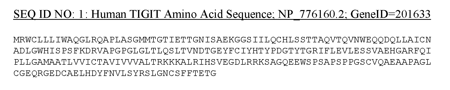 G
G

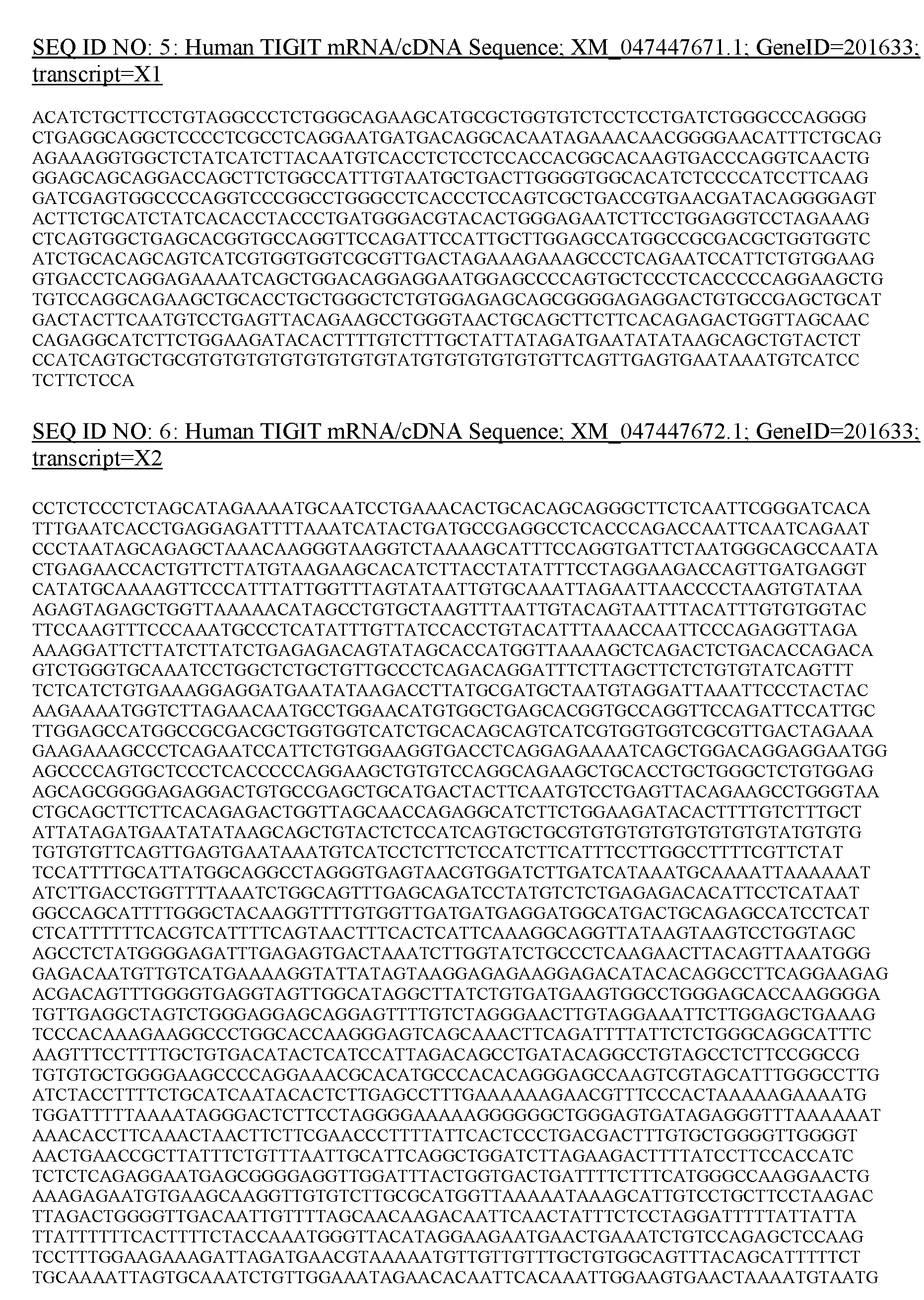

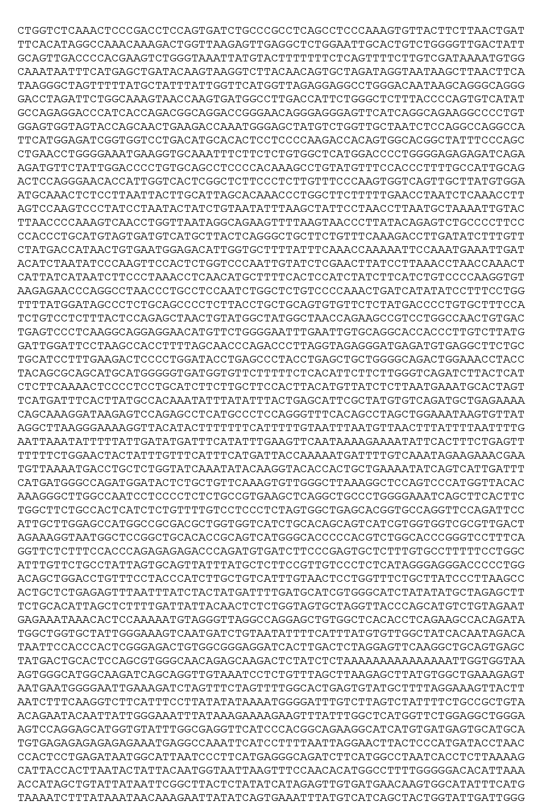
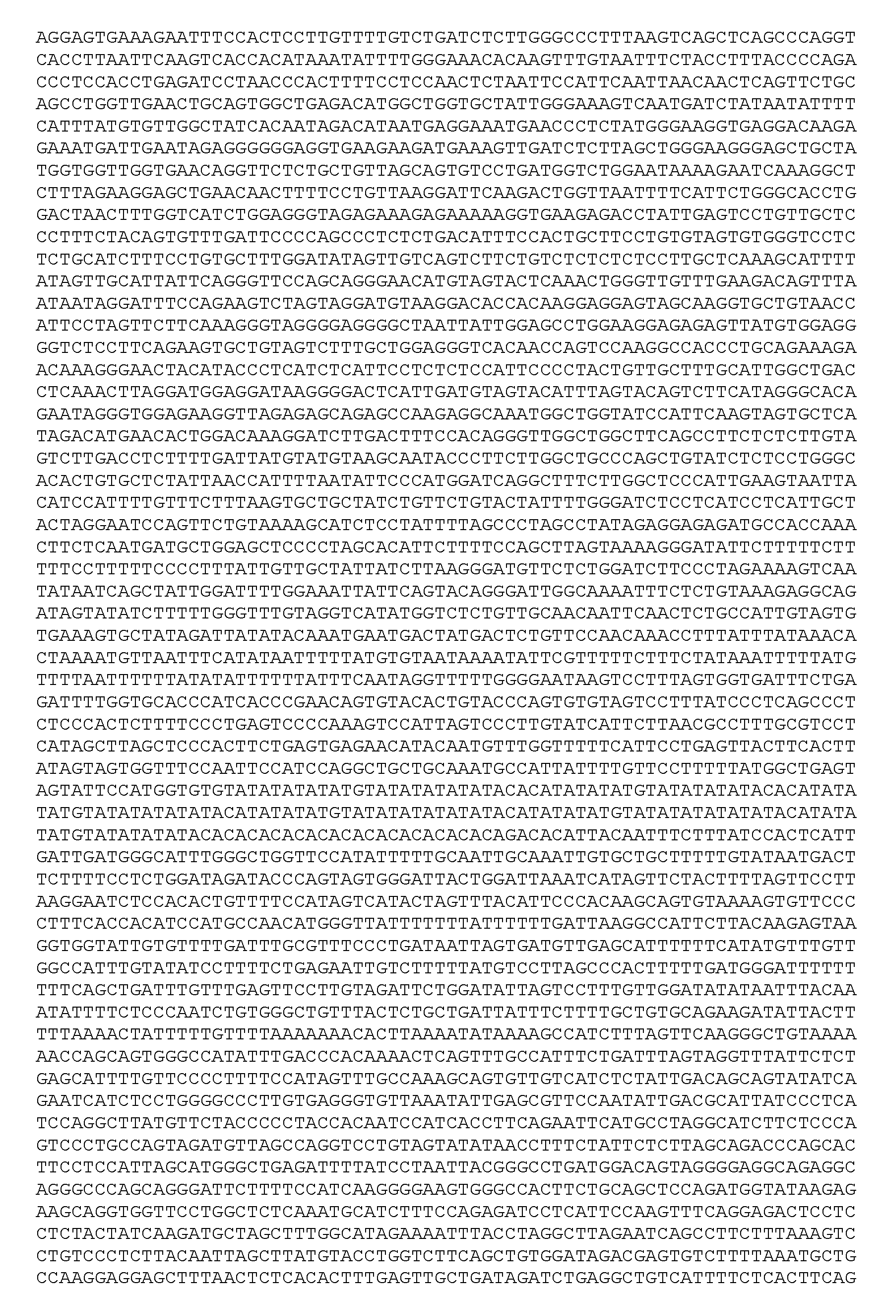


Itwillbeappreciatedthatspecificsequenceidentifiers(SEQ ID NOs)havebeen referencedthroughoutthespecificationforpurposesofillustrationandshouldtherefore notbe construedtobelimiting.Anymarkerencompassedbythepresentinvention,including,butnot limitedto,themarkersdescribedinthespecificationandmarkersdescribedherein,are well- knownintheartandmaybeusedintheembodimentsencompassedbythepresentinvention.
Asusedherein,theterm "monoclonalantibody"referstoanantibodyobtainedfrom a populationofsubstantiallyhomogeneousantibodiesthatspecificallybindtothesameepitope, i.e.,theindividualantibodiescomprisingthepopulationare identicalexceptforpossible naturallyoccurringmutationsthatmaybepresentinminoramounts.Themodifier "monoclonal" indicatesthecharacteroftheantibodyasbeingobtainedfrom asubstantiallyhomogeneous populationofantibodies,andisnottobeconstruedasrequiringproductionoftheantibodyby anyparticularmethod.
Thephrase “pharmaceutically-acceptablecarrier”asusedhereinmeansa pharmaceutically-acceptablematerial,compositionorvehicle,suchasaliquidorsolidfiller, diluent,excipient,orsolventencapsulatingmaterial,involvedincarryingortransportingthe subjectcompoundfrom oneorgan,orportionofthebody,toanotherorgan,orportionofthe body.
The terms “polynucleotide”, and “nucleic acid" are used interchangeably. They refer to a polymeric form of nucleotides of any length, either deoxyribonucleotides or ribonucleotides, or analogs thereof. Polynucleotides may have any three-dimensional structure, and may perform any function, known or unknown. The following are non-limiting examples of polynucleotides: coding or non-coding regions of a gene or gene fragment, loci (locus) defined from linkage analysis, exons, introns, messenger RNA (mRNA), transfer RNA, ribosomal RNA, ribozymes, cDNA, recombinant polynucleotides, branched polynucleotides, plasmids, vectors, isolated DNA of any sequence, isolated RNA of any sequence, nucleic acid probes, and primers. A polynucleotide may comprise modified nucleotides, such as methylated nucleotides and nucleotide analogs. If present, modifications to the nucleotide structure may be imparted before or after assembly of the polymer. The sequence of nucleotides may be interrupted by nonnucleotide components. A polynucleotide may be further modified, such as by conjugation with a labeling component. The term “recombinant” polynucleotide means a polynucleotide of genomic, cDNA, semisynthetic, or synthetic origin which either does not occur in nature or is linked to another polynucleotide in a non-natural arrangement.
The terms “prevent, ” “preventing, ” “prevention, ” and the like refer to reducing the probability of developing a disease, disorder, or condition in a subject, who does not have, but is at risk of or susceptible to developing a disease, disorder, or condition.
The term “small molecule" is a term of the art and includes molecules that are less than about 1000 molecular weight or less than about 500 molecular weight. In one embodiment, small molecules do not exclusively comprise peptide bonds. In another embodiment, small molecules are not oligomeric. Exemplary small molecule compounds which can be screened for activity include, but are not limited to, peptides, peptidomimetics, nucleic acids, carbohydrates, small organic molecules (e.g., polyketides) (Cane et al. (1998) Science 282:63), and natural product extract libraries. In another embodiment, the compounds are small, organic non-peptidic compounds. In a further embodiment, a small molecule is not biosynthetic.
As used herein, the term “subjecC means a human or non-human animal selected for treatment or therapy.
As used herein, the term “T cell” includes, but is not limited to, any T cell type listed herein, including CD4+ T cells and CD8+ T cells. The term “T cell” also includes both T helper 1 type T cells and T helper 2 type T cells. T cells express a cell surface receptor that recognizes a
specific antigenic moiety on the surface of a target cell. The cell surface receptor may be a wild type or recombinant T cell receptor (TCR), a chimeric antigen receptor (CAR), or any other surface receptor capable of recognizing an antigenic moiety that is associated with the target cell. Typically, a TCR has two protein chains (alpha- and beta-chain), which bind to specific peptides presented by an MHC protein on the surface of certain cells. TCRs recognize peptides in the context of MHC molecules expressed on the surface of a target cell. TCRs also recognize cancer antigens presented directly on the surface of cancer cells.
T cells used in T cell therapy, as described herein, can be modified following isolation using known methods, or the T cells can be activated and expanded (or differentiated in the case of progenitors) in vitro prior to or after being modified. In another embodiment, the T cells are genetically modified to have chimeric antigen receptors (e.g., transduced with a viral vector comprising a nucleic acid encoding a CAR) and then are activated and expanded in vitro. In some embodiments, the T cells are genetically modified with the engineered T cell receptors (e.g., transduced with a viral vector comprising a nucleic acid encoding a TCR) and then are activated and expanded in vitro. Methods for activating and expanding T cells are known in the art and are described, for example, in U.S. Pat. Nos. 6,905,874; 6,867,041; 6,797,514; W02012079000. Generally, such methods include culturing PBMC or isolated T cells with a stimulatory agent and costimulatory agent, such as anti-CD3 and anti-CD28 antibodies, often attached to a bead or other surface, in a culture medium with appropriate cytokines, such as IL-2 (e.g., recombinant human IL-2). Anti-CD3 and anti-CD28 antibodies attached to the same bead serve as a “surrogate” antigen presenting cell (APC). In other embodiments, the T cells may be activated and stimulated to proliferate with feeder cells and appropriate antibodies and cytokines using methods such as those described in U.S. Pat. Nos. 6,040,177; 5,827,642; and WO2012129514.
The T cell populations disclosed herein may comprise T helper cells. T helper cells (TH cells) assist other white blood cells in immunologic processes, including maturation of B cells into plasma cells and memory B cells, and activation of cytotoxic T cells and macrophages. These cells are also known as CD4+T cells because they express the CD4 glycoprotein on their surface. Helper T cells become activated when they are presented with peptide antigens by MHC class II molecules, which are expressed on the surface of antigen-presenting cells (APCs). Once activated, they divide rapidly and secrete small proteins called cytokines that regulate or assist in
the active immune response. These cells can differentiate into one of several subtypes, including THI, TH2, TH3, TH17, TH9, or TFH, which secrete different cytokines to facilitate different immune responses.
The T cell populations disclosed herein may comprise CTL cells. Cytotoxic T cells (Tc cells, or CTLs) destroy virally infected cells and tumor cells, and are also implicated in transplant rejection. These cells are also known as CD8+ T cells since they express the CDS glycoprotein at their surface. These cells recognize their targets by binding to antigen associated with MHC class I molecules, which are present on the surface of all nucleated cells.
The T cell populations disclosed herein may comprise memory T cells. Memory T cells are a subset of antigen-specific T cells that persist long-term after an infection has resolved. They quickly expand to large numbers of effector T cells upon re-exposure to their cognate antigen, thus providing the immune system with “memory” against past infections. Memory cells may be either CD4+ or CD8+. Memory T cells typically express the cell surface protein CD45RO.
The T cell populations disclosed herein may comprise regulatory T cells. Regulatory T cells (Tieg cells), formerly known as suppressor T cells, are crucial for the maintenance of immunological tolerance. Their major role is to suppress T cell-mediated immunity following an immune reaction and to suppress auto-reactive T cells that escaped the process of negative selection in the thymus. Two major classes of CD4+ Treg cells have been described — naturally occurring Treg cells and adaptive Treg cells.
The T cell populations disclosed herein may comprise Natural killer T (NKT) cells. Natural killer T (NKT) cells bridge the adaptive immune system with the innate immune system. Unlike conventional T cells that recognize peptide antigens presented by major histocompatibility complex (MHC) molecules, NKT cells recognize glycolipid antigen presented by a molecule called CDld.
In some embodiments, the T cells comprise a mixture of CD4+ cells. In other embodiments, the T cells are enriched for one or more subsets based on cell surface expression. For example, in some cases, the T comprise are cytotoxic CD8+ T lymphocytes.
Natural-killer (NK) cells are CD56+CD3 large granular lymphocytes that can kill virally infected and transformed cells, and constitute a critical cellular subset of the innate immune system (Godfrey J, et al. Leuk Lymphoma 201253:1666-1676). Unlike cytotoxic CD8+ T lymphocytes, NK cells launch cytotoxicity against tumor cells without the requirement for prior
sensitization and can eradicate MHC-I-negative cells (Nami-Mancinelli E, et al. Int Immunol 2011 23:427-431). NK cells are considered to be safer effector cells, as they may avoid the potentially lethal complications of cytokine storms (Morgan RA, et al. Mol Ther 2010 18:843- 851), tumor lysis syndrome (Porter DL, et al. N Engl J Med 2011 365:725-733), and on-target, off-tumor effects.
The “tumor microenvironment” is an art-recognized term and refers to the cellular environment in which the tumor exists, and includes, for example, interstitial fluids surrounding the tumor, surrounding blood vessels, immune cells, other cells, fibroblasts, signaling molecules, and the extracellular matrix.
The phrases "therapeutically-effective amount" and “effective amount” as used herein means the amount of an agent which is effective for producing the desired therapeutic effect in at least a sub-population of cells in a subject at a reasonable benefit/risk ratio applicable to any medical treatment.
“Treating’ a disease in a subject or “treating’ a subject having a disease refers to subjecting the subject to a pharmaceutical treatment, e.g., the administration of a drug, such that at least one symptom of the disease is decreased or prevented from worsening.
Modulators of TIGIT
Provided herein are methods of ameliorating an immune-related adverse event associated with an immunotherapy or immune-modulating therapy in a subject (e.g., a subject with cancer, autoimmune disease or another condition requiring immune-modulating medications), the method comprising administering to the subject an agent that agonizes the activity or expression of TIGIT. In some aspects, provided herein are methods of treating or preventing an autoimmune disease in a subject, the method comprising administering to the subject an agent that agonizes the activity or expression of TIGIT.
An agent disclosed herein may increase the activity or expression of TIGIT by at least 5%, at least 10%, at least 15%, at least 20%, at least 25%, at least 30%, at least 35%, at least 40%, at least 45%, at least 50%, at least 60%, at least 65%, at least 70%, at least 75%, at least 80%, at least 85%, at least 90%, at least 95%, at least 100%, at least 150%, at least 200%, at least 250%, at least 300%, at least 350%, at least 400%, at least 450%, at least 500%, at least 550%, at least 600%, at least 650%, at least 700%, at least 750%, at least 800%, at least 850%, at least
900%, at least 950%, or at least 1000%. An agent disclosed herein may increase TIGIT mRNA by at least 5%, at least 10%, at least 15%, at least 20%, at least 25%, at least 30%, at least 35%, at least 40%, at least 45%, at least 50%, at least 60%, at least 65%, at least 70%, at least 75%, at least 80%, at least 85%, at least 90%, at least 95%, at least 100%, at least 150%, at least 200%, at least 250%, at least 300%, at least 350%, at least 400%, at least 450%, at least 500%, at least 550%, at least 600%, at least 650%, at least 700%, at least 750%, at least 800%, at least 850%, at least 900%, at least 950%, or at least 1000%.Measurement of TIGIT can be done in a biological sample or multiple biological samples taken from the subject over a period of time.
The agent may be administered conjointly with an immunotherapy or immune- modulating therapy disclosed herein (e.g., an immune checkpoint inhibitor or other immune- modulating medication).
Also provided herein are methods of treating cancer by antagonizing TIGIT. The agent may reduce the activity or expression of TIGIT by at least 5%, at least 10%, at least 15%, at least 20%, at least 25%, at least 30%, at least 35%, at least 40%, at least 45%, at least 50%, at least 60%, at least 65%, at least 70%, at least 75%, at least 80%, at least 85%, at least 90%, at least 95%, or 100%. The agent may reduce the expression of TIGIT by at least 5%, at least 10%, at least 15%, at least 20%, at least 25%, at least 30%, at least 35%, at least 40%, at least 45%, at least 50%, at least 60%, at least 65%, at least 70%, at least 75%, at least 80%, at least 85%, at least 90%, at least 95%, or 100%. An agent disclosed herein may reduce TIGIT mRNA by at least 5%, at least 10%, at least 15%, at least 20%, at least 25%, at least 30%, at least 35%, at least 40%, at least 45%, at least 50%, at least 60%, at least 65%, at least 70%, at least 75%, at least 80%, at least 85%, at least 90%, at least 95%, or 100%.
Polynucleotide/ Nucleic Acid Molecules
Also provided herein are nucleic acid or polynucleotide molecules (RNA constructs) that encode the TIGIT peptides, antibodies, antigen binding fragments thereof and/or polypeptides described herein. For example, the polynucleotide may encode a TIGIT protein or fragment thereof. The nucleic acids may be present, for example, in whole cells, in a cell lysate, or in a partially purified or substantially pure form. In some embodiments, provided herein are methods
of administering to the subject an agent that is a nucleic acid or polynucleotide molecules (RNA constructs) that encode a TIGIT peptide.
Nucleic acids described herein can be obtained using standard molecular biology techniques. For example, nucleic acid molecules described herein can be cloned using standard PCR techniques or chemically synthesized. For antibodies obtained from an immunoglobulin gene library (e.g., using phage or yeast display techniques), nucleic acid encoding the antibody can be recovered from the library.
In certain embodiments, provided herein are vectors that contain the isolated nucleic acid molecules described herein (e.g., a nucleic acid of Table 1). As used herein, the term “vector,” refers to a nucleic acid molecule capable of transporting another nucleic acid to which it has been linked. One type of vector is a “plasmid”, which refers to a circular double stranded DNA loop into which additional DNA segments may be ligated. Another type of vector is a viral vector, wherein additional DNA segments may be ligated into the viral genome. Certain vectors are capable of autonomous replication in a host cell into which they are introduced (e.g., bacterial vectors having a bacterial origin of replication and episomal mammalian vectors). Other vectors (e.g., non-episomal mammalian vectors) can be integrated into the genome of a host cell upon introduction into the host cell, and thereby be replicated along with the host genome. Moreover, certain vectors are capable of directing the expression of genes. Such vectors are referred to herein as “recombinant expression vectors” (or simply, “expression vectors”). In certain embodiments, provided herein are cells that contain a nucleic acid described herein (e.g., a nucleic acid encoding an antibody, antigen binding fragment thereof, antibody-like molecule, or polypeptide described herein). The cell can be, for example, prokaryotic, eukaryotic, mammalian, avian, murine and/or human.
In some embodiments, peptides disclosed herein are delivered to subjects by use of viral vectors. Viral vector systems which can be utilized with the methods and compositions described herein include, but are not limited to, (a) adenovirus vectors; (b) retrovirus vectors, including but not limited to lentiviral vectors, moloney murine leukemia virus, etc. ; (c) adeno-associated virus vectors; (d) herpes simplex virus vectors; (e) SV 40 vectors; (f) polyoma virus vectors; (g) papilloma virus vectors; (h) picomavirus vectors; (i) pox virus vectors such as an orthopox, e.g., vaccinia virus vectors or avipox, e.g. canary pox or fowl pox; and (j) a helper-dependent or gutless adenovirus.
In some embodiments, adenoviruses can be used to deliver nucleic acids encoding a peptide. Adenoviruses have the advantage of being capable of infecting non-dividing cells. Kozarsky and Wilson, Current Opinion in Genetics and Development 3:499-503 (1993) present a review of adenovirus-based gene therapy.
The adeno-associated virus is a non-pathogenic parvovirus, consisting of a 4.7 kb singlestranded DNA genome, with no envelope icosahedral capsid. The genome contains three open reading frames (ORFs) flanked by inverted terminal repeats (ITRs) that function as a replication and packaging signal of viral origin. Rep ORF encodes four non-structural proteins that play a role in virus replication, transcriptional regulation, site-specific integration, and virion assembly. Cap ORF encodes three structural proteins (VP 1-3), which are assembled to form a 60- dimensional viral capsid. Finally, ORF, present as an alternative reading frame in the cap gene, produces assembly activating protein (AAP), a viral protein that localizes AAV capsid proteins into the nucleolus and functions during capsid assembly.
There are several natural ("wild type") serotypes and more than 100 known AAV variants, each of which differs in amino acid sequence, especially in the hypervariable regions of capsid proteins, and thus in its gene delivery properties. No association has been found between any AAV and any human disease, which makes recombinant AAV attractive for clinical applications.
For the purposes of the description herein, the term “AAV” is an abbreviation for adeno- associated virus, including, without limitation, the virus itself and its derivatives. Except where otherwise indicated, terminology refers to all subtypes or serotypes, and both replication- competent and recombinant forms. The term “AAV” includes, without limitation, AAV type 1 (AAV-1 or AAV1), AAV type 2 (AAV-2 or AAV2), AAV type 3A (AAV-3A or AAV3A), AAV type 3B (AAV-3B or AAV3B), AAV type 4 (AAV-4 or AAV4), AAV type 5 (AAV-5 or AAV5), AAV type 6 (AAV-6 or AAV6), AAV type 7 (AAV-7 or AAV7), type AAV 8 (AAV-8 or AAV8), AAV type 9 (AAV-9 or AAV9), and AAV type 10 (AAV-10 or AAV10 or AAVrhlO).
In some embodiments, a AAV vector that expresses a nucleic acid agent encoding an interferon peptide is a recombinant AAV vector having, for example, either an U6 or Hl RNA promoter, or a cytomegalovirus (CMV) promoter. Suitable AAV vectors for use in agents,
compositions, and methods described include, but are not limited to AAVs described in Passini etal, Methods Mol. Biol. 246: 225-36 (2004).
Blocking and Agonizing Antibody Agents
In some embodiments, the agent described herein is an antibody specific for a TIGIT peptide. A blocking antibody disclosed herein may inhibit expression or activity of a TIGIT protein by at least 5%, at least 10%, at least 15%, at least 20%, at least 25%, at least 30%, at least 35%, at least 40%, at least 45%, at least 50%, at least 60%, at least 65%, at least 70%, at least 75%, at least 80%, at least 85%, at least 90%, at least 95%, or 100%.
An agonizing antibody disclosed herein may increase the activity of a TIGIT protein by at least 5%, at least 10%, at least 15%, at least 20%, at least 25%, at least 30%, at least 35%, at least 40%, at least 45%, at least 50%, at least 60%, at least 65%, at least 70%, at least 75%, at least 80%, at least 85%, at least 90%, at least 95%, or 100%.
A blocking or agonizing antibody provided herein may have at least 5%, at least 10%, at least 15%, at least 20%, at least 25%, at least 30%, at least 35%, at least 40%, at least 45%, at least 50%, at least 60%, at least 65%, at least 70%, at least 75%, at least 80%, at least 85%, at least 90%, at least 95%, or 100% specificity for a TIGIT protein.
Antibodies may be polyclonal or monoclonal; xenogeneic, allogeneic, or syngeneic; or modified forms thereof (e.g., humanized, chimeric, etc.). Antibodies may also be fully human. The terms “monoclonal antibodies” and “monoclonal antibody composition”, as used herein, refer to a population of antibody polypeptides that contain only one species of an antigen binding site capable of immunoreacting with a particular epitope of an antigen, whereas the term “polyclonal antibodies” and “polyclonal antibody composition” refer to a population of antibody polypeptides that contain multiple species of antigen binding sites capable of interacting with a particular antigen. A monoclonal antibody composition typically displays a single binding affinity for a particular antigen with which it immunoreacts.
In certain embodiments, the methods and compositions provided herein relate to antibodies and antigen binding fragments thereof that bind specifically to a TIGIT protein. In some embodiments, the antibodies inhibit the function of the protein, such as inhibiting the activity of the protein, or interfering with protein-protein interactions. In some embodiments, the antibodies increase the function of the protein. Such antibodies can be polyclonal or monoclonal
and can be, for example, murine, chimeric, humanized or fully human. In some embodiments, the agent may be a recombinant antibodies specific for a TIGIT protein, such as chimeric or humanized monoclonal antibodies, can be made using standard recombinant DNA techniques. Such chimeric and humanized monoclonal antibodies can be produced by recombinant DNA techniques known in the art, for example using methods described in US Pat No. 4,816,567; US Pat. No. 5,565,332; Better et al. (1988) Science 240: 1041-1043; Liu et al. (1987) Proc. Natl. Acad. Sci. USA 84:3439-3443; Liu etaZ. (1987) J. Immunol. 139:3521-3526; Sun etaZ. (1987) Proc. Natl. Acad. Sci. 84:214-218; Nishimura et al. (1987) Cancer Res. 47:999-1005; Wood et al. (1985) Nature 314:446-449; and Shaw er al. (1988) J. Natl. Cancer Inst. 80:1553-1559); Morrison, S. L. (1985) Science 229:1202-1207; Oi etal. (1986) Biotechniques 4:214; Winter U.S. Patent 5,225,539; Jones et al. (1986) Nature 321 :552-525; Verhoeyan et al. (1988) Science 239:1534; and BeidleretaZ. (1988) J. Immunol. 141:4053-4060.
Human monoclonal antibodies specific for a TIGIT protein can be generated using transgenic or transchromosomal mice carrying parts of the human immune system rather than the mouse system. For example, “HuMAb mice” which contain a human immunoglobulin gene miniloci that encodes unrearranged human heavy (p. and y) and K light chain immunoglobulin sequences, together with targeted mutations that inactivate the endogenous p and K chain loci (Lonberg, N. et al. (1994) Nature 368(6474): 856859). Accordingly, the mice exhibit reduced expression of mouse IgM or x, and in response to immunization, the introduced human heavy and light chain transgenes undergo class switching and somatic mutation to generate high affinity human IgGx monoclonal antibodies (Lonberg, N. et al. (1994), supra; reviewed in Lonberg, N. (1994) Handbook of Experimental Pharmacology 113:49 101; Lonberg, N. and Huszar, D. (1995) Intern. Rev. Immunol. Vol. 13: 65 93, and Harding, F. and Lonberg, N. (1995) Ann. N. Y Acad. Sci 764:536546).
The preparation of HuMAb mice is described in Taylor, L. et al. (1992) Nucleic Acids Research 20:6287 6295; Chen, J. etal. (1993) International Immunology 5: 647656; Tuaillon et al. (1993) Proc. Natl. Acad. Sci USA 90:37203724; Choi etal. (1993) Nature Genetics 4:117 123; Chen, J. etal. (1993) EMBO J. 12: 821 830; Tuaillon etal. (1994) J. Immunol. 152:2912 2920; Lonberg etal., (1994) Nature 368(6474): 856859; Lonberg, N. (1994) Handbook of Experimental Pharmacology 113:49 101; Taylor, L. etal. (1994) International Immunology 6: 579591; Lonberg, N. and Huszar, D. (1995) Intern. Rev. Immunol. Vol. 13: 65 93; Harding, F.
and Lonberg, N. (1995) Ann. N.Y. Acad. Sci 764:536546; Fishwild, D. et al. (1996) Nature Biotechnology 14: 845 851. See further, U.S. Pat. Nos. 5,545,806; 5,569,825; 5,625,126; 5,633,425; 5,789,650; 5,877,397; 5,661,016; 5,814,318; 5,874,299; 5,770,429; and 5,545,807.
Interfering Nucleic Acid Agents
Provided herein are compositions comprising an agent that is an interfering nucleic acid specific for an mRNA product of a gene (e.g., a gene listed in Table 1). Provided herein are compositions comprising an agent that is an interfering nucleic acid specific for an mRNA transcript (e.g., a transcript listed in Table 1). The interfering nucleic acid may be a siRNA, shRNA, miRNA, or a peptide nucleic acid.
Interfering nucleic acids generally include a sequence of cyclic subunits, each bearing a base-pairing moiety, linked by intersubunit linkages that allow the base-pairing moieties to hybridize to a target sequence in a nucleic acid (typically an RNA) by Watson-Crick base pairing, to form a nucleic acid: oligomer heteroduplex within the target sequence. Interfering RNA molecules include, but are not limited to, antisense molecules, siRNA molecules, singlestranded siRNA molecules, miRNA molecules and shRNA molecules.
Typically at least 17, 18, 19, 20, 21, 22 or 23 nucleotides of the complement of the target mRNA sequence are sufficient to mediate inhibition of a target transcript. Perfect complementarity is not necessary. In some embodiments, the interfering nucleic acid molecule is double-stranded RNA. The double-stranded RNA molecule may have a 2 nucleotide 3’ overhang. In some embodiments, the two RNA strands are connected via a hairpin structure, forming a shRNA molecule. shRNA molecules can contain hairpins derived from microRNA molecules. For example, an RNAi vector can be constructed by cloning the interfering RNA sequence into a pCAG-miR30 construct containing the hairpin from the miR30 miRNA. RNA interference molecules may include DNA residues, as well as RNA residues.
Interfering nucleic acid molecules provided herein can contain RNA bases, non-RNA bases or a mixture of RNA bases and non-RNA bases. For example, interfering nucleic acid molecules provided herein can be primarily composed of RNA bases but also contain DNA bases or non-naturally occurring nucleotides.
The interfering nucleic acids can employ a variety of oligonucleotide chemistries. Examples of oligonucleotide chemistries include, without limitation, peptide nucleic acid (PNA),
linked nucleic acid (LNA), phosphorothioate, 2’0-Me-modified oligonucleotides, and morpholino chemistries, including combinations of any of the foregoing. In general, PNA and LNA chemistries can utilize shorter targeting sequences because of their relatively high target binding strength relative to 2’0-Me oligonucleotides. Phosphorothioate and 2’0-Me-modified chemistries are often combined to generate 2’0-Me-modified oligonucleotides having a phosphorothioate backbone. See, e.g., PCT Publication Nos. WO/2013/112053 and WO/2009/008725, incorporated by reference in their entireties.
Peptide nucleic acids (PNAs) are analogs of DNA in which the backbone is structurally homomorphous with a deoxyribose backbone, consisting of N-(2-aminoethyl) glycine units to which pyrimidine or purine bases are attached. PNAs containing natural pyrimidine and purine bases hybridize to complementary oligonucleotides obeying Watson-Crick base-pairing rules, and mimic DNA in terms of base pair recognition (Egholm, Buchardt et al. 1993). The backbone of PNAs is formed by peptide bonds rather than phosphodiester bonds, making them well-suited for antisense applications (see structure below). The backbone is uncharged, resulting in PNA/DNA or PNA/RNA duplexes that exhibit greater than normal thermal stability. PNAs are not recognized by nucleases or proteases.
Despite a radical structural change to the natural structure, PNAs are capable of sequence-specific binding in a helix form to DNA or RNA. Characteristics of PNAs include a high binding affinity to complementary DNA or RNA, a destabilizing effect caused by singlebase mismatch, resistance to nucleases and proteases, hybridization with DNA or RNA independent of salt concentration and triplex formation with homopurine DNA. PANAGENE™. has developed its proprietary Bts PNA monomers (Bts; benzothiazole-2-sulfonyl group) and proprietary oligomerization process. The PNA oligomerization using Bts PNA monomers is composed of repetitive cycles of deprotection, coupling and capping. PNAs can be produced synthetically using any technique known in the art. See, e.g., U.S. Pat. Nos. 6,969,766, 7,211,668, 7,022,851, 7,125,994, 7,145,006 and 7,179,896. See also U.S. Pat. Nos. 5,539,082; 5,714,331; and 5,719,262 for the preparation of PNAs. Further teaching of PNA compounds can be found in Nielsen et al., Science, 254:1497-1500, 1991. Each of the foregoing is incorporated by reference in its entirety.
Interfering nucleic acids may also contain “locked nucleic acid” subunits (LNAs). “LNAs” are a member of a class of modifications called bridged nucleic acid (BNA). BNA is
characterized by a covalent linkage that locks the conformation of the ribose ring in a C30-endo (northern) sugar pucker. For LNA, the bridge is composed of a methylene between the 2’-0 and the 4’-C positions. LNA enhances backbone preorganization and base stacking to increase hybridization and thermal stability.
The structures of LNAs can be found, for example, in Wengel, et al., Chemical Communications (1998) 455; Tetrahedron (1998) 54:3607, and Accounts of Chem. Research (1999) 32:301); Obika, et al., Tetrahedron Letters (1997) 38:8735; (1998) 39:5401, and Bioorganic Medicinal Chemistry (2008) 16:9230. Compounds provided herein may incorporate one or more LNAs; in some cases, the compounds may be entirely composed of LNAs. Methods for the synthesis of individual LNA nucleoside subunits and their incorporation into oligonucleotides are described, for example, in U.S. Pat. Nos. 7,572,582, 7,569,575, 7,084,125, 7,060,809, 7,053,207, 7,034,133, 6,794,499, and 6,670,461, each of which is incorporated by reference in its entirety. Typical intersubunit linkers include phosphodiester and phosphorothioate moieties; alternatively, non-phosphorous containing linkers may be employed. One embodiment is an LNA containing compound where each LNA subunit is separated by a DNA subunit. Certain compounds are composed of alternating LNA and DNA subunits where the intersubunit linker is phosphorothioate.
“Phosphorothioates” (or S-oligos) are a variant of normal DNA in which one of the nonbridging oxygens is replaced by a sulfur. The sulfurization of the intemucleotide bond reduces the action of endo-and exonucleases including 5’ to 3’ and 3’ to 5’ DNA POL 1 exonuclease, nucleases SI and Pl, RNases, serum nucleases and snake venom phosphodiesterase. Phosphorothioates are made by two principal routes: by the action of a solution of elemental sulfur in carbon disulfide on a hydrogen phosphonate, or by the method of sulfurizing phosphite triesters with either tetraethylthiuram disulfide (TETD) or 3H-1, 2- bensodithiol-3-one 1, 1 -dioxide (BDTD) (see, e.g., Iyer et al., J. Org. Chem. 55, 4693-4699, 1990). The latter methods avoid the problem of elemental sulfur’s insolubility in most organic solvents and the toxicity of carbon disulfide. The TETD and BDTD methods also yield higher purity phosphorothioates.
“2’0-Me oligonucleotides” molecules cany a methyl group at the 2’-OH residue of the ribose molecule. 2’-O-Me-RNAs show the same (or similar) behavior as DNA, but are protected against nuclease degradation. 2’-O-Me-RNAs can also be combined with phosphothioate
oligonucleotides (PTOs) for further stabilization. 2’0-Me oligonucleotides (phosphodiester or phosphothioate) can be synthesized according to routine techniques in the art (see, e.g., Yoo et al., Nucleic Acids Res. 32:2008-16, 2004).
The interfering nucleic acids described herein may be contacted with a cell or administered to an organism (e.g., a human). Alternatively, constructs and/or vectors encoding the interfering RNA molecules may be contacted with or introduced into a cell or organism. In certain embodiments, a viral, retroviral or lentiviral vector is used. In some embodiments, the vector has a tropism for cardiac tissue. In some embodiments the vector is an adeno-associated virus.
Typically at least 17, 18, 19, 20, 21, 22 or 23 nucleotides of the complement of the target mRNA sequence are sufficient to mediate inhibition of a target transcript. Perfect complementarity is not necessary. In some embodiments, the interfering nucleic acids contains a 1, 2 or 3 nucleotide mismatch with the target sequence. The interfering nucleic acid molecule may have a 2 nucleotide 3’ overhang. If the interfering nucleic acid molecule is expressed in a cell from a construct, for example from a hairpin molecule or from an inverted repeat of the desired sequence, then the endogenous cellular machinery will create the overhangs. shRNA molecules can contain hairpins derived from microRNA molecules. For example, an RNAi vector can be constructed by cloning the interfering RNA sequence into a pCAG-miR30 construct containing the hairpin from the miR30 miRNA. RNA interference molecules may include DNA residues, as well as RNA residues.
In some embodiments, the interfering nucleic acid molecule is a siRNA molecule. Such siRNA molecules should include a region of sufficient homology to the target region, and be of sufficient length in terms of nucleotides, such that the siRNA molecule down-regulate target RNA. The term “ribonucleotide” or “nucleotide” can, in the case of a modified RNA or nucleotide surrogate, also refer to a modified nucleotide, or surrogate replacement moiety at one or more positions. It is not necessary that there be perfect complementarity between the siRNA molecule and the target, but the correspondence must be sufficient to enable the siRNA molecule to direct sequence-specific silencing, such as by RNAi cleavage of the target RNA. In some embodiments, the sense strand need only be sufficiently complementary with the antisense strand to maintain the overall double-strand character of the molecule.
In addition, an siRNA molecule may be modified or include nucleoside surrogates. Single stranded regions of an siRNA molecule may be modified or include nucleoside surrogates, e.g., the unpaired region or regions of a hairpin structure, e.g., a region which links two complementary regions, can have modifications or nucleoside surrogates. Modification to stabilize one or more 3'- or 5 '-terminus of an siRNA molecule, e.g., against exonucleases, or to favor the antisense siRNA agent to enter into RISC are also useful. Modifications can include C3 (or C6, C7, C12) amino linkers, thiol linkers, carboxyl linkers, non-nucleotidic spacers (C3, C6, C9, Cl 2, abasic, triethylene glycol, hexaethylene glycol), special biotin or fluorescein reagents that come as phosphoramidites and that have another DMT-protected hydroxyl group, allowing multiple couplings during RNA synthesis.
Each strand of an siRNA molecule can be equal to or less than 35, 30, 25, 24, 23, 22, 21, or 20 nucleotides in length. In some embodiments, the strand is at least 19 nucleotides in length. For example, each strand can be between 21 and 25 nucleotides in length. In some embodiments, siRNA agents have a duplex region of 17, 18, 19, 29, 21, 22, 23, 24, or 25 nucleotide pairs, and one or more overhangs, such as one or two 3' overhangs, of 2-3 nucleotides.
A “small hairpin RNA” or “short hairpin RNA” or “shRNA” includes a short RNA sequence that makes a tight hairpin turn that can be used to silence gene expression via RNA interference. The shRNAs provided herein may be chemically synthesized or transcribed from a transcriptional cassette in a DNA plasmid. The shRNA hairpin structure is cleaved by the cellular machinery into siRNA, which is then bound to the RNA-induced silencing complex (RISC).
In some embodiments, shRNAs are about 15-60, 15-50, or 15-40 (duplex) nucleotides in length, about 15-30, 15-25, or 19-25 (duplex) nucleotides in length, or are about 20-24, 21-22, or 21-23 (duplex) nucleotides in length (e.g., each complementary sequence of the double-stranded shRNA is 15-60, 15-50, 15^0, 15-30, 15-25, or 19-25 nucleotides in length, or about 20-24, 21- 22, or 21-23 nucleotides in length, and the double-stranded shRNA is about 15-60, 15-50, 15-40, 15-30, 15-25, or 19-25 base pairs in length, or about 18-22, 19-20, or 19-21 base pairs in length). shRNA duplexes may comprise 3’ overhangs of about 1 to about 4 nucleotides or about 2 to about 3 nucleotides on the antisense strand and/or 5 ’-phosphate termini on the sense strand. In some embodiments, the shRNA comprises a sense strand and/or antisense strand sequence of from about 15 to about 60 nucleotides in length (e.g., about 15-60, 15-55, 15-50, 15-45, 15-40,
15-35, 15-30, or 15-25 nucleotides in length), or from about 19 to about 40 nucleotides in length (e.g., about 19-40, 19-35, 19-30, or 19-25 nucleotides in length), or from about 19 to about 23 nucleotides in length (e.g., 19, 20, 21, 22, or 23 nucleotides in length).
Non-limiting examples of shRNA include a double-stranded polynucleotide molecule assembled from a single-stranded molecule, where the sense and antisense regions are linked by a nucleic acid-based or non-nucleic acid-based linker; and a double-stranded polynucleotide molecule with a hairpin secondary structure having self-complementary sense and antisense regions. In some embodiments, the sense and antisense strands of the shRNA are linked by a loop structure comprising from about 1 to about 25 nucleotides, from about 2 to about 20 nucleotides, from about 4 to about 15 nucleotides, from about 5 to about 12 nucleotides, or 1, 2, 3, 4, 5, 6, 7, 8, 9, 10, 11, 12, 13, 14, 15, 16, 17, 18, 19, 20, 21, 22, 23, 24, 25, or more nucleotides.
Additional embodiments related to the shRNAs, as well as methods of designing and synthesizing such shRNAs, are described in U.S. patent application publication number 2011/0071208, the disclosure of which is herein incorporated by reference in its entirety for all purposes.
In some embodiments, provided herein are micro RNAs (miRNAs). miRNAs represent a large group of small RNAs produced naturally in organisms, some of which regulate the expression of target genes. miRNAs are formed from an approximately 70 nucleotide singlestranded hairpin precursor transcript by Dicer. miRNAs are not translated into proteins, but instead bind to specific messenger RNAs, thereby blocking translation. In some instances, miRNAs base-pair imprecisely with their targets to inhibit translation.
In some embodiments, antisense oligonucleotide compounds are provided herein. In certain embodiments, the degree of complementarity between the target sequence and antisense targeting sequence is sufficient to form a stable duplex. The region of complementarity of the antisense oligonucleotides with the target RNA sequence may be as short as 8-11 bases, but can be 12-15 bases or more, e.g., 10-40 bases, 12-30 bases, 12-25 bases, 15-25 bases, 12-20 bases, or 15-20 bases, including all integers in between these ranges. An antisense oligonucleotide of about 14-15 bases is generally long enough to have a unique complementary sequence.
In certain embodiments, antisense oligonucleotides may be 100% complementary to the target sequence, or may include mismatches, e.g., to improve selective targeting of allele
containing a disease-associated mutation, as long as a heteroduplex formed between the oligonucleotide and target sequence is sufficiently stable to withstand the action of cellular nucleases and other modes of degradation which may occur in vivo. Hence, certain oligonucleotides may have about or at least about 70% sequence complementarity, e.g., 70%, 71%, 72%, 73%, 74%, 75%, 76%, 77%, 78%, 79%, 80%, 81%, 82%, 83%, 84%, 85%, 86%, 87%, 88%, 89%, 90%, 91%, 92%, 93%, 94%, 95%, 96%, 97%, 98%, 99% or 100% sequence complementarity, between the oligonucleotide and the target sequence. Oligonucleotide backbones that are less susceptible to cleavage by nucleases are discussed herein. Mismatches, if present, are typically less destabilizing toward the end regions of the hybrid duplex than in the middle. The number of mismatches allowed will depend on the length of the oligonucleotide, the percentage of G:C base pairs in the duplex, and the position of the mismatches) in the duplex, according to well understood principles of duplex stability.
Interfering nucleic acid molecules can be prepared, for example, by chemical synthesis, in vitro transcription, or digestion of long dsRNA by Rnase III or Dicer. These can be introduced into cells by transfection, electroporation, or other methods known in the art. See Hannon, GJ, 2002, RNA Interference, Nature 418: 244-251; Bernstein E et al., 2002, The rest is silence. RNA 7: 1509-1521; Hutvagner G et al., RNAi: Nature abhors a double-strand. Cun. Opin. Genetics & Development 12: 225-232; Brummelkamp, 2002, A system for stable expression of short interfering RNAs in mammalian cells. Science 296: 550-553; Lee NS, Dohjima T, Bauer G, Li H, Li M-J, Ehsani A, Salvaterra P, and Rossi J. (2002). Expression of small interfering RNAs targeted against HIV-1 rev transcripts in human cells. Nature Biotechnol. 20:500-505; Miyagishi M, and Taira K. (2002). U6-promoter-driven siRNAs with four uridine 3' overhangs efficiently suppress targeted gene expression in mammalian cells. Nature Biotechnol. 20:497-500; Paddison PJ, Caudy AA, Bernstein E, Hannon GJ, and Conklin DS. (2002). Short hairpin RNAs (shRNAs) induce sequence-specific silencing in mammalian cells. Genes & Dev. 16:948-958; Paul CP, Good PD, Winer I, and Engelke DR (2002). Effective expression of small interfering RNA in human cells. Nature Biotechnol. 20:505-508; Sui G, Soohoo C, Affar E-B, Gay F, Shi Y, Forrester WC, and Shi Y. (2002). A DNA vector-based RNAi technology to suppress gene expression in mammalian cells. Proc. Natl. Acad. Sci. USA 99(6):5515-5520; Yu J-Y, DeRuiter SL, and Turner DL. (2002). RNA interference by expression of short-interfering RNAs and hairpin RNAs in mammalian cells. Proc. Natl. Acad. Sci. USA 99(9):6047-6052.
In the present methods, an interfering nucleic acid molecule or an interfering nucleic acid encoding polynucleotide can be administered to the subject, for example, as naked nucleic acid, in combination with a delivery reagent, and/or as a nucleic acid comprising sequences that express an interfering nucleic acid molecule. In some embodiments the nucleic acid comprising sequences that express the interfering nucleic acid molecules are delivered within vectors, e.g. plasmid, viral and bacterial vectors. Any nucleic acid delivery method known in the art can be used in the methods described herein. Suitable delivery reagents include, but are not limited to, e.g., the Minis Transit TKO lipophilic reagent; lipofectin; lipofectamine; cellfectin; polycations (e.g., polylysine), atelocollagen, nanoplexes and liposomes. The use of atelocollagen as a delivery vehicle for nucleic acid molecules is described in Minakuchi et al. Nucleic Acids Res., 32(13):el09 (2004); Hanai et al. Ann NY Acad Sci., 1082:9-17 (2006); and Kawata et al. Mol Cancer Then, 7(9):2904-12 (2008); each of which is incorporated herein in their entirety. Exemplary interfering nucleic acid delivery systems are provided in U.S. Patent Nos. 8,283,461, 8,313,772, 8,501,930. 8,426,554, 8,268,798 and 8,324,366, each of which is hereby incorporated by reference in its entirety.
In some embodiments of the methods described herein, liposomes are used to deliver an inhibitory oligonucleotide to a subject. Liposomes suitable for use in the methods described herein can be formed from standard vesicle-forming lipids, which generally include neutral or negatively charged phospholipids and a sterol, such as cholesterol. The selection of lipids is generally guided by consideration of factors such as the desired liposome size and half-life of the liposomes in the blood stream. A variety of methods are known for preparing liposomes, for example, as described in Szoka etal. (1980), Ann. Rev. Biophys. Bioeng. 9:467; and U.S. Pat. Nos. 4,235,871, 4,501,728, 4,837,028, and 5,019,369, the entire disclosures of which are herein incorporated by reference.
The liposomes for use in the present methods can also be modified so as to avoid clearance by the mononuclear macrophage system ("MMS") and reticuloendothelial system ("RES"). Such modified liposomes have opsonization-inhibition moieties on the surface or incorporated into the liposome structure.
Opsonization-inhibiting moieties for use in preparing the liposomes described herein are typically large hydrophilic polymers that are bound to the liposome membrane. As used herein, an opsonization inhibiting moiety is “bound” to a liposome membrane when it is chemically or
physically attached to the membrane, e.g., by the intercalation of a lipid-soluble anchor into the membrane itself, or by binding directly to active groups of membrane lipids. These opsonizationinhibiting hydrophilic polymers form a protective surface layer that significantly decreases the uptake of the liposomes by the MMS and RES; e.g., as described in U.S. Pat. No. 4,920,016, the entire disclosure of which is herein incorporated by reference.
In some embodiments, opsonization inhibiting moieties suitable for modifying liposomes are water-soluble polymers with a number-average molecular weight from about 500 to about 40,000 daltons, or from about 2,000 to about 20,000 daltons. Such polymers include polyethylene glycol (PEG) or polypropylene glycol (PPG) derivatives; e.g., methoxy PEG or PPG, and PEG or PPG stearate; synthetic polymers such as polyacrylamide or poly N-vinyl pyrrolidone; linear, branched, or dendrimeric polyamidoamines; polyacrylic acids; polyalcohols, e.g., polyvinylalcohol and polyxylitol to which carboxylic or amino groups are chemically linked, as well as gangliosides, such as ganglioside GM1. Copolymers of PEG, methoxy PEG, or methoxy PPG, or derivatives thereof, are also suitable. In addition, the opsonization inhibiting polymer can be a block copolymer of PEG and either a polyamino acid, polysaccharide, polyamidoamine, polyethyleneamine, or polynucleotide. The opsonization inhibiting polymers can also be natural polysaccharides containing amino acids or carboxylic acids, e.g., galacturonic acid, glucuronic acid, mannuronic acid, hyaluronic acid, pectic acid, neuraminic acid, alginic acid, carrageenan; aminated polysaccharides or oligosaccharides (linear or branched); or carboxylated polysaccharides or oligosaccharides, e.g., reacted with derivatives of carbonic acids with resultant linking of carboxylic groups. In some embodiments, the opsonization-inhibiting moiety is a PEG, PPG, or derivatives thereof. Liposomes modified with PEG or PEG-derivatives are sometimes called “PEGylated liposomes.”
Polypeptide Agents
In some embodiments, the agent provided herein is a polypeptide agent (e.g., a polypeptide that binds to a TIGIT protein). In some embodiments, the polypeptide induces cytotoxicity in cells that express a TIGIT protein. A polypeptide agent disclosed herein may increase the activity or expression of a TIGIT by at least 5%, at least 10%, at least 15%, at least 20%, at least 25%, at least 30%, at least 35%, at least 40%, at least 45%, at least 50%, at least 60%, at least 65%, at least 70%, at least 75%, at least 80%, at least 85%, at least 90%, at least
95%, or 100%. A polypeptide agent disclosed herein may inhibit the expression or activity of TIGIT by at least 5%, at least 10%, at least 15%, at least 20%, at least 25%, at least 30%, at least 35%, at least 40%, at least 45%, at least 50%, at least 60%, at least 65%, at least 70%, at least 75%, at least 80%, at least 85%, at least 90%, at least 95%, or 100%. In some embodiments, the agent may be a chimeric or fusion polypeptide. A fusion or chimeric polypeptide can be produced by standard recombinant DNA techniques. For example, DNA fragments coding for the different polypeptide sequences are ligated together in-frame in accordance with conventional techniques, for example by employing blunt-ended or stagger-ended termini for ligation, restriction enzyme digestion to provide for appropriate termini, filling-in of cohesive ends as appropriate, alkaline phosphatase treatment to avoid undesirable joining, and enzymatic ligation. In another embodiment, the fusion gene can be synthesized by conventional techniques including automated DNA synthesizers. Alternatively, PCR amplification of gene fragments can be carried out using anchor primers which give rise to complementary overhangs between two consecutive gene fragments which can subsequently be annealed and re-amplified to generate a chimeric gene sequence (see, for example, Current Protocols in Molecular Biology, Ausubel et al., eds., John Wiley & Sons: 1992). Moreover, many expression vectors are commercially available that already encode a fusion moiety.
The polypeptides described herein can be produced in prokaryotic or eukaryotic host cells by expression of polynucleotides encoding a polypeptide(s). Alternatively, such peptides can be synthesized by chemical methods. Methods for expression of heterologous polypeptides in recombinant hosts, chemical synthesis of polypeptides, and in vitro translation are well known in the art and are described further in Maniatis et al., Molecular Cloning: A Laboratory Manual (1989), 2nd Ed., Cold Spring Harbor, N. Y.; Berger and Kimmel, Methods in Enzymology, Volume 152, Guide to Molecular Cloning Techniques (1987), Academic Press, Inc., San Diego, Calif.; Merrifield, J. (1969) J. Am. Chem. Soc. 91:501; Chaiken I. M. (1981) CRC Crit. Rev. Biochem 11:255; Kaiser et al. (1989) Science 243:187; Merrifield, B. (1986) Science 232:342; Kent, S. B. H. (1988) Anna Rev. Biochem. 57:957; and Offord, R. E. (1980) Semisynthetic Proteins, Wiley Publishing, which are incorporated herein by reference.
Small Molecule Agents
Certain embodiments of the methods and compositions disclosed herein relate to the use of small molecule agents e.g., small molecule agents that modulate expression or activity of a TIGIT protein. Such agents include those known in the art. A small molecule provided herein may have at least 5%, at least 10%, at least 15%, at least 20%, at least 25%, at least 30%, at least 35%, at least 40%, at least 45%, at least 50%, at least 60%, at least 65%, at least 70%, at least 75%, at least 80%, at least 85%, at least 90%, at least 95%, or 100% specificity for a TIGIT protein.
Agents useful in the methods disclosed herein may be obtained from any available source, including systematic libraries of natural and/or synthetic compounds. Agents may also be obtained by any of the numerous approaches in combinatorial library methods known in the art, including: biological libraries; peptoid libraries (libraries of molecules having the functionalities of peptides, but with a novel, non-peptide backbone which are resistant to enzymatic degradation but which nevertheless remain bioactive; see, e.g., Zuckermann et al., 1994, J. Med. Chem. 37:2678-85); spatially addressable parallel solid phase or solution phase libraries; synthetic library methods requiring deconvolution; the 'one-bead one-compound' library method; and synthetic library methods using affinity chromatography selection. The biological library and peptoid library approaches are limited to peptide libraries, while the other four approaches are applicable to peptide, non-peptide oligomer or small molecule libraries of compounds (Lam, 1997, Anticancer Drug Des. 12:145).
Examples of methods for the synthesis of molecular libraries can be found in the art, for example in: DeWitt etal. (1993) Proc. Natl. Acad Sci. U.S.A. 90:6909; Erb etal. (1994) Proc. Natl. Acad. Sci. USA 91:11422; Zuckermann etal. (1994). J. Med. Chem. 37:2678; Cho etal. (1993) Science 261:1303; Carrell etal. (1994) Angew. Chem. Int. Ed Engl. 33:2059; Carell etal. (1994) Angew. Chem. Int. Ed Engl. 33:2061; and in Gallop etal. (1994) J. Med. Chem. 37:1233.
Libraries of agents may be presented in solution (e.g., Houghten, 1992, Biotechniques 13:412-421), or on beads (Lam, 1991, Nature 354:82-84), chips (Fodor, 1993, Nature 364:555- 556), bacteria and/or spores, (Ladner, USP 5,223,409), plasmids (Cull et al, 1992, Proc Natl Acad Sci USA 89:1865-1869) or on phage (Scott and Smith, 1990, Science 249:386-390; Devlin,
1990, Science 249:404-406; CwirlaetoZ, 1990, Proc. Natl. Acad Sci. 87:6378-6382; Felici,
1991, J. Mol. Biol. 222:301-310; Ladner, supra.).
Pharmaceutical Compositions
In certain embodiments, provided herein is a composition, e.g., a pharmaceutical composition, containing at least one agent (e.g., an antibody, an interfering nucleic acid, a peptide, or a small molecule disclosed herein) described herein together with a pharmaceutically acceptable carrier. In one embodiment, the composition includes a combination of multiple (e.g., two or more) agents described herein.
In some embodiments, the pharmaceutical composition is delivered locally or systemically. In some embodiments, the pharmaceutical composition may be administered to a tumor present in the subject. In some embodiments, the agent or pharmaceutical composition is administered with an additional cancer therapeutic agent. In some embodiments, the additional cancer therapeutic agent is a chemotherapeutic agent. In some embodiments, the pharmaceutical composition further comprises an additional agent for treatment of cancer. In some embodiments, the additional agent is a tumor vaccine. In certain embodiments, the additional therapeutic agent is a chemotherapeutic agents include alkylating agents such as thiotepa and cyclophosphamide (Cytoxan™); alkyl sulfonates such as busulfan, improsulfan and piposulfan; aziridines such as benzodopa, carboquone, meturedopa, and uredopa; emylerumines and memylamelamines including alfretamine, triemylenemelamine, triethylenephosphoramide, triethylenethiophosphoramide, and trimemylolomelamine; acetogenins (especially bullatacin and bullatacinone); a camptothecin (including synthetic analogue topotecan); bryostatin; callystatin; CC-1065 (including its adozelesin, carzelesin and bizelesin synthetic analogues); cryptophycins (articularly cryptophycin 1 and cryptophycin 8); dolastatin; duocarmycin (including the synthetic analogues, KW-2189 and CBI-TMI); eleutherobin; pancratistatin; a sarcodictyin; spongistatin; nitrogen mustards such as chlorambucil, chlomaphazine, cholophosphamide, estramustine, ifosfamide, mechlorethamine, mechlorethamine oxide hydrochloride, melphalan, novembichin, phenesterine, prednimustine, trofosfamide, uracil mustard; nitrosoureas such as carmustine, chlorozotocin, foremustine, lomustine, nimustine, ranimustine; antibiotics such as the enediyne antibiotics (e.g., calicheamicin, especially calicheamicin gammall and calicheamicin phili); dynemicin, including dynemicin A; bisphosphonates, such as clodronate; an esperamicin; as well as neocarzinostatin chromophore and related chromoprotein enediyne antibiotic chromomophores), aclacinomysins, actinomycin, authramycin, azaserine, bleomycins, cactinomycin, carabicin, carminomycin, carzinophilin, chromomycins, dactinomycin,
daunorubicin, detorubicin, 6-diazo-5-oxo-L-norleucine, doxorubicin (Adramycin™) (including morpholino-doxorubicin, cyanomorpholino-doxorubicin, 2-pyrrolino-doxorubicin and deoxydoxorubicin), epirubicin, esorubicin, idarubicin, marcellomycin, mitomycins such as mitomycin C, mycophenolic acid, nogalamycin, olivomycins, peplomycin, potfiromycin, puromycin, quelamycin, rodorubicin, streptonigrin, streptozocin, tubercidin, ubenimex, zinostatin, zorubicin; anti-metabolites such as methotrexate and 5-fluorouracil (5-FU); folic acid analogues such as demopterin, methotrexate, pteropterin, trimetrexate; purine analogs such as fludarabine, 6-mercaptopurine, thiamiprine, thioguanine; pyrimidine analogues such as ancitabine, azacitidine, 6-azauridine, carmofur, cytarabine, dideoxyuridine, doxifluridine, enocitabine, floxuridine; androgens such as calusterone, dromostanolone propionate, epitiostanol, mepitiostane, testolactone; anti-adrenals such as aminoglutethimide, mitotane, trilostane; folic acid replenisher such as frolinic acid; aceglatone; aldophosphamide glycoside; aminolevulinic acid; eniluracil; amsacrine; hestrabucil; bisantrene; edatraxate; defofamine; demecolcine; diaziquone; elformthine; elliptinium acetate; an epothilone; etoglucid; gallium nitrate; hydroxyurea; lentinan; lonidamine; maytansinoids such as maytansine and ansamitocins; mitoguazone; mitoxantrone; mopidamol; nitracrine; pentostatin; phenamet; pirarubicin; losoxantrone; podophyllinic acid; 2-ethylhydrazide; procarbazine; PSK™; razoxane; rhizoxin; sizofiran; spirogermanium; tenuazonic acid; triaziquone; 2,2',2"-tricUorotriemylamine; trichothecenes (especially T-2 toxin, verracurin A, roridin A and anguidine); urethane; vindesine; dacarbazine; mannomustine; mitobronitol; mitolactol; pipobroman; gacytosine; arabinoside ("Ara-C"); cyclophosphamide; thiopeta; taxoids, e.g., paclitaxel (Taxol™, Bristol Meyers Squibb Oncology, Princeton, N.J.) and docetaxel (Taxoteret™, Rhone-Poulenc Rorer, Antony, France); chlorambucil; gemcitabine (Gemzar™); 6-thioguanine; mercaptopurine; methotrexate; platinum analogs such as cisplatin and carboplatin; vinblastine; platinum; etoposide (VP- 16); ifosfamide; mitroxantrone; vancristine; vinorelbine (Navelbine™); novantrone; teniposide; edatrexate; daunomycin; aminopterin; xeoloda; ibandronate; CPT-11; topoisomerase inhibitor RFS 2000; difhioromethylomithine (DMFO); retinoids such as retinoic acid; capecitabine; and pharmaceutically acceptable salts, acids or derivatives of any of the above. Also included in the definition of “chemotherapeutic agent” are anti-hormonal agents that act to regulate or inhibit hormone action on tumors such as anti-estrogens and selective estrogen receptor modulators (SERMs), including, for example, tamoxifen (including Nolvadex™), raloxifene, droloxifene, 4-
hydroxytamoxifen, trioxifene, keoxifene, LY117018, onapristone, and toremifene (Fareston™); inhibitors of the enzyme aromatase, which regulates estrogen production in the adrenal glands, such as, for example, 4(5)-imidazoles, aminoglutethimide, megestrol acetate (Megace™), exemestane, formestane, fadrozole, vorozole (Rivisor™), letrozole (Femara™), and anastrozole (Arimidex™); and anti-androgens such as flutamide, nilutamide, bicalutamide, leuprohde, and goserelin; and pharmaceutically acceptable salts, acids or derivatives of any of the above. In some embodiments, the additional cancer therapeutic agent is an immune checkpoint inhibitor. Examples of immune checkpoint proteins are CTLA-4, PD-1, VISTA, B7-H2, B7-H3, PD-L1, B7-H4, B7-H6, ICOS, HVEM, PD-L2, CD160, gp49B, PIR-B, KIR family receptors, TIM-1, TIM-3, TIM-4, LAG-3, BTLA, SIRPalpha (CD47), CD48, 2B4 (CD244), B7.1, B7.2, ILT-2, ILT-4, TIGIT, HHLA2, butyrophilins, A2aR, and combinations thereof.
Non-limiting examples of immune checkpoint inhibitors are cemiplimab (REGN2810), nivolumab (BMS-936558, MDX-1106, ONO-4538), pembrolizumab (MK-3475, SCH 900475), SHR1210, sintilimab (IBI308), spartalizumab (PDR001), tislelizumab (BGB-A317), pidilizumab, BCD-100, toripalimab (JS001), PF-06801591, AB122, AK105, AMG 404, BCD- 100, BI 754091, F520, HLXIO, HX008, JTX^014, LZM009, MEDI0680, MGA012, Sym021, TSR-042, PSB205, MGD019, MGD013, AK104, XmAb20717, RO7121661, CX-188, atezolizumab (MPDL3280A, RG7446, RO5541267), durvalumab (MEDI4736, MEDI-4736), avelumab (MSB0010718C), FS118, BCD-135, BGB-A333, CBT-502, CK-301, CS1001, FAZ053, HLX20, KN035, MDX-1105, MSB2311, SHR-1316, TG-1501, ZKAB001, INBRX- 105, MCLA-145, KN046, M7824, LY3415244, ipilimumab, tremelimumab and L3D10.
The pharmaceutical compositions and/or agents disclosed herein may be specially formulated for administration in solid or liquid form, including those adapted for the following: (1) oral administration, for example, drenches (aqueous or non-aqueous solutions or suspensions), tablets, e.g., those targeted for buccal, sublingual, and systemic absorption, boluses, powders, granules, pastes for application to the tongue; or (2) parenteral administration, for example, by subcutaneous, intramuscular, intravenous, intrathecal, intracerebral, topical or epidural injection as, for example, a sterile solution or suspension, or sustained-release formulation. Methods of preparing pharmaceutical formulations or compositions include the step of bringing into association an agent described herein with the carrier and, optionally, one or more accessory ingredients. In general, the formulations are prepared by uniformly and
intimately bringing into association an agent described herein with liquid carriers, or finely divided solid carriers, or both, and then, if necessary, shaping the product.
Pharmaceutical compositions suitable for parenteral administration comprise one or more agents described herein in combination with one or more pharmaceutically-acceptable sterile isotonic aqueous or nonaqueous solutions, dispersions, suspensions or emulsions, or sterile powders which may be reconstituted into sterile injectable solutions or dispersions just prior to use, which may contain sugars, alcohols, antioxidants, buffers, bacteriostats, solutes which render the formulation isotonic with the blood of the intended recipient or suspending or thickening agents.
Examples of suitable aqueous and nonaqueous carriers which may be employed in the pharmaceutical compositions include water, ethanol, dimethyl sulfoxide (DMSO), polyols (such as glycerol, propylene glycol, polyethylene glycol, and the like), and suitable mixtures thereof, vegetable oils, such as olive oil, and injectable organic esters, such as ethyl oleate. Proper fluidity can be maintained, for example, by the use of coating materials, such as lecithin, by the maintenance of the required particle size in the case of dispersions, and by the use of surfactants.
Therapeutic Methods
In some aspects, provided herein are methods of treating a cancer or autoimmune disease by administering to a subject (e.g., to a tumor present in a subject) an agent and/or a pharmaceutical composition described herein. In some aspects, provided herein are methods of treating or ameliorating an immune-related adverse event associated with an immunotherapy or immune-modulating therapy in a subject with cancer, or an autoimmune disease or other condition requiring immune-modulating treatment, the method comprising administering to the subject an agent that modulates the activity or expression of TIGIT.
In some embodiments, the methods described herein may be used to treat any cancerous or pre-cancerous tumor. In some embodiments, the cancer includes a solid tumor. Cancers that may be treated by methods and compositions provided herein include, but are not limited to, cancer cells from the bladder, blood, bone, bone marrow, brain, breast, colon, esophagus, gastrointestinal, gum, head, kidney, liver, lung, nasopharynx, neck, ovary, prostate, skin, stomach, testis, tongue, or uterus. In addition, the cancer may specifically be of the following histological type, though it is not limited to these: neoplasm, malignant; carcinoma; carcinoma,
undifferentiated; giant and spindle cell carcinoma; small cell carcinoma; papillary carcinoma; squamous cell carcinoma; lymphoepithelial carcinoma; basal cell carcinoma; pilomatrix carcinoma; transitional cell carcinoma; papillary transitional cell carcinoma; adenocarcinoma; gastrinoma, malignant; cholangiocarcinoma; hepatocellular carcinoma; combined hepatocellular carcinoma and cholangiocarcinoma; trabecular adenocarcinoma; adenoid cystic carcinoma; adenocarcinoma in adenomatous polyp; adenocarcinoma, familial polyposis coli; solid carcinoma; carcinoid tumor, malignant; branchiolo-alveolar adenocarcinoma; papillary adenocarcinoma; chromophobe carcinoma; acidophil carcinoma; oxyphilic adenocarcinoma; basophil carcinoma; clear cell adenocarcinoma; granular cell carcinoma; follicular adenocarcinoma; papillary and follicular adenocarcinoma; nonencapsulating sclerosing carcinoma; adrenal cortical carcinoma; endometrioid carcinoma; skin appendage carcinoma; apocrine adenocarcinoma; sebaceous adenocarcinoma; ceruminous adenocarcinoma; mucoepidermoid carcinoma; cystadenocarcinoma; papillary cystadenocarcinoma; papillary serous cystadenocarcinoma; mucinous cystadenocarcinoma; mucinous adenocarcinoma; signet ring cell carcinoma; infiltrating duct carcinoma; medullary carcinoma; lobular carcinoma; inflammatory carcinoma; mammary paget's disease; acinar cell carcinoma; adenosquamous carcinoma; adenocarcinoma w/squamous metaplasia; malignant thymoma; malignant ovarian stromal tumor; malignant thecoma; malignant granulosa cell tumor; and malignant neuroblastoma; sertoli cell carcinoma; malignant leydig cell tumor; malignant lipid cell tumor; malignant paraganglioma; malignant extra-mammary paraganglioma; pheochromocytoma; glomangiosarcoma; malignant melanoma; amelanotic melanoma; superficial spreading melanoma; malignant melanoma in giant pigmented nevus; epithelioid cell melanoma; malignant blue nevus; sarcoma; fibrosarcoma; malignant fibrous histiocytoma; myxosarcoma; liposarcoma; leiomyosarcoma; rhabdomyosarcoma; embryonal rhabdomyosarcoma; alveolar rhabdomyosarcoma; stromal sarcoma; malignant mixed tumor; mullerian mixed tumor; nephroblastoma; hepatoblastoma; carcinosarcoma; malignant mesenchymoma; malignant brenner tumor; malignant phyllodes tumor; synovial sarcoma; malignant mesothelioma; dysgerminoma; embryonal carcinoma; malignant teratoma; malignant struma ovarii; choriocarcinoma; malignant mesonephroma; hemangiosarcoma; malignant hemangioendothelioma; kaposi's sarcoma; malignant hemangiopericytoma; lymphangiosarcoma; osteosarcoma; juxtacortical osteosarcoma; chondrosarcoma; malignant chondroblastoma;
mesenchymal chondrosarcoma; giant cell tumor of bone; ewing's sarcoma; malignant odontogenic tumor; ameloblastic odontosarcoma; malignant ameloblastoma; ameloblastic fibrosarcoma; malignant pinealoma; chordoma; malignant glioma; ependymoma; astrocytoma; protoplasmic astrocytoma; fibrillary astrocytoma; astroblastoma; glioblastoma; oligodendroglioma; oligodendroblastoma; primitive neuroectodermal; cerebellar sarcoma; ganglioneuroblastoma; neuroblastoma; retinoblastoma; olfactory neurogenic tumor; malignant meningioma; neurofibrosarcoma; malignant neurilemmoma; malignant granular cell tumor; malignant lymphoma; Hodgkin's disease; Hodgkin's lymphoma; paragranuloma; small lymphocytic malignant lymphoma; diffuse large cell malignant lymphoma; follicular malignant lymphoma; mycosis fimgoides; other specified non-Hodgkin's lymphomas; malignant histiocytosis; multiple myeloma; mast cell sarcoma; immunoproliferative small intestinal disease; leukemia; lymphoid leukemia; plasma cell leukemia; erythroleukemia; lymphosarcoma cell leukemia; myeloid leukemia; basophilic leukemia; eosinophilic leukemia; monocytic leukemia; mast cell leukemia; megakaryoblastic leukemia; myeloid sarcoma; and hairy cell leukemia.
In some embodiments, the subject has cancer. In some embodiments, the cancer comprises a solid tumor. In some embodiments, the tumor is an adenocarcinoma, an adrenal tumor, an anal tumor, a bile duct tumor, a bladder tumor, a bone tumor, a blood bom tumor, a brain/CNS tumor, a breast tumor, a cervical tumor, a colorectal tumor, an endometrial tumor, an esophageal tumor, an Ewing tumor, an eye tumor, a gallbladder tumor, a gastrointestinal, a kidney tumor, a laryngeal or hypopharyngreal tumor, a liver tumor, a lung tumor, a mesothelioma tumor, a multiple myeloma tumor, a muscle tumor, a nasopharyngeal tumor, a neuroblastoma, an oral tumor, an osteosarcoma, an ovarian tumor, a pancreatic tumor, a penile tumor, a pituitary tumor, a primary tumor, a prostate tumor, a retinoblastoma, a Rhabdomyosarcoma, a salivary gland tumor, a soft tissue sarcoma, a melanoma, a metastatic tumor, a basal cell carcinoma, a Merkel cell tumor, a testicular tumor, a thymus tumor, a thyroid tumor, a uterine tumor, a vaginal tumor, a vulvar tumor, or a Wilms tumor.
In some aspects, provided herein are methods of treating or preventing an autoimmune disease in a subject, the method comprising administering to the subject an agent that modulates the activity or expression of TIGIT. The autoimmune disease may be rheumatoid arthritis, myasthenia gravis, multiple sclerosis, psoriasis, systemic lupus erythematosus, autoimmune
thyroiditis (Hashimoto's thyroiditis), inflammatory bowel disease, autoimmune uveoretinitis, polymyositis, and type I diabetes, systemic lupus erythematosus, allergy, asthma, multiple sclerosis, autoimmune hemolytic, scleroderma and systemic sclerosis, Sjogren's syndrom, undifferentiated connective tissue syndrome, antiphospholipid syndrome, vasculitis (polyarteritis nodosa, allergic granulomatosis and angiitis, Wegner's granulomatosis, hypersensitivity vasculitis, polymyositis systemic lupus erythematosus, collagen diseases, autoimmune hepatitis, primary (autoimmune) sclerosing cholangitis or other hepatic diseases, thyroiditis, glomerulonephritis, Devic's disease, autoimmune thrombocytopenic purpura, pemphigus vulgaris, vasculitis caused by ANCA, Goodpasture’s syndrome, rheumatic fever, Graves’ disease (hyperthyroidism), insulin resistant diabetes, pernicious anemia, celiac disease, hemolytic disease of the newborn, cold aggutinin disease, IgA nephropathology, glomerulonephritis (including post-streptococcal), primary biliary cirrhosis, and serum sickness.
Actual dosage levels of the active ingredients in the pharmaceutical compositions or agents to be administered may be varied so as to obtain an amount of the active ingredient (e.g., an agent described herein) which is effective to achieve the desired therapeutic response for a particular patient, composition, and mode of administration, without being toxic to the patient.
The compositions disclosed herein may be administered over any period of time effective to achieve the desired therapeutic response for a particular patient, composition, and mode of administration, without being toxic to the patient. The period of time may be at least 1 day, at least 10 days, at least 20 days, at least 30, days, at least 60 days, at least three months, at least six months, at least a year, at least three years, at least five years, or at least ten years. The dose may be administered when needed, sporadically, or at regular intervals. For example, the dose may be administered monthly, weekly, biweekly, triweekly, once a day, or twice a day. In certain embodiments, a dose of the composition is administered at regular intervals over a period of time. In some embodiments, a dose of the composition is administered at least once a week. In some embodiments, a dose of the composition is administered at least twice a week. In certain embodiments, a dose of the composition is administered at least three times a week. In some embodiments, a dose of the composition is administered at least once a day. In some embodiments, a dose of the composition is administered at least twice a day. In some embodiments, doses of the composition are administered for at least 1 week, for at least 2 weeks, for at least 3 weeks, for at least 4 weeks, for at least 1 month, for at least 2 months, for at least 3
months, for at least 4 months, for at least 5 months, for at least 6 months, for at least 1 year, for at least two years, at least three years, or at least five years.
The selected dosage level will depend upon a variety of factors including the activity of the particular agent employed, the route of administration, the time of administration, the rate of excretion or metabolism of the particular compound being employed, the duration of the treatment, other drugs, compounds and/or materials used in combination with the particular compound employed, the age, sex, weight, condition, general health and prior medical history of the patient being treated, and like factors well known in the medical arts.
A physician having ordinary skill in the art can readily determine and prescribe the effective amount of the pharmaceutical composition required. For example, the physician could prescribe and/or administer doses of the compounds employed in the pharmaceutical composition at levels lower than that required in order to achieve the desired therapeutic effect and gradually increase the dosage until the desired effect is achieved.
Exemplification
While checkpoint blockade has emerged as ground-breaking therapy for cancer treatment, single target blockade successfully treats only a subset of cancer types and patients. The experiments disclosed herein assess whether TIGIT could be a successful target for coblockade therapy with PD-1. Two strains of conditional knockout mice were generated, either inducible in all PD-1 -expressing cell types or constitutive in CD4+ and CD8+ T cells. Another technique used a combination of antibody blockade strategies to study the potential synergy between PD-1 and TIGIT in both tumor and autoimmune contexts. It was found that loss of both
TIGIT and PD-1 enhances tumor growth control and survival but does not significantly increase severity of Experimental Autoimmune Encephalomyelitis (EAE) disease compared to loss of a single receptor alone. Given the role TIGIT plays on Treg cells and observations of high TIGIT expression on PD-1-/- Treg cells, it was hypothesized that TIGIT agonism on PD-1-/- Treg cells would increase their suppressive capacity and make them better able to control highly pathogenic PD-1-/- effector cells. To test this hypothesis, TIGIT was selectively agonized during priming and peak phases of EAE and diminished EAE severity was observed in PD-1-/- mice. These experiments were repeated using anti -PD-1 antibodies instead of PD-1-/- mice and showed similar findings. These findings, both in tumor and autoimmune contexts, show that TIGIT is not
only a promising candidate for co-blockade strategies with PD-1 to promote anti-tumor immunity, but is also a potential target for treatment of autoimmune diseases and immune-related adverse events associated with PD-1 blockade therapy and other therapies that induce autoimmune/autoimmune-like side effects/toxicities.
Materials and Methods For Example 1
Generation of Mice
WT FoxpS^ reporter, Pdcdl-/- Foxp3GFP, and Foxp3EOT2^re"GFP mice have been previously reported (Sage et al., 2013; Rubtsov et al., 2010; Keir et al., 2007). UBCERT2"Cre and CD4Cre mice have also been previously described (Ruzankina et al., 2007; Peters et al., 2015). To generate our conditional knock-out mice, TIGITM1 mice were obtained from Dr. Vijay Kuchroo’s laboratory. The mice were generated by transfecting C57BL/6 embryonic stem (ES) cells with a linearized targeting vector, which was created using C57BL/6 BAC clones and pieces of the TIGIT gene, flanked by loxP sites. ES cells carrying the desired homologous recombinant event were selected for neomycin-resistance and identified by Southern-blot analysis. These ES cells were injected into blastocysts that generated chimeras that gave rise to germ line transmission. Progeny were bred to C57BL/6 mice. The resulting litters were bred to Flpe mice, obtained from RIKEN (B6-Tg(CAG-FLPe)36/37), to remove the neomycin gene, which was flanked by frt sites. TIGnW mice were crossed to CD4Cre mice. The resultant CD4Cre TIGIlWfl strain was bred to in-house UBCERT2"Gre PD-W1 with the Foxp3-GFP reporter mice to generate two strains: CD4Cre PD-1M1 TIGPI^, that delete PD-1 and TIGIT on CD4+ and CD8+ cells constitutively, and UBCERT2"Cre PD-1^ TIGPI^, that delete PD-1 and TIGIT upon Tamoxifen administration on all PD-1 and TIGIT-expressing cells. Both strains would have the Foxp3-GFP reporter. As by-products of these crosses, single PD-1^1 and TIGITM1 mice were generated on both the UPQERi2-cre CD4Cre background as controls for the double mutant mice. TCRa-/- mice (B6.129S2-7’cra/m/Afo"/J) and wild-type C57BL/6 mice were obtained from The Jackson Laboratory.
Antibodies
Anti-CD3 (145-2C11) for in vitro studies was obtained from BioXCell. Conjugated anti- CD4 (RM4-5), anti-CD8p (YTS156.7.7), anti-CD62L (MEL-14), anti-CD44 (IM7), anti-PD-1
(RMP1-30), anti-TIGIT (1G9), anti-TIM-3 (RMT3-23) and anti-T-bet (4B10) were purchased from BioLegend. Anti-Ki67 (B56) antibody was obtained from BD Biosciences. Anti-Foxp3 (FJK-16s) and anti-RORT (AFKJS-9) antibodies were purchased from eBioscience. Anti-Lag-3 antibody (C9B7W) was purchased from Bio-Rad Laboratories. High affinity in vivo anti-PD-1 antibody (29F.1A12) was used. Corresponding isotype control (IgG2a) was obtained from BioXCell. Low affinity in vivo anti-PD-1 antibody (RMP1-14) was purchased from BioXCell. Corresponding isotype control (IgG2a) was also obtained from BioXCell. Anti-TIGIT blocking antibody (1B4) was used. Corresponding isotype control antibody (IgGl) was purchased from Biolegend. High affinity TIGIT agonizing antibody (4D4) was provided by Dr. Vijay Kuchroo’s lab and its corresponding isotype control antibody, Armenian Hamster IgG, was purchased from Biolegend. Low affinity TIGIT agonizing antibody (1G9) was provided by Dr. Vijay Kuchroo’s lab and its corresponding isotype control antibody (IgGl) was purchased from Biolegend.
Flow Cytometry
Single cell suspensions of immune cells from spleen, lymph node, thymus or CNS (see below for lymphocyte isolation from CNS) were generated and stained with conjugated antibodies in FACS buffer. Intracellular staining of transcription factors (Tbet and RORyT) and Foxp3 was conducted following fixation and permeabilization using the Foxp3 staining kit from eBioscience according to manufacturer’s instructions. For serum cytokine assessment, serum was isolated from whole blood after centrifugation in BD Microtainer Serum Separator Additive (SST) tubes from BD Biosciences. Cytokine assessment was conducted using the Cytometric Bead Array (CBA) kit from BD Biosciences. All flow cytometry was conducted on the LSRII (BD Biosciences) or FACSymphony (BD Biosciences). All data from flow cytometry were analyzed using FlowJo Software (Tree Star).
T cell sorting and isolation
All sorting was conducted on the BDFACS Aria (BD Biosciences). For the in vitro suppression assay, CD4+ cells were first selected by positive selection (Miltenyi Biotec) according to manufacturer’s instructions. CD4+ Treg cells were sorted based on GFP reporter expression (CD4+ Foxp3+). CD4+Foxp3- cells were sorted using GFP expression as conventional T cells (Tcon). For in vitro suppression assays, Tcon cells were first stained with
cell trace violet (CTV) dye from the CellTrace Violet Cell Proliferation Kit (Thermo Fisher Scientific). 1x10s Tcon were then cultured with Treg cells at the indicated ratios with soluble anti-CD3 (1 pg /ml) and irradiated APCs (from splenocytes) of TCRaf mice in a ratio of 4-5 : 1 of APCs to Tcon cells. Cells were then cultured in RPMI 1640 (Invitrogen) media supplemented with 10% FBS, 2 mM L-glutamine, 10 mM HEPES, 1% penicillin/streptomycin and 50 pM P- mercaptoethanol for 3 days. Proliferation was assessed as dilution of CTV dye via flow cytometry.
Induction of EAE and Cellular Analysis
For studies with CD4fre and UBCERT2"Cre strains, mice were immunized with 40-100pg MOG35-55 in CFA with 2 mg/mL heat-killed Mycobacterium tuberculosis H37RA in both flanks (subcutaneously). Mice were given 200 ng of Pertussis toxin (List Biological Laboratories) on day 0 and day 3 after MOG35-55 administration. Mice were monitored for signs of clinical disease and scored with the following EAE severity scoring system: 0 = no disease, 1= limp tail, 2= weak gait, 3= hind limb paralysis, 4= hind and forelimb paralysis, and 5= moribund.
For EAE studies using co-blockade strategies, wild-type Jackson mice were immunized as described above and given anti-PD-1 (days 1, 3, 5 and 7 post immunization), anti-TIGIT (Days 0, 2, 4, 10 and 17 post immunization), both antibodies (days outlined previously) or isotype control antibodies. Isotype antibodies were administered on the same days as their corresponding receptor blocking antibodies. Mice were monitored for signs of clinical disease as discussed above. For EAE studies to study TIGIT agonism, wild-type Jackson mice or PD-1-/- mice were immunized and given either high affinity (clone 4D4) or low affinity (clone 1G9) TIGIT agonizing antibodies on days 1, 3, 5, 10 and 17 post immunization. Corresponding isotype control antibodies were administered on the same days as their corresponding receptor blocking antibodies. Mice were monitored for signs of clinical disease as discussed above.
For priming studies, mice were immunized to induce EAE as above but not given Pertussis toxin, and inguinal lymph nodes were harvested d8 post immunization. For peak of disease analysis of CNS-infiltrating immune cells, EAE mice were harvested day 15 post immunization, and CNS (brain and spinal cord) were isolated. Tissue was digested using Collagenase D (Sigma Aldrich) and resuspended in 30% Percoll (diluted in PBS) and underlaid with 70% Percoll to generate a gradient. Lymphocytes were isolated at the interface between
30% and 70% Percoll following centrifugation at 450 x g x 20 minutes (with no brake) and resuspended in buffer (PBS with 1 % FBS and 2 mM EDTA) for staining.
Tumor studies
For tumor studies using the CDf3” strains, 1x10⁶ MC38 colorectal carcinoma cells, 3x10s B16 melanoma tumor cells or 3x10s B16-OVA melanoma tumor cells were injected s.c. in one flank of each mouse. Tumor growth was measured and followed for approximately 4 weeks. Mice were sacrificed if tumors grew to >2000mrrf, if the tumor became ulcerated or if mice lost > 20% of starting weight.
For tumor studies using blockade strategies, wild-type Jackson mice were injected s.c. with 3x10s B16-OVA melanoma tumor cells. They were given anti-PD-1 (days 1, 3 and 5 post immunization), anti-TIGIT (Days 0, 2, 4, 10 and 17 post immunization), both antibodies (days outlined previously) or isotype control antibodies. Isotype antibodies were administered on the same days as their corresponding receptor blocking antibodies. Tumor growth was measured and followed for approximately 4 weeks. Mice were sacrificed if tumor grew to >2000mm3, if the tumor became ulcerated or if mice lost > 20% of starting weight.
Statistical analysis
All of the statistical analyses were conducted using Prism software (version 6). Linear regression was used for one EAE experiment. Other figures are presented as mean ± SEM with significance determined using unpaired two-tailed Student’s t test. Log-rank (Cox-mantel) statistical test was used for survival curves. Significance is denoted by asterisks as follows: * pcO.05, ** p<0.01, *** pO.OOl, **** pcO.OOOl.
Example 1
Loss of PD-1 and TIGIT does not lead to spontaneous autoimmunity in conditional knock-out mice
To study the synergy between PD-1 and TIGIT, conditional knock out mice were generated, termed CD4Fre Pdcdl^1 TIGTW, that delete PD-1 and TIGIT in all T cells from embryonic development. Both PD-1 and TIGIT are efficiently deleted on T cells when compared
to Cre- littermates (Figure 1A-B). Validation of deletion focused on CD4+Foxp3+ (Treg) cells since they express high levels of both TIGIT and PD-1 at baseline. No gross signs of autoimmune disease in the initial litters were observed. Male and female litters were aged to 12 months of age to assess for spontaneous disease. After 12 months no gross signs of spontaneous disease were observed. Several of the aged mice, both Cre- and Cre+, underwent full necropsy, in collaboration with Dr. Roderick Bronson at the DFHCC Rodent Histopathology Core, and no gross abnormalities, either in lymphoid or nonlymphoid tissues, were observed. In both Cre- and Cre+ littermates, age-related changes were found, including steatosis and dermatitis, with one Cre+ male demonstrating peritonitis, hepatitis and pancreatitis (data not shown). Since most changes in Cre- and Cre+ were age-related, some of the key Thl, Th2 and Thl7-associated cytokines in the serum - specifically IL-2, IL-17A, IL-4, IFNy, TNF, IL-6 and IL-10 - were analyzed to evaluate for any signs of systemic inflammation. As loss of both receptors could increase the effector-phenotype of these cells and lead to increased effector cytokine production, these particular cytokines were studied. An increase in serum cytokines could indicate underlying inflammation without a known infectious agent, suggesting an autoimmune response. No differences in serum cytokine levels between Cre+ mice and Cre- littermate controls were found, suggesting a lack of indolent disease (Figure 1C).
At baseline, mice lacking PD-1 and. TIGIT in T cells are qualitatively similar to PD-1-/- mice
To study the impact of loss of both PD-1 and TIGIT in the CD4Fre Pdcdl^ TIGII^1 mice, single mutant control mice were generated, CD4?n PdcdlM1 and CD4?n TIGTI/^1, in which PD-1 or TIGIT is deleted on all T cells. The effect of loss of both PD-1 and TIGIT together to loss of PD-1 or TIGIT alone on T cells was compared. At baseline, T cell subset frequencies were assessed, expression of activation markers and other coinhibitory receptors. An increased frequency of CD4+Foxp3+ (Treg) cells (Figure 2A) was found and a marginally reduced frequency of CD4+Foxp3- (Tcon) cells (Figure 2B) in CD4Cre Pdcdl^1 and PdcdlW UGIlM1 mice compared to CD4Cre TIGn^f1 mice and Cre- controls. CD8+ T cell frequencies were similar in all mice but slightly increased in CD4Cre PdcdlM1 and CD4Cre Pdcdl^TIGn^ mice compared to CD4Pre TIGPI^f1 mice (Figure 2C). T cells in CD4^re Pdcdl^ and CD4Pre PdcdlM1 TIGII^A mice were more activated at baseline, as indicated by high CD44 expression and low CD62L expression, compared to Cre - controls in both Tcon (Figure 2D) and CD8+ T
cell compartments (Figure 2E). Since co-inhibitoiy receptors are frequently expressed as a module on T cells, the impact of loss of PD-1 and TIGIT on the expression of other co-inhibitoiy receptors was assessed. There were relatively higher frequencies of TIM-3+ and Lag-3+ Treg cells in both CD4Cre Pdcdl^wtA CD4Cre Pdcdl^TIGPI^f1 mice, perhaps due to a compensatory mechanism, (Figure 2F and H) with more subtle differences in frequency in the CD8+ cell compartments (Figure 2G and I). Of note, while TIM-3 frequencies were higher in CD8+ T cells in CD4Pre Pdcdl^TIGLfr^1 mice compared to CD4Pre Pdcdl^1 (Figure 2G) Lag-3 was higher in CD8+ T cells in CD4Cre Pdcdff# (Figure 21). This observation suggests that TIM-3 and Lag-3 may be differentially regulated depending on loss of inhibitory receptors). The assessment of T cell subset frequencies and activation markers, therefore, suggested that CD4Cre Pdcdl^TIGII^^ mice are most similar, at least at baseline, to CD4Cre Pdcdl^mice.
In the thymus, CD4Cre Pdcdl^TIGPI^ mice display subtle differences from their Cre- littermate controls
Loss of PD-1 can alter the frequency of T cells during thymic development1801. For this reason, it was assessed whether loss of both PD-1 and TIGIT together, before thymic development, would alter the T cell repertoire, thereby impacting the interpretation of future in vivo studies. The thymus of 8-week old mice was harvested and frequencies of CD4+ single positive (SP) (Figure 3A), CD8+ SP (Figure 3B), double negative (DN) (Figure 3C) and CD4+CD8+ double positive (DP) (Figure 3D) populations were assessed in CD^Pdcdl^, CD4Cre TIGII^A and CD4"rePdcdl^ TIGIT^A, comparing Cre+ mice to their Cre- littermate controls. While no significant differences in most compartments (Figure 3 A, C and D) were found, significantly increased CDS SP frequencies in CD4Cre Pdcdl^ TIGIff^1 were found compared to their Cre- littermates (Figure 3B). Of note, a slightly decreased frequency of DP cells was observed in CD4Cre PdcdlW compared to Cre- littermates (Figure 3D). Compared to the overall high frequency of DP cells, however, the difference in frequencies between different subsets was quite small.
However, given the subtle differences in T cell development, inducible deletion strains to circumvent potential changes in thymic development in the CD4^n mouse strains were developed. Thus, in addition to in vivo studies with the CD4Crc mouse strains, in vivo studies with tamoxifen-inducible UBCERr2~CrePdcdl^TIGrfrv^1 mice were performed as well as experiments
with antibody blockade to ensure that gross disease phenotypes were not due to alterations in thymic development.
Loss ofPD-1 and. TIGIT alters tumor growth and increases survival compared to loss of either receptor alone
Using CD4Crc mouse models, several tumor models were evaluated to determine whether PD-1 and TIGIT synergized to regulate anti-tumor immunity. Tumor growth was compared in CD4Cre Pdcdl^1, TIGII^A, Pdcdl^TIGn^f1 and Cre- littermate control mice using the MC38 colorectal carcinoma model. The MC38 model was extremely sensitive to loss ofPD-1 such that it was impossible to determine the impact of TIGIT deletion in addition to PD-1 deletion (Figure 4A). The B16 model was used and it was found that the tumors grew out similarly in all the strains (Figure 4B).
The B16-0VA model was tested because it has been found that ovalbumin enhances the immunogenicity of the B16 tumor model. It was hypothesized that this model might help elucidate the relative effects of loss of TIGIT and/or PD-1 on anti-tumor immunity. CDf3” Pdcdl^A, TIGTW1, Pdcdl^TIGII^fl and Cre- littermate control mice were challenged with 300,000 B16-OVA tumor cells and tumor growth measured for 6 weeks. While Pdcdl^1, TIGrW1, and Cre- littermates all showed similar tumor growth, the Pdcdl^TIGII^^rmce showed significant control of tumor growth (Figure 4C) and increased survival (Figure 4D). These data support findings that PD-1 and TIGIT can synergize to regulate anti-tumor immunity.
The tumor phenotype in CD f3" mouse models can be replicated using antibody blockade
Since subtle differences were observed between Cre+ and Cre- mice in the CDS SP T cell compartment when the thymus of CD4Pre Pdcdl^TIGIl^-was analyzed, antibody blockade experiments were also conducted using blocking anti -PD-1 and anti-TIGIT antibodies. 3x10s B16-OVA cells were injected s.c. into wild-type C57BL/6 mice from Jackson Laboratory and the mice were then administered either PD-1 blocking antibody (clone 29F.1A12) alone on days 1, 3 and 5 post tumor injection, anti-TIGIT blocking antibody (clone 1B4) alone on days 0, 2, 4, 10, 17 post tumor injection, both antibodies or isotype control antibodies. Whereas single antibody blockade ofPD-1 or TIGIT did not significantly impact tumor growth compared to isotype control antibody, blockade of both the receptors significantly controlled tumor growth (Figure
5 A) and significantly increased survival (Figure 5B). These data not only support the hypothesis that PD-1 and TIGIT synergize in controlling tumor immunity, but also reproduce the findings from the CD4Cre mouse strains.
Deletion of PD-1 and. TIGIT does not lead to enhanced severity of EAE disease
Since PD-1 and TIGIT also regulate T cell tolerance, whether loss of both co-inhibitory receptors would exacerbate autoimmune disease was examined. Experimental Autoimmune Encephalomyelitis (EAE) model of autoimmunity was chosen for several reasons: 1) the protocol was well established in the lab; 2) the course of disease (from priming to resolution) has been well documented (Korn et al., 2007); 3) several groups had used the EAE model of disease to assess autoimmune severity with PD-1 loss or blockade, providing historical data for comparison[220].
CDfJre Pdcdl^f1, TIGTI^, Pdcdl^TIGTI^ and Cre- littermate control mice were immunized with MOG35-55 in CFA to induce EAE and the disease course was monitored. It was found ^hstPdcdl^ and Pdcdl^TIGTI^ mice had a similar disease course, with earlier disease onset and higher severity at peak of disease compared to Cre- control mice (Figure 6A). While disease severity may appear modestly higher in Pdcdl^TIGTI^1 compared to PdcdlM1 mice, the difference in their curves was subtle, within error at peak of disease, and the similarity in disease severity was confirmed with other approaches, described below. Both the PdcdlW and Pdcdl^TIGTI^ mice also displayed a similar day of onset of resolution (approximately day 15), just as the Cre- control mice did. TIGFlM1 mice, on the other hand, had a delayed clinical onset of disease with delayed resolution of disease (onset approximately day 20) (Figure 6A). Since previous groups had observed enhanced severity of EAE disease with loss of either PD-1 (Salama et al., 2003) or TIGIT alone (Dixon et al., 2018), it was surprising to observe that loss of both receptors did not lead to enhanced clinical incidence or severity of disease.
Given potential alterations in thymic development in T cells subsets in the CD^n strains, the findings were compared using Tamoxifen-inducible mouse models. By generating UBCEKn" ^PdcdlWTIGrW1 , UBCEKr2<2rePdcdl^ and UBC^^TIGn^1 mice to inducibly delete PD- 1 and/or TIGIT, this would bypass any changes in thymic development that could impact EAE. As per established protocols in lab, 10 doses of Tamoxifen were administered to UBC™2" CnPdcdlMTIGnM , UBCEKr2<2rePdcdl^, UBC^^TIGTI^ mice and Cre - littermate
control mice for maximal deletion. The mice were then immunized with MOG35-55 in CFA to induce EAE. As with the CD4cre mice, the inducible deletion of both PD-1 and TIGIT did not induce more severe clinical disease than PD-1 deletion alone (Figure 6B). PdcdlM1 and Pdcdl^TIGIT^ strains had a similar day of onset (approximately day 12) and resolution (approximately day 17), both similar to Cre- control mice. Throughout the course of disease, however, combined loss of PD-1 and TIGIT displayed modestly diminished disease severity compared to PD-1 deficient mice (Figure 6B). A slight delay in onset of disease severity was observed in TIGIlM1 mice, such that although day of onset was similar to the other strains (approximately day 12) it took longer for the mice to get sicker. A delayed disease resolution was also observed, which was also observed in the CD4crc strains, with the peak of disease lasting longer and onset of resolution beginning closer to days 18-20 post immunization.
Since conditional knockout models cannot always replicate the impact of antibody blockade, the EAE studies were repeated using an antibody blockade approach, similar to thetumor studies. Mice were immunized with MOG35-55 in CFA and then administered PD-1 blocking antibody (clone RMP 1-14) on days 1, 3 and 5 post immunization and/or anti-TIGIT antibody (clone 1B4) days 0, 2, 4, 10, 17 post immunization along with isotype control antibody. It was found that while PD-1 blockade led to the most EAE disease severity, combination blockade of PD-1 and TIGIT did not further enhance the clinical disease severity (Figure 6C). In fact, mice with combination blockade therapy had delayed onset and lower clinical disease through peak and resolution phases compared to PD-1 blockade alone (Figure 6C). TIGIT blockade alone led to a disease course similar to isotype control antibody but with a slight delay in resolution, as observed in the conditional knockout models.
PD-1-/- TIGIT-/- Treg cells are as suppressive as PD-1-/- Treg cells in vitro
To better understand the loss of both PD-1 and TIGIT in Treg suppressive function, an in vitro Treg suppression assay was done using CD43” Pdcdl^, CD4"re Pdcdl^TIGII^l1 and Cre- littermate control mice. Treg cells were isolate from these strains based on expression of the Foxp3-GFP reporter. The Treg cells were cultured with naive, wild-type CD4+ Foxp3- conventional T cells (Tcon), sorted from the Cre- control mice, stained with CellTrace Violet dye (CTV), and then activated the cells with anti-CD3 and irradiated TCRa-/- APCs for 3 days. Proliferation was assessed as CTV dye dilution (Figure 7 A). It was observed that while PD-1-/-
Treg cells suppressed Tcon proliferation more than wild-type Treg cells, PD-1-/- TIGIT-/- double deficient Treg cells suppressed Tcon proliferation similar to PD-1-/-Tregs (Figure 7B). These data suggest that, at least in vitro, loss of TIGIT does not impact the enhanced suppressive capacity associated with PD-1 loss alone. This observation could explain, at the very least, why CD4Crc Pdcdl^TIGPI^ mice do not demonstrate enhanced EAE disease severity when compared to CD4Cre Pdcdl^A or TIGPlM1 mice.
At priming stage of EAE, Pdcdl5^ and Pdcdlfl/flTIGITfl/fl mice display only subtle differences
To better understand the cellular players contributing to the clinical EAE phenotype, a cellular analysis was done, comparing the CD4Cre mouse strains during the priming stage of EAE disease. After immunizing CDf^PdcdW1, Pdcdl^TIGTI^, TIGPI^ and Cre- littermate control mice, draining lymph nodes (inguinal) on day 8 post immunization were harvested and the cells analyzed for various functional phenotypes. While CD^Pdcdl^ mice had a greater frequency of Foxp3+ Treg cells during priming stage of disease (Figure 8A), a greater proportion of Treg cells tromPdcdl^TIGnM1 mice were Ki67+, suggesting that they may be more actively proliferating (Figure 8B). No difference in Ki67 expression in the Tcon cells (Figure 8C) was observed. When analyzing pathogenic effector cell subset markers, no significant difference was observed in expression of RORy (Figure 8D) and a modest, but an insignificant increase in Tbet expression was seen in Pdcdl^1 mice compared to Pdcdl^TIGPI^ mice (Figure 8E). These data suggested that, at least during priming stage of disease, Pdcdl^TIGII^1 mice do not display significant functional differences from other genotypes.
At peak of disease, Tcon cells from CD4CrePdcdlflZflTIGITflZfl and CD4CrePdcdlflZtl mice are distinct
Given the subtle findings during priming stage of disease, CNS-infiltrating immune cells at peak of EAE disease were evaluated next. CD4Cre strains were immunized with MOG35-55 in CFA to induce EAE and harvested CNS (brain and spinal cord) at peak of disease (day 15 post immunization). Only subtle differences in frequencies of Treg cells were detected, with CD^TdcdW1 and CD4^rePdcdl^TIGrWl mice displaying slightly higher frequencies of Treg cells in the CNS compared to CDf^TIGnM1 and Cre - control mice (Figure 9 A).
Significantly higher expression of Ki67 in the Tcon cell compartment of CD^P^PdcdW1 was seen compared to Cre - control mice but no significant difference between CD43rePdcdl^TIGrWl mice and Cre - control mice (Figure 9B). The slight difference in CD^TdcdW1 and CD43rePdcdl^TIGrWl Tcon cell expansion could be due to either intrinsic differences in expansion of effector cells or better control of effector cells by Treg cells in the CD4CrePdcdl^TIGIlWf1 mice. To begin to address this question, the frequencies of encephalitogenic Thl7 and Thl cells were examined by analyzing RORyt+ Tcon cells (Figure 9C) and Tbet+ Tcon cells (Figure 9D). While similar frequencies of RORyT+ Tcon cells were observed in all the CD4Cre strains, as was observed during priming stage of disease (Figure 9C), a significantly reduced proportion of Tbet+ Tcon cells in the CD4CrePdcdl^TIGTPv^1 mice (Figure 9D) was observed compared to the TIGIT or PD-1 single “knock-out” strains, suggesting either reduced infiltration, reduced expansion/differentiation of the effector T-bet+ T cells, increased suppression of the effector T-bet+ T cells by Treg cells or perhaps overstimulation-induced death. Given that a similar pattern was observed during priming, the last possibility seems less likely. It seems more likely that there is reduced expansion or differentiation of Tbet+ T cells within either the dLN prior to migration and/or within the CNS itself.
TIGIT may serve as a therapeutic target during autoimmunity in the context ofPD-1 loss
To assess whether compensatory changes could occur following loss ofPD-1, TIGIT expression in T cells was compared in control and CD4CrePdcdl^ mice. It was surprising to find that loss ofPD-1 on Treg cells led to significantly increased expression of TIGIT on the cell surface, even more than Cre - control mice (Figure 10 A). Because TIGIT is expressed at high levels at baseline in Treg cells, it was expected to see substantial TIGIT expression on Treg cells compared to effector cells. While effector cells, both Tcon (Figure 10B) and CD8+ cells (Figure 10C), from xnPdcdl^ mice expressed higher levels of TIGIT, the difference was much smaller than what was observed in Tregs. It was surprising to find how much higher TIGIT expression was on Pdcdl^A compared to any of the other strains.
Given how highly TIGIT is expressed on Treg cells, it was hypothesized that TIGIT directed therapies may disproportionately impact Treg cells over other cell types. Specifically, it was assessed whether TIGIT agonism could strengthen Treg function and suppress autoimmune disease severity in the context ofPD-1 loss, given the significantly increased expression of
TIGIT following loss of PD-1. These studies have clinical significance, as anti-PD-1 therapy is associated with several immune-related adverse events (Baumeister et al., 2016; Dillard et al., 2010; Topalian et al., 2012; Naidoo et al., 2015; Lo et al., 2015). Dixon etal., have described two TIGIT agonizing antibodies, one high affinity (4D4) and one low affinity (1G9) that could reduce EAE disease severity. EAE was induced in either wild-type or PD-1-/- mice with MOG35- 55 in CFA and then either the TIGIT agonizing or corresponding isotype control antibody was administered. After administering the low affinity TIGIT agonizing antibody, there was significant reduction in EAE disease severity in wild-type mice that had received agonizing TIGIT antibodies (10D). Given this finding, further experiments were conducted with the high affinity anti-TIGIT agonizing antibody. Reduced EAE severity was seen in wild-type mice after administration of the high affinity TIGIT agonizing antibody but the EAE phenotype was more subtle in the PD-1-/- mice (Figure 10E). EAE severity did seem to correlate with administration of the antibody such that disease severity was well controlled immediately following administration of the agonizing antibody, for instance on day 10 and day 17. The high affinity antibody (4D4) was hamster and it is possible that the generation of anti-hamster antibodies may have limited the effectiveness of the TIGIT agonizing antibody.
Since a reduction in EAE disease severity in PD-1-/- mice was observed using both the high affinity and low affinity antibodies, these studies suggest that TIGIT agonism might be an approach to ameliorate autoimmune disease in the setting of PD-1 pathway blockade.
TIGIT agonizing antibody does not alter CDS T cell cytotoxicity associated with PD-1 blockade therapy.
Administration of TIGIT agonist antibody does not alter PD-1 blockade-mediated tumor clearance. To determine the effects of TIGIT agonism in the tumor microenvironment, BIGOVA tumor cells were implanted in wild type mice and treated with anti-PD-1 blocking antibody alone, TIGIT agonizing antibody alone, both antibodies or isotype control antibodies as described in Figure 17. Tumor infiltrating lymphocytes were harvested on day 10 post implantation. CD8+ T cells were of particular interest, given their role in tumor clearance. It was found that tumor infiltrating CD8+ T cells from the PD-1 antibody alone or PD-1 antibody plus TIGIT agonist groups similarly expressed high levels of granzyme B. They also expressed higher Ki67 at a similar level, suggesting increased proliferation in response to antigen. These data
show that TIGIT agonism does not significantly alter the increased CD8+ T cell cytotoxicity associated with PD-1 blockade therapy. The group treated with TIGIT agonist alone expressed similar levels of Granzyme B and Ki67 as wild type mice. These data suggest that TIGIT agonists can be used as therapeutics for irAEs as they reduce autoimmune-responses without altering enhanced anti-tumor CD8+ T cell responses seen with PD-1 blockade.
Example 2
PD-1 blockade increases the frequency of TIGIT" Treg during EAE
As mice with germline PD-1 deletion showed increased TIGIT expression, next, the effect of anti -PD-1 blocking antibody on TIGIT expression was examined. Administration of an anti-PD-1 blocking antibody (29F.1 A12) alone during the induction of EAE exacerbated disease (Fig. 11 A). Examination of TIGIT expression on splenic immune cells during the onset of EAE after PD-1 blockade revealed an increase in the frequency of TTGIT^ Tregs, but not TIGIV B cells, CD8+ T cells, or Tcon (Fig. 11B). Moreover, the total number of splenic TIGI'P Tregs increased nearly seven-fold after anti-PD-1 administration (Fig. 11C). This increased expression on TIGIT in mice given anti-PD-1 blocking antibody suggested that TIGIT agonism on Tregs might ameliorate EAE during PD-1 blockade.
TIGIT agonism can ameliorate EAE when combined with PD-1 blockade
To determine whether TIGIT agonism could diminish EAE severity during PD-1 blockade, TIGIT agonist or anti-PD-1 blocking antibody was given in combination, or individually, to mice with EAE (Fig.12). Strikingly, while anti-PDl alone exacerbated EAE, combined administration of anti-PD-1 blocking antibody and TIGIT agonist attenuated EAE disease severity more than TIGIT agonism without PD-1 blockade. These data indicate that PD-1 blockade may make cells more sensitive to TIGIT agonism and in this context TIGIT agonism mitigates exacerbated EAE caused by PD-1 blockade.
TIGIT agonism does not alter the anti-tumor efficacy of PD-1 blockade
To examine the potential effects of TIGIT agonism on the anti-tumor efficacy of PD-1 blockade, mice with B16-0VA tumors were administered anti-PD-1, TIGIT agonist, or isotype control antibodies alone or in combination. Anti-PD-1 antibody attenuated tumor growth and
promoted survival when given alone, whereas tumor growth and survival were similar in mice given the TIGIT agonist or the isotype control. The combination of anti-PD-1 and TIGIT agonist led to similar attenuation of tumor growth and increased survival as in mice given anti-PD-1 alone (Fig. 13 A and 13B). Thus, TIGIT agonism does not impair the anti-tumor effects of PD-1 blockade. These findings suggest that TIGIT agonists could be a viable therapy for irAEs in the setting of anti-PD-1 therapies.
TIGIT agonism in combination with PD-1 blockade is associated with altered Treg expansion and stability
To investigate why the combination of TIGIT agonist and anti-PD-1 ameliorates EAE to a greater extent than TIGIT agonist alone, single-cell RNA with VDJ recombination profiling (scRNA/TCR-seq) was performed on Treg and Tcon cells isolated from the CNS of mice 15 days after EAE induction. Four experimental groups were compared: mice administered isotype alone (Iso/Iso), only anti-PD-1 (anti-PD-l/Iso), only TIGIT agonist (Iso/TIGIT agonist) or both (anti-PD-l/TIGIT agonist), as in Fig 12. Transcriptional and clonotypic analyses of Treg and Tcon in each group revealed that in mice treated with both anti-PD-1 and TIGIT agonist, CNS- derived Treg cells were more clonally expanded (based on Simpson Index) than other groups (Fig. 14A). Moreover, CNS-derived Treg cells treated with the combination therapy expressed greater levels of genes associated with TCR activation (e.g., Nr4al) and stability (e.g., Mbd2) relative to Treg cells treated with anti-PD-1 alone (Fig. 14B). In addition, there was greater expression of genes in the IL- 10 signaling pathway in Treg cells from mice given TIGIT agonist alone (Fig. 15). The expression of IL- 10 signaling pathway genes was even higher in Treg from mice given TIGIT agonist with anti-PD-1. These data suggest that TIGIT engagement may enhance IL-10 expression and suppression by TIGIV Tregs in the CNS during EAE and support the hypothesis that TIGIT agonism combined with PD-1 blockade strengthens and stabilizes Treg cells during tissue inflammation and CNS autoimmunity. As shown in Fig. 13, TIGIT agonist did not alter the anti-tumor efficacy of PD-1 blockade; Treg cells may not become as unstable in the tumor microenvironment, and thus TIGIT agonist does not impair anti-tumor immunity in this context.
To identify shared clones between dLN and CNS, a recently developed computational approach combining scRNA-seq with TCR-seq was used to identify unique TCRs that can serve
as a “molecular barcode” to track clonotypes63,77 (Fig. 16). Cells from CNS and spleen were collected from one EAE mouse at peak disease, following treatment with PD-1 blockade and TIGIT agonist mAh. There was clonal overlap between tissues and in both Treg and Tcon cells. Clonotypic groups containing cells that cluster transcriptionally with Tregs and Tcon may be ex- Treg cells. These data demonstrate combined TIGIT agonism and anti -PD-1 modulates splenic/CNS Treg clonal repertoire overlap.
In summary, as shown herein, agonistic TIGIT mAbs can ameliorate EAE disease severity and enhance Treg suppressive function and IL- 10 production. It has been observed that PD-1 blockade not only exacerbates EAE, but also increases the frequency of TIGIV Treg cells. These findings led to the investigation of whether TIGIT agonism could ameliorate EAE in mice given anti-PD-1 blocking mAh. Remarkably, EAE was less severe in mice given a TIGIT agonist and PD-1 blocking mAb compared to TIGIT agonist alone. As shown herein, TIGIT agonism combined with PD-1 blockade strengthens and stabilizes Treg during tissue inflammation and CNS autoimmunity. Importantly, TIGIT agonism combined with PD-1 blockade did not impair efficacy of PD-1 blockade in controlling tumors, demonstrating TIGIT as a potential target for autoimmune disorders and ameliorating irAEs without limiting efficacy of cancer immunotherapies.
Administration of TIGIT agonizing antibody ameliorates EAE, when given to PD-1-/- mice after onset EAE symptoms
It was found that TIGIT agonism during EAE induction can diminish the exacerbated EAE caused by PD-1 blockade. It was investigated whether TIGIT agonism also could diminish EAE severity in PD-1-/- mice if given after the onset of EAE symptoms. PD-1-/- mice were immunized to induce EAE and then treated with agonizing TIGIT antibody once mice had developed symptoms with an average score of 0.5 (late). As shown in Figure 18 and Figure 19, late administration of the TIGIT agonist led to a faster recovery compared to the isotype control group. These data show that the TIGIT agonizing antibody can ameliorate ongoing clinical EAE, suggesting its use as a therapeutic.
Incorporation by Reference
All publications, patents, and patent applications mentioned herein are hereby incorporated by reference in their entirety as if each individual publication, patent or patent application was specifically and individually indicated to be incorporated by reference. In case of conflict, the present application, including any definitions herein, will control.
Also incorporated by reference in their entirety are any polynucleotide and polypeptide sequences which reference an accession number correlating to an entry in a public database, such as those maintained by The Institute for Genomic Research (TIGR) on the World Wide Web and/or the National Center for Biotechnology Information (NCBI) on the World Wide Web.
Equivalents
Those skilled in the art will recognize, or be able to ascertain using no more than routine experimentation, many equivalents to the specific embodiments of the invention described herein. Such equivalents are intended to be encompassed by the following claims.
Claims
1. A method of treating or ameliorating an immune-related adverse event or immune- mediated toxicity associated with an immunotherapy or immune modulating therapy in a subject with cancer, an autoimmune disease or other condition requiring immune-modulating treatment, the method comprising administering to the subject an agent that modulates the activity or expression of TIGIT.
2. The method of claim 1, wherein the immune-related adverse event is pulmonary toxicity, thyroid dysfunction, myasthenia gravis, acute kidney injury, inflammatory arthritis, colitis, hepatitis, hypophysitis, skin rash, myocarditis, pneumonitis, autoimmune toxicity in tissues, or any side effect attributed to an immunotherapy or immune-modulating therapy.
3. A method of treating or preventing an autoimmune disease in a subject, the method comprising administering to the subject an agent that modulates the activity or expression of TIGIT.
4. The method of claim 3, wherein the autoimmune disease is rheumatoid arthritis, myasthenia gravis, multiple sclerosis, psoriasis, systemic lupus erythematosus, autoimmune thyroiditis (Hashimoto's thyroiditis), inflammatory bowel disease, autoimmune uveoretinitis, polymyositis, and type I diabetes, systemic lupus erythematosus, allergy, asthma, multiple sclerosis, autoimmune hemolytic, scleroderma and systemic sclerosis, Sjogren's syndrome, undifferentiated connective tissue syndrome, antiphospholipid syndrome, vasculitis (polyarteritis nodosa, allergic granulomatosis and angiitis, Wegner's granulomatosis, hypersensitivity vasculitis, polymyositis systemic lupus erythematosus, collagen diseases, autoimmune hepatitis, primary (autoimmune) sclerosing cholangitis or other hepatic diseases, thyroiditis, glomerulonephritis, Devic's disease, autoimmune throbocytopenic purpura, pemphigus vulgaris, vasculitis caused by ANCA, Goodpasture’s syndrome, rheumatic fever, Graves’ disease (hyperthyroidism), insulin resistant diabetes, pernicious anemia, celiac disease, hemolytic disease of the newborn, cold aggutinin disease, IgA nephropathy, glomerulonephritis (including post-streptococcal), primary biliary cirrhosis, and serum sickness.
5. The method of any one of claims 1 to 4, wherein the agent increases the activity or expression of TIGIT.
6. The method of claim 5, wherein the agent is a small molecule agonist of TIGIT.
7. The method of claim 5, wherein the agent is an agonizing antibody or fragment thereof of TIGIT.
8. The method of claim 7, wherein the agonizing antibody or fragment thereof is a monoclonal antibody.
9. The method of claim 7, wherein the agonizing antibody or fragment thereof is a chimeric antibody.
10. The method of claim 7, wherein the agonizing antibody or fragment thereof is a humanized antibody.
11. The method of claim 5, wherein the agent is a gRNA fused to a transcription activator.
12. The method of claim 11, wherein the gRNA comprises a region that is complementary to a portion of a gene that encodes a TIGIT protein.
13. The method of claim 5, wherein the agent is a vector encoding a TIGIT protein, such as a viral vector encoding a TIGIT protein.
14. The method of any one of claims 5 to 13, wherein the agent increases the activity or expression of TIGIT by at least 10%.
15. The method of any one of claims 5 to 13, wherein the agent increases the activity or expression of TIGIT by at least 20%.
16. The method of any one of claims 5 to 13, wherein the agent increases the activity or expression of TIGIT by at least 30%.
17. The method of any one of claims 5 to 13, wherein the agent increases the activity or expression of TIGIT by at least 60%.
18. The method of any one of claims 5 to 13, wherein the agent increases the activity or expression of TIGIT by at least 70%.
19. The method of any one of claims 5 to 13, wherein the agent increases the activity or expression of TIGIT by at least 80%.
20. The method of any one of claims 5 to 13, wherein the agent increases the activity or expression of TIGIT by at least 90%.
21. The method of any one of claims 5 to 13, wherein the agent increases the activity or expression of TIGIT by at least 100%.
22. The method of any one of claims 1 to 21, wherein the immunotherapy or immune modulating therapy is an immune checkpoint inhibitor.
23. The method of claim 22, wherein the immune checkpoint inhibitor therapy comprises an inhibitor of an immune checkpoint protein selected from CTLA-4, PD-1, VISTA, B7-H2, B7- H3, PD-L1, B7-H4, B7-H6, ICOS, HVEM, PD-L2, CD160, gp49B, PIR-B, KIR family receptors, TIM-1, TIM-3, TIM-4, LAG-3, BTLA, SIRPalpha (CD47), CD48, 2B4 (CD244), B7.1, B7.2, ILT-2, ILT-4, TIGIT, HHLA2, butyrophilins, A2aR, and combinations thereof.
24. The method of claim 22, wherein the immune checkpoint inhibitor is an inhibitor of PD-1 or PD-Ll.
25. The method of any one of claims 1 to 21, wherein the immunotherapy or immune- modulating therapy is a cytokine receptor agonist, an immune cell pro-inflammatory stimulator (such as a STING-cGAS pathway, inflammasome stimulators and other approaches the stimulate the maturation, specialization and activation of pro-inflammatory immune cells administered to an individual with cancer), an adoptive cell therapy, or a T cell therapy (such as a CAR-T cell therapy).
26. A method of treating cancer in a subject, the method comprising administering to the subject an agent that modulates the activity or expression of TIGIT conjointly with an immune checkpoint inhibitor.
27. The method of claim 26, wherein the administration of the agent that modulates the activity or expression of TIGIT and the immune checkpoint inhibitor act synergistically.
28. The method of any one of claims 25 to 27, wherein the immune checkpoint inhibitor is an inhibitor of an immune checkpoint protein selected from CTLA-4, PD-1, VISTA, B7-H2, B7- H3, PD-L1, B7-H4, B7-H6, ICOS, HVEM, PD-L2, CD160, gp49B, PIR-B, KIR family receptors, TIM-1, TIM-3, TIM-4, LAG-3, BTLA, SIRPalpha (CD47), CD48, 2B4 (CD244), B7.1, B7.2, ILT-2, ILT-4, TIGIT, HHLA2, butyrophilins, A2aR, and combinations thereof.
29. The method of claim 28, wherein the immune checkpoint inhibitor is an inhibitor of PD-1 or PD-Ll.
30. A method of treating cancer in a subject, the method comprising administering to the subject T-cells that have been treated ex vivo with an agent that modulates the activity or expression of TIGIT.
31. The method of claim 30, wherein the T-cells are tumor infiltrating lymphocytes.
32. The method of claim 30 or claim 31, wherein the T-cells are autologous.
33. The method of claim 30 or claim 31, wherein the T-cells are allogeneic.
34. The method of any one of claims 26 to 33, wherein the agent is an agent that inhibits the activity or expression of TIGIT.
35. The method of claim 34, wherein the agent is a blocking antibody specific for a TIGIT peptide.
36. The method of claim 35, wherein the blocking antibody is a polyclonal antibody.
37. The method of claim 35, wherein the blocking antibody is a monoclonal antibody.
38. The method of claim 35, wherein the blocking antibody is a chimeric antibody.
39. The method of claim 35, wherein the blocking antibody is a humanized antibody.
40. The method of claim 35, wherein the blocking antibody is an antibody fragment.
41. The method of claim 34, wherein the agent is a peptide inhibits the activity of TIGIT.
42. The method of claim 41, wherein the peptide specifically binds to a TIGIT peptide.
43. The method of claim 34, wherein the agent is a small molecule that inhibits the activity of a TIGIT peptide.
44. The method of claim 34, wherein the agent is an interfering nucleic acid specific for an mRNA encoding a TIGIT protein.
45. The method of claim 44, wherein the interfering nucleic acid is a siRNA.
46. The method of claim 44, wherein the interfering nucleic acid is a shRNA.
47. The method of claim 44, wherein the interfering nucleic acid is a miRNA.
48. The method of claim 44, wherein the interfering nucleic acid is a peptide nucleic acid.
49. The method any one of claims 1 to 48, wherein the agent is administered to the subject systemically.
50. The method any one of claims 1 to 48, wherein the agent is administered intravenously, subcutaneously, or intramuscularly.
51. The method any one of claims 1 to 48, wherein the agent is administered orally.
52. The method any one of claims 1 to 48, wherein the agent is administered locally.
53. The method any one of claims 1 to 48, wherein the agent is administered locally to a tumor of the cancer in the subject or topically to the tissue of the subject.
54. The method any one of claims 1 to 53, wherein the agent is administered to the subject in a pharmaceutically acceptable formulation.
55. The method of claim 1, wherein the subject has cancer, and the method further comprises administering an additional agent or cancer therapy.
56. The method of claim 55, wherein i) the additional agent is a chemotherapeutic agent or a cancer vaccine; or ii) the cancer therapy is radiation.
57. The method of claim 1, wherein the subject has cancer, and cancer is a primary cancer.
58. The method of claim 1, wherein the subject has cancer, and cancer is a metastatic cancer.
59. The method of claim 1, wherein the subject has cancer, and cancer is melanoma.
60. The method of claim 1, wherein the subject has cancer, and cancer is colorectal cancer.
61. The method of claim 1, wherein the subject has an autoimmune disease, and the method further comprises administering an additional agent or therapy for the treatment of autoimmune disease.
62. The method of claim 61, wherein the autoimmune disease is rheumatoid arthritis, myasthenia gravis, multiple sclerosis, psoriasis, systemic lupus erythematosus, autoimmune thyroiditis (Hashimoto's thyroiditis), inflammatory bowel disease, autoimmune uveoretinitis, polymyositis, and type I diabetes, systemic lupus erythematosus, allergy, asthma, multiple sclerosis, autoimmune hemolytic, scleroderma and systemic sclerosis, Sjogren's syndrome, undifferentiated connective tissue syndrome, antiphospholipid syndrome, vasculitis (polyarteritis nodosa, allergic granulomatosis and angiitis, Wegner's granulomatosis, hypersensitivity vasculitis, polymyositis systemic lupus erythematosus, collagen diseases, autoimmune hepatitis, primary (autoimmune) sclerosing cholangitis or other hepatic diseases, thyroiditis, glomerulonephritis, Devic's disease, autoimmune throbocytopenic purpura, pemphigus vulgaris, vasculitis caused by ANCA, Goodpasture’s syndrome, rheumatic fever, Graves’ disease
(hyperthyroidism), insulin resistant diabetes, pernicious anemia, celiac disease, hemolytic disease of the newborn, cold aggutinin disease, IgA nephropathology, glomerulonephritis (including post-streptococcal), primary biliary cirrhosis, and serum sickness.
63. The method of any one of the preceding claims, wherein the subject is a human.
Applications Claiming Priority (2)
| Application Number | Priority Date | Filing Date | Title |
|---|---|---|---|
| US202363532516P | 2023-08-14 | 2023-08-14 | |
| US63/532,516 | 2023-08-14 |
Publications (2)
| Publication Number | Publication Date |
|---|---|
| WO2025038750A2 true WO2025038750A2 (en) | 2025-02-20 |
| WO2025038750A3 WO2025038750A3 (en) | 2025-03-27 |
Family
ID=92708902
Family Applications (1)
| Application Number | Title | Priority Date | Filing Date |
|---|---|---|---|
| PCT/US2024/042316 Pending WO2025038750A2 (en) | 2023-08-14 | 2024-08-14 | Methods and compositions for treating cancer |
Country Status (1)
| Country | Link |
|---|---|
| WO (1) | WO2025038750A2 (en) |
Citations (54)
| Publication number | Priority date | Publication date | Assignee | Title |
|---|---|---|---|---|
| US4235871A (en) | 1978-02-24 | 1980-11-25 | Papahadjopoulos Demetrios P | Method of encapsulating biologically active materials in lipid vesicles |
| US4474893A (en) | 1981-07-01 | 1984-10-02 | The University of Texas System Cancer Center | Recombinant monoclonal antibodies |
| US4501728A (en) | 1983-01-06 | 1985-02-26 | Technology Unlimited, Inc. | Masking of liposomes from RES recognition |
| US4816567A (en) | 1983-04-08 | 1989-03-28 | Genentech, Inc. | Recombinant immunoglobin preparations |
| US4837028A (en) | 1986-12-24 | 1989-06-06 | Liposome Technology, Inc. | Liposomes with enhanced circulation time |
| US4920016A (en) | 1986-12-24 | 1990-04-24 | Linear Technology, Inc. | Liposomes with enhanced circulation time |
| US5019369A (en) | 1984-10-22 | 1991-05-28 | Vestar, Inc. | Method of targeting tumors in humans |
| US5223409A (en) | 1988-09-02 | 1993-06-29 | Protein Engineering Corp. | Directed evolution of novel binding proteins |
| US5225539A (en) | 1986-03-27 | 1993-07-06 | Medical Research Council | Recombinant altered antibodies and methods of making altered antibodies |
| US5539082A (en) | 1993-04-26 | 1996-07-23 | Nielsen; Peter E. | Peptide nucleic acids |
| US5545806A (en) | 1990-08-29 | 1996-08-13 | Genpharm International, Inc. | Ransgenic non-human animals for producing heterologous antibodies |
| US5545807A (en) | 1988-10-12 | 1996-08-13 | The Babraham Institute | Production of antibodies from transgenic animals |
| US5565332A (en) | 1991-09-23 | 1996-10-15 | Medical Research Council | Production of chimeric antibodies - a combinatorial approach |
| US5569825A (en) | 1990-08-29 | 1996-10-29 | Genpharm International | Transgenic non-human animals capable of producing heterologous antibodies of various isotypes |
| US5625126A (en) | 1990-08-29 | 1997-04-29 | Genpharm International, Inc. | Transgenic non-human animals for producing heterologous antibodies |
| US5633425A (en) | 1990-08-29 | 1997-05-27 | Genpharm International, Inc. | Transgenic non-human animals capable of producing heterologous antibodies |
| US5661016A (en) | 1990-08-29 | 1997-08-26 | Genpharm International Inc. | Transgenic non-human animals capable of producing heterologous antibodies of various isotypes |
| US5714331A (en) | 1991-05-24 | 1998-02-03 | Buchardt, Deceased; Ole | Peptide nucleic acids having enhanced binding affinity, sequence specificity and solubility |
| US5719262A (en) | 1993-11-22 | 1998-02-17 | Buchardt, Deceased; Ole | Peptide nucleic acids having amino acid side chains |
| US5770429A (en) | 1990-08-29 | 1998-06-23 | Genpharm International, Inc. | Transgenic non-human animals capable of producing heterologous antibodies |
| US5789650A (en) | 1990-08-29 | 1998-08-04 | Genpharm International, Inc. | Transgenic non-human animals for producing heterologous antibodies |
| US5798229A (en) | 1993-08-02 | 1998-08-25 | Merck Patent Gesellschaft Mit Beschrankter Haftung | Bispecific molecules recognizing lymphocyte antigen CD2 and tumor antigens |
| US5814318A (en) | 1990-08-29 | 1998-09-29 | Genpharm International Inc. | Transgenic non-human animals for producing heterologous antibodies |
| US5827642A (en) | 1994-08-31 | 1998-10-27 | Fred Hutchinson Cancer Research Center | Rapid expansion method ("REM") for in vitro propagation of T lymphocytes |
| US5874299A (en) | 1990-08-29 | 1999-02-23 | Genpharm International, Inc. | Transgenic non-human animals capable of producing heterologous antibodies |
| US5877397A (en) | 1990-08-29 | 1999-03-02 | Genpharm International Inc. | Transgenic non-human animals capable of producing heterologous antibodies of various isotypes |
| US5959084A (en) | 1990-10-29 | 1999-09-28 | Chiron Corporation | Bispecific antibodies, methods of production and uses thereof |
| US6670461B1 (en) | 1997-09-12 | 2003-12-30 | Exiqon A/S | Oligonucleotide analogues |
| US6797514B2 (en) | 2000-02-24 | 2004-09-28 | Xcyte Therapies, Inc. | Simultaneous stimulation and concentration of cells |
| US6867041B2 (en) | 2000-02-24 | 2005-03-15 | Xcyte Therapies, Inc. | Simultaneous stimulation and concentration of cells |
| US6905874B2 (en) | 2000-02-24 | 2005-06-14 | Xcyte Therapies, Inc. | Simultaneous stimulation and concentration of cells |
| US7053207B2 (en) | 1999-05-04 | 2006-05-30 | Exiqon A/S | L-ribo-LNA analogues |
| US7060809B2 (en) | 2001-09-04 | 2006-06-13 | Exiqon A/S | LNA compositions and uses thereof |
| US7084125B2 (en) | 1999-03-18 | 2006-08-01 | Exiqon A/S | Xylo-LNA analogues |
| WO2009008725A2 (en) | 2007-07-12 | 2009-01-15 | Prosensa Technologies B.V. | Molecules for targeting compounds to various selected organs, tissues or tumor cells |
| US7569575B2 (en) | 2002-05-08 | 2009-08-04 | Santaris Pharma A/S | Synthesis of locked nucleic acid derivatives |
| US7572582B2 (en) | 1997-09-12 | 2009-08-11 | Exiqon A/S | Oligonucleotide analogues |
| US20110071208A1 (en) | 2009-06-05 | 2011-03-24 | Protiva Biotherapeutics, Inc. | Lipid encapsulated dicer-substrate interfering rna |
| WO2012079000A1 (en) | 2010-12-09 | 2012-06-14 | The Trustees Of The University Of Pennsylvania | Use of chimeric antigen receptor-modified t cells to treat cancer |
| US8268798B2 (en) | 2007-11-15 | 2012-09-18 | Alcon Research, Ltd. | Low density lipoprotein receptor-mediated siRNA delivery |
| WO2012129514A1 (en) | 2011-03-23 | 2012-09-27 | Fred Hutchinson Cancer Research Center | Method and compositions for cellular immunotherapy |
| US8283461B2 (en) | 2004-03-05 | 2012-10-09 | Benitec, Inc. | Multiple promoter expression cassettes for simultaneous delivery of RNAi agents |
| US8313772B2 (en) | 2010-02-24 | 2012-11-20 | Arrowhead Madison Inc. | Compositions for targeted delivery of siRNA |
| US8324366B2 (en) | 2008-04-29 | 2012-12-04 | Alnylam Pharmaceuticals, Inc. | Compositions and methods for delivering RNAI using lipoproteins |
| US8426554B2 (en) | 2010-12-29 | 2013-04-23 | Arrowhead Madison Inc. | In vivo polynucleotide delivery conjugates having enzyme sensitive linkages |
| WO2013112053A1 (en) | 2012-01-27 | 2013-08-01 | Prosensa Technologies B.V. | Rna modulating oligonucleotides with improved characteristics for the treatment of duchenne and becker muscular dystrophy |
| US8501930B2 (en) | 2010-12-17 | 2013-08-06 | Arrowhead Madison Inc. | Peptide-based in vivo siRNA delivery system |
| WO2013142578A1 (en) | 2012-03-20 | 2013-09-26 | Vilnius University | RNA-DIRECTED DNA CLEAVAGE BY THE Cas9-crRNA COMPLEX |
| WO2013176772A1 (en) | 2012-05-25 | 2013-11-28 | The Regents Of The University Of California | Methods and compositions for rna-directed target dna modification and for rna-directed modulation of transcription |
| WO2014065596A1 (en) | 2012-10-23 | 2014-05-01 | Toolgen Incorporated | Composition for cleaving a target dna comprising a guide rna specific for the target dna and cas protein-encoding nucleic acid or cas protein, and use thereof |
| WO2014089290A1 (en) | 2012-12-06 | 2014-06-12 | Sigma-Aldrich Co. Llc | Crispr-based genome modification and regulation |
| WO2014093622A2 (en) | 2012-12-12 | 2014-06-19 | The Broad Institute, Inc. | Delivery, engineering and optimization of systems, methods and compositions for sequence manipulation and therapeutic applications |
| WO2014099750A2 (en) | 2012-12-17 | 2014-06-26 | President And Fellows Of Harvard College | Rna-guided human genome engineering |
| WO2014131833A1 (en) | 2013-02-27 | 2014-09-04 | Helmholtz Zentrum München Deutsches Forschungszentrum Für Gesundheit Und Umwelt (Gmbh) | Gene editing in the oocyte by cas9 nucleases |
-
2024
- 2024-08-14 WO PCT/US2024/042316 patent/WO2025038750A2/en active Pending
Patent Citations (57)
| Publication number | Priority date | Publication date | Assignee | Title |
|---|---|---|---|---|
| US4235871A (en) | 1978-02-24 | 1980-11-25 | Papahadjopoulos Demetrios P | Method of encapsulating biologically active materials in lipid vesicles |
| US4474893A (en) | 1981-07-01 | 1984-10-02 | The University of Texas System Cancer Center | Recombinant monoclonal antibodies |
| US4501728A (en) | 1983-01-06 | 1985-02-26 | Technology Unlimited, Inc. | Masking of liposomes from RES recognition |
| US4816567A (en) | 1983-04-08 | 1989-03-28 | Genentech, Inc. | Recombinant immunoglobin preparations |
| US5019369A (en) | 1984-10-22 | 1991-05-28 | Vestar, Inc. | Method of targeting tumors in humans |
| US5225539A (en) | 1986-03-27 | 1993-07-06 | Medical Research Council | Recombinant altered antibodies and methods of making altered antibodies |
| US4837028A (en) | 1986-12-24 | 1989-06-06 | Liposome Technology, Inc. | Liposomes with enhanced circulation time |
| US4920016A (en) | 1986-12-24 | 1990-04-24 | Linear Technology, Inc. | Liposomes with enhanced circulation time |
| US5223409A (en) | 1988-09-02 | 1993-06-29 | Protein Engineering Corp. | Directed evolution of novel binding proteins |
| US5545807A (en) | 1988-10-12 | 1996-08-13 | The Babraham Institute | Production of antibodies from transgenic animals |
| US5625126A (en) | 1990-08-29 | 1997-04-29 | Genpharm International, Inc. | Transgenic non-human animals for producing heterologous antibodies |
| US5770429A (en) | 1990-08-29 | 1998-06-23 | Genpharm International, Inc. | Transgenic non-human animals capable of producing heterologous antibodies |
| US5877397A (en) | 1990-08-29 | 1999-03-02 | Genpharm International Inc. | Transgenic non-human animals capable of producing heterologous antibodies of various isotypes |
| US5569825A (en) | 1990-08-29 | 1996-10-29 | Genpharm International | Transgenic non-human animals capable of producing heterologous antibodies of various isotypes |
| US5874299A (en) | 1990-08-29 | 1999-02-23 | Genpharm International, Inc. | Transgenic non-human animals capable of producing heterologous antibodies |
| US5633425A (en) | 1990-08-29 | 1997-05-27 | Genpharm International, Inc. | Transgenic non-human animals capable of producing heterologous antibodies |
| US5661016A (en) | 1990-08-29 | 1997-08-26 | Genpharm International Inc. | Transgenic non-human animals capable of producing heterologous antibodies of various isotypes |
| US5814318A (en) | 1990-08-29 | 1998-09-29 | Genpharm International Inc. | Transgenic non-human animals for producing heterologous antibodies |
| US5789650A (en) | 1990-08-29 | 1998-08-04 | Genpharm International, Inc. | Transgenic non-human animals for producing heterologous antibodies |
| US5545806A (en) | 1990-08-29 | 1996-08-13 | Genpharm International, Inc. | Ransgenic non-human animals for producing heterologous antibodies |
| US5959084A (en) | 1990-10-29 | 1999-09-28 | Chiron Corporation | Bispecific antibodies, methods of production and uses thereof |
| US5714331A (en) | 1991-05-24 | 1998-02-03 | Buchardt, Deceased; Ole | Peptide nucleic acids having enhanced binding affinity, sequence specificity and solubility |
| US5565332A (en) | 1991-09-23 | 1996-10-15 | Medical Research Council | Production of chimeric antibodies - a combinatorial approach |
| US5539082A (en) | 1993-04-26 | 1996-07-23 | Nielsen; Peter E. | Peptide nucleic acids |
| US5798229A (en) | 1993-08-02 | 1998-08-25 | Merck Patent Gesellschaft Mit Beschrankter Haftung | Bispecific molecules recognizing lymphocyte antigen CD2 and tumor antigens |
| US5719262A (en) | 1993-11-22 | 1998-02-17 | Buchardt, Deceased; Ole | Peptide nucleic acids having amino acid side chains |
| US5827642A (en) | 1994-08-31 | 1998-10-27 | Fred Hutchinson Cancer Research Center | Rapid expansion method ("REM") for in vitro propagation of T lymphocytes |
| US6040177A (en) | 1994-08-31 | 2000-03-21 | Fred Hutchinson Cancer Research Center | High efficiency transduction of T lymphocytes using rapid expansion methods ("REM") |
| US6670461B1 (en) | 1997-09-12 | 2003-12-30 | Exiqon A/S | Oligonucleotide analogues |
| US6794499B2 (en) | 1997-09-12 | 2004-09-21 | Exiqon A/S | Oligonucleotide analogues |
| US7034133B2 (en) | 1997-09-12 | 2006-04-25 | Exiqon A/S | Oligonucleotide analogues |
| US7572582B2 (en) | 1997-09-12 | 2009-08-11 | Exiqon A/S | Oligonucleotide analogues |
| US7084125B2 (en) | 1999-03-18 | 2006-08-01 | Exiqon A/S | Xylo-LNA analogues |
| US7053207B2 (en) | 1999-05-04 | 2006-05-30 | Exiqon A/S | L-ribo-LNA analogues |
| US6797514B2 (en) | 2000-02-24 | 2004-09-28 | Xcyte Therapies, Inc. | Simultaneous stimulation and concentration of cells |
| US6867041B2 (en) | 2000-02-24 | 2005-03-15 | Xcyte Therapies, Inc. | Simultaneous stimulation and concentration of cells |
| US6905874B2 (en) | 2000-02-24 | 2005-06-14 | Xcyte Therapies, Inc. | Simultaneous stimulation and concentration of cells |
| US7060809B2 (en) | 2001-09-04 | 2006-06-13 | Exiqon A/S | LNA compositions and uses thereof |
| US7569575B2 (en) | 2002-05-08 | 2009-08-04 | Santaris Pharma A/S | Synthesis of locked nucleic acid derivatives |
| US8283461B2 (en) | 2004-03-05 | 2012-10-09 | Benitec, Inc. | Multiple promoter expression cassettes for simultaneous delivery of RNAi agents |
| WO2009008725A2 (en) | 2007-07-12 | 2009-01-15 | Prosensa Technologies B.V. | Molecules for targeting compounds to various selected organs, tissues or tumor cells |
| US8268798B2 (en) | 2007-11-15 | 2012-09-18 | Alcon Research, Ltd. | Low density lipoprotein receptor-mediated siRNA delivery |
| US8324366B2 (en) | 2008-04-29 | 2012-12-04 | Alnylam Pharmaceuticals, Inc. | Compositions and methods for delivering RNAI using lipoproteins |
| US20110071208A1 (en) | 2009-06-05 | 2011-03-24 | Protiva Biotherapeutics, Inc. | Lipid encapsulated dicer-substrate interfering rna |
| US8313772B2 (en) | 2010-02-24 | 2012-11-20 | Arrowhead Madison Inc. | Compositions for targeted delivery of siRNA |
| WO2012079000A1 (en) | 2010-12-09 | 2012-06-14 | The Trustees Of The University Of Pennsylvania | Use of chimeric antigen receptor-modified t cells to treat cancer |
| US8501930B2 (en) | 2010-12-17 | 2013-08-06 | Arrowhead Madison Inc. | Peptide-based in vivo siRNA delivery system |
| US8426554B2 (en) | 2010-12-29 | 2013-04-23 | Arrowhead Madison Inc. | In vivo polynucleotide delivery conjugates having enzyme sensitive linkages |
| WO2012129514A1 (en) | 2011-03-23 | 2012-09-27 | Fred Hutchinson Cancer Research Center | Method and compositions for cellular immunotherapy |
| WO2013112053A1 (en) | 2012-01-27 | 2013-08-01 | Prosensa Technologies B.V. | Rna modulating oligonucleotides with improved characteristics for the treatment of duchenne and becker muscular dystrophy |
| WO2013142578A1 (en) | 2012-03-20 | 2013-09-26 | Vilnius University | RNA-DIRECTED DNA CLEAVAGE BY THE Cas9-crRNA COMPLEX |
| WO2013176772A1 (en) | 2012-05-25 | 2013-11-28 | The Regents Of The University Of California | Methods and compositions for rna-directed target dna modification and for rna-directed modulation of transcription |
| WO2014065596A1 (en) | 2012-10-23 | 2014-05-01 | Toolgen Incorporated | Composition for cleaving a target dna comprising a guide rna specific for the target dna and cas protein-encoding nucleic acid or cas protein, and use thereof |
| WO2014089290A1 (en) | 2012-12-06 | 2014-06-12 | Sigma-Aldrich Co. Llc | Crispr-based genome modification and regulation |
| WO2014093622A2 (en) | 2012-12-12 | 2014-06-19 | The Broad Institute, Inc. | Delivery, engineering and optimization of systems, methods and compositions for sequence manipulation and therapeutic applications |
| WO2014099750A2 (en) | 2012-12-17 | 2014-06-26 | President And Fellows Of Harvard College | Rna-guided human genome engineering |
| WO2014131833A1 (en) | 2013-02-27 | 2014-09-04 | Helmholtz Zentrum München Deutsches Forschungszentrum Für Gesundheit Und Umwelt (Gmbh) | Gene editing in the oocyte by cas9 nucleases |
Non-Patent Citations (84)
| Title |
|---|
| "Guide to Molecular Cloning Techniques", 1987, ACADEMIC PRESS, INC. |
| ACCOUNTS OF CHEM. RESEARCH, vol. 32, 1999, pages 301 |
| BEIDLER ET AL., J. IMMUNOL., vol. 141, 1988, pages 4053 - 4060 |
| BERGERKIMMEL, METHODS IN ENZYMOLOGY, vol. 152 |
| BERNSTEIN E ET AL., RNA, vol. 7, 2002, pages 1509 - 1521 |
| BIOORGANIC MEDICINAL CHEMISTRY, vol. 16, 2008, pages 9230 |
| BRUMMELKAMP: "A system for stable expression of short interfering RNAs in mammalian cells", SCIENCE, vol. 296, 2002, pages 550 - 553, XP055884260, DOI: 10.1126/science.1068999 |
| CANE ET AL., SCIENCE, vol. 282, 1998, pages 63 |
| CARRELL ET AL., ANGEW. CHEM. INT. ED. ENGL, vol. 33, 1994, pages 2061 |
| CHAIKEN I. M, CRC CRIT. REV. BIOCHEM, vol. 11, 1981, pages 255 |
| CHEN, J ET AL., EMBO J, vol. 12, 1993, pages 821 830 |
| CHEN, J ET AL., INTERNATIONAL IMMUNOLOGY, vol. 5, 1993, pages 647 656 |
| CHO, SCIENCE, vol. 261, 1993, pages 1303 |
| CHOI ET AL., NATURE GENETICS, vol. 4, no. 117, 1993, pages 123 |
| CHOTHIA ET AL., NATURE, vol. 342, 1989, pages 877 - 546 |
| CULL ET AL., PROC NATL ACAD SCI USA, vol. 89, 1992, pages 1865 - 1869 |
| CWIRLA ET AL., PROC. NATL. ACAD. SCI., vol. 87, 1990, pages 6378 - 6382 |
| DEWITT ET AL., PROC. NATL. ACAD. SCI. U S.A, vol. 90, 1993, pages 6909 |
| ERB ET AL., PROC. NATL. ACAD. SCI. USA, vol. 91, 1994, pages 11422 |
| FELICI, J. MOL. BIOL., vol. 222, 1991, pages 301 - 310 |
| FISHWILD, D ET AL., NATURE BIOTECHNOLOGY, vol. 14, 1996, pages 845 851 |
| FODOR, NATURE, vol. 364, 1993, pages 555 - 556 |
| GODFREY J ET AL., LEUK LYMPHOMA, vol. 53, 2012, pages 1666 - 1676 |
| GUO, Q.CHEN, C.WU, Z.ZHANG, W.WANG, L.YU, J.LI, L.ZHANG, J.DUAN, Y: "Engineered PD-1/TIGIT dual-activating cell-membrane nanoparticles with dexamethasone act synergistically to shape the effector T cell/Treg balance and alleviate systemic lupus erythematosus", BIOMATERIALS, vol. 285, 2022, pages 121517, XP087061271, DOI: 10.1016/j.biomaterials.2022.121517 |
| HANAI ET AL., ANN NY ACAD SCI., vol. 1082, 2006, pages 9 - 17 |
| HANNON, GJ, RNA INTERFERENCE, NATURE, vol. 418, 2002, pages 244 - 251 |
| HARDING, FLONBERG, N, ANN. N. Y ACAD. SCI, vol. 764, 1995, pages 536 546 |
| HARDING, FLONBERG, N, ANN. N.Y. ACAD. SCI, vol. 764, 1995, pages 536 546 |
| HOLLIGER ET AL., PROC. NATL. ACAD. SCI. U.S.A., vol. 90, 1993, pages 6444 - 6448 |
| HOUGHTEN, BIOTECHNIQUES, vol. 13, 1992, pages 412 - 421 |
| HUSTON ET AL., PROC. NATL. ACAD. SCI. USA, vol. 85, 1988, pages 5879 - 5883 |
| HUTVAGNER G ET AL., CURR. OPIN. GENETICS & DEVELOPMENT, vol. 12, pages 225 - 232 |
| IYER ET AL., J. ORG. CHEM., vol. 55, 1990, pages 4693 - 4699 |
| JONES ET AL., NATURE, vol. 321, 1986, pages 552 - 525 |
| KAISER ET AL., SCIENCE, vol. 243, 1989, pages 187 |
| KAWATA ET AL., MOL CANCER THER., vol. 7, no. 9, 2008, pages 2904 - 12 |
| KENT, S. B. H, ANNU. REV. BIOCHEM, vol. 57, 1988, pages 957 |
| KIPRIYANOV ET AL., HUMAN ANTIBODIES AND HYBRIDOMAS, vol. 6, 1995, pages 93 - 101 |
| KIPRIYANOV ET AL., MOL. IMMUNOL, vol. 31, 1994, pages 1047 - 1058 |
| KOZARSKYWILSON, CURRENT OPINION IN GENETICS AND DEVELOPMENT, vol. 3, 1993, pages 499 - 503 |
| LAM, NATURE, vol. 354, 1991, pages 82 - 84 |
| LEE NSDOHJIMA TBAUER GLI HLI M-JEHSANI ASALVATERRA PROSSI J: "Expression of small interfering RNAs targeted against HIV-1 rev transcripts in human cells", NATURE BIOTECHNOL., vol. 20, 2002, pages 500 - 505 |
| LIU ET AL., J. IMMUNOL., vol. 139, 1987, pages 3521 - 3526 |
| LIU ET AL., PROC. NATL. ACAD. SCI. USA, vol. 84, 1987, pages 3439 - 3443 |
| LONBERG, N ET AL., NATURE, vol. 368, no. 6474, 1994, pages 856 859 |
| LONBERG, N, HANDBOOK OF EXPERIMENTAL PHARMACOLOGY, vol. 113, 1994, pages 49 101 |
| LONBERG, NHUSZAR, D, INTERN. REV. IMMUNOL, vol. 13, 1995, pages 65 93 |
| MERRIFIELD, B, SCIENCE, vol. 232, 1986, pages 342 |
| MERRIFIELD, J, J. AM. CHEM. SOC, vol. 91, 1969, pages 501 |
| MINAKUCHI ET AL., NUCLEIC ACIDS RES, vol. 32, no. 13, 2004, pages e109 |
| MIYAGISHI MTAIRA K: "U6-promoter-driven siRNAs with four uridine 3' overhangs efficiently suppress targeted gene expression in mammalian cells", NATURE BIOTECHNOL., vol. 20, 2002, pages 497 - 500, XP002961100, DOI: 10.1038/nbt0502-497 |
| MORGAN RA ET AL., MOL THER, vol. 18, 2010, pages 843 - 851 |
| MORRISON, S. L, SCIENCE, vol. 229, 1985, pages 1202 - 1207 |
| NARNI-MANCINELLI E ET AL., INT IMMUNOL, vol. 23, 2011, pages 427 - 431 |
| NIELSEN ET AL., SCIENCE, vol. 254, 1991, pages 1497 - 1500 |
| NISHIMURA ET AL., CANCER RES., vol. 47, 1987, pages 999 - 1005 |
| OBIKA ET AL., TETRAHEDRON LETTERS, vol. 38, 1997, pages 8735 |
| OFFORD, R. E: "Semisynthetic Proteins", 1980, WILEY PUBLISHING |
| OI ET AL., BIOTECHNIQUES, vol. 4, 1986, pages 214 |
| OSBOUM ET AL., NATURE BIOTECHNOLOGY, vol. 16, 1998, pages 778 |
| PADDISON PJCAUDY AABERNSTEIN EHANNON GJCONKLIN DS: "Short hairpin RNAs (shRNAs) induce sequence-specific silencing in mammalian cells", GENES & DEV, vol. 16, 2002, pages 948 - 958, XP002204653, DOI: 10.1101/gad.981002 |
| PASSINI ET AL., METHODS MOL. BIOL, vol. 246, 2004, pages 225 - 36 |
| PAUL CPGOOD PDWINER IENGELKE DR: "Effective expression of small interfering RNA in human cells", NATURE BIOTECHNOL., vol. 20, 2002, pages 505 - 508, XP001121066, DOI: 10.1038/nbt0502-505 |
| POLJAK ET AL., STRUCTURE, vol. 2, 1994, pages 1121 - 1123 |
| PORTER DL ET AL., N ENGL J MED, vol. 365, 2011, pages 725 - 733 |
| SCOTTSMITH, SCIENCE, vol. 249, 1990, pages 404 - 406 |
| SHAW ET AL., J. NATL. CANCER INST, vol. 80, 1988, pages 1553 - 1559 |
| STAERZ ET AL., NATURE, vol. 314, 1985, pages 446 - 449 |
| STAERZBEVAN, IMMUNOL. TODAY, vol. 7, 1986, pages 241 |
| STAERZBEVAN, PROC. NATL. ACAD. SCI. USA, vol. 83, 1986, pages 1453 |
| SUI GSOOHOO CAFFAR E-BGAY FSHI YFORRESTER WCSHI Y: "A DNA vector-based RNAi technology to suppress gene expression in mammalian cells", PROC. NATL. ACAD. SCI. USA, vol. 99, no. 6, 2002, pages 5515 - 5520, XP002964701, DOI: 10.1073/pnas.082117599 |
| SUN ET AL., PROC. NATL. ACAD. SCI., vol. 84, 1987, pages 214 - 218 |
| SZOKA ET AL., ANN. REV. BIOPHYS. BIOENG, vol. 9, 1980, pages 467 |
| TAYLOR, L ET AL., INTERNATIONAL IMMUNOLOGY, vol. 6, 1994, pages 579 591 |
| TAYLOR, L ET AL., NUCLEIC ACIDS RESEARCH, vol. 20, 1992, pages 6287 6295 |
| TETRAHEDRON, vol. 54, 1998, pages 3607 |
| TUAILLON ET AL., J. IMMUNOL., vol. 152, no. 2912, 1994, pages 2920 |
| TUAILLON ET AL., PROC. NATL. ACAD. SCI USA, vol. 90, 1993, pages 3720 3724 |
| VERHOEYAN ET AL., SCIENCE, vol. 239, 1988, pages 1534 - 1043 |
| WENGEL ET AL., CHEMICAL COMMUNICATIONS, 1998, pages 455 |
| YOO ET AL., NUCLEIC ACIDS RES., vol. 32, 2004, pages 2008 - 16 |
| YU J-YDERUITER SLTURNER DL: "RNA interference by expression of short-interfering RNAs and hairpin RNAs in mammalian cells", PROC. NATL. ACAD. SCI. USA, vol. 99, no. 9, 2002, pages 6047 - 6052 |
| ZUCKERMANN ET AL., J. MED. CHEM, vol. 37, 1994, pages 2678 - 85 |
| ZUCKERMANN ET AL., J. MED. CHEM., vol. 37, 1994, pages 1233 |
Also Published As
| Publication number | Publication date |
|---|---|
| WO2025038750A3 (en) | 2025-03-27 |
Similar Documents
| Publication | Publication Date | Title |
|---|---|---|
| WO2018112032A1 (en) | Methods and compositions for targeting tumor-infiltrating tregs using inhibitors of ccr8 and tnfrsf8 | |
| JP6628787B2 (en) | Antagonistic anti-OX40L antibodies and methods of using them | |
| US20230340124A1 (en) | Methods of treating an individual that has failed an anti-pd-1/anti-pd-l1 therapy | |
| US20220213212A1 (en) | Methods and compositions for treating cancer | |
| EP4620978A1 (en) | Universal car-t cell targeting b7h3, and preparation method therefor and use thereof | |
| WO2023010073A1 (en) | Compositions and methods for improving t cell persistence and function | |
| US12516323B2 (en) | Methods and compositions for sensitization of tumor cells to immune therapy | |
| CN117413054A (en) | Compositions and methods for assessing and treating T cell dysfunction | |
| JP2022500378A (en) | Treatment of cancer by inhibiting the ubiquitin-conjugating enzyme E2K (UBE2K) | |
| CN103517717B (en) | The cell-mediated immune method of enhancing | |
| WO2015120434A1 (en) | Treatment of neurodegenerative and neurodevelopmental diseases by inhibition of the a2-na/k atpase/a-adducin complex | |
| WO2025038750A2 (en) | Methods and compositions for treating cancer | |
| US20250230412A1 (en) | Methods and compositions for treating cancer | |
| US20250032543A1 (en) | Dual targeting of pediatric malignancies through car t-cells secreting bispecific innate immune cell engagers (bices) | |
| Kerr | Modulation of T Cells to Promote an Anti-Tumor Response: Activation And Inhibition Of T Cells For Efficacy In T-Cell Lymphomas And Melanoma | |
| US20130295111A1 (en) | Methods and compositions for treating disorders associated with hyperactive immune system | |
| WO2024059183A1 (en) | Methods and compositions for modulation of piezo1 in the treatment of cancer |