WO2025029872A1 - Magnetic beads for detergent removal - Google Patents
Magnetic beads for detergent removal Download PDFInfo
- Publication number
- WO2025029872A1 WO2025029872A1 PCT/US2024/040308 US2024040308W WO2025029872A1 WO 2025029872 A1 WO2025029872 A1 WO 2025029872A1 US 2024040308 W US2024040308 W US 2024040308W WO 2025029872 A1 WO2025029872 A1 WO 2025029872A1
- Authority
- WO
- WIPO (PCT)
- Prior art keywords
- beads
- hydrophobic
- hydrophilic
- magnetic
- ligands
- Prior art date
- Legal status (The legal status is an assumption and is not a legal conclusion. Google has not performed a legal analysis and makes no representation as to the accuracy of the status listed.)
- Pending
Links
Classifications
-
- G—PHYSICS
- G01—MEASURING; TESTING
- G01N—INVESTIGATING OR ANALYSING MATERIALS BY DETERMINING THEIR CHEMICAL OR PHYSICAL PROPERTIES
- G01N33/00—Investigating or analysing materials by specific methods not covered by groups G01N1/00 - G01N31/00
- G01N33/48—Biological material, e.g. blood, urine; Haemocytometers
- G01N33/50—Chemical analysis of biological material, e.g. blood, urine; Testing involving biospecific ligand binding methods; Immunological testing
- G01N33/53—Immunoassay; Biospecific binding assay; Materials therefor
- G01N33/543—Immunoassay; Biospecific binding assay; Materials therefor with an insoluble carrier for immobilising immunochemicals
- G01N33/54313—Immunoassay; Biospecific binding assay; Materials therefor with an insoluble carrier for immobilising immunochemicals the carrier being characterised by its particulate form
- G01N33/54326—Magnetic particles
-
- B—PERFORMING OPERATIONS; TRANSPORTING
- B01—PHYSICAL OR CHEMICAL PROCESSES OR APPARATUS IN GENERAL
- B01J—CHEMICAL OR PHYSICAL PROCESSES, e.g. CATALYSIS OR COLLOID CHEMISTRY; THEIR RELEVANT APPARATUS
- B01J20/00—Solid sorbent compositions or filter aid compositions; Sorbents for chromatography; Processes for preparing, regenerating or reactivating thereof
- B01J20/28—Solid sorbent compositions or filter aid compositions; Sorbents for chromatography; Processes for preparing, regenerating or reactivating thereof characterised by their form or physical properties
- B01J20/28002—Solid sorbent compositions or filter aid compositions; Sorbents for chromatography; Processes for preparing, regenerating or reactivating thereof characterised by their form or physical properties characterised by their physical properties
- B01J20/28009—Magnetic properties
-
- G—PHYSICS
- G01—MEASURING; TESTING
- G01N—INVESTIGATING OR ANALYSING MATERIALS BY DETERMINING THEIR CHEMICAL OR PHYSICAL PROPERTIES
- G01N2446/00—Magnetic particle immunoreagent carriers
- G01N2446/80—Magnetic particle immunoreagent carriers characterised by the agent used to coat the magnetic particles, e.g. lipids
- G01N2446/84—Polymer coating, e.g. gelatin
Definitions
- MAGNETIC BEADS FOR DETERGENT REMOVAL CROSS REFERENCE TO RELATED APPLICATION This application claims the benefit of the earlier filing date of U.S. provisional patent application No.63/517,029, filed August 1, 2023, which is incorporated herein by reference in its entirety.
- FIELD This disclosure concerns magnetic beads that contain ligands suitable for removing detergent(s) from mixtures, including mixtures that comprise biological molecules including polypeptides, proteins, RNA, and/or DNA, and methods for making the beads and using the beads.
- BACKGROUND Mass spectrometry (MS)-based proteomics has become the most comprehensive approach for protein identification, quantitation, interactions, modifications, and structural characterization.
- proteomics sample preparation is highly complex and variable, with many different protocols available.
- bottom-up proteomics sample preparation requires multiple steps including protein extraction, reduction and alkylation of cysteines, digestion of the proteins into peptides, cleanup, and concentration of the peptides for LC-MS analysis.
- the lack of standardization in proteomic sample preparation makes it difficult to accurately compare results from different laboratories and different protocols.
- SUMMARY Detergents including non-ionic and ionic detergents, are frequently used in mass spectrometry (mass spec; MS) sample preparation.
- the method comprises forming a suspension comprising a first plurality of magnetic beads and a first liquid that contains a first amount of a detergent; agitating the suspension; and removing the magnetic beads from the suspension to leave a solution comprising a second amount of the detergent that is less than the first amount of the detergent.
- the second amount of the detergent may be less than 0.1% of the second solution, such as less than 0.01% of the second solution, and may be substantially zero.
- Each magnetic bead may comprise a crosslinked polymer, a magnetic material, and a plurality of hydrophobic ligands formed from a reaction between azlactone moieties one or more hydrophobic molecules.
- the hydrophobic molecules may comprise a straight or branched chain C4-C20alkyl amine, for example, butylamine (C4), hexylamine (C6), octylamine (C8), decylamine (C10), dodecylamine (C12), tetradecylamine (C14), hexadecylamine (C16), octadecylamine (C18), eicosylamine (C20), or a combination thereof.
- C4-C20alkyl amine for example, butylamine (C4), hexylamine (C6), octylamine (C8), decylamine (C10), dodecylamine (C12), tetradecylamine (C14), hexadecylamine (C16), octadecylamine (C18), eicosylamine (C20), or a combination thereof.
- the straight or branched C4-C20 alkyl amine is selected from octylamine (C8), dodecylamine (C12), octadecylamine (C18), or a combination thereof.
- the hydrophobic molecule is octylamine and the hydrophobic ligand is .
- the magnetic bead may further comprise a hydrophilic ligand.
- the hydrophilic ligand may be formed by a reaction between azlactone moieties and one or more hydrophilic molecules.
- the one or more hydrophilic molecules may be selected from O-(2- aminopropyl)-O’-(2-methoxyethyl)polypropylene glycol, 4,7,10-Trioxa-1,13 tridecanediamine, ⁇ -cyclodextrin, dextran, PEG molecules ranging in MW from 2000 Da to 20,000 Da, or a combination thereof.
- the hydrophilic ligand is .
- the magnetic bead comprises as the hydrophilic ligand.
- the two types of ligand are present in a ratio of from 10% hydrophobic:90% hydrophilic to 90% hydrophobic:10% hydrophilic, such as from 20%:80% hydrophobic:hydrophilic to 40%:60% hydrophobic:hydrophilic.
- the suspension may further comprise a second plurality of magnetic beads where each magnetic bead in the second plurality of magnetic beads comprises a crosslinked polymer, a magnetic material, and a plurality of hydrophilic ligands formed from a reaction between azlactone moieties and one or more hydrophilic molecules.
- the first plurality of magnetic beads and the second plurality of magnetic beads may be each provided in amounts sufficient to provide a ratio of beads comprising hydrophobic ligands (referred to as hydrophobic beads) to beads comprising hydrophilic ligands (referred to as hydrophilic beads) of from 10% hydrophobic beads:90% hydrophilic beads to 90% hydrophobic beads:10% hydrophilic beads, such as from 20%:80% hydrophobic beads:hydrophilic beads to 40%:60% hydrophobic beads:hydrophilic beads.
- the detergent may be selected from polyethylene glycol sorbitan monooleate, t-Octylphenoxypolyethoxyethanol, octylphenoxypolyethoxyethanol (NP- 40) , n-Dodecyl- ⁇ -D-maltoside (DDM), sodium dodecyl sulfate (SDS), Glyco-diosgenin (GDN), Lauryl Maltose Neopentyl Glycol (LMNG), or a combination thereof.
- polyethylene glycol sorbitan monooleate t-Octylphenoxypolyethoxyethanol, octylphenoxypolyethoxyethanol (NP- 40) , n-Dodecyl- ⁇ -D-maltoside (DDM), sodium dodecyl sulfate (SDS), Glyco-diosgenin (GDN), Lauryl Maltose Neopentyl Glycol (LMNG), or a combination thereof
- a magnetic bead comprising a crosslinked polymer bead and a magnetic material contained within the polymer bead, and further comprising a plurality of ligands wherein the ligands are selected from: hydrophobic ligands formed from a reaction between azlactone moieties and one or more hydrophobic molecules; hydrophilic ligands formed from a reaction between azlactone moieties and one or more hydrophilic molecules; or a combination of hydrophobic ligands and hydrophilic ligands formed from reactions between azlactone moieties and one or more hydrophobic molecules and one or more hydrophilic molecules.
- the crosslinked polymer may be a crosslinked azlactone polymer, and the magnetic bead may be an azlactone bead.
- the one or more hydrophobic molecules may comprise a straight or branched chain C4-C20alkyl amine as disclosed herein.
- the hydrophilic ligand may be derived from O-(2-aminopropyl)-O’-(2- methoxyethyl)polypropylene glycol, 4,7,10-Trioxa-1,13 tridecanediamine, ⁇ - cyclodextrin, dextran, a PEG molecule having a molecular weight of from 2,000 to 20,000 Da, or a combination thereof.
- the magnetic material may comprise an iron source, such as iron oxide.
- the magnetic material is in the form of a particle, powder, flake, or cluster, and/or may be contained within an agarose bead which in turn may be contained within the crosslinked polymer.
- the magnetic bead may have a bead size of from 20 to 80 microns, such as from 30 to 60 microns. Also disclosed herein are aspects of a kit comprising the magnetic beads, and aspects of a method for making the magnetic beads.
- FIG.1 is a flow chart illustrating one aspect of a standard workflow using the disclosed magnetic beads.
- FIG.2 is a flowchart illustrating an SP3 workflow that includes a PAC workflow and an SP2 workflow.
- FIG.3 is a digital image illustrating one aspect of the disclosed magnetic beads.
- FIG.4 is a graph of differential volume versus size, illustrating the average size of the disclosed magnetic beads, and a table providing the average size and standard deviation.
- FIG.5 is a digital image illustrating one aspect of the disclosed magnetic beads synthesized using iron oxide clusters as the magnetic material.
- FIG.6 is a digital image illustrating one aspect of the disclosed magnetic beads synthesized using iron oxide particles as the magnetic material.
- FIG.7 is a digital image illustrating one aspect of the disclosed magnetic beads synthesized using Dynabeads TM as the magnetic material.
- FIGS.8A and 8B are a digital image of plates from various stages of an SP2 workflow (FIG.8A) and a table of results (FIG.8B), illustrating the surface absorption losses of different bead types.
- FIG.9 is a graph of peptide yield versus bead type, illustrating the peptide yields from different bead types, identified using a quantitative colorimetric peptide assay.
- FIG.10 is a graph of number of peptides versus bead types, illustrating the number of unique peptides isolated with different bead types and identified using nanoLC-MS analysis, 75 i.d.
- Alkyl refers to a saturated aliphatic hydrocarbyl group having from 1 to 25 (C 1-25 ) or more carbon atoms, more typically 1 to 10 (C 1-10 ) carbon atoms such as 1 to 6 (C1-6) carbon atoms or 1 to 4 (C1-4) carbon atoms.
- Carboxy refers to a -CO 2 H functional group.
- Ester refers to a -CO 2 R functional group, where R is an alkyl group, such as a C1-6 alkyl group.
- Nucleic acid refers to a polynucleotide molecule.
- the polynucleotide may be a naturally occurring polynucleotide or a synthetic polynucleotide.
- a nucleic acid may be a DNA, RNA or mixture of DNA and RNA nucleotides.
- a nucleic acid contains from 20 to 10,000 nucleotides or more, such as from 100, 200, 300, 400, 500, 600, 700, 800, 900, 1000, 2000, 3000, 4000, or 5000 nucleotides to 10,000 nucleotides.
- “Peptide” refers to a compound comprising amino acid residues connected by peptide bonds.
- Functional groups derived from an azlactone moiety may comprise moieties derived from a reaction between the azlactone moiety and a nucleophilic reactive group, such as, but not limited to, an amine-containing compound, thiol containing group, alcohol or water.
- Hydrolysis of azlactone or reaction of the azlactone group with an amine- containing compound can add functionality and/or alter the surface properties of the bead.
- hydrolysis of azlactone rings may produce a bead having a plurality of carboxylic acid functional groups on its surface.
- the azlactone group are reacted with an amine-containing compound to increase or decrease the hydrophobicity or hydrophilicity of the bead.
- an amine-containing compound that includes an additional amine reactive group may be reacted with the azlactone bead, where the reactive amine group can be converted to an epoxide, maleimide, or iodoacetyl group using standard methods known in the art.
- azlactone beads are reacted with a compound to expand the number of functional groups attached to the bead.
- Such an approach can be an effective approach for increasing the binding capacity of the bead for a particular target molecule.
- the beads may be reacted with an amine-containing compound (e.g., a polymer or dendrimer) that includes multiple reactive groups.
- Ligands for ion exchange including but not limited to PDA, PEI-25K, PEI-800 daltons.
- the amine-containing compound is an organic ligand suitable for removing detergent from a mixture, such as a solution or suspension resulting from cell lysis.
- Exemplary detergents that can be removed using the disclosed magnetic beads include, but are not limited to, Tween TM (polyethylene glycol sorbitan monooleate), such as Tween TM 80 (polysorbate 80) and Tween TM 20 (polysorbate 20); Triton TM X-100 (t-Octylphenoxypolyethoxyethanol, Polyethylene glycol tert- octylphenyl ether); NP-40 (octylphenoxypolyethoxyethanol), DDM (n-Dodecyl- ⁇ -D- maltoside), SDS (sodium dodecyl sulfate), GDN (Glyco-diosgenin), or LMNG (Lauryl Maltose Neopentyl Glycol).
- Tween TM polyethylene glycol sorbitan monooleate
- Tween TM 80 polysorbate 80
- Tween TM 20 polysorbate 20
- the organic ligand may be a hydrophobic amine, for example, an alkylamine, such as a straight or branched C4-C20 alkyl amines, such as a straight or branched chain C 6 -C 18 alkylamine, for example, butylamine (C4), hexylamine (C6), octylamine (C8), decylamine (C10), dodecylamine (C12), tetradecylamine (C14), hexadecylamine (C16), octadecylamine (C18), eicosylamine (C20), or a combination thereof; a hydrophilic amine, such as O-(2-aminopropyl)-O’-(2- methoxyethyl)polypropylene glycol (Methoxy Jeffamine; from 3 to 25, 4,7,10-Trioxa-1,13 tridecanediamine (C10PEG diamine), ⁇ -cycl
- an amine containing moiety is a protein such as an antibody, an antibody binding protein (e.g., Protein A, Protein A/G, Protein G, and Protein L), Streptavidin, Neutravidin, glutathione, enzyme, or other biomolecule containing amines.
- azlactone beads are conjugated to an amine containing biomolecule such as an alkali stable Protein A (asPA) and the final conjugate can then be used to purify IgG. It is desired that when the biomolecule is an enzyme, it maintains its enzymatic selectivity once coupled to the bead.
- enzymes include proteolytic enzymes (e.g., trypsin) and other types of enzymes known in the art.
- FIG.12 shows similar performance with azlactone beads (asPA-magUL) and magnetic agarose beads (asPA-magAg).
- the assay was performed with duplicate samples of each magnetic bead.
- an amine-containing compounds is or includes metal charged chelates (e.g., nickel-charged nitrilotriacetic acid, Ni-NTA).
- azlactone beads are conjugated to Ni-NTA and the final conjugate are used to purify polyhistidine-tagged proteins from a soluble protein extract.
- the polymer bead is an azlactone bead formed from a vinyl azlactone and a crosslinker.
- the vinyl azlactone may be 4,4-dimethyl-2-vinyloxazol- 5(4H)-one, 4,4-Dimethyl-2-vinyloxazol-5(4H)-one
- the cross linker maybe any crosslinker suitable to facilitate polymerization and form the bead.
- the crosslinker is a bisacrylamide, agarose, or a vinyl ether.
- the crosslinker is methylene bisacrylamide.
- the vinyl azlactone is 4,4-dimethyl-2-vinyloxazol-5(4H)- one and the crosslinker is a bisacrylamide, such as methylene bisacrylamide.
- the beads comprise a plurality of functional moieties on the surface of the bead. The functional moieties may be selected from
- the azlactone bead is reacted with an organic ligand, such as an organic amine or alcohol. In some aspects, the azlactone bead is reacted with a single species of organic ligand, such as one hydrophobic ligand or one hydrophilic ligand. But in other aspects, the azlactone bead is reacted with multiple organic ligands, such as 2, 3, 4 or more hydrophobic ligands, 2, 3, or 4 or more hydrophilic ligands, or a combination of one or more, such as 1, 2, 3 or more, hydrophobic ligands and one or more, such as 1, 2, 3 or more, hydrophilic ligands.
- the azlactone bead is reacted with one hydrophobic ligand and one hydrophilic ligand.
- the ratio of hydrophobic to hydrophilic ligands is from 1% hydrophobic:99% hydrophilic to 99% hydrophobic:1% hydrophilic by weight, such as 10% hydrophobic:90% hydrophilic to 90% hydrophobic:10% hydrophilic, 20% hydrophobic:80% hydrophilic to 80% hydrophobic:20% hydrophilic, 30% hydrophobic:70% hydrophilic to 70% hydrophobic:30% hydrophilic, 40% hydrophobic:60% hydrophilic to 60% hydrophobic:40% hydrophilic, or about 50%:50% hydrophobic to hydrophilic by weight.
- the ratio was from 20%:80% hydrophobic:hydrophilic to 40%:60% hydrophobic:hydrophilic by weight.
- Exemplary functional moieties comprising hydrophobic ligands include, but are not limited
- exemplary functional moieties comprising hydrophilic ligands include, but are not limited to, where m is from 40-450 (MW of from about 2,000 to 20,000 DA),
- a plurality of magnetic beads contains beads having a single type of ligand, for example, just beads with hydrophobic ligands, just beads having hydrophilic ligands, or just beads that each contain a hydrophobic and a hydrophilic ligand.
- a plurality of beads comprises a first portion of beads that comprise a hydrophobic ligand and a second portion of beads that comprise a hydrophilic ligand.
- the first portion and second portion of the beads are each provided in amounts sufficient to provide a ratio of beads comprising hydrophobic ligands (referred to as hydrophobic beads) to beads comprising hydrophilic ligands (referred to as hydrophilic beads) of from 10% hydrophobic beads:90% hydrophilic beads to 90% hydrophobic beads:10% hydrophilic beads. And in certain aspects, the ratio of hydrophobic beads to hydrophilic beads is from 20%:80% hydrophobic beads:hydrophilic beads to 40%:60% hydrophobic beads:hydrophilic beads.
- the magnetic material in the magnetic beads may be in any suitable form, such as, but not limited to, a particle, powder, flake, cluster, bead, or combination thereof.
- the magnetic material is or comprises magnetic particles.
- the magnetic material in the magnetic beads may comprise an iron source.
- the iron source may be any suitable iron source, such as an iron source that is added during the polymer bead formation reaction.
- the iron source may be in the form of iron-containing particles and/or may be an iron oxide, such as Fe 3 O 4 , Fe 2 O 3 , or a combination thereof.
- the beads may include magnetic particles as the iron source, for example Dynabeads TM (available from Thermo Fisher Scientific) and/or Sera-Mag TM Speedbeads (available from Cytiva Life Sciences) and/or other magnetic beads or particles.
- the magnetic material is, or comprise, Fe3O4 particles.
- the magnetic material is an activated iron oxide source, such as Fe 3 O 4 -NHS, to provide an addition source of reactivity during particle synthesis.
- the magnetic material is an activated magnetic material and may contain one or more functional groups, such as, for example, a carboxy group or ester group.
- the magnetic material is an activated iron source that comprises additional functional groups, such as, but not limited to, carboxy or ester groups.
- the magnetic material is contained within an agarose bead, and the agarose bead is contained within the crosslinked polymer to form the magnetic bead.
- the magnetic particles may have a size suitable for use in the disclosed beads, such as from 5 nm to 1,000 nm or more, such as from 5 nm to 200 nm or from 100 nm to 800 nm. In certain aspects, the magnetic particles have a size of from 15 nm to 100 nm, such as from 15 nm to 50 nm, or from 50 nm to 100 nm.
- the magnetic particles have a size of 100 nm to 800 nm, for example, iron clusters that may have an average particle size of from 100 nm to 800 nm, such as from 100 nm to 600 nm, or about 100 nm, 200 nm, 300 nm, 400 nm, 500 nm or 600 nm.
- the average particle size of the magnetic particles may be determined using a particle size analyzer.
- the magnetic particle is substantially spherical, but in other aspects, the particle is not substantially spherical.
- the magnetic particle is a substantially spherical iron cluster. In certain other aspects, the magnetic particle is not an iron cluster and is not substantially spherical.
- the magnetic beads may be coated with another material that provides a substantially spherical shape.
- the magnetic beads have an average size suitable for use in separation technology.
- the average particle size is determined by a particle analyzer (e.g. Beckman Coulter TM LS13320XR) or by scanning electron microscopy (SEM).
- the magnetic beads are substantially spherical and the average particle size is the average diameter of the beads.
- the magnetic beads have an average size of from 20 ⁇ m to 100 ⁇ m or more, such as from 20 ⁇ m to 80 ⁇ m, from 30 ⁇ m to 60 ⁇ m, from 30 ⁇ m to 50 ⁇ m, or from 40 ⁇ m to 80 ⁇ m.
- the magnetic beads may be porous or non-porous.
- the magnetic beads are porous and may comprise pores having a size of from 100 Angstroms to 200 Angstroms. And in some aspects, the pores extend from the surface of the particle into the interior, but do not pass completely though the particle. In some aspects, at least a portion, and may be substantially all, of the magnetic material is located in the pores.
- the magnetic beads may have a hydrated state and a non-hydrated state. Typically, the disclosed bead size is the bead size in the hydrated state, and the bead size of the magnetic beads in the hydrated state (the hydrated bead size) is larger than the non-hydrated bead.
- magnetic beads have the tendency to swell, from 3 times larger to 10 times larger or more, from 5 times larger to 8 times larger, or from 5 times to 6 times larger.
- nonmagnetic beads typically swell to about 8x larger when hydrated, compared to their non-hydrated state.
- other magnetic beads do not swell to a measurable extent.
- the swelling of the disclosed magnetic beads may allow the functional moieties on the bead surface, such as azlactone moieties, to be more accessible, and/or allow large biomolecules to bind to the functional moieties more easily because steric problems due to their size are reduced due to the swelling increasing the space between the functional moieties.
- the disclosed magnetic beads demonstrate a reduced bead loss during use in an automated process, for example, a Kingfisher apparatus, when compared with other magnetic beads.
- the bead loss may be due, at least in part, to surface adsorption onto the apparatus, for example, onto the tip plate and sample plate.
- a population of the disclosed magnetic beads exhibits a bead loss of less than about 5% by weight when used in an automated process.
- Magnetic bead synthesis in a suitable solvent such as an alkane solvent, for example, heptane or hexanes, or a chlorinated solvent, such as carbon tetrachloride, chloroform, or dichloromethane.
- the monomer may be an azlactone monomer, such as a vinyl azlactone, for example, 4,4- dimethyl-2-vinyloxazol-5(4H)-one.
- the polymeric stabilizer may be any polymeric stabilizer suitable for use in the polymerization reaction.
- polymeric stabilizers include, but are not limited to, copoly(isooctylacrylate/acrylic acid), copoly(isobutylmethacrylate/acrylic acid) or copoly(hexylacrylate/sodium acrylate).
- the magnetic material is added in a suitable form, such as, but not limited to, a particle, powder, flake, cluster, bead, or combination thereof.
- the magnetic material comprises an iron source, such as iron oxide.
- iron oxide particles are used.
- the first solution is prepared and maintained at a temperature of from 35 °C to 50 °C, such as from 40 °C to 45 °C.
- a second solution is prepared comprising a crosslinker in a suitable solvent.
- the crosslinker is any crosslinker suitable to facilitate bead formation. Exemplary crosslinkers include, but are not limited to, an acrylamide, bisacrylamide, agarose, or a vinyl ether. In certain aspects, the crosslinker is methylene bisacrylamide.
- the solvent may be any solvent suitable to facilitate the polymerization reaction, such as an alcohol (for example, methanol, ethanol, 2-propanol, 1-propanol, or a combination thereof), water, or a combination thereof.
- a radical initiator for example, sodium persulfate
- the second solution is prepared and maintained at a temperature of from 25 °C to 40 °C, such as from 30 °C to 35 °C.
- the first solution and the second solution are combined and the mixture is agitated such as by stirring, shaking, swirling, etc, at a temperature suitable to facilitate polymerization and bead formation, followed by addition of a polymerization initiator, such as tetramethylethylenediamine (TEMED).
- TEMED tetramethylethylenediamine
- the temperature is from 30 °C to 50 °C.
- the beads are filtered and washed with a suitable solvent, such as acetone.
- the beads then are resuspended in a suitable solvent, such as acetone, and agitated, such as by shaking, stirring, swirling, or sonication, with cooling, such as in an ice bath.
- the beads are then screened to select beads of a desired size range.
- the disclosed magnetic beads are suitable for use with a wide range of biomolecules, including proteins, polypeptides, peptides, DNA, RNA, and combinations thereof. Additionally, the disclosed magnetic beads are suitable for use with isotopically labeled biomolecules.
- the isotopic labelling may include labeling with deuterium, carbon-13, nitrogen-15, oxygen-18, or a combination thereof.
- Sample is washed twice with 200 ⁇ L of 100% HPLC-grade MeCN (or 85% MeCN, 10% EtOH, 5.0% water, 0.5% formic acid for improved salt removal, such as in TMT- or TMTpro-labeled samples).
- d Beads are collected and the supernatant is aspirated to waste.
- e Sample is eluted with 50 ⁇ L of 2-4% MeCN, 0.2%FA in water for 2 minutes with occasional vortex.
- f. Place on the magnetic rack for 2 minutes, and transfer supernatant to a clean tube.
- Samples are centrifuged at 10,000xg for 30 seconds.
- h. Sample is moved to a new tube without disturbing pellet. 13.
- Example 8 Comparison Experiment To compare the disclosed magnetic beads with other, commercially available beads, a comparative workflow was developed using a KingFisher TM Sample Purification System from Thermo Fisher Scientific TM . Table 2 provides the beads that were used for the comparison experiment. Table 2. Beads used in the comparison experiment A 25 ⁇ g HeLa lysate sample was used for each bead type. The comparative workflow was as follows: A. Protein Digestion (Off KingFisher) • Reduction/alkylation: 95 °C for 10 minutes, cooled to room temperature, and centrifuged for seconds. • Digestion: 37 °C for 1 hour with shaking at 1,000 rpm.
- Trypsin/Lys-C 0.67 ⁇ g/ ⁇ L (100 ⁇ g of Trypsin/Lys-C reconstituted in 150 ⁇ L of 0.1% acetic acid/10 mM CaCl2 for use, sample to trypsin ratio: 10:1) Table 3.
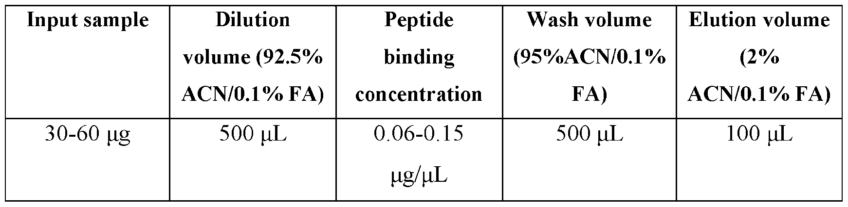
- Sample/reagent volume for protein digestion B. Peptide Cleanup (On KingFisher) • Bead conditioning: Load 20 ⁇ L of beads (5.0 mg) with 80 ⁇ L of Bead Wash/Binding Solution. Wash the beads with 200 ⁇ L of Bead Wash/Binding Solution. • Dilution: Transfer beads into the diluted sample. The total volume is 500 ⁇ L (>9% organic phase after dilution).
- Binding Mix the sample/beads at room temperature for 45 minutes at a medium rate.
- Wash Transfer the sample/beads into 500 ⁇ L of Wash Solution for peptide wash. Mix the plate at room temperature for 2 minutes at a medium rate.
- Elution Transfer the sample/beads into 100 ⁇ L of Elution Solution for peptide elution. Mix the plate at room temperature for 5 minutes at a medium rate. Collect the output peptide samples for LC-MS analysis. Table 4. Peptide cleanup volumes Results from the experiment are provided in FIGS.8A, 8B, and 9-11.
- FIGS.8A and 8B demonstrate that the disclosed magnetic beads exhibit less surface absorption loss than the Sera-Mag TM carboxylate modified particles, MyOne TM , or silica beads in the SP2 workflow.
- the SeraMag TM , MyOne TM , and silica beads all demonstrated significant bead loss on the surfaces of the comb tip plate and sample plate (bead transfer/mix and binding steps).
- the disclosed magnetic beads did not exhibit significant bead loss.
- the data provided in FIGS.8A and 8B with respect to the disclosed magnetic beads refers to hydrolyzed beads, that is beads where the azlactone moieties on the surface of the beads have been hydrolyzed.
- FIG.9 provides data demonstrating that the peptide yield from the KingFisher SP2 workflow for the disclosed magnetic beads was substantially higher than the peptide yield for the Sera-Mag TM , MyOne TM Silane or silica beads, using a quantitative colorimetric peptide assay. No peptides were recovered using silica beads.
- FIG.10 demonstrates that using the disclosed magnetic beads, substantially more unique peptides were identified by nano-LCMS analysis, than were identified using the other bead types.
- FIG.11 demonstrates that substantially more protein groups were identified using the disclosed magnetic beads.
- Example 9 An experiment was designed to demonstrate the superior binding of the disclosed magnetic beads coupled to alkali stable Protein A (asPA), compared to a commercial magnetic bead that comprises asPA conjugates. Conjugate asPA to the disclosed magnetic beads Alkali stable Protein A was conjugated to the disclosed azlactone beads according to the following procedure: 1. Weigh out 77mg of the disclosed azlactone beads. 2.
- the amount of asPA bound to the disclosed azlactone magnetic beads is about 2-fold higher than the amount of asPA bound to the same amount of magnetic agarose beads.
- Example 10 Derivatization of Magnetic Beads with Ocylamine Ligands Various hydrophilic or hydrophobic molecules or proportional mixtures of hydrophilic and hydrophobic molecules were reacted to the amine-reactive, azlactone groups of magnetic beads to impart the support with detergent removal and peptide recovery properties.
- Scheme 1 shows the reaction of a representative hydrophobic ligand (octylamine, C8) to a magnetic bead bearing a terminal azlactone group.
- Scheme 1 A solution of ligand(s) in ethanol was prepared by dissolving 10mg/mL, 2mg/mL, or 40mg/mL of hydrophobic, hydrophilic or proportional mix of hydrophobic and hydrophilic ligand, respectively, in 8mL of ethanol with stirring. 250mg of magnetic base beads (swell volume : 1 gram of magnetic beads gives 6 mLs of beads) were added to the solution and allowed to react for 2 hours with end-over-end mixing. The reacted beads were then washed thoroughly twice with 1 bed volume of water, and then resuspended in water for later use.
- This general procedure was used to modify the magnetic beads with various types of hydrophobic ligands (i.e., octylamine (C8), dodecylamine (C12), octadecylamine (C18)) and hydrophilic ligands (i.e., methoxy Jeffamine (MJ), 4,7,10- Trioxa-1,13 tridecanediamine (C10PEG), B- cyclodextrin, dextran, and PEG molecules ranging in MW from 2000 Da to 20,000 Da), and combinations thereof.
- Table 5 lists different ligands that are reacted to the azlactone group of the magnetic beads.
- Example 11 Removal of Non-Ionic and Ionic Detergents by Azlactone Magnetic Beads Magnetic beads bearing azlactone groups were used to remove non-ionic and ionic detergents from biological samples (e.g., cell lysates and samples containing proteins and/or polypeptides. Examples of detergent removal workflows implementing various detection methods are provided below.
- Absorbance 280 protocol To determine removal of Triton TM X 100 and NP 40) 1. Pipette 200 ⁇ L of 25% slurry magnetic beads into Dynamag2 magnetic stand. 2. Remove supernate. 3. Pipette 100 ⁇ L of 1% detergent solution into bead sample. 4. Vortex detergent and beads until homogenous 5. Place solution in Dynamag2 magnetic stand and collect flow through. 6.
- Coomassie Protein Assay protocol To determine removal of detergents: 1. Pipette 200 ⁇ L of 25% slurry magnetic beads into Dynamag2 magnetic stand. 2. Remove supernate. 3. Pipette 100 ⁇ L of 1% detergent solution into bead sample. 4. Vortex detergent and beads until homogenous. 5. Place solution in Dynamag2 magnetic stand and collect flow through. 6. Pipette 10 ⁇ L of each sample and standard into a 96 well plate. 7. Pipette 300 ⁇ L of Coomassie Blue Stain Reagent into each occupied well. 8.
- Table 9 shows the % removal of detergents Triton TM X-100 and NP-40 at 1% concentration using hydrophilic ligands reacted to the beads.
- the hydrophilic ligands have poor capacity to trap detergents and as a result they were eliminated from further evaluation.
- Table 9 Detergent removal capacity of hydrophilic coated magnetic beads
- Example 14 Removal of Detergents from Beads Coupled to Blends of Hydrophobic and Hydrophilic Ligands
- Table 10 shows variations of octylamine ligand reacted beads blended with hydrophilic ligands such as O-(2- aminopropyl)-O’-(2-methoxyethyl)polypropylene glycol (Methoxy Jeffamine) and C10 PEG diamine in various blend ratios.
- Table 11 shows removal of three different non-ionic detergents 1% Triton TM X- 100, 1% NP-40 and 1% n-Dodecyl- ⁇ -D-maltoside (DDM). Beads with a mixture of hydrophobic and hydrophilic properties were found to remove a significant amount of detergent from the tested samples (e.g., 82%-100%). Table 11. Removal of Detergents from Beads with Hydrophobic and Hydrophilic Properties The samples also were evaluated for detergent removal from HeLa digests using the workflow described herein. Referring to Table 12, 40% or greater HeLa digest was recovered with C8 modified beads and its blends with hydrophilic ligands. Table 12.
- Table 17 provides the success rate of each bead compared to the untreated control sample.
- Table 17. Success Rate Example 18 MS analysis of detergent samples before and after bead processing Materials and Reagents PierceTM HeLa Protein Digest Standard (Thermo Scientific, Catalog number 88328) Instrument MS: Q Exactive HF mass spectrometer, Serial# SN05061L Method Dilute the detergent (n-Dodecyl- ⁇ -D-maltoside (DDM) and NP-40) samples before (control/reference sample) and after (samples) bead processing using 50% acetonitrile/0.1% formic acid with an appropriate ratio (e.g., 1:1000 – 1:100) for direct infusion MS analysis.
- DDM n-Dodecyl- ⁇ -D-maltoside
Landscapes
- Health & Medical Sciences (AREA)
- Chemical & Material Sciences (AREA)
- Immunology (AREA)
- Life Sciences & Earth Sciences (AREA)
- Engineering & Computer Science (AREA)
- Analytical Chemistry (AREA)
- Urology & Nephrology (AREA)
- Molecular Biology (AREA)
- Biomedical Technology (AREA)
- Hematology (AREA)
- Cell Biology (AREA)
- Biotechnology (AREA)
- Microbiology (AREA)
- Chemical Kinetics & Catalysis (AREA)
- Organic Chemistry (AREA)
- Food Science & Technology (AREA)
- Medicinal Chemistry (AREA)
- Physics & Mathematics (AREA)
- Biochemistry (AREA)
- General Health & Medical Sciences (AREA)
- General Physics & Mathematics (AREA)
- Pathology (AREA)
- Solid-Sorbent Or Filter-Aiding Compositions (AREA)
Abstract
Disclosed herein are aspects of magnetic beads that include functional groups suitable for removing detergent from liquids. The magnetic beads may comprise a vinyl azlactone-based crosslinked polymer and a magnetic material dispersed in the bead. The magnetic beads may comprise hydrophobic and/or hydrophilic ligands suitable to facilitate removal of the detergents from solutions or suspensions, while allowing for good recovery of any biomolecules of interest in the sample. Also disclosed are aspects of a method for making the beads, aspects of a method for using the beads, and aspects of a kit comprising the beads.
Description
MAGNETIC BEADS FOR DETERGENT REMOVAL CROSS REFERENCE TO RELATED APPLICATION This application claims the benefit of the earlier filing date of U.S. provisional patent application No.63/517,029, filed August 1, 2023, which is incorporated herein by reference in its entirety. FIELD This disclosure concerns magnetic beads that contain ligands suitable for removing detergent(s) from mixtures, including mixtures that comprise biological molecules including polypeptides, proteins, RNA, and/or DNA, and methods for making the beads and using the beads. BACKGROUND Mass spectrometry (MS)-based proteomics has become the most comprehensive approach for protein identification, quantitation, interactions, modifications, and structural characterization. It is also a challenging topic that requires expertise for sample preparation since a high-quality sample from a robust and reproducible sample preparation is critical for success. Proteomics sample preparation is highly complex and variable, with many different protocols available. For example, bottom-up proteomics sample preparation requires multiple steps including protein extraction, reduction and alkylation of cysteines, digestion of the proteins into peptides, cleanup, and concentration of the peptides for LC-MS analysis. The lack of standardization in proteomic sample preparation makes it difficult to accurately compare results from different laboratories and different protocols. SUMMARY Detergents, including non-ionic and ionic detergents, are frequently used in mass spectrometry (mass spec; MS) sample preparation. However, it is often necessary
to remove the detergents before injecting the sample into the MS machine. With new instruments becoming increasing sensitive, it is necessary to remove 90% or more, and may be 99%, 99.9%, or 99.99% or more of these detergents in order to observe the molecule(s) of interest, such as polypeptides, or proteins. Therefore, there is a need for new methods to remove detergents from samples, while allowing a good recovery, such as greater than 30%, greater than 40%, or greater than 50%, of the molecule(s) of interest, and without reacting with or otherwise changing the nature of the molecule(s) of interest. Disclosed herein is a method for removing detergent from a solution using magnetic beads. The method comprises forming a suspension comprising a first plurality of magnetic beads and a first liquid that contains a first amount of a detergent; agitating the suspension; and removing the magnetic beads from the suspension to leave a solution comprising a second amount of the detergent that is less than the first amount of the detergent. The second amount of the detergent may be less than 0.1% of the second solution, such as less than 0.01% of the second solution, and may be substantially zero. Each magnetic bead may comprise a crosslinked polymer, a magnetic material, and a plurality of hydrophobic ligands formed from a reaction between azlactone moieties
 one or more hydrophobic molecules. The hydrophobic molecules may comprise a straight or branched chain C4-C20alkyl amine, for example, butylamine (C4), hexylamine (C6), octylamine (C8), decylamine (C10), dodecylamine (C12), tetradecylamine (C14), hexadecylamine (C16), octadecylamine (C18), eicosylamine (C20), or a combination thereof. In some aspects, the straight or branched C4-C20 alkyl amine is selected from octylamine (C8), dodecylamine (C12), octadecylamine (C18), or a combination thereof. In particular aspects, the hydrophobic molecule is octylamine and the hydrophobic ligand is
 .
The magnetic bead may further comprise a hydrophilic ligand. The hydrophilic ligand may be formed by a reaction between azlactone moieties and one or more hydrophilic molecules. The one or more hydrophilic molecules may be selected from O-(2- aminopropyl)-O’-(2-methoxyethyl)polypropylene glycol, 4,7,10-Trioxa-1,13 tridecanediamine, β-cyclodextrin, dextran, PEG molecules ranging in MW from 2000 Da to 20,000 Da, or a combination thereof. In some aspects, the hydrophilic ligand is
.
The magnetic bead may further comprise a hydrophilic ligand. The hydrophilic ligand may be formed by a reaction between azlactone moieties and one or more hydrophilic molecules. The one or more hydrophilic molecules may be selected from O-(2- aminopropyl)-O’-(2-methoxyethyl)polypropylene glycol, 4,7,10-Trioxa-1,13 tridecanediamine, β-cyclodextrin, dextran, PEG molecules ranging in MW from 2000 Da to 20,000 Da, or a combination thereof. In some aspects, the hydrophilic ligand is
 . In a particular aspects, the magnetic bead comprises

 as the hydrophilic ligand. In some aspects where the magnetic bead comprises a hydrophobic ligand and a hydrophilic ligand, the two types of ligand are present in a ratio of from 10% hydrophobic:90% hydrophilic to 90% hydrophobic:10% hydrophilic, such as from 20%:80% hydrophobic:hydrophilic to 40%:60% hydrophobic:hydrophilic. The suspension may further comprise a second plurality of magnetic beads where each magnetic bead in the second plurality of magnetic beads comprises a crosslinked polymer, a magnetic material, and a plurality of hydrophilic ligands formed from a reaction between azlactone moieties and one or more hydrophilic molecules. The first plurality of magnetic beads and the second plurality of magnetic beads may be
each provided in amounts sufficient to provide a ratio of beads comprising hydrophobic ligands (referred to as hydrophobic beads) to beads comprising hydrophilic ligands (referred to as hydrophilic beads) of from 10% hydrophobic beads:90% hydrophilic beads to 90% hydrophobic beads:10% hydrophilic beads, such as from 20%:80% hydrophobic beads:hydrophilic beads to 40%:60% hydrophobic beads:hydrophilic beads. In any aspects, the detergent may be selected from polyethylene glycol sorbitan monooleate, t-Octylphenoxypolyethoxyethanol, octylphenoxypolyethoxyethanol (NP- 40) , n-Dodecyl-β-D-maltoside (DDM), sodium dodecyl sulfate (SDS), Glyco-diosgenin (GDN), Lauryl Maltose Neopentyl Glycol (LMNG), or a combination thereof. Also disclosed herein are aspects of a magnetic bead comprising a crosslinked polymer bead and a magnetic material contained within the polymer bead, and further comprising a plurality of ligands wherein the ligands are selected from: hydrophobic ligands formed from a reaction between azlactone moieties and one or more hydrophobic molecules; hydrophilic ligands formed from a reaction between azlactone moieties and one or more hydrophilic molecules; or a combination of hydrophobic ligands and hydrophilic ligands formed from reactions between azlactone moieties and one or more hydrophobic molecules and one or more hydrophilic molecules. The crosslinked polymer may be a crosslinked azlactone polymer, and the magnetic bead may be an azlactone bead. The one or more hydrophobic molecules may comprise a straight or branched chain C4-C20alkyl amine as disclosed herein. And/or the hydrophilic ligand may be derived from O-(2-aminopropyl)-O’-(2- methoxyethyl)polypropylene glycol, 4,7,10-Trioxa-1,13 tridecanediamine, β- cyclodextrin, dextran, a PEG molecule having a molecular weight of from 2,000 to 20,000 Da, or a combination thereof. The magnetic material may comprise an iron source, such as iron oxide. In some aspects, the magnetic material is in the form of a particle, powder, flake, or cluster, and/or may be contained within an agarose bead which in turn may be contained within the crosslinked polymer.
In any aspects, the magnetic bead may have a bead size of from 20 to 80 microns, such as from 30 to 60 microns. Also disclosed herein are aspects of a kit comprising the magnetic beads, and aspects of a method for making the magnetic beads. The foregoing and other objects, features, and advantages of the disclosure will become more apparent from the following detailed description, which proceeds with reference to the accompanying figures. BRIEF DESCRIPTION OF THE DRAWINGS FIG.1 is a flow chart illustrating one aspect of a standard workflow using the disclosed magnetic beads. FIG.2 is a flowchart illustrating an SP3 workflow that includes a PAC workflow and an SP2 workflow. FIG.3 is a digital image illustrating one aspect of the disclosed magnetic beads. FIG.4 is a graph of differential volume versus size, illustrating the average size of the disclosed magnetic beads, and a table providing the average size and standard deviation. FIG.5 is a digital image illustrating one aspect of the disclosed magnetic beads synthesized using iron oxide clusters as the magnetic material. FIG.6 is a digital image illustrating one aspect of the disclosed magnetic beads synthesized using iron oxide particles as the magnetic material. FIG.7 is a digital image illustrating one aspect of the disclosed magnetic beads synthesized using DynabeadsTM as the magnetic material. FIGS.8A and 8B are a digital image of plates from various stages of an SP2 workflow (FIG.8A) and a table of results (FIG.8B), illustrating the surface absorption losses of different bead types. FIG.9 is a graph of peptide yield versus bead type, illustrating the peptide yields from different bead types, identified using a quantitative colorimetric peptide assay. FIG.10 is a graph of number of peptides versus bead types, illustrating the number of unique peptides isolated with different bead types and identified using
nanoLC-MS analysis, 75 i.d. column, 150 min gradient, QE plus mass spectrometry, PD 2.5 data. FIG.11 is a graph of protein group versus bead types, illustrating the number of different protein groups isolated with different bead types and identified using nanoLC- MS analysis, 75 i.d. column, 150 min gradient, QE plus mass spectrometry, PD 2.5 data. FIG.12 is a graph of bound antibody versus magnetic bead, illustrating the capacity of the disclosed magnetic beads conjugated to alkali stable Protein A (asPA) to bind rabbit IgG. FIG.13 is a graph of number versus peptides and peptide groups, illustrating the numbers of unique peptides and peptide groups identified from a HeLa protein digest after processing with an exemplary magnetic bead comprising octylamine at 10 mg/mL. FIG.14 is a graph of number versus peptides and peptide groups, illustrating the numbers of unique peptides and peptide groups identified from a HeLa protein digest after processing with an exemplary magnetic bead comprising octylamine at 20 mg/mL DETAILED DESCRIPTION I. Terms The following explanations of terms and methods are provided to better describe the present disclosure and to guide those of ordinary skill in the art in the practice of the present disclosure. The singular forms “a,” “an,” and “the” refer to one or more than one, unless the context clearly dictates otherwise. The term “or” refers to a single element of stated alternative elements or a combination of two or more elements, unless the context clearly indicates otherwise. As used herein, “comprises” means “includes.” Thus, “comprising A or B,” means “including A, B, or A and B,” without excluding additional elements. All references, including patents and patent applications cited herein, are incorporated by reference in their entirety, unless otherwise specified. Unless otherwise indicated, all numbers expressing quantities of components, molecular weights, percentages, temperatures, times, and so forth, as used in the specification or claims, are to be understood as being modified by the term “about.”
Accordingly, unless otherwise indicated, implicitly or explicitly, the numerical parameters set forth are approximations that may depend on the desired properties sought and/or limits of detection under standard test conditions/methods. When directly and explicitly distinguishing embodiments from discussed prior art, the embodiment numbers are not approximates unless the word “about” is expressly recited. Unless explained otherwise, all technical and scientific terms used herein have the same meaning as commonly understood by one of ordinary skill in the art to which this disclosure pertains. Although methods and materials similar or equivalent to those described herein can be used in the practice or testing of the present disclosure, suitable methods and materials are described below. The materials, methods, and examples are illustrative only and not intended to be limiting. “Alkyl” refers to a saturated aliphatic hydrocarbyl group having from 1 to 25 (C1-25) or more carbon atoms, more typically 1 to 10 (C1-10) carbon atoms such as 1 to 6 (C1-6) carbon atoms or 1 to 4 (C1-4) carbon atoms. “Carboxy” refers to a -CO2H functional group. “Ester” refers to a -CO2R functional group, where R is an alkyl group, such as a C1-6 alkyl group. “Nucleic acid” refers to a polynucleotide molecule. The polynucleotide may be a naturally occurring polynucleotide or a synthetic polynucleotide. A nucleic acid may be a DNA, RNA or mixture of DNA and RNA nucleotides. Typically, a nucleic acid contains from 20 to 10,000 nucleotides or more, such as from 100, 200, 300, 400, 500, 600, 700, 800, 900, 1000, 2000, 3000, 4000, or 5000 nucleotides to 10,000 nucleotides. “Peptide” refers to a compound comprising amino acid residues connected by peptide bonds. Typically, a peptide compound has from 2 to about 50 amino acid residues. “Polypeptide” refers to a compound comprising amino acid residues connected by peptide bonds. In some aspects, a polypeptide has from about 50 amino acid residues to 2000 or more amino acid residues. The amino acids may include alpha-amino acids, and may comprise the L-optical isomer, the D-optical isomer, or combinations thereof.
“Protein” refers to a molecule or complex comprising one or more polypeptides having secondary, tertiary and/or quaternary structure. The secondary, tertiary and/or quaternary structure of a protein typically is stabilized using non-covalent bonds, such as hydrogen bonds, ionic bonds, hydrophobic interactions, and/or van der Walls interactions, and/or covalent bonds, for example, disulfide bonds, such as between the thiol groups of cysteine residues. II. Magnetic Beads Disclosed herein are aspects of a magnetic bead that comprises magnetic material dispersed in a polymer bead. In some aspects, the polymer bead comprises a crosslinked polymer that comprises one or more azlactone moieties
as the hydrophilic ligand. In some aspects where the magnetic bead comprises a hydrophobic ligand and a hydrophilic ligand, the two types of ligand are present in a ratio of from 10% hydrophobic:90% hydrophilic to 90% hydrophobic:10% hydrophilic, such as from 20%:80% hydrophobic:hydrophilic to 40%:60% hydrophobic:hydrophilic. The suspension may further comprise a second plurality of magnetic beads where each magnetic bead in the second plurality of magnetic beads comprises a crosslinked polymer, a magnetic material, and a plurality of hydrophilic ligands formed from a reaction between azlactone moieties and one or more hydrophilic molecules. The first plurality of magnetic beads and the second plurality of magnetic beads may be
each provided in amounts sufficient to provide a ratio of beads comprising hydrophobic ligands (referred to as hydrophobic beads) to beads comprising hydrophilic ligands (referred to as hydrophilic beads) of from 10% hydrophobic beads:90% hydrophilic beads to 90% hydrophobic beads:10% hydrophilic beads, such as from 20%:80% hydrophobic beads:hydrophilic beads to 40%:60% hydrophobic beads:hydrophilic beads. In any aspects, the detergent may be selected from polyethylene glycol sorbitan monooleate, t-Octylphenoxypolyethoxyethanol, octylphenoxypolyethoxyethanol (NP- 40) , n-Dodecyl-β-D-maltoside (DDM), sodium dodecyl sulfate (SDS), Glyco-diosgenin (GDN), Lauryl Maltose Neopentyl Glycol (LMNG), or a combination thereof. Also disclosed herein are aspects of a magnetic bead comprising a crosslinked polymer bead and a magnetic material contained within the polymer bead, and further comprising a plurality of ligands wherein the ligands are selected from: hydrophobic ligands formed from a reaction between azlactone moieties and one or more hydrophobic molecules; hydrophilic ligands formed from a reaction between azlactone moieties and one or more hydrophilic molecules; or a combination of hydrophobic ligands and hydrophilic ligands formed from reactions between azlactone moieties and one or more hydrophobic molecules and one or more hydrophilic molecules. The crosslinked polymer may be a crosslinked azlactone polymer, and the magnetic bead may be an azlactone bead. The one or more hydrophobic molecules may comprise a straight or branched chain C4-C20alkyl amine as disclosed herein. And/or the hydrophilic ligand may be derived from O-(2-aminopropyl)-O’-(2- methoxyethyl)polypropylene glycol, 4,7,10-Trioxa-1,13 tridecanediamine, β- cyclodextrin, dextran, a PEG molecule having a molecular weight of from 2,000 to 20,000 Da, or a combination thereof. The magnetic material may comprise an iron source, such as iron oxide. In some aspects, the magnetic material is in the form of a particle, powder, flake, or cluster, and/or may be contained within an agarose bead which in turn may be contained within the crosslinked polymer.
In any aspects, the magnetic bead may have a bead size of from 20 to 80 microns, such as from 30 to 60 microns. Also disclosed herein are aspects of a kit comprising the magnetic beads, and aspects of a method for making the magnetic beads. The foregoing and other objects, features, and advantages of the disclosure will become more apparent from the following detailed description, which proceeds with reference to the accompanying figures. BRIEF DESCRIPTION OF THE DRAWINGS FIG.1 is a flow chart illustrating one aspect of a standard workflow using the disclosed magnetic beads. FIG.2 is a flowchart illustrating an SP3 workflow that includes a PAC workflow and an SP2 workflow. FIG.3 is a digital image illustrating one aspect of the disclosed magnetic beads. FIG.4 is a graph of differential volume versus size, illustrating the average size of the disclosed magnetic beads, and a table providing the average size and standard deviation. FIG.5 is a digital image illustrating one aspect of the disclosed magnetic beads synthesized using iron oxide clusters as the magnetic material. FIG.6 is a digital image illustrating one aspect of the disclosed magnetic beads synthesized using iron oxide particles as the magnetic material. FIG.7 is a digital image illustrating one aspect of the disclosed magnetic beads synthesized using DynabeadsTM as the magnetic material. FIGS.8A and 8B are a digital image of plates from various stages of an SP2 workflow (FIG.8A) and a table of results (FIG.8B), illustrating the surface absorption losses of different bead types. FIG.9 is a graph of peptide yield versus bead type, illustrating the peptide yields from different bead types, identified using a quantitative colorimetric peptide assay. FIG.10 is a graph of number of peptides versus bead types, illustrating the number of unique peptides isolated with different bead types and identified using
nanoLC-MS analysis, 75 i.d. column, 150 min gradient, QE plus mass spectrometry, PD 2.5 data. FIG.11 is a graph of protein group versus bead types, illustrating the number of different protein groups isolated with different bead types and identified using nanoLC- MS analysis, 75 i.d. column, 150 min gradient, QE plus mass spectrometry, PD 2.5 data. FIG.12 is a graph of bound antibody versus magnetic bead, illustrating the capacity of the disclosed magnetic beads conjugated to alkali stable Protein A (asPA) to bind rabbit IgG. FIG.13 is a graph of number versus peptides and peptide groups, illustrating the numbers of unique peptides and peptide groups identified from a HeLa protein digest after processing with an exemplary magnetic bead comprising octylamine at 10 mg/mL. FIG.14 is a graph of number versus peptides and peptide groups, illustrating the numbers of unique peptides and peptide groups identified from a HeLa protein digest after processing with an exemplary magnetic bead comprising octylamine at 20 mg/mL DETAILED DESCRIPTION I. Terms The following explanations of terms and methods are provided to better describe the present disclosure and to guide those of ordinary skill in the art in the practice of the present disclosure. The singular forms “a,” “an,” and “the” refer to one or more than one, unless the context clearly dictates otherwise. The term “or” refers to a single element of stated alternative elements or a combination of two or more elements, unless the context clearly indicates otherwise. As used herein, “comprises” means “includes.” Thus, “comprising A or B,” means “including A, B, or A and B,” without excluding additional elements. All references, including patents and patent applications cited herein, are incorporated by reference in their entirety, unless otherwise specified. Unless otherwise indicated, all numbers expressing quantities of components, molecular weights, percentages, temperatures, times, and so forth, as used in the specification or claims, are to be understood as being modified by the term “about.”
Accordingly, unless otherwise indicated, implicitly or explicitly, the numerical parameters set forth are approximations that may depend on the desired properties sought and/or limits of detection under standard test conditions/methods. When directly and explicitly distinguishing embodiments from discussed prior art, the embodiment numbers are not approximates unless the word “about” is expressly recited. Unless explained otherwise, all technical and scientific terms used herein have the same meaning as commonly understood by one of ordinary skill in the art to which this disclosure pertains. Although methods and materials similar or equivalent to those described herein can be used in the practice or testing of the present disclosure, suitable methods and materials are described below. The materials, methods, and examples are illustrative only and not intended to be limiting. “Alkyl” refers to a saturated aliphatic hydrocarbyl group having from 1 to 25 (C1-25) or more carbon atoms, more typically 1 to 10 (C1-10) carbon atoms such as 1 to 6 (C1-6) carbon atoms or 1 to 4 (C1-4) carbon atoms. “Carboxy” refers to a -CO2H functional group. “Ester” refers to a -CO2R functional group, where R is an alkyl group, such as a C1-6 alkyl group. “Nucleic acid” refers to a polynucleotide molecule. The polynucleotide may be a naturally occurring polynucleotide or a synthetic polynucleotide. A nucleic acid may be a DNA, RNA or mixture of DNA and RNA nucleotides. Typically, a nucleic acid contains from 20 to 10,000 nucleotides or more, such as from 100, 200, 300, 400, 500, 600, 700, 800, 900, 1000, 2000, 3000, 4000, or 5000 nucleotides to 10,000 nucleotides. “Peptide” refers to a compound comprising amino acid residues connected by peptide bonds. Typically, a peptide compound has from 2 to about 50 amino acid residues. “Polypeptide” refers to a compound comprising amino acid residues connected by peptide bonds. In some aspects, a polypeptide has from about 50 amino acid residues to 2000 or more amino acid residues. The amino acids may include alpha-amino acids, and may comprise the L-optical isomer, the D-optical isomer, or combinations thereof.
“Protein” refers to a molecule or complex comprising one or more polypeptides having secondary, tertiary and/or quaternary structure. The secondary, tertiary and/or quaternary structure of a protein typically is stabilized using non-covalent bonds, such as hydrogen bonds, ionic bonds, hydrophobic interactions, and/or van der Walls interactions, and/or covalent bonds, for example, disulfide bonds, such as between the thiol groups of cysteine residues. II. Magnetic Beads Disclosed herein are aspects of a magnetic bead that comprises magnetic material dispersed in a polymer bead. In some aspects, the polymer bead comprises a crosslinked polymer that comprises one or more azlactone moieties
 r one or more functional groups derived from an azlactone moiety. Functional groups derived from an azlactone moiety may comprise moieties derived from a reaction between the azlactone moiety and a nucleophilic reactive group, such as, but not limited to, an amine-containing compound, thiol containing group, alcohol or water. Hydrolysis of azlactone or reaction of the azlactone group with an amine- containing compound can add functionality and/or alter the surface properties of the bead. For example, hydrolysis of azlactone rings may produce a bead having a plurality of carboxylic acid functional groups on its surface. Alternatively, the azlactone group are reacted with an amine-containing compound to increase or decrease the hydrophobicity or hydrophilicity of the bead. In certain aspects, azlactone beads are reacted with an amine-containing compound that includes primary, secondary, or tertiary amine group(s) and can, optionally, further include one or more additional functional groups. In other aspects, azlactone beads are reacted with a thiol or alcohol containing compound resulting in thioamide and ester functionalized beads. In other aspects, the azlactone bead are reacted with an amine-containing compound to provide a linker that provides one or more additional functional groups for
further reaction with a ligand or biomolecule. For example, an amine-containing compound that includes an additional amine reactive group may be reacted with the azlactone bead, where the reactive amine group can be converted to an epoxide, maleimide, or iodoacetyl group using standard methods known in the art. In yet other aspects, azlactone beads are reacted with a compound to expand the number of functional groups attached to the bead. Such an approach can be an effective approach for increasing the binding capacity of the bead for a particular target molecule. For example, the beads may be reacted with an amine-containing compound (e.g., a polymer or dendrimer) that includes multiple reactive groups. In other aspects, the amine-containing compound are reacted with an azlactone group on the bead to provide a spacer to overcome steric hindrance upon ligand attachment or to provide a linker for subsequent reaction with a complementary reactive group using compounds and methods. Amine-containing compounds also include biomolecules (e.g., proteins, peptides, nucleic acids, nucleotides, and oligo- and polysaccharide), organic ligands, and large synthetic compounds, such as polymers (e.g., PEG) and dendrimers. Representative amine-containing organic ligands include, without limitation, alkylamines (e.g., straight or branched C4-C12 alkyl amines such as 1,5-diaminopentane, diethylamine, butylamine, and octylamine), arylamines (e.g., benzylamine and 4- aminobenzoic acid), aminocaproic acid, ethanolamine, 5-amino-2-methyl benzene sulfonic acid, aminoethyl-trimethylammonium chloride (AETMA), taurine, tris(hydroxymethyl)aminomethane (Tris), and the like. Ligands for ion exchange (cationic and anionic) including but not limited to PDA, PEI-25K, PEI-800 daltons. In certain aspects, the amine-containing compound is an organic ligand suitable for removing detergent from a mixture, such as a solution or suspension resulting from cell lysis. Exemplary detergents that can be removed using the disclosed magnetic beads include, but are not limited to, TweenTM (polyethylene glycol sorbitan monooleate), such as TweenTM 80 (polysorbate 80) and TweenTM 20 (polysorbate 20); TritonTM X-100 (t-Octylphenoxypolyethoxyethanol, Polyethylene glycol tert- octylphenyl ether); NP-40 (octylphenoxypolyethoxyethanol), DDM (n-Dodecyl-β-D-
maltoside), SDS (sodium dodecyl sulfate), GDN (Glyco-diosgenin), or LMNG (Lauryl Maltose Neopentyl Glycol). The organic ligand may be a hydrophobic amine, for example, an alkylamine, such as a straight or branched C4-C20 alkyl amines, such as a straight or branched chain C6-C18 alkylamine, for example, butylamine (C4), hexylamine (C6), octylamine (C8), decylamine (C10), dodecylamine (C12), tetradecylamine (C14), hexadecylamine (C16), octadecylamine (C18), eicosylamine (C20), or a combination thereof; a hydrophilic amine, such as O-(2-aminopropyl)-O’-(2- methoxyethyl)polypropylene glycol (Methoxy Jeffamine;
r one or more functional groups derived from an azlactone moiety. Functional groups derived from an azlactone moiety may comprise moieties derived from a reaction between the azlactone moiety and a nucleophilic reactive group, such as, but not limited to, an amine-containing compound, thiol containing group, alcohol or water. Hydrolysis of azlactone or reaction of the azlactone group with an amine- containing compound can add functionality and/or alter the surface properties of the bead. For example, hydrolysis of azlactone rings may produce a bead having a plurality of carboxylic acid functional groups on its surface. Alternatively, the azlactone group are reacted with an amine-containing compound to increase or decrease the hydrophobicity or hydrophilicity of the bead. In certain aspects, azlactone beads are reacted with an amine-containing compound that includes primary, secondary, or tertiary amine group(s) and can, optionally, further include one or more additional functional groups. In other aspects, azlactone beads are reacted with a thiol or alcohol containing compound resulting in thioamide and ester functionalized beads. In other aspects, the azlactone bead are reacted with an amine-containing compound to provide a linker that provides one or more additional functional groups for
further reaction with a ligand or biomolecule. For example, an amine-containing compound that includes an additional amine reactive group may be reacted with the azlactone bead, where the reactive amine group can be converted to an epoxide, maleimide, or iodoacetyl group using standard methods known in the art. In yet other aspects, azlactone beads are reacted with a compound to expand the number of functional groups attached to the bead. Such an approach can be an effective approach for increasing the binding capacity of the bead for a particular target molecule. For example, the beads may be reacted with an amine-containing compound (e.g., a polymer or dendrimer) that includes multiple reactive groups. In other aspects, the amine-containing compound are reacted with an azlactone group on the bead to provide a spacer to overcome steric hindrance upon ligand attachment or to provide a linker for subsequent reaction with a complementary reactive group using compounds and methods. Amine-containing compounds also include biomolecules (e.g., proteins, peptides, nucleic acids, nucleotides, and oligo- and polysaccharide), organic ligands, and large synthetic compounds, such as polymers (e.g., PEG) and dendrimers. Representative amine-containing organic ligands include, without limitation, alkylamines (e.g., straight or branched C4-C12 alkyl amines such as 1,5-diaminopentane, diethylamine, butylamine, and octylamine), arylamines (e.g., benzylamine and 4- aminobenzoic acid), aminocaproic acid, ethanolamine, 5-amino-2-methyl benzene sulfonic acid, aminoethyl-trimethylammonium chloride (AETMA), taurine, tris(hydroxymethyl)aminomethane (Tris), and the like. Ligands for ion exchange (cationic and anionic) including but not limited to PDA, PEI-25K, PEI-800 daltons. In certain aspects, the amine-containing compound is an organic ligand suitable for removing detergent from a mixture, such as a solution or suspension resulting from cell lysis. Exemplary detergents that can be removed using the disclosed magnetic beads include, but are not limited to, TweenTM (polyethylene glycol sorbitan monooleate), such as TweenTM 80 (polysorbate 80) and TweenTM 20 (polysorbate 20); TritonTM X-100 (t-Octylphenoxypolyethoxyethanol, Polyethylene glycol tert- octylphenyl ether); NP-40 (octylphenoxypolyethoxyethanol), DDM (n-Dodecyl-β-D-
maltoside), SDS (sodium dodecyl sulfate), GDN (Glyco-diosgenin), or LMNG (Lauryl Maltose Neopentyl Glycol). The organic ligand may be a hydrophobic amine, for example, an alkylamine, such as a straight or branched C4-C20 alkyl amines, such as a straight or branched chain C6-C18 alkylamine, for example, butylamine (C4), hexylamine (C6), octylamine (C8), decylamine (C10), dodecylamine (C12), tetradecylamine (C14), hexadecylamine (C16), octadecylamine (C18), eicosylamine (C20), or a combination thereof; a hydrophilic amine, such as O-(2-aminopropyl)-O’-(2- methoxyethyl)polypropylene glycol (Methoxy Jeffamine;
 from 3 to 25, 4,7,10-Trioxa-1,13 tridecanediamine (C10PEG diamine), β-cyclodextrin, dextran, or PEG molecules ranging in MW from 2000 Da to 20,000 Da (as measured by proton NMR), or a combination thereof; or a combination thereof. In some aspects, an amine containing moiety is a protein such as an antibody, an antibody binding protein (e.g., Protein A, Protein A/G, Protein G, and Protein L), Streptavidin, Neutravidin, glutathione, enzyme, or other biomolecule containing amines. In one aspect, azlactone beads are conjugated to an amine containing biomolecule such as an alkali stable Protein A (asPA) and the final conjugate can then be used to purify IgG. It is desired that when the biomolecule is an enzyme, it maintains its enzymatic selectivity once coupled to the bead. Examples of enzymes include proteolytic enzymes (e.g., trypsin) and other types of enzymes known in the art. FIG.12 shows similar performance with azlactone beads (asPA-magUL) and magnetic agarose beads (asPA-magAg). The assay was performed with duplicate samples of each magnetic bead. In some aspects, an amine-containing compounds is or includes metal charged chelates (e.g., nickel-charged nitrilotriacetic acid, Ni-NTA). In one aspect, azlactone beads are conjugated to Ni-NTA and the final conjugate are used to purify polyhistidine-tagged proteins from a soluble protein extract.
In some aspects, the polymer bead is an azlactone bead formed from a vinyl azlactone and a crosslinker. The vinyl azlactone may be 4,4-dimethyl-2-vinyloxazol- 5(4H)-one,
from 3 to 25, 4,7,10-Trioxa-1,13 tridecanediamine (C10PEG diamine), β-cyclodextrin, dextran, or PEG molecules ranging in MW from 2000 Da to 20,000 Da (as measured by proton NMR), or a combination thereof; or a combination thereof. In some aspects, an amine containing moiety is a protein such as an antibody, an antibody binding protein (e.g., Protein A, Protein A/G, Protein G, and Protein L), Streptavidin, Neutravidin, glutathione, enzyme, or other biomolecule containing amines. In one aspect, azlactone beads are conjugated to an amine containing biomolecule such as an alkali stable Protein A (asPA) and the final conjugate can then be used to purify IgG. It is desired that when the biomolecule is an enzyme, it maintains its enzymatic selectivity once coupled to the bead. Examples of enzymes include proteolytic enzymes (e.g., trypsin) and other types of enzymes known in the art. FIG.12 shows similar performance with azlactone beads (asPA-magUL) and magnetic agarose beads (asPA-magAg). The assay was performed with duplicate samples of each magnetic bead. In some aspects, an amine-containing compounds is or includes metal charged chelates (e.g., nickel-charged nitrilotriacetic acid, Ni-NTA). In one aspect, azlactone beads are conjugated to Ni-NTA and the final conjugate are used to purify polyhistidine-tagged proteins from a soluble protein extract.
In some aspects, the polymer bead is an azlactone bead formed from a vinyl azlactone and a crosslinker. The vinyl azlactone may be 4,4-dimethyl-2-vinyloxazol- 5(4H)-one,
 4,4-Dimethyl-2-vinyloxazol-5(4H)-one The cross linker maybe any crosslinker suitable to facilitate polymerization and form the bead. In some aspects, the crosslinker is a bisacrylamide, agarose, or a vinyl ether. In certain aspects, the crosslinker is methylene bisacrylamide. In particular aspects, the vinyl azlactone is 4,4-dimethyl-2-vinyloxazol-5(4H)- one and the crosslinker is a bisacrylamide, such as methylene bisacrylamide. In some aspects, the beads comprise a plurality of functional moieties on the surface of the bead. The functional moieties may be selected from


.
 In certain aspects, the azlactone bead is reacted with an organic ligand, such as an organic amine or alcohol. In some aspects, the azlactone bead is reacted with a single species of organic ligand, such as one hydrophobic ligand or one hydrophilic ligand. But in other aspects, the azlactone bead is reacted with multiple organic ligands, such as 2, 3, 4 or more hydrophobic ligands, 2, 3, or 4 or more hydrophilic ligands, or a combination of one or more, such as 1, 2, 3 or more, hydrophobic ligands and one or more, such as 1, 2, 3 or more, hydrophilic ligands. In certain aspects, the azlactone bead is reacted with one hydrophobic ligand and one hydrophilic ligand. In some aspects comprising a combination of hydrophobic and hydrophilic ligands, the ratio of hydrophobic to hydrophilic ligands is from 1% hydrophobic:99% hydrophilic to 99% hydrophobic:1% hydrophilic by weight, such as 10% hydrophobic:90% hydrophilic to 90% hydrophobic:10% hydrophilic, 20% hydrophobic:80% hydrophilic to 80% hydrophobic:20% hydrophilic, 30% hydrophobic:70% hydrophilic to 70% hydrophobic:30% hydrophilic, 40% hydrophobic:60% hydrophilic to 60% hydrophobic:40% hydrophilic, or about 50%:50% hydrophobic to hydrophilic by weight. In some aspects, the ratio was from 20%:80% hydrophobic:hydrophilic to 40%:60% hydrophobic:hydrophilic by weight. Exemplary functional moieties comprising hydrophobic ligands include, but are not limited to,


Claims
We claim: 1. A method, comprising: form a suspension comprising a first plurality of magnetic beads and a first solution that contains a first amount of a detergent; agitate the suspension; remove the magnetic beads from the suspension to leave a second solution comprising a second amount of a detergent that is less than the first amount of the detergent; wherein each magnetic bead in the first plurality of magnetic beads comprises a crosslinked polymer, a magnetic material, and a plurality of hydrophobic ligands formed from a reaction between azlactone moieties and one or more hydrophobic molecules.
 2. The method of claim 1, wherein the one or more hydrophobic molecules comprise a straight or branched C4-C20 alkyl amine. 3. The method of claim 2, wherein the straight or branched C4-C20 alkyl amine is selected from butylamine (C4), hexylamine (C6), octylamine (C8), decylamine (C10), dodecylamine (C12), tetradecylamine (C14), hexadecylamine (C16), octadecylamine (C18), eicosylamine (C20), or a combination thereof. 4. The method of claim 3, wherein the straight or branched C4-C20 alkyl amine is selected from octylamine (C8), dodecylamine (C12), octadecylamine (C18), or a combination thereof. 5. The method of any one of claims 1-4, wherein the hydrophobic ligands are selected from
,
 6. The method of any one of claims 1-5, wherein the hydrophobic ligand is
 7. The method of any one of claims 1-6, wherein the magnetic beads in the first plurality of magnetic beads further comprise a hydrophilic ligand. 8. The method of claim 7, wherein the hydrophilic ligand is formed by a reaction between azlactone moieties and one or more hydrophilic molecules.
9. The method of any one of claims 7-8, wherein the beads comprise hydrophobic and hydrophilic ligands in a ratio of from 10% hydrophobic:90% hydrophilic to 90% hydrophobic:10% hydrophilic. 10. The method of claim 9, wherein the ratio of hydrophobic to hydrophilic ligands is from 20%:80% hydrophobic:hydrophilic to 40%:60% hydrophobic:hydrophilic. 12. The method of any one of claims 1-6, wherein the suspension further comprises a second plurality of magnetic beads where each magnetic bead in the second plurality of magnetic beads comprises a crosslinked polymer, a magnetic material, and a plurality of hydrophilic ligands formed from a reaction between azlactone moieties and one or more hydrophilic molecules. 13. The method of claim 12, wherein the first plurality of magnetic beads and the second plurality of magnetic beads are each provided in amounts sufficient to provide a ratio of beads comprising hydrophobic ligands (referred to as hydrophobic beads) to beads comprising hydrophilic ligands (referred to as hydrophilic beads) of from 10% hydrophobic beads:90% hydrophilic beads to 90% hydrophobic beads:10% hydrophilic beads. 14. The method of claim 13, wherein the ratio of hydrophobic beads to hydrophilic beads is from 20%:80% hydrophobic beads:hydrophilic beads to 40%:60% hydrophobic beads:hydrophilic beads. 15. The method of any one of claims 8 to 14, wherein the one or more hydrophilic molecules is selected from O-(2-aminopropyl)-O’-(2- methoxyethyl)polypropylene glycol, 4,7,10-Trioxa-1,13 tridecanediamine, β- cyclodextrin, dextran, PEG molecules ranging in MW from 2000 Da to 20,000 Da, or a combination thereof.
16. The method of any one of claims 7-15, wherein the hydrophilic ligands are selected from
 where m is from 40-450 (MW of from about 2,000 to 20,000 DA),
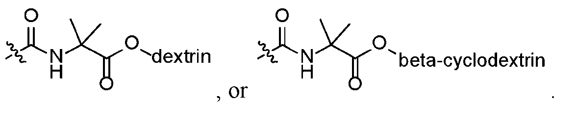 17. The method of claim 16, wherein the hydrophilic ligand is
 18. The method of any one of claims 7-16, wherein the hydrophobic ligand is
 the hydrophilic ligand is
 from 3 to 25,
 . 19. The method of any one of claims 1-18, wherein the second amount of detergent is less than 0.1% of the second solution. 20. The method of claim 19, wherein the second amount of detergent is 0.01% or less of the second solution. 21. The method of any one of claims 1-20, wherein the detergent is selected from polyethylene glycol sorbitan monooleate, t-Octylphenoxypolyethoxyethanol, Octylphenoxypolyethoxyethanol (NP-40), n-Dodecyl-β-D-maltoside (DDM), sodium dodecyl sulfate (SDS), Glyco-diosgenin (GDN), Lauryl Maltose Neopentyl Glycol (LMNG), or a combination thereof. 22. A magnetic bead, comprising a crosslinked polymer bead and a magnetic material contained within the polymer bead, and further comprising a plurality of ligands formed wherein the ligands are selected from: hydrophobic ligands formed from a reaction between azlactone moieties and one or more hydrophobic molecules; hydrophilic ligands formed from a reaction between azlactone moieties and one or more hydrophilic molecules; or a combination of hydrophobic ligands and hydrophilic ligands formed from reactions between azlactone moieties and one or more hydrophobic molecules and one or more hydrophilic molecules.
23. The magnetic bead of claim 22, wherein the plurality of ligands are hydrophobic ligands formed from a reaction between azlactone moieties and one or more hydrophobic molecules. 24. The magnetic bead of claim 22, wherein the plurality of ligands are a combination of hydrophobic ligands and hydrophilic ligands formed from reactions between azlactone moieties and one or more hydrophobic molecules and one or more hydrophilic molecules. 25. The magnetic bead of any one of claims 22-24, wherein the magnetic material comprises an iron source. 26. The magnetic bead of claim 25, wherein the iron source comprises iron oxide. 27. The magnetic bead of any one of claims 22-26, wherein the magnetic material is contained within an agarose bead, and the agarose bead is contained within the crosslinked polymer. 28. The magnetic bead of any one of claims 22-27, wherein the magnetic material is in the form of a particle, powder, flake, or cluster. 29. The magnetic bead of any one of claims 22-28, wherein the crosslinked polymer is a crosslinked azlactone polymer, and the magnetic bead is an azlactone bead. 30. The magnetic bead of any one of claims 22-29, wherein the one or more hydrophobic molecules comprise a straight or branched chain C4-C20alkyl amine. 31. The magnetic bead of claim 30, wherein the straight or branched C4-C20 alkyl amine is selected from butylamine (C4), hexylamine (C6), octylamine (C8),
decylamine (C10), dodecylamine (C12), tetradecylamine (C14), hexadecylamine (C16), octadecylamine (C18), eicosylamine (C20), or a combination thereof. 32. The magnetic bead of claim 30, wherein the straight or branched C4-C20 alkyl amine is selected from octylamine (C8), dodecylamine (C12), octadecylamine (C18), or a combination thereof. 33. The magnetic bead of claim 32, wherein the hydrophobic ligand is selected from
 34. The magnetic bead of any one of claims 22-33, wherein the hydrophobic

35. The magnetic bead of any one of claims 22 and 24-34, wherein the hydrophilic ligand is derived from O-(2-aminopropyl)-O’-(2- methoxyethyl)polypropylene glycol, 4,7,10-Trioxa-1,13 tridecanediamine, β- cyclodextrin, dextran, a PEG molecule having a molecular weight of from 2,000 to 20,000 Da, or a combination thereof. 36. The magnetic bead of any one of claims 22 and 24-35, wherein a ratio between the hydrophobic ligand and the hydrophilic ligand is from 10% hydrophobic:90% hydrophilic to 90% hydrophobic:10% hydrophilic by weight. 37. The magnetic bead of claim 36, wherein the ratio of hydrophobic to hydrophilic ligands is from 20%:80% hydrophobic:hydrophilic to 40%:60% hydrophobic:hydrophilic by weight. 38. The magnetic bead of any one of claims 22 and 24-37, wherein the magnetic bead comprises:
 as the hydrophobic ligand; and

 as the hydrophilic ligand. 39. The magnetic bead of any one of claims 22-38, wherein the bead has a bead size of from 20 to 80 microns.
40. The magnetic bead of claim 39, wherein the bead size is from 30 to 60 microns. 41. A mixture of magnetic beads according to any one of claims 22-40, the mixture comprising a first plurality of magnetic beads comprising hydrophobic ligands and a second plurality of magnetic beads comprising hydrophilic ligands. 42. The mixture of claim 41, wherein the first plurality of magnetic beads and the second plurality of magnetic beads are each provided in amounts sufficient to provide a ratio of beads comprising hydrophobic ligands (referred to as hydrophobic beads) to beads comprising hydrophilic ligands (referred to as hydrophilic beads) of from 10% hydrophobic beads:90% hydrophilic beads to 90% hydrophobic beads:10% hydrophilic beads. 43. The mixture of claim 42, wherein the ratio of hydrophobic beads to hydrophilic beads is from 20%:80% hydrophobic beads:hydrophilic beads to 40%:60% hydrophobic beads:hydrophilic beads. 44. A kit comprising a plurality of magnetic beads according to any one of claims 22-40 or a mixture of magnetic beads according to any one of clams 41-43. 45. A method for making a magnetic bead according to any one of claims 22-40, the method comprising: add a plurality magnetic bead comprising azlactone functional groups to a solution comprising molecule selected from hydrophobic molecules, hydrophilic molecules, or a combination of hydrophobic molecules and hydrophilic molecules; allow the magnetic beads to react with at least a portion of the molecules to form magnetic beads comprising ligands; and separate the magnetic beads comprising the ligands from the solution;
wherein when the molecules are hydrophobic molecules then the ligands are hydrophobic ligands, when the molecules are hydrophilic molecules then the ligands are hydrophilic ligands, and when the molecules comprise a combination of hydrophobic molecules and hydrophilic molecules then the ligands are a mixture of hydrophobic ligands and hydrophilic ligands. 46. The method of claim 45, wherein the molecules are hydrophobic molecules and the ligands are hydrophobic ligands. 47. The method of claim 45, wherein the molecules are hydrophilic molecules and the ligands are hydrophilic ligands. 48. The method of claim 45, wherein the molecules comprise a combination of hydrophobic molecules and hydrophilic molecules and the ligands are a mixture of hydrophobic ligands and hydrophilic ligands.
Applications Claiming Priority (2)
| Application Number | Priority Date | Filing Date | Title |
|---|---|---|---|
| US202363517029P | 2023-08-01 | 2023-08-01 | |
| US63/517,029 | 2023-08-01 |
Publications (1)
| Publication Number | Publication Date |
|---|---|
| WO2025029872A1 true WO2025029872A1 (en) | 2025-02-06 |
Family
ID=92542682
Family Applications (1)
| Application Number | Title | Priority Date | Filing Date |
|---|---|---|---|
| PCT/US2024/040308 Pending WO2025029872A1 (en) | 2023-08-01 | 2024-07-31 | Magnetic beads for detergent removal |
Country Status (1)
| Country | Link |
|---|---|
| WO (1) | WO2025029872A1 (en) |
Citations (4)
| Publication number | Priority date | Publication date | Assignee | Title |
|---|---|---|---|---|
| US5292840A (en) | 1987-03-13 | 1994-03-08 | Minnesota Mining And Manufacturing Company | Polymeric supports |
| US5336742A (en) * | 1987-03-13 | 1994-08-09 | Minnesota Mining And Manufacturing Company | Polymeric supports |
| US5993935A (en) * | 1991-10-11 | 1999-11-30 | 3M Innovative Properties Company | Covalently reactive particles incorporated in a continous porous matrix |
| US8735455B2 (en) * | 2008-06-26 | 2014-05-27 | 3M Innovative Properties Company | Solid support with a grafted chain |
-
2024
- 2024-07-31 WO PCT/US2024/040308 patent/WO2025029872A1/en active Pending
Patent Citations (5)
| Publication number | Priority date | Publication date | Assignee | Title |
|---|---|---|---|---|
| US5292840A (en) | 1987-03-13 | 1994-03-08 | Minnesota Mining And Manufacturing Company | Polymeric supports |
| US5336742A (en) * | 1987-03-13 | 1994-08-09 | Minnesota Mining And Manufacturing Company | Polymeric supports |
| US5403902A (en) | 1987-03-13 | 1995-04-04 | Minnesota Mining And Manufacturing Company | Polymeric supports |
| US5993935A (en) * | 1991-10-11 | 1999-11-30 | 3M Innovative Properties Company | Covalently reactive particles incorporated in a continous porous matrix |
| US8735455B2 (en) * | 2008-06-26 | 2014-05-27 | 3M Innovative Properties Company | Solid support with a grafted chain |
Non-Patent Citations (4)
| Title |
|---|
| ANONYMOUS: "One-Step Detergent Removal kit", 27 November 2022 (2022-11-27), pages 1 - 3, XP093223103, Retrieved from the Internet <URL:https://bioclone.net/wp-content/uploads/pdf/instruction-manual/sample/One-Step%20Detergent%20Removal%20Kit.pdf> [retrieved on 20241111] * |
| ANONYMOUS: "Safety data sheet for product code BD101 and BD102 issued by Bioclone Inc.", 27 November 2022 (2022-11-27), pages 1 - 5, XP093223112, Retrieved from the Internet <URL:https://bioclone.net/wp-content/uploads/pdf/MSDS/SDS-Sample%20Preparation.pdf> [retrieved on 20241111] * |
| PRAI-IN YINGRAK ET AL: "Recyclable magnetic nanocluster crosslinked with poly(ethylene oxide)-block-poly(2-vinyl-4,4-dimethylazlactone) copolymer for adsorption with antibody", MATERIALS SCIENCE AND ENGINEERING C, ELSEVIER SCIENCE S.A, CH, vol. 67, 7 May 2016 (2016-05-07), pages 285 - 293, XP029579386, ISSN: 0928-4931, DOI: 10.1016/J.MSEC.2016.05.032 * |
| PRAY-IN YINGRAK ET AL: "Hydrophilic azlactone-functionalized magnetite nanoparticle for conjugation with folic acid", JOURNAL OF NANOPARTICLE RESEARCH 16, 22 March 2014 (2014-03-22), XP093217422, Retrieved from the Internet <URL:http://link.springer.com/article/10.1007/s11051-014-2357-7/fulltext.html> [retrieved on 20241023], DOI: 10.1007/s11051-014-2357-7 * |
Similar Documents
| Publication | Publication Date | Title |
|---|---|---|
| Filipović et al. | Affinity-based isolation of extracellular vesicles by means of single-domain antibodies bound to macroporous methacrylate-based copolymer | |
| US20180010169A1 (en) | Methods and reagents for selection of biological molecules | |
| Vergara-Barberan et al. | Recent advances in aptamer-based miniaturized extraction approaches in food analysis | |
| US10816546B2 (en) | Binding a target substance | |
| Li et al. | Efficient on‐chip proteolysis system based on functionalized magnetic silica microspheres | |
| CN100500837C (en) | Methods for separating molecules | |
| JP2005538929A (en) | Methods for isolating nucleic acids and proteins from a single sample | |
| EP3043905B1 (en) | New diagnostic assay using particles with magnetic properties | |
| JP7462972B2 (en) | Biomagnetic microspheres and methods of preparation and use thereof | |
| EP1774334B1 (en) | Use of magnetic material to direct isolation of compounds and fractionation of multipart samples | |
| Braun et al. | Peptides as biosorbents–promising tools for resource recovery | |
| US9983203B2 (en) | Method for protein analysis | |
| CN102421898A (en) | Nucleic acid purification method | |
| WO2003095646A1 (en) | Isolating nucleic acid | |
| WO2025029872A1 (en) | Magnetic beads for detergent removal | |
| WO2025029903A1 (en) | Functionalized magnetic beads and methods of preparation and use | |
| CN114907612A (en) | Magnetic microsphere and manufacturing method and application thereof | |
| AU2004279361A1 (en) | Methods and kits for purifying His-tagged proteins | |
| US20060030056A1 (en) | Use of magnetic material to fractionate samples | |
| EP4025911B1 (en) | Conjugates composed of membrane-targeting peptides for extracellular vesicles isolation, analysis and their integration thereof | |
| CN115449533A (en) | A low-abundance protein enrichment kit, preparation method and application | |
| JP2006327962A (en) | Method for separating target substance and molecular complex | |
| JP2001158800A (en) | Avidin particles | |
| CN117723748B (en) | Immunomagnetic bead for enhancing target protein signal for targeted detection, and preparation method and application thereof | |
| CN115494243B (en) | Method for quantitatively determining binding efficiency of streptavidin-biotin and application |
Legal Events
| Date | Code | Title | Description |
|---|---|---|---|
| 121 | Ep: the epo has been informed by wipo that ep was designated in this application |
Ref document number: 24761735 Country of ref document: EP Kind code of ref document: A1 |