WO2024241206A1 - Use of anti-cd3 antibody for selectively depleting activated t cells - Google Patents
Use of anti-cd3 antibody for selectively depleting activated t cells Download PDFInfo
- Publication number
- WO2024241206A1 WO2024241206A1 PCT/IB2024/054911 IB2024054911W WO2024241206A1 WO 2024241206 A1 WO2024241206 A1 WO 2024241206A1 IB 2024054911 W IB2024054911 W IB 2024054911W WO 2024241206 A1 WO2024241206 A1 WO 2024241206A1
- Authority
- WO
- WIPO (PCT)
- Prior art keywords
- cells
- antibody
- seq
- cell
- variable region
- Prior art date
- Legal status (The legal status is an assumption and is not a legal conclusion. Google has not performed a legal analysis and makes no representation as to the accuracy of the status listed.)
- Pending
Links
Classifications
-
- C—CHEMISTRY; METALLURGY
- C07—ORGANIC CHEMISTRY
- C07K—PEPTIDES
- C07K16/00—Immunoglobulins [IGs], e.g. monoclonal or polyclonal antibodies
- C07K16/18—Immunoglobulins [IGs], e.g. monoclonal or polyclonal antibodies against material from animals or humans
- C07K16/28—Immunoglobulins [IGs], e.g. monoclonal or polyclonal antibodies against material from animals or humans against receptors, cell surface antigens or cell surface determinants
- C07K16/2803—Immunoglobulins [IGs], e.g. monoclonal or polyclonal antibodies against material from animals or humans against receptors, cell surface antigens or cell surface determinants against the immunoglobulin superfamily
- C07K16/2809—Immunoglobulins [IGs], e.g. monoclonal or polyclonal antibodies against material from animals or humans against receptors, cell surface antigens or cell surface determinants against the immunoglobulin superfamily against the T-cell receptor (TcR)-CD3 complex
-
- A—HUMAN NECESSITIES
- A61—MEDICAL OR VETERINARY SCIENCE; HYGIENE
- A61K—PREPARATIONS FOR MEDICAL, DENTAL OR TOILETRY PURPOSES
- A61K39/00—Medicinal preparations containing antigens or antibodies
-
- A—HUMAN NECESSITIES
- A61—MEDICAL OR VETERINARY SCIENCE; HYGIENE
- A61P—SPECIFIC THERAPEUTIC ACTIVITY OF CHEMICAL COMPOUNDS OR MEDICINAL PREPARATIONS
- A61P37/00—Drugs for immunological or allergic disorders
- A61P37/02—Immunomodulators
- A61P37/06—Immunosuppressants, e.g. drugs for graft rejection
-
- C—CHEMISTRY; METALLURGY
- C07—ORGANIC CHEMISTRY
- C07K—PEPTIDES
- C07K16/00—Immunoglobulins [IGs], e.g. monoclonal or polyclonal antibodies
- C07K16/18—Immunoglobulins [IGs], e.g. monoclonal or polyclonal antibodies against material from animals or humans
- C07K16/28—Immunoglobulins [IGs], e.g. monoclonal or polyclonal antibodies against material from animals or humans against receptors, cell surface antigens or cell surface determinants
-
- C—CHEMISTRY; METALLURGY
- C12—BIOCHEMISTRY; BEER; SPIRITS; WINE; VINEGAR; MICROBIOLOGY; ENZYMOLOGY; MUTATION OR GENETIC ENGINEERING
- C12N—MICROORGANISMS OR ENZYMES; COMPOSITIONS THEREOF; PROPAGATING, PRESERVING, OR MAINTAINING MICROORGANISMS; MUTATION OR GENETIC ENGINEERING; CULTURE MEDIA
- C12N15/00—Mutation or genetic engineering; DNA or RNA concerning genetic engineering, vectors, e.g. plasmids, or their isolation, preparation or purification; Use of hosts therefor
- C12N15/09—Recombinant DNA-technology
- C12N15/63—Introduction of foreign genetic material using vectors; Vectors; Use of hosts therefor; Regulation of expression
- C12N15/79—Vectors or expression systems specially adapted for eukaryotic hosts
- C12N15/85—Vectors or expression systems specially adapted for eukaryotic hosts for animal cells
-
- A—HUMAN NECESSITIES
- A61—MEDICAL OR VETERINARY SCIENCE; HYGIENE
- A61K—PREPARATIONS FOR MEDICAL, DENTAL OR TOILETRY PURPOSES
- A61K39/00—Medicinal preparations containing antigens or antibodies
- A61K2039/505—Medicinal preparations containing antigens or antibodies comprising antibodies
-
- C—CHEMISTRY; METALLURGY
- C07—ORGANIC CHEMISTRY
- C07K—PEPTIDES
- C07K2317/00—Immunoglobulins specific features
- C07K2317/60—Immunoglobulins specific features characterized by non-natural combinations of immunoglobulin fragments
- C07K2317/62—Immunoglobulins specific features characterized by non-natural combinations of immunoglobulin fragments comprising only variable region components
- C07K2317/622—Single chain antibody (scFv)
Definitions
- the present invention relates to the use of a monovalent antibody or an antigen-binding fragment thereof comprising a heavy chain variable region and a light chain variable region of an antibody that specifically binds to CD3, for selectively eliminating activated T cells.
- the monovalent anti-CD3 antibody or an antigen-binding fragment thereof according to the present invention can be effectively used as a T cell depleting agent and a T cell immunosuppressant.
- T cells are the most central cells in antigen-specific immune responses, and they are a group of cells that play a leading role in protecting the body from infection by mediating immune responses specific to pathogen antigens when pathogens invade the body.
- T cell responses when T cell responses are excessively induced, they can mediate inflammatory diseases by inducing excessive inflammatory responses in the body.
- Representative examples include excessive T cell responses to self-antigens causing autoimmune diseases, and T cell responses to grafts during organ transplantation can result in organ transplant failure through graft rejection.
- T cell-specific antibodies to remove T cells and suppress T cell-mediated inflammatory responses.
- T cell-removing antibodies such as anti-CD3 antibody, anti-Thymocyte global in(ATG), and anti-CD52 antibody have been attempted, and among these, ATG and anti-CD52 antibodies are currently commercially available and in use.
- T cell-depleting antibodies in the full IgG form have the risk of triggering a strong inflammatory response such as cytokine storm syndrome by inducing excessive T cell activation before T cell depletion. Therefore, rather than a strategy to completely remove T cells, it is necessary to develop a strategy to minimize the risk of cytokine storm syndrome and leave sufficient T cell populations (T cell reservoir) to respond to infection by temporarily removing only activated T cells (effector T cells) that immediately induce inflammation.
- the inventors of the present invention produced anti-CD3 antibodies in various forms and confirmed that a monovalent antibody, particularly an anti-CD3 antibody in the form of a single chain variable fragment (scFv), can selectively eliminate only activated T cells while relatively leaving non-activated T cells unaffected, thereby completing the present invention.
- a monovalent antibody particularly an anti-CD3 antibody in the form of a single chain variable fragment (scFv)
- scFv single chain variable fragment
- the present invention aims to provide a pharmaceutical composition for preventing or treating T cell-mediated autoimmune diseases, graft-versus-host diseases or organ transplant rejection, comprising a monovalent antibody comprising a heavy chain variable region and a light chain variable region of an antibody that specifically binds to CD3, or an antigen-binding fragment thereof.
- the present invention aims to provide a pharmaceutical composition for suppressing T cell immunity or removing activated T cells, comprising a monovalent antibody comprising a heavy chain variable region and a light chain variable region of an antibody that specifically binds to CD3, or an antigen-binding fragment thereof.
- the present invention aims to provide a method for suppressing T cell immunity, comprising a step of administering a monovalent antibody comprising a heavy chain variable region and a light chain variable region of an antibody that specifically binds to CD3, or an antigen-binding fragment thereof.
- the present invention provides a method for removing TCR (T cell receptor)-positive CAR-T cells, comprising the steps of (a) down-regulating the expression of a T cell receptor (TCR) in a T cell, (b) introducing a chimeric antigen receptor (CAR) into the T cell, and (c) treating the cells obtained in steps (a) and (b) with a monovalent antibody comprising a heavy chain variable region and a light chain variable region of an antibody that specifically binds to CD3 or an antigen-binding fragment thereof, wherein steps (a) and (b) are performed regardless of the order.
- TCR T cell receptor
- CAR chimeric antigen receptor
- the present invention provides a monovalent antibody comprising a heavy chain variable region and a light chain variable region of an antibody that specifically binds to CD3 or an antigen-binding fragment thereof.
- the present invention provides a polynucleotide encoding the monovalent antibody or an antigen-binding fragment thereof, an expression vector comprising the same, and a cell comprising the polynucleotide or an expression vector comprising the same.
- One aspect of the present invention provides a pharmaceutical composition for preventing or treating T cell-mediated autoimmune disease, graft-versus-host disease or organ transplant rejection, comprising a monovalent antibody or an antigen-binding fragment thereof comprising a heavy chain variable region and a light chain variable region of an antibody that specifically binds to CD3.
- the term “antibody” refers to an antibody that specifically binds to a particular antigen, including complete antibody forms as well as antigen binding fragments of antibody molecules.
- a complete antibody has a structure having two full-length light chains and two full-length heavy chains, each light chain being linked to a heavy chain by a disulfide bond.
- the heavy chain constant regions are of the gamma (X), mu (U), alpha (U), delta (6), and epsilon (e) types and have subclasses of gamma 1 (x1), gamma 2 (x2), gamma 3 (x3), gamma 4 (x4), alpha 1 (al), and alpha 2 (a2).
- the light chain constant regions are of the kappa (K) and lambda (In) types.
- the term “antigen binding fragment” means a fragment having an antigen binding function, and includes Fab, F(ab'), Fv, scFv, or single-domain antibody (sdAB).
- Fab fragment antigen binding
- Fab' differs from Fab in that it has a hinge region containing one or more cysteine residues at the C-terminus of the heavy chain CH1 domain.
- Fv is the minimum antibody fragment having only the heavy chain variable region and the light chain variable region, and recombinant techniques for producing Fv fragments are disclosed in PCT International Patent Publication Nos. W088/10649, W088/106630, W088/07085, W088/07086, and W0 88/09344.
- the term “heavy chain” means a full-length heavy chain and fragments thereof, including the variable region domain VH of an antibody and three constant region domains CHI, CH2, and CH3, which include an amino acid sequence having a sufficient variable region sequence for conferring specificity to an antigen.
- the term “light chain” as used herein means a full-length light chain and fragments thereof, including the variable region domain VL of an antibody and the constant region domain CL, which include an amino acid sequence having a sufficient variable region sequence for conferring specificity to an antigen.
- CDR complementarity determining region
- the heavy chain (HCDR1, HCDR2, and HCDR3) and the light chain (LCDR1, LCDR2, and LCDR3) each contain three CDRs.
- the CDRs provide key contact residues for binding of the antibody to an antigen or epitope.
- the antibody that specifically binds to CD3 is an antibody that binds to a CD3 molecule on the surface of a T cell, and the antibody may be any one of a monoclonal antibody, a polyclonal antibody, or a recombinant antibody.
- the antibody may be a full-length antibody or a fragment of the antibody.
- the antibody fragment may include a part of an anti-CD3 antibody having the ability to bind to CD3.
- the antibody fragment may be a Fab, Fab', Fv, scFv, or a single-domain antibody (sdAB).
- binding of an antibody or antigen fragment that specifically binds to CD3 It may be a monovalent antibody.
- the monovalent antibody may be a Fab, Fab', Fv, scFv or a single-domain antibody (sdAB).
- the scFv may be in a form in which a light chain variable region, a linker, and a heavy chain variable region of a fragment that specifically binds to CD3 are combined in this order from the N-terminus to the C-terminus, or a heavy chain variable region, a linker, and a light chain variable region of a fragment that specifically binds to CD3 are combined in this order.
- the Fab may be in a form in which a molecule including a light chain variable region and a light chain constant region of an antibody that specifically binds to CD3 are combined with a molecule including a heavy chain variable region and a heavy chain constant region of an antibody that specifically binds to CD3.
- the monovalent antibody or binding fragment thereof that specifically binds to the CD3 may include a complementarity determining region (CDR), specifically, CDR1, CDR2, or CDR3, of a heavy chain variable region and/or a light chain variable region selected from the known anti-CD3 antibodies 0KT3, UCHT1, Teplizumab, Otel ixizumab, Visilizumab, and Foralumab.
- CDR complementarity determining region
- the known anti-CD3 is only an example and is not limited thereto, and the antibody or binding fragment thereof may include a combination of CDR1, CDR2, or CDR3 of each of the known anti-CD3 antibodies.
- the monovalent antibody or binding fragment thereof that specifically binds to the CD3 may include a light chain variable region comprising a light chain CDR1 (LCDR1), a light chain CDR2 (LCDR2), and a light chain CDR3 (LCDR3) each represented by the amino acid sequences of SEQ ID NOS: 23 to 25, or a heavy chain variable region comprising a heavy chain CDR1 (HCDR1), a heavy chain CDR2CHCDR2), and a heavy chain CDR3 (HCDR3) each represented by the amino acid sequences of SEQ ID NOS: 26 to 28.
- LCDR1 light chain CDR1
- LCDR2 light chain CDR2
- LCDR3 light chain CDR3
- a linker may be included between the light chain variable region comprising the amino acid sequence of SEQ ID NO: 29 and the heavy chain variable region comprising the amino acid sequence of SEQ ID NO: 30.
- the linker may have an amino acid sequence of SEQ ID NO: 1 ((G4Sh linker), but the type of the linker is not limited thereto.
- the monovalent antibody or binding fragment thereof that specifically binds to CD3 may include a light chain variable region comprising LCDR1, LCDR2, and LCDR3 represented by amino acid sequences of SEQ ID NOs: 32 to 34, respectively, or a heavy chain variable region comprising HCDR1, HCDR2, and HCDR3 represented by amino acid sequences of SEQ ID NOs: 35 to 37, respectively.
- it may include a light chain variable region comprising an amino acid sequence of SEQ ID NO: 38 and/or a heavy chain variable region comprising an amino acid sequence of SEQ ID NO: 39.
- the monovalent anti-CD3 antibody or binding fragment thereof may include an scFv fragment comprising an amino acid sequence of SEQ ID NOs: 13, 22, 31, and 40, or an amino acid sequence of SEQ ID NO: It may be a Fab fragment comprising the amino acid sequences of SEQ ID NOs: 41 and 42.
- the anti-CD3 antibody or binding fragment thereof may comprise the amino acid sequences of SEQ ID NOs: 13, 22, 31, 40, 41, and 42, a portion thereof, or an amino acid sequence having at least 70%, 75%, 80%, 85%, 90%, 95%, 97%, 98% or 99% identity thereto.
- the monovalent anti-CD3 antibody or binding fragment thereof may further comprise an immunoglobulin kappa sequence.
- the above immunoglobulin kappa sequence may have the amino acid sequence of SEQ ID NO: 2, and may include the amino acid sequence of SEQ ID NO: 2, a portion thereof, or an amino acid sequence having at least 70%, 75%, 80%, 85%, 90%, 95%, 97%, 98% or 99% identity therewith.
- the monovalent anti-CD3 antibody or binding fragment thereof may further include a tag sequence.
- the tag may have the amino acid sequence of SEQ ID NO: 3 or 4, but is not limited thereto.
- homology and identity refer to the degree of relationship between two given base sequences and can be expressed as a percentage. The terms homology and identity are often used interchangeably. Whether any two sequences are homologous or identical can be determined using a well-known computer algorithm such as the "FASTA" program with default parameters, for example, as in Pearson et al (1988) [Proc. Natl. Acad. Sci. USA 85]: 2444.
- the Needleman-Wunsch algorithm (Needleman and Wunsch, 1970, J. Mol. Biol. 48: 443-453) as implemented in the Needleman program of the EMBOSS package (EMBOSS: The European Molecular Biology Open Software Suite, Rice et al. , 2000, Trends Genet . 16: 276— 277) (version 5.0.0 or later) can be determined using the GCG program package (Devereux, J. , et al. , Nucleic Acids Research 12: 387 (1984)), BLASTP, BLASTN, FASTA (Atschul, [S.] [F. ,] [ET AL, J MOLEC BIOL 215]: 403 (1990): Guide to Huge Computers , Martin J .
- the antibody or binding fragment thereof that specifically binds to CD3 may comprise a portion of a bispecific antibody.
- one of the binding sites for two different antigens of the bispecific antibody may comprise a site capable of recognizing and binding CD3.
- antibodies that specifically bind to the CD3 include teclitamab, tebentafusp, blinatumomab, catumaxomab, TNB-486, AMG562, duvortuxizumab, AMG910, pasotuxizumab, HPN424, AMG 160, JNJ-63898081, CC-1, AMG 509, HPN536, odronextamab, epcoritamab, glofitamab, mosunetuzumab, JNJ-75348780, Vixt imotamab, AMG 330, REGN4018, AMG199, MGD007, EGFR BAT, AMG596, M701, solitomab, MT110, AMG110, AMG 211, MED I -565, cibisatamab, tidutamab, talquetamab, RG6194, GBR 1302, M802, Runimotamab,
- bispecific antibodies are only examples of bispecific antibodies comprising at least one CD3-binding moiety, and are not limited thereto.
- the monovalent antibody comprising the heavy chain variable region and the light chain variable region of the antibody that specifically binds to CD3 of the present invention or an antigen-binding fragment thereof can induce the death of activated T cells and suppress an alloreactive immune response.
- the alloreactive immune response can be divided into a reaction in which the donor's T cells attack the recipient's normal tissues when the donor's T cells are administered to the recipient (graft-versus-host reaction) and a reaction in which the recipient's T cells attack the donor's organs or cells when the donor's organs or cells are transplanted into the recipient (graft rejection).
- T cell attack reactions that occur when the donor's or recipient's T cells are activated
- the monovalent antibody comprising the heavy chain variable region and the light chain variable region of the antibody that specifically binds to CD3 of the present invention or an antigen-binding fragment thereof can selectively kill activated T cells and suppress these alloreactive immune responses.
- the above T cell-mediated immune diseases may include T cell-mediated autoimmune diseases, graft-versus-host diseases, or organ transplant rejection.
- autoimmune disease refers to a disease resulting from an autoimmune reaction, which is a result of an inappropriate and excessive reaction to self-antigens.
- the autoimmune disease includes rheumatoid arthritis, multiple sclerosis, systemic lupus erythematosus, type 1 diabetes, Crohn's disease, scleroderma, Sjogren's syndrome, psoriasis, inflammatory bowel disease, ulcerative colitis, ankylosing spondylitis, interstitial lung disease, uveitis, Optic neuritis, peripheral neuropathies, sarcoidosis, antiphospholipid syndrome, inflammatory myopathies, Behcet's disease, alopecia areata/universal, pemphigus vulgaris, myasthenia gravis, Graves' disease, Hashimoto's thyroiditis, Guillain-Barre syndrome, celiac disease, and pernicious anemia, but is not limited thereto.
- the T cell mediated autoimmune disease may be selected from the group consisting of rheumatoid arthritis. (rheumatoid arthritis), systemic lupus erythematosus, Crohn's disease, multiple sclerosis, lupus nephritis, psoriasis (psS), focal and segmental glomerular sclerosis, and immune thrombocytopenia, but is not limited thereto.
- graft-versus-host disease refers to a disease in which, when allogeneic hematopoietic stem cell transplantation is performed on a patient with a hematological cancer, donor T cells in the peripheral blood or bone marrow that are injected together recognize the recipient's normal tissues as an attack target and an immune response occurs.
- Donor T cells are known to cause an excessive immune response throughout the body by promoting the secretion of proinflammatory and fibrotic cytokines or the production of autoantibodies in addition to direct cytolytic attacks.
- This graft-versus-host disease is a concept that includes not only allogeneic hematopoietic stem cell transplantation but also graft-versus-host disease caused by TCR-positive CAR T cells in the allogeneic CAR T cell therapy presented as an example in the present invention.
- organ transplant rejection used herein refers to a disease in which alloreactive T cells of a recipient that recognize an organ graft from an organ donor are activated and attack the organ graft, resulting in necrosis of the transplanted graft.
- a monovalent anti-CD3 antibody can eliminate activated T cells not only in vitro but also in vivo (in r/ra), suggesting the possibility that a monovalent anti-CD3 antibody can be developed as a therapeutic agent for T cell-mediated inflammatory autoimmune diseases caused by activated T cells.
- This can be easily predicted from examples in which existing T cell depleting or suppressing antibodies have been marketed for autoimmune diseases such as multiple sclerosis (alemtuzumab (anti-CD52 antibody), daclizumab (anti-CD25 antibody), etc.).
- anti-CD3 antibody teplizumab
- anti-CD3 antibody teplizumab
- monovalent anti-CD3 antibody according to the present invention can be developed as an autoimmune suppressant of T cell depleting and inactivating agents.
- bivalent full IgG antibody (full IgG) type anti-CD3 antibody such as teplizumab is structurally different from the monovalent anti-CD3 antibody of the present invention, and since monovalent anti-CD3 antibody shows remarkable T cell killing ability compared to bivalent full IgG antibody, monovalent anti-CD3 antibody is functionally distinct from bivalent full IgG antibody.
- autoimmune disease suppression ability of anti-CD3 antibody Although it is very difficult to implement the autoimmune disease suppression ability of anti-CD3 antibody in an experimental animal model, whether it can suppress human T cell-mediated inflammatory disease can be judged by the suppression ability against xenograft-versus-host disease induced when human T cells are administered to immunodeficient mice.
- the severity of xenograft-versus-host disease induced by human T cells was evaluated in an immunodeficient mouse model, and it was confirmed that when monovalent anti-CD3 antibody was treated, weight loss, clinical symptoms, and mortality due to graft-versus-host disease were all significantly reduced in the antibody-treated group (Fig. 19).
- monovalent anti-CD3 antibody can significantly suppress inflammatory diseases induced by T cells, it is highly likely that it will be developed as a treatment agent for autoimmune diseases, which are similar T cell-mediated inflammatory diseases.
- it can also be used for the treatment of allogeneic GVHD caused by donor T cells after allogeneic hematopoietic stem cell transplantation for the treatment of hematological malignancies, which is a disease similar to graft-versus-host disease in the above model.
- monovalent anti-CD3 antibodies or antigen-binding fragments thereof can be used as therapeutic agents for suppressing organ rejection by T cells of recipients after organ transplantation.
- the skin of C57BL6 mice which is an allogeneic strain, was transplanted into immunodeficient NSG mice, and activated human T cells were administered, thereby inducing an immune rejection reaction in which the engraftment of the transplanted skin by T cells was inhibited.
- the transplanted Although a graft rejection reaction in which most of the skin tissue was shed was observed, it was confirmed that the skin engraftment rate was significantly improved in the group that was co-administered with a monovalent anti-CD3 antibody (Fig. 20). Therefore, the monovalent anti-CD3 antibody will be useful as an immunorejection suppressor in organ transplantation.
- the monovalent anti-CD3 antibody or an antigen-binding fragment thereof according to the present invention can be used as a therapeutic agent for T cell-mediated inflammatory diseases such as autoimmune diseases, graft-versus-host disease, and organ transplant rejection.
- the pharmaceutical composition may further contain an immunosuppressant or may be used in combination with an immunosuppressant.
- the combination may mean that the monovalent anti-CD3 antibody or an antigen-binding fragment thereof and the immunosuppressant are administered simultaneously, sequentially or separately, and in any order.
- another aspect of the present invention provides a pharmaceutical composition for suppressing T cell immunity or eliminating activated T cells, comprising a monovalent antibody or an antigen-binding fragment thereof, comprising a heavy chain variable region and a light chain variable region of an antibody that specifically binds to CD3.
- a pharmaceutical composition for suppressing T cell immunity or eliminating activated T cells comprising a monovalent antibody or an antigen-binding fragment thereof, comprising a heavy chain variable region and a light chain variable region of an antibody that specifically binds to CD3.
- the "antibody”, the "antigen-binding fragment”, or the “antibody or antigen-binding fragment that specifically binds to CD3” are as described above.
- the monovalent antibody or antigen-binding fragment that specifically binds to CD3 may induce death of activated T cells. The death includes apoptosis of T cells.
- the monovalent antibody or antigen-binding fragment may induce death of T cells.
- the monovalent antibody or antigen-binding fragment that specifically binds to CD3 may not exhibit an activity of inducing apoptosis in non-activated T cells, specifically, naive T cells.
- the monovalent antibody or antigen-binding fragment that specifically binds to CD3 may induce apoptosis by up-regulating the dephosphorylation of NFATc2 in activated T cells. That is, it may induce apoptosis by the calcium-NFAT pathway during TCR/CD3 signaling of T cells.
- the monovalent antibody or antigen-binding fragment may be for T cell immune suppression or activated T cell removal.
- Another aspect of the present invention provides a method for suppressing T cell immunity, comprising a step of administering a monovalent antibody or antigen-binding fragment thereof comprising a heavy chain variable region and a light chain variable region of an antibody that specifically binds to CD3.
- the present invention provides a process for producing CAR-T cells for allogeneic CAR T cell therapy, comprising: (a) a step of down-regulating the expression of a T cell receptor (TCR) in a T cell, (b) a step of introducing a chimeric antigen receptor (CAR) into a T cell, and (c) a step of introducing a chimeric antigen receptor (CAR) into the T cell in steps (a) and (b).
- TCR T cell receptor
- CAR chimeric antigen receptor
- a method for removing TCR-positive CAR-T cells comprising a step of treating a monovalent antibody or an antigen-binding fragment thereof comprising a heavy chain variable region and a light chain variable region of an antibody that specifically binds to CD3 in the obtained cells, wherein steps (a) and (b) are performed regardless of the order.
- a step of activating T cells may be further included before performing each of steps (a) or (b). This is because, in order to down-regulate the expression of the TCR of the T cell or to introduce the chimeric receptor, the activation state of the target T cell must precede. More specifically, steps (a) and (b) may be performed regardless of the order in time series.
- step (a) after the expression of the T cell receptor in the T cell in step (a) is down-regulated, the introduction of the chimeric antigen receptor into the T cell in step (b) is possible, but after the introduction of the chimeric antigen receptor in step (b), the expression of the T cell receptor in the T cell in step (a) may be down-regulated. That is, after steps (a) and (b) are performed in any order, a method is provided for processing a monovalent antibody or an antigen-binding fragment thereof comprising a heavy chain variable region and a light chain variable region of an antibody that specifically binds to CD3 of the present invention, which is step (c).
- a CAR-T when a CAR-T is manufactured by expressing a CAR in a donor's T cell through allogeneic CAR T cell therapy and then injected into a recipient's body, there is a side effect that the T cell receptor (TCR) of the donor's T cell recognizes the recipient's normal cells as antigens and attacks the normal cells, causing a type of graft-versus-host disease.
- TCR T cell receptor
- a process of removing the TCR of the donor T cell specifically the step (a) above, is included.
- the residual TCR-positive CAR-T cells can be removed through the step (c) of treating a monovalent antibody comprising a heavy chain variable region and a light chain variable region of an antibody that specifically binds to CD3 of the present invention, or an antigen-binding fragment thereof. More specifically, CAR-T cells in which residual TCR-positive CAR-T cells are effectively removed can be produced through the step (c).
- the step of down-regulating the expression of the above TCR may be performed by a genome editing technique including the CRISPR system, specifically, CRISPR/Cas9, TALEN, Zinc finger nuclease, base editing and prime editing, or by a nucleic acid selected from the group consisting of antisense RNA, antagomir RNA, siRNA, shRNA and miRNA.
- a genome editing technique including the CRISPR system, specifically, CRISPR/Cas9, TALEN, Zinc finger nuclease, base editing and prime editing, or by a nucleic acid selected from the group consisting of antisense RNA, antagomir RNA, siRNA, shRNA and miRNA.
- Disease refers to a pathological state, particularly cancer, infectious disease, inflammatory disease, degenerative disease, apoptosis-related disease, and graft rejection.
- prevention refers to the prevention or protective treatment of a disease or disease state
- treatment refers to or includes the alleviation, inhibition of progression, or prevention of a disease, disorder, or condition, or one or more symptoms thereof
- active ingredient or “pharmaceutically effective amount” refers to any amount of a composition used in the course of practicing the invention provided herein that is sufficient to alleviate, inhibit progression, or prevent a disease, disorder, or condition, or one or more symptoms thereof.
- administering As used herein, the terms “administering,””introducing,” and “implanting” are used interchangeably and refer to the placement of a composition according to an embodiment into a subject by a method or route that results in at least partial localization of the composition according to an embodiment to a desired site. Administration may be by any suitable route that delivers at least a portion of the cells or cellular components of a composition according to an embodiment to a desired site within a viable subject. The survival period of cells after administration of the subject may be as short as several hours, for example, 24 hours to several days or as long as several years.
- the pharmaceutical composition according to the present invention may contain a pharmaceutically acceptable carrier.
- a binder, an active agent, a disintegrant, an excipient, a solubilizer, a dispersant, a stabilizer, a suspending agent, a pigment, a fragrance, etc. may be used, and in the case of an injection, a buffer, a preservative, an analgesic, a solubilizer, an isotonic agent, a stabilizer, etc. may be mixed and used, and in the case of topical administration, a base, an excipient, a lubricant, a preservative, etc. may be used.
- the dosage form of the pharmaceutical composition of the present invention can be variously prepared by mixing it with the pharmaceutically acceptable carrier described above.
- the anticancer composition may typically include a surfactant that facilitates movement across the membrane.
- surfactants include those derived from steroids, cationic lipids such as N-[1-(2,3-dioleoyl)propyl-N,N,N-trimethylammonium chloride (D0TMA), or various compounds such as cholesterol hemisuccinate and phosphatidyl glycerol.
- the pharmaceutical composition of the present invention may be administered orally or parenterally, and may be administered by, for example, intravenous injection, subcutaneous injection, intradermal injection, intramuscular injection, intraperitoneal injection, intrasternal injection, intratumoral injection, local administration, intranasal administration, intracerebral administration, intracranial administration, intrapulmonary administration, and rectal administration, but is not limited thereto.
- the appropriate dosage of the pharmaceutical composition of the present invention may vary depending on the formulation method, administration method, patient's age, weight, sex, pathological condition, food, administration time, administration route, excretion rate, and response sensitivity. varies depending on the same factors, and a generally skilled physician can easily determine and prescribe an effective dosage for the desired treatment or prevention.
- the daily dosage of the pharmaceutical composition of the present invention is 0.0001-100 mg/kg.
- pharmaceutically effective amount means an amount sufficient to prevent or treat the above-mentioned disease.
- Another aspect of the present invention provides a method for preventing or treating a disease (e.g., T cell-mediated autoimmune disease, graft-versus-host disease or organ transplant rejection), comprising administering to a subject in need thereof a monovalent antibody comprising a heavy chain variable region and a light chain variable region of an antibody that specifically binds to CD3, or an antigen-binding fragment thereof, or a composition comprising the same.
- an antibody or an antigen-binding fragment thereof comprising a light chain variable region comprising LCDR1 of SEQ ID NO: 23, LCDR2 of SEQ ID NO: 24, and LCDR3 of SEQ ID NO: 25, and a heavy chain variable region comprising HCDR1 of SEQ ID NO: 26, HCDR2 of SEQ ID NO: 27, and HCDR3 of SEQ ID NO: 28; or a light chain variable region comprising LCDR1 of SEQ ID NO: 32, LCDR2 of SEQ ID NO: 33, and LCDR3 of SEQ ID NO: 34, and a heavy chain variable region comprising HCDR1 of SEQ ID NO: 35, HCDR2 of SEQ ID NO: 36, and HCDR3 of SEQ ID NO: 37.
- the “antibody” and “antigen-binding fragment” herein are as described above.
- the antibody or antigen-binding fragment thereof may be an antibody or antigen-binding fragment thereof that specifically binds to CD3.
- the antibody or antigen-binding fragment thereof may comprise a light chain variable region comprising an amino acid sequence of SEQ ID NO: 29 or 38 and/or a heavy chain variable region comprising an amino acid sequence of SEQ ID NO: 30 or 39.
- the antibody or antigen-binding fragment thereof may comprise a light chain variable region comprising an amino acid sequence of SEQ ID NO: 29 and/or a heavy chain variable region comprising an amino acid sequence of SEQ ID NO: 30.
- the antibody or antigen-binding fragment thereof may comprise a light chain variable region comprising an amino acid sequence of SEQ ID NO: 38 and/or a heavy chain variable region comprising an amino acid sequence of SEQ ID NO: 39.
- Another aspect of the present invention provides a polynucleotide encoding the antibody or antigen-binding fragment thereof.
- a polynucleotide encoding an antibody of the present invention can be readily isolated and sequenced using conventional procedures.
- the heavy chain and Oligonucleotide primers designed to specifically amplify the light chain coding region can be used.
- the polynucleotide can be placed into an expression vector, and the expression vector can then be introduced into a suitable host cell to produce the desired monoclonal antibody from the transformed host cell (i.e., a transformant).
- a suitable host cell i.e., a transformant
- another aspect of the present invention provides an expression vector comprising the polynucleotide.
- the expression vector can be an adenoviral vector, a retroviral vector, a lentiviral vector or an adeno-associated virus vector, and in one embodiment of the present invention, the expression vector can be a retroviral vector.
- the expression vector can be produced by a person skilled in the art so that the anti-CD3 monovalent antibody according to the present invention can be expressed and secreted.
- the expression vector can further include a signal sequence or a leader sequence.
- the leader sequence may include, but is not limited to, the sequence of METDTLLLWVLLLWVPGSTGDV or MERHWIFLLLLSVTAGVHS.
- the expression vector may further include a restriction enzyme cleavage site sequence.
- the restriction enzyme cleavage site sequence may include, but is not limited to, the sequence of AQAA, GQAGQ, or KL.
- the present invention provides a cell comprising the polynucleotide or an expression vector comprising the same.
- the cell may be transduced with a virus comprising a polynucleotide capable of expressing an antibody or a binding fragment thereof that specifically binds to CD3 according to the present invention.
- the transduced cell can secrete the antibody or the binding fragment thereof that specifically binds to CD3 of the cell.
- the virus may include, in its genome, a nucleic acid molecule encoding the antibody or the binding fragment thereof that specifically binds to CD3 according to the present invention. Accordingly, the cell transduced with the virus can express an anti-CD3 antibody or a fragment thereof on its surface.
- the virus can be an adenovirus, a retrovirus, a lentivirus or an adeno-associated virus.
- the virus can be a retrovirus.
- the virus can be obtained by transforming a cell with an expression vector as described above together with an expression vector including a nucleic acid molecule encoding an envelope protein of the virus. At this time, the transformation can be performed by a conventional method.
- the available envelope proteins are VSV-G, Ecotrophic enve 1 ope, Moko 1 a, Rabies, MLV-Ampho, MLV-10A1, LCMV-WE, LCMV-Arm53b envelope, feline endogenous gamma retrovirus RD114 envelope and its variants, gibbon ape leukemia virus (GALV) envelope and its variants, MLV 4070A envelope, or gpl20/gp41 (Mol. Ther. Methods Clin. Dev., 2016(3) : 16017; J. Virol. Methods, 2004:122-131; Molecular Therapy-Methods & Cl inical Development 2016(3) : 16017).
- the monovalent anti-CD3 antibody or the antigen-binding fragment thereof can selectively remove only activated T cells while relatively leaving non-activated T cells unaffected, it can be usefully used as a T cell remover or T cell immunosuppressant, and can be used to prevent or treat, for example, T cell-mediated autoimmune diseases, graft-versus-host diseases, or organ transplant rejection.
- Fig. 1 is a graph showing the cell killing effect of anti-CD3 0KT3 IgG, Fab, and scFv antibodies on activated T cells.
- Fig. 2 is a graph showing the apoptosis induction effect of anti-CD3 0KT3 IgG, Fab, and scFv antibodies on activated T cells.
- Fig. 3 is a graph showing the cell killing effect of anti-CD3 UCHT1 IgG and scFv antibodies and anti-CD3 1-4-2, 1-4-7 scFv antibodies on activated T cells.
- Fig. 1 is a graph showing the cell killing effect of anti-CD3 0KT3 IgG, Fab, and scFv antibodies on activated T cells.
- Fig. 3 is a graph showing the cell killing effect of anti-CD3 UCHT1 IgG and scFv antibodies and anti-CD3 1-4-2, 1-4-7 scFv antibodies on activated T cells.
- FIG. 4 is a graph showing the cell killing effect of anti-CD3 0KT3 IgG, Fab, and scFv antibodies on unactivated T cells.
- Fig. 5 is an image showing the analysis of the apoptosis induction mechanism in anti-CD3 antibodies by Western blotting.
- Figure 6 is an image analyzing the apoptosis induction mechanism of anti-CD3 scFv through signal inhibitor and flow cytometry; Veh: Vehicle, CsA: Cyclosporin A, Dasa: Dasatinib.
- Figure 7 is a graph analyzing residual TCR positive cells after TCR removal by CRISPR/Cas9 for allogeneic CAR T cell production.
- Figure 8 is a graph analyzing residual TCR positive cells after TCR removal by CRISPR/Cas9 and MACS for allogeneic CAR T cell production.
- Figure 9 is a graph analyzing residual TCR positive cells after TCR removal by CRISPR/Cas9 and anti-CD3 0KT3 scFv for allogeneic CAR T cell production.
- Figure 10 is a graph analyzing allogeneic CAR T cells produced by CRISPR/Cas9 and anti-CD3 0KT3 scFv (Ab: CD3 0KT3 scFv).
- Figure 11 is a graph analyzing the tumor killing ability and cytokine secretion ability of allogeneic CAR T cells produced by CRISPR/Cas9 and anti-CD3 0KT3 scFv (Del Ab T cell: TCR deleted + anti-CD3 0KT3 scFv T cell, Conv CAR-T: regular CAR-T, Del Ab CAR-T: TCR deleted CAR-T + anti-CD3 0KT3 scFv).
- Figure 12 is an image showing the anticancer effect by TCR-deleted CAR T cells and/or anti-CD3 0KT3 scFv (Conv CAR-T: conventional CAR-T, Del CAR-T: TCR-deleted CAR-T, Del Ab CAR-T: TCR-deleted CAR-T + anti-CD3 0KT3 scFv).
- Figure 13 is a graph showing the effect of preventing or improving graft-versus-host disease side effects and mouse survival rate by TCR-deleted CAR T cells and/or anti-CD3 0KT3 scFv; Conv CAR-T: conventional CAR-T, Del CAR-T: TCR-deleted CAR-T, Del Ab CAR-T: TCR-deleted CAR-T + anti-CD3 0KT3 scFv.
- Figure 14 is a graph showing the decrease in the number of total T cells by anti-CD3 0KT3 scFv treatment.
- Figure 15 is a graph analyzing the change in the number of CD4 and CD8 T cell populations by anti-CD3 0KT3 scFv treatment through flow cytometry (EF/EM: Effector T cell/Effector memory T cell, N/SCM: Naive/Stem cell memory T cell, CM: Central memory T cell).
- Figure 16 is a graph analyzing the change in the distribution of CD4 and CD8 T cell populations by anti-CD3 0KT3 scFv treatment (EF/EM: Effector T cel 1/Ef fector memory T cell, N/SCM: Naive/Stem cel 1 memory T cell, CM: Central memory T cell).
- Figures 17 and 18 show the results of human T cell killing after in vivo administration of monovalent anti-CD3 antibody (UCHT1-scFv).
- Figure 19 shows the results of evaluating the weight loss, clinical symptoms of graft-versus-host disease, and mortality rate when monovalent anti-CD3 antibody (UCHT1-scFv) was administered after graft-versus-host disease was induced by human T cells in an immunodeficient mouse model.
- Figure 20 shows the results of observing whether a graft rejection reaction in which skin tissue is shed by human T cells occurs when monovalent anti-CD3 antibody (UCHT1-scFv) is administered in an immunodeficient mouse model.
- T cells receive activation and inactivation signals through signal transmission via the T cell receptor (TCR)-CD3 complex on the cell surface.
- the TCR is composed of alpha and beta chains
- the CD3 complex is composed of gamma, delta, epsilon, and zeta chains.
- antibodies to the CD3 epsilon subunit are the best known, and when anti-CD3 epsilon antibodies (hereinafter, anti-CD3 antibodies) are added to T cells in a form coated on the solid phase (for example, antibodies coated on a cell culture plate, also known as plate-bound antibodies), multiple antibodies simultaneously multiplex (cross-link) the TCR-CD3 complex, thereby transmitting the TCR signal into the cell.
- activated T cells were produced by stimulating normal peripheral blood T cells in vitro with plate-attached anti-CD3 and anti-CD28 antibodies.
- the Ficoll centrifugation method a cell separation method that utilizes the difference in cell density between cells, was used to separate mononuclear cells (PBMCs) containing a large number of T cells from normal peripheral blood cells.
- the separated PBMCs were cultured with plate-attached anti-CD3 antibodies (10 ug/ml, clone 0KT3), anti-CD28 antibodies (2 ug/ml, clone CD28.2), and recombinant hIL-2 (200 U/ml, Proleukin) and activated for 5 days. During this period, only T cells survive and proliferate, so most of the surviving cells after 5 days are activated T cells. Afterwards, the cells were washed and cultured for 3 days in a culture medium containing hIL-2 (200 U/ml) and then used as activated T cells.
- Experimental Example 1 Selective removal ability of monovalent anti-CD3 antibody on activated T cells
- 0KT3 IgG antibody 1.1 Selective elimination ability of 0KT3 for activated T cells
- Anti-human CD3 0KT3 IgG antibody was purchased as a commercial antibody.
- the 0KT3 clone which is a well-known anti-human CD3 antibody, was isolated and purified from human HEK293F cells in the form of Fab and scFv monovalent antibodies.
- VH-CH1 domain and VL-CL domain of each clone were cloned into an expression vector (pCEP4) for the anti-human CD3 0KT3 Fab antibody, and then the two plasmids were transfected into HEK293F cells, and the antibodies secreted into the culture medium were affinity purified.
- the scFv portion of each clone was linked to Ck (kappa 1 light chain constant domain) (scFv-Ck) and produced by affinity purification (affinity purification via kappa-select resin).
- the anti-human CD3 0KT3 scFv antibody was cloned into pCEP4 in the form of scFv-Ck, HEK293F cells were transformed with this plasmid, and the scFv-Ck antibody secreted into the culture medium was produced by affinity purification (affinity purification via kappa-select resin).
- affinity purification affinity purification via kappa-select resin.
- Each form of purified antibody was treated at the same molar concentration (12.5 pmol antibody/5X10 5 T cells/mL) to activated human T cells for 8 days, and the viability and apoptosis of T cells were analyzed by flow cytometry after 24 hours.
- activated T cells were added with each form of 0KT3 antibody (Fab and scFv) and cultured for 24 hours, and then T cell viability was analyzed through 7AAD staining and flow cytometry.
- 7AAD is a fluorescent DNA-binding compound that does not penetrate into cells when cells are alive and thus does not bind to light.
- 7AAD-negative cells are classified as live cells, and 7AAD-positive cells are classified as dead or dying cells.
- soluble 0KT3 antibodies in the form of full IgG did not affect the viability of T cells, but 0KT3-Fab and 0KT3-scFv antibodies in the form of monovalent antibodies significantly reduced the viability of T cells (ratio of 7AAD-negative cells).
- Annexin V a marker of apoptotic cell death (apoptotic cell 1 death)
- 7AAD staining were performed simultaneously, and the results are shown in Fig. 1.
- the cell membrane is stained with Annexin V, but since the cell membrane has not yet been destroyed, it takes the form of Annexin V(+) 7AAD(-), where 7AAD is not stained.
- Annexin V(+) 7AAD(+) the form of Annexin V(+) 7AAD(+).
- Fig. 2 As shown in Fig. 1, when each form of 0KT3 antibody was added to activated T cells and analyzed over time, the rate of early apoptosis increased rapidly from 3 hours in the groups treated with 0KT3-Fab and 0KT3-scFv antibodies, and after 6 hours, terminal apoptotic cells appeared and their rate increased rapidly thereafter. However, there was no change in the rate of apoptotic cells in the group treated with the full IgG form of 0KT3 antibody.
- activated caspase 3 As shown in Fig. 2, in the groups treated with 0KT3-Fab and 0KT3-scFv antibodies, activated caspase 3 (active caspase 3) was detected after 3 hours of treatment, indicating that active apoptosis occurred in these cells, but in the group treated with 0KT3 in the form of full IgG, activated caspase 3 was not detected.
- 0KT3 in the form of Fab or scFv unlike 0KT3 in the form of full IgG, induced cell death through the apoptosis pathway of activated T cells.
- the TCR/CD3 signaling pathway was analyzed.
- TCR/CD3 signaling of T cells activates three downstream signaling pathways: calcium-NFAT pathway, PKC0-NFKB pathway, and Erk-API pathway, following the activation of upper signaling molecules such as ZAP70-LAT-PLC x 1.
- activation of the calcium-NFAT pathway can be confirmed by the rapid migration of NFAT molecules in Western blotting (NFAT is activated by dephosphorylation, and dephosphorylated NFAT is detected as a small band in SDS-PAGE), activation of the PKC0-NFKB pathway can be confirmed by the degradation of IkB, an NFkB inhibitor, and activation of the Erk-API pathway can be confirmed by the detection of phosphorylated Erk (p-Erk).
- activated T cells were treated with each form of 0KT3 antibody (0KT3-IgG, OKT-Fab, 0KT3-scFv) or PMA/ionomycin (positive control) that activates all three downstream signaling pathways, and the activation of each pathway, NFATc2, IKB, P-ERK, and Total ERK, was confirmed through Western blotting, and the results are shown in Fig. 5.
- the death of T cells was confirmed following the administration of a signaling inhibitor that affects the activation of upstream molecules of TCR/CD3 signaling.
- activated T cells were treated with 10 ⁇ M cyclosporin A (calcineurin inhibitor, CsA) in 0.01% DMSO, 100 nM dasatinib (Lck inhibitor) in 0.01% DMSO, and 0.01% DMSO was used as the negative control.
- CsA cardiac glycoside
- Lck inhibitor dasatinib
- DMSO 0.01% DMSO was used as the negative control.
- Fig. 6 As shown in Fig. 5, when each group was compared with the positive control group that induced activation of all three pathways, in the case of OKT3-IgG, no noticeable activation was observed in all three pathways, but activation of NFAT was observed in cells treated with Fab and scFv. In contrast, activation of the NFkB and Erk-AP pathways was not observed.
- CAR T cell therapies are in the form of autologous CAR T cell therapy, in which autologous T cells are collected from the patient's peripheral blood, CAR genes are introduced, and then returned to the patient.
- autologous CAR T cells the process of collecting T cells from the patient and manufacturing CAR T cells takes a considerable amount of time, and in some cases, CAR T cell manufacturing may fail due to poor T cell condition of the patient.
- CAR T cell therapy is being attempted, in which CAR T cells are manufactured in advance from peripheral blood T cells (allogeneic T cells) collected from a healthy person, frozen, and then immediately administered to the patient in need.
- peripheral blood T cells allogeneic T cells
- the administered allogeneic CAR T cells may attack the patient's normal tissues, causing graft-versus-host disease (GVHD) side effects with severe inflammation. Therefore, there are attempts to prevent GVHD side effects by manufacturing CAR T cells with the TCR/CD3 complex of T cells that induce graft-versus-host disease removed using CRISPR/CAS9 gene editing technology.
- CRISPR/CAS9 CRISPR/CAS9 gene editing technology
- TCR and CD3 are always expressed together on the cell surface in the form of a complex, if either TCR or CD3 is not expressed, the other is not expressed either.
- residual TCR-positive cells can be removed with an anti-CD3 antibody.
- the present invention sought to remove residual TCR-positive cells with a monovalent anti-CD3 antibody. Specifically, as shown in Fig.
- gRNA/Cas9 protein complex (Ribonulceoprotein: RNP) targeting TCR was produced using a previously reported human TCR alpha chain-specific guide RNA (gRNA) sequence (GAGAATCAAAATCGGTGAAT) (Mol Ther. 2016 Mar; 24(3):570-81), and then electroporation was used to transfect T cells activated for 2 days. Afterwards, these T cells were cultured in a culture medium containing hIL-2 (200U/ml) for several days, and the expression of TCR and CD3 epsilon chain on the cell surface was confirmed by flow cytometry.
- gRNA human TCR alpha chain-specific guide RNA
- TCR-positive T cells On days 5, 8, and 11 of the experiment, On the 14th day, residual TCR-positive T cells were confirmed through flow cytometry, and the results are shown in Fig. 7.
- MACS magnet-associated cell sorting
- residual TCR-positive cells were confirmed by the same method. Specifically, after the TCR of T cells activated for 2 days was removed using the CRISPR/Cas9 technique, 6 days later, the T cells were treated with anti-CD3 antibodies labeled with magnetic microbeads, and then the T cells were passed through a magnetic column (LD column) to trap CD3-positive cells, i.e., TCR-positive cells, in the column.
- LD column magnetic column
- TCR-negative cells that passed through the column were harvested and further cultured in a culture medium containing hIL-2 (200 U/ml) for several days, and residual TCR-positive T cells were confirmed through flow cytometry, and the results are shown in Fig. 8.
- Fig. 7 it was confirmed that about 7% of TCR-positive T cells remained on the 5th day of culture, and this residual TCR positivity rate remained constant even after 2 weeks of in vitro culture.
- the existing MACS magnet-associated cell 1 sorting
- 0KT3-scFv antibody was added to the cell culture medium to confirm whether TCR-positive T cells could be removed.
- 0KT3-ScFv antibody was treated on the 7th day of culture, after T cell activation and anti-TCR gRNA/Cas9 complex introduction process, and the presence of residual TCR-positive T cells was confirmed after additional culture for 3 days.
- the results are shown in Fig. 9.
- most of the TCR-positive T cells were removed, which is more effective in removing residual TCR-positive T cells than the conventional MACS purification method.
- anti-CD3 scFv of the present invention was treated to the most commonly utilized conventional anti-CD19 CAR T cells to confirm the ability to remove TCR-positive T cells.
- anti-CD19 CAR cDNA was produced by requesting DNA synthesis (Integrated DNA Technologies) according to the previously published sequence (US Patent Publication No. US2013/0287748 Al) in the form of anti-CD19 scFv, CD8 hinge and transmembrane domains, 41BB intracellular domain, and CD3 zeta intracellular domain linked together.
- the produced anti-CD19 CAR cDNA was cloned into a lentiviral vector, and then 293T was cultured together with three types of packaging DNA (pMD.2G, pMDLg/pRRE, pRSV-rev) using Lipofectamin 3000 (Invitrogen). After transformation into the cell line (ATCC), the culture supernatant containing the secreted lentivirus for 24-48 hours was harvested and filtered (0.45x1 filter) to remove residual cell particles, concentrated 100-fold using an ultra-high-speed centrifuge, and used as a lentivirus concentrate for producing CAR T cells.
- the T cells After activating T cells in peripheral blood mononuclear cells for 2 days, the T cells were cultured with the concentrated lentivirus for 2 days to transduce the CAR gene into the T cells, thereby producing anti-CD19 CAR T cells. Thereafter, the cells were washed to remove the virus remaining in the culture medium, and the proliferation of CAR T cells was induced by adding the culture medium containing hIL-2 (200 U/ml) twice at 3-day intervals. Seven days after the start of T cell culture, 0KT3-scFv antibody (12.5 pmol/lX10 5 cells) was added to the cell culture medium, and the cell was cultured for an additional 3 days.
- Residual TCR-positive T cells were confirmed using a flow cytometer on the 10th day from the start of culture. The results are shown in Fig. 10. As shown in Fig. 10, in the CAR T cells with TCR deletion produced through TCR deletion and anti-CD19 CAR gene introduction, the presence of more than 10% TCR-positive T cells was confirmed on the 7th day of culture, but as a result of performing additional culture in the presence of 0KT3-ScFv for 3 days, it was confirmed that most of the residual TCR-positive CAR T cells were removed.
- the cells were cultured with the CD19-positive Raji cell line, which is the target of the CAR T cells, and the cytokine secretion ability and tumor killing ability were confirmed.
- Raji-Luc cells ( 3X10⁴ ) that overexpress luciferase in CD19-positive Raj cell lines were cultured with CAR T or T cells for 18 hours, and then the culture medium was treated with luciferin (6 mg/ml) to measure the luminescence of the surviving Raji-Luc cells using a luminometer, thereby deriving the cell survival rate, and the cell killing ability was calculated therefrom (Fig. 11).
- the specific mathematical formula is as follows.
- cytokine secretion capacity 100-cell survival rate.
- cytokine secretion capacity was measured by measuring INF- x secreted when CAR T cells or T cells treated with antibodies were co-cultured with CD19C+) tumor cells. Specifically, 1.5X10⁵ CD19-positive Raji cell line and 3X10⁴ CAR T cells were co-cultured for 24 hours, and the culture medium was obtained. This was tested according to the manufacturer's experimental method of the human IFN- x ELISA kit (R&D) to measure the amount of human INF- x in the culture medium (Fig. 11). As shown in Fig.
- allogeneic CAR T cells Although it is difficult to directly implement graft-versus-host disease caused by allogeneic CAR T cells in mice, when human CAR T cells are administered to immunodeficient mice, the TCR of the human CAR T cells recognizes the MHC molecules of mouse normal cells and induces attacks on mouse tissues, thereby simulating allogeneic graft-versus-host disease (allogeneic GVHD) caused by the donor's allogeneic CAR T cells attacking the recipient's MHC. This is called xenogeneic GVHD.
- allogeneic GVHD allogeneic graft-versus-host disease
- anti-CD19 CAR T cells produced using human T cells, anti-CD19 CAR T cells with a deleted TCR, or anti-CD19 CAR T cells with residual TCR-positive cells removed by treating them with 0KT3-scFv antibody were intravenously injected 5xl0 6 per mouse.
- the bioluminescence of the tumor was measured using an in vivo imaging system (IVIS) at one-week intervals.
- IVIS in vivo imaging system
- Each mouse was intraperitoneally injected with 2 mg of luciferin dissolved in 100 ⁇ l of saline, and images of the tumor in the body were obtained 10 minutes later using IVIS, which are shown in Fig. 12. As shown in Fig.
- the three CAR T cells Among the administration groups, all mice in the existing anti-CD19 CAR T cell group, the anti-CD19 CAR T cell group in which the TCR was removed but residual TCR-positive cells remained died of graft-versus-host disease, but only the mice in the anti-CD19 CAR T cell group in which residual TCR-positive cells were removed by treatment with 0KT3-scFv antibody survived without any adverse effects of graft-versus-host disease (Fig. 13).
- graft-versus-host disease was compared in the mice in the groups administered anti-CD19 CAR T cells manufactured using each human T cell, anti-CD19 CAR T cells in which the TCR was removed but residual TCR-positive cells remained, or anti-CD19 CAR T cells in which residual TCR-positive cells were removed by treatment with 0KT3-scFv antibody.
- the severity of graft-versus-host disease was measured by body weight and graft-versus-host disease score (GVHD score). Each body weight and graft-versus-host disease score were measured twice a week, and the graft-versus-host disease score was the sum of the scores from 0 to 3 based on five clinical symptoms (skin, hair condition, posture, activity, and inflammatory eye disease).
- Fig. 13 Each was measured up to 80 days after tumor administration, and the results are shown in Fig. 13.
- Fig. 13 in the case of the general CAR T cell administration group without a deleted TCR (Conv CAR-T), body weight decreased from 2 weeks after tumor administration, clinical findings of graft-versus-host disease were shown, and all individuals died of graft-versus-host disease after 3 weeks.
- the CAR T cell administration group (De l CAR- T) where the TCR was removed but residual TCR-positive cells remained, weight loss and the severity of graft-versus-host disease were somewhat reduced compared to the Conv CAR- T cell group, but the mice still died of graft-versus-host disease at 8-9 weeks.
- the monovalent anti-CD3 antibody of the present invention can prevent or treat the side effects of graft-versus-host disease by eliminating residual TCR positive cells during the manufacturing process of allogeneic CAR T cells.
- the monovalent anti-CD3 antibody of the present invention has the potential to be used as a therapeutic agent by directly eliminating the activated T cell population in the patient's body when directly administered to a patient suffering from an inflammatory disease caused by activated T cells. Examples of such diseases include There are autoimmune diseases, graft-versus-host disease, or organ transplant rejection.
- a monovalent anti-CD3 antibody when administered to a patient showing an autoimmune disease, graft-versus-host disease, or organ transplant rejection, if it can selectively remove activated T cells that mediate inflammation, it can be utilized as an immunosuppressant by suppressing the inflammatory response by T cells while minimizing the risk of infection by preserving naive T cells or memory T cells. Accordingly, we attempted to confirm whether the activated T cell population among blood cells in the human body can be selectively removed by treating the anti-CD3 scFv antibody (0KT3-ScFv) of the present invention.
- the T cell population of peripheral blood cells (PBMC) collected from a normal person was classified into subsets according to the activation history using CD45RA and CCR7 expression as markers.
- the CD45RA(+)CCR7(+) group is composed mostly of naive T cells that have never been activated, with some resting stem cell memory T cells mixed in (N/SCM), and the CD45RM-)CCR7(+) group is a central memory T cell population (CM) that has been activated but is in the resting state. All of these cell populations are currently in the resting state and are inactive cells.
- the CCR7(-) cell population is composed of activated effector T cells or effector memory T cells that can be activated immediately (EF/EM).
- the EF/EM cell population includes many currently activated T cells.
- the ratio of inactive and activated T cell populations and the decrease in cell number were analyzed through flow cytometry to analyze the T cell subpopulation inside the peripheral blood mononuclear cells. More specifically, by adding cell counting beads (Thermo Scientific) to the flow cytometer during the flow cytometry, the number of cells passing through the flow cytometer per unit volume was measured to calculate the number of T cells and each subset of cells.
- the 7-AAD-negative, CD3-positive cell group was gated, and then the T cell fraction of each subset of cells was analyzed in CD4-positive cells and CD8-positive cells.
- the results are shown in Figs. 14 to 16. As shown in Figs. 14 to 16, the total number of T cells was reduced by about 30% by 3 days of 0KT3-scFv antibody treatment (Fig. 14).
- the absolute number of cells in each subpopulation was calculated, in CD4 T cells, there was some decrease in the number of cells in the N/SCM group and CM group (inactivated T cells) due to antibody treatment, but the decrease in the number of cells in the EF/EM group (activated T cells) was much more prominent.
- the monovalent anti-CD3 antibody has a selective removal effect on the activated cell group among peripheral blood T cells, and can be usefully used as a multipurpose selective activated T cell depleting agent and T cell immunosuppressant.
- Experimental Example 5 In vivo activated T cell removal of anti-CD3 antibody
- human T cells activated in vitro were administered to immunodeficient mice, and then monovalent anti-CD3 antibody (UCHT1-scFv) was intravenously administered several times at two-day intervals and the death of human T cells was monitored.
- the cloned lentiviral plasmid was transfected into 293T cel 1 1 ine (ATCC) together with three types of packaging DNA (pMD.2G, pMDLg/pRRE, pRSV—rev) using Lipofectamine 3000 (Invitrogen).
- the culture supernatant containing the secreted lentivirus was harvested for 24 and 48 hours, and residual cell particles were removed using a 0.45 ⁇ 1 filter.
- the solution was concentrated 100-fold using an ultracentrifuge and used as a lentivirus concentrate for transduction.
- Leukocytes obtained through blood collection (leukapheresis) from normal individuals were added together with anti-CD28 antibody (CD28.2, 2//g/mt, BD Biosciences) to a 24-well plate coated with anti-CD3 antibody (0KT3, 10w/mt, BioXcell) and cultured for 48 hours to activate T cells.
- lentivirus concentrate for luciferase expression was mixed with the T cell culture medium and cultured together. Two days after transduction using lentivirus, the cells were washed to remove any virus remaining in the culture medium, and the cells were cultured by adding culture medium containing human IL-2 (200U/mt) twice every 3 days (day 3 + day 3) (10-day protocol).
- luciferase-expressing T cells have the advantage of being able to confirm the presence or absence of T cells and the degree of proliferation in vivo through IVIS, 5xl0 6 luciferase-expressing human T cells were intravenously injected into immunodeficient NSG mice. After human T cell injection, the degree of distribution of luciferase-expressing T cells in vivo was measured using an in vivo imaging system at one-week intervals. was confirmed.
- monovalent anti-CD3 antibody (UCHT1-scFv, low dose: 20ug/ea, high dose: 100ug/ea) diluted in 200ul of PBS was intravenously injected once every other day for a total of 5 times. From the time of injection of monovalent anti-CD3 antibody, the distribution of T cells in the mice was confirmed using IVIS once every other day. Each mouse was injected intraperitoneally with 2mg of luciferin dissolved in 100ul of saline. The size of tumor cells was measured through IVIS 10 minutes after intraperitoneal injection. The experimental results are shown in Figs.
- the degree of graft-versus-host disease in mice was measured in two ways (body weight, graft-versus-host disease score [GVHD score]). Weight and graft-versus-host disease scores were measured twice a week, and the graft-versus-host disease score was scored from 0 to 3 based on five disease scales (skin, hair condition, posture, activity, and inflammatory eye disease) and then totaled.
- Figure 19 shows the results of evaluating body weight loss, clinical symptoms of graft-versus-host disease, and mortality rate when monovalent anti-CD3 antibody (UCHT1-scFv) was administered after xenograft graft-versus-host disease was induced by human T cells in an immunodeficient mouse model.
- GVHD score graft-versus-host disease score
- allogeneic graft-versus-host disease allogeneic graft-versus-host disease
- allogeneic hematopoietic stem cell transplantation allogeneic hematopoietic stem cell 1 transplantation
- NSG mice were anesthetized with Avertin (Avert in) (8-10mg/PBS 400ul, intraperitoneal injection) throughout the entire surgical procedure and then underwent grafting surgery.
- Avertin Avert in
- Three pieces of donor skin were grafted onto each tail of one NSG mouse.
- the area where skin grafting was completed was fixed with a glass tube and adhesive bandage for 3 days to protect the grafted skin.
- Graft survival was evaluated by visual inspection of the grafted skin every day. Allograft rejection was defined as necrosis of 75% of the grafted skin surface.
- 2x10⁶ activated human T cells were injected intravenously.
- Monovalent anti-CD3 antibody (UCHT1-scFv, 100ug/ea, intraperitoneal injection) diluted in 200ul of PBS was injected once every two days, starting on the day of T-cell administration, for a total of four times. After a six-day rest, the second injection was performed in the same manner as the first injection, for a total of eight monovalent anti-CD3 antibody administrations. Graft engraftment was measured using the formula, ‘[number of grafted skin remaining as of the observation date]/[number of grafted skin remaining on the day of T-cell injection (14 days after skin grafting)] * 100.’ FIG.
- FIG. 20 shows the results of observing whether a graft rejection reaction in which skin tissue is shed by human T cells occurs when a monovalent anti-CD3 antibody (UCHT1-scFv) is administered to an immunodeficient mouse model.
- a monovalent anti-CD3 antibody (UCHT1-scFv)
- FIG. 20 shows the results of observing whether a graft rejection reaction in which skin tissue is shed by human T cells occurs when a monovalent anti-CD3 antibody (UCHT1-scFv) is administered to an immunodeficient mouse model.
- a monovalent anti-CD3 antibody UCHT1-scFv
Landscapes
- Health & Medical Sciences (AREA)
- Immunology (AREA)
- Chemical & Material Sciences (AREA)
- Life Sciences & Earth Sciences (AREA)
- Organic Chemistry (AREA)
- Engineering & Computer Science (AREA)
- Bioinformatics & Cheminformatics (AREA)
- General Health & Medical Sciences (AREA)
- Genetics & Genomics (AREA)
- Medicinal Chemistry (AREA)
- Veterinary Medicine (AREA)
- Biochemistry (AREA)
- Pharmacology & Pharmacy (AREA)
- Animal Behavior & Ethology (AREA)
- Molecular Biology (AREA)
- Public Health (AREA)
- Biophysics (AREA)
- Proteomics, Peptides & Aminoacids (AREA)
- Transplantation (AREA)
- General Chemical & Material Sciences (AREA)
- Nuclear Medicine, Radiotherapy & Molecular Imaging (AREA)
- Chemical Kinetics & Catalysis (AREA)
- Wood Science & Technology (AREA)
- Zoology (AREA)
- Biomedical Technology (AREA)
- Biotechnology (AREA)
- General Engineering & Computer Science (AREA)
- Microbiology (AREA)
- Plant Pathology (AREA)
- Physics & Mathematics (AREA)
- Mycology (AREA)
- Epidemiology (AREA)
- Medicines Containing Antibodies Or Antigens For Use As Internal Diagnostic Agents (AREA)
- Peptides Or Proteins (AREA)
Abstract
Description
【명세서】 【Specification】
【발명의 명칭】 항 CD3항체의 활성화된 T세포의 선택적 제거 용도 {Use of anti-CD3 antibodies to select ively eliminate act ivated T cells} 【Title of invention】Use of anti-CD3 antibodies to selectively eliminate activated T cells
【기술분야】 본 발명은 CD3에 특이적으로 결합하는 항체의 중쇄 가변 영역 및 경쇄 가변 영역을 포함하는 1가 (monova lent) 항체 또는 이의 항원 결합단편의 활성화된 T세포 선택적 제거 용도에 관한 것으로, 본 발명에 따른 1가 항 CD3 항체 또는 이의 항원 결합 단편은 T세포 제거제 및 T세포 면역억제제로서 효과적으로사용될 수 있다. 【Technical Field】 The present invention relates to the use of a monovalent antibody or an antigen-binding fragment thereof comprising a heavy chain variable region and a light chain variable region of an antibody that specifically binds to CD3, for selectively eliminating activated T cells. The monovalent anti-CD3 antibody or an antigen-binding fragment thereof according to the present invention can be effectively used as a T cell depleting agent and a T cell immunosuppressant.
【발명기술】 【Invention Technology】
T세포는 항원 특이적 면역반응에서 가장 중심적인 역할을 하는 세포로서, 인체에 병원균이 침입했을 때 병원균 항원에 특이적인 면역반응을 매개함으로써 인체를 감염으로부터 보호하는 역할을 주도하는 세포군이다. 그러나, 반대로 T세포 반응이 과하게 유도되었을 때는오히려 인체에 과한염증반응을유발함으로써 염증성 질환을 매개할 수 있다. 대표적인 예로, 자가항원에 대한 과한 T세포반응은 자가면역질환을 유발하고, 장기이식 (organ transplantat ion)시 이식절편 (graft)에 대한 T세포반응은이식거부반응 (transplantat ion reject ion)을통한장기이식 실패를 초래할 수 있다. 또한, 종양 치료목적으로 타인의 T세포를 환자에게 투여하는 항암 동종 T세포요법 (anti- tumor a 11 ogene i c T cel 1 therapy)에서는 이들 공여자의 동종 T세포 (all ogene i c donor T cell)가 환자의 정상조직을 공격하여 염증을 유발하는 이식편대숙주병 (graft versus host disease: GVHD)을 유발할수 있다. 이에, 이들 T세포 특이적인 항체를 개발하여 T세포를 제거함으로써 T세포 매개 염증반응을억제하려는시도가 지속되어 왔다. 예를들면, anti-CD3항체 , anti- Thymocyte global in(ATG) , anti- CD52항체 등의 T세포 제거용 항체들이 시도되어 왔으며 , 이중 ATG와 anti- CD52항체 등은 현재 시판되어 사용되고 있다. 그러나, 이들 항체는 전반적인 T세포의 감소를 유발함으로써 감염의 위험성을 증가시키고, 특히, 완전 IgG 형태의 T세포 제거 항체는 T세포 제거 이전에 과한 T세포의 활성을 유발함으로써 사이토카인 폭풍신드롬 (cytokine release syndrome)과같은강한염증 반응를 촉발하는 위험을 일으키는 등의 문제점이 있는 것으로 알려져 있다. 따라서, T세포를 전반적으로 제거하는 전략보다는, 염증을 즉각적으로 유발하는 활성화된 T세포 (activated T cell; effector T cell)만을 일시적으로 제거함으로써, 사이토카인 폭풍 신드롬의 위험을 최소화하고, 감염에 대응할 T 세포군 (T cell reservoir)를충분히 남겨두는 전략 개발할 필요가 있다. 이에 본 발명자들은 항 CD3 항체를 여러 형태로 제작해 본 결과, 1가 (monova lent) 항체 , 특히 단일사슬 항체 단편 (single chain variable fragment : scFv) 형태의 항 CD3 항체가, 활성화되지 않은 T세포에 대해서는 상대적으로 영향을 미치지 않으면서 활성화된 T세포만을 선택적으로 제거할 수 있음을 확인하고 본 발명을 완성하게 되었다. T cells are the most central cells in antigen-specific immune responses, and they are a group of cells that play a leading role in protecting the body from infection by mediating immune responses specific to pathogen antigens when pathogens invade the body. However, on the contrary, when T cell responses are excessively induced, they can mediate inflammatory diseases by inducing excessive inflammatory responses in the body. Representative examples include excessive T cell responses to self-antigens causing autoimmune diseases, and T cell responses to grafts during organ transplantation can result in organ transplant failure through graft rejection. In addition, in anti-tumor allogeneic T cell therapy, which administers another person's T cells to a patient for the purpose of tumor treatment, these allogeneic donor T cells can attack the patient's normal tissues and cause graft versus host disease (GVHD), which causes inflammation. Therefore, attempts have been made to develop T cell-specific antibodies to remove T cells and suppress T cell-mediated inflammatory responses. For example, T cell-removing antibodies such as anti-CD3 antibody, anti-Thymocyte global in(ATG), and anti-CD52 antibody have been attempted, and among these, ATG and anti-CD52 antibodies are currently commercially available and in use. However, these antibodies are known to have problems such as increasing the risk of infection by causing an overall decrease in T cells, and in particular, T cell-depleting antibodies in the full IgG form have the risk of triggering a strong inflammatory response such as cytokine storm syndrome by inducing excessive T cell activation before T cell depletion. Therefore, rather than a strategy to completely remove T cells, it is necessary to develop a strategy to minimize the risk of cytokine storm syndrome and leave sufficient T cell populations (T cell reservoir) to respond to infection by temporarily removing only activated T cells (effector T cells) that immediately induce inflammation. Accordingly, the inventors of the present invention produced anti-CD3 antibodies in various forms and confirmed that a monovalent antibody, particularly an anti-CD3 antibody in the form of a single chain variable fragment (scFv), can selectively eliminate only activated T cells while relatively leaving non-activated T cells unaffected, thereby completing the present invention.
【발명의 상세한설명】 【Detailed description of the invention】
【기술적 과제】 본 발명은 CD3에 특이적으로 결합하는 항체의 중쇄 가변 영역 및 경쇄 가변 영역을 포함하는 1가 (monova lent) 항체 또는 이의 항원 결합 단편을 포함하는, T 세포 매개 자가 면역 질환, 이식편대숙주병 (graft- versus host diseases) 또는 장기 이식 거부 반응을 예방또는치료용 약학조성물을 제공하고자 한다. 또한, 본 발명은 CD3에 특이적으로 결합하는 항체의 중쇄 가변 영역 및 경쇄 가변 영역을 포함하는 1가 항체 또는 이의 항원 결합 단편을 포함하는, T세포 면역 억제용 또는 활성화된 T세포제거용 약학조성물을 제공하고자 한다. 본 발명은 CD3에 특이적으로 결합하는 항체의 중쇄 가변 영역 및 경쇄 가변 영역을 포함하는 1가 항체 또는 이의 항원 결합 단편을 투여하는 단계를 포함하는, T세포 면역을 억제하는 방법을 제공하고자 한다. 본 발명은 (a) T세포에서 T세포수용체 (TCR)의 발현을하향조절하는단계, (b) T 세포에 키메릭 항원 수용체 (CAR)를 도입하는 단계, 및 (c) 상기 (a) 및 (b) 단계에서 얻은 세포에 CD3에 특이적으로 결합하는 항체의 중쇄 가변 영역 및 경쇄 가변 영역을 포함하는 1가 항체 또는 이의 항원 결합 단편을 처리하는 단계를 포함하며 , 상기 (a) 및 (b) 단계는순서와상관없이 수행되는, TCR(T cell receptor) 양성 CAR-T세포를 제거하는 방법을 제공하고자 한다. 본 발명은 CD3에 특이적으로 결합하는 항체의 중쇄 가변 영역 및 경쇄 가변 영역을 포함하는 1가 항체 또는 이의 항원 결합단편을 제공하고자 한다. 본 발명은 상기 1가 항체 또는 이의 항원 결합 단편을 코딩하는 폴리뉴클레오티드, 이를 포함하는 발현 벡터, 및 상기 폴리뉴클레오티드 또는 이를 포함하는 발현 벡터를 포함하는, 세포를 제공하고자 한다. 【Technical Problem】 The present invention aims to provide a pharmaceutical composition for preventing or treating T cell-mediated autoimmune diseases, graft-versus-host diseases or organ transplant rejection, comprising a monovalent antibody comprising a heavy chain variable region and a light chain variable region of an antibody that specifically binds to CD3, or an antigen-binding fragment thereof. In addition, the present invention aims to provide a pharmaceutical composition for suppressing T cell immunity or removing activated T cells, comprising a monovalent antibody comprising a heavy chain variable region and a light chain variable region of an antibody that specifically binds to CD3, or an antigen-binding fragment thereof. The present invention aims to provide a method for suppressing T cell immunity, comprising a step of administering a monovalent antibody comprising a heavy chain variable region and a light chain variable region of an antibody that specifically binds to CD3, or an antigen-binding fragment thereof. The present invention provides a method for removing TCR (T cell receptor)-positive CAR-T cells, comprising the steps of (a) down-regulating the expression of a T cell receptor (TCR) in a T cell, (b) introducing a chimeric antigen receptor (CAR) into the T cell, and (c) treating the cells obtained in steps (a) and (b) with a monovalent antibody comprising a heavy chain variable region and a light chain variable region of an antibody that specifically binds to CD3 or an antigen-binding fragment thereof, wherein steps (a) and (b) are performed regardless of the order. The present invention provides a monovalent antibody comprising a heavy chain variable region and a light chain variable region of an antibody that specifically binds to CD3 or an antigen-binding fragment thereof. The present invention provides a polynucleotide encoding the monovalent antibody or an antigen-binding fragment thereof, an expression vector comprising the same, and a cell comprising the polynucleotide or an expression vector comprising the same.
【기술적 해결방법】 본 발명의 일 양상은 CD3에 특이적으로 결합하는 항체의 중쇄 가변 영역 및 경쇄 가변 영역을 포함하는 1가 항체 또는 이의 항원 결합단편을 포함하는, T세포 매개 자가면역 질환, 이식편대숙주병 또는장기 이식 거부반응을 예방또는치료용 약학 조성물을 제공한다. 본 명세서에서 , 용어 “항체 (antibody)” 는특정 항원에 특이적으로결합하는 항체로서, 완전한 항체 형태 뿐만 아니라 항체 분자의 항원 결합 단편 (antigen binding fragment)을 포함한다. 완전한 항체는 2개의 전체 길이의 경쇄 및 2개의 전체 길이의 중쇄를 가지는 구조이며 각각의 경쇄는 중쇄와 다이설파이드 결합으로 연결되어 있다. 중쇄 불변 영역은 감마 (X ), 뮤 ( U ), 알파 U), 델타 ( 6 ) 및 엡실론 ( e ) 타입을 가지고 서브클래스로 감마 1(x1), 감마 2(x2), 감마 3(x3), 감마 4(x4), 알파 1( al) 및 알파 2( a2)를 가진다. 경쇄의 불변영역은 카파 ( K ) 및 람다 (入) 타입을 가진다. 본 명세서에서, 용어 “항원 결합 단편 (antigen binding fragment)” 은 항원 결합 기능을 보유하고 있는 단편을 의미하며, Fab, F(ab'), Fv, scFv, 또는 싱글도메인 항체 (single- domain antibody: sdAB) 등을 포함한다. 항체 단편 중 Fab( fragment antigen binding)는 경쇄 및 중쇄의 가변영역과 경쇄의 불변 영역 및 중쇄의 첫 번째 불변 영역 (CH1)을 가지는 구조로 1개의 항원 결합 부위를 가진다. Fab '는 중쇄 CH1 도메인의 C-말단에 하나 이상의 시스테인 잔기를 포함하는 힌지 영역 (hinge region)을 가진다는 점에서 Fab와 차이가 있다. Fv는 중쇄 가변부위 및 경쇄 가변부위만을 가지고 있는 최소의 항체조각으로서, Fv단편을 생성하는 재조합 기술은 PCT국제 공개특허출원 W088/10649, W088/106630, W088/07085, W088/07086 및 W0 88/09344에 개시되어 있다. 본 명세서에서, 용어 “중쇄” 는항원에 특이성을부여하기 위한충분한 가변 영역 서열을 갖는 아미노산서열을 포함하는 항체의 가변 영역 도메인 VH 및 3개의 불변 영역 도메인 CHI, CH2 및 CH3를 포함하는 전체길이 중쇄 및 이의 단편을 모두 의미한다. 또한 본 명세서에서 용어 “경쇄” 는 항원에 특이성을 부여하기 위한 충분한 가변영역 서열을갖는아미노산 서열을 포함하는항체의 가변 영역 도메인 VL 및 불변 영역 도메인 CL을포함하는 전체길이 경쇄 및 이의 단편을 모두 의미한다. 본 명세서에서, 용어 “CDR( complementarity determining region; 상보성 결정부위 )” 은 면역글로블린 중쇄 및 경쇄의 고가변 영역 (hypervar iable region)의 아미노산 서열을 의미한다. 중쇄 (HCDR1, HCDR2 및 HCDR3) 및 경쇄 (LCDR1, LCDR2 및 LCDR3)에는 각각 3개의 CDRs이 포함되어 있다. CDR은 항체가 항원 또는 에피토프에 결합하는데 있어서 주요한접촉 잔기를 제공한다. 상기 CD3에 특이적으로 결합하는 항체는 T 세포 표면에 있는 CD3 분자에 결합하는 항체로서 , 상기 항체는 단일 클론 항체, 다클론 항체 또는 재조합 항체 중 어느 하나일 수 있다. 또한, 상기 항체는 전장 형태의 항체 및 항체의 단편일 수 있다. 이때, 항체의 단편은 CD3와 결합능력이 있는 항- CD3 항체의 일부분을 포함할 수 있다. 상기 항체의 단편은 Fab, Fab' , Fv, scFv 또는 싱글도메인 항체 (sdAB)인 것일 수 있다. 본 발명에 있어서, CD3에 특이적으로 결합하는 항체 또는 항원 단편 결합은 1가 (monova lent) 항체인 것일 수 있다. 구체적으로, 상기 1가항체는 Fab, Fab' , Fv, scFv또는 싱글도메인 항체 (sdAB)인 것일 수 있다. 구체적으로, 상기 항체의 단편 중 scFv는 이의 N-말단에서부터 C-말단 방향으로 CD3에 특이적으로 결합하는 단편의 경쇄 가변 영역, 링커, 및 중쇄 가변 영역의 순서로, 또는 CD3에 특이적으로 결합하는 단편의 중쇄 가변 영역, 링커, 및 경쇄 가변 영역의 순서로 결합된 형태일 수 있다. 또한, 상기 항체의 단편 중 Fab은 CD3에 특이적으로 결합하는 항체의 경쇄 가변 영역 및 경쇄 불변 영역을 포함하는 분자와 CD3에 특이적으로 결합하는 항체의 중쇄 가변 영역 및 중쇄 불변 영역을 포함하는 분자가 결합한 형태일 수 있다. 상기 CD3에 특이적으로결합하는 1가항체 또는 이의 결합단편은공지의 항- CD3 항체인 0KT3, UCHT1, 테플리주맙 (Tepl izumab) , 오테릭시주맙 (Otel ixizumab) , 비실리주맙 (Visilizumab), 및 포랄루맙 (Foralumab)으로부터 선택된 어느하나의 중쇄 가변 영역 및/또는경쇄 가변 영역의 상보성 결정 영역 (complementarity determining region: CDR) , 구체적으로 CDR1, CDR2, 또는 CDR3을 포함하는 것일 수 있다. 상기 공지의 항- CD3는 예시에 불과하고 이에 제한되지 않으며, 상기 항체 또는 이의 결합 단편은 공지의 항- CD3 항체 각각의 CDR1, CDR2, 또는 CDR3의 조합을 포함하는 것일 수 있다. 상기 CD3에 특이적으로 결합하는 1가 항체 또는 이의 결합 단편은 서열번호 23 내지 25의 아미노산서열로 각각표시되는 경쇄 CDRl(LCDRl), 경쇄 CDR2(LCDR2) , 및 경쇄 CDR3(LCDR3)를 포함하는 경쇄 가변 영역, 또는 서열번호 26 내지 28의 아미노산 서열로 각각 표시되는 중쇄 CDRl(HCDRl), 중쇄 CDR2CHCDR2) , 및 중쇄 CDR3(HCDR3)를 포함하는 중쇄 가변 영역을 포함할 수 있다. 또한, 이는 서열번호 29의 아미노산 서열을 포함하는 경쇄 가변 영역 및/또는 서열번호 30의 아미노산 서열을 포함하는중쇄 가변 영역을 포함할수 있다. 본 발명의 일 실시예에서 , 상기 서열번호 29의 아미노산서열을 포함하는 경쇄 가변 영역과서열번호 30의 아미노산 서열을 포함하는 중쇄 가변 영역 사이에 링커를포함하는것일 수 있다. 상기 링커는 서열번호 1의 아미노산 서열 ((G4Sh 링커)을 가질 수 있으나, 링커의 종류를 이에 한정하는 것은 아니다. 상기 CD3에 특이적으로 결합하는 1가 항체 또는 이의 결합 단편은 서열번호 32 내지 34의 아미노산 서열로 각각 표시되는 LCDR1 , LCDR2, 및 LCDR3를 포함하는 경쇄 가변 영역, 또는서열번호 35내지 37의 아미노산서열로각각 표시되는 HCDR1 , HCDR2, 및 HCDR3를포함하는중쇄 가변 영역을포함할수 있다. 또한, 이는서열번호 38의 아미노산 서열을 포함하는 경쇄 가변 영역 및/또는 서열번호 39의 아미노산 서열을 포함하는중쇄 가변 영역을 포함할수 있다. 본 발명의 일 실시예에서 , 상기 1가 항 CD3 항체 또는 이의 결합 단편은 서열번호 13, 22, 31, 및 40의 아미노산서열을 포함하는 scFv단편 , 또는서열번호 41 및 42의 아미노산서열을 포함하는 Fab단편일 수 있다. 또한, 상기 항 CD3 항체 또는 이의 결합단편은서열번호 13, 22, 31, 40, 41, 및 42의 아미노산서열, 이들의 일부, 또는 이들과 적어도 70%, 75%, 80%, 85%, 90%, 95%, 97%, 98% 또는 99% 이상 동일성을 갖는 아미노산서열을 포함할수 있다. 상기 1가 항 CD3 항체 또는 이의 결합 단편은 면역글로불린 카파 서열을 더 포함하는 것일 수 있다. 상기 면역글로불린 카파 서열은 서열번호 2의 아미노산 서열을 가질 수 있고, 상기 서열번호 2의 아미노산서열, 이들의 일부, 또는 이들과 적어도 70%, 75%, 80%, 85%, 90%, 95%, 97%, 98% 또는 99% 이상 동일성을 갖는 아미노산 서열을 포함할 수 있다. 이에 더하여, 상기 1가 항 CD3 항체 또는 이의 결합 단편은 태그 서열을 더 포함하는 것일 수 있다. 상기 태그는 서열번호 3 또는 4의 아미노산서열을 가질 수 있으나, 이에 제한되는 것은 아니다. 본출원에서 "특정 서열번호의 유전자서열/아미노산서열을 포함하는", 또는 "특정 서열번호의 유전자 서열/아미노산 서열을 가지는"이라고 기재되어 있다 하더라도, 해당 서열번호의 유전자 서열/아미노산 서열로 구성된 것과 동일 혹은 상응하는 기능을 가지는경우라면 , 일부서열이 결실 , 변형 , 치환또는부가된유전자 서열/아미노산 서열을 갖는 것도 본 출원에서 사용될 수 있음은 자명하다. 또한 본 출원에서 유전자서열과 염기 서열은혼용될 수 있다. 예를 들어 , 상기 1가항 CD3 항체 또는 이의 결합단편와동일 혹은 상응하는 기능을 가지는 경우라면 해당 서열번호의 서열 내부나 말단에 무의미한 서열이 부가되거나 혹은 해당 서열번호의 서열 내부나 말단의 일부 서열이 결실된 것도 본원의 범위 내에 속하는 것이 자명하다. 상동성 (homology) 및 동일성 (identity)은두개의 주어진 염기 서열과관련된 정도를 의미하며 백분율로 표시될 수 있다. 용어 상동성 및 동일성은 종종 상호교환적으로 이용될 수 있다. 임의의 두 서열이 상동성 또는 동일성을 갖는지 여부는 예를 들어 , Pearson et al (1988) [Proc. Natl. Acad. Sci . USA 85]: 2444에서오} 같은 디폴트 파라미터를 이용하여 "FASTA" 프로그램과 같은 공지의 컴퓨터 알고리즘을 이용하여 결정될 수 있다. 또는, EMBOSS 패키지의 니들만 프로그램 (EMBOSS: The European Molecular Biology Open Software Suite, Rice et al . , 2000, Trends Genet . 16: 276— 277)(버전 5.0.0 또는 이후 버전)에서 수행되는 바와 같은, 니들만-운치 (Needleman- Wunsch) 알고리즘 (Needleman and Wunsch, 1970, J . Mol . Biol . 48: 443- 453)이 사용되어 결정될 수 있다 (GCG프로그램 패키지 (Devereux, J . , et al , Nucleic Acids Research 12: 387 (1984)), BLASTP, BLASTN, FASTA (Atschul, [S.] [F. ,] [ET AL, J MOLEC BIOL 215]: 403 (1990) : Guide to Huge Computers , Martin J . Bishop, [ ED . , ] Academic Press, San Diego, 1994, 및 [CARILLO ETA/.] (1988) SIAM J Applied Math 48: 1073을 포함한다). 예를 들어, 미국 국립생물공학 정보 데이터베이스 센터의 BLAST, 또는 ClustalW를 이용하여 서열의 상동성 또는 동일성을 결정할수 있다. 상기 CD3에 특이적으로 결합하는 항체 또는 이의 결합 단편은 이중특이적 항체 (bispecific antibody)의 일부를 포함할 수 있다. 구체적으로, 이중특이적 항체의 서로 다른 2개의 항원에 대한 결합부위 중 하나의 항원 결합 부위가 CD3을 인식 및 결합할수 있는부위를 포함할수 있다. 구체적으로, 상기 CD3에 특이적으로 결합하는 항체는 테클리타맙 (tecl istamab) , 테벤타푸스프 (tebentafusp) , 블리나투모맙 (bl inatumomab) , 카투막소맙 (Catumaxomab) , TNB-486 , AMG562 , 듀보툭시주맙 (duvortuxizumab) , AMG910 , 파소툭시주맙 (Pasotuxizumab) , HPN424 , AMG 160, JNJ-63898081, CC-1, AMG 509, HPN536, 오드로넥타맙 (odronextamab) , 엡코리타맙 (epcoritamab) , 글로피타맙 (glof itamab) , 모수네투주맙 (mosunetuzumab) , JNJ-75348780, 빅티모타맙 (Vixt imotamab) , AMG 330, REGN4018, AMG199, MGD007, EGFR BAT, AMG596, M701, 솔리토맙 (sol itomab) , MT110, AMG110, AMG 211, MED I -565, 시비사타맙 (cibisatamab) , 티두타맙 (t idutamab) , 탈케타맙 (talquetamab) , RG6194, GBR 1302, M802, 루니모타맙 (Runimotamab) , GEN1044, GEN1047, PF-07062119, AMG 757, BI 764532, HPN328, Hu3F8— BsAb, GEM3PSCA, IMC-C103C, IMC-F106C, JNJ-70218902, AMG 424, 엘라나타맙 (Elranatamab), ABBV-383 , AMG 420, CC-93269, 린보셀타맙 (Linvose Itamab) , 알누타맙 (Alnuctamab) , 세보스타맙 (Cevostamab) 및 AMG 427로부터 선택된 CD3 결합항체이고, 해당 항체의 중쇄 가변 영역 및/또는 경쇄 가변 영역은 CD3를 표적화하는 것일 수 있다. 상기 이중특이적 항체의 종류는 최소 1개 이상의 CD3와 결합할 수 있는 부위를 포함하는 이중특이적 항체의 예시일 뿐이고, 이에 제한되는 것은 아니다. 본 발명의 CD3에 특이적으로 결합하는 항체의 중쇄 가변 영역 및 경쇄 가변 영역을 포함하는 1가 항체 또는 이의 항원 결합 단편은 활성화된 T세포의 사멸을 유도할 수 있으며 , 동종반응성 (al loreactive) 면역 반응을 억제할수 있다. 동종반응성 면역반응이란 공여자의 T세포가 수여자에게 투여되는 경우, 공여자의 T세포가 수여자의 정상조직을 공격하는 반응 (이식편대숙주반응: graft- versus-host react ion)과, 공여자의 장기 또는 세포가 수여자에게 이식되었을 때 수여자의 T세포가 공여자의 장기 또는 세포를 공격하는 반응 (이식절편거부반응: graft rejection)으로 나눌 수 있다. 양쪽 반응 모두에서 공여자 혹은 수여자의 T세포가 활성화되어 나타나는 T세포의 공격반응이므로, 본 발명의 CD3에 특이적으로 결합하는 항체의 중쇄 가변 영역 및 경쇄 가변 영역을 포함하는 1가 항체 또는 이의 항원 결합 단편은 활성화된 T 세포를 선택적으로 살상하여 이들 동종반응성 면역반응을 억제할수 있다. 상기 T 세포 매개 면역 질환은 T 세포 매개 자가 면역 질환, 이식편대숙주병 (graft- versus host diseases) 또는 장기 이식 거부 반응을포함할수 있다. 본 명세서에서 용어 "자가 면역 질환'은 자가면역 반응으로부터 생성되는 질환을 의미하고, 이는 자기-항원에 대해 부적합하고 과도한 반응의 결과이다. 상기 자가 면역 질환은 류마티스성 관절염 (Rheumatoid arthritis) , 다발성 경화증 (Mult iple sclerosis) , 전신성 홍반성 루푸스 (Systemic lupus erythematosus) , 제 1형 당뇨병 (Type 1 diabetes) , 크론병 (Crohn's disease) , 경피증 (Scleroderma) , 쇼그렌 증후군 (Sjogren’ s syndrome) , 건선 (psoriasis) , 염증성 장질환 ( inf lammatory bowel disease) , 궤양성 대장염 (ulcer at ive colitis) , 강직성 척주염 (ankylosing spondylitis) , 간질성 폐질환 ( interst it ial lung disease) , 포도막염 (uveitis) , 시신경염 (optic neuritis) , 말초신경병증 (per ipheral neuropathies) , 유욕종증 (sarcoidosis) , 항인지질증후군 (ant iphosphol ipid syndrome) , 염증성 근병증 (inflammatory myopathies) , 베체트병 (Behcet ' s disease) , 원형탈모증/전신탈모증 (Alopecia total is/uni ver sal is) , 천포창 (pemphigus vulgaris) , 중증 근무력증 (Myasthenia gravis) , 그레이브스병 (Graves ' disease) , 하시모토 갑상선염 (Hashimoto’s thyroiditis) , 길랑-바레 증후군 (Gui 1 lain- Barre syndrome) , 셀리악병 (Celiac disease) 및 악성 빈혈 (Pernicious anemia)을 포함하는 군에서 선택되는 것일 수 있으나, 이에 제한되는 것은 아니다. 보다 구체적으로, T 세포 매개 자가면역 질환은류마티스 관절염 (rheumatoid arthritis) , 전신성 홍반성 루푸스 (systemic Lupus erythematous) , 크론병 (Crohn's disease) , 다발성 경화증 (multiple sclerosis) , 루푸스신염 (lupus nephritis) , 건선 (psoriasis, pSS) , 원발성 국소성 분절성 사구체경화증 (focal and segmental glomerular sclerosis) , 및 면역성 혈소판감소증 ( immune thromobocytopenia)을 포함하는 군에서 선택되는 것일 수 있으나, 이에 제한되는 것은 아니다. 본 명세서에서 용어 "이식편대숙주병 (graft- versus- host disease, GVHD)"은 혈액종양 환자에게 동종조혈모세포이식 (al 1 ogene i c hematopoietic stem cel 1 transplantation)을 시행하였을 때, 함께 주입되는 말초 혈액이나 골수 내의 공여자 T세포가 수여자의 정상조직을 공격대상으로 인식하여 면역 반응이 일어나는 질환을 의미한다. 공여자 T세포는 직접적인 세포용해성 공격 외에 전염증성 및 섬유성 사이토카인의 분비 또는자가항체 생산의 촉진하여 , 전신에 걸쳐 과도한면역 반응이 나타나는 것으로 알려져 있다. 이러한 이식편대숙주병은 동종조혈모세포이식 뿐 아니라 본 발명에서 실시예를 제시하는 동종 CAR T세포치료 시에 TCR 양성 CAR T세포에 의한 이식편대숙주병을 포함하는 개념이다. 본 명세서에서 용어 "장기이식거부반응"은 장기공여자의 장기이식절편 (transplantat ion graf t )을 표적으로 인식하는 수여자의 동종반응성 T세포 (al loreact ive T cel ls)가 활성화되어 장기이식절편을 공격함으로써 , 이식된 절편이 괴사되는 질환이다. 본 발명에서는 1가 항- CD3 항체가 시험관 내 ( in vitro) 뿐 아니라 생체 내 ( in r/ra)에서도 활성화된 T세포를 제거할 수 있음을 확인하였고 , 이는 1가 항- CD3 항체가 활성화된 T세포에 의해 야기되는 T세포 매개 염증성 자가면역질환의 치료제로 개발될 가능성을 시사한다. 이는 기존 T세포 제거용 혹은 억제용 항체들이 다발성경화증 등의 자가면역질환을 적응증으로 시판된 예에서 쉽게 예상할 수 있다 (알렘투주맙 (al emt uzumab ) (항- CD52항체 ) , 다클리주맙 ( dacl i zumab ) (항- CD25항체 ) 등) . 또한, 최근 항- CD3 항체 (테플리주맙: Tepl i zumab) 역시 자가면역성 당뇨병 (1형 당뇨병 )을 적응증으로 미국 FDA 승인하에 시판되기 시작하였으므로, 본 발명에 따른 1가 항- CD3 항체는 T세포 제거 및 비활성화제제의 자가면역억제제로 개발될 수 있다. 다만, 테플리주맙과 같은 2가 (bivalent ) 완전 IgG 항체 (완전 IgG) 형태의 항- CD3 항체는 본 발명의 1가 항- CD3 항체와는 구조적으로 다르며 , 1가 항- CD3 항체는 2가 완전 IgG 항체에 비해 현저한 T세포 살상능을 보이므로, 1가 항- CD3 항체는 2가 완전 IgG 항체와 기능적으로도 구별된다. 항- CD3 항체의 자가면역질환 억제력을 실험동물모델에서 구현하기는 매우 어려우나, 인간 T세포 매개 염증성질환을 억제할 수 있는지 여부는 면역결핍마우스에 인간 T세포를 투여했을 때 유발되는 이종 이식편대숙주병에 대한 억제력으로 판단할 수 있다. 본 발명에서는 면역결핍마우스 모델에서 인간 T세포에 의해 유발되는 이종 이식편대숙주병의 중증도를 평가한 결과 , 1가 항- CD3 항체를 처리시 , 이식편대숙주병에 의한 체중감소 , 임상 증상, 치사율 모두 항체처리군에서 현저하게 감소함을 확인하였다 (도 19) . 따라서 , 1가 항- CD3 항체는 T세포에 의해 유발되는 염증성 질환을 크게 억제할 수 있으므로, 유사한 T세포매개 염증질환인 자가면역질환 치료제로 개발될 가능성이 매우 높다. 추가적으로, 상기 모델의 이식편대숙주병과 유사 질환으로서 , 혈액종양 치료를 위한 동종 조혈모세포 이식 (al logenei c hematopoiet i c stem cel 1 transplantat ion) 후 공여자 T세포에 의해 발생하는 al logenei c GVHD의 치료목적으로도 사용 가능할 것이다. 또한, 1가 항- CD3 항체 또는 이의 항원 결합 단편은 장기이식 후 수여자의 T세포에 의한 장기거부반응을 억제하는 치료제로 사용될 수 있다. 본 발명에서는 면역결핍 NSG 마우스에 동종이형 균주 (al logeni c strain)인 C57BL6 마우스의 피부를 이식하고 , 활성화된 인간 T세포를 투여함으로써 , T세포에 의해 이식된 피부의 생착이 저해되는 면역거부반응을 유발하였다. 이때 , 인간 T세포만 투여한 그룹에서는 이식된 피부조직이 대부분 탈락되는 이식거부반응이 관찰되었으나, 1가 항- CD3 항체를 함께 투여한 그룹에는 피부 생착율이 현저하게 개선되는 것이 확인되었다 (도 20) . 따라서 , 1가 항- CD3 항체는 장기이식 시 면역거부반응 억제제로서 유용할 것이다. 따라서 , 본 발명에 따른 1가 항- CD3 항체 또는 이의 항원 결합 단편은 자가면역질환, 이식편대숙주병 , 장기이식거부반응 등 T세포 매개 염증질환의 치료제로 사용될 수 있다. 상기 약학 조성물은 면역억제제를 더 포함하거나, 면역억제제와 병용되는 것일 수 있다. 상기 병용은 상기 1가 항 CD3 항체 또는 이의 항원 결합 단편 및 면역억제제를 동시에 , 순차적으로 또는 개별적으로, 그리고 임의순서로 투여하는 것을 의미할 수 있다. 또한, 본 발명의 다른 양상은 CD3에 특이적으로 결합하는 항체의 중쇄 가변 영 역 및 경쇄 가변 영 역을 포함하는 1가 항체 또는 이의 항원 결합 단편을 포함하는 , T세포 면역 억제용 또는 활성화된 T세포 제거용 약학 조성물을 제공한다. 상기 약학 조성물에 있어서 , 상기 "항체" , "항원 결합 단편" , 또는 "CD3에 특이적으로 결합하는 항체 또는 항원 결합 단편"에 대해서는 전술한 바와 같다. 상기 CD3에 특이적으로 결합하는 1가 항체 또는 항원 결합 단편은 활성화된 T 세포의 사멸을 유도하는 것일 수 있다. 상기 사멸은 T 세포의 아폽토시스 (apoptos i s)를 포함한다, 본 발명의 일 실시 예에 있어서 , 동일한 항체의 완전 IgG의 형태와 달리 , 1가 항체 또는 항원 결합 단편에 대해서만 T 세포의 사멸을 유도하는 것일 수 있다. 또한, 상기 CD3에 특이적으로 결합하는 1가 항체 또는 항원 결합 단편은 활성화되지 않은 T 세포 , 구체적으로 나이브 (naive) T 세포에서는 사멸을 유도하는 활성이 나타나지 않을 수 있다. 구체적으로, CD3에 특이적으로 결합하는 1가 항체 또는 항원 결합 단편은 활성화된 T 세포의 NFATc2의 탈인산화를 상향 조절하여 아폽토시스를 유도하는 것일 수 있다. 즉 , T세포의 TCR/CD3 신호전달 중 칼슘- NFAT 경로에 의해 사멸을 유도하는 것일 수 있다. 즉 , 상기 1가 항체 또는 항원 결합 단편은 T세포 면역 억제용 또는 활성화된 T세포 제거용인 것일 수 있다. 본 발명의 또 다른 양상은 CD3에 특이적으로 결합하는 항체의 중쇄 가변 영 역 및 경쇄 가변 영 역을 포함하는 1가 항체 또는 이의 항원 결합 단편을 투여하는 단계를 포함하는 , T세포 면역을 억제하는 방법을 제공한다. 또한, 본 발명은 동종 CAR T세포요법을 위한 CAR- T세포를 제조하는 과정에 있어서 , (a) T 세포에서 T 세포 수용체 (TCR)의 발현을 하향 조절하는 단계 , (b) T 세포에 키메릭 항원 수용체 (CAR)를 도입하는 단계 , 및 (c) 상기 (a) 및 (b) 단계에서 얻은 세포에 CD3에 특이적으로 결합하는 항체의 중쇄 가변 영역 및 경쇄 가변 영역을 포함하는 1가 항체 또는 이의 항원 결합단편을 처리하는 단계를 포함하며, 상기 (a) 및 (b) 단계는 순서와 상관없이 수행되는, TCR 양성 CAR-T 세포를 제거하는 방법을 제공한다. 본 발명의 일 실시예에 의하면 , 상기 각 (a) 단계 또는 (b) 단계를 수행하기 이전에 , T세포를활성화하는 단계를더 포함할수 있다. 이는 T세포의 TCR의 발현을 하향조절하거나, 키메릭 수용체를 도입하기 위해서, 대상 T 세포의 활성 상태가 선행되어야 하기 때문이다. 보다 구체적으로, 상기 (a) 단계 및 (b) 단계는시계열적으로순서에 관계없이 수행될 수 있다. 상세히, (a) 단계의 T 세포에서의 T 세포 수용체의 발현이 하향 조절된 후, (b)의 단계의 T 세포에 키메릭 항원 수용체의 도입이 가능하나, (b) 단계의 키메릭 항원 수용체의 도입 후, (a) 단계의 T 세포에서 T 세포 수용체의 발현이 하향 조절될 수 있다. 즉, (a) 단계 및 (b)의 단계가순서에 상관없이 선행된 후, (c) 단계인 본 발명의 CD3에 특이적으로 결합하는 항체의 중쇄 가변 영역 및 경쇄 가변 영역을 포함하는 1가 항체 또는 이의 항원 결합 단편을 처리하는 방법을 제공한다. 본 발명의 일 실시예에 따르면, 동종 CAR T세포요법에 의할 때, 공여자의 T세포에 CAR를 발현하여 CAR- T를 제조한 후, 이를 수여자의 체내에 주입할 때, 공여자의 T 세포의 T 세포 수용체 (TCR)은 수여자의 정상세포를 항원으로 인지하여 정상세포를 공격하는 일종의 이식편대숙주병 (graft- versus host diseases)이 발생하는 부작용이 있다. 이러한부작용을최소로하기 위해 , 동종 CAR T세포치료를 위한 CAR- T를 제조하는데 있어서 , 공여자 T세포의 TCR을제거하는과정 , 구체적으로 상기 (a)를 포함한다. 이때, 실험적으로 TCR의 발현을 하향조절하는 방법에도 불구하고, 잔여 TCR양성 CAR-T세포가존재하고, 이는수여자의 체내에서 염증반응, 즉, 이식편대숙주병을 일으킬 수 있다. 이에, 본 발명의 CD3에 특이적으로 결합하는 항체의 중쇄 가변 영역 및 경쇄 가변 영역을 포함하는 1가 항체 또는 이의 항원 결합 단편을 처리하는 상기 (c) 단계를 통해 잔여하는 TCR 양성 CAR-T 세포를 제거할 수 있다. 보다구체적으로, 상기 (c) 단계를통해 잔여 TCR양성 CAR- T세포가효과적으로 제거된 CAR-T세포를 제조할 수 있다. 상기 TCR의 발현을 하향 조절하는 단계는 CRISPR 시스템, 구체적으로, CRISPR/Cas9, TALEN , 아연 핑거 뉴클레아제 (Zinc f inger nuclease) , 염기 편집 (baseediting) 및 프라임 편집 (prime- editing)을 포함하는 유전자 편집 기법 (genome editing)에 의해 수행되거나, 또는 안티센스 (ant isense) RNA, 안타고미르 (antagomir ) RNA, siRNA, shRNA및 miRNA로 이루어진 군에서 선택되는 핵산에 의해 수행되는 것일 수 있다. 본 명세서에서 있어서 용어 "질환'은 하나의 병리적 상태, 특히 암, 감염성 질환, 염증성 질환, 퇴행성 질환, 세포사멸 관련 질환 및 이식편 거부를 의미할수 있다. 본 명세서에서 용어 "예방"은 질환 또는 질환 상태의 방지 또는 보호적인 치료를 의미하고, 용어 "치료"는 질환, 장애 또는 병태, 또는 그의 하나 이상의 증상의 경감, 진행 억제 또는 예방을 지칭하거나, 그를 포함하며, "유효성분" 또는 "약제학적 유효량"은 질환, 장애 또는 병태, 또는 그의 하나 이상의 증상의 경감, 진행 억제 또는예방에 충분한본원에서 제공되는발명을실시하는 과정에서 이용되는 조성물의 임의의 양을 의미할수 있다. 본 명세서에서 용어, "투여하는," "도입하는" 및 "이식하는"은 상호교환적으로 사용되고 일구체예에 따른조성물의 원하는부위로의 적어도부분적 국소화를 초래하는 방법 또는 경로에 의한 개체내로의 일 구체예에 따른 조성물의 배치를 의미할 수 있다. 일 구체예에 따른 조성물의 세포 또는 세포 성분의 적어도 일부를 생존하는 개체 내에서 원하는 위치로 전달하는 임의의 적절한 경로에 의해 투여될 수 있다. 개체 투여 후세포의 생존기간은짧으면수 시간, 예를들면 24시간 내지 수일 내지 길면 수년일 수 있다. 본 발명에 따른 약학조성물은 약제학적으로 허용되는 담체를 포함하는 것일 수 있다. 경구 투여시에는 결합제, 활탁제, 붕해제, 부형제, 가용화제, 분산제, 안정화제 , 현탁화제 , 색소, 향료등을사용할수 있으며 , 주사제의 경우에는완충제 , 보존제, 무통화제, 가용화제, 등장제, 안정화제 등을 혼합하여 사용할 수 있으며, 국소 투여용의 경우에는 기제, 부형제, 윤활제, 보존제 등을 사용할 수 있다. 본 발명의 약학 조성물의 제형은 상술한 바와 같은 약제학적으로 허용되는 담체와 혼합하여 다양하게 제조될수있다. 예를들어 , 경구투여시에는정제, 트로키 , 캡슐, 엘릴시르, 서스펜션, 시럽, 웨이퍼 등의 형태로제조할수있으며, 주사제의 경우에는 단위 투약 앰플또는다수회 투약 형태로제조할수 있다. 또한, 상기 항암조성물은 전형적으로 막을 통과한이동을 용이하게 하는 계면활성제를포함할수 있다. 이러한 계면활성제는 스테로이드에서 유도된 것이거나 N- [1- (2,3-디올레오일)프로필-N,N,N- 트리메틸암모늄클로라이드(D0TMA) 등의 양이온성 지질, 또는 콜레스테롤 헤미숙시네이트, 포스파티딜 글리세롤 등의 각종 화합물 등이 있다. 본 발명의 약학조성물은경구 또는 비경구로투여할 수 있고, 예컨대 정맥내 주입, 피하 주입, 피내 주입, 근육내 주입, 복강내 주입, 흉골 내 주입, 종양 내 주입, 국소투여, 비내 투여, 뇌내 투여, 두개골내 투여, 폐내 투여 및 직장내 투여 등으로 투여할 수 있으나 이에 한정되는 것은 아니다. 본 발명의 약학 조성물의 적합한 투여량은 제제화 방법 , 투여방식 , 환자의 연령, 체중, 성, 병적 상태, 음식, 투여 시간, 투여 경로, 배설속도및 반응감응성과 같은 요인들에 의해 다양하며 , 보통으로 숙련된 의사는 소망하는 치료 또는 예방에 효과적인 투여량을 용이하게 결정 및 처방할 수 있다. 본 발명의 바람직한 구현예에 따르면 , 본 발명의 약학 조성물의 1일 투여량은 0.0001-100 mg/kg이다. 본 명세서에서 용어 "약제학적 유효량"은 상술한 질환을 예방 또는 치료하는 데 충분한 양을 의미한다 . 본 발명의 다른 양상은 CD3에 특이적으로 결합하는 항체의 중쇄 가변 영 역 및 경쇄 가변 영 역을 포함하는 1가 항체 또는 이의 항원 결합 단편 , 또는 이를 포함하는 조성물을 이를 필요로 하는 개체에게 투여하는 단계를 포함하는 , 질환 (예컨대 , T 세포 매개 자가 면역 질환, 이식편대숙주병 (graf t- versus host di seases) 또는 장기 이식 거부 반응)의 예방 또는 치료 방법을 제공한다. 또한, 본 발명 다른 양상은 서열번호 23의 LCDR1 , 서열번호 24의 LCDR2 , 및 서열번호 25의 LCDR3을 포함하는 경쇄 가변 영 역과 서열번호 26의 HCDR1 , 서열번호 27의 HCDR2 , 및 서열번호 28의 HCDR3을 포함하는 중쇄 가변 영 역을 포함하거나; 또는 서열번호 32의 LCDR1 , 서열번호 33의 LCDR2 , 및 서열번호 34의 LCDR3을 포함하는 경쇄 가변 영 역과 서열번호 35의 HCDR1 , 서열번호 36의 HCDR2 , 및 서열번호 37의 HCDR3을 포함하는 중쇄 가변 영 역을 포함하는 , 항체 또는 이의 항원 결합 단편을 제공한다. 상기에서 "항체" 및 "항원 결합 단편"에 대해서는 전술한 바와 같다. 상기 항체 또는 이의 항원 결합 단편은 CD3에 특이적으로 결합하는 항체 또는 이의 항원 결합 단편일 수 있다. 상기 항체 또는 이의 항원 결합 단편은 서열번호 29 또는 38의 아미노산 서열을 포함하는 경쇄 가변 영 역 및/또는 서열번호 30 또는 39의 아미노산 서열을 포함하는 중쇄 가변 영 역을 포함할 수 있다. 구체적으로, 상기 항체 또는 이의 항원 결합 단편은 서열번호 29의 아미노산 서열을 포함하는 경쇄 가변 영 역 및/또는 서열번호 30의 아미노산 서열을 포함하는 중쇄 가변 영 역을 포함할 수 있다. 또한, 상기 항체 또는 이의 항원 결합 단편은 서열번호 38의 아미노산 서열을 포함하는 경쇄 가변 영 역 및/또는 서열번호 39의 아미노산 서열을 포함하는 중쇄 가변 영 역을 포함할 수 있다. 본 발명의 다른 양상은 상기 항체 또는 이의 항원 결합 단편을 코딩하는 , 폴리뉴클레오티드를 제공한다 . 본 발명의 항체를 코딩하는 폴리뉴클레오티드는 통상적인 절차를 사용하여 쉽게 단리되고 서열분석될 수 있다. 그 예로, 파지 주형 디때로부터 당해 중쇄 및 경쇄 코딩 영 역을 특이적으로 증폭시키도록 디자인된 올리고뉴클레오티드 프라이머를 사용할 수 있다. 상기 폴리뉴클레오티드가 단리되면 , 이를 발현 벡터 내로 넣을 수 있고 , 그 후 상기 발현 벡터를 적당한 숙주세포에 도입하여 , 형질전환된 숙주세포 (즉 , 형질전환체 )로부터 원하는 단일클론항체를 생산할 수 있다. 또한, 본 발명의 다른 양상은 상기 폴리뉴클레오티드를 포함하는 발현 벡터를 제공한다. 상기 발현벡터는 아데노바이러스 벡터 , 레트로바이러스 벡터 , 렌티바이러스 벡터 또는 아데노-연관 바이러스 벡터일 수 있고 , 본 발명의 일실시 예에서 상기 발현벡터는 레트로바이러스 벡터일 수 있다. 상기 발현벡터는 본 발명에 따른 항- CD3 1가 항체가 발현 및 분비될 수 있도록 통상의 기술자에 의해 제작될 수 있다. 상기 발현벡터는 신호서열 또는 리더서열을 더 포함할 수 있다. 본 발명의 일 실시 예에서 , 상기 리더서열은 METDTLLLWVLLLWVPGSTGDV 또는 MERHWIFLLLLSVTAGVHS의 서열을 포함할 수 있으나, 이에 제한되는 것은 아니다. 추가적으로, 상기 발현벡터는 제한효소 절단 부위 서열을 더 포함할 수 있다. 구체적으로, 상기 제한효소 절단 부위 서열은 AQAA, GQAGQ, 또는 KL의 서열을 포함할 수 있으나, 이에 제한되는 것은 아니다. 또한, 본 발명은 상기 폴리뉴클레오티드 또는 이를 포함하는 발현 벡터를 포함하는 , 세포를 제공한다. 상기 세포는 본 발명에 따른 CD3에 특이적으로 결합하는 항체 또는 이의 결합 단편을 발현시킬 수 있는 폴리뉴클레오티드를 포함하는 바이러스에 형질도입된 것일 수 있다. 상기 형질도입된 세포는 세포의 CD3에 특이적으로 결합하는 항체 또는 이의 결합 단편을 분비할 수 있다. 상기 바이러스는 본 발명에 따른 CD3에 특이적으로 결합하는 항체 또는 이의 결합 단편을 코딩하는 핵산 분자를 게놈에 포함할 수 있다. 따라서 , 상기 바이러스가 형질도입된 세포는 이의 표면에 항- CD3 항체 또는 이의 단편을 발현될 수 있다. 상기 바이러스는 아데노바이러스, 레트로바이러스 , 렌티바이러스 또는 아데노-연관 바이러스일 수 있다. 본 발명의 일실시 예에서 , 상기 바이러스는 레트로바이러스일 수 있다. 상기 바이러스는 상기 서술한 바와 같은 발현벡터를 바이러스의 외피 단백질 (enve lope protein)을 코딩하는 핵산 분자를 포함하는 발현벡터와 함께 세포에 형질전환시켜 수득될 수 있다. 이때 , 형질전환은 통상적인 방법으로 수행될 수 있다. 형질전환 시 , 사용 가능한 외피 단백질은 VSV-G , Ecotropi c enve 1 ope , Moko 1 a , Rabi es , MLV-Ampho , MLV-10A1 , LCMV-WE , LCMV- Arm53b 외피 (enve lope) , 고양이 내인성 감마 레트로바이러스 ( fe l ine endogenous gamma retrovi rus) RD114 외피 및 이의 변이체 , 긴팔 원숭이 백혈병 바이러스 (gibbon ape leukemi a vi rus : GALV) 외피 및 이의 변이체 , MLV 4070A 외피 , 또는 gpl20/gp41일 수 있다 (Mol. Ther. Methods Clin. Dev. , 2016(3) : 16017; J . Virol . Methods , 2004:122-131; Molecular Therapy-Methods & Cl inical Development 2016(3) : 16017) . 【Technical Solution】 One aspect of the present invention provides a pharmaceutical composition for preventing or treating T cell-mediated autoimmune disease, graft-versus-host disease or organ transplant rejection, comprising a monovalent antibody or an antigen-binding fragment thereof comprising a heavy chain variable region and a light chain variable region of an antibody that specifically binds to CD3. As used herein, the term “antibody” refers to an antibody that specifically binds to a particular antigen, including complete antibody forms as well as antigen binding fragments of antibody molecules. A complete antibody has a structure having two full-length light chains and two full-length heavy chains, each light chain being linked to a heavy chain by a disulfide bond. The heavy chain constant regions are of the gamma (X), mu (U), alpha (U), delta (6), and epsilon (e) types and have subclasses of gamma 1 (x1), gamma 2 (x2), gamma 3 (x3), gamma 4 (x4), alpha 1 (al), and alpha 2 (a2). The light chain constant regions are of the kappa (K) and lambda (In) types. In this specification, the term “antigen binding fragment” means a fragment having an antigen binding function, and includes Fab, F(ab'), Fv, scFv, or single-domain antibody (sdAB). Among antibody fragments, Fab (fragment antigen binding) has a structure having variable regions of the light and heavy chains, a constant region of the light chain, and a first constant region (CH1) of the heavy chain and has one antigen binding site. Fab' differs from Fab in that it has a hinge region containing one or more cysteine residues at the C-terminus of the heavy chain CH1 domain. Fv is the minimum antibody fragment having only the heavy chain variable region and the light chain variable region, and recombinant techniques for producing Fv fragments are disclosed in PCT International Patent Publication Nos. W088/10649, W088/106630, W088/07085, W088/07086, and W0 88/09344. As used herein, the term “heavy chain” means a full-length heavy chain and fragments thereof, including the variable region domain VH of an antibody and three constant region domains CHI, CH2, and CH3, which include an amino acid sequence having a sufficient variable region sequence for conferring specificity to an antigen. In addition, the term “light chain” as used herein means a full-length light chain and fragments thereof, including the variable region domain VL of an antibody and the constant region domain CL, which include an amino acid sequence having a sufficient variable region sequence for conferring specificity to an antigen. In this specification, the term “CDR (complementarity determining region)” refers to the amino acid sequence of the hypervariable region of the immunoglobulin heavy chain and light chain. The heavy chain (HCDR1, HCDR2, and HCDR3) and the light chain (LCDR1, LCDR2, and LCDR3) each contain three CDRs. The CDRs provide key contact residues for binding of the antibody to an antigen or epitope. The antibody that specifically binds to CD3 is an antibody that binds to a CD3 molecule on the surface of a T cell, and the antibody may be any one of a monoclonal antibody, a polyclonal antibody, or a recombinant antibody. In addition, the antibody may be a full-length antibody or a fragment of the antibody. In this case, the antibody fragment may include a part of an anti-CD3 antibody having the ability to bind to CD3. The antibody fragment may be a Fab, Fab', Fv, scFv, or a single-domain antibody (sdAB). In the present invention, binding of an antibody or antigen fragment that specifically binds to CD3 It may be a monovalent antibody. Specifically, the monovalent antibody may be a Fab, Fab', Fv, scFv or a single-domain antibody (sdAB). Specifically, among the antibody fragments, the scFv may be in a form in which a light chain variable region, a linker, and a heavy chain variable region of a fragment that specifically binds to CD3 are combined in this order from the N-terminus to the C-terminus, or a heavy chain variable region, a linker, and a light chain variable region of a fragment that specifically binds to CD3 are combined in this order. In addition, among the antibody fragments, the Fab may be in a form in which a molecule including a light chain variable region and a light chain constant region of an antibody that specifically binds to CD3 are combined with a molecule including a heavy chain variable region and a heavy chain constant region of an antibody that specifically binds to CD3. The monovalent antibody or binding fragment thereof that specifically binds to the CD3 may include a complementarity determining region (CDR), specifically, CDR1, CDR2, or CDR3, of a heavy chain variable region and/or a light chain variable region selected from the known anti-CD3 antibodies 0KT3, UCHT1, Teplizumab, Otel ixizumab, Visilizumab, and Foralumab. The known anti-CD3 is only an example and is not limited thereto, and the antibody or binding fragment thereof may include a combination of CDR1, CDR2, or CDR3 of each of the known anti-CD3 antibodies. The monovalent antibody or binding fragment thereof that specifically binds to the CD3 may include a light chain variable region comprising a light chain CDR1 (LCDR1), a light chain CDR2 (LCDR2), and a light chain CDR3 (LCDR3) each represented by the amino acid sequences of SEQ ID NOS: 23 to 25, or a heavy chain variable region comprising a heavy chain CDR1 (HCDR1), a heavy chain CDR2CHCDR2), and a heavy chain CDR3 (HCDR3) each represented by the amino acid sequences of SEQ ID NOS: 26 to 28. In addition, it may include a light chain variable region comprising the amino acid sequence of SEQ ID NO: 29 and/or a heavy chain variable region comprising the amino acid sequence of SEQ ID NO: 30. In one embodiment of the present invention, a linker may be included between the light chain variable region comprising the amino acid sequence of SEQ ID NO: 29 and the heavy chain variable region comprising the amino acid sequence of SEQ ID NO: 30. The linker may have an amino acid sequence of SEQ ID NO: 1 ((G4Sh linker), but the type of the linker is not limited thereto. The monovalent antibody or binding fragment thereof that specifically binds to CD3 may include a light chain variable region comprising LCDR1, LCDR2, and LCDR3 represented by amino acid sequences of SEQ ID NOs: 32 to 34, respectively, or a heavy chain variable region comprising HCDR1, HCDR2, and HCDR3 represented by amino acid sequences of SEQ ID NOs: 35 to 37, respectively. In addition, it may include a light chain variable region comprising an amino acid sequence of SEQ ID NO: 38 and/or a heavy chain variable region comprising an amino acid sequence of SEQ ID NO: 39. In one embodiment of the present invention, the monovalent anti-CD3 antibody or binding fragment thereof may include an scFv fragment comprising an amino acid sequence of SEQ ID NOs: 13, 22, 31, and 40, or an amino acid sequence of SEQ ID NO: It may be a Fab fragment comprising the amino acid sequences of SEQ ID NOs: 41 and 42. In addition, the anti-CD3 antibody or binding fragment thereof may comprise the amino acid sequences of SEQ ID NOs: 13, 22, 31, 40, 41, and 42, a portion thereof, or an amino acid sequence having at least 70%, 75%, 80%, 85%, 90%, 95%, 97%, 98% or 99% identity thereto. The monovalent anti-CD3 antibody or binding fragment thereof may further comprise an immunoglobulin kappa sequence. The above immunoglobulin kappa sequence may have the amino acid sequence of SEQ ID NO: 2, and may include the amino acid sequence of SEQ ID NO: 2, a portion thereof, or an amino acid sequence having at least 70%, 75%, 80%, 85%, 90%, 95%, 97%, 98% or 99% identity therewith. In addition, the monovalent anti-CD3 antibody or binding fragment thereof may further include a tag sequence. The tag may have the amino acid sequence of SEQ ID NO: 3 or 4, but is not limited thereto. Even if the present application states that “comprising a gene sequence/amino acid sequence of a specific sequence number” or “having a gene sequence/amino acid sequence of a specific sequence number”, if it has the same or corresponding function as that composed of the gene sequence/amino acid sequence of the corresponding sequence number, it is obvious that a gene sequence/amino acid sequence in which a part of the sequence is deleted, modified, substituted or added may also be used in the present application. In addition, the gene sequence and the base sequence may be used interchangeably in the present application. For example, if it has the same or corresponding function as the above-mentioned monovalent CD3 antibody or binding fragment thereof, it is obvious that a meaningless sequence is added to the inside or the end of the sequence of the corresponding sequence number, or a part of the sequence is deleted is also within the scope of the present application. Homology and identity refer to the degree of relationship between two given base sequences and can be expressed as a percentage. The terms homology and identity are often used interchangeably. Whether any two sequences are homologous or identical can be determined using a well-known computer algorithm such as the "FASTA" program with default parameters, for example, as in Pearson et al (1988) [Proc. Natl. Acad. Sci. USA 85]: 2444. Alternatively, the Needleman-Wunsch algorithm (Needleman and Wunsch, 1970, J. Mol. Biol. 48: 443-453) as implemented in the Needleman program of the EMBOSS package (EMBOSS: The European Molecular Biology Open Software Suite, Rice et al. , 2000, Trends Genet . 16: 276— 277) (version 5.0.0 or later) can be determined using the GCG program package (Devereux, J. , et al. , Nucleic Acids Research 12: 387 (1984)), BLASTP, BLASTN, FASTA (Atschul, [S.] [F. ,] [ET AL, J MOLEC BIOL 215]: 403 (1990): Guide to Huge Computers , Martin J . Bishop, [ ED . , ] Academic Press, San Diego, 1994, and [CARILLO ETA/.] (1988) SIAM J Applied Math 48:1073). For example, the National Center for Biotechnology Information Database in the United States BLAST or ClustalW can be used to determine sequence homology or identity. The antibody or binding fragment thereof that specifically binds to CD3 may comprise a portion of a bispecific antibody. Specifically, one of the binding sites for two different antigens of the bispecific antibody may comprise a site capable of recognizing and binding CD3. Specifically, antibodies that specifically bind to the CD3 include teclitamab, tebentafusp, blinatumomab, catumaxomab, TNB-486, AMG562, duvortuxizumab, AMG910, pasotuxizumab, HPN424, AMG 160, JNJ-63898081, CC-1, AMG 509, HPN536, odronextamab, epcoritamab, glofitamab, mosunetuzumab, JNJ-75348780, Vixt imotamab, AMG 330, REGN4018, AMG199, MGD007, EGFR BAT, AMG596, M701, solitomab, MT110, AMG110, AMG 211, MED I -565, cibisatamab, tidutamab, talquetamab, RG6194, GBR 1302, M802, Runimotamab, GEN1044, GEN1047, PF-07062119, AMG 757, BI 764532, HPN328, Hu3F8— BsAb, GEM3PSCA, A CD3 binding antibody selected from IMC-C103C, IMC-F106C, JNJ-70218902, AMG 424, Elranatamab, ABBV-383, AMG 420, CC-93269, Linvose Itamab, Alnuctamab, Cevostamab and AMG 427, wherein the heavy chain variable region and/or the light chain variable region of the antibody may target CD3. The above types of bispecific antibodies are only examples of bispecific antibodies comprising at least one CD3-binding moiety, and are not limited thereto. The monovalent antibody comprising the heavy chain variable region and the light chain variable region of the antibody that specifically binds to CD3 of the present invention or an antigen-binding fragment thereof can induce the death of activated T cells and suppress an alloreactive immune response. The alloreactive immune response can be divided into a reaction in which the donor's T cells attack the recipient's normal tissues when the donor's T cells are administered to the recipient (graft-versus-host reaction) and a reaction in which the recipient's T cells attack the donor's organs or cells when the donor's organs or cells are transplanted into the recipient (graft rejection). Since both reactions are T cell attack reactions that occur when the donor's or recipient's T cells are activated, the monovalent antibody comprising the heavy chain variable region and the light chain variable region of the antibody that specifically binds to CD3 of the present invention or an antigen-binding fragment thereof can selectively kill activated T cells and suppress these alloreactive immune responses. The above T cell-mediated immune diseases may include T cell-mediated autoimmune diseases, graft-versus-host diseases, or organ transplant rejection. The term "autoimmune disease" as used herein refers to a disease resulting from an autoimmune reaction, which is a result of an inappropriate and excessive reaction to self-antigens. The autoimmune disease includes rheumatoid arthritis, multiple sclerosis, systemic lupus erythematosus, type 1 diabetes, Crohn's disease, scleroderma, Sjogren's syndrome, psoriasis, inflammatory bowel disease, ulcerative colitis, ankylosing spondylitis, interstitial lung disease, uveitis, Optic neuritis, peripheral neuropathies, sarcoidosis, antiphospholipid syndrome, inflammatory myopathies, Behcet's disease, alopecia areata/universal, pemphigus vulgaris, myasthenia gravis, Graves' disease, Hashimoto's thyroiditis, Guillain-Barre syndrome, celiac disease, and pernicious anemia, but is not limited thereto. More specifically, the T cell mediated autoimmune disease may be selected from the group consisting of rheumatoid arthritis. (rheumatoid arthritis), systemic lupus erythematosus, Crohn's disease, multiple sclerosis, lupus nephritis, psoriasis (psS), focal and segmental glomerular sclerosis, and immune thrombocytopenia, but is not limited thereto. As used herein, the term "graft-versus-host disease (GVHD)" refers to a disease in which, when allogeneic hematopoietic stem cell transplantation is performed on a patient with a hematological cancer, donor T cells in the peripheral blood or bone marrow that are injected together recognize the recipient's normal tissues as an attack target and an immune response occurs. Donor T cells are known to cause an excessive immune response throughout the body by promoting the secretion of proinflammatory and fibrotic cytokines or the production of autoantibodies in addition to direct cytolytic attacks. This graft-versus-host disease is a concept that includes not only allogeneic hematopoietic stem cell transplantation but also graft-versus-host disease caused by TCR-positive CAR T cells in the allogeneic CAR T cell therapy presented as an example in the present invention. The term "organ transplant rejection" used herein refers to a disease in which alloreactive T cells of a recipient that recognize an organ graft from an organ donor are activated and attack the organ graft, resulting in necrosis of the transplanted graft. In the present invention, it was confirmed that a monovalent anti-CD3 antibody can eliminate activated T cells not only in vitro but also in vivo (in r/ra), suggesting the possibility that a monovalent anti-CD3 antibody can be developed as a therapeutic agent for T cell-mediated inflammatory autoimmune diseases caused by activated T cells. This can be easily predicted from examples in which existing T cell depleting or suppressing antibodies have been marketed for autoimmune diseases such as multiple sclerosis (alemtuzumab (anti-CD52 antibody), daclizumab (anti-CD25 antibody), etc.). In addition, since anti-CD3 antibody (teplizumab) has also recently begun to be marketed with the indication of autoimmune diabetes (type 1 diabetes) under the approval of the US FDA, the monovalent anti-CD3 antibody according to the present invention can be developed as an autoimmune suppressant of T cell depleting and inactivating agents. However, bivalent full IgG antibody (full IgG) type anti-CD3 antibody such as teplizumab is structurally different from the monovalent anti-CD3 antibody of the present invention, and since monovalent anti-CD3 antibody shows remarkable T cell killing ability compared to bivalent full IgG antibody, monovalent anti-CD3 antibody is functionally distinct from bivalent full IgG antibody. Although it is very difficult to implement the autoimmune disease suppression ability of anti-CD3 antibody in an experimental animal model, whether it can suppress human T cell-mediated inflammatory disease can be judged by the suppression ability against xenograft-versus-host disease induced when human T cells are administered to immunodeficient mice. In the present invention, the severity of xenograft-versus-host disease induced by human T cells was evaluated in an immunodeficient mouse model, and it was confirmed that when monovalent anti-CD3 antibody was treated, weight loss, clinical symptoms, and mortality due to graft-versus-host disease were all significantly reduced in the antibody-treated group (Fig. 19). Therefore, since monovalent anti-CD3 antibody can significantly suppress inflammatory diseases induced by T cells, it is highly likely that it will be developed as a treatment agent for autoimmune diseases, which are similar T cell-mediated inflammatory diseases. In addition, it can also be used for the treatment of allogeneic GVHD caused by donor T cells after allogeneic hematopoietic stem cell transplantation for the treatment of hematological malignancies, which is a disease similar to graft-versus-host disease in the above model. In addition, monovalent anti-CD3 antibodies or antigen-binding fragments thereof can be used as therapeutic agents for suppressing organ rejection by T cells of recipients after organ transplantation. In the present invention, the skin of C57BL6 mice, which is an allogeneic strain, was transplanted into immunodeficient NSG mice, and activated human T cells were administered, thereby inducing an immune rejection reaction in which the engraftment of the transplanted skin by T cells was inhibited. At this time, in the group that was administered only human T cells, the transplanted Although a graft rejection reaction in which most of the skin tissue was shed was observed, it was confirmed that the skin engraftment rate was significantly improved in the group that was co-administered with a monovalent anti-CD3 antibody (Fig. 20). Therefore, the monovalent anti-CD3 antibody will be useful as an immunorejection suppressor in organ transplantation. Accordingly, the monovalent anti-CD3 antibody or an antigen-binding fragment thereof according to the present invention can be used as a therapeutic agent for T cell-mediated inflammatory diseases such as autoimmune diseases, graft-versus-host disease, and organ transplant rejection. The pharmaceutical composition may further contain an immunosuppressant or may be used in combination with an immunosuppressant. The combination may mean that the monovalent anti-CD3 antibody or an antigen-binding fragment thereof and the immunosuppressant are administered simultaneously, sequentially or separately, and in any order. In addition, another aspect of the present invention provides a pharmaceutical composition for suppressing T cell immunity or eliminating activated T cells, comprising a monovalent antibody or an antigen-binding fragment thereof, comprising a heavy chain variable region and a light chain variable region of an antibody that specifically binds to CD3. In the pharmaceutical composition, the "antibody", the "antigen-binding fragment", or the "antibody or antigen-binding fragment that specifically binds to CD3" are as described above. The monovalent antibody or antigen-binding fragment that specifically binds to CD3 may induce death of activated T cells. The death includes apoptosis of T cells. In one embodiment of the present invention, unlike the full IgG form of the same antibody, only the monovalent antibody or antigen-binding fragment may induce death of T cells. In addition, the monovalent antibody or antigen-binding fragment that specifically binds to CD3 may not exhibit an activity of inducing apoptosis in non-activated T cells, specifically, naive T cells. Specifically, the monovalent antibody or antigen-binding fragment that specifically binds to CD3 may induce apoptosis by up-regulating the dephosphorylation of NFATc2 in activated T cells. That is, it may induce apoptosis by the calcium-NFAT pathway during TCR/CD3 signaling of T cells. That is, the monovalent antibody or antigen-binding fragment may be for T cell immune suppression or activated T cell removal. Another aspect of the present invention provides a method for suppressing T cell immunity, comprising a step of administering a monovalent antibody or antigen-binding fragment thereof comprising a heavy chain variable region and a light chain variable region of an antibody that specifically binds to CD3. In addition, the present invention provides a process for producing CAR-T cells for allogeneic CAR T cell therapy, comprising: (a) a step of down-regulating the expression of a T cell receptor (TCR) in a T cell, (b) a step of introducing a chimeric antigen receptor (CAR) into a T cell, and (c) a step of introducing a chimeric antigen receptor (CAR) into the T cell in steps (a) and (b). A method for removing TCR-positive CAR-T cells is provided, comprising a step of treating a monovalent antibody or an antigen-binding fragment thereof comprising a heavy chain variable region and a light chain variable region of an antibody that specifically binds to CD3 in the obtained cells, wherein steps (a) and (b) are performed regardless of the order. According to one embodiment of the present invention, a step of activating T cells may be further included before performing each of steps (a) or (b). This is because, in order to down-regulate the expression of the TCR of the T cell or to introduce the chimeric receptor, the activation state of the target T cell must precede. More specifically, steps (a) and (b) may be performed regardless of the order in time series. In detail, after the expression of the T cell receptor in the T cell in step (a) is down-regulated, the introduction of the chimeric antigen receptor into the T cell in step (b) is possible, but after the introduction of the chimeric antigen receptor in step (b), the expression of the T cell receptor in the T cell in step (a) may be down-regulated. That is, after steps (a) and (b) are performed in any order, a method is provided for processing a monovalent antibody or an antigen-binding fragment thereof comprising a heavy chain variable region and a light chain variable region of an antibody that specifically binds to CD3 of the present invention, which is step (c). According to one embodiment of the present invention, when a CAR-T is manufactured by expressing a CAR in a donor's T cell through allogeneic CAR T cell therapy and then injected into a recipient's body, there is a side effect that the T cell receptor (TCR) of the donor's T cell recognizes the recipient's normal cells as antigens and attacks the normal cells, causing a type of graft-versus-host disease. In order to minimize this side effect, in manufacturing a CAR-T for allogeneic CAR T cell therapy, a process of removing the TCR of the donor T cell, specifically the step (a) above, is included. At this time, despite the method of experimentally down-regulating the expression of TCR, residual TCR-positive CAR-T cells exist, which may cause an inflammatory response, i.e., graft-versus-host disease, in the recipient's body. Accordingly, the residual TCR-positive CAR-T cells can be removed through the step (c) of treating a monovalent antibody comprising a heavy chain variable region and a light chain variable region of an antibody that specifically binds to CD3 of the present invention, or an antigen-binding fragment thereof. More specifically, CAR-T cells in which residual TCR-positive CAR-T cells are effectively removed can be produced through the step (c). The step of down-regulating the expression of the above TCR may be performed by a genome editing technique including the CRISPR system, specifically, CRISPR/Cas9, TALEN, Zinc finger nuclease, base editing and prime editing, or by a nucleic acid selected from the group consisting of antisense RNA, antagomir RNA, siRNA, shRNA and miRNA. The term "disease" as used herein refers to a pathological state, particularly cancer, infectious disease, inflammatory disease, degenerative disease, apoptosis-related disease, and graft rejection. As used herein, the term "prevention" refers to the prevention or protective treatment of a disease or disease state, and the term "treatment" refers to or includes the alleviation, inhibition of progression, or prevention of a disease, disorder, or condition, or one or more symptoms thereof, and "active ingredient" or "pharmaceutically effective amount" refers to any amount of a composition used in the course of practicing the invention provided herein that is sufficient to alleviate, inhibit progression, or prevent a disease, disorder, or condition, or one or more symptoms thereof. As used herein, the terms "administering,""introducing," and "implanting" are used interchangeably and refer to the placement of a composition according to an embodiment into a subject by a method or route that results in at least partial localization of the composition according to an embodiment to a desired site. Administration may be by any suitable route that delivers at least a portion of the cells or cellular components of a composition according to an embodiment to a desired site within a viable subject. The survival period of cells after administration of the subject may be as short as several hours, for example, 24 hours to several days or as long as several years. The pharmaceutical composition according to the present invention may contain a pharmaceutically acceptable carrier. In the case of oral administration, a binder, an active agent, a disintegrant, an excipient, a solubilizer, a dispersant, a stabilizer, a suspending agent, a pigment, a fragrance, etc. may be used, and in the case of an injection, a buffer, a preservative, an analgesic, a solubilizer, an isotonic agent, a stabilizer, etc. may be mixed and used, and in the case of topical administration, a base, an excipient, a lubricant, a preservative, etc. may be used. The dosage form of the pharmaceutical composition of the present invention can be variously prepared by mixing it with the pharmaceutically acceptable carrier described above. For example, in the case of oral administration, it can be prepared in the form of tablets, troches, capsules, elixirs, suspensions, syrups, wafers, etc., and in the case of an injection, it can be prepared in the form of a unit dosage ampoule or a multiple dosage form. In addition, the anticancer composition may typically include a surfactant that facilitates movement across the membrane. Such surfactants include those derived from steroids, cationic lipids such as N-[1-(2,3-dioleoyl)propyl-N,N,N-trimethylammonium chloride (D0TMA), or various compounds such as cholesterol hemisuccinate and phosphatidyl glycerol. The pharmaceutical composition of the present invention may be administered orally or parenterally, and may be administered by, for example, intravenous injection, subcutaneous injection, intradermal injection, intramuscular injection, intraperitoneal injection, intrasternal injection, intratumoral injection, local administration, intranasal administration, intracerebral administration, intracranial administration, intrapulmonary administration, and rectal administration, but is not limited thereto. The appropriate dosage of the pharmaceutical composition of the present invention may vary depending on the formulation method, administration method, patient's age, weight, sex, pathological condition, food, administration time, administration route, excretion rate, and response sensitivity. varies depending on the same factors, and a generally skilled physician can easily determine and prescribe an effective dosage for the desired treatment or prevention. According to a preferred embodiment of the present invention, the daily dosage of the pharmaceutical composition of the present invention is 0.0001-100 mg/kg. The term "pharmaceutically effective amount" as used herein means an amount sufficient to prevent or treat the above-mentioned disease. Another aspect of the present invention provides a method for preventing or treating a disease (e.g., T cell-mediated autoimmune disease, graft-versus-host disease or organ transplant rejection), comprising administering to a subject in need thereof a monovalent antibody comprising a heavy chain variable region and a light chain variable region of an antibody that specifically binds to CD3, or an antigen-binding fragment thereof, or a composition comprising the same. In addition, another aspect of the present invention provides an antibody or an antigen-binding fragment thereof, comprising a light chain variable region comprising LCDR1 of SEQ ID NO: 23, LCDR2 of SEQ ID NO: 24, and LCDR3 of SEQ ID NO: 25, and a heavy chain variable region comprising HCDR1 of SEQ ID NO: 26, HCDR2 of SEQ ID NO: 27, and HCDR3 of SEQ ID NO: 28; or a light chain variable region comprising LCDR1 of SEQ ID NO: 32, LCDR2 of SEQ ID NO: 33, and LCDR3 of SEQ ID NO: 34, and a heavy chain variable region comprising HCDR1 of SEQ ID NO: 35, HCDR2 of SEQ ID NO: 36, and HCDR3 of SEQ ID NO: 37. The "antibody" and "antigen-binding fragment" herein are as described above. The antibody or antigen-binding fragment thereof may be an antibody or antigen-binding fragment thereof that specifically binds to CD3. The antibody or antigen-binding fragment thereof may comprise a light chain variable region comprising an amino acid sequence of SEQ ID NO: 29 or 38 and/or a heavy chain variable region comprising an amino acid sequence of SEQ ID NO: 30 or 39. Specifically, the antibody or antigen-binding fragment thereof may comprise a light chain variable region comprising an amino acid sequence of SEQ ID NO: 29 and/or a heavy chain variable region comprising an amino acid sequence of SEQ ID NO: 30. In addition, the antibody or antigen-binding fragment thereof may comprise a light chain variable region comprising an amino acid sequence of SEQ ID NO: 38 and/or a heavy chain variable region comprising an amino acid sequence of SEQ ID NO: 39. Another aspect of the present invention provides a polynucleotide encoding the antibody or antigen-binding fragment thereof. A polynucleotide encoding an antibody of the present invention can be readily isolated and sequenced using conventional procedures. For example, the heavy chain and Oligonucleotide primers designed to specifically amplify the light chain coding region can be used. Once the polynucleotide is isolated, it can be placed into an expression vector, and the expression vector can then be introduced into a suitable host cell to produce the desired monoclonal antibody from the transformed host cell (i.e., a transformant). In addition, another aspect of the present invention provides an expression vector comprising the polynucleotide. The expression vector can be an adenoviral vector, a retroviral vector, a lentiviral vector or an adeno-associated virus vector, and in one embodiment of the present invention, the expression vector can be a retroviral vector. The expression vector can be produced by a person skilled in the art so that the anti-CD3 monovalent antibody according to the present invention can be expressed and secreted. The expression vector can further include a signal sequence or a leader sequence. In one embodiment of the present invention, the leader sequence may include, but is not limited to, the sequence of METDTLLLWVLLLWVPGSTGDV or MERHWIFLLLLSVTAGVHS. Additionally, the expression vector may further include a restriction enzyme cleavage site sequence. Specifically, the restriction enzyme cleavage site sequence may include, but is not limited to, the sequence of AQAA, GQAGQ, or KL. In addition, the present invention provides a cell comprising the polynucleotide or an expression vector comprising the same. The cell may be transduced with a virus comprising a polynucleotide capable of expressing an antibody or a binding fragment thereof that specifically binds to CD3 according to the present invention. The transduced cell can secrete the antibody or the binding fragment thereof that specifically binds to CD3 of the cell. The virus may include, in its genome, a nucleic acid molecule encoding the antibody or the binding fragment thereof that specifically binds to CD3 according to the present invention. Accordingly, the cell transduced with the virus can express an anti-CD3 antibody or a fragment thereof on its surface. The virus can be an adenovirus, a retrovirus, a lentivirus or an adeno-associated virus. In one embodiment of the present invention, the virus can be a retrovirus. The virus can be obtained by transforming a cell with an expression vector as described above together with an expression vector including a nucleic acid molecule encoding an envelope protein of the virus. At this time, the transformation can be performed by a conventional method. During transformation, the available envelope proteins are VSV-G, Ecotrophic enve 1 ope, Moko 1 a, Rabies, MLV-Ampho, MLV-10A1, LCMV-WE, LCMV-Arm53b envelope, feline endogenous gamma retrovirus RD114 envelope and its variants, gibbon ape leukemia virus (GALV) envelope and its variants, MLV 4070A envelope, or gpl20/gp41 (Mol. Ther. Methods Clin. Dev., 2016(3) : 16017; J. Virol. Methods, 2004:122-131; Molecular Therapy-Methods & Cl inical Development 2016(3) : 16017).
【발명의 효과】 본 발명의 일 양상에 따른 1가 항 CD3 항체 또는 이의 항원 결합 단편은 활성화되지 않은 T세포는 상대적으로 영향을 미치지 않으면서 활성화된 T세포만을 선택적으로 제거할 수 있으므로, T세포 제거제 혹은 T세포 면역억제제로서 유용하게 사용될 수 있으며 , 예컨대 T 세포 매개 자가 면역 질환, 이식편대숙주병 (graft- versus host diseases) 또는장기 이식 거부반응을 예방또는치료하는데 사용될수 있다. 또한, 항암 T세포요법으로 각광받고 있는 동종 CAR T세포요법 (allogeneic CAR T cell therapy)에서 문제될 수 있는 잔류 공여자 (donor) CD3 양성 T세포를 완전히 제거할 수 있으므로 동종 CAR T세포 투여에 의한 이식편대숙주병 (GVHD)의 부작용을 예방 또는 완화하는데 효과적으로사용될 수 있다. 【Effect of the invention】 Since the monovalent anti-CD3 antibody or the antigen-binding fragment thereof according to one aspect of the present invention can selectively remove only activated T cells while relatively leaving non-activated T cells unaffected, it can be usefully used as a T cell remover or T cell immunosuppressant, and can be used to prevent or treat, for example, T cell-mediated autoimmune diseases, graft-versus-host diseases, or organ transplant rejection. In addition, since it can completely remove residual donor CD3 positive T cells, which may be a problem in allogeneic CAR T cell therapy, which is attracting attention as an anticancer T cell therapy, it can be effectively used to prevent or alleviate the side effect of graft-versus-host disease (GVHD) caused by allogeneic CAR T cell administration.
【도면의 간단한설명】 도 1은 항- CD3 0KT3 IgG, Fab, 및 scFv항체의 활성화된 T세포에 대한세포 사멸 효과를 나타낸 그래프이다. 도 2는 항- CD3 0KT3 IgG, Fab, 및 scFv 항체의 활성화된 T 세포에 대한 아폽토시스 유도효과를 나타낸 그래프이다. 도 3은항- CD3 UCHT1 IgG, 및 scFv항체 및 항- CD3 1-4-2, 1-4-7 scFv항체의 활성화된 T세포에 대한세포사멸 효과를 나타낸 그래프이다. 도 4는항- CD3 0KT3 IgG, Fab, 및 scFv항체의 미활성화된 T세포에 대한세포 사멸 효과를 나타낸 그래프이다. 도 5는 항- CD3 유內의 아폽토시스 유도 메커니즘을 웨스턴 블랏팅으로 통해 분석한 이미지이다. 도 6은 항- CD3 scFv의 아폽토시스 유도 메커니즘을 신호억제제 및 유세포분석을 통해 분석한 이미지이다; Veh: Vehicle, CsA: Cyclosporin A, Dasa: Dasatinib. 도 7은 동종 CAR T세포 제조를 위해 CRISPR/Cas9에 의해 TCR 제거 후 잔류 TCR 양성 세포를분석한그래프이다. 도 8은동종 CAR T세포제조를위해 CRISPR/Cas9및 MACS에 의해 TCR제거 후 잔류 TCR 양성 세포를분석한그래프이다. 도 9는동종 CAR T세포제조를위해 CRISPR/Cas9 및 항- CD3 0KT3 scFv에 의해 TCR제거 후 잔류 TCR 양성 세포를분석한그래프이다. 도 10은 CRISPR/Cas9 및 항- CD3 0KT3 scFv에 의해 제조한 동종 CAR T세포를 분석한 그래프이다 (Ab: CD3 0KT3 scFv) . 도 11은 CRISPR/Cas9 및 항- CD3 0KT3 scFv에 의해 제조한 동종 CAR T세포의 종양살상능 및 사이토카인 분비능을 분석한 그래프이다 (Del Ab T세포: TCR제거 + 항- CD3 0KT3 scFv T 세포, Conv CAR-T: 통상의 CAR-T, Del Ab CAR-T: TCR 제거된 CAR-T + 항一 CD3 0KT3 scFv) . 도 12는 TCR제거된 CAR T세포및/또는항- CD3 0KT3 scFv에 의한항암효과를 나타낸 이미지이다 (Conv CAR-T: 기존 (Convent ional ) CAR-T, Del CAR-T: TCR 제거된 CAR-T, Del Ab CAR-T: TCR제거된 CAR-T + 항一 CD3 0KT3 scFv) . 도 13은 TCR 제거된 CAR T 세포 및/또는 항- CD3 0KT3 scFv에 의한 이식편대숙주병 부작용 예방또는개선 효과및 마우스생존율을 나타낸 그래프이다; Conv CAR-T: 기존 CAR-T, Del CAR-T: TCR제거된 CAR-T, Del Ab CAR-T: TCR제거된 CAR-T + 항一 CD3 0KT3 scFv. 도 14는 항- CD3 0KT3 scFv처리에 의한 총 T 세포의 수 감소를 나타낸 그래프이다. 도 15는 항- CD3 0KT3 scFv처리에 의한 CD4 및 CD8 T아세포군의 수의 변화를 유세포 분석을 통해 분석한 그래프이다 (EF/EM: 효과기 T cell/효과기 기억 T 세포, N/SCM: 나이브/줄기세포 기억 T세포, CM: Central memory T cell). 도 16는항- CD3 0KT3 scFv처리에 의한 CD4및 CD8 T아세포군의 분포의 변화를 분석한 그래프이다 (EF/EM: Effector T cel 1/Ef fector memory T cell, N/SCM: Naive/Stem cel 1 memory T cell, CM: 중앙기억 T세포 (Central memory T cell)). 도 17 및 18은 1가 항- CD3 항체 (UCHT1- scFv)를 생체내 (九 vivo) 투여한 후 인간 T세포의 사멸 결과를 나타낸 것이다. 도 19는 면역결핍마우스 모델에서 인간 T세포에 의해 이종 이식편대숙주병을 유발한 후 1가항- CD3항체 (UCHT1- scFv) 투여시 체중감소, 이식편대숙주병 임상증상 및 치사율을 평가한 결과를 나타낸 것이다. 도 20은 면역결핍마우스 모델에서 1가 항- CD3 항체 (UCHT1- scFv) 투여시 인간 T세포에 의해 피부조직이 탈락되는 이식거부반응이 일어나는지 여부를관찰한결과를 나타낸 것이다. 【Brief description of the drawings】 Fig. 1 is a graph showing the cell killing effect of anti-CD3 0KT3 IgG, Fab, and scFv antibodies on activated T cells. Fig. 2 is a graph showing the apoptosis induction effect of anti-CD3 0KT3 IgG, Fab, and scFv antibodies on activated T cells. Fig. 3 is a graph showing the cell killing effect of anti-CD3 UCHT1 IgG and scFv antibodies and anti-CD3 1-4-2, 1-4-7 scFv antibodies on activated T cells. Fig. 4 is a graph showing the cell killing effect of anti-CD3 0KT3 IgG, Fab, and scFv antibodies on unactivated T cells. Fig. 5 is an image showing the analysis of the apoptosis induction mechanism in anti-CD3 antibodies by Western blotting. Figure 6 is an image analyzing the apoptosis induction mechanism of anti-CD3 scFv through signal inhibitor and flow cytometry; Veh: Vehicle, CsA: Cyclosporin A, Dasa: Dasatinib. Figure 7 is a graph analyzing residual TCR positive cells after TCR removal by CRISPR/Cas9 for allogeneic CAR T cell production. Figure 8 is a graph analyzing residual TCR positive cells after TCR removal by CRISPR/Cas9 and MACS for allogeneic CAR T cell production. Figure 9 is a graph analyzing residual TCR positive cells after TCR removal by CRISPR/Cas9 and anti-CD3 0KT3 scFv for allogeneic CAR T cell production. Figure 10 is a graph analyzing allogeneic CAR T cells produced by CRISPR/Cas9 and anti-CD3 0KT3 scFv (Ab: CD3 0KT3 scFv). Figure 11 is a graph analyzing the tumor killing ability and cytokine secretion ability of allogeneic CAR T cells produced by CRISPR/Cas9 and anti-CD3 0KT3 scFv (Del Ab T cell: TCR deleted + anti-CD3 0KT3 scFv T cell, Conv CAR-T: regular CAR-T, Del Ab CAR-T: TCR deleted CAR-T + anti-CD3 0KT3 scFv). Figure 12 is an image showing the anticancer effect by TCR-deleted CAR T cells and/or anti-CD3 0KT3 scFv (Conv CAR-T: conventional CAR-T, Del CAR-T: TCR-deleted CAR-T, Del Ab CAR-T: TCR-deleted CAR-T + anti-CD3 0KT3 scFv). Figure 13 is a graph showing the effect of preventing or improving graft-versus-host disease side effects and mouse survival rate by TCR-deleted CAR T cells and/or anti-CD3 0KT3 scFv; Conv CAR-T: conventional CAR-T, Del CAR-T: TCR-deleted CAR-T, Del Ab CAR-T: TCR-deleted CAR-T + anti-CD3 0KT3 scFv. Figure 14 is a graph showing the decrease in the number of total T cells by anti-CD3 0KT3 scFv treatment. Figure 15 is a graph analyzing the change in the number of CD4 and CD8 T cell populations by anti-CD3 0KT3 scFv treatment through flow cytometry (EF/EM: Effector T cell/Effector memory T cell, N/SCM: Naive/Stem cell memory T cell, CM: Central memory T cell). Figure 16 is a graph analyzing the change in the distribution of CD4 and CD8 T cell populations by anti-CD3 0KT3 scFv treatment (EF/EM: Effector T cel 1/Ef fector memory T cell, N/SCM: Naive/Stem cel 1 memory T cell, CM: Central memory T cell). Figures 17 and 18 show the results of human T cell killing after in vivo administration of monovalent anti-CD3 antibody (UCHT1-scFv). Figure 19 shows the results of evaluating the weight loss, clinical symptoms of graft-versus-host disease, and mortality rate when monovalent anti-CD3 antibody (UCHT1-scFv) was administered after graft-versus-host disease was induced by human T cells in an immunodeficient mouse model. Figure 20 shows the results of observing whether a graft rejection reaction in which skin tissue is shed by human T cells occurs when monovalent anti-CD3 antibody (UCHT1-scFv) is administered in an immunodeficient mouse model.
【발명을실시하기 위한 형태】 이하, 본 발명의 이해를 돕기 위하여 바람직한 실시예를 제시한다. 그러나 하기의 실시예는 본 발명을 보다 쉽게 이해하기 위하여 제공되는 것일 뿐, 하기 실시예에 의해 본 발명의 내용이 한정되는 것은 아니다. 실시예들은 다양한 변환을 가할 수 있는 바, 실시 예들은 이하에서 개시되는 실시 예들에 한정되는 것이 아니라 다양한 형태로 구현될 수 있다. 실시예 1. 활성화 T 세포의 제조 【Form for implementing the invention】 Hereinafter, preferred examples are presented to help understand the present invention. However, the following examples are provided only to help understand the present invention more easily, and the content of the present invention is not limited by the following examples. The examples are provided to enable various transformations. As can be seen, the embodiments are not limited to the embodiments disclosed below and can be implemented in various forms. Example 1. Production of activated T cells
T세포는 세포표면의 T 세포 수용체 (TCR)-CD3복합체를 통한 신호전달을 통해 활성화 및 비활성화신호를 받게 되는데 , TCR은 알파 및 베타 체인으로 구성되어 있고 , CD3복합체는 감마, 델타, 입실론 및 제타 체인으로 구성되어 있다. 이중 CD3 입실론 서브 유닛에 대한 항체가 가장 잘 알려져 있으며 , 항- CD3 입실론 항체 (이하, 항- CD3 항체 )를 sol id phase에 코팅된 형태 (예를 들면 , 세포배양 플레이트에 코팅된 항체 , 일명 plate-bound 항체 )로 T세포에 가하게 되면 , 여러개의 항체가 동시에 TCR- CD3복합체를 다중화시킴 (cross- l inking)으로서 TCR 신호가 세포내부로 전달된다. TCR 신호에는 활성화신호와 비활성화신호 ■가 동시에 존재하게 되는데 , T세포 표면의 활성화수용체인 CD28에 대한 항체를 가하여 CD28신호를 함께 제공하는 경우 , 활성화신호가 우세하여 T세포는 활성화되게 된다. 이에 , 정상인 말초혈액 T세포를 시험관내에서 플레이트 부착된 항- CD3 항체와 항- CD28항체로 자극하여 활성화된 T세포 (act ivated T cel l )를 제작하였다. 구체적으로, 정상인 말초혈액세포에서 세포간 밀도차이를 이용한 세포분리 방법인 피콜 원심분리 (Fi col l centr i fugat ion)방법을 사용하여 T세포가 다량 함유된 단핵세포군 (per ipheral blood mononuclear cel l , PBMC)을 분리하였다. 분리한 PBMC를 플레이트에 부착된 형태의 항- CD3 항체 (10ug/ml , clone 0KT3)와 항- CD28항체 (2ug/ml , clone CD28.2) 및 재조합 hIL-2 (200U/ml , Proleukin)와 함께 배양하여 5일간 활성화하였다. 이 기간동안 T세포만 생존하여 증식하므로, 5일 후 대부분의 생존 세포는 활성화된 T세포 ■이다. 이후 , 세포를 세척 후 hIL-2 (200U/ml )가 존재하는 배양액 속에서 3일간 추가 배양한 후 활성화 T세포로 사용하였다. 실험예 1. 1가 항- CD3 항체의 활성화된 T세포에 대한 선택적 제거능 T cells receive activation and inactivation signals through signal transmission via the T cell receptor (TCR)-CD3 complex on the cell surface. The TCR is composed of alpha and beta chains, and the CD3 complex is composed of gamma, delta, epsilon, and zeta chains. Among them, antibodies to the CD3 epsilon subunit are the best known, and when anti-CD3 epsilon antibodies (hereinafter, anti-CD3 antibodies) are added to T cells in a form coated on the solid phase (for example, antibodies coated on a cell culture plate, also known as plate-bound antibodies), multiple antibodies simultaneously multiplex (cross-link) the TCR-CD3 complex, thereby transmitting the TCR signal into the cell. Both activation and inactivation signals ■ exist in the TCR signal, and when antibodies to CD28, an activating receptor on the T cell surface, are added to provide the CD28 signal together, the activation signal becomes dominant, so the T cell becomes activated. Here, activated T cells were produced by stimulating normal peripheral blood T cells in vitro with plate-attached anti-CD3 and anti-CD28 antibodies. Specifically, the Ficoll centrifugation method, a cell separation method that utilizes the difference in cell density between cells, was used to separate mononuclear cells (PBMCs) containing a large number of T cells from normal peripheral blood cells. The separated PBMCs were cultured with plate-attached anti-CD3 antibodies (10 ug/ml, clone 0KT3), anti-CD28 antibodies (2 ug/ml, clone CD28.2), and recombinant hIL-2 (200 U/ml, Proleukin) and activated for 5 days. During this period, only T cells survive and proliferate, so most of the surviving cells after 5 days are activated T cells. Afterwards, the cells were washed and cultured for 3 days in a culture medium containing hIL-2 (200 U/ml) and then used as activated T cells. Experimental Example 1. Selective removal ability of monovalent anti-CD3 antibody on activated T cells
1.1 0KT3의 활성화된 T세포에 대한 선택적 제거능 항-인간 CD3인 0KT3 IgG 항체는 상업용 항체를 구입하였다. 1가 가용성 항- CD3 항체의 T세포 제거능을 테스트하기 위하여 , 기존 잘 알려진 항-인간 CD3 항체인 0KT3 클론을 1가 항체 형태인 Fab 및 scFv형태로 인간 HEK293F세포로부터 분리 정제하였다. 구체적으로, 항-인간 CD3 0KT3 Fab항체는 각 클론의 VH-CH1 도메인과 VL-CL 도메인을 각각 발현벡터 (pCEP4)에 클로닝한 후 , 두 플라스미드를 HEK293F세포에 형질전환 (transfect ion)한 후 배양액으로 분비된 항체를 친화도 정제 (af f ini ty purification via kappa-select resin)하여 제작하였다. 구체적으로, 항-인간 CD3 0KT3 scFv 항체는 각 클론의 scFv부분을 Ck( kappa 1 ight chain constant domain)와 연결한 상태 (scFv- Ck)로 pCEP4에 클로닝하고, 이 플라스미드를 HEK293F세포에 형질전환한 후 배양액으로분비된 scFv- Ck항체를친화도정제 (affinity purification via kappa-select resin)하여 제작하였다. 하기 표 1 및 표 2에 각각 0KT3 유內의 서열 및 0KT3의 Fab의 서열 및 이를분리 정제하는데 이용한서열을 나타내었다.1.1 Selective elimination ability of 0KT3 for activated T cells Anti-human CD3 0KT3 IgG antibody was purchased as a commercial antibody. In order to test the T cell elimination ability of the monovalent soluble anti-CD3 antibody, the 0KT3 clone, which is a well-known anti-human CD3 antibody, was isolated and purified from human HEK293F cells in the form of Fab and scFv monovalent antibodies. Specifically, the VH-CH1 domain and VL-CL domain of each clone were cloned into an expression vector (pCEP4) for the anti-human CD3 0KT3 Fab antibody, and then the two plasmids were transfected into HEK293F cells, and the antibodies secreted into the culture medium were affinity purified. The scFv portion of each clone was linked to Ck (kappa 1 light chain constant domain) (scFv-Ck) and produced by affinity purification (affinity purification via kappa-select resin). Specifically, the anti-human CD3 0KT3 scFv antibody was cloned into pCEP4 in the form of scFv-Ck, HEK293F cells were transformed with this plasmid, and the scFv-Ck antibody secreted into the culture medium was produced by affinity purification (affinity purification via kappa-select resin). The sequence within the 0KT3 family, the sequence of the 0KT3 Fab, and the sequence used to separate and purify them are shown in Table 1 and Table 2, respectively.
【표 11
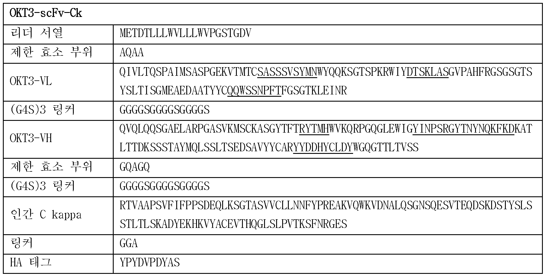
【표 2]

1.2 활성화 T세포의 사멸의 메커니즘 - 아폽토시스 (apoptosis) 1.2 Mechanism of death of activated T cells - Apoptosis
1가 항체 형태인 0KT3- Fab과 0KT3- scFv 항체에 의한 활성화 T세포의 사멸이 아폽토시스에 의한 것인지를 확인하기 위하여, 아폽토시스에 의한 세포 사멸 (apoptotic cel 1 death)의 마커인 아넥신 (Annexin) V와 7AAD염색을 동시에 진행하였고, 그 결과를도 1에 나타내었다. 세포의 초기 아폽토시스 (early apoptotic cell)에는 세포막이 아넥신 V에 염색되나, 아직 세포막파괴까지는 진행되지 않아 7AAD는 염색되지 않는 아넥신 V(+) 7AAD(- )형태를 띄게 되나, 말기 아폽토시스 (late apoptotic cell)는 7AAD까지 염색됨에 따라, 아넥신 V(+) 7AAD(+)형태를 띄게 된다. 또한, 0KT3의 1가 항체에 의한 활성화 T세포의 아폽토시스인지를 추가적으로 검증확인하기 위하여, 아폽토시스를 매개하는 효소인 카스파제 3 (caspase 3)의 활성화를 유세포분석을 통해 확인하였고, 그 결과를 도 2에 나타내었다. 도 1에 나타낸 바와 같이 , 활성화된 T세포에 각 형태의 0KT3 항체를 가한 후 시간에 따라분석한 결과, 0KT3- Fab, 0KT3- scFv항체를처리한그룹에서는 3시간부터 초기 아폽토시스의 비율이 급격히 증가하였으며, 6시간 이후부터 말기 아폽토시스 세포가 출현하면서 이후 급격히 그 비율이 증가하였다. 하지만, 완전 IgG 형태의 0KT3 항체를 처리한 군에서는 아폽토시스 세포의 비율에 변화가 없었다. 도 2에 나타낸 바와 같이, 0KT3-Fab, 0KT3- scFv 항체를 처리한 그룹에서, 처리후 3시간 이후부터 활성화된 카스파제 3 (활성 카스파제 3)가검출됨으로써 이들 세포에서 활발한아폽토시스가 일어났으나, 완전 IgG형태의 0KT3를처리한군에서는 활성화된 카스파제 3가 검출되지 않았다. 이상의 결과를통해 , 완전 IgG형태의 0KT3오}달리 Fab이나 scFv형태의 0KT3는 활성화된 T세포의 아폽토시스 경로에 의한 세포 사멸을 유도함을 확인하였다. In order to confirm whether the death of activated T cells by 0KT3-Fab and 0KT3-scFv antibodies in the form of monovalent antibodies was due to apoptosis, Annexin V, a marker of apoptotic cell death (apoptotic cell 1 death), and 7AAD staining were performed simultaneously, and the results are shown in Fig. 1. In early apoptotic cells, the cell membrane is stained with Annexin V, but since the cell membrane has not yet been destroyed, it takes the form of Annexin V(+) 7AAD(-), where 7AAD is not stained. On the other hand, in late apoptotic cells, as they are stained up to 7AAD, they take the form of Annexin V(+) 7AAD(+). In addition, to further verify whether the apoptosis of activated T cells was induced by the monovalent antibody of 0KT3, the activation of caspase 3, an enzyme that mediates apoptosis, was confirmed through flow cytometry, and the results are shown in Fig. 2. As shown in Fig. 1, when each form of 0KT3 antibody was added to activated T cells and analyzed over time, the rate of early apoptosis increased rapidly from 3 hours in the groups treated with 0KT3-Fab and 0KT3-scFv antibodies, and after 6 hours, terminal apoptotic cells appeared and their rate increased rapidly thereafter. However, there was no change in the rate of apoptotic cells in the group treated with the full IgG form of 0KT3 antibody. As shown in Fig. 2, in the groups treated with 0KT3-Fab and 0KT3-scFv antibodies, activated caspase 3 (active caspase 3) was detected after 3 hours of treatment, indicating that active apoptosis occurred in these cells, but in the group treated with 0KT3 in the form of full IgG, activated caspase 3 was not detected. Through the above results, it was confirmed that 0KT3 in the form of Fab or scFv, unlike 0KT3 in the form of full IgG, induced cell death through the apoptosis pathway of activated T cells.
1.3다른 1가 항체 항 CD3의 활성화된 T세포에 대한 선택적 제거능 가용성 1가 0KT3 항체의 T세포 아폽토시스 유도능이 다른 1가 항- CD3 항체에서도 재현되는지를 확인하였다. 또 다른 항- CD3 항체인 UCHT1 항체에 대해 IgG 항체는 상업용 항체를 구입하였고 , 상기 1.1과 동일한 방법으로 scFv형태로 제작한 후 , 활성화 T세포의 사멸을 유도하는지 확인하는 실험을 진행하였다. 하기 표 3에 각각 UCHT1 유內의 서열 및 이를 분리 정제하는데 이용한 서열을 나타내었다.1.3 Selective elimination of activated T cells by other monovalent antibodies anti-CD3 It was confirmed whether the T cell apoptosis-inducing ability of the available monovalent 0KT3 antibody was also reproduced in other monovalent anti-CD3 antibodies. For another anti-CD3 antibody, UCHT1 antibody, an IgG antibody was purchased commercially and produced in the form of scFv using the same method as 1.1 above, and an experiment was conducted to confirm whether it induces the death of activated T cells. The sequence of each UCHT1 gene and the sequence used to separate and purify it are shown in Table 3 below.
【표 3]

【표 4]


【표 5]

1.4 항 CD3의 활성화되지 않은 T세포에 대한 선택적 제거능 이상의 1가 항- CD3 항체의 아톱토시스 유발능이 활성화되지 않은 T세포 (unactivated T cell)에서도 보이는지를 확인하기 위해, 정상인 말초혈액 T세포에 scFv 0KT3 항체를 24시간동안가한후, 상기 실험예 1.3과 동일한 방법으로 T 세포의 사멸을 확인하였다. 그 결과를 도 4에 나타내었다. 도 4에 나타낸 바와 같이, 미활성화 T세포 (unact ivated T cell)에서는 완전 IgG, Fab, scFv 0KT3항체 모두아폽토시스를수반하는세포사멸이 관찰되지 않았다. 이상의 결과를 통해, 항- CD3 항체의 T세포 살상능은 완전 IgG 형태가 아닌, Fab 또는 scFv 형태의 anti- CD3 항체에서 활성화된 T세포에 대하여 선택적으로 제거능이 있음을 확인하였다. 총 4개의 서로 다른 1가 항- CD3 항체는 활성화된 T세포의 아폽토시스를 유도함을 확인하였다. 실험예 2. 1가항- CD3 항체의 아폽토시스 기전 분석 1.4 To determine whether the apoptosis-inducing ability of monovalent anti-CD3 antibodies beyond the selective elimination ability of unactivated T cells was also observed in unactivated T cells, scFv 0KT3 antibody was added to normal peripheral blood T cells for 24 hours, and T cell death was confirmed using the same method as in Experimental Example 1.3. The results are shown in Fig. 4. As shown in Fig. 4, in unactivated T cells, no apoptotic cell death was observed for any of the full IgG, Fab, or scFv 0KT3 antibodies. Through the above results, it was confirmed that the T cell killing ability of anti-CD3 antibodies selectively eliminated activated T cells in the Fab or scFv form, not in the full IgG form. A total of four different monovalent anti-CD3 antibodies were confirmed to induce apoptosis in activated T cells. Experimental Example 2. Analysis of the apoptosis mechanism of monovalent anti-CD3 antibodies
1가 항- CD3 항체에 의한 T세포 아폽토시스가 이들 항체가 TCR/CD3 신호전달 시스템에 의한 것인지 확인하기 위해, TCR/CD3 신호전달경로를분석하였다. To confirm whether T cell apoptosis induced by anti-CD3 antibodies is mediated by the TCR/CD3 signaling system, the TCR/CD3 signaling pathway was analyzed.
T세포의 TCR/CD3신호전달은 ZAP70- LAT- PLC x 1와같은상위 신호전달분자들의 활성화에 이어서, calcium-NFAT 경로, PKC0- NFKB경로, Erk- API경로의 3가지 하위 신호겨로의 활성화가 알려져 있다. 구체적으로, 칼슘- NFAT 경로의 활성화는 웨스톤 블랏팅에서 NFAT분자의 빠른 이동 (migration) (NFAT은 탈인산화에 의해 활성화되며, 탈인산화된 NFAT은 SDS- PAGE에서 작은 사이즈의 밴드로 검출)으로, PKC0- NFKB경로의 활성화는 NFkB저해분자인 IkB의 분해 (degration)로, Erk- API경로의 활성화는 인산화된 Erk(p- Erk)의 검출으로 확인할수 있다. 이에, 각 형태의 0KT3 항체 (0KT3- IgG, OKT-Fab, 0KT3-scFv) 또는 상기 3가지 하위 신호경로 모두를 활성화하는 PMA/이오노마이신 (lonomycin) (양성대조군)을 활성화된 T세포에 처리하고, 웨스턴 블랏팅을 통해 각각 NFATc2, IKB, P-ERK, Total ERK의 각 경로의 활성화 여부를 확인하였고, 그 결과를 도 5에 나타내었다. 또한, TCR/CD3 신호 와 그 하위의 NFAT 경로의 활성화가 0KT3 항체에 의한 아폽토시스를 유발하였는지를 검증하기 위하여 , TCR/CD3 신호전달 상위분자들의 활성화에 영향을 주는 신호전달 저해제의 투여에 따른 T세포의 사멸을 확인하였다. 구체적으로, 항- CD3 항체와 더불어 0.01% DMSO 중 사이클로스포린 A(Cyclosporin A)(calcineur in저해제 , CsA) 10uM, 0.01% DMSO 중 다사티닙 (Dasat inib)(Lck저해제 ) lOOnM 농도로 활성화된 T세포에 처리하였고, 음성대조군 (Vehicle)은 0.01% DMSO를 사용하였다. 그 결과를 도 6에나타내었다. 도 5에 나타낸 바와 같이, 세가지 경로 모두의 활성화를 유도하는 양성대조군을 기준으로 각군을 비교하였을 때, OKT3- IgG의 경우, 세 경로모두에서 눈에 띄는 활성화를 관찰하지 못하였으나, Fab과 scFv를 처리한 세포에서는 NFAT의 활성화가 관찰되었다. 반면 , NFkB및 Erk- AP경로의 활성화는관찰되지 않았다. 따라서 , 1가 OKT3 항체들은 TCR/CD3 신호 전달중 NFAT경로를 활성화시킴을 확인하였다. 도 6에 나타낸 바와 같이 , 다사티닙 (Dasa)은 0KT3- scFv에 의한 아폽토시스가 현저히 억제하였고, cyclosporinA(CsA)는 아폽토시스를 부분적으로 억제함을 확인하였다. 즉, 0KT3- scFv는 TCR/CD3 신호를 통해 아폽토시스를 유발하며, 특히 NFAT경로가 관여하고 있음을 확인하였다. 실험예 3. 동종 CAR T세포요법에서 항- CD3 아內의 잔류 TCR 양성 CAR T세포 제거능 It is known that TCR/CD3 signaling of T cells activates three downstream signaling pathways: calcium-NFAT pathway, PKC0-NFKB pathway, and Erk-API pathway, following the activation of upper signaling molecules such as ZAP70-LAT-PLC x 1. Specifically, activation of the calcium-NFAT pathway can be confirmed by the rapid migration of NFAT molecules in Western blotting (NFAT is activated by dephosphorylation, and dephosphorylated NFAT is detected as a small band in SDS-PAGE), activation of the PKC0-NFKB pathway can be confirmed by the degradation of IkB, an NFkB inhibitor, and activation of the Erk-API pathway can be confirmed by the detection of phosphorylated Erk (p-Erk). Here, activated T cells were treated with each form of 0KT3 antibody (0KT3-IgG, OKT-Fab, 0KT3-scFv) or PMA/ionomycin (positive control) that activates all three downstream signaling pathways, and the activation of each pathway, NFATc2, IKB, P-ERK, and Total ERK, was confirmed through Western blotting, and the results are shown in Fig. 5. In addition, in order to verify whether the TCR/CD3 signal and its downstream NFAT pathway activation induced apoptosis by 0KT3 antibody, the death of T cells was confirmed following the administration of a signaling inhibitor that affects the activation of upstream molecules of TCR/CD3 signaling. Specifically, activated T cells were treated with 10 μM cyclosporin A (calcineurin inhibitor, CsA) in 0.01% DMSO, 100 nM dasatinib (Lck inhibitor) in 0.01% DMSO, and 0.01% DMSO was used as the negative control. The results are shown in Fig. 6. As shown in Fig. 5, when each group was compared with the positive control group that induced activation of all three pathways, in the case of OKT3-IgG, no noticeable activation was observed in all three pathways, but activation of NFAT was observed in cells treated with Fab and scFv. In contrast, activation of the NFkB and Erk-AP pathways was not observed. Therefore, we confirmed that monovalent OKT3 antibodies activate the NFAT pathway during TCR/CD3 signaling. As shown in Fig. 6, dasatinib (Dasa) significantly inhibited apoptosis by 0KT3-scFv, and cyclosporinA (CsA) partially inhibited apoptosis. In other words, it was confirmed that 0KT3-scFv induces apoptosis through TCR/CD3 signaling, and that the NFAT pathway is particularly involved. Experimental Example 3. Ability to eliminate residual TCR-positive CAR T cells in anti-CD3 cells in allogeneic CAR T cell therapy
3.1 CRISPR/Cas9기법 및/또는 MACS에 의한 TCR제거의 불완전성 대부분의 항종양 CAR T세포요법은환자의 말초혈액에서 자가 T세포를채취하여 CAR유전자를 이입한 후 환자에게 되돌려주는 자가 CAR T세포요법 (autologous CAR T cell therapy)의 형태를유지하고 있다. 자가 CAR T세포의 경우, 환자에게서 T세포를 채취하여 CAR T세포를 제조하는 공정에 상당한 시간이 소요될 뿐 아니라, 환자의 T세포 상태가 좋지 않아 CAR T세포 제조가 실패하는 경우가 있다. 이에, 건강한 타인으로부터 채취한 말초혈액 T세포 (동종 T세포, allogeneic T cell)로부터 미리 CAR T세포를 제조하여 동결해 놓았다가, 필요한환자에게 즉시 투여할수 있는 동종 CAR T세포요법 (allogeneic CAR T cell therapy)이 시도되고 있다. 그러나, 동종 CAR T세포요법의 경우, 투여된동종 CAR T세포가환자의 정상조직을공격하여 심한염증을 유발하는 이식편대숙주병 (graft- versus- host disease: GVHD)부작용을 유발할 가능성이 있어, CRISPR/CAS9유전자편집 기술을 이용하여 이식편대숙주병을유도하는 T세포의 TCR/CD3 복합체를 제거한 CAR T세포를 제조하여 GVHD부작용을 방지하는 시도가 있다. 하지만, CRISPR/Cas9기법에 의한 TCR/CD3 복합체의 제거는 일부 잔류 TCR 양성 CAR T세포가존재하는문제가 있다. 3.1 Incompleteness of TCR deletion by CRISPR/Cas9 technique and/or MACS Most anti-tumor CAR T cell therapies are in the form of autologous CAR T cell therapy, in which autologous T cells are collected from the patient's peripheral blood, CAR genes are introduced, and then returned to the patient. In the case of autologous CAR T cells, the process of collecting T cells from the patient and manufacturing CAR T cells takes a considerable amount of time, and in some cases, CAR T cell manufacturing may fail due to poor T cell condition of the patient. Accordingly, allogeneic CAR T cell therapy is being attempted, in which CAR T cells are manufactured in advance from peripheral blood T cells (allogeneic T cells) collected from a healthy person, frozen, and then immediately administered to the patient in need. However, in the case of allogeneic CAR T-cell therapy, there is a possibility that the administered allogeneic CAR T cells may attack the patient's normal tissues, causing graft-versus-host disease (GVHD) side effects with severe inflammation. Therefore, there are attempts to prevent GVHD side effects by manufacturing CAR T cells with the TCR/CD3 complex of T cells that induce graft-versus-host disease removed using CRISPR/CAS9 gene editing technology. However, there is a problem that some residual TCR-positive CAR T cells exist due to the removal of the TCR/CD3 complex by the CRISPR/Cas9 technique.
TCR과 CD3는 항상 복합체 (complex)형태로 함께 세포표면에 발현되므로, TCR이나 CD3 어느 한쪽이 발현되지 않으면, 다른 한쪽 역시 발현되지 않는다. 즉, 항- CD3 항체로 잔류 TCR 양성세포를 제거할 수 있다. 이에, 동종 CAR T세포요법을 위한 CRISPR/Cas9에 의한 TCR/CD3 복합체의 제거 과정에서 , 본 발명의 1가 항- CD3 항체로 잔류 TCR 양성세포를제거하고자 하였다. 구체적으로, 도 7에 나타낸 바와 같이, 실험 0일차에 T세포를 활성화한 후, 기존에 보고된 사람 TCR 알파 체인 특이적 guide RNA(gRNA) 서열 (GAGAATCAAAATCGGTGAAT)을 사용하여 (Mol Ther. 2016 Mar; 24(3): 570-81) , TCR을 표적한 gRNA/Cas9 단백질 복합체 (Ribonulceoprotein: RNP)를 제작한 후, 2일간 활성화된 T세포에 전기천공 (electroporation) 방법으로 형질전환하였다. 이후 이들 T세포를 hIL- 2(200U/ml)가 포함된 배양액에서 수일간 배양하며 세포표면의 TCR 및 CD3입실론 체인의 발현을유세포분석법으로확인하였다. 실험 5일차, 8일차, 11일차, 14일차에 유세포분석을통해 잔류 TCR양성 T세포를 확인하였으며 , 그 결과를도 7에 나타내었다. 또한, 잔류 TCR 양성세포를 제거하기 위해 기존 방법인 MACS(magnet- associated cell sorting)정제를추가로수행하였을 때, 잔류 TCR양성세포를동일한 방법으로 확인하였다. 구체적으로, 2일간 활성화된 T세포의 TCR을 CRISPR/Cas9기법으로 제거한 후 6일 후, T세포에 magnetic microbead가 표지된 항- CD3 항체를 처리한 후, magnetic column (LD column)에 T세포를 통과시켜 CD3 양성 세포, 즉 TCR 양성 세포를 column에 trap시켰다. column을 통과하여 흘러나온 TCR 음성 세포를 수확하여, 수일간 hIL- 2(200U/ml)포함 배양액에서 추가 배양하여 유세포분석을 통해 잔류 TCR 양성 T세포를 확인하였으며, 그 결과를 도 8에 나타내었다. 도 7에 나타낸 바와 같이, 배양일 5일째에 약 7% 정도의 TCR 양성 T세포가 잔류함이 확인되었고, 이러한잔류 TCR양성률은 2주간시험관내 배양이 지속되더라도 일정하게 유지되었다. 도 8에 나타낸 바와 같이, 기존 MACS(magnet- associated cel 1 sorting)정제 방식을 사용했을 때에도, 약 2%정도의 TCR양성 T세포가잔류하는것을확인하였다. 이상의 결과를 통해 , 기존 MACS방식으로는 CRISPR/Cas9기법에 의한 TCR 양성 T세포의 완벽한제거는 매우 어려움을실험적으로 확인하였다. Since TCR and CD3 are always expressed together on the cell surface in the form of a complex, if either TCR or CD3 is not expressed, the other is not expressed either. In other words, residual TCR-positive cells can be removed with an anti-CD3 antibody. Accordingly, in the process of removing the TCR/CD3 complex by CRISPR/Cas9 for allogeneic CAR T cell therapy, the present invention sought to remove residual TCR-positive cells with a monovalent anti-CD3 antibody. Specifically, as shown in Fig. 7, after activating T cells on day 0 of the experiment, a gRNA/Cas9 protein complex (Ribonulceoprotein: RNP) targeting TCR was produced using a previously reported human TCR alpha chain-specific guide RNA (gRNA) sequence (GAGAATCAAAATCGGTGAAT) (Mol Ther. 2016 Mar; 24(3):570-81), and then electroporation was used to transfect T cells activated for 2 days. Afterwards, these T cells were cultured in a culture medium containing hIL-2 (200U/ml) for several days, and the expression of TCR and CD3 epsilon chain on the cell surface was confirmed by flow cytometry. On days 5, 8, and 11 of the experiment, On the 14th day, residual TCR-positive T cells were confirmed through flow cytometry, and the results are shown in Fig. 7. In addition, when the existing MACS (magnet-associated cell sorting) purification method was additionally performed to remove residual TCR-positive cells, residual TCR-positive cells were confirmed by the same method. Specifically, after the TCR of T cells activated for 2 days was removed using the CRISPR/Cas9 technique, 6 days later, the T cells were treated with anti-CD3 antibodies labeled with magnetic microbeads, and then the T cells were passed through a magnetic column (LD column) to trap CD3-positive cells, i.e., TCR-positive cells, in the column. TCR-negative cells that passed through the column were harvested and further cultured in a culture medium containing hIL-2 (200 U/ml) for several days, and residual TCR-positive T cells were confirmed through flow cytometry, and the results are shown in Fig. 8. As shown in Fig. 7, it was confirmed that about 7% of TCR-positive T cells remained on the 5th day of culture, and this residual TCR positivity rate remained constant even after 2 weeks of in vitro culture. As shown in Fig. 8, even when the existing MACS (magnet-associated cell 1 sorting) purification method was used, it was confirmed that about 2% of TCR-positive T cells remained. Through the above results, it was experimentally confirmed that it is very difficult to completely remove TCR-positive T cells by the CRISPR/Cas9 technique using the existing MACS method.
3.2 항- CD3 scFv의 잔류 TCR양성 T세포제거능 3.2 Anti-CD3 scFv’s ability to eliminate residual TCR-positive T cells
CRISPR/Cas9기법에 의한 TCR 양성 T세포 제거 후 세포배양액에 0KT3- scFv 항체를 가하여 TCR 양성 T세포를 제거할수 있는지 확인하였다. 우선 , 도 9에 나타낸바와같이 , T세포의 활성화 및 항- TCR gRNA/Cas9복합체 도입과정을 거친 배양 7일차 T세포에 0KT3-ScFv 항체를 처리하고, 3일간 추가 배양 후 잔류 TCR 양성 T세포 존재 여부를 확인하였다. 그 결과를 도 9에 나타내었다. 도 9에 나타낸 바와 같이, 대부분의 TCR 양성 T세포가 제거되었으며, 이는 기존의 MACS정제에 의한 방법보다효과적으로잔류 TCR 양성 T세포를 제거하였다. 이어서, 종래의 가장 보편적으로 활용되고 있는 항- CD19 CAR T세포에 본 발명의 항- CD3 scFv를 처리하여 TCR 양성 T세포를 제거능을 확인하였다. 구체적으로, 항- CD19 CAR cDNA는항- CD19 scFv, CD8 hinge및 막횡단도메인, 41BB 세포내 도메인, CD3제타 세포내 도메인이 연결된 형태로서, 기존 공개된 서열대로 (미국 특허 공개공보 US2013/0287748 Al) DNA합성을 의뢰하여 제작하였다 (Integrated DNA Technologies) . 제작된 항- CD19 CAR cDNA를 렌티바이러스벡터에 클로닝한 후, 리포펙타민 (Lipofectamin) 3000 (Invitrogen)을 이용하여 패키징 DNA 3종 (pMD.2G, pMDLg/pRRE, pRSV- rev)과 함께 293T 세포주 (ATCC)에 형질전환후, 24- 48시간동안 분비된 렌티바이러스가 포함된 배양상층액을 수확하여 필터하여 (0.45仰1 필터) 세포 잔존입자를 제거하고, 초고속원심분리기를 사용하여 100배 농축한후 CAR T세포 제작을 위한 렌티바이러스 농축액으로 사용하였다. 말초혈액 단핵세포내 T세포를 2일간 활성화후, 농축된 렌티바이러스와 같이 T세포를 이틀간배 양하여 CAR유전자를 T세포에 형질도입 (transduct ion)함으로써 항- CD19 CAR T세포를 제작하였다. 이후, 상기 세포들을 세척하여 배양액에 남은 바이러스를 제거하고 3일 간격으로 2번 hIL- 2(200U/ml)가 포함된 배양액을 추가하면서 CAR T세포의 증식을 유도하였다. T세포 배양시작일로부터 7일 후, 0KT3- scFv 항체 (12.5pmol/lX105세포)를 세포배양액에 가하여 3일간 추가 배양하였고, 배양시작일로부터 10일째 유세포분석기를 통해 잔류 TCR 양성 T세포를 확인하였다. 그 결과를 도 10에 나타내었다. 도 10에 나타낸 바와 같이, TCR 제거 및 항- CD19 CAR유전자 도입을 통해 제조된 TCR이 제거된 CAR T세포는, 배양 7일차에 10%이상의 TCR양성 T세포의 존재가 확인되었으나, 3일간 0KT3-ScFv존재하에서 추가배양을 시행한 결과, 대부분의 잔류 TCR 양성 CAR T세포가제거된 것을 확인하였다. 추가적으로, 종래의 보편적인 항- CD19 CAR T세포에 본 발명의 항- CD3 scFv를 처리했을 시 , 기존의 CAR T세포의 종양살상능등기능이 유지되는지 확인하기 위해, CAR T세포의 표적인 CD19양성이 Raji 세포주와함께 배양하여 , 사이토카인분비능과 종양살상능을 확인하였다. 구체적으로, CD19양성 Raj구세포에 루시페라제 (Luciferase)를과발현한 Raji- Luc 세포 (3X10⁴)를 CAR T 또는 T세포와 함께 18시간동안 배양한 후, 배양액에 luciferin(6mg/ml)을 처리하여 생존한 Raji- Luc세포의 발광도를 루미노미터 (luminometer)로 측정함으로써 , 세포의 생존율을 도출하였고, 이로부터 세포살상능을 계산하였다 (도 11). 구체적인 수학식은 아래와 같다. After removing TCR-positive T cells by the CRISPR/Cas9 technique, 0KT3-scFv antibody was added to the cell culture medium to confirm whether TCR-positive T cells could be removed. First, as shown in Fig. 9, 0KT3-ScFv antibody was treated on the 7th day of culture, after T cell activation and anti-TCR gRNA/Cas9 complex introduction process, and the presence of residual TCR-positive T cells was confirmed after additional culture for 3 days. The results are shown in Fig. 9. As shown in Fig. 9, most of the TCR-positive T cells were removed, which is more effective in removing residual TCR-positive T cells than the conventional MACS purification method. Next, the anti-CD3 scFv of the present invention was treated to the most commonly utilized conventional anti-CD19 CAR T cells to confirm the ability to remove TCR-positive T cells. Specifically, anti-CD19 CAR cDNA was produced by requesting DNA synthesis (Integrated DNA Technologies) according to the previously published sequence (US Patent Publication No. US2013/0287748 Al) in the form of anti-CD19 scFv, CD8 hinge and transmembrane domains, 41BB intracellular domain, and CD3 zeta intracellular domain linked together. The produced anti-CD19 CAR cDNA was cloned into a lentiviral vector, and then 293T was cultured together with three types of packaging DNA (pMD.2G, pMDLg/pRRE, pRSV-rev) using Lipofectamin 3000 (Invitrogen). After transformation into the cell line (ATCC), the culture supernatant containing the secreted lentivirus for 24-48 hours was harvested and filtered (0.45x1 filter) to remove residual cell particles, concentrated 100-fold using an ultra-high-speed centrifuge, and used as a lentivirus concentrate for producing CAR T cells. After activating T cells in peripheral blood mononuclear cells for 2 days, the T cells were cultured with the concentrated lentivirus for 2 days to transduce the CAR gene into the T cells, thereby producing anti-CD19 CAR T cells. Thereafter, the cells were washed to remove the virus remaining in the culture medium, and the proliferation of CAR T cells was induced by adding the culture medium containing hIL-2 (200 U/ml) twice at 3-day intervals. Seven days after the start of T cell culture, 0KT3-scFv antibody (12.5 pmol/lX10 5 cells) was added to the cell culture medium, and the cell was cultured for an additional 3 days. Residual TCR-positive T cells were confirmed using a flow cytometer on the 10th day from the start of culture. The results are shown in Fig. 10. As shown in Fig. 10, in the CAR T cells with TCR deletion produced through TCR deletion and anti-CD19 CAR gene introduction, the presence of more than 10% TCR-positive T cells was confirmed on the 7th day of culture, but as a result of performing additional culture in the presence of 0KT3-ScFv for 3 days, it was confirmed that most of the residual TCR-positive CAR T cells were removed. Additionally, in order to confirm whether the tumor killing ability of the existing CAR T cells is maintained when the anti-CD3 scFv of the present invention is treated with the conventional universal anti-CD19 CAR T cells, the cells were cultured with the CD19-positive Raji cell line, which is the target of the CAR T cells, and the cytokine secretion ability and tumor killing ability were confirmed. Specifically, Raji-Luc cells ( 3X10⁴ ) that overexpress luciferase in CD19-positive Raj cell lines were cultured with CAR T or T cells for 18 hours, and then the culture medium was treated with luciferin (6 mg/ml) to measure the luminescence of the surviving Raji-Luc cells using a luminometer, thereby deriving the cell survival rate, and the cell killing ability was calculated therefrom (Fig. 11). The specific mathematical formula is as follows.
[수학식 1] [Mathematical formula 1]
Raji- Luc세포의 생존율 (%) = [(실험군의 발광도)- (배경 (background) 발광도)]/ [(Raji- Luc세포만존재시의 발광도)- (배경 발광도)] xlOO. Viability of Raji-Luc cells (%) = [(luminescence of experimental group) - (background luminescence)] / [(luminescence when only Raji-Luc cells are present) - (background luminescence)] xlOO.
[수학식 2] 세포살상능 (%세포독성) = 100-세포생존율. 또한, 사이토카인 분비능의 측정은 항체를 처리한 CAR T세포 또는 T세포와 CD19C+) 종양세포를 공배양시 분비되는 INF- x를 측정하였다. 구체적으로, 1.5X10⁵ CD19양성 Raji세포주와 3X10⁴ CAR T세포를같이 배양한 24시간후, 배양액을획득하여 이를 인간 IFN- x ELISA 키트 (R&D)의 제조사 실험방법에 따라 실험하여 배양액 내의 인간 INF- x 의 양을 측정하였다 (도 11) . 도 11에 나타낸 바와 같이 , CAR T세포의 종양살상력 및 cytokine 분비능 모두 항체를 처리하지 않은 CAR T세포와 유사한 정도로 유지되고 있음이 확인하였다. 이상의 결과를 통해서 , 동종 CAR T세포의 TCR이 제거과정에서 발생하는 잔류 TCR 양성 CAR T세포에 1가 항- CD3 항체를 처리해줌으로써 보다 잔류 TCR 양성 CAR T 세포를 효과적으로 제거하여 , 이식편대숙주병 등의 부작용을 완화 또는 개선할 수 있는 가능성을 확인하였다. [Mathematical formula 2] Cell killing capacity (% cytotoxicity) = 100-cell survival rate. In addition, the cytokine secretion capacity was measured by measuring INF- x secreted when CAR T cells or T cells treated with antibodies were co-cultured with CD19C+) tumor cells. Specifically, 1.5X10⁵ CD19-positive Raji cell line and 3X10⁴ CAR T cells were co-cultured for 24 hours, and the culture medium was obtained. This was tested according to the manufacturer's experimental method of the human IFN- x ELISA kit (R&D) to measure the amount of human INF- x in the culture medium (Fig. 11). As shown in Fig. 11, it was confirmed that both the tumor killing power and cytokine secretion power of CAR T cells were maintained at a similar level to CAR T cells that were not treated with antibodies. Through the above results, it was confirmed that the possibility of alleviating or improving side effects such as graft-versus-host disease can be confirmed by treating the residual TCR-positive CAR T cells generated during the TCR removal process of allogeneic CAR T cells with monovalent anti-CD3 antibody to effectively remove the residual TCR-positive CAR T cells.
3.3 항- CD3 scFv에 의한 잔류 TCR 양성 CAR T세포 제거에 따른 이식편대숙주병 예방 효과 (、in vivo) 동종 (al logenei c) CAR T세포 제작 과정에서 발생하는 잔류 TCR 양성 CAR T세포에 의해 유발되는 이식편대숙주병을 본 발명의 1가 항- CD3 항체 (0KT3 scFv)를 처리함으로써 방지할 수 있는지 확인하기 위해 생체내 { in vivo) 실험을 수행하였다. 동종 CAR T세포에 의한 이식편대숙주병을 직접적으로 마우스에서 구현하기는 어려우나, 사람 CAR T세포를 면역결핍마우스에 투여하는 경우 사람 CAR T세포의 TCR이 마우스 정상세포의 MHC분자를 인식하여 마우스 조직을 공격하는 것을 유도함으로써 , 공여자의 동종 CAR T세포가 수여자의 MHC를 공격하여 유발되는 동종 이식편대숙주병 (al l ogene i c GVHD)을 모사할 수 있다. 이를 이종 이식편대숙주병 (xenogenei c GVHD)라고 부른다. 즉 , TCR을 발현하는 사람 CAR T세포를 사람 종양이 접종된 면역결핍마우스에 투여하면 , 초기 한달간 CAR T세포에 의한 종양제거효력을 마우스 생체내에서 관찰할 수 있으나, 그 이후에는 사람 CAR T세포가 마우스 정상조직을 공격하는 이종 이식편대숙주병 부작용을 관찰할 수 있다. 구체적으로, 면역결핍 NSG마우스에 2.5Gy 방사선 전신 조사 후 , CD19 양성 Raj i - Luc세포주 5xl05세포를 정맥주사하였다. Raj구세포 주사 3일 후 , 인간 T세포로 제작한 항- CD19 CAR T세포, TCR이 제거된 항- CD19 CAR T세포 , 혹은 0KT3- scFv 항체를 처리하여 잔류 TCR 양성 세포가 제거된 항- CD19 CAR T세포를 마우스당 5xl06개씩 정맥주사하였다. 종양세포 주입 후 일주일 간격으로 생체내 이미징 시스템 ( In- vivo imaging system , IVIS)을 이용하여 종양의 생물발광 (bi o luminescence)을 즉정하였고 , 각각의 마우스는 식염수 100ul에 녹인 루시페린 2mg을 복강 주사하고 10분 후 IVIS를 통하여 체내 종양의 이미지를 획득하였고 , 이를 도 12에 나타내었다. 도 12에 나타낸 바와 같이 , 생물발광 이미징을 통하여 마우스내 종양의 증식을 시간별로 측정한 결과 , 종양만 투여된 군은 2- 3주내에 모든 개체가 사망한 반면 , 세 가지 CAR- T세포를 투여받은 그룹은 종양의 증식이 거의 보이지 않고 , 3주까지는 모든 개체가 생존하는 우수한 치료효능을 보임으로써 , CAR T세포의 항종양효력은 모두 잘 유지되고 있음이 확인하였다. 하지만, 종양 투여후 10주후에는 , 세가지 CAR T세포 투여 군 중 기존의 항- CD19 CAR T세포 , TCR이 제거되었으나 잔류 TCR 양성세포가 남아있는 항- CD19 CAR T세포투여 군의 마우스 개체는 이식편대숙주병에 의해 모두 사망하였으나, 0KT3-scFv 항체를 처리하여 잔류 TCR 양성 세포가 제거된 항- CD19 CAR T세포군이 투여된 마우스군만 이식편대숙주병의 부작용 없이 생존하였다 (도 13 ) . 추가적으로, 각 인간 T세포로 제작한 항- CD19 CAR T세포 , TCR이 제거되었으나 잔류 TCR 양성세포가 남아있는 항- CD19 CAR T세포 , 혹은 0KT3- scFv 항체를 처리하여 잔류 TCR 양성 세포가 제거된 항- CD19 CAR T세포가 투여된 군의 마우스의 이식편대숙주병의 발병 정도를 비교하였다. 이식편대숙주병의 발병 정도는 체중 , 및 이식편대숙주병 점수 (GVHD score)으로 측정하였다. 각각 체중과 이식편대숙주병 점수는 일주일에 2번씩 측정하였으며 , 이식편대숙주병 점수는 5가지 임상 증상 (피부 , 털의 상태 , 자세 , 활동성 , 염증성 눈 질환)에 따라 0〜 3점까지를 점수를 부여하여 합산한 값이다. 각각을 종양 투여 후 80일까지 측정하였고 , 그 결과를 도 13에 나타내었다. 도 13에 나타낸 바와 같이 , TCR이 제거되지 않은 일반적인 CAR T세포 투여군 (Conv CAR- T)의 경우, 종양 투여 2주 후부터 체중이 감소하고 , 이식편대숙주병 임상소견을 보이며 , 3주후 모든 개체가 이식편대숙주병으로 사망하는 양상을 보였다. TCR이 제거되었으나 잔류 TCR 양성세포가 남아있는 CAR T세포 투여군 (De l CAR- T)의 경우 , 체중감소나 이식편대숙주병 중증도가 Conv CAR- T세포에 비해 일부 감소하였으나, 8- 9주에 역시 이식편대숙주병으로 사망하였다. 이는 잔류 TCR 양성 CAR T세포가 중증 이식편대숙주병 부작용을 유발한 것이다. 반면 , 항- CD3 scFv 항체를 처리하여 잔류 TCR 양성세포를 완전히 제거한 CAR T세포 투여군 (De l Ab CAR- T)에서는 체중감소나 이식편대숙주병 소견 없이 모든 개체가 장기간 생존하는 부작용 없는 항종양 효력을 보였다. 이상의 결과를 통해 , 동종 CAR T세포 제조과정에서 1가 항- CD3 항체 처리를 통한 잔류 TCR 양성세포의 효율적이고 완벽한 제거는 동종 CAR T세포의 이식편대숙주병 부작용을 현저히 낮출 수 있음을 확인하였다. 실험예 4. 항- CD3 항체에 의한 말초혈액내 활성화 T세포군의 선택적 제거 효과 이상의 실험예를 통해 , 본 발명의 1가 항- CD3 항체는 동종 CAR T세포 제조과정에서 잔류 TCR 양성세포를 제거하여 이식편대숙주병의 부작용을 예방 또는 치료할 수 있음을 확인하였다. 뿐만 아니라, 본 발명의 1가 항- CD3 항체는 , 활성화 T세포에 의한 염증질환을 앓고 있는 환자에게 직접 투여 시 환자 체내에서 직접 활성화된 T세포군을 제거함으로써 치료제로 사용될 가능성이 있다. 이와 같은 질환의 예로서는 자가면역질환, 이식편대숙주병, 혹은 장기이식거부반응이 있다. 즉, 1가 항- CD3 항체가 자가면역질환이나 이식편대숙주병 혹은장기이식 거부반응을보이는환자에게 투여되었을 때, 염증을매개하는 활성화 T세포를선택적으로제거할 수 있다면, naive T세포나 memory T세포를 보존함으로써 감염의 위험을 최소화하면서도 T세포에 의한 염증반응을 억제함으로써 면역억제제로 활용도가능하다. 이에, 본 발명의 항- CD3 scFv 항체 (0KT3- ScFv)를 처리함으로써 인체내 혈액세포 중 활성화 T세포군을 선택적으로제거할수 있는지 확인하고자 하였다. 구체적으로 , 정상인으로부터 채취한 말초혈액세포 (pher ipheral blood mononuclear cell: PBMC) T세포군은 CD45RA와 CCR7발현을 마커로 사용하여 활성화이력에 따른 아세포군으로 분류하였다. CD45RA(+)CCR7(+)군은 활성화된 적이 없는 naive T세포가 대부분이고, 일부 휴지기에 있는 줄기세포 기억 T세포군이 혼합되어 있고 (N/SCM), CD45RM- )CCR7(+)군은 활성화된 적이 있으나 휴지기에 있는 중앙 기억 T세포군 (CM)이며, 이들 세포군은 모두 현재는 휴지기에 있는 비활성화 세포군이다. 반면, CCR7(- )세포군은활성화된효과기 T세포 (effector T cell)이거나 바로 활성화될 수 있는 효과기 기억 T 세포 (effector memory T세포)로 구성되어 있다 (EF/EM). 따라서, EF/EM세포군에는 현재 활성화되어 있는 T세포들이 다수 포함되어 있다. 이에, 정상인 말초 혈액 단핵세포와 0KT3-scFv 항체를 3일간 배양한 후, 유세포분석을 통하여 말초 혈액 단핵세포 내부의 T세포 아세포군 (subpopulation)분석을 통해, 비활성화 및 활성화 T세포군의 비율변화 및 세포수 감소를 분석하였다. 보다 구체적으로, 유세포분석시 세포 계수 비드 (cell counting bead, Thermo Scientific)를 가하여, 단위부피당 유세포분석기를 통과한 세포수를 측정함으로써 , T세포및 각아세포군의 세포수를산출하였다. anti- CD3- PE, anti- CD4- APC- Cy7, anti- CD8- PE- Cy7, ant i- CD45RA- FITC, ant i -CCR7-APC 및 7- AAD로 염색한 후, 7-AAD 음성 , CD3 양성세포군을 게이팅 (gating)한 후, CD4 양성세포 및 CD8 양성세포에서 각 아세포군의 T세포내 분율을분석하였다. 그 결과를 도 14내지 16에 나타내었다. 도 14내지 16에 나타낸 바와 같이, 3일간 0KT3-scFv항체 처리에 의해 전체 T세포의 숫자는 약 30% 정도 감소하였다 (도 14). 이 중 N/SCM군 및 CM군 (비활성화 T 세포) 대비 EF/EM군 (활성화 T세포)의 비율이 CD4및 CD8 T세포모두에서 부분적으로 감소하였다 (도 15 및 16). 또한, 각 아세포군 (subpopulation) 세포의 절대 수를 산출한 결과, CD4 T세포에서는항체처리에 의해 N/SCM군 및 CM군 (비활성화 T세포)의 세포수 감소가 일부 보였으나, EF/EM (활성화 T 세포)군의 세포수 감소가 훨씬 두드러졌으며, CD8 T세포에서는 항체처리에 의한 N/SCM군, CM군 (비활성화 T세포)의 세포수의 변화가 없는 반면, EF/EM군 (활성화 T 세포)에서는 확연한 세포수 감소가 관찰되었다 . 또한 , 항체미처리군 대비 항체처리군에서의 세포수감소비율로 계산했을 때에도, CD4, CD8 T세포모두에서 EF/EM군 (활성화 T세포)의 상대적 세포수감소율이 현저히 높음을 확인하였다 (도 16). 이상의 결과를 통해 , 1가 항- CD3 항체는 말초혈액 T세포중 활성화 세포군에 대한 선택적 제거효과를 가져, 다목적 선택적 활성화 T세포 제거제 및 T세포 면역억제제로 유용하게 사용될 수 있음을 확인하였다. 실험예 5. 항- CD3항체의 생체내 활성화 T세포제거 상기 1가 항- CD3 항체를 직접 생체내 (/刀 r/w)에 투여시에도 활성화된 T세포 제거 효과가 관찰되는지 여부를 확인하기 위하여, 시험관내에서 활성화시킨 인간 T세포를 면역결핍 마우스에 투여한후, 1가항- CD3항체 (UCHT1- scFv)를이틀간격으로 수차례 정맥투여하고 인간 T세포의 사멸 여부를 추적 관찰하였다. 이때, 루시퍼라제 (luciferase) 효소를 인위적으로 발현시킨 인간 T세포를 사용함으로써, 생체내 이미징 시스템 (in- vivo imaging system: IVIS)을 이용하여 인간 T세포의 체내 생물발광을추적할 수 있도록 하였다. 구체적으로, 루시피라제 발현 인간 T세포 제작을 위해서, pD0NR222-eGFP (Addgene plasmid #364493)로부터 획득한 고감도 루시퍼라제 (eff- Luc) cDNA를 GFP cDNA와 함께 발현되도록, 일부 변형된 pCDH- EF1벡터 (Addgene Plasmid #72266)에 클로닝하였다. 클로닝된 렌티바이러스플라스미드를리포펙타민 3000 (Invitrogen)을 이용하여 packaging DNA 3종 (pMD.2G, pMDLg/ pRRE , pRSV— rev)과 함께 293T cel 1 1 ine (ATCC)에 형질전환 (transfect ion)한 후, 24 및 48시간동안 분비된 렌티바이러스가 포함된 배양상층액을수확한 후 0.45仰1필터를 이용하여 세포 잔존입자를 제거하고, 초고속원심분리기를 사용하여 100배 농축한 다음 형질도입용 렌티바이러스 농축액으로 사용하였다. 정상인으로부터 채혈 (leukapheresis)을 통해 얻어진 백혈구를 항 CD3 항체 (0KT3, 10w/mt, BioXcell)가코팅된 24well플레이트에 항 CD28항체 (CD28.2, 2//g/mt, BD Biosciences)와함께 첨가한후 48시간동안배양하여 T세포를활성화시켰다. 이후 루시퍼라제 발현용 렌티바이러스 농축액을 T세포 배양액에 섞어 같이 배양하였다. 렌티바이러스를 이용한 형질도입 2일 후, 상기 세포를 세척하여 배양액에 남은 바이러스를 제거하고, 3일씩 2번 (3일 +3일) 인간 IL-2 (200U/mt)가 포함된 배양액을 주가하여 배양하였다 (10일 protocol ) . 루시퍼라제 발현 T세포의 경우 생체내에서 T세포의 존재 유무 및 증식 정도를 IVIS를 통하여 확인 가능한 장점을 가지므로, 면역결핍 NSG 마우스에 루시퍼라제 발현 인간 T세포 5xl06세포를 정맥 주사하였다. 인간 T세포주입 후 일주일 간격으로 생체내 이미징 시스템을 이용하여 생체내에 루시퍼라제 발현 T세포의 분포정도를 확인하였다. T세포 주사 14일 후 ( IVIS를 통해 모든 마우스 내에 T세포가 관찰되는 시점 )부터 PBS 200ul에 희석한 1가 항- CD3 항체 (UCHT1- scFv, 저용량: 20ug/ea , 고용량: 100ug/ea)를 이틀에 한번 , 총 5회 정맥 주사하였다 . 1가 항- CD3 항체를 투입하는 시점부터는 이틀에 한번 IVIS를 이용하여 마우스 체내의 T세포의 분포 정도를 확인하였다. 각각의 마우스는 식염수 lOOul에 녹인 루시페린 2mg을 복강 주사하였다. 복강 주사 10분 후 IVIS를 통하여 종양세포의 크기를 측정하였다. 실험 결과를 도 17 및 18에 나타내었으며 , 이로부터 1가 항- CD3 항체를 투여한 군에서 생체내 T세포가 현저하게 소멸되었으며 , 투여되는 1가 항- CD3 항체의 양이 많을수록 더 빠르고 극명하게 생체내 T세포가 줄어드는 것으로 나타났고 용량의존적임이 확인되었다. 실험예 6. 항- CD3 항체의 생체내 T세포 매개 질환 억제 상기 면역결핍 마우스에 루시퍼라제 발현 인간 T세포를 주입하는 경우 , 인간 T세포가 마우스 정상조직을 공격하여 염증을 일으키는 이종 이식편대숙주병 (xenogenei c GVHD)를 유발하게 된다. 따라서 , 1가 항- CD3 항체를 투여하는 경우 , 인간 T세포의 제거에 의하여 이종 이식편대숙주병을 억제할 수 있는지를 테스트하였다. 마우스의 이식편대숙주병 발병 정도는 2가지의 방법 (체중, 이식편대숙주병 점수 [GVHD score] )으로 측정하였다. 무게와 이식편대숙주병 점수의 경우 일주일에 2번 측정하였으며 이식편대숙주병 점수의 경우 5가지 발병 척도 (피부 , 털의 상태 , 자세 , 활동성 , 염증성 눈 질환)에 따라 0〜 3점까지로 점수를 부여하여 총합하였다. 도 19는 면역결핍마우스 모델에서 인간 T세포에 의해 이종 이식편대숙주병을 유발한 후 1가 항- CD3 항체 (UCHT1- scFv) 투여시 체중감소 , 이식편대숙주병 임상 증상 및 치사율을 평가한 결과를 나타낸 것이다. 도 19로부터 1가 항- CD3 항체를 처리시 , 이식편대숙주병에 의한 체중감소 , 임상 증상 및 치사율 모두 현저하게 감소함을 확인하였다. 따라서 , 1가 항- CD3 항체는 T세포에 의해 유발되는 염증성 질환을 크게 억제할 수 있으므로, 유사한 T세포매개 염증질환인 자가면역질환 치료제로 개발될 가능성이 매우 높다. 또한, 상기 모델의 이식편대숙주병과 유사 질환으로서 , 혈액종양 치료를 위한 동종 조혈모세포 이식 (al logenei c hematopoiet i c stem cel 1 transplantat ion) 후 공여자 T세포에 의해 발생하는 동종 이식편대숙주병 (al logenei c GVHD)의 치료목적으로도 사용 가능할 것이다. 3.3 Effect of preventing graft-versus-host disease by eliminating residual TCR-positive CAR T cells by anti-CD3 scFv (in vivo) In order to confirm whether graft-versus-host disease caused by residual TCR-positive CAR T cells generated during the process of producing allogeneic CAR T cells can be prevented by treating with the monovalent anti-CD3 antibody (0KT3 scFv) of the present invention, an in vivo experiment was performed. Although it is difficult to directly implement graft-versus-host disease caused by allogeneic CAR T cells in mice, when human CAR T cells are administered to immunodeficient mice, the TCR of the human CAR T cells recognizes the MHC molecules of mouse normal cells and induces attacks on mouse tissues, thereby simulating allogeneic graft-versus-host disease (allogeneic GVHD) caused by the donor's allogeneic CAR T cells attacking the recipient's MHC. This is called xenogeneic GVHD. That is, when human CAR T cells expressing TCR are administered to immunodeficient mice inoculated with human tumors, the tumor eradication effect by CAR T cells can be observed in the mouse body for the first month, but xenogeneic graft-versus-host disease side effects in which human CAR T cells attack mouse normal tissues can be observed thereafter. Specifically, 5xl0 5 cells of the CD19-positive Raj i-Luc cell line were intravenously injected into immunodeficient NSG mice after 2.5 Gy of total body irradiation. Three days after the Raj cell injection, anti-CD19 CAR T cells produced using human T cells, anti-CD19 CAR T cells with a deleted TCR, or anti-CD19 CAR T cells with residual TCR-positive cells removed by treating them with 0KT3-scFv antibody were intravenously injected 5xl0 6 per mouse. After tumor cell injection, the bioluminescence of the tumor was measured using an in vivo imaging system (IVIS) at one-week intervals. Each mouse was intraperitoneally injected with 2 mg of luciferin dissolved in 100 μl of saline, and images of the tumor in the body were obtained 10 minutes later using IVIS, which are shown in Fig. 12. As shown in Fig. 12, the tumor proliferation in the mouse was measured over time using bioluminescence imaging. While all individuals in the group that received only the tumor died within 2-3 weeks, the group that received the three CAR-T cells showed almost no tumor proliferation, and all individuals survived for up to 3 weeks, showing excellent therapeutic efficacy, confirming that the antitumor efficacy of the CAR T cells was well maintained. However, 10 weeks after tumor administration, the three CAR T cells Among the administration groups, all mice in the existing anti-CD19 CAR T cell group, the anti-CD19 CAR T cell group in which the TCR was removed but residual TCR-positive cells remained died of graft-versus-host disease, but only the mice in the anti-CD19 CAR T cell group in which residual TCR-positive cells were removed by treatment with 0KT3-scFv antibody survived without any adverse effects of graft-versus-host disease (Fig. 13). Additionally, the severity of graft-versus-host disease was compared in the mice in the groups administered anti-CD19 CAR T cells manufactured using each human T cell, anti-CD19 CAR T cells in which the TCR was removed but residual TCR-positive cells remained, or anti-CD19 CAR T cells in which residual TCR-positive cells were removed by treatment with 0KT3-scFv antibody. The severity of graft-versus-host disease was measured by body weight and graft-versus-host disease score (GVHD score). Each body weight and graft-versus-host disease score were measured twice a week, and the graft-versus-host disease score was the sum of the scores from 0 to 3 based on five clinical symptoms (skin, hair condition, posture, activity, and inflammatory eye disease). Each was measured up to 80 days after tumor administration, and the results are shown in Fig. 13. As shown in Fig. 13, in the case of the general CAR T cell administration group without a deleted TCR (Conv CAR-T), body weight decreased from 2 weeks after tumor administration, clinical findings of graft-versus-host disease were shown, and all individuals died of graft-versus-host disease after 3 weeks. In the CAR T cell administration group (De l CAR- T) where the TCR was removed but residual TCR-positive cells remained, weight loss and the severity of graft-versus-host disease were somewhat reduced compared to the Conv CAR- T cell group, but the mice still died of graft-versus-host disease at 8-9 weeks. This indicates that the residual TCR-positive CAR T cells induced severe graft-versus-host disease side effects. On the other hand, in the CAR T cell administration group (De l Ab CAR- T) where residual TCR-positive cells were completely removed by treatment with anti-CD3 scFv antibodies, all animals survived long-term without weight loss or graft-versus-host disease, demonstrating anti-tumor efficacy without side effects. Through the above results, we confirmed that efficient and complete removal of residual TCR-positive cells through treatment with monovalent anti-CD3 antibodies during the manufacturing process of allogeneic CAR T cells can significantly reduce the graft-versus-host disease side effects of allogeneic CAR T cells. Experimental Example 4. Selective elimination effect of peripheral blood activated T cell population by anti-CD3 antibody Through the above experimental examples, it was confirmed that the monovalent anti-CD3 antibody of the present invention can prevent or treat the side effects of graft-versus-host disease by eliminating residual TCR positive cells during the manufacturing process of allogeneic CAR T cells. In addition, the monovalent anti-CD3 antibody of the present invention has the potential to be used as a therapeutic agent by directly eliminating the activated T cell population in the patient's body when directly administered to a patient suffering from an inflammatory disease caused by activated T cells. Examples of such diseases include There are autoimmune diseases, graft-versus-host disease, or organ transplant rejection. That is, when a monovalent anti-CD3 antibody is administered to a patient showing an autoimmune disease, graft-versus-host disease, or organ transplant rejection, if it can selectively remove activated T cells that mediate inflammation, it can be utilized as an immunosuppressant by suppressing the inflammatory response by T cells while minimizing the risk of infection by preserving naive T cells or memory T cells. Accordingly, we attempted to confirm whether the activated T cell population among blood cells in the human body can be selectively removed by treating the anti-CD3 scFv antibody (0KT3-ScFv) of the present invention. Specifically, the T cell population of peripheral blood cells (PBMC) collected from a normal person was classified into subsets according to the activation history using CD45RA and CCR7 expression as markers. The CD45RA(+)CCR7(+) group is composed mostly of naive T cells that have never been activated, with some resting stem cell memory T cells mixed in (N/SCM), and the CD45RM-)CCR7(+) group is a central memory T cell population (CM) that has been activated but is in the resting state. All of these cell populations are currently in the resting state and are inactive cells. On the other hand, the CCR7(-) cell population is composed of activated effector T cells or effector memory T cells that can be activated immediately (EF/EM). Therefore, the EF/EM cell population includes many currently activated T cells. After culturing normal peripheral blood mononuclear cells with 0KT3-scFv antibody for 3 days, the ratio of inactive and activated T cell populations and the decrease in cell number were analyzed through flow cytometry to analyze the T cell subpopulation inside the peripheral blood mononuclear cells. More specifically, by adding cell counting beads (Thermo Scientific) to the flow cytometer during the flow cytometry, the number of cells passing through the flow cytometer per unit volume was measured to calculate the number of T cells and each subset of cells. After staining with anti-CD3-PE, anti-CD4-APC-Cy7, anti-CD8-PE-Cy7, anti-i-CD45RA-FITC, anti-i-CCR7-APC, and 7-AAD, the 7-AAD-negative, CD3-positive cell group was gated, and then the T cell fraction of each subset of cells was analyzed in CD4-positive cells and CD8-positive cells. The results are shown in Figs. 14 to 16. As shown in Figs. 14 to 16, the total number of T cells was reduced by about 30% by 3 days of 0KT3-scFv antibody treatment (Fig. 14). Among these, the ratio of EF/EM group (activated T cells) to N/SCM group and CM group (inactivated T cells) partially decreased in both CD4 and CD8 T cells (Figs. 15 and 16). In addition, when the absolute number of cells in each subpopulation was calculated, in CD4 T cells, there was some decrease in the number of cells in the N/SCM group and CM group (inactivated T cells) due to antibody treatment, but the decrease in the number of cells in the EF/EM group (activated T cells) was much more prominent. In CD8 T cells, there was no change in the number of cells in the N/SCM group and CM group (inactivated T cells) due to antibody treatment, whereas a clear decrease in the number of cells in the EF/EM group (activated T cells) was observed. In addition, when calculated as the cell number decrease ratio in the antibody-treated group compared to the antibody-untreated group, Even then, it was confirmed that the relative cell number reduction rate of the EF/EM group (activated T cells) was significantly high in both CD4 and CD8 T cells (Fig. 16). Through the above results, it was confirmed that the monovalent anti-CD3 antibody has a selective removal effect on the activated cell group among peripheral blood T cells, and can be usefully used as a multipurpose selective activated T cell depleting agent and T cell immunosuppressant. Experimental Example 5. In vivo activated T cell removal of anti-CD3 antibody In order to confirm whether the activated T cell removal effect was observed when the above monovalent anti-CD3 antibody was directly administered in vivo (/刀 r/w), human T cells activated in vitro were administered to immunodeficient mice, and then monovalent anti-CD3 antibody (UCHT1-scFv) was intravenously administered several times at two-day intervals and the death of human T cells was monitored. At this time, by using human T cells that artificially expressed luciferase enzyme, it was possible to track the bioluminescence of human T cells in vivo using an in-vivo imaging system (IVIS). Specifically, to produce luciferase-expressing human T cells, the highly sensitive luciferase (eff-Luc) cDNA obtained from pD0NR222-eGFP (Addgene plasmid #364493) was cloned into a partially modified pCDH-EF1 vector (Addgene Plasmid #72266) so that it could be expressed together with GFP cDNA. The cloned lentiviral plasmid was transfected into 293T cel 1 1 ine (ATCC) together with three types of packaging DNA (pMD.2G, pMDLg/pRRE, pRSV—rev) using Lipofectamine 3000 (Invitrogen). The culture supernatant containing the secreted lentivirus was harvested for 24 and 48 hours, and residual cell particles were removed using a 0.45 × 1 filter. The solution was concentrated 100-fold using an ultracentrifuge and used as a lentivirus concentrate for transduction. Leukocytes obtained through blood collection (leukapheresis) from normal individuals were added together with anti-CD28 antibody (CD28.2, 2//g/mt, BD Biosciences) to a 24-well plate coated with anti-CD3 antibody (0KT3, 10w/mt, BioXcell) and cultured for 48 hours to activate T cells. After that, lentivirus concentrate for luciferase expression was mixed with the T cell culture medium and cultured together. Two days after transduction using lentivirus, the cells were washed to remove any virus remaining in the culture medium, and the cells were cultured by adding culture medium containing human IL-2 (200U/mt) twice every 3 days (day 3 + day 3) (10-day protocol). Since luciferase-expressing T cells have the advantage of being able to confirm the presence or absence of T cells and the degree of proliferation in vivo through IVIS, 5xl0 6 luciferase-expressing human T cells were intravenously injected into immunodeficient NSG mice. After human T cell injection, the degree of distribution of luciferase-expressing T cells in vivo was measured using an in vivo imaging system at one-week intervals. was confirmed. 14 days after T cell injection (at which time T cells were observed in all mice through IVIS), monovalent anti-CD3 antibody (UCHT1-scFv, low dose: 20ug/ea, high dose: 100ug/ea) diluted in 200ul of PBS was intravenously injected once every other day for a total of 5 times. From the time of injection of monovalent anti-CD3 antibody, the distribution of T cells in the mice was confirmed using IVIS once every other day. Each mouse was injected intraperitoneally with 2mg of luciferin dissolved in 100ul of saline. The size of tumor cells was measured through IVIS 10 minutes after intraperitoneal injection. The experimental results are shown in Figs. 17 and 18, and it was found that in the group administered monovalent anti-CD3 antibodies, in vivo T cells were significantly eliminated, and the greater the amount of monovalent anti-CD3 antibodies administered, the faster and more clearly the in vivo T cells decreased, and it was confirmed that it was dose-dependent. Experimental Example 6. In vivo T cell-mediated disease inhibition by anti-CD3 antibodies When luciferase-expressing human T cells are injected into the above-mentioned immunodeficient mice, xenogeneic graft-versus-host disease (GVHD) is induced, in which the human T cells attack normal mouse tissues and cause inflammation. Therefore, it was tested whether xenogeneic GVHD can be inhibited by the elimination of human T cells when monovalent anti-CD3 antibodies are administered. The degree of graft-versus-host disease in mice was measured in two ways (body weight, graft-versus-host disease score [GVHD score]). Weight and graft-versus-host disease scores were measured twice a week, and the graft-versus-host disease score was scored from 0 to 3 based on five disease scales (skin, hair condition, posture, activity, and inflammatory eye disease) and then totaled. Figure 19 shows the results of evaluating body weight loss, clinical symptoms of graft-versus-host disease, and mortality rate when monovalent anti-CD3 antibody (UCHT1-scFv) was administered after xenograft graft-versus-host disease was induced by human T cells in an immunodeficient mouse model. As shown in Figure 19, it was confirmed that when monovalent anti-CD3 antibody was treated, weight loss, clinical symptoms, and mortality rate due to graft-versus-host disease were all significantly reduced. Therefore, since monovalent anti-CD3 antibody can significantly suppress inflammatory diseases induced by T cells, it is highly likely that it will be developed as a treatment for autoimmune diseases, which are similar T cell-mediated inflammatory diseases. In addition, it may also be used for the treatment of allogeneic graft-versus-host disease (allogeneic GVHD) caused by donor T cells after allogeneic hematopoietic stem cell transplantation (allogeneic hematopoietic stem cell 1 transplantation) for the treatment of hematological malignancies, a disease similar to graft-versus-host disease of the above model.
1가 항- CD3 항체가 장기이식 후 수여자의 T세포에 의한 장기거부반응을 억제하는 치료제로 사용될 가능성이 있는지 여부를 확인하고자, 면역결핍 NSG 마우스에 동종이형 균주 (al logeni c strain)인 C57BL6 마우스의 피부를 이식하는 동종 마우스 피부 이식모델 (Al logenei c mouse skin transplantat ion model )에 활성화된 인간 T세포를 투여함으로써 면역거부반응을 유발한 후 이식된 피부의 생착이 저해되는 이식거부반응이 나타나는지 여부를 시험하였다 . 구체적으로, 암컷 C57BL/6 마우스로부터 유래된 꼬리 피부 (lxlx0.5cm3)를 암컷 면역결핍 NSG 마우스 꼬리부위에 이식하였다. 이식에 사용되는 꼬리 피부의 경우 이식에 사용되기 전까지 최대 30분 동안 PBS 용액에 보관하였다. NSG마우스는 수술의 전체 과정 동안 아베르틴 (Avert in) (8- 10mg/PBS 400ul , 복강주사)을 이용하여 마취 후 이식수술을 진행하였다. NSG 마우스 한 개체의 꼬리마다 각각 3조각의 공여자 피부를 이식하였다. 피부 이식이 완료된 부위는 3일 동안 유리관과 접착붕대로 고정하여 이식된 피부를 보호하였다. 이식된 피부에 대해 매일 시각적으로 이식편 생착 (Graf t survival )을 확인하여 평가하였다. 이식거부반응은 이식된 피부 표면의 75%가 괴사가 되었을 때로 정의하였다. 꼬리 피부이식 14일 후 , 활성 인간 T세포 2x10⁶세포를 정맥 주사하였다. PBS 200ul에 희석한 1가 항- CD3 항체 (UCHT1- scFv, 100ug/ea , 복강주사)를 T세포 투여일을 시작으로, 이틀에 한번 총 4번을 1차 주사하였다. 6일 휴식 후 , 1차 주사와 같은 방법으로 2차 주사를 진행하여 총 8번의 1가 항- CD3 항체를 투여하였다. 이식편 생착은 ‘[관찰일 기준 남아 있는 이식된 피부의 개수]/[T세포 주사 당일 (피부 이식 후 14일) 남아 있는 이식된 피부의 개수]* 100’ 의 공식을 통하여 측정하였다. 도 20은 면역결핍마우스 모델에서 1가 항- CD3 항체 (UCHT1- scFv) 투여시 인간 T세포에 의해 피부조직이 탈락되는 이식거부반응이 일어나는지 여부를 관찰한 결과를 나타낸 것이다. 도 20에서 보는 바와 같이 , 인간 T세포만 투여한 그룹에서는 이식된 피부조직이 대부분 탈락되는 이식거부반응이 관찰되었으나, 1가 항- CD3 항체를 함께 투여한 그룹에는 이식된 피부의 생착율이 현저하게 개선되는 것이 확인되었다. 따라서 , 1가 항- CD3 항체는 장기이식 시 면역거부반응 억제제로서 유용할 것이다. 전술한 본 발명의 설명은 예시를 위한 것이며 , 본 발명이 속하는 기술분야의 통상의 지식을 가진 자는 본 발명의 기술적 사상이나 필수적인 특징을 변경하지 않고서 다른 구체적인 형태로 쉽게 변형이 가능하다는 것을 이해할 수 있을 것이다. 그러므로 이상에서 기술한 실시 예들은 모든 면에서 예시적인 것이며 한정적이 아닌 것으로 이해해야만 한다. To determine whether monovalent anti-CD3 antibodies could be used as a therapeutic agent to suppress organ rejection by recipient T cells after organ transplantation, allogeneic skin grafting from C57BL6 mice, an allogeneic strain, onto immunodeficient NSG mice was performed. We tested whether graft rejection, which inhibits the survival of the grafted skin, occurred after inducing an immune rejection response by administering activated human T cells to a mouse skin graft model (Al logenei c mouse skin transplantation model). Specifically, tail skin (lxlx0.5cm3) derived from female C57BL/6 mice was grafted onto the tail of female immunodeficient NSG mice. Tail skin used for grafting was stored in PBS solution for up to 30 minutes before use for grafting. NSG mice were anesthetized with Avertin (Avert in) (8-10mg/PBS 400ul, intraperitoneal injection) throughout the entire surgical procedure and then underwent grafting surgery. Three pieces of donor skin were grafted onto each tail of one NSG mouse. The area where skin grafting was completed was fixed with a glass tube and adhesive bandage for 3 days to protect the grafted skin. Graft survival was evaluated by visual inspection of the grafted skin every day. Allograft rejection was defined as necrosis of 75% of the grafted skin surface. Fourteen days after tail skin grafting, 2x10⁶ activated human T cells were injected intravenously. Monovalent anti-CD3 antibody (UCHT1-scFv, 100ug/ea, intraperitoneal injection) diluted in 200ul of PBS was injected once every two days, starting on the day of T-cell administration, for a total of four times. After a six-day rest, the second injection was performed in the same manner as the first injection, for a total of eight monovalent anti-CD3 antibody administrations. Graft engraftment was measured using the formula, ‘[number of grafted skin remaining as of the observation date]/[number of grafted skin remaining on the day of T-cell injection (14 days after skin grafting)] * 100.’ FIG. 20 shows the results of observing whether a graft rejection reaction in which skin tissue is shed by human T cells occurs when a monovalent anti-CD3 antibody (UCHT1-scFv) is administered to an immunodeficient mouse model. As shown in FIG. 20, in the group administered only human T cells, a graft rejection reaction in which most of the grafted skin tissue was shed was observed, but in the group administered together with a monovalent anti-CD3 antibody, the survival rate of the grafted skin was significantly improved. Therefore, the monovalent anti-CD3 antibody will be useful as an immune rejection suppressant during organ transplantation. The above description of the present invention is for the purpose of illustration, and those skilled in the art to which the present invention pertains will understand that the present invention can be easily modified into other specific forms without changing the technical spirit or essential characteristics of the present invention. Therefore, it should be understood that the embodiments described above are exemplary in all aspects and not limiting.
Claims
Priority Applications (1)
| Application Number | Priority Date | Filing Date | Title |
|---|---|---|---|
| KR1020257015184A KR20250091231A (en) | 2023-05-19 | 2024-05-20 | Use of anti-CD3 antibodies to selectively eliminate activated T cells |
Applications Claiming Priority (2)
| Application Number | Priority Date | Filing Date | Title |
|---|---|---|---|
| KR20230065319 | 2023-05-19 | ||
| KR10-2023-0065319 | 2023-05-19 |
Publications (1)
| Publication Number | Publication Date |
|---|---|
| WO2024241206A1 true WO2024241206A1 (en) | 2024-11-28 |
Family
ID=93589827
Family Applications (1)
| Application Number | Title | Priority Date | Filing Date |
|---|---|---|---|
| PCT/IB2024/054911 Pending WO2024241206A1 (en) | 2023-05-19 | 2024-05-20 | Use of anti-cd3 antibody for selectively depleting activated t cells |
Country Status (2)
| Country | Link |
|---|---|
| KR (1) | KR20250091231A (en) |
| WO (1) | WO2024241206A1 (en) |
Citations (9)
| Publication number | Priority date | Publication date | Assignee | Title |
|---|---|---|---|---|
| WO1988007086A1 (en) | 1987-03-20 | 1988-09-22 | Creative Biomolecules, Inc. | Leader sequences for the production of recombinant proteins |
| WO1988007085A1 (en) | 1987-03-20 | 1988-09-22 | Creative Biomolecules, Inc. | Process for the purification of recombinant polypeptides |
| WO1988009344A1 (en) | 1987-05-21 | 1988-12-01 | Creative Biomolecules, Inc. | Targeted multifunctional proteins |
| US20130287748A1 (en) | 2010-12-09 | 2013-10-31 | The Trustees Of The University Of Pennsylvania | Use of Chimeric Antigen Receptor-Modified T-Cells to Treat Cancer |
| KR20140091038A (en) * | 2011-11-03 | 2014-07-18 | 톨러러 세러퓨틱스 인코포레이티드 | Antibody and Methods for Selective Inhibition of T-Cell Responses |
| US20190263904A1 (en) * | 2016-06-21 | 2019-08-29 | Teneobio, Inc. | Cd3 binding antibodies |
| US20200087398A1 (en) * | 2016-12-22 | 2020-03-19 | Ucl Business Plc | T cell-targeted t cells |
| KR20200035304A (en) * | 2017-08-10 | 2020-04-02 | 싱가포르국립대학교 | T cell receptor-deficient chimeric antigen receptor T-cells and methods of use thereof |
| US20200332004A1 (en) * | 2017-12-29 | 2020-10-22 | Cellectis | Method for improving production of car t cells |
-
2024
- 2024-05-20 WO PCT/IB2024/054911 patent/WO2024241206A1/en active Pending
- 2024-05-20 KR KR1020257015184A patent/KR20250091231A/en active Pending
Patent Citations (9)
| Publication number | Priority date | Publication date | Assignee | Title |
|---|---|---|---|---|
| WO1988007086A1 (en) | 1987-03-20 | 1988-09-22 | Creative Biomolecules, Inc. | Leader sequences for the production of recombinant proteins |
| WO1988007085A1 (en) | 1987-03-20 | 1988-09-22 | Creative Biomolecules, Inc. | Process for the purification of recombinant polypeptides |
| WO1988009344A1 (en) | 1987-05-21 | 1988-12-01 | Creative Biomolecules, Inc. | Targeted multifunctional proteins |
| US20130287748A1 (en) | 2010-12-09 | 2013-10-31 | The Trustees Of The University Of Pennsylvania | Use of Chimeric Antigen Receptor-Modified T-Cells to Treat Cancer |
| KR20140091038A (en) * | 2011-11-03 | 2014-07-18 | 톨러러 세러퓨틱스 인코포레이티드 | Antibody and Methods for Selective Inhibition of T-Cell Responses |
| US20190263904A1 (en) * | 2016-06-21 | 2019-08-29 | Teneobio, Inc. | Cd3 binding antibodies |
| US20200087398A1 (en) * | 2016-12-22 | 2020-03-19 | Ucl Business Plc | T cell-targeted t cells |
| KR20200035304A (en) * | 2017-08-10 | 2020-04-02 | 싱가포르국립대학교 | T cell receptor-deficient chimeric antigen receptor T-cells and methods of use thereof |
| US20200332004A1 (en) * | 2017-12-29 | 2020-10-22 | Cellectis | Method for improving production of car t cells |
Non-Patent Citations (11)
| Title |
|---|
| "Guide to Huge Computers", 1994, ACADEMIC PRESS |
| ALTSCHUL, S. F. ET AL., J MOLEC BIOL, vol. 215, 1990, pages 403 |
| CARRILLO ET AL., SIAM J APPLIED MATH, vol. 48, 1988, pages 1073 |
| DEVEREUX, J. ET AL., NUCLEIC ACIDS RESEARCH, vol. 12, 1984, pages 387 |
| J. VIROL. METHODS, vol. 2004, pages 122 - 131 |
| MOL. THER. METHODS CLIN. DEV., vol. 2016, no. 3, pages 16017 |
| MOL. THER., vol. 24, no. 3, March 2016 (2016-03-01), pages 570 - 581 |
| MOLECULAR THERAPY-METHODS & CLINICAL DEVELOPMENT, vol. 2016, no. 3, pages 16017 |
| NEEDLEMANWUNSCH, J. MOL. BIOL., vol. 48, 1970, pages 443 - 453 |
| PEARSON ET AL., PROC. NATL. ACAD. SCI., vol. 85, 1988, pages 2444 |
| RICE ET AL.: "EMBOSS: The European Molecular Biology Open Software Suite", TRENDS GENET, vol. 16, 2000, pages 276 - 277 |
Also Published As
| Publication number | Publication date |
|---|---|
| KR20250091231A (en) | 2025-06-20 |
Similar Documents
| Publication | Publication Date | Title |
|---|---|---|
| TW202430546A (en) | Compositions and methods of treating cancer with chimeric antigen receptors targeting claudin 18.2 | |
| WO2020108643A1 (en) | Cd19-and cd70-based combined car-t immunotherapy | |
| KR20200099132A (en) | Process for producing a therapeutic composition of engineered cells | |
| US20210000870A1 (en) | Bispecific chimeric antigen receptors and their application in the treatment of tumor | |
| KR20230129979A (en) | Dendritic cell activation chimeric antigen receptor and uses thereof | |
| JP7628155B2 (en) | IL-1RAP-TARGETING CAR-T CELLS AND USES THEREOF | |
| JP2025024031A (en) | CAR T Cells for the Treatment of CD1a Positive Cancer | |
| US20240247070A1 (en) | Compositions and methods for t-cell and cytokine activation | |
| JP2018520688A (en) | PD-L1 expressing hematopoietic stem cells and use thereof | |
| JP2021513857A (en) | CD83 binding chimeric antigen receptor | |
| JP7790729B2 (en) | Fully humanized anti-human CD22 chimeric antigen receptor and its applications | |
| JP2020532969A (en) | Chimeric antigen receptor and CAR-T cells that bind to CXCR5 | |
| WO2021244654A1 (en) | Activation induced cytokine production in immune cells | |
| JP2024514355A (en) | Chimeric antigen receptor (CAR)-T cells | |
| AU2021392655A1 (en) | Methods and materials for treating t cell cancers | |
| EP3759216A1 (en) | Transduction and expansion of cells | |
| US20250375524A1 (en) | Combination of engineered natural killer (nk) cells and antibody therapy and related methods | |
| WO2024241206A1 (en) | Use of anti-cd3 antibody for selectively depleting activated t cells | |
| US20240358833A1 (en) | Hla-a2-specific regulatory t cells | |
| JP2025519501A (en) | Chimeric antigen receptor based on CD79B humanized antibody and uses thereof | |
| US20230312673A1 (en) | Chimeric receptors with diverse co-regulatory sequences | |
| CN121532428A (en) | Use of anti-CD3 antibodies in selectively eliminating activated T cells | |
| US20230242666A1 (en) | Methods and Compositions for the Reduction of Chimeric Antigen Receptor Tonic Signaling | |
| US20240207313A1 (en) | Chimeric antigen receptor (car)-t cells | |
| WO2025255030A1 (en) | Interferon gamma responsive chimeric t cell costimulatory receptor |
Legal Events
| Date | Code | Title | Description |
|---|---|---|---|
| 121 | Ep: the epo has been informed by wipo that ep was designated in this application |
Ref document number: 24810562 Country of ref document: EP Kind code of ref document: A1 |
|
| ENP | Entry into the national phase |
Ref document number: 20257015184 Country of ref document: KR Kind code of ref document: A |
|
| WWP | Wipo information: published in national office |
Ref document number: 1020257015184 Country of ref document: KR |
|
| WWE | Wipo information: entry into national phase |
Ref document number: 2024810562 Country of ref document: EP |
|
| NENP | Non-entry into the national phase |
Ref country code: DE |