WO2024235862A1 - Antibodies capable of binding to ox40, variants thereof and uses thereof - Google Patents
Antibodies capable of binding to ox40, variants thereof and uses thereof Download PDFInfo
- Publication number
- WO2024235862A1 WO2024235862A1 PCT/EP2024/062962 EP2024062962W WO2024235862A1 WO 2024235862 A1 WO2024235862 A1 WO 2024235862A1 EP 2024062962 W EP2024062962 W EP 2024062962W WO 2024235862 A1 WO2024235862 A1 WO 2024235862A1
- Authority
- WO
- WIPO (PCT)
- Prior art keywords
- antibody
- lggl
- human
- cells
- binding
- Prior art date
- Legal status (The legal status is an assumption and is not a legal conclusion. Google has not performed a legal analysis and makes no representation as to the accuracy of the status listed.)
- Pending
Links
Classifications
-
- A—HUMAN NECESSITIES
- A61—MEDICAL OR VETERINARY SCIENCE; HYGIENE
- A61K—PREPARATIONS FOR MEDICAL, DENTAL OR TOILETRY PURPOSES
- A61K39/00—Medicinal preparations containing antigens or antibodies
-
- A—HUMAN NECESSITIES
- A61—MEDICAL OR VETERINARY SCIENCE; HYGIENE
- A61P—SPECIFIC THERAPEUTIC ACTIVITY OF CHEMICAL COMPOUNDS OR MEDICINAL PREPARATIONS
- A61P35/00—Antineoplastic agents
-
- C—CHEMISTRY; METALLURGY
- C07—ORGANIC CHEMISTRY
- C07K—PEPTIDES
- C07K16/00—Immunoglobulins [IGs], e.g. monoclonal or polyclonal antibodies
- C07K16/18—Immunoglobulins [IGs], e.g. monoclonal or polyclonal antibodies against material from animals or humans
- C07K16/28—Immunoglobulins [IGs], e.g. monoclonal or polyclonal antibodies against material from animals or humans against receptors, cell surface antigens or cell surface determinants
- C07K16/2878—Immunoglobulins [IGs], e.g. monoclonal or polyclonal antibodies against material from animals or humans against receptors, cell surface antigens or cell surface determinants against the NGF-receptor/TNF-receptor superfamily, e.g. CD27, CD30, CD40, CD95
-
- C—CHEMISTRY; METALLURGY
- C07—ORGANIC CHEMISTRY
- C07K—PEPTIDES
- C07K16/00—Immunoglobulins [IGs], e.g. monoclonal or polyclonal antibodies
- C07K16/42—Immunoglobulins [IGs], e.g. monoclonal or polyclonal antibodies against immunoglobulins
- C07K16/4208—Immunoglobulins [IGs], e.g. monoclonal or polyclonal antibodies against immunoglobulins against an idiotypic determinant on Ig
- C07K16/4241—Immunoglobulins [IGs], e.g. monoclonal or polyclonal antibodies against immunoglobulins against an idiotypic determinant on Ig against anti-human or anti-animal Ig
-
- A—HUMAN NECESSITIES
- A61—MEDICAL OR VETERINARY SCIENCE; HYGIENE
- A61K—PREPARATIONS FOR MEDICAL, DENTAL OR TOILETRY PURPOSES
- A61K39/00—Medicinal preparations containing antigens or antibodies
- A61K2039/505—Medicinal preparations containing antigens or antibodies comprising antibodies
-
- C—CHEMISTRY; METALLURGY
- C07—ORGANIC CHEMISTRY
- C07K—PEPTIDES
- C07K2317/00—Immunoglobulins specific features
- C07K2317/20—Immunoglobulins specific features characterized by taxonomic origin
- C07K2317/24—Immunoglobulins specific features characterized by taxonomic origin containing regions, domains or residues from different species, e.g. chimeric, humanized or veneered
-
- C—CHEMISTRY; METALLURGY
- C07—ORGANIC CHEMISTRY
- C07K—PEPTIDES
- C07K2317/00—Immunoglobulins specific features
- C07K2317/30—Immunoglobulins specific features characterized by aspects of specificity or valency
- C07K2317/31—Immunoglobulins specific features characterized by aspects of specificity or valency multispecific
-
- C—CHEMISTRY; METALLURGY
- C07—ORGANIC CHEMISTRY
- C07K—PEPTIDES
- C07K2317/00—Immunoglobulins specific features
- C07K2317/30—Immunoglobulins specific features characterized by aspects of specificity or valency
- C07K2317/33—Crossreactivity, e.g. for species or epitope, or lack of said crossreactivity
-
- C—CHEMISTRY; METALLURGY
- C07—ORGANIC CHEMISTRY
- C07K—PEPTIDES
- C07K2317/00—Immunoglobulins specific features
- C07K2317/30—Immunoglobulins specific features characterized by aspects of specificity or valency
- C07K2317/35—Valency
-
- C—CHEMISTRY; METALLURGY
- C07—ORGANIC CHEMISTRY
- C07K—PEPTIDES
- C07K2317/00—Immunoglobulins specific features
- C07K2317/50—Immunoglobulins specific features characterized by immunoglobulin fragments
- C07K2317/52—Constant or Fc region; Isotype
-
- C—CHEMISTRY; METALLURGY
- C07—ORGANIC CHEMISTRY
- C07K—PEPTIDES
- C07K2317/00—Immunoglobulins specific features
- C07K2317/50—Immunoglobulins specific features characterized by immunoglobulin fragments
- C07K2317/56—Immunoglobulins specific features characterized by immunoglobulin fragments variable (Fv) region, i.e. VH and/or VL
- C07K2317/565—Complementarity determining region [CDR]
-
- C—CHEMISTRY; METALLURGY
- C07—ORGANIC CHEMISTRY
- C07K—PEPTIDES
- C07K2317/00—Immunoglobulins specific features
- C07K2317/70—Immunoglobulins specific features characterized by effect upon binding to a cell or to an antigen
- C07K2317/71—Decreased effector function due to an Fc-modification
-
- C—CHEMISTRY; METALLURGY
- C07—ORGANIC CHEMISTRY
- C07K—PEPTIDES
- C07K2317/00—Immunoglobulins specific features
- C07K2317/70—Immunoglobulins specific features characterized by effect upon binding to a cell or to an antigen
- C07K2317/73—Inducing cell death, e.g. apoptosis, necrosis or inhibition of cell proliferation
-
- C—CHEMISTRY; METALLURGY
- C07—ORGANIC CHEMISTRY
- C07K—PEPTIDES
- C07K2317/00—Immunoglobulins specific features
- C07K2317/70—Immunoglobulins specific features characterized by effect upon binding to a cell or to an antigen
- C07K2317/75—Agonist effect on antigen
-
- C—CHEMISTRY; METALLURGY
- C07—ORGANIC CHEMISTRY
- C07K—PEPTIDES
- C07K2317/00—Immunoglobulins specific features
- C07K2317/90—Immunoglobulins specific features characterized by (pharmaco)kinetic aspects or by stability of the immunoglobulin
- C07K2317/92—Affinity (KD), association rate (Ka), dissociation rate (Kd) or EC50 value
Definitions
- the present invention relates to antibodies capable of binding to 0X40 and to antibody variants thereof comprising one or more mutations in the Fc region and to the use of such antibodies and Fc variants.
- 0X40 (CD134, TNFRSF4), a 277 amino acid long type I transmembrane protein, is a member of the tumor necrosis factor (TNF) receptor superfamily (TNFRSF) which co-stimulates T-cell activation after binding to its ligand 0X40 ligand (OX40L).
- TNF tumor necrosis factor
- TNFRSF tumor necrosis factor receptor superfamily
- OX40L 0X40 is expressed in humans on the cell membrane of activated CD4+ and CD8+ T cells and on regulatory T cells (Tregs), but not on resting naive T cells.
- OX40L transmembrane glycoprotein OX40L
- TNFSF4 type II transmembrane glycoprotein
- CD252 type II transmembrane glycoprotein
- OX40L is not constitutively expressed but its expression can be induced on antigen-presenting cells (APCs), including dendritic cells (DCs), macrophages and B cells.
- APCs antigen-presenting cells
- DCs dendritic cells
- macrophages and B cells.
- APCs antigen-presenting cells
- OX40L is also expressed on other hematopoietic cells such as activated natural killer (NK) cells or mast cells, and non-hematopoietic cells such as endothelial cells and smooth muscle cells (Croft et al. Immunol Rev. 2009 May;229(l):173-91).
- 0X40 signaling promotes the generation of memory T cells and inhibits the function of Tregs (Croft et al. Immunol Rev. 2009 May;229(l):173-91).
- agonistic 0X40 antibodies help deplete tumor-infiltrating OX40-expressing Tregs via antibody-dependent cellular cytotoxicity (ADCC) induced by myeloid and NK cells after interacting with Tregs (Choi et al. J Immunother Cancer. 20200ct;8(2):e000966).
- ADCC antibody-dependent cellular cytotoxicity
- IFN-y and IL-4 it was reported that 0X40 signaling may induce Treg proliferation (Ruby et al. J Immunol. 2009 Oct 15;183(8):4853-7).
- TME tumor micro-environment
- TILs tumor-infiltrating lymphocytes
- mAb anti-OX40 agonistic monoclonal antibody
- the antitumor activity of 0X40 mAbs is associated with the infiltration of T cells into the tumor and intratumoral proliferation of effector T cells (He et al. Int Immunopharmacol. 2020 Dec;89(Pt B):107097).
- the present invention concerns 0X40 binding antibodies and Fc variants thereof.
- the invention relates to an antibody capable of binding to human 0X40, said antibody comprises an antigen-binding region comprising a heavy chain variable (VH) region wherein the CDR1, CDR2, and CDR3 comprising the sequences as set forth in SEQ ID NOs: 12, 13, and 14, respectively, and a light chain variable (VL) region wherein the CDR1, CDR2, and CDR3 comprising the sequences as set forth in SEQ ID NO: 16, DAS and 17, respectively, and a human IgGl Fc region comprising a P329R mutation and an E345R mutation, wherein the amino acid positions are numbered according to Eu numbering.
- VH heavy chain variable
- VL light chain variable
- the invention relates to an antibody comprising a VH region having the sequence as set forth in SEQ ID NO: 20 and a VL region having the sequence set forth in SEQ ID NO: 21.
- the invention relates to an antibody comprising a VH region having the sequence as set forth in SEQ ID NO: 20 and a VL region having the sequence set forth in SEQ ID NO: 21, and further comprising a light chain constant region (CL) and a heavy chain constant region (CH).
- the invention relates to an antibody comprising the VH and VL regions comprising the sequences as set forth in SEQ ID NO: 20 and SEQ ID NO: 21, respectively, and further comprising a light chain constant region (CL) and a heavy chain constant region (CH) wherein the antibody is of the human IgGl isotype.
- the invention relates to an antibody wherein the antibody has a binding affinity KD for human 0X40 of 3,4 x IO’ 9 M.
- the invention relates to a humanized antibody.
- the invention relates to an antibody wherein the Fc region comprises the sequence set forth in SEQ ID NO: 3.
- the invention relates to an antibody wherein the antibody has a heavy chain constant region comprising the sequence set forth in SEQ ID NO: 58. In one aspect the invention relates to an antibody wherein the antibody comprises a heavy chain (HC) as set forth in SEQ ID NO: 18 and a light chain (LC) as set forth in SEQ ID NO: 19.
- HC heavy chain
- LC light chain
- the invention relates a composition comprising an antibody according to any aspect or embodiment herein.
- the invention relates a composition
- a composition comprising an antibody according to any aspect or embodiment herein and a pharmaceutically acceptable carrier.
- the invention relates to an antibody according to any aspect or embodiment herein for use as a medicament.
- the invention relates to an antibody according to any aspect or embodiment herein for use as a medicament, wherein the disease is cancer.
- the invention relates to a method of treating a disease, the method comprising administering an antibody according to any aspect or embodiment herein to a subject in need thereof.
- the invention relates to a method of treating a disease, the method comprising administering composition or pharmaceutical composition as defined herein to a subject in need thereof.
- the invention relates to an isolated nucleic acid encoding the antibody according to any aspect or embodiment herein.
- the invention relates to an expression vector comprising such a nucleic acid.
- the invention relates to a recombinant host cell which produces an antibody according to any aspect or embodiment herein.
- the invention relates to a kit-of-parts, such as a kit for use as a companion diagnostic/for identifying within a population of patients those patients which have a propensity to respond to treatment with an antibody according to any aspect or embodiment herein.
- the invention relates to an anti-idiotypic antibody, which binds to the antigen-binding region capable of binding to 0X40 as defined in any one aspect or embodiment herein.
- Figure 1 shows binding of anti-human 0X40 antibodies to (A) human and (B) cynomolgus monkey 0X40 expressed on OX40-transfected HEK293F cells, as determined by flow cytometry.
- Non-binding antibody lgGl-bl2-RR was included as negative control.
- Data shown are the geometric mean of the fluorescence intensity (gMFI) from one representative experiment out of two experiments performed.
- Figure 2 shows the half-maximal effective concentration (EC50) of anti-human 0X40 antibodies for binding to human and cynomolgus monkey 0X40 expressed on OX40-transfected HEK293F cells, as determined by flow cytometry. Data shown are the mean EC50 + SD from two experiments performed. The dotted black line indicates the mean EC50 of lgGl-CD134-003-HC6LC2-RR for binding to human 0X40.
- FIG 3 shows sequence alignments of 0X40 shuffle constructs with wild-type human and mouse 0X40 (TNR4). Amino acids in shuffle constructs that differ from those in the human 0X40 sequence are highlighted in black.
- Shuffle 1 human 0X40 with mouse CRD1;
- Shuffle 2 human 0X40 with mouse CRD2;
- Shuffle 3 human 0X40 with mouse CRD3;
- Shuffle 4 human 0X40 with mouse CRD4.
- Figure 4 shows sequence alignments for human and mouse 0X40 (TNR4). Amino acids in mouse 0X40 that differ from those in the human 0X40 sequence are highlighted in black.
- Figure 5 shows binding of anti-human 0X40 antibodies lgGl-CD134-003-HC6LC2-RR, lgGl-CD134-007- RR, and lgGl-CD134-012-RR-K409R to ExpiCHO-S cells transiently transfected to express mouse 0X40 (A), human 0X40 (B), or one of four shuffle constructs in which an individual CRD of human 0X40 is replaced by the mouse analogue (C-F), as determined by flow cytometry.
- Non-binding antibody IgGl- bl2-RR was included as negative control.
- Data shown are the geometric mean of the fluorescence intensity (gMFI) from one representative experiment out of two experiments performed.
- Figure 6 shows binding of anti-human 0X40 antibodies lgGl-CD134-h3C8-E345R, lgGl-CD134- RG7888, and lgG2s-CD134-SF2-E345R to ExpiCHO-S cells transiently transfected to express mouse 0X40 (A), human 0X40 (B), or one of four shuffle constructs in which an individual CRD of human 0X40 is replaced by the mouse analogue (C-F), as determined by flow cytometry.
- Non-binding antibody lgGl-bl2-RR was included as negative control.
- Data shown are the geometric mean of the fluorescence intensity (gMFI) from one representative experiment out of two experiments performed.
- Figure 7 shows binding of anti-human 0X40 antibodies lgGl-CD134-llD4, lgGl-CD134-A4453, and lgGl-CD134-ABBV368 to ExpiCHO-S cells transiently transfected to express mouse 0X40 (A), human 0X40 (B), or one of four shuffle constructs in which an individual CRD of human 0X40 is replaced by the mouse analogue (C-F), as determined by flow cytometry.
- Non-binding antibody lgGl-bl2-RR was included as negative control.
- Data shown are the geometric mean of the fluorescence intensity (gMFI) from one representative experiment out of two experiments performed.
- Figure 8 shows binding of anti-human 0X40 antibodies lgGl-CD134-GBR830, lgGl-CD134-HulO6, and lgGl-CD134-IBI101 to ExpiCHO-S cells transiently transfected to mouse 0X40 (A), human 0X40 (B), or one of four shuffle constructs in which an individual CRD of human 0X40 is replaced by the mouse analogue (C-F), as determined by flow cytometry.
- Non-binding antibody lgGl-bl2-RR was included as negative control.
- Data shown are the geometric mean of the fluorescence intensity (gMFI) from one representative experiment out of two experiments performed.
- Figure 9 shows binding of anti-human 0X40 antibodies lgGl-CD134-INCAGN1949 and lgGl-CD134- MEDI0562 to ExpiCHO-S cells transiently transfected to express mouse 0X40 (A), human 0X40 (B), or one of four shuffle constructs in which an individual CRD of human 0X40 is replaced by the mouse analogue (C-F), as determined by flow cytometry.
- Non-binding antibody lgGl-bl2-RR was included as negative control.
- Data shown are the geometric mean of the fluorescence intensity (gMFI) from one representative experiment out of two experiments performed.
- Figure 10 shows relative binding of anti-human 0X40 antibodies to ExpiCHO-S cells transiently transfected to express human 0X40 (A), human 0X40 with mouse CRD1 (B), human 0X40 with mouse CRD2 (C), human 0X40 with mouse CRD3 (D), or human 0X40 with mouse CRD4 (E), as measured by flow cytometry.
- Data shown are the mean fluorescence intensity (MFI) measured at an antibody concentration of 5 pg/mL normalized to the MFI of antibody lgGl-CD134-A4453. Black dots indicate individual measurements and horizontal bars indicate the mean and SD of two experiments performed.
- MFI mean fluorescence intensity
- Figure 11 shows activation of OX40-expressing reporter cells by lgGl-CD134-003-HC6LC2-RR and by variants of antibody IgGl-CD134-h3C8 (A), variants of antibody lgGl-CD134-RG7888 (B), lgG2s-CD134- SF2-E345R (C), lgGl-CD134-007-RR and lgGl-CD134-012-RR (D), and lgGl-CD134-HD4 (E).
- Nonbinding antibody lgGl-bl2-RR was included as negative control.
- RLU relative luminescence units
- Figure 12 shows the half-maximal effective concentration (EC 5 o) of anti-human 0X40 antibodies for activation of reporter cells overexpressing human 0X40. Shown are individual EC 5 o values derived from two to five independent experiments, with horizontal lines indicating the mean EC 5 o of all experiments combined. The dotted black line represents the mean EC 5 o of lgGl-CD134-003-HC6LC2-RR.
- Figure 13 shows the effect of lgGl-CD134-003-HC6LC2-RR and of variants of antibodies lgGl-CD134- h3C8, lgGl-CD134-RG7888, and lgG2s-CD134-SF2 on the proliferation of activated primary human CD4+ (A) and CD8+ (B) T cells, as analyzed by flow cytometry.
- Non-binding antibody lgGl-bl2-RR was included as negative control.
- Data shown is the mean expansion index of (A) CD4+ and (B) CD8+ T cells ⁇ SD from duplicate measurements of one representative donor out of four to eight donors tested.
- the dotted black lines represent the expansion indices of CD4+ or CD8+ T cells cultured in absence of anti-human 0X40 antibodies.
- C Expansion index of CD4 + T cells within polyclonally activated healthy human donor CD8- PBMCs
- D expansion index of CD8 + T cells within polyclonally activated healthy human donor CD4- PBMCs as analyzed on day four. Data are derived from one representative donor out of four donors tested in three experiments performed.
- Figure 14 shows the effect of lgGl-CD134-003-HC6LC2-RR and of variants of antibodies lgGl-CD134- h3C8 and lgGl-CD134-RG7888 on the percentage of CD4+ central memory T cells within the CD4+ T- cell population, as analyzed by flow cytometry.
- Non-binding antibody lgGl-bl2-RR was included as negative control.
- Data shown is the mean percentage of CCR7+CD45RA- cells among (A) CD4+ T cells and (B) CD8+ T cells ⁇ SD from duplicate measurements of one representative donor out of six donors tested.
- the dotted black line represents the percentage of CCR7+CD45RA- cells among CD4+ T cells cultured in absence of anti-human 0X40 antibodies.
- Figure 15 shows the binding of lgGl-CD134-003-HC6LC2-RR, its variant without Fc-inertness mutation (i.e., lgGl-CD134-003-HC6LC2-E345R), and two variants of antibody lgGl-CD134-RG7888 to immobilized human recombinant FcyRla (A), FcyRlla[H] (B), FcyRlla[R] (C), FcyRllb (D), FcyRllla[F] (E), and FcyRHIa[V] (F) constructs, as analyzed by SPR.
- Anti-HIV gpl20 antibody lgGl-bl2 with a wild-type Fc domain was included as positive control. Data shown are the relative binding response, measured in singlicate in one experiment performed.
- Figure 16 shows the binding of lgGl-CD134-003-HC6LC2-RR, its variant without Fc-inertness mutation (i.e., lgGl-CD134-003-HC6LC2-E345R), and two variants of antibody IgGl-CD134-h3C8 to immobilized human recombinant FcyRla (A), FcyRI la [H] (B), Fey Rlla [R] (C), FcyRllb (D), FcyRI Ila [F] (E), and FcyRII la [V] (F) constructs, as analyzed by SPR.
- Anti-HIV gpl20 antibody lgGl-bl2 with a wild-type Fc domain was included as positive control. Data shown are the relative binding response, measured in singlicate in one experiment performed.
- Figure 17 shows binding of anti-human 0X40 antibodies to ExpiCHO-S cells transiently transfected to express FcyRla. Binding is shown for lgGl-CD134-003-HC6LC2-RR, its variant without Fc mutations (i.e., lgGl-CD134-003-HC6LC2), and its parental chimeric antibody (i.e., lgGl-CD134-003) (A), and for (variants of) antibodies lgG2-CD134-SF2 (B), lgGl-CD134-HD4, lgGl-CD134-INCAGN1949, and IgGl- CD134-IBI101 (C), lgGl-CD134-h3C8 (D), and lgGl-CD134-RG7888 (E), as analyzed by flow cytometry. Non-binding antibody lgGl-bl2-RR was included as negative control. Data shown are the geometric mean of the fluorescence intensity (gMFI
- Figure 18 shows the binding capacity of lgGl-CD134-003-HC6LC2-RR on anti-CD3/CD28 beads- activated primary human CD4+ and CD8+ T cells as analyzed by quantitative flow cytometry, after 1, 1, or 3 days of T-cell activation, using a saturating concentration of lgGl-CD134-003-HC6LC2-RR.
- Data presented are mean number of antibody binding sites ⁇ SD from three donors, with symbols representing numbers from individual donors.
- Figure 19 shows dose-dependent binding of lgGl-CD134-003-HC6LC2-RR to activated human CD4+ and CD8+ T cells.
- Human PBMCs were cultured in the presence of anti-CD3/CD28 antibodies for 2 days and subsequently incubated with lgGl-CD134-003-HC6LC2-RR, lgGl-CD134-IBI101, lgGl-CD134- 11D4, lgGl-CD134-RG7888, or nonbinding control antibody lgGl-bl2-RR.
- Binding of anti-human 0X40 antibodies to T cells was evaluated by flow cytometric detection of an anti-human Fey antibody. Data shown are the mean ⁇ SD of duplicate wells from one representative donor out of three donors tested.
- Figure 20 shows binding of lgGl-CD134-003-HC6LC2-RR or soluble OX40L (sOX40L) to activated human CD4+ and CD8+ T cells.
- Human CD4+ and CD8+ T cells activated for two days by anti-CD3/CD28 beads, were incubated with lgGl-CD134-003-HC6LC2-RR or control antibody lgGl-bl2-RR in the presence and absence of a saturating concentration (2 pg/mL) of sOX40L. Binding was analyzed by flow cytometry.
- A Antibody binding to CD4+ and CD8+ T cells.
- B sOX40L binding to CD4+ and CD8+ T cells. Data shown are mean geometric mean fluorescence intensities (mean gMFI) from duplicate wells ⁇ SD of one representative donor out of three.
- Figure 21 shows activation of OX40-expressing reporter cells by anti-human 0X40 antibodies.
- A Mean ⁇ SD bioluminescence, as a surrogate for 0X40 agonist activity, presented as RLU ⁇ SD in 0X40+ Jurkat reporter T cells incubated with lgGl-CD134-003-HC6LC2-RR, lgGl-CD134-003-E345R, IgGl- CD134-003, lgGl-CD134-003-FEAL or nonbinding control antibody lgGl-bl2-RR in the absence or presence of FcyRllb-CHO-Kl cells. Data shown are from one experiment.
- B Mean ⁇ SD bioluminescence in 0X40+ Jurkat reporter !
- Figure 22 shows expression of cell surface-expressed markers associated with human T-cell activation after incubation of polyclonally activated healthy donor PBMC samples with lgGl-CD134-003-HC6LC2- RR or non-binding control antibody lgGl-bl2-RR for two or five days.
- Percentages ⁇ SD of CD4+ (A, C, E, G) and CD8+ T cells (B, D, F, H) expressing 4-1BB, CD25, HLA-DR, or PD-1 were determined using flow cytometry. Data shown were selected from either day two or five, depending on which day the maximal effect of lgGl-CD134-003-HC6LC2-RR was observed for each marker. Dotted lines represent percentages of marker expressing!
- Figure 23 shows the kinetics of cytokine concentrations in supernatants of polyclonally activated healthy human donor PBMC samples incubated with lgGl-CD134-003-HC6LC2-RR or nonbinding control antibody lgGl-bl2-RR for 1, 2, 3, 4, or 6 days.
- Figure 24 shows cytokine concentrations in supernatants of polyclonally activated healthy human donor PBMC samples incubated with lgGl-CD134-003-HC6LC2-RR or nonbinding control antibody lgGl-bl2-RR for four days.
- PBMC samples were non-depleted or either depleted of CD4+ or CD8+ T cells before incubation.
- Figure 25 shows cytokine concentrations in supernatants of polyclonally activated healthy human donor PBMC samples incubated with lgGl-CD134-003-HC6LC2-RR, lgGl-CD134-h3C8-K322A-E345R, lgGl-CD134-h3C8-E345R-LALAPG, or nonbinding control antibody lgGl-bl2-RR for four days.
- Figure 26 shows cytokine concentrations in supernatants of polyclonally activated healthy human donor PBMC samples incubated with lgGl-CD134-003-HC6LC2-RR, lgGl-CD134-RG7888-K322A- E345R, lgGl-CD134-RG7888-E345R-LALAPG, or nonbinding control antibody lgGl-bl2-RR for four days.
- Mean ⁇ SD calculated concentrations of (A) IFNy, (B) IL-13, (C) IL-2, and (D) TNFa, as determined by multiplexed ECLIA. Data shown are derived from one donor out of two donors tested in total.
- Figure 27 shows cytokine concentrations in supernatants of polyclonally activated healthy human donor PBMC samples incubated with lgGl-CD134-003-HC6LC2-RR, lgG2sCD134-SF2-E345R, or nonbinding control antibody lgGl-bl2-RR.
- Figure 28 shows CD8+ T-cell proliferation on day four in an antigen-specific human T-cell proliferation assay upon treatment with lgGl-CD134-003-HC6LC2-RR or nonbinding control antibody lgGl-bl2-RR, as assessed by flow cytometry.
- A Mean ⁇ SD expansion indices for OX40-expressing CLDN6-specific CD8+ T cells incubated with lgGl-CD134-003-HC6LC2-RR or lgGl-bl2-RR of duplicate wells from one representative donor out of three donors tested in two experiments. Dotted black line indicates baseline values as determined from iDC:CD8+ T-cell cocultures incubated without antibody (medium only).
- Figure 29 shows lack of binding of lgGl-CD134-003-HC6LC2-RR to FcyRla-expressing human monocyte-derived M2c-like macrophages as analyzed by flow cytometry. Binding of lgGl-CD134-003- HC6LC2-RR, or nonbinding control antibodies lgGl-bl2-RR or lgGl-bl2 to FcyRla-expressing human monocyte-derived M2c-like macrophages after 15 min (A) and 24 h (B) of incubation. Binding is shown relative to that of the background control (binding with secondary antibody only, indicated by the dotted black line). Dots represent three individual donors measured in two independent experiments, and bar graphs and error bars represent the mean fold-change and SD of the three donors, respectively.
- Figure 30 shows binding of lgGl-CD134-003-HC6LC2-RR to human neonatal receptor FcRn, as analyzed by SPR.
- Data shown are sensorgrams for the interaction between FcRn and lgGl-CD134-003-HC6LC2- RR at pH 6.0 (A-E) and pH 7.4 (F), with lgGl-CD134-003-HC6LC2-RR tested in different concentrations as indicated in each subpanel.
- Data shown are from one representative experiment out of three experiments for binding at pH 6.0 and out of two experiments for binding at pH 7.4.
- Figure 31 shows binding of lgGl-CD134-003-HC6LC2-RR and lgGl-CD52-E345R to activated CD4+ and CD8+ T cells, and Clq binding thereto.
- A-C Primary human T cells were stimulated with anti- CD3/CD28 beads and subsequently incubated with lgGl-CD134-003-HC6LC2-RR, lgGl-CD52-E345R, or lgGl-bl2-RR to evaluate antibody binding to the cells by flow cytometry. Shown is binding of antibodies to CD4+ (A) and CD8+ (B and C) T cells.
- Figure C displays the same data for lgGl-bl2-RR as Figure B, but with a different Y-axis range.
- Data are expressed as mean gMFI ⁇ SD of duplicate wells from one representative donor out of three donors tested in three experiments. Binding of Clq to lgGl-CD134-003-HC6LC2-RR, lgGl-CD52-E345R, or lgGl-bl2-RR to 0X40 on the cell membrane of activated CD4+ (D) and CD8+ (E) T cells, as analyzed by flow cytometry. All data shown are mean gMFI ⁇ SD of duplicate wells from one representative donor, out of three donors tested in three experiments.
- Figure 32 shows binding of monovalent and bivalent anti-human 0X40 antibodies to activated human T cells.
- Figure 33 shows the functional activity of BslgGl-bl2-RR-F405LxCD134-003-RR-K409R, lgGl-CD134- 003-HC6LC2-RR, and lgGl-CD134-003-RR-K409R in an 0X40+ T-cell reporter assay and polyclonal T- cell proliferation assay.
- Bioluminescence, as a surrogate for 0X40 agonist activity was as RLU, in 0X40+ Jurkat reporter T cells incubated with BslgGl-bl2-RR-F405LxCD134-003-RR-K409R, IgGl- CD134-003-HC6LC2-RR, lgGl-CD134-003-RR-K409R, or lgGl-bl2-RR. Data shown are from one representative experiment out of four experiments in total.
- Figure 34 shows membrane 0X40 expression and soluble 0X40 (sOX40) levels in polyclonally activated healthy human donor PBMC cultures upon treatment with lgGl-CD134-003-HC6LC2-RR or nonbinding control antibody lgGl-bl2-RR.
- A Mean ⁇ SD of duplicate wells of cell surface-expression of 0X40 on CD4+ and CD8+ T cells, as assessed by flow cytometry.
- B Mean concentrations ⁇ SD of duplicate wells of sOX40 in the supernatants, as measured by ECLIA. Data shown are from one representative donor out of three donors tested in a similar experimental setup.
- Figure 35 shows activation of OX40-expressing reporter cells by lgGl-CD134-003-HC6LC2-RR in the presence of soluble 0X40 (sOX40).
- 0X40+ Jurkat reporter cells were cultured for 5 h in the presence of different concentrations of lgGl-CD134-003-HC6LC2-RR or nonbinding control antibody lgGl-bl2- RR, and sOX40.
- Bioluminescence, as a surrogate for 0X40 agonist activity was measured as mean RLU ⁇ SD of duplicate wells. Data shown are derived from one experiment out of two experiments.
- Figure 36 shows proliferation of polyclonally activated CD4+ and CD8+ T cells upon treatment with lgGl-CD134-003-HC6LC2-RR or nonbinding control antibody lgGl-bl2-RR in presence or absence of sOX40, in a polyclonal T-cell proliferation assay.
- Data shown are mean ⁇ SD of duplicate wells of (A) CD4+ T-cell expansion indices, and (B) CD8+ T-cell expansion indices, as determined by flow cytometry on day four. Data are derived from one representative donor out of four donors tested in one experiment performed.
- Figure 38 shows antibody pharmacokinetic parameters in individual mice.
- Figure 39 shows tumor volumes measured in human 0X40 knock-in (hOX40 KI) mice bearing MC38 tumors. Mice were treated intraperitoneally on Day 0 with different concentrations of lgGl-CD134- 003-HC6LC2-RR or 20 mg/kg lgGl-bl2-FEAL.
- A Tumor volumes measured on Day 10 after starting treatment. Values for individual mice are plotted with horizontal bars indicating mean ⁇ SEM per treatment group. Statistically significant differences between treatment groups assessed using Mann- Whitney analysis (**P ⁇ 0.01).
- B Mean tumor volumes ( ⁇ SEM) measured over time in hOX40 KI mice bearing MC38 tumors treated as indicated in the figure.
- Figure 40 shows T-cell percentages and absolute numbers in peripheral blood samples from MC38 tumor-bearing hOX40 KI mice treated with lgGl-CD134-003-HC6LC2-RR or lgGl-bl2-FEAL as analyzed by flow cytometry. Percentage within the CD3+ T-cell population (A, C) and absolute numbers (B, D) of CD4+ (A, B) and CD8+ T cells (C, D) are shown. Dots in the graphs represent individual mice, and the columns and error bars represent the mean ⁇ SD of all animals included from one experiment. Statistically significant differences between treatment groups assessed using Mann-Whitney analysis (* P ⁇ 0.05; **P ⁇ 0.01).
- Figure 41 shows percentages of CD4+ T cells expressing proliferation marker Ki67 (A), CD25 (B), IA/IE (C), PD-1 (D), and 4-1BB (E), as analyzed by flow cytometry in peripheral blood samples taken on Day 5 (after two treatments) from MC38 tumor-bearing hOX40 KI mice treated with lgGl-CD134-003- HC6LC2-RR or lgGl-bl2-FEAL. Dots in the graphs represent individual mice, and the columns and error bars represent the mean ⁇ SD of all animals included from one experiment. Statistically significant differences between treatment groups assessed using Mann-Whitney analysis (* P ⁇ 0.05; **P ⁇ 0.01).
- Figure 42 shows percentages and absolute numbers of proliferating and tumor-specific (ADPGK- tetramer specific) CD8+ T cells. Data shown are (A) percentage of CD8+ T cells expressing proliferation marker Ki67, (B) absolute numbers of CD8+Ki67+ cells, (C) percentage of tumor-specific CD8+ T cells, and (D) absolute numbers of tumor-specific CD8+ T cells, as analyzed by flow cytometry in peripheral blood samples taken on Day 5 (after two treatments) from MC38 tumor-bearing hOX40 KI mice treated with lgGl-CD134-003-HC6LC2-RR or lgGl-bl2-FEAL. Dots in the graphs represent individual mice, and the columns and error bars represent the mean ⁇ SD of all animals included from one experiment. Statistically significant differences between treatment groups assessed using Mann- Whitney analysis (**P ⁇ 0.01).
- Figure 43 shows percentages of CD8+ T cells expressing proliferation marker CD25 (A), IA/IE (B), PD-1 (C), and 4-1BB (D), as analyzed by flow cytometry in peripheral blood samples taken on Day 5 (after two treatments) from MC38 tumor-bearing hOX40 KI mice treated with lgGl-CD134-003-HC6LC2-RR or lgGl-bl2-FEAL. Dots in the graphs represent individual mice, and the columns and error bars represent the mean ⁇ SD of all animals included from one experiment. Statistically significant differences between treatment groups assessed using Mann-Whitney analysis (* P ⁇ 0.05; **P ⁇ 0.01).
- Figure 44 shows plasma cytokine concentrations in lgGl-CD134-003-HC6LC2-RR-treated MC38 tumorbearing hOX40 KI mice.
- Plasma samples were collected on Day 0 (pretreatment) and Days 2 and 5 after one or two treatments with lgGl-CD134-003-HC6LC2-RR or lgGl-bl2-FEAL, respectively.
- Cytokine analysis in plasma samples was performed by ECLIA. Data shown are concentrations of four individual mice and mean concentrations of (A) IFNy, (B) IP-10, (C) IL-2, (D) IL-4, (E) MCP-1, (F) IL-10, (G) IL-27p28, and (H) TNFa.
- Figure 45 shows the numbers of intratumoral immune cells per mm2 tumor tissue collected from MC38-bearing hOX40 KI mice treated with lgGl-CD134-003-HC6LC2-RR or lgGl-bl2-FEAL. Number per mm2 tumor tissue of (A) CD3+ cells, (B) CD3+Ki67+ cells, (C) CD4+ cells, (D) CD8+ cells, (E) Granzyme B+ cells, and (F) human 0X40+ cells, as determined by quantitative immunohistochemistry analysis on tumor tissues. Dots in the graphs represent individual mice, and the columns and error bars represent the mean ⁇ SD. *p ⁇ 0.05 as determined by Mann-Whitney test.
- Figure 46 shows tumor volumes measured in hOX40 KI mice bearing MC38 tumors. Mice were treated intraperitoneally on Day 0 with different concentrations of lgGl-CD134-003-HC6LC2-RR or 20 mg/kg lgGl-bl2-FEAL.
- A Tumor volumes measured on Day 15 after starting treatment. Values for individual mice are plotted with horizontal bars indicating mean ⁇ SEM per treatment group included from one experiment. Statistically significant differences between treatment groups assessed using Mann- Whitney analysis (* P ⁇ 0.05; ** P ⁇ 0.01).
- B Mean tumor volumes ( ⁇ SEM) measured over time in hOX40 KI mice bearing MC38 tumors treated as indicated in the figure.
- Figure 47 shows percentages of (A) CD45+ cells, (B) CD3+ cells, (C) CD4+ T cells, (D) CD8+ T cells, and (D) Tregs detected in tumor samples collected from MC38 tumor-bearing hOX40 KI on Day 5 (after 2 treatments) after starting treatment with lgGl-CD134-003-HC6LC2-RR or lgGl-bl2-RR, using flow cytometry. Also shown are the absolute numbers of (F) CD45+ cells, (G) CD3+ cells, (H) CD4+ T cells, (I) CD8+T cells, and (J) Tregs, as detected using flow cytometry. Dots in the graphs represent individual mice, and the columns and error bars represent the mean ⁇ SD of all animals included from one experiment. Significance calculated using Mann-Whitney analysis (* P ⁇ 0.05).
- Figure 48 shows the percentages of conventional CD4+ T cells expressing (A) CD25, (B) Granzyme B (GZMB), (C) human 0X40, and (D) PD-1, as analyzed by flow cytometry in tumor samples taken on Day 5 (after two treatments) from MC38 tumor-bearing hOX40 KI mice treated with lgGl-CD134-003- HC6LC2-RR or lgGl-bl2-RR. Dots in the graphs represent individual mice, and the columns and error bars represent the mean ⁇ SD of all animals included from one experiment. Statistically significant differences between treatment groups assessed using Mann-Whitney analysis (* P ⁇ 0.05; **P ⁇ 0.01;
- Figure 49 shows the percentages of CD8+ T cells expressing (A) CD25, (B) Granzyme B (GZMB), (C) human 0X40, and (D) PD-1, as analyzed by flow cytometry in tumor samples taken on Day 5 (after two treatments) from MC38 tumor-bearing hOX40 KI mice treated with lgGl-CD134-003-HC6LC2-RR or lgGl-bl2-RR. Dots in the graphs represent individual mice, and the columns and error bars represent the mean + SD of all animals included from one experiment. Statistically significant differences between treatment groups assessed using Mann-Whitney analysis (**P ⁇ 0.01; *** P ⁇ 0.001).
- antibody in the context of the present invention refers to an immunoglobulin molecule, a fragment of an immunoglobulin molecule, or a derivative of either thereof, which has the ability to specifically bind to an antigen.
- the antibody of the present invention comprises an Fc domain of an immunoglobulin and an antigen-binding region.
- An antibody generally contains two CH2-CH3 regions and a connecting region, e.g., a hinge region, e.g. at least an Fc domain.
- the antibody of the present invention may comprise an Fc region and an antigen-binding region.
- the variable regions of the heavy and light chains of the immunoglobulin molecule contain a binding domain that interacts with an antigen.
- the constant or "Fc" regions of the antibodies may mediate the binding of the immunoglobulin to host tissues or factors, including various cells of the immune system (such as effector cells) and components of the complement system such as Clq, the first component in the classical pathway of complement activation.
- the Fc region of an immunoglobulin typically contains at least a CH2 domain and a CH3 domain of an immunoglobulin CH, and may comprise a connecting region, e.g., a hinge region.
- An Fc-region is typically in dimerized form via, e.g., disulfide bridges connecting the two hinge regions and/or non- covalent interactions between the two CHS regions.
- the dimer may be a homodimer (where the two Fc region monomer amino acid sequences are identical) or a heterodimer (where the two Fc region monomer amino acid sequences differ in one or more amino acids).
- An Fc region-fragment of a full- length antibody can, for example, be generated by digestion of the full-length antibody with papain, as is well known in the art.
- An antibody as defined herein may, in addition to an Fc region and an antigen-binding region, further comprise one or both of an immunoglobulin CHI region and a CL region.
- An antibody may also be a multi-specific antibody, such as a bispecific antibody or similar molecule.
- bispecific antibody refers to an antibody having specificities for at least two different, typically non-overlapping, epitopes. Such epitopes may be on the same or different targets. If the epitopes are on different targets, such targets may be on the same cell or different cells or cell types.
- antibody herein includes fragments of an antibody which comprise at least a portion of an Fc-region and which retain the ability to specifically bind to the antigen. Such fragments may be provided by any known technique, such as enzymatic cleavage, peptide synthesis and recombinant expression techniques. It has been shown that the antigen-binding function of an antibody may be performed by fragments of a full-length antibody.
- binding fragments encompassed within the term "Ab” or “antibody” include, without limitation, monovalent antibodies (described in W02007059782 by Genmab); heavy-chain antibodies, consisting only of two heavy chains and naturally occurring in e.g. camelids (e.g., Hamers-Casterman (1993) Nature 363:446); ThioMabs, Roche, W02011069104); strand-exchange engineered domain (SEED or Seed-body) which are asymmetric and bispecific antibody-like molecules (Merck, W02007110205); Triomab (Pharma/Fresenius Biotech, Lindhofer et al.
- antibody includes monoclonal antibodies (such as human monoclonal antibodies), polyclonal antibodies, chimeric antibodies, humanized antibodies, monospecific antibodies (such as bivalent monospecific antibodies), bispecific antibodies, antibodies of any isotype and/or allotype; antibody mixtures (recombinant polyclonals) for instance generated by technologies exploited by Symphogen and Merus (Oligoclonics), multimeric Fc proteins as described in WO2015/158867, and fusion proteins as described in WO2014/031646. While these different antibody fragments and formats are generally included within the meaning of antibody, they collectively and each independently are unique features of the present invention, exhibiting different biological properties and utility.
- An "agonistic antibody" for a natural receptor is a compound which binds the receptor to form a receptor-antibody complex and which activates said receptor, thereby initiating a pathway signaling and further biological process.
- an "agonistic 0X40 antibody” is an antibody which is capable of activating 0X40 receptor by a similar mechanism as the ligand for 0X40, known as OX40L (CD134L, OX-40L, TNLG2B, 0X40L, GP34, CD252 Antigen, CD134 Ligand, TAX transcriptionally- activated glycoprotein 1, , CD252, 0X40 ligand, OX40L, TNFSF4, Tumor necrosis factor ligand superfamily member 4, Glycoprotein Gp34, TNF Superfamily Member 4, TXGPl,Tax-Transcriptionally Activated Glycoprotein 1 (34kD), Tumor Necrosis Factor (Ligand) Superfamily, Member 4, Tumor Necrosis Factor (Ligand) Superfamily, Member 4, Tumor Necrosis Factor (Ligand) Superfamily, Member 4, Tumor Necrosis Factor (Ligand) Superfamily, Member 4, Tumor Necrosis Factor (Ligand)
- a “0X40 antibody” or “anti-OX40 antibody” as described herein is an antibody which binds specifically to the protein 0X40, in particular to human 0X40.
- a “variant” as used herein refers to a protein or polypeptide sequence which differs in one or more amino acid residues from a parent or reference sequence.
- a variant may, for example, have a sequence identity of at least 80%, such as 90%, or 95%, or 97%, or 98%, or 99%, to a parent or reference sequence. Also, or alternatively, a variant may differ from the parent or reference sequence by 12 or less, such as 11, 10, 9, 8, 7, 6, 5, 4, 3, 2 or 1 mutation(s) such as substitutions, insertions, or deletions of amino acid residues.
- a “variant antibody” or an “antibody variant”, used interchangeably herein, refers to an antibody that differs in one or more amino acid residues as compared to a parent or reference antibody, e.g., in the antigen-binding region, Fc-region or both.
- a “variant Fc region” or “Fc region variant” refers to an Fc region that differs in one or more amino acid residues as compared to a parent or reference Fc region, optionally differing from the parent or reference Fc region amino acid sequence by 12 or less, such as 11, 10, 9, 8, 7, 6, 5, 4, 3, 2 or 1 mutation(s) such as substitutions, insertions, or deletions of amino acid residues.
- the parent or reference Fc region is typically the Fc region of a human wild-type antibody which, depending on the context, may be a particular isotype.
- a variant Fc region may, in dimerized form, be a homodimer or heterodimer, e.g., where one of the amino acid sequences of the dimerized Fc region comprises a mutation while the other is identical to a parent or reference wild-type amino acid sequence.
- wild-type (typically a parent or reference sequence) IgG CH and variant IgG constant region amino acid sequences, which comprise Fc region amino acid sequences are set out in Table 3.
- immunoglobulin heavy chain or "heavy chain of an immunoglobulin” as used herein is intended to refer to one of the heavy chains of an immunoglobulin.
- a heavy chain is typically comprised of a heavy chain variable region (abbreviated herein as VH) and a heavy chain constant region (abbreviated herein as CH) which defines the isotype of the immunoglobulin.
- the heavy chain constant region typically is comprised of three domains, CHI, CH2, and CH3.
- immunoglobulin as used herein is intended to refer to a class of structurally related glycoproteins consisting of two pairs of polypeptide chains, one pair of light (L) low molecular weight chains and one pair of heavy (H) chains, all four potentially inter-connected by disulfide bonds.
- L light
- H heavy
- the structure of immunoglobulins has been well characterized (see for instance Fundamental Immunology Ch. 7 Paul, W., 2nd ed. Raven Press, N.Y. 1989). Within the structure of the immunoglobulin, the two heavy chains are inter-connected via disulfide bonds in the so-called "hinge region”.
- each light chain is typically comprised of several regions; a light chain variable region (abbreviated herein as VL) and a light chain constant region.
- the light chain constant region typically is comprised of one domain, CL.
- the VH and VL regions may be further subdivided into regions of hypervariability (or hypervariable regions which may be hypervariable in sequence and/or form of structurally defined loops), also termed complementarity-determining regions (CDRs), interspersed with regions that are more conserved, termed framework regions (FRs).
- CDRs complementarity-determining regions
- Each VH and VL is typically composed of three CDRs and four FRs, arranged from amino-terminus to carboxy-terminus in the following order: FR1, CDR1, FR2, CDR2, FR3, CDR3, FR4.
- CDR sequences herein are defined according to IMGT (see Lefranc MP. et al., Nucleic Acids Research, 1 , 209-212, 1999] and Brochet X. Nucl. Acids Res. 36, W503-508 (2008)).
- half molecule When used herein, the terms “half molecule”, “Fab arm” and “arm” refer to one heavy chain-light chain pair.
- a bispecific antibody When a bispecific antibody is described to comprise a half-molecule antibody “derived from” a first antibody, and a half-molecule antibody “derived from” a second antibody, the term “derived from” indicates that the bispecific antibody was generated by recombining, by any known method, said half-molecules from each of said first and second antibodies into the resulting bispecific antibody.
- recombining is not intended to be limited by any particular method of recombining and thus includes all of the methods for producing bispecific antibodies described herein below, including for example recombining by "half-molecule exchange” also described in the art as “Fab-arm exchange” and the DuoBody® method, as well as recombining at nucleic acid level and/or through co-expression of two half-molecules in the same cells.
- antigen-binding region refers to the region of an antibody which is capable of binding to the antigen. This binding region is typically defined by the VH and VL domains of the antibody which may be further subdivided into regions of hypervariability (or hypervariable regions which may be hypervariable in sequence and/or form of structurally defined loops), also termed complementarity-determining regions (CDRs), interspersed with regions that are more conserved, termed framework regions (FRs).
- CDRs complementarity-determining regions
- FRs framework regions
- the antigen can be any molecule, such as a polypeptide, e.g., present on a cell, bacterium, or virion.
- the terms "antigen-binding region” and “antigen-binding site” and “antigen-binding domain” may, unless contradicted by the context, be used interchangeably in the context of the present invention.
- binding refers to the binding of an antibody to a predetermined antigen or target, typically with a binding affinity corresponding to a K o of IE 6 M or less, e.g. 5E 7 M or less, IE 7 M or less, such as 5E 8 M or less, such as IE 8 M or less, such as 5E 9 M or less, or such as IE 9 M or less, when determined by biolayer interferometry using the antibody as the ligand and the antigen as the analyte and binds to the predetermined antigen with an affinity corresponding to a K o that is at least ten-fold lower, such as at least 100-fold lower, for instance at least 1,000-fold lower, such as at least 10,000-fold lower, for instance at least 100,000-fold lower than its affinity for binding to a non-specific antigen (e.g., BSA, casein) other than the predetermined antigen or a closely-related antigen.
- a non-specific antigen e.g., BSA, casein
- Ko (M), as used herein, refers to the dissociation equilibrium constant of a particular antibody-antigen interaction, and is obtained by dividing d by k a .
- k (sec 1 ), as used herein, refers to the dissociation rate constant of a particular antibodyantigen interaction. Said value is also referred to as the k o ff value or off-rate.
- k a (M 1 x sec’ 1 ), as used herein, refers to the association rate constant of a particular antibody-antigen interaction. Said value is also referred to as the k on value or on-rate.
- 0X40 refers to the human protein entitled 0X40, also known as tumor necrosis factor receptor superfamily member 4 (TNFRSF4).
- TNFRSF4 tumor necrosis factor receptor superfamily member 4
- amino acid residues 1-28 are a signal peptide
- amino acid residues 7S ⁇ -T1 ⁇ I are the mature polypeptide.
- the 0X40 protein has the amino acid sequence shown in SEQ ID NO: 51.
- W02009/079335A1 discloses human OX40-binding antibody 11D4, which showed agonistic activity in multiple murine tumor models.
- Antibody clone 11D4 was shown to induce T-cell activation upon incubation of anti-human CD3-preincubated primary T cells or healthy donor PBMC samples.
- 11D4 blocked binding of natural ligand OX40L and was shown to also bind cynomolgus monkey T cells.
- Gutierrez et al reported that llD4-based clinical candidate antibody BMS-986178 did not induce doselimiting toxicities nor objective responses in advanced cancer patients in a phase l/2a clinical trial when administered as monotherapy treatment (Gutierrez et al. Clin Cancer Res. 2021 Jan 15;27(2):460-472).
- 0X40 antibodies that were described to induce T-cell activation via engagement of human 0X40 include clones A4453 (WO2019/223733), Hul06 (W02020/030570A1), MEDI0562 (INN 10420, tavolimab), ABBV368 (INN 11242, revdofilimab), IBI101 (INN 11200; cudarolimab), INCAGN1949 (US10259882B2), GSK-3174998 (US9006399), and 49B4 (WO2019/086497A2).
- antibody binding region refers to a region of the antigen, which comprises the epitope to which the antibody binds.
- An antibody binding region may be determined by epitope binding using biolayer interferometry, by alanine scan, or by shuffle assays (using antigen constructs in which regions of the antigen are exchanged with that of another species and determining whether the antibody still binds to the antigen or not).
- the amino acids within the antibody binding region that are involved in the interaction with the antibody may be determined by hydrogen/deuterium exchange mass spectrometry and by crystallography of the antibody bound to its antigen.
- epipe means an antigenic determinant which is specifically bound by an antibody.
- Epitopes usually consist of surface groupings of molecules such as amino acids, sugar side chains or a combination thereof and usually have specific three-dimensional structural characteristics, as well as specific charge characteristics. Conformational and nonconformational epitopes are distinguished in that the binding to the former but not the latter is lost in the presence of denaturing solvents.
- the epitope may comprise amino acid residues which are directly involved in the binding, and other amino acid residues, which are not directly involved in the binding, such as amino acid residues which are effectively blocked or covered by the antibody when it is bound to the antigen (in other words, the amino acid residue is within or closely adjacent to the footprint of the specific antibody).
- monoclonal antibody refers to a preparation of antibody molecules of single molecular composition.
- a monoclonal antibody composition displays a single binding specificity and affinity for a particular epitope.
- human monoclonal antibody refers to antibodies displaying a single binding specificity which have variable and constant regions derived from human germline immunoglobulin sequences.
- the human monoclonal antibodies may be produced by a hybridoma which includes a B cell obtained from a transgenic or trans-chromosomal non-human animal, such as a transgenic mouse or rat, having a genome comprising a human heavy chain transgene and a light chain transgene, fused to an immortalized cell.
- Monoclonal antibodies may also be produced from recombinantly modified host cells, or systems that use cellular extracts supporting in vitro transcription and/or translation of nucleic acid sequences encoding the antibody.
- isotype refers to the immunoglobulin class (for instance IgG, IgGl, lgG2, lgG3, lgG4, IgD, IgA, IgE, or IgM) or any allotypes thereof, such as IgGlm(za) and IgGlm(f)) that is encoded by heavy chain constant region genes. Further, each heavy chain isotype can be combined with either a kappa (K) or lambda (A) light chain.
- K kappa
- A lambda
- full-length antibody when used herein, indicates that the antibody is not a fragment, but contains all of the domains of the particular isotype normally found for that isotype in nature, e.g., the VH, CHI, CH2, CH3, hinge, VL and CL domains for an IgGl antibody.
- the heavy and light chain constant and variable domains may in particular contain amino acid substitutions that improve the functional properties of the antibody when compared to the full-length parent or wild-type antibody.
- a full-length antibody according to the present invention may be produced by a method comprising the steps of (i) cloning the CDR sequences into a suitable vector comprising complete heavy chain sequences and complete light chain sequence, and (ii) expressing the complete heavy and light chain sequences in suitable expression systems. It is within the knowledge of the skilled person to produce a full-length antibody when starting out from either CDR sequences or full variable region sequences. Thus, the skilled person would know how to generate a full-length antibody according to the present invention.
- human antibody is intended to include antibodies comprising variable and framework regions derived from human germline immunoglobulin sequences and a human immunoglobulin constant domain.
- the human antibodies of the invention may include amino acid residues not encoded by human germline immunoglobulin sequences (e.g., mutations, insertions or deletions introduced by random or site-specific mutagenesis in vitro or by somatic mutation in vivo).
- human antibody as used herein, is not intended to include antibodies in which CDR sequences derived from the germline of another non-human species, such as a mouse, have been grafted onto human framework sequences.
- humanized antibody refers to a genetically engineered non-human antibody, which contains human antibody constant domains and non-human variable domains modified to contain a high level of sequence homology to human variable domains. This can be achieved by grafting of the six non-human antibody complementarity-determining regions (CDRs), which together form the antigen binding site, onto a homologous human acceptor framework region (FR) (see WO92/22653 and EP0629240). In order to fully reconstitute the binding affinity and specificity of the parental antibody, the substitution of framework residues from the parental antibody (i.e., the non-human antibody) into the human framework regions (back-mutations) may be required.
- CDRs complementarity-determining regions
- FR homologous human acceptor framework region
- a humanized antibody may comprise non-human CDR sequences, primarily human framework regions optionally comprising one or more amino acid back-mutations to the non-human amino acid sequence, and fully human constant regions.
- additional amino acid modifications which are not necessarily back-mutations, may be applied to obtain a humanized antibody with preferred characteristics, such as affinity and biochemical properties.
- Fc region or “Fc domain” as used herein may be used interchangeably and refers to a region of the heavy chain constant region comprising, in the direction from the N- to C-terminal end of the antibody, at least a hinge region, a CH2 region and a CH3 region.
- An Fc region of the antibody may mediate the binding of the immunoglobulin to host tissues or factors, including various cells of the immune system (such as effector cells) and components of the complement system.
- parent polypeptide or “parent antibody”, is to be understood as a polypeptide or antibody, which is identical to a polypeptide or antibody according to the invention, but where the parent polypeptide or parent antibody is without mutations, unless otherwise stated or clearly contradicted by the context.
- the antibody lgGl-CD134-003 is the parent antibody of lgGl-CD134-003- P329R-E345R.
- hinge region refers to the hinge region of an immunoglobulin heavy chain.
- the hinge region of a human IgGl antibody corresponds to amino acids 216-230 according to the Eu numbering (Eu-index) as set forth in Kabat, E.A. et al., Sequences of proteins of immunological interest. 5th Edition - US Department of Health and Human Services, NIH publication No. 91-3242, pp 662,680,689 (1991).
- the hinge region may also be any of the other subtypes as described herein.
- CHI region refers tothe CHI region of an immunoglobulin heavy chain.
- the CHI region of a human IgGl antibody corresponds to amino acids 118-215 according to the Eu numbering as set forth in Kabat (ibid).
- the CHI region may also be any of the other subtypes as described herein.
- CH2 region refers tothe CH2 region of an immunoglobulin heavy chain.
- CH2 region of a human IgGl antibody corresponds to amino acids 231-340 according to the Eu numbering as set forth in Kabat (ibid).
- the CH2 region may also be any of the other subtypes as described herein.
- CH3 region or "CH3 domain” as used herein refers tothe CH3 region of an immunoglobulin heavy chain.
- the CHS region of a human IgGl antibody corresponds to amino acids 341-447 according to the Eu numbering as set forth in Kabat (ibid).
- the CHS region may also be any of the other subtypes as described herein.
- Fc-mediated effector functions or “Fc effector functions” as used herein are used interchangeably and is intended to refer to functions that are a consequence of binding a polypeptide or antibody to its target or antigen on a cell membrane wherein the Fc-mediated effector function is attributable to the Fc region of the polypeptide or antibody.
- Fc-mediated effector functions include (i) Clq binding, (ii) complement activation, (iii) complement-dependent cytotoxicity (CDC), (iv) antibody-dependent cell-mediated cytotoxity (ADCC), (v) Fc gamma receptor (FcyR)- binding, (vi) antibody-dependent, FcyR-mediated antigen crosslinking, (vii) antibody-dependent cellular phagocytosis (ADCP), (viii) complement-enhanced cytotoxicity, (ix) binding to complement receptor of an opsonized antibody mediated by the antibody, (x) opsonization, and (xi) a combination of any of (i) to (x).
- decreased Fc effector function(s) or “Decreased Fc-mediated effector functions”, as used herein are used interchangeably and is intended to refer to an Fc effector function that is decreased for an antibody when directly compared to the Fc effector function of the parent polypeptide or antibody in the same assay.
- inertness refers to an Fc region which is at least not able to bind one or more FcyR, induce Fc-mediated crosslinking of FcyRs, or induce FcyR-mediated crosslinking of target antigens via two Fc regions of individual antibodies, or is not able to bind Clq.
- the Fc region is inert. Therefore, in certain embodiments some or all of the Fc-mediated effector functions are attenuated or completely absent.
- oligomerization is intended to refer to a process that converts monomers to a finite degree of polymerization.
- Antibodies according to the invention can form oligomers, such as hexamers, via non-covalent association of Fc-regions after target binding, e.g., at a cell surface. Oligomerization of anti-OX40 antibodies upon cell surface binding through Fc:Fc interactions may increase 0X40 clustering resulting in activation of 0X40 intracellular signaling.
- the capacity of antibodies comprising the E345R or E430G mutation to form oligomers, such as hexamers, upon cell surface binding can be evaluated as described in: de Jong RN et al, PLoS Biol.
- Fc-Fc-mediated oligomerization of antibodies occurs after target binding on a (cell) surface through the intermolecular association of Fc-regions between neighboring antibodies and is increased by introduction of a E345R or a E430G mutation (numbering according to Eu-index).
- clustering refers to oligomerization of antibodies through non-covalent interactions.
- Fc-Fc enhancing is intended to refer to increasing the binding strength between, or stabilizing the interaction between, the Fc regions of two Fc-region containing antibodies so that the antibodies form oligomers such as hexamers on the cell surface. This enhancement can be obtained by certain amino acid mutations in the Fc regions of the antibodies, such as E345R or E430G.
- the term "monospecific antibody” in the context of the present invention refers to an antibody that has binding specificity to one epitope only.
- the antibody may be a monospecific, monovalent antibody (i.e. carrying only one antigen binding region) or a monospecifc, bivalent antibody (i.e. an antibody with two identical antigen binding regions).
- bispecific antibody refers to an antibody comprising two non-identical antigen binding domains, e.g. two non-identical Fab arms or two Fab arms with non-identical CDR regions.
- bispecific antibodies have specificity for at least two different epitopes. Such epitopes may be on the same or different antigens or targets.
- a bispecific antibody may thus be capable of crosslinking multiple antigens, e.g. two different cells.
- a particular bispecific antibody of the present invention is capable of binding to 0X40 and a second target.
- bivalent antibody refers to an antibody that has two antigen binding regions, which bind to epitopes on one or two targets or antigens or binds to one or two epitopes on the same antigen.
- a bivalent antibody may be a monospecific, bivalent antibody or a bispecific, bivalent antibody.
- amino acid and “amino acid residue” may herein be used interchangeably and are not to be understood limiting.
- Amino acids are organic compounds containing amine (-NH 2 ) and carboxyl (- COOH) functional groups, along with a side chain (R group) specific to each amino acid.
- amino acids may be classified based on structure and chemical characteristics. Thus, classes of amino acids may be reflected in one or both of the following tables:
- substitution of one amino acid for another may be classified as a conservative or non-conservative substitution.
- a "conservative substitution” is a substitution of one amino acid with another amino acid having similar structural and/or chemical characteristics, such substitution of one amino acid residue for another amino acid residue of the same class as defined in any of the two tables above: for example, leucine may be substituted with isoleucine as they are both aliphatic, branched hydrophobes. Similarly, aspartic acid may be substituted with glutamic acid since they are both small, negatively charged residues.
- Xaa or X may typically represent any of the 20 naturally occurring amino acids.
- naturally occurring refers to any one of the following amino acid residues; glycine, alanine, valine, leucine, isoleucine, serine, threonine, lysine, arginine, histidine, aspartic acid, asparagine, glutamic acid, glutamine, proline, tryptophan, phenylalanine, tyrosine, methionine, and cysteine.
- K409R or “Lys409Arg” means, that the antibody comprises a substitution of lysine with arginine in amino acid position 409.
- the original amino acid(s) and/or substituted amino acid(s) may comprise more than one, but not all amino acid(s), the more than one amino acid may be separated by ",” or "/”.
- the substitution of lysine with arginine, alanine, or phenylalanine in position 409 is:
- a substitution embraces a substitution into any one or the other nineteen natural amino acids, or into other amino acids, such as non-natural amino acids.
- a substitution of amino acid K in position 409 includes each of the following substitutions: 409A, 409C, 409D, 409E, 409F, 409G, 409H, 4091, 409L, 409M, 409N, 409Q, 409R, 409S, 409T, 409V, 409W, 409P, and 409Y.
- This is, by the way, equivalent to the designation 409X, wherein the X designates any amino acid other than the original amino acid.
- substitutions may also be designated K409A, K409C, etc. or K409A,C, etc. or K409A/C/etc. The same applies by analogy to each and every position mentioned herein, to specifically include herein any one of such substitutions.
- the antibody according to the invention may also comprise a deletion of an amino acid residue.
- deletion may be denoted “del”, and includes, e.g., writing as K409del.
- the lysine in position 409 has been deleted from the amino acid sequence.
- host cell is intended to refer to a cell into which an expression vector has been introduced. It should be understood that such terms are intended to refer not only to the particular subject cell, but also to the progeny of such a cell. Because certain modifications may occur in succeeding generations due to either mutation or environmental influences, such progeny may not, in fact, be identical to the parent cell, but are still included within the scope of the term "host cell” as used herein.
- Recombinant host cells include, for example, transfectomas, such as CHO cells, HEK293 cells, Expi293F cells, PER.C6 cells, NSO cells, and lymphocytic cells, and prokaryotic cells such as E. coli and other eukaryotic hosts such as plant cells and fungi.
- transfectoma includes recombinant eukaryotic host cells expressing the antibody or a target antigen, such as CHO cells, PER.C6 cells, NSO cells, HEK293 cells, Expi293F cells, plant cells, or fungi, including yeast cells.
- the sequence identity between two amino acid sequences is determined using the Needleman-Wunsch algorithm (Needleman and Wunsch, 1970, J. Mol. Biol. 48: 443-453) as implemented in the Needle program of the EMBOSS package (EMBOSS: The European Molecular Biology Open Software Suite, Rice et al., 2000, Trends Genet. 16: 276-277), preferably version 5.0.0 or later.
- the parameters used are gap open penalty of 10, gap extension penalty of 0.5, and the EBLOSUM62 (EMBOSS version of BLOSUM62) substitution matrix.
- the output of Needle labeled "longest identity" (obtained using the -nobrief option) is used as the percent identity and is calculated as follows:
- Suitable variants typically exhibit at least about 45%, such as at least about 55%, at least about 65%, at least about 75%, at least about 85%, at least about 90%, at least about 95%, or more (e.g., about 99%) similarity to the parent sequence.
- effector cell refers to an immune cell which is involved in the effector phase of an immune response.
- exemplary immune cells include a cell of a myeloid or lymphoid origin, for instance lymphocytes (such as B cells and T cells including cytolytic T cells (CTLs)), killer cells, natural killer cells, macrophages, monocytes, eosinophils, polymorphonuclear cells, such as neutrophils, granulocytes, mast cells, and basophils.
- lymphocytes such as B cells and T cells including cytolytic T cells (CTLs)
- killer cells such as B cells and T cells including cytolytic T cells (CTLs)
- killer cells such as B cells and T cells including cytolytic T cells (CTLs)
- killer cells such as B cells and T cells including cytolytic T cells (CTLs)
- killer cells such as B cells and T cells including cytolytic T cells (CTLs)
- killer cells such as B cells and T cells including cytolytic T cells (CTLs
- monocytes, macrophages, neutrophils, dendritic cells and Kupffer cells which express FcyRs are involved in specific killing of target cells and/or presenting antigens to other components of the immune system, or binding to cells that present antigens.
- the ADCC can be further enhanced by antibody driven classical complement activation resulting in the deposition of activated C3 fragments on the target cell.
- C3 cleavage products are ligands for complement receptors (CRs), such as CR3, expressed on myeloid cells. The recognition of complement fragments by CRs on effector cells may promote enhanced Fc receptor-mediated ADCC.
- antibody driven classical complement activation leads to C3 fragments on the target cell.
- an effector cell may phagocytose a target antigen, target particle or target cell which may depend on antibody binding and mediated by FcyRs expressed by the effector cells.
- the expression of a particular FcyR or complement receptor on an effector cell may be regulated by humoral factors such as cytokines.
- FcyRI has been found to be up-regulated by interferon y (IFN y) and/or G-CSF. This enhanced expression increases the cytotoxic activity of FcyRI-bearing cells against targets.
- An effector cell can phagocytose a target antigen or phagocytose or lyse a target cell.
- antibody driven classical complement activation leads to C3 fragments on the target cell. These C3 cleavage products may promote direct phagocytosis by effector cells or indirectly by enhancing antibody mediated phagocytosis. In certain embodiments herein where the antibody has an inert Fc region the antibody does not induce an Fc-mediated effector function.
- Effective T cells or “Teffs” or “Teff” as used herein refers to T lymphocytes that carry out a function of an immune response, such as killing tumor cells and/or activating an antitumor immune response which can result in clearance of the tumor cells from the body.
- Teff phenotypes include CD3 + CD4 + and CD3 + CD8 + . Teffs may secrete, contain, or express markers such as IFNy, granzyme B and ICOS. It is appreciated that Teffs may not be fully restricted to these phenotypes.
- Memory T cells refers to T lymphocytes that remain in the body for a long period of time after an infection is removed. Examples of memory T cells include central memory T cells (CD45RA-CCR7+) and effector memory T cells (CD45RA-CCR7-). It is appreciated that memory T cells may not be fully restricted to these phenotypes.
- Treg Regulatory T cells
- '"Tregs or “Treg” as used herein refers to T lymphocytes that regulate the activity of other T cell(s) and/or other immune cells, usually by suppressing their activity.
- An example of a Treg phenotype is CD3 + CD4 + CD25 + CD127dim. Tregs may further express Foxp3. It is appreciated that Tregs may not be fully restricted to this phenotype.
- complement activation refers to the activation of the classical complement pathway, which is initiated by a large macromolecular complex called Cl binding to antibody-antigen complexes on a surface.
- Cl is a complex, which consists of 6 recognition proteins Clq and a heterotetramer of serine proteases, Clr2Cls2.
- Cl is the first protein complex in the early events of the classical complement cascade that involves a series of cleavage reactions that starts with the cleavage of C4 into C4a and C4b and C2 into C2a and C2b.
- C4b is deposited and forms together with C2a an enzymatic active convertase called C3 convertase, which cleaves complement component C3 into C3b and C3a, which forms a C5 convertase
- C3 convertase cleaves complement component C3 into C3b and C3a
- C5 convertase This C5 convertase splits C5 in C5a and C5b and the last component is deposited on the membrane and that in turn triggers the late events of complement activation in which terminal complement components C5b, C6, C7, C8 and C9 assemble into the membrane attack complex (MAC).
- the complement cascade results in the creation of pores in the cell membrane which causes lysis of the cell, also known as complement-dependent cytotoxicity (CDC).
- CDC complement-dependent cytotoxicity
- Complement activation can be evaluated by using Clq binding efficacy, CDC kinetics CDC assays (as described in W02013/004842, W02014/108198) or by the method Cellular deposition of C3b and C4b described in Beurskens et al., J Immunol April 1, 2012 vol. 188 no. 7, 3532-3541.
- Clq binding is intended to refer to the binding of Clq in the context of the binding of Clq to an antibody bound to its antigen.
- the antibody bound to its antigen is to be understood as happening both in vivo and in vitro in the context described herein.
- Clq binding can be evaluated for example by using antibody immobilized on artificial surfaces or by using antibody bound to a predetermined antigen on a cellular or virion surface, as described in Example 8 herein.
- the binding of Clq to an antibody oligomer is to be understood herein as a multivalent interaction resulting in high avidity binding.
- a decrease in Clq binding for example resulting from the introduction of a mutation in the antibody of the invention, may be measured by comparing the Clq binding of the mutated antibody to the Clq binding of its parent antibody (the antibody of the invention without the mutation within the same assay).
- treatment refers to the administration of an effective amount of a therapeutically active antibody of the present invention with the purpose of easing, ameliorating, arresting, or eradicating (curing) symptoms or disease states.
- an effective amount refers to an amount effective, at dosages and for periods of time necessary, to achieve a desired therapeutic result.
- a therapeutically effective amount of an antibody may vary according to factors such as the disease state, age, sex, and weight of the individual, and the ability of the antibody to elicit a desired response in the individual.
- a therapeutically effective amount is also one in which any toxic or detrimental effects of the antibody variant are outweighed by the therapeutically beneficial effects.
- the invention provides an antibody capable of binding to human 0X40, said antibody comprises an antigen-binding region comprising a heavy chain variable (VH) region wherein the CDR1, CDR2, and CDR3 comprising the sequences as set forth in SEQ ID NOs: 12, 13, and 14, respectively, and a light chain variable (VL) region wherein the CDR1, CDR2, and CDR3 comprising the sequences as set forth in SEQ ID NO: 16, DAS and 17, respectively, and a human IgGl Fc region comprising a P329R mutation and an E345R mutation, wherein the amino acid positions are numbered according to Eu numbering.
- VH heavy chain variable
- VL light chain variable
- anti-OX40 antibodies are provided which are able to bind to human.
- the antibody binds 0X40 e.g. on T cells and is agonistic upon binding to its target.
- an antibody which stimulates the activation and proliferation of T cells.
- the antibody may further stimulate memory formation and survival of T cells.
- Such an antibody is useful e.g. in the treatment of cancer.
- the antibody is further capable of binding to cynomolgus monkey 0X40 which is useful for toxicological studies of the antibody.
- the antibody according to the invention the antibody is capable of binding to cynomolgus monkey 0X40 having the sequence of SEQ ID NO: 51.
- the antibody is capable of binding to human 0X40 having the sequence of SEQ ID NO: 52.
- Human 0X40 is comprises four extracellular domains designated CRD1, CRD2, CRD3 and CRD4, respectively. Without being bund by theory it is contemplated that antibodies binding to CRD1 domain has enhanced capability of activating T cells.
- the antibody binds to the CRD1 domain of human 0X40.
- the antibody binds to an epitope of human 0X40, which allows binding of said antibody to human 0X40 in the presence of OX40L.
- the affinity of an antibody to its cognate antigen can be either too high or too for the antibody to activate the desired intracellular pathway.
- the antibody has a binding affinity K D for human 0X40 of 1 x 10’ 9 M to 6 x 10' 9 M.
- the antibody has a binding affinity K D for human 0X40 of 2 x IO -9 M to 5 x IO -9 M.
- the antibody has a binding affinity K D for human 0X40 of 3 x IO -9 M to 4 x 10‘ 9 M.
- the antibody has a binding affinity K D for human 0X40 of 3,4 x IO’ 9 M.
- the antibody is a humanized or chimeric antibody.
- the immunogenicity of an antibody may be reduced by humanization and thereby reduce the level of an anti-drug antibody (ADA) response.
- ADA anti-drug antibody
- the antibody is a humanized antibody.
- the antibody comprises a VH region having the sequence as set forth in SEQ ID NO: 20 and a VL region having the sequence set forth in SEQ ID NO: 21.
- an antibody comprising a humanized VH region as set forth in SEQ ID NO: 20 and a humanized VL region as set forth in SEQ ID NO: 21.
- mutations may be introduced into proteins having a well characterized sequence and three dimensional structure such as an antibody without the loss of function.
- variant having mutations in the framework region of the VH and/or VL sequence is also contemplated, particular variant of the VH and/or VL region as set forth in SEQ ID NO: 20 and SEQ ID NO 21, respectively.
- Variant may differ in one or more amino acids as compared to the framework regions i.e. FR1, FR2, FR3 and FR4 of the parent VH and/or VL sequence, e.g., in one or more framework regions, but still allows the antigen-binding region to retain at least a substantial proportion (at least about 90 percent, 95 percent or more) or even retain all of the affinity and/or specificity of the parent antibody.
- such functional variants retain significant sequence identity to the parent sequence.
- Exemplary variants include those which differ from the respective parent VH or VL region by 5 or less, such as 5, 4, 3, 2 or 1 mutation(s) such as substitutions, of amino acid residues.
- Exemplary variants include those which differ from the VH and/or VL regions of the parent sequences mainly by conservative amino acid substitutions; for instance, 5, such as 5, 4, 3, 2 or 1 of the amino acid substitutions in the variant can be conservative.
- the antibody may comprise at most 1, 2 or 3 mutations in the VH framework regions and/or in the VL framework regions, respectively. Such mutations may be substitutions. It is preferred that such substitutions do not significantly change the binding affinity and/or binding specificity of the anti-OX40 antibody of the invention.
- the present invention encompasses variants of the anti-OX40 antibody of the invention which variants have the same functional features as the antibody comprising the VH region CDR sequences as set forth in as set forth in SEQ ID NOs: 12, 13, and 14and the VL region CDR sequences as set forth in SEQ ID NO: 16, DAS and 17.
- the antibody comprises a VH region comprising a sequence which is at least 80% identical to the VH region as set forth in SEQ ID NO: 20. In another aspect of the invention the antibody comprises a VH region comprising a sequence which is at least 85% identical to the VH region as set forth in SEQ ID NO: 20. In another aspect of the invention the antibody comprises a VH region comprising a sequence which is at least 90% identical to the VH region as set forth in SEQ ID NO: 20. In another aspect of the invention the antibody comprises a VH region comprising a sequence which is at least 95% identical to the VH region as set forth in SEQ ID NO: 20.
- the antibody comprises a VH region comprising a sequence which is at least 96% identical to the VH region as set forth in SEQ ID NO: 20. In another aspect of the invention the antibody comprises a VH region comprising a sequence which is at least 97% identical to the VH region as set forth in SEQ ID NO: 20. In another aspect of the invention the antibody comprises a VH region comprising a sequence which is at least 98% identical to the VH region as set forth in SEQ ID NO: 20. In another aspect of the invention the antibody comprises a VH region comprising a sequence which is at least 99% identical to the VH region as set forth in SEQ ID NO: 20. In another aspect of the invention the antibody comprises a VH region comprising a sequence as set forth in SEQ ID NO: 20.
- the antibody comprises a VL region comprising a sequence which is at least 96% identical to the VL region as set forth in SEQ ID NO: 21. In another aspect of the invention the antibody comprises a VL region comprising a sequence which is at least 97% identical to the VL region as set forth in SEQ ID NO: 21. In another aspect of the invention the antibody comprises a VL region comprising a sequence which is at least 98% identical to the VL region as set forth in SEQ ID NO: 21. In another aspect of the invention the antibody comprises a VL region comprising a sequence which is at least 99% identical to the VL region as set forth in SEQ ID NO: 21. In another aspect of the invention the antibody comprises a VL region comprising a sequence as set forth in SEQ ID NO: 21.
- the antibody comprises the VH and VL regions comprising the sequences as set forth in SEQ ID NO: 20 and SEQ ID NO: 21, respectively.
- the antibody of the invention is an isolated antibody.
- the antibody of the invention is in a preferred embodiment a full-length antibody. Accordingly, the antibody of the invention may further comprise a light chain constant region (CL) and a heavy chain constant region (CH).
- CL light chain constant region
- CH heavy chain constant region
- the CH preferably comprises a CHI region, a hinge region, a CH2 region and a CHS region.
- the antibody according to the invention may comprise a light chain constant region which is a human kappa light chain. In another aspect it may comprise a human lambda light chain constant region.
- the antibody according to the invention may preferably further comprise a heavy chain constant region, which is of a human IgGl isotype.
- the antibody comprises a human IgGl Fc region.
- the human IgGl isotype exist in various allotypes such as IgGlm(f), IgGlm(a), IgGlm(x) and IgGlm(z), the allotype may also be written as IgGlmf, IgGlma, IgGlmx and IgGlmz, respectively.
- the antibody comprises an Fc region selected from the human IgGlmf, human IgGlma, human IgGlmx or human IgGlmz allotype. In one embodiment of the invention the Fc region is of the human IgGlmf allotype. In one embodiment of the invention the antibody comprises the Fc region having the sequence set forth in SEQ ID NO :1.
- US2014/0377284A1 discloses human OX40-binding antibody clone 12H3 (also designated SF2), which does not block binding of OX40L to 0X40.
- the SF2 antibody induced T-cell activation and was shown to dampen the inhibitory effect of Tregs in vitro.
- Zhang et al demonstrated that the agonistic effects and effector functions of antibody clone SF2 could be enhanced by different engineering approaches, including introduction of hexamerization-enhancing mutation E345R (Zhang et al. J Biol Chem. 2016 Dec 30;291(53):27134-27146).
- WO2016/164480A1 discloses human OX40-binding antibody clones, including clone h3C8, molecularly engineered to impart enhanced agonistic effects.
- Engineering strategies included the introduction of hexamerization-enhancing mutations E345R, E430G, S440Y or different combinations thereof.
- Fc inertness mutations were introduced, such as L234A-L235A-P329G.
- US20190276549A1 describes methods to engineer antibodies with enhanced Fc-Fc interactions, while further containing mutations that reduce the binding of complement factor Clq and Fey receptors, to generate non-depleting immune cell-targeting antibodies with agonistic properties independent of engagement with Fey receptors.
- the antibody according to the invention preferably comprises a modified human IgGl constant region.
- Such human IgGl comprise the Fc region which comprise the CH2 and CH3 region.
- the human IgGl or modified human IgGl is selected from IgGlmf, IgGlma, IgGlmx or IgGlmz.
- it is a modified IgGl having at least two mutations.
- it is IgGlmf having at least two mutations.
- the IgG is IgGlma having at least two mutations. In a further aspect it is IgGlmx having at least two mutations. In yet another aspect of the invention it is IgGlmz having at least two mutations.
- the IgG is a modified human IgG comprising two or more amino acid substitutions in the Fc region. In one embodiment it may be a human IgGl comprising two or more amino acid substitutions in the Fc region.
- the IgGlmf comprises two or more amino acid substitutions in the Fc region. In a further aspect of the invention the IgGlmf comprises three amino acid substitutions in the Fc region.
- the present invention in one aspect relates to a variant antibody comprising an antigen-binding region and a variant Fc region, wherein the antigen-binding region is capable of binding 0X40.
- the antibody comprises a variant human IgGl Fc region or a variant human IgGl CH region comprising an E345R mutation and a P329R mutation.
- reference to the mutations in the Fc region may similarly apply to the mutation(s) in the human IgGl CH region and vice versa.
- the position of an amino acid to be mutated in the Fc region can be given in relation to (i.e., "corresponding to") its position in a naturally occurring (wildtype) human IgGl heavy chain, when numbered according to the Eu numbering. So, if the parent Fc region already contains one or more mutations and/or if the parent Fc region is, for example, an IgGl, IgGlmf, IgGlma, IgGlmz or IgGlmz Fc region, the position of the amino acid corresponding to an amino acid residue such as, e.g., E345 in a human IgGl heavy chain numbered according to the Eu numbering can be determined by alignment.
- the parent Fc region is aligned with a wild-type human IgGl heavy chain sequence so as to identify the residue in the position corresponding to E345 in the human IgGl heavy chain sequence.
- Any wildtype human IgGl constant region amino acid sequence can be useful for this purpose, including any one of the different human IgGl allotypes set forth in Table 3.
- the amino acid residue at the position corresponding to position E345 in a human IgGl heavy chain according to Eu numbering is R. Accordingly, the antibody of the invention may comprise an E345R substitution in the Fc region.
- antibodies which have enhanced Fc-Fc interaction which may lead to antibodydependent clustering of 0X40 on the cell surface upon antibody binding, thereby increasing the agonism of the antibody of the invention.
- the amino acid residue at the position corresponding to position P329 in a human IgGl heavy chain according to Eu numbering is substituted with an R amino acid, thus introducing the P329R mutation in a human IgGl heavy chain according to Eu numbering.
- the antibody has the amino acid residue R at the position corresponding to position P329 in a human IgGl heavy chain according to Eu numbering. Accordingly, the antibody of the invention comprises a P329R substitution in the Fc region.
- the antibody of the invention comprising an E345R mutation in the Fc region has increased serum clearance.
- the inventors found that further introducing a mutation at position 329, such as P329R restored the clearance of the antibody of the invention to the level of the antibody comprising a human IgGl Fc region wild-type sequence (i.e. without any mutations in the human IgGl Fc region).
- the P329R mutations decreases the ability of the antibody to bind to FcyR receptors, such as FcyRla, FcyRlla, FcyRllb and FcyRllla.
- Fc region comprises a P329R and E345R mutation at the positions corresponding to positions P329 and E345 in a human IgGl heavy chain according to Eu numbering.
- an antibody which has increased 0X40 receptor agonism and comparable pharmacokinetic properties, such as e.g. serum clearance, when compared to an antibody comprising the same VH and VL region and comprising an identical IgGl heavy chain constant region with the exception of comprising the wildtype amino acid P at position 329 and the wildtype amino acid E at position 345.
- an embodiment the invention provides a 0X40 binding antibody which has increased receptor agonism upon binding to 0X40 and which further has pharmacokinetic properties which are comparable, such as similar or even identical pharmacokinetic properties, when compared to the pharmacokinetic properties of an antibody comprising the same VH and VL region but comprising a wild-type IgGl heavy chain constant region such as e.g. set out in SEQ ID NO: 1.
- the invention provides a 0X40 binding antibody which has pharmacokinetic properties which are not significantly different than the pharmacokinetic properties of an identical 0X40 binding antibody except for comprising a wild-type IgGl heavy chain constant region.
- the parent Fc region and/or human IgGl CH region is a wild-type human IgGl isotype.
- the parent human IgGl Fc region and/or human IgGl CH region is a wild-type human IgGlmf allotype. In one embodiment of the invention the parent human IgGl Fc region and/or human IgGl CH region is a wild-type human IgGlma allotype. In one embodiment of the invention the parent human IgGl Fc region and/or human IgGl CH region is a wild-type human IgGlmx allotype. In one embodiment of the invention the parent human IgGl Fc region and/or human IgGl CH region is a wild-type human IgGlmz allotype.
- the invention provides an antibody which comprises a heavy chain constant region comprising an amino acid sequence selected from the group comprising: SEQ ID NOs 58, 59, 60 and 61.
- the heavy chain constant region has the amino acid sequence of SEQ ID NO: 58.
- the heavy chain constant region has the amino acid sequence of SEQ ID NO: 59.
- the heavy chain constant region has the amino acid sequence of SEQ ID NO: 60.
- the heavy chain constant region has the amino acid sequence of SEQ ID NO: 61.
- the antibody according to the invention comprises: a.
- the VH region comprising the amino acid sequence set forth in SEQ ID No: 20 b.
- the VL region comprising the amino acid sequence set forth in SEQ ID No: 21, and c.
- the Fc region comprises the amino acid sequence set forth in SEQ ID No: 3.
- the antibody according to the invention comprises: a.
- the VH region comprising the amino acid sequence set forth in SEQ ID No: 20 b.
- the VL region comprising the amino acid sequence set forth in SEQ ID No: 21, and c.
- the CH region comprising the amino acid sequence set forth in SEQ ID No: 58.
- the antibody according to the invention comprises a heavy chain comprising the amino acid sequence set forth in SEQ ID NO: 18 and a light chain comprising the amino acid sequence set forth in SEQ ID NO: 19.
- the invention provides an antibody which comprises a heavy chain constant region that is modified so that the antibody induces an Fc-mediated effector function to a lesser extent relative to an identical antibody except for the modification.
- An example hereof is the 0X40 binding antibody of the invention comprising a P329R and an E345R substitution.
- Such antibody induces one or more Fc-mediated effector function(s) to a lesser extent compared to the antibody comprising the same sequence except not comprising the P329R substitution and also compared to the same antibody comprising the same sequence except not comprising the P329R and E345R substitutions, such as a wildtype IgGl heavy chain.
- the Fc-mediated effector function is decreased by at least 20%.
- the Fc-mediated effector function is decreased by at least 30%. In another aspect the Fc-mediated effector function is decreased by at least 40%. In another aspect the Fc-mediated effector function is decreased by at least 50%. In another aspect the Fc- mediated effector function is decreased by at least 60%. In another aspect the Fc-mediated effector function is decreased by at least 70%. In another aspect the Fc-mediated effector function is decreased by at least 80%. In another aspect the Fc-mediated effector function is decreased by at least 90%. In another aspect the antibody does not induce one or more Fc-mediated effector functions.
- the one or more Fc-effector functions that are decreased or not at all induced may be selected from the following group: complement-dependent cytotoxicity (CDC), complement-dependent cell-mediated cytotoxicity (CDCC), complement activation, antibody-dependent cell-mediated cytotoxicity (ADCC), antibody-dependent cell-mediated phagocytosis (ADCP), Clq binding and FcyR binding.
- CDC complement-dependent cytotoxicity
- DCC complement-dependent cell-mediated cytotoxicity
- ADCP antibody-dependent cell-mediated phagocytosis
- Clq binding FcyR binding
- the antibody of the invention induces CDC to a degree which is decreased by at least 20%, such as at least 30%, or at least 40%, or at least 50%, or at least 60%, or at least 70%, or at least 80%, or decreased by at least 90% relative to the identical antibody but a wildtype IgGl HC constant region.
- the antibody of the invention does not induce CDC.
- the antibody of the invention induces CDCC to a degree which is decreased by at least 20%, such as at least 30%, or at least 40%, or at least 50%, or at least 60%, or at least 70%, or at least 80%, or decreased by at least 90% relative to the identical antibody but having a wildtype IgGl HC constant region. In another embodiment the antibody of the invention does not induce CDCC.
- the antibody of the invention induces ADCC to a degree which is decreased by at least 20%, such as at least 30%, or at least 40%, or at least 50%, or at least 60%, or at least 70%, or at least 80%, or decreased by at least 90% relative to the identical antibody but having a wildtype IgGl HC constant region. In another embodiment the antibody of the invention does not induce ADCC.
- the antibody of the invention induces ADCP to a degree which is decreased by at least 20%, such as at least 30%, or at least 40%, or at least 50%, or at least 60%, or at least 70%, or at least 80%, or decreased by at least 90% relative to the identical antibody but having a wildtype IgGl HC constant region. In another embodiment the antibody of the invention does not induce ADCP.
- the antibody of the invention induces Clq binding to a degree which is decreased by at least 20%, such as at least 30%, or at least 40%, or at least 50%, or at least 60%, or at least 70%, or at least 80%, or decreased by at least 90% relative to the identical antibody but having a wildtype IgGl HC constant region. In another embodiment the antibody of the invention does not induce Clq binding.
- the antibody of the invention does not bind Clq.
- the antibody when bound to the cell membrane is not able to bind Clq to the Fc region of said antibody.
- the antibody does not induce target-independent fluidphase complement activation.
- the antibody of the invention induces FcyR binding to a degree which is decreased by at least 20%, such as at least 30%, or at least 40%, or at least 50%, or at least 60%, or at least 70%, or at least 80%, or at least decreased by at least 90%, or least decreased by at least 95% or decreased by at least 98% relative to the identical antibody but having a wildtype IgGl HC constant region.
- the antibody of the invention does not induce FcyR binding.
- the FcyR binding is determined as in example 8.
- the antibody of the invention has reduced Clq binding and reduced FcyR binding compared to the antibody comprising the same amino acid sequences except not comprising the P329R substitution. In one embodiment of the antibody of the invention has reduced binding to FcyRla, FcyRlla, FcyRllb, and FcyRllla. In one embodiment the antibody of the invention has reduced binding to FcyRla by at least 20%, such as at least 30%, or at least 40%, or at least 50%, or at least 60%, or at least 70%, or at least 80%.
- the antibody of the invention has reduced binding to FcyRlla, FcyRllb and FcyRlla by at least 60%, such as at least 70%, or at least 80%, or at least 85%, or at least 90%, or at least 95%, or at least 99%. In one embodiment of the invention the antibody has no binding to FcyRlla, FcyRllb and FcyRlla.
- the antibody according to any aspect or embodiment herein is, except for the recited mutations, a human antibody.
- the antibody is a monovalent antibody.
- the antibody is a bivalent antibody.
- the antibody of the invention may be a monospecific antibody.
- the antibody according to any aspect or embodiment herein is a monoclonal antibody, such as a human monoclonal antibody, such as a human bivalent monoclonal antibody, such as a human bivalent full-length monoclonal antibody.
- the antibody according to any aspect or embodiment herein is a humanized bivalent full-length monoclonal antibody.
- the antibody according to the invention is a humanized antibody comprising heavy chain as set forth in SEQ ID NO 18 and light chain as set forth in SEQ ID NO 19.
- an embodiment is described of a humanized antibody with an SI mean score of 2.66, which corresponds to an antibody with low clinical immunogenicity.
- the antibody according to any aspect or embodiment herein is, except for the optional recited mutations in the Fc region, an IgGl antibody, such as a full-length IgGl antibody, such as a human full-length IgGl antibody, optionally a human monoclonal full-length bivalent lgGl,K antibody, e.g. a human monoclonal full-length bivalent lgGlm(f),K antibody.
- an IgGl antibody such as a full-length IgGl antibody, such as a human full-length IgGl antibody, optionally a human monoclonal full-length bivalent lgGl,K antibody, e.g. a human monoclonal full-length bivalent lgGlm(f),K antibody.
- An antibody according to the present invention is advantageously in a bivalent monospecific format, comprising two antigen-binding regions binding to the same epitope.
- bispecific formats where one of the antigen-binding regions binds to a different epitope are also contemplated. So, the antibody according to any aspect or embodiment herein can, unless contradicted by context, be either a monospecific antibody or a bispecific antibody.
- the antibody of the invention is a bispecific antibody comprising a first antigen binding region capable of binding human 0X40 as described herein and comprising a second antigen binding region capable of binding to a different epitope on human 0X40.
- the antibody of the invention is a bispecific antibody comprising a first antigen binding region capable of binding human 0X40 as described herein and comprising a second antigen binding region capable of binding a different target. Such target may be on a different cell or on the same cell as 0X40.
- the antibody is capable of binding OX40-expressing human T cells.
- the antibody is capable of binding OX40-expressing cynomolgus T cells.
- the antibody has increased agonistic activity compared to a wild-type parent antibody without the P329R and E345R mutations.
- the antibody induces increased T-cell proliferation.
- the antibody induces increased T-cell proliferation compared to a parent antibody without the P329R and E345R mutations.
- the full-length IgGl antibody has had the C-terminal lysine of the HC cleaved off. Such an antibody is also considered a "full-length antibody”.
- the antibody is capable of inducing proliferation of human T cells such as CD4 + and CD8 + T cells, such as T helper cells and cytotoxic T cells, as described in Example 7.
- the antibody is capable of inducing proliferation of human CD4+ T cells.
- the antibody is capable of inducing proliferation of human CD8+ T cells.
- the antibody is capable of inducing activation of human OX40-expressing T cells.
- the antibody is capable of inducing expression of 0X40 on T cells, such as CD4+ T cells and CD8+ T cells.
- T cells such as CD4+ T cells and CD8+ T cells.
- the antibody is capable of increasing the expression of 0X40 on CD4+ T cells and CD8+ T cells.
- the antibody is capable of inducing expression of 4-1BB, CD25, and HLA-DR on CD4+ T cells and CD8+ T cells.
- the antibody is capable of inducing proliferation of CD4+ and CD8 + T cells with a central memory T cell phenotype.
- the antibody is capable of inducing secretion of TNFa, IFNy, IL-2 and IL-13. In another embodiment of the invention the antibody is capable of inducing secretion of TNFa, IFNy, IL-2 and IL-13 from CD8+ T cells.
- the antibody is capable of inducing granzyme B in T cells, such as CD4+ T cells. In another embodiment of the invention the antibody is capable of inducing increased level of granzyme B in T cells, such as CD4+ T cells. Granzyme B may be used as a marker for cytotoxic T cells.
- the antibody is able to generate T cells with increased level of cytolytic capability.
- Antibodies are well known as therapeutics which may be used in treatment of various diseases. Another method for administration of an antibody to a subject in need thereof includes administration of a nucleic acid or a combination of nucleic acids encoding said antibody for in vivo expression of said antibody.
- the present invention also relates to a nucleic acid sequence encoding the heavy chain of an antibody according to the present invention, wherein said heavy chain comprises a VH region comprising a VH CDR1 comprising the sequence as set forth in SEQ ID NO:12, a VH CDR2 comprising the sequence as set forth in SEQ ID NO:13, a VH CDR3 comprising the sequence as set forth in SEQ ID NO:17.
- the present invention also relates to a nucleic acid sequence encoding the heavy chain of an antibody according to the present invention, wherein said heavy chain comprises a VH region comprising a VH CDR1 comprising the sequence as set forth in SEQ ID NO:12, a VH CDR2 comprising the sequence as set forth in SEQ ID NO:13, a VH CDR3 comprising the sequence as set forth in SEQ ID NO:17 and a human IgGl CH region.
- the present invention also relates to a nucleic acid sequence encoding the encoding a VH region comprising the amino acid sequence as set forth in SEQ ID NO: 20.
- the present invention also relates to a nucleic acid sequence encoding a VL region comprising the amino acid sequence as set forth in SEQ ID NO: 21.
- the present invention also relates to a nucleic acid sequence encoding a heavy chain (HC) as set forth in SEQ ID NO: 18.
- the present invention also relates to a nucleic acid sequence encoding a light chain (LC) as set forth in SEQ ID NO: 19.
- the present invention also relates to a nucleic acid sequence encoding a heavy chain (HC) as set forth in SEQ ID NO: 18 and a light chain (LC) as set forth in SEQ ID NO: 19.
- the antibody of the present invention is encoded by one nucleic acid.
- the nucleotide sequences encoding the antibody of the present invention are present in one nucleic acid sequence or the same nucleic acid molecule.
- the antibody of the present invention is encoded by one nucleic acid.
- the nucleotide sequences encoding the antibody of the present invention are present in one nucleic acid or the same nucleic acid molecule.
- the antibody of the present invention is encoded by a combination of nucleic acid sequences, typically by two nucleic acid sequences.
- said combination of nucleic acid sequences comprise a nucleic acid sequence encoding the heavy chain of said antibody and a nucleic acid sequence encoding the light chain of said antibody.
- nucleic acid sequences may be used as a mean for supplying therapeutic proteins, such as antibodies, to a subject in need thereof.
- said nucleic acid may be deoxyribonucleic acid (DNA).
- DNAs and methods of preparing DNA suitable for in vivo expression of therapeutic proteins, such as antibodies are well known to a person skilled in the art and include but is not limited to that described by Patel A et al., 2018, Cell Reports 25, 1982-1993.
- said nucleic acid may be ribonucleic acid (RNA), such as messenger RNA (mRNA).
- RNA messenger RNA
- the mRNA may comprise only naturally occurring nucleotides.
- the mRNA may comprise modified nucleotides, wherein modified refers to said nucleotides being chemically different from the naturally occurring nucleotides.
- the mRNA may comprise both naturally occurring and modified nucleotides.
- mRNA suitable for expression of a therapeutic antibody in a subject often comprise an Open Reading Frame (ORF), flanked by Untranslated Regions (UTRs) comprising specific sequences, and 5'and 3'ends being formed by a cap structure and a poly(A)tail (see e.g. Schlake et al., 2019, Molecular Therapy Vol. 27 No 4 April).
- ORF Open Reading Frame
- UTRs Untranslated Regions
- RNA and RNA molecules suitable, e.g. mRNA, for in vivo expression include, but are not limited to those described in US9254311; US9221891; US20160185840 and EP3118224.
- nucleic acid sequence(s) which are administered to a subject for in vivo expression are prone to degradation and/or of causing an immunogenic response in the subject.
- said nucleic acid sequences typically is administered in a form suitable for the nucleic acid sequences to enter the cells of the subject.
- Different methods for delivering a nucleic acid sequence for in vivo expression exist and include both methods involving mechanical and chemical means. For example, such methods may involve electroporation or tattooing the nucleic acid onto the skin (Patel et al., 2018, Cell Reports 25, 1982- 1993).
- Other methods suitable for administration of the nucleic acid sequences to a subject involve administration of the nucleic acid in a suitable formulation.
- the present invention also relates to a delivery vehicle comprising a nucleic acid of the present invention.
- said mixture of delivery vehicles comprise a delivery vehicle comprising a nucleic acid sequence encoding a heavy chain comprising a VH region comprising a VH CDR1 comprising the sequence as set forth in SEQ ID NO:12, a VH CDR2 comprising the sequence as set forth in SEQ ID NO:13, a VH CDR3 comprising the sequence as set forth in SEQ ID NO:14 and a human IgGl CH region with a P329R and an E345R mutation, the amino acid residues being numbered according to the Eu numbering; and a delivery vehicle comprising a nucleic acid sequence encoding a light chain comprising a VL region comprising a VL CDR1 comprising the sequence as set forth in SEQ ID NO:16, a VL CDR2 comprising the sequence as set forth in: DAS, and a VL CDR3 comprising the sequence as set forth in SEQ ID NO:17.
- said delivery vehicle comprises a nucleic acid sequence or a combination of nucleic acid sequences encoding the heavy and a nucleic light chain of an antibody according to the present invention.
- said delivery vehicle may comprise a nucleic acid sequence encoding a heavy chain comprising a VH region comprising a VH CDR1 comprising the sequence as set forth in SEQ ID NO:12, a VH CDR2 comprising the sequence as set forth in SEQ ID NO:13, a VH CDR3 comprising the sequence as set forth in SEQ ID NO:14 and a human IgGl CH region with the mutations P329R and E345R the amino acid residues being numbered according to the Eu index; and a light chain comprising a VL region comprising a VL CDR1 comprising the sequence as set forth in SEQ ID NO:16, a VL CDR2 comprising the sequence as set forth in DAS, and a VL CDR3 comprising the sequence as set forth in SEQ ID NO:17.
- nucleic acid sequences encoding the heavy and light chain of the antibody according to the present invention are present in one (the same) nucleic acid molecule.
- nucleic acid sequences encoding the heavy and light chain of the antibody variant according to the present invention are present on separate or different nucleic acid molecules.
- said delivery vehicle may be a lipid formulation.
- the lipids of the formulation may particle(s), such as a lipid nanoparticle(s) (LNPs).
- LNPs lipid nanoparticles
- the nucleic acid sequence or combination of nucleic acid sequences of the present may be encapsulated within said particle, e.g. within said LNP.
- lipid formulations suitable for administration of a nucleic acid to a subject for in vivo expression are well known to a person skilled in the art.
- said lipid formulation may typically comprise lipids, ionizable amino lipids, PEG-lipids, cholesterol or any combination thereof.
- lipid formulations suitable for administration of a nucleic acid sequence to a subject for expression of a therapeutic antibody are well known in the art.
- examples of such lipid formulations include but are not limited to those described in US20180170866 (Arcturus), EP 2391343 (Arbutus), WO 2018/006052 (Protiva), WO2014152774 (Shire Human Genetics), EP 2 972 360 (Translate Bio), US10195156 (Moderna), and US20190022247 (Acuitas).
- the invention also provides isolated nucleic acid sequences and vectors encoding an antibody variant according to any one of the aspects and embodiments described herein, as well as vectors and expression systems encoding the variants.
- Suitable nucleic acid constructs, vectors and expression systems for antibodies and variants thereof are known in the art, and include, but are not limited to, those described in the Examples.
- the variant antibody comprises HC and LC that are separate polypeptides ratherthan contained in a single polypeptide (e.g., as in a scFv-Fc fusion protein)
- the nucleotide sequences encoding the heavy and light chains may be present in the same or different nucleic acids or vectors.
- the invention provides an isolated nucleic acid sequence or a combination of nucleic acid sequences encoding the antibody according to any aspect or embodiment herein.
- the invention also provides a nucleic acid sequence encoding a VH region comprising a VH CDR1 comprising the sequence as set forth in SEQ ID NO:12, a VH CDR2 comprising the sequence as set forth in SEQ ID NO:13, a VH CDR3 comprising the sequence as set forth in SEQ ID NO:14.
- the invention provides a nucleic acid sequence encoding a VL region comprising a VL CDR1 comprising the sequence as set forth in SEQ ID NO:16, a VL CDR2 comprising the sequence as set forth in: DAS, a VL CDR3 comprising the sequence as set forth in SEQ ID NO:17. Further, the invention provides a nucleic acid sequence encoding a VH region comprising the amino acid sequence as set forth in SEQ ID NO: 20. The invention also relates to a nucleic acid sequence encoding a VL region comprising the amino acid sequence as set forth in SEQ ID NO: 21.
- nucleic acid sequence or combination of nucleic acid sequences are RNA or DNA. In an embodiment of the invention the nucleic acid sequence or combination of nucleic acid sequences is/are mRNA.
- the invention further provides an expression vector comprising the nucleic acid sequence or combination thereof according to any aspect or embodiment described herein.
- the invention relates to a nucleic acid sequence or a combination of nucleic acid sequences as described herein for use in expression in mammalian cells.
- the invention relates to a nucleic acid or an expression vector comprising
- the invention relates to a nucleic acid or an expression vector comprising a nucleotide sequence encoding a heavy chain sequence of an antibody variant according to any one of the embodiments disclosed herein.
- the invention relates to a nucleic acid sequence or an expression vector comprising a nucleotide sequence encoding a heavy chain sequence and a light chain sequence of an antibody according to any one of the embodiments disclosed herein.
- the invention relates to a combination of a first and a second nucleic acid or a combination of a first and second expression vector, optionally in the same host cell, where the first comprises a nucleotide sequence according to (i), and the second comprises a nucleotide sequence according to (ii).
- An expression vector in the context of the present invention may be any suitable vector, including chromosomal, non-chromosomal, and synthetic nucleic acid vectors (a nucleic acid sequence comprising a suitable set of expression control elements).
- suitable vectors include derivatives of SV40, bacterial plasmids, phage DNA, baculovirus, yeast plasmids, vectors derived from combinations of plasmids and phage DNA, and viral nucleic acid (RNA or DNA) vectors.
- the vector is suitable for expression of the antibody in a bacterial cell.
- expression vectors such as BlueScript (Stratagene), pIN vectors (Van Heeke & Schuster, J Biol Chem 264, 5503 5509 (1989), pET vectors (Novagen, Madison Wl) and the like).
- An expression vector may also or alternatively be a vector suitable for expression in a yeast system. Any vector suitable for expression in a yeast system may be employed. Suitable vectors include, for example, vectors comprising constitutive or inducible promoters such as alpha factor, alcohol oxidase and PGH (reviewed in: F. Ausubel et al., ed. Current Protocols in Molecular Biology, Greene Publishing and Wiley InterScience New York (1987), and Grant et al., Methods in Enzymol 153, 516 544 (1987)).
- constitutive or inducible promoters such as alpha factor, alcohol oxidase and PGH
- An expression vector may also or alternatively be a vector suitable for expression in mammalian cells, e.g. a vector comprising glutamine synthetase as a selectable marker, such as the vectors described in Bebbington (1992) Biotechnology (NY) 10:169-175.
- a nucleic acid and/or vector may also comprise a nucleic acid sequence encoding a secretion/localization sequence, which can target a polypeptide, such as a nascent polypeptide chain, to the periplasmic space or into cell culture media.
- a secretion/localization sequence which can target a polypeptide, such as a nascent polypeptide chain, to the periplasmic space or into cell culture media.
- sequences are known in the art and include secretion leader or signal peptides.
- the expression vector may comprise or be associated with any suitable promoter, enhancer, and other expression-facilitating elements.
- suitable promoter, enhancer, and other expression-facilitating elements include strong expression promoters (e. g., human CMV IE promoter/enhancer as well as RSV, SV40, SL3 3, MMTV, and HIV LTR promoters), effective poly (A) termination sequences, an origin of replication for plasmid product in E. coli, an antibiotic resistance gene as selectable marker, and/or a convenient cloning site (e.g., a polylinker).
- Nucleic acids may also comprise an inducible promoter as opposed to a constitutive promoter such as CMV IE.
- the antibody-encoding expression vector may be positioned in and/or delivered to the host cell or host animal via a viral vector.
- the invention also provides a recombinant host cell which produces an antibody as disclosed herein, optionally wherein the host cell comprises the isolated nucleic acid(s) or vector(s) according to the present invention.
- the host cell has been transformed or transfected with the nucleic acid(s) or vector(s).
- the recombinant host cell of claim can be, for example, a eukaryotic cell, a prokaryotic cell, or a microbial cell, e.g., a transfectoma.
- the host cell is a eukaryotic cell.
- the host cell is a prokaryotic cell.
- the antibody is a heavy-chain antibody. In most embodiments, however, the antibody will contain both a heavy and a light chain and thus said host cell expresses both heavy- and light-chain-encoding construct, either on the same or a different vector.
- host cells include yeast, bacterial, plant and mammalian cells, such as CHO, CHO-S, HEK, HEK293, HEK-293F, Expi293F, PER.C6, NSO cells, Sp2/0 cells or lymphocytic cells.
- the host cell is a CHO (Chinese Hamster Ovary) cell.
- the host cell may comprise a first and second nucleic acid construct stably integrated into the cellular genome, wherein the first encodes the heavy chain and the second encodes the light chain of an antibody variant as disclosed herein.
- the present invention provides a cell comprising a non-integrated nucleic acid, such as a plasmid, cosmid, phagemid, or linear expression element, which comprises a first and second nucleic acid construct as specified above.
- said host cell is a cell which is capable of Asn-linked glycosylation of proteins, e.g. a eukaryotic cell, such as a mammalian cell, e.g. a human cell.
- a eukaryotic cell such as a mammalian cell, e.g. a human cell.
- said host cell is a host cell which is not capable of efficiently removing C-terminal lysine K447 residues from antibody heavy chains.
- Table 2 in Liu et al. (2008) J Pharm Sci 97: 2426 (incorporated herein by reference) lists a number of such antibody production systems, e.g. Sp2/0, NS/0 or transgenic mammary gland (goat), wherein only partial removal of C-terminal lysines is obtained.
- the host cell is a host cell with altered glycosylation machinery. Such cells have been described in the art and can be used as host cells in which to express variants of the invention to thereby produce an antibody with altered glycosylation. See, for example, Shields, R.L.
- Additional methods for generating engineered glycoforms are known in the art, and include but are not limited to those described in Davies et al., 2001, Biotechnol Bioeng 74:288-294; Shields et al, 2002, J Biol Chem 277:26733-26740; Shinkawa et al., 2003, J Biol Chem 278:3466-3473), US6602684, WOOO/61739A1; WO01/292246A1; W002/311140A1; WO 02/30954A1; PotelligentTM technology (Biowa, Inc.
- the invention relates to a transgenic non-human animal or plant comprising nucleic acids encoding one or two sets of a human heavy chain and a human light chain, wherein the animal or plant produces an antibody as disclosed herein.
- an antibody obtained or obtainable by the method described above.
- the present invention also relates to a method of increasing or decreasing at least one effector function of an antibody of the invention comprising introducing a P329R and an E345R mutation into the antibody in amino acid residue(s) corresponding to E345, and P329 in the Fc region of a human IgGl heavy chain, numbered according to the Eu-numbering.
- a method of decreasing an effector function, such as Clq binding or FcyR binding, of a parent antibody comprising a VH CDR1 comprising the sequence as set forth in SEQ ID NO:12, a VH CDR2 comprising the sequence as set forth in SEQ ID NO:13, a VH CDR3 comprising the sequence as set forth in SEQ ID NO:14, a VL CDR1 comprising the sequence as set forth in SEQ ID NO:16, a VL CDR2 comprising the sequence as set forth in: DAS, and a VL CDR3 comprising the sequence as set forth in SEQ ID NO:17 and further comprising an E345R amino acid substitution in the Fc region of a human IgGl heavy chain, wherein the amino acid residues are numbered according to the Eu numbering, the method comprising introducing a further P329R amino acid substitution in the Fc region of a human IgGl heavy chain, numbered according to the Eu-numbering.
- the effector function which is increased comprises 0X40 agonism.
- the effector function is Clq binding.
- the effector function is FcgR binding.
- the effector functions that are decreseased comprises both Clq- and FcyR binding.
- the effector function is Clq binding.
- an antibody according to the present invention having an E345R and a P329R mutation has decreased Clq binding when compared to an antibody having the E345R mutation and not the P329R mutation.
- the effector function that is decreased is binding to FcyR binding.
- FcyRs such as FcyRla, FcyRlla, FcyRllb, and/or FcyRllla.
- the binding to FcyRla is decreased.
- the binding to FcyRlla is decreased.
- the binding to FcyRllb is decreased.
- the binding to FcyRllla is decreased.
- an antibody according to the present invention having an E345R and a P329R mutation has decreased FcyRs when compared to an antibody having the E345R mutation and not the P329R mutation.
- the Fc region comprises a E345R and a P329R mutation.
- the Fc region of the antibody is, apart from the recited mutation(s), a human IgGl, more specifically a human IgGlm(f), IgGlm(a), human IgGlm(x) or human IgGlm(z) Fc region.
- a human IgGl more specifically a human IgGlm(f), IgGlm(a), human IgGlm(x) or human IgGlm(z) Fc region.
- the Fc region of the antibody is a human IgGlm(z) Fc region.
- the antibody can be a human full-length IgGl antibody, optionally a human monoclonal full-length bivalent lgGl,K antibody.
- the antibody can be a monospecific or bispecific antibody, such as a monospecific antibody.
- the Fc region of the antibody may is an human IgGl isotype, it may be any naturally occurring human IgGl allotype sequence, such as human IgGlm(f), human IgGlm(a), human IgGlm(x) or human IgGlm(z), which may also be written as human IgGlmf, IgGlma, human IgGlmx or human IgGlmz, wherein in some embodiments, the Fc region of the antibody comprises one or more further mutations, as described elsewhere herein.
- the present invention also relates to an antibody obtained or obtainable according to any of the above described methods.
- the present invention also relates to a composition
- a composition comprising an antibody according to the present invention, a nucleic acid according to the present invention, an expression vector according to the present invention or a host cell according to the present invention.
- composition according to the present invention is a pharmaceutical composition, typically comprising a pharmaceutically acceptable carrier.
- pharmaceutical composition contains an antibody as defined in any aspect or embodiment disclosed herein, or an expression vector as defined in any aspect or embodiment disclosed herein.
- the invention relates to a pharmaceutical composition
- a pharmaceutical composition comprising: an antibody as defined in any of the aspects and embodiments disclosed herein, and a pharmaceutically acceptable carrier.
- the pharmaceutical composition is administered by intravenous or subcutaneous injection or infusion.
- kit-of-parts such as a kit for use as a companion diagnostic for identifying within a population of patients those patients which have a propensity to respond to treatment with an antibody as defined herein, comprising an antibody as defined in any aspect or embodiment disclosed herein; and instructions for use of said kit.
- the invention also relates to kit-of-parts for use in therapy comprising an antibody according to the invention, or a composition comprising an antibody according to the invention, optionally wherein the kit-of-parts contains more than one dosage of the antibody.
- the kit-of-parts comprises such an antibody or composition in one or more containers such as vials.
- the antibodies of the present invention have numerous therapeutic utilities involving the treatment of diseases and disorders that may be treated by activating immune cells expressing 0X40.
- the antibodies may be administered to cells in culture, e.g., in vitro or ex vivo, or to human subjects, e.g., in vivo, to treat or prevent a variety of disorders and diseases.
- the term "subject" is intended to include human and non-human animals which may benefit or respond to the antibody.
- Subjects may for instance include human patients having diseases or disorders that may be corrected or ameliorated by modulating 0X40 function so that e.g. CD4 + and/or CD8 + T-cell populations are expanded.
- the antibodies may be used to elicit in vivo or in vitro proliferation of T-cell populations such as T helper cells and cytotoxic T cells.
- the present invention relates to the antibodies accordingtothe present invention, the nucleic acid or combination of nucleic acids accordingto the present invention, the delivery vehicle according to the present invention, the expression vector according to the present invention, the host cell according to the present invention, the composition according to the present invention, or the pharmaceutical composition according to the present invention for use as a medicament.
- the present invention relates to the use of the antibodies according to the present invention, the nucleic acid or combination of nucleic acids according to the present invention, the delivery vehicle according to the present invention, the expression vector according to the present invention, the host cell according to the present invention, the composition according to the present invention, or the pharmaceutical composition according to the present invention in the preparation of a medicament for treating or preventing a disease or disorder.
- the present invention relates to a method of treatment of a disease or disorder comprising administering the antibody according to the present invention, the nucleic acid or combination of nucleic acids according to the present invention, the delivery vehicle according to the present invention, the expression vector according to the present invention, the host cell according to claim the present invention, the composition according to the present invention, or the pharmaceutical composition according to the present invention to a subject in need thereof.
- the invention relates to the use of the antibody according to any aspect or embodiment in the preparation of a medicament for treating or preventing a disease or disorder.
- the invention relates to the antibody according to any aspect or embodiment for use in the treatment or prevention of a disease or disorder.
- the invention relates to a method of treating a disease or disorder, comprising administering the antibody according to any aspect or embodiment to a subject in need thereof, typically in a therapeutically effective amount and/or for a time sufficient to treat the disease or disorder.
- the invention relates to a pharmaceutical composition
- a pharmaceutical composition comprising the antibody accordingto any aspect or embodiment, for use as a medicament.
- the invention relates to a pharmaceutical composition comprising the antibody according to any aspect or embodiment for use in the treatment or prevention of a disease or disorder.
- the invention relates to a method of treatment of a disease or disorder comprising administering a pharmaceutical composition comprising the antibody according to any aspect or embodiment to a subject in need thereof, typically in a therapeutically effective amount and/or for a time sufficient to treat the disease or disorder.
- the present invention relates to a method of treating a disease or disorder, comprising the steps of:
- the disease or disorder is cancer, i.e. a tumorigenic disorder, such as for example, a hematological cancer or a solid tumor malignancy.
- the disease or disorder is an inflammatory and/or autoimmune disease or disorder.
- the antibody is administered at a dose of 1 to 20 mg/kg. In another embodiment of the invention the antibody is administered at a dose of 1 mg/kg. In another embodiment of the invention the antibody is administered at a dose of 2,5 mg/kg. In another embodiment of the invention the antibody is administered at a dose of 5 mg/kg. In another embodiment of the invention the antibody is administered at a dose of 10 mg/kg. In another embodiment of the invention the antibody is administered at a dose of 20 mg/kg.
- the invention relates to an anti-idiotypic antibody which binds to an antibody comprising at least one antigen-binding region capable of binding to 0X40, i.e. an antibody according to the invention as described herein.
- the anti-idiotypic antibody binds to the antigen-binding region capable of binding to 0X40 as described herein.
- the anti-idiotypic antibody binds to the antibody specified by the heavy chain as set forth in SEQ ID NO:18 and the light chain as set forth in SEQ ID NO:19.
- An anti-idiotypic (Id) antibody is an antibody which recognizes unique determinants generally associated with the antigen-binding site of an antibody.
- An antild antibody may be prepared by immunizing an animal of the same species and genetic type as the source of an anti-OX40 monoclonal antibody with the monoclonal antibody against which an anti-ld is being prepared. The immunized animal typically can recognize and respond to the idiotypic determinants of the immunizing antibody by producing an antibody to these idiotypic determinants (the anti-ld antibody).
- a method for producing such antibodies is described in for instance US 4,699,880. Such antibodies are further features of the present invention.
- An anti-ld antibody may also be used as an "immunogen" to induce an immune response in yet another animal, producing a so-called anti-anti-ld antibody.
- An anti-anti-ld antibody may be epitopically identical to the original monoclonal antibody, which induced the anti-ld antibody.
- Anti-ld antibodies may be varied (thereby producing anti-ld antibody variants) and/or derivatized by any suitable technique, such as those described elsewhere herein with respect to OX40specific antibodies of the present invention.
- a monoclonal anti-ld antibody may be coupled to a carrier such as keyhole limpet hemocyanin (KLH) and used to immunize BALB/c mice.
- Sera from these mice typically will contain anti- anti-ld antibodies that have the binding properties similar, if not identical, to an original/parental anti- 0X40 antibody.
- KLH keyhole limpet hemocyanin
- Fc regions may have at their C-terminus a lysine.
- the origin of this lysine is a naturally occurring sequence found in humans from which these Fc regions are derived.
- this terminal lysine can be cleaved off by proteolysis by endogenous carboxypeptidase(s), resulting in a constant region having the same sequence but lacking the C- terminal lysine.
- the DNA encoding this terminal lysine can be omitted from the sequence such that antibodies are produced without the lysine.
- Antibodies produced from nucleic acid sequences that either do, or do not encode a terminal lysine are substantially identical in sequence and in function since the degree of processing of the terminal lysine is typically high when e.g. using antibodies produced in CHO-based production systems (Dick, L.W. et al. Biotechnol. Bioeng. 2008;100: 1132-1143).
- proteins in accordance with the invention such as antibodies, can be generated with or without encoding or having a terminal lysine.
- sequences with a terminal lysine such as a constant region sequence having a terminal lysine
- sequences without a terminal lysine can also be understood as the corresponding sequences with a terminal lysine.
- Example 1 Generation of anti-human 0X40 antibodies and Fc variants thereof
- Identified unique sequences of OX40-binding VH and VL combinations were transiently co-expressed in HEK293 cells to generate rabbit-human chimeric antibodies. Based on the results of the screening assays to assess the antibodies' capacity to bind human 0X40 and the agonistic activity, 30 chimeric antibodies were selected for further testing. To this end, the V H and VL regions of these antibodies were gene synthesized and cloned upstream of human IgGl constant regions, after which antibodies were produced in HEK293 cells and purified. The antibodies were first tested for binding to human and cynomolgus monkey 0X40 by flow cytometry.
- a combination of CDR grafting and amino acid point mutations was used (Abzena), and five humanized V L chains (LC1 to LC5) and six humanized V H chains (HC1 to HC6) were designed per clone.
- DNA plasmids encoding all combinations of VH and V L chains (a total of 30 pairings per clone) and encoding an IgGl constant region with the RR mutations in the heavy chain were transfected into Expi293F cells for antibody production.
- One humanized OX40-binding antibody (lgGl-CD134- 003-HC6LC2-RR) was selected and further characterized in experiments described in the following Examples, alongside chimeric antibodies lgGl-CD134-003, lgGl-CD134-007, and lgGl-CD134-012 with different mutations in the Fc domain.
- Example 2 Binding affinities of anti-human 0X40 antibodies for recombinant human and cynomolgus monkey 0X40
- Data were acquired using Data Acquisition Software vl2.0 (Sartorius) and analyzed with Data Analysis Software vl2.0 (Sartorius). Data traces were corrected per antibody by subtraction of the reference sensor. The Y-axis was aligned to the last 10 s of the baseline and Interstep Correction alignment to dissociation as well as Savitzky-Golay filtering were applied. Data traces with a response ⁇ 0.05 nm were excluded from analysis. Data traces with a concentration higher than 100 nM were excluded from analysis for antibodies with K o values lower than 50 nM. The data was fitted with the 1:1 model using a window of interest for the association of 200 s and dissociation times set at 50 s, 200 s and 1,000 s. The dissociation time was chosen based upon R 2 value, visual inspection of the curve and at least 5% signal decay during the dissociation step.
- Affinities for cynomolgus monkey 0X40 could be accurately determined for eight 0X40 antibodies (lgGl-CD134-RG7888, lgGl-CD134-HD4, lgGl-CD134-003, lgGl-CD134-003-HC6LC2-RR, lgGl-CD134- 007, lgGl-CD134-A4453, lgGl-CD134-MEDI0562, and lgGl-CD134-ABBV368).
- the measured K D values for these antibodies were mostly in the nanomolar range (Table 4).
- Antibody lgGl-CD134-RG7888 had the highest affinity for human and cynomolgus monkey 0X40.
- Antibodies lgGl-CD134-007, lgGl-CD134-012 and IgGl-CD134-003-HC6LC2-RR had a higher affinity for human 0X40 than antibody lgGl-CD134-h3C8. No reliable interpretation of the binding characteristics to human and cynomolgus monkey 0X40 could be determined for antibody IgGl- CD134-SF2.
- Example 3 Assessment of cross-blocking of anti-human 0X40 IgGl antibodies using biolayer interferometry
- Antibody cross-block analysis of lgGl-CD134-003-HC6LC2-P329R-E345R (lgGl-CD134-003-HC6LC2- RR), lgGl-CD134-007, lgGl-CD134-012, and of 12 anti-human 0X40 IgGl antibodies was performed using biolayer interferometry on an Octet HTX instrument (Sartorius). Experiments were carried out while shaking at 1,000 RPM and at 30°C. Amine Reactive 2 nd Generation (AR2G) biosensors (Sartorius, cat. no.
- AR2G Amine Reactive 2 nd Generation
- lgGl-CD134-007 did not block binding of lgGl-CD134-012 to human 0X40, but blocked binding of antibodies IgGl-CD 134-ABBV368, lgGl-CD134-49B4-RRE, lgGl-CD134-HulO6, lgGl-CD134-h3C8, lgGl-CD134-GBR830, and lgGl-CD134-IBI101. From the tested antibodies, only lgGl-CD134-HD4 was blocked by lgGl-CD134-012.
- antibodies lgGl-CD134-MEDI0562, lgGl-CD134-SF2, lgGl-CD134-RG7888, lgGl-CD134- ABBV368, and lgGl-CD134-INCAGN1949 all cross-blocked each other.
- Antibody lgGl-CD134-A4453 did not cross-block any of the other tested antibodies.
- Example 4 Binding of anti-human 0X40 antibodies to cell surface-expressed human and cynomolgus monkey 0X40
- Binding of anti-human 0X40 antibodies lgGl-CD134-003-HC6LC2-P329R-E345R (lgGl-CD134-003- HC6LC2-RR), lgGl-CD134-007-RR, lgGl-CD134-012-RR-K409R, and of 12 anti-human 0X40 antibodies (eleven IgGl antibodies and one lgG2 antibody) to cell surface-expressed human and cynomolgus monkey 0X40 was analyzed by flow cytometry using transiently transfected Freestyle 293-F suspension (HEK293F) cells.
- Non-binding antibody lgGl-bl2-RR was used as negative control antibody.
- HEK293F cells (ThermoFisher, cat. no. R79007) were transiently transfected with mammalian expression vector pSB encoding full-length human or cynomolgus monkey 0X40 (SEQ ID NO: 52 and SEQ ID NO: 51, respectively) using 293fectin Transfection Reagent (ThermoFisher, cat. no. 12347019) and Opti-MEM® I Reduced Serum Medium with Glutamax (ThermoFisher, cat. no. 51985034), supplemented with 50 units penicillin and 50 units streptomycin (pen/strep; Lonza, cat. no. 17-603E), according to the manufacturer's instructions.
- Transfected cells were seeded in 96-well plates (30,000 cells/well; ThermoFisher, cat. no. 163320) for sequential incubations, with washing steps in between using fluorescence-activated cell sorting (FACS) buffer 1 (phosphate buffered saline [PBS; Lonza, cat. no. BE17-517Q] supplemented with 0.1% bovine serum albumin [BSA; Roche, cat. no. 10735086001] and 0.02% sodium azide [NaNs; Bio-World, cat. no. 41920044-3]) supplemented with 2 mM ethylenediaminetetraacetic acid (EDTA; Sigma-Aldrich, cat. no. 03690).
- FACS fluorescence-activated cell sorting
- antibodies lgG2s-CD134-SF2-E345R, IgGl- CD134-GBR830, lgGl-CD134-HulO6, and lgGl-CD134-MEDI0562 had a higher EC 50 for binding to human 0X40, and lgG2s-CD134-SF2-E345R, lgGl-CD134-GBR830, and lgGl-CD134-h3C8-E345R had a higher ECso for binding to cynomolgus monkey 0X40.
- the ECso for binding to human 0X40 did not differ largely from that for binding to cynomolgus monkey 0X40, except for antibodies lgGl-CD134-h3C8-E345R and lgGl-CD134- GBR830, which both had higher ECso values for cynomolgus monkey 0X40 than for human 0X40.
- Example 5 Determining domains important for binding of anti-human 0X40 antibodies to human 0X40 using domain-shuffled 0X40 molecules
- the 0X40 protein contains three full cysteine-rich domains (CRDs; CRD1, CRD2, and CRD4) and a truncated CRD (CRD3; Willoughby et al. Mol Immunol. 2017 Mar; 83:13-22).
- OX40L binding covers CRD1-3.
- OX40L is trimeric and each trimer associates with three 0X40 molecules on the T-cell surface.
- the formation of trimeric receptonligand complexes ensures clustering of the cytoplasmic domains of 0X40, which contain a QEE motif characteristic of many TNFR family members, creating docking sites for TNFR-associated factor (TRAF) adaptor proteins.
- TRF TNFR-associated factor
- human 0X40 (SEQ ID NO: 52), mouse 0X40 (SEQ ID NO: 53), human 0X40 with mouse CRD1 (SEQ ID NO: 54), human 0X40 with mouse CRD2 (SEQ ID NO: 55), human 0X40 with mouse CRDS (SEQ ID NO: 56), and human 0X40 with mouse CRD4 (SEQ ID NO: 57).
- Homology between human and mouse 0X40 is limited ( Figure 4).
- a CRD in human 0X40 is crucial for binding of an anti-OX40 antibody, binding will be lost upon replacement of that domain with the mouse analogue.
- retention of binding of the 0X40 antibodies to domain-shuffled 0X40 molecules illustrates that the shuffled domain of human 0X40 is not crucial for binding.
- the four shuffle constructs, as well as wild-type human and mouse 0X40 were transiently expressed on ExpiCHO-S cells using the ExpiFectamineTM CHO Transfection Kit (ThermoFisher, cat. no. A29131), according to the manufacturer's protocol.
- Antibodies lgGl-CD134-RG7888, lgG2s-CD134-SF2-E345R, lgGl-CD134-INCAGN1949, and IgGl- CD134-MEDI0562 showed loss of binding to human 0X40 with mouse CRD3 ( Figure 10D).
- CRD1 is crucial for binding of lgGl-CD134-003-HC6LC2-RR and lgGl-CD134-012-RR-K409R to human 0X40, while for all tested antibodies other than lgGl-CD134- 11D4, other CRDs are crucial for human 0X40 binding (Table 6).
- CRD2 seems to be crucial for binding to human 0X40, while for lgG2s-CD134-SF2- E345R and lgGl-CD134-RG7888, CRD3 seems to be crucial.
- Table 6 Binding regions crucial for binding of anti-human 0X40 antibodies to human 0X40
- Example 6 Agonistic activity of anti-human 0X40 antibodies in a cell-based 0X40 reporter assay
- the 0X40 agonist activity of different anti-human 0X40 antibodies with a hexamerization-enhancing mutation (E345R), and further consisting of an active or inactive Fc backbone was measured using Jurkat cells transfected with human 0X40 (0X40 Bioassay; Promega, cat. no. JA2191). These cells express the firefly luciferase gene under the control of NF-KB response elements and constitutively express human 0X40, resulting in luciferase production in response to 0X40 agonism. OX40 + Jurkat cells were thawed and left to rest overnight at 37°C/5% CO 2 in white opaque 96-well flat bottom culture plates (30,000 cells/well; PerkinElmer, cat.
- Test antibodies were lgGl-CD134-003-HC6LC2- P329R-E345R (lgGl-CD134-003-HC6LC2-RR), lgGl-CD134-007-RR, lgGl-CD134-012-RR, and antibodies lgGl-h3C8-K322A-E345R, lgGl-CD134-h3C8-E345R-L234A-L235A-P329G (lgGl-CD134- h3C8-E345R-LALAPG), lgGl-CD134-RG7888-K322A-E345R, lgGl-CD134-RG7888-E345R-LALAPG, and lgG2s-CD134-SF2-E345R.
- Non-binding antibody lgGl-bl2-RR was used as a negative control.
- Bio-Gio Luciferase Assay Reagent prepared by mixing Bio-Gio Luciferase Assay Buffer (Promega, cat. no. G719A) and Bio-Gio Luciferase Assay Substrate (Promega, cat. no. G720A) according to the manufacturer's instructions, was added in a 1:1 ratio to each well containing OX40 + Jurkat cells and antibody dilution series and incubated for 5-10 min at RT in the dark.
- Luminescence was measured using an EnVision Multilabel Reader (PerkinElmer) and presented as relative luminescence units (RLU) in bar diagrams generated using GraphPad Prism software. EC50 values were calculated using GraphPad Prism.
- lgGl-CD134-003-HC6LC2-RR induced stronger activation of the human OX40-expressing reporter cells than two hexamerization-enhanced (E345R) variants of the lgGl-h3C8 antibody with either an inert Fc backbone (LALAPG) or an Fc backbone that shows no Clq binding and reduced FcyR binding (K322A; Figure 11A).
- lgGl-CD134-003-HC6LC2-RR induced stronger activation of the reporter cells than an Fc-inert variant of lgG2-CD134-SF2 carrying a hexamerization-enhancing mutation reflected by the higher maximal RLU signal and lower EC50 values (lgG2s-CD134-SF2-E345R; Figure 11C, 12).
- Both lgGl-CD134-007-RR and lgGl-CD134-012-RR induced reporter cell activation, with lgGl-CD134-012-RR inducing a higher maximal activation, which was comparable to lgGl-CD134- 003-HC6LC2-RR ( Figure 11D, 12). No reporter cell activation was observed for antibody lgGl-CD134- 11D4 ( Figure HE).
- lgGl-CD134-003-HC6LC2-RR showed a higher potency to induce activation of reporter cells overexpressing human 0X40 than antibodies lgGl-CD134-h3C8, lgGl-CD134-RG7888, and lgG2- CD134-SF2 that carried the E345R hexamerization-enhancing mutation and consisted of either an inert Fc backbone or an Fc backbone that shows no Clq binding and reduced FcyR binding.
- IgGl- CD134-007-RR and lgGl-CD134-012-RR induced activation of the reporter cells, while no activity could be detected for antibody lgGl-CD134-llD4 in this assay.
- the non-humanized variants of 007 and 012 were tested here, alongside the humanized 003 clone.
- the 007 clone was less functionally active than 003 and 012.
- the 012 clone lost functional activity in T-cell proliferation assays after humanization.
- Example 7 The capacity of anti-human 0X40 antibodies to enhance T-cell proliferation
- lgGl-CD134-003-HC6LC2-P329R-E345R lgGl-CD134-003-HC6LC2-RR
- E345R hexamerization- enhancing mutation
- an active Fc backbone an inactive Fc backbone
- L234A-L235A-P329G LALAPG
- an Fc backbone which shows no Clq binding and reduced FcyR binding K322A
- PBMCs peripheral blood mononuclear cells
- PBMCs peripheral blood mononuclear cells
- CD4 + or CD8 + T cells were depleted from PBMCs using CD4 or CD8 microbeads (Miltenyi Biotec GmbH, cat. nos. 130-097-48 and 130-045-201) respectively.
- the separation procedure was generally carried out according to the manufacturer's instructions with a minor modification (volume of microbeads was reduced) and was in principle the same for the isolation of CD4" and CD8" PBMCs. In short, after determining the cell numbers, cell suspensions were centrifuged, and the supernatants discarded.
- the cells were resuspended in MACS buffer (DPBS [Thermo Fisher Scientific, cat. no. 14190250], 5 mM ethylenediaminetetraacetic acid [EDTA; Sigma-Aldrich, cat. no. 03690], 1% human albumin [CSL Behring, cat. no. PZN-00504775]) at 1 x 10 7 live cells per 80 pL buffer. Per 10 7 cells, 12 pL CD4 or CD8 microbeads were added. Cells and beads were mixed well prior to and two times during the 15 min incubation at 2 to 8 °C to ensure homogeneous labeling.
- DPBS Thermo Fisher Scientific, cat. no. 14190250]
- EDTA ethylenediaminetetraacetic acid
- Sigma-Aldrich cat. no. 03690
- human albumin CSL Behring, cat. no. PZN-00504775
- the cell suspensions were then washed with MACS buffer (8 min, 300 xg, RT) and filtered through 30 pm cell strainer (BD Biosciences, cat. no. 340626).
- LS columns (Miltenyi Biotec GmbH, cat. no. 130-042-401) were placed in a QuadroMACS separator on a MACS MultiStand (Miltenyi Biotec GmbH) and equilibrated with MACS buffer. Columns were loaded with the bead-labeled cells, and the suspension allowed to flow through by gravity flow, retaining the bead-labeled cells in the column. The columns were then washed three times with MACS buffer. Columns containing bead-labeled CD4 + and CD8 + cells were discarded. Unlabeled CD4" and CD8 PBMC in the flow through were centrifuged (8 min, 300xg, RT), resuspended in DPBS, and counted.
- PBMC samples were labelled with CellTraceTM Violet (Thermo Fisher Scientific, cat. no. C34557), each according to the manufacturer's instructions.
- T-cell proliferation was evaluated by flow cytometric analysis of CellTraceTM Violet dilution in CD4 + and CD8 + T cells using a BD FACSCelestaTM flow cytometer (BD Biosciences). Expansion indices were calculated using the integrated formula in the proliferation modeling tool in FlowJo software. In addition, the percentage of central memory cells (CCR7 + CD45RA ) among CD4 + and CD8 + T cells was assessed. lgGl-CD134-003-HC6LC2-RR dose-dependently enhanced CD4 + and CD8 + T-cell proliferation with high potency ( Figure 13).
- lgGl-CD134-h3C8-E345R-LALAPG also enhanced CD4 + and CD8 + T-cell proliferation but showed a reduced potency compared with lgGl-CD134-003-HC6LC2-RR ( Figure 13A, B) .
- lgGl-CD134-h3C8-K322A-E345R, lgGl-CD134-RG7888-K322A-E345R, lgGl-CD134- RG7888-E345R-LALAPG, lgG2s-SF2-E345R or lgGl-bl2-RR did not enhance CD4 + or CD8 + T-cell proliferation (Figure 13A, B).
- Table 7 EC50 values for lgGl-CD134-003-HC6LC2-RR-enhanced CD4 + and CD8 + T-cell proliferation in a polyclonal T-cell proliferation assay, based on results from 17 healthy human donors.
- the polyclonal T-cell proliferation assay was performed using CD8 + or CD4 + T-cell depleted PBMC samples.
- the lgGl-CD134-003-HC6LC2-RR- induced increase in T-cell proliferation was retained for CD4 + T cells in absence of CD8 + T cells, but not for CD8 + T cells in absence of CD4 + T cells ( Figure 13C, D). This suggests that the presence of CD4 + T cells is essential for lgGl-CD134-003-HC6LC2-RR-induced CD8 + T-cell proliferation in a polyclonal T-cell proliferation assay.
- lgGl-CD134-003-HC6LC2-RR treatment dose-dependently increased the percentage of CCR7 + CD45RA‘ central memory CD4 + and CD8 + T cells ( Figure 14).
- lgGl-CD134-h3C8-E345R-LALAPG also dose-dependently increased the percentage of central memory CD4 + T cells, but with reduced potency compared with lgGl-CD134-003-HC6LC2-RR.
- Example 8 Binding of anti-human 0X40 antibodies to human and cynomolgus monkey Fc gamma receptors
- SPR surface plasmon resonance
- OX40-specific antibody variants lgGl-CD134-003-HC6LC2, IgGl- CD134-003-HC6LC2-E345R, lgGl-CD134-003-HC6LC2-P329R-E345R (lgGl-CD134-003-HC6LC2-RR), and a selection of other OX40-specific antibodies (see Figure 15-16) to human FcyR variants was analyzed using a Biacore SPR system and compared to an anti-HIV gpl20 antibody with a wild-type Fc domain (lgGl-bl2) as the reference sample.
- Biacore Series S Sensor Chips CM5 Cytiva, cat. no.
- HBS-EP+ buffer (Cytiva, cat. no. BR100669) onto the anti-His chip surface until 400 RU of capture response was reached.
- antibody samples were injected for 12 cycles per sample to generate binding curves using antibody concentration ranges of 0 to 3,000 nM for FcyRla and 0 to 10,000 nM for the other FcyRs.
- Each sample that was analyzed on an FcyR-coated surface (Active Surface) was also analyzed on a parallel flow cell without FcyR (Reference Surface), which was used for background correction.
- Dissociation from the anti-His-coated surface was performed by regeneration of the surface using 10 mM Glycine-HCI pH 1.5 (Cytiva, cat. no. BR100354). Sensorgrams were generated using Biacore Insight Evaluation software (Cytiva) and a four-parameter logistic (4PL) fit was applied to calculate binding of the individual human OX40-specific antibodies relative to the reference sample (lgGl-bl2).
- ExpiCHO-S cells (ThermoFisher, cat. no. A29127) were transiently transfected with human FcyRla as follows. ExpiCHO-S cells (approximately 3.0 x 10 6 cells/mL; ThermoFisher Scientific, cat. no. A29133) were cultured in ExpiCHOTM Expression Medium (Thermo Fisher Scientific, cat. no. A2910001). For each transfection, 10 pg DNA encoding FcyRla was mixed in 0.39 mL cold OptiProTM Serum-Free Medium (OptiProTM SFM, ThermoFisher Scientific, cat. no.
- Transfected cells were plated in 96-well U-bottom plates (20,000 cells/well; ThermoFisher, cat. no. 163320) in culture medium (RPMI1640 with L-glutamine and 25mM HEPES [Capricorn Scientific, cat. no. RPMI-HA] supplemented with 50 units penicillin and 50 units streptomycin [pen/strep; Lonza, cat. no. 17-603E] and 10% Donor Bovine Serum with Iron [DBSI; Gibco, cat. no. 20371-030]).
- culture medium RPMI1640 with L-glutamine and 25mM HEPES
- pen/strep penicillin and 50 units streptomycin
- pen/strep pen/strep
- Lonza cat. no. 17-603E
- DBSI Donor Bovine Serum with Iron
- cells were incubated with 25 pL serial dilutions of OX40-specific antibodies lgGl-CD134-003-HC6LC2-RR, lgGl-CD134-003-HC6LC2, IgGl-CD 134-003, or (variants) of other OX40-specific antibodies (see Figure 17; final concentration range of 0.00051 to 10 pg/mL in 3-fold dilution steps) in FACS buffer 1 for 30 min at 4°C.
- lgGl-CD134-003-HC6LC2-RR showed low residual binding to high-affinity receptor FcyRla, while no binding by this antibody was observed to FcyRlla (H and R variants), FcyRII2b, or FcyRllla (F and V variants; Figure 15-16).
- lgGl-CD134-h3C8-K322A-E345R and lgGl-CD134-RG7888- K322A-E345R showed binding to all FcyRs tested, albeit slightly less than positive control lgGl-bl2 ( Figure 15-16).
- variants of the latter antibodies harboring the E345R-LALAPG Fc backbone did not show binding to any of the tested FcyRs.
- low residual binding by IgGl- CD134-003-HC6LC2-RR to cynomolgus monkey FcyRla was observed at high antibody concentrations (above 100 nM [14.9 pg/mL]).
- lgGl-CD134-003-HC6LC2-RR did not show binding to FcyRla ( Figure 17 A). Binding to FcyRla was observed for antibody variants carrying a wild-type Fc domain, including lgGl-CD134-003-HC6LC2, lgGl-CD134-003, lgGl-CD134- 11D4, lgGl-CD134-INCAGN1949, lgGl-CD134-IBI101, lgGl-CD134-h3C8, lgGl-CD134-h3C8-E345R, IgGl-CD 134-RG7888, lgGl-CD134-RG7888-E345R and for the variants of lgGl-CD134-h3C8 and IgGl- CD134-RG7888 harboring the E345R-K322A mutations
- lgGl-CD134-003-HC6LC2-RR shows minimal (FcyRla) or no (FcyRlla, FcyRllb, and FcyRllla) binding to human IgG Fc gamma receptors.
- Example 9 Antibody binding capacity of lgGl-CD134-003-HC6LC2-RR on activated primary CD4 + and CD8 + T cells
- the number of binding sites on the cell surface of activated primary human CD4 + and CD8 + T cells was quantitatively determined after 24 h, 48 h, and 72 h of activation.
- total human T cells were enriched by negative selection directly from buffy coats using the RosetteSep® Human T Cell Enrichment Cocktail (StemCell Technologies, cat. no. 15021) followed by density centrifugation over a Ficoll gradient (PromoCell, cat. no. C-44010 or Corning, cat. no. 25-072-CI), both according to the manufacturer's instructions.
- the enriched T cells were washed once in PBS (GE Healthcare, cat. no. SH3A3830.03 or Capricorn Scientific, cat. no.
- anti-CD3/CD28 beads (DynabeadsTM Human T-Activator CD3/CD28; ThermoFisher Scientific, cat. no. 11131D) were washed with PBS, and T cells and beads were resuspended in activation medium at a 1:2 bead-to-cell ratio.
- 300,000 T cells with beads 150 pL were plated per well in a round-bottom 96-well plate (ThermoFisher Scientific, cat. no. 163320), and incubated at 37 °C, 5% CO2 for one, two, and three days.
- the beads were removed using a Dynal® bead separator (Invitrogen, cat. no. 3019669), after which the T cells were washed in activation medium. This bead removal step was repeated twice. Cells were counted using AOPI solution to discriminate live from dead cells.
- the number of lgGl-CD134-003-HC6LC2-RR binding sites was analyzed usingthe Human IgG Calibrator Kit (Biocytex, cat. no. CP010) and flow cytometry , according to the manufacturer's instructions.
- the bead-activated T cells were plated in a round-bottom 96-well plate (50,000 cells/well; ThermoFisher Scientific, cat. no. 163320). Cells were washed once with PBS and once with FACS buffer 1 and were incubated for 30 min at 4 °C with 1 pg/mL, ie, saturating antibody concentration levels, of lgGl-CD134- 003-HC6LC2-RR diluted in FACS buffer 1.
- the number of lgGl-CD134-003-HC6LC2-RR-binding sites was higher after two and three days of stimulation compared with one day of stimulation for both CD4 + and CD8 + T cells ( Figure 18). Furthermore, the number of lgGl-CD134-003-HC6LC2-RR-binding sites (i.e., sABC) was approximately 4.5 to 8.5-fold higher on CD4 + T cells than on CD8 + T cells across all time points (CD4: 11,285 and 21,682 mean sABC; CD8: 1,322 and 4,816 mean sABC for one and three days of stimulation, respectively).
- sABC lgGl-CD134-003-HC6LC2-RR-binding sites
- the expression of 0X40 on T cells can be induced upon activation of T cells, with a maximal expression observed after two to three days of activation.
- the binding of antihuman 0X40 antibodies to anti-CD3/CD28 antibody-activated T cells was assessed using flow cytometry.
- Healthy human donor PBMCs were purified essentially as described in Example 7. The cell concentration was adjusted to 2 x 10 6 cells per mL (final concentration) in assay medium and plated in 6-well plates (Greiner, cat. no. 657160). PBMCs were stimulated with 0.3 pg/mLanti-CD3 (STEMCELL Technologies, cat. no. 60011) and 0.5 pg/mL anti-CD28 antibodies (BioLegend, cat. no. 302934) and cultured for two days at 37°C, 5% CO 2 .
- the PBMCs were harvested, counted and brought to a concentration of 2 x 10 6 cells per mL in FACS buffer 1 (DPBS with 2% heat-inactivated FBS and 2 mM ethylenediaminetetraacetic acid [EDTA]). 0X40 expression on the stimulated T cells was confirmed by staining with a commercial anti-OX40 antibody (data not shown).
- FACS buffer 1 DPBS with 2% heat-inactivated FBS and 2 mM ethylenediaminetetraacetic acid [EDTA]
- the activated PBMCs were transferred at 1 x 10 s cells per well in a 96-well round-bottom plate and incubated either with IgGl- CD134-003-HC6LC2-RR, lgGl-CD134-IBI101, lgGl-CD134-HD4, lgGl-CD134-RG7888, or non-binding control antibody lgGl-bl2-RR (final concentration: 0.00061 to 10 pg/mL in 4-fold dilution steps). After 60 min at RT, the cells were prepared for flow cytometric analyses.
- the cells were washed twice with 150 pL FACS buffer 1 and 30 pL/well cell-surface antibody mix (Table 9) in FACS buffer 1 containing antibody against human Fey antibody (Table 9) and fixable viability dye eFluor780 (1:1,500) was added. The staining procedures were carried out at 4°C for 15-20 min protected from light. Stained cells were washed twice with 120-150 pL FACS buffer 1 (5 min, 460 xg, RT) and resuspended in 60-100 pL FACS buffer 1 for direct flow cytometric analyses. Flow cytometric data were acquired on a FACSymphony A3 and analyzed with FlowJo software.
- EC 5 o values of the other anti-human 0X40 antibodies were in a similar range (lgGl-CD134-llD4), slightly higher (lgGl-CD134-RG7888) or markedly higher (lgGl-CD134-IBI101), while lgGl-CD134- RG7888 reached a higher maximal binding plateau.
- Table 10 EC50 values for binding of anti-human 0X40 antibodies to activated human CD4 + and CD8 + T cells. SD: standard deviation.
- Example 11 Binding of lgGl-CD134-003-HC6LC2-RR to human 0X40 in the presence of soluble OX40L
- 0X40 and OX40L are both present in soluble forms (sOX40 and sOX40L) that have previously been linked to autoimmune diseases.
- soluble forms sOX40 and sOX40L
- the bioactive nature of these soluble forms is supported by reports that sOX40 can compete for OX40L interactions with membrane- expressed 0X40. This can inhibit the inflammatory response and thus mimic the Treg function (Laustsen et al. Arthritis Res Ther. 2014 Oct 30;16(5):474).
- Stimulated PBMCs were pelleted, resuspended in FACS buffer 2 (DPBS with 2% heat-inactivated FBS [Sigma-Aldrich, cat. no. F7524] and 2 mM EDTA [Sigma-Aldrich, cat. no. 03690]), and seeded (100,000 cells/well) in round-bottom 96-well plates (VWR International GmbH, cat. no. 734-197). Next, cells were incubated at RT for 30 min with either murine Fc-tagged human sOX40L (Sino Biological, cat. no.
- the cells were washed twice with 150 pL FACS buffer 2 and incubated in 30 pL APC-conjugated goat-anti-mouse IgG F(ab')2 secondary antibody (diluted 1:1,000 in FACS buffer 2; Jackson ImmunoResearch, cat. no. 115-135-164) alone or together with AF488-conjugated goat-anti-human IgG F(ab')2 secondary antibody (diluted 1:500 in FACS buffer 2; Jackson ImmunoResearch, cat. no. 109-546-098) at 4 °C for 20 min, protected from light.
- APC-conjugated goat-anti-mouse IgG F(ab')2 secondary antibody diluted 1:1,000 in FACS buffer 2; Jackson ImmunoResearch, cat. no. 115-135-164
- AF488-conjugated goat-anti-human IgG F(ab')2 secondary antibody diluted 1:500 in FACS buffer 2; Jackson ImmunoResearch, cat. no.
- OX40L and lgGl-CD134-003-HC6LC2-RR bind overlapping domains on 0X40, but that the presence of OX40L has a minimal impact of binding of lgGl-CD134-003-HC6LC2-RR (but not vice versa).
- Table 12 Binding of lgGl-CD134-003-HC6LC2-RR to activated human T cells in the presence of soluble OX40L and vice versa.
- Example 12 Agonist activity of anti-human 0X40 antibodies in the absence and presence of Fey receptor-expressing cells
- Example 6 an assay was described that was performed using Jurkat cells transfected with human 0X40 to investigate the potency of different anti-human 0X40 antibodies.
- the potency to induce 0X40 agonist activity was investigated, both in the absence and presence of FcyR-expressing cells, for lgGl-CD134-003-HC6LC2-RR and variants thereof with an Fc-active backbone (IgGl-CD 134-003; SEQ ID No 9 and 10), or with an Fc backbone harboring a hexamerization enhancing mutation E345R as shown for the constant region illustrated by SEQ ID NO: 2.
- the potency to induce 0X40 agonist activity was assessed for IgGl-CD134-003-HC6LC2-RR, lgGl-CD134-HD4, lgGl-RG7888, and lgGl-CD134-IBI101.
- nonbinding control antibody lgGl-bl2-RR was used as a negative control.
- the assay was performed essentially as described in Example 6 with a few exceptions: here, 0X40- expressing Jurkat cells were stimulated with anti-human 0X40 antibodies either in the absence or presence of CHO-K1 cells transfected to express human Fey receptor 2B (Promega, cat. no. JA2255; FcyRllb-CHO KI cells). Vials of FcyRllb-CHO KI cells were thawed and cells were suspended in 14.5 to 29 mL RPMI 1640 medium (provided with Promega kit, cat. no. JA2191) supplemented with 5% fetal bovine serum (FBS, provided with Promega kit, cat. no. JA2191).
- FBS provided with Promega kit, cat. no. JA2191
- FcyRllb-expressing cells clearly enhanced 0X40 signaling activity for the FcyR-binding anti-human 0X40 antibodies (lgGl-CD134-003 and lgGl-CD134-003-E345R), whereas addition of FcyRllb-expressing cells did not or only minimally increase activity for the anti-human 0X40 antibodies with an inert Fc backbone (lgGl-CD134-003-HC6LC2-RR and lgGl-CD134-003-FEAL).
- the anti-human 0X40 antibodies lgGl-CD134-HD4, IgGl- RG7888, and lgGl-CD134-IBI101 only induced 0X40 agonist activity in the presence, and not in the absence, of FcyR-expressing cells in the described assay ( Figure 21B).
- Example 13 Expression of T-cell activation markers upon incubation with lgGl-CD134-003- HC6LC2-RR
- T-cell proliferation assay described in Example 7, the expression of T-cell activation- associated cell surface-expressed markers after incubation of healthy human donor PBMCs with IgGl- CD134-003-HC6LC2-RR was studied. Also, the potency to enhance expression of T-cell activation- associated cell surface-expressed markers was assessed for 0X40 agonistic reference antibody analogs lgGl-CD134-BMS986178, lgGl-CD134-RG7888, and lgGl-CD134-IBI101.
- Example 14 lgGl-CD134-003-HC6LC2-RR enhances cytokine secretion in a polycionally activated T- cell proliferation assay using healthy human donor PBMCs
- Cytokine concentrations in supernatants that were collected from a polyclonal T-cell proliferation assay (general assay set-up described in Example 7; cells were either stained with CellTrace Violet [using the CellTraceTM Violet Kit, Thermo Fisher Scientific, cat. no. C34557] or CFSE [using the Vybrant CFDA SE Cell Tracer Kit, Life Technologies, cat. No.
- V12883 after 1, 2, 3, 4, and/or 6 days of culture were determined by multiplexed ECLIA using the V-Plex Proinflammatory Panel 1 Human Kit (as 10- plex: IFN-y, IL-lp, IL-2, IL-4, IL-6, IL-8, IL 10, IL 12p70, IL-13 and TNF-a; MSD, cat. no. K15049D-2) or the V-PLEX Human Proinflammatory Panel I Kit (4-plex: IFN-y, IL-1(3, IL-6 and TNF-a, MSD, cat. no. K15052D-2) following the manufacturer's protocol.
- V-Plex Proinflammatory Panel 1 Human Kit as 10- plex: IFN-y, IL-lp, IL-2, IL-4, IL-6, IL-8, IL 10, IL 12p70, IL-13 and TNF-a; MSD, cat. no. K15049D-2) or the V-PLEX Human Proinflammatory Panel I Kit (4-plex: IFN
- cytokine concentrations were determined in supernatants collected from the mentioned polyclonal T-cell proliferation assay, after four days of culture, from which either CD4 + or CD8 + T cells were depleted, following the procedures as described in Example 7.
- the capacity of lgGl-CD134-003-HC6LC2-RR to induce cytokine secretion was compared to anti-human 0X40 antibodies lgGl-h3C8-K322A-E345R, lgGl-CD134-h3C8-E345R- LALAPG, lgGl-CD134-RG7888-K322A-E345R, lgGl-CD134-RG7888-E345R-LALAPG, and lgG2s-CD134- SF2-E345R after four 4 days of culture.
- Non-binding antibody lgGl-bl2-RR was used as a negative control. Results were presented in line graphs using GraphPad Prism.
- lgGl-CD134-003-HC6LC2-RR increased CD4 + and CD8 + T-cell proliferation dose- dependently (data not shown).
- lgGl-CD134-003-HC6LC2-RR enhanced cytokine secretion at the highest tested concentrations of lgGl-CD134-003-HC6LC2-RR (0.2 and 2 pg/mL), after three to four days for TNFa (Figure 23A), after two days for IL-2, followed by a relatively rapid decrease on day three to six (Figure 23B), and after two days for IFNy (Figure 23C) and IL-13 ( Figure 23D).
- lgGl-CD134-003-HC6LC2-RR induced higher cytokine levels of TNFa, IL-2, and IL-13, while a similar trend was observed for IFNy, despite relatively large variations between duplicate measurements in the latter case, as compared with variants of lgGl-CD134-h3C8 and lgGl-CD134-RG7888 (harboring the K322A-E345 or E345R-LALAPG mutations), and lgG2s-CD134-SF2-E345R ( Figure 25, 26, 27).
- the results described here indicate that lgGl-CD134-003-HC6LC2-RR enhanced proinflammatory cytokine release in a polyclonal T-cell proliferation assay using healthy human donor PBMCs, which was dependent on the presence of CD4 + T cells. Moreover, lgGl-CD134-003-HC6LC2- RR more potently enhanced cytokine secretion in this assay than the other tested hexamerization- enhanced anti-human 0X40 antibodies, namely variants of lgGl-CD134-h3C8, lgGl-CD134-RG7888, and lgG2s-SF2.
- Example 15 Capacity of lgGl-CD134-003-HC6LC2-RR to enhance CD8 + T-cell proliferation in an antigen-specific T-cell proliferation assay
- lgGl-CD134-003-HC6LC2-RR The capacity of lgGl-CD134-003-HC6LC2-RR to increase CD8 + T-cell proliferation was studied by flow cytometry using T cells that were stimulated by their cognate antigen.
- Human CD8 + T cells electroporated with 0X40 and a TCR that recognizes a CLDN6-derived peptide presented by major histocompatibility type I (MHC-I) molecules were cocultured with autologous immature moDCs (iDCs) electroporated with human full-length CLDN6.
- MHC-I major histocompatibility type I
- PBMCs were isolated from buffy coats obtained from healthy human donors by Ficoll-Paque density gradient separation, essentially as described in Example 7 and used for cell separation. Before cell separation, PBMCs were confirmed to be HLA-A*02-positive by flow cytometry. MACS columns or an AutoMACS Pro Separator (both Miltenyi Biotec GmbH) were used for cell separation, depending on availability.
- CD14 magnetic microbeads (Miltenyi Biotec GmbH, cat. no. 130-050-201) were used to positively select CD14 + monocytes and negatively select CD14' peripheral blood lymphocytes (PBL) from freshly isolated PBMCs.
- CD8 magnetic microbeads were used to isolate CD8 + T cells from previously frozen PBLs.
- the separation procedure was generally carried out according to the manufacturer's instructions with a minor modification (volume of microbeads was reduced; 12 pL beads per 10 7 cells) and was in principle the same for the separation of CD14 + /CD14 _ cells and the isolation of CD8 + T cells.
- the isolated CD14 + monocytes were differentiated into iDCs.
- up to 40 x 10 6 CD14 + monocytes per T175 suspension culture flask (Greiner Bio-One GmbH, cat. no. 661195) were cultured in DC medium (RPMI 1640, 5% PHS, 1 x minimum essential medium nonessential amino acids [MEM- NEAA; Life Technologies GmbH, cat. no. 11140-035], 1 mM sodium pyruvate [Life Technologies GmbH, cat. no. 11360-039]) containing 200 ng/mL GM-CSF (Miltenyi Biotec GmbH, cat. no. 130-093-868) and 200 ng/mL IL-4 (Miltenyi Biotec, cat. no.
- adherent cells were detached from the cell culture flasks by incubation with 10 mL DPBS containing 2 mM EDTA for 10 min at 37 °C and harvested together with nonadherent cells prior to further use.
- the iDCs were washed (8 min, 300xg, RT) with DPBS, counted and cryopreserved in FBS (Sigma-Aldrich, cat. no. F7524) containing 10% DMSO (AppliChem GmbH, cat. no. A3672,0100) or resuspended in X-VIVO 15 medium for direct electroporation.
- CD8 + T cells isolated from PBLs, and iDCs were electroporated using an ECM 830 Electroporation System.
- CD8 + T cells were electroporated with RNAs encoding the alpha- and beta-chain of a CLDN6 specific TCR (10 pg each, TCR#12a, TCR#12
- iDCs were electroporated with 2 pg RNA encoding CLDN6 or without RNA as mock control (Table 14).
- Table 14 RNA used for electroporation
- Electroporation of iDCs was generally carried out with 5 x 10 6 cells in 250 pL X-VIVO 15 medium. T cells were electroporated at a higher cell density of 10 to 15 x 10 6 cells in 250 pL X-VIVO 15 medium. The cells were pipetted into the cuvettes (4.0 mm gap size; VWR International, cat. no. 732-0023) at RT. The RNA was added to the cells and mixed by pipetting. Immediately after mixing the electroporation was performed at 500 V, 3 ms, and 1 pulse for T cells and at 300 V, 12 ms, 1 pulse for iDCs.
- 750 pL prewarmed assay medium (IMDM, 5% PHS) was added to the cells.
- IMDM methyl methacrylate
- 2.5 x 10 5 iDCs per well and 2 to 3 x 10 5 CD8 + T cells per well were cultured in 96-well round-bottom plates in 150 pL assay medium.
- the remaining iDCs were transferred to 6-well plates (Greiner, cat. no. 657160) and cultured O/N in 3 mL assay medium per 6-well and electroporation (37 °C, 5% CO 2 ).
- Electroporated T cells were transferred to a 15 mL tube and incubated for at least 2 h (37 °C, 5% CO 2 ) prior to labeling with CFSE.
- Electroporated CD8 + T cells were labeled with CFSE using the Vybrant CFDA SE Cell Tracer Kit (Life Technologies GmbH, cat. no. V12883).
- CFSE was dissolved in DMSO at a stock concentration of 9 mM and stored in aliquots at -20 °C.
- PBMCs were washed with DPBS (8 min, 300xg, RT).
- the pellets were resuspended in DPBS at a concentration of 20 x 10 6 cells/mL.
- An equal volume of 1.6 pM CFSE solution (diluted in DPBS from stock) was added. The cells were incubated for 10 min in the incubator (37 °C, 5% CO 2 ).
- the labeling reaction was stopped by adding a twofold excess volume of FBS (Sigma-Aldrich, cat. no. F7524) followed by resting for 2 min at RT. To wash the cells, the volume of the cell suspension was topped with assay medium, followed by centrifugation (8 min, 300xg, RT). After CFSE-labeling, the cells were resuspended in 3 mL assay medium per electroporation, transferred to a 6-well plate and cultured O/N (37 °C, 5% CO 2 ).
- Electroporated iDCs and CFSE-labeled CD8 + T cells were harvested after O/N incubation and counted using C-Chip cell counting chambers and erythrosine B solution. The concentration was adjusted to 1.5 x 10 6 T cells/mL and 1.5 x 10 5 iDCs/mL in assay medium. CD8 + T cells and iDCs were seeded at a 10:1 ratio (7.5 x 10 4 T cells and 7.5 x 10 3 iDCs per well) in a 96-well round-bottom plate.
- cultured cells were stained with CD8 antibody (BD Biosciences, cat. no. 564116; dilution 1:400) and Fixable Viability Dye eFluor780 (ThermoFisher Scientific, cat. no. 65-0865- 14; 1:1,500) in 30 pL FACS buffer in 96-well round-bottom plates. The staining and washing procedures were carried out as described in Example 10.
- Flow cytometry data were acquired on a BD FACSCelesta flow cytometer using a BDTM High Throughput sampler. Flow cytometry data were analyzed with FlowJo software.
- lgGl-CD134-003-HC6LC2-RR only increased CD8 + T-cell proliferation in presence of antigen stimulation, confirming that TCR activation is a prerequisite for 0X40 costimulation and that lgGl-CD134-003-HC6LC2-RR did not induce proliferation of resting T cells (Figure 28B).
- Example 16 Binding of lgGl-CD134-003-HC6LC2-RR to FcyRla-expressing human monocyte-derived M2c-like macrophages
- PBMCs peripheral blood mononuclear cells
- PBMCs Purified PBMCs were pelleted by centrifugation (10 min at 300xg), washed and resuspended in PBS, and counted on a Cellometer Auto 2000 Cell Viability Counter (Nexcelom Bioscience) using Cellometer ViaStainTM AOPI Staining Solution (Nexcelom Bioscience, cat. no. CS2-0106) in PBS to discriminate live from dead cells.
- Human monocytes were purified from PBMCs by positive selection using CD14 MicroBeads (Miltenyi Biotec, cat. no. 130-050-201), according to the manufacturer's instruction.
- the CD14 + cells were counted and resuspended at a density of 1.0 x 10 6 cells/mL in CellGenix® GMP DC medium (CellGenix, cat. no. 20801-0500) supplemented with 50 ng/mL macrophage colony-stimulating factor (M-CSF; Gibco, cat. no. PHC9501).
- monocytes For the polarization of monocytes toward M2c-like macrophages, purified monocytes were plated in 100 mm 2 NuncTM dishes with UpCellTM Surface (8 x 10 6 cells/dish at a density of 1.0 x 10 6 cells/mL; Thermo Fisher Scientific, cat. no. 174902) in M-CSF-supplemented CellGenix GMP DC medium (CellGenix, cat. no. 20801-0500).
- Cells were cultured (37 °C/5% CO 2 ) in above-mentioned medium for 7 days, followed by 3 days of culture in CellGenix GMP DC medium supplemented with 50 ng/mL M- CSF, 50 ng/mL interleukin (I L)-4 (R&D Systems, cat. no. 204-IL), and 50 ng/mL IL-10 (R&D Systems, cat. no. 1064-IL/CF).
- I L interleukin
- IL-10 R&D Systems, cat. no. 1064-IL/CF
- macrophages were detached from the culture dish surface by leaving the dish at RT for 40 to 60 min. Detached macrophages were pelleted by centrifugation (5 min at 300xg), counted and resuspended at a density of 1.5 x 10 6 cells/mL in CellGenix GMP DC medium.
- Cells were plated in 96-well round bottom plates (75,000 cells/well; Thermo Fisher Scientific, cat. no. 163320), centrifuged, and washed twice in FACS buffer. Next, cells were incubated with 50 pL of FITC-labeled anti-human CD64 (FcyRla) antibody (BioLegend, cat. no.
- monocyte-derived macrophages were plated in 96- well round-bottom plates (75,000 cell/well), washed twice using FACS buffer, and incubated with 50 pL of lgGl-CD134-003-HC6LC2-RR, lgGl-bl2, or lgGl-bl2-RR in CellGenix GMP DC medium (final antibody concentration of 10 pg/mL) at 37 °C/5% CO 2 for either 15 min or 24 h.
- lgGl-bl2 which has a wild-type Fc domain, was included as a positive control for binding to M2c-like macrophages, and Fc-inert lgGl-bl2-RR was included as a negative control.
- lgGl-bl2 showed efficient binding to the FcyRla-expressing M2c-like macrophages after 15 min and 24 h of incubation, no binding was observed for lgGl-CD134-003-HC6LC2-RR and lgGl-bl2-RR at either of the time points for any of the three donors tested (Figure 29).
- Example 17 Binding of lgGl-CD134-003-HC6LC2-RR to the neonatal Fc receptor
- Human T cells were purified from buffy coats obtained from healthy volunteers (Sanquin blood supply foundation, the Netherlands) by negative selection using the RosetteSepTM Human T Cell Enrichment Cocktail (Example 9) followed by density centrifugation (800xg, 20 min, with low brake) over Lymphocyte Separation Medium (Corning, cat. no. 25-072-CI), all according to the manufacturer's instructions.
- Purified T cells were washed in PBS (HyClone), pelleted, and resuspended in activation medium (RPMI 1640 with 25 mM HEPES and L-glutamine [Lonza, cat. no. BE12-115F] supplemented with 10% FBS [Sanquin, cat. no. K1146), 50 units penicillin and 50 pg/mL streptomycin [Lonza, cat. no. DE17-603E], and 1% L-glutamine [Lonza, cat. no. BE17-605E]).
- activation medium RPMI 1640 with 25 mM HEPES and L-glutamine [Lonza, cat. no. BE12-115F] supplemented with 10% FBS [Sanquin, cat. no. K1146), 50 units penicillin and 50 pg/mL streptomycin [Lonza, cat. no. DE17-603E], and 1% L-glutamine [Lonza, cat. no. BE17-6
- T cells were counted on a Cellometer Auto 2000 Cell Viability Counter (Nexcelom Bioscience), using Cellometer ViaStainTM AOPI Staining Solution (Nexcelom Bioscience, cat. no. CS2-0106) to discriminate live from dead cells.
- Cells were subsequently plated in round-bottom 96-well plates (Thermo Fisher Scientific, cat. no. 170189; 3xl0 5 cells/well in 150 pL).
- T cells were activated by incubation with anti-CD3/CD28 beads (DynabeadsTM Human T-Activator CD3/CD28; Thermo Fisher Scientific, cat. no.
- the CD3/CD28-activated human T cells were plated in round-bottom 96-well plates (50,000 cells/well) and incubated with serial dilutions of lgGl-CD134-003-HC6LC2-RR, lgGl-CD52-E435R, or lgGl-bl2-RR (final concentration of 0.00051 to 30 pg/mL in 3-fold dilution steps) in activation medium in a total volume of 80 pL at 37 °C for 15 min to allow the antibodies to bind to the cells.
- Table 16 Antibody panel used for T-cell characterization.
- lgGl-CD134-003-HC6LC2-RR did not induce target-independent fluid-phase complement activation
- lgGl-CD134-003-HC6LC2-RR was incubated in normal human serum (NHS) and production of C4d, as a measure for complement activation, was determined by ELISA. It was shown that lgGl-CD134-003-HC6LC2-RR did not induce fluid-phase complement activation in vitro (data not shown).
- Example 19 Assessment of monovalent versus bivalent binding of lgGl-CD134-003-HC6LC2-RR and variants thereof
- binding of lgGl-CD134-003-HC6LC2-RR to activated T cells was compared with binding of a functionally monovalent OX40-specific antibody (BslgGl-bl2-RR-F405LxCD134-003-RR-K409R; generated through controlled Fab-arm exchange of parental antibodies lgGl-bl2-RR-F405L [comprised of SEQ ID No 78, 75 and 71] and lgGl-CD134-003-RR-K409R [comprised of SEQ ID No 63, 21 and 20]).
- a functionally monovalent OX40-specific antibody BslgGl-bl2-RR-F405LxCD134-003-RR-K409R; generated through controlled Fab-arm exchange of parental antibodies lgGl-bl2-RR-F405L [comprised of SEQ ID No 78, 75 and 71] and lgGl-CD134-003-RR-K409R [
- Healthy donor T cells were stimulated in vitro with anti-CD3/CD28 beads for three days to induce 0X40 expression. Subsequently, binding of lgGl-CD134-003-HC6LC2-RR, monovalentbinding antibody BslgGl-bl2-RR-F405LxCD134-003-RR-K409R, chimeric control antibody lgGl-CD134- 003, or negative control antibody lgGl-bl2-RR, to the activated CD4 + and CD8 + T cells was analyzed by flow cytometry.
- Total human T cells were enriched by negative selection directly from buffy coats as described in Example 18.
- the enriched T cells were washed once or twice in PBS (Capricorn Scientific, cat. no. SP- 2121-500 mL) or in PBS supplemented with 2% DBSi (Gibco, cat. no. 20371-030) and 2 mM EDTA (Sigma-Aldrich, cat. no. 03690).
- Purified cells were counted on a Cellometer Auto 2000 Cell Viability Counter (Nexcelom Biosciences) using Cellometer ViaStain® AOPI solution (Nexcelom Bioscience, cat. no. CS2-0106) in PBS to discriminate live from dead cells. T cells were pelleted and resuspended in activation medium.
- T cells were activated by CD3 and CD28 stimulation for 72 h using anti-CD3/CD28 beads (DynabeadsTM Human T-Activator CD3/CD28) as described in Example 9; ThermoFisher Scientific, cat. no. 11131D).
- Activated T cells were either used directly or cryopreserved in a 1:1 mixture of IMDM medium with L- Glutamine and HEPES (Lonza, cat. no. 12-115F) and cryoprotective freezing medium (Lonza; cat. no. 12-132A) for further use.
- Activated T cells were seeded (50,000 cells/well) in round-bottom 96-well plates (Thermo Scientific, cat. no.
- the monovalent OX40-specific antibody BslgGl-bl2-RR-F405LxCD134-003-RR-K409R showed dosedependent binding to activated CD4 + and CD8 + T cells, with higher maximal binding to both CD4 + and CD8 + T cells compared with lgGl-CD134-003-HC6LC2-RR, as defined by the gMFI Y-span ( Figure 32).
- Example 20 Functional comparison of lgGl-CD134-003-HC6LC2-RR and a monovalently binding variant thereof
- 0X40 Bioassay kit Thiw and Use Kit; Promega, cat. no. JA2191
- OX40 + Jurkat cells were plated in a fresh 96-well white flat-bottom assay plate (PerkinElmer, cat. no. 6005680) at 60 pL per well and incubated for 16-20 h (37 °C, 5% CO 2 ).
- BslgGl-bl2-RR-F405LxCD134-003-RR-K409R induced 0X40 agonist activity at an approximately fivefold lower potency than lgGl-CD134-003-HC6LC2-RR, indicating that optimal agonist activity of lgGl-CD134-003-HC6LC2-RR is reached via its capacity to bind with both Fab arms ( Figure 33A).
- BslgGl-bl2-RR- F405LxCD134-003-RR-K409R enhanced CD4 + T-cell proliferation ( Figure 33B), however, a higher concentration was required to reach maximal effect compared with lgGl-CD134-003-HC6LC2-RR.
- Example 21 T-cell 0X40 expression and 0X40 shedding in response to incubation with lgGl-CD134- 003-HC6LC2-RR in vitro
- membrane-expressed 0X40 was determined by flow cytometry after two and five days of incubation with lgGl-CD134-003-HC6LC2-RR as described in Example 13 (antibody staining: APC-labeled CD4 antibody [ThermoFisher Scientific, cat. no. 17-0048-42; dilution 1:100], BV605-labeled CD8a antibody [BD Biosciences, cat. no. 564116; dilution 1:400], PE-labeled 0X40 antibody [BD Biosciences, cat. no.
- Soluble 0X40 (sOX40) has been detected in serum of both healthy donors and patients with autoimmune disease and cancer (Taylor and Schwarz. Identification of a soluble 0X40 isoform: development of a specific and quantitative immunoassay. J Immunol Methods 255: 67-72) and is thought to act as a decoy receptor by blocking 0X40 ligand (OX40L) binding to membrane-bound 0X40. Shedding of 0X40 in response to lgGl-CD134-003-HC6LC2-RR in vitro treatment was assessed by ECLIA, on the day 0X40 expression was determined.
- sOX40 concentrations in supernatants that had been collected from the polyclonal T-cell proliferation assay were determined by multiplexed ECLIA using the U-PLEX Human OX40/TNFRSF4 Assay (MSD, cat. no. K151T7K-2) following the manufacturer's protocol. Briefly, 25 pL biotinylated antibody diluted in diluent 100 were added per well to MSD Gold Small Spot Streptavidin plates and incubated for 1 h at RT.
- lgGl-CD134-003-HC6LC2-RR (at a concentration of 0.01 pg/mL or higher) enhanced the CD3-induced expression of 0X40 on CD4 + in three out of ten donors after two days of incubation, and on CD4 + and CD8 + T cells in six out of ten donors after three to six days, or after five days, of incubation and CD8 + T cells after two and three days of incubation, respectively (as exemplified in Figure 34A for one responding donor out of three donors in total; donor variability was observed in additionally tested donors).
- Example 21 it was described that lgGl-CD134-003-HC6LC2-RR may enhance 0X40 membrane expression on CD4 + and CD8 + T cells and, in parallel, enhance 0X40 shedding from the plasma membrane.
- sOX40 interferes with lgGl-CD134-003-HC6LC2-RR-mediated 0X40 agonist activity
- the assay was performed as described in Example 13 without FcyRllb-CHO KI cells .
- lgGl-CD134-003-HC6LC2-RR or lgGl-bl2-RR to final concentrations ranging between 0.0024 to 40 pg/mL (1:4 serial dilution) was added without or together with sOX40 (human 0X40, AcroBiosystems, cat. no.
- OXO-H5224 OXO-H5224 to a final concentration ranging between 1 and 1,000 ng/mL (1:10 serial dilution) and luminescence was measured using a CLARIOstar Plus microplate reader.
- 0.002 to 40 pg/mL lgGl-CD134-003-HC6LC2-RR or lgGl-bl2-RR (1:4 serial dilution) was added without or together with sOX40 (0.1 ng/mL to 1,000 ng/mL; 1:10 serial dilution), further following the methods described in Example 7 with the omission of CD45RA and CCR7 staining.
- Example 23 Preclinical immunogenicity assessment of lgGl-CD134-003-HC6LC2-RR
- Immunogenicity assessments for the heavy chain (HC) and light chain (LC) of lgGl-CD134-003-HC6LC2- RR were performed in silico using augmented intelligence (Al) platform iTope-AI and TCED (Abzena).
- the immunogenic potential of lgGl-CD134-003-HC6LC2-RR was further analyzed in vitro using the EpiScreen DC:T-cell proliferation assay (Abzena) by measuring the proliferation of human CD4 + T cells cocultured with MoDCs that had previously been incubated with lgGl-CD134-003-HC6LC2-RR.
- Position Risk Score Promiscuous peptides (Position Risk Score >0) were considered weak, medium, or strong MHC class II binders when their Position Risk Scores reached 1- 2, 3-5, or6+, respectively.
- the total score for the whole test protein sequence was calculated by adding the Position Risk Scores obtained for all individual peptides.
- 9-mer peptides fully homologous to sequences from the human proteome were excluded from the analysis since germline sequences are unlikely to have immunogenic potential due to T-cell tolerance.
- the Total Score for lgGl-CD134-003- HC6LC2-RR could then be compared to the Total Scores observed for benchmark mAbs, e.g., murine, chimeric, humanized, and human mAbs.
- the identified promiscuous peptides were interrogated against peptide sequences in TCED by a basic local alignment search tool (BLAST) search to identify any high sequence homology with >10,000 peptides from unrelated proteins and antibodies that stimulated T-cell responses in previous ex vivo EpiScreen studies (i.e., T-cell epitopes) at Abzena.
- BLAST basic local alignment search tool
- PBMC peripheral blood mononuclear cells
- SSO sequence-specific oligonucleotide
- lgGl-CD134-003-HC6LC2-RR was diluted to 0.75 .M (112.5 pg/mL) in MoDC culture medium (RPMI 1640 [ThermoFisher, cat. no. 21875-034] supplemented with human serum [HS; VWR, cat. no. 21001PM]), 2-mercaptoethanol (2 ME; Sigma, cat. no. M3148], L glutamine [ThermoFisher, cat. no. 25030-024], penicillin/streptomycin [ThermoFisher, cat. no. 15140-122], interleukin (I L)-4 [Peprotech, cat. no.
- KLH granulocyte macrophage colony-stimulating factor
- HERC/150/1/BG was used as benchmark antibody known for its low clinical immunogenicity and was stored at -80 °C as a 20 mg/mL stock solution and thawed and diluted to 125 pg/mL in MoDC culture media immediately before use.
- CD14 + monocytes were purified from donor PBMCs with negative human monocyte purification kits (StemCell Technologies, cat. no. 19058RF) and an automated cell purification instrument (RoboSepTM StemCell Technologies), according to the manufacturer's instructions.
- Monocytes were resuspended in MoDC culture medium and plated in low-bind 24-well plates at 1.5 x 10 6 per well in 2 mL culture medium (final volume) and incubated at 37 °C. On day two, the cells were fed by exchanging 1 mL MoDC culture medium with fresh medium.
- MoDC culture medium which also contained the following reagents (final concentrations): 0.3 pM (45 pg/mL) lgGl-CD134-003-HC6LC2-RR, 0.3 pM (45 pg/mL) Herceptin (benchmark antibody) or 100 pg/mL KLH (positive control). Except for the addition of the reagents, the untreated control wells were treated the same as the reagent-containing wells. Cells were incubated for 1 h (37 °C/5% CO2). After incubation, 0.01 pg/mL lipopolysaccharide (LPS; Sigma, cat. no.
- LPS lipopolysaccharide
- 1 x 10 6 CD4+ T cells were cocultured at a T:DC ratio of 10:1 with 1 x 10 5 y-irradiated MoDC in 24-well plates for 12 days in culture medium (RPMI 1640 supplemented with human serum, 2-mercaptoethanol, L-glutamine). At day 9, 10, 11, and 12, the cultures were gently resuspended and 3 x 100 pL aliquots were each transferred to a round-bottom 96-well for pulse labeling.
- the cells were pulsed by adding 100 pL culture medium containing 1.0 pCi [ 3 H]-thymidine (Perkin Elmer) and incubated for 6 h at 37 °C before harvesting onto filter mats using a TomTec Mach III cell harvester. For each sample, counts per minute (CPM) were determined by MeltiLexTM (Perkin Elmer) scintillation counting on a MicroBeta Microplate Beta Counter in ParaLuxTM low background count mode. Immunogenicity was expressed as stimulation index (SI), defined for each sample as mean CPM of wells containing T cells treated with compounds/CPM mean of baseline (untreated control wells).
- SI stimulation index
- Stimulation Index (Sl) (mean CPM (treated wells))/(mean CPM (untreated control wells))
- Donors that were positive (SI > 1.90, p ⁇ 0.05) at least at one time point during the time course assay were scored as positive donors and the mean magnitude SI was calculated from the average of positive donor responses.
- the P329R mutation was located in two of the identified weak binders (Pl anchor positions C317 and K318; Table 17), but these did not match any known T-cell epitope in TCED.
- the E345R mutation was not present in either of these peptides.
- 10 out of 12 non-germline promiscuous binding peptides were predicted in the HC variable domain (VH), of which two were predicted to be medium binders, and two were predicted to be strong binders (Table 17).
- VH HC variable domain
- Fw Part 1
- Table 17 Non-germline promiscuous lgGl-CD134-003-HC6LC2-RR HC peptides identified by iTope-AI and TCED analyses. Each 9-mer peptide is represented by its Pl anchor position. The variable region was numbered according to Kabat numbering, followed by linear numbering for the constant region. Position Risk Scores of promiscuous binding peptides were divided into weak (1-2), medium (3-5) and strong (6+) affinity binders. Homologous peptides identified from TCED are listed. a First amino acid of the 9-mer peptide. b Pl location according to Kabat numbering for variable region; linear numbering for constant region.
- c Underlined residues indicate positions in the lgGl-CD134-003-HC6LC2-RR VH CDRs that were defined according to IMGT definition.
- d R P329R mutation in the lgGl-CD134-003-HC6LC2-RR Fc region; position according to Eu numbering.
- e Total Score for the test sequence is calculated by adding the Position Risk Scores obtained for all individual peptides.
- the iTope Total Score for the lgGl-CD134-003-HC6LC2-RR LC sequence was 74.
- the Total Score for lgGl-CD134-003-HC6LC2-RR which is the sum of the HC and LC Total Scores, was 119, which is in line with benchmark humanized mAbs.
- Table 18 Non-germline promiscuous lgGl-CD134-003-HC6LC2-RR LC peptides identified by iTope-AI and TCED analyses. Each 9-mer peptide is represented by its Pl anchor position. The variable region was numbered according to Kabat numbering, followed by linear numbering for the constant region. Position Risk Scores of promiscuous binding peptides were divided into weak (1-2), medium (3-5) and strong (6+) affinity binders. Homologous peptides identified from TCED are listed. a First amino acid of the 9-mer peptide. b Pl location according to Kabat numbering for variable region; linear numbering for constant region.
- Underlined residues indicate positions in the lgGl-CD134-003-HC6LC2-RR VL CDRs, that were defined according to IMGT definition.
- d Total Score for the test sequence is calculated by adding the Position Risk Scores obtained for all individual peptides.
- the neoantigen KLH included as a positive control for immunogenicity, showed an 88% positive response rate.
- Table 19 Summary of the extent of CD4 + T-cell proliferative responses elicited by mature MoDC loaded with lgGl-CD134-003-HC6LC2-RR, Herceptin, or KLH
- Example 24 Pharmacokinetic analysis of lgGl-CD134-003-HC6LC2-RR in absence of target binding, studied in mice
- PK pharmacokinetic
- lgGl-CD134- 003-HC6LC2-RR does not bind to mouse 0X40 as shown in Example 5 and Figure 5A, and thus the experiment was designed to test pharmacokinetic behavior of the mentioned anti-human 0X40 antibodies in vivo, in absence of target binding. Also, lgGl-CD134-003-FEAL and lgGl-CD134-003-RR- K409R do not bind mouse 0X40 (data not shown).
- mice Female severe combined immunodeficient mice (SCID mice, Envigo; C.B-17/lcrHan®Hsd Prkdcscid) were housed at the Central Laboratory Animal Research Facility (Gemeenschappelijk Dierenlaboratorium, GDL) of Utrecht University in sterile individually ventilated cages (IVC), five mice per cage, with sterile food and water provided ad libitum. Mice received tail tattoos for identification purposes upon arrival at the GDL. Animal experiments were performed in compliance with the Dutch animal protection law (WoD) translated from the directives (2010/63/EU) and the Code of Practice "animal experiments for cancer research" (Inspection V&W, Zutphen, The Netherlands, 1999) and are approved by the Ethical Committee of Utrecht.
- WiD Dutch animal protection law
- mice Female SCID mice (3 mice per group) were injected intravenously in the tail vein with a 60 pL solution containing either of the mentioned test and control antibodies, corresponding to approximately 0.125, 1.25 and 12.5 mg antibody per kg body weight. 40 pL blood samples were collected at 10 minutes, 4 h, 1 day, 2 days, 8 days, 14 days and 21 days after antibody administration, plasma was collected from blood samples and stored at -65 °C until determination of total human IgG concentrations by ELISA.
- MSE00PLK2PNN in assay buffer with dilution series of IgGl- CD134-003-HC6LC2-RR, lgGl-CD134-003-FEAL or lgGl-CD134-003-RR-K409R (final antibody concentration ranges from 0.156 to 20 pg/mL).
- Quality control samples used to confirm the validity of individual assay runs, were prepared by spiking undiluted Pooled Mouse Plasma K2EDTA with three antibody concentrations covering the range of the reference curve: 0.5 pg/mL (quality control for low concentrations; LQC), 1.75 pg/mL (quality control for medium concentrations; MQC) and 15 pg/mL (quality control for high concentrations; HQC).
- ECL electrochemiluminescence
- the predicted IgG concentration-time curves for WT huIgG in mice in absence of target binding were defined based on a two-compartment PK model of a WT huIgG antibody with linear clearance described in literature (Bleeker W.K., et al. 2001. Accelerated autoantibody clearance by intravenous immunoglobulin therapy: studies in experimental models to determine the magnitude and time course of the effect. Blood 98: 3136-3142). This model appropriately described the PK profiles observed in previous in-house studies with other (non-binding) huIgG molecules in mice.
- PK parameters were derived by non-compartmental methods for IV (or extravascular where relevant) drug administration using Phoenix WinNonlin software (Certara). The following parameters were calculated:
- C m ax - maximum observed antibody concentration (pg/mL), defined as the highest antibody concentration observed in blood samples post antibody injection.
- the area between t 0 and the last measured time point (t
- ast to infinity was calculated using the elimination rate (Az) estimated between 3 days and 21 days post antibody injection.
- PK parameters were summarized by mean ⁇ standard deviation (SD) per treatment group, except for tmax, which was summarized by median and range. Results were visualized using GraphPad Prism.
- lgGl-CD134-003-HC6LC2-RR showed PK properties consistent with and typical for IV administered non-target-binding human IgG antibody in mice, with maximum exposure at the earliest tested postinjection time point, followed by a two-phase decline ( Figure 37).
- lgGl-CD134-003-HC6LC2-RR showed a slightly lower total CL and a slightly longer t/ z than the two chimeric OX40-targeting control antibodies. The C max was similar for all three antibodies across the dose-range tested ( Figure 38, Table 20).
- Table 20 Antibody PK parameters per treatment group. Shown are the mean ⁇ SD for the antibody CL, t/ profession AUCinf and C max , and the median with range for the antibody t max of three mice per treatment group, unless otherwise specified. a Mean and SD were based on samples from two mice.
- Example 25 In vivo antitumor efficacy of lgGl-CD134-003-HC6LC2-RR
- lgGl-CD134-003-HC6LC2-RR Due to lack of binding to mouse 0X40 (mOX40) as demonstrated in Example 5, the antitumor activity of lgGl-CD134-003-HC6LC2-RR was assessed in immunocompetent C57BL/6 mice genetically engineered to express the extracellular domain of human 0X40 (hOX40 knock-in [KI] mice) with a mOX40 intracellular domain. In vivo antitumor activity of lgGl-CD134-003-HC6LC2-RR was assessed in hOX40 KI mice inoculated with syngeneic MC38 mouse colon cancer cells.
- hOX40 KI mice on a C57BL/6 background (strain C57BL/6-Tn/rs/4 tml(TNFRSF4) /Bcgen, cat no. 110014) were obtained from Beijing Biocytogen Co., Ltd., featuring a humanized drug target (ie, 0X40) in immunocompetent mice. Mice were bred at Biocytogen Co., Ltd. and transferred to CrownBioscience, Inc. All animal experiments were performed at Crown Bioscience, Inc.
- mice were injected subcutaneously (SC) in the lower flank region with 1 x 10 6 MC38 cells (FuDan IBS cell center, cat. no. 1101MOU-PUMC000523, 100 pL in PBS). Tumor growth was evaluated three times per week using a caliper. Tumor volumes (mm 3 ) were calculated from caliper measurements as ([length] x [width] x [width]) / 2, where the length is the longest tumor dimension, and the width is the longest tumor dimension perpendicular to the length.
- mice were enrolled into four groups with 10 mice/group to assess antitumor activity (Table 21).
- the remaining 44 animals were randomized, and 16 of them, with an average tumor volume of 63.6 mm 3 , were enrolled into four groups with four mice/group for PD analysis (referred to as PD part 1), (Table 22).
- mice Due to blood clot formation in some tubes during blood collection of these 16 animals (PD part 1), it was decided to include 16 additional mice as a part 2 of the study (PD part 2). For this, a third randomization on the remaining 28 mice was performed seven days later, leading to four groups of four mice each with an average tumor volume of 146.81 mm 3 .
- Table 21 Treatment groups and dose levels for hOX40 KI mice (antitumor activity part)
- Table 22 Treatment groups and dose levels for hOX40 KI mice (PD part 1 and 2)
- mice were injected IP with lgGl-CD134- 003-HC6LC2-RR (1, 5, or 20 mg/kg) or lgGl-bl2-FEAL control antibody (20 mg/kg).
- the dosing volume was adjusted for body weight (10 pL/g in PBS).
- tumor growth was evaluated three times per week.
- animals were dosed twice per week for three weeks (2QWx3).
- animals were dosed twice on Days 0 and 3.
- TGI tumor growth inhibition
- progression-free survival analysis a tumor volume cut-off of 500 mm 3 was applied.
- the time point of tumor progression > 500 mm 3 was calculated in GraphPad Prism using nonlinear regression curve fitting of individual tumor growth curves, assuming an exponential growth equation, and plotted as Kaplan-Meier curves. Progression-free survival was compared between the treatment groups and the control group by nonparametric log-rank Mantel-Cox analysis, followed by pairwise comparisons using SPSS software.
- mice were euthanized after two dosings and changes in peripheral blood immune-cell populations, including CD4 + and CD8 + T-cell proliferation and activation were analyzed at Day 5.
- Approximately 50 to 100 pL of whole blood was collected via cardiac puncture on Day 5 of the experiment under isoflurane anesthesia and transferred into K2-EDTA tubes (BD, cat. no. 36597499).
- the whole blood samples (both PD part 1 [9 animals] and PD part 2 [14 animals]; whole blood samples with blood clots were excluded from analysis; 50-100 pL) were incubated with 1 pg/mL Mouse BD Fc BlockTM (cat. no. 553141; diluted in PBS + 0.09% sodium azide) for 10 min in the dark at 4 C. Then, the cells were stained either with an antibody panel for general immunophenotyping (Table 24), or with an antibody panel for T-cell characterization (Table 25).
- DO- 5523 was added, pulse vortexed to mix, and incubated for 30 min at RT in the dark.
- the cells were washed twice using IX Permeabilization buffer (eBioscience, cat. no. 00-5523; diluted in distilled H2O), followed by centrifugation and decanting of the supernatant.
- the FOXP3 antibody was added in 100 pL Permeabilization Buffer to each sample and incubated for 30 min at RT in the dark. After washing the cells twice with 2 mL PBS, the cells were resuspended in 250 pL PBS and analyzed on a BD LSRFortessaTM X-20 cell analyzer (BD Biosciences). Data were analyzed using Kaluza Analysis Software. Absolute numbers were calculated using 123count eBeads (ThermoFisher Scientific, cat. no. 01-1234- 42). 100 pL beads were added to each sample to be counted. The following equation was used to calculate the absolute counts:
- the ADPGK tetramer was added to each sample after blocking the cells, gently vortexed, and incubated for 30 min at 4 00 C in the dark. Then, the antibody mixture detailed in Table 25, except Ki67, diluted in Fc-blocking buffer was added to each sample and stained for 30 min at 4 °C. Cells were lysed and washed as described above. After resuspending the cell pellets, the cells were intracellularly stained for Ki67 following the methods as described above. After washing the cells twice with 2 mL PBS, the cells were resuspended in 250 pL PBS and analyzed on a BD LSRFortessa X-20 cell analyzer (BD Biosciences). Data were analyzed using Kaluza Analysis Software. Absolute numbers were calculated using 123count eBeads as described above.
- Table 25 Flow cytometry panel for T-cell characterization
- Plasma samples were obtained from mice in PD parts 1 and 2 on Days 0, 2, and 5. Per mouse, 50 plblood was drawn via the mandibularvein (Day 0 and 2), or via cardiac puncture on Day 5, and collected in EDTA anticoagulation tubes (BD, cat. no. 365974). The tubes were mixed well to ensure the blood samples were in full contact with the anticoagulant, before centrifuging (8000 RPM, 5 min at 4 °C). After centrifugation, the supernatant was transferred to a 1.5 mL Eppendorf tube.
- BD mandibularvein
- BD EDTA anticoagulation tubes
- mice cytokines IFNy, IL-2, IL-4, IL-10, TNFa, IP-10, MCP-1, IL-27p28
- the levels of mouse cytokines (IFNy, IL-2, IL-4, IL-10, TNFa, IP-10, MCP-1, IL-27p28) in plasma samples were determined in a multiplexed ECLIA using the V-PLEX Proinflammatory Panel 1 Mouse kit (MSD LLC, cat. no. K15048D-2) on a MESO QuickPlex SQ 120 instrument (MSD, LLC., cat. no. AI0AA-0), according to the manufacturer's instructions. Cytokine concentrations were calculated using standard curve data fitted to a four-parameter logistic (4PL) fit by the MSD workbench software and plotted using GraphPad Prism software.
- 4PL four-parameter logistic
- Treatments were well-tolerated, with no reported clinical signs of illness or observed body weight loss in response to antibody treatment in any of the mice in these studies.
- the percentage of tumor-specific CD8 + T cells after treatment was determined using an MHC class I ADPGK tetramer.
- mice treated with 1 or 20 mg/kg lgGl-CD134-003-HC6LC2-RR showed a significant reduction of CD8 + OX40 + T cells in peripheral blood compared with lgGl-bl2-FEAL control- treated mice, potentially due to 0X40 shedding (data not shown).
- the concentrations of IFNy, IP-10, IL-2, IL-4, IL-10, MCP-1, IL-27p28, and TNFa were determined in plasma samples collected from mice on Day 0 (pretreatment), and on Days 2 and 5 (two days after first and second treatment with lgGl-CD134-003-HC6LC2-RR or lgGl-bl2-FEAL), by ECLIA.
- An increase in plasma concentrations of IFNy, IP-10, IL-2, IL-4, IL-10, MCP-1, IL-27p28, and TNFa was observed on Day 5 after treatment with lgGl-CD134-003-HC6LC2-RR as compared to treatment with lgGl-B12- FEAL ( Figure 44). No clear dose-response relationship could be observed in these cytokine secretion profiles.
- lgGl-CD134-003-HC6LC2-RR exhibited significant antitumor activity in MC38 tumor bearing hOX40 KI mice compared with nonbinding control antibodies.
- lgGl-CD134- 003-HC6LC2-RR treatment resulted in CD8 + T-cell and CD4 + T-cell activation in peripheral blood, as demonstrated by the relative increase in expression of several activation markers and tumor-specific CD8 + T cells.
- increased concentrations of IFNy, IP-10, IL-2, IL-4, IL-10, MCP-1, IL-27p28, and TNFa were measured in mouse plasma samples upon treatment with lgGl-CD134-003-HC6LC2-RR.
- Example 26 Quantitative immunohistochemistry analysis on tumor tissues derived from mice treated with lgGl-CD134-003-HC6LC2-RR
- lgGl-CD134-003-HC6LC2-RR exhibited antitumor activity in MC38 tumor bearing hOX40 KI mice compared with nonbinding control antibodies.
- quantitative immunohistochemistry was performed to analyze the presence of T-cell populations, T-cell phenotypes, and the tumor proliferation index within tumor tissues derived from MC38-tumor bearing hOX40 KI mice treated with 1, 5, and 20 mg/kg lgGl-CD134-003-HC6LC2-RR or 20 mg/kg IgGl- bl2-FEAL as control.
- Tumors derived from hOX40 KI mice treated as described in Example 25 were harvested afterfive days of treatment, dissected, fixed in formalin, paraffin embedded and sectioned (4 pm). For histologic assessment, tumor sections were deparaffinized and stained with the Tissue-Tek Prisma H&E Stain Kit (Sakura, cat. no. 6190) using the Tissue-Tek Prisma Plus Automated Slide Stainer (Sakura). For evaluation of marker-positive cells within the tumor, sections were deparaffinized and antigens were retrieved using CC1 buffer (Roche, cat. no. 950-124), followed by quenching of endogenous peroxidase (Dako Agilent, cat. no.
- Table 28 Primary antibodies used for quantitative immunohistochemistry analysis. N/A: not applicable; cone: concentration; hOX40: human 0X40; RTU: ready-to-use (prediluted by vendor).
- nuclei were counterstained by incubation with Mayer's hematoxylin. Staining specificity was controlled by incorporating isotype, positive and negative control staining on consecutive tissue sections. Stained slides were subjected to whole slide imaging on AxioScan Z1 (Zeiss).
- a multiplex immunofluorescent assay was performed to co-stain CDS (RTU assay, clone 2GV6, cat. no. 790-4341, Roche), Ki-67 (RTU assay, clone 30-9, Roche, cat. no. 790-4286) and pan-cytokeratin (polyclonal, 50x dilution, ThermoFisher, cat. no. PAI-27114).
- IHC tissue sections of standard thickness (4 pm) were subjected to standard protocols for deparaffinization and antigen retrieval (Tris-EDTA buffer pH 7.8).
- FAM-labeled for CD3 [Discovery-FAM kit, Roche, cat.
- Staining specificity was controlled by incorporating isotype control stainings on consecutive tissue sections. Tissues were counterstained with DAPI. Sections were mounted with Prolong Gold Antifade Mountant (ThermoFisher, cat. no. P10144). Whole tissue slides were scanned with standardized scanning profiles on AxioScan Z1 (Zeiss).
- lgGl-CD134-003-HC6LC2-RR treatment led to increased numbers of CD4 + T cells and Granzyme-B + cells in murine MC38 tumors in hOX40 KI mice after five days of treatment, as assessed using quantitative IHC.
- Example 27 In vivo antitumor efficacy of lgGl-CD134-003-HC6LC2-RR
- Example 25 it was described that anti-human 0X40 antibody lgGl-CD134-003-HC6LC2-RR exhibited significant antitumor activity in MC38 tumor bearing hOX40 KI mice compared with nonbinding control antibodies.
- an in vivo dose-range finding study was performed. The only deviation from the methods described in Example 25 is that here, hOX40 KI mice were injected IP with lgGl-CD134-003-HC6LC2-RR (1, 2.5, 5, 10, or 20 mg/kg) or lgGl-bl2-FEAL control antibody (20 mg/kg) twice a week, for three weeks. Randomization was performed with an average tumor volume of 61.77 mm 3 . 60 animals were allocated to 6 groups with 10 animals per group (1 group per dose level).
- Example 28 In vivo effects on MC38 tumors after treatment with lgGl-CD134-003-HC6LC2-RR
- the following gating strategy was employed to phenotype and analyze the activation of the immune cell subsets.
- live cells were discriminated from dead cells by gating on the L/D’ cells.
- CD45 + cells were gated, after which CD19 + were gated out.
- CD3 + cells within the CD45 + CD19’ population were further divided into CD4 + and CD8 + T cells.
- Conventional CD4+ T cells were defined by the expression of CD4 and absence of F0XP3 expression.
- Tregs were defined by the expression of CD4, CD25, and FOXP3.
- the expression of CD25, Granzyme B (GZMB), human 0X40, and PD-1 was analyzed for conventional CD4 and CD8 T-cell subsets.
- Table 29 Flow cytometry panel for immunophenotyping and T-cell activation
Landscapes
- Health & Medical Sciences (AREA)
- Chemical & Material Sciences (AREA)
- Organic Chemistry (AREA)
- Immunology (AREA)
- Life Sciences & Earth Sciences (AREA)
- General Health & Medical Sciences (AREA)
- Medicinal Chemistry (AREA)
- Biochemistry (AREA)
- Biophysics (AREA)
- Genetics & Genomics (AREA)
- Molecular Biology (AREA)
- Proteomics, Peptides & Aminoacids (AREA)
- Veterinary Medicine (AREA)
- Pharmacology & Pharmacy (AREA)
- Animal Behavior & Ethology (AREA)
- Public Health (AREA)
- General Chemical & Material Sciences (AREA)
- Nuclear Medicine, Radiotherapy & Molecular Imaging (AREA)
- Chemical Kinetics & Catalysis (AREA)
- Microbiology (AREA)
- Mycology (AREA)
- Epidemiology (AREA)
- Peptides Or Proteins (AREA)
- Medicines Containing Antibodies Or Antigens For Use As Internal Diagnostic Agents (AREA)
- Micro-Organisms Or Cultivation Processes Thereof (AREA)
- Preparation Of Compounds By Using Micro-Organisms (AREA)
Abstract
Description
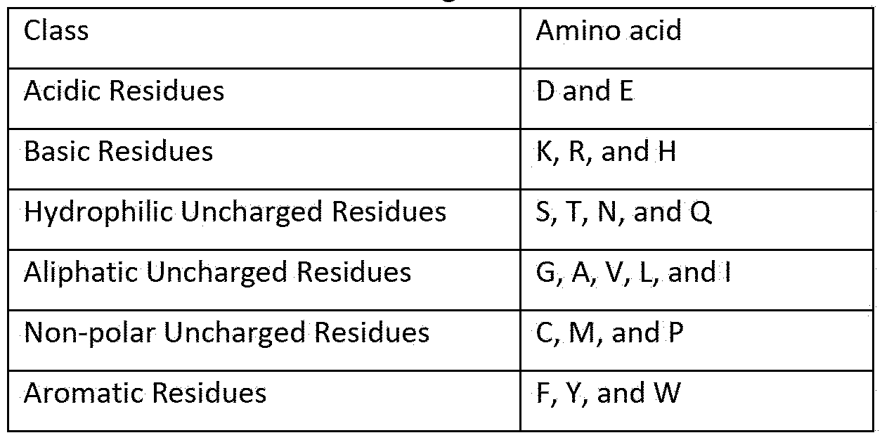
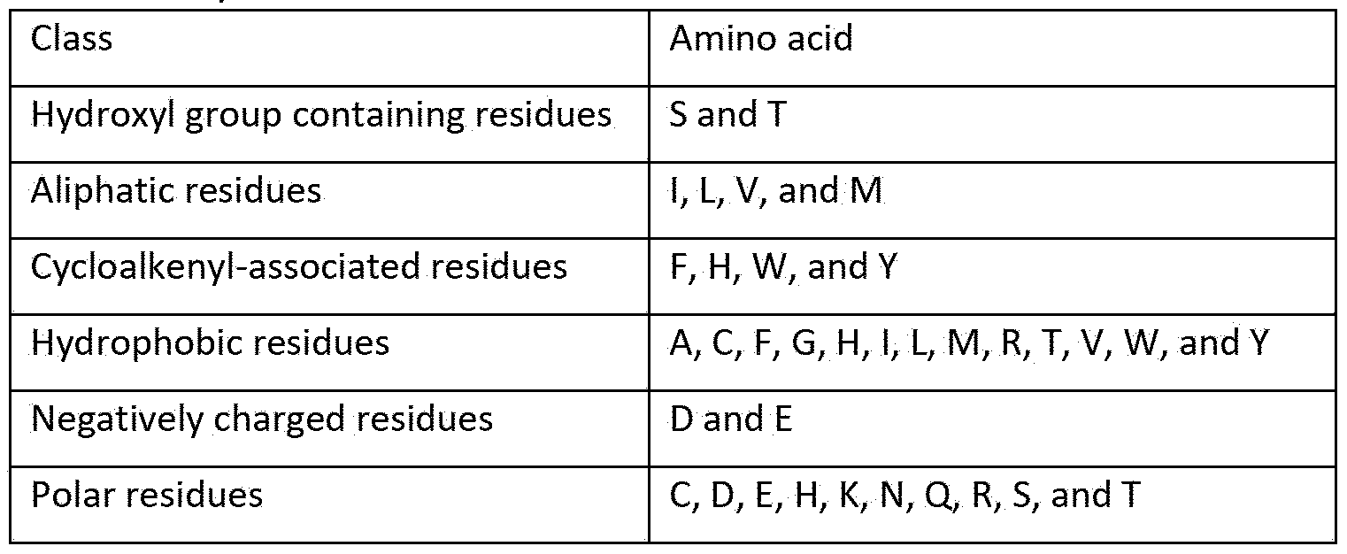

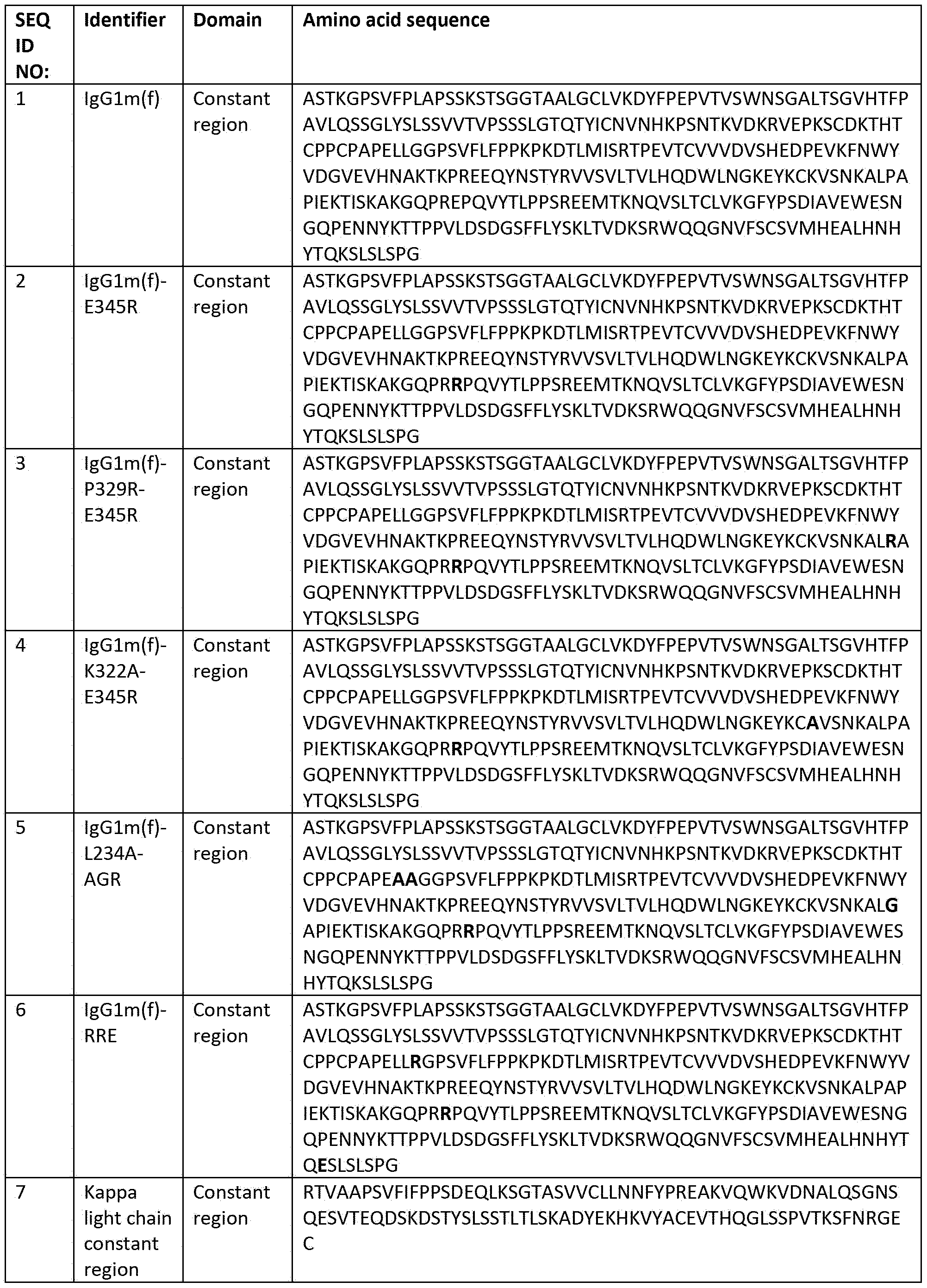
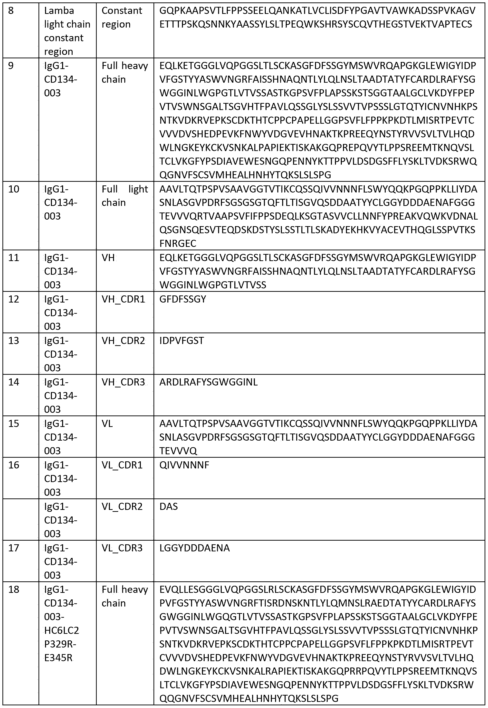


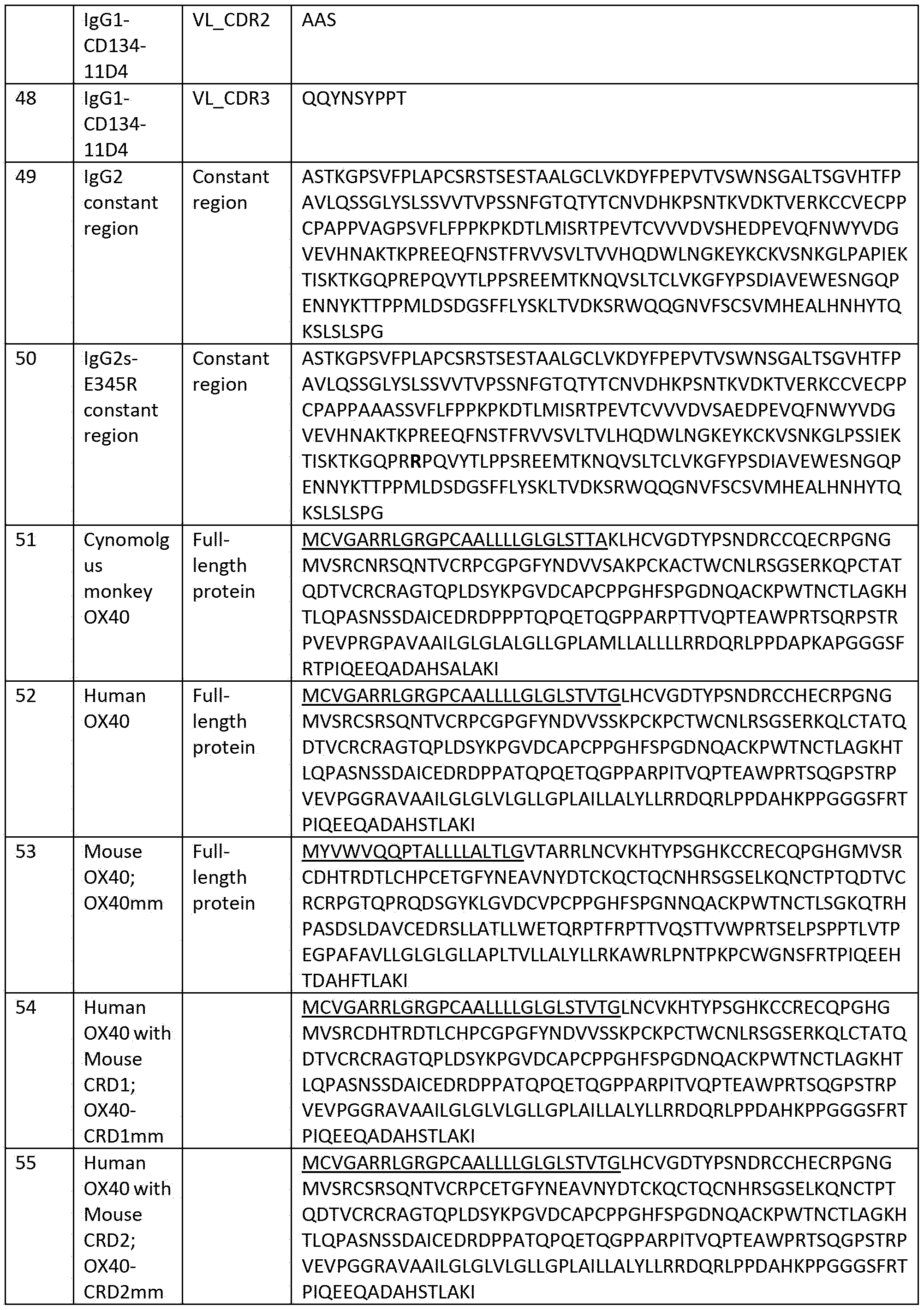
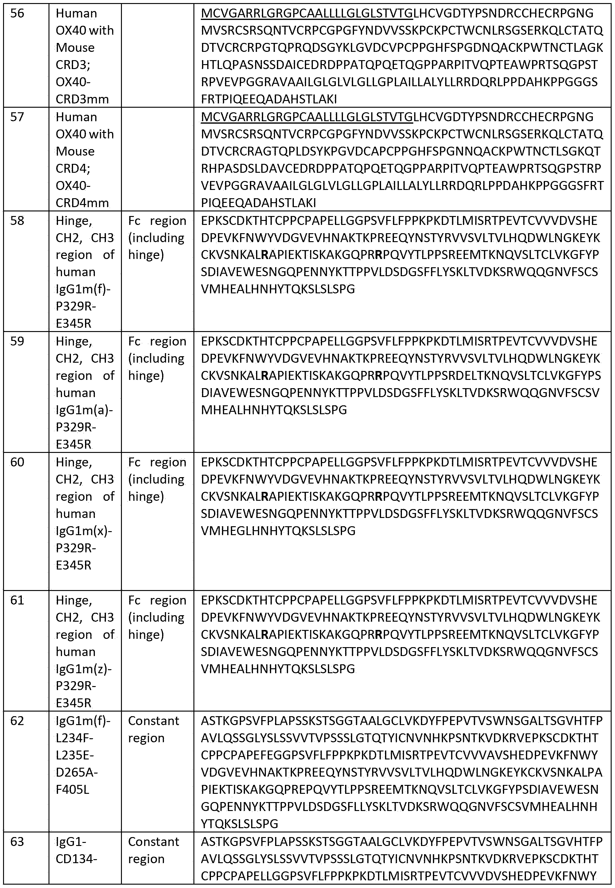


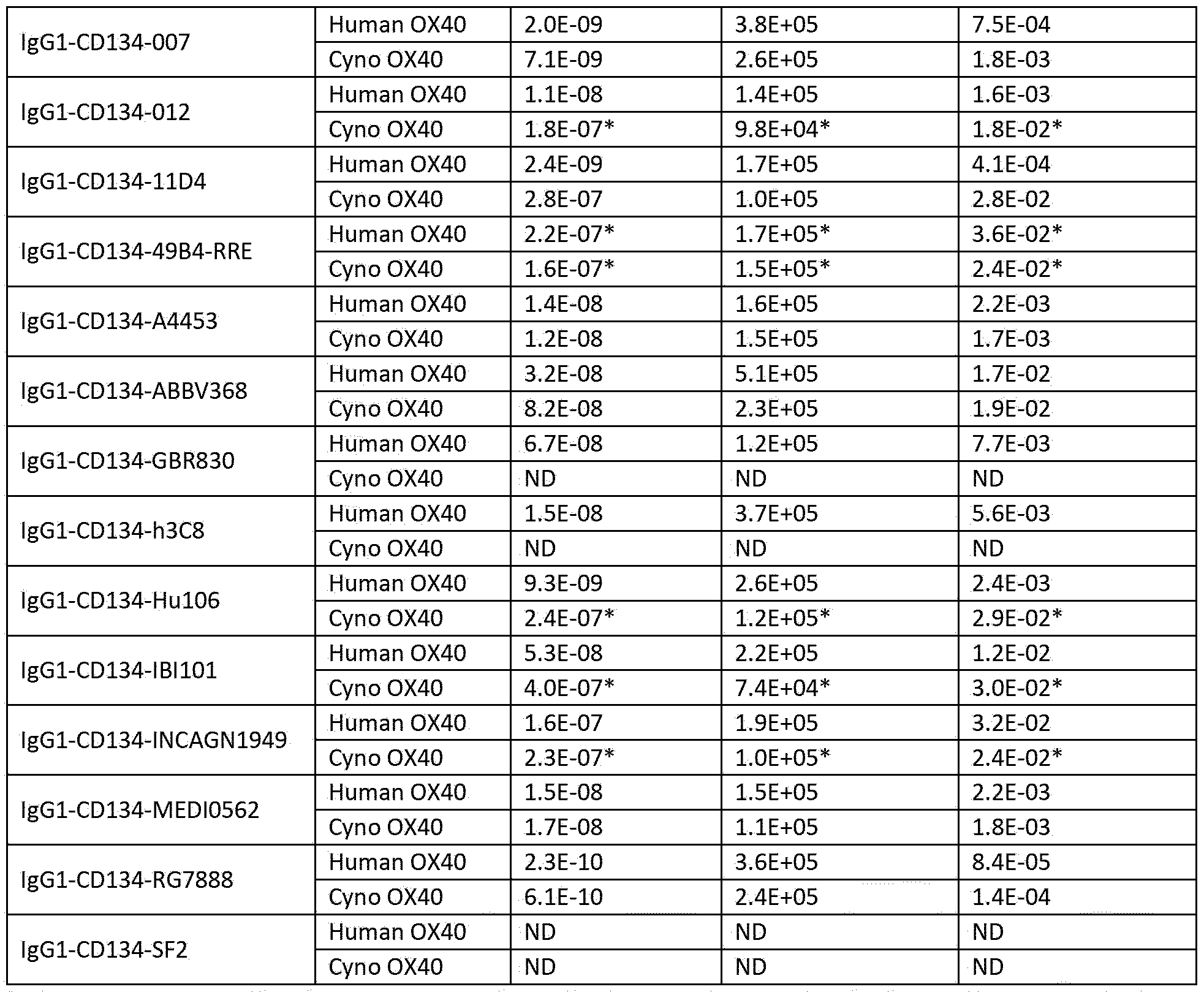


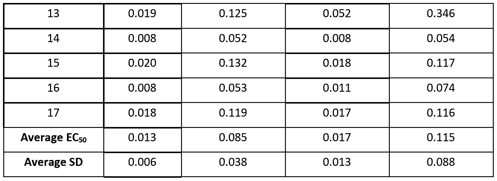


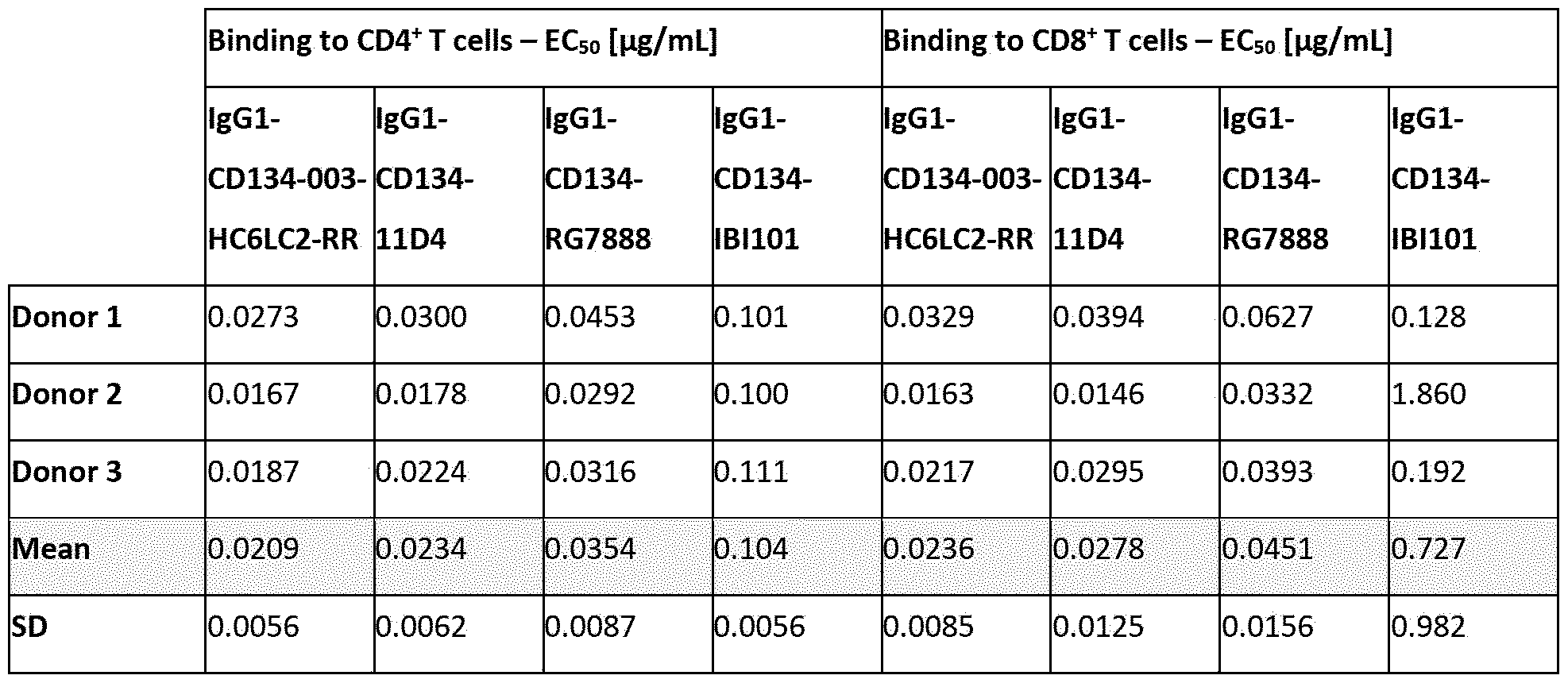

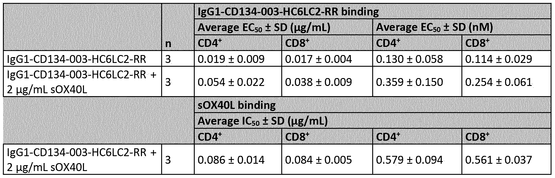

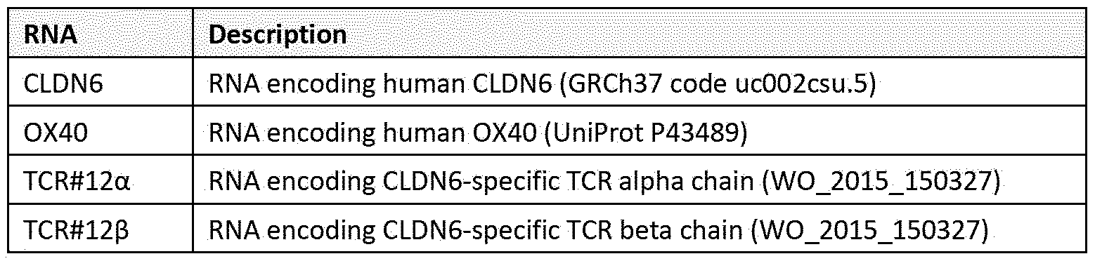

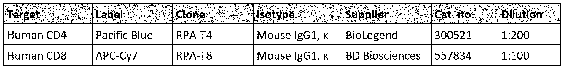

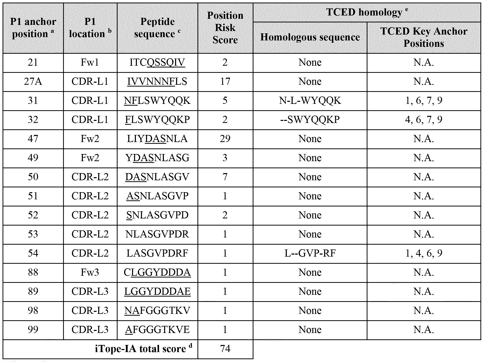
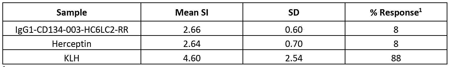

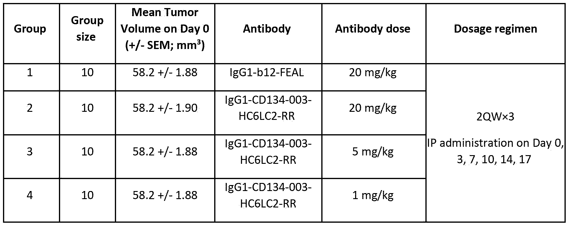
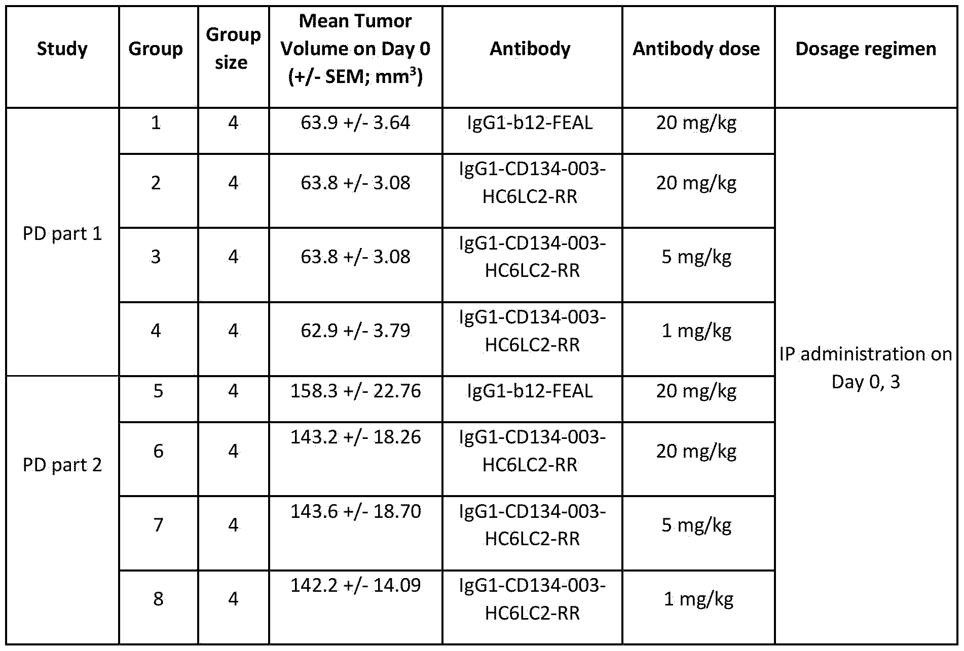

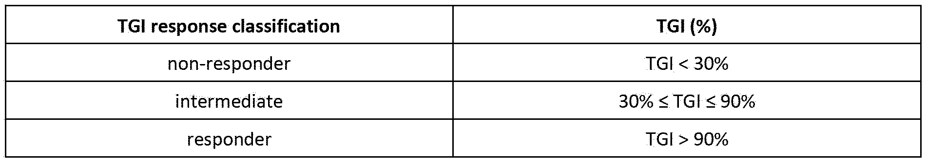
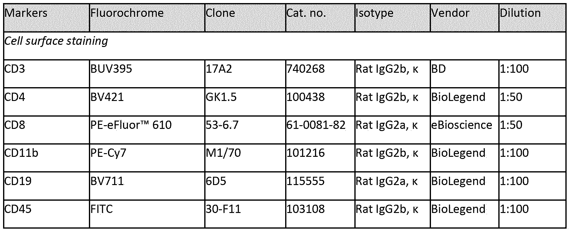
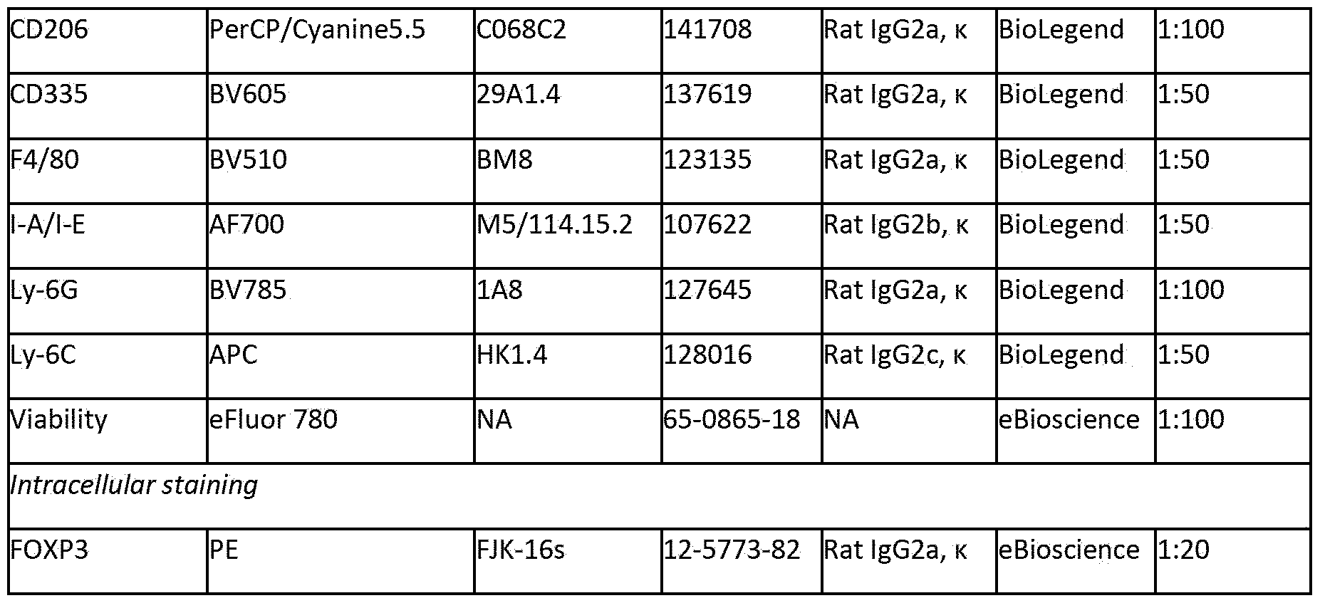


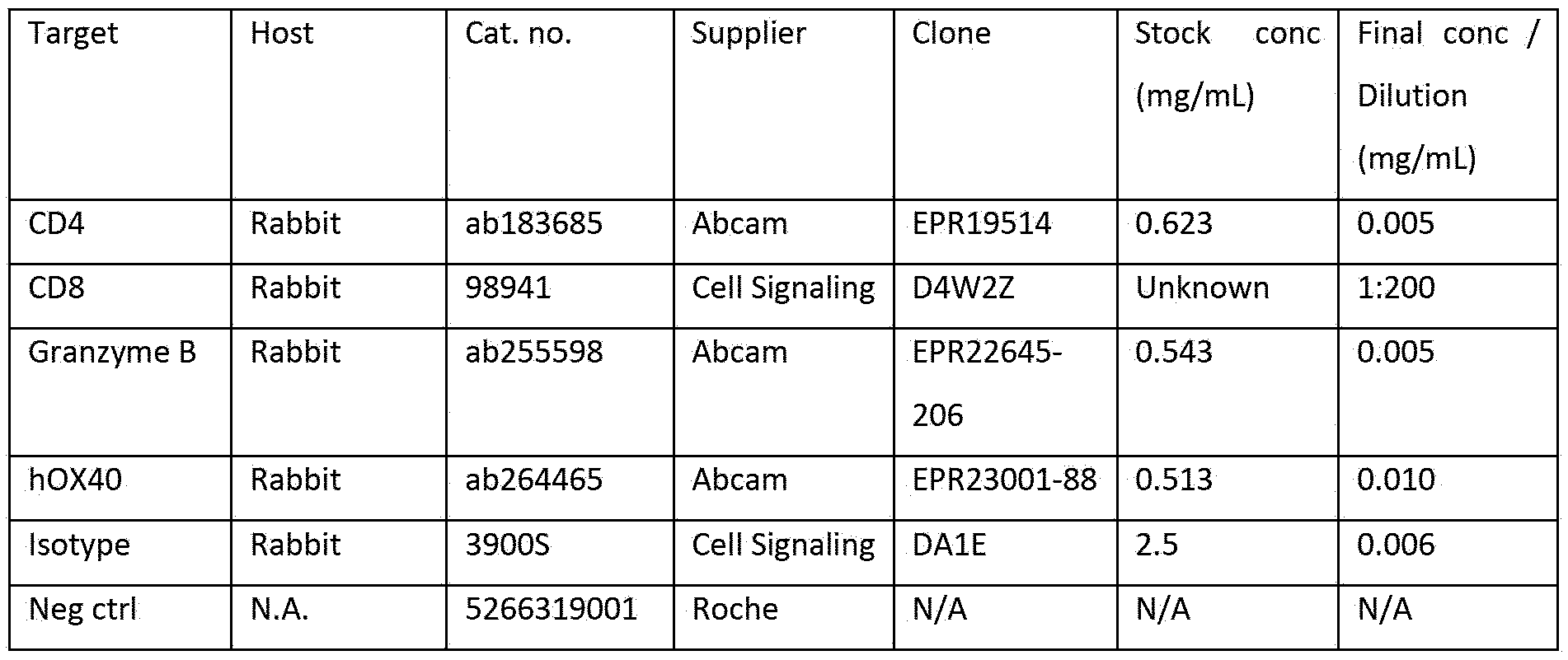

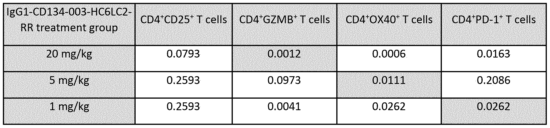
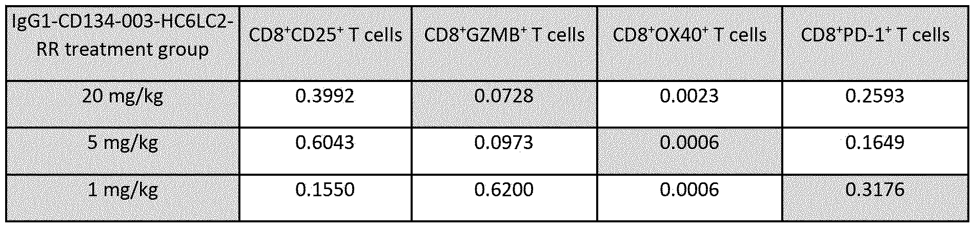
Claims
Priority Applications (3)
| Application Number | Priority Date | Filing Date | Title |
|---|---|---|---|
| CN202480031987.5A CN121285385A (en) | 2023-05-12 | 2024-05-10 | Antibodies capable of binding to OX40, variants thereof and uses thereof |
| AU2024271605A AU2024271605A1 (en) | 2023-05-12 | 2024-05-10 | Antibodies capable of binding to ox40, variants thereof and uses thereof |
| IL324457A IL324457A (en) | 2023-05-12 | 2025-11-04 | Antibodies capable of binding to ox40, variants thereof and uses thereof |
Applications Claiming Priority (4)
| Application Number | Priority Date | Filing Date | Title |
|---|---|---|---|
| EP23173143.1 | 2023-05-12 | ||
| EP23173143 | 2023-05-12 | ||
| EP23213531 | 2023-11-30 | ||
| EP23213531.9 | 2023-11-30 |
Publications (1)
| Publication Number | Publication Date |
|---|---|
| WO2024235862A1 true WO2024235862A1 (en) | 2024-11-21 |
Family
ID=91128429
Family Applications (1)
| Application Number | Title | Priority Date | Filing Date |
|---|---|---|---|
| PCT/EP2024/062962 Pending WO2024235862A1 (en) | 2023-05-12 | 2024-05-10 | Antibodies capable of binding to ox40, variants thereof and uses thereof |
Country Status (7)
| Country | Link |
|---|---|
| US (4) | US20240400703A1 (en) |
| CN (1) | CN121285385A (en) |
| AR (1) | AR132668A1 (en) |
| AU (1) | AU2024271605A1 (en) |
| IL (1) | IL324457A (en) |
| TW (1) | TW202509071A (en) |
| WO (1) | WO2024235862A1 (en) |
Families Citing this family (1)
| Publication number | Priority date | Publication date | Assignee | Title |
|---|---|---|---|---|
| CN121285385A (en) * | 2023-05-12 | 2026-01-06 | 金麦安博股份有限公司 | Antibodies capable of binding to OX40, variants thereof and uses thereof |
Citations (72)
| Publication number | Priority date | Publication date | Assignee | Title |
|---|---|---|---|---|
| US835A (en) | 1838-07-12 | X i i i x | ||
| US6077A (en) | 1849-01-30 | Improved hinged claw-wrench | ||
| US4699880A (en) | 1984-09-25 | 1987-10-13 | Immunomedics, Inc. | Method of producing monoclonal anti-idiotype antibody |
| WO1992022653A1 (en) | 1991-06-14 | 1992-12-23 | Genentech, Inc. | Method for making humanized antibodies |
| EP0629240A1 (en) | 1992-02-19 | 1994-12-21 | Scotgen Limited | Altered antibodies, products and processes relating thereto |
| US5589466A (en) | 1989-03-21 | 1996-12-31 | Vical Incorporated | Induction of a protective immune response in a mammal by injecting a DNA sequence |
| WO1998050431A2 (en) | 1997-05-02 | 1998-11-12 | Genentech, Inc. | A method for making multispecific antibodies having heteromultimeric and common components |
| US5973972A (en) | 1995-11-03 | 1999-10-26 | Samsung Electronics, Co., Ltd. | Precharge system for a semiconductor memory device |
| WO1999054342A1 (en) | 1998-04-20 | 1999-10-28 | Pablo Umana | Glycosylation engineering of antibodies for improving antibody-dependent cellular cytotoxicity |
| WO2000046147A2 (en) | 1999-02-03 | 2000-08-10 | Biosante Pharmaceuticals, Inc. | Therapeutic calcium phosphate particles and methods of manufacture and use |
| WO2000061739A1 (en) | 1999-04-09 | 2000-10-19 | Kyowa Hakko Kogyo Co., Ltd. | Method for controlling the activity of immunologically functional molecule |
| WO2000070087A1 (en) | 1999-05-14 | 2000-11-23 | Copernicus Therapeutics, Inc. | Automated nucleic acid compaction device |
| WO2002020039A2 (en) | 2000-09-04 | 2002-03-14 | Horst Lindhofer | Use of trifunctional bispecific and trispecific antibodies for the treatment of malignant ascites |
| WO2002030954A1 (en) | 2000-10-06 | 2002-04-18 | Kyowa Hakko Kogyo Co., Ltd. | Method of purifying antibody |
| WO2003035835A2 (en) | 2001-10-25 | 2003-05-01 | Genentech, Inc. | Glycoprotein compositions |
| US20030115614A1 (en) | 2000-10-06 | 2003-06-19 | Yutaka Kanda | Antibody composition-producing cell |
| WO2007059782A1 (en) | 2005-11-28 | 2007-05-31 | Genmab A/S | Recombinant monovalent antibodies and methods for production thereof |
| US7262028B2 (en) | 2002-07-18 | 2007-08-28 | Crucell Holland B.V. | Recombinant production of mixtures of antibodies |
| WO2007110205A2 (en) | 2006-03-24 | 2007-10-04 | Merck Patent Gmbh | Engineered heterodimeric protein domains |
| EP1870459A1 (en) | 2005-03-31 | 2007-12-26 | Chugai Seiyaku Kabushiki Kaisha | Methods for producing polypeptides by regulating polypeptide association |
| WO2008003116A2 (en) | 2006-07-05 | 2008-01-10 | F-Star Biotechnologische Forschungs- Und Entwicklungsges.M.B.H. | Method for engineering immunoglobulins |
| WO2008157379A2 (en) | 2007-06-21 | 2008-12-24 | Macrogenics, Inc. | Covalent diabodies and uses thereof |
| WO2009079335A1 (en) | 2007-12-14 | 2009-06-25 | Medarex, Inc. | Binding molecules to the human ox40 receptor |
| WO2009089004A1 (en) | 2008-01-07 | 2009-07-16 | Amgen Inc. | Method for making antibody fc-heterodimeric molecules using electrostatic steering effects |
| US7612181B2 (en) | 2005-08-19 | 2009-11-03 | Abbott Laboratories | Dual variable domain immunoglobulin and uses thereof |
| US20100155133A1 (en) | 2005-01-13 | 2010-06-24 | The Wiremold Company | Downward facing receptacle assembly for cable raceway |
| WO2010080538A1 (en) | 2008-12-19 | 2010-07-15 | Macrogenics, Inc. | Covalent diabodies and uses thereof |
| WO2010129304A2 (en) | 2009-04-27 | 2010-11-11 | Oncomed Pharmaceuticals, Inc. | Method for making heteromultimeric molecules |
| WO2010151792A1 (en) | 2009-06-26 | 2010-12-29 | Regeneron Pharmaceuticals, Inc. | Readily isolated bispecific antibodies with native immunoglobulin format |
| WO2011028952A1 (en) | 2009-09-02 | 2011-03-10 | Xencor, Inc. | Compositions and methods for simultaneous bivalent and monovalent co-engagement of antigens |
| US7951918B2 (en) | 2006-03-17 | 2011-05-31 | Biogen Idec Ma Inc. | Stabilized polypeptide compositions |
| WO2011069104A2 (en) | 2009-12-04 | 2011-06-09 | Genentech, Inc. | Multispecific antibodies, antibody analogs, compositions, and methods |
| WO2011117329A1 (en) | 2010-03-26 | 2011-09-29 | F. Hoffmann-La Roche Ag | Bispecific, bivalent anti-vegf/anti-ang-2 antibodies |
| WO2011131746A2 (en) | 2010-04-20 | 2011-10-27 | Genmab A/S | Heterodimeric antibody fc-containing proteins and methods for production thereof |
| WO2011143545A1 (en) | 2010-05-14 | 2011-11-17 | Rinat Neuroscience Corporation | Heterodimeric proteins and methods for producing and purifying them |
| CN102250246A (en) | 2011-06-10 | 2011-11-23 | 常州亚当生物技术有限公司 | Bispecific antibody to VEGF/PDGFR beta and application thereof |
| EP2391343A2 (en) | 2009-01-29 | 2011-12-07 | Alnylam Pharmaceuticals Inc. | Improved lipid formulation |
| WO2012023053A2 (en) | 2010-08-16 | 2012-02-23 | Novimmune S.A. | Methods for the generation of multispecific and multivalent antibodies |
| WO2012025530A1 (en) | 2010-08-24 | 2012-03-01 | F. Hoffmann-La Roche Ag | Bispecific antibodies comprising a disulfide stabilized - fv fragment |
| WO2012025525A1 (en) | 2010-08-24 | 2012-03-01 | Roche Glycart Ag | Activatable bispecific antibodies |
| WO2012058768A1 (en) | 2010-11-05 | 2012-05-10 | Zymeworks Inc. | Stable heterodimeric antibody design with mutations in the fc domain |
| WO2013004842A2 (en) | 2011-07-06 | 2013-01-10 | Genmab A/S | Antibody variants and uses thereof |
| WO2013157953A1 (en) | 2012-04-20 | 2013-10-24 | Merus B.V. | Methods and means for the production of ig-like molecules |
| WO2014031646A2 (en) | 2012-08-20 | 2014-02-27 | Gliknik Inc. | Molecules with antigen binding and polyvalent fc gamma receptor binding activity |
| WO2014108198A1 (en) | 2013-01-10 | 2014-07-17 | Genmab B.V. | Human igg1 fc region variants and uses thereof |
| WO2014152774A1 (en) | 2013-03-14 | 2014-09-25 | Shire Human Genetic Therapies, Inc. | Methods and compositions for delivering mrna coded antibodies |
| US20140348839A1 (en) | 2011-12-20 | 2014-11-27 | Medimmune, Llc | Modified polypeptides for bispecific antibody scaffolds |
| US20140377284A1 (en) | 2013-03-18 | 2014-12-25 | Janssen Pharmaceuticals, Inc. | Humanized anti-cd134 (ox40) antibodies and uses thereof |
| US9006399B2 (en) | 2010-08-23 | 2015-04-14 | Board Of Regents, The University Of Texas System | Anti-OX40 antibodies and methods of using the same |
| WO2015158867A1 (en) | 2014-04-16 | 2015-10-22 | Ucb Biopharma Sprl | Multimeric fc proteins |
| US9221891B2 (en) | 2012-04-02 | 2015-12-29 | Moderna Therapeutics, Inc. | In vivo production of proteins |
| EP2972360A1 (en) | 2013-03-15 | 2016-01-20 | Shire Human Genetic Therapies, Inc. | Synergistic enhancement of the delivery of nucleic acids via blended formulations |
| US20160185840A1 (en) | 2007-01-09 | 2016-06-30 | Curevac Ag | Rna-coded antibody |
| WO2016110584A1 (en) | 2015-01-08 | 2016-07-14 | Biontech Ag | Agonistic tnf receptor binding agents |
| WO2016164480A1 (en) | 2015-04-07 | 2016-10-13 | Genentech, Inc. | Antigen binding complex having agonistic activity and methods of use |
| WO2017019805A1 (en) | 2015-07-30 | 2017-02-02 | Massachusetts Institute Of Technology | Systems and methods for a short wave infrared device |
| WO2018006052A1 (en) | 2016-06-30 | 2018-01-04 | Protiva Biotherapeutics, Inc. | Compositions and methods for delivering messenger rna |
| WO2018031258A1 (en) | 2016-08-12 | 2018-02-15 | Janssen Biotech, Inc. | Engineered antibodies and other fc-domain containing molecules with enhanced agonism and effector functions |
| WO2018083126A1 (en) | 2016-11-01 | 2018-05-11 | Genmab B.V. | Polypeptide variants and uses thereof |
| US20180170866A1 (en) | 2016-12-21 | 2018-06-21 | Arcturus Therapeutics, Inc. | Ionizable cationic lipid for rna delivery |
| WO2018114877A1 (en) | 2016-12-21 | 2018-06-28 | F. Hoffmann-La Roche Ag | In vitro glycoengineering of antibodies |
| WO2018114878A1 (en) | 2016-12-21 | 2018-06-28 | F. Hoffmann-La Roche Ag | Re-use of enzymes in in vitro glycoengineering of antibodies |
| WO2018114879A1 (en) | 2016-12-21 | 2018-06-28 | F. Hoffmann-La Roche Ag | Method for in vitro glycoengineering of antibodies |
| WO2018146317A1 (en) | 2017-02-10 | 2018-08-16 | Genmab B.V. | Polypeptide variants and uses thereof |
| US20190022247A1 (en) | 2015-12-30 | 2019-01-24 | Acuitas Therapeutics, Inc. | Lipids and lipid nanoparticle formulations for delivery of nucleic acids |
| US10195156B2 (en) | 2015-12-22 | 2019-02-05 | Modernatx, Inc. | Compounds and compositions for intracellular delivery of agents |
| US10259882B2 (en) | 2015-05-07 | 2019-04-16 | Agenus Inc. | Anti-OX40 antibodies |
| WO2019086497A2 (en) | 2017-11-01 | 2019-05-09 | F. Hoffmann-La Roche Ag | Combination therapy with targeted ox40 agonists |
| US10449233B2 (en) | 2015-10-01 | 2019-10-22 | Heat Biologics, Inc. | Compositions and methods for adjoining type I and type II extracellular domains as heterologous chimeric proteins |
| WO2019223733A1 (en) | 2018-05-23 | 2019-11-28 | Beigene, Ltd. | Anti-ox40 antibodies and methods of use |
| WO2020030570A1 (en) | 2018-08-06 | 2020-02-13 | Glaxosmithkline Intellectual Property Development Limited | Combinations of an ox40 antibody and a tlr4 modulator and uses thereof |
| WO2023031473A1 (en) * | 2021-09-06 | 2023-03-09 | Genmab B.V. | Antibodies capable of binding to cd27, variants thereof and uses thereof |
Family Cites Families (61)
| Publication number | Priority date | Publication date | Assignee | Title |
|---|---|---|---|---|
| US6077835A (en) | 1994-03-23 | 2000-06-20 | Case Western Reserve University | Delivery of compacted nucleic acid to cells |
| US6808710B1 (en) | 1999-08-23 | 2004-10-26 | Genetics Institute, Inc. | Downmodulating an immune response with multivalent antibodies to PD-1 |
| CA2388245C (en) | 1999-10-19 | 2012-01-10 | Tatsuya Ogawa | The use of serum-free adapted rat cells for producing heterologous polypeptides |
| HU231090B1 (en) | 2000-10-06 | 2020-07-28 | Kyowa Kirin Co., Ltd. | Cells producing antibody compositions |
| EP1456652A4 (en) | 2001-11-13 | 2005-11-02 | Dana Farber Cancer Inst Inc | AGENTS MODULATING IMMUNE CELL ACTIVITY AND METHODS OF USE THEREOF |
| IL149820A0 (en) | 2002-05-23 | 2002-11-10 | Curetech Ltd | Humanized immunomodulatory monoclonal antibodies for the treatment of neoplastic disease or immunodeficiency |
| JP4511943B2 (en) | 2002-12-23 | 2010-07-28 | ワイス エルエルシー | Antibody against PD-1 and use thereof |
| CA2586909A1 (en) | 2004-11-12 | 2006-12-14 | Seattle Genetics, Inc. | Auristatins having an aminobenzoic acid unit at the n terminus |
| PL2161336T5 (en) | 2005-05-09 | 2017-10-31 | Ono Pharmaceutical Co | Human monoclonal antibodies to programmed death 1(PD-1) and methods for treating cancer using anti-PD-1 antibodies alone or in combination with other immunotherapeutics |
| CA2691357C (en) | 2007-06-18 | 2014-09-23 | N.V. Organon | Antibodies to human programmed death receptor pd-1 |
| US20090028857A1 (en) | 2007-07-23 | 2009-01-29 | Cell Genesys, Inc. | Pd-1 antibodies in combination with a cytokine-secreting cell and methods of use thereof |
| EP2050764A1 (en) | 2007-10-15 | 2009-04-22 | sanofi-aventis | Novel polyvalent bispecific antibody format and uses thereof |
| HUE034465T2 (en) | 2008-02-11 | 2018-02-28 | Cure Tech Ltd | Monoclonal antibodies for tumor treatment |
| US8168757B2 (en) | 2008-03-12 | 2012-05-01 | Merck Sharp & Dohme Corp. | PD-1 binding proteins |
| EP2328919A2 (en) | 2008-08-25 | 2011-06-08 | Amplimmune, Inc. | Pd-i antagonists and methods for treating infectious disease |
| JP5397668B2 (en) | 2008-09-02 | 2014-01-22 | ソニー株式会社 | Storage element and storage device |
| CA2998281C (en) | 2008-09-26 | 2022-08-16 | Dana-Farber Cancer Institute, Inc. | Human anti-pd-1 antobodies and uses therefor |
| ES2629337T3 (en) | 2009-02-09 | 2017-08-08 | Inserm - Institut National De La Santé Et De La Recherche Médicale | Antibodies against PD-1 and antibodies against PD-L1 and uses thereof |
| US20130017199A1 (en) | 2009-11-24 | 2013-01-17 | AMPLIMMUNE ,Inc. a corporation | Simultaneous inhibition of pd-l1/pd-l2 |
| WO2011082400A2 (en) | 2010-01-04 | 2011-07-07 | President And Fellows Of Harvard College | Modulators of immunoinhibitory receptor pd-1, and methods of use thereof |
| CA2802344C (en) | 2010-06-18 | 2023-06-13 | The Brigham And Women's Hospital, Inc. | Bi-specific antibodies against tim-3 and pd-1 for immunotherapy in chronic immune conditions |
| US8907053B2 (en) | 2010-06-25 | 2014-12-09 | Aurigene Discovery Technologies Limited | Immunosuppression modulating compounds |
| US8911734B2 (en) * | 2010-12-01 | 2014-12-16 | Alderbio Holdings Llc | Methods of preventing or treating pain using anti-NGF antibodies that selectively inhibit the association of NGF with TrkA, without affecting the association of NGF with p75 |
| PT2699264T (en) | 2011-04-20 | 2018-05-23 | Medimmune Llc | ANTIBODIES AND OTHER MOLECULES CONNECTING B7-H1 AND PD-1 |
| SG10201700698WA (en) | 2012-05-15 | 2017-02-27 | Bristol Myers Squibb Co | Cancer immunotherapy by disrupting pd-1/pd-l1 signaling |
| US20150337037A1 (en) * | 2012-06-05 | 2015-11-26 | Msm Protein Technologies | Human monoclonal antibodies against human chemokine receptor ccr6 |
| EP3263601B1 (en) | 2012-10-02 | 2021-11-24 | Bristol-Myers Squibb Company | Combination of anti-kir antibodies and anti-pd-1 antibodies to treat cancer |
| HUE053069T2 (en) | 2013-05-02 | 2021-06-28 | Anaptysbio Inc | Antibodies to programmed death-1 (PD-1) |
| CA2913977C (en) | 2013-05-31 | 2022-11-29 | Sorrento Therapeutics, Inc. | Antigen binding proteins that bind pd-1 |
| CN104250302B (en) | 2013-06-26 | 2017-11-14 | 上海君实生物医药科技股份有限公司 | The anti-antibody of PD 1 and its application |
| PT3702373T (en) | 2013-09-13 | 2022-09-27 | Beigene Switzerland Gmbh | Anti-pd1 antibodies and their use as therapeutics and diagnostics |
| SG10201804945WA (en) | 2013-12-12 | 2018-07-30 | Shanghai hengrui pharmaceutical co ltd | Pd-1 antibody, antigen-binding fragment thereof, and medical application thereof |
| TWI681969B (en) | 2014-01-23 | 2020-01-11 | 美商再生元醫藥公司 | Human antibodies to pd-1 |
| JOP20200094A1 (en) | 2014-01-24 | 2017-06-16 | Dana Farber Cancer Inst Inc | Antibody molecules to pd-1 and uses thereof |
| CN105330740B (en) | 2014-07-30 | 2018-08-17 | 珠海市丽珠单抗生物技术有限公司 | Anti- PD-1 antibody and its application |
| GB201413718D0 (en) | 2014-08-01 | 2014-09-17 | Olink Ab | Method for selecting a target nucleic acid sequence |
| US20170000885A1 (en) | 2015-06-08 | 2017-01-05 | Genentech, Inc. | Methods of treating cancer using anti-ox40 antibodies and pd-1 axis binding antagonists |
| SG10201906059VA (en) | 2015-07-30 | 2019-08-27 | Macrogenics Inc | Pd-1-binding molecules and methods of use thereof |
| US20180222989A1 (en) | 2015-08-04 | 2018-08-09 | Glaxosmithkline Intellectual Property Development Limited | Combination treatments and uses and methods thereof |
| WO2017024465A1 (en) | 2015-08-10 | 2017-02-16 | Innovent Biologics (Suzhou) Co., Ltd. | Pd-1 antibodies |
| DK3334763T3 (en) | 2015-08-11 | 2024-10-07 | Wuxi Biologics Ireland Ltd | Novel anti-PD-1 antibodies |
| WO2017040790A1 (en) | 2015-09-01 | 2017-03-09 | Agenus Inc. | Anti-pd-1 antibodies and methods of use thereof |
| CA3000564A1 (en) | 2015-10-02 | 2017-04-06 | Symphogen A/S | Anti-pd-1 antibodies and compositions |
| CN106632674B (en) | 2015-10-30 | 2018-11-16 | 泽达生物医药有限公司 | A kind of anti-PD-1 monoclonal antibody, its medical composition and its use |
| SG11201803956UA (en) * | 2015-12-01 | 2018-06-28 | Genmab Bv | Anti-dr5 antibodies and methods of use thereof |
| EP4424322A3 (en) | 2015-12-17 | 2025-04-16 | Novartis AG | Antibody molecules to pd-1 and uses thereof |
| CN111491362B (en) | 2016-02-02 | 2023-10-24 | 华为技术有限公司 | Methods, user equipment and base stations for determining transmit power |
| WO2017132827A1 (en) | 2016-02-02 | 2017-08-10 | Innovent Biologics (Suzhou) Co., Ltd. | Pd-1 antibodies |
| CN107286242B (en) | 2016-04-01 | 2019-03-22 | 中山康方生物医药有限公司 | Anti-PD-1 monoclonal antibody |
| EP3458478B1 (en) | 2016-05-18 | 2021-01-06 | Boehringer Ingelheim International GmbH | Anti pd-1 and anti-lag3 antibodies for cancer treatment |
| WO2018022831A1 (en) | 2016-07-28 | 2018-02-01 | Musc Foundation For Research Development | Methods and compositions for the treatment of cancer combining an anti-smic antibody and immune checkpoint inhibitors |
| CN106977602B (en) | 2016-08-23 | 2018-09-25 | 中山康方生物医药有限公司 | A kind of anti-PD1 monoclonal antibodies, its medical composition and its use |
| CN107840887B (en) | 2016-09-21 | 2022-03-25 | 基石药业(苏州)有限公司 | A new PD-1 monoclonal antibody |
| CN107077506A (en) | 2016-12-07 | 2017-08-18 | 深圳市大疆创新科技有限公司 | Control method of unmanned aerial vehicle and unmanned aerial vehicle |
| CN107058315B (en) | 2016-12-08 | 2019-11-08 | 上海优卡迪生物医药科技有限公司 | Knockdown of human PD-1 siRNA, recombinant expression CAR-T vector and its construction method and application |
| US10537637B2 (en) | 2017-01-05 | 2020-01-21 | Gensun Biopharma Inc. | Checkpoint regulator antagonists |
| MX2019012038A (en) | 2017-05-30 | 2019-11-18 | Bristol Myers Squibb Co | Compositions comprising a combination of an anti-lag-3 antibody, a pd-1 pathway inhibitor, and an immunotherapeutic agent. |
| WO2019000146A1 (en) | 2017-06-26 | 2019-01-03 | 深圳市博奥康生物科技有限公司 | Sirna of human programmed cell death receptor 1 and use thereof |
| MX2022006147A (en) | 2019-11-21 | 2022-06-17 | Beigene Ltd | Methods of cancer treatment using anti-ox40 antibodies in combination with anti-pd1 or anti-pdl1 antibodies. |
| WO2023217987A1 (en) | 2022-05-12 | 2023-11-16 | BioNTech SE | Monoclonal antibodies directed against programmed death-1 protein and their use in medicine |
| CN121285385A (en) * | 2023-05-12 | 2026-01-06 | 金麦安博股份有限公司 | Antibodies capable of binding to OX40, variants thereof and uses thereof |
-
2024
- 2024-05-10 CN CN202480031987.5A patent/CN121285385A/en active Pending
- 2024-05-10 US US18/660,847 patent/US20240400703A1/en not_active Abandoned
- 2024-05-10 WO PCT/EP2024/062962 patent/WO2024235862A1/en active Pending
- 2024-05-10 AR ARP240101205A patent/AR132668A1/en unknown
- 2024-05-10 TW TW113117396A patent/TW202509071A/en unknown
- 2024-05-10 US US18/660,672 patent/US12410258B2/en active Active
- 2024-05-10 AU AU2024271605A patent/AU2024271605A1/en active Pending
-
2025
- 2025-02-25 US US19/062,474 patent/US20250333527A1/en active Pending
- 2025-02-25 US US19/062,470 patent/US20250333526A1/en active Pending
- 2025-11-04 IL IL324457A patent/IL324457A/en unknown
Patent Citations (77)
| Publication number | Priority date | Publication date | Assignee | Title |
|---|---|---|---|---|
| US6077A (en) | 1849-01-30 | Improved hinged claw-wrench | ||
| US835A (en) | 1838-07-12 | X i i i x | ||
| US4699880A (en) | 1984-09-25 | 1987-10-13 | Immunomedics, Inc. | Method of producing monoclonal anti-idiotype antibody |
| US5589466A (en) | 1989-03-21 | 1996-12-31 | Vical Incorporated | Induction of a protective immune response in a mammal by injecting a DNA sequence |
| WO1992022653A1 (en) | 1991-06-14 | 1992-12-23 | Genentech, Inc. | Method for making humanized antibodies |
| EP0629240A1 (en) | 1992-02-19 | 1994-12-21 | Scotgen Limited | Altered antibodies, products and processes relating thereto |
| US5973972A (en) | 1995-11-03 | 1999-10-26 | Samsung Electronics, Co., Ltd. | Precharge system for a semiconductor memory device |
| WO1998050431A2 (en) | 1997-05-02 | 1998-11-12 | Genentech, Inc. | A method for making multispecific antibodies having heteromultimeric and common components |
| US6602684B1 (en) | 1998-04-20 | 2003-08-05 | Glycart Biotechnology Ag | Glycosylation engineering of antibodies for improving antibody-dependent cellular cytotoxicity |
| WO1999054342A1 (en) | 1998-04-20 | 1999-10-28 | Pablo Umana | Glycosylation engineering of antibodies for improving antibody-dependent cellular cytotoxicity |
| WO2000046147A2 (en) | 1999-02-03 | 2000-08-10 | Biosante Pharmaceuticals, Inc. | Therapeutic calcium phosphate particles and methods of manufacture and use |
| EP1176195A1 (en) | 1999-04-09 | 2002-01-30 | Kyowa Hakko Kogyo Co., Ltd. | Method for controlling the activity of immunologically functional molecule |
| WO2000061739A1 (en) | 1999-04-09 | 2000-10-19 | Kyowa Hakko Kogyo Co., Ltd. | Method for controlling the activity of immunologically functional molecule |
| WO2000070087A1 (en) | 1999-05-14 | 2000-11-23 | Copernicus Therapeutics, Inc. | Automated nucleic acid compaction device |
| WO2002020039A2 (en) | 2000-09-04 | 2002-03-14 | Horst Lindhofer | Use of trifunctional bispecific and trispecific antibodies for the treatment of malignant ascites |
| WO2002030954A1 (en) | 2000-10-06 | 2002-04-18 | Kyowa Hakko Kogyo Co., Ltd. | Method of purifying antibody |
| US20030115614A1 (en) | 2000-10-06 | 2003-06-19 | Yutaka Kanda | Antibody composition-producing cell |
| WO2003035835A2 (en) | 2001-10-25 | 2003-05-01 | Genentech, Inc. | Glycoprotein compositions |
| US7262028B2 (en) | 2002-07-18 | 2007-08-28 | Crucell Holland B.V. | Recombinant production of mixtures of antibodies |
| US20100155133A1 (en) | 2005-01-13 | 2010-06-24 | The Wiremold Company | Downward facing receptacle assembly for cable raceway |
| EP1870459A1 (en) | 2005-03-31 | 2007-12-26 | Chugai Seiyaku Kabushiki Kaisha | Methods for producing polypeptides by regulating polypeptide association |
| US7612181B2 (en) | 2005-08-19 | 2009-11-03 | Abbott Laboratories | Dual variable domain immunoglobulin and uses thereof |
| WO2007059782A1 (en) | 2005-11-28 | 2007-05-31 | Genmab A/S | Recombinant monovalent antibodies and methods for production thereof |
| US7951918B2 (en) | 2006-03-17 | 2011-05-31 | Biogen Idec Ma Inc. | Stabilized polypeptide compositions |
| WO2007110205A2 (en) | 2006-03-24 | 2007-10-04 | Merck Patent Gmbh | Engineered heterodimeric protein domains |
| WO2008003116A2 (en) | 2006-07-05 | 2008-01-10 | F-Star Biotechnologische Forschungs- Und Entwicklungsges.M.B.H. | Method for engineering immunoglobulins |
| EP3118224A1 (en) | 2007-01-09 | 2017-01-18 | CureVac AG | Rna-coded antibody |
| US20160185840A1 (en) | 2007-01-09 | 2016-06-30 | Curevac Ag | Rna-coded antibody |
| WO2008157379A2 (en) | 2007-06-21 | 2008-12-24 | Macrogenics, Inc. | Covalent diabodies and uses thereof |
| WO2009079335A1 (en) | 2007-12-14 | 2009-06-25 | Medarex, Inc. | Binding molecules to the human ox40 receptor |
| WO2009089004A1 (en) | 2008-01-07 | 2009-07-16 | Amgen Inc. | Method for making antibody fc-heterodimeric molecules using electrostatic steering effects |
| WO2010080538A1 (en) | 2008-12-19 | 2010-07-15 | Macrogenics, Inc. | Covalent diabodies and uses thereof |
| EP2391343A2 (en) | 2009-01-29 | 2011-12-07 | Alnylam Pharmaceuticals Inc. | Improved lipid formulation |
| WO2010129304A2 (en) | 2009-04-27 | 2010-11-11 | Oncomed Pharmaceuticals, Inc. | Method for making heteromultimeric molecules |
| WO2010151792A1 (en) | 2009-06-26 | 2010-12-29 | Regeneron Pharmaceuticals, Inc. | Readily isolated bispecific antibodies with native immunoglobulin format |
| WO2011028952A1 (en) | 2009-09-02 | 2011-03-10 | Xencor, Inc. | Compositions and methods for simultaneous bivalent and monovalent co-engagement of antigens |
| WO2011069104A2 (en) | 2009-12-04 | 2011-06-09 | Genentech, Inc. | Multispecific antibodies, antibody analogs, compositions, and methods |
| WO2011117329A1 (en) | 2010-03-26 | 2011-09-29 | F. Hoffmann-La Roche Ag | Bispecific, bivalent anti-vegf/anti-ang-2 antibodies |
| WO2011131746A2 (en) | 2010-04-20 | 2011-10-27 | Genmab A/S | Heterodimeric antibody fc-containing proteins and methods for production thereof |
| WO2011143545A1 (en) | 2010-05-14 | 2011-11-17 | Rinat Neuroscience Corporation | Heterodimeric proteins and methods for producing and purifying them |
| WO2012023053A2 (en) | 2010-08-16 | 2012-02-23 | Novimmune S.A. | Methods for the generation of multispecific and multivalent antibodies |
| US9006399B2 (en) | 2010-08-23 | 2015-04-14 | Board Of Regents, The University Of Texas System | Anti-OX40 antibodies and methods of using the same |
| WO2012025530A1 (en) | 2010-08-24 | 2012-03-01 | F. Hoffmann-La Roche Ag | Bispecific antibodies comprising a disulfide stabilized - fv fragment |
| WO2012025525A1 (en) | 2010-08-24 | 2012-03-01 | Roche Glycart Ag | Activatable bispecific antibodies |
| WO2012058768A1 (en) | 2010-11-05 | 2012-05-10 | Zymeworks Inc. | Stable heterodimeric antibody design with mutations in the fc domain |
| CN102250246A (en) | 2011-06-10 | 2011-11-23 | 常州亚当生物技术有限公司 | Bispecific antibody to VEGF/PDGFR beta and application thereof |
| WO2013004842A2 (en) | 2011-07-06 | 2013-01-10 | Genmab A/S | Antibody variants and uses thereof |
| US20140348839A1 (en) | 2011-12-20 | 2014-11-27 | Medimmune, Llc | Modified polypeptides for bispecific antibody scaffolds |
| US9254311B2 (en) | 2012-04-02 | 2016-02-09 | Moderna Therapeutics, Inc. | Modified polynucleotides for the production of proteins |
| US9221891B2 (en) | 2012-04-02 | 2015-12-29 | Moderna Therapeutics, Inc. | In vivo production of proteins |
| WO2013157953A1 (en) | 2012-04-20 | 2013-10-24 | Merus B.V. | Methods and means for the production of ig-like molecules |
| WO2014031646A2 (en) | 2012-08-20 | 2014-02-27 | Gliknik Inc. | Molecules with antigen binding and polyvalent fc gamma receptor binding activity |
| WO2014108198A1 (en) | 2013-01-10 | 2014-07-17 | Genmab B.V. | Human igg1 fc region variants and uses thereof |
| WO2014152774A1 (en) | 2013-03-14 | 2014-09-25 | Shire Human Genetic Therapies, Inc. | Methods and compositions for delivering mrna coded antibodies |
| EP2972360A1 (en) | 2013-03-15 | 2016-01-20 | Shire Human Genetic Therapies, Inc. | Synergistic enhancement of the delivery of nucleic acids via blended formulations |
| US20140377284A1 (en) | 2013-03-18 | 2014-12-25 | Janssen Pharmaceuticals, Inc. | Humanized anti-cd134 (ox40) antibodies and uses thereof |
| WO2015158867A1 (en) | 2014-04-16 | 2015-10-22 | Ucb Biopharma Sprl | Multimeric fc proteins |
| WO2016110584A1 (en) | 2015-01-08 | 2016-07-14 | Biontech Ag | Agonistic tnf receptor binding agents |
| WO2016164480A1 (en) | 2015-04-07 | 2016-10-13 | Genentech, Inc. | Antigen binding complex having agonistic activity and methods of use |
| US10259882B2 (en) | 2015-05-07 | 2019-04-16 | Agenus Inc. | Anti-OX40 antibodies |
| WO2017019805A1 (en) | 2015-07-30 | 2017-02-02 | Massachusetts Institute Of Technology | Systems and methods for a short wave infrared device |
| US10449233B2 (en) | 2015-10-01 | 2019-10-22 | Heat Biologics, Inc. | Compositions and methods for adjoining type I and type II extracellular domains as heterologous chimeric proteins |
| US10195156B2 (en) | 2015-12-22 | 2019-02-05 | Modernatx, Inc. | Compounds and compositions for intracellular delivery of agents |
| US20190022247A1 (en) | 2015-12-30 | 2019-01-24 | Acuitas Therapeutics, Inc. | Lipids and lipid nanoparticle formulations for delivery of nucleic acids |
| WO2018006052A1 (en) | 2016-06-30 | 2018-01-04 | Protiva Biotherapeutics, Inc. | Compositions and methods for delivering messenger rna |
| WO2018031258A1 (en) | 2016-08-12 | 2018-02-15 | Janssen Biotech, Inc. | Engineered antibodies and other fc-domain containing molecules with enhanced agonism and effector functions |
| US20190276549A1 (en) | 2016-11-01 | 2019-09-12 | Genmab B.V. | Polypeptide variants and uses thereof |
| WO2018083126A1 (en) | 2016-11-01 | 2018-05-11 | Genmab B.V. | Polypeptide variants and uses thereof |
| US20180170866A1 (en) | 2016-12-21 | 2018-06-21 | Arcturus Therapeutics, Inc. | Ionizable cationic lipid for rna delivery |
| WO2018114879A1 (en) | 2016-12-21 | 2018-06-28 | F. Hoffmann-La Roche Ag | Method for in vitro glycoengineering of antibodies |
| WO2018114878A1 (en) | 2016-12-21 | 2018-06-28 | F. Hoffmann-La Roche Ag | Re-use of enzymes in in vitro glycoengineering of antibodies |
| WO2018114877A1 (en) | 2016-12-21 | 2018-06-28 | F. Hoffmann-La Roche Ag | In vitro glycoengineering of antibodies |
| WO2018146317A1 (en) | 2017-02-10 | 2018-08-16 | Genmab B.V. | Polypeptide variants and uses thereof |
| WO2019086497A2 (en) | 2017-11-01 | 2019-05-09 | F. Hoffmann-La Roche Ag | Combination therapy with targeted ox40 agonists |
| WO2019223733A1 (en) | 2018-05-23 | 2019-11-28 | Beigene, Ltd. | Anti-ox40 antibodies and methods of use |
| WO2020030570A1 (en) | 2018-08-06 | 2020-02-13 | Glaxosmithkline Intellectual Property Development Limited | Combinations of an ox40 antibody and a tlr4 modulator and uses thereof |
| WO2023031473A1 (en) * | 2021-09-06 | 2023-03-09 | Genmab B.V. | Antibodies capable of binding to cd27, variants thereof and uses thereof |
Non-Patent Citations (44)
| Title |
|---|
| ABDICHE ET AL., PLOS ONE, vol. 12, no. 1, 6 January 2017 (2017-01-06), pages e0169535 |
| BARBAS ET AL., J MOL BIOL., vol. 230, no. 3, 5 April 1993 (1993-04-05), pages 812 - 2 |
| BEBBINGTON, BIOTECHNOLOGY (NY, vol. 10, 1992, pages 169 - 175 |
| BENVENISTYRESHEF, PNAS USA, vol. 83, 1986, pages 955155 |
| BEURSKENS ET AL., J IMMUNOL, vol. 188, no. 7, 1 April 2012 (2012-04-01), pages 3532 - 3541 |
| BLEEKER W.K. ET AL.: "Accelerated autoantibody clearance by intravenous immunoglobulin therapy: studies in experimental models to determine the magnitude and time course of the effect", BLOOD, vol. 98, 2001, pages 3136 - 3142, XP055058298, DOI: 10.1182/blood.V98.10.3136 |
| BROCHET X., NUCL. ACIDS RES., vol. 36, 2008, pages W503 - 508 |
| CHOI ET AL., J IMMUNOTHER CANCER, vol. 8, no. 2, October 2020 (2020-10-01), pages e000966 |
| CROFT ET AL., IMMUNOL REV, vol. 229, no. 1, May 2009 (2009-05-01), pages 173 - 91 |
| DAVIES ET AL., BIOTECHNOL BIOENG, vol. 74, 2001, pages 288 - 294 |
| DI ZHANG ET AL: "Fc Engineering Approaches to Enhance the Agonism and Effector Functions of an Anti-OX40 Antibody", JOURNAL OF BIOLOGICAL CHEMISTRY, vol. 291, no. 53, 30 December 2016 (2016-12-30), US, pages 27134 - 27146, XP055452279, ISSN: 0021-9258, DOI: 10.1074/jbc.M116.757773 * |
| DICK, L.W. ET AL., BIOTECHNOL. BIOENG., vol. 100, 2008, pages 1132 - 1143 |
| DIMASI, N. ET AL., J MOL BIOL, vol. 393, no. 3, 2009, pages 672 - 92 |
| GRANT ET AL., METHODS IN ENZYMOL, vol. 153, no. 516, 1987, pages 544 |
| GUTIERREZ ET AL., CLIN CANCER RES., vol. 27, no. 2, 15 January 2021 (2021-01-15), pages 460 - 472 |
| HAMERS-CASTERMAN, NATURE, vol. 363, 1993, pages 446 |
| HE ET AL., INT IMMUNOPHARMACOL, vol. 89, December 2020 (2020-12-01), pages 107097 |
| IMCLONE/ELI LILLYSHEN, J. ET AL., J IMMUNOL METHODS, vol. 318, no. 1-2, 2007, pages 65 - 74 |
| JONG RN ET AL., PLOS BIOL, vol. 14, no. 1, 6 January 2016 (2016-01-06), pages e1002344 |
| KABAT, E.A. ET AL.: "Sequences of proteins of immunological interest", 1991, US DEPARTMENT OF HEALTH AND HUMAN SERVICES, pages: 662,680,689 |
| LABRIJN ET AL.: "Efficient generation of stable bispecific IgG1 by controlled Fab-arm exchange", PNAS, vol. 110, no. 13, 2013, pages 5145 - 50, XP055433355, DOI: 10.1073/pnas.1220145110 |
| LAUSTSEN ET AL., ARTHRITIS RES THER, vol. 16, no. 5, 30 October 2014 (2014-10-30), pages 474 |
| LEFRANC MP. ET AL., NUCLEIC ACIDS RESEARCH, vol. 27, 1999, pages 209 - 212 |
| LINDHOFER ET AL.: "Pharma/Fresenius Biotech", J IMMUNOL, vol. 155, 1995, pages 219 |
| LIU ET AL., J PHARM SCI, vol. 97, 2008, pages 2426 |
| NEEDLEMANWUNSCH, J. MOL. BIOL., vol. 48, 1970, pages 443 - 453 |
| OKAZAKI ET AL., JMB, vol. 336, 2004, pages 1239 - 49 |
| PATEL A ET AL., CELL REPORTS, vol. 25, 2018, pages 1982 - 1993 |
| RICE ET AL.: "EMBOSS: The European Molecular Biology Open Software Suite", TRENDS GENET, vol. 16, 2000, pages 276 - 277, XP004200114, DOI: 10.1016/S0168-9525(00)02024-2 |
| RUBY ET AL., J IMMUNOL., vol. 183, no. 8, 15 October 2009 (2009-10-15), pages 4853 - 7 |
| SCHAKOWSKI ET AL., MOL THER, vol. 3, no. 793, 2001, pages 800 |
| SCHLAKE ET AL., MOLECULAR THERAPY, vol. 27, no. 4, 2019 |
| SHIELDS ET AL., J BIOL CHEM, vol. 277, 2002, pages 26733 - 26740 |
| SHIELDS, R.L. ET AL., J. BIOL. CHEM., vol. 277, 2002, pages 26733 - 26740 |
| SHINKAWA ET AL., J BIOL CHEM, vol. 278, 2003, pages 3466 - 3473 |
| SYKESJOHNSTON, NAT BIOTECH, vol. 17, no. 355, 1997, pages 59 |
| TAYLORSCHWARZ: "Identification of a soluble OX40 isoform: development of a specific and quantitative immunoassay", J IMMUNOL METHODS, vol. 255, pages 67 - 72, XP004274822, DOI: 10.1016/S0022-1759(01)00424-0 |
| UMANA ET AL., NAT. BIOTECH., vol. 17, 1999, pages 176 - 1 |
| VAN HEEKESCHUSTER, J BIOL CHEM, vol. 264, no. 5503, 1989, pages 5509 |
| WIGLER ET AL., CELL, vol. 14, 1978, pages 725 |
| WILLOUGHBY ET AL., MOL IMMUNOL, vol. 83, March 2017 (2017-03-01), pages 13 - 22 |
| WILLOUGHBY ET AL., MOL IMMUNOL., vol. 83, March 2017 (2017-03-01), pages 13 - 22 |
| ZHANG ET AL., J BIOL CHEM., vol. 291, no. 53, 30 December 2016 (2016-12-30), pages 27134 - 27146 |
| ZHANG ET AL.: "Ligand-Blocking and Membrane-Proximal Domain Targeting Anti-OX40 Antibodies Mediate Potent T Cell-Stimulatory and Anti-Tumor Activity", CELL REP, vol. 27, 2019, pages 3117 - 3123, XP009515205, DOI: 10.1016/j.celrep.2019.05.027 |
Also Published As
| Publication number | Publication date |
|---|---|
| CN121285385A (en) | 2026-01-06 |
| US20250333527A1 (en) | 2025-10-30 |
| AR132668A1 (en) | 2025-07-16 |
| IL324457A (en) | 2026-01-01 |
| US20250333526A1 (en) | 2025-10-30 |
| US20240400703A1 (en) | 2024-12-05 |
| TW202509071A (en) | 2025-03-01 |
| US12410258B2 (en) | 2025-09-09 |
| AU2024271605A1 (en) | 2025-11-13 |
| US20240376221A1 (en) | 2024-11-14 |
Similar Documents
| Publication | Publication Date | Title |
|---|---|---|
| KR102909650B1 (en) | Anti-CD25 for tumor-specific cell depletion | |
| JP2020514363A (en) | Fc-optimized anti-CD25 for tumor-specific cell depletion | |
| KR20190039421A (en) | Anti-TIGIT antibodies, anti-PVRIG antibodies, and combinations thereof | |
| US20250289900A1 (en) | Antibodies capable of binding to cd27, variants thereof and uses thereof | |
| US20250333527A1 (en) | Antibodies capable of binding to ox40, variants thereof and uses thereof | |
| KR20260011160A (en) | Antibodies capable of binding to OX40, variants thereof, and uses thereof | |
| WO2025114541A1 (en) | Antibodies capable of binding to ox40 in combination therapy | |
| RU2843220C2 (en) | Anti-cd25 antibody for tumour-specific cell depletion | |
| CN119451985A (en) | BINDING AGENTS CAPABLE OF BINDING CD27 IN COMBINATION THERAPY | |
| RU2839380C1 (en) | Fc-OPTIMIZED ANTI-CD25 ANTIBODIES FOR TUMOUR-SPECIFIC CELL DEPLETION | |
| CA3252821A1 (en) | Binding agents capable of binding to cd27 in combination therapy | |
| CN118055950A (en) | Antibodies capable of binding to CD27, variants thereof and uses thereof | |
| HK40110711A (en) | Antibodies capable of binding to cd27, variants thereof and uses thereof |
Legal Events
| Date | Code | Title | Description |
|---|---|---|---|
| 121 | Ep: the epo has been informed by wipo that ep was designated in this application |
Ref document number: 24726171 Country of ref document: EP Kind code of ref document: A1 |
|
| WWE | Wipo information: entry into national phase |
Ref document number: AU2024271605 Country of ref document: AU |
|
| WWE | Wipo information: entry into national phase |
Ref document number: 324457 Country of ref document: IL |
|
| WWE | Wipo information: entry into national phase |
Ref document number: 202547107634 Country of ref document: IN |
|
| WWE | Wipo information: entry into national phase |
Ref document number: CN2024800319875 Country of ref document: CN |
|
| ENP | Entry into the national phase |
Ref document number: 2024271605 Country of ref document: AU Date of ref document: 20240510 Kind code of ref document: A |
|
| REG | Reference to national code |
Ref country code: BR Ref legal event code: B01A Ref document number: 112025024606 Country of ref document: BR |
|
| WWE | Wipo information: entry into national phase |
Ref document number: 11202507128Q Country of ref document: SG |
|
| WWP | Wipo information: published in national office |
Ref document number: 11202507128Q Country of ref document: SG |
|
| WWP | Wipo information: published in national office |
Ref document number: 202547107634 Country of ref document: IN |
|
| WWP | Wipo information: published in national office |
Ref document number: 324457 Country of ref document: IL |
|
| WWE | Wipo information: entry into national phase |
Ref document number: 1020257041093 Country of ref document: KR |
|
| WWE | Wipo information: entry into national phase |
Ref document number: 2024726171 Country of ref document: EP |
|
| NENP | Non-entry into the national phase |
Ref country code: DE |
|
| ENP | Entry into the national phase |
Ref document number: 2024726171 Country of ref document: EP Effective date: 20251212 |
|
| WWP | Wipo information: published in national office |
Ref document number: 1020257041093 Country of ref document: KR |