WO2024229432A1 - Synthetic nanocarriers comprising an immunosuppressant in combination with high affinity il-2 receptor agonists to enhance immune tolerance - Google Patents
Synthetic nanocarriers comprising an immunosuppressant in combination with high affinity il-2 receptor agonists to enhance immune tolerance Download PDFInfo
- Publication number
- WO2024229432A1 WO2024229432A1 PCT/US2024/027858 US2024027858W WO2024229432A1 WO 2024229432 A1 WO2024229432 A1 WO 2024229432A1 US 2024027858 W US2024027858 W US 2024027858W WO 2024229432 A1 WO2024229432 A1 WO 2024229432A1
- Authority
- WO
- WIPO (PCT)
- Prior art keywords
- composition
- high affinity
- antigen
- immunosuppressant
- receptor agonist
- Prior art date
- Legal status (The legal status is an assumption and is not a legal conclusion. Google has not performed a legal analysis and makes no representation as to the accuracy of the status listed.)
- Pending
Links
Classifications
-
- A—HUMAN NECESSITIES
- A61—MEDICAL OR VETERINARY SCIENCE; HYGIENE
- A61K—PREPARATIONS FOR MEDICAL, DENTAL OR TOILETRY PURPOSES
- A61K31/00—Medicinal preparations containing organic active ingredients
- A61K31/33—Heterocyclic compounds
- A61K31/395—Heterocyclic compounds having nitrogen as a ring hetero atom, e.g. guanethidine or rifamycins
- A61K31/435—Heterocyclic compounds having nitrogen as a ring hetero atom, e.g. guanethidine or rifamycins having six-membered rings with one nitrogen as the only ring hetero atom
- A61K31/4353—Heterocyclic compounds having nitrogen as a ring hetero atom, e.g. guanethidine or rifamycins having six-membered rings with one nitrogen as the only ring hetero atom ortho- or peri-condensed with heterocyclic ring systems
- A61K31/436—Heterocyclic compounds having nitrogen as a ring hetero atom, e.g. guanethidine or rifamycins having six-membered rings with one nitrogen as the only ring hetero atom ortho- or peri-condensed with heterocyclic ring systems the heterocyclic ring system containing a six-membered ring having oxygen as a ring hetero atom, e.g. rapamycin
-
- A—HUMAN NECESSITIES
- A61—MEDICAL OR VETERINARY SCIENCE; HYGIENE
- A61K—PREPARATIONS FOR MEDICAL, DENTAL OR TOILETRY PURPOSES
- A61K38/00—Medicinal preparations containing peptides
- A61K38/16—Peptides having more than 20 amino acids; Gastrins; Somatostatins; Melanotropins; Derivatives thereof
- A61K38/17—Peptides having more than 20 amino acids; Gastrins; Somatostatins; Melanotropins; Derivatives thereof from animals; from humans
- A61K38/19—Cytokines; Lymphokines; Interferons
- A61K38/20—Interleukins [IL]
- A61K38/2013—IL-2
-
- A—HUMAN NECESSITIES
- A61—MEDICAL OR VETERINARY SCIENCE; HYGIENE
- A61K—PREPARATIONS FOR MEDICAL, DENTAL OR TOILETRY PURPOSES
- A61K39/00—Medicinal preparations containing antigens or antibodies
- A61K39/0005—Vertebrate antigens
- A61K39/0008—Antigens related to auto-immune diseases; Preparations to induce self-tolerance
-
- A—HUMAN NECESSITIES
- A61—MEDICAL OR VETERINARY SCIENCE; HYGIENE
- A61K—PREPARATIONS FOR MEDICAL, DENTAL OR TOILETRY PURPOSES
- A61K39/00—Medicinal preparations containing antigens or antibodies
- A61K39/39—Medicinal preparations containing antigens or antibodies characterised by the immunostimulating additives, e.g. chemical adjuvants
-
- A—HUMAN NECESSITIES
- A61—MEDICAL OR VETERINARY SCIENCE; HYGIENE
- A61K—PREPARATIONS FOR MEDICAL, DENTAL OR TOILETRY PURPOSES
- A61K40/00—Cellular immunotherapy
- A61K40/10—Cellular immunotherapy characterised by the cell type used
- A61K40/11—T-cells, e.g. tumour infiltrating lymphocytes [TIL] or regulatory T [Treg] cells; Lymphokine-activated killer [LAK] cells
-
- A—HUMAN NECESSITIES
- A61—MEDICAL OR VETERINARY SCIENCE; HYGIENE
- A61K—PREPARATIONS FOR MEDICAL, DENTAL OR TOILETRY PURPOSES
- A61K45/00—Medicinal preparations containing active ingredients not provided for in groups A61K31/00 - A61K41/00
- A61K45/06—Mixtures of active ingredients without chemical characterisation, e.g. antiphlogistics and cardiaca
-
- A—HUMAN NECESSITIES
- A61—MEDICAL OR VETERINARY SCIENCE; HYGIENE
- A61K—PREPARATIONS FOR MEDICAL, DENTAL OR TOILETRY PURPOSES
- A61K9/00—Medicinal preparations characterised by special physical form
- A61K9/0012—Galenical forms characterised by the site of application
- A61K9/0019—Injectable compositions; Intramuscular, intravenous, arterial, subcutaneous administration; Compositions to be administered through the skin in an invasive manner
-
- A—HUMAN NECESSITIES
- A61—MEDICAL OR VETERINARY SCIENCE; HYGIENE
- A61K—PREPARATIONS FOR MEDICAL, DENTAL OR TOILETRY PURPOSES
- A61K9/00—Medicinal preparations characterised by special physical form
- A61K9/48—Preparations in capsules, e.g. of gelatin, of chocolate
- A61K9/50—Microcapsules having a gas, liquid or semi-solid filling; Solid microparticles or pellets surrounded by a distinct coating layer, e.g. coated microspheres, coated drug crystals
- A61K9/51—Nanocapsules; Nanoparticles
- A61K9/5107—Excipients; Inactive ingredients
- A61K9/513—Organic macromolecular compounds; Dendrimers
- A61K9/5146—Organic macromolecular compounds; Dendrimers obtained otherwise than by reactions only involving carbon-to-carbon unsaturated bonds, e.g. polyethylene glycol, polyamines, polyanhydrides
- A61K9/5153—Polyesters, e.g. poly(lactide-co-glycolide)
-
- A—HUMAN NECESSITIES
- A61—MEDICAL OR VETERINARY SCIENCE; HYGIENE
- A61P—SPECIFIC THERAPEUTIC ACTIVITY OF CHEMICAL COMPOUNDS OR MEDICINAL PREPARATIONS
- A61P29/00—Non-central analgesic, antipyretic or antiinflammatory agents, e.g. antirheumatic agents; Non-steroidal antiinflammatory drugs [NSAID]
-
- A—HUMAN NECESSITIES
- A61—MEDICAL OR VETERINARY SCIENCE; HYGIENE
- A61P—SPECIFIC THERAPEUTIC ACTIVITY OF CHEMICAL COMPOUNDS OR MEDICINAL PREPARATIONS
- A61P3/00—Drugs for disorders of the metabolism
- A61P3/08—Drugs for disorders of the metabolism for glucose homeostasis
- A61P3/10—Drugs for disorders of the metabolism for glucose homeostasis for hyperglycaemia, e.g. antidiabetics
-
- A—HUMAN NECESSITIES
- A61—MEDICAL OR VETERINARY SCIENCE; HYGIENE
- A61P—SPECIFIC THERAPEUTIC ACTIVITY OF CHEMICAL COMPOUNDS OR MEDICINAL PREPARATIONS
- A61P37/00—Drugs for immunological or allergic disorders
- A61P37/02—Immunomodulators
- A61P37/06—Immunosuppressants, e.g. drugs for graft rejection
-
- A—HUMAN NECESSITIES
- A61—MEDICAL OR VETERINARY SCIENCE; HYGIENE
- A61K—PREPARATIONS FOR MEDICAL, DENTAL OR TOILETRY PURPOSES
- A61K39/00—Medicinal preparations containing antigens or antibodies
- A61K2039/555—Medicinal preparations containing antigens or antibodies characterised by a specific combination antigen/adjuvant
- A61K2039/55511—Organic adjuvants
- A61K2039/55522—Cytokines; Lymphokines; Interferons
- A61K2039/55527—Interleukins
- A61K2039/55533—IL-2
-
- A—HUMAN NECESSITIES
- A61—MEDICAL OR VETERINARY SCIENCE; HYGIENE
- A61K—PREPARATIONS FOR MEDICAL, DENTAL OR TOILETRY PURPOSES
- A61K39/00—Medicinal preparations containing antigens or antibodies
- A61K2039/555—Medicinal preparations containing antigens or antibodies characterised by a specific combination antigen/adjuvant
- A61K2039/55511—Organic adjuvants
- A61K2039/55555—Liposomes; Vesicles, e.g. nanoparticles; Spheres, e.g. nanospheres; Polymers
Definitions
- the methods and related compositions are directed to combination treatment with a high affinity IL-2 receptor agonist and an immunosuppressant, such as synthetic nanocarriers comprising an immunosuppressant.
- ImmTOR co-administration with Treg- selective IL-2 may represent a rare combination therapy that can provide less toxicity.
- ImmTOR has shown the ability to decrease the toxicity and increase the therapeutic window of engineered IL-2 molecules by mitigating effector T cell expansion typically observed at higher doses of IL-2.
- ImmTOR can also mitigate toxicity of low dose IL-2 administration.
- methods, and related compositions, for improving treatment with high affinity IL-2 receptor agonists that would otherwise result in undesired toxicities or undesirable levels thereof.
- this invention relates, at least in part, to methods for administering a high affinity IL-2 receptor agonist in combination with an immunosuppressant, and related compositions.
- the methods and compositions provided herein can be used for reducing toxicity of treatment with a high affinity IL-2 receptor agonist in a subject.
- the methods and compositions provided herein can be used in subjects that may otherwise forego treatment with a high affinity IL-2 receptor agonist due to toxicity or stop such treatment.
- the methods and compositions provided herein can also be used to prolong treatment with a high affinity IL-2 receptor agonist and/or allow for higher doses of a high affinity IL-2 receptor agonist to be administered to a subject.
- the subject is any subject in which administration with a high affinity IL-2 receptor agonist could be beneficial.
- a subject may have any one of the diseases or conditions provided herein, such as an autoimmune disease, such as an autoimmune liver disease, GVHD or diabetes, such as Type 1 diabetes.
- the subject is one that would benefit from treatment with a high affinity IL-2 receptor agonist.
- compositions provided herein may also be for enhancing regulatory T cell (also referred to herein as Treg) induction, expansion and/or durability in a non-antigen specific manner and/or an antigen- specific manner, while reducing T effector cells.
- the methods in some embodiments, can also include the administration of an antigen concomitantly with the high affinity IL-2 receptor agonist and immunosuppressant.
- the compositions, such as kits, provided herein can include an antigen, such as to which an antigen- specific tolerogenic immune response is desired.
- the methods and compositions provided herein can allow for a shift to tolerogenic immune response development, such as antigen-specific regulatory T cell production or development, CD8+ T cell count reduction in the liver and/or CD4-CD8- double negative cell count increase in the liver and spleen.
- the method and compositions provided herein can be used for subjects that would benefit from the production and/or enhancement of tolerogenic immune responses, such as antigen- specific regulatory T cell immune responses, and/or from the reduction of cytotoxic T cell activity.
- High affinity IL-2 receptor agonists can, or be specifically engineered to, preferentially bind to and/or activate existing regulatory T-cells.
- Combination treatment with high affinity IL-2 receptor agonists and an immunosuppressant, and in some embodiments in the presence of or with administered antigen, can provide improved tolerogenic immune responses, for example, by expanding existing regulatory T cells and/or by inducing and/or expanding regulatory T cells, which may be antigen- specific, while also reducing, mitigating, inhibiting, etc. toxicity of treatment with a high affinity IL-2 receptor agonist. It has been surprisingly found that combination treatment with high affinity IL-2 receptor agonists and an immunosuppressant can reduce high affinity IL-2 receptor agonist treatment toxicity and, thus, can improve the therapeutic window for such treatments.
- a composition comprising an immunosuppressant (e.g., synthetic nanocarriers comprising an immunosuppressant) and a high affinity IL-2 receptor agonist is provided.
- the composition also comprises an antigen.
- the antigen and high affinity IL-2 receptor agonist are each not co-formulated with the immunosuppressant (e.g., synthetic nanocarriers comprising an immunosuppressant).
- the composition further comprises a pharmaceutically acceptable excipient.
- One aspect of the disclosure provides a dosage form comprising any one of the compositions described herein.
- a method comprising administering to a subject in need thereof a composition comprising an immunosuppressant (e.g., synthetic nanocarriers comprising an immunosuppressant) and a composition comprising a high affinity IL-2 receptor agonist is provided.
- the method further comprises administering a composition comprising an antigen to the subject.
- the administering of the immunosuppressant (e.g., synthetic nanocarriers comprising an immunosuppressant) and high affinity IL-2 receptor agonist is performed on a subject in which an antigen is present and against which a tolerogenic immune response is desired.
- aspects of the disclosure provide a method of administering an immunosuppressant and a high affinity IL-2 receptor agonist to a subject in need thereof, the method comprising:
- the second time is 2-10, 2-7, 2-5, 2-4, 2-3, or 3-5 days after the first time. In some embodiments, the second time is about 2, 3, 4, 5, 6, 7, 8, 9, or 10 days after the first time.
- the immunosuppresant comprises rapamycin or a rapamycin analog; and/or the high affinity IL-2 receptor agonist is an IL-2 mutein; and/or the method further comprises administering an antigen to the subject, optionally wherein the antigen is an antigen associated with diabetes or GvHD.
- the immunosuppressant e.g., synthetic nanocarriers comprising an immunosuppressant
- the high affinity IL-2 receptor agonist are administered concomitantly to the subject.
- the immunosuppressant e.g., synthetic nanocarriers comprising an immunosuppressant
- the high affinity IL-2 receptor agonist e.g., synthetic nanocarriers comprising an immunosuppressant
- the antigen induces an undesired immune response in the subject.
- the antigen is one against which a tolerogenic immune response is desired.
- the administration is in an amount effective to result in enhanced numbers (e.g., by percentage (or ratio)) of regulatory T cells (e.g., antigen-specific regulatory T cells) in combination with reduced numbers (e.g., by percentage (or ratio)) of effector T cells (e.g., autoreactive effector T cells).
- regulatory T cells e.g., antigen-specific regulatory T cells
- effector T cells e.g., autoreactive effector T cells
- the subject has or is at risk of having an inflammatory disease, an autoimmune disease, an allergy, organ or tissue rejection or graft versus host disease.
- the subject has undergone or will undergo transplantation.
- the subject has or is at risk of having an undesired immune response against an antigen that is being administered or will be administered to the subject.
- the antigen is or is of any one of a therapeutic macromolecule, an autoantigen or an allergen, or an antigen associated with an inflammatory disease, an autoimmune disease, organ or tissue rejection or graft versus host disease.
- the therapeutic macromolecules are therapeutic proteins or therapeutic polynucleotides.
- the immunosuppressant comprises a statin, an mTOR inhibitor, a TGF-P signaling agent, a corticosteroid, an inhibitor of mitochondrial function, a P38 inhibitor, an NF-KB inhibitor, an adenosine receptor agonist, a prostaglandin E2 agonist, a phosphodiesterase 4 inhibitor, an HD AC inhibitor or a proteasome inhibitor.
- the mTOR inhibitor is rapamycin or a rapamycin analog.
- the synthetic nanocarriers comprise lipid nanoparticles, polymeric nanoparticles, metallic nanoparticles, surfactant-based emulsions, dendrimers, buckyballs, nanowires, virus-like particles or peptide or protein particles.
- the synthetic nanocarriers comprise lipid nanoparticles.
- the synthetic nanocarriers comprise liposomes.
- the synthetic nanocarriers comprise metallic nanoparticles.
- the metallic nanoparticles comprise gold nanoparticles.
- the synthetic nanocarriers comprise polymeric nanoparticles.
- the polymeric nanoparticles comprise a polymer that is a non-methoxy-terminated, pluronic polymer.
- the polymeric nanoparticles comprise a polyester, polyester coupled to a polyether, polyamino acid, polycarbonate, polyacetal, polyketal, polysaccharide, polyethyloxazoline or polyethyleneimine.
- the polyester comprises a poly(lactic acid), poly(glycolic acid), poly(lactic- co-glycolic acid) or polycaprolactone.
- the polymeric nanoparticles comprise a polyester and a polyester coupled to a polyether.
- the polyether comprises polyethylene glycol or polypropylene glycol.
- the mean of a particle size distribution obtained using dynamic light scattering of the synthetic nanocarriers is a diameter greater than lOOnm.
- the diameter is greater than 1 lOnm, 120nm, 130nm, 140nm or 150nm.
- the diameter is greater than 200nm.
- the diameter is greater than 250nm.
- the diameter is greater than 300nm. In another embodiment of any one of the methods or compositions provided herein, the diameter is less than 500nm.
- the diameter is less than 450nm. In another embodiment of any one of the methods or compositions provided herein, the diameter is less than 400nm. In another embodiment of any one of the methods or compositions provided herein, the diameter is less than 350nm. In another embodiment of any one of the methods or compositions provided herein, an aspect ratio of the synthetic nanocarriers is greater than or equal to 1:1, 1:1.2, 1:1.5, 1:2, 1:3, 1:5, 1:7 or 1:10.
- the load of the immunosuppressant on average across a population of synthetic nanocarriers is between 0.1% and 50% (weight/weight). In another embodiment of any one of the methods or compositions provided herein, the load of immunosuppressant on average across the synthetic nanocarriers is between 0.1% and 30% (weight/weight). In another embodiment of any one of the methods or compositions provided herein, the load of immunosuppressant on average across the synthetic nanocarriers is between 0.1% and 25% (weight/weight). In another embodiment of any one of the methods or compositions provided herein, the load of immunosuppressant is between 0.1% and 10% (weight/weight).
- the load of the immunosuppressant on average across the synthetic nanocarriers is between 1% and 50% (weight/weight). In another embodiment of any one of the methods or compositions provided herein, the load of immunosuppressant on average across the synthetic nanocarriers is between 1% and 30% (weight/weight). In another embodiment of any one of the methods or compositions provided herein, the load of immunosuppressant on average across the synthetic nanocarriers is between 1% and 25% (weight/weight). In another embodiment of any one of the methods or compositions provided herein, the load of immunosuppressant is between 1% and 10% (weight/weight).
- the load of the immunosuppressant on average across the synthetic nanocarriers is between 2% and 50% (weight/weight). In another embodiment of any one of the methods or compositions provided herein, the load of immunosuppressant on average across the synthetic nanocarriers is between 2% and 30% (weight/weight). In another embodiment of any one of the methods or compositions provided herein, the load of immunosuppressant on average across the synthetic nanocarriers is between 2% and 25% (weight/weight). In another embodiment of any one of the methods or compositions provided herein, the load of immunosuppressant is between 2% and 10% (weight/weight).
- the load of the immunosuppressant on average across the synthetic nanocarriers is between 4% and 50% (weight/weight). In another embodiment of any one of the methods or compositions provided herein, the load of immunosuppressant on average across the synthetic nanocarriers is between 4% and 30% (weight/weight). In another embodiment of any one of the methods or compositions provided herein, the load of immunosuppressant on average across the synthetic nanocarriers is between 4% and 25% (weight/weight). In another embodiment of any one of the methods or compositions provided herein, the load of immunosuppressant is between 4% and 10% (weight/weight).
- the load of the immunosuppressant on average across the synthetic nanocarriers is between 8% and 50% (weight/weight). In another embodiment of any one of the methods or compositions provided herein, the load of immunosuppressant on average across the synthetic nanocarriers is between 8% and 30% (weight/weight). In another embodiment of any one of the methods or compositions provided herein, the load of immunosuppressant on average across the synthetic nanocarriers is between 8% and 25% (weight/weight).
- the synthetic nanocarriers comprise poly(lactic acid) polymers and/or poly(lactic acid) coupled to polyethylene glycol polymers.
- the immunosuppressant and/or high affinity IL-2 receptor agonist are in an amount effective for preventing onset or progression of diabetes, optionally type 1 diabetes. In another embodiment of any one of the methods or compositions provided herein, the immunosuppressant and/or high affinity IL-2 receptor agonist are in an amount effective for treating diabetes, optionally type 1 diabetes.
- the immunosuppresent comprises rapamycin or a rapamycin analog; and/or high affinity IL-2 receptor agonist is an IL-2 mutein; and/or the antigen is an insulin peptide, optionally a hybrid insulin peptide, optionally wherein the inuslin peptide is encapsulated within a nanoparticle.
- the insulin peptide comprises the amino acid sequence of LQTLALNAARDP (HIP6.9) or LQTLALWSRMD (HIP2.5).
- the method comprises administering the immunosuppressant at a first time and administering the high affinity IL-2 receptor agonist at a second time, wherein the second time is at least one day (or 24 hours) after the first time.
- the second time is 2-10, 2-7, 2-5, 2-4, 2- 3, or 3-5 days after the first time.
- the second time is about 2, 3, 4, 5, 6, 7, 8, 9, or 10 days after the first time.
- the high affinity IL-2 receptor agonist is any one of such molecules described in WO2020264318A1, which molecules and their methods of production are incorporated herein by reference in their entirety.
- FIGs. 1A-1C show the effect of ImmTOR and IL-2 mutein injections, alone and in combination, on CD4 (FIG. 1A), CD25 (FIG. IB) and FoxP3 (FIG. 1C) expression in splenic T-cells.
- FIGs. 2A-2B show the effect of ImmTOR and IL-2 mutein injections, alone and in combination, on splenic CD8+ (FIG. 2A) and CD4-CD8- (FIG. 2B) T-cell counts.
- FIGs. 3A-3C show the effect of ImmTOR and IL-2 mutein injections, alone and in combination, on CD4 (FIG. 3A), CD25 (FIG. 3B) and FoxP3 (FIG. 3C) expression in hepatic T-cells.
- FIGs. 4A-4B show the effect of ImmTOR and IL-2 mutein injections, alone and in combination, on hepatic CD8+ (FIG. 4A) and CD4-CD8- (FIG. 4B) T-cell counts.
- FIG. 5 shows the effect of ImmTOR and IL-2 mutein injections, alone and in combination, on Treg counts in the spleen over a 14-day experiment, with measurement timepoints at 4, 7 and 14 days following treatment.
- FIG. 6 is a schematic illustrating the synergistic effect of combining an IL-2 mutein with ImmTOR and an antigen to induce and expand Tregs specific for the antigen.
- FIG. 7 shows the total Treg count and OVA-specific Treg count in the spleen of mice administered ImmTOR, an IL-2 mutein, and/or ovalbumin.
- FIG. 8 shows the results from the administration of two doses of AAV8 vector, on Days 0 and 56, with or without ImmTOR +/- IL-2 mutein administered on Days 0 and 56.
- FIGs. 9A-9E Expansion of splenic Tregs by ImmTOR and IL-2 mutein.
- FIG. 9A Dynamics of Treg induction by ImmTOR, Treg-biased IL-2 mutein Fc.IL2m (IL-2 mut) or the combination thereof. Groups of mice were treated as described, and spleens were harvested at times indicated, processed to single-cell suspension, stained, and analyzed for Treg abundance by flow cytometry. This graph is a summary of four independent experiments.
- FIG. 9B Representative graph of a 7-day timepoint from experiment shown in FIG. 9A. This graph is a summary of 2 independent experiments.
- FIG. 9A Dynamics of Treg induction by ImmTOR, Treg-biased IL-2 mutein Fc.IL2m (IL-2 mut) or the combination thereof. Groups of mice were treated as described, and spleens were harvested at times indicated, processed to single-cell suspension, stained, and analyzed for Treg abundance by flow cytometry. This
- FIG. 9D Effector cell populations induced by ascending doses of Fc.IL2m alone or in combination with ImmTOR, as described in FIG. 9C.
- Total numbers of CD8 + cytolytic T lymphocytes (CTL, CD3 + CD8 + ), CD4 + T effector (Teff, CD3 + CD4 + CD25 ), and NK (CD3 NK I .1 + ) cells are shown.
- CTL CD8 + cytolytic T lymphocytes
- Teff CD3 + CD4 + CD25
- NK CD3 NK I .1 +
- FIG. 9E Ratios of total number of Tregs relative to CTL, Teff, and NK cells after treatment with ascending doses of Fc.IL2m alone or in combination with ImmTOR, as described in FIG. 9C.
- FIG. 9F-9G CD4 + T cell IL-2RCX expression (FIG. 9F) and serum IL-2 (FIG. 9G) dynamics after treatment with ImmTOR, Fc.IL2m, or the combination thereof.
- the graphs represent summaries of 3 independent experiments.
- FIG. 9F the ratio of cells with elevated expression of high-affinity IL-2R (defined as CD3 + CD4 + CD25 hlgh CD122 + CD132 + ) to those expressing only the medium- affinity IL-2R (defined as CD3 + CD4 + CD25 CD I 22 + CD I 32 + ) is shown.
- FIG. 9F the ratio of cells with elevated expression of high-affinity IL-2R (defined as CD3 + CD4 + CD25 hlgh CD122 + CD132 + ) to those expressing only the medium- affinity IL-2R (defined as CD3 + CD4 + CD25 CD I 22 + CD I
- FIG. 10A-10B Induction of Tregs by ImmTOR and IL-2/antibody fusion protein in humanized mice. Mice were treated with F5111 IC (18.75 pg) alone or combined with ImmTOR (100 pg), and splenocytes were harvested at 7 days post treatment and analyzed by flow cytometry.
- FIG. 10A Human PBMC-engrafted NSG (huPBMC) mice were treated at 1.5-3 weeks after PBMC engraftment. Treg (CD3 + CD4 + CD25 + FoxP3 + ), CTL (CD3 + CD8 + ), and NK cell (CD3'CD56 + ) populations are presented as fractions (Tregs out of total T cells), absolute cell numbers, and relative ratios.
- FIG. 10B ImmTOR mitigates disease exacerbation by F5111 IC and prolongs survival in a HuPBMC model of GVHD.
- NSG mice were irradiated with 1 Gy and then reconstituted with IxlO 7 human PBMC. The next day, mice were treated with a single dose of saline, ImmTOR (100 pg), F5111 IC (9 pg), or the combination. Control animals were irradiated but did not receive HuPBMC.
- ImmTOR mitigates disease exacerbation by F5111 IC and prolongs survival in a HuPBMC model of GVHD.
- NSG mice were irradiated with 1 Gy and then reconstituted with IxlO 7 human PBMC. The next day, mice were treated with a single dose of saline, ImmTOR (100 pg), F5111 IC (9 pg), or the combination. Control animals were irradiated but did not receive HuPBMC.
- ImmTOR 100 p
- mice expressing human IL-2, IL-2Ra and IL-2RP (hu-IL-2/IL-2RaP) mice (5/group) were treated as described and Treg, stable Treg, CTL, and NK total and proliferating cell populations are shown as fractions, absolute cell numbers, or relative ratios. A representative experiment of 2 independent studies that resulted in a similar outcome is shown. Statistical significance: * p ⁇ 0.05, ** p ⁇ 0.01, *** p ⁇ 0.001, **** p ⁇ 0.0001.
- FIG. 11A-11B Mitigation of antibody response to high AAV vector dose by combination treatment with ImmTOR and IL-2 mutein.
- FIG. 11A C57BL/6 mice
- FIG. 12 provides a graph showing the ability of ImmTOR to prevent death in a GvHD mouse model, alone or in combination with IL-2 mutein.
- FIG. 13 ImmTOR improves GVHD disease scores. NSG mice were irradiated and reconstituted with HuPBMC. Disease activity index (DAI) was assessed three times per week.
- DAI Disease activity index
- FIG. 14A provides a schematic for a treatment of a mouse model of primary biliary cholangitis (PBC).
- PBC primary biliary cholangitis
- FIG. 14B provides graphs showing that ImmT0R+Fc.IL2m significantly reduced bile duct epithelial degeneration, biliary hyperplasia and liver inflammation.
- FIGs. 14C-14J provide hisology images following treatment. Liver histology showed striking biliary pathology, with marked peri-biliary mononuclear cell infiltrates, biliary hypercellularity and ductular ectasia in both male (FIGs. 14C-14F) and female (FIGs. 14G- 14J) mouse subjects. Treatment with ImmTOR (FIGs. 14D and 14H), ImmT0R+Fc.IL2m (FIGs. 14E and 141), and TOR+Fc.IL2m+NP-PDC-E2 (FIGs. 14F and 14J), showed progressive improvement of all histologic features, with the triple therapy showing only minimal residual disease pathology.
- FIG. 15A-15C Treatment with a combination of Fc.IL2m, ImmTOR, and nanoencapsulated antigen alleviates autoimmune pathology in a model of primary biliary cholangitis (PBC).
- PBC primary biliary cholangitis
- Groups of NOD.c3c4 mice (7-12/each) known to spontaneously develop PBC disease were either left untreated or treated three time (with 4-week intervals) either with ImmTOR alone (100 pg), ImmTOR combined with Fc.IL2m (9 pg) or with ImmTOR, Fc. IL2m and nanoparticle-entrapped internal lipoyl domain (ILD) of PDC-E2 protein, a mahor PBC-related autoimmune antigen (1 pg).
- FIG. 15A Experimental scheme.
- FIG. 15B Liver and biliary pathology scores (0-no findings, 1-minimal, 2-mild, 3-moderate, 4-marked, 5-severe). Statistical significance indicated (*p ⁇ 0.05, **p ⁇ 0.01, ****p ⁇ 0.0001).
- FIG. 15C Representative tissue images with treatments shown, top row - male animals, bottom rowfemale animals.
- Biliary mononuclear cell inflammation, foci of peri-biliary hypercellularity, and necrosis areas are shown by arrowheads, occasional accumulations of neutrophils by arrows, mononuclear cell infiltrates by stars, lobular mononuclear cell inflammation by teal arrowheads and peribiliary hepatic necrosis by arrows.
- FIG. 16A-16C Combining ImmTOR with Treg -biased IL-2 mutein alleviates hepatic cytotoxicity by concanavalin.
- FIG. 16B Mice were treated as in A. Serum IFN-y, IL-6, and KC/GRO levels at 12 hours after Con A challenge (summary of 2 identical independent experiments).
- FIG. 16C FGF21 serum levels prior to and after Con A challenge. A representative experiment of 4 studies that resulted in similar outcomes is shown. Statistical significance: * p ⁇ 0.05, ** p ⁇ 0.01, *** p ⁇ 0.001, **** p ⁇ 0.0001.
- FIG. 17 The combination of ImmTOR and IL-2 mutein decreases activation of hepatic NK, NKT, neutrophils, and macrophages after treatment with concanavalin
- Mice were challenged 4 days later with 12 mg/kg of concanavalin A (Con A). At 12 hours after Con A challenge, serum was drawn for cytokine quantification and livers were harvested and hepatic T cells were analyzed by flow cytometry.
- FIGs. 18A-18B Diabetes prevention by combination treatment with ImmTOR, IL-2 mutein, and NP-encapsulated hybrid insulin peptide 6.9 (NP-HIP6.9).
- mice Blood glucose was measured weekly, and mice scoring >250 mg/dL on 2/3 successive measurements were considered diabetic and those scoring >500 mg/dL twice or >600 mg/dL once were terminated. Fractions of surviving mice (FIG. 18 A) and individual mouse blood glucose levels (FIG. 18B) are shown with statistical significance indicated. Statistical significance: * - p ⁇ 0.05.
- FIGs. 19A-19F Diabetes prevention by early treatment with ImmTOR, IL-2 mutein, and NP-encapsulated hybrid insulin peptides 2.5 and 6.9 (NP-HIP2.5/6.9).
- FIG. 19C leptin
- FIG. 19D glucagon
- FIG. 19E insulin
- FIG. 19F PYY peptide.
- Statistical significance is indicated (* - p ⁇ 0.05; ** - p ⁇ 0.01).
- FIGs. 20A-20H Diabetes prevention with ImmTOR, IL-2 mutein, and NP- encapsulated hybrid insulin peptides 2.5 and 6.9 (NP-HIP2.5/6.9) in the early disease induction model.
- FIG. 20A Experimental scheme.
- FIG. 20B Fractions of surviving mice.
- FIGs. 20C-20D - average (C) and individual (D) glucose levels in all experimental groups with timing of all treatments shown and statistical significance indicated in C. Number of converting mice out of total is shown on top of graphs in D.
- FIGs. 20E-20H Terminal diabetes-related analyte levels in experimental mouse serum. Serum samples were taken at termination and analyzed by MSD. E - leptin, F - GEP-1, G - glucagon, H - PYY peptide. The data point from the only mouse converting to diabetic in the group treated with ImmTOR and Fc.IE2m is indicated by arrow in F and H. Statistical significance is indicated (ns - not significant; * - p ⁇ 0.05, ** - p ⁇ 0.01, *** - p ⁇ 0.001, **** - p ⁇ 0.0001).
- FIGs. 21A-21D Diabetes prevention with ImmTOR, IE-2 mutein, and NP- encapsulated hybrid insulin peptides 2.5 and 6.9 (NP-HIP2.5/6.9) in the intermediate strength disease induction model.
- FIG. 21A Experimental scheme.
- FIGs. 21B-21C Fractions of surviving mice.
- FIG. 21B-21C Fractions of surviving mice.
- FIG. 22A-22C Induction of HIP2.5 -specific Tregs by combination of ImmTOR, IL-2 mutein, and NP-HIP2.5.
- FIG. 22A Experimental scheme.
- FIG. 22C Antigen dose-dependent induction of HIP2.5-specific (BDC2.5-positive) donor Tregs by the combination of ImmTOR, Fc.IE2m, and NP-HIP2.5.
- FIG. 23A-23B Expansion of splenic Tregs by split-dosed ImmTOR and IL-2 mutein.
- FIG. 23A Experimental scheme. Mice were injected by Fc.IL2m at 4 or 7 days prior to harvest (Days -4 and -7) and either received no other treatment or were injected with ImmTOR at the same time (shown within rectangles) or 3 days earlier (Days -7 or -10, shown within ovals). Groups shown within the shapes of the same color were compared (green - Fc.IL2m administered on Day -4, violet - Fc.IL2m administered on Day -7).
- FIG. 23A Experimental scheme. Mice were injected by Fc.IL2m at 4 or 7 days prior to harvest (Days -4 and -7) and either received no other treatment or were injected with ImmTOR at the same time (shown within rectangles) or 3 days earlier (Days -7 or -10, shown within ovals). Groups shown within the shapes of the same color were compared (
- FIG. 24A-24E ImmTOR-IL improves GVHD disease scores and enhances survival.
- NSG mice were irradiated, reconstituted with HuPBMC (day -1) and treated with ImmTOR (50 or 100 pg, day 0) or F5111 (2.5 or 5 pg, days 3 and 17) or their combination.
- FIG. 24A Experimental scheme with all procedures and treatments color-coded and shown with respective arrows.
- FIGs. 24B-24C Mouse survival in experimental groups treated with low (B) or high (C) doses of ImmTOR and F5111 or their combination with statistical significance indicated (* - p ⁇ 0.05, ** - p ⁇ 0.01).
- FIG. 24D Individual animal weights. Treatments are shown above each individual group graph and the number of surviving mice is shown below.
- FIG. 24E Disease activity index (DAI) in each individual group (assessed three times per week, as described in Materials and Methods). Treatments are shown above each individual group graph.
- DAI Disease activity index
- a polymer includes a mixture of two or more such molecules or a mixture of differing molecular weights of a single polymer species
- a synthetic nanocarrier includes a mixture of two or more such synthetic nanocarriers or a plurality of such synthetic nanocarriers
- a therapeutic molecule includes a mixture of two or more such therapeutic molecules or a plurality of such therapeutic molecules
- an immunosuppressant includes a mixture of two or more such materials or a plurality of such immunosuppressant molecules, and the like.
- the term “comprise” or variations thereof such as “comprises” or “comprising” are to be read to indicate the inclusion of any recited integer (e.g. a feature, element, characteristic, property, method/process step or limitation) or group of integers (e.g. features, element, characteristics, properties, method/process steps or limitations) but not the exclusion of any other integer or group of integers.
- the term “comprising” is inclusive and does not exclude additional, unrecited integers or method/process steps.
- compositions and methods comprising or may be replaced with “consisting essentially of’ or “consisting of’.
- the phrase “consisting essentially of’ is used herein to require the specified integer(s) or steps as well as those which do not materially affect the character or function of the claimed invention.
- the term “consisting” is used to indicate the presence of the recited integer (e.g. a feature, element, characteristic, property, method/process step or limitation) or group of integers (e.g. features, element, characteristics, properties, method/process steps or limitations) alone.
- ImmTOR inhibits effector cell expansion induced by high doses of engineered IL-2 with an IL-2 mutein as well as the combination with F5111 IC.
- the methods and compositions provided herein allow for treatment with or improved treatment with high affinity IL-2 receptor agonists. Without wishing to be bound by any theory, it is thought that the methods and related compositions allow for increased number of regulatory T cells, such as antigen- specific regulatory T cells, while reducing effector T cells. Thus, the methods and related compositions provided herein can allow for reduced toxicity of treatment with high affinity IL-2 receptor agonists. It has been surprisingly found that synergistic effects can be achieved by practicing the methods described, or administering the compositions provided herein. As described herein, combination treatment with high affinity IL-2 receptor agonists and an immunosuppressant, and in some embodiments, in the presence of or with administered antigen, can provide improved immune responses.
- administering means providing a material to a subject in a manner that is pharmacologically useful.
- the term is intended to include “causing to be administered ” in some embodiments.
- “Causing to be administered” means causing, urging, encouraging, aiding, inducing or directing, directly or indirectly, another party to administer the material.
- an amount effective in the context of a composition or dosage form for administration to a subject refers to an amount of the composition or dosage form that produces one or more desired immune responses in the subject, for example, the generation of a tolerogenic immune response, such as enhancement in the production or development of regulatory T cells, such as CD4+ regulatory T cells, such as those specific to a particular antigen, such as a therapeutic macromolecule, an autoantigen or an allergen, or an antigen associated with an inflammatory disease, an autoimmune disease, organ or tissue rejection or graft versus host disease and/or a reduction in effector T cells. Therefore, in some embodiments, an amount effective is the amount of a composition or combination of compositions provided herein that produces one or more desired immune responses, such as the foregoing.
- the amount effective can be for in vitro or in vivo purposes.
- the amount can be one that a clinician would believe may have a clinical benefit for a subject that may experience undesired immune responses to an antigen (e.g., a therapeutic macromolecule, an autoantigen or an allergen, or an antigen associated with an inflammatory disease, an autoimmune disease, organ or tissue rejection or graft versus host disease).
- an antigen e.g., a therapeutic macromolecule, an autoantigen or an allergen, or an antigen associated with an inflammatory disease, an autoimmune disease, organ or tissue rejection or graft versus host disease.
- Amounts effective can involve reducing the level of an undesired immune response, although in some embodiments, it involves preventing an undesired immune response altogether. Amounts effective can also involve delaying the occurrence of an undesired immune response.
- An amount that is effective can also be an amount of a composition or combination of compositions provided herein that produces an increase in the production or development or durability of regulatory T cells (e.g., CD4+), such as antigen- specific regulatory T cells (e.g., CD4+), and/or a decrease in the number of effector T cells (e.g., effector T cells that are autoreactive and/or that result or increase due to treatment with a high affinity IL-2 receptor agonist alone (or such treatment without an immunosuppressant composition as provided herein).
- regulatory T cells e.g., CD4+
- antigen-specific regulatory T cells e.g., CD4+
- effector T cells e.g., effector T cells that are autoreactive and/or that result or
- the increase and/or decrease in the production or development can be an increase in the number of percentage (or ratio) of such cells.
- the increase and/or decrease can also be an increase in the activity of such cells.
- the increase and/or decrease can also be an increase in the durability of such cells.
- An amount effective can also be an amount that results in a desired therapeutic endpoint or a desired therapeutic result. Amounts effective, preferably, result in a tolerogenic immune response in a subject to an antigen. The achievement of any of the foregoing can be monitored by routine methods.
- the amount effective is one in which the desired immune response persists in the subject for at least 1 week, at least 2 weeks, or at least 1 month. In other embodiments of any one of the compositions and methods provided, the amount effective is one which produces a measurable desired immune response, for example, a measurable decrease in an immune response (e.g., to a specific antigen), for at least 1 week, at least 2 weeks or at least 1 month.
- Amounts effective will depend, of course, on the particular subject being treated; the severity of a condition, disease, or disorder; the individual patient parameters including age, physical condition, size and weight; the duration of the treatment; the nature of concurrent therapy (if any); the specific route of administration and like factors within the knowledge and expertise of the health practitioner. These factors are well known to those of ordinary skill in the art and can be addressed with no more than routine experimentation. It is generally preferred that a maximum dose be used, that is, the highest safe dose according to sound medical judgment. It will be understood by those of ordinary skill in the art, however, that a patient may insist upon a lower dose or tolerable dose for medical reasons, psychological reasons or for virtually any other reason.
- doses of the high affinity IL-2 receptor agonist, immunosuppressant and/or antigen refer to the amount of the high affinity IL-2 receptor agonist, immunosuppressant and/or antigen.
- the dose can be administered based on the number of synthetic nanocarriers that provide the desired amount of immunosuppressant and/or antigen (e.g., the synthetic nanocarriers comprise the immunosuppressant and/or antigen).
- Antigen-specific refers to any immune response that results from the presence of the antigen, or portion thereof, or that generates molecules that specifically recognize or bind the antigen. For example, where the immune response is antigen- specific antibody production, antibodies are produced that specifically bind the antigen.
- “Assessing an immune response” refers to any measurement or determination of the level, presence or absence, reduction, increase in, etc. of an immune response in vitro or in vivo. Such measurements or determinations may be performed on one or more samples obtained from a subject. Such assessing can be performed with any of the methods provided herein or otherwise known in the art. The assessing may be assessing the number or percentage of regulatory T cells, such as CD4+ regulatory T cells, such as those specific to a particular antigen, such as in a sample from a subject.
- Attaching or “Attached” or “Couple” or “Coupled” (and the like) means to chemically associate one entity (for example a moiety) with another.
- the attaching is covalent, meaning that the attachment occurs in the context of the presence of a covalent bond between the two entities.
- the non-covalent attaching is mediated by non-covalent interactions including but not limited to charge interactions, affinity interactions, metal coordination, physical adsorption, host-guest interactions, hydrophobic interactions, TT stacking interactions, hydrogen bonding interactions, van der Waals interactions, magnetic interactions, electrostatic interactions, dipole-dipole interactions, and/or combinations thereof.
- encapsulation is a form of attaching.
- Autoimmune disease is a disease in which the immune system fails to recognize a subject’s own organs, tissues or cells, and produces an immune response to attack those organs, tissues or cells as if they were foreign antigens. Autoimmune diseases are well known in the art; for example, as disclosed in The Encyclopedia of Autoimmune Diseases, Dana K. Cassell, Noel R. Rose, Infobase Publishing, 14 May 2014, incorporated by reference in its entirety as if fully disclosed herein.
- the autoimmune disease is graft versus host disease (GVHD), systemic lupus erythematosus (SLE), multiple sclerosis, rheumatoid arthritis, etc.
- the subject has or is at risk of having an autoimmune liver disease.
- the autoimmune liver disease is primary biliary cholangitis, autoimmune hepatitis or primary sclerosing cholangitis.
- Average refers to the arithmetic mean unless otherwise noted.
- Co-formulated means that the indicated materials are processed so as to produce a filled and finished pharmaceutical dosage form wherein the materials are in intimate physical contact or are chemically attached covalently or non-covalently.
- not coformulated means that the indicated materials are not in intimate physical contact and are not chemically attached.
- the high affinity IL-2 receptor agonist, immunosuppressant and/or antigen as described herein are not co-formulated prior to administration to a subject.
- references to “combination therapy”, “combinations” and the use of materials/agents “in combination” in this application may refer to materials/agents that are administered as part of the same overall treatment regimen.
- the posology of each of the two or more materials/agents may differ: each may be administered at the same time or at different times. It will therefore be appreciated that the materials/agents of the combination may be administered sequentially (e.g., before or after) or simultaneously, either in the same pharmaceutical formulation (i.e., together), or in different pharmaceutical formulations (i.e., separately).
- Conscomitantly means administering two or more materials/agents to a subject in a manner that is correlated in time, preferably sufficiently correlated in time so as to provide a modulation in an immune response or some other beneficial effect, and even more preferably the two or more materials/agents are administered in combination.
- concomitant administration may encompass administration of two or more materials/agents within a specified period of time, preferably within 1 month, more preferably within 1 week, still more preferably within 1 day, and even more preferably within 1 hour.
- the materials/agents may be repeatedly administered concomitantly; that is concomitant administration on more than one occasion.
- Determining or “determine” means to ascertain a factual relationship. Determining may be accomplished in a number of ways, including but not limited to performing experiments, or making projections. For instance, a dose of a/an high affinity IL-2 receptor agonist, immunosuppressant and/or antigen may be determined by starting with a test dose and using known scaling techniques (such as allometric or isometric scaling) to determine the dose for administration. Such may also be used to determine a protocol as provided herein. In another embodiment, the dose may be determined by testing various doses in a subject, i.e., through direct experimentation based on experience and guiding data.
- determining comprises “causing to be determined.” “Causing to be determined” means causing, urging, encouraging, aiding, inducing or directing or acting in coordination with an entity for the entity to ascertain a factual relationship; including directly or indirectly, or expressly or impliedly.
- Dosage form means a pharmacologically and/or immunologically active material in a medium, carrier, vehicle, or device suitable for administration to a subject. Any one of the compositions or doses provided herein may be in a dosage form.
- Dose refers to a specific quantity of a pharmacologically and/or immunologically active material for administration to a subject for a given time.
- Encapsulate means to enclose at least a portion of a substance within a synthetic nanocarrier. In some embodiments, a substance is enclosed completely within a synthetic nanocarrier. In other embodiments, most or all of a substance that is encapsulated is not exposed to the local environment external to the synthetic nanocarrier. In other embodiments, no more than 50%, 40%, 30%, 20%, 10% or 5% (weight/weight) is exposed to the local environment. Encapsulation is distinct from absorption, which places most or all of a substance on a surface of a synthetic nanocarrier, and leaves the substance exposed to the local environment external to the synthetic nanocarrier.
- “Enhancing the number or percentage of regulatory T cells” refers to increasing the number or percentage (or ratio) (of the total number of a type of cells) of said cells in a subject or subjects, as determined by taking samples from a subject or subjects and then assaying the samples using appropriate test methods.
- the percentage of regulatory T cells such as CD4+ regulatory T cells, such as those specific to a particular antigen, increases by at least 2-, 3-, 4-, 5-, or 6-fold or more.
- CD4+ regulatory T cells can be characterized as CD4+CD25+FoxP3+ cells.
- the number or percentage of CD4+ regulatory T cells can be assessed by any method described herein or known in the art.
- the CD4+ regulatory T cells in the peripheral blood of a subject can be quantified by obtaining a sample of peripheral blood from the subject, assessing the gene expression, protein presence, and/or localization of one or more molecules associated with CD4+ regulatory T cells, including without limitation CD25, FoxP3, CCR4, CCR8, CCR5, CTLA4, CD134, CD39, and/or GITR.
- any of the foremetioned molecules can be assessed by transcriptional analysis, such as quantitative RT-PCR, northern blotting, microarray, fluorescence in situ hybridization, or RNAseq; proteins can be detected by western blotting, immunofluorescence microscopy, flow cytometry, or ELISA.
- Cell surface molecules such as CD25, CCR4, CCR8, CCR5, CTLA4, CD134, CD39 and/or GITR can be evaluated by methods such as flow cytometry, cell surface staining, immunofluorescence microscopy, ELISAs, etc.
- CD4+ regulatory T cells are detected based on an anergic phenotype (e.g., lack of proliferation following TCR stimulation).
- CD4+regulatory T cells are identified based on resistance to activation- induced cell death or sensitivity to death induced by cytokine deprivation.
- CD4+ regulatory T cells can be identified based on the methylation state of the gene encoding FoxP3; for example, in CD4+ regulatory T cells, a portion of the FoxP3 gene has been found to be demethylated, which can be detected by DNA methylation analysis such as by PCR or other DNA-based methods.
- CD4+ regulatory T cells can be further identified or quantified based on the production of immunosuppressive cytokines including IL-9, IL- 10, or TGF-p.
- Antigen-specific CD4+ regulatory T cells can be identified and quantified by any method known in the art, for example, by stimulating cells ex vivo with an antigen-presenting cell loaded with the particular antigen and assessing activation of CD4+ regulatory T cells, or evaluating the T cell receptors of CD4+ regulatory T cells.
- the number or percentage (or ratio) of antigen-specific CD4+ regulatory T cells can be indirectly quantified by assessing one or more function or activity of activated CD4+ regulatory T cells following exposure to the antigen or a product containing the antigen. “Generating” means causing an action, such as an immune response (e.g., a tolerogenic immune response) to occur, either directly oneself or indirectly.
- a “high- affinity IL-2 receptor agonist” comprises a molecule that selectively binds to the high affinity receptor of interleukin-2 (IL-2) with high affinity and triggers a biological process at least similar in nature and intensity to the biological process that would be triggered by the binding of wild-type IL-2 to the high affinity IL-2 receptor.
- IL-2 receptor - a high affinity receptor comprised of an alpha (or CD25) chain, a beta chain and a gamma chain and a low (or moderate) affinity receptor comprised of just the beta and gamma chain.
- the high-affinity IL-2 receptor agonists as described herein selectively bind the high affinity receptor rather than the low affinity receptor.
- High-affinity IL-2 receptor agonists include but are not limited to wild-type IL-2, IL-2 muteins, IL-2 mimics, and fusion proteins of any of the foregoing (IL-2 fusion proteins).
- the wild-type IL- 2 may be at a low dose or dosed in combination with specific monoclonal antibodies (mAbs), wherein the complex of the mAbs bound to IL-2 selectively binds the high affinity IL-2 receptor.
- low-dose IL-2 refers to any dose of wild-type IL-2 a clinician would deem to be low. Such doses can be determined in one or more test subjects and applied to a subject in need of treatment. In some embodiments, the doses are seen in nonhuman test subjects and extrapolated to human subjects. In some embodiments of any one of the methods or compositions provided herein, a low dose of IL-2 is less than 5 million IU/m 2 , less than 4.5 million IU/m 2 , less than 4 IU/m 2 , or less than 3 IU/m 2 .
- a low dose of IL-2 is between 300,000 IU/m 2 and 3 IU/m 2 . In some embodiments of any one of the methods or compositions provided herein, the low dose is an ultra-low dose.
- an “ultralow dose of IL-2” is any dose of wild-type IL-2 a clinician would deem to be an ultra-low dose. In some embodiments of any one of the methods or compositions provided herein, an ultra-low dose of IL-2 is less than 300,000 IU/m 2 . In some embodiments of any one of the methods or compositions provided herein, an ultra-low dose of IL-2 is less than 200,000 IU/m 2 .
- an ultra-low dose of IL-2 is between 50,000 IU/m 2 and 200,000 IU/m 2 . In some embodiments, an ultra-low dose of IL-2 is 100,000 IU/m 2 .
- high affinity IL-2 receptor agonists are administered concomitantly with an immunosuppressant and, optionally, a target antigen.
- Such administration can expand Tregs that are existing and/or specific to a target antigen, while in some embodiments also result in a decrease in effector T cells.
- any of the high affinity IL-2 receptor agonists provided herein can be in the form of a complex of mAbs bound thereto.
- Identifying a subject is any action or set of actions that allows a clinician to recognize a subject as one who may benefit from the methods or compositions provided herein.
- the identified subject is one who could benefit from treatment with a high affinity IL-2 receptor agonist and/or from any one or more of the immune responses as described elseswhere herein.
- the action or set of actions may be either directly oneself or indirectly.
- the method further comprises identifying a subject in need of a method or composition as provided herein.
- Inflammatory disease is a disease or condition characterized by abnormal inflammation, such as resulting from the immune system attacking a subject’s own cells or tissues.
- IL-2 fusion proteins refers to engineered proteins resulting from the fusion of an IL- 2 molecules, such as wild-type IL-2, IL-2 muteins, IL-2 mimics, etc., or active portion thereof with one or more other peptide(s) or protein(s).
- Such other peptides or proteins may be antibodies or antigen-binding fragments thereof.
- the other peptides or proteins may also be an Fc portion of an IgG antibody, such as that may be used to extend the circulating half-life of the fusion protein.
- IL-2 fusion proteins may include IL-2 and anti-IL-2 antibodies or fusion proteins, IL-2-CD25 fusion proteins, etc.
- IL-2 mimics refers to engineered proteins or functional fragments thereof designed to effect the same function(s) as IL-2 and selectively bind the high affinity IL-2 receptor. These proteins typically recapitulate the binding sites of IL-2 but differ from IL-2 in topology and/or amino acid sequence.
- An example of such IL-2 mimics is described in Silva, DA., Yu, S., Ulge, U.Y. et al. De novo design of potent and selective mimics of IL-2 and IL-15. Nature 565, 186-191 (2019). https://doi.org/10.1038/s41586-018-0830-7.
- Interleukin-2 (IL-2) mutein refers to a biologically active derivative of IL-2 that retains desired properties of IL-2 and selectively binds the high affinity IL-2 receptor.
- the term includes polypeptides having one or more amino acid-like molecules including but not limited to compounds comprising only amino and or imino molecules, polypeptides containing one or more analogs of an amino acid (including, for example, unnatural amino acids, etc.), polypeptides with substituted linkages, as well as other modifications known in the art, both naturally occurring and non-naturally occurring (e.g., synthetic), cyclized, branched molecules and the like.
- the term also includes molecules comprising one or more N-substituted glycine residues (a "peptoid") and other synthetic amino acids or peptides.
- Interleukin-2 is a cytokine that plays a pivotal role in T cell immunity and tolerance.
- IL-2 is an important cytokine that induces differentiation of CD4 and CD8 T cells into effector T cells following antigen-mediated activation.
- IL-2 also mediates differentiation of CD8 T cells into memory cells.
- IL- 2 is also an important cytokine that mediates homeostasis and expansion of regulatory T cells (Tregs). Indeed, mice that are deficient in IL-2 develop lethal autoimmune syndrome. Effector T cells and Tregs express distinct receptors for IL-2.
- Tregs express a high affinity receptor for IL-2 comprised of three subunits, a (or CD25), P (or CD122) and y (or CD132), while memory T cells express an intermediate affinity receptor comprised of only P and y. While activated T cells can express CD25 after antigen stimulation, Tregs constitutively express high levels of CD25. Thus, Tregs are particularly sensitive to IL-2.
- IL-2 can be engineered to produce IL-2 muteins.
- IL-2 muteins can be produced by introducing variations (such as mutations) into the amino acid chain of IL-2. Such mutations can be point mutations where one (or a few) amino acids are deleted, replaced (substituted) or added in the IL-2 chain. For example, it is possible to engineer IL-2 muteins to selectively bind to and activate T-regs.
- Such IL-2 muteins can have improved affinity for the IL-2 receptor a subunit and/or reduced affinity for the IL-2 receptor P and y subunits, as compared to wild-type IL-2.
- IL-2 muteins can selectively promote the expansion of Treg cells and/or reduce agonism to effector T cells (Front Immunol. 2020 Apr 28; 11:638. doi: 10.3389/fimmu.2020.00638, Sci Immunol. 2020 Aug 14;5(50):eaba5264. doi: 10.1126/sciimmunol.aba5264, Front Immunol. 2020 Jun 5; 11 : 1106. doi: 10.3389/fimmu.2020.01106, Trends Immunol. 2015 Dec;36(12):763-777. doi: 10.1016/j.it.2015.10.003, Semin Oncol. 2018 Jan;45(l-2):95-104. doi:
- IE-2 muteins include, but are not limited to, PT101 (Pandion Therapeutic s/Merck - engineered IE-2 mutein fused to and Fc protein backbone; J Immunol 2020 May l;204 (1 Supplement) 237.16), PT002 (Pandion Therapeutic s/Merck - engineered IE-2 mutein with a MAdCAM tether for localization in the gut), N88D corresponding to a point mutation consisting of a substitution at amino acid position 88 of an Asparagine (N) residue with and Aspartic Acid (D) residue and the 2: 1 stoichiometry IE-2 mutien-Fv fusion protein IgG-(IL-2N88D)2 (J.
- IL-2 muteins include, but are not limited to IL-2 with R38A, F42A, Y45A, and E62A mutations (J Immunol 2013 Jun 15;190(12):6230-8; doi: 10.4049/jimmunol.l201895), P85R IL-2 variant FSD13 (Cell Death Dis 9, 989 (2016).
- Immunosuppressant means a compound that can cause an APC to have an immunosuppressive effect (e.g., tolerogenic effect) or a T or B cell to be suppressed.
- An immunosuppressive effect generally refers to the production or expression of cytokines or other factors by the APC that reduces, inhibits or prevents an undesired immune response or that promotes a desired immune response, such as a regulatory immune response (e.g., the production or development of regulatory T cells, such as CD4+ regulatory T cells).
- a regulatory immune response e.g., the production or development of regulatory T cells, such as CD4+ regulatory T cells.
- the immunosuppressive effect is a result of the immunosuppressant being delivered to the APC, preferably in the presence of an antigen.
- the immunosuppressant is one that causes an APC to promote a regulatory phenotype in one or more immune effector cells.
- the regulatory phenotype may be characterized by the inhibition of the production, induction, stimulation or recruitment of antigen- specific CD4+ T cells or B cells, the inhibition of the production of antigen- specific antibodies, the production, induction, stimulation or recruitment of Treg cells (e.g., CD4+CD25highFoxP3+ Treg cells), etc.
- the immunosuppressant is one that affects the response of the APC after it processes an antigen.
- the immunosuppressant is not one that interferes with the processing of the antigen.
- the immunosuppressant is not an apoptotic-signaling molecule.
- the immunosuppressant is not a phospholipid.
- Immunosuppressants include, but are not limited to, statins; mTOR inhibitors, such as rapamycin or a rapamycin analog; TGF-
- Immunosuppressants also include IDO, vitamin D3, cyclosporins, such as cyclosporine A, aryl hydrocarbon receptor inhibitors, resveratrol, azathiopurine (Aza), 6-mercaptopurine (6-MP), 6-thioguanine (6-TG), FK506, sanglifehrin A, salmeterol, mycophenolate mofetil (MMF), aspirin and other COX inhibitors, niflumic acid, estriol and triptolide.
- the immunosuppressant may comprise any of the agents provided herein.
- the immunosuppressant can be a compound that directly provides the immunosuppressive effect on APCs or it can be a compound that provides the immunosuppressive effect indirectly (i.e., after being processed in some way after administration). Immunosuppressants, therefore, include prodrug forms of any of the compounds provided herein.
- the immunosuppressants provided herein are formulated with synthetic nanocarriers.
- the immunosuppressant is an element that is in addition to the material that makes up the structure of the synthetic nanocarrier.
- the immunosuppressant is a compound that is in addition and attached to (e.g., coupled) the one or more polymers.
- the immunosuppressant is again in addition and attached to the one or more lipids.
- the immunosuppressant is an element present in addition to the material of the synthetic nanocarrier that results in an immunosuppressive effect.
- immunosuppressants include, but are not limited, small molecule drugs, natural products, antibodies (e.g., antibodies against CD20, CD3, CD4), biologics- based drugs, carbohydrate-based drugs, nanoparticles, liposomes, RNAi, antisense nucleic acids, aptamers, methotrexate, NSAIDs; fingolimod; natalizumab; alemtuzumab; anti-CD3; tacrolimus (FK506), etc. Further immunosuppressants, are known to those of skill in the art, and the invention is not limited in this respect.
- the immunosuppressant is in a form, such as a nanocrystalline form, whereby the form of the immunosuppressant itself is a particle or particle-like. In embodiments, such forms mimic a virus or other foreign pathogen.
- a form such as a nanocrystalline form
- such forms mimic a virus or other foreign pathogen.
- Drug nanocrystals such as nanocrystalline rapamycin are known to those of ordinary skill in the art (Katteboinaa, et al. 2009, International Journal of PharmTech Resesarch; Vol. 1, No. 3; pp682-694.
- drug nanocrystal refers to a form of a drug (e.g., an immunosuppressant) that does not include a carrier or matrix material.
- drug nanocrystals comprise 90%, 95%, 98% or 99% or more drug.
- Methods for producing drug nanocrystals include, without limitation, milling, high pressure homogenization, precipitation, spray drying, rapid expansion of supercritical solution (RESS), Nanoedge® technology (Baxter Healthcare), and Nanocrystal TechnologyTM (Elan Corporation).
- a surfactant or a stabilizer may be used for steric or electrostatic stability of the drug nanocrystal.
- the nanocrystal or nanocrytalline form of an immunosuppressant may be used to increase the solubility, stability, and/or bioavailability of the immunosuppressant, particularly immunosuppressants that are insoluble or labile.
- Load when attached to a synthetic nanocarrier, is the amount of a molecule, such as an immunosuppressant and/or antigen, that can be attached to the synthetic nanocarrier based on the total dry recipe weight of materials in an entire synthetic nanocarrier (weight/weight). Generally, such a load is calculated as an average across a population of synthetic nanocarriers. In one embodiment, the load on average across the synthetic nanocarriers is between 0.0001% and 99%. In another embodiment, the load is between 0.1% and 50%. In another embodiment, the load is between 0.1% and 20%. In another embodiment, the load is between 0.1% and 25%. In a further embodiment, the load is between 0.1% and 10%. In still a further embodiment, the load is between 1% and 10%.
- the load is between 1% and 25% or between 1% and 30%. In another embodiment, the load is between 2% and 25% or between 2% and 30%. In another embodiment, the load is between 4% and 25% or between 4% and 30%. In another embodiment, the load is between 8% and 25% or between 8% and 30%. In still a further embodiment, the load is between 7% and 20%.
- the load is at least 0.1%, at least 0.2%, at least 0.3%, at least 0.4%, at least 0.5%, at least 0.6%, at least 0.7%, at least 0.8%, at least 0.9%, at least 1%, at least 2%, at least 3%, at least 4%, at least 5%, at least 6%, at least at least 7%, at least 8%, at least 9%, at least 10%, at least 11%, at least 12%, at least 13%, at least 14%, at least 15%, at least 16%, at least 17%, at least 18%, at least 19%, at least 20%, at least 25%, at least 30%, at least 40%, or at least 50% on average across the population of synthetic nanocarriers.
- the load is 0.1%, 0.2%, 0.3%, 0.4%, 0.5%, 0.6%, 0.7%, 0.8%, 0.9%, 1%, 2%, 3%, 4%, 5%, 6%, 7%, 8%, 9%, 10%, 11%, 12%, 13%, 14%, 15%, 16%, 17%, 18%, 19% or 20% on average across the population of synthetic nanocarriers.
- the load is no more than 25% on average across a population of synthetic nanocarriers.
- the load is calculated as otherwise known in the art.
- the foregoing load embodiments refer to the load of immunosuppressant.
- the foregoing load embodiments refer to the load of antigen.
- the load of antigen (if also comprised in the synthetic nanocarriers) is between 1% and 10%.
- the load of immunosuppressant is the amount of the immunosuppressant in the particles or the like (weight/weight). In such embodiments, the load can approach 97%, 98%, 99% or more.
- “Maximum dimension of a synthetic nanocarrier” means the largest dimension of a nanocarrier measured along any axis of the synthetic nanocarrier. “Minimum dimension of a synthetic nanocarrier” means the smallest dimension of a synthetic nanocarrier measured along any axis of the synthetic nanocarrier. For example, for a spheroidal synthetic nanocarrier, the maximum and minimum dimension of a synthetic nanocarrier would be substantially identical, and would be the size of its diameter. Similarly, for a cuboidal synthetic nanocarrier, the minimum dimension of a synthetic nanocarrier would be the smallest of its height, width or length, while the maximum dimension of a synthetic nanocarrier would be the largest of its height, width or length.
- a minimum dimension of at least 75%, preferably at least 80%, more preferably at least 90%, of the synthetic nanocarriers in a sample, based on the total number of synthetic nanocarriers in the sample is equal to or greater than 100 nm.
- a maximum dimension of at least 75%, preferably at least 80%, more preferably at least 90%, of the synthetic nanocarriers in a sample, based on the total number of synthetic nanocarriers in the sample is equal to or less than 5 pm.
- a minimum dimension of at least 75%, preferably at least 80%, more preferably at least 90%, of the synthetic nanocarriers in a sample, based on the total number of synthetic nanocarriers in the sample is greater than 110 nm, more preferably greater than 120 nm, more preferably greater than 130 nm, and more preferably still greater than 150 nm.
- Aspects ratios of the maximum and minimum dimensions of synthetic nanocarriers may vary depending on the embodiment.
- aspect ratios of the maximum to minimum dimensions of the synthetic nanocarriers may vary from 1:1 to 1,000,000:1, preferably from 1:1 to 100,000:1, more preferably from 1:1 to 10,000:1, more preferably from 1:1 to 1000:1, still more preferably from 1:1 to 100:1, and yet more preferably from 1:1 to 10:1.
- a maximum dimension of at least 75%, preferably at least 80%, more preferably at least 90%, of the synthetic nanocarriers in a sample, based on the total number of synthetic nanocarriers in the sample is equal to or less than 3 pm, more preferably equal to or less than 2 pm, more preferably equal to or less than 1 pm, more preferably equal to or less than 800 nm, more preferably equal to or less than 600 nm, and more preferably still equal to or less than 500 nm.
- a minimum dimension of at least 75%, preferably at least 80%, more preferably at least 90%, of the synthetic nanocarriers in a sample, based on the total number of synthetic nanocarriers in the sample is equal to or greater than 100 nm, more preferably equal to or greater than 120 nm, more preferably equal to or greater than 130 nm, more preferably equal to or greater than 140 nm, and more preferably still equal to or greater than 150 nm.
- Measurement of synthetic nanocarrier dimensions e.g., effective diameter
- a suspension of synthetic nanocarriers can be diluted from an aqueous buffer into purified water to achieve a final synthetic nanocarrier suspension concentration of approximately 0.01 to 0.1 mg/mL.
- the diluted suspension may be prepared directly inside, or transferred to, a suitable cuvette for DLS analysis.
- the cuvette may then be placed in the DLS, allowed to equilibrate to the controlled temperature, and then scanned for sufficient time to acquire a stable and reproducible distribution based on appropriate inputs for viscosity of the medium and refractive indicies of the sample. The effective diameter, or mean of the distribution, is then reported.
- Determining the effective sizes of high aspect ratio, or non- spheroidal, synthetic nanocarriers may require augmentative techniques, such as electron microscopy, to obtain more accurate measurements.
- “Dimension” or “size” or “diameter” of synthetic nanocarriers means the mean of a particle size distribution, for example, obtained using dynamic light scattering.
- the mean of a particle size distribution obtained using dynamic light scattering of the synthetic nanocarriers is a diameter greater than lOOnm, 150nm, 200nm, 250nm or 300nm.
- Non-methoxy-terminated polymer means a polymer that has at least one terminus that ends with a moiety other than methoxy. In some embodiments, the polymer has at least two termini that ends with a moiety other than methoxy. In other embodiments, the polymer has no termini that ends with methoxy.
- Non-methoxy-terminated, pluronic polymer means a polymer other than a linear pluronic polymer with methoxy at both termini. Polymeric nanoparticles as provided herein can comprise non-methoxy-terminated polymers or non- methoxy-terminated, pluronic polymers.
- “Pharmaceutically acceptable excipient” or “pharmaceutically acceptable carrier” means a pharmacologically inactive material used together with a pharmacologically active material to formulate the compositions.
- Pharmaceutically acceptable excipients comprise a variety of materials known in the art, including but not limited to saccharides (such as glucose, lactose, and the like), preservatives such as antimicrobial agents, reconstitution aids, colorants, saline (such as phosphate buffered saline), and buffers.
- Protocol means a pattern of administering to a subject and includes any dosing regimen of one or more substances to a subject. Protocols are made up of elements (or variables); thus a protocol comprises one or more elements. Such elements of the protocol can comprise dosing amounts, dosing frequency, routes of administration, dosing duration, dosing rates, interval between dosing, combinations of any of the foregoing, and the like. In some embodiments, such a protocol may be used to administer one or more compositions of the invention to one or more test subjects.
- Immune responses in these test subjects can then be assessed to determine whether or not the protocol was effective in generating a desired or desired level of an immune response or therapeutic effect and/or reducing an undesired or undesired level of an immune response or therapeutic effect. Any therapeutic and/or immunologic effect may be assessed.
- One or more of the elements of a protocol may have been previously demonstrated in test subjects, such as non-human subjects, and then translated into human protocols. For example, dosing amounts demonstrated in non-human subjects can be scaled as an element of a human protocol using established techniques such as alimetric scaling or other scaling methods. Whether or not a protocol had a desired effect can be determined using any of the methods provided herein or otherwise known in the art.
- a sample may be obtained from a subject to which a composition provided herein has been administered according to a specific protocol in order to determine whether or not specific immune cells, cytokines, antibodies, etc. were reduced, generated, activated, etc.
- An exemplary protocol is one previously demonstrated to result in enhanced numbers or percentage (or ratio) of regulatory T cells, such as CD-I- regulatory T cells with the methods or compositions provided herein.
- Useful methods for detecting the presence and/or number of immune cells include, but are not limited to, flow cytometric methods (e.g., FACS), ELISpot, proliferation responses, cytokine production, and immunohistochemistry methods.
- Antibodies and other binding agents for specific staining of immune cell markers are commercially available.
- kits typically include staining reagents for antigens that allow for FACS-based detection, separation and/or quantitation of a desired cell population from a heterogeneous population of cells.
- a number of compositions as provided herein are administered to another subject using one or more or all or substantially all of the elements of which the protocol is comprised.
- the protocol has been demonstrated to result in the development or production of existing and/or antigen- specific regulatory T cells, such as CD4+ regulatory T cells, and also in the reduction in the development or production of effector T cells with the methods or compositions as provided herein.
- Providing means an action or set of actions that an individual performs that supply a needed item or set of items or methods for practicing of the present invention.
- the action or set of actions may be taken either directly oneself or indirectly.
- Providing a subject is any action or set of actions that causes a clinician to come in contact with a subject and administer a composition provided herein thereto or to perform a method provided herein thereupon.
- the action or set of actions may be taken either directly oneself or indirectly.
- the method further comprises providing a subject.
- Subject means animals, including warm blooded mammals such as humans and primates; avians; domestic household or farm animals such as cats, dogs, sheep, goats, cattle, horses and pigs; laboratory animals such as mice, rats and guinea pigs; fish; reptiles; zoo and wild animals; and the like.
- the subject has or is at risk of having an inflammatory disease, an autoimmune disease, an allergy, organ or tissue rejection or graft versus host disease.
- the subject has undergone or will undergo transplantation.
- the subject has or is at risk of having an undesired immune response against an antigen that is being administered or will be administered to the subject, such as a therapeutic macromolecule.
- “Synthetic nanocarrier(s)” means a discrete object that is not found in nature, and that possesses at least one dimension that is less than or equal to 5 microns in size.
- Albumin nanoparticles are generally included as synthetic nanocarriers, however in certain embodiments the synthetic nanocarriers do not comprise albumin nanoparticles. In some embodiments, synthetic nanocarriers do not comprise chitosan. In other embodiments, synthetic nanocarriers are not lipid-based nanoparticles. In further embodiments, synthetic nanocarriers do not comprise a phospholipid.
- a synthetic nanocarrier can be, but is not limited to, one or a plurality of lipid-based nanoparticles (also referred to herein as lipid nanoparticles, i.e., nanoparticles where the majority of the material that makes up their structure are lipids), polymeric nanoparticles, metallic nanoparticles, surfactant-based emulsions, dendrimers, buckyballs, nanowires, viruslike particles (i.e., particles that are primarily made up of viral structural proteins but that are not infectious or have low infectivity), peptide or protein-based particles (also referred to herein as protein particles, i.e., particles where the majority of the material that makes up their structure are peptides or proteins) (such as albumin nanoparticles) and/or nanoparticles that are developed using a combination of nanomaterials such as lipid-polymer nanoparticles.
- lipid-based nanoparticles also referred to herein as lipid nanoparticles, i.
- Synthetic nanocarriers may be a variety of different shapes, including but not limited to spheroidal, cuboidal, pyramidal, oblong, cylindrical, toroidal, and the like. Synthetic nanocarriers according to the invention comprise one or more surfaces.
- Exemplary synthetic nanocarriers that can be adapted for use in the practice of the present invention comprise: (1) the biodegradable nanoparticles disclosed in US Patent 5,543,158 to Gref et al., (2) the polymeric nanoparticles of Published US Patent Application 20060002852 to Saltzman et al., (3) the lithographically constructed nanoparticles of Published US Patent Application 20090028910 to DeSimone et al., (4) the disclosure of WO 2009/051837 to von Andrian et al., (5) the nanoparticles disclosed in Published US Patent Application 2008/0145441 to Penades et al., (6) the protein nanoparticles disclosed in Published US Patent Application 20090226525 to de los Rios et al., (7) the virus-like particles disclosed in published US Patent Application 20060222652 to Sebbel et al., (8) the nucleic acid attached virus-like particles disclosed in published US Patent Application 20060251677 to Bachmann et al., (9) the virus
- synthetic nanocarriers may possess an aspect ratio greater than or equal to 1:1, 1:1.2, 1:1.5, 1:2, 1:3, 1:5, 1:7, or greater than 1:10.
- Synthetic nanocarriers according to the invention that have a minimum dimension of equal to or less than about 100 nm, preferably equal to or less than 100 nm, in some embodiments, do not comprise a surface with hydroxyl groups that activate complement or alternatively comprise a surface that consists essentially of moieties that are not hydroxyl groups that activate complement.
- synthetic nanocarriers according to the invention that have a minimum dimension of equal to or less than about 100 nm, preferably equal to or less than 100 nm, do not comprise a surface that substantially activates complement or alternatively comprise a surface that consists essentially of moieties that do not substantially activate complement.
- synthetic nanocarriers according to the invention that have a minimum dimension of equal to or less than about 100 nm, preferably equal to or less than 100 nm, do not comprise a surface that activates complement or alternatively comprise a surface that consists essentially of moieties that do not activate complement.
- synthetic nanocarriers exclude virus-like particles.
- synthetic nanocarriers may possess an aspect ratio greater than or equal to 1:1, 1:1.2, 1:1.5, 1:2, 1:3, 1:5, 1:7, or 1:10.
- An “antigen” is a natural or synthetic entity that is recognized as foreign by the antibodies or cells of the immune system and can trigger an immune response.
- Antigens can be in the form of peptides, proteins, polysaccharides or lipids (e.g., lipopolysaccharides). In some embodiments, antigens are generated in a subject as a result of normal cell metabolism. In some embodiments, an antigen is an autoantigen or a native antigen and can stimulate auto-antibodies (or immunoglobulins) in a subject. In some embodiments, antigens are involved in autoimmune disease pathogenesis.
- Non-limiting examples of antigens include therapeutic macromolecules such as those used for protein or enzyme replacement therapies, allergens such as those present in food products (e.g., peanuts, dairy, etc.) or other surrounding substances (e.g., pollen, latex, etc.), autoantigens in the case of autoimmune diseases, or other antigens associated with inflammatory diseases, autoimmune diseases, organ or tissue rejection or graft versus host disease.
- the antigen may be one to which a subject is exposed or is administered.
- the antigen may also be an endogenous antigen.
- a “therapeutic macromolecule” refers to any protein, carbohydrate, lipid or nucleic acid that may be administered to a subject and have a therapeutic effect. In some embodiments, administration of the therapeutic macromolecule to a subject may result in an undesired immune response. In some embodiments, the therapeutic macromolecule may be a therapeutic polynucleotide or therapeutic protein. In other embodiments, the therapeutic macromolecule comprises infusible or injectable therapeutic proteins, enzymes, enzyme cofactors, hormones, blood or blood coagulation factors, cytokines, interferons, growth factors, monoclonal antibodies, polyclonal antibodies or proteins associated with Pompe’s disease.
- “Therapeutic polynucleotide” means any polynucleotide or polynucleotide-based therapy that may be administered to a subject and have a therapeutic effect. Therapeutic polynucleotides may be produced in, on or by cells and also may be obtained using cell free or from fully synthetic in vitro methods. Subjects, therefore, include any subject that is in need of treatment with any of the foregoing. Such subject include those that will receive any of the foregoing.
- “Therapeutic protein” means any protein or protein-based therapy that may be administered to a subject and have a therapeutic effect. Such therapies include protein replacement and protein supplementation therapies. Such therapies also include the administration of exogenous or foreign proteins, antibody therapies, and cell or cell-based therapies.
- Therapeutic proteins comprise, but are not limited to, infusible or injectable therapeutic proteins, enzymes, enzyme cofactors, hormones, blood clotting factors, cytokines, growth factors, monoclonal antibodies, antibody-drug conjugates, and polyclonal antibodies.
- Therapeutic proteins may be produced in, on or by cells and may be obtained from such cells or administered in the form of such cells.
- the therapeutic protein is produced in, on or by mammalian cells, insect cells, yeast cells, bacteria cells, plant cells, transgenic animal cells, transgenic plant cells, etc.
- the therapeutic protein may be recombinantly produced in such cells.
- the therapeutic protein may be produced in, on or by a virally transformed cell.
- Subjects therefore, include any subject that is in need of treatment with any of the foregoing. Such subjects include those that will receive any of the foregoing.
- Undesired immune response refers to any undesired immune response, such as that that results from an antigen, promotes or exacerbates a disease, disorder or condition provided herein (or a symptom thereof), and/or is symptomatic of a disease, disorder or condition provided herein. Such immune responses generally have a negative impact on a subject’s health or is symptomatic of a negative impact on a subject’s health.
- “Viral transfer vector” means a viral vector that has been adapted to deliver a nucleic acid, such as a transgene, as provided herein and includes such nucleic acid. “Viral vector” refers to all of the viral components of a viral transfer vector. Accordingly, “viral antigen” refers to an antigen of the viral components of the viral transfer vector, such as a capsid or coat protein, but not to the nucleic acid, such as a transgene, that it delivers, or any product it encodes. “Viral transfer vector antigen” refers to any antigen of the viral transfer vector including its viral components as well as delivered nucleic acid, such as a transgene, or any expression product thereof.
- the transgene may be a gene therapy transgene, a gene editing transgene, a gene expression modulating transgene or an exon skipping transgene.
- the transgene is one that encodes a protein provided herein, such as a therapeutic protein, a DNA-binding protein or an endonuclease.
- the transgene is one that encodes guide RNA, an antisense nucleic acid, snRNA, an RNAi molecule (e.g., dsRNAs or ssRNAs), miRNA, or triplex-forming oligonucleotides (TFOs), etc.
- Viral vectors can be based on, without limitation, retroviruses (e.g., murine retrovirus, avian retrovirus, Moloney murine leukemia virus (MoMuLV), Harvey murine sarcoma virus (HaMuSV), murine mammary tumor virus (MuMTV), gibbon ape leukemia virus (GaLV) and Rous Sarcoma Virus (RSV)), lentiviruses, herpes viruses, adenoviruses, adeno-associated viruses, alphaviruses, etc. Other examples are provided elsewhere herein or are known in the art.
- the viral vectors may be based on natural variants, strains, or serotypes of viruses, such as any one of those provided herein.
- the viral vectors may also be based on viruses selected through molecular evolution.
- the viral vectors may also be engineered vectors, recombinant vectors, mutant vectors, or hybrid vectors.
- the viral vector is a “chimeric viral vector”. In such embodiments, this means that the viral vector is made up of viral components that are derived from more than one virus or viral vector.
- synthetic nanocarriers are spheres or spheroids.
- synthetic nanocarriers are flat or plate- shaped.
- synthetic nanocarriers are cubes or cubic.
- synthetic nanocarriers are ovals or ellipses.
- synthetic nanocarriers are cylinders, cones, or pyramids.
- Synthetic nanocarriers can be solid or hollow and can comprise one or more layers. In some embodiments, each layer has a unique composition and unique properties relative to the other layer(s).
- synthetic nanocarriers may have a core/shell structure, wherein the core is one layer (e.g. a polymeric core) and the shell is a second layer (e.g. a lipid bilayer or monolayer). Synthetic nanocarriers may comprise a plurality of different layers.
- synthetic nanocarriers may optionally comprise one or more lipids.
- a synthetic nanocarrier may comprise a liposome.
- a synthetic nanocarrier may comprise a lipid bilayer.
- a synthetic nanocarrier may comprise a lipid monolayer.
- a synthetic nanocarrier may comprise a micelle.
- a synthetic nanocarrier may comprise a core comprising a polymeric matrix surrounded by a lipid layer (e.g., lipid bilayer, lipid monolayer, etc.).
- a synthetic nanocarrier may comprise a non- polymeric core (e.g., metal particle, quantum dot, ceramic particle, bone particle, viral particle, proteins, nucleic acids, carbohydrates, etc.) surrounded by a lipid layer (e.g., lipid bilayer, lipid monolayer, etc.).
- a non- polymeric core e.g., metal particle, quantum dot, ceramic particle, bone particle, viral particle, proteins, nucleic acids, carbohydrates, etc.
- lipid layer e.g., lipid bilayer, lipid monolayer, etc.
- synthetic nanocarriers may comprise metal particles, quantum dots, ceramic particles, etc.
- a non-polymeric synthetic nanocarrier is an aggregate of non-polymeric components, such as an aggregate of metal atoms (e.g., gold atoms).
- synthetic nanocarriers may optionally comprise one or more amphiphilic entities.
- an amphiphilic entity can promote the production of synthetic nanocarriers with increased stability, improved uniformity, or increased viscosity.
- amphiphilic entities can be associated with the interior surface of a lipid membrane (e.g., lipid bilayer, lipid monolayer, etc.). Many amphiphilic entities known in the art are suitable for use in making synthetic nanocarriers in accordance with the present invention.
- amphiphilic entities include, but are not limited to, phosphoglycerides; phosphatidylcholines; dipalmitoyl phosphatidylcholine (DPPC); dioleylphosphatidyl ethanolamine (DOPE); dioleyloxypropyltriethylammonium (DOTMA); dioleoylphosphatidylcholine; cholesterol; cholesterol ester; diacylglycerol; diacylglycerolsuccinate; diphosphatidyl glycerol (DPPG); hexanedecanol; fatty alcohols such as polyethylene glycol (PEG); polyoxyethylene-9-lauryl ether; a surface active fatty acid, such as palmitic acid or oleic acid; fatty acids; fatty acid monoglycerides; fatty acid diglycerides; fatty acid amides; sorbitan trioleate (Span®85) glycocholate; sorbitan monolaurate (Span®20); polysorbate 20
- amphiphilic entity component may be a mixture of different amphiphilic entities. Those skilled in the art will recognize that this is an exemplary, not comprehensive, list of substances with surfactant activity. Any amphiphilic entity may be used in the production of synthetic nanocarriers to be used in accordance with the present invention.
- synthetic nanocarriers may optionally comprise one or more carbohydrates.
- Carbohydrates may be natural or synthetic.
- a carbohydrate may be a derivatized natural carbohydrate.
- a carbohydrate comprises monosaccharide or disaccharide, including but not limited to glucose, fructose, galactose, ribose, lactose, sucrose, maltose, trehalose, cellbiose, mannose, xylose, arabinose, glucoronic acid, galactoronic acid, mannuronic acid, glucosamine, galatosamine, and neuramic acid.
- a carbohydrate is a polysaccharide, including but not limited to pullulan, cellulose, microcrystalline cellulose, hydroxypropyl methylcellulose (HPMC), hydroxycellulose (HC), methylcellulose (MC), dextran, cyclodextran, glycogen, hydroxyethylstarch, carageenan, glycon, amylose, chitosan, N,O-carboxylmethylchitosan, algin and alginic acid, starch, chitin, inulin, konjac, glucommannan, pustulan, heparin, hyaluronic acid, curdlan, and xanthan.
- the synthetic nanocarriers do not comprise (or specifically exclude) carbohydrates, such as a polysaccharide.
- the carbohydrate may comprise a carbohydrate derivative such as a sugar alcohol, including but not limited to mannitol, sorbitol, xylitol, erythritol, maltitol, and lactitol.
- synthetic nanocarriers can comprise one or more polymers.
- the synthetic nanocarriers comprise one or more polymers that is a non- methoxy-terminated, pluronic polymer.
- at least 1%, 2%, 3%, 4%, 5%, 10%, 15%, 20%, 25%, 30%, 35%, 40%, 45%, 50%, 55%, 60%, 65%, 70%, 75%, 80%, 85%, 90%, 95%, 97%, or 99% (weight/weight) of the polymers that make up the synthetic nanocarriers are non-methoxy-terminated, pluronic polymers.
- all of the polymers that make up the synthetic nanocarriers are non-methoxy-terminated, pluronic polymers.
- the synthetic nanocarriers comprise one or more polymers that is a non-methoxy-terminated polymer. In some embodiments, at least 1%, 2%, 3%, 4%, 5%, 10%, 15%, 20%, 25%, 30%, 35%, 40%, 45%, 50%, 55%, 60%, 65%, 70%, 75%, 80%, 85%, 90%, 95%, 97%, or 99% (weight/weight) of the polymers that make up the synthetic nanocarriers are non-methoxy-terminated polymers.
- all of the polymers that make up the synthetic nanocarriers are non-methoxy-terminated polymers.
- the synthetic nanocarriers comprise one or more polymers that do not comprise pluronic polymer. In some embodiments, at least 1%, 2%, 3%, 4%, 5%, 10%, 15%, 20%, 25%, 30%, 35%, 40%, 45%, 50%, 55%, 60%, 65%, 70%, 75%, 80%, 85%, 90%, 95%, 97%, or 99% (weight/weight) of the polymers that make up the synthetic nanocarriers do not comprise pluronic polymer. In some embodiments, all of the polymers that make up the synthetic nanocarriers do not comprise pluronic polymer. In some embodiments, such a polymer can be surrounded by a coating layer (e.g., liposome, lipid monolayer, micelle, etc.). In some embodiments, various elements of the synthetic nanocarriers can be attached to the polymer.
- a coating layer e.g., liposome
- the immunosuppressants and/or antigens can be attached to the synthetic nanocarriers by any of a number of methods.
- the attaching can be a result of bonding between the immunosuppressants and/or antigens and the synthetic nanocarriers. This bonding can result in the immunosuppressants and/or antigens being attached to the surface of the synthetic nanocarriers and/or contained (encapsulated) within the synthetic nanocarriers.
- the immunosuppressants and/or antigens are encapsulated by the synthetic nanocarriers as a result of the structure of the synthetic nanocarriers rather than bonding to the synthetic nanocarriers.
- the synthetic nanocarrier comprises a polymer as provided herein, and the immunosuppressants and/or antigens are attached to the polymer.
- both the immunosuppressants and antigens are attached to synthetic nanocarriers in some embodiments of any one of the methods or compositions provided herein, they can be attached to the same population of synthetic nanocarriers or to different populations of synthetic nanocarriers.
- a coupling moiety can be any moiety through which an immunosuppressant and/or antigen is bonded to a synthetic nanocarrier.
- moieties include covalent bonds, such as an amide bond or ester bond, as well as separate molecules that bond (covalently or non-covalently) the immunosuppressant to the synthetic nanocarrier.
- molecules include linkers or polymers or a unit thereof.
- the coupling moiety can comprise a charged polymer to which an immunosuppressant and/or antigen electrostatically binds.
- the coupling moiety can comprise a polymer or unit thereof to which it is covalently bonded.
- the synthetic nanocarriers comprise a polymer as provided herein. These synthetic nanocarriers can be completely polymeric or they can be a mix of polymers and other materials. In some embodiments, the polymers of a synthetic nanocarrier associate to form a polymeric matrix. In some of these embodiments, a component, such as an immunosuppressant and/or antigen, can be covalently associated with one or more polymers of the polymeric matrix. In some embodiments, covalent association is mediated by a linker. In some embodiments, a component can be noncovalently associated with one or more polymers of the polymeric matrix.
- a component can be encapsulated within, surrounded by, and/or dispersed throughout a polymeric matrix.
- a component can be associated with one or more polymers of a polymeric matrix by hydrophobic interactions, charge interactions, van der Waals forces, etc.
- a wide variety of polymers and methods for forming polymeric matrices therefrom are known conventionally.
- Polymers may be natural or unnatural (synthetic) polymers. Polymers may be homopolymers or copolymers comprising two or more monomers. In terms of sequence, copolymers may be random, block, or comprise a combination of random and block sequences. Typically, polymers in accordance with the present invention are organic polymers.
- the polymer comprises a polyester, polycarbonate, polyamide, or polyether, or unit thereof.
- the polymer comprises poly(ethylene glycol) (PEG), polypropylene glycol, poly(lactic acid), poly(glycolic acid), poly(lactic-co- glycolic acid), or a polycaprolactone, or unit thereof.
- the polymer is biodegradable. Therefore, in these embodiments, it is preferred that if the polymer comprises a polyether, such as poly(ethylene glycol) or polypropylene glycol or unit thereof, the polymer comprises a block-co-polymer of a polyether and a biodegradable polymer such that the polymer is biodegradable.
- the polymer does not solely comprise a polyether or unit thereof, such as poly(ethylene glycol) or polypropylene glycol or unit thereof.
- polymers suitable for use in the present invention include, but are not limited to polyethylenes, polycarbonates (e.g. poly(l,3-dioxan-2one)), polyanhydrides (e.g. poly(sebacic anhydride)), polypropylfumerates, polyamides (e.g. polycaprolactam), polyacetals, polyethers, polyesters (e.g., polylactide, polyglycolide, polylactide-co-glycolide, polycaprolactone, polyhydroxyacid (e.g.
- polymers in accordance with the present invention include polymers which have been approved for use in humans by the U.S. Food and Drug Administration (FDA) under 21 C.F.R. ⁇ 177.2600, including but not limited to polyesters (e.g., polylactic acid, poly(lactic-co-glycolic acid), polycaprolactone, polyvalerolactone, poly(l,3-dioxan-2one)); polyanhydrides (e.g., poly(sebacic anhydride)); polyethers (e.g., polyethylene glycol); polyurethanes; polymethacrylates; poly acrylates; and polycy anoacrylates.
- FDA U.S. Food and Drug Administration
- polymers can be hydrophilic.
- polymers may comprise anionic groups (e.g., phosphate group, sulphate group, carboxylate group); cationic groups (e.g., quaternary amine group); or polar groups (e.g., hydroxyl group, thiol group, amine group).
- a synthetic nanocarrier comprising a hydrophilic polymeric matrix generates a hydrophilic environment within the synthetic nanocarrier.
- polymers can be hydrophobic.
- a synthetic nanocarrier comprising a hydrophobic polymeric matrix generates a hydrophobic environment within the synthetic nanocarrier. Selection of the hydrophilicity or hydrophobicity of the polymer may have an impact on the nature of materials that are incorporated (e.g. attached) within the synthetic nanocarrier.
- polymers may be modified with one or more moieties and/or functional groups.
- moieties or functional groups can be used in accordance with the present invention.
- polymers may be modified with polyethylene glycol (PEG), with a carbohydrate, and/or with acyclic poly acetals derived from polysaccharides (Papisov, 2001, ACS Symposium Series, 786:301). Certain embodiments may be made using the general teachings of US Patent No. 5543158 to Gref et al., or WO publication W02009/051837 by Von Andrian et al.
- polymers may be modified with a lipid or fatty acid group.
- a fatty acid group may be one or more of butyric, caproic, caprylic, capric, lauric, myristic, palmitic, stearic, arachidic, behenic, or lignoceric acid.
- a fatty acid group may be one or more of palmitoleic, oleic, vaccenic, linoleic, alpha-linoleic, gamma-linoleic, arachidonic, gadoleic, arachidonic, eicosapentaenoic, docosahexaenoic, or erucic acid.
- polymers may be polyesters, including copolymers comprising lactic acid and glycolic acid units, such as poly (lactic acid-co-glycolic acid) and poly (lactide - co-glycolide), collectively referred to herein as “PLGA”; and homopolymers comprising glycolic acid units, referred to herein as “PGA,” and lactic acid units, such as poly-L-lactic acid, poly-D-lactic acid, poly-D,L-lactic acid, poly-L-lactide, poly-D-lactide, and poly-D,L- lactide, collectively referred to herein as “PLA.”
- exemplary polyesters include, for example, poly hydroxy acids; PEG copolymers and copolymers of lactide and glycolide (e.g., PLA-PEG copolymers, PGA-PEG copolymers, PLGA-PEG copolymers, and derivatives thereof.
- polyesters include, for example, poly (caprolactone), poly(caprolactone)-PEG copolymers, poly(L-lactide-co-L-lysine), poly(serine ester), poly(4-hydroxy-L-proline ester), poly[a-(4-aminobutyl)-L-glycolic acid], and derivatives thereof.
- a polymer may be PLGA.
- PLGA is a biocompatible and biodegradable co-polymer of lactic acid and glycolic acid, and various forms of PLGA are characterized by the ratio of lactic acid:glycolic acid.
- Lactic acid can be L-lactic acid, D- lactic acid, or D, L-lactic acid.
- the degradation rate of PLGA can be adjusted by altering the lactic acid:glycolic acid ratio.
- PLGA to be used in accordance with the present invention is characterized by a lactic acid:glycolic acid ratio of approximately 85:15, approximately 75:25, approximately 60:40, approximately 50:50, approximately 40:60, approximately 25:75, or approximately 15:85.
- polymers may be one or more acrylic polymers.
- acrylic polymers include, for example, acrylic acid and methacrylic acid copolymers, methyl methacrylate copolymers, ethoxy ethyl methacrylates, cyanoethyl methacrylate, aminoalkyl methacrylate copolymer, poly(acrylic acid), poly(methacrylic acid), methacrylic acid alkylamide copolymer, poly (methyl methacrylate), poly (methacrylic acid anhydride), methyl methacrylate, polymethacrylate, poly (methyl methacrylate) copolymer, polyacrylamide, aminoalkyl methacrylate copolymer, glycidyl methacrylate copolymers, polycyanoacrylates, and combinations comprising one or more of the foregoing polymers.
- the acrylic polymer may comprise fully-polymerized copolymers of acrylic and methacrylic acid esters with a low content of quaternary am
- polymers can be cationic polymers.
- cationic polymers are able to condense and/or protect negatively charged strands of nucleic acids.
- Amine-containing polymers such as poly(lysine) (Zauner et al., 1998, Adv. Drug Del. Rev., 30:97; and Kabanov et al., 1995, Bioconjugate Chem., 6:7), poly(ethylene imine) (PEI; Boussif et al., 1995, Proc. Natl. Acad. Sci., USA, 1995, 92:7297), and poly(amidoamine) dendrimers (Kukowska-Latallo et al., 1996, Proc. Natl.
- the synthetic nanocarriers may not comprise (or may exclude) cationic polymers.
- polymers can be degradable polyesters bearing cationic side chains (Putnam et al., 1999, Macromolecules, 32:3658; Barrera et al., 1993, J. Am. Chem. Soc., 115:11010; Kwon et al., 1989, Macromolecules, 22:3250; Lim et al., 1999, J. Am. Chem. Soc., 121:5633; and Zhou et al., 1990, Macromolecules, 23:3399).
- polyesters include poly(L-lactide-co-L-lysine) (Barrera et al., 1993, J. Am. Chem.
- polymers can be linear or branched polymers. In some embodiments, polymers can be dendrimers. In some embodiments, polymers can be substantially cross-linked to one another. In some embodiments, polymers can be substantially free of cross-links. In some embodiments, polymers can be used in accordance with the present invention without undergoing a cross-linking step. It is further to be understood that the synthetic nanocarriers may comprise block copolymers, graft copolymers, blends, mixtures, and/or adducts of any of the foregoing and other polymers. Those skilled in the art will recognize that the polymers listed herein represent an exemplary, not comprehensive, list of polymers that can be of use in accordance with the present invention.
- synthetic nanocarriers do not comprise a polymeric component.
- synthetic nanocarriers may comprise metal particles, quantum dots, ceramic particles, etc.
- a non-polymeric synthetic nanocarrier is an aggregate of non-polymeric components, such as an aggregate of metal atoms (e.g., gold atoms).
- compositions according to the invention can comprise elements, such as immunosuppressants and/or antigens, in combination with pharmaceutically acceptable excipients, such as preservatives, buffers, saline, or phosphate buffered saline.
- pharmaceutically acceptable excipients such as preservatives, buffers, saline, or phosphate buffered saline.
- the compositions may be made using conventional pharmaceutical manufacturing and compounding techniques to arrive at useful dosage forms.
- compositions, such as those comprising immunosuppressants and/or antigens are suspended in sterile saline solution for injection together with a preservative.
- the component when preparing synthetic nanocarriers as carriers, methods for attaching components to the synthetic nanocarriers may be useful. If the component is a small molecule it may be of advantage to attach the component to a polymer prior to the assembly of the synthetic nanocarriers. In embodiments, it may also be an advantage to prepare the synthetic nanocarriers with surface groups that are used to attach the component to the synthetic nanocarrier through the use of these surface groups rather than attaching the component to a polymer and then using this polymer conjugate in the construction of synthetic nanocarriers.
- the attaching can be a covalent linker.
- immunosuppressants according to the invention can be covalently attached to the external surface via a 1,2, 3 -triazole linker formed by the 1,3-dipolar cycloaddition reaction of azido groups on the surface of the nanocarrier with immunosuppressant containing an alkyne group or by the 1,3-dipolar cycloaddition reaction of alkynes on the surface of the nanocarrier with immunosuppressants containing an azido group.
- Such cycloaddition reactions are preferably performed in the presence of a Cu(I) catalyst along with a suitable Cu(I)-ligand and a reducing agent to reduce Cu(II) compound to catalytic active Cu(I) compound.
- This Cu(I)- catalyzed azide-alkyne cycloaddition (CuAAC) can also be referred as the click reaction.
- covalent coupling may comprise a covalent linker that comprises an amide linker, a disulfide linker, a thioether linker, a hydrazone linker, a hydrazide linker, an imine or oxime linker, an urea or thiourea linker, an amidine linker, an amine linker, and a sulfonamide linker.
- a covalent linker that comprises an amide linker, a disulfide linker, a thioether linker, a hydrazone linker, a hydrazide linker, an imine or oxime linker, an urea or thiourea linker, an amidine linker, an amine linker, and a sulfonamide linker.
- An amide linker is formed via an amide bond between an amine on one component with the carboxylic acid group of a second component such as the nanocarrier.
- the amide bond in the linker can be made using any of the conventional amide bond forming reactions with suitably protected amino acids and activated carboxylic acid such N- hydroxysuccinimide-activated ester.
- a disulfide linker is made via the formation of a disulfide (S-S) bond between two sulfur atoms of the form, for instance, of R1-S-S-R2.
- a disulfide bond can be formed by thiol exchange of a component containing thiol/mercaptan group(-SH) with another activated thiol group on a polymer or nanocarrier or a nanocarrier containing thiol/mercaptan groups with a component containing activated thiol group.
- a triazole linker specifically a 1,2,3-triazole of the form R 2 , wherein Rl and R2 may be any chemical entities, is made by the 1,3-dipolar cycloaddition reaction of an azide attached to a first component such as the nanocarrier with a terminal alkyne attached to a second component.
- the 1,3-dipolar cycloaddition reaction is performed with or without a catalyst, preferably with Cu(I) -catalyst, which links the two components through a 1,2,3- triazole function.
- This chemistry is described in detail by Sharpless et al., Angew. Chem. Int. Ed. 41(14), 2596, (2002) and Meldal, et al, Chem. Rev., 2008, 108(8), 2952-3015 and is often referred to as a “click” reaction or CuAAC.
- a polymer containing an azide or alkyne group, terminal to the polymer chain is prepared.
- This polymer is then used to prepare a synthetic nanocarrier in such a manner that a plurality of the alkyne or azide groups are positioned on the surface of that nanocarrier.
- the synthetic nanocarrier can be prepared by another route, and subsequently functionalized with alkyne or azide groups.
- the component is prepared with the presence of either an alkyne (if the polymer contains an azide) or an azide (if the polymer contains an alkyne) group.
- the component is then allowed to react with the nanocarrier via the 1,3-dipolar cycloaddition reaction with or without a catalyst which covalently attaches the component to the particle through the 1,4-disubstituted 1,2,3-triazole linker.
- a thioether linker is made by the formation of a sulfur-carbon (thioether) bond in the form, for instance, of R1-S-R2.
- Thioether can be made by either alkylation of a thiol/mercaptan (-SH) group on one component with an alkylating group such as halide or epoxide on a second component.
- Thioether linkers can also be formed by Michael addition of a thiol/mercaptan group on one component to an electron-deficient alkene group on a second component containing a maleimide group or vinyl sulfone group as the Michael acceptor.
- thioether linkers can be prepared by the radical thiol-ene reaction of a thiol/mercaptan group on one component with an alkene group on a second component.
- a hydrazone linker is made by the reaction of a hydrazide group on one component with an aldehyde/ketone group on the second component.
- a hydrazide linker is formed by the reaction of a hydrazine group on one component with a carboxylic acid group on the second component. Such reaction is generally performed using chemistry similar to the formation of amide bond where the carboxylic acid is activated with an activating reagent.
- An imine or oxime linker is formed by the reaction of an amine or N-alkoxyamine (or aminooxy) group on one component with an aldehyde or ketone group on the second component.
- An urea or thiourea linker is prepared by the reaction of an amine group on one component with an isocyanate or thioisocyanate group on the second component.
- An amidine linker is prepared by the reaction of an amine group on one component with an imidoester group on the second component.
- An amine linker is made by the alkylation reaction of an amine group on one component with an alkylating group such as halide, epoxide, or sulfonate ester group on the second component.
- an amine linker can also be made by reductive amination of an amine group on one component with an aldehyde or ketone group on the second component with a suitable reducing reagent such as sodium cyanoborohydride or sodium triacetoxyborohy dride .
- a sulfonamide linker is made by the reaction of an amine group on one component with a sulfonyl halide (such as sulfonyl chloride) group on the second component.
- a sulfone linker is made by Michael addition of a nucleophile to a vinyl sulfone. Either the vinyl sulfone or the nucleophile may be on the surface of the nanocarrier or attached to a component.
- the component can also be conjugated to the nanocarrier via non-covalent conjugation methods.
- a negative charged immunosuppressant can be conjugated to a positive charged nanocarrier through electrostatic adsorption.
- a component containing a metal ligand can also be conjugated to a nanocarrier containing a metal complex via a metalligand complex.
- the component can be attached to a polymer, for example polylactic acid-block-polyethylene glycol, prior to the assembly of the synthetic nanocarrier or the synthetic nanocarrier can be formed with reactive or activatible groups on its surface.
- the component may be prepared with a group which is compatible with the attachment chemistry that is presented by the synthetic nanocarriers’ surface.
- a peptide component can be attached to VLPs or liposomes using a suitable linker.
- a linker is a compound or reagent that capable of coupling two molecules together.
- the linker can be a homobifuntional or heterobifunctional reagent as described in Hermanson 2008.
- an VEP or liposome synthetic nanocarrier containing a carboxylic group on the surface can be treated with a homobifunctional linker, adipic dihydrazide (ADH), in the presence of EDC to form the corresponding synthetic nanocarrier with the ADH linker.
- ADH adipic dihydrazide
- the resulting ADH linked synthetic nanocarrier is then conjugated with a peptide component containing an acid group via the other end of the ADH linker on nanocarrier to produce the corresponding VLP or liposome peptide conjugate.
- the component can be attached by adsorption to a pre-formed synthetic nanocarrier or it can be attached by encapsulation during the formation of the synthetic nanocarrier.
- Immunosuppressants include, but are not limited to, statins; mTOR inhibitors, such as rapamycin or a rapamycin analog; TGF-
- Immunosuppressants also include IDO, vitamin D3, cyclosporine A, aryl hydrocarbon receptor inhibitors, resveratrol, azathiopurine, 6-mercaptopurine, aspirin, niflumic acid, estriol, tripolide, interleukins (e.g., IL-1, IL- 10), cyclosporine A, siRNAs targeting cytokines or cytokine receptors and the like.
- statins examples include atorvastatin (LIPITOR®, TORVAST®), cerivastatin, fluvastatin (LESCOL®, LESCOL® XL), lovastatin (MEVACOR®, ALTOCOR®, ALTOPREV®), mevastatin (COMPACTIN®), pitavastatin (LIVALO®, PIAVA®), rosuvastatin (PRAVACHOL®, SELEKTINE®, LIPOSTAT®), rosuvastatin (CRESTOR®), and simvastatin (ZOCOR®, LIPEX®).
- atorvastatin LIPITOR®, TORVAST®
- cerivastatin fluvastatin
- fluvastatin LESCOL®, LESCOL® XL
- lovastatin MEVACOR®, ALTOCOR®, ALTOPREV®
- mevastatin COMPACTIN®
- pitavastatin LIVALO®, PIAVA®
- rosuvastatin
- mTOR inhibitors include rapamycin and analogs thereof (e.g., CCL-779, RAD001, AP23573, C20-methallylrapamycin (C20-Marap), C16-(S)- butylsulfonamidorapamycin (C16-BSrap), C16-(S)-3-methylindolerapamycin (C16-iRap) (Bayle et al.
- rapamycin and analogs thereof e.g., CCL-779, RAD001, AP23573, C20-methallylrapamycin (C20-Marap), C16-(S)- butylsulfonamidorapamycin (C16-BSrap), C16-(S)-3-methylindolerapamycin (C16-iRap) (Bayle et al.
- 3 signaling agents examples include TGF-
- 3 ligands e.g., activin A, GDF1, GDF11, bone morphogenic proteins, nodal, TGF-
- their receptors e.g., ACVR1B, AC
- inhibitors of mitochondrial function include atractyloside (dipotassium salt), bongkrekic acid (triammonium salt), carbonyl cyanide m-chlorophenylhydrazone, carboxyatractyloside (e.g., from Atractylis gummiferd), CGP-37157, (-)-Deguelin (e.g., from Mundulea sericea), F16, hexokinase II VDAC binding domain peptide, oligomycin, rotenone, Ru360, SFK1, and valinomycin (e.g., from Streptomyces fulvissimus) (EMD4Biosciences, USA).
- atractyloside dipotassium salt
- bongkrekic acid triammonium salt
- carbonyl cyanide m-chlorophenylhydrazone e.g., from Atractylis gummiferd
- CGP-37157
- P38 inhibitors examples include SB-203580 (4-(4-Fluorophenyl)-2-(4- methylsulfinylphenyl)-5-(4-pyridyl) 1 H-imidazole) , SB -239063 (trans- 1 - (4hydroxycyclohexyl)-4-(fluorophenyl)-5-(2-methoxy-pyrimidin-4-yl) imidazole), SB- 220025 (5-(2amino-4-pyrimidinyl)-4-(4-fluorophenyl)-l-(4-piperidinyl)imidazole)), and ARRY-797.
- 3) inhibitors include IFRD1, 2-(l,8-naphthyridin-2-yl)- Phenol, 5-aminosalicylic acid, BAY 11-7082, BAY 11-7085, CAPE (Caffeic Acid Phenethylester), diethylmaleate, IKK-2 Inhibitor IV, IMD 0354, lactacystin, MG- 132 [Z-Leu- Leu-Leu-CHO], NFKB Activation Inhibitor III, NF-KB Activation Inhibitor II, JSH-23, parthenolide, Phenylarsine Oxide (PAO), PPM- 18, pyrrolidinedithiocarbamic acid ammonium salt, QNZ, RO 106-9920, rocaglamide, rocaglamide AL, rocaglamide C, rocaglamide I, rocaglamide J, rocagla
- adenosine receptor agonists examples include CGS-21680 and ATL-146e.
- prostaglandin E2 agonists examples include E-Prostanoid 2 and E-Prostanoid 4.
- phosphodiesterase inhibitors include caffeine, aminophylline, IB MX (3-isobutyl-l-methylxanthine), paraxanthine, pentoxifylline, theobromine, theophylline, methylated xanthines, vinpocetine, EHNA (erythro-9-(2-hydroxy-3-nonyl)adenine), anagrelide, enoximone (PERFANTM), milrinone, levosimendon, mesembrine, ibudilast, piclamilast, luteolin, drotaverine, roflumilast (DAXASTM, DALIRESPTM), sildenafil (REVATION®, VIAGRA®), tadalafil (ADCIRCA®, CIALIS®), vardenafil (LEVITRA®, STAXYN®), udenafil, avanafil, icariin, 4- methylpipe
- proteasome inhibitors examples include bortezomib, disulfiram, epigallocatechin- 3-gallate, and salinosporamide A.
- kinase inhibitors examples include bevacizumab, BIBW 2992, cetuximab (ERBITUX®), imatinib (GLEEVEC®), trastuzumab (HERCEPTIN®), gefitinib (IRESSA®), ranibizumab (LUCENTIS®), pegaptanib, sorafenib, dasatinib, sunitinib, erlotinib, nilotinib, lapatinib, panitumumab, vandetanib, E7080, pazopanib, and mubritinib.
- glucocorticoids examples include hydrocortisone (cortisol), cortisone acetate, prednisone, prednisolone, methylprednisolone, dexamethasone, betamethasone, triamcinolone, beclometasone, fludrocortisone acetate, deoxycorticosterone acetate (DOCA), and aldosterone.
- retinoids examples include retinol, retinal, tretinoin (retinoic acid, RETIN-A®), isotretinoin (ACCUTANE®, AMNESTEEM®, CLARA VIS®, SOTRET®), alitretinoin (PANRETIN®), etretinate (TEGISONTM) and its metabolite acitretin (SORIATANE®), tazarotene (TAZORAC®, AVAGE®, ZORAC®), bexarotene (TARGRETIN®), and adapalene (DIFFERIN®).
- retinoids include retinol, retinal, tretinoin (retinoic acid, RETIN-A®), isotretinoin (ACCUTANE®, AMNESTEEM®, CLARA VIS®, SOTRET®), alitretinoin (PANRETIN®), etretinate (TEGISONTM) and its metabolite acitretin (
- cytokine inhibitors include IL Ira, IL1 receptor antagonist, IGFBP, TNF- BF, uromodulin, Alpha-2-Macroglobulin, Cyclosporin A, Pentamidine, and Pentoxifylline (PENTOPAK®, PENTOXIL®, TRENTAL®).
- peroxisome proliferator- activated receptor antagonists examples include GW9662, PPARy antagonist III, G335, and T0070907 (EMD4Biosciences, USA).
- peroxisome proliferator-activated receptor agonists examples include pioglitazone, ciglitazone, clofibrate, GW1929, GW7647, L-165,041, LY 171883, PPARy activator, Fmoc- Leu, troglitazone, and WY- 14643 (EMD4Biosciences, USA).
- histone deacetylase inhibitors include hydroxamic acids (or hydroxamates) such as trichostatin A, cyclic tetrapeptides (such as trapoxin B) and depsipeptides, benzamides, electrophilic ketones, aliphatic acid compounds such as phenylbutyrate and valproic acid, hydroxamic acids such as vorinostat (SAHA), belinostat (PXD101), LAQ824, and panobinostat (LBH589), benzamides such as entinostat (MS-275), CI994, and mocetinostat (MGCD0103), nicotinamide, derivatives of NAD, dihydrocoumarin, naphthopyranone, and 2-hydroxynaphaldehydes.
- hydroxamic acids such as trichostatin A, cyclic tetrapeptides (such as trapoxin B) and depsipeptides, benzamides, electrophilic keto
- calcineurin inhibitors examples include cyclosporine, pimecrolimus, voclosporin, and tacrolimus.
- phosphatase inhibitors examples include BN82002 hydrochloride, CP-91149, calyculin A, cantharidic acid, cantharidin, cypermethrin, ethyl-3,4-dephostatin, fostriecin sodium salt, MAZ51, methyl-3,4-dephostatin, NSC 95397, norcantharidin, okadaic acid ammonium salt from prorocentrum concavum, okadaic acid, okadaic acid potassium salt, okadaic acid sodium salt, phenylarsine oxide, various phosphatase inhibitor cocktails, protein phosphatase 1C, protein phosphatase 2A inhibitor protein, protein phosphatase 2A1, protein phosphatase 2A2, and sodium orthovanadate.
- the antigens when also administered, can be attached to (e.g., encapsulated in) the synthetic nanocarriers to which the immunosuppressant is attached or to another population of synthetic nanocarriers that are not attached to the immunosuppressant.
- the antigens are not attached to any synthetic nanocarriers.
- the antigen may be delivered in the form of the antigen itself, or fragments or derivatives thereof.
- therapeutic macromolecules may be delivered in the form of the therapeutic macromolecule itself, or fragments or derivatives thereof.
- Therapeutic macromolecules can include therapeutic proteins or therapeutic polynucleotides. Additional therapeutic macromolecules useful in accordance with aspects of this invention will be apparent to those of skill in the art, and the invention is not limited in this respect.
- a component such as an antigen, a high affinity IL-2 receptor agonist or immunosuppressant, may be isolated.
- Isolated refers to the element being separated from its native environment and present in sufficient quantities to permit its identification or use. This means, for example, the element may be (i) selectively produced by expression cloning or (ii) purified as by chromatography or electrophoresis. Isolated elements may be, but need not be, substantially pure. Because an isolated element may be admixed with a pharmaceutically acceptable excipient in a pharmaceutical preparation, the element may comprise only a small percentage by weight of the preparation.
- the element is nonetheless isolated in that it has been separated from the substances with which it may be associated in living systems, i.e., isolated from other lipids or proteins. Any of the elements provided herein may be isolated and included in the compositions or used in the methods in isolated form.
- Synthetic nanocarriers may be prepared using a wide variety of methods known in the art.
- synthetic nanocarriers can be formed by methods such as nanoprecipitation, flow focusing using fluidic channels, spray drying, single and double emulsion solvent evaporation, solvent extraction, phase separation, milling, microemulsion procedures, microfabrication, nanofabrication, sacrificial layers, simple and complex coacervation, and other methods well known to those of ordinary skill in the art.
- aqueous and organic solvent syntheses for monodisperse semiconductor, conductive, magnetic, organic, and other nanomaterials have been described (Pellegrino et al., 2005, Small, 1:48; Murray et al., 2000, Ann. Rev. Mat.
- Various materials may be encapsulated into synthetic nanocarriers as desirable using a variety of methods including but not limited to C. Astete et al., “Synthesis and characterization of PLGA nanoparticles” J. Biomater. Sci. Polymer Edn, Vol. 17, No. 3, pp. 247-289 (2006); K. Avgoustakis “Pegylated Poly(Lactide) and Poly(Lactide-Co-Glycolide) Nanoparticles: Preparation, Properties and Possible Applications in Drug Delivery” Current Drug Delivery 1:321-333 (2004); C. Reis et al., “Nanoencapsulation I. Methods for preparation of drug-loaded polymeric nanoparticles” Nanomedicine 2:8- 21 (2006); P.
- synthetic nanocarriers are prepared by a nanoprecipitation process or spray drying. Conditions used in preparing synthetic nanocarriers may be altered to yield particles of a desired size or property (e.g., hydrophobicity, hydrophilicity, external morphology, “stickiness,” shape, etc.). The method of preparing the synthetic nanocarriers and the conditions (e.g., solvent, temperature, concentration, air flow rate, etc.) used may depend on the materials to be attached to the synthetic nanocarriers and/or the composition of the polymer matrix.
- Conditions used in preparing synthetic nanocarriers may be altered to yield particles of a desired size or property (e.g., hydrophobicity, hydrophilicity, external morphology, “stickiness,” shape, etc.).
- the method of preparing the synthetic nanocarriers and the conditions (e.g., solvent, temperature, concentration, air flow rate, etc.) used may depend on the materials to be attached to the synthetic nanocarriers and/or the composition of the polymer matrix.
- synthetic nanocarriers prepared by any of the above methods have a size range outside of the desired range
- synthetic nanocarriers can be sized, for example, using a sieve.
- Elements (i.e., components) of the synthetic nanocarriers may be attached to the overall synthetic nanocarrier, e.g., by one or more covalent bonds, or may be attached by means of one or more linkers. Additional methods of functionalizing synthetic nanocarriers may be adapted from Published US Patent Application 2006/0002852 to Saltzman et al., Published US Patent Application 2009/0028910 to DeSimone et al., or Published International Patent Application WO/2008/ 127532 Al to Murthy et al.
- synthetic nanocarriers can be attached to components directly or indirectly via non-covalent interactions.
- the non- covalent attaching is mediated by non-covalent interactions including but not limited to charge interactions, affinity interactions, metal coordination, physical adsorption, host-guest interactions, hydrophobic interactions, TT stacking interactions, hydrogen bonding interactions, van der Waals interactions, magnetic interactions, electrostatic interactions, dipole-dipole interactions, and/or combinations thereof.
- Such attachments may be arranged to be on an external surface or an internal surface of a synthetic nanocarrier.
- encapsulation and/or absorption is a form of attaching.
- the synthetic nanocarriers can be combined with an antigen by admixing in the same vehicle or delivery system.
- compositions provided herein may comprise inorganic or organic buffers (e.g., sodium or potassium salts of phosphate, carbonate, acetate, or citrate) and pH adjustment agents (e.g., hydrochloric acid, sodium or potassium hydroxide, salts of citrate or acetate, amino acids and their salts) antioxidants (e.g., ascorbic acid, alpha- tocopherol), surfactants (e.g., polysorbate 20, polysorbate 80, polyoxyethylene9-10 nonyl phenol, sodium desoxy cholate), solution and/or cryo/lyo stabilizers (e.g., sucrose, lactose, mannitol, trehalose), osmotic adjustment agents (e.g., salts or sugars), antibacterial agents (e.g., benzoic acid, phenol, gentamicin), antifoaming agents (e.g., polydimethylsilozone), preservatives (e.g., thimerosal,
- compositions according to the invention may comprise pharmaceutically acceptable excipients.
- the compositions may be made using conventional pharmaceutical manufacturing and compounding techniques to arrive at useful dosage forms. Techniques suitable for use in practicing the present invention may be found in Handbook of Industrial Mixing: Science and Practice, Edited by Edward L. Paul, Victor A. Atiemo-Obeng, and Suzanne M. Kresta, 2004 John Wiley & Sons, Inc.; and Pharmaceutics: The Science of Dosage Form Design, 2nd Ed. Edited by M. E. Auten, 2001, Churchill Livingstone. In an embodiment, compositions are suspended in sterile saline solution for injection with a preservative.
- compositions of the invention can be made in any suitable manner, and the invention is in no way limited to compositions that can be produced using the methods described herein. Selection of an appropriate method of manufacture may require attention to the properties of the particular moieties being associated.
- compositions are manufactured under sterile conditions or are terminally sterilized. This can ensure that resulting compositions are sterile and non- infectious, thus improving safety when compared to non-sterile compositions. This provides a valuable safety measure, especially when subjects receiving the compositions have immune defects, are suffering from infection, and/or are susceptible to infection.
- the compositions may be lyophilized and stored in suspension or as lyophilized powder depending on the formulation strategy for extended periods without losing activity.
- Administration according to the present invention may be by a variety of routes, including but not limited to subcutaneous, intravenous, intraperitoneal, intramuscular, transmucosal, transdermal, transcutaneous or intradermal routes.
- administration is via a subcutaneous route of administration.
- the compositions referred to herein may be manufactured and prepared for administration, in some embodiments concomitant administration, using conventional methods.
- compositions of the invention can be administered in effective amounts, such as the effective amounts described elsewhere herein.
- Doses of dosage forms may contain varying amounts of high affinity IL-2 receptor agonist, immunosuppressant and/or antigen, according to the invention.
- the amount of high affinity IL-2 receptor agonist, immunosuppressant and/or antigen present in the dosage forms can be varied according to the nature of the high affinity IL-2 receptor agonist, immunosuppressant and/or antigen, the therapeutic benefit to be accomplished, and other such parameters.
- dose ranging studies can be conducted to establish optimal therapeutic amount of the high affinity IL-2 receptor agonist, immunosuppressant and/or antigen to be present in dosage forms.
- the high affinity IL-2 receptor agonist, immunosuppressant and/or antigen are present in dosage forms in an amount effective to generate a tolerogenic immune response to the antigen upon administration to a subject, such as according to the methods provided herein.
- the high affinity IL-2 receptor agonist, immunosuppressant and/or antigen are present in dosage forms in an amount effective to enhance the production or development or durability of regulatory T cells, such as CD4+ regulatory T cells, such as antigen- specific regulatory T cells, in combination with the reduction of effector T cells, such as when concomitantly administered to a subject as provided herein. It may be possible to determine amounts of the high affinity IL-2 receptor agonist, immunosuppressant and/or antigen effective to generate desired immune responses using conventional dose ranging studies and techniques in subjects. Dosage forms may be administered at a variety of frequencies.
- kits comprising an immunosuppressant and a high affinity IL-2 receptor agonist.
- the kit also comprises an antigen.
- the immunosuppressant at be attached to synthetic nanocarriers in an embodiment.
- the antigen may be attached to synthetic nanocarriers comprising an immunosuppressant or other synthetic nanocarriers, in some embodiments.
- the immunosuppressant, high affinity IL-2 receptor agonist and any other components can be contained within separate containers in the kit.
- the container is a vial or an ampoule.
- the immunosuppressant, high affinity IL-2 receptor agonist and any other components are contained within a solution separate from the container, such that the immunosuppressant, high affinity IL-2 receptor agonist and any other components may be added to the container at a subsequent time.
- immunosuppressant, high affinity IL-2 receptor agonist and any other components are not co-formulated with each other prior to administration.
- the immunosuppressant, high affinity IL-2 receptor agonist and any other components are in lyophilized form each in a separate container, such that they may be reconstituted at a subsequent time.
- the kit further comprises instructions for reconstitution, mixing, administration, etc.
- the instructions include a description of the methods described herein. Instructions can be in any suitable form, e.g., as a printed insert or a label.
- the kit further comprises one or more syringes or other means for administering the immunosuppressant, high affinity IL-2 receptor agonist and any other components.
- mice were used to evaluate the effect of injecting ImmTOR (polymeric (PLA/PLA- PEG) synthetic nanocarriers encapsulating rapamycin) and/or an IL-2 mutein (Khoryati, et al. Science Immunology
- Animals were distributed across four groups numbered 1 to 4 (3 mice per group). Group 1 animals received one retro-orbital injection of 300pg of ImmTOR. Group 2 animals received one intraperitoneal injection of 9pg of IL-2 mutein.
- Group 3 animals received one intraperitoneal injection of 9pg of IL-2 mutein followed by one retro-orbital injection of 300pg of ImmTOR.
- Group 4 animals were not treated and served as a control to define the flow cytometry baseline. Splenic and hepatic tissues were harvested and processed for flow cytometry measurements 7 days following treatment.
- CD4+ T-cells were harvested from the spleen of animals from the 4 groups described above. Significant elevation, relative to the control group (Group 4), of CD25 and FoxP3 expression, and consequently elevation of Treg count, was observed for IL-2 mutein injections (Group 2 animals) and further enhanced when the IL-2 mutein injection was combined with an ImmTOR injection (Group 3 animals), especially with respect to FoxP3 expression (FIGs. IB and 1C). DN T-cell count increased slightly with IL-2 mutein administration (Group 2) relative to the control group (Group 4).
- CD4+ T-cells were harvested from the liver of animals from all four experimental groups. CD25 expression and FoxP3 expression were significantly increased in hepatic CD4 T cells when both IL-2 mutein and ImmTOR were injected (Group 3), indicating an increase in the hepatic Treg count relative to baseline (FIGs. 3B and 3C).
- mice were used to evaluate the effect of injecting ImmTOR (polymeric (PLA/PLA- PEG) synthetic nanocarriers encapsulating rapamycin) and/or an IL-2 mutein on the number of CD4+CD25+FoxP3+ Tregs in the spleen.
- Animals were distributed across four groups numbered 1 to 4.
- Group 1 animals received one retro-orbital injection of 300pg of ImmTOR.
- Group 2 animals received one intraperitoneal injection of 9pg of IL-2 mutein.
- Group 3 animals received one intraperitoneal injection of 9pg of IL-2 mutein followed by one retro- orbital injection of 300pg of ImmTOR.
- Group 4 animals were not treated and served as a control to define the flow cytometry baseline.
- Splenic tissues were harvested and processed for flow cytometry measurements 4, 7 and 14 days following treatment.
- CD4+ T-cells were harvested from the spleen of animals from the 4 groups described above.
- mice received one retro-orbital injection of 300pg of ImmTOR, one intraperitoneal injection of 9pg of IL-2 mutein, and/or one intraperitoneal injection of lOOpg of ovalbumin.
- Total splenic Treg counts and ovalbumin (OVA) -specific Treg counts were measured, as shown in FIG. 7 control group did not receive any of ImmTOR, IL-2 mutein, or ovalbumin, so as to define a baseline for comparison with the other experimental groups.
- Results show that animals that received ImmTOR and ovalbumin had a significantly higher OVA-specific Treg count relative to the baseline, despite not showing a significant increase in total splenic Treg counts. This indicates that the administration of a combination of ImmTOR and ovalbumin induces a specialization of Tregs into OVA-specific Tregs.
- the animals that received a combination of IL-2 mutein, ImmTOR and ovalbumin showed significantly higher OVA-specific Treg and significantly higher total splenic Treg counts compared to the baseline, indicating a synergistic activity of the IL-2 mutein and ImmTOR in inducing a tolerogenic response to the ovalbumin antigen.
- Synthetic nanocarriers comprising an immunosuppressant can be produced using any method known to those of ordinary skill in the art.
- the synthetic nanocarriers comprising an immunosuppressant are produced by any one of the methods of US Publication No. US 2016/0128986 Al and US Publication No. US 2016/0128987 Al, the described methods of such production and the resulting synthetic nanocarriers being incorporated herein by reference in their entirety.
- the synthetic nanocarriers comprising an immunosuppressant are such incorporated synthetic nanocarriers.
- Example 5 Combination of ImmTOR Tolerogenic Nanoparticles and IL-2 Mutein Induces Massive Expansion of Antigen- Specific Regulatory T Cells
- Biodegradable ImmTOR nanoparticles encapsulating rapamycin (PLA/PLA-PEG synthetic nanocarriers encapsulating rapamycin), an inhibitor of the mTOR pathway, has the ability to mitigate immunogenicity of AAV vectors and enable re-dosing.
- delayed immune responses can result in breakthrough of anti- AAV antibodies in some animals, particularly at higher vector doses.
- the combination of ImmTOR with a regulatory T cell (Treg)- selective interleukin-2 (IL-2) mutant molecule (IL-2 mutein) has been investigated. Teg-selective IL-2 muteins have been shown to expand all pre-existing Tregs, unlike ImmTOR which induces antigen- speciific Treg.
- ImmTOR has been found to act synergistically with an IL-2 mutein.
- a single dose of ImmTOR administered the same day as an IL-2 mutein resulted in increased total Tregs.
- expansion of antigen- specific Treg can be more desirable than expansion of total Treg.
- the ability of ImmTOR plus antigen combined with IL-2 mutein to induce and/or expand antigen- specific Treg was evaluated.
- Ovalbumin-specific OTII T cells were adoptively transferred into mice prior to treatment with ovalbumin and ImmTOR and/or IL-2 mutein. As expected, ImmTOR + ovalbumin did not expand total Treg, but increased the percentage of Eoxp3+ OTII cells from ⁇ 3% to 15%.
- IL-2 mutein + ovalbumin resulted in more modest increase that was similar to that observed with ovalbumin alone (-6%).
- the combination of ImmTOR + IL-2 mutein + ovalbumin showed a profound synergistic effect, with -45% of OTII cells expressing Foxp3.
- mice were treated with two doses of AAV8 vector, on Days 0 and 56, with or without ImmTOR +/- IL-2 mutein administered on Days 0 and 56.
- Treatment with IL-2 mutein showed a modest reduction in anti- AAV IgG antibodies (FIG. 8).
- Mice treated ImmTOR showed dose-dependent inhibition of anti-AAV antibodies, with a therapeutic dose of ImmTOR (200 pg) inhibiting the formation of antibodies through Day 75, 19 days after the second dose of AAV. However by Day 91, some mice showed delayed development of anti- AAV antibodies.
- Example 6 ImmTOR Mitigates Expansion of Effector Cells with High-dose IL-2 Mutein Therapy
- Rapamycin containing nanoparticles were manufactured as described (Kishimoto, T.K. et al. Improving the efficacy and safety of biologic drugs with tolerogenic nanoparticles. Nat Nanotechnol 11, 890-899 (2016); Maldonado, R.A. et al. Polymeric synthetic nanoparticles for the induction of antigen- specific immunological tolerance. Proc Natl Acad Sci U S A 112, E156-165 (2015)). ImmTOR doses were based on rapamycin content ranging from 200 to 400 pg. Rapamycin (sirolimus) was manufactured by Concord Biotech (Ahmedabad, India).
- Antigen-containing nanoparticles were prepared using a water/oil/water (W/O/W) double-emulsion solvent evaporation method as described (Maldonado, R.A. et al. Polymeric synthetic nanoparticles for the induction of antigenspecific immunological tolerance. Proc Natl Acad Sci U S A 112, E156-165 (2015)). Briefly, PLGA (50:50) and pegylated polylactic acid (PLA-PEG) were dissolved in dichloromethane to form the oil phase.
- aqueous solution of antigen (chicken ovalbumin or OVA protein, or hybrid insulin peptides HIP6.9, LQTLALNAARDP, or modified water-soluble HIP2.5, RGG-LQTLALWSRMD-GGR) was then added to the oil phase and emulsified by sonication (Branson Digital Sonifier 250A).
- sonication Branson Digital Sonifier 250A
- a double emulsion was created by adding an aqueous solution of polyvinyl alcohol and sonicating a second time. The double emulsion was added to a beaker containing PBS and stirred at RT for 4 h to allow the dichloromethane to evaporate.
- NPs were washed twice by centrifuging at 75,600 x g for 50 min at 4 °C followed by resuspension of the pellet in PBS. Concentration of extracted antigens was measured by HPLC. Dynamic Light Scattering (DLS) analysis of particle size and PDI was performed using a Malvern Zetasizer Nano-ZS ZEN 3600. All the nanoparticles loaded with antigens exhibited a particle size distribution ranging between 140-155 nm with a low polydispersity index ( ⁇ 0.15).
- DLS Dynamic Light Scattering
- Mouse IL2 mutein (Fc.IL2m) was constructed based on the sequence Fc.Mut24 published by Khoryati et al. ( Khoryati, L. et al. An IL-2 mutein engineered to promote expansion of regulatory T cells arrests ongoing autoimmunity in mice. Sci Immunol 5 (2020)).
- the final Fc.IL2m mutein sequence contained N103R, V106D and C140A mutations, but did not include the P51T point mutation.
- a signal peptide MGWSCIILFLVATATGVHS was added in front of Fc to achieve secretion from mammalian expression system.
- the protein was manufactured by Genscript (Piscataway, NJ), using its proprietary mammalian expression system.
- Human IL-2-CD25 immune complex F5111 IC was produced as previously described (VanDyke, D. et al. Engineered human cytokine/antibody fusion proteins expand regulatory T cells and confer autoimmune disease protection. Cell Rep 41, 111478 (2022)).
- Immunologically naive, female C57BL/6 mice aged 36-52 days (or 17- 18g) were purchased from Charles River Laboratories (Wilmington, MA). Similarly aged B6.Cg- Tg(TcraTcrb)425Cbn/J mice (also known as OT-II mice), expressing the T cell receptor (TCR), which is specific for chicken ovalbumin 323-339 peptide (OVA323-339 or OP-II) in the context of I- Ab resulting in CD4+ T cells that primarily recognize OP-II when presented by the MHC-II molecule were purchased from Jackson Laboratories (Bar Harbor, ME).
- TCR T cell receptor
- Human Tg-FcRn (B6.Cg-FcgrttmlDcr Tg(FCGRT)32Dcr/DcrJ) mice carrying a knock-out mutation for the mouse Fcgrt (Fc receptor) and expressing human FcRn and were also purchased from Jackson Laboratories as were Non-obese diabetic (NOD) NOD/ShiLtJ strain mice.
- Human Tg-IL-2/IL-2Ra/IL-2Rp mice carrying knock-out mutations for IL-2, and IL-2 receptor alpha and beta chains and expressing their human homologues were purchased from Biocytogen (Wakefield, MA).
- NOD coisogenic genetically engineered immunodeficient or NCG NCG (NOD-Prkdc26emCd52I12rgem26Cd22/NjuCrl) mice carrying a mutation in Sirpa and knockouts of Prkdc and I12rg genes and thus lacking functional/mature T, B, and NK cells, along with reduced macrophage and dendritic cell function and therefore amenable to grafting with human PBMC were purchased from Charles River Laboratories. PBMC from three different donors were used in each individual study, with DIY PBMC kit utilized per manufacturer’s instructions.
- mice were injected (i.v., tail vein or retro-orbital plexus) with ImmTOR nanoparticles in the effective range of 50-300 pg. or with Fc.IL2m (i.p. or i.v., retro-orbital plexus) or F5111-IC (i.v.) in the effective range of 6.25-18.75 pg.
- ImmTOR nanoparticles in the effective range of 50-300 pg. or with Fc.IL2m (i.p. or i.v., retro-orbital plexus) or F5111-IC (i.v.) in the effective range of 6.25-18.75 pg.
- livers and spleens were harvested and rendered into single cell suspensions. Livers were processed via collagenase 4 (Worthington, Lakewood, NJ) enzymatic digest according to manufacturer’s recommended protocol. Spleens were processed via mechanical passage through a 70 pm nylon mesh (ThermoFisher, Waltham, MA). Next, a red blood cell lysis step was performed for both liver and spleen suspensions for 5 min at room temperature in 150 mM NH4C1, 10 mM KHCO3, 10 pM Na2-EDTA; washed in PBS, 2% bovine serum; then filtered again with a 70 pm nylon mesh.
- Cells were incubated 20 min on ice with anti-CD16/32 (Fc-block, clone 93, BioLegend, San Diego, CA) then stained with antibodies directed toward cell surface receptors: CD3e-BV421 (BioLegend, clone 145-2C11), CD4-PerCP-Cy5.5 (BioLegend, clone RM4-5), CD8a-BV510 (BioLegend, clone 53-6.7), CD25-PE-CF594 (BD, clone PC61), NK1.1-AF700 (BD, clone PK136), CD122-APC (BioLegend, clone TM-B1), and CD132-PE (BioLegend, clone TUGm2).
- CD3e-BV421 BioLegend, clone 145-2C11
- CD4-PerCP-Cy5.5 BioLegend, clone RM4-5
- CD8a-BV510 BioLegend, clone
- IL-2 IL-2
- Lyse/Fix Buffer BD Biosciences, Franklin Lakes, NJ
- CD3e-FITC (clone 500A2), CD4-PerCP-Cy5.5 (clone RM4-5), CD25-FITC (clone 7D4), CD8a-Pacific Blue (clone 53-6.7), NK1.1-BV510 (clone PK136), and intracellular pSTAT5-A647 (clone 47), all from BD Biosciences, were used. Analysis was performed via FACSymphony A3 Cell Analyzer (BD Biosciences) with subsequent data analysis using Flow Jo software (TreeStar, Ashland, OR).
- Murine CD3+, CD4+CD3+ and CD4+CD25+ cells were isolated from splenocytes seven days post treatment via immunomagnetic bead selection (Miltenyi, Gaithersburg, MD) using either negative selection of untouched CD4+ T cells or positive selection of CD4+CD25+ T cells (both from Miltenyi). After careful supernatant removal, accurate cell counting, cell pellets were then snap frozen in liquid nitrogen, then stored on dry ice. Samples were then sent to EpigenDx (Hopkinton, MA) for subsequent targeted NextGen bisulfite sequencing panel analysis using their in-house FoxP3 methylation panel N4V1P15 analysis.
- EpigenDx Hopkinton, MA
- Serum cytokine concentrations were determined using Meso Scale Discovery (MSD) U-PLEX 10-Assay SECTORTM Plates, Linkers, and corresponding capture and detection antibody pairs. Plates were read using electrochemiluminescence detection on an MESO® QuickPlex SQ 120, with Discovery Workbench software (version 4.0.13) for analysis (MSD®, Gaitherburg, MD). Assays were performed according to manufacturer’s instructions, and without alterations to the recommended standard curve dilutions. Serum FGF21 concentration was determined by ELISA using the mouse/rat FGF21 commercial kit from R&D Systems (Minneapolis, MN). Serum samples were run at a 1:10 dilution.
- Concanavalin A (Con A) induced liver toxicity model was employed essentially as earlier described (Ilyinskii, P.O., Roy, C .J., LePrevost, J., Rizzo, G.L. & Kishimoto, T.K. Enhancement of the Tolerogenic Phenotype in the Liver by ImmTOR Nanoparticles. Front Immunol 12, 637469 (2021)).
- mice were injected (i.v., r.o.) Con A at 12 mg/kg and then terminally bled at 6 or 12 hours post-challenge with cytokine levels in serum determined by MSD as described above and liver tissues collected simultaneously for single-cell suspension analysis by flow cytometry as described above or for hematoxylin-eosin staining followed by microscopic evaluation.
- IL-2-based therapies A concern of IL-2-based therapies is the activation and expansion of effector cells, including CD4+ and CD-I- effector T cells, as well as NK cells (Satyanarayana, M. IL-2 treatment can be dangerous. Here’s how drug firms are trying to fix it. Chemical and Engineering News 99, https://cen.acs.org/pharmaceuticals/biologics/safer-IL2-cancer- immunotherapy-autoimmunity/99/il2 (2021)). It was observed that the addition of ImmTOR to high-dose IL-2 mutein therapy mitigated the expansion of effector cells in healthy mice (FIG. 9D).
- ImmTOR can counteract immunogenic anti-drug antibody responses. Caution is warranted, as the combination of rapamycin with low dose IL-2 was reported to induce transient impairment of P cell function in a small clinical trial conducted in patients with Type 1 diabetes (Long, S.A. et al. Rapamycin/IL-2 combination therapy in patients with type 1 diabetes augments Tregs yet transiently impairs beta-cell function. Diabetes 61, 2340-2348 (2012)). The authors speculated that the toxicity was related to IL-2, as P cell impairment was observed in patients that did not receive the full course of rapamycin and was most significant in the first month of therapy, concordant with IL-2 treatment. The use of the methods and compositions provided herein could help mitigate toxicities associated with low dose cytokine administration.
- Example 7 ImmTOR Tolerogenic Nanoparticles and IL-2 Mutein Increases Survival for GVHD
- mice (NOD.Cg-Prkdcscid I12rgtmlWjl/SzJ; Jackson Laboratory #005557) were irradiated with 1 Gy from an X-ray irradiator source and then reconstituted with IxlO 7 human PBMC. The next day, mice were treated with a single dose of phosphate buffer saline (vehicle), ImmTOR (100 pg), F5111 IC (9 pg), or the combination. Animals were assessed for disease activity 3 times per week. Each animal was assessed for weight loss, posture, activity, fur texture, skin integrity, and paleness on a 2 grade scale as indicated below.
- phosphate buffer saline vehicle
- ImmTOR 100 pg
- F5111 IC 9 pg
- Treg- selective human IL-2 immunocytokine denoted F5111 IC, which is comprised of the human anti-IL-2 antibody F5111 (Trotta, E. et al. A human anti-IL-2 antibody that potentiates regulatory T cells by a structure-based mechanism. Nat Med 24, 1005-1014 (2016)) covalently tethered to human IL-2 (VanDyke, D. et al. Engineered human cytokine/antibody fusion proteins expand regulatory T cells and confer autoimmune disease protection. Cell Rep 41, 111478 (2022)) was shown to potently and selectively stimulate the high affinity IL-2R resulting in robust in vitro activation and in vivo expansion of Tregs (VanDyke, D.
- mice did not show signs of GVHD at the time of treatment, the expansion of CD8+ T cells may reflect the presence of a population of cells that had been primed for activation in response to host mouse antigens.
- the addition of ImmTOR to F5111 IC treatment enabled expansion of Treg but inhibited the expansion of CD8+ T cells and NK cells.
- F5111 IC induced robust Treg expansion in the engineered human IL-2RaP mice without substantial expansion of CD8+ T cells (FIG. 10B).
- ImmTOR added to F5111 IC resulted in a corresponding synergistic expansion of Tregs, similar to that observed in wild-type mice treated with ImmT0R+Fc.IE2m.
- Example 8 ImmTOR Tolerogenic Nanoparticles with Encapsulated Antigen and IL-2 Mutein Alleviates Autoimmune Pathology in a Model of Primary Biliary Cholangitis (PBC)
- PBC Primary Biliary Cholangitis
- ImmTOR and Fc.IL2m in NOD.C3C4 mice which spontaneously develop an autoimmune disease of the liver which closely resembles primary biliary cholangitis (PBC) was assessed.
- the primary T cell epitope has been mapped to a peptide in the inner lipoyl domain of the E2 component of the pyruvate dehydrogenase complexes (PDC-E2).
- Mice were treated with three monthly doses of ImmTOR, ImmT0R+Fc.IL2m or ImmT0R+Fc.IL2m combined with nanoencapsulated PDC-E2 antigen (NP-PDC-E2) (FIG. 14A).
- ImmT0R+Fc.IL2m significantly reduced bile duct epithelial degeneration, biliary hyperplasia and liver inflammation (FIG. 14B and FIG. 14C).
- Coadministration of NP-PDC-E2 provided additional benefit.
- Liver histology showed striking biliary pathology, with marked peri-biliary mononuclear cell infiltrates, biliary hypercellularity and ductular ectasia in both male (FIG. 14C-F) and female mice (FIG. 14G- J).
- ImmTOR FIG. 14D and FIG. 14H
- ImmT0R+Fc.IL2m FIG. 14E and FIG. 141
- TOR+Fc.IL2m+NP-PDC-E2 FIG. 14F and FIG. 14J
- Rapamycin containing nanoparticles were manufactured as described (Kishimoto, T.K. et al. Improving the efficacy and safety of biologic drugs with tolerogenic nanoparticles. Nat Nanotechnol 11, 890-899 (2016); Maldonado, R.A. et al. Polymeric synthetic nanoparticles for the induction of antigen- specific immunological tolerance. Proc Natl Acad Sci U S A 112, E156-165 (2015)). ImmTOR doses were based on rapamycin content ranging from 200 to 400 pg. Rapamycin (sirolimus) was manufactured by Concord Biotech (Ahmedabad, India).
- Antigen-containing nanoparticles were prepared using a water/oil/water (W/O/W) double-emulsion solvent evaporation method as described (Maldonado, R.A. et al. Polymeric synthetic nanoparticles for the induction of antigenspecific immunological tolerance. Proc Natl Acad Sci U S A 112, E156-165 (2015)). Briefly, PLGA (50:50) and pegylated polylactic acid (PLA-PEG) were dissolved in dichloromethane to form the oil phase.
- aqueous solution of antigen (chicken ovalbumin or OVA protein, or hybrid insulin peptides HIP6.9, LQTLALNAARDP, or modified water-soluble HIP2.5, RGG-LQTLALWSRMD-GGR) was then added to the oil phase and emulsified by sonication (Branson Digital Sonifier 250A).
- sonication Branson Digital Sonifier 250A
- a double emulsion was created by adding an aqueous solution of polyvinyl alcohol and sonicating a second time. The double emulsion was added to a beaker containing PBS and stirred at RT for 4 h to allow the dichloromethane to evaporate.
- NPs were washed twice by centrifuging at 75,600 x g for 50 min at 4 °C followed by resuspension of the pellet in PBS. Concentration of extracted antigens was measured by HPLC. Dynamic Light Scattering (DLS) analysis of particle size and PDI was performed using a Malvern Zetasizer Nano-ZS ZEN 3600. All the nanoparticles loaded with antigens exhibited a particle size distribution ranging between 140-155 nm with a low polydispersity index ( ⁇ 0.15).
- DLS Dynamic Light Scattering
- Mouse IL2 mutein (Fc.IL2m) was constructed based on the sequence Fc.Mut24 published by Khoryati et al. ( Khoryati, L. et al. An IL-2 mutein engineered to promote expansion of regulatory T cells arrests ongoing autoimmunity in mice. Sci Immunol 5 (2020)).
- the final Fc.IL2m mutein sequence contained N103R, V106D and C140A mutations, but did not include the P51T point mutation.
- a signal peptide MGWSCIILFLVATATGVHS was added in front of Fc to achieve secretion from mammalian expression system.
- the protein was manufactured by Genscript (Piscataway, NJ), using its proprietary mammalian expression system.
- Human IL-2-CD25 immune complex F5111 IC was produced as previously described (VanDyke, D. et al. Engineered human cytokine/antibody fusion proteins expand regulatory T cells and confer autoimmune disease protection. Cell Rep 41, 111478 (2022)).
- Immunologically naive, female C57BL/6 mice aged 36-52 days (or 17- 18g) were purchased from Charles River Laboratories (Wilmington, MA). Similarly aged B6.Cg- Tg(TcraTcrb)425Cbn/J mice (also known as OT-II mice), expressing the T cell receptor (TCR), which is specific for chicken ovalbumin 323-339 peptide (OVA323-339 or OP-II) in the context of I- Ab resulting in CD4+ T cells that primarily recognize OP-II when presented by the MHC-II molecule were purchased from Jackson Laboratories (Bar Harbor, ME).
- TCR T cell receptor
- Human Tg-FcRn (B6.Cg-FcgrttmlDcr Tg(FCGRT)32Dcr/DcrJ) mice carrying a knock-out mutation for the mouse Fcgrt (Fc receptor) and expressing human FcRn and were also purchased from Jackson Laboratories as were Non-obese diabetic (NOD) NOD/ShiLtJ strain mice.
- Human Tg-IL-2/IL-2Ra/IL-2Rp mice carrying knock-out mutations for IL-2, and IL-2 receptor alpha and beta chains and expressing their human homologues were purchased from Biocytogen (Wakefield, MA).
- NOD coisogenic genetically engineered immunodeficient or NCG NCG (NOD-Prkdc26emCd52I12rgem26Cd22/NjuCrl) mice carrying a mutation in Sirpa and knockouts of Prkdc and I12rg genes and thus lacking functional/mature T, B, and NK cells, along with reduced macrophage and dendritic cell function and therefore amenable to grafting with human PBMC were purchased from Charles River Laboratories. PBMC from three different donors were used in each individual study, with DIY PBMC kit utilized per manufacturer’s instructions.
- mice were injected (i.v., tail vein or retro-orbital plexus) with ImmTOR nanoparticles in the effective range of 50-300 pg, or with Fc.IL2m (i.p. or i.v., retro-orbital plexus) or F5111-IC (i.v.) in the effective range of 6.25-18.75 pg.
- ImmTOR nanoparticles in the effective range of 50-300 pg
- Fc.IL2m i.p. or i.v., retro-orbital plexus
- F5111-IC i.v.
- livers and spleens were harvested and rendered into single cell suspensions. Livers were processed via collagenase 4 (Worthington, Lakewood, NJ) enzymatic digest according to manufacturer’s recommended protocol. Spleens were processed via mechanical passage through a 70 pm nylon mesh (ThermoFisher, Waltham, MA). Next, a red blood cell lysis step was performed for both liver and spleen suspensions for 5 min at room temperature in 150 mM NH4C1, 10 mM KHCO3, 10 pM Na2-EDTA; washed in PBS, 2% bovine serum; then filtered again with a 70 pm nylon mesh.
- Cells were incubated 20 min on ice with anti-CD16/32 (Fc-block, clone 93, BioLegend, San Diego, CA) then stained with antibodies directed toward cell surface receptors: CD3e-BV421 (BioLegend, clone 145-2C11), CD4-PerCP-Cy5.5 (BioLegend, clone RM4-5), CD8a-BV510 (BioLegend, clone 53-6.7), CD25-PE-CF594 (BD, clone PC61), NK1.1-AF700 (BD, clone PK136), CD122-APC (BioLegend, clone TM-B1), and CD132-PE (BioLegend, clone TUGm2).
- CD3e-BV421 BioLegend, clone 145-2C11
- CD4-PerCP-Cy5.5 BioLegend, clone RM4-5
- CD8a-BV510 BioLegend, clone
- IL-2 IL-2
- Lyse/Fix Buffer BD Biosciences, Franklin Lakes, NJ
- CD3e-FITC (clone 500A2), CD4-PerCP-Cy5.5 (clone RM4-5), CD25-FITC (clone 7D4), CD8a-Pacific Blue (clone 53-6.7), NK1.1-BV510 (clone PK136), and intracellular pSTAT5-A647 (clone 47), all from BD Biosciences, were used. Analysis was performed via FACSymphony A3 Cell Analyzer (BD Biosciences) with subsequent data analysis using Flow Jo software (TreeStar, Ashland, OR).
- Murine CD3+, CD4+CD3+ and CD4+CD25+ cells were isolated from splenocytes seven days post treatment via immunomagnetic bead selection (Miltenyi, Gaithersburg, MD) using either negative selection of untouched CD4+ T cells or positive selection of CD4+CD25+ T cells (both from Miltenyi). After careful supernatant removal, accurate cell counting, cell pellets were then snap frozen in liquid nitrogen, then stored on dry ice. Samples were then sent to EpigenDx (Hopkinton, MA) for subsequent targeted NextGen bisulfite sequencing panel analysis using their in-house FoxP3 methylation panel N4V1P15 analysis.
- EpigenDx Hopkinton, MA
- Serum cytokine concentrations were determined using Meso Scale Discovery (MSD) U-PLEX 10-Assay SECTORTM Plates, Linkers, and corresponding capture and detection antibody pairs. Plates were read using electrochemiluminescence detection on an MESO® QuickPlex SQ 120, with Discovery Workbench software (version 4.0.13) for analysis (MSD®, Gaitherburg, MD). Assays were performed according to manufacturer’s instructions, and without alterations to the recommended standard curve dilutions. Serum FGF21 concentration was determined by ELISA using the mouse/rat FGF21 commercial kit from R&D Systems (Minneapolis, MN). Serum samples were run at a 1:10 dilution.
- Concanavalin A Challenge Model Concanavalin A (Con A) induced liver toxicity model was employed essentially as earlier described (Ilyinskii, P.O., Roy, C .J., LePrevost, J., Rizzo, G.L. & Kishimoto, T.K. Enhancement of the Tolerogenic Phenotype in the Liver by ImmTOR Nanoparticles. Front Immunol 12, 637469 (2021)).
- mice were injected (i.v., r.o.) Con A at 12 mg/kg and then terminally bled at 6 or 12 hours post-challenge with cytokine levels in serum determined by MSD as described above and liver tissues collected simultaneously for single-cell suspension analysis by flow cytometry as described above or for hematoxylin-eosin staining followed by microscopic evaluation.
- ImmT0R+Fc.IL2m showed significant activity in a mouse model of PBC, with marked reduction of peri-biliary mononuclear cell infiltrates, biliary hypercellularity and ductular ectasia.
- the addition of nanoencapsulated PDC-E2 antigen further improved disease course.
- these results reinforce the ability to drive antigen- specific tolerogenic responses by co-administration of nanoencapsulated antigens.
- the robust efficacy of ImmTOR+Treg-selective IL-2 alone also suggests that tolerogenic immune responses to endogenously expressed autoantigens in the context of autoimmune disease can also be achieved without administered antigen.
- Example 9 ImmTOR+Fc.IL2m treatment ameliorates autoimmune hepatitis
- the activity of the ImmT0R+Fc.IL2m was evaluated in a model of autoimmune hepatitis induced by systemic administration of the concanavalin A, a lectin that causes polyclonal lymphocyte activation and hepatic infiltration of activated immune cells.
- Previous studies have shown that Treg depletion with anti-IL-2Roc antibodies exacerbated disease while adoptive transfer of Treg ameliorated disease (Wei, H.X. et al. CD4+ CD25+ Foxp3+ regulatory T cells protect against T cell-mediated fulminant hepatitis in a TGF-beta- dependent manner in mice. J Immunol 181, 7221-7229 (2008)).
- ImmTOR and Fc.IL2m monotherapies inhibited infiltration of activated effector T cells, and combination treatment led to a further reduction in cell infiltrates (FIG. 16A). Similar, though more modest, reductions were observed in activated NK cells; whereas reductions in activated NKT cells, neutrophils, and macrophages were primarily mediated by ImmTOR (FIG. 17). Both ImmTOR and Fc.IL2m reduced production of serum interferon-y and, to a lesser extent, of CXCL1 chemokine (FIG. 16B). Combination treatment further reduced the levels of both IFN- y and CXCL1, whereas reductions in IL-6 were primarily mediated by ImmTOR.
- ImmT0R+Fc.IL2m administration also induced increased production of FGF21, a hepatoprotective stress-response growth factor (FIG. 16C).
- FGF21 a hepatoprotective stress-response growth factor
- Rapamycin containing nanoparticles were manufactured as described earlier (Kishimoto, 2016; Maldonado, 2015). ImmTOR doses were based on rapamycin content ranging from 50 to 100 pg per mouse. Rapamycin (sirolimus) was manufactured by Concord Biotech (Ahmedabad, India). Antigen-containing nanoparticles (NP) were prepared using a water/oil/water (W/O/W) double-emulsion solvent evaporation method as described (Sands, 2022). Briefly, PLGA (50:50) and pegylated polylactic acid (PLA-PEG) were dissolved in dichloromethane to form the oil phase.
- PVA-PEG pegylated polylactic acid
- aqueous solution of antigen hybrid insulin peptide HIP6.9 (LQTLALNAARDP), or modified HIP2.5 (LQTLALWSRMD) was then added to the oil phase and emulsified by sonication (Branson Digital Sonifier 250A).
- sonication Branson Digital Sonifier 250A
- a double emulsion was created by adding an aqueous solution of polyvinyl alcohol and sonicating a second time. The double emulsion was added to a beaker containing PBS and stirred at RT for 4 h to allow the dichloromethane to evaporate.
- NPs were washed twice by centrifuging at 75,600 x g for 50 min at 4 °C followed by resuspension of the pellet in PBS.
- concentration of extracted antigens was measured by HPLC.
- Dynamic Light Scattering (DLS) analysis of particle size and PDI was performed using a Malvern Zetasizer Nano-ZS ZEN 3600. All the nanoparticles loaded with antigens exhibited a particle size distribution ranging between 140-155 nm with a low polydispersity index ( ⁇ 0.15).
- Mouse IL2 mutein (Fc.IL2m) was constructed based on the sequence Fc.Mut24 published by Khoryati et al. (Ref.) The protein was manufactured by Genscript, using its proprietary CHO mammalian expression system. F5111 IC was produced as previously described ((VanDyke).
- mice Immunologically naive, female C57BE/6 mice aged 36-52 days (or 17- 18g) were purchased from Charles River Eaboratories (Wilmington, MA). Non-obese diabetic (NOD) NOD/ShiEtJ strain and Tg-BDC2.5 mice were also purchased from Jackson Eaboratories.
- NOD non-obese diabetic
- mice were acclimated to the Animal Care Facility at Selecta for at least three days prior to treatment. All the experiments were conducted in strict compliance with NIH Guide for the Care and Use of Laboratory Animals and other federal, state and local regulations and were approved by Selecta’ s IACUC.
- mice were injected (i.v., tail vein or retro-orbital plexus) with ImmTOR nanoparticles in the effective range of 50-100 pg per mouse, or with NP-encapsulated peptide antigens in the effective range of 0.5-1 pg per mouse, or with Fc.IL2m (i.p. or i.v., retro-orbital plexus) or F5111-IC (i.v.) in the effective range of 2.5-9 pg per mouse.
- NOD type 1 diabetes model mice were treated with individual therapeutic components or their combinations three or four times total at 28-day intervals.
- mice (NOD.Cg-Prkdcscid I12rgtmlWjl/SzJ; Jackson Laboratory #005557) were irradiated with 1 Gy from an X-ray irradiator source and then reconstituted with IxlO 7 human PBMC. The next day, mice were treated with a single dose of phosphate buffer saline (vehicle) or ImmTOR (50 or 100 pg) or left untreated. F5111 IC (2.5 or 5.0 pg) was administered at 3 and 17 days after grafting. Animals were assessed for disease activity 3 times per week. Each animal was assessed for weight loss, posture, activity, fur texture, skin integrity, and paleness on a 2-grade scale for 42 days after grafting. Animals losing more than 20% weight or moribund were removed from the study.
- phosphate buffer saline vehicle
- ImmTOR 50 or 100 pg
- F5111 IC 2.5 or 5.0 pg
- Type 1 Diabetes model (standard and accelerated)
- mice were enrolled in the study at 6-7 weeks of age with the first treatment at week 8 or 10. They were monitored weekly using standard glucometer strips, and mice showing glucose levels >250 mg/dL on 2/3 successive measurements were considered diabetic and those scoring >500 mg/dL twice or >600 mg/dL once were terminated. All animals in the study were terminated at 32-35 weeks.
- female NOD mice were injected (i.p.) with 500 pg anti-PD-Ll (BioXCell, New Lebanon, NH) either on day 28 of age followed by 250 pg on day 30 of age (early disease induction) or with the same doses on days 70 and 72 (intermediate stage disease induction).
- ImmTOR + Ec.IL2m can prevent type 1 diabetes in the NOD mouse model if a therapeutic dose of ImmTOR (100 pg) was used and the treatment was started early, specifically at 8 weeks of mouse age, prior to elevation of glucose levels (Kishimoto et al., 2023).
- ImmTOR + Ec.IL2m were administered with or without nanoparticle-entrapped insulin-chromogranin A hybrid peptide 6.9 (Baker et al., 2019) (NP-HIP6.9), and both combinations protected 90% of experimental mice.
- NP-HIP6.9 nanoparticle-entrapped insulin-chromogranin A hybrid peptide 6.9
- both ImmTOR alone and Lc.IL2m were also effective, protecting 80% of mice.
- ImmTOR + Fc.IL2m without encapsulated HIP6.9 peptide was also effective, but marginally inferior to the same treatment with NP-HIP.6.9 and was significantly different from untreated group only by Mantel-Cox test (FIG. 18A).
- some mice treated with ImmTOR alone or with ImmTOR + Fc.IL2m showed elevated glucose levels above 250 mg/dL immediately after treatment (FIG. 18B), which reverted back to baseline by 30 weeks of age.
- Antibodies against PD-1 and its ligand PD-L1 have been used as immune checkpoint inhibitors in the treatment of cancer.
- One of the potential side effects observed in patients is the induction of type 1 diabetes and other autoimmune conditions. Immune checkpoint inhibition has also been shown to accelerate disease in NOD mice (Fife, 2006).
- the efficacy of ImmTOR + Fc.IL2m combination therapy was tested in this more aggressive form of type 1 diabetes induced by anti-PD-Ll antibodies.
- Several regimens of aPD-Ll administration have been described in this respect with the early administration being especially efficient (Fife, 2006).
- mice were injected with aPD-Ll twice early in their 5 th week of life leading to 100% (10/10 mice) glycemic conversion by week 14 and termination of all mice due to high glucose levels by week 16.
- mice treated with ImmTOR + Fc.IL2m resisted diabetes progression with only 1/10 mice converting at an early stage and being terminated by week 7.
- the addition of NP-HIP2.5 and NP-HIP6.9 to this treatment scheme was even more efficacious with no mice (0/10) progressing to full-blown diabetes through 20 weeks of observation (FIG. 20B).
- mice exceeding 400 mg/dL has not been previously reported.
- this treatment regimen can be viewed as therapeutic since injection of ImmTOR and Fc.IL2m started several days after inoculation of aPD-Ll, which apparently triggered near- immediate induction of diabetes in 50% of injected NOD mice.
- mice treated with ImmTOR + Fc.IL2m showed a profound increase in antigen- specific diabetogenic BDC2.5 T cells.
- FIG. 22C mice treated with ImmTOR + Fc.IL2m without NP-HIP2.5 showed a modest but notable increase in BDC2.5 -specific Tregs compared to other control groups, suggesting that ImmTOR + Fc.IL2m can utilize endogenously expressed antigens to drive expansion of antigen- specific Tregs in vivo.
- a Treg- selective human IL-2/anti-IL-2 antibody complex termed F5111-IC (VanDyke, 2022), unexpectedly exacerbated graft-vs-host disease (GVHD) in mice grafted with human peripheral blood mononuclear cells (PBMC) (Kishimoto, 2023).
- GVHD graft-vs-host disease
- PBMC peripheral blood mononuclear cells
- ImmTOR ImmTOR to F5111 IC mitigated effector cell expansion, while still allowing for Treg expansion, and enhanced survival compared to the untreated control mice.
- mice showed higher levels of Treg induction 4 days after administration of Fc.IL2m using the split dosing regimen compared to mice co-administered ImmTOR and Fc.IL2m on the same day (FIG. 23B, gr 1-3). Treg induction 7 days after Fc.IL2m administration was similar in both groups whether ImmTOR was co-administered or injected 3 days earlier, on Day -10 (FIG. 23B, groups within shapes in FIG. 23A). These data indicated that a split dosing regimen of ImmTOR and IL-2 mutein administration is a feasible approach to treat in a setting of inflammation, such as GVHD.
- F5111 was delayed by 3 days and it was also opted to administer it twice with a two-week interval since earlier data indicated that Tregs induced by ImmTOR + IL-2 mutein persist at detectable levels for about two weeks (Kishimoto, 2023).
- Combination treatment with ImmTOR and delayed F5111 proved to be highly effective in the human PBMC GVHD model, attaining 100% (10/10 mice) survival in both the low (FIG. 24B) and high dose (FIG. 24C) study arms.
- F5111 used alone had no therapeutic effect, although at these lower doses it did not exacerbate disease, as observed previously.
- ImmTOR alone was effective in delaying mortality, although overall survival was not significantly different from the vehicle-treated control groups at Day 42 (FIG.
- this approach can allow for the development of treatment modalities aiming to induce Tregs of novel antigen specificity including those targeting disease-related neoantigens such as HIPs in T ID or citrullinated antigens in rheumatoid arthritis.
- Dosing schemes and administration regimens of ImmTOR and IL-2 mutein molecules can be improved by taking into consideration their pleiotropic effects and pharmacokinetics (which is especially true for IL-2 muteins). Specifically, data in a GVHD model has indicated that human engineered IL-2 derivative, F5111-IC, was detrimental at a high dose administered immediately after grafting, while ImmTOR was beneficial on its own and was able to mitigate the adverse effects of F5111-IC.
Landscapes
- Health & Medical Sciences (AREA)
- Life Sciences & Earth Sciences (AREA)
- Chemical & Material Sciences (AREA)
- Animal Behavior & Ethology (AREA)
- General Health & Medical Sciences (AREA)
- Public Health (AREA)
- Veterinary Medicine (AREA)
- Pharmacology & Pharmacy (AREA)
- Medicinal Chemistry (AREA)
- Epidemiology (AREA)
- Bioinformatics & Cheminformatics (AREA)
- Engineering & Computer Science (AREA)
- Immunology (AREA)
- General Chemical & Material Sciences (AREA)
- Chemical Kinetics & Catalysis (AREA)
- Nuclear Medicine, Radiotherapy & Molecular Imaging (AREA)
- Diabetes (AREA)
- Organic Chemistry (AREA)
- Mycology (AREA)
- Physics & Mathematics (AREA)
- Microbiology (AREA)
- Rheumatology (AREA)
- Biomedical Technology (AREA)
- Nanotechnology (AREA)
- Optics & Photonics (AREA)
- Hematology (AREA)
- Dermatology (AREA)
- Obesity (AREA)
- Endocrinology (AREA)
- Emergency Medicine (AREA)
- Transplantation (AREA)
- Pain & Pain Management (AREA)
- Zoology (AREA)
- Gastroenterology & Hepatology (AREA)
- Proteomics, Peptides & Aminoacids (AREA)
- Medicines That Contain Protein Lipid Enzymes And Other Medicines (AREA)
Abstract
Disclosed are methods and related compositions for administering a high affinity IL-2 receptor agonist in combination with immunosuppressants. The methods and compositions provided can be used for enhancing regulatory T cells, including antigen-specific regulatory T cells, in combination with reducing effector T cells.
Description
SYNTHETIC NANOCARRTERS COMPRISING AN IMMUNOSUPPRESSANT IN COMBINATION WITH HIGH AFFINITY IL-2 RECEPTOR AGONISTS TO ENHANCE IMMUNE TOLERANCE
RELATED APPLICATIONS
This application claims the benefit of priority under 35 U.S.C. § 119(e) of U.S. Provisional Application Serial No. 63/463,685, filed on May 3, 2023; and U.S. Provisional Application Serial No. 63/500,952, filed on May 9, 2023, the entire contents of each of which are incorporated herein by reference.
FIELD OF THE INVENTION
Provided herein are methods and related compositions for reducing toxicity of treatment with high affinity IL-2 receptor agonists. The methods and related compositions are directed to combination treatment with a high affinity IL-2 receptor agonist and an immunosuppressant, such as synthetic nanocarriers comprising an immunosuppressant.
SUMMARY OF THE INVENTION
ImmTOR co-administration with Treg- selective IL-2 may represent a rare combination therapy that can provide less toxicity. Surprisingly, ImmTOR has shown the ability to decrease the toxicity and increase the therapeutic window of engineered IL-2 molecules by mitigating effector T cell expansion typically observed at higher doses of IL-2. In addition, ImmTOR can also mitigate toxicity of low dose IL-2 administration. Thus, provided herein are methods, and related compositions, for improving treatment with high affinity IL-2 receptor agonists that would otherwise result in undesired toxicities or undesirable levels thereof.
Accordingly, this invention relates, at least in part, to methods for administering a high affinity IL-2 receptor agonist in combination with an immunosuppressant, and related compositions. The methods and compositions provided herein can be used for reducing toxicity of treatment with a high affinity IL-2 receptor agonist in a subject. The methods and compositions provided herein can be used in subjects that may otherwise forego treatment with a high affinity IL-2 receptor agonist due to toxicity or stop such treatment. The methods and compositions provided herein can also be used to prolong treatment with a high affinity IL-2 receptor agonist and/or allow for higher doses of a high affinity IL-2 receptor agonist to be administered to a subject. In any one of the methods or compositions provided herein, the
subject is any subject in which administration with a high affinity IL-2 receptor agonist could be beneficial. Thus, a subject may have any one of the diseases or conditions provided herein, such as an autoimmune disease, such as an autoimmune liver disease, GVHD or diabetes, such as Type 1 diabetes. In one embodiment of any one of the methods or compositions provided herein, the subject is one that would benefit from treatment with a high affinity IL-2 receptor agonist.
The methods and compositions provided herein may also be for enhancing regulatory T cell (also referred to herein as Treg) induction, expansion and/or durability in a non-antigen specific manner and/or an antigen- specific manner, while reducing T effector cells. The methods, in some embodiments, can also include the administration of an antigen concomitantly with the high affinity IL-2 receptor agonist and immunosuppressant. In some embodiments, the compositions, such as kits, provided herein can include an antigen, such as to which an antigen- specific tolerogenic immune response is desired. The methods and compositions provided herein can allow for a shift to tolerogenic immune response development, such as antigen- specific regulatory T cell production or development, CD8+ T cell count reduction in the liver and/or CD4-CD8- double negative cell count increase in the liver and spleen. The method and compositions provided herein can be used for subjects that would benefit from the production and/or enhancement of tolerogenic immune responses, such as antigen- specific regulatory T cell immune responses, and/or from the reduction of cytotoxic T cell activity.
High affinity IL-2 receptor agonists can, or be specifically engineered to, preferentially bind to and/or activate existing regulatory T-cells. Combination treatment with high affinity IL-2 receptor agonists and an immunosuppressant, and in some embodiments in the presence of or with administered antigen, can provide improved tolerogenic immune responses, for example, by expanding existing regulatory T cells and/or by inducing and/or expanding regulatory T cells, which may be antigen- specific, while also reducing, mitigating, inhibiting, etc. toxicity of treatment with a high affinity IL-2 receptor agonist. It has been surprisingly found that combination treatment with high affinity IL-2 receptor agonists and an immunosuppressant can reduce high affinity IL-2 receptor agonist treatment toxicity and, thus, can improve the therapeutic window for such treatments.
In one aspect, a composition comprising an immunosuppressant (e.g., synthetic nanocarriers comprising an immunosuppressant) and a high affinity IL-2 receptor agonist is provided. In some embodiments, the composition also comprises an antigen. In some
embodiments, the antigen and high affinity IL-2 receptor agonist are each not co-formulated with the immunosuppressant (e.g., synthetic nanocarriers comprising an immunosuppressant). In one embodiment of any one of the compositions provided herein, the composition further comprises a pharmaceutically acceptable excipient.
One aspect of the disclosure provides a dosage form comprising any one of the compositions described herein.
In another aspect, a method comprising administering to a subject in need thereof a composition comprising an immunosuppressant (e.g., synthetic nanocarriers comprising an immunosuppressant) and a composition comprising a high affinity IL-2 receptor agonist is provided. In one embodiment, the method further comprises administering a composition comprising an antigen to the subject. In one embodiment, the administering of the immunosuppressant (e.g., synthetic nanocarriers comprising an immunosuppressant) and high affinity IL-2 receptor agonist is performed on a subject in which an antigen is present and against which a tolerogenic immune response is desired.
Other aspects of the disclosure provide a method of administering an immunosuppressant and a high affinity IL-2 receptor agonist to a subject in need thereof, the method comprising:
(a) administering the immunosuppressant to the subject at a first time;
(b) administering the high affinity IL-2 receptor to the subject at a second time; wherein the second time is at least one day (or 24 hours) after the first time.
In some embodiments, the second time is 2-10, 2-7, 2-5, 2-4, 2-3, or 3-5 days after the first time. In some embodiments, the second time is about 2, 3, 4, 5, 6, 7, 8, 9, or 10 days after the first time.
In some embodiments, the immunosuppresant comprises rapamycin or a rapamycin analog; and/or the high affinity IL-2 receptor agonist is an IL-2 mutein; and/or the method further comprises administering an antigen to the subject, optionally wherein the antigen is an antigen associated with diabetes or GvHD.
In one embodiment of any one of the methods provided herein, the immunosuppressant (e.g., synthetic nanocarriers comprising an immunosuppressant) and the high affinity IL-2 receptor agonist are administered concomitantly to the subject. In one embodiment of any one of the methods provided herein, the immunosuppressant (e.g., synthetic nanocarriers comprising an immunosuppressant), the high affinity IL-2 receptor agonist, and the antigen are administered concomitantly to the subject.
In one embodiment of any one of the methods or compositions provided herein, the antigen induces an undesired immune response in the subject. In one embodiment of any one of the methods or compositions provided herein, the antigen is one against which a tolerogenic immune response is desired.
In another embodiment of any one of the methods provided herein, the administration is in an amount effective to result in enhanced numbers (e.g., by percentage (or ratio)) of regulatory T cells (e.g., antigen- specific regulatory T cells) in combination with reduced numbers (e.g., by percentage (or ratio)) of effector T cells (e.g., autoreactive effector T cells).
In another embodiment of any one of the methods provided herein, the subject has or is at risk of having an inflammatory disease, an autoimmune disease, an allergy, organ or tissue rejection or graft versus host disease. In another embodiment of any one of the methods provided herein, the subject has undergone or will undergo transplantation. In another embodiment of any one of the methods provided herein, the subject has or is at risk of having an undesired immune response against an antigen that is being administered or will be administered to the subject.
In another embodiment of any one of the methods or compositions provided herein, the antigen is or is of any one of a therapeutic macromolecule, an autoantigen or an allergen, or an antigen associated with an inflammatory disease, an autoimmune disease, organ or tissue rejection or graft versus host disease. In another embodiment of any one of the methods or compositions provided herein, the therapeutic macromolecules are therapeutic proteins or therapeutic polynucleotides.
In another embodiment of any one of the methods or compositions provided herein, the immunosuppressant comprises a statin, an mTOR inhibitor, a TGF-P signaling agent, a corticosteroid, an inhibitor of mitochondrial function, a P38 inhibitor, an NF-KB inhibitor, an adenosine receptor agonist, a prostaglandin E2 agonist, a phosphodiesterase 4 inhibitor, an HD AC inhibitor or a proteasome inhibitor. In another embodiment of any one of the methods or compositions provided herein, the mTOR inhibitor is rapamycin or a rapamycin analog.
In another embodiment of any one of the methods or compositions provided herein, the synthetic nanocarriers comprise lipid nanoparticles, polymeric nanoparticles, metallic nanoparticles, surfactant-based emulsions, dendrimers, buckyballs, nanowires, virus-like particles or peptide or protein particles. In another embodiment of any one of the methods or compositions provided herein, the synthetic nanocarriers comprise lipid nanoparticles. In another embodiment of any one of the methods or compositions provided herein, the
synthetic nanocarriers comprise liposomes. In another embodiment of any one of the methods or compositions provided herein, the synthetic nanocarriers comprise metallic nanoparticles. In another embodiment of any one of the methods or compositions provided herein, the metallic nanoparticles comprise gold nanoparticles. In another embodiment of any one of the methods or compositions provided herein, the synthetic nanocarriers comprise polymeric nanoparticles.
In another embodiment of any one of the methods or compositions provided herein, the polymeric nanoparticles comprise a polymer that is a non-methoxy-terminated, pluronic polymer. In another embodiment of any one of the methods or compositions provided herein, the polymeric nanoparticles comprise a polyester, polyester coupled to a polyether, polyamino acid, polycarbonate, polyacetal, polyketal, polysaccharide, polyethyloxazoline or polyethyleneimine. In another embodiment of any one of the methods or compositions provided herein, the polyester comprises a poly(lactic acid), poly(glycolic acid), poly(lactic- co-glycolic acid) or polycaprolactone. In another embodiment of any one of the methods or compositions provided herein, the polymeric nanoparticles comprise a polyester and a polyester coupled to a polyether. In another embodiment of any one of the methods or compositions provided herein, the polyether comprises polyethylene glycol or polypropylene glycol.
In another embodiment of any one of the methods or compositions provided herein, the mean of a particle size distribution obtained using dynamic light scattering of the synthetic nanocarriers is a diameter greater than lOOnm. In another embodiment of any one of the methods or compositions provided herein, the diameter is greater than 1 lOnm, 120nm, 130nm, 140nm or 150nm. In another embodiment of any one of the methods or compositions provided herein, the diameter is greater than 200nm. In another embodiment of any one of the methods or compositions provided herein, the diameter is greater than 250nm. In another embodiment of any one of the methods or compositions provided herein, the diameter is greater than 300nm. In another embodiment of any one of the methods or compositions provided herein, the diameter is less than 500nm. In another embodiment of any one of the methods or compositions provided herein, the diameter is less than 450nm. In another embodiment of any one of the methods or compositions provided herein, the diameter is less than 400nm. In another embodiment of any one of the methods or compositions provided herein, the diameter is less than 350nm.
In another embodiment of any one of the methods or compositions provided herein, an aspect ratio of the synthetic nanocarriers is greater than or equal to 1:1, 1:1.2, 1:1.5, 1:2, 1:3, 1:5, 1:7 or 1:10.
In another embodiment of any one of the methods or compositions provided herein, the load of the immunosuppressant on average across a population of synthetic nanocarriers is between 0.1% and 50% (weight/weight). In another embodiment of any one of the methods or compositions provided herein, the load of immunosuppressant on average across the synthetic nanocarriers is between 0.1% and 30% (weight/weight). In another embodiment of any one of the methods or compositions provided herein, the load of immunosuppressant on average across the synthetic nanocarriers is between 0.1% and 25% (weight/weight). In another embodiment of any one of the methods or compositions provided herein, the load of immunosuppressant is between 0.1% and 10% (weight/weight). In another embodiment of any one of the methods or compositions provided herein, the load of the immunosuppressant on average across the synthetic nanocarriers is between 1% and 50% (weight/weight). In another embodiment of any one of the methods or compositions provided herein, the load of immunosuppressant on average across the synthetic nanocarriers is between 1% and 30% (weight/weight). In another embodiment of any one of the methods or compositions provided herein, the load of immunosuppressant on average across the synthetic nanocarriers is between 1% and 25% (weight/weight). In another embodiment of any one of the methods or compositions provided herein, the load of immunosuppressant is between 1% and 10% (weight/weight). In another embodiment of any one of the methods or compositions provided herein, the load of the immunosuppressant on average across the synthetic nanocarriers is between 2% and 50% (weight/weight). In another embodiment of any one of the methods or compositions provided herein, the load of immunosuppressant on average across the synthetic nanocarriers is between 2% and 30% (weight/weight). In another embodiment of any one of the methods or compositions provided herein, the load of immunosuppressant on average across the synthetic nanocarriers is between 2% and 25% (weight/weight). In another embodiment of any one of the methods or compositions provided herein, the load of immunosuppressant is between 2% and 10% (weight/weight). In another embodiment of any one of the methods or compositions provided herein, the load of the immunosuppressant on average across the synthetic nanocarriers is between 4% and 50% (weight/weight). In another embodiment of any one of the methods or compositions provided herein, the load of immunosuppressant on average across the synthetic nanocarriers is between 4% and 30%
(weight/weight). In another embodiment of any one of the methods or compositions provided herein, the load of immunosuppressant on average across the synthetic nanocarriers is between 4% and 25% (weight/weight). In another embodiment of any one of the methods or compositions provided herein, the load of immunosuppressant is between 4% and 10% (weight/weight). In another embodiment of any one of the methods or compositions provided herein, the load of the immunosuppressant on average across the synthetic nanocarriers is between 8% and 50% (weight/weight). In another embodiment of any one of the methods or compositions provided herein, the load of immunosuppressant on average across the synthetic nanocarriers is between 8% and 30% (weight/weight). In another embodiment of any one of the methods or compositions provided herein, the load of immunosuppressant on average across the synthetic nanocarriers is between 8% and 25% (weight/weight).
In another embodiment of any one of the methods or compositions provided herein, the synthetic nanocarriers comprise poly(lactic acid) polymers and/or poly(lactic acid) coupled to polyethylene glycol polymers.
In another embodiment of any one of the methods or compositions provided herein, the immunosuppressant and/or high affinity IL-2 receptor agonist are in an amount effective for preventing onset or progression of diabetes, optionally type 1 diabetes. In another embodiment of any one of the methods or compositions provided herein, the immunosuppressant and/or high affinity IL-2 receptor agonist are in an amount effective for treating diabetes, optionally type 1 diabetes.
In another embodiment of any one of the methods or compositions provided herein, the immunosuppresent comprises rapamycin or a rapamycin analog; and/or high affinity IL-2 receptor agonist is an IL-2 mutein; and/or the antigen is an insulin peptide, optionally a hybrid insulin peptide, optionally wherein the inuslin peptide is encapsulated within a nanoparticle.
In another embodiment of any one of the methods or compositions provided herein, the insulin peptide comprises the amino acid sequence of LQTLALNAARDP (HIP6.9) or LQTLALWSRMD (HIP2.5).
In another embodiment of any one of the methods provided herein, the method comprises administering the immunosuppressant at a first time and administering the high affinity IL-2 receptor agonist at a second time, wherein the second time is at least one day (or 24 hours) after the first time. In some embodiments, the second time is 2-10, 2-7, 2-5, 2-4, 2-
3, or 3-5 days after the first time. In some embodiments, the second time is about 2, 3, 4, 5, 6, 7, 8, 9, or 10 days after the first time.
In another embodiment of any one of the methods or compositions provided herein, the high affinity IL-2 receptor agonist is any one of such molecules described in WO2020264318A1, which molecules and their methods of production are incorporated herein by reference in their entirety.
BRIEF DESCRIPTION OF THE FIGURES
FIGs. 1A-1C show the effect of ImmTOR and IL-2 mutein injections, alone and in combination, on CD4 (FIG. 1A), CD25 (FIG. IB) and FoxP3 (FIG. 1C) expression in splenic T-cells.
FIGs. 2A-2B show the effect of ImmTOR and IL-2 mutein injections, alone and in combination, on splenic CD8+ (FIG. 2A) and CD4-CD8- (FIG. 2B) T-cell counts.
FIGs. 3A-3C show the effect of ImmTOR and IL-2 mutein injections, alone and in combination, on CD4 (FIG. 3A), CD25 (FIG. 3B) and FoxP3 (FIG. 3C) expression in hepatic T-cells.
FIGs. 4A-4B show the effect of ImmTOR and IL-2 mutein injections, alone and in combination, on hepatic CD8+ (FIG. 4A) and CD4-CD8- (FIG. 4B) T-cell counts.
FIG. 5 shows the effect of ImmTOR and IL-2 mutein injections, alone and in combination, on Treg counts in the spleen over a 14-day experiment, with measurement timepoints at 4, 7 and 14 days following treatment.
FIG. 6 is a schematic illustrating the synergistic effect of combining an IL-2 mutein with ImmTOR and an antigen to induce and expand Tregs specific for the antigen.
FIG. 7 shows the total Treg count and OVA-specific Treg count in the spleen of mice administered ImmTOR, an IL-2 mutein, and/or ovalbumin.
FIG. 8 shows the results from the administration of two doses of AAV8 vector, on Days 0 and 56, with or without ImmTOR +/- IL-2 mutein administered on Days 0 and 56.
FIGs. 9A-9E. Expansion of splenic Tregs by ImmTOR and IL-2 mutein. FIG. 9A: Dynamics of Treg induction by ImmTOR, Treg-biased IL-2 mutein Fc.IL2m (IL-2 mut) or the combination thereof. Groups of mice were treated as described, and spleens were harvested at times indicated, processed to single-cell suspension, stained, and analyzed for Treg abundance by flow cytometry. This graph is a summary of four independent experiments. FIG. 9B: Representative graph of a 7-day timepoint from experiment shown in
FIG. 9A. This graph is a summary of 2 independent experiments. FIG. 9C: Dosedependence of Treg induction by Fc.IL2m alone or combined with ImmTOR. Groups of mice (n=4 per cohort) were treated with ascending doses of Fc.IL2m, alone or combined with 100 pg ImmTOR. Mice were sacrificed and harvested spleens were then evaluated for total number of Tregs (defined as CD3+CD4+CD25+FoxP3+), proliferating Tregs (Ki-67+) Tregs, stable Tregs (CD3+CD4+CD25+FoxP3+Helios+) and proliferating stable Tregs by flow cytometry. Fractions of Tregs out of total T helpers and of proliferating Tregs out of total Tregs were also analyzed. FIG. 9D: Effector cell populations induced by ascending doses of Fc.IL2m alone or in combination with ImmTOR, as described in FIG. 9C. Total numbers of CD8+ cytolytic T lymphocytes (CTL, CD3+CD8+), CD4+ T effector (Teff, CD3+CD4+CD25 ), and NK (CD3 NK I .1+) cells are shown. FIG. 9E: Ratios of total number of Tregs relative to CTL, Teff, and NK cells after treatment with ascending doses of Fc.IL2m alone or in combination with ImmTOR, as described in FIG. 9C. FIG. 9F-9G: CD4+ T cell IL-2RCX expression (FIG. 9F) and serum IL-2 (FIG. 9G) dynamics after treatment with ImmTOR, Fc.IL2m, or the combination thereof. Groups of mice (n=3-6 per group for each timepoint) were treated with Fc.IL2m and/or ImmTOR at the indicated doses and expression of IL-2RCX (CD25), IL-2Rf3 (CD122), and IL-2Ry (CD132) were assessed within splenic CD4+ T cells. The graphs represent summaries of 3 independent experiments. For FIG. 9F, the ratio of cells with elevated expression of high-affinity IL-2R (defined as CD3+CD4+CD25hlghCD122+CD132+) to those expressing only the medium- affinity IL-2R (defined as CD3+CD4+CD25 CD I 22+CD I 32+) is shown. FIG. 9H: Demethylation of Treg- specific genes after treatment with ImmTOR, Fc.IL2m or their combination. Groups of mice (n=9 per group) were treated with Fc.IL2m and/or ImmTOR at the doses indicated, CD4+ T cells were isolated, and status of methylation within FoxP3, EOS, and Helios genes was assessed within multiple CpG islands as shown. The graphs represent summaries of 2 independent experiments. Statistical significance: * p < 0.05, ** p < 0.01, *** p < 0.001, **** p < 0.0001.
FIG. 10A-10B. Induction of Tregs by ImmTOR and IL-2/antibody fusion protein in humanized mice. Mice were treated with F5111 IC (18.75 pg) alone or combined with ImmTOR (100 pg), and splenocytes were harvested at 7 days post treatment and analyzed by flow cytometry. FIG. 10A: Human PBMC-engrafted NSG (huPBMC) mice were treated at 1.5-3 weeks after PBMC engraftment. Treg (CD3+CD4+CD25+FoxP3+), CTL (CD3+CD8+), and NK cell (CD3'CD56+) populations are presented as fractions (Tregs out of total T cells),
absolute cell numbers, and relative ratios. A summary of 3 experiments using different PBMC donors is shown. FIG. 10B: ImmTOR mitigates disease exacerbation by F5111 IC and prolongs survival in a HuPBMC model of GVHD. NSG mice were irradiated with 1 Gy and then reconstituted with IxlO7 human PBMC. The next day, mice were treated with a single dose of saline, ImmTOR (100 pg), F5111 IC (9 pg), or the combination. Control animals were irradiated but did not receive HuPBMC. C. Transgenic mice expressing human IL-2, IL-2Ra and IL-2RP (hu-IL-2/IL-2RaP) mice (5/group) were treated as described and Treg, stable Treg, CTL, and NK total and proliferating cell populations are shown as fractions, absolute cell numbers, or relative ratios. A representative experiment of 2 independent studies that resulted in a similar outcome is shown. Statistical significance: * p < 0.05, ** p < 0.01, *** p < 0.001, **** p < 0.0001.
FIG. 11A-11B. Mitigation of antibody response to high AAV vector dose by combination treatment with ImmTOR and IL-2 mutein. FIG. 11A: C57BL/6 mice (n=6 per cohort) were treated with a single high vector dose of 5E13 vg/kg on Day 0 with or without ImmTOR and/or Fc.IL2m (IL-2 mut). Groups treated with Fc.IL2m received additional doses on Days 28, 56, and 84. Anti-AAV8 IgG antibodies were assessed via serum blood draw on various days as indicated. FIG. 11B: C57BL/6 mice (n=3 per cohort for each timepoint) were injected with 5E13 vg/kg of AAV8 and either left untreated or coadministered ImmTOR and/or Fc.IL2m at the indicated doses. Spleens were harvested at the timepoints shown and processed into single-cell suspensions that were analyzed by flow cytometry. Populations are presented as fractions, absolute cell numbers, and relative ratios. Graphs are representative of 2 identical studies with similar results. Statistical significance: * p < 0.05, ** p < 0.01, *** p < 0.001, **** p < 0.0001.
FIG. 12 provides a graph showing the ability of ImmTOR to prevent death in a GvHD mouse model, alone or in combination with IL-2 mutein.
FIG. 13. ImmTOR improves GVHD disease scores. NSG mice were irradiated and reconstituted with HuPBMC. Disease activity index (DAI) was assessed three times per week.
FIG. 14A provides a schematic for a treatment of a mouse model of primary biliary cholangitis (PBC).
FIG. 14B provides graphs showing that ImmT0R+Fc.IL2m significantly reduced bile duct epithelial degeneration, biliary hyperplasia and liver inflammation.
FIGs. 14C-14J provide hisology images following treatment. Liver histology showed striking biliary pathology, with marked peri-biliary mononuclear cell infiltrates, biliary hypercellularity and ductular ectasia in both male (FIGs. 14C-14F) and female (FIGs. 14G- 14J) mouse subjects. Treatment with ImmTOR (FIGs. 14D and 14H), ImmT0R+Fc.IL2m (FIGs. 14E and 141), and TOR+Fc.IL2m+NP-PDC-E2 (FIGs. 14F and 14J), showed progressive improvement of all histologic features, with the triple therapy showing only minimal residual disease pathology.
FIG. 15A-15C. Treatment with a combination of Fc.IL2m, ImmTOR, and nanoencapsulated antigen alleviates autoimmune pathology in a model of primary biliary cholangitis (PBC). Groups of NOD.c3c4 mice (7-12/each) known to spontaneously develop PBC disease were either left untreated or treated three time (with 4-week intervals) either with ImmTOR alone (100 pg), ImmTOR combined with Fc.IL2m (9 pg) or with ImmTOR, Fc. IL2m and nanoparticle-entrapped internal lipoyl domain (ILD) of PDC-E2 protein, a mahor PBC-related autoimmune antigen (1 pg). Tissues were taken, stained, and scored at six weeks after the last treatment. FIG. 15A: Experimental scheme. FIG. 15B: Liver and biliary pathology scores (0-no findings, 1-minimal, 2-mild, 3-moderate, 4-marked, 5-severe). Statistical significance indicated (*p<0.05, **p<0.01, ****p<0.0001). FIG. 15C: Representative tissue images with treatments shown, top row - male animals, bottom rowfemale animals. Biliary mononuclear cell inflammation, foci of peri-biliary hypercellularity, and necrosis areas are shown by arrowheads, occasional accumulations of neutrophils by arrows, mononuclear cell infiltrates by stars, lobular mononuclear cell inflammation by teal arrowheads and peribiliary hepatic necrosis by arrows. Magnification - 2x with exception of females from untreated group (4x) and treated with ImmTOR and Fc.IL2 (lOx), the latter one exhibiting the highest degree of pathology in this group. Scale bars are shown within each image.
FIG. 16A-16C. Combining ImmTOR with Treg -biased IL-2 mutein alleviates hepatic cytotoxicity by concanavalin. FIG. 16A: Female C57BL/6 mice (n=5 per cohort) were either left untreated or treated with ImmTOR (200 pg) and Fc.IL2m (IL-2 mut, 9 pg) individually or in combination. Mice were challenged 4 days later with 12 mg/kg of concanavalin A (Con A). At 12 hours after Con A challenge, serum was drawn for cytokine analysis and livers were harvested and hepatic T cells were assessed by flow cytometry. A. Activated (CD69+) and highly activated (CD69high) T cells (CD3+) and CTL (CD3+CD8+) are shown either as fractions of total or as absolute cell numbers. A representative experiment of 4 studies that
resulted in similar outcomes is shown. FIG. 16B: Mice were treated as in A. Serum IFN-y, IL-6, and KC/GRO levels at 12 hours after Con A challenge (summary of 2 identical independent experiments). FIG. 16C: FGF21 serum levels prior to and after Con A challenge. A representative experiment of 4 studies that resulted in similar outcomes is shown. Statistical significance: * p < 0.05, ** p < 0.01, *** p < 0.001, **** p < 0.0001.
FIG. 17. The combination of ImmTOR and IL-2 mutein decreases activation of hepatic NK, NKT, neutrophils, and macrophages after treatment with concanavalin Female C57BL/6 mice (n=5 per cohort) were either left untreated or treated with ImmTOR (200 pg) and Fc.IL2m (IL-2 mut, 9 pg) individually or in combination. Mice were challenged 4 days later with 12 mg/kg of concanavalin A (Con A). At 12 hours after Con A challenge, serum was drawn for cytokine quantification and livers were harvested and hepatic T cells were analyzed by flow cytometry. Fractions or total cell numbers of activated (CD69+) or highly activated (CD69high) NK (CD3-NK1.1+), NKT (CD3+NK1.1+) cells, neutrophils (CDl lb+GR-l+), and macrophages (F4/80+) are shown.
FIGs. 18A-18B. Diabetes prevention by combination treatment with ImmTOR, IL-2 mutein, and NP-encapsulated hybrid insulin peptide 6.9 (NP-HIP6.9). Female NOD mice (n=10-12 per group) were left untreated or treated with ImmTOR (50 pg) only, Fc.IL2m (9 pg) only, or ImmTOR and Fc.IL2m, in the absence or presence of NP-HIP6.9 (0.5 pg) starting at week 10 of age (4 treatments at 4-week intervals, shown as arrows). Mice were monitored up to 35 weeks of age. Blood glucose was measured weekly, and mice scoring >250 mg/dL on 2/3 successive measurements were considered diabetic and those scoring >500 mg/dL twice or >600 mg/dL once were terminated. Fractions of surviving mice (FIG. 18 A) and individual mouse blood glucose levels (FIG. 18B) are shown with statistical significance indicated. Statistical significance: * - p < 0.05.
FIGs. 19A-19F. Diabetes prevention by early treatment with ImmTOR, IL-2 mutein, and NP-encapsulated hybrid insulin peptides 2.5 and 6.9 (NP-HIP2.5/6.9). Female NOD mice (n=5 per group) were left untreated or treated with ImmTOR (50 pg) and Fc.IL2m (9 pg), or with ImmTOR, Fc.IL2m combined with NP-HIP2.5 and NP-HIP6.9 (0.5 pg each) starting at week 8 of age (4 treatments at 4-week intervals, shown as arrows). Mice were monitored up to 32 weeks of age. Fractions of surviving mice (FIG. 19A) and individual mouse blood glucose levels (FIG. 19B) are shown with statistical significance indicated. Statistical significance: ** - p < 0.01. C-F. Terminal diabetes-related analyte levels in experimental mouse serum. Serum samples were taken at termination and analyzed by MSD. FIG. 19C:
leptin; FIG. 19D: glucagon; FIG. 19E - insulin; FIG. 19F - PYY peptide. Statistical significance is indicated (* - p < 0.05; ** - p < 0.01).
FIGs. 20A-20H. Diabetes prevention with ImmTOR, IL-2 mutein, and NP- encapsulated hybrid insulin peptides 2.5 and 6.9 (NP-HIP2.5/6.9) in the early disease induction model. FIG. 20A: Experimental scheme. Female NOD mice (n=10 per group) were treated twice intraperitoneally with 500 pg anti-PD-Ll on day 28 of age and with 250 pg on day 30 of age (shown in light teal) and then either left untreated or treated with combination of ImmTOR (50 pg) and Fc.IL2m (9 pg), or with the same dose of ImmTOR and Fc.IL2m combined with NP-HIP2.5 and NP-HIP6.9 (0.5 pg each) starting at week 5 of age (3 treatments at 4-week intervals, shown as black arrows). Mice were monitored up to 20 weeks of age. Blood glucose was measured weekly (dark teal arrows). FIG. 20B: Fractions of surviving mice. Timing of aPD-El and therapeutic treatments is indicated. FIGs. 20C-20D: - average (C) and individual (D) glucose levels in all experimental groups with timing of all treatments shown and statistical significance indicated in C. Number of converting mice out of total is shown on top of graphs in D. FIGs. 20E-20H: Terminal diabetes-related analyte levels in experimental mouse serum. Serum samples were taken at termination and analyzed by MSD. E - leptin, F - GEP-1, G - glucagon, H - PYY peptide. The data point from the only mouse converting to diabetic in the group treated with ImmTOR and Fc.IE2m is indicated by arrow in F and H. Statistical significance is indicated (ns - not significant; * - p < 0.05, ** - p < 0.01, *** - p < 0.001, **** - p < 0.0001).
FIGs. 21A-21D. Diabetes prevention with ImmTOR, IE-2 mutein, and NP- encapsulated hybrid insulin peptides 2.5 and 6.9 (NP-HIP2.5/6.9) in the intermediate strength disease induction model. FIG. 21A: Experimental scheme. Female NOD mice (n=5-l l per group) were treated twice intraperitoneally with 500 pg anti-PD-El on day 70 of age and with 250 pg on day 72 of age (shown in light teal) and either left untreated or treated with combination of ImmTOR (50 pg) and Fc.IE2m (9 pg), or with the same dose of ImmTOR and Fc.IE2m combined with NP-HIP2.5 and NP-HIP6.9 (0.5 pg each) starting at week 8 of age (3 treatments at 4-week intervals, shown as black arrows). Mice were monitored up to 20 weeks of age. Blood glucose was measured weekly (dark teal arrows). FIGs. 21B-21C: Fractions of surviving mice. FIG. 21D: Individual glucose levels in all experimental groups with timing of all treatments shown and time-points with statistical significance vs. untreated group indicated. Number of converting mice out of total is shown on top of graphs in C. Statistical significance: * - p < 0.05; ** - p < 0.01.
FIG. 22A-22C. Induction of HIP2.5 -specific Tregs by combination of ImmTOR, IL-2 mutein, and NP-HIP2.5. FIG. 22A: Experimental scheme. HIP2.5 -specific BDC2.5 T cells from BDC2.5 transgenic mouse donors were adoptively transferred into NOD recipient mice (n=3-4 per group) and 24 hours later treated with different combinations of ImmTOR (100 pg), Fc.IL2m (9 pg), and nanoparticle-encapsulated HIP2.5 (NP-HIP2.5, 0.5 pg) or left untreated. 7 days after treatment, spleens were harvested and analyzed by flow cytometry. FIG. 22B: Total recipient Tregs (BDC.25-negative) induced by combinations of ImmTOR, Fc.IL2m, and NP-HIP2.5 (n=3-4/group). Error bars indicate mean +/- SD. FIG. 22C: Antigen dose-dependent induction of HIP2.5-specific (BDC2.5-positive) donor Tregs by the combination of ImmTOR, Fc.IE2m, and NP-HIP2.5.
FIG. 23A-23B. Expansion of splenic Tregs by split-dosed ImmTOR and IL-2 mutein. FIG. 23A: Experimental scheme. Mice were injected by Fc.IL2m at 4 or 7 days prior to harvest (Days -4 and -7) and either received no other treatment or were injected with ImmTOR at the same time (shown within rectangles) or 3 days earlier (Days -7 or -10, shown within ovals). Groups shown within the shapes of the same color were compared (green - Fc.IL2m administered on Day -4, violet - Fc.IL2m administered on Day -7). FIG. 23B: Dynamics of Treg induction by ImmTOR, Treg-biased IL-2 mutein (Fc.IL2m, 9 pg) or the combination thereof with Fc.IL2m administered 3 days after ImmTOR. Groups of mice (n=3- 11 mice per timepoint) were treated as described, and spleens were harvested at times indicated (4 days after Fc.IL2m administration, gr. 1-3; or 7 days after Fc.IL2m administration, gr. 4-6), processed to single-cell suspension, stained, and analyzed for CD3+CD4+CD25+FoxP3+ Treg abundance by flow cytometry. This graph is a summary of four independent experiments. Error bars indicate mean +/- standard deviation (SD). Statistical significance is shown (* - p<0.05, ** - p<0.01, **** - p<0.0001)
FIG. 24A-24E. ImmTOR-IL improves GVHD disease scores and enhances survival. NSG mice were irradiated, reconstituted with HuPBMC (day -1) and treated with ImmTOR (50 or 100 pg, day 0) or F5111 (2.5 or 5 pg, days 3 and 17) or their combination. FIG. 24A: Experimental scheme with all procedures and treatments color-coded and shown with respective arrows. FIGs. 24B-24C: Mouse survival in experimental groups treated with low (B) or high (C) doses of ImmTOR and F5111 or their combination with statistical significance indicated (* - p<0.05, ** - p<0.01). FIG. 24D: Individual animal weights. Treatments are shown above each individual group graph and the number of surviving mice is shown below. FIG. 24E: Disease activity index (DAI) in each individual group (assessed
three times per week, as described in Materials and Methods). Treatments are shown above each individual group graph.
DETAILED DESCRIPTION OF THE INVENTION
Before describing the present invention in detail, it is to be understood that this invention is not limited to particularly exemplified materials or process parameters as such may, of course, vary. It is also to be understood that the terminology used herein is for the purpose of describing particular embodiments of the invention only, and is not intended to be limiting of the use of alternative terminology to describe the present invention.
All publications, patents and patent applications cited herein, whether supra or infra, are hereby incorporated by reference in their entirety for all purposes.
As used in this specification and the appended claims, the singular forms "a," "an" and "the" include plural referents unless the content clearly dictates otherwise. For example, reference to "a polymer" includes a mixture of two or more such molecules or a mixture of differing molecular weights of a single polymer species, reference to "a synthetic nanocarrier" includes a mixture of two or more such synthetic nanocarriers or a plurality of such synthetic nanocarriers, reference to “a therapeutic molecule” includes a mixture of two or more such therapeutic molecules or a plurality of such therapeutic molecules, reference to "an immunosuppressant" includes a mixture of two or more such materials or a plurality of such immunosuppressant molecules, and the like.
As used herein, the term “comprise” or variations thereof such as “comprises” or “comprising” are to be read to indicate the inclusion of any recited integer (e.g. a feature, element, characteristic, property, method/process step or limitation) or group of integers (e.g. features, element, characteristics, properties, method/process steps or limitations) but not the exclusion of any other integer or group of integers. Thus, as used herein, the term “comprising” is inclusive and does not exclude additional, unrecited integers or method/process steps.
In embodiments of any one of the compositions and methods provided herein, “comprising” may be replaced with “consisting essentially of’ or “consisting of’. The phrase “consisting essentially of’ is used herein to require the specified integer(s) or steps as well as those which do not materially affect the character or function of the claimed invention. As used herein, the term “consisting” is used to indicate the presence of the recited integer (e.g. a feature, element, characteristic, property, method/process step or limitation) or group of
integers (e.g. features, element, characteristics, properties, method/process steps or limitations) alone.
A. INTRODUCTION
It has been surprisingly found that ImmTOR inhibits effector cell expansion induced by high doses of engineered IL-2 with an IL-2 mutein as well as the combination with F5111 IC. The methods and compositions provided herein allow for treatment with or improved treatment with high affinity IL-2 receptor agonists. Without wishing to be bound by any theory, it is thought that the methods and related compositions allow for increased number of regulatory T cells, such as antigen- specific regulatory T cells, while reducing effector T cells. Thus, the methods and related compositions provided herein can allow for reduced toxicity of treatment with high affinity IL-2 receptor agonists. It has been surprisingly found that synergistic effects can be achieved by practicing the methods described, or administering the compositions provided herein. As described herein, combination treatment with high affinity IL-2 receptor agonists and an immunosuppressant, and in some embodiments, in the presence of or with administered antigen, can provide improved immune responses.
The invention will now be described in more detail below.
B. DEFINITIONS
"Administering" or "administration" or “administer” means providing a material to a subject in a manner that is pharmacologically useful. The term is intended to include “causing to be administered ” in some embodiments. “Causing to be administered” means causing, urging, encouraging, aiding, inducing or directing, directly or indirectly, another party to administer the material.
“Amount effective” in the context of a composition or dosage form for administration to a subject refers to an amount of the composition or dosage form that produces one or more desired immune responses in the subject, for example, the generation of a tolerogenic immune response, such as enhancement in the production or development of regulatory T cells, such as CD4+ regulatory T cells, such as those specific to a particular antigen, such as a therapeutic macromolecule, an autoantigen or an allergen, or an antigen associated with an inflammatory disease, an autoimmune disease, organ or tissue rejection or graft versus host disease and/or a reduction in effector T cells. Therefore, in some embodiments, an amount effective is the amount of a composition or combination of compositions provided herein that
produces one or more desired immune responses, such as the foregoing. The amount effective can be for in vitro or in vivo purposes. For in vivo purposes, the amount can be one that a clinician would believe may have a clinical benefit for a subject that may experience undesired immune responses to an antigen (e.g., a therapeutic macromolecule, an autoantigen or an allergen, or an antigen associated with an inflammatory disease, an autoimmune disease, organ or tissue rejection or graft versus host disease).
Amounts effective can involve reducing the level of an undesired immune response, although in some embodiments, it involves preventing an undesired immune response altogether. Amounts effective can also involve delaying the occurrence of an undesired immune response. An amount that is effective can also be an amount of a composition or combination of compositions provided herein that produces an increase in the production or development or durability of regulatory T cells (e.g., CD4+), such as antigen- specific regulatory T cells (e.g., CD4+), and/or a decrease in the number of effector T cells (e.g., effector T cells that are autoreactive and/or that result or increase due to treatment with a high affinity IL-2 receptor agonist alone (or such treatment without an immunosuppressant composition as provided herein). Specifically, the increase and/or decrease in the production or development can be an increase in the number of percentage (or ratio) of such cells. The increase and/or decrease can also be an increase in the activity of such cells. The increase and/or decrease can also be an increase in the durability of such cells. An amount effective can also be an amount that results in a desired therapeutic endpoint or a desired therapeutic result. Amounts effective, preferably, result in a tolerogenic immune response in a subject to an antigen. The achievement of any of the foregoing can be monitored by routine methods.
In some embodiments of any one of the compositions and methods provided, the amount effective is one in which the desired immune response persists in the subject for at least 1 week, at least 2 weeks, or at least 1 month. In other embodiments of any one of the compositions and methods provided, the amount effective is one which produces a measurable desired immune response, for example, a measurable decrease in an immune response (e.g., to a specific antigen), for at least 1 week, at least 2 weeks or at least 1 month.
Amounts effective will depend, of course, on the particular subject being treated; the severity of a condition, disease, or disorder; the individual patient parameters including age, physical condition, size and weight; the duration of the treatment; the nature of concurrent therapy (if any); the specific route of administration and like factors within the knowledge and expertise of the health practitioner. These factors are well known to those of ordinary
skill in the art and can be addressed with no more than routine experimentation. It is generally preferred that a maximum dose be used, that is, the highest safe dose according to sound medical judgment. It will be understood by those of ordinary skill in the art, however, that a patient may insist upon a lower dose or tolerable dose for medical reasons, psychological reasons or for virtually any other reason.
In general, doses of the high affinity IL-2 receptor agonist, immunosuppressant and/or antigen refer to the amount of the high affinity IL-2 receptor agonist, immunosuppressant and/or antigen. Alternatively, in some embodiments, the dose can be administered based on the number of synthetic nanocarriers that provide the desired amount of immunosuppressant and/or antigen (e.g., the synthetic nanocarriers comprise the immunosuppressant and/or antigen). “Antigen-specific” refers to any immune response that results from the presence of the antigen, or portion thereof, or that generates molecules that specifically recognize or bind the antigen. For example, where the immune response is antigen- specific antibody production, antibodies are produced that specifically bind the antigen.
“Assessing an immune response” refers to any measurement or determination of the level, presence or absence, reduction, increase in, etc. of an immune response in vitro or in vivo. Such measurements or determinations may be performed on one or more samples obtained from a subject. Such assessing can be performed with any of the methods provided herein or otherwise known in the art. The assessing may be assessing the number or percentage of regulatory T cells, such as CD4+ regulatory T cells, such as those specific to a particular antigen, such as in a sample from a subject.
“Attach” or “Attached” or “Couple” or “Coupled” (and the like) means to chemically associate one entity (for example a moiety) with another. In some embodiments, the attaching is covalent, meaning that the attachment occurs in the context of the presence of a covalent bond between the two entities. In non-covalent embodiments, the non-covalent attaching is mediated by non-covalent interactions including but not limited to charge interactions, affinity interactions, metal coordination, physical adsorption, host-guest interactions, hydrophobic interactions, TT stacking interactions, hydrogen bonding interactions, van der Waals interactions, magnetic interactions, electrostatic interactions, dipole-dipole interactions, and/or combinations thereof. In embodiments, encapsulation is a form of attaching.
“Autoimmune disease” is a disease in which the immune system fails to recognize a subject’s own organs, tissues or cells, and produces an immune response to attack those
organs, tissues or cells as if they were foreign antigens. Autoimmune diseases are well known in the art; for example, as disclosed in The Encyclopedia of Autoimmune Diseases, Dana K. Cassell, Noel R. Rose, Infobase Publishing, 14 May 2014, incorporated by reference in its entirety as if fully disclosed herein. In an embodiment of any one of the methods or compositions provided herein, the autoimmune disease is graft versus host disease (GVHD), systemic lupus erythematosus (SLE), multiple sclerosis, rheumatoid arthritis, etc. In an embodiment of any one of the methods or compositions provided herein, the subject has or is at risk of having an autoimmune liver disease. In one embodiment, the autoimmune liver disease is primary biliary cholangitis, autoimmune hepatitis or primary sclerosing cholangitis.
“Average”, as used herein, refers to the arithmetic mean unless otherwise noted.
“Co-formulated” means that the indicated materials are processed so as to produce a filled and finished pharmaceutical dosage form wherein the materials are in intimate physical contact or are chemically attached covalently or non-covalently. As used herein, “not coformulated” means that the indicated materials are not in intimate physical contact and are not chemically attached. In some embodiments, the high affinity IL-2 receptor agonist, immunosuppressant and/or antigen as described herein are not co-formulated prior to administration to a subject.
As used herein, the term “combination therapy” is intended to define therapies which comprise the use of a combination of two or more materials/agents. Thus, references to “combination therapy”, “combinations” and the use of materials/agents “in combination” in this application may refer to materials/agents that are administered as part of the same overall treatment regimen. As such, the posology of each of the two or more materials/agents may differ: each may be administered at the same time or at different times. It will therefore be appreciated that the materials/agents of the combination may be administered sequentially (e.g., before or after) or simultaneously, either in the same pharmaceutical formulation (i.e., together), or in different pharmaceutical formulations (i.e., separately). Simultaneously in the same formulation is as a unitary formulation whereas simultaneously in different pharmaceutical formulations is non-unitary. The posologies of each of the two or more materials/agents in a combination therapy may also differ with respect to the route of administration.
“Concomitantly” means administering two or more materials/agents to a subject in a manner that is correlated in time, preferably sufficiently correlated in time so as to provide a modulation in an immune response or some other beneficial effect, and even more preferably
the two or more materials/agents are administered in combination. In embodiments, concomitant administration may encompass administration of two or more materials/agents within a specified period of time, preferably within 1 month, more preferably within 1 week, still more preferably within 1 day, and even more preferably within 1 hour. In embodiments, the materials/agents may be repeatedly administered concomitantly; that is concomitant administration on more than one occasion.
“Determining” or “determine” means to ascertain a factual relationship. Determining may be accomplished in a number of ways, including but not limited to performing experiments, or making projections. For instance, a dose of a/an high affinity IL-2 receptor agonist, immunosuppressant and/or antigen may be determined by starting with a test dose and using known scaling techniques (such as allometric or isometric scaling) to determine the dose for administration. Such may also be used to determine a protocol as provided herein. In another embodiment, the dose may be determined by testing various doses in a subject, i.e., through direct experimentation based on experience and guiding data. In embodiments, “determining” or “determine” comprises “causing to be determined.” “Causing to be determined” means causing, urging, encouraging, aiding, inducing or directing or acting in coordination with an entity for the entity to ascertain a factual relationship; including directly or indirectly, or expressly or impliedly.
“Dosage form" means a pharmacologically and/or immunologically active material in a medium, carrier, vehicle, or device suitable for administration to a subject. Any one of the compositions or doses provided herein may be in a dosage form.
“Dose” refers to a specific quantity of a pharmacologically and/or immunologically active material for administration to a subject for a given time.
“Encapsulate” means to enclose at least a portion of a substance within a synthetic nanocarrier. In some embodiments, a substance is enclosed completely within a synthetic nanocarrier. In other embodiments, most or all of a substance that is encapsulated is not exposed to the local environment external to the synthetic nanocarrier. In other embodiments, no more than 50%, 40%, 30%, 20%, 10% or 5% (weight/weight) is exposed to the local environment. Encapsulation is distinct from absorption, which places most or all of a substance on a surface of a synthetic nanocarrier, and leaves the substance exposed to the local environment external to the synthetic nanocarrier.
“Enhancing the number or percentage of regulatory T cells” refers to increasing the number or percentage (or ratio) (of the total number of a type of cells) of said cells in a
subject or subjects, as determined by taking samples from a subject or subjects and then assaying the samples using appropriate test methods. In some embodiments, by practicing the methods provided herein or following administration of the compositions described herein, the percentage of regulatory T cells, such as CD4+ regulatory T cells, such as those specific to a particular antigen, increases by at least 2-, 3-, 4-, 5-, or 6-fold or more.
CD4+ regulatory T cells can be characterized as CD4+CD25+FoxP3+ cells. The number or percentage of CD4+ regulatory T cells can be assessed by any method described herein or known in the art. For example, the CD4+ regulatory T cells in the peripheral blood of a subject can be quantified by obtaining a sample of peripheral blood from the subject, assessing the gene expression, protein presence, and/or localization of one or more molecules associated with CD4+ regulatory T cells, including without limitation CD25, FoxP3, CCR4, CCR8, CCR5, CTLA4, CD134, CD39, and/or GITR. Any of the foremetioned molecules can be assessed by transcriptional analysis, such as quantitative RT-PCR, northern blotting, microarray, fluorescence in situ hybridization, or RNAseq; proteins can be detected by western blotting, immunofluorescence microscopy, flow cytometry, or ELISA. Cell surface molecules such as CD25, CCR4, CCR8, CCR5, CTLA4, CD134, CD39 and/or GITR can be evaluated by methods such as flow cytometry, cell surface staining, immunofluorescence microscopy, ELISAs, etc. In some embodiments, CD4+ regulatory T cells are detected based on an anergic phenotype (e.g., lack of proliferation following TCR stimulation). In some embodiments, CD4+regulatory T cells are identified based on resistance to activation- induced cell death or sensitivity to death induced by cytokine deprivation. In some embodiments, CD4+ regulatory T cells can be identified based on the methylation state of the gene encoding FoxP3; for example, in CD4+ regulatory T cells, a portion of the FoxP3 gene has been found to be demethylated, which can be detected by DNA methylation analysis such as by PCR or other DNA-based methods. CD4+ regulatory T cells can be further identified or quantified based on the production of immunosuppressive cytokines including IL-9, IL- 10, or TGF-p. Antigen- specific CD4+ regulatory T cells can be identified and quantified by any method known in the art, for example, by stimulating cells ex vivo with an antigen-presenting cell loaded with the particular antigen and assessing activation of CD4+ regulatory T cells, or evaluating the T cell receptors of CD4+ regulatory T cells. The number or percentage (or ratio) of antigen-specific CD4+ regulatory T cells can be indirectly quantified by assessing one or more function or activity of activated CD4+ regulatory T cells following exposure to the antigen or a product containing the antigen.
“Generating” means causing an action, such as an immune response (e.g., a tolerogenic immune response) to occur, either directly oneself or indirectly.
A “high- affinity IL-2 receptor agonist” comprises a molecule that selectively binds to the high affinity receptor of interleukin-2 (IL-2) with high affinity and triggers a biological process at least similar in nature and intensity to the biological process that would be triggered by the binding of wild-type IL-2 to the high affinity IL-2 receptor. There are two major forms of the IL-2 receptor - a high affinity receptor comprised of an alpha (or CD25) chain, a beta chain and a gamma chain and a low (or moderate) affinity receptor comprised of just the beta and gamma chain. The high-affinity IL-2 receptor agonists as described herein selectively bind the high affinity receptor rather than the low affinity receptor. High-affinity IL-2 receptor agonists include but are not limited to wild-type IL-2, IL-2 muteins, IL-2 mimics, and fusion proteins of any of the foregoing (IL-2 fusion proteins). The wild-type IL- 2 may be at a low dose or dosed in combination with specific monoclonal antibodies (mAbs), wherein the complex of the mAbs bound to IL-2 selectively binds the high affinity IL-2 receptor.
As used herein, “low-dose IL-2” refers to any dose of wild-type IL-2 a clinician would deem to be low. Such doses can be determined in one or more test subjects and applied to a subject in need of treatment. In some embodiments, the doses are seen in nonhuman test subjects and extrapolated to human subjects. In some embodiments of any one of the methods or compositions provided herein, a low dose of IL-2 is less than 5 million IU/m2, less than 4.5 million IU/m2, less than 4 IU/m2, or less than 3 IU/m2. In some embodiments of any one of the methods or compositions provided herein, a low dose of IL-2 is between 300,000 IU/m2 and 3 IU/m2. In some embodiments of any one of the methods or compositions provided herein, the low dose is an ultra-low dose. As used herein, an “ultralow dose of IL-2” is any dose of wild-type IL-2 a clinician would deem to be an ultra-low dose. In some embodiments of any one of the methods or compositions provided herein, an ultra-low dose of IL-2 is less than 300,000 IU/m2. In some embodiments of any one of the methods or compositions provided herein, an ultra-low dose of IL-2 is less than 200,000 IU/m2. In some embodiments of any one of the methods or compositions provided herein, an ultra-low dose of IL-2 is between 50,000 IU/m2and 200,000 IU/m2. In some embodiments, an ultra-low dose of IL-2 is 100,000 IU/m2.
In some embodiments, high affinity IL-2 receptor agonists are administered concomitantly with an immunosuppressant and, optionally, a target antigen. Such
administration can expand Tregs that are existing and/or specific to a target antigen, while in some embodiments also result in a decrease in effector T cells.
Any of the high affinity IL-2 receptor agonists provided herein can be in the form of a complex of mAbs bound thereto.
“Identifying a subject” is any action or set of actions that allows a clinician to recognize a subject as one who may benefit from the methods or compositions provided herein. Preferably, the identified subject is one who could benefit from treatment with a high affinity IL-2 receptor agonist and/or from any one or more of the immune responses as described elseswhere herein. The action or set of actions may be either directly oneself or indirectly. In one embodiment of any one of the methods provided herein, the method further comprises identifying a subject in need of a method or composition as provided herein.
“Inflammatory disease” is a disease or condition characterized by abnormal inflammation, such as resulting from the immune system attacking a subject’s own cells or tissues.
“IL-2 fusion proteins” refers to engineered proteins resulting from the fusion of an IL- 2 molecules, such as wild-type IL-2, IL-2 muteins, IL-2 mimics, etc., or active portion thereof with one or more other peptide(s) or protein(s). Such other peptides or proteins may be antibodies or antigen-binding fragments thereof. The other peptides or proteins may also be an Fc portion of an IgG antibody, such as that may be used to extend the circulating half-life of the fusion protein. IL-2 fusion proteins may include IL-2 and anti-IL-2 antibodies or fusion proteins, IL-2-CD25 fusion proteins, etc.
“IL-2 mimics”, as used herein, refers to engineered proteins or functional fragments thereof designed to effect the same function(s) as IL-2 and selectively bind the high affinity IL-2 receptor. These proteins typically recapitulate the binding sites of IL-2 but differ from IL-2 in topology and/or amino acid sequence. An example of such IL-2 mimics is described in Silva, DA., Yu, S., Ulge, U.Y. et al. De novo design of potent and selective mimics of IL-2 and IL-15. Nature 565, 186-191 (2019). https://doi.org/10.1038/s41586-018-0830-7.
“Interleukin-2 (IL-2) mutein” refers to a biologically active derivative of IL-2 that retains desired properties of IL-2 and selectively binds the high affinity IL-2 receptor. The term includes polypeptides having one or more amino acid-like molecules including but not limited to compounds comprising only amino and or imino molecules, polypeptides containing one or more analogs of an amino acid (including, for example, unnatural amino acids, etc.), polypeptides with substituted linkages, as well as other modifications known in
the art, both naturally occurring and non-naturally occurring (e.g., synthetic), cyclized, branched molecules and the like. The term also includes molecules comprising one or more N-substituted glycine residues (a "peptoid") and other synthetic amino acids or peptides.
Interleukin-2 (IL-2) is a cytokine that plays a pivotal role in T cell immunity and tolerance. During immune stimulation, IL-2 is an important cytokine that induces differentiation of CD4 and CD8 T cells into effector T cells following antigen-mediated activation. IL-2 also mediates differentiation of CD8 T cells into memory cells. However, IL- 2 is also an important cytokine that mediates homeostasis and expansion of regulatory T cells (Tregs). Indeed, mice that are deficient in IL-2 develop lethal autoimmune syndrome. Effector T cells and Tregs express distinct receptors for IL-2. Tregs express a high affinity receptor for IL-2 comprised of three subunits, a (or CD25), P (or CD122) and y (or CD132), while memory T cells express an intermediate affinity receptor comprised of only P and y. While activated T cells can express CD25 after antigen stimulation, Tregs constitutively express high levels of CD25. Thus, Tregs are particularly sensitive to IL-2.
IL-2 can be engineered to produce IL-2 muteins. IL-2 muteins can be produced by introducing variations (such as mutations) into the amino acid chain of IL-2. Such mutations can be point mutations where one (or a few) amino acids are deleted, replaced (substituted) or added in the IL-2 chain. For example, it is possible to engineer IL-2 muteins to selectively bind to and activate T-regs. Such IL-2 muteins can have improved affinity for the IL-2 receptor a subunit and/or reduced affinity for the IL-2 receptor P and y subunits, as compared to wild-type IL-2. IL-2 muteins can selectively promote the expansion of Treg cells and/or reduce agonism to effector T cells (Front Immunol. 2020 Apr 28; 11:638. doi: 10.3389/fimmu.2020.00638, Sci Immunol. 2020 Aug 14;5(50):eaba5264. doi: 10.1126/sciimmunol.aba5264, Front Immunol. 2020 Jun 5; 11 : 1106. doi: 10.3389/fimmu.2020.01106, Trends Immunol. 2015 Dec;36(12):763-777. doi: 10.1016/j.it.2015.10.003, Semin Oncol. 2018 Jan;45(l-2):95-104. doi:
10.1053/j.seminoncol.2018.04.001, US 2017/0037102 Al, J Immunol 2019 May l;202 (1 Supplement)68.20. doi). IE-2 muteins include, but are not limited to, PT101 (Pandion Therapeutic s/Merck - engineered IE-2 mutein fused to and Fc protein backbone; J Immunol 2020 May l;204 (1 Supplement) 237.16), PT002 (Pandion Therapeutic s/Merck - engineered IE-2 mutein with a MAdCAM tether for localization in the gut), N88D corresponding to a point mutation consisting of a substitution at amino acid position 88 of an Asparagine (N) residue with and Aspartic Acid (D) residue and the 2: 1 stoichiometry IE-2 mutien-Fv fusion
protein IgG-(IL-2N88D)2 (J. Autoimmun. 2018 November 13;95: 1. doi.org/10.1016/j.jaut.2018.10.017), AMG 592 (Amgen - IL-2 mutein-Fc fusion protein), RG7835 (Roche - IL-2 mutein-Fc fusion protein). Other non-limiting examples of IL-2 muteins include, but are not limited to IL-2 with R38A, F42A, Y45A, and E62A mutations (J Immunol 2013 Jun 15;190(12):6230-8; doi: 10.4049/jimmunol.l201895), P85R IL-2 variant FSD13 (Cell Death Dis 9, 989 (2018). https://doi.org/10.1038/s41419-018-1047-2), no-alpha mutein (Oncolmmunology 2020 June 2;9:1; doi.org/10.1080/2162402X.2020.1770565), and other structurally modified IL-2 muteins (Front Immunol 2020 June 5;11(1106); doi.org/10.3389/fimmu.2020.01106, Protein Eng 2003 Dec; 16(12): 1081-7; doi: 10.1093/protein/gzgl l l) as well as those of (J Exp Med 2020 Jan 6;217(l):e20191247; doi: 10.1084/jem.20191247, Nature 484, 529-533 (2012); doi.org/10.1038/nature 10975, J Autoimmun 2015 Jan;56:66-80; doi: 10.1016/j.jaut.2014.10.002).
“Immunosuppressant” means a compound that can cause an APC to have an immunosuppressive effect (e.g., tolerogenic effect) or a T or B cell to be suppressed. An immunosuppressive effect generally refers to the production or expression of cytokines or other factors by the APC that reduces, inhibits or prevents an undesired immune response or that promotes a desired immune response, such as a regulatory immune response (e.g., the production or development of regulatory T cells, such as CD4+ regulatory T cells). When the APC acquires an immunosuppressive function (under the immunosuppressive effect) on immune cells that recognize an antigen presented by this APC, the immunosuppressive effect is said to be specific to the presented antigen. Without being bound by any particular theory, it is thought that the immunosuppressive effect is a result of the immunosuppressant being delivered to the APC, preferably in the presence of an antigen. In one embodiment, the immunosuppressant is one that causes an APC to promote a regulatory phenotype in one or more immune effector cells. For example, the regulatory phenotype may be characterized by the inhibition of the production, induction, stimulation or recruitment of antigen- specific CD4+ T cells or B cells, the inhibition of the production of antigen- specific antibodies, the production, induction, stimulation or recruitment of Treg cells (e.g., CD4+CD25highFoxP3+ Treg cells), etc. This may be the result of the conversion of CD4+ T cells or B cells to a regulatory phenotype. This may also be the result of induction of FoxP3 in other immune cells, such as CD8+ T cells, macrophages and iNKT cells. In one embodiment, the immunosuppressant is one that affects the response of the APC after it processes an antigen. In another embodiment, the immunosuppressant is not one that interferes with the processing
of the antigen. In a further embodiment, the immunosuppressant is not an apoptotic-signaling molecule. In another embodiment, the immunosuppressant is not a phospholipid.
Immunosuppressants include, but are not limited to, statins; mTOR inhibitors, such as rapamycin or a rapamycin analog; TGF-|3 signaling agents; TGF-|3 receptor agonists; histone deacetylase inhibitors, such as Trichostatin A; corticosteroids; inhibitors of mitochondrial function, such as rotenone; P38 inhibitors; NF-K|3 inhibitors, such as 6Bio, Dexamethasone, TCPA-1, IKK VII; adenosine receptor agonists; prostaglandin E2 agonists (PGE2), such as Misoprostol; phosphodiesterase inhibitors, such as phosphodiesterase 4 inhibitor (PDE4), such as Rolipram; histone deacetylase (HD AC) inhibitors, proteasome inhibitors; kinase inhibitors; G-protein coupled receptor agonists; G-protein coupled receptor antagonists; glucocorticoids; retinoids; cytokine inhibitors; cytokine receptor inhibitors; cytokine receptor activators; peroxisome proliferator- activated receptor antagonists; peroxisome proliferator- activated receptor agonists; histone deacetylase inhibitors; calcineurin inhibitors; phosphatase inhibitors; PI3KB inhibitors, such as TGX-221; autophagy inhibitors, such as 3- Methyladenine; aryl hydrocarbon receptor inhibitors; proteasome inhibitor I (PSI); and oxidized ATPs, such as P2X receptor blockers. Immunosuppressants also include IDO, vitamin D3, cyclosporins, such as cyclosporine A, aryl hydrocarbon receptor inhibitors, resveratrol, azathiopurine (Aza), 6-mercaptopurine (6-MP), 6-thioguanine (6-TG), FK506, sanglifehrin A, salmeterol, mycophenolate mofetil (MMF), aspirin and other COX inhibitors, niflumic acid, estriol and triptolide. In embodiments, the immunosuppressant may comprise any of the agents provided herein.
The immunosuppressant can be a compound that directly provides the immunosuppressive effect on APCs or it can be a compound that provides the immunosuppressive effect indirectly (i.e., after being processed in some way after administration). Immunosuppressants, therefore, include prodrug forms of any of the compounds provided herein.
In embodiments of any one of the methods or compositions provided herein, the immunosuppressants provided herein are formulated with synthetic nanocarriers. In preferable embodiments, the immunosuppressant is an element that is in addition to the material that makes up the structure of the synthetic nanocarrier. For example, in one embodiment, where the synthetic nanocarrier is made up of one or more polymers, the immunosuppressant is a compound that is in addition and attached to (e.g., coupled) the one or more polymers. As another example, in one embodiment, where the synthetic nanocarrier
is made up of one or more lipids, the immunosuppressant is again in addition and attached to the one or more lipids. In embodiments, such as where the material of the synthetic nanocarrier also results in an immunosuppressive effect, the immunosuppressant is an element present in addition to the material of the synthetic nanocarrier that results in an immunosuppressive effect.
Other exemplary immunosuppressants include, but are not limited, small molecule drugs, natural products, antibodies (e.g., antibodies against CD20, CD3, CD4), biologics- based drugs, carbohydrate-based drugs, nanoparticles, liposomes, RNAi, antisense nucleic acids, aptamers, methotrexate, NSAIDs; fingolimod; natalizumab; alemtuzumab; anti-CD3; tacrolimus (FK506), etc. Further immunosuppressants, are known to those of skill in the art, and the invention is not limited in this respect.
In embodiments of any one of the methods, compositions or kits provided herein, the immunosuppressant is in a form, such as a nanocrystalline form, whereby the form of the immunosuppressant itself is a particle or particle-like. In embodiments, such forms mimic a virus or other foreign pathogen. Many drugs have been nanonized and appropriate methods for producing such drug forms would be known to one of ordinary skill in the art. Drug nanocrystals, such as nanocrystalline rapamycin are known to those of ordinary skill in the art (Katteboinaa, et al. 2009, International Journal of PharmTech Resesarch; Vol. 1, No. 3; pp682-694. As used herein a “drug nanocrystal” refers to a form of a drug (e.g., an immunosuppressant) that does not include a carrier or matrix material. In some embodiments, drug nanocrystals comprise 90%, 95%, 98% or 99% or more drug. Methods for producing drug nanocrystals include, without limitation, milling, high pressure homogenization, precipitation, spray drying, rapid expansion of supercritical solution (RESS), Nanoedge® technology (Baxter Healthcare), and Nanocrystal Technology™ (Elan Corporation). In some embodiments, a surfactant or a stabilizer may be used for steric or electrostatic stability of the drug nanocrystal. In some embodiments the nanocrystal or nanocrytalline form of an immunosuppressant may be used to increase the solubility, stability, and/or bioavailability of the immunosuppressant, particularly immunosuppressants that are insoluble or labile.
“Load”, when attached to a synthetic nanocarrier, is the amount of a molecule, such as an immunosuppressant and/or antigen, that can be attached to the synthetic nanocarrier based on the total dry recipe weight of materials in an entire synthetic nanocarrier (weight/weight). Generally, such a load is calculated as an average across a population of synthetic
nanocarriers. In one embodiment, the load on average across the synthetic nanocarriers is between 0.0001% and 99%. In another embodiment, the load is between 0.1% and 50%. In another embodiment, the load is between 0.1% and 20%. In another embodiment, the load is between 0.1% and 25%. In a further embodiment, the load is between 0.1% and 10%. In still a further embodiment, the load is between 1% and 10%. In another embodiment, the load is between 1% and 25% or between 1% and 30%. In another embodiment, the load is between 2% and 25% or between 2% and 30%. In another embodiment, the load is between 4% and 25% or between 4% and 30%. In another embodiment, the load is between 8% and 25% or between 8% and 30%. In still a further embodiment, the load is between 7% and 20%. In yet another embodiment, the load is at least 0.1%, at least 0.2%, at least 0.3%, at least 0.4%, at least 0.5%, at least 0.6%, at least 0.7%, at least 0.8%, at least 0.9%, at least 1%, at least 2%, at least 3%, at least 4%, at least 5%, at least 6%, at least at least 7%, at least 8%, at least 9%, at least 10%, at least 11%, at least 12%, at least 13%, at least 14%, at least 15%, at least 16%, at least 17%, at least 18%, at least 19%, at least 20%, at least 25%, at least 30%, at least 40%, or at least 50% on average across the population of synthetic nanocarriers. In yet a further embodiment, the load is 0.1%, 0.2%, 0.3%, 0.4%, 0.5%, 0.6%, 0.7%, 0.8%, 0.9%, 1%, 2%, 3%, 4%, 5%, 6%, 7%, 8%, 9%, 10%, 11%, 12%, 13%, 14%, 15%, 16%, 17%, 18%, 19% or 20% on average across the population of synthetic nanocarriers. In some embodiments of the above embodiments, the load is no more than 25% on average across a population of synthetic nanocarriers. In embodiments, the load is calculated as otherwise known in the art. In one embodiment of any one of the foregoing load embodiments, the foregoing load embodiments refer to the load of immunosuppressant. In another embodiment of any one of the foregoing load embodiments, the foregoing load embodiments refer to the load of antigen. In one embodiment of such an embodiment the load of antigen (if also comprised in the synthetic nanocarriers) is between 1% and 10%.
In some embodiments, when the form of the immunosuppressant is itself a particle or particle-like, such as a nanocrystalline immunosuppressant, the load of immunosuppressant is the amount of the immunosuppressant in the particles or the like (weight/weight). In such embodiments, the load can approach 97%, 98%, 99% or more.
“Maximum dimension of a synthetic nanocarrier” means the largest dimension of a nanocarrier measured along any axis of the synthetic nanocarrier. “Minimum dimension of a synthetic nanocarrier” means the smallest dimension of a synthetic nanocarrier measured along any axis of the synthetic nanocarrier. For example, for a spheroidal synthetic
nanocarrier, the maximum and minimum dimension of a synthetic nanocarrier would be substantially identical, and would be the size of its diameter. Similarly, for a cuboidal synthetic nanocarrier, the minimum dimension of a synthetic nanocarrier would be the smallest of its height, width or length, while the maximum dimension of a synthetic nanocarrier would be the largest of its height, width or length. In an embodiment, a minimum dimension of at least 75%, preferably at least 80%, more preferably at least 90%, of the synthetic nanocarriers in a sample, based on the total number of synthetic nanocarriers in the sample, is equal to or greater than 100 nm. In an embodiment, a maximum dimension of at least 75%, preferably at least 80%, more preferably at least 90%, of the synthetic nanocarriers in a sample, based on the total number of synthetic nanocarriers in the sample, is equal to or less than 5 pm. Preferably, a minimum dimension of at least 75%, preferably at least 80%, more preferably at least 90%, of the synthetic nanocarriers in a sample, based on the total number of synthetic nanocarriers in the sample, is greater than 110 nm, more preferably greater than 120 nm, more preferably greater than 130 nm, and more preferably still greater than 150 nm. Aspects ratios of the maximum and minimum dimensions of synthetic nanocarriers may vary depending on the embodiment. For instance, aspect ratios of the maximum to minimum dimensions of the synthetic nanocarriers may vary from 1:1 to 1,000,000:1, preferably from 1:1 to 100,000:1, more preferably from 1:1 to 10,000:1, more preferably from 1:1 to 1000:1, still more preferably from 1:1 to 100:1, and yet more preferably from 1:1 to 10:1. Preferably, a maximum dimension of at least 75%, preferably at least 80%, more preferably at least 90%, of the synthetic nanocarriers in a sample, based on the total number of synthetic nanocarriers in the sample is equal to or less than 3 pm, more preferably equal to or less than 2 pm, more preferably equal to or less than 1 pm, more preferably equal to or less than 800 nm, more preferably equal to or less than 600 nm, and more preferably still equal to or less than 500 nm. In preferred embodiments, a minimum dimension of at least 75%, preferably at least 80%, more preferably at least 90%, of the synthetic nanocarriers in a sample, based on the total number of synthetic nanocarriers in the sample, is equal to or greater than 100 nm, more preferably equal to or greater than 120 nm, more preferably equal to or greater than 130 nm, more preferably equal to or greater than 140 nm, and more preferably still equal to or greater than 150 nm. Measurement of synthetic nanocarrier dimensions (e.g., effective diameter) may be obtained, in some embodiments, by suspending the synthetic nanocarriers in a liquid (usually aqueous) media and using dynamic light scattering (DLS) (e.g. using a Brookhaven ZetaPALS instrument). For example, a
suspension of synthetic nanocarriers can be diluted from an aqueous buffer into purified water to achieve a final synthetic nanocarrier suspension concentration of approximately 0.01 to 0.1 mg/mL. The diluted suspension may be prepared directly inside, or transferred to, a suitable cuvette for DLS analysis. The cuvette may then be placed in the DLS, allowed to equilibrate to the controlled temperature, and then scanned for sufficient time to acquire a stable and reproducible distribution based on appropriate inputs for viscosity of the medium and refractive indicies of the sample. The effective diameter, or mean of the distribution, is then reported. Determining the effective sizes of high aspect ratio, or non- spheroidal, synthetic nanocarriers may require augmentative techniques, such as electron microscopy, to obtain more accurate measurements. “Dimension” or “size” or “diameter” of synthetic nanocarriers means the mean of a particle size distribution, for example, obtained using dynamic light scattering. In some embodiments, the mean of a particle size distribution obtained using dynamic light scattering of the synthetic nanocarriers is a diameter greater than lOOnm, 150nm, 200nm, 250nm or 300nm.
“Non-methoxy-terminated polymer” means a polymer that has at least one terminus that ends with a moiety other than methoxy. In some embodiments, the polymer has at least two termini that ends with a moiety other than methoxy. In other embodiments, the polymer has no termini that ends with methoxy. “Non-methoxy-terminated, pluronic polymer” means a polymer other than a linear pluronic polymer with methoxy at both termini. Polymeric nanoparticles as provided herein can comprise non-methoxy-terminated polymers or non- methoxy-terminated, pluronic polymers.
“Pharmaceutically acceptable excipient” or “pharmaceutically acceptable carrier” means a pharmacologically inactive material used together with a pharmacologically active material to formulate the compositions. Pharmaceutically acceptable excipients comprise a variety of materials known in the art, including but not limited to saccharides (such as glucose, lactose, and the like), preservatives such as antimicrobial agents, reconstitution aids, colorants, saline (such as phosphate buffered saline), and buffers.
“Protocol” means a pattern of administering to a subject and includes any dosing regimen of one or more substances to a subject. Protocols are made up of elements (or variables); thus a protocol comprises one or more elements. Such elements of the protocol can comprise dosing amounts, dosing frequency, routes of administration, dosing duration, dosing rates, interval between dosing, combinations of any of the foregoing, and the like. In some embodiments, such a protocol may be used to administer one or more compositions of
the invention to one or more test subjects. Immune responses in these test subjects can then be assessed to determine whether or not the protocol was effective in generating a desired or desired level of an immune response or therapeutic effect and/or reducing an undesired or undesired level of an immune response or therapeutic effect. Any therapeutic and/or immunologic effect may be assessed. One or more of the elements of a protocol may have been previously demonstrated in test subjects, such as non-human subjects, and then translated into human protocols. For example, dosing amounts demonstrated in non-human subjects can be scaled as an element of a human protocol using established techniques such as alimetric scaling or other scaling methods. Whether or not a protocol had a desired effect can be determined using any of the methods provided herein or otherwise known in the art. For example, a sample may be obtained from a subject to which a composition provided herein has been administered according to a specific protocol in order to determine whether or not specific immune cells, cytokines, antibodies, etc. were reduced, generated, activated, etc. An exemplary protocol is one previously demonstrated to result in enhanced numbers or percentage (or ratio) of regulatory T cells, such as CD-I- regulatory T cells with the methods or compositions provided herein. Useful methods for detecting the presence and/or number of immune cells include, but are not limited to, flow cytometric methods (e.g., FACS), ELISpot, proliferation responses, cytokine production, and immunohistochemistry methods. Antibodies and other binding agents for specific staining of immune cell markers, are commercially available. Such kits typically include staining reagents for antigens that allow for FACS-based detection, separation and/or quantitation of a desired cell population from a heterogeneous population of cells. In embodiments, a number of compositions as provided herein are administered to another subject using one or more or all or substantially all of the elements of which the protocol is comprised. In some embodiments, the protocol has been demonstrated to result in the development or production of existing and/or antigen- specific regulatory T cells, such as CD4+ regulatory T cells, and also in the reduction in the development or production of effector T cells with the methods or compositions as provided herein.
“Providing” means an action or set of actions that an individual performs that supply a needed item or set of items or methods for practicing of the present invention. The action or set of actions may be taken either directly oneself or indirectly.
“Providing a subject” is any action or set of actions that causes a clinician to come in contact with a subject and administer a composition provided herein thereto or to perform a
method provided herein thereupon. The action or set of actions may be taken either directly oneself or indirectly. In one embodiment of any one of the methods provided herein, the method further comprises providing a subject.
“Subject” means animals, including warm blooded mammals such as humans and primates; avians; domestic household or farm animals such as cats, dogs, sheep, goats, cattle, horses and pigs; laboratory animals such as mice, rats and guinea pigs; fish; reptiles; zoo and wild animals; and the like. In some embodiments, the subject has or is at risk of having an inflammatory disease, an autoimmune disease, an allergy, organ or tissue rejection or graft versus host disease. In other embodiments, the subject has undergone or will undergo transplantation. In further embodiments, the subject has or is at risk of having an undesired immune response against an antigen that is being administered or will be administered to the subject, such as a therapeutic macromolecule.
“Synthetic nanocarrier(s)” means a discrete object that is not found in nature, and that possesses at least one dimension that is less than or equal to 5 microns in size. Albumin nanoparticles are generally included as synthetic nanocarriers, however in certain embodiments the synthetic nanocarriers do not comprise albumin nanoparticles. In some embodiments, synthetic nanocarriers do not comprise chitosan. In other embodiments, synthetic nanocarriers are not lipid-based nanoparticles. In further embodiments, synthetic nanocarriers do not comprise a phospholipid.
A synthetic nanocarrier can be, but is not limited to, one or a plurality of lipid-based nanoparticles (also referred to herein as lipid nanoparticles, i.e., nanoparticles where the majority of the material that makes up their structure are lipids), polymeric nanoparticles, metallic nanoparticles, surfactant-based emulsions, dendrimers, buckyballs, nanowires, viruslike particles (i.e., particles that are primarily made up of viral structural proteins but that are not infectious or have low infectivity), peptide or protein-based particles (also referred to herein as protein particles, i.e., particles where the majority of the material that makes up their structure are peptides or proteins) (such as albumin nanoparticles) and/or nanoparticles that are developed using a combination of nanomaterials such as lipid-polymer nanoparticles. Synthetic nanocarriers may be a variety of different shapes, including but not limited to spheroidal, cuboidal, pyramidal, oblong, cylindrical, toroidal, and the like. Synthetic nanocarriers according to the invention comprise one or more surfaces. Exemplary synthetic nanocarriers that can be adapted for use in the practice of the present invention comprise: (1) the biodegradable nanoparticles disclosed in US Patent 5,543,158 to Gref et al., (2) the
polymeric nanoparticles of Published US Patent Application 20060002852 to Saltzman et al., (3) the lithographically constructed nanoparticles of Published US Patent Application 20090028910 to DeSimone et al., (4) the disclosure of WO 2009/051837 to von Andrian et al., (5) the nanoparticles disclosed in Published US Patent Application 2008/0145441 to Penades et al., (6) the protein nanoparticles disclosed in Published US Patent Application 20090226525 to de los Rios et al., (7) the virus-like particles disclosed in published US Patent Application 20060222652 to Sebbel et al., (8) the nucleic acid attached virus-like particles disclosed in published US Patent Application 20060251677 to Bachmann et al., (9) the virus-like particles disclosed in W02010047839A1 or W02009106999A2, (10) the nanoprecipitated nanoparticles disclosed in P. Paolicelli et al., “Surface-modified PLGA- based Nanoparticles that can Efficiently Associate and Deliver Virus-like Particles” Nanomedicine. 5(6):843-853 (2010), (11) apoptotic cells, apoptotic bodies or the synthetic or semisynthetic mimics disclosed in U.S. Publication 2002/0086049, or (12) those of Look et al., Nanogel-based delivery of mycophenolic acid ameliorates systemic lupus erythematosus in mice” J. Clinical Investigation 123(4): 1741- 1749(2013). In some embodiments, synthetic nanocarriers may possess an aspect ratio greater than or equal to 1:1, 1:1.2, 1:1.5, 1:2, 1:3, 1:5, 1:7, or greater than 1:10.
Synthetic nanocarriers according to the invention that have a minimum dimension of equal to or less than about 100 nm, preferably equal to or less than 100 nm, in some embodiments, do not comprise a surface with hydroxyl groups that activate complement or alternatively comprise a surface that consists essentially of moieties that are not hydroxyl groups that activate complement. In a preferred embodiment, synthetic nanocarriers according to the invention that have a minimum dimension of equal to or less than about 100 nm, preferably equal to or less than 100 nm, do not comprise a surface that substantially activates complement or alternatively comprise a surface that consists essentially of moieties that do not substantially activate complement. In a more preferred embodiment, synthetic nanocarriers according to the invention that have a minimum dimension of equal to or less than about 100 nm, preferably equal to or less than 100 nm, do not comprise a surface that activates complement or alternatively comprise a surface that consists essentially of moieties that do not activate complement. In embodiments, synthetic nanocarriers exclude virus-like particles. In embodiments, synthetic nanocarriers may possess an aspect ratio greater than or equal to 1:1, 1:1.2, 1:1.5, 1:2, 1:3, 1:5, 1:7, or 1:10.
An “antigen” is a natural or synthetic entity that is recognized as foreign by the antibodies or cells of the immune system and can trigger an immune response. Antigens can be in the form of peptides, proteins, polysaccharides or lipids (e.g., lipopolysaccharides). In some embodiments, antigens are generated in a subject as a result of normal cell metabolism. In some embodiments, an antigen is an autoantigen or a native antigen and can stimulate auto-antibodies (or immunoglobulins) in a subject. In some embodiments, antigens are involved in autoimmune disease pathogenesis. Non-limiting examples of antigens include therapeutic macromolecules such as those used for protein or enzyme replacement therapies, allergens such as those present in food products (e.g., peanuts, dairy, etc.) or other surrounding substances (e.g., pollen, latex, etc.), autoantigens in the case of autoimmune diseases, or other antigens associated with inflammatory diseases, autoimmune diseases, organ or tissue rejection or graft versus host disease. The antigen may be one to which a subject is exposed or is administered. The antigen may also be an endogenous antigen.
A “therapeutic macromolecule” refers to any protein, carbohydrate, lipid or nucleic acid that may be administered to a subject and have a therapeutic effect. In some embodiments, administration of the therapeutic macromolecule to a subject may result in an undesired immune response. In some embodiments, the therapeutic macromolecule may be a therapeutic polynucleotide or therapeutic protein. In other embodiments, the therapeutic macromolecule comprises infusible or injectable therapeutic proteins, enzymes, enzyme cofactors, hormones, blood or blood coagulation factors, cytokines, interferons, growth factors, monoclonal antibodies, polyclonal antibodies or proteins associated with Pompe’s disease.
“Therapeutic polynucleotide” means any polynucleotide or polynucleotide-based therapy that may be administered to a subject and have a therapeutic effect. Therapeutic polynucleotides may be produced in, on or by cells and also may be obtained using cell free or from fully synthetic in vitro methods. Subjects, therefore, include any subject that is in need of treatment with any of the foregoing. Such subject include those that will receive any of the foregoing.
“Therapeutic protein” means any protein or protein-based therapy that may be administered to a subject and have a therapeutic effect. Such therapies include protein replacement and protein supplementation therapies. Such therapies also include the administration of exogenous or foreign proteins, antibody therapies, and cell or cell-based therapies. Therapeutic proteins comprise, but are not limited to, infusible or injectable
therapeutic proteins, enzymes, enzyme cofactors, hormones, blood clotting factors, cytokines, growth factors, monoclonal antibodies, antibody-drug conjugates, and polyclonal antibodies.
Therapeutic proteins may be produced in, on or by cells and may be obtained from such cells or administered in the form of such cells. In embodiments, the therapeutic protein is produced in, on or by mammalian cells, insect cells, yeast cells, bacteria cells, plant cells, transgenic animal cells, transgenic plant cells, etc. The therapeutic protein may be recombinantly produced in such cells. The therapeutic protein may be produced in, on or by a virally transformed cell. Subjects, therefore, include any subject that is in need of treatment with any of the foregoing. Such subjects include those that will receive any of the foregoing.
“Undesired immune response” refers to any undesired immune response, such as that that results from an antigen, promotes or exacerbates a disease, disorder or condition provided herein (or a symptom thereof), and/or is symptomatic of a disease, disorder or condition provided herein. Such immune responses generally have a negative impact on a subject’s health or is symptomatic of a negative impact on a subject’s health.
“Viral transfer vector” means a viral vector that has been adapted to deliver a nucleic acid, such as a transgene, as provided herein and includes such nucleic acid. “Viral vector” refers to all of the viral components of a viral transfer vector. Accordingly, “viral antigen” refers to an antigen of the viral components of the viral transfer vector, such as a capsid or coat protein, but not to the nucleic acid, such as a transgene, that it delivers, or any product it encodes. “Viral transfer vector antigen” refers to any antigen of the viral transfer vector including its viral components as well as delivered nucleic acid, such as a transgene, or any expression product thereof. The transgene may be a gene therapy transgene, a gene editing transgene, a gene expression modulating transgene or an exon skipping transgene. In some embodiments, the transgene is one that encodes a protein provided herein, such as a therapeutic protein, a DNA-binding protein or an endonuclease. In other embodiments, the transgene is one that encodes guide RNA, an antisense nucleic acid, snRNA, an RNAi molecule (e.g., dsRNAs or ssRNAs), miRNA, or triplex-forming oligonucleotides (TFOs), etc. Viral vectors can be based on, without limitation, retroviruses (e.g., murine retrovirus, avian retrovirus, Moloney murine leukemia virus (MoMuLV), Harvey murine sarcoma virus (HaMuSV), murine mammary tumor virus (MuMTV), gibbon ape leukemia virus (GaLV) and Rous Sarcoma Virus (RSV)), lentiviruses, herpes viruses, adenoviruses, adeno-associated viruses, alphaviruses, etc. Other examples are provided elsewhere herein or are known in the art. The viral vectors may be based on natural variants, strains, or serotypes of viruses, such
as any one of those provided herein. The viral vectors may also be based on viruses selected through molecular evolution. The viral vectors may also be engineered vectors, recombinant vectors, mutant vectors, or hybrid vectors. In some embodiments, the viral vector is a “chimeric viral vector”. In such embodiments, this means that the viral vector is made up of viral components that are derived from more than one virus or viral vector.
C. COMPOSITIONS
A wide variety of synthetic nanocarriers can be used according to the invention. In some embodiments, synthetic nanocarriers are spheres or spheroids. In some embodiments, synthetic nanocarriers are flat or plate- shaped. In some embodiments, synthetic nanocarriers are cubes or cubic. In some embodiments, synthetic nanocarriers are ovals or ellipses. In some embodiments, synthetic nanocarriers are cylinders, cones, or pyramids.
In some embodiments, it is desirable to use a population of synthetic nanocarriers that is relatively uniform in terms of size or shape so that each synthetic nanocarrier has similar properties. For example, at least 80%, at least 90%, or at least 95% of the synthetic nanocarriers, based on the total number of synthetic nanocarriers, may have a minimum dimension or maximum dimension that falls within 5%, 10%, or 20% of the average diameter or average dimension of the synthetic nanocarriers.
Synthetic nanocarriers can be solid or hollow and can comprise one or more layers. In some embodiments, each layer has a unique composition and unique properties relative to the other layer(s). To give but one example, synthetic nanocarriers may have a core/shell structure, wherein the core is one layer (e.g. a polymeric core) and the shell is a second layer (e.g. a lipid bilayer or monolayer). Synthetic nanocarriers may comprise a plurality of different layers.
In some embodiments, synthetic nanocarriers may optionally comprise one or more lipids. In some embodiments, a synthetic nanocarrier may comprise a liposome. In some embodiments, a synthetic nanocarrier may comprise a lipid bilayer. In some embodiments, a synthetic nanocarrier may comprise a lipid monolayer. In some embodiments, a synthetic nanocarrier may comprise a micelle. In some embodiments, a synthetic nanocarrier may comprise a core comprising a polymeric matrix surrounded by a lipid layer (e.g., lipid bilayer, lipid monolayer, etc.). In some embodiments, a synthetic nanocarrier may comprise a non- polymeric core (e.g., metal particle, quantum dot, ceramic particle, bone particle, viral
particle, proteins, nucleic acids, carbohydrates, etc.) surrounded by a lipid layer (e.g., lipid bilayer, lipid monolayer, etc.).
In other embodiments, synthetic nanocarriers may comprise metal particles, quantum dots, ceramic particles, etc. In some embodiments, a non-polymeric synthetic nanocarrier is an aggregate of non-polymeric components, such as an aggregate of metal atoms (e.g., gold atoms).
In some embodiments, synthetic nanocarriers may optionally comprise one or more amphiphilic entities. In some embodiments, an amphiphilic entity can promote the production of synthetic nanocarriers with increased stability, improved uniformity, or increased viscosity. In some embodiments, amphiphilic entities can be associated with the interior surface of a lipid membrane (e.g., lipid bilayer, lipid monolayer, etc.). Many amphiphilic entities known in the art are suitable for use in making synthetic nanocarriers in accordance with the present invention. Such amphiphilic entities include, but are not limited to, phosphoglycerides; phosphatidylcholines; dipalmitoyl phosphatidylcholine (DPPC); dioleylphosphatidyl ethanolamine (DOPE); dioleyloxypropyltriethylammonium (DOTMA); dioleoylphosphatidylcholine; cholesterol; cholesterol ester; diacylglycerol; diacylglycerolsuccinate; diphosphatidyl glycerol (DPPG); hexanedecanol; fatty alcohols such as polyethylene glycol (PEG); polyoxyethylene-9-lauryl ether; a surface active fatty acid, such as palmitic acid or oleic acid; fatty acids; fatty acid monoglycerides; fatty acid diglycerides; fatty acid amides; sorbitan trioleate (Span®85) glycocholate; sorbitan monolaurate (Span®20); polysorbate 20 (Tween®20); polysorbate 60 (Tween®60); polysorbate 65 (Tween®65); polysorbate 80 (Tween®80); polysorbate 85 (Tween®85); polyoxyethylene monostearate; surfactin; a poloxomer; a sorbitan fatty acid ester such as sorbitan trioleate; lecithin; lysolecithin; phosphatidylserine; phosphatidy linositol;sphingomyelin; phosphatidylethanolamine (cephalin); cardiolipin; phosphatidic acid; cerebrosides; dicetylphosphate; dipalmitoylphosphatidylglycerol; stearylamine; dodecylamine; hexadecyl-amine; acetyl palmitate; glycerol ricinoleate; hexadecyl sterate; isopropyl myristate; tyloxapol; poly(ethylene glycol)5000- phosphatidylethanolamine; poly(ethylene glycol)400-monostearate; phospholipids; synthetic and/or natural detergents having high surfactant properties; deoxy cholates; cyclodextrins; chaotropic salts; ion pairing agents; and combinations thereof. An amphiphilic entity component may be a mixture of different amphiphilic entities. Those skilled in the art will recognize that this is an exemplary, not comprehensive, list of substances with surfactant
activity. Any amphiphilic entity may be used in the production of synthetic nanocarriers to be used in accordance with the present invention.
In some embodiments, synthetic nanocarriers may optionally comprise one or more carbohydrates. Carbohydrates may be natural or synthetic. A carbohydrate may be a derivatized natural carbohydrate. In certain embodiments, a carbohydrate comprises monosaccharide or disaccharide, including but not limited to glucose, fructose, galactose, ribose, lactose, sucrose, maltose, trehalose, cellbiose, mannose, xylose, arabinose, glucoronic acid, galactoronic acid, mannuronic acid, glucosamine, galatosamine, and neuramic acid. In certain embodiments, a carbohydrate is a polysaccharide, including but not limited to pullulan, cellulose, microcrystalline cellulose, hydroxypropyl methylcellulose (HPMC), hydroxycellulose (HC), methylcellulose (MC), dextran, cyclodextran, glycogen, hydroxyethylstarch, carageenan, glycon, amylose, chitosan, N,O-carboxylmethylchitosan, algin and alginic acid, starch, chitin, inulin, konjac, glucommannan, pustulan, heparin, hyaluronic acid, curdlan, and xanthan. In embodiments, the synthetic nanocarriers do not comprise (or specifically exclude) carbohydrates, such as a polysaccharide. In certain embodiments, the carbohydrate may comprise a carbohydrate derivative such as a sugar alcohol, including but not limited to mannitol, sorbitol, xylitol, erythritol, maltitol, and lactitol.
In some embodiments, synthetic nanocarriers can comprise one or more polymers. In some embodiments, the synthetic nanocarriers comprise one or more polymers that is a non- methoxy-terminated, pluronic polymer. In some embodiments, at least 1%, 2%, 3%, 4%, 5%, 10%, 15%, 20%, 25%, 30%, 35%, 40%, 45%, 50%, 55%, 60%, 65%, 70%, 75%, 80%, 85%, 90%, 95%, 97%, or 99% (weight/weight) of the polymers that make up the synthetic nanocarriers are non-methoxy-terminated, pluronic polymers. In some embodiments, all of the polymers that make up the synthetic nanocarriers are non-methoxy-terminated, pluronic polymers. In some embodiments, the synthetic nanocarriers comprise one or more polymers that is a non-methoxy-terminated polymer. In some embodiments, at least 1%, 2%, 3%, 4%, 5%, 10%, 15%, 20%, 25%, 30%, 35%, 40%, 45%, 50%, 55%, 60%, 65%, 70%, 75%, 80%, 85%, 90%, 95%, 97%, or 99% (weight/weight) of the polymers that make up the synthetic nanocarriers are non-methoxy-terminated polymers. In some embodiments, all of the polymers that make up the synthetic nanocarriers are non-methoxy-terminated polymers. In some embodiments, the synthetic nanocarriers comprise one or more polymers that do not comprise pluronic polymer. In some embodiments, at least 1%, 2%, 3%, 4%, 5%, 10%, 15%,
20%, 25%, 30%, 35%, 40%, 45%, 50%, 55%, 60%, 65%, 70%, 75%, 80%, 85%, 90%, 95%, 97%, or 99% (weight/weight) of the polymers that make up the synthetic nanocarriers do not comprise pluronic polymer. In some embodiments, all of the polymers that make up the synthetic nanocarriers do not comprise pluronic polymer. In some embodiments, such a polymer can be surrounded by a coating layer (e.g., liposome, lipid monolayer, micelle, etc.). In some embodiments, various elements of the synthetic nanocarriers can be attached to the polymer.
The immunosuppressants and/or antigens can be attached to the synthetic nanocarriers by any of a number of methods. Generally, the attaching can be a result of bonding between the immunosuppressants and/or antigens and the synthetic nanocarriers. This bonding can result in the immunosuppressants and/or antigens being attached to the surface of the synthetic nanocarriers and/or contained (encapsulated) within the synthetic nanocarriers. In some embodiments, however, the immunosuppressants and/or antigens are encapsulated by the synthetic nanocarriers as a result of the structure of the synthetic nanocarriers rather than bonding to the synthetic nanocarriers. In preferable embodiments, the synthetic nanocarrier comprises a polymer as provided herein, and the immunosuppressants and/or antigens are attached to the polymer. When both the immunosuppressants and antigens are attached to synthetic nanocarriers in some embodiments of any one of the methods or compositions provided herein, they can be attached to the same population of synthetic nanocarriers or to different populations of synthetic nanocarriers.
When attaching occurs as a result of bonding between the immunosuppressants and/or antigens and synthetic nanocarriers, the attaching may occur via a coupling moiety. A coupling moiety can be any moiety through which an immunosuppressant and/or antigen is bonded to a synthetic nanocarrier. Such moieties include covalent bonds, such as an amide bond or ester bond, as well as separate molecules that bond (covalently or non-covalently) the immunosuppressant to the synthetic nanocarrier. Such molecules include linkers or polymers or a unit thereof. For example, the coupling moiety can comprise a charged polymer to which an immunosuppressant and/or antigen electrostatically binds. As another example, the coupling moiety can comprise a polymer or unit thereof to which it is covalently bonded.
In preferred embodiments, the synthetic nanocarriers comprise a polymer as provided herein. These synthetic nanocarriers can be completely polymeric or they can be a mix of polymers and other materials.
In some embodiments, the polymers of a synthetic nanocarrier associate to form a polymeric matrix. In some of these embodiments, a component, such as an immunosuppressant and/or antigen, can be covalently associated with one or more polymers of the polymeric matrix. In some embodiments, covalent association is mediated by a linker. In some embodiments, a component can be noncovalently associated with one or more polymers of the polymeric matrix. For example, in some embodiments, a component can be encapsulated within, surrounded by, and/or dispersed throughout a polymeric matrix. Alternatively or additionally, a component can be associated with one or more polymers of a polymeric matrix by hydrophobic interactions, charge interactions, van der Waals forces, etc. A wide variety of polymers and methods for forming polymeric matrices therefrom are known conventionally.
Polymers may be natural or unnatural (synthetic) polymers. Polymers may be homopolymers or copolymers comprising two or more monomers. In terms of sequence, copolymers may be random, block, or comprise a combination of random and block sequences. Typically, polymers in accordance with the present invention are organic polymers.
In some embodiments, the polymer comprises a polyester, polycarbonate, polyamide, or polyether, or unit thereof. In other embodiments, the polymer comprises poly(ethylene glycol) (PEG), polypropylene glycol, poly(lactic acid), poly(glycolic acid), poly(lactic-co- glycolic acid), or a polycaprolactone, or unit thereof. In some embodiments, it is preferred that the polymer is biodegradable. Therefore, in these embodiments, it is preferred that if the polymer comprises a polyether, such as poly(ethylene glycol) or polypropylene glycol or unit thereof, the polymer comprises a block-co-polymer of a polyether and a biodegradable polymer such that the polymer is biodegradable. In other embodiments, the polymer does not solely comprise a polyether or unit thereof, such as poly(ethylene glycol) or polypropylene glycol or unit thereof.
Other examples of polymers suitable for use in the present invention include, but are not limited to polyethylenes, polycarbonates (e.g. poly(l,3-dioxan-2one)), polyanhydrides (e.g. poly(sebacic anhydride)), polypropylfumerates, polyamides (e.g. polycaprolactam), polyacetals, polyethers, polyesters (e.g., polylactide, polyglycolide, polylactide-co-glycolide, polycaprolactone, polyhydroxyacid (e.g. poly(P-hydroxyalkanoate))), poly(orthoesters), polycyanoacrylates, polyvinyl alcohols, polyurethanes, polyphosphazenes, poly acrylates,
polymethacrylates, polyureas, polystyrenes, and polyamines, polylysine, polylysine-PEG copolymers, and poly (ethyleneimine), poly(ethylene imine)-PEG copolymers.
In some embodiments, polymers in accordance with the present invention include polymers which have been approved for use in humans by the U.S. Food and Drug Administration (FDA) under 21 C.F.R. § 177.2600, including but not limited to polyesters (e.g., polylactic acid, poly(lactic-co-glycolic acid), polycaprolactone, polyvalerolactone, poly(l,3-dioxan-2one)); polyanhydrides (e.g., poly(sebacic anhydride)); polyethers (e.g., polyethylene glycol); polyurethanes; polymethacrylates; poly acrylates; and polycy anoacrylates.
In some embodiments, polymers can be hydrophilic. For example, polymers may comprise anionic groups (e.g., phosphate group, sulphate group, carboxylate group); cationic groups (e.g., quaternary amine group); or polar groups (e.g., hydroxyl group, thiol group, amine group). In some embodiments, a synthetic nanocarrier comprising a hydrophilic polymeric matrix generates a hydrophilic environment within the synthetic nanocarrier. In some embodiments, polymers can be hydrophobic. In some embodiments, a synthetic nanocarrier comprising a hydrophobic polymeric matrix generates a hydrophobic environment within the synthetic nanocarrier. Selection of the hydrophilicity or hydrophobicity of the polymer may have an impact on the nature of materials that are incorporated (e.g. attached) within the synthetic nanocarrier.
In some embodiments, polymers may be modified with one or more moieties and/or functional groups. A variety of moieties or functional groups can be used in accordance with the present invention. In some embodiments, polymers may be modified with polyethylene glycol (PEG), with a carbohydrate, and/or with acyclic poly acetals derived from polysaccharides (Papisov, 2001, ACS Symposium Series, 786:301). Certain embodiments may be made using the general teachings of US Patent No. 5543158 to Gref et al., or WO publication W02009/051837 by Von Andrian et al.
In some embodiments, polymers may be modified with a lipid or fatty acid group. In some embodiments, a fatty acid group may be one or more of butyric, caproic, caprylic, capric, lauric, myristic, palmitic, stearic, arachidic, behenic, or lignoceric acid. In some embodiments, a fatty acid group may be one or more of palmitoleic, oleic, vaccenic, linoleic, alpha-linoleic, gamma-linoleic, arachidonic, gadoleic, arachidonic, eicosapentaenoic, docosahexaenoic, or erucic acid.
In some embodiments, polymers may be polyesters, including copolymers comprising lactic acid and glycolic acid units, such as poly (lactic acid-co-glycolic acid) and poly (lactide - co-glycolide), collectively referred to herein as “PLGA”; and homopolymers comprising glycolic acid units, referred to herein as “PGA,” and lactic acid units, such as poly-L-lactic acid, poly-D-lactic acid, poly-D,L-lactic acid, poly-L-lactide, poly-D-lactide, and poly-D,L- lactide, collectively referred to herein as “PLA.” In some embodiments, exemplary polyesters include, for example, poly hydroxy acids; PEG copolymers and copolymers of lactide and glycolide (e.g., PLA-PEG copolymers, PGA-PEG copolymers, PLGA-PEG copolymers, and derivatives thereof. In some embodiments, polyesters include, for example, poly (caprolactone), poly(caprolactone)-PEG copolymers, poly(L-lactide-co-L-lysine), poly(serine ester), poly(4-hydroxy-L-proline ester), poly[a-(4-aminobutyl)-L-glycolic acid], and derivatives thereof.
In some embodiments, a polymer may be PLGA. PLGA is a biocompatible and biodegradable co-polymer of lactic acid and glycolic acid, and various forms of PLGA are characterized by the ratio of lactic acid:glycolic acid. Lactic acid can be L-lactic acid, D- lactic acid, or D, L-lactic acid. The degradation rate of PLGA can be adjusted by altering the lactic acid:glycolic acid ratio. In some embodiments, PLGA to be used in accordance with the present invention is characterized by a lactic acid:glycolic acid ratio of approximately 85:15, approximately 75:25, approximately 60:40, approximately 50:50, approximately 40:60, approximately 25:75, or approximately 15:85.
In some embodiments, polymers may be one or more acrylic polymers. In certain embodiments, acrylic polymers include, for example, acrylic acid and methacrylic acid copolymers, methyl methacrylate copolymers, ethoxy ethyl methacrylates, cyanoethyl methacrylate, aminoalkyl methacrylate copolymer, poly(acrylic acid), poly(methacrylic acid), methacrylic acid alkylamide copolymer, poly (methyl methacrylate), poly (methacrylic acid anhydride), methyl methacrylate, polymethacrylate, poly (methyl methacrylate) copolymer, polyacrylamide, aminoalkyl methacrylate copolymer, glycidyl methacrylate copolymers, polycyanoacrylates, and combinations comprising one or more of the foregoing polymers. The acrylic polymer may comprise fully-polymerized copolymers of acrylic and methacrylic acid esters with a low content of quaternary ammonium groups.
In some embodiments, polymers can be cationic polymers. In general, cationic polymers are able to condense and/or protect negatively charged strands of nucleic acids. Amine-containing polymers such as poly(lysine) (Zauner et al., 1998, Adv. Drug Del. Rev.,
30:97; and Kabanov et al., 1995, Bioconjugate Chem., 6:7), poly(ethylene imine) (PEI; Boussif et al., 1995, Proc. Natl. Acad. Sci., USA, 1995, 92:7297), and poly(amidoamine) dendrimers (Kukowska-Latallo et al., 1996, Proc. Natl. Acad. Sci., USA, 93:4897; Tang et al., 1996, Bioconjugate Chem., 7:703; and Haensler et al., 1993, Bioconjugate Chem., 4:372) are positively-charged at physiological pH, form ion pairs with nucleic acids. In embodiments, the synthetic nanocarriers may not comprise (or may exclude) cationic polymers.
In some embodiments, polymers can be degradable polyesters bearing cationic side chains (Putnam et al., 1999, Macromolecules, 32:3658; Barrera et al., 1993, J. Am. Chem. Soc., 115:11010; Kwon et al., 1989, Macromolecules, 22:3250; Lim et al., 1999, J. Am. Chem. Soc., 121:5633; and Zhou et al., 1990, Macromolecules, 23:3399). Examples of these polyesters include poly(L-lactide-co-L-lysine) (Barrera et al., 1993, J. Am. Chem. Soc., 115:11010), poly(serine ester) (Zhou et al., 1990, Macromolecules, 23:3399), poly(4- hydroxy-L-proline ester) (Putnam et al., 1999, Macromolecules, 32:3658; and Lim et al., 1999, J. Am. Chem. Soc., 121:5633), and poly(4-hydroxy-L-proline ester) (Putnam et al., 1999, Macromolecules, 32:3658; and Lim et al., 1999, J. Am. Chem. Soc., 121:5633).
The properties of these and other polymers and methods for preparing them are well known in the art (see, for example, U.S. Patents 6,123,727; 5,804,178; 5,770,417; 5,736,372; 5,716,404; 6,095,148; 5,837,752; 5,902,599; 5,696,175; 5,514,378; 5,512,600; 5,399,665; 5,019,379; 5,010,167; 4,806,621; 4,638,045; and 4,946,929; Wang et al., 2001, J. Am. Chem. Soc., 123:9480; Lim et al., 2001, J. Am. Chem. Soc., 123:2460; Langer, 2000, Acc. Chem. Res., 33:94; Langer, 1999, J. Control. Release, 62:7; and Uhrich et al., 1999, Chem. Rev., 99:3181). More generally, a variety of methods for synthesizing certain suitable polymers are described in Concise Encyclopedia of Polymer Science and Polymeric Amines and Ammonium Salts, Ed. by Goethals, Pergamon Press, 1980; Principles of Polymerization by Odian, John Wiley & Sons, Lourth Edition, 2004; Contemporary Polymer Chemistry by Allcock et al., Prentice-Hall, 1981; Deming et al., 1997, Nature, 390:386; and in U.S. Patents 6,506,577, 6,632,922, 6,686,446, and 6,818,732.
In some embodiments, polymers can be linear or branched polymers. In some embodiments, polymers can be dendrimers. In some embodiments, polymers can be substantially cross-linked to one another. In some embodiments, polymers can be substantially free of cross-links. In some embodiments, polymers can be used in accordance with the present invention without undergoing a cross-linking step. It is further to be
understood that the synthetic nanocarriers may comprise block copolymers, graft copolymers, blends, mixtures, and/or adducts of any of the foregoing and other polymers. Those skilled in the art will recognize that the polymers listed herein represent an exemplary, not comprehensive, list of polymers that can be of use in accordance with the present invention.
In some embodiments, synthetic nanocarriers do not comprise a polymeric component. In some embodiments, synthetic nanocarriers may comprise metal particles, quantum dots, ceramic particles, etc. In some embodiments, a non-polymeric synthetic nanocarrier is an aggregate of non-polymeric components, such as an aggregate of metal atoms (e.g., gold atoms).
Compositions according to the invention can comprise elements, such as immunosuppressants and/or antigens, in combination with pharmaceutically acceptable excipients, such as preservatives, buffers, saline, or phosphate buffered saline. The compositions may be made using conventional pharmaceutical manufacturing and compounding techniques to arrive at useful dosage forms. In an embodiment, compositions, such as those comprising immunosuppressants and/or antigens, are suspended in sterile saline solution for injection together with a preservative.
In embodiments, when preparing synthetic nanocarriers as carriers, methods for attaching components to the synthetic nanocarriers may be useful. If the component is a small molecule it may be of advantage to attach the component to a polymer prior to the assembly of the synthetic nanocarriers. In embodiments, it may also be an advantage to prepare the synthetic nanocarriers with surface groups that are used to attach the component to the synthetic nanocarrier through the use of these surface groups rather than attaching the component to a polymer and then using this polymer conjugate in the construction of synthetic nanocarriers.
In certain embodiments, the attaching can be a covalent linker. In embodiments, immunosuppressants according to the invention can be covalently attached to the external surface via a 1,2, 3 -triazole linker formed by the 1,3-dipolar cycloaddition reaction of azido groups on the surface of the nanocarrier with immunosuppressant containing an alkyne group or by the 1,3-dipolar cycloaddition reaction of alkynes on the surface of the nanocarrier with immunosuppressants containing an azido group. Such cycloaddition reactions are preferably performed in the presence of a Cu(I) catalyst along with a suitable Cu(I)-ligand and a reducing agent to reduce Cu(II) compound to catalytic active Cu(I) compound. This Cu(I)- catalyzed azide-alkyne cycloaddition (CuAAC) can also be referred as the click reaction.
Additionally, covalent coupling may comprise a covalent linker that comprises an amide linker, a disulfide linker, a thioether linker, a hydrazone linker, a hydrazide linker, an imine or oxime linker, an urea or thiourea linker, an amidine linker, an amine linker, and a sulfonamide linker.
An amide linker is formed via an amide bond between an amine on one component with the carboxylic acid group of a second component such as the nanocarrier. The amide bond in the linker can be made using any of the conventional amide bond forming reactions with suitably protected amino acids and activated carboxylic acid such N- hydroxysuccinimide-activated ester.
A disulfide linker is made via the formation of a disulfide (S-S) bond between two sulfur atoms of the form, for instance, of R1-S-S-R2. A disulfide bond can be formed by thiol exchange of a component containing thiol/mercaptan group(-SH) with another activated thiol group on a polymer or nanocarrier or a nanocarrier containing thiol/mercaptan groups with a component containing activated thiol group.
R1 bl - N

A triazole linker, specifically a 1,2,3-triazole of the form R2 , wherein Rl and R2 may be any chemical entities, is made by the 1,3-dipolar cycloaddition reaction of an azide attached to a first component such as the nanocarrier with a terminal alkyne attached to a second component. The 1,3-dipolar cycloaddition reaction is performed with or without a catalyst, preferably with Cu(I) -catalyst, which links the two components through a 1,2,3- triazole function. This chemistry is described in detail by Sharpless et al., Angew. Chem. Int. Ed. 41(14), 2596, (2002) and Meldal, et al, Chem. Rev., 2008, 108(8), 2952-3015 and is often referred to as a “click” reaction or CuAAC.
In embodiments, a polymer containing an azide or alkyne group, terminal to the polymer chain is prepared. This polymer is then used to prepare a synthetic nanocarrier in such a manner that a plurality of the alkyne or azide groups are positioned on the surface of that nanocarrier. Alternatively, the synthetic nanocarrier can be prepared by another route, and subsequently functionalized with alkyne or azide groups. The component is prepared with the presence of either an alkyne (if the polymer contains an azide) or an azide (if the polymer contains an alkyne) group. The component is then allowed to react with the nanocarrier via the 1,3-dipolar cycloaddition reaction with or without a catalyst which
covalently attaches the component to the particle through the 1,4-disubstituted 1,2,3-triazole linker.
A thioether linker is made by the formation of a sulfur-carbon (thioether) bond in the form, for instance, of R1-S-R2. Thioether can be made by either alkylation of a thiol/mercaptan (-SH) group on one component with an alkylating group such as halide or epoxide on a second component. Thioether linkers can also be formed by Michael addition of a thiol/mercaptan group on one component to an electron-deficient alkene group on a second component containing a maleimide group or vinyl sulfone group as the Michael acceptor. In another way, thioether linkers can be prepared by the radical thiol-ene reaction of a thiol/mercaptan group on one component with an alkene group on a second component.
A hydrazone linker is made by the reaction of a hydrazide group on one component with an aldehyde/ketone group on the second component.
A hydrazide linker is formed by the reaction of a hydrazine group on one component with a carboxylic acid group on the second component. Such reaction is generally performed using chemistry similar to the formation of amide bond where the carboxylic acid is activated with an activating reagent.
An imine or oxime linker is formed by the reaction of an amine or N-alkoxyamine (or aminooxy) group on one component with an aldehyde or ketone group on the second component.
An urea or thiourea linker is prepared by the reaction of an amine group on one component with an isocyanate or thioisocyanate group on the second component.
An amidine linker is prepared by the reaction of an amine group on one component with an imidoester group on the second component.
An amine linker is made by the alkylation reaction of an amine group on one component with an alkylating group such as halide, epoxide, or sulfonate ester group on the second component. Alternatively, an amine linker can also be made by reductive amination of an amine group on one component with an aldehyde or ketone group on the second component with a suitable reducing reagent such as sodium cyanoborohydride or sodium triacetoxyborohy dride .
A sulfonamide linker is made by the reaction of an amine group on one component with a sulfonyl halide (such as sulfonyl chloride) group on the second component.
A sulfone linker is made by Michael addition of a nucleophile to a vinyl sulfone. Either the vinyl sulfone or the nucleophile may be on the surface of the nanocarrier or attached to a component.
The component can also be conjugated to the nanocarrier via non-covalent conjugation methods. For example, a negative charged immunosuppressant can be conjugated to a positive charged nanocarrier through electrostatic adsorption. A component containing a metal ligand can also be conjugated to a nanocarrier containing a metal complex via a metalligand complex.
In embodiments, the component can be attached to a polymer, for example polylactic acid-block-polyethylene glycol, prior to the assembly of the synthetic nanocarrier or the synthetic nanocarrier can be formed with reactive or activatible groups on its surface. In the latter case, the component may be prepared with a group which is compatible with the attachment chemistry that is presented by the synthetic nanocarriers’ surface. In other embodiments, a peptide component can be attached to VLPs or liposomes using a suitable linker. A linker is a compound or reagent that capable of coupling two molecules together. In an embodiment, the linker can be a homobifuntional or heterobifunctional reagent as described in Hermanson 2008. For example, an VEP or liposome synthetic nanocarrier containing a carboxylic group on the surface can be treated with a homobifunctional linker, adipic dihydrazide (ADH), in the presence of EDC to form the corresponding synthetic nanocarrier with the ADH linker. The resulting ADH linked synthetic nanocarrier is then conjugated with a peptide component containing an acid group via the other end of the ADH linker on nanocarrier to produce the corresponding VLP or liposome peptide conjugate.
For detailed descriptions of available conjugation methods, see Hermanson G T “Bioconjugate Techniques”, 2nd Edition Published by Academic Press, Inc., 2008. In addition to covalent attachment the component can be attached by adsorption to a pre-formed synthetic nanocarrier or it can be attached by encapsulation during the formation of the synthetic nanocarrier.
Any immunosuppressant as provided herein can be used in the methods or compositions provided and can be, in some embodiments, attached to, or comprised in, synthetic nanocarriers. Immunosuppressants include, but are not limited to, statins; mTOR inhibitors, such as rapamycin or a rapamycin analog; TGF-|3 signaling agents; TGF-|3 receptor agonists; histone deacetylase (HD AC) inhibitors; corticosteroids; inhibitors of mitochondrial function, such as rotenone; P38 inhibitors; NF-K|3 inhibitors; adenosine
receptor agonists; prostaglandin E2 agonists; phosphodiesterase inhibitors, such as phosphodiesterase 4 inhibitor; proteasome inhibitors; kinase inhibitors; G-protein coupled receptor agonists; G-protein coupled receptor antagonists; glucocorticoids; retinoids; cytokine inhibitors; cytokine receptor inhibitors; cytokine receptor activators; peroxisome proliferator- activated receptor antagonists; peroxisome proliferator-activated receptor agonists; histone deacetylase inhibitors; calcineurin inhibitors; phosphatase inhibitors and oxidized ATPs. Immunosuppressants also include IDO, vitamin D3, cyclosporine A, aryl hydrocarbon receptor inhibitors, resveratrol, azathiopurine, 6-mercaptopurine, aspirin, niflumic acid, estriol, tripolide, interleukins (e.g., IL-1, IL- 10), cyclosporine A, siRNAs targeting cytokines or cytokine receptors and the like.
Examples of statins include atorvastatin (LIPITOR®, TORVAST®), cerivastatin, fluvastatin (LESCOL®, LESCOL® XL), lovastatin (MEVACOR®, ALTOCOR®, ALTOPREV®), mevastatin (COMPACTIN®), pitavastatin (LIVALO®, PIAVA®), rosuvastatin (PRAVACHOL®, SELEKTINE®, LIPOSTAT®), rosuvastatin (CRESTOR®), and simvastatin (ZOCOR®, LIPEX®).
Examples of mTOR inhibitors include rapamycin and analogs thereof (e.g., CCL-779, RAD001, AP23573, C20-methallylrapamycin (C20-Marap), C16-(S)- butylsulfonamidorapamycin (C16-BSrap), C16-(S)-3-methylindolerapamycin (C16-iRap) (Bayle et al. Chemistry & Biology 2006, 13:99-107)), AZD8055, BEZ235 (NVP-BEZ235), chrysophanic acid (chrysophanol), deforolimus (MK-8669), everolimus (RAD0001), KU- 0063794, PL103, PP242, temsirolimus, and WYE-354 (available from Selleck, Houston, TX, USA).
Examples of TGF-|3 signaling agents include TGF-|3 ligands (e.g., activin A, GDF1, GDF11, bone morphogenic proteins, nodal, TGF-|3s) and their receptors (e.g., ACVR1B, ACVR1C, ACVR2A, ACVR2B, BMPR2, BMPR1A, BMPR1B, TGFpRI, TGFpRII), R- SMADS/co-SMADS (e.g., SMAD1, SMAD2, SMAD3, SMAD4, SMAD5, SMAD8), and ligand inhibitors (e.g, follistatin, noggin, chordin, DAN, lefty, LTBP1, THBS1, Decorin).
Examples of inhibitors of mitochondrial function include atractyloside (dipotassium salt), bongkrekic acid (triammonium salt), carbonyl cyanide m-chlorophenylhydrazone, carboxyatractyloside (e.g., from Atractylis gummiferd), CGP-37157, (-)-Deguelin (e.g., from Mundulea sericea), F16, hexokinase II VDAC binding domain peptide, oligomycin, rotenone, Ru360, SFK1, and valinomycin (e.g., from Streptomyces fulvissimus) (EMD4Biosciences, USA).
Examples of P38 inhibitors include SB-203580 (4-(4-Fluorophenyl)-2-(4- methylsulfinylphenyl)-5-(4-pyridyl) 1 H-imidazole) , SB -239063 (trans- 1 - (4hydroxycyclohexyl)-4-(fluorophenyl)-5-(2-methoxy-pyrimidin-4-yl) imidazole), SB- 220025 (5-(2amino-4-pyrimidinyl)-4-(4-fluorophenyl)-l-(4-piperidinyl)imidazole)), and ARRY-797.
Examples of NF (e.g., NK-K|3) inhibitors include IFRD1, 2-(l,8-naphthyridin-2-yl)- Phenol, 5-aminosalicylic acid, BAY 11-7082, BAY 11-7085, CAPE (Caffeic Acid Phenethylester), diethylmaleate, IKK-2 Inhibitor IV, IMD 0354, lactacystin, MG- 132 [Z-Leu- Leu-Leu-CHO], NFKB Activation Inhibitor III, NF-KB Activation Inhibitor II, JSH-23, parthenolide, Phenylarsine Oxide (PAO), PPM- 18, pyrrolidinedithiocarbamic acid ammonium salt, QNZ, RO 106-9920, rocaglamide, rocaglamide AL, rocaglamide C, rocaglamide I, rocaglamide J, rocaglaol, (R)-MG-132, sodium salicylate, triptolide (PG490), and wedelolactone.
Examples of adenosine receptor agonists include CGS-21680 and ATL-146e.
Examples of prostaglandin E2 agonists include E-Prostanoid 2 and E-Prostanoid 4.
Examples of phosphodiesterase inhibitors (non- selective and selective inhibitors) include caffeine, aminophylline, IB MX (3-isobutyl-l-methylxanthine), paraxanthine, pentoxifylline, theobromine, theophylline, methylated xanthines, vinpocetine, EHNA (erythro-9-(2-hydroxy-3-nonyl)adenine), anagrelide, enoximone (PERFAN™), milrinone, levosimendon, mesembrine, ibudilast, piclamilast, luteolin, drotaverine, roflumilast (DAXAS™, DALIRESP™), sildenafil (REVATION®, VIAGRA®), tadalafil (ADCIRCA®, CIALIS®), vardenafil (LEVITRA®, STAXYN®), udenafil, avanafil, icariin, 4- methylpiperazine, and pyrazolo pyrimidin-7-1.
Examples of proteasome inhibitors include bortezomib, disulfiram, epigallocatechin- 3-gallate, and salinosporamide A.
Examples of kinase inhibitors include bevacizumab, BIBW 2992, cetuximab (ERBITUX®), imatinib (GLEEVEC®), trastuzumab (HERCEPTIN®), gefitinib (IRESSA®), ranibizumab (LUCENTIS®), pegaptanib, sorafenib, dasatinib, sunitinib, erlotinib, nilotinib, lapatinib, panitumumab, vandetanib, E7080, pazopanib, and mubritinib.
Examples of glucocorticoids include hydrocortisone (cortisol), cortisone acetate, prednisone, prednisolone, methylprednisolone, dexamethasone, betamethasone, triamcinolone, beclometasone, fludrocortisone acetate, deoxycorticosterone acetate (DOCA), and aldosterone.
Examples of retinoids include retinol, retinal, tretinoin (retinoic acid, RETIN-A®), isotretinoin (ACCUTANE®, AMNESTEEM®, CLARA VIS®, SOTRET®), alitretinoin (PANRETIN®), etretinate (TEGISON™) and its metabolite acitretin (SORIATANE®), tazarotene (TAZORAC®, AVAGE®, ZORAC®), bexarotene (TARGRETIN®), and adapalene (DIFFERIN®).
Examples of cytokine inhibitors include IL Ira, IL1 receptor antagonist, IGFBP, TNF- BF, uromodulin, Alpha-2-Macroglobulin, Cyclosporin A, Pentamidine, and Pentoxifylline (PENTOPAK®, PENTOXIL®, TRENTAL®).
Examples of peroxisome proliferator- activated receptor antagonists include GW9662, PPARy antagonist III, G335, and T0070907 (EMD4Biosciences, USA).
Examples of peroxisome proliferator-activated receptor agonists include pioglitazone, ciglitazone, clofibrate, GW1929, GW7647, L-165,041, LY 171883, PPARy activator, Fmoc- Leu, troglitazone, and WY- 14643 (EMD4Biosciences, USA).
Examples of histone deacetylase inhibitors include hydroxamic acids (or hydroxamates) such as trichostatin A, cyclic tetrapeptides (such as trapoxin B) and depsipeptides, benzamides, electrophilic ketones, aliphatic acid compounds such as phenylbutyrate and valproic acid, hydroxamic acids such as vorinostat (SAHA), belinostat (PXD101), LAQ824, and panobinostat (LBH589), benzamides such as entinostat (MS-275), CI994, and mocetinostat (MGCD0103), nicotinamide, derivatives of NAD, dihydrocoumarin, naphthopyranone, and 2-hydroxynaphaldehydes.
Examples of calcineurin inhibitors include cyclosporine, pimecrolimus, voclosporin, and tacrolimus.
Examples of phosphatase inhibitors include BN82002 hydrochloride, CP-91149, calyculin A, cantharidic acid, cantharidin, cypermethrin, ethyl-3,4-dephostatin, fostriecin sodium salt, MAZ51, methyl-3,4-dephostatin, NSC 95397, norcantharidin, okadaic acid ammonium salt from prorocentrum concavum, okadaic acid, okadaic acid potassium salt, okadaic acid sodium salt, phenylarsine oxide, various phosphatase inhibitor cocktails, protein phosphatase 1C, protein phosphatase 2A inhibitor protein, protein phosphatase 2A1, protein phosphatase 2A2, and sodium orthovanadate.
In some embodiments of any one of the methods or compositions provided herein, the antigens, when also administered, can be attached to (e.g., encapsulated in) the synthetic nanocarriers to which the immunosuppressant is attached or to another population of synthetic nanocarriers that are not attached to the immunosuppressant. In other
embodiments, the antigens are not attached to any synthetic nanocarriers. In some embodiments of either of these situations, the antigen may be delivered in the form of the antigen itself, or fragments or derivatives thereof. For example, therapeutic macromolecules may be delivered in the form of the therapeutic macromolecule itself, or fragments or derivatives thereof.
Therapeutic macromolecules can include therapeutic proteins or therapeutic polynucleotides. Additional therapeutic macromolecules useful in accordance with aspects of this invention will be apparent to those of skill in the art, and the invention is not limited in this respect.
In some embodiments, a component, such as an antigen, a high affinity IL-2 receptor agonist or immunosuppressant, may be isolated. Isolated refers to the element being separated from its native environment and present in sufficient quantities to permit its identification or use. This means, for example, the element may be (i) selectively produced by expression cloning or (ii) purified as by chromatography or electrophoresis. Isolated elements may be, but need not be, substantially pure. Because an isolated element may be admixed with a pharmaceutically acceptable excipient in a pharmaceutical preparation, the element may comprise only a small percentage by weight of the preparation. The element is nonetheless isolated in that it has been separated from the substances with which it may be associated in living systems, i.e., isolated from other lipids or proteins. Any of the elements provided herein may be isolated and included in the compositions or used in the methods in isolated form.
D. METHODS OF MAKING AND USING THE COMPOSITIONS AND RELATED METHODS
Synthetic nanocarriers may be prepared using a wide variety of methods known in the art. For example, synthetic nanocarriers can be formed by methods such as nanoprecipitation, flow focusing using fluidic channels, spray drying, single and double emulsion solvent evaporation, solvent extraction, phase separation, milling, microemulsion procedures, microfabrication, nanofabrication, sacrificial layers, simple and complex coacervation, and other methods well known to those of ordinary skill in the art. Alternatively or additionally, aqueous and organic solvent syntheses for monodisperse semiconductor, conductive, magnetic, organic, and other nanomaterials have been described (Pellegrino et al., 2005, Small, 1:48; Murray et al., 2000, Ann. Rev. Mat. Sci., 30:545; and
Trindade et al., 2001, Chem. Mat., 13:3843). Additional methods have been described in the literature (see, e.g., Doubrow, Ed., “Microcapsules and Nanoparticles in Medicine and Pharmacy,” CRC Press, Boca Raton, 1992; Mathiowitz et al., 1987, J. Control. Release, 5:13; Mathiowitz et al., 1987, Reactive Polymers, 6:275; and Mathiowitz et al., 1988, J. Appl. Polymer Sci., 35:755; US Patents 5578325 and 6007845; P. Paolicelli et al., “Surface- modified PLGA-based Nanoparticles that can Efficiently Associate and Deliver Virus-like Particles” Nanomedicine. 5(6):843-853 (2010)).
Various materials may be encapsulated into synthetic nanocarriers as desirable using a variety of methods including but not limited to C. Astete et al., “Synthesis and characterization of PLGA nanoparticles” J. Biomater. Sci. Polymer Edn, Vol. 17, No. 3, pp. 247-289 (2006); K. Avgoustakis “Pegylated Poly(Lactide) and Poly(Lactide-Co-Glycolide) Nanoparticles: Preparation, Properties and Possible Applications in Drug Delivery” Current Drug Delivery 1:321-333 (2004); C. Reis et al., “Nanoencapsulation I. Methods for preparation of drug-loaded polymeric nanoparticles” Nanomedicine 2:8- 21 (2006); P. Paolicelli et al., “Surface-modified PLGA-based Nanoparticles that can Efficiently Associate and Deliver Virus-like Particles” Nanomedicine. 5(6):843-853 (2010). Other methods suitable for encapsulating materials into synthetic nanocarriers may be used, including without limitation methods disclosed in United States Patent 6,632,671 to Unger issued October 14, 2003.
In certain embodiments, synthetic nanocarriers are prepared by a nanoprecipitation process or spray drying. Conditions used in preparing synthetic nanocarriers may be altered to yield particles of a desired size or property (e.g., hydrophobicity, hydrophilicity, external morphology, “stickiness,” shape, etc.). The method of preparing the synthetic nanocarriers and the conditions (e.g., solvent, temperature, concentration, air flow rate, etc.) used may depend on the materials to be attached to the synthetic nanocarriers and/or the composition of the polymer matrix.
If synthetic nanocarriers prepared by any of the above methods have a size range outside of the desired range, synthetic nanocarriers can be sized, for example, using a sieve.
Elements (i.e., components) of the synthetic nanocarriers may be attached to the overall synthetic nanocarrier, e.g., by one or more covalent bonds, or may be attached by means of one or more linkers. Additional methods of functionalizing synthetic nanocarriers may be adapted from Published US Patent Application 2006/0002852 to Saltzman et al.,
Published US Patent Application 2009/0028910 to DeSimone et al., or Published International Patent Application WO/2008/ 127532 Al to Murthy et al.
Alternatively or additionally, synthetic nanocarriers can be attached to components directly or indirectly via non-covalent interactions. In non-covalent embodiments, the non- covalent attaching is mediated by non-covalent interactions including but not limited to charge interactions, affinity interactions, metal coordination, physical adsorption, host-guest interactions, hydrophobic interactions, TT stacking interactions, hydrogen bonding interactions, van der Waals interactions, magnetic interactions, electrostatic interactions, dipole-dipole interactions, and/or combinations thereof. Such attachments may be arranged to be on an external surface or an internal surface of a synthetic nanocarrier. In embodiments, encapsulation and/or absorption is a form of attaching. In embodiments, the synthetic nanocarriers can be combined with an antigen by admixing in the same vehicle or delivery system.
Compositions provided herein may comprise inorganic or organic buffers (e.g., sodium or potassium salts of phosphate, carbonate, acetate, or citrate) and pH adjustment agents (e.g., hydrochloric acid, sodium or potassium hydroxide, salts of citrate or acetate, amino acids and their salts) antioxidants (e.g., ascorbic acid, alpha- tocopherol), surfactants (e.g., polysorbate 20, polysorbate 80, polyoxyethylene9-10 nonyl phenol, sodium desoxy cholate), solution and/or cryo/lyo stabilizers (e.g., sucrose, lactose, mannitol, trehalose), osmotic adjustment agents (e.g., salts or sugars), antibacterial agents (e.g., benzoic acid, phenol, gentamicin), antifoaming agents (e.g., polydimethylsilozone), preservatives (e.g., thimerosal, 2-phenoxyethanol, EDTA), polymeric stabilizers and viscosity-adjustment agents (e.g., polyvinylpyrrolidone, poloxamer 488, carboxymethylcellulose) and co-solvents (e.g., glycerol, polyethylene glycol, ethanol).
Compositions according to the invention may comprise pharmaceutically acceptable excipients. The compositions may be made using conventional pharmaceutical manufacturing and compounding techniques to arrive at useful dosage forms. Techniques suitable for use in practicing the present invention may be found in Handbook of Industrial Mixing: Science and Practice, Edited by Edward L. Paul, Victor A. Atiemo-Obeng, and Suzanne M. Kresta, 2004 John Wiley & Sons, Inc.; and Pharmaceutics: The Science of Dosage Form Design, 2nd Ed. Edited by M. E. Auten, 2001, Churchill Livingstone. In an embodiment, compositions are suspended in sterile saline solution for injection with a preservative.
It is to be understood that the compositions of the invention can be made in any suitable manner, and the invention is in no way limited to compositions that can be produced using the methods described herein. Selection of an appropriate method of manufacture may require attention to the properties of the particular moieties being associated.
In some embodiments, compositions are manufactured under sterile conditions or are terminally sterilized. This can ensure that resulting compositions are sterile and non- infectious, thus improving safety when compared to non-sterile compositions. This provides a valuable safety measure, especially when subjects receiving the compositions have immune defects, are suffering from infection, and/or are susceptible to infection. In some embodiments, the compositions may be lyophilized and stored in suspension or as lyophilized powder depending on the formulation strategy for extended periods without losing activity.
Administration according to the present invention may be by a variety of routes, including but not limited to subcutaneous, intravenous, intraperitoneal, intramuscular, transmucosal, transdermal, transcutaneous or intradermal routes. In a preferred embodiment, administration is via a subcutaneous route of administration. The compositions referred to herein may be manufactured and prepared for administration, in some embodiments concomitant administration, using conventional methods.
The compositions of the invention can be administered in effective amounts, such as the effective amounts described elsewhere herein. Doses of dosage forms may contain varying amounts of high affinity IL-2 receptor agonist, immunosuppressant and/or antigen, according to the invention. The amount of high affinity IL-2 receptor agonist, immunosuppressant and/or antigen present in the dosage forms can be varied according to the nature of the high affinity IL-2 receptor agonist, immunosuppressant and/or antigen, the therapeutic benefit to be accomplished, and other such parameters. In embodiments, dose ranging studies can be conducted to establish optimal therapeutic amount of the high affinity IL-2 receptor agonist, immunosuppressant and/or antigen to be present in dosage forms. In embodiments, the high affinity IL-2 receptor agonist, immunosuppressant and/or antigen are present in dosage forms in an amount effective to generate a tolerogenic immune response to the antigen upon administration to a subject, such as according to the methods provided herein. In preferable embodiments, the high affinity IL-2 receptor agonist, immunosuppressant and/or antigen are present in dosage forms in an amount effective to enhance the production or development or durability of regulatory T cells, such as CD4+ regulatory T cells, such as antigen- specific regulatory T cells, in combination with the
reduction of effector T cells, such as when concomitantly administered to a subject as provided herein. It may be possible to determine amounts of the high affinity IL-2 receptor agonist, immunosuppressant and/or antigen effective to generate desired immune responses using conventional dose ranging studies and techniques in subjects. Dosage forms may be administered at a variety of frequencies.
Another aspect of the disclosure relates to kits. In some embodiments, the kit comprises an immunosuppressant and a high affinity IL-2 receptor agonist. In some embodiments the kit also comprises an antigen. The immunosuppressant at be attached to synthetic nanocarriers in an embodiment. In another embodiment, the antigen may be attached to synthetic nanocarriers comprising an immunosuppressant or other synthetic nanocarriers, in some embodiments. The immunosuppressant, high affinity IL-2 receptor agonist and any other components can be contained within separate containers in the kit. In some embodiments, the container is a vial or an ampoule. In some embodiments, the immunosuppressant, high affinity IL-2 receptor agonist and any other components are contained within a solution separate from the container, such that the immunosuppressant, high affinity IL-2 receptor agonist and any other components may be added to the container at a subsequent time. In preferred embodiments, immunosuppressant, high affinity IL-2 receptor agonist and any other components are not co-formulated with each other prior to administration. In some embodiments, the immunosuppressant, high affinity IL-2 receptor agonist and any other components are in lyophilized form each in a separate container, such that they may be reconstituted at a subsequent time. In some embodiments, the kit further comprises instructions for reconstitution, mixing, administration, etc. In some embodiments, the instructions include a description of the methods described herein. Instructions can be in any suitable form, e.g., as a printed insert or a label. In some embodiments, the kit further comprises one or more syringes or other means for administering the immunosuppressant, high affinity IL-2 receptor agonist and any other components.
EXAMPLES
Example 1: ImmTOR and IL-2 Mutein Combination
Mice were used to evaluate the effect of injecting ImmTOR (polymeric (PLA/PLA- PEG) synthetic nanocarriers encapsulating rapamycin) and/or an IL-2 mutein (Khoryati, et al. Science Immunology|Report, 5, eaba5264 (2020)) on the expression levels of FoxP3 or other Treg markers in the liver and spleen. Animals were distributed across four groups numbered
1 to 4 (3 mice per group). Group 1 animals received one retro-orbital injection of 300pg of ImmTOR. Group 2 animals received one intraperitoneal injection of 9pg of IL-2 mutein. Group 3 animals received one intraperitoneal injection of 9pg of IL-2 mutein followed by one retro-orbital injection of 300pg of ImmTOR. Group 4 animals were not treated and served as a control to define the flow cytometry baseline. Splenic and hepatic tissues were harvested and processed for flow cytometry measurements 7 days following treatment.
Splenic T-cells
CD4+ T-cells were harvested from the spleen of animals from the 4 groups described above. Significant elevation, relative to the control group (Group 4), of CD25 and FoxP3 expression, and consequently elevation of Treg count, was observed for IL-2 mutein injections (Group 2 animals) and further enhanced when the IL-2 mutein injection was combined with an ImmTOR injection (Group 3 animals), especially with respect to FoxP3 expression (FIGs. IB and 1C). DN T-cell count increased slightly with IL-2 mutein administration (Group 2) relative to the control group (Group 4).
Hepatic T-cells
CD4+ T-cells were harvested from the liver of animals from all four experimental groups. CD25 expression and FoxP3 expression were significantly increased in hepatic CD4 T cells when both IL-2 mutein and ImmTOR were injected (Group 3), indicating an increase in the hepatic Treg count relative to baseline (FIGs. 3B and 3C).
All three treatment groups showed a significant decrease in hepatic CD8+ T-cells compared to the control group, indicating a downregulating effect of both ImmTOR and the IL-2 mutein both separately and in combination. Group 3 showed a slight reduction in CD8+ T-cell count compared to Groups 1 and 2 respectively, indicating that injection of both ImmTOR and IL-2 mutein is more efficient at reducing CD8+ T-cell levels (FIG. 4A). Both Group 1 (ImmTOR alone) and Group 3 (combined IL-2 mutein and ImmTOR) showed a noticeable increase in hepatic DN T-cell count compared to baseline (FIG. 4B).
Example 2: Sustained Induction of Tregs with ImmTOR and IL-2 Mutein Combination
Mice were used to evaluate the effect of injecting ImmTOR (polymeric (PLA/PLA- PEG) synthetic nanocarriers encapsulating rapamycin) and/or an IL-2 mutein on the number of CD4+CD25+FoxP3+ Tregs in the spleen. Animals were distributed across four groups
numbered 1 to 4. Group 1 animals received one retro-orbital injection of 300pg of ImmTOR. Group 2 animals received one intraperitoneal injection of 9pg of IL-2 mutein. Group 3 animals received one intraperitoneal injection of 9pg of IL-2 mutein followed by one retro- orbital injection of 300pg of ImmTOR. Group 4 animals were not treated and served as a control to define the flow cytometry baseline. Splenic tissues were harvested and processed for flow cytometry measurements 4, 7 and 14 days following treatment. CD4+ T-cells were harvested from the spleen of animals from the 4 groups described above.
On day 4 following treatment, animals treated with IL-2 mutein alone (group 2) and with IL-2 mutein and ImmTOR (group 3) had significantly higher counts of splenic CD4+CD25+FoxP3+ Tregs compared to baseline. Noticeably, group 2 animals had the highest count with over 6-fold increase in Treg count (27% of CD4+ cells) compared to baseline (4% of CD4+ cells), whereas group 3 animals had a 3.5-fold increase (14% of CD4+ cells). The IL-2 mutein non- selectively expands all pre-existing Tregs, which explains the high Treg counts in group 2 animals. On days 7 and 14 following treatment, group 3 animals had the highest levels of Tregs, significantly higher than Treg counts in all three other groups. Treg levels in animals from group 2 were higher than the baseline on day 7 but returned to baseline levels on day 14. These results indicate that the combination of ImmTOR and IL-2 mutein is more effective in inducing a robust and sustained increase in Treg counts.
Example 3: Synergistic activity of ImmTOR and IL-2 Mutein Combination
Mice received one retro-orbital injection of 300pg of ImmTOR, one intraperitoneal injection of 9pg of IL-2 mutein, and/or one intraperitoneal injection of lOOpg of ovalbumin. Total splenic Treg counts and ovalbumin (OVA) -specific Treg counts were measured, as shown in FIG. 7 control group did not receive any of ImmTOR, IL-2 mutein, or ovalbumin, so as to define a baseline for comparison with the other experimental groups.
Results show that animals that received ImmTOR and ovalbumin had a significantly higher OVA-specific Treg count relative to the baseline, despite not showing a significant increase in total splenic Treg counts. This indicates that the administration of a combination of ImmTOR and ovalbumin induces a specialization of Tregs into OVA-specific Tregs. The combination of ImmTOR and IL-2 mutein alone, increased total Treg counts, but did not affect OVA-specific Treg levels. In contrast, the animals that received a combination of IL-2 mutein, ImmTOR and ovalbumin showed significantly higher OVA-specific Treg and significantly higher total splenic Treg counts compared to the baseline, indicating a
synergistic activity of the IL-2 mutein and ImmTOR in inducing a tolerogenic response to the ovalbumin antigen.
Example 4: Synthesis of Synthetic Nanocarriers Comprising an Immunosuppressant (Prophetic)
Synthetic nanocarriers comprising an immunosuppressant, such as rapamycin, can be produced using any method known to those of ordinary skill in the art. Preferably, in some embodiments of any one of the methods or compositions provided herein the synthetic nanocarriers comprising an immunosuppressant are produced by any one of the methods of US Publication No. US 2016/0128986 Al and US Publication No. US 2016/0128987 Al, the described methods of such production and the resulting synthetic nanocarriers being incorporated herein by reference in their entirety. In any one of the methods or compositions provided herein, the synthetic nanocarriers comprising an immunosuppressant are such incorporated synthetic nanocarriers.
Example 5: Combination of ImmTOR Tolerogenic Nanoparticles and IL-2 Mutein Induces Massive Expansion of Antigen- Specific Regulatory T Cells
Biodegradable ImmTOR nanoparticles encapsulating rapamycin (PLA/PLA-PEG synthetic nanocarriers encapsulating rapamycin), an inhibitor of the mTOR pathway, has the ability to mitigate immunogenicity of AAV vectors and enable re-dosing. However, delayed immune responses can result in breakthrough of anti- AAV antibodies in some animals, particularly at higher vector doses. The combination of ImmTOR with a regulatory T cell (Treg)- selective interleukin-2 (IL-2) mutant molecule (IL-2 mutein) has been investigated. Teg-selective IL-2 muteins have been shown to expand all pre-existing Tregs, unlike ImmTOR which induces antigen- speciific Treg.
ImmTOR has been found to act synergistically with an IL-2 mutein. A single dose of ImmTOR administered the same day as an IL-2 mutein resulted in increased total Tregs. However, expansion of antigen- specific Treg can be more desirable than expansion of total Treg. The ability of ImmTOR plus antigen combined with IL-2 mutein to induce and/or expand antigen- specific Treg was evaluated. Ovalbumin-specific OTII T cells were adoptively transferred into mice prior to treatment with ovalbumin and ImmTOR and/or IL-2 mutein. As expected, ImmTOR + ovalbumin did not expand total Treg, but increased the percentage of Eoxp3+ OTII cells from ~3% to 15%. IL-2 mutein + ovalbumin resulted in
more modest increase that was similar to that observed with ovalbumin alone (-6%). However, the combination of ImmTOR + IL-2 mutein + ovalbumin showed a profound synergistic effect, with -45% of OTII cells expressing Foxp3.
The combination of ImmTOR and IL-2 mutein was tested to see if it would enable more durable inhibition of antibody responses to co-administered AAV gene therapy vectors. Mice were treated with two doses of AAV8 vector, on Days 0 and 56, with or without ImmTOR +/- IL-2 mutein administered on Days 0 and 56. Treatment with IL-2 mutein showed a modest reduction in anti- AAV IgG antibodies (FIG. 8). Mice treated ImmTOR showed dose-dependent inhibition of anti-AAV antibodies, with a therapeutic dose of ImmTOR (200 pg) inhibiting the formation of antibodies through Day 75, 19 days after the second dose of AAV. However by Day 91, some mice showed delayed development of anti- AAV antibodies. In contrast, the combination of ImmTOR + IL-2 mutein completely inhibited antibody formation through Day 117. These results show that the combination of ImmTOR and IL-2 mutein can provide more durable antigen- specific immune tolerance to mitigate immunogenicity of AAV gene therapy vectors.
Example 6: ImmTOR Mitigates Expansion of Effector Cells with High-dose IL-2 Mutein Therapy
Materials
Rapamycin containing nanoparticles (ImmTOR) were manufactured as described (Kishimoto, T.K. et al. Improving the efficacy and safety of biologic drugs with tolerogenic nanoparticles. Nat Nanotechnol 11, 890-899 (2016); Maldonado, R.A. et al. Polymeric synthetic nanoparticles for the induction of antigen- specific immunological tolerance. Proc Natl Acad Sci U S A 112, E156-165 (2015)). ImmTOR doses were based on rapamycin content ranging from 200 to 400 pg. Rapamycin (sirolimus) was manufactured by Concord Biotech (Ahmedabad, India). Antigen-containing nanoparticles (NP) were prepared using a water/oil/water (W/O/W) double-emulsion solvent evaporation method as described (Maldonado, R.A. et al. Polymeric synthetic nanoparticles for the induction of antigenspecific immunological tolerance. Proc Natl Acad Sci U S A 112, E156-165 (2015)). Briefly, PLGA (50:50) and pegylated polylactic acid (PLA-PEG) were dissolved in dichloromethane to form the oil phase. An aqueous solution of antigen (chicken ovalbumin or OVA protein, or hybrid insulin peptides HIP6.9, LQTLALNAARDP, or modified water-soluble HIP2.5,
RGG-LQTLALWSRMD-GGR) was then added to the oil phase and emulsified by sonication (Branson Digital Sonifier 250A). Following emulsification of the antigen solution into the oil phase, a double emulsion was created by adding an aqueous solution of polyvinyl alcohol and sonicating a second time. The double emulsion was added to a beaker containing PBS and stirred at RT for 4 h to allow the dichloromethane to evaporate. The resulting NPs were washed twice by centrifuging at 75,600 x g for 50 min at 4 °C followed by resuspension of the pellet in PBS. Concentration of extracted antigens was measured by HPLC. Dynamic Light Scattering (DLS) analysis of particle size and PDI was performed using a Malvern Zetasizer Nano-ZS ZEN 3600. All the nanoparticles loaded with antigens exhibited a particle size distribution ranging between 140-155 nm with a low polydispersity index (<0.15).
Mouse IL2 mutein (Fc.IL2m) was constructed based on the sequence Fc.Mut24 published by Khoryati et al. ( Khoryati, L. et al. An IL-2 mutein engineered to promote expansion of regulatory T cells arrests ongoing autoimmunity in mice. Sci Immunol 5 (2020)). The final Fc.IL2m mutein sequence contained N103R, V106D and C140A mutations, but did not include the P51T point mutation. A signal peptide MGWSCIILFLVATATGVHS was added in front of Fc to achieve secretion from mammalian expression system. The protein was manufactured by Genscript (Piscataway, NJ), using its proprietary mammalian expression system.
Human IL-2-CD25 immune complex F5111 IC was produced as previously described (VanDyke, D. et al. Engineered human cytokine/antibody fusion proteins expand regulatory T cells and confer autoimmune disease protection. Cell Rep 41, 111478 (2022)).
Immunologically naive, female C57BL/6 mice aged 36-52 days (or 17- 18g) were purchased from Charles River Laboratories (Wilmington, MA). Similarly aged B6.Cg- Tg(TcraTcrb)425Cbn/J mice (also known as OT-II mice), expressing the T cell receptor (TCR), which is specific for chicken ovalbumin 323-339 peptide (OVA323-339 or OP-II) in the context of I- Ab resulting in CD4+ T cells that primarily recognize OP-II when presented by the MHC-II molecule were purchased from Jackson Laboratories (Bar Harbor, ME). Human Tg-FcRn (B6.Cg-FcgrttmlDcr Tg(FCGRT)32Dcr/DcrJ) mice carrying a knock-out mutation for the mouse Fcgrt (Fc receptor) and expressing human FcRn and were also purchased from Jackson Laboratories as were Non-obese diabetic (NOD) NOD/ShiLtJ strain mice. Human Tg-IL-2/IL-2Ra/IL-2Rp mice carrying knock-out mutations for IL-2, and IL-2 receptor alpha and beta chains and expressing their human homologues were purchased from Biocytogen (Wakefield, MA). NOD coisogenic genetically engineered immunodeficient or
NCG (NOD-Prkdc26emCd52I12rgem26Cd22/NjuCrl) mice carrying a mutation in Sirpa and knockouts of Prkdc and I12rg genes and thus lacking functional/mature T, B, and NK cells, along with reduced macrophage and dendritic cell function and therefore amenable to grafting with human PBMC were purchased from Charles River Laboratories. PBMC from three different donors were used in each individual study, with DIY PBMC kit utilized per manufacturer’s instructions.
Methods
Mice were injected (i.v., tail vein or retro-orbital plexus) with ImmTOR nanoparticles in the effective range of 50-300 pg. or with Fc.IL2m (i.p. or i.v., retro-orbital plexus) or F5111-IC (i.v.) in the effective range of 6.25-18.75 pg.
Flow Cytometry (murine liver and spleen cell populations)
Immediately after euthanizing mice (at 1-14 days after initial treatment), livers and spleens were harvested and rendered into single cell suspensions. Livers were processed via collagenase 4 (Worthington, Lakewood, NJ) enzymatic digest according to manufacturer’s recommended protocol. Spleens were processed via mechanical passage through a 70 pm nylon mesh (ThermoFisher, Waltham, MA). Next, a red blood cell lysis step was performed for both liver and spleen suspensions for 5 min at room temperature in 150 mM NH4C1, 10 mM KHCO3, 10 pM Na2-EDTA; washed in PBS, 2% bovine serum; then filtered again with a 70 pm nylon mesh. Cells were incubated 20 min on ice with anti-CD16/32 (Fc-block, clone 93, BioLegend, San Diego, CA) then stained with antibodies directed toward cell surface receptors: CD3e-BV421 (BioLegend, clone 145-2C11), CD4-PerCP-Cy5.5 (BioLegend, clone RM4-5), CD8a-BV510 (BioLegend, clone 53-6.7), CD25-PE-CF594 (BD, clone PC61), NK1.1-AF700 (BD, clone PK136), CD122-APC (BioLegend, clone TM-B1), and CD132-PE (BioLegend, clone TUGm2). After cell surface labeling cells were then fixed and permeabilized according to manufacturer’s recommended protocol using a FoxP3 Transcription Kit (eBioscience, San Diego, CA). The targeted intracellular markers were stained with FoxP3-PE (InVitrogen, Waltham, MA), clone FJK-16s, Ki67-A647 (BioLegend, clone 11F6) and Helios-PE-Cy7 (BioLegend, clone 22F6). For pSTAT5 staining, cells were stimulated with equal molar concentrations of IL-2 (BD, cat#554603) or IL-2 mutein, fixed with Lyse/Fix Buffer (BD Biosciences, Franklin Lakes, NJ), and permeabilized with Perm Buffer III according to manufacturer’s Protocol III for methanol-based intracellular staining.
Cell surface antibodies CD3e-FITC (clone 500A2), CD4-PerCP-Cy5.5 (clone RM4-5), CD25-FITC (clone 7D4), CD8a-Pacific Blue (clone 53-6.7), NK1.1-BV510 (clone PK136), and intracellular pSTAT5-A647 (clone 47), all from BD Biosciences, were used. Analysis was performed via FACSymphony A3 Cell Analyzer (BD Biosciences) with subsequent data analysis using Flow Jo software (TreeStar, Ashland, OR).
Methylation Analysis
Murine CD3+, CD4+CD3+ and CD4+CD25+ cells were isolated from splenocytes seven days post treatment via immunomagnetic bead selection (Miltenyi, Gaithersburg, MD) using either negative selection of untouched CD4+ T cells or positive selection of CD4+CD25+ T cells (both from Miltenyi). After careful supernatant removal, accurate cell counting, cell pellets were then snap frozen in liquid nitrogen, then stored on dry ice. Samples were then sent to EpigenDx (Hopkinton, MA) for subsequent targeted NextGen bisulfite sequencing panel analysis using their in-house FoxP3 methylation panel N4V1P15 analysis.
Serum cytokines and FGF21 determination
Serum cytokine concentrations were determined using Meso Scale Discovery (MSD) U-PLEX 10-Assay SECTOR™ Plates, Linkers, and corresponding capture and detection antibody pairs. Plates were read using electrochemiluminescence detection on an MESO® QuickPlex SQ 120, with Discovery Workbench software (version 4.0.13) for analysis (MSD®, Gaitherburg, MD). Assays were performed according to manufacturer’s instructions, and without alterations to the recommended standard curve dilutions. Serum FGF21 concentration was determined by ELISA using the mouse/rat FGF21 commercial kit from R&D Systems (Minneapolis, MN). Serum samples were run at a 1:10 dilution.
Concanavalin A Challenge Model
Concanavalin A (Con A) induced liver toxicity model was employed essentially as earlier described (Ilyinskii, P.O., Roy, C .J., LePrevost, J., Rizzo, G.L. & Kishimoto, T.K. Enhancement of the Tolerogenic Phenotype in the Liver by ImmTOR Nanoparticles. Front Immunol 12, 637469 (2021)). Briefly, mice were injected (i.v., r.o.) Con A at 12 mg/kg and then terminally bled at 6 or 12 hours post-challenge with cytokine levels in serum determined by MSD as described above and liver tissues collected simultaneously for single-cell
suspension analysis by flow cytometry as described above or for hematoxylin-eosin staining followed by microscopic evaluation.
Statistical Analysis
Statistical analyses were performed using GraphPad Prism 9.4.1. To compare themouse experimental groups pairwise either multiple t test (for several time-points) or Mann- Whitney two-tailed test (for a single time-point; individual comparison of two groups presented within the same graph) were used. Significance is shown for each figure legend (* - p < 0.05, ** - p < 0.01; *** - p < 0.001; **** - p < 0.0001; not significant - p > 0.05). All data for individual experimental groups is presented as mean ± SD (error bars).
A concern of IL-2-based therapies is the activation and expansion of effector cells, including CD4+ and CD-I- effector T cells, as well as NK cells (Satyanarayana, M. IL-2 treatment can be dangerous. Here’s how drug firms are trying to fix it. Chemical and Engineering News 99, https://cen.acs.org/pharmaceuticals/biologics/safer-IL2-cancer- immunotherapy-autoimmunity/99/il2 (2021)). It was observed that the addition of ImmTOR to high-dose IL-2 mutein therapy mitigated the expansion of effector cells in healthy mice (FIG. 9D). One potential concern is that in settings of inflammatory disease, activated effector T cells can transiently express IL-2RCX, leading to the formation of the high affinity IL-2RaPy. Indeed, following adoptive transfer of human PBMC into immunodeficient mice, a setting which can lead to GVHD, administration of the Treg- selective IL-2 fusion protein E5111 IC alone led to exaggerated expansion of effector T cells. However, this expansion was prevented by the addition of ImmTOR (FIG. 10A). Similarly, the combination of ImmTOR with an IL-2 mutein mitigated effector cell expansion following administration of high vector doses of AAV, which can induce inflammation (FIG. 11B). Another potential concern related to engineered IL-2 molecules is their potential for immunogenicity. ImmTOR can counteract immunogenic anti-drug antibody responses. Caution is warranted, as the combination of rapamycin with low dose IL-2 was reported to induce transient impairment of P cell function in a small clinical trial conducted in patients with Type 1 diabetes (Long, S.A. et al. Rapamycin/IL-2 combination therapy in patients with type 1 diabetes augments Tregs yet transiently impairs beta-cell function. Diabetes 61, 2340-2348 (2012)). The authors speculated that the toxicity was related to IL-2, as P cell impairment was observed in patients that did not receive the full course of rapamycin and was most significant in the first month
of therapy, concordant with IL-2 treatment. The use of the methods and compositions provided herein could help mitigate toxicities associated with low dose cytokine administration.
Example 7: ImmTOR Tolerogenic Nanoparticles and IL-2 Mutein Increases Survival for GVHD
The effects of F5111 IC and ImmTOR in a model of GVHD in which host NSG mice were irradiated prior to transfer of HuPBMC which accelerates disease course were evaluated. F5111 IC alone exacerbated disease, while the combination of ImmTOR + F5111 IC prolonged survival and improved disease scores. Treatment with ImmTOR alone was similarly efficacious, but the combination with F5111 IC showed a trend to better durability of response.
GVHD Model
NSG mice (NOD.Cg-Prkdcscid I12rgtmlWjl/SzJ; Jackson Laboratory #005557) were irradiated with 1 Gy from an X-ray irradiator source and then reconstituted with IxlO7 human PBMC. The next day, mice were treated with a single dose of phosphate buffer saline (vehicle), ImmTOR (100 pg), F5111 IC (9 pg), or the combination. Animals were assessed for disease activity 3 times per week. Each animal was assessed for weight loss, posture, activity, fur texture, skin integrity, and paleness on a 2 grade scale as indicated below.
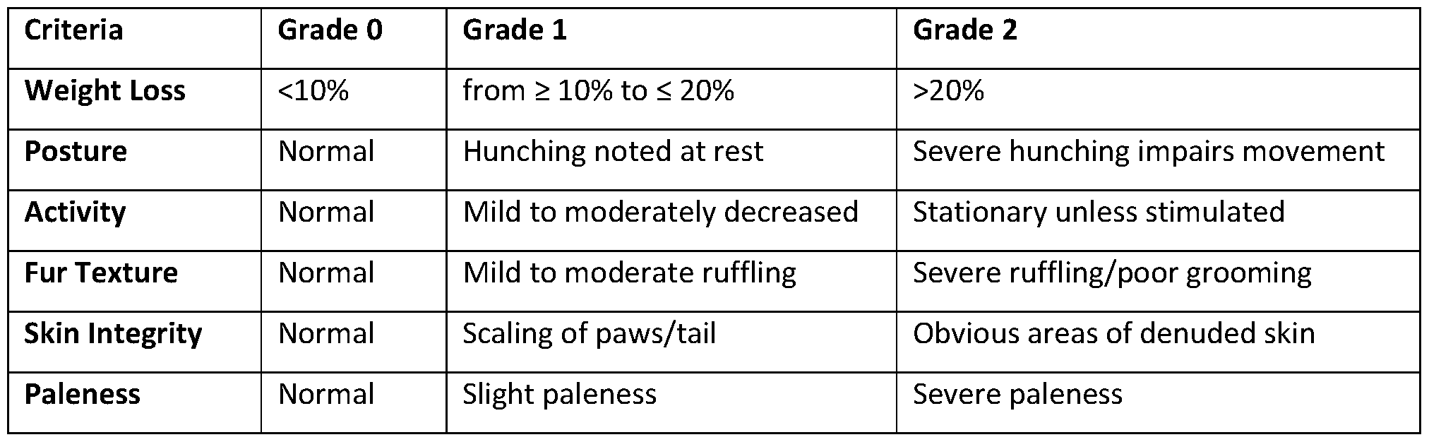
An engineered Treg- selective human IL-2 immunocytokine, denoted F5111 IC, which is comprised of the human anti-IL-2 antibody F5111 (Trotta, E. et al. A human anti-IL-2 antibody that potentiates regulatory T cells by a structure-based mechanism. Nat Med 24, 1005-1014 (2018)) covalently tethered to human IL-2 (VanDyke, D. et al. Engineered human
cytokine/antibody fusion proteins expand regulatory T cells and confer autoimmune disease protection. Cell Rep 41, 111478 (2022)) was shown to potently and selectively stimulate the high affinity IL-2R resulting in robust in vitro activation and in vivo expansion of Tregs (VanDyke, D. et al. Engineered human cytokine/antibody fusion proteins expand regulatory T cells and confer autoimmune disease protection. Cell Rep 41, 111478 (2022)). The activity of F5111 IC and ImmTOR in immunodeficient NOD SCID gamma (NSG) mice 2-3 weeks after engraftment with human peripheral blood mononuclear cells (HuPBMC mice) was evaluated. The HuPBMC mice, which are prone to develop GVHD, showed marked expansion of Treg, CD8+ T cells, and NK cells after F5111 IC treatment (FIG. 10A). Although the mice did not show signs of GVHD at the time of treatment, the expansion of CD8+ T cells may reflect the presence of a population of cells that had been primed for activation in response to host mouse antigens. Importantly, the addition of ImmTOR to F5111 IC treatment enabled expansion of Treg but inhibited the expansion of CD8+ T cells and NK cells.
The effects of F5111 IC in engineered knock-in mice expressing human IL-2RaP, which can form functional IE-2 receptors with endogenously expressed mouse IE-2Ry, was also assessed. F5111 IC induced robust Treg expansion in the engineered human IL-2RaP mice without substantial expansion of CD8+ T cells (FIG. 10B). The addition of ImmTOR to F5111 IC resulted in a corresponding synergistic expansion of Tregs, similar to that observed in wild-type mice treated with ImmT0R+Fc.IE2m. Collectively, the results of the mouse and human Treg expansion studies demonstrate that combination treatment with ImmTOR and Treg- selective IE-2 molecules induces selective promotion of Treg proliferation, synergistically enhancing the activity of the Treg-selective IL-2 molecules alone.
The increased expansion of effector cells observed in HuPBMC mice treated with F5111 IC alone correlated with exacerbation of disease in the HuPBMC GVHD model. Notably the addition of ImmTOR to F5111 IC significantly increased survival in this model (FIG. 10B). Disease exacerbation has also been reported for the IL-2/JES6-1 anti-IL-2 antibody complex in a mouse model of inflammatory arthritis induced by infection with chikungunya virus. Administration of IL-2/JES6-1 during active infection increased both Tregs and effector T cells resulting in disease worsening, while prophylactic administration in healthy mice prevented subsequent disease. The activity of engineered IL-2 molecules can be dose-limited due to its effects on effector cells in settings of inflammation. The addition
of ImmTOR can increase the therapeutic window of Treg- selective IL-2 by restraining effector cell activation while synergistically increasing Tregs.
It was also found that survival of the mice was also positively impacted with ImmTOR and the combination treatment. F5111 IC alone made disease worse, but the addition of ImmTOR mitigated this as did a combination of ImmTOR and the IL-2 mutein.
Example 8: ImmTOR Tolerogenic Nanoparticles with Encapsulated Antigen and IL-2 Mutein Alleviates Autoimmune Pathology in a Model of Primary Biliary Cholangitis (PBC)
The activity of ImmTOR and Fc.IL2m in NOD.C3C4 mice, which spontaneously develop an autoimmune disease of the liver which closely resembles primary biliary cholangitis (PBC) was assessed. The primary T cell epitope has been mapped to a peptide in the inner lipoyl domain of the E2 component of the pyruvate dehydrogenase complexes (PDC-E2). Mice were treated with three monthly doses of ImmTOR, ImmT0R+Fc.IL2m or ImmT0R+Fc.IL2m combined with nanoencapsulated PDC-E2 antigen (NP-PDC-E2) (FIG. 14A). Treatment with ImmT0R+Fc.IL2m significantly reduced bile duct epithelial degeneration, biliary hyperplasia and liver inflammation (FIG. 14B and FIG. 14C). Coadministration of NP-PDC-E2 provided additional benefit. Liver histology showed striking biliary pathology, with marked peri-biliary mononuclear cell infiltrates, biliary hypercellularity and ductular ectasia in both male (FIG. 14C-F) and female mice (FIG. 14G- J). Treatment with ImmTOR (FIG. 14D and FIG. 14H), ImmT0R+Fc.IL2m (FIG. 14E and FIG. 141), and TOR+Fc.IL2m+NP-PDC-E2 (FIG. 14F and FIG. 14J), showed progressive improvement of all histologic features, with the triple therapy showing only minimal residual disease pathology.
Materials
Rapamycin containing nanoparticles (ImmTOR) were manufactured as described (Kishimoto, T.K. et al. Improving the efficacy and safety of biologic drugs with tolerogenic nanoparticles. Nat Nanotechnol 11, 890-899 (2016); Maldonado, R.A. et al. Polymeric synthetic nanoparticles for the induction of antigen- specific immunological tolerance. Proc Natl Acad Sci U S A 112, E156-165 (2015)). ImmTOR doses were based on rapamycin content ranging from 200 to 400 pg. Rapamycin (sirolimus) was manufactured by Concord Biotech (Ahmedabad, India). Antigen-containing nanoparticles (NP) were prepared using a
water/oil/water (W/O/W) double-emulsion solvent evaporation method as described (Maldonado, R.A. et al. Polymeric synthetic nanoparticles for the induction of antigenspecific immunological tolerance. Proc Natl Acad Sci U S A 112, E156-165 (2015)). Briefly, PLGA (50:50) and pegylated polylactic acid (PLA-PEG) were dissolved in dichloromethane to form the oil phase. An aqueous solution of antigen (chicken ovalbumin or OVA protein, or hybrid insulin peptides HIP6.9, LQTLALNAARDP, or modified water-soluble HIP2.5, RGG-LQTLALWSRMD-GGR) was then added to the oil phase and emulsified by sonication (Branson Digital Sonifier 250A). Following emulsification of the antigen solution into the oil phase, a double emulsion was created by adding an aqueous solution of polyvinyl alcohol and sonicating a second time. The double emulsion was added to a beaker containing PBS and stirred at RT for 4 h to allow the dichloromethane to evaporate. The resulting NPs were washed twice by centrifuging at 75,600 x g for 50 min at 4 °C followed by resuspension of the pellet in PBS. Concentration of extracted antigens was measured by HPLC. Dynamic Light Scattering (DLS) analysis of particle size and PDI was performed using a Malvern Zetasizer Nano-ZS ZEN 3600. All the nanoparticles loaded with antigens exhibited a particle size distribution ranging between 140-155 nm with a low polydispersity index (<0.15).
Mouse IL2 mutein (Fc.IL2m) was constructed based on the sequence Fc.Mut24 published by Khoryati et al. ( Khoryati, L. et al. An IL-2 mutein engineered to promote expansion of regulatory T cells arrests ongoing autoimmunity in mice. Sci Immunol 5 (2020)). The final Fc.IL2m mutein sequence contained N103R, V106D and C140A mutations, but did not include the P51T point mutation. A signal peptide MGWSCIILFLVATATGVHS was added in front of Fc to achieve secretion from mammalian expression system. The protein was manufactured by Genscript (Piscataway, NJ), using its proprietary mammalian expression system.
Human IL-2-CD25 immune complex F5111 IC was produced as previously described (VanDyke, D. et al. Engineered human cytokine/antibody fusion proteins expand regulatory T cells and confer autoimmune disease protection. Cell Rep 41, 111478 (2022)).
Immunologically naive, female C57BL/6 mice aged 36-52 days (or 17- 18g) were purchased from Charles River Laboratories (Wilmington, MA). Similarly aged B6.Cg- Tg(TcraTcrb)425Cbn/J mice (also known as OT-II mice), expressing the T cell receptor (TCR), which is specific for chicken ovalbumin 323-339 peptide (OVA323-339 or OP-II) in the context of I- Ab resulting in CD4+ T cells that primarily recognize OP-II when presented by the MHC-II molecule were purchased from Jackson Laboratories (Bar Harbor, ME).
Human Tg-FcRn (B6.Cg-FcgrttmlDcr Tg(FCGRT)32Dcr/DcrJ) mice carrying a knock-out mutation for the mouse Fcgrt (Fc receptor) and expressing human FcRn and were also purchased from Jackson Laboratories as were Non-obese diabetic (NOD) NOD/ShiLtJ strain mice. Human Tg-IL-2/IL-2Ra/IL-2Rp mice carrying knock-out mutations for IL-2, and IL-2 receptor alpha and beta chains and expressing their human homologues were purchased from Biocytogen (Wakefield, MA). NOD coisogenic genetically engineered immunodeficient or NCG (NOD-Prkdc26emCd52I12rgem26Cd22/NjuCrl) mice carrying a mutation in Sirpa and knockouts of Prkdc and I12rg genes and thus lacking functional/mature T, B, and NK cells, along with reduced macrophage and dendritic cell function and therefore amenable to grafting with human PBMC were purchased from Charles River Laboratories. PBMC from three different donors were used in each individual study, with DIY PBMC kit utilized per manufacturer’s instructions.
Methods
Mice were injected (i.v., tail vein or retro-orbital plexus) with ImmTOR nanoparticles in the effective range of 50-300 pg, or with Fc.IL2m (i.p. or i.v., retro-orbital plexus) or F5111-IC (i.v.) in the effective range of 6.25-18.75 pg.
Flow Cytometry (murine liver and spleen cell populations)
Immediately after euthanizing mice (at 1-14 days after initial treatment), livers and spleens were harvested and rendered into single cell suspensions. Livers were processed via collagenase 4 (Worthington, Lakewood, NJ) enzymatic digest according to manufacturer’s recommended protocol. Spleens were processed via mechanical passage through a 70 pm nylon mesh (ThermoFisher, Waltham, MA). Next, a red blood cell lysis step was performed for both liver and spleen suspensions for 5 min at room temperature in 150 mM NH4C1, 10 mM KHCO3, 10 pM Na2-EDTA; washed in PBS, 2% bovine serum; then filtered again with a 70 pm nylon mesh. Cells were incubated 20 min on ice with anti-CD16/32 (Fc-block, clone 93, BioLegend, San Diego, CA) then stained with antibodies directed toward cell surface receptors: CD3e-BV421 (BioLegend, clone 145-2C11), CD4-PerCP-Cy5.5 (BioLegend, clone RM4-5), CD8a-BV510 (BioLegend, clone 53-6.7), CD25-PE-CF594 (BD, clone PC61), NK1.1-AF700 (BD, clone PK136), CD122-APC (BioLegend, clone TM-B1), and CD132-PE (BioLegend, clone TUGm2). After cell surface labeling cells were then fixed and permeabilized according to manufacturer’s recommended protocol using a FoxP3
Transcription Kit (eBioscience, San Diego, CA). The targeted intracellular markers were stained with FoxP3-PE (InVitrogen, Waltham, MA), clone FJK-16s, Ki67-A647 (BioLegend, clone 11F6) and Helios-PE-Cy7 (BioLegend, clone 22F6). For pSTAT5 staining, cells were stimulated with equal molar concentrations of IL-2 (BD, cat#554603) or IL-2 mutein, fixed with Lyse/Fix Buffer (BD Biosciences, Franklin Lakes, NJ), and permeabilized with Perm Buffer III according to manufacturer’s Protocol III for methanol-based intracellular staining. Cell surface antibodies CD3e-FITC (clone 500A2), CD4-PerCP-Cy5.5 (clone RM4-5), CD25-FITC (clone 7D4), CD8a-Pacific Blue (clone 53-6.7), NK1.1-BV510 (clone PK136), and intracellular pSTAT5-A647 (clone 47), all from BD Biosciences, were used. Analysis was performed via FACSymphony A3 Cell Analyzer (BD Biosciences) with subsequent data analysis using Flow Jo software (TreeStar, Ashland, OR).
Methylation Analysis
Murine CD3+, CD4+CD3+ and CD4+CD25+ cells were isolated from splenocytes seven days post treatment via immunomagnetic bead selection (Miltenyi, Gaithersburg, MD) using either negative selection of untouched CD4+ T cells or positive selection of CD4+CD25+ T cells (both from Miltenyi). After careful supernatant removal, accurate cell counting, cell pellets were then snap frozen in liquid nitrogen, then stored on dry ice. Samples were then sent to EpigenDx (Hopkinton, MA) for subsequent targeted NextGen bisulfite sequencing panel analysis using their in-house FoxP3 methylation panel N4V1P15 analysis.
Serum cytokines and FGF21 determination
Serum cytokine concentrations were determined using Meso Scale Discovery (MSD) U-PLEX 10-Assay SECTOR™ Plates, Linkers, and corresponding capture and detection antibody pairs. Plates were read using electrochemiluminescence detection on an MESO® QuickPlex SQ 120, with Discovery Workbench software (version 4.0.13) for analysis (MSD®, Gaitherburg, MD). Assays were performed according to manufacturer’s instructions, and without alterations to the recommended standard curve dilutions. Serum FGF21 concentration was determined by ELISA using the mouse/rat FGF21 commercial kit from R&D Systems (Minneapolis, MN). Serum samples were run at a 1:10 dilution.
Concanavalin A Challenge Model
Concanavalin A (Con A) induced liver toxicity model was employed essentially as earlier described (Ilyinskii, P.O., Roy, C .J., LePrevost, J., Rizzo, G.L. & Kishimoto, T.K. Enhancement of the Tolerogenic Phenotype in the Liver by ImmTOR Nanoparticles. Front Immunol 12, 637469 (2021)). Briefly, mice were injected (i.v., r.o.) Con A at 12 mg/kg and then terminally bled at 6 or 12 hours post-challenge with cytokine levels in serum determined by MSD as described above and liver tissues collected simultaneously for single-cell suspension analysis by flow cytometry as described above or for hematoxylin-eosin staining followed by microscopic evaluation.
Statistical Analysis
Statistical analyses were performed using GraphPad Prism 9.4.1. To compare themouse experimental groups pairwise either multiple t test (for several time-points) or Mann-Whitney two-tailed test (for a single time-point; individual comparison of two groups presented within the same graph) were used. Significance is shown for each figure legend (* - p < 0.05, ** - p < 0.01; *** - p < 0.001; **** - p < 0.0001; not significant - p > 0.05). All data for individual experimental groups is presented as mean ± SD (error bars).
ImmT0R+Fc.IL2m showed significant activity in a mouse model of PBC, with marked reduction of peri-biliary mononuclear cell infiltrates, biliary hypercellularity and ductular ectasia. The addition of nanoencapsulated PDC-E2 antigen further improved disease course. Taken together, these results reinforce the ability to drive antigen- specific tolerogenic responses by co-administration of nanoencapsulated antigens. However, the robust efficacy of ImmTOR+Treg-selective IL-2 alone also suggests that tolerogenic immune responses to endogenously expressed autoantigens in the context of autoimmune disease can also be achieved without administered antigen.
Example 9: ImmTOR+Fc.IL2m treatment ameliorates autoimmune hepatitis
The activity of the ImmT0R+Fc.IL2m was evaluated in a model of autoimmune hepatitis induced by systemic administration of the concanavalin A, a lectin that causes polyclonal lymphocyte activation and hepatic infiltration of activated immune cells. Previous studies have shown that Treg depletion with anti-IL-2Roc antibodies exacerbated disease while adoptive transfer of Treg ameliorated disease (Wei, H.X. et al. CD4+ CD25+ Foxp3+ regulatory T cells protect against T cell-mediated fulminant hepatitis in a TGF-beta-
dependent manner in mice. J Immunol 181, 7221-7229 (2008)). Both ImmTOR and Fc.IL2m monotherapies inhibited infiltration of activated effector T cells, and combination treatment led to a further reduction in cell infiltrates (FIG. 16A). Similar, though more modest, reductions were observed in activated NK cells; whereas reductions in activated NKT cells, neutrophils, and macrophages were primarily mediated by ImmTOR (FIG. 17). Both ImmTOR and Fc.IL2m reduced production of serum interferon-y and, to a lesser extent, of CXCL1 chemokine (FIG. 16B). Combination treatment further reduced the levels of both IFN- y and CXCL1, whereas reductions in IL-6 were primarily mediated by ImmTOR. ImmT0R+Fc.IL2m administration also induced increased production of FGF21, a hepatoprotective stress-response growth factor (FIG. 16C). Overall, this model illustrated the therapeutic potential for ImmT0R+Fc.IL2m combination therapy in autoimmune hepatitis.
Example 10. The Therapeutic Combination of Rapamycin Nanoparticles and Engineered Interleukin-2 is Fully Protective in Two Stringent Models of Autoimmune Disease
Materials and. Methods
ImmTOR, other nanoparticles and IL-2 mutein molecules
Rapamycin containing nanoparticles (ImmTOR) were manufactured as described earlier (Kishimoto, 2016; Maldonado, 2015). ImmTOR doses were based on rapamycin content ranging from 50 to 100 pg per mouse. Rapamycin (sirolimus) was manufactured by Concord Biotech (Ahmedabad, India). Antigen-containing nanoparticles (NP) were prepared using a water/oil/water (W/O/W) double-emulsion solvent evaporation method as described (Sands, 2022). Briefly, PLGA (50:50) and pegylated polylactic acid (PLA-PEG) were dissolved in dichloromethane to form the oil phase. An aqueous solution of antigen (hybrid insulin peptide HIP6.9 (LQTLALNAARDP), or modified HIP2.5 (LQTLALWSRMD) was then added to the oil phase and emulsified by sonication (Branson Digital Sonifier 250A). Following emulsification of the antigen solution into the oil phase, a double emulsion was created by adding an aqueous solution of polyvinyl alcohol and sonicating a second time. The double emulsion was added to a beaker containing PBS and stirred at RT for 4 h to allow the dichloromethane to evaporate. The resulting NPs were washed twice by centrifuging at 75,600 x g for 50 min at 4 °C followed by resuspension of the pellet in PBS. The
concentration of extracted antigens was measured by HPLC. Dynamic Light Scattering (DLS) analysis of particle size and PDI was performed using a Malvern Zetasizer Nano-ZS ZEN 3600. All the nanoparticles loaded with antigens exhibited a particle size distribution ranging between 140-155 nm with a low polydispersity index (<0.15). Mouse IL2 mutein (Fc.IL2m) was constructed based on the sequence Fc.Mut24 published by Khoryati et al. (Ref.) The protein was manufactured by Genscript, using its proprietary CHO mammalian expression system. F5111 IC was produced as previously described ((VanDyke).
Mice. Immunologically naive, female C57BE/6 mice aged 36-52 days (or 17- 18g) were purchased from Charles River Eaboratories (Wilmington, MA). Non-obese diabetic (NOD) NOD/ShiEtJ strain and Tg-BDC2.5 mice were also purchased from Jackson Eaboratories. An immunodeficient strain NSG (NOD.Cg-Prkdcscid I12rgtmlWjl/SzJ), carrying mutations in Prkdc (protein kinase, DNA activated catalytic subunit) and null allele of the IL2rg (IL2rgnull) lacking functional/mature T, B, and NK cells was purchased from Jackson Labs, and used in GVHD studies. To minimize the potential effects of stress, mice were acclimated to the Animal Care Facility at Selecta for at least three days prior to treatment. All the experiments were conducted in strict compliance with NIH Guide for the Care and Use of Laboratory Animals and other federal, state and local regulations and were approved by Selecta’ s IACUC.
Animal Injections
Mice were injected (i.v., tail vein or retro-orbital plexus) with ImmTOR nanoparticles in the effective range of 50-100 pg per mouse, or with NP-encapsulated peptide antigens in the effective range of 0.5-1 pg per mouse, or with Fc.IL2m (i.p. or i.v., retro-orbital plexus) or F5111-IC (i.v.) in the effective range of 2.5-9 pg per mouse. NOD (type 1 diabetes model) mice were treated with individual therapeutic components or their combinations three or four times total at 28-day intervals.
GVHD model
NSG mice (NOD.Cg-Prkdcscid I12rgtmlWjl/SzJ; Jackson Laboratory #005557) were irradiated with 1 Gy from an X-ray irradiator source and then reconstituted with IxlO7 human PBMC. The next day, mice were treated with a single dose of phosphate buffer saline (vehicle) or ImmTOR (50 or 100 pg) or left untreated. F5111 IC (2.5 or 5.0 pg) was administered at 3 and 17 days after grafting. Animals were assessed for disease activity 3
times per week. Each animal was assessed for weight loss, posture, activity, fur texture, skin integrity, and paleness on a 2-grade scale for 42 days after grafting. Animals losing more than 20% weight or moribund were removed from the study.
Type 1 Diabetes model (standard and accelerated)
In the standard model of diabetes female NOD mice were enrolled in the study at 6-7 weeks of age with the first treatment at week 8 or 10. They were monitored weekly using standard glucometer strips, and mice showing glucose levels >250 mg/dL on 2/3 successive measurements were considered diabetic and those scoring >500 mg/dL twice or >600 mg/dL once were terminated. All animals in the study were terminated at 32-35 weeks. In an accelerated models of diabetes development, female NOD mice were injected (i.p.) with 500 pg anti-PD-Ll (BioXCell, New Lebanon, NH) either on day 28 of age followed by 250 pg on day 30 of age (early disease induction) or with the same doses on days 70 and 72 (intermediate stage disease induction).
Results
Efficacy of late treatment with ImmT0R+Ec.IL2m using the standard model of type 1 diabetes in NOD mice
It was found that ImmTOR + Ec.IL2m can prevent type 1 diabetes in the NOD mouse model if a therapeutic dose of ImmTOR (100 pg) was used and the treatment was started early, specifically at 8 weeks of mouse age, prior to elevation of glucose levels (Kishimoto et al., 2023). ImmTOR + Ec.IL2m were administered with or without nanoparticle-entrapped insulin-chromogranin A hybrid peptide 6.9 (Baker et al., 2019) (NP-HIP6.9), and both combinations protected 90% of experimental mice. However, in this experiment both ImmTOR alone and Lc.IL2m were also effective, protecting 80% of mice.
In the current study, more challenging regimens of treatment and disease induction were tested. Lirstly, a sub-optimal dose of ImmTOR (50 pg) and of NP-HIP6.9 (0.5 pg) and also delayed treatment until 10 weeks of age, which is about the time when the first mice become glycemic, were tested. Not surprisingly, this model proved to be more difficult to treat than that used previously. Specifically, under these conditions, neither ImmTOR alone or Lc.IL2m alone provided any significant protection. However, the combination of ImmTOR + Lc.IL2m with NP-HIP6.9 provided 90% protection from disease over 35 weeks of
observation (FIG. 18) and was the only group significantly different from the group of untreated mice by both statistical tests employed, namely Mantel-Cox and Gehan-Breslow- Wilcoxon tests (FIG. 18A). Notably, ImmTOR + Fc.IL2m without encapsulated HIP6.9 peptide was also effective, but marginally inferior to the same treatment with NP-HIP.6.9 and was significantly different from untreated group only by Mantel-Cox test (FIG. 18A). Interestingly, some mice treated with ImmTOR alone or with ImmTOR + Fc.IL2m showed elevated glucose levels above 250 mg/dL immediately after treatment (FIG. 18B), which reverted back to baseline by 30 weeks of age. The reversal of glycemia is unusual and suggests that these mice were on the cusp of becoming diabetic but were restrained by the induction or expansion of Tregs. The absence of stable glucose elevation was especially noticeable late in treatment, which may indicate that the regimen or timing or dose levels of ImmTOR and Fc.IL2m in this model may be adjusted. At the same time, no effect of this kind was seen in the group treated with ImmTOR + Fc.IL2m combined with NP-HIP6.9 with only 1 mouse out of 10 in this group becoming glycemic by week 15 (FIG. 18B).
It is worth noting that if the same ImmTOR + Fc.IL2m combination with or without NP- HIP2.5 and NP-HIP6.9 was initiated at 8 weeks of age, as in our experiments reported earlier (Kishimoto et al., 2023), then both treatments were extremely efficacious with no mice becoming glycemic over the 32 weeks of observation (FIG. 19A). The durable effect of treatment is notable in this experiment where 100% of the control mice developed diabetes by 6 months of age (FIG. 19B). Not unexpectedly, both treatment combinations led to significant differences in serum levels of insulin, leptin, glucagon, and PYY compared to untreated control mice (FIGs. 19C-F). Of note, slightly higher levels of leptin and especially, of insulin (FIG. 19E) seen in mice treated using NP-encapsulated peptides indicate an advantage of adding tolerizing antigens to ImmTOR and Fc.IL2m combinations.
Efficacy of ImmT0R+Fc.IL2m in an accelerated diabetes model
Antibodies against PD-1 and its ligand PD-L1 have been used as immune checkpoint inhibitors in the treatment of cancer. One of the potential side effects observed in patients is the induction of type 1 diabetes and other autoimmune conditions. Immune checkpoint inhibition has also been shown to accelerate disease in NOD mice (Fife, 2006). The efficacy of ImmTOR + Fc.IL2m combination therapy was tested in this more aggressive form of type 1 diabetes induced by anti-PD-Ll antibodies. Several regimens of aPD-Ll administration
have been described in this respect with the early administration being especially efficient (Fife, 2006).
In the most challenging regimen, mice were injected with aPD-Ll twice early in their 5th week of life leading to 100% (10/10 mice) glycemic conversion by week 14 and termination of all mice due to high glucose levels by week 16. In contrast, mice treated with ImmTOR + Fc.IL2m resisted diabetes progression with only 1/10 mice converting at an early stage and being terminated by week 7. The addition of NP-HIP2.5 and NP-HIP6.9 to this treatment scheme was even more efficacious with no mice (0/10) progressing to full-blown diabetes through 20 weeks of observation (FIG. 20B). Average glucose levels in this group became statistically different from that in untreated mice by week 7, while in the group treated with ImmTOR + Fc.IL2m without NP-entrapped HIP peptides, they became statistically different from those in untreated mice by week 10 with significance in both groups becoming even more pronounced with each following time-point (FIG. 20C).
Progression to diabetes in untreated mice happened in two waves, one early (weeks 6- 7) and one late (weeks 11-14) (FIG. 20D). In both treatment groups, there was a total of 3/20 mice showing early glucose elevation at week 6. Of these, the only one glycemic mouse in the group treated with ImmTOR + Fc.IL2m progressed irreversibly to diabetes (FIG. 20D). Interestingly, transient glucose elevation in the two mice treated with ImmTOR + Fc.IL2m combined with NP-HIP2.5/6.9 returned to baseline after each of the three treatments, despite glucose levels exceeding 400 mg/dL in one mouse, and then remained low for the duration of the study (FIG. 20D). To the best of our knowledge, reversal of diabetes in mice exceeding 400 mg/dL has not been previously reported. Notably, this treatment regimen can be viewed as therapeutic since injection of ImmTOR and Fc.IL2m started several days after inoculation of aPD-Ll, which apparently triggered near- immediate induction of diabetes in 50% of injected NOD mice.
Nearly all terminal serum analytes tested were statistically different in both treatment groups vs. those in untreated mice, with only the single diabetic mouse in the group treated with ImmTOR + Fc.IL2m without HIP peptides showing high levels of GLP-1 and PYY. Treatment with Fc.IL2m alone in this model was not effective.
Similar results were observed when aPD-Ll antibodies were administered at 10 weeks of age after the start of ImmTOR + Fc.IL2m treatment at week 8 (FIG. 21A). In this case, elevation of glucose in untreated mice also occurred within one week of treatment and was seen in majority (9/11) of untreated mice within 2 weeks (FIGs. 21B, C). However,
there was no second wave of diabetes induction, although one of two remaining non-diabetic mice had shown a slow and incomplete progression to diabetes at the end of the study (FIG. 21C). In this experiment all combinations of ImmTOR + Fc.IL2m were statistically indistinguishable protecting 60-67% mice by the end of the study irrespective of presence or absence of NP-encapsulated peptides (FIG. 21B, C), although it seemed that onset of disease was slightly delayed in the group co-treated with NP-HIP6.9. No significant differences between terminal analytes were seen between different treatment groups.
Induction of antigen- specific Tregs by ImmTOR + Fc.IL2m combined with pathogenic diabetic epitope
Expansion of total Tregs by engineered IL-2 have been shown to confer protection in models of autoimmune disease by mediating antigen-nonselective inhibition of effector cells through a process termed ‘bystander suppression’. However, preclinical studies have shown that antigen- specific Tregs are more potent than antigen-non-selective Tregs in mitigating diabetes. It has been shown that combination of ImmTOR + Fc.IL2m showed profound synergistic expansion of ovalbumin- specific Tregs if co-administered with NP-encapsulated ovalbumin (Kishimoto, 2023). The ability of ImmTOR and Fc.IL2m to induce Tregs specific to a diabetogenic peptide HIP2.5 (Baker, 2019) was evaluated. Splenocytes from BDC2.5 transgenic mice expressing a diabetogenic T cell receptor (Gonzalez, 1997, 2001) were adoptively transferred into naive NOD mice, followed by tolerogenic treatments with different combinations of ImmTOR, Fc.IL2m and NP-HIP2.5. Both Fc.IL-2m (Khoryati) and the combination of Fc.IL2m and ImmTOR + Fc.IL2m (Kishimoto, 2023) efficiently expanded total (endogenous) Tregs in naive recipients. There was no additional benefit of adding NP-HIP2.5 (FIG. 22B). In contrast, the administration of NP-HIP2.5 antigen particles to mice treated with ImmTOR + Fc.IL2m showed a profound increase in antigen- specific diabetogenic BDC2.5 T cells. (FIG. 22C). Interestingly, mice treated with ImmTOR + Fc.IL2m without NP-HIP2.5 showed a modest but notable increase in BDC2.5 -specific Tregs compared to other control groups, suggesting that ImmTOR + Fc.IL2m can utilize endogenously expressed antigens to drive expansion of antigen- specific Tregs in vivo. To our knowledge, this is the first case of induction of antigen- specific Tregs targeting a known pathogenic epitope in an autoimmune disease model, which has a direct relevance to human disease.
Efficacy of ImmTOR and IL-2 mutein in GVHD treatment
A Treg- selective human IL-2/anti-IL-2 antibody complex, termed F5111-IC (VanDyke, 2022), unexpectedly exacerbated graft-vs-host disease (GVHD) in mice grafted with human peripheral blood mononuclear cells (PBMC) (Kishimoto, 2023). The increased mortality was attributed to expansion of effector cells, which can express the high affinity IL- 2 receptor, in addition to Tregs. Importantly, the addition of ImmTOR to F5111 IC mitigated effector cell expansion, while still allowing for Treg expansion, and enhanced survival compared to the untreated control mice.
Since the induction of Tregs by ImmTOR takes about one week, it was hypothesized that delaying delivery of the IL-2 component might be more efficacious by allowing ImmTOR to dampen down inflammation prior to administration of IL-2 (Kishimoto, Maldonado). A split dosing regimen was tested in native (uninflamed) mice by treating with ImmTOR on Day -7 (7 days prior to tissue harvest) and administration of Fc.IL-2m on Day - 4 (4 days prior to harvest) and compared this regimen to simultaneous administration of ImmTOR and Fc.IL2m or Fc.IL2m used alone on Day -4 (FIG. 23A, groups shown within shapes). Mice showed higher levels of Treg induction 4 days after administration of Fc.IL2m using the split dosing regimen compared to mice co-administered ImmTOR and Fc.IL2m on the same day (FIG. 23B, gr 1-3). Treg induction 7 days after Fc.IL2m administration was similar in both groups whether ImmTOR was co-administered or injected 3 days earlier, on Day -10 (FIG. 23B, groups within shapes in FIG. 23A). These data indicated that a split dosing regimen of ImmTOR and IL-2 mutein administration is a feasible approach to treat in a setting of inflammation, such as GVHD.
Since in this case human PBMC are used in the GVHD model, the human IL-2 immunocytokine, F5111 IC, was again employed. Two different doses of ImmTOR, a higher dose (100 pg) shown to be effective on its own (100 pg) and a suboptimal dose (50 pg) were tested. In addition, the amount of F5111 used was lowered from 9 pg to 2.5 and 5.0 pg. Collectively, there were two test study arms, one using lower doses of ImmTOR and F5111 and another using higher doses of both treatments. Finally, since the temporal splitting of ImmTOR and F5111 was proved to be efficient in Treg induction (FIG. 23), the delivery of F5111 was delayed by 3 days and it was also opted to administer it twice with a two-week interval since earlier data indicated that Tregs induced by ImmTOR + IL-2 mutein persist at detectable levels for about two weeks (Kishimoto, 2023).
Combination treatment with ImmTOR and delayed F5111 proved to be highly effective in the human PBMC GVHD model, attaining 100% (10/10 mice) survival in both the low (FIG. 24B) and high dose (FIG. 24C) study arms. Notably, F5111 used alone had no therapeutic effect, although at these lower doses it did not exacerbate disease, as observed previously. ImmTOR alone was effective in delaying mortality, although overall survival was not significantly different from the vehicle-treated control groups at Day 42 (FIG. 24B, C). The survival rate in both combination treatment groups was statistically different from that in mice receiving no treatment or treated with F5111 alone (FIGs. 24B, C). This is further illustrated by dynamics of animal weights (FIG. 24D) and disease activity index (FIG. 24E). In both cases, overall graphs from both combination treatment groups are nearly indistinguishable from those of non-grafted control group, while being visibly different from all other groups, especially vs. untreated controls and groups treated with F5111 alone (FIGs. 24D, E). Collectively, both ImmTOR + human engineered IL-2 mutein combinations were fully protective for 7 weeks of post-grafting observation.
Discussion
The synergy of ImmTOR nanoparticles encapsulating rapamycin and two engineered IL-2 molecules, mouse Fc.IL2m and human F5111-IC, in several immunological and disease- related models (Kishimoto, 2023) was observed. This synergy led to expansion and maintenance of Tregs and was also capable of induction and expansion of antigen- specific Tregs if ImmTOR and IL-2 mutein were co-administered with target antigen. The findings were extended to two different models of immune response mitigation, those of type 1 diabetes (T1D) and GVHD.
Specifically, in a T1D model the combination treatment with ImmTOR, Fc.IL2m and nanoparticle-encapsulated antigens, HIP6.9 and HIP2.5, were differentiated from single treatments by using lower dose of ImmTOR and a late treatment start. Moreover, such antigen-containing treatment schemes were extremely efficient (and superior to ImmTOR combined with Fc.IL2m without antigen in some embodiments) in the accelerated model of type 1 diabetes in which 90-100% mice convert to diabetes within 12-14 weeks. Furthermore, the expansion of antigen- specific Tregs recognizing HIP2.5, an autoantigen generated during the development of diabetes in NOD mice and humans and known to play a role in disease pathology, was demonstrated. If this approach is expanded to include several antigens and/or epitopes with proven role in misdirected autoimmunity, this can allow for the development of
treatment modalities aiming to induce Tregs of novel antigen specificity including those targeting disease-related neoantigens such as HIPs in T ID or citrullinated antigens in rheumatoid arthritis.
Dosing schemes and administration regimens of ImmTOR and IL-2 mutein molecules can be improved by taking into consideration their pleiotropic effects and pharmacokinetics (which is especially true for IL-2 muteins). Specifically, data in a GVHD model has indicated that human engineered IL-2 derivative, F5111-IC, was detrimental at a high dose administered immediately after grafting, while ImmTOR was beneficial on its own and was able to mitigate the adverse effects of F5111-IC. When the administration regimen was amended by splitting the dosing of ImmTOR and IL-2 mutein and its applicability to mouse Treg induction with pre-injection of ImmTOR was demonstrated to permit efficient Treg induction if Fc.IL2m was inoculated at a later time-point. Extending this approach to a GVHD model and using human F5111 molecule a synergy of ImmTOR with late-delivered low-dose F5111 was attained, and virtually no disease development was observed as was an absence of mortality in combination-treated mice not dissimilar to phenotype of control nongrafted mice. It is likely that this was the consequence of bystander suppression through expansion of total Tregs by late-delivered F5111 in ImmTOR- stimulated tolerogenic milieu.
Collectively, the ability of ImmTOR rapamycin-containing particles and engineered IL-2 molecules has been demonstrated to induce both unspecific and antigen- specific Treg response and provided evidence of such combinations being fully protective in several challenging models of immune-mediated diseases (100% survival was repeatedly seen in two separate models). This opens up a number of new avenues to apply these or similar therapeutic modalities in a disease- and antigen-specific fashion.
Claims
1. A composition comprising:
(a) immunosuppressant (e.g., synthetic nanocarriers comprising the immunosuppressant (e.g., any one of the synthetic nanocarriers comprising an immunosuppressant as provided herein));
(b) a high affinity IL-2 receptor agonist (e.g., any one of the high affinity IL-2 receptor agonists provided herein) and,
(c) optionally, an antigen (e.g., any one of the antigens provided herein).
2. The composition of claim 1, further comprising a pharmaceutically acceptable excipient.
3. The composition of claim 1 or claim 2, wherein the antigen is encapsulated in the synthetic nanocarriers.
4. A dosage form comprising the composition of any one of claims 1-3.
5. A method comprising administering to a subject in need thereof:
(a) immunosuppressant (e.g., synthetic nanocarriers comprising the immunosuppressant (e.g., any one of the synthetic nanocarriers comprising an immunosuppressant as provided herein));
(b) a high affinity IL-2 receptor agonist agonist (e.g., any one of the high affinity IL-2 receptor agonists provided herein) and,
(c) optionally, an antigen (e.g., any one of the antigens provided herein).
6. The method of claim 5, wherein the immunosuppressant and the high affinity IL-2 receptor agonist and, optionally, an antigen are administered concomitantly.
7. The method of any one of claims 5-6, wherein (a), (b) and, optionally, (c) are administered in an amount effective to enhance regulatory T cells (e.g., CD4+), such as antigen- specific regulatory T cells (e.g, CD4+), and/or reduce or inhibit effector T cells.
8. The method of any of claims 5-7, wherein the subject has or is at risk of having an inflammatory disease, an autoimmune disease, an allergy, or graft versus host disease.
9. The method of any of claims 5-8, wherein the subject has or is at risk of having an undesired immune response against an antigen that is being administered or will be administered to the subject.
10. The method of claim 9, wherein the antigen is a therapeutic macromolecule.
11. The method of any of claims 5-9, wherein the subject has or is at risk of having an undesired immune response against an antigen to which the subject is exposed or will be exposed.
12. The method or composition of any of the preceding claims, wherein the immunosuppressant comprise a statin, an mTOR inhibitor, a TGF-P signaling agent, a corticosteroid, an inhibitor of mitochondrial function, a P38 inhibitor, an NF-KB inhibitor, an adenosine receptor agonist, a prostaglandin E2 agonist, a phosphodiesterase 4 inhibitor, an HD AC inhibitor or a proteasome inhibitor.
13. The method or composition of claim 12, wherein the mTOR inhibitor is rapamycin or a rapamycin analog.
14. The method or composition of any one of the preceding claims, wherein the synthetic nanocarriers comprise lipid nanoparticles, polymeric nanoparticles, metallic nanoparticles, surfactant-based emulsions, dendrimers, buckyballs, nanowires, virus-like particles or peptide or protein particles.
15. The method or composition of claim 14, wherein the synthetic nanocarriers comprise polymeric nanoparticles.
16. The method or composition of claim 14 or 15, wherein the polymeric nanoparticles comprise a polyester, a polyester coupled to a polyether, polyamino acid, polycarbonate, polyacetal, polyketal, polysaccharide, polyethyloxazoline or polyethyleneimine.
17. The method or composition of claim 16, wherein the polyester comprises a poly(lactic acid), poly(glycolic acid), poly(lactic-co-glycolic acid) or polycaprolactone.
18. The method or composition of claim 16 or 17, wherein the polymeric nanoparticles comprise a polyester and a polyester coupled to a polyether.
19. The method or composition of any of claims 16-18, wherein the polyether comprises polyethylene glycol or polypropylene glycol.
20. The method or composition of any of the preceding claims, wherein the mean of a particle size distribution obtained using dynamic light scattering of the synthetic nanocarriers is a diameter greater than lOOnm.
21. The method or composition of claim 20, wherein the diameter is greater than 150nm.
22. The method or composition of claim 21, wherein the diameter is greater than 200nm.
23. The method or composition of claim 22, wherein the diameter is greater than 250nm.
24. The method or composition of claim 23, wherein the diameter is greater than 300nm.
25. The method of composition of any one of claims 20-24, wherein the diameter is less than 500nm.
26. The method of composition of any one of claims 20-24, wherein the diameter is less than 450nm.
27. The method of composition of any one of claims 20-24, wherein the diameter is less than 400nm.
28. The method of composition of any one of claims 20-24, wherein the diameter is less than 350nm.
29. The method of composition of any one of claims 20-23, wherein the diameter is less than 300nm.
30. The method of composition of any one of claims 20-22, wherein the diameter is less than 250nm.
31. The method of composition of claim 20 or 21, wherein the diameter is less than 200nm.
32. The method or composition of any of the preceding claims, wherein an aspect ratio of the synthetic nanocarriers is greater than or equal to 1:1, 1:1.2, 1:1.5, 1:2, 1:3, 1:5, 1:7 or 1:10.
33. The method or composition of any one of the preceding claims, wherein the load of immunosuppressant comprised in the synthetic nanocarriers, on average across the synthetic nanocarriers, is between 1% and 40% (weight/weight).
34. The method or composition of claim 33, wherein the load is between 1% and 30%.
35. The method or composition of claim 34, wherein the load is between 1% and 25%.
36. The method or composition of claim 33, wherein the load is between 2% and 40%.
37. The method or composition of claim 36, wherein the load is between 2% and 30%.
38. The method or composition of claim 37, wherein the load is between 2% and 25%.
39. The method or composition of claim 33, wherein the load is between 4% and 40%.
40. The method or composition of claim 39, wherein the load is between 4% and 30%.
41. The method or composition of claim 40, wherein the load is between 4% and 25%.
42. The method or composition of claim 33, wherein the load is between 8% and 40%.
43. The method or composition of claim 42, wherein the load is between 8% and 30%.
44. The method or composition of claim 43, wherein the load is between 8% and 25%.
45. The method or composition of any one of the preceding claims, wherein the high affinity IL-2 receptor agonist is wild type IL-2, an IL-2 mutein, an IL-2 mimic or an IL-2 fusion protein.
46. The method or composition of any one of the preceding claims, wherein the frequency, dose amounts, timing and/or mode of administration of the synthetic nanocarriers comprising the immunosuppressant is according to any one of the protocols provided herein.
47. The method or composition of any one of the preceding claims, wherein the frequency, dose amounts, timing and/or mode of administration of the high affinity IL-2 receptor agonist is according to any one of the protocols provided herein.
48. The method or composition of any one of the preceding claims, wherein the frequency, dose amounts, timing and/or mode of administration of the optional antigen is according to any one of the protocols provided herein.
49. The method of composition of any one of the preceding claims, wherein the antigen is a therapeutic macromolecule, such as a therapeutic polynucleotide, such as a viral vector.
50. The method or composition of claim 49, wherein when the antigen is a viral vector, the synthetic nanocarriers comprising the immunosuppressant, high affinity IL-2 receptor agonist (e.g, IL-mutein) and viral vector are concomitantly administered every other month.
51. The method or composition of claim 50, wherein the concomitant administration occurs at least two times.
52. The method or composition of claim 50 or 51, wherein the dose of the synthetic nanocarriers comprising the immunosuppressant, high affinity IL-2 receptor agonist (e.g., IL- mutein) and/or viral vector is/are any one of the respective doses provided herein.
53. The method or composition of claim 50 or 51, wherein the dose of the synthetic nanocarriers comprising an immunosuppressant is/are any one of the doses provided herein.
54. The method or composition of any one of the preceding claims, wherein the antigen is any one of the antigens provided herein.
55. The method or composition of any one of the preceding claims, wherein the antigen is associated with an autoimmune disease, such as an autoimmune liver disease.
56. The method or composition of any one of the preceding claims, wherein the antigen is associated with GVHD.
57. The method or composition of any one of the preceding claims, wherein the antigen is associated with diabetes, such as Type 1 diabetes.
58. The method or composition of any one of the preceding claims, wherein the subject has or is at risk of having any one of the diseases or conditions provided herein.
55. The method or composition of any one of the preceding claims, wherein the subject is any one of the subjects provided herein.
56. The method of claim 55, wherein the subject is one who would benefit from treatment with a high affinity IL-2 receptor agonist.
57. The method of claim 55 or 56, wherein the subject has or is at risk of having toxicity from treatment with the high affinity IL-2 receptor agonist with or without treatment with any one of the immunosuppressants as provided herein.
58. The method of any one of the preceding claims, wherein the method is for treatment with a high affinity IL-2 receptor agonist.
59. The method of any one of the preceding claims, wherein the method is for treatment with a high affinity IL-2 receptor agonist but at reduced toxicity.
60. The method of any one of the preceding claims, wherein the method is for prolonging or continuing with treatment with a high affinity IL-2 receptor agonist.
61. The method of any one of the preceding claims, wherein the method is for prolonging or continuing with treatment with a high affinity IL-2 receptor agonist but at reduced toxicity.
62. The method of any one of the preceding claims, wherein the method is for treatment with a high affinity IL-2 receptor agonist but at a higher dose of the high affinity IL-2 receptor agonist, which higher dose is higher than a dose that would be administered to a subject in the absence of concomitant administration with an immunosuppressant as provided herein.
63. The method or composition of any one of the preceding claims, wherein the high affinity IL-2 receptor agonist is any one of the high affinity IL-2 receptor agonists provided herein.
64. The method or composition of claim 63, wherein the high affinity IL-2 receptor agonist is the mouse IL2 mutein (Fc.IL2m) as described herein.
65. The method or composition of claim 63, wherein the high affinity IL-2 receptor agonist is the human IL-2-CD25 immune complex F5111 IC as described herein.
66. The method or composition of claim 63, wherein the high affinity IL-2 receptor agonist is low-dose IL-2.
67. The method or composition of any one of the preceding claims, wherein the dose of the synthetic nanocarriers comprising an immunosuppressant is/are any one of the doses provided herein.
68. The method or composition of any one of the preceding claims, wherein the dose of the high affinity IL-2 receptor agonist is/are any one of the doses provided herein.
69. The method or composition of any one of the preceding claims, wherein the toxicity is T effector cell production or activity, or an increase in such production or activity.
70. The method or composition of any one of the preceding claims, wherein the toxicity is inflammation or increase in inflammation.
71. The method or composition of any one of the preceding claims, wherein the subject has or is at risk of inflammation or increase in inflammation.
72. The method or composition of any one of claims 69-71, wherein treatment with the high affinity IL-2 receptor agonist alone can result in such toxicity.
73. The method or composition of any one of the preceding claims, wherein the combination therapy of the immunosuppressant and high affinity IL-2 receptor agonist is for reducing T effector cell production or activity, or reducing an increase in such production or activity, and promoting T regulatory cell production or activity, or promoting an increase in such production or activity.
74. The method or composition of any one of the preceding claims, wherein the immunosuppressant and/or high affinity IL-2 receptor agonist are in an amount effective for reducing T effector cell production or activity, or reducing an increase in such production or activity, and promoting T regulatory cell production or activity, or promoting an increase in such production or activity.
75. The method or composition of any one of the preceding claims, wherein no antigen is administered to the subject and/or the antigen is an autoantigen or endogenous antigen.
76. The method or composition of any one of the preceding claims, wherein the immunosuppressant and/or high affinity IL-2 receptor agonist are in an amount effective for preventing onset or progression of diabetes, optionally type 1 diabetes.
77. The method or composition of any one of the preceding claims, wherein the immunosuppressant and/or high affinity IL-2 receptor agonist are in an amount effective for treating diabetes, optionally type 1 diabetes.
78. The method or composition of claim 76 or 77, wherein: the immunosuppresent comprises rapamycin or a rapamycin analog; and/or high affinity IL-2 receptor agonist is an IL-2 mutein; and/or the antigen is an insulin peptide, optionally a hybrid insulin peptide, optionally wherein the inuslin peptide is encapsulated within a nanoparticle.
79. The method or composition of claim 78, wherein the insulin peptide comprises the amino acid sequence of LQTLALNAARDP (HIP6.9) or LQTLALWSRMD (HIP2.5).
80. The method of any one of the preceding claims, wherein the method comprises administering the immunosuppressant at a first time and administering the high affinity IL-2 receptor agonist at a second time, wherein the second time is at least one day (or 24 hours) after the first time.
81. The method of claim 80, wherein the second time is 2-10, 2-7, 2-5, 2-4, 2-3, or 3-5 days after the first time.
82. The method of claim 80, wherein the second time is about 2, 3, 4, 5, 6, 7, 8, 9, or 10 days after the first time.
83. A method of administering an immunosuppressant and a high affinity IL-2 receptor agonist to a subject in need thereof, the method comprising:
(a) administering the immunosuppressant to the subject at a first time;
(b) administering the high affinity IL-2 receptor to the subject at a second time;
wherein the second time is at least one day (or 24 hours) after the first time.
84. The method of claim 83, wherein: the immunosuppresent comprises rapamycin or a rapamycin analog; and/or the high affinity IL-2 receptor agonist is an IL-2 mutein; and/or the method further comprises administering an antigen to the subject, optionally wherein the antigen is an antigen associated with diabetes or GvHD.
Applications Claiming Priority (4)
| Application Number | Priority Date | Filing Date | Title |
|---|---|---|---|
| US202363463685P | 2023-05-03 | 2023-05-03 | |
| US63/463,685 | 2023-05-03 | ||
| US202363500952P | 2023-05-09 | 2023-05-09 | |
| US63/500,952 | 2023-05-09 |
Publications (1)
| Publication Number | Publication Date |
|---|---|
| WO2024229432A1 true WO2024229432A1 (en) | 2024-11-07 |
Family
ID=91302381
Family Applications (1)
| Application Number | Title | Priority Date | Filing Date |
|---|---|---|---|
| PCT/US2024/027858 Pending WO2024229432A1 (en) | 2023-05-03 | 2024-05-03 | Synthetic nanocarriers comprising an immunosuppressant in combination with high affinity il-2 receptor agonists to enhance immune tolerance |
Country Status (1)
| Country | Link |
|---|---|
| WO (1) | WO2024229432A1 (en) |
Citations (39)
| Publication number | Priority date | Publication date | Assignee | Title |
|---|---|---|---|---|
| US4638045A (en) | 1985-02-19 | 1987-01-20 | Massachusetts Institute Of Technology | Non-peptide polyamino acid bioerodible polymers |
| US4806621A (en) | 1986-01-21 | 1989-02-21 | Massachusetts Institute Of Technology | Biocompatible, bioerodible, hydrophobic, implantable polyimino carbonate article |
| US4946929A (en) | 1983-03-22 | 1990-08-07 | Massachusetts Institute Of Technology | Bioerodible articles useful as implants and prostheses having predictable degradation rates |
| US5010167A (en) | 1989-03-31 | 1991-04-23 | Massachusetts Institute Of Technology | Poly(amide-and imide-co-anhydride) for biological application |
| US5019379A (en) | 1987-07-31 | 1991-05-28 | Massachusetts Institute Of Technology | Unsaturated polyanhydrides |
| US5399665A (en) | 1992-11-05 | 1995-03-21 | Massachusetts Institute Of Technology | Biodegradable polymers for cell transplantation |
| US5512600A (en) | 1993-01-15 | 1996-04-30 | Massachusetts Institute Of Technology | Preparation of bonded fiber structures for cell implantation |
| US5514378A (en) | 1993-02-01 | 1996-05-07 | Massachusetts Institute Of Technology | Biocompatible polymer membranes and methods of preparation of three dimensional membrane structures |
| US5543158A (en) | 1993-07-23 | 1996-08-06 | Massachusetts Institute Of Technology | Biodegradable injectable nanoparticles |
| US5578325A (en) | 1993-07-23 | 1996-11-26 | Massachusetts Institute Of Technology | Nanoparticles and microparticles of non-linear hydrophilic-hydrophobic multiblock copolymers |
| US5716404A (en) | 1994-12-16 | 1998-02-10 | Massachusetts Institute Of Technology | Breast tissue engineering |
| US5736372A (en) | 1986-11-20 | 1998-04-07 | Massachusetts Institute Of Technology | Biodegradable synthetic polymeric fibrous matrix containing chondrocyte for in vivo production of a cartilaginous structure |
| US5770417A (en) | 1986-11-20 | 1998-06-23 | Massachusetts Institute Of Technology Children's Medical Center Corporation | Three-dimensional fibrous scaffold containing attached cells for producing vascularized tissue in vivo |
| US5804178A (en) | 1986-11-20 | 1998-09-08 | Massachusetts Institute Of Technology | Implantation of cell-matrix structure adjacent mesentery, omentum or peritoneum tissue |
| US5837752A (en) | 1997-07-17 | 1998-11-17 | Massachusetts Institute Of Technology | Semi-interpenetrating polymer networks |
| US5902599A (en) | 1996-02-20 | 1999-05-11 | Massachusetts Institute Of Technology | Biodegradable polymer networks for use in orthopedic and dental applications |
| US6007845A (en) | 1994-07-22 | 1999-12-28 | Massachusetts Institute Of Technology | Nanoparticles and microparticles of non-linear hydrophilic-hydrophobic multiblock copolymers |
| US6095148A (en) | 1995-11-03 | 2000-08-01 | Children's Medical Center Corporation | Neuronal stimulation using electrically conducting polymers |
| US6123727A (en) | 1995-05-01 | 2000-09-26 | Massachusetts Institute Of Technology | Tissue engineered tendons and ligaments |
| US20020086049A1 (en) | 2000-09-18 | 2002-07-04 | Bolton Anthony E. | Apoptosis-mimicking synthetic entities and use thereof in medical treatment |
| US6506577B1 (en) | 1998-03-19 | 2003-01-14 | The Regents Of The University Of California | Synthesis and crosslinking of catechol containing copolypeptides |
| US6632922B1 (en) | 1998-03-19 | 2003-10-14 | The Regents Of The University Of California | Methods and compositions for controlled polypeptide synthesis |
| US6632671B2 (en) | 2000-02-28 | 2003-10-14 | Genesegues, Inc. | Nanoparticle encapsulation system and method |
| US6686446B2 (en) | 1998-03-19 | 2004-02-03 | The Regents Of The University Of California | Methods and compositions for controlled polypeptide synthesis |
| US6818732B2 (en) | 2001-08-30 | 2004-11-16 | The Regents Of The University Of California | Transition metal initiators for controlled poly (beta-peptide) synthesis from beta-lactam monomers |
| US20060002852A1 (en) | 2004-07-01 | 2006-01-05 | Yale University | Targeted and high density drug loaded polymeric materials |
| US20060222652A1 (en) | 2000-05-05 | 2006-10-05 | Cytos Biotechnology Ag | Molecular antigen array |
| US20060251677A1 (en) | 2003-03-26 | 2006-11-09 | Cytos Biotechnology Ag | Packaging of immunostimulatory oligonucleotides into virus-like particles: method of preparation and use |
| US20080145441A1 (en) | 2000-10-16 | 2008-06-19 | Midatech Limited | Nanoparticles |
| WO2008127532A1 (en) | 2007-04-12 | 2008-10-23 | Emory University | Novel strategies for delivery of active agents using micelles and particles |
| US20090028910A1 (en) | 2003-12-19 | 2009-01-29 | University Of North Carolina At Chapel Hill | Methods for Fabrication Isolated Micro-and Nano-Structures Using Soft or Imprint Lithography |
| WO2009051837A2 (en) | 2007-10-12 | 2009-04-23 | Massachusetts Institute Of Technology | Vaccine nanotechnology |
| WO2009106999A2 (en) | 2008-02-28 | 2009-09-03 | Deutsches Krebsforschungszentrum, Stiftung Des Öffentlichen Rechts | Hollow nanoparticles and uses thereof |
| US20090226525A1 (en) | 2007-04-09 | 2009-09-10 | Chimeros Inc. | Self-assembling nanoparticle drug delivery system |
| WO2010047839A1 (en) | 2008-10-25 | 2010-04-29 | Aura Biosciences | Modified plant virus particles and uses therefor |
| US20160128986A1 (en) | 2014-11-05 | 2016-05-12 | Selecta Biosciences, Inc. | Methods and compositions related to the use of low hlb surfactants in the production of synthetic nanocarriers comprising a rapalog |
| US20170037102A1 (en) | 2014-07-21 | 2017-02-09 | Delinia, Inc. | Molecules that selectively activate regulatory t cells for the treatment of autoimmune diseases |
| WO2020264318A1 (en) | 2019-06-26 | 2020-12-30 | The Johns Hopkins University | Methods and materials for targeted expansion of regulatory t cells |
| US20220323607A1 (en) * | 2021-04-09 | 2022-10-13 | Selecta Biosciences, Inc. | Synthetic nanocarriers comprising an immunosuppressant in combination with high affinity il-2 receptor agonists to enhance immune tolerance |
-
2024
- 2024-05-03 WO PCT/US2024/027858 patent/WO2024229432A1/en active Pending
Patent Citations (41)
| Publication number | Priority date | Publication date | Assignee | Title |
|---|---|---|---|---|
| US4946929A (en) | 1983-03-22 | 1990-08-07 | Massachusetts Institute Of Technology | Bioerodible articles useful as implants and prostheses having predictable degradation rates |
| US4638045A (en) | 1985-02-19 | 1987-01-20 | Massachusetts Institute Of Technology | Non-peptide polyamino acid bioerodible polymers |
| US4806621A (en) | 1986-01-21 | 1989-02-21 | Massachusetts Institute Of Technology | Biocompatible, bioerodible, hydrophobic, implantable polyimino carbonate article |
| US5736372A (en) | 1986-11-20 | 1998-04-07 | Massachusetts Institute Of Technology | Biodegradable synthetic polymeric fibrous matrix containing chondrocyte for in vivo production of a cartilaginous structure |
| US5804178A (en) | 1986-11-20 | 1998-09-08 | Massachusetts Institute Of Technology | Implantation of cell-matrix structure adjacent mesentery, omentum or peritoneum tissue |
| US5770417A (en) | 1986-11-20 | 1998-06-23 | Massachusetts Institute Of Technology Children's Medical Center Corporation | Three-dimensional fibrous scaffold containing attached cells for producing vascularized tissue in vivo |
| US5019379A (en) | 1987-07-31 | 1991-05-28 | Massachusetts Institute Of Technology | Unsaturated polyanhydrides |
| US5010167A (en) | 1989-03-31 | 1991-04-23 | Massachusetts Institute Of Technology | Poly(amide-and imide-co-anhydride) for biological application |
| US5399665A (en) | 1992-11-05 | 1995-03-21 | Massachusetts Institute Of Technology | Biodegradable polymers for cell transplantation |
| US5512600A (en) | 1993-01-15 | 1996-04-30 | Massachusetts Institute Of Technology | Preparation of bonded fiber structures for cell implantation |
| US5696175A (en) | 1993-01-15 | 1997-12-09 | Massachusetts Institute Of Technology | Preparation of bonded fiber structures for cell implantation |
| US5514378A (en) | 1993-02-01 | 1996-05-07 | Massachusetts Institute Of Technology | Biocompatible polymer membranes and methods of preparation of three dimensional membrane structures |
| US5578325A (en) | 1993-07-23 | 1996-11-26 | Massachusetts Institute Of Technology | Nanoparticles and microparticles of non-linear hydrophilic-hydrophobic multiblock copolymers |
| US5543158A (en) | 1993-07-23 | 1996-08-06 | Massachusetts Institute Of Technology | Biodegradable injectable nanoparticles |
| US6007845A (en) | 1994-07-22 | 1999-12-28 | Massachusetts Institute Of Technology | Nanoparticles and microparticles of non-linear hydrophilic-hydrophobic multiblock copolymers |
| US5716404A (en) | 1994-12-16 | 1998-02-10 | Massachusetts Institute Of Technology | Breast tissue engineering |
| US6123727A (en) | 1995-05-01 | 2000-09-26 | Massachusetts Institute Of Technology | Tissue engineered tendons and ligaments |
| US6095148A (en) | 1995-11-03 | 2000-08-01 | Children's Medical Center Corporation | Neuronal stimulation using electrically conducting polymers |
| US5902599A (en) | 1996-02-20 | 1999-05-11 | Massachusetts Institute Of Technology | Biodegradable polymer networks for use in orthopedic and dental applications |
| US5837752A (en) | 1997-07-17 | 1998-11-17 | Massachusetts Institute Of Technology | Semi-interpenetrating polymer networks |
| US6506577B1 (en) | 1998-03-19 | 2003-01-14 | The Regents Of The University Of California | Synthesis and crosslinking of catechol containing copolypeptides |
| US6632922B1 (en) | 1998-03-19 | 2003-10-14 | The Regents Of The University Of California | Methods and compositions for controlled polypeptide synthesis |
| US6686446B2 (en) | 1998-03-19 | 2004-02-03 | The Regents Of The University Of California | Methods and compositions for controlled polypeptide synthesis |
| US6632671B2 (en) | 2000-02-28 | 2003-10-14 | Genesegues, Inc. | Nanoparticle encapsulation system and method |
| US20060222652A1 (en) | 2000-05-05 | 2006-10-05 | Cytos Biotechnology Ag | Molecular antigen array |
| US20020086049A1 (en) | 2000-09-18 | 2002-07-04 | Bolton Anthony E. | Apoptosis-mimicking synthetic entities and use thereof in medical treatment |
| US20080145441A1 (en) | 2000-10-16 | 2008-06-19 | Midatech Limited | Nanoparticles |
| US6818732B2 (en) | 2001-08-30 | 2004-11-16 | The Regents Of The University Of California | Transition metal initiators for controlled poly (beta-peptide) synthesis from beta-lactam monomers |
| US20060251677A1 (en) | 2003-03-26 | 2006-11-09 | Cytos Biotechnology Ag | Packaging of immunostimulatory oligonucleotides into virus-like particles: method of preparation and use |
| US20090028910A1 (en) | 2003-12-19 | 2009-01-29 | University Of North Carolina At Chapel Hill | Methods for Fabrication Isolated Micro-and Nano-Structures Using Soft or Imprint Lithography |
| US20060002852A1 (en) | 2004-07-01 | 2006-01-05 | Yale University | Targeted and high density drug loaded polymeric materials |
| US20090226525A1 (en) | 2007-04-09 | 2009-09-10 | Chimeros Inc. | Self-assembling nanoparticle drug delivery system |
| WO2008127532A1 (en) | 2007-04-12 | 2008-10-23 | Emory University | Novel strategies for delivery of active agents using micelles and particles |
| WO2009051837A2 (en) | 2007-10-12 | 2009-04-23 | Massachusetts Institute Of Technology | Vaccine nanotechnology |
| WO2009106999A2 (en) | 2008-02-28 | 2009-09-03 | Deutsches Krebsforschungszentrum, Stiftung Des Öffentlichen Rechts | Hollow nanoparticles and uses thereof |
| WO2010047839A1 (en) | 2008-10-25 | 2010-04-29 | Aura Biosciences | Modified plant virus particles and uses therefor |
| US20170037102A1 (en) | 2014-07-21 | 2017-02-09 | Delinia, Inc. | Molecules that selectively activate regulatory t cells for the treatment of autoimmune diseases |
| US20160128986A1 (en) | 2014-11-05 | 2016-05-12 | Selecta Biosciences, Inc. | Methods and compositions related to the use of low hlb surfactants in the production of synthetic nanocarriers comprising a rapalog |
| US20160128987A1 (en) | 2014-11-05 | 2016-05-12 | Selecta Biosciences, Inc. | Methods and compositions related to synthetic nanocarriers with rapamycin in a stable, super-saturated state |
| WO2020264318A1 (en) | 2019-06-26 | 2020-12-30 | The Johns Hopkins University | Methods and materials for targeted expansion of regulatory t cells |
| US20220323607A1 (en) * | 2021-04-09 | 2022-10-13 | Selecta Biosciences, Inc. | Synthetic nanocarriers comprising an immunosuppressant in combination with high affinity il-2 receptor agonists to enhance immune tolerance |
Non-Patent Citations (68)
| Title |
|---|
| "Concise Encyclopedia of Polymer Science and Polymeric Amines and Ammonium Salts", 1980, PERGAMON PRESS |
| "Handbook of Industrial Mixing: Science and Practice", 2004, JOHN WILEY & SONS, INC. |
| "Pharmaceutics: The Science of Dosage Form Design", 2001, CHURCHILL LIVINGSTONE |
| ALLCOCK ET AL.: "Contemporary Polymer Chemistry", 1981, PRENTICE-HALL |
| BARRERA ET AL., J. AM. CHEM. SOC., vol. 115, 1993, pages 11010 |
| BAYLE ET AL., CHEMISTRY & BIOLOGY, vol. 13, 2006, pages 99 - 107 |
| BOUSSIF ET AL., PROC. NATL. ACAD. SCI., USA, vol. 92, 1995, pages 7297 |
| C. ASTETE ET AL.: "Synthesis and characterization of PLGA nanoparticles", J. BIOMATER. SCI. POLYMER EDN, vol. 17, no. 3, 2006, pages 247 - 289, XP009134610 |
| C. REIS ET AL.: "Nanoencapsulation I. Methods for preparation of drug-loaded polymeric nanoparticles", NANOMEDICINE, vol. 2, 2006, pages 8 - 21 |
| CELL DEATH DIS, vol. 9, 2018, pages 989, Retrieved from the Internet <URL:https://doi.org/10.1038/s41419-018-1047-2> |
| DANA K. CASSELLNOEL R. ROSE: "The Encyclopedia of Autoimmune Diseases", 14 May 2014, INFOBASE PUBLISHING |
| DEMING ET AL., NATURE, vol. 390, 1997, pages 386 |
| DOUBROW: "Microcapsules and Nanoparticles in Medicine and Pharmacy", 1992, CRC PRESS |
| FRONT IMMUNOL, vol. 11, no. 1106, 5 June 2020 (2020-06-05) |
| FRONT IMMUNOL., vol. 11, 28 April 2020 (2020-04-28), pages 638 |
| FRONT IMMUNOL., vol. 11, 5 June 2020 (2020-06-05), pages 1106 |
| HAENSLER ET AL., BIOCONJUGATE CHEM., vol. 4, 1993, pages 372 |
| HUANG MINGSHU ET AL: "Advanced Delivery Strategies for Immunotherapy in Type I Diabetes Mellitus", BIODRUGS, vol. 37, no. 3, 1 May 2023 (2023-05-01), NZ, pages 331 - 352, XP093194247, ISSN: 1173-8804, Retrieved from the Internet <URL:https://link.springer.com/article/10.1007/s40259-023-00594-6/fulltext.html> [retrieved on 20240812], DOI: 10.1007/s40259-023-00594-6 * |
| ILYINSKII, P.O.ROY, C.J.LEPREVOST, J.RIZZO, G.L.KISHIMOTO, T.K.: "Enhancement of the Tolerogenic Phenotype in the Liver by ImmTOR Nanoparticles", FRONT IMMUNOL, vol. 12, 2021, pages 637469 |
| J AUTOIMMUN, vol. 56, January 2015 (2015-01-01), pages 66 - 80 |
| J EXP MED, vol. 217, no. 1, 6 January 2020 (2020-01-06), pages e20191247 |
| J IMMUNOL, vol. 190, no. 12, 15 June 2013 (2013-06-15), pages 6230 - 8 |
| J IMMUNOL, vol. 202, no. 68, 1 May 2019 (2019-05-01), pages 20 |
| J IMMUNOL, vol. 204, no. 237, 1 May 2020 (2020-05-01), pages 16 |
| J. AUTOIMMUN., vol. 95, no. 1, 13 November 2018 (2018-11-13) |
| K. AVGOUSTAKIS: "Pegylated Poly(Lactide) and Poly(Lactide-Co-Glycolide) Nanoparticles: Preparation, Properties and Possible Applications in Drug Delivery", CURRENT DRUG DELIVERY, vol. 1, 2004, pages 321 - 333, XP009134627 |
| KABANOV ET AL., BIOCONJUGATE CHEM., vol. 6, 1995, pages 7 |
| KATTEBOINAA ET AL., INTERNATIONAL JOURNAL OF PHARMTECH RESESARCH, vol. 1, no. 3, 2009, pages 682 - 694 |
| KHORYATI ET AL., SCIENCE IMMUNOLOGY, 2020, pages eaba5264 |
| KHORYATI, L. ET AL.: "An IL-2 mutein engineered to promote expansion of regulatory T cells arrests ongoing autoimmunity in mice", SCI IMMUNOL, vol. 5, 2020, XP055935853, DOI: 10.1126/sciimmunol.aba5264 |
| KISHIMOTO TAKASHI KEI ET AL: "Rapamycin nanoparticles increase the therapeutic window of engineered interleukin-2 and drive expansion of antigen-specific regulatory T cells for protection against autoimmune disease", JOURNAL OF AUTOIMMUNITY, LONDON, GB, vol. 140, 14 October 2023 (2023-10-14), XP087435292, ISSN: 0896-8411, [retrieved on 20231014], DOI: 10.1016/J.JAUT.2023.103125 * |
| KISHIMOTO TAKASHI KEI ET AL: "Synergistic activity of IL-2 mutein with tolerogenic ImmTOR nanoparticles leads to massive expansion of antigen-specific Tregs and protection against autoimmune disease", BIORXIV, 16 May 2023 (2023-05-16), pages 1 - 34, XP093056567, Retrieved from the Internet <URL:https://www.biorxiv.org/content/10.1101/2023.05.15.540840v1> [retrieved on 20230621], DOI: 10.1101/2023.05.15.540840 * |
| KISHIMOTO, T.K. ET AL.: "Improving the efficacy and safety of biologic drugs with tolerogenic nanoparticles", NAT NANOTECHNOL, vol. 11, 2016, pages 890 - 899, XP055371105, DOI: 10.1038/nnano.2016.135 |
| KUKOWSKA-LATALLO ET AL., PROC. NATL. ACAD. SCI., USA, vol. 93, 1996, pages 4897 |
| KWON ET AL., MACROMOLECULES, vol. 22, 1989, pages 3250 |
| LANGER, ACC. CHEM. RES., vol. 33, 2000, pages 94 |
| LANGER, J. CONTROL. RELEASE, vol. 62, 1999, pages 7 |
| LIM ET AL., J. AM. CHEM. SOC., vol. 121, 1999, pages 5633 |
| LONG, S.A. ET AL.: "Rapamycin/IL-2 combination therapy in patients with type 1 diabetes augments Tregs yet transiently impairs beta-cell function", DIABETES, vol. 61, 2012, pages 2340 - 2348 |
| LOOK ET AL.: "Nanogel-based delivery of mycophenolic acid ameliorates systemic lupus erythematosus in mice", J. CLINICAL INVESTIGATION, vol. 123, no. 4, 2013, pages 1741 - 1749 |
| MALDONADO, R.A. ET AL.: "Polymeric synthetic nanoparticles for the induction of antigen-specific immunological tolerance", PROC NATL ACAD SCI U S A, vol. 112, 2015, pages E156 - 165, XP002764343, DOI: 10.1073/pnas.1408686111 |
| MATHIOWITZ ET AL., J. APPL. POLYMER SCI., vol. 35, 1988, pages 755 |
| MATHIOWITZ ET AL., J. CONTROL. RELEASE, vol. 5, 1987, pages 13 |
| MATHIOWITZ ET AL., REACTIVE POLYMERS, vol. 6, 1987, pages 275 |
| MELDAL ET AL., CHEM. REV., vol. 108, no. 8, 2008, pages 2952 - 3015 |
| MURRAY ET AL., ANN. REV. MAT. SCI., vol. 30, 2000, pages 545 |
| NATURE, vol. 484, 2012, pages 529 - 533 |
| ONCOIMMUNOLOGY, vol. 9, no. 1, 2 June 2020 (2020-06-02) |
| P. PAOLICELLI ET AL.: "Surface-modified PLGA-based Nanoparticles that can Efficiently Associate and Deliver Virus-like Particles", NANOMEDICINE, vol. 5, no. 6, 2010, pages 843 - 853 |
| PAPISOV, ACS SYMPOSIUM SERIES, vol. 786, 2001, pages 301 |
| PELLEGRINO ET AL., SMALL, vol. 1, 2005, pages 48 |
| PROTEIN ENG, vol. 16, no. 12, December 2003 (2003-12-01), pages 1081 - 7 |
| PUTNAM ET AL., MACROMOLECULES, vol. 32, 1999, pages 3658 |
| SATYANARAYANA, M.: "IL-2 treatment can be dangerous. Here's how drug firms are trying to fix it", CHEMICAL AND ENGINEERING NEWS, vol. 99, 2021, Retrieved from the Internet <URL:https://cen.acs.org/pharmaceuticals/biologics/safer-IL2-cancer-immunotherapy-autoimmunity/99/i12> |
| SCI IMMUNOL., vol. 5, no. 50, 14 August 2020 (2020-08-14), pages eaba5264 |
| SEMIN ONCOL., vol. 45, no. 1-2, January 2018 (2018-01-01), pages 95 - 104 |
| SHARPLESS ET AL., ANGEW. CHEM. INT. ED., vol. 41, no. 14, 2002, pages 2596 |
| SILVA, DA.YU, S.ULGE, U.Y. ET AL.: "De novo design of potent and selective mimics of IL-2 and IL-15", NATURE, vol. 565, 2019, pages 186 - 191, XP055636971, Retrieved from the Internet <URL:https://doi.org/10.1038/s41586-018-0830-7> DOI: 10.1038/s41586-018-0830-7 |
| TANG ET AL., BIOCONJUGATE CHEM., vol. 7, 1996, pages 703 |
| TRENDS IMMUNOL., vol. 36, no. 12, December 2015 (2015-12-01), pages 763 - 777 |
| TRINDADE ET AL., CHEM. MAT., vol. 13, 2001, pages 3843 |
| TROTTA, E. ET AL.: "A human anti-IL-2 antibody that potentiates regulatory T cells by a structure-based mechanism", NAT MED, vol. 24, 2018, pages 1005 - 1014, XP036542078, DOI: 10.1038/s41591-018-0070-2 |
| UHRICH ET AL., CHEM. REV., vol. 99, 1999, pages 3181 |
| VANDYKE, D. ET AL.: "Engineered human cytokine/antibody fusion proteins expand regulatory T cells and confer autoimmune disease protection", CELL REP, vol. 41, 2022, pages 111478, XP093030809, DOI: 10.1016/j.celrep.2022.111478 |
| WANG ET AL., J. AM. CHEM. SOC., vol. 123, 2001, pages 2460 |
| WEI, H.X. ET AL.: "CD4+ CD25+ Foxp3+ regulatory T cells protect against T cell-mediated fulminant hepatitis in a TGF-beta- dependent manner in mice", J IMMUNOL, vol. 181, 2008, pages 7221 - 7229 |
| ZAUNER ET AL., ADV. DRUG DEL. REV., vol. 30, 1998, pages 97 |
| ZHOU ET AL., MACROMOLECULES, vol. 23, 1990, pages 3399 |
Similar Documents
| Publication | Publication Date | Title |
|---|---|---|
| US20220323607A1 (en) | Synthetic nanocarriers comprising an immunosuppressant in combination with high affinity il-2 receptor agonists to enhance immune tolerance | |
| JP2020015741A (en) | Methods and compositions for enhancing CD4 + regulatory T cells | |
| US20220133864A1 (en) | Methods and compositions for reducing immune responses against immunoglobulin proteases | |
| US20150359865A1 (en) | Tolerogenic synthetic nanocarriers for t-cell-mediated autoimmune disease | |
| US20240024517A1 (en) | Viral vector dosing protocols | |
| US20230140196A1 (en) | Viral vector dosing protocols | |
| US20230381277A1 (en) | High affinity il-2 receptor agonists and immunosuppressants to enhance immune tolerance | |
| US20230263906A1 (en) | High affinity il-2 receptor agonists and synthetic nanocarrier dose sparing | |
| US20230372535A1 (en) | Synthetic nanocarriers comprising an immunosuppressant in combination with high affinity il-2 receptor agonists and anti-igm agents | |
| US20230322884A1 (en) | Immunosuppressant in combination with high affinity il-2 receptor agonists and related dosing | |
| WO2024229432A1 (en) | Synthetic nanocarriers comprising an immunosuppressant in combination with high affinity il-2 receptor agonists to enhance immune tolerance | |
| WO2024229350A1 (en) | Immunosuppressant in combination with high affinity il-2 receptor agonists for diabetes | |
| WO2024229380A1 (en) | Immunosuppressant in combination with high affinity il-2 receptor agonists in autoimmune liver diseases | |
| WO2024229370A1 (en) | Compositions and methods for treating gvhd | |
| CN117320717A (en) | Synthetic nanocarriers containing immunosuppressants in combination with high-affinity IL-2 receptor agonists to enhance immune tolerance | |
| WO2023086615A1 (en) | Multiple dosing with viral vectors |
Legal Events
| Date | Code | Title | Description |
|---|---|---|---|
| 121 | Ep: the epo has been informed by wipo that ep was designated in this application |
Ref document number: 24729569 Country of ref document: EP Kind code of ref document: A1 |
|
| NENP | Non-entry into the national phase |
Ref country code: DE |