WO2024211981A1 - Ige antibody against prion protein characterized using lad2 mast cells - Google Patents
Ige antibody against prion protein characterized using lad2 mast cells Download PDFInfo
- Publication number
- WO2024211981A1 WO2024211981A1 PCT/CA2024/050330 CA2024050330W WO2024211981A1 WO 2024211981 A1 WO2024211981 A1 WO 2024211981A1 CA 2024050330 W CA2024050330 W CA 2024050330W WO 2024211981 A1 WO2024211981 A1 WO 2024211981A1
- Authority
- WO
- WIPO (PCT)
- Prior art keywords
- antibody
- ige
- seq
- amino acid
- acid sequence
- Prior art date
- Legal status (The legal status is an assumption and is not a legal conclusion. Google has not performed a legal analysis and makes no representation as to the accuracy of the status listed.)
- Ceased
Links
Classifications
-
- A—HUMAN NECESSITIES
- A61—MEDICAL OR VETERINARY SCIENCE; HYGIENE
- A61P—SPECIFIC THERAPEUTIC ACTIVITY OF CHEMICAL COMPOUNDS OR MEDICINAL PREPARATIONS
- A61P25/00—Drugs for disorders of the nervous system
- A61P25/28—Drugs for disorders of the nervous system for treating neurodegenerative disorders of the central nervous system, e.g. nootropic agents, cognition enhancers, drugs for treating Alzheimer's disease or other forms of dementia
-
- C—CHEMISTRY; METALLURGY
- C07—ORGANIC CHEMISTRY
- C07K—PEPTIDES
- C07K16/00—Immunoglobulins [IGs], e.g. monoclonal or polyclonal antibodies
- C07K16/18—Immunoglobulins [IGs], e.g. monoclonal or polyclonal antibodies against material from animals or humans
- C07K16/28—Immunoglobulins [IGs], e.g. monoclonal or polyclonal antibodies against material from animals or humans against receptors, cell surface antigens or cell surface determinants
- C07K16/2872—Immunoglobulins [IGs], e.g. monoclonal or polyclonal antibodies against material from animals or humans against receptors, cell surface antigens or cell surface determinants against prion molecules, e.g. CD230
-
- C—CHEMISTRY; METALLURGY
- C07—ORGANIC CHEMISTRY
- C07K—PEPTIDES
- C07K2317/00—Immunoglobulins specific features
- C07K2317/50—Immunoglobulins specific features characterized by immunoglobulin fragments
- C07K2317/52—Constant or Fc region; Isotype
Definitions
- the present disclosure relates to antibodies that bind to prion protein (PrP).
- PrP prion protein
- Immunoglobin E is best known for its association with allergies and asthma, but can also provide protection from toxins, venoms, and parasites (1). It can induce a rapid response after exposure to antigens by triggering activation of granulated effector cells such as mast cells, basophils and eosinophils (2). When activated, mast cells release the preformed contents of their granules, which include high concentrations of histamine, proteases, proteoglycans and other signaling molecules such as newly synthesized lipid mediators and cytokines/chemokines (2, 3).
- the proteases released by mast cells include chymase, tryptase, cathepsins and carboxypeptidase which can cleave a large variety of target proteins including blood clotting factors, cell adhesion proteins, cytokines and chemokines (4). Some of these proteases are mast cell-specific and are not produced by any other immune cell. Although mast cells can be activated by various mechanisms, one of the most specific stimuli in vivo is through clustering of their surface FceRI receptors by crosslinking of IgE bound to antigen.
- IgG immunoglobin G
- IgE immunoglobin G
- IgG immunoglobin G
- IgEs have several attributes that could be advantageous in immunotherapy of diseases that are unresponsive to IgG immunotherapy.
- they can overcome immunotolerance, as shown by their role in allergic responses to normally innocuous antigens (6).
- IgEs generate a strong, self- perpetuating immune response sustained by FceRI-expressing immune cells (7).
- the proteases released from effector cells could potentially degrade disease causing substances or aggregates while effector-released cytokines could recruit other immune cells to disease sites.
- IgEs function best at the mucosal lining of the gastrointestinal tract, while IgGs function best in the circulation. IgEs are effective at clearing mucosa-associated pathogens such as helminths (8) and have been proposed in cancer treatment, since some animal model systems have shown that IgE to cancer biomarkers may trigger immune responses that can target and kill tumours (9-11). IgG cannot easily access most solid tissues (12), including brain (13), and they are most often associated with acute and transient humoral immune responses rather than chronic and sustained cellular responses, which would be more appropriate for addressing chronic diseases (11).
- mast cells via FceRI requires multiple epitopes in close proximity to crosslinkthe IgE receptor (14), possibly enabling discrimination between low concentrations of self- antigens present in healthy cells and higher concentrations, or aggregates, of self-antigens present in some diseases.
- IgE is often involved in allergic reactions, and even small amounts of antigen can induce potentially fatal reactions if enough effector cells are induced to degranulate (15).
- IgE was relatively well tolerated, but induced anaphylaxis in one of the 24 patients (16).
- the binding affinity of any therapeutic IgEs will require fine tuning to balance the ability of the IgE to bind its antigen and its ability to induce widespread degranulation.
- low affinity IgEs may prove useful in therapy, as they can induce different immune responses than high affinity IgEs.
- a low affinity stimulus of IgE caused a shift of signalling from the adapter LAT1 to related adapter LAT2, resulting in less degranulation, less leukotriene and cytokine production, but higher chemokine production (17).
- this low affinity antigen also induced less vascular permeability and changed the ratio of recruited monocyte/macrophagesmeutrophils.
- low affinity IgEs can induce degranulation of mast cells if they are exposed to antigens with multiple epitopes in close proximity (18). Potentially, targeted degranulation via IgE could be used in diseases where the disease state of an endogenous protein involves high local expression or clustering of normally monomeric epitopes, such as in protein misfolding aggregation diseases.
- proteinopathies that involve toxic aggregation of misfolded endogenous proteins, including Ap and Tau proteins in Alzheimer’s disease, a-synuclein in Parkinson's disease, TAR DNA-binding protein 43 in amyotrophic lateral sclerosis (19).
- the hallmark protein folding diseases are the transmissible spongiform encephalopathies, or “prion diseases”, which include scrapie in sheep, bovine spongiform encephalopathy in cows, chronic wasting disease in cervids, and Creutzfeldt-Jakob disease in humans (20, 21).
- PrP prion protein
- PrP Sc prion protein
- an engineered antibody comprising the variable region from an anti-PrP (aPrP) IgG fused to the constant region of a human IgE, to produce a chimeric aPrP IgE.
- the recombinant aPrP antibody binds surface FceRI on LAD2 cells and activates phosphorylation of SYK and ERK.
- the aPrP antibody is crosslinked, it initiates LAD2 degranulation and the release of proteases such as tryptase and cathepsin G that can digest PrP.
- the engineered aPrP IgE can activate mast cells through FceRI and generate responses that could remove PrP substrate, thereby reducing prion conversion.
- an IgE antibody that binds specifically to prion protein (PrP), wherein the antibody comprises three variable heavy domain complementarity determining regions (CDR)(CDR H1 , H2 and H3), and three variable light domain CDR (CDR L1 , L2 and L3), said CDR H1 , H2, H3, L1 , L2, and L3, and wherein: the CDR H1 comprises the amino acid sequence GFSLTSYGVH (SEQ ID NO:1), the CDR H2 comprises the amino acid sequence VIWSDGSTTYDSTLKS (SEQ ID NO:2), the CDR H3 comprises the amino acid sequence RHFY (SEQ ID NO:3), the CDR L1 comprises the amino acid sequence KSSQSLLDSDGKTYLN (SEQ ID NO:4), the CDR L2 comprises the amino acid sequence LVSKLDS (SEQ ID NO:5), and the CDR L3 comprises the amino acid sequence WQGTHFPQT (SEQ ID NO:
- the IgE antibody provides lower levels of degranulation from an activated mast cell bound with said IgE antibody compared to an activated mast cell bound with an HE1 control IgE monoclonal antibody, registered in The Antibody Registry under #RRID AB_1075924, as determined by measuring the amount of p-hexosaminidase released by each activated mast cell into a surrounding medium, wherein a lower level of p- hexosaminidase release indicates a lower level of degranulation.
- the IgE antibody may further comprise four variable heavy domain framework regions (HFR)(HFR 1 , 2, 3 and 4), wherein: the HFR 1 comprises the amino acid sequence: GPGLVAPSQSLSITCTIS (SEQ ID NO:7), the HFR 2 comprises the amino acid sequence: WVRQPPGKGLEWLV (SEQ ID NO:8), the HFR 3 comprises the amino acid sequence: RLSISKDNSKSQVFSKMNSLQTDDTAMYCCAR (SEQ ID NO:9), and the HFR 4 comprises the amino acid sequence: A (SEQ ID NQ:10); or the HFR 1 comprises the amino acid sequence VKLQESGGEWRPGTSVKVSCKASGYAFT (SEQ ID NO:34), the HFR 2 comprises the amino acid sequence WVKQRPGQGLEWIG (SEQ ID NO:35), the HFR 3 comprises the amino acid sequence KATLTADKSSSTAYMQLNSLTSDDSAVYFCAR (SEQ ID NO:36), and the HFR 4
- the IgE antibody may further comprise four variable light domain framework regions (LFR)(LFR 1 , 2, 3 and 4), wherein: the LFR 1 comprises the amino acid sequence DIVMTQTTFTLSVTIGQPASISC (SEQ ID NO:11), the LFR 2 comprises the amino acid sequence WLLQRPGQSPKRLIY (SEQ ID NO:12), the LFR 3 comprises the amino acid sequence GVPDRFTGSGSGTDFTLKISRVEAEDLGVYYC (SEQ ID NO:13), and the LFR 4 comprises the amino acid sequence FGGG (SEQ ID NO:14); or the LFR 1 comprises the amino acid sequence DIVMTQSHKFMSTSVGDRVSITC (SEQ ID NO:38), the LFR 2 comprises the amino acid sequence WYQQKPGQSPKLLIY (SEQ ID NO:39), the LFR 3 comprises the amino acid sequence GVPDRFTGSGSGTDFTLTISNVQSEDLSDYFC (SEQ ID NQ:40), and the LFR 4 comprises the amino acid sequence
- the IgE antibody may comprise a variable heavy domain (VH) comprising the amino acid sequence: GPGLVAPSQSLSITCTISGFSLTSYGVHWVRQPPGKGLEWLWIWSDGSTTYD STLKSRLSISKDNSKSQVFSKMNSLQTDDTAMYCCARRHFYA (SEQ ID NO:15).
- VH variable heavy domain
- the IgE antibody may comprise a variable light domain (VL) comprising the amino acid sequence: DIVMTQTTFTLSVTIGQPASISCKSSQSLLDSDGKTYLNWLLQRPGQSPKRLIY LVSKLDSGVPDRFTGSGSGTDFTLKISRVEAEDLGVYYCWQGTHFPQTFGGG (SEQ ID NO:16).
- VL variable light domain
- the IgE antibody may comprise a heavy chain comprising the amino acid sequence: MDWTWRILFLVAAATGAHSEFDILEGPGLVAPSQSLSITCTISGFSLTSYGVHWVRQP [0015] PGKGLEWLVVIWSDGSTTYDSTLKSRLSISKDNSKSQVFSKMNSLQTDDTAMYC CARRHFYAASTQSPSVFPLTRCCKNIPSNATSVTLGCLATGYFPEPVMVTWDTGSLNGTTMTLP ATTLTLSGHYATISLLTVSGAWAKQMFTCRVAHTPSSTDWVDNKTFSVCSRDFTPPTVKILQSSC DGGGHFPPTIQLLCLVSGYTPGTINITWLEDGQVMDVDLSTASTTQEGELASTQSELTLSQKHWL SDRTYTCQVTYQGHTFEDSTKKCADSNPRGVSAYLSRPSPFDLFIRKSPTITCLVVDLAPSKGTV NLTWSRASGKPVNHSTRKEEKQRNGTLTVTSTLPVGTRDW
- the IgE antibody may comprise a light chain comprising the amino acid sequence: MLPSQLIGFLLLWVPASRGEFDILERDIVMTQTTFTLSVTIGQPASISCKSSQSLLDSDGKTYLNW LLQRPGQSPKRLIYLVSKLDSGVPDRFTGSGSGTDFTLKISRVEAEDLGVYYCWQGTHFPQTFG GGTVAAPSVFIFPPSDEQKSGTASVVCLLNNFYPREAKVQKVDNALQSGNSQESVTEQDSKDST YSLSSTLTLSKADYEKHKVYACEVTHQGLSSPVTKSFNRGEC (SEQ ID NO:18).
- the IgE antibody may comprise a variable heavy domain (VH) comprising the amino acid sequence:
- the IgE antibody may comprise a variable light domain (VL) comprising the amino acid sequence:
- the IgE antibody may be humanized.
- the IgE antibody may fail to induce release of a chemokine C-C motif ligand 2 (CCL2) from an activated mast cell bound with said IgE antibody.
- CCL2 chemokine C-C motif ligand 2
- the IgE antibody may provide weaker activation of an activated mast cell bound with said IgE antibody compared to an activated mast cell bound with an HE1 control IgE monoclonal antibody, registered in The Antibody Registry under #RRID AB_1075924, as determined by measuring phosphorylation of SYK kinase, ERK kinase, or both, in said activated mast cell, wherein a lower level of phosphorylation indicates weaker activation.
- a compound comprising the IgE antibody of the present invention linked to a functional moiety.
- the IgE antibody may be covalently linked to the functional moiety via a peptide linker.
- the IgE antibody may be recombinantly linked to the functional moiety via the peptide linker.
- the peptide linker may comprise from 1 to about 40 amino acid residues.
- the peptide linker may comprise the amino acid sequence (SS) n , (GGG) n , [GGGG
- the IgE antibody may be linked to a detectable label, a peptide, a polypeptide, a protein, an enzyme, a second antibody, an antibody fragment, an antigen-binding fragment, or a combination of any two or more thereof; wherein the IgE antibody is functional and wherein the linked detectable label, peptide, polypeptide, protein, enzyme, second antibody, antibody fragment, antigen-binding fragment, or the combination of any two or more thereof is functional.
- the IgE antibody may be linked to a second antibody linked to an antibody operable to transmigrate the blood-brain barrier, crossing the spinal cord and/or entering the central nervous system.
- composition comprising an IgE antibody of the present invention or a compound of the present invention, and a pharmaceutically acceptable diluent, carrier or excipient.
- nucleic acid molecule encoding an IgE antibody of the present invention, or a compound of the present invention.
- a vector comprising the nucleic acid molecule of the present invention operably linked to one or more regulatory elements to allow expression of the antibody of the present invention, or the compound of the present invention in a host cell.
- a cell comprising the vector of the present invention for expressing the antibody of the present invention, or the compound of the present invention.
- a method of alleviating or treating a prion disease in a subject in need thereof comprising administering an effective amount of an antibody of the present invention, or a compound of the present invention to said subject.
- Another embodiment is use of an antibody of the present invention or a compound of the present invention to alleviate or treat a prion disease.
- Another embodiment is use of an antibody of the present invention or a compound of the present invention in the preparation of a medicament to alleviate or treat a prion disease.
- an antibody of the present invention or a compound of the present invention may be for use to alleviate or treat a prion disease.
- the prion disease may be Creutzfeldt-Jakob disease (CJD).
- FIG. 1A illustrates the effects of mast cell proteases on PrP. 15 pg of recPrP(29- 231) was exposed to commercial preparations of indicated proteases for 2.5 hours at 37°C then analyzed by SDS-PAGE and Coomassie stain.
- Fig. 1 B illustrates the effects of mast cell proteases on PrP. 10 pg of recPrP(29- 231) was exposed to supernatant from degranulated mast cells or proteinase K for 1 hour at 37°C. Digestion was then analyzed by western blot with an aPrP IgG.
- Fig. 1 C illustrates the effects of mast cell proteases on PrP.
- LAD2 cells sensitized with commercial IgE were exposed to 10 pg/mL of anti-lgE (algE) to activate degranulation. Cells were then pelleted by centrifugation, supernatant was removed, and then the pellet was lysed. Lysate and supernatant were analyzed by western blot for Tryptase and Cathepsin G. * indicates non-specific bands. Gel images were cropped to show relevant bands. Lysate and supernatant samples were run on the same gel and spliced for easier comparison.
- algE anti-lgE
- FIG. 2A illustrates biomarker and gene expression of LAD2 activated by IgE/algE.
- LAD2 sensitized with human myeloma IgE were activated with algE for 24 hrs.
- Fig. 2B illustrates biomarker and gene expression of LAD2 activated by IgE/algE, shown by heatmap analysis of microarray gene expression profiles of untreated and IgE/algE activated LAD2. All genes with a greater than 2-fold change and p-value less than 0.05 are shown.
- FIG. 2C illustrates biomarker and gene expression of LAD2 activated by IgE/algE.
- FIG. 3A illustrates the construction, expression and isolation of aPrP IgE. Cloning strategy for aPrP IgE plasmid.
- Fig. 3B illustrates the amino acid sequences of the heavy chains of aPrP-lgE. Blue indicates light chain while red boxes indicate CD regions.
- Fig. 3C illustrates the amino acid sequences of the light chains of aPrP-lgE. Blue indicates light chain while red indicates CD regions.
- Fig. 4A illustrates the testing of the ability of the aPrP IgE to activate mast cells.
- LAD2 cells were incubated with no IgE, aPrP IgE, or a control IgE overnight. Cells were then stained with algE IgG APC and analyzed by flow cytometry. Histograms of APC fluorescence (IgE staining) are shown for each incubation condition.
- Fig. 4B illustrates the testing of the ability of the aPrP IgE to activate mast cells.
- the aPrP IgE or a control IgE (Ctrl IgE) were pre-incubated with or without the antibody Omalizumab (OmAb) which blocks binding of IgE to Fc receptors.
- OmAb Omalizumab
- LAD2 cells were then exposed to these antibodies for one hour before staining with an APC-linked algE antibody and fluorescence was determined by flow cytometry.
- Fig. 4C illustrates the testing of the ability of the aPrP IgE to activate mast cells.
- Fig. 4D illustrates the testing of the ability of the aPrP IgE to activate mast cells.
- Fig. 5A illustrates that the aPrP IgE can activate other responses downstream of FceRI.
- LAD2 cells were sensitized with aPrP IgE or a control IgE overnight. Cells were then activated by exposure to algE for 5, 15 or 30 minutes then lysed. Phosphorylation of SYK (Y348) or ERK were determined by western blot.
- Fig. 5B illustrates that the aPrP IgE can activate other responses downstream of FceRI.
- LAD2 cells were sensitized with aPrP-lgE overnight and activated with algE or a peptide representing residues 45-95 of human PrP (PrP 45/90, SEQ ID NO:22) for 30 minutes. Cells were centrifuged and supernatant was collected while pellet was lysed. Both lysate and supernatants were analyzed by western blot with indicated antibodies. * indicates non-specific bands.
- Fig. 5C illustrates that the aPrP IgE can activate other responses downstream of FceRI.
- LAD2 cells were sensitized with control IgE (Ctrl IgE) or aPrP IgE, then activated by algE for 20 hours.
- Fig. 6A illustrates testing of the ability of the aPrP IgE to bind its antigen.
- Anti-PrP IgE was used as a primary antibody in a western blotto determine binding to several PrP containing samples. Samples used include a PrP-free lysate (Negative), the aPrP IgE itself, buffer only (Blank), a recombinant PrP derived from bacteria (rPrP), and brain homogenates from human, mouse, and deer. Gel image was cropped to show indicated lanes.
- Fig. 6B illustrates testing of the ability of the aPrP IgE to bind its antigen.
- Fig. 6C illustrates testing of the ability of the aPrP IgE to bind its antigen.
- Fig. 7A illustrates the changes to surface expression of biomarkers after aPrP IgE sensitized cells are exposed to PrP 45/95.
- LAD2 were sensitized with aPrP IgE or control IgE overnight then exposed to anti-lgE or indicated concentrations of PrP 45/95 for 24 hours.
- Fig. 8 illustrates separation of 10 pL of the indicated antibody stock by reducing SDS-PAGE and probed by western blot with antibodies towards IgE heavy chain and K-light chain. 21 Ong of POM2 IgG and various amounts of a control IgE were loaded as a reference.
- Fig. 9 illustrates that POM2 IgE is able to bind to its epitope.
- ELISA was conducted using a 96 well plate coated with PrP 45-95. Indicated antibodies were incubated in coated wells before incubation with anti-IgG or anti-lgE HRP. Binding was then visualized with TMB substrate. Values are calculated concentration vs OD 450nm.
- Fig. 10 illustrates the binding of POM2 IgE to recombinant PrP and PrP in brain homogenates.
- 10% Brain homogenate from RML mice or varying amounts of recombinant PrP were separated by reducing SDS-PAGE and probed via western blot with POM2 IgE as the primary antibody.
- the lanes marked BH were loaded with 1 Opl of 10% (w/v) RML brain homogenate.
- the lanes marked 0.01 , 0.05, 0.1 , 0.5, 1 , and 5 were loaded with 0.01 , 0.05, 0.1 , 0.5, 1 , or 5 pg of recombinant PrP, respectively.
- Primary antibodies VSM2, left blot; POM2, right blot
- VSM2 left blot
- POM2 right blot
- Fig. 11A illustrates the testing of the ability of POM2 IgE to bind IgE receptor on human mast cells.
- POM2 IgE, commercial IgE (Ctrl IgE), or POM2 IgG labelled with the fluorophore CF647 were pre-incubated with media only, Omalizumab to block IgE binding to Fc receptors, or PrP45/95 to prevent binding of POM2 to its antigen.
- LAD2 cells were then exposed to antibodies for 1 hr before IgE was labelled with anti-lgE APC and cells were analyzed by FACS. The y-axis represents fluorescence in the APC channel.
- POM2 IgE vs commercial IgE binding is shown.
- Fig. 11 B illustrates the testing of the ability of POM2 IgE to bind IgE receptor on human mast cells.
- POM2 IgE, commercial IgE (Ctrl IgE), or POM2 IgG labelled with the fluorophore CF647 were pre-incubated with media only, Omalizumab to block IgE binding to Fc receptors, or PrP45/95 to prevent binding of POM2 to its antigen.
- LAD2 cells were then exposed to antibodies for 1 hr before IgE was labelled with anti-lgE APC and cells were analyzed by FACS. The y-axis represents fluorescence in the APC channel. The effects of Omalizumab and PrP 45-95 is shown.
- Fig. 12A illustrates the testing of the activation of POM2 IgE sensitized mast cells to antigen.
- LAD2 were sensitized overnight with POM2 IgE or a control IgE (Ctrl). LAD2 were then exposed to PrP 45/95 for 30 minutes. As controls, cells were also exposed to anti-lgE or C4880. Degranulation was then measured by B-Hexosaminidase assay.
- Fig. 12B illustrates the testing of the activation of POM2 IgE sensitized mast cells to antigen.
- LAD2 were sensitized overnight with POM2 IgE or a control IgE (Ctrl). LAD2 were then exposed to PrP 45/95 pre-bound to a 96 well plate for 30 minutes. As controls, cells were also exposed to anti-lgE or C4880. Degranulation was then measured by B-Hexosaminidase assay. One set of wells contained anti-lgE prebound to the plate.
- Fig. 12C illustrates the testing of the activation of POM2 IgE sensitized mast cells to antigen.
- LAD2 were sensitized overnight with POM2 IgE or a control IgE (Ctrl). LAD2 were then exposed to mouse PrP fibrils for 30 minutes. As controls, cells were also exposed to anti-lgE or C4880. Degranulation was then measured by B-Hexosaminidase assay. An additional control was included where LAD2 cells exposed to anti-lgE were also simultaneously exposed to the buffer in which fibrils were suspended (RT-QuIC).
- the present invention is directed to a technology for the specific recognition of the prion protein (PrP).
- an IgE antibody that binds specifically to prion protein (PrP), wherein the IgE antibody comprises three variable heavy domain complementarity determining regions (CDR) (CDR H1 , H2 and H3), and three variable light domain CDR (CDR L1 , L2 and L3).
- an antibody is considered to “bind specifically” to prion protein if it binds to prion protein with higher affinity than it binds to unrelated proteins (i.e., proteins other than prion protein or prion protein homologues). Binding specificity of an antibody can be determined using methods known in the art, such as western blotting and enzyme-linked immunosorbent assay (ELISA).
- ELISA enzyme-linked immunosorbent assay
- the IgE antibody may provide lower levels of degranulation from an activated mast cell bound with said IgE antibody compared to an activated mast cell bound with an HE1 control IgE monoclonal antibody, registered in The Antibody Registry under #RRID AB_1075924.
- the CDR H1 , H2, H3, L1 , L2, and L3 of the IgE antibody of the invention may comprise the following amino acid sequences:
- CDR H1 GFSLTSYGVH (SEQ ID NO:1),
- CDR H2 VIWSDGSTTYDSTLKS (SEQ ID NO:2)
- CDR L1 KSSQSLLDSDGKTYLN (SEQ ID NO:4), CDR L2: LVSKLDS (SEQ ID NO:5), and CDR L3: WQGTHFPQT (SEQ ID NO:6); or CDR H1 : NYLIE (SEQ ID NO:28),
- CDR H2 VINPGSGDTNYNEKFKG (SEQ ID NO:29),
- CDR L2 WASTRHT (SEQ ID NO:32), and CDR L3: QQYSSYPT (SEQ ID NO:33).
- the IgE antibody may further comprise four variable heavy domain framework regions (HFR) (HFR 1 , 2, 3 and 4).
- HFR 1 , 2, 3, and 4 may comprise the following amino acid sequences:
- HFR 1 GPGLVAPSQSLSITCTIS (SEQ ID NOT),
- HFR 2 WVRQPPGKGLEWLV (SEQ ID NO:8)
- HFR 3 RLSISKDNSKSQVFSKMNSLQTDDTAMYCCAR (SEQ ID NO:9), and
- HFR 4 A (SEQ ID NQ:10): or
- HFR 1 VKLQESGGEWRPGTSVKVSCKASGYAFT (SEQ ID NO:34),
- HFR 2 VWKQRPGQGLEWIG (SEQ ID NO:35),
- HFR 3 KATLTADKSSSTAYMQLNSLTSDDSAVYFCAR (SEQ ID NO:36), and HFR 4: WGQGVSVTVSS (SEQ ID NO:37).
- the IgE antibody may further comprise four variable light domain framework regions (LFR)(LFR 1 , 2, 3 and 4).
- LFR 1 , 2, 3, and 4 may comprise the following amino acid sequences:
- LFR 1 DIVMTQTTFTLSVTIGQPASISC (SEQ ID NO:11),
- LFR 2 WLLQRPGQSPKRLIY (SEQ ID NO:12),
- LFR 3 GVPDRFTGSGSGTDFTLKISRVEAEDLGVYYC (SEQ ID NO:13), and
- LFR 4 FGGG (SEQ ID NO:14); or
- LFR 1 DIVMTQSHKFMSTSVGDRVSITC (SEQ ID NO:38),
- LFR 2 WYQQKPGQSPKLLIY (SEQ ID NO:39),
- LFR 3 GVPDRFTGSGSGTDFTLTISNVQSEDLSDYFC (SEQ ID NQ:40), and LFR 4: FGGGTKLEIKRA (SEQ ID NO:41).
- the IgE antibody may comprise a variable heavy domain (VH) comprising the amino acid sequence:
- GPGLVAPSQSLSITCTISGFSLTSYGVHWVRQPPGKGLEWLWIWSDGSTTYDSTLKSRLSISKD NSKSQVFSKMNSLQTDDTAMYCCARRHFYA SEQ ID NO:15.
- the IgE antibody may comprise a variable light domain (VL) comprising the amino acid sequence:
- the IgE antibody may comprise a variable heavy chain comprising the amino acid sequence: MDWTWRILFLVAAATGAHSEFDILEGPGLVAPSQSLSITCTISGFSLTSYGVHWVRQPPGKGLE WLWIWSDGSTTYDSTLKSRLSISKDNSKSQVFSKMNSLQTDDTAMYCCARRHFYAASTQSPSV FPLTRCCKNIPSNATSVTLGCLATGYFPEPVMVTWDTGSLNGTTMTLPATTLTLSGHYATISLLTV SGAWAKQMFTCRVAHTPSSTDVWDNKTFSVCSRDFTPPTVKILQSSCDGGGHFPPTIQLLCLVS GYTPGTINITWLEDGQVMDVDLSTASTTQEGELASTQSELTLSQKHWLSDRTYTCQVTYQGHTF EDSTKKCADSNPRGVSAYLSRPSPFDLFIRKSPTITCLVVDLAPSKGTVNLTWSRASGKPVNHST RKEEKQRNGTLTVTSTLPVG
- the IgE antibody may comprise a variable light chain comprising the amino acid sequence:
- the IgE antibody may comprise a variable heavy domain (VH) comprising the amino acid sequence:
- the IgE antibody may comprise a variable light domain (VL) comprising the amino acid sequence:
- the IgE antibody fails to induce release of a chemokine C-C motif ligand 2 (CCL2) from an activated mast cell bound with said IgE antibody.
- CCL2 chemokine C-C motif ligand 2
- the IgE antibody provides weaker activation of an activated mast cell bound with said IgE antibody compared to an activated mast cell bound with an HE1 control IgE monoclonal antibody, registered in The Antibody Registry under #RRID AB_1075924, as determined by measuring phosphorylation of SYK kinase, ERK kinase, or both, in said activated mast cell, wherein a lower level of phosphorylation indicates weaker activation.
- the expression “substantially identical sequence” is intended to mean an amino acid sequence which may comprise one or more conservative amino acid mutations relative to the reference sequence. It is known in the art that the introduction of one or more conservative amino acid mutations to a reference sequence may yield a mutant polypeptide with no substantial change in physiological, chemical, physico-chemical or functional properties compared to the reference polypeptide. In such a case, the reference and mutant polypeptides would be considered “substantially identical” polypeptides.
- a conservative amino acid substitution is defined herein as the substitution of an amino acid residue for another amino acid residue with similar chemical properties (e.g., size, charge, or polarity).
- one or more conservative amino acid mutations may be made to the one or more framework regions of the IgE antibody while maintaining both the CDR sequences and the overall structure of the CDRs of the IgE antibody; thus, the specificity and binding of IgE antibody are maintained.
- one or more conservative amino acid mutations may be made to the one or more framework regions of the IgE antibody and to a CDR sequence while maintaining the antigenbinding function of the overall structure of the CDRs of IgE antibody; thus, the specificity and binding of IgE antibody are maintained.
- a conservative mutation may be a conservative amino acid substitution.
- Such a conservative amino acid substitution may substitute a basic, neutral, hydrophobic, or acidic amino acid for another amino acid of the same group.
- basic amino acid it is meant a hydrophilic amino acid having a side chain pK value of greater than 7, which is typically positively charged at physiological pH.
- Basic amino acids include histidine (His or H), arginine (Arg or R), and lysine (Lys or K).
- neutral amino acid also “polar amino acid”
- polar amino acid it is meant a hydrophilic amino acid having a side chain that is uncharged at physiological pH, but which has at least one bond in which the pair of electrons shared in common by two atoms is held more closely by one of the atoms.
- Polar amino acids include serine (Ser or S), threonine (Thr or T), cysteine (Cys or C), tyrosine (Tyr or Y), asparagine (Asn or N), and glutamine (Gin or Q).
- hydrophobic amino acid (also “non-polar amino acid”) it is meant an amino acid exhibiting a hydrophobicity of greater than zero according to the normalized consensus hydrophobicity scale of Eisenberg (1984).
- Hydrophobic amino acids include proline (Pro or P), isoleucine (He or I), phenylalanine (Phe or F), valine (Vai or V), leucine (Leu or L), tryptophan (Trp or W), methionine (Met or M), alanine (Ala or A), and glycine (Gly or G).
- “Acidic amino acid” refers to a hydrophilic amino acid having a side chain pK value of less than 7, which is typically negatively charged at physiological pH. Acidic amino acids include glutamate (Glu or E) and aspartate (Asp or D).
- Sequence identity is used to evaluate the similarity of two sequences. It is determined by calculating the percentage of residues that are the same when the two sequences are aligned for maximum correspondence between residue positions. Any known method may be used to calculate sequence identity; for example, computer software is available to calculate sequence identity. Without wishing to be limiting, sequence identity can be calculated by software such as NCBI BLAST2 service maintained by the Swiss Institute of Bioinformatics (and as found at ca.expasy.org/tools/blast/), BLAST-P, Blast-N, or FASTA-N, or any other appropriate software that is known in the art.
- the substantially identical sequences of the present invention may be at least 90% identical; in another example, the substantially identical sequences may be at least 90%, at least 91 %, at least 92%, at least 93%, at least 94%, at least 95%, at least 96%, at least 97%, at least 98%, or at least 99% identical, or any percentage there between, at the amino acid level to sequences described herein.
- a substantially identical sequence retains the activity and specificity of the reference sequence.
- the difference in sequence identity may be due to one or more conservative amino acid mutations.
- the present invention may be directed to an antibody or antigen-binding fragment comprising a sequence at least 95%, at least 98%, or at least 99% identical to that of one or more of the antibodies or antigen-binding fragments described herein.
- immunoglobulin refers to an antigen-binding protein constructed from paired heavy and light polypeptide chains; various Ig isotypes exist, including IgA, IgD, IgE, IgG, and IgM.
- each chain fold folds into a number of distinct globular domains joined by more linear polypeptide sequences.
- VL variable
- CL constant
- CH2 constant
- CH3 constant
- Fv antigen binding region
- the light and heavy chain variable regions are responsible for binding a target antigen and can therefore show significant sequence diversity between antibodies.
- the constant regions show less sequence diversity, and are responsible for binding a number of natural proteins to elicit important biochemical events.
- the variable region of an antibody contains the antigenbinding determinants of the molecule, and thus determines the specificity of an antibody for its target antigen.
- the majority of sequence variability occurs in six hypervariable regions, three each per variable heavy (VH) and light (VL) chain; the hypervariable regions combine to form the antigenbinding site, and contribute to binding and recognition of an antigenic determinant.
- the specificity and affinity of an antibody for its antigen is determined by the structure of the hypervariable regions, as well as their size, shape, and chemistry of the surface they present to the antigen.
- Various schemes exist for identification of the regions of hypervariability the two most common being those of Kabat and of Chothia and Lesk.
- Kabat and Wu (1991) define the “complementarity-determining regions” (CDRs) based on sequence variability at the antigen-binding regions of the VH and VL domains.
- Chothia and Lesk (1987) define the “hypervariable loops” (H or L) based on the location of the structural loop regions in the VH and VL domains.
- CDR and “hypervariable loop” interchangeably, and they may be so used herein.
- the CDRs/loops are identified herein according to the IMGT nomenclature scheme (i.e., CDR 1 , 2 and 3, for each variable region).
- An “antibody fragment” or “antigen-binding fragment” as referred to herein may include any suitable antigen-binding antibody fragment known in the art.
- the antibody fragment may be a naturally-occurring antibody fragment, or it may be a non-naturally occurring antibody fragment obtained, for example, by manipulation of a naturally-occurring antibody or by recombinant methods.
- an antibody fragment may include, but is not limited to, a Fv, a single-chain Fv (scFv; a molecule consisting of VL and VH connected with a peptide linker), a Fab, a F(ab’)2, a single-domain antibody (sdAb; a fragment composed of a single VL or VH or a VHH), or a multivalent presentation of any of these.
- Antibody fragments such as those just described may require one or more linker sequences, disulfide bonds, or other type(s) of covalent bond(s) to link different portions of the fragments; those of skill in the art will be familiar with the requirements of the different types of fragments and various approaches for their construction.
- the term “about” is used to indicate some degree of variation, e.g., ⁇ 10% or ⁇ 5%, around the given value.
- degree of variation should be rounded up to the nearest whole number (for example, 34 ⁇ 10% would be interpreted as 30 to 38, and 34 ⁇ 5% would be interpreted as 32 to 36).
- the present invention further encompasses an IgE antibody that is “humanized” using any suitable method known in the art, such as, but not limited to, CDR grafting or veneering.
- Humanization of an antibody comprises replacing an amino acid in IgE antibody sequence with its human counterpart, as found in the human consensus sequence, without substantial loss of antigen-binding ability or specificity; this approach reduces immunogenicity of IgE antibody when introduced into a human subject.
- one or more than one of the CDRs defined herein may be fused or grafted to a human variable region (VH, or VL), to other human antibody (IgA, IgD, IgE, IgG, and IgM), to a human antibody fragment framework region (Fv, scFv, Fab) or to another protein of similar size and nature onto which a CDR can be grafted (Nicaise et al, 2004).
- VH human variable region
- IgA, IgD, IgE, IgG, and IgM human antibody fragment framework region
- Fv, scFv, Fab human antibody fragment framework region
- TGF-p family member a human/rhesus/rat/mouse TGF-p family member, collectively referred to as TGF-p family member
- TGF-p family member a human/rhesus/rat/mouse TGF-p family member, collectively referred to as TGF-p family member
- CDR grafting is known in the art and is described in at least the following: US Patent No. 6180370, US Patent No. 5693761 , US Patent No. 6054297, US Patent No. 5859205, and European Patent No. 626390.
- Veneering also referred to in the art as “variable region resurfacing”, involves humanizing solvent-exposed positions of an antibody or antigen-binding fragment; thus, preserving buried non-humanized residues, which may be important for CDR conformation, while minimizing the potential for immunological reaction against solvent-exposed regions. Veneering is known in the art and is described in at least the following: US Patent No. 5869619, US Patent No. 5766886, US Patent No. 5821123, and European Patent No. 519596. Persons of skill in the art would also be amply familiar with methods of preparing such humanized antibody fragments and humanizing amino acid positions.
- an IgE antibody according to the present invention may comprise one or more additional sequences to aid in expression, detection or purification of IgE antibody. Any such sequence or tag known to those of skill in the art may be used.
- the IgE antibody may comprise a targeting or signal sequence (such as, but not limited to, ompA or pelB), a detection/purification tag (such as, but not limited to, c-Myc, HA, His5, or His6), or a combination of any two or more thereof.
- the one or more additional sequences may include a biotin recognition site such as that described by Cronan et al. in WO 95/04069 or by Voges et al. in WO/2004/076670.
- a linker sequence may be used in conjunction with the additional sequence or tag, or may serve as a detection/purification tag.
- a compound comprising an IgE antibody according to the present invention, linked to a functional moiety, optionally by a linker sequence.
- the antibody will typically be covalently linked to the functional moiety, either directly or indirectly (e.g., indirect linkage via a peptide or chemical linker, as will be familiar to one skilled in the art), but non-covalent linkage is also possible (e.g., by non-covalent interaction between coiled coil domains).
- the IgE antibody may be linked to the functional moiety via a peptide linker (also known as a linker sequence).
- linker sequence is intended to mean a short (typically 40 amino acids or fewer) peptide sequence that is introduced between protein domains. Linker sequences are often composed of flexible residues such as glycine and serine so that the linked protein domains are free to move relative to one another.
- the linker sequence can be any linker sequence known in the art that would allow for the antibody and the functional moiety of the present invention to be operably linked for the desired function.
- the linker may be any sequence known in the art (either a natural or synthetic linker) that allows for an operable fusion comprising an antibody or antigen-binding fragment linked to a polypeptide (e.g., the functional moiety).
- the linker sequence may be a linker sequence L such as (SS) n , (GGG) n , [(GGGG (SEQ ID NO: 23)] n , [(GGGS (SEQ ID NO: 24)] n , [SSGGG (SEQ ID NO: 19)] n , or [(GGGGS (SEQ ID NO: 20)[ n wherein n is equal to or greater than 1 , or from 1 to about 5, or from 1 to about 15; or n may be any number that would allow for the operability of the compound of the present invention.
- the linker may be an amino acid sequence, for example, an amino acid sequence that comprises 1 to about 40 amino acids, or about 3 to about 40 amino acids, or about 5 to about 40 amino acids, or about 10 to about 40 amino acids, or about 15 to about 40 amino acids, or about 20 to about 40 amino acids, or about 25 to about 40 amino acids, or about 30 to about 40 amino acids, or about 35 to about 40 amino acids, or about 3 to about 35 amino acids, or about 5 to about 35 amino acids, or about 10 to about 35 amino acids, or about 15 to about 35 amino acids, or about 20 to about 35 amino acids, or about 25 to about 35 amino acids, or about 30 to about 35 amino acids, or about 3 to about 30 amino acids, or about 5 to about 30 amino acids, or about 10 to about 30 amino acids, or about 15 to about 30 amino acids, or about 20 to about 30 amino acids, or about 25 to about 30 amino acids, or about 3 to about 25 amino acids, or about 5 to about 25 amino acids, or about 10 to about 25 amino acids, or about 15 to about 25 amino acids, or about 15
- the term “functional moiety” is intended to mean a part of the compound having an activity, purpose, or task; relating to the way in which the compound is intended to work or operate.
- the functional moiety may be linked to the IgE antibody, for example, through a chemical link pursuant to a chemical reaction, and/or through fusion of the IgE antibody with the functional moiety, obtained for example using recombinant DNA technology.
- the IgE antibody of the compound may be fused to a detectable label (e.g., a fluorescent marker, for example Alexa FluorTM 467), a radioactive marker, an MRI contrast agent, a detectable secondary antibody, or combinations thereof), a peptide, a polypeptide (e.g. growth factor CIBP2, an antimicrobial cyclic peptide), a protein, an enzyme [such as iduronate-2-sulfatase (IDS), acid beta-glucosidase (GCase), a serine protease, a growth factor, etc.], another (or the same) antibody or a fragment operable to bind a target epitope (e.g.
- a detectable label e.g., a fluorescent marker, for example Alexa FluorTM 467
- a radioactive marker e.g., an MRI contrast agent, a detectable secondary antibody, or combinations thereof
- a detectable label e.g., a fluorescent marker,
- the compound may be fused to a second antibody or antigen-binding fragment, operable to bind a target epitope, which may be the same as, or distinct from the epitope of the IgE antibody of the present invention.
- the IgE antibody of the present invention may also be in a multivalent display format, also referred to herein as multivalent presentation.
- Multimerization may be achieved by any suitable method known in the art. For example, and without wishing to be limiting in any manner, multimerization may be achieved using self-assembly molecules such as those described in Zhang et al (2004a; 2004b) and W02003/046560, where pentabodies are produced by expressing a fusion protein comprising IgE antibody of the present invention and the pentamerization domain of the B- subunit of an AB5 toxin family (Merritt & Hol, 1995). A multimer may also be formed using the multimerization domains described by Zhu et al.
- the IgE antibody may be presented as a dimer, a trimer, or any other suitable oligomer.
- Each subunit of the multimers described above may comprise the same or different antibodies or antigen-binding fragments of the present invention, which may have the same or different specificity.
- the multimerization domains may be linked to the IgE antibody using a linker, as required; such a linker should be of sufficient length and appropriate composition to provide flexible attachment of the two molecules but should not hamper the antigen-binding properties of the IgE antibody.
- the linker sequence can be any linker known in the art that would allow for the compound of the present invention to be prepared and be operable for the desired function.
- the present invention also encompasses a composition comprising one or more than one antibody of the present invention and/or compound of the present invention as described herein.
- the composition may comprise a single antibody and/or compound as described above, or the composition may comprise a mixture of antibody(ies) and/or compounds.
- the IgE antibody(ies) or antigen-binding fragments) thereof and/or compounds may have the same specificity, or they may differ in their specificities.
- a composition according to the invention may also comprise a pharmaceutically acceptable diluent, excipient, or carrier.
- the diluent, excipient, or carrier may be any suitable diluent, excipient, or carrier known in the art that is compatible with other ingredients in the composition, that is compatible with the method of delivery of the composition, and that is not deleterious to the recipient of the composition.
- the composition may be in any suitable form; for example, the composition may be provided in suspension form, powder form (such as, but not limited to, lyophilised or encapsulated), capsule form or tablet form.
- the carrier when the composition is provided in suspension form, may comprise water, saline, or a suitable buffer, and optionally comprise one or more additives to improve solubility and/or stability. Reconstitution to produce a suspension may be effected in a buffer at a suitable pH to ensure the viability of the IgE antibody. Dry powders may also include additives to improve stability and/or carriers to increase bulk/volume; for example, and without wishing to be limiting, the dry powder composition may comprise sucrose or trehalose. In a specific, non-limiting example, the composition may be formulated for delivery of the IgE antibody to the gastrointestinal tract of the subject.
- composition may comprise encapsulation, time release, or other suitable technologies for delivery of the IgE antibody and/or compound of the present invention. It would be within the competency of a person of skill in the art to prepare suitable compositions comprising the present IgE antibody and/or compound.
- the invention also encompasses a nucleic acid molecule comprising a nucleotide sequence encoding an IgE antibody or compound of the present invention.
- the invention further comprises a vector comprising the nucleic acid molecule; a cell comprising the vector, for expressing the antibody or compound of the present invention, and a cell for expressing the antibody or compound of the present invention.
- the invention also encompasses method of alleviating or treating a prion disease in a subject in need thereof, comprising administering an effective amount of the IgE antibody of the present invention, or the compound of the present invention, to said subject.
- the invention also encompasses the use of the IgE antibody of the present invention, or the compound of the present invention, in a method of alleviating or treating a prion disease in a subject in need thereof.
- the prion disease may be Creutzfeldt-Jakob disease (CJD).
- CJD Creutzfeldt-Jakob disease
- the subject may be any human or non-human animal. In an embodiment, the subject is a human or non-human mammal.
- LAD2 cells were maintained in StemPro-34 SFM (Fisher ScientificTM) with 20 mM L-Glutamine (Fisher ScientificTM), 500 U/mL Penicillin-Streptomycin (Fisher ScientificTM) and 100 ng/mL of stem cell factor (SCF) (PeproTechTM, Inc.). LAD2 cells were a gift from Dr. Arnold Kirschenbaum and Dr. Dean Metcalfe at NIAID-NIH.
- a peptide representing amino acids 45 to 95 of human PrP (SEQ ID NO: 22) was synthesized by the Alberta Proteomics and Mass Spectrometry Facility in Edmonton, Alberta.
- the full human PrP amino acid sequence is provided in SEQ ID NO: 21 .
- Recombinant deer PrP was expressed and purified as previously described (24). Lyophilized recombinant PrP was dissolved in 6M guanidine hydrochloride (GdnHCI) at a protein concentration of 5 mg/ml and stored at -80°C. Fibrils were aggregated using a modified Real Time Quaking-Induced Conversion (RT-QuIC). Sample was diluted in RT-QuIC buffer (20 mM sodium phosphate pH 7.4; 130 mM NaCI; 10 mM EDTA; 0.002% SDS) to a final protein concentration of 0.2 mg/mL, and 0.2 M GdnHCI.
- RT-QuIC buffer 20 mM sodium phosphate pH 7.4; 130 mM NaCI; 10 mM EDTA; 0.002% SDS
- the RT-QuIC was carried out in 96-well plates (white plate, clear bottom; CostarTM 3610) sealed with thermal adhesive film. The samples were incubated in the presence of 10 mM thioflavin T (ThT) at 42°C with cycles of 1 min shaking (700 rpm double orbital) and 1 min rest. ThT fluorescence measurements (450+/210 nm excitation and 480+/210 nm emission; bottom read) confirmed fibril formation.
- Thioflavin T ThT
- Recombinant human proteinases carboxypeptidase A1 , cathepsin C, granzyme B and chymase 1 were purchased from the Sino BiologicalTM Inc. Cathepsin G from human leukocytes and human lung tryptase were purchased from the Sigma-AldrichTM Canada Co. Recombinant hamster prion protein was from Syntheteq LabsTM.
- pVITROI -anti-PrP IgE/k mammalian expression vector was constructed by cloning of the light and heavy chains into the dual antibody expression cassette in pVITROI .
- Doublestranded DNA fragments corresponding to the 3B5 VL and 13F10 VH sequences from two aPrP IgGs (25) were designed to be inserted between BspEI/Xbal and Agel/Sall restriction sites in pVITROI -dV-lgE/k carrier (SEQ ID NO: 27). Both fragments and all DNA primers were purchased from Integrated DNA Technologies, Inc. After PCR amplification and agarose gel purification, 3B5 VL and 13F10 VH segments were introduced into the pVITROI -dV-lgE/k vector sequentially using restriction sites indicated above. Correct insertion was verified by DNA sequencing.
- Recombinant aPrP was expressed in the FreestyleTM 293-F cell line.
- Cells were grown in FreestyleTM 293 Expression Medium and transfected with expression plasmid using FreestyleTM MAX reagent (InvitrogenTM) according to the manufacturer’s protocols.
- Growing temperature was reduced to 33°C and incubator platform rotating speed was lowered to 110 rpm 24 hr post-transfection and incubated for nine days.
- Sample buffer (62.5mM Tris-HCI; pH 6.8, 5% glycerol, 5% SDS (sodium dodecyl sulphate), 3mM EDTA, 0.02% bromophenol blue, 4% p-mercaptoethanol) was then added from a 5X stock to a final concentration of 1X.
- Anti-PrP IgE, rPrP or 10pg of 10% (w/v) human, mouse, and deer brain homogenates were then boiled at 100°C for 10 minutes and loaded into a precast NuPAGE (polyacrylamide gel electrophoresis) 4-12% Bis-Tris gradient gel (Fisher Scientific Cat: NP0321 BOX).
- Immunoblots were run in a Novex Mini-Cell apparatus with NuPAGE MES running buffer (Fisher ScientificTM Cat: NP0002) at 150V for 1 hr. Gels were then transferred to a polyvinylidene fluoride (PVDF) membrane (Fisher Scientific Cat: IPFL00010) with transfer buffer (1.9M Glycine and 245mM Tris base) in a Novex XCELL II blot module at 30V for 45 minutes. Membranes were probed by diluting the aPrP IgE in TBS-Tween (0.05%) at a 1 :500 overnight at 4°C.
- PVDF polyvinylidene fluoride
- Membranes were washed for 5 minutes three times with TBST (0.05%), and then the appropriate alkaline phosphatase (AP) conjugated secondary (2°) (a-Human IgE (Invitrogen A18793) was added at a dilution of 1 :1000 in 5% nonfat skim milk and was incubated at room temperature for 1 hr. Membranes were washed with TBST (0.01 %) three times for 5, 10, and 15 minutes. After washing, membranes were developed using the Attophos System (Fisher ScientificTM Cat: S1000) for 5 minutes, then dried before imaging (lmageQuant3000TM).
- AP alkaline phosphatase conjugated secondary
- LAD2 cells were concentrated to a density of 5x10 5 cells/mL and incubated overnight with 100 ng/mL of aPrP IgE, commercial human IgE (Clone HE1 , also referred to as Human IgE Isotype Control (HE1), available from InvitrogenTM/ ThermoFisher ScientificTM Catalog # DIA HE1 -01 , and registered in The Antibody Registry under Research Resource Identifiers (#RRID) AB_1075924), or no antibody.
- HE1 Human IgE Isotype Control
- #RRID Human IgE Isotype Control
- cells were labelled with an anti-human IgE APC antibody for 30 minutes at 4°C and fluorescence was analyzed with CytoFLEXTM Flow Cytometer (Beckmann CoulterTM, USA). Cells were distinguished from debris by gating on a subset of cells on a SSC vs. FSC dot plot, then single cells were selected on a F
- aliguots of aPrP IgE (200 ng/mL) or commercial human IgE (HE1) (200 ng/mL) were preincubated for 1 hour at 37°C in media with 1 mg/mL of OmalizumabTM (GenentechTM).
- the IgE/OmalizumabTM mixture was then mixed 1 :1 with LAD2 cells (1x10 6 cells/mL) and incubated for another hour at 37°C.
- LAD2 cells were washed with 0.1% BSA in PBS and then labelled with ahuman IgE APC and analyzed by flow cytometry (CytoFLEXTM, BeckmanTM).
- LAD2 degranulation was determined by p-hexosaminidase release as previously described (26). LAD2 cells were seeded into 12 well plate wells at a density of 5x10 5 cells/mL in complete media and exposed to 0, 1 , 5 or 25 ng/mL of control IgE (HE1) or aPrP IgE overnight.
- HEPES Buffer (10mM HEPES, 137mM NaCI, 2.7mM KCI, 0.4mM Na 2 HPO 4 , 5.mM glucose, 1.8mM CaCI 2 , 1.3mM MgSO 4 , 0.4% BSA, pH 7.4) and aliguoted at 2.5x10 4 cells/well into a round bottom 96 well plate.
- Each IgE exposed condition was then exposed, in duplicate or guadruplicate, to HEPES buffer alone, 10 pg/mL of anti-human IgE IgG (Fisher ScientificTM), 10 pg/mL of PrP fibrils or 100 pg/mL of compound 48/80 (MilliporeSigmaTM) for 30 minutes at 37°C in a 5% CO2 incubator.
- PrP fibrils cells were exposed to the above conditions for 60 minutes. Cells were pelleted and half the supernatant (50 pL) was transferred to another flat bottom 96 well plate.
- the cell pellet was then lysed by adding 50 pL of 0.1 % Triton X-100 and pipetting up and down before being transferred to another flat bottom 96 well plate.
- the reaction was quenched by adding 50 pL of 0.4M glycine pH 10.7 and read immediately on a plate reader at 405 nm, with 570 or 530 nm used as a reference.
- Statistical analysis was done using multiple t-tests and Holm-Sidak method to adjust p-values for multiple comparisons.
- LAD2 cells were sensitized with human myeloma IgE (Sigma-AldrichTM) for 20 hr (100 ng/mL) and activated with algE (10 pg/mL) for 3 hr at 37°C.
- RNA was isolated using TrizolTM (Thermo Fisher ScientificTM) and QIAprep® Spin Miniprep RNA isolation columns. Total RNA was quantified by UV spectrophotometry (OD260/280) and quality of the RNA was assessed using an Agilent Bioanalyzer. First and second strand cRNA was prepared and fragmented to uniform size and verified on the bioanalyzer.
- Agilent Whole Genome Rx44K arrays were hybridized with the cRNA target, washed and scanned on an Agilent G2565 Microarray Scanner. Data was analyzed with Agilent Feature Extraction and GeneSpringTM GX v7.3.1 software. Heatmaps were generated using Heatmapper (77). The clustering method used was average linkage and the distance measurement method was Euclidean.
- LAD2 cells were sensitized overnight with 25 ng/mL of human myeloma control IgE or aPrP IgE. Cells were then washed and exposed 10 pg/mL of algE or indicated concentrations of PrP 45/95 for 24 hours.
- Cells were collected, resuspended in 0.4% BSA/PBS and labelled with aFceRI-FITC (eBioscienceTM; 5 pg/mL), aCD29-PE (eBioscienceTM; 12 pg/mL), aC3aR-PE (BD BiosciencesTM; 5 pg/mL) or aCD203c (BD BiosciencesTM; 10 pg/mL) for 1 hr at 4°C in the dark. Cells were washed twice with ice-cold 0.1 % BSA/PBS and analyzed on a CytoFLEXTM flow cytometer.
- LAD2 cells were sensitized with 25 ng/mL of commercial control IgE (HE1), aPrP IgE or no IgE overnight. The next day, cells were washed and resuspended in HEPES buffer + 0.4% BSA and activated with 10 pg/mL of algE for 5 minutes at 37°C. Cells washed in 0.1 % BSA in PBS and kept on ice, blocked with 3% BSA in PBS, then labelled with aFceRI PE (eBioscienceTM). Cells were analyzed on a CytoFLEXTM flow cytometer (Beckman).
- LAD2 cells were sensitized overnight with 25 ng/mL of commercial control IgE (HE1) or aPrP IgE. Cells were then resuspended in HEPES buffer without BSA and exposed to 10pg/mL of algE for 5, 15 or 30 minutes or left unexposed (0-minute time point). Cells were then lysed by adding 2X SDS lysis buffer (100mM Tris, 4% SDS, 20% glycerol, 0.4% p-mercaptoethanol) + 2X completeTM Protease Inhibitor Cocktail (RocheTM) + 2X HaltTM Phosphatase Inhibitor (Thermo ScientificTM), boiled and analyzed by SDS-PAGE and western blot.
- 2X SDS lysis buffer 100mM Tris, 4% SDS, 20% glycerol, 0.4% p-mercaptoethanol
- 2X completeTM Protease Inhibitor Cocktail (RocheTM) + 2X HaltTM Phosphata
- Membranes were labelled with mouse aSYK (Abeam), rabbit aSYK (Phospho Y348; AbeamTM), mouse a-diphosphorylated ERK- 1 &2 (Sigma-AldrichTM) and rabbit aERK1/2 (Cell SignallingTM).
- Anti-rabbit 800 (LicorTM) and amouse 680 (LicorTM) were used as secondary antibodies and labelled membranes were analyzed by an OdysseyTM CLX Imaging SystemTM (LicorTM). Gels are cropped to show bands at predicted sizes of markers, around the 70kDa molecular marker for SYK and around the 38kDa molecular marker for ERK.
- LAD2 cells were sensitized overnight with 25 ng/mL of commercial control IgE (HE1) or aPrP IgE. Cells were then washed and exposed to 10 pg/mL of algE or indicated concentrations of PrP 45/95 for 20-24 hours. Cells were isolated and analyzed by flow cytometry as indicated above. Supernatants were collected and levels of CCL2, TNF and IL-8 were assessed using the corresponding human ELISA kits (R&D SystemsTM) according to the manufacturer's protocol. For samples exposed to PrP 45/95, statistical analysis was done by comparing all samples to aPrP IgE only using one way ANOVA with Dunnett's correction for multiple comparisons.
- LAD2 cells were sensitized with control IgE (HE1) or aPrP-lgE overnight, washed and resuspended in HEPES Buffer without BSA and activated by exposure to 10 pg/mL algE or 1 pg/mL of PrP 45/95 peptide for 30 minutes. Cells were then centrifuged, and supernatants were collected. Pellets were lysed in 1X SDS lysis buffer (described above) + 1X completeTM Protease Inhibitor Cocktail (RocheTM).
- recombinant hamster PrP recombinant hamster PrP (recPrP(29-231)) polypeptide was exposed to various mast cell enzymes and changes in size and quantity were investigated via Coomassie stain (Fig. 1A). Notably, tryptase and, to a lesser extent, cathepsin G both reduced the total amount of protein and caused a smaller fragment to appear between 10 and 15 kDa.
- recPrP (29-231) polypeptide was exposed to conditioned supernatants from LAD2 cells degranulated with compound 48/80 (C48/80) (Fig. 1 B).
- LAD2 model system This is a human mast cell culture with high expression of IgE receptor FceRI, which can be activated with IgE to induce degranulation (27-30). Given that such activation causes the LAD2 cells to degranulate and release tryptase, we first sought to characterize LAD2 cell responses to IgE/algE activation in our hands to allow for a more comprehensive understanding of this model system. First, the ability of IgE and subsequent activation with algE to induce changes in cell surface receptors was analyzed.
- FIG. 2A The Vasoactive Intestinal Peptide Receptor 2 (VIP2R), which was previously shown to be increased upon degranulation in LAD2 cells (32), was also upregulated (Fig. 2A).
- the Complement C3a Receptor 1 (C3aR) showed decreased expression after activation while no effect was seen on the SCF receptor and common mast cell marker Kit (Fig. 2A).
- aPrP IgE To generate an aPrP IgE, a recombinant humanized antibody, 13F10/3B5 was used as a template (25). This antibody was chosen because its sequence was readily available and its binding characteristics had been previously described (36). DNA fragments corresponding to the variable regions of this antibody were synthesized and cloned into the pVITRO1-dV-lgE/k carrier (37) (Fig. 3A). The resulting plasmid was transiently transfected into mammalian suspension cells and produced recombinant mAbs were isolated from the media by CHT chromatography. The antibody stock was analyzed by SDS-PAGE and western blot (Fig. 6A).
- Staining was diffuse rather than concentrated in two small bands at the expected sizes of the heavy and light chains, at 70kDa and 25kDa respectively. This may be due to heterogeneity of post-translational modifications and/or incomplete oxidation of disulfide bonds within the antibody stock.
- aPrP IgE did activate mast cells.
- degranulation after activation was measured (Fig. 4C).
- LAD2 cells were preloaded with a control IgE, aPrP IgE, or no IgE overnight and exposed to an algE IgG antibody.
- This second antibody is expected to cluster IgE molecules, activating the IgE receptors they are attached to.
- the aPrP IgE was able to induce degranulation in LAD2 cells at 25 ng/mL and 5 ng/mL concentrations, though not at the lowest concentration of 1 ng/mL.
- the control antibody was able to activate the receptor to a greater extent at all three concentrations, possibly due to the higher purity of the commercial preparation.
- Activation of LAD2 through aPrP IgE and algE also induced a decrease in surface expression of the IgE receptor, FceRI (Fig. 4D). This is a further indication that the aPrP IgE is functioning through FceRI, as this receptor is known to be endocytosed after activation (40).
- SYK is a tyrosine kinase that binds to phospho-tyrosines on the cytoplasmic side of the FceRI, while ERK is phosphorylated later during a mitogen activated protein kinase cascade (41). Phosphorylation of SYK and ERK peaked at 5 minutes before becoming undetectable at 15 and 30 minutes in SYK and ERK respectively.
- aPrP IgE showed weaker activation than control IgE, possibly explaining the lower levels of degranulation seen for this antibody.
- the aPrP IgE was also able to cause release of tryptase into the supernatant (Fig. 5B). Since CCL2, also known as MCP-1 , is a commonly assayed chemokine released by mast cells, we next checked to see whether the aPrP IgE could induce the release of CCL2 from LAD2 cells. Both control IgE and the aPrP IgE failed to increase CCL2 release after activation (Fig. 5C). Several additional cytokines were tested but found not to be released from LAD2 cells when activated via commercial IgE or aPrP IgE crosslinking.
- aPrP IgE could bind PrP protein
- we used western blot to detect binding to various sources of PrP (Fig. 6A).
- the aPrP IgE was able to bind to large amounts (10 pg) of recombinant mouse PrP (23-231) polypeptide but failed to bind lower amounts of endogenous cellular PrP c from 10 pg brain homogenates of various species.
- Further attempts to assess binding ability by ELISA or surface plasmon resonance failed to show binding to lower amounts of PrP, suggesting that the binding affinity is weak.
- aPrP IgE sensitized LAD2 cells activated in response to exposure to a PrP peptide containing the epitope bound by aPrP IgE (amino acids 45 to 95, SEQ ID NO: 22) (Fig. 6B).
- exposure to the PrP peptide did not result in a significant change in CCL2 release compared to non-activated cells.
- aPrP IgE- sensitized LAD2 cells exposed to the PrP peptide also did not release more tryptase into the media than non-activated cells (Fig. 5B).
- PrP peptide should contain multiple epitopes for aPrP IgE, these epitopes are clustered together and so it is possible that multiple antibodies are unable to bind this peptide at once, which would prevent crosslinking necessary to activate the IgE receptor.
- PrP fibrils comprised of many PrP monomers, to activate aPrP sensitized LAD2 (Fig. 6C). Here too, fibrils show no effect, suggesting that the epitope may be less accessible in the fibril form, or that the aPrP IgE likely lacks sufficient binding to its antigen to induce a response in LAD2 cells.
- Prions are transmissible pathogenic agents responsible for fatal diseases such as bovine spongiform encephalopathy (BSE), chronic wasting disease (CWD) and Creutzfeldt-Jakob disease (CJD).
- BSE bovine spongiform encephalopathy
- CWD chronic wasting disease
- CJD Creutzfeldt-Jakob disease
- the key event in prion disease is the conversion of cellular prion protein (PrP c ) into the infectious form, PrP Sc (43).
- Small-molecule drugs with anti-prion activity in vitro have been identified (44, 45) but proven ineffective or toxic in vivo (46-48).
- Immunotherapy overcomes these challenges because injection of antigen-specific immunoglobulin (Ig) can effectively neutralize infection without damaging healthy tissues.
- Ig antigen-specific immunoglobulin
- Increasing the strength and breadth of PrP-specific cellular immune responses may also provide a clinical benefit: immune tissues appear to be involved in peripherally-acquired prion disease (49- 51), the immune system may facilitate prion entry via the gut (52), and PrP c may have a physiological role in some immune cells (53, 54). Many antibodies against PrP epitopes have been made based on IgG (55-57) but they are largely ineffective or only slightly slow disease progression in vivo (58) (59). This failure may be due to several factors. Existing antibodies have mainly aimed to bind PrP c and interfere with its misfolding, rather than engage the cellular immune response.
- IgG is mainly a serum antibody
- IgG is mainly a serum antibody
- IgG is mainly a serum antibody
- IgG is mainly a serum antibody
- IgG is mainly a serum antibody
- IgG is mainly a serum antibody
- IgG-based immunotherapeutic approaches for other misfolding-related neurodegenerative diseases like Alzheimer’s, Parkinson’s, and ALS (66-69).
- Existing immunotherapy strategy thus has significant drawbacks, and a new approach to aPrP antibody design is required — one that overcomes at least some of these challenges.
- IgE IgE class antibody directed towards the prion protein.
- IgE antibodies have recently found use as anti-cancer agents (70).
- an analogy was drawn between targeting solid tumors and one of IgEs traditional roles in expunging parasites (70).
- the present inventors believe that this analogy may be extended to PrP aggregates, which are also large structures that need to be broken down.
- a large portion of mast cell granule protein is proteases (2), which could potentially degrade PrP aggregates, particularly the earliest formed oligomers which are more proteasesensitive (71).
- mast cells could recruit other cells of the immune system to clear PrP aggregates and destroy infected cells. Since mast cells are usually present at the periphery, including the gut mucosa, IgE could also function prophylactically, allowing early recognition and destruction of ingested PrP Sc .
- the aPrP described herein proved to be effective in binding mast cells, suggesting that the Fc portion of the molecule is intact and can bind the IgE receptor. This was further shown by the ability of Omalizumab, an algE antibody capable of preventing IgE binding to Fc receptors, to block aPrP IgE binding.
- the aPrP IgE was also shown to be functionally active, as IgE loaded LAD2 mast cells degranulated when exposed to algE, and induced the phosphorylation of downstream kinases and release of proteases. Ultimately, these experiments show that the LAD2 mast cell line is an effective tool for characterizing chimeric IgE molecules.
- LAD2 cells show similar levels of high affinity IgE receptor and histamine release after IgE induced degranulation compared to human skin mast cells (76).
- HMC-1 the other common human mast cell line
- IgE receptor 76
- LAD2 cells express lower levels of some proteases compared to human skin mast cells (76), they do express enough of some proteases to have an effect on PrP in vitro. For these reasons, LAD2 cells will likely prove a good model system for initial functional characterization of synthetic IgEs before transitioning to more expensive and difficult to obtain human mast cells from human tissues.
- aPrP IgE showed some binding to its antigen, overall binding was not sufficient to induce degranulation of LAD2 cells after exposure to its antigen. Some changes in gene expression and surface biomarker expression were evident, though. This suggests that the aPrP IgE of the present invention has low affinity for the PrP antigens tested herein.
- An important recent study by Suzuki et al. has shown that I g E with low affinity for their antigen shifts signals from the adapter Linker of Activation of T cells 1 (LAT1) to the related adapter LAT2 such that degranulation is dampened but chemokine production is enhanced (17). This suggests that a low affinity IgE, such as the one described herein, may be beneficial in altering immune cell recruitment at the site of inflammation without initiating an anaphylactic response through degranulation.
- Prions are transmissible pathogenic agents responsible for fatal diseases such as ‘mad cow’ disease and Creutzfeldt- Jakob Disease (CJD).
- CJD Creutzfeldt- Jakob Disease
- One route of infection for prion disease is via the mucosal lining of the gastrointestinal tract.
- PrP endogenous protein
- All current strategies rely on a single type of antibody, IgG.
- IgGs have difficulty penetrating tissues, have a short half-life, do not work well at mucosal sites, and do not adequately activate the cell mediated type II branch of the immune response.
- IgE a mediator of allergic reactions
- IgE antibody that recognizes recombinant PrP and determined its ability to activate effector cells of type II immunity, mast cells.
- Anti-PrP IgE binds surface high affinity IgE receptors (FceRI) on human mast cells LAD2, and crosslinking of this surface antibody caused LAD2 cell degranulation and release of proteases. It has been shown that LAD2-derived proteases and recombinant proteases degraded recombinant PrP, suggesting a feedback process whereby PrP activation of LAD2 cells can lead to the degradation and possible removal of extracellular PrP.
- PrP The prion protein
- PrP is a highly conserved protein found in mammals that mediates infectious prion diseases in mammals, including scrapie in sheep, bovine spongiform encephalopathy in cows, and Creutzfeldt-Jakob disease and Kuru in humans [20,21].
- PrP normally exists in a cellular form (PrPC) largely composed of a-helical secondary structure but can also exist as a p-sheet rich conformation (PrPSc) prone to aggregation [79].
- PrPSc functions as an infectious agent, and can convert PrPC to PrPSc forming large, toxic plaques [79].
- Immunoglobin E is a class of antibodies most commonly associated with type I hypersensitivity reactions such as allergy and asthma [80]. Despite being best known for its role in perpetuating disease, IgE is known to function in protection from parasites as well as toxins and venoms [1]. IgE is able to induce a rapid response after exposure to its antigen by triggering activation of granulated effector cells such as mast cells, basophils and eosinophils [2].
- mast cells When activated, mast cells release the preformed contents of their granules, which include high concentrations of histamine, proteases, proteoglycans and other signaling molecules such as newly synthesized lipid mediators and cytokines/chemokines [2,3].
- the proteases released by mast cells include chymase, tryptase, and carboxypeptidase which can cleave a large variety of target proteins including blood clotting factors, cell adhesion proteins, cytokines and chemokines [4]. Some of these proteases are mast cell-specific and are not produced by any other immune cell. As such, mast cells may be an untapped mechanism of immunomodulation and therapy in proteinopathies.
- mast cells can be activated by various mechanisms, one of the most specific stimuli in vivo is through clustering of their surface FceRI receptors by crosslinking of bound IgE by antigen.
- vaccines and IgG antibodies have been developed against PrP, they generally only have modest effects on survival [81].
- IgG class antibodies the most abundant class of antibodies in the serum, have been successfully used as therapeutics and are in clinical use, they have several challenges when it comes to treating prion disease: they function best in the circulation, they are unable to easily access most solid tissues [12], and they don’t initiate stable, long-lived, and protective cellular immune responses [11].
- IgE immunotolerance
- it can overcome immunotolerance, as shown by its role in allergic responses to normally innocuous antigens [6]; it is not neurotoxic [82], since neurons do not express the receptor for IgE, FceRI; it activates mast cell cellular responses, which are capable of disrupting the BBB [83,84]; and it generates a strong, self-perpetuating immune response sustained by FceRI-expressing immune cells [7].
- the proteases released from the effector cells could potentially degrade PrP plaques, while cytokines could recruit other immune cells to sites of PrPSc aggregation.
- IgEs function best at the mucosal lining of the gastrointestinal tract, precisely where peripherally acquired prion infections may enter the host. They are effective at clearing mucosa-associated pathogens such as helminths [8] and are being used in cancer treatment, where they are effective at triggering immune responses that can target and kill tumours [11 ,10,9]. Notably, toxicity could be limited due to the tendency of IgE to be present only at low levels in the serum, instead being present mostly on the surface of effector cells (ref). Furthermore, activation of effector cells requires multiple epitopes in close proximity to crosslink the IgE receptor [14], allowing possible way to discriminate between PrPC monomers and PrPSc oligomers/aggregates.
- LAD2 cells a gift from Arnold Kirschenbaum and Dean Metcalf (NIH) were maintained in StemPro-34 SFM (Fisher ScientificTM) with 20mM L-Glutamine (Fisher ScientificTM), 500U/mL Penicillin-Streptomycin (Fisher ScientificTM) and 100ng/mL of stem cell factor (SCF) (PeproTechTM, Inc.).
- SCF stem cell factor
- a peptide representing amino acids 45 to 95 of human PrP was synthesized by the Alberta Proteomics and Mass Spectrometry Facility in Edmonton, Alberta.
- pVITRO1-POM2 IgE/k mammalian expression vector was constructed by classic ligation method of the variable regions of the light and heavy chains into the dual antibody expression cassette in pVITRO1-dV-lgE/k [37]. Fragments corresponding to the light and heavy chain variables regions of POM2 [86] were designed to be inserted between BspEI/Xbal and Agel/Sall restriction sites in pVITRO1-dV-lgE/k and synthesized by Integrated DNA technologies Inc. Primers ordered from Integrated DNA Technologies, Inc. were used to amplify the required fragments which were then subjected to agarose gel purification. After digestion, fragments and vector were ligated together. Correct insertion was verified by DNA sequencing.
- the FreestyleTM 293-F cell line was chosen as a host culture for the recombinant anti-PrP IgE mAb production.
- Cell culture was grown in FreestyleTM 293 Expression Medium and transfected with expression plasmid using FreestyleTM MAX reagent (InvitrogenTM) according to the manufacturer protocols. Growing temperature was reduced to 33°C and incubator platform rotating speed was lowered to 110 rpm after 24 hr. post-transfection. Culture was incubated for nine days at 33°C, 8% CO2 110 rpm. Expressed recombinant antibodies were purified from the medium using CHT chromatography. Cells were separated from medium by two consecutive centrifugations.
- IgE Human ELISA Kit InvitrogenTM
- PVDF polyvinylidene fluoride
- Membranes were then washed for 5 minutes three times with TBST (0.05%), and then the appropriate alkaline phosphatase (AP) conjugated secondary (2°) (a-mouse (PromegaTM Cat: PRS3721) was added at a dilution of 1 :1000 in 5% nonfat skim milk and was incubated at room temperature for 1 hr. Membranes were then washed with TBST (0.01 %) three times for 5, 10, and 15 minutes. After washing, membranes were developed using the Attophos SystemTM (Fisher ScientificTM Cat: S1000) for 5 minutes, then dried before imaging (ImageQuantTM 3000).
- AP alkaline phosphatase conjugated secondary
- PrP 45-95 was suspended to a concentration of 80 nM in Carbonate/Bicarbonate buffer pH 9.2 (4 mM Carbonate, 46 mM Bicarbonate) and incubated on MaxisorpTM 96 well plates (NuncTM) for 2 hours at room temperature. As controls, 12.3 ng of indicated IgE or 10 ng of IgG were added to wells labelled as “bound.” Wells were then washed and blocked in 5% (w/v) Non- Fat Dry milk in TBS + 0.05% Tween for 2 hours at room temperature.
- Anti-PrP IgEs control IgE (Clone HE1 , InvitrogenTM), POM2 IgG or control IgG (Clone P3.6.2.8.1 , eBioscienceTM) were added at a concentration of 500 nmol/L in TBS + 0.05% Tween + 1 % BSA and a two-fold dilution series was made. Antibodies were allowed to bind overnight at 4°C. The next day, plates were washed and incubated with anti-Human IgE HRP (InvitrogenTM) or anti-Mouse IgG HRP (InvitrogenTM) for one hour at room temperature.
- TMB-substrate (Thermo ScientificTM) was added and incubated covered for 15-30 minutes. The reaction was stopped by the addition of Stop Solution (InvitrogenTM) and adsorbance was read at 450nm and 620nm for correction on a BioTekTM SynergyTM H1 plate reader.
- LAD2 cells were concentrated to a density of 5x10 5 cells/mL and incubated overnight with 100ng/mL of anti-PrP IgE, commercial human IgE (Clone HE1 , InvitrogenTM), or no antibody. The next day, cells were stained with an anti-Human IgE APC antibody for 30 minutes at 4°C and fluorescence was analyzed with CytoFLEXTM Flow Cytometer (Beckmann CoulterTM, USA).
- POM2 IgE To test inhibition of IgE binding with anti-lgE, POM2 IgE, commercial human IgE (Clone HE1 , InvitrogenTM), or commercial POM2 IgG (SigmaTM) labelled with fluorophore (using a Mix-n- StainTM CFTM 647 Antibody Labeling Kit (SigmaTM) were preincubated for 1 hour at 37°C at a concentration of 200ng/mL (161 ng/mL for POM2 IgG) in media with 1 mg/mL of the anti-lgE antibody Omalizumab or 1 pM of PrP 45-95.
- Antibody mixtures in media were then mixed 1 :1 with LAD2 cells in media at a concentration of 1x10 6 cells/mL and incubated for another hour at 37°C. Cells were then labelled with an anti-Human IgE APC antibody for 30 minutes at 4°C and fluorescence was analyzed with CytoFLEXTM Flow Cytometer (Beckmann Coulter, USA).
- LAD2 cells were seeded into 12 well plate wells at a density of 5x10 5 cells/mL in complete media and exposed to 100 ng/mL of control IgE (Clone HE1 , InvitrogenTM) or POM2 IgE overnight.
- HEPES Buffer (10 mM HEPES, 137 mM NaCI, 2.7 mM KCI, 0.4 mM Na2HPO4, 5 mM glucose, 1.8 mM CaCh, 1.3 mM MgSO4, 0.4% BSA, pH 7.4) and aliquoted at 2.5x10 4 cel Is/dish into a round bottom 96 well plate.
- Each IgE exposed condition was then exposed, in quadruplicate, to HEPES buffer alone, 10 pg/mL of anti-Human IgE IgG (Fisher ScientificTM), or 10 pg/mL of C48/80 (MilliporeSigmaTM), indicated concentrations of PrP 45-95, or fibrillized mouse PrP prepared as previously described [78].
- PrP 45-95 was prepared at indicated concentrations in PBS and incubated overnight at 4°C in a NuncTM MaxisorpTM 96 well plate. Plates were then washed and blocked for 1 hour at 37°C in 3% BSA in PBS before cells were added.
- POM2 IgE was used as a primary antibody in a western blot (Fig. 10). 10% RML brain homogenate and varying amounts of recombinant PrP (rPrP) were separated by SDS-PAGE and transferred to a membrane. This membrane was then exposed to P0M2 IgE then anti-lgE AP. P0M2 IgE, but not a previous anti- PrP IgE, was able to detect 50 ng or less of rPrP. One or two small bands between 25kDa and 37kDa could also be detected in brain homogenates, which is the expected size of full length, fully glycosylated PrP. Together these data indicate that POM2 IgE can bind to PrP.
- POM2 was preincubated with Omalizumab, an antibody that binds to IgEs and prevents binding to Fc receptors, or PrP 45-95 to bind to and block the antigen binding pocket (Fig. 11 B). It was found that most of the binding was through Fc receptors, as Omalizumab blocked binding to a similar extent as control IgE, while PrP45-95 had no effect (in stark contrast to its effect on POM2 IgG).
- IgE enhances Fc epsilon receptor I expression and IgE-dependent release of histamine and lipid mediators from human umbilical cord blood-derived mast cells: synergistic effect of IL-4 and IgE on human mast cell Fc epsilon receptor I expression and mediator release. Journal of immunology 162: 5455-5465.
- the monoclonal antibody 97A6 defines a novel surface antigen expressed on human basophils and their multipotent and unipotent progenitors. Blood 94: 2343-2356.
- PrP PrP-associated form
- Anti-PrP Mab 6D11 suppresses PrP(Sc) replication in prion infected myeloid precursor line FDC-P1/22L and in the lymphoreticular system in vivo. Neurobiology of disease 34: 267-278.
Landscapes
- Health & Medical Sciences (AREA)
- Chemical & Material Sciences (AREA)
- Organic Chemistry (AREA)
- Engineering & Computer Science (AREA)
- Immunology (AREA)
- Neurosurgery (AREA)
- General Health & Medical Sciences (AREA)
- Neurology (AREA)
- Medicinal Chemistry (AREA)
- Biomedical Technology (AREA)
- Bioinformatics & Cheminformatics (AREA)
- Life Sciences & Earth Sciences (AREA)
- Molecular Biology (AREA)
- Chemical Kinetics & Catalysis (AREA)
- Biochemistry (AREA)
- Hospice & Palliative Care (AREA)
- Genetics & Genomics (AREA)
- Biophysics (AREA)
- Psychiatry (AREA)
- Proteomics, Peptides & Aminoacids (AREA)
- General Chemical & Material Sciences (AREA)
- Nuclear Medicine, Radiotherapy & Molecular Imaging (AREA)
- Pharmacology & Pharmacy (AREA)
- Animal Behavior & Ethology (AREA)
- Public Health (AREA)
- Veterinary Medicine (AREA)
- Peptides Or Proteins (AREA)
Abstract
The present document describes IgE antibodies that bind specifically to prion protein (PrP), which may provide lower levels of degranulation from an activated mast cell bound with said IgE antibodies compared to an activated mast cell bound with an HE1 control IgE monoclonal antibody. This work shows that recombinant anti-PrP IgEs can activate LAD2 cells to release enzymes that can degrade PrP, suggesting that IgEs may be useful in targeting diseases that involve protein aggregation.
Description
IGE ANTIBODY AGAINST PRION PROTEIN CHARACTERIZED USING LAD2 MAST CELLS
CROSS-REFERENCE TO RELATED APPLICATIONS
[0001] This application claims the benefit of US provisional patent application No. 63/458,555 filed on April 11th, 2023, the contents of which is hereby incorporated by reference in its entirety.
FIELD
[0002] The present disclosure relates to antibodies that bind to prion protein (PrP).
BACKGROUND
[0003] Immunoglobin E, or IgE, is best known for its association with allergies and asthma, but can also provide protection from toxins, venoms, and parasites (1). It can induce a rapid response after exposure to antigens by triggering activation of granulated effector cells such as mast cells, basophils and eosinophils (2). When activated, mast cells release the preformed contents of their granules, which include high concentrations of histamine, proteases, proteoglycans and other signaling molecules such as newly synthesized lipid mediators and cytokines/chemokines (2, 3). The proteases released by mast cells include chymase, tryptase, cathepsins and carboxypeptidase which can cleave a large variety of target proteins including blood clotting factors, cell adhesion proteins, cytokines and chemokines (4). Some of these proteases are mast cell-specific and are not produced by any other immune cell. Although mast cells can be activated by various mechanisms, one of the most specific stimuli in vivo is through clustering of their surface FceRI receptors by crosslinking of IgE bound to antigen.
[0004] Though the immunoglobin G (IgG) class of antibodies has seen extensive use as therapeutics for various diseases and are in clinical use (5), IgE has been relatively underutilized. IgEs have several attributes that could be advantageous in immunotherapy of diseases that are unresponsive to IgG immunotherapy. First, they can overcome immunotolerance, as shown by their role in allergic responses to normally innocuous antigens (6). IgEs generate a strong, self- perpetuating immune response sustained by FceRI-expressing immune cells (7). The proteases released from effector cells could potentially degrade disease causing substances or aggregates while effector-released cytokines could recruit other immune cells to disease sites. Moreover, IgEs function best at the mucosal lining of the gastrointestinal tract, while IgGs function best in the circulation. IgEs are effective at clearing mucosa-associated pathogens such as helminths (8) and have been proposed in cancer treatment, since some animal model systems have shown that IgE to cancer biomarkers may trigger immune responses that can target and kill tumours (9-11). IgG cannot easily access most solid tissues (12), including brain (13), and they are most often associated with acute and transient humoral immune responses rather than chronic and sustained cellular responses, which would be more appropriate for addressing chronic diseases (11). Furthermore, activation of mast cells via FceRI requires multiple epitopes in close proximity to crosslinkthe IgE receptor (14), possibly enabling discrimination between low concentrations of self-
antigens present in healthy cells and higher concentrations, or aggregates, of self-antigens present in some diseases.
[0005] One reason for the relative underuse of IgE is fear of anaphylaxis. IgE is often involved in allergic reactions, and even small amounts of antigen can induce potentially fatal reactions if enough effector cells are induced to degranulate (15). For example, in a phase 1 human trial of IgE directed against solid tumours expressing folate receptor-alpha, the IgE was relatively well tolerated, but induced anaphylaxis in one of the 24 patients (16). For this reason, the binding affinity of any therapeutic IgEs will require fine tuning to balance the ability of the IgE to bind its antigen and its ability to induce widespread degranulation. Ultimately, low affinity IgEs may prove useful in therapy, as they can induce different immune responses than high affinity IgEs. For example, in mouse mast cells, a low affinity stimulus of IgE caused a shift of signalling from the adapter LAT1 to related adapter LAT2, resulting in less degranulation, less leukotriene and cytokine production, but higher chemokine production (17). In vivo, this low affinity antigen also induced less vascular permeability and changed the ratio of recruited monocyte/macrophagesmeutrophils. Interestingly, low affinity IgEs can induce degranulation of mast cells if they are exposed to antigens with multiple epitopes in close proximity (18). Potentially, targeted degranulation via IgE could be used in diseases where the disease state of an endogenous protein involves high local expression or clustering of normally monomeric epitopes, such as in protein misfolding aggregation diseases.
[0006] There exist several proteinopathies that involve toxic aggregation of misfolded endogenous proteins, including Ap and Tau proteins in Alzheimer’s disease, a-synuclein in Parkinson's disease, TAR DNA-binding protein 43 in amyotrophic lateral sclerosis (19). The hallmark protein folding diseases are the transmissible spongiform encephalopathies, or “prion diseases”, which include scrapie in sheep, bovine spongiform encephalopathy in cows, chronic wasting disease in cervids, and Creutzfeldt-Jakob disease in humans (20, 21). Prion diseases are caused by the formation of misfolded isoforms of the endogenously expressed glycoprotein, prion protein (PrP): although PrP in its native cellular form (denoted PrPc) is largely a-helical, it can also exist in p-sheet rich conformations (denoted PrPSc) that are prone to aggregation and function as an unusual type of infectious agent that is able to spread disease by converting PrPc molecules into more PrPSc (23). In transmissible prion diseases, PrPSc is primarily acquired orally, traveling from the gut, through the enteric nervous system to the brain where it causes progressive neurodegeneration. These diseases are invariably fatal with no disease-modifying treatments (22).
SUMMARY
[0007] Described herein is an engineered antibody comprising the variable region from an anti-PrP (aPrP) IgG fused to the constant region of a human IgE, to produce a chimeric aPrP IgE. The recombinant aPrP antibody binds surface FceRI on LAD2 cells and activates phosphorylation of SYK and ERK. When the aPrP antibody is crosslinked, it initiates LAD2 degranulation and the release of proteases such as tryptase and cathepsin G that can digest PrP. The engineered aPrP IgE can activate mast cells through FceRI and generate responses that could remove PrP substrate, thereby reducing prion conversion.
[0008] According to an embodiment, there is provided an IgE antibody that binds specifically to prion protein (PrP), wherein the antibody comprises three variable heavy domain complementarity determining regions (CDR)(CDR H1 , H2 and H3), and three variable light domain CDR (CDR L1 , L2 and L3), said CDR H1 , H2, H3, L1 , L2, and L3, and wherein: the CDR H1 comprises the amino acid sequence GFSLTSYGVH (SEQ ID NO:1), the CDR H2 comprises the amino acid sequence VIWSDGSTTYDSTLKS (SEQ ID NO:2), the CDR H3 comprises the amino acid sequence RHFY (SEQ ID NO:3), the CDR L1 comprises the amino acid sequence KSSQSLLDSDGKTYLN (SEQ ID NO:4), the CDR L2 comprises the amino acid sequence LVSKLDS (SEQ ID NO:5), and the CDR L3 comprises the amino acid sequence WQGTHFPQT (SEQ ID NO:6); or the CDR H1 comprises the amino acid sequence NYLIE (SEQ ID NO:28), the CDR H2 comprises the amino acid sequence VINPGSGDTNYNEKFKG (SEQ ID NO:29), the CDR H3 comprises the amino acid sequence SGAAAPTYYAMDY (SEQ ID NQ:30), the CDR L1 comprises the amino acid sequence KASQVGTALA (SEQ ID NO:31), the CDR L2 comprises the amino acid sequence WASTRHT (SEQ ID NO:32), and the CDR L3 comprises the amino acid sequence QQYSSYPT (SEQ ID NO:33).
[0009] According to an embodiment, the IgE antibody provides lower levels of degranulation from an activated mast cell bound with said IgE antibody compared to an activated mast cell bound with an HE1 control IgE monoclonal antibody, registered in The Antibody Registry under #RRID AB_1075924, as determined by measuring the amount of p-hexosaminidase released by each activated mast cell into a surrounding medium, wherein a lower level of p- hexosaminidase release indicates a lower level of degranulation.
[0010] The IgE antibody may further comprise four variable heavy domain framework regions (HFR)(HFR 1 , 2, 3 and 4), wherein: the HFR 1 comprises the amino acid sequence: GPGLVAPSQSLSITCTIS (SEQ ID NO:7), the HFR 2 comprises the amino acid sequence: WVRQPPGKGLEWLV (SEQ ID NO:8), the HFR 3 comprises the amino acid sequence: RLSISKDNSKSQVFSKMNSLQTDDTAMYCCAR (SEQ ID NO:9), and the HFR 4 comprises the amino acid sequence: A (SEQ ID NQ:10); or the HFR 1 comprises the amino acid sequence VKLQESGGEWRPGTSVKVSCKASGYAFT (SEQ ID NO:34), the HFR 2 comprises the amino acid sequence WVKQRPGQGLEWIG (SEQ ID NO:35), the HFR 3 comprises the amino acid sequence KATLTADKSSSTAYMQLNSLTSDDSAVYFCAR (SEQ ID NO:36), and
the HFR 4 comprises the amino acid sequence WGQGVSVTVSS (SEQ ID NO:37).
[0011] The IgE antibody may further comprise four variable light domain framework regions (LFR)(LFR 1 , 2, 3 and 4), wherein: the LFR 1 comprises the amino acid sequence DIVMTQTTFTLSVTIGQPASISC (SEQ ID NO:11), the LFR 2 comprises the amino acid sequence WLLQRPGQSPKRLIY (SEQ ID NO:12), the LFR 3 comprises the amino acid sequence GVPDRFTGSGSGTDFTLKISRVEAEDLGVYYC (SEQ ID NO:13), and the LFR 4 comprises the amino acid sequence FGGG (SEQ ID NO:14); or the LFR 1 comprises the amino acid sequence DIVMTQSHKFMSTSVGDRVSITC (SEQ ID NO:38), the LFR 2 comprises the amino acid sequence WYQQKPGQSPKLLIY (SEQ ID NO:39), the LFR 3 comprises the amino acid sequence GVPDRFTGSGSGTDFTLTISNVQSEDLSDYFC (SEQ ID NQ:40), and the LFR 4 comprises the amino acid sequence FGGGTKLEIKRA (SEQ ID NO:41).
[0012] The IgE antibody may comprise a variable heavy domain (VH) comprising the amino acid sequence: GPGLVAPSQSLSITCTISGFSLTSYGVHWVRQPPGKGLEWLWIWSDGSTTYD STLKSRLSISKDNSKSQVFSKMNSLQTDDTAMYCCARRHFYA (SEQ ID NO:15).
[0013] The IgE antibody may comprise a variable light domain (VL) comprising the amino acid sequence: DIVMTQTTFTLSVTIGQPASISCKSSQSLLDSDGKTYLNWLLQRPGQSPKRLIY LVSKLDSGVPDRFTGSGSGTDFTLKISRVEAEDLGVYYCWQGTHFPQTFGGG (SEQ ID NO:16).
[0014] The IgE antibody may comprise a heavy chain comprising the amino acid sequence: MDWTWRILFLVAAATGAHSEFDILEGPGLVAPSQSLSITCTISGFSLTSYGVHWVRQP [0015] PGKGLEWLVVIWSDGSTTYDSTLKSRLSISKDNSKSQVFSKMNSLQTDDTAMYC CARRHFYAASTQSPSVFPLTRCCKNIPSNATSVTLGCLATGYFPEPVMVTWDTGSLNGTTMTLP ATTLTLSGHYATISLLTVSGAWAKQMFTCRVAHTPSSTDWVDNKTFSVCSRDFTPPTVKILQSSC DGGGHFPPTIQLLCLVSGYTPGTINITWLEDGQVMDVDLSTASTTQEGELASTQSELTLSQKHWL SDRTYTCQVTYQGHTFEDSTKKCADSNPRGVSAYLSRPSPFDLFIRKSPTITCLVVDLAPSKGTV NLTWSRASGKPVNHSTRKEEKQRNGTLTVTSTLPVGTRDWIEGETYQCRVTHPHLPRALMRST TKTSGPRAAPEVYAFATPEWPGSRDKRTLACLIQNFMPEDISVQWLHNEVQLPDARHSTTQPRK TKGSGFFVFSRLEVTRAEWEQKDEFICRAVHEAASPSQTVQRAVSVNPGK (SEQ ID NO:17).
[0016] The IgE antibody may comprise a light chain comprising the amino acid sequence: MLPSQLIGFLLLWVPASRGEFDILERDIVMTQTTFTLSVTIGQPASISCKSSQSLLDSDGKTYLNW LLQRPGQSPKRLIYLVSKLDSGVPDRFTGSGSGTDFTLKISRVEAEDLGVYYCWQGTHFPQTFG GGTVAAPSVFIFPPSDEQKSGTASVVCLLNNFYPREAKVQKVDNALQSGNSQESVTEQDSKDST YSLSSTLTLSKADYEKHKVYACEVTHQGLSSPVTKSFNRGEC (SEQ ID NO:18).
[0017] The IgE antibody may comprise a variable heavy domain (VH) comprising the amino acid sequence:
VKLQESGGEWRPGTSVKVSCKASGYAFTNYLIEWVKQRPGQGLEWIGVINPGSGDTNYNEKF KGKATLTADKSSSTAYMQLNSLTSDDSAVYFCARSGAAAPTYYAMDYWGQGVSVTVSSAKTTP PSVYPLAPAAAAANSMVTLGCLVKGYFPEPVTVTWNSGSLSGGVHTFPAVLQSDLYTLSSSVTV PSSTWPSETVTCNVAHPASSTKVDKKIVPR (SEQ ID NO:42).
[0018] The IgE antibody may comprise a variable light domain (VL) comprising the amino acid sequence:
DIVMTQSHKFMSTSVGDRVSITCKASQVGTALAWYQQKPGQSPKLLIYWASTRHTGVPDRFTGS GSGTDFTLTISNVQSEDLSDYFCQQYSSYPTFGGGTKLEIKRADAAPTVSIFPPSSEQLTSGGAS VVCFLNNFYPKDINVKWKIDGSERQNGVLNSETDQDSKDSTYSMSSTLTLTKDEYERHNSYTCE ATHKTSTSPIVKSFNRA (SEQ ID NO:43).
[0019] The IgE antibody may be humanized.
[0020] The IgE antibody may fail to induce release of a chemokine C-C motif ligand 2 (CCL2) from an activated mast cell bound with said IgE antibody.
[0021] The IgE antibody may provide weaker activation of an activated mast cell bound with said IgE antibody compared to an activated mast cell bound with an HE1 control IgE monoclonal antibody, registered in The Antibody Registry under #RRID AB_1075924, as determined by measuring phosphorylation of SYK kinase, ERK kinase, or both, in said activated mast cell, wherein a lower level of phosphorylation indicates weaker activation.
[0022] According to another embodiment, there is provided a compound comprising the IgE antibody of the present invention linked to a functional moiety.
[0023] The IgE antibody may be covalently linked to the functional moiety via a peptide linker.
[0024] The IgE antibody may be recombinantly linked to the functional moiety via the peptide linker.
[0025] The peptide linker may comprise from 1 to about 40 amino acid residues.
[0026] The peptide linker may comprise the amino acid sequence (SS)n, (GGG)n, [GGGG
(SEQ ID NO: 23)]n, [GGGS (SEQ ID NO: 24)]n, or [SSGGG (SEQ ID NO: 19)]n, or [GGGGS (SEQ ID NO: 20)]n, wherein n > 1 .
[0027] The IgE antibody may be linked to a detectable label, a peptide, a polypeptide, a protein, an enzyme, a second antibody, an antibody fragment, an antigen-binding fragment, or a combination of any two or more thereof; wherein the IgE antibody is functional and wherein the linked detectable label, peptide, polypeptide, protein, enzyme, second antibody, antibody fragment, antigen-binding fragment, or the combination of any two or more thereof is functional.
[0028] The IgE antibody may be linked to a second antibody linked to an antibody operable to transmigrate the blood-brain barrier, crossing the spinal cord and/or entering the central nervous system.
[0029] According to another embodiment, there is provided a composition comprising an IgE antibody of the present invention or a compound of the present invention, and a pharmaceutically acceptable diluent, carrier or excipient.
[0030] According to another embodiment, there is provided a nucleic acid molecule encoding an IgE antibody of the present invention, or a compound of the present invention.
[0031] According to another embodiment, there is provided a vector comprising the nucleic acid molecule of the present invention operably linked to one or more regulatory elements to allow expression of the antibody of the present invention, or the compound of the present invention in a host cell.
[0032] According to another embodiment, there is provided a cell comprising the vector of the present invention for expressing the antibody of the present invention, or the compound of the present invention.
[0033] According to another embodiment, there is provided a method of alleviating or treating a prion disease in a subject in need thereof, comprising administering an effective amount of an antibody of the present invention, or a compound of the present invention to said subject.
[0034] Another embodiment is use of an antibody of the present invention or a compound of the present invention to alleviate or treat a prion disease.
[0035] Another embodiment is use of an antibody of the present invention or a compound of the present invention in the preparation of a medicament to alleviate or treat a prion disease.
[0036] According to another embodiment, an antibody of the present invention or a compound of the present invention may be for use to alleviate or treat a prion disease.
[0037] The prion disease may be Creutzfeldt-Jakob disease (CJD).
[0038] Features and advantages of the subject matter hereof will become more apparent in light of the following detailed description of selected embodiments, as illustrated in the accompanying figures. As will be realized, the subject matter disclosed and claimed is capable of modifications in various respects, all without departing from the scope of the claims. Accordingly, the drawings and the description are to be regarded as illustrative in nature, and not as restrictive and the full scope of the subject matter is set forth in the claims.
BRIEF DESCRIPTION OF THE DRAWINGS
[0039] Further features and advantages of the present disclosure will become apparent from the following detailed description, taken in combination with the appended drawings, in which:
[0040] Fig. 1A illustrates the effects of mast cell proteases on PrP. 15 pg of recPrP(29- 231) was exposed to commercial preparations of indicated proteases for 2.5 hours at 37°C then analyzed by SDS-PAGE and Coomassie stain.
[0041] Fig. 1 B illustrates the effects of mast cell proteases on PrP. 10 pg of recPrP(29- 231) was exposed to supernatant from degranulated mast cells or proteinase K for 1 hour at 37°C. Digestion was then analyzed by western blot with an aPrP IgG.
[0042] Fig. 1 C illustrates the effects of mast cell proteases on PrP. LAD2 cells sensitized with commercial IgE were exposed to 10 pg/mL of anti-lgE (algE) to activate degranulation. Cells were then pelleted by centrifugation, supernatant was removed, and then the pellet was lysed. Lysate and supernatant were analyzed by western blot for Tryptase and Cathepsin G. * indicates non-specific bands. Gel images were cropped to show relevant bands. Lysate and supernatant samples were run on the same gel and spliced for easier comparison.
[0043] Fig. 2A illustrates biomarker and gene expression of LAD2 activated by IgE/algE. LAD2 sensitized with human myeloma IgE were activated with algE for 24 hrs. Cells were then labelled with the indicated cell surface marker and analyzed by flow cytometry (n=3).
[0044] Fig. 2B illustrates biomarker and gene expression of LAD2 activated by IgE/algE, shown by heatmap analysis of microarray gene expression profiles of untreated and IgE/algE activated LAD2. All genes with a greater than 2-fold change and p-value less than 0.05 are shown.
[0045] Fig. 2C illustrates biomarker and gene expression of LAD2 activated by IgE/algE. Venn diagram analysis of genes up- and down-regulated (p < 0.05) in LAD2 activated by IgE/algE as compared to untreated LAD2, as determined by microarray. The gray area represents genes that did not show a statistically significant change in expression. (n=3).
[0046] Fig. 3A illustrates the construction, expression and isolation of aPrP IgE. Cloning strategy for aPrP IgE plasmid.
[0047] Fig. 3B illustrates the amino acid sequences of the heavy chains of aPrP-lgE. Blue indicates light chain while red boxes indicate CD regions.
[0048] Fig. 3C illustrates the amino acid sequences of the light chains of aPrP-lgE. Blue indicates light chain while red indicates CD regions.
[0049] Fig. 4A illustrates the testing of the ability of the aPrP IgE to activate mast cells. LAD2 cells were incubated with no IgE, aPrP IgE, or a control IgE overnight. Cells were then stained with algE IgG APC and analyzed by flow cytometry. Histograms of APC fluorescence (IgE staining) are shown for each incubation condition.
[0050] Fig. 4B illustrates the testing of the ability of the aPrP IgE to activate mast cells. The aPrP IgE or a control IgE (Ctrl IgE) were pre-incubated with or without the antibody Omalizumab (OmAb) which blocks binding of IgE to Fc receptors. LAD2 cells were then exposed
to these antibodies for one hour before staining with an APC-linked algE antibody and fluorescence was determined by flow cytometry.
[0051] Fig. 4C illustrates the testing of the ability of the aPrP IgE to activate mast cells. LAD2 cells were incubated with no IgE, aPrP IgE, or a control IgE overnight. Bound IgE was then crosslinked with algE IgG and degranulation was measured by a p-hexosaminidase release assay (n = 5).
[0052] Fig. 4D illustrates the testing of the ability of the aPrP IgE to activate mast cells. LAD2 cells were sensitized with indicated IgE or no IgE. Activation was induced with 10 pg/mL algE IgG or 1 pg/mL PrP 45/95 for 5 minutes and FceRI surface expression was determined by FACs (n = 4). * = p<0.05, ** = p<0.01 , *** = p<0.001 .
[0053] Fig. 5A illustrates that the aPrP IgE can activate other responses downstream of FceRI. LAD2 cells were sensitized with aPrP IgE or a control IgE overnight. Cells were then activated by exposure to algE for 5, 15 or 30 minutes then lysed. Phosphorylation of SYK (Y348) or ERK were determined by western blot.
[0054] Fig. 5B illustrates that the aPrP IgE can activate other responses downstream of FceRI. LAD2 cells were sensitized with aPrP-lgE overnight and activated with algE or a peptide representing residues 45-95 of human PrP (PrP 45/90, SEQ ID NO:22) for 30 minutes. Cells were centrifuged and supernatant was collected while pellet was lysed. Both lysate and supernatants were analyzed by western blot with indicated antibodies. * indicates non-specific bands.
[0055] Fig. 5C illustrates that the aPrP IgE can activate other responses downstream of FceRI. LAD2 cells were sensitized with control IgE (Ctrl IgE) or aPrP IgE, then activated by algE for 20 hours. Supernatants were then collected and analyzed for CCL2 release via ELISA (n = 3). No statistically significant differences were found between untreated and algE treated cells. Gel images were cropped to display only relevant bands. Lysate and supernatant samples were run on the same gel and spliced for easier comparison.
[0056] Fig. 6A illustrates testing of the ability of the aPrP IgE to bind its antigen. Anti-PrP IgE was used as a primary antibody in a western blotto determine binding to several PrP containing samples. Samples used include a PrP-free lysate (Negative), the aPrP IgE itself, buffer only (Blank), a recombinant PrP derived from bacteria (rPrP), and brain homogenates from human, mouse, and deer. Gel image was cropped to show indicated lanes.
[0057] Fig. 6B illustrates testing of the ability of the aPrP IgE to bind its antigen. LAD2 cells were left untreated (UT) or sensitized with aPrP IgE or control IgE (Ctrl IgE) then activated with algE or a peptide representing amino acids 45 to 95 of human PrP (PrP 45/90, SEQ ID NO:22) for 24 hours. Supernatants were then collected and CCL2 levels were assessed by ELISA (n = 3).
[0058] Fig. 6C illustrates testing of the ability of the aPrP IgE to bind its antigen. LAD2 cells were sensitized with aPrP IgE or a control IgE overnight. Sensitized cells were then activated by
algE, PrP fibrils or C48/80 for one hour and degranulation was measured by a p-hexosaminidase release assay (n = 3). ** = p<0.01 .
[0059] Fig. 7A illustrates the changes to surface expression of biomarkers after aPrP IgE sensitized cells are exposed to PrP 45/95. LAD2 were sensitized with aPrP IgE or control IgE overnight then exposed to anti-lgE or indicated concentrations of PrP 45/95 for 24 hours. Cells were labeled with antibodies to CD203c, FceRI, C3aR and CD29 and analyzed by flow cytometry (n = 3).
[0060] Fig. 7B illustrates the changes to surface expression of biomarkers after aPrP IgE sensitized cells are exposed to PrP 45/95. Average mean fluorescence intensity of given markers from (A). * = p<0.05, ** = p<0.01 , *** = p<0.001 , **** = p<0.0001 .
[0061] Fig. 8 illustrates separation of 10 pL of the indicated antibody stock by reducing SDS-PAGE and probed by western blot with antibodies towards IgE heavy chain and K-light chain. 21 Ong of POM2 IgG and various amounts of a control IgE were loaded as a reference.
[0062] Fig. 9 illustrates that POM2 IgE is able to bind to its epitope. ELISA was conducted using a 96 well plate coated with PrP 45-95. Indicated antibodies were incubated in coated wells before incubation with anti-IgG or anti-lgE HRP. Binding was then visualized with TMB substrate. Values are calculated concentration vs OD 450nm.
[0063] Fig. 10 illustrates the binding of POM2 IgE to recombinant PrP and PrP in brain homogenates. 10% Brain homogenate from RML mice or varying amounts of recombinant PrP were separated by reducing SDS-PAGE and probed via western blot with POM2 IgE as the primary antibody. The lanes marked BH were loaded with 1 Opl of 10% (w/v) RML brain homogenate. The lanes marked 0.01 , 0.05, 0.1 , 0.5, 1 , and 5 were loaded with 0.01 , 0.05, 0.1 , 0.5, 1 , or 5 pg of recombinant PrP, respectively. Primary antibodies (VSM2, left blot; POM2, right blot) were used at a 1 :100 dilution and the secondary antibody was used at a 1 :1000 dilution.
[0064] Fig. 11A illustrates the testing of the ability of POM2 IgE to bind IgE receptor on human mast cells. POM2 IgE, commercial IgE (Ctrl IgE), or POM2 IgG labelled with the fluorophore CF647 were pre-incubated with media only, Omalizumab to block IgE binding to Fc receptors, or PrP45/95 to prevent binding of POM2 to its antigen. LAD2 cells were then exposed to antibodies for 1 hr before IgE was labelled with anti-lgE APC and cells were analyzed by FACS. The y-axis represents fluorescence in the APC channel. POM2 IgE vs commercial IgE binding is shown.
[0065] Fig. 11 B illustrates the testing of the ability of POM2 IgE to bind IgE receptor on human mast cells. POM2 IgE, commercial IgE (Ctrl IgE), or POM2 IgG labelled with the fluorophore CF647 were pre-incubated with media only, Omalizumab to block IgE binding to Fc receptors, or PrP45/95 to prevent binding of POM2 to its antigen. LAD2 cells were then exposed to antibodies for 1 hr before IgE was labelled with anti-lgE APC and cells were analyzed by FACS. The y-axis represents fluorescence in the APC channel. The effects of Omalizumab and PrP 45-95 is shown.
[0066] Fig. 12A illustrates the testing of the activation of POM2 IgE sensitized mast cells to antigen. LAD2 were sensitized overnight with POM2 IgE or a control IgE (Ctrl). LAD2 were then exposed to PrP 45/95 for 30 minutes. As controls, cells were also exposed to anti-lgE or C4880. Degranulation was then measured by B-Hexosaminidase assay.
[0067] Fig. 12B illustrates the testing of the activation of POM2 IgE sensitized mast cells to antigen. LAD2 were sensitized overnight with POM2 IgE or a control IgE (Ctrl). LAD2 were then exposed to PrP 45/95 pre-bound to a 96 well plate for 30 minutes. As controls, cells were also exposed to anti-lgE or C4880. Degranulation was then measured by B-Hexosaminidase assay. One set of wells contained anti-lgE prebound to the plate.
[0068] Fig. 12C illustrates the testing of the activation of POM2 IgE sensitized mast cells to antigen. LAD2 were sensitized overnight with POM2 IgE or a control IgE (Ctrl). LAD2 were then exposed to mouse PrP fibrils for 30 minutes. As controls, cells were also exposed to anti-lgE or C4880. Degranulation was then measured by B-Hexosaminidase assay. An additional control was included where LAD2 cells exposed to anti-lgE were also simultaneously exposed to the buffer in which fibrils were suspended (RT-QuIC).
DETAILED DESCRIPTION
[0069] The present invention is directed to a technology for the specific recognition of the prion protein (PrP). In embodiments there is disclosed an IgE antibody that binds specifically to prion protein (PrP), wherein the IgE antibody comprises three variable heavy domain complementarity determining regions (CDR) (CDR H1 , H2 and H3), and three variable light domain CDR (CDR L1 , L2 and L3).
[0070] As used herein, an antibody is considered to “bind specifically” to prion protein if it binds to prion protein with higher affinity than it binds to unrelated proteins (i.e., proteins other than prion protein or prion protein homologues). Binding specificity of an antibody can be determined using methods known in the art, such as western blotting and enzyme-linked immunosorbent assay (ELISA).
[0071] The IgE antibody may provide lower levels of degranulation from an activated mast cell bound with said IgE antibody compared to an activated mast cell bound with an HE1 control IgE monoclonal antibody, registered in The Antibody Registry under #RRID AB_1075924.
[0072] The CDR H1 , H2, H3, L1 , L2, and L3 of the IgE antibody of the invention may comprise the following amino acid sequences:
CDR H1 : GFSLTSYGVH (SEQ ID NO:1),
CDR H2: VIWSDGSTTYDSTLKS (SEQ ID NO:2),
CDR H3: RHFY (SEQ ID NO:3),
CDR L1 : KSSQSLLDSDGKTYLN (SEQ ID NO:4), CDR L2: LVSKLDS (SEQ ID NO:5), and CDR L3: WQGTHFPQT (SEQ ID NO:6); or
CDR H1 : NYLIE (SEQ ID NO:28),
CDR H2: VINPGSGDTNYNEKFKG (SEQ ID NO:29),
CDR H3: SGAAAPTYYAMDY (SEQ ID NQ:30),
CDR L1 : KASQVGTALA (SEQ ID N0:31),
CDR L2: WASTRHT (SEQ ID NO:32), and CDR L3: QQYSSYPT (SEQ ID NO:33).
[0073] According to embodiments, the IgE antibody may further comprise four variable heavy domain framework regions (HFR) (HFR 1 , 2, 3 and 4). The HFR 1 , 2, 3, and 4 may comprise the following amino acid sequences:
HFR 1 : GPGLVAPSQSLSITCTIS (SEQ ID NOT),
HFR 2: WVRQPPGKGLEWLV (SEQ ID NO:8),
HFR 3: RLSISKDNSKSQVFSKMNSLQTDDTAMYCCAR (SEQ ID NO:9), and
HFR 4: A (SEQ ID NQ:10): or
HFR 1 : VKLQESGGEWRPGTSVKVSCKASGYAFT (SEQ ID NO:34),
HFR 2: VWKQRPGQGLEWIG (SEQ ID NO:35),
HFR 3: KATLTADKSSSTAYMQLNSLTSDDSAVYFCAR (SEQ ID NO:36), and HFR 4: WGQGVSVTVSS (SEQ ID NO:37).
[0074] According to embodiments, the IgE antibody may further comprise four variable light domain framework regions (LFR)(LFR 1 , 2, 3 and 4). The LFR 1 , 2, 3, and 4 may comprise the following amino acid sequences:
LFR 1 : DIVMTQTTFTLSVTIGQPASISC (SEQ ID NO:11),
LFR 2: WLLQRPGQSPKRLIY (SEQ ID NO:12),
LFR 3: GVPDRFTGSGSGTDFTLKISRVEAEDLGVYYC (SEQ ID NO:13), and
LFR 4: FGGG (SEQ ID NO:14); or
LFR 1 : DIVMTQSHKFMSTSVGDRVSITC (SEQ ID NO:38),
LFR 2: WYQQKPGQSPKLLIY (SEQ ID NO:39),
LFR 3: GVPDRFTGSGSGTDFTLTISNVQSEDLSDYFC (SEQ ID NQ:40), and LFR 4: FGGGTKLEIKRA (SEQ ID NO:41).
[0075] According to embodiments, the IgE antibody may comprise a variable heavy domain (VH) comprising the amino acid sequence:
GPGLVAPSQSLSITCTISGFSLTSYGVHWVRQPPGKGLEWLWIWSDGSTTYDSTLKSRLSISKD NSKSQVFSKMNSLQTDDTAMYCCARRHFYA (SEQ ID NO:15).
[0076] According to embodiments, the IgE antibody may comprise a variable light domain (VL) comprising the amino acid sequence:
DIVMTQTTFTLSVTIGQPASISCKSSQSLLDSDGKTYLNWLLQRPGQSPKRLIYLVSKLDSGVPDR FTGSGSGTDFTLKISRVEAEDLGVYYCWQGTHFPQTFGGG (SEQ ID NO:16).
[0077] According to embodiments, the IgE antibody may comprise a variable heavy chain comprising the amino acid sequence:
MDWTWRILFLVAAATGAHSEFDILEGPGLVAPSQSLSITCTISGFSLTSYGVHWVRQPPGKGLE WLWIWSDGSTTYDSTLKSRLSISKDNSKSQVFSKMNSLQTDDTAMYCCARRHFYAASTQSPSV FPLTRCCKNIPSNATSVTLGCLATGYFPEPVMVTWDTGSLNGTTMTLPATTLTLSGHYATISLLTV SGAWAKQMFTCRVAHTPSSTDVWDNKTFSVCSRDFTPPTVKILQSSCDGGGHFPPTIQLLCLVS GYTPGTINITWLEDGQVMDVDLSTASTTQEGELASTQSELTLSQKHWLSDRTYTCQVTYQGHTF EDSTKKCADSNPRGVSAYLSRPSPFDLFIRKSPTITCLVVDLAPSKGTVNLTWSRASGKPVNHST RKEEKQRNGTLTVTSTLPVGTRDWIEGETYQCRVTHPHLPRALMRSTTKTSGPRAAPEVYAFAT PEWPGSRDKRTLACLIQNFMPEDISVQWLHNEVQLPDARHSTTQPRKTKGSGFFVFSRLEVTRA EWEQKDEFICRAVHEAASPSQTVQRAVSVNPGK (SEQ ID N0:17).
[0078] According to embodiments, the IgE antibody may comprise a variable light chain comprising the amino acid sequence:
MLPSQLIGFLLLWVPASRGEFDILERDIVMTQTTFTLSVTIGQPASISCKSSQSLLDSDGKTYLNW LLQRPGQSPKRLIYLVSKLDSGVPDRFTGSGSGTDFTLKISRVEAEDLGVYYCWQGTHFPQTFG GGTVAAPSVFIFPPSDEQKSGTASVVCLLNNFYPREAKVQKVDNALQSGNSQESVTEQDSKDST YSLSSTLTLSKADYEKHKVYACEVTHQGLSSPVTKSFNRGEC (SEQ ID NO:18)
[0079] According to embodiments, the IgE antibody may comprise a variable heavy domain (VH) comprising the amino acid sequence:
VKLQESGGEWRPGTSVKVSCKASGYAFTNYLIEWVKQRPGQGLEWIGVINPGSGDTNYNEKF KGKATLTADKSSSTAYMQLNSLTSDDSAVYFCARSGAAAPTYYAMDYWGQGVSVTVSSAKTTP PSVYPLAPAAAAANSMVTLGCLVKGYFPEPVTVTWNSGSLSGGVHTFPAVLQSDLYTLSSSVTV PSSTWPSETVTCNVAHPASSTKVDKKIVPR (SEQ ID NO:42).
[0080] According to embodiments, the IgE antibody may comprise a variable light domain (VL) comprising the amino acid sequence:
DIVMTQSHKFMSTSVGDRVSITCKASQVGTALAWYQQKPGQSPKLLIYWASTRHTGVPDRFTGS GSGTDFTLTISNVQSEDLSDYFCQQYSSYPTFGGGTKLEIKRADAAPTVSIFPPSSEQLTSGGAS VVCFLNNFYPKDINVKWKIDGSERQNGVLNSETDQDSKDSTYSMSSTLTLTKDEYERHNSYTCE ATHKTSTSPIVKSFNRA (SEQ ID NO:43).
[0081] According to embodiments, the IgE antibody fails to induce release of a chemokine C-C motif ligand 2 (CCL2) from an activated mast cell bound with said IgE antibody.
[0082] According to embodiments, the IgE antibody provides weaker activation of an activated mast cell bound with said IgE antibody compared to an activated mast cell bound with an HE1 control IgE monoclonal antibody, registered in The Antibody Registry under #RRID AB_1075924, as determined by measuring phosphorylation of SYK kinase, ERK kinase, or both, in said activated mast cell, wherein a lower level of phosphorylation indicates weaker activation.
[0083] As used herein, the expression “substantially identical sequence” is intended to mean an amino acid sequence which may comprise one or more conservative amino acid mutations relative to the reference sequence. It is known in the art that the introduction of one or
more conservative amino acid mutations to a reference sequence may yield a mutant polypeptide with no substantial change in physiological, chemical, physico-chemical or functional properties compared to the reference polypeptide. In such a case, the reference and mutant polypeptides would be considered “substantially identical” polypeptides. A conservative amino acid substitution is defined herein as the substitution of an amino acid residue for another amino acid residue with similar chemical properties (e.g., size, charge, or polarity). According to one embodiment, one or more conservative amino acid mutations may be made to the one or more framework regions of the IgE antibody while maintaining both the CDR sequences and the overall structure of the CDRs of the IgE antibody; thus, the specificity and binding of IgE antibody are maintained. According to another embodiment, one or more conservative amino acid mutations may be made to the one or more framework regions of the IgE antibody and to a CDR sequence while maintaining the antigenbinding function of the overall structure of the CDRs of IgE antibody; thus, the specificity and binding of IgE antibody are maintained.
[0084] In a non-limiting example, a conservative mutation may be a conservative amino acid substitution. Such a conservative amino acid substitution may substitute a basic, neutral, hydrophobic, or acidic amino acid for another amino acid of the same group. By the term “basic amino acid” it is meant a hydrophilic amino acid having a side chain pK value of greater than 7, which is typically positively charged at physiological pH. Basic amino acids include histidine (His or H), arginine (Arg or R), and lysine (Lys or K). By the term “neutral amino acid” (also “polar amino acid”), it is meant a hydrophilic amino acid having a side chain that is uncharged at physiological pH, but which has at least one bond in which the pair of electrons shared in common by two atoms is held more closely by one of the atoms. Polar amino acids include serine (Ser or S), threonine (Thr or T), cysteine (Cys or C), tyrosine (Tyr or Y), asparagine (Asn or N), and glutamine (Gin or Q). By the term “hydrophobic amino acid” (also “non-polar amino acid”) it is meant an amino acid exhibiting a hydrophobicity of greater than zero according to the normalized consensus hydrophobicity scale of Eisenberg (1984). Hydrophobic amino acids include proline (Pro or P), isoleucine (He or I), phenylalanine (Phe or F), valine (Vai or V), leucine (Leu or L), tryptophan (Trp or W), methionine (Met or M), alanine (Ala or A), and glycine (Gly or G). “Acidic amino acid” refers to a hydrophilic amino acid having a side chain pK value of less than 7, which is typically negatively charged at physiological pH. Acidic amino acids include glutamate (Glu or E) and aspartate (Asp or D).
[0085] Sequence identity is used to evaluate the similarity of two sequences. It is determined by calculating the percentage of residues that are the same when the two sequences are aligned for maximum correspondence between residue positions. Any known method may be used to calculate sequence identity; for example, computer software is available to calculate sequence identity. Without wishing to be limiting, sequence identity can be calculated by software such as NCBI BLAST2 service maintained by the Swiss Institute of Bioinformatics (and as found at ca.expasy.org/tools/blast/), BLAST-P, Blast-N, or FASTA-N, or any other appropriate software that is known in the art.
[0086] The substantially identical sequences of the present invention may be at least 90% identical; in another example, the substantially identical sequences may be at least 90%, at least 91 %, at least 92%, at least 93%, at least 94%, at least 95%, at least 96%, at least 97%, at least 98%, or at least 99% identical, or any percentage there between, at the amino acid level to sequences described herein. Importantly, a substantially identical sequence retains the activity and specificity of the reference sequence. In a non-limiting embodiment, the difference in sequence identity may be due to one or more conservative amino acid mutations. In a non-limiting example, the present invention may be directed to an antibody or antigen-binding fragment comprising a sequence at least 95%, at least 98%, or at least 99% identical to that of one or more of the antibodies or antigen-binding fragments described herein.
[0087] The term “antibody”, also referred to in the art as “immunoglobulin” (Ig), as used herein refers to an antigen-binding protein constructed from paired heavy and light polypeptide chains; various Ig isotypes exist, including IgA, IgD, IgE, IgG, and IgM. When an antibody is correctly folded, each chain folds into a number of distinct globular domains joined by more linear polypeptide sequences. For example, the immunoglobulin light chain folds into a variable (VL) and a constant (CL) domain, while the heavy chain folds into a variable (VH) and three constant (CH1 , CH2, CH3) domains. Interaction of the heavy and light chain variable domains (VH and VL) results in the formation of an antigen binding region (Fv). Each domain has a well-established structure familiar to those of skill in the art.
[0088] The light and heavy chain variable regions are responsible for binding a target antigen and can therefore show significant sequence diversity between antibodies. The constant regions show less sequence diversity, and are responsible for binding a number of natural proteins to elicit important biochemical events. The variable region of an antibody contains the antigenbinding determinants of the molecule, and thus determines the specificity of an antibody for its target antigen. The majority of sequence variability occurs in six hypervariable regions, three each per variable heavy (VH) and light (VL) chain; the hypervariable regions combine to form the antigenbinding site, and contribute to binding and recognition of an antigenic determinant. The specificity and affinity of an antibody for its antigen is determined by the structure of the hypervariable regions, as well as their size, shape, and chemistry of the surface they present to the antigen. Various schemes exist for identification of the regions of hypervariability, the two most common being those of Kabat and of Chothia and Lesk. Kabat and Wu (1991) define the “complementarity-determining regions” (CDRs) based on sequence variability at the antigen-binding regions of the VH and VL domains. Chothia and Lesk (1987) define the “hypervariable loops” (H or L) based on the location of the structural loop regions in the VH and VL domains. These individual schemes define CDR and hypervariable loop regions that are adjacent or overlapping. Those of skill in the antibody art often utilize the terms “CDR” and “hypervariable loop” interchangeably, and they may be so used herein. The CDRs/loops are identified herein according to the IMGT nomenclature scheme (i.e., CDR 1 , 2 and 3, for each variable region).
[0089] An “antibody fragment” or “antigen-binding fragment” as referred to herein may include any suitable antigen-binding antibody fragment known in the art. The antibody fragment may be a naturally-occurring antibody fragment, or it may be a non-naturally occurring antibody fragment obtained, for example, by manipulation of a naturally-occurring antibody or by recombinant methods. For example, an antibody fragment may include, but is not limited to, a Fv, a single-chain Fv (scFv; a molecule consisting of VL and VH connected with a peptide linker), a Fab, a F(ab’)2, a single-domain antibody (sdAb; a fragment composed of a single VL or VH or a VHH), or a multivalent presentation of any of these. Antibody fragments such as those just described may require one or more linker sequences, disulfide bonds, or other type(s) of covalent bond(s) to link different portions of the fragments; those of skill in the art will be familiar with the requirements of the different types of fragments and various approaches for their construction.
[0090] As used herein, the term “about” is used to indicate some degree of variation, e.g., ±10% or ±5%, around the given value. In the context of a number of amino acids, the degree of variation should be rounded up to the nearest whole number (for example, 34 ± 10% would be interpreted as 30 to 38, and 34 ± 5% would be interpreted as 32 to 36).
[0091] The present invention further encompasses an IgE antibody that is “humanized” using any suitable method known in the art, such as, but not limited to, CDR grafting or veneering. Humanization of an antibody comprises replacing an amino acid in IgE antibody sequence with its human counterpart, as found in the human consensus sequence, without substantial loss of antigen-binding ability or specificity; this approach reduces immunogenicity of IgE antibody when introduced into a human subject. In the process of CDR grafting, one or more than one of the CDRs defined herein may be fused or grafted to a human variable region (VH, or VL), to other human antibody (IgA, IgD, IgE, IgG, and IgM), to a human antibody fragment framework region (Fv, scFv, Fab) or to another protein of similar size and nature onto which a CDR can be grafted (Nicaise et al, 2004). In such a case, the conformation of the one or more than one hypervariable loop is likely preserved, and the affinity and specificity of IgE antibody for its target (i.e., a human/rhesus/rat/mouse TGF-p family member, collectively referred to as TGF-p family member) is likely minimally affected. CDR grafting is known in the art and is described in at least the following: US Patent No. 6180370, US Patent No. 5693761 , US Patent No. 6054297, US Patent No. 5859205, and European Patent No. 626390. Veneering, also referred to in the art as “variable region resurfacing”, involves humanizing solvent-exposed positions of an antibody or antigen-binding fragment; thus, preserving buried non-humanized residues, which may be important for CDR conformation, while minimizing the potential for immunological reaction against solvent-exposed regions. Veneering is known in the art and is described in at least the following: US Patent No. 5869619, US Patent No. 5766886, US Patent No. 5821123, and European Patent No. 519596. Persons of skill in the art would also be amply familiar with methods of preparing such humanized antibody fragments and humanizing amino acid positions.
[0092] An IgE antibody according to the present invention may comprise one or more additional sequences to aid in expression, detection or purification of IgE antibody. Any such
sequence or tag known to those of skill in the art may be used. For example, and without wishing to be limiting, the IgE antibody may comprise a targeting or signal sequence (such as, but not limited to, ompA or pelB), a detection/purification tag (such as, but not limited to, c-Myc, HA, His5, or His6), or a combination of any two or more thereof. In another example, the one or more additional sequences may include a biotin recognition site such as that described by Cronan et al. in WO 95/04069 or by Voges et al. in WO/2004/076670. As is also known to those of skill in the art, a linker sequence may be used in conjunction with the additional sequence or tag, or may serve as a detection/purification tag.
[0093] In another embodiment, there is disclosed a compound comprising an IgE antibody according to the present invention, linked to a functional moiety, optionally by a linker sequence. The antibody will typically be covalently linked to the functional moiety, either directly or indirectly (e.g., indirect linkage via a peptide or chemical linker, as will be familiar to one skilled in the art), but non-covalent linkage is also possible (e.g., by non-covalent interaction between coiled coil domains). In embodiments of the compound, the IgE antibody may be linked to the functional moiety via a peptide linker (also known as a linker sequence). As used herein, the term “linker sequence” is intended to mean a short (typically 40 amino acids or fewer) peptide sequence that is introduced between protein domains. Linker sequences are often composed of flexible residues such as glycine and serine so that the linked protein domains are free to move relative to one another. The linker sequence can be any linker sequence known in the art that would allow for the antibody and the functional moiety of the present invention to be operably linked for the desired function. The linker may be any sequence known in the art (either a natural or synthetic linker) that allows for an operable fusion comprising an antibody or antigen-binding fragment linked to a polypeptide (e.g., the functional moiety). For example, the linker sequence may be a linker sequence L such as (SS)n, (GGG)n, [(GGGG (SEQ ID NO: 23)]n, [(GGGS (SEQ ID NO: 24)]n, [SSGGG (SEQ ID NO: 19)]n, or [(GGGGS (SEQ ID NO: 20)[n wherein n is equal to or greater than 1 , or from 1 to about 5, or from 1 to about 15; or n may be any number that would allow for the operability of the compound of the present invention. In another example, the linker may be an amino acid sequence, for example, an amino acid sequence that comprises 1 to about 40 amino acids, or about 3 to about 40 amino acids, or about 5 to about 40 amino acids, or about 10 to about 40 amino acids, or about 15 to about 40 amino acids, or about 20 to about 40 amino acids, or about 25 to about 40 amino acids, or about 30 to about 40 amino acids, or about 35 to about 40 amino acids, or about 3 to about 35 amino acids, or about 5 to about 35 amino acids, or about 10 to about 35 amino acids, or about 15 to about 35 amino acids, or about 20 to about 35 amino acids, or about 25 to about 35 amino acids, or about 30 to about 35 amino acids, or about 3 to about 30 amino acids, or about 5 to about 30 amino acids, or about 10 to about 30 amino acids, or about 15 to about 30 amino acids, or about 20 to about 30 amino acids, or about 25 to about 30 amino acids, or about 3 to about 25 amino acids, or about 5 to about 25 amino acids, or about 10 to about 25 amino acids, or about 15 to about 25 amino acids, or about 20 to about 25 amino acids, or about 3 to about 20 amino acids, or about 5 to about 20 amino acids, or about 10 to about 20 amino acids,
or about 15 to about 20 amino acids, or about 3 to about 15 amino acids, or about 5 to about 15 amino acids, or about 10 to about 15 amino acids, or about 15 to about 20 amino acids, or about 3 to about 10 amino acids, or about 5 to about 10 amino acids, or about 3 to about 5 amino acids; or up to 3, up to 5, up to 10, up to 15, up to 20, up to 25, up to 30, up to 35, or up to 40 amino acids.
[0094] As used herein, the term “functional moiety” is intended to mean a part of the compound having an activity, purpose, or task; relating to the way in which the compound is intended to work or operate. In embodiments, the functional moiety may be linked to the IgE antibody, for example, through a chemical link pursuant to a chemical reaction, and/or through fusion of the IgE antibody with the functional moiety, obtained for example using recombinant DNA technology.
[0095] According to an embodiment, the IgE antibody of the compound may be fused to a detectable label (e.g., a fluorescent marker, for example Alexa Fluor™ 467), a radioactive marker, an MRI contrast agent, a detectable secondary antibody, or combinations thereof), a peptide, a polypeptide (e.g. growth factor CIBP2, an antimicrobial cyclic peptide), a protein, an enzyme [such as iduronate-2-sulfatase (IDS), acid beta-glucosidase (GCase), a serine protease, a growth factor, etc.], another (or the same) antibody or a fragment operable to bind a target epitope (e.g. an antimicrobial antibody, an anti-inflammatory antibody, an intrabody, a BBB-crossing antibody, a neurodegeneration target antibody, an ion channel targeting antibody, a cancer associated antigen antibody, a checkpoint inhibitor targeting antibody, or a GPCR targeting antibody (for any use and for example for use in imaging, diagnostic, affinity purification, etc.), or a combination of any two or more thereof, in which both the IgE antibody and the rest of the compound (i.e., the functional moiety) remain functional for their intended purpose. In a preferred embodiment, the compound may be fused to a second antibody or antigen-binding fragment, operable to bind a target epitope, which may be the same as, or distinct from the epitope of the IgE antibody of the present invention.
[0096] The IgE antibody of the present invention may also be in a multivalent display format, also referred to herein as multivalent presentation. Multimerization may be achieved by any suitable method known in the art. For example, and without wishing to be limiting in any manner, multimerization may be achieved using self-assembly molecules such as those described in Zhang et al (2004a; 2004b) and W02003/046560, where pentabodies are produced by expressing a fusion protein comprising IgE antibody of the present invention and the pentamerization domain of the B- subunit of an AB5 toxin family (Merritt & Hol, 1995). A multimer may also be formed using the multimerization domains described by Zhu et al. (2010); this form, referred to herein as a “combody” form, is a fusion of the antibody or fragment of the present invention with a coiled-coil peptide resulting in a multimeric molecule (Zhu et al., 2010). Other forms of multivalent display are also encompassed by the present invention. For example, and without wishing to be limiting, the IgE antibody may be presented as a dimer, a trimer, or any other suitable oligomer. This may be achieved by methods known in the art (Spiess et al, 2015), for example by a direct linking connection (Nielsen et al, 2000), c-jun/Fos interaction (de Kruif & Logtenberg, 1996), or “Knob into holes” interaction (Ridgway et al, 1996).
[0097] Each subunit of the multimers described above may comprise the same or different antibodies or antigen-binding fragments of the present invention, which may have the same or different specificity. Additionally, the multimerization domains may be linked to the IgE antibody using a linker, as required; such a linker should be of sufficient length and appropriate composition to provide flexible attachment of the two molecules but should not hamper the antigen-binding properties of the IgE antibody. As defined above, the linker sequence can be any linker known in the art that would allow for the compound of the present invention to be prepared and be operable for the desired function.
[0098] According to another embodiment, the present invention also encompasses a composition comprising one or more than one antibody of the present invention and/or compound of the present invention as described herein. The composition may comprise a single antibody and/or compound as described above, or the composition may comprise a mixture of antibody(ies) and/or compounds. Furthermore, in a composition comprising a mixture of antibody (ies) or antigenbinding fragment(s) thereof and/or compounds of the present invention, the IgE antibody(ies) or antigen-binding fragments) thereof and/or compounds may have the same specificity, or they may differ in their specificities.
[0099] A composition according to the invention may also comprise a pharmaceutically acceptable diluent, excipient, or carrier. The diluent, excipient, or carrier may be any suitable diluent, excipient, or carrier known in the art that is compatible with other ingredients in the composition, that is compatible with the method of delivery of the composition, and that is not deleterious to the recipient of the composition. The composition may be in any suitable form; for example, the composition may be provided in suspension form, powder form (such as, but not limited to, lyophilised or encapsulated), capsule form or tablet form. For example, and without wishing to be limiting, when the composition is provided in suspension form, the carrier may comprise water, saline, or a suitable buffer, and optionally comprise one or more additives to improve solubility and/or stability. Reconstitution to produce a suspension may be effected in a buffer at a suitable pH to ensure the viability of the IgE antibody. Dry powders may also include additives to improve stability and/or carriers to increase bulk/volume; for example, and without wishing to be limiting, the dry powder composition may comprise sucrose or trehalose. In a specific, non-limiting example, the composition may be formulated for delivery of the IgE antibody to the gastrointestinal tract of the subject. Thus, the composition may comprise encapsulation, time release, or other suitable technologies for delivery of the IgE antibody and/or compound of the present invention. It would be within the competency of a person of skill in the art to prepare suitable compositions comprising the present IgE antibody and/or compound.
[00100] The invention also encompasses a nucleic acid molecule comprising a nucleotide sequence encoding an IgE antibody or compound of the present invention. The invention further comprises a vector comprising the nucleic acid molecule; a cell comprising the vector, for expressing the antibody or compound of the present invention, and a cell for expressing the antibody or compound of the present invention.
[00101] According to another embodiment, the invention also encompasses method of alleviating or treating a prion disease in a subject in need thereof, comprising administering an effective amount of the IgE antibody of the present invention, or the compound of the present invention, to said subject. According to another embodiment, the invention also encompasses the use of the IgE antibody of the present invention, or the compound of the present invention, in a method of alleviating or treating a prion disease in a subject in need thereof. The prion disease may be Creutzfeldt-Jakob disease (CJD). The subject may be any human or non-human animal. In an embodiment, the subject is a human or non-human mammal.
[00102] The present invention will be more readily understood by referring to the following examples which are given to illustrate the invention rather than to limit its scope.
EXAMPLE 1
MATERIALS AND METHODS
Cell lines and media
[00103] LAD2 cells were maintained in StemPro-34 SFM (Fisher Scientific™) with 20 mM L-Glutamine (Fisher Scientific™), 500 U/mL Penicillin-Streptomycin (Fisher Scientific™) and 100 ng/mL of stem cell factor (SCF) (PeproTech™, Inc.). LAD2 cells were a gift from Dr. Arnold Kirschenbaum and Dr. Dean Metcalfe at NIAID-NIH.
PrP 45/95 Peptide
[00104] A peptide representing amino acids 45 to 95 of human PrP (SEQ ID NO: 22) was synthesized by the Alberta Proteomics and Mass Spectrometry Facility in Edmonton, Alberta. The full human PrP amino acid sequence is provided in SEQ ID NO: 21 .
Preparation of PrP Fibrils
[00105] Recombinant deer PrP was expressed and purified as previously described (24). Lyophilized recombinant PrP was dissolved in 6M guanidine hydrochloride (GdnHCI) at a protein concentration of 5 mg/ml and stored at -80°C. Fibrils were aggregated using a modified Real Time Quaking-Induced Conversion (RT-QuIC). Sample was diluted in RT-QuIC buffer (20 mM sodium phosphate pH 7.4; 130 mM NaCI; 10 mM EDTA; 0.002% SDS) to a final protein concentration of 0.2 mg/mL, and 0.2 M GdnHCI. The RT-QuIC was carried out in 96-well plates (white plate, clear bottom; Costar™ 3610) sealed with thermal adhesive film. The samples were incubated in the presence of 10 mM thioflavin T (ThT) at 42°C with cycles of 1 min shaking (700 rpm double orbital) and 1 min rest. ThT fluorescence measurements (450+/210 nm excitation and 480+/210 nm emission; bottom read) confirmed fibril formation.
Digestion of PrP by mast cell enzymes
[00106] Recombinant human proteinases: carboxypeptidase A1 , cathepsin C, granzyme B and chymase 1 were purchased from the Sino Biological™ Inc. Cathepsin G from human leukocytes and human lung tryptase were purchased from the Sigma-Aldrich™ Canada Co.
Recombinant hamster prion protein was from Syntheteq Labs™. Lyophilized hamster prion protein sequence 29-231 [recP rP (29-231)] (SEQ ID NO: 25) was resuspended in 20 mM Tris buffer, pH=7.2 to a concentration of 1 mg/mL and 15 pg was combined with the following amount of enzymes and gently mixed by pipetting: 0.25 pg carboxypeptidase A1 , 0.3 pg cathepsin C, 0.3 pg granzyme B, 0.32 pg chymase 1 , 0.5 U cathepsin G and 0.05 U tryptase. All samples were incubated at 37°C for the 2.5 hours then combined with protein gel loading buffer (0.25 M Tris, 10% SDS, 50% glycerol, 0.5 M fresh DTT, 0.25 % bromophenol blue, pH=6.8) and incubated at 95°C for 10 minutes. Next, protein samples were loaded onto a 20% Acrylamide gel. After SDS-PAGE, gels were stained with 1% Coomassie Brilliant Blue G-250 and then destained with a solution containing 30% ethanol and 10% acetic acid.
Recombinant Antibody Construction
[00107] pVITROI -anti-PrP IgE/k mammalian expression vector was constructed by cloning of the light and heavy chains into the dual antibody expression cassette in pVITROI . Doublestranded DNA fragments corresponding to the 3B5 VL and 13F10 VH sequences from two aPrP IgGs (25) were designed to be inserted between BspEI/Xbal and Agel/Sall restriction sites in pVITROI -dV-lgE/k carrier (SEQ ID NO: 27). Both fragments and all DNA primers were purchased from Integrated DNA Technologies, Inc. After PCR amplification and agarose gel purification, 3B5 VL and 13F10 VH segments were introduced into the pVITROI -dV-lgE/k vector sequentially using restriction sites indicated above. Correct insertion was verified by DNA sequencing.
Recombinant Antibody Production
[00108] Recombinant aPrP was expressed in the Freestyle™ 293-F cell line. Cells were grown in Freestyle™ 293 Expression Medium and transfected with expression plasmid using Freestyle™ MAX reagent (Invitrogen™) according to the manufacturer’s protocols. Growing temperature was reduced to 33°C and incubator platform rotating speed was lowered to 110 rpm 24 hr post-transfection and incubated for nine days. Expressed recombinant antibodies were purified from the medium using CHT chromatography and CHT Type I 40 pm resin (Bio-Rad Laboratories, Inc.) which was equilibrated with 25 mM Tris, 3 mM sodium phosphate, pH=7.4. The collected fractions containing recombinant aPrP IgE protein were combined, and buffer was changed to PBS pH=7.4 by dialysis for 24 hours with three buffer changes at 4°C. Samples were concentrated at 4°C using Amicon® ultra centrifugation filters with 100 kDa MWCO cut-off size membranes. Glycerol was added to the sample to 50 % final concentration (v/v). Recombinant mAb was aliquoted and stored at -20°C. To determine the concentration of the IgE in the samples, an IgE Human ELISA Kit (Invitrogen) was used according to the manufacturer's instructions.
Determination of aPrP IgE to PrP antigen via western blot
[00109] Ten pg of full length sequence 23-231 recombinant mouse PrP [recPrP(23-231)] (SEQ ID NO: 26) was diluted in radioimmunoprecipitation assay (RIPA) lysis buffer (150mM NaCI, 1 % Triton X-100, 0.5% sodium deoxycholate, 0.1 % SDS, 50mM Tris, pH 8.0). Sample buffer (62.5mM Tris-HCI; pH 6.8, 5% glycerol, 5% SDS (sodium dodecyl sulphate), 3mM EDTA, 0.02%
bromophenol blue, 4% p-mercaptoethanol) was then added from a 5X stock to a final concentration of 1X. Anti-PrP IgE, rPrP or 10pg of 10% (w/v) human, mouse, and deer brain homogenates were then boiled at 100°C for 10 minutes and loaded into a precast NuPAGE (polyacrylamide gel electrophoresis) 4-12% Bis-Tris gradient gel (Fisher Scientific Cat: NP0321 BOX). Immunoblots were run in a Novex Mini-Cell apparatus with NuPAGE MES running buffer (Fisher Scientific™ Cat: NP0002) at 150V for 1 hr. Gels were then transferred to a polyvinylidene fluoride (PVDF) membrane (Fisher Scientific Cat: IPFL00010) with transfer buffer (1.9M Glycine and 245mM Tris base) in a Novex XCELL II blot module at 30V for 45 minutes. Membranes were probed by diluting the aPrP IgE in TBS-Tween (0.05%) at a 1 :500 overnight at 4°C. Membranes were washed for 5 minutes three times with TBST (0.05%), and then the appropriate alkaline phosphatase (AP) conjugated secondary (2°) (a-Human IgE (Invitrogen A18793) was added at a dilution of 1 :1000 in 5% nonfat skim milk and was incubated at room temperature for 1 hr. Membranes were washed with TBST (0.01 %) three times for 5, 10, and 15 minutes. After washing, membranes were developed using the Attophos System (Fisher Scientific™ Cat: S1000) for 5 minutes, then dried before imaging (lmageQuant3000™).
Detecting IgE binding by flow cytometry
[00110] LAD2 cells were concentrated to a density of 5x10⁵ cells/mL and incubated overnight with 100 ng/mL of aPrP IgE, commercial human IgE (Clone HE1 , also referred to as Human IgE Isotype Control (HE1), available from Invitrogen™/ ThermoFisher Scientific™ Catalog # DIA HE1 -01 , and registered in The Antibody Registry under Research Resource Identifiers (#RRID) AB_1075924), or no antibody. The next day, cells were labelled with an anti-human IgE APC antibody for 30 minutes at 4°C and fluorescence was analyzed with CytoFLEX™ Flow Cytometer (Beckmann Coulter™, USA). Cells were distinguished from debris by gating on a subset of cells on a SSC vs. FSC dot plot, then single cells were selected on a FSC-A vs. FSC-H dot plot.
[00111] To determine the specificity of IgE binding to FceRI, aliguots of aPrP IgE (200 ng/mL) or commercial human IgE (HE1) (200 ng/mL) were preincubated for 1 hour at 37°C in media with 1 mg/mL of Omalizumab™ (Genentech™). The IgE/Omalizumab™ mixture was then mixed 1 :1 with LAD2 cells (1x10⁶ cells/mL) and incubated for another hour at 37°C. LAD2 cells were washed with 0.1% BSA in PBS and then labelled with ahuman IgE APC and analyzed by flow cytometry (CytoFLEX™, Beckman™).
Degranulation assay
[00112] LAD2 degranulation was determined by p-hexosaminidase release as previously described (26). LAD2 cells were seeded into 12 well plate wells at a density of 5x10⁵ cells/mL in complete media and exposed to 0, 1 , 5 or 25 ng/mL of control IgE (HE1) or aPrP IgE overnight. After washing once, cells were resuspended in HEPES Buffer (10mM HEPES, 137mM NaCI, 2.7mM KCI, 0.4mM Na2HPO4, 5.mM glucose, 1.8mM CaCI2, 1.3mM MgSO4, 0.4% BSA, pH 7.4) and aliguoted at 2.5x10⁴ cells/well into a round bottom 96 well plate. Each IgE exposed condition was then exposed, in duplicate or guadruplicate, to HEPES buffer alone, 10 pg/mL of anti-human
IgE IgG (Fisher Scientific™), 10 pg/mL of PrP fibrils or 100 pg/mL of compound 48/80 (MilliporeSigma™) for 30 minutes at 37°C in a 5% CO2 incubator. For experiments with PrP fibrils, cells were exposed to the above conditions for 60 minutes. Cells were pelleted and half the supernatant (50 pL) was transferred to another flat bottom 96 well plate. The cell pellet was then lysed by adding 50 pL of 0.1 % Triton X-100 and pipetting up and down before being transferred to another flat bottom 96 well plate. Fifty pL of 1 mM pNAG (Sigma™) in citrate buffer (0.04M Citric acid, 0.02M Na2HPO4, pH 4.5) was added to both supernatant and lysate wells and plates were incubated at 37°C for 1 .5 hours. The reaction was quenched by adding 50 pL of 0.4M glycine pH 10.7 and read immediately on a plate reader at 405 nm, with 570 or 530 nm used as a reference. Statistical analysis was done using multiple t-tests and Holm-Sidak method to adjust p-values for multiple comparisons.
Microarray analysis
[00113] LAD2 cells were sensitized with human myeloma IgE (Sigma-Aldrich™) for 20 hr (100 ng/mL) and activated with algE (10 pg/mL) for 3 hr at 37°C. RNA was isolated using Trizol™ (Thermo Fisher Scientific™) and QIAprep® Spin Miniprep RNA isolation columns. Total RNA was quantified by UV spectrophotometry (OD260/280) and quality of the RNA was assessed using an Agilent Bioanalyzer. First and second strand cRNA was prepared and fragmented to uniform size and verified on the bioanalyzer. Agilent Whole Genome Rx44K arrays were hybridized with the cRNA target, washed and scanned on an Agilent G2565 Microarray Scanner. Data was analyzed with Agilent Feature Extraction and GeneSpring™ GX v7.3.1 software. Heatmaps were generated using Heatmapper (77). The clustering method used was average linkage and the distance measurement method was Euclidean.
Detection of biomarker expression by flow cytometry
[00114] LAD2 cells were sensitized overnight with 25 ng/mL of human myeloma control IgE or aPrP IgE. Cells were then washed and exposed 10 pg/mL of algE or indicated concentrations of PrP 45/95 for 24 hours. Cells were collected, resuspended in 0.4% BSA/PBS and labelled with aFceRI-FITC (eBioscience™; 5 pg/mL), aCD29-PE (eBioscience™; 12 pg/mL), aC3aR-PE (BD Biosciences™; 5 pg/mL) or aCD203c (BD Biosciences™; 10 pg/mL) for 1 hr at 4°C in the dark. Cells were washed twice with ice-cold 0.1 % BSA/PBS and analyzed on a CytoFLEX™ flow cytometer.
Detection of changes to surface FceRI levels by flow cytometry
[00115] LAD2 cells were sensitized with 25 ng/mL of commercial control IgE (HE1), aPrP IgE or no IgE overnight. The next day, cells were washed and resuspended in HEPES buffer + 0.4% BSA and activated with 10 pg/mL of algE for 5 minutes at 37°C. Cells washed in 0.1 % BSA in PBS and kept on ice, blocked with 3% BSA in PBS, then labelled with aFceRI PE (eBioscience™). Cells were analyzed on a CytoFLEX™ flow cytometer (Beckman). Statistical analysis was done by t-tests comparing algE or PrP 45/95 treated samples to matching IgE alone (Control or aPrP IgE) in Graphpad™ Prism™ 7.01 software.
Determination of Phosphorylation of SYK and ERK by western blot
[00116] LAD2 cells were sensitized overnight with 25 ng/mL of commercial control IgE (HE1) or aPrP IgE. Cells were then resuspended in HEPES buffer without BSA and exposed to 10pg/mL of algE for 5, 15 or 30 minutes or left unexposed (0-minute time point). Cells were then lysed by adding 2X SDS lysis buffer (100mM Tris, 4% SDS, 20% glycerol, 0.4% p-mercaptoethanol) + 2X complete™ Protease Inhibitor Cocktail (Roche™) + 2X Halt™ Phosphatase Inhibitor (Thermo Scientific™), boiled and analyzed by SDS-PAGE and western blot. Membranes were labelled with mouse aSYK (Abeam), rabbit aSYK (Phospho Y348; Abeam™), mouse a-diphosphorylated ERK- 1 &2 (Sigma-Aldrich™) and rabbit aERK1/2 (Cell Signalling™). Anti-rabbit 800 (Licor™) and amouse 680 (Licor™) were used as secondary antibodies and labelled membranes were analyzed by an Odyssey™ CLX Imaging System™ (Licor™). Gels are cropped to show bands at predicted sizes of markers, around the 70kDa molecular marker for SYK and around the 38kDa molecular marker for ERK.
ELISA analysis of chemokine and cytokine production
[00117] LAD2 cells were sensitized overnight with 25 ng/mL of commercial control IgE (HE1) or aPrP IgE. Cells were then washed and exposed to 10 pg/mL of algE or indicated concentrations of PrP 45/95 for 20-24 hours. Cells were isolated and analyzed by flow cytometry as indicated above. Supernatants were collected and levels of CCL2, TNF and IL-8 were assessed using the corresponding human ELISA kits (R&D Systems™) according to the manufacturer's protocol. For samples exposed to PrP 45/95, statistical analysis was done by comparing all samples to aPrP IgE only using one way ANOVA with Dunnett's correction for multiple comparisons.
Analysis of protease release
[00118] LAD2 cells were sensitized with control IgE (HE1) or aPrP-lgE overnight, washed and resuspended in HEPES Buffer without BSA and activated by exposure to 10 pg/mL algE or 1 pg/mL of PrP 45/95 peptide for 30 minutes. Cells were then centrifuged, and supernatants were collected. Pellets were lysed in 1X SDS lysis buffer (described above) + 1X complete™ Protease Inhibitor Cocktail (Roche™). The equivalent of 5x10⁵ lysed cells and an equivalent amount of supernatant were analyzed by western blot with mouse anti-tryptase (Neomarkers™, California) or rabbit anti-cathepsin G (Cortex Biochem™). All blots are cropped to show only bands at expected sizes of tryptase (around 30kDa MW marker) and cathepsin G (around 25kDa MW marker).
Statistical analysis
[00119] Statistical analysis was done using Graphpad™ Prism™ 7.01. For grouped data, multiple t tests with Holm-Sidak's correction was performed between treatments represented in the legend. Except where indicated otherwise in the materials and methods, ungrouped graphs were compared to the untreated control using one-way ANOVA with multiple comparisons and Dunnett's correction.
EXAMPLE 2
EFFECTS OF MAST CELL PROTEASES ON PRP
[00120] To determine whether proteases from mast cell granules could degrade PrP, purified recombinant hamster PrP (recPrP(29-231)) polypeptide was exposed to various mast cell enzymes and changes in size and quantity were investigated via Coomassie stain (Fig. 1A). Notably, tryptase and, to a lesser extent, cathepsin G both reduced the total amount of protein and caused a smaller fragment to appear between 10 and 15 kDa. To further test whether mast cell proteases could digest PrP, recPrP (29-231) polypeptide was exposed to conditioned supernatants from LAD2 cells degranulated with compound 48/80 (C48/80) (Fig. 1 B). Here, mast cell supernatant caused several smaller PrP bands to appear, with a major band appearing below 10kDa. Next, it was determined whether tryptase and cathepsin G are released by LAD2 cells when activated through FceRI and IgE crosslinking (Fig. 1C). Surprisingly, while a large amount of tryptase was released into the supernatant upon degranulation, the amount of cathepsin G was much more limited.
EXAMPLE 3
IGE CROSSLINKING ACTIVATES LAD2 CELLS
[00121] To demonstrate that an aPrP IgE of the present invention could activate mast cells through FceRI receptors, we used the LAD2 model system. This is a human mast cell culture with high expression of IgE receptor FceRI, which can be activated with IgE to induce degranulation (27-30). Given that such activation causes the LAD2 cells to degranulate and release tryptase, we first sought to characterize LAD2 cell responses to IgE/algE activation in our hands to allow for a more comprehensive understanding of this model system. First, the ability of IgE and subsequent activation with algE to induce changes in cell surface receptors was analyzed. As previously shown in human umbilical cord blood-derived mast cells (31), IgE increased the amount of FceRla on LAD2 cells (Fig. 2A). The Vasoactive Intestinal Peptide Receptor 2 (VIP2R), which was previously shown to be increased upon degranulation in LAD2 cells (32), was also upregulated (Fig. 2A). The Complement C3a Receptor 1 (C3aR) showed decreased expression after activation while no effect was seen on the SCF receptor and common mast cell marker Kit (Fig. 2A). A microarray analysis (Figs. 2B, C) looking at differences in gene expression of LAD2 cells with and without IgE/algE activation showed an upregulation of chemokines (CCL1 & CCL2) which was observed in previous studies of mast cells (33, 34). Cytokine receptors that had previously been shown to be upregulated in umbilical cord blood derived mast cells (35), notably IL27RA and TNFRSF12A, were also found upregulated in LAD2 cells.
EXAMPLE 4
CONSTRUCTION AND EXPRESSION OF APRP IGE
[00122] To generate an aPrP IgE, a recombinant humanized antibody, 13F10/3B5was used as a template (25). This antibody was chosen because its sequence was readily available and its
binding characteristics had been previously described (36). DNA fragments corresponding to the variable regions of this antibody were synthesized and cloned into the pVITRO1-dV-lgE/k carrier (37) (Fig. 3A). The resulting plasmid was transiently transfected into mammalian suspension cells and produced recombinant mAbs were isolated from the media by CHT chromatography. The antibody stock was analyzed by SDS-PAGE and western blot (Fig. 6A). Staining was diffuse rather than concentrated in two small bands at the expected sizes of the heavy and light chains, at 70kDa and 25kDa respectively. This may be due to heterogeneity of post-translational modifications and/or incomplete oxidation of disulfide bonds within the antibody stock.
EXAMPLE 5
TESTING ACTIVATION OF MAST CELLS BY APRP IGE
[00123] To ensure that the IgE antibody constant region was correctly folded, its ability to bind and activate mast cells was tested. First, LAD2 cells were exposed to the aPrP IgE or a commercial IgE overnight. Binding was then assessed by labeling with an APC-labeled algE antibody and measuring fluorescence via flow cytometry (Fig. 4A). Notably, both IgEs caused a significant increase in fluorescence relative to cells not exposed to IgE. The control IgE showed a greater shift, but this may be due to the increased purity of the commercial IgE stock. Since mast cells are known to express PrP (38), we sought to ensure that the aPrP IgE was binding to mast cells through IgE receptors and not through binding PrP itself. To do this, IgE was pre-incubated with Omalizumab™, an antibody directed towards IgE that inhibits binding of IgE to its receptors (39). This algE was able to successfully inhibit binding of both aPrP IgE and the control IgE to LAD2 cells (Fig. 4B), showing that the interaction is almost entirely through the IgE receptor and not through binding of the antibody to its antigen.
[00124] Next, to ensure that the aPrP IgE could activate mast cells, degranulation after activation was measured (Fig. 4C). LAD2 cells were preloaded with a control IgE, aPrP IgE, or no IgE overnight and exposed to an algE IgG antibody. This second antibody is expected to cluster IgE molecules, activating the IgE receptors they are attached to. The aPrP IgE was able to induce degranulation in LAD2 cells at 25 ng/mL and 5 ng/mL concentrations, though not at the lowest concentration of 1 ng/mL. The control antibody was able to activate the receptor to a greater extent at all three concentrations, possibly due to the higher purity of the commercial preparation. Activation of LAD2 through aPrP IgE and algE also induced a decrease in surface expression of the IgE receptor, FceRI (Fig. 4D). This is a further indication that the aPrP IgE is functioning through FceRI, as this receptor is known to be endocytosed after activation (40).
[00125] Further evidence that the aPrP IgE can activate LAD2 cells through the FceRI receptor was shown by its ability to induce phosphorylation of the downstream signaling molecules SYK and ERK (Fig. 5A). SYK is a tyrosine kinase that binds to phospho-tyrosines on the cytoplasmic side of the FceRI, while ERK is phosphorylated later during a mitogen activated protein kinase cascade (41). Phosphorylation of SYK and ERK peaked at 5 minutes before becoming undetectable at 15 and 30 minutes in SYK and ERK respectively. Notably, aPrP IgE showed
weaker activation than control IgE, possibly explaining the lower levels of degranulation seen for this antibody. The aPrP IgE was also able to cause release of tryptase into the supernatant (Fig. 5B). Since CCL2, also known as MCP-1 , is a commonly assayed chemokine released by mast cells, we next checked to see whether the aPrP IgE could induce the release of CCL2 from LAD2 cells. Both control IgE and the aPrP IgE failed to increase CCL2 release after activation (Fig. 5C). Several additional cytokines were tested but found not to be released from LAD2 cells when activated via commercial IgE or aPrP IgE crosslinking.
EXAMPLE 6
BINDING OF APRP IGE TO ITS ANTIGEN
[00126] To determine whether aPrP IgE could bind PrP protein, we used western blot to detect binding to various sources of PrP (Fig. 6A). Here, the aPrP IgE was able to bind to large amounts (10 pg) of recombinant mouse PrP (23-231) polypeptide but failed to bind lower amounts of endogenous cellular PrPc from 10 pg brain homogenates of various species. Further attempts to assess binding ability by ELISA or surface plasmon resonance (data not shown) failed to show binding to lower amounts of PrP, suggesting that the binding affinity is weak. Given the sensitivity of mast cells to IgE-induced activation, we next tested the ability of aPrP IgE sensitized LAD2 cells activated in response to exposure to a PrP peptide containing the epitope bound by aPrP IgE (amino acids 45 to 95, SEQ ID NO: 22) (Fig. 6B). Here, exposure to the PrP peptide did not result in a significant change in CCL2 release compared to non-activated cells. Notably, aPrP IgE- sensitized LAD2 cells exposed to the PrP peptide also did not release more tryptase into the media than non-activated cells (Fig. 5B). Although the PrP peptide should contain multiple epitopes for aPrP IgE, these epitopes are clustered together and so it is possible that multiple antibodies are unable to bind this peptide at once, which would prevent crosslinking necessary to activate the IgE receptor. We also tested the ability of PrP fibrils comprised of many PrP monomers, to activate aPrP sensitized LAD2 (Fig. 6C). Here too, fibrils show no effect, suggesting that the epitope may be less accessible in the fibril form, or that the aPrP IgE likely lacks sufficient binding to its antigen to induce a response in LAD2 cells.
[00127] To further test whether aPrP IgE could induce a response in LAD2 cells when exposed to its antigen, the ability of the PrP peptide to activate aPrP IgE and induce changes to cell surface receptors (Fig. 7) was evaluated. Here, PrP peptide induced similar increases in FceRI and CD203c expression as control IgE + algE, as compared to untreated cells (Figs. 7A, B). CD203c is a specific marker for mast cells and basophils that is upregulated after FceRI activation (42). Likewise, both control IgE + algE and aPrP IgE + peptide induced decreases in CD29, or integrin beta-1 , cell surface expression, while only control IgE + algE had an effect on C3aR.
EXAMPLE 7
DISCUSSION
[00128] Prions are transmissible pathogenic agents responsible for fatal diseases such as bovine spongiform encephalopathy (BSE), chronic wasting disease (CWD) and Creutzfeldt-Jakob disease (CJD). No effective therapies for prion diseases exist. The key event in prion disease is the conversion of cellular prion protein (PrPc) into the infectious form, PrPSc (43). Small-molecule drugs with anti-prion activity in vitro have been identified (44, 45) but proven ineffective or toxic in vivo (46-48). Immunotherapy overcomes these challenges because injection of antigen-specific immunoglobulin (Ig) can effectively neutralize infection without damaging healthy tissues. Increasing the strength and breadth of PrP-specific cellular immune responses may also provide a clinical benefit: immune tissues appear to be involved in peripherally-acquired prion disease (49- 51), the immune system may facilitate prion entry via the gut (52), and PrPc may have a physiological role in some immune cells (53, 54). Many antibodies against PrP epitopes have been made based on IgG (55-57) but they are largely ineffective or only slightly slow disease progression in vivo (58) (59). This failure may be due to several factors. Existing antibodies have mainly aimed to bind PrPc and interfere with its misfolding, rather than engage the cellular immune response. In contrast, it is becoming increasingly clear from work on other diseases like cancer that in order to develop a successful immunotherapy, the entire repertoire of the immune response (humoral as well as cellular) must be mobilized (60). Moreover, the antibodies have trouble crossing the bloodbrain barrier (BBB), and face three critical immunological hurdles (61): PrPc is endogenous, hence immunotherapy must break immune-tolerance; IgG itself may cause neurotoxicity by activating inflammation (62); and large doses must be supplied to the infection site to reduce PrP content, only postponing symptom onset (63, 64). The poor response to IgGs may arise from poor transport of IgG in tissues and across the BBB (12) (IgG is mainly a serum antibody) and/or the high endogenous blood levels of IgG (requiring high doses of therapeutic IgG to overcome competition for Fc receptor binding, a problem common in IgG-based immunotherapy (65)). Similarly disappointing results from initially promising antibodies have been found for IgG-based immunotherapeutic approaches for other misfolding-related neurodegenerative diseases like Alzheimer’s, Parkinson’s, and ALS (66-69). Existing immunotherapy strategy thus has significant drawbacks, and a new approach to aPrP antibody design is required — one that overcomes at least some of these challenges.
[00129] Here, we present the construction of an IgE class antibody directed towards the prion protein. Although relatively understudied as therapeutics, IgE antibodies have recently found use as anti-cancer agents (70). Initially, an analogy was drawn between targeting solid tumors and one of IgEs traditional roles in expunging parasites (70). The present inventors believe that this analogy may be extended to PrP aggregates, which are also large structures that need to be broken down. Notably, a large portion of mast cell granule protein is proteases (2), which could potentially degrade PrP aggregates, particularly the earliest formed oligomers which are more proteasesensitive (71). Alternatively, mast cells could recruit other cells of the immune system to clear PrP
aggregates and destroy infected cells. Since mast cells are usually present at the periphery, including the gut mucosa, IgE could also function prophylactically, allowing early recognition and destruction of ingested PrPSc.
[00130] Given their therapeutic potential in prion disease, we initially tested whether mast cell proteases could target PrP. Using isolated proteases in an in vitro assay, we found that tryptase, and to a lesser extent cathepsin G, were able to decrease the level of recombinant PrP and lead to the formation of a smaller fragment between 10 and 15 kDa. When rPrP was exposed to supernatant from degranulated LAD2 cells, several smaller fragments appeared, although the effect on overall protein levels was less apparent. This effect was likely mediated mostly by tryptase, as it had a larger effect in in vitro experiments and was released in significant amounts after activation, in contrast to cathepsin G. The precise amino acid sequence where mast cell proteases cleave PrP has yet to be determined, but a previous study by Haddon, et al. (38) revealed that PrP was released from the surface of degranulated mast cells. This suggests that protease cleavage may occur somewhere in the C-terminus, separating the bulk of PrP from its GPI anchor (72). Given that PrPSc is only able to cause cell death in neurons expressing PrPc (73), effector cell proteases could reduce the amount of PrPc on the surface of neurons near PrPSc plaques, thereby reducing neurodegeneration. Notably, increasing protease-induced shedding of PrP from the surface of neurons reduces the toxicity of both PrPSc and amyloid-p oligomers (74, 75), further supporting the idea of targeting PrPc with proteases. Reduction in PrPc levels could also slow down PrPSc conversion and propagation.
[00131] The aPrP described herein proved to be effective in binding mast cells, suggesting that the Fc portion of the molecule is intact and can bind the IgE receptor. This was further shown by the ability of Omalizumab, an algE antibody capable of preventing IgE binding to Fc receptors, to block aPrP IgE binding. The aPrP IgE was also shown to be functionally active, as IgE loaded LAD2 mast cells degranulated when exposed to algE, and induced the phosphorylation of downstream kinases and release of proteases. Ultimately, these experiments show that the LAD2 mast cell line is an effective tool for characterizing chimeric IgE molecules. Notably, LAD2 cells show similar levels of high affinity IgE receptor and histamine release after IgE induced degranulation compared to human skin mast cells (76). In contrast, the other common human mast cell line, HMC-1 , showed little if any IgE induced histamine release and much lower levels of IgE receptor (76). Although LAD2 cells express lower levels of some proteases compared to human skin mast cells (76), they do express enough of some proteases to have an effect on PrP in vitro. For these reasons, LAD2 cells will likely prove a good model system for initial functional characterization of synthetic IgEs before transitioning to more expensive and difficult to obtain human mast cells from human tissues.
[00132] Though the aPrP IgE showed some binding to its antigen, overall binding was not sufficient to induce degranulation of LAD2 cells after exposure to its antigen. Some changes in gene expression and surface biomarker expression were evident, though. This suggests that the aPrP IgE of the present invention has low affinity for the PrP antigens tested herein. An important
recent study by Suzuki et al. has shown that I g E with low affinity for their antigen shifts signals from the adapter Linker of Activation of T cells 1 (LAT1) to the related adapter LAT2 such that degranulation is dampened but chemokine production is enhanced (17). This suggests that a low affinity IgE, such as the one described herein, may be beneficial in altering immune cell recruitment at the site of inflammation without initiating an anaphylactic response through degranulation.
EXAMPLE 8
CONSTRUCTION OF POM2 ANTI-PRP IGE MONOCLONAL ANTIBODY
[00133] Prions are transmissible pathogenic agents responsible for fatal diseases such as ‘mad cow’ disease and Creutzfeldt- Jakob Disease (CJD). One route of infection for prion disease is via the mucosal lining of the gastrointestinal tract. There are no treatments for these diseases, but some promising strategies are based on immunotherapy-using antibodies to clear infectious prions from diseased tissues. However, designing these antibodies is difficult since prions are misfolded versions of an endogenous protein (PrP). All current strategies rely on a single type of antibody, IgG. Currently produced IgGs have difficulty penetrating tissues, have a short half-life, do not work well at mucosal sites, and do not adequately activate the cell mediated type II branch of the immune response. In contrast, IgE, a mediator of allergic reactions, are effective at very low concentrations for long periods of time, and they recognize and eliminate pathogens in the gastrointestinal tract. We designed an IgE antibody that recognizes recombinant PrP and determined its ability to activate effector cells of type II immunity, mast cells. Anti-PrP IgE binds surface high affinity IgE receptors (FceRI) on human mast cells LAD2, and crosslinking of this surface antibody caused LAD2 cell degranulation and release of proteases. It has been shown that LAD2-derived proteases and recombinant proteases degraded recombinant PrP, suggesting a feedback process whereby PrP activation of LAD2 cells can lead to the degradation and possible removal of extracellular PrP.
[00134] The prion protein (PrP) is a highly conserved protein found in mammals that mediates infectious prion diseases in mammals, including scrapie in sheep, bovine spongiform encephalopathy in cows, and Creutzfeldt-Jakob disease and Kuru in humans [20,21]. PrP normally exists in a cellular form (PrPC) largely composed of a-helical secondary structure but can also exist as a p-sheet rich conformation (PrPSc) prone to aggregation [79]. Notably, PrPSc functions as an infectious agent, and can convert PrPC to PrPSc forming large, toxic plaques [79]. Several potential functions have been ascribed to PrPC, but the lack of obvious phenotype in knockout animals makes it difficult to ascertain why PrP is so highly conserved [72].
[00135] Immunoglobin E (IgE) is a class of antibodies most commonly associated with type I hypersensitivity reactions such as allergy and asthma [80]. Despite being best known for its role in perpetuating disease, IgE is known to function in protection from parasites as well as toxins and venoms [1]. IgE is able to induce a rapid response after exposure to its antigen by triggering activation of granulated effector cells such as mast cells, basophils and eosinophils [2]. When activated, mast cells release the preformed contents of their granules, which include high
concentrations of histamine, proteases, proteoglycans and other signaling molecules such as newly synthesized lipid mediators and cytokines/chemokines [2,3]. The proteases released by mast cells include chymase, tryptase, and carboxypeptidase which can cleave a large variety of target proteins including blood clotting factors, cell adhesion proteins, cytokines and chemokines [4]. Some of these proteases are mast cell-specific and are not produced by any other immune cell. As such, mast cells may be an untapped mechanism of immunomodulation and therapy in proteinopathies. Although mast cells can be activated by various mechanisms, one of the most specific stimuli in vivo is through clustering of their surface FceRI receptors by crosslinking of bound IgE by antigen. Although several vaccines and IgG antibodies have been developed against PrP, they generally only have modest effects on survival [81]. While IgG class antibodies, the most abundant class of antibodies in the serum, have been successfully used as therapeutics and are in clinical use, they have several challenges when it comes to treating prion disease: they function best in the circulation, they are unable to easily access most solid tissues [12], and they don’t initiate stable, long-lived, and protective cellular immune responses [11]. A promising alternative is to use IgE, which brings several advantages: it can overcome immunotolerance, as shown by its role in allergic responses to normally innocuous antigens [6]; it is not neurotoxic [82], since neurons do not express the receptor for IgE, FceRI; it activates mast cell cellular responses, which are capable of disrupting the BBB [83,84]; and it generates a strong, self-perpetuating immune response sustained by FceRI-expressing immune cells [7]. The proteases released from the effector cells could potentially degrade PrP plaques, while cytokines could recruit other immune cells to sites of PrPSc aggregation. Furthermore, IgEs function best at the mucosal lining of the gastrointestinal tract, precisely where peripherally acquired prion infections may enter the host. They are effective at clearing mucosa-associated pathogens such as helminths [8] and are being used in cancer treatment, where they are effective at triggering immune responses that can target and kill tumours [11 ,10,9]. Notably, toxicity could be limited due to the tendency of IgE to be present only at low levels in the serum, instead being present mostly on the surface of effector cells (ref). Furthermore, activation of effector cells requires multiple epitopes in close proximity to crosslink the IgE receptor [14], allowing possible way to discriminate between PrPC monomers and PrPSc oligomers/aggregates.
[00136] It is proposed herein to leverage the unique characteristics of IgE’s to initiate a comprehensive anti-PrP immune response to build an innovative approach to immunotherapy for acquired prion disease. Furthermore, we sought to demonstrate the effectiveness of the human mast cell line, I.AD2, in functional testing of synthetic IgE molecules.
Materials and Methods
LAD2 cell culture and activation
[00137] LAD2 cells, a gift from Arnold Kirschenbaum and Dean Metcalf (NIH), were maintained in StemPro-34 SFM (Fisher Scientific™) with 20mM L-Glutamine (Fisher Scientific™),
500U/mL Penicillin-Streptomycin (Fisher Scientific™) and 100ng/mL of stem cell factor (SCF) (PeproTech™, Inc.).
PrP 45-95 peptide
[00138] A peptide representing amino acids 45 to 95 of human PrP was synthesized by the Alberta Proteomics and Mass Spectrometry Facility in Edmonton, Alberta.
Recombinant Antibody Construction.
[00139] pVITRO1-POM2 IgE/k mammalian expression vector was constructed by classic ligation method of the variable regions of the light and heavy chains into the dual antibody expression cassette in pVITRO1-dV-lgE/k [37]. Fragments corresponding to the light and heavy chain variables regions of POM2 [86] were designed to be inserted between BspEI/Xbal and Agel/Sall restriction sites in pVITRO1-dV-lgE/k and synthesized by Integrated DNA technologies Inc. Primers ordered from Integrated DNA Technologies, Inc. were used to amplify the required fragments which were then subjected to agarose gel purification. After digestion, fragments and vector were ligated together. Correct insertion was verified by DNA sequencing.
Recombinant Antibody Production
[00140] The Freestyle™ 293-F cell line was chosen as a host culture for the recombinant anti-PrP IgE mAb production. Cell culture was grown in Freestyle™ 293 Expression Medium and transfected with expression plasmid using Freestyle™ MAX reagent (Invitrogen™) according to the manufacturer protocols. Growing temperature was reduced to 33°C and incubator platform rotating speed was lowered to 110 rpm after 24 hr. post-transfection. Culture was incubated for nine days at 33°C, 8% CO2 110 rpm. Expressed recombinant antibodies were purified from the medium using CHT chromatography. Cells were separated from medium by two consecutive centrifugations. First collected culture was centrifuged for 10 min at x120 g at room temperature (RT) with supernatant centrifugation at x18500 g at RT as a second sample clarification step. CHT Type I 40 pm resin (Bio-Rad Laboratories™, Inc.) was equilibrated with 25 mM Tris, 3 mM sodium phosphate, pH=7.4. 1 M stock solution of the sodium phosphate with pH= 7.4 was added to the centrifuged medium until 3 mM final concentration and clarified sample was loaded onto the column at 4 ml/min. Resin was washed with 10 column volumes of the equilibration buffer and linear gradient of the 25 mM Tris, 600 mM sodium phosphate, pH=7.4 was applied with running speed of 1 ml/min. Collected fractions containing recombinant anti-PrP IgE protein were combined together and immediately conditioned into the PBS buffer pH=7.4 by 24 hr. dialysis with three buffer changes at 4°C. Sample was concentrated at 4°C using Amicon® ultra centrifugation filter with 100 KDa MWCO cut-off size membrane. Glycerol was added to the sample to 50% final volume. Recombinant mAb was aliquoted and stored at -20°C. To determine concentration of the IgE antibody, a IgE Human ELISA Kit (Invitrogen™) was used according to the manufacturer's instructions.
Testing heavy and light chain in anti-PrP IgE preparations
[00141] 10 pL of anti-PrP IgEs, 210 ng of POM2 IgG (Sigma™) or indicated amounts of commercial IgE (Clone HE1 , Invitrogen™) were separated by SDS-PAGE and transferred to a 0.2 pm nitrocellulose membrane. Membranes were then probed with goat anti-human IgE (Invitrogen™) or mouse anti-kappa light chain (Novus Biologicals™ Canada) overnight at 4°C. Secondary staining was performed with donkey anti-goat 800CW (LiCOR™) and donkey antiMouse 680RD (LiCOR™) and membranes were visualized on a Licor™ Odyssey CLx™ scanner.
Testing of anti-PrP IciE antigen binding via western blot
[00142] Full length mouse recombinant PrP (aa23-231) was diluted in Radioimmunoprecipitation assay (RIPA) lysis buffer (150 mM NaCI, 1 % Triton X-100, 0.5% sodium deoxycholate, 0.1 % SDS, 50 mM Tris, pH 8.0). Sample buffer (62.5 mM Tris-HCI; pH 6.8, 5% glycerol, 5% sodium dodecyl sulphate (SDS), 3 mM EDTA, 0.02% bromophenol blue, 4% p- mercaptoethanol) was then added from a 5X stock to a final concentration of 1X. Samples and 10 pg of 10% (w/v) mouse brain homogenate (RML) were then boiled at 100°C for 10 minutes and loaded into a precast NuPAGE (polyacrylamide gel electrophoresis) 4-12% Bis-Tris gradient gel (Fisher Scientific™ Cat: NP0321 BOX). Immunoblots were run in a Novex Mini-Cell apparatus with NuPAGE MES running buffer (Fisher Scientific™ Cat: NP0002) at 150V for 1 hr. Gels were then transferred to a polyvinylidene fluoride (PVDF) membrane (Fisher Scientific™ Cat: IPFL00010) with transfer buffer (1 .9M Glycine and 245 mM Tris base) in a Novex XCELL II blot module at 30V for 45 minutes. Membranes were then probed by diluting the appropriate primary (1 °) antibody in TBS- Tween (0.05%) at a 1 :100 overnight at 4°C. Membranes were then washed for 5 minutes three times with TBST (0.05%), and then the appropriate alkaline phosphatase (AP) conjugated secondary (2°) (a-mouse (Promega™ Cat: PRS3721) was added at a dilution of 1 :1000 in 5% nonfat skim milk and was incubated at room temperature for 1 hr. Membranes were then washed with TBST (0.01 %) three times for 5, 10, and 15 minutes. After washing, membranes were developed using the Attophos System™ (Fisher Scientific™ Cat: S1000) for 5 minutes, then dried before imaging (ImageQuant™ 3000).
PrP 45-95 ELISA
[00143] PrP 45-95 was suspended to a concentration of 80 nM in Carbonate/Bicarbonate buffer pH 9.2 (4 mM Carbonate, 46 mM Bicarbonate) and incubated on Maxisorp™ 96 well plates (Nunc™) for 2 hours at room temperature. As controls, 12.3 ng of indicated IgE or 10 ng of IgG were added to wells labelled as “bound.” Wells were then washed and blocked in 5% (w/v) Non- Fat Dry milk in TBS + 0.05% Tween for 2 hours at room temperature. Anti-PrP IgEs, control IgE (Clone HE1 , Invitrogen™), POM2 IgG or control IgG (Clone P3.6.2.8.1 , eBioscience™) were added at a concentration of 500 nmol/L in TBS + 0.05% Tween + 1 % BSA and a two-fold dilution series was made. Antibodies were allowed to bind overnight at 4°C. The next day, plates were washed and incubated with anti-Human IgE HRP (Invitrogen™) or anti-Mouse IgG HRP (Invitrogen™) for one hour at room temperature. After washing again, TMB-substrate (Thermo Scientific™) was added and incubated covered for 15-30 minutes. The reaction was stopped by the addition of Stop
Solution (Invitrogen™) and adsorbance was read at 450nm and 620nm for correction on a BioTek™ Synergy™ H1 plate reader.
Detecting IgE binding by flow cytometry
[00144] LAD2 cells were concentrated to a density of 5x10⁵ cells/mL and incubated overnight with 100ng/mL of anti-PrP IgE, commercial human IgE (Clone HE1 , Invitrogen™), or no antibody. The next day, cells were stained with an anti-Human IgE APC antibody for 30 minutes at 4°C and fluorescence was analyzed with CytoFLEX™ Flow Cytometer (Beckmann Coulter™, USA). To test inhibition of IgE binding with anti-lgE, POM2 IgE, commercial human IgE (Clone HE1 , Invitrogen™), or commercial POM2 IgG (Sigma™) labelled with fluorophore (using a Mix-n- Stain™ CF™ 647 Antibody Labeling Kit (Sigma™) were preincubated for 1 hour at 37°C at a concentration of 200ng/mL (161 ng/mL for POM2 IgG) in media with 1 mg/mL of the anti-lgE antibody Omalizumab or 1 pM of PrP 45-95. Antibody mixtures in media were then mixed 1 :1 with LAD2 cells in media at a concentration of 1x10⁶ cells/mL and incubated for another hour at 37°C. Cells were then labelled with an anti-Human IgE APC antibody for 30 minutes at 4°C and fluorescence was analyzed with CytoFLEX™ Flow Cytometer (Beckmann Coulter, USA).
B-Hexosaminidase assay
[00145] p-hexosaminidase assays were conducted as previously described [26]. LAD2 cells were seeded into 12 well plate wells at a density of 5x10⁵ cells/mL in complete media and exposed to 100 ng/mL of control IgE (Clone HE1 , Invitrogen™) or POM2 IgE overnight. After washing once, cells were resuspended in HEPES Buffer (10 mM HEPES, 137 mM NaCI, 2.7 mM KCI, 0.4 mM Na2HPO4, 5 mM glucose, 1.8 mM CaCh, 1.3 mM MgSO4, 0.4% BSA, pH 7.4) and aliquoted at 2.5x10⁴ cel Is/dish into a round bottom 96 well plate. Each IgE exposed condition was then exposed, in quadruplicate, to HEPES buffer alone, 10 pg/mL of anti-Human IgE IgG (Fisher Scientific™), or 10 pg/mL of C48/80 (MilliporeSigma™), indicated concentrations of PrP 45-95, or fibrillized mouse PrP prepared as previously described [78]. To prebind PrP 45-95 to plates, PrP 45-95 was prepared at indicated concentrations in PBS and incubated overnight at 4°C in a Nunc™ Maxisorp™ 96 well plate. Plates were then washed and blocked for 1 hour at 37°C in 3% BSA in PBS before cells were added. Cells were incubated for 30 minutes (PrP 45-95 experiments) or 1 hour (fibril experiments) at 37°C in a 5% CO2 incubator. After incubation, cells were pelleted, and half the supernatant (50 pL) was transferred to another flat bottom 96 well plate. The cell pellet was then lysed by adding 50 pL of 0.1 % Triton X-100 and pipetting up and down before being transferred to another flat bottom 96 well plate. 50 pL of 1 mM pNAG (Sigma™) in citrate buffer (0.04 M citric acid, 0.02 M Na2HPO4 pH 4.5) was added to both supernatant and lysate wells and plates were incubated at 37°C for 1.5 hours. Reaction was quenched by adding 50 pL of 0.4 M glycine pH 10.7 and read immediately on a plate reader at 405nm, with 570 or 530nm used as a reference.
EXAMPLE 9
CONSTRUCTION AND EXPRESSION OF ANTI-PRP IGE AND ANALYSIS BY WESTERN BLOT
[00146] To generate an anti-PrP IgE (POM2 IgE), DNA fragments corresponding to the variable regions of POM2 IgG [86] were synthesized and cloned into the pVITROI -dV-lgE/k carrier [37]. The resulting plasmid was transiently transfected into mammalian suspension cells and produced recombinant mAbs that were isolated from the media. When analyzing antibody stock by SDS-PAGE and western blot (Fig. 8), the POM2 IgE showed a major band at the expected size of the heavy chain (70 kDa). Western blot towards the light chain indicated the presence of a major band at the expected size of the K-light chain (25 kDa). These bands were similar to those present in the control IgE.
EXAMPLE 10
VSM-1 AND POM2 IGE ELISA WITH PRP PEPTIDE 45/95
[00147] To determine whether the antibody could detect soluble PrP by ELISA, we designed a custom ELISA protocol and measured anti-PrP lgE-POM2 binding to a peptide with the sequence of the octapeptide region of PrP (aa45-95) (Fig. 9 and Table 1 below). ELISA was conducted using a 96 well plate coated with PrP 45-95. Indicated antibodies were incubated in coated wells before incubation with anti-IgG or anti-lgE HRP. Binding was then visualized with tetramethylbenzidine (TMB) substrate. Values are presented as measured optical densities after corrected for blank in indicated well. Here, POM2 IgE was found to bind to the PrP 45-95 peptide, indicating its ability to bind its intended antigen. However, binding was much lower than commercially prepared POM2 IgG when comparing similar concentrations.

EXAMPLE 11
BINDING OF POM2 IGE TO HUMAN BRAIN HOMOGENATE AND RPRP BY WESTERN BLOT
[00148] In orderto confirm that POM2 IgE could bind PrP, POM2 IgE was used as a primary antibody in a western blot (Fig. 10). 10% RML brain homogenate and varying amounts of recombinant PrP (rPrP) were separated by SDS-PAGE and transferred to a membrane. This
membrane was then exposed to P0M2 IgE then anti-lgE AP. P0M2 IgE, but not a previous anti- PrP IgE, was able to detect 50 ng or less of rPrP. One or two small bands between 25kDa and 37kDa could also be detected in brain homogenates, which is the expected size of full length, fully glycosylated PrP. Together these data indicate that POM2 IgE can bind to PrP.
EXAMPLE 12
BINDING OF PRP IGE TO LAD2 CELLS
[00149] Since human mast cells are the target effector cells of most IgE-driven immune responses, we measured the binding of POM2 IgE to human mast cells via the high affinity IgE receptor (FceRI). LAD2 incubated with POM2 IgE showed similar levels of IgE binding as cells exposed to a commercial control IgE (Fig. 11 A). This indicates that POM2 IgE can bind to mast cells in vitro. Since mast cells are known to express PrP on their surface, further experiments were conducted to test whether this binding was to its antigen or the FceRI. To accomplish this, POM2 was preincubated with Omalizumab, an antibody that binds to IgEs and prevents binding to Fc receptors, or PrP 45-95 to bind to and block the antigen binding pocket (Fig. 11 B). It was found that most of the binding was through Fc receptors, as Omalizumab blocked binding to a similar extent as control IgE, while PrP45-95 had no effect (in stark contrast to its effect on POM2 IgG).
EXAMPLE 13
ACTIVATION OF POM2 IGE SENSITIZED LAD2 WITH ANTIGEN
[00150] To determine whether anti-PrP POM2 IgE binding to the surface of LAD2 could activate their degranulation, we sensitized LAD2 with the anti-PrP POM2 IgE and then exposed cells to antigen in various forms (Figs. 12A-C). Exposure of cells to PrP 45/95 in monomer form did not elicit any reaction (Fig. 12A), which may be expected if multiple anti-PrP POM2 IgEs are incapable of binding to the same peptide and inducing crosslinking. Next, to increase the two- dimensional density of PrP 45-95 and encourage crosslinking, PrP 45-95 peptide was bound to 96 well plates before LAD2 were added and allowed to settle on the plate. Here, though, no induction of degranulation above background levels was observed (Fig. 12B). As a final test for activation, anti-PrP POM2 IgE sensitized LAD2 cells were exposed to fibrillized mouse PrP, which should contain various epitopes in close proximity. Here too, though, no activation of degranulation was seen at any concentrations (Fig. 12C).
[00151] While preferred embodiments have been described above and illustrated in the accompanying drawings, it will be evident to those skilled in the art that modifications may be made without departing from this disclosure. Such modifications are considered as possible variants comprised in the scope of the disclosure.
SEQUENCES

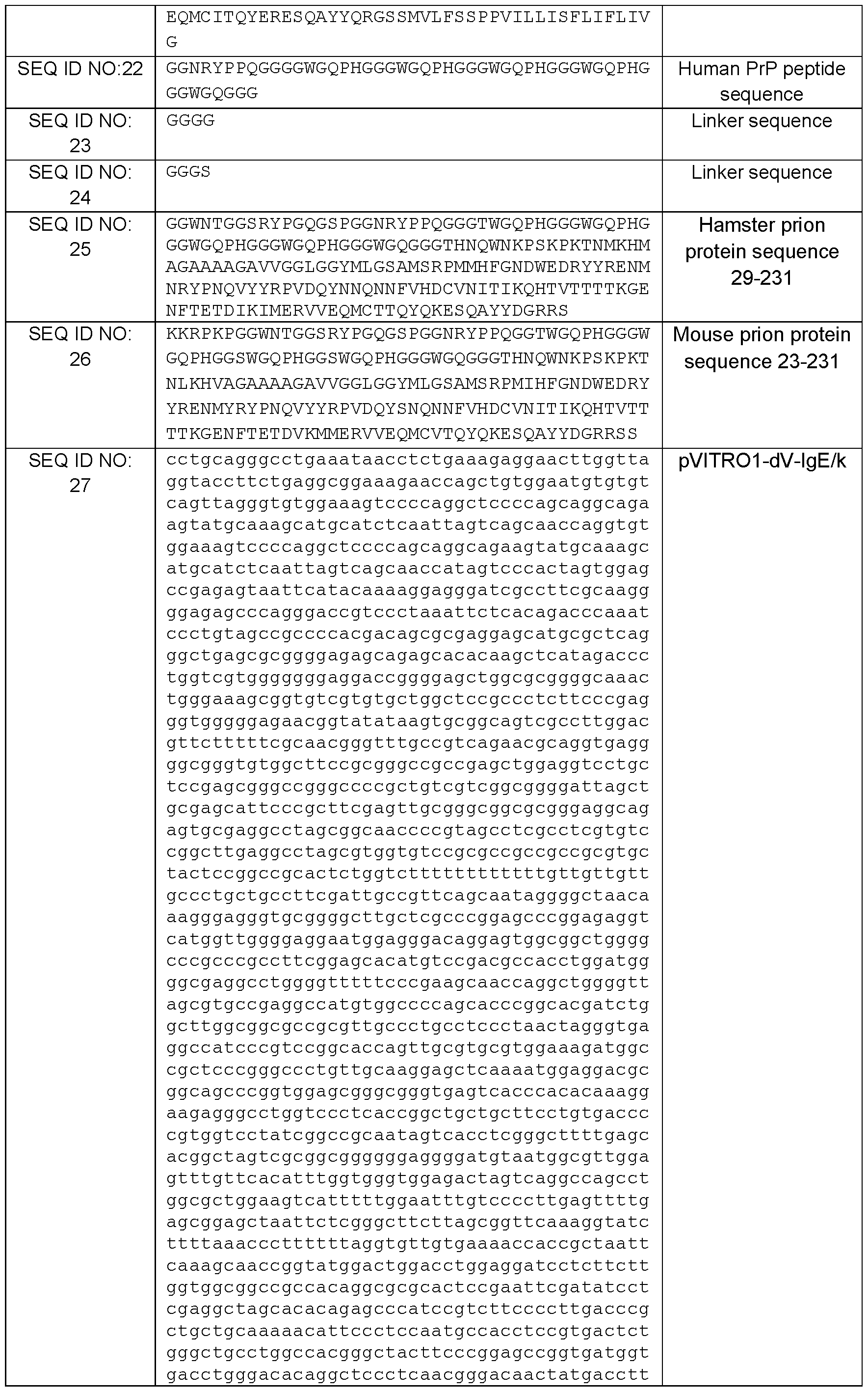


Legend: underlined = CDR 1 , 2 or 3
REFERENCES
1. Fitzsimmons, C. M., F. H. Falcone, and D. W. Dunne. 2014. Helminth Allergens, Parasite- Specific IgE, and Its Protective Role in Human Immunity. Frontiers in immunology 5: 61.
2. Stone, K. D., C. Prussin, and D. D. Metcalfe. 2010. IgE, mast cells, basophils, and eosinophils. The Journal of allergy and clinical immunology 125: S73-80.
3. Moon, T. C., A. D. Befus, and M. Kulka. 2014. Mast cell mediators: their differential release and the secretory pathways involved. Frontiers in immunology 5: 569.
4. Wernersson, S., and G. Pejler. 2014. Mast cell secretory granules: armed for battle. Nature reviews. Immunology 14: 478-494.
5. Perez, E. E., J. S. Orange, F. Bonilla, J. Chinen, I. K. Chinn, M. Dorsey, Y. El-Gamal, T. O. Harville, E. Hossny, B. Mazer, R. Nelson, E. Secord, S. C. Jordan, E. R. Stiehm, A. A. Vo, and M. Ballow. 2017. Update on the use of immunoglobulin in human disease: A review of evidence. The Journal of allergy and clinical immunology 139: S1-S46.
6. Sutton, B. J., and A. M. Davies. 2015. Structure and dynamics of IgE-receptor interactions: FcepsilonRI and CD23/FcepsilonRII. Immunological reviews 268: 222-235.
7. Shelburne, C. P., and S. N. Abraham. 2011. The mast cell in innate and adaptive immunity. Advances in experimental medicine and biology 716: 162-185.
8. Negrao-Correa, D., L. S. Adams, and R. G. Bell. 1996. Intestinal transport and catabolism of IgE: a major blood-independent pathway of IgE dissemination during a Trichinella spiralis infection of rats. Journal of immunology 157: 4037-4044.
9. Teo, P. Z., P. J. Utz, and J. A. Mollick. 2012. Using the allergic immune system to target cancer: activity of IgE antibodies specific for human CD20 and MUC1. Cancer immunology, immunotherapy : Cll 61 : 2295-2309.
10. Josephs, D. H., J. F. Spicer, P. Karagiannis, H. J. Gould, and S. N. Karagiannis. 2014. IgE immunotherapy: a novel concept with promise for the treatment of cancer. mAbs 6: 54-72.
11. Leoh, L. S., T. R. Daniels-Wells, and M. L. Penichet. 2015. IgE immunotherapy against cancer. Current topics in microbiology and immunology 388: 109-149.
12. Vidarsson, G., G. Dekkers, and T. Rispens. 2014. IgG subclasses and allotypes: from structure to effector functions. Frontiers in immunology 5: 520.
13. Muldoon, L. L, C. Soussain, K. Jahnke, C. Johanson, T. Siegal, Q. R. Smith, W. A. Hall, K. Hynynen, P. D. Senter, D. M. Peereboom, and E. A. Neuwelt. 2007. Chemotherapy delivery issues in central nervous system malignancy: a reality check. Journal of clinical oncology : official journal of the American Society of Clinical Oncology 25: 2295-2305.
14. Daniels-Wells, T. R., G. Helguera, R. K. Leuchter, R. Quintero, M. Kozman, J. A. Rodriguez, E. Ortiz-Sanchez, O. Martinez-Maza, B. C. Schultes, C. F. Nicodemus, and M. L. Penichet. 2013. A novel IgE antibody targeting the prostate-specific antigen as a potential prostate cancer therapy. BMC cancer 13: 195.
15. Reber, L. L, J. D. Hernandez, and S. J. Galli. 2017. The pathophysiology of anaphylaxis. The Journal of allergy and clinical immunology 140: 335-348.
16. Spicer, J., B. Basu, A. Montes, U. Banerji, R. Kristeleit, G. J. Veal, C. Corrigan, S. Till, G. Nintos, T. Brier, I. G. Funingana, J. E. Ang, K. Zaki, A. Griffin, C. Barton, P. Jones, S. Mellor, S. Brook, K. Stoddart, C. Selkirk, S. Carroll, H. Lentfer, N. Woodman, A. Pope, G. Pellizzari, M. Nakamura, K. M. Ilieva, A. Khiabany, C. Stavraka, H. Gould, J. Chauhan, H. Bax, S. Pinder, D. Josephs, and S. Karagiannis. 2020. Abstract CT141 : Phase 1 trial of MOv18, a first-in-class IgE antibody therapy for cancer. Cancer Research 80: CT141-CT141.
17. Suzuki, R., S. Leach, W. Liu, E. Ralston, J. Scheffel, W. Zhang, C. A. Lowell, and J. Rivera. 2014. Molecular editing of cellular responses by the high-affinity receptor for IgE. Science 343: 1021- 1025.
18. Chang, X., L. Zha, A. Wallimann, M. O. Mohsen, P. Krenger, X. Liu, M. Vogel, and M. F. Bachmann. 2021. Low-affinity but high-avidity interactions may offer an explanation for IgE-mediated allergen cross-reactivity. Allergy 76: 2565-2574.
19. Jucker, M., and L. C. Walker. 2013. Self-propagation of pathogenic protein aggregates in neurodegenerative diseases. Nature 501 : 45-51.
20. Greenlee, J. J., and M. H. Greenlee. 2015. The transmissible spongiform encephalopathies of livestock. ILAR journal 56: 7-25.
21. Sikorska, B., and P. P. Liberski. 2012. Human prion diseases: from Kuru to variant Creutzfeldt- Jakob disease. Sub-cellular biochemistry 65: 457-496.
22. Stewart, L. A., L. H. Rydzewska, G. F. Keogh, and R. S. Knight. 2008. Systematic review of therapeutic interventions in human prion disease. Neurology 70: 1272-1281.
23. Colby, D. W., and S. B. Prusiner. 2011. Prions. Cold Spring Harbor perspectives in biology 3: a006833.
24. Cortez, L. M., J. Kumar, L. Renault, H. S. Young, and V. L. Sim. 2013. Mouse prion protein polymorphism Phe-108/Val-189 affects the kinetics of fibril formation and the response to seeding: evidence for a two-step nucleation polymerization mechanism. The Journal of biological chemistry 288: 4772-4781.
25. Mueller, D. A., L. Heinig, S. Ramljak, A. Krueger, R. Schulte, A. Wrede, and A. W. Stuke. 2010. Conditional expression of full-length humanized anti-prion protein antibodies in Chinese hamster ovary cells. Hybridoma 29: 463-472.
26. Kuehn, H. S., M. Radinger, and A. M. Gilfillan. 2010. Measuring mast cell mediator release. Current protocols in immunology Chapter 7: Unit7 38.
27. Catalli, A., V. Karpov, L. E. Erdos, B. P. Tancowny, R. P. Schleimer, and M. Kulka. 2014. Stimulus-selective regulation of human mast cell gene expression, degranulation and leukotriene production by fluticasone and salmeterol. PloS one 9: e96891.
28. Gaudenzio, N., R. Sibilano, T. Marichal, P. Starkl, L. L. Reber, N. Cenac, B. D. McNeil, X. Dong, J. D. Hernandez, R. Sagi-Eisenberg, I. Hammel, A. Roers, S. Valitutti, M. Tsai, E. Espinosa, and S. J. Galli. 2016. Different activation signals induce distinct mast cell degranulation strategies. The Journal of clinical investigation 126: 3981-3998.
29. Yu, Y., D. Lin, Z. Liu, R. Fang, S. Zheng, Y. Cheng, Z. Huang, C. W. Ng, and H. Y. A. Lau. 2022. 6-O-angeloylplenolin inhibits anti-lgE-stimulated human mast cell activation via suppressing calcium influx and ERK phosphorylation. Iranian journal of basic medical sciences 25: 629-634.
30. Johnsson, A. K., J. H. Choi, E. Ronnberg, D. Fuchs, J. Kolmert, M. Hamberg, B. Dahlen, C. E. Wheelock, S. E. Dahlen, and G. Nilsson. 2021. Selective inhibition of prostaglandin D2 biosynthesis in human mast cells to overcome need for multiple receptor antagonists: Biochemical consequences. Clinical and experimental allergy : journal of the British Society for Allergy and Clinical Immunology 51: 594-603.
31. Yamaguchi, M., K. Sayama, K. Yano, C. S. Lantz, N. Noben-Trauth, C. Ra, J. J. Costa, and S. J. Galli. 1999. IgE enhances Fc epsilon receptor I expression and IgE-dependent release of histamine and lipid mediators from human umbilical cord blood-derived mast cells: synergistic effect of IL-4 and IgE on human mast cell Fc epsilon receptor I expression and mediator release. Journal of immunology 162: 5455-5465.
32. Kulka, M., C. H. Sheen, B. P. Tancowny, L. C. Grammer, and R. P. Schleimer. 2008. Neuropeptides activate human mast cell degranulation and chemokine production. Immunology 123: 398-410.
33. Nakajima, T., N. Inagaki, H. Tanaka, A. Tanaka, M. Yoshikawa, M. Tamari, K. Hasegawa, K. Matsumoto, H. Tachimoto, M. Ebisawa, G. Tsujimoto, H. Matsuda, H. Nagai, and H. Saito. 2002. Marked increase in CC chemokine gene expression in both human and mouse mast cell transcriptomes following Fcepsilon receptor I cross-linking: an interspecies comparison. Blood 100: 3861-3868.
34. Motakis, E., S. Guhl, Y. Ishizu, M. Itoh, H. Kawaji, M. de Hoon, T. Lassmann, P. Carninci, Y. Hayashizaki, T. Zuberbier, A. R. Forrest, M. Babina, and F. consortium. 2014. Redefinition of the human mast cell transcriptome by deep-CAGE sequencing. Blood 123: e58-67.
35. Jayapal, M., H. K. Tay, R. Reghunathan, L. Zhi, K. K. Chow, M. Rauff, and A. J. Melendez. 2006. Genome-wide gene expression profiling of human mast cells stimulated by IgE or FcepsilonRI-
aggregation reveals a complex network of genes involved in inflammatory responses. BMC genomics 7: 210.
36. Krasemann, S., M. H. Groschup, S. Harmeyer, G. Hunsmann, and W. Bodemer. 1996. Generation of monoclonal antibodies against human prion proteins in PrPO/O mice. Molecular medicine 2: 725-734.
37. Dodev, T. S., P. Karagiannis, A. E. Gilbert, D. H. Josephs, H. Bowen, L. K. James, H. J. Bax, R. Beavil, M. O. Pang, H. J. Gould, S. N. Karagiannis, and A. J. Beavil. 2014. A tool kit for rapid cloning and expression of recombinant antibodies. Scientific reports 4: 5885.
38. Haddon, D. J., M. R. Hughes, F. Antignano, D. Westaway, N. R. Cashman, and K. M. McNagny. 2009. Prion protein expression and release by mast cells after activation. The Journal of infectious diseases 200: 827-831.
39. Schulman, E. S. 2001. Development of a monoclonal anti-immunoglobulin E antibody (omalizumab) for the treatment of allergic respiratory disorders. American journal of respiratory and critical care medicine 164: S6-11.
40. Fattakhova, G., M. Masilamani, F. Borrego, A. M. Gilfillan, D. D. Metcalfe, and J. E. Coligan. 2006. The high-affinity immunoglobulin-E receptor (FcepsilonRI) is endocytosed by an AP-2/clathrin- independent, dynamin-dependent mechanism. Traffic 7: 673-685.
41. Kopec, A., B. Panaszek, and A. M. Fal. 2006. Intracellular signaling pathways in IgE-dependent mast cell activation. Archivum immunologiae et therapiae experimentalis 54: 393-401.
42. Buhring, H. J., P. J. Simmons, M. Pudney, R. Muller, D. Jarrossay, A. van Agthoven, M. Willheim, W. Brugger, P. Valent, and L. Kanz. 1999. The monoclonal antibody 97A6 defines a novel surface antigen expressed on human basophils and their multipotent and unipotent progenitors. Blood 94: 2343-2356.
43. Caughey, B., and G. J. Raymond. 1991 . The scrapie-associated form of PrP is made from a cell surface precursor that is both protease- and phospholipase-sensitive. The Journal of biological chemistry 266: 18217-18223.
44. Sim, V. L. 2012. Prion disease: chemotherapeutic strategies. Infectious disorders drug targets 12: 144-160.
45. Kamatari, Y. O., Y. Hayano, K. Yamaguchi, J. Hosokawa-Muto, and K. Kuwata. 2013. Characterizing antiprion compounds based on their binding properties to prion proteins: implications as medical chaperones. Protein science : a publication of the Protein Society 22: 22-34.
46. Doh-ura, K. 2004. [Prion diseases: disease diversity and therapeutics], Rinsho shinkeigaku = Clinical neurology 44: 855-856.
47. Priola, S. A., A. Raines, and W. S. Caughey. 2000. Porphyrin and phthalocyanine antiscrapie compounds. Science 287: 1503-1506.
48. Rudyk, H., S. Vasiljevic, R. M. Hennion, C. R. Birkett, J. Hope, and I. H. Gilbert. 2000. Screening Congo Red and its analogues for their ability to prevent the formation of PrP-res in scrapie-infected cells. The Journal of general virology 81: 1155-1164.
49. Kimberlin, R. H., and C. A. Walker. 1989. The role of the spleen in the neuroinvasion of scrapie in mice. Virus research 12: 201-211.
50. Hadlow, W. J., R. E. Race, and R. C. Kennedy. 1987. Temporal distribution of transmissible mink encephalopathy virus in mink inoculated subcutaneously. Journal of virology 61 : 3235-3240.
51. Hilton, D. A., E. Fathers, P. Edwards, J. W. Ironside, and J. Zajicek. 1998. Prion immunoreactivity in appendix before clinical onset of variant Creutzfeldt-Jakob disease. Lancet 352: 703-704.
52. Kimberlin, R. H., and C. A. Walker. 1989. Pathogenesis of scrapie in mice after intragastric infection. Virus research 12: 213-220.
53. Williams, S. K., R. Fairless, J. Weise, U. Kalinke, W. Schulz-Schaeffer, and R. Diem. 2011. Neuroprotective effects of the cellular prion protein in autoimmune optic neuritis. The American journal of pathology 178: 2823-2831.
54. Aude-Garcia, C., C. Villiers, S. M. Candeias, C. Garrel, C. Bertrand, V. Collin, P. N. Marche, and E. Jouvin-Marche. 2011. Enhanced susceptibility of T lymphocytes to oxidative stress in the absence of the cellular prion protein. Cellular and molecular life sciences : CMLS 68: 687-696.
55. Rovis, T. L, and G. Legname. 2014. Prion protein-specific antibodies-development, modes of action and therapeutics application. Viruses 6: 3719-3737.
56. Heegaard, P. M., A. L. Bergstrom, H. G. Andersen, and H. Cordes. 2015. Prion-Specific Antibodies Produced in Wild-Type Mice. Methods in molecular biology 1348: 285-301.
57. Didonna, A., A. C. Venturini, K. Hartman, T. Vranac, V. Curin Serbec, and G. Legname. 2015. Characterization of four new monoclonal antibodies against the distal N-terminal region of PrP(c). PeerJ 3: e811.
58. Sadowski, M. J., J. Pankiewicz, F. Prelli, H. Scholtzova, D. S. Spinner, R. B. Kascsak, R. J. Kascsak, and T. Wisniewski. 2009. Anti-PrP Mab 6D11 suppresses PrP(Sc) replication in prion infected myeloid precursor line FDC-P1/22L and in the lymphoreticular system in vivo. Neurobiology of disease 34: 267-278.
59. Mead, S., A. Khalili-Shirazi, C. Potter, T. Mok, A. Nihat, H. Hyare, S. Canning, C. Schmidt, T. Campbell, L. Darwent, N. Muirhead, N. Ebsworth, P. Hextall, M. Wakeling, J. Linehan, V. Libri, B. Williams, Z. Jaunmuktane, S. Brandner, P. Rudge, and J. Collinge. 2022. Prion protein monoclonal antibody (PRN100) therapy for Creutzfeldt-Jakob disease: evaluation of a first-in-human treatment programme. The Lancet. Neurology 21 : 342-354.
60. Ferris, R. L, H. J. Lenz, A. M. Trotta, J. Garcia-Foncillas, J. Schulten, F. Audhuy, M. Merlano, and G. Milano. 2018. Rationale for combination of therapeutic antibodies targeting tumor cells and immune checkpoint receptors: Harnessing innate and adaptive immunity through lgG1 isotype immune effector stimulation. Cancer treatment reviews 63: 48-60.
61. Taschuk, R., J. Van der Merwe, K. Marciniuk, A. Potter, N. Cashman, P. Griebel, and S. Napper. 2015. In vitro neutralization of prions with PrP(Sc)-specific antibodies. Prion 9: 292-303.
62. Lunnon, K., J. L. Teeling, A. L. Tutt, M. S. Cragg, M. J. Glennie, and V. H. Perry. 2011. Systemic inflammation modulates Fc receptor expression on microglia during chronic neurodegeneration. Journal of immunology 186: 7215-7224.
63. Feraudet-Tarisse, C., O. Andreoletti, N. Morel, S. Simon, C. Lacroux, J. Mathey, P. Lamourette, A. Relano, J. M. Torres, C. Creminon, and J. Grassi. 2010. Immunotherapeutic effect of anti-PrP monoclonal antibodies in transmissible spongiform encephalopathy mouse models: pharmacokinetic and pharmacodynamic analysis. The Journal of general virology 91: 1635-1645.
64. Prusiner, S. B., D. Groth, A. Serban, R. Koehler, D. Foster, M. Torchia, D. Burton, S. L. Yang, and S. J. DeArmond. 1993. Ablation of the prion protein (PrP) gene in mice prevents scrapie and facilitates production of anti-PrP antibodies. Proceedings of the National Academy of Sciences of the United States of America 90: 10608-10612.
65. Preithner, S., S. Elm, S. Lippold, M. Locher, A. Wolf, A. J. da Silva, P. A. Baeuerle, and N. S. Prang. 2006. High concentrations of therapeutic lgG1 antibodies are needed to compensate for inhibition of antibody-dependent cellular cytotoxicity by excess endogenous immunoglobulin G. Molecular immunology 43: 1183-1193.
66. Solomon, B., R. Koppel, E. Hanan, and T. Katzav. 1996. Monoclonal antibodies inhibit in vitro fibrillar aggregation of the Alzheimer beta-amyloid peptide. Proceedings of the National Academy of Sciences of the United States of America 93: 452-455.
67. Schenk, D., R. Barbour, W. Dunn, G. Gordon, H. Grajeda, T. Guido, K. Hu, J. Huang, K. Johnson-Wood, K. Khan, D. Kholodenko, M. Lee, Z. Liao, I. Lieberburg, R. Motter, L. Mutter, F. Soriano, G. Shopp, N. Vasquez, C. Vandevert, S. Walker, M. Wogulis, T. Yednock, D. Games, and P. Seubert. 1999. Immunization with amyloid-beta attenuates Alzheimer-disease-like pathology in the PDAPP mouse. Nature 400: 173-177.
68. Valera, E., B. Spencer, and E. Masliah. 2016. Immunotherapeutic Approaches Targeting Amyloid-beta, alpha-Synuclein, and Tau for the Treatment of Neurodegenerative Disorders. Neurotherapeutics : the journal of the American Society for Experimental NeuroTherapeutics 13: 179- 189.
69. Vandenberghe, R., J. O. Rinne, M. Boada, S. Katayama, P. Scheltens, B. Vellas, M. Tuchman, A. Gass, J. B. Fiebach, D. Hill, K. Lobello, D. Li, T. McRae, P. Lucas, I. Evans, K. Booth, G. Luscan, B. T. Wyman, L. Hua, L. Yang, H. R. Brashear, R. S. Black, Bapineuzumab, and I. Clinical Study. 2016.
Bapineuzumab for mild to moderate Alzheimer's disease in two global, randomized, phase 3 trials. Alzheimer's research & therapy 8: 18.
70. Sutton, B. J., A. M. Davies, H. J. Bax, and S. N. Karagiannis. 2019. IgE Antibodies: From Structure to Function and Clinical Translation. Antibodies 8.
71. Kim, C., T. Haldiman, K. Surewicz, Y. Cohen, W. Chen, J. Blevins, M. S. Sy, M. Cohen, Q. Kong, G. C. Telling, W. K. Surewicz, and J. G. Safar. 2012. Small protease sensitive oligomers of PrPSc in distinct human prions determine conversion rate of PrP(C). PLoS pathogens 8: e1002835.
72. Castle, A. R., and A. C. Gill. 2017. Physiological Functions of the Cellular Prion Protein. Frontiers in molecular biosciences 4: 19.
73. Chiesa, R. 2015. The elusive role of the prion protein and the mechanism of toxicity in prion disease. PLoS pathogens 11 : e1004745.
74. Jarosz-Griffiths, H. H., N. J. Corbett, H. A. Rowland, K. Fisher, A. C. Jones, J. Baron, G. J. Howell, S. A. Cowley, S. Chintawar, M. Z. Cader, K. A. B. Kellett, and N. M. Hooper. 2019. Proteolytic shedding of the prion protein via activation of metallopeptidase ADAM10 reduces cellular binding and toxicity of amyloid-beta oligomers. The Journal of biological chemistry 294: 7085-7097.
75. Endres, K., G. Mitteregger, E. Kojro, H. Kretzschmar, and F. Fahrenholz. 2009. Influence of ADAM10 on prion protein processing and scrapie infectiosity in vivo. Neurobiology of disease 36: 233- 241.
76. Guhl, S., M. Babina, A. Neou, T. Zuberbier, and M. Artuc. 2010. Mast cell lines HMC-1 and LAD2 in comparison with mature human skin mast cells— drastically reduced levels of tryptase and chymase in mast cell lines. Experimental dermatology 19: 845-847.
77. Babicki S, Arndt D, Marcu A, Liang Y, Grant JR, Maciejewski A, Wishart DS. Heatmapper: web- enabled heat mapping for all. Nucleic Acids Res. 2016 Jul 8;44(W1 ):W147-53.
78. Willows, S.D.; Semenchenko, V.; Norman, G.; Woodside, M.T.; Sim, V.L.; Kulka, M. Mast Cell Proteases Cleave Prion Proteins and a Recombinant Ig against PrP Can Activate Human Mast Cells. J Immunol2023, 210, 1447-1458, doi:10.4049/jimmunol.2200726.
79. Prusiner, S.B. The Prion Diseases. Brain Pathol 1998, 8, 499-513, doi: 10.1111/j.1750- 3639.1998.tb00171.x.
80. Warrington, R.; Watson, W.; Kim, H.L.; Antonetti, F.R. An Introduction to Immunology and Immunopathology. Allergy Asthma Clin Immunol 2011 , 7, S1, doi:10.1186/1710-1492-7-S1-S1.
81. Ma, Y.; Ma, J. Immunotherapy against Prion Disease. Pathogens 2020, 9, 216, doi: 10.3390/pathogens9030216.
82. Farmand, S.; Sundin, M. Hyper-lgE Syndromes: Recent Advances in Pathogenesis, Diagnostics and Clinical Care. Curr Opin Hematol 2015, 22, 12-22, doi: 10.1097/MCH.0000000000000104.
83. Esposito, P.; Chandler, N.; Kandere, K.; Basu, S.; Jacobson, S.; Connolly, R.; Tutor, D.; Theoharides, T.C. Corticotropin-Releasing Hormone and Brain Mast Cells Regulate Blood-Brain-Barrier Permeability Induced by Acute Stress. J Pharmacol Exp Ther 2002, 303, 1061-1066, doi: 10. 1124/jpet.102.038497.
84. Kritas, S.K.; Saggini, A.; Cerulli, G.; Caraffa, A.; Antinolfi, P.; Pantalone, A.; Rosati, M.; Tei, M.; Speziali, A.; Saggini, R.; et al. Impact of Mast Cells on Multiple Sclerosis: Inhibitory Effect of Natalizumab. Int J Immunopathol Pharmacol 2014, 27, 331-335, doi: 10.1177/039463201402700303.
85. Intestinal Transport and Catabolism of IgE: A Major Blood-Independent Pathway of IgE Dissemination during a Trichinella Spiralis Infection of Rats - PubMed Available online: https://pubmed.ncbi.nlm.nih.gov/8892637/ (accessed on 26 April 2023).
86. Polymenidou, M.; Moos, R.; Scott, M.; Sigurdson, C.; Shi, Y.-Z.; Yajima, B.; Hafner-Bratkovic, I.; Jerala, R.; Hornemann, S.; Wuthrich, K.; et al. The POM Monoclonals: A Comprehensive Set of Antibodies to Non-Overlapping Prion Protein Epitopes. PLoS ONE 2008, 3, e3872, doi: 10.1371/journal. pone.0003872.
Claims
1 . An IgE antibody that binds specifically to prion protein (PrP), wherein the antibody comprises three variable heavy domain complementarity determining regions (CDR)(CDR H1 , H2 and H3), and three variable light domain CDR (CDR L1 , L2 and L3), wherein: the CDR H1 comprises the amino acid sequence GFSLTSYGVH (SEQ ID NO:1), the CDR H2 comprises the amino acid sequence VIWSDGSTTYDSTLKS (SEQ ID NO:2), the CDR H3 comprises the amino acid sequence RHFY (SEQ ID NO:3), the CDR L1 comprises the amino acid sequence KSSQSLLDSDGKTYLN (SEQ ID NO:4), the CDR L2 comprises the amino acid sequence LVSKLDS (SEQ ID NO:5), and the CDR L3 comprises the amino acid sequence WQGTHFPQT (SEQ ID NO:6); or the CDR H1 comprises the amino acid sequence NYLIE (SEQ ID NO:28), the CDR H2 comprises the amino acid sequence VINPGSGDTNYNEKFKG (SEQ ID NO:29), the CDR H3 comprises the amino acid sequence SGAAAPTYYAMDY (SEQ ID NQ:30), the CDR L1 comprises the amino acid sequence KASQVGTALA (SEQ ID NO:31), the CDR L2 comprises the amino acid sequence WASTRHT (SEQ ID NO:32), and the CDR L3 comprises the amino acid sequence QQYSSYPT (SEQ ID NO:33).
2. The IgE antibody of claim 1 , wherein said antibody provides lower levels of degranulation from an activated mast cell bound with said IgE antibody compared to an activated mast cell bound with an HE1 control IgE monoclonal antibody, registered in The Antibody Registry under #RRID AB_1075924, as determined by measuring the amount of p-hexosaminidase released by each activated mast cell into a surrounding medium, wherein a lower level of p-hexosaminidase release indicates a lower level of degranulation.
3. The IgE antibody of claim 1 or 2, further comprising four variable heavy domain framework regions (HFR)(HFR 1 , 2, 3 and 4), wherein: the HFR 1 comprises the amino acid sequence GPGLVAPSQSLSITCTIS (SEQ ID NO:7), the HFR 2 comprises the amino acid sequence WVRQPPGKGLEWLV (SEQ ID NO:8), the HFR 3 comprises the amino acid sequence RLSISKDNSKSQVFSKMNSLQTDDTAMYCCAR (SEQ ID NO:9), and the HFR 4 comprises the amino acid sequence A (SEQ ID NQ:10); or the HFR 1 comprises the amino acid sequence VKLQESGGEWRPGTSVKVSCKASGYAFT (SEQ ID NO:34), the HFR 2 comprises the amino acid sequence WVKQRPGQGLEWIG (SEQ ID NO:35), the HFR 3 comprises the amino acid sequence KATLTADKSSSTAYMQLNSLTSDDSAVYFCAR (SEQ ID NO:36), and the HFR 4 comprises the amino acid sequence WGQGVSVTVSS (SEQ ID NO:37).
4. The IgE antibody of any one of claims 1 to 3, further comprising four variable light domain framework regions (LFR)(LFR 1 , 2, 3 and 4), wherein: the LFR 1 comprises the amino acid sequence DIVMTQTTFTLSVTIGQPASISC (SEQ ID NO:11), the LFR 2 comprises the amino acid sequence WLLQRPGQSPKRLIY (SEQ ID NO:12), the LFR 3 comprises the amino acid sequence GVPDRFTGSGSGTDFTLKISRVEAEDLGVYYC (SEQ ID NO:13), and the LFR 4 comprises the amino acid sequence FGGG (SEQ ID NO:14); or the LFR 1 comprises the amino acid sequence DIVMTQSHKFMSTSVGDRVSITC (SEQ ID NO:38), the LFR 2 comprises the amino acid sequence WYQQKPGQSPKLLIY (SEQ ID NO:39), the LFR 3 comprises the amino acid sequence GVPDRFTGSGSGTDFTLTISNVQSEDLSDYFC (SEQ ID NQ:40), and the LFR 4 comprises the amino acid sequence FGGGTKLEIKRA (SEQ ID NO:41).
5. The IgE antibody of claim 1 , wherein said antibody comprises a variable heavy domain (VH) comprising the amino acid sequence:
GPGLVAPSQSLSITCTISGFSLTSYGVHWVRQPPGKGLEWLWIWSDGSTTYDSTLKSRLSISKD NSKSQVFSKMNSLQTDDTAMYCCARRHFYA (SEQ ID NO:15).
6. The IgE antibody of claim 5, wherein said antibody comprises a variable light domain (VL) comprising the amino acid sequence:
DIVMTQTTFTLSVTIGQPASISCKSSQSLLDSDGKTYLNWLLQRPGQSPKRLIYLVSKLDSGVPDR FTGSGSGTDFTLKISRVEAEDLGVYYCWQGTHFPQTFGGG (SEQ ID NO:16)
7. The IgE antibody claim 1 , wherein said antibody comprises a heavy chain comprising the amino acid sequence:
MDWTWRILFLVAAATGAHSEFDILEGPGLVAPSQSLSITCTISGFSLTSYGVHWVRQPPGKGLE WLWIWSDGSTTYDSTLKSRLSISKDNSKSQVFSKMNSLQTDDTAMYCCARRHFYAASTQSPSV FPLTRCCKNIPSNATSVTLGCLATGYFPEPVMVTWDTGSLNGTTMTLPATTLTLSGHYATISLLTV SGAWAKQMFTCRVAHTPSSTDVWDNKTFSVCSRDFTPPTVKILQSSCDGGGHFPPTIQLLCLVS GYTPGTINITWLEDGQVMDVDLSTASTTQEGELASTQSELTLSQKHWLSDRTYTCQVTYQGHTF EDSTKKCADSNPRGVSAYLSRPSPFDLFIRKSPTITCLVVDLAPSKGTVNLTWSRASGKPVNHST RKEEKQRNGTLTVTSTLPVGTRDWIEGETYQCRVTHPHLPRALMRSTTKTSGPRAAPEVYAFAT PEWPGSRDKRTLACLIQNFMPEDISVQWLHNEVQLPDARHSTTQPRKTKGSGFFVFSRLEVTRA EWEQKDEFICRAVHEAASPSQTVQRAVSVNPGK (SEQ ID NO:17).
8. The IgE antibody of claim 7, wherein said antibody comprises a light chain comprising the amino acid sequence:
MLPSQLIGFLLLWVPASRGEFDILERDIVMTQTTFTLSVTIGQPASISCKSSQSLLDSDGKTYLNW LLQRPGQSPKRLIYLVSKLDSGVPDRFTGSGSGTDFTLKISRVEAEDLGVYYCWQGTHFPQTFG
GGTVAAPSVFIFPPSDEQKSGTASVVCLLNNFYPREAKVQKVDNALQSGNSQESVTEQDSKDST
YSLSSTLTLSKADYEKHKVYACEVTHQGLSSPVTKSFNRGEC (SEQ ID N0:18).
9. The I g E antibody of claim 1 , wherein said antibody comprises a variable heavy domain (VH) comprising the amino acid sequence:
VKLQESGGEWRPGTSVKVSCKASGYAFTNYLIEWVKQRPGQGLEWIGVINPGSGDTNYNEKF KGKATLTADKSSSTAYMQLNSLTSDDSAVYFCARSGAAAPTYYAMDYWGQGVSVTVSSAKTTP PSVYPLAPAAAAANSMVTLGCLVKGYFPEPVTVTWNSGSLSGGVHTFPAVLQSDLYTLSSSVTV PSSTWPSETVTCNVAHPASSTKVDKKIVPR (SEQ ID NO:42).
10. The IgE antibody of claim 9, wherein said antibody comprises a variable light domain (VL) comprising the amino acid sequence:
DIVMTQSHKFMSTSVGDRVSITCKASQVGTALAWYQQKPGQSPKLLIYWASTRHTGVPDRFTGS GSGTDFTLTISNVQSEDLSDYFCQQYSSYPTFGGGTKLEIKRADAAPTVSIFPPSSEQLTSGGAS VVCFLNNFYPKDINVKWKIDGSERQNGVLNSETDQDSKDSTYSMSSTLTLTKDEYERHNSYTCE ATHKTSTSPIVKSFNRA (SEQ ID NO:43).
11 . The IgE antibody of any one of claims 1 to 10, wherein said antibody is humanized.
12. The IgE antibody of any one of claims 1 to 11 , wherein said antibody fails to induce release of a chemokine C-C motif ligand 2 (CCL2) from an activated mast cell bound with said IgE antibody.
13. The IgE antibody of any one of claims 1 to 12, wherein said antibody provides weaker activation of an activated mast cell bound with said IgE antibody compared to an activated mast cell bound with the HE1 control IgE monoclonal antibody, as determined by measuring phosphorylation of SYK kinase, ERK kinase, or both, in said activated mast cell, wherein a lower level of phosphorylation indicates weaker activation.
14. A compound comprising the antibody of any one of claims 1 to 13 linked to a functional moiety.
15. The compound of claim 14, wherein the antibody is covalently linked to the functional moiety via a peptide linker.
16. The compound of claim 14, wherein the antibody is recombinantly linked to the functional moiety via the peptide linker.
17. The compound of claim 15 or 16, wherein the peptide linker comprises from 1 to about 40 amino acid residues.
18. The compound of any one of claims 15 to 17, wherein the peptide linker comprises the amino acid sequence (SS)n, (GGG)n, [GGGG (SEQ ID NO: 23)]n, [GGGS (SEQ ID NO: 24)]n, or [SSGGG (SEQ ID NO: 19)]n, or [GGGGS (SEQ ID NO: 20)]n, wherein n > 1 .
19. The compound of any one of claims 14 to 18,
wherein the IgE antibody is linked to a detectable label, a peptide, a polypeptide, a protein, an enzyme, a second antibody, an antibody fragment, an antigen-binding fragment, or a combination of any two or more thereof; wherein the IgE antibody is functional; and wherein the linked detectable label, peptide, polypeptide, protein, enzyme, second antibody, antibody fragment, antigen-binding fragment, or the combination of any two or more thereof is functional.
20. The compound of claim 19, wherein the antibody is linked to the second antibody and wherein the second antibody is operable to transmigrate the blood-brain barrier, cross the spinal cord and/or enter the central nervous system.
21 . A composition comprising the antibody of any one of claims 1 to 13 or the compound of any one of claims 14 to 20, and a pharmaceutically acceptable diluent, carrier or excipient.
22. A nucleic acid molecule encoding the antibody of any one of claims 1 to 13, or the compound of any one of claims 14 to 18.
23. A vector comprising the nucleic acid molecule of claim 22 operably linked to one or more regulatory elements to allow expression of the antibody or compound in a host cell.
24. A cell comprising the vector of claim 23 for expressing the antibody or compound.
25. A method of alleviating or treating a prion disease in a subject in need thereof, comprising administering an effective amount of the antibody of any one of claims 1 to 13, the compound of any one of claims 14 to 20, or the composition of claim 21 to said subject.
26. The method of claim 25, wherein said prion disease is Creutzfeldt-Jakob disease (CJD).
27. Use of the antibody of any one of claims 1 to 13, the compound of any one of claims 14 to 20, or the composition of claim 21 to alleviate or treat a prion disease.
28. Use of the antibody of any one of claims 1 to 13 or the compound of any one of claims 14 to 20, or the composition of claim 21 in the preparation of a medicament for the alleviation or treatment of a prion disease.
29. The use of claim 27 or 28, wherein said prion disease is Creutzfeldt-Jakob disease (CJD).
30. The antibody of any one of claims 1 to 13, the compound of any one of claims 14 to 20, or the composition of claim 21 for use to alleviate or treat a prion disease, wherein said prion disease is optionally Creutzfeldt-Jakob disease (CJD).
Applications Claiming Priority (2)
| Application Number | Priority Date | Filing Date | Title |
|---|---|---|---|
| US202363458555P | 2023-04-11 | 2023-04-11 | |
| US63/458,555 | 2023-04-11 |
Publications (1)
| Publication Number | Publication Date |
|---|---|
| WO2024211981A1 true WO2024211981A1 (en) | 2024-10-17 |
Family
ID=93058499
Family Applications (1)
| Application Number | Title | Priority Date | Filing Date |
|---|---|---|---|
| PCT/CA2024/050330 Ceased WO2024211981A1 (en) | 2023-04-11 | 2024-03-19 | Ige antibody against prion protein characterized using lad2 mast cells |
Country Status (1)
| Country | Link |
|---|---|
| WO (1) | WO2024211981A1 (en) |
Citations (1)
| Publication number | Priority date | Publication date | Assignee | Title |
|---|---|---|---|---|
| WO2010099612A1 (en) * | 2009-03-02 | 2010-09-10 | The University Of British Columbia | Antibodies and epitopes specific to misfolded prion protein |
-
2024
- 2024-03-19 WO PCT/CA2024/050330 patent/WO2024211981A1/en not_active Ceased
Patent Citations (1)
| Publication number | Priority date | Publication date | Assignee | Title |
|---|---|---|---|---|
| WO2010099612A1 (en) * | 2009-03-02 | 2010-09-10 | The University Of British Columbia | Antibodies and epitopes specific to misfolded prion protein |
Non-Patent Citations (3)
| Title |
|---|
| "Prion-Specific IgE Activates Human Mast Cells to Release Proteases", THE JOURNAL OF ALLERGY AND CLINICAL IMMUNOLOGY, vol. 149, 1 February 2022 (2022-02-01), XP086947645, ISSN: 0091-6749, DOI: KULKA, M et * |
| SWAYAMPAKULA MRIDULA, BARAL PRAVAS KUMAR, AGUZZI ADRIANO, KAV NAT N. V., JAMES MICHAEL N. G.: "The crystal structure of an octapeptide repeat of the Prion protein in complex with a Fab fragment of the POM2 antibody", PROTEIN SCIENCE, WILEY, US, vol. 22, no. 7, 1 July 2013 (2013-07-01), US , pages 893 - 903, XP093224282, ISSN: 0961-8368, DOI: 10.1002/pro.2270 * |
| WILLOWS STEVEN D., SEMENCHENKO VALENTYNA, NORMAN GRANT, WOODSIDE MICHAEL T., SIM VALERIE L., KULKA MARIANNA: "Mast Cell Proteases Cleave Prion Proteins and a Recombinant Ig against PrP Can Activate Human Mast Cells", JOURNAL OF IMMUNOLOGY, WILLIAMS & WILKINS CO., vol. 210, no. 9, 1 May 2023 (2023-05-01), pages 1447 - 1458, XP093224281, ISSN: 0022-1767, DOI: 10.4049/jimmunol.2200726 * |
Similar Documents
| Publication | Publication Date | Title |
|---|---|---|
| JP7610279B2 (en) | Antibodies to alpha-synuclein and uses thereof | |
| US20250250331A1 (en) | Anti-age antibodies for treating neurodegenerative disorders | |
| US10570193B2 (en) | Binding moieties for biofilm remediation | |
| US11919947B2 (en) | Antibody binding active α-synuclein | |
| JP7449092B2 (en) | Blood-brain barrier migration compounds and their uses | |
| US20140314773A1 (en) | Compositions and methods for treating alzheimer's disease | |
| David et al. | Potential candidate camelid antibodies for the treatment of protein-misfolding diseases | |
| WO2013071267A1 (en) | Compositions and methods for treating alzheimer's disease | |
| WO2017192594A1 (en) | Binding moieties for biofilm remediation | |
| US20150329622A1 (en) | Anti-bag3 antibodies for therapeutic use | |
| Manoutcharian et al. | Recombinant Antibody Fragments for Immunotherapy of Parkinson’s Disease: K. Manoutcharian, G. Gevorkian | |
| WO2024211981A1 (en) | Ige antibody against prion protein characterized using lad2 mast cells | |
| Willows et al. | Mast cell proteases cleave prion proteins and a recombinant Ig against PrP can activate human mast cells | |
| CN113646042B (en) | Method for specifically eliminating adipocytes presenting perilipin-1 fragment | |
| JP2024506409A (en) | antibody | |
| CA3071634C (en) | Antibody binding active alpha-synuclein | |
| HK40022840B (en) | ANTIBODIES TO α‐SYNUCLEIN AND USES THEREOF | |
| Ewert et al. | Humanized antibodies against the beta-amyloid peptide | |
| HK40062966A (en) | Means for specifically eliminating perilipin-1 fragment presenting adipocytes | |
| US20160280742A1 (en) | Compositions and Methods for Treating Alzheimer's Disease | |
| HK40022840A (en) | ANTIBODIES TO α‐SYNUCLEIN AND USES THEREOF |
Legal Events
| Date | Code | Title | Description |
|---|---|---|---|
| 121 | Ep: the epo has been informed by wipo that ep was designated in this application |
Ref document number: 24787692 Country of ref document: EP Kind code of ref document: A1 |
|
| NENP | Non-entry into the national phase |
Ref country code: DE |