WO2024116140A1 - Combination therapy for treatment of cancer comprising anti-pd-l1 and anti-cd73 antibodies - Google Patents
Combination therapy for treatment of cancer comprising anti-pd-l1 and anti-cd73 antibodies Download PDFInfo
- Publication number
- WO2024116140A1 WO2024116140A1 PCT/IB2023/062127 IB2023062127W WO2024116140A1 WO 2024116140 A1 WO2024116140 A1 WO 2024116140A1 IB 2023062127 W IB2023062127 W IB 2023062127W WO 2024116140 A1 WO2024116140 A1 WO 2024116140A1
- Authority
- WO
- WIPO (PCT)
- Prior art keywords
- antibody
- tumor
- inhibitor
- subject
- seq
- Prior art date
- Legal status (The legal status is an assumption and is not a legal conclusion. Google has not performed a legal analysis and makes no representation as to the accuracy of the status listed.)
- Ceased
Links
Classifications
-
- C—CHEMISTRY; METALLURGY
- C07—ORGANIC CHEMISTRY
- C07K—PEPTIDES
- C07K16/00—Immunoglobulins [IGs], e.g. monoclonal or polyclonal antibodies
- C07K16/18—Immunoglobulins [IGs], e.g. monoclonal or polyclonal antibodies against material from animals or humans
- C07K16/28—Immunoglobulins [IGs], e.g. monoclonal or polyclonal antibodies against material from animals or humans against receptors, cell surface antigens or cell surface determinants
- C07K16/2803—Immunoglobulins [IGs], e.g. monoclonal or polyclonal antibodies against material from animals or humans against receptors, cell surface antigens or cell surface determinants against the immunoglobulin superfamily
- C07K16/2827—Immunoglobulins [IGs], e.g. monoclonal or polyclonal antibodies against material from animals or humans against receptors, cell surface antigens or cell surface determinants against the immunoglobulin superfamily against B7 molecules, e.g. CD80, CD86
-
- A—HUMAN NECESSITIES
- A61—MEDICAL OR VETERINARY SCIENCE; HYGIENE
- A61K—PREPARATIONS FOR MEDICAL, DENTAL OR TOILETRY PURPOSES
- A61K39/00—Medicinal preparations containing antigens or antibodies
- A61K39/395—Antibodies; Immunoglobulins; Immune serum, e.g. antilymphocytic serum
- A61K39/39533—Antibodies; Immunoglobulins; Immune serum, e.g. antilymphocytic serum against materials from animals
- A61K39/39558—Antibodies; Immunoglobulins; Immune serum, e.g. antilymphocytic serum against materials from animals against tumor tissues, cells, antigens
-
- A—HUMAN NECESSITIES
- A61—MEDICAL OR VETERINARY SCIENCE; HYGIENE
- A61K—PREPARATIONS FOR MEDICAL, DENTAL OR TOILETRY PURPOSES
- A61K31/00—Medicinal preparations containing organic active ingredients
- A61K31/33—Heterocyclic compounds
- A61K31/335—Heterocyclic compounds having oxygen as the only ring hetero atom, e.g. fungichromin
- A61K31/337—Heterocyclic compounds having oxygen as the only ring hetero atom, e.g. fungichromin having four-membered rings, e.g. taxol
-
- A—HUMAN NECESSITIES
- A61—MEDICAL OR VETERINARY SCIENCE; HYGIENE
- A61K—PREPARATIONS FOR MEDICAL, DENTAL OR TOILETRY PURPOSES
- A61K31/00—Medicinal preparations containing organic active ingredients
- A61K31/33—Heterocyclic compounds
- A61K31/395—Heterocyclic compounds having nitrogen as a ring hetero atom, e.g. guanethidine or rifamycins
- A61K31/495—Heterocyclic compounds having nitrogen as a ring hetero atom, e.g. guanethidine or rifamycins having six-membered rings with two or more nitrogen atoms as the only ring heteroatoms, e.g. piperazine or tetrazines
- A61K31/505—Pyrimidines; Hydrogenated pyrimidines, e.g. trimethoprim
- A61K31/513—Pyrimidines; Hydrogenated pyrimidines, e.g. trimethoprim having oxo groups directly attached to the heterocyclic ring, e.g. cytosine
-
- A—HUMAN NECESSITIES
- A61—MEDICAL OR VETERINARY SCIENCE; HYGIENE
- A61K—PREPARATIONS FOR MEDICAL, DENTAL OR TOILETRY PURPOSES
- A61K31/00—Medicinal preparations containing organic active ingredients
- A61K31/33—Heterocyclic compounds
- A61K31/555—Heterocyclic compounds containing heavy metals, e.g. hemin, hematin, melarsoprol
-
- A—HUMAN NECESSITIES
- A61—MEDICAL OR VETERINARY SCIENCE; HYGIENE
- A61K—PREPARATIONS FOR MEDICAL, DENTAL OR TOILETRY PURPOSES
- A61K39/00—Medicinal preparations containing antigens or antibodies
- A61K39/395—Antibodies; Immunoglobulins; Immune serum, e.g. antilymphocytic serum
-
- A—HUMAN NECESSITIES
- A61—MEDICAL OR VETERINARY SCIENCE; HYGIENE
- A61K—PREPARATIONS FOR MEDICAL, DENTAL OR TOILETRY PURPOSES
- A61K45/00—Medicinal preparations containing active ingredients not provided for in groups A61K31/00 - A61K41/00
- A61K45/06—Mixtures of active ingredients without chemical characterisation, e.g. antiphlogistics and cardiaca
-
- A—HUMAN NECESSITIES
- A61—MEDICAL OR VETERINARY SCIENCE; HYGIENE
- A61P—SPECIFIC THERAPEUTIC ACTIVITY OF CHEMICAL COMPOUNDS OR MEDICINAL PREPARATIONS
- A61P35/00—Antineoplastic agents
-
- C—CHEMISTRY; METALLURGY
- C07—ORGANIC CHEMISTRY
- C07K—PEPTIDES
- C07K16/00—Immunoglobulins [IGs], e.g. monoclonal or polyclonal antibodies
- C07K16/18—Immunoglobulins [IGs], e.g. monoclonal or polyclonal antibodies against material from animals or humans
- C07K16/28—Immunoglobulins [IGs], e.g. monoclonal or polyclonal antibodies against material from animals or humans against receptors, cell surface antigens or cell surface determinants
- C07K16/2896—Immunoglobulins [IGs], e.g. monoclonal or polyclonal antibodies against material from animals or humans against receptors, cell surface antigens or cell surface determinants against molecules with a "CD"-designation, not provided for elsewhere
-
- G—PHYSICS
- G01—MEASURING; TESTING
- G01N—INVESTIGATING OR ANALYSING MATERIALS BY DETERMINING THEIR CHEMICAL OR PHYSICAL PROPERTIES
- G01N33/00—Investigating or analysing materials by specific methods not covered by groups G01N1/00 - G01N31/00
- G01N33/48—Biological material, e.g. blood, urine; Haemocytometers
- G01N33/50—Chemical analysis of biological material, e.g. blood, urine; Testing involving biospecific ligand binding methods; Immunological testing
- G01N33/68—Chemical analysis of biological material, e.g. blood, urine; Testing involving biospecific ligand binding methods; Immunological testing involving proteins, peptides or amino acids
- G01N33/6872—Intracellular protein regulatory factors and their receptors, e.g. including ion channels
-
- A—HUMAN NECESSITIES
- A61—MEDICAL OR VETERINARY SCIENCE; HYGIENE
- A61K—PREPARATIONS FOR MEDICAL, DENTAL OR TOILETRY PURPOSES
- A61K39/00—Medicinal preparations containing antigens or antibodies
- A61K2039/505—Medicinal preparations containing antigens or antibodies comprising antibodies
-
- A—HUMAN NECESSITIES
- A61—MEDICAL OR VETERINARY SCIENCE; HYGIENE
- A61K—PREPARATIONS FOR MEDICAL, DENTAL OR TOILETRY PURPOSES
- A61K39/00—Medicinal preparations containing antigens or antibodies
- A61K2039/505—Medicinal preparations containing antigens or antibodies comprising antibodies
- A61K2039/507—Comprising a combination of two or more separate antibodies
-
- A—HUMAN NECESSITIES
- A61—MEDICAL OR VETERINARY SCIENCE; HYGIENE
- A61K—PREPARATIONS FOR MEDICAL, DENTAL OR TOILETRY PURPOSES
- A61K2300/00—Mixtures or combinations of active ingredients, wherein at least one active ingredient is fully defined in groups A61K31/00 - A61K41/00
-
- C—CHEMISTRY; METALLURGY
- C07—ORGANIC CHEMISTRY
- C07K—PEPTIDES
- C07K2317/00—Immunoglobulins specific features
- C07K2317/50—Immunoglobulins specific features characterized by immunoglobulin fragments
- C07K2317/56—Immunoglobulins specific features characterized by immunoglobulin fragments variable (Fv) region, i.e. VH and/or VL
- C07K2317/565—Complementarity determining region [CDR]
-
- C—CHEMISTRY; METALLURGY
- C07—ORGANIC CHEMISTRY
- C07K—PEPTIDES
- C07K2317/00—Immunoglobulins specific features
- C07K2317/70—Immunoglobulins specific features characterized by effect upon binding to a cell or to an antigen
- C07K2317/76—Antagonist effect on antigen, e.g. neutralization or inhibition of binding
-
- G—PHYSICS
- G01—MEASURING; TESTING
- G01N—INVESTIGATING OR ANALYSING MATERIALS BY DETERMINING THEIR CHEMICAL OR PHYSICAL PROPERTIES
- G01N2333/00—Assays involving biological materials from specific organisms or of a specific nature
- G01N2333/435—Assays involving biological materials from specific organisms or of a specific nature from animals; from humans
- G01N2333/705—Assays involving receptors, cell surface antigens or cell surface determinants
- G01N2333/70596—Molecules with a "CD"-designation not provided for elsewhere in G01N2333/705
Definitions
- the invention disclosed herein relates to methods of treating a tumor in a subject comprising administration of a CD73 inhibitor, a T cell checkpoint inhibitor, and chemotherapy and/or radiotherapy.
- BACKGROUND OF THE INVENTION [0002] Extracellular adenosine has emerged as an important regulator of immunological processes within the tumor microenvironment and is thought to diminish the effectiveness of T- cell checkpoint inhibiting drugs (Sidders B, et al., Clin Cancer Res 2020, 26:2176–87; Augustin RC, et al., J Immunother Cancer 2022, 10:e004089).
- Adenosine triphosphate (ATP) released from necrotic or damaged cells, is hydrolysed to adenosine by the sequential action of two ectonucleosidases working in tandem: CD39 (ENTPD1) and CD73 (NT5E).
- ENTPD1 two ectonucleosidases working in tandem
- N5E CD73
- the resultant adenosine acts as a readily diffusible immunosuppressive ‘smog’, and it is likely that cytotoxic agents and radiotherapy exacerbate this process.
- enhanced anti-tumor activity is observed in preclinical models when radiotherapy is combined with an anti-CD73 (aCD73) inhibiting antibody treatment (Wennerberg E, et al.
- T-cell checkpoint inhibitors within this paradigm provides further scope for enhanced cell-mediated activity; by counteracting adaptive immune-resistance and unfettering anti-tumor immunity.
- Oleclumab a CD73 inhibiting human monoclonal IgG1-TM antibody (Hay CM, et al., Oncoimmunology 2016; 5) is currently in phase 2/3 of clinical development in combination with Durvalumab for treatment of patients with various solid tumors.
- the present disclosure is directed to a method of inhibiting tumor growth in a subject in need thereof comprising administering to the subject a therapeutically effective amount of a PD-L1 inhibitor in combination with chemotherapy and/or radiotherapy; wherein the subject has decreased CD73 protein or CD73 activity levels compared to a normal subject.
- the present disclosure is also directed to a method of treating cancer in a subject in need thereof comprising administering to the subject a therapeutically effective amount of a PD- L1 inhibitor in combination with chemotherapy and/or radiotherapy; wherein the subject has decreased CD73 protein or CD73 activity levels compared to a normal subject.
- the present disclosure is also directed to a method of producing a protective tumor memory response in a subject comprising administering to the subject a therapeutically effective amount of a PD-L1 inhibitor in combination with chemotherapy and/or radiotherapy; wherein the subject has decreased CD73 protein or CD73 activity levels compared to a normal subject.
- the present disclosure is also directed to a method of inhibiting tumor growth in a subject in need thereof comprising administering to the subject a therapeutically effective amount of a CD73 inhibitor, a PD-L1 inhibitor, and chemotherapy and/or radiotherapy.
- the present disclosure is also directed to a method of treating cancer in a subject in need thereof comprising administering to the subject a therapeutically effective amount of a CD73 inhibitor, a PD-L1 inhibitor, and chemotherapy and/or radiotherapy.
- the present disclosure is also directed to a method of producing a protective tumor memory response in a subject comprising administering to the subject a therapeutically effective amount of a CD73 inhibitor, a PD-L1 inhibitor, and chemotherapy and/or radiotherapy.
- the PD-L1 inhibitor, and the chemotherapy and/or radiotherapy are administered concurrently.
- the PD-L1 inhibitor, and the chemotherapy and/or radiotherapy are administered sequentially.
- the CD73 inhibitor is administered prior to administration of the PD-L1 inhibitor, and the chemotherapy and/or radiotherapy.
- the chemotherapy is docetaxel, 5-fluorouracil, and/or oxaliplatin.
- the PD-L1 inhibitor is an anti-PD-L1 antibody or an antigen-binding fragment thereof.
- the anti-PD-L1 antibody or antigen-binding fragment thereof comprising: (a) a heavy chain (HC) CDR1 comprising the amino acid sequence of SEQ ID NO:1, a HC CDR2 comprising the amino acid sequence of SEQ ID NO:2, and a HC CDR3 comprising the amino acid sequence of SEQ ID NO:3; and a light chain (LC) CDR1 comprising the amino acid sequence of SEQ ID NO:4, a LC CDR2 comprising the amino acid sequence of SEQ ID NO:5, and a LC CDR3 comprising the amino acid sequence of SEQ ID NO:6.
- HC heavy chain
- LC CDR2 comprising the amino acid sequence of SEQ ID NO:2
- a LC CDR3 comprising the amino acid sequence of SEQ ID NO:6.
- the anti- PD-L1 antibody or antigen binding fragment thereof comprises a HC variable domain (VH) comprising the amino acid sequence of SEQ ID NO:7, and a LC variable domain (VL) comprising the amino acid sequence of SEQ ID NO:8.
- VH HC variable domain
- VL LC variable domain
- the anti-PD-L1 antibody is durvalumab.
- the CD73 inhibitor is an anti-CD73 antibody or antigen-binding fragment thereof.
- the anti-CD73 antibody or antigen-binding fragment thereof comprising: (a) a HC CDR1 comprising the amino acid sequence of SEQ ID NO:9, a HC CDR2 comprising the amino acid sequence of SEQ ID NO:10, and a HC CDR3 comprising the amino acid sequence of SEQ ID NO:11; and a LC CDR1 comprising the amino acid sequence of SEQ ID NO:12, a LC CDR2 comprising the amino acid sequence of SEQ ID NO:13, and a LC CDR3 comprising the amino acid sequence of SEQ ID NO:14.
- the anti-CD73 antibody or antigen binding fragment thereof comprises a HC variable domain (VH) comprising the amino acid sequence of SEQ ID NO:15, and a LC variable domain (VL) comprising the amino acid sequence of SEQ ID NO:16.
- the anti-CD73 antibody or antigen binding fragment thereof comprises a HC comprising the amino acid sequence of SEQ ID NO:17, and a LC comprising the amino acid sequence of SEQ ID NO:18.
- the anti-CD73 antibody is oleclumab.
- the administration results in upregulation of CXCR3 in the tumor microenvironment.
- CD73 protein or CD73 activity levels are determined by immunohistochemistry (IHC), imaging mass cytometry (IMC), or mass spectroscopy imaging (MSI).
- the tumor or cancer is a solid tumor or a cancer resulting from a solid tumor growth.
- the solid tumor is a lung tumor, breast tumor, colon tumor, bladder tumor, prostate tumor, colorectal tumor, head and neck tumor, liver tumor, or a pancreatic tumor.
- the lung tumor is a non-small cell lung tumor.
- the subject is a human.
- FIG. 1A-1C show combined anti-CD73, anti-PD-L1 and 5FU+OHP treatment leads to enhanced complete responses in syngeneic mouse models.
- A Schematic of the experimental design.
- B BALB/c (CT26 cells in PBS) and
- C C57BL/6J mice (MCA205 cells in 50% Matrigel + PBS) were implanted with 5 x 10 5 cells in the right flank and treated as shown in schematic.
- BALB/c C57BL/6J mice
- MCA205 cells in 50% Matrigel + PBS Fig.2B
- Growth curves were plotted from calliper measurements done three times per week.
- Anti-CD73 monotherapy did not show any effect compared to control treated mice in both CT26 and MCA205 models.
- Anti-PD-L1 monotherapy had extremely modest response in CT26 (1/13 CR) only.
- Combined treatment with aCD73 and aPD-L1 also did not reveal any enhance response rate, with 1/13 CR mice observed in each CT26 and MCA205 tumor models.
- FIGS. 3A-3B show that Mass Spectrometry Imaging (MSI) confirms adenosine pathway modulation by addition of anti-CD73 to 5FU+OHP.
- MSI Mass Spectrometry Imaging
- Figures 4A-4E show that the addition of anti-CD73 to 5FU+OHP and Docetaxel does not lead to enhanced cytotoxicity in vitro.10,000 cells of each HT-29 (4A), HCT-116 (4B), CT26 (4C and 4E) and MCA-205 (4D) cells were seeded in 96 well plate and treated with serially diluted chemotherapeutics as indicated along with anti-CD73. Cytotoxicity was measured by CellTiter-Glo® Luminescent assay after 72 hours of incubation with drugs. As shown in different panels of results, none of the cell lines tested showed any additivity of anti-CD73 to either 5FU+OHP and Docetaxel.
- FIGS. 5A-5C show CD8 depletion leads to loss of efficacy seen in combined aCD73, aPD-L1 and 5FU+OHP treatment in syngeneic mouse model MCA205.
- 5A Schematic of the experimental design.
- Figures 6A-6D show IHC and MSI analysis confirms target (CD73) engagement and adenosine pathway modulation by addition of aCD73 to 5FU+OHP.
- (6C) Mass Spectrometry Imaging showed a notable trend in adenosine, inosine and xanthine suppression as early PD biomarkers in the CT26 tumors from mice treated with anti-CD73 containing triple combination group. Results are reported as relative abundance in arbitrary units.
- Figures 7C shows the addition of aCD73 to aPD-L1 + 5FU+OHP drove elevated tumor infiltrating lymphocytes (cytotoxic T-cell, NK, B-cells) and myeloid dendritic cells in CT26 tumors.
- FIG. 10 shows imaging mass cytometry (IMC) of the pharmacodynamic changes observed by addition of aCD73 to 5FU+OHP. IMC images of the CT26 tumors bearing mice treated with either control, 5FU+OHP and aCD73+5FU+OHP.
- FIG. 11 shows the top 50 differentially expressed genes for Triple vs Control were identified and log2 fold changes shown for different comparisons. Heatmap on the right panel shows expression change for selected immune-related genes across different conditions.
- Figure 12 shows a schematic of treatment groups divided by timing of aCD73, aPD- L1 and radiotherapy determined using a MC38 syngeneic mouse model.
- FIG. 13 shows that mice concurrently treated with aCD73, aPD-L1, and RTx showed the highest level of tumor inhibition and subsequent probability of survival.
- Figure 14 shows that mice concurrently treated with aCD73, aPD-L1, and RTx also demonstrated an induction of a protective memory response upon rechallenge using B16F10 and MC38 mouse models.
- Figure 15 shows a schematic of treatment groups divided by timing of the individual aCD73, aPD-L1, and radiotherapy. As before, MC38 cells were implanted as before and the timing of the individual therapies was mapped.
- Figure 16 shows administration of aCD73 therapy prior to aPD-L1 and RTx results in the greatest decrease in tumor volume, which correlated to the highest probability of survival.
- aCD73 murine surrogate of Oleclumab
- Transcriptomic based pharmacodynamic assessments highlighted increased abundances of cytotoxic lymphocytes and immune-supportive myeloid populations in the tumor.
- Profiling of treatment groups representing the various components of the combination allowed deconvolution of contributing individual therapeutic components; highlighting effects conferred by CD73 inhibition in the context of the combined chemotherapy and PD-L1 blockade.
- an "antibody” shall include, without limitation, a glycoprotein immunoglobulin which binds specifically to an antigen and comprises at least two heavy (H) chains and two light (L) chains interconnected by disulfide bonds, or an antigen-binding portion thereof.
- Each H chain comprises a heavy chain variable region (abbreviated herein as VH) and a heavy chain constant region.
- the heavy chain constant region comprises three constant domains, C H1 , CH2 and CH3.
- Each light chain comprises a light chain variable region (abbreviated herein as VL) and a light chain constant region.
- the light chain constant region is comprises one constant domain, C L .
- V H and V L regions can be further subdivided into regions of hypervariability, termed complementarity determining regions (CDRs), interspersed with regions that are more conserved, termed framework regions (FR).
- CDRs complementarity determining regions
- FR framework regions
- Each VH and VL comprises three CDRs and four FRs, arranged from amino-terminus to carboxy-terminus in the following order: FR1, CDR1, FR2, CDR2, FR3, CDR3, FR4.
- the variable regions of the heavy and light chains contain a binding domain that interacts with an antigen.
- the constant regions of the antibodies may mediate the binding of the immunoglobulin to host tissues or factors, including various cells of the immune system (e.g., effector cells) and the first component (C1q) of the classical complement system.
- a heavy chain may have the C-terminal lysine or not. Unless specified otherwise herein, the amino acids in the variable regions are numbered using the Kabat numbering system and those in the constant regions are numbered using the EU system.
- An immunoglobulin may derive from any of the commonly known isotypes, including but not limited to IgA, secretory IgA, IgG and IgM.
- IgG subclasses are also well known to those in the art and include but are not limited to human IgG1, IgG2, IgG3 and IgG4.
- “Isotype” refers to the antibody class or subclass (e.g., IgM or IgG1) that is encoded by the heavy chain constant region genes.
- antibody includes, by way of example, monoclonal and polyclonal antibodies; chimeric and humanized antibodies; human or nonhuman antibodies; wholly synthetic antibodies; and single chain antibodies.
- a nonhuman antibody may be humanized by recombinant methods to reduce its immunogenicity in man.
- the term "antibody” includes monospecific, bispecific, or multi-specific antibodies, as well as a single chain antibody. In aspects, the antibody is a bispecific antibody. In other aspects, the antibody is a monospecific antibody.
- an “IgG antibody” has the structure of a naturally occurring IgG antibody, i.e., it has the same number of heavy and light chains and disulfide bonds as a naturally occurring IgG antibody of the same subclass.
- an anti-ICOS IgG1, IgG2, IgG3 or IgG4 antibody consists of two heavy chains (HCs) and two light chains (LCs), wherein the two heavy chains and light chains are linked by the same number and location of disulfide bridges that occur in naturally occurring IgG1, IgG2, IgG3 and IgG4 antibodies, respectively (unless the antibody has been mutated to modify the disulfide bonds)
- An "isolated antibody” refers to an antibody that is substantially free of other antibodies having different antigenic specificities (e.g., an isolated antibody that binds specifically to PD-L1 is substantially free of antibodies that bind specifically to antigens other than PD-1).
- an isolated antibody that binds specifically to PD-L1 may, however, have cross-reactivity to other antigens, such as PD-L1 molecules from different species. Moreover, an isolated antibody may be substantially free of other cellular material and/or chemicals.
- the antibody may be an antibody that has been altered (e.g., by mutation, deletion, substitution, conjugation to a non-antibody moiety).
- an antibody may include one or more variant amino acids (compared to a naturally occurring antibody) which change a property (e.g., a functional property) of the antibody.
- numerous such alterations are known in the art which affect, e.g., half-life, effector function, and/or immune responses to the antibody in a patient.
- antibody also includes artificial polypeptide constructs which comprise at least one antibody-derived antigen binding site.
- mAb monoclonal antibody
- a mAb refers to a non-naturally occurring preparation of antibody molecules of single molecular composition, i.e., antibody molecules whose primary sequences are essentially identical, and which exhibits a single binding specificity and affinity for a particular epitope.
- a mAb is an example of an isolated antibody.
- MAbs may be produced by hybridoma, recombinant, transgenic or other techniques known to those skilled in the art.
- a "human” antibody refers to an antibody having variable regions in which both the framework and CDR regions are derived from human germline immunoglobulin sequences.
- the constant region is also derived from human germline immunoglobulin sequences.
- the human antibodies of the disclosure may include amino acid residues not encoded by human germline immunoglobulin sequences (e.g., mutations introduced by random or site-specific mutagenesis in vitro or by somatic mutation in vivo).
- the term "human antibody,” as used herein, is not intended to include antibodies in which CDR sequences derived from the germline of another mammalian species, such as a mouse, have been grafted onto human framework sequences.
- a “humanized antibody” refers to an antibody in which some, most or all of the amino acids outside the CDR domains of a non-human antibody are replaced with corresponding amino acids derived from human immunoglobulins. In one aspect of a humanized form of an antibody, some, most or all of the amino acids outside the CDR domains have been replaced with amino acids from human immunoglobulins, whereas some, most or all amino acids within one or more CDR regions are unchanged. Small additions, deletions, insertions, substitutions or modifications of amino acids are permissible as long as they do not abrogate the ability of the antibody to bind to a particular antigen.
- a "humanized” antibody retains an antigenic specificity similar to that of the original antibody.
- a "chimeric antibody” refers to an antibody in which the variable regions are derived from one species and the constant regions are derived from another species, such as an antibody in which the variable regions are derived from a mouse antibody and the constant regions are derived from a human antibody.
- An "anti-antigen” antibody refers to an antibody that binds specifically to the antigen.
- an anti-PD-L1 antibody binds specifically to PD-L1 and an anti-CD73 antibody binds specifically to CD73.
- An "antigen-binding portion" of an antibody (also called an “antigen-binding fragment”) refers to one or more fragments of an antibody that retain the ability to bind specifically to the antigen bound by the whole antibody.
- binding fragments encompassed within the term “antigen-binding portion” or “antigen-binding fragment” of an antibody, e.g., an anti-CD73 antibody described herein, include: (1) a Fab fragment (fragment from papain cleavage) or a similar monovalent fragment consisting of the VL, VH, LC and CH1 domains; (2) a F(ab’)2 fragment (fragment from pepsin cleavage) or a similar bivalent fragment comprising two Fab fragments linked by a disulfide bridge at the hinge region; (3) a Fd fragment consisting of the VH and CH1 domains; (4) a Fv fragment consisting of the VL and VH domains of a single arm of an antibody, (5) a single domain antibody (dAb) fragment (Ward et al., (1989) Nature 341:544-46), which
- the two domains of the Fv fragment, VL and VH are coded for by separate genes, they can be joined, using recombinant methods, by a synthetic linker that enables them to be made as a single protein chain in which the VL and VH regions pair to form monovalent molecules (known as single chain Fv (scFv); see e.g., Bird et al. (1988) Science 242:423-426; and Huston et al. (1988) Proc. Natl. Acad. Sci. USA 85:5879-5883).
- single chain Fv single chain Fv
- Such single chain antibodies are also intended to be encompassed within the term “antigen-binding portion” or “antigen-binding fragment” of an antibody.
- CD73 polypeptide refers to the CD73 (Cluster of Differentiation 73) protein, also referred to as 5 '-nucleotidase (5' -NT) or ecto-5' -nucleotidase in the literature, which is encoded by the NT5E gene. See, e.g., Misumi et al. Eur. J.
- a “T-cell checkpoint inhibitor” or “immune checkpoint inhibitor” refers to any compound inhibiting the function of an immune checkpoint protein.
- the immune checkpoint protein is a human immune checkpoint protein.
- the immune checkpoint protein inhibitor in particular is an inhibitor of a human immune checkpoint protein.
- "Programmed Death Ligand-1 (PD-L1)” is one of two cell surface glycoprotein ligands for PD-1 (the other being PD-L2) that downregulate T cell activation and cytokine secretion upon binding to PD-1.
- the term "PD-L1” as used herein includes human PD-L1 (hPD-L1), variants, isoforms, and species homologs of hPD-L1, and 5 analogs having at least one common epitope with hPD-L1.
- a "patient” as used herein includes any patient who is afflicted with a cancer (e.g., non-small cell lung cancer (NSCLC)).
- NSCLC non-small cell lung cancer
- Subject and “patient” are used interchangeably herein.
- administering refers to the physical introduction of a composition comprising a therapeutic agent to a subject, using any of the various methods and delivery systems known to those skilled in the art. Routes of administration for the formulations disclosed herein include intravenous, intramuscular, subcutaneous, intraperitoneal, spinal or other parenteral routes of administration, for example by injection or infusion.
- parenteral administration means modes of administration other than enteral and topical administration, usually by injection, and includes, without limitation, intravenous, intramuscular, intraarterial, intrathecal, intralymphatic, intralesional, intracapsular, intraorbital, intracardiac, intradermal, intraperitoneal, transtracheal, subcutaneous, subcuticular, intraarticular, subcapsular, subarachnoid, intraspinal, epidural and intrasternal injection and infusion, as well as in vivo electroporation.
- the formulation is administered via a non-parenteral route, in some aspects, orally.
- Non- parenteral routes include a topical, epidermal or mucosal route of administration, for example, intranasally, vaginally, rectally, sublingually or topically.
- Administering can also be performed, for example, once, a plurality of times, and/or over one or more extended periods.
- "Treatment" or “therapy” of a subject refers to any type of intervention or process performed on, or the administration of an active agent to, the subject with the objective of reversing, alleviating, ameliorating, inhibiting, slowing down or preventing the onset, progression, development, severity or recurrence of a symptom, complication or condition, or biochemical indicia associated with a disease.
- effective treatment refers to treatment producing a beneficial effect, e.g., amelioration of at least one symptom of a disease or disorder.
- a beneficial effect can take the form of an improvement over baseline, i.e., an improvement over a measurement or observation made prior to initiation of therapy according to the method.
- a beneficial effect can also take the form of arresting, slowing, retarding, or stabilizing of a deleterious progression of a marker of solid tumor.
- Effective treatment may refer to alleviation of at least one symptom of a solid tumor.
- Such effective treatment may, e.g., reduce patient pain, reduce the size and/or number of lesions, may reduce or prevent metastasis of a tumor, and/or may slow tumor growth.
- an effective amount refers to an amount of an agent that provides the desired biological, therapeutic, and/or prophylactic result. That result can be reduction, amelioration, palliation, lessening, delaying, and/or alleviation of one or more of the signs, symptoms, or causes of a disease, or any other desired alteration of a biological system.
- an effective amount comprises an amount sufficient to cause a tumor to shrink and/or to decrease the growth rate of the tumor (such as to suppress tumor growth) or to prevent or delay other unwanted cell proliferation.
- an effective amount is an amount sufficient to delay tumor development.
- an effective amount is an amount sufficient to prevent or delay tumor recurrence.
- An effective amount can be administered in one or more administrations.
- the effective amount of the drug or composition may: (i) reduce the number of cancer cells; (ii) reduce tumor size; (iii) inhibit, retard, slow to some extent and may stop cancer cell infiltration into peripheral organs; (iv) inhibit (i.e., slow to some extent and may stop tumor metastasis; (v) inhibit tumor growth; (vi) prevent or delay occurrence and/or recurrence of tumor; and/or (vii) relieve to some extent one or more of the symptoms associated with the cancer.
- an "effective amount" is the amount of anti-CD73 antibody and the amount of anti-PD- L1 antibody, in combination, clinically proven to affect a significant decrease in cancer or slowing of progression of cancer, such as an advanced solid tumor.
- progression-free survival which can be abbreviated as PFS, as used herein refers to the length of time during and after the treatment of a solid tumor (i.e., NSCLC) that a patient lives with the disease but it does not get worse.
- a “cancer” refers a broad group of various diseases characterized by the uncontrolled growth of abnormal cells in the body. Unregulated cell division and growth results in the formation of malignant tumors that invade neighboring tissues and may also metastasize to distant parts of the body through the lymphatic system or bloodstream.
- a “cancer” or “cancer tissue” can include a tumor.
- tumor refers to any mass of tissue that results from excessive cell growth or proliferation, either benign (non-cancerous) or malignant (cancerous), including pre-cancerous lesions.
- An "immune response” refers to the action of a cell of the immune system (for example, T lymphocytes, B lymphocytes, natural killer (NK) cells, macrophages, eosinophils, mast cells, dendritic cells and neutrophils) and soluble macromolecules produced by any of these cells or the liver (including antibodies, cytokines, and complement) that results in selective targeting, binding to, damage to, destruction of, and/or elimination from a vertebrate's body of invading pathogens, cells or tissues infected with pathogens, cancerous or other abnormal cells, or, in cases of autoimmunity or pathological inflammation, normal human cells or tissues.
- the present disclosure is directed to a method for inhibiting tumor growth in a subject having decreased CD73 protein expression or CD73 activity levels compared to a normal subject.
- a combination therapy of a T-cell checkpoint inhibitor and chemotherapy and/or radiotherapy results in better therapeutic outcomes (e.g., objective response rate and disease control rate).
- the present disclosure provides identifying a patient as having decreased CD73 protein expression or CD73 activity and providing a combination therapy of a T-cell checkpoint inhibitor and chemotherapy and/or radiotherapy.
- chemotherapeutic agents may be used in accordance with the present aspects.
- the term “chemotherapy” refers to the use of drugs to treat cancer.
- a “chemotherapeutic agent” is used to connote a compound or composition that is administered in the treatment of cancer. These agents or drugs are categorized by their mode of activity within a cell, for example, whether and at what stage they affect the cell cycle. Alternatively, an agent may be characterized based on its ability to directly cross-link DNA, to intercalate into DNA, or to induce chromosomal and mitotic aberrations by affecting nucleic acid synthesis.
- chemotherapeutic agents include alkylating agents, such as thiotepa and cyclosphosphamide; alkyl sulfonates, such as busulfan, improsulfan, and piposulfan; aziridines, such as benzodopa, carboquone, meturedopa, and uredopa; ethylenimines and methylamelamines, including altretamine, triethylenemelamine, trietylenephosphoramide, triethiylenethiophosphoramide, and trimethylolomelamine; acetogenins (especially bullatacin and bullatacinone); a camptothecin (including the synthetic analogue topotecan); bryostatin; callystatin; CC-1065 (including its adozelesin, carzelesin and bizelesin synthetic analogues); cryptophycins (particularly cryptophycin 1 and cryptophycin 8); do
- the combination includes radiotherapy.
- Other factors that cause DNA damage and have been used extensively include what are commonly known as ⁇ -rays, X- rays, and/or the directed delivery of radioisotopes to tumor cells.
- Other forms of DNA damaging factors are also contemplated, such as microwaves, proton beam irradiation (U.S. Pat. Nos. 5,760,395 and 4,870,287), and UV-irradiation. It is most likely that all of these factors affect a broad range of damage on DNA, on the precursors of DNA, on the replication and repair of DNA, and on the assembly and maintenance of chromosomes.
- Dosage ranges for X-rays range from daily doses of 50 to 200 roentgens for prolonged periods of time (3 to 4 wk), to single doses of 2000 to 6000 roentgens.
- Dosage ranges for radioisotopes vary widely, and depend on the half-life of the isotope, the strength and type of radiation emitted, and the uptake by the neoplastic cells.
- the present disclosure is directed to identifying a patient as having decreased CD73 protein expression or CD73 activity levels and treating the subject by administering a T-cell checkpoint inhibitor (e.g., an anti-PD-L1 antibody) and chemotherapy and/or radiotherapy.
- a T-cell checkpoint inhibitor e.g., an anti-PD-L1 antibody
- the disclosure includes a method of identifying a patient as having decreased CD73 protein expression or CD73 activity levels and treating the subject by administering an anti-PD-L1 antibody and chemotherapy and/or radiotherapy.
- the present disclosure is directed to a method for inhibiting tumor growth in a subject comprising administering a combination therapy of a CD73 inhibitor, a T-cell checkpoint inhibitor and chemotherapy and/or radiotherapy.
- tumor growth in the subject is inhbited by administering an anti-CD73 antibody, an anti-PD-L1 antibody, and chemotheraphy and/or radiotherapy.
- CD73 is a glycosylphosphatidylinositol (GPI) anchored cell surface protein that catalyzes the hydrolysis of adenosine monophosphate (AMP) to adenosine, and works in concert with CD39, which converts adenosine triphosphate (ATP) into AMP.
- AMP adenosine monophosphate
- ATP adenosine triphosphate
- the resulting adenosine functions as a signaling molecule that activates the P1 receptors expressed on the cell surface in many different tissues.
- Four G protein-coupled P1 or adenosine receptors have been cloned and designated as A1, A2A, A2B, and A3.
- Adenosine impacts a wide range of physiological processes including neural function, vascular perfusion, and immune responses.
- CD73 is a key protein molecule in cancer development.
- CD73 has been found to be overexpressed in many cancer cell lines and tumor types including, for example, breast cancer, colorectal cancer, ovarian cancer, gastric cancer, gallbladder cancer, and cancers associated with poor prognosis. [0077] In addition to being a prognostic biomarker in cancer patients, overexpression of CD73 has also been found to be functionally linked to therapy (e.g., cancer therapy) resistance. Elevated levels of CD73 were initially linked to resistance to a variety of chemotherapeutic agents including vincristine and doxorubicin. [0078] CD73 has also been shown to be involved in immunotherapy resistance.
- This ectonucleotidase participates in the process of tumor immune escape by inhibiting the activation, clonal expansion, and homing of tumor-specific T cells (in particular, T helper and cytotoxic T cells); impairing tumor cell killing by cytolytic effector T lymphocytes; driving, via pericellular generation of adenosine, the suppressive capabilities of Treg and Th17 cells; enhancing the conversion of type 1 macrophages into tumor-promoting type 2 macrophages; and promoting the accumulation of MDSCs.
- the subjects to be treated have decreased CD73 protein expression or CD73 activity. In some aspects, this decreased is caused by prior treatment with a CD73 inhibitor.
- the CD73 inhibitor is an anti-CD73 antibody or antigen- binding fragment thereof.
- the anti-CD73 antibody or antigen-binding fragment thereof is an antibody described in PCT Publication No. WO 2016/075099.
- the anti-CD73 antibody comprises the HC CDR1-3 and LC CDR1-3 of SEQ ID NOs:9-11 and 12-14, respectively.
- the anti-CD73 antibody comprises the VH and VL of SEQ ID NOs:15 and 16, respectively.
- the anti-CD73 antibody comprises the heavy and light chains of SEQ ID NOs:17 and 18, respectively.
- the anti-CD73 antibody is oleclumab.
- the method comprises administering an anti-CD73 antibody or antigen-binding fragment thereof, prior to, or concurrently with a T-cell checkpoint inhibitor and chemotherapy and/or radiotherapy.
- the anti-CD73 antibody or antigen-binding fragment thereof is an antibody described in PCT Publication No. WO 2016/075099.
- the anti-CD73 antibody comprises the HC CDR1-3 and LC CDR1-3 of SEQ ID NOs:9- 11 and 12-14, respectively.
- the anti-CD73 antibody comprises the VH and VL of SEQ ID NOs:15 and 16, respectively.
- the anti-CD73 antibody comprises the heavy and light chains of SEQ ID NOs:17 and 18, respectively.
- the anti-CD73 antibody is oleclumab.
- prior art antibodies can be used to decrease CD73 expression and/or activity. Exemplary anti-CD73 antibodies are described in PCT Publication Nos. WO 2018/137598; WO 2016/081748; WO 2017/064043, WO 2017/100670, and WO 2018/237157.
- the disclosure includes a method of selecting a tumor in a human patient for immunotherapy, comprising: (a) determining the level of CD73 protein expression or CD73 activity in a tumor sample; and (b) selecting the tumor for immunotherapy if the tumor sample displays decreased CD73 protein expression or CD73 activity.
- the disclosure includes a method of identifying a tumor in a human patient that is likely to be responsive to an immunotherapy, the method comprising: (a) determining the level of CD73 protein expression or CD73 activity in a tumor sample; and (b) identifying the tumor as likely to be responsive to treatment if the tumor displays decreased CD73 protein expression or CD73 activity.
- the immunotherapy comprises contacting the tumor with a therapeutically effective amount of a PD-1 pathway inhibitor.
- the immunotherapy comprises contacting the tumor with a therapeutically effective amount of an anti-PD-L1 antibody.
- the immunotherapy comprises contacting the tumor with a therapeutically effective amount of an anti- PD-1 antibody.
- the immunotherapy comprises contacting the tumor with a therapeutically effective amount of an anti-CTLA-4 antibody. In some aspects, the immunotherapy comprises contacting the tumor with a therapeutically effective amount of a PD-1 pathway inhibitor and chemotherapy and/or radiotherapy.
- the disclosure includes a method for treating tumor growth in a human patient in need thereof comprising administering to the patient a T-cell checkpoint inhibitor and chemotherapy and/or radiotherapy, wherein the patient is identified as having decreased CD73 protein expression or CD73 activity prior to the administration.
- the T-cell checkpoint therapy comprises administering a therapeutically effective amount of a PD-1 pathway inhibitor.
- the T-cell checkpoint therapy comprises administering a therapeutically effective amount of an anti-PD-L1 antibody.
- the disclosure includes a method for reducing a tumor size at least by 10% in a human patient afflicted with a tumor comprising administering to the patient a combination therapy disclosed herein (e.g., an anti-CD73 antibody, an anti-PD-L1 antibody, and chemotherapy and/or radiotherapy; or an anti-PD-L1 antibody, and chemotherapy and/or radiotherapy).
- a combination therapy disclosed herein e.g., an anti-CD73 antibody, an anti-PD-L1 antibody, and chemotherapy and/or radiotherapy; or an anti-PD-L1 antibody, and chemotherapy and/or radiotherapy.
- the patient has been identified as having decreased CD73 protein expression or CD73 activity prior to the administration and wherein the administration reduces the tumor size at least about 10%, about 20%, about 30%, about 40%, about 50%, about 60%, about 70%, about 80%, about 90%, or 100% compared to the tumor size prior to the administration.
- the disclosure can also include a method of preventing a relapse and/or inducing remission in a patient comprising administering to the patient a combination therapy disclosed herein (e.g., an anti-CD73 antibody, an anti-PD-L1 antibody, and chemotherapy and/or radiotherapy; or an anti-PD-L1 antibody, and chemotherapy and/or radiotherapy).
- a combination therapy disclosed herein e.g., an anti-CD73 antibody, an anti-PD-L1 antibody, and chemotherapy and/or radiotherapy; or an anti-PD-L1 antibody, and chemotherapy and/or radiotherapy.
- the method of the disclosure comprises (i) identifying a patient as having decreased CD73 protein expression or CD73 activity; (ii) administering to the patient a combination therapy disclosed herein (e.g., an anti-CD73 antibody, an anti-PD-L1 antibody, and chemotherapy and/or radiotherapy; or an anti-PD-L1 antibody, and chemotherapy and/or radiotherapy).
- a combination therapy disclosed herein e.g., an anti-CD73 antibody, an anti-PD-L1 antibody, and chemotherapy and/or radiotherapy; or an anti-PD-L1 antibody, and chemotherapy and/or radiotherapy.
- the administration of a combination therapy disclosed herein induces a complete response. In other aspects, the administration of the combination therapy disclosed herein induces a partial response.
- the immunotherapy comprises administering a therapeutically effective amount of a PD-1 pathway inhibitor and chemotherapy and/or radiotherapy.
- the PD-1 pathway inhibitor is an anti-PD-L1 antibody.
- the combination therapy comprises administering a therapeutically effective amount of a CD73 inhibitor, a T-cell checkpoint inhibitor, and chemotherapy and/or radiotherapy.
- the combination therapy comprises administering a therapeutically effective amount of an anti-CD73 antibody, an anti-PD-L1 antibody, and chemotherapy and/or radiotherapy.
- CD73 expression or CD73 activity is determined by receiving the results of an assay capable of determining CD73 expression/activity. Measurement of CD73 activity/expression [0089]
- a test sample is obtained from the patient who is in need of the therapy.
- a test sample includes, but is not limited to, any clinically relevant sample, such as a tumor biopsy, a core biopsy tissue sample, a fine needle aspirate, or a sample of bodily fluid, such as blood, plasma, serum, lymph, ascites fluid, cystic fluid, or urine.
- the test tissue sample is from a primary tumor.
- the test sample is from a metastasis.
- test samples are taken from a subject at multiple time points, for example, before treatment, during treatment, and/or after treatment. In some aspects, test samples are taken from different locations in the subject, for example, a sample from a primary tumor and a sample from a metastasis in a distant location.
- the test tissue sample is a paraffin-embedded fixed tissue sample. In some aspects, the test tissue sample is a formalin-fixed paraffin embedded (FFPE) tissue sample. In some aspects, the test tissue sample is a fresh tissue (e.g., tumor) sample. In some aspects, the test tissue sample is a frozen tissue sample. In some aspects, the test tissue sample is a fresh frozen (FF) tissue (e.g., tumor) sample.
- FFPE formalin-fixed paraffin embedded
- the test tissue sample is a cell isolated from a fluid.
- the test tissue sample comprises circulating tumor cells (CTCs).
- the test tissue sample comprises circulating lymphocytes.
- the test tissue sample is an archival tissue sample.
- the test tissue sample is an archival tissue sample with known diagnosis, treatment, and/or outcome history.
- the sample is a block of tissue.
- the test tissue sample is dispersed cells.
- the sample size is from about 1 cell to about 1 x 10 6 cells or more.
- the sample size is about 1 cell to about 1 x 10 5 cells.
- the sample size is about 1 cell to about 10,000 cells.
- the sample size is about 1 cell to about 1,000 cells. In some aspects, the sample size is about 1 cells to about 100 cells. In some aspects, the sample size is about 1 cell to about 10 cells. In some aspects, the sample size is a single cell. [0091] In another aspect, the assessment of CD73 activity/expression can be achieved without obtaining a test tissue sample. In some aspects, selecting a suitable patient includes (i) optionally providing a test tissue sample obtained from a patient with cancer of the tissue, the test tissue sample comprising tumor cells; and (ii) assessing the proportion of cells in the test tissue sample that express CD73 on the surface of the cells based on an assessment that the proportion of cells in the test tissue sample that express CD73 on the cell surface is lower than a predetermined threshold level.
- the step comprising the provision of a test sample obtained from a patient is an optional step. That is, in certain aspects the method includes this step, and in other aspects, this step is not included in the method. It should also be understood that in certain aspects the "measuring" or “assessing” step to identify, or determine the number or proportion of, cells in the test sample that express CD73 is performed by a transformative method of assaying for CD73 expression, for example by performing a reverse transcriptase-polymerase chain reaction (RT-PCR) assay, IHC, imaging mass cytometry (IMC), or a mass spectroscopy imaging (MSI) assay.
- RT-PCR reverse transcriptase-polymerase chain reaction
- IHC imaging mass cytometry
- MSI mass spectroscopy imaging
- no transformative step is involved and CD73 expression is assessed by, for example, reviewing a report of test results from a laboratory.
- CD73 activity/expression is assessed by reviewing the results of, for example, an immunohistochemistry assay from a laboratory.
- the steps that provide the test result is performed by a medical practitioner or someone acting under the direction of a medical practitioner. In other aspects, these steps are performed by an independent laboratory or by an independent person such as a laboratory technician.
- the proportion of cells that express CD73 is assessed by performing an assay to detect the presence of CD73 RNA.
- the presence of CD73 RNA is detected by RT-PCR, in situ hybridization or RNase protection.
- the presence of CD73 RNA is detected by an RT-PCR based assay.
- scoring the RT-PCR based assay comprises assessing the level of CD73 RNA expression in the test tissue sample relative to a predetermined level.
- the proportion of cells that express CD73 is assessed by performing an assay to detect the presence of CD73 polypeptide.
- the presence of CD73 polypeptide is detected by IHC, enzyme-linked immunosorbent assay (ELISA), in vivo imaging, or flow cytometry.
- CD73 expression is assayed by IHC, imaging mass cytometry (IMC), or mass spectroscopy imaging (MSI).
- T-cell checkpoint inhibitors [0095] In the tumor microenvironment, cancer cells can evade the immunosurveillance by changing their surface antigens, thus avoiding the detection and destruction by host lymphocytes. A central mechanism of tumor-induced immune suppression is the increased expression of ligands able to bind inhibitory T cell receptors. These ligands are known as T-cell or immune checkpoints and act in physiological conditions to prevent the development of autoimmunity at multiple steps during the immunological response.
- the main mechanisms involved in the T cell modulation are the suppression of potential autoreactive na ⁇ ve T cell (characterized by a TCR directed against self- antigens) at initial stages in lymph nodes, or in later phases the T cell deactivation in peripheral tissues.
- This process is called peripheral tolerance and is exerted mainly by the immune checkpoints cytotoxic T-lymphocyte–associated antigen 4 (CTLA-4) and programmed death 1 (PD-1) pathways.
- CTLA-4 cytotoxic T-lymphocyte–associated antigen 4
- PD-1 programmed death 1
- the next generation of immune checkpoints includes, for example, the lymphocyte activation gene-3 (LAG-3), T cell immunoglobulin and mucin-domain containing-3 (TIM-3), B and T cell lymphocyte attenuator (BTLA), T cell immunoglobulin and ITIM domain (TIGIT), V-domain Ig suppressor of T cell activation (VISTA), and B7 homolog 3 protein (B7-H3).
- PD-1 pathway inhibitors [0097]
- the present application encompasses use of an anti-PD-L1 antibody as the T-cell checkpoint inhibitor.
- the anti-PD-L1 antibody inhibits the binding of PD-L1 receptor, i.e., PD-1 to its ligand PD-L1.
- Anti-human-PD-L1 antibodies (or VH and/or VL domains derived therefrom) suitable for use in the methods of the disclosure can be generated using methods well known in the art.
- the anti-PD-L1 antibody or antigen-binding fragment thereof is an antibody described in PCT Publication No. WO 2011/066389.
- the anti-PD-L1 antibody comprises the HC CDR1-3 and LC CDR1-3 of SEQ ID NOs:1-3 and 4-6, respectively.
- the anti-PD-L1 antibody comprises the VH and VL of SEQ ID NOs:7 and 8, respectively.
- the anti-CD73 antibody is durvalumab.
- art recognized anti-PD-L1 antibodies can be used.
- anti- PD-L1 antibodies useful in the claimed methods are disclosed in U.S. Pat. No. 7,943,743.
- Such anti-PD-L1 antibodies include 12A4 (also referred to as BMS-936559).
- the anti- PD-L1 antibody is atezolizumab (Tecentriq or RG7446) (see, e.g., Herbst et al. (2013) J Clin Oncol 31(suppl):3000. Abstract; U.S. Patent No. 8,217,149), or avelumab (Bavencio).
- Other art recognized anti-PD-L1 antibodies which can be used include those described in, for example, U.S.
- antibodies that compete with any of these art-recognized antibodies or inhibitors for binding to PD-L1 also can be used.
- antibodies that cross-compete for binding to human PD-L1 with, or bind to the same epitope region of human PD-L1 as the above-references PD-L1 antibodies are mAbs.
- these cross-competing antibodies can be chimeric antibodies, or can be humanized or human antibodies.
- the PD-L1 antibody is durvalumab (IMFINZITM). Durvalumab is a human IgG1 kappa monoclonal anti-PD-L1 antibody.
- the PD-L1 antibody is atezolizumab (TECENTRIQ®). Atezolizumab is a fully humanized IgG1 monoclonal anti-PD-L1 antibody.
- the PD-L1 antibody is avelumab (BAVENCIO®).
- Avelumab is a human IgG1 lambda monoclonal anti-PD-L1 antibody.
- Anti-PD-L1 antibodies usable in the disclosed methods also include isolated antibodies that bind specifically to human PD-L1 and cross-compete for binding to human PD-L1 with any anti-PD-L1 antibody disclosed herein, e.g., durvalumab, atezolizumab, and/or avelumab.
- the anti-PD-L1 antibody binds the same epitope as any of the anti-PD-L1 antibodies described herein, e.g., durvalumab, atezolizumab, and/or avelumab.
- cross-competing antibodies are expected to have functional properties very similar those of the reference antibody by virtue of their binding to the same epitope region of PD-L1.
- Cross-competing antibodies can be readily identified based on their ability to cross-compete with atezolizumab and/or avelumab in standard PD-L1 binding assays such as Biacore analysis, ELISA assays or flow cytometry (see, e.g., WO 2013/173223).
- the antibodies that cross-compete for binding to human PD-L1 with, or bind to the same epitope region of human PD-L1 antibody as, durvalumab, atezolizumab, and/or avelumab are monoclonal antibodies.
- these cross- competing antibodies are chimeric antibodies, engineered antibodies, or humanized or human antibodies.
- Such chimeric, engineered, humanized or human monoclonal antibodies can be prepared and isolated by methods well known in the art.
- Anti-PD-L1 antibodies usable in the methods of the disclosed disclosure also include antigen-binding portions of the above antibodies.
- Anti-PD-L1 antibodies suitable for use in the disclosed methods or compositions are antibodies that bind to PD-L1 with high specificity and affinity, block the binding of PD-1, and inhibit the immunosuppressive effect of the PD-1 signaling pathway.
- an anti-PD-L1 "antibody” includes an antigen-binding portion or fragment that binds to PD-L1 and exhibits the functional properties similar to those of whole antibodies in inhibiting receptor binding and up-regulating the immune system.
- the anti-PD-L1 antibody or antigen-binding portion thereof cross-competes with durvalumab, atezolizumab, and/or avelumab for binding to human PD-L1.
- the T-cell check point inhibitor is a PD-1 pathway inhibitor, for example an anti-PD-1 antibody.
- the PD-1 pathway inhibitor is a PD-L2-binding agent, for example an anti-PD-L2 antibody.
- the PD-L1-binding agent is a soluble PD-1 polypeptide, for example, a PD-1-Fc fusion polypeptide capable of binding to PD-L1.
- the PD-L2-binding agent is a soluble PD-1 polypeptide, for example, a PD-1-Fc fusion polypeptide capable of binding to PD-L2.
- Anti-human-PD-1 antibodies (or VH and/or VL domains derived therefrom) suitable for use in the disclosure can be generated using methods well known in the art. Alternatively, art recognized anti-PD-1 antibodies can be used.
- the anti-PD-1 antibody is Nivolumab or BMS-936558 described in WO 2006/121168.
- Other known PD-1 antibodies include lambrolizumab (MK-3475) described in WO 2008/156712.
- PD-1 antibodies and other PD-1 inhibitors include those described in, for example, WO 2009/014708, WO 03/099196, WO 2009/114335 and WO 2011/161699, which are herein incorporated by reference.
- the anti-PD-1 antibody is REGN2810.
- the anti-PD-1 antibody is PDR001.
- Another known anti-PD-1 antibody is pidilizumab (CT-011).
- CT-011 Another known anti-PD-1 antibody
- the anti-PD-1 antibody or fragment thereof cross-competes with pembrolizumab.
- the anti-PD-1 antibody or fragment thereof binds to the same epitope as pembrolizumab.
- the anti-PD-1 antibody has the same CDRs as pembrolizumab.
- the anti-PD-1 antibody is pembrolizumab.
- Pembrolizumab also known as "KEYTRUDA ® ", lambrolizumab, and MK-3475
- the first antibody is an anti-PD-1 antagonist.
- One example of the anti-PD-1 antagonist is AMP-224, which is a B7-DC Fc fusion protein. AMP-224 is discussed in U.S. Publ. No.2013/0017199.
- the anti-PD-1 antibody or fragment thereof cross-competes with BGB-A317.
- the anti-PD-1 antibody or fragment thereof binds the same epitope as BGB-A317.
- the anti-PD-1 antibody has the same CDRs as BGB-A317.
- the anti-PD-1 antibody is BGB-A317, which is a humanized monoclonal antibody. BGB-A317 is described in U.S. Publ. No.2015/0079109.
- the antibody is pidilizumab (CT-011), which is an antibody previously reported to bind to PD-1 but which is believed to bind to a different target.
- CT-011 pidilizumab
- the antibodies that cross-compete for binding to human PD-1 with, or bind to the same epitope region of human PD-1 as, nivolumab are mAbs.
- these cross-competing antibodies can be chimeric antibodies, or humanized or human antibodies.
- Such chimeric, humanized or human mAbs can be prepared and isolated by methods well known in the art.
- Other anti-PD-1 monoclonal antibodies have been described in, for example, U.S. Patent Nos.
- Anti-PD-1 antibodies useful for the compositions of the disclosure also include antigen-binding portions of the above antibodies. It has been amply demonstrated that the antigen- binding function of an antibody can be performed by fragments of a full-length antibody.
- binding fragments encompassed within the term "antigen-binding portion" of an antibody include (i) a Fab fragment, a monovalent fragment consisting of the VL, VH, CL and CH1 domains; (ii) a F(ab') 2 fragment, a bivalent fragment comprising two Fab fragments linked by a disulfide bridge at the hinge region; (iii) a Fd fragment consisting of the V H and C H1 domains; and (iv) a Fv fragment consisting of the VL and VH domains of a single arm of an antibody.
- Anti-PD-1 antibodies usable in the disclosed methods also include isolated antibodies that bind specifically to human PD-1 and cross-compete for binding to human PD-1 with any anti-PD-1 antibody disclosed herein.
- the anti-PD-1 antibody binds the same epitope as any of the anti-PD-1 antibodies described herein.
- the ability of antibodies to cross- compete for binding to an antigen indicates that these monoclonal antibodies bind to the same epitope region of the antigen and sterically hinder the binding of other cross-competing antibodies to that particular epitope region. These cross-competing antibodies are expected to have functional properties very similar those of the reference antibody. 4.
- a LAG-3 inhibitor is a LAG-3-binding agent, for example an anti- LAG-3 antibody.
- the LAG-3 inhibitor is a soluble LAG-3 polypeptide, for example, a LAG-3-Fc fusion polypeptide capable of binding to MHC Class II.
- Anti-human-LAG-3 antibodies (or VH/VL domains derived therefrom) suitable for use in the disclosure can be generated using methods well known in the art. Alternatively, art recognized anti-LAG-3 antibodies can be used.
- LAG-3 inhibitors include an anti-LAG-3 bispecific antibody.
- the anti-LAG-3 antibody is relatlimab or BMS-986016 comprising heavy and light chains described in PCT/US13/48999.
- the antibody competes for binding with and/or binds to the same epitope on LAG-3 as the above-mentioned antibodies.
- art recognized anti-LAG-3 antibodies can be used in the therapeutic methods of the disclosure.
- the anti-human LAG-3 antibody described in US2011/0150892 A1, and referred to as monoclonal antibody 25F7 (also known as "25F7" and "LAG-3.1) can be used.
- IMP731 H5L7BW
- MK-4280 28G-10
- REGN3767 described in Journal for ImmunoTherapy of Cancer, (2016) Vol.4, Supp. Supplement 1 Abstract Number: P195, BAP050 described in WO2017/019894
- IMP-701 LAG-525
- Sym022 TSR-033, MGD013, BI754111
- FS118 AVA-017 and GSK2831781.
- anti-LAG-3 antibodies useful in the claimed disclosure can be found in, for example: WO2016/028672, WO2017/106129, WO2017/062888, WO2009/044273, WO2018/069500, WO2016/126858, WO2014/179664, WO2016/200782, WO2015/200119, WO2017/019846, WO2017/198741, WO2017/220555, WO2017/220569, WO2018/071500, WO2017/015560, WO2017/025498, WO2017/087589, WO2017/087901, WO2018/083087, WO2017/149143, WO2017/219995, US2017/0260271, WO2017/086367, WO2017/086419, WO2018/034227, and WO2014/140180.
- the LAG-3 inhibitor is IMP321 (eftilagimod alpha).
- IMP321 eftilagimod alpha
- Antibodies that compete with any of the above-referenced art-recognized antibodies for binding to LAG-3 also can be used.
- CTLA-4 antagonists [0125]
- the present application encompasses use of an anti-CTLA-4 antibody.
- the anti-CTLA-4 antibody binds to and inhibits CTLA-4.
- the anti-CTLA-4 antibody is ipilimumab (YERVOY), tremelimumab (ticilimumab; CP-675,206), AGEN-1884, or ATOR-1015.
- the CTLA-4 antagonist is a soluble CTLA-4 polypeptide.
- the soluble CTLA-4 polypeptide is abatacept (Orencia), belatacept (Nulojix), RG2077, or RG-1046.
- the CTLA-4 antagonist is a cell based therapy.
- the CTLA-4 antagonist is an anti-CTLA-4 mAb RNA/GITRL RNA-transfected autologous dendritic cell vaccine or an anti-CTLA-4 mAb RNA-transfected autologous dendritic cell vaccine. 6.
- the immune checkpoint inhibitor is a CD80 antagonist, a CD86 antagonist, a TIM-3 antagonist, a TIGIT antagonist, a CD20 antagonist, a CD96 antagonist, a IDO1 antagonist, a STING antagonist, a GARP antagonist, a CD40 antagonist, A2aR antagonist, a CEACAM1 (CD66a) antagonist, a CEA antagonist, a CD47 antagonist a PVRIG antagonist, a TDO antagonist, a VISTA antagonist, or a KIR antagonist.
- the immune checkpoint inhibitor is a KIR antagonist.
- the KIR antagonist is an anti-KIR antibody or antigen binding fragment thereof.
- the anti-KIR antibody is lirilumab (1-7F9, BMS-986015, IPH 2101) or IPH4102.
- the immune checkpoint inhibitor is TIGIT antagonist.
- the TIGIT antagonist is an anti-TIGIT antibody or antigen binding fragment thereof.
- the anti-TIGIT antibody is BMS-986207, AB 154, COM902 (CGEN-15137), or OMP- 313M32.
- the immune checkpoint inhibitor is a TIM-3 antagonist.
- the TIM-3 antagonist is an anti-TIM-3 antibody or antigen binding fragment thereof.
- the anti-TIM-3 antibody is TSR-022 or LY3321367.
- the immune checkpoint inhibitor is an IDO1 antagonist.
- the IDO1 antagonist is indoximod (NLG8189; 1-methyl- D -TRP), epacadostat (INCB- 024360, INCB-24360), KHK2455, PF-06840003, navoximod (RG6078, GDC-0919, NLG919), BMS-986205 (F001287), or pyrrolidine-2,5-dione derivatives.
- the immune checkpoint inhibitor is a STING antagonist.
- the STING antagonist is 2’ or 3’-mono-fluoro substituted cyclic-di-nucleotides; 2’3’-di- fluoro substituted mixed linkage 2’,5’ – 3’,5’ cyclic-di-nucleotides; 2’-fluoro substituted, bis-3’,5’ cyclic-di-nucleotides; 2’,2’’-diF-Rp,Rp,bis-3’,5’ cyclic-di-nucleotides; or fluorinated cyclic-di- nucleotides.
- the immune checkpoint inhibitor is CD20 antagonist.
- the CD20 antagonist is an anti-CD20 antibody or antigen binding fragment thereof.
- the anti-CD20 antibody is rituximab (RITUXAN; IDEC-102; IDEC-C2B8), ABP 798, ofatumumab, or obinutuzumab.
- the immune checkpoint inhibitor is CD80 antagonist.
- the CD80 antagonist is an anti-CD80 antibody or antigen binding fragment thereof.
- the anti-CD80 antibody is galiximab or AV 1142742.
- the immune checkpoint inhibitor is a GARP antagonist.
- the GARP antagonist is an anti-GARP antibody or antigen binding fragment thereof.
- the anti-GARP antibody is ARGX-115.
- the immune checkpoint inhibitor is a CD40 antagonist.
- the CD40 antagonist is an anti-CD40 antibody for antigen binding fragment thereof.
- the anti-CD40 antibody is BMS3h-56, lucatumumab (HCD122 and CHIR-12.12), CHIR-5.9, or dacetuzumab (huS2C6, PRO 64553, RG 3636, SGN 14, SGN-40).
- the CD40 antagonist is a soluble CD40 ligand (CD40-L).
- the soluble CD40 ligand is a fusion polypeptide.
- the soluble CD40 ligand is a CD40-L/FC2 or a monomeric CD40-L.
- the immune checkpoint inhibitor is an A2aR antagonist.
- the A2aR antagonist is a small molecule.
- the A2aR antagonist is CPI- 444, PBF-509, istradefylline (KW-6002), preladenant (SCH420814), tozadenant (SYN115), vipadenant (BIIB014), HTL-1071, ST1535, SCH412348, SCH442416, SCH58261, ZM241385, or AZD4635.
- the immune checkpoint inhibitor is a CEACAM1 antagonist.
- the CEACAM1 antagonist is an anti-CEACAM1 antibody or antigen binding fragment thereof.
- the anti-CEACAM1 antibody is CM-24 (MK-6018).
- the immune checkpoint inhibitor is a CEA antagonist.
- the CEA antagonist is an anti-CEA antibody or antigen binding fragment thereof.
- the anti-CEA antibody is cergutuzumab amunaleukin (RG7813, RO-6895882) or RG7802 (RO6958688).
- the immune checkpoint inhibitor is a CD47 antagonist.
- the CD47 antagonist is an anti-CD47 antibody or antigen binding fragment thereof.
- the anti-CD47 antibody is HuF9-G4, CC-90002, TTI-621, ALX148, NI-1701, NI- 1801, SRF231, or Effi-DEM.
- the immune checkpoint inhibitor is a PVRIG antagonist.
- the PVRIG antagonist is an anti-PVRIG antibody or antigen binding fragment thereof.
- the anti-PVRIG antibody is COM701 (CGEN-15029).
- the immune checkpoint inhibitor is a TDO antagonist.
- the TDO antagonist is a 4-(indol-3-yl)-pyrazole derivative, a 3-indol substituted derivative, or a 3- (indol-3-yl)-pyridine derivative.
- the immune checkpoint inhibitor is a dual IDO and TDO antagonist.
- the dual IDO and TDO antagonist is a small molecule.
- the inhibitor of an immune checkpoint molecule is an inhibitor of B7-H3.
- the inhibitor of B7-H3 is enoblituzumab, MGD009, or 8H9.
- the agonist of an immune checkpoint molecule is an agonist of OX40, CD27, CD28, GITR, ICOS, TLR7/8, and CD137 (also known as 4-1BB).
- the agonist of CD137 is urelumab.
- the agonist of CD137 is utomilumab.
- the agonist of an immune checkpoint molecule is an inhibitor of GITR.
- the agonist of GITR is TRX518, MK-4166, INCAGN1876, MK- 1248, AMG228, BMS-986156, GWN323, MEDI1873, or MEDI6469.
- the agonist of an immune checkpoint molecule is an agonist of OX40, e.g., OX40 agonist antibody or OX40L fusion protein.
- the anti-OX40 antibody is INCAGN01949, MEDI0562 (tavolimab), MOXR-0916, PF-04518600, GSK3174998, BMS-986178, or 9B12.
- the OX40L fusion protein is MEDI6383.
- the agonist of an immune checkpoint molecule is an agonist of ICOS.
- the agonist of ICOS is GSK-3359609, JTX-2011, or MEDI-570.
- the agonist of an immune checkpoint molecule is an agonist of CD28. In some embodiments, the agonist of CD28 is theralizumab. [0149] In some embodiments, the agonist of an immune checkpoint molecule is an agonist of CD27. In some embodiments, the agonist of CD27 is varlilumab. [0150] In some embodiments, the agonist of an immune checkpoint molecule is an agonist of TLR7/8. In some embodiments, the agonist of TLR7/8 is MEDI9197. 7.
- Patient Populations Provided herein are clinical methods for treating tumors in subjects, e.g., human patients, using a therapy disclosed herein, for example, a T-cell checkpoint inhibitor (e.g., an anti- PD-L1 antibody), a CD73 inhibitor (e.g., an anti-CD73 antibody), and chemotherapy and/or radiotherapy.
- a therapy disclosed herein for example, a T-cell checkpoint inhibitor (e.g., an anti- PD-L1 antibody), a CD73 inhibitor (e.g., an anti-CD73 antibody), and chemotherapy and/or radiotherapy.
- cancers and/or malignant tumors that may be treated using the methods of the disclosure, include liver cancer, hepatocellular carcinoma (HCC), bone cancer, pancreatic cancer, skin cancer, oral cancer, cancer of the head or neck, breast cancer, lung cancer, small cell lung cancer, NSCLC, cutaneous or intraocular malignant melanoma, renal cancer, uterine cancer, ovarian cancer, colorectal cancer, colon cancer, rectal cancer, cancer of the anal region, stomach cancer, testicular cancer, uterine cancer, carcinoma of the fallopian tubes, carcinoma of the endometrium, carcinoma of the cervix, carcinoma of the vagina, carcinoma of the vulva, squamous cell carcinoma of the head and neck (SCCHN), non-Hodgkin's lymphoma, cancer of the esophagus, cancer of the small intestine, cancer of the endocrine system, cancer of the thyroid gland, cancer of the parathyroid gland, cancer of the adrenal gland, sarcoma
- HCC
- the cancer is renal cell carcinoma (RCC), gastric/gastoesophogeal junction carcinoma, non-small cell lung carcinoma (NSCLC), melanoma, squamous cell carcinoma of the head and neck (SCCHN), hepatocellular carcinoma, or urothelial carcinoma.
- RCC renal cell carcinoma
- NSCLC non-small cell lung carcinoma
- SCCHN squamous cell carcinoma of the head and neck
- urothelial carcinoma urothelial carcinoma
- the human patient suffers from a malignant tumor that is refractory to treatment with an immune checkpoint inhibitor.
- the patient suffers from a malignant tumor that is refractory to treatment with a PD-L1 inhibitor.
- the patient suffers from a malignant tumor that is refractory to treatment with an anti-PD-L1 antibody.
- mice were subcutaneously implanted with 0.5e 6 CT26 (colorectal) tumor cells.
- C57BL/6 mice were subcutaneously implanted with 0.5e 6 MCA205 (fibrosarcoma) tumor cells in 50% Matrigel® (Corning, Cat. No.356231) in PBS or with 0.5e 6 MC38 cells in PBS. Tumors were measured with callipers three times a week, starting from day -5.
- Tumor bearing mice were treated either with combinations of oxaliplatin (Hospira, Clinical grade, 5mg/mL) at 6mg/Kg dose level and 5-Fluorouracil (Hospira, Clinical grade, 50mg/mL) at 50mg/Kg dose level or docetaxel (Accord, Clinical grade, 20mg/mL) dosed at 10mg/Kg along with murine surrogate monoclonal antibodies for Oleclumab (clone 10.3, an anti-CD73 with murine IgG1 Fc sequence, 10mg/Kg or 20mg/Kg) and Durvalumab (clone 80, a chimeric rat anti-mouse PD-L1 antibody with IgG1 Fc sequence, 10mg/Kg).
- oxaliplatin Hospira, Clinical grade, 5mg/mL
- 5-Fluorouracil Hospira, Clinical grade, 50mg/mL
- chemotherapeutic drugs 5-Fluorouracil, Oxaliplatin, Docetaxel
- 50 ⁇ l/well of serially diluted chemotherapeutic drugs 5-Fluorouracil, Oxaliplatin, Docetaxel
- chemotherapeutic drugs 5-Fluorouracil, Oxaliplatin, Docetaxel
- Cell viability was measured using CellTiter- Glo® Luminescent assay (Promega, Cat. No. G7573).
- RNA preparation for Bulk sequencing [0156] Tumor tissue was flash-frozen in liquid nitrogen and stored at -80 o C at the time of collection in the animal unit and was sent to Novogene on dry ice. Tissue processing and RNA extraction was carried out by Novogene Co.
- RNA quality check was performed by 1% Agarose gel electrophoresis, amount and purity were measured by Nanodrop, and RNA Integrating Number (RIN) was obtained by Bioanalyzer Agilent2100.
- DESeq2 (Love MI et al., Genome Biol 2014; 15) was used to normalize counts with size factors and identify differentially expressed genes across conditions with a threshold of abs(log2FC)>1 and adjusted p value ⁇ 0.05.
- Gene set enrichment analysis was performed using R package ‘fGSEA’ (Korotkevich G et al., bioRxiv 2021; 060012) using hallmark gene sets from mouse MSigDB (Subramanian A. et al., Proc Natl Acad Sci USA 2005; 102:15545–50).
- Enrichment p-values were calculated as described in Korotkevich G et al., bioRxiv 2021; 060012 and p-values were adjusted using Benjamini-Hochberg method.
- Gene ontology Biological Process enrichments were identified using R package topGO (Bioconductor – topGO) with adj-pval ⁇ 0.05.
- KEGG pathway enrichment analysis was conducted using R package clusterProfiler (Wu T. et al., The Innovation 2021; 2:100141).
- MCPCounter tool (Becht E. et al., Genome Biol 2016; 17:1–20) was used to estimate immune cell abundances for each sample and condition using immune gene signatures within MCPCounter.
- Tissue preparation for Mass Spectrometry Imaging [0160] Tumors were snap frozen in liquid nitrogen immediately after resection and the frozen tissues were embedded in a HPMC/PVP hydrogel as previously described (Dannhorn A. et al., Anal Chem 2020; 92:11080–8). Sectioning was performed on a CM3050 S cryostat (Leica Biosystems, Nussloch, Germany) at a section thickness of 10 ⁇ m and the tissue sections were immediately thaw mounted and dried under a stream of nitrogen and sealed in vacuum pouches to preserve the metabolic integrity of the sections. Tissue sections for DESI-MSI and IMC were thaw- mounted onto Superfrost microscope slides (VWR, Cat.
- MSI-MSI analysis was performed on a Q-Exactive mass spectrometer (Thermo Scientific, Bremen, Germany) equipped with an automated 2D-DESI ion source (Prosolia Inc., Indianapolis, IN, USA) operated in negative ion mode, covering the applicable mass range up to m/z of 1000, with a nominal mass resolution of 70,000.
- the injection time was fixed to 150 ms resulting in a scan rate of 3.8 pixel/s.
- the spatial resolution was set to 70 ⁇ m.
- a home-built Swagelok DESI sprayer was operated with a mixture of 95% methanol, 5% water delivered with a flow rate of 2721.5 ⁇ L/min and nebulized with nitrogen at a backpressure of 6 bar.
- the resulting .raw files were converted into .mzML files using ProteoWizard msConvert (version 3.0.4043) and 274 subsequently compiled to an .imzML file (imzML converter version 1.3) (Race AM et al., J Proteomics 2012; 75:5111–2). All subsequent data processing was performed in SCiLS Lab (version 2021b, Bruker Daltonik, Bremen, Germany).
- MALDI-MSI analysis was performed on a RapifleX Tissuetyper instrument (Bruker Daltonik, Bremen, Germany) operated in negative detection mode.
- 9-Aminoacridine (9- AA) prepared in 80:20 methanol:water was used as a MALDI matrix and spray deposited using an automated spray system (M3-Sprayer, HTX technologies, Chapel Hill, NC, USA).
- MALDI experiments were performed with a spatial resolution of 50 ⁇ m. A total of 400 laser shots were summed up per pixel to give the final spectra. For all experiments, the laser was operated with a repetition rate of 10 kHz. All raw data was directly uploaded and processed in SCiLS lab (Version 2021b) software packages.
- DESI and MALDI data and images were normalised to the total ion current (TIC) to compensate for signal variation across the course of the experiments.
- the data analysis performed in the SCiLS lab software packages included classification of the datasets based on manual annotation of the MSI data guided by the histology to identify “tumor”, and “necrosis” while removing the background as applicable.
- the tissue classification was performed using partial least squares-discriminant analysis (PLS-DA).
- PLS-DA partial least squares-discriminant analysis
- the “necrotic margin” was delineated and differentiated from the “viable tumor” by subjecting the “tumor” tissue cluster to a pixel-wise principal component analysis (PCA).
- PCA principal component analysis
- the slides were fixed with 4% paraformaldehyde in phosphate-buffered saline (PBS) for 10 minutes.
- the slides were washed 3 x 5 minutes in PBS, permeabilized for 5 minutes with 1:1000 dilution of Triton X-100 in Casein Solution, washed 3 x 5 minutes in PBS, and blocked for 30 minutes with Casein Solution.
- Antibodies were diluted to an appropriate concentration and the slides incubated overnight with the antibody solution at 4 ⁇ C.
- the slides were washed 3 x 5 minutes in PBS and nuclei were stained with DNA Intercalator- iridium at a dilution of 1:400 in PBS for 30 minutes.
- the slides were washed 3 x 5 minutes in PBS, 30 seconds in deionized water, then dried for storage at room temperature until analysis.
- a region for IMC analysis was selected using consecutive H&E stained sections and the MSI results. Areas of approximately 1.5x1.5 to 2.0x2.0 mm were selected for analysis to include necrotic, necrotic margin and viable tumor regions.
- IMC analysis was performed using a Hyperion instrument (Fluidigm Corporation, San Francisco, CA, USA) with an ablation energy of 4 db an ablation frequency of 100 Hz. [0165] IMC data were analysed using Halo v.3.3.2541.366 (Indica Laboratories, Albuquerque, NM, USA).
- Tissue regions were classified into viable tumor, necrosis, necrotic margin and off-tissue using Random Forest. Analysis was performed using Highplex FL v4.0.4. cell segmentation and thresholds were optimized manually. Nuclear segmentation was performed using 193Ir DNA intercalator channel. IMC images were adjusted and extracted using the build-in figure maker tool. Immunohistochemistry [0166] A portion of each tumor was immersion-fixed in 10% neutral-buffered formalin, then processed to paraffin using routine methods.
- Tissues were sectioned at 4 mm thickness and immunohistochemically stained using a rabbit monoclonal antibody to CD73 (D7F9A, Cell Signaling Technology) at a 0.5 ug/ml dilution, on an automated Leica Bond-RX immunostainer, using DAB as a chromogen and hematoxylin as background stain.
- This antibody was previously shown not to have binding inhibition in tissues previously treated with Oleclumab (unpublished observations).
- Resulting slides were digitally scanned at 20x magnification using an Aperio scanner (Leica Biosystems). For analysis, measurement of positive CD73 area and of hematoxylin- positive area were performed using Halo image analysis software (Indica Labs).
- the tumors areas were manually annotated, and the positive DAB and hematoxylin areas were measured in the annotated area using the Halo area quantification algorithm, adapted to the staining characteristics of the tissues. Percent of CD73-positive area and hematoxylin-positive areas within the total tumor area were obtained. The final data output used was the ratio of CD73-to-hematoxylin areas, hematoxylin area being considered a representative indicator of cellularity (as it mainly stains nuclei), allowing normalisation of CD73-positive area to the overall cellularity of the tumors.
- 5FU 5-fluorouracil
- OHP oxaliplatin
- Anti-PD-L1 treatment retarded tumor growth rates in a proportion of mice bearing CT26 tumors, but displayed minimal activity in the MCA205 model ( Figure 2).
- 5FU+OHP chemotherapy supressed tumor growth rates in both the models, but combining 5FU+OHP with either immune-oncology (IO) agents (anti-CD73 or anti-PD-L1) exerted minimal further uplift in efficacy.
- IO immune-oncology
- a combination treatment utilizing anti-CD73 and anti-PD-L1 antibodies afforded similar levels of tumor growth control to anti-PD-L1 alone, highlighting the importance of the chemotherapy component within the chemotherapy+IO combination ( Figures 1 and 2).
- EXAMPLE 2 Effect of aCD73 on chemotherapy induced cytotoxicity in cell culture [0170]
- TME tumor microenvironment
- anti-CD73 could enhance the direct cytotoxic effect of 5FU+OHP on the cell lines themselves.
- Figure 4 No evidence was found that this was occurring in the in vitro setting.
- CD8 cell depletion compromises efficacy of the quadruple combination (aCD73+aPD-L1+5FU+OHP) in MCA205 tumor bearing mice
- CD8 T-cells played a part in the enhanced efficacy of the combination
- EXAMPLE 4 ‘Early’ pharmacodynamic effect of aCD73+5FU+OHP with respect to CD73 expression, adenosine pathway metabolites and stromal cell populations in the murine CT26 tumor model [0172] Pharmacodynamic activity of the aCD73 and aPD-L1 plus 5FU+OHP chemotherapy combination, was evaluated using Immunohistochemistry (IHC) and Imaging Mass Cytometry (IMC) to understand the early changes mediated by CD73 blockade.
- IHC Immunohistochemistry
- IMC Imaging Mass Cytometry
- the murine surrogate antibody of Oleclumab lowered CD73 levels in CT26 tumors.
- the IHC based detection of CD73 employed an antibody that did not compete with the murine surrogate antibody of Oleclumab; therefore the lower levels of CD73 protein were consistent with Oleclumab’s known ability to internalize CD73.
- IMC highlighted that CT26 tumors from aCD73 treated mice tended to have lower frequencies of cells expressing markers known to associate with cancer associated fibroblasts and with the suppressive tumor associated macrophages.
- Mass Spectroscopy Imaging (MSI) revealed a notable trend for adenosine, inosine and xanthine suppression in CT26 tumors from mice treated with anti-CD73 ( Figure 3 and Figure 5).
- adenosine monophosphate the main substate of CD73, appeared elevated relative to other treatment groups, in tumor tissue from mice treated with chemotherapy and the anti-CD73 combination.
- AMP adenosine monophosphate
- EXAMPLE 5 Pharmacodynamic effect of aCD73+aPD-L1+5FU+OHP with respect to CT26 transcriptome [0173] To further gain a deeper understanding of the mechanistic basis for the enhanced activity in the quadruple group, RNAseq was used to explore changes within the CT26 transcriptome.
- IL-10, IL-1b, CXCL2, S100A8 were downregulated when 5FU+OHP was included.
- Adding CD73 blockade to the combination of 5FU+OHP and aPD-L1 upregulated 510 genes and downregulated 8, that were not modulated within other iterations of the combination as shown in Figure 7A.
- the 5FU+OHP+aCD73+aPD-L1 treatment stood out prominently for the elevated tumoral abundances of both lymphocytes (cytotoxic T-cell, NK, B-cells) and myeloid cell populations (including monocytic dendritic cells); a profile conducive to improved tumor control and increased overall survival times in cancer models (Petitprez F, et al., Nature 2020577:7791; Voss MH, et al., JCO20203815_suppl5025 2020; 38:5025–5025; Chambers AM, et al., Front Immunol 2018; 9:2533; Mastelic-Gavillet B, et al., J Immunother Cancer 2019; 7:1–16).
- lymphocytes cytotoxic T-cell, NK, B-cells
- myeloid cell populations including monocytic dendritic cells
- EXAMPLE 6 Combined aCD73, aPD-L1 and Docetaxel treatment leads to enhanced complete responses in CT26 syngeneic mouse model [0179] Importantly, the synergistic combinatorial effects noted with 5FU+OHP were found to extend to a second chemotherapy class; i.e. Docetaxel (DTX). The combination approach was overlayed onto an already established tolerated dosing scheme for this chemotherapy.
- DTX Docetaxel
- EXAMPLE 7 Fractionated radiotherapy is enhanced by combined aCD73, aPD-L1 in MC38 syngeneic mouse model
- Previously published preclinical data highlights a role for CD73 within radiotherapy responsiveness in cancer (Wennerberg E, et al., Cancer Immunol Res 20208:465–78; Tsukui H, et al., BMC Cancer 2020; 20; and Nguyen AM, et al., Molecular & Cellular Proteomics 2020; 19:375–89).
- Whether addition of aCD73+aPD-L1 treatment would also augment radiotherapy responses in line with the chemotherapy data was explored.
- the MC38 model of colorectal cancer was used to test the effects that was already established for fractionated radiotherapy regimen, employing a concurrent approach in this experiment, i.e., all the treatments starting on the same day (schematic Figure 9A).
- the tumors were enrolled once they were between 70-120 mm 3 for the intervention treatment.
- EXAMPLE 8 Optimal Scheduling with aCD73, aPD-L1 in MC38 syngeneic mouse model
- the effect of timing of aCD73, aPD-L1 and radiotherapy was also determined using a MC38 syngeneic mouse model.
- Six groups of mice were implanted with 5 x 10 5 cells as shown in Figure 12.
- mice that were treated with aCD73, aPD-L1, and RTx concurrently showed the highest level of tumor inhibition and subsequent probability of survival. This also correlated with an induction of a protective memory response upon rechallenge using B16F10 and MC38 mouse models ( Figure 14).
- a growing body of evidence supports an immunosuppressive role for extracellular adenosine within the tumor microenvironment; with adenosine related gene signatures linked to poor outcome and reduced response to T-cell checkpoint inhibiting drugs in several indications (Sidders et al.,; and Allard D, et al., Immunol Lett 2019; 205:31–9).
- RNAseq analysis confirmed aCD73+aPD-L1+5FU+OHP drove elevated tumoral abundances of cytotoxic lymphocytes and other key immune-cells, such as myeloid dendritic cells and B cells.
- 5FU+OHP treatment included IFN- ⁇ , CCL3, CCL8, Lag-3 and granzyme B (upregulated) with downregulation of CXCL2, IL-1b, CD103 and XCR1.5FU+OHP treatment elevated ARORA2 gene expression, although ARORA2 upregulation was counteracted when either aPD-L1 or aCD73 Mabs were combined with 5FU+OHP treatment.
- Gene signatures associated with proinflammation, STAT5 pathway activation and chemotaxis were only significantly upregulated once a CD73 blockade was applied with the OHP+5FU treatment (Table 2).
- GDF-15 is a pleotropic cytokine of emerging interest in cancer (Wischhusen J, et al., Front Immunol 2020; 11) and GDF15 transcript levels in CT26 tumors were markedly elevated by 5FU+OHP containing treatments; mirroring findings from other mouse models and human cancer patients on platinum therapy (Breen DM, et al., Cell Metab 2020; 32:938-950.e6).
- RNAseq data obtained from mice treated with either of the IO agents alone e.g., as monotherapies
- the combination of aCD73 and aPD-L1 resulted in 1236 differentially expressed genes; highlighting broader transcriptomic changes associated with antibody mediated targeting of multiple inhibitory checkpoints within the tumor microenvironment.
- the IO ‘doublet’ was noted for its activation of pathways and gene families linked to inflammation, myeloid leukocyte migration, cell chemotaxis, cytokine-cytokine receptor interactions (including TNF), chemokine signaling pathways and complement and coagulation cascades.
- the pathways and processes influenced by the IO combination were mostly distinct from those influenced by 5FU+OHP treatment; with potential for complementarity if overlayed within a combinatorial therapeutic paradigm.
- the ‘doublet’ IO combination afforded less efficient activation of type 1 interferon pathway genes; known to correlate with favorable disease outcomes in patients with many forms of cancer and mediate a range of beneficial immunomodulatory effects within the tumor microenvironment (Zitvogel L, et al., Nat Rev Immunol 2015; 15:405–14).
- CXCR3 is the cognate receptor on activated T- cells for the IFN induced chemokines CXCL9-11; its upregulation being coincident with elevated abundances of T-cells and increased expression of interferon activated chemokines in the CT26 tumors of mice that received 5FU+OHP, or combinations containing that chemotherapy.
- Recent publications highlight the importance of tumoral chemotaxis of CXCR3 bearing T-cells for preclinical and clinical responsiveness to T-cell checkpoint inhibitors (Marcovecchio PM, et al., J Immunother Cancer 2021; 9; Qu Y, et al., Cell Rep 2020; 32; Chow MT, et al., Immunity 2019; 50:1498-1512.e5).
- Elevated expression of Pdcd1 (PD-1) infer tumoral T-cells in those CT26 tumor bearing mice are likely activated and / or exhausted and strongly support the inclusion of aPD-L1 to counteract adaptive immune-resistance to anti-tumor effects.
- Snodgrass et al. (Front Immunol 2018; 9) identified a novel role for ALOX15 in CCL17 production in human macrophages; important to note in light of the obvious increase in CCL17 expression in the most protective therapeutic formats.
- MHC II, Itgae, Itgax, DCstamp, TARM1, CD301 The regulation of genes linked to dendritic cell biology and antigen presentation (MHC II, Itgae, Itgax, DCstamp, TARM1, CD301) is particularly interesting and consistent with the work of others exploring CD73 inhibition in the context of radiotherapy4 and adenosine pathway blockade in general. Wennerberg et al.
- CD301 is thought to participate in the recognition of molecules from both altered self and pathogens due to its monosaccharide specificity for Gal and N- acetylgalactosamine30.
- T cell-interacting, activating receptor on myeloid cells-1 (TARM1; gene symbol Tarm1) is a recently identified LILR family member encoded within the leukocyte receptor complex.
- TARM1 is expressed by and is required for the activation of DCs; Tarm1 was highly expressed in inflammatory-type (I-A/I-E+Ly6C+CD11b+CD11c+) DCs in draining LNs (dLNs) after induction of CIA in Tarm1+/– mice31.
- Ear2 is an RNase and also forms part of a 14 gene signature expressed by non-classical monocytes (Ma RY, et al., Trends Immunol 2022; 43:546–63). Emerging evidence points to an immunomodulatory role for extracellular RNase molecules, thereby acting as alarmins (Lu L, et al., Front Immunol 2018; 9:1012). Similarly, tumoral RNase2a expression was significantly increased by the combination therapy.
Landscapes
- Health & Medical Sciences (AREA)
- Chemical & Material Sciences (AREA)
- Immunology (AREA)
- Life Sciences & Earth Sciences (AREA)
- Medicinal Chemistry (AREA)
- General Health & Medical Sciences (AREA)
- Organic Chemistry (AREA)
- Molecular Biology (AREA)
- Veterinary Medicine (AREA)
- Public Health (AREA)
- Animal Behavior & Ethology (AREA)
- Pharmacology & Pharmacy (AREA)
- Proteomics, Peptides & Aminoacids (AREA)
- Biochemistry (AREA)
- Genetics & Genomics (AREA)
- Biophysics (AREA)
- Engineering & Computer Science (AREA)
- Epidemiology (AREA)
- Microbiology (AREA)
- Nuclear Medicine, Radiotherapy & Molecular Imaging (AREA)
- General Chemical & Material Sciences (AREA)
- Chemical Kinetics & Catalysis (AREA)
- Biomedical Technology (AREA)
- Bioinformatics & Cheminformatics (AREA)
- Mycology (AREA)
- Cell Biology (AREA)
- Urology & Nephrology (AREA)
- Hematology (AREA)
- Food Science & Technology (AREA)
- Biotechnology (AREA)
- General Physics & Mathematics (AREA)
- Analytical Chemistry (AREA)
- Oncology (AREA)
- Physics & Mathematics (AREA)
- Pathology (AREA)
- Medicines That Contain Protein Lipid Enzymes And Other Medicines (AREA)
- Medicines Containing Antibodies Or Antigens For Use As Internal Diagnostic Agents (AREA)
- Pharmaceuticals Containing Other Organic And Inorganic Compounds (AREA)
- Acyclic And Carbocyclic Compounds In Medicinal Compositions (AREA)
Abstract
Description
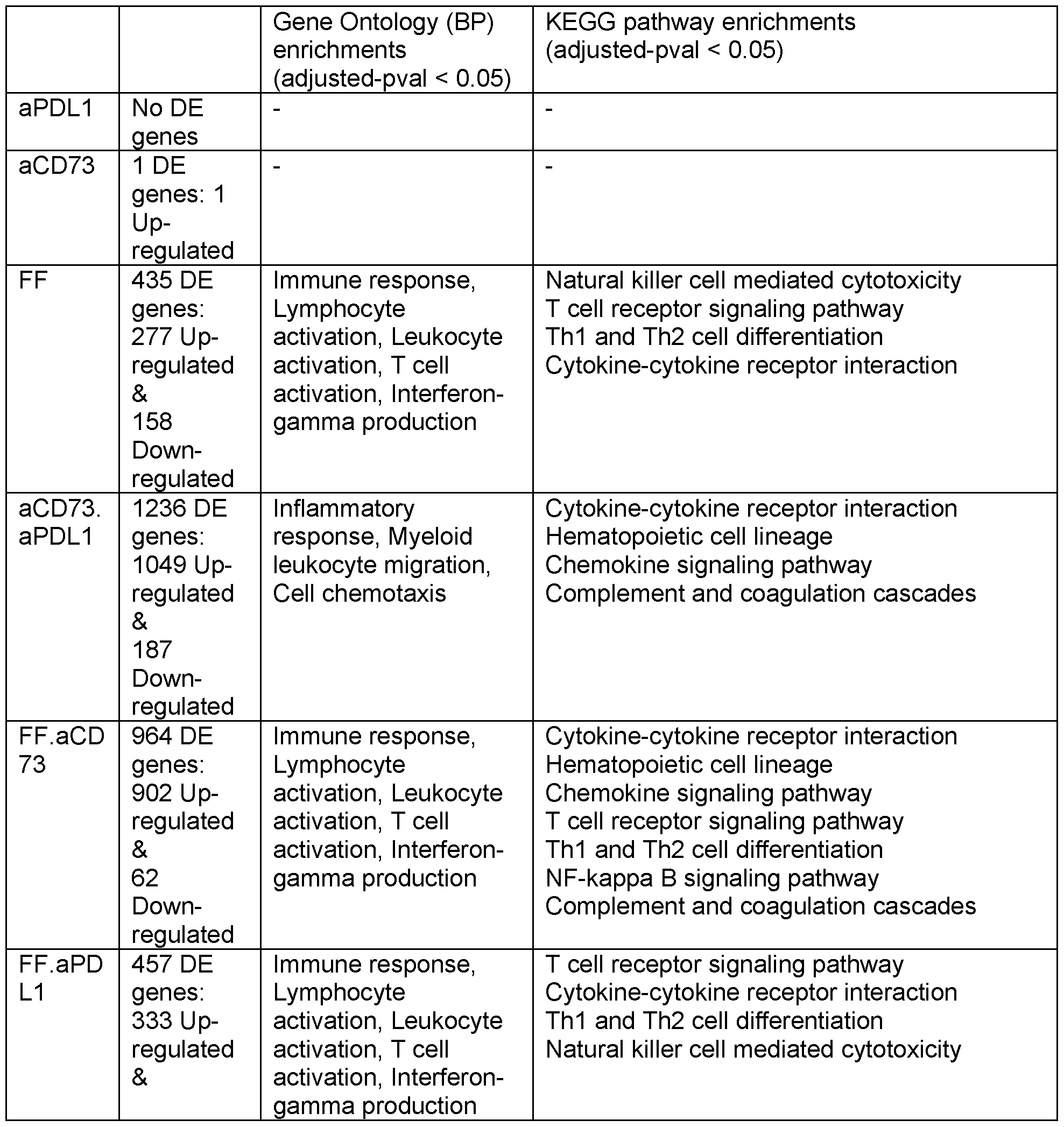

Claims
Priority Applications (7)
| Application Number | Priority Date | Filing Date | Title |
|---|---|---|---|
| CN202380082829.8A CN120390652A (en) | 2022-12-01 | 2023-12-01 | Combination therapy comprising an anti-PD-L1 antibody and an anti-CD73 antibody for treating cancer |
| JP2025531270A JP2025541725A (en) | 2022-12-01 | 2023-12-01 | Combination therapy for the treatment of cancer comprising an anti-PD-L1 antibody and an anti-CD73 antibody |
| KR1020257021849A KR20250113508A (en) | 2022-12-01 | 2023-12-01 | Combination therapy for cancer treatment including anti-PD-L1 and anti-CD73 antibodies |
| EP23828482.2A EP4626552A1 (en) | 2022-12-01 | 2023-12-01 | Combination therapy for treatment of cancer comprising anti-pd-l1 and anti-cd73 antibodies |
| US19/126,249 US20250340648A1 (en) | 2022-12-01 | 2023-12-01 | Combination therapy for treatment of cancer comprising anti-pd-l1 and anti-cd73 antibodies |
| AU2023403103A AU2023403103A1 (en) | 2022-12-01 | 2023-12-01 | Combination therapy for treatment of cancer comprising anti-pd-l1 and anti-cd73 antibodies |
| IL321145A IL321145A (en) | 2022-12-01 | 2025-05-27 | Combination therapy for treatment of cancer comprising anti-pd-l1 and anti-cd73 antibodies |
Applications Claiming Priority (2)
| Application Number | Priority Date | Filing Date | Title |
|---|---|---|---|
| US202263385760P | 2022-12-01 | 2022-12-01 | |
| US63/385,760 | 2022-12-01 |
Publications (1)
| Publication Number | Publication Date |
|---|---|
| WO2024116140A1 true WO2024116140A1 (en) | 2024-06-06 |
Family
ID=89321746
Family Applications (1)
| Application Number | Title | Priority Date | Filing Date |
|---|---|---|---|
| PCT/IB2023/062127 Ceased WO2024116140A1 (en) | 2022-12-01 | 2023-12-01 | Combination therapy for treatment of cancer comprising anti-pd-l1 and anti-cd73 antibodies |
Country Status (9)
| Country | Link |
|---|---|
| US (1) | US20250340648A1 (en) |
| EP (1) | EP4626552A1 (en) |
| JP (1) | JP2025541725A (en) |
| KR (1) | KR20250113508A (en) |
| CN (1) | CN120390652A (en) |
| AU (1) | AU2023403103A1 (en) |
| IL (1) | IL321145A (en) |
| TW (1) | TW202440157A (en) |
| WO (1) | WO2024116140A1 (en) |
Citations (73)
| Publication number | Priority date | Publication date | Assignee | Title |
|---|---|---|---|---|
| US4870287A (en) | 1988-03-03 | 1989-09-26 | Loma Linda University Medical Center | Multi-station proton beam therapy system |
| US5760395A (en) | 1996-04-18 | 1998-06-02 | Universities Research Assoc., Inc. | Method and apparatus for laser-controlled proton beam radiology |
| WO2003099196A2 (en) | 2002-05-23 | 2003-12-04 | Cure Tech Ltd. | Humanized immunomodulatory monoclonal antibodies for the treatment of neoplastic disease or immunodeficiency |
| US6808710B1 (en) | 1999-08-23 | 2004-10-26 | Genetics Institute, Inc. | Downmodulating an immune response with multivalent antibodies to PD-1 |
| WO2006121168A1 (en) | 2005-05-09 | 2006-11-16 | Ono Pharmaceutical Co., Ltd. | Human monoclonal antibodies to programmed death 1(pd-1) and methods for treating cancer using anti-pd-1 antibodies alone or in combination with other immunotherapeutics |
| WO2008156712A1 (en) | 2007-06-18 | 2008-12-24 | N. V. Organon | Antibodies to human programmed death receptor pd-1 |
| WO2009014708A2 (en) | 2007-07-23 | 2009-01-29 | Cell Genesys, Inc. | Pd-1 antibodies in combination with a cytokine-secreting cell and methods of use thereof |
| US7488802B2 (en) | 2002-12-23 | 2009-02-10 | Wyeth | Antibodies against PD-1 |
| WO2009044273A2 (en) | 2007-10-05 | 2009-04-09 | Immutep | Use of recombinant lag-3 or the derivatives thereof for eliciting monocyte immune response |
| WO2009114335A2 (en) | 2008-03-12 | 2009-09-17 | Merck & Co., Inc. | Pd-1 binding proteins |
| US7635757B2 (en) | 1999-08-23 | 2009-12-22 | Dana-Farber Cancer Institute, Inc. | B7-4 Antibodies and uses therefor |
| US20090317368A1 (en) | 1999-11-30 | 2009-12-24 | Lieping Chen | B7-h1, a novel immunoregulatory molecule |
| US20110007023A1 (en) | 2009-07-09 | 2011-01-13 | Sony Ericsson Mobile Communications Ab | Display device, touch screen device comprising the display device, mobile device and method for sensing a force on a display device |
| US7943743B2 (en) | 2005-07-01 | 2011-05-17 | Medarex, Inc. | Human monoclonal antibodies to programmed death ligand 1 (PD-L1) |
| WO2011066389A1 (en) | 2009-11-24 | 2011-06-03 | Medimmmune, Limited | Targeted binding agents against b7-h1 |
| US20110150892A1 (en) | 2008-08-11 | 2011-06-23 | Medarex, Inc. | Human antibodies that bind lymphocyte activation gene-3 (lag-3) and uses thereof |
| WO2011161699A2 (en) | 2010-06-25 | 2011-12-29 | Aurigene Discovery Technologies Limited | Immunosuppression modulating compounds |
| US8217149B2 (en) | 2008-12-09 | 2012-07-10 | Genentech, Inc. | Anti-PD-L1 antibodies, compositions and articles of manufacture |
| WO2012145493A1 (en) | 2011-04-20 | 2012-10-26 | Amplimmune, Inc. | Antibodies and other molecules that bind b7-h1 and pd-1 |
| WO2013014668A1 (en) | 2011-07-24 | 2013-01-31 | Curetech Ltd. | Variants of humanized immunomodulatory monoclonal antibodies |
| WO2013173223A1 (en) | 2012-05-15 | 2013-11-21 | Bristol-Myers Squibb Company | Cancer immunotherapy by disrupting pd-1/pd-l1 signaling |
| WO2014140180A1 (en) | 2013-03-15 | 2014-09-18 | Glaxosmithkline Intellectual Property Development Limited | Anti-lag-3 binding proteins |
| WO2014179664A2 (en) | 2013-05-02 | 2014-11-06 | Anaptysbio, Inc. | Antibodies directed against programmed death-1 (pd-1) |
| WO2014194302A2 (en) | 2013-05-31 | 2014-12-04 | Sorrento Therapeutics, Inc. | Antigen binding proteins that bind pd-1 |
| WO2014206107A1 (en) | 2013-06-26 | 2014-12-31 | 上海君实生物医药科技有限公司 | Anti-pd-1 antibody and use thereof |
| WO2015035606A1 (en) | 2013-09-13 | 2015-03-19 | Beigene, Ltd. | Anti-pd1 antibodies and their use as therapeutics and diagnostics |
| WO2015085847A1 (en) | 2013-12-12 | 2015-06-18 | 上海恒瑞医药有限公司 | Pd-1 antibody, antigen-binding fragment thereof, and medical application thereof |
| WO2015112800A1 (en) | 2014-01-23 | 2015-07-30 | Regeneron Pharmaceuticals, Inc. | Human antibodies to pd-1 |
| WO2015112900A1 (en) | 2014-01-24 | 2015-07-30 | Dana-Farber Cancer Institue, Inc. | Antibody molecules to pd-1 and uses thereof |
| WO2015200119A1 (en) | 2014-06-26 | 2015-12-30 | Macrogenics, Inc. | Covalently bonded diabodies having immunoreactivity with pd-1 and lag-3, and methods of use thereof |
| WO2016028672A1 (en) | 2014-08-19 | 2016-02-25 | Merck Sharp & Dohme Corp. | Anti-lag3 antibodies and antigen-binding fragments |
| WO2016075099A1 (en) | 2014-11-10 | 2016-05-19 | Medimmune Limited | Binding molecules specific for cd73 and uses thereof |
| WO2016081748A2 (en) | 2014-11-21 | 2016-05-26 | Bristol-Myers Squibb Company | Antibodies against cd73 and uses thereof |
| WO2016106159A1 (en) | 2014-12-22 | 2016-06-30 | Enumeral Biomedical Holding, Inc. | Anti-pd-1 antibodies |
| WO2016126858A2 (en) | 2015-02-03 | 2016-08-11 | Anaptysbio, Inc. | Antibodies directed against lymphocyte activation gene 3 (lag-3) |
| WO2016197367A1 (en) | 2015-06-11 | 2016-12-15 | Wuxi Biologics (Shanghai) Co. Ltd. | Novel anti-pd-l1 antibodies |
| WO2016200782A1 (en) | 2015-06-08 | 2016-12-15 | Macrogenics, Inc. | Lag-3-binding molecules and methods of use thereof |
| WO2017015560A2 (en) | 2015-07-22 | 2017-01-26 | Sorrento Therapeutics, Inc. | Antibody therapeutics that bind lag3 |
| WO2017019846A1 (en) | 2015-07-30 | 2017-02-02 | Macrogenics, Inc. | Pd-1-binding molecules and methods use thereof |
| WO2017019894A1 (en) | 2015-07-29 | 2017-02-02 | Novartis Ag | Combination therapies comprising antibody molecules to lag-3 |
| WO2017020291A1 (en) | 2015-08-06 | 2017-02-09 | Wuxi Biologics (Shanghai) Co. Ltd. | Novel anti-pd-l1 antibodies |
| WO2017024465A1 (en) | 2015-08-10 | 2017-02-16 | Innovent Biologics (Suzhou) Co., Ltd. | Pd-1 antibodies |
| WO2017025498A1 (en) | 2015-08-07 | 2017-02-16 | Pieris Pharmaceuticals Gmbh | Novel fusion polypeptide specific for lag-3 and pd-1 |
| WO2017024515A1 (en) | 2015-08-11 | 2017-02-16 | Wuxi Biologics (Cayman) Inc. | Novel anti-pd-1 antibodies |
| WO2017025051A1 (en) | 2015-08-11 | 2017-02-16 | Wuxi Biologics (Shanghai) Co. Ltd. | Novel anti-pd-1 antibodies |
| WO2017040790A1 (en) | 2015-09-01 | 2017-03-09 | Agenus Inc. | Anti-pd-1 antibodies and methods of use thereof |
| WO2017062888A1 (en) | 2015-10-09 | 2017-04-13 | Regeneron Pharmaceuticals, Inc. | Anti-lag3 antibodies and uses thereof |
| WO2017064043A1 (en) | 2015-10-12 | 2017-04-20 | Innate Pharma | Cd73 blocking agents |
| WO2017087589A2 (en) | 2015-11-18 | 2017-05-26 | Merck Sharp & Dohme Corp. | Pd1 and/or lag3 binders |
| WO2017087901A2 (en) | 2015-11-19 | 2017-05-26 | Sutro Biopharma, Inc. | Anti-lag3 antibodies, compositions comprising anti-lag3 antibodies and methods of making and using anti-lag3 antibodies |
| WO2017086419A1 (en) | 2015-11-18 | 2017-05-26 | 中外製薬株式会社 | Method for enhancing humoral immune response |
| WO2017086367A1 (en) | 2015-11-18 | 2017-05-26 | 中外製薬株式会社 | Combination therapy using t cell redirection antigen binding molecule against cell having immunosuppressing function |
| WO2017100670A1 (en) | 2015-12-09 | 2017-06-15 | Corvus Pharmaceuticals, Inc. | Humanized anti-cd73 antibodies |
| WO2017106129A1 (en) | 2015-12-16 | 2017-06-22 | Merck Sharp & Dohme Corp. | Anti-lag3 antibodies and antigen-binding fragments |
| WO2017106061A1 (en) | 2015-12-14 | 2017-06-22 | Macrogenics, Inc. | Bispecific molecules having immunoreactivity with pd-1 and ctla-4, and methods of use thereof |
| WO2017123557A1 (en) | 2016-01-11 | 2017-07-20 | Armo Biosciences, Inc. | Interleukin-10 in production of antigen-specific cd8+ t cells and methods of use of same |
| WO2017133540A1 (en) | 2016-02-02 | 2017-08-10 | Innovent Biologics (Suzhou) Co., Ltd. | Pd-1 antibodies |
| WO2017132825A1 (en) | 2016-02-02 | 2017-08-10 | 华为技术有限公司 | Emission power verification method, user equipment, and base station |
| WO2017149143A1 (en) | 2016-03-04 | 2017-09-08 | Agency For Science, Technology And Research | Anti-lag-3 antibodies |
| US20170260271A1 (en) | 2014-05-13 | 2017-09-14 | Chugai Seiyaku Kabushiki Kaisha | T Cell-Redirected Antigen-Binding Molecule For Cells Having Immunosuppression Function |
| WO2017198741A1 (en) | 2016-05-18 | 2017-11-23 | Boehringer Ingelheim International Gmbh | Anti pd-1 and anti-lag3 antibodies for cancer treatment |
| WO2017220555A1 (en) | 2016-06-20 | 2017-12-28 | F-Star Beta Limited | Lag -3 binding members |
| WO2017219995A1 (en) | 2016-06-23 | 2017-12-28 | 江苏恒瑞医药股份有限公司 | Lag-3 antibody, antigen-binding fragment thereof, and pharmaceutical application thereof |
| WO2017220569A1 (en) | 2016-06-20 | 2017-12-28 | F-Star Delta Limited | Binding molecules binding pd-l1 and lag-3 |
| WO2018034227A1 (en) | 2016-08-15 | 2018-02-22 | 国立大学法人北海道大学 | Anti-lag-3 antibody |
| WO2018071500A1 (en) | 2016-10-11 | 2018-04-19 | Agenus Inc. | Anti-lag-3 antibodies and methods of use thereof |
| WO2018069500A2 (en) | 2016-10-13 | 2018-04-19 | Symphogen A/S | Anti-lag-3 antibodies and compositions |
| WO2018083087A2 (en) | 2016-11-02 | 2018-05-11 | Glaxosmithkline Intellectual Property (No.2) Limited | Binding proteins |
| WO2018137598A1 (en) | 2017-01-24 | 2018-08-02 | I-Mab | Anti-cd73 antibodies and uses thereof |
| WO2018237157A1 (en) | 2017-06-22 | 2018-12-27 | Novartis Ag | Antibody molecules to cd73 and uses thereof |
| US20200317803A1 (en) * | 2019-04-02 | 2020-10-08 | Medimmune, Llc | Anti-cd73, anti-pd-l1 antibodies and chemotherapy for treating tumors |
| US20210147570A1 (en) * | 2018-04-12 | 2021-05-20 | Bristol-Myers Squibb Company | Anticancer combination therapy with cd73 antagonist antibody and pd-1/pd-l1 axis antagonist antibody |
| WO2022064399A1 (en) * | 2020-09-23 | 2022-03-31 | Medimmune, Llc | Treatment methods using anti-cd73 and anti-pd-l1 antibodies and chemotherapy |
-
2023
- 2023-12-01 CN CN202380082829.8A patent/CN120390652A/en active Pending
- 2023-12-01 KR KR1020257021849A patent/KR20250113508A/en active Pending
- 2023-12-01 JP JP2025531270A patent/JP2025541725A/en active Pending
- 2023-12-01 EP EP23828482.2A patent/EP4626552A1/en active Pending
- 2023-12-01 TW TW112146944A patent/TW202440157A/en unknown
- 2023-12-01 US US19/126,249 patent/US20250340648A1/en active Pending
- 2023-12-01 AU AU2023403103A patent/AU2023403103A1/en active Pending
- 2023-12-01 WO PCT/IB2023/062127 patent/WO2024116140A1/en not_active Ceased
-
2025
- 2025-05-27 IL IL321145A patent/IL321145A/en unknown
Patent Citations (81)
| Publication number | Priority date | Publication date | Assignee | Title |
|---|---|---|---|---|
| US4870287A (en) | 1988-03-03 | 1989-09-26 | Loma Linda University Medical Center | Multi-station proton beam therapy system |
| US5760395A (en) | 1996-04-18 | 1998-06-02 | Universities Research Assoc., Inc. | Method and apparatus for laser-controlled proton beam radiology |
| US7635757B2 (en) | 1999-08-23 | 2009-12-22 | Dana-Farber Cancer Institute, Inc. | B7-4 Antibodies and uses therefor |
| US6808710B1 (en) | 1999-08-23 | 2004-10-26 | Genetics Institute, Inc. | Downmodulating an immune response with multivalent antibodies to PD-1 |
| US20090317368A1 (en) | 1999-11-30 | 2009-12-24 | Lieping Chen | B7-h1, a novel immunoregulatory molecule |
| WO2003099196A2 (en) | 2002-05-23 | 2003-12-04 | Cure Tech Ltd. | Humanized immunomodulatory monoclonal antibodies for the treatment of neoplastic disease or immunodeficiency |
| US7488802B2 (en) | 2002-12-23 | 2009-02-10 | Wyeth | Antibodies against PD-1 |
| WO2006121168A1 (en) | 2005-05-09 | 2006-11-16 | Ono Pharmaceutical Co., Ltd. | Human monoclonal antibodies to programmed death 1(pd-1) and methods for treating cancer using anti-pd-1 antibodies alone or in combination with other immunotherapeutics |
| US7943743B2 (en) | 2005-07-01 | 2011-05-17 | Medarex, Inc. | Human monoclonal antibodies to programmed death ligand 1 (PD-L1) |
| US8354509B2 (en) | 2007-06-18 | 2013-01-15 | Msd Oss B.V. | Antibodies to human programmed death receptor PD-1 |
| WO2008156712A1 (en) | 2007-06-18 | 2008-12-24 | N. V. Organon | Antibodies to human programmed death receptor pd-1 |
| US8900587B2 (en) | 2007-06-18 | 2014-12-02 | Merck Sharp & Dohme Corp. | Antibodies to human programmed death receptor PD-1 |
| WO2009014708A2 (en) | 2007-07-23 | 2009-01-29 | Cell Genesys, Inc. | Pd-1 antibodies in combination with a cytokine-secreting cell and methods of use thereof |
| WO2009044273A2 (en) | 2007-10-05 | 2009-04-09 | Immutep | Use of recombinant lag-3 or the derivatives thereof for eliciting monocyte immune response |
| US8168757B2 (en) | 2008-03-12 | 2012-05-01 | Merck Sharp & Dohme Corp. | PD-1 binding proteins |
| WO2009114335A2 (en) | 2008-03-12 | 2009-09-17 | Merck & Co., Inc. | Pd-1 binding proteins |
| US20110150892A1 (en) | 2008-08-11 | 2011-06-23 | Medarex, Inc. | Human antibodies that bind lymphocyte activation gene-3 (lag-3) and uses thereof |
| US8217149B2 (en) | 2008-12-09 | 2012-07-10 | Genentech, Inc. | Anti-PD-L1 antibodies, compositions and articles of manufacture |
| US20110007023A1 (en) | 2009-07-09 | 2011-01-13 | Sony Ericsson Mobile Communications Ab | Display device, touch screen device comprising the display device, mobile device and method for sensing a force on a display device |
| WO2011066389A1 (en) | 2009-11-24 | 2011-06-03 | Medimmmune, Limited | Targeted binding agents against b7-h1 |
| WO2011161699A2 (en) | 2010-06-25 | 2011-12-29 | Aurigene Discovery Technologies Limited | Immunosuppression modulating compounds |
| WO2012145493A1 (en) | 2011-04-20 | 2012-10-26 | Amplimmune, Inc. | Antibodies and other molecules that bind b7-h1 and pd-1 |
| WO2013014668A1 (en) | 2011-07-24 | 2013-01-31 | Curetech Ltd. | Variants of humanized immunomodulatory monoclonal antibodies |
| US8686119B2 (en) | 2011-07-24 | 2014-04-01 | Curetech Ltd. | Variants of humanized immunomodulatory monoclonal antibodies |
| WO2013173223A1 (en) | 2012-05-15 | 2013-11-21 | Bristol-Myers Squibb Company | Cancer immunotherapy by disrupting pd-1/pd-l1 signaling |
| WO2014140180A1 (en) | 2013-03-15 | 2014-09-18 | Glaxosmithkline Intellectual Property Development Limited | Anti-lag-3 binding proteins |
| WO2014179664A2 (en) | 2013-05-02 | 2014-11-06 | Anaptysbio, Inc. | Antibodies directed against programmed death-1 (pd-1) |
| WO2014194302A2 (en) | 2013-05-31 | 2014-12-04 | Sorrento Therapeutics, Inc. | Antigen binding proteins that bind pd-1 |
| US20160272708A1 (en) | 2013-06-26 | 2016-09-22 | Shanghai Junshi Biosciences Inc. | Anti-pd-1 antibody and use thereof |
| WO2014206107A1 (en) | 2013-06-26 | 2014-12-31 | 上海君实生物医药科技有限公司 | Anti-pd-1 antibody and use thereof |
| WO2015035606A1 (en) | 2013-09-13 | 2015-03-19 | Beigene, Ltd. | Anti-pd1 antibodies and their use as therapeutics and diagnostics |
| WO2015085847A1 (en) | 2013-12-12 | 2015-06-18 | 上海恒瑞医药有限公司 | Pd-1 antibody, antigen-binding fragment thereof, and medical application thereof |
| WO2015112800A1 (en) | 2014-01-23 | 2015-07-30 | Regeneron Pharmaceuticals, Inc. | Human antibodies to pd-1 |
| WO2015112900A1 (en) | 2014-01-24 | 2015-07-30 | Dana-Farber Cancer Institue, Inc. | Antibody molecules to pd-1 and uses thereof |
| US20170260271A1 (en) | 2014-05-13 | 2017-09-14 | Chugai Seiyaku Kabushiki Kaisha | T Cell-Redirected Antigen-Binding Molecule For Cells Having Immunosuppression Function |
| WO2015200119A1 (en) | 2014-06-26 | 2015-12-30 | Macrogenics, Inc. | Covalently bonded diabodies having immunoreactivity with pd-1 and lag-3, and methods of use thereof |
| WO2016028672A1 (en) | 2014-08-19 | 2016-02-25 | Merck Sharp & Dohme Corp. | Anti-lag3 antibodies and antigen-binding fragments |
| WO2016075099A1 (en) | 2014-11-10 | 2016-05-19 | Medimmune Limited | Binding molecules specific for cd73 and uses thereof |
| WO2016081748A2 (en) | 2014-11-21 | 2016-05-26 | Bristol-Myers Squibb Company | Antibodies against cd73 and uses thereof |
| WO2016106159A1 (en) | 2014-12-22 | 2016-06-30 | Enumeral Biomedical Holding, Inc. | Anti-pd-1 antibodies |
| WO2016126858A2 (en) | 2015-02-03 | 2016-08-11 | Anaptysbio, Inc. | Antibodies directed against lymphocyte activation gene 3 (lag-3) |
| WO2016200782A1 (en) | 2015-06-08 | 2016-12-15 | Macrogenics, Inc. | Lag-3-binding molecules and methods of use thereof |
| WO2016197367A1 (en) | 2015-06-11 | 2016-12-15 | Wuxi Biologics (Shanghai) Co. Ltd. | Novel anti-pd-l1 antibodies |
| WO2017015560A2 (en) | 2015-07-22 | 2017-01-26 | Sorrento Therapeutics, Inc. | Antibody therapeutics that bind lag3 |
| WO2017019894A1 (en) | 2015-07-29 | 2017-02-02 | Novartis Ag | Combination therapies comprising antibody molecules to lag-3 |
| WO2017019846A1 (en) | 2015-07-30 | 2017-02-02 | Macrogenics, Inc. | Pd-1-binding molecules and methods use thereof |
| WO2017020291A1 (en) | 2015-08-06 | 2017-02-09 | Wuxi Biologics (Shanghai) Co. Ltd. | Novel anti-pd-l1 antibodies |
| WO2017020858A1 (en) | 2015-08-06 | 2017-02-09 | Wuxi Biologics (Shanghai) Co. Ltd. | Novel anti-pd-l1 antibodies |
| WO2017025498A1 (en) | 2015-08-07 | 2017-02-16 | Pieris Pharmaceuticals Gmbh | Novel fusion polypeptide specific for lag-3 and pd-1 |
| WO2017024465A1 (en) | 2015-08-10 | 2017-02-16 | Innovent Biologics (Suzhou) Co., Ltd. | Pd-1 antibodies |
| WO2017025016A1 (en) | 2015-08-10 | 2017-02-16 | Innovent Biologics (Suzhou) Co., Ltd. | Pd-1 antibodies |
| WO2017024515A1 (en) | 2015-08-11 | 2017-02-16 | Wuxi Biologics (Cayman) Inc. | Novel anti-pd-1 antibodies |
| WO2017025051A1 (en) | 2015-08-11 | 2017-02-16 | Wuxi Biologics (Shanghai) Co. Ltd. | Novel anti-pd-1 antibodies |
| WO2017040790A1 (en) | 2015-09-01 | 2017-03-09 | Agenus Inc. | Anti-pd-1 antibodies and methods of use thereof |
| WO2017062888A1 (en) | 2015-10-09 | 2017-04-13 | Regeneron Pharmaceuticals, Inc. | Anti-lag3 antibodies and uses thereof |
| WO2017064043A1 (en) | 2015-10-12 | 2017-04-20 | Innate Pharma | Cd73 blocking agents |
| WO2017087589A2 (en) | 2015-11-18 | 2017-05-26 | Merck Sharp & Dohme Corp. | Pd1 and/or lag3 binders |
| WO2017086419A1 (en) | 2015-11-18 | 2017-05-26 | 中外製薬株式会社 | Method for enhancing humoral immune response |
| WO2017086367A1 (en) | 2015-11-18 | 2017-05-26 | 中外製薬株式会社 | Combination therapy using t cell redirection antigen binding molecule against cell having immunosuppressing function |
| WO2017087901A2 (en) | 2015-11-19 | 2017-05-26 | Sutro Biopharma, Inc. | Anti-lag3 antibodies, compositions comprising anti-lag3 antibodies and methods of making and using anti-lag3 antibodies |
| WO2017100670A1 (en) | 2015-12-09 | 2017-06-15 | Corvus Pharmaceuticals, Inc. | Humanized anti-cd73 antibodies |
| WO2017106061A1 (en) | 2015-12-14 | 2017-06-22 | Macrogenics, Inc. | Bispecific molecules having immunoreactivity with pd-1 and ctla-4, and methods of use thereof |
| WO2017106129A1 (en) | 2015-12-16 | 2017-06-22 | Merck Sharp & Dohme Corp. | Anti-lag3 antibodies and antigen-binding fragments |
| WO2017123557A1 (en) | 2016-01-11 | 2017-07-20 | Armo Biosciences, Inc. | Interleukin-10 in production of antigen-specific cd8+ t cells and methods of use of same |
| WO2017133540A1 (en) | 2016-02-02 | 2017-08-10 | Innovent Biologics (Suzhou) Co., Ltd. | Pd-1 antibodies |
| WO2017132825A1 (en) | 2016-02-02 | 2017-08-10 | 华为技术有限公司 | Emission power verification method, user equipment, and base station |
| WO2017132827A1 (en) | 2016-02-02 | 2017-08-10 | Innovent Biologics (Suzhou) Co., Ltd. | Pd-1 antibodies |
| WO2017149143A1 (en) | 2016-03-04 | 2017-09-08 | Agency For Science, Technology And Research | Anti-lag-3 antibodies |
| WO2017198741A1 (en) | 2016-05-18 | 2017-11-23 | Boehringer Ingelheim International Gmbh | Anti pd-1 and anti-lag3 antibodies for cancer treatment |
| WO2017220569A1 (en) | 2016-06-20 | 2017-12-28 | F-Star Delta Limited | Binding molecules binding pd-l1 and lag-3 |
| WO2017220555A1 (en) | 2016-06-20 | 2017-12-28 | F-Star Beta Limited | Lag -3 binding members |
| WO2017219995A1 (en) | 2016-06-23 | 2017-12-28 | 江苏恒瑞医药股份有限公司 | Lag-3 antibody, antigen-binding fragment thereof, and pharmaceutical application thereof |
| WO2018034227A1 (en) | 2016-08-15 | 2018-02-22 | 国立大学法人北海道大学 | Anti-lag-3 antibody |
| WO2018071500A1 (en) | 2016-10-11 | 2018-04-19 | Agenus Inc. | Anti-lag-3 antibodies and methods of use thereof |
| WO2018069500A2 (en) | 2016-10-13 | 2018-04-19 | Symphogen A/S | Anti-lag-3 antibodies and compositions |
| WO2018083087A2 (en) | 2016-11-02 | 2018-05-11 | Glaxosmithkline Intellectual Property (No.2) Limited | Binding proteins |
| WO2018137598A1 (en) | 2017-01-24 | 2018-08-02 | I-Mab | Anti-cd73 antibodies and uses thereof |
| WO2018237157A1 (en) | 2017-06-22 | 2018-12-27 | Novartis Ag | Antibody molecules to cd73 and uses thereof |
| US20210147570A1 (en) * | 2018-04-12 | 2021-05-20 | Bristol-Myers Squibb Company | Anticancer combination therapy with cd73 antagonist antibody and pd-1/pd-l1 axis antagonist antibody |
| US20200317803A1 (en) * | 2019-04-02 | 2020-10-08 | Medimmune, Llc | Anti-cd73, anti-pd-l1 antibodies and chemotherapy for treating tumors |
| WO2022064399A1 (en) * | 2020-09-23 | 2022-03-31 | Medimmune, Llc | Treatment methods using anti-cd73 and anti-pd-l1 antibodies and chemotherapy |
Non-Patent Citations (54)
| Title |
|---|
| "GenBank", Database accession no. Q9NZQ7 |
| "The Dictionary of Cell and Molecular Biology", 1999, ACADEMIC PRESS |
| "the Oxford Dictionary Of Biochemistry And Molecular Biology", 2000, OXFORD UNIVERSITY PRESS |
| "Uniprot", Database accession no. Q61503 |
| AUGUSTIN RC ET AL., J IMMUNOTHER CANCER, vol. 10, 2022, pages e004089 |
| BECHT E. ET AL., GENOME BIOL, vol. 17, 2016, pages 1 - 20 |
| BIRD ET AL., SCIENCE, vol. 242, 1988, pages 423 - 426 |
| BREEN DM ET AL., CELL METAB, vol. 32, 2020, pages 938 - 950 |
| BRUNI D ET AL., NATURE REVIEWS CANCER, vol. 20, 2020, pages 662 - 80 |
| CHAMBERS AM ET AL., FRONT IMMUNOL, vol. 9, 2018, pages 1012 |
| CHOW MT ET AL., IMMUNITY, vol. 50, 2019, pages 1498 - 1512 |
| COFFELT SB ET AL., TRENDS IMMUNOL, vol. 36, 2015, pages 198 - 216 |
| DANNHORN A. ET AL., ANAL CHEM, vol. 92, 2020, pages 11080 - 8 |
| DOBIN A ET AL., BIOINFORMATICS, vol. 29, 2013, pages 15 - 21 |
| DOSSET M ET AL., ONCOIMMUNOLOGY, vol. 7, 2018 |
| FERRERE GLADYS ET AL: "Ketogenic diet and ketone bodies enhance the anticancer effects of PD-1 blockade", HAL (LE CENTRE POUR LA COMMUNICATION SCIENTIFIQUE DIRECTE), 25 January 2021 (2021-01-25), XP093138515, Retrieved from the Internet <URL:https://insight.jci.org/articles/view/142843/version/1/pdf/render.pdf> [retrieved on 20240306], DOI: 10.1172/jci * |
| FRIDMAN WH ET AL., JOURNAL OF EXPERIMENTAL MEDICINE, vol. 218, 2021 |
| GRISS J ET AL., NATURE COMMUNICATIONS, vol. 10, 2019, pages 1 - 14 |
| HAIR J ET AL., CANCER RES, vol. 81, 2021, pages 1695 - 1695 |
| HAY CM ET AL., ONCOIMMUNOLOGY, vol. 5, 2016 |
| HERBST ET AL., J CLIN ONCOL, vol. 31, 2013, pages 3000 |
| HUSTON ET AL., PROC. NATL. ACAD. SCI. USA, vol. 85, 1988, pages 5879 - 5883 |
| ISHII HIDENOBU ET AL: "Predictive value of CD73 expression for the efficacy of immune checkpoint inhibitors in NSCLC", THORACIC CANCER, vol. 11, no. 4, 15 February 2020 (2020-02-15), pages 950 - 955, XP093138517, ISSN: 1759-7706, Retrieved from the Internet <URL:https://onlinelibrary.wiley.com/doi/full-xml/10.1111/1759-7714.13346> DOI: 10.1111/1759-7714.13346 * |
| JOURNAL FOR IMMUNOTHERAPY OF CANCER, vol. 4, no. P195, 2016 |
| JUO, PEI-SHOW: "the Concise Dictionary of Biomedicine and Molecular Biology", 2002, CRC PRESS |
| KOROTKEVICH G ET AL., BIORXIV, 2021, pages 060012 |
| LOVE MI ET AL., GENOME BIOL, vol. 15, 2014 |
| MA RY ET AL., TRENDS IMMUNOL, vol. 43, 2022, pages 546 - 63 |
| MAGAGNA I ET AL., CANCERS, vol. 13, 2021 |
| MARCOVECCHIO PM ET AL., J IMMUNOTHER CANCER, vol. 9, 2021, pages A729 - A729 |
| MASTELIC-GAVILLET B ET AL., J IMMUNOTHER CANCER, vol. 7, 2019, pages 42 - 16 |
| MISUMI ET AL., EUR. J. BIOCHEM., vol. 191, no. 3, 1990, pages 563 - 9 |
| MOORE SF ET AL., JOURNAL OF IMMUNOLOGY, vol. 183, 2009, pages 3302 - 8 |
| NGUYEN AM ET AL., MOLECULAR & CELLULAR PROTEOMICS, vol. 19, 2020, pages 375 - 89 |
| PETITPREZ F ET AL., NATURE, vol. 577, 2020, pages 7791 |
| PUTRI GH ET AL., BIOINFORMATICS, vol. 38, 2022, pages 2943 - 5 |
| QU Y ET AL., CELL REP, vol. 32, 2020 |
| RACE AM ET AL., J PROTEOMICS, vol. 75, 2012, pages 5111 - 2 |
| SIDDERS B ET AL., CLIN CANCER RES, vol. 26, 2020, pages 2176 - 87 |
| SIDDERSALLARD D ET AL., IMMUNOL LETT, vol. 205, 2019, pages 31 - 9 |
| SIEW YY ET AL., INT IMMUNOL, vol. 27, 2015, pages 621 - 32 |
| SUBRAMANIAN A. ET AL., PROC NATL ACAD SCI USA, vol. 102, 2005, pages 15545 - 50 |
| TSUKUI H ET AL., BMC CANCER, vol. 20, 2020 |
| VOSS MH ET AL., JC020203815_SUPPL5025, vol. 38, 2020, pages 5025 - 5025 |
| WARD ET AL., NATURE, vol. 341, 1989, pages 544 - 46 |
| WENNERBERG E ET AL., CANCER IMMUNOL RES, vol. 8, 2020, pages 465 - 78 |
| WENNERBERG E ET AL., FRONT IMMUNOL, vol. 8, 2017, pages 229 |
| WENNERBERG ET AL., CANCER IMMUNOL RES., 2020 |
| WISCHHUSEN J ET AL., FRONT IMMUNOL, vol. 11, 2020 |
| WU Q ET AL., CELL DEATH DIFFER, vol. 24, 2017, pages 1632 - 44 |
| WU T. ET AL., THE INNOVATION, vol. 2, 2021, pages 100141 |
| XUAN W ET AL., J LEUKOC BIOL, vol. 97, 2015, pages 61 - 9 |
| YU M ET AL., NAT COMMUN, vol. 11, 2020 |
| ZITVOGEL L ET AL., NAT REV IMMUNOL, vol. 15, 2015, pages 405 - 14 |
Also Published As
| Publication number | Publication date |
|---|---|
| AU2023403103A1 (en) | 2025-07-10 |
| IL321145A (en) | 2025-07-01 |
| CN120390652A (en) | 2025-07-29 |
| US20250340648A1 (en) | 2025-11-06 |
| TW202440157A (en) | 2024-10-16 |
| KR20250113508A (en) | 2025-07-25 |
| EP4626552A1 (en) | 2025-10-08 |
| JP2025541725A (en) | 2025-12-23 |
Similar Documents
| Publication | Publication Date | Title |
|---|---|---|
| US12534530B2 (en) | Combination of a PD-1 antagonist and a VEGFR inhibitor for treating cancer | |
| US20210161928A1 (en) | Cancer therapy via a combination of epigenetic modulation and immune modulation | |
| TWI772261B (en) | Pd-l1 antagonist combination treatments | |
| KR102644408B1 (en) | Combination of anti-PD-L1 antibody and DNA-PK inhibitor for treatment of cancer | |
| CN110621787A (en) | Methods for diagnosis and treatment of cancer | |
| US20230192870A1 (en) | Combination Anti-CSF1R and Anti-PD-1 Antibody Combination Therapy for Pancreatic Cancer | |
| CN113396230A (en) | Methods of diagnosis and treatment of cancer | |
| EP3856251B1 (en) | Combination of a pd-1 antagonist, an atr inhibitor and a platinating agent for the treatment of cancer | |
| WO2011113041A2 (en) | Neutralization of flt3 ligand as a leukemia therapy | |
| IL303256A (en) | Methods and compositions for neoadjuvant and adjuvant urothelial carcinoma therapy | |
| US20210353750A1 (en) | Methods of Treating Cancer | |
| US20210340232A1 (en) | Monoclonal antibodies against human dickkopf3 and uses thereof | |
| EP4196612A1 (en) | Diagnostic and therapeutic methods for cancer | |
| US20250340648A1 (en) | Combination therapy for treatment of cancer comprising anti-pd-l1 and anti-cd73 antibodies | |
| JP2023512237A (en) | Use of poziotinib for the treatment of cancers with NRG1 fusions | |
| WO2024150177A1 (en) | Treatment methods for solid tumors | |
| EP4330436A1 (en) | Therapeutic and diagnostic methods and compositions for cancer | |
| US20210355222A1 (en) | Methods of Treating Cancer | |
| HK40055955A (en) | Diagnostic and therapeutic methods for cancer | |
| CN117545857A (en) | Methods and compositions for the treatment and diagnosis of cancer |
Legal Events
| Date | Code | Title | Description |
|---|---|---|---|
| 121 | Ep: the epo has been informed by wipo that ep was designated in this application |
Ref document number: 23828482 Country of ref document: EP Kind code of ref document: A1 |
|
| WWE | Wipo information: entry into national phase |
Ref document number: 321145 Country of ref document: IL |
|
| ENP | Entry into the national phase |
Ref document number: 2025531270 Country of ref document: JP Kind code of ref document: A |
|
| WWE | Wipo information: entry into national phase |
Ref document number: 2025531270 Country of ref document: JP |
|
| WWE | Wipo information: entry into national phase |
Ref document number: 202380082829.8 Country of ref document: CN |
|
| WWE | Wipo information: entry into national phase |
Ref document number: P2025-01678 Country of ref document: AE |
|
| REG | Reference to national code |
Ref country code: BR Ref legal event code: B01A Ref document number: 112025011100 Country of ref document: BR |
|
| WWE | Wipo information: entry into national phase |
Ref document number: AU2023403103 Country of ref document: AU |
|
| WWE | Wipo information: entry into national phase |
Ref document number: 11202503638T Country of ref document: SG Ref document number: 202591716 Country of ref document: EA |
|
| WWP | Wipo information: published in national office |
Ref document number: 11202503638T Country of ref document: SG |
|
| WWE | Wipo information: entry into national phase |
Ref document number: 2023828482 Country of ref document: EP |
|
| NENP | Non-entry into the national phase |
Ref country code: DE |
|
| ENP | Entry into the national phase |
Ref document number: 2023403103 Country of ref document: AU Date of ref document: 20231201 Kind code of ref document: A |
|
| ENP | Entry into the national phase |
Ref document number: 2023828482 Country of ref document: EP Effective date: 20250701 |
|
| WWP | Wipo information: published in national office |
Ref document number: 1020257021849 Country of ref document: KR |
|
| WWP | Wipo information: published in national office |
Ref document number: 202380082829.8 Country of ref document: CN |
|
| WWP | Wipo information: published in national office |
Ref document number: 2023828482 Country of ref document: EP |