WO2024106939A1 - Anticorps se liant de manière spécifique à b7-h3 - Google Patents
Anticorps se liant de manière spécifique à b7-h3 Download PDFInfo
- Publication number
- WO2024106939A1 WO2024106939A1 PCT/KR2023/018360 KR2023018360W WO2024106939A1 WO 2024106939 A1 WO2024106939 A1 WO 2024106939A1 KR 2023018360 W KR2023018360 W KR 2023018360W WO 2024106939 A1 WO2024106939 A1 WO 2024106939A1
- Authority
- WO
- WIPO (PCT)
- Prior art keywords
- antibody
- cancer
- antigen
- binding fragment
- vector
- Prior art date
- Legal status (The legal status is an assumption and is not a legal conclusion. Google has not performed a legal analysis and makes no representation as to the accuracy of the status listed.)
- Ceased
Links
Images


Classifications
-
- A—HUMAN NECESSITIES
- A61—MEDICAL OR VETERINARY SCIENCE; HYGIENE
- A61K—PREPARATIONS FOR MEDICAL, DENTAL OR TOILETRY PURPOSES
- A61K39/00—Medicinal preparations containing antigens or antibodies
-
- A—HUMAN NECESSITIES
- A61—MEDICAL OR VETERINARY SCIENCE; HYGIENE
- A61P—SPECIFIC THERAPEUTIC ACTIVITY OF CHEMICAL COMPOUNDS OR MEDICINAL PREPARATIONS
- A61P35/00—Antineoplastic agents
-
- C—CHEMISTRY; METALLURGY
- C07—ORGANIC CHEMISTRY
- C07K—PEPTIDES
- C07K16/00—Immunoglobulins [IGs], e.g. monoclonal or polyclonal antibodies
- C07K16/18—Immunoglobulins [IGs], e.g. monoclonal or polyclonal antibodies against material from animals or humans
- C07K16/28—Immunoglobulins [IGs], e.g. monoclonal or polyclonal antibodies against material from animals or humans against receptors, cell surface antigens or cell surface determinants
Definitions
- the present invention relates to an antibody or antigen-binding fragment thereof that specifically binds to B7-H3.
- the global solid anticancer drug market is expected to grow at an average annual rate of 12.5% from approximately $101 billion (approximately KRW 126 trillion) in 2021 to approximately $252 billion (approximately KRW 310 trillion) in 2028.
- the market size by detailed disease in 2028 was approximately $52 billion (approximately KRW 65 trillion) for non-small cell lung cancer, approximately $46 billion (approximately KRW 57 trillion) for breast cancer, and approximately $46 billion (approximately KRW 57 trillion) for breast cancer.
- breast cancer stomach cancer, colon cancer, liver cancer, lung cancer, and cervical cancer
- five of the six major cancer types are lung cancer, colon cancer, liver cancer, stomach cancer, and cervical cancer.
- the incidence rate shows a decreasing trend or no significant increase over the past 10 years.
- the incidence rate of the remaining breast cancers has been continuously increasing for the past 20 years.
- Breast cancer is classified based on three indicators: estrogen receptor, progesterone receptor, and epidermal growth factor (HER2). If all three indicators are tested negative (not detected), triple negative breast cancer is diagnosed.
- the five-year survival rate after diagnosis for breast cancer is over 90%, but for triple-negative breast cancer, it is around 70%.
- the 5-year survival rate for stage 3-4 triple negative breast cancer patients is only about 30%. This is a low figure compared to the 5-year survival rate of about 50% for other stage 3 and 4 breast cancer patients, excluding triple-negative breast cancer.
- the progression of triple-negative breast cancer is rapid and aggressive, with approximately 30% of patients diagnosed early experiencing the risk of death within 5 years after surgery.
- triple-negative breast cancer In the case of triple-negative breast cancer, there are many mutations in cancer cells, and because metastasis to other organs is faster than that of other breast cancers, there are several limitations in treatment. In addition, triple-negative breast cancer has a poor prognosis, with approximately 50% of patients experiencing recurrence even if treated early. Current breast cancer treatments target estrogen, progesterone, and epidermal growth factor receptors. Therefore, it is difficult for triple-negative breast cancer patients who are negative for these receptors to expect treatment effects using the drug.
- childhood cancer occurs at a significant rate.
- the incidence of childhood cancer is similar throughout the world, and in Korea, about 1,000 to 1,200 people are diagnosed with childhood cancer every year. This means that approximately 1 per 100,000 children are diagnosed with childhood cancer each year, accounting for approximately 1% of all cancer patients.
- Pediatric cancer is a very dangerous disease that ranks first in disease mortality among children between the ages of 1 and 9, exceeding the number of children who die in traffic accidents.
- lymphoblastic leukemia a leukemia in which lymphoid white blood cells do not mature properly and turn into cancer cells
- has very good treatment results with a cure rate of about 80-90%
- acute myeloid leukemia also shows a cure rate of 70%.
- the prognosis for pediatric brain tumors is poor, so the development of new treatments is necessary.
- 247,952 new cases of cancer occurred in Korea in 2020, of which brain tumors were confirmed to account for 1,795 cases, or 0.7% of all cancer cases.
- brain tumors that occur frequently in children and adolescents include medulloblastoma, astrocytoma, ependymoma, craniopharyngioma, brainstem glioma, and germ cell tumor. cell tumor), and brain tumors can be classified according to their malignancy, location, and constituent cells.
- brainstem glioma is a glioma that occurs in the midbrain, pons, and medulla of the brain. It causes multiple cranial nerve disorders such as dysarthria, dysphagia, strabismus, and facial paralysis, as well as hemiparesis, and has a 2-year survival rate of less than 10%. is the worst pediatric brain tumor.
- the most basic treatment for brain tumors is surgery or a combination of surgery and radiation therapy, and chemotherapy may also be attempted depending on the tumor tissue results.
- Cerebellar mutism syndrome CMS occurs in 25% of surgical patients. Cerebellar mutism syndrome is a condition that can occur in patients who have undergone surgery to remove a tumor from a specific part of the brain, including the cerebellum. Symptoms appear about 1 to 2 days after surgery and include decreased speech, difficulty walking due to difficulty maintaining balance, and loss of muscle tension. Symptoms of cerebellar mutism syndrome gradually disappear over time. However, there are also difficulties in conducting radiation irradiation immediately after surgery. Postoperative adjuvant craniospinal radiotherapy is used to treat the entire brain and spine for vesicles to prevent their spread in the cerebrospinal fluid (CSF). Craniospinal radiotherapy undoubtedly prolongs survival. Unfortunately, radiation therapy comes at the cost of a 2-4 point decline in Intelligence Quotient (IQ) per year.
- IQ Intelligence Quotient
- Leptomeningeal metastatic cancer is known to occur in approximately 5-20% of all solid cancers, of which lung cancer, breast cancer, and melanoma account for a large proportion.
- the proportion of patients with leptomeningeal metastasis is also frequently found in lung cancer, breast cancer, and melanoma (Cancers (Basel). 2021;13).
- Cancers (Basel). 2021;13) According to the 2020 national cancer registration statistics, it is known that 28,949 people were diagnosed with lung cancer and 24,923 people with breast cancer in 2020, so the number of patients with leptomeningeal metastasis from the above two cancers is expected to be at least 2,500 per year in Korea.
- Tagrisso which effectively crosses the blood-brain barrier, are known to be effective in leptomeningeal metastasis cancer, but the number of patients to whom they can be applied is limited and the targeted treatments have the problem of developing resistance (Neurotherapeutics. 2022;19:1782- 1798).
- B7-H3 is known to be highly expressed in various carcinomas, including pediatric brain cancer, leptomeningeal metastasis cancer, and triple-negative breast cancer, and such high expression of B7-H3 is associated with negative clinical treatment results.
- treatment of human triple-negative breast cancer cell lines with a monoclonal antibody that specifically binds to B7-H3 resulted in inhibition of the growth of triple-negative breast cancer cells. Therefore, the present inventor confirmed the relationship of B7-H3, which is highly expressed in cancer, to various cancers and its potential as a therapeutic agent for triple-negative breast cancer, etc., and came to the present invention.
- the present inventors attempted to develop an antibody targeting B7-H3, which is highly expressed in solid tumors.
- the present inventors completed the present invention by discovering an anti-B7-H3 antibody that specifically binds to various types of cancer cell lines expressing B7-H3 and exhibits a T cell-mediated cancer cell killing effect.
- the purpose of the present invention is to provide an anti-B7-H3 antibody or antigen-binding fragment thereof that specifically binds to B7-H3.
- Another object of the present invention is to provide an anti-B7-H3 antibody or antigen-binding fragment thereof that binds to a specific epitope located within the amino acid sequence of the domain of B7-H3.
- Another object of the present invention is to provide a nucleic acid encoding the anti-B7-H3 antibody or antigen-binding fragment thereof.
- Another object of the present invention is to provide a vector containing the above nucleic acid.
- Another object of the present invention is to provide cells containing the vector.
- Another object of the present invention is to provide cells transformed with the vector.
- Another object of the present invention is the anti-B7-H3 antibody or antigen-binding fragment thereof, the nucleic acid encoding the antibody or antigen-binding fragment thereof, the vector comprising the nucleic acid, and the transformation comprising the vector or the vector.
- the aim is to provide a composition comprising a component selected from the group consisting of cells.
- Another object of the present invention is the anti-B7-H3 antibody or antigen-binding fragment thereof, the nucleic acid encoding the antibody or antigen-binding fragment thereof, the vector comprising the nucleic acid, and the transformation comprising the vector or the vector.
- the aim is to provide a pharmaceutical composition for preventing or treating cancer, comprising an ingredient selected from the group consisting of cells and a pharmaceutically acceptable carrier.
- Another object of the present invention is to provide the anti-B7-H3 antibody or antigen-binding fragment thereof, a nucleic acid encoding the antibody or antigen-binding fragment thereof, a vector comprising the nucleic acid, and the vector to a subject in need thereof.
- the aim is to provide a method of treating cancer, comprising administering a component selected from the group consisting of cells transformed with the vector.
- Another object of the present invention is the anti-B7-H3 antibody or antigen-binding fragment thereof, the nucleic acid encoding the antibody or antigen-binding fragment thereof, the vector comprising the nucleic acid, and the transformation comprising the vector or the vector.
- the aim is to provide a composition for diagnosing cancer, which is characterized in that it contains a component selected from the group consisting of cells.
- the present invention provides an anti-B7-H3 antibody or antigen-binding fragment thereof that specifically binds to B7-H3.
- B7-family proteins are expressed on the surface of antigen presenting cells (APC), including cancer cells, and regulate T cell actions through interaction with T cell surface proteins.
- APC antigen presenting cells
- B7-H3 is one of the B7 family proteins expressed in APC and is known as an immune checkpoint that inhibits the action of T cells (Front Immunol. 2021; 12: 701006). Since B7 family proteins regulate T cell actions through different mechanisms, the specificity of antibodies targeting B7-H3 for B7-H3 is very important.
- an “antibody” is a term in the art and can be used interchangeably herein, and refers to a molecule having an antigen binding site that specifically binds to an antigen. As used herein, the term includes whole antibodies and any antigen-binding fragments (i.e., “antigen-binding portions”) or single chains thereof. In one embodiment, an “antibody” refers to a glycoprotein, or an antigen-binding portion thereof, comprising at least two heavy (H) and two light (L) chains interconnected by disulfide bonds. In other embodiments, “antibody” refers to a single chain antibody comprising a single variable domain, e.g., a VHH domain.
- Each heavy chain consists of a heavy chain variable region (abbreviated as VH) and a heavy chain constant region.
- the heavy chain constant region consists of three domains CH1, CH2, and CH3.
- each light chain consists of a light chain variable region (abbreviated as VL) and a light chain constant region.
- the light chain constant region consists of one domain CL.
- VH and VL regions can be further subdivided into regions of hypervariability, called complementarity-determining regions (CDR), interspersed with more conserved regions, called framework regions (FR).
- CDR complementarity-determining regions
- FR framework regions
- Each VH and VL consists of three CDRs and four FRs, arranged from amino-terminus to carboxy-terminus in the following order: FR1, CDR1, FR2, CDR2, FR3, CDR3, and FR4.
- the variable regions of the heavy and light chains contain binding domains that interact with antigen.
- the constant region of the antibody may mediate the binding of the immunoglobulin to host tissues or factors, including various cells of the immune system (e.g., effector cells) and the first component of the orthodox complement system (C1q).
- Antibodies may be of any type (e.g., IgG, IgE, IgM, IgD, IgA, or IgY), any type (e.g., IgD, IgG2, IgG3, IgG4, IgA1, or IgA2), or any subtype of immunoglobulin molecule. (e.g., IgG1, IgG2, IgG3, and IgG4 in humans; and IgG1, IgG2a, IgG2b, and IgG3 in mice). Immunoglobulins, such as IgG1, exist in several allotypes, which differ from each other by up to several amino acids.
- Antibodies disclosed herein may be derived from any of the commonly known isotypes, types, subtypes, or allotypes.
- the antibodies disclosed herein are of the IgG1, IgG2, IgG3, or IgG4 subtype or any hybrid thereof.
- the antibody is of the human IgG1 subtype or the human IgG2 or human IgG4 subtype.
- Antibody includes, for example, naturally-occurring and non-naturally-occurring antibodies; monoclonal and polyclonal antibodies; chimeric and humanized antibodies; Human and non-human antibodies, fully synthetic antibodies; single chain antibody; monospecific antibodies; multispecific antibodies (including bispecific antibodies); Tetrameric antibodies containing two heavy and two light chain molecules; antibody light chain monomer; antibody heavy chain monomer; antibody light chain dimer; antibody heavy chain dimer; antibody light chain-antibody heavy chain pair; intrabody; heteroconjugate antibodies; monovalent antibody; Camelized antibodies; apibody; A combination consisting of an anti-idiotypic (anti-Id) antibody (including, for example, an anti-anti-Id antibody) and a single monomeric variable antibody domain (e.g., a VH domain or a VL domain) sufficiently capable of antigen binding. Includes single-domain antibodies (sdAb) containing molecules (Harmen M. M. and Haard H. J. Appl Microbiol Biotechnol. 77(1): 13-22 (2007)
- antigen-binding portion of an antibody, i.e., “antigen-binding fragment,” refers to an antigen (e.g., a domain of human Ig-like-V-type 1 or Ig-like-V-type 2). refers to one or more fragments of an antibody that has the ability to specifically bind to.
- fragments may be, for example, from about 8 to about 1500 amino acids long, suitably from about 8 to about 745 amino acids long, more suitably from about 8 to about 300 amino acids long, for example from about 8 to about 200 amino acids long. amino acids, or about 10 to about 50 or 100 amino acids in length.
- binding fragments encompassed by the term “antigen-binding portion” or “antigen-binding fragment” of an antibody, e.g., an anti-B7-H3 antibody disclosed herein, include (i) the VL, VH, CL, and CH1 domains; (ii) the F(ab')2 fragment, which is a bivalent fragment comprising two Fab fragments connected by a disulfide bridge at the hinge region; (iii) the Fd fragment, which is composed of VH and CH1 domains; (iv) an Fv fragment consisting of the VL and VH domains of a single arm of an antibody, and (v) a dAb fragment consisting of a disulfide-linked Fvs (sdFv) VH domain (Ward et al., (1989) Nature 341: 544) -546); and (vi) a separate complementarity
- VL and VH are encoded by separate genes, they can be joined by synthetic linkers and using recombinant methods, whereby the VL and VH regions are paired to form a single protein chain to form a monovalent molecule.
- short-chain Fv scFv
- Such single chain antibodies are also included within the term “antigen-binding portion” or “antigen-binding fragment” of an antibody.
- the antigen-binding portion can be produced by recombinant DNA techniques, or by enzymatic or chemical cleavage of intact immunoglobulins.
- variable region typically refers to a portion of an antibody, usually a portion of the light or heavy chain, typically about 110 to 120 amino acids in the mature heavy chain and about 90 to 115 amino acids in the mature light chain, which differ extensively in sequence from the antibody. It is used because of the binding and specificity of specific antibodies to specific antigens. Sequence variability is concentrated in regions called complementarity-determining regions (CDRs), while the more highly conserved regions in the variable domains are called framework regions (FRs).
- CDRs complementarity-determining regions
- FRs framework regions
- variable region is a human variable region.
- variable region includes rodent or murine CDRs and human framework regions (FR).
- FR human framework regions
- variable region is a primate (eg, non-human primate) variable region.
- variable region includes rodent or murine CDRs and primate (e.g., non-human primate) framework regions (FR).
- the term "heavy chain" when used in reference to an antibody may be of any of the different types, e.g., alpha ( ⁇ ), delta ( ⁇ ), epsilon ( ⁇ ), etc., based on the amino acid sequence of the constant domain.
- gamma ( ⁇ ) and mu ( ⁇ ) which give rise to the IgA, IgD, IgE, IgG, and IgM types of antibodies, including subtypes of IgG, such as IgG1, IgG2, IgG3, and IgG4, respectively.
- the term “light chain” when used in reference to an antibody can refer to any of the different types, such as kappa ( ⁇ ) and lambda ( ⁇ ), based on the amino acid sequence of the constant domain. Light chain amino acid sequences are well known in the art. In certain embodiments, the light chain is a human light chain.
- VL and “VL domain” are used interchangeably and refer to the light chain variable region of an antibody.
- VH and “VH domain” are used interchangeably and refer to the heavy chain variable region of an antibody.
- constant region or “constant domain” used in this specification are interchangeable and have common meanings in the art.
- the constant domain is the portion of the antibody, e.g., the carboxy-terminal portion of the light and/or heavy chain that is not directly involved in the binding of the antibody to antigen but may exhibit various effector functions, such as interaction with Fc receptors.
- the constant regions of immunoglobulin molecules generally have more conserved amino acid sequences than the immunoglobulin variable domains.
- Fc region fragment crystallizable region
- Fc domain Fc domain
- Fc refers to the Fc receptor located on various cells of the immune system (e.g. effector cells) or the first component (C1q) of the orthodox complement system.
- C1q the first component of the orthodox complement system.
- the Fc region comprises the constant region of the antibody excluding the first constant region immunoglobulin domain (eg CH1 or CL).
- the Fc region contains two identical protein fragments derived from the second (CH2) and third (CH3) constant domains of the antibody's two heavy chains;
- the IgM and IgE Fc regions contain three heavy chain constant domains (CH domains 2-4) in each polypeptide chain.
- the Fc region includes the immunoglobulin domains C ⁇ 2 and C ⁇ 3 and the hinge between C ⁇ 1 and C ⁇ 2.
- the human IgG heavy chain Fc region is generally defined as extending from the amino acid residue at position C226 or P230 (or the amino acid between these two amino acids) to the carboxy-terminus of the heavy chain, where Numbering follows the EU index as in Kabat.
- the CH2 domain of the human IgG Fc region extends from about amino acid 231 to about amino acid 340, and the CH3 domain is located on the C-terminal side of the Cm domain in the Fc region, i.e. it extends from about amino acid 341 to about amino acid 447 of the IgG. .
- the Fc region may be a native sequence Fc, including any allotype variant, or a variant Fc (eg, a non-naturally-occurring Fc). Additionally, Fc refers to the region in isolation or to an Fc-containing protein polypeptide, such as a "binding protein comprising an Fc region", also called an "Fc fusion protein” (e.g., an antibody or immunoadhesion). refers to the area related to
- “Hinge”, “hinge domain” or “hinge region” or “antibody hinge region” refers to the domain of the heavy chain constant region that connects the CH1 domain to the CH2 domain and includes the upper, middle, and lower portions of the hinge (Roux et al. Immunol 1998 161:4083).
- the hinge changes the level of flexibility between the binding and effector regions of the antibody and also provides a site for intermolecular disulfide bonds between the two heavy chain constant regions.
- IgG1, IgG2, IgG3, and IgG4 hinges are known in the art (e.g., Kabat EA et al., (1991) Sequences of Proteins of Immunological Interest, Fifth Edition, US Department of Health and Human Services, NIH Publication No. 91-3242; Vidarsson G. et al., Front Immunol. 5:520 (published online October 20, 2014).
- isotype refers to the antibody type (e.g., IgG1, IgG2, IgG3, IgG4, IgM, IgA1, IgA2, IgD, and IgE antibodies) encoded by the heavy chain constant region genes.
- Allotype refers to a naturally-occurring variant within a specific isotype group that differs by several amino acids (see, e.g., Jefferis et al. (2009) mAbs 1:1). Antibodies presented herein may have any allotype. Allotypes of IgG1, IgG2, IgG3, and IgG4 are known in the art (e.g., Kabat EA et al., (1991) (ibid.); Vidarsson G. et al., Front Immunol . 5:520 ( published online October 20, 2014); and Lefranc MP, mAbs 1:4, 1-7 (2009).
- antibody that recognizes an antigen and “antibody specific for an antigen” are used interchangeably herein with the term “antibody that specifically binds to an antigen.”
- isolated antibody refers to an antibody that is substantially free of other antibodies with a different antigen specificity (e.g., an isolated antibody that specifically binds to B7-H3 may bind to an antigen other than B7-H3). There are practically no antibodies that bind specifically). However, isolated antibodies that specifically bind to epitopes of B7-H3 may have cross-reactivity to other B7-H3 proteins from different species.
- Binding affinity generally refers to the overall combined strength of non-covalent interactions between a single binding site on a molecule (e.g., an antibody) and its binding partner (e.g., an antigen). Unless otherwise specified, “binding affinity” as used herein refers to the intrinsic binding affinity that reflects a 1:1 interaction between members of a binding pair (e.g., antibody and antigen).
- the affinity of molecule X for partner Y can generally be expressed by the dissociation constant (KD). Affinity can be measured and/or expressed in a number of ways known in the art, including, but not limited to, equilibrium dissociation constant (KD), and equilibrium association constant (KA).
- KD is calculated from the quotient of koff/kon and expressed in molar concentration (M)
- KA is calculated from the quotient of kon/koff.
- kon refers to the association rate constant of an antibody, for example, to an antigen
- koff for example, refers to the dissociation rate constant of an antibody to an antigen.
- kon and koff can be determined by techniques known to those skilled in the art, such as immunoassays (e.g. enzyme-linked immunosorbent assay (ELISA)), BIACORE ® or kinetic exclusion assays (KinExA ® ).
- the terms “specifically binds,” “specifically recognizes,” “specifically binds,” “selectively binds,” and “selectively binds” refer to similar terms in relation to an antibody, such as an antigen (e.g. A term relating to a molecule (e.g. an antibody) that binds to an epitope (e.g. an epitope or an immune complex), as such binding is understood by those skilled in the art.
- a molecule that specifically binds to an antigen as determined, for example, by immunoassay, BIACORE ® , KinExA ® 3000 instrument (Sapidyne Instruments, Boise, ID), or other assays known in the art. , can bind to other peptides or polypeptides, generally with lower affinity.
- Antibodies specifically bind to their cognate antigen with high affinity, reflected by a dissociation constant of typically 10 -5 to 10 -11 M or less. A KD exceeding about 10 -4 M is generally considered to indicate nonspecific binding.
- An antibody that “specifically binds” an antigen is one that binds with high affinity to the antigen and to an antigen that is substantially identical to the antigen, for example by immunoassay (e.g. ELISA) or BIACORE ® 2000 instrument using a defined antigen. 10 -7 M or less, preferably 10 -8 M or less, even more preferably 10 -9 M or less, more preferably 10 -10 M or less or 10 -11 M, as determined by surface plasmon resonance (SPR) technology. It means having a KD of less than or equal to the following, which means that it does not bind to unrelated antigens with high affinity.
- SPR surface plasmon resonance
- the term “antigen” refers to any natural or synthetic immunogenic substance, such as a protein, peptide, or hapten.
- the antigen may be B7-H3 or a fragment thereof.
- epitope is a term in the art and refers to a localized region of an antigen to which an antibody can specifically bind.
- An epitope may be, for example, contiguous amino acids of a polypeptide (a linear or continuous epitope), or an epitope may be, for example, a combination of two or more non-contiguous regions of a polypeptide or polypeptides (a conformational may be a linear, non-linear, discontinuous, or non-contiguous epitope).
- Epitopes formed from sequential amino acids are typically, but not always, retained upon exposure to denaturing solvents, and epitopes formed by tertiary folding are typically lost upon treatment with denaturing solvents.
- Epitopes typically contain at least 1, 2, 3, 4, 5, 6, 7, 8, 9, 10, 11, 12, 13, 14, 15, or 20 amino acids in unique spatial configurations.
- Methods for determining which epitopes are bound by a given antibody i.e. epitope mapping
- epitope mapping include, for example, immunoblotting and immunoprecipitation analyses, where overlapping or consecutive peptides are bound by a given antibody (e.g. tested for reactivity with anti-B7-H3 antibodies).
- Methods for determining the spatial conformation of an epitope include those skilled in the art and disclosed herein, including, for example, X-ray crystallography, two-dimensional nuclear magnetic resonance, and HDX-MS (e.g., Epitope Mapping Protocols in Methods in Molecular Biology, Vol. 66, G. E. Morris, Ed.
- the term “binds to the same epitope” in relation to two or more antibodies means that the antibodies bind to the same segment of amino acid residues as determined by a given method.
- Techniques for determining whether antibodies bind to the "same epitope on B7-H3" as an antibody presented herein include, for example, epitope mapping methods, such as X-ray determination of antigen:antibody complexes that provide atomic resolution of the epitope. Includes analysis and hydrogen/deuterium exchange mass spectrometry (HDX-MS). Other methods monitor the binding of antibodies to antigen fragments or mutated variants of the antigen, where loss of binding due to modifications of amino acid residues within the antigen sequence is primarily considered an indication of epitope components.
- an antibody that “cross-competes with another antibody for binding to the target” refers to an antibody that (partially or completely) inhibits the binding of the remaining antibodies to the target. Whether two antibodies compete with each other for binding to the target, that is, whether and to what extent one antibody inhibits the binding of the remaining antibody to the target can be determined using a known competition experiment. In certain embodiments, the antibody competes with another antibody for binding to the target and reduces this binding by at least 10%, 20%, 30%, 40%, 50%, 60%, 70%, 80%, 90%, or 100%. It hinders. The level of inhibition or competition may differ depending on whether the antibody is a “blocking antibody” (i.e., a cold antibody that has been first incubated with the target). Competitive analysis, for example, E.d.
- reference antibody refers to an antibody that serves as a standard for analysis of competing antibodies that bind to the same epitope, overlapping epitope, or adjacent epitope, and that cross-competition (cross) in binding between the competing antibody and the epitope. -compete).
- the term “monoclonal antibody” refers to an antibody that exhibits a single binding specificity and affinity for a specific epitope or a composition of antibodies in which all antibodies exhibit a single binding specificity and affinity to a specific epitope.
- the term “human monoclonal antibody” refers to an antibody or antibody composition that exhibits a single binding specificity and has variable and optional constant regions derived from human dotline immunoglobulin sequences.
- the human monoclonal antibody comprises B cells obtained from a transgenic non-human animal, e.g., a transgenic mouse, with a genome comprising human heavy and light chain transgenes fused to immortalized cells. It is produced by a hybridoma that
- recombinant human antibody refers to (a) an antibody isolated from, or a hybridoma prepared from, an animal (e.g., a mouse) that is transgenic or transchromosomal for human immunoglobulin genes, (b) an antibody. antibodies isolated from host cells transformed to express, for example, transfectomas, (c) antibodies isolated from recombinant, combinatorial human antibody libraries, and (d) human immunoglobulin genes for other DNA sequences. It includes all human antibodies made, expressed, produced or isolated by recombinant means, such as antibodies made, expressed, produced or isolated by any other means involving splicing of sequences.
- variable regions utilizing specific human dotline immunoglobulin sequences, including subsequent rearrangements and mutations that occur, for example, during antibody maturation.
- the variable region is encoded by various genes that rearrange to form antibodies specific for foreign antigens. Contains an antigen binding domain.
- variable regions can be further modified by multiple single amino acid changes (somatic mutations or hypermutations) to increase the affinity of the antibody for foreign antigens.
- the constant region will change in further response to antigen (i.e. isotype switch).
- a rearranged and somatically mutated nucleic acid molecule encoding light and heavy chain immunoglobulin polypeptides in response to antigen may not have sequence identity with the original nucleic acid molecule, but will instead be substantially identical or similar (i.e., at least 80% identity). has).
- Human antibodies refer to antibodies with variable regions in which both the framework and CDR regions are derived from human dotline immunoglobulin sequences. Additionally, if the antibody contains a constant region, the constant region is also derived from a human dotline immunoglobulin sequence. Antibodies provided herein may contain amino acid residues that are not encoded by human dotline immunoglobulin sequences (e.g., mutations introduced by random or site-directed mutagenesis in vitro or by somatic mutation in vivo). . However, the term “human antibody” does not include antibodies in which CDR sequences derived from the dot lines of other mammalian species, such as mouse, have been grafted onto human framework sequences. The terms “human” antibody and “fully human” antibody are used synonymously.
- humanized antibody refers to an antibody in which some, most or all of the amino acids outside the CDR domains of a non-human antibody have been replaced with corresponding amino acids derived from human immunoglobulins. In one embodiment of the humanized form of the antibody, some, most or all of the amino acids outside the CDR domains are replaced with amino acids from human immunoglobulins, and some, most or all of the amino acids within one or more CDR regions remain intact. Additions, deletions, insertions, substitutions or modifications of amino acids are permitted as long as they do not abolish the ability of the antibody to bind to the specific antigen. “Humanized” antibodies possess antigenic specificity similar to that of the original antibody.
- Chimeric antibody refers to an antibody whose variable region is derived from one species and whose constant region is derived from another species, such as an antibody whose variable region is derived from a mouse antibody and whose constant region is derived from a human antibody.
- cross-react refers to the ability of an antibody provided herein to bind B7-H3 from different species.
- an antibody provided herein that binds human B7-H3 may also bind B7-H3 of other species (e.g., mouse B7-H3).
- This cross-reactivity is measured by detecting specific reactivity with purified antigen in a binding assay (e.g. SPR, ELISA) or by detecting binding or functional interaction with cells physiologically expressing B7-H3. It can be.
- Methods for determining cross-reactivity include standard binding assays disclosed herein, such as BIACORE ® surface plasmon resonance (SPR) analysis using a BIACORE ® 2000 SPR instrument (Biacore AB, Uppsala, Sweden), or flow cytometry techniques. do.
- SPR surface plasmon resonance
- conservative amino acid substitution refers to the substitution of an amino acid residue with an amino acid residue having a similar side chain.
- Families of amino acid residues with similar side chains are defined in the art. These families include basic side chains (e.g. lysine, arginine, histidine), acidic side chains (e.g. aspartic acid, glutamic acid), and uncharged polar side chains (e.g. glycine, asparagine, glutamine, serine, threonine, tyrosine, cysteine, tryptophan), nonpolar side chains (e.g.
- a predicted non-essential amino acid residue in an anti-B7-H3 antibody is replaced with another amino acid residue from the same side chain family.
- substantially homology indicates that two polypeptides, or their designated sequences, are identical when optimally aligned and compared, with at least about 80% of the amino acids, at least about 90% to 95%, or at least about 98% of the amino acids. % to 99.5% have the appropriate amino acid insertion or deletion.
- the percent identity between two amino acid sequences can be determined using the Needleman and Wunsch ( J. Mol. Biol. (48):444-453 (1970)) algorithm incorporated in the GAP program of the GCG software package (http://www. available at .gcg.com), which uses a Blossum 62 matrix or a PAM250 matrix, and gap weights of 16, 14, 12, 10, 8, 6, or 4, and length weights of 1, 2, 3, 4, 5, or 6. use.
- Nucleic acid and protein sequences presented herein can be used, for example, as “query sequences” to perform searches in public databases to identify related sequences. These searches can be performed using the NBLAST and XBLAST programs (version 2.0) from Altschul, et al. (1990) J. Mol. Biol. 215:403-10.
- Gapped BLAST can be used as described in Altschul et al., (1997) Nucleic Acids Res . 25(17): 3389-3402.
- the default variables for each program e.g., XBLAST and NBLAST
- XBLAST and NBLAST can be used (see worldwideweb.ncbi.nlm.nih.gov).
- the anti-B7-H3 antibody or antigen-binding fragment thereof of the present invention is a domain of B7-H3, Ig-like-V-type, Ig-like-V-type 1, and/or Binds to an epitope located within the amino acid sequence of the Ig-like-V-type 2 domain.
- the anti-B7-H3 antibody or antigen-binding fragment thereof of the present invention has the Ig-like-V-type 1 sequence of SEQ ID NO: 36 and the Ig-like-V-type 2 sequence of SEQ ID NO: 38. It binds to an epitope located within the amino acid sequence of the B7-H3 domain selected from the group consisting of the sequence and the Ig-like-V-type sequence of SEQ ID NO: 40.
- the anti-B7-H3 antibody or antigen-binding fragment thereof of the present invention has amino acid residues 2 to 6 of SEQ ID NO: 36 (i.e. EVQVP), amino acid residues 6 to 10 of SEQ ID NO: 38 (i.e. , EVQVP) and one or more amino acids corresponding to amino acid residues 2 to 6 of SEQ ID NO: 40 (i.e., EVQVS).
- the anti-B7-H3 antibody or antigen-binding fragment thereof is contained within the amino acid sequence of the human-derived Ig-like-V-type 1, and/or Ig-like-V-type 2 domain. It binds to both the located epitope (e.g. EVQVP) and the epitope located within the amino acid sequence of the mouse-derived Ig-like-V-type domain (e.g. EVQVS), showing a T cell-mediated cancer cell killing effect (Example 8 and 9), when developed as a therapeutic antibody, it is expected to show efficient anticancer effects in preclinical tests.
- the located epitope e.g. EVQVP
- EVQVS mouse-derived Ig-like-V-type domain
- the anti-B7-H3 antibody or antigen-binding fragment thereof of the invention comprises light chain CDR1, CDR2, and CDR3 and heavy chain CDR1, CDR2, and CDR3, wherein light chain CDR1, CDR2, and CDR3 consists of SEQ ID NOs: 3, 4, and 5, respectively, and heavy chain CDR1, CDR2, and CDR3 consists of SEQ ID NOs: 6, 7, and 8, respectively.
- the anti-B7-H3 antibody or antigen-binding fragment thereof of the present invention includes a light chain variable region and a heavy chain variable region.
- the light chain variable region has an amino acid sequence that is at least 80%, at least 85%, at least 90%, at least 95%, at least 96%, at least 97%, at least 98%, at least 99%, or 100% identical to the sequence shown in SEQ ID NO: 9. may include.
- the light chain variable region includes light chain CDR1, CDR2, and CDR3 consisting of SEQ ID NOs: 3, 4, and 5, and is at least 80%, at least 85%, at least 90%, at least 95%, or at least identical to the sequence shown in SEQ ID NO: 9. and may comprise amino acid sequences that are 96%, at least 97%, at least 98%, at least 99%, or 100% identical.
- the heavy chain variable region has an amino acid sequence that is at least 80%, at least 85%, at least 90%, at least 95%, at least 96%, at least 97%, at least 98%, at least 99%, or 100% identical to the sequence shown in SEQ ID NO: 10. may include.
- the heavy chain variable region includes heavy chain CDR1, CDR2, and CDR3 consisting of SEQ ID NOs: 6, 7, and 8, and is at least 80%, at least 85%, at least 90%, at least 95%, or at least identical to the sequence shown in SEQ ID NO: 10. and may comprise amino acid sequences that are 96%, at least 97%, at least 98%, at least 99%, or 100% identical.
- the anti-B7-H3 antibody or antigen-binding fragment thereof of the present invention includes a light chain variable region consisting of SEQ ID NO: 9.
- the anti-B7-H3 antibody or antigen-binding fragment thereof of the present invention includes a heavy chain variable region consisting of SEQ ID NO: 10.
- the light chain variable region is at least 80%, at least 85%, at least 90%, at least 95%, at least 96%, at least 97%, at least 98%, at least 99%, or 100% identical to the sequence shown in SEQ ID NO: 21. It may contain an amino acid sequence.
- the light chain variable region includes light chain CDR1, CDR2, and CDR3 consisting of SEQ ID NOs: 3, 4, and 5, and is at least 80%, at least 85%, at least 90%, at least 95%, or at least identical to the sequence shown in SEQ ID NO: 21. and may comprise amino acid sequences that are 96%, at least 97%, at least 98%, at least 99%, or 100% identical.
- the heavy chain variable region has an amino acid sequence that is at least 80%, at least 85%, at least 90%, at least 95%, at least 96%, at least 97%, at least 98%, at least 99%, or 100% identical to the sequence shown in SEQ ID NO: 22. may include.
- the heavy chain variable region includes heavy chain CDR1, CDR2, and CDR3 consisting of SEQ ID NOs: 6, 7, and 8, and is at least 80%, at least 85%, at least 90%, at least 95%, or at least identical to the sequence shown in SEQ ID NO: 22. and may comprise amino acid sequences that are 96%, at least 97%, at least 98%, at least 99%, or 100% identical.
- the anti-B7-H3 antibody or antigen-binding fragment thereof of the present invention includes a light chain variable region consisting of SEQ ID NO: 21.
- the anti-B7-H3 antibody or antigen-binding fragment thereof of the present invention includes a heavy chain variable region consisting of SEQ ID NO: 22.
- the anti-B7-H3 antibody or antigen-binding fragment thereof of the present invention is a reference antibody comprising a light chain variable region consisting of SEQ ID NO: 9 and a heavy chain variable region consisting of SEQ ID NO: 10, and the epitope compete with each other in combination with
- the anti-B7-H3 antibody or antigen-binding fragment thereof of the present invention comprises a reference antibody comprising a light chain variable region consisting of SEQ ID NO: 21 and a heavy chain variable region consisting of SEQ ID NO: 22, and the epitope compete with each other in combination with
- the present invention provides a nucleic acid encoding the anti-B7-H3 antibody or antigen-binding fragment thereof.
- nucleic acid molecule is meant to comprehensively include DNA (gDNA and cDNA) and RNA molecules, and nucleotides, which are the basic structural units in nucleic acid molecules, include not only natural nucleotides but also analogs with modified sugar or base sites. (analogue) is also included (Scheit, Nucleotide Analogs, John Wiley, New York (1980); Uhlman and Peyman, Chemical Reviews, (1990) 90:543-584).
- sequences of nucleic acid molecules encoding the heavy and light chain variable regions of the present invention may be modified. The modifications include additions, deletions, or non-conservative or conservative substitutions of nucleotides.
- the nucleic acid molecule of the present invention is interpreted to also include a nucleotide sequence showing substantial identity to the above-described nucleotide sequence.
- the above substantial identity is at least 80% when the nucleotide sequence of the present invention and any other sequence are aligned to correspond as much as possible, and the aligned sequence is analyzed using an algorithm commonly used in the art.
- Homology in one specific example, means a nucleotide sequence showing at least 90% homology, and in another specific example, at least 95% homology.
- the present invention provides a vector containing a nucleic acid molecule encoding the above-described antibody or antigen-binding fragment thereof of the present invention.
- vector refers to a carrier capable of inserting a polynucleotide sequence for introduction into cells capable of replicating the polynucleotide (nucleic acid) sequence.
- Polynucleotide sequences may be exogenous or heterologous.
- Vectors include, but are not limited to, plasmids, cosmid vectors, and viral vectors (retrovirus, adenovirus, and adeno-associated virus vectors, etc.).
- expression vector refers to a vector containing a nucleotide sequence encoding at least a portion of the gene product to be transcribed. In some cases, the RNA molecule is then translated into a protein, polypeptide, or peptide. Expression vectors may contain various control sequences. In addition to regulatory sequences that regulate transcription and translation, vectors and expression vectors may also contain nucleic acid sequences that also serve other functions.
- the present invention provides a cell containing the above-described vector.
- the present invention provides cells transformed with the above-described vector.
- the term “cell” includes eukaryotic cells and prokaryotic cells, and refers to any transformable cell that can contain the vector, replicate the vector, or express the gene encoded by the vector. it means.
- Cells may be transfected, transduced, or transformed by the vector, which refers to a process in which an exogenous polynucleotide (nucleic acid molecule) is transferred or introduced into a host cell.
- transformation is used to include transfected and transduced.
- the (host) cells of the present invention are not limited, but are preferably insect cells or mammalian cells, more preferably Sf9 for insect cells, HEK293 cells, HeLa cells, ARPE-19 cells, RPE- 1 cells, HepG2 cells, Hep3B cells, Huh-7 cells, C8D1a cells, Neuro2A cells, CHO cells, MES13 cells, BHK-21 cells, COS7 cells, COP5 cells, A549 cells, MCF-7 cells, HC70 cells, HCC1428 cells , BT-549 cells, PC3 cells, LNCaP cells, Capan-1 cells, Panc-1 cells, MIA PaCa-2 cells, SW480 cells, HCT166 cells, LoVo cells, A172 cells, MKN-45 cells, MKN-74 cells, Kato-III cells, NCI-N87 cells, HT-144 cells, SK-MEL-2 cells, SH-SY5Y cells, C6 cells, HT-22 cells, PC-12 cells, NIH3T3 cells
- the antigen-binding fragment of the anti-B7-H3 antibody of the present invention can be used to genetically modify immune cells to express chimeric antigen receptors.
- immune cells refers to cells of the immune system that can be classified into lymphocytes (e.g., T cells, B cells, and NK cells), neutrophils, and monocytes/macrophages.
- lymphocytes e.g., T cells, B cells, and NK cells
- neutrophils e.g., neutrophils, and monocytes/macrophages.
- monocytes e.g., monocytes/macrophages.
- the immune cells are T cells.
- the immune cells are NK cells.
- Immune cells can be modified in one or more ways. Immune cells (e.g., T cells) may express at least one non-natural molecule that is a receptor for an antigen present on the surface of one or more types of cells.
- the immune cells include immune cells not found in nature (e.g., T cells) because they contain or are engineered to express at least one synthetic molecule not found in nature.
- immune cells e.g., T cells
- CAR chimeric antigen receptor
- the immune cells are T cells, e.g., CD4+ T cells, CD8+ T cells, Treg cells, Th1 T cells, Th2 T cells, Th17 T cells, non-specific T cells, or a combination of any of the foregoing. It may be a population of T cells containing.
- Immune cells e.g., T cells
- CARs receptors can be programmed to recognize an antigen and, when bound, activate immune cells to kill cells expressing that antigen. Accordingly, immune cells expressing CAR(s) against antigens expressed on tumor cells can target and kill tumor cells.
- CD19-targeted CAR-transduced T cells CD19-CAR T cells
- CD19-CAR T cells CD19-targeted CAR-transduced T cells
- the cells of the present invention are preferably isolated cells.
- the cells of the present invention are preferably in-vitro cells.
- the cells of the present invention are preferably ex-vivo cells.
- the present invention includes the above-described anti-B7-H3 antibody or antigen-binding fragment thereof, a nucleic acid encoding the antibody or antigen-binding fragment thereof, a vector comprising the nucleic acid, and the vector, or It provides for use as a medicament of a component selected from the group consisting of cells transformed with the vector.
- the present invention includes the above-described anti-B7-H3 antibody or antigen-binding fragment thereof, a nucleic acid encoding the antibody or antigen-binding fragment thereof, a vector comprising the nucleic acid, and the vector, or A composition comprising a component selected from the group consisting of cells transformed with the vector is provided.
- the composition of the present invention is a pharmaceutical composition for preventing or treating cancer.
- the present invention provides to a subject in need thereof, a nucleic acid encoding the anti-B7-H3 antibody or an antigen-binding fragment thereof, a nucleic acid encoding the antibody or an antigen-binding fragment thereof, and a vector comprising the nucleic acid. and administering a component selected from the group consisting of cells containing the vector or transformed with the vector.
- a method of treating cancer (or tumor) in a subject comprising the anti-B7-H3 antibody or antigen-binding fragment thereof, a nucleic acid encoding the antibody or antigen-binding fragment thereof, and the nucleic acid It includes administering to a subject a component selected from the group consisting of a vector and cells containing the vector or transformed with the vector.
- the anti-B7-H3 antibody or antigen-binding fragment thereof specifically binds to B7-H3 and exerts a T cell-mediated cancer cell killing effect.
- the methods disclosed herein inhibit and/or reduce the growth of cancer (or tumor) in a subject.
- tumor growth e.g., tumor volume or weight
- baseline e.g., the corresponding tumor volume or weight in a subject who had not received a composition disclosed herein.
- the methods disclosed herein reduce the frequency of the disease by at least 5%, at least 10%, at least 20%, at least 30% compared to baseline (e.g., the corresponding frequency in subjects who did not receive a composition disclosed herein).
- TGI tumor growth inhibition
- administration of a composition disclosed herein is administered to patients who have not received a composition disclosed herein, e.g., said anti-B7-H3 antibody.
- tumors that can be treated with the methods disclosed herein are derived from cancers that are typically responsive to existing anti-cancer agents and cancers that are typically non-responsive to existing anti-cancer agents.
- the cancer is a solid tumor or cancer with hematological malignancy (liquid tumor).
- Non-limiting examples of cancers requiring treatment include brain tumor, medulloblastoma, leptomeningeal metastasis, squamous cell carcinoma, small-cell lung cancer (SCLC), non-small cell lung cancer, squamous non-small cell lung cancer (NSCLC), non-squamous NSCLC, gastrointestinal cancer, renal cancer (e.g. clear cell carcinoma), ovarian cancer, liver cancer, biliary tract cancer, nasopharyngeal cancer, colorectal cancer, endometrial cancer, kidney cancer (e.g.
- renal cell carcinoma (RCC)
- prostate cancer For example, hormone-refractory prostate adenocarcinoma), thyroid cancer, parathyroid cancer, pancreatic cancer, cervical cancer, stomach cancer, bladder cancer, hepatocellular carcinoma, breast cancer, colon carcinoma, head and neck cancer (or carcinoma), laryngeal cancer, germ cell tumor, childhood cancer, nasal natural killer, Melanoma (e.g.
- metastatic malignant melanoma such as cutaneous or intraocular malignant melanoma
- bone cancer skin cancer, uterine cancer, cancer of the anal area, testicular cancer, carcinoma of the fallopian tubes, carcinoma of the endometrium, carcinoma of the cervix, carcinoma of the vagina, Carcinoma of the vulva, esophageal cancer, small intestine cancer, colon cancer, colon cancer, mast cell tumor, cancer of the endocrine system, cancer of the parathyroid gland, cancer of the adrenal gland, sarcoma of soft tissue, cancer of the urethra, cancer of the genitals, solid tumor in children, cancer of the ureter.
- carcinoma of the renal pelvis carcinoma of the renal pelvis, neoplastic angiogenesis, pituitary adenoma, Kaposi's sarcoma, carcinoma in situ, squamous cell carcinoma, T-cell lymphoma, environmentally induced cancers, including those caused by asbestos, virus-related cancers or cancers of viral origin (e.g. For example, human papillomavirus (HPV-related or -originating tumor), and the two major blood cell lineages: the myeloid cell line (which produces granulocytes, erythrocytes, platelets, macrophages, and mast cells) or the lymphoid cell line.
- myeloid cell line which produces granulocytes, erythrocytes, platelets, macrophages, and mast cells
- lymphoid cell line lymphoid cell line.
- Hematologic malignancies derived from any of the following which produce B, T, NK, and plasma cells
- all types of leukemia, lymphoma, and myeloma including acute, chronic, lymphocytic, and/or myeloid leukemia , such as acute leukemia (ALL), acute myeloid leukemia (AML), chronic lymphocytic leukemia (CLL) and chronic myelogenous leukemia (CML), undifferentiated AML (MO), myeloblastic leukemia (M1), myeloblastic leukemia ( M2; with cell maturation), promyelocytic leukemia (M3 or M3 variant [M3V]), myelomonocytic leukemia (M4 or M4 variant with eosinophilia [M4E]), monocytic leukemia (M5), erythroleukemia (M6).
- ALL acute leukemia
- AML acute myeloid leukemia
- CLL chronic lymph
- lymphomas such as Hodgkin's lymphoma (HL), non-Hodgkin's lymphoma (NHL), B cell hematological malignancies, such as B-cell lymphoma, T-cell lymphoma, lymphoblastoma lymphoma, monocytic B-cell lymphoma , mucosa-associated lymphoid tissue (MALT) lymphoma, anaplastic (e.g.
- Ki 1+ large-cell lymphoma, adult T-cell lymphoma/leukemia, mantle cell lymphoma, angio-immunoblastic T-cell lymphoma, vascular Central lymphoma, intestinal T-cell lymphoma, primary mediastinal B-cell lymphoma, precursor T-lymphoblastic lymphoma, T-lymphoblastic; and lymphoma/leukemia (T-Lbly/T-ALL), peripheral T-cell lymphoma, lymphoblastic lymphoma, post-transplant lymphoproliferative disorder, histiocytic true lymphoma, primary effusion lymphoma, B-cell lymphoma, lymphoblastic lymphoma (LBL).
- T-Lbly/T-ALL lymphoma/leukemia
- peripheral T-cell lymphoma lymphoblastic lymphoma
- post-transplant lymphoproliferative disorder histiocytic true lymphoma
- lymphoid system acute lymphoblastic leukemia, diffuse large B-cell lymphoma, Burkitt lymphoma, follicular lymphoma, diffuse histiocytic lymphoma (DHL), immunoblastic large cell lymphoma, precursor B-lymphoblastic Lymphoma, cutaneous T-cell lymphoma (CTLC) (also called mycosis fungoides or Sizary syndrome), and lymphoblastoma lymphoma with Waldenström macroglobulinemia (LPL); Myeloma, such as IgG myeloma, light chain myeloma, nonsecretory myeloma, asymptomatic multiple myeloma (also called indolent myeloma), solitary plasmocytoma, and multiple myeloma, chronic lymphocytic leukemia (CLL), clast cell lymphoma; Tumors
- T-cells and T-cell tumors including T-cell disorders such as T-prolymphocytic leukemia (T-PLL); Large granular lymphocytic leukemia of T-cell type (LGL); a/d T-NHL hepatosplenic lymphoma; Peripheral/post-thymic T cell lymphoma (pleomorphic and immunoblastic subtypes); Angiocentric (nasal) T-cell lymphoma; Cancer of the head or neck, kidney, rectum, or thyroid gland; acute myeloid lymphoma, and any combination thereof.
- the cancer includes brain tumor, pediatric cancer, medulloblastoma, leptomeningeal metastasis cancer, breast cancer, lung cancer, pancreatic cancer, colon cancer, liver cancer, prostate cancer, ovarian cancer, stomach cancer, esophageal cancer, lymphoma, melanoma, Kidney cancer, fibrosarcoma, colon cancer, colorectal cancer, endometrial cancer, thyroid cancer, parathyroid cancer, cervical cancer, bladder cancer, head and neck cancer, bone cancer, skin cancer, uterine cancer, testicular cancer, biliary tract cancer, bronchial cancer, nasopharyngeal cancer, laryngeal cancer, and ureteral cancer. It is one or more cancers selected from the group consisting of.
- the methods disclosed herein also provide treatment for metastatic cancer, unresectable, refractory cancer (e.g., conventional cancer therapy, e.g., immunotherapy, e.g., therapy with a blocking PD-(L)1 antibody. cancer that is resistant to cancer), and/or recurrent cancer.
- the methods disclosed herein can be used to treat recurrent cancer.
- the pharmaceutical composition may be used in combination with additional cancer agents including immunotherapy agents, chemotherapy agents, targeted therapies, or radiotherapy agents.
- the methods disclosed herein include chemotherapy as an existing cancer therapy.
- the methods disclosed herein effectively increase the duration of survival of a subject (e.g., affected by a tumor).
- the duration of survival of a subject can be at least about 1 month, at least about 2 months, compared to a reference subject (e.g., another subject not treated with a composition disclosed herein, e.g., the antibody-drug conjugate).
- a reference subject e.g., another subject not treated with a composition disclosed herein, e.g., the antibody-drug conjugate.
- the methods disclosed herein determine the duration of survival of a subject by determining the survival of a reference subject (e.g., another subject not treated with a composition disclosed herein, e.g., said anti-B7-H3 antibody). At a level longer than the duration (about 1 month longer, about 2 months longer, about 3 months longer, about 4 months longer, about 5 months longer, about 6 months longer, about 7 months longer, about 8 months longer, about 9 months longer, about 10 months longer, about 11 months longer, or about 1 year longer).
- the methods of the invention increase the duration of disease progression-free survival of a subject.
- the subject's duration of disease progression-free survival is at least about 1 month compared to a reference subject (e.g., another subject not treated with a composition disclosed herein, e.g., said anti-B7-H3 antibody).
- a reference subject e.g., another subject not treated with a composition disclosed herein, e.g., said anti-B7-H3 antibody.
- the methods disclosed herein effectively increase the rate of reaction in a group of subjects.
- the response rate in a group of subjects is at least about 2% when compared to a reference subject (e.g., other subjects not treated with a composition disclosed herein, e.g., said anti-B7-H3 antibody); at least about 3%, at least about 4%, at least about 5%, at least about 10%, at least about 15%, at least about 20%, at least about 25%, at least about 30%, at least about 35%, at least about 40%, at least about 45%, at least about 50%, at least about 55%, at least about 60%, at least about 70%, at least about 75%, at least about 80%, at least about 85%, at least about 90%, at least about 95%, increased by at least about 99% or at least about 100%.
- the subject to be treated in the methods is a non-human animal, such as a rat or mouse. In some embodiments, the subject to be treated in the methods is a human.
- the invention also includes methods of treating cancer in a subject in combination with other cancer agents.
- the anti-B7-H3 antibody useful in the methods of the invention is administered in combination therapy, i.e., with at least one other anti-cancer agent and/or immunomodulatory agent, such as a T-cell stimulating (e.g. activating) agent.
- T-cell stimulating agent e.g. activating
- anti-B7-H3 antibodies useful in the methods of the invention may be administered in combination with other compounds, drugs, and/or agents used in the treatment of cancer.
- Such compounds, drugs, and/or agents may include, for example, chemotherapy drugs, small molecule drugs, or antibodies that stimulate an immune response against cancer.
- the methods disclosed herein are used in combination with standard treatments (e.g., surgery, radiation, and chemotherapy). In other embodiments, the methods disclosed herein are used as maintenance therapy, for example, therapy intended to prevent the development or recurrence of a tumor.
- the anti-B7-H3 antibodies disclosed herein are used in combination with one or more additional cancer agents, including immunotherapy agents, chemotherapy agents, targeted therapies, radiotherapy agents, or any combination thereof. can be used.
- the anti-B7-H3 antibody or antigen-binding fragment thereof disclosed herein, having the desired degree of purity, is prepared in a pharmaceutically acceptable carrier, excipient, or stabilizer (Remington's Pharmaceutical Sciences (1990) Mack Publishing Co., Easton, PA).
- compositions comprising: Acceptable carriers, excipients, or stabilizers are nontoxic to recipients at the dosages and concentrations employed and include buffers such as phosphate, citrate, and other organic acids; Antioxidants including ascorbic acid and methionine; Preservatives (such as octadecyldimethylbenzylammonium chloride; hexamethonium chloride; benzalkonium chloride; phenol, butyl or benzyl alcohol; alkyl parabens such as methyl or propyl paraben; catechol; resorcinol; cyclohexanol; 3-pentanol ; and m-cresol); low molecular weight (less than about 10 residues) polypeptides; Proteins such as serum albumin, gelatin, or immunoglobulins; Hydrophilic polymers such as polyvinylpyrrolidone; Amino acids such as glycine, glutamine, asparagine, his
- the pharmaceutical composition comprises an anti-B7-H3 antibody or antigen-binding fragment thereof disclosed herein, a nucleic acid encoding the antibody or antigen-binding fragment thereof, a vector comprising the nucleic acid, and the vector or A component selected from the group consisting of cells transformed with a vector is contained in a pharmaceutically acceptable carrier.
- the pharmaceutical composition comprises an effective amount of an anti-B7-H3 antibody disclosed herein, and optionally one or more additional prophylactic or therapeutic agents, in a pharmaceutically acceptable carrier.
- an anti-B7-H3 antibody or antigen-binding fragment thereof a nucleic acid encoding the antibody or antigen-binding fragment thereof, a vector comprising the nucleic acid, and a cell comprising the vector or transformed with the vector.
- the ingredient selected from the group is the only active ingredient included in the pharmaceutical composition.
- Pharmaceutically acceptable carriers used in parenteral preparations include aqueous vehicles, non-aqueous vehicles, antibacterial agents, isotonic agents, buffers, antioxidants, local anesthetics, suspending and dispersing agents, emulsifiers, sequestering or chelating agents of metal ions, and other pharmaceutical agents.
- aqueous vehicles include Sodium Chloride Injection, Ringer's Injection, Isotonic Dextrose Injection, Sterile Water Injection, Dextrose, and Lactated Ringer's Injection.
- Non-aqueous vehicles include fixed oils of vegetable origin, cottonseed oil, corn oil, sesame oil and peanut oil.
- Antimicrobial agents in bacteriostatic or fungicidal concentrations can be added to parenteral preparations packaged in multi-dose containers, including phenols or cresols, mercury-containing substances, benzyl alcohol, chlorobutanol, methyl and propyl p-hydroxybenzoic acid esters, thimerosols, etc. Includes benzalkonium chloride and benzethonium chloride.
- Isotonic agents include sodium chloride and dextrose. Buffers include phosphate and citrate.
- Antioxidants include sodium bisulfate.
- Local anesthetics include procaine hydrochloride.
- Suspending and dispersing agents include sodium carboxymethylcellulose, hydroxypropyl methylcellulose and polyvinylpyrrolidone.
- Emulsifiers include polysorbate 80 ( TWEEN® 80). Sequestering or chelating agents for metal ions include EDTA. Pharmaceutically acceptable carriers also include ethyl alcohol, polyethylene glycol, and propylene glycol for water-miscible vehicles, and may include sodium hydroxide, hydrochloric acid, citric acid, or lactic acid for pH adjustment.
- compositions can be formulated to suit any route of administration to a subject.
- routes of administration include intranasal, oral, parenteral, intrathecal, intracerebroventricular, pulmonary, subcutaneous, or intraventricular routes.
- Parenteral administration characterized by subcutaneous, intramuscular or intravenous injection, is also contemplated.
- Injectables may be prepared in conventional forms, as liquid solutions or suspensions, solid forms suitable for being brought into solution or suspension in a liquid prior to injection, or emulsions.
- Injectables, solutions and emulsions also contain one or more excipients. Suitable excipients are, for example, water, saline, dextrose, glycerol or ethanol.
- the pharmaceutical composition to be administered may also contain small amounts of non-toxic auxiliary substances, such as wetting or emulsifying agents, pH buffering agents, stabilizers, solubility enhancers, and other agents such as sodium acetate, sorbitan monolaurate, triethanolamine. May contain oleate and cyclodextrin.
- auxiliary substances such as wetting or emulsifying agents, pH buffering agents, stabilizers, solubility enhancers, and other agents such as sodium acetate, sorbitan monolaurate, triethanolamine. May contain oleate and cyclodextrin.
- Preparations for parenteral administration of anti-B7-H3 antibodies include sterile ready-to-injectable solutions, sterile dry soluble products that can be combined with a solvent immediately prior to use, including tablets for subcutaneous injection, such as lyophilized powders, ready-to-inject. Includes sterile suspensions, sterile dry insoluble products that can be combined with the vehicle immediately prior to use, and sterile emulsions. Solutions may be aqueous or non-aqueous.
- Anti-B7-H3 antibodies disclosed herein can also be formulated to target specific tissues, receptors, or other body regions of the subject to be treated.
- a variety of targeting methods are known to those skilled in the art. It is contemplated to use such targeting methods in the present compositions.
- Non-limiting examples of targeting methods include US Pat. ,No.048,736,No. See 6,039,975, 6,004,534, 5,985,307, 5,972,366, 5,900,252, 5,840,674, 5,759,542 and 5,709,874.
- compositions used for in vivo administration may be sterile. For example, it can be sterilized by filtration through a sterile filtration membrane.
- a composition for diagnosing cancer comprising a component selected from the group consisting of transformed cells is provided.
- the above-described anti-B7-H3 antibody or antigen-binding fragment thereof a nucleic acid encoding the antibody or antigen-binding fragment thereof, a vector comprising the nucleic acid, and a vector comprising or using the vector.
- a kit for diagnosing cancer containing a component selected from the group consisting of transformed cells.
- the kit may include instructions for use, which include: the anti-B7-H3 antibody or antigen-binding fragment thereof,
- a method of using a diagnostic kit may be included, including the step of processing a nucleic acid encoding the antibody or antigen-binding fragment thereof, a vector containing the nucleic acid, and a cell containing the vector or transformed with the vector.
- the subject in need of diagnosis is a non-human animal, such as a rat or mouse. In some embodiments, the subject to be diagnosed is a human.
- a sample isolated from a subject in need of diagnosis includes the above-described anti-B7-H3 antibody or antigen-binding fragment thereof, a nucleic acid encoding the antibody or antigen-binding fragment thereof, and the nucleic acid.
- a method of providing information for the diagnosis of cancer comprising the step of processing a component selected from the group consisting of a vector and cells containing the vector or transformed with the vector.
- the present invention provides an anti-B7-H3 antibody or antigen-binding fragment thereof that binds to a specific epitope located within the amino acid sequence of the domain of B7-H3.
- the present invention also provides the anti-B7-H3 antibody or antigen-binding fragment thereof, a nucleic acid encoding the antibody or antigen-binding fragment thereof, a vector comprising the nucleic acid, and a transformation comprising the vector or the vector.
- a pharmaceutical composition for preventing or treating cancer comprising an ingredient selected from the group consisting of cells and a pharmaceutically acceptable carrier.
- the anti-B7-H3 antibody specifically binds to various types of cancer cell lines expressing B7-H3, exhibits a T cell-mediated cancer cell killing effect, and binds to both human and mouse epitopes, making it a therapeutic antibody. It is expected that efficient evaluation of anticancer effects will be possible in preclinical trials upon development.
- Figure 1 is a schematic diagram of biopanning using an antibody library.
- Figure 2a is a diagram confirming the binding ability of 2D IgG produced with a complete antibody to recombinant human 4Ig B7-H3, recombinant human 2Ig B7-H3, and recombinant mouse B7-H3 by indirect ELISA.
- Figure 2b is a diagram confirming the binding ability of 2D IgG to Raji and BT-20 cell lines by FACS method.
- Figure 3a is a diagram confirming the binding ability of G2D IgG, a humanized antibody, to recombinant human 4Ig B7-H3, recombinant human 2Ig B7-H3, and recombinant mouse B7-H3 by indirect ELISA
- Figure 3b is a diagram confirming the binding affinity of G2D IgG, a humanized antibody, to recombinant human B7-H3. This is a diagram confirming the binding ability to 4Ig B7-H3, recombinant human 2Ig B7-H3, and recombinant mouse B7-H3 using SPR method.
- Figure 4 is a diagram showing the interaction between B7 family proteins expressed on antigen presenting cells (APC) containing cancer cells and T cells.
- APC antigen presenting cells
- Figure 5 is a diagram confirming the specificity of G2D IgG for B7-H3 among B7 family proteins by indirect ELISA.
- Figure 6 is a diagram confirming the binding capacity of G2D IgG to Raji cell line expressing B7-H3 by FACS method.
- Figure 6a is a diagram confirming the binding affinity to Raji cell line expressing human 4Ig and 2Ig B7-H3
- Figure 6b is a diagram confirming binding affinity to Raji cell line expressing mouse B7-H3.
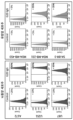
- Figure 7 is a diagram confirming the binding capacity of G2D IgG to 16 types of human cancer cell lines by FACS method.
- 7A shows colon cancer, liver cancer, prostate cancer, and ovarian cancer cell lines;
- Figure 7b shows brain tumor, breast cancer cell lines;
- Figure 7c shows the results of confirming the binding ability to pancreatic cancer and lung cancer cell lines.
- Figure 8 is a diagram confirming the binding capacity of G2D IgG to four mouse cancer cell lines in which B7-H3 expression was confirmed by FACS method.
- Figure 9 is a diagram confirming the immunogenicity of G2D IgG using an in silico method.
- Figure 10 is a diagram confirming the epitope of G2D IgG.
- Figure 10a is a diagram confirming the binding of G2D IgG to recombinant 4Ig B7-H3 protein by western blot to confirm the epitope type of G2D IgG
- Figure 10b is a diagram confirming the human 4Ig B7-H3 peptide to which G2D IgG binds. .
- Figure 10c is a diagram confirming the binding of G2D IgG to human 4Ig B7-H3 peptide containing EVQVP, which is expected to be an epitope of G2D IgG
- Figure 10d is a diagram showing the epitope of G2D IgG in all amino acids of human 4Ig B7-H3 am
- Figure 10e is a diagram showing the epitope of G2D IgG in the human 4Ig B7-H3 protein structure.
- FIG. 11 is a diagram confirming the anticancer efficacy of G2D IgG in syngeneic mice transplanted with the GL261 cell line.
- FIG. 11A is a diagram showing the results of measuring tumor volume
- FIG. 11B is a diagram showing the results of measuring body weight.
- B7-H3 positive clones were selected using phage display technology.
- 2.5 ⁇ g of recombinant human 4Ig B7-H3 (Sino Biological, China) was conjugated to magnetic beads (ThermoFisher Scientific, USA).
- a non-immune chicken phage library (chicken naive phage library) was reacted with a Raji cell pellet, which is known not to express B7-H3, to remove phages binding to Raji cells in advance, and dispersed in a 3% bovine serum albumin (Millipore, USA) solution.
- 96 clones selected from the plate of the fourth biopanning result were cultured overnight at 37°C with 100 ⁇ g/ml carbenicillin, 70 ⁇ g/ml kanamycin, and VCSM13 helper phage in a 96 deep well plate to produce scFv clones. Proliferation of expressed phage was induced.
- the culture medium obtained above was centrifuged to obtain a medium supernatant containing phages, and recombinant human 4Ig B7-H3, recombinant human 2Ig B7-H3 (R&D Systems, USA), and recombinant mouse B7-H3 (Sino Biological, China) were obtained, respectively.
- ER2738 which has a 2D clone showing a positive signal in the selected B7-H3, was cultured overnight using SB medium and centrifuged to obtain bacterial cells. Plasmid DNA was obtained using a DNA mini prep kit (Geneall, Korea), and the base sequence was analyzed using the primers in Table 1. The 2D clones analyzed had the CDR sequences in Table 2. The light chain and heavy chain variable region amino acid sequences of the 2D clone are shown in Table 3.
- Amino acid sequences of light and heavy chain variable regions of selected clones amino acid sequence number light chain variable region ALTQPSSVSANLGGTVKITCSGGSIGYGWYQQKAPGSAPVTVIYYNDRRPSDIPSRFSGSKSGSANTLTITGVQADDEAIYYCGSADSSSTYTGIFGAGTTLTVL 9 Heavy chain variable region AVTLDESGGGLQTPGGGLSLVCKASGFDFSSYPMVWVRQAPGKGLEYVASINSGGSWTGYGAAVKGRATISRDNGQSTVRLQLNNLRAEDTATYYCARAYGAATIDAWGHGTEVIVSS 10
- the heavy chain was treated with Eco RI and Not I (New England Biolab, UK) enzymes and ligated into pCMV (ThermoFisher Scientific, USA), an expression vector for animal cells, which was also treated with the same restriction enzymes. Additionally, the light chain was treated using Xba I (New England Biolab, UK) enzyme and ligated into the pCMV vector similarly treated with the same restriction enzyme. The ligated plasmid was transformed by applying heat shock to DH5 ⁇ competent cells (New England Biolab, UK), and the obtained colonies were mass-cultured to obtain the plasmid.
- the heavy chain and light chain plasmids converted into complete antibodies were transduced into Expi293F cells (Invitrogen, USA) using polyethylenimine (PEI) (Polysciences, USA) and 150 mM NaCl, and cultured in Freestyle 293 expression medium (Invitrogen, USA). ) was cultured in suspension for 7 days in an Erlenmeyer flask at 37°C, 8% CO 2 and 55% humidity. The expression cell culture was centrifuged at 4,000 rpm for 10 minutes, and the supernatant was taken and filtered through a 0.22 ⁇ m filter.
- PEI polyethylenimine
- Freestyle 293 expression medium Invitrogen, USA
- the filtered supernatant was induced to bind to a HiTrap Mabselect PrismA, 1 mL (GE Healthcare, USA) column at 4°C.
- the bound resin was washed with 10 cv (column volume) of 20 mM sodium phosphate (pH 7.0) and 1 M sodium chloride solution, and then the bound antibody was eluted using 100 mM sodium citrate (pH 3.0) and 150 mM sodium chloride solution. Then, it was neutralized with 1 M Tris-HCL (pH 9.0).
- the binding ability of the 2D IgG produced in Example 2 to the recombinant B7-H3 protein was confirmed by indirect ELISA.
- indirect ELISA recombinant human 4Ig B7-H3, recombinant human 2Ig B7-H3, and recombinant mouse B7-H3 were each diluted to 1 ⁇ g/ml in 50 ⁇ l of PBS, placed in a 96-well immunoplate (Corning, USA), and incubated at 4°C. It was stored and adsorbed overnight.
- the Raji cell line is known to be a B7-H3-negative cell line that does not express B7-H3 (Mol Ther Oncolytics. 2020;17:180-189), and B7-H3 is overexpressed in various breast cancer cells, including the BT-20 cell line. It is known that there is (Mol Cancer Ther. 2011;10(6):960-71).
- the binding capacity of 2D IgG was analyzed by FACS using the Raji cell line, a B7-H3 negative cell line, and the BT-20 cell line, a B7-H3 positive cell line. 5x10 5 BT-20 and Raji cells were suspended in PBS with or without 1 ⁇ g of 2D IgG, reacted at 4°C for 30 minutes, and then centrifuged to remove the supernatant. After removing the supernatant, each cell was washed twice using PBS.
- 2D IgG is a chicken/human chimeric antibody
- humanization was performed as follows to secure a humanized antibody.
- the amino acid residue numbers of the antibody domain are based on the Kabat EU numbering system commonly used in the art (Kabat et al., "Sequences of Proteins of Immunological Interest", 5th Ed., U.S. Department of Health and Human Services, Numbered according to EU index numbers as in NIH Publication No. 91-3242, 1991.
- the J gene of the light chain variable region was fixed as FGGGTKLTVL, referring to US2010-0056386, and the J gene of the heavy chain variable region was fixed as WGQGTTVTVSS, referring to US2014-0206849.
- Humanized 2D was named G2D, and its base sequence is shown in Table 5.
- G2D light and heavy chain variable region amino acid sequences light chain variable region G2D (VL) SYELTQPPSVSVSPGQTARITC SGGSIGYG WYQQKAPGQAPV T VIY YNDRRPS GIPERFSGS K SGTT N TLTISGVQAEDEADYYC GSADSSSTYTGI FGGGTKLTVL SEQ ID NO: 21
- Heavy chain variable region G2D (VH) QVQLVESGGGLVQPGGSLRLSCSASGF D FS SYPMV WVRQAPGKGLEYVS SINSGGSWTGYGAAVKG R
- a TISRDNSKNTLYLQMNSLRAEDTATYYCAR AYGAATIDA WGQGTTVTVSS SEQ ID NO: 22
- the DNA of the G2D variable region described above was synthesized in scFv form (Cosmogenetech, Korea) and converted into a complete antibody through PCR using the primer combinations shown in Table 6 below. Complete antibody conversion and expression/purification were performed in the same manner as in Example 2 described above.
- the binding ability of G2D IgG to B7-H3 was confirmed through SPR analysis, which can measure antigen-antibody binding force and kinetics using optical principles. SPR analysis can confirm the association rate (K a ) and dissociation rate (K d ), and through this, the K D value can be obtained, making quantitative binding force analysis possible.
- B7-family proteins are expressed on the surface of antigen presenting cells (APC), including cancer cells, and regulate T cell activity through interaction with T cell surface proteins (Figure 4).
- APC antigen presenting cells
- B7-H3 is one of the B7 family proteins expressed on APC.
- the T cell receptor has not yet been identified, it is known as an immune checkpoint that inhibits the action of T cells (Front Immunol. 2021; 12: 701006). Since B7 family proteins regulate T cell actions through different mechanisms, the specificity of antibodies targeting B7-H3 for B7-H3 is very important. Therefore, the specificity of G2D IgG for B7-family proteins was analyzed by indirect ELISA.
- Indirect ELISA was performed on: B7-DC (Sino Biological, China), B7-1 (Sino Biological, China), B7-2 (Sino Biological, China), B7-H2 (Sino Biological, China), B7-H4 (Sino Biological, China) China), B7-H1 (Sino Biological, China), 4Ig B7-H3, and 2Ig B7-H3 were used in the same manner as Example 3-1 using a 96-well immune plate coated at 1 ⁇ g/ml.
- G2D IgG was 4Ig among B7 family proteins. It bound only to 2Ig B7-H3 ( Figure 5). These results show the excellent specificity of G2D IgG for B7-H3.
- the recombinant protein and the cell surface protein have the same sequence, there may be structural differences, so it is essential to confirm the binding of the antibody to the cell surface protein.
- a structure containing 4Ig, 2Ig, and mouse B7-H3 extracellular domain was introduced into Raji cell line, a B7-H3 negative cell line. 2Ig, 4Ig, and mouse B7-H3 expressed on the cell surface were confirmed through insertion of green fluorescent protein (GFP). Binding of G2D IgG to B7-H3 expressed on the cell surface was confirmed in the same manner as Example 3-2.
- the secondary antibody used was goat anti-Human IgG (H+L) 594 antibody (Invitrogen, USA). Since binding to cell lines is difficult to confirm quantitatively, enoblituzumab, currently in the clinical stage and registered as a Korean patent (registration number: 10-1828570), was used as a control antibody.
- G2D IgG did not bind to Raji-Vector control, which did not express B7-H3, but bound to Raji-4Ig B7-H3 and Raji-2Ig B7-H3, which expressed 4Ig and 2Ig B7-H3 ( Figure 6a).
- the binding affinity of G2D IgG to 4Ig and 2Ig B7-H3 expressed on the cell surface was higher than that of enoblituzumab, a control antibody. Similar to human B7-H3, G2D IgG did not bind to Raji-Vector control, but bound to Raji-mouse B7-H3 expressing mouse B7-H3 ( Figure 6b).
- the control antibody, enoblituzumab did not bind to mouse B7-H3.
- Therapeutic antibodies are injected into the body and exhibit therapeutic effects by specifically binding to antigens expressed on cells, so the better the binding ability to antigens expressed on cells, the better the therapeutic effect can be expected.
- Figures 6a and b specific and excellent binding of G2D IgG to cell surface B7-H3 can be confirmed.
- G2D IgG showed a clear peak shift in all 16 cell lines used compared to the control to which only the secondary antibody was bound, except for the breast cancer cell line MDA-MB-453 ( Figure 7). These results show that G2D IgG binds to 15 out of 16 cell lines used, meaning that G2D IgG can bind to cancer cells expressing B7-H3 regardless of cancer type.
- Antibodies that cross-link to human and mouse antigens are advantageous for developing treatments because they allow the anticancer effect and level of immune cell activity to be confirmed in a syngeneic mouse model with a preserved immune system. Additionally, it has the advantage of being able to confirm toxicity in small animals, mice, before large animal toxicity tests. Therefore, a total of four mouse cancer cell lines, 3LL (mouse lung cancer cell line), 4T1 (mouse breast cancer cell line), Panc-02 (mouse pancreatic cancer cell line), and GL-261 (mouse brain tumor cell line), in which expression of B7-H3 was confirmed. The binding ability of G2D IgG was confirmed by the method of Example 3-2.
- G2D IgG bound to the four mouse cancer cell lines used ( Figure 8). These results mean that G2D IgG can be applied to various mouse models, including the syngeneic mouse model, and when developing G2D IgG as a therapeutic antibody, efficient evaluation of anticancer effects is possible in preclinical tests.
- the property of inducing an immune response when a therapeutic antibody is introduced into the body is called immunogenicity.
- the immune response in the body caused by the immunogenicity of a therapeutic antibody can cause serious problems not only in drug efficacy but also in patient safety. . Therefore, confirmation of the immunogenicity of therapeutic antibodies is an important part of drug development. Therefore, the immunogenicity of G2D IgG was confirmed using an in silico method.
- Epitope is an antigenic determining site and refers to a specific sequence of an antigen that an antibody can recognize and bind to.
- the therapeutic effect of antibodies may differ depending on the epitope, and in the case of cancer immunotherapy drugs, the degree of immune cell activation may also vary, so identifying the epitope of an antibody is an important part of drug development.
- EVQVP is located in the Ig-like V-type1 and Ig-like V-type2 domains of 4Ig B7-H3 ( Figures 10d and e).
- these epitopes were confirmed to contain the GYPEAE sequence ( Figure 10b), and GYPEAE is the Ig-like C2-type1 and Ig-like C2- of 4Ig B7-H3. It is located in type2 ( Figures 10d and e).
- G2D IgG had a different epitope from the control antibody, enoblituzumab.
- the underlined bold portion in the above sequence represents the B7-H3 epitope to which the G2D antibody binds.
- G2D IgG showed a T cell-mediated cancer cell killing effect that induced about 30% of activation compared to the negative control group, and the effect was equal or better than that of enoblituzumab, which has wild-type Fc as a control antibody (Table 9). This means that G2D IgG can enhance the cell death effect of T cells through B7-H3 binding on the surface of cancer cells, and that G2D IgG can be applied as an immunotherapy agent.
- Example 9 Evaluation of anticancer efficacy of G2D IgG in GL261 transplant syngeneic mouse model
- G2D IgG as an immuno-anticancer agent was confirmed in Example 8, it was verified through an animal model.
- the syngeneic mouse model induces cancer while the mouse immune system is preserved, so if the antibody being evaluated can recognize mouse cancer cell antigens, it is useful for confirming the anticancer effect through immune cells without separate immune cell transplantation.
- GL261 FIG. 8
- a mouse glioma cell line for which G2D IgG binding was confirmed was implanted subcutaneously on the right flank of the mouse at 4x10⁶ cells/mouse into an immunocompetent mouse, a C57BL/ 6 female mouse (Orient Bio, Korea).
- G2D IgG administration began when the tumor size reached approximately 70 mm 3 .
- InVivo MAb anti-mouse CD276 (anti-mB7-H3 IgG) (BioXcell, USA), an antibody that binds to mouse B7-H3, was used.
- Each antibody was injected intravenously at a concentration of 10 mg/kg, and administered twice a week for 2 weeks.
- Body weight was measured using an electronic scale once a week after the start of administration and on the day of autopsy.
Landscapes
- Health & Medical Sciences (AREA)
- Chemical & Material Sciences (AREA)
- Life Sciences & Earth Sciences (AREA)
- Organic Chemistry (AREA)
- Immunology (AREA)
- General Health & Medical Sciences (AREA)
- Medicinal Chemistry (AREA)
- Veterinary Medicine (AREA)
- Public Health (AREA)
- Animal Behavior & Ethology (AREA)
- Pharmacology & Pharmacy (AREA)
- Proteomics, Peptides & Aminoacids (AREA)
- Microbiology (AREA)
- Mycology (AREA)
- Molecular Biology (AREA)
- Epidemiology (AREA)
- Genetics & Genomics (AREA)
- Biophysics (AREA)
- Biochemistry (AREA)
- Chemical Kinetics & Catalysis (AREA)
- General Chemical & Material Sciences (AREA)
- Nuclear Medicine, Radiotherapy & Molecular Imaging (AREA)
- Peptides Or Proteins (AREA)
Abstract
La présente invention concerne un anticorps anti-B7-H3 ou un fragment de liaison à l'antigène de celui-ci, qui se lie à un épitope spécifique situé à l'intérieur de la séquence d'acides aminés d'un domaine de B7-H3. L'anticorps (i) se lie de manière spécifique à divers types de lignées cellulaires cancéreuses exprimant B7-H3, démontrant des effets de mort cellulaire de cancer à médiation cellulaire, et (ii) se lie à des épitopes chez les humains et les souris, permettant ainsi une évaluation efficace des effets anticancéreux dans des essais précliniques lorsqu'ils sont développés dans un anticorps thérapeutique.
Applications Claiming Priority (4)
| Application Number | Priority Date | Filing Date | Title |
|---|---|---|---|
| KR10-2022-0152913 | 2022-11-15 | ||
| KR20220152913 | 2022-11-15 | ||
| KR1020230154552A KR20240072932A (ko) | 2022-11-15 | 2023-11-09 | B7-h3에 특이적으로 결합하는 항체 |
| KR10-2023-0154552 | 2023-11-09 |
Publications (1)
| Publication Number | Publication Date |
|---|---|
| WO2024106939A1 true WO2024106939A1 (fr) | 2024-05-23 |
Family
ID=91084778
Family Applications (1)
| Application Number | Title | Priority Date | Filing Date |
|---|---|---|---|
| PCT/KR2023/018360 Ceased WO2024106939A1 (fr) | 2022-11-15 | 2023-11-15 | Anticorps se liant de manière spécifique à b7-h3 |
Country Status (1)
| Country | Link |
|---|---|
| WO (1) | WO2024106939A1 (fr) |
Citations (5)
| Publication number | Priority date | Publication date | Assignee | Title |
|---|---|---|---|---|
| KR20100014527A (ko) * | 2007-03-22 | 2010-02-10 | 슬로안-케테링인스티튜트퍼캔서리서치 | 단일클론항체 8h9의 용도 |
| KR20140033018A (ko) * | 2011-04-25 | 2014-03-17 | 다이이찌 산쿄 가부시키가이샤 | 항 b7-h3항체 |
| KR20170042786A (ko) * | 2014-08-27 | 2017-04-19 | 메모리얼 슬로안-케터링 캔서 센터 | 항체, 조성물 및 용도 |
| KR20190134614A (ko) * | 2017-03-31 | 2019-12-04 | 지앙수 헨그루이 메디슨 컴퍼니 리미티드 | B7-h3 항체, 이의 항원-결합 단편 및 이의 의학적 용도 |
| WO2020102779A1 (fr) * | 2018-11-16 | 2020-05-22 | Albert Einstein College Of Medicine | Anticorps monoclonaux dirigés contre le domaine igv de b7-h3 et ses utilisations |
-
2023
- 2023-11-15 WO PCT/KR2023/018360 patent/WO2024106939A1/fr not_active Ceased
Patent Citations (5)
| Publication number | Priority date | Publication date | Assignee | Title |
|---|---|---|---|---|
| KR20100014527A (ko) * | 2007-03-22 | 2010-02-10 | 슬로안-케테링인스티튜트퍼캔서리서치 | 단일클론항체 8h9의 용도 |
| KR20140033018A (ko) * | 2011-04-25 | 2014-03-17 | 다이이찌 산쿄 가부시키가이샤 | 항 b7-h3항체 |
| KR20170042786A (ko) * | 2014-08-27 | 2017-04-19 | 메모리얼 슬로안-케터링 캔서 센터 | 항체, 조성물 및 용도 |
| KR20190134614A (ko) * | 2017-03-31 | 2019-12-04 | 지앙수 헨그루이 메디슨 컴퍼니 리미티드 | B7-h3 항체, 이의 항원-결합 단편 및 이의 의학적 용도 |
| WO2020102779A1 (fr) * | 2018-11-16 | 2020-05-22 | Albert Einstein College Of Medicine | Anticorps monoclonaux dirigés contre le domaine igv de b7-h3 et ses utilisations |
Similar Documents
| Publication | Publication Date | Title |
|---|---|---|
| WO2020076105A1 (fr) | Nouvel anticorps anti-c-kit | |
| WO2017052241A1 (fr) | Nouvel anticorps anti-mésothéline et composition le comprenant | |
| NZ719092A (en) | Tumor necrosis factor-like ligand 1a specific antibodies and compositions and uses thereof | |
| BR112021004287A2 (pt) | anticorpos anti-avss8 e composições e usos dos mesmos | |
| WO2019225777A1 (fr) | Anticorps anti-ror1 et son utilisation | |
| KR20190117493A (ko) | 항 pd-1 항체 및 그의 용도 | |
| WO2018217058A1 (fr) | Anticorps interleukine-2 anti-humains et leurs utilisations | |
| WO2021107566A1 (fr) | Anticorps dirigé contre c-kit et utilisation correspondante | |
| WO2017030370A1 (fr) | Récepteur d'anticorps chimérique auquel est lié un anticorps anti-cotinine et utilisation de celui-ci | |
| TW202118791A (zh) | 對cd39具有特異性的結合分子及其用途 | |
| WO2019132533A1 (fr) | Anticorps anti-pd-l1 et son utilisation | |
| WO2022169269A1 (fr) | Anticorps anti-ctla-4 et son utilisation | |
| WO2023214778A1 (fr) | Anticorps anti-cd300c ou fragment de liaison à l'antigène de celui-ci, et ses utilisations pour prévenir ou traiter une maladie cérébrale neurodégénérative | |
| WO2019078699A2 (fr) | Anticorps anti-vista et son utilisation | |
| WO2020101365A1 (fr) | Anticorps anti-c-met présentant une stabilité améliorée ou des fragments de liaison à l'antigène de celui-ci | |
| WO2020004937A1 (fr) | Conjugué anticorps anti-bcma-médicament et utilisation associée | |
| WO2024090927A1 (fr) | Nouvel anticorps anti-cdcp1 et son utilisation | |
| WO2016084993A1 (fr) | Nouvel anticorps anti-egfrviii et composition le comprenant | |
| KR20240072932A (ko) | B7-h3에 특이적으로 결합하는 항체 | |
| WO2024106939A1 (fr) | Anticorps se liant de manière spécifique à b7-h3 | |
| WO2021029746A2 (fr) | Anticorps anti-tie 2 et son utilisation | |
| WO2021101244A1 (fr) | Composition pour la prévention ou le traitement du cancer, comprenant des anticorps monoclonaux anti-cd300c | |
| WO2024172363A1 (fr) | Anticorps anti-cntn4 et ses utilisations | |
| WO2023157989A1 (fr) | Conjugué anticorps-médicament | |
| WO2023182866A1 (fr) | Anticorps spécifique de hla-g et son utilisation |
Legal Events
| Date | Code | Title | Description |
|---|---|---|---|
| 121 | Ep: the epo has been informed by wipo that ep was designated in this application |
Ref document number: 23892005 Country of ref document: EP Kind code of ref document: A1 |
|
| NENP | Non-entry into the national phase |
Ref country code: DE |
|
| 122 | Ep: pct application non-entry in european phase |
Ref document number: 23892005 Country of ref document: EP Kind code of ref document: A1 |