WO2024092161A2 - Size-tunable synthetic particles with tunable optical properties and methods for using the same for immune cell activation - Google Patents
Size-tunable synthetic particles with tunable optical properties and methods for using the same for immune cell activation Download PDFInfo
- Publication number
- WO2024092161A2 WO2024092161A2 PCT/US2023/077961 US2023077961W WO2024092161A2 WO 2024092161 A2 WO2024092161 A2 WO 2024092161A2 US 2023077961 W US2023077961 W US 2023077961W WO 2024092161 A2 WO2024092161 A2 WO 2024092161A2
- Authority
- WO
- WIPO (PCT)
- Prior art keywords
- particle
- succinimidyl ester
- methacrylate
- difluoro
- cell
- Prior art date
- Legal status (The legal status is an assumption and is not a legal conclusion. Google has not performed a legal analysis and makes no representation as to the accuracy of the status listed.)
- Ceased
Links
Classifications
-
- G—PHYSICS
- G01—MEASURING; TESTING
- G01N—INVESTIGATING OR ANALYSING MATERIALS BY DETERMINING THEIR CHEMICAL OR PHYSICAL PROPERTIES
- G01N33/00—Investigating or analysing materials by specific methods not covered by groups G01N1/00 - G01N31/00
- G01N33/48—Biological material, e.g. blood, urine; Haemocytometers
- G01N33/50—Chemical analysis of biological material, e.g. blood, urine; Testing involving biospecific ligand binding methods; Immunological testing
- G01N33/53—Immunoassay; Biospecific binding assay; Materials therefor
- G01N33/531—Production of immunochemical test materials
- G01N33/532—Production of labelled immunochemicals
-
- G—PHYSICS
- G01—MEASURING; TESTING
- G01N—INVESTIGATING OR ANALYSING MATERIALS BY DETERMINING THEIR CHEMICAL OR PHYSICAL PROPERTIES
- G01N15/00—Investigating characteristics of particles; Investigating permeability, pore-volume or surface-area of porous materials
- G01N15/08—Investigating permeability, pore-volume, or surface area of porous materials
- G01N15/088—Investigating volume, surface area, size or distribution of pores; Porosimetry
- G01N15/0893—Investigating volume, surface area, size or distribution of pores; Porosimetry by measuring weight or volume of sorbed fluid, e.g. B.E.T. method
-
- G—PHYSICS
- G01—MEASURING; TESTING
- G01N—INVESTIGATING OR ANALYSING MATERIALS BY DETERMINING THEIR CHEMICAL OR PHYSICAL PROPERTIES
- G01N15/00—Investigating characteristics of particles; Investigating permeability, pore-volume or surface-area of porous materials
- G01N15/10—Investigating individual particles
- G01N15/1012—Calibrating particle analysers; References therefor
-
- G—PHYSICS
- G01—MEASURING; TESTING
- G01N—INVESTIGATING OR ANALYSING MATERIALS BY DETERMINING THEIR CHEMICAL OR PHYSICAL PROPERTIES
- G01N15/00—Investigating characteristics of particles; Investigating permeability, pore-volume or surface-area of porous materials
- G01N15/10—Investigating individual particles
- G01N15/14—Optical investigation techniques, e.g. flow cytometry
- G01N15/1456—Optical investigation techniques, e.g. flow cytometry without spatial resolution of the texture or inner structure of the particle, e.g. processing of pulse signals
- G01N15/1459—Optical investigation techniques, e.g. flow cytometry without spatial resolution of the texture or inner structure of the particle, e.g. processing of pulse signals the analysis being performed on a sample stream
-
- G—PHYSICS
- G01—MEASURING; TESTING
- G01N—INVESTIGATING OR ANALYSING MATERIALS BY DETERMINING THEIR CHEMICAL OR PHYSICAL PROPERTIES
- G01N15/00—Investigating characteristics of particles; Investigating permeability, pore-volume or surface-area of porous materials
- G01N15/10—Investigating individual particles
- G01N15/14—Optical investigation techniques, e.g. flow cytometry
- G01N15/1468—Optical investigation techniques, e.g. flow cytometry with spatial resolution of the texture or inner structure of the particle
- G01N15/147—Optical investigation techniques, e.g. flow cytometry with spatial resolution of the texture or inner structure of the particle the analysis being performed on a sample stream
-
- G—PHYSICS
- G01—MEASURING; TESTING
- G01N—INVESTIGATING OR ANALYSING MATERIALS BY DETERMINING THEIR CHEMICAL OR PHYSICAL PROPERTIES
- G01N33/00—Investigating or analysing materials by specific methods not covered by groups G01N1/00 - G01N31/00
- G01N33/48—Biological material, e.g. blood, urine; Haemocytometers
- G01N33/50—Chemical analysis of biological material, e.g. blood, urine; Testing involving biospecific ligand binding methods; Immunological testing
- G01N33/5005—Chemical analysis of biological material, e.g. blood, urine; Testing involving biospecific ligand binding methods; Immunological testing involving human or animal cells
-
- G—PHYSICS
- G01—MEASURING; TESTING
- G01N—INVESTIGATING OR ANALYSING MATERIALS BY DETERMINING THEIR CHEMICAL OR PHYSICAL PROPERTIES
- G01N33/00—Investigating or analysing materials by specific methods not covered by groups G01N1/00 - G01N31/00
- G01N33/48—Biological material, e.g. blood, urine; Haemocytometers
- G01N33/50—Chemical analysis of biological material, e.g. blood, urine; Testing involving biospecific ligand binding methods; Immunological testing
- G01N33/53—Immunoassay; Biospecific binding assay; Materials therefor
- G01N33/531—Production of immunochemical test materials
- G01N33/532—Production of labelled immunochemicals
- G01N33/533—Production of labelled immunochemicals with fluorescent label
-
- G—PHYSICS
- G01—MEASURING; TESTING
- G01N—INVESTIGATING OR ANALYSING MATERIALS BY DETERMINING THEIR CHEMICAL OR PHYSICAL PROPERTIES
- G01N33/00—Investigating or analysing materials by specific methods not covered by groups G01N1/00 - G01N31/00
- G01N33/48—Biological material, e.g. blood, urine; Haemocytometers
- G01N33/50—Chemical analysis of biological material, e.g. blood, urine; Testing involving biospecific ligand binding methods; Immunological testing
- G01N33/53—Immunoassay; Biospecific binding assay; Materials therefor
- G01N33/563—Immunoassay; Biospecific binding assay; Materials therefor involving antibody fragments
-
- C—CHEMISTRY; METALLURGY
- C07—ORGANIC CHEMISTRY
- C07K—PEPTIDES
- C07K16/00—Immunoglobulins [IGs], e.g. monoclonal or polyclonal antibodies
- C07K16/18—Immunoglobulins [IGs], e.g. monoclonal or polyclonal antibodies against material from animals or humans
- C07K16/28—Immunoglobulins [IGs], e.g. monoclonal or polyclonal antibodies against material from animals or humans against receptors, cell surface antigens or cell surface determinants
-
- G—PHYSICS
- G01—MEASURING; TESTING
- G01N—INVESTIGATING OR ANALYSING MATERIALS BY DETERMINING THEIR CHEMICAL OR PHYSICAL PROPERTIES
- G01N15/00—Investigating characteristics of particles; Investigating permeability, pore-volume or surface-area of porous materials
- G01N2015/0038—Investigating nanoparticles
-
- G—PHYSICS
- G01—MEASURING; TESTING
- G01N—INVESTIGATING OR ANALYSING MATERIALS BY DETERMINING THEIR CHEMICAL OR PHYSICAL PROPERTIES
- G01N15/00—Investigating characteristics of particles; Investigating permeability, pore-volume or surface-area of porous materials
- G01N15/10—Investigating individual particles
- G01N2015/1006—Investigating individual particles for cytology
-
- G—PHYSICS
- G01—MEASURING; TESTING
- G01N—INVESTIGATING OR ANALYSING MATERIALS BY DETERMINING THEIR CHEMICAL OR PHYSICAL PROPERTIES
- G01N15/00—Investigating characteristics of particles; Investigating permeability, pore-volume or surface-area of porous materials
- G01N15/10—Investigating individual particles
- G01N15/1012—Calibrating particle analysers; References therefor
- G01N2015/1014—Constitution of reference particles
-
- G—PHYSICS
- G01—MEASURING; TESTING
- G01N—INVESTIGATING OR ANALYSING MATERIALS BY DETERMINING THEIR CHEMICAL OR PHYSICAL PROPERTIES
- G01N15/00—Investigating characteristics of particles; Investigating permeability, pore-volume or surface-area of porous materials
- G01N15/10—Investigating individual particles
- G01N15/14—Optical investigation techniques, e.g. flow cytometry
- G01N2015/1493—Particle size
-
- G—PHYSICS
- G01—MEASURING; TESTING
- G01N—INVESTIGATING OR ANALYSING MATERIALS BY DETERMINING THEIR CHEMICAL OR PHYSICAL PROPERTIES
- G01N15/00—Investigating characteristics of particles; Investigating permeability, pore-volume or surface-area of porous materials
- G01N15/10—Investigating individual particles
- G01N15/14—Optical investigation techniques, e.g. flow cytometry
- G01N2015/1497—Particle shape
Definitions
- Flow cytometry is a technique that allows for the rapid separation, counting, and characterization of individual cells and is routinely used in clinical and laboratory settings for a variety of applications.
- the technology relies on directing a beam of light onto a hydrodynamically-focused stream of liquid.
- a number of detectors are then aimed at the point where the stream passes through the light beam: one in line with the light beam (forward scatter or FSC) and several perpendicular to it (side scatter or SSC).
- FSC correlates with the cell volume and SSC depends on the inner complexity of the particle (e.g., shape of the nucleus, the amount and type of cytoplasmic granules or the membrane roughness).
- T cell activation Current standards for in vitro T cell activation are magnetic microbeads containing ⁇ CD3 and ⁇ CD28 antibodies and having a subcellular sized diameter. However, these microbeads, which may be monodisperse polystyrene beads, are superparamagnetic, thus requiring an additional isolation step after beads have been in culture.
- Other methods to stimulate e.g., T cells in vitro include a plate- bound method where ⁇ CD3 and ⁇ CD28 antibodies are directly added to T cell culture and are Attorney Docket No. SLIN-015/01WO 323489-2099 washed off after 24h of stimulation.
- the present disclosure relates to a hydrogel particle comprising a polymerized monomer and having at least one surface is provided.
- the hydrogel particle has at least one optical property that is substantially similar to the at least one optical property of a target cell.
- the optical property in one embodiment is a side scatter profile (SSC), forward scatter profile (FSC), a fluorescence emission profile, or a combination thereof.
- the target cell can be any target cell that the user specifies.
- the target cell is an immune cell, stem cell or cancer cell.
- the present disclosure relates to a method for calibrating a cytometric device for analysis of a target cell, is provided.
- the method comprises inserting into the device a hydrogel particle having at least one optical property substantially similar to a target cell, wherein the hydrogel particle comprises a polymerized monomer and has at least one surface.
- the method further comprises measuring the at least one optical property of the hydrogel particle using the cytometric device.
- the at least one optical property in one embodiment is used as a reference to detect a target cell in a sample.
- the present disclosure relates to a method for detecting a target cell in a sample.
- the method comprises inserting into the device a hydrogel particle having at least one optical property substantially similar to a target cell, wherein the hydrogel particle comprises a polymerized monomer.
- the method further comprises measuring the at least one optical property of the hydrogel particle using the cytometric device.
- a sample comprising a plurality of cells is inserted into the cytometric device, and the at least one optical property of individual cells of the plurality are measured.
- a determination is made, based on the optical property measurement, whether the target cell or plurality thereof is present in the sample.
- the hydrogel particle comprises a biodegradable monomer.
- the biodegradable monomer is a Attorney Docket No. SLIN-015/01WO 323489-2099 monosaccharide, disaccharide, polysaccharide, peptide, protein, or protein domain.
- the biodegradable monomer is functionalized with acrylamide or acrylate.
- the methods herein can be used on any appropriate detection or analysis platform, including, without limitation, imaging (e.g., a microscope, a scanner, or the like), flow cytometry, or other immunodetection methods (e.g., an ELISA assay), electrophoresis, omic analysis (genomics, glycomics, proteomics, lipidomics analysis), molecular analysis (q-PCR etc.), or the like.
- Analysis such as imaging or detecting, can be performed in fluorescence, bright field, dark field, or immunohistochemical (e.g. chromogenic stains).
- the present disclosure relates to particles for immune cell activation.
- FIG.1A-B illustrates the optical properties of disclosed hydrogel particles compared to polystyrene beads.
- FIG. 2 depicts the process of producing labeled hydrogel particles of the disclosure, including hydrogels with attached biomolecules.
- FIG.3A-3C provides brightfield and fluorescent images of labeled hydrogel particles of the disclosure.
- FIG.4A-4C illustrates the use of hydrogel particles of the disclosure as calibrants for cell types displaying a variety of optical scattering properties.
- FIG. 5 provides dating showing correlation of inter-drop delay for a flow cytometer with hydrogel particle diameter.
- FIG.6A and FIG.6C provides brightfield and FIG.6B and FIG.6D fluorescent images of Chinese Hamster Ovary cells (FIG.6A and FIG.6B) and hydrogel particles of the disclosure (FIG.6C and FIG.6D).
- FIG.7 provides data showing comparison of human buccal cells to hydrogel particles encapsulating different amounts of DNA, as measured by fluorescence-activated cell sorting (FACS).
- FIG. 8 provides data for hydrogel particles encapsulating nanoparticles at different concentrations, demonstrating tuning of side scattering independent of forward scattering.
- FIG. 9 provides data for hydrogel particles produced with different percentages of polymer, demonstrating tuning of refractive index measured by forward scattering.
- FIG.10 shows one embodiment of hydrogel parameter tuning to match and/or mimic desired cell population metrics.
- Attorney Docket No. SLIN-015/01WO 323489-2099 [0020]
- FIGS.11A-11D and 12A-B are diagrams showing embodiments of how to adjust the forward scatter, side scatter and surface properties of a hydrogel particle.
- FIG.13 are scatter plots for various hydrogel particles (FIG.13A) and (FIG.13B) and a commercial blood sample (FIG.13C).
- FIG.14 shows a scatter plot of a porous particle and a general step for manufacturing of porous particles.
- FIG.13 shows one embodiment of hydrogel parameter tuning to match and/or mimic desired cell population metrics.
- FIGS.11A-11D and 12A-B are diagrams showing embodiments of how to adjust the forward scatter, side scatter and surface properties of a hydrogel particle.
- FIG.13 are scatter plots for various hydrogel particles (FIG.13A) and (FIG.13B) and a commercial blood
- the porogen may be polyethylene glycol 8000 at concentrations of 2.25%, 3.4%, 4.5%, 6.3%, and 9% w/v.
- the porosity of the porous particles increases with increasing content of polyethylene glycol 8000 in the water phase formulations.
- Each image of the porogen concentrations can be evaluated in view of the 50 ⁇ m scale bar in the 9% porogen image.
- Increased porosity can be used as a factor for increase SSC optical match of particles.
- Porosity can also help replicate visual morphologies of target cells. Further conjugation of biomolecules on particles can provide additional functionality, including immune response activation functions.
- FIG. 16 provides scatter plots of side scatter data and forward scatter data for porous particles formed by varying porogen concentrations (weight by volume) within the dispersed phase. From left to right, the porous particles comprise polyethylene glycol 8000 at concentrations of 2.25%, 3.4%, and 4.5% w/v. The side scatter of the porous particles measured by flow cytometry increases with increasing content of polyethylene glycol 8000 in the water phase formulations, while the forward scatter is largely unchanged.
- FIG. 17 provides scatter plots of side scatter data and forward scatter data for porous particles comprising a constant concentration of porogen and nanoparticles.
- FIG. 18 provides scatter plots of optical scatter of porous particles conjugated with fluorescent dyes. Fluorophores or dyes can be conjugated to the porous particles, which can then be used to mimic a stained cell in the applications of image cytometry or histology.
- FIG.19 is a schematic of a degradable particle, according to embodiments of the present disclosure.
- FIG.20 is a schematic of a particle as a synthetic feeder cell, according to embodiments of the present disclosure.
- Attorney Docket No. SLIN-015/01WO 323489-2099 [0029]
- FIG. 21 is a schematic of a particle as a synthetic biomolecule presenting particle, according to embodiments of the present disclosure.
- FIG.22A and FIG.22B relate to particles as feeder cells, according to embodiments of the present disclosure.
- FIG.23A and FIG.23B relate to synthetic biomolecule presenting particles, according to embodiments of the present disclosure.
- FIG. 24 depicts a method of generating porous particles by a microfluidic droplet process, the process including curing and purification before cell therapy application.
- FIG. 24 depicts a method of generating porous particles by a microfluidic droplet process, the process including curing and purification before cell therapy application.
- FIG. 25 is a microscopy image of porous particles formed using polyethylene glycol (PEG).
- FIG.26 depicts early-stage (24 hour incubation) activation of Jurkat samples incubated with either DynabeadsTM or porous particles, according to embodiments of the present disclosure.
- the porous particles of FIG. 26 are particles having pores formed during manufacturing using 9% w/v PEG as a porogen.
- FIG. 26 depicts an increased activation of Jurkat samples as indicated by upregulation of activation marker CD69 when compared with baseline Jurkats values and also when compared against cells activated by DynabeadsTM.
- FIG. 26 depicts an increased activation of Jurkat samples as indicated by upregulation of activation marker CD69 when compared with baseline Jurkats values and also when compared against cells activated by DynabeadsTM.
- FIG. 27 is a bar chart depicting early-stage T-cell activation (i.e., increase in Jurkat activation) when incubated with porous particles (pores formed by 9% PEG) and DynabeadsTM for 24 hours. As shown, T-cell activation is increased in porous particles samples, as shown by an increase in CD69.
- FIG.28 depicts a relative upregulation of early-stage T-cell activation marker CD69 in Jurkat samples incubated for 48 hours with porous particles (pores formed by 9% PEG) as compared to DynabeadsTM. Activation during this prolonged incubation period represents a sustained activation.
- FIG.29 depicts a relative upregulation of late-stage T-cell activation marker CD25 in Jurkat samples incubated for 48 hours with porous particles (pores formed by 9% PEG) as compared to DynabeadsTM. Activation during this prolonged incubation period represents a sustained activation.
- FIG. 30 is a bar chart depicting a relative upregulation of late-stage T-cell activation marker CD25 in Jurkat samples incubated for 48 hours with porous particles (pores formed by 9% PEG) as compared to DynabeadsTM.
- FIG. 31 provides scatter plots of conjugation.
- the term “about” is used to indicate that a value includes the inherent variation of error for the device or the method being employed to determine the value, or the variation that exists among the samples being measured. Unless otherwise stated or otherwise evident from the context, the term “about” means within 10% above or below the reported numerical value (except where such number would exceed 100% of a possible value or go below 0%). When used in conjunction with a range or series of values, the term “about” applies to the endpoints of the range or each of the values enumerated in the series, unless otherwise indicated. As used in this application, the terms “about” and “approximately” are used as equivalents.
- porosity may be used to refer to the percentage of void space within the hydrogel particle. When porogens are used, the porosity is the percentage of void space within the hydrogel particle after removal of the porogens. In such a case, the porosity may comprise a plurality of micropores and a plurality of macropores, as will be described below.
- the cells contacted with the defined medium can be further treated with a cell differentiation environment to stabilize the cells, or to differentiate the cells.
- the term “stabilize,” when used in reference to the differentiation state of a cell or culture of cells indicates that the cells will continue to proliferate over multiple passages in culture, and preferably indefinitely in culture, where most, if not all, of the cells in the culture are of the same differentiation state.
- the stabilized cells when the stabilized cells divide, the division typically yields cells of the same cell type or yields cells of the same differentiation state.
- a stabilized cell or cell population in general does not further differentiate or de-differentiate if the cell culture conditions are not altered and the cells continue to be passaged and are not overgrown.
- the cell that is stabilized is capable of proliferation in the stable state indefinitely, or for at least more than 2 passages.
- the cells are stable for more than 3 passages, 4 passages, 5 passages, 6 passages, 7 passages, 8 passages, 9 passages, more than 10 passages, more than 15 passages, more than 20 passages, more than 25 passages, or more than 30 passages.
- the cell is stable for greater than approximately 1 month, 2 months, 3 months, 4 months, 5 months, 6 months, 7 months, 8 months, 9 months, 10 months, or 11 months of continuous passaging. In another embodiment, the cell is stable for greater than approximately 1 year of continuous passaging.
- stem cells are maintained in culture in a pluripotent state by routine passage in the defined medium until it is desired that they be differentiated.
- proliferate refers to an increase in the number cells in a cell culture.
- growth environment is an environment in which stem cells (e.g., primate embryonic stem cells) will proliferate in vitro.
- a “defined” medium refers to a biochemically defined formulation comprised solely of the biochemically-defined constituents.
- a defined medium may include solely constituents having known chemical compositions.
- a defined medium may also include constituents that are derived from known sources.
- a defined medium may also include factors and other compositions secreted from known tissues or cells; however, the defined medium will not include the conditioned medium from a culture of such cells.
- a “defined medium” may, if indicated, include particular compounds added to form the culture medium.
- the term “basal medium” refers to a solution of amino acids, vitamins, salts, and nutrients that is effective to support the growth of cells in culture, although normally these compounds will not support cell growth unless supplemented with additional compounds.
- the nutrients include a carbon source (e.g., a sugar such as glucose) that can be metabolized by the cells, as well as other compounds necessary for the cells' survival.
- DMEM Dulbecco's Modified Eagle Media
- KO-DMEM Knockout- DMEM
- DMEM/F12 any base medium that supports the growth of primate embryonic stem cells in a substantially undifferentiated state can be employed.
- a “basal medium” as described herein also refers to the basal medium described in PCT/US2007/062755, filed Jun.13, 2007, which is herein incorporated in its entirety.
- Several critical calibration measurements for flow cytometers require precise time resolution, such as setting the offset time between lasers, and calculating the delay time between detection and sorting of an object. Due to the fluidic conditions within the instrument, precise setting of these timing parameters requires the use of calibration particles that are the same size as the cells to be analyzed. Timing calibrations are typically performed using polystyrene beads with variable fluorescent intensities to calibrate the response of an excitation source and to set the inter-laser timing delay and sorting delay.
- Flow cytometers can also be calibrated using forward and side scatter signals which are general measures of size and granularity or complexity of the target sample. These calibrations are crucial for the accurate performance of the cytometer and for any downstream analysis or sorting of cell populations.
- the disclosed hydrogel particles exhibit tuned scatter properties and are suitable for use as calibration reagents for a range of mammalian or bacterial cell types. Scattering is a standard metric for distinguishing cell types in heterogeneous mixtures for clinical, food safety, and research purposes.
- polystyrene particles can be used to set inter-laser and sorting delays for some applications, many eukaryotic cell types fall outside of the size range of commercially available polystyrene particles (1-20 ⁇ m) making it nearly impossible to accurately calibrate a flow cytometer for these targets.
- polystyrene particles are fundamentally limited in the optical properties that can possess such as side scattering, which is a general Attorney Docket No. SLIN-015/01WO 323489-2099 measure of cellular complexity. Polystyrene particles are therefore limited in the two most important passive optical measurements used in flow cytometry: FSC (forward scattering), and SSC (side scattering) which measure the size and complexity of the target respectively.
- a composition comprising a plurality of hydrogel particles, wherein the individual hydrogel particles of the plurality each has one or more optical properties substantially similar to one or more optical properties of a target cell.
- Each of the individual hydrogel particles of the plurality independently comprises a hydrogel which is synthesized by polymerizing one or more monomers, i.e., to form a homopolymer or copolymer.
- the use of bifunctional monomers allows for the further derivatization of hydrogels, e.g., with fluorescent dyes, biomolecules, such as cell surface markers or epitope binding fragments thereof, and immunostimulatory biomolecules, including CD markers and antibodies or antigen-binding fragments thereof, as well as a combination thereof.
- An example of hydrogel parameter tuning to meet/match desired cell subpopulation metrics is provided at FIG.10. Methods for tuning the properties of a hydrogel are described herein.
- the present invention provides individual hydrogel particles each having one or more optical properties substantially similar to one or more optical Attorney Docket No. SLIN-015/01WO 323489-2099 properties of a target cell.
- the one or more optical properties is a side scatter profile, a forward scatter profile or a secondary marker profile, such as a fluorescence marker profile, for example a fluorescence marker profile of a fluorescently-labeled antibody that binds to the surface of the hydrogel particle.
- substantially similar denotes at least 40% similar, at least 50% similar, at least 60% similar, at least 70% similar, at least 80% similar, at least 90% similar, at least 95% similar, at least 96% similar, at least 97% similar, at least 98% similar or at least 99% similar.
- the present invention is based in part on the unexpected discovery that one or more optical properties of a hydrogel particle can be independently modulated by altering the composition of the hydrogel particle, for example, by altering the amount of initial monomer (or co-monomer) in the composition, by altering the surface functionalization, by altering the amount of a polymerization initiator or by altering the amount of crosslinker .
- hydrogel particles can be tuned without having a substantial effect on density of the particle.
- SSC side scattering
- FSC forward scattering
- the optical properties (e.g. refractive index) of hydrogel particles can be tuned without having a substantial effect on density of the particle.
- cytometric methods such as flow cytometry or (fluorescence-activated cell sorting) FACS require a minimal density in order to function in those assays.
- a method for producing a hydrogel particle is provided, wherein the hydrogel particle has one or more optical properties substantially similar to the optical properties of one or more target cells.
- the hydrogel particle has pre- determined optical properties.
- the optical property in one embodiment, is SSC, FSC, fluorescence emission, or a combination thereof.
- a method of calibrating a cytometric device for analysis of a target cell comprises (a) inserting into the device a hydrogel particle having optical properties substantially similar to the optical properties of the target cell; b) measuring the optical properties of the hydrogel particle using the cytometric device, thereby calibrating the cytometric device for analysis of the target cell.
- Cytometric devices are known in the art, and include commercially available devices for performing flow cytometry and FACS.
- compositions comprising a plurality of hydrogel particles are provided.
- a hydrogel is a material comprising a macromolecular three-dimensional network that allows it to swell when in the presence of water, to shrink in the absence of (or by reduction of the amount of) water, but not dissolve in water.
- the swelling Attorney Docket No. SLIN-015/01WO 323489-2099 i.e., the absorption of water, is a consequence of the presence of hydrophilic functional groups attached to or dispersed within the macromolecular network.
- Crosslinks between adjacent macromolecules result in the aqueous insolubility of these hydrogels.
- the cross-links may be due to chemical (i.e., covalent) or physical (i.e., Van Der Waal forces, hydrogen-bonding, ionic forces, etc.) bonds.
- Synthetically prepared hydrogels can be prepared by polymerizing a monomeric material to form a backbone and cross-linking the backbone with a crosslinking agent.
- the term “hydrogel” refers to the macromolecular material whether dehydrated or in a hydrated state.
- a characteristic of a hydrogel that is of particular value is that the material retains the general shape, whether dehydrated or hydrated. Thus, if the hydrogel has an approximately spherical shape in the dehydrated condition, it will be spherical in the hydrated condition.
- a hydrogel particle disclosed herein comprises greater than about 30%, greater than about 40%, greater than about 50%, greater than about 55%, greater than about 60%, greater than about 65%, greater than about 70%, greater than about 75%, greater than about 80%, greater than about 85%, greater than about 90%, or greater than about 95% water.
- a hydrogel particle has a water content of about 10 percent by weight to about 95 percent by weight, or about 20 percent by weight to about 95 percent by weight, or about 30 percent by weight to about 95 percent by weight, or about 40 percent by weight to about 95 percent by weight, or about 50 percent by weight to about 95 percent by weight, or about 60 percent by weight to about 95 percent by weight, or about 70 percent by weight to about 95 percent by weight, or about 80 percent by weight to about 95 percent by weight.
- the monomeric material (monomer) in one embodiment is polymerized to form a homopolymer.
- copolymers of different monomeric units i.e., co-monomers
- the monomer or co-monomers used in the methods and compositions described herein in one embodiment, is a bifunctional monomer or includes a bifunctional monomer (where co-monomers are employed).
- the hydrogel is synthesized in the presence of a crosslinker. In a further embodiment, embodiment, the hydrogel is synthesized in the presence of a polymerization initiator.
- the amount of monomer can be varied by the user of the invention, for example to obtain a particular optical property that is substantially similar to that of a target cell.
- the monomeric component(s) i.e., monomer, co-monomer, bifunctional monomer, or a combination thereof, for example, bis/acrylamide in various crosslinking ratios, allyl amine or other co-monomers which provide chemical functionality for secondary labeling/conjugation or alginate is present at about 10 percent by weight to about 95 percent weight of the hydrogel.
- the monomeric component(s) is present at about 15 percent by weight to about 90 percent weight of the hydrogel, or about 20 percent by weight to about 90 percent weight of the hydrogel.
- Examples of various monomers and cross-linking chemistries available for use with the present invention are provided in the Thermo Scientific Crosslinking Technical Handbook entitled “Easy molecular bonding crosslinking technology,” (available at tools.lifetechnologies.com/content/sfs/brochures/1602163-Crosslinking-Reagents- Handbook.pdf, the disclosure of which is incorporated by reference in its entirety for all purposes.
- a monomer for use with the hydrogels provided herein is lactic acid, glycolic acid, acrylic acid, 1-hydroxyethyl methacrylate, ethyl methacrylate, 2- hydroxyethyl methacrylate (HEMA), propylene glycol methacrylate, acrylamide, N- vinylpyrrolidone (NVP), methyl methacrylate, glycidyl methacrylate, glycerol methacrylate (GMA), glycol methacrylate, ethylene glycol, fumaric acid, a derivatized version thereof, or a combination thereof.
- the polymer may be degradable.
- the polymer may be a polyester based on polylactide (PLA), polyglycolide (PGA), polycaprolactone, poly(lactic-co-glycolic) acid (PLGA), and their copolymers.
- PLA polylactide
- PGA polyglycolide
- PLGA poly(lactic-co-glycolic) acid
- Other biodegradable polymers may be used.
- one or more of the following monomers is used herein to form a hydrogel of the present invention: 2-hydroxyethyl methacrylate, hydroxyethoxyethyl methacrylate, hydroxydiethoxyethyl methacrylate, methoxyethyl methacrylate, methoxyethoxyethyl methacrylate, methoxydiethoxyethyl methacrylate, poly(ethylene glycol) methacrylate, methoxy-poly(ethylene glycol) methacrylate, methacrylic acid, sodium methacrylate, glycerol methacrylate, hydroxypropyl methacrylate, hydroxybutyl methacrylate or a combination thereof.
- one or more of the following monomers is used herein to form a tunable hydrogel: phenyl acrylate, phenyl methacrylate, benzyl acrylate, benzyl methacrylate, 2-phenylethyl acrylate, 2-phenylethyl methacrylate, 2-phenoxyethyl acrylate, 2-phenoxyethyl Attorney Docket No.
- Both synthetic monomers and bio-monomers can be used in the hydrogels provided herein, to form synthetic hydrogels, bio-hydrogels, or hybrid hydrogels that comprise a synthetic component and a bio-component (e.g., peptide, protein, monosaccharide, disaccharide, polysaccharide, primary amines sulfhydryls, carbonyls, carbohydrates, carboxylic acids present on a biolmolecule).
- a bio-component e.g., peptide, protein, monosaccharide, disaccharide, polysaccharide, primary amines sulfhydryls, carbonyls, carbohydrates, carboxylic acids present on a biolmolecule.
- proteins, peptides or carbohydrates can be used as individual monomers to form a hydrogel that includes or does not include a synthetic monomer (or polymer) and in combination with chemically compatible co-monomers and crosslinking chemistries (see for example, the Thermo Scientific Crosslinking Technical Handbook entitled “Easy molecular bonding crosslinking technology,” available at tools.lifetechnologies.com/content/sfs/brochures/1602163-Crosslinking-Reagents- Handbook.pdf, the disclosure of which is incorporated by reference in its entirety for all purposes.).
- Compatible crosslinking chemistries include, but are not limited to, amines, carboxyls, and other reactive chemical side groups.
- any form of polymerization chemistry/methods commonly known by those skilled in the art can be employed to form polymers.
- polymerization can be catalyzed by ultraviolet light-induced radical formation and reaction progression.
- a hydrogel particle of the disclosure is produced by the polymerization of acrylamide or the polymerization of acrylate.
- the acrylamide in one embodiment is a polymerizable carbohydrate derivatized acrylamide as described in U.S. Patent No.
- an acrylate-functionalized poly(ethylene) glycol monomer is used as a hydrogel monomer.
- the PEG in one embodiment is an acrylate or acrylamide functionalized PEG.
- a hydrogel particle comprises a monofunctional monomer polymerized with at least one bifunctional monomer.
- a monofunctional monomer polymerized with at least one bifunctional monomer includes, but is not limited to, the formation of poly-acrylamide polymers using acrylamide and bis-acrylamide (a bifunctional monomer).
- a hydrogel particle provided herein comprises a bifunctional monomer polymerized with a second bifunctional monomer.
- One example include, but is not limited to, the formation of polymers with mixed composition containing compatible chemistries such as acrylamide, bis-acrylamide, and bis-acrylamide structural congeners containing a wide range of additional chemistries.
- a hydrogel particle provided herein comprises a polymerizable monofunctional monomer and is a monofunctional acrylic monomer.
- monofunctional acrylic monomers for use herein are acrylamide; methacrylamide; N- alkylacrylamides such as N-ethylacrylamide, N-isopropylacrylamide or N- tertbutylacrylamide; N-alkylmethacrylamides such as N-ethylmethacrylamide or Nisopropylmethacrylamide; N,N-dialkylacrylamides such as N,N-dimethylacrylamide and N ,N -diethyl-acrylamide; N-[( dialkylamino)alkyl] acrylamides such as N-[3dimethylamino) propyl]acrylamide or N-[3-(diethylamino)propyl] acrylamide; N-[(dialkylamino) alkyl] methacrylamides such as N-[3
- a bifunctional monomer is any monomer that can polymerize with a monofunctional monomer of the disclosure to form a hydrogel as described herein that further contains a second functional group that can participate in a second reaction, e.g., conjugation of a fluorophore, cell surface receptor (or domain thereof), or immunostimulatory biomolecule.
- a bifunctional monomer is selected from the group consisting of: allyl amine, allyl alcohol, allyl isothiocyanate, allyl chloride, and allyl maleimide.
- a bifunctional monomer can be a bifunctional acrylic monomer.
- Non-limiting examples of bifunctional acrylic monomers are N,N'-methylenebisacrylamide, N,N'methylene bismethacrylamide, N ,N'-ethylene bisacrylamide, N,N' -ethylene bismethacrylamide, N,N'propylenebisacrylamide and N,N'-(1,2- dihydroxyethylene) bisacrylamide.
- Higher-order branched chain and linear co-monomers can be substituted in the polymer mix to adjust the refractive index while maintaining polymer density, as described in U.S. Patent No.6,657,030, incorporated herein by reference in its entirety for all purposes.
- a hydrogel comprises a molecule that modulates the optical properties of the hydrogel.
- an individual hydrogel particle or a plurality thereof comprises a biodegradable polymer as a hydrogel monomer.
- the biodegradable polymer is a poly(esters) based on polylactide (PLA), polyglycolide (PGA), polycaprolactone (PCL), poly(lactic-co-glycolic) acid (PLGA), and their copolymers.
- the biodegradable polymer is a carbohydrate or a protein, or a combination thereof.
- a monosaccharide, disaccharide or polysaccharide e.g., glucose, sucrose, or maltodextrin
- peptide, protein or domain thereof
- hydrogel monomer e.g., glucose, sucrose, or maltodextrin
- Other biodegradable polymers include poly(hydroxyalkanoate)s of the PHB-PHV class, additional poly(ester)s, and natural polymers, for example, modified poly(saccharide)s, e.g., starch, cellulose, and chitosan.
- the biocompatible polymer is an adhesion protein, cellulose, a carbohydrate, a starch (e.g., maltodextrin, 2-hydroxyethyl starch, alginic acid), a dextran, a lignin, a polyaminoacid, an amino acid, or chitin.
- a starch e.g., maltodextrin, 2-hydroxyethyl starch, alginic acid
- dextran e.g., a lignin, a polyaminoacid, an amino acid, or chitin.
- the protein in one embodiment comprises only natural amino acids. However, the invention is not limited thereto.
- self-assembling artificial proteins and proteins with non-natural amino acids e.g., those incorporated into non-ribosomal peptides or synthetically introduced via synthetic approaches, see for example, Zhang et al. (2013). Current Opinion in Structural Biology 23, pp. 581-587, the disclosure of which is incorporated by reference in its entirety for all purposes), or protein domains thereof, can also be used as hydrogel monomers.
- the range of non-natural (unnatural) amino acids that can be incorporated into such compositions is well known to those skilled in the art (Zhang et al. (2013). Current Opinion in Structural Biology 23, pp.581-587; incorporated by reference in its entirety for all Attorney Docket No.
- the biodegradable polymer in one embodiment, is used as a co-monomer, i.e., in a mixture of monomers.
- the biodegradable polymer in one embodiment is a bifunctional monomer.
- the biomonomer in one embodiment, is functionalized with acrylamide or acrylate.
- the polymerizable acrylamide functionalized biomolecule is an acrylamide or acrylate functionalized protein (for example, an acrylamide functionalized collagen or functionalized collagen domain), an acrylamide or acrylate functionalized peptide, or an acrylamide or acrylate functionalized monosaccharide, disaccharide or polysaccharide.
- Any monosaccharide, disaccharide or polysaccharide (functionalized or otherwise) can be used as a hydrogel monomer.
- an acrylamide or acrylate functionalized monosaccharide, disaccharide or polysaccharide is used as a polymerizable hydrogel monomer.
- a structural polysaccharide is used as a polymerizable hydrogel monomer.
- the structural polysaccharide is an arabinoxylan, cellulose, chitin or a pectin.
- alginic acid (alginate) is used as a polymerizable hydrogel monomer.
- a glycosaminoglycan is used as a polymerizable monomer in the hydrogels provided herein.
- the GAG is chondroitin sulfate, dermatan sulfate, keratin sulfate, heparin, heparin sulfate or hyaluronic acid (also referred to in the art as hyaluron or hyaluronate) is used as a polymerizable hydrogel monomer.
- hyaluron or hyaluronate also referred to in the art as hyaluron or hyaluronate
- biocompatible monomers that can be incorporated are known in the art, see, for example the non-degradable biocompatible monomers disclosed in Shastri (2003). Current Pharmaceutical Biotechnology 4, pp. 331-337, incorporated by reference herein in its entirety for all purposes. Other monomers are provided in de Moraes Porto (2012). Polymer Biocompatibility, Polymerization, Dr. Ailton De Souza Gomes (Ed.), ISBN: 978-953- 51-0745-3; InTech, DOI: 10.5772/47786; Heller et al. (2010).
- Biocompatible monomers for use with the hydrogels described herein include in one embodiment, ethyleglycol dimethacrylate (EGDMA), 2-hydroxyethyl methacrylate (HEMA), methylmethacrylte (MMA), methacryloxymethyltrimethylsilane (TMS-MA), N-vinyl-2- pyrrolidon (N-VP), styrene, or a combination thereof.
- EGDMA ethyleglycol dimethacrylate
- HEMA 2-hydroxyethyl methacrylate
- MMA methylmethacrylte
- TMS-MA methacryloxymethyltrimethylsilane
- N-VP N-vinyl-2- pyrrolidon
- styrene or a combination thereof.
- Naturally occurring hydrogels useful in this invention include various polysaccharides available from natural sources such as plants, algae, fungi, yeasts, marine invertebrates and arthropods.
- Non-limiting examples include agarose, dextrans, chitin, cellulose-based compounds, starch, derivatized starch, and the like. These generally will have repeating glucose units as a major portion of the polysaccharide backbone.
- Hyaluronan in one embodiment is used as a hydrogel monomer (either as a single monomer or as a co-monomer).
- Hyaluronan in one embodiment is functionalized, for example with acrylate or acrylamide.
- Hyaluronan is a high molecular weight GAG composed of disaccharide repeating units of N-acetylglucosamine and glucuronic acid linked together through alternating ⁇ -1,4 and ⁇ -1,3 glycosidic bonds.
- hyaluronate is found in several soft connective tissues, including skin, umbilical cord, synovial fluid, and vitreous humor. Accordingly, in one embodiment, where one or more optical properties of a skin cell, umbilical cord cell or vitreous humor cell is desired to be mimicked, in one embodiment, hyaluronan is used as a hydrogel monomer. Methods for fabricating hydrogel particles are described in Xu et al. (2012). Soft Matter. 8, pp.
- hyaluronan can be derivatized with various reactive handles depending on the desired cross-linking chemistry and other monomers used to form a hydrogel particle.
- chitosan a linear polysaccharide composed of randomly distributed ⁇ -(1-4)-linked D-glucosamine (deacetylated unit) and N-acetyl-D-glucosamine (acetylated unit), is used as a hydrogel monomer (either as a single monomer or as a co- monomer).
- polysaccharides for use as a hydrogel monomer or co-monomer include but are not limited to, agar, agarose, alginic acid, alguronic acid, alpha glucan, amylopectin, amylose, arabinoxylan, beta-glucan, callose, capsullan, carrageenan polysaccharides (e.g., kappa, iota or lambda class), cellodextrin, cellulin, cellulose, chitin, chitosan, chrysolaminarin, curdlan, cyclodextrin, alpha-cyclodextrin, dextrin, ficoll, fructan, fucoidan, galactoglucomannan, galactomannan, galactosaminoogalactan, gellan gum, glucan, glucomannan, glucorunoxylan, glycocalyx, glycogen, hemicellulose, homopolys
- polysaccharide can be further functionalized.
- one or more of the polysaccharides described herein in one embodiment is functionalized with acrylate or acrylamide.
- an individual hydrogel particle or a plurality thereof comprises a peptide, protein, a protein domain, or a combination thereof as a hydrogel monomer or plurality thereof.
- the protein is a structural protein, or a domain thereof, for example, such as silk, elastin, titin or collagen, or a domain thereof.
- the protein is an extracellular matrix (ECM) component (e.g., collagen, elastin, proteoglycan, fibrin, lysine, fibronectin).
- ECM extracellular matrix
- the structural protein is collagen.
- the collagen is collagen type I, collagen type II or collagen type III or a combination thereof.
- the hydrogel monomer comprises a proteoglycan.
- the proteoglycan is decorin, biglycan, testican, bikunin, fibromodulin, lumican, or a domain thereof.
- an acrylate-functionalized structural protein hydrogel monomer is used as a component of the hydrogel provided herein (e.g., an acrylate functionalized protein or protein domain, for example, silk, elastin, titin, collagen, proteoglycan, or a functionalized domain thereof).
- the acrylate functionalized structural protein hydrogel monomer comprises a proteoglycan, e.g., decorin, biglycan, testican, bikunin, fibromodulin, lumican, or a domain thereof.
- PEG monomers and oligopeptides can be that mimic extracellular matrix proteins are used in the hydrogels provided herein, for example, with vinyl sulfone- functionalized multiarm PEG, integrin binding peptides and bis-cysteine matrix metalloproteinase peptides as described by Lutolf et al. (2003). Proc. Natl. Acad. Sci. U.S.A.
- hydrogels are formed by a Michael-type addition reaction between the di- thiolated oligopeptides and vinyl sulfone groups on the PEG.
- the range of additional compatible chemistries that can be incorporated here are obvious to those skilled in the art and follow general chemical reactivity principles, see for example Thermo Scientific Crosslinking Technical Handbook entitled “Easy molecular bonding crosslinking technology,” (available at Attorney Docket No. SLIN-015/01WO 323489-2099 tools.lifetechnologies.com/content/sfs/brochures/1602163-Crosslinking-Reagents- Handbook.pdf).
- bioactive domains in natural proteins can also be used as a hydrogel monomer or portion thereof.
- a cell-adhesive integrin binding domain, a controlled release affinity binding domain or a transglutaminase cross-linking domain can be used in the hydrogels provided herein. Details for producing such hydrogels can be found in Martino et al. (2009). Biomaterials 30, 1089; Martino et al. (2011). Sci. Trans. Med.3, 100ra89; Hu and Messersmith (2003). J. Am. Chem. Soc.125, 14298, each of which is incorporated by reference in its entirety for all purposes.
- recombinant DNA methods are used to create proteins, designed to gel in response to changes in pH or temperature, for example, by the methods described by Petka et al. (1998). Science 281, pp.389-392, incorporated by reference in its entirety for all purposes. Briefly, the proteins consist of terminal leucine zipper domains flanking a water- soluble polyelectrolyte segment. In near-neutral aqueous solutions, coiled-coil aggregates of the terminal domains form a three-dimensional hydrogel polymer network.
- cross linking agents that can be used to crosslink the hydrogels provided herein include but are not limited to ethylene glycol dimethacrylate (EGDMA), tetraethylene glycol dimethacrylate, and N,N'-15 methylenebisacrylamide.
- EGDMA ethylene glycol dimethacrylate
- tetraethylene glycol dimethacrylate tetraethylene glycol dimethacrylate
- N,N'-15 methylenebisacrylamide ethylene glycol dimethacrylate
- additional crosslinking chemistries which can be used are obvious to those skilled in the art and follow general chemical reactivity principles, see for example Thermo Scientific Crosslinking Technical Handbook entitled “Easy molecular bonding crosslinking technology,” (available at tools.lifetechnologies.com/content/sfs/brochures/1602163-Crosslinking-Reagents- Handbook.pdf).
- polymerization of a hydrogel is initiated by a persulfate or an equivalent initiator that catalyzes radical formation.
- a persulfate or an equivalent initiator that catalyzes radical formation.
- the range of compatible initiators are known to those skilled in the art and follow general chemical reactivity principles, see for example Thermo Scientific Crosslinking Technical Handbook entitled “Easy molecular bonding crosslinking technology,” (available at tools.lifetechnologies.com/content/sfs/brochures/1602163-Crosslinking-Reagents- Handbook.pdf).
- the persulfate can be any water-soluble persulfate. Non-limiting examples of water soluble persulfates are ammonium persulfate and alkali metal persulfates.
- Alkali metals include lithium, sodium and potassium.
- the persulfate is ammonium persulfate or potassium persulfate.
- polymerization of the hydrogel provided herein is initiated by ammonium persulfate.
- an accelerant which can catalyze the formation of polymerization-labile chemical side groups.
- the range of possible accelerants is known to those skilled in the art and follow general chemical reactivity principles see for example Thermo Scientific Crosslinking Technical Handbook entitled “Easy molecular bonding crosslinking technology,” (available at tools.lifetechnologies.com/ content/sfs/brochures/1602163-Crosslinking-Reagents-Handbook.pdf).
- the accelerant in one embodiment is a tertiary amine.
- the tertiary amine can be any water-soluble tertiary amine.
- an accelerant is used in the polymerization reaction and is N,N,N',N'tetramethylethylenediamine, 3-dimethylamino) propionitrile, or N,N,N',N'tetramethylethylenediamine (TEMED).
- an accelerant is used in the polymerization reaction and isazobis (isobutyronitrile) (AIBN).
- AIBN isazobis (isobutyronitrile)
- Microfluidic methods of producing a plurality of droplets are known to those of ordinary skill in the art, and described in US Patent Publication No. 2011/0218123 and U.S. Patent No. 7,294,503, each incorporated herein by reference in their entireties for all purposes.
- Such methods provide for a plurality of droplets containing a first fluid (e.g., dispersed phase) and being substantially surrounded by a second fluid (e.g., a continuous phase), where the first fluid and the second fluid are substantially immiscible (e.g., droplets containing an aqueous-based liquid being substantially surrounded by an oil-based liquid).
- a plurality of fluidic droplets may be polydisperse (e.g., having a range of different sizes), or in some cases, the fluidic droplets may be monodisperse or substantially monodisperse, e.g., having a homogenous distribution of diameters, for instance, such that no more than about 10%, about 5%, about 3%, about 1%, about 0.03%, or about 0.01% of the droplets have an average diameter greater than about 10%, about 5%, about 3%, about 1%, about 0.03%, or about 0.01% of the average diameter.
- the average diameter of a population of droplets refers to the arithmetic average of the diameters of the droplets. Average diameters of the particles can be measured, for example, by light scattering techniques. Average diameters of hydrogel particles in one embodiment, are tailored, for example by varying flow rates of the fluid streams of the first and second fluids within the channel(s) of a microfluidic device, or by varying the volume of the channel(s) of the microfluidic device.
- Attorney Docket No. SLIN-015/01WO 323489-2099 [0098] Accordingly, the disclosure provides population of hydrogel particles comprising a plurality of hydrogel particles, wherein the population of hydrogel particles is substantially monodisperse.
- microfluidic refers to a device, apparatus or system including at least one fluid channel having a cross-sectional dimension of less than 1 mm, and a ratio of length to largest cross-sectional dimension perpendicular to the channel of at least about 3:1.
- a micro fluidic device comprising a micro fluidic channel is especially well suited to preparing a plurality of mono disperse droplets.
- Droplet size (e.g., volume) is related to microfluidic channel size.
- the micro fluidic channel may be of any size, for example, having a largest dimension perpendicular to fluid flow of less than about 5 mm or 2 mm, or less than about 1 mm, or less than about 500 ⁇ m, less than about 200 ⁇ m, less than about 100 ⁇ m, less than about 60 ⁇ m, less than about 50 ⁇ m, less than about 40 ⁇ m, less than about 30 ⁇ m, less than about 25 ⁇ m, less than about 10 ⁇ m, less than about 3 ⁇ m, less than about 1 ⁇ m, less than about 300 nm, less than about 100 nm, less than about 30 nm, or less than about 10 nm.
- Droplet size can be tuned by adjusting the relative flow rates.
- drop diameters are equivalent to the width of the channel, or within about 10%, 15%, 20%, 30%, 40%, 50%, 60%, 70%, 80%, 90%, or 100% the width of the channel.
- the dimensions of a hydrogel particle of the disclosure are substantially similar to the droplet from which it was formed. Therefore, in some embodiments, a hydrogel particle has a diameter of less than about 1 ⁇ m, 2, 5, 10, 15, 20, 25, 30, 35, 40, 45, 50, 60, 70, 80, 90, 100, 120, 150, 200, 250, 300, 350, 400, 450, 500, 600, 800, or less than 1000 ⁇ m in diameter.
- a hydrogel particle has a diameter of more than about 1 ⁇ m, 2, 5, 10, 15, 20, 25, 30, 35, 40, 45, 50, 60, 70, 80, 90, 100, 120, 150, 200, 250, 300, 350, 400, 450, 500, 600, 800, or greater than 1000 ⁇ m in diameter. In one embodiment, a hydrogel particle has a diameter in the range of 5 ⁇ m to 100 ⁇ m. [0104] In some embodiments, a hydrogel particle of the disclosure is spherical in shape. Attorney Docket No. SLIN-015/01WO 323489-2099 [0105] In some embodiments, a hydrogel particle of the disclosure does not comprise agarose.
- Hydrogel particle manufacturing in one embodiment is carried out by suspension polymerization, which is also referred to in the art as pearl, bead or granular polymerization (see Elbert (2011). Acta Biomater.7, pp.31-56, incorporated by reference herein in its entirety for all purposes).
- suspension polymerization the monomer is insoluble in the continuous phase, for example an aqueous monomer solution (dispersed phase) in a continuous oil phase (continuous phase).
- polymerization initiation occurs within the monomer-rich droplets and with greater than one radical per droplet at any time.
- the monomer phase in one embodiment includes a monomer which can be a bifunctional monomer or a plurality of monomer species (co-monomers, which can be a plurality of bifunctional monomers.
- the monomer phase in one embodiment includes an initiator and/or a crosslinking agent.
- Emulsion polymerization can also be used to form the hydrogel particles described herein. In emulsion polymerization, the monomer has poor solubility in the continuous phase, similar to suspension polymerization, however, polymerization initiation occurs outside the monomer droplets (see Elbert (2011). Acta Biomater.7, pp.31-56, incorporated by reference herein in its entirety for all purposes).
- the initiator causes chain growth of the monomer (or co-monomers) dissolved in the continuous phase or monomer contained in micelles if surfactants are present.
- hydrogel particles are formed by precipitation polymerization, for example as described in Elbert (2011). Acta Biomater. 7, pp. 31-56, incorporated by reference herein in its entirety for all purposes.
- Precipitation polymerization is a technique that takes advantage of the differences in the solubility of monomer and polymer to produce microparticles. Specifically, it is known that larger polymer chains generally have lower solubility than smaller ones. Accordingly, above a specific molecular weight, phase separation may be favored.
- Precipitation polymerization initially begins as solution polymerizations in a single phase, homogenous system. Shortly after the start of the polymerization, in one embodiment, a relatively high concentration of polymer chains is present, favoring phase separation by nucleation. As polymerization proceeds, the concentration of polymer chains is low and existing particles capture the chains before nucleation of new particles can occur. Thus, nucleation of particles occurs only for a brief period of time shortly after the start of the reaction, which in one embodiment, results in a narrow size distribution of particles. Additional methods include but are not limited to lithographic particle formation (Helgeson et al. (2011). Curr. Opin. Colloid. Interface Sci. 16, pp. 106-117, incorporated by reference herein in its Attorney Docket No.
- membrane emulsification e.g., by the micosieve emulsification technology techniques described by Nanomi B.V. (Netherlands)
- microchannel emulsification Sugiura et al. (2002). Languimir 18, pp.5708-5712, incorporated by reference herein in its entirety
- bulk emulsification SNF Floerger, available at snf.com.au/downloads/Emulsion_Handbook_E.pdf, incorporated by reference herein in its entirety).
- hydrogel particles are formed within a microfluidic device having two oil channels that focus on a central stream of aqueous monomer solution.
- droplets form at the interface of the two channels and central stream to break off droplets in water-in-oil emulsion.
- droplets are stabilized prior to polymerization, for example, by adding a surfactant to the oil phase.
- droplets are not stabilized prior to polymerization.
- Polymerization of the monomer in one embodiment is triggered by adding an accelerator (e.g., N,N,N' ,N'tetramethylethylenediamine) to one or both of the oil channels after initial droplets are formed.
- an accelerator e.g., N,N,N' ,N'tetramethylethylenediamine
- the aqueous monomer solution as provided above can include a single monomer species or a plurality of monomer species.
- the aqueous monomer solution can include co- monomers, a bifunctional monomer or a combination thereof.
- the monomer or plurality of monomers can includes a bifunctional monomer, for example, one of the monomers described above.
- co-monomers can be used to modulate forward scatter or side scatter, for example, by adjusting the refractive index of the hydrogel particle.
- the central stream of aqueous monomer solution comprises a cross- linker, for example, N,N'-bisacrylamide.
- the central stream of aqueous monomer solution comprises a cross-linker and an accelerator, in addition to the monomer.
- the aqueous monomer solution comprises an initiator, for example an oxidizing agent such as ammonium persulfate.
- Forward scatter was modulated by adjusting the refractive index of the gel by adding co-monomers allyl acrylate and allyl methacrylate (see also FIGS.11 and 12). Forward scatter can also be modulated with side scattering nanoparticles containing sufficient optical resolution/size/density including, but not limited to, higher density colloidal suspensions of silica and/or PMMA particles.
- a bead, plurality of beads, biomolecule, or plurality of biomolecules is embedded (encapsulated) within the hydrogel particle.
- An encapsulated bead or biomolecule in one embodiment, is employed to mimic one or more intracellular organelles of a target cell, or a cell after it engulfs a particle.
- encapsulating or embedding a bead or biomolecule is accomplished at the time of hydrogel particle formation.
- beads can be suspended in the appropriate concentration to allow for an average of one bead to be embedded/encapsulated in a single hydrogel particle.
- the bead suspension can be included, for example, within the aqueous solution of monomer.
- a biomolecule or mixture of biomolecules can be incorporated into the aqueous solution of monomer to encapsulate the biomolecule or biomolecules.
- a hydrogel particle comprising an embedded substance is provided.
- the embedded substance is an embedded molecule, for example a biomolecule.
- the biomolecule can be a single species or a plurality of different species.
- a protein, peptide, carbohydrate, nucleic acid or combination thereof can be encapsulated within a hydrogel particle of the invention.
- nucleic acid molecules e.g., of varying sequences or nucleic acid type such as genomic DNA, messenger RNA or DNA-RNA hybrids
- these can be comprised of any protein or nucleic acid as both forms of biological material contain labile chemical side-groups (or can be modified by commercial vendors (e.g., Integrated DNA Technology chemical side group modifications).
- Such side-groups are compatible with reaction chemistries commonly found in co-monomer compositions (e.g., acrylate chemistry, NHS-ester, primary amines, copper catalyzed click chemistry (Sharpless)).
- reaction chemistries commonly found in co-monomer compositions (e.g., acrylate chemistry, NHS-ester, primary amines, copper catalyzed click chemistry (Sharpless)).
- the range of possible embedded molecules which contain compatible chemistries is understood by those skilled in the art.
- embedded molecules can also be attached on particle surfaces, including micro and/or macropore surfaces.
- different subpopulations of hydrogel particles are fabricated, each with a different concentration of biomolecule.
- the biomolecule is a nucleic acid, a protein, an intracellular ion such as calcium acid (or other biomolecule of the user’s choosing, for example, calcium).
- different subpopulations of hydrogel particles are fabricated, each with a different concentration of a drug substance.
- SLIN-015/01WO 323489-2099 drug substance in one embodiment is a biomolecule (i.e., a biologic, antibody or antigen- binding fragment thereof, antibody drug conjugate, protein/enzyme, peptide, non-ribosomal peptide, or related molecule) or a small molecule synthetic drug (e.g., Type I/II/III polyketide, non-ribosomal peptide with bioactive properties, or other small molecule entity as generally classified by those skilled in the art).
- the present invention is particularly useful for determining assay resolution where cells are stained for their respective nucleic acid or protein content.
- different populations of the hydrogel particles provided herein are encapsulated with known, differing amounts of an intracellular substance, e.g., nucleic acid or protein.
- Individual hydrogel particles are stained for the intracellular substance and fluorescence is measured via a cytometric device for the individual hydrogels of the various populations. This allows for a generation of a standard curve to establish the sensitivity and dynamic range of the intracellular assay. Once established, a sample can be run through the cytometer to detect target cell(s) if present, and to quantify the amount of intracellular substance in the respective target cell(s).
- the embedded substance is an infectious disease biomarker, for example one of the infectious disease biomarkers in the Infectious Disease Biomarker Database (IDBD, see Yang et al. (2008) IDBD: Infectious Disease Biomarker Database. Nucleic Acid Res.36, pp. D455-D460, incorporated by reference in its entirety for all purposes).
- the infectious disease biomarker is a biomarker of gastrointestinal infection, respiratory infection, neurological infection, urogenital infection, viral infection, hemorrhagic fever, zoonosis, arbovirus, antibiotics resistance or bioterrorism.
- the viral infection is an Ebola infection.
- the methods provided herein are used to determine the sensitivity and/or dynamic range of a cellular nucleic acid quantification assay.
- a sample is interrogated for cell types within the sample (if present), and amount of cellular nucleic acid within the cell.
- the present invention provides a means for determining the resolution and/or sensitivity of an intracellular protein quantification assay.
- Hydrogel particles in one embodiment, encapsulate known amounts of protein, at various concentrations, and subsequently stained with the appropriate protein antibody. Fluorescence is measured for the various particles to determine the sensitivity and/or dynamic range of the assay.
- individual hydrogel particles are tuned to have at least one optical property substantially similar to a circulating tumor cell or a fetal cell, present in maternal blood.
- the individual particles are embedded with known quantities of a biomolecule of interest.
- the particles are used to generate a standard curve for a biomolecule detection assay for the particular cell type.
- the embedded substance is a bead or plurality of beads.
- a hydrogel particle is embedded with a single bead.
- individual hydrogels the average number of embedded beads in a plurality of hydrogel particles is one.
- the optical properties of the bead or plurality of beads are used in combination with the FSC and SSC properties of the hydrogel particle for quality control of a flow cytometry assay.
- the embedded bead in one embodiment is used as a control to calibrate the flow cytometer system, including the laser source, optics, and stream flow.
- the embedded bead is used as a means for quantitating the amount of fluorescence in a sample, e.g., a particular cell.
- embedded beads of various intensities can be used to generate a standard curve of fluorescence to determine whether a cell expresses a certain marker and at what level of expression.
- a bead with the diameter of about 1 ⁇ m to about 3 ⁇ m, about 2 ⁇ m to about 4 ⁇ m or about 3 ⁇ m to about 7 ⁇ m is embedded in a hydrogel provided herein.
- the bead has a diameter of about 3 ⁇ m to about 3.5 ⁇ m.
- the bead is a fluorescent bead.
- the bead has a diameter of about 1 ⁇ m to about 2.5 ⁇ m or about 1.5 ⁇ m to about 3 ⁇ m.
- the bead is a fluorescent bead and can be stained either internally or at its surface.
- the fluorescent bead is stained internally. Without wishing to be bound by theory, it is thought that internal staining insulates the fluorophores from environmental interactions that could cause variable fluorescence output.
- the embedded bead is a fluorescence bead and in a further embodiment, the fluorescent bead is stained internally.
- the bead is derivatized with one or more of the following fluorescent dyes: 6- carboxy-4', 5'-dichloro- 2', 7'-dimethoxyfluorescein succinimidylester; 5-( and-6)- carboxyeosin; 5- carboxyfluorescein;6 carboxyfluorescein; 5-(and-6)-carboxyfluorescein; S- Attorney Docket No.
- SLIN-015/01WO 323489-2099 including, but not limited to BODIPY® FL; BODIPY® TMR STP ester; BODIPY® TR-X STP ester; BODIPY® 630/650-X STPester; BODIPY® 650/665-X STP ester;6-dibromo-4, 4- difluoro-5, 7 -dimethyl-4-bora-3 a, 4a-diaza-s-indacene-3-propionic acid succinimidyl ester;4,4-difluoro-4-bora-3a,4a-diaza-s-indacene-3,5-dipropionic acid;4,4- difluoro-5,7- dimethyl-4-bora-3a,4a-diaza-s-indacene-3-pentanoicacid; 4,4-difluoro-5,7- dimethyl-4- bora3a,4a-diaza-s-
- a fluorescent bead that can be excited at any wavelength from 365 nm – 650 nm is embedded in a hydrogel particle.
- the bead is a “rainbow particle” that contains a mixture of fluorophores, for example 4 fluorophores, 5 fluorophores, 6 fluorophores, seven fluorophores or eight fluorophores.
- the user selects which wavelength to excite the particle, depending on the fluorophore being interrogated.
- Rainbow particles are commercially available, for example, from BD Biosciences (catalog nos.556298 (mid range FL1 fluorescence), 556286 (6 color, 3.0-3.4 ⁇ m), 556288 (6 color, 6.0-6.4 ⁇ m), 559123 (8 color)) and Spherotech in various diameters (e.g., catalog nos.
- a cell sorting set-up bead can be embedded in one or more of the hydrogel particles provided herein.
- a cell sorting set-up beads approximates the size, emission wavelength, and intensity of a biological sample, and can be used to calibrate a flow cytometer’s cell sorting system, including laser source, optics, and stream flow.
- a cell sorting set-up beads is embedded in one or more hydrogel particles and is amenable for use with a UV, blue, green/yellow or red laser.
- the embedded bead is excited at 570 nm with emission of 575 nm, but may also be exited at 488 nm.
- Commercially available cell sorting set-up beads are available, for example, from Life Technologies (catalog nos. C-16506 (UV laser), C-16508 (blue laser), C- 16509 (green-yellow laser), C-16507 (red laser)).
- a compensation control bead can also be embedded in one or more of the hydrogel particles provided herein. Accurate compensation is an important parameter for effective multicolor analysis in flow cytometry. However, cellular-based compensation controls are not completely effective as many antigens are not highly expressed, and dimly stained cells can lead to inaccurate compensation settings.
- a compensation control bead in one embodiment, includes a fluorescent antibody conjugate capture capacity (positive compensation bead) or is inert (negative compensation bead).
- the compensation bead is mixed with a fluorophore-conjugated human, mouse, rat, hamster, or rabbit antibody; the two components provide a distinct high-signal positive control with an appropriate negative population that can then be used to set compensation properly regardless of the intensity of the cells in the actual experiment.
- the antibody is mixed with the bead, it is embedded in one or more of the hydrogel particles provided herein.
- Commercially available compensation beads are available, for example, from Life Technologies (catalog nos.
- a hydrogel particle with an embedded/encapsulated bead is used as a reference for a cellular assay, for example, a phagocytosis assay cytoxicity assay, motility assay, viability assay, etc.
- Phagocytosis is the process by which a cell engulfs a solid particle to form an internal vesicle known as a phagosome.
- a hydrogel particle can be tuned to have one or more optical properties substantially similar to a phagocyte, before and after the phagocyte engulfs a particle.
- the hydrogel particles provided herein are used as control particles for a phagocytosis assay.
- one or more of the optical properties of a hydrogel particle is substantially similar to a phagocyte prior to particle uptake and
- one or more of the optical properties of a second hydrogel particle is substantially similar to a phagocyte after to particle uptake.
- a control is generated for measuring particle uptake by a phagocyte.
- the phagocyte is a professional phagocyte.
- the phagocyte is a non-professional phagocyte (i.e., a cell that consumes dying cells and foreign organisms).
- the non-professional phagocyte is an epithelial cell, endothelial cell, fibroblast or mesenchymal cell.
- Hydrogel particles in one embodiment are tuned to have one or more optical properties substantially similar to a professional phagocyte set forth in Table 3 below (prior to and/or after particle uptake). Attorney Docket No.
- a plurality of hydrogel particles of the invention, embedded with a substance such as nucleic acid or a bead is used as control reagents for a genomic cytometry assay.
- a specific number of copies of a particular chromosome, RNA sequence and/or DNA sequence can be mimicked by the embedded substance.
- the hydrogel particle can then be used as a control for a sample being probed for genetic information, such as the number of copies of a chromosome, the number of copies of an RNA sequence and/or the number of copies of an RNA sequence.
- the three primary modes of deconvolution for flow cytometry are the two passive optical properties of a particle (forward scattering, FSC, corresponding to the refractive index, or RI; and side scattering, SSC) and biomarkers present on the surface of a given cell type. Therefore, compositions that allow hydrogel particles of the disclosure to mimic specific cell types with respect to these three modes are useful for providing synthetic, robust calibrants for flow cytometry.
- FSC forward scattering
- SSC side scattering
- the refractive index (RI) of a disclosed hydrogel particle is greater than about 1.10, greater than about 1.15, greater than about 1.20, greater than about 1.25, greater than about 1.30, greater than about 1.35, greater than about 1.40, greater than about 1.45, greater than about 1.50, greater than about 1.55, greater than about 1.60, greater than about 1.65, greater than about 1.70, greater than about 1.75, greater than about 1.80, greater than about 1.85, greater than about 1.90, greater than about 1.95, greater than about 2.00, greater than about 2.10, greater than about 2.20, greater than about 2.30, greater than about 2.40, greater than about 2.50, greater than about 2.60, greater than about 2.70, greater than about 2.80, or greater than about 2.90.
- the refractive index (RI) of a disclosed hydrogel particle is about 1.10 to about 3.0, or about 1.15 to about 3.0, or about 1.20 to about 3.0, or about 1.25 to about 3.0, or about 1.30 to about 3.0, or about 1.35 to about 3.0, or about 1.4 to about 3.0, or about 1.45 to about 3.0, or about 1.50 to about 3.0, or about 1.6 to about 3.0, or about 1.7 to about 3.0, or about 1.8 to about 3.0, or about 1.9 to about 3.0, or about 2.0 to about 3.0.
- the refractive index (RI) of a disclosed hydrogel particle is less than about 1.10, less than about 1.15, less than about 1.20, less than about 1.25, less than about 1.30, less than about 1.35, less than about 1.40, less than about 1.45, less than about 1.50, less than about 1.55, less than about 1.60, less than about 1.65, less than about 1.70, less than about 1.75, less than about 1.80, less than about 1.85, less than about 1.90, less than about 1.95, less than about 2.00, less than about 2.10, less than about 2.20, less than about 2.30, less than about Attorney Docket No.
- SLIN-015/01WO 323489-2099 2.40 less than about 2.50, less than about 2.60, less than about 2.70, less than about 2.80, or less than about 2.90.
- the SSC of a disclosed hydrogel particle is most meaningfully measured in comparison to that of target cell.
- a disclosed hydrogel particle has an SSC within 30%, within 25%, within 20%, within 15%, within 10%, within 5%, or within 1% that of a target cell, as measured by a cytometric device.
- the SSC of a hydrogel particle in one embodiment, is modulated by incorporating a high-refractive index molecule (or plurality thereof) in the hydrogel.
- a high-refractive index molecule is provided in a hydrogel particle, and in a further embodiment, the high-refractive index molecule is colloidal silica, alkyl acrylate, alkyl methacrylate or a combination thereof.
- a hydrogel particle of the disclosure comprises alkyl acrylate and/or alkyl methacrylate. Concentration of monomer in one embodiment is adjusted to further adjust the refractive index of the hydrogel particle.
- Alkyl acrylates or Alkyl methacrylates can contain 1 to 18, 1 to 8, or 2 to 8, carbon atoms in the alkyl group, such as methyl, ethyl, n-propyl, isopropyl, n-butyl, isobutyl or tertbutyl, 2-ethylhexyl, heptyl or octyl groups.
- the alkyl group may be branched or linear.
- High-refractive index molecules can also include vinylarenes such as styrene and methylstyrene, optionally substituted on the aromatic ring with an alkyl group, such as methyl, ethyl or tert-butyl, or with a halogen, such as chlorostyrene.
- FSC is modulated by adjusting the percentage of monomer present in the composition thereby altering the water content present during hydrogel formation.
- the ratio of monomer and co-monomer is adjusted to change the hydrogel particle’s forward scatter properties. This is shown in both FIG.11 and FIG.12.
- the ratio of monomer and co-monomer can be used to adjust the hydrogel particle’s elasticity (i.e., Young’s Modulus) to be substantially similar to the elasticity of the target cell.
- the ratio of the monomer and co-monomer can change the Young’s Modulus for the hydrogel particle can range from 0.2 kiloPascals (kPa) to 400 kPa, based on the elasticity of the target cell.
- the elasticity of the hydrogel particle e.g., softness or firmness
- the FSC of a disclosed hydrogel particle is most meaningfully measured in comparison to that of target cell.
- a disclosed hydrogel particle has an FSC within 30%, within 25%, within 20%, within 15%, within 10%, within 5%, or within 1% that of a target cell, as measured by a cytometric device.
- FSC is related to particle volume, and thus can be modulated by altering particle diameter, as described herein.
- particle diameter in one embodiment is altered to modulate FSC properties of a hydrogel particle.
- hydrogel particle diameter is increased in one embodiment is altered by harnessing larger microfluidic channels during particle formation.
- a hydrogel particle of the disclosure comprises one or more types of nanoparticles selected from the group consisting of: polymethyl methacrylate (PMMA) nanoparticles, polystyrene (PS) nanoparticles, and silica nanoparticles. See also FIGS.11 and 12 which show that addition of various concentrations of nanoparticles allow for the adjustment of side scatter of a particle.
- PMMA polymethyl methacrylate
- PS polystyrene
- silica nanoparticles silica nanoparticles.
- hydrogel particles can be fabricated and adjusted to tune the capacitance of the particles, e.g., to calibrate coulter counters.
- a hydrogel particle’s capacitance is adjusted by altering the amount of hydrogel monomer in the composition.
- a hydrogel particle of the disclosure has material modulus properties (e.g., elasticity) more closely resembling that of a target cell as compared to a polystyrene bead of the same diameter.
- material modulus properties e.g., elasticity
- one or more of the particle’s surfaces can be functionalized, for example, to mimic one or more optical properties of a target cell or a labeled target cell, or to imbue the particle with immunostimulatory properties.
- the functionalized hydrogel particle can also include an embedded bead or substance such as a biomolecule, as described above.
- one or more hydrogel particles are functionalized with one or more fluorescent dyes, one or more cell surface markers/immunostimulatory biomolecules (or epitope binding regions thereof), or a combination thereof.
- the hydrogel particle is formed by polymerizing at least one bifunctional Attorney Docket No. SLIN-015/01WO 323489-2099 monomer and after formation, the hydrogel particle includes one or more functional groups that can be used for further attachment of a cell surface marker, an epitope binding region of a cell surface marker, a fluorescent dye, or combination thereof.
- the free functional group in one embodiment, is an amine group, a carboxyl group, a hydroxyl group or a combination thereof.
- a hydrogel particle can be functionalized with any fluorescent dye known in the art, including fluorescent dyes listed in The MolecularProbes® Handbook-A Guide to Fluorescent Probes and Labeling Technologies, incorporated herein by reference in its entirety for all purposes.
- Functionalization can be mediated by a compound comprising a free amine group, e.g. allylamine, which can be incorporated into a bifunctional monomer used to form the hydrogel, as discussed above.
- Non-limiting examples of known fluorescent dyes that can be used to functionalize the surface of a hydrogel particle described herein include: 6-carboxy-4', 5'-dichloro- 2', 7'- dimethoxyfluorescein succinimidylester; 5-( and-6)-carboxyeosin; 5- carboxyfluorescein;6 carboxyfluorescein; 5-( and-6)-carboxyfluorescein; S-carboxyfluorescein- bis-(5- carboxymethoxy-2-nitrobenzyl)ether,-alanine-carboxamide, or succinimidyl ester; 5- carboxyfluoresceinsuccinimidyl ester; 6-carboxyfluorescein succinimidyl ester;5-( and-6)- carboxyfluorescein succinimidyl ester;5-(4,6-dichlorotriazinyl) amino fluorescein; 2 ', 7' - difluoro fluorescein; e
- fluorescent dyes for use with the hydrogel particles described herein include, but are not limited to, BODIPY® dyes commercially available from Invitrogen, including, but not limited to BODIPY® FL; BODIPY® TMR STP ester; BODIPY® TR-X STP ester; BODIPY® 630/650-X STPester; BODIPY® 650/665-X STP ester;6-dibromo-4, 4- difluoro-5, 7 -dimethyl-4-bora-3 a, 4a-diaza-s-indacene-3-propionic acid succinimidyl ester;4,4-difluoro-4-bora-3a,4a-diaza-s-indacene-3,5-dipropionic acid;4,4- difluoro-5,7- dimethyl-4-bora-3a,4a-diaza-s-indacene-3-pentanoicacid;
- Fluorescent dyes for derivatization of the surface of one or more hydrogel particles in one embodiment include, but are not limited to, , Alexa fluor dyes commercially available from Invitrogen, including but not limited to Alexa Fluor® 350 carboxylic acid; Alexa Fluor® 430 carboxylic acid; Alexa Fluor® 488 carboxylic acid; Alexa Fluor® 532 carboxylic acid; Alexa Fluor® 546 carboxylic acid; Alexa Fluor® 555 carboxylic acid; Alexa Fluor® 568 carboxylic acid; Alexa Fluor® 594 carboxylic acid; Alexa Fluor® 633 carboxylic acid; Alexa Fluor® 64 7 carboxylic acid; Alexa Fluor® 660 carboxylic acid; and Alexa Fluor® 680 carboxylic acid.
- Alexa fluor dyes commercially available from Invitrogen, including but not limited to Alexa Fluor® 350 carboxylic acid; Alexa Fluor® 430 carboxylic acid; Alexa Fluor® 488 carboxylic acid;
- fluorescent dyes for use with the hydrogel particles and methods described herein include cyanine dyes commercially available from Amersham- Pharmacia Biotech, including, but not limited to Cy3 NHS ester; Cy 5 NHS ester; Cy5.5 NHSester; and Cy7 NHS ester. [0153] It is within the ordinary skill in the art to select a suitable dye or dyes based on the desired spectral excitation and emission properties of the hydrogel particle.
- Hydrogel particles in one embodiment, are functionalized with one or more cell surface markers (see, e.g., Tables 4 and 7-8), or fragments thereof, for example, extracellular portions thereof in the case of transmembrane proteins, for example, by attaching the one or more cell surface markers, extracellular portions or ligand binding regions thereof to the particle via a free amine, free carboxyl and/or free hydroxyl group present on the surface of the hydrogel Attorney Docket No. SLIN-015/01WO 323489-2099 particle.
- Functionalization of a hydrogel particle with a dye or cell surface molecule can also occur through a linker, for example a streptavidin/biotin conjugate.
- individual hydrogel particles can be derivatized with one or more cell surface markers, or fragments thereof, for example, extracellular portions thereof in the case of transmembrane proteins to further mimic the structural properties of the target cell.
- Tables 4 and 7-8 provided below, sets forth a non-limiting list of cell surface markers that can be used to derivative hydrogel particles, depending on the target cell.
- the cell surface marker is provided, it is understood that a portion of the cell surface marker, for example, a receptor binding portion, a ligand binding portion, or an extracellular portion of the marker can be used to derivative the hydrogel particle (at the free functional group, as described above). See also FIGS.
- the particles of the present disclosure mimic target cells as measured by experimental assays (e.g., cytometry). In other embodiments, the particles mimic the properties of one or more target cells, as exhibited in a biological context. Thus, in some embodiments, the particles of the present disclosure exhibit immunostimulatory or feeder properties.
- a plurality of hydrogel particles is used to determine the dynamic range and/or sensitivity of detection of a particular cell surface marker or combination thereof on a population of target cells.
- the population of hydrogel particles can be tuned to have the SSC and/or FSC profile of the target cell, and subpopulations of the hydrogel particle are derivatized with a specific number of copies of a cell surface marker, e.g., a cell surface receptor, or a domain thereof, for example, an epitope binding region thereof.
- individual subpopulations of hydrogel particles can each be derivatized to have a unique number of copies, e.g., one subpopulation will contain 100 copies of a cell surface marker, a second subpopulation will contain 1,000 copies of the same cell surface marker, a third subpopulation will contain 10,000 copies of the same cell surface marker, etc.
- the populations of hydrogel particles are fluorescently stained for the respective cell surface marker and fluorescence is detected for hydrogel particles in each subpopulation.
- the subpopulations of hydrogel particles can be used to generate a standard curve of fluorescence emission for target cells with the respective cell marker.
- the cell surface marker can be any of the cell surface markers provided thereof, or binding regions thereof, or a cell surface marker known to one of ordinary skill in the art.
- Hydrogel particles of the disclosure behave similarly to target cells in procedures such as staining and analysis by flow cytometry or FACS.
- a hydrogel particle has one or more optical properties substantially similar to one of the cell types set forth in Table 4, Table 5, Table 6, or Table 7.
- a target cell is an immune cell.
- immune cells include B lymphocytes, also called B cells, T lymphocytes, also called T cells, Attorney Docket No.
- a target cell encompasses all cells of a particular class of cell with shared properties.
- a target cell can be a lymphocyte, including NK cells, T cells, and B cells.
- a target cell can be an activated lymphocyte.
- a target cell is a primary cell, cultured cell, established cell, normal cell, transformed cell, infected cell, stably transfected cell, transiently transfected cell, proliferating cell, or terminally differentiated cells.
- a target cell is a primary neuronal cell.
- a variety of neurons can be target cells.
- a target cell can be a primary neuron; established neuron; transformed neuron; stably transfected neuron; or motor or sensory neuron.
- a target cell is selected from the group consisting of: primary lymphocytes, monocytes, and granulocytes.
- a target cell can be virtually any type of cell, including prokaryotic and eukaryotic cells.
- Suitable prokaryotic target cells include, but are not limited to, bacteria such as E. coli, various Bacillus species, and the extremophile bacteria such as thermophiles.
- Suitable eukaryotic target cells include, but are not limited to, fungi such as yeast and filamentous fungi, including species of Saccharomyces, Aspergillus, Trichoderma, and Neurospora; plant cells including those of com, sorghum, tobacco, canola, soybean, cotton, tomato, potato, alfalfa, sunflower, etc.; and animal cells, including fish, birds and mammals.
- Suitable fish cells include, but are not limited to, those from species of salmon, trout, tilapia, tuna, carp, flounder, halibut, swordfish, cod and zebrafish.
- Suitable bird cells include, but are not limited to, those of chickens, ducks, quail, pheasants and turkeys, and other jungle foul or game birds.
- Suitable mammalian cells include, but are not limited to, cells from horses, cows, buffalo, deer, sheep, rabbits, rodents such as mice, rats, hamsters and guinea pigs, goats, pigs, primates, marine mammals including dolphins and whales, as well as cell lines, such as human cell lines of any tissue or stem cell type, and stem cells, including pluripotent and non- pluripotent, and non-human zygotes.
- Suitable cells also include those cell types implicated in a wide variety of disease conditions, even while in a non-diseased state. Accordingly, suitable eukaryotic cell types Attorney Docket No.
- SLIN-015/01WO 323489-2099 include, but are not limited to, tumor cells of all types (e.g., melanoma, myeloid leukemia, carcinomas of the lung, breast, ovaries, colon, kidney, prostate, pancreas and testes), cardiomyocytes, dendritic cells, endothelial cells, epithelial cells, lymphocytes (T -cell and B cell), mast cells, eosinophils, vascular intimal cells, macrophages, natural killer cells, erythrocytes, hepatocytes, leukocytes including mononuclear leukocytes, stem cells such as hematopoietic, neural, skin, lung, kidney, liver and myocyte stem cells (for use in screening for differentiation and de-differentiation factors), osteoclasts, chondrocytes and other connective tissue cells, keratinocytes, melanocytes, liver cells, kidney cells, and adipocytes.
- tumor cells of all types
- the cells are primary disease state cells, such as primary tumor cells. Suitable cells also include known research cells, including, but not limited to, Jurkat T cells, NIH3T3 cells, CHO, COS, etc. See the ATCC cell line catalog, hereby expressly incorporated by reference.
- a target cell is a tumor microvesicle or tumor macrovesicle. Tumor microvesicles, also known as tumor-secreted microvesicles or tumor-secreted exosomes, can be found in circulating blood and may have immune-suppressive activities. Tumor microvesicles typically range in size from 30-200 nm in diameter.
- tumor macro vesicles Larger tumor micro vesicles may be referred to as tumor macro vesicles, and can range in size from 3-10 ⁇ m in diameter.
- the hydrogel particles described herein can be employed in any flow cytometer known to those of ordinary skill in the art. For example, one or more of the flow cytometers provided in Table 9 below are amenable for use with the hydrogels and assays described herein.
- Attorney Docket No. SLIN-015/01WO 323489-2099 Attorney Docket No. SLIN-015/01WO 323489-2099 Attorney Docket No. SLIN-015/01WO 323489-2099 Attorney Docket No. SLIN-015/01WO 323489-2099 Attorney Docket No. SLIN-015/01WO 323489-2099 Attorney Docket No. SLIN-015/01WO 323489-2099 Attorney Docket No.
- the present disclosure further relates to the use of the above- described hydrogel particles, or modifications thereof, as synthetic biomolecule presenting Attorney Docket No. SLIN-015/01WO 323489-2099 particles.
- the synthetic biomolecule presenting particles herein may be referred to as synthetic particles, or more generally as hydrogel particles.
- the particles in order to be used as a biomolecule presenting particle, the particles may be functionalized. After the particles are formed, a biomolecule (or other stimulating factor or marker) can be attached to a surface of the particles using binding chemistries based on the particle composition (i.e., polymer). These biomolecules may be selected based on particular cell surface markers of interest.
- markers of interest may be one or more cell surface markers, or fragments thereof, for example, extracellular portions thereof in the case of transmembrane proteins, for example.
- the biomolecules may be antibodies or antigen-binding fragments thereof related to the particular cell surface marker of interest.
- the biomolecules may be one or more cell surface markers, extracellular portions or ligand binding regions thereof and may be attached to the particle via a free amine, free carboxyl and/or free hydroxyl group present on the surface of the particle.
- Functionalization of a particle with a cell surface molecule can also occur through a linker, such as by a streptavidin/biotin conjugate, a biotin/streptavidin conjugate, a streptavidin/biotin/streptavidin conjugate, and/or a biotin/streptavidin/biotin conjugate.
- a linker such as by a streptavidin/biotin conjugate, a biotin/streptavidin conjugate, a streptavidin/biotin/streptavidin conjugate, and/or a biotin/streptavidin/biotin conjugate.
- a linker such as by a streptavidin/biotin conjugate, a biotin/streptavidin conjugate, a streptavidin/biotin/streptavidin conjugate, and/or a biotin/streptavidin/biotin conjugate.
- the alteration of pore size distribution allows more surface area per unit hydrogel particle or more surface area per unit volume for advanced cell therapy.
- the porosity of the porous particle may be controlled by adjusting manufacturing parameters. For instance, the porosity may be controlled through the use of a porogen.
- the generation of pores offers a number of advantages over nonporous structures. This includes enhanced nutrient transport and higher surface to area to volume ratio.
- This 3- dimensional scaffold mimics a bioreactor. This bioreactor is achieved by allowing the porous hydrogels to absorb water, maintain an optimal ion nutrient gradient, and maintain an optimal osmotic pressure which favors cellular growth and cell activation.
- any material that a) can phase separate (is not miscible) with the hydrogel matrix and b) does not get incorporated into/tethered to the hydrogel matrix and can be removed after formation of the hydrogel matrix can be used as a porogen for the synthesis of porous hydrogel particles.
- the porous hydrogel particle comprises a plurality of micropores, which are formed inherently by monomer polymerization, and a plurality of Attorney Docket No. SLIN-015/01WO 323489-2099 macropores, which are formed when the porogen is removed from the hydrogel particle.
- the plurality of micropores which may be formed during polymerization of the monomer within the dispersed phase, may have an average diameter of between about 1nm and about 20 nm and/or between about 2 nm and about 4 nm. In embodiments, the plurality of macropores may have an average diameter of between about 200 nm and about 2 ⁇ m. In embodiments, the macropore-laden hydrogel particle may have a diameter substantially similar to the hydrogel particles described elsewhere herein. For instance, the macropore-laden hydrogel particle may have a diameter of between about 1 ⁇ m and about 25 ⁇ m and/or between about 2 ⁇ m and about 5 ⁇ m.
- the macropore-laden hydrogel particles may exhibit a Young’s modulus of between about 0.2 kPa and about 400 kPa.
- the present disclosure refers to methods of producing particles comprising a dispersed monomer phase and a continuous suspension phase, such as oil. Embodiments of these methods recite the presence of a porogen mixed with the monomer phase. As noted in earlier portion of this disclosure, porogens may be immiscible within the monomer, and thus may be said to form a further dispersed phase within the monomer phase (i.e., where porogen may be considered the dispersed phase and the monomer phase would be considered a continuous phase).
- the monomer phase is referred to as the dispersed phase, regardless of whether it also includes porogens.
- the continuous phase refers to the suspension (e.g., oil) phase.
- the monomer to be polymerized may be within a first phase and the porogen may be within a second phase.
- the porogen may be one or more of a porogen polymer, a water-soluble polymer, a salt, carbon black, a biodegradable polymer, a degradable polymer, seaweed polysaccharides, and a paraffin wax.
- the salt comprises one or more of sodium chloride, ammonium bicarbonate, lithium chloride, zinc chloride, silicon dioxide, calcium carbonate, and combinations thereof.
- calcium carbonate particles can phase separate in hydrogel and get washed away with a low pH buffer.
- the porogen polymer comprises one or more of polyethylene glycol, poly(vinylpyrrolidone), polyvinyl alcohol, and combinations thereof.
- the porogen polymer may include polymers that are water soluble but also gel matrix polymer immiscible may also be used.
- PEG polyethylene glycol
- PEG polyethylene glycol
- linear PEG polymer for example, can be Attorney Docket No. SLIN-015/01WO 323489-2099 introduced in the water phase of our microfluidic synthesis of hydrogel particles.
- the linear PEG polymers immiscible with the gel matrix polymer (poly acrylamide in this case), become phase separated with the gel matrix and form its own domains, spatially excluding polyacrylamide hydrogels.
- the beads are washed with water where the PEG polymers are removed from the hydrogel matrix. This leaves hollow pores within the hydrogels. These pores create more water/hydrogel interface and thus increase the side scattering of the entire particle.
- the porous hydrogel also have unique sponge-like morphology that can be observed with microscopy and also useful as cell control for imaging cytometry or any imaging based cell characterization techniques.
- the porogen polymer can have a linear, branched, hyperbranched, or a bottlebrush structure.
- the porogen polymer may comprise polymeric particles that become water soluble after a stimulus is applied.
- hydrogel particles with a degradable crosslinker e.g. N,N'-Bis(acryloyl)cystamine
- a cleaving agent e.g. reducing agent for N,N’- Bis(acryloyl)cystamine.
- Porous structures can be created on the hydrogel particles where biomarkers may be conjugated and remain accessible to interactions with antibodies or in inverse, where conjugated antibodies can interact with their antigens on cells.
- the porous structures allow for conjugation of a large number of biomolecules (i.e., greater than 100,000, greater than 1,000,000). All previously demonstrated attachment chemistries can be used with or incorporated into this technique.
- the side scatter properties of porous hydrogel particles may more closely match the optical properties of living cells. Addition of polyethylene glycol (PEG) to the hydrogel matrix during synthesis creates pores in the hydrogel beads that can scatter incident light due to phase transitions between the hydrogel matrix and the pores containing.
- PEG polyethylene glycol
- Addition of PEG as a porogen can also increase the biomarker binding capacity of the hydrogel beads by creating a porous surface with increased surface area for the binding of biomarkers.
- creating a porous bead structure increases the surface area of the hydrogel particle.
- Modification of the percentage of the material forming the hydrogel particle, the molecular weight of the porogen and the % concentration of the porogen added can be adjusted to achieve a desired porosity and resultant side scatter profile when measured on a flow cytometer. Table 10 shows previously characterized hydrodynamic radius of various PEG polymer molecular weights, and thus the minimum implied pore size introduced by their Attorney Docket No.
- PEG polyethylene glycol
- Adjusting the initial PEG concentration added during the preparation of the raw droplets impacts pore size and distribution.
- varying the PEG concentration introduced to the particle formulation determines a number of pores per unit volume of the resulting particle matrix.
- the PEG concentration within the dispersed phase may be between about 1% w/v and about 99% w/v.
- the PEG concentration may be at least about 1%, at least about 2%, at least about 4%, at least about 6% at least about 8%, at least about 10%, at least about 12%, at least about 14%, at least about 16%, at least about 18%, at least about 20%, at least about 25%, at least about 30%, at least about 35% and/or at least about 40% w/v.
- the PEG concentration introduced during preparation of the particles may be about 9% w/v.
- the PEG concentration introduced during preparation of the particles may be about 2.25%, about 3.4%, or about 4.5% w/v.
- the PEG concentration within the dispersed phase may be between about 1% v/v and about 99% v/v.
- the PEG solution comprises a variable concentration of PEG 8000.
- the Attorney Docket No. SLIN-015/01WO 323489-2099 particles of the present disclosure can be further modified by varying the size of the microsphere (i.e., particle) produced. Size can be controlled by flow rates and/or pressure of the aqueous and oil phase during the microfluidic droplet generation process.
- FIG.14 provides a high-level flow diagram of formation of porous hydrogel particles, including polymerization of a dispersed phase into a hydrogel particle, encapsulation of PEG domains therein, and washing of the hydrogel particle to remove the PEG domains to form macropores.
- the PEG domains may alternatively, or additionally, be removed by leaching. Unlike washing, which may refer to a solute that is readily dissolvable, leaching may be appropriate when the solute requires more time to dissolve and thus to be removed from the material.
- leaching may be appropriate when the solute requires more time to dissolve and thus to be removed from the material.
- FIG.15 provides a series of microscopic images of porous hydrogel particles formed with varying levels of PEG, increasing in concentration from left to right.
- FIG.16 demonstrates the ability to modify PEG concentrations used during formation to modify side scatter profiles of the resulting porous hydrogel particle.
- nanoparticles can be used in conjunction with porous hydrogel particles.
- FIG.17 demonstrates the ability to modify nanoparticle concentrations within the porous hydrogel particles to mimic organelles in a target cell. Without wishing to be bound by theory, the ability to selectively tune both forward and side scatter of a hydrogel, as described herein, allows for a robust platform to mimic a vast array of cell types.
- FIG. 18 demonstrates the ability to conjugate fluorophores to the porous hydrogel particles. Suitable fluorophores will be described further below. [0186]
- cell therapy activation can be performed.
- each particle can be functionalized with biotinylated-proteins for advanced cell activation. Exploiting the pore structure of this porous network permits improvements in cell response and cell proliferation.
- pores into these particles could be used to improve biological response and lead to improved outcomes in biomedical, diagnostic, and therapeutics applications, especially cell activation therapy. It may be that the increased surface area to volume ratio introduced by these pores can enhance biological cell seeding by enabling more efficient mass transport such as cell signaling and cell cargo transport with enhanced liquid diffusion such as cell media to maximize cell proliferation. In any event, the generation of pores offers a number of advantages over non-porous structures. This includes enhanced nutrient transport and higher surface to area to volume ratio. Attorney Docket No. SLIN-015/01WO 323489-2099 [0187]
- the particles may be hydrogels as described herein.
- the particles may be bioreactors, achieved by allowing the porous particles to absorb water, maintain an optimal ion nutrient gradient, and maintain an optimal osmotic pressure which favors cellular growth and cell activation.
- the particles are hydrogels, and appreciating it is well established in tissue engineering that cell migration is influenced by hydrogel stiffness and rough surface area, it is likely the particles of the present disclosure lead to the formation of much stronger cell–ligand bonds, thereby leading to enhanced growth and proliferation.
- the present disclosure relates to a PEG-based porous particle having a porosity that allows for higher protein/biomolecule loading capacity, further allowing for improved cell stimulation.
- the fabricated particle allows for stronger bead-to-cell contact, and possible changes in Young's modulus, thereby affecting the quality of the stimulatory signal that the T cell receives and adhesion when compared to a monolayer slab (i.e., plate-bound activation method).
- a monolayer slab i.e., plate-bound activation method.
- biotinylated ⁇ CD3 and ⁇ CD28 antibodies are attached to streptavidin coated, porous particles, thereby allowing for engagement of T-cell receptors (TCR) and co-stimulatory receptors on T-cells.
- the present disclosure relates to the use of a biodegradable polymer as a base polymer for the particles.
- each particle of a plurality of particles can be fabricated according to the methods described previously herein.
- the use of bifunctional monomers allows for the further derivatization of particles, e.g., with cell surface markers or epitope binding fragments thereof, or a combination thereof.
- Methods for tuning Attorney Docket No. SLIN-015/01WO 323489-2099 the properties of each particle were described above.
- the ability to adjust a range of parameters including particle components and concentration of the same allows for the ability to tune a particle to mimic a wide range of cells, for example one of the cell types described above.
- one or more of the particle’s surfaces can be functionalized, for example, to mimic one or more optical properties of a target cell or a labeled target cell.
- the functionalized particle can also include an embedded bead or substance such as a biomolecule, as described above.
- one or more particles are functionalized with one or more fluorescent dyes, one or more cell surface markers (or epitope binding regions thereof), or a combination thereof.
- the particle is formed by polymerizing at least one bifunctional monomer and after formation, the particle includes one or more functional groups that can be used for further attachment of a cell surface marker, an epitope binding region of a cell surface marker, a fluorescent dye, or combination thereof.
- the free functional group in one embodiment, is an amine group, a carboxyl group, a hydroxyl group or a combination thereof.
- multiple bifunctional monomers can be used, for example, to functionalize the particle using different chemistries and with different molecules.
- a particle can be functionalized with any fluorescent dye known in the art, including fluorescent dyes listed in The MolecularProbes® Handbook-A Guide to Fluorescent Probes and Labeling Technologies, incorporated herein by reference in its entirety for all purposes.
- Functionalization can be mediated by a compound comprising a free amine group, e.g. allylamine, which can be incorporated into a bifunctional monomer used to form the particle, as discussed above.
- Fluorescent dyes for derivatization of the surface of one or more particles in one embodiment include, but are not limited to: Alexa fluor dyes commercially available from Invitrogen, including but not limited to Alexa Fluor® 350 carboxylic acid; Alexa Fluor® 430 carboxylic acid; Alexa Fluor® 488 carboxylic acid; Alexa Fluor® 532 carboxylic acid; Alexa Fluor® 546 carboxylic acid; Alexa Fluor® 555 carboxylic acid; Alexa Fluor® 568 carboxylic acid; Alexa Fluor® 594 carboxylic acid; Alexa Fluor® 633 carboxylic acid; Alexa Fluor® 64 7 carboxylic acid; Alexa Fluor® 660 carboxylic acid; and Alexa Fluor® 680 carboxylic acid.
- Alexa fluor dyes commercially available from Invitrogen, including but not limited to Alexa Fluor® 350 carboxylic acid; Alexa Fluor® 430 carboxylic acid; Alexa Fluor® 488 carboxylic acid; Alexa Fluor
- fluorescent dyes for use with the particles and methods described herein include cyanine dyes commercially available from Amersham-Pharmacia Biotech, Attorney Docket No. SLIN-015/01WO 323489-2099 including, but not limited to Cy3 NHS ester; Cy 5 NHS ester; Cy5.5 NHSester; and Cy7 NHS ester. [0196] It is within the ordinary skill in the art to select a suitable dye or dyes based on the desired spectral excitation and emission properties of the particle.
- Particles in one embodiment, are functionalized with one or more cell surface markers (see, e.g., Tables 4 and 7), or fragments thereof, for example, extracellular portions thereof in the case of transmembrane proteins, for example, by attaching the one or more cell surface markers, extracellular portions or ligand binding regions thereof to the particle via a free amine, free carboxyl and/or free hydroxyl group present on the surface of the particle.
- Functionalization of a particle with a dye or cell surface molecule can also occur through a linker, for example a streptavidin/biotin conjugate.
- Feeder cells support the growth of target cells by releasing biomolecules such as growth factors, adhesion molecules, and/or extracellular matrix to the culture media, but can introduce issues such as viruses and unwanted antigens into the cell culture.
- the present disclosure provides particles that act as feeder cells and comprise one or more growth factors, adhesion molecules, and/or extracellular matrix to the culture media/target cells.
- the feeder particles comprise a polymer matrix and one or more polypeptides or fragments thereof that support the growth of target cells.
- the feeder particles comprise one or more polypeptides or fragments (e.g., proliferation analyte) thereof that stimulate the proliferation and/or activation of the target cell.
- the biomolecules are attached to the surface of the particle.
- the biomolecules are in the matrix of the particle itself.
- the particle is engineered to degrade to provide such biomolecule to the target cell. The rate of degradation can be modulated to provide slow degradation of the particle and thus slow release of the biomolecule to the target cell.
- the biomolecules are attached to both the surface of the particle and in the matrix of the particle.
- the biomolecules on the surface and in the matrix of the particle are the same.
- the biomolecules on the surface and in the matrix of the particle are different.
- the present disclosure may be used for plasma cells, lymphocytes, immune cells, biomolecule presenting cells (e.g., dendritic cells, macrophages, B cells), na ⁇ ve B cells, memory B cells, na ⁇ ve T cells, memory T cells, chimeric antigen receptor T cell (CAR T cell), regulatory T cells, cytotoxic T cells, NK cells, or any other appropriate cell.
- T cell activation is triggered by a peptide antigen bound to a major histocompatibility complex (MHC) molecule on the surface of an antigen presenting cell (APC), a T cell receptor / CD3 complex (TCR / CD3). While this is the primary signal in T cell activation, other receptor-ligand interactions between APC and T cells are also required for full activation. For example, TCR stimulation in the absence of other molecular interactions can induce an anergic state such that these cells cannot respond to a complete activation signal upon restimulation.
- MHC major histocompatibility complex
- APC antigen presenting cell
- TCR / CD3 complex T cell receptor / CD3 complex
- a second signaling molecule such as a membrane bound protein or APC secretion product.
- a second signaling molecule such as a membrane bound protein or APC secretion product.
- membrane-bound proteins such second interactions are usually adhesive in nature and enhance the contact between the two cells.
- Other signaling molecules eg, further activation signaling from APC to T cells
- CD28 is a surface glycoprotein that is present in 80% of peripheral T cells in humans and is present in both quiescent and activated T cells. CD28 binds to B7-1 (CD80) or B7-2 (CD86) and is one of the most potent of the known costimulatory molecules. Combined with TCR engagement, CD28 ligation on T cells induces the production of interleukin-2 (IL-2).
- IL-2 interleukin-2
- the present disclosure provides particles (comprising a polymer matrix) that act as APCs and comprise one or more immunostimulatory biomolecules that stimulate the expansion and/or activation of a T cell.
- these synthetic biomolecule presenting particles comprise one or more of an activation biomolecule, a stimulatory biomolecule, a costimulatory biomolecule and/or a T cell homeostasis factor.
- the present disclosure can detect, induce, or detect and induce activation events including, but not limited to, cell expansion, cell proliferation, cell differentiation, activation maintenance, cell maturation, cell receptor clustering, synapse formation (e.g., between a lymphocyte and a tumor cell), cytokine production, gene expression, protein expression, or any other appropriate occurrence by which the target cell is activated upon recognition of or stimulation by the proper antigen, antibody, immunoglobulin (e.g., CD3, Attorney Docket No.
- activation events including, but not limited to, cell expansion, cell proliferation, cell differentiation, activation maintenance, cell maturation, cell receptor clustering, synapse formation (e.g., between a lymphocyte and a tumor cell), cytokine production, gene expression, protein expression, or any other appropriate occurrence by which the target cell is activated upon recognition of or stimulation by the proper antigen, antibody, immunoglobulin (e.g., CD3, Attorney Docket No.
- these activation events can be induced based on proximity of a particle to a cell of interest.
- the particle can be conjugated to the cell of interest, whether via direct or indirect conjugation.
- the particle can be proximal to but not in contact with the cell of interest.
- the particle and the cell of interest can be separated by less than 1 nm, less than 1 micron, less than 1 millimeter, or any appropriate separation distance by which the activation event can still occur.
- Action may be distant from an area of introduction of the particle. in which a signal event or cascade event occurs remotely. The distance can be at least 1 millimeter, at least 1 centimeter, at least 1 meter, etc.
- the particle may be introduced intramuscularly or intravenously and the action is in a lymph node or distant immune organ or other target organ.
- the particle may be introduced on one side of a membrane and the action maybe on another side of a membrane (for e.g., via a semi-permeable membrane).
- the molecule that can stimulate T cell expansion and/or activation is a polypeptide or fragment thereof. In some embodiments, the polypeptide or fragment thereof that can stimulate T cell expansion and/or activation is a peptide antigen. In some embodiments, the molecule that can stimulate T cell expansion and/or activation is a component of a MHC molecule. In some embodiments, the molecule that can stimulate T cell expansion and/or activation is a component of a T cell receptor/CD3 complex. In some embodiments, the molecule that can stimulate T cell expansion and/or activation is an antibody that specifically binds a component of a T cell receptor/CD3 complex.
- the particle of the present disclosure comprises an antibody or antigen-binding fragment therefore that specifically binds to CD3.
- the particle of the present disclosure comprises one or more T cell activation molecules and one or more T cell costimulatory molecules.
- the particle of the present disclosure comprises one or more antibodies or antigen-binding fragments thereof that specifically bind T cell activation molecules and one or more T cell costimulatory molecules.
- the particle of the present disclosure comprises a T cell activation molecule of CD3 and a T cell costimulatory molecule selected from CD28, ICOS, CD27, CD40, CD40L, CD137L, and CD137 (or antibodies targeting said activation/costimulatory molecules.
- the particle of the present disclosure comprises one or more antibodies or antigen-binding fragments thereof that Attorney Docket No. SLIN-015/01WO 323489-2099 specifically bind to CD3 and one or more antibodies or antigen-binding fragments thereof that specifically bind to CD28, ICOS, CD27, CD40, CD40L, CD137L, CD137, the like, or combinations thereof.
- the receptor molecule on the particle would be a MHC-tetramer (MHC class I or class II) and the CD3 CD28 molecules would be encapsulated within and/or attached to the surface of the particle such that the primary recognition would be dictated by antigen-specificity by the MHC tetramer with the CD3, CD28 stimulation of such targeted cells occurring later with the consequence that only Ag-specific cells are co-stimulated allowing for lower magnitude of Cytokine Release Syndrome.
- An embodiment of the present disclosure is to use synthetic particles to eliminate a pathogenic subset of T-cells, B-cells, NK cells or other immune cells. For example, to eliminate pathogenic T-cells in auto-immune disease.
- the T cell activation molecule may be an anti-CD3 antibody or an antigen-binding fragment thereof, an anti-macrophage scavenger receptor (MSR1) antibody or an antigen-binding fragment thereof, an anti-T cell receptor (TCR) antibody or an antigen- binding fragment thereof, an anti-CD2 antibody or an antibody thereof, antigen-binding fragments, anti-CD47 antibodies or antigen-binding fragments thereof, major histocompatibility complex (MHC) molecules loaded with MHC peptides or multimers thereof, and MHC-immunoglobulin (Ig) conjugates or multimers thereof, or combinations thereof.
- MSR1 anti-macrophage scavenger receptor
- TCR anti-T cell receptor
- the particle comprises one or more T cell costimulatory molecules including, but not limited to, CD28, 4.1BB (CD137), OX40 (CD134), CD27 (TNFRSF7), GITR (CD357), CD30 (TNFRSF8), HVEM (CD270), LT ⁇ R (TNFRSF3), DR3 (TNFRSF25) ), ICOS (CD278), CD226 (DNAM1), CRTAM (CD355), TIM1 (HAVCR1, KIM1), CD2 (LFA2, OX34), SLAM (CD150, SLAMF1), 2B4 (CD244, SLAMF4), Ly108 (NTBA, CD352) , SLAMF6), CD84 (SLAMF5), Ly9 (CD229, SLAMF3) and/or CRACC (CD319, BLAME).
- T cell costimulatory molecules including, but not limited to, CD28, 4.1BB (CD137), OX40 (CD134), CD27 (TNFRSF7), GITR (
- the particles comprises one or more antibodies or antigen-binding fragments thereof that specifically bind to CD28, 4.1BB (CD137), OX40 (CD134), CD27 (TNFRSF7), GITR (CD357), CD30 (TNFRSF8), HVEM (CD270), LT ⁇ R (TNFRSF3), DR3 (TNFRSF25) ), ICOS (CD278), PD1 (CD279) CD226 (DNAM1), CRTAM (CD355), TIM1 (HAVCR1, KIM1), CD2 (LFA2, OX34), SLAM (CD150, SLAMF1), 2B4 (CD244, SLAMF4), Ly108 (NTBA, CD352) , SLAMF6), CD84 (SLAMF5), Ly9 (CD229, Attorney Docket No.
- the particle of the present disclosure comprises an anti-CD28 antibody or antigen-binding fragment thereof.
- the particle of the present disclosure comprises one or more polypeptides that promote expansion of a particular T cell subtype while simultaneously inhibiting the development of the other subset.
- the polypeptide that promotes expansion of a particular T cell subtype is a cytokine.
- the cytokine is an interleukin, interferon, lymphotoxin, a member of the TNF superfamily, or an antibody or antigen-binding fragment thereof that binds to one of the foregoing.
- the cytokine is selected from a list including, but not limited to, IL-1, IL-2, IL- 4, IL-5, IL-7, IL-10, IL-12, IL-15, IL-17, IL-21, interferon ⁇ , IFN alpha, IFN beta, lymphotoxin ⁇ , TNF ⁇ , TNF ⁇ or a combination thereof.
- the particle of the present disclosure comprises one or more T cell homeostasis factors.
- the T cell homeostasis factor is selected from a list including, but not limited to, transforming growth factor ⁇ (TGF- ⁇ ), or agonists thereof, mimetics thereof, variants thereof, functional fragments thereof, or a combination thereof.
- TGF- ⁇ transforming growth factor ⁇
- the T cell homeostasis factor is IL-2, an agonist, mimetic, variant, or functional fragment or a combination thereof.
- the particle comprises a CD3 and a CD28 biomolecule or fragment thereof.
- the particle comprises an anti- CD3 and an anti-CD28 antibody or antigen-binding fragment thereof.
- the biomolecules are attached to the surface of the particle (e.g., a synthetic particle or a feeder particle). In some embodiments, the biomolecules are in the matrix of the particle itself (e.g., encapsulated or embedded within the particle). In some embodiments, the biomolecules are attached to both the surface of the particle and within the matrix of the particle. In some embodiments, the biomolecules on the surface and in the matrix of the particle are the same. In some embodiments, the biomolecules on the surface and in the matrix of the particle are different.
- the T cell stimulated and/or expanded and or depleted/removed by the particle of the present disclosure is selected from the nonlimiting group consisting of natural killer (NK) cells, CD3 + T cells, CD4 + T cells, CD8 + T cells, and regulatory T cells (Treg), or a combination thereof.
- the T cell is a helper T cell.
- the T cell is a cytotoxic T cell.
- the T cell is a Th1 or a Th2 cell.
- the T cell is a recombinant T cell.
- T cells depleted/removed by the particles of the present disclosure are CD25+ regulatory T cells and/or CD4+ T cells.
- the T cell is freshly collected from a subject.
- the T cell is a cultured cell line.
- the T cell is an established cell line.
- the T cell is cultured from a preserved or frozen sample.
- the particles of the present disclosure induce the expansion, proliferation, and/or activation of any appropriate T cell.
- the T cell does not expand, proliferate, and/or activate in culture without the synthetic particles.
- the T cell does not expand, proliferate, and/or activate well in culture without the synthetic particles.
- the T cells, or subsets thereof are eliminated as a consequence of incubating with the synthetic particles.
- the T cells are derived from any appropriate source within an animal.
- the animals from which the T cells are harvested may be vertebrate or invertebrate, mammalian or non-mammalian, human or non-human. Examples of animal sources include, but are not limited to, primates, rodents, canines, felines, equines, bovines and porcines.
- the target cell is as described above and in Tables 3, 4, 5, and 6.
- the particles of the present disclosure support the growth of any appropriate target cell.
- the target cell does not proliferate in culture without the feeder particles. In some embodiments, the target cell does not proliferate well in culture without the feeder particles.
- the target cell is a stem cell.
- the stem cell is, without limitation, an embryonic stem cell, a ICM/epiblast cell, a primitive ectoderm cell, a primordial germ cell, a cancer cell, or a teratocarcinoma cell.
- the stem cell is a pluripotent stem cell, a totipotent stem cell, a multipotent stem cell, an oligopotent, or a unipotent stem cell.
- the pluripotent stem cell is an embryonic stem cell.
- the stem cell is an undifferentiated pluripotent stem cell.
- the totipotent stem cell is, without limitation, an embryonic stem cell, a neural stem cell, a bone marrow stem cell, a hematopoietic stem cell, a cardiomyocytes, a neuron, an astrocyte, a muscle cell, or a connective tissue cell.
- the multipotent stem cell is, without limitation, a myeloid progenitor cell, or a lymphoid progenitor cell.
- the stem cell is an induced pluripotent stem cell (iSPC).
- the stem cell is an adult stem cell.
- the stem cell is an undifferentiated pluripotent stem cell.
- Attorney Docket No. SLIN-015/01WO 323489-2099 the stem cell is a mammalian stem cell.
- the stem cell is a primate stem cell.
- the stem cell is a human stem cell.
- the stem cells are derived from any source within an animal.
- stem cells may be harvested from embryos, or any primordial germ layer therein, from placental or chorion tissue, or from more mature tissue such as adult stem cells including, but not limited to adipose, bone marrow, nervous tissue, mammary tissue, liver tissue, pancreas, epithelial, respiratory, gonadal and muscle tissue.
- the stem cells are placental- or chorionic-derived stem cells.
- the present disclosure contemplates using differentiable cells from any animal capable of generating differentiable cells, e.g., pancreatic type cells such as beta cells.
- the animals from which the differentiable cells are harvested may be vertebrate or invertebrate, mammalian or non-mammalian, human or non-human.
- animal sources include, but are not limited to, primates, rodents, canines, felines, equines, bovines and porcines.
- the target cell is a blood cell.
- the target cell is a peripheral blood mononuclear cell (PMBC).
- the peripheral blood mononuclear cell is a lymphocyte, a monocyte, or a dendritic cell.
- the lymphocyte is a T-cell, B-cell, or NK cell.
- the target cell is a natural killer (NK) cell.
- the cell culture is enriched.
- the term “enriched” refers to a cell culture that contains at least 10%, 15%, 20%, 25%, 30%, 35%, 40%, 45%, 50%, 55%, 60%, 65%, 70%, 75%, 80%, 85%, 90%, 95%, or 100% of the desired cell lineage.
- the term “substantially undifferentiated” cell culture refers to a population of stem cells comprising at least about 50%, preferably at least about 60%, 70%, or 80%, and even more preferably, at least about 90%, undifferentiated, stem cells.
- Fluorescence- activated cell sorting using labeled antibodies or reporter genes/proteins e.g., enhanced green fluorescence protein [EGFP]
- EGFP enhanced green fluorescence protein
- one or more cell surface markers correlated with an undifferentiated state e.g., SSEA-4, Tra-1-60, and Tra-1-81
- SSEA-4, Tra-1-60, and Tra-1-81 e.g., SSEA-4, Tra-1-60, and Tra-1-81
- the typical pluripotent stem cell transcription factor marker, Oct-4 can be detected.
- Telomerase reverse transcriptase (TERT) activity and alkaline phosphatase can also be assayed.
- positive and/or negative selection can be used to detect, for Attorney Docket No. SLIN-015/01WO 323489-2099 example, by immuno-staining or employing a reporter gene (e.g., EGFP), the expression (or lack thereof) of certain markers (e.g., Oct-4, SSEA-4, Tra-1-60, Tra-1-81, SSEA-1, SSEA-3, nestin, telomerase, Myc, p300, and Tip60 histone acetyltransferases, and alkaline phosphatase activity) or the presence of certain post- translational modifications (e.g., acetylated histones), thereby facilitating assessment of the state of self-renewal or differentiation of the cells.
- a reporter gene e.g., EGFP
- certain markers e.g., Oct-4, SSEA-4, Tra-1-60, Tra-1-81, SSEA-1, SSEA-3, nestin, telomerase, Myc, p300, and Tip60 histone
- the feeder particle comprises one or more molecules that support cell growth and/or stimulate target cell proliferation or activation. These molecules include, but are not limited to, cytokines, growth factors, cytokine receptors, extracellular matrix, transcription factors, secreted polypeptides and other molecules, and growth factor receptors, or fragments thereof.
- the feeder particle comprises a fibroblast growth factor (bFGF), an acidic fibroblast growth factor (aFGF), an epidermal growth factor (EGF), insulin-like growth factor -1 (IGF-I), insulin-like growth factor-II (IGF-II), a platelet-derived growth factor-AB (PDGF), a vascular endothelial cell growth factor (VEGF), activin-A, a bone morphogenic protein (BMP), a chemokine, a morphogen, a neutralizing antibody, a heregulin, an interferon, a macrophage-derived cytokine, an interleukin, an interleukin receptor, IL-1, IL-2, IL-3, IL-4, IL-5, IL-6, IL-7, IL-8, IL-9, IL-10, IL-11, IL-12, IL-13, IL-14, IL-15, IL-16, IL-17, IL-18, IL-19, IL-20, IL
- CD27 ligand CD40L, CD137L
- TNF-related apoptosis-inducing ligand TRAIL
- TNF-related activation-induced cytokine TRANCE
- TNF-related weak inducer of apoptosis TWEAK
- BAFF B cell activating factor
- LIGHT LIGHT (homologous to lymphotoxin, exhibits inducible expression and competes with herpes simplex virus glycoprotein D for binding to herpesvirus entry mediator, a receptor expressed on T lymphocytes), TNF-like cytokine 1A (TL1A), glucocorticoid-induced TNF receptor-related protein ligand (GITRL), transforming growth factor ⁇ (TGF- ⁇ ), TGF- ⁇ , vascular endothelial growth factor (VEGF), nerve growth factor (NGF), macrophage colony- stimulating factor (M-CSF), granulocyte-macro
- the biomolecules on the surface and in the matrix of the particle are different and the components of the matrix dissolve at different rates.
- Attorney Docket No. SLIN-015/01WO 323489-2099 [0231]
- the particles are engineered to degrade to provide such biomolecules to a cell in culture. Degradation can include, without limitation, dissolution (i.e., dissolving) or lysis.
- the particle can be engineered to have multiple layers, as shown in FIG. 19, with different rates of degradation for at least two of the layers.
- the particle whether in its entirety or various layers thereof, can be degraded chemically (e.g., reagents, detergents, bursting, or the like), mechanically (e.g., vibration, acoustic, freeze-thaw, bursting, or the like), or both chemically and mechanically.
- the rate of degradation of the entire particles, individual layers of the particles, or groups or subpopulations of a particle population can be fast (i.e., less than 24 hours) or slow (i.e., 24 hours or more). For example, a first layer of a particle can degrade in less than 24 hours and a second layer of the same particle can degrade in 48 hours.
- a first subpopulation of particles can degrade in less than 1 hour
- a second subpopulation of particles can degrade in 24 hours
- a third subpopulation of particles can degrade in one week.
- the first, second, and third subpopulations form a population of particles.
- a population of particles can include groups or subpopulations of particles having different rates of degradation.
- the particle can be engineered to have pore sizes which correlate to various rates of degradation. The pore sizes can range from 0.1 nm to 1 ⁇ m.
- a first particle can have a first pore size, such that the first particle has a first rate of degradation; and, a second can have a second pore size, such that the particle has a second rate of degradation with the first and second rates of degradation not being equal (e.g., first rate is faster than the second rate; or the first rate is slower than the second rate).
- the particle can be engineered to have a rate of degradation based on a plurality of factors, including, without limitation, pore size, chemical composition (i.e., chemical bonds, monomers, co-monomer), layer composition, the like, and combinations thereof.
- the particle contains growth factor, cytokines or hormone precursors that must be processed by a protease to release the active growth factor.
- the corresponding proteases capable of producing the active growth factor may be added to the growth media, naturally secreted by the target cells or included in the composition of the particles.
- the particle contains disulfide cross links enabling the particle to dissolve upon the addition of a reducing agent.
- the particle can be dissolved by the addition of a protease.
- the growth factors are Attorney Docket No.
- the feeder particle comprises only one type of molecule that supports cell growth and/or stimulates target cell proliferation or activation. In some embodiments, the feeder particle comprises only one class of molecule that supports target cell growth and/or stimulates target cell proliferation or activation.
- the feeder particle comprises multiple types and/or classes of molecules that support cell growth and/or stimulate target cell proliferation or activation.
- the feeder particle comprises an interleukin and a cell surface molecule. In some embodiments, the feeder particle comprises at least two interleukins and a cell surface molecule. In some embodiments, the feeder particle comprises IL-2, IL-15, IL-21, CD137L, and CD137 (TNFRSF9; 4-1BB). In some embodiments, the feeder particle comprises IL-15, IL-21, CD137L, and CD137 and activates NK cells. [0239] In some embodiments, the feeder particle comprises one or more components of the extracellular matrix. In some embodiments, the feeder particle provides physical support for the target cells.
- the feeder particle comprises between about 1 and about 100,000,000 copies of one or more molecules that support cell growth and/or stimulate target cell proliferation or activation. In some embodiments, the feeder particle is approximately the same size as the target cell and comprises between about 500 and 100,000,000 copies of one or more molecules that support cell growth and/or stimulate target cell proliferation or activation. In some embodiments, the feeder particle is approximately about 5 ⁇ m to about 200 ⁇ m and comprises between about 500 and 100,000,000 copies of one or more molecules that support cell growth and/or stimulate target cell proliferation or activation. In some embodiments, the particle has a diameter of at least 5 nm.
- the feeder particle comprises at least the same number of the one or more molecules that support cell growth and/or stimulate target cell proliferation or activation as binding sites of the target cell. In some embodiments, the feeder particle comprises more of the one or more molecules that support cell growth and/or stimulate target cell proliferation or activation as binding sites of the target cell. In some embodiments, the feeder particle comprises at least 1, at least 10, at least 100, at least 1,000, at least 10,000, at least 100,000, at least 1,000,000, at least 10,000,000, or at least 100,000,000 copies of one or more molecules that support cell growth and/or stimulate target cell proliferation or activation. Attorney Docket No.
- a plurality of particles is used to determine the dynamic range and/or sensitivity of detection of a particular cell surface marker or combination thereof on a population of target cells.
- the population of particles can be tuned to have the SSC and/or FSC profile of the target cell, and subpopulations of the particle are derivatized with a specific number of copies of a cell surface marker, e.g., a cell surface receptor, or a domain thereof, for example, an epitope binding region thereof.
- individual subpopulations of particles can each be derivatized to have a unique number of copies, e.g., one subpopulation will contain 100 copies of a cell surface marker, a second subpopulation will contain 1,000 copies of the same cell surface marker, a third subpopulation will contain 10,000 copies of the same cell surface marker, etc.
- the populations of particles are fluorescently stained for the respective cell surface marker and fluorescence is detected for particles in each subpopulation.
- the subpopulations of particles can be used to generate a standard curve of fluorescence emission for target cells with the respective cell marker.
- the cell surface marker can be any of the cell surface markers provided thereof, or binding regions thereof, or a cell surface marker known to one of ordinary skill in the art.
- the present disclosure provides methods of culturing a target cell with one or more feeder particles as described herein.
- the culturing media is useful in culturing the target cells.
- the media is substantially isotonic as compared to the cells being cultured.
- the particular medium comprises a base medium and an amount of various factors necessary to support substantially undifferentiated growth of embryonic stem cells.
- the base medium comprises salts, essential amino acids, a carbon source that can be metabolized by the target cells, and human serum.
- the base medium comprises cytokines such as IL-2, IL-7, and IL-15. All these ingredients are supplied in an amount that will support respective target cells.
- the disclosure provides a cell culture composition comprising a target cell, a defined culture media comprising human serum (hS), and a feeder particle as described herein, and wherein the composition is essentially free of feeder cells.
- the disclosure provides a cell culture composition comprising a natural killer cell, a defined culture media comprising human serum (hS), and a feeder particle as described herein, and wherein the composition is essentially free of feeder cells.
- the disclosure provides a cell culture composition comprising a natural killer cell, a defined culture media comprising human serum (hS), and a feeder particle as described herein comprising one or more of an interleukin and/or a member of the tumor necrosis factor Attorney Docket No. SLIN-015/01WO 323489-2099 superfamily, and wherein the composition is essentially free of feeder cells.
- the disclosure provides a cell culture composition comprising a natural killer cell, a defined culture media comprising human serum (hS), and a feeder particle as described herein comprising one or more of IL-15, IL-21, CD137L, and/or CD137 and wherein the composition is essentially free of feeder cells.
- the disclosure provides a cell culture composition comprising a natural killer cell, a defined culture media comprising human serum (hS), and different feeder particles as described herein comprising one or more of IL-15, IL-21, CD137L, and/or CD137 and wherein the composition is essentially free of feeder cells.
- the disclosure provides, as partially shown in FIG.5A and as partially described in FIG. 5B, a feeder particle comprising IL-15, IL-21, CD137L, and CD137.
- the disclosure provides a cell culture composition comprising a T cell, a defined culture media comprising human serum (hS), and a synthetic particle as described herein, and wherein the composition is essentially free of feeder cells.
- the disclosure provides a cell culture composition comprising a B cell, a defined culture media comprising human serum (hS), and a CD19-expressing synthetic particle as described herein, and wherein the composition is essentially free of feeder cells.
- the disclosure provides a cell culture composition comprising a T cell, a defined culture media comprising human serum (hS), and a synthetic particle as described herein comprising one or more antibodies or antigen-binding fragments thereof that specifically bind CD3 and one or more antibodies or antigen-binding fragments thereof that specifically bind CD28, and wherein the composition is essentially free of feeder cells.
- the disclosure provides a cell culture composition comprising a T cell, a defined culture media comprising human serum (hS), and a synthetic particle, as shown in FIG. 22A and FIG. 22B, comprising one or more antibodies or antigen-binding fragments thereof that specifically bind CD3 and one or more antibodies or antigen-binding fragments thereof that specifically bind CD28, and wherein the composition is essentially free of feeder cells.
- the disclosure provides a cell culture composition comprising a lymphocyte, a defined culture media comprising hS, and a synthetic particle, as shown in FIG. 23A, comprising one or more antibodies or antigen-binding fragments thereof that specifically bind CD4. As shown in FIG.
- FIG. 23A staining anti-CD4-conjugated magnetic nanoparticle containing particles with a fluorescently labeled secondary antibody shows a mean fluorescence intensity (MFI) of 190k indicating that the particles contain a significant amount Attorney Docket No. SLIN-015/01WO 323489-2099 of bound anti-CD4.
- FIG. 23B shows that capture beads can bind specifically with particle lymphocyte mimics. Top panels of FIG. 23B show positive control interactions between streptavidin and biotin particles. Bottom panels of FIG.23B show Anti-CD4 beads with CD4+ particles.
- the disclosure provides a cell culture composition comprising a hydrogel particle, as described herein, and at least one immune cell.
- the cell culture composition may comprise a hydrogel particle comprising a matrix comprising a polymerized monomer, said matrix comprising a plurality of micropores and a plurality of macropores and one or more immunostimulatory biomolecules, and at least one immune cell.
- the one or more immunostimulatory biomolecules may be selected from the group consisting of an anti-CD3 antibody or antigen-binding fragment thereof, an anti-CD28 antibody or antigen-binding fragment thereof, an anti-CD19 antibody or antigen-binding fragment, an anti- 41BBL antibody or antigen-binding fragment, an anti-OX40L antibody or antigen-binding fragment, an anti-CD2 antibody or antigen-binding fragment, an anti-CD335 antibody or antigen-binding fragment, an anti-CD16 antibody or antigen-binding fragment, an anti-CD56 antibody or antigen-binding fragment, an anti-CD20 antibody or antigen-binding fragment, an anti-CD80 antibody or antigen-binding fragment, an anti-CD86 antibody or antigen-binding fragment, an anti-CD69 antibody or antigen-binding fragment, an anti-CD154 antibody or antigen-binding fragment, an anti-CD137 antibody or antigen-binding fragment, an IgM antibody or antigen-binding fragment, an IgG antibody
- the at least one immune cell may be a target cell selected from one of Tables 3-7.
- the cells and the particles are cultured in media comprising synthetic media supplements and are serum-free.
- the feeder particles form a single monolayer in the cell culture. In some embodiments, the feeder particles form a multi-layer support in the cell culture.
- the cell culture comprises a single type of feeder particle. In some embodiments, the cell culture comprises a combination of different types of feeder particles. [0251] In some embodiments, the cell culture comprises between about 1 x 10 5 and about 1 x 10 8 feeder particles per mL of cell culture.
- the cell culture comprises about 1 x 10 5 , about 1 x 10 6 , about 1 x 10 7 , or about 1 x 10 8 feeder particles per mL of cell Attorney Docket No. SLIN-015/01WO 323489-2099 culture.
- the cell culture comprises a similar concentration of feeder particles as feeder cells used in traditional cell culturing methods.
- the feeder particles of the present disclosure are applied to the cell culture at a dilution of about 1:1 to about 1:1000.
- the feeder particles are applied to the cell culture at a dilution of about 1:1, about 1:2, about 1:3, about 1:4, about 1:5, about 1:6, about 1:7, about 1:8, about 1:9, about 1:10, about 1:20, about 1:30, about 1:40, about 1:50, about 1:60, about 1:70, about 1:80, about 1:90, about 1:100, about 1:200, about 1:300, about 1:400, about 1:500, about 1:600, about 1:700, about 1:800, about 1:900, or about 1:1000.
- culturing the target cell with a feeder particle of the present disclosure increases target cell proliferation by about 1% to about 10000% compared to culturing of the target cell without the feeder particle.
- target cell proliferation is increased by about 1%, about 2%, about 3%, about 4%, about 5%, about 6%, about 7%, about 8%, about 9%, about 10%, about 15%, about 20%, about 25%, about 30%, about 35%, about 40%, about 45%, about 50%, about 55%, about 60%, about 65%, about 70%, about 75%, about 80%, about 85%, about 90%, about 95%, about 100%, about 200%, about 300%, about400%, about 500%, about 600%, about 700%, about 800%, about 900%, about 1000%, about 2000%, about 3000%, about 4000%, about 5000%, about 6000%, about 7000%, about 8000%, about 9000%, or about 10000% compared to culturing of the target cell without the feeder particle.
- cell proliferation can be at least 100,000x the initial cell population.
- culturing the target cell with a feeder particle of the present disclosure increases target cell activation by about 1% to about 10000% compared to culturing of the target cell without the feeder particle.
- target cell proliferation is increased by about 1%, about 2%, about 3%, about 4%, about 5%, about 6%, about 7%, about 8%, about 9%, about 10%, about 15%, about 20%, about 25%, about 30%, about 35%, about 40%, about 45%, about 50%, about 55%, about 60%, about 65%, about 70%, about 75%, about 80%, about 85%, about 90%, about 95%, about 100%, about 200%, about 300%, about 400%, about 500%, about 600%, about 700%, about 800%, about 900%, about 1000%, about 2000%, about 3000%, about 4000%, about 5000%, about 6000%, about 7000%, about 8000%, about 9000%, or about 10000% compared to culturing of the target cell without the feeder particle.
- cell activation can be at least 100,000x the initial cell population.
- the feeder cells can support culturing or proliferation based on proximity of a particle to a cell of interest.
- the particle can be conjugated to Attorney Docket No. SLIN-015/01WO 323489-2099 the cell of interest, whether via direct or indirect conjugation.
- the particle can be proximal to but not in contact with the cell of interest.
- the particle and the cell of interest can be separated by less than 1 nm, less than 1 micron, less than 1 millimeter, or any appropriate separation distance by which the activation event can still occur.
- Culturing or proliferation may be distant from an area in which the cell of interest is located (i.e., culturing or proliferation can occur remotely).
- the distance can be at least 1 millimeter, at least 1 centimeter, at least 1 meter, etc.
- the particle may be introduced intramuscularly or intravenously and the action is in a lymph node or distant immune organ or other target organ.
- the particle may be introduced on one side of a membrane and the action maybe on another side of a membrane (for e.g., via a semi- permeable membrane).
- the synthetic particles form a single monolayer in the cell culture.
- the synthetic particles form a multi-layer support in the cell culture.
- the cell culture comprises a single type of a synthetic particle. In some embodiments, the cell culture comprises a combination of different types of synthetic particles. [0259] In some embodiments, the cell culture comprises at least about 1 x 10 1 synthetic particles per mL of cell culture, e.g., at least about 1 x 10 1 , at least about 1 x 10 2 , at least about 1 x 10 3 , at least about 1 x 10 4 , at least about 1 x 10 5 , at least about 1 x 10 6 , at least about 1 x 10 7 , at least about 1 x 10 8 , at least about 1 x 10 9 , at least about 1 x 10 10 , at least about 1 x 10 11 ,at least about 1 x 10 12 , at least about 1 x 10 13 , at least about 1 x 10 14 , at least about 1 x 10 15 , at least about 1 x 10 16 , at least about 1 x 10 17 , at least about 1 x
- the cell culture comprises from about 1 x 10 5 to about 1 x 10 8 synthetic particles per mL of cell culture (e.g., 1 x 10 5 , 2 x 10 5 , 3 x 10 5 , 4 x 10 5 , 5 x 10 5 , 6 x 10 5 , 7 x 10 5 , 8 x 10 5 , 9 x 10 5 , 1 x 10 6 , 2 x 10 6 , 3 x 10 6 , 4 x 10 6 , 5 x 10 6 , 6 x 10 6 , 7 x 10 6 , 8 x 10 6 , 9 x 10 6 , 1 x 10 7 , 2 x 10 7 , 3 x 10 7 , 4 x 10 7 , 5 x 10 7 , 6 x 10 7 , 7 x 10 7 , 8 x 10 7 , 9 x 10 7 , including all values and subranges therein).
- the cell culture comprises about 1 x 10 5 , about 1 x 10 6 , about 1 x 10 7 , or about 1 x 10 8 synthetic particles per mL of cell culture.
- the cell culture comprises a similar concentration of synthetic particles as APC cells used in traditional cell culturing methods.
- the synthetic particles of the present disclosure and T cells are cultured for at least about 30 minutes, about 1 hour, about 1.5 hours, about 2 hours, about 2.5 hours, about 3 hours, about 3.5 hours, about 4 hours, about 4.5 hours, about 5 hours, about 5.5 Attorney Docket No.
- SLIN-015/01WO 323489-2099 hours about 6 hours, about 6.5 hours, about 7 hours, about 7.5 hours, about 8 hours, about 8.5 hours, about 9 hours, about 9.5 hours, about 10 hours, about 10.5 hours, about 11 hours, about 11.5 hours, about 12 hours, about 13 hours, about 14 hours, about 15 hours, about 16 hours, about 17 hours, about 18 hours, about 19 hours, about 20 hours, about 21 hours, about 22 hours, about 23 hours, about 24 hours, 2, days, 36 hours, 3 days, 4 days, 5 days, 6 days, 7 days, 8 days, 9 days, 10 days, 11 days, 13 days, 14 days, or more, including all values and ranges therein.
- Adoptive Cell Therapy Provided are synthetic particles, and cells produced therefrom, for adoptive cell therapy, e.g., adoptive immunotherapy.
- the cells include immune cells such as those described above, including T cells and NK cells, and generally express genetically engineered antigen receptors such as engineered TCRs and/or chimeric antigen receptors (CARs).
- CARs chimeric antigen receptors
- the particles are engineered by introducing one or more biomolecules that stimulate T cell expansion and/or activation.
- the biomolecules may interact with antigen receptors, including engineered T cell receptors (TCRs) and functional non-TCR antigen receptors, such as chimeric antigen receptors (CARs), including activating, stimulatory, and costimulatory CARs, and combinations thereof.
- TCRs engineered T cell receptors
- CARs chimeric antigen receptors
- the cells cultured with the synthetic particles disclosed herein express an engineered receptor targeting (e.g., specifically binding to or recognizing) a biomolecule, such as a disease-specific target antigen corresponding to the disease or condition to be treated.
- the adoptive cell therapy is tumor-infiltrating lymphocyte therapy. In tumor infiltrating lymphocyte therapy, naturally occurring T cells that have already infiltrated patients’ tumors are harvested and cultured with the synthetic particles described herein to activate and expand them. Activated T cells are then re-infused into patients, where they can then seek out and destroy tumors.
- the adoptive cell therapy is engineered TCR therapy. In TCR therapy, T cells from patients are harvested.
- the T cells are equipped (engineered) with an appropriate T cell receptor (e.g., as described herein) that enables them to target specific cancer biomolecules.
- the engineered T cells are then cultured with the synthetic particles described herein to activate and expand them. Activated T cells are then re-infused into patients, where they can then seek out and destroy tumors.
- the adoptive cell therapy is CAR T cell therapy.
- CAR T cell therapy T cells from patients are harvested. T cells are collected via apheresis, a procedure Attorney Docket No. SLIN-015/01WO 323489-2099 during which blood is withdrawn from the body and one or more blood components (such as plasma, platelets or white blood cells) are removed.
- T cells are then reengineered in a laboratory.
- the T cells are sent to a laboratory or a drug manufacturing facility where they are genetically engineered, by introducing nucleic acids, RNA, and/or DNA into them, to produce CARs on the surface of the cells.
- the T cells are known as CAR T cells.
- CARs are proteins that allow the T cells to recognize an antigen on targeted tumor cells.
- the reengineered CAR T cells are then cultured with the synthetic particles described herein to activate and expand them.
- the number of the patient’s genetically modified T cells is “expanded” by growing cells in the laboratory.
- CAR T cells When there are enough of them, these CAR T cells are frozen and sent to the hospital or center where the patient is being treated. At the hospital or treatment center, the CAR T cells are thawed and then infused into the patient, where they can then seek out and destroy tumors. CARs can bind to cancer cells even if their antigens are not presented on the surface via major histocompatibility complex, which can render more cancer cells vulnerable to their attacks. Many patients are given a brief course of one or more chemotherapy agents, called “lymphodepletion,” before they receive the infusion of CAR T cells. CAR T cells that have been returned to the patient’s bloodstream multiply in number. These are the “attacker” cells that will recognize, and attack, cells that have the targeted antigen on their surface.
- the adoptive cell therapy is natural killer (NK) cell therapy.
- individual particles can be derivatized with one or more cell surface markers, or fragments thereof, for example, extracellular portions thereof in the case of transmembrane proteins to further mimic the structural properties of the target cell.
- Tables 4, 7, and 8, provided previously, sets forth a non-limiting list of cell surface markers that can be used to derivative particles, depending on the target cell.
- the cell surface marker is provided, it is understood that a portion of the cell surface marker, for example, a receptor binding portion, a ligand binding portion, or an extracellular portion of the marker can be used to derivative the particle (at the free functional group, as described above).
- FIGS.29 and 30 show that particle surface modification with for example, a cell surface receptor, together with the selective tuning of FSC and/or SSC, allows for the fabrication of a particle with the desired feature(s).
- Cell types including but not limited to various cell lines such as CHO, HEK-293, BHK- 21, NS0, MDCK, VERO, MRC-S, W1-38 and Sp2/0 Mouse Myeloma (hybridomas).
- Other cell types for use with the particles described herein include keratinocyte of epidermis, basal cell of epidermis, keratinocyte of fingernails and toenails, basal cell of nail bed, hair shaft cells, Attorney Docket No.
- medullary hair shaft cells cortical hair shaft cells, cuticular hair shaft cells, hair-root sheath cells, cuticular hair-root sheath cells, hair-root sheath cells of Huxley's layer, hair-root sheath cells of Henle's layer, external hair-root sheath cells, hair matrix cell (stem cell), surface epithelial cell of stratified squamous epithelium of tongue, surface epithelial cell of stratified squamous epithelium of oral cavity, surface epithelial cell of stratified squamous epithelium of esophagus, surface epithelial cell of stratified squamous epithelium of anal canal, surface epithelial cell of stratified squamous epithelium of distal urethra, surface epithelial cell of stratified squamous epithelium of vagina, basal cell of these epithelia, cell of urinary epi
- juxtaglomerular cell renin secretion
- macula densa cell of kidney peripolar cell of kidney, mesangial cell of kidney, epidermal keratinocyte, epidermal basal cell, keratinocyte of fingernails and toenails, nail bed basal cell (stem cell), medullary hair shaft cell, cortical hair shaft cell, cuticular hair shaft cell, cuticular hair root sheath cell, hair root sheath cell of Huxley's layer, hair root sheath cell of Henle's layer, external hair root sheath cell, hair matrix cell (stem cell), epithelial cell of stratified squamous epithelium of cornea, epithelial cell of stratified squamous epithelium of tongue, epithelial cell of stratified squamous epithelium of oral cavity, epithelial cell of stratified squamous epithelium of esophage, renin secretion, nail bed
- SLIN-015/01WO 323489-2099 semilunatum epithelial cell of vestibular system of ear, organ of Corti interdental epithelial cell, loose connective tissue fibroblasts, corneal fibroblasts (corneal keratocytes), tendon fibroblasts, bone marrow reticular tissue fibroblasts, nonepithelial fibroblasts, pericyte, nucleus pulposus cell of intervertebral disc, cementoblast/cementocyte, odontoblast/odontocyte, hyaline cartilage chondrocyte, fibrocartilage chondrocyte, elastic cartilage chondrocyte, osteoblast/osteocyte, osteoprogenitor cell , hyalocyte of vitreous body of eye, stellate cell of perilymphatic space of ear, hepatic stellate cell (Ito cell), pancreatic stelle cell, skeletal muscle cell, red skeletal muscle cell (slow), white skeletal
- a target cell is a tumor microvesicle or tumor macrovesicle.
- Tumor microvesicles also known as tumor-secreted microvesicles or tumor-secreted exosomes, can be found in circulating blood and may have immune-suppressive activities. Tumor microvesicles typically range in size from 30-200 nm in diameter. Larger tumor micro vesicles may be referred to as tumor macro vesicles, and can range in size from 3-10 ⁇ m in diameter.
- the cells are eukaryotic cells, such as mammalian cells, e.g., human cells.
- the cells are derived from the blood, bone marrow, lymph, or lymphoid organs, are cells of the immune system, such as cells of the innate or adaptive immunity, e.g., myeloid or lymphoid cells, including lymphocytes, typically T cells and/or NK cells.
- Other exemplary cells include stem cells, such as multipotent and pluripotent stem cells, including induced pluripotent stem cells (iPSCs).
- the cells are human cells.
- the cells typically are primary cells, such as those isolated directly from a subject and/or Attorney Docket No. SLIN-015/01WO 323489-2099 isolated from a subject and frozen.
- the cells include one or more subsets of T cells or other cell types, such as whole T cell populations, CD4+ cells, CDS+ cells, and subpopulations thereof, such as those defined by function, activation state, maturity, potential for differentiation, expansion, recirculation, localization, and/or persistence capacities, antigen-specificity, type of antigen receptor, presence in a particular organ or compartment, marker or cytokine secretion profile, and/or degree of differentiation.
- the cells may be allogeneic and/or autologous.
- the methods include off-the-shelf methods.
- the cells are pluripotent and/or multipotent, such as stem cells, such as induced pluripotent stem cells (iPSCs).
- the methods include isolating cells from the subject, preparing, processing, culturing, and/or engineering them, as described herein, and re- introducing them into the same patient, before or after cryopreservation.
- T cells and/or of CD4+ and/or of CDS+ T cells are naive T (TN) cells, effector T cells (T EFF), memory T cells and sub-types thereof, such as stem cell memory T (T scM), central memory T (TcM), effector memory T (TEM), or terminally differentiated effector memory T cells, tumor-infiltrating lymphocytes (TIL), immature T cells, mature T cells, helper T cells, cytotoxic T cells, mucosa-associated invariant T (MAIT) cells, naturally occurring and adaptive regulatory T (Treg) cells, helper T cells, such as THI cells, TH2 cells, TH3 cells, THI 7 cells, TH9 cells, TH22 cells, follicular helper T cells, alpha/beta T cells, and delta/gamma T cells.
- T scM stem cell memory T
- TcM central memory T
- TEM effector memory T
- TIL tumor-infiltrating lymphocytes
- one or more of the T cell populations is enriched for or depleted of cells that are positive for (marker+) or express high levels (marker high ) of one or more particular markers, such as surface markers, or that are negative for (marker-) or express relatively low levels (marker low ) of one or more markers.
- markers are those that are absent or expressed at relatively low levels on certain populations of T cells (such as nonmemory cells) but are present or expressed at relatively higher levels on certain other populations of T cells (such as memory cells).
- the cells are enriched for (i.e., positively selected for) cells that are positive or expressing high surface levels of CD45RO, CCR7, CD2S, CD27, CD44, CDl27, and/or CD62L and/or depleted of (e.g., negatively selected for) cells that are positive for or express high surface levels of CD45RA.
- cells are enriched for or depleted of cells positive or expressing high surface levels of CDl22, CD95, CD25, CD27, and/or IL 7-Ra (CDl27).
- CDS+ T cells are enriched for cells positive for CD45RO (or negative for CD45RA) and for CD62L. Attorney Docket No. SLIN-015/01WO 323489-2099 [0273]
- a CD4+ T cell population and a CDS+ T cell sub-population e.g., a sub-population enriched for central memory (T cM) cells.
- the cells are natural killer (NK) cells.
- the cells are monocytes or granulocytes, e.g., myeloid cells, macrophages, neutrophils, dendritic cells, mast cells, eosinophils, and/or basophils. ii.
- the cells typically are isolated from a sample, such as a biological sample, e.g., one obtained from or derived from a subject.
- a sample such as a biological sample, e.g., one obtained from or derived from a subject.
- the subject from which the cell is isolated as one having a particular disease or condition or in need of a cell therapy or to which cell therapy will be administered.
- the subject in some embodiments is a mammal, such as a human, such as a subject in need of a particular therapeutic intervention, such as the adoptive cell therapy for which cells are being isolated, processed, and/or engineered.
- the cells in some embodiments are primary cells, e.g., primary human cells.
- the samples include tissue, fluid, and other samples taken directly from the subject, as well as samples resulting from one or more processing steps, such as separation, centrifugation, genetic engineering (e.g., transduction with viral vector), washing, and/or incubation.
- the biological sample can be a sample obtained directly from a biological source or a sample that is processed.
- Biological samples include, but are not limited to, body fluids, such as blood, plasma, serum, cerebrospinal fluid, synovial fluid, urine and sweat, tissue and organ samples, including processed samples derived therefrom.
- the sample from which the cells are derived or isolated is blood or a blood-derived sample or is or is derived from an apheresis or leukapheresis product.
- Exemplary samples include whole blood, peripheral blood mononuclear cells (PBMCs), leukocytes, bone marrow, thymus, tissue biopsy, tumor, leukemia, lymphoma, lymph node, gut associated lymphoid tissue, mucosa associated lymphoid tissue, spleen, other lymphoid tissues, liver, lung, stomach, intestine, colon, kidney, pancreas, breast, bone, prostate, cervix, testes, ovaries, tonsil, or other organ, and/or cells derived therefrom.
- Samples include, in the context of cell therapy, e.g., adoptive cell therapy, samples from autologous and allogeneic sources.
- the cells are derived from cell lines, e.g., T cell lines.
- the cells in some embodiments are obtained from a xenogeneic source, for example, from mouse, rat, non-human primate, and pig. iii. Incubation and Culture Attorney Docket No. SLIN-015/01WO 323489-2099 [0278]
- the cells are incubated and/or cultured prior to or in connection with genetic engineering.
- the incubation steps can include culture, cultivation, stimulation, activation, and/or propagation.
- the compositions or cells are incubated in the presence of stimulating conditions or a stimulatory agent.
- Such conditions include those designed to induce proliferation, expansion, activation, and/or survival of cells in the population, to mimic antigen exposure, and/or to prime the cells for genetic engineering, such as for the introduction of a genetically engineered antigen receptor.
- the conditions can include one or more of particular media, temperature, oxygen content, carbon dioxide content, time, agents, e.g., nutrients, amino acids, antibiotics, ions, and/or stimulatory factors, such as cytokines, chemokines, antigens, binding partners, fusion proteins, recombinant soluble receptors, and any other agents designed to activate the cells.
- the stimulating conditions or agents include one or more agent, e.g., ligand, which is capable of activating an intracellular signaling domain of a TCR complex.
- the agent turns on or initiates TCR/CD3 intracellular signaling cascade in a T cell.
- agents can include antibodies, such as those specific for a TCR component and/or costimulatory receptor, e.g., anti-CD3, anti-CD28, for example, bound to solid support such as a bead, and/or one or more cytokines.
- the expansion method may further comprise the step of adding anti-CD3 and/or anti CD28 antibody to the culture medium (e.g., at a concentration of at least about 0.5 ng/ml).
- the stimulating agents include lL-2 and/or IL- 15, for example, an IL-2 concentration of at least about 10 units/mL.
- the T cells are expanded by adding to the culture-initiating composition feeder cells, such as non-dividing peripheral blood mononuclear cells (PBMC), (e.g., such that the resulting population of cells contains at least about 5, 10, 20, or 40 or more PBMC feeder cells for each T lymphocyte in the initial population to be expanded), and incubating the culture (e.g., for a time sufficient to expand the numbers of T cells).
- the non-dividing feeder cells can comprise gamma irradiated PBMC feeder cells.
- the PBMC are irradiated with ganma rays in the range of about 3000 to 3600 rads to prevent cell division.
- the feeder cells are added to culture medium prior to the addition complex (MHC) molecule.
- MHC addition complex
- antigen receptors including CARs and recombinant TCRs, as well as methods for engineering and introducing the receptors into cells.
- the T cells are expanded by cell culture with synthetic particles, as described above.
- the cell culture comprises at least about 1 x 10 1 synthetic particles per mL of cell culture, e.g., at least about 1 x 10 1 , at least about Attorney Docket No.
- the cell culture comprises from about 1 x 10 5 to about 1 x 10 8 synthetic particles per mL of cell culture (e.g., 1 x 10 5 , 2 x 10 5 , 3 x 10 5 , 4 x 10 5 , 5 x 10 5 , 6 x 10 5 , 7 x 10 5 , 8 x 10 5 , 9 x 10 5 , 1 x 10 6 , 2 x 10 6 , 3 x 10 6 , 4 x 10 6 , 5 x 10 6 , 6 x 10 6 , 7 x 10 6 , 8 x 10 6 , 9 x 10 6 , 1 x 10 7 , 2 x 10 7 , 3 x 10 7 , 4 x 10 7 , 5 x 10 7 , 6 x 10 7 , 7 x 10 7 , 8 x 10 7 , 9 x 10 7 , including all values and subranges therein).
- the cell culture comprises about 1 x 10 5 , about 1 x 10 6 , about 1 x 10 7 , or about 1 x 10 8 synthetic particles per mL of cell culture.
- the synthetic particles of the present disclosure and T cells are cultured for at least about 30 minutes, about 1 hour, about 1.5 hours, about 2 hours, about 2.5 hours, about 3 hours, about 3.5 hours, about 4 hours, about 4.5 hours, about 5 hours, about 5.5 hours, about 6 hours, about 6.5 hours, about 7 hours, about 7.5 hours, about 8 hours, about 8.5 hours, about 9 hours, about 9.5 hours, about 10 hours, about 10.5 hours, about 11 hours, about 11.5 hours, about 12 hours, about 13 hours, about 14 hours, about 15 hours, about 16 hours, about 17 hours, about 18 hours, about 19 hours, about 20 hours, about 21 hours, about 22 hours, about 23 hours, about 24 hours, 2 days, 3, days, 4 days, 5 days, 6 days, 7 days, 8 days, 9 days, 10 days,
- the synthetic particles of the present disclosure are applied to the cell culture at a dilution of about 1:1 to about 1:1000. In some embodiments, the synthetic particles are applied to the cell culture at a dilution of about 1:1, about 1:2, about 1:3, about 1:4, about 1:5, about 1:6, about 1:7, about 1:8, about 1:9, about 1:10, about 1:20, about 1:30, about 1:40, about 1:50, about 1:60, about 1:70, about 1:80, about 1:90, about 1:100, about 1:200, about 1:300, about 1:400, about 1:500, about 1:600, about 1:700, about 1:800, about 1:900, or about 1:1000.
- culturing the T cell with a synthetic particle of the present disclosure increases T cell proliferation by about 1% to about 1000s% compared to culturing of the T cell without the synthetic particle.
- T cell proliferation is increased by about 1%, about 2%, about 3%, about 4%, about 5%, about 6%, about 7%, about 8%, about 9%, about 10%, about 15%, about 20%, about 25%, about 30%, about 35%, about 40%, about 45%, about 50%, about 55%, about 60%, about 65%, about 70%, about 75%, about 80%, about 85%, about 90%, about 95%, about 100%, about 200%, about 300%, about 400%, Attorney Docket No.
- culturing the T cell with a synthetic particle of the present disclosure increases T cell activation by about 1% to about 1000% compared to culturing of the T cell without the synthetic particle.
- T cell activation is increased by about 1%, about 2%, about 3%, about 4%, about 5%, about 6%, about 7%, about 8%, about 9%, about 10%, about 15%, about 20%, about 25%, about 30%, about 35%, about 40%, about 45%, about 50%, about 55%, about 60%, about 65%, about 70%, about 75%, about 80%, about 85%, about 90%, about 95%, about 100%, about 200%, about 300%, about 400%, about 500%, about 600%, about 700%, about 800%, about 900%, or about 1000% compared to culturing of the T cell without the synthetic particle.
- culturing the T cell with a synthetic particle of the present disclosure increases T cell expansion by about 1% to about 1000% compared to culturing of the T cell without the synthetic particle.
- T cell expansion is increased by about 1%, about 2%, about 3%, about 4%, about 5%, about 6%, about 7%, about 8%, about 9%, about 10%, about 15%, about 20%, about 25%, about 30%, about 35%, about 40%, about 45%, about 50%, about 55%, about 60%, about 65%, about 70%, about 75%, about 80%, about 85%, about 90%, about 95%, about 100%, about 200%, about 300%, about 400%, about 500%, about 600%, about 700%, about 800%, about 900%, or about 1000% compared to culturing of the T cell without the synthetic particle.
- the stimulating conditions include temperature suitable for the growth of human T lymphocytes, for example, at least about 25 degrees Celsius, generally at least about 30 degrees, and generally at or about 37 degrees Celsius.
- the incubation may further comprise adding non-dividing EBY-transformed lymphoblastoid cells (LCL) as feeder cells.
- LCL can be irradiated with gamma rays in the range of about 6000 to 10,000 rads.
- the LCL feeder cells in some embodiments is provided in any suitable amount, such as a ratio of LCL feeder cells to initial T lymphocytes of at least about 10: 1.
- antigen-specific T cells such as antigenspecific CD4+ and/or CDS+ T cells
- antigen-specific T cell lines or clones can be generated to cytomegalovirus antigens by isolating T cells from infected subjects and stimulating the cells in vitro with the same antigen.
- the methods include assessing expression of one or more markers on the surface of the engineered cells or cells being engineered. In one embodiment, Attorney Docket No.
- the methods include assessing surface expression of one or more target antigen (e.g., antigen recognized by the genetically engineered antigen receptor) sought to be targeted by the adoptive cell therapy, for example, by affinity-based detection methods such as by flow cytometry.
- target antigen e.g., antigen recognized by the genetically engineered antigen receptor
- affinity-based detection methods such as by flow cytometry.
- the method reveals surface expression of the antigen or other marker
- the gene encoding the antigen or other marker is disrupted or expression otherwise repressed for example, using the methods described herein.
- Example 1 Generation of Hydrogel Particles
- Photomasks for UV lithography were sourced from CADart Services Inc. and were designed using AutoCad (AutoDesk, Inc.).
- SU-8 photo resist Mocrochem, Inc.
- OAI, Inc. collimated UV light source
- PDMS polydimethylsiloxane, Sigma Aldrich, Inc.
- Droplets were formed using flow-focusing geometry where two oil channels focus a central stream of aqueous monomer solution to break off droplets in a water-in-oil emulsion.
- a fluorocarbon-oil Novec 75003M, Inc. was used as the outer, continuous phase liquid for droplet formation.
- a surfactant was added at 0.5% w/w to the oil phase (ammonium carboxylate salt of Krytox 157 FSH, Dupont).
- a central phase of an aqueous monomer solution containing N-acrylamide (1-20% w/v), a cross-linker (N,N'-bisacrylamide, 0.05-1% w/v), an accelerator, and ammonium persulfate (1% w/v) was used.
- Several co-monomers were added to the basic gel formulation to add functionality. Allyl-amine provided primary amine groups for secondary labeling after gel formation.
- hydrogel particles are tuned in multiple dimensions to match specific cell types unlike polystyrene beads. Cells are deconvolved using combinations of optical parameters such as FSC and SSC (FIG.4A) or secondary markers. Hydrogel particles are tuned to match the SSC and FSC of specific cell types unlike polystyrene beads (brown) which are limited in size (FSC) and side scattering (FIG. 4B).
- Hydrogel particles are further functionalized with stoichiometrically tuned ratios of specific chemical side-groups and secondary labels allowing the cell type to be precisely matched without suffering from biological noise as fixed cell lines do (FIG.4C).
- Example 4 Flow Cytometer Delay Time as a Function of Hydrogel Particle Diameter [0298] As shown in FIG. 5, the inter-drop delay for a flow cytometer can be precisely correlated to hydrogel particle diameter. Data are shown for hydrogel particles of 3, 6, 10, 32, and 50 ⁇ m diameters using flow cytometer nozzle sizes of 70 ⁇ m and 100 ⁇ m.
- Example 5 Comparison of Hydrogel Particles with Encapsulated DNA to Cells Attorney Docket No.
- hydrogel particles with encapsulated DNA 40 ⁇ g/mL-1000) ⁇ g/mL of reconstituted calf thymus DNA was added to a polymer mix containing 20% 19:1(acrylamide:bis-acrylamide) and 0.1% allyl amine in water. 0.4% ammoniumpersulfate was added to the mix prior to droplet formation.
- Hydrogel particles were formed as described in Example 1. Hydrogel particles with 200 ⁇ g/mL of encapsulated calf thymus DNA displayed cell-like staining using propidium iodide as visualized using a commercial imaging cytometer and compared to Chinese Hamster Ovary cells stained using the same procedure.
- FIG.6 Images were obtained using a Nexcelom CellometerTM (FIG.6).
- Cells obtained from a buccal swab were washed in PBS and stained with propidium iodide.
- populations of hydrogel particles containing a range of DNA concentrations were also stained in the same manner.
- Both the cell and particle suspensions were analyzed on a flow cytometer (488/590 nm excitation/emission).
- Flow cytometry analysis of cheek cells and the same range of encapsulated DNA particles showed that the particles display a range of cell-like fluorescent properties (FIG. 7, left panel).
- the intensity of staining shows a linear correlation with the median intensity as measured by flow cytometry (FIG.7, right panel).
- Example 6 Tuning of Hydrogel Particle Side Scattering [0301] Colloidal silica was added at 12.5%, 6.25%, 3.125% and 0% to the aqueous fraction of the polymer mix and hydrogel particles were formed as described in Example 1. Forward and side scattering data were obtained using a flow cytometer. The results showed that side scatter signal (FIG. 8, left panel) increased with higher percentages of encapsulated nanoparticles while forward scatter (FIG. 8, right panel) remained generally unchanged, demonstrating the independent tuning of side scatter and forward scatter.
- Example 7 Tuning of Hydrogel Particle Forward Scattering
- the percentage of acrylamide:bis-acrylamide in the hydrogel composition was varied from between 10 and 40% to tune the refractive index of the hydrogel particles as measured by forward scattering in a flow cytometer. As shown in FIG. 9, the forward scattering increased with increasing percentages of acrylamide: bisacrylamide as a fraction of water.
- Example 8 Tuning of Hydrogel Particle Optical Properties [0303] An example of tuning hydrogel particles to match optical properties of a desired cell subtype. Co/monomers can be combined with nanoparticles to tune both forward and side Attorney Docket No.
- chemically labile co-monomers e.g. allyl amine, acrylic acid
- additional fluorophores/proteins/biological side groups can be added and labeled (if desired) in order to match cell subpopulation staining in addition to scattering properties.
- Additional side groups such as those containing heavy metals, can be used for Cy-TOF (cytometry, time of flight mass spectrometry) calibration for example.
- biocompatible material can be encapsulated to mimic subcellular organelle staining.
- Example 9 Tuning of Hydrogel Particle Optical Properties
- a 50 nm nanoparticle colloidal suspension was incorporated into the hydrogel matrix to mimic the optical properties of lymphocytes and monocytes (FIGS. 13A and 13B).
- the percent composition of the suspension was altered to match the blood cell subpopulations from the blood sample control (Streck) (FIG.13C).
- the concentration of the acrylamide monomer (0.7 - 0.8M) of the hydrogel particle was adjusted to increase the forward scatter of the particles to match blood cell subpopulations.
- the percentage of bisacrylamide cross linker can also be changed to affect forward scatter (1-5%).
- Silica nanoparticles were used at 5% or 10% in the compositions to adjust side scatter.
- Example 10 Tuning of Hydrogel Particle Optical Properties
- a 50 nm nanoparticle colloidal suspension was incorporated into the hydrogel matrix to mimic the optical properties of lymphocytes and monocytes (FIGS. 13A and 13B).
- the percent composition of the suspension was altered to match the blood cell subpopulations from the blood sample control (Streck) (FIG.13C).
- the concentration of the acrylamide monomer (0.7 - 0.8M) of the hydrogel particle was adjusted to increase the forward scatter of the particles to match blood cell subpopulations.
- the percentage of bisacrylamide cross linker can also be changed to affect forward scatter (1-5%).
- Example 11 Formation and Functionalization of Porous Hydrogel Particles [0308]
- an aqueous solution, or continuous phase, of monomers was formed (e.g., acrylamide and bis-acrylamide at 0.62M Attorney Docket No. SLIN-015/01WO 323489-2099 with the addition of 0.0036M of streptavidin-acrylamide dissolved in a 100mM pH 7.5 Tris- HCl buffer).
- An additive e.g., linear PEG 8000 was added (e.g., at 9 wt%) to the aqueous solution to form a dispersed phase.
- a dispersed phase From the aqueous phase, droplets were formed using a microfluidic polydimethylsiloxane (PDMS) device configured (e.g., using the channels, flow rates, and/or pressures) to control the droplets’ form (e.g., having an average droplet diameter of about 20 ⁇ m).
- PDMS microfluidic polydimethylsiloxane
- the droplets were collected, de-gassed, and then cured in the presence of a polymerization agent (e.g., ammonium persulfate at 0.1 wt %).
- a polymerization agent e.g., ammonium persulfate at 0.1 wt %.
- FIG.25 is a microscopy image of porous particles formed using polyethylene glycol (PEG).
- Example 12 Porous Hydrogel Particles as Immune Response Activators [0309] Porous particles generated according to Example 11 were used in immune cell activation assays. With reference to FIG. 26-31, a porous hydrogel particle was fabricated according to the above and the below specifications.
- FIG. 26 shows early-stage activation was increased in Jurkat samples incubated with 9% PEG porous hydrogel particles compared to DynabeadsTM at 24 hours, as evidenced by upregulation in CD69, an early-stage activation marker.
- FIG. 27 shows late-stage activation was increased in Jurkat samples incubated with 9% PEG porous hydrogel particles compared to DynabeadsTM at 48 hours, as evidence by sustained activation of CD25, a late-stage T cell activation marker.
- Embodiment A1 A hydrogel particle, comprising a polymerized monomer comprising a plurality of micropores, and a plurality of macropores within the polymerized monomer, wherein the particle has a porosity of about 5% to about 95% of a volume of the particle.
- Embodiment A1.1 The particle of Embodiment A1, wherein an average diameter of the plurality of macropores is larger than an average diameter of the plurality of micropores.
- Embodiment A1.2 The particle of Embodiment A1, wherein an average diameter of the plurality of macropores is between about 200 nm and about 2 ⁇ m.
- Embodiment A1.3 The particle of Embodiment A1, wherein an average diameter of the plurality of micropores is between about 1 nm and about 20 nm.
- Attorney Docket No. SLIN-015/01WO 323489-2099 [0321] Embodiment A1.4.
- Embodiment A1.3 wherein the average diameter of the plurality of micropores is between about 2 nm and about 4 nm.
- Embodiment A2 The particle of Embodiment A1, wherein the plurality of micropores are formed during monomer polymerization.
- Embodiment A3. The particle of Embodiment A1 or Embodiment A2, wherein the plurality of macropores comprise between about 2% and about 30% of a total number of pores of the particle, the total number of pores of the particle being a combination of the plurality of micropores and the plurality of macropores.
- Embodiment A3.1 Embodiment A3.1.
- Embodiment A3 The particle of any one of Embodiment A1 to Embodiment A3, wherein the particle comprises the plurality of macropores at a concentration of at least 2.25% v/v, at least 3.4% v/v, and/or at least 4.5% v/v.
- Embodiment A4 The particle of any one of Embodiment A1 to Embodiment A3, wherein the particle has a porosity of between about 80% and about 95% of the volume of the particle.
- a refractive index greater than about 1.10, greater than about 1.15, greater than about 1.20, greater than about 1.25, greater than about 1.30, greater than about 1.35, greater than about 1.40, greater than about 1.45, greater
- Embodiment A6 The particle of any one of Embodiment A1 to Embodiment A5, wherein the particle exhibits increased side scatter compared to an otherwise identical particle lacking macropores.
- Embodiment A7 The particle of Embodiment A6, wherein the particle exhibits increased forward scatter compared to an otherwise identical particle lacking macropores.
- Embodiment A8 The particle of any one of Embodiment A1 to Embodiment A7, wherein the particle exhibits a Young’s modulus of between about 0.2 kPa and about 400 kPa.
- Embodiment A8.1 The particle of any one of Embodiment A1 to Embodiment A8, further comprising one or more biomolecules.
- Embodiment A9 The particle of any one of Embodiment A1 to Embodiment A8, further comprising one or more biomolecules conjugated to a surface of the particle.
- Embodiment A10 The particle of Embodiment A9, wherein the surface of the particle is an internal surface or an external surface.
- Embodiment A11 The particle of Embodiment A10, wherein the internal surface is within the plurality of macropores.
- Embodiment A12.1 The particle of Embodiment A12, wherein the one or more biomolecules is an antibody or an antigen-binding fragment thereof and the antibody or antigen-binding fragment thereof comprises anti CD3 and/or anti CD28.
- Embodiment A12.2 The particle of any one of Embodiment A8.1 to Embodiment A12.1, wherein the antibody or antigen-binding fragment comprises comprising anti-CD19, anti-41BBL, anti-OX40L, anti-CD2, anti-CD335, anti-CD16, anti-CD56, anti-CD20, anti- CD80, anti-CD86, anti-CD69, anti-CD154, and/or anti-CD137.
- the one or more biomolecules comprise an IgM antibody or antigen-binding fragment, an IgG antibody or antigen-binding fragment, an IgE antibody or antigen-binding fragment, an IgA antibody or antigen-binding fragment, an IgD antibody or antigen-binding fragment, and/or toll-like receptors.
- the polymerized monomer comprises one or more monomers selected from the group consisting of: hydroxyethyl methacrylate; ethyl methacrylate; 2-hydroxyethyl methacrylate (HEMA); propylene glycol methacrylate; acrylamide; N-vinylpyrrolidone (NVP); methyl methacrylate; glycidyl methacrylate; glycerol methacrylate (GMA); glycol methacrylate; ethylene glycol; fumaric acid; 2-hydroxyethyl methacrylate; hydroxyethoxyethyl methacrylate; hydroxydiethoxyethyl methacrylate; methoxyethyl methacrylate; methoxyethoxyethyl methacrylate; methoxydiethoxyethyl methacrylate; poly(ethylene glycol) methacrylate; methoxy-poly(ethylene glycol) methacrylate; methoxy-poly(ethylene glycol) methacrylate
- Embodiment A13.1 The particle of any one of Embodiment A1 to Embodiment A13, further comprising at least one fluorophore.
- Embodiment A14 The particle of any one of Embodiment A1 to Embodiment A13, further comprising at least one fluorophore conjugated to a surface of the particle.
- Embodiment A15 The particle of Embodiment A14, wherein the surface of the particle is an internal surface or an external surface.
- Embodiment A16 The particle of Embodiment A15, wherein the internal surface is within the plurality of macropores.
- Embodiment A17 Embodiment A17.
- Embodiment A13.1 or Embodiment A14 wherein the at least one fluorophore is one or more selected from the group consisting of: peridinin chlorophyll protein-cyanine 5.5 dye (PerCP-Cy5.5); phycoerythrin-cyanine7 (PE Cy7); allophycocyanin-cyanine 7 (APC-Cy7); fluorescein isothiocyanate (FITC); phycoerythrin (PE); allophyscocyanin (APC); 6-carboxy-4', 5'-dichloro-2', 7'- dimethoxyfluorescein succinimidylester; 5-( and-6)-carboxyeosin; 5-carboxyfluorescein; 6 carboxyfluorescein; 5-(and-6)-carboxyfluorescein; S-carboxyfluorescein-bis-(5- carboxymethoxy-2-nitrobenzyl)ether,-alanine-carboxamide, or succinimidy
- Embodiment A18 The particle of any one of Embodiment A1 to Embodiment A17, wherein the particle has a diameter of between about 1 ⁇ m and about 25 ⁇ m.
- Embodiment A19 The particle of any one of Embodiment A1 to Embodiment A18, wherein the particle has a diameter of between about 2 ⁇ m and about 5 ⁇ m.
- Embodiment A20 The particle of any one of Embodiment A1 to Embodiment A18, wherein the particle has an optical-scatter property that is substantially similar to a corresponding optical-scatter property of a target cell.
- Embodiment A20.1 Embodiment A20.1.
- Embodiment A20 wherein the optical-scatter property that is substantially similar to the corresponding optical-scatter property of the target cell is side scatter (SSC). Attorney Docket No. SLIN-015/01WO 323489-2099 [0348] Embodiment A20.2.
- the particle of Embodiment A20, wherein the optical-scatter property that is substantially similar to the corresponding optical-scatter property of the target cell is forward scatter (FSC). [0349] Embodiment A21.
- a method of forming a particle comprising mixing a dispersed phase comprising a monomer and porogens, with a continuous phase, wherein the dispersed phase and the continuous phase are immiscible, polymerizing the monomer in the dispersed phase, thereby encapsulating or embedding porogens within the polymerized monomer, and removing the porogens from the polymerized monomer to form the particle.
- a method of forming a particle comprising mixing a first phase comprising a monomer and porogens, with a second phase, wherein the first phase and the second phase are immiscible, polymerizing the monomer in the first phase, thereby encapsulating or embedding porogens within the polymerized monomer. and removing the porogens from the polymerized monomer to form the particle.
- a method of forming a particle comprising mixing an aqueous phase comprising a monomer and porogens, with a non-aqueous phase, wherein the non- aqueous phase and the aqueous phase are immiscible, polymerizing the monomer in the aqueous phase, thereby encapsulating or embedding porogens within the polymerized monomer. and removing the porogens from the polymerized monomer to form the particle.
- Embodiment A21 wherein the dispersed phase comprises one or more porogens selected from the group consisting of: a porogen polymer; a water-soluble polymer; a salt; carbon black; a biodegradable polymer; seaweed polysaccharides; and a paraffin wax.
- Embodiment A23 The method of Embodiment A22, wherein the porogens comprise a salt selected from the group consisting of sodium chloride, ammonium bicarbonate, lithium chloride, zinc chloride, silicon dioxide, calcium carbonate, and combinations thereof.
- Embodiment A24 Embodiment A24.
- Embodiment A22 or Embodiment A23 wherein the porogens comprise a porogen polymer selected from the group consisting of polyethylene glycol, poly(vinylpyrrolidone), polyvinyl alcohol, and combinations thereof.
- Embodiment A25 The method of Embodiment A24, wherein porogen polymer comprises a polyethylene glycol having a molecular weight of between about 200 kDa and about 40,000 kDa.
- Embodiment A26 The method of Embodiment A24 or Embodiment A25, wherein the porogen polymer comprises a polyethylene glycol having a linear, branched, hyperbranched, and/or bottlebrush structure.
- Embodiment A27 The method of any one of Embodiment A24 to Embodiment A26, wherein the porogen polymer comprises a polyethylene glycol having a hydrodynamic radius of between about 0.5 nm and between about 4 nm.
- Embodiment A28 The method of any one of Embodiment A21 to Embodiment A27, wherein the dispersed phase comprises polyethylene glycol at a concentration of between about 1% v/v and about 90% v/v.
- Embodiment A28.1 Embodiment A28.1.
- Embodiment A29 The method of any one of Embodiment A21 to Embodiment A27, wherein the dispersed phase comprises polyethylene glycol at a concentration of between about 1% w/v and about 99% w/v.
- Embodiment A29 The method of any one of Embodiment A21 to Embodiment A28, wherein the dispersed phase comprises polyethylene glycol at a concentration of between about 2% v/v and about 20% v/v.
- Embodiment A29.1 The method of any one of Embodiment A21 to Embodiment A28, wherein the dispersed phase comprises porogens at a concentration of between about 2% w/v and about 20% w/v.
- Embodiment A30 The method of any one of Embodiment A21 to Embodiment A29, wherein removing the porogens from the polymerized monomer comprises washing the polymerized monomer in a solvent.
- Embodiment A31 The method of Embodiment A30, wherein the washing is performed iteratively.
- Embodiment A32 The method of Embodiment A30 or Embodiment A31, wherein the solvent is selected from the group consisting of water, polydioctylfluorene, and an alcohol.
- Embodiment A33 The method of Embodiment A30 or Embodiment A31, wherein the solvent is selected from the group consisting of water, polydioctylfluorene, and an alcohol.
- Embodiment A34 The method of any one of Embodiment A21 to Embodiment A33, wherein removing the porogens forms a plurality of macropores within polymerized monomer.
- Embodiment A34.1 The method of Embodiment A34, wherein an average diameter of the plurality of macropores is between about 200 nm and about 2 ⁇ m.
- Embodiment A34 wherein the polymerized monomer comprises a plurality of micropores formed during the polymerizing step.
- Embodiment A36 The method of Embodiment A35, wherein an average diameter of the plurality of micropores is between about 1 nm and about 20 nm. Attorney Docket No. SLIN-015/01WO 323489-2099 [0370] Embodiment A37. The method of Embodiment A36, wherein the average diameter of the plurality of micropores is between about 2 nm and about 4 nm. [0371] Embodiment A38.
- Embodiment A35 or Embodiment A36 wherein the plurality of macropores comprise between about 2% and about 30% of a total number of pores of the particle, the total number of pores of the particle being a combination of the plurality of micropores and the plurality of macropores.
- Embodiment A39 The method of any one of claims 21 to Embodiment A38, wherein the particle has a porosity between about 80% and about 95% of a volume of the particle.
- Embodiment A41 The method of Embodiment A35 or Embodiment A36, wherein the particle comprises the plurality of macropores at a concentration of at least 2.25% v/v, at least 3.4% v/v, and/or at least 4.5% v/v.
- Embodiment A42 The method of any one of Embodiment A35 to Embodiment A41, wherein the particle exhibits increased side scatter compared to an otherwise identical particle lacking macropores.
- Embodiment A43 The method of any one of Embodiment A35 to Embodiment A42, wherein the particle exhibits increased forward scatter compared to an otherwise identical property lacking macropores.
- Embodiment A44 The method of any one of Embodiment A35 to Embodiment A42, wherein the particle exhibits increased forward scatter compared to an otherwise identical property lacking macropores.
- Embodiment A45 The method of any one of Embodiment A21 to Embodiment A44, further comprising curing the particle. Attorney Docket No. SLIN-015/01WO 323489-2099 [0379] Embodiment A46. The method of Embodiment A45, wherein curing comprises thermal curing. [0380] Embodiment A47.
- a method for calibrating a cytometric device for analysis of a target cell comprising inserting into the device the particle of any one of Embodiment A1 to Embodiment A20, wherein the particle has at least one optical property substantially similar to a target cell, measuring the at least one optical property of the particle using the cytometric device, and calibrating the cytometric device based on the optical property measurement of the particle.
- a method for detecting a target cell in a sample comprising, inserting into the device the particle of any one of Embodiment A1 to Embodiment A20, wherein the particle has at least one optical property substantially similar to a target cell, measuring the at least one optical property of the particle using the cytometric device, inserting a sample in the cytometric device comprising a plurality of cells, measuring the at least one optical property of individual cells of the plurality, determining, based on the optical property measurement, whether the target cell or plurality thereof is present in the sample.
- a method for calibrating a cytometric device for analysis of a target cell comprising inserting into the device the particle of any one of Embodiment A1 to Embodiment A20, wherein the particle has at least one morphological property that is substantially similar to a target cell, measuring the at least one morphological property of the particle using the cytometric device, and calibrating the cytometric device based on the morphological property measurement of the particle.
- a method for detecting a target cell in a sample comprising inserting into the device the particle of any one of Embodiment A1 to Embodiment A20, wherein the particle has at least one morphological property substantially similar to a target cell, measuring the at least one morphological property of the particle using the cytometric device, inserting a sample in the cytometric device comprising a plurality of cells, measuring the at least one morphological property of individual cells of the plurality, determining, based on the morphological property measurement, whether the target cell or plurality thereof is present in the sample.
- a hydrogel particle comprising a matrix comprising a polymerized monomer having a plurality of micropores, a plurality of macropores, and one or more biomolecules.
- Embodiment B1.1 The particle of Embodiment B1, wherein the particle has a porosity of about 5% to about 95% of a volume of the particle.
- Embodiment B1.2 The particle of Embodiment B1, wherein an average diameter of the plurality of macropores is larger than an average diameter of the plurality of micropores.
- Embodiment B1 wherein an average diameter of the plurality of macropores is between about 200 nm and about 2 ⁇ m.
- Embodiment B1.4 The particle of Embodiment B1, wherein an average diameter of the plurality of micropores is between about 1 nm and about 20 nm.
- Embodiment B1.5 The particle of Embodiment B1.4, wherein the average diameter of the plurality of micropores is between about 2 nm and about 4 nm.
- Embodiment B2. The particle of any one of Embodiment B1 to Embodiment B1.5, wherein the one or more biomolecules are attached to the matrix.
- Embodiment B1 to Embodiment B2 wherein the one or more biomolecules are attached to surfaces of the matrix, the surfaces comprising internal surfaces and/or external surfaces.
- Embodiment B4 The particle of Embodiment B3, wherein the internal surfaces are within the plurality of micropores and the plurality of macropores.
- Embodiment B5. The particle of any one of Embodiment B1 to Embodiment B4, wherein the one or more biomolecules are attached to the matrix via a linker.
- Embodiment B6 The particle of Embodiment B5, wherein the linker comprises streptavidin.
- Embodiment B7 Embodiment B7.
- Embodiment B8 The particle of any one of Embodiment B1 to Embodiment B6, wherein the one or more biomolecules are biotinylated.
- Embodiment B8 The particle of any one of Embodiment B1 to Embodiment B7, wherein the polymerized monomer comprises a bifunctional monomer, and wherein the one or more biomolecules are attached to the bifunctional monomer.
- Embodiment B9 The particle of Embodiment B8, wherein the biomolecule is further attached via a linker comprising streptavidin.
- the polymerized monomer comprises one or more monomers selected from the group consisting of: hydroxyethyl methacrylate; ethyl methacrylate; 2-hydroxyethyl methacrylate (HEMA); propylene glycol methacrylate; acrylamide; N-vinylpyrrolidone (NVP); methyl methacrylate; glycidyl methacrylate; glycerol methacrylate (GMA); glycol methacrylate; ethylene glycol; fumaric acid; 2-hydroxyethyl methacrylate; hydroxyethoxyethyl methacrylate; hydroxydiethoxyethyl methacrylate; methoxyethyl methacrylate; methoxyethoxyethyl methacrylate; methoxydiethoxyethyl methacrylate; poly(ethylene glycol) Attorney Docket No.
- Embodiment B11 The particle of any one of Embodiment B1 to Embodiment B10, wherein the one or more biomolecules are one or more selected from the group consisting of: a biologic; an antibody or an antigen-binding fragment thereof; an antibody drug conjugate; a protein; an enzyme; a peptide; a non-ribosomal peptide; CD3; CD4; CD8; CD19; CD14; ccr7; CD45; CD45RA; CD27; CD16; CD56; CD127; CD25; CD38; HLA-DR; PD-1; CD28; CD183; CD185; CD57; IFN-gamma; CD20; TCR gamma/delta; TNF alpha; CD69; IL-2; Ki-67; CCR6; CD34; CD45RO; CD161; IgD; CD95; CD117; CD123; CD11c; IgM; CD39; FoxP3; CD10; CD40L; CD62L;
- Embodiment B11.1 The particle of any one of Embodiment B1 to Embodiment B11, wherein the one or more biomolecules is an antibody or an antigen-binding fragment thereof and said antibody or antigen-binding fragment thereof comprises anti CD3 and/or anti CD28.
- Embodiment B11.2. The particle of any one of Embodiment B1 to Embodiment B11.1, wherein the antibody or antigen-binding fragment comprises comprising anti-CD19, anti-41BBL, anti-OX40L, anti-CD2, anti-CD335, anti-CD16, and/or anti-CD56.
- Embodiment B12 The particle of any one of Embodiment B1 to Embodiment B11.3, further comprising at least one fluorophore attached to a surface of the particle. [0404] Embodiment B13.
- Embodiment B11.3 or Embodiment B12 wherein the at least one fluorophore is one or more selected from the group consisting of: peridinin chlorophyll protein-cyanine 5.5 dye (PerCP-Cy5.5); phycoerythrin-cyanine7 (PE Cy7); allophycocyanin-cyanine 7 (APC-Cy7); fluorescein isothiocyanate (FITC); phycoerythrin (PE); allophyscocyanin (APC); 6-carboxy-4', 5'-dichloro-2', 7'-dimethoxyfluorescein succinimidylester; 5-( and-6)-carboxyeosin; 5-carboxyfluorescein; 6 carboxyfluorescein; 5- Attorney Docket No.
- a method of inducing an immune cell response comprising contacting an immune cell with a particle comprising, a matrix comprising a polymerized monomer having a plurality of micropores and a plurality of macropores, and one or more biomolecules, wherein the immune cell response comprises activation and/or expansion of the immune cell.
- Embodiment B14.2. The method of Embodiment B14.1, wherein the particle has a porosity of about 5% to about 95% of a volume of the particle.
- a method of inducing an immune cell response comprising culturing immune cells with a particle comprising a matrix comprising a polymerized monomer comprising a plurality of micropores and a plurality of macropores, and one or more biomolecules, wherein the immune cell response comprises activation and/or expansion of the immune cell.
- Embodiment B14.4 The method of any one of Embodiment B14 to Embodiment B14.3 wherein the particle has a porosity of about 5% to about 95% of a volume of the particle.
- the one or more T cell stimulatory molecules and one or more T cell co-stimulatory molecules comprise CD28, 4.1BB (CD137), OX40 (CD134), CD27 (TNFRSF7), GITR (CD357), CD30 (TNFRSF8), HVEM (CD270), LT ⁇ R (TNFRSF3), DR3 (TNFRSF25) ), ICOS (CD278), CD226 (DNAM1), CRTAM (CD355), TIM1 (HAVCR1, KIM1), CD2 (LFA2, OX34), SLAM (CD150, SLAMF1), 2B4 (CD244, SLAMF4), Ly108 (NTBA, CD352) , SLAMF6), CD84 (SLAMF5), Ly9 (CD229, SLAMF3) and/or CRACC (CD319, BLAME).
- Embodiment B16 The method of any one of Embodiment B14 to Embodiment B15, wherein the one or more biomolecules comprise antibodies or antigen-binding fragments thereof that specifically bind one or more T cell stimulatory molecules and/or one or more T cell co-stimulatory molecules.
- Embodiment B16.1 The method of any one of Embodiment B14 to Embodiment B15, wherein the one or more biomolecules comprise one or more antibodies or antigen- binding fragments thereof that specifically bind to CD28, 4.1BB (CD137), OX40 (CD134), CD27 (TNFRSF7), GITR (CD357), CD30 (TNFRSF8), HVEM (CD270), LT ⁇ R (TNFRSF3), DR3 (TNFRSF25) ), ICOS (CD278), PD1 (CD279) CD226 (DNAM1), CRTAM (CD355), TIM1 (HAVCR1, KIM1), CD2 (LFA2, OX34), SLAM (CD150, SLAMF1), 2B4 (CD244, SLAMF4), Ly108 (NTBA, CD352) , SLAMF6), CD84 (SLAMF5), Ly9 (CD229, SLAMF3) and/or
- Embodiment B17 The method of Embodiment B16, wherein the antibodies or antigen-binding fragments thereof comprise an anti-CD3 antibody or antigen-binding fragments thereof, and/or an anti-CD28 antibody or antigen-binding fragments thereof.
- Embodiment B17.1. The method of any one of Embodiment B14 to Embodiment B17, wherein the immune cell response from contacting the immune cell with the particle is higher than the immune cell response from a control immune cell contacted with an otherwise identical particle lacking macropores.
- Embodiment B17.2. The method of Embodiment B17.1, wherein the immune cell response is determined by IL-2 secretion from the immune cell.
- Embodiment B17.3. The method of Embodiment B17.1, wherein the immune cell response is determined by CD25 expression from the immune cell.
- Embodiment B17.4. The method of Embodiment B17.1, wherein the immune cell response is determined by CD69 expression from the immune cell.
- Embodiment B18. The method of any one of Embodiment B14 to Embodiment B17.4, wherein contacting comprises exposing the immune cell to the particle at a ratio of immune cell:particle of between about 1:0.5 and about 1:20. [0420] Embodiment B19.
- Embodiment B20 A method of inducing expansion and/or activation of immune cells in culture, comprising culturing the immune cells with a plurality of the particle of any one of Embodiment B1-Embodiment B13.
- Embodiment B20 The method of Embodiment B19, wherein the immune cells are T cells.
- Embodiment B21 The method of Embodiment B19, wherein the immune cells are cytotoxic T cells.
- Embodiment B22 The method of Embodiment B19, wherein the immune cells are chimeric antigen receptor (CAR) T cells. Attorney Docket No. SLIN-015/01WO 323489-2099 [0424] Embodiment B23.
- Embodiment B23.1 The method of Embodiment B23, wherein the one or more T cell stimulatory molecules and one or more T cell co-stimulatory molecules comprise CD28, 4.1BB (CD137), OX40 (CD134), CD27 (TNFRSF7), GITR (CD357), CD30 (TNFRSF8), HVEM (CD270), LT ⁇ R (TNFRSF3), DR3 (TNFRSF25) ), ICOS (CD278), CD226 (DNAM1), CRTAM (CD355), TIM1 (HAVCR1, KIM1), CD2 (LFA2, OX34), SLAM (CD150, SLAMF1), 2B4 (CD244, SLAMF4), Ly108 (NTBA, CD352) , SLAMF6), CD84 (SLAMF5), Ly9 (CD229
- Embodiment B23.2 The method of Embodiment B19, wherein the one or more biomolecules comprise polypeptides that promote expansion of a particular T cell subtype.
- Embodiment B23.3 The method of Embodiment B23.2, wherein the polypeptides comprise a cytokine selected from one or more cytokines including IL-1, IL-2, IL-4, IL-5, IL- 7, IL-10, IL-12, IL-15, IL-17, IL-21, interferon ⁇ , IFN alpha, IFN beta, lymphotoxin ⁇ , TNF ⁇ , and/or TNF ⁇ .
- Embodiment B24 Embodiment B24.
- Embodiment B19 wherein the one or more biomolecules comprise antibodies or antigen-binding fragments thereof that specifically bind one or more T cell stimulatory molecules and/or one or more T cell co-stimulatory molecules.
- Embodiment B25 The method of Embodiment B24, wherein the antibodies or antigen-binding fragments thereof comprise an anti-CD3 antibody or antigen-binding fragments thereof, and/or an anti-CD28 antibody or antigen-binding fragments thereof.
- Embodiment B26 Embodiment B26.
- Embodiment B26.1 The method of Embodiment B26, wherein the immune cell response is determined by IL-2 secretion from the immune cell.
- Embodiment B26.2. The method of Embodiment B26, wherein the immune cell response is determined by CD25 expression from the immune cell.
- Embodiment B26 wherein the immune cell response is determined by CD69 expression from the immune cell.
- Attorney Docket No. SLIN-015/01WO 323489-2099 [0434] Embodiment B27.3.
- the method of any one of Embodiment B19 to Embodiment B26.3, wherein contacting comprises exposing the immune cell to the particle at a ratio of immune cell:particle of between about 1:0.5 and about 1:20. [0435] Embodiment C1.
- a hydrogel particle comprising a matrix comprising a polymerized monomer, said matrix comprising i) a plurality of micropores and a plurality of macropores, and ii) one or more immunostimulatory biomolecules selected from the group consisting of an anti-CD3 antibody or antigen-binding fragment thereof, an anti-CD28 antibody or antigen-binding fragment thereof, and combinations thereof.
- Embodiment C1.1 The particle of Embodiment C1, wherein an average diameter of the plurality of macropores is larger than an average diameter of the plurality of micropores.
- Embodiment C1.2 Embodiment C1.2.
- Embodiment C1 wherein an average diameter of the plurality of macropores is between about 200 nm and about 2 ⁇ m.
- Embodiment C1.3 The particle of Embodiment C1, wherein an average diameter of the plurality of micropores is between about 1 nm and about 20 nm.
- Embodiment C1.4 The particle of Embodiment C1.3, wherein the average diameter of the plurality of micropores is between about 2 nm and about 4 nm.
- Embodiment C1.5 Embodiment C1.5.
- Embodiment C1 The particle of any one of Embodiment C1 to Embodiment C1.4, wherein the particle comprises the plurality of macropores at a concentration of at least 2.25% v/v, at least 3.4% v/v, and/or at least 4.5% v/v.
- Embodiment C1.6 The particle of any one of Embodiment C1 to Embodiment C1.5, wherein the particle has a diameter of between about 1 ⁇ m and about 25 ⁇ m.
- Embodiment C1.7 The particle of any one of Embodiment C1 to Embodiment C1.6, wherein the particle has a diameter of between about 2 ⁇ m and about 5 ⁇ m.
- Embodiment C1 wherein the one or more immunostimulatory biomolecules are attached to the matrix.
- Embodiment C3 The particle of Embodiment C1 or Embodiment C2, wherein the one or more immunostimulatory biomolecules are attached to surfaces of the matrix, the surfaces comprising internal surfaces and/or external surfaces.
- Embodiment C4 The particle of Embodiment C3, wherein the internal surfaces are within the plurality of micropores and the plurality of macropores.
- Embodiment C5 The particle of any one of Embodiment C1 to Embodiment C3, wherein the one or more immunostimulatory biomolecules are attached to a surface of the matrix via a linker.
- Embodiment C6 The particle of Embodiment C5, wherein the linker comprises streptavidin.
- Embodiment C7 The particle of any one of Embodiment C1 to Embodiment C6, wherein the one or more immunostimulatory biomolecules are biotinylated.
- Embodiment C8 The particle of Embodiment C5, wherein the polymerized monomer comprises a bifunctional monomer and wherein the one or more immunostimulatory biomolecules are attached to the bifunctional monomer.
- Embodiment C10 The particle of any one of Embodiment C1 to Embodiment C9, wherein the polymerized monomer comprises one or more monomers selected from the group consisting of: hydroxyethyl methacrylate; ethyl methacrylate; 2-hydroxyethyl methacrylate (HEMA); propylene glycol methacrylate; acrylamide; N-vinylpyrrolidone (NVP); methyl methacrylate; glycidyl methacrylate; glycerol methacrylate (GMA); glycol methacrylate; ethylene glycol; fumaric acid; 2-hydroxyethyl methacrylate; hydroxyethoxyethyl methacrylate; hydroxydiethoxyethyl methacrylate; methoxyethyl methacrylate; methoxyethyl methacrylate; methoxye
- the particle of Embodiment C1, wherein the one or more immunostimulatory biomolecules further comprises an anti-CD19 antibody or antigen-binding fragment, an anti-41BBL antibody or antigen-binding fragment, an anti-OX40L antibody or antigen-binding fragment, an anti-CD2 antibody or antigen-binding fragment, an anti-CD335 antibody or antigen-binding fragment, an anti-CD16 antibody or antigen-binding fragment, an anti-CD56 antibody or antigen-binding fragment, an anti-CD20 antibody or antigen-binding fragment, an anti-CD80 antibody or antigen-binding fragment, an anti-CD86 antibody or antigen-binding fragment, an anti-CD69 antibody or antigen-binding fragment, an anti-CD154 antibody or antigen-binding fragment, an anti-CD137 antibody or antigen-binding fragment, an IgM antibody or antigen-binding fragment, an IgG antibody or antigen-binding fragment, an IgE antibody or antigen-binding fragment, an IgA antibody
- Embodiment C12 The particle of any one of Embodiment C1 to Embodiment C11, further comprising at least one fluorophore.
- Oregon Green® 488 isothiocyanate; Oregon Green® 488-X succinimidyl ester; Oregon Green® 500 carboxylic acid; Oregon Green® 500 carboxylic acid, succinimidylester or triethylammonium salt; Oregon Green® 514 carboxylic acid; Oregon Green® 514 carboxylic acid or succinimidyl ester; RhodamineGreenTM carboxylic acid, succinimidyl ester or hydrochloride; Rhodamine GreenTM carboxylic acid, trifluoroacetamide or succinimidylester; Rhodamine GreenTM-X succinimidyl ester or hydrochloride; RhodolGreenTM carboxylic acid, N,O-bis-(trifluoroacetyl) or succinimidylester; bis-(4- carboxypiperidinyl) sulfonerhodamine or di(succinimidylester); 5-( and-6)carboxynap
- Embodiment C14 A method of forming a synthetic human cell mimic particle, comprising mixing a dispersed phase comprising a monomer and porogens, with a continuous phase, wherein the dispersed phase and the continuous phase are immiscible, polymerizing the dispersed phase, thereby encapsulating or embedding porogens of the within the polymerized monomer, removing the porogens from the polymerized monomer to form the particle, and attaching one or more antibodies or antibody fragments to the particle, the one or more antibodies or antibody fragments comprising at least one antibody or antigen-binding fragment Attorney Docket No.
- SLIN-015/01WO 323489-2099 thereof selected from the group consisting of: an anti-CD3 antibody or antigen-binding fragment thereof, an anti-CD28 antibody or antigen-binding fragment thereof, and combinations thereof.
- Embodiment C14.1 Embodiment C14.1.
- a method of forming a synthetic human cell mimic particle comprising mixing a first phase comprising a monomer and porogens, with a second phase, wherein the first phase and the second phase are immiscible, polymerizing the first phase, thereby encapsulating or embedding porogens of the within the polymerized monomer, removing the porogens from the polymerized monomer to form the particle, and attaching one or more antibodies or antibody fragments to the particle, the one or more antibodies or antibody fragments comprising at least one antibody or antigen-binding fragment thereof selected from the group consisting of: an anti-CD3 antibody or antigen-binding fragment thereof, an anti- CD28 antibody or antigen-binding fragment thereof, and combinations thereof.
- Embodiment C14.2. A method of forming a synthetic human cell mimic particle, comprising mixing an aqueous phase comprising a monomer and porogens, with a non-aqueous phase, wherein the non-aqueous phase and the aqueous phase are immiscible, polymerizing the aqueous phase, thereby encapsulating or embedding porogens of the within the polymerized monomer, removing the porogens from the polymerized monomer to form the particle, and attaching one or more antibodies or antibody fragments to the particle, the one or more antibodies or antibody fragments comprising at least one antibody or antigen-binding fragment thereof selected from the group consisting of: an anti-CD3 antibody or antigen-binding fragment thereof, an anti-CD28 antibody or antigen-binding fragment thereof, and combinations thereof.
- Embodiment C15 The method of Embodiment C14, wherein the dispersed phase comprises one or more porogens selected from the group consisting of: a porogen polymer; a water-soluble polymer, a salt; carbon black; a biodegradable polymer; seaweed polysaccharides; and a paraffin wax.
- Embodiment C16 The method of Embodiment C15, wherein the porogens comprise a salt selected from the group consisting of: sodium chloride, ammonium bicarbonate, lithium chloride, zinc chloride, silicon dioxide, calcium carbonate, and combinations thereof.
- Embodiment C17 Embodiment C17.
- Embodiment C15 or Embodiment C16 wherein the porogens comprise a porogen polymer selected from the group consisting of: polyethylene glycol, poly(vinylpyrrolidone), polyvinyl alcohol, and combinations thereof.
- a porogen polymer selected from the group consisting of: polyethylene glycol, poly(vinylpyrrolidone), polyvinyl alcohol, and combinations thereof.
- Attorney Docket No. SLIN-015/01WO 323489-2099 [0462]
- Embodiment C18 The method of Embodiment C17, wherein the porogen polymer comprises a polyethylene glycol having a molecular weight of between about 200 kDa and about 40,000 kDa.
- Embodiment C19 Embodiment C19.
- Embodiment C17 wherein the porogen polymer comprises a polyethylene glycol having a linear, branched, hyperbranched, and/or bottlebrush structure.
- Embodiment C20 The method of Embodiment C17, wherein the porogen polymer comprises a polyethylene glycol having a hydrodynamic radius of between about 0.5 nm and between about 4 nm.
- Embodiment C21 The method of Embodiment C17, wherein the dispersed phase comprises polyethylene glycol at a concentration of between about 1% v/v and about 99% v/v.
- Embodiment C21.1 Embodiment C21.1.
- Embodiment C17 wherein the dispersed phase comprises polyethylene glycol at a concentration of between about 1% w/v and about 99% w/v.
- Embodiment C22 The method of any one of Embodiment C14 to Embodiment C21, wherein the dispersed phase comprises porogens at a concentration of between about 2% v/v and about 20% v/v.
- Embodiment C22.1 The method of any one of Embodiment C14 to Embodiment C21, wherein the dispersed phase comprises porogens at a concentration of between about 2% w/v and about 20% w/v.
- Embodiment C23 Embodiment C23.
- Embodiment C22 wherein the particle comprises a plurality of macropores at a concentration of at least 2.25% v/v, at least 3.4% v/v, and/or at least 4.5% v/v, wherein the plurality of macropores are formed by removing the porogens.
- Embodiment C24 The method of any one of Embodiment C14 to Embodiment C23, wherein removing the porogens from the polymerized monomer comprises washing the polymerized monomer in a solvent.
- Embodiment C25 The method of Embodiment C24, wherein the washing is performed iteratively.
- Embodiment C26 Embodiment C26.
- Embodiment C25 wherein the solvent is selected from the group consisting of water, polydioctylfluorene, and an alcohol.
- Embodiment C27 The method of any one of Embodiment C14 to Embodiment C26, wherein removing the porogens comprises leaching the porogen from the polymerized monomer.
- Embodiment C28 The method of any one of Embodiment C14 to Embodiment C27, wherein a plurality of macropores are formed by removing the porogens, and wherein the Attorney Docket No. SLIN-015/01WO 323489-2099 plurality of macropores comprise between about 2% and about 30% of a total number of pores of the particle.
- Embodiment C29 The method of any one of Embodiment C14 to Embodiment C28, wherein the particle has a porosity between about 80% and about 95% of a volume of the particle. [0476] Embodiment C30.
- a refractive index of the particle is greater than about 1.10, greater than about 1.15, greater than about 1.20, greater than about 1.25, greater than about 1.30, greater than about 1.35, greater than about 1.40, greater than about 1.45, greater than about 1.50, greater than about 1.55, greater than about 1.60, greater than about 1.65, greater than about 1.70, greater than about 1.75, greater than about 1.80, greater than about 1.85, greater than about 1.90, greater than about 1.95, greater than about 2.00, greater than about 2.10, greater than about 2.20, greater than about 2.30, greater than about 2.40, greater than about 2.50, greater than about 2.60, greater than about 2.70, greater than about 2.80, or greater than about 2.90.
- Embodiment C31 The method of any one of Embodiment C14 to Embodiment C30, wherein the particle exhibits increased side scatter compared to an otherwise identical particle lacking macropores.
- Embodiment C32 The method of any one of Embodiment C14 to Embodiment C31, wherein the particle exhibits increased forward scatter compared to an otherwise identical particle lacking macropores.
- Embodiment C33 The method of any one of Embodiment C14 to Embodiment C32, wherein the particle exhibits a Young’s modulus of between about 0.2 kPa and about 400 kPa.
- Embodiment C34 Embodiment C34.
- Embodiment C14 to Embodiment C33 further comprising curing the particle.
- Embodiment C35 The method of Embodiment C34, wherein curing comprises thermal curing.
- Embodiment C36 The method of any one of Embodiment C14 to Embodiment C35, wherein the one or more antibodies or antibody fragments are attached to surfaces of the particle via a linker.
- Embodiment C37 The method of Embodiment C36, wherein the surfaces of the particle are an internal surface or an external surface.
- Embodiment C38 The method of Embodiment C37, wherein the linker is streptavidin.
- Embodiment C39 The method of any one of Embodiment C14 to Embodiment C38, wherein the one or more antibodies or antibody fragments are biotinylated. [0486] Embodiment C40.
- the polymerized monomer comprises one or more monomers selected from the group consisting of: hydroxyethyl methacrylate; ethyl methacrylate; 2-hydroxyethyl methacrylate (HEMA); propylene glycol methacrylate; acrylamide; N-vinylpyrrolidone (NVP); methyl methacrylate; glycidyl methacrylate; glycerol methacrylate (GMA); glycol methacrylate; ethylene glycol; fumaric acid; 2-hydroxyethyl methacrylate; hydroxyethoxyethyl methacrylate; hydroxydiethoxyethyl methacrylate; methoxyethyl methacrylate; methoxyethoxyethyl methacrylate; methoxydiethoxyethyl methacrylate; poly(ethylene glycol) methacrylate; methoxy-poly(ethylene glycol) methacrylate; methoxy-poly(ethylene glycol) methacrylate
- Embodiment C41 The method of any one of Embodiment C14 to Embodiment C40, further comprising attaching at least one fluorophore to the particle.
- Embodiment C42 The method of any one of Embodiment C14 to Embodiment C41, further comprising attaching at least one fluorophore to surfaces of the particle.
- Embodiment C43 The method of any one of Embodiment C14 to Embodiment C41, further comprising attaching at least one fluorophore to surfaces of the particle.
- Embodiment C42 wherein the at least one fluorophore is one or more selected from the group consisting of: peridinin chlorophyll protein-cyanine 5.5 dye (PerCP-Cy5.5); phycoerythrin-cyanine7 (PE Cy7); allophycocyanin-cyanine 7 (APC-Cy7); fluorescein isothiocyanate (FITC); phycoerythrin (PE); allophyscocyanin (APC); 6-carboxy-4', 5'-dichloro-2', 7'-dimethoxyfluorescein succinimidylester; 5-( and-6)-carboxyeosin; 5-carboxyfluorescein; 6 carboxyfluorescein; 5- (and-6)-carboxyfluorescein; S-carboxyfluorescein-bis-(5-carboxymethoxy-2- nitrobenzyl)ether,-alanine-carboxamide, or succinimid
Landscapes
- Health & Medical Sciences (AREA)
- Immunology (AREA)
- Life Sciences & Earth Sciences (AREA)
- Chemical & Material Sciences (AREA)
- Engineering & Computer Science (AREA)
- General Physics & Mathematics (AREA)
- Pathology (AREA)
- Biochemistry (AREA)
- General Health & Medical Sciences (AREA)
- Physics & Mathematics (AREA)
- Analytical Chemistry (AREA)
- Hematology (AREA)
- Molecular Biology (AREA)
- Urology & Nephrology (AREA)
- Biomedical Technology (AREA)
- Dispersion Chemistry (AREA)
- Cell Biology (AREA)
- Medicinal Chemistry (AREA)
- Food Science & Technology (AREA)
- Microbiology (AREA)
- Biotechnology (AREA)
- Tropical Medicine & Parasitology (AREA)
- Peptides Or Proteins (AREA)
- Apparatus Associated With Microorganisms And Enzymes (AREA)
Abstract
Description


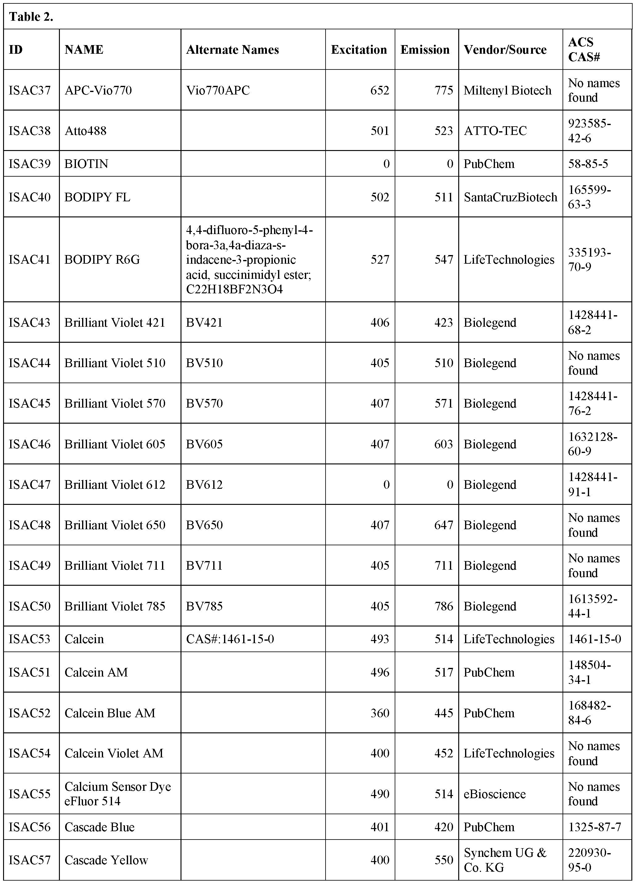


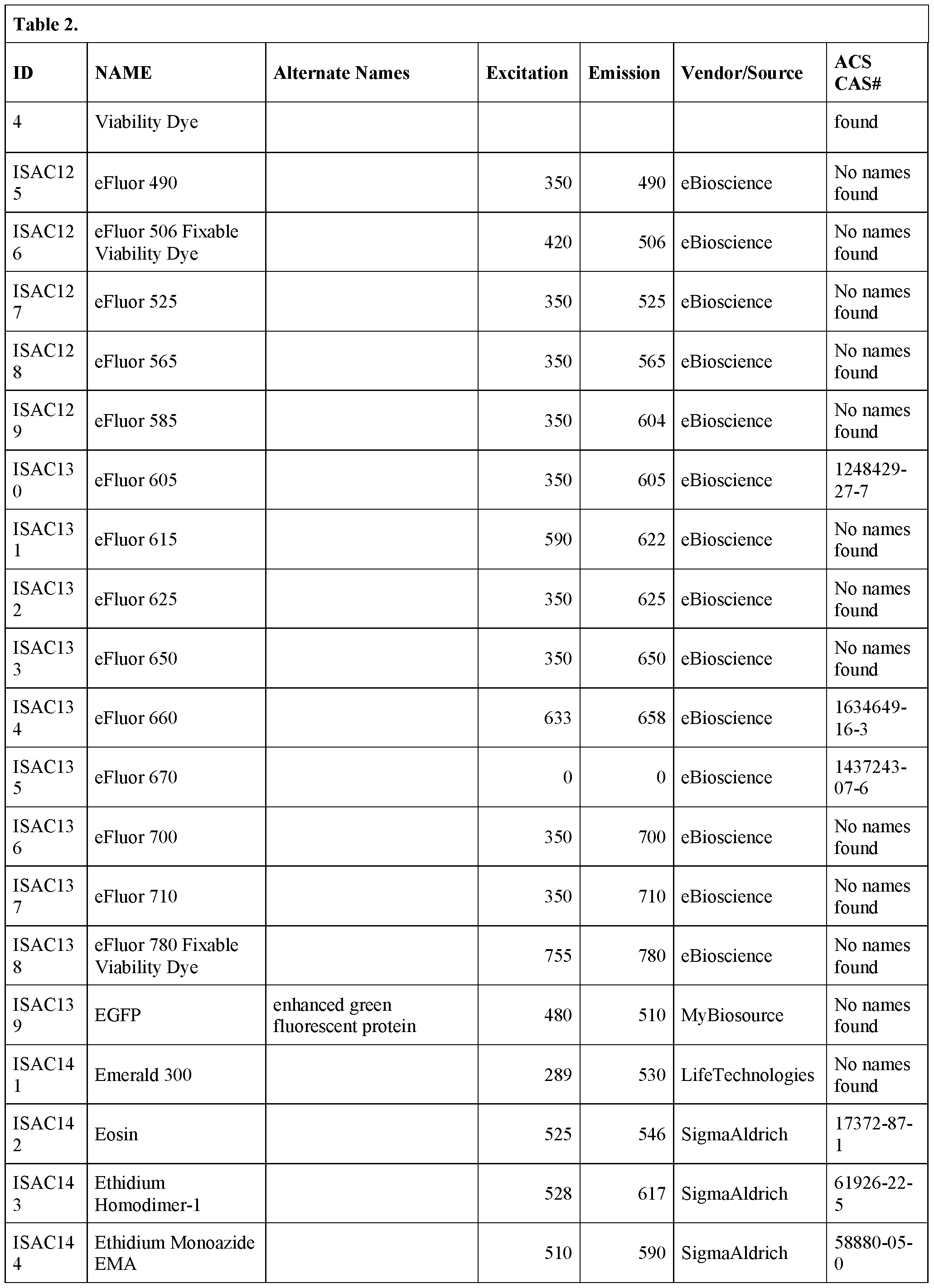


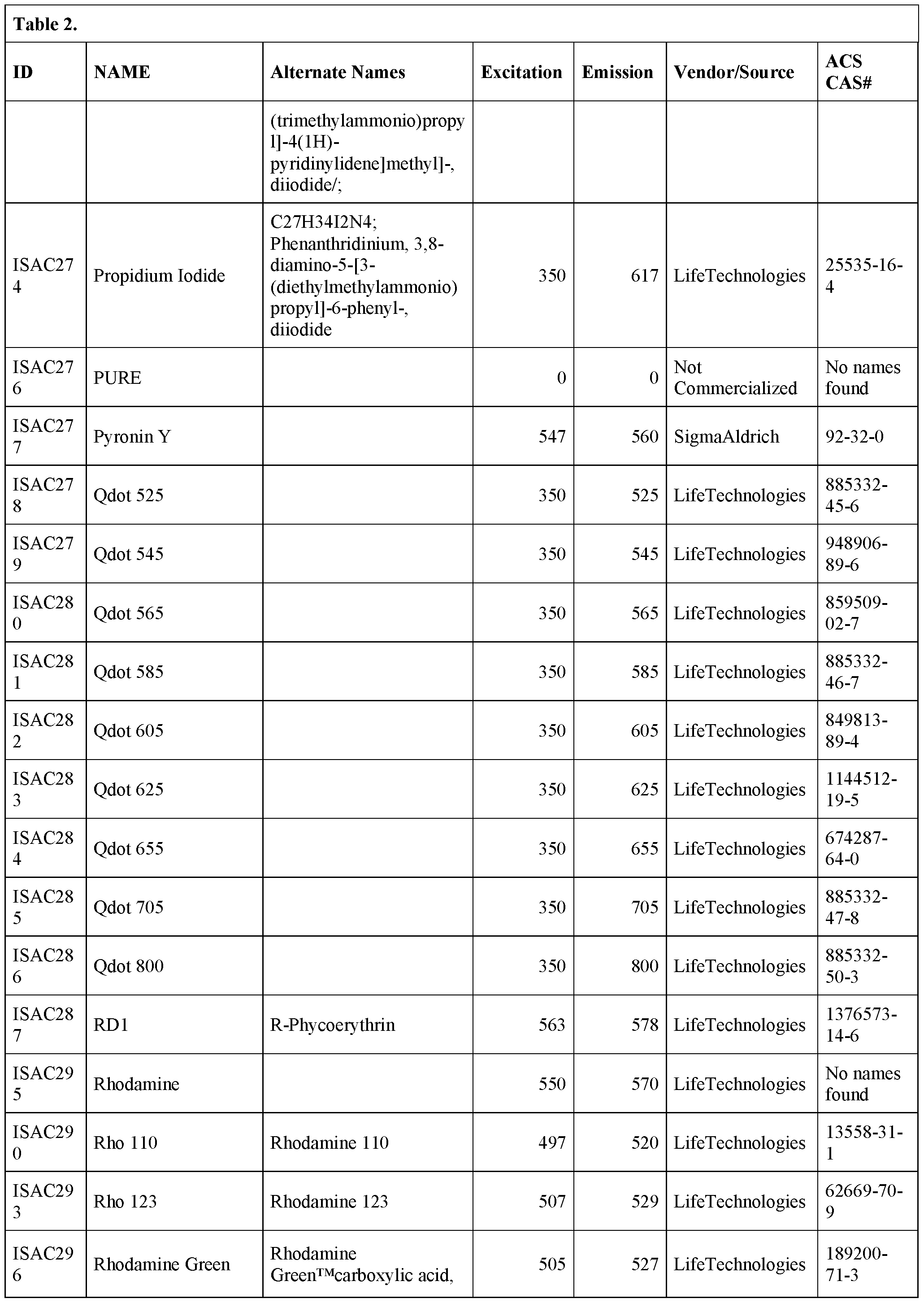


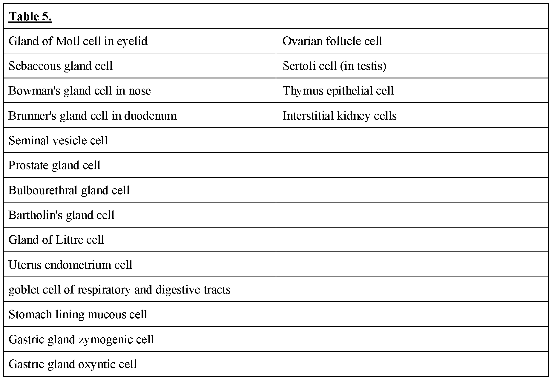


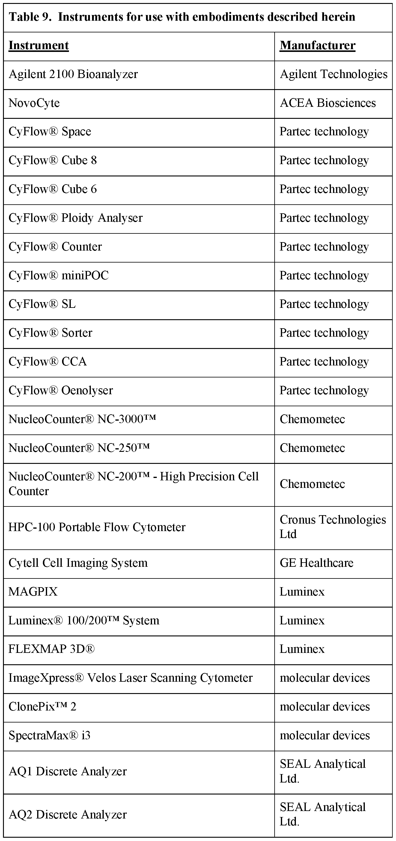


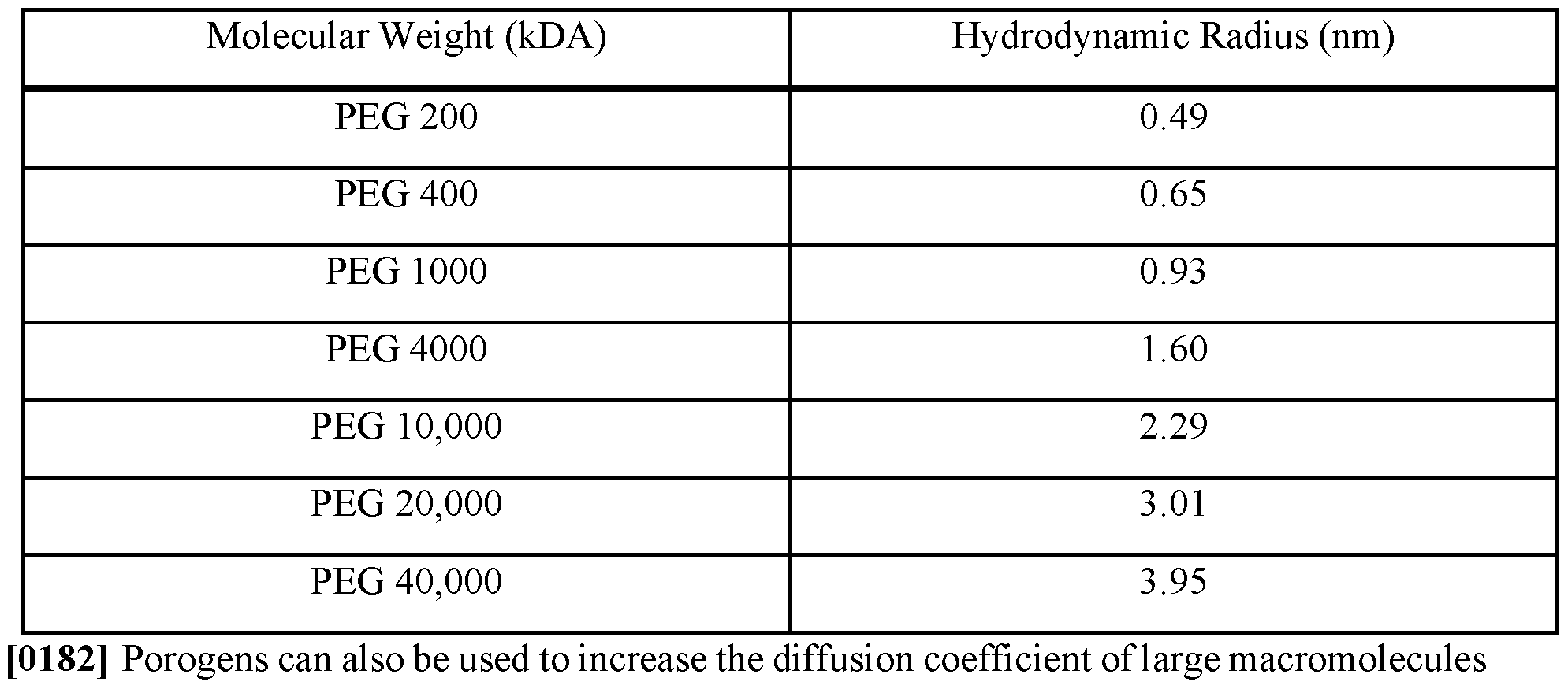

Claims
Priority Applications (4)
| Application Number | Priority Date | Filing Date | Title |
|---|---|---|---|
| CN202380074286.5A CN120712108A (en) | 2022-10-26 | 2023-10-26 | Size-tunable synthetic particles with tunable optical properties and methods for using the same for immune cell activation |
| EP23883775.1A EP4608872A2 (en) | 2022-10-26 | 2023-10-26 | Size-tunable synthetic particles with tunable optical properties and methods for using the same for immune cell activation |
| US18/735,500 US12196661B2 (en) | 2022-10-26 | 2024-06-06 | Size-tunable synthetic particles with tunable optical properties and methods for using the same for immune cell activation |
| US18/966,771 US20250093251A1 (en) | 2022-10-26 | 2024-12-03 | Size-tunable synthetic particles with tunable optical properties and methods for using the same immune cell activation |
Applications Claiming Priority (4)
| Application Number | Priority Date | Filing Date | Title |
|---|---|---|---|
| US202263419580P | 2022-10-26 | 2022-10-26 | |
| US63/419,580 | 2022-10-26 | ||
| US202363488949P | 2023-03-07 | 2023-03-07 | |
| US63/488,949 | 2023-03-07 |
Related Child Applications (1)
| Application Number | Title | Priority Date | Filing Date |
|---|---|---|---|
| US18/735,500 Continuation US12196661B2 (en) | 2022-10-26 | 2024-06-06 | Size-tunable synthetic particles with tunable optical properties and methods for using the same for immune cell activation |
Publications (2)
| Publication Number | Publication Date |
|---|---|
| WO2024092161A2 true WO2024092161A2 (en) | 2024-05-02 |
| WO2024092161A3 WO2024092161A3 (en) | 2024-07-04 |
Family
ID=90832105
Family Applications (1)
| Application Number | Title | Priority Date | Filing Date |
|---|---|---|---|
| PCT/US2023/077961 Ceased WO2024092161A2 (en) | 2022-10-26 | 2023-10-26 | Size-tunable synthetic particles with tunable optical properties and methods for using the same for immune cell activation |
Country Status (4)
| Country | Link |
|---|---|
| US (2) | US12196661B2 (en) |
| EP (1) | EP4608872A2 (en) |
| CN (1) | CN120712108A (en) |
| WO (1) | WO2024092161A2 (en) |
Cited By (3)
| Publication number | Priority date | Publication date | Assignee | Title |
|---|---|---|---|---|
| US12134779B2 (en) | 2021-10-29 | 2024-11-05 | Slingshot Biosciences, Inc. | Hydrogel particles as feeder cells and as synthetic antigen presenting cells |
| US12196661B2 (en) | 2022-10-26 | 2025-01-14 | Slingshot Biosciences, Inc. | Size-tunable synthetic particles with tunable optical properties and methods for using the same for immune cell activation |
| US12352679B2 (en) | 2015-02-09 | 2025-07-08 | Slingshot Biosciences, Inc. | Synthetic human cell mimic particle for cytometric or coulter device |
Families Citing this family (1)
| Publication number | Priority date | Publication date | Assignee | Title |
|---|---|---|---|---|
| JP2025516304A (en) | 2022-05-05 | 2025-05-27 | スリングショット バイオサイエンシーズ, インコーポレイテッド | Engineered particles as red blood cell mimics for hematology and compositions containing them - Patents.com |
Family Cites Families (187)
| Publication number | Priority date | Publication date | Assignee | Title |
|---|---|---|---|---|
| US3822095A (en) | 1972-08-14 | 1974-07-02 | Block Engineering | System for differentiating particles |
| GB1412274A (en) | 1973-05-29 | 1975-11-05 | Bio Rad Laboratories | Radioactive determination of serum thyroxine |
| US3916205A (en) | 1973-05-31 | 1975-10-28 | Block Engineering | Differential counting of leukocytes and other cells |
| US3872312A (en) | 1973-07-02 | 1975-03-18 | Block Engineering | Method and apparatus for detecting and classifying nucleic acid particles |
| US3975084A (en) | 1973-09-27 | 1976-08-17 | Block Engineering, Inc. | Particle detecting system |
| US3937799A (en) | 1974-08-26 | 1976-02-10 | Bio-Rad Laboratories | Radioassay of vitamin b-12 employing bentonite |
| US4271123A (en) | 1979-10-22 | 1981-06-02 | Bio-Rad Laboratories, Inc. | Automated system for performing fluorescent immunoassays |
| US4295199A (en) | 1979-10-22 | 1981-10-13 | Bio-Rad Laboratories, Inc. | Automatic fluorometer and data processor for performing fluorescent immunoassays |
| US4389491A (en) | 1982-04-26 | 1983-06-21 | Bio-Rad Laboratories, Inc. | Method and kit for chromatographic separation of hemoglobin A1c |
| US4409335A (en) | 1982-05-28 | 1983-10-11 | Bio-Rad Laboratories, Inc. | Method for eliminating glucose dependent Schiff base effect from hemoglobin A1 assay |
| US4511662A (en) | 1982-06-18 | 1985-04-16 | Bio-Rad Laboratories, Inc. | Simultaneous assay for T and B lymphocyte populations and subpopulations |
| US4448888A (en) | 1982-08-24 | 1984-05-15 | Bio-Rad Laboratories, Inc. | Compositions and method for correcting temperature dependence of hemoglobin assays |
| US4774189A (en) | 1984-12-24 | 1988-09-27 | Flow Cytometry Standards Corp. | Fluorescent calibration microbeads simulating stained cells |
| US4857451A (en) | 1984-12-24 | 1989-08-15 | Flow Cytometry Standards Corporation | Method of compensating and calibrating a flow cytometer, and microbead standards kit therefor |
| US5093234A (en) | 1984-12-24 | 1992-03-03 | Caribbean Microparticles Corporation | Method of aligning, compensating, and calibrating a flow cytometer for analysis of samples, and microbead standards kit therefor |
| DE3650175T3 (en) | 1985-04-29 | 2007-09-06 | Bio-Rad Laboratories, Inc., Hercules | SYNTHETIC ANTIGENIC TO IDENTIFY AIDS. |
| US4704891A (en) | 1986-08-29 | 1987-11-10 | Becton, Dickinson And Company | Method and materials for calibrating flow cytometers and other analysis instruments |
| US5244799A (en) * | 1987-05-20 | 1993-09-14 | Anderson David M | Preparation of a polymeric hydrogel containing micropores and macropores for use as a cell culture substrate |
| US5091206A (en) | 1987-10-26 | 1992-02-25 | Baxter Diagnostics Inc. | Process for producing magnetically responsive polymer particles and application thereof |
| US5395688A (en) | 1987-10-26 | 1995-03-07 | Baxter Diagnostics Inc. | Magnetically responsive fluorescent polymer particles |
| JPH03503845A (en) | 1988-04-22 | 1991-08-29 | マサチユセツツ・インスチチユート・オブ・テクノロジー | How to form and use microdroplets |
| US5840293A (en) | 1988-11-16 | 1998-11-24 | Advanced Polymer Systems, Inc. | Ionic beads for controlled release and adsorption |
| US5820879A (en) | 1993-02-12 | 1998-10-13 | Access Pharmaceuticals, Inc. | Method of delivering a lipid-coated condensed-phase microparticle composition |
| WO1995003408A1 (en) | 1993-07-26 | 1995-02-02 | Dana-Farber Cancer Institute | B7-2: ctl a4/cd 28 counter receptor |
| DE4339509A1 (en) | 1993-11-22 | 1995-05-24 | Basf Ag | PPE / PA moldings for the production of moldings by means of blow molding, profile extrusion and pipe extrusion |
| USRE39542E1 (en) | 1994-01-13 | 2007-04-03 | The Rogosin Institute | Preparation of agarose coated, solid agarose-collagen beads containing secretory cells |
| JP3875754B2 (en) | 1995-11-17 | 2007-01-31 | シスメックス株式会社 | Standard solution for flow cytometer |
| US5841139A (en) | 1997-02-28 | 1998-11-24 | Bio-Rad Laboratories, Inc. | Optical instrument providing combined infrared and Ramen analysis of samples |
| US6043506A (en) | 1997-08-13 | 2000-03-28 | Bio-Rad Laboratories, Inc. | Multi parameter scanner |
| WO1999011692A1 (en) | 1997-09-03 | 1999-03-11 | The Regents Of The University Of California | Novel biomimetic hydrogel materials |
| CA2260991C (en) | 1997-11-18 | 2009-03-24 | Bio-Rad Laboratories, Inc. | Multiplex flow immunoassays with magnetic particles as solid phase |
| US6051835A (en) | 1998-01-07 | 2000-04-18 | Bio-Rad Laboratories, Inc. | Spectral imaging apparatus and methodology |
| DE19810565A1 (en) * | 1998-03-11 | 1999-09-16 | Basf Ag | Economical drying of microporous particles containing fluid e.g. inorganic, organic or polymer gel |
| US6613234B2 (en) | 1998-04-06 | 2003-09-02 | Ciphergen Biosystems, Inc. | Large pore volume composite mineral oxide beads, their preparation and their applications for adsorption and chromatography |
| WO2000008212A1 (en) | 1998-08-07 | 2000-02-17 | Cellay, Llc | Gel microdrops in genetic analysis |
| US6329485B1 (en) | 1998-12-11 | 2001-12-11 | Bausch & Lomb Incorporated | High refractive index hydrogel compositions for ophthalmic implants |
| CA2368247A1 (en) | 1999-04-27 | 2000-11-09 | Ciphergen Biosystems, Inc. | Probes for a gas phase ion spectrometer |
| US6372813B1 (en) | 1999-06-25 | 2002-04-16 | Motorola | Methods and compositions for attachment of biomolecules to solid supports, hydrogels, and hydrogel arrays |
| DE60027576T2 (en) | 1999-08-17 | 2007-05-03 | Luminex Corp., Austin | SECULATION OF FLUORESCENT PARTICLES |
| AU7646000A (en) | 1999-11-05 | 2001-05-14 | Novozymes A/S | Methods to screen microorganisms or gene libraries for products secreted from a cell |
| US6514763B2 (en) | 2000-04-28 | 2003-02-04 | Hematronix, Inc. | Hematology blood control and method for preparation of same |
| US6635750B1 (en) | 2000-07-20 | 2003-10-21 | Millennium Pharmaceuticals, Inc. | B7-H2 nucleic acids, members of the B7 family |
| AU2001290879A1 (en) | 2000-09-15 | 2002-03-26 | California Institute Of Technology | Microfabricated crossflow devices and methods |
| US6905881B2 (en) | 2000-11-30 | 2005-06-14 | Paul Sammak | Microbead-based test plates and test methods for fluorescence imaging systems |
| US20020115116A1 (en) | 2001-02-22 | 2002-08-22 | Yong Song | Multiplex protein interaction determinations using glutathione-GST binding |
| US6806058B2 (en) | 2001-05-26 | 2004-10-19 | One Cell Systems, Inc. | Secretions of proteins by encapsulated cells |
| IL159475A0 (en) | 2001-06-21 | 2004-06-01 | Altus Biologics Inc | Spherical protein particles, compositions containing the same and methods for the production thereof |
| US7842498B2 (en) | 2001-11-08 | 2010-11-30 | Bio-Rad Laboratories, Inc. | Hydrophobic surface chip |
| EP1456652A4 (en) | 2001-11-13 | 2005-11-02 | Dana Farber Cancer Inst Inc | AGENTS MODULATING IMMUNE CELL ACTIVITY AND METHODS OF USE THEREOF |
| US6838289B2 (en) | 2001-11-14 | 2005-01-04 | Beckman Coulter, Inc. | Analyte detection system |
| US8088388B2 (en) | 2002-02-14 | 2012-01-03 | United Biomedical, Inc. | Stabilized synthetic immunogen delivery system |
| US20030218130A1 (en) | 2002-05-02 | 2003-11-27 | Ciphergen Biosystems, Inc. | Biochips with surfaces coated with polysaccharide-based hydrogels |
| JP2006507921A (en) | 2002-06-28 | 2006-03-09 | プレジデント・アンド・フェロウズ・オブ・ハーバード・カレッジ | Method and apparatus for fluid dispersion |
| US7045366B2 (en) | 2003-09-12 | 2006-05-16 | Ciphergen Biosystems, Inc. | Photocrosslinked hydrogel blend surface coatings |
| WO2004076511A2 (en) | 2003-02-21 | 2004-09-10 | Ciphergen Biosystems, Inc. | Photocrosslinked hydrogel surface coatings |
| JP4239637B2 (en) | 2003-03-24 | 2009-03-18 | 富士ゼロックス株式会社 | Polymer gel composition and optical element using the same |
| EP3511693B1 (en) | 2003-03-28 | 2022-08-24 | Inguran, LLC | Apparatus for detecting the breakoff point of a droplet generation system |
| CA2521862C (en) | 2003-04-10 | 2012-10-16 | President And Fellows Of Harvard College | Formation and control of fluidic species |
| EP1641424A4 (en) | 2003-07-09 | 2007-05-02 | Vaxdesign Corp | Programmed immune responses using a vaccination node |
| EP2662136A3 (en) | 2003-08-27 | 2013-12-25 | President and Fellows of Harvard College | Method for handling and mixing droplets |
| US20050112650A1 (en) | 2003-10-20 | 2005-05-26 | Ciphergen Biosystems, Inc. | Reactive polyurethane-based polymers |
| WO2005042048A2 (en) | 2003-10-22 | 2005-05-12 | Encelle, Inc. | Bioactive hydrogel compositions for regenerating connective tissue |
| US20050089987A1 (en) | 2003-10-27 | 2005-04-28 | Lonza Ltd. | Polyacryamide beads containing encapsulated cells |
| EP1682261B1 (en) | 2003-11-12 | 2012-08-15 | BIO-RAD Haifa Ltd. | Method for carrying out multiple binding reactions in an array format |
| US20080044472A1 (en) | 2004-01-23 | 2008-02-21 | Garcia Antonio A | Photoresponsive Hydrogels |
| EP1580559B1 (en) | 2004-03-23 | 2013-12-25 | Bio-Rad Laboratories, Inc. | Methods for reducing the variance between analyte concentrations taken from complex sample mixtures |
| WO2005092286A2 (en) | 2004-03-29 | 2005-10-06 | The University Of Houston System | Metallic nano-particles and discrete polymer-coated nano-particles |
| JP4261403B2 (en) * | 2004-03-29 | 2009-04-30 | 積水化成品工業株式会社 | Method for producing porous resin particles |
| GB0414825D0 (en) | 2004-07-02 | 2004-08-04 | Biostatus Ltd | Gel formulations and uses thereof |
| US20080213593A1 (en) | 2005-01-21 | 2008-09-04 | President And Fellows Of Harvard College | Systems And Methods For Forming Fluidic Droplets Encapsulated In Particles Such As Colloidal Particles |
| US20060240560A1 (en) | 2005-01-31 | 2006-10-26 | Eric Bakker | Microsphere optical ion sensors based on doped silica gel templates |
| WO2006096571A2 (en) | 2005-03-04 | 2006-09-14 | President And Fellows Of Harvard College | Method and apparatus for forming multiple emulsions |
| US20070054119A1 (en) | 2005-03-04 | 2007-03-08 | Piotr Garstecki | Systems and methods of forming particles |
| US7531357B2 (en) | 2005-04-04 | 2009-05-12 | Bio-Rad Laboratories, Inc. | Preparation of platelet analogs |
| JP2006349060A (en) | 2005-06-16 | 2006-12-28 | Ntn Corp | Ball screw |
| EP1744145B1 (en) | 2005-07-12 | 2015-09-09 | Sysmex Corporation | Standard material for particle analyzer |
| JP4744264B2 (en) | 2005-10-19 | 2011-08-10 | シスメックス株式会社 | Reference material for urine sediment analyzer. |
| US20080023630A1 (en) | 2005-11-22 | 2008-01-31 | Ciphergen Biosystems Inc. | Polymer probe doped with conductive material for mass spectrometry |
| JP2009518501A (en) * | 2005-12-07 | 2009-05-07 | エムアイピー テクノロジーズ エービー | Production method of polymer beads |
| US7560283B2 (en) | 2006-01-27 | 2009-07-14 | Bio-Rad Laboratories, Inc. | Storage-stable cellular whole blood composition containing elevated amounts of D-dimer |
| KR20080109775A (en) | 2006-02-23 | 2008-12-17 | 노보셀, 인크 | Compositions and methods useful for culturing differentiable cells |
| US20070254378A1 (en) | 2006-03-06 | 2007-11-01 | Bio-Rad Laboratories, Inc. | Chelating monomers and polymers |
| EP2021113A2 (en) | 2006-05-11 | 2009-02-11 | Raindance Technologies, Inc. | Microfluidic devices |
| US20080019921A1 (en) | 2006-06-30 | 2008-01-24 | Invitrogen Corporation | Uniform fluorescent microsphere with hydrophobic surfaces |
| US7482161B2 (en) | 2006-07-17 | 2009-01-27 | Bio-Rad Laboratories, Inc. | Preparation of a red blood cell component for a hematology control |
| US7588942B2 (en) | 2006-08-04 | 2009-09-15 | Bio-Rad Laboratories, Inc. | Standard/reference/control for blood coagulation testing |
| US7935518B2 (en) | 2006-09-27 | 2011-05-03 | Alessandra Luchini | Smart hydrogel particles for biomarker harvesting |
| US7776927B2 (en) | 2007-03-28 | 2010-08-17 | President And Fellows Of Harvard College | Emulsions and techniques for formation |
| FR2917174B1 (en) | 2007-06-08 | 2021-02-12 | Bio Rad Pasteur | MULTIPLE ANALYSIS OF BLOOD SAMPLES |
| CN101214217A (en) | 2007-12-26 | 2008-07-09 | 仲恺农业技术学院 | Hydrogel-hydrogel composite material and its preparation method and application of drug slow-release matrix |
| US8030095B2 (en) | 2008-03-04 | 2011-10-04 | Crystal Bioscience Inc. | Gel microdrop composition and method of using the same |
| CN101245368A (en) | 2008-03-20 | 2008-08-20 | 重庆师范大学 | Classification of myxospores using scattered light and autofluorescence parameters in flow cytometry |
| JP2011516895A (en) | 2008-04-17 | 2011-05-26 | キアゲン レイク コンスタンツ ゲゼルシャフト ミット ベシュレンクテル ハフツング | Fluorescence standards and their use |
| KR102340754B1 (en) | 2008-04-21 | 2021-12-16 | 오토노미, 인코포레이티드 | Auris formulations for treating otic diseases and conditions |
| US9931386B2 (en) | 2008-06-16 | 2018-04-03 | Atsuo Ochi | Recombinant multiple domain fusion protein mitogens and use thereof for inducing enhancement or repression of antigen-specific immunity |
| JP5896119B2 (en) | 2008-08-26 | 2016-03-30 | リオッタ,ランス,エー. | Immunoassay based on hydrogel nanoparticles |
| WO2010025988A1 (en) | 2008-09-04 | 2010-03-11 | Unilever Plc | Method and apparatus for producing microparticles |
| WO2010033200A2 (en) | 2008-09-19 | 2010-03-25 | President And Fellows Of Harvard College | Creation of libraries of droplets and related species |
| US9417190B2 (en) | 2008-09-23 | 2016-08-16 | Bio-Rad Laboratories, Inc. | Calibrations and controls for droplet-based assays |
| CN102143996A (en) * | 2008-10-30 | 2011-08-03 | 大卫·刘 | Micro-spherical porous biocompatible scaffolds and methods and apparatus for fabricating same |
| US8415161B2 (en) | 2008-11-13 | 2013-04-09 | Becton, Dickinson And Company | Instrument setup system for a fluorescence analyzer |
| US8114580B2 (en) | 2009-01-13 | 2012-02-14 | Bio-Rad Laboratories, Inc. | Simulation of normal fresh blood platelets for reference control |
| US20100187441A1 (en) | 2009-01-28 | 2010-07-29 | Bio-Rad Laboratories, Inc. | Transilluminator adaptor for conversion of ultraviolet radiation to visible light |
| WO2010105070A1 (en) | 2009-03-12 | 2010-09-16 | Conocophillips Company | Crosslinked swellable polymer |
| US8187885B2 (en) | 2009-05-07 | 2012-05-29 | Nodality, Inc. | Microbead kit and method for quantitative calibration and performance monitoring of a fluorescence instrument |
| US11090363B2 (en) * | 2009-07-10 | 2021-08-17 | University of Pittsburgh—of the Commonwealth System of Higher Education | Vasoactive intestinal peptide release from microparticles |
| US8451450B2 (en) | 2009-09-14 | 2013-05-28 | Bio-Rad Laboratories, Inc. | Near real time optical phase conjugation |
| US8603828B2 (en) | 2009-11-18 | 2013-12-10 | Bio-Rad Laboratories, Inc. | Multiplex immunoassays for hemoglobin, hemoglobin variants, and glycated forms |
| US8790592B2 (en) | 2010-02-04 | 2014-07-29 | Bio-Rad Laboratories, Inc. | Measuring multi-analyte samples using an in-line flow cell |
| WO2011098407A1 (en) | 2010-02-12 | 2011-08-18 | Cemm Forschungszentrum Für Molekulare Medizin Gmbh | Microspheres |
| JP5934657B2 (en) | 2010-02-12 | 2016-06-15 | レインダンス テクノロジーズ, インコーポレイテッド | Digital specimen analysis |
| KR101825607B1 (en) | 2010-02-18 | 2018-02-05 | 다우 코닝 코포레이션 | Surface-modified hydrogels and hydrogel microparticles |
| FR2956868B1 (en) | 2010-03-01 | 2014-01-10 | Bio Rad Pasteur | RAPID METHOD FOR DETECTION OF ENZYMES AND MICROORGANISMS |
| WO2011156432A2 (en) | 2010-06-07 | 2011-12-15 | Firefly Bioworks, Inc. | Scanning multifunctional particles |
| US20110318820A1 (en) | 2010-06-29 | 2011-12-29 | Life Technologies Corporation | Immobilized Buffer Particles and Uses Thereof |
| JP5858451B2 (en) | 2010-06-29 | 2016-02-10 | 国立大学法人 岡山大学 | Method for producing hydrogel capsules |
| US8530449B2 (en) | 2010-09-09 | 2013-09-10 | Assad S. Sawaya | Composition for a topical ophthalmic clear colloidal liquid which undergoes a liquid-gel phase transition in the eye |
| US8609363B2 (en) | 2010-11-18 | 2013-12-17 | Bio-Rad Laboratories, Inc. | Viability cell counting by differential light absorption |
| WO2012103516A1 (en) | 2011-01-28 | 2012-08-02 | Amyris, Inc. | Gel-encapsulated microcolony screening |
| EP2675913B1 (en) | 2011-02-15 | 2016-12-14 | Bio-Rad Laboratories, Inc. | Detecting methylation in a subpopulation of genomic dna |
| US9816931B2 (en) | 2011-03-31 | 2017-11-14 | Bio-Rad Laboratories, Inc. | Managing variation in spectroscopic intensity measurements through the use of a reference component |
| US9228898B2 (en) | 2011-03-31 | 2016-01-05 | Gnubio, Inc. | Scalable spectroscopic detection and measurement |
| JP2013015357A (en) | 2011-07-01 | 2013-01-24 | Shimadzu Corp | Flow cytometer |
| EP2739587B1 (en) | 2011-08-01 | 2020-05-27 | Denovo Sciences | Cell capture system |
| JP2013155358A (en) | 2012-01-31 | 2013-08-15 | Kanazawa Univ | Emulsion polymerization method, and method for synthesizing hydrogel |
| FR2986617B1 (en) | 2012-02-02 | 2015-03-27 | Horiba Abx Sas | DEVICE AND METHOD FOR PERFORMING HEMATOLOGICAL AND BIOCHEMICAL MEASUREMENTS FROM A BIOLOGICAL SAMPLE |
| US20150177115A1 (en) | 2012-04-06 | 2015-06-25 | Slingshot Biosciences | Hydrogel particles with tunable optical properties |
| US20130274125A1 (en) | 2012-04-16 | 2013-10-17 | Bio-Rad Laboratories Inc. | Multiplex immunoassay for rheumatoid arthritis and other autoimmune diseases |
| JP2015525343A (en) | 2012-05-30 | 2015-09-03 | アイリス インターナショナル インコーポレイテッド | Flow cytometer |
| EP2859324B1 (en) | 2012-06-07 | 2019-08-07 | Bio-rad Laboratories, Inc. | Automated and accurate drop delay for flow cytometry |
| EP2864787B1 (en) | 2012-06-22 | 2017-11-01 | Bio-Rad Laboratories, Inc. | Human factor xiii as a normalization control for immunoassays |
| JP2015530361A (en) | 2012-07-13 | 2015-10-15 | タフツ・ユニバーシティ | Encapsulation of immiscible phases in silk fibroin biomaterial |
| WO2014043388A1 (en) | 2012-09-12 | 2014-03-20 | Gnubio, Inc. | Integrated microfluidic system, method and kit for performing assays |
| US9804149B2 (en) | 2012-10-10 | 2017-10-31 | Bio-Rad Laboratories, Inc. | Patient-based results display |
| US20150267196A1 (en) | 2012-10-12 | 2015-09-24 | Case Western Reserve University | Biodegradable hydrogel for polynucleotide delivery |
| US20140179808A1 (en) | 2012-10-23 | 2014-06-26 | Bio-Rad Laboratories, Inc. | Detection of Ovarian Carcinoma by Assay for Autoantibodies to Multiple Antigens |
| RU2631002C2 (en) | 2012-12-05 | 2017-09-15 | Тевакс Дженетикс Вэксин Ко., Лтд. | Fusion proteins for application as immunogenic amplifying agents to induce antigen-specific t-cell response |
| US9176154B2 (en) | 2012-12-12 | 2015-11-03 | Bio-Rad Laboratories, Inc. | Calibration process and system |
| US9217175B2 (en) | 2013-02-01 | 2015-12-22 | Bio-Rad Laboratories, Inc. | Multiplexed digital assay with specific and generic reporters |
| CA2905272A1 (en) | 2013-03-14 | 2014-10-02 | Icahn School Of Medicine At Mount Sinai | Newcastle disease viruses and uses thereof |
| CN109828110A (en) | 2013-04-26 | 2019-05-31 | 生物辐射实验室股份有限公司 | Multiple hepatitis B test |
| US9856535B2 (en) | 2013-05-31 | 2018-01-02 | Denovo Sciences, Inc. | System for isolating cells |
| CA2953287A1 (en) | 2013-06-19 | 2014-12-24 | Massachusetts Institute Of Technology | In vivo targeting of cells with ligand-conjugated particles |
| CN103744185B (en) | 2013-12-30 | 2016-03-30 | 维林光电(苏州)有限公司 | A kind of calibrating method of the scaling system based on flow cytometer polychromatic source waist position |
| US10392557B2 (en) | 2014-04-08 | 2019-08-27 | Dna Medicine Institute, Inc. | Multi-dye microparticles |
| EP3161157B1 (en) | 2014-06-24 | 2024-03-27 | Bio-Rad Laboratories, Inc. | Digital pcr barcoding |
| US10583091B2 (en) | 2014-10-23 | 2020-03-10 | The Brigham And Women's Hospital, Inc. | Amphiphile-polymer particles |
| CA2969038C (en) | 2014-12-03 | 2023-07-04 | Ventana Medical Systems, Inc. | Methods, systems, and apparatuses for quantitative analysis of heterogeneous biomarker distribution |
| WO2016113691A1 (en) | 2015-01-14 | 2016-07-21 | Bio-Rad Laboratories, Inc. | Blood analysis systems and methods |
| US20230067460A1 (en) | 2015-02-09 | 2023-03-02 | Slingshot Biosciences, Inc. | Hydrogel particles as feeder cells and as synthetic antigen presenting cells |
| EP3256850B1 (en) | 2015-02-09 | 2025-07-09 | Slingshot Biosciences, Inc. | Hydrogel particles with tunable optical properties and methods for using the same |
| AU2016291817A1 (en) | 2015-07-16 | 2018-02-22 | Biolinerx Ltd. | Compositions and methods for treating cancer |
| US10180385B2 (en) | 2015-08-12 | 2019-01-15 | Bio-Rad Laboratories, Inc. | Multi-spectral filter profiling and quality control for flow cytometry |
| CN107923070A (en) | 2015-08-25 | 2018-04-17 | 生物辐射实验室股份有限公司 | Digital immunoassays |
| US11213490B2 (en) | 2016-05-19 | 2022-01-04 | Molly Shoichet | Encapsulation-free controlled protein release system |
| WO2018022991A1 (en) | 2016-07-29 | 2018-02-01 | Streck, Inc. | Suspension composition for hematology analysis control |
| EP3335695B1 (en) | 2016-12-15 | 2020-02-05 | UPM-Kymmene Corporation | A method for freeze-drying hydrogel comprising nanofibrillar cellulose, a freeze-dried medical hydrogel comprising nanofibrillar cellulose, and a hydrogel comprising nanofibrillar cellulose |
| EP3574047B1 (en) | 2017-01-30 | 2024-03-06 | Bio-Rad Laboratories, Inc. | Emulsion compositions and methods of their use |
| EP4455301A3 (en) | 2017-02-24 | 2024-11-13 | The Regents of The University of California | Particle-drop structures and methods for making and using the same |
| US20200085971A1 (en) * | 2017-03-20 | 2020-03-19 | Qt Holdings Corp | Methods and compositions for modulation of immune cells |
| US11884738B2 (en) * | 2017-03-31 | 2024-01-30 | The University Of North Carolina At Chapel Hill | Methods and compositions for activation of T cells using nanoparticles conjugated with multiple ligands for binding receptors on T cells |
| JP7339160B2 (en) | 2017-04-27 | 2023-09-05 | ジュノ セラピューティクス ゲーエムベーハー | Oligomeric particle reagents and methods of use thereof |
| US11186862B2 (en) | 2017-06-20 | 2021-11-30 | Bio-Rad Laboratories, Inc. | MDA using bead oligonucleotide |
| US11808772B2 (en) | 2017-07-19 | 2023-11-07 | Bio-Rad Europe Gmbh | Biomarker combinations to simultaneously evaluate non-alcoholic steatohepatitis and hepatic fibrosis status |
| CA3075355A1 (en) | 2017-09-11 | 2019-03-14 | Evorion Biotechnologies Gmbh | Systems, methods and hydrogels for cell culture and analysis |
| US11047845B1 (en) | 2017-11-15 | 2021-06-29 | Medica Corporation | Control material and methods for cell analyzers |
| WO2019099948A1 (en) | 2017-11-17 | 2019-05-23 | Bio-Rad Laboratories, Inc. | Smart advisor for blood test evaluation |
| JP7027459B2 (en) | 2018-02-13 | 2022-03-01 | イルミナ インコーポレイテッド | DNA sequencing using hydrogel beads |
| US10344100B1 (en) | 2018-02-28 | 2019-07-09 | The Florida International University Board Of Trustees | Micro/nano magnetic hydrogels with autofluorescence for therapeutic and diagnostic applications |
| EP4458351A3 (en) | 2018-08-17 | 2025-05-21 | The Regents of the University of California | Particle-containing droplet systems with monodisperse fluid volumes |
| EP4249651B1 (en) | 2018-08-20 | 2025-01-29 | Bio-Rad Laboratories, Inc. | Nucleotide sequence generation by barcode bead-colocalization in partitions |
| US11697793B2 (en) | 2018-10-10 | 2023-07-11 | Washington University | Compositions and methods of making and using protein-functionalized hydrogels |
| AU2019364545B2 (en) | 2018-10-26 | 2025-09-04 | Illumina, Inc. | Modulating polymer beads for DNA processing |
| EP3903095A4 (en) | 2018-12-28 | 2023-02-08 | Becton, Dickinson and Company | METHODS FOR SPECTRAL RESOLUTION OF FLUOROPHORES OF A SAMPLE AND SYSTEMS THEREOF |
| US20200232979A1 (en) | 2019-01-22 | 2020-07-23 | Mayo Foundation For Medical Education And Research | Microcapsules and Methods for Analyte Detection |
| CA3126087A1 (en) * | 2019-02-25 | 2020-09-03 | Novartis Ag | Mesoporous silica particles compositions for viral delivery |
| WO2021091859A1 (en) | 2019-11-04 | 2021-05-14 | Bio-Rad Laboratories, Inc. | Cell barcoding for single cell sequencing |
| EP4069867B1 (en) | 2019-12-04 | 2025-08-27 | Becton, Dickinson and Company | Magnetic capture bead mediated molecular barcoding of nucleic acid targets in single particles and compositions for use in the same |
| US20210190740A1 (en) | 2019-12-19 | 2021-06-24 | Bio-Rad Laboratories, Inc. | Automated chromatogram analysis for blood test evaluation |
| US11313782B2 (en) | 2020-01-24 | 2022-04-26 | Slingshot Biosciences, Inc. | Compositions and methods for cell-like calibration particles |
| EP4097599A4 (en) | 2020-01-31 | 2024-02-28 | Bio-Rad Laboratories, Inc. | STABLE REFERENCE MATERIALS FOR AUTOMATED HEMATOLOGY TESTING PLATFORMS |
| US11598768B2 (en) | 2020-05-04 | 2023-03-07 | Slingshot Biosciences, Inc. | Compositions and methods for passive optical barcoding for multiplexed assays |
| US12163968B2 (en) | 2020-09-03 | 2024-12-10 | Bio-Rad Laboratories, Inc. | Preparation of nucleated RBC (NRBC) analogs for use as reference hematology controls in automated hematology analyzers |
| CA3235582A1 (en) | 2021-10-29 | 2023-05-04 | Anh Tuan Nguyen | Hydrogel particles as feeder cells and as synthetic antigen presenting cells |
| JP2025516304A (en) | 2022-05-05 | 2025-05-27 | スリングショット バイオサイエンシーズ, インコーポレイテッド | Engineered particles as red blood cell mimics for hematology and compositions containing them - Patents.com |
| AU2023280490A1 (en) | 2022-06-02 | 2024-12-05 | Slingshot Biosciences, Inc. | Apoptotic cell mimic |
| WO2024092161A2 (en) * | 2022-10-26 | 2024-05-02 | Slingshot Biosciences, Inc. | Size-tunable synthetic particles with tunable optical properties and methods for using the same for immune cell activation |
-
2023
- 2023-10-26 WO PCT/US2023/077961 patent/WO2024092161A2/en not_active Ceased
- 2023-10-26 CN CN202380074286.5A patent/CN120712108A/en active Pending
- 2023-10-26 EP EP23883775.1A patent/EP4608872A2/en active Pending
-
2024
- 2024-06-06 US US18/735,500 patent/US12196661B2/en active Active
- 2024-12-03 US US18/966,771 patent/US20250093251A1/en active Pending
Cited By (4)
| Publication number | Priority date | Publication date | Assignee | Title |
|---|---|---|---|---|
| US12352679B2 (en) | 2015-02-09 | 2025-07-08 | Slingshot Biosciences, Inc. | Synthetic human cell mimic particle for cytometric or coulter device |
| US12134779B2 (en) | 2021-10-29 | 2024-11-05 | Slingshot Biosciences, Inc. | Hydrogel particles as feeder cells and as synthetic antigen presenting cells |
| US12312595B2 (en) | 2021-10-29 | 2025-05-27 | Slingshot Biosciences, Inc. | Hydrogel particles as feeder cells and as synthetic antigen presenting cells |
| US12196661B2 (en) | 2022-10-26 | 2025-01-14 | Slingshot Biosciences, Inc. | Size-tunable synthetic particles with tunable optical properties and methods for using the same for immune cell activation |
Also Published As
| Publication number | Publication date |
|---|---|
| WO2024092161A3 (en) | 2024-07-04 |
| US12196661B2 (en) | 2025-01-14 |
| US20240353305A1 (en) | 2024-10-24 |
| US20250093251A1 (en) | 2025-03-20 |
| EP4608872A2 (en) | 2025-09-03 |
| CN120712108A (en) | 2025-09-26 |
Similar Documents
| Publication | Publication Date | Title |
|---|---|---|
| US20230067460A1 (en) | Hydrogel particles as feeder cells and as synthetic antigen presenting cells | |
| US12134779B2 (en) | Hydrogel particles as feeder cells and as synthetic antigen presenting cells | |
| WO2024092161A2 (en) | Size-tunable synthetic particles with tunable optical properties and methods for using the same for immune cell activation | |
| KR102436976B1 (en) | Hydrogel particles with tunable optical properties and methods for using the same | |
| Barnes et al. | Collagen–poly (N-isopropylacrylamide) hydrogels with tunable properties | |
| Liu et al. | Viscoelastic synthetic antigen-presenting cells for augmenting the potency of cancer therapies | |
| Quartey et al. | Matrix‐Bound Hyaluronan Molecular Weight as a Regulator of Dendritic Cell Immune Potency | |
| WO2025049609A1 (en) | Cd34 stem cell mimics | |
| CN118355112A (en) | Hydrogel particles as feeder cells and as synthetic antigen presenting cells | |
| WO2024186668A2 (en) | Size-tunable synthetic particles for immune cell activation | |
| WO2025090968A1 (en) | Hydrogel cell mimics with subcellular features | |
| WO2025166277A1 (en) | Synthetic cell mimics with modified surface charges | |
| Quartey et al. | Tissue-bound hyaluronan molecular weight as a regulator of dendritic cell immune potency | |
| WO2025024017A1 (en) | Synthetic viscoelastic activating cells for t cell engineering | |
| EP4677359A1 (en) | Cell labeling and annotation in micro-organospheres |
Legal Events
| Date | Code | Title | Description |
|---|---|---|---|
| 121 | Ep: the epo has been informed by wipo that ep was designated in this application |
Ref document number: 23883775 Country of ref document: EP Kind code of ref document: A2 |
|
| WWE | Wipo information: entry into national phase |
Ref document number: 2023883775 Country of ref document: EP |
|
| NENP | Non-entry into the national phase |
Ref country code: DE |
|
| ENP | Entry into the national phase |
Ref document number: 2023883775 Country of ref document: EP Effective date: 20250526 |
|
| 121 | Ep: the epo has been informed by wipo that ep was designated in this application |
Ref document number: 23883775 Country of ref document: EP Kind code of ref document: A2 |
|
| WWP | Wipo information: published in national office |
Ref document number: 2023883775 Country of ref document: EP |