WO2024068572A1 - Improved protease-activatable t cell bispecific antibodies - Google Patents
Improved protease-activatable t cell bispecific antibodies Download PDFInfo
- Publication number
- WO2024068572A1 WO2024068572A1 PCT/EP2023/076444 EP2023076444W WO2024068572A1 WO 2024068572 A1 WO2024068572 A1 WO 2024068572A1 EP 2023076444 W EP2023076444 W EP 2023076444W WO 2024068572 A1 WO2024068572 A1 WO 2024068572A1
- Authority
- WO
- WIPO (PCT)
- Prior art keywords
- seq
- amino acid
- acid sequence
- protease
- activatable
- Prior art date
- Legal status (The legal status is an assumption and is not a legal conclusion. Google has not performed a legal analysis and makes no representation as to the accuracy of the status listed.)
- Ceased
Links
Classifications
-
- C—CHEMISTRY; METALLURGY
- C07—ORGANIC CHEMISTRY
- C07K—PEPTIDES
- C07K16/00—Immunoglobulins [IGs], e.g. monoclonal or polyclonal antibodies
- C07K16/18—Immunoglobulins [IGs], e.g. monoclonal or polyclonal antibodies against material from animals or humans
- C07K16/28—Immunoglobulins [IGs], e.g. monoclonal or polyclonal antibodies against material from animals or humans against receptors, cell surface antigens or cell surface determinants
- C07K16/2803—Immunoglobulins [IGs], e.g. monoclonal or polyclonal antibodies against material from animals or humans against receptors, cell surface antigens or cell surface determinants against the immunoglobulin superfamily
- C07K16/2809—Immunoglobulins [IGs], e.g. monoclonal or polyclonal antibodies against material from animals or humans against receptors, cell surface antigens or cell surface determinants against the immunoglobulin superfamily against the T-cell receptor (TcR)-CD3 complex
-
- C—CHEMISTRY; METALLURGY
- C07—ORGANIC CHEMISTRY
- C07K—PEPTIDES
- C07K16/00—Immunoglobulins [IGs], e.g. monoclonal or polyclonal antibodies
- C07K16/18—Immunoglobulins [IGs], e.g. monoclonal or polyclonal antibodies against material from animals or humans
- C07K16/28—Immunoglobulins [IGs], e.g. monoclonal or polyclonal antibodies against material from animals or humans against receptors, cell surface antigens or cell surface determinants
- C07K16/30—Immunoglobulins [IGs], e.g. monoclonal or polyclonal antibodies against material from animals or humans against receptors, cell surface antigens or cell surface determinants from tumour cells
-
- C—CHEMISTRY; METALLURGY
- C07—ORGANIC CHEMISTRY
- C07K—PEPTIDES
- C07K16/00—Immunoglobulins [IGs], e.g. monoclonal or polyclonal antibodies
- C07K16/46—Hybrid immunoglobulins
- C07K16/468—Immunoglobulins having two or more different antigen binding sites, e.g. multifunctional antibodies
-
- A—HUMAN NECESSITIES
- A61—MEDICAL OR VETERINARY SCIENCE; HYGIENE
- A61K—PREPARATIONS FOR MEDICAL, DENTAL OR TOILETRY PURPOSES
- A61K39/00—Medicinal preparations containing antigens or antibodies
- A61K2039/505—Medicinal preparations containing antigens or antibodies comprising antibodies
-
- A—HUMAN NECESSITIES
- A61—MEDICAL OR VETERINARY SCIENCE; HYGIENE
- A61K—PREPARATIONS FOR MEDICAL, DENTAL OR TOILETRY PURPOSES
- A61K39/00—Medicinal preparations containing antigens or antibodies
- A61K2039/505—Medicinal preparations containing antigens or antibodies comprising antibodies
- A61K2039/507—Comprising a combination of two or more separate antibodies
-
- C—CHEMISTRY; METALLURGY
- C07—ORGANIC CHEMISTRY
- C07K—PEPTIDES
- C07K2317/00—Immunoglobulins specific features
- C07K2317/20—Immunoglobulins specific features characterized by taxonomic origin
- C07K2317/24—Immunoglobulins specific features characterized by taxonomic origin containing regions, domains or residues from different species, e.g. chimeric, humanized or veneered
-
- C—CHEMISTRY; METALLURGY
- C07—ORGANIC CHEMISTRY
- C07K—PEPTIDES
- C07K2317/00—Immunoglobulins specific features
- C07K2317/30—Immunoglobulins specific features characterized by aspects of specificity or valency
- C07K2317/31—Immunoglobulins specific features characterized by aspects of specificity or valency multispecific
-
- C—CHEMISTRY; METALLURGY
- C07—ORGANIC CHEMISTRY
- C07K—PEPTIDES
- C07K2317/00—Immunoglobulins specific features
- C07K2317/30—Immunoglobulins specific features characterized by aspects of specificity or valency
- C07K2317/35—Valency
-
- C—CHEMISTRY; METALLURGY
- C07—ORGANIC CHEMISTRY
- C07K—PEPTIDES
- C07K2317/00—Immunoglobulins specific features
- C07K2317/50—Immunoglobulins specific features characterized by immunoglobulin fragments
- C07K2317/56—Immunoglobulins specific features characterized by immunoglobulin fragments variable (Fv) region, i.e. VH and/or VL
- C07K2317/565—Complementarity determining region [CDR]
-
- C—CHEMISTRY; METALLURGY
- C07—ORGANIC CHEMISTRY
- C07K—PEPTIDES
- C07K2317/00—Immunoglobulins specific features
- C07K2317/60—Immunoglobulins specific features characterized by non-natural combinations of immunoglobulin fragments
- C07K2317/62—Immunoglobulins specific features characterized by non-natural combinations of immunoglobulin fragments comprising only variable region components
- C07K2317/622—Single chain antibody (scFv)
-
- C—CHEMISTRY; METALLURGY
- C07—ORGANIC CHEMISTRY
- C07K—PEPTIDES
- C07K2317/00—Immunoglobulins specific features
- C07K2317/60—Immunoglobulins specific features characterized by non-natural combinations of immunoglobulin fragments
- C07K2317/62—Immunoglobulins specific features characterized by non-natural combinations of immunoglobulin fragments comprising only variable region components
- C07K2317/626—Diabody or triabody
-
- C—CHEMISTRY; METALLURGY
- C07—ORGANIC CHEMISTRY
- C07K—PEPTIDES
- C07K2317/00—Immunoglobulins specific features
- C07K2317/70—Immunoglobulins specific features characterized by effect upon binding to a cell or to an antigen
- C07K2317/73—Inducing cell death, e.g. apoptosis, necrosis or inhibition of cell proliferation
-
- C—CHEMISTRY; METALLURGY
- C07—ORGANIC CHEMISTRY
- C07K—PEPTIDES
- C07K2319/00—Fusion polypeptide
Definitions
- the present invention generally relates to improved protease -activatable antigenbinding molecules that comprise an anti-idiotype-binding moiety which reversibly masks a CD3 antigen binding moiety of the molecule.
- the present invention relates to polynucleotides encoding such protease-activatable T cell binding molecules, and vectors and host cells comprising such polynucleotides.
- the invention further relates to methods for producing the protease-activatable T cell binding molecules of the invention, and to methods of using the same, e.g., in the treatment of disease.
- the selective destruction of an individual target cell or a specific target cell type is often desirable in a variety of clinical settings. For example, it is a primary goal of cancer therapy to specifically destroy tumor cells, while leaving healthy cells and tissues intact and undamaged.
- NK natural killer
- CTLs cytotoxic T lymphocytes
- bispecific antibodies designed to bind with one “arm” to a surface antigen on target cells, and with the second “arm” to an activating, invariant component of the T cell receptor (TCR) complex have become of interest in recent years.
- TCR T cell receptor
- the simultaneous binding of such an antibody to both of its targets will force a temporary interaction between target cell and T cell, causing activation of any cytotoxic T cell and subsequent lysis of the target cell.
- the immune response is redirected to the target cells and is independent of peptide antigen presentation by the target cell or the specificity of the T cell as would be relevant for normal MHC- restricted activation of CTLs.
- CTLs are activated only when in close proximity to a target cell, i.e., the immunological synapse is mimicked.
- T cell activating bispecific molecules that do not require lymphocyte preconditioning or co- stimulation in order to elicit efficient lysis of target cells.
- Several bispecific antibody formats have been developed and their suitability for T cell mediated immunotherapy has been investigated.
- BiTE bispecific T cell engager
- diabodies Holliger et al., Prot Eng 9, 299- 305 (1996)
- DART dual affinity retargeting molecules
- bispecific molecules suitable for treatment provides several technical challenges related to efficacy, toxicity, applicability and producibility that have to be met.
- the bispecific molecule targets an antigen that is expressed in tumor cells but also in normal tissue on-target/off-tumor toxicity can occur.
- T cell activating bispecific molecules that unleash full T cell activation in the presence of target cells but not in the presence of normal cells or tissue.
- the invention provides improved T cell activating bispecific molecules.
- the invention provided protease -activatable T cell activating bispecific molecules with reduced or absent activity prior to reaching the site of action such as for example the tumor microenvironment. This leads to an improved safety profile, for example less toxicity and efficient activation of the molecules at the site of action.
- protease-activatable T cell activating bispecific molecule comprising
- a second antigen binding moiety capable of binding to a target cell antigen selected from the group consisting of IGF-1R, cMET, and TROP2;
- a masking moiety covalently attached to the T cell activating bispecific molecule through a peptide linker, wherein the masking moiety is capable of binding to the idiotype of the first or the second antigen binding moiety thereby reversibly concealing the first or the second antigen binding moiety, wherein the peptide linker comprises the protease recognition sequence XQARK (SEQ ID NO: 39) wherein X is histidine (H) or proline (P).
- the masking moiety is covalently attached to the first antigen binding moiety and reversibly conceals the first antigen binding moiety. In one embodiment, the masking moiety is covalently attached to the heavy chain variable region of the first antigen binding moiety.
- the masking moiety is an scFv.
- the second antigen binding moiety is a conventional Fab, or (ii) the second antigen binding moiety is a crossover Fab molecule wherein either the variable or the constant regions of the Fab light chain and the Fab heavy chain are exchanged.
- the first antigen binding moiety is a conventional Fab molecule.
- the protease-activatable T cell activating bispecific molecule comprises a third antigen binding moiety which is a Fab molecule capable of binding to a target cell antigen.
- the third antigen binding moiety is identical to the second antigen binding moiety.
- the first and the second antigen binding moiety are fused to each other, optionally via a peptide linker.
- the second antigen binding moiety is fused at the C -terminus of the Fab heavy chain to the N-terminus of the Fab heavy chain of the first antigen binding moiety.
- the protease-activatable T cell activating bispecific molecule additionally comprises an Fc domain composed of a first and a second subunit capable of stable association.
- the Fc domain is an IgG, specifically an IgGl or IgG4, Fc domain.
- the Fc domain exhibits reduced binding affinity to an Fc receptor and/or reduced effector function, as compared to a native IgGl Fc domain.
- the antigen binding moiety capable of binding to CD3 comprises a heavy chain variable (VH) region (a) a heavy chain complementary determining region (HCDR)l amino acid sequence of SYAMN (SEQ ID NO: 1);
- the antigen binding moiety capable of binding to CD3 comprises a VH region comprising an amino acid sequence that is at least about 95%, 96%, 97%, 98%, 99% or 100% identical to the amino acid sequence of SEQ ID NO: 5, and/or a VL region comprising an amino acid sequence that is at least about 95%, 96%, 97%, 98%, 99% or 100% identical to the amino acid sequence of SEQ ID NO: 10.
- the antigen binding moiety capable of binding to CD3 comprises a heavy chain variable (VH) region comprising
- HCDR heavy chain complementary determining region
- the antigen binding moiety capable of binding to CD3 comprises a VH region comprising an amino acid sequence that is at least about 95%, 96%, 97%, 98%, 99% or 100% identical to the amino acid sequence of SEQ ID NO: 6, and/or a VL region comprising an amino acid sequence that is at least about 95%, 96%, 97%, 98%, 99% or 100% identical to the amino acid sequence of SEQ ID NO: 10.
- the masking moiety comprises a VH region comprising:
- HCDR2 amino acid sequence selected from the group consisting of WINTETGEPRYTDDFKG (SEQ ID NO: 16), WINTETGEPRYTDDFTG (SEQ ID NO: 17), and WINTETGEPRYTQGFKG (SEQ ID NO: 18);
- the masking moiety comprises a VH region comprising:
- the masking moiety comprises a VH region comprising:
- the masking moiety comprises a VH region comprising:
- the masking moiety comprises a VH region comprising:
- the masking moiety comprises a VH region comprising:
- the second antigen binding moiety is capable of binding to IGF- 1R and comprises a VH region comprising: a) a HCDR1 amino acid sequence of SYGMH (SEQ ID NO: 61); b) a HCDR2 amino acid sequence of IIWFDGSSTYYADSVRG (SEQ ID NO: 62); and c) a HCDR3 amino acid sequence of ELGRRYFDL (SEQ ID NO: 63); and a VL region comprising: d) a LCDR1 of RASQSVSSYLA (SEQ ID NO: 65); e) a LCDR2 amino acid sequence of D ASKRAT (SEQ ID NO: 66); and f) a LCDR3 amino acid sequence of QQRSKWPPWT (SEQ ID NO: 67).
- the antigen binding moiety capable of binding to IGF-1R comprises a VH region comprising an amino acid sequence that is at least about 95%, 96%, 97%, 98%, 99% or 100% identical to the amino acid sequence of SEQ ID NO: 64 and/or a VL region comprising an amino acid sequence that is at least about 95%, 96%, 97%, 98%, 99% or 100% identical to the amino acid sequence of SEQ ID NO: 68.
- the second antigen binding moiety is capable of binding to cMET and comprises a VH region comprising: a) a HCDR1 amino acid sequence of SYWLH (SEQ ID NO: 69); b) a HCDR2 amino acid sequence of MIDPSNSDTRFNPNFKD (SEQ ID NO: 70); and c) a HCDR3 amino acid sequence of YRSYVTPLDY (SEQ ID NO: 71); and a VL region comprising: d) a LCDR1 of KSSQSLLYTSSQKNYLA (SEQ ID NO: 73); e) a LCDR2 amino acid sequence of WASTRES (SEQ ID NO: 74); and f) a LCDR3 amino acid sequence of QQYYAYPWT (SEQ ID NO: 75).
- the antigen binding moiety capable of binding to cMET comprises a VH region comprising an amino acid sequence that is at least about 95%, 96%, 97%, 98%, 99% or 100% identical to the amino acid sequence of SEQ ID NO: 72 and/or a VL region comprising an amino acid sequence that is at least about 95%, 96%, 97%, 98%, 99% or 100% identical to the amino acid sequence of SEQ ID NO: 76.
- the second antigen binding moiety is capable of binding to TROP2 and comprises a VH region comprising: a) a HCDR1 amino acid sequence of NYGMN (SEQ ID NO: 77); b) a HCDR2 amino acid sequence of WINTKTGEPTYAEEFKG (SEQ ID NO: 78); and c) a HCDR3 amino acid sequence of GGYGSSYWYFDV (SEQ ID NO: 79); and a VL region comprising: d) a LCDR1 of KASQDVSIAVA (SEQ ID NO: 81); e) a LCDR2 amino acid sequence of SASYRYT (SEQ ID NO: 82); and f) a LCDR3 amino acid sequence of QQHYITPLT (SEQ ID NO: 83).
- the antigen binding moiety capable of binding to TROP2 comprises a VH region comprising an amino acid sequence that is at least about 95%, 96%, 97%, 98%, 99% or 100% identical to the amino acid sequence of SEQ ID NO: 80 and/or a VL region comprising an amino acid sequence that is at least about 95%, 96%, 97%, 98%, 99% or 100% identical to the amino acid sequence of SEQ ID NO: 84.
- the protease-cleavable linker comprises the protease recognition sequence PQARK (SEQ ID NO: 41).
- an idiotype-specific polypeptide for reversibly concealing an anti-CD3 antigen binding site of a molecule, wherein the idiotype-specific polypeptide is covalently attached to the molecule through a peptide linker, wherein the linker comprises the protease recognition sequence XQARK SEQ ID NO: 39) wherein X is histidine (H) or proline (P).
- the idiotype-specific polypeptide is an anti-idiotype scFv.
- the molecule is a T-cell activating bispecific molecule.
- the linker comprises the Protease recognition sequence PQARK (SEQ ID NO: 41).
- a pharmaceutical composition comprising the protease-activatable T cell activating bispecific molecule as herein described and a pharmaceutically acceptable carrier.
- a pharmaceutical composition comprising the idiotype-specific polypeptide as herein described and a pharmaceutically acceptable carrier.
- an isolated polynucleotide encoding the protease- activatable T cell activating bispecific antigen binding molecule as herein described.
- an isolated polynucleotide encoding idiotype - specific polypeptide as herein described is provided.
- a vector particularly an expression vector, comprising the polynucleotide as herein described.
- a host cell comprising the vector as herein described.
- a method of producing a protease-activatable T cell activating bispecific molecule comprising the steps of a) culturing the host cell as herein described under conditions suitable for the expression of the protease -activatable T cell activating bispecific molecule and b) recovering the protease-activatable T cell activating bispecific molecule.
- a protease-activatable T cell activating bispecific molecule as herein described for use as a medicament.
- the medicament is for treating or delaying progression of cancer, treating or delaying progression of an immune related disease, or enhancing or stimulating an immune response or function in an individual.
- protease -activatable T cell activating bispecific molecule as herein described for the manufacture of a medicament for the treatment of a disease.
- protease -activatable T cell activating bispecific molecule as herein described, wherein the disease is a cancer.
- a method of treating a disease in an individual comprising administering to said individual a therapeutically effective amount of a composition comprising the protease-activatable T cell activating bispecific molecule as herein described.
- the method is for treating or delaying progression of cancer.
- FIG. 1A depict schematics of an exemplary protease-activatable FolRl proTCB molecule (SEQ ID NO: 45, SEQ ID NO: 46, SEQ ID NO: 53).
- Figure IB depicts schematics of an exemplary IGF-1R proTCB molecule (SEQ ID NO: 85, SEQ ID NO: 87, SEQ ID NO: 88, SEQ ID NO: 89).
- Figure 1C depicts schematics of an exemplary cMET proTCB molecule (SEQ ID NO: 91, SEQ ID NO: 93, SEQ ID NO: 94).
- Figure ID depicts schematics of an exemplary TROP2 proTCB molecule (SEQ ID NO: 96, SEQ ID NO: 98, SEQ ID NO: 99, SEQ ID NO: 100).
- Figure 2 depicts the study design for single dose PK and stability study.
- Female NSG mice were injected intravenously with Protease activatable FolRl TCB molecules containing either HQ ARK or PQARK (Group A and B) linkers and were compared to a classical FolRl TCB molecule (Group C). Mice were bled at 24 hours, 7 days and 10 days after injection. Serum was prepared and analyzed by ELISA for total and active version of FolRl TCB molecules.
- Figure 3 depicts quantification of active pro-TCB in serum of non-tumor bearing mice. Measurement of active and total TCB concentration in sera over time upon single i.v. injection of Protease activatable FolRl TCB or classical FolRl TCB by ELISA was performed. Active and total TCB were quantified by ELISA using an anti-PG antibody (Protease activatable FolRl TCB) and an anti -idiotypic anti-CD3 antibody (active FolRl TCB). The percentage of active TCB of total TCB is shown. Dose corrections were not required, as equimolar doses of Protease activatable FolRl TCB and classical FolRl TCB were used in the respective studies.
- Figure 4. depicts the study design for the in vivo efficacy study.
- Female NSG mice were injected subcutaneously with a human breast cancer PDX (BC004) and received first treatment when tumors reached a size of approximately 200 mm3 (day 28).
- Mice were treated once weekly intravenously Protease activatable FolRl TCB molecules containing either PMAKK or PQARK (Group D and E) cleavage site or with a classical FolRl TCB molecule (Group B) as well as with masked FolRl TCB comprising a non-cleavable linker (Group C).
- One group received only a histidine buffer and served as control (Group A; Vehicle). Tumor growth was measured by caliber and study was terminated at day 58, tumors were harvested and weighed.
- Figure 5A-5G depicts tumor growth inhibition and tumor weight at study termination.
- (5 A) Depicted are the Tumor volumes over time as MEAN +/- SEM for all treatment groups.
- the Protease activatable FolRl TCB molecule containing the PQARK cleavage site resulted in comparable tumor growth inhibition as seen for the classical FolRl TCB.
- the masked FolRl TCB comprising the non-cleavable linker as well as the molecule containing the PMAKK cleavage site didn't result in tumor growth inhibition.
- Figure 7A-7D depicts binding of the indicated constructs to IGF1R on different human cancer cell lines was determined by flow cytometry.
- Figure 10A-C depicts tumor cell killing by IGF1R proTCBs.
- FIG. 12A-12F depicts CD8+ T cell activation by IGF1R proTCBs.
- Figure 14A-14D depicts binding of the indicated constructs to Trop2 on different human cancer cell lines was determined by flow cytometry.
- FIG. 17A-17C depicts tumor cell killing by Trop2 proTCBs.
- FIG. 18A-18F depicts CD4+ T cell activation by Trop2 proTCBs.
- Upregulation of T cell activation markers CD25 (18A-18C) and CD69 (18D-18F) were assessed using flow cytometry. Healthy donor PBMCs were incubated for 72 h with different target cell lines (T-47D, HPAF II, and OVMANA) and treated with different concentrations of Trop2 TCB molecules as indicated.
- FIG. 19A-19F depicts CD8+ T cell activation by Trop2 proTCBs.
- Upregulation of T cell activation markers CD25 (19A-19C) and CD69 (19D-19F) were assessed using flow cytometry. Healthy donor PBMCs were incubated for 72 h with different target cell lines (T-47D, HPAF II, and OVMANA) and treated with different concentrations of Trop2 TCB molecules as indicated.
- Figure 21A-21D depicts binding of the indicated constructs to cMet on different human cancer cell lines was determined by flow cytometry.
- Figure 22 depicts Jurkat NF AT activation mediated by cMet proTCBs.
- FIG. 24A-24C depicts tumor cell killing by cMet proTCBs.
- Tumor cell killing of the target cell lines (24 A) OVMANA, (24B) HPAF II, (24C) T-47D by healthy donor PBMCs upon treatment with cMet proTCBs was determined by LDH release after 72 h.
- FIG. 25A-25F depicts CD4+ T cell activation by cMet proTCBs.
- Upregulation of T cell activation markers CD25 (25A-25C) and CD69 (25D-25F) were assessed using flow cytometry. Healthy donor PBMCs were incubated with different target cell lines (OVMANA, HPAF II, T-47D) and treated with different concentrations of cMet TCB molecules as indicated for 72 h.
- FIG. 26A-26F depicts CD8+ T cell activation by cMet proTCBs.
- Upregulation of T cell activation markers CD25 (26A-26C) and CD69 (26D-26F) were assessed using flow cytometry. Healthy donor PBMCs were incubated with different target cell lines (OVMANA, HPAF II, T-47D) and treated with different concentrations of cMet TCB molecules as indicated for 72 h.
- an “acceptor human framework” for the purposes herein is a framework comprising the amino acid sequence of a light chain variable domain (VL) framework or a heavy chain variable domain (VH) framework derived from a human immunoglobulin framework or a human consensus framework, as defined below.
- An acceptor human framework “derived from” a human immunoglobulin framework or a human consensus framework may comprise the same amino acid sequence thereof, or it may contain amino acid sequence changes. In some aspects, the number of amino acid changes are 10 or less, 9 or less, 8 or less, 7 or less, 6 or less, 5 or less, 4 or less, 3 or less, or 2 or less.
- the VL acceptor human framework is identical in sequence to the VL human immunoglobulin framework sequence or human consensus framework sequence.
- Binding affinity refers to intrinsic binding affinity which reflects a 1 : 1 interaction between members of a binding pair (e.g., antibody and antigen).
- KD dissociation constant
- an “affinity matured” antibody refers to an antibody with one or more alterations in one or more complementary determining regions (CDRs), compared to a parent antibody which does not possess such alterations, such alterations resulting in an improvement in the affinity of the antibody for antigen.
- CDRs complementary determining regions
- antibody herein is used in the broadest sense and encompasses various antibody structures, including but not limited to monoclonal antibodies, polyclonal antibodies, multispecific antibodies (e.g., bispecific antibodies), and antibody fragments so long as they exhibit the desired antigen-binding activity.
- antibody fragment refers to a molecule other than an intact antibody that comprises a portion of an intact antibody that binds the antigen to which the intact antibody binds.
- antibody fragments include but are not limited to Fv, Fab, Fab', Fab’ - SH, F(ab')2; diabodies; linear antibodies; single-chain antibody molecules (e.g., scFv, and scFab); single domain antibodies (dAbs); and multispecific antibodies formed from antibody fragments.
- Screening for antibodies binding to a particular epitope can be done using methods routine in the art such as, e.g., without limitation, alanine scanning, peptide blots (see Meth. Mol. Biol. 248 (2004) 443 -463), peptide cleavage analysis, epitope excision, epitope extraction, chemical modification of antigens (see Prot. Sci. 9 (2000) 487-496), and cross-blocking (see “Antibodies”, Harlow and Lane (Cold Spring Harbor Press, Cold Spring Harb., NY).
- SAP Antigen Structure-based Antibody Profiling
- MAP Modification- Assisted Profiling
- the antibodies in each bin bind to the same epitope which may be a unique epitope either distinctly different from or partially overlapping with epitope represented by another bin.
- two antibodies are deemed to bind to the same or an overlapping epitope if a 1-, 5-, 10-, 20- or 100-fold excess of one antibody inhibits binding of the other by at least 50%, at least 75%, at least 90% or even 99% or more as measured in a competitive binding assay (see, e.g., Junghans et al., Cancer Res. 50 (1990) 1495-1502).
- two antibodies are deemed to bind to the same epitope if essentially all amino acid mutations in the antigen that reduce or eliminate binding of one antibody also reduce or eliminate binding of the other.
- Two antibodies are deemed to have “overlapping epitopes” if only a subset of the amino acid mutations that reduce or eliminate binding of one antibody reduce or eliminate binding of the other, end epitope section]]
- chimeric antibody refers to an antibody in which a portion of the heavy and/or light chain is derived from a particular source or species, while the remainder of the heavy and/or light chain is derived from a different source or species.
- the “class” of an antibody refers to the type of constant domain or constant region possessed by its heavy chain.
- the antibody is of the IgGi isotype.
- the antibody is of the IgGi isotype with the P329G, L234A and L235A mutation to reduce Fc-region effector function.
- the antibody is of the IgG 2 isotype.
- the antibody is of the IgG 4 isotype with the S228P mutation in the hinge region to improve stability of IgG4 antibody.
- the heavy chain constant domains that correspond to the different classes of immunoglobulins are called a, d, e, g, and m, respectively.
- the light chain of an antibody may be assigned to one of two types, called kappa (K) and lambda (X), based on the amino acid sequence of its constant domain.
- constant region derived from human origin denotes a constant heavy chain region of a human antibody of the subclass IgGl, IgG2, IgG3, or IgG4 and/or a constant light chain kappa or lambda region.
- constant regions are well known in the state of the art and e.g. described by Kabat, E.A., et al., Sequences of Proteins of Immunological Interest, 5th ed., Public Health Service, National Institutes of Health, Bethesda, MD (1991) (see also e.g. Johnson, G., and Wu, T.T., Nucleic Acids Res.
- EU numbering system also called the EU index of Kabat, as described in Kabat, E.A. et al., Sequences of Proteins of Immunological Interest, 5th ed., Public Health Service, National Institutes of Health, Bethesda, MD (1991), NIH Publication 91 -3242.
- “Effector functions” refer to those biological activities attributable to the Fc region of an antibody, which vary with the antibody isotype. Examples of antibody effector functions include: Clq binding and complement dependent cytotoxicity (CDC); Fc receptor binding; antibody-dependent cell-mediated cytotoxicity (ADCC); phagocytosis; down regulation of cell surface receptors (e.g., B cell receptor); and B cell activation.
- an “effective amount” of an agent refers to an amount effective, at dosages and for periods of time necessary, to achieve the desired therapeutic or prophylactic result.
- Fc region herein is used to define a C-terminal region of an immunoglobulin heavy chain that contains at least a portion of the constant region.
- the term includes native sequence Fc regions and variant Fc regions.
- a human IgG heavy chain Fc region extends from Cys226, or from Pro230, to the carboxyl -terminus of the heavy chain.
- antibodies produced by host cells may undergo post- translational cleavage of one or more, particularly one or two, amino acids from the C- terminus of the heavy chain.
- an antibody produced by a host cell by expression of a specific nucleic acid molecule encoding a full-length heavy chain may include the full- length heavy chain, or it may include a cleaved variant of the full-length heavy chain.
- This may be the case where the final two C-terminal amino acids of the heavy chain are glycine (G446) and lysine (K447, EU numbering system). Therefore, the C-terminal lysine (Lys447), or the C-terminal glycine (Gly446) and lysine (Lys447), of the Fc region may or may not be present.
- a heavy chain including an Fc region as specified herein, comprised in an antibody according to the invention comprises an additional C-terminal glycine-lysine dipeptide (G446 and K447, EU numbering system).
- a heavy chain including an Fc region as specified herein, comprised in an antibody according to the invention comprises an additional C-terminal glycine residue (G446, numbering according to EU index).
- EU numbering system also called the EU index, as described in Kabat et al., Sequences of Proteins of Immunological Interest, 5th Ed. Public Health Service, National Institutes of Health, Bethesda, MD, 1991.
- “Framework” or “FR” refers to variable domain residues other than complementary determining regions (CDRs).
- the FR of a variable domain generally consists of four FR domains: FR1, FR2, FR3, and FR4. Accordingly, the CDR and FR sequences generally appear in the following sequence in VH (or VL): FR1-CDR-H1(CDR-L1)-FR2- CDR- H2(CDR-L2)-FR3 - CDR-H3 (CDR-L3 )-FR4.
- full length antibody “intact antibody”, and “whole antibody” are used herein interchangeably to refer to an antibody having a structure substantially similar to a native antibody structure or having heavy chains that contain an Fc region as defined herein.
- host cell refers to cells into which exogenous nucleic acid has been introduced, including the progeny of such cells.
- Host cells include “transformants” and “transformed cells”, which include the primary transformed cell and progeny derived therefrom without regard to the number of passages. Progeny may not be completely identical in nucleic acid content to a parent cell, but may contain mutations. Mutant progeny that have the same function or biological activity as screened or selected for in the originally transformed cell are included herein.
- a “human antibody” is one which possesses an amino acid sequence which corresponds to that of an antibody produced by a human or a human cell or derived from a non-human source that utilizes human antibody repertoires or other human antibodyencoding sequences. This definition of a human antibody specifically excludes a humanized antibody comprising non-human antigen-binding residues.
- a “human consensus framework” is a framework which represents the most commonly occurring amino acid residues in a selection of human immunoglobulin VL or VH framework sequences.
- the selection of human immunoglobulin VL or VH sequences is from a subgroup of variable domain sequences.
- the subgroup of sequences is a subgroup as in Kabat et al., Sequences of Proteins of Immunological Interest, Fifth Edition, NIH Publication 91-3242, Bethesda MD (1991), vols. 1-3.
- the subgroup is subgroup kappa I as in Kabat et al., supra.
- the subgroup is subgroup III as in Kabat et al., supra. [[Adapt as needed to refer to the actual subgroups of the VH/VLs of the invention]]
- a “humanized” antibody refers to a chimeric antibody comprising amino acid residues from non-human CDRs and amino acid residues from human FRs.
- a humanized antibody will comprise substantially all of at least one, and typically two, variable domains, in which all or substantially all of the CDRs correspond to those of a non-human antibody, and all or substantially all of the FRs correspond to those of a human antibody.
- a humanized antibody optionally may comprise at least a portion of an antibody constant region derived from a human antibody.
- a “humanized form” of an antibody, e.g., a non-human antibody refers to an antibody that has undergone humanization.
- HVR hyp ervari able region
- CDRs complementarity determining regions
- antibodies comprise six CDRs: three in the VH (HCDR1, HCDR2, HCDR3), and three in the VL (LCDR1, LCDR2, LCDR3).
- Exemplary CDRs herein include:
- CDRs are determined according to Kabat et al., supra.
- CDR designations can also be determined according to Chothia, supra, McCallum, supra, or any other scientifically accepted nomenclature system.
- an “immunoconjugate” is an antibody conjugated to one or more heterologous molecule(s), including but not limited to a cytotoxic agent.
- mammals include, but are not limited to, domesticated animals (e.g., cows, sheep, cats, dogs, and horses), primates (e.g., humans and non-human primates such as monkeys), rabbits, and rodents (e.g., mice and rats).
- domesticated animals e.g., cows, sheep, cats, dogs, and horses
- primates e.g., humans and non-human primates such as monkeys
- rabbits e.g., mice and rats
- rodents e.g., mice and rats
- an “isolated” antibody is one which has been separated from a component of its natural environment.
- an antibody is purified to greater than 95% or 99% purity as determined by, for example, electrophoretic (e.g., SDS-PAGE, isoelectric focusing (IEF), capillary electrophoresis) or chromatographic (e.g., ion exchange or reverse phase HPLC) methods.
- electrophoretic e.g., SDS-PAGE, isoelectric focusing (IEF), capillary electrophoresis
- chromatographic e.g., ion exchange or reverse phase HPLC
- nucleic acid molecule or “polynucleotide” includes any compound and/or substance that comprises a polymer of nucleotides.
- Each nucleotide is composed of a base, specifically a purine- or pyrimidine base (i.e. cytosine (C), guanine (G), adenine (A), thymine (T) or uracil (U)), a sugar (i.e. deoxyribose or ribose), and a phosphate group.
- cytosine (C), guanine (G), adenine (A), thymine (T) or uracil (U) a sugar (i.e. deoxyribose or ribose), and a phosphate group.
- C cytosine
- G guanine
- A adenine
- T thymine
- U uracil
- sugar i.e. deoxyribose or rib
- nucleic acid molecule encompasses deoxyribonucleic acid (DNA) including e.g., complementary DNA (cDNA) and genomic DNA, ribonucleic acid (RNA), in particular messenger RNA (mRNA), synthetic forms of DNA or RNA, and mixed polymers comprising two or more of these molecules.
- DNA deoxyribonucleic acid
- cDNA complementary DNA
- RNA ribonucleic acid
- mRNA messenger RNA
- the nucleic acid molecule may be linear or circular.
- nucleic acid molecule includes both, sense and antisense strands, as well as single stranded and double stranded forms.
- the herein described nucleic acid molecule can contain naturally occurring or non-naturally occurring nucleotides.
- nucleic acid molecules also encompass DNA and RNA molecules which are suitable as a vector for direct expression of an antibody of the invention in vitro and/or in vivo, e.g., in a host or patient.
- DNA e.g., cDNA
- RNA e.g., mRNA
- mRNA can be chemically modified to enhance the stability of the RNA vector and/or expression of the encoded molecule so that mRNA can be injected into a subject to generate the antibody in vivo (see e.g., Stadler ert al, Nature Medicine 2017, published online 12 June 2017, doi: 10.1038/nm.4356 or EP 2 101 823 Bl).
- nucleic acid refers to a nucleic acid molecule that has been separated from a component of its natural environment.
- An isolated nucleic acid includes a nucleic acid molecule contained in cells that ordinarily contain the nucleic acid molecule, but the nucleic acid molecule is present extrachromosomally or at a chromosomal location that is different from its natural chromosomal location.
- isolated nucleic acid encoding a polypeptide refers to one or more nucleic acid molecules encoding, e.g, antibody heavy and light chains (or fragments thereof) or an idiotype-specific polypeptide, including such nucleic acid molecule(s) in a single vector or separate vectors, and such nucleic acid molecule(s) present at one or more locations in a host cell.
- the term “monoclonal antibody” as used herein refers to an antibody obtained from a population of substantially homogeneous antibodies, i.e., the individual antibodies comprising the population are identical and/or bind the same epitope, except for possible variant antibodies, e.g., containing naturally occurring mutations or arising during production of a monoclonal antibody preparation, such variants generally being present in minor amounts.
- polyclonal antibody preparations typically include different antibodies directed against different determinants (epitopes)
- each monoclonal antibody of a monoclonal antibody preparation is directed against a single determinant on an antigen.
- the modifier “monoclonal” indicates the character of the antibody as being obtained from a substantially homogeneous population of antibodies, and is not to be construed as requiring production of the antibody by any particular method.
- the monoclonal antibodies in accordance with the present invention may be made by a variety of techniques, including but not limited to the hybridoma method, recombinant DNA methods, phage-display methods, and methods utilizing transgenic animals containing all or part of the human immunoglobulin loci, such methods and other exemplary methods for making monoclonal antibodies being described herein.
- naked antibody refers to an antibody that is not conjugated to a heterologous moiety (e.g., a cytotoxic moiety) or radiolabel.
- the naked antibody may be present in a pharmaceutical composition.
- “Native antibodies” refer to naturally occurring immunoglobulin molecules with varying structures.
- native IgG antibodies are heterotetrameric glycoproteins of about 150,000 daltons, composed of two identical light chains and two identical heavy chains that are disulfide-bonded. From N- to C-terminus, each heavy chain has a variable domain (VH), also called a variable heavy domain or a heavy chain variable region, followed by three constant heavy domains (CHI, CH2, and CH3). Similarly, from N- to C- terminus, each light chain has a variable domain (VL), also called a variable light domain or a light chain variable region, followed by a constant light (CL) domain.
- package insert is used to refer to instructions customarily included in commercial packages of therapeutic products, that contain information about the indications, usage, dosage, administration, combination therapy, contraindications and/or warnings concerning the use of such therapeutic products.
- Percent (%) amino acid sequence identity with respect to a reference polypeptide sequence is defined as the percentage of amino acid residues in a candidate sequence that are identical with the amino acid residues in the reference polypeptide sequence, after aligning the sequences and introducing gaps, if necessary, to achieve the maximum percent sequence identity, and not considering any conservative substitutions as part of the sequence identity for the purposes of the alignment. Alignment for purposes of determining percent amino acid sequence identity can be achieved in various ways that are within the skill in the art, for instance, using publicly available computer software such as BLAST, BLAST-2, Clustal W, Megalign (DNASTAR) software or the FASTA program package.
- the percent identity values can be generated using the sequence comparison computer program ALIGN-2.
- the ALIGN-2 sequence comparison computer program was authored by Genentech, Inc., and the source code has been filed with user documentation in the U.S. Copyright Office, Washington D.C., 20559, where it is registered under U.S. Copyright Registration No. TXU510087 and is described in WO 2001/007611.
- percent amino acid sequence identity values are generated using the ggsearch program of the FASTA package version 36.3.8c or later with a BLOSUM50 comparison matrix.
- the FASTA program package was authored by W. R. Pearson and D. J. Lipman (1988), “Improved Tools for Biological Sequence Analysis”, PNAS 85:2444-2448; W. R. Pearson (1996) “Effective protein sequence comparison” Meth. Enzymol. 266:227- 258; and Pearson et. al. (1997) Genomics 46:24-36 and is publicly available from www.fasta.bioch.virginia.edu/fasta_www2/fasta_down.shtml or www.
- pharmaceutical composition or “pharmaceutical formulation” refers to a preparation which is in such form as to permit the biological activity of an active ingredient contained therein to be effective, and which contains no additional components which are unacceptably toxic to a subject to which the pharmaceutical composition would be administered.
- a “pharmaceutically acceptable carrier” refers to an ingredient in a pharmaceutical composition or formulation, other than an active ingredient, which is nontoxic to a subject.
- a pharmaceutically acceptable carrier includes, but is not limited to, a buffer, excipient, stabilizer, or preservative.
- FolRl refers to any native FolRl from any vertebrate source, including mammals such as primates (e.g., humans) and rodents (e.g., mice and rats), unless otherwise indicated.
- the term encompasses “full-length”, unprocessed FolRl as well as any form of FolRl that results from processing in the cell.
- the term also encompasses naturally occurring variants of FolRl, e.g., splice variants or allelic variants.
- IGF-1R insulin-like growth factor type 1 receptor
- IGF-1R refers to any native IGF-1R from any vertebrate source, including mammals such as primates (e.g., humans) and rodents (e.g., mice and rats), unless otherwise indicated.
- the term encompasses “full-length”, unprocessed IGF-1R as well as any form of IGF-1R that results from processing in the cell.
- the term also encompasses naturally occurring variants of IGF-1R, e.g., splice variants or allelic variants.
- cMET or “tyrosine-protein kinase Met“ as used herein, refers to any native cMET from any vertebrate source, including mammals such as primates (e.g., humans) and rodents (e.g., mice and rats), unless otherwise indicated.
- the term encompasses “full-length”, unprocessed cMET as well as any form of cMET that results from processing in the cell.
- the term also encompasses naturally occurring variants of cMET, e.g., splice variants or allelic variants.
- TROP2 Tumor-associated calcium signal transducer 2
- TROP2 refers to any native TROP2 from any vertebrate source, including mammals such as primates (e.g., humans) and rodents (e.g., mice and rats), unless otherwise indicated.
- the term encompasses “full-length”, unprocessed TROP2 as well as any form of TROP2 that results from processing in the cell.
- the term also encompasses naturally occurring variants of TROP2, e.g., splice variants or allelic variants.
- treatment refers to clinical intervention in an attempt to alter the natural course of a disease in the individual being treated, and can be performed either for prophylaxis or during the course of clinical pathology. Desirable effects of treatment include, but are not limited to, preventing occurrence or recurrence of disease, alleviation of symptoms, diminishment of any direct or indirect pathological consequences of the disease, preventing metastasis, decreasing the rate of disease progression, amelioration or palliation of the disease state, and remission or improved prognosis.
- antibodies of the invention are used to delay development of a disease or to slow the progression of a disease.
- variable region refers to the domain of an antibody heavy or light chain that is involved in binding the antibody to antigen.
- the variable domains of the heavy chain and light chain (VH and VL, respectively) of a native antibody generally have similar structures, with each domain comprising four conserved framework regions (FRs) and three complementary determining regions (CDRs).
- FRs conserved framework regions
- CDRs complementary determining regions
- antibodies that bind a particular antigen may be isolated using a VH or VL domain from an antibody that binds the antigen to screen a library of complementary VL or VH domains, respectively. See, e.g., Portolano et al., J. Immunol. 150:880-887 (1993); Clarkson et al., Nature 352:624-628 (1991).
- vector refers to a nucleic acid molecule capable of propagating another nucleic acid to which it is linked.
- the term includes the vector as a self-replicating nucleic acid structure as well as the vector incorporated into the genome of a host cell into which it has been introduced.
- Certain vectors are capable of directing the expression of nucleic acids to which they are operatively linked. Such vectors are referred to herein as “expression vectors”.
- an antigen binding moiety refers to a polypeptide molecule that specifically binds to an antigenic determinant.
- an antigen binding moiety is able to direct the entity to which it is attached (e.g., a second antigen binding moiety) to a target site, for example to a specific type of tumor cell or tumor stroma bearing the antigenic determinant.
- an antigen binding moiety is able to activate signaling through its target antigen, for example a T cell receptor complex antigen.
- Antigen binding moieties include antibodies and fragments thereof as further defined herein. Particular antigen binding moieties include an antigen binding domain of an antibody, comprising an antibody heavy chain variable region and an antibody light chain variable region.
- the antigen binding moieties may comprise antibody constant regions as further defined herein and known in the art.
- Useful heavy chain constant regions include any of the five isotypes: a, 6, a, y, or p.
- Useful light chain constant regions include any of the two isotypes: K and X.
- T cell activation refers to one or more cellular response of a T lymphocyte, particularly a cytotoxic T lymphocyte, selected from: proliferation, differentiation, cytokine secretion, cytotoxic effector molecule release, cytotoxic activity, and expression of activation markers.
- the Protease-activatable “T cell activating bispecific molecule” of the invention are capable of inducing T cell activation. Suitable assays to measure T cell activation are known in the art described herein.
- a “target cell antigen” as used herein refers to an antigenic determinant presented on the surface of a target cell, for example a cell in a tumor such as a cancer cell or a cell of the tumor stroma.
- first and second with respect to antigen binding moieties etc., are used for convenience of distinguishing when there is more than one of each type of moiety. Use of these terms is not intended to confer a specific order or orientation of the protease-activatable T cell activating bispecific molecule unless explicitly so stated.
- a “Fab molecule” refers to a protein consisting of the VH and CHI domain of the heavy chain (the “Fab heavy chain”) and the VL and CL domain of the light chain (the “Fab light chain”) of an immunoglobulin.
- fused is meant that the components (e.g., a Fab molecule and an Fc domain subunit) are linked by peptide bonds, either directly or via one or more peptide linkers.
- single-chain refers to a molecule comprising amino acid monomers linearly linked by peptide bonds.
- one of the antigen binding moieties is a single-chain Fab molecule, i.e. a Fab molecule wherein the Fab light chain and the Fab heavy chain are connected by a peptide linker to form a single peptide chain.
- the C-terminus of the Fab light chain is connected to the N-terminus of the Fab heavy chain in the single-chain Fab molecule.
- crossover Fab molecule also termed “Crossfab” is meant a Fab molecule wherein either the variable regions or the constant regions of the Fab heavy and light chain are exchanged, i.e. the crossover Fab molecule comprises a peptide chain composed of the light chain variable region and the heavy chain constant region, and a peptide chain composed of the heavy chain variable region and the light chain constant region.
- the peptide chain comprising the heavy chain constant region is referred to herein as the “heavy chain” of the crossover Fab molecule.
- the peptide chain comprising the heavy chain variable region is referred to herein as the “heavy chain” of the crossover Fab molecule.
- a “conventional” Fab molecule is meant a Fab molecule in its natural format, i.e. comprising a heavy chain composed of the heavy chain variable and constant regions (VH-CH1), and a light chain composed of the light chain variable and constant regions (VL-CL).
- An “idiotype-specific polypeptide” as used herein refers to a polypeptide that recognizes the idiotype of an andtigen antigen binding moiety, e.g., an antigen-binding moiety specific for CD3.
- An “idiotype” can be defined as the specific combination of idiotopes present within an antigen binding moiety complement determining regions (CDRs).
- the idiotype-specific polypeptide is capable of specifically binding to the variable region of the antigen-binding moiety and thereby reducing or preventing specific binding of the antigen-binding moiety to its cognate antigen.
- the idiotype-specific polypeptide can function as a masking moiety of the molecule.
- anti-idiotype antibodies or anti-idiotype-binding antibody fragments specific for the idiotype of anti-CD3 binding molecules are disclosed herein.
- Protease or “proteolytic enzyme” as used herein refers to any proteolytic enzyme that cleaves the linker at a recognition site and that is expressed by a target cell. Such Proteases might be secreted by the target cell or remain associated with the target cell, e.g., on the target cell surface.
- Proteases include but are not limited to metalloproteinases, e.g., matrix metalloproteinase 1 -28 and A Disintegrin And Metalloproteinase (ADAM) 2, 7-12, 15, 17-23, 28-30 and 33, serine Proteases, e.g., urokinase-type plasminogen activator and Matriptase, cysteine Protease, aspartic Proteases, and members of the cathepsin family.
- metalloproteinases e.g., matrix metalloproteinase 1 -28 and A Disintegrin And Metalloproteinase (ADAM) 2, 7-12, 15, 17-23, 28-30 and 33
- serine Proteases e.g., urokinase-type plasminogen activator and Matriptase
- cysteine Protease aspartic Proteases, and members of the cathepsin family.
- “Protease-activatable” as used herein, with respect to the T cell activating bispecific molecule refers to a T cell activating bispecific molecule having reduced or abrogated ability to activate T cells due to a masking moiety that reduces or abrogates the T cell activating bispecific molecule’s ability to bind to CD3.
- proteolytic cleavage e.g., by proteolytic cleavage of a linker connecting the masking moiety to the T cell activating bispecific molecule
- binding to CD3 is restored and the T cell activating bispecific molecule is thereby activated.
- “Reversibly concealing” as used herein refers to the binding of a masking moiety or idiotype-specific polypeptide to an antigen-binding moiety or molecule such as to prevent the antigen-binding moiety or molecule from its antigen, e.g., CD3. This concealing is reversible in that the idiotype-specific polypeptide can be released from the antigen-binding moiety or molecule, e.g., by Protease cleavage, and thereby freeing the antigen-binding moiety or molecule to bind to its antigen.
- Protease-activatable T cell activating bispecific molecules provides improved T cell activating bispecific molecules.
- the invention provided protease-activatable T cell activating bispecific molecules with reduced or absent activity prior to reaching the site of action such as for example the tumor microenvironment. This leads to an improved safety profile, for example less toxicity and efficient activation of the molecules at the site of action.
- the invention relates to a protease -activatable T cell activating bispecific molecule comprising
- a masking moiety covalently attached to the T cell bispecific binding molecule through a Protease-cleavable linker, wherein the masking moiety is capable of binding to the idiotype of the first or the second antigen binding moiety thereby Reversibly concealing the first or second antigen binding moiety.
- the first antigen binding moiety capable of binding to CD3 comprises an idiotype.
- the masking moiety of the protease-activatable T cell activating bispecific molecule is covalently attached to the first antigen binding moiety.
- the masking moiety is covalently attached to the heavy chain variable region of the first antigen binding moiety.
- the masking moiety is covalently attached to the light chain variable region of the first antigen binding moiety. This covalent bond is separate from the specific binding, which is preferably non- covalent, of the masking moiety to the idiotype first antigen binding site.
- the idiotype of the first antigen binding moiety comprises its variable region.
- the masking moiety binds to amino acid residues that make contact with CD3 when the first antigen binding moiety is bound to CD3.
- the masking moiety is not the cognate antigen or fragments thereof of the first antigen binding moiety, i.e., the masking moiety is not a CD3 or fragments thereof.
- the masking moiety is an anti-idiotypic antibody or fragment thereof.
- the masking moiety is an anti-idiotypic scFv. Exemplary embodiments of masking moieties which are anti- idiotypic scFv, and protease activatable T cell activating molecules comprising such masking moieties, are described in detail herein below and in the examples.
- the antigen binding molecule of the invention is bispecific, i.e. it comprises at least two antigen binding moieties capable of specific binding to two distinct antigenic determinants.
- the antigen binding moieties are Fab molecules (i.e. antigen binding domains composed of a heavy and a light chain, each comprising a variable and a constant region).
- said Fab molecules are human.
- said Fab molecules are humanized.
- said Fab molecules comprise human heavy and light chain constant regions.
- At least one of the antigen binding moieties is a crossover Fab molecule.
- Such modification prevent mispairing of heavy and light chains from different Fab molecules, thereby improving the yield and purity of the protease-activatable T cell activating bispecific molecule of the invention in recombinant production.
- the constant regions of the Fab light chain and the Fab heavy chain are exchanged.
- the variable regions of the Fab light chain and the Fab heavy chain are exchanged.
- the protease-activatable T cell activating bispecific molecule is capable of simultaneous binding to a target cell antigen, particularly a tumor cell antigen, and CD3.
- the protease-activatable T cell activating bispecific molecule is capable of crosslinking a T cell and a target cell by simultaneous binding to a target cell antigen and CD3.
- simultaneous binding results in lysis of the target cell, particularly a tumor cell.
- simultaneous binding results in activation of the T cell.
- such simultaneous binding results in a cellular response of a T lymphocyte, particularly a cytotoxic T lymphocyte, selected from the group of: proliferation, differentiation, cytokine secretion, cytotoxic effector molecule release, cytotoxic activity, and expression of activation markers.
- a T lymphocyte particularly a cytotoxic T lymphocyte, selected from the group of: proliferation, differentiation, cytokine secretion, cytotoxic effector molecule release, cytotoxic activity, and expression of activation markers.
- binding of the protease-activatable T cell activating bispecific molecule to CD3 without simultaneous binding to the target cell antigen does not result in T cell activation.
- the protease-activatable T cell activating bispecific molecule is capable of re-directing cytotoxic activity of a T cell to a target cell.
- said re-direction is independent of MHC -mediated peptide antigen presentation by the target cell and and/or specificity of the T cell.
- a T cell according to any of the embodiments of the invention is a cytotoxic T cell.
- the T cell is a CD4 + or a CD8 + T cell, particularly a CD8 + T cell.
- the protease-activatable T cell activating bispecific molecule of the invention comprises at least one antigen binding moiety capable of binding to CD3 (also referred to herein as an “CD3 antigen binding moiety” or “first antigen binding moiety”).
- the protease-activatable T cell activating bispecific molecule comprises not more than one antigen binding moiety capable of binding to CD3.
- the protease-activatable T cell activating bispecific molecule provides monovalent binding to CD3.
- the CD3 antigen binding is a crossover Fab molecule, i.e. a Fab molecule wherein either the variable or the constant regions of the Fab heavy and light chains are exchanged.
- the antigen binding moiety capable of binding to CD3 preferably is a crossover Fab molecule and the antigen binding moieties capable of binding to a target cell antigen are conventional Fab molecules.
- CD3 is human CD3 or cynomolgus CD3, most particularly human CD3.
- the CD3 antigen binding moiety is cross-reactive for (i.e. specifically binds to) human and cynomolgus CD3.
- the first antigen binding moiety is capable of binding to the epsilon subunit of CD3.
- the CD3 antigen binding moiety comprises at least one heavy chain complementarity determining region (CDR) selected from the group consisting of SEQ ID NO: 1, SEQ ID NO: 2, SEQ ID NO: 3 and SEQ ID NO: 4 and at least one light chain CDR selected from the group of SEQ ID NO: 7, SEQ ID NO: 8, SEQ ID NO: 9.
- CDR heavy chain complementarity determining region
- the CD3 antigen binding moiety comprises the heavy chain CDR1 of SEQ ID NO: 1, the heavy chain CDR2 of SEQ ID NO: 2, the heavy chain CDR3 of SEQ ID NO: 3, the light chain CDR1 of SEQ ID NO: 7, the light chain CDR2 of SEQ ID NO: 8, and the light chain CDR3 of SEQ ID NO: 9.
- the CD3 antigen binding moiety comprises a heavy chain variable region sequence that is at least about 95%, 96%, 97%, 98%, 99% or 100% identical to the amino acid sequence of SEQ ID NO: 5, and a light chain variable region sequence that is at least about 95%, 96%, 97%, 98%, 99% or 100% identical to the amino acid sequence of SEQ ID NO: 10.
- the CD3 antigen binding moiety comprises the heavy chain variable region sequence of SEQ ID NO: 5 and the light chain variable region sequence of SEQ ID NO: 10.
- the CD3 antigen binding moiety comprises the heavy chain CDR1 of SEQ ID NO: 1, the heavy chain CDR2 of SEQ ID NO: 2, the heavy chain CDR3 of SEQ ID NO: 4, the light chain CDR1 of SEQ ID NO: 7, the light chain CDR2 of SEQ ID NO: 8, and the light chain CDR3 of SEQ ID NO: 9.
- the CD3 antigen binding moiety comprises a heavy chain variable region sequence that is at least about 95%, 96%, 97%, 98%, 99% or 100% identical to the amino acid sequence of SEQ ID NO: 6, and a light chain variable region sequence that is at least about 95%, 96%, 97%, 98%, 99% or 100% identical to the amino acid sequence of SEQ ID NO: 10.
- the CD3 antigen binding moiety comprises the heavy chain variable region sequence of SEQ ID NO: 6 and the light chain variable region sequence of SEQ ID NO: 10.
- the protease-activatable T cell activating bispecific molecule of the invention comprises at least one antigen binding moiety capable of binding to a target cell antigen (also referred to herein as an “target cell antigen binding moiety” or “second” or “third” antigen binding moiety).
- the protease-activatable T cell activating bispecific molecule comprises two antigen binding moieties capable of binding to a target cell antigen.
- each of these antigen binding moieties specifically binds to the same antigenic determinant.
- all of these antigen binding moieties are identical.
- the protease-activatable T cell activating bispecific molecule comprises an immunoglobulin molecule capable of binding to a target cell antigen. In one embodiment the protease- activatable T cell activating bispecific molecule comprises not more than two antigen binding moieties capable of binding to a target cell antigen.
- the target cell antigen binding moiety is a Fab molecule, particularly a conventional Fab molecule that binds to a specific antigenic determinant and is able to direct the protease-activatable T cell activating bispecific molecule to a target site, for example to a specific type of tumor cell that bears the antigenic determinant.
- the target cell antigen binding moiety specifically binds to a cell surface antigen.
- the target cell antigen binding moiety specifically binds to insulin-like growth factor 1 (IGF-1R) on the surface of a target cell.
- the target cell antigen binding moiety specifically binds to tyrosine-protein kinase Met (cMET) on the surface of a target cell.
- the target cell antigen binding moiety specifically binds to Tumor-associated calcium signal transducer 2 (TROP2) on the surface of a target cell.
- the target cell antigen binding moiety is directed to an antigen associated with a pathological condition, such as an antigen presented on a tumor cell or on a virus-infected cell.
- Suitable antigens are cell surface antigens, for example, but not limited to, cell surface receptors.
- the antigen is a human antigen.
- the target cell antigen is insulin-like growth factor 1 (IGF-1R).
- the target cell antigen is tyrosine-protein kinase Met (cMET).
- the target cell antigen is Tumor-associated calcium signal transducer 2 (TROP2).
- the protease-activatable T cell activating bispecific molecule comprises at least one antigen binding moiety that is specific for IGF-1R.
- the IGF-1R is a human IGF-1R.
- the antigen binding moiety that is specific for IGF-1R comprises at least one heavy chain complementarity determining region (CDR) selected from the group consisting of SEQ ID NO: 61, SEQ ID NO: 62, SEQ ID NO: 63 and at least one light chain CDR selected from the group of SEQ ID NO: 65, SEQ ID NO: 66, SEQ ID NO: 67.
- CDR heavy chain complementarity determining region
- the antigen binding moiety that is specific for IGF-1R comprises the heavy chain CDR1 of SEQ ID NO: 61, the heavy chain CDR2 of SEQ ID NO: 62, the heavy chain CDR3 of SEQ ID NO: 63, the light chain CDR1 of SEQ ID NO: 65, the light chain CDR2 of SEQ ID NO: 66, and the light chain CDR3 of SEQ ID NO: 67.
- the antigen binding moiety that is specific for IGF-1R comprises a heavy chain variable region sequence that is at least about 95%, 96%, 97%, 98%, 99% or 100% identical to SEQ ID NO: 64 and a light chain variable region sequence that is at least about 95%, 96%, 97%, 98%, 99% or 100% identical to SEQ ID NO: 68, or variants thereof that retain functionality.
- the antigen binding moiety that is specific for IGF-1R comprises the heavy chain variable region comprising an amino acid sequence of SEQ ID NO: 64 and the light chain variable region comprising an amino acid sequence of SEQ ID NO: 68.
- the protease-activatable T cell activating bispecific molecule comprises at least one antigen binding moiety that is specific for cMET.
- the cMET is a human cMET.
- the antigen binding moiety that is specific for cMET comprises at least one heavy chain complementarity determining region (CDR) selected from the group consisting of SEQ ID NO: 69, SEQ ID NO: 70, SEQ ID NO: 71 and at least one light chain CDR selected from the group of SEQ ID NO: 73, SEQ ID NO: 74, SEQ ID NO: 75.
- CDR heavy chain complementarity determining region
- the antigen binding moiety that is specific for cMET comprises the heavy chain CDR1 of SEQ ID NO: 69, the heavy chain CDR2 of SEQ ID NO: 70, the heavy chain CDR3 of SEQ ID NO: 71, the light chain CDR1 of SEQ ID NO: 73, the light chain CDR2 of SEQ ID NO: 74, and the light chain CDR3 of SEQ ID NO: 75.
- the antigen binding moiety that is specific for cMET comprises a heavy chain variable region sequence that is at least about 95%, 96%, 97%, 98%, 99% or 100% identical to SEQ ID NO: 72 and a light chain variable region sequence that is at least about 95%, 96%, 97%, 98%, 99% or 100% identical to SEQ ID NO: 76, or variants thereof that retain functionality.
- the antigen binding moiety that is specific for cMET comprises the heavy chain variable region comprising an amino acid sequence of SEQ ID NO: 72 and the light chain variable region comprising an amino acid sequence of SEQ ID NO: 76.
- the protease-activatable T cell activating bispecific molecule comprises at least one antigen binding moiety that is specific for TROP2.
- the TROP2 is a human TROP2.
- the antigen binding moiety that is specific for TROP2 comprises at least one heavy chain complementarity determining region (CDR) selected from the group consisting of SEQ ID NO: 77, SEQ ID NO: 78 and SEQ ID NO: 79 and at least one light chain CDR selected from the group of SEQ ID NO: 81, SEQ ID NO: 82 and SEQ ID NO: 83.
- CDR heavy chain complementarity determining region
- the antigen binding moiety that is specific for TROP2 comprises the heavy chain CDR1 of SEQ ID NO: 77, the heavy chain CDR2 of SEQ ID NO: 78, the heavy chain CDR3 of SEQ ID NO: 79, the light chain CDR1 of SEQ ID NO: 81, the light chain CDR2 of SEQ ID NO: 82, and the light chain CDR3 of SEQ ID NO: 83.
- the antigen binding moiety that is specific for TROP2 comprises a heavy chain variable region sequence that is at least about 95%, 96%, 97%, 98%, 99% or 100% identical to SEQ ID NO: 80 and a light chain variable region sequence that is at least about 95%, 96%, 97%, 98%, 99% or 100% identical to SEQ ID NO: 84, or variants thereof that retain functionality.
- the antigen binding moiety that is specific for PROT comprises the heavy chain variable region comprising an amino acid sequence of SEQ ID NO: 80 and the light chain variable region comprising an amino acid sequence of SEQ ID NO: 84.
- the protease-activatable T cell activating bispecific molecule of the invention comprises at least one masking moiety.
- Others have tried to mask binding of an antibody by capping the binding moiety with a fragment of the antigen recognized by the binding moiety (e.g., WO2013128194).
- This approach has several limitations. For example, using the antigen allows for less flexibility in reducing the affinity of the binding moiety. This is so because the affinity has to be high enough to be reliably masked by the antigen mask.
- dissociated antigen could potentially bind to and interact with its cognate receptor(s) in vivo and cause undesirable signals to the cell expressing such receptor.
- the approach described herein uses an anti-idiotype antibody or fragment thereof as a mask.
- the anti-idiotype mask has a KD of 1-8 nM. In one embodiment, anti-idiotype mask has a KD of 2 nM at 37°C.
- the masking moiety recognizes the idiotype of the first antigen binding moiety capable of binding to a CD3, e.g., a human CD3. In one specific embodiment, the masking moiety recognizes the idiotype of the second antigen binding moiety capable of binding to a target cell antigen.
- the masking moiety masks a CD3 -binding moiety and comprises at least one of the heavy chain complementary determining region (HCDR)l of SEQ ID NO: 15, the HCDR2 of SEQ ID NO: 16, the HCDR2 of SEQ ID NO: 17, the HCDR2 of SEQ ID NO: 18, the HCDR3 of SEQ ID NO: 19, the light chain complementary determining region (LCDR)l of SEQ ID NO: 23, the LCDR1 of SEQ ID NO: 26, the LCDR2 of SEQ ID NO: 27, the LCDR3 of SEQ ID NO: 28, and the LCDR3 of SEQ ID NO: 29.
- HCDR heavy chain complementary determining region
- the masking moiety comprises a VH region comprising a HCDR1 amino acid sequence of DYSMN (SEQ ID NO: 15), a HCDR2 amino acid sequence of WINTETGEPRYTDDFKG (SEQ ID NO: 16), a HCDR3 amino acid sequence of EGDYDVFDY (SEQ ID NO: 19); and a VL region comprising a LCDR1 amino acid sequence of RASKSVSTSSYSYMH (SEQ ID NO: 25), a LCDR2 amino acid sequence of YVSYLES (SEQ ID NO: 27), and a LCDR3 amino acid sequence of QHSREFPYT (SEQ ID NO: 28).
- the masking moiety comprises a VH region comprising a HCDR1 amino acid sequence of DYSMN (SEQ ID NO: 15), a HCDR2 amino acid sequence of WINTETGEPRYTDDFKG (SEQ ID NO: 16), aHCDR3 amino acid sequence of EGDYDVFDY (SEQ ID NO: 19); and a VL region comprising a LCDR1 amino acid sequence of KSSKSVSTSSYSYMH (SEQ ID NO: 26), a LCDR2 amino acid sequence of YVSYLES (SEQ ID NO: 27), and a LCDR3 amino acid sequence of QHSREFPYT (SEQ ID NO: 28).
- the masking moiety comprises a VH region comprising a HCDR1 amino acid sequence of DYSMN (SEQ ID NO: 15), a HCDR2 amino acid sequence of WINTETGEPRYTDDFTG (SEQ ID NO: 17), aHCDR3 amino acid sequence of EGDYDVFDY (SEQ ID NO: 19); and a VL region comprising a LCDR1 amino acid sequence of KSSKSVSTSSYSYMH (SEQ ID NO: 26), a LCDR2 amino acid sequence of YVSYLES (SEQ ID NO: 27), and a LCDR3 amino acid sequence of QHSREFPYT (SEQ ID NO: 28).
- the masking moiety comprises a VH region comprising a HCDR1 amino acid sequence of DYSMN (SEQ ID NO: 15), a HCDR2 amino acid sequence of WINTETGEPRYTQGFKG (SEQ ID NO: 18), aHCDR3 amino acid sequence of EGDYDVFDY (SEQ ID NO: 19); and a VL region comprising a LCDR1 amino acid sequence of KSSKSVSTSSYSYMH (SEQ ID NO: 26), a LCDR2 amino acid sequence of YVSYLES (SEQ ID NO: 27), and a LCDR3 amino acid sequence of QHSREFPYT (SEQ ID NO: 28).
- the masking moiety comprises a VH region comprising a HCDR1 amino acid sequence of DYSMN (SEQ ID NO: 15), a HCDR2 amino acid sequence of WINTETGEPRYTQGFKG (SEQ ID NO: 18), aHCDR3 amino acid sequence of EGDYDVFDY (SEQ ID NO: 19); and a VL region comprising a LCDR1 amino acid sequence of RASKSVSTSSYSYMH (SEQ ID NO: 25), a LCDR2 amino acid sequence of YVSYLES (SEQ ID NO: 27), and a LCDR3 amino acid sequence of QQSREFPYT (SEQ ID NO: 29).
- the masking moiety masks a CD3 -binding moiety and comprises a polypeptide sequence that is at least about 95%, 96%, 97%, 98%, 99% or 100% identical to SEQ ID NO: 20. In one embodiment, the masking moiety masks a CD3 -binding moiety and comprises the polypeptide sequence of SEQ ID NO: 30.
- the masking moiety is humanized.
- Methods to humanize immunoglobulins are well known in the art and herein described.
- the masking moiety comprises a heavy chain variable (VH) region sequence that is at least about 95%, 96%, 97%, 98%, 99% or 100% identical to an amino acid sequence selected from the group consisting of SEQ ID NO: 21, SEQ ID NO: 22, SEQ ID NO: 23 and SEQ ID NO: 24, and a light chain variable (VL) region sequence that is at least about 95%, 96%, 97%, 98%, 99% or 100% identical to an amino acid sequence selected from the group consisting of SEQ ID NO: 31, SEQ ID NO: 32, SEQ ID NO: 33 and SEQ ID NO: 34.
- VH heavy chain variable
- VL light chain variable
- the masking moiety comprises a VH region sequence that is at least about 95%, 96%, 97%, 98%, 99% or 100% identical to the amino acid sequence of SEQ ID NO: 21, and a VL region sequence that is at least about 95%, 96%, 97%, 98%, 99% or 100% identical to the amino acid sequence of SEQ ID NO: 31.
- the masking moiety comprises a VH region sequence that is at least about 95%, 96%, 97%, 98%, 99% or 100% identical to the amino acid sequence of SEQ ID NO: 21, and a VL region sequence that is at least about 95%, 96%, 97%, 98%, 99% or 100% identical to the amino acid sequence of SEQ ID NO: 32.
- the masking moiety comprises a VH region sequence that is at least about 95%, 96%, 97%, 98%, 99% or 100% identical to the amino acid sequence of SEQ ID NO: 22, and a VL region sequence that is at least about 95%, 96%, 97%, 98%, 99% or 100% identical to the amino acid sequence of SEQ ID NO: 32.
- the masking moiety comprises a VH region sequence that is at least about 95%, 96%, 97%, 98%, 99% or 100% identical to the amino acid sequence of SEQ ID NO: 23, and a VL region sequence that is at least about 95%, 96%, 97%, 98%, 99% or 100% identical to the amino acid sequence of SEQ ID NO: 32.
- the masking moiety comprises a VH region sequence that is at least about 95%, 96%, 97%, 98%, 99% or 100% identical to the amino acid sequence of SEQ ID NO: 23, and a VL region sequence that is at least about 95%, 96%, 97%, 98%, 99% or 100% identical to the amino acid sequence of SEQ ID NO: 33.
- the masking moiety comprises a VH region sequence that is at least about 95%, 96%, 97%, 98%, 99% or 100% identical to the amino acid sequence of SEQ ID NO: 24, and a VL region sequence that is at least about 95%, 96%, 97%, 98%, 99% or 100% identical to the amino acid sequence of SEQ ID NO: 34.
- the masking moiety or the idiotype-specific polypeptide for Reversibly concealing antigen binding of an antigen-binding of a molecule is an scFc.
- Such idiotype-specific polypeptide for Reversibly concealing an anti-CD3 antigen binding site must be capable of binding to the anti-CD3 antigen binding site’s idiotype and thereby reducing or abrogating binding of the anti-CD3 antigen binding site to CD3.
- -idiotype scF is an scFc.
- the masking moiety comprises an idiotype-specific polypeptide for Reversibly concealing antigen binding of an antigen-binding of a molecule. In one embodiment, the masking moiety comprises an idiotype-specific polypeptide. In a preferred embodiment, the idiotype-specific polypeptide is an scFv. In one preferred embodiment, the masking moiety is an scFv.
- the scFv comprises a polypeptide sequence that is at least about 95%, 96%, 97%, 98%, 99% or 100% identical to SEQ ID NO: 35.
- the anti-idiotypic scFv comprises the polypeptide sequence of SEQ ID NO: 35.
- the scFv comprises a polypeptide sequence that is at least about 95%, 96%, 97%, 98%, 99% or 100% identical to SEQ ID NO: 36.
- the anti-idiotypic scFv comprises the polypeptide sequence of SEQ ID NO: 36.
- the scFv comprises a polypeptide sequence that is at least about 95%, 96%, 97%, 98%, 99% or 100% identical to SEQ ID NO: 37.
- the anti-idiotypic scFv comprises the polypeptide sequence of SEQ ID NO: 37.
- the scFv comprises a polypeptide sequence that is at least about 95%, 96%, 97%, 98%, 99% or 100% identical to SEQ ID NO: 38.
- the anti-idiotypic scFv comprises the polypeptide sequence of SEQ ID NO: 38.
- the protease-activatable T cell activating bispecific molecule of the invention comprises at least one Protease-activatable linker.
- the protease-activatable T cell activating bispecific molecule of the invention is inactive prior to cleavage of the Protease - activatable linker, e.g. In the tumor microenvironment.
- the masking moiety e.g. The idiotype-specific polypeptide
- the idiotype-specific polypeptide is covalently attached to the molecule through a linker.
- the idiotype-specific polypeptide is covalently attached to the molecule through more than one linker.
- the idiotypespecific polypeptide is covalently attached to the molecule through two linkers.
- the linker is a peptide linker.
- the linker is a Protease- cleavable linker.
- the Protease-cleavable linker comprises a Protease recognition site. In one embodiment the Protease is Matriptase. In a preferred embodiment, the Protease-cleavable linker comprises a Matriptase recognition site. In one embodiments the protease-activatable T cell activating bispecific molecule comprises a linker having a Protease recognition site comprising the polypeptide sequence XQARK (SEQ ID NO: 39) wherein X is histidine (H) or proline (P). In one embodiment, the Protease recognition site comprises the polypeptide sequence HQ ARK (SEQ ID NO: 43). In a preferred embodiment, the Protease recognition site comprises the polypeptide sequence PQARK (SEQ ID NO: 41).
- PQARK SEQ ID NO: 41
- HQARK SEQ ID NO: 43
- a Protease-activatable (therapeutic) molecule should be inactive until it reaches the site of action (e.g. a tumor).
- the site of action e.g. a tumor.
- One favorable property of the matriptase recognition sites of the present invention e.g. PQARK and HQARK
- PQARK and HQARK is that they are stable in vivo prior to reaching a site of action (see for example Figure 3).
- such activatable molecules should be activated efficiently at the site of action (e.g. a tumor).
- the tumor microenvironment may exhibit a as low as pH 5.6 (see for example Boedtkjer et al 2020, Annual Review of Physiology, Volume 82, 2020, pp 103-126).
- the matriptase recognition sites of the present invention can be activated stronger at physiological pH compared to a published matriptase recognition site PMAKK (see for example Table 4).
- the matriptase recognition sites in the molecules of the present invention e.g. PQARK and cMET proTCB P1AI0623, TROP2 proTCB P1AI0690, IGF-1R proTCB P1AH0943
- the matriptase recognition sites in the molecules of the present invention e.g. PQARK and cMET proTCB P1AI0623, TROP2 proTCB P1AI0690, IGF-1R proTCB P1AH0943
- the matriptase recognition site is embedded in a linker, for example an (unstructured) polypeptide.
- the polypeptide comprises one or several unstructured peptide linkers.
- the isolated polypeptide comprises at least one peptide linker, in particular wherein the at least one peptide linker does not exhibit secondary structure.
- the peptide comprises an amino acid sequence with a length of at least 5 amino acids, preferably with a length of 5 to 100, more preferably of 10 to 50 amino acids, most preferably of 20 to 40.
- the Protease-cleavable linker is a polypeptide peptide with a length of 30, 31, 32, 33, 34, 35, 36, 37, 38, 39 or 40 amino acids. In a preferred embodiment, the Protease-cleavable linker is a peptide with a length of 33 amino acids.
- the polypeptide comprises a Protease recognition site.
- the Protease recognition sequence is a substrate for matriptase.
- the Protease recognition site comprises or consists of the sequence PQARK (SEQ ID NO: 41) or HQARK (SEQ ID NO: 43).
- the Protease-cleavable linker is an unstructured polypeptide. In one embodiment, the Protease-cleavable linker does not exhibit secondary structure. In one embodiment the Protease-cleavable linker comprises at least one linker that promote an unstructured confirmation. In one embodiment, the linker comprises serine (S) and/or glycine (G).
- the Protease- cleavable linker comprises (648)2.
- the Protease-cleavable linker comprises (648)3.
- the Protease-cleavable linker comprises G2S.
- the Protease-cleavable linker comprises the Protease recognition site at any position (e.g. at the start, within at any position, or at the end of the linker).
- isolated polypeptide comprises or consists of the sequence SGGGSGGGGSPQARKGGGGSGGGGSGGGGSGGS (SEQ ID NO: 42). In one embodiment, the isolated polypeptide comprises or consists of the sequence SGGGSGGGGSHQARKGGGGSGGGGSGGGGSGGS (SEQ ID NO: 44).
- protease-activatable T cell activating bispecific molecule can be fused to each other in a variety of configurations.
- An exemplary configurations is depicted in Figure 1.
- the protease-activatable T cell activating bispecific molecule comprises an Fc domain composed of a first and a second subunit capable of stable association.
- the second antigen binding moiety is fused at the C-terminus of the Fab heavy chain to the N-terminus of the first or the second subunit of the Fc domain.
- the first antigen binding moiety is fused at the C-terminus of the Fab heavy chain to the N-terminus of the Fab heavy chain of the second antigen binding moiety.
- the protease-activatable T cell activating bispecific molecule essentially consists of a first and a second antigen binding moiety, an Fc domain composed of a first and a second subunit, and optionally one or more peptide linkers, wherein the first antigen binding moiety is fused at the C-terminus of the Fab heavy chain to the N-terminus of the Fab heavy chain of the second antigen binding moiety, and the second antigen binding moiety is fused at the C-terminus of the Fab heavy chain to the N-terminus of the first or the second subunit of the Fc domain.
- the Fab light chain of the first antigen binding moiety and the Fab light chain of the second antigen binding moiety may additionally be fused to each other.
- the first antigen binding moiety is fused at the C- terminus of the Fab heavy chain to the N-terminus of the first or second subunit of the Fc domain.
- the protease-activatable T cell activating bispecific molecule essentially consists of a first and a second antigen binding moiety, an Fc domain composed of a first and a second subunit, and optionally one or more peptide linkers, wherein the first and the second antigen binding moiety are each fused at the C-terminus of the Fab heavy chain to the N-terminus of one of the subunits of the Fc domain.
- the first antigen binding moiety is fused at the C-terminus of the Fab heavy chain to the N-terminus of the first or second subunit of the Fc domain.
- the second antigen binding moiety is fused at the C-terminus of the Fab heavy chain to the N-terminus of the Fab heavy chain of the first antigen binding moiety.
- the protease-activatable T cell activating bispecific molecule essentially consists of a first and a second antigen binding moiety, an Fc domain composed of a first and a second subunit, and optionally one or more peptide linkers, wherein the second antigen binding moiety is fused at the C-terminus of the Fab heavy chain to the N-terminus of the Fab heavy chain of the first antigen binding moiety, and the first antigen binding moiety is fused at the C-terminus of the Fab heavy chain to the N-terminus of the first or the second subunit of the Fc domain.
- the Fab light chain of the first antigen binding moiety and the Fab light chain of the second antigen binding moiety may additionally be fused to each other.
- the antigen binding moieties may be fused to the Fc domain or to each other directly or through a peptide linker, comprising one or more amino acids, typically about 2-20 amino acids.
- Peptide linkers are known in the art and are described herein.
- Suitable, non- immunogenic peptide linkers include, for example, (G4S) n , (SG4)n, (G4S) n or G4(SG4)n peptide linkers, “n” is generally a number between 1 and 10, typically between 2 and 4.
- a particularly suitable peptide linker for fusing the Fab light chains of the first and the second antigen binding moiety to each other is (G4S)2.
- linkers may comprise (a portion of) an immunoglobulin hinge region. Particularly where an antigen binding moiety is fused to the N-terminus of an Fc domain subunit, it may be fused via an immunoglobulin hinge region or a portion thereof, with or without an additional peptide linker.
- a protease-activatable T cell activating bispecific molecule with a single antigen binding moiety capable of binding to a target cell antigen is useful, particularly in cases where internalization of the target cell antigen is to be expected following binding of a high affinity antigen binding moiety. In such cases, the presence of more than one antigen binding moiety specific for the target cell antigen may enhance internalization of the target cell antigen, thereby reducing its availability.
- protease-activatable T cell activating bispecific molecule comprising two or more antigen binding moieties specific for a target cell antigen for example to optimize targeting to the target site or to allow crosslinking of target cell antigens.
- the protease-activatable T cell activating bispecific molecule of the invention further comprises a third antigen binding moiety which is a Fab molecule capable of binding to a target cell antigen.
- the third antigen binding moiety is a conventional Fab molecule.
- the third antigen binding moiety is capable of binding to the same target cell antigen as the second antigen binding moiety.
- the first antigen binding moiety is capable of binding to CD3, and the second and third antigen binding moieties are capable of binding to a target cell antigen.
- the second and the third antigen binding moiety are identical (i.e. they comprise the same amino acid sequences).
- the first antigen binding moiety is capable of binding to CD3, and the second and third antigen binding moieties are capable of binding to IGF-1R, wherein the second and third antigen binding moieties comprise at least one heavy chain complementarity determining region (CDR) selected from the group consisting of SEQ ID NO: 61, SEQ ID NO: 62 and SEQ ID NO: 63 and at least one light chain CDR selected from the group of SEQ ID NO: 65, SEQ ID NO: 66 and SEQ ID NO: 67.
- CDR heavy chain complementarity determining region
- the first antigen binding moiety is capable of binding to CD3, and the second and third antigen binding moieties are capable of binding to cMET, wherein the second and third antigen binding moieties comprise at least one heavy chain complementarity determining region (CDR) selected from the group consisting of SEQ ID NO: 69, SEQ ID NO: 70 and SEQ ID NO: 71 and at least one light chain CDR selected from the group of SEQ ID NO: 73, SEQ ID NO: 74 and SEQ ID NO: 75.
- CDR heavy chain complementarity determining region
- the first antigen binding moiety is capable of binding to CD3, and the second and third antigen binding moieties are capable of binding to TROP2, wherein the second and third antigen binding moieties comprise at least one heavy chain complementarity determining region (CDR) selected from the group consisting of SEQ ID NO: 77, SEQ ID NO: 78 and SEQ ID NO: 79 and at least one light chain CDR selected from the group of SEQ ID NO: 81, SEQ ID NO: 82 and SEQ ID NO: 83.
- CDR heavy chain complementarity determining region
- a first antigen binding moiety which is a Fab molecule capable of binding to CD3, and which comprises at least one heavy chain complementarity determining region (CDR) selected from the group consisting of SEQ ID NO: 1, SEQ ID NO: 2 and SEQ ID NO: 3 and at least one light chain CDR selected from the group of SEQ ID NO: 7, SEQ ID NO: 8, SEQ ID NO: 9;
- CDR heavy chain complementarity determining region
- a second antigen binding moiety which is a Fab molecule capable of binding to a target cell antigen.
- the first antigen binding moiety comprises a heavy chain variable region comprising an amino acid sequence that is at least about 95%, 96%, 97%, 98%, 99% or 100% identical to an amino acid sequence of SEQ ID NO: 5 and a light chain variable region comprising an amino acid sequence that is at least about 95%, 96%, 97%, 98%, 99% or 100% identical to an amino acid sequence of SEQ ID NO: 10.
- the first antigen binding moiety comprises the heavy chain variable region comprising an amino acid sequence of SEQ ID NO: 5 and the light chain variable region comprising an amino acid sequence of SEQ ID NO: 10.
- the second antigen binding moiety is capable of binding to IGF-1R and comprises at least one heavy chain complementarity determining region (CDR) selected from the group consisting of SEQ ID NO: 61, SEQ ID NO: 62 and SEQ ID NO: 63 and at least one light chain CDR selected from the group of SEQ ID NO: 65, SEQ ID NO: 66 and SEQ ID NO: 67.
- CDR heavy chain complementarity determining region
- the second antigen binding moiety is capable of binding to IGF-1R and comprises a heavy chain variable region comprising an amino acid sequence that is at least about 95%, 96%, 97%, 98%, 99% or 100% identical to the amino acid sequence of SEQ ID NO: 64 and a light chain variable region comprising an amino acid sequence that is at least about 95%, 96%, 97%, 98%, 99% or 100% identical to the amino acid sequence of SEQ ID NO: 68.
- the second antigen binding moiety is capable of binding to cMET and comprises at least one heavy chain complementarity determining region (CDR) selected from the group consisting of SEQ ID NO: 69, SEQ ID NO: 70 and SEQ ID NO: 71 and at least one light chain CDR selected from the group of SEQ ID NO: 73, SEQ ID NO: 74 and SEQ ID NO: 75.
- CDR heavy chain complementarity determining region
- the second antigen binding moiety is capable of binding to IGF-1R and comprises a heavy chain variable region comprising an amino acid sequence that is at least about 95%, 96%, 97%, 98%, 99% or 100% identical to the amino acid sequence of SEQ ID NO: 72 and a light chain variable region comprising an amino acid sequence that is at least about 95%, 96%, 97%, 98%, 99% or 100% identical to the amino acid sequence of SEQ ID NO: 76.
- the second antigen binding moiety is capable of binding to TROP2 and comprises at least one heavy chain complementarity determining region (CDR) selected from the group consisting of SEQ ID NO: 77, SEQ ID NO: 78 and SEQ ID NO: 79 and at least one light chain CDR selected from the group of SEQ ID NO: 81, SEQ ID NO: 82 and SEQ ID NO: 83.
- CDR heavy chain complementarity determining region
- the second antigen binding moiety is capable of binding to TROP2 and comprises a heavy chain variable region comprising an amino acid sequence that is at least about 95%, 96%, 97%, 98%, 99% or 100% identical to the amino acid sequence of SEQ ID NO: 80 and a light chain variable region comprising an amino acid sequence that is at least about 95%, 96%, 97%, 98%, 99% or 100% identical to the amino acid sequence of SEQ ID NO: 84.
- a first antigen binding moiety which is a Fab molecule capable of binding to CD3, comprising at least one heavy chain complementarity determining region (CDR) selected from the group consisting of SEQ ID NO: 1, SEQ ID NO: 2 and SEQ ID NO: 30 and at least one light chain CDR selected from the group of SEQ ID NO: 7, SEQ ID NO: 8, and SEQ ID NO: 9;
- CDR heavy chain complementarity determining region
- a second antigen binding moiety which is a Fab molecule capable of binding to IGF-1R comprising at least one heavy chain complementarity determining region (CDR) selected from the group consisting of SEQ ID NO: 61, SEQ ID NO: 62, and SEQ ID NO: 63 and at least one light chain CDR selected from the group of SEQ ID NO: 65, SEQ ID NO: 66, and SEQ ID NO: 67.
- CDR heavy chain complementarity determining region
- a first antigen binding moiety which is a Fab molecule capable of binding to CD3 comprising a heavy chain variable region comprising an amino acid sequence that is at least about 95%, 96%, 97%, 98%, 99% or 100% identical to the amino acid sequence of SEQ ID NO: 5 and a light chain variable region comprising an amino acid sequence that is at least about 95%, 96%, 97%, 98%, 99% or 100% identical to the amino acid sequence of SEQ ID NO: 10,
- a second antigen binding moiety which is a Fab molecule capable of binding to IGF-1R comprising heavy chain variable region comprising an amino acid sequence that is at least about 95%, 96%, 97%, 98%, 99% or 100% identical to the amino acid sequence of SEQ ID NO: 64 and a light chain variable region comprising an amino acid sequence that is at least about 95%, 96%, 97%, 98%, 99% or 100% identical to the amino acid sequence of SEQ ID NO: 68.
- a first antigen binding moiety which is a Fab molecule capable of binding to CD3, comprising at least one heavy chain complementarity determining region (CDR) selected from the group consisting of SEQ ID NO: 1, SEQ ID NO: 2 and SEQ ID NO: 30 and at least one light chain CDR selected from the group of SEQ ID NO: 7, SEQ ID NO: 8, SEQ ID NO: 9;
- CDR heavy chain complementarity determining region
- a second antigen binding moiety which is a Fab molecule capable of binding to cMET comprising at least one heavy chain complementarity determining region (CDR) selected from the group consisting of SEQ ID NO: 69, SEQ ID NO: 70, and SEQ ID NO: 71 and at least one light chain CDR selected from the group of SEQ ID NO: 73, SEQ ID NO: 74, and SEQ ID NO: 75.
- CDR heavy chain complementarity determining region
- a first antigen binding moiety which is a Fab molecule capable of binding to CD3 comprising a heavy chain variable region comprising an amino acid sequence that is at least about 95%, 96%, 97%, 98%, 99% or 100% identical to the amino acid sequence of SEQ ID NO: 5 and a light chain variable region comprising an amino acid sequence that is at least about 95%, 96%, 97%, 98%, 99% or 100% identical to the amino acid sequence of SEQ ID NO: 10,
- a second antigen binding moiety which is a Fab molecule capable of binding to cMET comprising heavy chain variable region comprising an amino acid sequence that is at least about 95%, 96%, 97%, 98%, 99% or 100% identical to the amino acid sequence of SEQ ID NO: 72 and a light chain variable region comprising an amino acid sequence that is at least about 95%, 96%, 97%, 98%, 99% or 100% identical to the amino acid sequence of SEQ ID NO: 76.
- a first antigen binding moiety which is a Fab molecule capable of binding to CD3, comprising at least one heavy chain complementarity determining region (CDR) selected from the group consisting of SEQ ID NO: 1, SEQ ID NO: 2 and SEQ ID NO: 30 and at least one light chain CDR selected from the group of SEQ ID NO: 7, SEQ ID NO: 8, SEQ ID NO: 9;
- CDR heavy chain complementarity determining region
- a second antigen binding moiety which is a Fab molecule capable of binding to TROP2 comprising at least one heavy chain complementarity determining region (CDR) selected from the group consisting of SEQ ID NO: 77, SEQ ID NO: 78, and SEQ ID NO: 79 and at least one light chain CDR selected from the group of SEQ ID NO: 81, SEQ ID NO: 82, and SEQ ID NO: 83.
- CDR heavy chain complementarity determining region
- the present invention provides a protease-activatable T cell activating bispecific molecule comprising (i) a first antigen binding moiety which is a Fab molecule capable of binding to CD3 comprising a heavy chain variable region comprising an amino acid sequence that is at least about 95%, 96%, 97%, 98%, 99% or 100% identical to the amino acid sequence of SEQ ID NO: 5 and a light chain variable region comprising an amino acid sequence that is at least about 95%, 96%, 97%, 98%, 99% or 100% identical to the amino acid sequence of SEQ ID NO: 10,
- a second antigen binding moiety which is a Fab molecule capable of binding to TROP2 comprising heavy chain variable region comprising an amino acid sequence that is at least about 95%, 96%, 97%, 98%, 99% or 100% identical to the amino acid sequence of SEQ ID NO: 80 and a light chain variable region comprising an amino acid sequence that is at least about 95%, 96%, 97%, 98%, 99% or 100% identical to the amino acid sequence of SEQ ID NO: 84.
- the second antigen binding moiety is a conventional Fab molecule.
- the first antigen binding moiety is a crossover Fab molecule wherein the constant regions of the Fab light chain and the Fab heavy chain are exchanged, and the second antigen binding moiety is a conventional Fab molecule.
- the first and the second antigen binding moiety are fused to each other, optionally through a peptide linker.
- the protease-activatable T cell activating bispecific molecule further comprises an Fc domain composed of a first and a second subunit capable of stable association.
- not more than one antigen binding moiety capable of binding to CD3 is present in the protease-activatable T cell activating bispecific molecule (i.e. the protease-activatable T cell activating bispecific molecule provides monovalent binding to CD3).
- the first antigen binding moiety is capable of binding to CD3, and comprises at least one heavy chain complementarity determining region (CDR) selected from the group consisting of SEQ ID NO: 1, SEQ ID NO: 2 and SEQ ID NO: 3 and at least one light chain CDR selected from the group of SEQ ID NO: 7, SEQ ID NO: 8, SEQ ID NO: 9; and the second and third antigen binding moieties are capable of binding to IGF- 1R, wherein the second and third antigen binding moieties comprise at least one heavy chain complementarity determining region (CDR) selected from the group consisting of SEQ ID NO: 61, SEQ ID NO: 62, and SEQ ID NO: 63 and at least one light chain CDR selected from the group of SEQ ID NO: 65, SEQ ID NO: 66, and SEQ ID NO: 67.
- CDR heavy chain complementarity determining region
- the first antigen binding moiety is capable of binding to CD3, and comprises a heavy chain variable region comprising an amino acid sequence that is at least about 95%, 96%, 97%, 98%, 99% or 100% identical to the amino acid sequence of SEQ ID NO: 5 and a light chain variable region comprising an amino acid sequence that is at least about 95%, 96%, 97%, 98%, 99% or 100% identical to the amino acid sequence of SEQ ID NO: 10, and the second and third antigen binding moieties are capable of binding to IGF-1R, wherein the second and third antigen binding moieties comprise a heavy chain variable region comprising an amino acid sequence that is at least about 95%, 96%, 97%, 98%, 99% or 100% identical to the amino acid sequence of SEQ ID NO: 64 and a light chain variable region comprising an amino acid sequence that is at least about 95%, 96%, 97%, 98%, 99% or 100% identical to the amino acid sequence of SEQ ID NO: 68.
- the first antigen binding moiety is capable of binding to CD3, and comprises at least one heavy chain complementarity determining region (CDR) selected from the group consisting of SEQ ID NO: 1, SEQ ID NO: 2 and SEQ ID NO: 3 and at least one light chain CDR selected from the group of SEQ ID NO: 7, SEQ ID NO: 8, SEQ ID NO: 9; and the second and third antigen binding moieties are capable of binding to cMET, wherein the second and third antigen binding moieties comprise at least one heavy chain complementarity determining region (CDR) selected from the group consisting of SEQ ID NO: 69, SEQ ID NO: 70, and SEQ ID NO: 71 and at least one light chain CDR selected from the group of SEQ ID NO: 73, SEQ ID NO: 74, and SEQ ID NO: 75.
- CDR heavy chain complementarity determining region
- the first antigen binding moiety is capable of binding to CD3, and comprises a heavy chain variable region comprising an amino acid sequence that is at least about 95%, 96%, 97%, 98%, 99% or 100% identical to the amino acid sequence of SEQ ID NO: 5 and a light chain variable region comprising an amino acid sequence that is at least about 95%, 96%, 97%, 98%, 99% or 100% identical to the amino acid sequence of SEQ ID NO: 10, and the second and third antigen binding moieties are capable of binding to cMET, wherein the second and third antigen binding moieties comprise a heavy chain variable region comprising an amino acid sequen that is at least about 95%, 96%, 97%, 98%, 99% or 100% identical to the amino acid sequence of SEQ ID NO: 72 and a light chain variable region comprising an amino acid sequence that is at least about 95%, 96%, 97%, 98%, 99% or 100% identical to the amino acid sequence of SEQ ID NO: 76.
- the first antigen binding moiety is capable of binding to CD3, and comprises at least one heavy chain complementarity determining region (CDR) selected from the group consisting of SEQ ID NO: 1, SEQ ID NO: 2 and SEQ ID NO: 3 and at least one light chain CDR selected from the group of SEQ ID NO: 7, SEQ ID NO: 8, SEQ ID NO: 9; and the second and third antigen binding moieties are capable of binding to TROP2, wherein the second and third antigen binding moieties comprise at least one heavy chain complementarity determining region (CDR) selected from the group consisting of SEQ ID NO: 77, SEQ ID NO: 78, and SEQ ID NO: 79 and at least one light chain CDR selected from the group of SEQ ID NO: 81, SEQ ID NO: 82, and SEQ ID NO: 83.
- CDR heavy chain complementarity determining region
- the first antigen binding moiety is capable of binding to CD3, and comprises a heavy chain variable region comprising an amino acid sequence that is at least about 95%, 96%, 97%, 98%, 99% or 100% identical to the amino acid sequence of SEQ ID NO: 5 and a light chain variable region comprising an amino acid sequence that is at least about 95%, 96%, 97%, 98%, 99% or 100% identical to the amino acid sequence of SEQ ID NO: 10, and the second and third antigen binding moieties are capable of binding to TROP2, wherein the second and third antigen binding moieties comprise a heavy chain variable region comprising an amino acid sequen that is at least about 95%, 96%, 97%, 98%, 99% or 100% identical to the amino acid sequence of SEQ ID NO: 80 and a light chain variable region comprising an amino acid sequence that is at least about 95%, 96%, 97%, 98%, 99% or 100% identical to the amino acid sequence of SEQ ID NO: 84.
- the second and the third antigen binding moiety may be fused to the Fc domain directly or through a peptide linker.
- the second and the third antigen binding moiety are each fused to the Fc domain through an immunoglobulin hinge region.
- the immunoglobulin hinge region is a human IgGi hinge region.
- the second and the third antigen binding moiety and the Fc domain are part of an immunoglobulin molecule.
- the immunoglobulin molecule is an IgG class immunoglobulin.
- the immunoglobulin is an IgGi subclass immunoglobulin.
- the immunoglobulin is an IgG4 subclass immunoglobulin.
- the immunoglobulin is a human immunoglobulin. In other embodiments the immunoglobulin is a chimeric immunoglobulin or a humanized immunoglobulin.
- the protease-activatable T cell activating bispecific molecule essentially consists of an immunoglobulin molecule capable of binding to a target cell antigen, and an antigen binding moiety capable of binding to CD3 wherein the antigen binding moiety is a Fab molecule, particularly a crossover Fab molecule, fused to the N- terminus of one of the immunoglobulin heavy chains, optionally via a peptide linker.
- the first and the third antigen binding moiety are each fused at the C-terminus of the Fab heavy chain to the N-terminus of one of the subunits of the Fc domain, and the second antigen binding moiety is fused at the C-terminus of the Fab heavy chain to the N-terminus of the Fab heavy chain of the first antigen binding moiety.
- the protease-activatable T cell activating bispecific molecule essentially consists of a first, a second and a third antigen binding moiety, an Fc domain composed of a first and a second subunit, and optionally one or more peptide linkers, wherein the second antigen binding moiety is fused at the C-terminus of the Fab heavy chain to the N-terminus of the Fab heavy chain of the first antigen binding moiety, and the first antigen binding moiety is fused at the C-terminus of the Fab heavy chain to the N- terminus of the first subunit of the Fc domain, and wherein the third antigen binding moiety is fused at the C-terminus of the Fab heavy chain to the N-terminus of the second subunit of the Fc domain.
- the Fab light chain of the first antigen binding moiety and the Fab light chain of the second antigen binding moiety may additionally be fused to each other.
- a first antigen binding moiety which is a Fab molecule capable of binding to CD3, comprising the heavy chain complementarity determining region (CDR) 1 of SEQ ID NO: 1, the heavy chain CDR 2 of SEQ ID NO: 2, the heavy chain CDR 3 of SEQ ID NO: 3, the light chain CDR 1 of SEQ ID NO: 7, the light chain SEQ ID NO: 8 and the light chain CDR 3 of SEQ ID NO: 9, wherein the first antigen binding moiety is a crossover Fab molecule wherein either the variable or the constant regions, particularly the constant regions, of the Fab light chain and the Fab heavy chain are exchanged;
- a second and a third antigen binding moiety each of which is a Fab molecule capable of binding to IGF-1R comprising the heavy chain CDR 1 of SEQ ID NO: 61, the heavy chain CDR 2 of SEQ ID NO: 62, the heavy chain CDR 3 of SEQ ID NO: 63, the light chain CDR 1 of SEQ ID NO: 65, the light chain CDR 2 of SEQ ID NO: 66 and the light chain CDR3 of SEQ ID NO: 67.
- the present invention provides a protease-activatable T cell activating bispecific molecule comprising (i) a first antigen binding moiety which is a Fab molecule capable of binding to CD3 comprising a heavy chain variable region comprising an amino acid sequence that is at least about 95%, 96%, 97%, 98%, 99% or 100% identical to the amino acid sequence of SEQ ID NO: 5 and a light chain variable region comprising an amino acid sequence that is at least about 95%, 96%, 97%, 98%, 99% or 100% identical to the amino acid sequence of SEQ ID NO: 10, wherein the first antigen binding moiety is a crossover Fab molecule wherein either the variable or the constant regions, particularly the constant regions, of the Fab light chain and the Fab heavy chain are exchanged;
- a second and a third antigen binding moiety each of which is a Fab molecule capable of binding to IGF-1R comprising heavy chain variable region comprising an amino acid sequence that is at least about 95%, 96%, 97%, 98%, 99% or 100% identical to the amino acid sequence of SEQ ID NO: 64 and a light chain variable region comprising an amino acid sequence that is at least about 95%, 96%, 97%, 98%, 99% or 100% identical to the amino acid sequence of SEQ ID NO: 68.
- a first antigen binding moiety which is a Fab molecule capable of binding to CD3, comprising the heavy chain complementarity determining region (CDR) 1 of SEQ ID NO: 1, the heavy chain CDR 2 of SEQ ID NO: 2, the heavy chain CDR 3 of SEQ ID NO: 3, the light chain CDR 1 of SEQ ID NO: 7, the light chain SEQ ID NO: 8 and the light chain CDR 3 of SEQ ID NO: 9, wherein the first antigen binding moiety is a crossover Fab molecule wherein either the variable or the constant regions, particularly the constant regions, of the Fab light chain and the Fab heavy chain are exchanged;
- a second and a third antigen binding moiety each of which is a Fab molecule capable of binding to cMET comprising the heavy chain CDR 1 of SEQ ID NO: 69, the heavy chain CDR 2 of SEQ ID NO: 70, the heavy chain CDR 3 of SEQ ID NO: 71, the light chain CDR 1 of SEQ ID NO: 73, the light chain CDR 2 of SEQ ID NO: 74 and the light chain CDR3 of SEQ ID NO: 75.
- a first antigen binding moiety which is a Fab molecule capable of binding to CD3 comprising a heavy chain variable region comprising an amino acid sequence that is at least about 95%, 96%, 97%, 98%, 99% or 100% identical to the amino acid sequence of SEQ ID NO: 5 and a light chain variable region comprising an amino acid sequence that is at least about 95%, 96%, 97%, 98%, 99% or 100% identical to the amino acid sequence of SEQ ID NO: 10, wherein the first antigen binding moiety is a crossover Fab molecule wherein either the variable or the constant regions, particularly the constant regions, of the Fab light chain and the Fab heavy chain are exchanged;
- a second and a third antigen binding moiety each of which is a Fab molecule capable of binding to cMET comprising heavy chain variable region comprising an amino acid sequence that is at least about 95%, 96%, 97%, 98%, 99% or 100% identical to the amino acid sequence of SEQ ID NO: 72 and a light chain variable region comprising an amino acid sequence that is at least about 95%, 96%, 97%, 98%, 99% or 100% identical to the amino acid sequence of SEQ ID NO: 76.
- a first antigen binding moiety which is a Fab molecule capable of binding to CD3, comprising the heavy chain complementarity determining region (CDR) 1 of SEQ ID NO: 1, the heavy chain CDR 2 of SEQ ID NO: 2, the heavy chain CDR 3 of SEQ ID NO: 3, the light chain CDR 1 of SEQ ID NO: 7, the light chain SEQ ID NO: 8 and the light chain CDR 3 of SEQ ID NO: 9, wherein the first antigen binding moiety is a crossover Fab molecule wherein either the variable or the constant regions, particularly the constant regions, of the Fab light chain and the Fab heavy chain are exchanged;
- a second and a third antigen binding moiety each of which is a Fab molecule capable of binding to TROP2 comprising the heavy chain CDR 1 of SEQ ID NO: 77, the heavy chain CDR 2 of SEQ ID NO: 78, the heavy chain CDR 3 of SEQ ID NO: 79, the light chain CDR 1 of SEQ ID NO: 81, the light chain CDR 2 of SEQ ID NO: 82 and the light chain CDR3 of SEQ ID NO: 83.
- a first antigen binding moiety which is a Fab molecule capable of binding to CD3 comprising a heavy chain variable region comprising an amino acid sequence that is at least about 95%, 96%, 97%, 98%, 99% or 100% identical to the amino acid sequence of SEQ ID NO: 5 and a light chain variable region comprising an amino acid sequence that is at least about 95%, 96%, 97%, 98%, 99% or 100% identical to the amino acid sequence of SEQ ID NO: 10, wherein the first antigen binding moiety is a crossover Fab molecule wherein either the variable or the constant regions, particularly the constant regions, of the Fab light chain and the Fab heavy chain are exchanged;
- a second and a third antigen binding moiety each of which is a Fab molecule capable of binding to TROP2 comprising heavy chain variable region comprising an amino acid sequence that is at least about 95%, 96%, 97%, 98%, 99% or 100% identical to the amino acid sequence of SEQ ID NO: 80 and a light chain variable region comprising an amino acid sequence that is at least about 95%, 96%, 97%, 98%, 99% or 100% identical to the amino acid sequence of SEQ ID NO: 84.
- the protease-activatable T cell activating bispecific molecule may further comprise (iii) an Fc domain composed of a first and a second subunit capable of stable association, wherein the second antigen binding moiety is fused at the C-terminus of the Fab heavy chain to the N-terminus of the Fab heavy chain of the first antigen binding moiety, and the first antigen binding moiety is fused at the C- terminus of the Fab heavy chain to the N-terminus of the first subunit of the Fc domain, and wherein the third antigen binding moiety is fused at the C-terminus of the Fab heavy chain to the N-terminus of the second subunit of the Fc domain.
- the Fab light chain of the first antigen binding moiety and the Fab light chain of the second antigen binding moiety are fused to each other, optionally via a linker peptide.
- the Fab light chain of the first antigen binding moiety may be fused at its C-terminus to the N- terminus of the Fab light chain of the second antigen binding moiety, or the Fab light chain of the second antigen binding moiety may be fused at its C-terminus to the N-terminus of the Fab light chain of the first antigen binding moiety.
- Fusion of the Fab light chains of the first and the second antigen binding moiety further reduces mispairing of unmatched Fab heavy and light chains, and also reduces the number of plasmids needed for expression of some of the protease-activatable T cell activating bispecific molecule of the invention.
- the protease-activatable T cell activating bispecific molecule comprises a polypeptide wherein the Fab light chain variable region of the first antigen binding moiety shares a carboxy-terminal peptide bond with the Fab heavy chain constant region of the first antigen binding moiety (i.e.
- a the first antigen binding moiety comprises a crossover Fab heavy chain, wherein the heavy chain variable region is replaced by a light chain variable region), which in turn shares a carboxy -terminal peptide bond with an Fc domain subunit (VL(i)-CHl(i)-CH2-CH3(-CH4)), and a polypeptide wherein a the Fab heavy chain of the second antigen binding moiety shares a carboxy -terminal peptide bond with an Fc domain subunit (VH(2)-CH1(2)-CH2-CH3(-CH4)).
- the protease-activatable T cell activating bispecific molecule further comprises a polypeptide wherein the Fab heavy chain variable region of the first antigen binding moiety shares a carboxy-terminal peptide bond with the Fab light chain constant region of the first antigen binding moiety (VH(i)-CL(i)) and the Fab light chain polypeptide of the second antigen binding moiety (VL(2)-CL(2)).
- the polypeptides are covalently linked, e.g., by a disulfide bond.
- the protease-activatable T cell activating bispecific molecule comprises a polypeptide wherein the Fab heavy chain variable region of the first antigen binding moiety shares a carboxy-terminal peptide bond with the Fab light chain constant region of the first antigen binding moiety (i.e.
- the first antigen binding moiety comprises a crossover Fab heavy chain, wherein the heavy chain constant region is replaced by a light chain constant region), which in turn shares a carboxy-terminal peptide bond with an Fc domain subunit (VH(i)-CL(i)-CH2-CH3(-CH4)), and a polypeptide wherein the Fab heavy chain of the second antigen binding moiety shares a carboxy-terminal peptide bond with an Fc domain subunit (VH(2)-CH1(2)-CH2-CH3(-CH4)).
- the protease-activatable T cell activating bispecific molecule further comprises a polypeptide wherein the Fab light chain variable region of the first antigen binding moiety shares a carboxy-terminal peptide bond with the Fab heavy chain constant region of the first antigen binding moiety (VL(i)-CHl(i)) and the Fab light chain polypeptide of the second antigen binding moiety (VL(2)-CL(2)).
- the polypeptides are covalently linked, e.g., by a disulfide bond.
- the protease-activatable T cell activating bispecific molecule comprises a polypeptide wherein the Fab light chain variable region of the first antigen binding moiety shares a carboxy-terminal peptide bond with the Fab heavy chain constant region of the first antigen binding moiety (i.e.
- the first antigen binding moiety comprises a crossover Fab heavy chain, wherein the heavy chain variable region is replaced by a light chain variable region), which in turn shares a carboxy-terminal peptide bond with the Fab heavy chain of the second antigen binding moiety, which in turn shares a carboxy-terminal peptide bond with an Fc domain subunit (VL(i)-CHl(i)-VH(2)-CHl(2)-CH2-CH3(-CH4)).
- the protease-activatable T cell activating bispecific molecule comprises a polypeptide wherein the Fab heavy chain variable region of the first antigen binding moiety shares a carboxy-terminal peptide bond with the Fab light chain constant region of the first antigen binding moiety (i.e. the first antigen binding moiety comprises a crossover Fab heavy chain, wherein the heavy chain constant region is replaced by a light chain constant region), which in turn shares a carboxy-terminal peptide bond with the Fab heavy chain of the second antigen binding moiety, which in turn shares a carboxy-terminal peptide bond with an Fc domain subunit (VH(i)-CL(i)-VH(2)-CHl(2)-CH2-CH3(-CH4)).
- VH(i)-CL(i)-VH(2)-CHl(2)-CH2-CH3(-CH4) an Fc domain subunit
- the protease-activatable T cell activating bispecific molecule comprises a polypeptide wherein the Fab heavy chain of the second antigen binding moiety shares a carboxy-terminal peptide bond with the Fab light chain variable region of the first antigen binding moiety which in turn shares a carboxy-terminal peptide bond with the Fab heavy chain constant region of the first antigen binding moiety (i.e.
- the first antigen binding moiety comprises a crossover Fab heavy chain, wherein the heavy chain variable region is replaced by a light chain variable region), which in turn shares a carboxy-terminal peptide bond with an Fc domain subunit (VH(2J-CH I (2j-VL(i j-CH I (i j-CH2-CH3(-CH4)).
- the protease-activatable T cell activating bispecific molecule comprises a polypeptide wherein the Fab heavy chain of the second antigen binding moiety shares a carboxy-terminal peptide bond with the Fab heavy chain variable region of the first antigen binding moiety which in turn shares a carboxy-terminal peptide bond with the Fab light chain constant region of the first antigen binding moiety (i.e. the first antigen binding moiety comprises a crossover Fab heavy chain, wherein the heavy chain constant region is replaced by a light chain constant region), which in turn shares a carboxy-terminal peptide bond with an Fc domain subunit (VH(2)-CHl(2)-VH(i)-CL(i)-CH2-CH3(-CH4)).
- the protease-activatable T cell activating bispecific molecule further comprises a crossover Fab light chain polypeptide of the first antigen binding moiety, wherein the Fab heavy chain variable region of the first antigen binding moiety shares a carboxy-terminal peptide bond with the Fab light chain constant region of the first antigen binding moiety (VH(i)-CL(i)), and the Fab light chain polypeptide of the second antigen binding moiety (VL(2)-CL(2)).
- the protease- activatable T cell activating bispecific molecule further comprises a crossover Fab light chain polypeptide, wherein the Fab light chain variable region of the first antigen binding moiety shares a carboxy-terminal peptide bond with the Fab heavy chain constant region of the first antigen binding moiety (VL(i)-CHl(i)), and the Fab light chain polypeptide of the second antigen binding moiety (VL(2)-CL(2)).
- the protease-activatable T cell activating bispecific molecule further comprises a polypeptide wherein the Fab light chain variable region of the first antigen binding moiety shares a carboxy-terminal peptide bond with the Fab heavy chain constant region of the first antigen binding moiety which in turn shares a carboxy-terminal peptide bond with the Fab light chain polypeptide of the second antigen binding moiety (VL(i)-CHl(i)-VL(2)-CL(2) ⁇ , a polypeptide wherein the Fab heavy chain variable region of the first antigen binding moiety shares a carboxy-terminal peptide bond with the Fab light chain constant region of the first antigen binding moiety which in turn shares a carboxy-terminal peptide bond with the Fab light chain polypeptide of the second antigen binding moiety (VH(i)-CL(i)-VL(2)-CL(2) ⁇ , a polypeptide wherein the Fab light chain polypeptide of the second antigen binding moiety shares
- the protease-activatable T cell activating bispecific molecule may further comprise (i) an Fc domain subunit polypeptide (CH2-CH3(- CH4)), or (ii) a polypeptide wherein the Fab heavy chain of a third antigen binding moiety shares a carboxy-terminal peptide bond with an Fc domain subunit (VH(3)-CH1(3)-CH2- CH3(-CH4)) and the Fab light chain polypeptide of a third antigen binding moiety (VL(3)- CL(3)).
- the polypeptides are covalently linked, e.g., by a disulfide bond.
- components of the protease -activatable T cell activating bispecific molecule may be fused directly or through various linkers, particularly peptide linkers comprising one or more amino acids, typically about 2-20 amino acids, that are described herein or are known in the art.
- Suitable, non-immunogenic peptide linkers include, for example, (G4S) n , (SG4)n, (G4S) n or G4(SG4)n peptide linkers, wherein n is generally a number between 1 and 10, typically between 2 and 4.
- the Fc domain of the protease-activatable T cell activating bispecific molecule consists of a pair of polypeptide chains comprising heavy chain domains of an immunoglobulin molecule.
- the Fc domain of an immunoglobulin G (IgG) molecule is a dimer, each subunit of which comprises the CH2 and CH3 IgG heavy chain constant domains.
- the two subunits of the Fc domain are capable of stable association with each other.
- the protease-activatable T cell activating bispecific molecule of the invention comprises not more than one Fc domain.
- the Fc domain of the protease- activatable T cell activating bispecific molecule is an IgG Fc domain.
- the Fc domain is an IgGi Fc domain.
- the Fc domain is an IgG 4 Fc domain.
- the Fc domain is an IgG 4 Fc domain comprising an amino acid substitution at position S228 (Kabat numbering), particularly the amino acid substitution S228P. This amino acid substitution reduces in vivo Fab arm exchange of IgG 4 antibodies (see Stubenrauch et al., Drug Metabolism and Disposition 38, 84-91 (2010)).
- the Fc domain is human.
- Protease-activatable T cell activating bispecific molecules comprise different antigen binding moieties, fused to one or the other of the two subunits of the Fc domain, thus the two subunits of the Fc domain are typically comprised in two nonidentical polypeptide chains. Recombinant co -expression of these polypeptides and subsequent dimerization leads to several possible combinations of the two polypeptides. To improve the yield and purity of protease-activatable T cell activating bispecific molecules in recombinant production, it will thus be advantageous to introduce in the Fc domain of the protease-activatable T cell activating bispecific molecule a modification promoting the association of the desired polypeptides.
- the Fc domain of the protease-activatable T cell activating bispecific molecule comprises a modification promoting the association of the first and the second subunit of the Fc domain.
- the site of most extensive protein-protein interaction between the two subunits of a human IgG Fc domain is in the CH3 domain of the Fc domain.
- said modification is in the CH3 domain of the Fc domain.
- said modification is a so-called “knob-into-hole” modification, comprising a “knob” modification in one of the two subunits of the Fc domain and a “hole” modification in the other one of the two subunits of the Fc domain.
- the method involves introducing a protuberance (“knob”) at the interface of a first polypeptide and a corresponding cavity (“hole”) in the interface of a second polypeptide, such that the protuberance can be positioned in the cavity so as to promote heterodimer formation and hinder homodimer formation.
- Protuberances are constructed by replacing small amino acid side chains from the interface of the first polypeptide with larger side chains (e.g., tyrosine or tryptophan).
- Compensatory cavities of identical or similar size to the protuberances are created in the interface of the second polypeptide by replacing large amino acid side chains with smaller ones (e.g., alanine or threonine).
- an amino acid residue is replaced with an amino acid residue having a larger side chain volume, thereby generating a protuberance within the CH3 domain of the first subunit which is positionable in a cavity within the CH3 domain of the second subunit, and in the CH3 domain of the second subunit of the Fc domain an amino acid residue is replaced with an amino acid residue having a smaller side chain volume, thereby generating a cavity within the CH3 domain of the second subunit within which the protuberance within the CH3 domain of the first subunit is positionable.
- the protuberance and cavity can be made by altering the nucleic acid encoding the polypeptides, e.g., by site-specific mutagenesis, or by peptide synthesis.
- the threonine residue at position 366 in the CH3 domain of the first subunit of the Fc domain the threonine residue at position 366 is replaced with a tryptophan residue (T366W), and in the CH3 domain of the second subunit of the Fc domain the tyrosine residue at position 407 is replaced with a valine residue (Y407V).
- the threonine residue at position 366 in the second subunit of the Fc domain additionally the threonine residue at position 366 is replaced with a serine residue (T366S) and the leucine residue at position 368 is replaced with an alanine residue (L368A).
- the serine residue at position 354 is replaced with a cysteine residue (S354C)
- the tyrosine residue at position 349 is replaced by a cysteine residue (Y349C).
- the antigen binding moiety capable of binding to CD3 is fused (optionally via the antigen binding moiety capable of binding to a target cell antigen) to the first subunit of the Fc domain (comprising the “knob” modification).
- fusion of the antigen binding moiety capable of binding to CD3 to the knob-containing subunit of the Fc domain will (further) minimize the generation of antigen binding molecules comprising two antigen binding moieties capable of binding to CD3 (steric clash of two knob-containing polypeptides).
- a modification promoting association of the first and the second subunit of the Fc domain comprises a modification mediating electrostatic steering effects, e.g., as described in PCT publication WO 2009/089004.
- this method involves replacement of one or more amino acid residues at the interface of the two Fc domain subunits by charged amino acid residues so that homodimer formation becomes electrostatically unfavorable but heterodimerization electrostatically favorable.
- the Fc domain confers to the protease-activatable T cell activating bispecific molecule favorable pharmacokinetic properties, including a long serum half-life which contributes to good accumulation in the target tissue and a favorable tissue-blood distribution ratio. At the same time it may, however, lead to undesirable targeting of the protease-activatable T cell activating bispecific molecule to cells expressing Fc receptors rather than to the preferred antigen-bearing cells. Moreover, the co-activation of Fc receptor signaling pathways may lead to cytokine release which, in combination with the T cell activating properties and the long half-life of the antigen binding molecule, results in excessive activation of cytokine receptors and severe side effects upon systemic administration.
- Activation of (Fc receptor-bearing) immune cells other than T cells may even reduce efficacy of the protease-activatable T cell activating bispecific molecule due to the potential destruction of T cells e.g., by NK cells.
- the Fc domain of the protease-activatable T cell activating bispecific molecules according to the invention exhibits reduced binding affinity to an Fc receptor and/or reduced effector function, as compared to a native IgGi Fc domain.
- the Fc domain (or the protease-activatable T cell activating bispecific molecule comprising said Fc domain) exhibits less than 50%, preferably less than 20%, more preferably less than 10% and most preferably less than 5% of the binding affinity to an Fc receptor, as compared to a native IgGi Fc domain (or a protease-activatable T cell activating bispecific molecule comprising a native IgGi Fc domain), and/or less than 50%, preferably less than 20%, more preferably less than 10% and most preferably less than 5% of the effector function, as compared to a native IgGi Fc domain domain (or a protease-activatable T cell activating bispecific molecule comprising a native IgGi Fc domain).
- the Fc domain domain (or the protease- activatable T cell activating bispecific molecule comprising said Fc domain) does not substantially bind to an Fc receptor and/or induce effector function.
- the Fc receptor is an Fey receptor.
- the Fc receptor is a human Fc receptor.
- the Fc receptor is an activating Fc receptor.
- the Fc receptor is an activating human Fey receptor, more specifically human FcyRIIIa, FcyRI or FcyRIIa, most specifically human FcyRIIIa.
- the effector function is one or more selected from the group of CDC, ADCC, ADCP, and cytokine secretion.
- the Fc domain is engineered to have reduced binding affinity to an Fc receptor and/or reduced effector function, as compared to a non-engineered Fc domain.
- the Fc domain of the protease-activatable T cell activating bispecific molecule comprises one or more amino acid mutation that reduces the binding affinity of the Fc domain to an Fc receptor and/or effector function.
- the same one or more amino acid mutation is present in each of the two subunits of the Fc domain.
- the amino acid mutation reduces the binding affinity of the Fc domain to an Fc receptor.
- the amino acid mutation reduces the binding affinity of the Fc domain to an Fc receptor by at least 2-fold, at least 5-fold, or at least 10- fold.
- the combination of these amino acid mutations may reduce the binding affinity of the Fc domain to an Fc receptor by at least 10- fold, at least 20-fold, or even at least 50-fold.
- the protease-activatable T cell activating bispecific molecule comprising an engineered Fc domain exhibits less than 20%, particularly less than 10%, more particularly less than 5% of the binding affinity to an Fc receptor as compared to a protease-activatable T cell activating bispecific molecule comprising a non-engineered Fc domain.
- the Fc receptor is an Fey receptor.
- the Fc receptor is a human Fc receptor.
- the Fc receptor is an activating Fc receptor.
- the Fc receptor is an activating human Fey receptor, more specifically human FcyRIIIa, FcyRI or FcyRIIa, most specifically human FcyRIIIa.
- binding to each of these receptors is reduced.
- binding affinity to a complement component, specifically binding affinity to Clq is also reduced.
- binding affinity to neonatal Fc receptor (FcRn) is not reduced. Substantially similar binding to FcRn, i.e.
- the Fc domain (or the protease-activatable T cell activating bispecific molecule comprising said Fc domain) exhibits greater than about 70% of the binding affinity of a non-engineered form of the Fc domain (or the protease-activatable T cell activating bispecific molecule comprising said non-engineered form of the Fc domain) to FcRn.
- the Fc domain, or protease-activatable T cell activating bispecific molecules of the invention comprising said Fc domain may exhibit greater than about 80% and even greater than about 90% of such affinity.
- the Fc domain of the protease-activatable T cell activating bispecific molecule is engineered to have reduced effector function, as compared to a non-engineered Fc domain.
- the reduced effector function can include, but is not limited to, one or more of the following: reduced complement dependent cytotoxicity (CDC), reduced antibody-dependent cell-mediated cytotoxicity (ADCC), reduced antibody -dependent cellular phagocytosis (ADCP), reduced cytokine secretion, reduced immune complex-mediated antigen uptake by antigen-presenting cells, reduced binding to NK cells, reduced binding to macrophages, reduced binding to monocytes, reduced binding to polymorphonuclear cells, reduced direct signaling inducing apoptosis, reduced crosslinking of target -bound antibodies, reduced dendritic cell maturation, or reduced T cell priming.
- the reduced effector function is one or more selected from the group of reduced CDC, reduced ADCC, reduced ADCP, and reduced cytokine secretion. In a particular embodiment the reduced effector function is reduced ADCC. In one embodiment the reduced ADCC is less than 20% of the ADCC induced by a non-engineered Fc domain (or a protease-activatable T cell activating bispecific molecule comprising a non-engineered Fc domain).
- the amino acid mutation that reduces the binding affinity of the Fc domain to an Fc receptor and/or effector function is an amino acid substitution.
- the Fc domain comprises an amino acid substitution at a position selected from the group of E233, L234, L235, N297, P331 and P329.
- the Fc domain comprises an amino acid substitution at a position selected from the group of L234, L235 and P329.
- the Fc domain comprises the amino acid substitutions L234A and L235A.
- the Fc domain is an IgGi Fc domain, particularly a human IgGi Fc domain.
- the Fc domain comprises an amino acid substitution at position P329.
- the amino acid substitution is P329A or P329G, particularly P329G.
- the Fc domain comprises an amino acid substitution at position P329 and a further amino acid substitution at a position selected from E233, L234, L235, N297 and P331.
- the further amino acid substitution is E233P, L234A, L235A, L235E, N297A, N297D or P331S.
- the Fc domain comprises amino acid substitutions at positions P329, L234 and L235.
- the Fc domain comprises the amino acid mutations L234A, L235A and P329G (“P329G LALA”).
- the Fc domain is an IgGi Fc domain, particularly a human IgGi Fc domain.
- the “P329G LALA” combination of amino acid substitutions almost completely abolishes Fey receptor (as well as complement) binding of a human IgGi Fc domain, as described in PCT publication no. WO 2012/130831, incorporated herein by reference in its entirety.
- WO 2012/130831 also describes methods of preparing such mutant Fc domains and methods for determining its properties such as Fc receptor binding or effector functions.
- the Fc domain of the protease-activatable T cell activating bispecific molecules of the invention is an IgG 4 Fc domain, particularly a human IgG 4 Fc domain.
- the IgG 4 Fc domain comprises amino acid substitutions at position S228, specifically the amino acid substitution S228P.
- the IgG 4 Fc domain comprises an amino acid substitution at position L235, specifically the amino acid substitution L235E.
- the IgG 4 Fc domain comprises an amino acid substitution at position P329, specifically the amino acid substitution P329G.
- the IgG4 Fc domain comprises amino acid substitutions at positions S228, L235 and P329, specifically amino acid substitutions S228P, L235E and P329G.
- IgG4 Fc domain mutants and their Fey receptor binding properties are described in PCT publication no. WO 2012/130831, incorporated herein by reference in its entirety.
- the Fc domain exhibiting reduced binding affinity to an Fc receptor and/or reduced effector function, as compared to a native IgGi Fc domain is a human IgGi Fc domain comprising the amino acid substitutions L234A, L235A and optionally P329G, or a human IgG4 Fc domain comprising the amino acid substitutions S228P, L235E and optionally P329G.
- Fc domains with reduced Fc receptor binding and/or effector function also include those with substitution of one or more of Fc domain residues 238, 265, 269, 270, 297, 327 and 329 (U.S. Patent No. 6,737,056).
- Such Fc mutants include Fc mutants with substitutions at two or more of amino acid positions 265, 269, 270, 297 and 327, including the so-called “DANA” Fc mutant with substitution of residues 265 and 297 to alanine (US Patent No. 7,332,581).
- Mutant Fc domains can be prepared by amino acid deletion, substitution, insertion or modification using genetic or chemical methods well known in the art. Genetic methods may include site-specific mutagenesis of the encoding DNA sequence, PCR, gene synthesis, and the like. The correct nucleotide changes can be verified for example by sequencing.
- Binding to Fc receptors can be easily determined e.g., by ELISA, or by Surface Plasmon Resonance (SPR) using standard instrumentation such as a BIAcore instrument (GE Healthcare), and Fc receptors such as may be obtained by recombinant expression. A suitable such binding assay is described herein. Alternatively, binding affinity of Fc domains or cell activating bispecific antigen binding molecules comprising an Fc domain for Fc receptors may be evaluated using cell lines known to express particular Fc receptors, such as human NK cells expressing Fcyllla receptor.
- Effector function of an Fc domain, or a protease-activatable T cell activating bispecific molecule comprising an Fc domain can be measured by methods known in the art.
- a suitable assay for measuring ADCC is described herein.
- Other examples of in vitro assays to assess ADCC activity of a molecule of interest are described in U.S. Patent No. 5,500,362; Hellstrom et al. Proc Natl Acad Sci USA 83, 7059-7063 (1986) and Hellstrom et al., Proc Natl Acad Sci USA 82, 1499-1502 (1985); U.S. Patent No. 5,821,337; Bruggemann et al., J Exp Med 166, 1351-1361 (1987).
- non-radioactive assays methods may be employed (see, for example, ACTITM non-radioactive cytotoxicity assay for flow cytometry (CellTechnology, Inc. Mountain View, CA); and CytoTox 96® non-radioactive cytotoxicity assay (Promega, Madison, WI)).
- Useful effector cells for such assays include peripheral blood mononuclear cells (PBMC) and Natural Killer (NK) cells.
- PBMC peripheral blood mononuclear cells
- NK Natural Killer
- ADCC activity of the molecule of interest may be assessed in vivo, e.g., in a animal model such as that disclosed in Clynes et al., Proc Natl Acad Sci USA 95, 652-656 (1998).
- binding of the Fc domain to a complement component, specifically to Clq is reduced.
- said reduced effector function includes reduced CDC.
- Clq binding assays may be carried out to determine whether the protease- activatable T cell activating bispecific molecule is able to bind Clq and hence has CDC activity. See e.g., Clq and C3c binding ELISA in WO 2006/029879 and WO 2005/100402.
- a CDC assay may be performed (see, for example, Gazzano-Santoro et al., J Immunol Methods 202, 163 (1996); Cragg et al., Blood 101, 1045- 1052 (2003); and Cragg and Glennie, Blood 103, 2738-2743 (2004)).
- Exemplary protease-activatable T cell activating bispecific molecules capable of binding to CD3 and IGF-1R, cMET, or TROP2
- the first antigen binding moiety capable of binding to CD3 as described herein above, the second (and third) antigen binding moiety capable of binding to IGF-1R, cMET, or TROP2 as described herein above, the Fc domain as described herein above, the masking moiety and the Protease-cleavable linker of the invention can be fused to each other in a variety of configurations.
- An exemplary configuration is shown in Figure 1. Exemplary sequences are shown herein below.
- the protease-activatable T cell activating bispecific molecule comprising a first antigen binding moiety capable of binding to CD3 and a second and and third antigen binding moiety capable of binding to IGF-1R comprises a polypeptide sequence that is at least about 95%, 96%, 97%, 98%, 99% or 100% identical to SEQ ID NO: 85, a polypeptide sequence that is at least about 95%, 96%, 97%, 98%, 99% or 100% identical to SEQ ID NO: 87, a polypeptide sequence that is at least about 95%, 96%, 97%, 98%, 99% or 100% identical to SEQ ID NO: 88, and a polypeptide sequence that is at least about 95%, 96%, 97%, 98%, 99% or 100% identical to SEQ ID NO: 89.
- the protease-activatable T cell activating bispecific molecule comprises a polypeptide sequence that is at least about 95%, 96%, 97%, 98%, 99% or 100% identical to SEQ ID NO: 85, a polypeptide sequence that is at least about 95%, 96%, 97%, 98%, 99% or 100% identical to SEQ ID NO: 87, a polypeptide sequence that is at least about 95%, 96%, 97%, 98%, 99% or 100% identical to SEQ ID NO: 88, and a polypeptide sequence that is at least about 95%, 96%, 97%, 98%, 99% or 100% identical to SEQ ID NO: 89.
- the protease-activatable T cell activating bispecific molecule comprises the polypeptide sequence of SEQ ID NO: 85, the polypeptide sequence of SEQ ID NO: 87, the polypeptide sequence of SEQ ID NO: 88, and the polypeptide sequence of SEQ ID NO: 89.
- the protease-activatable T cell activating bispecific molecule comprising a first antigen binding moiety capable of binding to CD3 and a second antigen binding moiety capable of binding to cMET comprises a polypeptide sequence that is at least about 95%, 96%, 97%, 98%, 99% or 100% identical to SEQ ID NO: 91, a polypeptide sequence that is at least about 95%, 96%, 97%, 98%, 99% or 100% identical to SEQ ID NO:
- the protease -activatable T cell activating bispecific molecule comprises a polypeptide sequence that is at least about 95%, 96%, 97%, 98%, 99% or 100% identical to SEQ ID NO: 91, a polypeptide sequence that is at least about 95%, 96%, 97%, 98%, 99% or 100% identical to SEQ ID NO: 93, and a polypeptide sequence that is at least about 95%, 96%, 97%, 98%, 99% or 100% identical to SEQ ID NO:
- the protease-activatable T cell activating bispecific molecule comprises the polypeptide sequence of SEQ ID NO: 91, the polypeptide sequence of SEQ ID NO: 93, and the polypeptide sequence of SEQ ID NO: 94.
- the protease-activatable T cell activating bispecific molecule comprising a first antigen binding moiety capable of binding to CD3 and a second and and third antigen binding moiety capable of binding to TROP2 comprises a polypeptide sequence that is at least about 95%, 96%, 97%, 98%, 99% or 100% identical to SEQ ID NO: 96, a polypeptide sequence that is at least about 95%, 96%, 97%, 98%, 99% or 100% identical to SEQ ID NO: 98, a polypeptide sequence that is at least about 95%, 96%, 97%, 98%, 99% or 100% identical to SEQ ID NO: 99, and a polypeptide sequence that is at least about 95%, 96%, 97%, 98%, 99% or 100% identical to SEQ ID NO: 100.
- the protease-activatable T cell activating bispecific molecule comprises a polypeptide sequence that is at least about 95%, 96%, 97%, 98%, 99% or 100% identical to SEQ ID NO: 96, a polypeptide sequence that is at least about 95%, 96%, 97%, 98%, 99% or 100% identical to SEQ ID NO: 98, a polypeptide sequence that is at least about 95%, 96%, 97%, 98%, 99% or 100% identical to SEQ ID NO: 99, and a polypeptide sequence that is at least about 95%, 96%, 97%, 98%, 99% or 100% identical to SEQ ID NO: 100.
- the protease-activatable T cell activating bispecific molecule comprises the polypeptide sequence of SEQ ID NO: 96, the polypeptide sequence of SEQ ID NO: 98, the polypeptide sequence of SEQ ID NO: 99, and the polypeptide sequence of SEQ ID NO: 100.
- the invention further provides isolated polynucleotides encoding a protease- activatable T cell activating bispecific molecule as described herein or a fragment thereof.
- said fragment is an antigen binding fragment.
- polynucleotides encoding protease-activatable T cell activating bi specific molecules of the invention may be expressed as a single polynucleotide that encodes the entire protease-activatable T cell activating bispecific molecule or as multiple (e.g., two or more) polynucleotides that are co-expressed.
- Polypeptides encoded by polynucleotides that are co-expressed may associate through, e.g., disulfide bonds or other means to form a functional protease-activatable T cell activating bispecific molecule.
- the light chain portion of an antigen binding moiety may be encoded by a separate polynucleotide from the portion of the protease-activatable T cell activating bispecific molecule comprising the heavy chain portion of the antigen binding moiety, an Fc domain subunit and optionally (part of) another antigen binding moiety.
- the heavy chain polypeptides When co-expressed, the heavy chain polypeptides will associate with the light chain polypeptides to form the antigen binding moiety.
- the portion of the protease -activatable T cell activating bispecific molecule comprising one of the two Fc domain subunits and optionally (part of) one or more antigen binding moieties could be encoded by a separate polynucleotide from the portion of the protease-activatable T cell activating bispecific molecule comprising the the other of the two Fc domain subunits and optionally (part of) an antigen binding moiety. When co-expressed, the Fc domain subunits will associate to form the Fc domain.
- the isolated polynucleotide encodes the entire protease - activatable T cell activating bispecific molecule according to the invention as described herein. In other embodiments, the isolated polynucleotide encodes a polypeptides comprised in the protease-activatable T cell activating bispecific molecule according to the invention as described herein.
- the present invention is directed to an isolated polynucleotide encoding a protease-activatable T cell activating bispecific molecule of the invention or a fragment thereof, wherein the polynucleotide comprises a sequence that encodes a variable region sequence.
- the present invention is directed to an isolated polynucleotide encoding a protease -activatable T cell activating bispecific molecule or fragment thereof, wherein the polynucleotide comprises a sequence that encodes a polypeptide sequence as shown in SEQ ID NO: 85, SEQ ID NO: 87, SEQ ID NO: 88, SEQ ID NO: 89, SEQ ID NO: 91, SEQ ID NO: 93, SEQ ID NO: 94, SEQ ID NO: 96, SEQ ID NO: 98, SEQ ID NO: 99, or SEQ ID NO: 100, or a fragment thereof.
- the polynucleotides encoding idiotype-specific polypeptides of the invention may be expressed as a single polynucleotide that encodes the entire idiotype -specific polypeptide or as multiple (e.g., two or more) polynucleotides that are co-expressed. Polypeptides encoded by polynucleotides that are co-expressed may associate through, e.g., disulfide bonds or other means to form a functional idiotype-specific polypeptide, e.g., a masking moiety.
- the idiotype-specific polypeptide is an anti-idiotypic scFv (single chain variable fragment) wherein the light chain variable portion of the anti- idiotypic scFv may be encoded by a separate polynucleotide from the portion of the anti- idiotypic scFv comprising the heavy chain variable portion of the anti-idiotypic scFv.
- the heavy chain polypeptides will associate with the light chain polypeptides to form the anti-idiotypic scFv.
- the isolated polynucleotide encodes the idiotype-specific polypeptide according to the invention as described herein.
- RNA for example, in the form of messenger RNA (mRNA).
- mRNA messenger RNA
- RNA of the present invention may be single stranded or double stranded.
- Protease-activatable T cell activating bispecific molecules of the invention may be obtained, for example, by solid-state peptide synthesis (e.g., Merrifield solid phase synthesis) or recombinant production.
- one or more polynucleotide encoding the protease-activatable T cell activating bispecific molecule (fragment), e.g., as described above, is isolated and inserted into one or more vectors for further cloning and/or expression in a host cell.
- Such polynucleotide may be readily isolated and sequenced using conventional procedures.
- a vector, preferably an expression vector, comprising one or more of the polynucleotides of the invention is provided.
- the expression vector can be part of a plasmid, virus, or may be a nucleic acid fragment.
- the expression vector includes an expression cassette into which the polynucleotide encoding the protease-activatable T cell activating bispecific molecule (fragment) (i.e. the coding region) is cloned in operable association with a promoter and/or other transcription or translation control elements.
- a "coding region" is a portion of nucleic acid which consists of codons translated into amino acids.
- a "stop codon" (TAG, TGA, or TAA) is not translated into an amino acid, it may be considered to be part of a coding region, if present, but any flanking sequences, for example promoters, ribosome binding sites, transcriptional terminators, introns, 5' and 3' untranslated regions, and the like, are not part of a coding region.
- Two or more coding regions can be present in a single polynucleotide construct, e.g., on a single vector, or in separate polynucleotide constructs, e.g., on separate (different) vectors.
- any vector may contain a single coding region, or may comprise two or more coding regions, e.g., a vector of the present invention may encode one or more polypeptides, which are post- or co- translationally separated into the final proteins via proteolytic cleavage.
- a vector, polynucleotide, or nucleic acid of the invention may encode heterologous coding regions, either fused or unfused to a polynucleotide encoding the protease-activatable T cell activating bispecific molecule (fragment) of the invention, or variant or derivative thereof.
- Heterologous coding regions include without limitation specialized elements or motifs, such as a secretory signal peptide or a heterologous functional domain.
- a promoter region would be operably associated with a nucleic acid encoding a polypeptide if the promoter was capable of effecting transcription of that nucleic acid.
- the promoter may be a cell-specific promoter that directs substantial transcription of the DNA only in predetermined cells.
- Other transcription control elements besides a promoter, for example enhancers, operators, repressors, and transcription termination signals, can be operably associated with the polynucleotide to direct cell-specific transcription. Suitable promoters and other transcription control regions are disclosed herein. A variety of transcription control regions are known to those skilled in the art.
- transcription control regions which function in vertebrate cells, such as, but not limited to, promoter and enhancer segments from cytomegaloviruses (e.g., the immediate early promoter, in conjunction with intron-A), simian virus 40 (e.g., the early promoter), and retroviruses (such as, e.g., Rous sarcoma virus).
- transcription control regions include those derived from vertebrate genes such as actin, heat shock protein, bovine growth hormone and rabbit a-globin, as well as other sequences capable of controlling gene expression in eukaryotic cells.
- tissue-specific promoters and enhancers as well as inducible promoters (e.g., promoters inducible tetracyclins).
- inducible promoters e.g., promoters inducible tetracyclins
- translation control elements include, but are not limited to ribosome binding sites, translation initiation and termination codons, and elements derived from viral systems (particularly an internal ribosome entry site, or IRES, also referred to as a CITE sequence).
- the expression cassette may also include other features such as an origin of replication, and/or chromosome integration elements such as retroviral long terminal repeats (LTRs), or adeno-associated viral (AAV) inverted terminal repeats (ITRs).
- LTRs retroviral long terminal repeats
- AAV adeno-associated viral inverted terminal repeats
- Polynucleotide and nucleic acid coding regions of the present invention may be associated with additional coding regions which encode secretory or signal peptides, which direct the secretion of a polypeptide encoded by a polynucleotide of the present invention.
- additional coding regions which encode secretory or signal peptides, which direct the secretion of a polypeptide encoded by a polynucleotide of the present invention.
- DNA encoding a signal sequence may be placed upstream of the nucleic acid encoding a protease-activatable T cell activating bispecific molecule of the invention or a fragment thereof.
- proteins secreted by mammalian cells have a signal peptide or secretory leader sequence which is cleaved from the mature protein once export of the growing protein chain across the rough endoplasmic reticulum has been initiated.
- polypeptides secreted by vertebrate cells generally have a signal peptide fused to the N-terminus of the polypeptide, which is cleaved from the translated polypeptide to produce a secreted or "mature" form of the polypeptide.
- the native signal peptide e.g., an immunoglobulin heavy chain or light chain signal peptide is used, or a functional derivative of that sequence that retains the ability to direct the secretion of the polypeptide that is operably associated with it.
- a heterologous mammalian signal peptide, or a functional derivative thereof may be used.
- the wild-type leader sequence may be substituted with the leader sequence of human tissue plasminogen activator (TP A) or mouse P- glucuronidase.
- DNA encoding a short protein sequence that could be used to facilitate later purification (e.g., a histidine tag) or assist in labeling the protease-activatable T cell activating bispecific molecule may be included within or at the ends of the protease- activatable T cell activating bispecific molecule (fragment) encoding polynucleotide.
- a host cell comprising one or more polynucleotides of the invention.
- a host cell comprising one or more vectors of the invention.
- the polynucleotides and vectors may incorporate any of the features, singly or in combination, described herein in relation to polynucleotides and vectors, respectively.
- a host cell comprises (e.g., has been transformed or transfected with) a vector comprising a polynucleotide that encodes (part of) a protease-activatable T cell activating bispecific molecule of the invention.
- the term "host cell” refers to any kind of cellular system which can be engineered to generate the protease-activatable T cell activating bispecific molecules of the invention or fragments thereof.
- Host cells suitable for replicating and for supporting expression of protease-activatable T cell activating bispecific molecules are well known in the art. Such cells may be transfected or transduced as appropriate with the particular expression vector and large quantities of vector containing cells can be grown for seeding large scale fermenters to obtain sufficient quantities of the protease-activatable T cell activating bispecific molecule for clinical applications.
- Suitable host cells include prokaryotic microorganisms, such as E.
- polypeptides may be produced in bacteria in particular when glycosylation is not needed. After expression, the polypeptide may be isolated from the bacterial cell paste in a soluble fraction and can be further purified.
- eukaryotic microbes such as filamentous fungi or yeast are suitable cloning or expression hosts for polypeptide-encoding vectors, including fungi and yeast strains whose glycosylation pathways have been “humanized”, resulting in the production of a polypeptide with a partially or fully human glycosylation pattern.
- Suitable host cells for the expression of (glycosylated) polypeptides are also derived from multicellular organisms (invertebrates and vertebrates). Examples of invertebrate cells include plant and insect cells. Numerous baculoviral strains have been identified which may be used in conjunction with insect cells, particularly for transfection of Spodoptera frugiperda cells. Plant cell cultures can also be utilized as hosts. See e.g., US Patent Nos.
- Vertebrate cells may also be used as hosts.
- Vertebrate cells may also be used as hosts.
- mammalian cell lines that are adapted to grow in suspension may be useful.
- TM4 cells as described, e.g., in Mather, Biol Reprod 23, 243 - 251 (1980)
- monkey kidney cells CV1
- African green monkey kidney cells VERO-76
- human cervical carcinoma cells HELA
- canine kidney cells MDCK
- buffalo rat liver cells BBL 3 A
- human lung cells W138
- human liver cells Hep G2
- mouse mammary tumor cells MMT 060562
- TRI cells as described, e.g., in Mather et al., Annals N.Y.
- MRC 5 cells MRC 5 cells
- FS4 cells Other useful mammalian host cell lines include Chinese hamster ovary (CHO) cells, including dhfr" CHO cells (Urlaub et al., Proc Natl Acad Sci USA 77, 4216 (1980)); and myeloma cell lines such as YO, NSO, P3X63 and Sp2/0.
- CHO Chinese hamster ovary
- dhfr CHO cells
- myeloma cell lines such as YO, NSO, P3X63 and Sp2/0.
- Host cells include cultured cells, e.g., mammalian cultured cells, yeast cells, insect cells, bacterial cells and plant cells, to name only a few, but also cells comprised within a transgenic animal, transgenic plant or cultured plant or animal tissue.
- the host cell is a eukaryotic cell, preferably a mammalian cell, such as a Chinese Hamster Ovary (CHO) cell, a human embryonic kidney (HEK) cell or a lymphoid cell (e.g., YO, NSO, Sp20 cell).
- CHO Chinese Hamster Ovary
- HEK human embryonic kidney
- a lymphoid cell e.g., YO, NSO, Sp20 cell.
- Cells expressing a polypeptide comprising either the heavy or the light chain of an antigen binding domain such as an antibody may be engineered so as to also express the other of the antibody chains such that the expressed product is an antibody that has both a heavy and a light chain.
- a method of producing a protease-activatable T cell activating bispecific molecule comprises culturing a host cell comprising a polynucleotide encoding the protease-activatable T cell activating bispecific molecule, as provided herein, under conditions suitable for expression of the protease-activatable T cell activating bispecific molecule, and recovering the protease-activatable T cell activating bispecific molecule from the host cell (or host cell culture medium).
- protease-activatable T cell activating bispecific molecules are genetically fused to each other, protease-activatable T cell activating bispecific molecules can be designed such that its components are fused directly to each other or indirectly through a linker sequence.
- the composition and length of the linker may be determined in accordance with methods well known in the art and may be tested for efficacy. Examples of linker sequences between different components of protease-activatable T cell activating bispecific molecules are found in the sequences provided herein. Additional sequences may also be included to incorporate a cleavage site to separate the individual components of the fusion if desired, for example an endopeptidase recognition sequence.
- the one or more antigen binding moieties of the protease - activatable T cell activating bispecific molecules comprise at least an antibody variable region capable of binding an antigenic determinant.
- Variable regions can form part of and be derived from naturally or non-naturally occurring antibodies and fragments thereof.
- Methods to produce polyclonal antibodies and monoclonal antibodies are well known in the art (see e.g., Harlow and Lane, "Antibodies, a laboratory manual", Cold Spring Harbor Laboratory, 1988).
- Non-naturally occurring antibodies can be constructed using solid phase-peptide synthesis, can be produced recombinantly (e.g., as described in U.S. patent No. 4,186,567) or can be obtained, for example, by screening combinatorial libraries comprising variable heavy chains and variable light chains (see e.g., U.S. Patent. No. 5,969,108 to McCafferty).
- any animal species of antibody, antibody fragment, antigen binding domain or variable region can be used in the protease-activatable T cell activating bispecific molecules of the invention.
- Non-limiting antibodies, antibody fragments, antigen binding domains or variable regions useful in the present invention can be of murine, primate, or human origin. If the protease-activatable T cell activating bispecific molecule is intended for human use, a chimeric form of antibody may be used wherein the constant regions of the antibody are from a human.
- a “humanized” or fully human form of the antibody can also be prepared in accordance with methods well known in the art (see e. g. U.S. Patent No. 5,565,332 to Winter).
- Humanization may be achieved by various methods including, but not limited to (a) grafting the non-human (e.g., donor antibody) CDRs onto human (e.g., recipient antibody) framework and constant regions with or without retention of critical framework residues (e.g., those that are important for retaining good antigen binding affinity or antibody functions), (b) grafting only the non-human specificity-determining regions (SDRs or a-CDRs; the residues critical for the antibody-antigen interaction) onto human framework and constant regions, or (c) transplanting the entire non-human variable domains, but "cloaking" them with a human-like section by replacement of surface residues.
- a grafting the non-human (e.g., donor antibody) CDRs onto human (e.g., recipient antibody) framework and constant regions with or without retention of critical framework residues (e.g., those that are important for retaining good antigen binding affinity or antibody functions)
- SDRs or a-CDRs the residues critical for the antibody-
- Human antibodies and human variable regions can be produced using various techniques known in the art. Human antibodies are described generally in van Dijk and van de Winkel, Curr Opin Pharmacol 5, 368-74 (2001) and Lonberg, Curr Opin Immunol 20, 450-459 (2008). Human variable regions can form part of and be derived from human monoclonal antibodies made by the hybridoma method (see e.g., Monoclonal Antibody Production Techniques and Applications, pp. 51-63 (Marcel Dekker, Inc., New York, 1987)).
- Human antibodies and human variable regions may also be prepared by administering an immunogen to a transgenic animal that has been modified to produce intact human antibodies or intact antibodies with human variable regions in response to antigenic challenge (see e.g., Lonberg, Nat Biotech 23, 1117-1125 (2005). Human antibodies and human variable regions may also be generated by isolating Fv clone variable region sequences selected from human-derived phage display libraries (see e.g., Hoogenboom et al.
- Phage typically display antibody fragments, either as single-chain Fv (scFv) fragments or as Fab fragments.
- the antigen binding moieties useful in the present invention are engineered to have enhanced binding affinity according to, for example, the methods disclosed in U.S. Pat. Appl. Publ. No. 2004/0132066, the entire contents of which are hereby incorporated by reference.
- protease-activatable T cell activating bispecific molecule of the invention to bind to a specific antigenic determinant can be measured either through an enzyme-linked immunosorbent assay (ELISA) or other techniques familiar to one of skill in the art, e.g., surface plasmon resonance technique (analyzed on a BIACORE T100 system) (Liljeblad, et al., Glyco J 17, 323 -329 (2000)), and traditional binding assays (Heeley, Endocr Res 28, 217-229 (2002)).
- ELISA enzyme-linked immunosorbent assay
- Competition assays may be used to identify an antibody, antibody fragment, antigen binding domain or variable domain that competes with a reference antibody for binding to a particular antigen, e.g., an antibody that competes with the V9 antibody for binding to CD3.
- a competing antibody binds to the same epitope (e.g., a linear or a conformational epitope) that is bound by the reference antibody.
- epitope e.g., a linear or a conformational epitope
- Detailed exemplary methods for mapping an epitope to which an antibody binds are provided in Morris (1996) “Epitope Mapping Protocols,” in Methods in Molecular Biology vol. 66 (Humana Press, Totowa, NJ).
- immobilized antigen e.g., CD3
- a solution comprising a first labeled antibody that binds to the antigen (e.g., V9 antibody, described in US 6,054,297) and a second unlabeled antibody that is being tested for its ability to compete with the first antibody for binding to the antigen.
- the second antibody may be present in a hybridoma supernatant.
- immobilized antigen is incubated in a solution comprising the first labeled antibody but not the second unlabeled antibody. After incubation under conditions permissive for binding of the first antibody to the antigen, excess unbound antibody is removed, and the amount of label associated with immobilized antigen is measured.
- Protease-activatable T cell activating bispecific molecules prepared as described herein may be purified by art-known techniques such as high performance liquid chromatography, ion exchange chromatography, gel electrophoresis, affinity chromatography, size exclusion chromatography, and the like.
- the actual conditions used to purify a particular protein will depend, in part, on factors such as net charge, hydrophobicity, hydrophilicity etc., and will be apparent to those having skill in the art.
- affinity chromatography purification an antibody, ligand, receptor or antigen can be used to which the protease-activatable T cell activating bispecific molecule binds.
- a matrix with protein A or protein G may be used for affinity chromatography purification of protease -activatable T cell activating bispecific molecules of the invention.
- Sequential Protein A or G affinity chromatography and size exclusion chromatography can be used to isolate a protease-activatable T cell activating bispecific molecule essentially as described in the Examples.
- the purity of the protease-activatable T cell activating bispecific molecule can be determined by any of a variety of well-known analytical methods including gel electrophoresis, high pressure liquid chromatography, and the like.
- the heavy chain fusion proteins expressed as described in the Examples were shown to be intact and properly assembled as demonstrated by reducing SDS-PAGE (see, e.g., FIGs.
- Protease-activatable T cell activating bispecific molecules provided herein may be identified, screened for, or characterized for their physical/chemical properties and/or biological activities by various assays known in the art.
- the affinity of the protease-activatable T cell activating bispecific molecule for an Fc receptor or a target antigen can be determined in accordance with the methods set forth in the Examples by surface plasmon resonance (SPR), using standard instrumentation such as a BIAcore instrument (GE Healthcare), and receptors or target proteins such as may be obtained by recombinant expression.
- SPR surface plasmon resonance
- BIAcore instrument GE Healthcare
- receptors or target proteins such as may be obtained by recombinant expression.
- binding of protease-activatable T cell activating bispecific molecules for different receptors or target antigens may be evaluated using cell lines expressing the particular receptor or target antigen, for example by flow cytometry (FACS).
- FACS flow cytometry
- KD is measured by surface plasmon resonance using a BIACORE® T100 machine (GE Healthcare) at 25 °C.
- CM5 chips To analyze the interaction between the Fc-portion and Fc receptors, His-tagged recombinant Fc-receptor is captured by an anti-Penta His antibody (Qiagen) immobilized on CM5 chips and the bispecific constructs are used as analytes. Briefly, carboxymethylated dextran biosensor chips (CM5, GE Healthcare) are activated with N-ethyl-N’-(3- dimethylaminopropyl)-carbodiimide hydrochloride (EDC) and N-hydroxy succinimide (NHS) according to the supplier’s instructions.
- CM5 carboxymethylated dextran biosensor chips
- EDC N-ethyl-N’-(3- dimethylaminopropyl)-carbodiimide hydrochloride
- NHS N-hydroxy succinimide
- Anti Penta-His antibody is diluted with 10 mM sodium acetate, pH 5.0, to 40 pg/ml before injection at a flow rate of 5 pl/min to achieve approximately 6500 response units (RU) of coupled protein. Following the injection of the ligand, 1 M ethanolamine is injected to block unreacted groups. Subsequently the Fc- receptor is captured for 60 s at 4 or 10 nM.
- HBS-EP GE Healthcare, 10 mM HEPES, 150 mM NaCl, 3 mM EDTA, 0.05 % Surfactant P20, pH 7.4
- HBS-EP GE Healthcare, 10 mM HEPES, 150 mM NaCl, 3 mM EDTA, 0.05 % Surfactant P20, pH 7.4
- bispecific constructs are captured by an anti- human Fab specific antibody (GE Healthcare) that is immobilized on an activated CM5-sensor chip surface as described for the anti Penta-His antibody.
- the final amount of coupled protein is approximately 12000 RU.
- the bispecific constructs are captured for 90 s at 300 nM.
- the target antigens are passed through the flow cells for 180 s at a concentration range from 250 to 1000 nM with a flowrate of 30 pl/min. The dissociation is monitored for 180 s.
- Biological activity of the protease-activatable T cell activating bispecific molecules of the invention can be measured by various assays as described in the Examples. Biological activities may for example include the induction of proliferation of T cells, the induction of signaling in T cells, the induction of expression of activation markers in T cells, the induction of cytokine secretion by T cells, the induction of lysis of target cells such as tumor cells, and the induction of tumor regression and/or the improvement of survival.
- compositions Compositions, Formulations, and Routes of Administration
- the invention provides pharmaceutical compositions comprising any of the protease-activatable T cell activating bispecific molecules provided herein, e.g., for use in any of the below therapeutic methods.
- a pharmaceutical composition comprises any of the protease-activatable T cell activating bispecific molecules provided herein and a pharmaceutically acceptable carrier.
- a pharmaceutical composition comprises any of the protease-activatable T cell activating bispecific molecules provided herein and at least one additional therapeutic agent, e.g., as described below.
- a method of producing a protease-activatable T cell activating bispecific molecule of the invention in a form suitable for administration in vivo comprising (a) obtaining a protease-activatable T cell activating bispecific molecule according to the invention, and (b) formulating the protease-activatable T cell activating bispecific molecule with at least one pharmaceutically acceptable carrier, whereby a preparation of protease-activatable T cell activating bispecific molecule is formulated for administration in vivo.
- compositions of the present invention comprise a therapeutically effective amount of one or more protease -activatable T cell activating bispecific molecule dissolved or dispersed in a pharmaceutically acceptable carrier.
- pharmaceutically acceptable refers to molecular entities and compositions that are generally non-toxic to recipients at the dosages and concentrations employed, i.e. do not produce an adverse, allergic or other untoward reaction when administered to an animal, such as, for example, a human, as appropriate.
- compositions that contain at least one protease -activatable T cell activating bispecific molecule and optionally an additional active ingredient will be known to those of skill in the art in light of the present disclosure, as exemplified by Remington's Pharmaceutical Sciences, 18th Ed. Mack Printing Company, 1990, incorporated herein by reference. Moreover, for animal (e.g., human) administration, it will be understood that preparations should meet sterility, pyrogenicity, general safety and purity standards as required by FDA Office of Biological Standards or corresponding authorities in other countries. Preferred compositions are lyophilized formulations or aqueous solutions.
- pharmaceutically acceptable carrier includes any and all solvents, buffers, dispersion media, coatings, surfactants, antioxidants, preservatives (e.g., antibacterial agents, antifungal agents), isotonic agents, absorption delaying agents, salts, preservatives, antioxidants, proteins, drugs, drug stabilizers, polymers, gels, binders, excipients, disintegration agents, lubricants, sweetening agents, flavoring agents, dyes, such like materials and combinations thereof, as would be known to one of ordinary skill in the art (see, for example, Remington's Pharmaceutical Sciences, 18th Ed. Mack Printing Company, 1990, pp. 1289-1329, incorporated herein by reference). Except insofar as any conventional carrier is incompatible with the active ingredient, its use in the therapeutic or pharmaceutical compositions is contemplated.
- the composition may comprise different types of carriers depending on whether it is to be administered in solid, liquid or aerosol form, and whether it need to be sterile for such routes of administration as injection.
- Protease -activatable T cell activating bispecific molecules of the present invention can be administered intravenously, intradermally, intraarterially, intraperitoneally, intralesionally, intracranially, intraarticularly, intraprostatically, intrasplenically, intrarenally, intrapleurally, intratracheally, intranasally, intravitreally, intravaginally, intrarectally, intratumorally, intramuscularly, intraperitoneally, subcutaneously, subconjunctivally, intravesicularlly, mucosally, intrapericardially, intraumbilically, intraocularally, orally, topically, locally, by inhalation (e.g., aerosol inhalation), injection, infusion, continuous infusion, localized perfusion bathing target cells directly, via a catheter, via a lavage
- compositions include those designed for administration by injection, e.g., subcutaneous, intradermal, intralesional, intravenous, intraarterial intramuscular, intrathecal or intraperitoneal injection.
- the protease -activatable T cell activating bispecific molecules of the invention may be formulated in aqueous solutions, preferably in physiologically compatible buffers such as Hanks' solution, Ringer's solution, or physiological saline buffer.
- the solution may contain formulatory agents such as suspending, stabilizing and/or dispersing agents.
- the protease -activatable T cell activating bispecific molecules may be in powder form for constitution with a suitable vehicle, e.g., sterile pyrogen-free water, before use.
- Sterile injectable solutions are prepared by incorporating the protease-activatable T cell activating bispecific molecules of the invention in the required amount in the appropriate solvent with various of the other ingredients enumerated below, as required. Sterility may be readily accomplished, e.g., by filtration through sterile filtration membranes. Generally, dispersions are prepared by incorporating the various sterilized active ingredients into a sterile vehicle which contains the basic dispersion medium and/or the other ingredients. In the case of sterile powders for the preparation of sterile injectable solutions, suspensions or emulsion, the preferred methods of preparation are vacuum-drying or freeze-drying techniques which yield a powder of the active ingredient plus any additional desired ingredient from a previously sterile-filtered liquid medium thereof.
- the liquid medium should be suitably buffered if necessary and the liquid diluent first rendered isotonic prior to injection with sufficient saline or glucose.
- the composition must be stable under the conditions of manufacture and storage, and preserved against the contaminating action of microorganisms, such as bacteria and fungi. It will be appreciated that endotoxin contamination should be kept minimally at a safe level, for example, less that 0.5 ng/mg protein.
- Suitable pharmaceutically acceptable carriers include, but are not limited to: buffers such as phosphate, citrate, and other organic acids; antioxidants including ascorbic acid and methionine; preservatives (such as octadecyldimethylbenzyl ammonium chloride; hexamethonium chloride; benzalkonium chloride; benzethonium chloride; phenol, butyl or benzyl alcohol; alkyl parabens such as methyl or propyl paraben; catechol; resorcinol; cyclohexanol; 3 -pentanol; and m-cresol); low molecular weight (less than about 10 residues) polypeptides; proteins, such as serum albumin, gelatin, or immunoglobulins; hydrophilic polymers such as polyvinylpyrrolidone; amino acids such as glycine, glutamine, asparagine, histidine, arginine, or lysine; monosacc
- Aqueous injection suspensions may contain compounds which increase the viscosity of the suspension, such as sodium carboxymethyl cellulose, sorbitol, dextran, or the like.
- the suspension may also contain suitable stabilizers or agents which increase the solubility of the compounds to allow for the preparation of highly concentrated solutions.
- suspensions of the active compounds may be prepared as appropriate oily injection suspensions.
- Suitable lipophilic solvents or vehicles include fatty oils such as sesame oil, or synthetic fatty acid esters, such as ethyl cleats or triglycerides, or liposomes.
- Active ingredients may be entrapped in microcapsules prepared, for example, by coacervation techniques or by interfacial polymerization, for example, hydroxymethylcellulose or gelatin-microcapsules and poly-(methylmethacylate) microcapsules, respectively, in colloidal drug delivery systems (for example, liposomes, albumin microspheres, microemulsions, nano -particles and nanocapsules) or in macroemulsions.
- colloidal drug delivery systems for example, liposomes, albumin microspheres, microemulsions, nano -particles and nanocapsules
- Sustained-release preparations may be prepared.
- sustained-release preparations include semipermeable matrices of solid hydrophobic polymers containing the polypeptide, which matrices are in the form of shaped articles, e.g., films, or microcapsules.
- prolonged absorption of an injectable composition can be brought about by the use in the compositions of agents delaying absorption, such as, for example, aluminum monostearate, gelatin or combinations thereof.
- the protease -activatable T cell activating bispecific molecules may also be formulated as a depot preparation. Such long acting formulations may be administered by implantation (for example subcutaneously or intramuscularly) or by intramuscular injection.
- the protease -activatable T cell activating bispecific molecules may be formulated with suitable polymeric or hydrophobic materials (for example as an emulsion in an acceptable oil) or ion exchange resins, or as sparingly soluble derivatives, for example, as a sparingly soluble salt.
- compositions comprising the protease -activatable T cell activating bispecific molecules of the invention may be manufactured by means of conventional mixing, dissolving, emulsifying, encapsulating, entrapping or lyophilizing processes.
- Pharmaceutical compositions may be formulated in conventional manner using one or more physiologically acceptable carriers, diluents, excipients or auxiliaries which facilitate processing of the proteins into preparations that can be used pharmaceutically. Proper formulation is dependent upon the route of administration chosen.
- the protease-activatable T cell activating bispecific molecules may be formulated into a composition in a free acid or base, neutral or salt form.
- Pharmaceutically acceptable salts are salts that substantially retain the biological activity of the free acid or base. These include the acid addition salts, e.g., those formed with the free amino groups of a proteinaceous composition, or which are formed with inorganic acids such as for example, hydrochloric or phosphoric acids, or such organic acids as acetic, oxalic, tartaric or mandelic acid.
- Salts formed with the free carboxyl groups can also be derived from inorganic bases such as for example, sodium, potassium, ammonium, calcium or ferric hydroxides; or such organic bases as isopropylamine, tri methylamine, histidine or procaine. Pharmaceutical salts tend to be more soluble in aqueous and other protic solvents than are the corresponding free base forms.
- protease-activatable T cell activating bispecific molecules may be used in therapeutic methods.
- Protease-activatable T cell activating bispecific molecules of the invention can be used as immunotherapeutic agents, for example in the treatment of cancers.
- protease-activatable T cell activating bispecific molecules of the invention would be formulated, dosed, and administered in a fashion consistent with good medical practice.
- Factors for consideration in this context include the particular disorder being treated, the particular mammal being treated, the clinical condition of the individual patient, the cause of the disorder, the site of delivery of the agent, the method of administration, the scheduling of administration, and other factors known to medical practitioners.
- protease-activatable T cell activating bispecific molecules of the invention for use as a medicament are provided.
- protease-activatable T cell activating bispecific molecules of the invention for use in treating a disease are provided.
- protease-activatable T cell activating bispecific molecules of the invention for use in a method of treatment are provided.
- the invention provides a protease-activatable T cell activating bispecific molecule as described herein for use in the treatment of a disease in an individual in need thereof.
- the invention provides a protease-activatable T cell activating bispecific molecule for use in a method of treating an individual having a disease comprising administering to the individual a therapeutically effective amount of the protease-activatable T cell activating bispecific molecule.
- the disease to be treated is a proliferative disorder.
- the disease is cancer.
- the method further comprises administering to the individual a therapeutically effective amount of at least one additional therapeutic agent, e.g., an anti-cancer agent if the disease to be treated is cancer.
- the invention provides a protease-activatable T cell activating bispecific molecule as described herein for use in inducing lysis of a target cell, particularly a tumor cell.
- the invention provides a protease-activatable T cell activating bispecific molecule for use in a method of inducing lysis of a target cell, particularly a tumor cell, in an individual comprising administering to the individual an effective amount of the protease-activatable T cell activating bispecific molecule to induce lysis of a target cell.
- An “individual” according to any of the above embodiments is a mammal, preferably a human.
- the invention provides for the use of a protease-activatable T cell activating bispecific molecule of the invention in the manufacture or preparation of a medicament.
- the medicament is for the treatment of a disease in an individual in need thereof.
- the medicament is for use in a method of treating a disease comprising administering to an individual having the disease a therapeutically effective amount of the medicament.
- the disease to be treated is a proliferative disorder.
- the disease is cancer.
- the method further comprises administering to the individual a therapeutically effective amount of at least one additional therapeutic agent, e.g., an anti- cancer agent if the disease to be treated is cancer.
- the medicament is for inducing lysis of a target cell, particularly a tumor cell.
- the medicament is for use in a method of inducing lysis of a target cell, particularly a tumor cell, in an individual comprising administering to the individual an effective amount of the medicament to induce lysis of a target cell.
- An “individual” according to any of the above embodiments may be a mammal, preferably a human.
- the invention provides a method for treating a disease.
- the method comprises administering to an individual having such disease a therapeutically effective amount of a protease-activatable T cell activating bispecific molecule of the invention.
- a composition is administered to said invididual, comprising the protease-activatable T cell activating bispecific molecule of the invention in a pharmaceutically acceptable form.
- the disease to be treated is a proliferative disorder.
- the disease is cancer.
- the method further comprises administering to the individual a therapeutically effective amount of at least one additional therapeutic agent, e.g., an anti- cancer agent if the disease to be treated is cancer.
- An “individual” according to any of the above embodiments may be a mammal, preferably a human.
- the invention provides a method for inducing lysis of a target cell, particularly a tumor cell.
- the method comprises contacting a target cell with a protease-activatable T cell activating bispecific molecule of the invention in the presence of a T cell, particularly a cytotoxic T cell.
- a method for inducing lysis of a target cell, particularly a tumor cell, in an individual is provided.
- the method comprises administering to the individual an effective amount of a protease-activatable T cell activating bispecific molecule to induce lysis of a target cell.
- an “individual” is a human.
- the disease to be treated is a proliferative disorder, particularly cancer.
- cancers include bladder cancer, brain cancer, head and neck cancer, pancreatic cancer, lung cancer, breast cancer, ovarian cancer, uterine cancer, cervical cancer, endometrial cancer, esophageal cancer, colon cancer, colorectal cancer, rectal cancer, gastric cancer, prostate cancer, blood cancer, skin cancer, squamous cell carcinoma, bone cancer, and kidney cancer.
- the cancer is chosen from the group consisting of renal cell cancer, skin cancer, lung cancer, colorectal cancer, breast cancer, brain cancer, head and neck cancer.
- a skilled artisan readily recognizes that in many cases the protease - activatable T cell activating bispecific molecule may not provide a cure but may only provide partial benefit. In some embodiments, a physiological change having some benefit is also considered therapeutically beneficial. Thus, in some embodiments, an amount of protease-activatable T cell activating bispecific molecule that provides a physiological change is considered an "effective amount" or a "therapeutically effective amount".
- the subject, patient, or individual in need of treatment is typically a mammal, more specifically a human.
- an effective amount of a protease -activatable T cell activating bispecific molecule of the invention is administered to a cell.
- a therapeutically effective amount of a protease -activatable T cell activating bispecific molecule of the invention is administered to an individual for the treatment of disease.
- the appropriate dosage of a protease- activatable T cell activating bispecific molecule of the invention (when used alone or in combination with one or more other additional therapeutic agents) will depend on the type of disease to be treated, the route of administration, the body weight of the patient, the type of T cell activating bispecific antigen binding molecule, the severity and course of the disease, whether the T cell activating bispecific antigen binding molecule is administered for preventive or therapeutic purposes, previous or concurrent therapeutic interventions, the patient's clinical history and response to the protease-activatable T cell activating bispecific molecule, and the discretion of the attending physician.
- the practitioner responsible for administration will, in any event, determine the concentration of active ingredient(s) in a composition and appropriate dose(s) for the individual subject.
- Various dosing schedules including but not limited to single or multiple administrations over various time-points, bolus administration, and pulse infusion are contemplated herein.
- the protease-activatable T cell activating bispecific molecule is suitably administered to the patient at one time or over a series of treatments.
- about 1 pg/kg to 15 mg/kg (e.g., 0.1 mg/kg - 10 mg/kg) of protease-activatable T cell activating bispecific molecule can be an initial candidate dosage for administration to the patient, whether, for example, by one or more separate administrations, or by continuous infusion.
- One typical daily dosage might range from about 1 pg/kg to 100 mg/kg or more, depending on the factors mentioned above.
- the treatment would generally be sustained until a desired suppression of disease symptoms occurs.
- One exemplary dosage of the T cell activating bispecific antigen binding molecule would be in the range from about 0.005 mg/kg to about 10 mg/kg.
- a dose may also comprise from about 1 microgram/kg body weight, about 5 microgram/kg body weight, about 10 microgram/kg body weight, about 50 microgram/kg body weight, about 100 microgram/kg body weight, about 200 microgram/kg body weight, about 350 microgram/kg body weight, about 500 microgram/kg body weight, about 1 milligram/kg body weight, about 5 milligram/kg body weight, about 10 milligram/kg body weight, about 50 milligram/kg body weight, about 100 milligram/kg body weight, about 200 milligram/kg body weight, about 350 milligram/kg body weight, about 500 milligram/kg body weight, to about 1000 mg/kg body weight or more per administration, and any range derivable therein.
- a range of about 5 mg/kg body weight to about 100 mg/kg body weight, about 5 microgram/kg body weight to about 500 milligram/kg body weight, etc. can be administered, based on the numbers described above.
- one or more doses of about 0.5 mg/kg, 2.0 mg/kg, 5.0 mg/kg or 10 mg/kg (or any combination thereof) may be administered to the patient.
- Such doses may be administered intermittently, e.g., every week or every three weeks (e.g., such that the patient receives from about two to about twenty, or e.g., about six doses of the protease-activatable T cell activating bispecific molecule).
- An initial higher loading dose, followed by one or more lower doses may be administered.
- other dosage regimens may be useful. The progress of this therapy is easily monitored by conventional techniques and assays.
- the protease-activatable T cell activating bispecific molecule of the invention will generally be used in an amount effective to achieve the intended purpose.
- the protease -activatable T cell activating bispecific molecules of the invention, or pharmaceutical compositions thereof are administered or applied in a therapeutically effective amount. Determination of a therapeutically effective amount is well within the capabilities of those skilled in the art, especially in light of the detailed disclosure provided herein.
- a therapeutically effective dose can be estimated initially from in vitro assays, such as cell culture assays.
- a dose can then be formulated in animal models to achieve a circulating concentration range that includes the IC50 as determined in cell culture. Such information can be used to more accurately determine useful doses in humans.
- Initial dosages can also be estimated from in vivo data, e.g., animal models, using techniques that are well known in the art. One having ordinary skill in the art could readily optimize administration to humans based on animal data.
- Dosage amount and interval may be adjusted individually to provide plasma levels of the protease-activatable T cell activating bispecific molecules which are sufficient to maintain therapeutic effect.
- Usual patient dosages for administration by injection range from about 0.1 to 50 mg/kg/day, typically from about 0.5 to 1 mg/kg/day.
- Therapeutically effective plasma levels may be achieved by administering multiple doses each day. Levels in plasma may be measured, for example, by HPLC.
- the effective local concentration of the protease-activatable T cell activating bispecific molecules may not be related to plasma concentration.
- One having skill in the art will be able to optimize therapeutically effective local dosages without undue experimentation.
- a therapeutically effective dose of the protease -activatable T cell activating bispecific molecules described herein will generally provide therapeutic benefit without causing substantial toxicity.
- Toxicity and therapeutic efficacy of a protease-activatable T cell activating bispecific molecule can be determined by standard pharmaceutical procedures in cell culture or experimental animals. Cell culture assays and animal studies can be used to determine the LD50 (the dose lethal to 50% of a population) and the ED50 (the dose therapeutically effective in 50% of a population). The dose ratio between toxic and therapeutic effects is the therapeutic index, which can be expressed as the ratio LD50/ED50.
- Protease-activatable T cell activating bispecific molecule that exhibit large therapeutic indices are preferred.
- the protease-activatable T cell activating bispecific molecule according to the present invention exhibits a high therapeutic index.
- the data obtained from cell culture assays and animal studies can be used in formulating a range of dosages suitable for use in humans.
- the dosage lies preferably within a range of circulating concentrations that include the ED 50 with little or no toxicity.
- the dosage may vary within this range depending upon a variety of factors, e.g., the dosage form employed, the route of administration utilized, the condition of the subject, and the like.
- the exact formulation, route of administration and dosage can be chosen by the individual physician in view of the patient's condition (see, e.g., Fingl et al., 1975, in: The Pharmacological Basis of Therapeutics, Ch. 1, p. 1, incorporated herein by reference in its entirety).
- the attending physician for patients treated with protease-activatable T cell activating bispecific molecules of the invention would know how and when to terminate, interrupt, or adjust administration due to toxicity, organ dysfunction, and the like. Conversely, the attending physician would also know to adjust treatment to higher levels if the clinical response were not adequate (precluding toxicity).
- the magnitude of an administered dose in the management of the disorder of interest will vary with the severity of the condition to be treated, with the route of administration, and the like. The severity of the condition may, for example, be evaluated, in part, by standard prognostic evaluation methods. Further, the dose and perhaps dose frequency will also vary according to the age, body weight, and response of the individual patient.
- protease-activatable T cell activating bispecific molecules of the invention may be administered in combination with one or more other agents in therapy.
- a protease-activatable T cell activating bispecific molecule of the invention may be coadministered with at least one additional therapeutic agent.
- therapeutic agent encompasses any agent administered to treat a symptom or disease in an individual in need of such treatment.
- additional therapeutic agent may comprise any active ingredients suitable for the particular indication being treated, preferably those with complementary activities that do not adversely affect each other.
- an additional therapeutic agent is an immunomodulatory agent, a cytostatic agent, an inhibitor of cell adhesion, a cytotoxic agent, an activator of cell apoptosis, or an agent that increases the sensitivity of cells to apoptotic inducers.
- the additional therapeutic agent is an anti-cancer agent, for example a microtubule disruptor, an antimetabolite, a topoisomerase inhibitor, a DNA intercalator, an alkylating agent, a hormonal therapy, a kinase inhibitor, a receptor antagonist, an activator of tumor cell apoptosis, or an antiangiogenic agent.
- Such other agents are suitably present in combination in amounts that are effective for the purpose intended.
- the effective amount of such other agents depends on the amount of protease-activatable T cell activating bispecific molecule used, the type of disorder or treatment, and other factors discussed above.
- the protease -activatable T cell activating bispecific molecule are generally used in the same dosages and with administration routes as described herein, or about from 1 to 99% of the dosages described herein, or in any dosage and by any route that is empirically/clinically determined to be appropriate.
- combination therapies noted above encompass combined administration (where two or more therapeutic agents are included in the same or separate compositions), and separate administration, in which case, administration of the protease-activatable T cell activating bispecific molecule of the invention can occur prior to, simultaneously, and/or following, administration of the additional therapeutic agent and/or adjuvant.
- Protease- activatable T cell activating bispecific molecules of the invention can also be used in combination with radiation therapy.
- an article of manufacture containing materials useful for the treatment, prevention and/or diagnosis of the disorders described above comprises a container and a label or package insert on or associated with the container.
- Suitable containers include, for example, bottles, vials, syringes, IV solution bags, etc.
- the containers may be formed from a variety of materials such as glass or plastic.
- the container holds a composition which is by itself or combined with another composition effective for treating, preventing and/or diagnosing the condition and may have a sterile access port (for example the container may be an intravenous solution bag or a vial having a stopper pierceable by a hypodermic injection needle).
- At least one active agent in the composition is a protease-activatable T cell activating bispecific molecule of the invention.
- the label or package insert indicates that the composition is used for treating the condition of choice.
- the article of manufacture may comprise (a) a first container with a composition contained therein, wherein the composition comprises a protease-activatable T cell activating bispecific molecule of the invention; and (b) a second container with a composition contained therein, wherein the composition comprises a further cytotoxic or otherwise therapeutic agent.
- the article of manufacture in this embodiment of the invention may further comprise a package insert indicating that the compositions can be used to treat a particular condition.
- the article of manufacture may further comprise a second (or third) container comprising a pharmaceutically-acceptable buffer, such as bacteriostatic water for injection (BWFI), phosphate-buffered saline, Ringer's solution and dextrose solution. It may further include other materials desirable from a commercial and user standpoint, including other buffers, diluents, filters, needles, and syringes.
- BWFI bacteriostatic water for injection
- phosphate-buffered saline such as bacteriostatic water for injection (BWFI), phosphate-buffered saline, Ringer's solution and dextrose solution.
- BWFI bacteriostatic water for injection
- phosphate-buffered saline such as bacteriostatic water for injection (BWFI), phosphate-buffered saline, Ringer's solution and dextrose solution.
- BWFI bacteriostatic water for injection
- Ringer's solution such as phosphate
- a protease-activatable T cell activating bispecific molecule comprising
- a masking moiety covalently attached to the T cell bispecific binding molecule through a peptide linker, wherein the masking moiety is capable of binding to the idiotype of the first or the second antigen binding moiety thereby Reversibly concealing the first or the second antigen binding moiety, wherein the linker comprises the protease recognition sequence XQARK (SEQ ID NO: 39) wherein X is histidine (H) or proline (P).
- each subunit of the Fc domain comprises three amino acid substitutions that reduce binding to an activating Fc receptor and/or effector function wherein said amino acid substitutions are L234A, L235A and P329G.
- ADCC antibody-dependent cell-mediated cytotoxicity
- protease-activatable T cell activating bispecific molecule of any one of claims 1 -27, wherein the moiety capable of binding to CD3 comprises
- VH heavy chain variable region
- HCDR heavy chain complementary determining region
- VL light chain variable region
- LCDR light chain complementarity determining region
- protease-activatable T cell activating bispecific molecule of any one of claims 1-27, wherein the moiety capable of binding to CD3 comprises
- VH heavy chain variable region
- HCDR heavy chain complementary determining region
- VL light chain variable region
- LCDR light chain complementarity determining region
- the masking moiety comprises a heavy chain variable region comprising at least one of:
- HCDR heavy chain complementarity determining region
- HCDR2 amino acid sequence selected from the group consisting of WINTETGEPRYTDDFKG (SEQ ID NO: 16), WINTETGEPRYTDDFTG (SEQ ID NO: 17) and WINTETGEPRYTQGFKG (SEQ ID NO: 18);
- LCDR light chain complementary determining region
- HCDR2 amino acid sequence selected from the group consisting of WINTETGEPRYTDDFKG (SEQ ID NO: 16), WINTETGEPRYTDDFTG (SEQ ID NO: 17) and WINTETGEPRYTQGFKG (SEQ ID NO: 18);
- LCDR1 amino acid sequence selected from the group consisting of RASKSVSTSSYSYMH (SEQ ID NO: 25) and KSSKSVSTSSYSYMH (SEQ ID NO: 26);
- protease-activatable T cell activating bispecific molecule of any one of embodiments 1-39, wherein the masking moiety is humanized.
- CDR heavy chain complementarity determining region
- CDR heavy chain complementarity determining region
- CDR heavy chain complementarity determining region
- a protease-activatable T cell activating bispecific molecule comprising
- a protease-activatable T cell activating bispecific molecule comprising
- a protease-activatable T cell activating bispecific molecule comprising
- a host cell comprising the polynucleotide of embodiment 59 or the vector of embodiment 61.
- a method of producing a protease-activatable T cell activating bispecific molecule comprising the steps of a) culturing the host cell of embodiment 62 under conditions suitable for the expression of the protease -activatable T cell activating bispecific molecule and b) recovering the protease-activatable T cell activating bispecific molecule.
- a protease-activatable T cell activating bispecific molecule produced by the method of embodiment 63.
- a pharmaceutical composition comprising the protease-activatable T cell activating bispecific molecule of any one of embodiments 1 -58 and a pharmaceutically acceptable carrier.
- protease-activatable T cell activating bispecific molecule of any one of embodiments 1-58 or the composition of embodiment 65 for use as a medicament.
- protease-activatable T cell activating bispecific molecule of any one of embodiments 1-58 for use in the treatment of a disease in an individual in need thereof.
- protease-activatable T cell activating bispecific molecule for use according to embodiment 68, wherein the disease is a cancer.
- a method of treating a disease in an individual comprising administering to said individual a therapeutically effective amount of a composition comprising the protease- activatable T cell activating bispecific molecule of any one of embodiments 1 -58 or composition of embodiment 65.
- a method for inducing lysis of a target cell comprising contacting a target cell with the protease-activatable T cell activating bispecific molecule of any one of embodiments 1 -58 or the composition of embodiment 65 in the presence of a T cell.
- T cell bispecific molecules were produced in the proprietary 2+1 heterodimer format based on the knob-into-hole technology (two binding moieties for the target antigen and one for the CD3).
- An anti CD3 binder blocking scFv (stabilized via H44/L100 disulfide bridge) in the order VHVL is fused to the N-terminus of the VH of the CD3 binding Fab ( Figure 1A).
- the linker between scFv and Fab is 33 amino acids in length and consists of a matriptase site embedded in the GS linker sequence.
- the genes for each chain of the proTCBs are inserted separately into mammalian expression vectors. Expression of all genes is under control of a human CMV promoter - Intron A - 5’UTR cassette. Downstream of the genes a BGH polyadenylation signal is located.
- Bispecific proTCB molecules with different matriptase linkers were generated by transient transfection of Expi293FTM cells.
- Cells were seeded in Expi293TM medium (Gibco, Cat. N° 1435101) at a density of 2.5 x 10 6 /ml.
- Expression vectors and ExpiFectamine (Gibco, ExpiFectamineTM transfection kit, Cat. N° 13385544) were separately mixed in OptiMEMTM reduced serum medium (Gibco, Cat. N° 11520386). After 5 minutes, both solutions were combined, mixed by pipetting and incubated for 25 minutes at room temperature.
- Cells were added to the expression vector/ExpiFectamine solution and incubated for 24 hours at 37°C in a shaking incubator with a 5% CO 2 atmosphere.
- supplements Transfection Enhancers 1 and 2, ExpiFectamineTM transfection kit
- Cell supernatants were harvested after 4-5 days by centrifugation and subsequent filtration (0.2 pm filter), and proteins were purified from the harvested supernatant by standard methods as indicated below.
- Proteins were purified from filtered cell culture supernatants referring to standard protocols.
- Fc containing proteins were purified from cell culture supernatants by Protein A-affinity chromatography (equilibration buffer: 20 mM sodium citrate, 20 mM sodium phosphate, pH 7.5; elution buffer: 20 mM sodium citrate, pH 3.0). Elution was achieved at pH 3.0 followed by immediate pH neutralization of the sample.
- the protein was concentrated by centrifugation (Millipore Amicon® ULTRA-15 (Art. Nr.: UFC903096), and aggregated protein was separated from monomeric protein by size exclusion chromatography in 20 mM histidine, 140 mM sodium chloride, pH 6.0 (or as stated otherwise).
- the CD3s binding response and therefore the proTCB activation rate was monitored by continuously injecting the proTCB/matriptase mixtures for 30s at a flow rate of 5 pl/min onto the surface for up to 10 hours. After each injection, the CD3e surface was regenerated by a 60s injection of 10 mM Glycine pH 1.5 at a flow rate of 5 pl/min.
- a concentration series of 0.16, 0.31, 0.63, 1.25 and 2.5 nM FOLR1 TCB was injected to generate a calibration line and convert the obtained binding response of the proTCBs from resonance units (RU) to molar concentrations (nM). Molar concentrations of the activated proTCBs were plotted against the incubation time and cleavage rates (pM/min) were calculated by determining the slopes of each derived straight line.
- Selected FOLRlproTCB molecules with different humanized masks where produced as described in Example 1 and analyzed for stability and developability .
- Thermal stability was investigated by static light scattering (SLS) using the UNcle platform (Unchained Labs). Briefly, 9 pl of a 1 mg/ml solution of the proTCBs were transferred to the sample device of the instrument. A temperature gradient from 30°C to 90°C was applied with a rate of 0.1 °C/min. The static light scattering was monitored at a wavelength of 266 nm and thus the aggregation temperature (Tagg) was determined.
- SLS static light scattering
- Relative FcRn binding affinity was determined by high pressure liquid chromatography (HPLC). Briefly, 30 pl of the proTCBs with a concentration of 1 mg/ml were injected on a FcRn Streptavidin Sepharose column (Roche Diagnostics 08128057001). A step gradient using 20 mM MES sodium salt, 140 mM NaCl pH 5.5 and pH 8.8 according to the manufacturers recommendations was applied at a column temperature of 25°C. The detection wavelength was set to 280 nm. The relative retention times were calculated using an appropriate reference antibody.
- Relative Heparin binding affinity was determined by high pressure liquid chromatography (HPLC). Briefly, 100 pl of the proTCBs at a concentration of 0.35 mg/ml were injected on a TSK-Gel Heparin-5PW column (Tosoh Bioscience 13064). A step gradient was applied using 50 mM Tris pH 7.4 and 50 mM Tris, 1 M NaCl, pH 7.4, respectively. The flow rate was set to 0.8 ml/min, the column temperature to 25°C. The detection wavelength was set to 280 nm. The relative retention times were calculated using an appropriate reference antibody. Table 6: Results
- FOLR1 proTCBs were incubated in PBS-T pH 7.4 at 37°C with the different proteases listed above in the following concentrations: i. 10 nM proTCB + 50 pM Matriptase ii. 10 nM proTCB + 1 U Matriptase-2 iii. 10 nM proTCB + 300 pM activated Hepsin iv. 10 nM proTCB + 5.5 nM uPA v. 10 nM proTCB + 5 nM activated Legumain vi. 10 nM proTCB + 3 nM Furin (1 mM CaC12 added)
- the CD3s binding response and therefore the proTCB activation rates were monitored by continuously injecting the proTCB/protease mixtures for 30s at a flow rate of 5 pl/min onto the surface for up to 10 hours. After each injection, the CD3e surface was regenerated by a 60s injection of 10 mM Glycine pH 1.5 at a flow rate of 5 pl/min.
- a concentration series of 0.16, 0.31, 0.63, 1.25 and 2.5 nM FOLR1 TCB was injected to generate a calibration line and convert the obtained binding response of the proTCBs from resonance units (RU) to molar concentrations (nM). Molar concentrations of the activated proTCBs were plotted against the incubation time and cleavage rates (pM/min) were calculated by determining the slopes of each derived straight line.
- Selected FOLRlproTCB molecules were analyzed for stability and developability.
- Binding of the anti-idiotypic after 14d incubation in either 20 mM His/HCl, 140 mM NaCl pH 6.0 at 40°C or IxPBS pH 7.4 at 37°C was investigated by surface plasmon resonance using a Biacore T200 instrument (Cytiva). Briefly, a biotinylated anti-human CD3s antibody as well as a biotinylated anti-human IgG (Capture Select, Thermoscientific, 7103302500) were immobilized on a series s sensor chip CAP (Biotin CAPture Kit, Cytiva 28920234) after injecting the capture reagent according to the manufacturer’s instructions.
- the obtained surface densities were approximately 1000 RU and 1500 RU, respectively.
- the anti-idiotypic antibodies were injected onto the chip surface at a concentration of 1 pg/ml for 30s at a flow rate of 5 pl/min. The dissociation was monitored for 30s. After each injection, the chip surface was regenerated by injecting 2 M guanidine -HC1, 0.5 M NaOH for 120s. Bulk refractive index differences were corrected by subtracting the response obtained from a mock surface.
- the binding response of the anti-human CD3s antibody surface was divided by the binding response of the anti-human IgG surface.
- the relative active concentration was obtained by dividing the normalized response of the stressed samples by the normalized response of the unstressed reference sample for each molecule.
- Binding of the proTCBs after 14d incubation in either 20 mM His/HCl, 140 mM NaCl pH 6.0 at 40°C or IxPBS pH 7.4 at 37°C was investigated by surface plasmon resonance using a Biacore T200 instrument (Cytiva). Briefly, a mouse anti-huIgG CH2 PG- LALA antibody (P1AE2335) as well as human CD3s (P1AA6119) were immobilized on a series s sensor chip CM5 (Cytiva) using standard amine coupling chemistry. The obtained ligand desnities were approximately 8500 RU and 7000 RU, respectively.
- the proTCBs were captured to the anti-human IgG PG-LALA surface for 75s at a concentration of 2 pg/ml and a flow rate of 10 pl/min. Subsequently, human FolRl (Pl AD6798) was injected for 120s at a concentration of 900 nM at a flow rate of 10 pl/min. The dissociation was monitored for 120s. After each human FolRl injection, the surface was regenerated by injecting 20 mM NaOH for 35s. For CD3s binding assessment, the proTCBs were injected onto the CD3s surface at a concentration of 10 pg/ml for 90s at a flow rate of 10 pg/ml. The dissociation was monitored for 90s. After each injection, the surface was regenerated by injecting 10 mM Glycine pH 2.1 for 70s. Bulk refractive index differences for each interaction were corrected by subtracting the response obtained from a mock surface.
- the FolRl and CD3s binding response were divided by the binding response of the anti-human IgG PG-LALA surface.
- the relative active concentration was obtained by dividing the normalized response of the stressed samples by the normalized response of the unstressed reference sample (FolRl) or the unmasked control molecule (CD3s).
- mice A single dose of 5 mg/kg of pro-FolRl-TCB molecules containing different Matriptase selective cleavage sites were injected into NSG mice. As illustrated in Figure 2, all mice were injected i.v. with 200 pl of the appropriate solution. To obtain the proper amount of compounds per 200 pl, the stock solutions (Table 17) were diluted with histidine buffer. Two mice per time point and group were bled at 24hr, 7 days, and 10 days. The injected compounds were analyzed in serum samples by ELISA.
- Detection of the molecules was carried out by LBA (ligand binding assay) as follows. Serum samples of mice treated with P1AF5419 (cleavage site: HQARK), P1AF5420 (cleavage site: PQARK), or P1AE6554 (classical FolRl 2+1 TCB) were analysed with an ECLIA method specific for human CHl/PGLALA-containing domains (“total assay”) and an ECLIA method capturing with an CD3 anti-ID antibody and detecting with an anti-PGLALA-specific antibody (“active assay”) using a cobas e411 instrument.
- total assay an ECLIA method specific for human CHl/PGLALA-containing domains
- active assay an ECLIA method capturing with an CD3 anti-ID antibody and detecting with an anti-PGLALA-specific antibody
- test samples ofPlAF5419 (cleavage site: HQ ARK) (004-09), P1AF5420 (cleavage site: PQARK) (004-06), or PlAE6554 (classical FolRl 2+1 TCB), first detection antibody mAb ⁇ H-IgG>l 1-1.19.31-IgG-Bi, second detection antibody mAb ⁇ H-Fc(PGLALA)>M-1.7.24-IgG-Ru, and SA-beads were added stepwise to a detection vessel and incubated for 9 minutes in each step.
- test samples of FolRl TCB (007-19), first detection antibody mAb ⁇ CH2527>rH-4.24.72-IgG()-Bi, second detection antibody mAb ⁇ H-Fc(PGLALA)>M- 1.7.24-IgG-Ru, and SA-beads were added stepwise to a detection vessel and incubated for 9 minutes in each step. Finally, the SA-beads-bound complex was detected by a measuring cell which numbers the counts of SA-beads in repeat. The counts were proportional to the analyte concentration in the test sample.
- the human breast cancer patient derived xenograft HER2+ ER- xenograft model BC004 was purchased from OncoTest (Freiburg, Germany). Tumor fragments were digested with Collagenase D and DNase I (Roche), counted and 1 x 10 6 BC004 cells were injected in total volume of 100 pl of a mix of RPMI and Matrigel was injected subcutaneously in the flank of anaesthetized mice with a 22G to 30G needle.
- mice Female NSG mice, age 4-5 weeks at start of the experiment (Jackson Laboratory) were maintained under specific-pathogen-free condition with daily cycles of 12 h light / 12 h darkness according to committed guidelines (GV-Solas; Felasa; TierschG). The experimental study protocol was reviewed and approved by local government (P 2011/128). After arrival, animals were maintained for one week to get accustomed to the new environment and for observation. Continuous health monitoring was carried out on a regular basis.
- mice Female NSG mice were injected i.p. with 15 mg/kg of Busulfan followed one day later by an i.v. injection of IxlO 5 human hematopoietic stem cells isolated from cord blood.
- mice were bled sublingual and blood was analyzed by flow cytometry for successful humanization.
- Efficiently engrafted mice were randomized according to their human T cell frequencies into the different treatment groups.
- mice were injected with the tumor PDX cells and treated once weekly with the compounds or Histidine buffer (Vehicle) when tumor size reached appr. 200 mm3 (day28). All mice were injected i.v. with 200 pl of the appropriate solution. To obtain the proper amount of compounds per 200 pl, the stock solutions (Table 18) were diluted with Histidine buffer when necessary.
- FIG. 5A shows the tumor growth kinetics (Mean, +SEM) in the most efficacious treatment groups as well as the individual tumor growth per mouse.
- FOLR1 pro-TCBs containing either the PQARK cleavage site was identified as the best pro- TCBs tested in this study.
- no efficacy was seen in the group treated with FOLR1 pro-TCB containing a non-cleavable linker.
- Tumor weight at termination are depicted in all treatment groups. This read-out clearly supports the finding in the tumor growth kinetics as well as showing that a pro-TCB comprising the PQARK cleavage site results in comparable tumor weight at termination as compared to the classical FolRl TCB.
- T cell bispecific molecules were produced in the proprietary heterodimer format based on the knob-into-hole technology (one or two binding moieties for the target antigen and one for the CD3).
- An anti CD3 binder blocking scFv (stabilized via H44/L100 disulfide bridge) in the order VHVL is fused to the N-terminus of the VH of the CD3 binding Fab ( Figure 1B-1D).
- the linker between scFv and Fab is 33 amino acids in length and consists of a matriptase protease recognition site embedded in the GS linker sequence.
- the genes for each chain of the proTCBs were inserted separately into mammalian expression vectors. Expression of all genes was under control of a human CMV promoter - Intron A - 5’UTR cassette. Downstream of the genes a BGH polyadenylation signal was located.
- Bispecific proTCB molecules with different matriptase linkers were generated by transient transfection of Expi293FTM cells.
- Cells were seeded in Expi293TM medium (Gibco, Cat. N° 1435101) at a density of 2.5 x 10 6 /ml.
- Expression vectors and ExpiFectamine (Gibco, ExpiFectamineTM transfection kit, Cat. N° 13385544) were separately mixed in OptiMEMTM reduced serum medium (Gibco, Cat. N° 11520386). After 5 minutes, both solutions were combined, mixed by pipetting and incubated for 25 minutes at room temperature.
- Cells were added to the expression vector/ExpiF ectamine solution and incubated for 24 hours at 37°C in a shaking incubator with a 5% CO2 atmosphere.
- supplements Transfection Enhancers 1 and 2, ExpiFectamineTM transfection kit
- Cell supernatants were harvested after 4-5 days by centrifugation and subsequent filtration (0.2 pm filter), and proteins were purified from the harvested supernatant by standard methods as indicated below.
- Proteins were purified from filtered cell culture supernatants referring to standard protocols.
- Fc containing proteins were purified from cell culture supernatants by Protein A-affinity chromatography (equilibration buffer: 20 mM sodium citrate, 20 mM sodium phosphate, pH 7.5; elution buffer: 20 mM sodium citrate, pH 3.0). Elution was achieved at pH 3.0 followed by immediate pH neutralization of the sample.
- the protein was concentrated by centrifugation (Millipore Amicon® ULTRA-15 (Art. Nr.: UFC903096), and aggregated protein was separated from monomeric protein by size exclusion chromatography in 20 mM histidine, 140 mM sodium chloride, pH 6.0 (or as stated otherwise).
- IGF-1R proTCB SEQ ID NO: 85, SEQ ID NO: 87, SEQ ID NO: 88, SEQ ID NO: 89
- controls unmasked classical IGF-1R TCB, non-cleavable IGF- 1R proTCB
- Jurkat -NF AT reporter cells are a human acute lymphatic leukemia reporter cell line with a NF AT promoter, expressing human CD3.
- the cells were cultured in RP Mil 640, 2g/l glucose, 2 g/1 NaHCCE, 10% FBS, 25 mM HEPES, 2 mM L-glutamin, 1 x NEAA, 1 x sodium-pyruvate at 0.1 - 0.5 mio cells per ml.
- a final concentration of 200 pg per ml Hygromycin B (Sigman, #10834555001) was added whenever cells were passaged.
- TCB constructs were set up twice, one vial of each molecule was left untreated and the other one was pre -activated by adding 1 pl human recombinant Matriptase (Enzo ⁇ 2.5U/pl, ALX-201-246-U25, Lot 12152015) at room temperature overnight. The next day, Jurkat cells were harvested and transferred into a 96 well round bottom plate (100’000 cells per well).
- the cells were washed with FACS buffer (PBS, 2% FBS, 5 mM EDTA, 0.025% NaN 3 ) and incubated with 25 pl of 1 :4 titrated IGF- 1R TCB constructs (starting concentration: 50 nM) in FACS buffer for 30 min at 4°C. After the staining, the cells were washed twice with FACS buffer to remove unbound molecules. To discriminate between live and dead cells, the Jurkat cells were stained with 25 pl of the diluted NIR live/dead dye (1 : 1000 dilution, Lot: 2192282, Invitrogen) for 10 min at room temperature.
- FACS buffer PBS, 2% FBS, 5 mM EDTA, 0.025% NaN 3
- IGF-1R proTCB SEQ ID NO: 85, SEQ ID NO: 87, SEQ ID NO: 88, SEQ ID NO: 89
- OVMANA is a human ovarian clear cell adenocarcinoma cell line (RNCB Accession ID: CL012520), which has been cultured in RPMI1640 + lx Glutamax + 10% FBS.
- T-47D is a human carcinoma cell line (RNCB Accession ID: CL000001), which has been cultured in RPMI1640 + lx Glutamax + 10% FBS.
- HPAF II is a human pancreatic adenocarcinoma cell line (RNCB Accession ID: CLO 10030), which has been cultured in EMEM + 10% FBS + 1% Glutamax + 1% NEAA + 1% sodium-pyruvate.
- HeLa is a human epithelial adenocarcinoma cell line (RNCB Accession ID: CL022232).
- the used HeLa cells has been genetically modified (NLR - red nucleus, inactive ST 14) and has been cultured in DMEM + 10% FBS + 1% Glutamax, + 3 pg/ml Puromycin (LabForce AG, #ant-pr-l) (0.6 pl/10 ml), + 80 pg/ml Hygromycin B (Sigman, #10834555001) (80 pl/10 ml).
- Target cells were harvested and transferred into a 96 well round bottom plate (100’000 cells per well). The cells were washed with FACS buffer (PBS, 2% FBS, 5 mM EDTA, 0.025% NaNs) and stained with 25 pl of corresponding IGF-1R TCB constructs in FACS buffer for 30 min at 4°C. After the staining, the cells were washed twice with FACS buffer to remove unbound molecules. To discriminate between live and dead cells, the Jurkat cells were stained with 25 pl of the diluted NIR live/dead dye (1 : 1000 dilution, Lot: 2192282, Invitrogen) for 10 min at room temperature.
- FACS buffer PBS, 2% FBS, 5 mM EDTA, 0.025% NaNs
- Example 13 Functional activity of the IGF-1R proTCB constructs - Jurkat NFAT reporter cell assay using beads
- the capacity of the IGF-1R proTCB (SEQ ID NO: 85, SEQ ID NO: 87, SEQ ID NO: 88, SEQ ID NO: 89) to induce TCR cross-linking and subsequently T cell activation was assessed using SA-beads coated with biotinylated human-IGF-lR antigen (biotinylated human IGF-1R / CD221 Protein, His, AvitagTM, BioCat) and Jurkat NF AT reporter cells (a CD3 -expressing human acute lymphatic leukemia reporter cell line with a NF AT promoter, GloResponse Jurkat NFAT-RE-luc2P, Promega #CS176501).
- biotinylated human-IGF-lR antigen biotinylated human IGF-1R / CD221 Protein, His, AvitagTM, BioCat
- Jurkat NF AT reporter cells a CD3 -expressing human acute lymphatic leukemia reporter cell line with a NF AT promoter, GloRespon
- the NF AT promoter Upon simultaneous binding of the IGF-1R TCB constructs to IGF-1R coated SA-beads and CD3 expressed on Jurkat NF AT reporter T cells, the NF AT promoter is activated and leads to expression of active firefly luciferase.
- the intensity of luminescence signal (obtained upon addition of luciferase substrate) is proportional to the intensity of CD3 activation and signaling.
- Jurkat NF AT reporter T cells grow in suspension and were cultured in RPMI1640, 2g/l glucose, 2 g/1 NaHCCh, 10% FBS, 25 rnM HEPES, 2 mM L-glutamin, 1 x NEAA, 1 x sodium-pyruvate at 0.1 - 0.5 mio cells per ml, 200 pg per ml Hygromycin B.
- Jurkat cells were harvested and viability determined using the ViCell XR cell counter (Beckman Coulter). Cells were centrifuged at 300 ref for 3 min before medium was aspirated and cells were resuspended in fresh assay medium without Hygromycin B (Sigman, #10834555001). The coated SA-bead suspension (1 fold) was mixed with effector cell suspension (2 fold) in a falcon tube and cAMP GloSensor (#E1291, Promega) was added (2 % end volume). 30 pl of the mix were plated in a flat -bottom, white-walled 96-well-plate (#655098, Greiner bio-one) and 10 pl/well of diluted TCBs or medium (for negative control) were added. Cells were incubated for 5 hours at 37°C in a humidified incubator. At the end of incubation time, luminescence was detected using TEC AN Spark 10M.
- Example 14 Functional activity of the IGF-1R proTCB constructs - Jurkat NFAT reporter cell assay using cells
- IGF-1R proTCB SEQ ID NO: 85, SEQ ID NO: 87, SEQ ID NO: 88, SEQ ID NO: 89
- IGF-1R expressing cancer cells T-47D > MKN-45 > OVMANA > HPAF II
- Jurkat NFAT reporter cells a CD3 -expressing human acute lymphatic leukemia reporter cell line with a NFAT promoter, GloResponse Jurkat NFAT-RE-luc2P, Promega #CS176501.
- anti-IGF-lR and anti-CD3 arm of the TCB constructs to IGF-1R expressing target cells and to CD3 on Jurkat -NFAT reporter cells, the NFAT promoter of the Jurkat cells is activated and leads to expression of active firefly luciferase.
- the intensity of luminescence signal (obtained upon addition of luciferase substrate) is proportional to the intensity of CD3 activation and signaling.
- Jurkat NFAT reporter cells grow in suspension and were cultured in RPMI1640, 2g/l glucose, 2 g/1 NaHCCh, 10% FBS, 25 rnM HEPES, 2 mM L-glutamin, 1 x NEAA, 1 x sodium-pyruvate at 0.1 - 0.5 mio cells per ml, 200 pg per ml Hygromycin B.
- the non-cleavable proTCB (negative control) and the non-Matriptase pre-activated IGF-1R proTCB did not induce NFAT activation, indicating an efficacious masking capacity of H1L2 mask.
- the maximal activation of the Jurkat NFAT cells depends on the target levels expressed on the tumor target cells (IGF-1R surface expression ranking: T-47D > MKN-45 > OVMANA > HPAF II).
- tumor target cells were harvested and viability determined using ViCell XR cell counter (Beckman Coulter). 10.000 target cells/well were plated in a flatbottom, white-walled 96-well-plate (#655098, Greiner bio -one) in 100 pl medium and 50 pl/well of diluted antibodies or medium (for negative control) were added to the target cells. Subsequently, Jurkat NF AT reporter cells were harvested and viability assessed.
- the tested IGF-1R TCB constructs have been activated by an overnight incubation with human recombinant human Matriptase (Enzo ⁇ 2.5U/pl, ALX- 201-246-U25, Lot 12152015) at room temperature.
- Example 15 T cell mediated tumor cell killing (LDH release) and T cell activation
- the potential of the IGF-1R proTCB (SEQ ID NO: 85, SEQ ID NO: 87, SEQ ID NO: 88, SEQ ID NO: 89) to induce T cell mediated tumor cell killing was assessed by the quantification of LDH released by the apoptotic/necrotic target cells into the supernatants (LDH detection kit, Roche Applied Science, #11 644 793 001).
- the non-cleavable proTCB constructs showed with increasing TCB concentrations CD69 induction, while CD25 expression was not induced. This data suggests that the classical IGF-1R TCB and the Matriptase pre-cleaved IGF-1R proTCB are able to activate CD4 + ( Figure 11) and CD8 + ( Figure 12) T cells, even if LDH release is not sensitive enough.
- Target cells were harvested with cell dissociation buffer (Gibco, #13151 -014), washed with PBS, and plated at a density of 20.000 cells/well using flat -bottom 96-well plates. Cells were left to adhere overnight.
- Peripheral blood mononuclear cells (PBMCs) were prepared by Histopaque density centrifugation of fresh blood obtained from healthy human donors. Fresh blood was diluted with sterile PBS and layered over Histopaque gradient (Sigma, #H8889). After centrifugation (450 x g, 30 minutes, room temperature), the plasma above the PBMC-containing interphase was discarded and PBMCs transferred in a new falcon tube subsequently filled with 50 ml of PBS.
- the mixture was centrifuged (400 x g, 10 minutes, room temperature), the supernatant discarded and the PBMC pellet washed twice with sterile PBS (centrifugation steps 350 x g, 10 minutes).
- the resulting PBMC population was counted automatically (ViCell XR cell counter from Beckman Coulter) and frozen in RPMI1640 medium containing 10% FBS and 1% L-alanyl-L-glutamine (Biochrom, K0302) and 5% dimethyl sulfoxide (DMSO) until further use.
- the antibody was added at the indicated concentrations in triplicates.
- PBMCs were thawed the day before the assay and cultured with 2 mio cells/ml RPMI1640 medium containing 10% FBS and 1% L-alanyl-L-glutamine (Biochrom, K0302) at 37°C in a humidified incubator.
- All TCB constructs were set up twice, one vial of each molecule was left untreated and the other one was pre-activated by adding 1 pl of Matriptase (Enzo ⁇ 2.5U/pl, ALX-201-246-U25, Lot 12152015) at room temperature overnight.
- the PBMCs were added to the target cells at the final effector to target (E:T) ratio of 5: 1 (100.000 cells/well).
- Target cell killing was assessed after 72 hours of incubation at 37°C, 5% CO2 by quantification of LDH released into cell supernatants by apoptotic/necrotic cells according to the manufactures’ protocol (LDH detection kit, Roche Applied Science, #11 644 793 001).
- Maximal lysis maximal release: MR
- Minimal lysis spontaneous release: SR
- SR spontaneous release: SR
- Activation of CD4 + and CD8 + T cells mediated by the TCB constructs was assessed after 72 hours of incubation by flow cytometry using antibodies recognizing the T cell activation markers CD25 (late activation marker) and CD69 (early activation marker).
- PBMCs were transferred into a round -bottom 96-well plate, centrifuged at 350 x g for 5 min and washed twice with FACS buffer (PBS, 2% FBS, 5 mM EDTA, 0.025% NaN3).
- CD4 APC #300514, BioLegend
- CD8 FITC #344704, BioLegend
- CD25 BV421 #302630, BioLegend
- CD69 PE #310906, BioLegend
- Jurkat -NF AT reporter cells are a human acute lymphatic leukemia reporter cell line with a NF AT promoter, expressing human CD3.
- the cells were cultured in RP Mil 640, 2g/l glucose, 2 g/1 NaHCCL, 10% FBS, 25 mM HEPES, 2 mM L-glutamin, 1 x NEAA, 1 x sodium-pyruvate at 0.1 - 0.5 mio cells per ml.
- a final concentration of 200 pg per ml Hygromycin B (Sigman, #10834555001) was added whenever cells were passaged. 17, 1.2 CD3 binding by flow cytometry
- TCB constructs were set up twice, one vial of each molecule was left untreated and the other one was pre -activated by adding 1 pl Matriptase (Enzo ⁇ 2.5U/pl, ALX-201-246-U25, Lot 12152015) at room temperature overnight. The next day, Jurkat cells were harvested and transferred into a 96 well round bottom plate (100’000 cells per well). The cells were washed with FACS buffer (PBS, 2% FBS, 5 mM EDTA, 0.025% NaNs) and incubated with 25 pl of 1 :4 titrated Trop2 TCB constructs (starting concentration: 50 nM) in FACS buffer for 30 minutes at 4°C.
- FACS buffer PBS, 2% FBS, 5 mM EDTA, 0.025% NaNs
- the cells were washed twice with FACS buffer to remove unbound molecules.
- the Jurkat cells were stained with 25 pl of the diluted NIR live/dead dye (1 : 1000 dilution, Lot: 2192282, Invitrogen) for 10 minutes at room temperature. Then 25 pl of the diluted Fite AffiniPure F(ab')2 Fragment Goat Anti Human IgG, Fey fragment specific secondary antibody (1 :75 dilution, 109-116-170, Jackson ImmunoResearch) was added to the cells. After 30 minutes incubation at 4°C the cells were washed twice with FACS buffer to remove unbound antibodies. Finally, the cells were resuspended in 100 pl FACS buffer and analyzed by flow cytometry using BD Canto II.
- Trop2 proTCB SEQ ID NO: 96, SEQ ID NO: 98, SEQ ID NO: 99, SEQ ID NO: 100
- its controls unmasked Trop2 TCB, non-cleavable Trop2 proTCB
- All three tested Trop2 TCB constructs bind comparable well to Trop2 (Figure 14)
- OVMANA is a human ovarian clear cell adenocarcinoma cell line (RNCB Accession ID: CL012520), which has been cultured in RPMI1640 + lx Glutamax + 10% FBS.
- T-47D is a human carcinoma cell line (RNCB Accession ID: CL000001), which has been cultured in RPMI1640 + lx Glutamax + 10% FBS.
- HPAF II is a human pancreatic adenocarcinoma cell line (RNCB Accession ID: CLO 10030), which has been cultured in EMEM + 10% FBS + 1% Glutamax + 1% NEAA + 1% sodium-pyruvate.
- HeLa is a human epithelial adenocarcinoma cell line (RNCB Accession ID: CL022232).
- the used HeLa NLR cells has been genetically modified (NLR - red nucleus, ST 14 - overexpress Matriptase) and has been cultured in DMEM + 10% FBS + 1% Glutamax, + 3 pg/ml Puromycin (LabForce AG, #ant-pr-l) (0.6 pl/10 ml), + 80 pg/ml Hygromycin B (Sigman, #10834555001) (80 pl/10 ml).
- Target cells were harvested and transferred into a 96 well round bottom plate (100’000 cells per well). The cells were washed with FACS buffer (PBS, 2% FBS, 5 mM EDTA, 0.025% NaNs) and stained with 25 pl of corresponding Trop2 TCB constructs in FACS buffer for 30 minutes at 4°C. After the staining, the cells were washed twice with FACS buffer to remove unbound molecules. To discriminate between live and dead cells, the Jurkat cells were stained with 25 pl of the diluted NIR live/dead dye (1 : 1000 dilution, Lot: 2192282, Invitrogen) for 10 minutes at room temperature.
- FACS buffer PBS, 2% FBS, 5 mM EDTA, 0.025% NaNs
- Example 18 Functional activity of the Trop2 proTCB constructs - Jurkat NFAT reporter cell assay using beads
- the capacity of the Trop2 proTCB (SEQ ID NO: 96, SEQ ID NO: 98, SEQ ID NO: 99, SEQ ID NO: 100) to induce TCR cross-linking and subsequently T cell activation was assessed using SA-beads coated with biotinylated human-Trop2 antigen (biotinylated human TROP-2 / TACSTD2 protein, His Tag, AvitagTM, BioCat, # TR2-H82E5-25ug-AC) and Jurkat -NF AT reporter cells (a CD3 -expressing human acute lymphatic leukemia reporter cell line with a NF AT promoter, GloResponse Jurkat NFAT-RE-luc2P, Promega #CS176501).
- biotinylated human-Trop2 antigen biotinylated human TROP-2 / TACSTD2 protein, His Tag, AvitagTM, BioCat, # TR2-H82E5-25ug-AC
- Jurkat -NF AT reporter cells a CD3 -expressing
- the NF AT promoter Upon simultaneous binding of the Trop2 TCB constructs to Trop2 coated SA- beads and CD3 expressed on Jurkat NF AT reporter T cells, the NF AT promoter is activated and leads to expression of active firefly luciferase.
- the intensity of luminescence signal (obtained upon addition of luciferase substrate) is proportional to the intensity of CD3 activation and signaling.
- Jurkat NF AT reporter T cells grow in suspension and were cultured in RPMU640, 2g/l glucose, 2 g/1 NaHCCh, 10% FBS, 25 mM HEPES, 2 mM L- glutamin, 1 x NEAA, 1 x sodium-pyruvate at 0.1 - 0.5 mio cells per ml, 200 pg per ml Hygromycin B.
- 108 pl SA-beads (Dynabeads M-280 Streptavidin, Lubio Science #11205D) were diluted in 5 ml DPBS. The beads were centrifuged at 400 ref for 4 minutes and supernatant was aspirated. Then biotinylated hu-Trop2 antigen (final 63.3 nM, 2 pg/ml) was added to 0.5 ml DPBS and then added to the SA-beads. Beads were resuspended with antigen solution. The bead-antigen mixture was incubated for 60 minutes at 25°C, slowly rotating. After incubation, 5 ml DPBS were added to the bead-ag conjugates, centrifuged and the supernatant was discarded. The conjugates were resuspended in assay medium.
- Jurkat cells were harvested and viability determined using the ViCell XR cell counter (Beckman Coulter). Cells were centrifuged at 300 ref for 3 minutes before medium was aspirated and cells were resuspended in fresh assay medium without Hygromycin B (Sigman, #10834555001). The coated SA-bead suspension (1 fold) was mixed with effector cell suspension (2 fold) in a falcon tube and cAMP GloSensor (#E1291, Promega) was added (2 % end volume). 30 pl of the mix were plated in a flat -bottom, white-walled 96-well-plate (#655098, Greiner bio-one) and 10 pl/well of diluted TCBs or medium (for negative control) were added. Cells were incubated for 5 hours at 37°C in a humidified incubator. At the end of incubation time, luminescence was detected using TEC AN Spark 10M.
- Example 19 Functional activity of the Trop2 proTCB constructs - Jurkat NFAT reporter cell assay using cells
- the capacity of the Trop2 proTCB (SEQ ID NO: 96, SEQ ID NO: 98, SEQ ID NO: 99, SEQ ID NO: 100) to induce TCR cross-linking and subsequently T cell activation was assessed using Trop2 positive cancer cells and Jurkat -NFAT reporter cells (a CD3- expressing human acute lymphatic leukemia reporter cell line with a NFAT promoter, GloResponse Jurkat NFAT-RE-luc2P, Promega #CS176501).
- the anti-Trop2 and anti-CD3, of the TCBs to Trop2 positive target cells and to CD3 on Jurkat-NFAT reporter cells Upon simultaneous binding of both arms, the anti-Trop2 and anti-CD3, of the TCBs to Trop2 positive target cells and to CD3 on Jurkat-NFAT reporter cells, the NFAT promoter of the Jurkat cells is activated and leads to expression of active firefly luciferase.
- the intensity of luminescence signal (obtained upon addition of luciferase substrate) is proportional to the intensity of CD3 activation and signaling.
- Jurkat-NFAT reporter cells grow in suspension and were cultured in RPMI1640, 2g/l glucose, 2 g/1 NaHCO 3 , 10% FBS, 25 mM HEPES, 2 mM L-glutamin, 1 x NEAA, 1 x sodium-pyruvate at 0.1 - 0.5 mio cells per ml, 200 pg per ml Hygromycin B.
- tumor target cells were harvested and viability determined using ViCell XR cell counter (Beckman Coulter). 10.000 target cells/well were plated in a flatbottom, white-walled 96-well-plate (#655098, Greiner bio-one) in 100 pl medium and 50 pl/well of diluted antibodies or medium (for negative controls) were added to the target cells. Subsequently, Jurkat-NFAT reporter cells were harvested and viability assessed. Cells were resuspended at 1.25 mio cells/ml in cell culture medium without Hygromycin B (Sigman, #10834555001) and added to tumor cells at 25.000 cells/well (50 pl/well) to obtain a final E:T of 2.5: 1 and a final volume of 200 pl per well.
- the tested Trop2 TCB constructs have been activated by an overnight incubation with human recombinant human Matriptase (Enzo ⁇ 2.5U/pl, ALX- 201-246-U25, Lot 12152015) at room temperature.
- Example 20 T cell mediated tumor cell killing (LDH release) and T cell activation
- the potential of the Trop2 proTCB (SEQ ID NO: 96, SEQ ID NO: 98, SEQ ID NO: 99, SEQ ID NO: 100) to induce T cell mediated tumor cell killing was assessed by the quantification of LDH released of the apoptotic/necrotic target cells into the supernatants (LDH detection kit, Roche Applied Science, #11 644 793 001).
- T-47D target cells seem to efficiently activate the Trop2 proTCB, which then induces CD4 + and CD8 + T cell activation in terms of increased CD25 and CD69 surface expression ( Figure 18A, 18D and Figure 19A, 19D). While HPAF II cells still activate the Trop2 proTCB, as shown by increased CD69 surface expression level on CD4 + and CD8 + T cells ( Figure 18F and Figure 19F), OVMANA target cells showed proTCB activation only slightly at the two highest concentrations ( Figure 18B, 18E and Figure 19B, 19E). Also with Trop2 proTCB, CD69 seems to be a more sensitive T cell activation marker than CD25.
- Target cells were harvested with cell dissociation buffer (Gibco, #13151 -014), washed with PBS, and plated at a density of 20.000 cells/well using flat -bottom 96-well plates. Cells were left to adhere overnight.
- Peripheral blood mononuclear cells (PBMCs) were prepared by Histopaque density centrifugation of fresh blood obtained from healthy human donors. Fresh blood was diluted with sterile PBS and layered over Histopaque gradient (Sigma, #H8889). After centrifugation (450 x g, 30 minutes, room temperature), the plasma above the PBMC-containing interphase was discarded and PBMCs transferred in a new falcon tube subsequently filled with 50 ml of PBS.
- the mixture was centrifuged (400 x g, 10 minutes, room temperature), the supernatant discarded and the PBMC pellet washed twice with sterile PBS (centrifugation steps 350 x g, 10 minutes).
- the resulting PBMC population was counted automatically (ViCell XR cell counter from Beckman Coulter) and frozen in RPMI1640 medium containing 10% FBS and 1% L-alanyl-L-glutamine (Biochrom, K0302) and 5% dimethyl sulfoxide (DMSO) until further use.
- the antibody was added at the indicated concentrations in triplicates.
- PBMCs were thawed the day before the assay and cultured with 2 mio cells/ml RPMI1640 medium containing 10% FBS and 1% L-alanyl-L-glutamine (Biochrom, K0302) at 37°C in a humidified incubator.
- All TCB constructs were set up twice, one vial of each molecule was left untreated and the other one was pre-activated by adding 1 pl of Matriptase (Enzo ⁇ 2.5U/pl, ALX-201-246-U25, Lot 12152015) at room temperature overnight.
- the PBMCs were added to the target cells at the final effector to target (E:T) ratio of 5: 1 (100.000 cells/well).
- Activation of CD4 + and CD8 + T cells mediated by the TCB constructs was assessed after 72 hours of incubation by flow cytometry using antibodies recognizing the T cell activation markers CD25 (late activation marker) and CD69 (early activation marker).
- PBMCs were transferred into a round -bottom 96-well plate, centrifuged at 350 x g for 5 minutes and washed twice with FACS buffer. Surface staining for CD4 APC (#300514, BioLegend), CD8 FITC (#344704, BioLegend), CD25 BV421 (#302630, BioLegend) and CD69 PE (#310906, BioLegend) was performed according to the suppliers’ indications.
- cMet proTCB SEQ ID NO: 91, SEQ ID NO: 93, SEQ ID NO: 94
- its controls unmasked cMet TCB, non-cleavable cMet proTCB
- Jurkat-NFAT reporter cells are a human acute lymphatic leukemia reporter cell line with a NF AT promoter, expressing human CD3.
- the cells were cultured in RPMI1640, 2g/l glucose, 2 g/1 NaHCCh, 10% FBS, 25 rnM HEPES, 2 mM L-glutamin, 1 x NEAA, 1 x sodium-pyruvate at 0.1 - 0.5 mio cells per ml.
- a final concentration of 200 pg per ml Hygromycin B (Sigman, #10834555001) was added whenever cells were passaged.
- TCB constructs were set up twice, one vial of each molecule was left untreated and the other one was pre-activated by adding 1 pl Matriptase (Enzo ⁇ 2.5U/pl, ALX-201-246-U25, Lot 12152015) at room temperature overnight. The next day, Jurkat cells were harvested and transferred into a 96 well round bottom plate (100’000 cells per well).
- 1 pl Matriptase Enzo ⁇ 2.5U/pl, ALX-201-246-U25, Lot 12152015
- the cells were washed with FACS buffer (PBS, 2% FBS, 5 mM EDTA, 0.025% NaNs) and incubated with 25 pl of 1 :4 titrated cMet TCB constructs (starting concentration: 50 nM) in FACS buffer (PBS, 2% FBS, 5 mM EDTA, 0.025% NaNs) for 30 minutes at 4°C. After the staining, the cells were washed twice with FACS buffer to remove unbound molecules. To discriminate between live and dead cells, the Jurkat cells were stained with 25 pl of the diluted NIR live/dead dye (1 : 1000 dilution, Lot: 2192282, Invitrogen) for 10 minutes at room temperature.
- FACS buffer PBS, 2% FBS, 5 mM EDTA, 0.025% NaNs
- Example 22 Binding of cMet proTCB constructs to cMet on different cancer cell lines
- OVMANA is a human ovarian clear cell adenocarcinoma cell line (RNCB Accession ID: CL012520), which has been cultured in RPMI1640 + lx Glutamax + 10% FBS.
- T-47D is a human carcinoma cell line (RNCB Accession ID: CL000001), which has been cultured in RPMI1640 + lx Glutamax + 10% FBS.
- HPAF II is a human pancreatic adenocarcinoma cell line (RNCB Accession ID: CLO 10030), which has been cultured in EMEM + 10% FBS + 1% Glutamax + 1% NEAA + 1% sodium-pyruvate.
- HeLa is a human epithelial adenocarcinoma cell line (RNCB Accession ID: CL022232).
- the used HeLa NLR cells has been genetically modified (NLR - red nucleus, ST 14 - overexpress Matriptase) and hence has been cultured in DMEM + 10% FBS + 1% Glutamax, + 3 pg/ml Puromycin (LabForce AG, #ant-pr-l) (0.6 pl/10 ml), + 80 pg/ml Hygromycin B (Sigman, #10834555001) (80 pl/10 ml).
- Target cells were harvested and transferred into a 96 well round bottom plate (100’000 cells per well). The cells were washed with FACS buffer (PBS, 2% FBS, 5 mM EDTA, 0.025% NaNs) and stained with 25 pl of corresponding cMet TCB constructs in FACS buffer for 30 minutes at 4°C. After the staining, the cells were washed twice with FACS buffer to remove unbound molecules. To discriminate between live and dead cells, the Jurkat cells were stained with 25 pl of the diluted NIR live/dead dye (1 : 1000 dilution, Lot: 2192282, Invitrogen) for 10 minutes at room temperature.
- FACS buffer PBS, 2% FBS, 5 mM EDTA, 0.025% NaNs
- Example 23 Functional activity of the cMet proTCB constructs - Jurkat NFAT reporter cell assay using SA-beads
- the capacity of the cMet proTCB (SEQ ID NO: 91, SEQ ID NO: 93, SEQ ID NO: 94) to induce TCR cross-linking and subsequently T cell activation was assessed using SA- beads coated with biotinylated human-cMet antigen (recombinant human Hepatocyte Growth Factor Receptor/HGF R/cMet biotinylated, His Tag, AvitagTM, BioCat #PKSH033970) and Jurkat NF AT reporter cells (a CD3 -expressing human acute lymphatic leukemia reporter cell line with a NF AT promoter, GloResponse Jurkat NFAT-RE-luc2P, Promega #CS176501).
- the NF AT promoter Upon simultaneous binding of the cMet TCB constructs to cMet coated SA-beads and CD3 expressed on Jurkat NF AT reporter T cells, the NF AT promoter is activated and leads to expression of active firefly luciferase.
- the intensity of luminescence signal (obtained upon addition of luciferase substrate) is proportional to the intensity of CD3 activation and signaling.
- Jurkat NF AT reporter T cells grow in suspension and were cultured in RPMI1640, 2g/l glucose, 2 g/1 NaHCCh, 10% FBS, 25 mM HEPES, 2 mM L-glutamin, 1 x NEAA, 1 x sodium-pyruvate at 0.1 - 0.5 mio cells per ml, 200 pg per ml Hygromycin B.
- Pre-activated cMet proTCB and classical cMet TCB induce Jurkat NF AT activation to a similar extent ( Figure 22).
- the non- cleavable cMet proTCB (w/ or w/o Matriptase pre-treatment) induced at the two highest concentrations slight NF AT activation, while the not pre-cleaved cMet proTCB showed strong NF AT induction in the Jurkat cells ( Figure 22).
- As we have seen proper functional blockade of the P035.093 CD3 binder by the H1L2 mask Figure 8, Figure 15) it is plausible to speculate that the activation results from a fraction of activated/ non-masked molecules within the samples.
- 164 pl SA-beads (Dynabeads M-280 Streptavidin, Lubio Science #11205D) (100 pl for 10 pg antigen) were diluted in 5 ml DPBS.
- the beads were centrifuged at 400 ref for 4 minutes and supernatant was aspirated.
- biotinylated hu- cMet antigen (final 63.3 nM, 6 pg/ml) was added to 0.5 ml DPBS and then added to the SA- beads.
- Beads were resuspended with antigen solution.
- the bead-antigen mixture was incubated for 60 minutes at 25°C, slowly rotating. After incubation, 5 ml DPBS were added to the bead-ag conjugates, centrifuged and the supernatant was discarded.
- the conjugates were resuspended in assay medium.
- Jurkat cells were harvested and viability determined using the ViCell XR cell counter (Beckman Coulter). Cells were centrifuged at 300 ref for 3 minutes before medium was aspirated and cells were resuspended in fresh assay medium without Hygromycin B (Sigman, #10834555001). The coated SA-bead suspension (1 fold) was mixed with effector cell suspension (2 fold) in a falcon tube and cAMP GloSensor (#E1291, Promega) was added (2 % end volume). 30 pl of the mix were plated in a flat -bottom, white-walled 96-well-plate (#655098, Greiner bio-one) and 10 pl/well of diluted TCBs or medium (for negative controls) were added. Cells were incubated for 5 hours at 37°C in a humidified incubator. At the end of incubation time, luminescence was detected using TEC AN Spark 10M.
- Example 24 Functional activity of the cMet proTCB constructs - Jurkat NFAT reporter cell assay using cells
- cMet proTCB SEQ ID NO: 91, SEQ ID NO: 93, SEQ ID NO: 94
- TCR cross-linking and subsequently T cell activation was assessed using cMet expressing cancer cells and Jurkat NFAT reporter cells (a CD3 -expressing human acute lymphatic leukemia reporter cell line with a NFAT promoter, GloResponse Jurkat NFAT- RE-luc2P, Promega #CS176501).
- the anti-cMet and anti-CD3, of the TCBs Upon simultaneous binding of both arms, the anti-cMet and anti-CD3, of the TCBs to cMet positive target cells and to CD3 on Jurkat NFAT reporter cells, the NFAT promoter of the Jurkat cells is activated and leads to expression of active firefly luciferase.
- the intensity of luminescence signal (obtained upon addition of luciferase substrate) is proportional to the intensity of CD3 activation and signaling.
- Jurkat NFAT reporter cells grow in suspension and were cultured in RPMI1640, 2g/l glucose, 2 g/1 NaHCCE, 10% FBS, 25 mM HEPES, 2 mM L-glutamin, 1 x NEAA, 1 x sodium-pyruvate at 0.1 - 0.5 mio cells per ml, 200 pg per ml Hygromycin B.
- tumor target cells were harvested and viability determined using ViCell XR cell counter (Beckman Coulter). 10.000 target cells/well were plated in a flatbottom, white-walled 96-well-plate (#655098, Greiner bio -one) in 100 pl medium and 50 pl/well of diluted antibodies or medium (for negative control) were added to the target cells. Subsequently, Jurkat -NF AT reporter cells were harvested and viability assessed.
- Example 25 T cell mediated tumor cell killing (LDH release) and T cell activation
- the potential of the cMet proTCB (SEQ ID NO: 91, SEQ ID NO: 93, SEQ ID NO: 94) to induce T cell mediated tumor cell killing was assessed by the quantification of LDH released of the apoptotic/necrotic target cells into the supernatants (LDH detection kit, Roche Applied Science, #11 644 793 001).
- the non-cleavable cMet proTCB and the not pre-activated cMet proTCB induced activation marker surface expression in an antigen dose-dependent fashion (OVMANA > HPAF II > T-47D), while the unspecific DP47 negative control TCB failed to induce T cell -activation marker surfaceexpression ( Figure 25 and Figure 26).
- the increased activation of the T cells observed with the not pre-activated proTCB might either be caused by target cell-induced mask-cleavage and hence proTCB activation by the cellular Matriptase or by a fraction of activated/non- masked molecules within the samples.
- Target cells were harvested with cell dissociation buffer (Gibco, #13151 -014), washed with PBS, and plated at a density of 20.000 cells/well using flat -bottom 96-well plates. Cells were left to adhere overnight.
- Peripheral blood mononuclear cells (PBMCs) were prepared by Histopaque density centrifugation of fresh blood obtained from healthy human donors. Fresh blood was diluted with sterile PBS and layered over Histopaque gradient (Sigma, #H8889). After centrifugation (450 x g, 30 minutes, room temperature), the plasma above the PBMC-containing interphase was discarded and PBMCs transferred in a new falcon tube subsequently filled with 50 ml of PBS.
- the mixture was centrifuged (400 x g, 10 minutes, room temperature), the supernatant discarded and the PBMC pellet washed twice with sterile PBS (centrifugation steps 350 x g, 10 minutes).
- the resulting PBMC population was counted automatically (ViCell XR cell counter from Beckman Coulter) and frozen in RPMI1640 medium containing 10% FBS and 1% L-alanyl-L-glutamine (Biochrom, K0302) and 5% dimethyl sulfoxide (DMSO) until further use.
- the antibody was added at the indicated concentrations in triplicates.
- PBMCs were thawed the day before the assay and cultured with 2 mio cells/ml RPMI1640 medium containing 10% FBS and 1% L-alanyl-L-glutamine (Biochrom, K0302) at 37°C in a humidified incubator.
- All TCB constructs were set up twice, one vial of each molecule was left untreated and the other one was pre-activated by adding 1 pl of Matriptase (Enzo ⁇ 2.5U/pl, ALX-201-246-U25, Lot 12152015) at room temperature overnight.
- the PBMCs were added to the target cells at the final effector to target (E:T) ratio of 5: 1 (100.000 cells/well).
- Activation of CD4 + and CD8 + T cells mediated by the TCB constructs was assessed after 72 hours of incubation by flow cytometry using antibodies recognizing the T cell activation markers CD25 (late activation marker) and CD69 (early activation marker).
- PBMCs were transferred into a round -bottom 96-well plate, centrifuged at 350 x g for 5 minutes and washed twice with FACS buffer. Surface staining for CD4 APC (#300514, BioLegend), CD8 FITC (#344704, BioLegend), CD25 BV421 (#302630, BioLegend) and CD69 PE (#310906, BioLegend) was performed according to the suppliers’ indications.
Landscapes
- Health & Medical Sciences (AREA)
- Chemical & Material Sciences (AREA)
- Immunology (AREA)
- Organic Chemistry (AREA)
- Life Sciences & Earth Sciences (AREA)
- Medicinal Chemistry (AREA)
- General Health & Medical Sciences (AREA)
- Genetics & Genomics (AREA)
- Biophysics (AREA)
- Molecular Biology (AREA)
- Proteomics, Peptides & Aminoacids (AREA)
- Biochemistry (AREA)
- Cell Biology (AREA)
- Peptides Or Proteins (AREA)
- Preparation Of Compounds By Using Micro-Organisms (AREA)
- Micro-Organisms Or Cultivation Processes Thereof (AREA)
- Medicines Containing Antibodies Or Antigens For Use As Internal Diagnostic Agents (AREA)
Abstract
Description
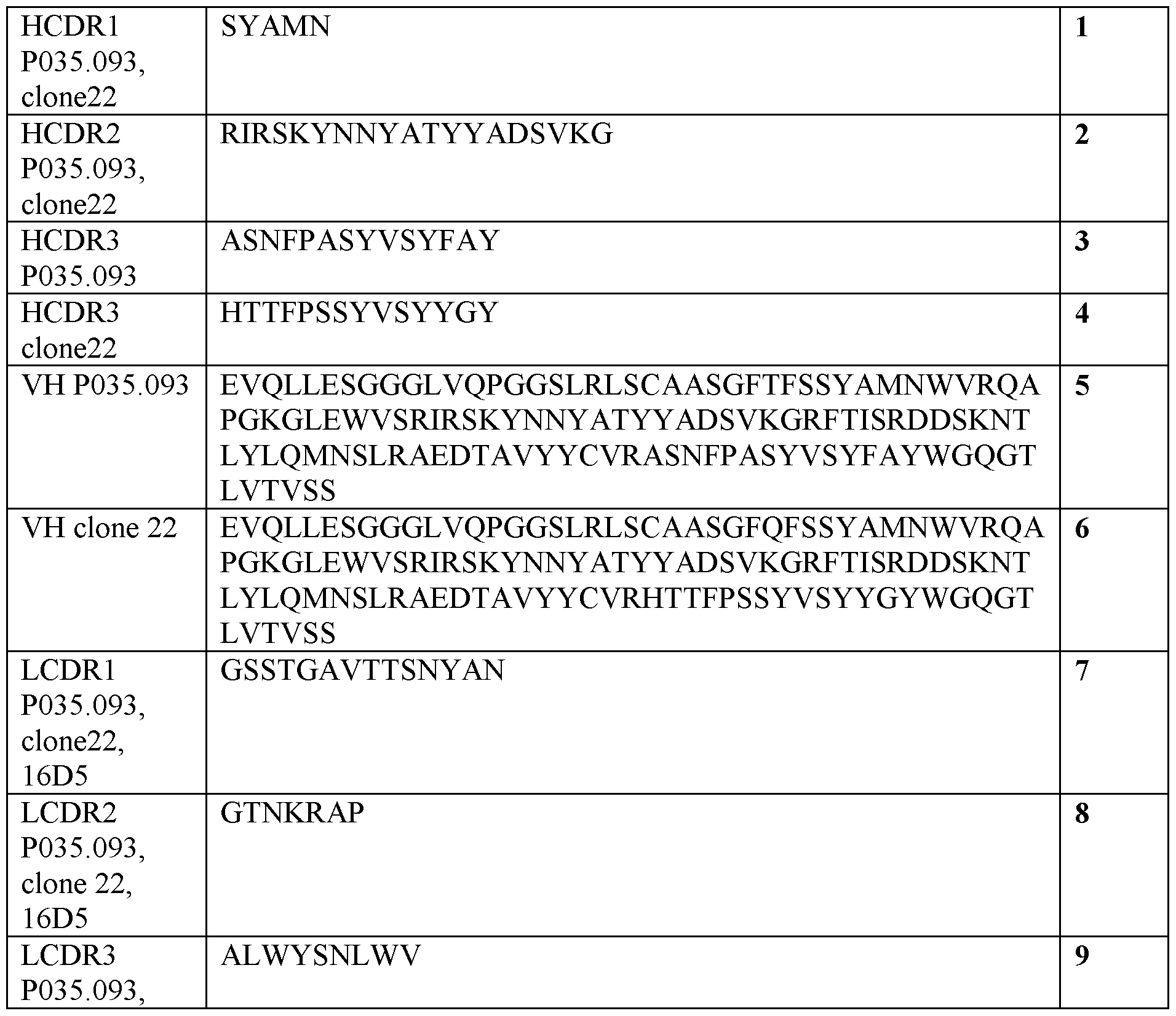
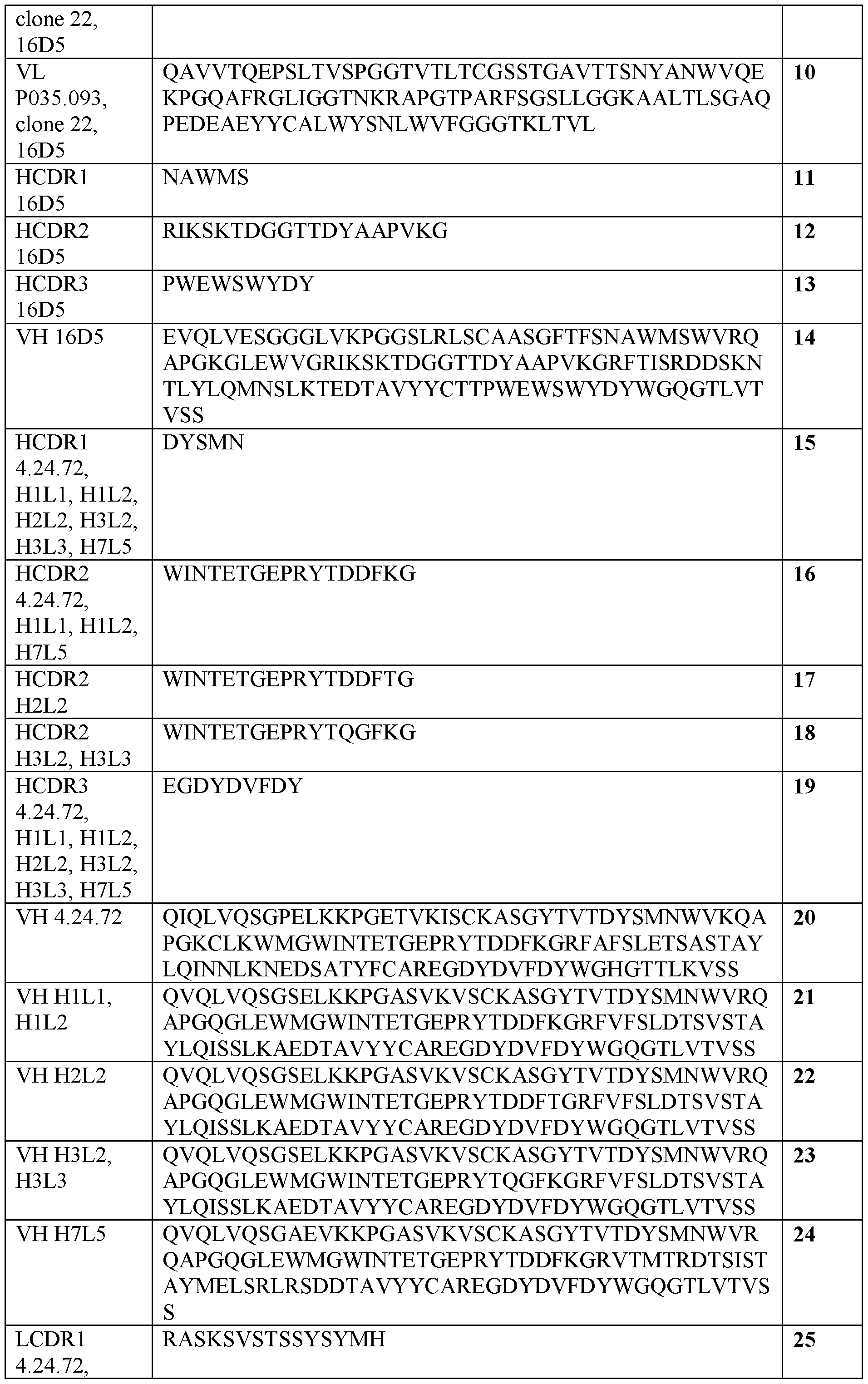


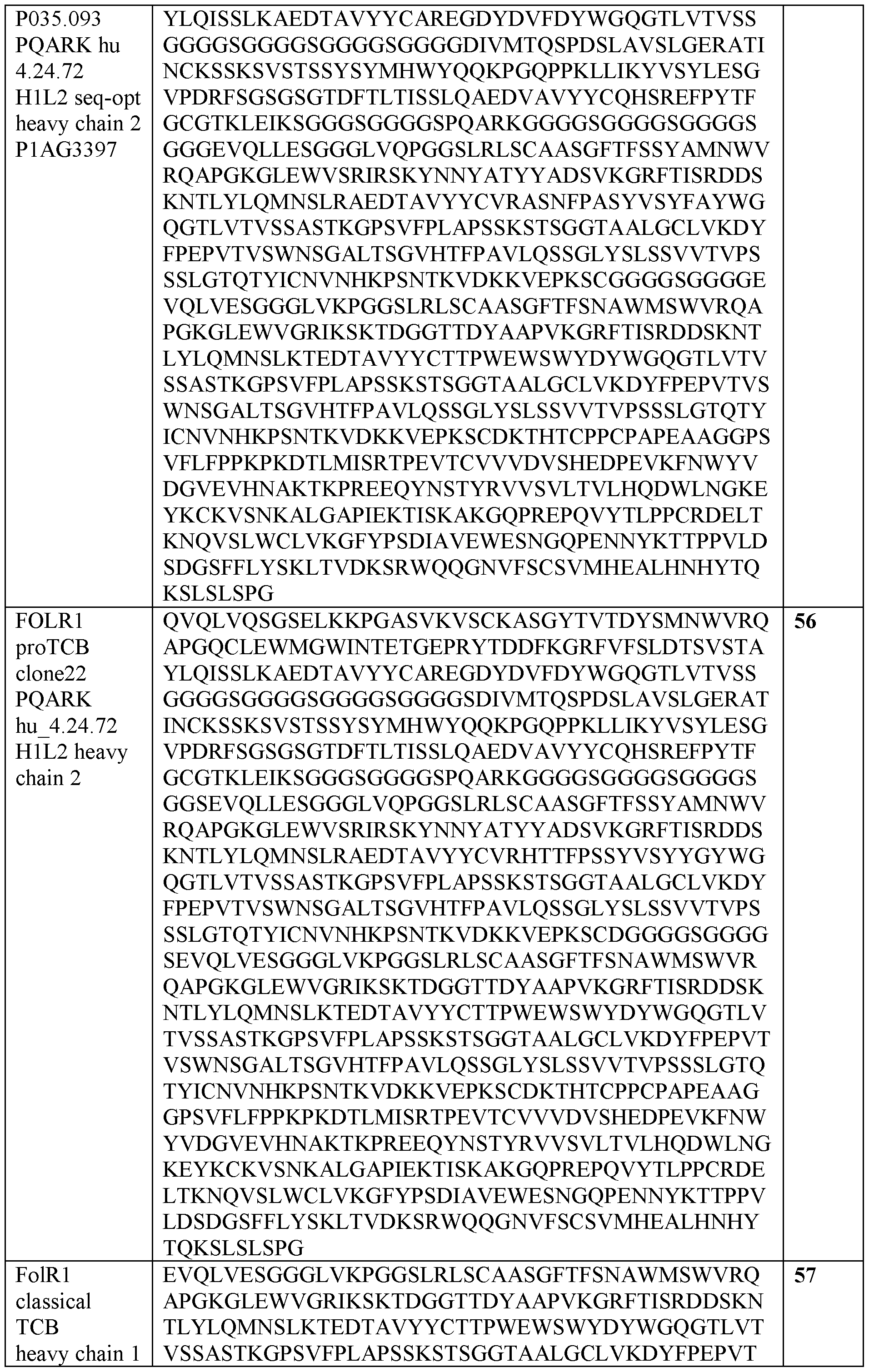


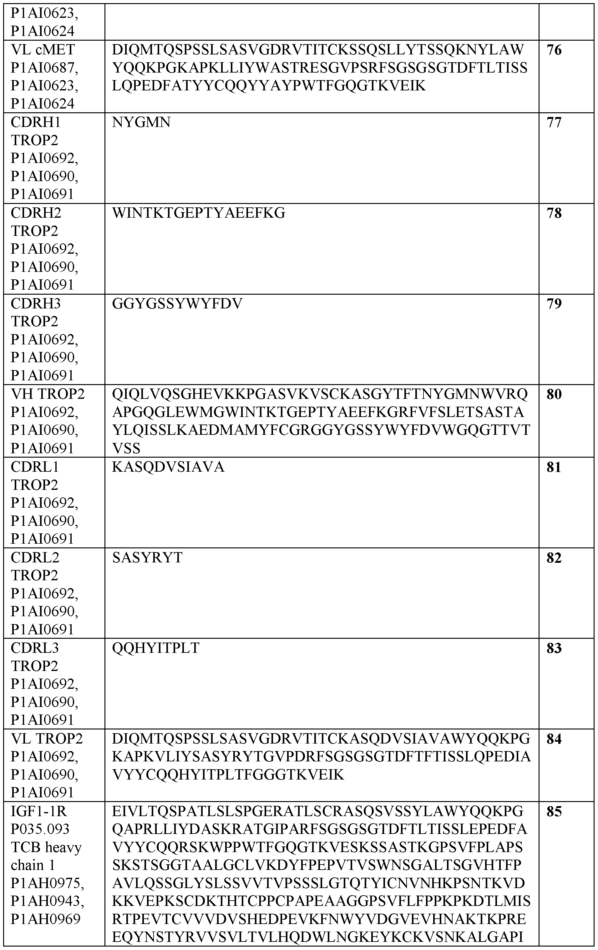
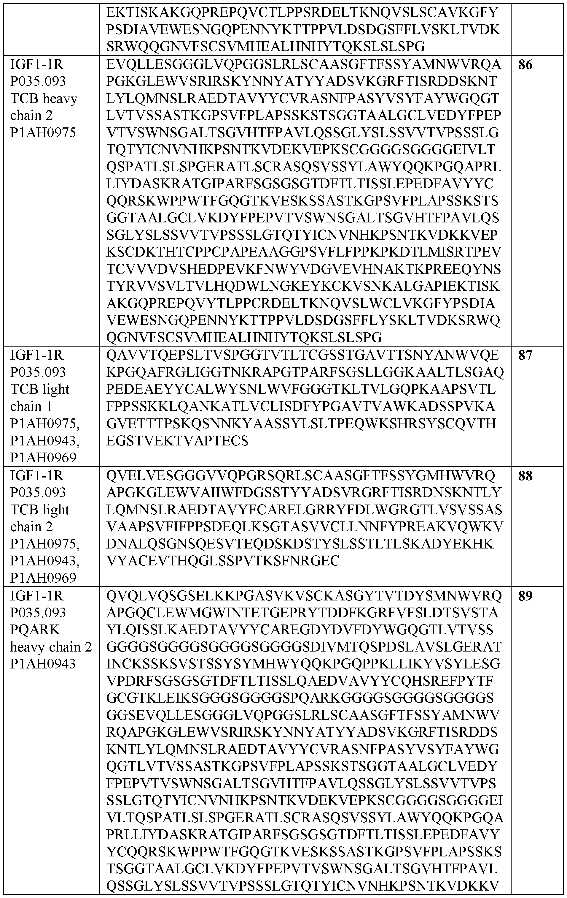

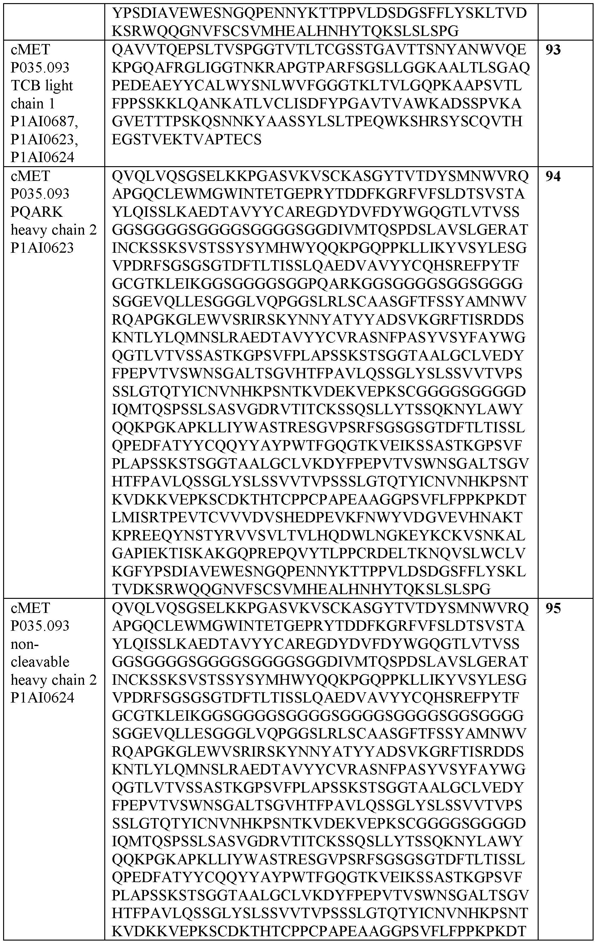
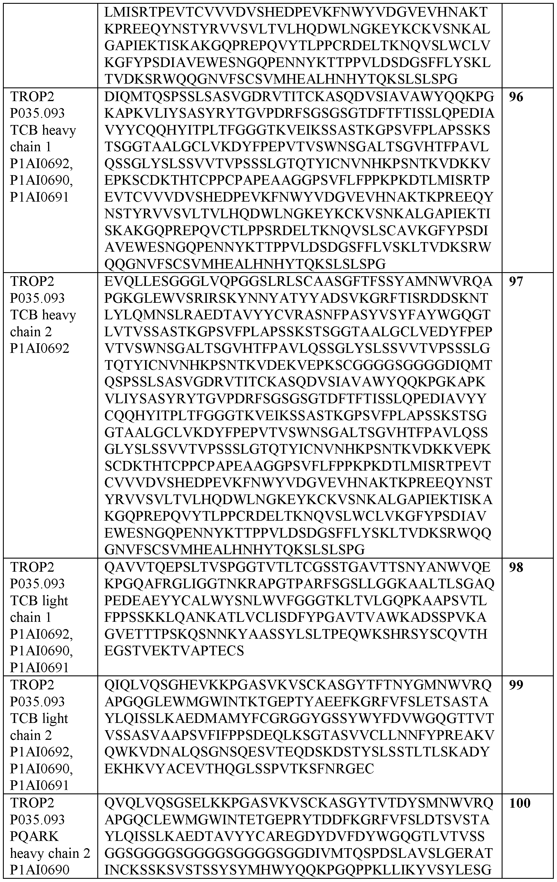


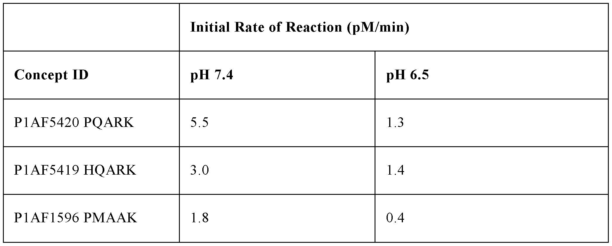


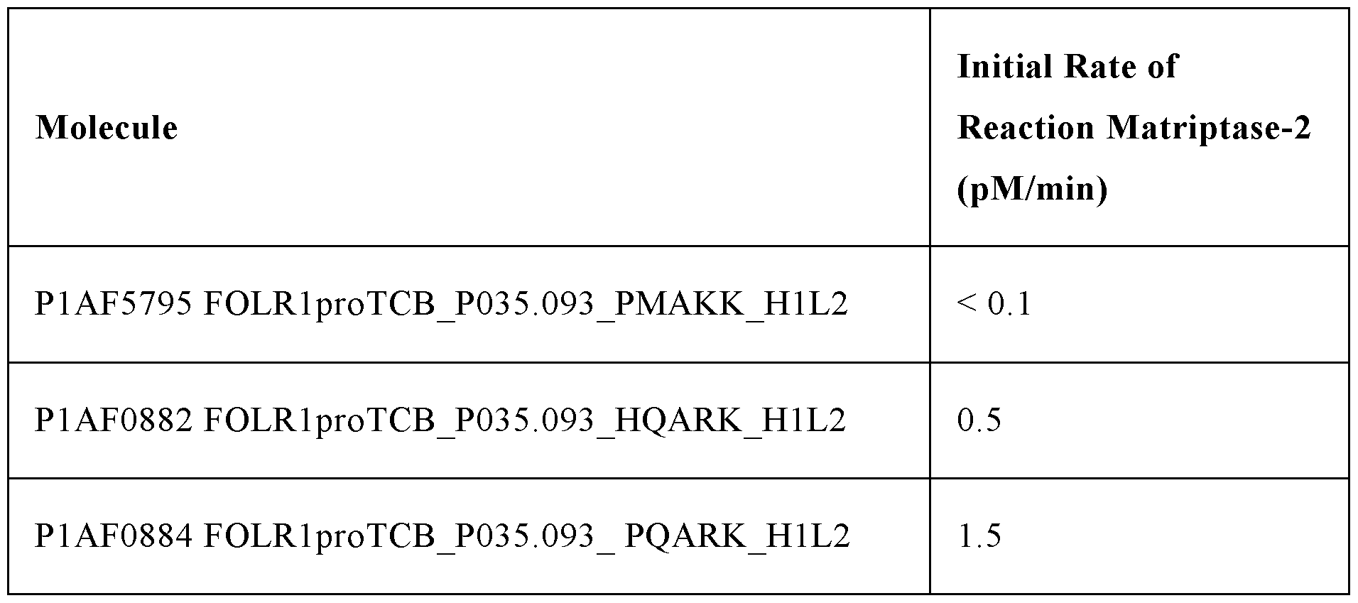


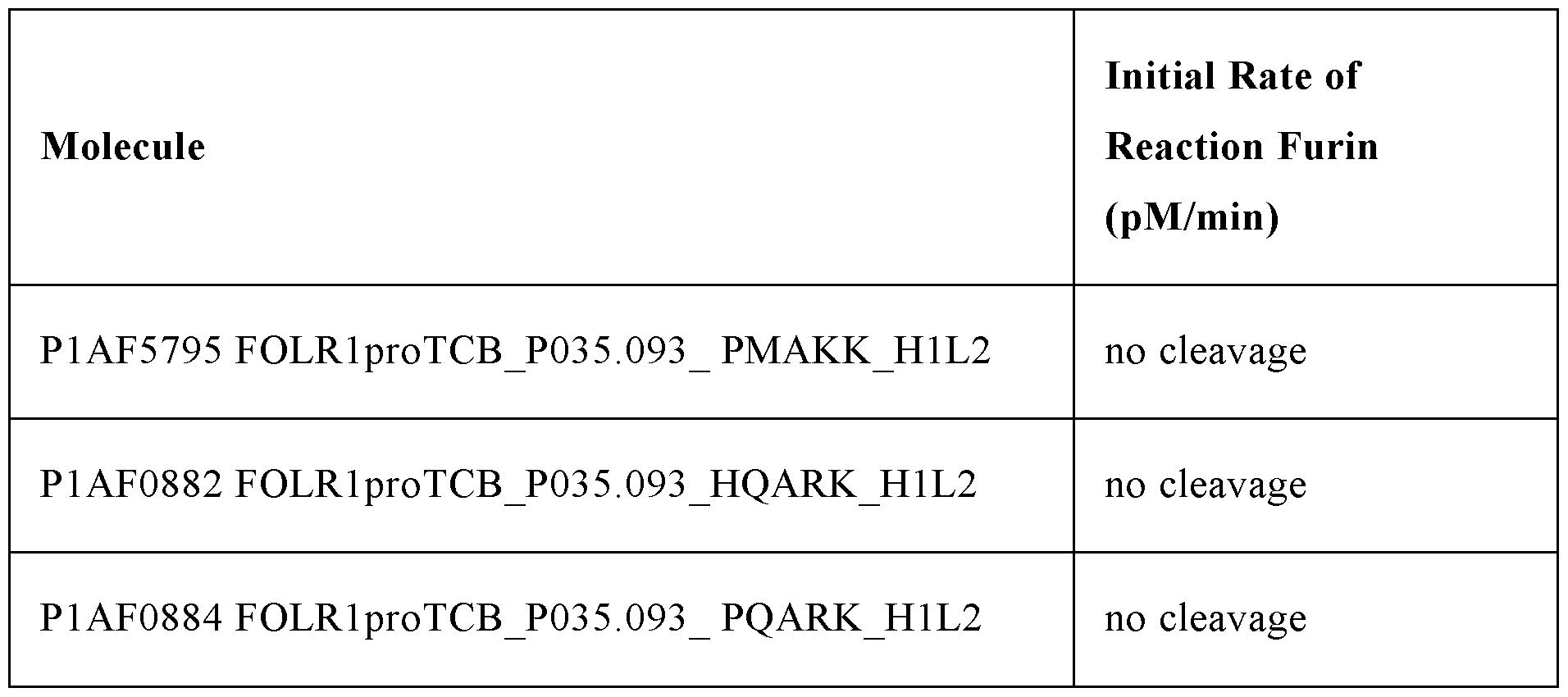
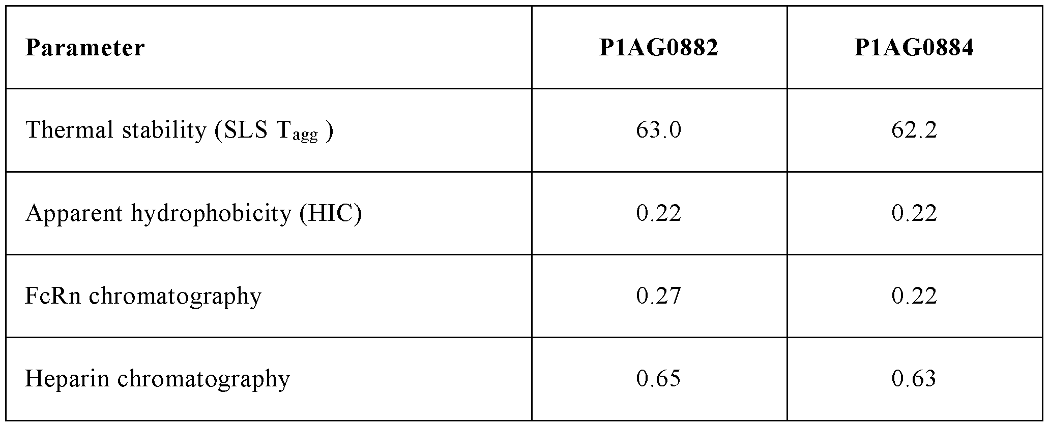


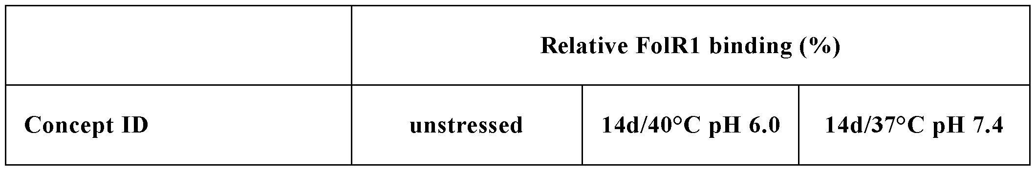
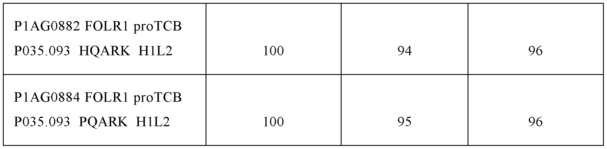

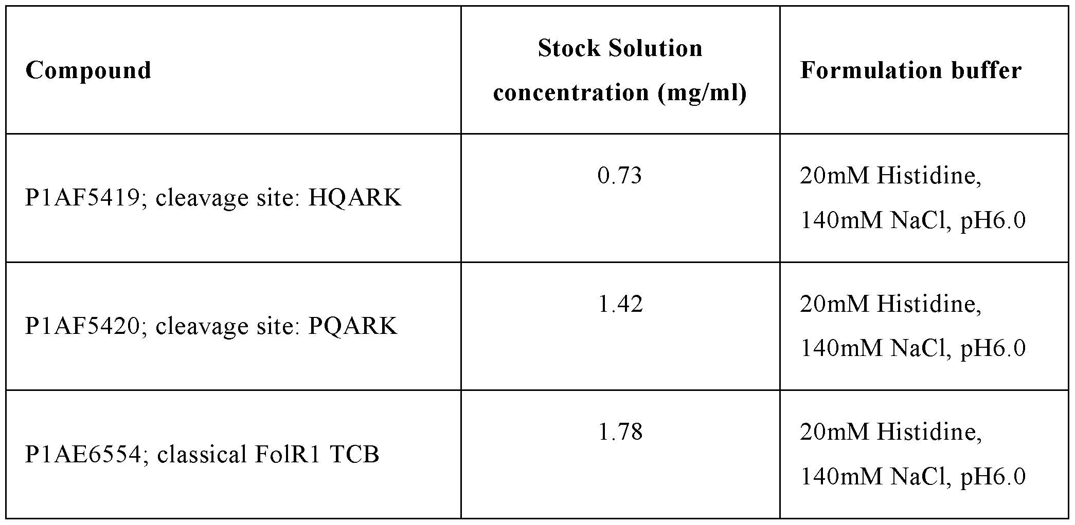

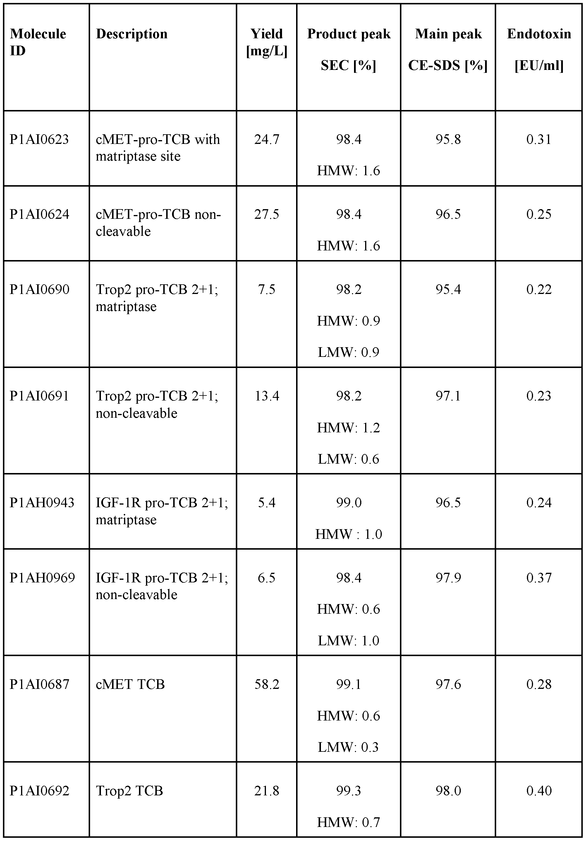
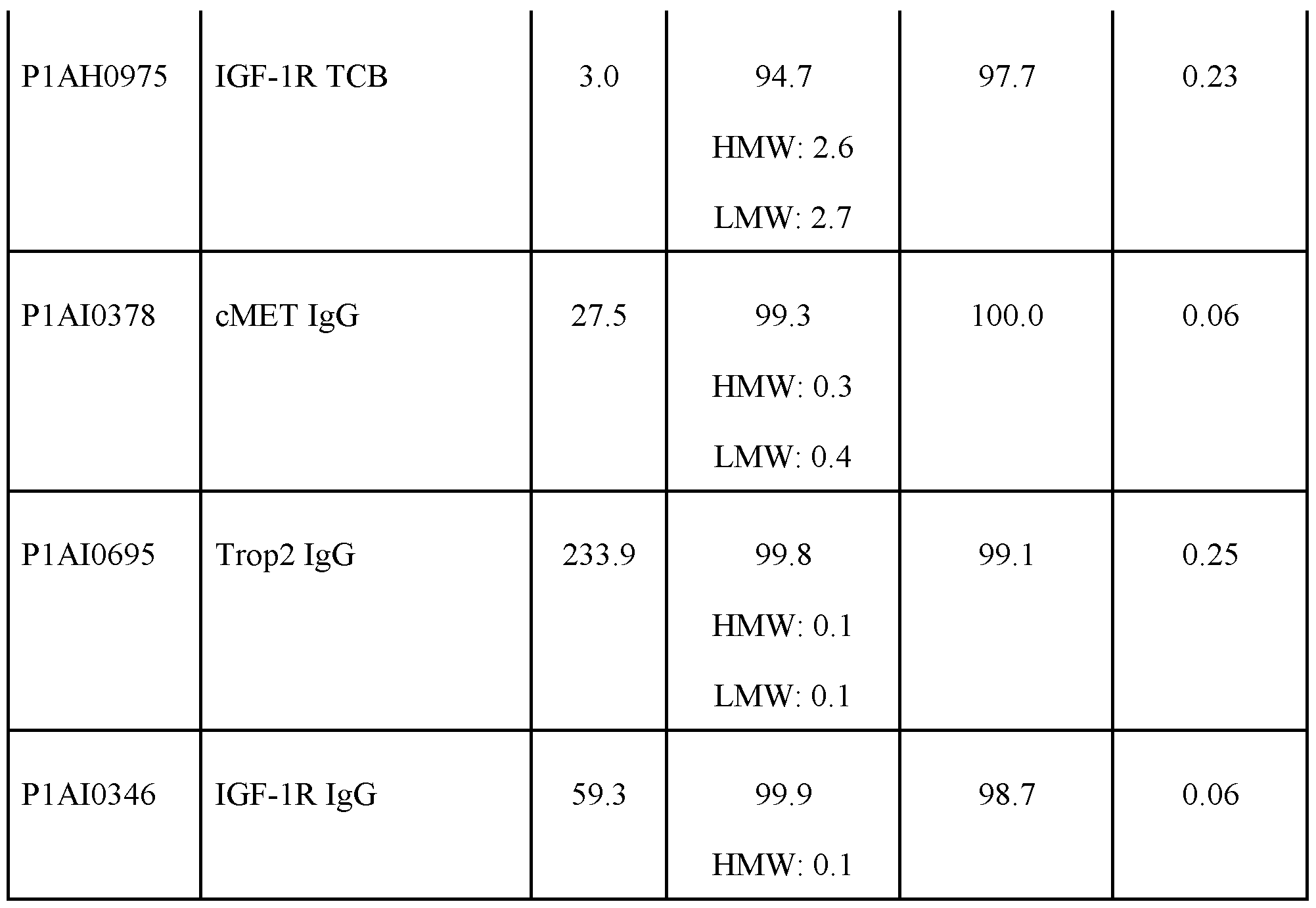
Claims
Priority Applications (3)
| Application Number | Priority Date | Filing Date | Title |
|---|---|---|---|
| CN202380068958.1A CN119948060A (en) | 2022-09-28 | 2023-09-26 | Improved protease-activatable T cell bispecific antibodies |
| JP2025517743A JP2025533570A (en) | 2022-09-28 | 2023-09-26 | Improved protease-activated T cell bispecific antibodies |
| EP23776639.9A EP4594353A1 (en) | 2022-09-28 | 2023-09-26 | Improved protease-activatable t cell bispecific antibodies |
Applications Claiming Priority (2)
| Application Number | Priority Date | Filing Date | Title |
|---|---|---|---|
| EP22198243.2 | 2022-09-28 | ||
| EP22198243 | 2022-09-28 |
Related Child Applications (1)
| Application Number | Title | Priority Date | Filing Date |
|---|---|---|---|
| US19/093,051 Continuation US20260035462A1 (en) | 2022-09-28 | 2025-03-27 | Protease-activatable t cell bispecific antibodies |
Publications (1)
| Publication Number | Publication Date |
|---|---|
| WO2024068572A1 true WO2024068572A1 (en) | 2024-04-04 |
Family
ID=83505575
Family Applications (1)
| Application Number | Title | Priority Date | Filing Date |
|---|---|---|---|
| PCT/EP2023/076444 Ceased WO2024068572A1 (en) | 2022-09-28 | 2023-09-26 | Improved protease-activatable t cell bispecific antibodies |
Country Status (4)
| Country | Link |
|---|---|
| EP (1) | EP4594353A1 (en) |
| JP (1) | JP2025533570A (en) |
| CN (1) | CN119948060A (en) |
| WO (1) | WO2024068572A1 (en) |
Citations (32)
| Publication number | Priority date | Publication date | Assignee | Title |
|---|---|---|---|---|
| US4186567A (en) | 1977-04-18 | 1980-02-05 | Hitachi Metals, Ltd. | Ornament utilizing rare earth-cobalt magnet |
| US5500362A (en) | 1987-01-08 | 1996-03-19 | Xoma Corporation | Chimeric antibody with specificity to human B cell surface antigen |
| US5565332A (en) | 1991-09-23 | 1996-10-15 | Medical Research Council | Production of chimeric antibodies - a combinatorial approach |
| US5731168A (en) | 1995-03-01 | 1998-03-24 | Genentech, Inc. | Method for making heteromultimeric polypeptides |
| US5821337A (en) | 1991-06-14 | 1998-10-13 | Genentech, Inc. | Immunoglobulin variants |
| US5959177A (en) | 1989-10-27 | 1999-09-28 | The Scripps Research Institute | Transgenic plants expressing assembled secretory antibodies |
| US5969108A (en) | 1990-07-10 | 1999-10-19 | Medical Research Council | Methods for producing members of specific binding pairs |
| US6040498A (en) | 1998-08-11 | 2000-03-21 | North Caroline State University | Genetically engineered duckweed |
| US6054297A (en) | 1991-06-14 | 2000-04-25 | Genentech, Inc. | Humanized antibodies and methods for making them |
| WO2001007611A2 (en) | 1999-07-26 | 2001-02-01 | Genentech, Inc. | Novel polynucleotides and method for the use thereof |
| US6420548B1 (en) | 1999-10-04 | 2002-07-16 | Medicago Inc. | Method for regulating transcription of foreign genes |
| US6737056B1 (en) | 1999-01-15 | 2004-05-18 | Genentech, Inc. | Polypeptide variants with altered effector function |
| US20040101920A1 (en) | 2002-11-01 | 2004-05-27 | Czeslaw Radziejewski | Modification assisted profiling (MAP) methodology |
| US20040132066A1 (en) | 2002-02-14 | 2004-07-08 | Kalobios, Inc. | Methods for affinity maturation |
| WO2005100402A1 (en) | 2004-04-13 | 2005-10-27 | F.Hoffmann-La Roche Ag | Anti-p-selectin antibodies |
| US6982321B2 (en) | 1986-03-27 | 2006-01-03 | Medical Research Council | Altered antibodies |
| WO2006029879A2 (en) | 2004-09-17 | 2006-03-23 | F.Hoffmann-La Roche Ag | Anti-ox40l antibodies |
| US7087409B2 (en) | 1997-12-05 | 2006-08-08 | The Scripps Research Institute | Humanization of murine antibody |
| US7125978B1 (en) | 1999-10-04 | 2006-10-24 | Medicago Inc. | Promoter for regulating expression of foreign genes |
| US7527791B2 (en) | 2004-03-31 | 2009-05-05 | Genentech, Inc. | Humanized anti-TGF-beta antibodies |
| WO2009089004A1 (en) | 2008-01-07 | 2009-07-16 | Amgen Inc. | Method for making antibody fc-heterodimeric molecules using electrostatic steering effects |
| WO2012130831A1 (en) | 2011-03-29 | 2012-10-04 | Roche Glycart Ag | Antibody fc variants |
| WO2013041462A1 (en) * | 2011-09-23 | 2013-03-28 | Roche Glycart Ag | Bispecific anti-egfr/anti igf-1r antibodies |
| WO2013128194A1 (en) | 2012-02-28 | 2013-09-06 | The University Of Birmingham | Immunotherapeutic molecules and uses |
| US20170152543A1 (en) * | 2015-11-30 | 2017-06-01 | Roche Sequencing Solutions, Inc. | System and method for identification of protease substrates |
| WO2018157147A1 (en) * | 2017-02-27 | 2018-08-30 | Dragonfly Therapeutics, Inc. | Multispecific binding proteins targeting caix, ano1, mesothelin,trop2, cea, or claudin-18.2 |
| WO2021255137A1 (en) * | 2020-06-19 | 2021-12-23 | F. Hoffmann-La Roche Ag | Protease-activated t cell bispecific antibodies |
| WO2021260064A1 (en) * | 2020-06-25 | 2021-12-30 | F. Hoffmann-La Roche Ag | Anti-cd3/anti-cd28 bispecific antigen binding molecules |
| WO2022020388A1 (en) * | 2020-07-21 | 2022-01-27 | Amunix Pharmaceuticals, Inc. | Compositions and methods related to activatable therapeutic agents |
| WO2022119958A1 (en) * | 2020-12-02 | 2022-06-09 | Cue Biopharma, Inc. | T-cell modulatory multimeric polypeptides and methods of use thereof |
| CA3206603A1 (en) * | 2021-02-11 | 2022-08-18 | Fangyong Du | Anti-cd3 antibodies and methods of use thereof |
| WO2023186760A1 (en) * | 2022-03-28 | 2023-10-05 | F. Hoffmann-La Roche Ag | Improved folr1 protease-activatable t cell bispecific antibodies |
-
2023
- 2023-09-26 WO PCT/EP2023/076444 patent/WO2024068572A1/en not_active Ceased
- 2023-09-26 EP EP23776639.9A patent/EP4594353A1/en active Pending
- 2023-09-26 CN CN202380068958.1A patent/CN119948060A/en active Pending
- 2023-09-26 JP JP2025517743A patent/JP2025533570A/en active Pending
Patent Citations (35)
| Publication number | Priority date | Publication date | Assignee | Title |
|---|---|---|---|---|
| US4186567A (en) | 1977-04-18 | 1980-02-05 | Hitachi Metals, Ltd. | Ornament utilizing rare earth-cobalt magnet |
| US6982321B2 (en) | 1986-03-27 | 2006-01-03 | Medical Research Council | Altered antibodies |
| US5500362A (en) | 1987-01-08 | 1996-03-19 | Xoma Corporation | Chimeric antibody with specificity to human B cell surface antigen |
| US6417429B1 (en) | 1989-10-27 | 2002-07-09 | The Scripps Research Institute | Transgenic plants expressing assembled secretory antibodies |
| US5959177A (en) | 1989-10-27 | 1999-09-28 | The Scripps Research Institute | Transgenic plants expressing assembled secretory antibodies |
| US5969108A (en) | 1990-07-10 | 1999-10-19 | Medical Research Council | Methods for producing members of specific binding pairs |
| US6054297A (en) | 1991-06-14 | 2000-04-25 | Genentech, Inc. | Humanized antibodies and methods for making them |
| US5821337A (en) | 1991-06-14 | 1998-10-13 | Genentech, Inc. | Immunoglobulin variants |
| US5565332A (en) | 1991-09-23 | 1996-10-15 | Medical Research Council | Production of chimeric antibodies - a combinatorial approach |
| US5731168A (en) | 1995-03-01 | 1998-03-24 | Genentech, Inc. | Method for making heteromultimeric polypeptides |
| US7695936B2 (en) | 1995-03-01 | 2010-04-13 | Genentech, Inc. | Knobs and holes heteromeric polypeptides |
| US7087409B2 (en) | 1997-12-05 | 2006-08-08 | The Scripps Research Institute | Humanization of murine antibody |
| US6040498A (en) | 1998-08-11 | 2000-03-21 | North Caroline State University | Genetically engineered duckweed |
| US7332581B2 (en) | 1999-01-15 | 2008-02-19 | Genentech, Inc. | Polypeptide variants with altered effector function |
| US6737056B1 (en) | 1999-01-15 | 2004-05-18 | Genentech, Inc. | Polypeptide variants with altered effector function |
| WO2001007611A2 (en) | 1999-07-26 | 2001-02-01 | Genentech, Inc. | Novel polynucleotides and method for the use thereof |
| US6420548B1 (en) | 1999-10-04 | 2002-07-16 | Medicago Inc. | Method for regulating transcription of foreign genes |
| US7125978B1 (en) | 1999-10-04 | 2006-10-24 | Medicago Inc. | Promoter for regulating expression of foreign genes |
| US20040132066A1 (en) | 2002-02-14 | 2004-07-08 | Kalobios, Inc. | Methods for affinity maturation |
| US20040101920A1 (en) | 2002-11-01 | 2004-05-27 | Czeslaw Radziejewski | Modification assisted profiling (MAP) methodology |
| US7527791B2 (en) | 2004-03-31 | 2009-05-05 | Genentech, Inc. | Humanized anti-TGF-beta antibodies |
| WO2005100402A1 (en) | 2004-04-13 | 2005-10-27 | F.Hoffmann-La Roche Ag | Anti-p-selectin antibodies |
| WO2006029879A2 (en) | 2004-09-17 | 2006-03-23 | F.Hoffmann-La Roche Ag | Anti-ox40l antibodies |
| WO2009089004A1 (en) | 2008-01-07 | 2009-07-16 | Amgen Inc. | Method for making antibody fc-heterodimeric molecules using electrostatic steering effects |
| WO2012130831A1 (en) | 2011-03-29 | 2012-10-04 | Roche Glycart Ag | Antibody fc variants |
| WO2013041462A1 (en) * | 2011-09-23 | 2013-03-28 | Roche Glycart Ag | Bispecific anti-egfr/anti igf-1r antibodies |
| WO2013128194A1 (en) | 2012-02-28 | 2013-09-06 | The University Of Birmingham | Immunotherapeutic molecules and uses |
| US20170152543A1 (en) * | 2015-11-30 | 2017-06-01 | Roche Sequencing Solutions, Inc. | System and method for identification of protease substrates |
| WO2018157147A1 (en) * | 2017-02-27 | 2018-08-30 | Dragonfly Therapeutics, Inc. | Multispecific binding proteins targeting caix, ano1, mesothelin,trop2, cea, or claudin-18.2 |
| WO2021255137A1 (en) * | 2020-06-19 | 2021-12-23 | F. Hoffmann-La Roche Ag | Protease-activated t cell bispecific antibodies |
| WO2021260064A1 (en) * | 2020-06-25 | 2021-12-30 | F. Hoffmann-La Roche Ag | Anti-cd3/anti-cd28 bispecific antigen binding molecules |
| WO2022020388A1 (en) * | 2020-07-21 | 2022-01-27 | Amunix Pharmaceuticals, Inc. | Compositions and methods related to activatable therapeutic agents |
| WO2022119958A1 (en) * | 2020-12-02 | 2022-06-09 | Cue Biopharma, Inc. | T-cell modulatory multimeric polypeptides and methods of use thereof |
| CA3206603A1 (en) * | 2021-02-11 | 2022-08-18 | Fangyong Du | Anti-cd3 antibodies and methods of use thereof |
| WO2023186760A1 (en) * | 2022-03-28 | 2023-10-05 | F. Hoffmann-La Roche Ag | Improved folr1 protease-activatable t cell bispecific antibodies |
Non-Patent Citations (65)
| Title |
|---|
| "Monoclonal Antibody Production Techniques and Applications", 1987, MARCEL DEKKER, INC., pages: 51 - 63 |
| "Remington's Pharmaceutical Sciences", 1990, MACK PRINTING COMPANY, pages: 1289 - 1329 |
| A DISINTEGRIN AND METALLOPROTEINASE (ADAM, vol. 2, no. 7-12, pages 17 - 23 |
| BARRÉ OLIVIER ET AL: "Cleavage Specificity Analysis of Six Type II Transmembrane Serine Proteases (TTSPs) Using PICS with Proteome-Derived Peptide Libraries", PLOS ONE, vol. 9, no. 9, 11 September 2014 (2014-09-11), pages e105984, XP093059241, DOI: 10.1371/journal.pone.0105984 * |
| BOEDTKJER ET AL., ANNUAL REVIEW OF PHYSIOLOGY, vol. 82, 2020, pages 103 - 126 |
| BRUGGEMANN ET AL., J EXP MED, vol. 166, 1987, pages 1351 - 1361 |
| CARTER, J IMMUNOL METH, vol. 248, 2001, pages 7 - 15 |
| CARTER, J IMMUNOL METHODS, vol. 248, 2001, pages 7 - 15 |
| CLACKSON ET AL., NATURE, vol. 352, 1991, pages 624 - 3242 |
| CLYNES ET AL., PROC NATL ACAD SCI USA, vol. 95, 1998, pages 652 - 656 |
| CRAGG ET AL., BLOOD, vol. 101, 2003, pages 1045 - 1052 |
| CRAGGGLENNIE, BLOOD, vol. 103, 2004, pages 2738 - 2743 |
| DALL'ACQUA ET AL., METHODS, vol. 36, 2005, pages 61 - 68 |
| FINGL ET AL., THE PHARMACOLOGICAL BASIS OF THERAPEUTICS, 1975 |
| FLATMAN ET AL., J. CHROMATOGR. B, vol. 848, 2007, pages 79 - 87 |
| FRANSSON, FRONT BIOSCI, vol. 13, 2008, pages 1619 - 1633 |
| GAZZANO-SANTORO ET AL., J IMMUNOL METHODS, vol. 202, 1996, pages 163 |
| GERNGROSS, NAT BIOTECH, vol. 22, 2004, pages 1409 - 1414 |
| GRAHAM ET AL., J GEN VIROL, vol. 36, 1977, pages 59 |
| HARLOWLANE: "Antibodies: A Laboratory Manual", vol. 14, 1988, COLD SPRING HARBOR LABORATORY |
| HEELEY, ENDOCR RES, vol. 28, 2002, pages 217 - 229 |
| HELLSTROM ET AL., PROC NATL ACAD SCI USA, vol. 82, 1985, pages 1499 - 1502 |
| HELLSTROM ET AL., PROC NATL ACAD SCI USA, vol. 83, 1986, pages 7059 - 7063 |
| HOLLIGER ET AL., PROT ENG, vol. 9, 1996, pages 617 - 621 |
| HOLLIGERHUDSON, NATURE BIOTECHNOLOGY, vol. 23, 2005, pages 1126 - 1136 |
| HOOGENBOOM ET AL., METHODS IN MOLECULAR BIOLOGY, vol. 178, pages 1 - 37 |
| JOHNSON, G.WU, T.T., NUCLEIC ACIDS RES., vol. 28, 2000, pages 214 - 218 |
| JONES ET AL., NATURE, vol. 321, 1986, pages 522 - 525 |
| JUNGHANS ET AL., CANCER RES., vol. 50, 1990, pages 1495 - 1502 |
| KABAT, E.A. ET AL., PROC. NATL. ACAD. SCI. USA, vol. 72, 1975, pages 2785 - 2788 |
| KABAT, E.A. ET AL.: "Public Health Service, National Institutes of Health", 1991, PUBLIC HEALTH SERVICE, NATIONAL INSTITUTES OF HEALTH, article "Sequences of Proteins of Immunological Interest", pages: 91 - 3242 |
| KINDT ET AL.: "Kuby Immunology", 2007, W.H. FREEMAN AND CO., pages: 91 |
| KIPRIYANOV ET AL., J MOL BIOL, vol. 293, 1999, pages 865 - 881 |
| KLIMKA ET AL., BR J CANCER, vol. 83, 2000, pages 252 - 260 |
| LEE S-L ET AL: "39720 - 36725", JOURNAL OF BIOLOGICAL CHEMISTRY, AMERICAN SOCIETY FOR BIOCHEMISTRY AND MOLECULAR BIOLOGY, US, vol. 275, no. 47, 24 November 2000 (2000-11-24), pages 36720 - 36725, XP000999224, ISSN: 0021-9258, DOI: 10.1074/JBC.M007802200 * |
| LI ET AL., NAT BIOTECH, vol. 23, 2005, pages 1117 - 1125 |
| LILJEBLAD ET AL., GLYCO J, vol. 17, 2000, pages 323 - 329 |
| LONBERG, CURR OPIN IMMUNOL, vol. 20, 2008, pages 450 - 459 |
| MACCALLUM ET AL., J. MOL. BIOL., vol. 262, 1996, pages 732 - 745 |
| MANIATIS ET AL.: "Current Protocols in Molecular Biology", 1989, GREENE PUBLISHING ASSOCIATES AND WILEY INTERSCIENCE |
| MARTIN C. BOONSTRA ET AL: "Selecting Targets for Tumor Imaging: An Overview of Cancer-Associated Membrane Proteins", BIOMARKERS IN CANCER, vol. 8, 1 September 2016 (2016-09-01), pages 119 - 133, XP055748038, ISSN: 1179-299X, DOI: 10.4137/BIC.S38542 * |
| MATHER ET AL., ANNALS N.Y. ACAD SCI, vol. 383, 1982, pages 44 - 68 |
| MATHER, BIOL REPROD, vol. 23, 1980, pages 243 - 251 |
| MCCAFFERTY ET AL., NATURE, vol. 348, pages 552 - 554 |
| METH. MOL. BIOL., vol. 248, 2004, pages 443 - 463 |
| MOORE ET AL., BLOOD, vol. 117, 2011, pages 4542 - 51 |
| MORRISON ET AL., PROC NATL ACAD SCI, vol. 81, 1984, pages 6851 - 6855 |
| MORRISONOI, ADV IMMUNOL, vol. 44, 1988, pages 65 - 92 |
| NAGORSENBAUERLE, EXP CELL RES, vol. 317, 2011, pages 1255 - 1260 |
| PADLAN, MOL IMMUNOL, vol. 28, 1991, pages 489 - 498 |
| PADLAN, MOLEC IMMUN, vol. 31, no. 3, 1994, pages 169 - 217 |
| PEARSON, GENOMICS, vol. 46, 1997, pages 24 - 36 |
| PORTOLANO ET AL., J. IMMUNOL., vol. 150, 1993, pages 880 - 887 |
| PROT. SCI., vol. 9, 2000, pages 487 - 496 |
| QUEEN ET AL., PROC NATL ACAD SCI USA, vol. 86, pages 10029 - 10033 |
| RIECHMANN ET AL., NATURE, vol. 332, 1988, pages 323 - 329 |
| SEIMETZ ET AL., CANCER TREAT REV, vol. 36, 2010, pages 458 - 467 |
| STADLER, NATURE MEDICINE, 12 June 2017 (2017-06-12) |
| STUBENRAUCH ET AL., DRUG METABOLISM AND DISPOSITION, vol. 38, 2010, pages 84 - 91 |
| URLAUB ET AL., PROC NATL ACAD SCI USA, vol. 77, 1980, pages 4216 |
| VAN DIJKVAN DE WINKEL, CURR OPIN PHARMACOL, vol. 5, 2001, pages 368 - 74 |
| VERHOEYEN ET AL., SCIENCE, vol. 239, 1988, pages 1534 - 1536 |
| W. R. PEARSON: "Effective protein sequence comparison", METH. ENZYMOL., vol. 266, 1996, pages 227 - 258 |
| W. R. PEARSOND. J. LIPMAN: "Improved Tools for Biological Sequence Analysis", PNAS, vol. 85, 1988, pages 2444 - 2448 |
| YAZAKIWU: "Methods in Molecular Biology", vol. 248, 1996, HUMANA PRESS, article "Epitope Mapping Protocols", pages: 255 - 268 |
Also Published As
| Publication number | Publication date |
|---|---|
| JP2025533570A (en) | 2025-10-07 |
| EP4594353A1 (en) | 2025-08-06 |
| CN119948060A (en) | 2025-05-06 |
Similar Documents
| Publication | Publication Date | Title |
|---|---|---|
| US12139553B2 (en) | T cell activating bispecific antigen binding molecules | |
| AU2019250201B2 (en) | Bispecific T cell activating antigen binding molecules | |
| CN109843926B (en) | Bispecific antibodies against CD3 | |
| US20220275087A1 (en) | Protease-activated t cell bispecific molecules | |
| AU2018271329A1 (en) | Bispecific T cell activating antigen binding molecules | |
| HK1254068A1 (en) | Bispecific t cell activating antigen binding molecules | |
| EP3433280A1 (en) | Protease-activated t cell bispecific molecules | |
| US20230287145A1 (en) | Protease-activated t cell bispecific antibodies | |
| US20250092128A1 (en) | Folr1 protease-activatable t cell bispecific antibodies | |
| WO2017055392A1 (en) | Anti-cd3xcd44v6 bispecific t cell activating antigen binding molecules | |
| US20260035462A1 (en) | Protease-activatable t cell bispecific antibodies | |
| WO2024068572A1 (en) | Improved protease-activatable t cell bispecific antibodies | |
| HK40119423A (en) | Improved folr1 protease-activatable t cell bispecific antibodies | |
| HK40088901A (en) | Protease-activated t cell bispecific antibodies | |
| HK1254966B (en) | Bispecific anti-cd19xcd3 t cell activating antigen binding molecules | |
| HK1233664B (en) | Bispecific t cell activating antigen binding molecules | |
| HK1233664A1 (en) | Bispecific t cell activating antigen binding molecules |
Legal Events
| Date | Code | Title | Description |
|---|---|---|---|
| 121 | Ep: the epo has been informed by wipo that ep was designated in this application |
Ref document number: 23776639 Country of ref document: EP Kind code of ref document: A1 |
|
| ENP | Entry into the national phase |
Ref document number: 2025517743 Country of ref document: JP Kind code of ref document: A |
|
| WWE | Wipo information: entry into national phase |
Ref document number: 2025517743 Country of ref document: JP |
|
| WWE | Wipo information: entry into national phase |
Ref document number: 202380068958.1 Country of ref document: CN |
|
| WWE | Wipo information: entry into national phase |
Ref document number: 2023776639 Country of ref document: EP |
|
| NENP | Non-entry into the national phase |
Ref country code: DE |
|
| ENP | Entry into the national phase |
Ref document number: 2023776639 Country of ref document: EP Effective date: 20250428 |
|
| WWP | Wipo information: published in national office |
Ref document number: 202380068958.1 Country of ref document: CN |
|
| WWP | Wipo information: published in national office |
Ref document number: 2023776639 Country of ref document: EP |