WO2023286772A1 - Cardiomyocyte production method - Google Patents
Cardiomyocyte production method Download PDFInfo
- Publication number
- WO2023286772A1 WO2023286772A1 PCT/JP2022/027420 JP2022027420W WO2023286772A1 WO 2023286772 A1 WO2023286772 A1 WO 2023286772A1 JP 2022027420 W JP2022027420 W JP 2022027420W WO 2023286772 A1 WO2023286772 A1 WO 2023286772A1
- Authority
- WO
- WIPO (PCT)
- Prior art keywords
- cells
- cardiomyocytes
- fetoprotein
- serum
- free
- Prior art date
- Legal status (The legal status is an assumption and is not a legal conclusion. Google has not performed a legal analysis and makes no representation as to the accuracy of the status listed.)
- Ceased
Links
Images


Classifications
-
- C—CHEMISTRY; METALLURGY
- C12—BIOCHEMISTRY; BEER; SPIRITS; WINE; VINEGAR; MICROBIOLOGY; ENZYMOLOGY; MUTATION OR GENETIC ENGINEERING
- C12N—MICROORGANISMS OR ENZYMES; COMPOSITIONS THEREOF; PROPAGATING, PRESERVING, OR MAINTAINING MICROORGANISMS; MUTATION OR GENETIC ENGINEERING; CULTURE MEDIA
- C12N5/00—Undifferentiated human, animal or plant cells, e.g. cell lines; Tissues; Cultivation or maintenance thereof; Culture media therefor
- C12N5/0018—Culture media for cell or tissue culture
- C12N5/0037—Serum-free medium, which may still contain naturally-sourced components
-
- C—CHEMISTRY; METALLURGY
- C07—ORGANIC CHEMISTRY
- C07K—PEPTIDES
- C07K14/00—Peptides having more than 20 amino acids; Gastrins; Somatostatins; Melanotropins; Derivatives thereof
- C07K14/435—Peptides having more than 20 amino acids; Gastrins; Somatostatins; Melanotropins; Derivatives thereof from animals; from humans
- C07K14/46—Peptides having more than 20 amino acids; Gastrins; Somatostatins; Melanotropins; Derivatives thereof from animals; from humans from vertebrates
- C07K14/47—Peptides having more than 20 amino acids; Gastrins; Somatostatins; Melanotropins; Derivatives thereof from animals; from humans from vertebrates from mammals
-
- C—CHEMISTRY; METALLURGY
- C12—BIOCHEMISTRY; BEER; SPIRITS; WINE; VINEGAR; MICROBIOLOGY; ENZYMOLOGY; MUTATION OR GENETIC ENGINEERING
- C12N—MICROORGANISMS OR ENZYMES; COMPOSITIONS THEREOF; PROPAGATING, PRESERVING, OR MAINTAINING MICROORGANISMS; MUTATION OR GENETIC ENGINEERING; CULTURE MEDIA
- C12N1/00—Microorganisms, e.g. protozoa; Compositions thereof; Processes of propagating, maintaining or preserving microorganisms or compositions thereof; Processes of preparing or isolating a composition containing a microorganism; Culture media therefor
-
- C—CHEMISTRY; METALLURGY
- C12—BIOCHEMISTRY; BEER; SPIRITS; WINE; VINEGAR; MICROBIOLOGY; ENZYMOLOGY; MUTATION OR GENETIC ENGINEERING
- C12N—MICROORGANISMS OR ENZYMES; COMPOSITIONS THEREOF; PROPAGATING, PRESERVING, OR MAINTAINING MICROORGANISMS; MUTATION OR GENETIC ENGINEERING; CULTURE MEDIA
- C12N5/00—Undifferentiated human, animal or plant cells, e.g. cell lines; Tissues; Cultivation or maintenance thereof; Culture media therefor
- C12N5/0018—Culture media for cell or tissue culture
- C12N5/0056—Xeno-free medium
-
- C—CHEMISTRY; METALLURGY
- C12—BIOCHEMISTRY; BEER; SPIRITS; WINE; VINEGAR; MICROBIOLOGY; ENZYMOLOGY; MUTATION OR GENETIC ENGINEERING
- C12N—MICROORGANISMS OR ENZYMES; COMPOSITIONS THEREOF; PROPAGATING, PRESERVING, OR MAINTAINING MICROORGANISMS; MUTATION OR GENETIC ENGINEERING; CULTURE MEDIA
- C12N5/00—Undifferentiated human, animal or plant cells, e.g. cell lines; Tissues; Cultivation or maintenance thereof; Culture media therefor
- C12N5/06—Animal cells or tissues; Human cells or tissues
- C12N5/0602—Vertebrate cells
- C12N5/0652—Cells of skeletal and connective tissues; Mesenchyme
- C12N5/0657—Cardiomyocytes; Heart cells
-
- C—CHEMISTRY; METALLURGY
- C12—BIOCHEMISTRY; BEER; SPIRITS; WINE; VINEGAR; MICROBIOLOGY; ENZYMOLOGY; MUTATION OR GENETIC ENGINEERING
- C12N—MICROORGANISMS OR ENZYMES; COMPOSITIONS THEREOF; PROPAGATING, PRESERVING, OR MAINTAINING MICROORGANISMS; MUTATION OR GENETIC ENGINEERING; CULTURE MEDIA
- C12N5/00—Undifferentiated human, animal or plant cells, e.g. cell lines; Tissues; Cultivation or maintenance thereof; Culture media therefor
- C12N5/10—Cells modified by introduction of foreign genetic material
-
- C—CHEMISTRY; METALLURGY
- C12—BIOCHEMISTRY; BEER; SPIRITS; WINE; VINEGAR; MICROBIOLOGY; ENZYMOLOGY; MUTATION OR GENETIC ENGINEERING
- C12N—MICROORGANISMS OR ENZYMES; COMPOSITIONS THEREOF; PROPAGATING, PRESERVING, OR MAINTAINING MICROORGANISMS; MUTATION OR GENETIC ENGINEERING; CULTURE MEDIA
- C12N2501/00—Active agents used in cell culture processes, e.g. differentation
- C12N2501/998—Proteins not provided for elsewhere
-
- C—CHEMISTRY; METALLURGY
- C12—BIOCHEMISTRY; BEER; SPIRITS; WINE; VINEGAR; MICROBIOLOGY; ENZYMOLOGY; MUTATION OR GENETIC ENGINEERING
- C12N—MICROORGANISMS OR ENZYMES; COMPOSITIONS THEREOF; PROPAGATING, PRESERVING, OR MAINTAINING MICROORGANISMS; MUTATION OR GENETIC ENGINEERING; CULTURE MEDIA
- C12N2506/00—Differentiation of animal cells from one lineage to another; Differentiation of pluripotent cells
- C12N2506/45—Differentiation of animal cells from one lineage to another; Differentiation of pluripotent cells from artificially induced pluripotent stem cells
Definitions
- the present invention relates to a method for producing cardiomyocytes. More specifically, it relates to a method for producing a cell population containing a high proportion of mature cardiomyocytes from pluripotent stem cells.
- Patent Documents 1 to 3 The inventors have developed a method for inducing differentiation of cardiomyocytes at low cost and with high efficiency using low-molecular-weight compounds.
- This method is a protein-free differentiation induction method in which proteins such as cytokines are replaced with low-molecular-weight compounds.
- iPS cell-derived cardiomyocytes generally remain in an immature stage similar to fetal cardiomyocytes and take a long time to mature.
- a method for maturation by adding a specific low-molecular-weight compound has been proposed, but it only shows an increase in the expression of the endogenous TNNI3 gene, and does not improve cell quality or yield. It is not clear (Patent Document 4).
- ⁇ -fetoprotein is a component contained in fetal bovine serum and is sometimes added to the medium as a serum substitute (Patent Documents 5-8). It is known that ⁇ -fetoprotein promotes proliferation of stem cells as a serum substitute (Patent Document 5) and is used for maintaining undifferentiated state (Patent Document 8), but it is known that differentiation induction of cells into specific cells A method of using it as a factor is not known.
- the object of the present invention is to provide a method that can efficiently induce mature cardiomyocytes in a short period of time.
- a method for producing a cell population containing cardiomyocytes comprising a differentiation-inducing process from cells capable of differentiating into cardiomyocytes to cardiomyocytes, wherein the differentiation-inducing process includes culturing the cells in a serum-free medium containing ⁇ -fetoprotein.
- the above method comprising culturing and obtaining cardiomyocytes.
- the obtained cell population contains 80% or more, preferably 85% or more, more preferably 90% or more cardiomyocytes as a percentage of cells measured by flow cytometry [1] to [7]
- the method according to any one of [9] The method according to any one of [1] to [8], wherein the ⁇ -fetoprotein is human ⁇ -fetoprotein.
- the serum-free medium is xeno-free and/or cytokine-free.
- cardiomyocyte differentiation-inducing factor comprises any one of CHIR99021, prostratin, KY03-I, XAV939, A419259, and AG1478.
- Cardiomyocyte differentiation-inducing factor is one or more selected from the group consisting of Wnt signal activators (GSK3 ⁇ inhibitors), PKC activators, Src inhibitors, and EGF receptor inhibitors described later.
- Wnt signal activators GSK3 ⁇ inhibitors
- PKC activators PKC activators
- Src inhibitors Src inhibitors
- EGF receptor inhibitors described later.
- a method for improving the yield of cardiomyocytes comprising adding ⁇ -fetoprotein to the medium in the step of inducing differentiation into cardiomyocytes.
- the cardiomyocyte yield is improved by at least 5%, more preferably by 10% or more, 30% or more, or 50% or more, compared to when no ⁇ -fetoprotein is added.
- a method for improving the proportion of atrial cardiomyocytes comprising adding ⁇ -fetoprotein to the culture medium in the step of inducing differentiation into cardiomyocytes.
- the percentage of MYL7-positive cells measured by FACS is 60% or more, or the expression level of the MYL7 gene measured by RT-PCR is 50% or more compared to the case where ⁇ -fetoprotein is not added. , preferably 70% or more, 80% or more, 90% or more, more preferably 100%.
- the expression level of ion channels is improved by 50% or more, preferably 100%, more preferably 200%, and still more preferably 400% or more compared to immature cardiomyocytes.
- a method for improving the percentage of mature cardiomyocytes comprising adding ⁇ -fetoprotein to the culture medium in the step of inducing differentiation into cardiomyocytes.
- the percentage of mature myocardial cells is improved by at least 5% or more, more preferably by 10% or more, or 20% or more, compared to the case where ⁇ -fetoprotein is not added.
- a pharmaceutical composition for treating heart disease comprising a cell population obtained by the method according to any one of [1] to [12] above and a pharmacologically acceptable carrier, wherein the cell population but contains 80% or more, preferably 90% or more, more preferably 95% or more cardiomyocytes as a percentage of cells measured by flow cytometry, and mature cardiomyocytes and / or atrial cardiomyocytes
- a pharmaceutical composition which is enriched.
- mature cardiomyocytes can be efficiently induced in a short period of time. Since the method of the present invention uses a serum-free medium, the medium components can be more stably controlled, and a cell population containing highly purified mature cardiomyocytes suitable for clinical use can be prepared inexpensively and in a short period of time. can. Furthermore, by combining the method of the present invention with the PFCD method, xeno-free and cytokine-free pluripotent stem cells can be induced into cardiomyocytes.
- FIG. 1A shows the AFP (50 ⁇ g/mL) addition period.
- FIG. 1B shows the percentage of cardiomyocytes (cTnT-positive cells) obtained on Day 19 after culture under each condition.
- FIG. 2A shows the AFP (10 ⁇ g/mL) addition period.
- FIG. 2B shows the percentage of cardiomyocytes (cTnT-positive cells) obtained on Day 28 after culture under each condition.
- FIG. 2C shows MYL7 gene expression levels. The vertical axis in FIG. 2C indicates relative mRNA expression (relative mRNA expression level when the expression level of control (A) is set to 1).
- FIG. 3 shows the fluorescence intensity of phalloidin when AFP was added to cardiomyocytes to a final concentration of 10 ⁇ g/mL and 1 ⁇ g/mL and cultured. The vertical axis indicates the total FITC fluorescence amount of phalloidin-FITC.
- FIG. 4 shows the amount of phalloidin fluorescence when various AFP samples were added to cardiomyocytes and cultured. The vertical axis indicates the total FITC fluorescence amount of phalloidin-FITC.
- FIG. 5A shows the expression levels of ⁇ -MHC, ⁇ -MHC and MYL7.
- FIG. 5B shows the expression levels of voltage-gated K channel markers hERG and Kir2.1, voltage-gated Na channel marker Nav1.5, and pacemaker-specific ion channel hHCN4.
- Figure 6 shows A: Phalloidin brightness (development of cytoskeleton), B: cTnT brightness ( myocardial fiber development), and C: DAPI count (number of cells).
- the present invention comprises culturing cells in a serum-free medium containing ⁇ -fetoprotein to obtain cardiomyocytes. , relates to a method for producing a cell population containing cardiomyocytes.
- cardiomyocytes are cells that constitute the myocardium and are characterized, for example, by the expression and pulsation of cardiac troponin (cTnT). Cardiomyocytes include subtypes such as sinus node type, ventricular type, and atrial type. As will be described later, atrial-type cardiomyocytes are characterized by high expression of the MYL7 gene and the like. Mature cardiomyocytes are also characterized by high expression of ion channels.
- cells capable of differentiating into cardiomyocytes are not particularly limited as long as they are cells capable of differentiating into cardiomyocytes.
- Mesodermal lineage cells differentiated from progenitor cells and pluripotent stem cells are included.
- the "cells capable of differentiating into cardiomyocytes” are pluripotent stem cells or mesodermal cells differentiated from pluripotent stem cells.
- pluripotent stem cell means a stem cell that has the ability to differentiate into cells of any of the three germ layers (endoderm, mesoderm, and ectoderm).
- Pluripotent stem cells include, for example, induced pluripotent stem cells (iPS cells), embryonic stem cells (ES cells), spermatogonial stem cells, and embryonic germ cells.
- iPS cells were established by Kyoto University Yamanaka et al. by introducing the four factors of Oct3/4, Sox2, Klf4, and c-Myc into mouse or human cells (fibroblasts, peripheral blood cells, umbilical cord blood cells, etc.). and iPS cells established by introducing three factors of Oct3/4, Sox2, and Klf4 into mouse or human cells; iPS cells established by introducing them into mouse or human cells, Daley et al. of Harvard University introduced 6 genes of OCT3/4, SOX2, KLF4, C-MYC, hTERT, SV40 large T into mouse or human cells. Examples include established iPS cells.
- the iPS cells are preferably clinical grade human iPS cells, and if necessary, cells that match the HLA type of the subject to be transplanted are used.
- ES cells are not particularly limited, but from an ethical perspective, it is preferable to use established ES cell lines.
- ES cell lines in addition to ES cell lines provided by RIKEN, Kyoto University, and NIH, ES cell lines sold by Cellartis, etc. can be used.
- cardiac progenitor cells mean progenitor cells/stem cells that have a differentiation tropism to cardiomyocytes.
- Myocardial progenitor cells include c-kit-positive myocardial progenitor cells and c-kit-negative myocardial progenitor cells, which can be isolated from cardiac tissue by known methods (WO2003/035838, WO2006/093276, etc.).
- pluripotent stem cell-derived mesodermal cells are mesodermal cells that have been induced to differentiate from the aforementioned “pluripotent stem cells”.
- Mesoderm is a group of cells formed between endoderm and ectoderm in early animal development.
- pluripotent stem cell-derived mesodermal cells are a group of cells obtained during induction of differentiation from pluripotent stem cells to cardiomyocytes. Characterized by expression of NODAL, MSX1, ⁇ SMA, NKX2.5, or ⁇ MHC.
- immature cardiomyocytes such as cardiomyocytes with underdeveloped cytoskeleton, cardiomyocytes with low expression levels of troponin T, troponin I, and ⁇ MHC, and cardiomyocytes with high expression levels of NKX2.5 can be used. shall be included in "mesoderm cells derived from pluripotent stem cells”.
- the cells used in the present invention are preferably mammalian cells, more preferably human cells.
- the above-described cardiomyocyte-differentiable cells are cultured in a serum-free medium containing ⁇ -fetoprotein.
- ⁇ -fetoprotein is a glycoprotein produced in fetal liver cells and yolk sac.
- ⁇ -fetoprotein is a marker for early hepatocytes, which is an endodermal system, and is also known as a tumor marker because it is produced in tumor cells such as hepatocellular carcinoma.
- Alpha-fetoprotein is abundant in fetal bovine serum (FBS), but recombinant alpha-fetoprotein is also available and sometimes used as a serum substitute.
- the ⁇ -fetoprotein used in the present invention is preferably bovine ⁇ -fetoprotein or human ⁇ -fetoprotein, more preferably human ⁇ -fetoprotein.
- the ⁇ -fetoprotein is preferably recombinant ⁇ -fetoprotein, particularly recombinant human ⁇ -fetoprotein.
- the amount of ⁇ -fetoprotein added to the serum-free medium is appropriately set according to the characteristics of the cells used.
- the amount of ⁇ -fetoprotein is, for example, at least 1 ⁇ g/mL or more, 2 ⁇ g/mL or more, 3 ⁇ g/mL or more, preferably 5 ⁇ g/mL or more, more preferably 10 ⁇ g/mL or more.
- the upper limit is preferably at most 500 ⁇ g/mL, more preferably 300 ⁇ g/mL, still more preferably 200 ⁇ g/mL, particularly preferably 100 ⁇ g/mL, and more preferably 50 ⁇ g/mL.
- it is used in the range of 1-200 ⁇ g/mL, preferably in the range of 5-100 ⁇ g/mL, more preferably in the range of 10-50 ⁇ g/mL.
- the ⁇ -fetoprotein used in the present invention may be a composition containing other pharmacologically acceptable ingredients in addition to ⁇ -fetoprotein.
- the composition containing ⁇ -fetoprotein is not limited, and known reagents and the like can be used. Examples include BioVision AFP (catalog number P1585), LEE BIOSOLUTIONS AFP (catalog number 105-11), and HyTest AFP (catalog number 8F8). preferred because it is low. Also, when BioVision AFP and LEE BIOSOLUTIONS AFP are used, it is preferable to reduce or remove components other than AFP by buffer substitution.
- “Serum-free medium” used in the present invention means a medium that does not contain unadjusted or unpurified serum.
- the composition of the serum-free medium will be described in detail in "3. Medium”.
- Cultivation of cells in a serum-free medium containing ⁇ -fetoprotein is preferably carried out at a stage when the cells have differentiated to mesodermal cells, preferably immature cardiomyocytes.
- Immature cardiomyocytes are, for example, cells 0 to 7 days after the cardiomyocytes start beating during differentiation, or mesodermal markers T, MIXL1, NODAL, MSX1, ⁇ SMA, NKX2.5. , or characterized by the expression of ⁇ MHC.
- Cardiomyocytes obtained by the method of the present invention are preferably “mature cardiomyocytes".
- a “mature cardiomyocyte” means a cardiomyocyte with a developed cytoskeleton.
- the development of the cytoskeleton can be confirmed, for example, by staining actin filaments of cells using phalloidin or the like.
- the amount (percentage) of "mature cardiomyocytes" with developed cytoskeleton can be determined.
- mature cardiomyocytes include voltage-gated K channels (Kv11.1, Kir2.1) and voltage-gated Na channels (Nav1.5 ) is also characterized by high expression of For example, by measuring the amount of RNA (mRNA) of hERG encoding Kv11.1, KCNJ2 encoding Kir2.1, or SCN5A encoding Nav1.5 by RT-PCR, It is possible to know the amount of mature cardiomyocytes.
- mRNA RNA
- High expression of ion channel genes means that the expression level is 50% or more higher than in immature myocardial cells.
- the expression level of the ion channel gene is 100%, more preferably 200%, still more preferably 400% or more, which is higher than in immature cardiomyocytes.
- the amount (percentage) of "mature cardiomyocytes" can be more clearly known. In that case, if ion channel-positive cells account for 60% or more, it can be said that the cell population includes "(mature) cardiomyocytes with high ion channel expression.”
- Cardiomyocytes obtained by the method of the present invention are preferably “atrial cardiomyocytes”.
- Atrial-type cardiomyocytes are characterized by high expression of MYL7, whose expression level is known to increase as the atrial muscle matures. For example, by measuring the amount of MYL gene (mRNA) by RT-PCR, it is possible to know the amount of atrial cardiomyocytes relative to a comparison subject (eg, control). Note that "high expression" of the MYL7 gene means that the expression level is 50% or more higher than in immature myocardial cells.
- the expression level of the MYL7 gene is 100%, more preferably 200%, still more preferably 400% or more, which is higher than in immature cardiomyocytes.
- the amount (percentage) of "atrial-type cardiomyocytes” can be known more clearly. In that case, if MYL7-positive cells are 60% or more, it can be said that the cell population contains "(atrial-type) cardiomyocytes with high expression of MYL7.”
- the "cell population containing cardiomyocytes" obtained by the method of the present invention is enriched with the above-described atrial cardiomyocytes.
- the "cell population containing cardiomyocytes" obtained by the method of the present invention is enriched with the mature cardiomyocytes described above.
- enriched or “enriched” means that the proportion of the cells in the cell population is increased.
- atrial-type cardiomyocytes are 5%, 10%, 20%, 30%, 40%, 50%, 60%, 70% compared to when not using , 80%, 85%, 90%, 95%, 97%, 98% or 99% enriched.
- mature cardiomyocytes are 5%, 10%, 20%, 30%, 40%, 50%, 60%, 70% compared to when not using , 80%, 85%, 90%, 95%, 97%, 98% or 99% enriched.
- cardiomyocytes must contain at least 70%, preferably 80% or more, in order to be used for cardiac regenerative medicine. Therefore, cell populations containing highly purified cardiomyocytes obtained by the method of the present invention can be suitably used in regenerative medicine.
- a "cardiomyocyte-containing cell population (population)" is a cell population containing the above-described cardiomyocytes.
- a cell population is a plurality of cells, and the form is not limited. For example, a plurality of cells may be dispersed, or a plurality of cells may be combined to form a clump. There are sheets, lumps, suspensions containing cells, and the like.
- a “marker” specific to the cell can be used to identify the cell.
- Marker includes both “marker protein” and “marker gene”, and is specifically expressed on the cell surface, in the cytoplasm, and/or in the nucleus in a given cell type, or specifically Deleting protein or gene is meant.
- the marker protein is a cell surface protein.
- marker proteins such as cTnT
- immunological assays using antibodies specific to the marker proteins such as ELISA, immunostaining, and flow cytometry, can be used.
- Marker genes such as MYL7 can be detected using, for example, RT-PCR, microarrays, biochips, and the like.
- expressed or “positive expression” means that the protein or gene is expressed in a detectable amount (or an amount higher than the background intensity) by a method known in the art means
- not expressed or “negative expression” means that the expression level of a protein or gene is below the detection limit by all or any of the above known techniques.
- the present invention provides a method for producing a cell population containing cardiomyocytes, characterized by adding ⁇ -fetoprotein in the process of inducing differentiation from pluripotent stem cells to cardiomyocytes. do.
- the “differentiation induction process” means the time after the start of differentiation induction.
- the time point at which differentiation induction is initiated specifically means the time point at which the medium is changed to a differentiation-inducing medium. For example, it is the time to switch from a medium for maintaining the undifferentiated state of cells capable of differentiating into cardiomyocytes to a medium for inducing differentiation.
- the end point of the “differentiation induction process” is not limited, it is preferable to reach the stage at which sufficient maturation is observed, for example, the point at which the cells mature like adult cardiomyocytes. Therefore, the “differentiation-inducing process” includes a maturation step (explained later) that promotes further maturation of cardiomyocytes characterized by cardiac troponin (cTnT) expression and beating.
- cTnT cardiac troponin
- the number of times ⁇ -fetoprotein is added is not limited, and is appropriately set according to the method of use and the characteristics of the desired myocardial cell population.
- the method of the present invention is 1) a step of inducing differentiation of pluripotent stem cells into cardiomyocytes in a medium containing ⁇ -fetoprotein; 2) culturing the cells after differentiation induction in a serum-free medium containing ⁇ -fetoprotein to obtain cardiomyocytes.
- the medium without ⁇ -fetoprotein means that ⁇ -fetoprotein does not exert its effect, and does not exclude the case where it contains a very small amount of ⁇ -fetoprotein, for example, less than 1 ⁇ g/mL.
- Induction of differentiation from pluripotent stem cells to cardiomyocytes can be performed by adding cardiomyocyte differentiation-inducing factors to the medium.
- the differentiation-inducing factor is not limited, and those known in the art can be used.
- Cytokines for example, are used as cardiomyocyte differentiation-inducing factors.
- Said cytokines include Activin A, BMP4, FGF2, DKK1, VEGF and the like.
- Methods that do not use cytokines include, for example, Wnt signal activators (GSK3 ⁇ inhibitors), PKC activators, Wnt signal inhibitors, Src inhibitors, and EGF receptor inhibitors. It is described in the mentioned literature.
- Differentiation induction can be performed according to known methods using these factors, for example, the method of J Zhang (https://www.ncbi.nlm.nih.gov/pmc/articles/PMC3482164/) et al. /033298, WO2007/002136, WO2007/126077, WO2009/017254, WO2009/118928, US20200407687A1, US9453201, etc., and the PFCD method developed by the inventors described later.
- the timing of addition of ⁇ -fetoprotein varies depending on the method used and the characteristics of the desired cardiomyocyte population, and is not limited as long as it is after the initiation of differentiation induction.
- Method 1 1) Culturing the cells in a medium containing no ⁇ -fetoprotein from the start of the induction of differentiation until beating of the cells is confirmed, and 2) After confirming the beating of the cells, culturing the cells in a serum-free medium containing ⁇ -fetoprotein. . Specifically, the cells are cultured in a medium containing no ⁇ -fetoprotein for about 3 days from the start of differentiation induction, and then cultured in a serum-free medium containing ⁇ -fetoprotein for at least 3 days or more, preferably 4 days or more. do.
- Method 2 1) Culturing the cells in a serum-free medium containing ⁇ -fetoprotein from the start of differentiation induction until beating of the cells is confirmed, and 2) After confirming the beating of the cells, culturing the cells in a medium containing no ⁇ -fetoprotein. .
- cells are cultured in a serum-free medium containing ⁇ -fetoprotein for about 3 days from the start of differentiation induction, and then for at least 3 days, preferably 4 days or more, in a medium that does not contain ⁇ -fetoprotein. do.
- Method 3 1) Culturing the cells in a medium containing no ⁇ -fetoprotein from the start of differentiation induction until beating of the cells is confirmed, and 2) After confirming the beating of the cells, culturing the cells in a serum-free medium containing ⁇ -fetoprotein. do. Specifically, the cells are cultured in a serum-free medium containing ⁇ -fetoprotein for about 3 days from the initiation of differentiation induction, and thereafter, the cells are cultured in a serum-free medium containing ⁇ -fetoprotein for at least 3 days or more, preferably 4 days or more. to culture.
- PFCD method a method for inducing cardiomyocytes from pluripotent stem cells using low-molecular-weight compounds
- PFCD method a method for inducing cardiomyocytes from pluripotent stem cells using low-molecular-weight compounds
- Pluripotent stem cells may be cultured in an appropriate medium prior to the differentiation-inducing step, and then trained, maintained, and optionally expanded (proliferated).
- Media for such training, maintenance and expansion include mTeSR TM 1, mTeSR TM 2, StemFit®, CTS KnockOut SR XenoFree, Essential 8, CTS Essential 8, hESF9, CDM, STEMPRO, DMEM, Commercially available media such as IMDM, RMPI, DMEM/F12 and ⁇ MEM can be used.
- the number of days for the differentiation induction step is at least 4 days or more, preferably 5 days or more, more preferably 4-7 days, most preferably 4-6 days, for example 5 or 6 days.
- the differentiation induction step consists of two steps: (1) culturing pluripotent stem cells in a medium containing a Wnt signal activator (GSK3 ⁇ inhibitor) and a PKC activator, and (2) culturing the cells in a medium containing a Wnt signal inhibitor, a Src inhibitor, and an EGF receptor inhibitor.
- GSK3 ⁇ inhibitor Wnt signal activator
- PKC activator PKC activator
- Wnt signal activators include BIO, CHIR99021, CHIR98014, TDZD-8, SB216763, TWS-119, 1-azakenpaullone, SB216763, SB415286, AR-AO144-18, CT99021, CT20026, and TWS119.
- GSK3 ⁇ inhibitors can be used. BIO or CHIR99021 is preferred, and CHIR99021 is more preferred.
- PPC activators include Phorbol 12-myristate 13-acetate (PMA), prostratin, Bryostatin 1, Bryostatin 2, FR236924, (-)-Indolactam V, PEP005, Phorbol 12,13-dibutyrate, SC-9, SC-10, 1-Oleoyl-2-acetyl-sn-glycerol, 1-O-Hexadecyl-2-O-arachidonyl-sn-glycerol, 1-O-Hexadecyl-2-O-arachidonyl-sn-glycerol, 1, 2-Dioctanoyl-sn-glycerol, PIP2, Resiniferatoxin, Phorbol 12,13-Dihexanoate, Mezerein, Ingenol 3-Angelate, RHC-80267, DCP-LA, Lipoxin A4. PMA or prostratin is preferred, and prostratin is more preferred.
- Wnt signal inhibitors include IWP2, IWP4, XAV939, IWR1, and compounds described in WO2012/026491: KY02111, KY010104, T61164, KY02114, KY01045, KY01040, KY02109, KY010104, KY01043, KY01046, PB218452, N , PB2572, PB2570, KY02104, SO087, SO102, SO096, SO094, SO3031 (KY01-I), SO2031 (KY02-I), SO3042 (KY03-I), SO2077, etc.
- Wnt signal inhibitors are compounds described in XAV939 and WO2012/026491.
- Src inhibitors include A419259, SU6656, PP1, 1-Naphthyl PP1, PP2, Indirubin-3′-(2,3-dihydroxypropyl)-oximether, TX-1123, Src Kinase Inhibitor I (CAS 179248-59- 0), AZM475271, Bosutinib, Herbimycin A, KB SRC 4, MNS, PD166285, and TC-S7003. A419259 or SU6656 is preferred, and A419259 is more preferred.
- EGF receptor inhibitors include AG1478, gefitinib, afatinib, ARRY334543, AST1306, AZD8931, BIBU1361, BIBX1382, BPDQ, BPIQ-I, BPIQ-II, canertinib, CL-387,785, CUDC101, dacomitinib, vandetanib, EGFR inhibitor III (N-(4-((3,4-dichloro-6-fluorophenyl)amino)-quinazoline-6-yl)-2-chloroacetamide, CAS 733009-42-2), EGFR/ErbB-2 inhibitor (4- (4-benzyloxyanilino)-6,7-dimethoxyquinazoline, CAS 179248-61-4), erlotinib, GW583340, GW2974, HDS029, lapatinib, WHI-P154, OSI-420, PD153035, PD168393, PD
- Wnt signal activator GSK3 ⁇ inhibitor
- PKC activator Wnt signal inhibitor
- Src inhibitor Wnt signal inhibitor
- EGF receptor inhibitor EGF receptor inhibitor
- Step (2) may be started immediately after step (1) is completed, or may be started after a certain period of time from the end of step (1).
- the cells are treated for 1-2 days in a medium containing no Wnt signal activator, PKC activator, Wnt signal inhibitor, Src inhibitor, or EGF receptor inhibitor.
- the medium may be replaced with medium containing a Wnt signal inhibitor, a Src inhibitor and an EGF receptor inhibitor to initiate step (2).
- the culture period of step (1) is appropriately set according to the cells to be used. Alternatively, it is carried out for 2 to 13 days, preferably 3 to 10 days, more preferably 4 to 10 days, still more preferably 4 to 8 days, starting 1 to 2 days after the completion of step (1).
- step (1) corresponds to the early stage of myocardial differentiation induction, which is the differentiation induction period from pluripotent stem cells to mesoderm
- the period of step (1) may be determined based on the expression of mesoderm-related genes.
- Mesoderm-related genes include T, MIXL1, NODAL and the like.
- Step (2) corresponds to the late stage of cardiomyocyte differentiation induction from mesoderm to cardiomyocyte, and the period can be determined by confirming differentiation to cardiomyocyte.
- Cardiomyocyte differentiation can be confirmed by the number of beating cardiomyocytes, the expression of myocardial markers, the expression of ion channels, the response to electrophysiological stimulation, and the like.
- Myocardial markers include ⁇ MHC, ⁇ MHC, cTnT, ⁇ -actinin, and NKX2.5.
- Ion channels also include HCN4, Nav1.5, Cav1.2, Cav3.2, hERG, and KCNQ1.
- the medium used in the differentiation-inducing step is also preferably a serum-free medium, and a basal medium as described in "3. Cardiomyocyte maturation medium” can be used. Various vitamins, amino acids, etc. may be added to.
- the culture is preferably carried out by floating culture using a low-adhesion vessel (dish). Other details of the culture conditions are as described in WO2015/182765.
- the cells obtained in the differentiation induction process are basically immature cardiomyocytes, and even if the expression of cardiomyocyte markers is observed, the development of the cytoskeleton, which is characteristic of mature cardiomyocytes, and the development of atrial-type novel cell markers are observed. Expression of MYL7 is low.
- the cells obtained in the differentiation induction step are cultured using a serum-free medium containing ⁇ -fetoprotein according to the method described in "1. Method for producing cardiomyocyte-containing cell population".
- the medium used in the maturation step does not contain the low molecular compound used in the differentiation induction step.
- the period of culturing in a medium containing ⁇ -fetoprotein is not particularly limited as long as it is after the initiation of differentiation induction. As mentioned above, it can be added early or late in the differentiation induction process. That is, it may be added in step 1, step 2, or both.
- the timing of addition of ⁇ -fetoprotein may be determined by the expression of myocardial markers. Specifically, culturing in a medium containing ⁇ -fetoprotein may be performed during the period from the start of induction of differentiation to the start of expression of the myocardial marker, or may be started at the timing when the myocardial marker begins to be expressed.
- myocardial differentiation is not completed there, and differentiation progresses slowly over a period of about 1 to 2 weeks. Therefore, even if some cells begin to express myocardial markers, most of the other cells are still at the stage of mesodermal cells. differentiation is promoted.
- Culture in a medium containing ⁇ -fetoprotein is performed at an early stage of the differentiation induction process (early stage after differentiation culture), for example, between about 1 and 6 days after the completion of differentiation culture, and at a late stage of the differentiation induction process (differentiation culture). There is no significant difference in the yield of cardiomyocytes between the latter stage), for example, between about 7 and 18 days after the end of the differentiation culture, but the ratio of atrial type cardiomyocytes is higher in the latter stage.
- the number of days in step (2) is 1 day or more, preferably 3 days or more, more preferably 5 days or more.
- the present invention also provides media for cardiomyocyte differentiation.
- the medium of the present invention contains 5 to 100 ⁇ g/mL ⁇ -fetoprotein, does not contain serum, and contains a cardiomyocyte differentiation inducer. Cardiomyocyte differentiation inducers are as described above.
- “Serum-free medium” means a medium that does not contain unadjusted or unpurified serum, and a medium containing purified blood-derived components or animal tissue-derived components (e.g., growth factors) is a serum-free medium correspond to
- Basic media include DMEM medium, BME medium, ⁇ MEM medium, serum-free DMEM/F12 medium, BGJb medium, CMRL1066 medium, Glasgow MEM medium, Improved MEM Zinc Option medium, IMDM medium, Medium 199 medium, Eagle MEM medium, and Ham medium. , RPMI 1640 medium, Fischer's medium, McCoy's medium, Williams E medium, any medium that can be used for culturing animal cells can be used, but KnockOut TM DMEM, Medium154, StemPro (registered trademark) hESC SFM , Essential8, Stemfit, etc., can also be used.
- the medium may contain "serum replacement”.
- Serum replacements include, for example, albumin (e.g. lipid-rich albumin), transferrin, fatty acids, collagen precursors, trace elements (e.g. zinc, selenium), B-27® supplement, N2 supplement, knockout serum replacement ( KSR: Invitrogen), 2-mercaptoethanol, 3' thiol glycerol and the like.
- albumin e.g. lipid-rich albumin
- transferrin e.g. fatty acids
- collagen precursors e.g. zinc, selenium
- trace elements e.g. zinc, selenium
- B-27® supplement e.g. zinc, selenium
- B-27® supplement e.g. zinc, selenium
- N2 supplement e.g. zinc, selenium
- KSR knockout serum replacement
- 2-mercaptoethanol e.g., 2-mercaptoethanol
- 3' thiol glycerol e.gly
- nutrient sources include carbon sources such as glycerol, glucose, fructose, sucrose, lactose, honey, starch, and dextrin, hydrocarbons such as fatty acids, oils, lecithin, and alcohols, ammonium sulfate, ammonium nitrate, and ammonium chloride.
- nitrogen sources such as sodium nitrate, salt, potassium salts, phosphates, magnesium salts, calcium salts, iron salts, inorganic salts such as manganese salts, monopotassium phosphate, dipotassium phosphate, magnesium sulfate, sodium chloride , ferrous sulfate, sodium molybdate, sodium tungstate and manganese sulfate, various vitamins, amino acids, and the like.
- the serum-free medium is preferably xeno-free and/or cytokine-free.
- the serum-free medium is protein-free, containing no proteinaceous components (eg, albumin, etc.) other than alpha-fetoprotein.
- the ⁇ -fetoprotein is preferably bovine ⁇ -fetoprotein or human ⁇ -fetoprotein, more preferably human ⁇ -fetoprotein.
- the ⁇ -fetoprotein is preferably recombinant ⁇ -fetoprotein, particularly recombinant human ⁇ -fetoprotein.
- the amount of ⁇ -fetoprotein added to serum-free medium is 5-100 ⁇ g/mL.
- the lower limit is preferably 6 ⁇ g/mL, 7 ⁇ g/mL, 8 ⁇ g/mL, 9 ⁇ g/mL, more preferably 10 ⁇ g/mL.
- the upper limit is preferably 90 ⁇ g/mL, 80 ⁇ g/mL, 70 ⁇ g/mL, 60 ⁇ g/mL, more preferably 50 ⁇ g/mL. More preferably, it is in the range of 10-50 ⁇ g/mL.
- Cardiomyocyte maturation promoter The present invention also provides a cardiomyocyte maturation promoter containing ⁇ -fetoprotein as an active ingredient.
- the cardiomyocyte maturation promoting agent of the present invention may contain a carrier acceptable for cell culture in addition to ⁇ -fetoprotein.
- promote means to produce a higher result than a comparative object such as a control.
- a comparative object such as a control.
- a cell population with a high percentage of cardiomyocytes can be obtained.
- a cell population with a high proportion of mature cardiomyocytes and MYL7-positive atrial cardiomyocytes can be obtained.
- the cell population obtained by the method of the present invention has a ratio of cTnT-positive cells measured by flow cytometry of 80% or more, preferably 85% or more, more preferably 90% or more, for example, 91%, 92%, or more. %, 93%, 94% or more, more preferably 95% or more cardiomyocytes.
- the cell population of the present invention, which contains a high percentage of cardiomyocytes, particularly mature or atrial cardiomyocytes is useful for treating heart disease. , to provide a method for its manufacture.
- a cell population containing cardiomyocytes obtained by the method of the present invention can be used together with a pharmacologically acceptable carrier to prepare a pharmaceutical composition for administration (injection) to the affected area.
- Pharmacologically acceptable carriers include sterile water, physiological saline, media (especially media used for culturing mammalian cells such as RPMI), and physiological buffers such as PBS. Vegetable oils, emulsifiers, suspending agents, surfactants, stabilizers, excipients, preservatives, and binders may be added to the pharmaceutical composition as necessary.
- Aqueous solutions for injection include, for example, physiological saline, media, physiological buffers such as PBS, isotonic solutions containing glucose and other adjuvants such as D-sorbitol, D-mannose, D-mannitol, sodium chloride, etc. and may be used in combination with a suitable solubilizing agent such as alcohol, specifically ethanol, polyalcohol, propylene glycol, polyethylene glycol, or a nonionic surfactant such as polysorbate 80, HCO-50 and the like.
- physiological saline media
- physiological buffers such as PBS
- isotonic solutions containing glucose and other adjuvants such as D-sorbitol, D-mannose, D-mannitol, sodium chloride, etc.
- a suitable solubilizing agent such as alcohol, specifically ethanol, polyalcohol, propylene glycol, polyethylene glycol, or a nonionic surfactant such as polysorbate 80, HCO-50 and the like.
- a myocardial sheet can be prepared by layering a cell population containing cardiomyocytes or cardiomyocytes obtained by the method of the present invention and processing it into a sheet.
- Target heart diseases include ischemic heart diseases such as myocardial infarction and angina pectoris, and heart failure.
- the present invention also provides cell populations comprising cardiomyocytes.
- the culture contains alpha-fetoprotein and is serum-free.
- the cell population in the culture is enriched for cardiomyocytes, preferably mature cardiomyocytes, and the percentage of cells measured by flow cytometry is 80% or more, preferably 85% or more, more preferably 90%.
- cardiomyocytes preferably mature cardiomyocytes. Therefore, the culture of the present invention can be suitably used for regenerative medicine.
- the present invention also provides a method for promoting maturation of cardiomyocytes by adding ⁇ -fetoprotein to the medium in the step of inducing differentiation into cardiomyocytes.
- the present invention also provides a method for improving the yield of cardiomyocytes by adding ⁇ -fetoprotein to the medium in the step of inducing differentiation into cardiomyocytes.
- the cardiomyocyte yield is improved by at least 5%, more preferably by 10% or more, 30% or more, or 50% or more, compared to when no ⁇ -fetoprotein is added.
- the present invention also provides a method for increasing the ratio of atrial type cardiomyocytes by adding ⁇ -fetoprotein to the medium in the step of inducing differentiation into cardiomyocytes.
- the percentage of MYL7-positive cells measured by FACS is 60% or more, or the expression level of the MYL7 gene measured by RT-PCR is 50% or more compared to the case where ⁇ -fetoprotein is not added. , more preferably by 100% or more.
- the present invention also provides a method for increasing the percentage of mature cardiomyocytes by adding ⁇ -fetoprotein to the medium in the step of inducing cardiomyocyte differentiation.
- the percentage of mature myocardial cells is improved by at least 5% or more, more preferably by 10% or more, or 20% or more, compared to the case where ⁇ -fetoprotein is not added.
- Example 1 Method (50 ⁇ g/mL AFP added) Human iPS cells (strain 253G1) were adherently cultured on a 10 cm dish (Falcon, 353003) coated with iMatrix-511 (matrixome) using Essential8 medium. When 80-90% confluent, they were detached using EDTA, seeded in low-adhesion 6-well plates (Corning, 3471) at approximately 1x10⁶ cells/well, and cultured for 2 days to form iPS cell spheroids. .
- the method of inducing differentiation into cardiomyocytes basically followed the method described in WO2015/182765. That is, 2 ⁇ M CHIR99021 and 1 ⁇ M prostratin were added to IMDM:DMEM 1:1 medium containing antibiotics and amino acids (Table 1), and suspension culture was performed for 2 days using a low-adhesion dish. On Day 3, the medium was changed to a medium supplemented with KY03-I 4 ⁇ M, XAV939 2 ⁇ M, A419259 0.3 ⁇ M, and AG1478 8 ⁇ M (Table 1), and suspension culture was carried out until Day 6.
- Results Condition B (addition of AFP from day 0 to day 6) caused cell death and could not be analyzed.
- condition C AFP added on days 7 to 13
- the cTnT-positive cell ratio was higher than in the control, and an improvement in myocardial purity was observed (Table 2, Fig. 1).
- Example 2 Method (AFP 10 ⁇ g/mL added) iPS cells (strain 253G1) were adherently cultured on a 10 cm dish (Falcon, 353003) coated with iMatrix-511 (matrixome) using Essential8. When 80-90% confluent, the cells were detached with EDTA, seeded in a low-adhesion 6-well plate (Corning, 3471) at approximately 1 ⁇ 10 6 cells/well, and cultured for 2 days to form iPS cell spheroids.
- a composition containing ⁇ -fetoprotein (AFP) used human cord blood-derived AFP (BioVision, P1585-1000).
- A No AFP added (control)
- B AFP was added at a final concentration of 10 ⁇ g/mL from day 0 to day 6 after the initiation of differentiation induction
- C Addition of AFP at a final concentration of 10 ⁇ g/mL from day 7 to 12
- D Addition of AFP at a final concentration of 10 ⁇ g/mL from day 13 to 18
- TTP TATA-Box Binding Protein
- Example 3 Thaw frozen cardiomyocytes (Myoridge, H-011106: 30 days after the start of induction) prepared by Myoridge's differentiation induction method (protein-free differentiation induction method: see WO2015/182765), and 96 wells in medium containing 2% FBS. Plates (Corning) were plated. The number of cells was 1 x 10⁴ cells/well, and 5 wells were seeded. After culturing for 4 days, the medium was replaced with serum-free DMEM (Gibco) medium. AFP was not added to control wells, and AFP was added to other wells at concentrations of 10 ⁇ g/mL and 1 ⁇ g/mL.
- the cells were fixed with 50 ⁇ L of 4% paraformaldehyde (Nacalai) and washed once with 150 ⁇ L/well of 0.01% Tween 20/PBS solution.
- DAPI (Nacalai) was added to stain actin filaments and cell nuclei. Then, fluorescence images of phalloidin and DAPI in each well were taken with a confocal quantitative image cytometer CQ1 (Yokogawa Electric Corporation), and the amount of fluorescence per well was calculated.
- Example 4 1. Effect of Various AFP Samples on Cells Compared to differentiated somatic cells, undifferentiated cells in the early stage of development before differentiation are known to be more sensitive to toxicity. The effect of AFP on cell death before myocardial differentiation that occurred in 1 and 2 was verified.
- Method a Buffer Substitution of AFP BioVision and LeeBio AFP solutions were added to an Amicon 10K column and centrifuged at 4° C., 14,000 ⁇ g for 20 minutes. The filtrate dropped at this time was collected and used for the addition test (primary filtrate).
- b. Addition to cardiomyocytes Thaw frozen cardiomyocytes (Myoridge, H-011106: 30 days after the start of induction) prepared by Myoridge's differentiation induction method (protein-free differentiation induction method: see WO2015/182765) and add to 24 wells. Plates (Corning) were plated. The number of cells is 1x10⁶ cells/well. After culturing for 5 days, the cells were detached with Accumax (Nacalai) and seeded in a 96-well plate (Corning). The number of cells was 8 x 10 3 cells/well, and 5 wells were seeded. After culturing for 4 days, the medium was replaced with serum-free DMEM (Gibco) medium. The conditions are as follows.
- Control No additive 2.
- HT-AFP HyTest AFP (powder) 3.
- BV-AFP stock solution BioVision AFP (liquid) 1mg/mL added to 10ug/mL (same ratio as 1%) 4.
- BV-AFP displacement with buffer displacement 5.
- BV-AF filtrate Add the primary filtrate obtained during buffer replacement to 1% 6.
- PBS Add PBS to 1%
- iPS cells (strain 253G1) were adherently cultured on a 10 cm dish (Falcon, 353003) coated with iMatrix-511 (matrixome) using Essential8. When 80-90% confluent, detached with EDTA, seeded on 10 low-adhesion 6-cm dishes (Corning, 3261) at approximately 2 x 10 6 cells/dish, cultured for 1 day, and iPS cell spheroids. formed. The resulting iPS cell spheroids were harvested and the total amount was evenly replated in 21 wells of a low-adhesion 6-well plate (Corning, 3471).
- Example 2 The method of inducing differentiation into cardiomyocytes basically followed the method described in WO2015/182765 (see Example 1).
- Example 5 Method (AFP 30 ⁇ g/mL added) Thaw frozen cardiomyocytes (Myoridge, H-011106: 30 days after the start of induction) prepared by Myoridge's differentiation induction method (protein-free differentiation induction method: see WO2015/182765) and plate in a 96-well plate with serum-free medium. (Corning). The number of cells was 2 x 10⁴ cells/well, and 5 wells were seeded. After 8 days of culture, the medium was replaced with serum-free DMEM (Gibco) medium. A composition containing ⁇ -fetoprotein (AFP) used human cord blood-derived AFP (HyTest, 8F8). The conditions are as follows.
- AFP ⁇ -fetoprotein
- the present invention is useful for preparing cardiomyocytes for regenerative medicine.
Landscapes
- Health & Medical Sciences (AREA)
- Engineering & Computer Science (AREA)
- Life Sciences & Earth Sciences (AREA)
- Biomedical Technology (AREA)
- Biotechnology (AREA)
- Chemical & Material Sciences (AREA)
- Organic Chemistry (AREA)
- Genetics & Genomics (AREA)
- Zoology (AREA)
- Bioinformatics & Cheminformatics (AREA)
- Wood Science & Technology (AREA)
- Biochemistry (AREA)
- General Health & Medical Sciences (AREA)
- Microbiology (AREA)
- General Engineering & Computer Science (AREA)
- Cell Biology (AREA)
- Cardiology (AREA)
- Rheumatology (AREA)
- Medicinal Chemistry (AREA)
- Tropical Medicine & Parasitology (AREA)
- Virology (AREA)
- Toxicology (AREA)
- Gastroenterology & Hepatology (AREA)
- Biophysics (AREA)
- Molecular Biology (AREA)
- Proteomics, Peptides & Aminoacids (AREA)
- Micro-Organisms Or Cultivation Processes Thereof (AREA)
Abstract
Description
関連出願:
本出願は日本特許出願2021-114913(2021年7月12日出願)に基づく優先権を主張しており、この内容は本明細書に参照として取り込まれる。
技術分野:
本発明は心筋細胞の製造方法に関する。より詳細には、多能性幹細胞から成熟型の心筋細胞を高い割合で含む細胞集団を製造する方法に関する。
Related application:
This application claims priority from Japanese Patent Application No. 2021-114913 (filed July 12, 2021), the contents of which are incorporated herein by reference.
Technical field:
The present invention relates to a method for producing cardiomyocytes. More specifically, it relates to a method for producing a cell population containing a high proportion of mature cardiomyocytes from pluripotent stem cells.
心移植に代わる治療法として、幹細胞由来の心筋細胞移植の実現が望まれている。心筋細胞をヒトに移植するためには、少なくとも109個の細胞が必要と言われており、成熟した質の高い心筋細胞を高純度で含む細胞集団を調製することが心臓再生医療の実現のためには重要である。公知の方法で得られた心筋細胞をマーカー等を利用して濃縮し、高純度化することも可能ではあるが、従来の心筋細胞誘導法は多数のサイトカインを必要とするため、生産コストが高くなるという問題がある。 As a therapeutic alternative to heart transplantation, realization of stem cell-derived myocardial cell transplantation is desired. It is said that at least 10 9 cells are required for the transplantation of cardiomyocytes into humans. It is important for Cardiomyocytes obtained by known methods can be concentrated and highly purified using markers, etc., but conventional cardiomyocyte induction methods require a large number of cytokines, resulting in high production costs. There is a problem of becoming
発明者らは、低分子化合物を利用して安価かつ高効率で心筋細胞を分化誘導する方法を開発している(特許文献1~3)。この方法はサイトカインなどのタンパク質を低分子化合物に置き換えたプロテインフリー分化誘導法である。しかしながら、iPS細胞由来の心筋細胞は一般的に胎児性心筋細胞に類似した未熟な段階に留まり、成熟化には時間がかかる。これに対し、特定の低分子化合物を添加することで成熟化を図る方法も提案されているが、内因性のTNNI3遺伝子の発現上昇が示されているのみで、細胞の質や収量の改善は明らかではない(特許文献4)。
The inventors have developed a method for inducing differentiation of cardiomyocytes at low cost and with high efficiency using low-molecular-weight compounds (
αフェトプロテインはウシ胎児血清に含有される成分であり、血清代替物として培地に添加されることがある(特許文献5~8)。αフェトプロテインは血清代替物として幹細胞の増殖を促進すること(特許文献5)や未分化性維持に使用されること(特許文献8)は知られているが、細胞の特定の細胞への分化誘導因子として利用する方法は知られていない。 α-fetoprotein is a component contained in fetal bovine serum and is sometimes added to the medium as a serum substitute (Patent Documents 5-8). It is known that α-fetoprotein promotes proliferation of stem cells as a serum substitute (Patent Document 5) and is used for maintaining undifferentiated state (Patent Document 8), but it is known that differentiation induction of cells into specific cells A method of using it as a factor is not known.
本発明の課題は、成熟した心筋細胞を短時間に効率よく誘導できる方法を提供することにある。 The object of the present invention is to provide a method that can efficiently induce mature cardiomyocytes in a short period of time.
発明者らは、心筋細胞への分化途中でαフェトプロテインを添加することにより、心筋細胞の収量が向上し、細胞骨格が発達した成熟型心筋細胞や心房型の心筋細胞の割合が向上することを見出した。本発明は、かかる知見に基づくものであり、下記の[1]~[24]を提供する。
[1] 心筋細胞を含む細胞集団の製造方法であって、心筋細胞に分化可能な細胞から心筋細胞への分化誘導プロセスを含み、前記分化誘導プロセスにおいて、αフェトプロテインを含む無血清培地で細胞を培養し、心筋細胞を得ることを含む、前記方法。
[2] 心筋細胞に分化可能な細胞が、多能性幹細胞又は多能性細胞由来の中胚葉細胞である、[1]に記載の方法。
[3] 多能性幹細胞がiPS細胞である、[2]に記載の方法。
[4] 得られる心筋細胞がhERG、KCNJ2、SCN5AまたはMYL7が高発現している心筋細胞である、[1]~[3]のいずれかに記載の方法。
[5] 得られる心筋細胞が成熟型の心筋細胞である、[1]~[4]のいずれかに記載の方法。
[6] 分化誘導開始後から細胞の拍動が確認されるまで、αフェトプロテインを含む無血清培地で細胞を培養する、[1]~[5]のいずれかに記載の方法。
[7] 細胞の拍動が確認された後、αフェトプロテインを含む無血清培地で細胞を培養する、[1]~[6]のいずれかに記載の方法。
[8] 得られる細胞集団が、フローサイトメトリーで測定される細胞の割合として、80%以上、好ましくは85%以上、より好ましくは90%以上の心筋細胞を含む、[1]~[7]のいずれかに記載の方法。
[9] αフェトプロテインがヒトαフェトプロテインである、[1]~[8]のいずれかに記載の方法。
[10] 無血清培地が5~100 μg/mLのαフェトプロテインを含む、[1]~[9]のいずれかに記載の方法。
[11] 無血清培地が、ゼノフリー及び/又はサイトカインフリーである、[1]~[10]のいずれかに記載の方法。
[12] αフェトプロテインを除いて、無血清培地がプロテインフリーである、[1]~[10]のいずれかに記載の方法。
[13] αフェトプロテイン、好ましくは5~100 μg/mLのαフェトプロテイン、及び心筋細胞への分化誘導因子を含む、無血清培地。
[14] 無血清培地が、ゼノフリー及び/又はサイトカインフリーである、[13]に記載の培地。
[15] αフェトプロテインを除いて、無血清培地がプロテインフリーである、[13]又は[14]に記載の培地。
[16] 心筋細胞への分化誘導因子が、CHIR99021、prostratin、KY03-I、XAV939、A419259、及びAG1478のいずれかを含む、[13]~[15]のいずれかに記載の培地。
[17] 心筋細胞への分化誘導因子が後述するWntシグナル活性化剤(GSK3β阻害剤)、PKC活性化剤、Src阻害剤、及びEGF受容体阻害剤からなる群より選ばれる1以上である、[13]~[16]のいずれかに記載の培地。
[18] 心筋細胞を含む細胞集団の培養物であって、αフェトプロテインを含み、血清を含まず、好ましくは、前記細胞集団が、フローサイトメトリーで測定される細胞の割合として、80%以上、好ましくは85%以上、より好ましくは90%以上の心筋細胞を含む、前記培養物。
[19] αフェトプロテインを有効成分として含む心筋細胞成熟化促進剤。
[20] 心筋細胞の成熟を促進する方法であって、心筋細胞への分化誘導工程において、αフェトプロテインを培地に添加することを特徴とする方法。
[21] 心筋細胞の収量を向上させる方法であって、心筋細胞への分化誘導工程において、αフェトプロテインを培地に添加することを特徴とする方法。好ましくは、心筋細胞の収量は、αフェトプロテインを添加しない場合に比較して、少なくとも5%、より好ましくは10%以上、30%以上、あるいは50%以上向上する。
[22] 心房型の心筋細胞の割合を向上させる方法であって、心筋細胞への分化誘導工程において、αフェトプロテインを培地に添加することを特徴とする方法。好ましくは、FACSで測定されるMYL7陽性細胞の割合が60%以上になるか、あるいはRT-PCRで測定されるMYL7遺伝子の発現量が、αフェトプロテインを添加しない場合に比較して、50%以上、好ましくは70%以上、80%以上、90%以上、より好ましくは100%向上する。あるいは、イオンチャネルの発現量が、未熟な心筋細胞に比較して、50%以上、好ましくは、100%、より好ましく200%、さらに好ましくは400%以上向上する。
[23] 成熟型の心筋細胞の割合を向上させる方法であって、心筋細胞への分化誘導工程において、αフェトプロテインを培地に添加することを特徴とする方法。好ましくは、成熟型の心筋細胞の割合は、αフェトプロテインを添加しない場合に比較して、少なくとも5%以上、より好ましくは10%以上、あるいは20%以上向上する。
[24] 上記[1]~[12]のいずれかに記載の方法で得られる細胞集団と、薬理学的に許容しうる担体を含む心疾患治療用の医薬組成物であって、前記細胞集団が、フローサイトメトリーで測定される細胞の割合として、80%以上、好ましくは90%以上、より好ましくは95%以上の心筋細胞を含み、成熟型の心筋細胞及び/又は心房型の心筋細胞が富化されている、医薬組成物。
The inventors have found that the addition of α-fetoprotein during differentiation into cardiomyocytes improves the yield of cardiomyocytes and increases the ratio of mature cardiomyocytes with well-developed cytoskeleton and atrial type cardiomyocytes. Found it. The present invention is based on such findings, and provides the following [1] to [24].
[1] A method for producing a cell population containing cardiomyocytes, comprising a differentiation-inducing process from cells capable of differentiating into cardiomyocytes to cardiomyocytes, wherein the differentiation-inducing process includes culturing the cells in a serum-free medium containing α-fetoprotein. The above method, comprising culturing and obtaining cardiomyocytes.
[2] The method of [1], wherein the cells capable of differentiating into cardiomyocytes are pluripotent stem cells or pluripotent cell-derived mesodermal cells.
[3] The method of [2], wherein the pluripotent stem cells are iPS cells.
[4] The method according to any one of [1] to [3], wherein the obtained cardiomyocytes are cardiomyocytes that highly express hERG, KCNJ2, SCN5A or MYL7.
[5] The method according to any one of [1] to [4], wherein the obtained cardiomyocytes are mature cardiomyocytes.
[6] The method according to any one of [1] to [5], wherein the cells are cultured in a serum-free medium containing α-fetoprotein from the start of induction of differentiation until pulsation of the cells is confirmed.
[7] The method according to any one of [1] to [6], wherein the cells are cultured in a serum-free medium containing α-fetoprotein after confirming the beating of the cells.
[8] The obtained cell population contains 80% or more, preferably 85% or more, more preferably 90% or more cardiomyocytes as a percentage of cells measured by flow cytometry [1] to [7] The method according to any one of
[9] The method according to any one of [1] to [8], wherein the α-fetoprotein is human α-fetoprotein.
[10] The method of any one of [1] to [9], wherein the serum-free medium contains 5-100 μg/mL α-fetoprotein.
[11] The method according to any one of [1] to [10], wherein the serum-free medium is xeno-free and/or cytokine-free.
[12] The method according to any one of [1] to [10], wherein the serum-free medium is protein-free except for α-fetoprotein.
[13] A serum-free medium containing α-fetoprotein, preferably 5 to 100 μg/mL α-fetoprotein, and a cardiomyocyte differentiation inducer.
[14] The medium of [13], wherein the serum-free medium is xeno-free and/or cytokine-free.
[15] The medium of [13] or [14], wherein the serum-free medium is protein-free except for α-fetoprotein.
[16] The medium according to any one of [13] to [15], wherein the cardiomyocyte differentiation-inducing factor comprises any one of CHIR99021, prostratin, KY03-I, XAV939, A419259, and AG1478.
[17] Cardiomyocyte differentiation-inducing factor is one or more selected from the group consisting of Wnt signal activators (GSK3β inhibitors), PKC activators, Src inhibitors, and EGF receptor inhibitors described later. The medium according to any one of [13] to [16].
[18] A culture of a cell population containing cardiomyocytes, which contains α-fetoprotein and does not contain serum, preferably the cell population is 80% or more as a percentage of cells measured by flow cytometry, Said culture preferably contains 85% or more, more preferably 90% or more cardiomyocytes.
[19] A cardiomyocyte maturation promoter containing α-fetoprotein as an active ingredient.
[20] A method for promoting cardiomyocyte maturation, comprising adding α-fetoprotein to a medium in the step of inducing differentiation into cardiomyocytes.
[21] A method for improving the yield of cardiomyocytes, comprising adding α-fetoprotein to the medium in the step of inducing differentiation into cardiomyocytes. Preferably, the cardiomyocyte yield is improved by at least 5%, more preferably by 10% or more, 30% or more, or 50% or more, compared to when no α-fetoprotein is added.
[22] A method for improving the proportion of atrial cardiomyocytes, comprising adding α-fetoprotein to the culture medium in the step of inducing differentiation into cardiomyocytes. Preferably, the percentage of MYL7-positive cells measured by FACS is 60% or more, or the expression level of the MYL7 gene measured by RT-PCR is 50% or more compared to the case where α-fetoprotein is not added. , preferably 70% or more, 80% or more, 90% or more, more preferably 100%. Alternatively, the expression level of ion channels is improved by 50% or more, preferably 100%, more preferably 200%, and still more preferably 400% or more compared to immature cardiomyocytes.
[23] A method for improving the percentage of mature cardiomyocytes, comprising adding α-fetoprotein to the culture medium in the step of inducing differentiation into cardiomyocytes. Preferably, the percentage of mature myocardial cells is improved by at least 5% or more, more preferably by 10% or more, or 20% or more, compared to the case where α-fetoprotein is not added.
[24] A pharmaceutical composition for treating heart disease, comprising a cell population obtained by the method according to any one of [1] to [12] above and a pharmacologically acceptable carrier, wherein the cell population but contains 80% or more, preferably 90% or more, more preferably 95% or more cardiomyocytes as a percentage of cells measured by flow cytometry, and mature cardiomyocytes and / or atrial cardiomyocytes A pharmaceutical composition, which is enriched.
本発明によれば、成熟した心筋細胞を短期間で効率よく誘導することができる。本発明の方法は無血清培地を用いるため培地成分をより安定的に制御することができ、臨床使用に適した成熟型心筋細胞を高純度で含む細胞集団を安価かつ短期間で調製することができる。さらに、本発明の方法は、PFCD法を組みわせることで、ゼノフリー、サイトカインフリーで多能性幹細胞を心筋細胞に誘導することが可能である。 According to the present invention, mature cardiomyocytes can be efficiently induced in a short period of time. Since the method of the present invention uses a serum-free medium, the medium components can be more stably controlled, and a cell population containing highly purified mature cardiomyocytes suitable for clinical use can be prepared inexpensively and in a short period of time. can. Furthermore, by combining the method of the present invention with the PFCD method, xeno-free and cytokine-free pluripotent stem cells can be induced into cardiomyocytes.
1.心筋細胞を含む細胞集団の製造方法
本発明は、心筋細胞に分化可能な細胞から心筋細胞への分化誘導プロセスにおいて、αフェトプロテインを含む無血清培地で細胞を培養し、心筋細胞を得ることを含む、心筋細胞を含む細胞集団の製造方法に関する。
1. Method for Producing Cardiomyocyte-Containing Cell Population In the process of inducing cardiomyocyte differentiation from cells capable of differentiating into cardiomyocytes, the present invention comprises culturing cells in a serum-free medium containing α-fetoprotein to obtain cardiomyocytes. , relates to a method for producing a cell population containing cardiomyocytes.
本発明において、「心筋細胞」とは、心筋を構成する細胞であって、例えば、心筋トロポニン(cTnT)の発現と拍動で特徴づけられる。心筋細胞には、洞結節型、心室型、心房型などのサブタイプが存在する。後述するように心房型の心筋細胞はMYL7遺伝子等の高発現によって特徴づけられる。成熟した心筋細胞はイオンチャネルの高発現によっても特徴づけられる。 In the present invention, "cardiomyocytes" are cells that constitute the myocardium and are characterized, for example, by the expression and pulsation of cardiac troponin (cTnT). Cardiomyocytes include subtypes such as sinus node type, ventricular type, and atrial type. As will be described later, atrial-type cardiomyocytes are characterized by high expression of the MYL7 gene and the like. Mature cardiomyocytes are also characterized by high expression of ion channels.
本発明において、「心筋細胞に分化可能な細胞」とは、心筋細胞への分化可能な細胞であれば特に限定されず、例えば、多能性幹細胞(pluripotent stem cell)、間葉系幹細胞、心筋前駆細胞、多能性幹細胞から分化された中胚葉系細胞が含まれる。好ましくは、「心筋細胞に分化可能な細胞」は、多能性幹細胞または多能性幹細胞から分化された中胚葉系細胞である。 In the present invention, "cells capable of differentiating into cardiomyocytes" are not particularly limited as long as they are cells capable of differentiating into cardiomyocytes. Mesodermal lineage cells differentiated from progenitor cells and pluripotent stem cells are included. Preferably, the "cells capable of differentiating into cardiomyocytes" are pluripotent stem cells or mesodermal cells differentiated from pluripotent stem cells.
本発明において、「多能性幹細胞(pluripotent stem cell)」とは、3胚葉(内胚葉、中胚葉、外胚葉)のいずれの系統の細胞にも分化する能力をもった幹細胞を意味する。多能性幹細胞としては、例えば、人工多能性幹細胞(iPS細胞)、胚性幹細胞(ES細胞)、精子幹細胞、胚性生殖細胞などが挙げられる。 In the present invention, "pluripotent stem cell" means a stem cell that has the ability to differentiate into cells of any of the three germ layers (endoderm, mesoderm, and ectoderm). Pluripotent stem cells include, for example, induced pluripotent stem cells (iPS cells), embryonic stem cells (ES cells), spermatogonial stem cells, and embryonic germ cells.
「iPS細胞」としては、京都大学Yamanakaらにより、Oct3/4・Sox2・Klf4・c-Mycの4因子をマウス又はヒト細胞(線維芽細胞、末梢血細胞、臍帯血細胞等)に導入して樹立されたiPS細胞や、Oct3/4・Sox2・Klf4の3因子をマウス又はヒト細胞に導入して樹立されたiPS細胞;Wisconsin大のThomsonらにより、OCT3/4,SOX2,NANOG,LIN28の4遺伝子をマウス又はヒト細胞に導入して樹立されたiPS細胞、Harvard大のDaleyらにより、マウス又はヒト細胞にOCT3/4,SOX2,KLF4,C-MYC,hTERT,SV40 large Tの6遺伝子を導入して樹立されたiPS細胞が挙げられる。iPS細胞は臨床グレードのヒトiPS細胞が好ましく、必要に応じて移植を受ける対象とHLA型が一致する細胞を使用する。 "iPS cells" were established by Kyoto University Yamanaka et al. by introducing the four factors of Oct3/4, Sox2, Klf4, and c-Myc into mouse or human cells (fibroblasts, peripheral blood cells, umbilical cord blood cells, etc.). and iPS cells established by introducing three factors of Oct3/4, Sox2, and Klf4 into mouse or human cells; iPS cells established by introducing them into mouse or human cells, Daley et al. of Harvard University introduced 6 genes of OCT3/4, SOX2, KLF4, C-MYC, hTERT, SV40 large T into mouse or human cells. Examples include established iPS cells. The iPS cells are preferably clinical grade human iPS cells, and if necessary, cells that match the HLA type of the subject to be transplanted are used.
「ES細胞」は特に限定されないが、倫理的な側面から、樹立されたES細胞株を使用することが好ましい。ES細胞株としては、理研、京都大学、NIHから提供されているES細胞株のほか、Cellartis社が販売しているES細胞株等を利用することができる。 "ES cells" are not particularly limited, but from an ethical perspective, it is preferable to use established ES cell lines. As ES cell lines, in addition to ES cell lines provided by RIKEN, Kyoto University, and NIH, ES cell lines sold by Cellartis, etc. can be used.
本発明において、「心筋前駆細胞」とは、心筋細胞への分化指向性を有する前駆細胞・幹細胞を意味する。心筋前駆細胞には、c-kit陽性の心筋前駆細胞とc-kit陰性の心筋前駆細胞があり、心臓組織から公知の方法により単離することができる(WO2003/035838、WO2006/093276等)。 In the present invention, "cardiac progenitor cells" mean progenitor cells/stem cells that have a differentiation tropism to cardiomyocytes. Myocardial progenitor cells include c-kit-positive myocardial progenitor cells and c-kit-negative myocardial progenitor cells, which can be isolated from cardiac tissue by known methods (WO2003/035838, WO2006/093276, etc.).
本発明において、「多能性幹細胞由来の中胚葉細胞」とは、上記した「多能性幹細胞」から分化誘導された中胚葉細胞である。中胚葉とは、動物の発生初期において、内胚葉と外胚葉の間に形成される細胞群である。具体的には、「多能性幹細胞由来の中胚葉細胞」は、多能性幹細胞から心筋細胞への分化誘導途中で得られる細胞群であり、例えば、中胚葉系マーカーであるT、MIXL1、NODAL、MSX1、αSMA、NKX2.5、又はαMHCの発現によって特徴づけられる。本発明においては、細胞骨格が発達していない心筋細胞、トロポニンTやトロポニンI、βMHCの発現量が低い心筋細胞、NKX2.5の発現量が高い心筋細胞のように、未成熟な心筋細胞も「多能性幹細胞由来の中胚葉細胞」に含まれるものとする。 In the present invention, "pluripotent stem cell-derived mesodermal cells" are mesodermal cells that have been induced to differentiate from the aforementioned "pluripotent stem cells". Mesoderm is a group of cells formed between endoderm and ectoderm in early animal development. Specifically, "pluripotent stem cell-derived mesodermal cells" are a group of cells obtained during induction of differentiation from pluripotent stem cells to cardiomyocytes. Characterized by expression of NODAL, MSX1, αSMA, NKX2.5, or αMHC. In the present invention, immature cardiomyocytes such as cardiomyocytes with underdeveloped cytoskeleton, cardiomyocytes with low expression levels of troponin T, troponin I, and βMHC, and cardiomyocytes with high expression levels of NKX2.5 can be used. shall be included in "mesoderm cells derived from pluripotent stem cells".
本発明で使用される細胞は、好ましくは哺乳類細胞であり、より好ましくはヒトの細胞である。 The cells used in the present invention are preferably mammalian cells, more preferably human cells.
本発明においては、上述した心筋細胞に分化可能な細胞を、αフェトプロテインを含む無血清培地で培養する。 In the present invention, the above-described cardiomyocyte-differentiable cells are cultured in a serum-free medium containing α-fetoprotein.
「αフェトプロテイン」は、胎児の肝細胞や卵黄嚢で産生される糖タンパク質である。「αフェトプロテイン」は内胚葉系である初期肝細胞のマーカーであり、肝細胞癌などの腫瘍細胞で産生されるため、腫瘍マーカーとしても知られている。「αフェトプロテイン」はウシ胎児血清(FBS)に豊富に含まれているが、組換え型のαフェトプロテインも利用可能であり、血清代替物として利用されることもある。 "α-fetoprotein" is a glycoprotein produced in fetal liver cells and yolk sac. "α-fetoprotein" is a marker for early hepatocytes, which is an endodermal system, and is also known as a tumor marker because it is produced in tumor cells such as hepatocellular carcinoma. "Alpha-fetoprotein" is abundant in fetal bovine serum (FBS), but recombinant alpha-fetoprotein is also available and sometimes used as a serum substitute.
本発明で使用されるαフェトプロテインは、ウシαフェトプロテイン又はヒトαフェトプロテインが好ましく、ヒトαフェトプロテインがより好ましい。また臨床使用を考慮すると、αフェトプロテインは組換え型αフェトプロテイン、特に組換え型ヒトαフェトプロテインが好ましい。 The α-fetoprotein used in the present invention is preferably bovine α-fetoprotein or human α-fetoprotein, more preferably human α-fetoprotein. In consideration of clinical use, the α-fetoprotein is preferably recombinant α-fetoprotein, particularly recombinant human α-fetoprotein.
無血清培地に添加するαフェトプロテインの量は、使用する細胞の特性に応じて適宜設定される。αフェトプロテインの量は、例えば、少なくとも1 μg/mL以上、2 μg/mL以上、3 μg/mL以上、好ましくは5 μg/mL以上、より好ましくは10 μg/mL以上である。上限は、多くとも500μg/mLが好ましく、より好ましくは300μg/mL、さらに好ましくは200 μg/mL、特に好ましくは100 μg/mL、より好ましくは50 μg/mLである。例えば、1~200 μg/mLの範囲、好ましくは5~100 μg/mLの範囲、より好ましくは10~50μg/mLの範囲で使用される。 The amount of α-fetoprotein added to the serum-free medium is appropriately set according to the characteristics of the cells used. The amount of α-fetoprotein is, for example, at least 1 μg/mL or more, 2 μg/mL or more, 3 μg/mL or more, preferably 5 μg/mL or more, more preferably 10 μg/mL or more. The upper limit is preferably at most 500 μg/mL, more preferably 300 μg/mL, still more preferably 200 μg/mL, particularly preferably 100 μg/mL, and more preferably 50 μg/mL. For example, it is used in the range of 1-200 μg/mL, preferably in the range of 5-100 μg/mL, more preferably in the range of 10-50 μg/mL.
本発明で使用されるαフェトプロテインは、αフェトプロテインの他に薬理学的に許容しうる他の成分を含む組成物を用いてもよい。αフェトプロテインを含む組成物は、限定されず公知の試薬などが使用できる。例えば、BioVision AFP(カタログナンバーP1585)、LEE BIOSOLUTIONS AFP(カタログナンバー105-11),HyTest AFP(カタログナンバー8F8)などがあり、なかでも、HyTest AFP(カタログナンバー8F8)が溶媒部分に含まれる毒性が低いという理由で好ましい。また、BioVision AFPおよびLEE BIOSOLUTIONS AFPを使用する場合はバッファー置換してAFP以外の他の成分を減少したり、除去したりして用いることが好ましい。 The α-fetoprotein used in the present invention may be a composition containing other pharmacologically acceptable ingredients in addition to α-fetoprotein. The composition containing α-fetoprotein is not limited, and known reagents and the like can be used. Examples include BioVision AFP (catalog number P1585), LEE BIOSOLUTIONS AFP (catalog number 105-11), and HyTest AFP (catalog number 8F8). preferred because it is low. Also, when BioVision AFP and LEE BIOSOLUTIONS AFP are used, it is preferable to reduce or remove components other than AFP by buffer substitution.
本発明で使用する「無血清培地」とは、無調整又は未精製の血清を含まない培地を意味する。無血清培地の構成については、「3.培地」において詳述する。 "Serum-free medium" used in the present invention means a medium that does not contain unadjusted or unpurified serum. The composition of the serum-free medium will be described in detail in "3. Medium".
αフェトプロテインを含む無血清培地での細胞の培養は、細胞が中胚葉系細胞、好ましくは未成熟な心筋細胞まで分化した段階で実施することが好ましい。未成熟な心筋細胞は、例えば、分化途中において、心筋細胞が拍動を開始してから0~7日後の細胞、あるいは中胚葉系マーカーであるT、MIXL1、NODAL、MSX1、αSMA、NKX2.5、又はαMHCの発現によって特徴づけられる。多能性幹細胞を出発材料とする場合の方法は、次項において詳述する。
Cultivation of cells in a serum-free medium containing α-fetoprotein is preferably carried out at a stage when the cells have differentiated to mesodermal cells, preferably immature cardiomyocytes. Immature cardiomyocytes are, for example,
本発明の方法で得られる「心筋細胞」は、好ましくは「成熟型の心筋細胞」である。「成熟型の心筋細胞」とは、細胞骨格が発達した心筋細胞を意味する。細胞骨格の発達は、例えば、ファロイジンなどを用いて細胞のアクチンフィラメントを染色することにより、確認することができる。例えば、免疫染色でファロイジンとcTnTの二重染色を行うことにより、細胞骨格が発達した「成熟型の心筋細胞」の量(割合)を決定することができる。 "Cardiomyocytes" obtained by the method of the present invention are preferably "mature cardiomyocytes". A "mature cardiomyocyte" means a cardiomyocyte with a developed cytoskeleton. The development of the cytoskeleton can be confirmed, for example, by staining actin filaments of cells using phalloidin or the like. For example, by double staining with phalloidin and cTnT in immunostaining, the amount (percentage) of "mature cardiomyocytes" with developed cytoskeleton can be determined.
また、「成熟型の心筋細胞」は、心筋細胞が成熟すると発現量が上がることが知られている電位依存性Kチャネル(Kv11.1、Kir2.1)や電位依存性Naチャネル(Nav1.5)の高発現によっても特徴づけられる。例えば、RT-PCRによってKv11.1をコードするhERG、 Kir2.1をコードするKCNJ2、あるいはNav1.5をコードするSCN5AのRNA(mRNA)量を測定することで、比較対象(例えば、コントロール)に対する成熟型心筋細胞の量を知ることができる。なおイオンチャネル遺伝子(hERG、KCNJ2、SCN5A)が「高発現」とは、未熟な心筋細胞に比較して、50%以上発現量が高いことを意味する。好ましくは、イオンチャネル遺伝子の発現量は100%、より好ましく200%、さらに好ましくは400%以上、未熟な心筋細胞に比較して高い。あるいは、FACSによってイオンチャネルタンパク陽性細胞の数(割合)を測定することで「成熟型の心筋細胞」の量(割合)をより明確に知ることができる。その場合、イオンチャネル陽性細胞が60%以上であれば、イオンチャネルが高発現の(成熟型)心筋細胞」を含む細胞集団と言える。 In addition, "mature cardiomyocytes" include voltage-gated K channels (Kv11.1, Kir2.1) and voltage-gated Na channels (Nav1.5 ) is also characterized by high expression of For example, by measuring the amount of RNA (mRNA) of hERG encoding Kv11.1, KCNJ2 encoding Kir2.1, or SCN5A encoding Nav1.5 by RT-PCR, It is possible to know the amount of mature cardiomyocytes. "High expression" of ion channel genes (hERG, KCNJ2, SCN5A) means that the expression level is 50% or more higher than in immature myocardial cells. Preferably, the expression level of the ion channel gene is 100%, more preferably 200%, still more preferably 400% or more, which is higher than in immature cardiomyocytes. Alternatively, by measuring the number (percentage) of ion channel protein-positive cells by FACS, the amount (percentage) of "mature cardiomyocytes" can be more clearly known. In that case, if ion channel-positive cells account for 60% or more, it can be said that the cell population includes "(mature) cardiomyocytes with high ion channel expression."
本発明の方法で得られる「心筋細胞」は、好ましくは「心房型の心筋細胞」である。「心房型の心筋細胞」は、心房筋が成熟すると発現量が上がることが知られているMYL7の高発現によって特徴づけられる。例えば、RT-PCRによってMYL遺伝子(mRNA)量を測定することで、比較対象(例えば、コントロール)に対する心房型心筋細胞の量を知ることができる。なおMYL7遺伝子が「高発現」とは、未熟な心筋細胞に比較して、50%以上発現量が高いことを意味する。好ましくは、MYL7遺伝子の発現量は100%、より好ましく200%、さらに好ましくは400%以上、未熟な心筋細胞に比較して高い。あるいは、FACSによってMYLタンパク陽性細胞の数(割合)を測定することで「心房型の心筋細胞」の量(割合)をより明確に知ることができる。その場合、MYL7陽性細胞が60%以上であれば、MYL7が高発現の(心房型)心筋細胞」を含む細胞集団と言える。 "Cardiomyocytes" obtained by the method of the present invention are preferably "atrial cardiomyocytes". "Atrial-type cardiomyocytes" are characterized by high expression of MYL7, whose expression level is known to increase as the atrial muscle matures. For example, by measuring the amount of MYL gene (mRNA) by RT-PCR, it is possible to know the amount of atrial cardiomyocytes relative to a comparison subject (eg, control). Note that "high expression" of the MYL7 gene means that the expression level is 50% or more higher than in immature myocardial cells. Preferably, the expression level of the MYL7 gene is 100%, more preferably 200%, still more preferably 400% or more, which is higher than in immature cardiomyocytes. Alternatively, by measuring the number (percentage) of MYL protein-positive cells by FACS, the amount (percentage) of "atrial-type cardiomyocytes" can be known more clearly. In that case, if MYL7-positive cells are 60% or more, it can be said that the cell population contains "(atrial-type) cardiomyocytes with high expression of MYL7."
本発明の方法で得られる「心筋細胞を含む細胞集団」は、上記した心房型の心筋細胞が富化されている。 The "cell population containing cardiomyocytes" obtained by the method of the present invention is enriched with the above-described atrial cardiomyocytes.
本発明の方法で得られる「心筋細胞を含む細胞集団」は、上記した成熟型の心筋細胞が富化されている。 The "cell population containing cardiomyocytes" obtained by the method of the present invention is enriched with the mature cardiomyocytes described above.
ここで、「富化」又は「富化されている」とは、細胞集団における当該細胞の割合が高められていることを意味する。例えば、本発明の方法を使用することにより、使用しない場合に比較して、心房型の心筋細胞が、5%、10%、20%、30%、40%、50%、60%、70%、80%、85%、90%、95%、97%、98%又は99%富化される。あるいは、本発明の方法を使用することにより、使用しない場合に比較して、成熟型の心筋細胞が、5%、10%、20%、30%、40%、50%、60%、70%、80%、85%、90%、95%、97%、98%又は99%富化される。 Here, "enriched" or "enriched" means that the proportion of the cells in the cell population is increased. For example, by using the method of the present invention, atrial-type cardiomyocytes are 5%, 10%, 20%, 30%, 40%, 50%, 60%, 70% compared to when not using , 80%, 85%, 90%, 95%, 97%, 98% or 99% enriched. Alternatively, by using the method of the present invention, mature cardiomyocytes are 5%, 10%, 20%, 30%, 40%, 50%, 60%, 70% compared to when not using , 80%, 85%, 90%, 95%, 97%, 98% or 99% enriched.
一般に心臓再生医療に利用するためには、心筋細胞を少なくとも70%、好ましくは80%以上含む必要がある。したがって、本発明の方法で得られる高純度の心筋細胞を含む細胞集団は再生医療に好適に利用できる。 In general, cardiomyocytes must contain at least 70%, preferably 80% or more, in order to be used for cardiac regenerative medicine. Therefore, cell populations containing highly purified cardiomyocytes obtained by the method of the present invention can be suitably used in regenerative medicine.
本明細書において、「心筋細胞を含む細胞集団(population)」は、上記した心筋細胞を含む細胞集団である。細胞集団は、複数の細胞であって形態は限定されない。例えば、複数の細胞が分散するものであっても、複数の細胞が結合して塊を形成するものであってもよい。シート状、塊状、細胞を含む懸濁液などがある。 As used herein, a "cardiomyocyte-containing cell population (population)" is a cell population containing the above-described cardiomyocytes. A cell population is a plurality of cells, and the form is not limited. For example, a plurality of cells may be dispersed, or a plurality of cells may be combined to form a clump. There are sheets, lumps, suspensions containing cells, and the like.
細胞の特定には、当該細胞に特異的な「マーカー」を利用することができる。「マーカー」には、「マーカータンパク質」と「マーカー遺伝子」の両方を含み、所定の細胞型において細胞表面、細胞質内、及び/又は核内等に特異的に発現されるか、あるいは特異的に欠失しているタンパク質又は遺伝子を意味する。好ましくは、マーカータンパク質は細胞表面タンパク質である。 A "marker" specific to the cell can be used to identify the cell. "Marker" includes both "marker protein" and "marker gene", and is specifically expressed on the cell surface, in the cytoplasm, and/or in the nucleus in a given cell type, or specifically Deleting protein or gene is meant. Preferably, the marker protein is a cell surface protein.
なお、cTnTなどのマーカータンパク質の検出は、当該マーカータンパク質に特異的な抗体を用いた免疫学的アッセイ、例えば、ELISA、免疫染色、フローサイトメトリーを利用することができる。MYL7などのマーカー遺伝子の検出は、例えば、RT-PCR、マイクロアレイ、バイオチップ等を利用して行うことができる。 For detection of marker proteins such as cTnT, immunological assays using antibodies specific to the marker proteins, such as ELISA, immunostaining, and flow cytometry, can be used. Marker genes such as MYL7 can be detected using, for example, RT-PCR, microarrays, biochips, and the like.
本明細書において、「発現している」又は「発現陽性」とは、タンパク質又は遺伝子が当該分野で公知の手法による検出可能量(またはバックグラウンドの強度よりも高い量)で発現していることを意味する。本明細書において、「発現していない」又は「発現陰性」とは、タンパク質又は遺伝子の発現量が、上記のような公知手法の全てあるいはいずれかによる検出限界以下であることを意味する。 As used herein, "expressed" or "positive expression" means that the protein or gene is expressed in a detectable amount (or an amount higher than the background intensity) by a method known in the art means As used herein, "not expressed" or "negative expression" means that the expression level of a protein or gene is below the detection limit by all or any of the above known techniques.
2.多能性幹細胞からの心筋細胞の製造
本発明は、多能性幹細胞から心筋細胞への分化誘導プロセスにおいて、αフェトプロテインを添加することを特徴とする、心筋細胞を含む細胞集団の製造方法を提供する。
2. Production of cardiomyocytes from pluripotent stem cells The present invention provides a method for producing a cell population containing cardiomyocytes, characterized by adding α-fetoprotein in the process of inducing differentiation from pluripotent stem cells to cardiomyocytes. do.
本発明において、「分化誘導プロセス」とは、分化誘導の開始時点以降を意味する。分化誘導の開始時点は、具体的には分化誘導用の培地に変更する時点を意味する。例えば、心筋細胞に分化可能な細胞の未分化性を維持するための培地から、分化誘導用の培地に切り替える時点である。また、「分化誘導プロセス」の終点は限定されないが、十分に成熟が認められる段階まで、例えば成人心筋細胞と同様に成熟する時点までが好ましい。したがって、「分化誘導プロセス」には、心筋トロポニン(cTnT)の発現や拍動で特徴づけられる心筋細胞に対して更なる成熟を促す成熟化工程(後に説明)を含む。 In the present invention, the "differentiation induction process" means the time after the start of differentiation induction. The time point at which differentiation induction is initiated specifically means the time point at which the medium is changed to a differentiation-inducing medium. For example, it is the time to switch from a medium for maintaining the undifferentiated state of cells capable of differentiating into cardiomyocytes to a medium for inducing differentiation. In addition, although the end point of the "differentiation induction process" is not limited, it is preferable to reach the stage at which sufficient maturation is observed, for example, the point at which the cells mature like adult cardiomyocytes. Therefore, the "differentiation-inducing process" includes a maturation step (explained later) that promotes further maturation of cardiomyocytes characterized by cardiac troponin (cTnT) expression and beating.
αフェトプロテインを添加する回数は制限されず、用いる方法や目的とする心筋細胞集団の特性によって適宜設定される。 The number of times α-fetoprotein is added is not limited, and is appropriately set according to the method of use and the characteristics of the desired myocardial cell population.
2.1 公知の心筋分化誘導方法への適用
具体的に言えば、本発明の方法は、
1)αフェトプロテインを含む培地で多能性幹細胞を心筋細胞に分化誘導する工程と、
2)分化誘導後の細胞をαフェトプロテインを含む無血清培地で培養し、心筋細胞を得る工程とを含む。
(ここで、αフェトプロテインを含まない培地とは、αフェトプロテインが作用を発揮しない程度であることを意味し、例えば1 μg/mL未満のごく微量のαフェトプロテインが含まれる場合は排除しない。)
2.1 Application to known myocardial differentiation induction methods Specifically, the method of the present invention is
1) a step of inducing differentiation of pluripotent stem cells into cardiomyocytes in a medium containing α-fetoprotein;
2) culturing the cells after differentiation induction in a serum-free medium containing α-fetoprotein to obtain cardiomyocytes.
(Here, the medium without α-fetoprotein means that α-fetoprotein does not exert its effect, and does not exclude the case where it contains a very small amount of α-fetoprotein, for example, less than 1 µg/mL.)
多能性幹細胞から心筋細胞への分化誘導は、培地に心筋細胞への分化誘導因子を添加することで実施できる。分化誘導因子は限定されず、当該分野で公知のものを使用できる。心筋細胞への分化誘導因子として、例えば、サイトカインが用いられる。前記サイトカインとしては、Activin A, BMP4, FGF2, DKK1, VEGFなどがある。また、サイトカインを用いない方法として、例えばWntシグナル活性化剤(GSK3β阻害剤)、PKC活性化剤、Wntシグナル阻害剤、Src阻害剤、及びEGF受容体阻害剤が挙げられ、後述するPFCD法を記載した文献などに記載されている。分化誘導は、これらの因子を用いて公知の方法にしたがって実施でき、例えば、J Zhang(https://www.ncbi.nlm.nih.gov/pmc/articles/PMC3482164/)らの方法や、WO2005/033298、WO2007/002136、WO2007/126077、WO2009/017254、WO2009/118928、US20200407687A1、US9453201などに記載される方法、及び後述する発明者らが開発したPFCD法等が挙げられる。 Induction of differentiation from pluripotent stem cells to cardiomyocytes can be performed by adding cardiomyocyte differentiation-inducing factors to the medium. The differentiation-inducing factor is not limited, and those known in the art can be used. Cytokines, for example, are used as cardiomyocyte differentiation-inducing factors. Said cytokines include Activin A, BMP4, FGF2, DKK1, VEGF and the like. Methods that do not use cytokines include, for example, Wnt signal activators (GSK3β inhibitors), PKC activators, Wnt signal inhibitors, Src inhibitors, and EGF receptor inhibitors. It is described in the mentioned literature. Differentiation induction can be performed according to known methods using these factors, for example, the method of J Zhang (https://www.ncbi.nlm.nih.gov/pmc/articles/PMC3482164/) et al. /033298, WO2007/002136, WO2007/126077, WO2009/017254, WO2009/118928, US20200407687A1, US9453201, etc., and the PFCD method developed by the inventors described later.
αフェトプロテインの添加時期は、用いる方法や目的とする心筋細胞集団の特性によって異なり、分化誘導開始後であれば制限されず、例えば、分化誘導プロセスの早い段階で添加する方法(方法1)、分化誘導プロセスの遅い段階で添加する方法(方法2)、分化誘導プロセスの早い段階と遅い段階の両方で添加する方法(方法3)がある。成熟した心筋細胞の収率という点からは、分化誘導プロセスの初期の段階において、期間を限定せずにAFPを添加し続けることが好ましい。 The timing of addition of α-fetoprotein varies depending on the method used and the characteristics of the desired cardiomyocyte population, and is not limited as long as it is after the initiation of differentiation induction. There is a method of adding at a late stage of the induction process (Method 2) and a method of adding at both an early stage and a late stage of the differentiation induction process (Method 3). From the viewpoint of the yield of mature cardiomyocytes, it is preferable to continue adding AFP for an unlimited period in the initial stage of the differentiation induction process.
方法1:
1)分化誘導開始後から細胞の拍動が確認されるまで、αフェトプロテインを含まない培地で細胞を培養し、2)細胞の拍動確認後、αフェトプロテインを含む無血清培地で細胞を培養する。具体的に言えば、分化誘導開始から約3日間、αフェトプロテインを含まない培地で細胞を培養し、その後、少なくとも3日以上、好ましくは4日以上、αフェトプロテインを含む無血清培地で細胞を培養する。
Method 1:
1) Culturing the cells in a medium containing no α-fetoprotein from the start of the induction of differentiation until beating of the cells is confirmed, and 2) After confirming the beating of the cells, culturing the cells in a serum-free medium containing α-fetoprotein. . Specifically, the cells are cultured in a medium containing no α-fetoprotein for about 3 days from the start of differentiation induction, and then cultured in a serum-free medium containing α-fetoprotein for at least 3 days or more, preferably 4 days or more. do.
方法2:
1)分化誘導開始後から細胞の拍動が確認されるまで、αフェトプロテインを含む無血清培地で細胞を培養し、2)細胞の拍動確認後、αフェトプロテインを含まない培地で細胞を培養する。具体的に言えば、分化誘導開始から約3日間、αフェトプロテインを含む無血清培地で細胞を培養し、その後、少なくとも3日以上、好ましくは4日以上、αフェトプロテインを含まない培地で細胞を培養する。
Method 2:
1) Culturing the cells in a serum-free medium containing α-fetoprotein from the start of differentiation induction until beating of the cells is confirmed, and 2) After confirming the beating of the cells, culturing the cells in a medium containing no α-fetoprotein. . Specifically, cells are cultured in a serum-free medium containing α-fetoprotein for about 3 days from the start of differentiation induction, and then for at least 3 days, preferably 4 days or more, in a medium that does not contain α-fetoprotein. do.
方法3:
1)分化誘導開始後から細胞の拍動が確認されるまで、αフェトプロテインを含まない培地で細胞を培養し、2)細胞の拍動確認後も、αフェトプロテインを含む無血清培地で細胞を培養する。具体的に言えば、分化誘導開始から約3日間、αフェトプロテインを含む無血清培地で細胞を培養し、その後も、少なくとも3日以上、好ましくは4日以上、αフェトプロテインを含む無血清培地で細胞を培養する。
Method 3:
1) Culturing the cells in a medium containing no α-fetoprotein from the start of differentiation induction until beating of the cells is confirmed, and 2) After confirming the beating of the cells, culturing the cells in a serum-free medium containing α-fetoprotein. do. Specifically, the cells are cultured in a serum-free medium containing α-fetoprotein for about 3 days from the initiation of differentiation induction, and thereafter, the cells are cultured in a serum-free medium containing α-fetoprotein for at least 3 days or more, preferably 4 days or more. to culture.
2.2 PFCD法への適用
発明者らは低分子化合物を利用した多能性幹細胞からの心筋細胞の誘導方法(PFCD法)を開発しており、この方法を本発明に好適に利用することができる(WO2012/026491、WO2013/111875、WO2015/182765、US2013/0183753、US2014/0127807)。以下、PFCD法に本発明の方法を適用する場合について具体的に説明する。
2.2 Application to PFCD method The inventors have developed a method for inducing cardiomyocytes from pluripotent stem cells using low-molecular-weight compounds (PFCD method), and this method is preferably used in the present invention. (WO2012/026491, WO2013/111875, WO2015/182765, US2013/0183753, US2014/0127807). A case where the method of the present invention is applied to the PFCD method will be specifically described below.
[多能性幹細胞の維持・増殖]
多能性幹細胞は、分化誘導工程に先立って、適当な培地で培養し、訓化・維持、必要に応じて拡張(増殖)させてもよい。そのような訓化・維持・拡張のための培地としては、mTeSRTM1、mTeSRTM2、StemFit(登録商標)、CTS KnockOut SR XenoFree、Essential 8、CTS Essential 8、hESF9、CDM、STEMPRO、DMEM、IMDM、RMPI、DMEM/F12、αMEM等の市販の培地を使用することができる。
[Maintenance and proliferation of pluripotent stem cells]
Pluripotent stem cells may be cultured in an appropriate medium prior to the differentiation-inducing step, and then trained, maintained, and optionally expanded (proliferated). Media for such training, maintenance and expansion include
[分化誘導工程]
PFCD法にしたがい、多能性幹細胞から心筋細胞への分化誘導を行う。分化誘導工程の日数は少なくとも4日以上、好ましくは5日以上、より好ましくは4~7日、最も好ましくは4~6日、例えば5又は6日である。
[Differentiation induction step]
According to the PFCD method, pluripotent stem cells are induced to differentiate into cardiomyocytes. The number of days for the differentiation induction step is at least 4 days or more, preferably 5 days or more, more preferably 4-7 days, most preferably 4-6 days, for example 5 or 6 days.
分化誘導工程は、2つの工程:(1)Wntシグナル活性化剤(GSK3β阻害剤)及びPKC活性化剤を含む培地で多能性幹細胞を培養する工程、及び(2)前記工程で得られた細胞をWntシグナル阻害剤、Src阻害剤、及びEGF受容体阻害剤を含む培地中で培養する工程、に分けられる。 The differentiation induction step consists of two steps: (1) culturing pluripotent stem cells in a medium containing a Wnt signal activator (GSK3β inhibitor) and a PKC activator, and (2) culturing the cells in a medium containing a Wnt signal inhibitor, a Src inhibitor, and an EGF receptor inhibitor.
「Wntシグナル活性化剤」としては、例えば、BIO、CHIR99021、CHIR98014、TDZD-8、SB216763 、TWS-119 、1-azakenpaullone、 SB216763 、SB415286 、AR-AO144-18、CT99021、CT20026,及びTWS119などのGSK3β阻害剤を使用することができる。好ましくは、BIO又はCHIR99021であり、より好ましくはCHIR99021である。 Examples of "Wnt signal activators" include BIO, CHIR99021, CHIR98014, TDZD-8, SB216763, TWS-119, 1-azakenpaullone, SB216763, SB415286, AR-AO144-18, CT99021, CT20026, and TWS119. GSK3β inhibitors can be used. BIO or CHIR99021 is preferred, and CHIR99021 is more preferred.
「PKC活性化剤」としては、Phorbol 12-myristate 13-acetate (PMA)、prostratin、Bryostatin 1、Bryostatin 2、FR236924、(-)-Indolactam V、PEP005、Phorbol 12,13-dibutyrate、SC-9、SC-10、1-Oleoyl-2-acetyl-sn-glycerol、 1-O-Hexadecyl-2-O-arachidonyl-sn-glycerol, 1-O-Hexadecyl-2-O-arachidonyl-sn-glycerol、1,2-Dioctanoyl-sn-glycerol、PIP2、Resiniferatoxin、Phorbol 12,13-Dihexanoate、Mezerein、Ingenol 3-Angelate、RHC-80267、DCP-LA、Lipoxin A4が挙げられる。好ましくはPMA又はprostratinであり、より好ましくはprostratinである。
"PKC activators" include Phorbol 12-myristate 13-acetate (PMA), prostratin,
「Wntシグナル阻害剤」としては、IWP2、IWP4、XAV939、IWR1、及びWO2012/026491に記載される化合物:KY02111、KY010104、T61164、KY02114、KY01045、KY01040、KY02109、KY010104、KY01043、KY01046、PB2852、N11474、PB2572、PB2570、KY02104、SO087、SO102、SO096、SO094、SO3031 (KY01-I)、SO2031 (KY02-I)、SO3042 (KY03-I)、SO2077等が挙げられる。好ましくは、Wntシグナル阻害剤はXAV939及びWO2012/026491に記載される化合物である。 "Wnt signal inhibitors" include IWP2, IWP4, XAV939, IWR1, and compounds described in WO2012/026491: KY02111, KY010104, T61164, KY02114, KY01045, KY01040, KY02109, KY010104, KY01043, KY01046, PB218452, N , PB2572, PB2570, KY02104, SO087, SO102, SO096, SO094, SO3031 (KY01-I), SO2031 (KY02-I), SO3042 (KY03-I), SO2077, etc. Preferably, Wnt signal inhibitors are compounds described in XAV939 and WO2012/026491.
「Src阻害剤」としては、A419259、SU6656、PP1、1-Naphthyl PP1、PP2、Indirubin-3′-(2,3-dihydroxypropyl)-oximether、TX-1123、Src Kinase Inhibitor I (CAS 179248-59-0)、AZM475271、Bosutinib、Herbimycin A、KB SRC 4、MNS、PD166285、及びTC-S7003等が挙げられる。好ましくはA419259又はSU6656であり、より好ましくはA419259である。
"Src inhibitors" include A419259, SU6656, PP1, 1-Naphthyl PP1, PP2, Indirubin-3′-(2,3-dihydroxypropyl)-oximether, TX-1123, Src Kinase Inhibitor I (CAS 179248-59- 0), AZM475271, Bosutinib, Herbimycin A,
「EGF受容体阻害剤」としては、AG1478、gefitinib、afatinib、ARRY334543、AST1306、AZD8931、BIBU1361、BIBX1382、BPDQ、BPIQ-I、BPIQ-II、canertinib、CL-387,785、CUDC101、dacomitinib、vandetanib、EGFR inhibitor III (N-(4-((3,4-dichloro-6-fluorophenyl)amino)-quinazoline-6-yl)-2-chloroacetamide、CAS 733009-42-2)、EGFR/ErbB-2 inhibitor (4-(4-benzyloxyanilino)-6,7-dimethoxyquinazoline、CAS 179248-61-4)、erlotinib、GW583340、GW2974、HDS029、lapatinib、WHI-P154、OSI-420、PD153035、PD168393、PD174265、pelitinib、Compound 56、XL657、PP3、AG-490、AG555、tyrphostin B42、tyrphostin B44、AG556、AG494、AG825、RG-13022、DAPH、EGFR Inhibitor (cyclopropanecarboxylic acid (3-(6-(3-trifluoromethyl-phenylamino)-pyrimidin-4-ylamino)-phenyl)-amide、CAS 879127-07-8)、erbstatin analog (methyl 2,5-dihydroxycinnamate、CAS 63177-57-1)、JNJ28871063、tyrphostin 47、lavendustin A、lavendustin C、lavendustin C methylate、LFM-A12、TAK165、TAK285、tyrphostin 51、tyrphostin AG183、tyrphostin AG528、tyrphostin AG99、tyrphostin RG14620、WZ3146、WZ4002、WZ8040、butein、及びtyrphostin AG112.等が挙げられる。好ましくは、AG1478又はgefitinibであり、より好ましくはAG1478である。 "EGF receptor inhibitors" include AG1478, gefitinib, afatinib, ARRY334543, AST1306, AZD8931, BIBU1361, BIBX1382, BPDQ, BPIQ-I, BPIQ-II, canertinib, CL-387,785, CUDC101, dacomitinib, vandetanib, EGFR inhibitor III (N-(4-((3,4-dichloro-6-fluorophenyl)amino)-quinazoline-6-yl)-2-chloroacetamide, CAS 733009-42-2), EGFR/ErbB-2 inhibitor (4- (4-benzyloxyanilino)-6,7-dimethoxyquinazoline, CAS 179248-61-4), erlotinib, GW583340, GW2974, HDS029, lapatinib, WHI-P154, OSI-420, PD153035, PD168393, PD174265, pelitinib, Compound 56, XL657 , PP3, AG-490, AG555, tyrphostin B42, tyrphostin B44, AG556, AG494, AG825, RG-13022, DAPH, EGFR Inhibitor (cyclopropanecarboxylic acid (3-(6-(3-trifluoromethyl-phenylamino)-pyrimidin-4-carboxylic ylamino)-phenyl)-amide, CAS 879127-07-8), erbstatin analog (methyl 2,5-dihydroxycinnamate, CAS 63177-57-1), JNJ28871063, tyrphostin 47, lavendustin A, lavendustin C, lavendustin C methylate, LFM -A12, TAK165, TAK285, tyrphostin 51, tyrphostin AG183, tyrphostin AG528, tyrphostin AG99, tyrphostin RG14620, WZ3146, WZ4002, WZ8040, butein, and tyrphostin AG112. AG1478 or gefitinib is preferred, and AG1478 is more preferred.
上記したWntシグナル活性化剤(GSK3β阻害剤)、PKC活性化剤、Wntシグナル阻害剤、Src阻害剤、及びEGF受容体阻害剤は、それぞれ1種でもよいし、2種以上を組み合わせて使用してもよい。 The above Wnt signal activator (GSK3β inhibitor), PKC activator, Wnt signal inhibitor, Src inhibitor, and EGF receptor inhibitor may be used singly or in combination of two or more. may
本発明の方法において、工程(1)および工程(2)の期間、および工程(1)の終了から工程(2)の開始までの期間は、細胞の種類等に応じて適宜変更されうる。工程(2)は、工程(1)の終了直後から開始してもよいし、工程(1)の終了から一定期間後に開始してもよい。例えば、工程(1)の終了後、Wntシグナル活性化剤、PKC活性化剤、Wntシグナル阻害剤、Src阻害剤、及びEGF受容体阻害剤のいずれも含まない培地中で細胞を1~2日間、好ましくは1日間培養した後、培地をWntシグナル阻害剤、Src阻害剤、およびEGF受容体阻害剤を含む培地に交換し、工程(2)を開始してもよい。 In the method of the present invention, the period of steps (1) and (2) and the period from the end of step (1) to the start of step (2) can be changed as appropriate depending on the cell type and the like. Step (2) may be started immediately after step (1) is completed, or may be started after a certain period of time from the end of step (1). For example, after completion of step (1), the cells are treated for 1-2 days in a medium containing no Wnt signal activator, PKC activator, Wnt signal inhibitor, Src inhibitor, or EGF receptor inhibitor. , preferably after culturing for 1 day, the medium may be replaced with medium containing a Wnt signal inhibitor, a Src inhibitor and an EGF receptor inhibitor to initiate step (2).
工程(1)の培養期間は、使用する細胞に応じて適宜設定されるが、例えば、工程(1)を1~3日間実施し、次いで、工程(2)を、工程(1)の終了直後または工程(1)の終了の1~2日後から、2~13日間、好ましくは3~10日間、より好ましくは4~10日間、さらに好ましくは4~8日間、実施する。 The culture period of step (1) is appropriately set according to the cells to be used. Alternatively, it is carried out for 2 to 13 days, preferably 3 to 10 days, more preferably 4 to 10 days, still more preferably 4 to 8 days, starting 1 to 2 days after the completion of step (1).
工程(1)は、多能性幹細胞から中胚葉への分化誘導期である心筋分化誘導の前期段階にあたることから、工程(1)の期間は、中胚葉関連遺伝子の発現に基づき決定することもできる。中胚葉関連遺伝子としては、T、MIXL1、NODAL等が挙げられる。工程(2)は中胚葉から心筋細胞へ分化する心筋分化誘導の後期段階にあたり、その期間は、心筋細胞への分化を確認することによって決定することができる。心筋細胞への分化は、拍動心筋細胞の数、心筋マーカーの発現、イオンチャネルの発現、電気生理学的刺激に対する反応等により確認することができる。心筋マーカーとしては、αMHC、βMHC、cTnT、αアクチニン、およびNKX2.5が挙げられる。また、イオンチャネルとしては、HCN4、Nav1.5、Cav1.2、Cav3.2、hERG、及びKCNQ1が挙げられる。 Since step (1) corresponds to the early stage of myocardial differentiation induction, which is the differentiation induction period from pluripotent stem cells to mesoderm, the period of step (1) may be determined based on the expression of mesoderm-related genes. can. Mesoderm-related genes include T, MIXL1, NODAL and the like. Step (2) corresponds to the late stage of cardiomyocyte differentiation induction from mesoderm to cardiomyocyte, and the period can be determined by confirming differentiation to cardiomyocyte. Cardiomyocyte differentiation can be confirmed by the number of beating cardiomyocytes, the expression of myocardial markers, the expression of ion channels, the response to electrophysiological stimulation, and the like. Myocardial markers include αMHC, βMHC, cTnT, α-actinin, and NKX2.5. Ion channels also include HCN4, Nav1.5, Cav1.2, Cav3.2, hERG, and KCNQ1.
分化誘導工程で使用する培地も無血清培地であることが好ましく、「3.心筋細胞成熟化用培地」に記載するような基本培地を使用することができ、培地には上記した低分子化合物以外に各種ビタミン類、アミノ酸類等を添加してもよい。また培養は、低接着性の容器(ディッシュ)を用いて浮遊培養することが好ましい。その他、培養条件の詳細はWO2015/182765の記載に従う。 The medium used in the differentiation-inducing step is also preferably a serum-free medium, and a basal medium as described in "3. Cardiomyocyte maturation medium" can be used. Various vitamins, amino acids, etc. may be added to. In addition, the culture is preferably carried out by floating culture using a low-adhesion vessel (dish). Other details of the culture conditions are as described in WO2015/182765.
分化誘導工程で得られる細胞は基本的に未熟な心筋細胞であり、心筋細胞マーカーの発現はみられても、成熟型心筋細胞の特徴である細胞骨格の発達や、心房型新規細胞マーカーであるMYL7の発現は低い。 The cells obtained in the differentiation induction process are basically immature cardiomyocytes, and even if the expression of cardiomyocyte markers is observed, the development of the cytoskeleton, which is characteristic of mature cardiomyocytes, and the development of atrial-type novel cell markers are observed. Expression of MYL7 is low.
[成熟化工程]
成熟化工程では、「1.心筋細胞を含む細胞集団の製造方法」に記載した方法にしたがって、αフェトプロテインを含む無血清培地を用いて分化誘導工程で得られた細胞を培養する。成熟化工程で使用する培地は分化誘導工程で使用した低分子化合物は含まない。
[Maturing process]
In the maturation step, the cells obtained in the differentiation induction step are cultured using a serum-free medium containing α-fetoprotein according to the method described in "1. Method for producing cardiomyocyte-containing cell population". The medium used in the maturation step does not contain the low molecular compound used in the differentiation induction step.
αフェトプロテインを含む培地での培養時期は、分化誘導開始後であれば特に限定されない。前述のとおり、分化誘導プロセスの早い段階で添加しても、遅い段階で添加してもよい。すなわち、上記工程1で添加してもよいし、上記工程2で添加してもよいし、その両方で添加してもよい。αフェトプロテインの添加のタイミングは、心筋マーカーの発現で決定してもよい。具体的に言えば、αフェトプロテインを含む培地での培養は、分化誘導開始後から心筋マーカーが発現し始めるまでの間でもよいし、心筋マーカーが発現し始めるタイミングで開始してもよい。心筋マーカーが発現し始めたとしても、そこで心筋分化が完了するわけではなく、そこから1~2週間程度かけてゆっくりと分化が進む。そのため、一部の細胞が心筋マーカーを発現し始めても、他の大部分の細胞はまだ中胚葉細胞の段階にあり、そのようなタイミングでαフェトプロテインを含む培地で培養することで、心筋細胞への分化が促進される。
The period of culturing in a medium containing α-fetoprotein is not particularly limited as long as it is after the initiation of differentiation induction. As mentioned above, it can be added early or late in the differentiation induction process. That is, it may be added in
αフェトプロテインを含む培地での培養を、分化誘導プロセスの早い段階(分化培養後前期)、例えば分化培養の終了後約1~6日の間に行う場合と、分化誘導プロセスの遅い段階(分化培養後後期)、例えば分化培養の終了後約7~18日の間に行う場合とでは、心筋細胞の収量に大きな差異はないが、心房型の心筋細胞の割合は後期の方が多い。 Culture in a medium containing α-fetoprotein is performed at an early stage of the differentiation induction process (early stage after differentiation culture), for example, between about 1 and 6 days after the completion of differentiation culture, and at a late stage of the differentiation induction process (differentiation culture). There is no significant difference in the yield of cardiomyocytes between the latter stage), for example, between about 7 and 18 days after the end of the differentiation culture, but the ratio of atrial type cardiomyocytes is higher in the latter stage.
成熟化工程では、所望の量の心筋細胞、あるいは所望の量の成熟型心筋細胞や心房型心筋細胞が得られるまで、適宜培地を交換しながら培養を継続する。一般に、工程(2)の日数は1日以上、好ましくは3日以上、より好ましくは5日以上である。例えば、2~4日、好ましくは、4~7日、より好ましくは7~14日である。 In the maturation process, culture is continued while appropriately exchanging the medium until the desired amount of cardiomyocytes, or the desired amount of mature cardiomyocytes or atrial cardiomyocytes is obtained. Generally, the number of days in step (2) is 1 day or more, preferably 3 days or more, more preferably 5 days or more. For example, 2-4 days, preferably 4-7 days, more preferably 7-14 days.
3.心筋細胞成熟化用培地
本発明は心筋細胞分化用の培地も提供する。本発明の培地は、5~100 μg/mLのαフェトプロテインを含み、血清を含まず、心筋細胞への分化誘導因子を含む。心筋細胞への分化誘導因子については、上記したとおりである。
3. Media for Cardiomyocyte Maturation The present invention also provides media for cardiomyocyte differentiation. The medium of the present invention contains 5 to 100 μg/mL α-fetoprotein, does not contain serum, and contains a cardiomyocyte differentiation inducer. Cardiomyocyte differentiation inducers are as described above.
「無血清培地」とは、無調整又は未精製の血清を含まない培地を意味し、精製された血液由来成分や動物組織由来成分(例えば、増殖因子)が混入している培地は無血清培地に該当する。 "Serum-free medium" means a medium that does not contain unadjusted or unpurified serum, and a medium containing purified blood-derived components or animal tissue-derived components (e.g., growth factors) is a serum-free medium correspond to
基本培地としては、DMEM培地、BME培地、αMEM培地、無血清DMEM/F12培地、BGJb培地、CMRL1066培地、Glasgow MEM培地、Improved MEM Zinc Option培地、IMDM培地、Medium 199培地、Eagle MEM培地、ハム培地、RPMI 1640培地、Fischer’s培地、McCoy’s培地、ウイリアムスE培地など、動物細胞の培養に用いることのできる培地であればいずれも用いることができるが、KnockOutTMDMEM、Medium154、StemPro(登録商標)hESC SFM、Essential8、Stemfit等の幹細胞用培地も使用できる。 Basic media include DMEM medium, BME medium, αMEM medium, serum-free DMEM/F12 medium, BGJb medium, CMRL1066 medium, Glasgow MEM medium, Improved MEM Zinc Option medium, IMDM medium, Medium 199 medium, Eagle MEM medium, and Ham medium. , RPMI 1640 medium, Fischer's medium, McCoy's medium, Williams E medium, any medium that can be used for culturing animal cells can be used, but KnockOut TM DMEM, Medium154, StemPro (registered trademark) hESC SFM , Essential8, Stemfit, etc., can also be used.
培地は、「血清代替物」を含んでいてもよい。血清代替物としては、例えば、アルブミン(例えば、脂質リッチアルブミン)、トランスフェリン、脂肪酸、コラーゲン前駆体、微量元素(例えば亜鉛、セレン)、B-27(登録商標)サプリメント、N2サプリメント、knockout serum replacement(KSR:Invitrogen)、2-メルカプトエタノール、3’チオールグリセロール等が挙げられる。なお、本明細書における「血清代替物」には、αフェトプロテインは含まないものとする。 The medium may contain "serum replacement". Serum replacements include, for example, albumin (e.g. lipid-rich albumin), transferrin, fatty acids, collagen precursors, trace elements (e.g. zinc, selenium), B-27® supplement, N2 supplement, knockout serum replacement ( KSR: Invitrogen), 2-mercaptoethanol, 3' thiol glycerol and the like. The term "serum substitute" used herein does not include α-fetoprotein.
培地には、細胞の維持増殖に必要な各種栄養源や分化誘導に必要な各成分を適宜添加してもよい。例えば、栄養源としては、グリセロール、グルコース、果糖、ショ糖、乳糖、ハチミツ、デンプン、デキストリン等の炭素源、また、脂肪酸、油脂、レシチン、アルコール類等の炭化水素類、硫酸アンモニウム、硝酸アンモニウム、塩化アンモニウム、尿素、硝酸ナトリウム等の窒素源、食塩、カリウム塩、リン酸塩、マグネシウム塩、カルシウム塩、鉄塩、マンガン塩等の無機塩類、リン酸一カリウム、リン酸二カリウム、硫酸マグネシウム、塩化ナトリウム、硫酸第一鉄、モリブデン酸ナトリウム、タングステン酸ナトリウム及び硫酸マンガン、各種ビタミン類、アミノ酸類等を含むことができる。 Various nutrients necessary for the maintenance and proliferation of cells and components necessary for differentiation induction may be added to the medium as appropriate. For example, nutrient sources include carbon sources such as glycerol, glucose, fructose, sucrose, lactose, honey, starch, and dextrin, hydrocarbons such as fatty acids, oils, lecithin, and alcohols, ammonium sulfate, ammonium nitrate, and ammonium chloride. , urea, nitrogen sources such as sodium nitrate, salt, potassium salts, phosphates, magnesium salts, calcium salts, iron salts, inorganic salts such as manganese salts, monopotassium phosphate, dipotassium phosphate, magnesium sulfate, sodium chloride , ferrous sulfate, sodium molybdate, sodium tungstate and manganese sulfate, various vitamins, amino acids, and the like.
無血清培地は、好ましくはゼノフリー及び/又はサイトカインフリーである。ある実施形態では、無血清培地は、αフェトプロテイン以外にタンパク質性成分(例えば、アルブミンなど)を含まないプロテインフリーである。 The serum-free medium is preferably xeno-free and/or cytokine-free. In some embodiments, the serum-free medium is protein-free, containing no proteinaceous components (eg, albumin, etc.) other than alpha-fetoprotein.
αフェトプロテインは、ウシαフェトプロテイン又はヒトαフェトプロテインが好ましく、ヒトαフェトプロテインがより好ましい。またαフェトプロテインは組換え型αフェトプロテイン、特に組換え型ヒトαフェトプロテインが好ましい。 The α-fetoprotein is preferably bovine α-fetoprotein or human α-fetoprotein, more preferably human α-fetoprotein. The α-fetoprotein is preferably recombinant α-fetoprotein, particularly recombinant human α-fetoprotein.
無血清培地に添加するαフェトプロテインの量は、5~100 μg/mLである。下限は好ましくは、6 μg/mL、7 μg/mL、8 μg/mL、9 μg/mL、より好ましくは10 μg/mLである。上限は、好ましくは90 μg/mL、80 μg/mL、70 μg/mL、60 μg/mL、より好ましくは50 μg/mLである。より好ましくは10~50 μg/mLの範囲である。 The amount of α-fetoprotein added to serum-free medium is 5-100 μg/mL. The lower limit is preferably 6 μg/mL, 7 μg/mL, 8 μg/mL, 9 μg/mL, more preferably 10 μg/mL. The upper limit is preferably 90 μg/mL, 80 μg/mL, 70 μg/mL, 60 μg/mL, more preferably 50 μg/mL. More preferably, it is in the range of 10-50 μg/mL.
4.心筋細胞成熟化促進剤
本発明は、αフェトプロテインを有効成分として含む心筋細胞成熟化促進剤も提供する。本発明の心筋細胞成熟化促進剤は、αフェトプロテインのほかに細胞培養に許容し得る担体を含んでいてもよい。
4. Cardiomyocyte maturation promoter The present invention also provides a cardiomyocyte maturation promoter containing α-fetoprotein as an active ingredient. The cardiomyocyte maturation promoting agent of the present invention may contain a carrier acceptable for cell culture in addition to α-fetoprotein.
本明細書において、「促進」又は「促進する」とは、コントロールなどの比較対象よりも高い結果を生じさせることを意味する。例えば、比較対象に対して、1.1倍、1.2倍、1.5倍、2倍、3倍、4倍、5倍、6倍、7倍、8倍、9倍、10倍、あるいはそれ以上の結果を生じさせることを意味し、統計的に有意に高い結果を生じさせることが好ましい。 As used herein, "promote" or "promote" means to produce a higher result than a comparative object such as a control. For example, 1.1x, 1.2x, 1.5x, 2x, 3x, 4x, 5x, 6x, 7x, 8x, 9x, 10x, or more than the comparison. It is preferable to produce a statistically significant high result.
5.心疾患治療用医薬組成物又は心筋シート
本発明の方法によれば、心筋細胞の割合が高い細胞集団が得られる。とくに、成熟型の心筋細胞やMYL7陽性の心房型心筋細胞の割合が高い細胞集団が得られる。例えば、本発明の方法で得られる細胞集団は、フローサイトメトリーで測定されるcTnT陽性細胞の割合として、80%以上、好ましくは85%以上、より好ましくは90%以上、例えば、91%、92%、93%、94%以上、さらに好ましくは95%以上の心筋細胞を含む。心筋細胞、特に成熟型又は心房型の心筋細胞を高い割合で含む本発明の細胞集団は、心疾患の治療に有用であり、本発明はそのような心疾患治療用医薬組成物又は心筋シートや、その製造方法を提供する。
5. Pharmaceutical composition for treating heart disease or myocardial sheet According to the method of the present invention, a cell population with a high percentage of cardiomyocytes can be obtained. In particular, a cell population with a high proportion of mature cardiomyocytes and MYL7-positive atrial cardiomyocytes can be obtained. For example, the cell population obtained by the method of the present invention has a ratio of cTnT-positive cells measured by flow cytometry of 80% or more, preferably 85% or more, more preferably 90% or more, for example, 91%, 92%, or more. %, 93%, 94% or more, more preferably 95% or more cardiomyocytes. The cell population of the present invention, which contains a high percentage of cardiomyocytes, particularly mature or atrial cardiomyocytes, is useful for treating heart disease. , to provide a method for its manufacture.
例えば、本発明の方法で得られる心筋細胞を含む細胞集団は、薬理学上許容しうる担体とともに、患部への投与(注入)用の医薬組成物を調製することができる。 For example, a cell population containing cardiomyocytes obtained by the method of the present invention can be used together with a pharmacologically acceptable carrier to prepare a pharmaceutical composition for administration (injection) to the affected area.
薬理学上許容しうる担体としては、滅菌水や生理食塩水、培地(とくに、RPMI等の哺乳動物細胞の培養に用いられる培地)、PBSなどの生理緩衝液が挙げられる。医薬組成物には、必要に応じて、植物油、乳化剤、懸濁剤、界面活性剤、安定剤、賦形剤、防腐剤、結合剤を添加してもよい。 Pharmacologically acceptable carriers include sterile water, physiological saline, media (especially media used for culturing mammalian cells such as RPMI), and physiological buffers such as PBS. Vegetable oils, emulsifiers, suspending agents, surfactants, stabilizers, excipients, preservatives, and binders may be added to the pharmaceutical composition as necessary.
注射用の水溶液としては、例えば、生理食塩水、培地、PBSなどの生理緩衝液、ブドウ糖やその他の補助剤を含む等張液、例えばD-ソルビトール、D-マンノース、D-マンニトール、塩化ナトリウム等が挙げられ、適当な溶解補助剤、例えばアルコール、具体的にはエタノール、ポリアルコール、プロピレングリコール、ポリエチレングリコールや非イオン性界面活性剤、例えばポリソルベート80、HCO-50等と併用してもよい。
Aqueous solutions for injection include, for example, physiological saline, media, physiological buffers such as PBS, isotonic solutions containing glucose and other adjuvants such as D-sorbitol, D-mannose, D-mannitol, sodium chloride, etc. and may be used in combination with a suitable solubilizing agent such as alcohol, specifically ethanol, polyalcohol, propylene glycol, polyethylene glycol, or a nonionic surfactant such as
あるいは、本発明の方法で得られる心筋細胞を含む細胞集団又は心筋細胞を重層化し、シート状に加工することで、心筋シートを調製することもできる。 Alternatively, a myocardial sheet can be prepared by layering a cell population containing cardiomyocytes or cardiomyocytes obtained by the method of the present invention and processing it into a sheet.
対象となる心疾患としては、心筋梗塞や狭心症などの虚血性心疾患、心不全を挙げることができる。 Target heart diseases include ischemic heart diseases such as myocardial infarction and angina pectoris, and heart failure.
7.心筋細胞を含む細胞集団の培養物
本発明は、心筋細胞を含む細胞集団も提供する。前記培養物は、αフェトプロテインを含み、血清を含まない。培養物中の細胞集団は心筋細胞、好ましくは成熟した心筋細胞について富化されており、フローサイトメトリーで測定される細胞の割合として、80%以上、好ましくは85%以上、より好ましくは90%以上の心筋細胞、好ましくは成熟した心筋細胞を含む。そのため、本発明の培養物は、再生医療に好適に使用することができる。
7. Cultures of Cell Populations Comprising Cardiomyocytes The present invention also provides cell populations comprising cardiomyocytes. The culture contains alpha-fetoprotein and is serum-free. The cell population in the culture is enriched for cardiomyocytes, preferably mature cardiomyocytes, and the percentage of cells measured by flow cytometry is 80% or more, preferably 85% or more, more preferably 90%. cardiomyocytes, preferably mature cardiomyocytes. Therefore, the culture of the present invention can be suitably used for regenerative medicine.
8.その他
本発明は、心筋細胞への分化誘導工程において、αフェトプロテインを培地に添加することにより、心筋細胞の成熟を促進する方法も提供する。
8. Others The present invention also provides a method for promoting maturation of cardiomyocytes by adding α-fetoprotein to the medium in the step of inducing differentiation into cardiomyocytes.
また本発明は、心筋細胞への分化誘導工程において、αフェトプロテインを培地に添加することにより、心筋細胞の収量を向上させる方法も提供する。好ましくは、心筋細胞の収量は、αフェトプロテインを添加しない場合に比較して、少なくとも5%、より好ましくは10%以上、30%以上、あるいは50%以上向上する。 The present invention also provides a method for improving the yield of cardiomyocytes by adding α-fetoprotein to the medium in the step of inducing differentiation into cardiomyocytes. Preferably, the cardiomyocyte yield is improved by at least 5%, more preferably by 10% or more, 30% or more, or 50% or more, compared to when no α-fetoprotein is added.
本発明は、心筋細胞への分化誘導工程において、αフェトプロテインを培地に添加することにより、心房型の心筋細胞の割合を向上させる方法も提供する。好ましくは、FACSで測定されるMYL7陽性細胞の割合が60%以上になるか、あるいはRT-PCRで測定されるMYL7遺伝子の発現量が、αフェトプロテインを添加しない場合に比較して、50%以上、より好ましくは100%以上向上する。 The present invention also provides a method for increasing the ratio of atrial type cardiomyocytes by adding α-fetoprotein to the medium in the step of inducing differentiation into cardiomyocytes. Preferably, the percentage of MYL7-positive cells measured by FACS is 60% or more, or the expression level of the MYL7 gene measured by RT-PCR is 50% or more compared to the case where α-fetoprotein is not added. , more preferably by 100% or more.
本発明は、心筋細胞への分化誘導工程において、αフェトプロテインを培地に添加することにより、成熟型の心筋細胞の割合を向上させる方法も提供する。好ましくは、成熟型の心筋細胞の割合は、αフェトプロテインを添加しない場合に比較して、少なくとも5%以上、より好ましくは10%以上、あるいは20%以上向上する。 The present invention also provides a method for increasing the percentage of mature cardiomyocytes by adding α-fetoprotein to the medium in the step of inducing cardiomyocyte differentiation. Preferably, the percentage of mature myocardial cells is improved by at least 5% or more, more preferably by 10% or more, or 20% or more, compared to the case where α-fetoprotein is not added.
以下、実施例により本発明を具体的に説明するが、本発明はこれらの実施例に限定されるものではない。 The present invention will be specifically described below with reference to examples, but the present invention is not limited to these examples.
実施例1:
方法(AFP50 μg/mL添加)
ヒトiPS細胞 (253G1株) をiMatrix-511(マトリクソーム)でコートした10 cmディッシュ (Falcon, 353003)でEssential8培地を用いて接着培養した。80~90%コンフルエントになったところでEDTAを用いて剥離し、低接着の6ウェルプレート (Corning, 3471) に約1x10⁶個細胞/ウェルで播種し、2日間培養してiPS細胞スフェロイドを形成した。
Example 1:
Method (50 μg/mL AFP added)
Human iPS cells (strain 253G1) were adherently cultured on a 10 cm dish (Falcon, 353003) coated with iMatrix-511 (matrixome) using Essential8 medium. When 80-90% confluent, they were detached using EDTA, seeded in low-adhesion 6-well plates (Corning, 3471) at approximately 1x10⁶ cells/well, and cultured for 2 days to form iPS cell spheroids. .
心筋細胞への分化誘導方法は、基本的にWO2015/182765に記載の方法にしたがった。すなわち、抗生物質及びアミノ酸を含むIMDM:DMEM 1:1の培地(表1)に、CHIR99021 2 μM、prostratin 1 μMを添加し、低接着ディッシュを用いて2日間浮遊培養した。Day 3にKY03-I 4 μM、XAV939 2 μM、A419259 0.3 μM、AG1478 8 μMを添加した培地(表1)に変えて、Day6まで浮遊培養した。
The method of inducing differentiation into cardiomyocytes basically followed the method described in WO2015/182765. That is, 2 μM CHIR99021 and 1 μM prostratin were added to IMDM:DMEM 1:1 medium containing antibiotics and amino acids (Table 1), and suspension culture was performed for 2 days using a low-adhesion dish. On

Day 6以降は、低分子化合物を含まない培地(表1)を用いて、以下のA~Cの条件(各々n=3)にしたがい、AFP存在又は非存在下で浮遊培養を行った。AFPを含む組成物として、ヒト臍帯血由来AFP(BioVision、P1585-1000)を使用した。
(A)AFP無添加(コントロール)
(B)分化誘導開始後day0~6までの期間AFPを終濃度50 μg/mLで添加
(C)day7~13までの期間AFPを終濃度50 μg/mLで添加
After
(A) No AFP added (control)
(B) Addition of AFP at a final concentration of 50 μg/mL from
day19で細胞を回収し、トリプシン処理でシングルセル化し、セルカウントした。その後FACS解析で心筋細胞率(cTnT陽性細胞の割合)を調べた。FACSにはAccuri C6 plus (BD) を使用した。図1Aにおいて、「d0」「d1」等は、分化誘導開始後day0、day1等を意味する。また、「d3」「d5」等の色付セルは培地交換を行った日を示す。
Cells were harvested on day 19, treated with trypsin to single cells, and counted. After that, the cardiomyocyte rate (percentage of cTnT-positive cells) was examined by FACS analysis. Accuri C6 plus (BD) was used for FACS. In FIG. 1A, "d0", "d1", etc.
結果
条件B(day0~6までAFPを添加)は細胞死を起こし、解析不能であった。条件C (day7~13にAFP添加) ではコントロールに比べてcTnT陽性細胞率が高く、心筋純度の向上が見られた(表2、図1)。
Results Condition B (addition of AFP from
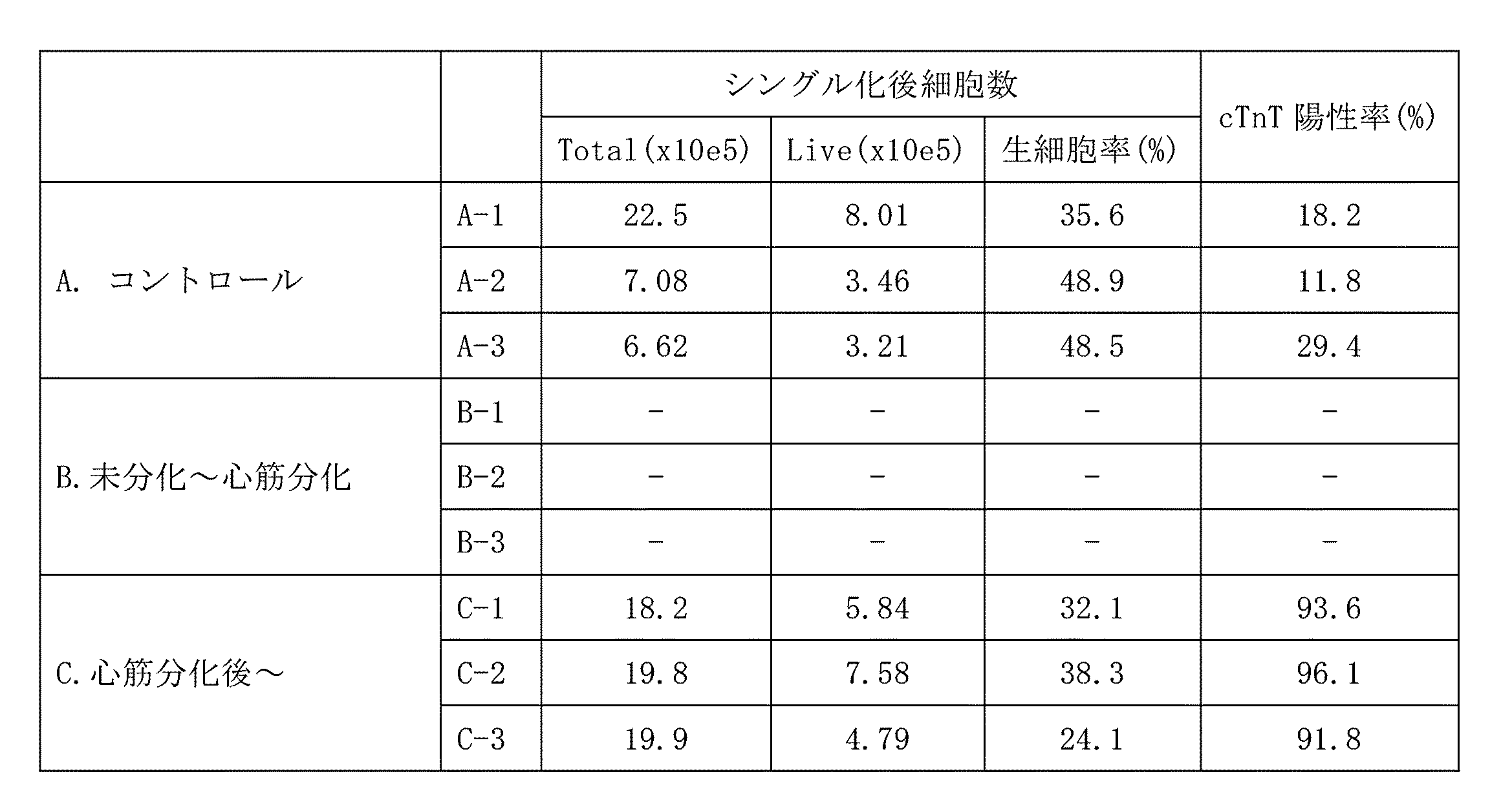
実施例2:
方法(AFP10 μg/mL添加)
iPS細胞(253G1株)をiMatrix-511(マトリクソーム)でコートした10 cmディッシュ(Falcon, 353003)でEssential8を用いて接着培養した。80~90%コンフルエントになったところでEDTAで剥離し、低接着の6ウェルプレート(Corning, 3471)に約1x10⁶個細胞/ウェルで播種し、2日間培養してiPS細胞スフェロイドを形成した。
Example 2:
Method (
iPS cells (strain 253G1) were adherently cultured on a 10 cm dish (Falcon, 353003) coated with iMatrix-511 (matrixome) using Essential8. When 80-90% confluent, the cells were detached with EDTA, seeded in a low-adhesion 6-well plate (Corning, 3471) at approximately 1×10 6 cells/well, and cultured for 2 days to form iPS cell spheroids.
心筋細胞への分化誘導方法は、基本的にWO2015/182765に記載の方法にしたがった(実施例1参照)。実施例1にしたがい、以下のA~Dの条件(各々n=3)でiPS細胞の分化誘導を行った。αフェトプロテイン(AFP)を含む組成物は、ヒト臍帯血由来AFP(BioVision、P1585-1000)を使用した。
(A)AFP無添加(コントロール)、
(B)分化誘導開始後day0~6までの期間AFPを終濃度10 μg/mLで添加、
(C)day7~12までの期間AFPを終濃度10 μg/mLで添加
(D)day13~18までの期間AFPを終濃度10 μg/mLで添加
The method of inducing differentiation into cardiomyocytes basically followed the method described in WO2015/182765 (see Example 1). In accordance with Example 1, iPS cell differentiation was induced under the following conditions A to D (each n=3). A composition containing α-fetoprotein (AFP) used human cord blood-derived AFP (BioVision, P1585-1000).
(A) No AFP added (control),
(B) AFP was added at a final concentration of 10 μg/mL from
(C) Addition of AFP at a final concentration of 10 μg/mL from
day28で細胞を回収し、半量をmiRNeasy Mini Kit(キアゲン)を用いてRNAを回収し定量的PCR法(PowerUp SYBR Green Master Mix、サーモフィッシャー)を用いて、心房筋マーカー遺伝子であるHuman myosin light chain 7(MYL7)遺伝子の発現量を、TATA-Box Binding Protein (TBP)遺伝子を内部標準として解析した。残り半量をトリプシン処理でシングルセル化し、セルカウントした。その後FACS解析で心筋率(cTnT陽性率)を調べた。FACSにはAccuri C6 plus(BD)を使用した。図2Aにおいて、「d0」「d1」等は、分化誘導開始後day0、day1等を意味する。また、「d3」「d5」等の色付セルは培地交換を行った日を示す。
Cells were harvested on day 28, half of the RNA was harvested using the miRNeasy Mini Kit (Qiagen), and quantitative PCR (PowerUp SYBR Green Master Mix, Thermo Fisher) was performed to detect human myosin light chain, an atrial myosin marker gene. 7 (MYL7) gene expression level was analyzed using the TATA-Box Binding Protein (TBP) gene as an internal standard. The remaining half was treated with trypsin to obtain single cells, and the cells were counted. After that, the myocardial rate (cTnT positive rate) was examined by FACS analysis. Accuri C6 plus (BD) was used for FACS. In FIG. 2A, "d0", "d1", etc.
結果
10 μg/mL添加濃度では、50 μg/mL添加濃度の時と同様に、条件B(d0~6添加)では細胞死が起こり分化の評価が出来なかった。条件C(d7~12添加)、条件D(d13~18添加)ではコントロールに比べてcTnT陽性細胞率が高く、心筋純度の向上が見られた(表3、図2B)。また心房筋マーカーであるMYL7については、AFP添加の条件C, Dで、心筋純度の向上傾向よりも顕著な増加が見られた(図2C)。心房筋型の心筋細胞に分化していると考えられる。
Results At a concentration of 10 μg/mL, similar to the case of a concentration of 50 μg/mL, cell death occurred under condition B (addition of d0 to 6), and differentiation could not be evaluated. In condition C (addition of d7-12) and condition D (addition of d13-18), the ratio of cTnT-positive cells was higher than in the control, and an improvement in myocardial purity was observed (Table 3, FIG. 2B). In addition, MYL7, an atrial myocardial marker, was found to increase more markedly in AFP-added conditions C and D than myocardial purity tended to improve (Fig. 2C). It is considered that they are differentiated into atrial myocardial cells.
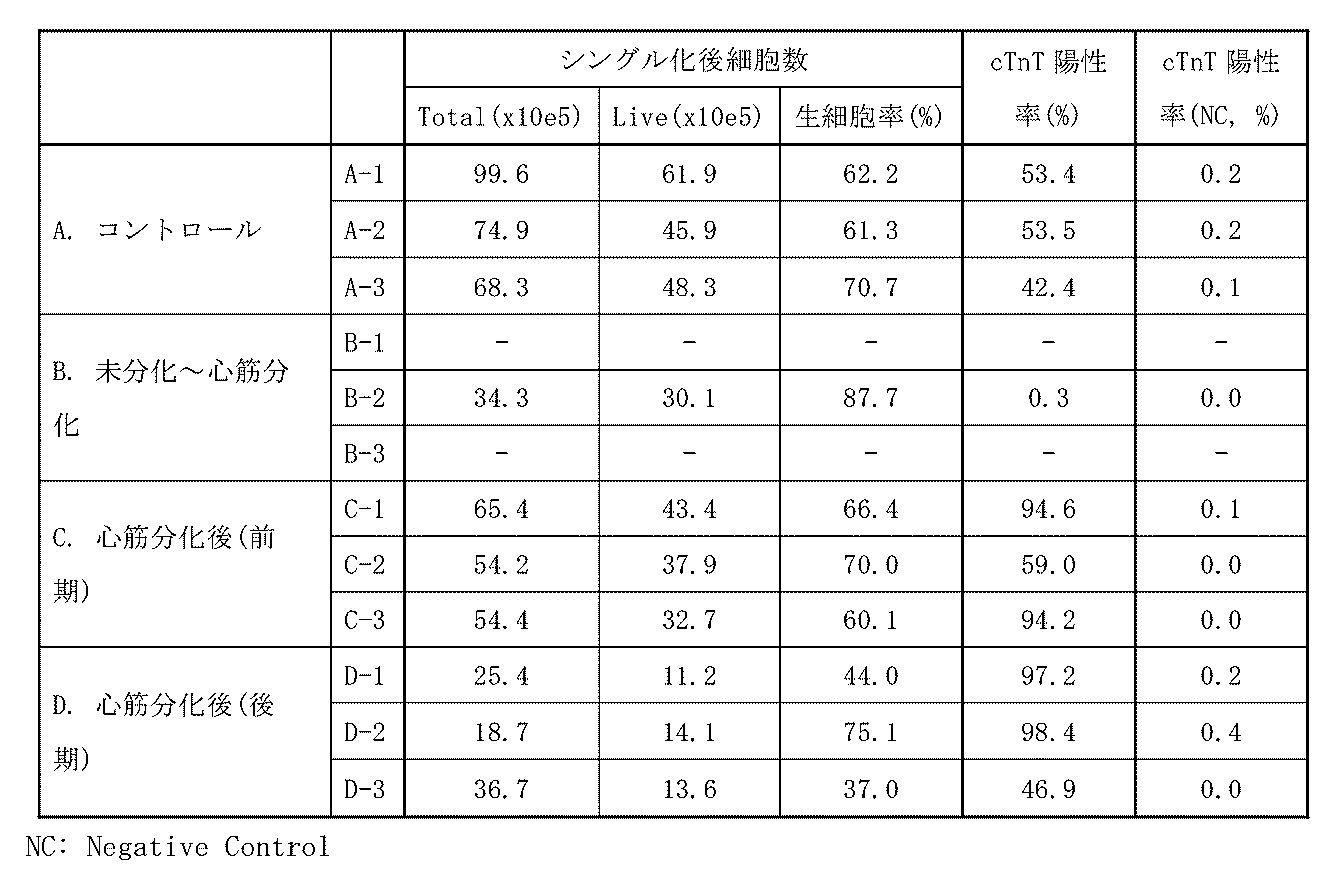
実施例3:
マイオリッジ社の分化誘導法(プロテインフリー分化誘導法:WO2015/182765参照)で作成した凍結心筋細胞(マイオリッジ、H-011106:誘導開始後30日)を解凍し、2%FBSを含む培地で96ウェルプレート(Corning)に播種した。細胞数は1 x104 個細胞/ウェルで5ウェルずつ播種した。4日間培養した後、血清フリーのDMEM(Gibco)培地で交換した。コントロールのウェルはAFPを添加せず、他のウェルには10 μg/mL、1 μg/mLの濃度でAFPを添加した。さらに7日間培養した後、4% パラホルムアルデヒド(Nacalai)50 μLで固定し、0.01% Tween 20/PBS溶液 150 μL/ウェルで1回ウォッシュ後、200 nMのファロイジン-FITC(Sigma), 1 μMのDAPI(Nacalai)を添加し、アクチンフィラメントと細胞核を染色した。その後、各ウェルのファロイジンとDAPIの蛍光画像を共焦点定量イメージサイトメーターCQ1(横河電機)で撮影し、1ウェルあたりの蛍光量を算出した。
Example 3:
Thaw frozen cardiomyocytes (Myoridge, H-011106: 30 days after the start of induction) prepared by Myoridge's differentiation induction method (protein-free differentiation induction method: see WO2015/182765), and 96 wells in medium containing 2% FBS. Plates (Corning) were plated. The number of cells was 1 x 10⁴ cells/well, and 5 wells were seeded. After culturing for 4 days, the medium was replaced with serum-free DMEM (Gibco) medium. AFP was not added to control wells, and AFP was added to other wells at concentrations of 10 μg/mL and 1 μg/mL. After culturing for an additional 7 days, the cells were fixed with 50 μL of 4% paraformaldehyde (Nacalai) and washed once with 150 μL/well of 0.01
結果
コントロールと比べ、AFPを添加したウェルではDAPIの蛍光量に差はなかったが、ファロイジンの蛍光量が有意に増加していた(図3)。ファロイジンはアクチンフィラメントを染色するため、コントロールと比べて細胞骨格が発達した心筋細胞に成熟していることが示唆される。
Results Compared to the control, there was no difference in the fluorescence intensity of DAPI in the wells to which AFP was added, but the fluorescence intensity of phalloidin was significantly increased (Fig. 3). Phalloidin stains actin filaments, suggesting maturation to cardiomyocytes with well-developed cytoskeleton compared to controls.
実施例4:
1.各種AFP試料の細胞に対する影響
分化後の体細胞と比較して、分化前の発生初期の未分化細胞は毒性への感受性が高いことが知られているため、各種AFP試料を用いて、実施例1及び2で生じた心筋分化前の細胞死に対するAFPの影響を検証した。
方法
a. AFPのバッファー置換
Amicon 10KカラムにBioVision, LeeBioのAFP溶液をそれぞれ添加し、4℃, 14,000 x g, 20分で遠心した。この時落ちたろ液は回収し、添加試験に用いた(一次ろ液)。30 μl程度まで濃縮されたので、最大容量の500 μlになるように470 μlの血清フリーのDMEM(Gibco)培地を添加し、4℃, 14,000 x g, 20分で遠心した。この操作を2回繰り返した。再度470 μlの血清フリーのDMEM(Gibco)培地を添加して軽く混ぜ、カラムをひっくり返して遠心して回収した。この時の遠心条件は4℃, 14,000 x g, 2分。0.22 μmフィルターを用いて滅菌し、添加試験へ進んだ。
Example 4:
1. Effect of Various AFP Samples on Cells Compared to differentiated somatic cells, undifferentiated cells in the early stage of development before differentiation are known to be more sensitive to toxicity. The effect of AFP on cell death before myocardial differentiation that occurred in 1 and 2 was verified.
Method
a. Buffer Substitution of AFP BioVision and LeeBio AFP solutions were added to an Amicon 10K column and centrifuged at 4° C., 14,000×g for 20 minutes. The filtrate dropped at this time was collected and used for the addition test (primary filtrate). Since it was concentrated to about 30 μl, 470 μl of serum-free DMEM (Gibco) medium was added to bring the maximum volume to 500 μl, followed by centrifugation at 4° C., 14,000×g, 20 minutes. This operation was repeated twice. 470 μl of serum-free DMEM (Gibco) medium was added again, mixed gently, the column was turned upside down and centrifuged to recover. The centrifugation conditions at this time are 4℃, 14,000 x g, 2 minutes. Sterilize using a 0.22 μm filter and proceed to spiking test.
b. 心筋細胞への添加
マイオリッジ社の分化誘導法(プロテインフリー分化誘導法:WO2015/182765参照)で作製した凍結心筋細胞(マイオリッジ、H-011106:誘導開始後30日)を解凍し、24ウェルプレート(Corning)に播種した。細胞数は1x106個細胞/ウェル。5日間培養した後にAccumax(Nacalai)で剥離し、96ウェルプレート(Corning)に播種した。細胞数は8 x103個細胞/ウェルで5ウェルずつ播種した。4日間培養した後、血清フリーのDMEM(Gibco)培地で交換した。条件は次の通り。
1. Control: 添加無し
2. HT-AFP: HyTest製AFP(粉末)
3. BV-AFP原液: BioVision製AFP(液体)1mg/mLを10ug/mLとなるよう添加(1%と同じ割合)
4. BV-AFP置換: バッファー置換したもの
5. BV-AFろ液: バッファー置換の際に得られた一次ろ液を1%となるよう添加
6. PBS: PBSを1%となるよう添加
b. Addition to cardiomyocytes Thaw frozen cardiomyocytes (Myoridge, H-011106: 30 days after the start of induction) prepared by Myoridge's differentiation induction method (protein-free differentiation induction method: see WO2015/182765) and add to 24 wells. Plates (Corning) were plated. The number of cells is 1x10⁶ cells/well. After culturing for 5 days, the cells were detached with Accumax (Nacalai) and seeded in a 96-well plate (Corning). The number of cells was 8 x 10 3 cells/well, and 5 wells were seeded. After culturing for 4 days, the medium was replaced with serum-free DMEM (Gibco) medium. The conditions are as follows.
1. Control: No additive
2. HT-AFP: HyTest AFP (powder)
3. BV-AFP stock solution: BioVision AFP (liquid) 1mg/mL added to 10ug/mL (same ratio as 1%)
4. BV-AFP displacement: with buffer displacement
5. BV-AF filtrate: Add the primary filtrate obtained during buffer replacement to 1%
6. PBS: Add PBS to 1%
さらに5日間培養した後、4% パラホルムアルデヒド(Nacalai)50 μLで固定し、0.01% Tween 20/PBS溶液 150 μL/ウェルで1回ウォッシュ後、200 nMのファロイジン-FITC(Sigma)を添加し、アクチンフィラメントを染色した。4℃で一晩静置した後、染色液を全量除いてPBSに置換し、各ウェルのファロイジンの蛍光画像を共焦点定量イメージサイトメーターCQ1(横河電機)で撮影し、1ウェルあたりのファロイジンの蛍光面積を算出した。
After culturing for an additional 5 days, fix with 50 μL of 4% paraformaldehyde (Nacalai), wash once with 150 μL/well of 0.01
結果
BV-AFPを原液のまま用いると、何も添加しない条件とほぼ同じファロイジン面積値(細胞面積値を示す)となり添加効果が見られなかったが、置換した場合は細胞面積値が上昇し、HT-AFPの値に近づいた(図4)。また、ろ液を添加した場合は細胞死が起こった。以上のことから、BV-AFPは製品のバッファーに細胞毒性がある可能性が非常に高いと考えられる。一方でHT-AFPは粉末をそのまま希釈して添加しても、何も添加しない条件と比べて高い値を示したことから、毒性はほとんど含まれていないことが示唆された(図4)。以上の結果から、HT-AFPを用いて、分化誘導開始後day0~6におけるAFP添加の影響を再度試験した。
Results When the undiluted BV-AFP solution was used, the phalloidin area value (indicating the cell area value) was almost the same as when nothing was added, and no addition effect was observed. It approached the value of HT-AFP (Fig. 4). Moreover, cell death occurred when the filtrate was added. From the above, it is considered that BV-AFP is highly likely to be cytotoxic in the product buffer. On the other hand, HT-AFP showed higher values even when the powder was diluted and added as it was compared to the condition where nothing was added, suggesting that it contains almost no toxicity (Fig. 4). Based on the above results, using HT-AFP, the influence of AFP addition on
2.分化誘導開始後day0~6におけるAFP添加の心筋分化に与える影響
方法(AFP 30 μg/mL添加)
iPS細胞(253G1株)をiMatrix-511(マトリクソーム)でコートした10 cmディッシュ(Falcon, 353003)でEssential8を用いて接着培養した。80~90%コンフルエントになったところでEDTAを用いて剥離し、低接着の6 cmディッシュ(Corning, 3261)10枚に約2 x10⁶個細胞/ディッシュで播種し、1日培養してiPS細胞スフェロイドを形成した。得られたiPS細胞スフェロイドを回収し、全量を低接着の6ウェルプレート(Corning, 3471)の21ウェルに均等になるように再播種した。
2. Effect of AFP addition on myocardial differentiation from
iPS cells (strain 253G1) were adherently cultured on a 10 cm dish (Falcon, 353003) coated with iMatrix-511 (matrixome) using Essential8. When 80-90% confluent, detached with EDTA, seeded on 10 low-adhesion 6-cm dishes (Corning, 3261) at approximately 2 x 10 6 cells/dish, cultured for 1 day, and iPS cell spheroids. formed. The resulting iPS cell spheroids were harvested and the total amount was evenly replated in 21 wells of a low-adhesion 6-well plate (Corning, 3471).
心筋細胞への分化誘導方法は、基本的にWO2015/182765に記載の方法にしたがった(実施例1参照)。実施例1にしたがい、以下のA~Dの条件(各々n=3)でiPS細胞の分化誘導を行った。αフェトプロテイン(AFP)を含む組成物は、ヒト臍帯血由来AFP(HyTest、8F8)を使用した。
(A)AFP無添加(コントロール)
(B)分化誘導開始後day0~2までの期間AFPを終濃度30 μg/mLで添加
(C)day3~6までの期間AFPを終濃度30 μg/mLで添加
(D)day0~6までの期間AFPを終濃度30 μg/mLで添加
The method of inducing differentiation into cardiomyocytes basically followed the method described in WO2015/182765 (see Example 1). In accordance with Example 1, iPS cell differentiation was induced under the following conditions A to D (each n=3). A composition containing α-fetoprotein (AFP) used human cord blood-derived AFP (HyTest, 8F8).
(A) No AFP added (control)
(B) Addition of AFP at a final concentration of 30 μg/mL from
day18で細胞を回収し、半量をmiRNeasy Mini Kit(キアゲン)を用いてRNAを回収し定量的PCR法(PowerUp SYBR Green Master Mix、サーモフィッシャー)を用いて、各種遺伝子の発現量を、TATA-Box Binding Protein(TBP)遺伝子を内部標準として解析した。残り半量をトリプシン処理でシングルセル化し、セルカウントした。その後FACS解析で心筋率(cTnT陽性率)を調べた。FACSにはAccuri C6 plus(BD)を使用した。 Cells were harvested on day 18, half of the RNA was harvested using the miRNeasy Mini Kit (Qiagen), and quantitative PCR (PowerUp SYBR Green Master Mix, Thermo Fisher) was used to measure the expression levels of various genes using the TATA-Box. The Binding Protein (TBP) gene was analyzed as an internal standard. The remaining half was treated with trypsin to obtain single cells, and the cells were counted. After that, the myocardial rate (cTnT positive rate) was examined by FACS analysis. Accuri C6 plus (BD) was used for FACS.
結果
得られた心筋細胞について、コントロールと比較して細胞数の大きな変動はどの条件でも起こらなかった。一方で、条件B-Dでは心筋率が優位に増加していた。また、定量的PCR法を用いた解析では、条件Dで数種類のチャネル系遺伝子とα,β-MHC、MYL7の発現量が増加しており、コントロールや他のAFP条件と比べて発達した心筋細胞になっていたことが示唆される(図5)。細胞数と心筋率、遺伝子発現を総合的に評価すると、条件Dが最も良い結果を示していた。このことから、少なくとも分化誘導初期の段階において、期間を限定せずにAFPを添加し続けることで、心筋細胞の分化や成熟に有用に働く可能性が示された。
Results Regarding the cardiomyocytes obtained, no large fluctuations in the number of cells occurred under any condition compared to the control. On the other hand, the myocardial fraction increased significantly in condition BD. In addition, quantitative PCR analysis revealed that the expression levels of several channel genes, α,β-MHC, and MYL7 were increased in condition D, indicating that cardiomyocytes developed more than controls and other AFP conditions. (Fig. 5). Comprehensive evaluation of cell count, myocardium rate, and gene expression showed that condition D showed the best results. From this, at least at the initial stage of induction of differentiation, continuous addition of AFP for an unlimited period of time was shown to be useful for the differentiation and maturation of cardiomyocytes.

実施例5:
方法(AFP 30 μg/mL添加)
マイオリッジ社の分化誘導法(プロテインフリー分化誘導法:WO2015/182765参照)で作製した凍結心筋細胞(マイオリッジ、H-011106:誘導開始後30日)を解凍し、血清を含まない培地で96ウェルプレート(Corning)に播種した。細胞数は2 x104 個細胞/ウェルで5ウェルずつ播種した。8日間培養した後、血清フリーのDMEM(Gibco)培地で交換した。αフェトプロテイン(AFP)を含む組成物は、ヒト臍帯血由来AFP(HyTest、8F8)を使用した。条件は次の通り。
(A)無添加(コントロール)
(B)2% FBS
(C)HSA 30 μg/mL
(D)AFP(臍帯血由来) 30 μg/mL
(E)AFP(HEK組換え) 30 μg/mL
(F)AFP(臍帯血由来)+ HSA 各15 μg/mL
Example 5:
Method (
Thaw frozen cardiomyocytes (Myoridge, H-011106: 30 days after the start of induction) prepared by Myoridge's differentiation induction method (protein-free differentiation induction method: see WO2015/182765) and plate in a 96-well plate with serum-free medium. (Corning). The number of cells was 2 x 10⁴ cells/well, and 5 wells were seeded. After 8 days of culture, the medium was replaced with serum-free DMEM (Gibco) medium. A composition containing α-fetoprotein (AFP) used human cord blood-derived AFP (HyTest, 8F8). The conditions are as follows.
(A) No additive (control)
(B) 2% FBS
(C)
(D) AFP (from cord blood) 30 μg/mL
(E) AFP (HEK recombinant) 30 μg/mL
(F) AFP (from cord blood) +
さらに7日間培養した後、4% パラホルムアルデヒド(Nacalai)50 μLで固定し、0.01% Tween 20/PBS溶液 150 μL/ウェルで1回ウォッシュ後、200 nMのファロイジン-FITC(Sigma)、400 ng/mlのcTnT一次抗体(Santa Cruz) 1μMのDAPI(Nacalai)を添加し、アクチンフィラメントと細胞核を染色した。4℃で一晩静置した後、染色液を全量除いてcTnT二次抗体-Alexa Flour 647(Thermo Fisher)を添加し、室温で30分反応させて心筋トロポニンTを染色した。その後PBSに置換し、各ウェルのファロイジン、cTnT及びDAPIの蛍光画像を共焦点定量イメージサイトメーターCQ1(横河電機)で撮影し、1ウェルあたりのファロイジン、cTnTの蛍光量及びDAPIの個数を算出した。
After culturing for an additional 7 days, fix with 50 μL of 4% paraformaldehyde (Nacalai), wash once with 150 μL/well of 0.01
結果
コントロールやFBS添加条件と比べ、AFPを添加したウェルではcTnTの蛍光量とDAPIの個数が有意に増加していた(図6)。このことから、AFPを添加した心筋細胞は心筋線維が発達し、成熟化が進んだ細胞になっていることが示唆される。またその細胞数も多く、心筋細胞を生存させる効果も示された。
Results Compared to the control and FBS-added wells, the amount of cTnT fluorescence and the number of DAPI were significantly increased in the wells to which AFP was added (Fig. 6). This suggests that cardiomyocytes to which AFP was added developed myocardial fibers and matured into advanced cells. In addition, the number of cells was large, and the effect of reviving cardiomyocytes was also shown.
本発明は、再生医療のための心筋細胞の調製に有用である。 The present invention is useful for preparing cardiomyocytes for regenerative medicine.
本明細書中で引用した全ての刊行物、特許及び特許出願をそのまま参考として本明細書中にとり入れるものとする。 All publications, patents and patent applications cited in this specification shall be incorporated herein by reference.
Claims (17)
A culture of a cell population comprising cardiomyocytes, said culture comprising alpha-fetoprotein and free of serum.
Priority Applications (2)
| Application Number | Priority Date | Filing Date | Title |
|---|---|---|---|
| JP2023534816A JPWO2023286772A1 (en) | 2021-07-12 | 2022-07-12 | |
| US18/411,134 US20240309327A1 (en) | 2021-07-12 | 2024-01-12 | Cardiomyocyte production method |
Applications Claiming Priority (2)
| Application Number | Priority Date | Filing Date | Title |
|---|---|---|---|
| JP2021114913 | 2021-07-12 | ||
| JP2021-114913 | 2021-07-12 |
Related Child Applications (1)
| Application Number | Title | Priority Date | Filing Date |
|---|---|---|---|
| US18/411,134 Continuation-In-Part US20240309327A1 (en) | 2021-07-12 | 2024-01-12 | Cardiomyocyte production method |
Publications (1)
| Publication Number | Publication Date |
|---|---|
| WO2023286772A1 true WO2023286772A1 (en) | 2023-01-19 |
Family
ID=84919430
Family Applications (1)
| Application Number | Title | Priority Date | Filing Date |
|---|---|---|---|
| PCT/JP2022/027420 Ceased WO2023286772A1 (en) | 2021-07-12 | 2022-07-12 | Cardiomyocyte production method |
Country Status (3)
| Country | Link |
|---|---|
| US (1) | US20240309327A1 (en) |
| JP (1) | JPWO2023286772A1 (en) |
| WO (1) | WO2023286772A1 (en) |
Cited By (1)
| Publication number | Priority date | Publication date | Assignee | Title |
|---|---|---|---|---|
| WO2025170008A1 (en) * | 2024-02-07 | 2025-08-14 | 国立大学法人京都大学 | Method for producing mature cardiomyocytes |
Citations (4)
| Publication number | Priority date | Publication date | Assignee | Title |
|---|---|---|---|---|
| US20100081200A1 (en) * | 2007-06-05 | 2010-04-01 | Kristiina Rajala | Formulations and methods for culturing stem cells |
| US20100317104A1 (en) * | 2009-04-10 | 2010-12-16 | Australian Stem Cell Centre Limited | Cell culture media |
| JP2013512667A (en) * | 2009-12-04 | 2013-04-18 | クリスティーナ、ラジャラ | Formulations and methods for culturing stem cells |
| WO2015182765A1 (en) * | 2014-05-30 | 2015-12-03 | 国立大学法人京都大学 | Method for inducing myocardial differentiation of pluripotent stem cells using low-molecular compound |
-
2022
- 2022-07-12 JP JP2023534816A patent/JPWO2023286772A1/ja active Pending
- 2022-07-12 WO PCT/JP2022/027420 patent/WO2023286772A1/en not_active Ceased
-
2024
- 2024-01-12 US US18/411,134 patent/US20240309327A1/en active Pending
Patent Citations (4)
| Publication number | Priority date | Publication date | Assignee | Title |
|---|---|---|---|---|
| US20100081200A1 (en) * | 2007-06-05 | 2010-04-01 | Kristiina Rajala | Formulations and methods for culturing stem cells |
| US20100317104A1 (en) * | 2009-04-10 | 2010-12-16 | Australian Stem Cell Centre Limited | Cell culture media |
| JP2013512667A (en) * | 2009-12-04 | 2013-04-18 | クリスティーナ、ラジャラ | Formulations and methods for culturing stem cells |
| WO2015182765A1 (en) * | 2014-05-30 | 2015-12-03 | 国立大学法人京都大学 | Method for inducing myocardial differentiation of pluripotent stem cells using low-molecular compound |
Cited By (1)
| Publication number | Priority date | Publication date | Assignee | Title |
|---|---|---|---|---|
| WO2025170008A1 (en) * | 2024-02-07 | 2025-08-14 | 国立大学法人京都大学 | Method for producing mature cardiomyocytes |
Also Published As
| Publication number | Publication date |
|---|---|
| JPWO2023286772A1 (en) | 2023-01-19 |
| US20240309327A1 (en) | 2024-09-19 |
Similar Documents
| Publication | Publication Date | Title |
|---|---|---|
| US8507275B2 (en) | Method of inducing differentiation of embryonic stem cells into hemangioblast | |
| US8318489B2 (en) | Prostacyclin directed differentiation of cardiomyocytes from human embryonic stem cells | |
| KR101861171B1 (en) | Cardiomyocyte medium with dialyzed serum | |
| JP2021510527A (en) | Methods for Differentiation of Human Pluripotent Stem Cell Lines in Suspension Culture | |
| JP2017140052A (en) | Creation of myocardial cell | |
| JPWO2015182765A1 (en) | Induction of myocardial differentiation of pluripotent stem cells using low molecular weight compounds | |
| JPWO2014168264A1 (en) | Method for inducing alveolar epithelial progenitor cells | |
| JP2018532389A (en) | Proliferation of epithelial cells ex vivo | |
| JPWO2019093340A1 (en) | Method for inducing primitive endoderm from naive pluripotent stem cells | |
| JP7357369B2 (en) | Novel renal progenitor cell marker and method for enriching renal progenitor cells using it | |
| US9238795B2 (en) | Populations of smooth muscle cells of specific embryonic lineages | |
| WO2018190305A1 (en) | Method for producing differentiated cell spheroids | |
| US20240309327A1 (en) | Cardiomyocyte production method | |
| JPWO2020090903A1 (en) | Method for producing pluripotent stem cells whose differentiation resistance to the mesodermal system has been released | |
| WO2020095423A1 (en) | Method for producing kidney structure having dendritically branched collecting duct from pluripotent stem cells | |
| JP7541748B2 (en) | Method for enriching cardiomyocytes | |
| JP7344486B2 (en) | Cardiomyocyte maturation promoter | |
| JP2024531682A (en) | Methods for producing committed cardiac progenitor cells | |
| JP6305186B2 (en) | Method for producing blood cells or cardiomyocytes from pluripotent stem cells |
Legal Events
| Date | Code | Title | Description |
|---|---|---|---|
| 121 | Ep: the epo has been informed by wipo that ep was designated in this application |
Ref document number: 22842115 Country of ref document: EP Kind code of ref document: A1 |
|
| WWE | Wipo information: entry into national phase |
Ref document number: 2023534816 Country of ref document: JP |
|
| NENP | Non-entry into the national phase |
Ref country code: DE |
|
| 122 | Ep: pct application non-entry in european phase |
Ref document number: 22842115 Country of ref document: EP Kind code of ref document: A1 |