WO2023235754A1 - Combination therapies for treating urothelial carcinoma - Google Patents
Combination therapies for treating urothelial carcinoma Download PDFInfo
- Publication number
- WO2023235754A1 WO2023235754A1 PCT/US2023/067697 US2023067697W WO2023235754A1 WO 2023235754 A1 WO2023235754 A1 WO 2023235754A1 US 2023067697 W US2023067697 W US 2023067697W WO 2023235754 A1 WO2023235754 A1 WO 2023235754A1
- Authority
- WO
- WIPO (PCT)
- Prior art keywords
- domain
- sirpα
- variant
- polypeptide
- seq
- Prior art date
- Legal status (The legal status is an assumption and is not a legal conclusion. Google has not performed a legal analysis and makes no representation as to the accuracy of the status listed.)
- Ceased
Links
Classifications
-
- A—HUMAN NECESSITIES
- A61—MEDICAL OR VETERINARY SCIENCE; HYGIENE
- A61K—PREPARATIONS FOR MEDICAL, DENTAL OR TOILETRY PURPOSES
- A61K38/00—Medicinal preparations containing peptides
- A61K38/16—Peptides having more than 20 amino acids; Gastrins; Somatostatins; Melanotropins; Derivatives thereof
- A61K38/17—Peptides having more than 20 amino acids; Gastrins; Somatostatins; Melanotropins; Derivatives thereof from animals; from humans
- A61K38/177—Receptors; Cell surface antigens; Cell surface determinants
- A61K38/1774—Immunoglobulin superfamily (e.g. CD2, CD4, CD8, ICAM molecules, B7 molecules, Fc-receptors, MHC-molecules)
-
- A—HUMAN NECESSITIES
- A61—MEDICAL OR VETERINARY SCIENCE; HYGIENE
- A61K—PREPARATIONS FOR MEDICAL, DENTAL OR TOILETRY PURPOSES
- A61K39/00—Medicinal preparations containing antigens or antibodies
- A61K39/395—Antibodies; Immunoglobulins; Immune serum, e.g. antilymphocytic serum
- A61K39/39533—Antibodies; Immunoglobulins; Immune serum, e.g. antilymphocytic serum against materials from animals
- A61K39/3955—Antibodies; Immunoglobulins; Immune serum, e.g. antilymphocytic serum against materials from animals against proteinaceous materials, e.g. enzymes, hormones, lymphokines
-
- A—HUMAN NECESSITIES
- A61—MEDICAL OR VETERINARY SCIENCE; HYGIENE
- A61K—PREPARATIONS FOR MEDICAL, DENTAL OR TOILETRY PURPOSES
- A61K47/00—Medicinal preparations characterised by the non-active ingredients used, e.g. carriers or inert additives; Targeting or modifying agents chemically bound to the active ingredient
- A61K47/50—Medicinal preparations characterised by the non-active ingredients used, e.g. carriers or inert additives; Targeting or modifying agents chemically bound to the active ingredient the non-active ingredient being chemically bound to the active ingredient, e.g. polymer-drug conjugates
- A61K47/51—Medicinal preparations characterised by the non-active ingredients used, e.g. carriers or inert additives; Targeting or modifying agents chemically bound to the active ingredient the non-active ingredient being chemically bound to the active ingredient, e.g. polymer-drug conjugates the non-active ingredient being a modifying agent
- A61K47/68—Medicinal preparations characterised by the non-active ingredients used, e.g. carriers or inert additives; Targeting or modifying agents chemically bound to the active ingredient the non-active ingredient being chemically bound to the active ingredient, e.g. polymer-drug conjugates the non-active ingredient being a modifying agent the modifying agent being an antibody, an immunoglobulin or a fragment thereof, e.g. an Fc-fragment
- A61K47/6801—Drug-antibody or immunoglobulin conjugates defined by the pharmacologically or therapeutically active agent
- A61K47/6803—Drugs conjugated to an antibody or immunoglobulin, e.g. cisplatin-antibody conjugates
- A61K47/68031—Drugs conjugated to an antibody or immunoglobulin, e.g. cisplatin-antibody conjugates the drug being an auristatin
-
- A—HUMAN NECESSITIES
- A61—MEDICAL OR VETERINARY SCIENCE; HYGIENE
- A61K—PREPARATIONS FOR MEDICAL, DENTAL OR TOILETRY PURPOSES
- A61K47/00—Medicinal preparations characterised by the non-active ingredients used, e.g. carriers or inert additives; Targeting or modifying agents chemically bound to the active ingredient
- A61K47/50—Medicinal preparations characterised by the non-active ingredients used, e.g. carriers or inert additives; Targeting or modifying agents chemically bound to the active ingredient the non-active ingredient being chemically bound to the active ingredient, e.g. polymer-drug conjugates
- A61K47/51—Medicinal preparations characterised by the non-active ingredients used, e.g. carriers or inert additives; Targeting or modifying agents chemically bound to the active ingredient the non-active ingredient being chemically bound to the active ingredient, e.g. polymer-drug conjugates the non-active ingredient being a modifying agent
- A61K47/68—Medicinal preparations characterised by the non-active ingredients used, e.g. carriers or inert additives; Targeting or modifying agents chemically bound to the active ingredient the non-active ingredient being chemically bound to the active ingredient, e.g. polymer-drug conjugates the non-active ingredient being a modifying agent the modifying agent being an antibody, an immunoglobulin or a fragment thereof, e.g. an Fc-fragment
- A61K47/6801—Drug-antibody or immunoglobulin conjugates defined by the pharmacologically or therapeutically active agent
- A61K47/6803—Drugs conjugated to an antibody or immunoglobulin, e.g. cisplatin-antibody conjugates
- A61K47/6811—Drugs conjugated to an antibody or immunoglobulin, e.g. cisplatin-antibody conjugates the drug being a protein or peptide, e.g. transferrin or bleomycin
-
- A—HUMAN NECESSITIES
- A61—MEDICAL OR VETERINARY SCIENCE; HYGIENE
- A61K—PREPARATIONS FOR MEDICAL, DENTAL OR TOILETRY PURPOSES
- A61K47/00—Medicinal preparations characterised by the non-active ingredients used, e.g. carriers or inert additives; Targeting or modifying agents chemically bound to the active ingredient
- A61K47/50—Medicinal preparations characterised by the non-active ingredients used, e.g. carriers or inert additives; Targeting or modifying agents chemically bound to the active ingredient the non-active ingredient being chemically bound to the active ingredient, e.g. polymer-drug conjugates
- A61K47/51—Medicinal preparations characterised by the non-active ingredients used, e.g. carriers or inert additives; Targeting or modifying agents chemically bound to the active ingredient the non-active ingredient being chemically bound to the active ingredient, e.g. polymer-drug conjugates the non-active ingredient being a modifying agent
- A61K47/68—Medicinal preparations characterised by the non-active ingredients used, e.g. carriers or inert additives; Targeting or modifying agents chemically bound to the active ingredient the non-active ingredient being chemically bound to the active ingredient, e.g. polymer-drug conjugates the non-active ingredient being a modifying agent the modifying agent being an antibody, an immunoglobulin or a fragment thereof, e.g. an Fc-fragment
- A61K47/6835—Medicinal preparations characterised by the non-active ingredients used, e.g. carriers or inert additives; Targeting or modifying agents chemically bound to the active ingredient the non-active ingredient being chemically bound to the active ingredient, e.g. polymer-drug conjugates the non-active ingredient being a modifying agent the modifying agent being an antibody, an immunoglobulin or a fragment thereof, e.g. an Fc-fragment the modifying agent being an antibody or an immunoglobulin bearing at least one antigen-binding site
- A61K47/6849—Medicinal preparations characterised by the non-active ingredients used, e.g. carriers or inert additives; Targeting or modifying agents chemically bound to the active ingredient the non-active ingredient being chemically bound to the active ingredient, e.g. polymer-drug conjugates the non-active ingredient being a modifying agent the modifying agent being an antibody, an immunoglobulin or a fragment thereof, e.g. an Fc-fragment the modifying agent being an antibody or an immunoglobulin bearing at least one antigen-binding site the antibody targeting a receptor, a cell surface antigen or a cell surface determinant
-
- A—HUMAN NECESSITIES
- A61—MEDICAL OR VETERINARY SCIENCE; HYGIENE
- A61K—PREPARATIONS FOR MEDICAL, DENTAL OR TOILETRY PURPOSES
- A61K47/00—Medicinal preparations characterised by the non-active ingredients used, e.g. carriers or inert additives; Targeting or modifying agents chemically bound to the active ingredient
- A61K47/50—Medicinal preparations characterised by the non-active ingredients used, e.g. carriers or inert additives; Targeting or modifying agents chemically bound to the active ingredient the non-active ingredient being chemically bound to the active ingredient, e.g. polymer-drug conjugates
- A61K47/51—Medicinal preparations characterised by the non-active ingredients used, e.g. carriers or inert additives; Targeting or modifying agents chemically bound to the active ingredient the non-active ingredient being chemically bound to the active ingredient, e.g. polymer-drug conjugates the non-active ingredient being a modifying agent
- A61K47/68—Medicinal preparations characterised by the non-active ingredients used, e.g. carriers or inert additives; Targeting or modifying agents chemically bound to the active ingredient the non-active ingredient being chemically bound to the active ingredient, e.g. polymer-drug conjugates the non-active ingredient being a modifying agent the modifying agent being an antibody, an immunoglobulin or a fragment thereof, e.g. an Fc-fragment
- A61K47/6889—Conjugates wherein the antibody being the modifying agent and wherein the linker, binder or spacer confers particular properties to the conjugates, e.g. peptidic enzyme-labile linkers or acid-labile linkers, providing for an acid-labile immuno conjugate wherein the drug may be released from its antibody conjugated part in an acidic, e.g. tumoural or environment
-
- A—HUMAN NECESSITIES
- A61—MEDICAL OR VETERINARY SCIENCE; HYGIENE
- A61P—SPECIFIC THERAPEUTIC ACTIVITY OF CHEMICAL COMPOUNDS OR MEDICINAL PREPARATIONS
- A61P35/00—Antineoplastic agents
-
- C—CHEMISTRY; METALLURGY
- C07—ORGANIC CHEMISTRY
- C07K—PEPTIDES
- C07K14/00—Peptides having more than 20 amino acids; Gastrins; Somatostatins; Melanotropins; Derivatives thereof
- C07K14/435—Peptides having more than 20 amino acids; Gastrins; Somatostatins; Melanotropins; Derivatives thereof from animals; from humans
- C07K14/705—Receptors; Cell surface antigens; Cell surface determinants
- C07K14/70503—Immunoglobulin superfamily
-
- C—CHEMISTRY; METALLURGY
- C07—ORGANIC CHEMISTRY
- C07K—PEPTIDES
- C07K16/00—Immunoglobulins [IGs], e.g. monoclonal or polyclonal antibodies
- C07K16/18—Immunoglobulins [IGs], e.g. monoclonal or polyclonal antibodies against material from animals or humans
- C07K16/28—Immunoglobulins [IGs], e.g. monoclonal or polyclonal antibodies against material from animals or humans against receptors, cell surface antigens or cell surface determinants
- C07K16/2803—Immunoglobulins [IGs], e.g. monoclonal or polyclonal antibodies against material from animals or humans against receptors, cell surface antigens or cell surface determinants against the immunoglobulin superfamily
-
- A—HUMAN NECESSITIES
- A61—MEDICAL OR VETERINARY SCIENCE; HYGIENE
- A61K—PREPARATIONS FOR MEDICAL, DENTAL OR TOILETRY PURPOSES
- A61K39/00—Medicinal preparations containing antigens or antibodies
- A61K2039/505—Medicinal preparations containing antigens or antibodies comprising antibodies
-
- A—HUMAN NECESSITIES
- A61—MEDICAL OR VETERINARY SCIENCE; HYGIENE
- A61K—PREPARATIONS FOR MEDICAL, DENTAL OR TOILETRY PURPOSES
- A61K39/00—Medicinal preparations containing antigens or antibodies
- A61K2039/54—Medicinal preparations containing antigens or antibodies characterised by the route of administration
-
- A—HUMAN NECESSITIES
- A61—MEDICAL OR VETERINARY SCIENCE; HYGIENE
- A61K—PREPARATIONS FOR MEDICAL, DENTAL OR TOILETRY PURPOSES
- A61K39/00—Medicinal preparations containing antigens or antibodies
- A61K2039/545—Medicinal preparations containing antigens or antibodies characterised by the dose, timing or administration schedule
-
- A—HUMAN NECESSITIES
- A61—MEDICAL OR VETERINARY SCIENCE; HYGIENE
- A61K—PREPARATIONS FOR MEDICAL, DENTAL OR TOILETRY PURPOSES
- A61K2300/00—Mixtures or combinations of active ingredients, wherein at least one active ingredient is fully defined in groups A61K31/00 - A61K41/00
-
- C—CHEMISTRY; METALLURGY
- C07—ORGANIC CHEMISTRY
- C07K—PEPTIDES
- C07K2317/00—Immunoglobulins specific features
- C07K2317/70—Immunoglobulins specific features characterized by effect upon binding to a cell or to an antigen
- C07K2317/71—Decreased effector function due to an Fc-modification
-
- C—CHEMISTRY; METALLURGY
- C07—ORGANIC CHEMISTRY
- C07K—PEPTIDES
- C07K2317/00—Immunoglobulins specific features
- C07K2317/90—Immunoglobulins specific features characterized by (pharmaco)kinetic aspects or by stability of the immunoglobulin
- C07K2317/92—Affinity (KD), association rate (Ka), dissociation rate (Kd) or EC50 value
-
- C—CHEMISTRY; METALLURGY
- C07—ORGANIC CHEMISTRY
- C07K—PEPTIDES
- C07K2319/00—Fusion polypeptide
- C07K2319/30—Non-immunoglobulin-derived peptide or protein having an immunoglobulin constant or Fc region, or a fragment thereof, attached thereto
Definitions
- the present invention relates to methods of treating cancer that comprise administering an agent that blocks the interaction between CD47 (e.g., hCD47) and SIRP ⁇ (e.g., hSIRP ⁇ ) to an individual in need thereof in combination with an antibody drug conjugate (e.g., enfortumab vedotin).
- an agent that blocks the interaction between CD47 e.g., hCD47
- SIRP ⁇ e.g., hSIRP ⁇
- an antibody drug conjugate e.g., enfortumab vedotin.
- Bladder cancer occurs mainly in people over the age of 55 years with a median age at the time of diagnosis of 73 years. The ratio of men:women who develop this cancer is approximately 4:1. Caucasians are more likely to be diagnosed with bladder cancer than African Americans or Hispanic Americans (SEER Cancer Stat Facts: Bladder Cancer, 2021. National Cancer Institute. Bethesda, MD, https://seer(dot)cancer(dot)gov/statfacts/html/urinb(dot)html. Accessed 09 March 2022).
- bladder cancers are urothelial cancer; if the cancer is advanced at the time of diagnosis, the prognosis is poor (Simeone JC, Nordstrom BL, Patel K, Mann H, Klein AB, Horne L. Treatment patterns and overall survival in metastatic urothelial carcinoma in a real-world, US setting. Cancer Epidemiol.2019;60:121-7). Most urothelial cancers are diagnosed at the non-muscle invasive stage. At this this stage, disease management involves resection with or without intravesicular therapy. Despite such treatment, patients often develop more advanced disease that is incurable, ultimately leading to death. Approximately 12% of patients have locally advanced or metastatic disease at diagnosis (SEER Cancer Stat Facts: Bladder Cancer, 2021.
- First-line therapy for locally advanced or metastatic urothelial cancer in patients with sufficient renal function consists of cisplatin-based combinations, such as combinations with methotrexate, vinblastine, doxorubicin, and cisplatin (MVAC) or gemcitabine plus cisplatin, which demonstrated overall response rates up to 50%, including approximately 10% to 15% complete responses (CRs) (Bellmunt J, Orsola A, Wiegel T, Guix M, De Santis M, Kataja V; ESMO Guidelines Working Group.
- Bladder cancer ESMO Clinical Practice Guidelines for diagnosis, treatment and follow-up. Ann Oncol.2011 Sep; v22 Suppl 6:vi45-9. doi: 10(dot)1093/annonc/mdr376(dot)PMID: 21908503).
- Carboplatin and gemcitabine are commonly used in patients who are ineligible for cisplatin, but outcomes are generally inferior. Despite initial chemosensitivity, patients are not cured, and the outcome of metastatic urothelial cancer after these regimens is poor: median time to progression is only 7 months and median overall survival (OS) is 14 months.
- a method of treating urothelial cancer in an individual comprising administering to the individual (a) an effective amount of a fusion polypeptide comprising a SIRP ⁇ D1 domain variant and an Fc domain variant, and (b) an effective amount of enfortumab vedotin, wherein the SIRP ⁇ D1 domain variant of the fusion polypeptide comprises the amino acid sequence of SEQ ID NO: 81 or SEQ ID NO: 85; and wherein the Fc domain variant of the fusion polypeptide is (i) a human IgG1 Fc region comprising L234A, L235A, G237A, and N297A mutations, wherein numbering is according to the EU index of Kabat; (ii) a human IgG2 Fc region comprising A330S, P331S, and N297A mutations, wherein numbering is according to the EU index of Kabat; (iii) a human Ig
- the individual is a human.
- the urothelial cancer is locally advanced urothelial cancer or metastatic urothelial cancer.
- the urothelial cancer is bladder cancer, renal pelvis cancer, cancer of the ureter, or cancer of the urethra.
- the individual received prior treatment with an immune checkpoint inhibitor (CPI).
- the CPI was a PD-1 inhibitor or a PD-L1 inhibitor.
- the CPI was atezolizumab, pembrolizumab, durvalumab, avelumab, or nivolumab.
- the individual received prior treatment with a platinum-containing chemotherapy. In some embodiments, the individual had progression or recurrence of urothelial cancer during or following receipt of most recent prior therapy. In some embodiments, the individual has not received prior treatment with a monomethylauristatin (MMAE)-based antibody-drug conjugate. In some embodiments, the individual has not received prior treatment with enfortumab vedotin. In some embodiments, the individual has not received prior treatment with a therapeutic agent that blocks the interaction between CD47 and SIRP ⁇ .
- MMAE monomethylauristatin
- the enfortumab vedotin is administered to the individual in one or more 28-day cycles, and wherein the enfortumab vedotin is administered to the individual at a dose of 1.25 mg/kg IV on Days 1, 8 and 15 of each 28-day cycle. In some embodiments, the enfortumab vedotin is administered intravenously. In some embodiments, the fusion polypeptide is administered to the individual at a dose up to about 60 mg/kg. In some embodiments, the fusion polypeptide is administered to the individual at a dose of about 30 mg/kg once every two weeks (q2w).
- the fusion polypeptide is administered at a dose of about 20 mg/kg once every two weeks (q2w). In some embodiments, the fusion polypeptide is administered at a dose of about 15 mg/kg once every two weeks (q2w). In some embodiments, the fusion polypeptide is administered intravenously.
- the SIRP ⁇ D1 domain variant comprises the amino acid sequence of SEQ ID NO: 85. In some embodiments, the SIRP ⁇ D1 domain variant comprises the amino acid sequence of SEQ ID NO: 81.
- the Fc domain variant is a human IgG1 Fc region comprising L234A, L235A, G237A, and N297A mutations, wherein numbering is according to the EU index of Kabat.
- the Fc domain variant comprises the amino acid sequence of SEQ ID NO: 91.
- the fusion polypeptide comprises the amino acid sequence of SEQ ID NO: 136.
- the fusion polypeptide comprises the amino acid sequence of SEQ ID NO: 135.
- fusion polypeptide forms a homodimer.
- kits comprising a polypeptide comprising a SIRP ⁇ D1 domain variant and an Fc domain variant in a pharmaceutically acceptable carrier, for use in combination with enfortumab vedotin for treating urothelial cancer in an individual in need thereof, wherein the SIRP ⁇ D1 domain variant comprises the amino acid sequence of SEQ ID NO: 81 or SEQ ID NO: 85; wherein the Fc domain variant is (i) a human IgG1 Fc region comprising L234A, L235A, G237A, and N297A mutations, wherein numbering is according to the EU index of Kabat; (ii) a human IgG2 Fc region comprising A330S, P331S, and N297A mutations, wherein numbering is according to the EU index of Kabat; (iii) a human IgG4 Fc region comprising S228P, E233P, F234V, L235A, and delG
- FIG 1A shows the results of experiments that were performed to determine whether DRUG A enhances the antibody-dependent cellular phagocytosis (ADCP) activity of DRUG B and DRUG C using macrophages derived from monocytes from a first human donor and T47D ductal carcinoma cells and OE19 esophageal adenocarcinoma cells as target cells.
- ADCP antibody-dependent cellular phagocytosis
- FIG 1B shows the results of experiments that were performed to determine whether DRUG A enhances the antibody-dependent cellular phagocytosis (ADCP) activity of DRUG B and DRUG C using macrophages derived from monocytes obtained from a second human donor and T47D ductal carcinoma cells and OE19 esophageal adenocarcinoma cells as target cells.
- FIG 2 shows the results of experiments that were performed to determine the effects of DRUG A on DRUG B-dependent (left panel) or DRUG C-dependent (right panel) ADCP of OE19 esophageal adenocarcinoma cells by macrophages derived from monocytes obtained from a third human donor.
- FIG 3 shows results from assays that were performed to determine the effects of DRUG A on DRUG B-dependent (left panel) or DRUG C-dependent (right panel) ADCP of HT-1376 bladder carcinoma cells by macrophages derived from monocytes obtained from a third human donor.
- FIG 4 provides a study design for the Phase 1 clinical trial described in Example 3. DETAILED DESCRIPTION [0018] The following description sets forth exemplary methods, parameters and the like. It should be recognized, however, that such description is not intended as a limitation on the scope of the present disclosure but is instead provided as a description of exemplary embodiments.
- the term can mean within an order of magnitude, preferably within 5-fold, and more preferably within 2-fold, of a value.
- the term “about” meaning within an acceptable error range for the particular value should be assumed.
- the desirable or beneficial effect may include reduced frequency or severity of one or more symptoms of the disease (i.e., tumor growth and/or metastasis, or other effect mediated by the numbers and/or activity of immune cells, and the like), or arrest or inhibition of further development of the disease, condition, or disorder.
- the desirable or beneficial effect may include inhibition of further growth or spread of cancer cells, death of cancer cells, inhibition of reoccurrence of cancer, reduction of pain associated with the cancer, or improved survival of the mammal.
- the effect can be either subjective or objective.
- the mammal is human
- the human may note improved vigor or vitality or decreased pain as subjective symptoms of improvement or response to therapy.
- the clinician may notice a decrease in tumor size or tumor burden based on physical exam, laboratory parameters, tumor markers or radiographic findings. Additionally, the clinician may observe a decrease in a detectable tumor marker.
- other tests can be used to evaluate objective improvement, such as computed tomography (CT), magnetic resonance imaging (MRI), and others.
- CT computed tomography
- MRI magnetic resonance imaging
- linker refers to a linkage between two elements, e.g., protein domains.
- a linker can be a covalent bond or a spacer.
- spacer refers to a moiety (e.g., a polyethylene glycol (PEG) polymer) or an amino acid sequence (e.g., a 1-200 amino acid sequence) occurring between two polypeptides or polypeptide domains to provide space or flexibility (or both space and flexibility) between the two polypeptides or polypeptide domains.
- PEG polyethylene glycol
- an amino acid spacer is part of the primary sequence of a polypeptide (e.g., joined to the spaced polypeptides or polypeptide domains via the polypeptide backbone).
- pharmaceutical composition refers to a medicinal or pharmaceutical formulation that includes an active ingredient as well as excipients or diluents (or both excipients and diluents) and enables the active ingredient to be administered by suitable methods of administration.
- the pharmaceutical compositions disclosed herein include pharmaceutically acceptable components that are compatible with the polypeptide.
- the pharmaceutical composition is in tablet or capsule form for oral administration or in aqueous form for intravenous or subcutaneous administration, for example by injection.
- the terms “subject,” “individual,” and “patient” are used interchangeably to refer to a vertebrate, for example, a mammal. Mammals include, but are not limited to, murines, simians, humans, farm animals, sport animals, and pets. Tissues, cells, and their progeny of a biological entity obtained in vivo or cultured in vitro are also encompassed. None of the terms entail supervision of a medical professional.
- the term “affinity” or “binding affinity” refers to the strength of the binding interaction between two molecules.
- binding affinity refers to the strength of the sum total of non-covalent interactions between a molecule and its binding partner, such as a SIRP ⁇ D1 domain variant and CD47. Unless indicated otherwise, binding affinity refers to intrinsic binding affinity, which reflects a 1:1 interaction between members of a binding pair.
- the binding affinity between two molecules is commonly described by the dissociation constant (K D ) or the association constant (K A ). Two molecules that have low binding affinity for each other generally bind slowly, tend to dissociate easily, and exhibit a large K D . Two molecules that have high affinity for each other generally bind readily, tend to remain bound longer, and exhibit a small K D .
- the KD of two interacting molecules is determined using known methods and techniques, e.g., surface plasmon resonance (SPR).
- K D can be calculated as the ratio of k off /k on .
- K D less than refers to a numerically smaller K D value and an increasing binding affinity relative to the recited K D value.
- K D greater than refers to a numerically larger K D value and a decreasing binding affinity relative to the recited KD value.
- An “effective amount” refers to at least an amount effective, at dosages and for periods of time necessary, to achieve one or more desired or indicated effects, including a therapeutic or prophylactic result.
- an effective amount can be provided in one or more administrations.
- an effective amount of antibody, drug, compound, or pharmaceutical composition is an amount sufficient to accomplish prophylactic or therapeutic treatment either directly or indirectly.
- an effective amount of a drug, compound, or pharmaceutical composition may or may not be achieved in conjunction with another drug, compound, or pharmaceutical composition (e.g., an effective amount as administered as a monotherapy or combination therapy).
- an “effective amount” may be considered in the context of administering one or more therapeutic agents, and a single agent may be considered to be given in an effective amount if, in conjunction with one or more other agents, a desirable result may be or is achieved.
- a method of treating cancer e.g., a urothelial cancer in an individual (e.g., a human individual) that comprises administering to the individual (a) an effective amount of an agent that blocks the interaction between CD47 (e.g., hCD47) and SIRP ⁇ (e.g., hSIRP ⁇ ) and (b) an effective amount of an antibody-drug conjugate.
- the agent that blocks the interaction between CD47 (e.g., hCD47) and SIRP ⁇ (e.g., hSIRP ⁇ ) is a small molecule inhibitor of the CD47-SIRP ⁇ pathway (e.g., RRX-001 and others).
- Exemplary small molecule inhibitors of the CD47-SIRP ⁇ pathway include, but are not limited to, e.g., Miller et al. (2019) “Quantitative high-throughput screening assays for the discovery and development of SIRP ⁇ -CD47 interaction inhibitors.”
- the agent that blocks the interaction between CD47 e.g., hCD47
- SIRP ⁇ e.g., hSIRP ⁇
- the agent binds CD47 (e.g., hCD47) with a K D of about 10 nM or better (such as at least about any one of 9 nM, 8 nM, 7nM, 6 nM, 5 nM, 3 nM, 2 nM, 1 nM, 750 pM, 500pM, 250 pM, 200 pM, 100 pM, 50 pM, 25 pM, 20 pM 10 pM or less than 10 pM).
- a K D of about 10 nM or better (such as at least about any one of 9 nM, 8 nM, 7nM, 6 nM, 5 nM, 3 nM, 2 nM, 1 nM, 750 pM, 500pM, 250 pM, 200 pM, 100 pM, 50 pM, 25 pM, 20 pM 10 pM or less than 10 pM).
- the agent that binds CD47 exhibits at least about 50% CD47 receptor occupancy (e.g., at least about any one of 50%, 55%, 60%, 65%, 70%, 75%, 80%, 85%, 90%, 95%, 99%, or about 100%) in a human subject.
- the agent that binds CD47 e.g., hCD47
- the agent that binds CD47 is a polypeptide.
- the agent that binds CD47 is an anti-CD47 antibody (e.g., a therapeutic anti-CD47 antibody) or an antigen-binding fragment thereof.
- the antigen binding fragment of the anti-CD47 antibody is a Fab, a Fab’, a Fab’-SH, an F(ab’)2, an Fv, an scFv, a one- armed antibody, or a diabody.
- the anti-CD47 antibody is a monospecific antibody.
- the anti-CD47 antibody is a multispecific (e.g., bispecific) antibody.
- the term “anti-CD47 antibody” encompasses antibody-based constructs (such as multispecific constructs) including, without limitation triomabs, DARTs (i.e., dual-affinity re-targeting antibodies), TandAbs (i.e., tandem diabodies), tandem scFvs, CrossMabs, DNLs (i.e., dock and lock antibodies), DVD-Ig (i.e., dual variable domain immunoglobulins), tetravalent bispecific IgGs, nanobodies, dual targeting domains, and ART-Igs (i.e., asymmetric reengineering technology-immunoglobulins).
- the anti-CD47 antibody is a full-length antibody, e.g., Hu5F9-G4, B6H12.2, BRIC126, CC-90002, SRF231, or IBI188 (from Innovent Biologics) (see, e.g., Zhao et al. (2011), PNAS USA 108:18342-18347; Chao et al. (2010) Cell 142:699-713, Kim et al.
- the agent that blocks the interaction between CD47 e.g., hCD47
- SIRP ⁇ e.g., hSIRP ⁇
- SIRP ⁇ e.g., hSIRP ⁇
- the agent binds SIRP ⁇ (e.g., hSIRP ⁇ ) with a K D of about 10 nM or better (such as at least about any one of 9 nM, 8 nM, 7 nM, 6 nM, 5 nM, 3 nM, 2 nM, 1 nM, 750 pM, 500 pM, 250 pM, 200 pM, 100 pM, 50 pM, 25 pM, 20 pM, 10 pM or less than 10 pM).
- SIRP ⁇ e.g., hSIRP ⁇
- the agent that binds SIRP ⁇ exhibits at least about 50% SIRP ⁇ receptor occupancy (e.g., at least about any one of 50%, 55%, 60%, 65%, 70%, 75%, 80%, 85%, 90%, 95%, 99%, or about 100%) in a human subject.
- the agent that binds SIRP ⁇ e.g., hSIRP ⁇
- the agent that binds SIRP ⁇ is a polypeptide.
- the agent that binds SIRP ⁇ is an anti-SIRP ⁇ antibody (e.g., a therapeutic anti-SIRP ⁇ antibody) or an antigen-binding fragment thereof.
- the antigen binding fragment of the anti-SIRP ⁇ antibody is a Fab, a Fab’, a Fab’-SH, an F(ab’)2, an Fv, an scFv, a one- armed antibody, or a diabody.
- the anti-SIRP ⁇ antibody is a monospecific antibody or monospecific antibody construct (including, but not limited to those described above). In some embodiments, the anti-SIRP ⁇ antibody is a multispecific (e.g., bispecific) antibody or a multispecific antibody construct (including, but not limited to those described above). In some embodiments, the anti-SIRP ⁇ antibody is a full-length antibody, e.g., KWAR23, SE12C3, 040, or MY-1 (see, e.g., Ring et al. (2017) PNAS USA 114(49): E10578-E10585); Murata et al. (2016) Cancer Sci 109(5):1300-1308; and Yanigata et al.
- the anti-SIRP ⁇ antibody is an antibody described in WO 2018/057669; US-2018-0105600-A1; US20180312587; WO2018107058; WO2019023347; US20180037652; WO2018210795; WO2017178653; WO2018149938; WO2017068164; and WO2016063233, the contents of which are incorporated herein by reference in their entireties.
- the agent that blocks the interaction between CD47 (e.g., hCD47) and SIRP ⁇ is an anti-SIRP ⁇ antibody or an anti-SIRP ⁇ antibody (e.g., an anti-SIRP ⁇ antibody or anti-SIRP ⁇ antibody that is capable of binding SIRP ⁇ ), or an antigen-binding fragment thereof.
- the agent is an antibody (or antigen binding fragment thereof) that is capable of bind two or more of SIRP ⁇ , SIRP ⁇ , and SIRP ⁇ .
- such antibody binds SIRP ⁇ (e.g., hSIRP ⁇ ) with a K D of about 10 nM or better (such as at least about any one of 9 nM, 8 nM, 7 nM, 6 nM, 5 nM, 3 nM, 2 nM, 1 nM, 750 pM, 500 pM, 250 pM, 200 pM, 100 pM, 50 pM, 25 pM, 20 pM, 10 pM or less than 10 pM).
- SIRP ⁇ e.g., hSIRP ⁇
- the antibody (or antigen binding fragment thereof) exhibits at least about 50% SIRP ⁇ receptor occupancy (e.g., at least about any one of 50%, 55%, 60%, 65%, 70%, 75%, 80%, 85%, 90%, 95%, 99%, or about 100%) in a human subject.
- the antibody (or antigen binding fragment thereof) has an EC50 of about 80 ng/ml or less, e.g., about any one of 75, 70, 65, 60, 55, 50, 45, 40, 35, 30, 25, 20, 15, 10, or 5 ng/ml.
- the antigen binding fragment is a Fab, a Fab’, a Fab’-SH, an F(ab’)2, an Fv, an scFv, a one-armed antibody, or a diabody.
- the antibody is a monospecific antibody or monospecific antibody construct (including, but not limited to those described above).
- the antibody is a multispecific (e.g., bispecific) antibody or a multispecific antibody construct (including, but not limited to those described above).
- the agent that blocks the interaction between CD47 e.g., hCD47
- SIRP ⁇ e.g., hSIRP ⁇
- the agent that blocks the interaction between CD47 is a fusion polypeptide comprising a moiety that binds CD47.
- the fusion polypeptide comprises an antibody Fc region and a moiety that binds CD47.
- the portion of the fusion polypeptide that binds CD47 binds CD47 (e.g., hCD47) binds CD47 (e.g., hCD47) with a K D of about 10 nM or better (such as at least about any one 9 nM, 8 nM, 7 nM, 6 nM, 5 nM, 3 nM, 2 nM, 1 nM, 750 pM, 500 pM, 250 pM, 200 pM, 100 pM, 50 pM, 25 pM, 20 pM, 10 pM or less than 10 pM).
- a K D of about 10 nM or better (such as at least about any one 9 nM, 8 nM, 7 nM, 6 nM, 5 nM, 3 nM, 2 nM, 1 nM, 750 pM, 500 pM, 250 pM, 200 pM, 100 pM, 50 pM,
- the fusion polypeptide exhibits at least about 50% CD47 receptor occupancy (e.g., at least about any one of 50%, 55%, 60%, 65%, 70%, 75%, 80%, 85%, 90%, 95%, 99%, or about 100%) in a human subject.
- the fusion polypeptide has an EC50 of about 80 ng/ml or less, e.g., about any one of 75, 70, 65, 60, 55, 50, 45, 40, 35, 30, 25, 20, 15, 10, or 5 ng/ml.
- the fusion polypeptide comprises wild type human antibody Fc region.
- the fusion polypeptide comprises an Fc variant (e.g., a variant of a wild type human antibody Fc region) that comprises one or more amino acid insertions, deletions, and/or substitutions relative to the amino acid sequence of a wild type human antibody Fc region.
- the Fc variant exhibits reduced (e.g., such as ablated) effector function as compared to a WT Fc region.
- Exemplary Fc variants are described in WO 2017/027422 and US 2017/0107270, the contents of which are incorporated herein by reference in their entireties.
- moiety of the fusion protein that binds CD47 is a WT SIRP ⁇ (e.g., hSIRP ⁇ ), or a WT SIRP ⁇ (e.g., hSIRP ⁇ ).
- moiety that binds CD47 is a CD47-binding fragment (e.g., D1 domain) of a WT SIRP ⁇ (e.g., hSIRP ⁇ ), or a WT SIRP ⁇ (e.g., hSIRP ⁇ ).
- the moiety that binds CD47 is a SIRP ⁇ variant, a SIRP ⁇ variant, a SIRP ⁇ variant, or a CD47-binding fragment thereof (e.g., the D1 domain).
- the SIRP ⁇ variant, SIRP ⁇ variant, SIRP ⁇ variant, or the CD47-binding fragment thereof (e.g., the D1 domain) of any of the preceding comprises one or more amino acid insertions, deletions or substitutions relative to the amino acid sequence of a wild type SIRP ⁇ , SIRP ⁇ , SIRP ⁇ , or CD47-binding fragment thereof of any of the preceding, respectively.
- SIRP ⁇ variants and SIRP ⁇ variants are described in, e.g., WO 2013/109752; US 2015/0071905; USP 9,944,911; WO 2016/023040; WO 2017/027422; US 2017/0107270; USP 10,259,859; US9845345; WO2016187226; US20180155405; WO2017177333; WO2014094122; US2015329616; US20180312563; WO2018176132; WO2018081898; WO2018081897; PCT/US2019/048921; US20180141986A1; and EP3287470A1, the contents of which are incorporated herein by reference in their entireties.
- the agent that blocks the interaction between CD47 e.g., hCD47
- SIRP ⁇ e.g., hSIRP ⁇
- hCD47 a fusion polypeptide comprising an antibody Fc region and a SIRP ⁇ variant.
- the SIRP ⁇ variant binds CD47 (e.g., hCD47) with a K D of about 10 nM or better (such as at least about any one of 9 nM, 8 nM, 7 nM, 6 nM, 5 nM, 3 nM, 2 nM, 1 nM, 750 pM, 500 pM, 250 pM, 200 pM, 100 pM, 50 pM, 25 pM, 20 pM, 10 pM or less than 10 pM).
- a K D of about 10 nM or better (such as at least about any one of 9 nM, 8 nM, 7 nM, 6 nM, 5 nM, 3 nM, 2 nM, 1 nM, 750 pM, 500 pM, 250 pM, 200 pM, 100 pM, 50 pM, 25 pM, 20 pM, 10 pM or less than 10 pM).
- the fusion polypeptide exhibits at least about 50% CD47 receptor occupancy (e.g., at least about any one of 50%, 55%, 60%, 65%, 70%, 75%, 80%, 85%, 90%, 95%, 99%, or about 100%) in a human subject.
- the fusion polypeptide has an EC50 of about 80 ng/ml or less, e.g., about any one of 75, 70, 65, 60, 55, 50, 45, 40, 35, 30, 25, 20, 15, 10, or 5 ng/ml.
- the fusion polypeptide comprises WT human antibody Fc region.
- the fusion polypeptide comprises an Fc variant (e.g., a variant of a WT human antibody Fc region) that exhibits reduced (e.g., such as ablated) effector function as compared to a WT Fc region, such as those described in the references cited herein.
- Fc variant e.g., a variant of a WT human antibody Fc region
- reduced effector function e.g., such as ablated
- the fusion polypeptide comprises a SIRP ⁇ variant described in WO 2013/109752; US 2015/0071905; WO 2016/023040; WO 2017/027422; US 2017/0107270; USP 10,259,859; US9845345; WO2016187226; US20180155405; WO2017177333; WO2014094122; US2015329616; US20180312563; WO2018176132; WO2018081898; WO2018081897; US20180141986A1; and EP3287470A1, the contents of which are incorporated herein by reference in their entireties.
- the fusion polypeptide comprising an antibody Fc region and a SIRP ⁇ variant is TTI- 621, TTI-622, or IMM01 (see, e.g., Petrova et al. (2017) Clin Cancer Res 23:1086-1079; Russ et al. (2016) Blood Rev S0268-960X(17)30093-0; Zhang, X, Chen, W, Fan, J et al Disrupting CD47- SIRP ⁇ axis alone or combined with autophagy depletion for the therapy of glioblastoma. Carcinogenesis 2018; 39: 689–99).
- the agent that blocks the interaction between CD47 (e.g., hCD47) and SIRP ⁇ (e.g., hSIRP ⁇ ) is a fusion polypeptide comprising a SIRP ⁇ D1 domain variant (e.g., a SIRP ⁇ D1 domain variant described herein) and an Fc domain variant (e.g., an Fc domain variant described herein). Further details regarding such fusion polypeptides are provided below.
- the fusion polypeptide comprises a Signal-Regulatory Protein ⁇ (SIRP ⁇ ) D1 Domain Variant and an Fc Variant Signal-Regulatory Protein ⁇ (SIRP ⁇ ) D1 Domain Variants
- the fusion polypeptide comprises a Signal-Regulatory Protein ⁇ (SIRP ⁇ ) D1 domain or a variant thereof.
- the SIRP ⁇ D1 domain variant comprises one or more amino acid insertions, deletions, and/or substitutions relative to the amino acid sequence of a wild type SIRP ⁇ D1 domain.
- polypeptides comprising a signal-regulatory protein ⁇ (SIRP- ⁇ ) D1 variant comprising a SIRP ⁇ D1 domain, or a CD47-binding fragment thereof, that comprises an amino acid mutation at position 80 relative to a wild-type SIRP ⁇ D1 domain (e.g., a wild-type SIRP ⁇ D1 domain set forth in SEQ ID NO: 1 or 2); and at least one additional amino acid mutation relative to a wild-type SIRP ⁇ D1 domain (e.g., a wild-type SIRP ⁇ D1 domain set forth in SEQ ID NO: 1 or 2) at an amino acid position from the group consisting of: residue 6, residue 27, residue 31, residue 47, residue 53, residue 54, residue 56, residue 66, and residue 92.
- SIRP- ⁇ signal-regulatory protein ⁇
- fusion polypeptides comprising an Fc domain variants, wherein an Fc domain variant dimer comprises two Fc domain variants, wherein each Fc domain variant independently is selected from (i) a human IgG1 Fc region consisting of mutations L234A, L235A, G237A, and N297A; (ii) a human IgG2 Fc region consisting of mutations A330S, P331S and N297A; or (iii) a human IgG4 Fc region comprising mutations S228P, E233P, F234V, L235A, delG236, and N297A.
- SIRP- ⁇ Signal-regulatory protein ⁇
- SIRP-alpha is a transmembrane glycoprotein belonging to the Ig superfamily that is widely expressed on the membrane of myeloid cells.
- SIRP ⁇ interacts with CD47, a protein broadly expressed on many cell types in the body. The interaction of SIRP ⁇ with CD47 prevents engulfment of “self” cells, which can otherwise be recognized by the immune system. It has been observed that high CD47 expression on tumor cells can act, in acute myeloid leukemia and several solid tumor cancers, as a negative prognostic factor for survival.
- Native SIRP ⁇ comprises 3 highly homologous immunoglobulin (Ig)-like extracellular domains—D1, D2, and D3.
- the SIRP ⁇ D1 domain (“D1 domain”) refers to the membrane distal, extracellular domain of SIRP ⁇ and mediates binding of SIRP ⁇ to CD47.
- SIRP ⁇ polypeptide refers to any SIRP ⁇ polypeptide or fragment thereof that is capable of binding to CD47.
- Table 1 shows the amino acid sequences of the D1 domains of the naturally occurring wild-type human SIRP ⁇ D1 domain variants (SEQ ID NOs: 1and 2).
- a SIRP ⁇ polypeptide comprises a SIRP ⁇ D1 domain.
- a SIRP ⁇ polypeptide comprises a wild-type D1 domain, such as those provided in SEQ ID NOs: 1 and 2.
- a SIRP ⁇ polypeptide includes a D2 or D3 domain (or both a D2 and a D3 domain) (see Table 3) of a wild-type human SIRP ⁇ .
- Table 1 Sequences of Wild-Type SIRP ⁇ D1 Domains [0044]
- SIRP ⁇ D1 domain variant refers to a polypeptide comprising a SIRP ⁇ D1 domain or a CD47-binding portion of a SIRP ⁇ polypeptide that has a higher affinity to CD47 than wild-type SIRP ⁇ .
- a SIRP ⁇ D1 domain variant comprises at least one amino acid substitution, deletion, or insertion (or a combination thereof) relative to the amino acid sequence of a wild-type SIRP ⁇ .
- a fusion polypeptide comprises a SIRP ⁇ D1 domain variant that comprises one or more amino acid substitutions, insertions, additions, or deletions relative to a wild- type D1 domain shown in SEQ ID NOs: 1 and 2.
- Table 2 lists exemplary amino acid substitutions in each SIRP ⁇ D1 domain variant (SEQ ID NOs: 13-14).
- fusion polypeptide comprises a fragment (e.g., a CD47-binding fragment) of a SIRP ⁇ D1 domain variant.
- the fragment (e.g., a CD47-binding fragment) of a SIRP ⁇ D1 domain variant comprises an amino acid sequence of less than 10 amino acids in length, about 10 amino acids in length, about 20 amino acids in length, about 30 amino acids in length, about 40 amino acids in length, about 50 amino acids in length, about 60 amino acids in length, about 70 amino acids in length, about 80 amino acids in length, about 90 amino acids in length, about 100 amino acids in length, or more than about 100 amino acids in length.
- the fusion polypeptide comprising a SIRP ⁇ D1 domain variant binds with higher binding affinity to CD47 than a wild-type human SIRP ⁇ D1 domain.
- the SIRP ⁇ D1 domain variant binds to human CD47 with at least 1-fold (e.g., at least 1.5-fold, 2-fold, 2.5-fold, 3-fold, 3.5-fold, 4-fold, 5-fold or greater than 5-fold) affinity than the affinity of a naturally occurring D1 domain. In some embodiments, the SIRP ⁇ D1 domain variant binds to human CD47 with at least 1-fold (e.g., at least 10-fold, 100-fold, 1000-fold or greater than 1000-fold) affinity than the affinity of a naturally occurring D1 domain.
- the term “optimized affinity” or “optimized binding affinity” refers to an optimized strength of the binding interaction between a fusion polypeptide disclosed herein (e.g., a fusion polypeptide that comprises a SIRP ⁇ D1 domain variant) and CD47.
- a fusion polypeptide disclosed herein e.g., a fusion polypeptide that comprises a SIRP ⁇ D1 domain variant
- CD47 a fusion polypeptide that comprises a SIRP ⁇ D1 domain variant
- the fusion polypeptide binds primarily or with higher affinity to CD47 on cancer cells and does not substantially bind or binds with lower affinity to CD47 on non-cancer cells.
- the binding affinity between the fusion polypeptide and CD47 is optimized such that the interaction does not cause clinically relevant toxicity or decreases toxicity compared to a variant which binds with maximal affinity.
- the fusion polypeptide including a SIRP ⁇ D1 domain variant is developed to have a lower binding affinity to CD47 than which is maximally achievable.
- the fusion polypeptide comprises a SIRP ⁇ D1 domain variant that cross react with rodent CD47 (e.g., mouse CD47 or rat CD47), non-human primate (NHP) CD47 (e.g., cynomolgus CD47), and human CD47.
- rodent CD47 e.g., mouse CD47 or rat CD47
- NHS non-human primate
- CD47 e.g., cynomolgus CD47
- human CD47 e.g., human CD47.
- the term “immunogenicity” refers to the property of a protein (e.g., a therapeutic protein) which causes an immune response in the host as though it is a foreign antigen.
- the immunogenicity of a protein can be assayed in vitro in a variety of different ways, such as through in vitro T-cell proliferation assays.
- minimal immunogenicity refers to an immunogenicity of a polypeptide (e.g., a therapeutic polypeptide) that has been modified, e.g., through amino acid substitutions, to be lower (e.g., at least 10%, 25%, 50%, or 100% lower) than the immunogenicity before the amino acid substitutions are introduced (e.g., an unmodified protein).
- the fusion polypeptide (e.g., a polypeptide comprising a SIRP ⁇ D1 domain variant and an Fc variant) is modified to have minimal immunogenicity and causes no or very little immune response in a subject (e.g., a human subject) even though it may be recognized by the subject’s immune system as foreign antigen.
- the fusion polypeptide comprising SIRP ⁇ D1 domain variant demonstrates minimal immunogenicity.
- the fusion polypeptide that is administered to the subject comprises a SIRP ⁇ D1 domain variant that has the same amino acid sequence as that of the endogenous SIRP ⁇ of the subject, except for amino acid changes which increase affinity of the SIRP ⁇ D1 domain variant.
- the fusion polypeptide comprises a SIRP ⁇ D1 domain variant that lowers the risk of side effects compared to anti-CD47 antibodies or wild-type SIRP ⁇ . In some embodiments, the fusion polypeptide comprises a SIRP ⁇ D1 domain variant that lowers the risk of anemia compared to anti-CD47 antibodies or wild-type SIRP ⁇ . In some embodiments, the fusion polypeptide comprises a SIRP ⁇ D1 domain variant that does not cause acute anemia in rodent or non-human primates (NHP) studies. [0051] Table 2 lists specific amino acid substitutions in a SIRP ⁇ D1 domain variant relative to each D1 domain sequence.
- the SIRP ⁇ D1 domain variant of the fusion polypeptide comprises one or more (e.g., two, three, four, five, six, seven, eight, nine, ten, eleven, twelve, thirteen, fourteen or more) of the substitutions listed in Table 2.
- the SIRP ⁇ D1 domain variant of the fusion polypeptide comprises, at most, fifteen amino acid substitutions relative to a wild-type D1 domain.
- the SIRP ⁇ D1 domain variant of the fusion polypeptide comprises, at most, ten amino acid substitutions relative to a wild- type D1 domain.
- the SIRP ⁇ D1 domain variant of the fusion polypeptide comprises, at most, seven amino acid substitutions relative to a wild-type D1 domain.
- the fusion polypeptide comprises a SIRP ⁇ D1 domain variant that has least 90% (e.g., at least 92%, 95%, 97% or greater than 97%) amino acid sequence identity to a sequence of a wild- type D1 domain.
- the fusion polypeptide comprises chimeric a SIRP ⁇ D1 domain variant, e.g., a variant that comprises a portion of two or more wild-type D1 domains or variants thereof (e.g., a portion of a first wild-type D1 domain (or a variant thereof) from a first species or and a portion of a second wild-type D1 domain (or variant thereof) from a second species).
- a chimeric SIRP ⁇ D1 domain variant includes portions from at least two (e.g., two three, four, five or more portions) wild-type D1 domains (or variants thereof), wherein each of the portions is from a different wild-type D1 domain (e.g., each wild-type D1 domain is from a different species).
- the fusion polypeptide comprises a chimeric SIRP ⁇ D1 domain variant further that further comprises one or more amino acid substitutions listed in Table 2. Table 2. Amino Acid Substitutions in a SIRP ⁇ D1 Domain Variant
- the fusion polypeptide comprises a SIRP ⁇ D1 domain variant that comprises a sequence of: EEEX 1 QX 2 IQPDKSVLVAAGETX 3 TLRCTX 4 TSLX 5 PVGPIQWFRGAGPGRX 6 LIYNQX 7 X 8 GX 9 F PRVTTVSDX 10 TX 11 RNNMDFSIRIGNITPADAGTYYCX 12 KX 13 RKGSPDDVEX 14 KSGAGTELSV RAKPS (SEQ ID NO: 13), wherein X 1 is L, I, or V; X 2 is V, L, or, I; X 3 is A or V; X 4 is A, I, or L; X 5 is I, T, S, or F; X 6 is E, V, or L; X 7 is K or R; X 8 is E or Q; X 9 is H, P, or R; X 10 is L, T, or G; X 11 is K or R; X 12 is V or I;
- the fusion polypeptide comprises a SIRP ⁇ D1 domain variant that comprises the sequence of SEQ ID NOs: 13, wherein X 1 is L, I, or V.
- X 2 is V, L, or, I.
- X 3 is A or V.
- X 4 is A, I, or L.
- X 5 is I, T, S, or F.
- X 6 is E, V, or L.
- X 7 is K or R.
- X 8 is E or Q.
- X 9 is H, P, or R.
- X 10 is L, T, or G.
- X 11 is K or R.
- X 12 is V or I.
- X 13 is F, L, V.
- X 14 is F or V.
- the fusion polypeptide comprises a SIRP ⁇ D1 domain variant (or CD47- binding fragment thereof) that comprises no more than six amino acid substitutions relative to the wild-type SIRP ⁇ D1 domain that comprises the sequence of SEQ ID NO: 1. [0055] In some embodiments, the fusion polypeptide binds CD47 with at least 10-fold greater binding affinity than the wild-type SIRP ⁇ D1 domain that comprises the sequence of SEQ ID NO: 1.
- the polypeptide binds CD47 with at least 100-fold greater binding affinity than the wild-type SIRP ⁇ D1 domain that comprises the sequence of SEQ ID NO: 1. In some embodiments, the fusion polypeptide binds CD47 with at least 1000-fold greater binding affinity than the wild-type SIRP ⁇ D1 domain that comprises the sequence of SEQ ID NO: 1.
- the fusion polypeptide comprises a SIRP ⁇ D1 domain variant or CD47-binding fragment thereof that binds to CD47 with a K D less than 1 x 10 -8 M, less than 5 x 10 -9 M, less than 1 x 10 -9 M, less than 5 x 10 -10 M, less than 1 x 10 -10 M or less than 1 x 10 -11 M.
- the fusion polypeptide comprises a SIRP ⁇ D1 domain variant or CD47-binding fragment thereof that binds to CD47 with a K D between about 500 nM and 100 nM, between about 100 nM and 50 nM, between about 50 nM and 10 nM, between about 10 nM and 5 nM, between about 5 nM and 1 nM, between about 1 nM and 500 pM, between about 500 pM and 100 pM, between about 100 pM and 50 pM, or between about 50 pM and 10 pM.
- the fusion polypeptide comprises a SIRP ⁇ D1 domain variant that comprises a sequence of: EEEX 1 QX 2 IQPDKSVSVAAGESX 3 ILHCTX 4 TSLX 5 PVGPIQWFRGAGPARX 6 LIYNQX 7 X 8 GX 9 FP RVTTVSEX 10 TX 11 RENMDFSISISNITPADAGTYYCX 12 KX 13 RKGSPDTEX 14 KSGAGTELSVRA KPS (SEQ ID NO: 14), wherein X 1 is L, I, or V; X 2 is V, L, or, I; X 3 is A or V; X 4 is V, I, or L; X 5 is I, T, S, or F; X 6 is E, V, or L; X 7 is K or R; X 8 is E or Q; X 9 is H, P, or R; X 10 is S, T, or G; X 11 is K or R; X 12 is V or I;
- the fusion polypeptide comprises the sequence of SEQ ID NO: 14, wherein X 1 is L, I, or V.
- X 2 is V, L, or, I.
- X 3 is A or V.
- X 4 is V, I, or L.
- X 5 is I, T, S, or F.
- X 6 is E, V, or L.
- X 7 is K or R.
- X 8 is E or Q.
- X 9 is H, P, or R.
- X 10 is S, T, or G.
- X 11 is K or R.
- X 12 is V or I.
- X 13 is F, L, or V.
- X 14 is F or V.
- the fusion polypeptide comprises a SIRP ⁇ D1 domain variant (or CD47-binding fragment thereof) that comprises no more than six amino acid substitutions relative to the wild-type SIRP ⁇ D1 domain that comprises the sequence of SEQ ID NO: 2.
- the fusion polypeptide binds CD47 with at least 10-fold greater binding affinity than the wild-type SIRP ⁇ D1 domain comprising the sequence of SEQ ID NO: 2.
- the fusion polypeptide binds CD47 with at least 100-fold greater binding affinity than the wild-type SIRP ⁇ D1 domain comprising the sequence of SEQ ID NO: 2. In some embodiments, the fusion polypeptide binds CD47 with at least 1000-fold greater binding affinity than the wild-type SIRP ⁇ D1 domain comprising the sequence of SEQ ID NO: 2.
- the fusion polypeptide comprises a SIRP ⁇ D1 domain variant (or CD47-binding fragment thereof) that binds to CD47 with a K D less than 1 x 10 -8 M, less than 5 x 10 -9 M, less than 1 x 10 -9 M, less than 5 x 10 -10 M, less than 1 x 10 -10 M or less than 1 x 10 -11 M.
- the fusion polypeptide comprises a SIRP ⁇ D1 domain variant (or CD47-binding fragment thereof) that binds to CD47 with a K D between about 500 nM and 100 nM, between about 100 nM and 50 nM, between about 50 nM and 10 nM, between about 10 nM and 5 nM, between about 5 nM and 1 nM, between about 1 nM and 500 pM, between about 500 pM and 100 pM, between about 100 pM and 50 pM, or between about 50 pM and 10 pM.
- a K D between about 500 nM and 100 nM, between about 100 nM and 50 nM, between about 50 nM and 10 nM, between about 10 nM and 5 nM, between about 5 nM and 1 nM, between about 1 nM and 500 pM, between about 500 pM and 100 pM, between about 100 pM and 50 pM, or between about 50
- the fusion polypeptide comprises a SIRP ⁇ D1 domain variant that comprises a sequence of: EEX 1 X 2 QX 3 IQPDKX 4 VX 5 VAAGEX 6 X 7 X 8 LX 9 CTX 10 TSLX 11 PVGPIQWFRGAGPX 12 RX 13 LIYNQ X 14 X 15 GX 16 FPRVTTVSX 17 X 18 TX 19 RX 20 NMDFX 21 IX 22 IX 23 NITPADAGTYYCX 24 KX 25 RKGSPDX 2 6 X 27 EX 28 KSGAGTELSVRX 29 KPS (SEQ ID NO: 23), wherein X 1 is E or G; X 2 is L, I, or V; X 3 is V, L, or, I; X 4 is S or F; X 5 is L or S; X 6 is S or T; X 7 is A or V; X 8 is I or T; X 9 is H or R; X 10 is A, V
- X 2 is L, I, or V.
- X 3 is V, L, or, I.
- X 4 is S or F.
- X 5 is L or S.
- X 6 is S or T.
- X 7 is A or V.
- X 8 is I or T.
- X 9 is H or R.
- X 10 is A, V, I, or L.
- X 11 is I, T, S, or F.
- X 12 is A or G.
- X 13 is E, V, or L.
- X 14 is K or R.
- X 15 is E or Q.
- X 16 is H, P, or R.
- X 17 is D or E.
- X 18 is S, L, T, or G.
- X 19 is K or R.
- X 20 is E or D.
- X 21 is S or P.
- X 22 is S or R.
- X 23 is S or G.
- X 24 is V or I.
- X 25 is F, L, V.
- X 26 is D or absent.
- X 27 is T or V.
- X 28 is F or V.
- X 29 is A or G.
- the fusion polypeptide comprises a SIRP ⁇ D1 domain variant (or CD47-binding fragment thereof) that no more than six amino acid substitutions relative to the wild-type SIRP ⁇ D1 domain having the sequence of SEQ ID NO: 1 or 2. [0061] In some embodiments, the fusion polypeptide binds CD47 with at least 10-fold greater binding affinity than the wild-type SIRP ⁇ D1 domain having the sequence of SEQ ID NO: 1 or 2.
- the fusion polypeptide binds CD47 with at least 100-fold greater binding affinity than the wild-type SIRP ⁇ D1 domain having the sequence of SEQ ID NO: 1 or 2. In some embodiments, the fusion polypeptide binds CD47 with at least 1000-fold greater binding affinity than the wild-type SIRP ⁇ D1 domain having the sequence of SEQ ID NO: 1 or 2.
- the fusion polypeptide comprises a SIRP ⁇ D1 domain variant (or CD47-binding fragment thereof) that binds to CD47 with a K D less than 1 x 10 -8 M, less than 5 x 10 -9 M, less than 1 x 10 -9 M, less than 5 x 10 -10 M, less than 1 x 10 -10 M or less than 1 x 10 -11 M.
- the fusion polypeptide comprises a SIRP ⁇ D1 domain variant (or CD47-binding fragment thereof) that binds to CD47 with a K D between about 500 nM and 100 nM, between about 100 nM and 50 nM, between about 50 nM and 10 nM, between about 10 nM and 5 nM, between about 5 nM and 1 nM, between about 1 nM and 500 pM, between about 500 pM and 100 pM, between about 100 pM and 50 pM, or between about 50 pM and 10 pM.
- a K D between about 500 nM and 100 nM, between about 100 nM and 50 nM, between about 50 nM and 10 nM, between about 10 nM and 5 nM, between about 5 nM and 1 nM, between about 1 nM and 500 pM, between about 500 pM and 100 pM, between about 100 pM and 50 pM, or between about 50
- the fusion polypeptide comprises a SIRP ⁇ D2 domain that comprises the sequence of SEQ ID NO: 24 or a SIRP ⁇ D3 domain having the sequence of SEQ ID NO: 25.
- the fusion polypeptide comprises a SIRP ⁇ D2 domain that comprises SEQ ID NO: 24 and a D3 domain that comprises SEQ ID NO: 25 (see Table 3).
- the SIRP ⁇ D1 domain variant further comprises a fragment or variant of a D2 domain or a fragment or variant of a D3 domain.
- the SIRP ⁇ D1 domain variant further comprises a fragment or variant of a D2 domain and a fragment or variant of a D3 domain.
- a SIRP ⁇ D1 domain variant is joined to a D2 or D3 domain by way of a linker. In some embodiments, a SIRP ⁇ D1 domain variant is joined to a D2 and D3 domain by way of a linker. Table 3. Amino Acid Sequences of SIRP ⁇ D2 and D3 Domains [0063] In some embodiments, the fusion polypeptide comprises a SIRP ⁇ D1 domain variant that is attached (e.g., fused, such as genetically fused) to an Fc domain or Fc domain variant.
- the fusion polypeptide comprises a SIRP ⁇ D1 domain variant that is attached (e.g., fused, such as genetically fused) to an Fc domain variant that is unable to dimerize.
- the fusion polypeptide that comprises a SIRP ⁇ D1 domain variant and Fc domain or Fc domain variant exhibits improved pharmacokinetic properties, e.g., increase serum half-life, as compared to a fusion polypeptide that does not comprise the Fc domain or Fc domain variant.
- the fusion polypeptide that comprises a SIRP ⁇ D1 domain variant does not comprise the sequence of any one of SEQ ID NOs: 26-36 shown in Table 4. Table 4. Exemplary SIRP ⁇ D1 Domain Variants
- the fusion polypeptides described herein are utilized in vitro for binding assays, such as immune assays.
- the fusion polypeptides described herein are utilized in liquid phase or bound to a solid phase carrier.
- the fusion polypeptides utilized for immunoassays are detectably labeled in various ways.
- fusion polypeptides described herein are bound to various carriers and used to detect the presence of specific antigen expressing cells. Examples of carriers include glass, polystyrene, polypropylene, polyethylene, dextran, nylon, amylases, natural and modified celluloses, polyacrylamides, agaroses, and magnetite.
- the nature of the carrier can be either soluble or insoluble.
- labels and methods of labeling are known. Examples of labels include enzymes, radioisotopes, fluorescent compounds, colloidal metals, chemiluminescent compounds, and bio-luminescent compounds. Various techniques for binding labels to polypeptides disclosed herein are available.
- the fusion polypeptide is coupled to low molecular weight haptens. These haptens are then specifically detected by means of a second reaction.
- the hapten biotin is used with avidin or the haptens dinitrophenol, pyridoxal, or fluorescein are detected with specific anti-hapten antibodies (e.g., anti-dinitrophenol antibodies, anti- pyridoxal antibodies, and anti-fluorescein antibodies respectively).
- specific anti-hapten antibodies e.g., anti-dinitrophenol antibodies, anti- pyridoxal antibodies, and anti-fluorescein antibodies respectively.
- SIRP ⁇ D1 Domain Variants with Altered Glycosylation Patterns are polypeptides comprising a signal-regulatory protein ⁇ (SIRP- ⁇ ) D1 variant comprising a SIRP ⁇ D1 domain, or a fragment thereof, having an amino acid mutation at residue 80 relative to a wild-type SIRP ⁇ D1 domain (e.g., a wild-type SIRP ⁇ D1 domain set forth in SEQ ID NO: 1 or 2); and at least one additional amino acid mutation relative to a wild-type SIRP ⁇ D1 domain (e.g., a wild-type SIRP ⁇ D1 domain set forth in SEQ ID NO: 1 or 2) at a residue selected from the group consisting of: residue 6, residue 27, residue 31, residue 47, residue 53, residue 54, residue 56, residue 66, and residue 92.
- SIRP- ⁇ signal-regulatory protein ⁇
- polypeptides comprising an Fc domain variant, wherein an Fc domain variant dimer comprises two Fc domain variants, wherein each Fc domain variant independently is selected from (i) a human IgG1 Fc region consisting of mutations L234A, L235A, G237A, and N297A; (ii) a human IgG2 Fc region consisting of mutations A330S, P331S and N297A; or (iii) a human IgG4 Fc region comprising mutations S228P, E233P, F234V, L235A, delG236, and N297A.
- a polypeptide in a composition disclosed herein comprises a SIRP ⁇ D1 domain variant that has reduced or minimal glycosylation.
- the D1 domain of SEQ ID NOs: 1 and 2 in Table 1 each contains a single potential N-linked glycosylation site at amino acid N80 in the sequence N80ITP.
- Expression of a SIRP ⁇ D1 domain in Chinese Hamster Ovary (CHO) cells results in a major band of 16 kDa (non-glycosylated) and a minor band of higher molecular weight that was removed by Endo Hf.
- Endo Hf is a recombinant protein fusion of Endoglycosidase H and maltose binding protein.
- Endo Hf cleaves within the chitobiose core of high mannose and some hybrid oligosaccharides from N-linked glycoproteins. This implies that a proline at amino acid position 83 can reduce the efficiency of glycosylation, leading to a protein with different degrees of glycosylation and therefore heterogeneity. For drug development, heterogeneity can give rise to challenges in process development. Therefore, to investigate the possibility of generating homogenous, non-glycosylated forms of SIRP ⁇ D1 domain variants, in some embodiments, amino acid N80 of a SIRP ⁇ D1 variant is mutated to Ala.
- amino acid N80 in a SIRP ⁇ D1 domain variant is replaced by any amino acid, including any naturally and non-naturally occurring amino acid, e.g., N80A and N80Q.
- a SIRP ⁇ D1 domain variant comprises an N80A mutation and at least 1 additional mutation (e.g., at least 2, 3, 4, 5, 6, 7, 8, 9, or 10 additional mutations or more).
- the additional mutation is in the CD47 binding site.
- the additional mutation is in the hydrophobic core of the D1 domain.
- a polypeptide in a composition disclosed herein includes a SIRP ⁇ D1 domain variant that has increased glycosylation relative to a wild-type SIRP ⁇ D1 domain. Another option to increase homogeneity of the final product is to enhance the efficiency of glycosylation at amino acid N80 and generate SIRP ⁇ D1 domain variants with increased glycosylation relative to a wild-type.
- the amino acid P83 in the sequence NITP83 affects the degree of glycosylation at amino acid N80. In some embodiments, changing P83 to any amino acid increases the efficiency of glycosylation at N80.
- amino acid P83 in a SIRP ⁇ D1 domain variant is replaced by any amino acid, including naturally and non- naturally amino acids, e.g., P83V, P83A, P83I, and P83L.
- a polypeptide of the disclosure is expressed in a cell that is optimized not to glycosylate proteins that are expressed by such cell, for example by genetic engineering of the cell line (e.g., genetically engineered yeast or mammalian host) or modifications of cell culture conditions such as addition of kifunensine or by using a naturally non-glycosylating host such as a prokaryote (E. coli, etc.).
- a SIRP ⁇ D1 domain variant includes one or more (e.g., two, three, four, five, six, seven, eight, nine, ten, eleven, twelve, thirteen, fourteen or more) of the substitutions listed in Table 5.
- the SIRP ⁇ D1 domain variants are not glycosylated or are minimally glycosylated.
- the SIRP ⁇ D1 domain variants are fully glycosylated or almost fully glycosylated.
- a SIRP ⁇ D1 domain variant includes at most fourteen amino acid substitutions relative to a wild-type D1 domain.
- a SIRP ⁇ D1 domain variant includes at most ten amino acid substitutions relative to a wild-type D1 domain. In some embodiments, a SIRP ⁇ D1 domain variant includes at most seven amino acid substitutions relative to a wild-type D1 domain. In some embodiments, a SIRP ⁇ D1 domain variant of the disclosure has at least 90% (e.g., at least 92%, 95%, 97% or greater than 97%) amino acid sequence identity to a sequence of a wild-type D1 domain.
- a SIRP ⁇ D1 domain variant is a chimeric SIRP ⁇ D1 domain variant that includes a portion of two or more wild-type D1 domains or variants thereof (e.g., a portion of one wild-type D1 domain or variant thereof and a portion of another wild-type D1 domain or variant thereof).
- a chimeric SIRP ⁇ D1 domain variant includes at least two portions (e.g., three, four, five or more portions) of wild-type D1 domains or variants thereof, wherein each of the portions is from a different wild-type D1 domain.
- a chimeric SIRP ⁇ D1 domain variant further includes one or more amino acid substitutions listed in Table 5.
- a polypeptide includes a SIRP ⁇ D1 domain variant having a sequence of: EEEX 1 QX 2 IQPDKSVLVAAGETX 3 TLRCTX 4 TSLX 5 PVGPIQWFRGAGPGRX 6 LIYNQX 7 X 8 GX 9 F PRVTTVSDX 10 TX 11 RNNMDFSIRIGX 12 ITX 13 ADAGTYYCX 14 KX 15 RKGSPDDVEX 16 KSGAGTE LSVRAKPS (SEQ ID NO: 37), wherein X 1 is L, I, or V; X 2 is V, L, or, I; X 3 is A or V; X 4 is A, I, or L; X 5 is I, T, S, or F; X 6 is E, V, or L; X 7 is K or R; X 8 is E or Q; X 9 is H, P, or R; X 10 is L, T, or G; X 11 is K or R; X 12 is N, A,
- a polypeptide includes a SIRP ⁇ D1 domain variant having a sequence of SEQ ID NO: 37, wherein X 1 is L, I, or V.
- X 2 is V, L, or, I.
- X 3 is A or V.
- X 4 is A, I, or L.
- X 5 is I, T, S, or F.
- X 6 is E, V, or L.
- X 7 is K or R.
- X 8 is E or Q.
- X 9 is H, P, or R.
- X 10 is L, T, or G.
- X 11 is K or R.
- X12 is N, A, C, D, E, F, G, H, I, K, L, M, P, Q, R, S, T, V, W, or Y.
- X 13 is P, A, C, D, E, F, G, H, I, K, L, M, N, Q, R, S, T, V, W, or Y.
- X 14 is V or I.
- X 15 is F, L, V.
- X 16 is F or V.
- a polypeptide provided herein includes no more than ten amino acid substitutions relative to the wild-type SIRP ⁇ D1 domain having the sequence of SEQ ID NO: 1. In some embodiments, the polypeptide provided herein includes no more than seven amino acid substitutions relative to the wild-type SIRP ⁇ D1 domain having the sequence of SEQ ID NO: 1. [0077] In some embodiments, the polypeptide binds CD47 with at least 10-fold greater binding affinity than the wild-type SIRP ⁇ D1 domain having the sequence of SEQ ID NO: 1. In some embodiments, the polypeptide binds CD47 with at least 100-fold greater binding affinity than the wild-type SIRP ⁇ D1 domain having the sequence of SEQ ID NO: 1.
- the polypeptide binds CD47 with at least 1000-fold greater binding affinity than the wild-type SIRP ⁇ D1 domain having the sequence of SEQ ID NO: 1.
- a SIRP ⁇ D1 domain variant polypeptide or fragment thereof binds to CD47 with a K D less than 1 x 10 -8 M, less than 5 x 10 -9 M, less than 1 x 10 -9 M, less than 5 x 10 -10 M, less than 1 x 10 -10 M or less than 1 x 10 -11 M.
- a SIRP ⁇ D1 domain variant polypeptide or fragment thereof binds to CD47 with a K D between about 500 nM and 100 nM, between about 100 nM and 50 nM, between about 50 nM and 10 nM, between about 10 nM and 5 nM, between about 5 nM and 1 nM, between about 1 nM and 500 pM, between about 500 pM and 100 pM, between about 100 pM and 50 pM, or between about 50 pM and 10 pM.
- a polypeptide includes a SIRP ⁇ D1 domain variant having a sequence of: EEEX 1 QX 2 IQPDKSVSVAAGESX 3 ILHCTX 4 TSLX 5 PVGPIQWFRGAGPARX 6 LIYNQX 7 X 8 GX 9 FP RVTTVSEX 10 TX 11 RENMDFSISISX 12 ITX 13 ADAGTYYCX 14 KX 15 RKGSPDTEX 16 KSGAGTELSV RAKPS (SEQ ID NO: 38), wherein X1 is L, I, or V; X2 is V, L, or, I; X3 is A or V; X4 is V, I, or L; X 5 is I, T, S, or F; X 6 is E, V, or L; X 7 is K or R; X 8 is E or Q; X 9 is H, P, or R; X 10 is S, T, or G; X 11 is K or R; X 12 is N, A
- a polypeptide includes a SIRP ⁇ D1 domain variant having a sequence of SEQ ID NO: 38, wherein X 1 is L, I, or V.
- X 2 is V, L, or, I.
- X 3 is A or V.
- X 4 is V, I, or L.
- X 5 is I, T, S, or F.
- X 6 is E, V, or L.
- X 7 is K or R.
- X 8 is E or Q.
- X 9 is H, P, or R.
- X 10 is S, T, or G.
- X 11 is K or R.
- X 12 is N, A, C, D, E, F, G, H, I, K, L, M, P, Q, R, S, T, V, W, or Y.
- X 13 is P, A, C, D, E, F, G, H, I, K, L, M, N, Q, R, S, T, V, W, or Y.
- X 14 is V or I.
- X 15 is F, L, or V.
- X 16 is F or V.
- a polypeptide includes a SIRP ⁇ D1 domain variant having no more than ten amino acid substitutions relative to the wild-type SIRP ⁇ D1 domain having the sequence of SEQ ID NO: 2. In some embodiments, a polypeptide includes a SIRP ⁇ D1 domain variant having no more than seven amino acid substitutions relative to the wild-type SIRP ⁇ D1 domain having the sequence of SEQ ID NO: 2. [0081] In some embodiments, the polypeptide binds CD47 with at least 10-fold greater binding affinity than the wild-type SIRP ⁇ D1 domain having the sequence of SEQ ID NO: 2.
- the polypeptide binds CD47 with at least 100-fold greater binding affinity than the wild-type SIRP ⁇ D1 domain having the sequence of SEQ ID NO: 2. In some embodiments, the polypeptide binds CD47 with at least 1000-fold greater binding affinity than the wild-type SIRP ⁇ D1 domain having the sequence of SEQ ID NO: 2. In some embodiments, a SIRP ⁇ D1 domain variant polypeptide or fragment thereof binds to CD47 with a K D less than 1 x 10 -8 M, less than 5 x 10 -9 M, less than 1 x 10 -9 M, less than 5 x 10 -10 M, less than 1 x 10 -10 M or less than 1x10 -11 M.
- a SIRP ⁇ D1 domain variant polypeptide or fragment thereof binds to CD47 with a K D between about 500 nM and 100 nM, between about 100 nM and 50 nM, between about 50 nM and 10 nM, between about 10 nM and 5 nM, between about 5 nM and 1 nM, between about 1 nM and 500 pM, between about 500 pM and 100 pM, between about 100 pM and 50 pM, or between about 50 pM and 10 pM.
- the disclosure features a polypeptide including a SIRP ⁇ D1 domain variant having a sequence of: EEX 1 X 2 QX 3 IQPDKX 4 VX 5 VAAGEX 6 X 7 X 8 LX 9 CTX 10 TSLX 11 PVGPIQWFRGAGPX 12 RX 13 LIYNQ X 14 X 15 GX 16 FPRVTTVSX 17 X 18 TX 19 RX 20 NMDFX 21 IX 22 IX 23 X 24 ITX 25 ADAGTYYCX 26 KX 27 RKGSP DX 28 X 29 EX 30 KSGAGTELSVRX 31 KPS (SEQ ID NO: 47), wherein X 1 is E or G; X 2 is L, I, or V; X 3 is V, L, or, I; X 4 is S or F; X 5 is L or S; X 6 is S or T; X 7 is A or V; X 8 is I or T; X 9 is H, R, or L; X
- the polypeptide comprises the sequence of SEQ ID NO: 47, wherein X 1 is E or G.
- X 2 is L, I, or V.
- X 3 is V, L, or, I.
- X 4 is S or F.
- X 5 is L or S.
- X 6 is S or T.
- X 7 is A or V.
- X 8 is I or T.
- X 9 is H or R.
- X 10 is A, V, I, or L.
- X 11 is I, T, S, or F.
- X 12 is A or G.
- X 13 is E, V, or L.
- X 14 is K or R.
- X 15 is E or Q.
- X 16 is H, P, or R.
- X 17 is D or E.
- X 18 is S, L, T, or G.
- X 19 is K or R.
- X 20 is E or N.
- X 21 is S or P.
- X 22 is S or R.
- X 23 is S or G.
- X 24 is N, A, C, D, E, F, G, H, I, K, L, M, P, Q, R, S, T, V, W, or Y.
- X 25 is P, A, C, D, E, F, G, H, I, K, L, M, N, Q, R, S, T, V, W, or Y.
- X26 is V or I.
- X 27 is F, L, V.
- X 28 is D or absent.
- X 29 is T or V.
- X 30 is F or V.
- X 31 is A or G.
- the polypeptide of this aspect of the disclosure includes no more than ten amino acid substitutions relative to the wild-type SIRP ⁇ D1 domain having the sequence of SEQ ID NO: 1 or 2. In some embodiments, the polypeptide of this aspect of the disclosure includes no more than seven amino acid substitutions relative to the wild-type SIRP ⁇ D1 domain having the sequence of SEQ ID NO: 1 or 2. [0085] In some embodiments, the polypeptide binds CD47 with at least 10-fold greater binding affinity than the wild-type SIRP ⁇ D1 domain having the sequence of SEQ ID NO: 1 or 2.
- the polypeptide binds CD47 with at least 100-fold greater binding affinity than the wild-type SIRP ⁇ D1 domain having the sequence of SEQ ID NO: 1 or 2. In some embodiments, the polypeptide binds CD47 with at least 1000-fold greater binding affinity than the wild-type SIRP ⁇ D1 domain having the sequence of SEQ ID NO: 1 or 2. In some embodiments, a SIRP ⁇ D1 domain variant polypeptide or fragment thereof binds to CD47 with a K D less than 1 x 10 -8 M, less than 5 x 10 -9 M, less than 1 x 10 -9 M, less than 5 x 10 -10 M, less than 1 x 10 -10 M or less than 1 x 10 -11 M.
- a SIRP ⁇ D1 domain variant polypeptide or fragment thereof binds to CD47 with a K D between about 500 nM and 100 nM, between about 100 nM and 50 nM, between about 50 nM and 10 nM, between about 10 nM and 5 nM, between about 5 nM and 1 nM, between about 1 nM and 500 pM, between about 500 pM and 100 pM, between about 100 pM and 50 pM, or between about 50 pM and 10 pM.
- a polypeptide includes a SIRP ⁇ D1 domain variant having a sequence of: EEELQX 1 IQPDKSVX 2 VAAGEX 3 AX 4 LX 5 CTX 6 TSLX 7 PVGPIQWFRGAGPX 8 RX 9 LIYNQX 10 X 11 G X 12 FPRVTTVSX 13 X 14 TKRX 15 NMDFSIX 16 IX 17 X 18 ITPADAGTYYCX 19 KFRKGX 20 X 21 X 22 DX 23 EF KSGAGTELSVRAKPS (SEQ ID NO: 48), wherein X 1 is V or I; X 2 is L or S; X 3 is T or S; X 4 is T or I; X 5 is R or H; X 6 is A, V, or I; X 7 is I, R, Y, K or F; X 8 is G or A; X 9 is E or V; X 10 is K or R; X 11 is E, D or Q;
- the disclosure features a polypeptide including a SIRP ⁇ D1 domain variant having a sequence of: EEELQX 1 IQPDKSVLVAAGETATLRCTX 2 TSLX 3 PVGPIQWFRGAGPGRX 4 LIYNQX 5 X 6 GX 7 FP RVTTVSDX 8 TKRNNMDFSIRIGX 9 ITPADAGTYYCX 10 KFRKGSPDDVEFKSGAGTELSVRAKP S (SEQ ID NO: 49), wherein X 1 is V, L, or I; X 2 is A, I, V, or L; X 3 is I, F, S, or T; X 4 is E, V, or L; X 5 is K or R; X 6 is E or Q; X 7 is H, P, or R; X 8 is L, T, S, or G; X 9 is A; and X 10 is V or I; and wherein the variant comprises at least one amino acid substitution relative to a wild-type SIRP ⁇
- the polypeptide comprises the sequence of SEQ ID NO: 49, wherein X 1 is V, L or I.
- X 2 is A, I, V, or L.
- X 3 is I, F, S, or T.
- X 4 is E, V, or L.
- X 5 is K or R.
- X 6 is E or Q.
- X 7 is H, P, or R.
- X 8 is L, T, S or G.
- X 9 is A.
- X 10 is V or I.
- the polypeptide comprises a SIRP ⁇ D1 domain that comprises at least 85% sequence identity (e.g., at least 86%, 87%, 88%, 89%, 90%, 91%, 92%, 93%, 94%, 95%, 96%, 97%, 98%, 99%, or 100% sequence identity) to SEQ ID NO: 49, wherein each of X 1, X 2, X 3, X 4, X 5, X 6, X 7, X 8, X 9, and X 10 are not a wild-type amino acid.
- the polypeptide of this aspect of the disclosure includes no more than ten amino acid substitutions relative to the wild-type SIRP ⁇ D1 domain having the sequence of any one of SEQ ID NO: 1. In some embodiments, the polypeptide of this aspect of the disclosure includes no more than seven amino acid substitutions relative to the wild-type SIRP ⁇ D1 domain having the sequence of any one of SEQ ID NO: 1. [0091] In some embodiments, the polypeptide binds CD47 with at least 10-fold greater binding affinity than the wild-type SIRP ⁇ D1 domain having the sequence of SEQ ID NO: 1.
- the polypeptide binds CD47 with at least 100-fold greater binding affinity than the wild-type SIRP ⁇ D1 domain having the sequence of SEQ ID NO: 1. In some embodiments, the polypeptide binds CD47 with at least 1000-fold greater binding affinity than the wild-type SIRP ⁇ D1 domain having the sequence of SEQ ID NO: 1. In some embodiments, a SIRP ⁇ D1 domain variant polypeptide or fragment thereof binds to CD47 with a K D less than 1 x 10 -8 M, less than 5 x 10 -9 M, less than 1 x 10 -9 M, less than 5 x 10 -10 M, less than 1 x 10 -10 M or less than 1 x 10 -11 M.
- a SIRP ⁇ D1 domain variant polypeptide or fragment thereof binds to CD47 with a KD between about 500 nM and 100 nM, between about 100 nM and 50 nM, between about 50 nM and 10 nM, between about 10 nM and 5 nM, between about 5 nM and 1 nM, between about 1 nM and 500 pM, between about 500 pM and 100 pM, between about 100 pM and 50 pM, or between about 50 pM and 10 pM.
- the disclosure features a polypeptide including a SIRP ⁇ D1 domain variant having a sequence of: EEELQX 1 IQPDKSVSVAAGESAILHCTX 2 TSLX 3 PVGPIQWFRGAGPARX 4 LIYNQX 5 X 6 GX 7 FPR VTTVSEX 8 TKRENMDFSISISX 9 ITPADAGTYYCX 10 KFRKGSPDTEFKSGAGTELSVRAKPS, (SEQ ID NO: 50), wherein X 1 is V or I; X 2 is V or I; X 3 is I or F; X 4 is E or V; X 5 is K or R; X 6 is E or Q; X 7 is H or P; X 8 is S or T; X 9 is N or A; and X 10 V or I; and wherein the variant comprises at least one amino acid substitution relative to a wild-type SIRP ⁇ D1 domain having the sequence of SEQ ID NO: 2.
- the polypeptide comprises the sequence of SEQ ID NO: 50, wherein X 1 is V or I.
- X 2 is V or I.
- X 3 is I or F.
- X 4 is E or V.
- X 5 is K or R.
- X 6 is E or Q.
- X 7 is H or P.
- X 8 is S or R.
- X 9 is N or A.
- X 10 is V or I.
- the polypeptide comprises a SIRP ⁇ D1 domain that comprises at least 85% sequence identity (e.g., at least 86%, 87%, 88%, 89%, 90%, 91%, 92%, 93%, 94%, 95%, 96%, 97%, 98%, 99%, or 100% sequence identity) to SEQ ID NO: 50, wherein each of X 1, X 2, X 3, X 4, X 5, X 6, X 7, X 8, X 9, and X 10 is not a wild-type amino acid.
- the polypeptide of this aspect of the disclosure includes no more than ten amino acid substitutions relative to the wild-type SIRP ⁇ D1 domain having the sequence of SEQ ID NO: 2. In some embodiments, the polypeptide of this aspect of the disclosure includes no more than seven amino acid substitutions relative to the wild-type SIRP ⁇ D1 domain having the sequence of SEQ ID NO: 2. [0096] In some embodiments, the polypeptide binds CD47 with at least 10-fold greater binding affinity than the wild-type SIRP ⁇ D1 domain having the sequence of SEQ ID NO: 2. In some embodiments, the polypeptide binds CD47 with at least 100-fold greater binding affinity than the wild-type SIRP ⁇ D1 domain having the sequence of SEQ ID NO: 2.
- the polypeptide binds CD47 with at least 1000-fold greater binding affinity than the wild-type SIRP ⁇ D1 domain having the sequence of SEQ ID NO: 2.
- a SIRP ⁇ D1 domain variant polypeptide or fragment thereof binds to CD47 with a K D less than 1 x 10 -8 M, less than 5 x 10 -9 M, less than 1 x 10 -9 M, less than 5 x 10 -10 M, less than 1 x 10 -10 M or less than 1 x 10 -11 M.
- a SIRP ⁇ D1 domain variant polypeptide or fragment thereof binds to CD47 with a K D between about 500 nM and 100 nM, between about 100 nM and 50 nM, between about 50 nM and 10 nM, between about 10 nM and 5 nM, between about 5 nM and 1 nM, between about 1 nM and 500 pM, between about 500 pM and 100 pM, between about 100 pM and 50 pM, or between about 50 pM and 10 pM.
- the disclosure features a polypeptide including a SIRP ⁇ D1 domain variant having a sequence of: EEELQX 1 IQPDKSVLVAAGETATLRCTX 2 TSLX 3 PVGPIQWFRGAGPGRX 4 LIYNQX 5 EGX 6 FPR VTTVSDX 7 TKRNNMDFSIRIGX 8 ITPADAGTYYCX 9 KFRKGSPDDVEFKSGAGTELSVRAKPS (SEQ ID NO: 51), wherein X 1 is V or I; X 2 is A or I; X 3 is I or F; X 4 is E or V; X 5 is K or R; X 6 is H or P; X 7 is L or T; X 8 is N or A; and X 9 is V or I; and wherein the variant comprises at least one amino acid substitution relative to a wild-type SIRP ⁇ D1 domain having the sequence of SEQ ID NO: 1.
- the polypeptide comprises the sequence of SEQ ID NO: 51, wherein X 1 is V or I.
- X 2 is A or I.
- X 3 is I or F.
- X 4 is E or V.
- X 5 is K or R.
- X 6 is H or P.
- X 7 is L or T.
- X 8 is N or A.
- X 9 is V or I.
- X 4 is not V.
- the polypeptide comprises the sequence of SEQ ID NO: 51, wherein X 8 is A.
- X 8 is A and X 1 is V or I.
- X 8 is A and X 2 is A or I.
- X 8 is A and X 3 is I or F.
- X 8 is A and X 4 is E or V.
- X 4 is not V.
- X 8 is A and X 6 is H or P. In any of the aforementioned embodiments, X 8 is A and X 7 is A or V. In any of the aforementioned embodiments, X 8 is A and X 9 is V or I. [0100] In some embodiments, the polypeptide comprises the sequence of SEQ ID NO: 51, wherein X 8 is A. In any of the aforementioned embodiments in this aspect of the disclosure, X 8 is A and X 1 is I. In any of the aforementioned embodiments in this aspect of the disclosure, X 8 is A and X 2 is I. In any of the aforementioned embodiments, X 8 is A and X 3 is F.
- X 8 is A and X 4 is V. In any of the aforementioned embodiments, X 8 is A and X 5 is R. In any of the aforementioned embodiments, X 8 is A and X 6 is P. In any of the aforementioned embodiments, X 8 is A and X 7 is T. In any of the aforementioned embodiments, X 8 is A and X 9 is I.
- the polypeptide comprises a SIRP ⁇ D1 domain variant that comprises at least 85% sequence identity (e.g., at least 86%, 87%, 88%, 89%, 90%, 91%, 92%, 93%, 94%, 95%, 96%, 97%, 98%, 99%, or 100% sequence identity) to SEQ ID NO: 51, wherein each of X 1, X 2, X 3, X 4, X 5, X 6, X 7, X 8, and X 9 is not a wild-type amino acid.
- the polypeptide of this aspect of the disclosure comprises no more than ten amino acid substitutions relative to the wild-type SIRP ⁇ D1 domain having the sequence of SEQ ID NO: 1.
- the polypeptide of this aspect of the disclosure comprises no more than seven amino acid substitutions relative to the wild-type SIRP ⁇ D1 domain having the sequence of SEQ ID NO: 1. [0103] In some embodiments, the polypeptide binds CD47 with at least 10-fold greater binding affinity than the wild-type SIRP ⁇ D1 domain having the sequence of SEQ ID NO: 1. In some embodiments, the polypeptide binds CD47 with at least 100-fold greater binding affinity than the wild-type SIRP ⁇ D1 domain having the sequence of SEQ ID NOs: 1. In some embodiments, the polypeptide binds CD47 with at least 1000-fold greater binding affinity than the wild-type SIRP ⁇ D1 domain having the sequence of SEQ ID NO: 1.
- a SIRP ⁇ D1 domain variant polypeptide or fragment thereof binds to CD47 with a K D less than 1 x 10 -8 M, less than 5 x 10 -9 M, less than 1 x 10 -9 M, less than 5 x 10 -10 M, less than 1 x 10 -10 M or less than 1 x 10 -11 M.
- a SIRP ⁇ D1 domain variant polypeptide or fragment thereof binds to CD47 with a K D between about 500 nM and 100 nM, between about 100 nM and 50 nM, between about 50 nM and 10 nM, between about 10 nM and 5 nM, between about 5 nM and 1 nM, between about 1 nM and 500 pM, between about 500 pM and 100 pM, between about 100 pM and 50 pM, or between about 50 pM and 10 pM.
- the disclosure features a polypeptide including a SIRP ⁇ D1 domain variant having a sequence of: EEELQX 1 IQPDKSVLVAAGETATLRCTX 2 TSLX 3 PVGPIQWFRGAGPGRELIYNQX 4 EGX 5 FPRV TTVSDX 6 TKRNNMDFSIRIGX 7 ITPADAGTYYCVKFRKGSPDDVEFKSGAGTELSVRAKPS (SEQ ID NO: 222), wherein X 1 is V, L, or I; X 2 is A, I, or L; X 3 is I, T, S, or F; X 4 is K or R; X 5 is H or P; X 6 is L, T, or G; X 7 is N or A; and wherein the variant comprises at least one amino acid substitution relative to a wild-type SIRP ⁇ D1 domain having a sequence according to SEQ ID NO: 1.
- the polypeptide comprises the sequence of SEQ ID NO: 222, wherein X 1 is V, L, or I.
- X 2 is A, I, or L.
- X 3 is I, T, S, or F.
- X 4 is K or R.
- X 5 is H or P.
- X 6 is L, T, or G.
- X 7 is N or A.
- the polypeptide comprises the sequence of SEQ ID NO: 222, wherein X 1 is V or I. In any of the aforementioned embodiments in this aspect of the disclosure, X 2 is A or I. In any of the aforementioned embodiments, X 3 is I or F. In any of the aforementioned embodiments, X 4 is K or R. In any of the aforementioned embodiments, X 5 is H or P. In any of the aforementioned embodiments, X 6 is L or T. In any of the aforementioned embodiments, X 7 is N or A. [0107] In some embodiments, the polypeptide comprises the sequence of SEQ ID NO: 222, wherein X 7 is A.
- X 7 is A and X 1 is V or I. In any of the aforementioned embodiments in this aspect of the disclosure, X 7 is A and X 2 is A or I. In any of the aforementioned embodiments, X 7 is A and X 3 is I or F. In any of the aforementioned embodiments, X 7 is A and X 4 is K or R. In any of the aforementioned embodiments, X 7 is A and X 5 is H or P. In any of the aforementioned embodiments, X 7 is A and X 6 is L or T. [0108] In some embodiments, the polypeptide comprises the sequence of SEQ ID NO: 222, wherein X 7 is A.
- X 7 is A and X 1 is I. In any of the aforementioned embodiments in this aspect of the disclosure, X 7 is A and X 2 is I. In any of the aforementioned embodiments, X 7 is A and X 3 is F. In any of the aforementioned embodiments, X 7 is A and X 4 is R. In any of the aforementioned embodiments, X 7 is A and X 5 is P. In any of the aforementioned embodiments, X 7 is A and X 6 is T.
- the polypeptide comprises a SIRP ⁇ D1 domain that comprises at least 85% sequence identity (e.g., at least 86%, 87%, 88%, 89%, 90%, 91%, 92%, 93%, 94%, 95%, 96%, 97%, 98%, 99%, or 100% sequence identity) to SEQ ID NO: 222, wherein each of X 1, X 2, X 3, X 4, X 5, X 6, and X 7 is not a wild-type amino acid.
- the polypeptide of this aspect of the disclosure includes no more than ten amino acid substitutions relative to the wild-type SIRP ⁇ D1 domain having the sequence of SEQ ID NO: 1.
- the polypeptide of this aspect of the disclosure includes no more than seven amino acid substitutions relative to the wild-type SIRP ⁇ D1 domain having the sequence of SEQ ID NO: 1. [0111] In some embodiments, the polypeptide binds CD47 with at least 10-fold greater binding affinity than the wild-type SIRP ⁇ D1 domain having the sequence of SEQ ID NO: 1. In some embodiments, the polypeptide binds CD47 with at least 100-fold greater binding affinity than the wild-type SIRP ⁇ D1 domain having the sequence of SEQ ID NO: 1. In some embodiments, the polypeptide binds CD47 with at least 1000-fold greater binding affinity than the wild-type SIRP ⁇ D1 domain having the sequence of SEQ ID NO: 1.
- fragments include polypeptides of less than 10 amino acids in length, about 10 amino acids in length, about 20 amino acids in length, about 30 amino acids in length, about 40 amino acids in length, about 50 amino acids in length, about 60 amino acids in length, about 70 amino acids in length, about 80 amino acids in length, about 90 amino acids in length, about 100 amino acids in length, or more than about 100 amino acids in length. Fragments retain the ability to bind to CD47.
- SIRP ⁇ D1 domain variant polypeptides and fragments thereof bind to CD47 with a higher affinity than a SIRP ⁇ polypeptide binds to CD47.
- a SIRP ⁇ D1 domain variant polypeptide or fragment thereof binds to CD47 with a K D less than 1 x 10 -8 M, less than 5 x 10 -9 M, less than 1 x 10 -9 M, less than 5 x 10 -10 M, less than 1 x 10 -10 M or less than 1 x 10 -11 M.
- a SIRP ⁇ D1 domain variant polypeptide or fragment thereof binds to CD47 with a K D between about 500 nM and 100 nM, between about 100 nM and 50 nM, between about 50 nM and 10 nM, between about 10 nM and 5 nM, between about 5 nM and 1 nM, between about 1 nM and 500 pM, between about 500 pM and 100 pM, between about 100 pM and 50 pM, or between about 50 pM and 10 pM.
- the disclosure features a polypeptide including a SIRP ⁇ D1 domain variant having a sequence of: EEELQX 1 IQPDKSVSVAAGESAILHCTX 2 TSLX 3 PVGPIQWFRGAGPARELIYNQX 4 EGX 5 FPRV TTVSEX 6 TKRENMDFSISISX 7 ITPADAGTYYCVKFRKGSPDTEFKSGAGTELSVRAKPS (SEQ ID NO: 212), wherein X 1 is V, L, or I; X 2 is V, I, or L; X 3 is I, T, S, or F; X 4 is K or R; X 5 is H, P, or R; X 6 is S, T, of G; X 7 is N or A; and wherein the variant comprises at least one amino acid substitution relative to a wild-type SIRP ⁇ D1 domain having the sequence of SEQ ID NO: 2.
- the polypeptide comprises the sequence of SEQ ID NO: 212, wherein X 1 is V, L, or I.
- X 2 is V, I, or L.
- X 3 is I, T, S, or F.
- X 4 is K or R.
- X 5 is H or P.
- X 6 is S, T, or G.
- X 7 is N or A.
- the polypeptide comprises the sequence of SEQ ID NO: 212, wherein X 1 is V or I. In any of the aforementioned embodiments in this aspect of the disclosure, X 2 is V or I. In any of the aforementioned embodiments, X 3 is I or F. In any of the aforementioned embodiments, X 4 is K or R. In any of the aforementioned embodiments, X 5 is H or P. In any of the aforementioned embodiments, X 6 is S or T. In any of the aforementioned embodiments, X 7 is N or A. [0115] In some embodiments, the polypeptide comprises the sequence of SEQ ID NO: 212, wherein X 7 is A.
- X 7 is A and X 1 is V or I. In any of the aforementioned embodiments in this aspect of the disclosure, X 7 is A and X 2 is V or I. In any of the aforementioned embodiments, X 7 is A and X 3 is I or F. In any of the aforementioned embodiments, X 7 is A and X 4 is K or R. In any of the aforementioned embodiments, X 7 is A and X 5 is H or P. In any of the aforementioned embodiments, X 7 is A and X 6 is S or T. [0116] In some embodiments, the polypeptide comprises the sequence of SEQ ID NO: 212, wherein X 7 is A.
- X 7 is A and X 1 is I. In any of the aforementioned embodiments in this aspect of the disclosure, X 7 is A and X 2 is I. In any of the aforementioned embodiments, X 7 is A and X 3 is F. In any of the aforementioned embodiments, X 7 is A and X 4 is R. In any of the aforementioned embodiments, X 7 is A and X 5 is P. In any of the aforementioned embodiments, X 7 is A and X 6 is T.
- the polypeptide comprises a SIRP ⁇ D1 domain having at least 85% sequence identity (e.g., at least 86%, 87%, 88%, 89%, 90%, 91%, 92%, 93%, 94%, 95%, 96%, 97%, 98%, 99%, or 100% sequence identity) to SEQ ID NO: 212, wherein each of X 1, X 2, X 3, X 4, X 5, X 6 , and X 7 is not a wild-type amino acid.
- the polypeptide of this aspect of the disclosure includes no more than ten amino acid substitutions relative to the wild-type SIRP ⁇ D1 domain having the sequence of SEQ ID NO: 2.
- the polypeptide of this aspect of the disclosure includes no more than seven amino acid substitutions relative to the wild-type SIRP ⁇ D1 domain having the sequence of SEQ ID NO: 2. [0119] In some embodiments, the polypeptide binds CD47 with at least 10-fold greater binding affinity than the wild-type SIRP ⁇ D1 domain having the sequence of SEQ ID NO: 2. In some embodiments, the polypeptide binds CD47 with at least 100-fold greater binding affinity than the wild-type SIRP ⁇ D1 domain having the sequence of SEQ ID NO: 2. In some embodiments, the polypeptide binds CD47 with at least 1000-fold greater binding affinity than the wild-type SIRP ⁇ D1 domain having the sequence of SEQ ID NO: 2.
- fragments include polypeptides of less than 10 amino acids in length, about 10 amino acids in length, about 20 amino acids in length, about 30 amino acids in length, about 40 amino acids in length, about 50 amino acids in length, about 60 amino acids in length, about 70 amino acids in length, about 80 amino acids in length, about 90 amino acids in length, about 100 amino acids in length, or more than about 100 amino acids in length. Fragments retain the ability to bind to CD47.
- SIRP ⁇ D1 domain variant polypeptides and fragments thereof bind to CD47 with a higher affinity than a SIRP ⁇ polypeptide binds to CD47.
- a SIRP ⁇ D1 domain variant polypeptide or fragment thereof binds to CD47 with a K D less than 1 x 10 -8 M, less than 5 x 10 -9 M, less than 1 x 10 -9 M, less than 5 x 10 -10 M, less than 1 x 10 -10 M or less than 1 x 10 -11 M.
- a SIRP ⁇ D1 domain variant polypeptide or fragment thereof binds to CD47 with a K D between about 500 nM and 100 nM, between about 100 nM and 50 nM, between about 50 nM and 10 nM, between about 10 nM and 5 nM, between about 5 nM and 1 nM, between about 1 nM and 500 pM, between about 500 pM and 100 pM, between about 100 pM and 50 pM, or between about 50 pM and 10 pM.
- a polypeptide comprising a SIRP ⁇ D1 domain variant having a sequence according to: EEELQX 1 IQPDKSVLVAAGETATLRCTX 2 TSLX 3 PVGPIQWFRGAGPGRX 4 LIYNQX 5 X 6 GX 7 FP RVTTVSDX 8 TKRNNMDFSIRIGX 9 X 10 X 11 X 12 ADAGTYYCX 13 KFRKGSPDDVEFKSGAGTELSV RAKPS (SEQ ID NO: 218), wherein X 1 is V, L, or I; X 2 is A, V, L, or I; X 3 is I, S, T, or F; X 4 is E, L, or V; X5 is K or R; X6 is E or Q; X7 is H, R, or P; X8 is S, G, L, or T; X9 is any amino acid; X10 is any amino acid; X 11 is any amino acid;
- the polypeptide comprises the sequence of SEQ ID NO: 212, wherein X 1 , wherein X 9 is A.
- X 9 is N.
- X 10 is I.
- X 9 is N and X10 is P.
- X 9 is N and X11 is any amino acid other than S, T, or C.
- X 11 is T.
- X 11 is an amino acid other than T.
- X 12 is P.
- X 9 is N and X 12 is any amino acid other than P.
- a polypeptide comprising a SIRP ⁇ D1 domain variant having a sequence according to: EEELQX 1 IQPDKSVLVAAGETATLRCTX 2 TSLX 3 PVGPIQWFRGAGPGRX 4 LIYNQX 5 X 6 GX 7 FP RVTTVSDX 8 TKRNNMDFSIRIGX 9 ITX 10 ADAGTYYCX 11 KFRKGSPDDVEFKSGAGTELSVRA KPS (SEQ ID NO: 219), wherein X 1 is V, L, or I; X 2 is A, V, L, or I; X 3 is I, S, T, or F; X 4 is E, L, or V; X 5 is K or R; X 6 is E or Q; X 7 is H, R, or P; X 8 is S, G, L, or T; X 9 is N; X 10 is any amino acid other than P; and X 11 is V or I; and wherein
- compositions which include a SIRP ⁇ D1 domain variant polypeptide having the amino acid sequence of SEQ ID NO: 48, or a fragment thereof.
- the SIRP ⁇ D1 domain variant polypeptide or fragment thereof binds to CD47 with a higher affinity compared to the affinity that a SIRP ⁇ polypeptide binds to the CD47.
- the SIRP ⁇ D1 domain variant polypeptide binds to CD47 with a K D less than 1 x 10 -8 M, less than 1 x 10 -9 M, less than 1 x 10 -10 M or less than 1 x 10 -11 M.
- the above-mentioned SIRP ⁇ D1 domain variant polypeptides are attached or fused to a second polypeptide.
- the second polypeptide includes, without limitation, an Fc polypeptide, an Fc variant or a fragment of the foregoing.
- a SIRP ⁇ D1 domain variant polypeptide is selected from any one of SEQ ID NOs: 53-87 and 213 shown in Table 6.
- the polypeptide comprises a SIRP ⁇ D1 domain variant that has at least 85% sequence identity (e.g., at least 86%, 87%, 88%, 89%, 90%, 91%, 92%, 93%, 94%, 95%, 96%, 97%, 98%, 99%, or 100% sequence identity) to any variant provided in Table 6.
- the polypeptide comprises a SIRP ⁇ D1 domain that has at least 85% sequence identity (e.g., at least 86%, 87%, 88%, 89%, 90%, 91%, 92%, 93%, 94%, 95%, 96%, 97%, 98%, 99%, or 100% sequence identity) to SEQ ID NOs: 80, 81, or 85 in Table 6.
- Fc Domain Variants [0127]
- the fusion polypeptides disclosed herein comprise a signal-regulatory protein ⁇ (SIRP- ⁇ ) D1 variant (or a CD47-binding fragment thereof) and an Fc domain variant.
- the Fc domain variant is or comprises (i) a human IgG1 Fc region that comprises L234A, L235A, G237A, and N297A mutations (wherein amino acid numbering is according to the EU index); (ii) a human IgG2 Fc region that comprises A330S, P331S and N297A mutations (wherein amino acid numbering is according to the EU index); or (iii) a human IgG4 Fc region comprising S228P, E233P, F234V, L235A, delG236, and N297A mutations (wherein amino acid numbering is according to the EU index).
- Antibodies that target cell surface antigens can trigger immunostimulatory and effector functions that are associated with Fc receptor (FcR) engagement on immune cells.
- Fc receptor Fc receptor
- Binding of the Fc region to Fc receptors on cell surfaces can trigger a number of biological responses including phagocytosis of antibody-coated particles (antibody-dependent cell-mediated phagocytosis, or ADCP), clearance of immune complexes, lysis of antibody-coated cells by killer cells (antibody- dependent cell-mediated cytotoxicity, or ADCC) and, release of inflammatory mediators, placental transfer, and control of immunoglobulin production. Additionally, binding of the C1 component of complement to antibodies can activate the complement system. Activation of complement can be important for the lysis of cellular pathogens. However, the activation of complement can also stimulate the inflammatory response and can also be involved in autoimmune hypersensitivity or other immunological disorders.
- the fusion protein comprises SIRP ⁇ D1 domain variant (or CD47- binding fragment thereof) linked (e.g., fused, such as genetically fused) to an Fc domain variant which forms an Fc domain having ablated or reduced effector function.
- a Fc domain variant refers to a polypeptide chain that includes second and third antibody constant domains (e.g., CH2 and CH3).
- an Fc domain variant also includes a hinge domain.
- the Fc domain variant is of any immunoglobulin antibody isotype, including IgG, IgE, IgM, IgA, and IgD.
- an Fc domain variant is of any IgG subtype (e.g., IgG1, IgG2, IgG2a, IgG2b, IgG2c, IgG3, and IgG4).
- an Fc domain variant comprises as many as ten amino acid modifications (e.g., insertions, deletions and/or substitutions) relative to a wild-type Fc domain monomer sequence (e.g., 1-10, 1-8, 1-6, 1-4 amino acid substitutions, additions or insertions, deletions, or combinations thereof) that alter the interaction between an Fc domain and an Fc receptor.
- a wild-type Fc domain monomer sequence e.g., 1-10, 1-8, 1-6, 1-4 amino acid substitutions, additions or insertions, deletions, or combinations thereof.
- Fc domain dimer variant comprises two Fc domain variants.
- an Fc domain dimer variant comprises Fc domain variants that are mutated to lack effector functions, for example a “dead Fc domain dimer variant.”
- each of the Fc domains in an Fc domain dimer variant includes amino acid substitutions in the CH2 and/or CH3 antibody constant domains to reduce the interaction or binding between the Fc domain dimer variant and an Fc receptor, such as an Fc ⁇ receptor (Fc ⁇ R), an Fc ⁇ receptor (Fc ⁇ R), or an Fc ⁇ (Fc ⁇ R).
- the fusion polypeptide comprises a SIRP ⁇ D1 domain variant (e.g., any of the variants described in Tables 2, 5, and 6) fused (e.g., genetically fused) to an Fc domain variant of an immunoglobulin or a fragment of such an Fc domain variant.
- the fusion polypeptide comprises an Fc domain variant of an immunoglobulin (or fragment thereof) that is capable of forming an Fc domain dimer with another Fc domain variant.
- the fusion polypeptide comprises an Fc domain variant of an immunoglobulin (or fragment thereof) that is not capable of forming an Fc domain dimer with another Fc domain variant.
- a fusion polypeptide that comprises an Fc domain variant (or fragment thereof) demonstrates increased serum half-life of the polypeptide, as compared to a polypeptide that does not comprise the Fc domain variant (or fragment thereof).
- the fusion polypeptide comprises an Fc domain variant (or fragment thereof) that dimerizes with a second Fc domain variant to form an Fc domain dimer variant that binds an Fc receptor.
- the fusion polypeptide comprises an Fc domain variant (or fragment thereof) that dimerizes with a second Fc domain variant to form an Fc domain dimer variant that does not bind an Fc receptor.
- the fusion polypeptide comprises an Fc domain variant (or fragment thereof) that does not induce any immune system-related response following administration to a subject (e.g., a human subject).
- the fusion polypeptide comprises a SIRP ⁇ D1 domain or variant thereof joined to a first Fc domain variant and an antibody variable domain joined to a second Fc domain variant, in which the first and second Fc domain variants combine to form an Fc domain dimer variant (e.g., a heterodimeric Fc domain dimer variant).
- the fusion polypeptide comprises a SIRP ⁇ D1 domain variant joined (e.g., fused, such as genetically fused) to a first Fc domain variant and a second SIRP ⁇ D1 domain variant joined (e.g., fused, such as genetically fused) to a second Fc domain variant, in which the first and second Fc domain variants combine to form an Fc domain dimer variant (e.g., a heterodimeric Fc domain dimer variant).
- the fusion polypeptide herein comprises a homodimer comprising a first SIRP ⁇ D1 domain variant joined (e.g., fused, such as genetically fused) to a first Fc domain.
- An Fc domain dimer is the protein structure that is found at the C-terminus of an immunoglobulin.
- An Fc domain dimer includes two Fc domains that are dimerized by the interaction between the CH3 antibody constant domains.
- a wild-type Fc domain dimer forms the minimum structure that binds to an Fc receptor, e.g., Fc ⁇ RI, Fc ⁇ RIIa, Fc ⁇ RIIb, Fc ⁇ RIIIa, Fc ⁇ RIIIb, and Fc ⁇ RIV.
- the Fc domain dimer is not involved directly in binding an antibody to its target, but can be involved in various effector functions, such as participation of the antibody in antibody-dependent cellular toxicity.
- the fusion polypeptide comprises an Fc domain variant that comprises amino acid substitutions, additions or insertions, deletions, or any combinations thereof, relative to the amino acid sequence of the corresponding wild type Fc domain, that lead to decreased effector function such as decreased antibody-dependent cell-mediated cytotoxicity (ADCC), decreased complement-dependent cytolysis (CDC), decreased antibody-dependent cell-mediated phagocytosis (ADCP), or any combinations thereof.
- ADCC antibody-dependent cell-mediated cytotoxicity
- CDC complement-dependent cytolysis
- ADCP antibody-dependent cell-mediated phagocytosis
- the fusion polypeptide is characterized by decreased binding (e.g., minimal binding or absence of binding) to a human Fc receptor and decreased binding (e.g., minimal binding or absence of binding) to complement protein C1q.
- the fusion polypeptide is characterized by decreased binding (e.g., minimal binding or absence of binding) to human Fc ⁇ RI, Fc ⁇ RIIA, Fc ⁇ RIIB, Fc ⁇ RIIIB, or any combinations thereof, and C1q.
- the fusion polypeptide comprises, in some embodiments, an human IgG Fc domain variant that comprises one or more amino acid substitutions at E233, L234, L235, G236, G237, D265, D270, N297, E318, K320, K322, A327, A330, P331, or P329 (numbering according to the EU index of Kabat (Sequences of Proteins of Immunological Interest, 5th Ed. Public Health Service, National Institutes of Health, Bethesda, MD. (1991))).
- the fusion polypeptide comprises a non-native Fc domain (e.g., an Fc domain variant) that, when dimerized to form an Fc domain dimer variant, exhibits reduced or ablated binding to at least one of Fc ⁇ receptors CD16a, CD32a, CD32b, CD32c, and CD64 as compared to fusion polypeptide comprising a native Fc domain dimer.
- the fusion polypeptide when dimerized (e.g., homodimerized or heterodimerized) exhibits reduced or ablated binding to CD16a, CD32a, CD32b, CD32c, and CD64 Fc ⁇ receptors.
- CDC refers to a form of cytotoxicity in which the complement cascade is activated by the complement component C1q binding to antibody Fc domains.
- the fusion polypeptide comprises an Fc domain variant that, when dimerized to form an Fc domain dimer variant, exhibits at least a 5%, 10%, 15%, 20%, 30%, 40%, 50%, 60%, 70%, 80%, 90% or greater reduction in C1q binding compared to a polypeptide construct comprising a wild-type Fc region.
- the fusion polypeptide comprises an Fc domain variant that, when dimerized to form an Fc domain dimer variant, exhibits reduced CDC as compared to a polypeptide construct comprising a wild-type Fc domain.
- the fusion polypeptide comprises an Fc domain variant that, when dimerized to form an Fc domain dimer variant, exhibits at least a 5%, 10%, 15%, 20%, 30%, 40%, 50%, 60%, 70%, 80%, 90% or greater reduction in CDC compared to a fusion polypeptide comprising a wild-type Fc domain.
- the fusion polypeptide comprises an Fc domain variant that, when dimerized to form an Fc domain dimer variant, exhibits negligible CDC as compared to a polypeptide construct comprising a wild-type Fc region.
- the fusion polypeptide comprises an Fc domain variant that is minimally glycosylated or has reduced glycosylation relative to a wild type Fc domain.
- deglycosylation is accomplished with a mutation of N297A (wherein amino acid numbering is according to the EU index), or by mutating N297 to any amino acid which is not N.
- the N-Xaa1- Xaa2-Xaa3 motif refers to residues 297-300 as designated according to Kabat et al., 1991.
- a mutation to any one or more of N, Xaa1, Xaa2, or Xaa3 results in deglycosylation of the Fc domain variant.
- the fusion polypeptide comprises an IgG Fc domain variant that, when dimerized, exhibits a reduced capacity to specifically bind Fc ⁇ receptors or a reduced capacity to induce phagocytosis.
- the fusion polypeptide comprises an Fc domain variant (e.g., an IgG Fc domain variant) that, when dimerized, lacks functions, typical of a “dead” Fc domain variant (e.g., a “dead” IgG Fc domain variant).
- an Fc domain variant (e.g., an IgG Fc domain variant) comprises amino acid substitutions that are known to minimize the interaction between the Fc domain dimer and an Fc ⁇ receptor.
- the fusion polypeptide comprises an Fc domain variant (e.g., an IgG Fc domain variant) that comprise one or more of amino acid substitutions L234A, L235A, G237A, and N297A (as designated according to the EU numbering system per Kabat et al., 1991).
- the Fc domain variant comprises one or more additional mutations.
- the fusion polypeptide comprises an Fc domain variant (e.g., an IgG Fc domain variant) that comprises up to 12, 11, 10, 9, 8, 7, 6, 5 or 4 or fewer mutations in total as compared to the amino acid sequence of a wild-type human IgG1 domain.
- the Fc domain variant further comprises one or more additional deletions.
- the C-terminal lysine of the Fc domain IgG1 heavy chain constant region provided in SEQ ID NO: 88 in Table 7 is deleted, for example to increase the homogeneity of the polypeptide when the polypeptide is produced in bacterial or mammalian cells.
- the human IgG1 Fc domain variant comprises up to 12, 11, 10, 9, 8, 7, 6, 5 or 4 or fewer deletions in total as compared to wild-type human IgG1 sequence (see, e.g., SEQ ID NO: 161 below).
- the fusion polypeptide comprises a sequence set forth in any one of SEQ ID NO: 135, SEQ ID NO: 136 or SEQ ID NO: 137.
- the Fc domain variant is a variant of a human IgG2 or human IgG4 antibody Fc domain.
- the IgG2 variant or IgG4 variant comprises amino acid substitutions A330S, P331S, or both A330S and P331S (wherein amino acid numbering is according to the EU index).
- the IgG2 Fc domain variant comprises a human IgG2 Fc domain that comprises one or more of A330S, P331S and N297A amino acid substitutions (as designated according to the EU numbering system per Kabat, et al. (1991).
- the IgG2 Fc domain variant comprises one or more additional mutations.
- a human IgG2 Fc domain variant comprises up to 12, 11, 10, 9, 8, 7, 6, 5, 4, 3 or fewer mutations in total, as compared to wild-type human IgG2 sequence.
- the C-terminal lysine of a wild-type human IgG2 Fc domain (e.g., SEQ ID NO: 89 in Table 7) is deleted to generate an IgG2 Fc domain variant.
- the IgG2 Fc domain variant comprises up to 12, 11, 10, 9, 8, 7, 6, 5 or 4 or fewer deletions in total as compared to wild-type human IgG2 sequence (see, e.g., SEQ ID NO: 162 below).
- the Fc domain variant is an IgG4 Fc domain variant.
- the IgG4 Fc domain variant comprises a S228P mutation (wherein amino acid numbering is according to the EU index).
- the IgG4 Fc domain variant comprises up to 12, 11, 10, 9, 8, 7, 6, 5, 4, 3, 2 or 1 mutation(s) in total, as compared the amino acid sequence of a wild-type human IgG4 Fc domain.
- the Fc domain variant comprises one or more of S228P, E233P, F234V, L235A, and delG236 (wherein amino acid numbering is designated according to the EU numbering system per Kabat, et al. (1991)). In some embodiments, the Fc domain variant comprises one or more of S228P, E233P, F234V, L235A, delG236, and N297A amino acid substitutions (as designated according to the EU numbering system per Kabat, et al. (1991).
- the Fc domain variant is a variant of a human IgG1 Fc domain monomer that comprises at least one mutation (such as two, three or all four mutations) selected from the group consisting of: L234A, L235A, G237A and N297A.
- the Fc domain variant is a variant of a human IgG2 Fc domain monomer that comprises at least one mutation (such as two or all three mutations) selected from the group consisting of: A330S, P331S and N297A.
- the Fc domain variant exhibits reduced binding to an Fc receptor, as compared to a wild-type human IgG Fc domain.
- the Fc domain variant exhibits ablated binding to an Fc receptor of the subject compared to the wild-type human IgG Fc domain. In some embodiments, the Fc domain variant exhibits a reduction in the ability to mediate phagocytosis compared to a wild-type human IgG Fc domain. In some embodiments, the Fc domain variant exhibits ablated phagocytosis compared to the wild-type human IgG Fc domain. [0145] SEQ ID NO: 88 and SEQ ID NO: 89 are amino acid sequences of the IgG1 and IgG2 Fc domains, respectively. In some embodiments, an Fc domain variant comprises (or is) any one of SEQ ID NOs: 90-95 as shown in Table 7. Table 7. Amino Acid Sequences of Fc Domain Variants
- Antibody-dependent cell-mediated cytotoxicity which is also referred to herein as ADCC, refers to a form of cytotoxicity in which secreted Ig bound onto Fc receptors (FcRs) present on certain cytotoxic cells (e.g., Natural Killer (NK) cells and neutrophils) enabling these cytotoxic effector cells to bind specifically to an antigen-bearing target cell and subsequently kill the target cell.
- FcRs Fc receptors
- Antibody-dependent cell-mediated phagocytosis which is also referred to herein as ADCP, refers to a form of cytotoxicity in which secreted Ig bound onto Fc receptors (FcRs) present on certain phagocytic cells (e.g., macrophages) enabling these phagocytic effector cells to bind specifically to an antigen-bearing target cell and subsequently engulf and digest the target cell.
- FcRs Fc receptors
- Ligand-specific high-affinity IgG antibodies directed to the surface of target cells can stimulate the cytotoxic or phagocytic cells and can be used for such killing.
- a polypeptide e.g., fusion polypeptide
- a polypeptide comprises an Fc domain variant or Fc domain dimer variant that exhibits reduced ADCC or ADCP, as compared to a polypeptide (e.g., fusion polypeptide) comprising a wild-type Fc domain (e.g., a wild-type Fc domain dimer).
- a polypeptide e.g., fusion polypeptide
- a polypeptide comprises an Fc domain variant or Fc domain dimer variant that exhibits any one of about a 5%, 10%, 15%, 20%, 30%, 40%, 50%, 60%, 70%, 80%, 90% or greater reduction in ADCC or ADCP, as compared to a polypeptide (e.g., fusion polypeptide) comprising a wild-type Fc domain.
- a polypeptide e.g., fusion polypeptide
- a polypeptide comprises an Fc domain variant or Fc domain dimer variant that exhibits ablated ADCC or ADCP, as compared to a polypeptide (e.g., fusion polypeptide) comprising a wild-type Fc region.
- Complement-directed cytotoxicity which is also referred to herein as CDC, refers to a form of cytotoxicity in which the complement cascade is activated by the complement component C1q binding to antibody Fc domains.
- a polypeptide (e.g., fusion polypeptide) provided herein comprises an Fc domain variant or Fc domain dimer variant that exhibits any one of about at least a 5%, 10%, 15%, 20%, 30%, 40%, 50%, 60%, 70%, 80%, 90% or greater reduction in C1q binding, as compared to a polypeptide (e.g., fusion polypeptide) comprising a wild-type Fc region.
- a polypeptide (e.g., fusion polypeptide) provided herein comprises an Fc domain variant or Fc domain dimer variant that exhibits reduced CDC, as compared to a polypeptide (e.g., fusion polypeptide) comprising a wild-type Fc region.
- a polypeptide e.g., fusion polypeptide
- a polypeptide comprises an Fc domain variant or Fc domain dimer variant that exhibits at least a 5%, 10%, 15%, 20%, 30%, 40%, 50%, 60%, 70%, 80%, 90% or greater reduction in CDC, as compared to a polypeptide (e.g., fusion polypeptide) comprising a wild- type Fc region.
- a polypeptide e.g., fusion polypeptide
- a polypeptide e.g., fusion polypeptide
- Fc domain variants or Fc domain dimer variants herein exhibit reduced binding to an Fc ⁇ receptor compared to a wild-type human IgG Fc region.
- an Fc domain variant or Fc domain dimer variant has an affinity for an Fc ⁇ receptor that is lower than the affinity of a wild type IgG Fc domain to an Fc ⁇ receptor, as described in the Examples.
- the binding of an Fc domain variant or Fc domain dimer variant described herein to an Fc ⁇ receptor is reduced by about any one of10%, 20% 30%, 40%, 50%, 60%, 70%, 80%, 90%, 95%, 96%, 97%, 98%, 99% or 100% (fully ablated effector function), as compared to the binding of a wild-type Fc domain to an Fc ⁇ receptor.
- the reduced binding is for any one or more Fc ⁇ receptors selected from the group consisting of: CD16a, CD32a, CD32b, CD32c, and CD64.
- the Fc domain variants or Fc domain dimer variants disclosed herein exhibit a reduction of phagocytosis compared to a wild-type human IgG Fc region.
- the capacity of Fc domain variant or Fc domain dimer variant described herein to mediate phagocytosis is reduced by about any one of10%, 20% 30%, 40%, 50%, 60%, 70%, 80%, 90%, 95%, 96%, 97%, 98%, 99% or 100%, as compared to the binding of a wild-type Fc domain.
- an Fc domain variant or Fc domain dimer variant exhibits ablated phagocytosis as compared to a wild-type human IgG Fc region.
- a SIRP ⁇ variant is linked to the Fc domain variant or Fc domain dimer variant sequence via a linker sequence.
- the linker sequence generally comprises a small number of amino acids, such as less than ten amino acids, although longer linkers are also utilized.
- the linker has a length less than 10, 9, 8, 7, 6, or 5 amino acids or shorter.
- the linker has a length of at least 10, 11, 12, 13, 14, 15, 20, 25, 30, or 35 amino acids or longer.
- a cleavable linker is employed.
- a polypeptide herein comprises a targeting or signal sequence that directs the polypeptide to a desired cellular location or to the extracellular milieu.
- certain signaling sequences target a polypeptide to be either secreted into the growth media, or into the periplasmic space, located between the inner and outer membrane of the cell.
- the polypeptide e.g., fusion polypeptide
- Such epitopes or tags include, but are not limited to, polyhistidine tags (His-tags) (for example His6 (HHHHHH SEQ ID NO: 223) and His10 (HHHHHHHHHH SEQ ID NO: 224)) or other tags for use with Immobilized Metal Affinity Chromatography (IMAC) systems (e.g., Ni+2 affinity columns), GST fusions, MBP fusions, Strep- tag, the BSP biotinylation target sequence of the bacterial enzyme BirA, and epitope tags which are targeted by antibodies (for example c-myc tags, flag-tags, and the like). In some embodiments, such tags are useful for purification, for screening, or both.
- IMAC Immobilized Metal Affinity Chromatography
- polypeptide e.g., fusion polypeptide
- a fusion partner enables the use of a selection method to screen Fc domain variants or Fc domain dimer variants as described herein.
- Various fusion partners that enable a variety of selection methods are available. For example, by fusing the members of an Fc domain variant or Fc domain dimer variant library to the gene III protein, phage display can be employed.
- fusion partners Fc domain variants or Fc domain dimer variants to be labeled.
- a fusion partner binds to a specific sequence on the expression vector, enabling the fusion partner and associated Fc domain variant or Fc domain dimer variant to be linked covalently or noncovalently with the nucleic acid that encodes them.
- the therapeutic moiety is, e.g., a peptide, a protein, an antibody, a siRNA, or a small molecule.
- Non-limiting examples of therapeutic antibodies that are coupled to the Fc domain variants or Fc domain dimer variants of the present disclosure include, but are not limited to antibodies that recognize CD47.
- Non-limiting examples of therapeutic polypeptides that are coupled to the Fc domain variants or Fc domain dimer variants of the present disclosure include, but are not limited to, CD47 binding polypeptides, including SIRP ⁇ polypeptides. In such instances, the CD47 binding polypeptide is attached or fused to an Fc domain variant or Fc domain dimer variant of the disclosure.
- Examples of CD47 binding polypeptides include, but are not limited to, anti-CD47 antibodies or fragments thereof, and ligands of CD47 such as SIRP ⁇ or a fragment thereof.
- CD47 binding polypeptides include, but are not limited to naturally-occurring forms of SIRP ⁇ as well as mutants thereof.
- a polypeptide comprising an Fc domain dimer variant, wherein the Fc domain dimer variant comprises two Fc domain variants, wherein each Fc domain variant independently is selected from (i) a human IgG1 Fc region consisting of mutations L234A, L235A, G237A, and N297A; (ii) a human IgG2 Fc region consisting of mutations A330S, P331S and N297A; or (iii) a human IgG4 Fc region comprising mutations S228P, E233P, F234V, L235A, delG236, and N297A.
- the Fc domain variants are identical (i.e., homodimer). In some embodiments, the Fc domain variants are different (i.e., heterodimer). In some embodiments, at least one of the Fc domain variant in an Fc domain dimer is a human IgG1 Fc region consisting of mutations L234A, L235A, G237A, and N297A. In some embodiments, at least one of the Fc domain variants in an Fc domain dimer is a human IgG2 Fc region consisting of mutations A330S, P331S and N297A.
- the Fc domain dimer variant exhibits ablated or reduced binding to an Fc ⁇ receptor compared to the wild-type version of the human IgG Fc region. In some embodiments, the Fc domain dimer variant exhibits ablated or reduced binding to CD16a, CD32a, CD32b, CD32c, and CD64 Fc ⁇ receptors compared to the wild-type version of the human IgG Fc region. In some embodiments, the Fc domain dimer variant exhibits ablated or reduced binding to C1q compared to the wild-type version of the human IgG Fc fusion.
- At least one of the Fc domain variants in an Fc domain dimer variant is a human IgG4 Fc region comprising mutations S228P, E233P, F234V, L235A, delG236, and N297A.
- the Fc domain dimer variant exhibits ablated or reduced binding to an Fc ⁇ receptor compared to the wild-type human IgG4 Fc region.
- the Fc domain dimer variant exhibits ablated or reduced binding to CD16a and CD32b Fc ⁇ receptors compared to the wild-type version of its human IgG4 Fc region.
- the Fc domain dimer variant binds to an Fc ⁇ receptor with a K D greater than about 5 x 10 -6 M.
- the Fc domain dimer variant further comprises a CD47 binding polypeptide.
- the Fc domain dimer variant exhibits ablated or reduced binding to an Fc ⁇ receptor compared to a wild-type version of a human IgG Fc region.
- the CD47 binding polypeptide does not cause acute anemia in rodents and non-human primates. In some embodiments, the CD47 binding polypeptide does not cause acute anemia in humans.
- the CD47 binding polypeptide is a signal-regulatory protein ⁇ (SIRP- ⁇ ) polypeptide or a fragment thereof.
- SIRP ⁇ polypeptide comprises a SIRP ⁇ D1 domain variant comprising the amino acid sequence, EEELQX 1 IQPDKSVLVAAGETATLRCTX 2 TSLX 3 PVGPIQWFRGAGPGRX 4 LIYNQX 5 EGX 6 FPR VTTVSDX 7 TKRNNMDFSIRIGX 8 ITPADAGTYYCX 9 KFRKGSPDDVEFKSGAGTELSVRAKPS (SEQ ID NO: 221), wherein X1 is V or I; X2 is A or I; X3 is I or F; X4 is E or V; X5 is K or R; X6 is H or P; X7 is L or T; X8 is any amino acid other than N; and X9 is V or I.
- the SIRP ⁇ polypeptide comprises a SIRP ⁇ D1 domain variant wherein X1 is V or I; X2 is A or I; X3 is I or F; X4 is E; X5 is K or R; X6 is H or P; X7 is L or T; X8 is not N; and X9 is V.
- a polypeptide comprising: a SIRP ⁇ D1 domain variant, wherein the SIRP ⁇ D1 domain variant is a non-naturally occurring high affinity SIRP ⁇ D1 domain, wherein the SIRP ⁇ D1 domain variant binds to human CD47 with an affinity that is at least 10-fold greater than the affinity of a naturally occurring D1 domain; and an Fc domain variant, wherein the Fc domain variant is linked to a second polypeptide comprising a second Fc domain variant to form an Fc domain dimer variant, wherein the Fc domain dimer variant has ablated or reduced effector function.
- the non-naturally occurring high affinity SIRP ⁇ D1 domain comprises an amino acid mutation at residue 80.
- a SIRP ⁇ D1 domain variant wherein the SIRP ⁇ D1 domain variant binds CD47 from a first species with a KD less than 250 nM; and wherein the SIRP ⁇ D1 domain variant binds CD47 from a second species with a KD less than 250 nM; and the KD for CD47 from the first species and the KD for CD47 from the second species are within 100 fold of each other; wherein the first species and the second species are selected from the group consisting of: human, rodent, and non-human primate.
- the SIRP ⁇ D1 domain variant binds CD47 from at least 3 different species.
- the non-human primate is cynomolgus monkey.
- a polypeptide comprising (a) a SIRP ⁇ D1 domain that binds human CD47 with a K D less than 250 nM; and (b) an Fc domain or variant thereof linked to the N-terminus or the C-terminus of the SIRP ⁇ D1 domain, wherein the polypeptide does not cause acute anemia in rodents and non-human primates.
- the polypeptide is a non-naturally occurring variant of a human SIRP- ⁇ .
- administration of the polypeptide in vivo results in hemoglobin reduction by less than 50% during the first week after administration.
- the polypeptide further comprises at least one Fc domain dimer variant, wherein the Fc domain dimer variant comprises an Fc domain variant selected from (i) a human IgG1 Fc region consisting of mutations L234A, L235A, G237A, and N297A; (ii) a human IgG2 Fc region consisting of mutations A330S, P331S and N297A; or (iii) a human IgG4 Fc region comprising mutations S228P, E233P, F234V, L235A, delG236, and N297A.
- Fc domain dimer variant comprises an Fc domain variant selected from (i) a human IgG1 Fc region consisting of mutations L234A, L235A, G237A, and N297A; (ii) a human IgG2 Fc region consisting of mutations A330S, P331S and N297A; or (iii) a human IgG4 F
- the Fc domain variant is a human IgG1 Fc region consisting of mutations L234A, L235A, G237A, and N297A. In some embodiments, the Fc domain variant is a human IgG2 Fc region consisting of mutations A330S, P331S and N297A.
- the SIRP ⁇ constructs of the disclosure include a SIRP ⁇ domain or variant thereof that has its C-terminus joined to the N-terminus of an Fc domain or variant thereof by way of a linker using conventional genetic or chemical means, e.g., chemical conjugation. In some embodiments, a linker (e.g., a spacer) is inserted between the polypeptide and the Fc domain or variant thereof.
- a polypeptide of the disclosure including a SIRP ⁇ D1 domain variant is fused to an Fc domain variant that is incapable of forming a dimer.
- a polypeptide of the disclosure is fused to an Fc domain or variant thereof that is capable of forming a dimer, e.g., a heterodimer, with another Fc domain or variant thereof.
- a polypeptide of the invention is fused to an Fc domain or variant thereof and this fusion protein forms a homodimer.
- a polypeptide of the disclosure is fused to a first Fc domain or variant thereof and a different protein or peptide (e.g., an antibody variable region) is fused to a second Fc domain or variant thereof.
- a SIRP ⁇ D1 domain or variant thereof is joined to a first Fc domain or variant thereof and a therapeutic protein (e.g., a cytokine, an interleukin, an antigen, a steroid, an anti-inflammatory agent, or an immunomodulatory agent) is joined to a second Fc domain or variant thereof.
- the first and second Fc domains or variants thereof form a heterodimer.
- a SIRP ⁇ D1 domain variant polypeptide (e.g., any of the variants described in Tables 2, 5, and 6) is fused to an Fc polypeptide or Fc variant polypeptide, such as an Fc domain or variant thereof.
- Fc polypeptide or Fc variant polypeptide such as an Fc domain or variant thereof.
- polypeptides comprising a SIRP ⁇ D1 domain variant polypeptide and a fused Fc domain variant polypeptide include, but are not limited to, SEQ ID NOS: 96-137, 214, and 216 shown in Table 8. Table 8.
- the polypeptide comprises a SIRP ⁇ D1 variant domain that has at least 85% sequence identity (e.g., at least 86%, 87%, 88%, 89%, 90%, 91%, 92%, 93%, 94%, 95%, 96%, 97%, 98%, 99%, or 100% sequence identity) to any variant provided in Table 8.
- sequence identity e.g., at least 86%, 87%, 88%, 89%, 90%, 91%, 92%, 93%, 94%, 95%, 96%, 97%, 98%, 99%, or 100% sequence identity
- the polypeptide comprises a SIRP ⁇ D1 domain variant that has at least 85% sequence identity (e.g., at least 86%, 87%, 88%, 89%, 90%, 91%, 92%, 93%, 94%, 95%, 96%, 97%, 98%, 99%, or 100% sequence identity) to SEQ ID NOs: 98-104, 107-113, 116-122, or 135-137 in Table 8.
- the polypeptide comprises (a) a signal-regulatory protein ⁇ (SIRP- ⁇ ) D1 variant, wherein the SIRP ⁇ D1 domain variant comprises the amino acid sequence, EEX 1 X 2 QX 3 IQPDKX 4 VX 5 VAAGEX 6 X 7 X 8 LX 9 CTX 10 TSLX 11 PVGPIQWFRGAGPX 12 RX 13 LIYNQ X 14 X 15 GX 16 FPRVTTVSX 17 X 18 TX 19 RX 20 NMDFX 21 IX 22 IX 23 X 24 ITX 25 ADAGTYYCX 26 KX 27 RKGSP DX 28 X 29 EX 30 KSGAGTELSVRX 31 KPS (SEQ ID NO: 47), wherein X 1 is E, or G; X 2 is L, I, or V; X 3 is V, L, or I; X 4 is S, or F; X 5 is L, or S; X 6 is S, or T; X 7
- the polypeptide comprises a SIRP ⁇ D1 domain variant wherein the SIRP ⁇ D1 domain variant comprises an amino acid sequence according to SEQ ID NO: 47; an Fc domain dimer having two Fc domains, wherein one of the Fc domains is an Fc domain variant comprising a human IgG1 Fc region comprising L234A, L235A, G237A, and N297A mutations.
- a SIRP ⁇ D1 domain variant polypeptide (e.g., any of the variants described in Tables 2, 5, and 6) is fused to a first Fc domain (e.g., an Fc domain variant) either at the N-terminus or at the C-terminus.
- the first Fc domain is a variant that is incapable of forming an dimer.
- the first Fc domain forms a dimer with a second Fc domain.
- the first and second Fc domains comprise amino acid substitutions that promote heterodimerization between the first and second domain Fc domains.
- each of the two Fc domains in an Fc domain dimer includes amino acid substitutions that promote the heterodimerization of the two monomers.
- a SIRP ⁇ construct is formed, for example, from a first subunit including a SIRP ⁇ D1 domain variant polypeptide fused to a first Fc domain and a second subunit including a second Fc domain (e.g., without a SIRP ⁇ D1 domain variant polypeptide or any other polypeptide).
- a construct has a single SIRP ⁇ D1 domain variant polypeptide linked to an Fc domain dimer (e.g., single arm).
- a construct has two SIRP ⁇ D1 domain variant polypeptides linked to an Fc domain dimer (e.g., double arm).
- a SIRP ⁇ D1 domain variant having a K D of about 500 nM is particularly useful in a double arm construct.
- a SIRP ⁇ D1 domain variant having a K D of about 50 nM is particularly useful in a double arm construct.
- a SIRP ⁇ D1 domain variant having a K D of about 5 nM is useful in a double arm construct and a single arm construct.
- a SIRP ⁇ D1 domain variant having a K D of about 500 pM is useful in a double arm construct and a single arm construct.
- a SIRP ⁇ D1 domain variant having a K D of about 100 pM is useful in a double arm construct and a single arm construct. In some embodiments, a SIRP ⁇ D1 domain variant having a K D of about 50 pM is useful in a double arm construct and a single arm construct. In some embodiments, a SIRP ⁇ D1 domain variant having a K D of about 10 pM is useful in a double arm construct and a single arm construct. [0168] In some embodiments, heterodimerization of Fc domains is promoted by introducing different, but compatible, substitutions in the two Fc domains, such as “knob-into-hole” residue pairs and charge residue pairs.
- a hole refers to a void that is created when an original amino acid in a protein is replaced with a different amino acid having a smaller side-chain volume.
- a knob refers to a bump that is created when an original amino acid in a protein is replaced with a different amino acid having a larger side-chain volume.
- an amino acid being replaced is in the CH3 antibody constant domain of an Fc domain and involved in the dimerization of two Fc domains.
- a hole in one CH3 antibody constant domain is created to accommodate a knob in another CH3 antibody constant domain, such that the knob and hole amino acids act to promote or favor the heterodimerization of the two Fc domains.
- a hole in one CH3 antibody constant domain is created to better accommodate an original amino acid in another CH3 antibody constant domain.
- a knob in one CH3 antibody constant domain is created to form additional interactions with original amino acids in another CH3 antibody constant domain.
- a hole is constructed by replacing amino acids having larger side chains such as tyrosine or tryptophan with amino acids having smaller side chains such as alanine, valine, or threonine, for example a Y407V mutation in the CH3 antibody constant domain.
- a knob is constructed by replacing amino acids having smaller side chains with amino acids having larger side chains, for example a T366W mutation in the CH3 antibody constant domain.
- one Fc domain includes the knob mutation T366W and the other Fc domain includes hole mutations T366S, L358A, and Y407V.
- a polypeptide of the disclosure including a SIRP ⁇ D1 domain variant is fused to an Fc domain including the knob mutation T366W to limit unwanted knob-knob homodimer formation.
- knob-into-hole amino acid pairs are included, without limitation, in Table 9 and examples of knob-into-hole Fc domain variants and SIRP ⁇ – Fc fusions are provided in Table 10.
- electrostatic steering is also used to control the dimerization of Fc domains.
- Electrostatic steering refers to the utilization of favorable electrostatic interactions between oppositely charged amino acids in peptides, protein domains, and proteins to control the formation of higher ordered protein molecules.
- one or more amino acid residues that make up the CH3-CH3 interface are replaced with positively- or negatively-charged amino acid residues such that the interaction becomes electrostatically favorable or unfavorable depending on the specific charged amino acids introduced.
- a positively-charged amino acid in the interface such as lysine, arginine, or histidine, is replaced with a negatively-charged amino acid such as aspartic acid or glutamic acid.
- a negatively-charged amino acid in the interface is replaced with a positively-charged amino acid.
- the charged amino acids are introduced to one of the interacting CH3 antibody constant domains, or both.
- introducing charged amino acids to the interacting CH3 antibody constant domains of the two Fc domains promotes the selective formation of heterodimers of Fc domains as controlled by the electrostatic steering effects resulting from the interaction between charged amino acids. Examples of electrostatic steering amino acid pairs are included, without limitation, in Table 11. Table 11.
- a first Fc domain and a second Fc domain each includes one or more of the following amino acid substitutions: T366W, T366S, L368A, Y407V, T366Y, T394W, F405W, Y349T, Y349E, Y349V, L351T, L351H, L351N, L351K, P353S, S354D, D356K, D356R, D356S, E357K, E357R, E357Q, S364A, T366E, L368T, L368Y, L368E, K370E, K370D, K370Q, K392E, K392D, T394N, P395N, P
- an Fc domain comprises: (a) one of the following amino acid substitutions relative to wild type human IgG1: T366W, T366S, L368A, Y407V, T366Y, T394W, F405W, Y349T, Y349E, Y349V, L351T, L351H, L351N, L351K, P353S, S354D, D356K, D356R, D356S, E357K, E357R, E357Q, S364A, T366E, L368T, L368Y, L368E, K370E, K370D, K370Q, K392E, K392D, T394N, P395N, P396T, V397T, V397Q, L398T, D399K, D399R, D399N, F405T, F405H, F405R
- an Fc domain variant comprises: (a) one of the following amino acid substitutions relative to wild type human IgG1: T366W, T366S, L368A, Y407V, T366Y, T394W, F405W, Y349T, Y349E, Y349V, L351T, L351H, L351N, L351K, P353S, S354D, D356K, D356R, D356S, E357K, E357R, E357Q, S364A, T366E, L368T, L368Y, L368E, K370E, K370D, K370Q, K392E, K392D, T394N, P395N, P396T, V397T, V397Q, L398T, D399K, D399R, D399N, F405T, F405H, F405R, Y
- the first and second Fc domains include different amino acid substitutions.
- the first Fc domain includes T366W.
- the second Fc domain includes T366S, L368A, and Y407V.
- the first Fc domain includes D399K.
- the second Fc domain includes K409D.
- polypeptides comprising a signal-regulatory protein ⁇ (SIRP- ⁇ ) D1 variant comprising a SIRP ⁇ D1 domain, or a fragment thereof, having an amino acid mutation at residue 80 relative to a wild-type SIRP ⁇ D1 domain; and at least one additional amino acid mutation relative to a wild-type SIRP ⁇ D1 domain at a residue selected from the group consisting of: residue 6, residue 27, residue 31, residue 47, residue 53, residue 54, residue 56, residue 66, and residue 92.
- SIRP- ⁇ signal-regulatory protein ⁇
- polypeptides comprising an Fc variant, wherein the Fc variant comprises an Fc domain dimer comprising two Fc domain variants, wherein each Fc domain variant independently is selected from (i) a human IgG1 Fc region consisting of mutations L234A, L235A, G237A, and N297A; (ii) a human IgG2 Fc region consisting of mutations A330S, P331S and N297A; or (iii) a human IgG4 Fc region comprising mutations S228P, E233P, F234V, L235A, delG236, and N297A.
- the signal-regulatory protein ⁇ (SIRP- ⁇ ) D1 variant and the Fc variant are connected.
- the C-terminus of the SIRP ⁇ D1 domain variant is connected to the N-terminus of the Fc domain variant, such that the two polypeptides are joined to each other in tandem series.
- the signal-regulatory protein ⁇ (SIRP- ⁇ ) D1 variant and the Fc variant are connected via covalent bond, e.g., a peptide bond, a synthetic polymer, or any kind of bond created from a chemical reaction, e.g. chemical conjugation.
- the carboxylic acid group at the C-terminus of one protein domain reacts with the amino group at the N-terminus of another protein domain (e.g., an Fc variant) in a condensation reaction to form a peptide bond.
- a SIRP ⁇ D1 domain variant reacts with the amino group at the N-terminus of another protein domain (e.g., an Fc variant) in a condensation reaction to form a peptide bond.
- the peptide bond is formed from synthetic means through a conventional organic chemistry reaction, or by natural production from a host cell, wherein a nucleic acid molecule encoding the DNA sequences of both proteins (e.g., an Fc domain variant and a SIRP ⁇ D1 domain variant) in tandem series can be directly transcribed and translated into a contiguous polypeptide encoding both proteins by the necessary molecular machineries (e.g., DNA polymerase and ribosome) in the host cell.
- a nucleic acid molecule encoding the DNA sequences of both proteins e.g., an Fc domain variant and a SIRP ⁇ D1 domain variant
- the necessary molecular machineries e.g., DNA polymerase and ribosome
- the polymer is functionalized with reactive chemical functional groups at each end to react with the terminal amino acids at the connecting ends of two proteins.
- the signal-regulatory protein ⁇ (SIRP- ⁇ ) D1 variant and the Fc variant are connected by a bond other than a peptide bond, e.g., a bond formed by a chemical reaction
- chemical functional groups e.g., amine, carboxylic acid, ester, azide, or other functional groups
- the two functional groups then react through synthetic chemistry means to form a chemical bond, thus connecting the two proteins together.
- a linker between an Fc domain monomer and a SIRP ⁇ D1 variant polypeptide of the disclosure is an amino acid spacer including about 1-200 amino acids.
- Suitable peptide spacers include peptide linkers containing flexible amino acid residues such as glycine and serine. Examples of linker sequences are provided in Table 12.
- a spacer contains motifs, e.g., multiple or repeating motifs, of GS, GG, GGS, GGG, GGGGS (SEQ ID NO: 163), GGSG (SEQ ID NO: 164), or SGGG (SEQ ID NO: 165).
- a spacer contains 2 to 12 amino acids including motifs of GS, e.g., GS, GSGS (SEQ ID NO: 166), GSGSGS (SEQ ID NO: 167), GSGSGSGS (SEQ ID NO: 168), GSGSGSGSGS (SEQ ID NO: 169), or GSGSGSGSGSGSGS (SEQ ID NO: 170).
- a spacer contains 3 to 12 amino acids including motifs of GGS, e.g., GGS, GGSGGS (SEQ ID NO: 171), GGSGGSGGS (SEQ ID NO: 172), and GGSGGSGGSGGS (SEQ ID NO: 173).
- a spacer contains 4 to 12 amino acids including motifs of GGSG (SEQ ID NO: 164), e.g., GGSG (SEQ ID NO: 164), GGSGGGSG (SEQ ID NO: 174), or GGSGGGSGGGSG (SEQ ID NO: 175).
- a spacer contains motifs of GGGGS (SEQ ID NO: 163), e.g., GGGGSGGGGSGGGGS (SEQ ID NO: 176).
- a spacer contains amino acids other than glycine and serine, e.g., AAS (SEQ ID NO: 177), AAAL (SEQ ID NO: 178), AAAK (SEQ ID NO: 179), AAAR (SEQ ID NO: 180), EGKSSGSGSESKST (SEQ ID NO: 181), GSAGSAAGSGEF (SEQ ID NO: 182), AEAAAKEAAAKA (SEQ ID NO: 183), KESGSVSSEQLAQFRSLD (SEQ ID NO: 184), GGGGAGGGG (SEQ ID NO: 185), GENLYFQSGG (SEQ ID NO: 186), SACYCELS (SEQ ID NO: 187), RSIAT (SEQ ID NO: 188), RPACKIPNDLKQKVMNH (SEQ ID NO: 189), GGSAGGSGSGSSGGSSGASGTGTAGGTGSGSGTGSG (SEQ ID NO: 190), AAANSSIDLISVPVDSR (SEQ ID NO: 189
- a spacer contains motifs, e.g., multiple or repeating motifs, of EAAAK (SEQ ID NO: 193).
- a spacer contains motifs, e.g., multiple or repeating motifs, of proline-rich sequences such as (XP)n, in which X is any amino acid (e.g., A, K, or E) and n is from 1-5, and PAPAP (SEQ ID NO: 194).
- XP proline-rich sequences
- the length of the peptide spacer and the amino acids used is adjusted depending on the two proteins involved and the degree of flexibility desired in the final protein fusion polypeptide. In some embodiments, the length of the spacer is adjusted to ensure proper protein folding and avoid aggregate formation. In some embodiments, a spacer is A or AAAL (SEQ ID NO: 178).
- polypeptides comprising a signal-regulatory protein ⁇ (SIRP- ⁇ ) D1 variant comprising a SIRP ⁇ D1 domain, or a fragment thereof, having an amino acid mutation at residue 80 relative to a wild-type SIRP ⁇ D1 domain; and at least one additional amino acid mutation relative to a wild-type SIRP ⁇ D1 domain at a residue selected from the group consisting of: residue 6, residue 27, residue 31, residue 47, residue 53, residue 54, residue 56, residue 66, and residue 92.
- SIRP- ⁇ signal-regulatory protein ⁇
- polypeptides comprising an Fc variant, wherein the Fc variant comprises an Fc domain dimer having two Fc domain monomers, wherein each Fc domain monomer independently is selected from (i) a human IgG1 Fc region consisting of mutations L234A, L235A, G237A, and N297A; (ii) a human IgG2 Fc region consisting of mutations A330S, P331S and N297A; or (iii) a human IgG4 Fc region comprising mutations S228P, E233P, F234V, L235A, delG236, and N297A.
- the polypeptides of the disclosure are produced from a host cell.
- a host cell refers to a vehicle that includes the necessary cellular components, e.g., organelles, needed to express the polypeptides and fusion polypeptides described herein from their corresponding nucleic acids.
- the nucleic acids are included in nucleic acid vectors introduced into the host cell by transformation, transfection, electroporation, calcium phosphate precipitation, direct microinjection, infection, etc.
- the choice of nucleic acid vector depends on the host cell to be used.
- host cells are of either prokaryotic (e.g., bacterial) or eukaryotic (e.g., mammalian) origin.
- a polypeptide for example a polypeptide construct comprising a SIRP ⁇ D1 domain variant (e.g., any variant provided in Tables 2, 5, and 6) and a fusion partner such as an Fc variant are produced by culturing a host cell transformed with a nucleic acid, preferably an expression vector, containing a nucleic acid encoding the polypeptide construct (e.g., Fc variant, linker, and fusion partner) under the appropriate conditions to induce or cause expression of the polypeptide construct.
- a nucleic acid preferably an expression vector, containing a nucleic acid encoding the polypeptide construct (e.g., Fc variant, linker, and fusion partner) under the appropriate conditions to induce or cause expression of the polypeptide construct.
- the conditions appropriate for expression varies with the expression vector and the host cell chosen.
- a wide variety of appropriate host cells are used, including, but not limited to, mammalian cells, bacteria, insect cells, and yeast.
- a variety of cell lines that find use in the present disclosure are described in the ATCC ® cell line catalog, available from the American Type Culture Collection.
- Fc domain variants of this disclosure are expressed in a cell that is optimized not to glycosylate proteins that are expressed by such cell, either by genetic engineering of the cell line or modifications of cell culture conditions such as addition of kifunensine or by using a naturally non-glycosylating host such as a prokaryote (E.
- a nucleic acid sequence encoding the amino acid sequence of a polypeptide of the disclosure can be prepared by a variety of methods. These methods include, but are not limited to, oligonucleotide-mediated (or site-directed) mutagenesis and PCR mutagenesis. In some embodiments, a nucleic acid molecule encoding a polypeptide of the disclosure is obtained using standard techniques, e.g., gene synthesis.
- nucleic acid molecule encoding a wild-type SIRP ⁇ D1 domain is mutated to include specific amino acid substitutions using standard techniques, e.g., QuikChange TM mutagenesis.
- nucleic acid molecules are synthesized using a nucleotide synthesizer or PCR techniques.
- the nucleic acids that encode a polypeptide construct for example a polypeptide construct comprising a SIRP ⁇ D1 domain variant (e.g., any variant provided in Tables 2, 5, and 6) and a fusion partner such as an Fc variant are incorporated into an expression vector in order to express the protein.
- a variety of expression vectors can be utilized for protein expression.
- Expression vectors can comprise self-replicating, extra-chromosomal vectors or vectors which integrate into a host genome.
- a vector can also include various components or elements.
- the vector components include, but are not limited to, transcriptional and translational regulatory sequences such as a promoter sequence, a ribosomal binding site, a signal sequence, transcriptional start and stop sequences, translational start and stop sequences, 3’ and 5’ untranslated regions (UTRs), and enhancer or activator sequences; an origin of replication; a selection marker gene; and the nucleic acid sequence encoding the polypeptide of interest, and a transcription termination sequence.
- transcriptional and translational regulatory sequences such as a promoter sequence, a ribosomal binding site, a signal sequence, transcriptional start and stop sequences, translational start and stop sequences, 3’ and 5’ untranslated regions (UTRs), and enhancer or activator sequences
- an origin of replication such as a selection marker gene; and the nucleic acid sequence en
- expression vectors comprise a protein operably linked with control or regulatory sequences, selectable markers, any fusion partners, additional elements, or any combinations thereof.
- operably linked means that the nucleic acid is placed into a functional relationship with another nucleic acid sequence.
- these expression vectors include transcriptional and translational regulatory nucleic acid operably linked to the nucleic acid encoding the Fc variant, and are typically appropriate to the host cell used to express the protein.
- a selection gene or marker such as, but not limited to, an antibiotic resistance gene or fluorescent protein gene, can be used to select for host cells containing the expression vector, for example by antibiotic or fluorescence expression.
- selection genes are available.
- the components or elements of a vector are optimized such that expression vectors are compatible with the host cell type.
- Expression vectors which find use in the present disclosure include, but are not limited to, those which enable protein expression in mammalian cells, bacteria, insect cells, yeast, and in in vitro systems.
- mammalian cells are used as host cells to produce polypeptides of the disclosure.
- mammalian cell types include, but are not limited to, human embryonic kidney (HEK) (e.g., HEK293, HEK 293F), Chinese hamster ovary (CHO), HeLa, COS, PC3, Vero, MC3T3, NS0, Sp2/0, VERY, BHK, MDCK, W138, BT483, Hs578T, HTB2, BT20, T47D, NS0 (a murine myeloma cell line that does not endogenously produce any immunoglobulin chains), CRL7O3O, and HsS78Bst cells.
- HEK human embryonic kidney
- CHO Chinese hamster ovary
- HeLa HeLa
- COS Chinese hamster ovary
- PC3, Vero MC3T3, NS0, Sp2/0, VERY, BHK, MDCK, W138, BT483, Hs578T, HTB2, BT20, T47D, NS0 (a murine myelom
- E. coli strains include, but are not limited to, E. coli 294 (ATCC ® 31,446), E. coli ⁇ 1776 (ATCC ® 31,537, E. coli BL21 (DE3) (ATCC ® BAA-1025), and E. coli RV308 (ATCC ® 31,608).
- E. coli 294 ATCC ® 31,446
- E. coli ⁇ 1776 ATCC ® 31,537
- E. coli BL21 (DE3) ATCC ® BAA-1025
- E. coli RV308 ATCC ® 31,608.
- Different host cells have characteristic and specific mechanisms for the posttranslational processing and modification of protein products (e.g., glycosylation).
- appropriate cell lines or host systems are chosen to ensure the correct modification and processing of the polypeptide expressed.
- a polypeptide construct for example a polypeptide construct comprising a SIRP ⁇ D1 domain variant (e.g., any variant provided in Tables 2, 5, and 6) and a fusion partner such as an Fc variant are expressed in mammalian expression systems, including systems in which the expression constructs are introduced into the mammalian cells using virus such as retrovirus or adenovirus.
- virus such as retrovirus or adenovirus.
- human, mouse, rat, hamster, or primate cells are utilized.
- Suitable cells also include known research cells, including but not limited to Jurkat T cells, NIH3T3, CHO, COS, and 293 cells. Alternately, in some embodiments, proteins are expressed in bacterial cells. Bacterial expression systems are well known in the art, and include Escherichia coli (E. coli), Bacillus subtilis, Streptococcus cremoris, and Streptococcus lividans. In some cases, polypeptide constructs comprising Fc domain variants are produced in insect cells such as but not limited to Sf9 and Sf21 cells or yeast cells such as but not limited to organisms from the genera Saccharomyces, Pichia, Kluyveromyces, Hansenula and Yarrowia.
- insect cells such as but not limited to Sf9 and Sf21 cells or yeast cells such as but not limited to organisms from the genera Saccharomyces, Pichia, Kluyveromyces, Hansenula and Yarrowia.
- polypeptide constructs comprising Fc domain variants are expressed in vitro using cell free translation systems.
- In vitro translation systems derived from both prokaryotic (e.g., E. coli) and eukaryotic ( , wheat germ, rabbit reticulocytes) cells are available and, in some embodiments, chosen based on the expression levels and functional properties of the protein of interest.
- prokaryotic e.g., E. coli
- eukaryotic , wheat germ, rabbit reticulocytes
- in vitro translation is required for some display technologies, for example ribosome display.
- the Fc domain variants are produced by chemical synthesis methods such as, but not limited to, liquid-phase peptide synthesis and solid- phase peptide synthesis.
- a polypeptide construct includes non-natural amino acids, amino acid analogues, amino acid mimetics, or any combinations thereof that function in a manner similar to the naturally occurring amino acids.
- Naturally encoded amino acids generally refer to the 20 common amino acids (alanine, arginine, asparagine, aspartic acid, cysteine, glutamine, glutamic acid, glycine, histidine, isoleucine, leucine, lysine, methionine, phenylalanine, proline, serine, threonine, tryptophan, tyrosine, and valine) and pyrrolysine and selenocysteine.
- common amino acids alanine, arginine, asparagine, aspartic acid, cysteine, glutamine, glutamic acid, glycine, histidine, isoleucine, leucine, lysine, methionine, phenylalanine, proline, serine, threonine, tryptophan, tyrosine, and valine
- Amino acid analogs refers to compounds that have the same basic chemical structure as a naturally occurring amino acid, e.g., an a carbon that is bound to a hydrogen, a carboxyl group, an amino group, and an R group, such as, homoserine, norleucine, methionine sulfoxide, methionine methyl sulfonium.
- such analogs have modified R groups (such as, norleucine) or modified peptide backbones, but generally retain the same basic chemical structure as a naturally occurring amino acid.
- host cells used to produce polypeptides of the disclosure are grown in media suitable for culturing of the selected host cells.
- suitable media for mammalian host cells include Minimal Essential Medium (MEM), Dulbecco’s Modified Eagle’s Medium (DMEM), Expi293 TM Expression Medium, DMEM with supplemented fetal bovine serum (FBS), and RPMI-1640.
- suitable media for bacterial host cells include Luria broth (LB) plus necessary supplements, such as a selection agent, e.g., ampicillin.
- host cells are cultured at suitable temperatures, such as from about 20 °C to about 39 °C, e.g., from about 25 °C to about 37 °C, preferably 37 °C, and CO 2 levels, such as about 5% to 10%.
- the pH of the medium is from about pH 6.8 to pH 7.4, e.g., pH 7.0, depending mainly on the host organism. If an inducible promoter is used in the expression vector, protein expression can be induced under conditions suitable for the activation of the promoter. [0196] In some embodiments, protein recovery involves disrupting the host cell, for example by osmotic shock, sonication, or lysis. Once the cells are disrupted, cell debris is removed by centrifugation or filtration. The proteins can then be further purified.
- a polypeptide of the disclosure is purified by various methods of protein purification, for example, by chromatography (e.g., ion exchange chromatography, affinity chromatography, and size-exclusion column chromatography), centrifugation, differential solubility, or by any other standard technique for the purification of proteins.
- the protein is isolated and purified by appropriately selecting and combining affinity columns such as Protein A column (e.g., POROS Protein A chromatography) with chromatography columns (e.g., POROS HS-50 cation exchange chromatography), filtration, ultra-filtration, de-salting and dialysis procedures.
- a polypeptide is conjugated to marker sequences, such as a peptide to facilitate purification.
- marker amino acid sequence is a hexa-histidine peptide (His6-tag), which can bind to a nickel-functionalized agarose affinity column with micromolar affinity.
- His6-tag hexa-histidine peptide
- HA hemagglutinin “HA” tag, which corresponds to an epitope derived from the influenza hemagglutinin protein can be used.
- polypeptides of the disclosure for example a polypeptide construct comprising a SIRP ⁇ D1 domain variant (e.g., any variant provided in Tables 2, 5, and 6) and a fusion partner such as an Fc variant are produced by the cells of a subject (e.g., a human), e.g., in the context of gene therapy, by administrating a vector such as a viral vector (e.g., a retroviral vector, adenoviral vector, poxviral vector (e.g., vaccinia viral vector, such as Modified Vaccinia Ankara (MVA)), adeno-associated viral vector, and alphaviral vector) containing a nucleic acid molecule encoding a polypeptide of the disclosure.
- a viral vector e.g., a retroviral vector, adenoviral vector, poxviral vector (e.g., vaccinia viral vector, such as Modified Vaccinia Ankara (MVA)
- the vector once inside a cell of the subject (e.g., by transformation, transfection, electroporation, calcium phosphate precipitation, direct microinjection, infection, etc.) can be used for the expression of a polypeptide disclosed herein.
- the polypeptide is secreted from the cell.
- treatment of a disease or disorder is the desired outcome, no further action is required.
- collection of the protein is desired, blood is collected from the subject and the protein purified from the blood by various methods.
- a method of treating cancer e.g., a urothelial cancer, such as a urothelial carcinoma
- an individual e.g., a human individual
- administering to the individual (a) an effective amount of an agent that blocks the interaction between CD47 (e.g., hCD47) and SIRP ⁇ (e.g., hSIRP ⁇ ) and (b) an effective amount of an antibody- drug conjugate (ADC).
- the urothelial cancer is histologically confirmed, unresectable locally advanced or metastatic urothelial carcinoma.
- the urothelial carcinoma is cancer of the bladder, renal pelvis, ureter, or urethra.
- the individual has transitional cell carcinoma with squamous differentiation or mixed cell types, wherein urothelial carcinoma is the predominant histology.
- the individual does not have small cell carcinoma or neuroendocrine histology.
- the ADC comprises an antibody that specifically binds nectin-4 (e.g., human nectin-4) linked to a cytotoxic drug.
- the anti-nectin-4 antibody is enfortumab, which is also known as AGS-22C3 (CAS Registry Number 1448664-46-7).
- the cytotoxic drug is monomethyl auristatin-E (MMAE), a small molecule microtubule disrupting agent that is also known as vedotin or SGD-1006 (CAS Registry Number 474645-27-7).
- MMAE monomethyl auristatin-E
- SGD-1006 a small molecule microtubule disrupting agent that is also known as vedotin or SGD-1006 (CAS Registry Number 474645-27-7).
- the ADC is enfortumab vedotin (also known as PADCEV®, CAS Registry Number 1346452-25-2).
- Enfortumab vedotin is a Nectin-4 directed antibody-drug conjugate (ADC) that comprises a fully human anti-Nectin-4 IgG1 kappa monoclonal antibody conjugated to MMAE via a protease-cleavable maleimidocaproyl valine-citrulline (vc) linker. Conjugation takes place on cysteine residues on the heavy chains of the antibody to yield a product with a drug-to-antibody ratio (DAR) of about 3.8:1. The molecular weight is approximately 152 kDa.
- the ADC (e.g., enfortumab vedotin) is administered to the individual for one or more 28-day cycles. In some embodiments, the ADC (e.g., enfortumab vedotin) is administered at a dose of 1.25 mg/kg on each of days 1, 8, and 15 of the one or more 28-day cycles. In some embodiments, the ADC (e.g., enfortumab vedotin) is administered to the individual for one or more 28-day cycles. In some embodiments, the ADC (e.g., enfortumab vedotin) is administered at a dose of 1.25 mg/kg every 3 weeks (Q3W).
- Q3W 1.25 mg/kg every 3 weeks
- the ADC (e.g., enfortumab vedotin) is administered via intravenous infusion. In some embodiments, the ADC (e.g., enfortumab vedotin) is administered via intravenous infusion over a period of 30 minutes on each of days 1, 8, and 15 of the one or more 28- day cycles. In some embodiments, the maximum dose of the ADC (e.g., enfortumab vedotin) administered to the individual up to a maximum dose of 125 mg on each of days 1, 8, and 15 of the one or more 28-day cycles.
- the ADC (e.g., enfortumab vedotin) is administered according to the regimen provided on the local package insert (for the United States, see, e.g., https://astellas(dot)us/docs/PADCEV(underscore)label(dot)pdf). Details regarding the ADC’s mechanism of action can also be found on the package insert.
- the agent that blocks the interaction between CD47 and SIRP ⁇ is an agent (e.g., any agent) described elsewhere herein.
- the agent that blocks the interaction between CD47 and SIRP ⁇ is a polypeptide (e.g., fusion polypeptide) comprising a SIRP ⁇ D1 domain variant (e.g., a SIRP ⁇ D1 domain variant described herein) and an Fc domain variant (e.g., an Fc domain variant described herein).
- a polypeptide e.g., fusion polypeptide
- the C-terminus of the SIRP ⁇ D1 domain variant of the fusion polypeptide e.g., a SIRP ⁇ D1 domain variant described herein
- the polypeptide comprises a SIRP ⁇ D1 domain variant that comprises the amino acid sequence of SEQ ID NO: 81 or SEQ ID NO: 85.
- the Fc domain variant is (i) a human IgG1 Fc region comprising L234A, L235A, G237A, and N297A mutations, wherein numbering is according to the EU index of Kabat; (ii) a human IgG2 Fc region comprising A330S, P331S, and N297A mutations, wherein numbering is according to the EU index of Kabat; (iii) a human IgG4 Fc region comprising S228P, E233P, F234V, L235A, and delG236 mutations, wherein numbering is according to the EU index of Kabat; or (iv) a human IgG4 Fc region comprising S228P, E233P, F234V, L235A,
- the polypeptide (e.g., fusion polypeptide) administered to the individual comprises the amino acid sequence of SEQ ID NO: 136 or SEQ ID NO: 135. In some embodiments, the polypeptide (e.g., fusion polypeptide) forms a dimer, e.g., a homodimer.
- the polypeptide is administered to the individual (e.g., human individual) at a dose of up to about 60 mg/kg (e.g., such as about any one of 10, 11, 12, 13, 14, 15, 16, 17, 18, 19, 20, 21, 22, 23, 24, 25, 26, 27, 28, 29, 30, 31, 32, 33, 34, 35, 36, 37, 38, 39, 40, 41, 42, 43, 44, 45, 46, 47, 48, 49, 50, 51, 52, 53, 54, 55, 56, 57, 58, 59 or 60 mg/kg, including any range between these values).
- the polypeptide is administered to the individual via intravenous infusion.
- the polypeptide is administered to the individual at a dose of about 15 mg/kg.
- the polypeptide is administered to the individual at a dose of about 15 mg/kg q2w (i.e., once every two weeks or once every 14 days). In some embodiments, the polypeptide is administered to the individual at a dose of about 20 mg/kg. In some embodiments, the polypeptide is administered to the individual at a dose of about 20 mg/kg q2w (i.e., once every two weeks or once every 14 days). In some embodiments, the polypeptide is administered to the individual at a dose of about 30 mg/kg. In some embodiments, the polypeptide is administered to the individual at a dose of about 30 mg/kg q2w (i.e., once every two weeks or once every 14 days.
- the polypeptide is administered via intravenous infusion over a period of 60 hours at 15, 20, or 30 mg/kg q2w (i.e., once every two weeks or once every 14 days).
- the fusion polypeptide is supplied for use (e.g., intravenous administration) in a 1000 mg/50 ml Type I clear glass vial sealed with a 20 mm Teflon coated rubber septum stopper and tamper-evident aluminum seal.
- the fusion polypeptide is stored in its original container at 2-8 ⁇ C (36-46 ⁇ F) until use (e.g., preparation for intravenous administration).
- the polypeptide is administered prior to the ADC.
- the ADC e.g., enfortumab vedotin
- the subject has received prior treatment with an immune checkpoint inhibitor (CPI) for locally advanced urothelial cancer or metastatic urothelial cancer.
- CPI immune checkpoint inhibitor
- the subject has received CPI for urothelial cancer in a neoadjuvant setting or adjuvant setting and had recurrent or progressive disease either during CPI therapy or within 12 months of completion of CPI therapy.
- the CPI therapy comprised or was a programmed cell death protein 1 (PD-1) inhibitor or a programmed cell death ligand 1 (PD-L1) inhibitor.
- the PD-1 inhibitor or the PD-L1 inhibitor was a therapeutic antibody.
- the therapeutic anti-PD-1 antibody or the therapeutic anti-PD-L1 antibody was or comprised one or more of atezolizumab, pembrolizumab, durvalumab, avelumab, and nivolumab.
- the subject has received prior therapy for urothelial cancer with a platinum-containing chemotherapy.
- the subject received the platinum- containing chemotherapy for urothelial cancer in an adjuvant setting or neoadjuvant setting and had recurrent or progressive disease within 12 months of completion.
- the subject has received the platinum-containing chemotherapy for metastatic urothelial cancer or for unresectable locally advanced urothelial cancer.
- the platinum-containing chemotherapy was or comprised one or more of cisplatin, carboplatin, oxaliplatin, nedaplatin, triplatin tetranitrate, phenanthriplatin, picoplatin, and satraplatin.
- the subject has progressed (e.g., the subject’s urothelial cancer has demonstrated disease progression) during or following receipt of the most recent prior therapy for urothelial cancer.
- the subject’s cancer has recurred (e.g., demonstrated recurrence) during or following receipt of most recent therapy.
- the subject has not received prior treatment with enfortumab vedotin.
- the subject has not received treatment with a monomethylauristatin (MMAE)-based (e.g., vedotin-based) antibody-drug conjugate (ADC).
- MMAE monomethylauristatin
- ADC antibody-drug conjugate
- the subject has not received prior treatment with an agent that disrupts the interaction between hCD47 and hSIRP ⁇ , e.g., an anti-CD47 agent and/or an anti- SIRP ⁇ agent.
- the subject does not have hypersensitivity to enfortumab vedotin or to any excipient contained in the drug formulation of enfortumab vedotin (including histidine, trehalose dihydrate, and polysorbate 20).
- subject is not hypersensitive to biopharmaceuticals produced in Chinese hamster ovary (CHO) cells.
- the subject is not intolerant to or does not have severe allergic or anaphylactic reactions to antibodies or infused therapeutic proteins.
- the subject is not intolerant to or does not have severe allergic or anaphylactic reactions to any of the substances included in the polypeptide formulation.
- the cancer treated by a method provided herein is urothelial cancer, head and neck cancer, gastric cancer, non-small cell lung cancer (NSCLC), hormone receptor positive breast cancer that does not overexpress (or express) HER2, e.g., HR + HER2- breast cancer.
- Kits and Articles of Manufacture [0205]
- an article of manufacture or a kit is comprising a polypeptide (e.g., a fusion polypeptide described herein) comprising a SIRP ⁇ D1 domain variant and an Fc domain variant.
- the SIRP ⁇ D1 domain variant is for use in combination with an antibody-drug conjugate (e.g., enfortumab vedotin) for the treatment of urothelial cancer in an individual (e.g., human individual).
- the SIRP ⁇ D1 domain variant is for use in combination with an antibody-drug conjugate (e.g., enfortumab vedotin) for the treatment of urothelial in an individual (e.g., human individual).
- the SIRP ⁇ D1 domain variant comprises the amino acid sequence selected from the group consisting of: SEQ ID NO: 81 and SEQ ID NO: 85.
- the Fc domain variant is (i) a human IgG1 Fc region comprising L234A, L235A, G237A, and N297A mutations, wherein numbering is according to the EU index of Kabat; (ii) a human IgG2 Fc region comprising A330S, P331S, and N297A mutations, wherein numbering is according to the EU index of Kabat; (iii) a human IgG4 Fc region comprising S228P, E233P, F234V, L235A, and delG236 mutations, wherein numbering is according to the EU index of Kabat; or (iv) a human IgG4 Fc region comprising S228P, E233P, F234V, L235A, delG236, and N297A mutations, wherein numbering is according to the EU index of Kabat.
- the Fc domain variant comprises the amino acid sequence of SEQ ID NO: 91.
- the polypeptide comprises the amino acid sequence of SEQ ID NO: 135 or SEQ ID NO: 136.
- the polypeptide comprising a SIRP ⁇ D1 domain variant and an Fc domain variant forms a homodimer.
- the kit or article of manufacture is for use according to a method of treatment provided herein. [0206] In some embodiments, the kit or article of manufacture further comprises an antibody- drug conjugate (ADC).
- the ADC comprises an anti-nectin-4 antibody (e.g., enfortumab).
- the ADC comprises an antibody that specifically binds nectin-4 (e.g., human nectin-4) linked to a cytotoxic drug.
- the cytotoxic drug is monomethyl auristatin-E (MMAE), a small molecule microtubule disrupting agent that is also known as vedotin.
- MMAE monomethyl auristatin-E
- the ADC is enfortumab vedotin.
- the polypeptide (e.g., fusion polypeptide) and the ADC are provided in the same container or separate containers. Suitable containers include, for example, bottles, vials, bags and syringes.
- the container may be formed from a variety of materials such as glass, plastic (such as polyvinyl chloride or polyolefin), or metal alloy (such as stainless steel or hastelloy).
- the container holds the formulation and the label on, or associated with, the container may indicate directions for use.
- the article of manufacture or kit may further include other materials desirable from a commercial and user standpoint, including other buffers, diluents, filters, needles, syringes, and package inserts with instructions for use.
- the kit comprises a package insert or label with instructions for using the polypeptide (e.g., fusion polypeptide) in combination with the antibody-drug conjugate (e.g., enfortumab vedotin) to treat or delay progression of cancer (e.g., a urothelial cancer, such as a urothelial cancer described in further detail elsewhere herein) in an individual (such as a human individual).
- the package insert or label provides instructions to administer the polypeptide (e.g., fusion polypeptide) to the individual in need thereof at a dose of up to 60 mg/kg.
- the package insert or label provides instructions to administer the polypeptide (e.g., fusion polypeptide) to the individual at a dose of 20 mg/kg once every 2 weeks (q2w), or once every 14 days. In some embodiments, the package insert or label provides instructions to administer the polypeptide (e.g., fusion polypeptide) to the individual in need thereof at a dose of 30 mg/kg once every 2 weeks (q2w), or once every 14 days. In some embodiments, the package insert or label provides instructions to administer the polypeptide (e.g., fusion polypeptide) to the individual in need thereof at a dose of 15 mg/kg once every 2 weeks (q2w), or once every 14 days.
- the polypeptide e.g., fusion polypeptide
- Suitable containers include, for example, bottles, vials, bags and syringes.
- the container may be formed from a variety of materials such as glass, plastic (such as polyvinyl chloride or polyolefin), or metal alloy (such as stainless steel or hastelloy).
- the container holds the formulation and the label on, or associated with, the container may indicate directions for use.
- the article of manufacture or kit may further include other materials desirable from a commercial and user standpoint, including other buffers, diluents, filters, needles, syringes, and package inserts with instructions for use.
- the article of manufacture further includes one or more of another agent (e.g., a chemotherapeutic agent, an anti-neoplastic agent, a therapeutic antibody, etc.).
- Suitable containers for the one or more agents include, for example, bottles, vials, bags and syringes.
- Example 1 Assessing the Binding of Human Fc ⁇ Receptors to DRUG B and DRUG C.
- IgG antibodies mediate the phagocytosis of target tumor cells via the engagement of Fc gamma receptors (Fc ⁇ Rs) on effector cells (e.g., macrophages) by eliciting antibody-dependent cellular phagocytosis (ADCP) or antibody-dependent cellular cytotoxicity (ADCC). Most effector cells express multiple Fc ⁇ Rs.
- Fc gamma receptors for antibody IgG1 class are Fc ⁇ RI/CD64, Fc ⁇ RII/CD32, andFc ⁇ RIII/CD16.
- Fc ⁇ RI/CD64 is high affinity receptor while Fc ⁇ RII and Fc ⁇ RIII are low affinity receptors.
- Fc ⁇ RIIA has H/R131 single nucleotide polymorphism, and one well characterized Fc ⁇ RIII single nucleotide polymorphism is V/F158.
- IgG Fc domain binds to multiple Fc ⁇ Rs with varying affinities and even low affinity interactions engaged in high avidity immune complexes contribute to target cell clearance (Armour et al. (2003) Mol. Immunol.40:585- 593 ; Nagelkerke et al. (2019) Front Immunol.10:2237; Kang et al. (2019) Front Immunol. doi(dot)org/10(dot)3389/fimmu(dot)2019(dot)00562).
- Human IgG1 isotype binds human FcRn with a published K D of 760 +/- 60 nM at 25°C, pH 5.8 (Abdiche et al. (2015) MAbs.7(2):331-43).
- the antibody drug conjugate (ADC) enfortumab vedotin comprises an mc-vc-PAB- MMAE linker payload conjugated to interchain cysteines. Such conjugation might limit the binding of enfortumab’s Fc region to Fc receptors and affect the ADC’s ability to mediate antibody- dependent cellular phagocytosis (ADCP).
- ADCP antibody- dependent cellular phagocytosis
- the affinities of hFc ⁇ Ia, hFc ⁇ IIa-H131, hFc ⁇ IIa-R131, hFc ⁇ IIIa, Fc ⁇ IIIaV158F, and hFcRn for DRUG B (i.e., an enfortumab similar) and DRUG C (i.e., enfortumab vedotin similar) were evaluated via surface plasmon resonance (SPR).
- DRUG B monoclonal antibody gene synthesis, antibody expression and purification
- the heavy chain and light chain genes were cloned into separate mammalian expression vectors and transiently co-transfected into Expi293F cells (ThermoFisher). Antibody expression was carried out in Expi293 Expression Medium, and cell culture supernatant was harvested 5 days post transfection.
- DRUG B was purified using MABSELECT PrismA Resin (Cytiva) and buffer exchanged into 1x phosphate buffer saline pH 7.4. Analytical size-exclusion chromatography (Cytiva, Superdex 20010/300) data indicated that DRUG B was ⁇ 99 % monomer. [0214] The amino acid sequences of the DRUG B light chain and the DRUG B heavy chain are provided below.
- the light chain variable domain and the heavy chain variable domain are underlined.
- DRUG B light chain DRUG B heavy chain: [0215]
- the amino acid sequences of the human Nectin-4 extracellular domain (ECD) and the cynomolgus Nectin-4 ECD are provided below. His6 (HHHHHH SEQ ID NO: 223) is fused to the C-terminal domains of the human the human and cynomolgus Nectin-4 ECDs (indicated below in bold type).
- DRUG B at ⁇ 1 ⁇ g/mL concentration was captured in flow cell 2 of each channel at flow rate 10 ⁇ L/min for 60 seconds (s) contact time while buffer was used for Flow cell 1 respectively.
- the human nectin-4-ECD analytes were prepared as a 5-membered 3-fold dilution series with top nominal concentration of 300 nM, and each analyte series was injected in order of ascending concentration using a single-cycle kinetics method. Association and dissociation times were monitored for 120s and 1800s, respectively, at 30 ⁇ L/min flow rate.
- the surfaces were regenerated with 75mM phosphoric acid at pH 1.6 using two pulses of 15s at 30 ⁇ L/min flow rate.
- DRUG B bound human nectin-4-ECD with K D of approximately 36.3 ⁇ 3.3 nM at 37°C and approximately 14.5 ⁇ 0.7 nM at 25°C.
- DRUG B bound cynomolgus nectin-4-ECD with a K D of approximately 50.7 ⁇ 1.6 nM at 37°C and approximately 22.6 ⁇ 1.7 nM at 25°C.
- DRUG C generation of DRUG C
- an antibody drug conjugate similar to enfortumab vedotin (CAS Number 1346452-25-2)
- the enfortumab similar antibody, DRUG B was conjugated to maleimidocaproyl-valyl-citrullinyl-p-aminobenzyloxycarbonyl-monomethyl auristatin E linker- payload (MC-Val-Cit-PAB-MMAE, CAS 646502-53-6; obtained from BroadPharm) via interchain cysteines of the antibody.
- DRUG B monoclonal antibody in 1 x PBS pH 7.4, 10% sucrose, 5 mM EDTA, 30 mM Tris-HCL pH 7.5 was partially reduced by adding 20 molar equivalents of TCEP (ThermoFisher) relative to monoclonal antibody for 20 min at 20°C.
- TCEP ThermoFisher
- MC-Val- Cit-PAB-MMAE was dissolved in 100% DMSO and added to the reaction mixture at 10 molar equivalents relative to monoclonal antibody as 5% v/v solution of DMSO and the reaction solution was nutated for 2 hours at 20°C.
- Enfortumab vedotin has an average drug-to-antibody ratio (DAR) of approximately 3.8:1 (see PADCEV®, US package insert, 2019).
- the DAR for DRUG C i.e., an enfortumab vedotin ADC similar, was determined using liquid chromatography mass spectrometry (LC-MS) at CRO Novatia, LLC (USA).
- DRUG C DRUG C was deglycosylated and reduced using PNGase F treatment (New England Biolabs Rapid PNGase F) and then analyzed by reversed-phase liquid chromatography coupled to mass spectrometry (RPLC-MS).
- HPLC was Acquity I-Class UPLC coupled with a Halo Diphenyl column 2.1x50 mm, 2.7 mm.
- the Phase A was 0.05% TFA in water
- Phase B was 0.05% trifluoro acetic acid in acetonitrile.
- the gradient was 10-20% solution B in 1 min, 20-50% solution B in 9 min, 0.5 mL/min, 80°C.
- the mass spectrometer is Waters Xevo G2-XS Q-Tof. Data was processed using MassLyxn software via Novatia ProMass HR. The results showed that average drug-to-antibody ratio of DRUG C is 3.85:1, similar to enfortumab vedotin’s DAR value (3.8:1). Binding of Human Fc ⁇ Rs to DRUG B and DRUG C [0222] All SPR experiments were performed at 25°C using a Biacore 8K equipped with S-type sensor chips and the kinetics data analyses were done using the Biacore Insight Evaluation Software.
- the running buffer was HBS-EP+ (10 mM HEPES, pH7.4, 150mM NaCl, 3 mM EDTA, 0.05% (v/v) Surfactant P20) for all hFc ⁇ Rs interactions. All hFc ⁇ Rs analytes were used at their nominal concentrations as determined A280 absorbance and using their molar calculated extinction coefficient. [0224] The interactions of DRUG B and DRUG C with human Fc ⁇ Rs were analyzed by flowing the extracellular domains (ECDs) of the hFc ⁇ Rs over DRUG B or DRUG C captured on a nectin-4- coated CM5 chip.
- ECDs extracellular domains
- DRUG B and DRUG C were captured on flow cell 2 of each channel for 120 seconds at 10 ⁇ L/min at 2 ⁇ g/mL in HBS-EP+ (100-200 RUs).
- Analytes were injected in a capture method using single-cycle kinetics mode at nominal top concentrations of 30 nM with 3-fold serial dilutions for hFc ⁇ RI (CD64) or 3000 nM with 3-fold serial dilutions for hFc ⁇ RIIa (CD32a), or hFc ⁇ RIIIa (CD16a).
- the hFcRn run was done in PBS pH 5.8 with 0.01% Tween-20.
- the hFcRn analyte was prepared as a 5 membered 3-fold serial dilution with a top nominal concentration of 3000 nM, and these samples were injected in order of ascending concentration using a single-cycle mode. Association and dissociation times were monitored for 120s and 600s, respectively.
- the surfaces were regenerated with PBS pH 7.4 using two pulses of 30s at 30 ⁇ L/min flow rate.
- the neutral pH buffer efficiently removed hFcRn while maintaining the captured antibodies on the chip.
- DRUG C i.e., an enfortumab vedotin similar
- DRUG B i.e., an unconjugated enfortumab similar
- DRUG C i.e., an enfortumab vedotin similar
- DRUG B i.e., an unconjugated enfortumab similar
- linker-payload mc-vc-PAB-MMAE
- interchain cysteines of enfortumab vedotin with average drug to antibody ratio of 3.85:1 does not appear to impact ability of DRUG C to bind human Fc ⁇ Rs and human FcRn receptors.
- Enfortumab vedotin-ejfv is a nectin-4 directed antibody-drug conjugate (ADC) comprised of a fully human anti-Nectin-4 IgG1 kappa monoclonal antibody conjugated to the small molecule microtubule disrupting agent, monomethyl auristatin E, via a protease-cleavable maleimidocaproyl valine-citrulline linker (herein referred to as mc-vc-PAB-MMAE).
- ADC directed antibody-drug conjugate
- the enfortumab vedotin linker-payload is conjugated to interchain cysteine residues that comprise the interchain disulfide bonds of the antibody to yield a product with a drug-to-antibody ratio of approximately 3.8:1 (see PADCEV®, US package insert).
- the heavy-heavy interchain cysteines are located in the hinge region and heavy-light chain interchain cysteines are located at the interface of human IgG1 antibody heavy chain domain CH1 and human IgG1 light chain kappa constant domain (CK).
- linker-payload mc-vc-PAB- MMAE conjugation of linker-payload mc-vc-PAB- MMAE to interchain cysteines in enfortumab vedotin might pose a steric hindrance to binding of Fc region to Fc ⁇ Rs and the FcRn receptors.
- the objective of this experiment was to evaluate whether presence of linker payload in Fc and Fc proximal regions of DRUG C affects Fc effector function of mediating ADCP.
- DRUG A is an exemplary SIRP ⁇ variant-Fc variant fusion polypeptide that has high affinity for human CD47 and lacks Fc effector function.
- OE19 (Sigma 96071721-1VL) and T47D (ATCC HTB-133) cells were maintained in growth medium comprised of RPMI-1640 (Thermo Fisher Scientific 11875119) supplemented with 10% FBS (Thermo Fisher Scientific 26140079), 1% penicillin/streptomycin (Thermo Fisher Scientific 15140163), and 1% GlutaMAX (Thermo Fisher Scientific 35050061).
- OE19 is a human esophageal adenocarcinoma cell line.
- T47D is a human breast cancer (infiltrating ductal carcinoma) cell line. Information about nectin-4 expression levels for OE19 and T47D is provided in Table B.
- HT-1376 (ATCC CRL-1472) cells were maintained in growth medium comprised of DMEM (Thermo Fisher Scientific 11965092) supplemented with 10% FBS (Millipore TMS-013-B), one percent penicillin/streptomycin (Thermo Fisher Scientific 15140163), and one percent GlutaMAX (Thermo Fisher Scientific 35050061).
- DMEM Thermo Fisher Scientific 11965092
- FBS Micropore TMS-013-B
- penicillin/streptomycin Thermo Fisher Scientific 15140163
- GlutaMAX Thermo Fisher Scientific 35050061.
- HT-1376 is a human urinary bladder carcinoma cell line. Information about nectin-4 expression levels for HT-1376 is provided in Table B.
- F/P fluorophore to protein ratio
- PBMCs peripheral blood mononuclear cells
- CD14 + monocytes were seeded into 150 mm tissue culture dishes (Falcon 353025) at 10 million cells per dish in 25 mL medium comprised of RPMI-1640 supplemented with 10% FBS (Thermo Fisher Scientific 26140079), 1% penicillin/streptomycin (Thermo Fisher Scientific 15140163), 1% GlutaMAX (Thermo Fisher Scientific 35050061) and 50 ng/mL M-CSF (Miltenyi 130-096-492). Cells were cultured for seven days.
- HT-1376, T47-D and OE19 cells were detached from culture plates by washing once with 10 mL PBS and incubating in 5 mL TrypLE Select for 10 minutes at 37°C. Cells were washed twice in PBS and resuspended in PBS.
- HT-1376, T-47D and OE19 cells were labeled with the Celltrace CFSE Cell Proliferation kit (Thermo Fisher Scientific C34554) in suspension with 150 nM CFSE according to the manufacturer’s instructions and resuspended in RPMI-1640.
- Macrophages were detached from culture plates by washing once with 10 mL PBS and incubation in 5 mL TrypLE Select for 20 minutes at 37°C. Cells were removed with a cell scraper (Corning 3008), washed in PBS, and resuspended in RPMI-1640. [0239] CFSE labeled T47-D and OE19 target cells were added to ultra-low attachment U-bottom 96 well plates at 100,000 cells per well. DRUG B or DRUG C were added at a concentration of 40 ng/mL or 8 ng/mL, and DRUG A was added at a concentration of 6.25 nM, 0.40 nM or 90 pM.
- CFSE labeled HT-1376 and OE19 target cells were added to ultra-low attachment U-bottom 96 well plates (Corning 7007) at 100,000 cells per well.
- DRUG B or DRUG C were added at a concentration of 200 ng/ml.
- ten-fold serial dilutions of DRUG A between 100 nM and 0.1 pM were added. Plates with target cells, DRUG A and DRUG B or DRUG C were incubated for 20 minutes at 37°C in a humidified incubator with 5% carbon dioxide prior to addition of 50,000 macrophages.
- Percent phagocytosis was calculated as the percentage of viable CD11b + CD163 + human monocyte-derived macrophages that stain negative for target cell markers (CD326-) and positive for CFSE.
- Data were plotted, maximum phagocytosis values identified, and EC 50 values calculated with Prism 9 software (Graphpad). Mean levels of phagocytosis and mean EC 50 values were calculated with Excel (Microsoft). Error bars represent standard deviation from the mean. Results [0241] Table B provides a summary of Nectin-4 receptor numbers for cell lines tested.
- Nectin-4 receptors expressed on the cell surface of a cell line tested in Table B ranged from a high of 110,312 to a low of 43,784.
- Table B Nectin-4 Receptor Numbers of Human Tumor Cell Lines [0242]
- Table C provides a summary of the effects of DRUG B, DRUG C, DRUG A + DRUG B, and DRUG A + DRUG C on the phagocytosis of TROP2-expressing cell lines by macrophages derived from monocytes obtained from human donors.
- Table C Summary of the ADCP activities of DRUG B, DRUG C, DRUG A + DRUG B, and DRUG A+ DRUG C on ADCP of TROP2-expressing human cell lines a The percentage of macrophages that have phagocytosed target cells in response to DRUG B or DRUG C normalized to the percentage of macrophages that have phagocytosed target cells treated with media alone. b Maximum percentage of macrophages that have phagocytosed target cells in response to DRUG A plus DRUG B or DRUG C normalized to the percentage of macrophages that have phagocytosed target cells treated with media alone. c EC 50 values were calculated for cells that received DRUG A plus DRUG B or DRUG C.
- DRUG B and DRUG C each stimulated ADCP by an average of 1.37-fold over media alone.
- Combination of DRUG A with DRUG B or DRUG C enhanced ADCP of all cell lines by an average of 2.39-fold over media alone.
- DRUG A enhanced ADCP of DRUG B and DRUG C in OE19 and HT-1376 with an overall mean EC 50 of 24.54 pM and 6.19 pM, respectively.
- FIGs 1A and 1B Results for DRUG A enhanced in vitro phagocytosis of DRUG B and DRUG C using T- 47D and OE19 with two different donors are shown in FIGs 1A and 1B.
- FIG 1A shows DRUG A enhancement of DRUG B- or DRUG C-induced phagocytosis of OE19 and T47D cells by human monocyte-derived macrophages obtained from a first donor. Percent phagocytosis, defined as percent of viable macrophages that phagocytosed CFSE-labeled OE19 cells or T47D cells, is indicated on the y-axis. Single agent or combination parameters are indicated on the x-axis.
- FIG 1B shows DRUG A enhancement of DRUG B- or DRUG C-induced phagocytosis of OE19 and T47D cells by human monocyte-derived macrophages obtained from a second donor.
- Percent phagocytosis defined as percent of viable macrophages that phagocytosed CFSE-labeled OE19 cells or T47D cells, is indicated on the y-axis. Single agent or combination parameters are indicated on the x-axis.
- EC 50 results for in vitro phagocytosis assays using OE19 cells are shown in FIG 2.
- percent phagocytosis defined as percent of viable macrophages that phagocytosed CFSE-labeled OE19 cells is indicated on the y-axis. Concentration of DRUG A (nM) is indicated on the x-axis. Phagocytosis percentages for cells treated with DRUG B only, DRUG C only, or media only are shown at 0 nM DRUG A and are indicated by arrows. Phagocytosis percentages are shown for cells treated with DRUG A + DRUG B (open circle), DRUG A + DRUG C (solid circle), and DRUG A alone (open square). Error bars represent standard deviation of three technical replicates.
- EC 50 was calculated for each curve from a sigmoidal-dose response, variable slope fit. EC 50 results for in vitro phagocytosis assays using HT-1376 are shown in FIG 3.
- percent phagocytosis defined as percent of viable macrophages that phagocytosed CFSE labeled tumor cells, is indicated on the y-axis.
- Concentration of DRUG A (nM) is indicated on the x-axis.
- Phagocytosis percentages for cells treated with DRUG B only, DRUG C only, or media only are show at 0 nM DRUG A and are indicated by arrows.
- Phagocytosis percentages are shown for cells treated with DRUG A + DRUG B (open circle) or DRUG A + DRUG C (solid circle), and DRUG A alone (open square). Error bars represent standard deviation of three technical replicates. EC 50 was calculated for each curve from a sigmoidal- dose response, variable slope fit. Conclusions [0245] The effect of DRUG A on ADCP of DRUG B and DRUG C was evaluated with a flow cytometry-based in vitro phagocytosis assay. In multiple tumor cell lines expressing a broad range of nectin-4 receptors, DRUG A enhanced ADCP of DRUG B and DRUG C with an overall mean EC50 of 24.54 and 6.19 pM, respectively.
- DRUG B and DRUG C stimulated ADCP across cell lines an average of 1.41-fold and 1.32-fold, respectively, over background level observed with media only control.
- the combination of DRUG A with DRUG B and the combination of DRUG A with DRUG C enhanced ADCP by an average of 2.42-fold and 2.36-fold, respectively, over media only control.
- linker-payload, mc-vc-PAB-MMAE, conjugated to interchain cysteines of DRUG C with average drug to antibody ratio of 3.85:1 does not appear to impact ability of DRUG C to mediate ADCP, either alone or when combined with DRUG A.
- Example 3 A Phase 1 Safety, Pharmacokinetic, Pharmacodynamic Study of DRUG A in Combination with Enfortumab Vedotin in Subjects with Urothelial Carcinoma
- This example describes a Phase 1 clinical study of DRUG A in combination with enfortumab vedotin in subjects with locally advanced or metastatic urothelial cancer (A) Study Design [0247]
- This study includes a dose escalation portion (Phase 1a) and a dose expansion portion (Phase 1b). The study design is presented in FIG 4. Approximately 30 adult subjects (i.e., 18 years of age or older) are enrolled.
- This study is designed to establish the safety and tolerability, the maximum tolerated dose (MTD), the recommended Phase 2 dose (RP2D), the single- and multiple- dose PK profiles, and the PD markers (including but not limited to target occupancy) of DRUG A with enfortumab vedotin, and to characterize the preliminary activity (e.g., therapeutic activity) of DRUG A in combination with enfortumab vedotin.
- MTD maximum tolerated dose
- R2D recommended Phase 2 dose
- PD markers including but not limited to target occupancy
- DRUG A is administered intravenously (IV) in escalating dose level cohorts beginning with a starting dose of 20 mg/kg given once every 2 weeks (Q2W) in combination with enfortumab vedotin given at a standard dose and schedule of enfortumab vedotin of 1.25 mg/kg IV on Days 1, 8 and 15 of each 28-day cycle.
- the DRUG A dose is escalated and evaluated for the occurrence of dose limiting toxicities (DLTs) using Bayesian optimal interval (BOIN) design (see Liu et al. (2015) Journal of the Royal Statistical Society. Series C: Applied Statistics. 64(3): 507-523 and Yuan et al.
- DRUG A is evaluated at 2 dose levels: 20 mg/kg Q2W and 30 mg/kg Q2W.
- a lower dose level for DRUG A i.e., 15 mg/kg Q2W is evaluated if 20 mg/kg Q2W is not tolerated.
- Other dose levels and/or schedules at or below the maximum tolerated dose (MTD) may be evaluated.
- the target dose-limiting toxicity (DLT) rate for the MTD is set at 0.25. Cohorts of 3 subjects are enrolled and evaluated for DLTs. DLTs are evaluated for each cohort and are described in further detail below. DLTs are assessed during an assessment window of 28 days in Cycle 1.
- the BOIN design uses the following rules with overdose control to guide dose escalation/de-escalation: • if the observed DLT rate at the current dose is ⁇ 0.197, the dose is escalated to the next higher dose level; • if the observed DLT is >0.298, the dose is de-escalated to the next lower dose level; • otherwise, the current dose level is maintained. [0251] Selection of the MTD is based on isotonic regression as specified in Liu et al. (2015) Journal of the Royal Statistical Society. Series C: Applied Statistics. 64(3): 507-523. Specifically, MTD is selected as the dose for which the isotonic estimate of the DLT rate is closest to the target DLT rate.
- the Sponsor together with the SRC review all available safety, PK, PD, and preliminary anticancer activity data from the Phase 1a portion, inclusive of both the dose escalation and backfill cohorts, to make a recommendation on the dose of DRUG A in combination with enfortumab vedotin to be used in the Phase 2 setting.
- a dose expansion is opened to further assess the safety, tolerability and characterize preliminary anticancer activity of DRUG A and enfortumab vedotin in the selected subject populations (see FIG 4).
- Subjects have up to 28 days to complete screening assessments, and are treated with the combination of DRUG A and enfortumab vedotin until (a) disease progression, (b) the subject or physician decide to discontinue treatment, (c) unacceptable toxicity occurs, (d) withdrawal of consent, or (e) the study is terminated.
- Tumor assessments are performed at baseline and approximately every 8 weeks during the treatment phase of the study.
- Patients may continue treatment after radiographic progression if, in the estimation of the Investigator, the subject (i) is deriving clinical benefit from study treatment and is demonstrating an absence of clinical symptoms or signs indicating clinically significant disease progression; (ii) has no decline in performance status (PS); (iii) demonstrates absence of rapid disease progression or threat to vital organs or critical anatomical sites requiring urgent alternative medical intervention; and (iv) demonstrates no significant, unacceptable or irreversible toxicities related to study treatment.
- PS performance status
- EOT end of treatment
- DRUG A The end of treatment (EOT) visit occurs approximately 4 weeks (at least 28 days and no more than 35 days), or before starting the next cancer therapy, after the last dose of DRUG A to review/collect concomitant medications, vital signs, adverse events (AEs) and serious adverse events (SAEs) and assess resolution of any treatment related toxicity. Thereafter, follow up consists of overall survival data that is collected by phone every 3 months for 24 months. [0255] The study is conducted in compliance with the protocol, good clinical practice (GCP) and applicable regulatory requirement(s).
- GCP good clinical practice
- the primary objectives of this study are (1) to evaluate the safety and tolerability of DRUG A in combination with enfortumab vedotin in subjects with previously treated locally advanced or metastatic urothelial carcinoma; and (2) to determine the maximum tolerated dose (MTD) and the recommended Phase 2 dose (RP2D) of DRUG A in combination with enfortumab vedotin.
- MTD maximum tolerated dose
- R2D Phase 2 dose
- the secondary objectives of this study are (1) to evaluate the overall safety profile of DRUG A in combination with enfortumab vedotin; (2) to characterize the single and multiple-dose pharmacokinetics (PK) of DRUG A in combination with enfortumab vedotin; (3) to evaluate the immunogenicity of DRUG A; and (4) to evaluate evidence of the antitumor activity of DRUG A in combination with enfortumab vedotin.
- PK pharmacokinetics
- Exploratory objectives of this study are (1) to explore the pharmacodynamic (PD) effect of DRUG A in combination with enfortumab vedotin; and (2) to evaluate methodology to mitigate DRUG A interference in serologic testing used for blood product transfusions.
- PD pharmacodynamic
- the primary endpoints of this study are (1) first cycle dose-limiting toxicities (DLTs), as described in further detail below; and (2) adverse events (AEs), as characterized by type, frequency, severity (as graded by National Cancer Institute Common Terminology Criteria for Adverse Events (NCI CTCAE v.5.0, see, e.g., https://ctep(dot)cancer(dot)gov/protocoldevelopment/electronic(underscore) applications/docs/CTCAE(underscore)v5(underscore)Quick(underscore)Reference(underscore) 5x7(dot)pdf ), timing, seriousness, and relationship to study therapy.
- DLTs dose-limiting toxicities
- AEs adverse events
- the secondary endpoints of this study are (1) laboratory abnormalities as characterized by type, frequency, severity (as graded by NCI CTCAE v.5.0) and timing; (2) pharmacokinetic parameters of DRUG A such as maximum serum concentration (C max ), time to reach maximum serum concentration (T max ), drug exposure across time (area under the curve or AUC), clearance (CL), and half-life (t 1/2 ) as data permit, (3) assessment using Response Evaluation Criteria in Solid Tumors (RECIST 1.1, see, e.g., Eisenhauer et al.
- the exploratory endpoints of this study are (1) pharmacodynamic effects, including (a) pre-DRUG A dose levels and post-DRUG A dose levels of CD47 target occupancy in peripheral blood; (b) immunophenotyping of circulating leukocyte population; (c) tumor marker expression, infiltrating leukocyte populations and immune-modulatory molecules in tumor biopsy tissue before and after study treatment; (d) exploratory molecular analysis (including but not limited to tumor and immune markers) in peripheral blood and/or tumor biopsy samples before and after treatment; and (2) characterization of methodologies for mitigation of DRUG A interference in indirect antiglobulin testing (IAT) and direct antiglobulin testing (DAT) during DRUG A treatment.
- IAT indirect antiglobulin testing
- DAT direct antiglobulin testing
- Subjects must meet the following inclusion criteria to be eligible for enrollment into the study: • Subjects have histologically confirmed, unresectable locally advanced or metastatic urothelial carcinoma (i.e., cancer of the bladder, renal pelvis, ureter or urethra). Subjects with urothelial carcinoma (transitional cell) with squamous differentiation or mixed cell types are eligible provided that urothelial carcinoma is the predominant histology. Subjects with any element of small cell or neuroendocrine histology are excluded. • Subjects have received prior treatment with an immune checkpoint inhibitor (CPI) in the locally advanced or metastatic urothelial cancer setting.
- CPI immune checkpoint inhibitor
- a CPI is defined as a programmed cell death protein 1 (PD-1) inhibitor or programmed cell death ligand 1 (PD-L1) inhibitor (including, but not limited to: atezolizumab, pembrolizumab, durvalumab, avelumab, and nivolumab).
- PD-1 programmed cell death protein 1
- P-L1 programmed cell death ligand 1
- Subjects have received prior treatment with platinum-containing chemotherapy defined as those who received platinum in the adjuvant/neoadjuvant setting and had recurrent or progressive disease within 12 months of completion OR received treatment with platinum in the metastatic setting or for unresectable locally advanced disease.
- Subjects have had progression or recurrence of urothelial cancer during or following receipt of most recent therapy. • Subjects must have measurable disease according to RECIST (Version 1.1). Lesions in a prior radiation field must have progressed subsequent to radiotherapy to be considered measurable.
- Subjects with any of the following characteristics/conditions are not included in the study: • Preexisting sensory or motor neuropathy Grade ⁇ 2; • Presence of symptomatic or uncontrolled central nervous system (CNS) metastases. Subjects with treated CNS metastases are permitted on study if all the following are true: • CNS metastases have been clinically stable for at least 6 weeks prior to screening; • If requiring steroid treatment for CNS metastases, the subject is on a stable dose ⁇ 20 mg/day of prednisone or equivalent for at least 2 weeks; • Baseline scans show no evidence of new or enlarged brain metastasis; and • Subject does not have leptomeningeal disease • Prior treatment with enfortumab vedotin or other monomethylauristatin (MMAE)- based antibody-drug conjugate (ADCs) • Prior treatment with any anti-CD47 or anti-signal regulatory protein- ⁇ (SIRP ⁇ ) agent.
- MMAE monomethylauristatin
- Subjects with ⁇ Grade 2 immunotherapy-related hypothyroidism or panhypopituitarism are enrolled when well-maintained/controlled on a stable dose of hormone replacement therapy (if indicated).
- Subjects with ongoing ⁇ Grade 3 immunotherapy-related hypothyroidism or panhypopituitarism are excluded.
- Subjects with ongoing immunotherapy related colitis, uveitis, myocarditis, or pneumonitis or subjects with other immunotherapy related AEs requiring high doses of steroids (>20 mg/day of prednisone or equivalent) are excluded.
- Current systemic antimicrobial treatment for active infection viral, bacterial, or fungal. Routine antimicrobial prophylaxis is permitted.
- HBV hepatitis B
- HCV hepatitis C
- HAV human immunodeficiency virus
- Seasonal influenza vaccines for injection are generally killed virus vaccines and are allowed; however, intranasal influenza vaccines (e.g., FluMist®) are live attenuated vaccines and are not allowed. • Subjects with history of another malignancy within 3 years before the first dose of study drug, or any evidence of residual disease from a previously diagnosed malignancy. Subjects with nonmelanoma skin cancer, localized prostate cancer treated with curative intent with no evidence of progression, low-risk or very low-risk (per standard guidelines) localized prostate cancer under active surveillance/watchful waiting without intent to treat, or carcinoma in situ of any type (if complete resection was performed) are allowed.
- intranasal influenza vaccines e.g., FluMist®
- Subjects with an active autoimmune disease that has required systemic treatment in past 1 year i.e., with use of disease modifying agents, corticosteroids or immunosuppressive drugs.
- Replacement therapy e.g., thyroxine, insulin, or physiologic corticosteroid replacement therapy for adrenal or pituitary insufficiency
- Other severe acute or chronic medical or psychiatric condition including recent (within the past year) or active suicidal ideation or behavior, or laboratory abnormality that may increase the risk associated with study participation or investigational product administration or may interfere with the interpretation of study results and would make the subject inappropriate for entry into this study.
- HbA1c hemoglobin A1C
- the dose escalation and backfill cohorts determine the optimal dose and schedule of the DRUG A recommended phase 2 dose (RP2D) in combination with enfortumab vedotin.
- R2D recommended phase 2 dose
- DRUG A is administered at or below the MTD in combination with enfortumab vedotin determined in the dose escalation portion of the study.
- Enfortumab vedotin is administered at the standard dose of 1.25 mg/kg IV on Days 1, 8, and 15 of each 28-day cycle. See Table D below. Table D.
- Dose Escalation Dosage and Administration Schedule [0265] As noted elsewhere herein, the Bayesian optimal interval (BOIN) design (see Liu et al.
- DRUG A is supplied in a 1000 mg/50 mL Type 1 clear glass vial, sealed with a 20 mm Teflon coated rubber serum stopper and a tamper-evident aluminum seal. Each single use vial delivers 1000 mg DRUG A (50 mL) and is intended for intravenous (IV) administration.
- IV intravenous
- Complete information about enfortumab vedotin dosage form and packaging can be found in the United States or local package insert. For the United States, see, e.g., https://astellas(dot)us/docs/PADCEV(underscore)label(dot)pdf.
- DRUG A is administered once every 2 weeks as an IV infusion on an outpatient basis. Doses are infused over approximately 60 minutes. The use of an infusion pump is the preferred method of administration to ensure accurate delivery of the investigational product, but gravity drips are allowed. [0269] A cycle is defined as the time from Day 1 dose to the next Day 1 dose. If there are no treatment delays, a cycle is 28 days for once every 2 week dosing. All trial treatments are administered on an outpatient basis. Subjects are observed in the clinic for at least 2 hours after infusion on Day 1 of Cycle 1, and as clinically indicated, thereafter. [0270] No premedication for DRUG A is required.
- enfortumab vedotin infusions are followed per the enfortumab vedotin US or local package insert.
- the recommended dose of enfortumab vedotin is 1.25 mg/kg (up to a maximum of 125 mg for subjects ⁇ 100 kg) administered as an intravenous infusion over 30 minutes on Days 1, 8 and 15 of a 28-day cycle until disease progression or unacceptable toxicity.
- enfortumab vedotin commences approximately 30 minutes after DRUG A therapy finishes.
- Dose interruptions and modifications are permitted for toxicities.
- Dose modifications of DRUG A may occur in one of three ways: • Within a cycle: (for once every 2 week dosing of DRUG A only) dosing interruption until adequate recovery and dose reduction, if required, during a given treatment cycle; • Between cycles: next cycle administration may be delayed due to persisting toxicity when a new cycle is due to start; • In the next cycle: dose reduction may be required in a subsequent cycle based on toxicity experienced in the previous cycle. [0273] In the event DRUG A is permanently discontinued, the subject may continue to receive enfortumab vedotin if in the investigator’s opinion, the subject is deriving clinical benefit.
- the number of subjects treated at each dose (i.e., between 3 and 15) in the dose escalation portion is based on BOIN (Bayesian optimal interval) design (see, e.g., Yuan et al. (2016) Clin Cancer Res.22(17): 4291–4301).
- BOIN Boyesian optimal interval
- additional subjects are enrolled at the same dose level in backfill cohorts to further evaluate safety, pharmacokinetics, pharmacodynamics, and preliminary antitumor activity of DRUG A administered in combination with enfortumab vedotin in subjects with previously treated locally advanced or metastatic urothelial carcinoma. No DLT evaluation is performed in these backfill cohorts.
- Severity of adverse events are graded according to CTCAE Version 5.0 (see, e.g., https://ctep(dot)cancer(dot)gov/protocoldevelopment/electronic(underscore) applications/docs/CTCAE(underscore)v5(underscore)Quick(underscore)Reference(underscore) 5x7(dot)pdf. Additional summaries are provided for AEs that are observed with higher frequency or considered as a significant adverse event (e.g., Hy’s Law cases). [0279] Adverse events, electrocardiograms (ECGs), blood pressure (BP), pulse rate, cardiac monitoring, and safety laboratory data are reviewed and summarized on an ongoing basis during the study to evaluate the safety of subjects.
- ECGs electrocardiograms
- BP blood pressure
- pulse rate cardiac monitoring
- safety laboratory data are reviewed and summarized on an ongoing basis during the study to evaluate the safety of subjects.
- Tumor assessments include all known or suspected disease sites.
- Computed tomography (CT) is the preferred imaging modality, but magnetic resonance imaging (MRI) is also used. Imaging includes chest, abdomen, and pelvis (head and neck are optional). Brain CT or MRI scan should be performed for subjects with known or suspected brain metastases. The same imaging technique used to characterize each identified and reported lesion at baseline is employed in the following tumor assessments.
- Antitumor activity is assessed through radiological tumor assessments conducted at baseline, during treatment, whenever disease progression is suspected (e.g., symptomatic deterioration), and at the time of treatment discontinuation. Assessment of response for the relevant secondary endpoints are made using RECIST Version 1.1 (see, e.g., Eisenhauer et al. (2009) Eur J Cancer 45: 228-247) as evaluated by the investigator. Changes in tumor size are categorized as complete response (CR), partial response (PR), stable disease (SD), or progressive disease, the latter incorporating the appearance of new lesions.
- CR complete response
- PR partial response
- SD stable disease
- progressive disease the latter incorporating the appearance of new lesions.
- Tumor Biopsy Markers [0286] Analyses of biopsied tissue are conducted at central reference labs.
- Nectin-4 expression PD-L1 status
- additional immunohistochemistry (IHC) assessments such as CD47 expression, and frequency and location of infiltrating immune cells such as T cells and tumor- associated macrophages (TAMs). Additional multiplex immunofluorescence assays and exploratory molecular assays for tumor, immune and checkpoint markers are performed if biopsy materials suffice.
- IHC immunohistochemistry
- TAMs tumor-associated macrophages
- Additional multiplex immunofluorescence assays and exploratory molecular assays for tumor, immune and checkpoint markers are performed if biopsy materials suffice.
- Pharmacokinetics/Pharmacodynamics [0287] Blood samples to provide serum for PK analysis are collected. All efforts are made to obtain the pharmacokinetic samples at the exact nominal time relative to dosing. Drug concentrations of DRUG A are measured using validated methods. Pharmacokinetic (PK) parameters are determined from the respective concentration time data using standard noncompartmental methods.
- DRUG A pharmacokinetic parameters, including maximum concentration (Cmax), time to maximum concentration (T max ), area under the concentration time curve from time 0 to the time of last measurement (AUC last ), AUC from time 0 to infinity (AUC inf ), and/or area under the plasma concentration-time curve during a dosage interval (IJ) (AUCIJ) are calculated.
- additional PK parameters including clearance (CL), volume of distribution (V z ), terminal elimination half-life (t 1/2 ), and accumulation ratio (R ac ), are calculated.
- Drug concentrations of DRUG A are summarized graphically and with descriptive statistics by dose, cycle, and the nominal PK sampling time.
- Noncompartmental PK parameters are summarized descriptively by dose and cycle. Pharmacodynamic data are summarized graphically and with descriptive statistics by time and dose. PK/PD analyses using appropriate model-based methods are explored to better understand the exposure-response relationship and results may be reported separately.
- Whole blood samples for CD47 target occupancy, immunophenotyping of circulating leukocytes, peripheral blood circulating tumor DNA (ctDNA), and exploratory molecular analysis are collected.
- Blood samples collected at the Baseline visit, and any leftover blood collected at other visits, are retained for potential pharmacogenomic analyses related to drug response.
- antibody screens including indirect antiglobulin tests (IATs) and direct antiglobulin tests (DATs) may report as falsely positive as a result of anti-human globulin (AHG) binding to the Fc portion of DRUG A, thereby potentially impacting the interpretation of the pretransfusion crossmatch.
- IATs indirect antiglobulin tests
- DATs direct antiglobulin tests
- AHG anti-human globulin binding to the Fc portion of DRUG A, thereby potentially impacting the interpretation of the pretransfusion crossmatch.
- a potential mitigation methodology for neutralization of this interference is evaluated. For this reason, blood banks at sites may be provided with an investigational neutralizing reagent, or may send blood samples to a designated reference laboratory for testing with an exploratory neutralizing assay at the time when ABO Rh typing, antibody screening and crossmatching is performed for RBC transfusion.
Landscapes
- Health & Medical Sciences (AREA)
- Life Sciences & Earth Sciences (AREA)
- Chemical & Material Sciences (AREA)
- Immunology (AREA)
- Bioinformatics & Cheminformatics (AREA)
- Engineering & Computer Science (AREA)
- Medicinal Chemistry (AREA)
- General Health & Medical Sciences (AREA)
- Pharmacology & Pharmacy (AREA)
- Veterinary Medicine (AREA)
- Public Health (AREA)
- Animal Behavior & Ethology (AREA)
- Organic Chemistry (AREA)
- Epidemiology (AREA)
- Cell Biology (AREA)
- Proteomics, Peptides & Aminoacids (AREA)
- Molecular Biology (AREA)
- Gastroenterology & Hepatology (AREA)
- Zoology (AREA)
- Genetics & Genomics (AREA)
- Biochemistry (AREA)
- Biophysics (AREA)
- Nuclear Medicine, Radiotherapy & Molecular Imaging (AREA)
- Toxicology (AREA)
- General Chemical & Material Sciences (AREA)
- Chemical Kinetics & Catalysis (AREA)
- Endocrinology (AREA)
- Microbiology (AREA)
- Mycology (AREA)
- Medicines Containing Antibodies Or Antigens For Use As Internal Diagnostic Agents (AREA)
- Peptides Or Proteins (AREA)
- Medicines That Contain Protein Lipid Enzymes And Other Medicines (AREA)
- Medicinal Preparation (AREA)
Abstract
Description


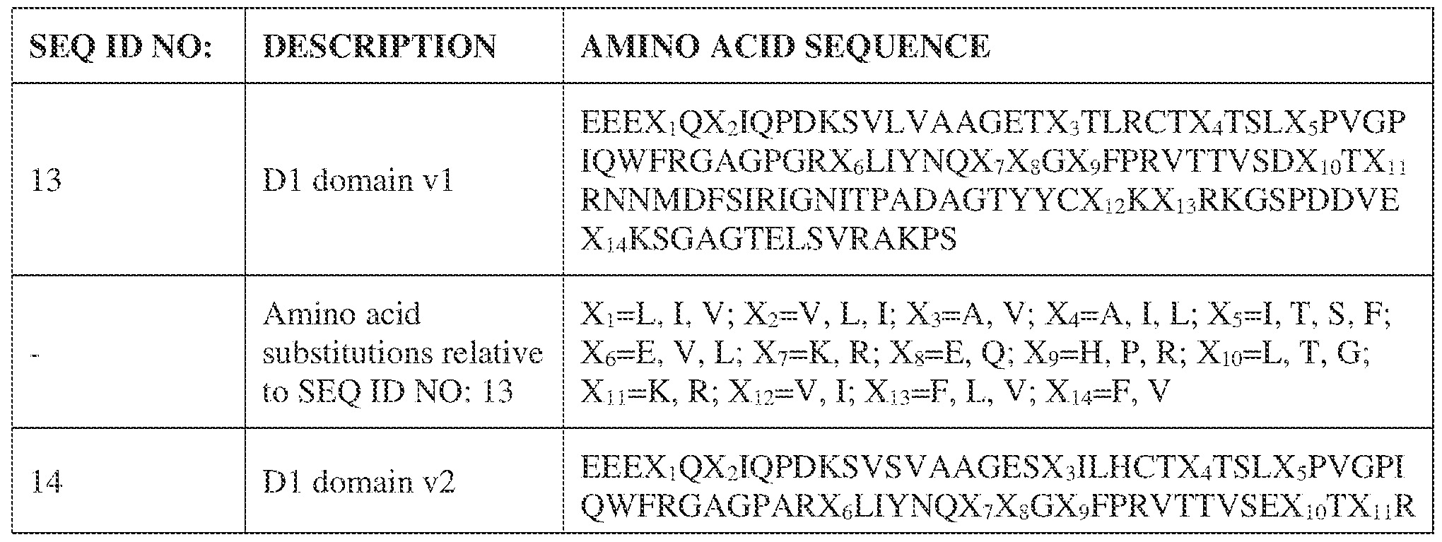


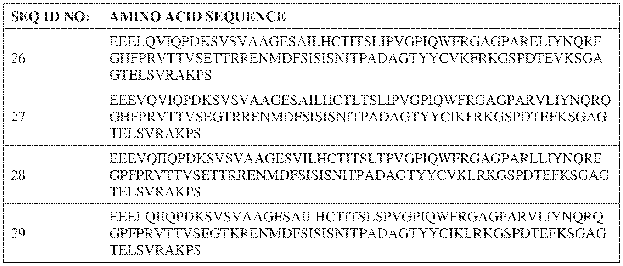


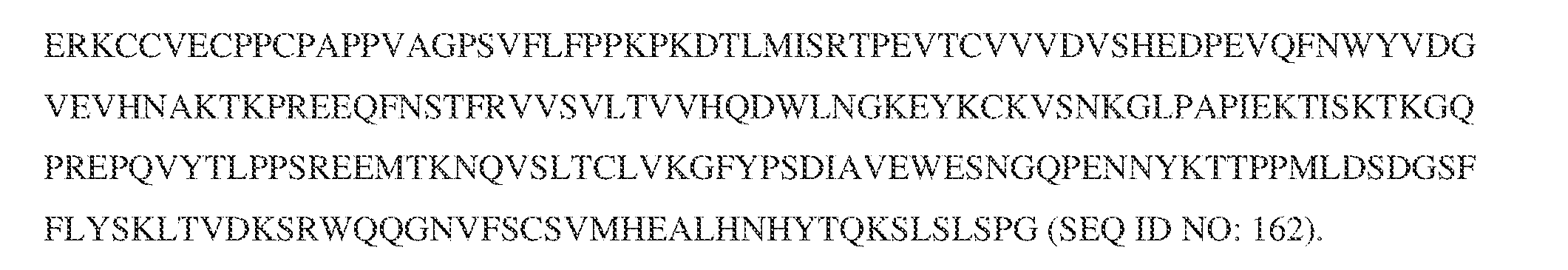


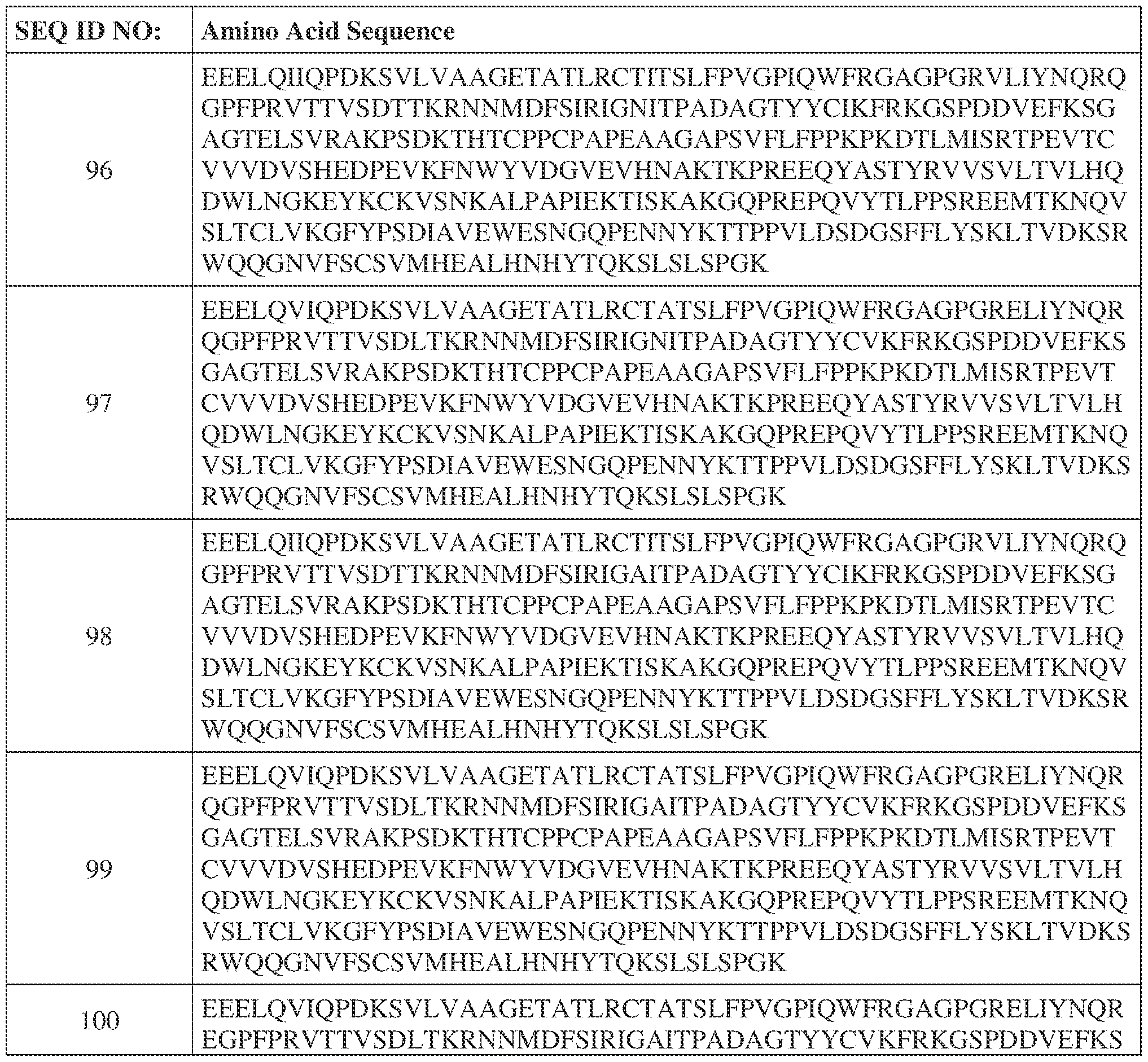


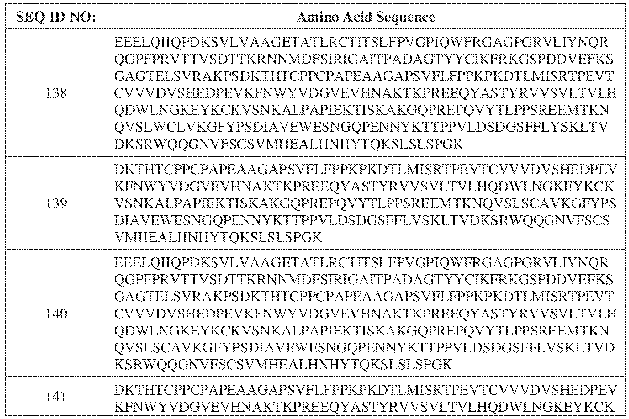


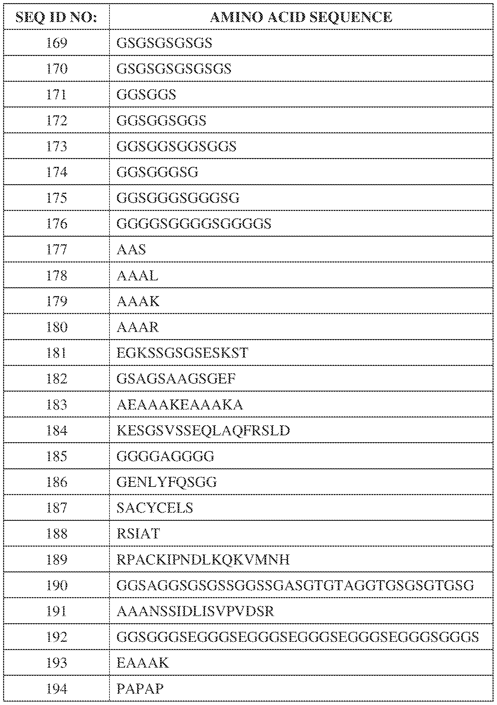


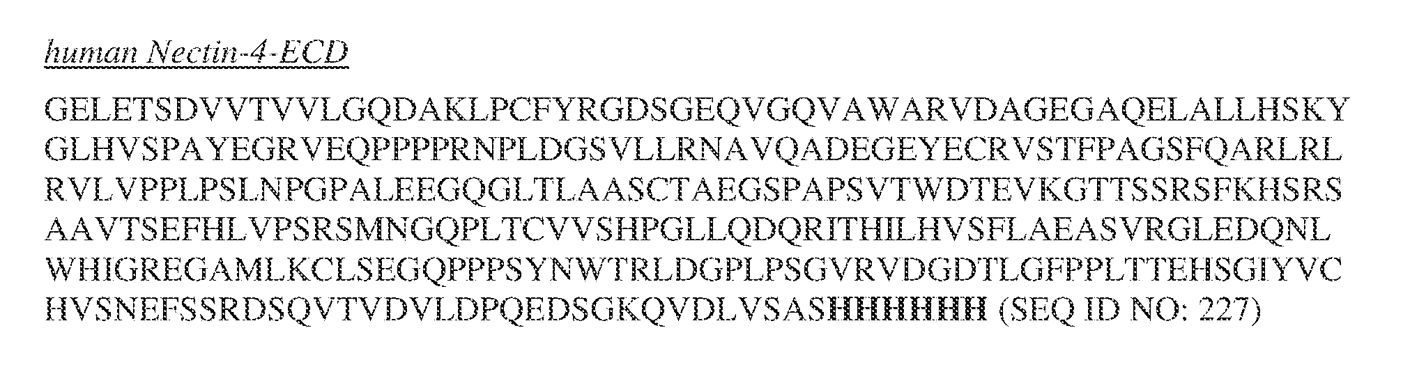


Claims
Priority Applications (8)
| Application Number | Priority Date | Filing Date | Title |
|---|---|---|---|
| KR1020247042025A KR20250022059A (en) | 2022-06-01 | 2023-05-31 | Combination therapy for the treatment of urothelial carcinoma |
| CN202380052100.6A CN119497722A (en) | 2022-06-01 | 2023-05-31 | Combination therapy for the treatment of urothelial carcinoma |
| EP23734867.7A EP4532548A1 (en) | 2022-06-01 | 2023-05-31 | Combination therapies for treating urothelial carcinoma |
| AU2023281712A AU2023281712A1 (en) | 2022-06-01 | 2023-05-31 | Combination therapies for treating urothelial carcinoma |
| CA3253030A CA3253030A1 (en) | 2022-06-01 | 2023-05-31 | Combination therapies for treating urothelial carcinoma |
| JP2024570461A JP2025523369A (en) | 2022-06-01 | 2023-05-31 | Combination Therapy for Treating Urothelial Carcinoma |
| IL316971A IL316971A (en) | 2022-06-01 | 2023-05-31 | Combination therapies for treating urothelial carcinoma |
| MX2024014744A MX2024014744A (en) | 2022-06-01 | 2024-11-28 | Combination therapies for treating urothelial carcinoma |
Applications Claiming Priority (2)
| Application Number | Priority Date | Filing Date | Title |
|---|---|---|---|
| US202263347939P | 2022-06-01 | 2022-06-01 | |
| US63/347,939 | 2022-06-01 |
Publications (1)
| Publication Number | Publication Date |
|---|---|
| WO2023235754A1 true WO2023235754A1 (en) | 2023-12-07 |
Family
ID=87036781
Family Applications (1)
| Application Number | Title | Priority Date | Filing Date |
|---|---|---|---|
| PCT/US2023/067697 Ceased WO2023235754A1 (en) | 2022-06-01 | 2023-05-31 | Combination therapies for treating urothelial carcinoma |
Country Status (10)
| Country | Link |
|---|---|
| US (1) | US20240010701A1 (en) |
| EP (1) | EP4532548A1 (en) |
| JP (1) | JP2025523369A (en) |
| KR (1) | KR20250022059A (en) |
| CN (1) | CN119497722A (en) |
| AU (1) | AU2023281712A1 (en) |
| CA (1) | CA3253030A1 (en) |
| IL (1) | IL316971A (en) |
| MX (1) | MX2024014744A (en) |
| WO (1) | WO2023235754A1 (en) |
Cited By (2)
| Publication number | Priority date | Publication date | Assignee | Title |
|---|---|---|---|---|
| US12098214B2 (en) | 2021-05-13 | 2024-09-24 | ALX Oncology Inc. | Combination therapies for treating cancer |
| US12527838B2 (en) | 2019-11-27 | 2026-01-20 | ALX Oncology Inc. | Combination therapies comprising an agent that blocks the interaction between CD47 and SIRPα for treating gastric or gastroesphageal junction cancel |
Citations (23)
| Publication number | Priority date | Publication date | Assignee | Title |
|---|---|---|---|---|
| WO2013109752A1 (en) | 2012-01-17 | 2013-07-25 | The Board Of Trustees Of The Leland Stanford Junior University | High affinity sirp-alpha reagents |
| WO2014094122A1 (en) | 2012-12-17 | 2014-06-26 | Trillium Therapeutics Inc. | Treatment of cd47+ disease cells with sirp alpha-fc fusions |
| WO2016023040A1 (en) | 2014-08-08 | 2016-02-11 | Alexo Therapeutics International | Sirp-alpha variant constructs and uses thereof |
| WO2016063233A1 (en) | 2014-10-24 | 2016-04-28 | Effimune | Method and compositions for inducing differentiation of myeloid derived suppressor cell to treat cancer and infectious diseases |
| WO2016187226A1 (en) | 2015-05-18 | 2016-11-24 | Ab Initio Biotherapeutics, Inc. | Sirp polypeptide compositions and methods of use |
| WO2017027422A1 (en) | 2015-08-07 | 2017-02-16 | Alexo Therapeutics Inc. | Constructs having a sirp-alpha domain or variant thereof |
| WO2017068164A1 (en) | 2015-10-21 | 2017-04-27 | Ose Immunotherapeutics | Methods and compositions for modifying macrophage polarization into pro-inflammatory cells to treat cancer |
| WO2017178653A2 (en) | 2016-04-14 | 2017-10-19 | Ose Immunotherapeutics | NEW ANTI-SIRPa ANTIBODIES AND THEIR THERAPEUTIC APPLICATIONS |
| WO2017177333A1 (en) | 2016-04-15 | 2017-10-19 | Trillium Therapeutics Inc. | Macrophage stimulation in cd47 blockade therapy |
| US20180037652A1 (en) | 2016-08-03 | 2018-02-08 | The Board Of Trustees Of The Leland Stanford Junior University | Disrupting FC Receptor Engagement on Macrophages Enhances Efficacy of Anti-SIRPalpha Antibody Therapy |
| EP3287470A1 (en) | 2015-04-24 | 2018-02-28 | Immuneonco Biopharmaceuticals (Shanghai) Co., Ltd | Novel recombinant bi-functional fusion protein and preparation and application therefor |
| WO2018057669A1 (en) | 2016-09-21 | 2018-03-29 | Alexo Therapeutics Inc. | Antibodies against signal-regulatory protein alpha and methods of use |
| US9962454B2 (en) | 2010-09-29 | 2018-05-08 | Agensys, Inc. | Antibody drug conjugates (ADC) that bind to 191P4D12 proteins |
| WO2018081898A1 (en) | 2016-11-03 | 2018-05-11 | Trillium Therapeutics Inc. | Improvements in cd47 blockade therapy by hdac inhibitors |
| WO2018081897A1 (en) | 2016-11-03 | 2018-05-11 | Trillium Therapeutics Inc. | Enhancement of cd47 blockade therapy by proteasome inhibitors |
| WO2018107058A1 (en) | 2016-12-09 | 2018-06-14 | Alector Llc | Anti-sirp-alpha antibodies and methods of use thereof |
| WO2018149938A1 (en) | 2017-02-17 | 2018-08-23 | Ose Immunotherapeutics | New uses of anti-sirpg antibodies |
| WO2018176132A1 (en) | 2017-03-28 | 2018-10-04 | Trillium Therapeutics Inc. | Cd47 blockade therapy |
| US20180312587A1 (en) | 2017-04-13 | 2018-11-01 | Aduro Biotech Holdings, Europe B.V. | Anti-sirp alpha antibodies |
| WO2018210795A1 (en) | 2017-05-16 | 2018-11-22 | Synthon Biopharmaceuticals B.V. | ANTI-SIRPα ANTIBODIES |
| WO2019023347A1 (en) | 2017-07-26 | 2019-01-31 | Forty Seven, Inc. | Anti-sirp-alpha antibodies and related methods |
| WO2021108693A1 (en) * | 2019-11-27 | 2021-06-03 | ALX Oncology Inc. | Combination therapies for treating cancer |
| WO2022060955A1 (en) * | 2020-09-17 | 2022-03-24 | Agensys, Inc. | Methods for treating cancers with antibody drug conjugates (adc) that bind to 191p4d12 proteins |
-
2023
- 2023-05-31 WO PCT/US2023/067697 patent/WO2023235754A1/en not_active Ceased
- 2023-05-31 CA CA3253030A patent/CA3253030A1/en active Pending
- 2023-05-31 CN CN202380052100.6A patent/CN119497722A/en active Pending
- 2023-05-31 EP EP23734867.7A patent/EP4532548A1/en active Pending
- 2023-05-31 IL IL316971A patent/IL316971A/en unknown
- 2023-05-31 JP JP2024570461A patent/JP2025523369A/en active Pending
- 2023-05-31 US US18/326,847 patent/US20240010701A1/en active Pending
- 2023-05-31 AU AU2023281712A patent/AU2023281712A1/en active Pending
- 2023-05-31 KR KR1020247042025A patent/KR20250022059A/en active Pending
-
2024
- 2024-11-28 MX MX2024014744A patent/MX2024014744A/en unknown
Patent Citations (33)
| Publication number | Priority date | Publication date | Assignee | Title |
|---|---|---|---|---|
| US9962454B2 (en) | 2010-09-29 | 2018-05-08 | Agensys, Inc. | Antibody drug conjugates (ADC) that bind to 191P4D12 proteins |
| US20150071905A1 (en) | 2012-01-17 | 2015-03-12 | The Board Of Trustees Of The Leland Stanford Junior University | High Affinity Sirp-Alpha Reagents |
| WO2013109752A1 (en) | 2012-01-17 | 2013-07-25 | The Board Of Trustees Of The Leland Stanford Junior University | High affinity sirp-alpha reagents |
| US9944911B2 (en) | 2012-01-17 | 2018-04-17 | The Board Of Trustees Of The Leland Stanford Junior University | High affinity Sirp-alpha reagents and methods of using |
| WO2014094122A1 (en) | 2012-12-17 | 2014-06-26 | Trillium Therapeutics Inc. | Treatment of cd47+ disease cells with sirp alpha-fc fusions |
| US20150329616A1 (en) | 2012-12-17 | 2015-11-19 | Trillium Therapeutics Inc. | Treatment of CD47+ Disease Cells with SIRP Alpha-FC Fusions |
| US20180312563A1 (en) | 2012-12-17 | 2018-11-01 | Trillium Therapeutics Inc. | Treatment of CD47+ Disease Cells with SIRP Alpha-FC Fusions |
| WO2016023040A1 (en) | 2014-08-08 | 2016-02-11 | Alexo Therapeutics International | Sirp-alpha variant constructs and uses thereof |
| WO2016063233A1 (en) | 2014-10-24 | 2016-04-28 | Effimune | Method and compositions for inducing differentiation of myeloid derived suppressor cell to treat cancer and infectious diseases |
| EP3287470A1 (en) | 2015-04-24 | 2018-02-28 | Immuneonco Biopharmaceuticals (Shanghai) Co., Ltd | Novel recombinant bi-functional fusion protein and preparation and application therefor |
| US20180141986A1 (en) | 2015-04-24 | 2018-05-24 | Immuneonco Biopharmaceuticals (Shanghai) Co.,Ltd | Novel recombinant bi-functional fusion protein and preparation and application therefor |
| WO2016187226A1 (en) | 2015-05-18 | 2016-11-24 | Ab Initio Biotherapeutics, Inc. | Sirp polypeptide compositions and methods of use |
| US9845345B2 (en) | 2015-05-18 | 2017-12-19 | Ab Initio Biotherapeutics, Inc. | SIRP polypeptide compositions and methods of use |
| US20180155405A1 (en) | 2015-05-18 | 2018-06-07 | Ab Initio Biotherapeutics, Inc. | Sirp polypeptide compositions and methods of use |
| US20170107270A1 (en) | 2015-08-07 | 2017-04-20 | Alexo Therapeutics Inc. | Constructs having a sirp-alpha domain or variant thereof |
| WO2017027422A1 (en) | 2015-08-07 | 2017-02-16 | Alexo Therapeutics Inc. | Constructs having a sirp-alpha domain or variant thereof |
| US10259859B2 (en) | 2015-08-07 | 2019-04-16 | ALX Oncology Inc. | Constructs having a SIRP-α domain or variant thereof |
| WO2017068164A1 (en) | 2015-10-21 | 2017-04-27 | Ose Immunotherapeutics | Methods and compositions for modifying macrophage polarization into pro-inflammatory cells to treat cancer |
| WO2017178653A2 (en) | 2016-04-14 | 2017-10-19 | Ose Immunotherapeutics | NEW ANTI-SIRPa ANTIBODIES AND THEIR THERAPEUTIC APPLICATIONS |
| WO2017177333A1 (en) | 2016-04-15 | 2017-10-19 | Trillium Therapeutics Inc. | Macrophage stimulation in cd47 blockade therapy |
| US20180037652A1 (en) | 2016-08-03 | 2018-02-08 | The Board Of Trustees Of The Leland Stanford Junior University | Disrupting FC Receptor Engagement on Macrophages Enhances Efficacy of Anti-SIRPalpha Antibody Therapy |
| US20180105600A1 (en) | 2016-09-21 | 2018-04-19 | Alexo Therapeutics Inc. | Antibodies against signal-regulatory protein alpha and methods of use |
| WO2018057669A1 (en) | 2016-09-21 | 2018-03-29 | Alexo Therapeutics Inc. | Antibodies against signal-regulatory protein alpha and methods of use |
| WO2018081898A1 (en) | 2016-11-03 | 2018-05-11 | Trillium Therapeutics Inc. | Improvements in cd47 blockade therapy by hdac inhibitors |
| WO2018081897A1 (en) | 2016-11-03 | 2018-05-11 | Trillium Therapeutics Inc. | Enhancement of cd47 blockade therapy by proteasome inhibitors |
| WO2018107058A1 (en) | 2016-12-09 | 2018-06-14 | Alector Llc | Anti-sirp-alpha antibodies and methods of use thereof |
| WO2018149938A1 (en) | 2017-02-17 | 2018-08-23 | Ose Immunotherapeutics | New uses of anti-sirpg antibodies |
| WO2018176132A1 (en) | 2017-03-28 | 2018-10-04 | Trillium Therapeutics Inc. | Cd47 blockade therapy |
| US20180312587A1 (en) | 2017-04-13 | 2018-11-01 | Aduro Biotech Holdings, Europe B.V. | Anti-sirp alpha antibodies |
| WO2018210795A1 (en) | 2017-05-16 | 2018-11-22 | Synthon Biopharmaceuticals B.V. | ANTI-SIRPα ANTIBODIES |
| WO2019023347A1 (en) | 2017-07-26 | 2019-01-31 | Forty Seven, Inc. | Anti-sirp-alpha antibodies and related methods |
| WO2021108693A1 (en) * | 2019-11-27 | 2021-06-03 | ALX Oncology Inc. | Combination therapies for treating cancer |
| WO2022060955A1 (en) * | 2020-09-17 | 2022-03-24 | Agensys, Inc. | Methods for treating cancers with antibody drug conjugates (adc) that bind to 191p4d12 proteins |
Non-Patent Citations (40)
| Title |
|---|
| "SEER Cancer Stat Facts: Bladder Cancer", 2021, NATIONAL CANCER INSTITUTE |
| "Sequences of Proteins of Immunological Interest", 1991, PUBLIC HEALTH SERVICE, NATIONAL INSTITUTES OF HEALTH |
| ABDICHE ET AL., MABS, vol. 7, no. 1-4, 2015, pages 2 |
| ANONYMOUS: "Assessment report: Padcev", 24 February 2022 (2022-02-24), pages 1 - 188, XP093078771, Retrieved from the Internet <URL:https://www.ema.europa.eu/en/documents/assessment-report/padcev-epar-public-assessment-report_en.pdf> [retrieved on 20230905] * |
| ANONYMOUS: "NCT05524545 A Study of Evorpacept (ALX148) With Enfortumab Vedotin for Subjects With Urothelial Carcinoma (ASPEN-07)", 1 September 2022 (2022-09-01), pages 1 - 7, XP093078608, Retrieved from the Internet <URL:https://classic.clinicaltrials.gov/ct2/show/NCT05524545> [retrieved on 20230904] * |
| ARMOUR ET AL., MOL. IMMUNOL., vol. 40, 2003, pages 585 - 593 |
| AUSUBEL ET AL.: "Current Protocols in Molecular Biology", 2002, JOHN WILEY & SONS |
| BELLMUNT JORSOLA AWIEGEL TGUIX MDE SANTIS MKATAJA V: "ESMO Guidelines Working Group. Bladder cancer: ESMO Clinical Practice Guidelines for diagnosis, treatment and follow-up", ANN ONCOL., September 2011 (2011-09-01) |
| CAS, no. 1448664-46-7 |
| CHAO, BLOOD, vol. 118, 2011, pages 4890 - 4891 |
| CHAO, CELL, vol. 142, 2010, pages 699 - 713 |
| EDRIS, PNAS USA, 2012 |
| EISENHAUER ET AL., EUR 7 CANCER, vol. 45, 2009, pages 228 - 247 |
| GOTO, EUR J. CANCER, vol. 50, 2014, pages 1836 - 1846 |
| HARLOWLANE: "Antibodies: A Laboratory Manual", 1990, COLD SPRING HARBOR LABORATORY PRESS |
| HUSAIN, BIODRUGS, vol. 32, no. 5, 2018, pages 441 - 464 |
| KARLSSON ET AL., ANALYTICAL BIOCHEMISTRY, vol. 349, no. 1, 2006, pages 136 - 147 |
| KIM, LEUKEMIA, vol. 26, 2012, pages 2538 - 2545 |
| LAKHANI NEHAL J ET AL: "Evorpacept alone and in combination with pembrolizumab or trastuzumab in patients with advanced solid tumours (ASPEN-01): a first-in-human, open-label, multicentre, phase 1 dose-escalation and dose-expansion study", THE LANCET ONCOLOGY, ELSEVIER, AMSTERDAM, NL, vol. 22, no. 12, 15 November 2021 (2021-11-15), pages 1740 - 1751, XP086884207, ISSN: 1470-2045, [retrieved on 20211115], DOI: 10.1016/S1470-2045(21)00584-2 * |
| LIU ET AL., JOURNAL OF THE ROYAL STATISTICAL SOCIETY. SERIES C: APPLIED STATISTICS., vol. 64, no. 3, 2015, pages 507 - 523 |
| LIU, JOURNAL OF THE ROYAL STATI TICAL SOCIETY. SERIES C: APPLIED STATISTICS, vol. 64, no. 3, 2015, pages 507 - 523 |
| MILLER ET AL.: "Quantitative high-throughput screening assays for the discovery and development of SIRPa-CD47 interaction inhibitors.", PLOS ONE, vol. 14, no. 7, 2019, pages 0218897 |
| MURATA, CANCER SCI, vol. 109, no. 5, 2018, pages 1300 - 1308 |
| MYSZKA, D.G., J. MOL. RECOGNIT., vol. 12, 1999, pages 279 - 284 |
| NAGELKERKE ET AL., FRONT IMMUNOL., vol. 10, 2019, pages 2237 |
| PETROVA, CLIN CANCER RES, vol. 23, 2017, pages 1086 - 1079 |
| RING, PNAS USA, vol. 114, no. 49, 2017 |
| RUSS ET AL., BLOOD REV, 2018 |
| SAMBROOKRUSSELL: "Molecular Cloning, A Laboratory Approach", 2001, SPRING HARBOR PRESS |
| SASIKUMAR, ACR-NCI-EORTC INTERNATIONAL CONFERENCE: MOLECULAR TARGETS AND CANCER THERAPEUTICS, 26 October 2017 (2017-10-26) |
| SENGELOV L, ROBERTS JT, RICCI S, DOGLIOTTI L, OLIVER T: "ong-term survival results of a plus cisplatin in patients with bladder cancer.", J CLIN ONCOL., vol. 23, no. 21, 2005, pages 4602 - 8 |
| SIMEONE JCNORDSTROM BLPATEL KMANN HKLEIN ABHOME L: "Treatment patterns and overall survival in metastatic urothelial carcinoma in a real-world, US setting", CANCER EPIDEMIOL., vol. 60, 2019, pages 121 - 7, XP085691029, DOI: 10.1016/j.canep.2019.03.013 |
| SPIESS, MOLECULAR IMMUNOLOGY, vol. 67, no. 2, 2015, pages 95 - 106 |
| WANG YUCHEN ET AL: "Recent Advances of Tumor Therapy Based on the CD47-SIRP[alpha] Axis", MOLECULAR PHARMACEUTICS, vol. 19, no. 5, 18 April 2022 (2022-04-18), US, pages 1273 - 1293, XP093078337, ISSN: 1543-8384, Retrieved from the Internet <URL:https://pubs.acs.org/doi/pdf/10.1021/acs.molpharmaceut.2c00073> DOI: 10.1021/acs.molpharmaceut.2c00073 * |
| YANIGATA, JCI INSIGHT, vol. 2, 2017, pages 89140 |
| YUAN ET AL., CANCER RES., vol. 22, no. 17, 2016, pages 4291 - 4301 |
| YUAN ET AL., CLIN CANCER RES., vol. 22, no. 17, 2016, pages 4291 - 4301 |
| ZHANG, XCHEN, WFAN, J ET AL., DISRUPTING CD47-SIRPΑ AXIS ALONE OR COMBINED WITH AUTOPHAGY DEPLETION FOR THE THERAPY OF GLIOBLASTOMA. CARCINOGENESIS, vol. 39, 2018, pages 689 - 99 |
| ZHAO, PNAS USA, 2011 |
| ZHOU ET AL., CLIN CANCER RES., vol. 24, no. 18, 2018, pages 4357 - 4364 |
Cited By (2)
| Publication number | Priority date | Publication date | Assignee | Title |
|---|---|---|---|---|
| US12527838B2 (en) | 2019-11-27 | 2026-01-20 | ALX Oncology Inc. | Combination therapies comprising an agent that blocks the interaction between CD47 and SIRPα for treating gastric or gastroesphageal junction cancel |
| US12098214B2 (en) | 2021-05-13 | 2024-09-24 | ALX Oncology Inc. | Combination therapies for treating cancer |
Also Published As
| Publication number | Publication date |
|---|---|
| AU2023281712A1 (en) | 2024-11-28 |
| CN119497722A (en) | 2025-02-21 |
| JP2025523369A (en) | 2025-07-23 |
| IL316971A (en) | 2025-01-01 |
| MX2024014744A (en) | 2025-03-07 |
| EP4532548A1 (en) | 2025-04-09 |
| KR20250022059A (en) | 2025-02-14 |
| US20240010701A1 (en) | 2024-01-11 |
| CA3253030A1 (en) | 2023-12-07 |
Similar Documents
| Publication | Publication Date | Title |
|---|---|---|
| US11208459B2 (en) | Constructs having a SIRP-alpha domain or variant thereof | |
| JP7104000B2 (en) | Humanized or chimeric CD3 antibody | |
| US12157771B2 (en) | Proteins binding NKG2D, CD16 and CLEC12A | |
| JP7710997B2 (en) | Methods of Treating Cancer with SIRP alpha FC Fusions in Combination with Immune Checkpoint Inhibitors | |
| AU2021358659A9 (en) | Anti-dectin-1 antibodies and methods of use thereof | |
| CN108368170A (en) | Anti-PD-1 antibodies, activatable anti-PD-1 antibodies and methods of use thereof | |
| KR20220075383A (en) | Multispecific binding protein for cancer treatment | |
| CN113195544A (en) | Multispecific binding proteins and methods of use thereof | |
| US20240010701A1 (en) | Combination therapies for treating urothelial carcinoma | |
| KR20250006353A (en) | Anti-Dectin-1 antibodies and methods of use thereof | |
| US20220242962A1 (en) | 4-1bb and ox40 binding proteins and related compositions and methods, antibodies against 4-1bb, antibodies against ox40 | |
| JP2023551907A (en) | Tumor-associated antigens and CD3 binding proteins, related compositions, and methods |
Legal Events
| Date | Code | Title | Description |
|---|---|---|---|
| 121 | Ep: the epo has been informed by wipo that ep was designated in this application |
Ref document number: 23734867 Country of ref document: EP Kind code of ref document: A1 |
|
| WWE | Wipo information: entry into national phase |
Ref document number: 816180 Country of ref document: NZ Ref document number: AU2023281712 Country of ref document: AU |
|
| WWE | Wipo information: entry into national phase |
Ref document number: 316971 Country of ref document: IL |
|
| ENP | Entry into the national phase |
Ref document number: 2023281712 Country of ref document: AU Date of ref document: 20230531 Kind code of ref document: A |
|
| WWE | Wipo information: entry into national phase |
Ref document number: 2024570461 Country of ref document: JP Ref document number: MX/A/2024/014744 Country of ref document: MX |
|
| REG | Reference to national code |
Ref country code: BR Ref legal event code: B01A Ref document number: 112024024222 Country of ref document: BR |
|
| ENP | Entry into the national phase |
Ref document number: 20247042025 Country of ref document: KR Kind code of ref document: A |
|
| WWE | Wipo information: entry into national phase |
Ref document number: 11202407913T Country of ref document: SG |
|
| WWE | Wipo information: entry into national phase |
Ref document number: 2023734867 Country of ref document: EP |
|
| NENP | Non-entry into the national phase |
Ref country code: DE |
|
| ENP | Entry into the national phase |
Ref document number: 2023734867 Country of ref document: EP Effective date: 20250102 |
|
| WWE | Wipo information: entry into national phase |
Ref document number: 202380052100.6 Country of ref document: CN |
|
| WWP | Wipo information: published in national office |
Ref document number: 1020247042025 Country of ref document: KR |
|
| WWP | Wipo information: published in national office |
Ref document number: 202380052100.6 Country of ref document: CN |
|
| WWP | Wipo information: published in national office |
Ref document number: MX/A/2024/014744 Country of ref document: MX |
|
| REG | Reference to national code |
Ref country code: BR Ref legal event code: B01E Ref document number: 112024024222 Country of ref document: BR Free format text: 1) APRESENTE NOVO RELATORIO DESCRITIVO ADAPTADO AO ART. 26 INCISO I DA PORTARIA/INPI NO 14/2024, UMA VEZ QUE O CONTEUDO ENVIADO NA PETICAO NO 870250000285 DE 03/01/2025 ENCONTRA-SE FORA DA NORMA EM RELACAO AO TITULO, CONTENDO TEXTO DIFERENTE DO TITULO INCIANDO A PAGINA. 2) APRESENTE NOVO RESUMO ADAPTADO AO ART. 40 INCISO I DA PORTARIA/INPI NO 14/2024, UMA VEZ QUE O CONTEUDO ENVIADO NA PETICAO NO 870250000285 DE 03/01/2025 ENCONTRA-SE FORA DA NORMA EM RELACAO AO TITULO, CONTENDO TEXTO DIFERENTE DO TITULO INCIANDO A PAGINA. 3) APRESENTE NOVAS FOLHAS DO RELATORIO DESCRITIVO ADAPTADAS AO ART. 20 DA PORTARIA/INPI NO 14/2024, UMA VEZ QUE O CONTEUDO ENVIADO NA PETICAO NO 870250000285 DE 03/01/2025 |
|
| WWP | Wipo information: published in national office |
Ref document number: 2023734867 Country of ref document: EP |
|
| ENP | Entry into the national phase |
Ref document number: 112024024222 Country of ref document: BR Kind code of ref document: A2 Effective date: 20241121 |