WO2023235699A1 - Antibodies to lilrb4 and uses thereof - Google Patents
Antibodies to lilrb4 and uses thereof Download PDFInfo
- Publication number
- WO2023235699A1 WO2023235699A1 PCT/US2023/067612 US2023067612W WO2023235699A1 WO 2023235699 A1 WO2023235699 A1 WO 2023235699A1 US 2023067612 W US2023067612 W US 2023067612W WO 2023235699 A1 WO2023235699 A1 WO 2023235699A1
- Authority
- WO
- WIPO (PCT)
- Prior art keywords
- amino acid
- acid sequence
- seq
- antibody
- cancer
- Prior art date
- Legal status (The legal status is an assumption and is not a legal conclusion. Google has not performed a legal analysis and makes no representation as to the accuracy of the status listed.)
- Ceased
Links
Classifications
-
- C—CHEMISTRY; METALLURGY
- C07—ORGANIC CHEMISTRY
- C07K—PEPTIDES
- C07K16/00—Immunoglobulins [IGs], e.g. monoclonal or polyclonal antibodies
- C07K16/18—Immunoglobulins [IGs], e.g. monoclonal or polyclonal antibodies against material from animals or humans
- C07K16/28—Immunoglobulins [IGs], e.g. monoclonal or polyclonal antibodies against material from animals or humans against receptors, cell surface antigens or cell surface determinants
- C07K16/2803—Immunoglobulins [IGs], e.g. monoclonal or polyclonal antibodies against material from animals or humans against receptors, cell surface antigens or cell surface determinants against the immunoglobulin superfamily
-
- A—HUMAN NECESSITIES
- A61—MEDICAL OR VETERINARY SCIENCE; HYGIENE
- A61P—SPECIFIC THERAPEUTIC ACTIVITY OF CHEMICAL COMPOUNDS OR MEDICINAL PREPARATIONS
- A61P35/00—Antineoplastic agents
-
- A—HUMAN NECESSITIES
- A61—MEDICAL OR VETERINARY SCIENCE; HYGIENE
- A61K—PREPARATIONS FOR MEDICAL, DENTAL OR TOILETRY PURPOSES
- A61K39/00—Medicinal preparations containing antigens or antibodies
- A61K2039/505—Medicinal preparations containing antigens or antibodies comprising antibodies
-
- C—CHEMISTRY; METALLURGY
- C07—ORGANIC CHEMISTRY
- C07K—PEPTIDES
- C07K2317/00—Immunoglobulins specific features
- C07K2317/20—Immunoglobulins specific features characterized by taxonomic origin
- C07K2317/24—Immunoglobulins specific features characterized by taxonomic origin containing regions, domains or residues from different species, e.g. chimeric, humanized or veneered
-
- C—CHEMISTRY; METALLURGY
- C07—ORGANIC CHEMISTRY
- C07K—PEPTIDES
- C07K2317/00—Immunoglobulins specific features
- C07K2317/50—Immunoglobulins specific features characterized by immunoglobulin fragments
- C07K2317/56—Immunoglobulins specific features characterized by immunoglobulin fragments variable (Fv) region, i.e. VH and/or VL
- C07K2317/565—Complementarity determining region [CDR]
-
- C—CHEMISTRY; METALLURGY
- C07—ORGANIC CHEMISTRY
- C07K—PEPTIDES
- C07K2317/00—Immunoglobulins specific features
- C07K2317/70—Immunoglobulins specific features characterized by effect upon binding to a cell or to an antigen
- C07K2317/73—Inducing cell death, e.g. apoptosis, necrosis or inhibition of cell proliferation
-
- C—CHEMISTRY; METALLURGY
- C07—ORGANIC CHEMISTRY
- C07K—PEPTIDES
- C07K2317/00—Immunoglobulins specific features
- C07K2317/70—Immunoglobulins specific features characterized by effect upon binding to a cell or to an antigen
- C07K2317/76—Antagonist effect on antigen, e.g. neutralization or inhibition of binding
-
- C—CHEMISTRY; METALLURGY
- C07—ORGANIC CHEMISTRY
- C07K—PEPTIDES
- C07K2317/00—Immunoglobulins specific features
- C07K2317/90—Immunoglobulins specific features characterized by (pharmaco)kinetic aspects or by stability of the immunoglobulin
- C07K2317/92—Affinity (KD), association rate (Ka), dissociation rate (Kd) or EC50 value
Definitions
- Antibodies that bind to LILRB4 are provided. Methods of treatment comprising administering anti-LILRB4 antibodies are also provided.
- Myeloid cells such as dendritic cells and macrophages, can instruct the adaptive immune system to mount a response against tumor cells and pathogens by presenting peptide antigens to T cells while expressing immunogenic cytokines and costimulatory signals, thereby promoting cytotoxic T cell activation and proliferation.
- myeloid cells maintain tolerance to endogenous proteins by presenting self-antigens to T cells in the context of non-immunogenic signals, such as regulatory cytokines, which can promote regulatory T cells and suppress immunogenicity.
- Cancer cells can evade the immune system by engaging signaling pathways associated with immunosuppressive or immunoregulatory antigen presentation. Such evasion events represent a major obstacle to therapeutic strategies that rely on promoting anti-tumor immunity. Therefore, there is a need for therapeutic compositions and methods that prevent tumor-induced immunosuppression and promote immunogenic presentation of tumor antigens by myeloid cells.
- LILRB4 leukocyte immunoglobulin-like receptor B4
- Embodiment 1 An isolated antibody that binds LILRB4, wherein the antibody comprises: a) an HCDR1 comprising the amino acid sequence of SEQ ID NO: 5, an HCDR2 comprising the amino acid sequence of SEQ ID NO: 6, an HCDR3 comprising the amino acid sequence of SEQ ID NO: 7, an LCDR1 comprising the amino acid sequence of SEQ ID NO: 8, an LCDR2 comprising the amino acid sequence of SEQ ID NO: 9, and an LCDR3 comprising the amino acid sequence of SEQ ID NO: 10; b) an HCDR1 comprising the amino acid sequence of SEQ ID NO: 15, an HCDR2 comprising the amino acid sequence of SEQ ID NO: 16, an HCDR3 comprising the amino acid sequence of SEQ ID NO: 17, an LCDR1 comprising the amino acid sequence of SEQ ID NO: 18, an LCDR2 comprising the amino acid sequence of SEQ ID NO: 19, and an LCDR3 comprising the amino acid sequence of SEQ ID NO: 20; c) an HCDR1 comprising the amino
- Embodiment 2 The isolated antibody of embodiment 1, wherein the antibody comprises: a) a heavy chain variable region (VH) comprising an amino acid sequence that is at least 90%, 91%, 92%, 93%, 94%, 95%, 96%, 97%, 98%, or 99% identical to the amino acid sequence of SEQ ID NO: 3, and a light chain variable region (VL) comprising an amino acid sequence that is at least 90%, 91%, 92%, 93%, 94%, 95%, 96%, 97%, 98%, or 99% identical to the amino acid sequence of SEQ ID NO: 4; or b) a heavy chain variable region (VH) comprising an amino acid sequence that is at least 90%, 91%, 92%, 93%, 94%, 95%, 96%, 97%, 98%, or 99% identical to the amino acid sequence of SEQ ID NO: 13, and a light chain variable region (VL) comprising an amino acid sequence that is at least 90%, 91%, 92%, 93%, 94%, 95%, 96%, 9
- Embodiment 3 The isolated antibody of embodiments 1 or 2, wherein the antibody comprises a heavy chain variable region (VH) comprising the amino acid sequence of SEQ ID NO: 3, and a light chain variable region (VL) comprising the amino acid sequence of SEQ ID NO: 4.
- VH heavy chain variable region
- VL light chain variable region
- Embodiment 4 The isolated antibody of embodiments 1 or 2, wherein the antibody comprises a heavy chain variable region (VH) comprising the amino acid sequence of SEQ ID NO: 13, and a light chain variable region (VL) comprising the amino acid sequence of SEQ ID NO: 14.
- VH heavy chain variable region
- VL light chain variable region
- Embodiment 5 The isolated antibody of embodiments 1 or 2, wherein the antibody comprises a heavy chain variable region (VH) comprising the amino acid sequence of SEQ ID NO: 23, and a light chain variable region (VL) comprising the amino acid sequence of SEQ ID NO: 24.
- VH heavy chain variable region
- VL light chain variable region
- Embodiment 6 The isolated antibody of embodiments 1 or 2, wherein the antibody comprises a heavy chain variable region (VH) comprising the amino acid sequence of SEQ ID NO: 33, and a light chain variable region (VL) comprising the amino acid sequence of SEQ ID NO: 34.
- VH heavy chain variable region
- VL light chain variable region
- Embodiment 7 The isolated antibody of embodiments 1 or 2, wherein the antibody comprises a heavy chain variable region (VH) comprising the amino acid sequence of SEQ ID NO: 43, and a light chain variable region (VL) comprising the amino acid sequence of SEQ ID NO: 44.
- VH heavy chain variable region
- VL light chain variable region
- Embodiment 8 The isolated antibody of embodiments 1 or 2, wherein the antibody comprises a heavy chain variable region (VH) comprising the amino acid sequence of SEQ ID NO: 53, and a light chain variable region (VL) comprising the amino acid sequence of SEQ ID NO: 54.
- VH heavy chain variable region
- VL light chain variable region
- Embodiment 9 The isolated antibody of any one of embodiments 1-8, wherein the antibody is a monoclonal antibody.
- Embodiment 10 The isolated antibody of any one of embodiments 1-9, wherein the antibody is a chimeric antibody or a humanized antibody.
- Embodiment 11 The isolated antibody of any one of embodiments 1-10, wherein the antibody is a full-length antibody.
- Embodiment 12. The isolated antibody of any one of embodiments 1-11, wherein the antibody is an IgGl antibody, an IgG2 antibody, and IgG3 antibody, or an IgG4 antibody.
- Embodiment 13 The isolated antibody of any one of embodiments 1-12, wherein the antibody is an IgG4 antibody.
- Embodiment 14 The isolated antibody of any one of embodiments 1-10, wherein the antibody is an antibody fragment selected from a Fab, Fab’, Fv, scFv or (Fab’)2 fragment.
- Embodiment 15 The isolated antibody of any one of embodiments 1-13, wherein the antibody comprises: a) a heavy chain (HC) sequence having at least 85%, 90%, 91%, 92%, 93%, 94%, 95%, 96%, 97%, 98%, 99%, or 100% sequence identity to the amino acid sequence of SEQ ID NO: 1, and a light chain (LC) sequence having at least 85%, 90%, 91%, 92%, 93%, 94%, 95%, 96%, 97%, 98%, 99%, or 100% sequence identity to the amino acid sequence of SEQ ID NO: 2; b) a heavy chain (HC) sequence having at least 85%, 90%, 91%, 92%, 93%, 94%, 95%, 96%, 97%, 98%, 99%, or 100% sequence identity to the amino acid
- Embodiment 16 An isolated antibody that binds LILRB4, wherein the antibody does not compete with any of the following antibodies: 9B11, H128-3, mAb24251, ZM4.1, 52B8, BM1, and BM4.
- Embodiment 17 The isolated antibody of embodiment 16, wherein the antibody competes for binding to LILRB4 with: a) a reference antibody comprising a VH comprising the amino acid sequence of SEQ ID NO: 3 and VL comprising the amino acid sequence of SEQ ID NO: 4; and/or b) a reference antibody comprising a VH comprising the amino acid sequence of SEQ ID NO: 33 and VL comprising the amino acid sequence of SEQ ID NO: 34.
- Embodiment 18 The isolated antibody of embodiment 16, wherein the antibody competes for binding to LILRB4 with: a) a reference antibody comprising a VH comprising the amino acid sequence of SEQ ID NO: 43 and VL comprising the amino acid sequence of SEQ ID NO: 44; and/or b) a reference antibody comprising a VH comprising the amino acid sequence of SEQ ID NO: 53 and VL comprising the amino acid sequence of SEQ ID NO: 54.
- Embodiment 19 An isolated antibody that binds LILRB4, wherein the antibody competes for binding to LILRB4 with: a) a reference antibody comprising a VH comprising the amino acid sequence of SEQ ID NO: 3 and VL comprising the amino acid sequence of SEQ ID NO: 4; and/or b) a reference antibody comprising a VH comprising the amino acid sequence of SEQ ID NO: 33 and VL comprising the amino acid sequence of SEQ ID NO: 34.
- Embodiment 20 The isolated antibody of embodiment 19, wherein the antibody competes for binding to LILRB4 with a reference antibody comprising a VH comprising the amino acid sequence of SEQ ID NO: 3 and VL comprising the amino acid sequence of SEQ ID NO: 4, and with a reference antibody comprising a VH comprising the amino acid sequence of SEQ ID NO: 33 and VL comprising the amino acid sequence of SEQ ID NO: 34.
- Embodiment 21 An isolated antibody that binds LILRB4, wherein the antibody competes for binding to LILRB4 with: a) a reference antibody comprising a VH comprising the amino acid sequence of SEQ ID NO: 43 and VL comprising the amino acid sequence of SEQ ID NO: 44; and/or b) a reference antibody comprising a VH comprising the amino acid sequence of SEQ ID NO: 53 and VL comprising the amino acid sequence of SEQ ID NO: 54.
- Embodiment 22 The isolated antibody of embodiment 21, wherein the antibody competes for binding to LILRB4 with a reference antibody comprising a VH comprising the amino acid sequence of SEQ ID NO: 43 and VL comprising the amino acid sequence of SEQ ID NO: 44, and with a reference antibody comprising a VH comprising the amino acid sequence of SEQ ID NO: 53 and VL comprising the amino acid sequence of SEQ ID NO: 54.
- Embodiment 23 The isolated antibody of any one of embodiments 19-22, wherein the antibody does not compete with any of the following antibodies: 9B11, H128-3, mAb24251, ZM4.1, 52B8, BM1, and BM4.
- Embodiment 24 The isolated antibody of any one of embodiments 16-23, wherein competition is determined using Anti-Human IgG Fc Capture (AHC) biosensors and hLILRB4- hFcl.
- AHC Anti-Human IgG Fc Capture
- Embodiment 25 The isolated antibody of embodiment 24, wherein the hLILRB4-hFcl comprises the amino acid sequence of SEQ ID NO: 61 or SEQ ID NO: 66.
- Embodiment 26 The isolated antibody of any one of embodiments 16-25, wherein the antibody blocks at least 30%, at least 40%, at least 50%, at least 60%, at least 70%, at least 80%, or at least 90% of reference antibody binding to LILRB4.
- Embodiment 27 The isolated antibody of any one of the preceding embodiments, wherein the antibody binds to human LILRB4.
- Embodiment 28 The isolated antibody of embodiment 27, wherein the antibody binds to human LILRB4 comprising the amino acid sequence of SEQ ID NO: 63, and/or binds to human LILRB4 comprising the amino acid sequence of SEQ ID NO: 65.
- Embodiment 29 The isolated antibody of any one of the preceding embodiments, wherein the antibody binds to human LILRB4 with an affinity (KD) of less than 5 nM, less than 3 nM, or less than 2 nM.
- KD affinity
- Embodiment 30 The isolated antibody of embodiment 29, wherein affinity is determined using surface plasmon resonance (SPR).
- SPR surface plasmon resonance
- Embodiment 31 The isolated antibody of any one of the preceding embodiments, wherein administration of the antibody to a mammal reduces tumor size in the mammal.
- Embodiment 32 The isolated antibody of embodiment 31, wherein the mammal is a human.
- Embodiment 33 The isolated antibody of embodiment 32, wherein the human has cancer.
- Embodiment 34 The isolated antibody of any one of the preceding embodiments, wherein the antibody does not detectably bind or binds with at least 10-fold lower affinity to LILRA1, LILRA2, LILRA3, LILRA4, LILRA5, LILRA6, LILRB1, LILRB2, LILRB3, and LILRB5.
- Embodiment 35 An immunoconjugate comprising the isolated antibody of any one of embodiments 1-34 and a cytotoxic agent.
- Embodiment 36 An isolated nucleic acid encoding the antibody of any one of embodiments 1-34.
- Embodiment 37 A vector comprising the nucleic acid of embodiment 36.
- Embodiment 38 A host cell comprising the nucleic acid of embodiment 36 or the vector of embodiment 37.
- Embodiment 39 A host cell that produces the isolated antibody of any one of embodiments
- Embodiment 40 A method for making an anti-LILRB4 antibody, comprising culturing the host cell of embodiment 38 or 39 under conditions suitable for expression of the antibody.
- Embodiment 41 The method of embodiment 40, further comprising recovering the antibody produced by the host cell.
- Embodiment 42 A pharmaceutical composition comprising the isolated anti-LILRB4 antibody of any one of embodiments 1-34 and a pharmaceutically acceptable carrier.
- Embodiment 43 A method of treating cancer in a mammal comprising administering an effective amount of the isolated anti-LILRB4 antibody of any one of embodiments 1-34 or the pharmaceutical composition of embodiment 42.
- Embodiment 44 The method of embodiment 43, wherein the cancer is selected from carcinoma, lymphoma, blastoma, sarcoma, and leukemia, optionally wherein the cancer is kidney cancer (e.g., renal cell carcinoma, e.g., papillary renal cell carcinoma), squamous cell cancer, mesothelioma, teratoma, small-cell lung cancer, pituitary cancer, esophageal cancer, astrocytoma, soft tissue sarcoma, lung cancer (e.g., non-small cell lung cancer, adenocarcinoma of the lung, squamous carcinoma of the lung), cancer of the peritoneum, hepatocellular cancer, gastrointestinal cancer (e.g., stomach cancer), pancreatic cancer, cervical cancer, ovarian cancer, liver cancer, bladder cancer, hepatoma, breast cancer, colon cancer, colorectal cancer, rectal cancer, endometrial or uterine carcinoma, salivary gland carcinoma, liver cancer, prostate
- Embodiment 45 A method of enhancing an anti -turn or immune response in a mammal comprising administering an effective amount of the isolated anti-LILRB4 antibody of any one of embodiments 1-34 or the pharmaceutical composition of embodiment 42.
- Embodiment 46 A method of reducing tumor size in a in a mammal with cancer comprising administering an effective amount of the isolated anti-LILRB4 antibody of any one of embodiments 1-34 or the pharmaceutical composition of embodiment 42.
- Embodiment 47 The method of embodiment 45 or embodiment 46, wherein the mammal has a cancer selected from carcinoma, lymphoma, blastoma, sarcoma, and leukemia, optionally wherein the cancer is kidney cancer (e.g., renal cell carcinoma, e.g., papillary renal cell carcinoma), squamous cell cancer, mesothelioma, teratoma, small-cell lung cancer, pituitary cancer, esophageal cancer, astrocytoma, soft tissue sarcoma, lung cancer (e.g., non-small cell lung cancer, adenocarcinoma of the lung, squamous carcinoma of the lung), cancer of the peritoneum, hepatocellular cancer, gastrointestinal cancer (e.g., stomach cancer), pancreatic cancer, cervical cancer, ovarian cancer, liver cancer, bladder cancer, hepatoma, breast cancer, colon cancer, colorectal cancer, rectal cancer, endometrial or uterine carcinoma, saliva
- Embodiment 48 The method of any one of embodiments 45-47, wherein the mammal is a human.
- Embodiment 49 The method of any one of embodiments 43-48, wherein the mammal is administered at least one additional therapeutic agent.
- Embodiment 50 The method of embodiment 49, wherein the additional therapeutic agent is an immunotherapeutic agent or a cancer vaccine.
- Embodiment 51 The method of embodiment 50, wherein the additional therapeutic agent is an immunotherapeutic agent.
- Embodiment 52 The method of any one of embodiments 49-51, wherein the additional therapeutic agent is selected from a PD-1 therapy, a LAG3 therapy, a TIM3 therapy, a LILRB1 therapy, a LILRB2 therapy, a TIGIT therapy, an ICOS therapy, and combinations thereof.
- Embodiment 53 The method of embodiment 52, wherein the additional therapeutic is a PD-1 therapy in combination with a LAG3 therapy, a TIM3 therapy, a LILRB1 therapy, a LILRB2 therapy, a TIGIT therapy, or an ICOS therapy.
- Embodiment 54 The method of embodiment 52 or embodiment 53, wherein the PD-1 therapy is an anti-PD-1 antibody, and anti-PD-Ll antibody, or an anti-PD-L2 antibody.
- Embodiment 55 The method of embodiment 54, wherein the PD-1 therapy is selected from pimivalimab, nivolumab, pembrolizumab, cemiplimab, pidilizumab, atezolizumab, avelumab, dostarlimab-gxly, AMP-224, BMS-936559, AMP-514, KD-033, balstilimab, STI-A1010, STI- Al l 10, pimivalimab, and durvalumab.
- Embodiment 56 The method of embodiment 52 or embodiment 53, wherein the ICOS therapy is an anti -ICOS antibody.
- Embodiment 57 The method of embodiment 56, wherein the anti-ICOS antibody is vopratelimab, GSK609, or BMS-986226.
- Figure 1 shows an exemplary mechanism of action of LILRB4 antagonist therapy.
- Immunosuppresive mediators in the tumor microenvironment increase LILRB4 levels on dendritic cells (DCs) and myeloid-derived suppressor cells (MDSCs).
- DCs dendritic cells
- MDSCs myeloid-derived suppressor cells
- Blockade of LILRB4 leads to activation of tolerized DCs and reduction of the suppressive function of MDSCs thereby increasing T cell activation and proliferation leading to anti-tumor activity.
- Figures 2A and 2B show the results of experiments measuring LILRB4 binding of anti-LILRB4 antibodies by flow cytometry. Mean fluorescence intensity (MFI) is shown on the y-axis. Anti-LILRB4 antibody concentration in nM is shown on the y-axis.
- Figure 2A is a graph showing cell-based affinity determination of humanized anti-LILRB4 mAbs. All anti-LILRB4 mAbs tested exhibited dose-dependent specific binding to cell expressed hLILRB4 (CHO-S), while the isotype control mAb did not bind hLILRB4.
- Figure 2B is a graph showing cell-based affinity determination of humanized anti-LILRB4 mAbs. All anti-LILRB4 mAbs tested exhibited dose-dependent specific binding to cell expressed hLILRB4 on human monocyte derived dendritic cells (hMDDCs), while the isotype control mAb did not bind.
- Figure 3 is a bar graph showing results of a cell-based LILR family cross-reactivity screen of anti-LILRB4 humanized mAbs. MFI for each antibody on each LILR family cell line was divided by the MFI of the appropriate isotype control on that cell line. All antibodies tested show specific binding to hLILRB4.
- Figures 4A and 4B are example binning matrices for anti-LILRB4 antibodies.
- Anti- LILRB4 antibodies used as the first antibody in the binning experiments are listed in column 1 of each of Figure 4A and Figure 4B.
- Anti-LILRB4 antibodies used as the second antibody in the binning experiments are listed in row 1 of each of Figure 4A and Figure 4B.
- a “0” indicates complete blocking between the two indicated antibodies.
- “synagis. hG4” is the isotype control and “R&D” is mab24251.
- FIG. 5 is a bar graph showing IFNy secretion in a Mixed Lymphocyte Reaction (MLR) assay with human monocyte derived Dendritic Cells co-cultured with human allogeneic CD8+ T cells. Data points indicate IFNy levels from individual donor pairs. Combination of M31 chimera and anti-PDl treatment increases IFNy production compared to Isotype+anti-PDl treatment. Levels of IFN-y production are shown, normalized to isotype. The units on the y-axis are fold-increase compared to isotype. The external benchmark was antibody 52B8, described in WO 2019/099597. A indicates a statistically significant difference.
- MLR Mixed Lymphocyte Reaction
- Figure 6 is a bar graph showing CD8+ T cell proliferation in a MDSC-mediated T cell suppression assay.
- human peripheral blood mononuclear cell (PBMCs) were co-cultured with SKMEL-5 cells.
- PBMCs peripheral blood mononuclear cell
- CD33+ MDSCs were isolated with magnetic beads and co-cultured with autologous CD8+ T cells in the presence of CD3/CD28 beads and anti-LILRB4 mAb.
- M31 chimera treatment increases CD8+ T cell proliferation compared to Isotype treatment.
- the external benchmark was 52B8.
- Figures 7A-E show IFN y secretion in a Mixed Lymphocyte Reaction (MLR) assay with human monocyte derived Dendritic Cells co-cultured with human allogeneic CD8+ T cells.
- Figure 7A is a bar graph. Data points on the bar graph indicate IFN y levels from individual donor pairs. Combination of B4M5.018 humanized variant and anti-PDl treatment increases IFN y production compared to Isotype+anti-PDl treatment.
- Figures 7B-8E are line graphs that show IFN y production by each individual donor pair after indicated treatment. Dotted lines indicate donor pairs that did not show increased IFN y production after anti-LILRB4 treatment. Solid lines indicate donor pairs that did show increased IFN-y production after anti-LILRB4 treatment. A indicates a statistically significant difference. The horizontal dotted line indicates the level of IFN- y production of isotype.
- Figures 8A-D are bar graphs showing CD8+ T cell proliferation and IFNy production in a MDSC-mediated T cell suppression assay.
- human PBMCs were co- cultured with SKMEL-5 cells.
- CD33+ MDSCs were isolated with magnetic beads and co- cultured with autologous CD8+ T cells in the presence of CD3/CD28 beads and anti-LILRB4 mAb.
- B4M5.018 humanized anti-LILRB4 treatment increases CD8+ T cell proliferation and IFNy production compared to Isotype treatment.
- the ratios were 2: 1 CD8+ T celkMDSC, Figure 8 A shows data from Donor 1.
- Figure 8B shows data from Donor 2.
- Figures 9A-D show gene expression in anti-LILRB4 mAh histoculture studies. Fresh human tumors were sliced and treated with anti-LILRB4 antibodies or isotype control (IC) for 24h. When available, an untreated fresh slice from each tumor was reserved for baseline analysis. RNA was extracted and mRNA gene expression profiling was performed using the nCounter® Human Immunology V2 Panel (NanoString).
- Figure 9A is a bar graph showing pharmacodynamic (PD) response rates. Percent of responders is shown on the x-axis. The PD gene signature used to assess the PD response rate for each bar is shown on the y-axis. PD response rates were evaluated based on changes in PD markers or gene signatures.
- PD pharmacodynamic
- PD responders were defined as samples with significant change in PD marker or signature score in anti-LILRB4 antibody vs. IC treated samples within each tumor.
- PD signatures denoted by * are single-gene log2 expression values, otherwise the signature score is the mean log2 expression value of all genes within the signature.
- PD responders defined by immunosuppressive (IMS) signatures have downregulated scores below the noise threshold upon anti-LILRB4 treatment.
- PD responders defined by activation (ACT) or Ratio (ACT/IMS) have upregulated scores above the noise threshold upon anti-LILRB4 treatment.
- Figure 9B is a scatter plot showing Log2 fold change of the MDSC PD signature scores in anti-LILRB4 antibody vs.
- FIG. 9C is a bar graph showing top genes modulated by anti-LILRB4 antibodies vs. isotype control within PD responders defined by the MDSC signature. Names of genes are shown on the y-axis. The absolute value of mean log2 fold change of gene expression values of anti-LILRB4 antibody treated samples compared to isotype control treated samples within each matching donor is shown on the x-axis. The top two bars represent upregulation and the remaining bars represent downregulation.
- Figure 9D is an exemplary scatter plot showing a comparison of gene expression profiles of untreated baseline samples in PD responders vs. non-responders defined by the MDSC signature.
- Human tumors from lung, head and neck, ovarian, and kidney cancer patients (138 total samples) were used in this study.
- the thresholds for defining significance were based on the bottom 95 th percentile of log2 fold change of signature score across isotype control treated replicates from a larger independent study.
- the dotted line represents p-value 0.05.
- the y-axis shows -loglO(p-value).
- the x-axis shows log2 fold change of signature score, increasing from the left side to the right side of the graph.
- FIG. 10A-C show that reversal of fibronectin inhibition of tolerogenic dendritic cell (tDC) and THP-1 cell activation following FcR stimulation by a control human IgGl antibody used to trigger FcR crosslinking.
- IL-8 cytokine secretion by tolerogenic dendritic cells (tDCs) (Figure 10A), THP-1 cells (Figure 10B), and LILRB4 knockout (LILRB4 KO) THP-1 cells (Figure IOC) following FcR crosslinking, with and without fibronectin inhibition, and FcR cross-linking with fibronectin inhibition and isotype (h!gG4 antibody) or an anti-LILRB4 mAb (B4M5.018 in Figures 10 A and 10B; M31 in Figure IOC). Bars represent the mean+SEM of triplicate wells from a representative tDC donor or THP-1 experiment.
- Antibodies that bind LILRB4 are provided.
- Antibody heavy chains and light chains that are capable of forming antibodies that bind LILRB4 are also provided.
- antibodies, heavy chains, and light chains comprising one or more particular complementarity determining regions (CDRs) are provided.
- Antibodies, heavy chains, and light chains comprising one or more heavy chain variable regions (VH) or light chain variable regions (VL) are provided.
- Polynucleotides encoding antibodies to LILRB4 are provided.
- Methods of producing and/or recovering antibodies to LILRB4 are provided.
- Methods of treatment using antibodies to LILRB4 are provided. Such methods include, but are not limited to, methods of treating cancer.
- nucleic acid molecule refers to a polymer of nucleotides.
- polymers of nucleotides may contain natural and/or non-natural nucleotides, and include, but are not limited to, DNA, RNA, and PNA.
- Nucleic acid sequence refers to the linear sequence of nucleotides that comprise the nucleic acid molecule or polynucleotide.
- polypeptide and “protein” are used interchangeably to refer to a polymer of amino acid residues, and are not limited to a minimum length. Such polymers of amino acid residues may contain natural or non-natural amino acid residues, and include, but are not limited to, peptides, oligopeptides, dimers, trimers, and multimers of amino acid residues. Both full- length proteins and fragments thereof are encompassed by the definition.
- the terms also include post-expression modifications of the polypeptide, for example, glycosylation, sialylation, acetylation, phosphorylation, and the like.
- polypeptide refers to a protein which includes modifications, such as deletions, additions, and substitutions (generally conservative in nature), to the native sequence, as long as the protein maintains the desired activity. These modifications may be deliberate, as through site-directed mutagenesis, or may be accidental, such as through mutations of hosts which produce the proteins or errors due to PCR amplification.
- LILRB4 and “leukocyte immunoglobulin like-receptor B4” as used herein refer to any native LILRB4 that results from expression and processing of LILRB4 in a cell.
- the term includes LILRB4 from any vertebrate source, including mammals such as primates (e.g., humans and cynomolgus monkeys) and rodents (e.g., mice and rats), unless otherwise indicated.
- the term also includes naturally occurring variants of LILRB4, e.g., splice variants or allelic variants.
- the amino acid sequences of exemplary human LILRB4 precursor proteins are shown in SEQ ID NOs: 62 and 64.
- the amino acid sequences of exemplary mature human LILRB4 proteins are shown in SEQ ID NOs: 63 and 65.
- a “PD-1 therapy” encompasses any therapy that modulates PD-1 binding to PD-L1 and/or PD-L2.
- PD-1 therapies may, for example, directly interact with PD-1 and/or PD-L1.
- a PD-1 therapy includes a molecule that directly binds to and/or influences the activity of PD-1.
- a PD-1 therapy includes a molecule that directly binds to and/or influences the activity of PD-L1.
- an antibody that binds to PD-1 or PD-L1 and blocks the interaction of PD-1 to PD-L1 is a PD-1 therapeutic.
- PD-1 specific for a therapy involving a molecule that interacts directly with PD-1
- PD-L1 specific for a molecule that interacts directly with PD-L1
- all disclosure contained herein regarding PD-1 therapy applies to PD-1 therapy generally, as well as PD-1 specific and/or PD-L1 specific therapies.
- the PD-1 therapy is an anti- PD1 antibody or an anti-PD-Ll antibody.
- Nonlimiting exemplary PD-1 therapies include nivolumab (BMS-936558/MDX-1106/ONO-4538/OPDIVO® (Bristol-Myers Squibb Co.)); pidilizumab (CT-011/ MDV9300 (Curetech)); pembrolizumab (KEYTRUDA®/MK-3475 (Merck)); durvalumab (IMFINZI®/MEDI-4736 (Medimmune/AstraZeneca)); avelumab (MSB- 0010718C/BAVENCIO® (Merck KGaA/Pfizer)); dostarlimab-gxly (TSR-042/ANB- 011/JEMPERLI® (AnaptysBio/GSK)); AMP-224 (Amplimmune/ Medimmune/ AstraZeneca/ GSK); BMS-936559 (MDX-1105 (Bristol-Myers Squibb Co.)); AMP-514 (
- the term “specifically binds” to an antigen is a term that is well understood in the art, and methods to determine such specific binding are also well known in the art.
- a molecule is said to exhibit “specific binding” or “preferential binding” if it reacts or associates more frequently, more rapidly, with greater duration and/or with greater affinity with a particular cell or substance than it does with alternative cells or substances.
- An antibody “specifically binds” or “preferentially binds” to a target if it binds with greater affinity, avidity, more readily, and/or with greater duration than it binds to other substances.
- an antibody that specifically or preferentially binds to LILRB4 is an antibody that binds LILRB4 with greater affinity, avidity, more readily, and/or with greater duration than it binds to other antigens. It is also understood by reading this definition that, for example, an antibody that specifically or preferentially binds to a first target may or may not specifically or preferentially bind to a second target. As such, “specific binding” or “preferential binding” does not necessarily require (although it can include) exclusive binding. Generally, but not necessarily, reference to binding means preferential binding. “Specificity” refers to the ability of a binding protein to selectively bind an antigen.
- substantially pure refers to material which is at least 50% pure (that is, free from contaminants), more preferably, at least 90% pure, more preferably, at least 95% pure, yet more preferably, at least 98% pure, and most preferably, at least 99% pure.
- the term “competes” or “cross-competes” refers to competitive binding of one molecule with another, e.g., by binding to all or part of the same epitope.
- Cross-competition can be determined using the experiments described herein (e.g., biolayer interferometry), for example, by detecting no positive response signal upon addition of a second antibody to a sensor after a first antibody is bound to the signal.
- one LILRB4 antibody cross-competes another LILRB4 antibody for binding to LILRB4. Characterization of such cross-competition between LILRB4 antibodies is described, e.g., in Example 2.
- antibody herein is used in the broadest sense and encompasses various antibody structures, including but not limited to monoclonal antibodies, polyclonal antibodies, multispecific antibodies (for example, bispecific (such as Bi-specific T-cell engagers) and trispecific antibodies), and antibody fragments so long as they exhibit the desired antigenbinding activity.
- antibody includes, but is not limited to, fragments that are capable of binding to an antigen, such as Fv, single-chain Fv (scFv), Fab, Fab’, di-scFv, sdAb (single domain antibody) and (Fab’)2 (including a chemically linked F(ab’)2).
- an antigen such as Fv, single-chain Fv (scFv), Fab, Fab’, di-scFv, sdAb (single domain antibody) and (Fab’)2 (including a chemically linked F(ab’)2).
- Papain digestion of antibodies produces two identical antigen-binding fragments, called “Fab” fragments, each with a single antigen-binding site, and a residual “Fc” fragment, whose name reflects its ability to crystallize readily.
- Pepsin treatment yields an F(ab’)2 fragment that has two antigen-combining sites and is still capable of cross-linking antigen.
- antibody also includes, but is not limited to, chimeric antibodies, humanized antibodies, and antibodies of various species such as mouse, human, cynomolgus monkey, etc. Furthermore, for all antibody constructs provided herein, variants having the sequences from other organisms are also contemplated. Thus, if a human version of an antibody is disclosed, one of skill in the art will appreciate how to transform the human sequence based antibody into a mouse, rat, cat, dog, horse, etc. sequence. Antibody fragments also include either orientation of single chain scFvs, tandem di-scFv, diabodies, tandem tri-sdcFv, minibodies, etc.
- Antibody fragments also include nanobodies (sdAb, an antibody having a single, monomeric domain, such as a pair of variable domains of heavy chains, without a light chain).
- An antibody fragment can be referred to as being a specific species in some embodiments (for example, human scFv or a mouse scFv). This denotes the sequences of at least part of the non-CDR regions, rather than the source of the construct.
- the term “monoclonal antibody” refers to an antibody of a substantially homogeneous population of antibodies, that is, the individual antibodies comprising the population are identical except for possible naturally-occurring mutations that may be present in minor amounts. Monoclonal antibodies are highly specific, being directed against a single antigenic site.
- each monoclonal antibody is directed against a single determinant on the antigen.
- a sample of monoclonal antibodies can bind to the same epitope on the antigen.
- the modifier “monoclonal” indicates the character of the antibody as being obtained from a substantially homogeneous population of antibodies, and is not to be construed as requiring production of the antibody by any particular method.
- the monoclonal antibodies may be made by the hybridoma method first described by Kohler and Milstein, 1975, Nature 256:495, or may be made by recombinant DNA methods such as described in U.S. Pat. No. 4,816,567.
- the monoclonal antibodies may also be isolated from phage libraries generated using the techniques described in McCafferty et al., 1990, Nature 348:552-554, for example.
- CDR denotes a complementarity determining region as defined by at least one manner of identification to one of skill in the art.
- CDRs can be defined in accordance with any of the Chothia numbering schemes, the Kabat numbering scheme, a combination of Kabat and Chothia, the AbM definition, the contact definition, and/or a combination of the Kabat, Chothia, AbM, and/or contact definitions.
- CDR- Ll CDR- L2, CDR-L3, CDR-H1, CDR-H2, and CDR-H3
- CDR-L2 CDR-L2, CDR-L3, CDR-H1, CDR-H2, and CDR-H3
- amino acid residues 24-34 of LI 50-56 ofL2, 89-97 of L3, 31-35B of Hl, 50-65 ofH2, and 95-102 ofH3.
- the AbM definition can include, for example, CDRs (CDR-L1, CDR-L2, CDR-L3, CDR-H1, CDR-H2, and CDR-H3) at amino acid residues 24-34 of LI, 50-56 of L2, 89-97 of L3, H26-H35B of Hl, 50-58 of H2, and 95-102 of H3.
- the Contact definition can include, for example, CDRs (CDR-L1, CDR-L2, CDR-L3, CDR-H1, CDR-H2, and CDR-H3) at amino acid residues 30-36 of LI, 46-55 of L2, 89-96 of L3, 30-35 of Hl, 47-58 of H2, and 93-101 of H3.
- the Chothia definition can include, for example, CDRs (CDR-L1, CDR-L2, CDR-L3, CDR-H1, CDR-H2, and CDR-H3) at amino acid residues 24-34 of LI, 50-56 ofL2, 89-97 ofL3, 26-32...34 of Hl, 52-56 ofH2, and 95-102 ofH3.
- CDRs can also be provided as shown in any one or more of the accompanying figures.
- CDRS generally comprise the amino acid residues that form the hypervariable loops.
- the various CDRs within an antibody can be designated by their appropriate number and chain type, including, without limitation as: a) CDR-L1, CDR-L2, CDR-L3, CDR-H1, CDR-H2, and CDR-H3; b) CDRL1, CDRL2, CDRL3, CDRH1, CDRH2, and CDRH3; c) LCDR-1, LCDR-2, LCDR-3, HCDR-1, HCDR-2, and HCDR-3; or d) LCDR1, LCDR2, LCDR3, HCDR1, HCDR2, and HCDR3; etc.
- CDR is used herein to also encompass HVR or a “hyper variable region”, including hypervariable loops.
- exemplary hypervariable loops occur at amino acid residues 26-32 (LI), 50-52 (L2), 91-96 (L3), 26-32 (Hl), 53-55 (H2), and 96-101 (H3).
- the term “heavy chain variable region” as used herein refers to a region comprising at least three heavy chain CDRs.
- the heavy chain variable region includes the three CDRs and at least FR2 and FR3.
- the heavy chain variable region includes at least heavy chain HCDR1, framework (FR) 2, HCDR2, FR3, and HCDR3.
- a heavy chain variable region also comprises at least a portion of an FR1 and/or at least a portion of an FR4.
- heavy chain constant region refers to a region comprising at least three heavy chain constant domains, CHI, CH2, and CH3.
- Nonlimiting exemplary heavy chain constant regions include y, 5, and a.
- Nonlimiting exemplary heavy chain constant regions also include a and p.
- Each heavy constant region corresponds to an antibody isotype.
- an antibody comprising a y constant region is an IgG antibody
- an antibody comprising a 5 constant region is an IgD antibody
- an antibody comprising an a constant region is an IgA antibody.
- an antibody comprising a p constant region is an IgM antibody
- an antibody comprising an a constant region is an IgE antibody.
- IgG antibodies include, but are not limited to, IgGl (comprising a yi constant region), IgG2 (comprising a y2 constant region), IgG3 (comprising a y3 constant region), and IgG4 (comprising a y4 constant region) antibodies
- IgA antibodies include, but are not limited to, IgAl (comprising an ai constant region) and IgA2 (comprising an 012 constant region) antibodies
- IgM antibodies include, but are not limited to, IgMl and IgM2.
- heavy chain refers to a polypeptide comprising at least a heavy chain variable region, with or without a leader sequence.
- a heavy chain comprises at least a portion of a heavy chain constant region.
- full-length heavy chain refers to a polypeptide comprising a heavy chain variable region and a heavy chain constant region, with or without a leader sequence.
- the term “light chain variable region” as used herein refers to a region comprising at least three light chain CDRs.
- the light chain variable region includes the three CDRs and at least FR2 and FR3.
- the light chain variable region includes at least light chain LCDR1, framework (FR) 2, LCDR2, FR3, and LCDR3.
- a light chain variable region may comprise light chain CDR1, framework (FR) 2, CDR2, FR3, and CDR3.
- a light chain variable region also comprises at least a portion of an FR1 and/or at least a portion of an FR4.
- light chain constant region refers to a region comprising a light chain constant domain, CL.
- Nonlimiting exemplary light chain constant regions include X and K.
- non-function-altering deletions and alterations within the domains are encompassed within the scope of the term “light chain constant region,” unless designated otherwise.
- light chain refers to a polypeptide comprising at least a light chain variable region, with or without a leader sequence.
- a light chain comprises at least a portion of a light chain constant region.
- full-length light chain refers to a polypeptide comprising a light chain variable region and a light chain constant region, with or without a leader sequence.
- an “acceptor human framework” for the purposes herein is a framework comprising the amino acid sequence of a light chain variable domain (VL) framework or a heavy chain variable domain (VH) framework derived from a human immunoglobulin framework or a human consensus framework, as defined below.
- An acceptor human framework derived from a human immunoglobulin framework or a human consensus framework can comprise the same amino acid sequence thereof, or it can contain amino acid sequence changes. In some embodiments, the number of amino acid changes are 10 or less, 9 or less, 8 or less, 7 or less, 6 or less, 5 or less, 4 or less, 3 or less, or 2 or less.
- the VL acceptor human framework is identical in sequence to the VL human immunoglobulin framework sequence or human consensus framework sequence.
- affinity refers to the strength of the sum total of noncovalent interactions between a single binding site of a molecule (for example, an antibody) and its binding partner (for example, an antigen).
- the affinity of a molecule X for its partner Y can generally be represented by the dissociation constant (KD).
- KD dissociation constant
- Affinity can be measured by common methods known in the art (such as, for example, ELISA KD, KinExA, bio-layer interferometry (BLI), and/or surface plasmon resonance devices (such as a BIAcore® device), including those described herein).
- KD refers to the equilibrium dissociation constant of an antibody-antigen interaction.
- the “KD,” “Kd,” “Kd” or “Kd value” of the antibody is measured by using surface plasmon resonance assays using, for example, a BIACORE®-2000 or a BIACORE®-3000 (BIAcore, Inc., Piscataway, N.J.) at 25 °C with immobilized antigen CM5 chips at ⁇ 10 response units (RU).
- carboxymethylated dextran biosensor chips (CM5, BIACORE, Inc.) are activated with N-ethyl-N’-(3-dimethylaminopropyl)-carbodiimide hydrochloride (EDC) and N-hydroxysuccinimide (NHS) according to the supplier’s instructions.
- Antigen is diluted with 10 mM sodium acetate, pH 4.8, to 5 pg/ml ( ⁇ 0.2 pM) before injection at a flow rate of 5 pL/minute to achieve approximately 10 response units (RU) of coupled protein.
- 1 M ethanolamine is injected to block unreacted groups.
- the difference between said two values is substantially the same, for example, less than about 50%, less than about 40%, less than about 30%, less than about 20%, and/or less than about 10% as a function of the reference/comparator value.
- the difference between said two values is substantially different, for example, greater than about 10%, greater than about 20%, greater than about 30%, greater than about 40%, and/or greater than about 50% as a function of the value for the reference/comparator molecule.
- Surface plasmon resonance denotes an optical phenomenon that allows for the analysis of real-time biospecific interactions by detection of alterations in protein concentrations within a biosensor matrix, for example using the BIAcoreTM system (BIAcore International AB, a GE Healthcare company, Uppsala, Sweden and Piscataway, N.J.). For further descriptions, see Jonsson et al. (1993) Ann. Biol. Clin. 51 : 19-26.
- Biolayer interferometry refers to an optical analytical technique that analyzes the interference pattern of light reflected from a layer of immobilized protein on a biosensor tip and an internal reference layer. Changes in the number of molecules bound to the biosensor tip cause shifts in the interference pattern that can be measured in real-time.
- a nonlimiting exemplary device for biolayer interferometry is ForteBio Octet® RED96 system (Pall Corporation). See, e.g., Abdiche et al., 2008, Anal. Biochem. 377: 209-277.
- k O n refers to the rate constant for association of an antibody to an antigen.
- the rate constants (k on and k O ff) and equilibrium dissociation constants are measured using IgGs (bivalent) with monovalent LILRB4 antigen.
- Kon “kon”, “association rate constant”, or “ka”, are used interchangeably herein. The value indicates the binding rate of a binding protein to its target antigen or the rate of complex formation between an antibody and antigen, shown by the equation: Antibody(“Ab”)+Antigen(“Ag”)-> Ab-Ag.
- k O ff refers to the rate constant for dissociation of an antibody from the antibody/antigen complex.
- k O ff is also denoted as “K O ff” or the “dissociation rate constant”. This value indicates the dissociation rate of an antibody from its target antigen or separation of Ab-Ag complex over time into free antibody and antigen as shown by the equation:
- biological activity refers to any one or more biological properties of a molecule (whether present naturally as found in vivo, or provided or enabled by recombinant means).
- Biological properties include, but are not limited to, binding a receptor, inducing cell proliferation, inhibiting cell growth, inducing maturation or activation (e.g., myeloid cell maturation or activation), inhibiting maturation or activation (e.g., myeloid cell maturation or activation), inducing cytokine expression or secretion (e.g., inflammatory cytokines or immunosuppressive cytokines), inducing apoptosis, and enzymatic activity.
- An “affinity matured” antibody refers to an antibody with one or more alterations in one or more CDRs compared to a parent antibody which does not possess such alterations, such alterations resulting in an improvement in the affinity of the antibody for antigen.
- a “chimeric antibody” as used herein refers to an antibody in which a portion of the heavy and/or light chain is derived from a particular source or species, while at least a part of the remainder of the heavy and/or light chain is derived from a different source or species.
- a chimeric antibody refers to an antibody comprising at least one variable region from a first species (such as mouse, rat, cynomolgus monkey, etc.) and at least one constant region from a second species (such as human, cynomolgus monkey, etc.).
- a chimeric antibody comprises at least one mouse variable region and at least one human constant region.
- a chimeric antibody comprises at least one cynomolgus variable region and at least one human constant region. In some embodiments, all of the variable regions of a chimeric antibody are from a first species and all of the constant regions of the chimeric antibody are from a second species.
- the chimeric construct can also be a functional fragment, as noted above.
- a “humanized antibody” as used herein refers to an antibody in which at least one amino acid in a framework region of a non-human variable region has been replaced with the corresponding amino acid from a human variable region.
- a humanized antibody comprises at least one human constant region or fragment thereof.
- a humanized antibody is an antibody fragment, such as Fab, an scFv, a (Fab')2, etc.
- humanized also denotes forms of non-human (for example, murine) antibodies that are chimeric immunoglobulins, immunoglobulin chains, or fragments thereof (such as Fv, Fab, Fab', F(ab')2 or other antigen-binding subsequences of antibodies) that contain minimal sequence of non-human immunoglobulin.
- Humanized antibodies can include human immunoglobulins (recipient antibody) in which residues from a complementary determining region (CDR) of the recipient are substituted by residues from a CDR of a non-human species (donor antibody) such as mouse, rat, or rabbit having the desired specificity, affinity, and capacity.
- CDR complementary determining region
- Fv framework region (FR) residues of the human immunoglobulin are replaced by corresponding non-human residues.
- the humanized antibody can comprise residues that are found neither in the recipient antibody nor in the imported CDR or framework sequences, but are included to further refine and optimize antibody performance.
- the humanized antibody can comprise substantially all of at least one, and typically two, variable domains, in which all or substantially all of the CDR regions correspond to those of a non-human immunoglobulin and all or substantially all of the FR regions are those of a human immunoglobulin consensus sequence.
- the humanized antibody can also comprise at least a portion of an immunoglobulin constant region or domain (Fc), typically that of a human immunoglobulin.
- humanized antibodies have one or more CDRs (CDR LI, CDR L2, CDR L3, CDR Hl, CDR H2, and/or CDR H3) which are altered with respect to the original antibody, which are also termed one or more CDRs “derived from” one or more CDRs from the original antibody.
- CDR LI CDR LI, CDR L2, CDR L3, CDR Hl, CDR H2, and/or CDR H3
- CDR-grafted antibody refers to a humanized antibody in which one or more complementarity determining regions (CDRs) of a first (non-human) species have been grafted onto the framework regions (FRs) of a second (human) species.
- a “human antibody” as used herein encompasses antibodies produced in humans, antibodies produced in non-human animals that comprise human immunoglobulin genes, such as XenoMouse® mice, and antibodies selected using in vitro methods, such as phage display (Vaughan et al., 1996, Nature Biotechnology, 14:309-314; Sheets et al., 1998, Proc. Natl. Acad. Sci. (USA) 95:6157-6162; Hoogenboom and Winter, 1991, J. Mol. Biol., 227:381; Marks et al., 1991, J. Mol. Biol., 222:581), wherein the antibody repertoire is based on a human immunoglobulin sequence.
- the term “human antibody” denotes the genus of sequences that are human sequences. Thus, the term is not designating the process by which the antibody was created, but the genus of sequences that are relevant.
- a “functional Fc region” possesses an “effector function” of a native sequence Fc region.
- effector functions include Fc receptor binding; Clq binding; CDC; ADCC; phagocytosis; down regulation of cell surface receptors (for example B cell receptor; BCR), etc.
- Such effector functions generally require the Fc region to be combined with a binding domain (for example, an antibody variable domain) and can be assessed using various assays.
- a “native sequence Fc region” comprises an amino acid sequence identical to the amino acid sequence of an Fc region found in nature.
- Native sequence human Fc regions include a native sequence human IgGl Fc region (non-A and A allotypes); native sequence human IgG2 Fc region; native sequence human IgG3 Fc region; and native sequence human IgG4 Fc region as well as naturally occurring variants thereof.
- a “variant Fc region” comprises an amino acid sequence which differs from that of a native sequence Fc region by virtue of at least one amino acid modification.
- a “variant Fc region” comprises an amino acid sequence which differs from that of a native sequence Fc region by virtue of at least one amino acid modification, yet retains at least one effector function of the native sequence Fc region.
- the variant Fc region has at least one amino acid substitution compared to a native sequence Fc region or to the Fc region of a parent polypeptide, for example, from about one to about ten amino acid substitutions, and preferably, from about one to about five amino acid substitutions in a native sequence Fc region or in the Fc region of the parent polypeptide.
- the variant Fc region herein will possess at least about 80% sequence identity with a native sequence Fc region and/or with an Fc region of a parent polypeptide, at least about 90% sequence identity therewith, at least about 95%, at least about 96%, at least about 97%, at least about 98%, or at least about 99% sequence identity therewith.
- Fc receptor or “FcR” describes a receptor that binds to the Fc region of an antibody.
- an FcyR is a native human FcR.
- an FcR is one which binds an IgG antibody (a gamma receptor) and includes receptors of the FcyRI, FcyRII, and FcyRIII subclasses, including allelic variants and alternatively spliced forms of those receptors.
- FcyRII receptors include FcyRIIA (an “activating receptor”) and FcyRIIB (an “inhibiting receptor”), which have similar amino acid sequences that differ primarily in the cytoplasmic domains thereof.
- Activating receptor FcyRIIA contains an immunoreceptor tyrosine-based activation motif (IT AM) in its cytoplasmic domain
- Inhibiting receptor FcyRIIB contains an immunoreceptor tyrosine-based inhibition motif (ITIM) in its cytoplasmic domain.
- ITIM immunoreceptor tyrosine-based inhibition motif
- Fc receptor or “FcR” also includes the neonatal receptor, FcRn, which is responsible for the transfer of maternal IgGs to the fetus (Guyer et al., J. Immunol. 117:587 (1976) and Kim et al., J. Immunol. 24:249 (1994)) and regulation of homeostasis of immunoglobulins. Methods of measuring binding to FcRn are known (see, for example, Ghetie and Ward., Immunol. Today 18(12):592-598 (1997); Ghetie et al., Nature Biotechnology, 15(7):637-640 (1997); Hinton et al., J. Biol. Chem. 279(8):6213-6216 (2004); WO 2004/92219 (Hinton et all).
- FcRn neonatal receptor
- “Effector functions” refer to biological activities attributable to the Fc region of an antibody, which vary with the antibody isotype. Examples of antibody effector functions include: Clq binding and complement dependent cytotoxicity (CDC); Fc receptor binding; antibody-dependent cell-mediated cytotoxicity (ADCC); phagocytosis; down regulation of cell surface receptors (for example B cell receptor); and B cell activation.
- Human effector cells are leukocytes which express one or more FcRs and perform effector functions. In some embodiments, the cells express at least FcyRIII and perform ADCC effector function(s). Examples of human leukocytes which mediate ADCC include peripheral blood mononuclear cells (PBMC), natural killer (NK) cells, monocytes, cytotoxic T cells, and neutrophils.
- PBMC peripheral blood mononuclear cells
- NK natural killer cells
- monocytes monocytes
- cytotoxic T cells cytotoxic T cells
- neutrophils neutrophils.
- the effector cells may be isolated from a native source, for example, from blood.
- ADCC antibody-dependent cell-mediated cytotoxicity
- FcRs Fc receptors
- cytotoxic cells for example NK cells, neutrophils, and macrophages
- NK cells express FcyRIII only, whereas monocytes express FcyRI, FcyRII, and FcyRIII.
- FcR expression on hematopoietic cells is summarized in Table 3 on page 464 of Ravetch and Kinet, Annu. Rev.
- ADCC activity of a molecule of interest may be assessed in vitro, such as that described in US Pat. Nos. 5,500,362 or 5,821,337 or U.S. Pat. No. 6,737,056 (Presta).
- Useful effector cells for such assays include PBMC and NK cells.
- ADCC activity of the molecule of interest may be assessed in vivo, for example, in an animal model such as that disclosed in Clynes et al. Proc. Natl. Acad. Sci. (USA) 95:652-656 (1998).
- polypeptide variants with altered Fc region amino acid sequences are described, for example, in U.S. Pat. No. 7,923,538, and U.S. Pat. No. 7,994,290.
- “Complement dependent cytotoxicity” or “CDC” refers to the lysis of a target cell in the presence of complement. Activation of the classical complement pathway is initiated by the binding of the first component of the complement system (Clq) to antibodies (of the appropriate subclass), which are bound to their cognate antigen.
- a CDC assay for example, as described in Gazzano- Santoro et al., J. Immunol. Methods 202: 163 (1996), may be performed.
- Polypeptide variants with altered Fc region amino acid sequences polypeptides with a variant Fc region
- increased or decreased Clq binding capability are described, for example, in U.S. Pat. No.
- a polypeptide variant with “altered” FcR binding affinity or ADCC activity is one which has either enhanced or diminished FcR binding activity and/or ADCC activity compared to a parent polypeptide or to a polypeptide comprising a native sequence Fc region.
- the polypeptide variant which “displays increased binding” to an FcR binds at least one FcR with better affinity than the parent polypeptide.
- the polypeptide variant which “displays decreased binding” to an FcR binds at least one FcR with lower affinity than a parent polypeptide.
- Such variants which display decreased binding to an FcR may possess little or no appreciable binding to an FcR, for example, 0-20% binding to the FcR compared to a native sequence IgG Fc region.
- the polypeptide variant which “mediates antibody-dependent cell-mediated cytotoxicity (ADCC) in the presence of human effector cells more effectively” than a parent antibody is one which in vitro or in vivo is more effective at mediating ADCC, when the amounts of polypeptide variant and parent antibody used in the assay are essentially the same.
- ADCC antibody-dependent cell-mediated cytotoxicity
- substantially similar denotes a sufficiently high degree of similarity between two or more numeric values such that one of skill in the art would consider the difference between the two or more values to be of little or no biological and/or statistical significance within the context of the biological characteristic measured by said value.
- the two or more substantially similar values differ by no more than about any one of 5%, 10%, 15%, 20%, 25%, or 50%.
- the phrase “substantially different,” as used herein, denotes a sufficiently high degree of difference between two numeric values such that one of skill in the art would consider the difference between the two values to be of statistical significance within the context of the biological characteristic measured by said values.
- the two substantially different numeric values differ by greater than about any one of 10%, 20%, 25%, 30%, 35%, 40%, 45%, 50%, 60%, 70%, 80%, 90%, or 100%.
- substantially reduced denotes a sufficiently high degree of reduction between a numeric value and a reference numeric value such that one of skill in the art would consider the difference between the two values to be of statistical significance within the context of the biological characteristic measured by said values.
- the substantially reduced numeric values is reduced by greater than about any one of 10%, 20%, 25%, 30%, 35%, 40%, 45%, 50%, 60%, 70%, 80%, 90%, or 100% compared to the reference value.
- leader sequence refers to a sequence of amino acid residues located at the N-terminus of a polypeptide that facilitates secretion of a polypeptide from a mammalian cell.
- a leader sequence can be cleaved upon export of the polypeptide from the mammalian cell, forming a mature protein.
- Leader sequences can be natural or synthetic, and they can be heterologous or homologous to the protein to which they are attached.
- a “native sequence” polypeptide comprises a polypeptide having the same amino acid sequence as a polypeptide found in nature.
- a native sequence polypeptide can have the amino acid sequence of naturally occurring polypeptide from any mammal.
- Such native sequence polypeptide can be isolated from nature or can be produced by recombinant or synthetic means.
- the term “native sequence” polypeptide specifically encompasses naturally occurring truncated or secreted forms of the polypeptide (for example, an extracellular domain sequence), naturally occurring variant forms (for example, alternatively spliced forms) and naturally occurring allelic variants of the polypeptide.
- a polypeptide “variant” means a biologically active polypeptide having at least about 80% amino acid sequence identity with the native sequence polypeptide after aligning the sequences and introducing gaps, if necessary, to achieve the maximum percent sequence identity, and not considering any conservative substitutions as part of the sequence identity.
- Such variants include, for instance, polypeptides wherein one or more amino acid residues are added, or deleted, at the N- or C-terminus of the polypeptide.
- a variant will have at least about 80% amino acid sequence identity.
- a variant will have at least about 90% amino acid sequence identity.
- a variant will have at least about 95% amino acid sequence identity with the native sequence polypeptide.
- Percent (%) amino acid sequence identity and “homology” with respect to a peptide, polypeptide or antibody sequence are defined as the percentage of amino acid residues in a candidate sequence that are identical with the amino acid residues in the specific peptide or polypeptide sequence, after aligning the sequences and introducing gaps, if necessary, to achieve the maximum percent sequence identity, and not considering any conservative substitutions as part of the sequence identity. Alignment for purposes of determining percent amino acid sequence identity can be achieved in various ways that are within the skill in the art, for instance, using publicly available computer software such as BLAST, BLAST-2, ALIGN or MEGALIGNTM (DNASTAR) software. Those skilled in the art can determine appropriate parameters for measuring alignment, including any algorithms needed to achieve maximal alignment over the full length of the sequences being compared.
- amino acid substitution may include but are not limited to the replacement of one amino acid in a polypeptide with another amino acid. Exemplary substitutions are shown in Table 1. Amino acid substitutions may be introduced into an antibody of interest and the products screened for a desired activity, for example, retained/improved antigen binding, decreased immunogenicity, or improved ADCC or CDC.
- Amino acids may be grouped according to common side-chain properties:
- Non-conservative substitutions will entail exchanging a member of one of these classes for another class.
- vector is used to describe a polynucleotide that can be engineered to contain a cloned polynucleotide or polynucleotides that can be propagated in a host cell.
- a vector can include one or more of the following elements: an origin of replication, one or more regulatory sequences (such as, for example, promoters and/or enhancers) that regulate the expression of the polypeptide of interest, and/or one or more selectable marker genes (such as, for example, antibiotic resistance genes and genes that can be used in colorimetric assays, for example, P-galactosidase).
- expression vector refers to a vector that is used to express a polypeptide of interest in a host cell.
- a “host cell” refers to a cell that may be or has been a recipient of a vector or isolated polynucleotide.
- Host cells may be prokaryotic cells or eukaryotic cells.
- Exemplary eukaryotic cells include mammalian cells, such as primate or non-primate animal cells; fungal cells, such as yeast; plant cells; and insect cells.
- Nonlimiting exemplary mammalian cells include, but are not limited to, NSO cells, PER.C6® cells (Crucell), and 293 and CHO cells, and their derivatives, such as 293-6E and DG44 cells, respectively.
- Host cells include progeny of a single host cell, and the progeny may not necessarily be completely identical (in morphology or in genomic DNA complement) to the original parent cell due to natural, accidental, or deliberate mutation.
- a host cell includes cells transfected in vivo with a polynucleotide(s) a provided herein.
- isolated refers to a molecule that has been separated from at least some of the components with which it is typically found in nature or produced.
- a polypeptide is referred to as “isolated” when it is separated from at least some of the components of the cell in which it was produced.
- a polypeptide is secreted by a cell after expression, physically separating the supernatant containing the polypeptide from the cell that produced it is considered to be “isolating” the polypeptide.
- a polynucleotide is referred to as “isolated” when it is not part of the larger polynucleotide (such as, for example, genomic DNA or mitochondrial DNA, in the case of a DNA polynucleotide) in which it is typically found in nature, or is separated from at least some of the components of the cell in which it was produced, for example, in the case of an RNA polynucleotide.
- a DNA polynucleotide that is contained in a vector inside a host cell may be referred to as “isolated”.
- the terms “individual” or “subject” are used interchangeably herein to refer to an animal; for example, a mammal.
- mammals including, but not limited to, humans, rodents, simians, felines, canines, equines, bovines, porcines, ovines, caprines, mammalian laboratory animals, mammalian farm animals, mammalian sport animals, and mammalian pets.
- an “individual” or “subject” refers to an individual or subject in need of treatment for a disease or disorder.
- the subject to receive the treatment can be a patient, designating the fact that the subject has been identified as having a disorder of relevance to the treatment, or being at adequate risk of contracting the disorder.
- a “disease” or “disorder” as used herein refers to a condition where treatment is needed and/or desired.
- Cancer and “tumor,” as used herein, are interchangeable terms that refer to any abnormal cell or tissue growth or proliferation in an animal.
- cancer and “tumor” encompass solid and hematological/lymphatic cancers and also encompass malignant, pre-malignant, and benign growth, such as dysplasia. Examples of cancer include but are not limited to, carcinoma, lymphoma, blastoma, sarcoma, and leukemia.
- kidney cancer e.g., renal cell carcinoma, e.g., papillary renal cell carcinoma
- squamous cell cancer mesothelioma, teratoma, small-cell lung cancer, pituitary cancer, esophageal cancer, astrocytoma, soft tissue sarcoma
- lung cancer e.g., non-small cell lung cancer, adenocarcinoma of the lung, squamous carcinoma of the lung
- cancer of the peritoneum hepatocellular cancer
- gastrointestinal cancer e.g., stomach cancer
- pancreatic cancer cervical cancer, ovarian cancer, liver cancer, bladder cancer, hepatoma, breast cancer, colon cancer, colorectal cancer, rectal cancer, endometrial or uterine carcinoma, salivary gland carcinoma, liver cancer, prostate cancer, vulval cancer, thyroid cancer, thymoma, hepatic carcinoma, brain cancer, glioma, glioblasto
- treatment is an approach for obtaining beneficial or desired clinical results.
- Treatment covers any administration or application of a therapeutic for disease in a mammal, including a human.
- beneficial or desired clinical results include, but are not limited to, any one or more of: alleviation of one or more symptoms, diminishment of extent of disease, preventing or delaying spread (for example, metastasis, for example metastasis to the lung or to the lymph node) of disease, preventing or delaying recurrence of disease, delay or slowing of disease progression, amelioration of the disease state, inhibiting the disease or progression of the disease, inhibiting or slowing the disease or its progression, arresting its development, and remission (whether partial or total).
- treatment is a reduction of pathological consequence of a proliferative disease.
- the methods provided herein contemplate any one or more of these aspects of treatment. In-line with the above, the term treatment does not require one-hundred percent removal of all aspects of the disorder.
- “Ameliorating” means a lessening or improvement of one or more symptoms as compared to not administering an anti-LILRB4 antibody. “Ameliorating” also includes shortening or reduction in duration of a symptom. [0087] In the context of cancer, the term “treating” includes any or all of: inhibiting growth of cancer cells, inhibiting replication of cancer cells, lessening of overall tumor burden and ameliorating one or more symptoms associated with the disease.
- biological sample means a quantity of a substance from a living thing or formerly living thing.
- substances include, but are not limited to, blood, (for example, whole blood), plasma, serum, urine, amniotic fluid, synovial fluid, endothelial cells, leukocytes, monocytes, other cells, organs, tissues, bone marrow, lymph nodes and spleen.
- control refers to a composition known to not contain an analyte (“negative control”) or to contain analyte (“positive control”).
- a positive control can comprise a known concentration of analyte.
- Control “positive control,” and “calibrator” may be used interchangeably herein to refer to a composition comprising a known concentration of analyte.
- a “positive control” can be used to establish assay performance characteristics and is a useful indicator of the integrity of reagents (for example, analytes).
- Predetermined cutoff and “predetermined level” refer generally to an assay cutoff value that is used to assess diagnostic/prognostic/therapeutic efficacy results by comparing the assay results against the predetermined cutoff/level, where the predetermined cutoff/level already has been linked or associated with various clinical parameters (for example, severity of disease, progression/nonprogression/improvement, etc.). While the present disclosure may provide exemplary predetermined levels, it is well-known that cutoff values may vary depending on the nature of the immunoassay (for example, antibodies employed, etc.).
- inhibitors refer to a decrease or cessation of any phenotypic characteristic or to the decrease or cessation in the incidence, degree, or likelihood of that characteristic.
- To “reduce” or “inhibit” is to decrease, reduce or arrest an activity, function, and/or amount as compared to a reference.
- by “reduce” or “inhibit” is meant the ability to cause an overall decrease of 20% or greater.
- by “reduce” or “inhibit” is meant the ability to cause an overall decrease of 50% or greater.
- by “reduce” or “inhibit” is meant the ability to cause an overall decrease of 75%, 85%, 90%, 95%, or greater.
- the amount noted above is inhibited or decreased over a period of time, relative to a control dose (such as a placebo) over the same period of time.
- a “reference” as used herein refers to any sample, standard, or level that is used for comparison purposes.
- a reference may be obtained from a healthy and/or non-diseased sample.
- a reference may be obtained from an untreated sample.
- a reference is obtained from a non-diseased on non-treated sample of a subject individual.
- a reference is obtained from one or more healthy individuals who are not the subject or patient.
- “delaying development of a disease” means to defer, hinder, slow, retard, stabilize, suppress and/or postpone development of the disease (such as cancer). This delay can be of varying lengths of time, depending on the history of the disease and/or individual being treated. As is evident to one skilled in the art, a sufficient or significant delay can, in effect, encompass prevention, in that the individual does not develop the disease. For example, a late stage cancer, such as development of metastasis, may be delayed.
- Preventing includes providing prophylaxis with respect to the occurrence or recurrence of a disease in a subject that may be predisposed to the disease but has not yet been diagnosed with the disease. Unless otherwise specified, the terms “reduce”, “inhibit”, or “prevent” do not denote or require complete prevention over all time.
- a function or activity is to reduce the function or activity when compared to otherwise same conditions except for a condition or parameter of interest, or alternatively, as compared to another condition.
- an antibody which suppresses tumor growth reduces the rate of growth of the tumor compared to the rate of growth of the tumor in the absence of the antibody.
- a “therapeutically effective amount” of a substance/molecule, agonist or antagonist may vary according to factors such as the disease state, age, sex, and weight of the individual, and the ability of the substance/molecule, agonist or antagonist to elicit a desired response in the individual.
- a therapeutically effective amount is also one in which any toxic or detrimental effects of the substance/molecule, agonist or antagonist are outweighed by the therapeutically beneficial effects.
- a therapeutically effective amount may be delivered in one or more administrations.
- a therapeutically effective amount refers to an amount effective, at dosages and for periods of time necessary, to achieve the desired therapeutic and/or prophylactic result.
- a “prophylactically effective amount” refers to an amount effective, at dosages and for periods of time necessary, to achieve the desired prophylactic result. Typically, but not necessarily, since a prophylactic dose is used in subjects prior to or at an earlier stage of disease, the prophylactically effective amount will be less than the therapeutically effective amount.
- composition refers to a preparation which is in such form as to permit the biological activity of the active ingredient(s) to be effective, and which contains no additional components which are unacceptably toxic to a subject to which the formulation would be administered. Such formulations may be sterile.
- a “pharmaceutically acceptable carrier” refers to a non-toxic solid, semisolid, or liquid filler, diluent, encapsulating material, formulation auxiliary, or carrier conventional in the art for use with a therapeutic agent that together comprise a “pharmaceutical composition” for administration to a subject.
- a pharmaceutically acceptable carrier is non-toxic to recipients at the dosages and concentrations employed and is compatible with other ingredients of the formulation. The pharmaceutically acceptable carrier is appropriate for the formulation employed.
- a “sterile” formulation is aseptic or essentially free from living microorganisms and their spores.
- IDO inhibitor refers to an agent capable of inhibiting the activity of indoleamine 2,3 -dioxygenase (IDO) and thereby reversing IDO-mediated immunosuppression.
- the IDO inhibitor may inhibit IDO1 and/or IDO2 (INDOL1).
- An IDO inhibitor may be a reversible or irreversible IDO inhibitor.
- a “reversible IDO inhibitor” is a compound that reversibly inhibits IDO enzyme activity either at the catalytic site or at a non-catalytic site and an “irreversible IDO inhibitor” is a compound that irreversibly inhibits IDO enzyme activity by forming a covalent bond with the enzyme.
- Nonlimiting exemplary IDO inhibitors include Indoximod (New Link Genetics), INCB024360 (Incyte Corp.), 1-methyl-D-tryptophan (New Link Genetics), and GDC-0919 (Genentech, Inc.).
- a “chimeric antigen receptor T cell therapy” or “CAR-T therapy” refers to a therapeutic agent comprising a T cell genetically modified to express a receptor that recognizes an antigen expressed by tumor cell.
- the antigen may be an antigen specifically expressed by the tumor or an antigen expressed by both cancerous cells and healthy tissue.
- CAR-T therapy is adoptive CAR-T therapy, in which a patients T cells are removed and modified to express the chimeric antigen receptor, and then returned to the patient.
- Administration “in combination with” one or more further therapeutic agents includes simultaneous (concurrent) and consecutive or sequential administration in any order.
- the term “concurrently” is used herein to refer to administration of two or more therapeutic agents, where at least part of the administration overlaps in time or where the administration of one therapeutic agent falls within a short period of time relative to administration of the other therapeutic agent.
- the two or more therapeutic agents are administered with a time separation of no more than about a specified number of minutes.
- the term “sequentially” is used herein to refer to administration of two or more therapeutic agents where the administration of one or more agent(s) continues after discontinuing the administration of one or more other agent(s), or wherein administration of one or more agent(s) begins before the administration of one or more other agent(s).
- administration of the two or more therapeutic agents are administered with a time separation of more than about a specified number of minutes.
- conjunction with refers to administration of one treatment modality in addition to another treatment modality.
- in conjunction with refers to administration of one treatment modality before, during or after administration of the other treatment modality to the individual.
- package insert is used to refer to instructions customarily included in commercial packages of therapeutic products, that contain information about the indications, usage, dosage, administration, combination therapy, contraindications and/or warnings concerning the use of such therapeutic products.
- An “article of manufacture” is any manufacture (for example, a package or container) or kit comprising at least one reagent, for example, a medicament for treatment of a disease or disorder (for example, cancer), or a probe for specifically detecting a biomarker described herein.
- the manufacture or kit is promoted, distributed, or sold as a unit for performing the methods described herein.
- label and “detectable label” mean a moiety attached to an antibody or its analyte to render a reaction (for example, binding) between the members of the specific binding pair, detectable.
- the labeled member of the specific binding pair is referred to as “detectably labeled.”
- label binding protein refers to a protein with a label incorporated that provides for the identification of the binding protein.
- the label is a detectable marker that can produce a signal that is detectable by visual or instrumental means, for example, incorporation of a radiolabeled amino acid or attachment to a polypeptide of biotinyl moieties that can be detected by marked avidin (for example, streptavidin containing a fluorescent marker or enzymatic activity that can be detected by optical or colorimetric methods).
- marked avidin for example, streptavidin containing a fluorescent marker or enzymatic activity that can be detected by optical or colorimetric methods.
- labels for polypeptides include, but are not limited to, the following: radioisotopes or radionuclides (for example, 3 H, 14 C, 35 S, 90 Y, "Tc, in In, 125 I, 131 I, 177 Lu, 166 Ho, or 153 Sm); chromogens, fluorescent labels (for example, FITC, rhodamine, lanthanide phosphors), enzymatic labels (for example, horseradish peroxidase, luciferase, alkaline phosphatase); chemiluminescent markers; biotinyl groups; predetermined polypeptide epitopes recognized by a secondary reporter (for example, leucine zipper pair sequences, binding sites for secondary antibodies, metal binding domains, epitope tags); and magnetic agents, such as gadolinium chelates.
- radioisotopes or radionuclides for example, 3 H, 14 C, 35 S, 90 Y, "Tc, in In, 125 I,
- labels commonly employed for immunoassays include moieties that produce light, for example, acridinium compounds, and moieties that produce fluorescence, for example, fluorescein.
- the moiety itself may not be detectably labeled but may become detectable upon reaction with yet another moiety.
- conjugate refers to an antibody that is chemically linked to a second chemical moiety, such as a therapeutic or cytotoxic agent.
- agent includes a chemical compound, a mixture of chemical compounds, a biological macromolecule, or an extract made from biological materials.
- the therapeutic or cytotoxic agents include, but are not limited to, pertussis toxin, taxol, cytochalasin B, gramicidin D, ethidium bromide, emetine, mitomycin, etoposide, tenoposide, vincristine, vinblastine, colchicin, doxorubicin, daunorubicin, dihydroxy anthracin di one, mitoxantrone, mithramycin, actinomycin D, 1- dehydrotestosterone, glucocorticoids, procaine, tetracaine, lidocaine, propranolol, and puromycin and analogs or homologs thereof.
- the conjugate antibody may be a detectably labeled antibody used as the detection antibody.
- Anti-LILRB4 antibodies include, but are not limited to, humanized antibodies, chimeric antibodies, mouse antibodies, human antibodies, and antibodies comprising the heavy chain and/or light chain CDRs discussed herein.
- an isolated antibody that binds to LILRB4 is provided.
- a monoclonal antibody that binds to LILRB4 is provided.
- an anti-LILRB4 antibody is an antagonist anti- LILRB4 antibody.
- the antibody binds to human LILRB4.
- the antibody binds to human LILRB4 comprising the amino acid sequence of SEQ ID NO: 63, and/or binds to human LILRB4 comprising the amino acid sequence of SEQ ID NO: 65.
- an anti-LILRB4 antibody comprises at least one, two, three, four, five, or six CDRs selected from (a) HCDR1 comprising the amino acid sequence of SEQ ID NO: 5; (b) HCDR2 comprising the amino acid sequence of SEQ ID NO: 6; (c) HCDR3 comprising the amino acid sequence of SEQ ID NO: 7; (d) LCDR1 comprising the amino acid sequence of SEQ ID NO: 8; (e) LCDR2 comprising the amino acid sequence of SEQ ID NO: 9; and (f) LCDR3 comprising the amino acid sequence of SEQ ID NO: 10.
- an anti-LILRB4 antibody comprises at least one, two, three, four, five, or six CDRs selected from (a) HCDR1 comprising an amino acid sequence of SEQ ID NO: 15 or 25; (b) HCDR2 comprising the amino acid sequence of SEQ ID NO: 16 or 26; (c) HCDR3 comprising the amino acid sequence of SEQ ID NO: 17 or 27; (d) LCDR1 comprising the amino acid sequence of SEQ ID NO: 18 or 28; (e) LCDR2 comprising the amino acid sequence of SEQ ID NO: 19 or 29; and (f) LCDR3 comprising the amino acid sequence of SEQ ID NO: 20 or 30.
- an anti-LILRB4 antibody comprises at least one, two, three, four, five, or six CDRs selected from (a) HCDR1 comprising the amino acid sequence of SEQ ID NO: 15; (b) HCDR2 comprising the amino acid sequence of SEQ ID NO: 16; (c) HCDR3 comprising the amino acid sequence of SEQ ID NO: 17; (d) LCDR1 comprising the amino acid sequence of SEQ ID NO: 18; (e) LCDR2 comprising the amino acid sequence of SEQ ID NO: 19; and (f) LCDR3 comprising the amino acid sequence of SEQ ID NO: 20.
- an anti-LILRB4 antibody comprises at least one, two, three, four, five, or six CDRs selected from (a) HCDR1 comprising the amino acid sequence of SEQ ID NO: 25; (b) HCDR2 comprising the amino acid sequence of SEQ ID NO: 26; (c) HCDR3 comprising the amino acid sequence of SEQ ID NO: 27; (d) LCDR1 comprising the amino acid sequence of SEQ ID NO: 28; (e) LCDR2 comprising the amino acid sequence of SEQ ID NO: 29; and (f) LCDR3 comprising the amino acid sequence of SEQ ID NO: 30.
- an anti-LILRB4 antibody comprises at least one, two, three, four, five, or six CDRs selected from (a) HCDR1 comprising the amino acid sequence of SEQ ID NO: 35; (b) HCDR2 comprising the amino acid sequence of SEQ ID NO: 36; (c) HCDR3 comprising the amino acid sequence of SEQ ID NO: 37; (d) LCDR1 comprising the amino acid sequence of SEQ ID NO: 38; (e) LCDR2 comprising the amino acid sequence of SEQ ID NO: 39; and (f) LCDR3 comprising the amino acid sequence of SEQ ID NO: 40.
- an anti-LILRB4 antibody comprises at least one, two, three, four, five, or six CDRs selected from (a) HCDR1 comprising the amino acid sequence of SEQ ID NO: 45; (b) HCDR2 comprising the amino acid sequence of SEQ ID NO: 46; (c) HCDR3 comprising the amino acid sequence of SEQ ID NO: 47; (d) LCDR1 comprising the amino acid sequence of SEQ ID NO: 48; (e) LCDR2 comprising the amino acid sequence of SEQ ID NO: 49; and (f) LCDR3 comprising the amino acid sequence of SEQ ID NO: 50.
- an anti-LILRB4 antibody comprises at least one, two, three, four, five, or six CDRs selected from (a) HCDR1 comprising the amino acid sequence of SEQ ID NO: 55; (b) HCDR2 comprising the amino acid sequence of SEQ ID NO: 56; (c) HCDR3 comprising the amino acid sequence of SEQ ID NO: 57; (d) LCDR1 comprising the amino acid sequence of SEQ ID NO: 58; (e) LCDR2 comprising the amino acid sequence selected of SEQ ID NO: 59; and (f) LCDR3 comprising the amino acid sequence selected of SEQ ID NO: 60.
- an anti-LILRB4 antibody comprises a heavy chain variable region and a light chain variable region.
- an anti-LILRB4 antibody comprises at least one heavy chain comprising a heavy chain variable region and at least a portion of a heavy chain constant region, and at least one light chain comprising a light chain variable region and at least a portion of a light chain constant region.
- an anti-LILRB4 antibody comprises two heavy chains, wherein each heavy chain comprises a heavy chain variable region and at least a portion of a heavy chain constant region, and two light chains, wherein each light chain comprises a light chain variable region and at least a portion of a light chain constant region.
- a single-chain Fv or any other antibody that comprises, for example, a single polypeptide chain comprising all six CDRs (three heavy chain CDRs and three light chain CDRs) is considered to have a heavy chain and a light chain.
- the heavy chain is the region of the anti-LILRB4 antibody that comprises the three heavy chain CDRs.
- the light chain is the region of the anti- LILRB4 antibody that comprises the three light chain CDRs.
- the anti-LILRB4 antibody comprises six CDRs including (a) HCDR1 comprising the amino acid sequence of SEQ ID NO: 5; (b) HCDR2 comprising the amino acid sequence of SEQ ID NO: 6; (c) HCDR3 comprising the amino acid sequence of SEQ ID NO: 7; (d) LCDR1 comprising the amino acid sequence of SEQ ID NO: 8; (e) LCDR2 comprising the amino acid sequence of SEQ ID NO: 9; and (f) LCDR3 comprising the amino acid sequence of SEQ ID NO: 10.
- the anti-LILRB4 antibody comprises six CDRs including (a) HCDR1 comprising the amino acid sequence of SEQ ID NO: 15 or 25; (b) HCDR2 comprising the amino acid sequence of SEQ ID NO: 16 or 26; (c) HCDR3 comprising the amino acid sequence of SEQ ID NO: 17 or 27; (d) LCDR1 comprising the amino acid sequence of SEQ ID NO: 18 or 28; (e) LCDR2 comprising the amino acid sequence of SEQ ID NO: 19 or 29; and (f) LCDR3 comprising the amino acid sequence of SEQ ID NO: 20 and 30.
- the anti-LILRB4 antibody comprises six CDRs including (a) HCDR1 comprising the amino acid sequence of SEQ ID NO: 15; (b) HCDR2 comprising the amino acid sequence of SEQ ID NO: 16; (c) HCDR3 comprising the amino acid sequence of SEQ ID NO: 17; (d) LCDR1 comprising the amino acid sequence of SEQ ID NO: 18; (e) LCDR2 comprising the amino acid sequence of SEQ ID NO: 19; and (f) LCDR3 comprising the amino acid sequence of SEQ ID NO: 20.
- the anti-LILRB4 antibody comprises six CDRs including (a) HCDR1 comprising the amino acid sequence of SEQ ID NO: 25; (b) HCDR2 comprising the amino acid sequence of SEQ ID NO: 26; (c) HCDR3 comprising the amino acid sequence of SEQ ID NO: 27; (d) LCDR1 comprising the amino acid sequence of SEQ ID NO: 28; (e) LCDR2 comprising the amino acid sequence of SEQ ID NO: 29; and (f) LCDR3 comprising the amino acid sequence of SEQ ID NO: 30.
- the anti-LILRB4 antibody comprises six CDRs including (a) HCDR1 comprising the amino acid sequence of SEQ ID NO: 35; (b) HCDR2 comprising the amino acid sequence of SEQ ID NO: 36; (c) HCDR3 comprising the amino acid sequence of SEQ ID NO: 37; (d) LCDR1 comprising the amino acid sequence of SEQ ID NO: 38; (e) LCDR2 comprising the amino acid sequence of SEQ ID NO: 39; and (f) LCDR3 comprising the amino acid sequence of SEQ ID NO: 40.
- the anti-LILRB4 antibody comprises six CDRs including (a) HCDR1 comprising the amino acid sequence of SEQ ID NO: 45; (b) HCDR2 comprising the amino acid sequence of SEQ ID NO: 46; (c) HCDR3 comprising the amino acid sequence of SEQ ID NO: 47; (d) LCDR1 comprising the amino acid sequence of SEQ ID NO: 48; (e) LCDR2 comprising the amino acid sequence of SEQ ID NO: 49; and (f) LCDR3 comprising the amino acid sequence of SEQ ID NO: 50.
- the anti-LILRB4 antibody comprises six CDRs including (a) HCDR1 comprising the amino acid sequence of SEQ ID NO: 55; (b) HCDR2 comprising the amino acid sequence of SEQ ID NO: 56; (c) HCDR3 comprising the amino acid sequence selected of SEQ ID NO: 57; (d) LCDR1 comprising the amino acid sequence selected of SEQ ID NO: 58; (e) LCDR2 comprising the amino acid sequence of SEQ ID NO: 59; and (f) LCDR3 comprising the amino acid sequence of SEQ ID NO: 60.
- the anti-LILRB4 antibody comprises the six CDRs as described above and binds to LILRB4.
- the anti-LILRB4 antibody comprises at least one, at least two, or all three VH CDR sequences selected from (a) HCDR1 comprising the amino acid sequence of SEQ ID NO: 5; (b) HCDR2 comprising the amino acid sequence of SEQ ID NO: 6; (c) HCDR3 comprising the amino acid sequence of SEQ ID NO: 7.
- the anti-LILRB4 antibody comprises at least one, at least two, or all three VH CDR sequences selected from (a) HCDR1 comprising the amino acid sequence of SEQ ID NO: 15 or 25; (b) HCDR2 comprising the amino acid sequence of SEQ ID NO: 16 or 26; (c) HCDR3 comprising the amino acid sequence of SEQ ID NO: 17 or 27.
- the anti-LILRB4 antibody comprises at least one, at least two, or all three VH CDR sequences selected from (a) HCDR1 comprising the amino acid sequence of SEQ ID NO: 15; (b) HCDR2 comprising the amino acid sequence of SEQ ID NO: 16; (c) HCDR3 comprising the amino acid sequence of SEQ ID NO: 17.
- the anti-LILRB4 antibody comprises at least one, at least two, or all three VH CDR sequences selected from (a) HCDR1 comprising the amino acid sequence of SEQ ID NO: 25; (b) HCDR2 comprising the amino acid sequence of SEQ ID NO: 26; (c) HCDR3 comprising the amino acid sequence of SEQ ID NO: 27.
- the anti-LILRB4 antibody comprises at least one, at least two, or all three VH CDR sequences selected from (a) HCDR1 comprising the amino acid sequence of SEQ ID NO: 35; (b) HCDR2 comprising the amino acid sequence of SEQ ID NO: 36; (c) HCDR3 comprising the amino acid sequence of SEQ ID NO:37.
- the anti-LILRB4 antibody comprises at least one, at least two, or all three VH CDR sequences selected from (a) HCDR1 comprising the amino acid sequence of SEQ ID NO: 45; (b) HCDR2 comprising the amino acid sequence of SEQ ID NO: 46; (c) HCDR3 comprising the amino acid sequence of SEQ ID NO: 47.
- the anti-LILRB4 antibody comprises at least one, at least two, or all three VH CDR sequences selected from (a) HCDR1 comprising the amino acid sequence of SEQ ID NO: 55; (b) HCDR2 comprising the amino acid sequence of SEQ ID NO: 56; and (c) HCDR3 comprising the amino acid sequence of SEQ ID NO: 57.
- the antibody comprises at least one, at least two, or all three VL CDR sequences selected from (a) LCDR1 comprising the amino acid sequence of SEQ ID NO: 8; (b) LCDR2 comprising the amino acid sequence of SEQ ID NO: 9; and (c) LCDR3 comprising the amino acid sequence of SEQ ID NO: 10.
- the antibody comprises at least one, at least two, or all three VL CDR sequences selected from (a) LCDR1 comprising the amino acid sequence of SEQ ID NO: 18 or 28; (b) LCDR2 comprising the amino acid sequence of SEQ ID NO: 19 or 29; and (c) LCDR3 comprising the amino acid sequence of SEQ ID NO: 20 or 30.
- the antibody comprises at least one, at least two, or all three VL CDR sequences selected from (a) LCDR1 comprising the amino acid sequence of SEQ ID NO: 18; (b) LCDR2 comprising the amino acid sequence of SEQ ID NO: 19; and (c) LCDR3 comprising the amino acid sequence of SEQ ID NO: 20.
- the antibody comprises at least one, at least two, or all three VL CDR sequences selected from (a) LCDR1 comprising the amino acid sequence of SEQ ID NO: 28; (b) LCDR2 comprising the amino acid sequence of SEQ ID NO: 29; and (c) LCDR3 comprising the amino acid sequence of SEQ ID NO: 30.
- the antibody comprises at least one, at least two, or all three VL CDR sequences selected from (a) LCDR1 comprising the amino acid sequence of SEQ ID NO: 38; (b) LCDR2 comprising the amino acid sequence of SEQ ID NO: 39; and (c) LCDR3 comprising the amino acid sequence of SEQ ID NO: 40.
- the antibody comprises at least one, at least two, or all three VL CDR sequences selected from (a) LCDR1 comprising the amino acid sequence of SEQ ID NO: 48; (b) LCDR2 comprising the amino acid sequence of SEQ ID NO: 49; and (c) LCDR3 comprising the amino acid sequence of SEQ ID NO: 50.
- the antibody comprises at least one, at least two, or all three VL CDR sequences selected from (a) LCDR1 comprising the amino acid sequence of SEQ ID NO: 58; (b) LCDR2 comprising the amino acid sequence of SEQ ID NO: 59; and (c) LCDR3 comprising the amino acid sequence of SEQ ID NO: 60.
- any of the six CDRs provided herein can be combined as subparts with any of the other CDRs provided herein, for a total of six CDRs in a construct.
- two CDRs from a first antibody for example, HCDR1 and HCDR2
- four CDRs from a second antibody HCDR3, LCDR1, LCDR2, and LCDR3
- two or fewer residues in one or more of the CDRs can be replaced to obtain a variant thereof.
- two or fewer residues can be replaced in 1, 2, 3, 4, 5, or 6 of the CDRs.
- the anti-LILRB4 antibody comprises (I) a VH domain comprising at least one, at least two, or all three VH CDR sequences selected from (a) HCDR1 comprising the amino acid sequence of SEQ ID NO: 5; (b) HCDR2 comprising the amino acid sequence of SEQ ID NO: 6; (c) HCDR3 comprising the amino acid sequence of SEQ ID NO: 7; and (II) a VL domain comprising at least one, at least two, or all three VL CDR sequences selected from (d) LCDR1 comprising the amino acid sequence of SEQ ID NO: 8; (e) LCDR2 comprising the amino acid sequence of SEQ ID NO: 9; and (f) LCDR3 comprising the amino acid sequence of SEQ ID NO: 10.
- the anti-LILRB4 antibody comprises (I) a VH domain comprising at least one, at least two, or all three VH CDR sequences selected from (a) HCDR1 comprising the amino acid sequence of SEQ ID NO: 15 or 25; (b) HCDR2 comprising the amino acid sequence of SEQ ID NO: 16 or 26; (c) HCDR3 comprising the amino acid sequence of SEQ ID NO: 17 or 27; and (II) a VL domain comprising at least one, at least two, or all three VL CDR sequences selected from (d) LCDR1 comprising the amino acid sequence of SEQ ID NO: 18 or 28; (e) LCDR2 comprising the amino acid sequence of SEQ ID NO: 19 or 29; and (f) LCDR3 comprising the amino acid sequence of SEQ ID NO: 20 or 30.
- the anti-LILRB4 antibody comprises (I) a VH domain comprising at least one, at least two, or all three VH CDR sequences selected from (a) HCDR1 comprising the amino acid sequence of SEQ ID NO: 15; (b) HCDR2 comprising the amino acid sequence of SEQ ID NO: 16; (c) HCDR3 comprising the amino acid sequence of SEQ ID NO: 17; and (II) a VL domain comprising at least one, at least two, or all three VL CDR sequences selected from (d) LCDR1 comprising the amino acid sequence of SEQ ID NO: 18; (e) LCDR2 comprising the amino acid sequence of SEQ ID NO: 19; and (f) LCDR3 comprising the amino acid sequence of SEQ ID NO: 20.
- the anti-LILRB4 antibody comprises (I) a VH domain comprising at least one, at least two, or all three VH CDR sequences selected from (a) HCDR1 comprising the amino acid sequence of SEQ ID NO: 25; (b) HCDR2 comprising the amino acid sequence of SEQ ID NO: 26; (c) HCDR3 comprising the amino acid sequence of SEQ ID NO: 27; and (II) a VL domain comprising at least one, at least two, or all three VL CDR sequences selected from (d) LCDR1 comprising the amino acid sequence of SEQ ID NO: 28; (e) LCDR2 comprising the amino acid sequence of SEQ ID NO: 29; and (f) LCDR3 comprising the amino acid sequence of SEQ ID NO: 30.
- the anti-LILRB4 antibody comprises (I) a VH domain comprising at least one, at least two, or all three VH CDR sequences selected from (a) HCDR1 comprising the amino acid sequence of SEQ ID NO: 35; (b) HCDR2 comprising the amino acid sequence of SEQ ID NO: 36; (c) HCDR3 comprising the amino acid sequence of SEQ ID NO: 37; and (II) a VL domain comprising at least one, at least two, or all three VL CDR sequences selected from (d) LCDR1 comprising the amino acid sequence of SEQ ID NO: 38; (e) LCDR2 comprising the amino acid sequence of SEQ ID NO: 39; and (f) LCDR3 comprising the amino acid sequence of SEQ ID NO: 40.
- the anti-LILRB4 antibody comprises (I) a VH domain comprising at least one, at least two, or all three VH CDR sequences selected from (a) HCDR1 comprising the amino acid sequence of SEQ ID NO: 45; (b) HCDR2 comprising the amino acid sequence of SEQ ID NO: 46; (c) HCDR3 comprising the amino acid sequence of SEQ ID NO: 47; and (II) a VL domain comprising at least one, at least two, or all three VL CDR sequences selected from (d) LCDR1 comprising the amino acid sequence of SEQ ID NO: 48; (e) LCDR2 comprising the amino acid sequence of SEQ ID NO: 49; and (f) LCDR3 comprising the amino acid sequence of SEQ ID NO: 50.
- the anti-LILRB4 antibody comprises (I) a VH domain comprising at least one, at least two, or all three VH CDR sequences selected from (a) HCDR1 comprising the amino acid sequence of SEQ ID NO: 55; (b) HCDR2 comprising the amino acid sequence of SEQ ID NO: 56; (c) HCDR3 comprising the amino acid sequence of SEQ ID NO: 57; and (II) a VL domain comprising at least one, at least two, or all three VL CDR sequences selected from (d) LCDR1 comprising the amino acid sequence of SEQ ID NO: 58; (e) LCDR2 comprising the amino acid sequence of SEQ ID NO: 59; and (f) LCDR3 comprising the amino acid sequence of SEQ ID NO: 60.
- an anti-LILRB4 antibody comprises a heavy chain variable domain (VH) sequence having at least 90%, 91%, 92%, 93%, 94%, 95%, 96%, 97%, 98%, 99%, or 100% sequence identity to the amino acid sequence of SEQ ID NO: 3, 13, 23, 33, 43, or 53.
- VH heavy chain variable domain
- a VH sequence having at least 90%, 91%, 92%, 93%, 94%, 95%, 96%, 97%, 98%, or 99% identity contains substitutions (for example, conservative substitutions), insertions, or deletions relative to the reference sequence, but an anti-LILRB4 antibody comprising that sequence retains the ability to bind to LILRB4.
- the anti-LILRB4 antibody comprises the VH sequence in SEQ ID NO: 3, 13, 23, 33, 43, or 53, including post-translational modifications of that sequence.
- an anti-LILRB4 antibody comprising a light chain variable domain (VL) having at least 90%, 91%, 92%, 93%, 94%, 95%, 96%, 97%, 98%, 99%, or 100% sequence identity to the amino acid sequence of SEQ ID NO: 4, 14, 24, 34, 44, or 54.
- VL light chain variable domain
- a VL sequence having at least 90%, 91%, 92%, 93%, 94%, 95%, 96%, 97%, 98%, or 99% identity contains substitutions (for example, conservative substitutions), insertions, or deletions relative to the reference sequence, but an anti-LILRB4 antibody comprising that sequence retains the ability to bind to LILRB4.
- the anti-LILRB4 antibody comprises the VL sequence in SEQ ID NO: 4, 14, 24, 34, 44, or 54, including post- translational modifications of that sequence.
- an anti-LILRB4 antibody comprises a heavy chain variable domain (VH) sequence having at least 90%, 91%, 92%, 93%, 94%, 95%, 96%, 97%, 98%, 99%, or 100% sequence identity to the amino acid sequence of SEQ ID NO: 3, 13, 23, 33, 43, or 53and a light chain variable domain (VL) having at least 90%, 91%, 92%, 93%, 94%, 95%, 96%, 97%, 98%, 99%, or 100% sequence identity to the amino acid sequence of SEQ ID NO: 4, 14, 24, 34, 44, or 54.
- VH heavy chain variable domain
- VL light chain variable domain
- a VH sequence having at least 90%, 91%, 92%, 93%, 94%, 95%, 96%, 97%, 98%, or 99% identity contains substitutions (for example, conservative substitutions), insertions, or deletions relative to the reference sequence
- a VL sequence having at least 90%, 91%, 92%, 93%, 94%, 95%, 96%, 97%, 98%, or 99% identity contains substitutions (for example, conservative substitutions), insertions, or deletions relative to the reference sequence, but an anti-LILRB4 antibody comprising that sequence retains the ability to bind to LILRB4.
- the anti-LILRB4 antibody comprises the VH sequence in SEQ ID NO: 3, 13, 23, 33, 43, or 53and the VL sequence of SEQ ID NO: 4, 14, 24, 34, 44, or 54, including post-translational modifications of one or both sequence.
- an anti-LILRB4 antibody comprises a VH as in any of the embodiments provided herein, and a VL as in any of the embodiments provided herein.
- the antibody comprises the VH and VL sequences in SEQ ID NO: 3 and SEQ ID NO: 4, respectively, including post-translational modifications of those sequences.
- the antibody comprises the VH and VL sequences in SEQ ID NO: 13 and SEQ ID NO: 14, respectively, including post-translational modifications of those sequences.
- the antibody comprises the VH and VL sequences in SEQ ID NO: 23 and SEQ ID NO: 24, respectively, including post-translational modifications of those sequences.
- the antibody comprises the VH and VL sequences in SEQ ID NO: 33 and SEQ ID NO: 34, respectively, including post-translational modifications of those sequences. In some embodiments, the antibody comprises the VH and VL sequences in SEQ ID NO: 43 and SEQ ID NO: 44, respectively, including post-translational modifications of those sequences. In some embodiments, the antibody comprises the VH and VL sequences in SEQ ID NO: 53 and SEQ ID NO: 54, respectively, including post-translational modifications of those sequences.
- an anti-LILRB4 antibody comprises a heavy chain (HC) sequence having at least 85%, 90%, 91%, 92%, 93%, 94%, 95%, 96%, 97%, 98%, 99%, or 100% sequence identity to the amino acid sequence of SEQ ID NO: 1, 11, 21, 31, 41, or 51.
- a HC sequence having at least 85%, 90%, 91%, 92%, 93%, 94%, 95%, 96%, 97%, 98%, or 99% identity contains substitutions (for example, conservative substitutions), insertions, or deletions relative to the reference sequence, but an anti-LILRB4 antibody comprising that sequence retains the ability to bind to LILRB4.
- the anti-LILRB4 antibody comprises the HC sequence in SEQ ID NO: 1, 11, 21, 31, 41, or 51, including post-translational modifications of that sequence.
- an anti-LILRB4 antibody comprising a light chain (LC) sequence having at least 85%, 90%, 91%, 92%, 93%, 94%, 95%, 96%, 97%, 98%, 99%, or 100% sequence identity to the amino acid sequence of SEQ ID NO: 2, 12, 22, 32, 42, or 52.
- a LC sequence having at least 85%, 90%, 91%, 92%, 93%, 94%, 95%, 96%, 97%, 98%, or 99% identity contains substitutions (for example, conservative substitutions), insertions, or deletions relative to the reference sequence, but an anti-LILRB4 antibody comprising that sequence retains the ability to bind to LILRB4.
- the anti-LILRB4 antibody comprises the LC sequence in SEQ ID NO: 2, 12, 22, 32, 42, or 52, including post-translational modifications of that sequence.
- an anti-LILRB4 antibody comprises a heavy chain (HC) sequence having at least 85%, 90%, 91%, 92%, 93%, 94%, 95%, 96%, 97%, 98%, 99%, or 100% sequence identity to the amino acid sequence of SEQ ID NO: 1, 11, 21, 31, 41, or 51 and a light chain (LC) sequence having at least 85%, 90%, 91%, 92%, 93%, 94%, 95%, 96%, 97%, 98%, 99%, or 100% sequence identity to the amino acid sequence of SEQ ID NO: 2, 12, 22, 32, 42, or 52.
- HC heavy chain
- LC light chain
- a HC sequence having at least 85%, 90%, 91%, 92%, 93%, 94%, 95%, 96%, 97%, 98%, or 99% identity contains substitutions (for example, conservative substitutions), insertions, or deletions relative to the reference sequence
- a LC sequence having at least 85%, 90%, 91%, 92%, 93%, 94%, 95%, 96%, 97%, 98%, or 99% identity contains substitutions (for example, conservative substitutions), insertions, or deletions relative to the reference sequence, but an anti-LILRB4 antibody comprising that sequence retains the ability to bind to LILRB4.
- the anti-LILRB4 antibody comprises the HC sequence in SEQ ID NO: 1, 11, 21, 31, 41, or 51 and the LC sequence of SEQ ID NO: 2, 12, 22, 32, 42, or 52, including post-translational modifications of one or both sequences.
- an anti-LILRB4 antibody comprises a HC as in any of the embodiments provided herein, and a LC as in any of the embodiments provided herein.
- the antibody comprises the HC and LC sequences in SEQ ID NO: 1 and SEQ ID NO: 2, respectively, including post-translational modifications of those sequences.
- the antibody comprises the HC and LC sequences in SEQ ID NO: 11 and SEQ ID NO: 12, respectively, including post-translational modifications of those sequences.
- the antibody comprises the HC and LC sequences in SEQ ID NO: 21 and SEQ ID NO: 22, respectively, including post-translational modifications of those sequences.
- the antibody comprises the HC and LC sequences in SEQ ID NO: 31 and SEQ ID NO: 32, respectively, including post-translational modifications of those sequences. In some embodiments, the antibody comprises the HC and LC sequences in SEQ ID NO: 41 and SEQ ID NO: 42, respectively, including post-translational modifications of those sequences. In some embodiments, the antibody comprises the HC and LC sequences in SEQ ID NO: 51 and SEQ ID NO: 52, respectively, including post-translational modifications of those sequences.
- antibodies that compete with the anti-LILRB4 antibodies provided herein for binding to LILRB4 are provided. In some embodiments, antibodies compete with the anti-LILRB4 antibodies provided herein for binding to an epitope on LILRB4.
- antibodies are provided herein that bind LILRB4, and wherein the antibodies do not compete with any of the following antibodies: 9B11, H128-3, mAb24251, ZM4.1, 52B8, BM1, and BM4.
- an antibody that competes for binding to LILRB4 with: i) an antibody comprising a VH comprising the amino acid sequence of SEQ ID NO: 3 and VL comprising the amino acid sequence of SEQ ID NO: 4; and/or ii) an antibody comprising a VH comprising the amino acid sequence of SEQ ID NO: 33 and VL comprising the amino acid sequence of SEQ ID NO: 34.
- an antibody that competes for binding to LILRB4 with: i) an antibody comprising a VH comprising the amino acid sequence of SEQ ID NO: 43 and VL comprising the amino acid sequence of SEQ ID NO: 44; and/or ii) an antibody comprising a VH comprising the amino acid sequence of SEQ ID NO: 53 and VL comprising the amino acid sequence of SEQ ID NO: 54.
- an antibody that competes for binding to LILRB4 with: i) an antibody comprising a VH comprising the amino acid sequence of SEQ ID NO: 3 and VL comprising the amino acid sequence of SEQ ID NO: 4; and/or ii) an antibody comprising a VH comprising the amino acid sequence of SEQ ID NO: 33 and VL comprising the amino acid sequence of SEQ ID NO: 34.
- an antibody that competes for binding to LILRB4 with: i) an antibody comprising a VH comprising the amino acid sequence of SEQ ID NO: 3 and VL comprising the amino acid sequence of SEQ ID NO: 4; and/or ii) an antibody comprising a VH comprising the amino acid sequence of SEQ ID NO: 33 and VL comprising the amino acid sequence of SEQ ID NO: 34.
- the antibody competes for binding to LILRB4 with an antibody comprising a VH comprising the amino acid sequence of SEQ ID NO: 3 and VL comprising the amino acid sequence of SEQ ID NO: 4, and with an antibody comprising a VH comprising the amino acid sequence of SEQ ID NO: 33 and VL comprising the amino acid sequence of SEQ ID NO: 34.
- the antibody competes for binding to LILRB4 with: i) an antibody comprising a VH comprising the amino acid sequence of SEQ ID NO: 3 and VL comprising the amino acid sequence of SEQ ID NO: 4; and/or ii) an antibody comprising a VH comprising the amino acid sequence of SEQ ID NO: 33 and VL comprising the amino acid sequence of SEQ ID NO: 34.
- the antibody competes for binding to LILRB4 with an antibody comprising a VH comprising the amino acid sequence of SEQ ID NO: 43 and VL comprising the amino acid sequence of SEQ ID NO: 44, and with an antibody comprising a VH comprising the amino acid sequence of SEQ ID NO: 53 and VL comprising the amino acid sequence of SEQ ID NO: 54.
- an antibody that competes for binding to LILRB4 with: i) an antibody comprising a VH comprising the amino acid sequence of SEQ ID NO: 43 and VL comprising the amino acid sequence of SEQ ID NO: 44; and/or ii) an antibody comprising a VH comprising the amino acid sequence of SEQ ID NO: 53 and VL comprising the amino acid sequence of SEQ ID NO: 54.
- competition assays may be used to identify a monoclonal antibody that competes with an anti-LILRB4 antibody described herein for binding to LILRB4.
- Competition assays can be used to determine whether two antibodies bind the same epitope by recognizing identical or sterically overlapping epitopes or one antibody competitively inhibits binding of another antibody to the antigen. In some embodiments, such a competing antibody binds to the same epitope that is bound by an antibody described herein.
- Exemplary competition assays include, but are not limited to, routine assays such as those provided in Harlow and Lane (1988) Antibodies: A Laboratory Manual ch.14 (Cold Spring Harbor Laboratory, Cold Spring Harbor, N.Y.).
- hLILRB4-hFcl comprises the amino acid sequence of SEQ ID NO: 61 or SEQ ID NO: 66.
- two antibodies are said to bind to the same epitope if each blocks binding of the other by 50% or more.
- the antibody that competes with an anti- LILRB4 antibody described herein is a chimeric, humanized or human antibody. In some embodiments, an antibody that competes with a chimeric, humanized, or human anti-LILRB4 antibody as described herein is provided.
- antibodies that bind to any one or more of the epitopes that the antibodies provided herein are provided. In some embodiments, antibodies that bind and overlap an epitope to which the present antibodies bind to are provided. In some embodiments, an antibody is provided that competes with at least one of the antibodies provided herein. In some embodiments, an antibody is provided that competes with at least two of the antibodies provided herein. In some embodiments, an antibody is provided that competes with at least three of the antibodies provided herein. In some embodiments, the antibody binds to an overlapping epitope as an antibody described in the examples herein. In some embodiments, the entire epitope is bound and/or obstructed by the competing antibody.
- a part of the epitope is bound and/or obstructed by the competing antibody.
- the competing antibody’s paratope binds to at least a part of the epitope of an antibody provided herein.
- the competing antibody’s paratope binds the target, and a different section of the competing antibody’s structure obstruct at least a part of the epitope of an antibody provided herein.
- an antibody provided herein is a chimeric antibody.
- Certain chimeric antibodies are described, for example, in U.S. Patent No. 4,816,567; and Morrison et al., (1984) Proc. Natl. Acad. Sci. USA, 81 :6851-6855 (1984)).
- a chimeric antibody comprises a non-human variable region (for example, a variable region derived from a mouse, rat, hamster, rabbit, or non-human primate, such as a monkey) and a human constant region.
- a chimeric antibody is a “class switched” antibody in which the class or subclass has been changed from that of the parent antibody. Chimeric antibodies include antigen-binding fragments thereof.
- a chimeric antibody described herein comprises one or more human constant regions.
- the human heavy chain constant region is of an isotype selected from IgA, IgG, and IgD.
- the human light chain constant region is of an isotype selected from K and X.
- a chimeric antibody described herein comprises a human IgG constant region.
- a chimeric antibody described herein comprises a human IgG4 heavy chain constant region.
- a chimeric antibody described herein comprises a human IgG4 constant region and a human K light chain.
- the human IgG4 constant region comprises a substitution at position 228 (EU numbering) in which the native serine is substituted with a proline (i.e., S228P). In some embodiments, this substitution stabilizes the IgG4 antibody.
- effector function may depend on the particular method of treatment intended for an antibody. Thus, in some embodiments, when effector function is desirable, a chimeric anti-LILRB4 antibody comprising a human IgGl heavy chain constant region or a human IgG3 heavy chain constant region is selected. In some embodiments, when effector function is not desirable, a chimeric anti-LILRB4 antibody comprising a human IgG4 or IgG2 heavy chain constant region is selected.
- humanized antibodies that bind LILRB4 are provided.
- Humanized antibodies are useful as therapeutic molecules because humanized antibodies reduce or eliminate the human immune response as compared to non-human antibodies, which can result in an immune response to an antibody therapeutic (such as the human anti-mouse antibody (HAMA) response), and decreased effectiveness of the therapeutic.
- HAMA human anti-mouse antibody
- a chimeric antibody is a humanized antibody.
- a non- human antibody is humanized to reduce immunogenicity to humans, while retaining the specificity and affinity of the parental non-human antibody.
- a humanized antibody comprises one or more variable domains in which CDRs, (or portions thereof) are derived from a non-human antibody, and FRs (or portions thereof) are derived from human antibody sequences.
- a humanized antibody optionally will also comprise at least a portion of a human constant region.
- some FR residues in a humanized antibody are substituted with corresponding residues from a non-human antibody (for example, the antibody from which the CDR residues are derived), for example, to restore or improve antibody specificity or affinity.
- Human framework regions that can be used for humanization include but are not limited to: framework regions selected using the “best-fit” method (see, for example, Sims et al. (1993) J. Immunol. 151 :2296); framework regions derived from the consensus sequence of human antibodies of a particular subgroup of light or heavy chain variable regions (see, for example, Carter et al. (1992) Proc. Natl. Acad. Sci. USA, 89:4285; and Presta et al. (1993) J. Immunol, 151 :2623); human mature (somatically mutated) framework regions or human germline framework regions (see, for example, Almagro and Fransson, (2008) Front. Biosci.
- an anti-LILRB4 antibody provided herein is a human antibody.
- Human antibodies can be produced using various techniques known in the art. Human antibodies are described generally in van Dijk and van de Winkel, (2001) Curr. Opin.
- the human antibody is not a naturally occurring antibody.
- the human antibody is a monoclonal antibody; thus, in some embodiments, each of the human antibodies in a set can bind to the same epitope on the antigen.
- Human antibodies can be prepared by administering an immunogen to a transgenic animal that has been modified to produce intact human antibodies or intact antibodies with human variable regions in response to antigenic challenge.
- Such animals typically contain all or a portion of the human immunoglobulin loci, which replace the endogenous immunoglobulin loci, or which are present extrachromosomally or integrated randomly into the animal's chromosomes. In such transgenic mice, the endogenous immunoglobulin loci have generally been inactivated.
- a transgenic animal typically contain all or a portion of the human immunoglobulin loci, which replace the endogenous immunoglobulin loci, or which are present extrachromosomally or integrated randomly into the animal's chromosomes. In such transgenic mice, the endogenous immunoglobulin loci have generally been inactivated.
- Human antibodies can also be made by hybridoma-based methods. Human myeloma and mouse-human heteromyeloma cell lines for the production of human monoclonal antibodies have been described. (See, for example, Kozbor (1984) J. Immunol, 133: 3001; Brodeur et al., Monoclonal Antibody Production Techniques and Applications, pp. 51-63 (Marcel Dekker, Inc., New York, 1987); and Boerner et al, (1991) J. Immunol., 147:86). Human antibodies generated via human B-cell hybridoma technology are also described in Li et al., (2006) Proc. Natl. Acad. Sci. USA, 103:3557-3562.
- Additional methods include those described, for example, in U.S. Patent No. 7,189,826 (describing production of monoclonal human IgM antibodies from hybridoma cell lines) and Ni, (2006) Xiandai Mianyixue , 26(4):265-268 (describing humanhuman hybridomas).
- Human hybridoma technology Trioma technology
- Vollmers and Brandlein (2005) Histology and Histopathology, 20(3):927-937 (2005)
- Vollmers and Brandlein (2005) Methods and Findings in Experimental and Clinical Pharmacology, 27(3): 185-191.
- Human antibodies can also be generated by isolating Fv clone variable domain sequences selected from human-derived phage display libraries. Such variable domain sequences may then be combined with a desired human constant domain. Techniques for selecting human antibodies from antibody libraries are described below.
- Antibodies may be isolated by screening combinatorial libraries for antibodies with the desired activity or activities. For example, a variety of methods are known in the art for generating phage display libraries and screening such libraries for antibodies possessing the desired binding characteristics. Such methods are reviewed, for example, in Hoogenboom et al. in Methods in Molecular Biology 178: 1-37 (O'Brien et al., ed., Human Press, Totowa, NJ, 2001) and further described, for example, in the McCafferty et al, (1990) Nature 348:552-554; Clackson et al, (1991) Nature 352: 624-628; Marks et al, (1992) J. Mol.
- phage display methods repertoires of VH and VL genes are separately cloned by polymerase chain reaction (PCR) and recombined randomly in phage libraries, which can then be screened for antigen-binding phage as described in Winter et al., (1994) Ann. Rev. Immunol., 12:433-455. Phage typically display antibody fragments, either as single-chain Fv (scFv) fragments or as Fab fragments. Libraries from immunized sources provide high-affinity antibodies to the immunogen without the requirement of constructing hybridomas.
- PCR polymerase chain reaction
- naive repertoire can be cloned (for example, from human) to provide a single source of antibodies to a wide range of non-self and also self-antigens without any immunization as described by Griffiths et al., (1993) EMBO J 12:725-734.
- naive libraries can also be made synthetically by cloning unrearranged V-gene segments from stem cells, and using PCR primers containing random sequence to encode the highly variable CDR3 regions and to accomplish rearrangement in vitro, as described by Hoogenboom and Winter (1992), J. Mol. Biol, 227:381-388.
- Patent publications describing human antibody phage libraries include, for example: US Patent No. 5,750,373, and US Patent Publication Nos. 2005/0079574, 2005/0119455, 2005/0266000, 2007/0117126, 2007/0160598, 2007/0237764, 2007/0292936, and 2009/0002360.
- a chimeric human anti-LILRB4 antibody comprising the variable region from a human antibody that binds LILRB4 and the constant region from a different human antibody.
- a chimeric human anti- LILRB4 antibody where the antibody comprises the CDRs from a human antibody that binds LILRB4 and a framework from a different human antibody is provided.
- the antibody is not a naturally occurring human antibody.
- a human anti-LILRB4 antibody comprises one or more human constant regions.
- the human heavy chain constant region is of an isotype selected from IgA, IgG, and IgD.
- the human light chain constant region is of an isotype selected from K and X.
- a human antibody described herein comprises a human IgG constant region.
- a human antibody described herein comprises a human IgG4 heavy chain constant region.
- a human antibody described herein comprises a human IgG4 constant region and a human K light chain.
- a human anti-LILB4 antibody comprising a human IgGl heavy chain constant region or a human IgG3 heavy chain constant region is selected.
- a human anti-LILRB4 antibody comprising a human IgG4 or IgG2 heavy chain constant region is selected.
- human antibody denotes the genus of possible sequences for the antibody construct, rather than a source of the antibody.
- an antibody described herein comprises one or more human constant regions.
- the human heavy chain constant region is of an isotype selected from IgA, IgG, and IgD.
- an antibody described herein comprises a human IgG constant region.
- an anti-LILRB4 antibody comprising a human IgGl heavy chain constant region or a human IgG3 heavy chain constant region is selected.
- an anti-LILRB4 antibody comprising a human IgG4 or IgG2 heavy chain constant region is selected.
- the human light chain constant region is of an isotype selected from K and X.
- an antibody described herein comprises a human IgG4 heavy chain constant region. In some embodiments, an antibody described herein comprises a human IgG4 constant region and a human K light chain.
- the numbering of the residues in an immunoglobulin heavy chain is that of the EU index as in Kabat el al., Sequences of Proteins of Immunological Interest, 5th Ed. Public Health Service, National Institutes of Health, Bethesda, Md. (1991), expressly incorporated herein by reference.
- the “EU index as in Kabat” refers to the residue numbering of the human IgGl EU antibody.
- effector function may depend on the particular method of treatment intended for an antibody.
- an anti-LILRB4 antibody comprising a human IgGl heavy chain constant region or a human IgG3 heavy chain constant region is selected.
- an anti-LILRB4 antibody comprising a human IgG4 or IgG2 heavy chain constant region is selected.
- an antibody comprises a variant Fc region has at least one amino acid substitution compared to the Fc region of a wild-type IgG or a wild-type antibody.
- the variant Fc region has two or more amino acid substitutions in the Fc region of the wild-type antibody.
- the variant Fc region has three or more amino acid substitutions in the Fc region of the wild-type antibody.
- the variant Fc region has at least one, two or three or more Fc region amino acid substitutions described herein.
- the variant Fc region herein will possess at least about 80% homology with a native sequence Fc region and/or with an Fc region of a parent polypeptide.
- the variant Fc region herein will possess at least about 90% homology with a native sequence Fc region and/or with an Fc region of a parent polypeptide. In some embodiments, the variant Fc region herein will possess at least about 95% homology with a native sequence Fc region and/or with an Fc region of a parent polypeptide.
- a heavy chain constant region lacks the C-terminal lysine (K) residue, for example, because it is removed during antibody production.
- the heavy chain or heavy chain constant region may be referred to as “desK.”
- the heavy chain constant region lacking the C-terminal lysine is an IgG, such as an IgGl, IgG2, IgG3, or IgG4.
- the heavy chain amino acid sequences provided herein do not include the terminal lysine. It is to be understood that any of the antibodies provided herein may be expressed with the terminal lysine, and/or may exist as a mixture of antibodies, some with the terminal lysine and some without the terminal lysine. Such mixtures typically arise when the expression sequence encodes the terminal lysine, but it is removed from some of the antibodies during production.
- an antibody provided herein is altered to increase or decrease the extent to which the antibody is glycosylated.
- Addition or deletion of glycosylation sites to an antibody may be conveniently accomplished by altering the amino acid sequence such that one or more glycosylation sites is created or removed.
- the carbohydrate attached thereto may be altered.
- Native antibodies produced by mammalian cells typically comprise a branched, biantennary oligosaccharide that is generally attached by an N-linkage to Asn297 of the CH2 domain of the Fc region. See, for example, Wright et al. TIBTECH 15:26-32 (1997).
- the oligosaccharide may include various carbohydrates, for example, mannose, N-acetyl glucosamine (GlcNAc), galactose, and sialic acid, as well as a fucose attached to a GlcNAc in the “stem” of the biantennary oligosaccharide structure.
- modifications of the oligosaccharide in an antibody may be made in order to create antibody variants with certain improved properties.
- antibody variants having a carbohydrate structure that lacks fucose attached (directly or indirectly) to an Fc region.
- the amount of fucose in such antibody may be from 1% to 80%, from 1% to 65%, from 5% to 65% or from 20% to 40%.
- the amount of fucose is determined by calculating the average amount of fucose within the sugar chain at Asn297, relative to the sum of all glycostructures attached to Asn 297 (for example, complex, hybrid and high mannose structures) as measured by MALDI-TOF mass spectrometry, as described in WO 2008/077546, for example.
- Asn297 refers to the asparagine residue located at about position 297 in the Fc region (EU numbering of Fc region residues); however, Asn297 may also be located about ⁇ 3 amino acids upstream or downstream of position 297, that is, between positions 294 and 300, due to minor sequence variations in antibodies. Such fucosylation variants may have improved ADCC function. See, for example, US Patent Publication Nos. US 2003/0157108 (Presta, L ); US 2004/0093621 (Kyowa Hakko Kogyo Co., Ltd).
- Examples of publications related to “defucosylated” or “fucose-deficient” antibody variants include: US 2003/0157108; WO 2000/61739; WO 2001/29246; US 2003/0115614; US 2002/0164328; US 2004/0093621; US 2004/0132140; US 2004/0110704; US 2004/0110282; US 2004/0109865; WO 2003/085119; WO 2003/084570; WO 2005/035586; WO 2005/035778; W02005/053742; W02002/031140; Okazaki et al. J. Mol. Biol. 336: 1239- 1249 (2004); Yamane-Ohnuki et al. Biotech.
- Examples of cell lines capable of producing defucosylated antibodies include Lecl3 CHO cells deficient in protein fucosylation (Ripka et al. Arch. Biochem. Biophys. 249:533-545 (1986); US Patent Application No. US 2003/0157108 Al, Presta, L; and WO 2004/056312 Al, Adams et al., especially at Example 11), and knockout cell lines, such as alpha- 1,6-fucosyltransf erase gene, FUT8, knockout CHO cells (see, for example, Yamane-Ohnuki et al. Biotech. Bioeng. 87: 614 (2004); Kanda, Y. et al., Biotechnol. Bioeng., 94(4):680-688 (2006); and W02003/085107).
- Antibody variants are further provided with bisected oligosaccharides, for example, in which a biantennary oligosaccharide attached to the Fc region of the antibody is bisected by GlcNAc. Such antibody variants may have reduced fucosylation and/or improved ADCC function. Examples of such antibody variants are described, for example, in WO 2003/011878 (Jean-Mairet et alf, US Patent No. 6,602,684 (Umana et alf, and US 2005/0123546 (Umana et al . Antibody variants with at least one galactose residue in the oligosaccharide attached to the Fc region are also provided. Such antibody variants may have improved CDC function. Such antibody variants are described, for example, in WO 1997/30087 (Patel et alf, WO 1998/58964 (Raju, S.); and WO 1999/22764 (Raju, S.).
- Antibody variants are also provided with amino-terminal leader extensions.
- one or more amino acid residues of the amino-terminal leader sequence are present at the amino-terminus of any one or more heavy or light chains of an antibody.
- An exemplary amino-terminal leader extension comprises or consists of three amino acid residues, VHS, present on one or both light chains of an antibody variant.
- the in vivo or serum half-life of human FcRn high affinity binding polypeptides can be assayed, for example, in transgenic mice, in humans, or in non-human primates to which the polypeptides with a variant Fc region are administered. See also, for example, Petkova et al. International Immunology 18(12): 1759-1769 (2006).
- the antibody variant mediates ADCC in the presence of human effector cells more effectively than a parent antibody.
- the antibody variant is substantially more effective at mediating ADCC in vitro, when the amounts of polypeptide variant and parent antibody used in the assay are essentially the same.
- the antibody variant is substantially more effective at mediating ADCC in vivo, when the amounts of polypeptide variant and parent antibody used in the assay are essentially the same.
- such variants will be identified using the in vitro ADCC assay as herein disclosed, but other assays or methods for determining ADCC activity, for example in an animal model etc., are contemplated.
- an anti-LILRB4 antibody is conjugated to another molecule.
- the additional molecule can be a detectable marker, such as a label.
- the additional molecule can be a therapeutic molecule, such as a cytotoxic agent.
- a label and/or a cytotoxic agent can be conjugated to the antibody.
- a label is a moiety that facilitates detection of the antibody and/or facilitates detection of a molecule to which the antibody binds.
- Nonlimiting exemplary labels include, but are not limited to, radioisotopes, fluorescent groups, enzymatic groups, chemiluminescent groups, biotin, epitope tags, metal-binding tags, etc. One skilled in the art can select a suitable label according to the specific application.
- a cytotoxic agent is a moiety that reduces the proliferative capacity of one or more cells.
- a cell has reduced proliferative capacity when the cell becomes less able to proliferate, for example, because the cell undergoes apoptosis or otherwise dies, the cell fails to proceed through the cell cycle and/or fails to divide, the cell differentiates, etc.
- Nonlimiting exemplary cytotoxic agents include, but are not limited to, radioisotopes, toxins, and chemotherapeutic agents.
- One skilled in the art can select a suitable cytotoxic according to the intended application.
- the cytotoxic agent is at least one of an antimetabolite, an alkylating agent, an antibiotic, a growth factor, a cytokine, an anti-angiogenic agent, an anti-mitotic agent, an anthracycline, toxin, or an apoptotic agent.
- a label and/or a cytotoxic agent is conjugated to an antibody using chemical methods in vitro.
- Nonlimiting exemplary chemical methods of conjugation are known in the art, and include services, methods and/or reagents commercially available from, for example, Thermo Scientific Life Science Research Produces (formerly Pierce; Rockford, Ill.), Prozyme (Hayward, Calif.), SACRI Antibody Services (Calgary, Canada), AbD Serotec (Raleigh, N.C.), etc.
- a label and/or cytotoxic agent when a label and/or cytotoxic agent is a polypeptide, the label and/or cytotoxic agent can be expressed from the same expression vector with at least one antibody chain to produce a polypeptide comprising the label and/or cytotoxic agent fused to an antibody chain.
- conjugation can be covalent. In some embodiments, conjugation can be non-covalent. In some embodiments, conjugation can be via a specific binding interaction, for example, through the binding of a secondary antibody.
- a leader sequence from a heterologous protein may be desirable.
- employing heterologous leader sequences can be advantageous in that a resulting mature polypeptide can remain unaltered as the leader sequence is removed in the ER during the secretion process.
- the addition of a heterologous leader sequence can be useful to express and secrete some proteins.
- Certain exemplary leader sequence sequences are described, for example, in the online Leader sequence Database maintained by the Department of Biochemistry, National University of Singapore. See Choo et al., BMC Bioinformatics, 6: 249 (2005); and PCT Publication No. WO 2006/081430.
- anti-LILRB4 antibodies that provide specific functional characteristics.
- the anti-LILRB4 antibody provided herein binds to human LILRB4 with a greater affinity than to any one or more of (such as all of) human LILRB1, human LILRB3, human LILRB2, human LILRB5, human LILRA1, human LILRA2, human LILRA3, human LILRA4, human LILRA5, or human LILRA6.
- the anti- LILRB4 antibody of the invention binds to human LILRB4 with at least 2-fold greater affinity (e.g., at least 3-fold, at least 4-fold, at least 5-fold, at least 10-fold, at least 20-fold, at least 30- fold, at least 40-fold, at least 50-fold, at least 100-fold, or more) greater affinity relative to any one or more of human LILRB1, human LILRB3, human LILRB2, human LILRB5, human LILRA1, human LILRA2, human LILRA3, human LILRA4, human LILRA5, or human LILRA6.
- at least 2-fold greater affinity e.g., at least 3-fold, at least 4-fold, at least 5-fold, at least 10-fold, at least 20-fold, at least 30- fold, at least 40-fold, at least 50-fold, at least 100-fold, or more
- binding of the anti-LILRB4 antibody provided herein to any one or more of human LILRB1, human LILRB3, human LILRB2, human LILRB5, human LILRA1, human LILRA2, human LILRA3, human LILRA4, human LILRA5, or human LILRA6 is undetectable, e.g., bio-layer interferometry (e.g., less than 0.08 nm by OCTET®).
- the KD of the anti-LILRB4 antibody provided herein to any one or more of LILRB1, human LILRB3, human LILRB2, human LILRB5, human LILRA1, human LILRA2, human LILRA3, human LILRA4, human LILRA5, or human LILRA6 is greater than 10 nM (e.g., greater than 15 nM, greater than 20 nM, greater than 25 nM, greater than 30 nM, greater than 35 nM, greater than 40 nM, greater than 45 nM, greater than 50 nM, greater than 60 nM, greater than 70 nM, greater than 80 nM, greater than 90 nM, greater than 100 nM, greater than 500 nM, greater than 1 pM, greater than 10 pM, or greater than 100 pM).
- 10 nM e.g., greater than 15 nM, greater than 20 nM, greater than 25 nM, greater than 30 nM, greater than 35 nM, greater than 40
- anti-LILRB4 antibodies provided herein bind to human LILRB4 with an affinity (KD) of less than 5 nM, less than 3 nM, or less than 2 nM.
- affinity is determined using surface plasmon resonance (SPR).
- anti-LILRB4 antibody provided herein are administered to a mammal.
- administration of anti-LILRB4 antibodies provided herein enhances an immune response in a mammal.
- administration of anti- LILRB4 antibodies provided herein to a mammal results in proliferation of T cells in the mammal.
- the T cells are CD8+ T cells.
- administration of anti-LILRB4 antibodies provided herein reduces tumor size in the mammal.
- the mammal is human.
- the human has cancer. IV. Antibody Expression and Production
- nucleic acid molecules comprising polynucleotides that encode one or more chains of an anti-LILRB4 antibody are provided herein.
- a nucleic acid molecule comprises a polynucleotide that encodes a heavy chain or a light chain of an anti-LILRB4 antibody.
- a nucleic acid molecule comprises both a polynucleotide that encodes a heavy chain and a polynucleotide that encodes a light chain, of an anti-LILRB4 antibody.
- a first nucleic acid molecule comprises a first polynucleotide that encodes a heavy chain and a second nucleic acid molecule comprises a second polynucleotide that encodes a light chain.
- the heavy chain and the light chain are expressed from one nucleic acid molecule, or from two separate nucleic acid molecules, as two separate polypeptides.
- a single polynucleotide encodes a single polypeptide comprising both a heavy chain and a light chain linked together.
- a polynucleotide encoding a heavy chain or light chain of an anti-LILRB4 antibody comprises a nucleotide sequence that encodes at least one of the CDRs provided herein. In some embodiments, a polynucleotide encoding a heavy chain or light chain of an anti-LILRB4 antibody comprises a nucleotide sequence that encodes at least 3 of the CDRs provided herein. In some embodiments, a polynucleotide encoding a heavy chain or light chain of an anti-LILRB4 antibody comprises a nucleotide sequence that encodes at least 6 of the CDRs provided herein.
- a polynucleotide encoding a heavy chain or light chain of an anti-LILRB4 antibody comprises a nucleotide sequence that encodes a leader sequence, which, when translated, is located at the N terminus of the heavy chain or light chain.
- the leader sequence may be the native heavy or light chain leader sequence, or may be another heterologous leader sequence.
- the nucleic acid is one that encodes for any of the amino acid sequences for the antibodies in the Sequence Table herein. In some embodiments, the nucleic acid is one that is at least 80% identical to a nucleic acid encoding any of the amino acid sequences for the antibodies in the Sequence Table herein, for example, at least 80, 85, 90, 91, 92, 93, 94, 95, 96, 97, 98, or 99% identical. In some embodiments, the nucleic acid is one that hybridizes to any one or more of the nucleic acid sequences provided herein. In some of the embodiments, the hybridization is under moderate conditions.
- the hybridization is under highly stringent conditions, such as: at least about 6X SSC and 1% SDS at 65°C, with a first wash for 10 minutes at about 42°C with about 20% (v/v) formamide in 0.1X SSC, and with a subsequent wash with 0.2 X SSC and 0.1% SDS at 65°C.
- highly stringent conditions such as: at least about 6X SSC and 1% SDS at 65°C, with a first wash for 10 minutes at about 42°C with about 20% (v/v) formamide in 0.1X SSC, and with a subsequent wash with 0.2 X SSC and 0.1% SDS at 65°C.
- Nucleic acid molecules can be constructed using recombinant DNA techniques conventional in the art.
- a nucleic acid molecule is an expression vector that is suitable for expression in a selected host cell.
- Vectors comprising polynucleotides that encode anti-LILRB4 heavy chains and/or anti- LILRB4 light chains are provided. Vectors comprising polynucleotides that encode anti- LILRB4 heavy chains and/or anti- LILRB4 light chains are also provided. Such vectors include, but are not limited to, DNA vectors, phage vectors, viral vectors, retroviral vectors, etc.
- a vector comprises a first polynucleotide sequence encoding a heavy chain and a second polynucleotide sequence encoding a light chain.
- the heavy chain and light chain are expressed from the vector as two separate polypeptides. In some embodiments, the heavy chain and light chain are expressed as part of a single polypeptide, such as, for example, when the antibody is an scFv.
- a first vector comprises a polynucleotide that encodes a heavy chain and a second vector comprises a polynucleotide that encodes a light chain.
- the first vector and second vector are transfected into host cells in similar amounts (such as similar molar amounts or similar mass amounts).
- a mole- or mass-ratio of between 5: 1 and 1 :5 of the first vector and the second vector is transfected into host cells.
- a mass ratio of between 1 : 1 and 1 :5 for the vector encoding the heavy chain and the vector encoding the light chain is used.
- a mass ratio of 1 :2 for the vector encoding the heavy chain and the vector encoding the light chain is used.
- a vector is selected that is optimized for expression of polypeptides in CHO or CHO-derived cells, or in NSO cells. Exemplary such vectors are described, for example, in Running Deer et al., Biotechnol. Prog. 20:880-889 (2004).
- anti-LILRB4 antibody heavy chains and/or anti-LILRB4 antibody light chains may be expressed in prokaryotic cells, such as bacterial cells; or in eukaryotic cells, such as fungal cells (such as yeast), plant cells, insect cells, and mammalian cells. Such expression may be carried out, for example, according to procedures known in the art.
- exemplary eukaryotic cells that may be used to express polypeptides include, but are not limited to, COS cells, including COS 7 cells; 293 cells, including 293-6E cells; CHO cells, including CHO-S, DG44. Lecl3 CHO cells, and FUT8 CHO cells; PER.C6® cells (Crucell); and NSO cells.
- anti-LILRB4 antibody heavy chains and/or anti-LILRB4 antibody light chains may be expressed in yeast. See, for example, U.S. Publication No. US 2006/0270045 Al.
- a particular eukaryotic host cell is selected based on its ability to make desired post-translational modifications to the anti-LILRB4 antibody heavy chains and/or anti-LILRB4 antibody light chains.
- CHO cells produce polypeptides that have a higher level of sialylation than the same polypeptide produced in 293 cells.
- nucleic acids may be transiently or stably transfected in the desired host cells, according to any suitable method.
- Host cells comprising any of the polynucleotides or vectors described herein are also provided.
- a host cell comprising an anti-LILRB4 antibody is provided.
- a method for making an antibody comprising culturing a host cell under conditions suitable for expression of the antibody.
- Any host cells capable of over-expressing heterologous DNAs can be used for the purpose of isolating the genes encoding the antibody, polypeptide or protein of interest.
- Non-limiting examples of mammalian host cells include but not limited to COS, HeLa, and CHO cells. See also PCT Publication No. WO 87/04462.
- Suitable non-mammalian host cells include prokaryotes (such as E. coll or B. subtillis) and yeast (such as S. cerevisae, S. pom be: or K. lactis).
- anti-LILRB4 antibodies are recovered after production in a host cell.
- Anti-LILRB4 antibodies can be purified by any suitable method. Such methods include, but are not limited to, the use of affinity matrices or hydrophobic interaction chromatography. Suitable affinity ligands include the R0R1 ECD and ligands that bind antibody constant regions. For example, a Protein A, Protein G, Protein A/G, or an antibody affinity column may be used to bind the constant region and to purify an anti-LILRB4 antibody. Hydrophobic interactive chromatography, for example, a butyl or phenyl column, may also suitable for purifying some polypeptides such as antibodies.
- Ion exchange chromatography for example anion exchange chromatography and/or cation exchange chromatography
- Mixed-mode chromatography for example reversed phase/anion exchange, reversed phase/cation exchange, hydrophilic interaction/anion exchange, hydrophilic interaction/cation exchange, etc.
- Many methods of purifying polypeptides are known in the art.
- an anti-LILRB4 antibody is produced in a cell-free system.
- Nonlimiting exemplary cell-free systems are described, for example, in Sitaraman et al., Methods Mol. Biol. 498: 229-44 (2009); Spirin, Trends Biotechnol. 22: 538-45 (2004); Endo et al., Biotechnol. Adv. 21 : 695-713 (2003).
- antibodies prepared by the methods described above are provided.
- the antibody is prepared in a host cell.
- the antibody is prepared in a cell-free system.
- the antibody is purified.
- the antibody prepared in a host cell or a cell-free system is a chimeric antibody.
- the antibody prepared in a host cell or a cell-free system is a humanized antibody.
- the antibody prepared in a host cell or a cell-free system is a human antibody.
- a cell culture media comprising an anti- LILRB4 antibody is provided.
- a host cell culture fluid comprising an anti-LILRB4 antibody is provided.
- compositions comprising antibodies prepared by the methods described above are provided.
- the composition comprises an antibody prepared in a host cell.
- the composition comprises an antibody prepared in a cell-free system.
- the composition comprises a purified antibody.
- the composition comprises a chimeric antibody prepared in a host cell or a cell-free system.
- the composition comprises a humanized antibody prepared in a host cell or a cell-free system.
- the composition comprises a human antibody prepared in a host cell or a cell-free system.
- a composition comprising anti-LILRB4 antibody at a concentration of more than about any one of 10 mg/mL, 20 mg/mL, 30 mg/mL, 40 mg/mL, 50 mg/mL, 60 mg/mL, 70 mg/mL, 80 mg/mL, 90 mg/mL, 100 mg/mL, 125 mg/mL, 150 mg/mL, 175 mg/mL, 200 mg/mL, 225 mg/mL, or 250 mg/mL is provided.
- the composition comprises a chimeric antibody prepared in a host cell or a cell-free system.
- the composition comprises a humanized antibody prepared in a host cell or a cell- free system.
- the composition comprises a human antibody prepared in a host cell or a cell-free system.
- Antibodies and compositions comprising antibodies are provided for use in methods of treatment for mammals.
- the mammal is a human.
- Methods of treating disease comprising administering anti-LILRB4 antibodies are also provided.
- a method of enhancing an anti-tumor immune response in a mammal comprising administering an effective amount of anti-LILRB4 antibodies herein or a pharmaceutical composition comprising anti-LILRB4 antibodies provided herein, optionally wherein the mammal has cancer.
- a method of reducing tumor size in a mammal with cancer comprising administering an effective amount of anti-LILRB4 antibodies herein or a pharmaceutical composition comprising anti-LILRB4 antibodies provided herein.
- Nonlimiting exemplary diseases that can be treated with anti-LILRB4 antibodies include, but are not limited to, cancer.
- a method of treating cancer in a mammal comprising administering an effective amount of anti-LILRB4 antibodies herein or a pharmaceutical composition comprising anti-LILRB4 antibodies provided herein.
- diseases, such as cancer that can be treated according to the methods of the invention include solid and hematological/lymphatic cancers and also malignant, pre-malignant, and benign growth, such as dysplasia.
- diseases such as cancer
- cancer include but are not limited to, carcinoma, lymphoma, blastoma, sarcoma, and leukemia.
- kidney cancer e.g., renal cell carcinoma, e.g., papillary renal cell carcinoma
- squamous cell cancer mesothelioma, teratoma, small-cell lung cancer, pituitary cancer, esophageal cancer, astrocytoma, soft tissue sarcoma
- lung cancer e.g., non-small cell lung cancer, adenocarcinoma of the lung, squamous carcinoma of the lung
- cancer of the peritoneum hepatocellular cancer
- gastrointestinal cancer e.g., stomach cancer
- pancreatic cancer cervical cancer, ovarian cancer, liver cancer, bladder cancer, hepatoma, breast cancer, colon cancer, colorectal cancer, rectal cancer, endometrial or uterine carcinoma, salivary gland carcinoma, liver cancer, prostate cancer, vulval cancer, thyroid cancer, thymoma, hepatic carcinoma, brain cancer, glioma, glioblasto
- the anti-LILRB4 antibody can be administered as needed to subjects. Determination of the frequency of administration can be made by persons skilled in the art, such as an attending physician based on considerations of the condition being treated, age of the subject being treated, severity of the condition being treated, general state of health of the subject being treated and the like.
- compositions are administered in an amount effective for treatment of (including prophylaxis of) cancer.
- the therapeutically effective amount is typically dependent on the weight of the subject being treated, his or her physical or health condition, the extensiveness of the condition to be treated, or the age of the subject being treated.
- anti-LILRB4 antibodies may be administered in an amount in the range of about 10 pg/kg body weight to about 100 mg/kg body weight per dose.
- compositions are administered in an amount effective for enhancing an immune response in a subject.
- compositions comprising anti-LILRB4 antibodies are provided in formulations with a wide variety of pharmaceutically acceptable carriers (see, for example, Gennaro, Remington: The Science and Practice of Pharmacy with Facts and Comparisons: Drugfacts Plus, 20th ed. (2003); Ansel et al., Pharmaceutical Dosage Forms and Drug Delivery Systems, 7 th ed., Lippencott Williams and Wilkins (2004); Kibbe et al., Handbook of Pharmaceutical Excipients, 3 rd ed., Pharmaceutical Press (2000)).
- Various pharmaceutically acceptable carriers which include vehicles, adjuvants, and diluents, are available.
- Nonlimiting exemplary carriers include saline, buffered saline, dextrose, water, glycerol, ethanol, and combinations thereof.
- a pharmaceutical composition comprising an anti-LILRB4 antibody.
- the pharmaceutical composition comprises a chimeric antibody.
- the pharmaceutical composition comprises a humanized antibody.
- the pharmaceutical composition comprises an antibody prepared in a host cell or cell-free system as described herein.
- the pharmaceutical composition comprises pharmaceutically acceptable carrier.
- anti-LILRB4 antibodies can be administered in vivo by various routes, including, but not limited to, intravenous, intra-arterial, parenteral, intratumoral, intraperitoneal or subcutaneous.
- routes including, but not limited to, intravenous, intra-arterial, parenteral, intratumoral, intraperitoneal or subcutaneous.
- the appropriate formulation and route of administration may be selected according to the intended application.
- Anti-LILRB4 antibodies can be administered alone or with other modes of treatment. They can be provided before, substantially contemporaneous with, and/or after other modes of treatment, for example, surgery, chemotherapy, radiation therapy, or the administration of a biologic, such as another therapeutic antibody. In some embodiments, an anti-LILRB4 antibody is administered in conjunction with another anti-cancer agent.
- the anti-LILRB4 antibody is given concurrently with a second therapeutic agent.
- the two or more therapeutic agents are administered with a time separation of no more than about 60 minutes, such as no more than about any of 30, 15, 10, 5, or 1 minutes.
- the anti-LILRB4 antibody is administered sequentially with a second therapeutic agent.
- administration of the two or more therapeutic agents are administered with a time separation of more than about 15 minutes, such as about any of 20, 30, 40, 50, or 60 minutes, 1 day, 2 days, 3 days, 1 week, 2 weeks, or 1 month, or longer.
- the anti-LILRB4 antibody is administered with a second therapeutic method for treatment.
- the administration of an antibody provided herein can be in combination with another system of treatment.
- the additional therapeutic agent is an immunotherapeutic agent.
- an anti-LILRB4 antibody provided herein is administered with a PD-1 therapy.
- PD-1 therapies include, but are not limited to, nivolumab (BMS- 936558/MDX-l 106/ONO-4538/OPDIVO® (Bristol-Myers Squibb Co.)); pidilizumab (CT-011/ MDV9300 (Curetech)); pembrolizumab (KEYTRUDA®/MK-3475 (Merck)); durvalumab (IMFINZI®/MEDI-4736 (Medimmune/AstraZeneca)); avelumab (MSB-
- an anti-LILRB4 antibody provided herein is administered with a LILRB1 and/or LILRB2 (ILT4) antagonist.
- LILRB1 antagonists include, but are not limited to, NGM707 (NGM Bio), IOS- 1002 (ImmunOs Therapeutics AG), D2MAB (D2M Biotherapeutics), SAR-44481 (Sanofi), and AGEN1571 (Agenus).
- Exemplary LILRB2 antagonists include, but are not limited to, MK-4830 (Merck); NGM707 (NGM Bio) (LILRB1 and LILRB2 dual antagonist); 10-108 (Immune-One Therapeutics); and JTX-8064 (Jounce Therapeutics).
- an anti-LILRB4 antibody provided herein is administered with a PD-1 therapy and a LILRB1 or LILRB2 antagonist.
- an anti-LILRB4 antibody provided herein is administered in combination with a LAG-3 therapy.
- LAG3 therapies include IMP321 (a soluble LAG3 ECD-Fc fusion, Immutep), relatlimab (BMS-986016, Bristol-Myers Squibb), LAG525 (Novartis), MK-4280 (Merck), REGN3767 (Regeneron), TSR-033 (Tesaro), Sym022 (Symphogen), INCAGN02385 (InCyte), or MGD013 (a bispecific PD-1/LAG3 DART®, Macrogenics).
- an anti-LILRB4 antibody provided herein is administered with an anti-LAG3 therapy and an anti -PD-1 therapy, for example, relatlimab-rmbw in combination with nivolumab.
- ant anti-LILRB4 antibody provided herein is administered in combination with a TIM3 therapy.
- exemplary TIM3 therapies include MGB453 (Novartis), TSR-022 (Tesaro), Sym023 (Symphogen), BGBA4 (BeiGene), R07121661 (Hoffman-La Roche), LY3321367 (Eli lilly), ICAGN02390 (Incyte), BMS-986258 (Bristol- Myers Squibb), and cobolimab (AnaptysBio).
- an anti-LILRB4 antibody provided herein is administered with an anti-TIM3 therapy and an anti-PD-1 therapy, for example, cobolimab in combination with dostarlimab.
- a subject is selected for treatment with an anti-LILRB4 antibody provided herein and a PD-1 therapy if the subject’s tumor is PD-L1 HIGH . Determining the level of PD-L1 may be determined, for example, using H4C.
- a subject is first treated with a PD-1 therapy, and is later treated with an anti-LILRB4 antibody provided herein, with or without continuing the PD-1 therapy.
- methods provided herein include treatment of a subject with an anti-LILRB4 antibody, wherein the subject has previously been treated with a PD-1 therapy.
- an anti-LILRB4 antibody provided herein is administered to patients who show the presence of macrophages in one or more tumors.
- the presence of macrophages can be determined by, e.g., mRNA signature or IHC.
- an anti-LILRB4 antibody provided herein is administered with one or more therapies selected from: an anti-CD47 antibody (e.g., CC90002 (Celgene) or Hu5F9-G4 (Forty Seven, Inc.)); an anti-SIRP alpha antibody (e.g., OSE-172 (OSE Immunotherapuetics)); pegylated IL-2 (e.g., NKTR-214 (Nektar Therapeutics)); an anti-VEGF antibody (e.g., bevacizumab (AVASTIN®)); TTI-621 or TTL624 (Trillium Therapeutics SIRPa- Fc); ALX148 (Alexo, SIRPa-Fc), an IDO inhibitor (e.g., epacadostat (Incyte)), or a TIGIT therapy such as: tiragolumab (Genentech/Roche); BMS-986207 (Bristol-Myers Squibb Co.);
- a subject is selected for treatment with an anti-LILRB4 antibody provided herein and an ICOS therapy (e.g., vopratelimab, e.g., as described in U.S. Patent Publication No. 2016/0304610, incorporated herein by reference in its entirety).
- the ICOS therapy is an anti-ICOS antibody.
- Nonlimiting exemplary anti-ICOS antibodies include vopratelimab, GSK609, or BMS-986226.
- a subject is first treated with an ICOS therapy, and is later treated with an anti-LILRB4 antibody provided herein, with or without continuing the ICOS therapy.
- methods provided herein include treatment of a subject with an anti-LILRB4 antibody, wherein the subject has previously been treated with an ICOS therapy.
- the anti-LILRB4 antibody provided herein is administered with an agonist anti-OX40 antibody (such as Medi6469, Medlmmune; MOXR0916/RG7888, Roche).
- the anti-LILRB4 antibody provided herein is administered with an anti- CTLA4 antibody (such as ipilimumab/YERVOY®, BMS), or with an anti-CTLA4 antibody (such as ipilimumab) and an anti-PD-1 antibody (such as nivolumab).
- an additional therapeutic agent is a chemotherapeutic agent.
- chemotherapeutic agents that may be combined with the anti-LILRB4 antibodies provided herein include, but are not limited to, capectiabine, cyclophosphamide, dacarbazine, temozolomide, cyclophosphamide, docetaxel, doxorubicin, daunorubicin, cisplatin, carboplatin, epirubicin, eribulin, 5-FU, gemcitabine, irinotecan, ixabepilone, methotrexate, mitoxantrone, oxaliplatin, paclitaxel, nab-paclitaxel, ABRAXANE® (protein-bound paclitaxel), pemetrexed, vinorelbine, and vincristine.
- an anti-LILRB4 antibody provided herein is administered with at least one kinase inhibitor.
- kinase inhibitors include erlotinib, afatinib, gefitinib, crizotinib, dabrafenib, trametinib, vemurafenib, and cobimetanib.
- the additional therapeutic agent is an IDO inhibitor.
- Nonlimiting exemplary IDO inhibitors are described, e.g., in US 2016/0060237; and US 2015/0352206.
- Nonlimiting exemplary IDO inhibitors include Indoximod (New Link Genetics), INCB024360 (Incyte Corp), 1-methyl-D-tryptophan (New Link Genetics), and GDC-0919 (Genentech).
- an anti-LILRB4 antibody provided herein is administered in combination with an immune-modifying drug (IMiD).
- IMD immune-modifying drug
- Nonlimiting exemplary IMiDs include thalidomide, lenalidomide, and pomalidomide.
- an additional therapeutic agent is a cancer vaccine.
- Cancer vaccines have been investigated as a potential approach for antigen transfer and activation of dendritic cells.
- vaccination in combination with immunologic checkpoints or agonists for co-stimulatory pathways have shown evidence of overcoming tolerance and generating increased anti-tumor response.
- a range of cancer vaccines have been tested that employ different approaches to promoting an immune response against the tumor (see, e.g., Emens LA, Expert Opin Em erg Drugs 13(2): 295-308 (2008)). Approaches have been designed to enhance the response of B cells, T cells, or professional antigen-presenting cells against tumors.
- cancer vaccines include, but are not limited to, peptide-based vaccines that employ targeting distinct tumor antigens, which may be delivered as peptides/proteins or as genetically-engineered DNA vectors, viruses, bacteria, or the like; and cell biology approaches, for example, for cancer vaccine development against less well-defined targets, including, but not limited to, vaccines developed from patient-derived dendritic cells, autologous tumor cells or tumor cell lysates, allogeneic tumor cells, and the like.
- the anti-LILRB4 antibodies provided herein may be used in combination with a cancer vaccine.
- exemplary cancer vaccines include, but are not limited to, dendritic cell vaccines, oncolytic viruses, tumor cell vaccines, etc. In some embodiments, such vaccines augment the anti-tumor response.
- cancer vaccines that can be used in combination with anti-LILRB4 antibodies provided herein include, but are not limited to, MAGE3 vaccine (e.g., for melanoma and bladder cancer), MUC1 vaccine (e.g., for breast cancer), EGFRv3 (such as Rindopepimut, e.g., for brain cancer, including glioblastoma multiforme), or ALVAC-CEA (e.g., for CEA+ cancers).
- MAGE3 vaccine e.g., for melanoma and bladder cancer
- MUC1 vaccine e.g., for breast cancer
- EGFRv3 such as Rindopepimut, e.g., for brain cancer, including glioblastoma multiforme
- ALVAC-CEA e.g., for CEA+ cancers.
- Nonlimiting exemplary cancer vaccines also include Sipuleucel-T, which is derived from autologous peripheral-blood mononuclear cells (PBMCs) that include antigen-presenting cells (see, e.g., Kantoff PW et al., N Engl J Med 363:411-22 (2010)).
- PBMCs peripheral-blood mononuclear cells
- antigen-presenting cells see, e.g., Kantoff PW et al., N Engl J Med 363:411-22 (2010).
- PA2024 a recombinant fusion protein of prostatic acid phosphatase (a prostate antigen) and granulocyte-macrophage colonystimulating factor (an immune-cell activator).
- neoantigens may, in some embodiments, be referred to as neoantigens.
- neoantigens in tumor vaccines neoantigens in the tumor predicted to bind the major histocompatibility complex protein HLA-A*02:01 are identified for individual patients with a cancer, such as melanoma.
- Dendritic cells from the patient are matured ex vivo, then incubated with neoantigens. The activated dendritic cells are then administered to the patient.
- robust T-cell immunity against the neoantigen is detectable.
- the cancer vaccine is developed using a neoantigen.
- the cancer vaccine is a DNA vaccine.
- the cancer vaccine is an engineered virus comprising a cancer antigen, such as PROSTVAC (rilimogene galvacirepvec/rilimogene glafolivec).
- the cancer vaccine comprises engineered tumor cells, such as GV AX, which is a granulocyte-macrophage colony-stimulating factor (GM-CSF) gene-transfected tumor cell vaccine (see, e.g., Nemunaitis, 2005, Expert Rev Vaccines, 4: 259-74).
- GM-CSF granulocyte-macrophage colony-stimulating factor
- an anti-LILRB4 antibody described herein is administered before, concurrently, and/or after a cancer vaccine.
- cancer vaccines developed using neoantigens are used in combination with the anti-LILRB4 antibodies described herein.
- the combination is used to treat a cancer with a high mutational burden, such as melanoma, lung, bladder, or colorectal cancer.
- an anti-LILRB4 antibody provided herein is administered in combination with a chimeric antigen receptor T cell therapy (CAR-T therapy).
- CAR-T therapy may be genetically modified to express a receptor that recognizes an antigen expressed by tumor cell.
- the antigen may be an antigen specifically expressed by the tumor or an antigen expressed by both cancerous cells and healthy tissue.
- the CAR-T cell is an anti- BCMA CAR-T cell.
- CAR-T therapy is adoptive CAR-T therapy, in which a patients T cells are removed and modified to express the chimeric antigen receptor, and then returned to the patient.
- the method comprises detecting whether the patient has cells that express LILRB4 using an anti-LILRB4 antibody.
- the method of detection comprises contacting the sample with an antibody, polypeptide, or polynucleotide and determining whether the level of binding differs from that of a reference or comparison sample (such as a control). In some embodiments, the method may be useful to determine whether the antibodies or polypeptides described herein are an appropriate treatment for the subject.
- the cells or cell/tissue lysate are contacted with an anti-LILRB4 antibody and the binding between the antibody and the cell is determined.
- the test cells show binding activity as compared to a reference cell of the same tissue type, it may indicate that the subject would benefit from treatment with an anti-LILRB4 antibody.
- the test cells are from human tissues. In some embodiments, the test cells are from human blood.
- Various methods known in the art for detecting specific antibody-antigen binding can be used.
- Exemplary immunoassays which can be conducted include fluorescence polarization immunoassay (FPIA), fluorescence immunoassay (FIA), enzyme immunoassay (EIA), nephelometric inhibition immunoassay (NIA), enzyme linked immunosorbent assay (ELISA), and radioimmunoassay (RIA).
- FPIA fluorescence polarization immunoassay
- FIA fluorescence immunoassay
- EIA enzyme immunoassay
- NIA nephelometric inhibition immunoassay
- ELISA enzyme linked immunosorbent assay
- RIA radioimmunoassay
- An indicator moiety, or label group can be attached to the subject antibodies and is selected so as to meet the needs of various uses of the method which are often dictated by the availability of assay equipment and compatible immunoassay procedures.
- Appropriate labels include, without limitation, radionuclides (for example 125 I, 131 I, 35 S, 3 H, or 32 P), enzymes (for example, alkaline phosphatase, horseradish peroxidase, luciferase, or P- glactosidase), fluorescent moieties or proteins (for example, fluorescein, rhodamine, phycoerythrin, GFP, or BFP), or luminescent moieties (for example, QdotTM nanoparticles supplied by the Quantum Dot Corporation, Palo Alto, Calif.).
- radionuclides for example 125 I, 131 I, 35 S, 3 H, or 32 P
- enzymes for example, alkaline phosphatase, horseradish peroxidase, luciferase, or P- glactosidase
- fluorescent moieties or proteins for example, fluorescein, rhodamine, phycoerythrin, G
- the polypeptide including antibodies can be labeled with a detectable moiety including but not limited to radioisotopes, fluorescent labels, and various enzyme-substrate labels know in the art. Methods of conjugating labels to an antibody are known in the art.
- the anti-LILRB4 antibodies need not be labeled, and the presence thereof can be detected using a second labeled antibody which binds to the first anti- LILRB4 antibody.
- the anti-LILRB4 antibody can be employed in any known assay method, such as competitive binding assays, direct and indirect sandwich assays, and immunoprecipitation assays. Zola, Monoclonal Antibodies: A Manual of Techniques, pp. 147- 158 (CRC Press, Inc. 1987).
- the anti-LILRB4 antibodies and polypeptides can also be used for in vivo diagnostic assays, such as in vivo imaging.
- the antibody or the polypeptide is labeled with a radionuclide (such as ni In, "Tc, 14 C, 131 I, 125 1, 3 H, or any other radionuclide label, including those outlined herein) so that the cells or tissue of interest can be localized using immunoscintigraphy.
- a radionuclide such as ni In, "Tc, 14 C, 131 I, 125 1, 3 H, or any other radionuclide label, including those outlined herein
- the antibody may also be used as staining reagent in pathology using techniques well known in the art.
- a first antibody is used for a diagnostic and a second antibody is used as a therapeutic.
- the first and second antibodies are different.
- the first antibody is from a non-human, while the therapeutic is from a human.
- the first and second antibodies can both bind to the antigen at the same time, by binding to separate epitopes.
- kits, medicines, compositions, and unit dosage forms for use in any of the methods described herein.
- Kits can include one or more containers comprising an anti-LILRB4 antibody (or unit dosage forms and/or articles of manufacture).
- a unit dosage is provided wherein the unit dosage contains a predetermined amount of a composition comprising an anti- LILRB4 antibody, with or without one or more additional agents.
- such a unit dosage is supplied in single-use prefilled syringe for injection.
- the composition contained in the unit dosage can comprise saline, sucrose, or the like; a buffer, such as phosphate, or the like; and/or be formulated within a stable and effective pH range.
- the composition can be provided as a lyophilized powder that may be reconstituted upon addition of an appropriate liquid, for example, sterile water.
- the composition comprises one or more substances that inhibit protein aggregation, including, but not limited to, sucrose and arginine.
- a composition comprises heparin and/or a proteoglycan.
- the amount of the anti-LILRB4 antibody used in the unit dose can be any of the amounts provided herein for the various methods and/or compositions described.
- kits further comprise instructions for use in the treatment of cancer in accordance with any of the methods described herein.
- the kit may further comprise a description of selection an individual suitable or treatment. Instructions supplied in the kits are typically written instructions on a label or package insert (for example, a paper sheet included in the kit), but machine-readable instructions (for example, instructions carried on a magnetic or optical storage disk) are also acceptable.
- the kit further comprises another therapeutic agent.
- kits are in suitable packaging.
- suitable packaging includes, but is not limited to, vials, bottles, jars, flexible packaging (for example, sealed Mylar or plastic bags), and the like. Kits may optionally provide additional components such as buffers and interpretative information.
- the present application thus also provides articles of manufacture, which include vials (such as sealed vials), bottles, jars, flexible packaging, and the like.
- mice were immunized with human LILRB4 protein (SEQ ID NO: 61). Splenocytes from mice with good titer against LILRB4 were fused with a mouse myeloma cell line to create hybridomas. Hybridoma clone supernatants were screened for specificity to human LILRB4 over other LILR family members using cell lines overexpressing full length LILR proteins. Hybridoma clones of interest were scaled up and supernatant was purified for more extensive antibody screening before clones of interest were sequenced and produced recombinantly as human IgG4(S228P) chimeras.
- human LILRB4 protein SEQ ID NO: 61
- Splenocytes from mice with good titer against LILRB4 were fused with a mouse myeloma cell line to create hybridomas.
- Hybridoma clone supernatants were screened for specificity to human LILRB4 over other LILR family members using cell lines over
- Example 2 Chimeric antibody screening: Initial anti-LILRB4 screen set-up, epitope binning for anti-LILRB4 antibodies
- Chimeric (hIgG4) anti-LILRB4 antibodies were selected based on specificity to cell- expressed hLILRB4 over the ten other human LILR family members, ability to reduce MDSC suppressive function, and ability to enhance T cell response in a tolerigenic DC Mixed Lymphocyte Reaction (MLR) assay.
- MLR tolerigenic DC Mixed Lymphocyte Reaction
- Antibody binning summary Methods for tandem binning
- hLILRB4-hFcl was produced recombinantly using transient transfection of HEK293 cells with a plasmid encoding hLILRB4-hFcL
- the protein was purified by Protein A chromatography to a final purity of >90% as determined by an SEC HPLC.
- Each monomer of the dimeric hLILRB4-hFc protein has the sequence of SEQ ID NO: 66.
- Example 3 Chimeric antibody screening: Screening against cross-reactivity with LILR family members
- the purpose of this screen was to identify antibodies with specific binding to hLILRB4 expressed on cells, with a counter- screen against cell-expressed hLILRBl, hLILRB2, hLILRB3, hLILRB5, hLILRAl, hLILRA2, hLILRA3, hLILRA4, hLILRA5, and hLILRA6.
- Mouse, rat, and human chimeric, and humanized antibodies were screened for cellular hLILRB4-specificity.
- Positive hits for hLILRB4 binding were identified as antibodies that bound hLILRB4-CHO-s greater than three-fold over isotype control mAb binding.
- Antibodies that also bound non- LILRB4 expressing cells greater than three-fold over isotype control mAb binding were designated as non-LILRB4-specific, or cross-reactive.
- Example 4 Chimeric antibody screening: Screening anti-LILRB4 chimeric mAbs for biological activity in Mixed Lymphocyte Reaction (MLR)
- MLR Mixed Lymphocyte Reaction
- CBA cytokine bead array
- PBMCs from healthy donors (20 x 10 7 cells) were co-cultured with SKMEL-5 human melanoma cell line (4 x 10 5 cells) in T150 flasks in a final volume of 25mL of cell culture media (RPMI (Gibco) +10% FBS (Sigma) + 2mM Glutamine (Gibco)) containing GM-CSF (20 ng/mL) in the presence of mAbs (1 pg/mL). After incubation for 7 days at 37° C with 5% CO2, cells were harvested and CD33+ myeloid cells were isolated with anti-CD33 magnetic beads (Myltenyi Biotech).
- CD33+ MDSCs were then co-cultured with autologous CD8+ T cells isolated from healthy donors PBMCs.
- MDSCs and CD8+ T cells were co-cultured in 96-well round-bottom tissue culture plates at 1 :4 and 1 :2 (MDSC:T cell) ratios in a final volume of 200 pL of cell culture media containing soluble isotype control or anti-LILRB4 mAbs (1 pg/mL).
- CD8+ T cells were labelled with CellTraceTM violet dye (Invitrogen) to assess proliferation.
- Anti-CD3/CD28 beads and IL-2 cytokine (100 U/mL) were used to induce T cell proliferation. After incubation for 3 days at 37° C with 5% CO2, cells were collected and labeled with APC-Cy7 LIVE/DEADTM viability dye (Invitrogen), BV605-labeled anti-CD3 (Biolegend) and APC-labeled anti-CD8 (Biolegend). Samples were analyzed using the BD LSRFortessaTM cytometer analyzer (BD Biosciences).
- Example 6 Humanization, affinity characterization, and assessment strategy for lead chimeric antibodies
- Lead chimeras were humanized using by grafting the CDRs of lead antibodies into human frameworks while maintaining certain amino acids to support loop structure and chain interface. For each parent chimera between three and seven heavy chain variable regions and between three and five light chain variable regions were generated and expressed in combination to create a total of 71 humanized variants in the human IgG4 backbone. These variants were expressed as recombinant protein and filtered based on protein titer and affinity to the human LILRB4 target. Antibodies were further characterized for functional and biophysical properties to narrow down the panel and select humanized leads.
- Example 7 Humanized antibody characterization: Biological activity of humanized anti- LILRB4 mAbs in Mixed Lymphocyte Reaction (MLR)
- MLR assay was set up and DCs were harvested and plated at 1 :5 ratio with allogeneic CD8+ T cells isolated from PBMCs from healthy donors.
- a total of 1.5 x 10 5 DCs and 3 x 10 4 CD8+ T cells per well were co-cultured in a 96-well round-bottom tissue culture plate in a final volume of 200 pL of cell culture media in the presence of soluble isotype control or humanized anti- LILRB4 mAbs (1 pg/mL) in combination with anti-PDl mAb (2 pg/mL).
- CBA cytokine bead array
- FIG. 7A Bar graph represents IFNy normalized levels relative to isotype control treatment alone.
- the combination of anti-LILRB4 B4M5.018 humanized variant and anti-PDl mAb treatment significantly enhanced IFNy production compared to isotype control and anti-PDl combo treatment.
- Line graphs display IFNy levels from individual donor pairs comparing cytokine production after Isotype control + anti-PDl treatment versus anti- LILRB4 + anti-PDl mAbs for each humanized variant treatment. See FIGs. 7B-7E.
- Anti- LILRB4 humanized variant B4M5.018 treatment shows higher number of donor pairs (9 out of 10) with increased IFNy levels comparing to isotype control treatment.
- Example 8 Humanized antibody characterization: Biological activity of humanized anti- LILRB4 mAbs on myeloid-derived suppressor cells (MDSCs)
- PBMCs from healthy donors (20 x 10 7 cells) were co-cultured with SKMEL-5 human melanoma cell line (4 x 10 5 cells) in T150 flasks in a final volume of 25mL of cell culture media (RPMI (Gibco) +10% FBS (Sigma) + 2mM Glutamine (Gibco)) containing GM-CSF (20ng/mL) in the presence of mAbs (1 pg/mL). After incubation for 7 days at 37° C with 5% CO2, cells were harvested and CD33+ myeloid cells were isolated with anti-CD33 magnetic beads (Myltenyi Biotech).
- CD33+ MDSCs were then co-cultured with autologous CD8+ T cells isolated from healthy donors PBMCs.
- MDSCs and CD8+ T cells were co-cultured in 96-well round-bottom tissue culture plates at 1 :4 and 1 :2 (MDSC:T cell) ratios in a final volume of 200 pL of cell culture media containing soluble isotype control or anti-LILRB4 mAbs (1 pg/mL).
- CD8+ T cells were labelled with CellTraceTM violet dye (Invitrogen) to assess proliferation.
- Anti-CD3/CD28 beads and IL-2 cytokine (100 U/mL) were used to induce T cell proliferation. After incubation for 3 days at 37° C with 5% CO2, supernatants were collected and cytokine bead array (CBA) was performed according to manufacturer’s protocol (BD Biosciences) to measure IFNy produced in response to mAb treatment. Cells were also collected and labeled with APC-Cy7 LIVE/DEADTM viability dye (Invitrogen), BV605-labeled anti-CD3 (Biolegend) and APC-labeled anti-CD8 (Biolegend). Cytokine and labeled cell samples were analyzed using the BD LSRFortessaTM cytometer analyzer (BD Biosciences). Data represent mean of triplicate treatment of two donors.
- RNAtoerTM Human tumors from lung, head and neck, ovarian, and kidney cancer patients (138 total replicate-collapsed anti-LILRB4 treated samples across 64 tumors) were used in the histoculture study. Fresh tumor tumors were sliced and treated with anti-LILRB4 antibodies (B4M5.018 or M31 chimera) or isotype control (IC) hIgG4 for 24 hours and saved in RNAtoerTM. When available, an untreated fresh slice from each tumor was reserved for baseline analysis. RNA was extracted and mRNA gene expression profiling was performed using the nCounter® Human Immunology V2 panel with an additional set of custom spike-in genes (NanoString).
- Gene expression was normalized to the mean expression housekeeping genes, Iog2 transformed, and renormalized to a constant value of 10 as the mean expression of housekeeping genes across all samples. To reduce false positive hits in differential expression analysis within lowly detected genes, expression values below the floor value were set to the floor value. The floor value is defined as the 95 th percentile expression of negative control probes across samples from a predetermined training set.
- PD response rates were evaluated based on changes in PD markers or signatures in the anti-LILRB4 compared to IC treated samples within each tumor (FIG. 9A).
- Exemplary PD markers and signatures are shown in Table 5.
- PD signatures denoted by * are single-gene log2 housekeeping normalized expression values, otherwise the signature score is the mean of log2 expression value of all genes within the signature.
- PD responders defined by immunosuppressive (IMS) signatures are defined as samples with downregulated scores below the noise threshold upon anti-LILRB4 treatment.
- PD responders defined by activation (ACT) or ratio of activation to immunosuppressive signatures (ACT/IMS) are defined as samples with upregulated scores above the noise threshold upon anti-LILRB4 treatment.
- the thresholds for defining significance were based on the top (for ACT or ACT/IMS PD response) or bottom (for IMS PD response) 95th percentile of log2 fold change of marker or signature scores across IC treated replicates from a larger independent study (184 IC treated samples across 70 donors) to account for noise due to slice-to-slice variations within each tumor regardless of effects of treatments on gene expression.
- PD marker or signature score is defined as the mean value across the replicates prior to log2 fold change calculation comparing anti-LILRB4 and IC treatment.
- FIG. 9B An example of log2 fold change of anti-LILRB4 vs. IC treated MDSC PD signature scores and grouping into PD responders and non-responders is shown in FIG. 9B.
- Example 10 Reversal of fibronectin-mediated inhibition of tolerogenic dendritic cell and THP-1 cell activation by anti-LILRB4 antibodies
- High binding TC plates were coated with PBS, 5pg/ml of hlgGl antibody, 5pg/ml fibronectin, or duo coated with 5pg/ml of hlgGl antibody and fibronectin.
- hlgGl antibody was used to trigger FcR cross-linking on tDCs and THP-1 cells and induce cytokine production.
- tDCs (7 x 10 4 cells/well) or THP-1 cells (2 x 10 5 cells/well) were cultured in a final volume of 200 pL of cell culture media in the presence of soluble isotype control or anti-LILRB4 mAb (B4M5.018 or M31) (5 pg/mL).
- CBA cytokine bead array
- FcR crosslinking leads to increased cytokine secretion by tDCs and THP-1 cells. Cytokine secretion is inhibited by fibronectin on tDCs and wild-type THP-1 cells, but not on THP-1 LILRB4 KO cells (see FIGs. 10B and 10C). As shown in FIG. 10A and FIG. 10B, Anti- LILRB4 mAb treatment reverses fibronectin-mediated inhibition of tDC or THP-1 activation, as measured by IL-8 expression. Bars represent the mean+SEM of triplicate wells from a representative tDC donor or THP-1 experiment.
Landscapes
- Health & Medical Sciences (AREA)
- Chemical & Material Sciences (AREA)
- Immunology (AREA)
- Organic Chemistry (AREA)
- Medicinal Chemistry (AREA)
- Life Sciences & Earth Sciences (AREA)
- General Health & Medical Sciences (AREA)
- Proteomics, Peptides & Aminoacids (AREA)
- Nuclear Medicine, Radiotherapy & Molecular Imaging (AREA)
- Molecular Biology (AREA)
- Biophysics (AREA)
- Biochemistry (AREA)
- Chemical Kinetics & Catalysis (AREA)
- General Chemical & Material Sciences (AREA)
- Genetics & Genomics (AREA)
- Pharmacology & Pharmacy (AREA)
- Animal Behavior & Ethology (AREA)
- Public Health (AREA)
- Veterinary Medicine (AREA)
- Peptides Or Proteins (AREA)
- Medicines Containing Antibodies Or Antigens For Use As Internal Diagnostic Agents (AREA)
Abstract
Description
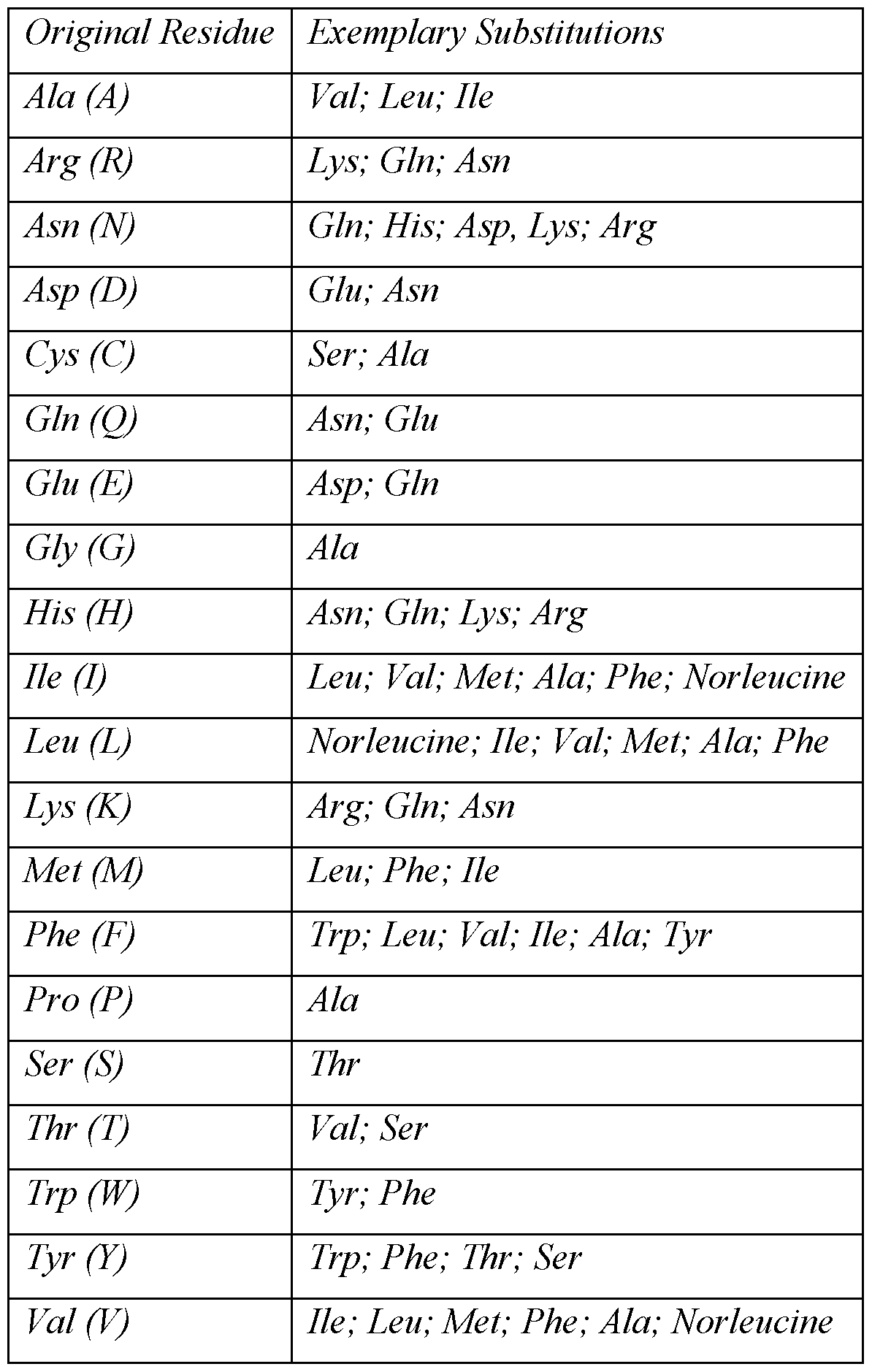
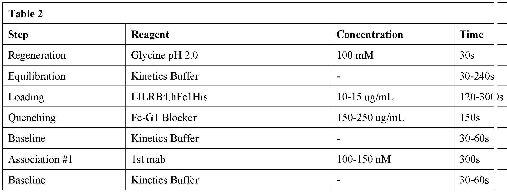

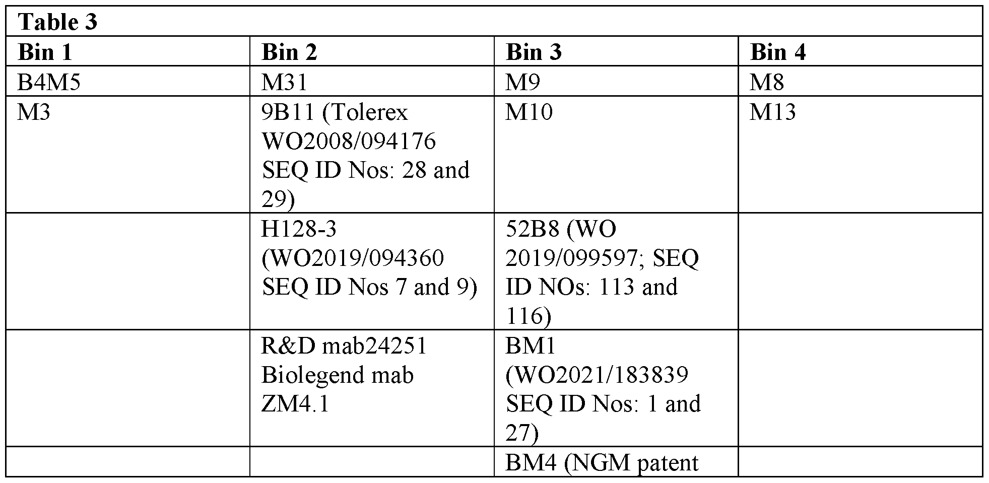


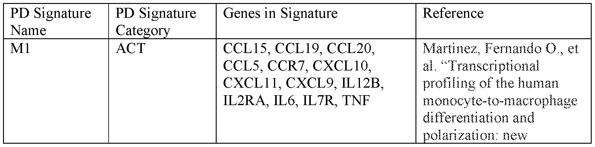

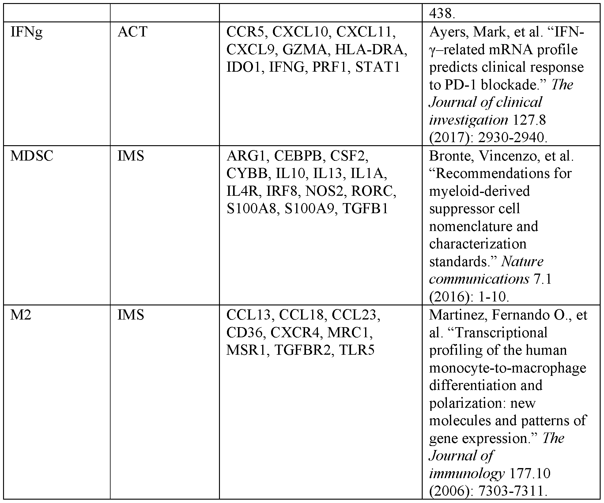


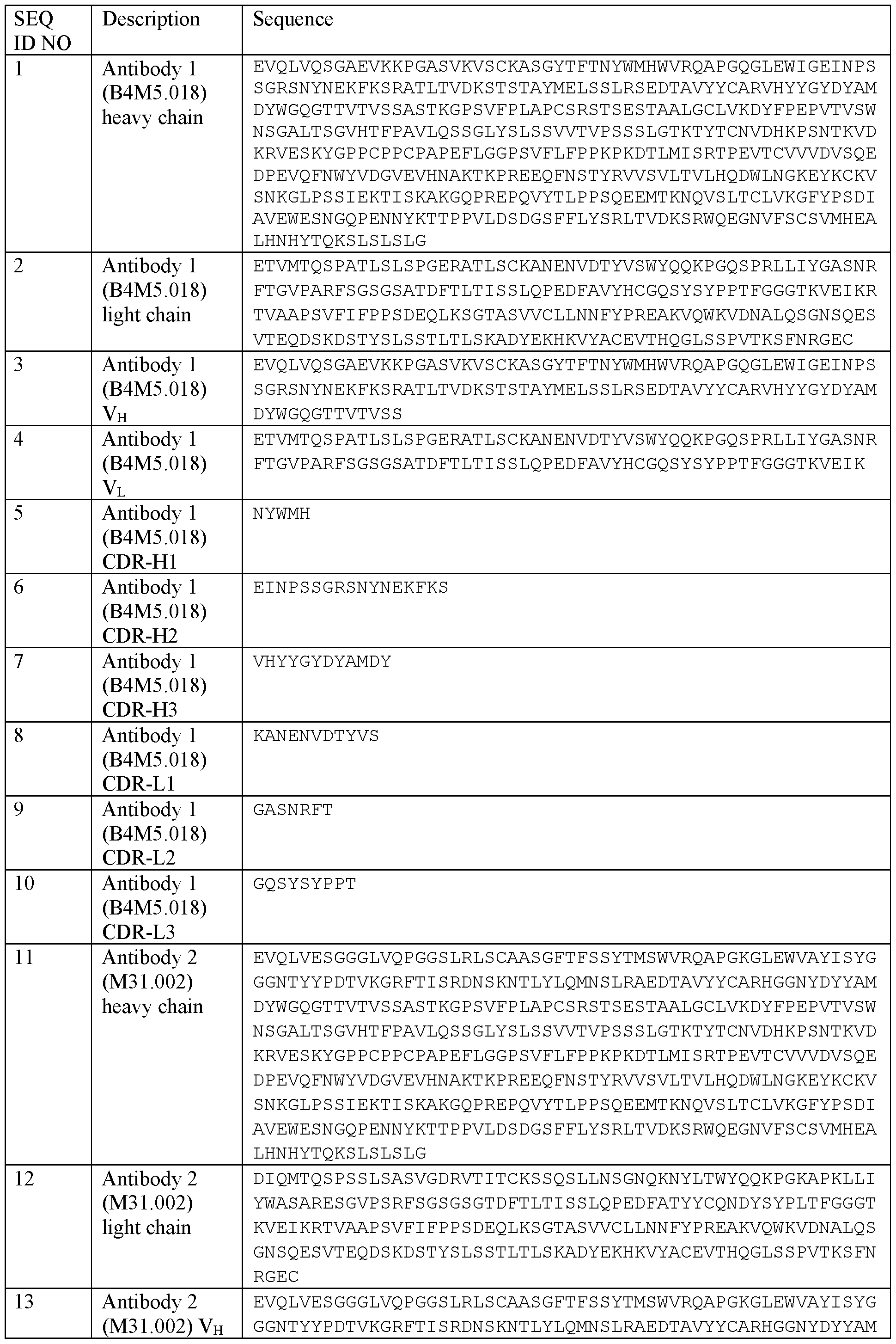
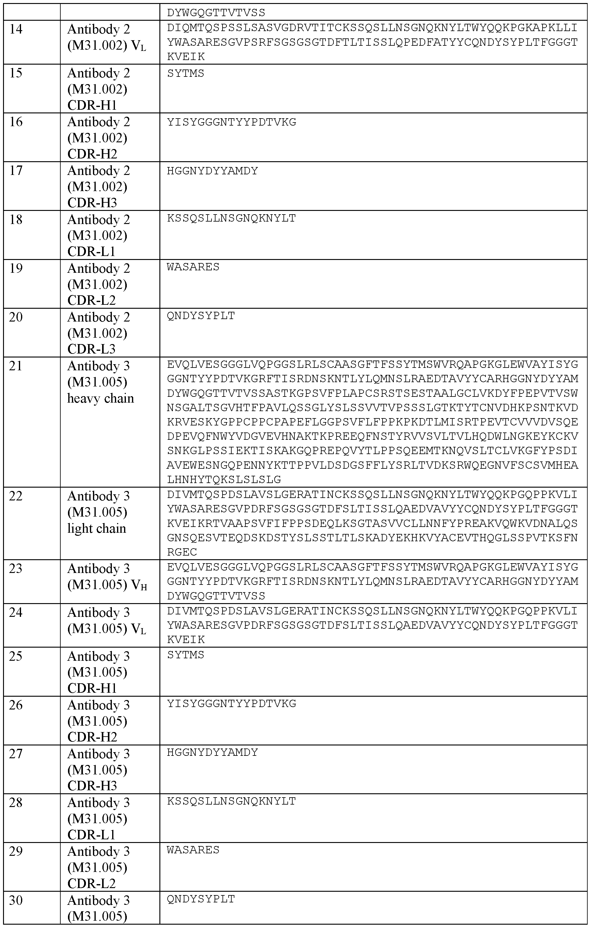

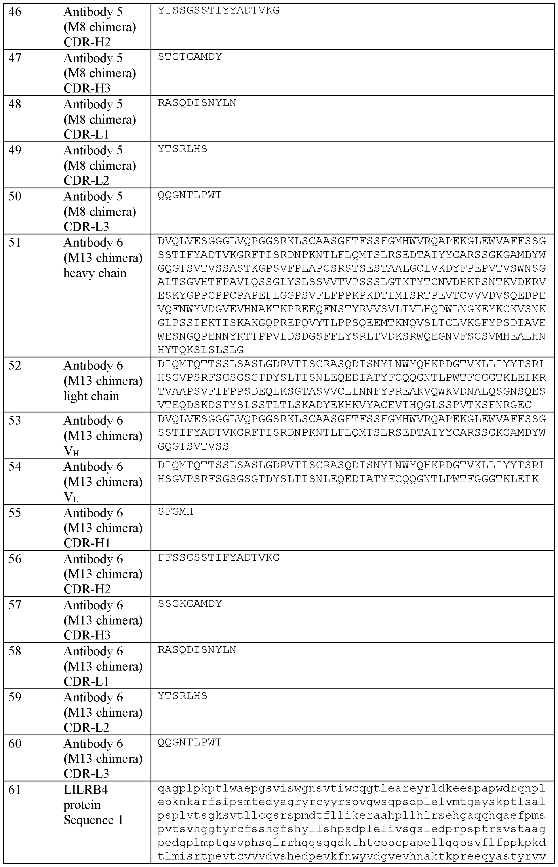
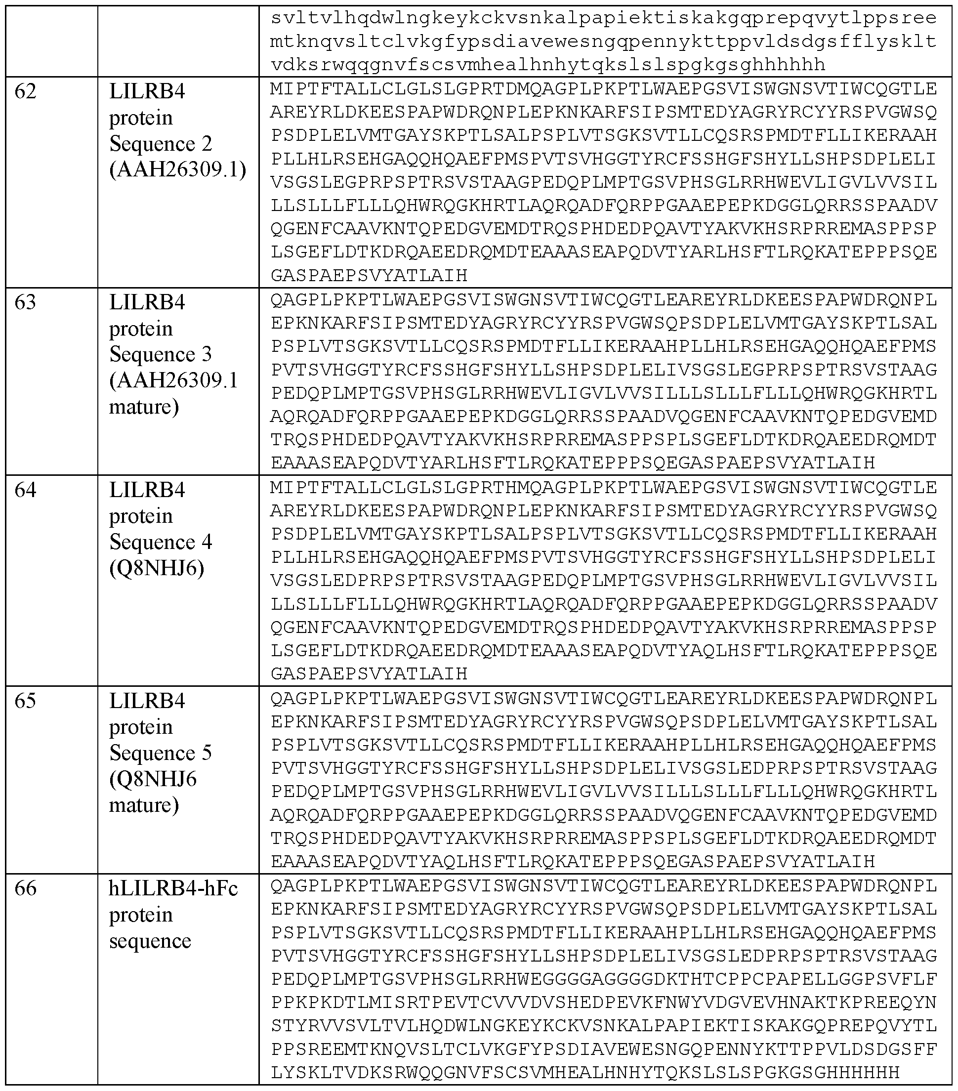
Claims
Applications Claiming Priority (4)
| Application Number | Priority Date | Filing Date | Title |
|---|---|---|---|
| US202263347474P | 2022-05-31 | 2022-05-31 | |
| US63/347,474 | 2022-05-31 | ||
| US202263383084P | 2022-11-10 | 2022-11-10 | |
| US63/383,084 | 2022-11-10 |
Publications (1)
| Publication Number | Publication Date |
|---|---|
| WO2023235699A1 true WO2023235699A1 (en) | 2023-12-07 |
Family
ID=87036381
Family Applications (1)
| Application Number | Title | Priority Date | Filing Date |
|---|---|---|---|
| PCT/US2023/067612 Ceased WO2023235699A1 (en) | 2022-05-31 | 2023-05-30 | Antibodies to lilrb4 and uses thereof |
Country Status (1)
| Country | Link |
|---|---|
| WO (1) | WO2023235699A1 (en) |
Cited By (1)
| Publication number | Priority date | Publication date | Assignee | Title |
|---|---|---|---|---|
| US20220025042A1 (en) * | 2017-11-17 | 2022-01-27 | Merck Sharp & Dohme Corp. | Antibodies specific for immunoglobulin-like transcript 3 (ilt3) and uses thereof |
Citations (67)
| Publication number | Priority date | Publication date | Assignee | Title |
|---|---|---|---|---|
| WO1987004462A1 (en) | 1986-01-23 | 1987-07-30 | Celltech Limited | Recombinant dna sequences, vectors containing them and method for the use thereof |
| US4816567A (en) | 1983-04-08 | 1989-03-28 | Genentech, Inc. | Recombinant immunoglobin preparations |
| US5500362A (en) | 1987-01-08 | 1996-03-19 | Xoma Corporation | Chimeric antibody with specificity to human B cell surface antigen |
| WO1997030087A1 (en) | 1996-02-16 | 1997-08-21 | Glaxo Group Limited | Preparation of glycosylated antibodies |
| US5750373A (en) | 1990-12-03 | 1998-05-12 | Genentech, Inc. | Enrichment method for variant proteins having altered binding properties, M13 phagemids, and growth hormone variants |
| US5770429A (en) | 1990-08-29 | 1998-06-23 | Genpharm International, Inc. | Transgenic non-human animals capable of producing heterologous antibodies |
| US5821337A (en) | 1991-06-14 | 1998-10-13 | Genentech, Inc. | Immunoglobulin variants |
| WO1998058964A1 (en) | 1997-06-24 | 1998-12-30 | Genentech, Inc. | Methods and compositions for galactosylated glycoproteins |
| WO1999010494A2 (en) | 1997-08-25 | 1999-03-04 | Genentech, Inc. | Agonist antibodies to the thrombopoietin receptor, and their therapeutic uses |
| WO1999022764A1 (en) | 1997-10-31 | 1999-05-14 | Genentech, Inc. | Methods and compositions comprising glycoprotein glycoforms |
| WO1999051642A1 (en) | 1998-04-02 | 1999-10-14 | Genentech, Inc. | Antibody variants and fragments thereof |
| US6075181A (en) | 1990-01-12 | 2000-06-13 | Abgenix, Inc. | Human antibodies derived from immunized xenomice |
| WO2000061739A1 (en) | 1999-04-09 | 2000-10-19 | Kyowa Hakko Kogyo Co., Ltd. | Method for controlling the activity of immunologically functional molecule |
| US6150584A (en) | 1990-01-12 | 2000-11-21 | Abgenix, Inc. | Human antibodies derived from immunized xenomice |
| US6194551B1 (en) | 1998-04-02 | 2001-02-27 | Genentech, Inc. | Polypeptide variants |
| WO2001029246A1 (en) | 1999-10-19 | 2001-04-26 | Kyowa Hakko Kogyo Co., Ltd. | Process for producing polypeptide |
| WO2002031140A1 (en) | 2000-10-06 | 2002-04-18 | Kyowa Hakko Kogyo Co., Ltd. | Cells producing antibody compositions |
| US20020164328A1 (en) | 2000-10-06 | 2002-11-07 | Toyohide Shinkawa | Process for purifying antibody |
| WO2003011878A2 (en) | 2001-08-03 | 2003-02-13 | Glycart Biotechnology Ag | Antibody glycosylation variants having increased antibody-dependent cellular cytotoxicity |
| US20030115614A1 (en) | 2000-10-06 | 2003-06-19 | Yutaka Kanda | Antibody composition-producing cell |
| US6602684B1 (en) | 1998-04-20 | 2003-08-05 | Glycart Biotechnology Ag | Glycosylation engineering of antibodies for improving antibody-dependent cellular cytotoxicity |
| US20030157108A1 (en) | 2001-10-25 | 2003-08-21 | Genentech, Inc. | Glycoprotein compositions |
| WO2003084570A1 (en) | 2002-04-09 | 2003-10-16 | Kyowa Hakko Kogyo Co., Ltd. | DRUG CONTAINING ANTIBODY COMPOSITION APPROPRIATE FOR PATIENT SUFFERING FROM FcϜRIIIa POLYMORPHISM |
| WO2003085119A1 (en) | 2002-04-09 | 2003-10-16 | Kyowa Hakko Kogyo Co., Ltd. | METHOD OF ENHANCING ACTIVITY OF ANTIBODY COMPOSITION OF BINDING TO FcϜ RECEPTOR IIIa |
| WO2003085107A1 (en) | 2002-04-09 | 2003-10-16 | Kyowa Hakko Kogyo Co., Ltd. | Cells with modified genome |
| US20040093621A1 (en) | 2001-12-25 | 2004-05-13 | Kyowa Hakko Kogyo Co., Ltd | Antibody composition which specifically binds to CD20 |
| US6737056B1 (en) | 1999-01-15 | 2004-05-18 | Genentech, Inc. | Polypeptide variants with altered effector function |
| US20040109865A1 (en) | 2002-04-09 | 2004-06-10 | Kyowa Hakko Kogyo Co., Ltd. | Antibody composition-containing medicament |
| US20040110282A1 (en) | 2002-04-09 | 2004-06-10 | Kyowa Hakko Kogyo Co., Ltd. | Cells in which activity of the protein involved in transportation of GDP-fucose is reduced or lost |
| WO2004056312A2 (en) | 2002-12-16 | 2004-07-08 | Genentech, Inc. | Immunoglobulin variants and uses thereof |
| US20040132140A1 (en) | 2002-04-09 | 2004-07-08 | Kyowa Hakko Kogyo Co., Ltd. | Production process for antibody composition |
| WO2004092219A2 (en) | 2003-04-10 | 2004-10-28 | Protein Design Labs, Inc | Alteration of fcrn binding affinities or serum half-lives of antibodies by mutagenesis |
| US20050079574A1 (en) | 2003-01-16 | 2005-04-14 | Genentech, Inc. | Synthetic antibody phage libraries |
| WO2005035586A1 (en) | 2003-10-08 | 2005-04-21 | Kyowa Hakko Kogyo Co., Ltd. | Fused protein composition |
| WO2005035778A1 (en) | 2003-10-09 | 2005-04-21 | Kyowa Hakko Kogyo Co., Ltd. | PROCESS FOR PRODUCING ANTIBODY COMPOSITION BY USING RNA INHIBITING THE FUNCTION OF α1,6-FUCOSYLTRANSFERASE |
| US20050119455A1 (en) | 2002-06-03 | 2005-06-02 | Genentech, Inc. | Synthetic antibody phage libraries |
| US20050123546A1 (en) | 2003-11-05 | 2005-06-09 | Glycart Biotechnology Ag | Antigen binding molecules with increased Fc receptor binding affinity and effector function |
| WO2005053742A1 (en) | 2003-12-04 | 2005-06-16 | Kyowa Hakko Kogyo Co., Ltd. | Medicine containing antibody composition |
| US20050266000A1 (en) | 2004-04-09 | 2005-12-01 | Genentech, Inc. | Variable domain library and uses |
| US6982321B2 (en) | 1986-03-27 | 2006-01-03 | Medical Research Council | Altered antibodies |
| US7041870B2 (en) | 2000-11-30 | 2006-05-09 | Medarex, Inc. | Transgenic transchromosomal rodents for making human antibodies |
| WO2006081430A2 (en) | 2005-01-27 | 2006-08-03 | Five Prime Therapeutics, Inc. | Leader sequences for directing secretion of polypeptides and methods for production thereof |
| US7087409B2 (en) | 1997-12-05 | 2006-08-08 | The Scripps Research Institute | Humanization of murine antibody |
| US20060270045A1 (en) | 2003-10-22 | 2006-11-30 | Keck Graduate Institute | Methods of synthesizing heteromultimeric polypeptides in yeast using a haploid mating strategy |
| WO2006138739A2 (en) * | 2005-06-17 | 2006-12-28 | Tolerx, Inc. | Ilt3 binding molecules and uses therefor |
| US7189826B2 (en) | 1997-11-24 | 2007-03-13 | Institute For Human Genetics And Biochemistry | Monoclonal human natural antibodies |
| US20070061900A1 (en) | 2000-10-31 | 2007-03-15 | Murphy Andrew J | Methods of modifying eukaryotic cells |
| US20070117126A1 (en) | 1999-12-15 | 2007-05-24 | Genentech, Inc. | Shotgun scanning |
| US20070160598A1 (en) | 2005-11-07 | 2007-07-12 | Dennis Mark S | Binding polypeptides with diversified and consensus vh/vl hypervariable sequences |
| US20070237764A1 (en) | 2005-12-02 | 2007-10-11 | Genentech, Inc. | Binding polypeptides with restricted diversity sequences |
| US20070292936A1 (en) | 2006-05-09 | 2007-12-20 | Genentech, Inc. | Binding polypeptides with optimized scaffolds |
| WO2008077546A1 (en) | 2006-12-22 | 2008-07-03 | F. Hoffmann-La Roche Ag | Antibodies against insulin-like growth factor i receptor and uses thereof |
| US20090002360A1 (en) | 2007-05-25 | 2009-01-01 | Innolux Display Corp. | Liquid crystal display device and method for driving same |
| US7527791B2 (en) | 2004-03-31 | 2009-05-05 | Genentech, Inc. | Humanized anti-TGF-beta antibodies |
| US7923538B2 (en) | 2005-07-22 | 2011-04-12 | Kyowa Hakko Kirin Co., Ltd | Recombinant antibody composition |
| US7994290B2 (en) | 2007-01-24 | 2011-08-09 | Kyowa Hakko Kirin Co., Ltd | Effector function enhanced recombinant antibody composition |
| US20150352206A1 (en) | 2012-10-26 | 2015-12-10 | The University Of Chicago | Synergistic combination of immunologic inhibitors for the treatment of cancer |
| US20160060237A1 (en) | 2013-03-15 | 2016-03-03 | Bristol-Myers Squibb Company | Ido inhibitors |
| WO2016144728A2 (en) * | 2015-03-06 | 2016-09-15 | The Board Of Regents Of The University Of Texas System | Anti-lilrb antibodies and their use in detecting and treating cancer |
| US20160304610A1 (en) | 2015-03-23 | 2016-10-20 | Jounce Therapeutics, Inc. | Antibodies to icos |
| WO2018089300A1 (en) * | 2016-11-10 | 2018-05-17 | Merck Sharp & Dohme Corp. | Ilt3 ligand |
| WO2018148494A1 (en) * | 2017-02-09 | 2018-08-16 | Bluefin Biomedicine, Inc. | Anti-ilt3 antibodies and antibody drug conjugates |
| WO2019099597A2 (en) | 2017-11-17 | 2019-05-23 | Merck Sharp & Dohme Corp. | Antibodies specific for immunoglobulin-like transcript 3 (ilt3) and uses thereof |
| WO2020056077A1 (en) * | 2018-09-13 | 2020-03-19 | The Board Of Regents Of The University Of Texas System | Novel lilrb4 antibodies and uses thereof |
| WO2020180789A1 (en) * | 2019-03-01 | 2020-09-10 | Board Of Regents, The University Of Texas System | Lilrb4-binding antibody and methods of use thereof |
| WO2021127200A1 (en) * | 2019-12-19 | 2021-06-24 | Ngm Biopharmaceuticals, Inc. | Ilt3-binding agents and methods of use thereof |
| WO2021183839A2 (en) * | 2020-03-12 | 2021-09-16 | Immune-Onc Therapeutics, Inc. | Novel anti-lilrb4 antibodies and derivative products |
-
2023
- 2023-05-30 WO PCT/US2023/067612 patent/WO2023235699A1/en not_active Ceased
Patent Citations (68)
| Publication number | Priority date | Publication date | Assignee | Title |
|---|---|---|---|---|
| US4816567A (en) | 1983-04-08 | 1989-03-28 | Genentech, Inc. | Recombinant immunoglobin preparations |
| WO1987004462A1 (en) | 1986-01-23 | 1987-07-30 | Celltech Limited | Recombinant dna sequences, vectors containing them and method for the use thereof |
| US6982321B2 (en) | 1986-03-27 | 2006-01-03 | Medical Research Council | Altered antibodies |
| US5500362A (en) | 1987-01-08 | 1996-03-19 | Xoma Corporation | Chimeric antibody with specificity to human B cell surface antigen |
| US6075181A (en) | 1990-01-12 | 2000-06-13 | Abgenix, Inc. | Human antibodies derived from immunized xenomice |
| US6150584A (en) | 1990-01-12 | 2000-11-21 | Abgenix, Inc. | Human antibodies derived from immunized xenomice |
| US5770429A (en) | 1990-08-29 | 1998-06-23 | Genpharm International, Inc. | Transgenic non-human animals capable of producing heterologous antibodies |
| US5750373A (en) | 1990-12-03 | 1998-05-12 | Genentech, Inc. | Enrichment method for variant proteins having altered binding properties, M13 phagemids, and growth hormone variants |
| US5821337A (en) | 1991-06-14 | 1998-10-13 | Genentech, Inc. | Immunoglobulin variants |
| WO1997030087A1 (en) | 1996-02-16 | 1997-08-21 | Glaxo Group Limited | Preparation of glycosylated antibodies |
| WO1998058964A1 (en) | 1997-06-24 | 1998-12-30 | Genentech, Inc. | Methods and compositions for galactosylated glycoproteins |
| WO1999010494A2 (en) | 1997-08-25 | 1999-03-04 | Genentech, Inc. | Agonist antibodies to the thrombopoietin receptor, and their therapeutic uses |
| WO1999022764A1 (en) | 1997-10-31 | 1999-05-14 | Genentech, Inc. | Methods and compositions comprising glycoprotein glycoforms |
| US7189826B2 (en) | 1997-11-24 | 2007-03-13 | Institute For Human Genetics And Biochemistry | Monoclonal human natural antibodies |
| US7087409B2 (en) | 1997-12-05 | 2006-08-08 | The Scripps Research Institute | Humanization of murine antibody |
| WO1999051642A1 (en) | 1998-04-02 | 1999-10-14 | Genentech, Inc. | Antibody variants and fragments thereof |
| US6194551B1 (en) | 1998-04-02 | 2001-02-27 | Genentech, Inc. | Polypeptide variants |
| US6602684B1 (en) | 1998-04-20 | 2003-08-05 | Glycart Biotechnology Ag | Glycosylation engineering of antibodies for improving antibody-dependent cellular cytotoxicity |
| US6737056B1 (en) | 1999-01-15 | 2004-05-18 | Genentech, Inc. | Polypeptide variants with altered effector function |
| WO2000061739A1 (en) | 1999-04-09 | 2000-10-19 | Kyowa Hakko Kogyo Co., Ltd. | Method for controlling the activity of immunologically functional molecule |
| WO2001029246A1 (en) | 1999-10-19 | 2001-04-26 | Kyowa Hakko Kogyo Co., Ltd. | Process for producing polypeptide |
| US20070117126A1 (en) | 1999-12-15 | 2007-05-24 | Genentech, Inc. | Shotgun scanning |
| US20020164328A1 (en) | 2000-10-06 | 2002-11-07 | Toyohide Shinkawa | Process for purifying antibody |
| US20030115614A1 (en) | 2000-10-06 | 2003-06-19 | Yutaka Kanda | Antibody composition-producing cell |
| WO2002031140A1 (en) | 2000-10-06 | 2002-04-18 | Kyowa Hakko Kogyo Co., Ltd. | Cells producing antibody compositions |
| US20070061900A1 (en) | 2000-10-31 | 2007-03-15 | Murphy Andrew J | Methods of modifying eukaryotic cells |
| US7041870B2 (en) | 2000-11-30 | 2006-05-09 | Medarex, Inc. | Transgenic transchromosomal rodents for making human antibodies |
| WO2003011878A2 (en) | 2001-08-03 | 2003-02-13 | Glycart Biotechnology Ag | Antibody glycosylation variants having increased antibody-dependent cellular cytotoxicity |
| US20030157108A1 (en) | 2001-10-25 | 2003-08-21 | Genentech, Inc. | Glycoprotein compositions |
| US20040093621A1 (en) | 2001-12-25 | 2004-05-13 | Kyowa Hakko Kogyo Co., Ltd | Antibody composition which specifically binds to CD20 |
| US20040110704A1 (en) | 2002-04-09 | 2004-06-10 | Kyowa Hakko Kogyo Co., Ltd. | Cells of which genome is modified |
| US20040132140A1 (en) | 2002-04-09 | 2004-07-08 | Kyowa Hakko Kogyo Co., Ltd. | Production process for antibody composition |
| US20040110282A1 (en) | 2002-04-09 | 2004-06-10 | Kyowa Hakko Kogyo Co., Ltd. | Cells in which activity of the protein involved in transportation of GDP-fucose is reduced or lost |
| US20040109865A1 (en) | 2002-04-09 | 2004-06-10 | Kyowa Hakko Kogyo Co., Ltd. | Antibody composition-containing medicament |
| WO2003085107A1 (en) | 2002-04-09 | 2003-10-16 | Kyowa Hakko Kogyo Co., Ltd. | Cells with modified genome |
| WO2003085119A1 (en) | 2002-04-09 | 2003-10-16 | Kyowa Hakko Kogyo Co., Ltd. | METHOD OF ENHANCING ACTIVITY OF ANTIBODY COMPOSITION OF BINDING TO FcϜ RECEPTOR IIIa |
| WO2003084570A1 (en) | 2002-04-09 | 2003-10-16 | Kyowa Hakko Kogyo Co., Ltd. | DRUG CONTAINING ANTIBODY COMPOSITION APPROPRIATE FOR PATIENT SUFFERING FROM FcϜRIIIa POLYMORPHISM |
| US20050119455A1 (en) | 2002-06-03 | 2005-06-02 | Genentech, Inc. | Synthetic antibody phage libraries |
| WO2004056312A2 (en) | 2002-12-16 | 2004-07-08 | Genentech, Inc. | Immunoglobulin variants and uses thereof |
| US20050079574A1 (en) | 2003-01-16 | 2005-04-14 | Genentech, Inc. | Synthetic antibody phage libraries |
| WO2004092219A2 (en) | 2003-04-10 | 2004-10-28 | Protein Design Labs, Inc | Alteration of fcrn binding affinities or serum half-lives of antibodies by mutagenesis |
| WO2005035586A1 (en) | 2003-10-08 | 2005-04-21 | Kyowa Hakko Kogyo Co., Ltd. | Fused protein composition |
| WO2005035778A1 (en) | 2003-10-09 | 2005-04-21 | Kyowa Hakko Kogyo Co., Ltd. | PROCESS FOR PRODUCING ANTIBODY COMPOSITION BY USING RNA INHIBITING THE FUNCTION OF α1,6-FUCOSYLTRANSFERASE |
| US20060270045A1 (en) | 2003-10-22 | 2006-11-30 | Keck Graduate Institute | Methods of synthesizing heteromultimeric polypeptides in yeast using a haploid mating strategy |
| US20050123546A1 (en) | 2003-11-05 | 2005-06-09 | Glycart Biotechnology Ag | Antigen binding molecules with increased Fc receptor binding affinity and effector function |
| WO2005053742A1 (en) | 2003-12-04 | 2005-06-16 | Kyowa Hakko Kogyo Co., Ltd. | Medicine containing antibody composition |
| US7527791B2 (en) | 2004-03-31 | 2009-05-05 | Genentech, Inc. | Humanized anti-TGF-beta antibodies |
| US20050266000A1 (en) | 2004-04-09 | 2005-12-01 | Genentech, Inc. | Variable domain library and uses |
| WO2006081430A2 (en) | 2005-01-27 | 2006-08-03 | Five Prime Therapeutics, Inc. | Leader sequences for directing secretion of polypeptides and methods for production thereof |
| WO2006138739A2 (en) * | 2005-06-17 | 2006-12-28 | Tolerx, Inc. | Ilt3 binding molecules and uses therefor |
| US7923538B2 (en) | 2005-07-22 | 2011-04-12 | Kyowa Hakko Kirin Co., Ltd | Recombinant antibody composition |
| US20070160598A1 (en) | 2005-11-07 | 2007-07-12 | Dennis Mark S | Binding polypeptides with diversified and consensus vh/vl hypervariable sequences |
| US20070237764A1 (en) | 2005-12-02 | 2007-10-11 | Genentech, Inc. | Binding polypeptides with restricted diversity sequences |
| US20070292936A1 (en) | 2006-05-09 | 2007-12-20 | Genentech, Inc. | Binding polypeptides with optimized scaffolds |
| WO2008077546A1 (en) | 2006-12-22 | 2008-07-03 | F. Hoffmann-La Roche Ag | Antibodies against insulin-like growth factor i receptor and uses thereof |
| US7994290B2 (en) | 2007-01-24 | 2011-08-09 | Kyowa Hakko Kirin Co., Ltd | Effector function enhanced recombinant antibody composition |
| US20090002360A1 (en) | 2007-05-25 | 2009-01-01 | Innolux Display Corp. | Liquid crystal display device and method for driving same |
| US20150352206A1 (en) | 2012-10-26 | 2015-12-10 | The University Of Chicago | Synergistic combination of immunologic inhibitors for the treatment of cancer |
| US20160060237A1 (en) | 2013-03-15 | 2016-03-03 | Bristol-Myers Squibb Company | Ido inhibitors |
| WO2016144728A2 (en) * | 2015-03-06 | 2016-09-15 | The Board Of Regents Of The University Of Texas System | Anti-lilrb antibodies and their use in detecting and treating cancer |
| US20160304610A1 (en) | 2015-03-23 | 2016-10-20 | Jounce Therapeutics, Inc. | Antibodies to icos |
| WO2018089300A1 (en) * | 2016-11-10 | 2018-05-17 | Merck Sharp & Dohme Corp. | Ilt3 ligand |
| WO2018148494A1 (en) * | 2017-02-09 | 2018-08-16 | Bluefin Biomedicine, Inc. | Anti-ilt3 antibodies and antibody drug conjugates |
| WO2019099597A2 (en) | 2017-11-17 | 2019-05-23 | Merck Sharp & Dohme Corp. | Antibodies specific for immunoglobulin-like transcript 3 (ilt3) and uses thereof |
| WO2020056077A1 (en) * | 2018-09-13 | 2020-03-19 | The Board Of Regents Of The University Of Texas System | Novel lilrb4 antibodies and uses thereof |
| WO2020180789A1 (en) * | 2019-03-01 | 2020-09-10 | Board Of Regents, The University Of Texas System | Lilrb4-binding antibody and methods of use thereof |
| WO2021127200A1 (en) * | 2019-12-19 | 2021-06-24 | Ngm Biopharmaceuticals, Inc. | Ilt3-binding agents and methods of use thereof |
| WO2021183839A2 (en) * | 2020-03-12 | 2021-09-16 | Immune-Onc Therapeutics, Inc. | Novel anti-lilrb4 antibodies and derivative products |
Non-Patent Citations (73)
| Title |
|---|
| ABDICHE ET AL., ANAL. BIOCHEM., vol. 377, 2008, pages 209 - 277 |
| ALMAGROFRANSSON, FRONT. BIOSCI., vol. 13, 2008, pages 1619 - 1633 |
| ANSEL ET AL.: "Pharmaceutical Dosage Forms and Drug Delivery Systems", 2004, LIPPENCOTT WILLIAMS AND WILKINS |
| BACA ET AL., J. BIOL. CHEM., vol. 272, 1997, pages 10678 - 10684 |
| BOERNER ET AL., J. IMMUNOL., vol. 147, 1991, pages 86 |
| BRODEUR ET AL.: "Monoclonal Antibody Production Techniques and Applications", 1987, MARCEL DEKKER, INC., pages: 147 - 158 |
| CAPEL ET AL., IMMUNOMETHODS, vol. 4, 1994, pages 25 - 34 |
| CARRENO BM ET AL., SCIENCE, vol. 348, 2015, pages 6236 |
| CARTER ET AL., PROC. NATL. ACAD. SCI. USA, vol. 89, 1992, pages 4285 |
| CHEN ET AL., J. MOL. BIOL., vol. 293, 1999, pages 865 - 881 |
| CHOO ET AL., BMC BIOINFORMATICS, vol. 6, 2005, pages 249 |
| CHOTHIALESK, J. MOL. BIOL., vol. 196, 1987, pages 901 - 917 |
| CLACKSON ET AL., NATURE, vol. 352, 1991, pages 624 - 628 |
| CLYNES ET AL., PROC. NATL. ACAD. SCI. (USA), vol. 95, 1998, pages 652 - 656 |
| DAERON, ANNU. REV. IMMUNOL., vol. 15, 1997, pages 203 - 234 |
| DAI ET AL., J NATL CANCER INST, vol. 108, no. 7, 2016 |
| DALL'ACQUA ET AL., METHODS, vol. 36, 2005, pages 61 - 68 |
| EMENS LA, EXPERT OPIN EMERG DRUGS, vol. 13, no. 2, 2008, pages 295 - 308 |
| ENDO ET AL., BIOTECHNOL. ADV., vol. 21, 2003, pages 695 - 713 |
| FELLOUSE, PROC. NATL. ACAD. SCI. USA, vol. 101, no. 34, 2004, pages 12467 - 12472 |
| GAZZANO-SANTORO ET AL., J. IMMUNOL. METHODS, vol. 202, 1996, pages 163 |
| GENNARO, REMINGTON: THE SCIENCE AND PRACTICE OF PHARMACY WITH FACTS AND COMPARISONS: DRUGFACTS PLUS, 2003 |
| GHETIE ET AL., NATURE BIOTECHNOLOGY, vol. 15, no. 7, 1997, pages 637 - 640 |
| GHETIEWARD, IMMUNOL. TODAY, vol. 18, no. 12, 1997, pages 592 - 598 |
| GILL ET AL., BLOOD REV, 2015, pages 0268 - 960 |
| GILL ET AL., IMMUNOL REV, vol. 263, no. 1, 2015, pages 68 - 89 |
| GILL ET AL., IMMUNOL. REV, vol. 263, no. 1, 2015, pages 68 - 89 |
| GRIFFITHS ET AL., EMBO J, vol. 12, 1993, pages 725 - 734 |
| GUYER ET AL., J. IMMUNOL., vol. 117, 1976, pages 587 |
| HAAS ET AL., J. LAB. CLIN. MED., vol. 126, 1995, pages 330 - 41 |
| HINTON ET AL., J. BIOL. CHEM., vol. 279, no. 8, 2004, pages 6213 - 6216 |
| HOOGENBOOM ET AL.: "Methods in Molecular Biology", vol. 248, 2003, HUMANA PRESS, article "Epitope Mapping Protocols", pages: 161 - 175 |
| HOOGENBOOMWINTER, J. MOL. BIOL, vol. 227, 1992, pages 381 - 388 |
| HOOGENBOOMWINTER, J. MOL. BIOL., vol. 222, 1991, pages 581 |
| IDUSOGIE ET AL., J. IMMUNOL., vol. 164, 2000, pages 4178 - 4184 |
| JONSSON ET AL., ANN. BIOL. CLIN., vol. 51, 1993, pages 19 - 26 |
| KANDA, Y. ET AL., BIOTECHNOL. BIOENG., vol. 94, no. 4, 2006, pages 680 - 688 |
| KANTOFF PW ET AL., N ENGL JMED, vol. 363, 2010, pages 411 - 22 |
| KIBBE ET AL.: "Handbook of Pharmaceutical Excipients", 2000, PHARMACEUTICAL PRESS |
| KIM ET AL., J. IMMUNOL., vol. 24, 1994, pages 249 |
| KLIMKA ET AL., BR. J. CANCER, vol. 83, 2000, pages 252 - 260 |
| KOHLERMILSTEIN, NATURE, vol. 256, 1975, pages 495 |
| KOZBOR, J. IMMUNOL, vol. 133, 1984, pages 3001 |
| LEE ET AL., J. IMMUNOL. METHODS, vol. 284, 2004, pages 119 - 132 |
| LI ET AL., PROC. NATL. ACAD. SCI. USA, vol. 103, 2006, pages 3557 - 3562 |
| LONBERG, CURR. OPIN. IMMUNOL., vol. 20, 2008, pages 450 - 459 |
| LONBERG, NAT. BIOTECH., vol. 23, 2005, pages 1117 - 1125 |
| MCCAFFERTY ET AL., NATURE, vol. 348, 1990, pages 552 - 554 |
| MORRISON ET AL., PROC. NATL. ACAD. SCI. USA, vol. 81, 1984, pages 6851 - 6855 |
| NEMUNAITIS, EXPERT REV VACCINES, vol. 4, 2005, pages 259 - 74 |
| NI, XIANDAI MIANYIXUE, vol. 26, no. 4, 2006, pages 265 - 268 |
| OKAZAKI ET AL., J. MOL. BIOL., vol. 336, no. 5, 2004, pages 1239 - 1249 |
| PADLAN, MOL. IMMUNOL., vol. 28, 1991, pages 489 - 498 |
| PETKOVA ET AL., INTERNATIONAL IMMUNOLOGY, vol. 18, no. 12, 2006, pages 1759 - 1769 |
| PRESTA ET AL., J. IMMUNOL, vol. 151, 1993, pages 2623 |
| QUEEN ET AL., PROC. NATL ACAD. SCI. USA, vol. 86, 1989, pages 10029 - 10033 |
| RAVETCHKINET, ANNU. REV. IMMUNOL, vol. 9, 1991, pages 457 - 92 |
| RIECHMANN ET AL., NATURE, vol. 332, 1988, pages 323 - 329 |
| RIPKA ET AL., ARCH. BIOCHEM. BIOPHYS, vol. 249, 1986, pages 533 - 545 |
| ROSOK ET AL., J. BIOL. CHEM., vol. 271, 1996, pages 22611 - 22618 |
| RUNNING DEER ET AL., BIOTECHNOL. PROG., vol. 20, 2004, pages 880 - 889 |
| SAMBROOK ET AL.: "Molecular Cloning, A Laboratory Manual", 2001, COLD SPRING HARBOR LABORATORY PRESS |
| SHEETS ET AL., PROC. NATL. ACAD. SCI. (USA, vol. 95, 1998, pages 6157 - 6162 |
| SIMS, J. IMMUNOL., vol. 151, 1993, pages 2296 |
| SITARAMAN ET AL., METHODS MOL. BIOL., vol. 498, 2009, pages 229 - 44 |
| SPIRIN, TRENDS BIOTECHNOL, vol. 22, 2004, pages 538 - 45 |
| VAN DIJKVAN DE WINKEL, CURR. OPIN. PHARMACOL., vol. 5, 2001, pages 368 - 374 |
| VAUGHAN ET AL., NATURE BIOTECHNOLOGY, vol. 14, 1996, pages 309 - 314 |
| VOLLMERSBRANDLEIN, HISTOLOGY AND HISTOPATHOLOGY, vol. 20, no. 3, 2005, pages 927 - 937 |
| VOLLMERSBRANDLEIN, METHODS AND FINDINGS IN EXPERIMENTAL AND CLINICAL PHARMACOLOGY, vol. 27, no. 3, 2005, pages 185 - 191 |
| WINTER ET AL., ANN. REV. IMMUNOL., vol. 12, 1994, pages 433 - 455 |
| WRIGHT ET AL., TIBTECH, vol. 15, 1997, pages 26 - 32 |
| YAMANE-OHNUKI ET AL., BIOTECH. BIOENG., vol. 87, 2004, pages 614 |
Cited By (1)
| Publication number | Priority date | Publication date | Assignee | Title |
|---|---|---|---|---|
| US20220025042A1 (en) * | 2017-11-17 | 2022-01-27 | Merck Sharp & Dohme Corp. | Antibodies specific for immunoglobulin-like transcript 3 (ilt3) and uses thereof |
Similar Documents
| Publication | Publication Date | Title |
|---|---|---|
| US11905329B2 (en) | Anti-PD-1 antibodies and uses thereof | |
| US20230053631A1 (en) | Antibodies to lilrb2 | |
| EP3273992B1 (en) | Antibodies to icos | |
| WO2023235699A1 (en) | Antibodies to lilrb4 and uses thereof | |
| HK40073610A (en) | Antibodies to pd-1 and uses thereof | |
| HK40039832A (en) | Antibodies to icos | |
| HK40014070A (en) | Antibodies to pd-1 and uses thereof | |
| HK40014070B (en) | Antibodies to pd-1 and uses thereof | |
| HK40038468A (en) | Antibodies to lilrb2 |
Legal Events
| Date | Code | Title | Description |
|---|---|---|---|
| 121 | Ep: the epo has been informed by wipo that ep was designated in this application |
Ref document number: 23734862 Country of ref document: EP Kind code of ref document: A1 |
|
| NENP | Non-entry into the national phase |
Ref country code: DE |
|
| 122 | Ep: pct application non-entry in european phase |
Ref document number: 23734862 Country of ref document: EP Kind code of ref document: A1 |
|
| 32PN | Ep: public notification in the ep bulletin as address of the adressee cannot be established |
Free format text: NOTING OF LOSS OF RIGHTS PURSUANT TO RULE 112(1) EPC (EPO FORM 1205A DATED 01/04/2025) |
|
| 122 | Ep: pct application non-entry in european phase |
Ref document number: 23734862 Country of ref document: EP Kind code of ref document: A1 |