WO2023230524A1 - Compositions of secretory and/or catalytic cells and methods using the same - Google Patents
Compositions of secretory and/or catalytic cells and methods using the same Download PDFInfo
- Publication number
- WO2023230524A1 WO2023230524A1 PCT/US2023/067422 US2023067422W WO2023230524A1 WO 2023230524 A1 WO2023230524 A1 WO 2023230524A1 US 2023067422 W US2023067422 W US 2023067422W WO 2023230524 A1 WO2023230524 A1 WO 2023230524A1
- Authority
- WO
- WIPO (PCT)
- Prior art keywords
- cells
- secretory
- composition
- catalytic
- ascs
- Prior art date
- Legal status (The legal status is an assumption and is not a legal conclusion. Google has not performed a legal analysis and makes no representation as to the accuracy of the status listed.)
- Ceased
Links
Classifications
-
- A—HUMAN NECESSITIES
- A61—MEDICAL OR VETERINARY SCIENCE; HYGIENE
- A61K—PREPARATIONS FOR MEDICAL, DENTAL OR TOILETRY PURPOSES
- A61K35/00—Medicinal preparations containing materials or reaction products thereof with undetermined constitution
- A61K35/12—Materials from mammals; Compositions comprising non-specified tissues or cells; Compositions comprising non-embryonic stem cells; Genetically modified cells
- A61K35/35—Fat tissue; Adipocytes; Stromal cells; Connective tissues
-
- A—HUMAN NECESSITIES
- A61—MEDICAL OR VETERINARY SCIENCE; HYGIENE
- A61K—PREPARATIONS FOR MEDICAL, DENTAL OR TOILETRY PURPOSES
- A61K35/00—Medicinal preparations containing materials or reaction products thereof with undetermined constitution
- A61K35/12—Materials from mammals; Compositions comprising non-specified tissues or cells; Compositions comprising non-embryonic stem cells; Genetically modified cells
- A61K35/30—Nerves; Brain; Eyes; Corneal cells; Cerebrospinal fluid; Neuronal stem cells; Neuronal precursor cells; Glial cells; Oligodendrocytes; Schwann cells; Astroglia; Astrocytes; Choroid plexus; Spinal cord tissue
-
- A—HUMAN NECESSITIES
- A61—MEDICAL OR VETERINARY SCIENCE; HYGIENE
- A61K—PREPARATIONS FOR MEDICAL, DENTAL OR TOILETRY PURPOSES
- A61K35/00—Medicinal preparations containing materials or reaction products thereof with undetermined constitution
- A61K35/12—Materials from mammals; Compositions comprising non-specified tissues or cells; Compositions comprising non-embryonic stem cells; Genetically modified cells
- A61K35/37—Digestive system
- A61K35/39—Pancreas; Islets of Langerhans
-
- A—HUMAN NECESSITIES
- A61—MEDICAL OR VETERINARY SCIENCE; HYGIENE
- A61K—PREPARATIONS FOR MEDICAL, DENTAL OR TOILETRY PURPOSES
- A61K35/00—Medicinal preparations containing materials or reaction products thereof with undetermined constitution
- A61K35/12—Materials from mammals; Compositions comprising non-specified tissues or cells; Compositions comprising non-embryonic stem cells; Genetically modified cells
- A61K35/55—Glands not provided for in groups A61K35/22 - A61K35/545, e.g. thyroids, parathyroids or pineal glands
-
- A—HUMAN NECESSITIES
- A61—MEDICAL OR VETERINARY SCIENCE; HYGIENE
- A61P—SPECIFIC THERAPEUTIC ACTIVITY OF CHEMICAL COMPOUNDS OR MEDICINAL PREPARATIONS
- A61P5/00—Drugs for disorders of the endocrine system
Definitions
- compositions comprising secretory and/or catalytic cells and methods of making and using the same that are useful for the treatment or prevention of a disease or disorder, e.g., in a mammalian subject, such as a human.
- HPA hyperphenylalaninemia
- PAH phenylalanine hydroxylase
- EPO erythropoietin
- the disclosure provides an allogenic, long-acting composition
- a therapeutically effective amount of substantially pure secretory and/or catalytic cells wherein the composition is modified for improved therapeutic efficacy relative to an unmodified composition, wherein the improved therapeutic efficacy is, upon administration to a subject, one or both of: increased immunotolerance or decreased immunogenicity.
- the cells of the composition exhibit decreased major histocompatibility complex (MHC) activity relative to an unmodified composition.
- the cells of the composition exhibit increased activity of one or more checkpoint inhibitors relative to an unmodified composition.
- the one or more checkpoint inhibitors are selected from CTLA-4, PDU, PDL2, PDI, B7-H3, B7-H4, BTLA, HVEM, TIM3, GAL9, LAG3, VISTA, KIR, 2B4, CD160, CGEN- 15049, CHK 1 , CHK2, A2aR, and B-7 family ligands or a combination thereof.
- the decreased immunogenicity comprises a decrease or elimination of activity or levels of endogenous proteins and/or genes and/or an increase in the activity or expression of endogenous and exogenous proteins relative to an unmodified composition. In some embodiments, the decreased immunogenicity comprises a decrease or elimination of the activity or levels of endogenous proteins comprising a knock down of the endogenous proteins and/or genes. In some embodiments, the knock down is transient or permanent.
- the transient knock down is performed using a viral vector such as AAV, AdV, or HSV, or a non-viral method such as physical method such as electroporation, mechanoporation, or sonoporation, or a chemical method such as nanoparticle encapsulation, wherein the nanoparticle optionally comprises one or more of a lipid, a polymer, and a peptide.
- the permanent knock down is performed using a lentivirus or retrovirus.
- the knock down is performed using one or more of a shRNA, siRNA, antisense oligonucleotides, systems comprising a template-directed nuclease, Zinc finger nucleases, TALENs, meganucleases, and a homing nuclease, optionally wherein the knock down targets one or more genes selected from B2M, NLRC5, HLA-DR, and CD80/86.
- the increase in the expression of endogenous and exogenous proteins comprises introducing copies of one or more genes.
- the one or more genes are introduced using a lentivirus, a retrovirus, or a plasmid.
- the one or more genes are selected from CD59, CD46, CD55, C1 -inhibitor protease, HLA-C, HLA-E, HLA-G, HLA-E heavy chain, HLA-F, CD52, CD47, CTLA4-lg, PD-L1 , IDO1 , FasL, IL-35, IL-39, IL-10, bovine herpes virus type 1 , Epstein-Barr virus, human cytomegalovirus (e.g.
- HCMV such as HCMV94, US2, US3, US6, US11 , and US11
- the composition further comprises and/or is co-administered and/or coformulated with one or more immunosuppressants, optionally wherein the one or more immunosuppresants are administered over a fixed period of time or chronically.
- the one or more immunosuppresants are selected from: a) a polymer and/or copolymer (e.g. polaxamer P188); b) a small molecule, optionally selected from:
- a calcineurin inhibitor e.g. tacrolimus, cyclosporin A, cyclosporine
- MTOR inhibitor e.g. sirolimus, rapamycin, everolimus
- corticosteroid and/or steroid e.g. a glucocorticoid
- a chemotherapeutic e.g. azathioprine, methotrexate, 6-mercaptopurine, cyclophosphamide
- IMPDH inosine monophosphate dehydrogenase
- mycophenolate mofetil e.g. mycophenolate mofetil
- IMPDH inosine monophosphate dehydrogenase
- a nucleoside analogue e.g. bredinin
- a biologic optionally selected from: i. an antibody or antibody fragment such as a Fab, an F(ab')2, an Fv, a domain antibody, a single-chain antibody, and a nanobody, optionally selected from an anti-T cell antibody (e.g. Anti-PD1 , Anti-CTLA-4, anti-CD3 (OKT3)), an anti-complement protein (e.g. C5, C6, 07, 08, or C9), an anti-CD25 (e.g. daclizumab, Basiliximab), and an anti-CD20 (e.g.
- an antibody or antibody fragment such as a Fab, an F(ab')2, an Fv, a domain antibody, a single-chain antibody, and a nanobody
- an anti-T cell antibody e.g. Anti-PD1 , Anti-CTLA-4, anti-CD3 (OKT3)
- an anti-complement protein e.g.
- a nucleic acid encoding any of the foregoing ii. a soluble complement receptor (e.g. CD59, CD55, CD46), or a nucleic acid encoding any of the foregoing and ill.
- an i.v. administered immunoglobulin G optionally selected from a. subclass of antithymocytes IgG (e.g. Thymoglobulin) and a subclass of IgG fusion proteins such as the Fc fragment of a human lgG1 immunoglobulin linked to the extracellular domain of CTLA-4 (e.g. Belatacept), or a nucleic acid encoding any of the foregoing.
- the disclosure provides an allogenic, long-acting composition
- a therapeutically effective amount of secretory and/or catalytic cells wherein the composition is modified for improved therapeutic efficacy relative to an unmodified composition, wherein the improved therapeutic efficacy is, upon administration to a subject, one or both of: increased immunotolerance or decreased immunogenicity, wherein the composition is modified by disposal in the lumen of in a substantially sealed biocompatible device, optionally wherein the device is permanent or degradable (e.g. biodegradeable) and/or dissolvable. In some embodiments, the device is permanent or biodegradeable and/or dissolvable.
- the device comprises electrospun materials, fibers, and/or fiber members comprising the composition, or a gel and/or gel scaffold, optionally a biodegradable gel and/or a hybrid inorganic biodegradable gel, comprising the composition.
- the device is non- porous or porous, optionally comprising one or more pores selected from nanopores having a diameter of less than about 20 nm; micropores having a diameter in the range of about 10 nm to about 20 pm; and/or macropores having a diameter greater than about 20 pm.
- the device is of a size of about 500 pm or less, about 400 pm or less, about 300 pm or less, about 200 pm or less, about 150 pm or less, about 100 pm or less, about 90 pm or less, about 80 pm or less, about 70 pm or less, about 60 pm or less, about 50 pm or less, about 40 pm or less, about 30 pm or less, or about 20 pm or less.
- the device is a permanent device, optionally comprising polyethylene terephthalate (PET), poly(butylene terephthalate) (PBT), and/or polyurethane (PU).
- PET polyethylene terephthalate
- PBT poly(butylene terephthalate)
- PU polyurethane
- the device comprises electrospun materials, fibers, and/or fiber members, optionally comprising polymeric electrospun materials, fibers, and/or fiber members further optionally wherein the device is semi-permeable, allowing water and nutrient exchange, and limiting cell transfer and optionally limiting transfer of, for example, immunoglobulins.
- the electrospun materials, fibers, and/or fiber members comprise one or more polymers selected from: a) non-resorbable polymers (e.g.
- polyethylene polyethylene oxide, polyethylene terephthalate, (PET) polyester, polymethylmethacrylate, polyacrylonitrile, silicone, polyurethane, polycarbonate, polyether ketone ketone, polyether ether ketone, polyether imide, polyamide, polystyrene, polyether sulfone, polysulfone, polyvinyl acetate, polytetrafluoroethylene, polyvinylidene fluoride, copolymers thereof, or combinations thereof); b) resorbable polymers (e.g.
- polycaprolactone poly(lactide-co-caprolactone), poly(lactide-co- glycolide), polyglycolide, polylactic acid, including derivatives thereof such as, without limitation, poly(L-lactic acid), and poly(D, L-lactic acid), polyglycolic acid, polydioxanone, poly(- hydroxybutyrate-co-3-hydroxyvalerate), trimethylene carbonate, polydiols, polyesters, polyethylene terephthalate (PET), polyurethane, polyethylene, polyethylene oxide, polymethylmethacrylate, polyacrylonitrile, silicone, polycarbonate, polyether ketone ketone, polyether ether ketone, polyether imide, polyamide, polystyrene, polyether sulfone, polysulfone, polyvinyl acetate, polytetrafluoroethylene, polyvinylidene fluoride, polyglycolic acid, polydioxanone, collagen, gelatin, fibrin, fibro
- the electrospun materials, fibers, and/or fiber members are formed into a flat shape, such as a sheet or sheet-like fiber mold, a fiber scaffold, a pouch, and/or tube, or a tubular lattice.
- the electrospun materials, fibers, and/or fiber members have a diameter less than about 20 pm, about 10 pm, about 5 pm, about 2 pm, about 1 pm, about 0.5 pm, about 0.2 pm, or about 0.1 pm.
- the device is degradable (e.g. biodegradeable) and/or dissolvable. In some embodiments, the device is biodegradeable and/or dissolvable.
- the device comprises a biodegradable gel, optionaly wherein the composition is added to the gel prior to polymerization of the gel or the composition is added to the gel after polymerization of the gel, optionally wherein the biodegradable gel is a hydrogel and/or a hybrid inorganic biodegradable gel.
- the biodegradable gel is degraded enzymatically or hydrolytically.
- the biodegradable gel comprises one or more hydrogels, optionally selected from: a) naturally derived hydrogels, optionally selected from:
- Protein-based gels e.g. collagen, fibrin, gelatin, elastin-like peptides, fibrinogen, selfassembling peptides, elastin-like polypeptides
- Polysaccharide-based gels e.g. alginate, alginate-co-gelatin, styrenated gelatin, chitosan, Chondroitin sulfa, hyaluronic acid, chitin
- modified gels of any one of I. -ill e.g. comprising one or more polyethylene glycol (PEG) moieties and/or one or more RGD oligopeptides
- synthetic hydrogels optionally selected from:
- Biodegradable PEG-based gels e.g. macromers include triblock copolymers of poly(a- hydroxy esters)-b-poly(ethylene glycol)-b-poly(a-hydroxy esters) endcapped with(meth)acrylate functional groups, poly(a-hydroxy esters) (e.g. PLA, poly(8- caprolactone) (PCL)); ii. Polyfumarate-based hydrogels (e.g. macromers including poly(lactide-co-ethylene oxide-co-fumerate) and MMP-diacrylate); and
- phosphoester-based hydrogels e.g. poly(6-aminohexyl propylene phosphate)-acrylate.
- the hydrogel comprises a stiffness range of about 0.1 to about 500 kPa, e.g., about 0.1 to about 10 kPa, about 0.5 to about 15 kPa, about 1 to about 15 kPa, about 5 to about 20 kPa, about 10 to about 50 kPa, about 20 to about 100 kPa, about 150 to about 300 kPa, about 100 to about 400 kPa, about 200 to about 450 kPa or about 250 to about 500 kPa.
- each cell containing hydrogel capsule is characterized by a stiffness of about 10 kPa, about 15 kPa, about 20 kPa, about 25 kPa, about 30 kPa, about 35 kPa, about 40 kPa, about 45 kPa, about 50 kPa, about 55 kPa, about 60 kPa, about 65 kPa, about 70 kPa, about 75 kPa, about 80 kPa, about 85 kPa, about 90 kPa, or about 95 kPa or about 100 kPa.
- the hydrogel comprises a water content of more than about 20% w/w, about 30% w/w, about 40% w/w, about 50% w/w, about 60% w/w, about 70% w/w, about 80% w/w, about 90% w/w, or about 95% w/w.
- the gel is present in one or more layers, optionally wherein the one or more layers have a thickness ranging from about 0.5 m to about 20 pm, or wherein the gel microencapsulates the secretory and/or catalytic cellsinto droplets.
- the disclosure provides a method for treating, preventing, or ameliorating a disease or disorder in a subject in need thereof, comprising administering a composition of the disclosure to the subject.
- the composition is an allogenic, long-acting composition comprising a therapeutically effective amount of secretory and/or catalytic cells, wherein the composition is modified for improved therapeutic efficacy relative to an unmodified composition, wherein the improved therapeutic efficacy is, upon administration to a subject, one or both of: increased immunotolerance or decreased immunogenicity.
- the disclosure provides a use of a composition of the disclosure in the manufacture of a medicament for treating, preventing, or ameliorating a disease or disorder.
- the composition is an allogenic, long-acting composition comprising a therapeutically effective amount of secretory and/or catalytic cells, wherein the composition is modified for improved therapeutic efficacy relative to an unmodified composition, wherein the improved therapeutic efficacy is, upon administration to a subject, one or both of: increased immunotolerance or decreased immunogenicity.
- the disease or disorder is a lipodystrophy characterized by insufficient leptin function in a human subject, wherein the cell is an adipogenic cell, such as an ASC-derived adipocyte, optionally wherein the cell is allogeneic to the human subject.
- an adipogenic cell such as an ASC-derived adipocyte
- Figures 1A-1 B depict representative images of human ASCs ( Figure 1A) and murine ASCs ( Figure 1 B) in culture after 2 passages.
- ASCs were isolated from adipose tissues using either the enzymatic digestion method or the explant culture method described in Example 1. Isolated ASCs were expanded in culture, and their images were captured using transmitted light and 20X in a M5000 EVOS imaging system.
- Figures 2A-2B depict experimental data demonstrating the characterization of surface markers of ASCs isolated from human adipose tissues and expanded in culture. The cells were stained with fluorophore-conjugated antibodies against CD29, CD73, CD90, CD105, CD31 , CD45, and CD34 and analyzed with flow cytometry.
- Figure 2A depicts experimental data representative of gating strategy for stained ASCs. Most of the ASCs (>97%) are positive for CD73, CD105, and CD90 and negative for CD34, CD45, and CD31 .
- Figure 2B depicts distributions of fluorescence intensity for different cell surface markers in unstained vs stained ASCs. Stained ASCs display a homogenous normal distribution for both positive and negative markers. Unstained cells are represented as dash lines and stained cells as solid lines.
- Figures 3A-3B depict experimental data demonstrating the characterization of adipocytes derived from ASC differentiation in culture.
- Figure 3A depicts Oil Red 0 staining of ASCs and differentiated ASCs. The cells were fixed with 10% formaldehyde and stained with Oil Red O solution. The images were captured using RBG transmitted light with a 20X objection in an M5000 EVOS imaging system. Oil Red O binds to neutral lipids and stains lipid droplets dark red. In the differentiated culture, >80% of the cells are round in shape and contain a large number of lipid droplets, shown as dark spheres in the right image. These are differentiated adipocytes.
- Figure 3B depicts gene expression levels of adipocyte-specific genes in undifferentiated ASCs and differentiated ASCs.
- the gene expression levels for adiponectin, PPAR y, leptin, CIDEC, and FABP4 were quantified using RT-PCR and normalized to actin. All expression levels were then normalized to control (undifferentiated ASCs). All adipocyte-specific genes are significantly upregulated in the differentiated ASCs compared to control.
- Figure 4 depicts a human adiponectin promoter mapping.
- Minimal elements of human adiponectin promoter include the adiponectin distal enhancer (-2667 to -2507 bp) and the adiponectin proximal promoter region (-540 to +77 bp).
- the distal enhancer contains 2 binding sites for the transcription factor C/EBPct.
- the distal enhancer and proximal promoter region together are both necessary and sufficient for transcriptional activation of the human adiponectin promoter.
- Figure 5 depicts aP2/FABP4 promoter mapping.
- Minimal elements of ap2 promoter include the aP2 distal enhancer (-5.4 kb to -4.9 kb) and the ap2 proximal promoter region (-63 to +21 bp).
- the distal enhancer and proximal promoter region together are necessary and sufficient for transcriptional activation of the aP2 promoter.
- Figures 6A-6B depict experimental data showing long-term engraftment of adipocytes derived from transplanted human ASCs in mice (in vivo).
- Human adipsin ( Figure 6A) and FABP4 ( Figure 6B) were detected at day 117 post-transplant in the dorsal flank.
- Figure 7 depicts experimental data demonstrating in vivo secretion of gaussia luciferase by adipocytes derived from transplanted genetically modified adipogenic cells and long-term engraftment of adipocytes derived from transplanted human ASCs in mice (in vivo). Donor-derived adipocytes expressed GLuc for at least 84 days in recipient mice.
- Figure 8 depicts experimental data demonstrating transplantation of adipocytes and in vivo secretion of adipsin. Human adipsin level was detected in plasma up to 126 days post transplantation.
- Figures 9A-9F depicts experimental data demonstrating non immunogeneic adipogenic cells (in vivo). No innate immune response was detected at 5 hours and day 5 post transplantation in hASCs and culture-derived hAdipocytes. Levels of TNFct (Figure 9A), I FNy ( Figure 9B), IL1 p ( Figure 9C), IL6 ( Figure 9D), IL10 ( Figure 9E), and IL2 ( Figure 9F) were measured.
- Figure 10 depicts experimental data demonstrating non immunogeneic adipogenic cells (in vitro).
- Figures 11 A-11 B depict images demonstrating long-term engraftment of xenografted human adipose cells in immune competent mice (in vivo) at days 92 ( Figure 11 A) and 151 post implantation ( Figure 11 B).
- Figures 12A-12B depict experimental data demonstrating localized biodistribution of transplanted adipocytes.
- Figure 12A depicts experimental data demonstrating that luciferase analyzed from day 3-day 98 post transplantation was detected at all timepoints in mice measured in transplant- naive mice and mice transplanted with adipocytes.
- Figure 12B depicts images of luciferase activity in mice measured at day 14 and day 98.
- Figures 13A-13C depict experimental data demonstrating the increased adipogenic potentiaton of CD10+ cells.
- CD10+ selected ASC populations produced adipocytes that secrete significantly higher levels of adiponectin compared to the control and CD10-.
- Figure 13A depicts a schematic for a non-limiting method of culturing and differentiating adipose stem cells into adipocytes.
- Figure 13B depicts images demonstrating ASCs at day 7 post induction.
- Figure 13C depicts experimental data demonstrating adiponectin protein in media at day 7 for control, CD10+ and CD10- adipocytes.
- Figure 14A-14B depict experimental data demonstrating the ability to generate and characterize adipocytes that secrete a mammalian serum protein.
- Figure 14A depicts a schematic for a non-limiting method of preparing adipocytes that secrete EPO.
- Figure 14B depicts experimental data demonstrating adipocyte specific EPO expression (in vitro). Levels of hEPO in hEPO engineered cells and unengineered control cells were detected.
- Figure 15A depicts a schematic for a non-limiting method of preparing adipocytes that secrete gaussia luciferase (GLuc).
- Figure 15B depicts experimental data demonstrating adipocyte specific gLUC expression in vitro). Engineered ASCs secreted more GLuc as they were further differentiated into adipocytes.
- Figures 16A-16D depict experimental data demonstrating the therapeutic effects in mice by transplanting ASCs and adipogenic cells genetically modified to secrete EPO. Levels in the mice transplanted with hEPO expressing ASCs and adipocytes rose above the levels in the control mice and remained higher for 30+ days. Figures 16A and 16C depict experimental data demonstrating EPO levels in plasma. Figures 16B and 16D depict experimental data demonstrating reticulocyte counts.
- Figures 17A-17D depict experimental data demonstrating that allogeneic ASCs of the disclosure are non-immunogenic as demonstrated by a lack of cell death in mixed lymphocyte assays.
- Figure 18 depicts experimental data demonstrating B2M -/- ASCs molecular validation by flow cytometry
- B2M -/- ASCs and WT ASCs isolated from the inguinal fat pad of mice via enzymatic digestion were stained with fluorescent antibodies against CD90.2, MHC I and MHC II.
- B2M -/- ASCs and WT ASCs exhibited high expression of CD90.2 and minimal expression of MHC II.
- B2M -/- ASCs showed lower MHC I expression compared to WT ASCs.
- Figure 19A depicts images of cell morphology and lipid drop formation.
- B2M -I- and WT murine ASCs produced lipid droplets after 6 days of exposure to differentiation media, and not in the absence of differentiation media.
- Figure 19B depicts the quantification of adiponectin expression using an adiponectin ELISA kit.
- B2M -/- and WT murine ASCs were found to produce higher levels of adiponectin after exposure to differentiation media versus without exposure to differentiation media.
- Figure 20A depicts a graph of experimental data demonstrating longitudinal bioluminescent imaging of adipocytes differentiated from hASCs engineered to express Flue transplanted into NSG- SGM3 mice humanized with CD34+ cells, C57BL/6 mice, and SCID mice. Fluc+ human adipocytes were transplanted subcutaneously into the dorsal side of SCID, NSG-SGM3 mice humanized with CD34+ cells, and C57BL/6 mice. Transplantation persistence was quantified via IVIS imaging and was found to be similar in the humanized NSG-SGM3 mice versus the SCID mice.
- Figure 20B depicts a graph of experimental data demonstrating longitudinal bioluminescent imaging of adipocytes differentiated from hASCs engineered to express Flue transplanted into NSG-SGM3 mice humanized with CD34+ cells.
- human iPSCs engineered to express Flue were transplanted into NSG-SGM3 mice humanized with CD34+ cells as a positive rejection control.
- This experiment included two different CD34+ human donors, both were fully H LA-mismatched with the human adipocyte donor whereas donor #1 was partially matched with the human iPSC donor.
- Both Fluc+ human adipocytes and human iPSCs were transplanted subcutaneously into the dorsal side of NSG-SGM3 mice humanized with CD34+ cells. Transplantation persistence was quantified via IVIS imaging.
- Figure 21 depicts an electrospun chamber IVIS imaging of human adipose stem cells (ASCs) to check for cell survival before transplantation.
- ASCs adipose stem cells
- Figure 22 depicts a timeline of electrospun chamber post-surgery IVIS imaging.
- Results include IVIS imaging for day 2, 7, 14, 21 , 28, and 35 days after transplantation, followed by biweekly measurements until the signal dropped below the detection threshold or until Day 215, whichever occurred first.
- Fluc+ human ASCs were loaded in electrospun cell chambers and surgically inserted into the dorsal side of SCID and C57BL/6 mice.
- mock surgeries transplanted a matching amount of Fluc+ human ASCs (as free cells in solution) into an equivalent surgical pocket.
- Fluc+ human ASCs were also injected subcutaneously in SCID mice. Cell survival was quantified via IVIS imaging.
- the encapsulated ASCs exhibit luciferase signal for a longer period of time versus the unencapsulated ASCs transplanted as mock surgeries.
- SCID mice the difference between encapsulated ASCs and free ASCs transplanted in a mock surgery was less apparent than in C57BL/6 mice.
- Figures 23A-23B depict a graph of experimental data demonstrating the tracking of cell survival over time starting on day 7 using IVIS imaging.
- Fluc+ WT murine ASCs were injected subcutaneously into C57BL/6 ( Figure 23A) and NSG mice ( Figure 23B) with and without poloxamer P188. Cell survival post transplantation was assessed via IVIS imaging.
- Fluc+ mASCs co-administered with P188 exhibited higher fluorescent signal for a longer period of time versus Fluc+ mASCs on their on in the immunocompetent C57BL/6 mice, but not in the NSG mice.
- Figure 24 depicts a graph of experimental data comparing levels of IDUA expression in engineered cells to wild type cells.
- Human ASCs were transiently transfected with increasing amounts of plasmid encoding IDUA.
- IDUA expression was compared against un-transfected ASCs using a fluorescent cell lysate assay and against an IDUA KO cell line. The transfected ASCs were found to express higher amounts of IDUA versus unengineered and IDUA KO ASCs.
- Figure 25 depicts a graph of experimental data comparing levels of IDUA expression in engineered cells to wild type cells using a live cell assay.
- Human ASCs were stably transduced with a lentivirus encoding IDUA.
- IDUA expression was compared against untransfected ASCs and an IDUA KO cell line using a fluorescent live cell assay.
- the transduced ASCs were found to express higher amounts of IDUA versus unengineered and IDUA KO ASCs.
- Recombinant IDUA was used as a positive control for the assay.
- Figures 26A-26C depict experimental data demonstrating protein expression of Factor IX (Figure 26A), C1 inhibitor ( Figure 26B), and complement component 2 ( Figure 260) in engineered adipocytes and ASCs.
- Figure 27A depicts experimental data demonstrating SGSH levels in hASC lysates. Western blot analysis of hSGSH levels in transfected and non-transfected human ASCs was carried out, with recombinant hSGSH as a positive control.
- Figure 27B depicts experimental data demonstrating OTC levels in hASC lysates Western blot analysis oh hOTC levels in transfected and non-transfected human ASCs was carried out, with recombinant hOTC as a positive control. Arrow indicates exogenously expressed human OTC.
- Figure 27C depicts experimental data demonstrating hGH1 levels in supernatant.
- hGH1 levels in cell culture media 24 h post-transfection were measured by ELISA. 10X images of cells transfected with hGH1 plasmids, with or without FBS in the media, are shown.
- Figure 27D depicts experimental data demonstrating a-galactosidase A activity in supernatant. Activity of secreted hGLA was measured by cleavage of a fluorogenic substrate. Results from the supernatants of hASCs transfected with two different hGLA plasmids are plotted as RFUs.
- Figure 30A depicts experimental data demonstrating branched-chain alpha-keto acid dehydrogenase activity in cell lysate of undifferentiated hASCs, Day 7, and Day 14 differentiated human adipocytes, as well as for HepG2 cell line serving as a positive control.
- Figure 30B depicts experimental data comparing the percentage change in the concentration of BCAAs in cell culture supernatant at 4 hours and 24 hours (from left to right): negative control, in the presence of undifferentiated human ASCs, Day 7 human adipocytes, Day 14 human adipocytes, and HepG2 (positive control).
- Cell culture media (DM EM) alone was the negative control. Data is plotted as the mean +/- error from 2 technical replicates.
- Figure 31A depicts experimental data comparing LPL gene expression (relative to GAPDH, calculated by delta Ct method) from undifferentiated hASCs, Day 7 and Day 21 differentiated human adipocytes.
- Figure 31 B depicts experimental data demonstrating LPL protein measured from cell culture supernatant from human adipocytes differentiated to Day 7 and Day 21 .
- Figure 32A depicts the cobblestone morphology of iPSC-derived hepatocytes confirmed under brightfield microscopy.
- Figure 32B depicts a graph of experimental data comparing secretion of key hepatocyte proteins alpha-1 antitrypsin (A1AT) and albumin quantified on 22 and 29 days, respectively, after initiating differentiation using ELISA assays.
- iPSCs were included as a negative control and the hepatocyte carcinoma cell line HepG2 served as a technical positive control.
- Figure 32C depicts a graph of experimental data comparing CYP3A4 activity quantified after 28 days of differentiation as a marker of hepatocyte maturity.
- FIG. 33 depicts a timeline of electrospun chamber post-surgery IVIS imaging. Results include IVIS imaging for day 4, 7, 14, 21 , 28, and 49 after transplantation. 32x10 6 Fluc+ human adipocytes were loaded in electrospun cell chambers and surgically inserted into the dorsal side of SCID and ob/ob mice (Jax strain #: 000632). As a control, mock surgeries transplanted unencapsulated Fluc+ human adipocytes into an equivalent surgical pocket. As an additional positive control, Fluc+ human adipocytes were also injected subcutaneously in SCID mice. Cell survival was quantified via IVIS imaging.
- the present disclosure relates to, in part, the surprising finding that secretory and/or catalytic cells cells can be modified for improved therapeutic efficacy, including increased immunotolerance and/or decreased immunogenicity, and transplanted into a subject, leading to long-lasting cell engraftment and in vivo secretion of a protein and/or other molecule, such as protein, making them effective for the treatment of diseases or disorders, including diseases or disorders associated with abnormal physiology, such as defects of lipid, carbohydrate, or protein metabolism, such as deficient protein (e.g., enzyme) activity and/or production or complete protein functional and/or production deficiency.
- diseases or disorders including diseases or disorders associated with abnormal physiology, such as defects of lipid, carbohydrate, or protein metabolism, such as deficient protein (e.g., enzyme) activity and/or production or complete protein functional and/or production deficiency.
- the disclosure provides secretory and/or catalytic cells.
- secretory and/or catalytic cells include adipogenic cells, ASCs, adipocytes, iPSCs-derived hepatocytes, iPSCs-derived islet cells, iPSCs-derived dopaminergic neurons, endocrine cells, cells derived from xeno sources (e.g. pig), cadaver tissue, live donors (e.g. hepatocytes), and embryonic stem cells.
- the secretory and/or catalytic cells are engineered cells.
- the secretory and/or catalytic cells are unengineered cells.
- the unengineered cells are useful for cellular endogenous functions of interest, including for example: secretory and/or catalytic (e.g. absorb substrate, convert, release) functions.
- secretory cells prevent and/or reduce the accumulation of metabolism byproducts which are not used as reserve substances.
- secretory cells are specialized cells derived from elements belonging to other tissues.
- the secretory cells have endogenous functions of interest related to the production and release of molecules which can be useful to the organism where it occurs.
- the catalytic cells are cells that modulate enzymatic activity and catalytic functions of interest.
- the catalytic cells have endogenous functions of interest related to absorbing substrates, converting substrates, and releasing a desired product or molecule.
- the secretory and/or catalytic cells are non-adipogenic cells.
- the secretory and/or catalytic cells are allogenic.
- allogenic cells include cells obtained from a donor that is different from the subject to be treated.
- the secretory and/or catalytic cells are autologous.
- the secretory and/or catalytic cells are substantially pure.
- substantially pure refers to a population of secretory and/or catalytic cellsin which greater than about 80%, or greater than about 85%, greater than about 90%, or greater than about 95%, or greater than about 97%, or greater than about 98%, or greater than about 99% of the cells exhibit the same or similar characteristics (e.g., therapeutic effect, potency, differentiation capacity, mitotic activity, proliferative capacity, morphology, cell-surface markers, and combinations of the foregoing).
- substantially pure refers to a population of secretory and/or catalytic cells in which greater than about 80%, or greater than about 85%, greater than about 90%, or greater than about 95%, or greater than about 97%, or greater than about 98%, or greater than about 99% of the cells exhibit the same or similar therapeutic effect. In some embodiments, substantially pure refers to a population of secretory and/or catalytic cells in which greater than about 80%, or greater than about 85%, greater than about 90%, or greater than about 95%, or greater than about 97%, or greater than about 98%, or greater than about 99% of the cells exhibit the same or similar potency.
- substantially pure refers to a population of secretory and/or catalytic cells in which greater than about 80%, or greater than about 85%, greater than about 90%, or greater than about 95%, or greater than about 97%, or greater than about 98%, or greater than about 99% of the cells exhibit the same or similar differentiation capacity. In some embodiments, substantially pure refers to a population of secretory and/or catalytic cells in which greater than about 80%, or greater than about 85%, greater than about 90%, or greater than about 95%, or greater than about 97%, or greater than about 98%, or greater than about 99% of the cells exhibit the same or similar mitotic activity.
- substantially pure refers to a population of secretory and/or catalytic cells in which greater than about 80%, or greater than about 85%, greater than about 90%, or greater than about 95%, or greater than about 97%, or greater than about 98%, or greater than about 99% of the cells exhibit the same or similar proliferative capacity. In some embodiments, substantially pure refers to a population of secretory and/or catalytic cells in which greater than about 80%, or greater than about 85%, greater than about 90%, or greater than about 95%, or greater than about 97%, or greater than about 98%, or greater than about 99% of the cells exhibit the same or similar morphology.
- substantially pure refers to a population of secretory and/or catalytic cells in which greater than about 80%, or greater than about 85%, greater than about 90%, or greater than about 95%, or greater than about 97%, or greater than about 98%, or greater than about 99% of the cells exhibit the same or similar identity and/or quantity of a cell surface marker.
- substantially pure refers to a population of cells which is enriched for secretory and/or catalytic cells over non-secretory and/or non-catalytic cells ⁇ e.g. cells that are biologically inactive, or cells that hinder the present therapeutic effects).
- substantially pure refers to a population of secretory and/or catalytic cells which has about 5-fold, or about 10-fold, or about 15-fold, or about 20-fold, or about 30-fold, or about 50-fold, or about 100-fold, or about 300-fold, or about 500-fold, or about 1000-fold more secretory and/or catalytic cells than non-secretory and/or non-catalytic cells.
- substantially pure refers to a population of cells which is enriched for secretory and/or catalytic cells over non-secretory and/or non-catalytic cells and which contains one or more helper cells, which increase, enhance, or maintain the present therapeutic effect ⁇ e.g. as compared to a population of cells which is enriched for secretory and/or catalytic cells over non-secretory and/or non-catalytic cells and which lacks one or more helper cells).
- the secretory and/or catalytic cells are cultured and expanded. Methods of culturing are described herein, and would be understood by one of ordinary skill in the art. In some embodiments, secretory and/or catalytic cells are cultured and expanded to the desired amount of cells. In some embodiments, the composition comprising secretory and/or catalytic cells is prepared either separately or as co-cultures, in the presence or absence of a matrix or support. In some embodiments, the secretory and/or catalytic cells are freshly prepared and/or harvested. In some embodiments, the secretory and/or catalytic cells are thawed from cryopreserved stock.
- the secretory and/or catalytic cells are suitable for cryoprotection, e.g. with a cryoprotectant including, e.g. DMSO, albumin ⁇ e.g. human serum albumin) and/or saline.
- a cryoprotectant including, e.g. DMSO, albumin ⁇ e.g. human serum albumin
- secretory and/or catalytic cells are isolated from any source, as would be understood by one of ordinary skill in the art.
- the secretory and/or catalytic cells are isolated from adipose tissue.
- the secretory and/or catalytic cells are isolated from peripheral blood.
- the secretory and/or catalytic cells are isolated from human peripheral blood.
- the secretory and/or catalytic cells are mammalian secretory and/or catalytic cells.
- the secretory and/or catalytic cells are human secretory and/or catalytic cells.
- the secretory and/or catalytic cells are suitable for use in a human subject.
- the secretory and/or catalytic cells are non-immunogenic. In some embodiments, the secretory and/or catalytic cells do not trigger and/or do not substantially trigger an innate immune response in a subject.
- Non-limiting methods for identifying an innate immune response include measuring the level of factors indicative of an innate immune response including, but not limited to, TNFo, IFNy, IL1 p, IL6, IL10, and IL2, using any method as would be understood by one of ordinary skill in the art.
- secretory and/or catalytic cells of the disclosure result in no upregulation and/or substantially no upregulation of one or more factors selected from TNFct, IFNy, IL1 p, IL6, IL10, and IL2 in a subject.
- secretory and/or catalytic cells of the disclosure result in a reduced and/or suppressed level of one or more factors selected from TNFo, IFNy, IL1 p, ILS, IL10, and IL2 in a subject comapred to a subject exhibiting an innate immune response
- the secretory and/or catalytic cells are transplanted into a subject in need thereof.
- the biodistribution of the secretory and/or catalytic cells is controlled and measured.
- the biodistribution of secretory and/or catalytic cells is localized at a site of transplantation.
- the biodistribution of secretory and/or catalytic cells is widespread throughout the body.
- secretory and/or catalytic cells are transplanted into a subject at a volumetric dose.
- secretory and/or catalytic cells are transplanted at a concentration of about 250,000 cells/kg to about 4 million cells/kg are suspended in water or other suitable buffer (e.g. PBS, HBSS, etc.), and the secretory and/or catalytic cells are transplanted into a subject at a dose of about 0.01 piL to about 100 mL, about 0.1 piL to about 10 mL, about 1 piL to about 3 mL, or about 100 piL to about 2 mL.
- suitable buffer e.g. PBS, HBSS, etc.
- the secretory and/or catalytic cells are transplanted into a subject at a dose of about 0.00001 cc to about 100 cc, about 0.0001 cc to about 10 cc, about 0.001 cc to about 3 cc, or about 0.1 cc to about 2 cc.
- secretory and/or catalytic cells are transplanted and/or implanted into a subject using a needle.
- a needle Any needle size and/or needle gauge that is useful for transplanting and/or implanting the cells of the disclosure is contemplated by the present disclosure.
- the needle has a gauge of 25 G or larger, 26 G or larger, 27 G or larger, 28 G or larger, 29 G or larger, or 30 G or larger.
- the needle gauge is 25 G, 26 G, 27 G, 28 G, 29 G, or 30 G.
- the secretory and/or catalytic cells of the present disclosure exhibit long-lasting cell engraftment in vivo.
- the percentage of engraftment ranges from about 10% to about 99%. In some embodiments, the percentage of engraftment ranges from about 20% to about 80%, or about 40% to about 60%. In some embodiments, the percentage of engraftment is at least about 10%, about 20%, about 30%, about 40%, about 50%, about 60%, about 70%, about 80%, about 90%, about 95%, about or 99% or more.
- the secretory and/or catalytic cells persist up to about 1 day, up to about 2 days, up to about 3 days, up to about 4 days, up to about 5 days, up to about 6 days, up to about 7 days, up to about 2 weeks, up to about 3 weeks, up to about 1 month, up to about 2 months, up to about 3 months, up to about 4 months, up to about 5 months, up to about 6 months, up to about 7 months, up to about 8 months, up to about 9 months, up to about 10 months, about up to about 1 1 months, up to about 1 year, or up to about 2 years post engraftment, or more, e.g., at least about 3, about 4, about 5, about 6, about 7, about 8, about 9, about or about 10 years.
- the secretory and/or catalytic cells secrete a molecule (e.g. protein) of interest up to about 1 day, up to about 2 days, up to about 3 days, up to about 4 days, up to about 5 days, up to about 6 days, up to about 7 days, up to about 2 weeks, up to about 3 weeks, up to about 1 month, up to about 2 months, up to about 3 months, up to about 4 months, up to about 5 months, up to about 6 months, up to about 7 months, up to about 8 months, up to about 9 months, up to about 10 months, up to about 11 months, up to about 1 year, or up to about 2 years post engraftment, or more, e.g., at least about 3, about 4, about 5, about 6, about 7, about 8, about 9, about or about 10 years.
- a molecule e.g. protein
- the secretory and/or catalytic cells of the present disclosure have enhanced viability. Viability of the secretory and/or catalytic cells of the present disclosure can be determined using any methods known in the art, including, without limitation, the examination of membrane integrity with colorimetric or fluorescent dyes. In some embodiments, the secretory and/or catalytic cells are at least about 60%, about 65%, about 70%, about 75%, about 80%, about 85%, about 90%, about 91 %, about 92%, about 93%, about 94%, about 95%, about 96%, about 97%, about 98%, or about 99% or more viable.
- the secretory and/or catalytic cells comprise or are adipogenic cells. Any adipogenic cells are contemplated by the present disclosure. Non-limiting examples of adipogenic cells include adipocytes, adipogenic stem cells (ASCs), and CD34 + cells. In some embodiments, the adipogenic cells are engineered cells. In some embodiments, the adipogenic cells are unengineered cells.
- the adipogenic cells are allogenic. Allogenic cells include cells obtained from a donor that is different from the subject to be treated. In some embodiments, the adipogenic cells are autologous.
- the adipogenic cells are substantially pure.
- substantially pure refers to a population of adipogenic cells in which greater than about 80%, or greater than about 85%, greater than about 90%, or greater than about 95%, or greater than about 97%, or greater than about 98%, or greater than about 99% of the cells exhibit the same or similar characteristics (e.g., therapeutic effect, potency, differentiation capacity, mitotic activity, proliferative capacity, morphology, cell-surface markers, and combinations of the foregoing).
- substantially pure refers to a population of adipogenic cells in which greater than about 80%, or greater than about 85%, greater than about 90%, or greater than about 95%, or greater than about 97%, or greater than about 98%, or greater than about 99% of the cells exhibit the same or similar therapeutic effect. In some embodiments, substantially pure refers to a population of adipogenic cells in which greater than about 80%, or greater than about 85%, greater than about 90%, or greater than about 95%, or greater than about 97%, or greater than about 98%, or greater than about 99% of the cells exhibit the same or similar potency.
- substantially pure refers to a population of adipogenic cells in which greater than about 80%, or greater than about 85%, greater than about 90%, or greater than about 95%, or greater than about 97%, or greater than about 98%, or greater than about 99% of the cells exhibit the same or similar differentiation capacity. In some embodiments, substantially pure refers to a population of adipogenic cells in which greater than about 80%, or greater than about 85%, greater than about 90%, or greater than about 95%, or greater than about 97%, or greater than about 98%, or greater than about 99% of the cells exhibit the same or similar mitotic activity.
- substantially pure refers to a population of adipogenic cells in which greater than about 80%, or greater than about 85%, greater than about 90%, or greater than about 95%, or greater than about 97%, or greater than about 98%, or greater than about 99% of the cells exhibit the same or similar proliferative capacity. In some embodiments, substantially pure refers to a population of adipogenic cells in which greater than about 80%, or greater than about 85%, greater than about 90%, or greater than about 95%, or greater than about 97%, or greater than about 98%, or greater than about 99% of the cells exhibit the same or similar morphology.
- substantially pure refers to a population of adipogenic cells in which greater than about 80%, or greater than about 85%, greater than about 90%, or greater than about 95%, or greater than about 97%, or greater than about 98%, or greater than about 99% of the cells exhibit the same or similar identity and/or quantity of a cell surface marker.
- substantially pure refers to a population of cells which is enriched for adipogenic cells over non-adipogenic cells (e.g. cells of a starting population, cells that are biologically inactive, or cells that hinder the present therapeutic effects).
- non-adipogenic cells include cells other than adipocytes; depending on the starting cell population, ASCs and/or CD34 + cells; and precursor cells thereof that differentiate into non-adipose cells, such as osteoblasts, fibroblasts, lymphocytes, and myeloid cells.
- substantially pure refers to a population of adipogenic cells which has about 5-fold, or about 10-fold, or about 15-fold, or about 20-fold, or about 30- fold, or about 50-fold, or about 100-fold, or about 300-fold, or about 500-fold, or about 1000-fold more adipogenic cells than non-adipogenic cells.
- substantially pure refers to a population of cells which is enriched for adipogenic cells over non-adipogenic cells and which contains one or more helper cells, which increase, enhance, or maintain the present therapeutic effect (e.g. as compared to a population of cells which is enriched for adipogenic cells over non-adipogenic cells and which lacks one or more helper cells).
- the adipogenic cells are cultured and expanded. Methods of culturing are described herein, and would be understood by one of ordinary skill in the art. In some embodiments, adipogenic cells are cultured and expanded to the desired amount of cells. In some embodiments, the composition comprising adipogenic cells is prepared either separately or as co-cultures, in the presence or absence of a matrix or support. In some embodiments, the adipogenic cells are freshly prepared and/or harvested. In some embodiments, the adipogenic cells are thawed from cryopreserved stock. In some embodiments, the adipogenic cells are suitable for cryoprotection, e.g. with a cryoprotectant including, e.g. DMSO, albumin (e.g. human serum albumin) and/or saline.
- a cryoprotectant including, e.g. DMSO, albumin (e.g. human serum albumin) and/or saline.
- Adipogenic cells may be isolated from any source, as would be understood by one of ordinary skill in the art.
- the adipogenic cells are isolated from adipose tissue.
- the adipogenic cells are isolated from peripheral blood.
- the adipogenic cells are isolated from human peripheral blood.
- the adipogenic cells are mammalian adipogenic cells.
- the adipogenic cells are human adipogenic cells In some embodiments, the adipogenic cells are suitable for use in a human subject.
- the adipogenic cells are adipocytes.
- the adipocytes are brown/beige adipocytes or white adipocytes, or a combination of brown/beige and white adipocytes, e.g, in various ratios.
- the adipogenic cells are a combination of brown/beige adipocytes and white adipocytes.
- the ratio of brown/beige adipocytes to white adipocytes is between about 1 :99 and about 99:1 .
- the ratio of brown/beige adipocytes to white adipocytes is between about 1 :50 and about 50:1 .
- the ratio of brown/beige adipocytes to white adipocytes is between about 1 :25 and about 25:1.
- the ratio of brown/beige adipocytes to white adipocytes is between about 1 :10 and about 10:1.
- the ratio of brown/beige adipocytes to white adipocytes is between about 1 :5 and about 5:1 . In some embodiments, the ratio of brown/beige adipocytes to white adipocytes is between about 1 :2 and about 2:1 . In some embodiments, the ratio of brown/beige adipocytes to white adipocytes is about 1 :1.
- White adipocytes are found in white adipose tissue, and are adipocytes comprising a single large fat droplet, with a flattened nucleus located on the periphery of the cell.
- White adipose tissue functions to help maintain body temperature (via insulation) and to store energy in the Form of lipids.
- White adipose cells can be distinguished from precursor cells by the presence of a C/EBPo and PPARy2- positive nucleus and high cytoplasmic levels of FABP4 as determined, e.g. by antibody staining.
- Marker genes of white adipocytes are well known and include, by way of non-limiting example, lipoprotein lipase (LPL; NCBI Gene ID No.
- HSL hormone-sensitive lipase
- adiponectin ADIPOQ NCBI Gene ID No. 9370
- FABP4 NCBI Gene ID No. 2167
- CEBPA NCBI Gene ID No. 1050
- PPARG2 NCBI Gene ID No. 5468; NCBI Reference Sequence N M— 015869
- Brown/beige adipocytes utilize the chemical energy in lipids and glucose to produce heat via non-shivering thermogenesis, and are adipose cells comprising multiple lipid droplets throughout the cell, a rounded nucleus and a large number of mitochondria, which give the cells their distinctive brown color.
- Marker genes of brown/beige adipocytes are well known and include, by way of non-limiting example, lipoprotein lipase (LPL), UCP1 (NCBI Gene ID No. 7350), ELOVL3 (NCBI Gene ID No. 83401 ), PGC1A (NCBI Gene ID No. 10891), CYC1 (NCBI Gene ID No.
- Brown/beige adipocytes can be distinguished from white adipocytes by having high relative expression of, by way of non-limiting example, UCP1 , ELOVL3, PGC1A, and CYC1 and low relative expression of, by way of non-limiting example, ADIPOO, HSL, and FABP4, while both cell types will display high levels of PPARy2 and LPL expression.
- the adipocytes express and/or secrete one or more of CIDEC, FABP4, PLIN1, LGALS12, ADIPOQ, TUSC5, SLC19A3, PPARG, LEP, CEBPA, or a combination thereof.
- the expression of one or more of CIDEC, FABP4, PLIN1, LGALS12, ADIPOQ, TUSC5, SLC19A3, PPARG, LEP, CEBPA, or a combination thereof is elevated relative to non-adipocytes, including ASCs and cells from non-adipose tissues.
- the adipocytes and/or adipocytes differentiated from adipocyte precursor cells, such as ASCs or CD34 + cells secrete one or more native products.
- the native product is one or more of fatty acids or other fatty acid-derived chemicals.
- the fatty acid derived chemicals include fatty acid esters, fatty alkanes and alkenes, fatty alcohols, fatty ketones, and fatty lactones.
- the fatty acid is a saturated or unsaturated fatty acid.
- the saturated or unsaturated fatty acid comprises, e.g., at least 8, at least 10, at least 12, at least 14, at least 16, at least 18, at least 20, at least 22, at least 24, at least 26, at least 28, or at least 30 carbon atoms
- the saturated or unsaturated fatty acid comprises, e.g., between 4 and 24 carbon atoms, between 6 and 24 carbon atoms, between 8 and 24 carbon atoms, between 10 and 24 carbon atoms, between 12 and 24 carbon atoms, between 14 and 24 carbon atoms, or between 16 and 24 carbon atoms, between 4 and 22 carbon atoms, between 6 and 22 carbon atoms, between 8 and 22 carbon atoms, between 10 and 22 carbon atoms, between 12 and 22 carbon atoms, between 14 and 22 carbon atoms, or between 16 and 22 carbon atoms, between 4 and 20 carbon atoms, between 6 and 20 carbon atoms, between 8 and 20 carbon atoms, between 10 and 20 carbon atoms, between 12 and 20 carbon atoms, between 12 and 20
- the unsaturated fatty acid has, e.g., 1 or more, 2 or more, 3 or more, 4 or more, 5 or more, or 6 or more double bonds.
- fatty acids include capryllic acid (8:0), pelargonic acid (9:0), capric acid (10:0), undecylic acid (11 :0), lauric acid (12:0), tridecylic acid (13:0), myristic acid (14:0), myristoleic acid (14:1), pentadecyclic acid (15:0), palmitic acid (16:0), palmitoleic acid (16:1), sapienic acid (16:1 ), margaric acid (17:0), stearic acid (18:0), oleic acid (18:1), elaidic acid (18:1), vaccenic acid (18:1), linoleic acid (18:2), linoelaidic acid (18:2), a-linolenic acid (18:3), y-lino
- adipocytes are characterized as having one or more, 2 or more, 3 or more, 4 or more, 5 or more, 10 or more, 15 or more, 20 or more, 25 or more, 30 or more, or 35 or more of the following: a. being post-mitotic; b. having a lipid content of greater than about 35% (% fresh weight of adipose tissue; e.g. greater than about 40%, about 45%, about 50%, about 55%, about 60%, about 65%, about 70%, about 75%, or about 80%); c. having a fat content in adipose tissue of about 60% to about 95% (e.g.
- 60-94% about 60% to about 90%, about 60% to about 85%, about 60% to about 80%, about 60% to about 75%, about 60% to about 70%, about 60% to about 65%, about 65% to about 90%, about 70% to about 90%, about 75% to about 90%, about 80% to about 90%, or about 85% to about 90%); d. having an average fat content of about 80% (e.g. about 75 to about 85%); e. having a water content in adipose tissue of about 5% to about 40% (e.g.
- lipid content comprising one or more of stearic acid, oleic acid, linoleic acid, palmitic acid, palmitoleic acid, and myristic acid, a derivative thereof; i. having a lipid content comprising one or more of free fatty acids, cholesterol, monoglycerides, and diglycerides; j. having a lipid droplet of a size greater than about 90% of the cell volume (e.g.
- m. having a nucleus volume of about 200—400 pm 3 e.g. about 200 to about 350 pm 3 , about 200 to about 300 pm 3 , about 200 to about 250 pm 3 , about 250 to about 400 pm 3 , about 250 to about 350 m 3 , about 250 to about 300 pm 3 , about 300 to about 350 pm 3 or about 300 to about 400 pm 3 ); n.
- having a total volume of about 4,000-18,000 pm 3 e.g. about 4000 to about 15000 pm 3 , about 5000 to about 15000 pm 3 , about 10000 to about 15000 pm 3 , about 12500 to about 15000 pm 3 , about 4000 to about 10000 pm 3 , about 5000 to about 15000 pm 3 , about 7500 to about 15000 pm 3 , about 10000 to about 15000 pm 3 , about 12500 to about 15000 pm 3 ); o. having a nucleus to cell ratio of about 1 :20-1 :90 (e.g.
- r. being capable of absorbing and releasing liquids; s. being buoyant in in water or an aqueous solution (e.g., media, or HBSS); t. having a non-centrally located nucleus; u. having one or more fat droplets; v. having a non-spherical cytoplasm; w. being capable of secreting one or more of adiponectin, leptin, and TNF-alpha; x. being capable of lipogenesis; y. being capable of storing triglycerides (T G); z.
- T G triglycerides
- NEFA non-esterified fatty acids
- long chain fatty acids such as oleic acid palmitoleic acid, linoleic acid, arachidonic acid, lauric acid, and stearic acid
- aa. being responsive to hormones
- bb. being responsive to neural input
- cc. having a cell turn-over rate of about 9 years (e.g. about 8 to about 10 years)
- dd. having an average diameter of about 45 m (e.g. about 47.2 pm, about 40 pm, about; 42.5 pm, about 47.5 pm, or about 50 pm) ee.
- the adipocytes are capable of lipogenesis. Any method for identifying and/or measuring lipogenesis is contemplated by the present disclosure.
- lipogenesis can be determined by measuring for the expression of genes involved in de novo lipogenesis (DNL) and in fatty acid elongation and desaturation.
- DNL de novo lipogenesis
- 13 C-labeled substrates can be utilized to study the pathway of DNL.
- TG triacylglycerol
- TG composition showed the products of DNL (saturated fatty acids from 12:0 to 18:0) together with unsaturated fatty acids (particularly 16: 1 n-7 and 18: 1 n-9) produced by elongation/desaturation.
- DNL saturated fatty acids from 12:0 to 18:0
- unsaturated fatty acids particularly 16: 1 n-7 and 18: 1 n-9 produced by elongation/desaturation.
- the adipocytes are responsive to hormones.
- hormones include glucocorticoids, estrogens, steroid hormones such as androgens, adrenaline, noradrenaline, amino acid derivative hormones such as triiodothyronine, adrenocorticotropic hormone- releasing factor, thyroid-stimulating hormone-releasing factor, somatostatin, luteinizing hormone, growth Hormones, peptide hormones such as leucine enkephalin, oxytocin, vasopressin, glucagon, insulin, secretin, and calcitonin. Any method for identifying and/or measuring responsiveness to hormones is contemplated by the present disclosure. For non-limiting examples of methods, see Muller, Drug Discovery and Evaluation: Pharmacological Assays, Springer International Publishing Switzerland (2016), which is incorporated by reference herein in its entirety.
- the adipocytes are responsive to neural input. Any method for identifying and/or measuring responsiveness to neural input is contemplated by the present disclosure. For non-limiting examples of methods, see Correll, Science 140, 26, 387-388 (1963), which is incorporated by reference herein in its entirety.
- the adipocytes are responsive to atrial natriuretic peptide (ANP).
- ANP atrial natriuretic peptide
- the adipocytes are capable of lipolysis. Any method for identifying and/or measuring lipolysis is contemplated by the present disclosure. Non-limiting examples of methods for cellular lipolysis, cell-free lipolysis, and analysis of lipolysis products can be found in Muller, Drug Discovery and Evaluation: Pharmacological Assays, Springer International Publishing Switzerland (2016), which is incorporated by reference herein in its entirety.
- the adipocytes express receptors that can bind and respond to steroid hormones. Any method for identifying and/or measuring the expression of receptors that can bind and respond to steroid hormones is contemplated by the present disclosure. For non-limiting examples of methods, see Rebuffe-Scrive et al., J. Clin. Endocrinol. Metab. 71 , 5, 1215-1219 (1990), which is incorporated by reference herein in its entirety.
- the adipocytes are lysed due to phosphatidylcholine. Any method for identifying and/or measuring lysis due to phosphatidylcholine is contemplated by the present disclosure. For non-limiting examples of methods, see Kim et al., PLoS One 12, 5, e0176722 (2017), which is incorporated by reference herein in its entirety.
- the adipogenic cells are ASCs.
- the ASCs are mammalian ASC.
- Non-limiting examples of mammalian ASCs include primate ASCs (such as human ASCs).
- the ASCs have one or more, or one, two, three of:
- a glucose uptake of about 5 mmol/L to about 10 mmol/L e.g. about 6.13 ⁇ 0.58 mmol/L to about 7.73 ⁇ 0.37 mmol/L, about 5 mmol/L to about 7.5 mmol/L, about 2.5 mmol/L to about 10 mmol/L, about 2.5 mmol/L to about 7.5 mmol/L, or about 2.5 mmol/L to about 5 mmol/L;
- a lactate production of about 10 mmol/L to about 15 mmol/L e.g. about 10.53 ⁇ 1 .09 mmol/L to about 12.91 ⁇ 1.12 mmol/L, about 10 mmol/L to about 14 mmol/L, about 10 mmol/L to about 13 mmol/L, about 10 mmol/L to about 12 mmol/L, about 10 mmol/L to about 11 mmol/L, about 10 mmol/L to about 14 mmol/L, about 10 mmol/L to about 13 mmol/L, about 10 mmol/L to about 12 mmol/L, about 10 mmol/L to about 15 mmol/L).
- a lactate production of about 10 mmol/L to about 15 mmol/L e.g. about 10.53 ⁇ 1 .09 mmol/L to about 12.91 ⁇ 1.12 mmol/L, about 10 mmol/L to about 14 mmol/L,
- the ASCs are highly adipogenic.
- highly adipogenic ACSs can be the strongest responder to adipogenic differentiation and/or yield significantly more adipocytes both in vitro and in vivo relative to control ASCs.
- highly adipogenic ASCs are isolated through selection for cell surface proteins that are differentially expressed between the highly adipogenic ASCs and control ASCs.
- the highly adipogenic ACS show high or elevated expression levels of upregulated adipocyte-specific genes relative to ASCs isolated from adipose tissue without selection (e.g., In some embodiments, about 2-fold, or about 5-fold, or about 10- fold, or about 30-fold, or about 100-fold).
- Non-limiting examples of genes that can be upregulated in highly adipogenic cells include MA T2B, CCDC115, CCDC69, SLC2A3, SPPL3, CD107b (LAMP2), GINM1, CDw210 (IL10RB), CD164, and CD253 (JNFSF10) compared to wild type adipogenic cells and/or unenriched adipogenic cells and/or are obtainable from ASCs that expresses elevated levels of the genes compared to wild type ASCs and/or unenriched ASCs.
- the highly adipogenic ACS show reduced expression levels of downregulated adipocyte-specific genes relative to ASCs isolated from adipose tissue without selection.
- Non-limiting examples of genes that can be downregulated in highly adipogenic cells include MAP11, UBASH3B, NCS1, TRAF7, GNB2, ANO10, FKBP2, EMP3, CD266 TNFRSF12A), CD151 , CD49c (ITGA3), and CD91 (LRP1) compared to wild type adipogenic cells and/or unenriched adipogenic cells and/or are obtainable from ASCs that expresses elevated levels of the genes compared to wild type ASCs and/or unenriched ASCs.
- highly adipogenic ACSs can be isolated in vitro or in vivo.
- the ASCs exhibit upregulation of one or more of MAT2B, CCDC115, CCDC69, SLC2A3, SPPL3, CD107b (LAMP2), GINM1, CDw210 (JL10RB), CD164, and CD253 (TNFSF10) compared to wild type ASCs and/or unenriched ASCs.
- the ASCs exhibit upregulation of one or more of MA T2B, CCDC69, CDw210 (IL10RB), CD107b (LAMP2), CD164, and CD253 (JNFSF10) compared to wild type ASCs and/or unenriched ASCs.
- the ASCs exhibit upregulation of one or more of MAT2B, CCDC69, CDw210 (IL10RB), and CD164 compared to wild type ASCs and/or unenriched ASCs. In some embodiments, the ASCs exhibit upregulation of one or more of one or more of CDw210, CD107b, CD164, and CD253 compared to wild type ASCs and/or unenriched ASCs.
- the ASCs exhibit down regulation of one or more of MAP11, UBASH3B, NCS1, TRAF7, GNB2, ANO10, FKBP2, EMP3, CD266 (TNFRSF12A), CD151, CD49c (ITGA3), and CD91 (LRP1) compared to wild type ASCs and/or unenriched ASCs.
- the ASCs exhibit downregulation ofone or more of UBASH3B, CD266(TNFRSF12A) I CD151 , and CD49c(ITGA3). compared to wild type ASCs and/or unenriched ASCs.
- the ASCs exhibit downregulation of one or more of UBASH3B and CD266 (TNFRSF12A compared to wild type ASCs).
- the ASCs express elevated levels of one or more of CDw210, CD107b, CD164, and CD253 compared to, e.g., wild type ASCs and/or unenriched ASCs.
- the ASCs are negative for CD266, CD167, CD325, and CD1 15 and positive for one or more of CD361 , CD120b, CD164, and CD213A1 compared to wild type ASCs and/or unenriched ASCs.
- the ASCs express elevated levels of one or both of CD164 and CD253 compared to wild type ASCs and/or unenriched ASCs.
- the ASCs differentiate into adipocytes that secrete high levels of adiponectin.
- the adipocytes express 2.5-10 times more adiponectin than the average adipocyte (e.g. wild type adipocytes and/or unenriched adipocytes).
- these ASCs are isolated through selection for plasma membrane proteins that are differentially expressed between them and control ASCs.
- the ASCs differentiate into adipocytes that secrete high levels of adiponectin are highly adipogenic.
- Non-limiting examples of genes that can be upregulated include GINM1, CCDC69, CCDC115, CD361 (EVI2B), CD120b (TNFRSF1B), CD164, CD213A1 (IL13RA1), and CD10 compared to wild type ASCs and/or unenriched ASCs.
- Non-limiting examples of genes that can be downregulated e.g., about 2-fold, or about 5-fold, or about 10-fold, or about 30-fold, or about 100-fold
- genes that can be downregulated include FKBP2, THBS1, CTNNB1, MPZL1, CD266 (TNFRSF12A), CD167 (DDR1), CD325 (CDH2), and CD115 (PVR) compared to wild type ASCs and/or unenriched ASCs.
- the ACSs can be isolated in vitro or in vivo.
- the ASCs exhibit upregulation of one or more of GINM1, CCDC69, CCDC11 , CD361 (EVI2B), CD120b (TNFRSF1B), CD164, CD213A1 (IL13RA1), and CD10 compared to wild type ASCs and/or unenriched ASCs.
- the ASCs exhibit upregulation of one or more of CDC69, CD361 (EVI2B), CD120b (TNFRSF1B), CD164, and CD213A1 (IL13RA1) compared to wild type ASCs and/or unenriched ASCs.
- the ASCs exhibit upregulation of CDC69, CD361 (EVI2B), CD164, and CD213A1 (IL13RA1) compared to wild type ASCs and/or unenriched ASCs. In some embodiments, the ASCs exhibit upregulation of one or more of CD361, CD120b, CD164, and CD213A1 compared to wild type ASCs and/or unenriched ASCs. In some embodiments, the ASCs exhibit upregulation of one or both of CD164 and CD253 compared to wild type ASCs and/or unenriched ASCs.
- the ASCs exhibit down regulation of one or more of FKBP2, THBS1, CTNNB1, MPZL1, CD266 (TNFRSF12A), CD167 (DDR1), CD325 (CDH2), and CD115 (PVR) compared to wild type ASCs and/or unenriched ASCs.
- the ASCs exhibit downregulation of one or more of CD266 (TNFRSF12A), CD167 (DDR1), CD325 (CDH2), and CD115 (PVR) compared to wild type ASCs and/or unenriched ASCs.
- the ASCs exhibit downregulation of one or more of CD266 (TNFRSF12A) and CD325 (CDH2) compared to wild type ASCs and/or unenriched ASCs. In some embodiments, the ASCs exhibit downregulation of CD266, CD167, CD325, and CD115 compared to wild type ASCs and/or unenriched ASCs. [0097] In some embodiments, the ASCs express elevated levels of one or more of CD361 , CD120b, CD164, and CD213A1 compared to, e.g., wild type ASCs and/or unenriched ASCs.
- the ASCs express reduced levels of one or more of CD266, CD167, CD325, and CD1 15 compared to wild type ASCs and/or unenriched ASCs
- the ASCs express elevated levels of one or more of CD361 , CD120b, CD164, and CD213A1 , and express reduced levels of one or more of CD266, CD167, CD325, and CD115 compared to wild type ASCs and/or unenriched ASCs.
- the ASCs are negative for CD151 , CD10, CD26, and CD142 and positive for one or more of CDw210b, CD340 and CDw293 compared to wild type ASCs and/or unenriched ASCs.
- the ASCs exhibit upregulation of CD10 compared to, e.g., wild type ASCs and/or unenriched ASCs.
- ASCs exhibiting upregulation of CD10 express and/or secrete elevated levels of adiponectin compared to, e.g., wild type ASCs and/or unenriched ASCs.
- ASCs exhibiting upregulation of CD10 express and/or secrete levels of adiponectin about 1.5-fold, or about 2-fold, or about 5-fold, or about 10-fold, or about 30-fold, or about 100-fold greater than wild type ASCs and/or unenriched ASCs.
- about 1 % to about 99%, about 50% to about 99%, about 75% to about 99%, or about 80% to about 99% of the ASCs express CD10 compared to wild type ASCs and/or unenriched ASCs. In some embodiments, at least about 60%, 65%, 70%, 75%, 80%, 85%, 90%, 95%, 96%, 97%, 98%, 99%, or greater than 99% of the ASCs express CD10 compared to wild type ASCs and/or unenriched ASCs.
- the ASCs are selectively enriched for one or more of CD10, CDw210, CD107b, CD164, CD253, CD361 , CD120b, CD213A1 , HLAII, CDI lb, CDI Ic, CD14, CD45, CD31 , CD34, CD80 and CD86.
- Non-limiting methods for selectively enriching ASCs include, but are not limited to, antibody-based methods, such as affinity capture and FACS.
- the ASCs and/or a population of ASCs are selectively enriched for CDIO ⁇ e.g. CD10-enriched ASCs).
- CD10-enriched ASCs express elevated levels of CD10 compared to wild type ASCs and/or unenriched ASCs. In some embodiments, about 1 % to about 99%, about 50% to about 99%, about 75% to about 99%, or about 80% to about 99% of the CD10-enriched ASCs express CD10. In some embodiments, at least about 60%, 65%, 70%, 75%, 80%, 85%, 90%, 95%, 96%, 97%, 98%, 99%, or greater than 99% of the CD10-enriched ASCs express CD10 compared to wild type ASCs and/or unenriched ASCs.
- the adipogenic cells of the disclosure are obtainable from CD10- enriched ASCs.
- CD10-enriched ASCs differentiate into adipogenic cells ⁇ e.g. brown/beige adipocytes or white adipocytes) that express CD10.
- the adipogenic cells are white adipocytes obtainable from CD10-enriched ASCs.
- the ASCs express elevated levels of CD10 compared to wild type ASCs and/or unenriched ASCs. In some embodiments, about 1 % to about 99%, about 50% to about 99%, about 75% to about 99%, or about 80% to about 99% of the CD10-enriched ASCs express CD10.
- At least about 60%, 65%, 70%, 75%, 80%, 85%, 90%, 95%, 96%, 97%, 98%, 99%, or greater than 99% of the CD10-enriched ASCs express CD10 compared to wild type ASCs and/or unenriched ASCs.
- the ASCs produce adipocytes expressing high levels of intracellular PEX5.
- the adipocytes give rise to adipocytes expressing PEX5 at levels higher than 75% of the population.
- ASCs that produce adipocytes expressing high levels of intracellular PEX5 are highly adipogenic.
- these ASCs are isolated through selection for plasma membrane proteins that are differentially expressed between them and control ASCs.
- Non-limiting examples of genes that can be upregulated include LRRFIP2, AVEN, SHKBP1, SMPD2, CDw210b (IL10RB), CD340 (ERBB2), and CDw293 (BMPR1B) compared to wild type ASCs and/or unenriched ASCs.
- Non-limiting examples of genes that can be downregulated in ASCs that produce adipocytes expressing high levels of intracellular PEX5 include TGA7, PLEKHG4, SYNC, CD151 , CD10 (MME), CD26 (DPP4), and CD142 (F3) compared to wild type ASCs and/or unenriched ASCs.
- the ACSs can be isolated in vitro or in vivo.
- the ASCs exhibit upregulation of one or more of LRRFIP2, AVEN, SHKBP1, SMPD2, CDw210b (IL10RB), CD340 (ERBB2), and CDw293 (BMPR1B) compared to wild type ASCs and/or unenriched ASCs.
- the ASCs exhibit upregulation of one or more of CDw210b (IL10RB), CD340 (ERBB2), and CDw293 (BMPR1B) compared to wild type ASCs and/or unenriched ASCs.
- the ASCs exhibit downregulation of one or more of TGA7, PLEKHG4, SYNC, CD151, CD10 (MME), CD26 (DPP4), and CD142 (F3). compared to wild type ASCs and/or unenriched ASCs. In some embodiments, the ASCs exhibit downregulation of one or more of CD151, CD10 (MME), CD26 (DPP4), and CD142 (F3) compared to wild type ASCs and/or unenriched ASCs. In some embodiments, the ASCs exhibit downregulation of CD115 (PVR). In some embodiments, the ASCs exhibit downregulation of CD151, CD10 (MME), CD26 (DPP4), and CD142 (F3) compared to wild type ASCs and/or unenriched ASCs.
- the ASCs express elevated levels of one or more of CDw210b, CD340 and CDw293 compared to, e.g., wild type ASCs and/or unenriched ASCs. In some embodiments, the ASCs express reduced levels of one or more of CD151 , CD10, CD26, and CD142 compared to, e.g., wild type ASCs and/or unenriched ASCs. In some embodiments, the ASCs express elevated levels of one or more of CDw210b, CD340 and CDw293, and express reduced levels of one or more of CD151 , CD10, CD26, and CD142 compared to wild type ASCs and/or unenriched ASCs. In some embodiments, the ASCs are negative for CD151 , CD10, CD26, and CD142 and positive for one or more of CDw210b, CD340 and CDw293 compared to wild type ASCs and/or unenriched ASCs.
- less than about 10%, about 9%, about 8%, about 7%, about 6%, about 5%, about 4% about 3% about 2% or about 1% of ASCs express one or more of the surface markers HLAII, CDI lb, CDI Ic, CD14, CD45, CD31 , CD34, CD80 and CD86. In some embodiments, less than about 5% of ASCs express one or more of the surface markers HLAII, CDI lb, CDI Ic, CD14, CD45, CD31 , CD34, CD80 and CD86.
- At least about 85%, about 86%, about 87%, about 88%, about 89%, about 90%, about 91%, about 92%, about 93%, about 94%, about 95%, about 96%, about 97%, about 98%, or about 99% of the ASCs express one or more of the surface markers HLA I, CD29, CD44, CD59, CD73, CD90, and CD105. In some embodiments, at least about 90% of the ASCs express one or more of the surface markers HLA I, CD29, CD44, CD59, CD73, CD90, and CD105. In some embodiments, at least about 95% of the ASCs express one or more of the surface markers HLA I, CD29, CD44, CD59, CD73, CD90, and CD105.
- the adipogenic cells are CD34 + cells.
- the CD34 + cells are obtained from peripheral blood stem cell (PBSC) donations.
- the CD34 + cells are obtained from borne marrow transplants (BMT).
- the donor has a body mass index (BM I) of less than 20, less than 25, less than 30, less than 35, or less than 40.
- the adipogenic cells are adipocyte precursor cells that differentiate into adipocytes. In some embodiments, the adipogenic cells differentiate into adipocytes in vitro. In some embodiments, the adipogenic cells differentiate into adipocytes in vivo. In some embodiments, the adipocytes exhibit higher expression levels of the adipogenic genes compared to the adipocyte precursor cells.
- the adipogenic cells comprise adipocyte precursor cells.
- adipocyte precursor cells include cells that differentiate into adipocytes.
- Non-limiting examples of adipocyte precursor cells include adipogenic stem cells (ASCs) and CD34 + cells.
- the adipocyte precursor cells comprise ASCs.
- the adipocyte precursor cells comprise CD34 + cells.
- the adipocyte precursor cells comprise ASCs and CD34 + cells.
- the adipogenic cells upon administration to a subject, provide a therapeutically effective amount of adipocytes.
- the adipogenic cells comprise adipocyte precursor cells which differentiate into adipocytes in vitro, and a therapeutically effective amount of the adipocytes is administered to a subject.
- the adipogenic cells comprise adipocyte precursor cells, which differentiate into adipocytes in vivo to provide a therapeutically effective amount of adipocytes.
- the percentage of adipogenic cells that differentiate into adipocytes is about 1 % to about 99% or more, about 20% to about 90%, or about 50% to about 80%. In some embodiments, about 50% to about 80% of adipogenic cells differentiate into adipocytes. In some embodiments, more than about 40%, about 45%, about 50%, about 55%, about 60%, about 65%, about 70%, about 75%, about 80%, about 85%, about 90%, about 95%, or more than 99% of adipogenic cells differentiate into adipocytes. In some embodiments, more than about 80% of adipogenic cells differentiate into adipocytes.
- the adipogenic cells are non-immunogenic. In some embodiments, the adipogenic cells do not trigger and/or do not substantially trigger an innate immune response in a subject.
- Non-limiting methods for identifying an innate immune response include measuring the level of factors indicative of an innate immune response including, but not limited to, TNFct, IFNy, IL1 p, IL6, IL10, and IL2, using any method as would be understood by one of ordinary skill in the art.
- adipogenic cells of the disclosure result in no upregulation and/or substantially no upregulation of one or more factors selected from TNFa, IFNy, IL113, IL6, IL10, and IL2 in a subject.
- adipogenic cells of the disclosure result in a reduced level of one or more factors selected from TNFct, IFNy, IL113, IL6, IL10, and IL2 in a subject comapred to a subject exhibiting an innate immune response.
- the adipogenic cells are transplanted into a subject in need thereof.
- the transplanted adipogenic cells comprise adipocyte precursor cells, such as ASCs and CD34 + cells.
- adipogenic cells differentiate into adipocytes upon transplantation.
- the transplanted adipogenic cells comprise adipocytes.
- the adipocytes are engrafted after transplantation. Methods for determining adipocyte engraftment are described herein and include, without limitation, measuring above-baseline levels of protein expressed by the adipocytes.
- the biodistribution of the adipogenic cells can be controlled and measured.
- the biodistribution of adipocytes derived from transplanted ASCs is localized at the site of transplantation. In some embodiments, the biodistribution of adipocytes derived from transplanted CD34 + cells is widespread throughout the body.
- adipocyte precursor cells are transplanted into a subject at a volumetric dose. In some embodiments, adipocyte precursor cells at a concentration of about 250,000 cells/kg to about 4 million cells/kg are suspended in water or other suitable buffer (e.g.
- the adipocyte precursor cells are transplanted into a subject at a dose of about 0.01
- the adipocyte precursor cells are transplanted into a subject at a dose of about 0.00001 cc to about 100 cc, about 0.0001 cc to about 10 cc, about 0.001 cc to about 3 cc, or about 0.1 cc to about 2 cc.
- the adipocyte precursor cells are ASCs.
- the adipocyte precursor cells are CD34 + cells.
- adipogenic cells and/or adipocyte precursor cells are transplanted and/or implanted into a subject using a needle.
- Any needle size and/or needle gauge that is useful for transplanting and/or implanting the cells of the disclosure is contemplated by the present disclosure.
- the needle has a gauge of 25 G or larger, 26 G or larger, 27 G or larger, 28 G or larger, 29 G or larger, or 30 G or larger.
- the needle gauge is 25 G, 26 G, 27 G, 28 G, 29 G, or 30 G.
- the adipogenic cells of the present disclosure exhibit long-lasting cell engraftment and secretion of adiponectin in vivo.
- Methods of determining the engraftment of adipogenic cells include, without limitation, monitoring the serum level of adiponectin since adiponectin is specific to adipocytes, assessing the presence of adipocytes in harvested tissues, and analyzing bone marrow using flow cytometry for the presence of differentiated adipocytes.
- the percentage of engraftment ranges from about 10% to about 99%. In some embodiments, the percentage of engraftment ranges from about 20% to about 80%. In some embodiments, the percentage of engraftment is at least 10%, 20%, 30%, 40%, 50%, 60%, 70%, 80%, 90%, 95%, or 99% or more.
- the adipogenic cells persist up to 1 day, up to 2 days, up to 3 days, up to 4 days, up to 5 days, up to 6 days, up to 7 days, up to 2 weeks, up to 3 weeks, up to 1 month, up to 2 months, up to 3 months, up to 4 months, up to 5 months, up to 6 months, up to 7 months, up to 8 months, up to 9 months, up to 10 months, up to 1 1 months, up to 1 year, or up to 2 years post engraftment, or more, e.g., at least: 3, 4, 5, 6, 7, 8, 9, or 10 years.
- the adipogenic cells secrete a molecule (e.g.
- protein of interest up to 1 day, up to 2 days, up to 3 days, up to 4 days, up to 5 days, up to 6 days, up to 7 days, up to 2 weeks, up to 3 weeks, up to 1 month, up to 2 months, up to 3 months, up to 4 months, up to 5 months, up to 6 months, up to 7 months, up to 8 months, up to 9 months, up to 10 months, up to 11 months, up to 1 year, or up to 2 years post engraftment, or more, e.g., at least: 3, 4, 5, 6, 7, 8, 9, or 10 years.
- the adipogenic cells of the present disclosure have enhanced viability. Viability of the adipogenic cells of the present disclosure can be determined using any methods known in the art, including, without limitation, the examination of membrane integrity with colorimetric or fluorescent dyes. In some embodiments, the adipogenic cells are at least about 60%, about 65%, about 70%, about 75%, about 80%, about 85%, about 90%, about 91 %, about 92%, about 93%, about 94%, about 95%, about 96%, about 97%, about 98%, or about 99% or more viable.
- the adipogenic cells e.g. adipocytes
- the adipogenic cells are useful for treating diseases (e.g. in a human subject), including but not limited to, a lipodystrophy including but not limited to generalized lipodystrophy (GLD) and partial lipodystrophy (PL), and/or a lipodystrophy characterized by insufficient leptin function.
- the disease or disorder is a lipodystrophy characterized by insufficient leptin function in a human subject.
- the cell is an adipogenic cell, such as an ASC-derived adipocyte, optionally wherein the cell is allogeneic to the human subject.
- the secretory and/or catalytic cells comprise or consist of iPSCs- derived hepatocytes. Any iPSCs-derived hepatocytes are contemplated by the present disclosure. Nonlimiting examples of iPSCs-derived hepatocytes include embryonic stem cells. In some embodiments, the iPSCs-derived hepatocytes are engineered cells. In some embodiments, the iPSCs-derived hepatocytes are unengineered cells. In some embodiments, the iPSCs-derived hepatocytes of the disclosure are useful for treating diseases, including but not limited to, acute liver failure (ALP), and acute- on-chronic liver failure (ACLF). iPSCs-derived Islet Cells
- ALP acute liver failure
- ACLF acute- on-chronic liver failure
- the secretory and/or catalytic cells comprise or consist of iPSCs- derived islet cells. Any iPSCs-derived islet cells are contemplated by the present disclosure. Non-limiting examples of islet cells include alpha cells, beta cells, delta cells, and PP (gamma cells or F cells) cells. In some embodiments, the iPSCs-derived islet cells are engineered cells. In some embodiments, the iPSCs-derived islet cells are unengineered cells. In some embodiments, the iPSCs-derived islet cells of the disclosure are useful for treating diseases, including but not limited to, diabetes.
- the secretory and/or catalytic cells comprise or consist of IPSCs- derived dopaminergic neurons. Any iPSCs-derived dopaminergic neurons are contemplated by the present disclosure. In some embodiments, the iPSCs-derived dopaminergic neurons are engineered cells. In some embodiments, the iPSCs-derived dopaminergic neurons are unengineered cells.
- the secretory and/or catalytic cells comprise or consist of endocrine cells. Any endocrine cells are contemplated by the present disclosure.
- endocrine cells can be derived from the pancreas, thyroid, parathyroid, pineal, pituitary, hypothalamus, ovaries, and/or testes.
- the endocrine cells are engineered cells. In some embodiments, the endocrine cells are unengineered cells.
- pancreatic cells comprise or consist of alpha cells (e.g. capable of secreting glucagon hormone), delta cells (e.g. capable of secreting samostatin hormone), and/or beta cells (e.g. capable of secreting insulin).
- pancreatic cells comprise or consist of thyroid cells, including follicular cells of the thyroid, and/or C-cells of the thyroid (e.g. capable of producing calcitonin).
- parathyroid cells comprise or consist of chief cells (e.g. capable of secreting parathyroid hormone).
- pineal cells comprise or consist of pinealocytes (e.g. capable of secreting melatonin).
- pituitary cells comprise or consist of thyrotropes (e.g. capable of secreting thyrotropin), lactotropes (e.g. capable of secreting prolactin), corticotropes (e.g. capable of secreting adrenocorticotropic hormone (ACTH)), somatotropes (e.g. capable of secreting growth hormone), and gonadotropes that (e.g. capable of secreting gonadotropins such as luteinizing hormone and follicle stimulating hormone).
- thyrotropes e.g. capable of secreting thyrotropin
- lactotropes e.g. capable of secreting prolactin
- corticotropes e.g. capable of secreting adrenocorticotropic hormone (ACTH)
- somatotropes e.g. capable of secreting growth hormone
- gonadotropes e.g. capable of secreting gonadotropins such as luteinizing
- endocrine cells comprise or consist of endcrine cells of the ovaries and testes.
- the endocrine cells of the disclosure are useful for treating diseases, including but not limited to, diabetes, hypothyroidism (including but not limited to hypothyroidism due to genetic causes (e.g. congenital hypothyroidism), hypertriglyceridemia, autoimmune diseases (e.g. Hashimoto's disease, atrophic thyroiditis), and/or due to surgical removal and/or radiation treatment (e.g.
- hypoparathyroidism including but not limited to hypoparathyroidism due to genetic causes (e.g. inherited hypoparathyroidism) and/or due to surgical removal and/or radiation treatment (e.g. used for treating parathyroid tumors)
- hormone deficiencies e.g. growth hormone deficiencies
- hypothalamic obesity e.g. obesity
- adrenal insufficiencies e.g. hypogonadism
- the secretory and/or catalytic cells comprise or are cells derived from xeno (xenograft) sources (e.g. pig), cadaver tissues, live donors (e.g. hepatocytes), and/or embryonic stem cells (ESCs). Any cells derived from xeno sources, cadaver tissues, live donors, and/or ESCs are contemplated by the present disclosure. Non-limiting examples of xeno sources include pig and goat.
- the cells derived from xeno sources, cadaver tissues, live donors, and/or ESCs are engineered cells.
- the cells derived from xeno sources, cadaver tissues, live donors, and/or ESCs are unengineered cells.
- the present disclosure includes engineered secretory and/or engineered catalytic cells.
- Non-limiting methods for genetically engineering secretory and/or catalytic cells are described herein.
- lentivirus vectors can be used to genetically modify secretory and/or catalytic cells.
- secretory and/or catalytic cells are first engineered to express and/or secrete a protein of interest.
- the secretory and/or catalytic cells comprise engineered ASCs.
- the secretory and/or catalytic cells comprise engineered CD34 + cells.
- the engineered secretory and/or engineered catalytic cells differentiate into adipocytes in vitro.
- the engineered secretory and/or engineered catalytic cells differentiate into adipocytes in vivo.
- the secretory and/or catalytic cells are engineered to express and/or secrete a reporter protein upon differentiation into adipocytes.
- a nonlimiting example of a reporter protein is Gaussia luciferase (GLuc).
- the secretory and/or catalytic cells are engineered to express and/or secrete a mammalian serum protein upon differentiation into adipocytes.
- a non-limiting example of a serum protein is erythropoietin (EPO).
- EPO erythropoietin
- the secretory and/or catalytic cells are engineered to express and/or secrete an intracellular mammalian protein, such as an intracellular enzyme, upon differentiation into adipocytes.
- a non-limiting example of an intracellular mammalian protein is phenylalanine hydroxylase (PAH).
- engineered secretory and/or engineered catalytic cells are engineered to over-express their endogenous proteins of interest including, without limitation, secretory functions, and catalytic functions (absorb substrate, convert, release).
- the secretory and/or catalytic cells are engineered to secrete exogenous proteins with desirable therapeutic functions.
- proteins that can be expressed and/or secreted by engineered secretory and/or engineered catalytic cells cells include Cystinosin, GLP-1 , Factor VIII, Factor IX, C0L2A1 , Parathyroid hormone (1-84), alkaline phosphatase, alpha-1 antitrypsin, Trastuzumab, Apolipoprotein A1, Isobutyryl-CoA dehydrogenase, SLC25A20, ATP-binding cassette sub-family G member 5, ABCG5, Phenylalanine hydroxylase, Xanthine dehydrogenase, Ornithine-transcarbamoylase, 3-Hydroxy-3- methylglutaryl-CoA synthase, Glycine cleavage system P protein, Lysine:a-ketoglutarate reductase, Cystathionine p-synthase, Phytanoyl-CoA
- the protein expressed and/or secreted by engineered secretory and/or engineered catalytic cells is erythropoietin (EPO). In some embodiments, the protein expressed and/or secreted by engineered secretory and/or engineered catalytic cells is selected from erythropoietin (EPO), adipsin, and adiponectin.
- the engineered secretory and/or engineered catalytic cells express and/or secrete erythropoietin (EPO). In some embodiments, the engineered secretory and/or unengineered catalytic cells express and/or secrete phenylalanine hydroxylase (PAH).
- EPO erythropoietin
- PAH phenylalanine hydroxylase
- the present disclosure includes unengineered secretory and/or unengineered catalytic cells.
- the unengineered secretory and/or unengineered catalytic cells include unengineered adipocytes and/or unengineered adipocyte precursor cells (such as unengineered ASCs or unengineered CD34 + cells).
- unengineered ASCs unengineered ASCs
- CD34 + cells unengineered CD34 + cells
- Non-limiting methods for identifying and isolating unengineered secretory and/or unengineered catalytic cells are described herein.
- the unengineered secretory and/or unengineered catalytic cells are adipogenic cells that differentiate into adipocytes in vitro.
- the unengineered secretory and/or unengineered catalytic cells are adipogenic cells that differentiate into adipocytes in vivo.
- the secretory and/or catalytic cells upon administration to a subject, provide a therapeutically effective amount of a protein.
- the secretory and/or catalytic cells express and/or secrete a therapeutically effective amount of a protein.
- Non-limiting examples of proteins expressed and/or secreted by unengineered secretory and/or unengineered catalytic cells include phenylalanine hydroxylase (PAH); adiponectin; PEX5; ATP:cob(1)alamin adenosyl transferase (MMAB); 14-3-3 protein epsilon; 2-oxoisoval erate dehydrogenase subunit alpha, mitochondrial, BCKDHA; 2-Oxoisovalerate dehydrogenase subunit beta, mitochondrial, BCKDHB; 3-Hydroxyisobutyrate dehydrogenase (HIBADH); 3-Hydroxyisobutyryl-CoA deacylase (HIBCH); 3-Methylcrotonyl CoA carboxylase, MCCC1 ; 3- Methylcrotonyl CoA carboxylase, MCCC2; 4-Aminobutyrate-o- ketoglutarate aminotransferase (ABA
- the unengineered secretory and/or unengineered catalytic cells express and/or secrete one or more of Lysosomal acid lipase, Adiponectin, Complement C3, Adipocytes (whole cells), Adipocytes (whole cells), Plasma protease C1 inhibitor, Propionyl-CoA carboxylase, Collagen alpha-1 (V) chain, Hydroxy acyl-CoA Dehydrogenase Trifunctional Multienzyme Complex Subunit Alpha, Lipoprotein lipase (LPL), Vitamin K-dependent protein S, Fumarylacetoacetate hydrolase domain-containing protein 2A, Adenine phosphoribosyltransferase, Citrin, Methylmalonic semialdehyde dehydrogenase, Succinyl-CoA 3-oxoacid-CoA transferase, Galactose-1 -phosphate uridyl transferase, Glycerol kinase,
- the unengineered secretory and/or unengineered catalytic cells express and/or secrete Lipoprotein lipase (LPL).
- LPL Lipoprotein lipase
- the disclosure provides secretory and/or catalytic cells that are modified (e.g. modified secretory and/or catalytic cells).
- modifications include, without limitation, engineering (e.g engineered secretory and/or engineered catalytic cells), disposal and/or encapsulation by physical separation and/or a barrier (e.g. a device), and one or more of improved therapeutic efficacy, increased immunotolerance, decreased immunogenicity, decreased major histocompatibility complex (MHC) activity, increased activity of one or more checkpoint inhibitors, decreased immunogenicity (e.g.
- secretory and/or catalytic cells that are modified are optionally engineered and unengineered secretory and/or unengineered catalytic cells.
- Non-limiting methods for generating secretory and/or catalytic cells that express and/or secrete any protein and/or molecule described herein include transfecting adipocyte progentiror cells (e.g. ASCs) with a lentivirus reporter vector expressing the protein and/or molecule, allowing the cells to differentiate, and collecting the engineered secretory and/or engineered catalytic cells. See, e.g., Figures 14A and 15A.
- the engineered secretory and/or engineered catalytic cells and/or the unengineered secretory and/or unengineered catalytic cells express and/or secrete one or more of a therapeutically effective amount of a protein that regulates heme.
- a protein that regulates heme include erythropoietin (EPO), EPOR, and GATA-1 , epoetin alfa (e.g., Procrit and Epogen), epoetin beta (e.g., NeoRecormon), epoetin zeta (e.g., Silapo and Retacrit), darbepoetin alfa (e.g.
- the protein that reguates heme also regulates EPO, including, but not limited to, Hypoxia Inducible Factors (HIFs), which regulate EPO which regulates heme-containing cells.
- HIFs Hypoxia Inducible Factors
- the secretory and/or catalytic cells comprise a combination of engineered secretory and/or engineered catalytic cells and unengineered secretory and/or unengineered catalytic cells.
- the ratio of engineered secretory and/or engineered catalytic cells to unengineered secretory and/or unengineered catalytic cells is between about 1 :99 and about 99:1 .
- the ratio of engineered secretory and/or engineered catalytic cells to unengineered secretory and/or unengineered catalytic cells is between about 1 :50 and about 50:1 .
- the ratio of engineered secretory and/or engineered catalytic cells to unengineered secretory and/or unengineered catalytic cells is between about 1 :25 and about 25:1 . In some embodiments, the ratio of engineered secretory and/or engineered catalytic cells to unengineered secretory and/or unengineered catalytic cells is between about 1 :10 and about 10:1 . In some embodiments, the ratio of engineered secretory and/or engineered catalytic cells to unengineered secretory and/or unengineered catalytic cells is between about 1 :5 and about 5:1 .
- the ratio of engineered secretory and/or engineered catalytic cells to unengineered secretory and/or unengineered catalytic cells is between about 1 :2 and about 2:1 . In some embodiments, the ratio of engineered secretory and/or engineered catalytic cells to unengineered secretory and/or unengineered catalytic cells is about 1 :1.
- the secretory and/or catalytic cells comprise a heterologous nucleic acid.
- heterologous nucleic acids include, but are not limited to, DNA or RNA that encodes a gene product or gene product(s) of interest, introduced, for example, for purposes of production of an encoded protein.
- the heterologous nucleic acid comprises an adipocyte-specific promoter.
- adipocyte-specific promoters include an adiponectin promoter and an aP2/FABP4 promoter.
- the adipocyte-specific promoter comprises a minimal proximal promoter sequence.
- the adipocyte-specific promoter optionally further comprises one or more of a distal enhancer sequence and additional transcription factor binding site.
- the transcription factor binding site is a C/EBPct binding site.
- the adipocyte specific promoter is an adiponectin promoter.
- the adiponectin promotor is a human adiponectin promoter.
- the adipocyte specific promoter is in operative association with one or more therapeutic proteins.
- the secretory and/or catalytic cells are modified, and optionally comprise engineered and/or unengineered secretory and/or catalytic unengineered cells.
- the disclosure provides an allogenic, long-acting composition
- a therapeutically effective amount of substantially pure secretory and/or catalytic cells wherein the composition and/or secretory and/or catalytic cells therein are modified for improved therapeutic efficacy relative to an unmodified composition, wherein the improved therapeutic efficacy (e.g. upon administration to a subject) is one or both of: increased immunotolerance or decreased immunogenicity, and wherein the secretory and/or catalytic cells comprise a heterologous nucleic acid.
- the secretory and/or catalytic cells are modified, and optionally comprise engineered secretory and/or engineered catalytic and/or unengineered secretory and/or unengineered catalytic cells.
- the adipocyte-specific promoter is selected from adiponectin or ap2/FABP4. In some embodiments, the adipocyte-specific promoter is selected from CFD, FABP4, PLIN2, PLIN4, LEP, LIFE, PPARy, Resistin, lsG12b, and ACVR1C.
- the promoter is a non-adipocyte-specific promoter and/or is a partially adiopocyte-specific promoter.
- the non-adipocyte-specific promoter and/or partially adiopocyte-specific promoter is selected from DCN, ADH1 B, and HAS1 .
- the promoter is a constitutive promoter.
- constitutive promoters are useful for transgene expression.
- the constitutive promoter is selected from EF1 a, CMV, and CAG.
- the therapeutic protein has one or more of antioxidant activity, binding, cargo receptor activity, catalytic activity, molecular carrier activity, molecular function regulator, molecular transducer activity, nutrient reservoir activity, protein tag, structural molecule activity, toxin activity, transcription regulator activity, translation regulator activity, or transporter activity.
- therapeutic proteins include, but are not limited to, an enzyme replacement protein, a protein for supplementation, a protein vaccination, antigens (e.g. tumor antigens, viral, bacterial), hormones, cytokines, antibodies, immunotherapy (e.g.
- cellular reprogramming/transdifferentiation factor e.g., influences susceptibility to an immune response/signal
- immune effector e.g.., influences susceptibility to an immune response/signal
- a regulated death effector protein e.g.., an inducer of apoptosis or necrosis
- a non-lytic inhibitor of a tumor e.g.., an inhibitor of an oncoprotein
- an epigenetic modifying agent epigenetic enzyme, a transcription factor, a DNA or protein modification enzyme, a DNA-intercalating agent, an efflux pump inhibitor, a nuclear receptor activator or inhibitor, a proteasome inhibitor, a competitive inhibitor for an enzyme, a protein synthesis effector or inhibitor, a nuclease, a protein fragment or domain, a ligand or a receptor, and a CRISPR system or component thereof.
- the heterologous nucleic acid comprises one or more RNA expression sequences, each of which may encode a polypeptide.
- the polypeptide is produced in substantial amounts.
- the polypeptide may be any proteinaceous molecule that can be produced.
- a polypeptide can be a polypeptide that can be secreted from a cell, or localized to the cytoplasm, nucleus or membrane compartment of a cell.
- polypeptides include, but are not limited to, at least a portion of a viral envelope protein, metabolic regulatory enzymes (e.g., that regulate lipid or steroid production), an antigen, a toleragen, a cytokine, a toxin, enzymes whose absence is associated with a disease, and polypeptides that are not active in an animal until cleaved (e.g., in the gut of an animal), and a hormone.
- proteins that can be expressed from the heterologous nucleic acid include a human protein, for instance, receptor binding protein, hormone, growth factor, growth factor receptor modulator, and regenerative protein (e.g., proteins implicated in proliferation and differentiation, e.g., therapeutic protein, for wound healing).
- exemplary proteins that can be expressed from the heterologous nucleic acid include EGF (epithelial growth factor).
- exemplary proteins that can be expressed from the heterologous nucleic acid include enzymes, for instance, oxidoreductase enzymes, metabolic enzymes, mitochondrial enzymes, oxygenases, dehydrogenases, ATP-independent enzyme, and desaturases.
- exemplary proteins that can be expressed from the heterologous nucleic acid include an intracellular protein or cytosolic protein.
- the protein is NanoLuc® luciferase (nLuc).
- the exemplary proteins that can be expressed from heterologous nucleic acid include a secretary protein, for instance, a secretary enzyme.
- the heterologous nucleic acid expresses a secretary protein that can have a short half-life therapeutic in the blood, or can be a protein with a subcellular localization signal, or protein with secretory signal peptide.
- the heterologous nucleic acid expresses a gaussia Luciferase (gLuc).
- the heterologous nucleic acid expresses a non-human protein, for instance, a fluorescent protein, an energy-transfer acceptor, or a protein-tag like Flag, Myc, or His.
- exemplary proteins that can be expressed from the heterologous expresses includes a GFP.
- the heterologous nucleic acid expresses tagged proteins, .e.g., fusion proteins or engineered proteins containing a protein tag, e.g., chitin binding protein (CBP), maltose binding protein (MBP), Fc tag, glutathione-S-transferase (GST), AviTag (GLNDIFEAQKIEWHE; SEQ ID NO: 21 ), Calmodulin-tag (KRRWKKNFIAVSAANRFKKISSSGAL; SEQ ID NO: 22); polyglutamate tag (EEEEEE; SEQ ID NO: 23); E-tag (GAPVPYPDPLEPR; SEQ ID NO: 24); FLAG-tag (DYKDDDDK; SEQ ID NO: 25), HA-tag (YPYDVPDYA; SEQ ID NO: 26); His-tag (HHHHHH; SEQ ID NO: 27); Myc-tag (EQKLISEEDL; SEQ ID NO: 28); NE-tag (TKENPRSNQEESY
- CBP
- the heterologous nucleic acid expresses an antibody, e.g., an antibody fragment, or a portion thereof, such as an antigen-binding fragment of an antibody, including scFvs and conjugates or multimers thereof.
- the antibody expressed by the secretory and/or catalytic cells can be of any isotype, such as IgA, IgD, IgE, IgG, IgM.
- the heterologous nucleic acid expresses a portion of an antibody, such as a light chain, a heavy chain, a Fc fragment, a CDR (complementary determining region), a Fv fragment, or a Fab fragment, a further portion thereof.
- the heterologous nucleic acid expresses one or more portions of an antibody.
- the heterologous nucleic acid can comprise more than one expression sequence, each of which expresses a portion of an antibody, and the sum of which can constitute the antibody.
- the heterologous nucleic acid comprises one expression sequence coding for the heavy chain of an antibody, and another expression sequence coding for the light chain of the antibody.
- when the heterologous nucleic acid expresses a light chain and heavy chain can be subject to appropriate modification, folding, or other post-translation modification to form a functional antibody.
- the adipogenc cells of the dislosure comprise a modification that modulates cell death.
- the modification is or comprises a suicide switch.
- suicide switches include herpes simplex virus thymidine kinase (HSV-tk), caspase 9 (iCasp9), CD20/eGFRt expression, and HLA-targeting antibodies.
- the suicide switch is a drug-induced suicide switch, such as by way of example, HSV-tk, iCasp9, and CD20/eGFRt expression.
- the suicide switch is HSV-tk.
- HSV-tk is used in combination with ganciclovir (GOV).
- HSV-tk phosphorylates specific nucleoside analogues, such as GCV, forming a toxic GCV-triphosphate compound that competes with triphosphate as a substrate incorporated into DNA via the action of DNA polymerase, leading to the inhibition of DNA synthesis and subsequent cellular death.
- the suicide switch is or comprises a capsase, or a modified version thereof, e.g. iCasp9.
- ICasp9 is used in combination with a chemical inducer of dimerization (CID).
- CIDs include rimiducid (AP1903) and rapamycin and/or a rapalog.
- iCasp9 contains a modified human caspase 9 fused to the human FK506 binding protein (FKBP), e.g. FKBP12, and conditional administration of a CID forms dimerization and activates the downstream caspase molecules, resulting in apoptosis of cells expressing the fusion protein. See, for example, Gargett and Brown, Front. Pharmacol. 5:235 (2014), which is incorporated by reference herein in its entirety.
- FKBP human FK506 binding protein
- the suicide switch is or comprises a FKBP, e.g., FKBP12, region and is capable of binding or interacting with a CID.
- the suicide switch is CD20/eGFRt.
- the adipogenc cells express CD20/eGFRt and this suicide switch is used in combination with an antibody targeting modified secretory and/or catalytic cells.
- the suicide switch is HLA targeting antibodies.
- the HLA targeting antibodies depend on the donor.
- the suicide switch is or comprises RQR8.
- the suicide switch is or comprises truncated EGF receptor (EGFRt).
- modification that modulates cell death includes removal of one or more engraftments of adipogenc cells of the disclosure.
- the present disclosure includes a composition comprising secretory and/or catalytic cells described herein.
- the composition comprises a therapeutically effective amount of the secretory and/or catalytic cells.
- the composition comprises substantially pure secretory and/or catalytic cells, wherein the secretory and/or catalytic cells are modified secretory and/or catalytic cells (optionally engineered and/or unengineered secretory and/or unengineered catalytic cells).
- the composition is allogenic or includes allogenic cells.
- the composition is non-immunogenic.
- the composition does not result in an inflammatory reaction upon administration.
- the secretory and/or catalytic cells are non-immunogenic.
- the composition upon administration a subject, the composition, optionally the secretory and/or catalytic cells therein, elicits less than about 40%, about 35%, about 30%, about 25%, about 24%, about 23%, about 22%, about 21 %, about 20%, about 19%, about 18%, about 17%, about 16%, about 15%, about 14%, about 13%, about 12%, about 11 %, about 10%, about 9%, about 8, about 7%, about 6%, about 5%, about 4%, about 3%, about 2%, or about 1 % increase in an inflammatory cytokine, such as TNF-alpha, IL-2, or IFN-gamma, or any combination thereof.
- an inflammatory cytokine such as TNF-alpha, IL-2, or IFN-gamma, or any
- the composition and/or the secretory and/or catalytic cells do not express and/or secrete proteins that are associated with an immune response, or express and/or secrete level of proteins associated with an immune response at a reduced level such that the subject does not exhibit an immune response when administered the composition and/or the secretory and/or catalytic cells.
- the composition upon administration a subject, the composition elicits an increase of about 5%, about 10%, about 15%, about 20%, about 25%, about 30%, about 35%, about 40%, about 45%, about 50%, about 60%, about 70%, about 80%, about 90%, about 100%, about 150%, about 200%, about 250%, about 300%, about 350%, or about 400% or more of one or more cytokines selected from IDO, HLA-G, HGF, PGE2, TGFbeta, and IL-6, or any combination thereof, upon administration to a subject.
- the composition is long-acting
- a long-acting composition such as a long-acting composition of secretory and/or catalytic cells described herein, is capable of providing therapeutic effect, such as protein, lipid, or hormone secretion at therapeutically- effective levels, for extended periods, such as, in some embodiments, at least about 1 , about 2, about 3, about 4, about 5, about 6, about 7, about 8, about 9, about 10, about 1 1 , about 12, about 15, about 18, about 21 , or about 24 months to about 2.5, about 3, about 3 5, about 4, about 4.5, about 5, about 5.5, about 6, about 6.5, about 7, about 7.5, about 8, about 8.5, about 9, about 9.5, or about 10 years.
- a long-acting composition such as a long-acting composition of secretory and/or catalytic cells described herein, is capable of providing therapeutic effect, such as protein, lipid, or hormone secretion at therapeutically-effective levels, for extended periods, such as, in some embodiments, at least about 1 , about 2, about 3, about 4, about 5, about 6, about 7, about 8, about 9, about 10, about 11 , about 12, about 15, about 18, about 21 , about 24 months, about 2.5, about 3, about 3.5, about 4, about 4.5, about 5, about 5.5, about 6, about 6.5, about 7, about 7.5, about 8, about 8.5, about 9, about 9.5, or about 10 years.
- the disclosure provides an allogenic, long-acting composition
- a therapeutically effective amount of a substantially pure secretory and/or catalytic cells wherein the composition and/or the secretory and/or catalytic cells are modified for improved therapeutic efficacy relative to an unmodified composition and/or compared to compared to wild type, unenriched, and/or unmodified secretory and/or catalytic cells, and wherein the improved therapeutic efficacy (e.g. upon administration to a subject) is one or both of: increased immunotolerance or decreased immunogenicity.
- the secretory and/or catalytic cells are modified, and optionally comprise engineered and/or unengineered secretory and/or unengineered catalytic cells.
- a composition that is modified includes secretory and/or catalytic cells that are modified (e.g. modified secretory and/or catalytic cells).
- modifications include, without limitation, engineering (e.g. genetic engineering), disposal and/or encapsulation by physical separation and/or a barrier (e.g a device), and one or more of improved therapeutic efficacy, increased immunotolerance, decreased immunogenicity, decreased major histocompatibility complex (MHC) activity, increased activity of one or more checkpoint inhibitors, decreased immunogenicity (e.g.
- compositions that is modified and/or secretory and/or catalytic cells comprised therein optionally are or comprise engineered and unengineered secretory and/or unengineered catalytic cells.
- the present disclosure includes compositions comprising secretory and/or catalytic cells described herein.
- the composition is an allogenic, non- immunogenic, long-acting composition comprising a therapeutically effective amount of substantially pure secretory and/or catalytic cells.
- the composition is an autologous, non- immunogenic, long-acting composition comprising a therapeutically effective amount of substantially pure secretory and/or catalytic cells, wherein the secretory and/or catalytic cells comprise one or more heterologous nucleic acid.
- the composition is capable of treating, preventing, or ameliorating a disease or disorder in a subject in need thereof.
- the composition comprises about 50,000 to about 6,000,000,000 secretory and/or catalytic cells, including without limitation adipogenic cells, optionally selected from one or more of adipocytes and adipocyte precursor cells (such as adipogenic stem cells (ASCs), and CD34 + cells) (e.g.
- adipogenic cells optionally selected from one or more of adipocytes and adipocyte precursor cells (such as adipogenic stem cells (ASCs), and CD34 + cells) (e.g.
- adipocytes and adipocyte precursor cells such as adipogenic stem cells (ASCs), and CD34 + cells
- the secretory and/or catalytic cells are present in the composition at a concentration of about 70,000,000 cells/mL to about 3,000,000 cells/mL. In some embodiments, the secretory and/or catalytic cells, including without limitation adipocytes, are present in the composition at a concentration of about 50,000,000 cells/mL to about 10,000,000 cells/mL. In some embodiments, the secretory and/or catalytic cells, including without limitation adipocytes, are present in the composition at a concentration of about 40,000,000 cells/mL to about 20,000,000 cells/mL.
- the secretory and/or catalytic cells are present in the composition at a concentration of about 38,000,000 cells/mL. In some embodiments, the secretory and/or catalytic cells, including without limitation adipocytes, are present in the composition at a concentration of about 30,000,000 cells/mL. In some embodiments, the secretory and/or catalytic cells, including without limitation adipocytes are present in the composition at a concentration of about 5,000,000 cells/mL.
- the ASCs are present in the composition at a concentration of about 0.1 million cells/mL to about 100 million cells/mL (e.g. about 0.1 million cells/mL to about 10 million cells/mL, about 0.1 million cells/mL to about 1 million cells/mL, or about 0.1 million cells/mL to about 0 5 million cells/mL). In some embodiments, the ASCs are present in the composition at a concentration of about 5 million cells/mL.
- the composition comprises about 1 million to about 750 million ASCs. In some embodiments, the composition comprises about 120 million ASCs. In some embodiments, the composition comprises about 4 x 10 6 ASCs.
- the ASCs are present in the composition at a concentration of about 250,000 cells/kg to about 4 million cells/kg.
- the composition comprises about 0.2 x 10 6 to about 0.8 x 10 s CD34 + cells.
- the composition is substantially free of one or more bacteria, virus, fungus, and pyrogen, and in more particular embodiments is substantially free of all of the foregoing
- composition and/or secretory and/or catalytic cells comprised therein exhibit improved therapeutic efficacy relative to and/or compared to an unmodified composition and/or wild type, unenriched, and/or unmodified secretory and/or catalytic cells. In some embodiments, the composition and/or secretory and/or catalytic cells comprised therein exhibit improved therapeutic efficacy upon administration to a subject.
- the composition and/or secretory and/or catalytic cells comprised therein exhibit therapeutic efficacy that is about 1 -fold, about 2-fold, about 5-fold, about 10- fold, about 50-fold, about 100-fold, about 500-fold, or about 1000-fold greater and/or improved relative to and/or compared to an unmodified composition and/or wild type, unenriched, and/or unmodified secretory and/or catalytic cells.
- Therapeutic efficacy can be measured using any suitable method, as would be understood by one of ordinary skill in the art.
- the secretory and/or catalytic cells are modified, and optionally comprise engineered and/or unengineered secretory and/or unengineered catalytic cells.
- the improved therapeutic efficacy comprises increased immunotolerance. In some embodiments, the improved therapeutic efficacy comprises increased immunotolerance upon administration to a subject. In some embodiments, the composition and/or secretory and/or catalytic cells comprised therein exhibit increased immunotolerance compared to an unmodified composition and/or compared to wild type, unenriched, and/or unmodified secretory and/or catalytic cells.
- the composition and/or secretory and/or catalytic cells comprised therein exhibit immunotolerance that is about 1-fold, about 2-fold, about 5-fold, about 10-fold, about 50- fold, about 100-fold, about 500-fold, or about 1000-fold greater and/or improved compared to an unmodified composition and/or compared to wild type, unenriched, and/or unmodified secretory and/or catalytic cells.
- Immunotolerance can be measured using any suitable method, as would be understood by one of ordinary skill in the art.
- the secretory and/or catalytic cells are modified, and optionally comprise engineered and/or unengineered secretory and/or unengineered catalytic cells.
- the improved therapeutic efficacy comprises decreased immunogenicity. In some embodiments, the improved therapeutic efficacy comprises decreased immunogenicity upon administration to a subject. In some embodiments, the composition and/or secretory and/or catalytic cells comprised therein exhibit decreased immunogenicity compared to an unmodified composition relative to and/or compared to wild type secretory and/or catalytic cells and/or unenriched secretory and/or catalytic cells.
- the composition and/or secretory and/or catalytic cells comprised therein exhibit immunogenicity that is about 1-fold, about 2-fold, about 5- fold, about 10-fold, about 50-fold, about 100-fold, about 500-fold, or about 1000-fold reduced and/or decreased relative to and/or compared to an unmodified composition and/or wild type, unenriched, and/or unmodified secretory and/or catalytic cells. Immunogenicity can be measured using any suitable method, as would be understood by one of ordinary skill in the art.
- the secretory and/or catalytic cells are modified, and optionally comprise engineered and/or unengineered secretory and/or unengineered catalytic cells.
- the decreased immunogenicity of the composition and/or secretory and/or catalytic cells comprised therein comprises a decrease or elimination of endogenous proteins and/or an increase in the expression of endogenous and/or exogenous proteins relative to an unmodified composition and/or compared to wild type, unenriched, and/or unmodified secretory and/or catalytic cells.
- the decrease or elimination of endogenous proteins comprises or is a decrease or elimination of activityor levels of endogenous proteins.
- the increase in the expression of endogenous and/or exogenous proteins comprises or is an increase in the activity or expression of endogenous and/or exogenous proteins.
- secretory and/or catalytic cells e.g. adipogenic cells, IPSCs-derived hepatocytes, IPSCs-derived islet cells, IPSCs-derived dopaminergic neurons, endrocine cells, and cells derived from xeno sources (e.g. pig), cadaver tissue, live donors (e.g. hepatocytes) and embryonic stem cells
- xeno sources e.g. pig
- cadaver tissue e.g. hepatocytes
- live donors e.g. hepatocytes
- embryonic stem cells e.g. adipogenic cells
- the secretory and/or catalytic cells are modified, and optionally comprise engineered and/or unengineered secretory and/or unengineered catalytic cells.
- the decreased immunogenicity comprises a decrease or elimination of endogenous proteins comprising a knock down of the endogenous proteins.
- the decrease or elimination of endogenous proteins comprises or is a decrease or elimination of the activity or levels of endogenous proteins.
- the knock down is transient or permanent. Any method useful for performing a knock down is contemplated by the present disclosure, as would be understood by one of ordinary skill in the art.
- Non-limiting examples for performing transient knock downs include the use of a viral vector such as AAV, AdV, or HSV, or a non-viral method such as physical method such as electroporation, mechanoporation, or sonoporation, or a chemical method such as nanoparticle encapsulation.
- the nanoparticle comprises one or more of a lipid, a polymer, and a peptide.
- Non-limiting examples for performing permanent knock downs include the use of a lentivirus or retrovirus.
- Non-limiting examples of performing knock downs include gene editing methods such as a shRNA, siRNA, antisense oligonucleotides, systems comprising a template-directed nuclease (e.g., CRISPR-Cas9 (or other Cas systems such as Cas12), including combinations (e.g., fusions) of a template-directed nuclease, e.g., with a reverse transcriptase, such as GENE WRITER systems (as described in, for example, WG/2020/047124 and WO/2021/178720, both of which are incorporated by reference herein in their entireties), etc.), Zinc finger nucleases, TALENs, meganucleases, and a homing nuclease.
- a template-directed nuclease e.g., CRISPR-Cas9 (or other Cas systems such as Cas12
- a reverse transcriptase such as GENE WRITER systems (as described
- any gene that when knocked down would provide decreased immunogenicity of the composition and/or secretory and/or catalytic cells comprised therein is contemplated by the present disclosure.
- one or more genes are selected from B2M, NLRC5, HLA-DR, and CD80/86.
- increasing expression of endogenous and exogenous proteins comprises introducing copies of one or more genes.
- Non-limiting methods for introducing copies of one or more genes include the use of a lentivirus, a retrovirus, or a plasmid. Any gene that provides decreased immunogenicity of the composition and/or secretory and/or catalytic cells comprised therein is contemplated by the present disclosure.
- the one or more genes are selected from complement inhibitors (e.g. CD59, CD46, CD55, C1 -inhibitor protease); HLA proteins (e.g.
- increasing the expression of endogenous and exogenous proteins comprises increasing expression of the following proteins as cell surface proteins. In some embodiments, increasing the expression of endogenous and exogenous proteins comprises releasing soluble factors
- the improved therapeutic efficacy comprises decreased major histocompatibility complex (MHC) activity.
- MHC major histocompatibility complex
- composition and/or secretory and/or catalytic cells comprised therein exhibits decreased major histocompatibility complex (MHC) activity relative to and/or compared to an unmodified composition and/or wild type secretory and/or catalytic cells and/or unenriched secretory and/or catalytic cells.
- the composition and/or secretory and/or catalytic cells comprised therein exhibit major histocompatibility complex (MHC) activity that is about 1-fold, about 2-fold, about 5-fold, about 10-fold, about 50-fold, about 100-fold, about 500- fold, or about 1000-fold reduced and/or decreased relative to and/or compared to an unmodified composition and/or wild type secretory and/or catalytic cells and/or unenriched secretory and/or catalytic cells.
- MHC activity can be measured using any suitable method, as would be understood by one of ordinary skill in the art.
- MHC is one or both of MHC class I and MHC class II.
- cells of the composition exhibit decreased major histocompatibility complex (MHC) activity relative to an unmodified composition
- composition and/or secretory and/or catalytic cells comprised therein exhibit increased activity of one or more checkpoint inhibitors relative to and/or compared to an unmodified composition and/or wild type secretory and/or catalytic cells and/or unenriched secretory and/or catalytic cells.
- the composition and/or secretory and/or catalytic cells comprised therein exhibit increased activity of one or more checkpoint inhibitors that is about 1 -fold, about 2-fold, about 5-fold, about 10-fold, about 50-fold, about 100-fold, about 500-fold, or about 1000-fold greater and/or improved relative to and/or compared to an unmodified composition and/or wild type, unenriched secretory and/or catalytic cells, and/or unmodified secretory and/or catalytic cells.
- Nonlimiting examples of checkpoint inhibitors include CTLA-4, PDLI, PDL2, PDI, B7-H3, B7-H4, BTLA, HVEM, TIM3, GAL9, LAG3, VISTA, KIR, 2B4, CD160, CGEN-15049, CHK 1 , CHK2, A2aR, and B-7 family ligands or a combination thereof.
- Checkpoint inhibitor activity can be measured using any suitable method, as would be understood by one of ordinary skill in the art.
- cells of the composition exhibit increased activity of one or more checkpoint inhibitors relative to an unmodified composition.
- the composition further comprises and/or is co-administered and/or coformulated with one or more immunosuppressants.
- administration of an immunosuppressant provides beneficial effects other than or not requiring immunosuppressive activity.
- an immunosuppressant e.g. polaxamer P188
- an immunosuppressant is administered as a general factor to improve engraftment survival, wherein immunosuppressant activity is not required.
- the one or more immunosuppresants are administered over a fixed period of time.
- the one or more immunosuppresants are administered chronically. Any immunosuppressant is contemplated by the present disclosure.
- immunosuppressants include polymers and/or copolymers (e.g. polaxamer P188), small molecules including but not limited to calcineurin inhibitors (e.g. tacrolimus, cyclosporin A, cyclosporine), MTOR inhibitors (e.g. sirolimus, rapamycin, everolimus), corticosteroids and/or steroids (e.g. a glucocorticoid), chemotherapeutics (e.g. azathioprine, methotrexate, 6-mercaptopurine, cyclophosphamide), inosine monophosphate dehydrogenases (IMPDH) (e.g.
- calcineurin inhibitors e.g. tacrolimus, cyclosporin A, cyclosporine
- MTOR inhibitors e.g. sirolimus, rapamycin, everolimus
- corticosteroids and/or steroids e.g. a glucocortico
- mycophenolate mofetil mycophenolate mofetil
- pyrimidine synthesis inhibitors e.g. leflunamide, brequinar
- analogues e.g. bredinin
- biologies including but not limited to antibody or antibody fragments (e.g. a Fab, an F(ab')2, an Fv, a domain antibody, a single-chain antibody, and a nanobody) including but not limited to anti-T cell antibodies (e.g. Anti-PD1 , Anti-CTLA-4, anti-CD3 (OKT3)), anti-complement proteins (e.g. C5, C6, C7, C8, or C9), an anti-CD25 (e.g.
- anti-T cell antibodies e.g. Anti-PD1 , Anti-CTLA-4, anti-CD3 (OKT3)
- anti-complement proteins e.g. C5, C6, C7, C8, or C9
- an anti-CD25 e.g.
- soluble complement receptors e.g. CD59, CD55, CD46
- an i.v. administered immunoglobulin G optionally selected from a. subclass of anti-thymocytes IgG (e.g.
- the disclosure provides devices useful for encapsulation, for examplemicroencapsulation, of the compositions and/or the adiopogenic cells of the disclosure.
- the compositions and/or secretory and/or catalytic cells of the disclosure are modified by disposal and/or encapsulation in the device disclosed herein.
- the composition and/or the secretory and/or catalytic cells therein are modified by disposal in the lumen of in a substantially sealed biocompatible device, optionally wherein the device is permanent or degradable (e.g. biodegradeable) and/or dissolvable.
- the device is biodegradeable and/or dissolvable.
- substantially sealed refers to a device that is semi-permeable.
- the secretory and/or catalytic cells are modified, and optionally comprise engineered and/or unengineered secretory and/or unengineered catalytic cells.
- the device comprises a lumen and/or a pouch comprising the composition and/or secretory and/or catalytic cells of the disclosure.
- the device comprises a gel and/or gel scaffold comprising the composition and/or secretory and/or catalytic cells of the disclosure.
- the device is a permanent (e.g. nondegradable) device.
- the permanent device comprises polyethylene terephthalate (PET), poly(butylene terephthalate) (PBT), and/or polyurethane (PU).
- the device is a degradable (e.g. biodegradeable) and/or dissolvable device.
- the device is a biodegradeable and/or dissolvable device.
- the secretory and/or catalytic cells are modified, and optionally comprise engineered and/or unengineered secretory and/or unengineered catalytic cells.
- the device is a non-degradable device.
- the range of cells used in the device is about 1 million to about 10 billion cells. In some embodiments, the range of cells used in the device is about about 1 million to about 20 billion cells, about 1 million to about 5 billion cells, about 1 million to about 1 billion cells, about 1 million to about 500 million cells, about 1 million to about 250 million cells, about 1 million to about 100 million cells, about 1 million to about 50 millions cells, about 1 million to about 40 million cells, about 40 million to about 10 billion cells, about 100 million to about 10 billion cells, about 500 million to about 10 billion cells, about 1 billion to about 10 billion cells, about 3 billion to about 10 billion cells, or about 5 billion to about 10 billion cells. In some embodiments, the range of cells used in the device for humans is about 1 million to about 10 billion cells. In some embodiments, the device is used for mice, and the range of cells used in the device includes, without limitation, about 1 million to about 40 million cells.
- the device is for use in humans, and the range of cells used in the device includes, without limitation, about 1 million to about 10 billion cells. In some embodiments, the range of cells used in the device is about 1 million to about 10 billion cells. In some embodiments, the range of cells used in the device is about 1 million to about 20 billion cells, about 1 million to about 5 billion cells, about 1 million to about 1 billion cells, about 1 million to about 500 million cells, about 1 million to about 250 million cells, about 1 million to about 100 million cells, about 1 million to about 50 millions cells, about 1 million to about 40 million cells, about 40 million to about 10 billion cells, about 100 million to about 10 billion cells, about 500 million to about 10 billion cells, about 1 billion to about 10 billion cells, about 3 billion to about 10 billion cells, or about 5 billion to about 10 billion cells.
- the range of cells used in the device for humans is about 1 million to about 10 billion cells. In some embodiments, the range of cells used in the device is about 5 billion to about 10 billion cells. In some embodiments, the range of cells used in the device is about 1 million to about 40 million cells.
- the device comprises a range of cells useful for treatment of diseases including, without limitations, acute liver failure, acute-on-chronic liver failure (ACLF), a lipodystrophy (e.g. generalized lipodystrophy (GLD), partial lipodystrophy (PL)), diabetes, hypothyroidism (including but not limited to hypothyroidism due to genetic causes (e.g. congenital hypothyroidism), autoimmune diseases (e.g.
- Hashimoto s disease, atrophic thyroiditis), and/or due to surgical removal and/or radiation treatment (e.g. used for treating parathyroid tumors)), hypoparathyroidism (including but not limited to hypoparathyroidism due to genetic causes (e.g. inherited hypoparathyroidism) and/or due to surgical removal and/or radiation treatment (e.g. used for treating parathyroid tumors)), hormone deficiencies (e.g. growth hormone deficiencies), hypothalamic obesity, adrenal insufficiencies, and hypogonadism.
- the device is used for patients with acute liver failure, and the range of cells used in the device includes, without limitation, about 5 billion to about 10 billion cells.
- the device is used for patients with acute-on-chronic liver failure (ACLF), and the range of cells used in the device includes, without limitation, about 5 billion to about 10 billion cells.
- ACLF acute-on-chronic liver failure
- the device is used for patients with generalized lipodystrophy (GLD), and the range of cells used in the device includes, without limitation, about 5 billion to about 10 billion cells.
- the device is used for patients with partial lipodystrophy (PL), and the range of cells used in the device includes, without limitation, about 5 billion to about 10 billion cells.
- the device comprises cells (e.g. secretory and/or catalytic of the disclosure) that are capable for surviving for a period of time.
- the range of cell survival is at least about 2 years, about 18 months, about 1 year, about 10 months, about 9 months, about 8 months, about 7 months, about 6 months, about 5 months, about 4 months, about 3 months, about 2 months, about 1 month, about 3 weeks, about 2 weeks, or about 1 week.
- the duration of cell survival is at least about 2 weeks. In some embodiments, the duration of cell survival is at least about 1 year.
- the device is useful for treating a disease including, without limitation, acute liver failure, acute-on-chronic liver failure (ACLF), generalized lipodystrophy (GLD), partial lipodystrophy (PL), diabetes, hypothyroidism (including but not limited to hypothyroidism due to genetic causes (e.g. congenital hypothyroidism), hypertriglyceridemia, autoimmune diseases (e.g. Hashimoto’s disease, atrophic thyroiditis), and/or due to surgical removal and/or radiation treatment (e.g. used for treating parathyroid tumors)), hypoparathyroidism (including but not limited to hypoparathyroidism due to genetic causes (e.g.
- the range of cell survival is useful for treating acute liver failure and is about 4 months, about 3 months, about 2 months, about 1 month, about 3 weeks, about 2 weeks, or about 1 week, optionally at least about 2 weeks.
- the range of cell survival is useful for treating acute- on-chronic liver failure (ACLF) and is about 4 months, about 3 months, about 2 months, about 1 month, about 3 weeks, about 2 weeks, or about 1 week, optionally at least about 2 weeks.
- ACLF acute- on-chronic liver failure
- the range of cell survival is useful for treating generalized lipodystrophy (GLD) and is about 4 months, about 3 months, about 2 months, about 1 month, about 3 weeks, about 2 weeks, or about 1 week, optionally at least about 2 weeks.
- the range of cell survival is useful for treating partial lipodystrophy (PL) and is about 4 months, about 3 months, about 2 months, about 1 month, about 3 weeks, about 2 weeks, or about 1 week, optionally at least about 2 weeks.
- the device comprises a range of density of cells (e.g. secretory and/or catalytic of the disclosure) per surface area.
- the range of density of cells per surface area is about 10,000 to about 7,500,000 cells per cm 2 , about 10,000 to about 5,000,000 cells per cm 2 , about 10,000 to about 2,500,000 cells per cm 2 , about 10,000 to about 1 ,000,000 cells per cm 2 , about 10,000 to about 750,000 cells per cm 2 , about 10,000 to about 500,000 cells per cm 2 , about 10,000 to about 250,000 cells per cm 2 , about 10,000 to about 100,000 cells per cm 2 , about 10,000 to about 90,000 cells per cm 2 , about 10,000 to about 80,000 cells per cm 2 , about 10,000 to about 70,000 cells per cm 2 , about 10,000 to about 60,000 cells per cm 2 , about 10,000 to about 50,000 cells per cm 2 , about 10,000 to about 40,000 cells per cm 2 , about 20,000 to about 100,000 cells per cm 2 , about 50,000 to about 250,000 cells per cm 2 , about 100,000 to about
- the range of density of cells per surface area is about 10,000 to about 10,000,000 cells per cm 2 .
- the device comprises a range of density of cells (e.g. secretory and/or catalytic of the disclosure) useful for treatment of diseases including, without limitations, acute liver failure, acute-on-chronic liver failure (ACLF), a lipodystrophy (e.g. generalized lipodystrophy (GLD), partial lipodystrophy (PL)), diabetes, hypothyroidism (including but not limited to hypothyroidism due to genetic causes (e.g. congenital hypothyroidism), hypertriglyceridemia, autoimmune diseases (e.g.
- Hashimoto s disease, atrophic thyroiditis), and/or due to surgical removal and/or radiation treatment (e.g. used for treating parathyroid tumors)), hypoparathyroidism (including but not limited to hypoparathyroidism due to genetic causes (e.g. inherited hypoparathyroidism) and/or due to surgical removal and/or radiation treatment (e.g. used for treating parathyroid tumors)), hormone deficiencies (e.g. growth hormone deficiencies), hypothalamic obesity, adrenal insufficiencies, and hypogonadism.
- the density of cells per surface area is useful for treating acute liver failure and is about 20,000 to about 60,000 cells per cm 2 , or about 40,000 cells per cm 2 .
- the density of cells per surface area is useful for treating acute-on-chronic liver failure (ACLF) and is about 20,000 to about 60,000 cells per cm 2 , or about 40,000 cells per cm 2 .
- the density of cells per surface area is useful for treating generalized lipodystrophy (GLD) and is about 20,000 to about 60,000 cells per cm 2 , or about 40,000 cells per cm 2 .
- the density of cells per surface area is useful for treating partial lipodystrophy (PL) and is about 20,000 to about 60,000 cells per cm 2 , or about 40,000 cells per cm 2 .
- any method useful for loading and/or disposing the compositions and/or secretory and/or catalytic cells of the disclosure into the device are contemplated by the present disclosure.
- Non-limiting examples include loading and/or disposing the compositions and/or secretory and/or catalytic cells of the disclosure into the device using a syringe, for example a 23 gauge syringe.
- the cells can be loaded into the device as single cells.
- the single cells can adhere to the device inner surfaces, the surface of any microcarriers within the device, or both device inner surfaces and the surface or any microcarriers within the device.
- the cells can be loaded into the device as organoids, or organoid-like aggregates or spherical assemblies.
- the organoids, or organoid-like aggregates or spherical assemblies are capable of staining in suspension.
- the organoid-like aggregates or spherical assemblies are used to differentiate cells in 3D.
- the cells can be loaded into the device as miniorgans.
- the mini-organ has at least two organ-specific cell types.
- the cells of the mini-organ self-organize in 3D to form structure resembling tissues in the organ.
- the mini-organ is capable of manifesting organ-specific functionality.
- the mini-organ replaces the function of an organ in the body of a subject.
- the mini-organ replaces the function of a cell in the body of a subject.
- the device comprises one layer. In some embodiments, the device comprises a plurality of layers. In some embodiments, the plurality of layers comprises one or more inner layers, one or more middle layers, and/or one or more outer layers. In some embodiments, the device comprises one or more inner layers, one or more middle layers, and one or more outer layers. In some embodiments, the device shape is capable of being modified for enhanced surface area to volume ratio, as would be understood by one of ordinary skilled the art. In some embodiments, the device is or comprises a coil or a cylinder.
- the device comprises a sandwich of two or more layers comprising materials described herein. In a non-limiting example, the device comprises a sandwich of two or more layers comprising nondegradable materials (e.g. on both the top and bottom of the device). In some embodiments, the device comprises non-degradable materials comprising an outer layer, a membrane, and an inner layer. In a non limiting example, the device comprises an outer layer of PET-PBT, a membrane of PET, and inner layer of PU. In some embodiments, the device comprises non-degradable material comprising an outer layer of PET-PBT, membrane of PET, and inner layer of PU. In some embodiments, the device comprises two or more layers of degradable materials on both the top and bottom of the device. In some embodiments, the device comprises two or more layers of degradable materials and non-degradable materials on both the top and bottom of the device.
- the device comprises a port; e.g., e.g., an open port (the port is configured such that when the device is impacted in a subject, the port is accessible) or a closed port (the port is configured such that when the device is implanted in a subject, the port is also implanted within the subject and is only accessible, e.g., through the skin).
- the open port is used to add more cells.
- the open port is used to infuse cells with additional materials, including but not limited to oxygen and/or nutrients.
- the open port is used to infuse cells with oxygen and/or nutrients.
- the open port is used to infuse cells with oxygen and/or nutrients for ICU patients, e.g. under constant supervision.
- the open port is used to infuse cells with oxygen and nutrients for patients having acute liver failure, including but not limited to ICU patients, e.g., under constant supervision.
- the open port is used to infuse cells with oxygen and nutrients for patients having acute-on- chronic liver failure (ACLF), including but not limited to ICU patients, e.g., under constant supervision.
- ACLF acute-on- chronic liver failure
- the open port is used to infuse cells with oxygen and nutrients for patients having generalized lipodystrophy (GLD), including but not limited to ICU patients, e.g., under constant supervision.
- the open port is used to infuse cells with oxygen and nutrients for patients having partial lipodystrophy (PL), including but not limited to ICU patients, e.g., under constant supervision.
- the port includes a medicalgrade self-sealing stopper to keep cells and fluids enclosed in the device.
- the device is non-porous or porous.
- the device comprises one or more pores selected from nanopores, micropores, and macropores.
- the nanopores have a diameter of less than about 20 nm, less than about 10 nm, or less than about 5 nm.
- the micropores having a diameter in the range of about 10 nm to about 20 pm, about 100 nn to about 20 pm.
- the macropores have a diameter greater than about 20 pm, greater than about 100 pm, or greater than about 400 pm.
- the device is of a size of about 500 pm or less, about 400 pm or less, about 300 pm or less, about 200 pm or less, about 150 pm or less, about 100 pm or less, about 90 pm or less, about 80 pm or less, about 70 pm or less, about 60 pm or less, about 50 pm or less, about 40 pm or less, about 30 pm or less, or about 20 pm or less.
- Any material capable of providing a substantially sealed biocompatible device is contemplated by the present disclosure.
- useful materials include electrospun materials, fibers, and/or and a gel and/or gel scaffold including but not limited to hydrogels and hybrid inorganic biodegradable gels.
- the device comprises electrospun materials, fibers, and/or fiber members. In some embodiments, the device comprises polymeric electrospun materials, fibers, and/or fiber members. In some embodiments, the device is semi-permeable In a non-limiting example, semi- permeable includes allowing water and nutrient exchange, but limiting cell transfer, and optionally limiting transfer of, for example, immunoglobulins. In some embodiments, semi-permeable includes pores having a diameter of less than about 20 pm, about 10 pm, about 5 pm, about 2 pm, about 1 pm, about 0.5 pm, about 0.2 pm, or about O.1 pm.
- Non-limiting examples electrospun materials, fibers, and/or fiber members include non-resorbable polymers (e.g. polyethylene, polyethylene oxide, polyethylene terephthalate, polyester, polymethylmethacrylate, polyacrylonitrile, silicone, polyurethane (PU), polycarbonate, polyether ketone, polyether ether ketone, polyether imide, polyamide, polystyrene, polyether sulfone, polysulfone, polyvinyl acetate, polytetrafluoroethylene, polyvinylidene fluoride, copolymers thereof, or combinations thereof); resorbable polymers (e.g., polyethylene, polyethylene oxide, polyethylene terephthalate, polyester, polymethylmethacrylate, polyacrylonitrile, silicone, polyurethane (PU), polycarbonate, polyether ketone, polyether ether ketone, polyether imide, polyamide, polystyrene, polyether sulfone, polysulfone, polyviny
- polycaprolactone poly(lactide-co-caprolactone), poly(lactide-co-glycolide), polyglycolide, polylactic acid, including derivatives thereof such as, without limitation, poly(L-lactic acid), and poly(D, L-lactic acid), polyglycolic acid, polydioxanone, poly(- hydroxybutyrate-co-3-hydroxyvalerate), trimethylene carbonate, polydiols, polyesters, polyethylene terephthalate (PET), poly(butylene terephthalate) (PBT), polyurethane, polyethylene, polyethylene oxide, polymethylmethacrylate, polyacrylonitrile, silicone, polycarbonate, polyether ketone, polyether ether ketone, polyether imide, polyamide, polystyrene, polyether sulfone, polysulfone, polyvinyl acetate, polytetrafluoroethylene, polyvinylidene fluoride, polyglycolic acid, polydiox
- the resorbable polymers are selected from poly (lactide-co-g lycol ide) , polyglycolide, poly(L- lactic acid), copolymers thereof, and combinations thereof); natural polymers (collagen, gelatin, fibrin, fibronectin, albumin, hyaluronic acid, elastin, chitosan, alginate, silk (e.g. silk fibroin), copolymers thereof, or combinations thereof); polyvinyl alcohol (PVA); nylon; and 1 D polymer nanofibers (e.g.
- the resorbable polymers are selected from poly(lactide-co- glycolide), polyglycolide, poly(L-lactic acid), copolymers thereof, and combinations thereof.
- the resorbable polymer comprises poly(lactide-co-glycolide).
- the resorbable polymer comprises poly(L-lactic acid).
- the device comprises polyurethane (PU), polyethylene terephthalate) (PET), polyethylene terephthalate), and/or (PET)-poly(butylene terephthalate) (PBT).
- the the device comprises one or more inner layers comprising polyurethane (PU), one or more middle layers poly(ethylene terephthalate) (PET), and one or more outer layers comprising poly(ethylene terephthalate) (PET)-poly(butylene terephthalate) (PBT).
- the electrospun materials, fibers, and/or fiber members comprise polymers that are bio-stable and/or not absorbable or biodegradable when implanted. In some embodiments, these electrospun materials, fibers, and/or fiber members may remain generally chemically unchanged for the length of time in which they remain implanted. In some embodiments, electrospun materials, fibers, and/or fiber members are absorbed or bio-degraded over time. For examples of electrospun materials, fibers, and/or fiber members see US 20180015423 and US 20180161185, both of which are incorporated by reference herein in their entireties.
- the electrospun materials, fibers, and/or fiber members are formed into a flat shape, such as a sheet or sheet-like fiber mold, a fiber scaffold, a pouch and/or tube, or a tubular lattice.
- the tube is an elongated hollow tube having two discrete open tubular ends and fixed inner and outer wall diameters.
- the flat shape is a flat or planar sheet of nanofibrous fabric.
- the electrospun materials, fibers, and/or fiber members can be folded, or twisted, and otherwise manipulated to meet specific requirements of thickness, gauge, or deniers; and can also be cut, split, tailored, and conformed to meet particular shapes, configurations and patterns. [00188] In some embodiments, the electrospun materials, fibers, and/or fiber members have a diameter and/or thickness less than about 20 pm, about 10 pm, about 5 pm, about 2 pm, about 1 pm, about 0.5 pm, about 0.2 pm, or about 0.1 pm.
- 1 D individual polymer nanofibers have diameters and/or a thickness in a range from about 50 nm to about 5 pm, or about 100 nm to 1 pm. In some embodiments, when the diameters and/or thickness of the 1 D individual nanofibers are smaller than 100 nm, a separation effect may be degraded due to an excessive increase of the porosity of the nanofiber membrane. In some embodiments, when the diameters and/or thickness of the 1 D individual nanofibers are higher than 1 pm, a separation effect may be also degraded due to a remarkable decrease of the porosity of the nanofiber membrane.
- the device comprises a gel and/or gel scaffold.
- the device comprises a dissolvable and/or biodegradable gel.
- a device comprising a dissolvable and/or biodegradeable gel provides support for cell survival, engraftment, immune avoidance, and over time the support mechanism and/or mesh and/or device breaks down, leaving the cells at the site where they were transplanted.
- the composition and/or secretory and/or catalytic cells are added to the gel prior to polymerization of the gel.
- the composition and/or secretory and/or catalytic cells are encapsulated during scaffold formation (i.e. polymerization).
- composition and/or secretory and/or catalytic cells are added to the gel after polymerization of the gel.
- the composition and/or secretory and/or catalytic cells are added and/or seeded onto prefabricated porous scaffolds (e.g. gel scaffolds) after polymerization of the gel.
- Any method for degradation of the biodegradable gel is contemplated by the present disclosure.
- Non-limiting examples of degradation include enzymatic or hydrolytic degradation.
- the biodegradable gel comprises one or more hydrogels.
- the hydrogels are naturally derived hydrogels.
- Naturally derived hydrogels include but are not limited to DNA-based gels; Protein-based gels (e.g. collagen, fibrin, gelatin, elastin-like peptides, fibrinogen, self-assembling peptides, elastin-like polypeptides); Polysaccharide- based gels (e.g. alginate, alginate-co-gelatin, styrenated gelatin, chitosan, Chondroitin sulfa, hyaluronic acid, chitin); and modified gels thereof.
- DNA-based gels e.g. collagen, fibrin, gelatin, elastin-like peptides, fibrinogen, self-assembling peptides, elastin-like polypeptides
- Polysaccharide- based gels e.g. alginate, alginate-co-gelatin
- Non-limiting examples of modified gels include gels comprising one or more polyethylene glycol (PEG) moieties and/or one or more RGD oligopeptides.
- the hydrogels are synthetic hydrogels.
- Non-limiting examples of synthetic hydrogels include but are not limited to biodegradable PEG-based gels (e.g. macromers include triblock copolymers of poly(a-hydroxy esters)-b-poly(ethylene glycol)-b-poly(a-hydroxy esters) endcapped with(meth)acrylate functional groups, poly(a-hydroxy esters) (e.g. PLA, poly(8-caprolactone) (PCL)); polyfumarate-based hydrogels (e.g.
- macromers including poly(lactide-co-ethylene oxide-co-fumerate) and MMP-d I acrylate); and phosphoester-based hydrogels (e.g. poly(6-aminohexyl propylene phosphate)- acrylate).
- the hydrogel comprises a stiffness range of about 0.1 to about 500 kPa, e.g., about 0.1 to about 10 kPa, about 0.5 to about 15 kPa, about 1 to about 15 kPa, about 5 to about 20 kPa, about 10 to about 50 kPa, about 20 to about 100 kPa, about 150 to about 300 kPa, about 100 to about 400 kPa, about 200 to about 450 kPa or about 250 to about 500 kPa.
- each cell containing hydrogel capsule is characterized by a stiffness of about 10 kPa, about 15 kPa, about 20 kPa, about 25 kPa, about 30 kPa, about 35 kPa, about 40 kPa, about 45 kPa, about 50 kPa, about 55 kPa, about 60 kPa, about 65 kPa, about 70 kPa, about 75 kPa, about 80 kPa, about 85 kPa, about 90 kPa, or about 95 kPa or about 100 kPa.
- the hydrogel comprises a water content of more than about 20% w/w, about 30% w/w, about 40% w/w, about 50% w/w, about 60% w/w, about 70% w/w, about 80% w/w, about 90% w/w, or about 95% w/w.
- the device comprises one or more hybrid inorganic biodegradable gels.
- hybrid inorganic biodegradable gels include gels made using manganese dioxide (MnCh).
- any of the electrospun materials, fibers, gel and/or gel scaffolds including but not limited to dissolvable and/or biodegradable gels (e.g. hydrogels) further comprise oneor more hydbid inorganic biodegradable gels.
- shapes of hybrid inorganic biodegradable gels include nanoscaffolds or micro-encapsulation droplets or hallow nanoparticles.
- the gel is present in one or more layers, optionally wherein the one or more layers have a thickness ranging from about 0.5 pm to about 20 pm, about 0.5 pm to about 15 pm, about 0.5 pm to about 10 pm, about 0.5 pm to about 7.5 pm, about 1 pm to about 20 pm about 1 pm to about 10 pm, about 1 pm to about 5 pm, or about 0.8 pm to about 1.2 pm.
- the gel microencapsulates the secretory and/or catalytic cells into droplets.
- the disclosure provides a non-degradable (e.g. permanent) device, including but not limited to a non-degradable microencapsulation device.
- the device comprises secretory and/or catalytic cells of the disclosure.
- the device comprises non-degradable materials.
- Non-limiting examples of non-degradable materials include polymeric electrospun materials, fibers, and/or fiber members.
- the non degradable material is nitinol, polyurethane (PU), PET, and PET-PBT. See, for example, US 8,691 ,543, which is incorporated by reference herein in its entirety.
- Non-limiting examples electrospun materials, fibers, and/or fiber members include non- resorbable polymers (e.g. polyethylene, polyethylene oxide, polyethylene terephthalate, polyester, polymethylmethacrylate, polyacrylonitrile, silicone, polyurethane (PU), polycarbonate, polyether ketone, polyether ether ketone, polyether imide, polyamide, polystyrene, polyether sulfone, polysulfone, polyvinyl acetate, polytetrafluoroethylene, polyvinylidene fluoride, copolymers thereof, or combinations thereof); resorbable polymers (e.g., polyethylene, polyethylene oxide, polyethylene terephthalate, polyester, polymethylmethacrylate, polyacrylonitrile, silicone, polyurethane (PU), polycarbonate, polyether ketone, polyether ether ketone, polyether imide, polyamide, polystyrene, polyether sulfone, polysulfone, polyvin
- polycaprolactone poly(lactide-co-caprolactone), poly(lactide-co-glycolide), polyglycolide, polylactic acid, including derivatives thereof such as, without limitation, poly(L-lactic acid), and poly(D, L-lactic acid), polyglycolic acid, polydioxanone, poly(-hydroxybutyrate-co-3-hydroxyvalerate), trimethylene carbonate, polydiols, polyesters, polyethylene terephthalate (PET), poly(butylene terephthalate) (PBT), polyurethane, polyethylene, polyethylene oxide, polymethylmethacrylate, polyacrylonitrile, silicone, polycarbonate, polyether ketone, polyether ether ketone, polyether imide, polyamide, polystyrene, polyether sulfone, polysulfone, polyvinyl acetate, polytetrafluoroethylene, polyvinylidene fluoride, polyglycolic acid, polydioxan
- the resorbable polymers are selected from poly(lactide-co-glycolide), polyglycolide, poly(L-lactic acid), copolymers thereof, and combinations thereof); natural polymers (collagen, gelatin, fibrin, fibronectin, albumin, hyaluronic acid, elastin, chitosan, alginate, silk (e.g. silk fibroin), copolymers thereof, or combinations thereof); polyvinyl alcohol (PVA); nylon; and 1 D polymer nanofibers (e.g.
- the resorbable polymers are selected from poly(lactide-co- glycolide), polyglycolide, poly(L-lactic acid), copolymers thereof, and combinations thereof.
- the resorbable polymer comprises poly(lactide-co-glycolide) .
- the resorbable polymer comprises poly(L-lactic acid).
- the device comprises polyurethane (PU), poly(ethylene terephthalate) (PET), and/or polyethylene terephthalate) (PET)-poly(butylene terephthalate) (PBT).
- the the device comprises one or more inner layers comprising polyurethane (PU), one or more middle layers polyethylene terephthalate) (PET), and one or more outer layers comprising poly(ethylene terephthalate) (PET)-poly(butylene terephthalate) (PBT).
- the electrospun materials, fibers, and/or fiber members comprise polymers that are bio-stable and/or not absorbable or biodegradable when implanted. In some embodiments, these electrospun materials, fibers, and/or fiber members may remain generally chemically unchanged for the length of time in which they remain implanted. In some embodiments, electrospun materials, fibers, and/or fiber members are absorbed or bio-degraded over time. For examples of electrospun materials, fibers, and/or fiber members see US 20180015423 and US 20180161185, both of which are incorporated by reference herein in their entireties.
- the electrospun materials, fibers, and/or fiber members are formed into a flat shape, such as a sheet or sheet-like fiber mold, a fiber scaffold, a pouch, and/or tube, or a tubular lattice.
- the tube is an elongated hollow tube having two discrete open tubular ends and fixed inner and outer wall diameters.
- the flat shape is a flat or planar sheet of nanofibrous fabric.
- the electrospun materials, fibers, and/or fiber members can be folded, or twisted, and otherwise manipulated to meet specific requirements of thickness, gauge, or deniers; and can also be cut, split, tailored, and conformed to meet particular shapes, configurations and patterns.
- the electrospun materials, fibers, and/or fiber members have a diameter and/or thickness less than about 20 pm, about 10 pm, about 5 pm, about 2 pm, about 1 pm, about 0.5 pm, about 0.2 pm, or about 0.1 pm.
- 1 D individual polymer nanofibers have diameters and/or a thickness in a range from about 50 nm to about 5 pm, or about 100 nm to about 1 pm. In some embodiments, when the diameters and/or thickness of the 1 D individual nanofibers are smaller than 100 nm, a separation effect may be degraded due to an excessive increase of the porosity of the nanofiber membrane. In some embodiments, when the diameters and/or thickness of the 1 D individual nanofibers are higher than 1 pm, a separation effect may be also degraded due to a remarkable decrease of the porosity of the nanofiber membrane.
- the devices of the disclosure comprising a scaffold (e.g. a mesh) that prevents cells from passing through the pores.
- the pore size on the device e.g. a non-degradable microencapsulation device
- the pore size can be modified depending on the size of the cell to be contained within the scaffold.
- the pore size is less than about 3 pm.
- the pore size is about 0.5 pm, about 0.6 pm, about 0.7 pm, about 0.8 pm, about 0.9 pm, about 1 .0 pm, about 1 .5 pm, about 2.0 pm, about 2 5 pm, about 3.0 pm, about 3.5 pm, about 4.0 pm, about 4.5 pm, about 5.0 pm, about 5.5 pm, about 6.0 pm, about 6.5 pm, about 7.0 pm, about 7.5 pm, about 8.0 pm, about 8.5 pm, about 9.0 pm, about 9.5 pm, or about 10.0 pm in diameter.
- the pore size ranges from about 0.5 im to about 20 pim, about 0.5 pirn to about 15 pim, about 0.5 pim to about 10 pim, about 0.5 pim to about 7.5 pim, about 1 pim to about 20 pim, about 1 pim to about 10 pim, about 1 pim to about 5 pim, or about 0 8 pim to about 1 .2 pim.
- the thickness of the nanofibrous scaffold is adjusted as needed, as would be understood by one of ordinary skill in the art.
- the thickness is about 10 pm, about 20 pm, about 30 pm, about 40 pm, about 50 pm, about 60 pm, about 70 pm, about 80 pm, about 90 pm, about 100 pm, about 1 10 pm, about 120 pm, about 130 pm, about 140 pm, or about 150 pm. In some embodiments, the thickness range from about 10 to about 150 pm.
- the thickness range from about 5 to about 200 pm, about 5 to about 175 pm, about 5 to about 150 pm, about 5 to about 100 pm, about 10 to about 90 pm, about 10 to about 80 pm, about 10 to about 70 pm, about 10 to about 60 pm, about 10 to about 50 pm, about 10 to about 40 pm, about 10 to about 30 pm, or about 10 to about 20 pm.
- the thickness to promote gap junction formation is about 10 to about 20 pm.
- the disclosure provides a degradable (e.g. biodegradable) device, including but not limited to a degradable microencapsulation device.
- the device comprises secretory and/or catalytic cells of the disclosure.
- the device comprises degradable materials including but not limited to alginate (e.g. for alginate microencapsulation).
- the device comprises a gel and/or gel scaffold.
- the device comprises a dissolvable and/or biodegradable gel.
- a device comprising a dissolvable and/or biodegradeable gel provides support for cell survival, engraftment, immune avoidance, and over time the support mechanism and/or mesh and/or device breaks down, leaving the cells at the site where they were transplanted.
- the composition and/or adipogenic cells are added to the gel prior to polymerization of the gel.
- the composition and/or adipogenic cells are encapsulated during scaffold formation (i.e. polymerization).
- the composition and/or adipogenic cells are added to the gel after polymerization of the gel.
- the composition and/or adipogenic cells are added and/or seeded onto prefabricated porous scaffolds (e.g. gel scaffolds) after polymerization of the gel.
- Any method for degradation of the biodegradable gel is contemplated by the present disclosure.
- Non-limiting examples of degradation include enzymatic or hydrolytic degradation.
- the biodegradable gel comprises one or more hydrogels.
- the hydrogels are naturally derived hydrogels.
- Naturally derived hydrogels include but are not limited to DNA-based gels; protein-based gels (e.g. collagen, fibrin, gelatin, elastin-like peptides, fibrinogen, self-assembling peptides, elastin-like polypeptides); polysaccharide- based gels (e.g. alginate, alginate-co-gelatin, styrenated gelatin, chitosan, chondroitin sulfa, hyaluronic acid, chitin); and modified gels thereof.
- DNA-based gels e.g. collagen, fibrin, gelatin, elastin-like peptides, fibrinogen, self-assembling peptides, elastin-like polypeptides
- polysaccharide- based gels e.g. alginate, alginate-co-gelatin
- Non-limiting examples of modified gels include gels comprising one or more polyethylene glycol (PEG) moieties and/or one or more RGD oligopeptides.
- the hydrogels are synthetic hydrogels.
- Non-limiting examples of synthetic hydrogels include but are not limited to biodegradable PEG-based gels (e.g. macromers include triblock copolymers of poly(a-hydroxy esters)-b-poly(ethylene glycol)-b-poly(a-hydroxy esters) endcapped with(meth)acrylate functional groups, poly(a-hydroxy esters) (e.g. PLA, poly(8-caprolactone) (POL)); polyfumarate-based hydrogels (e.g.
- macromers including poly(lactide-co-ethylene oxide-co-fumerate) and MMP-d I acrylate); and phosphoester-based hydrogels (e.g. poly(6-aminohexyl propylene phosphate)- acrylate).
- Alginate encompasses both “unmodified alginate” and/or “natural alginate” (e.g. a polysaccharide-containing copolymer made of 1 ,4-linked beta-D-mannuronate and alpha-L-guluronate) and “modified alginate” (e.g. alginate comprising covalent modifications to one or more monomers of the alginate polysaccharide).
- alginate is or comprises unmodified and/or natural alginate.
- alginate is or comprises modified alginate.
- alginate is chemically modified, e.g., to include zwitterionic groups, including combinations of the foregoing modifications.
- an alginate composition comprises both modified and unmodified alginates in various ratios, including but not limited to a ratio of about 9:1 , about 8:1 , about 7:1 , about 6:1 , about 5:1 , about 4:1 , about 3:1 , about 2:1 , about 1 :1 , about 1 :2, about 1 :3, about 1 :4, about 1 :5, about 1 :6, about 1 :7, about 1 :8, or about 1 :9.
- alginates comprising zwitterionic monomers can be mixed with unmodified alginates in a ratio of about 1 :1 , about 1 :2, about 1 :3, about 1 :4, about 1 :5, about 1 :6, about 1 :7, about 1 :8, or about 1 :9.
- zwitterionic refers to ionic molecule(s) whose charge is net neutral but contains positively and negatively charged functional groups.
- zwitterionic groups include, but are not limited to phosphorbetaine, sulfobetaine, carboxybetaine, cysteine, sulfopyridinium betaine, phosphorylcholine, or sulfobetain siloxane.
- an alginate for use in the devices, compositions, and/or methods of the present disclosure include those described in one or more of: WO2012112982, WO2012167223, W02016019391 , WO2017075631 , WO2017075631 , W02019090309, WO2018067615, WO2019169333, WO2021062263, WO2021062273, WO2022031862, WO20211 19522, WO2019169245, WO2019195055, W02020069429, WO20211 13751 , WO2018140834, US10730983, US9867781 , US10278922, and US10709667, US10709818, and US10426735 (including, but not limited to, the structure of FIG.
- the composition of the present disclosure contains additional polymers, such as non-alginates and/or additional agents.
- the additional agents promote cell viability, and/or cell function (including cell differentiation).
- the additional agents prevent rejection and/or fibrosis, including by reducing (e.g., by about 10, about 20, about 30, about 40, about 50, about 60, about 70, about 80, about 90, about 95, about 99%, or more, relative to unmodified polymers, e.g. alginate) or substantially eliminating one or more of macrophage attachment, cell-derived innate immune system response, and/or lymphocyte activation, including all three.
- the hydrogel comprises a stiffness range of about 0.1 to about 500 kPa, e.g., about 0.1 to about 10 kPa, about 0.5 to about 15 kPa, about 1 to about 15 kPa, about 5 to about 20 kPa, about 10 to about 50 kPa, about 20 to about 100 kPa, about 150 to about 300 kPa, about 100 to about 400 kPa, about 200 to about 450 kPa or about 250 to about 500 kPa.
- each cell containing hydrogel capsule is characterized by a stiffness of about 10 kPa, about 15 kPa, about 20 kPa, about 25 kPa, about 30 kPa, about 35 kPa, about 40 kPa, about 45 kPa, about 50 kPa, about 55 kPa, about 60 kPa, about 65 kPa, about 70 kPa, about 75 kPa, about 80 kPa, about 85 kPa, about 90 kPa, or about 95 kPa or about 100 kPa.
- the hydrogel comprises a water content of more than about 20% w/w, about 30% w/w, about 40% w/w, about 50% w/w, about 60% w/w, about 70% w/w, about 80% w/w, about 90% w/w, or about 95% w/w.
- the gel is present in one or more layers, optionally wherein the one or more layers have a thickness ranging from about 0.5 pm to about 20 pm, about 0.5 pm to about 15 pm, about 0.5 m to about 10 pm, about 0.5 pm to about 7.5 pm, about 1 pm to about 20 pm about 1 pm to about 10 pm, about 1 pm to about 5 pm, or about 0.8 pm to about 1.2 pm.
- the gel microencapsulates the secretory and/or catalytic cells (e.g. adipogenic cells) into droplets.
- chemical modifications are made to the device outer surface.
- the device is a degradable device.
- the device is a non-degradable device.
- the chemical modifications comprise Zwitterionic modifications.
- the Zwitterionic modification renders the surface of the device with equal numbers of negative and positive groups to retain a large amount of water molecules as a hydration layer through hydrogen bonding.
- the device further comprises additional gels and/or other additives.
- the device is a degradable device.
- the device is a non- degradable device.
- the gels or additives are mixed with the cells and/or deposited on the surface of the device before the cells are inserted. Without being bound to a particular theory, the gels or additives improve cell survival inside the chamber.
- the gels and/or additives include Matrigel, degradable hydrogels, and/or polaxamer P188.
- the disclosure provides a method of modifying an acceptor cell, the method comprising: contacting the acceptor cell with a donor cell under conditions suitable for transfer of the membrane-associated agent and/or the cargo molecule to the acceptor cell; wherein the donor cell comprises:
- a membrane-associated agent comprising: (i) a membrane-associated moiety, (ii) a first docking moiety, and (iii) an intracellular moiety;
- an exogenous cargo molecule e.g., a polypeptide, nucleic acid, or combination, e.g., a gene modification system
- the acceptor cell comprises a second docking moiety that binds specifically to the first docking moiety; and wherein after the transfer the acceptor cell comprises an increased amount of the membrane- associated agent and/or cargo molecule; thereby modifying the acceptor cell, wherein the donor cell is an secretory and/or catalytic cell of the disclosure and/or a cell in a composition of the disclosure.
- secretory and/or catalytic cells of the present disclosure can be engineered to serve as donor cells for the transfer of a membrane-associated agent to an acceptor cell.
- the secretory and/or catalytic cells are selected from adipogenic cells, iPSCs- derived hepatocytes, IPSCs-derived islet cells, IPSCs-derived dopaminergic neurons, endrocine cells, and cells derived from xeno sources (e.g. pig), cadaver tissue, live donors (e.g. hepatocytes) and embryonic stem cells.
- the secretory and/or catalytic cells are modified, and optionally comprise engineered and/or unengineered secretory and/or unengineered catalytic cells.
- secretory and/or catalytic cells e g.
- ASCs can be engineered to express and transfer a CD19-targeted CAR to a human CD19+ cell line in vitro. Additional examples are described in International Patent Application No. PCT/US2022/023582, filed April 6, 2022, including but not limited to Example 22, which is incorporated by reference herein in its entirety.
- the secretory and/or catalytic of the disclosure comprise one or more membrane-associated agents (e.g. a TCR or CAR) and an exogenous and/or associated cargo molecule, wherein the adipogenc cells are capable of transferring the cargo.
- a membrane-associated agents e.g. a TCR or CAR
- an exogenous and/or associated cargo molecule is contemplated by the present disclosure including but not limited to antibody or antibody fragments, small molecules, and biologies.
- the secretory and/or catalytic cells are modified, and optionally comprise engineered and/or unengineered secretory and/or unengineered catalytic cells.
- Any membrane-associated agent is contemplated by the present disclosure, as would be understood by one of ordinary skill in the art.
- Non-limiting examples of membrane-associated agents include a T cell receptor (TCR), a chimeric antigen receptor (CAR), or a functional fragment or variant thereof.
- transfer of the CD19-targeted CAR to acceptor cells is measured as the percent of acceptor cells that acquire the CAR and is expected to be a significant fraction of acceptor cells, e.g., up to about: 10, 20, 30, 40, 45, 46, 47, 48, 49, 50, 51 , 52, 53, 54%, or more.
- the secretory and/or catalytic cell is an ADC such as an ADC described herein.
- the composition is a pharmaceutical composition.
- the pharmaceutical compositions of the present disclosure are formulated to provide a therapeutically effective amount of secretory and/or catalytic cells, as described herein, as the active ingredient.
- the pharmaceutical compositions also comprise one or more pharmaceutically acceptable excipients, carriers, including inert solid diluents and fillers, diluents, including sterile aqueous solution and various organic solvents, permeation enhancers, solubilizers and adjuvants.
- Pharmaceutical excipients can be liquids, such as water and oils, including those of petroleum, animal, vegetable, or synthetic origin, such as peanut oil, soybean oil, mineral oil, sesame oil and the like.
- the pharmaceutical excipients can be, for example, saline, gum acacia, gelatin, starch paste, talc, keratin, colloidal silica, urea and the like.
- auxiliary, stabilizing, thickening, lubricating, and coloring agents can be used.
- the pharmaceutically acceptable excipients are sterile when administered to a subject. Water is a useful excipient when any agent disclosed herein is administered intravenously.
- Saline solutions and aqueous dextrose and glycerol solutions can also be employed as liquid excipients, specifically for injectable solutions.
- suitable pharmaceutical excipients also include starch, glucose, lactose, sucrose, gelatin, malt, rice, flour, chalk, silica gel, sodium stearate, glycerol monostearate, talc, sodium chloride, dried skim milk, glycerol, propylene, glycol, water, ethanol and the like.
- Any composition disclosed herein, if desired, can also formulated with wetting or emulsifying agents, or pH buffering agents.
- suitable pharmaceutical excipients are described in Remington's Pharmaceutical Sciences 1447-1676 (Alfonso R. Gennaro eds., 19th ed. 1995), incorporated herein by reference.
- the composition comprises an excipient or carrier.
- the diluent is a pharmaceutically acceptable excipient or carrier.
- the composition comprises a diluent.
- the diluent is a pharmaceutically acceptable diluent.
- diluents include liquid diluents such as water, ethanol, propylene glycol, glycerin and various combinations thereof, and inert solid diluents such as calcium carbonate, calcium phosphate or kaolin.
- the diluent comprises one or more of saline, phosphate buffered saline, Dulbecco's Modified Eagle Medium DMEM, alpha modified Minimal Essential Medium (alpha MEM), Roswell Park Memorial Institute Media 1640 (RPMI Media 1640), HBSS, human albumin, and Ringer's solution and the like, or any combination thereof.
- the composition further comprises a therapeutically effective amount of one or more of heparin, FBS, human albumin, bFGF, PPAR-y agonists, insulin, and a Rho kinase inhibitor, or any combination thereof.
- PPAR-y agonists include Rosiglitazone, GW-9662, Tesaglitazar, GW 1929 hydrochloride, Ciglitazone, nTZDpa, Troglitazone, Genistein, Telmisartan, Edaglitazone, 15-deoxy-A-12,14-Prostaglandin J2, and Pioglitazone hydrochloride.
- Rho kinase inhibitors include Fasudil, Y27632, Rhopressa, and Netarsudil.
- the composition comprises a therapeutically effective amount of one or more of heparin, FBS, human albumin, bFGF, PPAR-y agonists, insulin, P188 (poloxamer P188), checkpoint inhibitors, and a Rho kinase inhibitor, or any combination thereof;
- the cells are pre-treated with a therapeutically effective amount of one or more of heparin, FBS, human albumin, bFGF, PPAR-y agonists, insulin, P188 (poloxamer P188), checkpoint inhibitors, and a Rho kinase inhibitor, or any combination thereof; or iii) both i) and ii).
- the diluent further comprises of one or more of heparin, FBS, human albumin, bFGF, PPAR-y agonists, insulin, and a Rho kinase inhibitor, or any combination thereof.
- compositions e.g., pharmaceutical compositions, disclosed herein are suspended in a saline buffer (including, without limitation TBS, PBS, and the like).
- a saline buffer including, without limitation TBS, PBS, and the like.
- the present technology includes the disclosed secretory and/or catalytic cells in various formulations of pharmaceutical compositions.
- Any secretory and/or catalytic cells disclosed herein can take the form of solutions, suspensions, emulsion, drops, tablets, pills, pellets, capsules, capsules containing liquids, powders, sustained-release formulations, emulsions, aerosols, sprays, suspensions, or any other form suitable for use.
- compositions comprising the secretory and/or catalytic cells can also include a solubilizing agent.
- the agents can be delivered with a suitable vehicle or delivery device as known in the art.
- the composition comprises a scaffold.
- the scaffold comprises biomaterials.
- the three-dimensional biomaterials include adipocytic cells embedded in an extracellular matrix attached to, or dispersed within, or trapped within the scaffold.
- the biomaterials are biodegradeable and/or synthetic
- the scaffold comprises biodegradable biomaterials.
- biodegradable biomaterials include fibrin, collagen, elastin, gelatin, vitronectin, fibronectin, laminin, reconstituted basement membrane matrix, starch, dextran, alginate, hyaluron, chitin, chitosan, agarose, sugars, hyaluronic acid, poly (lactic acid), poly (glycolic acid), polyethylene glycol, decel lularized tissue, self-assembling peptides, polypeptides, glycosaminoglycans, derivatives and mixtures thereof.
- biodegradable polymers or polymer species include, but are not limited to, polydioxanone, polycarbonate, polyoxalate, poly (a-ester), polyanhydride, polyacetate, polycaprolactone, poly (ortho Esters), polyamino acids, polyamides, and mixtures and copolymers thereof, L-lactic acid and D-lactic acid stereopolymers, copolymers of bis (para-carboxyphenoxy) propanoic acid and sebacic acid, sebacic acid copolymers, caprolactone Copolymer, poly (lactic acid) / poly (glycolic acid) / polyethylene glycol copolymer, polyurethane and poly (lactic acid) copolymer, polyurethane and poly (lactic acid) copolymer, a-amino acid copolymer, a-amino acid and caproic acid copolymer , A-benzylglutamate and polyethylene glycol copolymers, succinate and poly (
- the scaffold comprises one or more of collagen, various proteoglycans, alginate-based substrates and chitosan.
- the scaffold comprises one or more of a hydrogel, silk, Matrigel, acellular and/or decellarized scaffolds, poly-s-caprolactone scaffolds, resorbable scaffolds, and nanofiberhydrogel composite.
- the scaffold comprises synthetic biomaterials.
- synthetic biomaterials include lactone-based polyesters or copolyesters such as polylactide, polycaprolactonglycolide, polyorthoesters, polyanhydrides, polyaminoacids, polysaccharides, polyphosphazenes, poly(ether-ester) copolymers (e.g., PEO-PLLA); polydimethylsiloxane, poly(ethylene-vinylacetate), acrylate-based polymers or copolymers (e.g., polyhydroxyethyl methylmethacrylate, polyvinyl pyrrolidinone), fluorinated polymers such as polytetrafluoroethylene and cellulose esters.
- lactone-based polyesters or copolyesters such as polylactide, polycaprolactonglycolide, polyorthoesters, polyanhydrides, polyaminoacids, polysaccharides, polyphosphazenes, poly(ether-ester)
- the scaffold comprises one or more of a hydrogel, a matrigel, alginates, collagens, chitosans, PGAs, PLAs, and PGA/PLA copolymers, biodegradable biomaterials (e.g. collagen, proteoglycans, alginate-based substrates, chitosan) or any combination thereof.
- a hydrogel e.g. a matrigel, alginates, collagens, chitosans, PGAs, PLAs, and PGA/PLA copolymers
- biodegradable biomaterials e.g. collagen, proteoglycans, alginate-based substrates, chitosan
- the composition further comprises a therapeutically effective amount of one or more additional therapeutic agents.
- the additional therapeutic agent is one or more of an analgesic and an anti-infective agent.
- a composition may contain an analgesic, to aid in treating inflammation or pain at the site of the fistula, or an anti-infective agent to prevent infection of the site treated with the composition.
- Non-limiting examples of additional therapeutic agents include analgesics, such as nonsteroidal anti-inflammatory drugs, opiate agonists and salicylates; anti-infective agents, such as antihelmintics, antianaerobics, antibiotics, aminoglycoside antibiotics, antifungal antibiotics, cephalosporin antibiotics, macrolide antibiotics, miscellaneous B-lactam antibiotics, penicillin antibiotics, quinolone antibiotics, sulfonamide antibiotics, tetracycline antibiotics, antimycobacterials, antituberculosis antimycobacterials, antiprotozoals, antimalarial antiprotozoals, antiviral agents, anti-retroviral agents, scabicides, anti inflammatory agents, corticosteroid anti- inflammatory agents, antipruritics/local anesthetics, topical anti-infectives, antifungal topical anti- infectives, antiviral topical anti-infectives; electrolytic and renal agents, such as acid
- analgesics in general such as lidocaine or derivatives thereof, and nonsteroidal antiinflammatory drugs (NSAIDs) analgesics, including diclofenac, ibuprofen, ketoprofen, and naproxen; (2) opiate agonist analgesics, such as codeine, fentanyl, hydromorphone, and morphine; (3) salicylate analgesics, such as aspirin (ASA) (enteric coated ASA); (4) Hi-blocker antihistamines, such as clemastine and terfenadine; (5) anti-infective agents, such as mupirocin; (6) antianaerobic anti-infectives, such as chloramphenicol and clindamycin; (7) antifungal antibiotic anti-infectives, such as amphotericin b, clotrimazole, fluconazole, and ketoconazole; (8) macro
- NSAIDs nonsteroidal antiinflammatory drugs
- the therapeutic agent may be a growth factor or other molecule that affects cell differentiation and/or proliferation.
- Growth factors that induce final differentiation states are well-known in the art, and may be selected from any such factor that has been shown to induce a final differentiation state.
- Growth factors for use in methods described herein may, in certain some embodiments, be variants or fragments of a naturally-occurring growth factor.
- a variant may be generated by making conservative amino acid changes and testing the resulting variant in one of the functional assays described above or another functional assay known in the art.
- Conservative amino acid substitutions refer to the interchangeability of residues having similar side chains.
- a group of amino acids having aliphatic side chains is glycine, alanine, valine, leucine, and isoleucine; a group of amino acids having ali ph atic-hyd roxyl side chains is serine and threonine; a group of amino acids having amide-containing side chains is asparagine and glutamine; a group of amino acids having aromatic side chains is phenylalanine, tyrosine, and tryptophan; a group of amino acids having basic side chains is lysine, arginine, and histidine; and a group of amino acids having sulfur-containing side chains is cysteine and methionine.
- variants or fragments of polypeptide growth factors can be generated using conventional techniques, such as mutagenesis, including creating discrete point mutation(s), or by truncation. For instance, mutation can give rise to variants which retain substantially the same, or merely a subset, of the biological activity of a polypeptide growth factor from which it was derived.
- compositions comprising the secretory and/or catalytic cells described herein may conveniently be presented in unit dosage forms and may be prepared by any of the methods well known in the art of pharmacy. Such methods generally include the step of bringing therapeutic agents into association with a carrier, which constitutes one or more accessory ingredients. Typically, the pharmaceutical compositions are prepared by uniformly and intimately bringing therapeutic agent into association with a liquid carrier, a finely divided solid carrier, or both, and then, if necessary, shaping the product into dosage forms of the desired formulation (e.g., wet or dry granulation, powder blends, etc., followed by tableting using conventional methods known in the art).
- a carrier which constitutes one or more accessory ingredients.
- the pharmaceutical compositions are prepared by uniformly and intimately bringing therapeutic agent into association with a liquid carrier, a finely divided solid carrier, or both, and then, if necessary, shaping the product into dosage forms of the desired formulation (e.g., wet or dry granulation, powder blends, etc., followed by tableting using conventional methods
- any secretory and/or catalytic cells disclosed herein are formulated in accordance with routine procedures as a pharmaceutical composition adapted for a mode of administration disclosed herein.
- the present disclosure includes methods for treating, preventing, or ameliorating a disease or disorder in a subject in need thereof, comprising administering a composition comprising an effective amount of secretory and/or catalytic cells of the present disclosure to the subject.
- the subject has the disease or disorder.
- the subject is suspected of having the disease or disorder.
- the subject has an elevated risk for the disease or disorder.
- the subject is suspected of having an elevated risk for the disease or disorder.
- the secretory and/or catalytic cells are substantially pure.
- the disease or disorder is associated with abnormal protein production. In some embodiments, the disease or disorder is associated with complete deficiency of a protein.
- the method comprises administering a composition comprising unengineered or non-transformed secretory and/or catalytic cells.
- diseases or disorders that can be treated, prevented, or ameliorated by administering unengineered or nontransformed secretory and/or catalytic cells include Lysosomal storage disorders, Metabolic disorders, Complement deficiencies, Adipocyte disorders, Endocrine disorders, Vascular diseases, Branched-chain amino acid metabolism disorders (e.g.
- maple syrup urine disorder (MSUD)), Connective tissue disorders, Fatty acid transport and mitochrondrial oxidation disorders, Genetic dyslipidemias, Hematological disorders, Phenylalanine and tyrosine metabolism disorders, Purine metabolism disorders, Urea cycle disorders, Beta-amino acid and gamma-amino acid disorders, Ketone metabolism disorders, Galactosemia, Glycerol Metabolism Disorders, Glycine Metabolism Disorders, Lysine Metabolism Disorders, Methionine and Sulfur Metabolism Disorders, and Peroxisome biogenesis, very long chain fatty acid metabolism disorders, acute liver failure, acute-on-chronic liver failure (ACLF), a lipodystrophy (e.g.
- ACLF acute-on-chronic liver failure
- hypothyroidism including but not limited to hypothyroidism due to genetic causes (e.g. congenital hypothyroidism), hypertriglyceridemia, autoimmune diseases (e.g. Hashimoto's disease, atrophic thyroiditis), and/or due to surgical removal and/or radiation treatment (e.g. used for treating parathyroid tumors)
- hypoparathyroidism including but not limited to hypoparathyroidism due to genetic causes (e.g. inherited hypoparathyroidism) and/or due to surgical removal and/or radiation treatment (e.g. used for treating parathyroid tumors)
- hormone deficiencies e.g.
- Table 1 shows non-limiting examples of classes of diseases and disorders and example indications that can be treated, prevented, or ameliorated using unengineered secretory and/or unengineered catalytic cells of the present disclosure.
- the disease or disorder is selected from Wolman disease, Obesity, C3 deficiency, Familial lipodystrophy, Cachexia, Hereditary angioedema, Propionic acidemia Type 1 , maple syrup urine disease (MSUD), Ehlers-Danlos syndrome, long-chain 3-hydroxy acyl-CoA dehydrogenase deficiency , Familial LPL deficiency, Protein S deficiency, Tyrosinemia type I, Adenine phosphoribosyltransferase deficiency, Citrullinemia type I, Methylmalonic semialdehyde dehydrogenase deficiency, Succinyl-CoA 3- oxoacid-CoA transferase deficiency, Galactose- 1 -phosphate uridyl transferase deficiency, Glycerol kinase deficiency, Nonketotic hyperglycin
- the unengineered secretory and/or unengineered catalytic cells express and/or secrete glucagon hormone, samostatin hormone, insulin, calcitonin, parathyroid hormone, melatonin, thyrotropin, lactotropin, corticotropin, prolactin, ACTH, growth hormone, gonadotropins, luteinizing hormone, follicle stimulating hormone, anti-diuretic hormone, oxytocin, N-Sulfoglucosamine Sulfohydrolase (SGSH), C1 inhibitor, human complement component 2, and human growth hormone (somatotropin), SGSH, OTC, GLA, Factor IX, C1 inhibitor, and/or component 2.
- glucagon hormone samostatin hormone
- insulin calcitonin
- parathyroid hormone parathyroid hormone
- melatonin melatonin
- thyrotropin lactotropin
- corticotropin corticotropin
- prolactin ACTH
- the engineered secretory and/or engineered catalytic cells overexpress and/or secrete glucagon hormone, samostatin hormone, insulin, calcitonin, parathyroid hormone, melatonin, thyrotropin, lactotropin, corticotropin, prolactin, ACTH, growth hormone, gonadotropins, luteinizing hormone, follicle stimulating hormone, anti-diuretic hormone, oxytocin, N-Sulfoglucosamine Sulfohydrolase (SGSH), C1 inhibitor, human complement component 2, and human growth hormone (somatotropin), SGSH, OTC, GLA, Factor IX, C1 inhibitor, and/or component 2.
- glucagon hormone samostatin hormone
- insulin calcitonin
- parathyroid hormone parathyroid hormone
- melatonin melatonin
- thyrotropin lactotropin
- corticotropin corticotropin
- prolactin ACTH
- the method comprises administering a composition comprising engineered or transformed secretory and/or catalytic cells.
- the secretory and/or catalytic cells comprise a heterologous nucleic acid.
- the heterologous nucleic acid comprises a therapeutic transgene.
- diseases or disorders that can be treated, prevented, or ameliorated by administering engineered or transformed secretory and/or catalytic cells include Lysosomal storage disorders, Metabolic disorders, Hematological disorders, Bone and connective tissue disorders, Endocrine disorders, Inflammatory disorders, Monogenic disorders, Cancer, Cardiovascular disorders, Branched-chain amino acid metabolism disorders (e.g.
- maple syrup urine disorder (MSUD)
- Fatty acid transport and mitochrondrial oxidation disorders Genetic dyslipidemias, Phenylalanine and tyrosine metabolism disorders, Purine metabolism disorders, Urea cycle disorders, Ketone metabolism disorders, Glycine Metabolism Disorders, Lysine Metabolism Disorders, Methionine and Sulfur Metabolism Disorders, Peroxisome biogenesis and very long chain fatty acid metabolism disorders, acute liver failure, acute-on-chronic liver failure (ACLF), a lipodystrophy (e.g. generalized lipodystrophy (GLD), partial lipodystrophy (PL)), diabetes, hypothyroidism (including but not limited to hypothyroidism due to genetic causes (e.g.
- hypoparathyroidism including but not limited to hypoparathyroidism due to genetic causes (e.g. inherited hypoparathyroidism) and/or due to surgical removal and/or radiation treatment (e.g. used for treating parathyroid tumors)
- hormone deficiencies e.g.
- Table 2 Illustrative diseases or disorders against which engineered secretory and/or engineered catalytic cells are useful
- the disease or disorder is selected from is selected from Cystinosis, T2D, Hemophilia A or B, Stickler syndrome, Osteoporosis, Rheumatoid Arthritis, A1AT deficiency, Breast cancer, Atherosclerosis, Isobutyryl-CoA dehydrogenase deficiency, carnitine-acylcarnitine translocase deficiency, Sitosterolemia, Phenylketonuria, Hereditary xanthinuria, Ornithine-transcarbamoylase deficiency, 3-Hydroxy-3- methylglutaryl-CoA synthase deficiency, Nonketotic hyperglycinemia, Hyperlysinemia, Homocystinuria, Refsum disease, and growth failure in children with kidney disease.
- the disease or disorder is a lipodystrophy (e.g. generalized lipodystrophy (GLD), partial lipodystrophy (PL)).
- the lipodystrophy is characterized by insufficient leptin function in a human subject, wherein the cell is an adipogenic cell, such as an ASC-derived adipocyte, optionally wherein the cell is allogeneic to the human subject.
- the disease or disorder is hyperphenylalaninemia (HPA). In some embodiments, the disease or disorder is anemia.
- HPA hyperphenylalaninemia
- the disease or disorder is anemia.
- the disease or disorder is a branched-chain amino acid metabolism disorder.
- the branched-chain amino acid metabolism disorder is maple syrup urine disorder (MSUD).
- the present disclosure includes methods for increasing red blood cell production in a subject in need thereof, comprising administering a composition comprising an effective amount of secretory and/or catalytic cells of the present disclosure to the subject.
- the method comprises administering secretory and/or catalytic cells that express and/or secrete a heme factor
- the composition of the disclosure is administered to a subject in need thereof for the treatment, prevention, or amelioration of a disease or disorder.
- the composition can be administered in either single or multiple doses by any of the accepted modes of administration of agents having similar utilities, including rectal, buccal, intranasal and transdermal routes, by intra-arterial injection, intravenously, intraperitoneally, parenterally, intramuscularly, subcutaneously, orally, topically, or as an inhalant.
- the composition is administered to the subject in a single administration.
- a single administration includes administration at one single site or at multiple sites.
- the composition is administered to the subject in multiple administrations.
- multiple administrations include repeated administration at one single site or at multiple sites.
- the composition is administered to the target tissue or organ of interest.
- the composition is administered to a peripheral tissue or organ.
- Non-limiting examples of administration to a peripheral tissue or organ include, without limitation, any administration route to a tissue or organ which does not imply direct injection into the target tissue or organ or interest.
- administration to a peripheral tissue or organ comprises systemic injections, such as intramuscular, intravenous, intraperitoneal, intra-arterial, sub-cutaneous, or transdermic injections.
- the composition is capable of treating, preventing, or ameliorating a disease or disorder in the subject when administered in a single administration.
- the composition is capable of treating, preventing, or ameliorating a disease or disorder in the subject when administered in multiple administrations.
- the composition is formulated for administration by a route selected from subcutaneous, intradermal, intramuscular, intracranial, intraocular, intravenous, and fat pad.
- the composition is administered subcutaneously, intradermally, intramuscularly, intracranially, intraocularly, intravenously, and into a fat pad.
- the composition is administered by subcutaneous injection.
- the secretory and/or catalytic cells are transplanted into the subject.
- the composition is capable of treating, preventing, or ameliorating a disease or disorder in the subject when administered as an implant.
- the composition is capable of treating, preventing, or ameliorating a disease or disorder in the subject when administered subcutaneously as an implant. In some embodiments, the composition is capable of treating, preventing, or ameliorating a disease or disorder in the subject when administered intraperitoneally as an implant. In some embodiments, the implant is and/or remains localized (e.g. remains at the site of administration).
- any secretory and/or catalytic cells disclosed herein as well as the dosing schedule can depend on various parameters and factors, including, but not limited to, the specific secretory and/or catalytic cells, the disease being treated, the severity of the condition, whether the condition is to be treated or prevented, the subject's age, weight, and general health, and the administering physician's discretion. Additionally, pharmacogenomic (the effect of genotype on the pharmacokinetic, pharmacodynamic or efficacy profile of a therapeutic) information about a particular subject may affect dosage used.
- the exact individual dosages can be adjusted somewhat depending on a variety of factors, including the specific combination of the agents being administered, the time of administration, the route of administration, the nature of the formulation, the rate of excretion, the particular disease being treated, the severity of the disorder, and the anatomical location of the disorder. Some variations in the dosage can be expected.
- delivery can be in a vesicle, in particular a liposome (see Langer, 1990, Science 249:1527-1533; Treat et al., in Liposomes in Therapy of Infectious Disease and Cancer, Lopez- Berestein and Fidler (eds.), Liss, New York, pp. 353-365 (1989).
- a liposome see Langer, 1990, Science 249:1527-1533; Treat et al., in Liposomes in Therapy of Infectious Disease and Cancer, Lopez- Berestein and Fidler (eds.), Liss, New York, pp. 353-365 (1989).
- Secretory and/or catalytic cells disclosed herein can be administered by a controlled-release or a sustained-release means or by delivery a device that is well known to those of ordinary skill in the art. Examples include, but are not limited to, those described in U.S. Patent Nos. 3,845,770; 3,916,899; 3,536,809; 3,598,123; 4,008,719; 5,674,533; 5,059,595; 5,591 ,767; 5,120,548; 5,073,543; 5,639,476; 5,354,556; and 5,733,556, each of which is incorporated herein by reference in its entirety.
- Such dosage forms can be useful for providing controlled- or sustained-release of one or more active ingredients using, for example, hydropropylmethyl cellulose, other polymer matrices, gels, permeable membranes, osmotic systems, multilayer coatings, microparticles, liposomes, microspheres, or a combination thereof to provide the desired release profile in varying proportions.
- Controlled- or sustained-release of an active ingredient can be stimulated by various conditions, including but not limited to, changes in pH, changes in temperature, stimulation by an appropriate wavelength of light, concentration or availability of enzymes, concentration or availability of water, or other physiological conditions or compounds.
- polymeric materials can be used (see Medical Applications of Controlled Release, Langer and Wise (eds.), CRC Pres., Boca Raton, Florida (1974); Controlled Drug Bioavailability, Drug Product Design and Performance, Smolen and Ball (eds.), Wiley, New York (1984); Ranger and Peppas, 1983, J. Macromol. Sci. Rev. Macromol. Chem. 23:61 ; Levy et al., 1985, Science 228:190; During et al., 1989, Ann. Neurol. 25:351 ; Howard et al., 1989, J. Neurosurg. 71 :105).
- a controlled-release system can be placed in proximity of the target area to be treated, thus requiring only a fraction of the systemic dose (see, e.g., Goodson, in Medical Applications of Controlled Release, supra, vol. 2, pp. 1 15-138 (1984)).
- Other controlled-release systems discussed in the review by Langer, 1990, Science 249:1527-1533 may be used.
- the dosage regimen utilizing any secretory and/or catalytic cells disclosed herein can be selected in accordance with a variety of factors including type, species, age, weight, sex and medical condition of the subject; the severity of the condition to be treated; the route of administration; the renal or hepatic function of the subject; the pharmacogenomic makeup of the individual; and the specific composition of the disclosure employed.
- Any secretory and/or catalytic cells disclosed herein can be administered in a single daily dose, or the total daily dosage can be administered in divided doses of two, three or four times daily.
- any secretory and/or catalytic cells disclosed herein can be administered continuously rather than intermittently throughout the dosage regimen.
- a combined remission or clinical remission of the disease or disorder is achieved within 24 weeks, 18 weeks, 12 weeks, 8 weeks, or 6 weeks from administration of the composition.
- the secretory and/or catalytic cells are CD34 + cells and the disease or disorder is selected from Wolman disease, Obesity, C2 deficiency, C3 deficiency, Familial lipodystrophy, Cachexia, Hereditary angioedema (HAE), Propionic acidemia Type 1 , maple syrup urine disease (MSUD), Ehlers-Danlos syndrome, long-chain 3-hydroxy acyl-CoA dehydrogenase deficiency , Familial LPL deficiency, Protein S deficiency, Tyrosinemia type I, Adenine phosphoribosyltransferase deficiency, Citrullinemia type I, Methylmalonic semialdehyde dehydrogenase deficiency, Succinyl-CoA 3- oxoacid- CoA transferase deficiency, Galactose-1 -phosphate uridyl transferase defic
- the secretory and/or catalytic cells are CD34 + cells and the disease or disorder is selected from a disease or disorder selected from Lysosomal storage disorders, Metabolic disorders, Hematological disorders, Bone and connective tissue disorders, Endocrine disorders, Inflammatory disorders, Monogenic disorders, Cancer, Cardiovascular disorders, Branched-chain amino acid metabolism disorders (e.g.
- maple syrup urine disorder (MSUD)), Fatty acid transport and mitochrondrial oxidation disorders, Genetic dyslipidemias, Phenylalanine and tyrosine metabolism disorders, Purine metabolism disorders, Urea cycle disorders, Ketone metabolism disorders, Glycine Metabolism Disorders, Lysine Metabolism Disorders, Methionine and Sulfur Metabolism Disorders, Peroxisome biogenesis and very long chain fatty acid metabolism disorders, other protein deficiency disorders, Complement deficiencies, Adipocyte disorders, Vascular diseases, Connective tissue disorders, Beta-amino acid and gamma-amino acid disorders, Galactosemia, acute liver failure, acute- on-chronic liver failure (ACLF), a lipodystrophy (e.g.
- hypothyroidism including but not limited to hypothyroidism due to genetic causes (e g. congenital hypothyroidism), hypertriglyceridemia, autoimmune diseases (e.g. Hashimoto's disease, atrophic thyroiditis), and/or due to surgical removal and/or radiation treatment (e.g. used for treating parathyroid tumors)
- hypoparathyroidism including but not limited to hypoparathyroidism due to genetic causes (e.g. inherited hypoparathyroidism) and/or due to surgical removal and/or radiation treatment (e.g. used for treating parathyroid tumors)
- hormone deficiencies e.g. growth hormone deficiencies
- hypothalamic obesity e.g. growth hormone deficiencies
- adrenal insufficiencies e.g. growth hormone deficiencies
- hypogonadism e.g. growth hormone deficiencies
- Glycerol Metabolism Disorders e.g. growth hormone deficiencies
- the secretory and/or catalytic cells and/or compositions comprising same are administered in combination with one or more additional compounds.
- the secretory and/or catalytic cells are pretreated with one or more additional compounds, for example prior to administration to a subject.
- the one or more compounds are additional therapeutic agents.
- the one or more additional compounds include small molecules, large molecules, and/or extracts.
- Non-limiting embodiments of small molecules include VEGF activators, such as TGP-377; HIF-1 alpha activators/stabilizers, such as 3,4 DHB, L-mimosine, DBM, Ciclopirox olamine, DFO, NOG, and DMOG; LPA-agonists such as 2(S)-OMPT, adenosine receptor agonists, beta-lactams, such as penicillins and cephalosporin C; macrolides, such as erythromycin; aminoglycosides such as streptomycin; resveratrol; ginsenosides such as Rb1 , Rb2, Rg3, Rh2, Rh3, Rg1 , Rg2, Rh1 , and F1 ; curcumin; adenosine; sokotrasterol sulfate; and cholestane trisulfate.
- LPA-agonists such as 2(S)-OMPT, adenosine receptor agonists, beta-l
- Non-limting examples of large molecules include VEGFA; VEGF165; FGF2; FGF4; PDGF-BB (platelet-derived growth factor); Ang1 (angiopoiten 1 ), TGFp (transforming growth factor); LPA-producing enzyme (AXT); phthalimide neovascularization factor (PNF1).
- Non-limiting embodiments of extracts include extracts of Epimedium sagittatum, extracts of Trichosanthes kirilowii and extracts of Dalbergia odorifera.
- the secretory and/or catalytic cells and/or compositions comprising same are co-administered with one or more immunosuppressants.
- the immunosuppressants are administered over a fixed period of time.
- the immunosuppressants are administered chronically.
- immunosuppressants include polymers and/or copolymers (e.g. polaxamer P188), small molecules including but not limited to calcineurin inhibitors (e.g. tacrolimus, cyclosporin A, cyclosporine), MTOR inhibitors (e.g. sirolimus, rapamycin, everolimus), corticosteroids and/or steroids (e.g.
- a glucocorticoid a glucocorticoid
- chemotherapeutics e.g. azathioprine, methotrexate, 6-mercaptopurine, cyclophosphamide
- inosine monophosphate dehydrogenases IMPDH
- mycophenolate mofetil a pyrimidine synthesis inhibitors
- leflunamide a chimpanzee
- analogues e.g. bredinin
- biologies including but not limited to antibody or antibody fragments (e.g. a Fab, an F(ab')2, an Fv, a domain antibody, a single-chain antibody, and a nanobody) including but not limited to anti-T cell antibodies (e.g.
- Anti-PD1 , Anti-CTLA-4, anti-CD3 (OKT3)), anticomplement proteins e.g. 05, 06, C7, 08, or C9, an anti-CD25 (e.g. daclizumab, Basiliximab), an anti- CD20 (e.g. Rituximab), or a nucleic acid encoding any of the foregoing, soluble complement receptors (e.g. CD59, CD55, CD46), or a nucleic acid encoding any of the foregoing, and an i.v. administered immunoglobulin G, optionally selected from a. subclass of anti-thymocytes IgG (e.g.
- Thymoglobulin and a subclass of IgG fusion proteins such as the Fc fragment of a human lgG1 immunoglobulin linked to the extracellular domain of CTLA-4 (e.g. Belatacept), or a nucleic acid encoding any of the foregoing.
- the present disclosure includes a process for in vivo electroporation (EP) of secretory and/or catalytic cells.
- Electroporation is a method for permeabilization of cell membranes by temporary generation of membrane pores with electrical stimulation.
- the method comprises injecting the secretory and/or catalytic cells into adipose tissue of a subject, placing the adipose tissue between a first plate electrode and a second plate electrode, and passing a current from the first plate electrode through the adipose tissue to the second plate electrode.
- the tissue is folded between the first plate electrode and the second plate electrode.
- the current is a series of electrical pulses.
- the plate electodes each have a voltage between about 150 cm- 1 and about 350 cm- 1 . In some embodiments, the plate electodes each have a voltage between about 175 cm- 1 and about 300 cm- 1 . In some embodiments, the plate electodes each have a voltage between about 190 cm 1 and about 250 cm- 1 . In some embodiments, the plate electodes each have a voltage between about 195 cm- 1 and about 210 cur 1 . In some embodiments, the plate electodes each have a voltage up to about 155 V, about 160 V, about 165 V, about 170 V, about 175 V, about 180 V, about 185 V.
- the distance between the first plate electrode and the second plate electrode ranges from about 5 mm to about 50 mm, about 5 mm to about 20 mm, or about 10 mm to about 15 mm. In some embodiments, the distance between the first plate electrode and the second plate electrode is about 5 mm, about 6 mm, about 7 mm, about 8 mm, about 9 mm, about 10 mm, about 11 mm, about 12 mm, about 13 mm, about 14 mm, about 15 mm, about 16 mm, about 17 mm, about 18 mm, about 19 mm, or about 20 mm. See, e.g., Fisher et al. Gene Therapy 24:757-767 (2017), which is incorporated by reference herein in its entirety.
- the subject and/or animal is a mammal, e.g., a human, mouse, rat, guinea pig, dog, cat, horse, cow, pig, rabbit, sheep, or non-human primate, such as a monkey, chimpanzee, or baboon.
- the subject and/or animal is a non-mammal, such, for example, a zebrafish.
- the subject and/or animal may comprise fluorescently- tagged cells (with e.g. GFP).
- the subject and/or animal is a transgenic animal comprising a fluorescent cell, such as, for example, an RPE cell and/or an immune cell.
- the subject and/or animal is a human.
- the human is a pediatric human.
- the human is an infant or child.
- the human is an adult human.
- the human is a geriatric human.
- the human may be referred to as a patient.
- the human has an age in a range of from about 0 months to about 6 months old, from about 6 to about 12 months old, from about 6 to about 18 months old, from about 18 to about 36 months old, from about 1 to about 5 years old, from about 5 to about 10 years old, from about 10 to about 15 years old, from about 15 to about 20 years old, from about 20 to about 25 years old, from about 25 to about 30 years old, from about 30 to about 35 years old, from about 35 to about 40 years old, from about 40 to about 45 years old, from about 45 to about 50 years old, from about 50 to about 55 years old, from about 55 to about 60 years old, from about 60 to about 65 years old, from about 65 to about 70 years old, from about 70 to about 75 years old, from about 75 to about 80 years old, from about 80 to about 85 years old, from about 85 to about 90 years old, from about 90 to about 95 years old or from about 95 to about 100 years old.
- the subject is a non-human animal, and therefore the disclosure pertains to veterinary use.
- the non-human animal is a household pet.
- the non-human animal is a livestock animal.
- a subject’s and/or an animal’s eye comprises (i) a fluorescent compound in an amount effective to indicate the presence of an ocular disease or disorder in the subject and/or animal and (II) a toxin in an amount effective to induce atrophy of ocular tissue.
- a subject and/or animal is administered an agent of the disclosure or is not administered an agent of the disclosure.
- RPE and immune cells are evaluated and/or effected.
- immune cells include cells of a subject’s and/or animal’s innate immune system. In some embodiments, such cells include, but are not limited to, macrophage, monocyte, and microglial cells.
- the disclosure provides for detecting a presence, detecting an absence, or measuring an amount of immune cells in a subject's and/or animal's eye
- kits that can simplify the administration of any agent described herein.
- An exemplary kit of the disclosure comprises any agent described herein in unit dosage form.
- the unit dosage form is a container, such as a pre-filled syringe, which can be sterile, containing any agent described herein and a pharmaceutically acceptable carrier, diluent, excipient, or vehicle.
- the kit can further comprise a label or printed instructions instructing the use of any agent described herein.
- the kit may also include a lid speculum, topical anesthetic, and a cleaning agent for the ocular surface.
- the kit can also further comprise one or more additional agent described herein.
- the present disclosure includes a syringe comprising one or more compositions of the present disclosure.
- the syringe is prefilled with a volume ofthe composition.
- the syringe is prefilled in a volume of about 1 mL to about 10 mL.
- the syringe is prefilled in a volume of about 6.0 mL, about 5.9 mL, about 5.8 mL, about 5.7 mL, about 5.6 mL, about 5.5 mL, about 5.4 mL, about 5.3 mL, about 5.2 mL, about 5.1 mL, about 5.0 mL, about 4.9 mL, about 4.8 mL, about 4.7 mL, about 4.6 mL, about 4.5 mL, about 4.4 mL, about 4.3 mL, about 4.2 mL, about 4.1 mL, about 4.0 mL, about 3.9 mL, about 3.8 mL, about 3.7 mL, about 3.6 mL, about 3.5 mL, about 3.4 mL, about 3.3 mL, about 3.2 mL, about 3.1 mL, about 3.0 mL, about 2.9 mL, about 2 8 mL, about 2.7 mL
- the syringe is prefilled with a volume less than about 10 mL of the composition. In some embodiments, the syringe is prefilled with a volume less than about 6 mL of the composition. In some embodiments, the syringe is prefilled with a volume less than about 3 mL of the composition. In some embodiments, the syringe is prefilled with a volume of about 2 mL or less of the composition.
- the syringe comprises a composition having a shelf stability ranging from about 2 hours to about 1 week. In some embodiments, the syringe comprises a composition having a shelf stability of at least about 12 hours, about 24 hours, about 36 hours, about 48 hours, or about 72 hours when stored at a temperature ranging from about -85 °C to about 25 °C. In some embodiments, the syringe comprises a composition having a shelf stability ranging from about 2 hours to about 1 week.
- the syringe comprises a composition having a shelf stability of at least about 12 hours, about 24 hours, about 36 hours, about 48 hours, or about 72 hours when stored at a temperature ranging from about 15 °C to about 25 °C.
- the syringe comprises a composition exhibiting less than about 35%, about 30%, about 25%, about 20%, about 19%, about 18%, about 17%, about 16%, about 15%, about 14%, about 13%, about 12%, about 11 %, about 10%, about 9%, about 8%, about 7%, about 6%, or about 5% loss of cell viability when stored at a temperature ranging from about -85 °C to about 25 °C.
- the syringe comprises a composition exhibiting less than about 35%, about 30%, about 25%, about 20%, about 19%, about 18%, about 17%, about 16%, about 15%, about 14%, about 13%, about 12%, about 1 1 %, about 10%, about 9%, about 8%, about 7%, about 6%, or about 5% loss of cell viability when stored at a temperature ranging from about 15 °C to about 25 °C.
- the storage temperature is about -80 °C. In some embodiments, the storage temperature is about -20 °C. In some embodiments, the storage temperature is about 4 °C. In some embodiments, the storage temperature is about 21 °C.
- the kit comprises a container containing a composition comprising secretory and/or catalytic cells of the present disclosure, and a therapeutically effective amount of an additional therapeutic agent, such those described herein.
- an “effective amount” is an amount that is effective for treating, preventing, or ameliorating a disease or disorder such as those described herein.
- An agent is “useful for the treatment of a disease or disorder” if the agent provides a measurable treatment, prevention, or reduction in the rate of pathogenesis of a disease or disorder.
- compositional percentages are by weight of the total composition, unless otherwise specified.
- the word “include,” and its variants is intended to be nonlimiting, such that recitation of items in a list is not to the exclusion of other like items that may also be useful in the materials, compositions, devices, and methods of this technology.
- the terms “can” and “may” and their variants are intended to be non-limiting, such that recitation that an embodiment can or may comprise certain elements or features does not exclude other embodiments of the present technology that do not contain those elements or features.
- adipose tissue includes any fat tissue.
- the adipose tissue may be brown or white adipose tissue, derived from subcutaneous, omental/visceral, mammary, gonadal, or other adipose tissue site.
- the adipose tissue is subcutaneous white adipose tissue.
- the adipose tissue may be from any organism having fat tissue.
- the adipose tissue is mammalian.
- the adipose tissue is human.
- a convenient source of adipose tissue is from liposuction surgery, however, the source of adipose tissue or the method of isolation of adipose tissue is not limited.
- secretory and/or catalytic cells are cells that, upon administration to a subject, provide adipogenic cells that preferentially provide adipocytes.
- secretory and/or catalytic cells are adipocytes, whether white or brown/ beige; in certain particular embodiments, the adipocytes are white adipocytes.
- secretory and/or catalytic cells are adipose- derived stem cells (ASCs).
- ASCs adipose- derived stem cells
- the secretory and/or catalytic cells are CD34 + cells.
- Secretory and/or catalytic cells can thus include precursor or progenitor cells to any of the foregoing, such as pre-adipocytes, pre-ASCs, and MSCs.
- Adipocytes or commonly fat cells, can be characterized by a variety of properties.
- adipocytes are characterized by expression (e.g., elevated expression) or one or more genes, including CIDEC, FABP4, PLIN1. LGALS12, ADIPOQ, TUSC5, SLC19A3, PPARG, LEP, CEBPA, and combinations thereof.
- adipocytes are characterized as having one or more, 2 or more, 3 or more, 4 or more, 5 or more, 10 or more, 15 or more, 20 or more, 25 or more, 30 or more, or 35 or more of the following: a. being post-mitotic; b. having a lipid content of greater than about 35% (% fresh weight of adipose tissue; e.g. greater than about 40%, about 45%, about 50%, about 55%, about 60%, about 65%, about 70%, about 75%, or about 80%); optionally having a fat content in adipose tissue of about 60% to about 95% (e.g.
- 60-94% about 60% to about 90%, about 60% to about 85%, about 60% to about 80%, about 60% to about 75%, about 60% to about 70%, about 60% to about 65%, about 65% to about 90%, about 70% to about 90%, about 75% to about 90%, about 80% to about 90%, or about 85% to about 90%), optionally having an average fat content of about 80% (e.g. about 75 to about 85%), optionally having a water content in adipose tissue of about 5% to about 40% (e.g.
- lipid content comprising one or more of free fatty acids, cholesterol, monoglycerides, and diglycerides
- lipid droplet comprising at least about 30% to about 99% of the volume of the cell; (e.g., at least about 40% to about 90%, about 50% to about 90%, about 60% to about 90%, about 70% to about 90% about 80% to about 90%, about 50%, about 60%, about 70%, about 80%, or about 90%); g. having a surface size of about 20-300 m in diameter (e.g.
- m being capable of absorbing and releasing liquids
- n being buoyant in in water or an aqueous solution (e.g., media, or HBSS); o. having a non-centrally located nucleus; p. having one or more fat droplets; q. having a non-spherical cytoplasm; r. being capable of secreting one or more of adiponectin, leptin, and TNF-alpha; s. being capable of lipogenesis; t. being capable of storing triglycerides (T G); u.
- T G triglycerides
- NEFA non-esterified fatty acids
- NEFA non-esterified fatty acids
- long chain fatty acids such as oleic acid palmitoleic acid, linoleic acid, arachidonic acid, lauric acid, and stearic acid
- v. being responsive to hormones
- w. being responsive to neural input
- x. having a cell turn-over rate of about 9 years (e.g. about 8 to about 10 years)
- 80% viability e.g. about 85%, about 90%, about 95%, about 97%, about 98%, or about 99%
- gg. greater than about 80% purity e.g. about 85%, about 90%, about 95%, about 97%, about 98%, or about 99%
- hh. adequate potency e.g. amount of Oil Red 0 e
- adipose stem cells also referred to as adipose-derived stem cells or ASCs
- ASCs are stem cells that originate from the stromal fraction of adipose tissue, generally from a mammal, such as human, i.e., human adipose tissue (hASCs).
- the ASCs are positive for one or more of the surface markers CD29, CD73, CD90, and CD105 (e.g., positive for 1 , 2, 3, or all 4); in certain some embodiments, the ASCs negative for one or more of the surface markers CD31 and CD45 (e.g., negative for one or both); while in further embodiments, ASCs are positive for one or more of the surface markers CD29, CD73, CD90, and CD105 (e.g., positive for 1 , 2, 3, or all 4) and negative for one or more of the surface markers CD31 , CD34, and CD45 (e.g., negative for one, two, or all three). ASCs, in some embodiments are adherent to plastic under standard culture conditions.
- Expanded ASCs in certain some embodiments exhibit a fibroblast-like morphology in culture.
- ASCs are, in some embodiments, characterized by the ability to differentiate toward one or more of the osteogenic, adipogenic, myogenic, or chondrogenic lineages.
- the secretory and/or catalytic cells are positive for one or more of the surface markers CD90, CD73 and MHC-I [e.g., positive for 1 , 2, or all 3); in certain some embodiments, the secretory and/or catalytic cells are negative for one or more of the surface markers MHC-II, CD45 and CD40 (e.g., negative for 1 , 2, or all 3); while in further embodiments, secretory and/or catalytic cells are positive for one or more of the surface markers CD90, CD73 and MHC-I (e.g., positive for 1 , 2, or all 3) and negative for one or more of the surface markers MHC-II, CD45 and CD40 (e.g., negative for 1 , 2, or all 3).
- the secretory and/or catalytic cells are ASCs. In some embodiments, the secretory and/or catalytic cells are adipocytes Throughout this disclosure, the terms “MHC” and “HLA” may be used interchangeably.
- CD34 + cells refer to cells positive for the surface marker CD34.
- CD34 cells are also positive for one or more of CD90 and CD49F (e.g., one or both).
- CD34 + cells are negative for one or more of Lin, CD38, and CD45RA (e.g., negative for one, two or all three).
- CD34 + cells are positive for one or both of CD90 and CD49F and negative for one or more of Lin, CD38, and CD45RA
- these cells are hematopoietic stem cells and progenitor cells, such as hematopoietic progenitor cells and endothelial progenitor cells.
- Human CD34 + cells are relatively rare cells, normally found in bone marrow in adults. These cells give rise to all major hematopoietic lineages. Besides CD34, they are typically positive for surface markers CD90 and CD49F and negative for Lin, CD38, and CD45RA.
- Embodiment 1 An allogenic, long-acting composition comprising a therapeutically effective amount of substantially pure secretory and/or catalytic cells, wherein the composition is modified for improved therapeutic efficacy relative to an unmodified composition, wherein the improved therapeutic efficacy is, upon administration to a subject, one or both of: increased immunotolerance or decreased immunogenicity.
- Embodiment 2 The composition of Embodiment 1 , wherein the composition is capable of treating, preventing, or ameliorating a disease or disorder in a subject in need thereof.
- Embodiment 3 The composition of Embodiment 2, wherein the composition is capable of treating, preventing, or ameliorating a disease or disorder in the subject when administered in a single administration.
- Embodiment 4. The composition of any one of Embodiments 1 -3, where the secretory and/or catalytic cells are cultured and expanded.
- Embodiment 5 The composition of any one of Embodiments 1-4, wherein the composition does not result in an inflammatory reaction upon administration.
- Embodiment 6 The composition of any one of Embodiments 1-5, wherein the composition elicits less than about 40%, about 35%, about 30%, about 25%, about 24%, about 23%, about 22%, about 21 %, about 20%, about 19%, about 18%, about 17%, about 16%, about 15%, about 14%, about 13%, about 12%, about 11 %, about 10%, about 9%, about 8, about 7%, about 6%, about 5%, about 4%, about 3%, about 2%, or about 1 % increase in TNF-alpha, IL-2, or IFN-gamma, or any combination thereof, upon administration to a subject.
- Embodiment 7 The composition of any one of Embodiments 1-6, wherein the composition elicits an increase of about 5%, about 10%, about 15%, about 20%, about 25%, about 30%, about 35%, about 40%, about 45%, about 50%, about 60%, about 70%, about 80%, about 90%, about 100%, about 150%, about 200%, about 250%, about 300%, about 350%, or about 400% or more of IDO, HLA-G, HGF, PGE2, TGFbeta, and IL-6, or any combination thereof, upon administration to a subject.
- Embodiment 8 The composition of any one of Embodiments 1-7, wherein the secretory and/or catalytic cells comprise or consist of one or more of adipogenic cells, induced pluripotent stem cells (iPSCs)-derived hepatocytes, iPSCs-derived islet cells, iPSCs-derived dopaminergic neurons, endrocine cells, and cells derived from xeno sources (e.g. pig), cadaver tissue, live donors (e.g. hepatocytes) and embryonic stem cells.
- iPSCs induced pluripotent stem cells
- Embodiment 9 The composition of Embodiment 8, wherein the endocrine cells are selected from one or more of pancreatic cells, (e.g. alpha, beta, and delta cells), thyroid cells (e.g. follicular cells and C-cells), parathyroid cells (e.g. chief cells), pineal cells (e.g. pinealocytes), pituitary cells (e.g. thyrotrope, lactotropes, corticotropes, somatropes, and gonadotropes), hypothalamus cells (e.g. secretory neurons), and endocrine cells of the ovaries and testes.
- pancreatic cells e.g. alpha, beta, and delta cells
- thyroid cells e.g. follicular cells and C-cells
- parathyroid cells e.g. chief cells
- pineal cells e.g. pinealocytes
- pituitary cells e.g. thyrotrope, lactotropes, cor
- Embodiment 10 The composition of any one of Embodiments 1-9, wherein the secretory and/or catalytic cells are engineered.
- Embodiment 11 The composition of any one of Embodiments 1-9, wherein the secretory and/or catalytic cells are unengineered.
- Embodiment 12 The composition of Embodiment 11 , wherein the secretory and/or catalytic cells are adipogenic cells, optionally selected from adipocytes, adipogenic stem cells (ASCs), and CD34 + cells.
- ASCs adipogenic stem cells
- Embodiment 13 The composition of any one of Embodiments 1-12, wherein the secretory and/or catalytic cells cells are adipocytes.
- Embodiment 14 The composition of Embodiment 13, wherein the adipocytes are brown/beige adipocytes or white adipocytes.
- Embodiment 15 The composition of any one of Embodiments 13-14, wherein the adipocytes express and/or secrete one or more of CIDEC, FABP4, PLIN1, LGALS12, ADIPOQ, TUSC5, SLC19A3, PPARG, LEP, CEBPA, or a combination thereof.
- Embodiment 16 The composition of any one of Embodiments 13-15, wherein the adipocytes are characterized as having one or more, 2 or more, 3 or more, 4 or more, 5 or more, 10 or more, 15 or more, 20 or more, 25 or more, 30 or more, or 35 or more of the following: a. being post-mitotic; b. having a lipid content of greater than about 35% (% fresh weight of adipose tissue); optionally greater than about 40%, about 45%, about 50%, about 55%, about 60%, about 65%, about 70%, about 75%, or about 80%; c.
- a water content in adipose tissue of about 5% to about 40%, optionally about 6-36%, about 5% to about 35%, about 5% to about 30%, about 5% to about 25%, about 5% to about 20%, about 5% to about 15%, about 5% to about 10%, about 10% to about 40%, about 15% to about 40%, about 20% to about 40%, about 25% to about 40%, about 30% to about 40%, or about 35% to about 40%); f. having an average water content of about 15%, optionally about 12.5% to about 17.5%; g.
- lipid droplet having a lipid droplet of a size greater than about 90% of the cell volume, optionally greater than 95% or greater than about 98%, or about 93%, or about 95%, or about 97%, or about 99%; k. having a lipid droplet comprising at least about 30% to about 99% of the volume of the cell; optionally at least about 40% to about 90%, about 50% to about 90%, about 60% to about 90%, about 70% to about 90% about 80% to about 90%, about 50%, about 60%, about 70%, about 80%, or about 90%; l.
- a surface size of about 20-300 pirn in diameter optionally about 20-300 pm, about 20-200 pm, about 20-100 pm, about 20-500 pm, about 20-30 pirn, about 50-300 pim, about 50-200 pim, about 50-100 pim, about 100-300 pim, about 100-200 pim, about ISOSOO pim, about 150-200 pim, or about 200-300 pm; m.
- nucleus volume having a nucleus volume of about 200—400 pm 3 , optionally about 200 to about 350 pm 3 , about 200 to about 300 pm 3 , about 200 to about 250 pm 3 , about 250 to about 400 pm 3 , about 250 to about 350 pm 3 , about 250 to about 300 pm 3 , about 300 to about 350 pm 3 or about 300 to about 400 pm 3 ; n.
- nucleus to cell ratio of about 1 :20-1 :90, optionally about 1 :20 to about 1 :80, about 1 :20 to about 1 :70, about 1 :20 to about 1 :60, about 1 :20 to about 1 :50, about 1 :20 to about 1 :40, about 1 :20 to about 1 :30; about 1 :30 to about 1 :80, about 1 :40 to about 1 :80, about 1 :50 to about 1 :80, about 1 :60 to about 1 :80, or about 1 :70 to about 1 :80; p. having a flattened nucleus; q.
- cytoplasm having a small cytoplasm of less than about 10% to about 60% of total cell volume, wherein the cytoplasm excludes lipid droplets volume, optionally less than about 20%, less than about 30%, less than about 40%, or less than about 50%; r. being capable of absorbing and releasing liquids; s. being buoyant in in water or an aqueous solution, optionally media, or HBSS; t. having a non-centrally located nucleus; u. having one or more fat droplets; v. having a non-spherical cytoplasm; w. being capable of secreting one or more of adiponectin, leptin, and TNF-alpha; x. being capable of lipogenesis; y.
- TG triglycerides
- NEFA non-esterified fatty acids
- option al ly long chain fatty acids such as oleic acid palmitoleic acid, linoleic acid, arachidonic acid, lauric acid, and stearic acid
- aa being responsive to hormones
- bb being responsive to neural input
- cc. having a cell turn-over rate of about 9 years, optionally about 8 to about 10 years
- dd. having an average diameter of about 45 pm, optionally about 47.2 pm, about 40 pm, about; 42.5 pm, about 47.5 pm, or about 50 pm; ee.
- ADP atrial natriuretic peptide
- Embodiment 17 The composition of any one of Embodiments 12-16, wherein the adipocytes are present at a concentration of about 38,000,000 cells/mL, about 70,000,000 cells/mL to about 3,000,000 cells/mL, or about 40,000,000 cells/mL to about 20,000,000 cells/mL.
- Embodiment 18 The composition of any one of Embodiments 1-16, wherein the composition comprises about 50,000 to about 6,000,000,000 secretory and/or catalytic cells, optionally selected from one or more of adipocytes and adipocyte precursor cells (such as adipogenic stem cells (ASCs), and CD34 + cells).
- adipocytes and adipocyte precursor cells such as adipogenic stem cells (ASCs), and CD34 + cells.
- Embodiment 19 The composition of any one of Embodiments 1-12, wherein the secretory and/or catalytic cells are ASCs.
- Embodiment 20 The composition of Embodiment 19, wherein the ASCs are present in the composition at a concentration of about 0.1-100 million cells/mL or about 5 million cells/mL.
- Embodiment 21 The composition of any one of Embodiments 19 or 20, wherein the composition comprises about 1 million to about 750 million ASCs or about 120 million ASCs.
- Embodiment 22 The composition of any one of Embodiments 19-21 , wherein the composition comprises an ASC concentration of about 250,000 cells/kg to about 4 million cells/kg.
- Embodiment 23 The composition of any one of Embodiments 19-22, wherein the ASCs are characterized as having one or more, or one, two, three of the following: a. viability of about 90% or greater; b. glucose uptake of about 5 mmol/L to about 10 mmol/L; c. and lactate production of about 10 mmol/L to about 15 mmol/L.
- Embodiment 24 The composition of any one of Embodiments 19-23, wherein the ASCs express elevated levels of one or more of CDw210, CD107b, CD164, and CD253, or any combination thereof compared to wild type ASCs and/or unenriched ASCs.
- Embodiment 25 The composition of any of Embodiments 19-24, wherein the ASCs express elevated levels of one or more of CD164 and CD253, or any combination thereof compared to wild type ASCs and/or unenriched ASCs.
- Embodiment 26 The composition of any one of Embodiments 19-25, wherein the ASCs express elevated levels of one or more of CD361 , CD120b, CD164, and CD213A1 , any combination thereof compared to wild type ASCs and/or unenriched ASCs.
- Embodiment 27 The composition of any one of Embodiments 19-26, wherein the ASCs express reduced levels of one or more of CD266, CD167, CD325, and CD1 15, or any combination thereof compared to wild type ASCs and/or unenriched ASCs
- Embodiment 28 The composition of any one of Embodiments 19-27, wherein the ASCs express elevated levels of one or more of CDw210b, CD340 and CDw293, or any combination thereof compared to wild type ASCs and/or unenriched ASCs
- Embodiment 29 The composition of any one of Embodiments 19-28, wherein the ASCs express reduced levels of one or more of CD151 , CD10, CD26, and CD142, or any combination thereo compared to wild type ASCs and/or unenriched ASCs
- Embodiment 30 The composition of any one of Embodiments 19-29, wherein less than about 5% of ASCs express one or more of the surface markers HLAII, CDI lb, CDI Ic, CD14, CD45, CD31 , CD34, CD80 and CD86
- Embodiment 31 The composition of any one of Embodiments 19-30, wherein at least about 90% or at least about 95% of the ASCs express one or more of the surface markers HLA I, CD29, CD44, CD59, CD73, CD90, and CD105.
- Embodiment 32 The composition of any one of Embodiments 19-31 , the ASCs express elevated levels of CD10 compared to wild type ASCs and/or unenriched ASCs.
- Embodiment 33 The composition of Embodiment 32, wherein at least about 90% or at least about 95% of the ASCs express CD10 compared to wild type ASCs and/or unenriched ASCs
- Embodiment 34 The composition of any one of Embodiments 19-28, wherein the ASCs comprise a population of ASCs selectively enriched for CD10 compared to wild type ASCs and/or unenriched ASCs
- Embodiment 35 The composition of any one of Embodiments 1-18, wherein the secretory and/or catalytic cells are white adipocytes obtainable by CD10-enriched ASCs
- Embodiment 36 The composition of any one of Embodiments 1-12, wherein the secretory and/or catalytic cells are CD34+ cells.
- Embodiment 37 The composition of any one of Embodiments 1-36, wherein the secretory and/or catalytic cells are mammalian adipogenic cells.
- Embodiment 38 The composition of Embodiment 37, wherein the secretory and/or catalytic cells are selected from human secretory and/or catalytic cells or secretory and/or catalytic cells suitable for use in a human subject.
- Embodiment 39 The composition of any one of Embodiments 1-38, wherein the secretory and/or catalytic cells , upon administration to a subject, provide a therapeutically effective amount of adipocytes.
- Embodiment 40 The composition of any one of Embodiments 1-39, wherein the secretory and/or catalytic cells , upon administration to a subject, provide a therapeutically effective amount of one or more of erythropoietin (EPO); adipsin; phenylalanine hydroxylase (PAH); adiponectin; PEX5; ATP:cob(1)alamin adenosyl transferase (MMAB); 14-3-3 protein epsilon; 2-oxoisoval erate dehydrogenase subunit alpha, mitochondrial, BCKDHA; 2-Oxoisovalerate dehydrogenase subunit beta, mitochondrial, BCKDHB; 3-Hydroxyisobutyrate dehydrogenase (HIBADH); 3-Hydroxyisobutyryl-CoA deacylase (HIBCH); 3-Methylcrotonyl CoA carboxylase, MCCC1
- EPO
- Embodiment 41 The composition of any one of Embodiments 1-40, wherein the secretory and/or catalytic cells comprise a heterologous nucleic acid.
- Embodiment 42 The composition of Embodiment 41 , wherein the heterologous nucleic acid comprises an adipocyte-specific promoter, optionally an adiponectin promoter or an aP2/FABP4 promoter optionally comprising a minimal proximal promoter sequence, and optionally further comprises one or more of a distal enhancer sequence and additional transcription factor binding sites, optionally C/EBPa binding sites.
- the heterologous nucleic acid comprises an adipocyte-specific promoter, optionally an adiponectin promoter or an aP2/FABP4 promoter optionally comprising a minimal proximal promoter sequence, and optionally further comprises one or more of a distal enhancer sequence and additional transcription factor binding sites, optionally C/EBPa binding sites.
- Embodiment 43 The composition of Embodiment 42, wherein the adipocyte specific promoter is an adiponectin promoter, optionally a human adiponectin promoter.
- Embodiment 44 The composition of Embodiment 42 or 43, wherein the adipocyte specific promoter is in operative association with a therapeutic protein.
- Embodiment 45 The composition of any one of Embodiments 1-44, wherein the secretory and/or catalytic cells comprise one or more heterologous nucleic acid.
- Embodiment 46 The composition of any one of Embodiments 1-45, wherein the secretory and/or catalytic cellsare at least about 60%, about 65%, about 70%, about 75%, about 80%, about 85%, about 90%, about 91%, about 92%, about 93%, about 94%, about 95%, about 96%, about 97%, about 98%, or about 99% or more viable.
- Embodiment 47 The composition of any one of Embodiments 1-45, wherein the composition is substantially free of one or more bacteria, virus, fungus, and pyrogen.
- Embodiment 48 The composition of any one of the preceding Embodiments, wherein the composition comprises a pharmaceutically acceptable carrier, diluent, excipient, or vehicle.
- Embodiment 49 The composition of Embodiment 48, wherein the diluent comprises one or more of saline, phosphate buffered saline, Dulbecco's Modified Eagle Medium DMEM, alpha modified Minimal Essential Medium (alpha. MEM), Roswell Park Memorial Institute Media 1640 (RPMI Media 1640), HBSS, human albumin, and Ringer's solution and the like, or any combination thereof.
- the diluent comprises one or more of saline, phosphate buffered saline, Dulbecco's Modified Eagle Medium DMEM, alpha modified Minimal Essential Medium (alpha. MEM), Roswell Park Memorial Institute Media 1640 (RPMI Media 1640), HBSS, human albumin, and Ringer's solution and the like, or any combination thereof.
- Embodiment 50 The composition of any one of Embodiments 1-49 wherein: i) the composition comprises a therapeutically effective amount of one or more of heparin, FBS, human albumin, bFGF, PPAR-y agonists, insulin, P188 (poloxamer), checkpoint inhibitors, and a Rho kinase inhibitor, or any combination thereof; ii) the cells are pre-treated with a therapeutically effective amount of one or more of heparin, FBS, human albumin, bFGF, PPAR-y agonists, insulin, P188 (poloxamer), checkpoint inhibitors, and a Rho kinase inhibitor, or any combination thereof; or iii) both i) and ii).
- Embodiment 51 The composition of any one of Embodiments 1-50, wherein the composition comprises a scaffold.
- Embodiment 52 The composition of Embodiment 51 , wherein the scaffold comprises biodegradable biomaterials, optionally natural biomaterials such as collagen, various proteoglycans, alginate-based substrates and chitosan.
- Embodiment 53 The composition of Embodiment 51 , wherein the scaffold comprises synthetic biomaterials, optionally synthetic polymer-based materials.
- Embodiment 54 The composition of Embodiment 51 , wherein the scaffold comprises one or more of a hydrogel, a matrigel, alginates, collagens, chitosans, PGAs, PLAs, and PGA/PLA copolymers, silk, acellular/ de-cellularized scaffolds, optionally from cadavers or non-human animals, biodegradable biomaterials, optionally, collagen, proteoglycans, alginate-based substrates, or chitosan, or any combination thereof.
- a hydrogel a matrigel, alginates, collagens, chitosans, PGAs, PLAs, and PGA/PLA copolymers, silk, acellular/ de-cellularized scaffolds, optionally from cadavers or non-human animals, biodegradable biomaterials, optionally, collagen, proteoglycans, alginate-based substrates, or chitosan, or any combination thereof.
- Embodiment 55 The composition of any one of Embodiments 1-54, wherein the composition further comprises a therapeutically effective amount of one or more additional therapeutic agents.
- Embodiment 56 The composition of Embodiment 55, wherein the additional therapeutic agent is one or more of an analgesic and an anti-infective agent.
- Embodiment 57 The composition of any one of Embodiments 1-56, wherein the composition is formulated for administration by a route selected from subcutaneous, intradermal, intramuscular, intracranial, intraocular, intravenous, and fat pad
- Embodiment 58 The composition of any one of Embodiments 1-57, wherein the secretory and/or catalytic cells persist up to 1 day, up to 2 days, up to 3 days, up to 4 days, up to 5 days, up to 6 days, up to 7 days, up to 2 weeks, up to 3 weeks, up to 1 month, up to 2 months, up to 3 months, up to 4 months, up to 5 months, up to 6 months, up to 7 months, up to 8 months, up to 9 months, up to 10 months, up to 11 months, up to 1 year, or up to 2 years post engraftment, or more.
- Embodiment 59 The composition of any one of Embodiments 1-57, wherein the secretory and/or catalytic cells secrete one or more proteins and/or other molecules up to 1 day, up to 2 days, up to 3 days, up to 4 days, up to 5 days, up to 6 days, up to 7 days, up to 2 weeks, up to 3 weeks, up to 1 month, up to 2 months, up to 3 months, up to 4 months, up to 5 months, up to 6 months, up to 7 months, up to 8 months, up to 9 months, up to 10 months, up to 1 1 months, up to 1 year, or up to 2 years post engraftment, or more.
- Embodiment 60 The composition of any one of Embodiments 1-59, wherein the cells of the composition exhibit decreased major histocompatibility complex (MHC) activity relative to an unmodified composition
- MHC major histocompatibility complex
- Embodiment 61 The composition of any one of Embodiments 1-60, wherein the cells of the composition exhibit increased activity of one or more checkpoint inhibitors relative to an unmodified composition.
- Embodiment 62 The composition of Embodiment 61 , wherein the one or more checkpoint inhibitors are selected from CTLA-4, PDU, PDL2, PDI, B7-H3, B7-H4, BTLA, HVEM, TIM3, GAL9, LAG3, VISTA, KIR, 2B4, CD160, CGEN-15049, CHK 1 , CHK2, A2aR, and B-7 family ligands or a combination thereof.
- the one or more checkpoint inhibitors are selected from CTLA-4, PDU, PDL2, PDI, B7-H3, B7-H4, BTLA, HVEM, TIM3, GAL9, LAG3, VISTA, KIR, 2B4, CD160, CGEN-15049, CHK 1 , CHK2, A2aR, and B-7 family ligands or a combination thereof.
- Embodiment 63 The composition of any one of Embodiments 1 -62, wherein the decreased immunogenicity comprises a decrease or elimination of activity or levels of endogenous proteins and/or genes and/or an increase in the activity or expression of endogenous and exogenous proteins relative to an unmodified composition.
- Embodiment 64 The composition of Embodiment 63, wherein the decreased immunogenicity comprises a decrease or elimination of the activity or levels of endogenous proteins comprising a knock down of the endogenous proteins and/or genes.
- Embodiment 65 The composition of Embodiment 64, wherein the knock down is transient or permanent.
- Embodiment 66 The composition of Embodiment 65, wherein the transient knock down is performed using a viral vector such as AAV, AdV, or HSV, or a non-viral method such as physical method such as electroporation, mechanoporation, or sonoporation, or a chemical method such as nanoparticle encapsulation, wherein the nanoparticle optionally comprises one or more of a lipid, a polymer, and a peptide.
- a viral vector such as AAV, AdV, or HSV
- a non-viral method such as physical method such as electroporation, mechanoporation, or sonoporation, or a chemical method such as nanoparticle encapsulation, wherein the nanoparticle optionally comprises one or more of a lipid, a polymer, and a peptide.
- Embodiment 67 The composition of Embodiment 65, wherein the permanent knock down is performed using a lentivirus or retrovirus.
- Embodiment 68 The composition of any one of Embodiments 64-67, wherein the knock down is performed using one or more of a shRNA, siRNA, antisense oligonucleotides, systems comprising a template-directed nuclease, Zinc finger nucleases, TALENs, meganucleases, and a homing nuclease, optionally wherein the knock down targets one or more genes selected from B2M, NLRC5, H LA-DR, and CD80/86.
- a shRNA shRNA
- siRNA antisense oligonucleotides
- systems comprising a template-directed nuclease, Zinc finger nucleases, TALENs, meganucleases, and a homing nuclease
- the knock down targets one or more genes selected from B2M, NLRC5, H LA-DR, and CD80/86.
- Embodiment 69 The composition of Embodiment 67, wherein the increase in the expression of endogenous and exogenous proteins comprises introducing copies of one or more genes.
- Embodiment 70 The composition of Embodiment 69, wherein the one or more genes are introduced using a lentivirus, a retrovirus, or a plasmid.
- Embodiment 71 The composition of Embodiment 69 or 70, wherein the one or more genes are selected from CD59, CD46, CD55, C1 -inhibitor protease, HLA-C, HLA-E, HLA-G, HLA-E heavy chain, HLA-F, CD52, CD47, CTLA4-lg, PD-L1 , IDO1 , FasL, IL-35, IL-39, IL-10, bovine herpes virus type 1 , Epstein-Barr virus, human cytomegalovirus (e.g.
- HCMV such as HCIW94, US2, US3, US6, US1 1 , and US11
- Embodiment 72 The composition of any one of Embodiments 1-71 , wherein the composition further comprises and/or is co-administered and/or co-formulated with one or more immunosuppressants, optionally wherein the one or more immunosuppresants are administered over a fixed period of time or chronically.
- Embodiment 73 The composition of Embodiment 72, wherein the one or more immunosuppresants are selected from: a) a polymer and/or copolymer (e.g. polaxamer P188); b) a small molecule, optionally selected from:
- a calcineurin inhibitor e.g. tacrolimus, cyclosporin A, cyclosporine
- MTOR inhibitor e.g. sirolimus, rapamycin, everolimus
- corticosteroid and/or steroid e.g. a glucocorticoid
- a chemotherapeutic e.g. azathioprine, methotrexate, 6-mercaptopurine, cyclophosphamide
- v. an inosine monophosphate dehydrogenase (IMPDH) e.g. mycophenolate mofetil
- IMPDH inosine monophosphate dehydrogenase
- a pyrimidine synthesis inhibitor e.g. leflunamide, brequinar
- a nucleoside analogue e.g. bredinin
- a biologic optionally selected from:
- an antibody or antibody fragment such as a Fab, an F(ab')2, an Fv, a domain antibody, a single-chain antibody, and a nanobody, optionally selected from an anti-T cell antibody (e.g. Anti-PD1 , Anti-CTLA-4, anti-CD3 (OKT3)), an anti-complement protein (e.g. C5, C6, C7, 08, or 09), an anti-CD25 (e.g. daclizumab, Basiliximab), and an anti-CD20 (e.g. Rituximab), or a nucleic acid encoding any of the foregoing; ii. a soluble complement receptor (e.g.
- an i.v. administered immunoglobulin G optionally selected from a. subclass of anti-thymocytes IgG (e.g. Thymoglobulin) and a subclass of IgG fusion proteins such as the Fc fragment of a human lgG1 immunoglobulin linked to the extracellular domain of CTLA-4 (e.g. Belatacept), or a nucleic acid encoding any of the foregoing.
- Embodiment 74 The composition of any one of Embodiments 1-73, wherein the composition is modified by disposal in the lumen of in a substantially sealed biocompatible device, optionally wherein the device is permanent or degradable (e.g. biodegradeable) and/or dissolvable.
- Embodiment 75 The composition of Embodiment 74, wherein the device comprises electrospun materials, fibers, and/or fiber members comprising the composition, or a gel and/or gel scaffold, optionally a biodegradable gel and/or a hybrid inorganic biodegradable gel, comprising the composition
- Embodiment 76 The composition of Embodiment 74 or 75, wherein the device is non-porous or porous, optionally comprising one or more pores selected from nanopores having a diameter of less than about 20 nm; micropores having a diameter in the range of about 10 nm to about 20 pm; and/or macropores having a diameter greater than about 20 pm.
- Embodiment 77 The composition any one of Embodiments 74-76, wherein the device is of a size of about 500 pm or less, about 400 pm or less, about 300 pm or less, about 200 pm or less, about 150 pm or less, about 100 pm or less, about 90 pm or less, about 80 pm or less, about 70 pm or less, about 60 pm or less, about 50 pm or less, about 40 pm or less, about 30 pm or less, or about 20 pm or less.
- Embodiment 78 The composition of any one of Embodiments 74-77, wherein the device is a permanent device, optionally comprising polyethylene terephthalate (PET), poly(butylene terephthalate) (PBT), and/or polyurethane (PU).
- PET polyethylene terephthalate
- PBT poly(butylene terephthalate)
- PU polyurethane
- Embodiment 79 The composition of Embodiment 78, wherein the device comprises electrospun materials, fibers, and/or fiber members, optionally comprising polymeric electrospun materials, fibers, and/or fiber members further optionally wherein the device is semi-permeable, allowing water and nutrient exchange, and limiting cell transfer and optionally limiting transfer of, for example, immunoglobulins.
- Embodiment 80 The composition of Embodiment 79, wherein the electrospun materials, fibers, and/or fiber members comprise one or more polymers selected from: a) non-resorbable polymers (e.g. polyethylene, polyethylene oxide, polyethylene terephthalate, polyester, polymethylmethacrylate, polyacrylonitrile, silicone, polyurethane, polycarbonate, polyether ketone ketone, polyether ether ketone, polyether imide, polyamide, polystyrene, polyether sulfone, polysulfone, polyvinyl acetate, polytetrafluoroethylene, polyvinylidene fluoride, copolymers thereof, or combinations thereof); b) resorbable polymers (e.g.
- polycaprolactone poly(lactide-co-caprolactone), poly(lactide- co-glycolide), polyglycolide, polylactic acid, including derivatives thereof such as, without limitation, poly(L-lactic acid), and poly(D, L-lactic acid), polyglycolic acid, polydioxanone, poly(- hydroxybutyrate-co-3-hydroxyvalerate), trimethylene carbonate, polydiols, polyesters, polyethylene terephthalate, polyurethane, polyethylene, polyethylene oxide, polymethylmethacrylate, polyacrylonitrile, silicone, polycarbonate, polyether ketone ketone, polyether ether ketone, polyether imide, polyamide, polystyrene, polyether sulfone, polysulfone, polyvinyl acetate, polytetrafluoroethylene, polyvinylidene fluoride, polyglycolic acid, polydioxanone, collagen, gelatin, fibrin, fibronectin
- Embodiment 81 The composition of Embodiment 79 or 80, wherein the electrospun materials, fibers, and/or fiber members are formed into a flat shape, such as a sheet or sheet-like fiber mold, a fiber scaffold, a pouch, and/or tube, or a tubular lattice.
- Embodiment 82 The composition of any one of Embodiments 79-81 , wherein the electrospun materials, fibers, and/or fiber members have a diameter less than about 20 pm, about 10 pm, about 5 pm, about 2 pm, about 1 pm, about 0.5 pm, about 0.2 pm, or about 0.1 pm.
- Embodiment 83 The composition of Embodiment 74-77, wherein the device is degradable (e.g. biodegradeable) and/or dissolvable, optionally comprising alginate.
- degradable e.g. biodegradeable
- dissolvable optionally comprising alginate.
- Embodiment 84 The composition of Embodiment 83, wherein the device comprises a biodegradable gel, optionaly wherein the composition is added to the gel prior to polymerization of the gel or the composition is added to the gel after polymerization of the gel, optionally wherein the biodegradable gel is a hydrogel and/or a hybrid inorganic biodegradable gel.
- Embodiment 85 The composition of Embodiment 84, wherein the biodegradable gel is degraded enzymatically or hydrolytically.
- Embodiment 86 The composition of Embodiment 84 or 85, wherein the biodegradable gel comprises one or more hydrogels, optionally selected from: a) naturally derived hydrogels, optionally selected from:
- Protein-based gels e.g. collagen, fibrin, gelatin, elastin-like peptides, fibrinogen, self-assembling peptides, elastin-like polypeptides
- Polysaccharide-based gels e.g. alginate, alginate-co-gelatin, styrenated gelatin, chitosan, Chondroitin sulfa, hyaluronic acid, chitin
- modified gels of any one of i.-iii e.g. comprising one or more polyethylene glycol (PEG) moieties and/or one or more RGD oligopeptides
- synthetic hydrogels optionally selected from:
- Biodegradable PEG-based gels include triblock copolymers of poly(a-hydroxy esters)-b-poly(ethylene glycol)-b-poly(a-hydroxy esters) endcapped with(meth)acrylate functional groups, poly(a-hydroxy esters) (e.g. PLA, poly(8- caprolactone) (PCL));
- Polyfumarate-based hydrogels e.g. macromers including poly(lactide-co- ethylene oxide-co-fumerate) and MMP-diacrylate
- Phosphoester-based hydrogels e.g. poly(6-aminohexyl propylene phosphate)- acrylate.
- Embodiment 87 The composition of Embodiment 86, wherein the hydrogel comprises a stiffness range of about 0.1 to about 500 kPa, e.g., about 0.1 to about 10 kPa, about 0.5 to about 15 kPa, about 1 to about 15 kPa, about 5 to about 20 kPa, about 10 to about 50 kPa, about 20 to about 100 kPa, about 150 to about 300 kPa, about 100 to about 400 kPa, about 200 to about 450 kPa or about 250 to about 500 kPa.
- a stiffness range of about 0.1 to about 500 kPa e.g., about 0.1 to about 10 kPa, about 0.5 to about 15 kPa, about 1 to about 15 kPa, about 5 to about 20 kPa, about 10 to about 50 kPa, about 20 to about 100 kPa, about 150 to about 300 kPa, about 100 to about 400 kPa,
- each cell containing hydrogel capsule is characterized by a stiffness of about 10 kPa, about 15 kPa, about 20 kPa, about 25 kPa, about 30 kPa, about 35 kPa, about 40 kPa, about 45 kPa, about 50 kPa, about 55 kPa, about 60 kPa, about 65 kPa, about 70 kPa, about 75 kPa, about 80 kPa, about 85 kPa, about 90 kPa, or about 95 kPa or about 100 kPa.
- Embodiment 88 Embodiment 88.
- composition of Embodiment 86 or 87, wherein the hydrogel comprises a water content of more than about 20% w/w, about 30% w/w, about 40% w/w, about 50% w/w, about 60% w/w, about 70% w/w, about 80% w/w, about 90% w/w, or about 95% w/w.
- Embodiment 89 The composition of any one of Embodiments 84-88, wherein the gel is present in one or more layers, optionally wherein the one or more layers have a thickness ranging from about 0.5 m to about 20 pm, or wherein the gel microencapsulates the adipogenic cells into droplets.
- Embodiment 90 The composition of any one of Embodiments 73-89, wherein the device outer surface comprises Zwitterionic modifications.
- Embodiment 91 The composition of any one of Embodiments 73-90, wherein the device further comprises additional gels and/or other additives, optionally selected from Matrigel, degradable hydrogels, and polaxamer P188.
- Embodiment 92 The composition of any one of Embodiments 73-91 , wherein the device further comprises one or more of the following: a) a device shape capable of being modified to enhance surface area to volume ratio; b) an open port; c) a coil or a cylinder; d) a sandwich of two or more layers comprising nondegradable materials (e.g. an outer layer, a membrane, and an inner layer, optionally an outer layer of PET-PBT, a membrane of PET, and inner layer of PU); and e) a pore size ranging from about 0.5 to about 10 pm.
- nondegradable materials e.g. an outer layer, a membrane, and an inner layer, optionally an outer layer of PET-PBT, a membrane of PET, and inner layer of PU
- a pore size ranging from about 0.5 to about 10 pm.
- Embodiment 93 A method for treating, preventing, or ameliorating a disease or disorder in a subject in need thereof, comprising administering a composition of any one of Embodiments 1-92 to the subject
- Embodiment 94 The method of Embodiment 93, wherein the subject is a mammal, optionally a primate.
- Embodiment 95 The method of Embodiment 94, wherein the subject is a human, optionally an adult human, a child, or an infant.
- Embodiment 96 The method of any one of Embodiments 93-95, wherein the composition is administered in a single administration, optionally at a single site or multiple sites.
- Embodiment 97 The method of any one of Embodiments 93-96, wherein the composition is administered in multiple administrations, optionally at a single site or multiple sites.
- Embodiment 98 The method of any one of Embodiments 93-97, wherein the composition is administered by subcutaneous injection.
- Embodiment 99 The method of any one of Embodiments 93-98, wherein a combined remission or clinical remission of the disease or disorder is achieved within 24, 18, 12, 8, or 6 weeks from administration
- Embodiment 100 The method of any one of Embodiments 93-99, wherein the subject has, is suspected of having, or is suspected of having an elevated risk for a disease or disorder selected from Lysosomal storage disorders, Metabolic disorders, Complement deficiencies, Adipocyte disorders, Endocrine disorders, Vascular diseases, Branched-chain amino acid metabolism disorders (e.g.
- maple syrup urine disorder (MSUD)), Connective tissue disorders, Fatty acid transport and mitochrondrial oxidation disorders, Genetic dyslipidemias, Hematological disorders, Phenylalanine and tyrosine metabolism disorders, Purine metabolism disorders, Urea cycle disorders, Beta-amino acid and gammaamino acid disorders, Ketone metabolism disorders, Galactosemia, Glycerol Metabolism Disorders, Glycine Metabolism Disorders, Lysine Metabolism Disorders, Methionine and Sulfur Metabolism Disorders, and Peroxisome biogenesis, very long chain fatty acid metabolism disorders, acute liver failure, acute-on-chronic liver failure (ACLF), a lipodystrophy (e.g.
- ACLF acute-on-chronic liver failure
- hypothyroidism including, hypothyroidism due to genetic causes (e.g. congenital hypothyroidism), hypertriglyceridemia, autoimmune diseases (e.g. Hashimoto's disease, atrophic thyroiditis), and/or due to surgical removal and/or radiation treatment (e.g. used for treating parathyroid tumoris)), hypoparathyroidism (including hypoparathyroidism due to genetic causes (e.g., inherited hypoparathyroidism) and/or due to surgical removal and/or radiation treatment (e.g. used for treating parathyroid tumors)), hormone deficiencies (e.g. , growth hormone deficiencies), hypothalamic obesity, adrenal insufficiencies, and hypogonadism.
- genetic causes e.g. congenital hypothyroidism
- hypertriglyceridemia e.g. congenital hypothyroidism
- autoimmune diseases e.g. Hashimoto's disease, atrophic thyroiditis
- radiation treatment e.g.
- Embodiment 101 The method of Embodiment 100, wherein the disease or disorder is selected from Wolman disease, Obesity, 02 deficiency, C3 deficiency, Familial lipodystrophy, Cachexia, Hereditary angioedema (HAE), Propionic acidemia Type 1 , maple syrup urine disease (MSUD), Ehlers- Danlos syndrome, long-chain 3-hydroxy acyl-CoA dehydrogenase deficiency, Familial LPL deficiency, Protein S deficiency, Tyrosinemia type I, Adenine phosphoribosyltransferase deficiency, Citrullinemia type I, Methylmalonic semialdehyde dehydrogenase deficiency, Succinyl-CoA 3- oxoacid-CoA transferase deficiency, Galactose-1 -phosphate uridyl transferase deficiency, Glycerol
- Embodiment 102 The method of Embodiment 100 or 101 , wherein the composition comprises secretory and/or catalytic cells that are not transformed.
- Embodiment 103 The method of any one of Embodiments 93-102, wherein the subject has, is suspected of having, or is suspected of having elevated risk for a disease or disorder selected from Lysosomal storage disorders, Metabolic disorders, Hematological disorders, Bone and connective tissue disorders, Endocrine disorders, Inflammatory disorders, Monogenic disorders, Cancer, Cardiovascular disorders, Branched-chain amino acid metabolism disorders (e.g.
- maple syrup urine disorder (MSUD)
- Fatty acid transport and mitochrondrial oxidation disorders Genetic dyslipidemias, Phenylalanine and tyrosine metabolism disorders, Purine metabolism disorders, Urea cycle disorders, Ketone metabolism disorders, Glycine Metabolism Disorders, Lysine Metabolism Disorders, Methionine and Sulfur Metabolism Disorders, Peroxisome biogenesis and very long chain fatty acid metabolism disorders, acute liver failure (ALP), acute-on-chronic liver failure (ACLF), a lipodystrophy (e.g. generalized lipodystrophy (GLD), partial lipodystrophy (PL)), diabetes, hypothyroidism (including but not limited to hypothyroidism due to genetic causes (e.g.
- ALP acute liver failure
- ACLF acute-on-chronic liver failure
- a lipodystrophy e.g. generalized lipodystrophy (GLD), partial lipodystrophy (PL)
- diabetes hypothyroidism (including but not limited to hypo
- congenital hypothyroidism hypertriglyceridemia
- autoimmune diseases e.g. Hashimoto’s disease, atrophic thyroiditis
- surgical removal and/or radiation treatment e.g. used for treating parathyroid tumors
- hypoparathyroidism including but not limited to hypoparathyroidism due to genetic causes (e.g. inherited hypoparathyroidism) and/or due to surgical removal and/or radiation treatment (e.g. used for treating parathyroid tumors)
- hormone deficiencies e.g. growth hormone deficiencies
- hypothalamic obesity e.g. growth hormone deficiencies
- adrenal insufficiencies e.g. growth hormone deficiencies
- hypogonadism e.g. growth hormone deficiencies
- other protein deficiency disorders e.g. growth hormone deficiencies
- Embodiment 104 The method of Embodiment 99, wherein the disease or disorder is selected from Cystinosis, T2D, Hemophilia A or B, Stickler syndrome, Osteoporosis, Rheumatoid Arthritis, A1AT deficiency, Breast cancer, Atherosclerosis, Isobutyryl-CoA dehydrogenase deficiency, carnitineacylcarnitine translocase deficiency, Sitosterolemia, Phenylketonuria, Hereditary xanthinuria, Ornithinetranscarbamoylase deficiency, 3-Hydroxy-3- methylglutaryl-CoA synthase deficiency, Nonketotic hyperglycinemia, Hyperlysinemia, Homocystinuria, Refsum disease, and growth failure in children with kidney disease.
- the disease or disorder is selected from Cystinosis, T2D, Hemophilia A or B
- Embodiment 105 The method of Embodiment 103 or 104, wherein the composition comprises secretory and/or catalytic cells that are transformed, optionally comprising a heterologous nucleic acid comprising a therapeutic transgene.
- Embodiment 106 The method of Embodiment 105, wherein the secretory and/or catalytic cells comprise one or more of a genes, or genes associated with cystinosin, GLP-1 , Factor VIII, Factor IX, COL2A1 , Parathyroid hormone (1 -84), alkaline phosphatase, alpha-1 antitrypsin, Trastuzumab, Apolipoprotein A1 , Isobutyryl-CoA dehydrogenase, SLC25A20, ATP-binding cassette sub-family G member 5, ABCG5, Phenylalanine hydroxylase, Xanthine dehydrogenase, Ornithine-transcarbamoylase, 3-Hydroxy-3- methylglutaryl-CoA synthase, Glycine cleavage system P protein, Lysine:o-ketoglutarate reductase, Cystathionine p-synthase
- Embodiment 107 The method of any one of Embodiments 93-106, wherein the secretory and/or catalytic cells are CD34+ cells and the disease or disorder is selected from Wolman disease, Obesity, C3 deficiency, Familial lipodystrophy, Cachexia, Hereditary angioedema, Propionic acidemia Type 1 , maple syrup urine disease (MSUD), Ehlers-Danlos syndrome, long-chain 3-hydroxy acyl-CoA dehydrogenase deficiency , Familial LPL deficiency, Protein S deficiency, Tyrosinemia type I, Adenine phosphoribosyltransferase deficiency, Citrullinemia type I, Methylmalonic semialdehyde dehydrogenase deficiency, Succinyl-CoA 3- oxoacid-CoA transferase deficiency, Galactose- 1 -phosphate
- Embodiment 108 The method of any one of Embodiments 93-107, further comprising coadministered the composition with one or more immunosuppressants, optionally wherein the one or more immunosuppresants are administered over a fixed period of time or chronically.
- Embodiment 109 The method of Embodiment 108, wherein the one or more immunosuppresants are selected from: a) a polymer and/or copolymer (e.g. polaxamer P188); b) a small molecule, optionally selected from:
- a polymer and/or copolymer e.g. polaxamer P188
- a small molecule optionally selected from:
- a calcineurin inhibitor e.g. tacrolimus, cyclosporin A, cyclosporine
- MTOR inhibitor e.g. sirolimus, rapamycin, everolimus
- corticosteroid and/or steroid e.g. a glucocorticoid
- a chemotherapeutic e.g. azathioprine, methotrexate, 6-mercaptopurine, cyclophosphamide
- IMPDH inosine monophosphate dehydrogenase
- mycophenolate mofetil e.g. mycophenolate mofetil
- IMPDH inosine monophosphate dehydrogenase
- a nucleoside analogue e.g. bredinin
- a biologic optionally selected from: i. an antibody or antibody fragment such as a Fab, an F(ab')2, an Fv, a domain antibody, a single-chain antibody, and a nanobody, optionally selected from an anti-T cell antibody (e.g. Anti-PD1 , Anti-CTLA-4, anti-CD3 (OKT3)), an anti-complement protein (e.g. C5, C6, C7, C8, or C9), an anti-CD25 (e.g. daclizumab, Basiliximab), and an anti-CD20 (e.g.
- an antibody or antibody fragment such as a Fab, an F(ab')2, an Fv, a domain antibody, a single-chain antibody, and a nanobody
- an anti-T cell antibody e.g. Anti-PD1 , Anti-CTLA-4, anti-CD3 (OKT3)
- an anti-complement protein e.g
- a soluble complement receptor e.g. CD59, CD55, CD46
- a nucleic acid encoding any of the foregoing ii. a soluble complement receptor (e.g. CD59, CD55, CD46), or a nucleic acid encoding any of the foregoing; and
- Embodiment 110 Use of a composition of any one of Embodiments 1 -92 in treating, preventing, or ameliorating a disease or disorder.
- Embodiment 111 Use of a composition of any one of Embodiments 1-92 in the manufacture of a medicament for treating, preventing, or ameliorating a disease or disorder.
- Embodiment 112. The use of Embodiment 110 or 111 , wherein the disease or disorder is a lipodystrophy characterized by insufficient leptin function in a human subject, wherein the cells are or comprise adipogenic cells, such as an ASC-derived adipocytes, optionally wherein the cells are allogeneic to the human subject
- Embodiment 113 An allogenic, long-acting composition comprising a therapeutically effective amount of substantially pure adipogenic cells, wherein the composition is modified for improved therapeutic efficacy relative to an unmodified composition, wherein the improved therapeutic efficacy is, upon administration to a subject, one or both of: increased immunotolerance or decreased immunogenicity; wherein the adipogenic cells , upon administration to a subject, provide a therapeutically effective amount of lipoprotein lipase (LPL).
- LPL lipoprotein lipase
- Embodiment 114 The composition of Embodiment 113, wherein the adipogenic cells are or comprise ASC-derived adipocytes.
- Embodiment 115 The composition of Embodiment 113 or 114, wherein the adipogenic cells are allogeneic.
- Embodiment 116 A method of treating a lipodystrophy in a human subject in need thereof, the method comprising administering to the human subject a therapeutically effective amount of a composition of Embodiment 113, wherein the lipodystrophy is characterized by insufficient leptin function in a human subject.
- Embodiment 117 The method of Embodiment 116, wherein the adipogenic cells are or comprise ASC-derived adipocytes.
- Embodiment 118 The method of Embodiment 116 or 1 17, wherein the adipogenic cells are allogeneic to the human subject.
- Example 1 Isolation of ASCs and cell expansion in culture.
- This example demonstrates, inter alia, the process of isolating ASCs from adipose tissues and expanding ASCs in culture.
- the ASCs were isolated from adipose tissue using either an enzymatic digestion method or an explant culture method.
- the adipose tissue was subcutaneous white adipose tissue, isolated via the standard liposuction procedure from a human donor or surgically removed from mice. See Wu et al., Clevel. Clin. J. Med. 87, 6, 367-476 (2020) and Kilroy et al., Isolation of murine adipose-derived stromal/stem cells for adipogenic differentiation or flow cytometry-based analysis, Adipose-derived stem cells: Methods and protocols. 2 nd ed.
- the enzymatic digestion method was as follows. The adipose tissue was washed three or four times with PBS and suspended in an equal volume of 0.1 % collagenase type I (Sigma-Aldrich, SCR103). Digestion was performed at 37°C with 5% humidified CO2 and continuous agitation for 60 min, following which enzyme was neutralized with FBS. The digest was then centrifuged for 20 min at 400 x g.
- the explant culture method for isolating ASCs was as follows. The adipose tissue was washed to remove excess blood by mixing with an equal volume of PBS and allowed to settle for 5 min for separation of the aqueous phase from the fat fraction. The fat was then transferred to a Petri dish, where it was minced into fragments of about 5 mm 3 . The tissue fragments were evenly distributed over the surface of a tissue culture-treated dish. Approximately 1 g tissue was plated per 100 mm dish. 2.5 ml of prewarmed complete medium was gently added to the dish such that the explants still remain in contact with the surface of the culture dish. The dish was maintained at 37°C with 5% humidified CO2 with a change of medium every 3 - 4 days. Cell outgrowth was observed on day 5 -10 after plating, and the explant tissue was removed after another 5 - 7 days. The outgrown cells were expanded up to duplication 15 and frozen.
- FIG. 1A-B shows representative images of ASCs in culture ( Figure 1A: human ASCs; Figure 1 B: murine ASCs) taken using transmitted light and the 20X objective in an EVOS M5000 imaging system (ThermoFisher). The cells are adherent to the tissue culture dish surface and display typical ASC morphology of spindle shape and large, flattened appearance.
- the isolated and expanded cells were characterized for ASCs' surface markers using flow cytometric analysis. Specifically, cells were stained with directly conjugated antibodies against CD29, CD73, CD90, CD105, CD31 , CD45, and CD34. It was expected that the isolated cells would show high expression of CD29, CD73, CD90, and CD105, low expression of CD31 and CD45, and variable expression of CD34.
- Figure 2A-B shows that the ASCs constitute a relatively homogenous population and >97% of the ASCs are positive for CD73, CD105, and CD90 and negative for CD34, CD45, and CD31.
- Example 2 Isolation of human CD34* cells and cell expansion in culture
- CD34 + cells are isolated from a human donor as follows. CD34 + cells mobilization from the bone marrow is performed using filgrastim (granulocyte-colony stimulating factor; G-CSF) and plerixafor. Peripheral blood mononuclear cells are collected by apheresis. Harvested cells are enriched for CD34 + cells with the use of a CliniMACS device (Miltenyi Biotec) according to the manufacturer’s instructions.
- filgrastim granulocyte-colony stimulating factor
- plerixafor plerixafor.
- Peripheral blood mononuclear cells are collected by apheresis.
- Harvested cells are enriched for CD34 + cells with the use of a CliniMACS device (Miltenyi Biotec) according to the manufacturer’s instructions.
- the cells are cultured in Stem Cell Growth Medium (SCGM, Cell Genix) supplemented with the following cytokines: 100 ng/mL thrombopoietin (TPO), 100 ng/mL Fms-related tyrosine kinase 3 ligand (FltL), and 100 ng/mL stem cell factor (SCF) (all from Cell Genix).
- SCGM Stem Cell Growth Medium
- TPO thrombopoietin
- FltL Fms-related tyrosine kinase 3 ligand
- SCF stem cell factor
- Freshly isolated cells and cultured cells are characterized for surface markers using flow cytometric analysis. Specifically, cells are stained with a directly conjugated antibody against CD34, CD90, CD49F, Lin, CD38, or CD45RA (Biolegend). It is expected that the cells will show high expression of CD34, CD90, and CD49F and low expression of Lin, CD38, and CD45RA.
- Example 3 In vitro production of adipocytes by differentiation of ASCs
- This example demonstrates, inter alia, the process of adipogenic differentiation to obtain adipocytes from ASCs.
- ASCs were isolated and expanded in culture as described in Example 1.
- Adipocytes were derived from ASCs using a procedure modified from Li et al., Isolation of human adipose-derived stem cells from lipoaspirates, Adipose-derived stem cells: Methods and protocols. 2 nd ed. New York (NY): Springer Nature. P. 155-165 (2016), which is incorporated by reference herein in its entrety.
- the expanded ASCs at 100% confluence were treated with the following differentiation medium: DMEM/F12 (Gibco, 10565042) supplemented with 10% FBS, 33 piM biotin (Fisher, BP232-1), 17 piM pantothenate (Fisher, AAA1660922), 1 piM bovine insulin (Sigma, 10516), 1 piM dexamethasone (Fisher, D19611 G), 0.1875 mM isobutylmethylxanthine (IBMX) (Fisher, AC228420010), and 0.2 mM indomethacin (Fisher, AAA1991006).
- the human ASCs were kept on the differentiation medium for 7 - 8 days.
- the murine ASCs were fed the same medium without IBMX and indomethacin for an additional 4 - 5 days.
- the differentiated ASCs were harvested by incubation with 0.25% Trypsin-EDTA for 5 - 10 minutes at 37°C. Trypsin-EDTA is inactivated by the addition of DM EM (+10% FBS).
- DM EM +10% FBS
- the harvested cells were resuspended in cryopreservation medium (90% FBS, 10% DMSO) at 2.5 x 10 7 cells/mL and immediately placed into a freezing container with the temperature at -80°C, overnight, and then transferred to a liquid nitrogen tank (-140°C) for storage.
- the adipogenic differentiation was assessed for the presence of intracellular lipid droplets by observing the cellular morphology through Oil Red O staining. Specifically, the cells were fixed in 10% (v/v) neutral buffered formaldehyde (Sigma, HT501128) for 1 h and stained for 10 min with a 60% (v/v) Oil Red O solution (Fisher, AAA1298914). The rate of differentiation was expressed as the ratio of the number of Oil Red O-positive cells to the number of total cells.
- adiponectin human: primers 1 and 2; murine: primers 3 and 4
- PPARy human: primers 5 and 6; murine: primers 7 and 8
- leptin human: primers 9 and 10; murine: primers 11 and 12
- CIDEC human: primers 13 and 14; murine: primers 15 and 16
- FABP4 human: primers 17 and 18; murine: primers 19 and 20
- GAPDH human: primers 21 and 22; murine: primers 23 and 24
- actin human: primers 25 and 26; murine: primers 27 and 28
- FIG. 3A As shown in Figure 3A, more than 80% of ASCs were differentiated into adipocytes, which contain lipid droplets stained positive for Oil Red O.
- Figure 3B shows that all tested adipocytespecific genes are highly upregulated in the differentiated cells, further confirming adipogenic differentiation.
- the efficiency of adipogenic differentiation is also quantified via flow cytometric analysis. Specifically, LipidTOX Deep Red (Fisher, H34477) is added to the cell suspension at 1 :200 dilution and mixed gently. The cells are incubated at room temperature for 30 min. The cells are then analyzed on a flow cytometer. It is expected that differentiated adipocytes are stained for LipidTOX at a higher level compared to ASCs. The LipidTOX-positive cells can also be quantified via cell imaging using an epifluorescence microscope.
- this example details, inter alia, the steps to differentiate ASCs into adipocytes in culture.
- the example also demonstrates how to assess the adipogenic differentiation and characterize the differentiated cells.
- Example 4 In vitro production of adipocytes by differentiation of CD34* cells
- This example demonstrates, inter alia, the process of adipogenic differentiation to obtain adipocytes from CD34 + cells.
- CD34 + cells are isolated and expanded in culture as described in Example 2.
- Adipocytes are derived from CD34 + cells as follows.
- the CD34 + cells are cultured in minimum essential medium a (aMEM) (Gibco, 12571063) containing 20% FBS, 15 ng/mL interleukin-3 (IL-3) (Gibco, PHC0034), and 0.6 ng/mL recombinant human macrophage-colony stimulating factor (human M-CSF) (R&D Systems, 216-MC) for a period of 3 days.
- aMEM minimum essential medium a
- IL-3 interleukin-3
- R&D Systems 216-MC
- the non-adherent cells are treated with 0.02% Pronase (MilliporeSigma) and then cultured in aMEM containing 20% FBS and 10 ng/mL of M-CSF for a period of 2 days.
- adipogenesis initiation medium consisting of aMEM , 10% FBS, 100 ng/mL human M-CSF, 0.5 mM IBMX (Fisher, AC228420010), and 1 piM dexamethasone (Fisher, D19611 G), and 10 pig/mL of insulin (Sigma, 10516).
- the medium After 2 days of induction, the medium is replaced with the adipogenesis progression medium consisting of aMEM, 10% FBS, 100 ng/mL human M-CSF, and 10 pig/mL insulin. 2 days later, the adipogenesis progression medium is replaced with the maintenance medium consisting of aMEM, 10% FBS, and 100 ng/mL human M-CSF, and incubation continues for at least 5 more days.
- the adipogenic differentiation of CD34 + cells is assessed for the presence of intracellular lipid droplets by observing the cellular morphology through Oil Red 0 staining as described in Example 3. The expected adipogenic differentiation rate is 50 - 80%.
- the efficiency of adipogenic differentiation for CD34 + cells can also be quantified via flow cytometric analysis as described in Example 3. It is expected that differentiated adipocytes are stained for LipidTOX at a higher level compared to ASCs.
- the LipidTOX-positive cells can also be quantified via cell imaging using an epifluorescence microscope.
- adipocyte-specific gene expressions in the differentiated cells are quantified by reverse transcription-polymerase chain reaction (RT-PCR) as described in Example 3. It is expected that adipocytes will show higher expression levels of the adipogenic genes compared to ASCs.
- RT-PCR reverse transcription-polymerase chain reaction
- this example details, inter alia, the steps to differentiate CD34 + cells into adipocytes in culture.
- the example also demonstrates, inter alia, how to assess the adipogenic differentiation and characterize the differentiated cells.
- Example 5A Long-term engraftment of adipocytes derived from transplanted ASCs in mice and in vivo adiponectin secretion
- This example demonstrates, inter alia, the ability of transplanted ASCs to give rise to long- lasting adipocyte engraftment and secretion of adiponectin in vivo.
- ASCs are isolated and expanded in culture as described in Example 1 .
- Cryopreserved ASCs are thawed and seeded at 1 x 10 5 - 3 x 10 5 cells/cm 2 to allow cells to recover in culture from cryopreservation and not to expand.
- the cells are harvested and suspended in phenol red-free DMEM or Matrigel (Corning, 354234) at a concentration of 4 x 10 6 cells/100 pL. Mice are anaesthetized using isoflurane prior to the cell injections.
- mice The dorsal side of each mouse is swabbed with 70% ethanol, and the ASCs suspended in phenol red-free DMEM or Matrigel (4 x 10 6 cells/side) are injected using a 29G gauge syringe into each side of the dorsal flank.
- phenol red-free DMEM or Matrigel 4 x 10 6 cells/side
- an equal volume of phenol red-free DMEM or Matrigel alone is injected.
- Post recovery the mice are fed a normal chow diet (LabDiet, 5058) or a high fat diet (Research Diets, D12451 ).
- mice In one cohort, eight-week old NOD SCID mice (homozygous for the severe combined immune deficiency spontaneous mutation Prkdc scid , The Jackson Laboratory, 001303) or BALB/cJ mice (The Jackson Laboratory, 000651) are injected with ASCs derived from human adipose tissue (hASCs). Differentiation of hASCs into adipocytes in vivo is monitored via the serum level of human adiponectin since adiponectin is specific to adipocytes and is secreted into circulation. In these mice, serum is drawn every seven days for up to six months post recovery.
- hASCs human adipose tissue
- Collected serum is diluted 1 - 10 fold in PBS and analyzed for human adiponectin by enzyme-linked immunosorbent assay (Zen-Bio, Inc., ADIP-1). It is expected that the serum level of human adiponectin in the transplanted mice will rise above the level in the mock-transplanted mice as early as the second week post recovery and will remain high up to six months.
- hASCs differentiation of hASCs into adipocytes in vivo is also assessed by the presence of human adipocytes in harvested tissues.
- the hASCs-transplanted dorsal tissues, mouse adipose depots (gonadal, perirenal, retroperitoneal, mesenteric, and inguinal), and non-adipose depots (lower hind limb skeletal muscle, liver, and lung) are harvested seven days post recovery and every month afterward up to six months.
- the harvested tissues are subjected to whole-mount imaging on the same day of the cull.
- the tissues are minced into ⁇ 4 mm 3 pieces and fixed in 1 % paraformaldehyde for 15 min at room temperature.
- the fixed tissues are rehydrated in PBS 3 x 10 min each and stained with BODIPY-493/503 (ThermoFisher, D3922) (2 pig/ml to visualize the mature adipocytes), DAPI (ThermoFisher, D1306) (1 pig/ml, to visualize the nuclei), and anti-human CD29 antibody (1 :25, to locate the human cells) (Biolegend) for 30 min on ice in the dark.
- the stained tissues are washed 3 x 10 min with PBS to remove any unbound dyes and antibody.
- Human adipocytes are cells stained positive for both BODIPY and human CD29. It is expected that these cells will appear in the hASCs-transplanted dorsal tissues as early as seven days post recovery. By 12 weeks, it is expected that fat pads are apparent at the transplanted sites. Human adipocytes may also be observed in mouse adipose and non- adipose depots due to the migration of the hASCs outside of the transplanted sites.
- GFP + mASCs derived from adipose tissue from UBC-GFP transgenic mice (The Jackson Laboratory, 004353)
- GFP + mASCs Differentiation of GFP + mASCs into GFP + adipocytes is assessed by harvesting the grafted tissues, the recipient mouse adipose depots (gonadal, perirenal, retroperitoneal, mesenteric, and inguinal), and non-adipose depot (lower hind limb skeletal muscle, liver, and lung) seven days post recovery and every month afterward up to six months.
- the harvested tissues are minced into ⁇ 4 mm 3 pieces and fixed in 1 % paraformaldehyde for 15 min at room temperature.
- the fixed tissues are rehydrated in PBS 3 x 10 min each and stained with BODIPY-493/503 (ThermoFisher, D3922) (2 pig/ml to visualize the mature adipocytes), DAPI (ThermoFisher, D1306) (1 pig/ml, to visualize the nuclei), and anti-GFP antibody (to locate the transplanted cells) (Biolegend).
- BODIPY-493/503 ThermoFisher, D3922
- DAPI ThermoFisher, D1306
- anti-GFP antibody to locate the transplanted cells
- Adipocytes derived from the GFP + mASCs are cells that stain positive for both GFP and BODIPY-493/503. Similar to the hASCs- transplanted cohort, it is expected that GFP + mASCs-derived adipocytes will appear in the transplanted dorsal tissues as early as seven days post recovery. By 12 weeks, it is expected that fat pads are apparent at the transplanted sites GFP + mASCs-derived adipocytes may also be observed in the recipient mouse adipose and non-adipose depots due to the migration of the GFP + mASCs outside of the transplanted sites.
- the example demonstrates, inter alia, that both human and murine ASCs yield adipocytes upon transplantation, and the donor-derived adipocytes persist for up to six months in recipient mice.
- This example also shows, inter alia, the ability to achieve long-term in vivo secretion of human adiponectin by the human adipocytes derived from transplanted hASCs.
- Example 5B Long-term engraftment of adipocytes derived from transplanted ASCs in mice and in vivo
- This example demonstrates, inter alia, the ability of transplanted human ASCs to give rise to long-lasting adipocyte engraftment in vivo as demonstrated by the detection of adipogenic genes Adipsin and FABP4 at day 117 post transplantation.
- growth media Prior to thawing cells, growth media was prepared with DMEM Low Glucose + Glutamax (Thermo Fisher, 10567-014) supplemented with 10% FBS (Gemini, 100-106) and 1X Penicillinstreptomycin (Thermo Fisher, 15140-122) then sterile filtered through a 0,22um filter bottle. A desired number of frozen ASC cryo-vials were collected from liquid nitrogen storage and thawed on a bead bath at 37 degrees.
- cell solutions were mixed with growth media at a ratio of 1mL thawed cells to 9mL of growth media then pelleted in a swinging bucket centrifuge at 200xg for 5 minutes. After centrifugation, media was carefully aspirated off without dislodging the pellet. Then the pellet was resuspended in 5mL of growth media and gently mixed by pipetting up and down to the dislodge the pellet into single cells. After fully breaking the pellet into single cells, the cell solution was transferred to an appropriately sized sterile container and filled with a pre-determined volume of growth media for the size vessels to be used for culture.
- Cells were then seeded into at 3x10 4 - 6x10 4 cells/cm 2 to allow cells to recover in culture from cryopreservation and to expand. Growth media was changed the day after thawing cells followed by changes every 2-3 days until cells reach 70% confluence. Once cells reached 70% confluence, they were passaged as described above and seeded into 6 well culture plates at 1 x10 5 cells/well and allowed to culture overnight. The following day, cells were transfected with a predetermined MOI, with a lentivirus reporter vector expressing a gLUC reporter gene with a puromycin resistance gene (engineered cells). gLUC expression was driven by the human adiponectin promoter (phAdipoQ) in hASCs. Engineered cells were selected using puromycin. Both engingeered and unengineered cells were then further expanded.
- phAdipoQ human adiponectin promoter
- Culture vessels were then washed 1X using a serological pipette with an equal volume of growth media to ensure full removal of any residual cells on the culture vessels. Growth media was then transferred to the cell and trypsin solution to quench the trypsin. Cells were then pelleted by centrifuging in a swinging bucket centrifuge at 80xg for 5 minutes. After pelleting, media was removed and cells were resuspended in pre-chilled phenol-red free HBSS (Thermo Fisher, 14175-095) pipetted up and down using a serological pipette to break the pellet into single cells.
- pre-chilled phenol-red free HBSS Thermo Fisher, 14175-095
- NOD SCID mice (homozygous for the severe combined immune deficiency spontaneous mutation Prkdc sad , The Jackson Laboratory, 001303) were injected with ASCs.
- the dorsal side of each mouse was swabbed with 70% ethanol, and the ASCs suspended in HBSS (4 x 10 6 cells/side) were injected using a 25G gauge syringe into each side of the dorsal flank
- HBSS 4 x 10 6 cells/side
- an equal volume of HBSS alone was injected.
- RT-PCR reverse transcription- polymerase chain reaction
- adipogenic genes were assayed using the listed primer pairs: FABP4 (human: primers 17 and 18) and adipsin (human primers: Human Adipsin primers 108: GACACCATCGACCACGACC (SEQ ID NO: 41) and 109: GCCACGTCGCAGAGAGTTC (SEQ ID NO: 42)).
- Raw CT values were plotted, non-detected values were plotted at 40CT.
- Figures 6A-6B human FABP4 and Adipsin were detected at day 117 posttransplant in the dorsal flank. These markers are human specific and can thus not be derived from murine tissue. Both engineered and unengineered hASCs differentiated into adipocytes in vivo.
- Example 5C In vivo secretion of gaussia luciferase by adipocytes derived from transplanted genetically modified adipogenic cells and long-term engraftment of adipocytes derived from transplanted human ASCs in mice (in vivo).
- This example demonstrates, inter alia, the ability to achieve sustained in vivo secretion of gaussia luciferase (Glue) by transplanting engineered adipogenic cells. Furthermore, it demonstrates that transplanted engineered human ASCs give rise to long-lasting adipocyte engraftment in vivo as demonstrated by the detection of expression of gaussia luciferase under the adipocyte specific adiponectin promoter.
- human ASCs hASCs were cultured similar to hASCs as described in Example 5A and/or 5B. Once cells reached 70% confluence, they were passaged as described in Example 5A and/or 5B and seeded into 6 well culture plates at 1x10 5 cells/well and allowed to culture overnight. The following day, cells were transfected with a pre-determined MOI, with a lentivirus reporter vectors expressing a Glue reporter gene with a puromycin resistance gene. Glue expression was driven by the human adiponectin promoter SEQ ID NO: 4. hASCs were transfected using a pre-determined MOI by combining growth media with a calculated amount of the specific LV used.
- LV1 Glue cells were changed to new growth media containing 2ug/mL Puromycin (Sigma, P9620) and allowed to culture for 96 hours to select for LV1 transfected cells. After 96 hours, substantial cell death was observed and all remaining cells were positively integrated with the LV1 construct. Cells were changed to new growth media and allowed to outgrow for 6-7 days until 70% confluence with media changes performed every 2- 3 days. After reaching 70% confluence, transfected hASCs were passaged as described in Example 5A and/or 5B and allowed to outgrow for 6-7 days with media changes every 2-3 days.
- Example 7A and/or 7B were then passaged again as described in Example 5A and/or 5B and allowed to outgrow for 6-7 days with media changed every 2-3 days. After reaching 70% confluence, cells were passaged for differentiation as described in Example 7A and/or 7B and subsequently differentiated as described in Example 7A and/or 7B.
- NOD SCID mice The Jackson Laboratory, 001303
- the dorsal side of each mouse was swabbed with 70% ethanol, and the ASCs suspended in HBSS (8x 10 6 cells/side) were injected using a 25G gauge syringe into each side of the dorsal flank.
- HBSS HBSS alone was injected.
- mice Post recovery, the mice were fed a high fat diet (Research Diets, 01245145% high fat diet product # NC9248609) for 28 days followed by normal chow diet (LabDiet, 5001) for the remainder of the study.
- Expression of adipocyte specific glue was measured weekly in plasma. Glue secretion was quantified using the PierceTM Gaussia Luciferase Glow Assay kit (ThermoFisher, 16161 ) according to manufacturer's instructions. Briefly, the plasma was collected via a tail nick and mixed with a buffer containing coelenterazine. The bioluminescence produced by Glue results from the oxidation of coelenterazine, and the signal was measured using a luminometer. As shown in Figure 7, donor-derived adipocytes express glue for at least 84 days in recipient mice.
- the example demonstrates, inter alia, that human ASCs yield adipocytes upon transplantation, and the donor-derived adipocytes persist for at least 84 days in recipient mice.
- This example also shows, inter alia, the ability to achieve long-term in vivo of gaussia luciferase by the adipocytes derived from transplanted hASCs.
- Example 6 Long-term engraftment of adipocytes derived from transplanted CD34* cells and in vivo adiponectin secretion
- This example demonstrates, inter alia, the ability of transplanted CD34 + cells to give rise to long-lasting adipocyte engraftment and secretion of adiponectin in vivo.
- human CD34 + cells are isolated and expanded in culture as described in Example 2. Cryopreserved CD34 + are thawed and pre-stimulated for 24 - 48 hours at approximately 1 x 10 6 cells/mL in cytokine supplemented media (as described in Example 2). NOD.Cg-K/f w - 41J Tyr + Pr dc scid //2rgtmiwji (NBSGW) mice are obtained from the Jackson Laboratory (Stock 026622). Non-irradiated NBSGW female mice (6 - 8 weeks of age) are infused by retro-orbital injection with 0.2 - 0.8 x 10 6 CD34 + cells (resuspended in 200 pl DPBS).
- Differentiation of transplanted human CD34 + cells into adipocytes in vivo is monitored via the serum level of human adiponectin since adiponectin is specific to adipocytes and is secreted into circulation.
- serum is drawn every seven days for up to six months post recovery.
- Collected serum is diluted 1 - 10 fold in PBS and analyzed for human adiponectin by enzyme- linked immunosorbent assay (Zen-Bio, Inc., ADIP-1). It is expected that the serum level of human adiponectin in the transplanted mice will rise above the level in the mock-transplanted mice as early as the second week post recovery and will remain high up to six months.
- mouse adipose depots gonadal, perirenal, retroperitoneal, mesenteric, and inguinal
- non-adipose depots lower hind limb skeletal muscle, liver, and lung
- the harvested tissues are subjected to whole-mount imaging on the same day of the cull. Specifically, the tissues are minced into ⁇ 4 mm 3 pieces and fixed in 1 % paraformaldehyde for 15 min at room temperature.
- the fixed tissues are rehydrated in PBS 3 x 10 min each and stained with BODIPY-493/503 (ThermoFisher, D3922) (2 g/ml to visualize the mature adipocytes), DAPI (ThermoFisher, D1306) (1 pig/ml, to visualize the nuclei), and anti-human CD29 antibody (1 :25, to locate the human cells) (Biolegend) for 30 min on ice in the dark.
- the stained tissues are washed 3 x 10 min with PBS to remove any unbound dyes and antibody.
- the tissues are then placed on microscope slides and mounted with Fluoromount-GTM (ThermoFisher, 00-4958-02).
- the slides are imaged in an EVOS M5000 imaging system (ThermoFisher) using the 20X objective.
- the acquired images are processed in Adobe Photoshop software.
- Human adipocytes are cells stained positive for both BODIPY and human CD29. It is expected that these cells will appear in the mouse adipose depots as early as two weeks post recovery. Human adipocytes may also be observed in mouse non-adipose depots.
- human CD34 + cells engraftment is assessed by harvesting bone marrow from the recipient mice 12 - 16 weeks post-engraftment.
- the bone marrow cells are analyzed using flow cytometry for the presence of human CD34 + -derived cells.
- the bone marrow cells are first incubated with Human TruStain FcX (422302, BioLegend) and TruStain fcX (anti-mouse CD16/32, 101320, BioLegend) blocking antibodies for 10 min, followed by the incubation with V450 Mouse Anti-Human CD45 Clone HI30 (560367, BD Biosciences), PE-eFluor 610 mCD45 Monoclonal Antibody (30-F11) (61- 0451-82, Thermo Fisher), FITC anti-human CD235a Antibody (349104, BioLegend), PE anti-human CD33 Antibody (366608, BioLegend), APC anti-human CD19 Antibody (302212, BioLegend), and Fixable Viability Dye eFluor 780 for live/dead staining (65-0865-14, Thermo Fisher). Percentage human engraftment is calculated as hCD45 + cel ls/(h CD45 +
- the example demonstrates, inter alia, that human CD34 + cells yield adipocytes upon transplantation, and the donor-derived adipocytes persist for up to six months in recipient mice.
- This example also shows, inter alia, the ability to achieve long-term in vivo secretion of human adiponectin by the human adipocytes derived from the transplanted CD34 + cells.
- Example 7A T ransplantation of adipocytes and in vivo secretion of adiponectin
- adipocytes are derived from either ASCs as described in Example 3 or CD34 + cells as described in Example 4.
- Adipocytes are either freshly harvested or thawed from a cryopreserved stock.
- the cells are suspended at 10 6 cells/40 pi L in phenol red-free DMEM.
- Mice are anaesthetized using isoflurane prior to the cell injections.
- the dorsal side of each mouse is swabbed with 70% ethanol, and the adipocytes suspended in phenol red-free DMEM (4 x 10 6 cells/side) are injected using a 26G gauge syringe into each side of the dorsal flank.
- phenol red-free DMEM 4 x 10 6 cells/side
- hAdipocytes adipocytes derived from hASCs or human CD34 + cells in culture
- h Adipocytes adipocytes derived from hASCs or human CD34 + cells in culture.
- Evidence for hAdipocyte engraftment is elevated serum human adiponectin levels and positive staining for both BODIPY-493/503 and human CD29 in grafted tissues following procedures described in Example 5A and/or 5B.
- Serum human adiponectin level is measured three days post recovery and then every week up to six months. Tissues are harvested and stained seven days post recovery and then every month up to six months.
- results from this example shows, inter alia, that adipocytes derived from human ASCs, murine ASCs, or human CD34 + cells in culture can be transplanted to achieve long-lasting adipocyte engraftment in vivo.
- This example also demonstrates, inter alia, the ability to achieve long-term in vivo secretion of human adiponectin from transplanted human adipocytes.
- Example 7B T ransplantation of adipocytes and in vivo secretion of adipsin
- ASCs were initially purchased from Obatala. Prior to thawing cells, growth media was prepared with DMEM Low Glucose + Glutamx (Thermo Fisher, 10567-014) supplemented with 10% FBS (Gemini, 100-106) and 1X Penicillin-Streptomycin (Thermo Fisher, 15140-122) then sterile filtered through a 0,22um filter bottle. A desired number of frozen ASC cryo-vials were collected from liquid nitrogen storage and thawed on a bead bath at 37 degrees.
- cell solutions were mixed with growth media at a ratio of 1 mL thawed cells to 9mL of growth media then pelleted in a swinging bucket centrifuge at 200xg for 5 minutes. After centrifugation, media was carefully aspirated off without dislodging the pellet. Then the pellet was resuspended in 5mL of growth media and gently mixed by pipetting up and down to the dislodge the pellet into single cells. After fully breaking the pellet into single cells, the cell solution was transferred to an appropriately sized sterile container and filled with a predetermined volume of growth media for the size vessels to be used for culture.
- Cells were then seeded into at 3x10 4 - 6x10 4 cells/cm 2 to allow cells to recover in culture from cryopreservation and to expand. Growth media was changed the day after thawing cells followed by changes every 2-3 days until cells reach 70% confluence. Once cells reach 70% confluence they were passaged to seed for differentiation. Growth media was aspirated off the culture vessels and a desired volume of 0.25% Trypsin-EDTA (Thermo Fisher, 25200-072) was added on to each vessel. Vessels were then incubated at 37 degrees for 5 minutes to allow cells to dissociate off of the plastic. After 5 minutes cells were observed under a microscope at 4X to ensure there has been enough separation from the plastic.
- Trypsin-EDTA Trypsin-EDTA
- Cells were then fully dissociated from the plastic using a serological pipette to gently pipette the cell and trypsin solution up and down and washing across the span of the culture vessel. The cell solution was then transferred to an appropriately sized vessel leaving enough room for an equal volume of growth media. Culture vessels were then washed 1X using a serological pipette with an equal volume of growth media to ensure full removal of any residual cells on the culture vessels. Growth media was then transferred to the cell and trypsin solution to quench the trypsin. Cells were then pelleted by centrifuging in a swinging bucket centrifuge at 80xg for 5 minutes. After pelleting, supernatant was removed, and cells were resuspended in a pre-determined volume of growth media.
- Sufficient Human Adipocyte Induction Media was prepared in DMEM/F12 (Thermo Fisher, 10565-018) containing 3% FBS (Gemini, 100-106), 1X Penicillin-Streptomycin (Thermo Fisher, 15140-122), 33 iM Biotin (Fisher Scientific, BP232-1), 17 piM Pantothenate (Fisher Scientific, AAA1660922), 1 pM Insulin (sigma, I9278), 187.5uM IBMX (Fisher Scientific, AAJ64598MC), 200uM Indomethacin (Fisher Scientific, AAA1991006), and 1 pM Dexamethasone (Fisher Scientific, D16911 G) then sterile filtered through a 0.22uM PES filter bottle.
- Human Adipocyte Induction Media was aspirated off of the culture vessels and replaced with freshly prepared Human Adipocyte Maintenance Media and cultured for 4 days. After 7 days of differentiation, Human Adipocyte Maintenance Media was aspirated off of the culture vessels and a desired volume of 0.25% Trypsin- EDTA (Thermo Fisher, 25200-072) was added on to each vessel. Vessels were then incubated at 37 degrees for 5 minutes to allow cells to dissociate off the plastic. After 5 minutes cells were observed under a microscope at 4X to ensure there has been enough separation from the plastic. Cells were then fully dissociated from the plastic using a serological pipette to gently pipette the cell and trypsin solution up and down and washing across the span of the culture vessel.
- Trypsin- EDTA Thermo Fisher, 25200-072
- the cell solution was then transferred to an appropriately sized vessel leaving enough room for an equal volume of DMEM/F12 media.
- Culture vessels were then washed 1X using a serological pipette with an equal volume of DMEM/F12 to ensure full removal of any residual cells on the culture vessels.
- DMEM/F12 was then transferred to the cell and trypsin solution to quench the trypsin.
- Cells were then pelleted by centrifuging in a swinging bucket centrifuge at 80xg for 5 minutes. After pelleting, media was removed and cells were resuspended in prechilled phenol-red free HBSS (Thermo Fisher, 14175-095) pipetted up and down using a serological pipette to break the pellet into single cells.
- NOD SCID mice (The Jackson Laboratory, 001303) were injected with adipocytes derived from hASCs (hAdipocytes). The dorsal side of each mouse was swabbed with 70% ethanol, and the adipocytes suspended in HBSS (8, 16 or 32x 10 6 cells/side) were injected using a 27G gauge syringe into the side of the dorsal flank for subcutaneous dosing, or into the ingual fat pad. In the mock- transplanted cohort, an equal volume of HBSS alone was injected. Post recovery, the mice were fed a high fat diet (Research Diets, 01245145% high fat diet product # NC9248609) for 14 days followed by normal chow diet (LabDiet, 5001) for the remainder of the study.
- adipocytes derived from hASCs hAdipocytes
- hAdipocyte engraftment was demonstrated by detection of human Adipsin levels in plasma.
- the level of human adipsin secretion was analyzed in serum using the cell ELISA kits for human adipsin (LEGENDplexTM Human Adipokine, Biolegend) up until 126 days post administration.
- Human adipsin level was detected in plasma up to 126 days post transplantation as shown in Figure 8.
- Human adipsin was detected at higher levels ⁇ 80pg/m I when 32M human cells were dosed compared to ⁇ 50pg/ml when 16M human cells were dosed, furthermore a very low background level of ⁇ 5pg/ml was found in control mice dosed with HBSS.
- results from this example show, inter alia, that adipocytes derived from human ASCs in culture can be transplanted to achieve long-lasting adipocyte engraftment in vivo.
- This example also demonstrates, inter alia, the ability to achieve long-term in vivo secretion of human adiponectin from transplanted human adipocytes.
- Example 8A Non-immunogenicity of ASCs and differentiated adipocytes in culture
- hASCs are isolated and expanded as described in Example 1.
- hAdipocytes are derived from hASCs as described in Example 3 or from human CD34 + cells as described in Example 4.
- the immunogenic properties of both of these cell types are assessed using immunophenotyping or the one-way mixed lymphocyte reaction (MLR) assay.
- MLR mixed lymphocyte reaction
- the cells are characterized for immunogenic markers using flow cytometric analysis.
- Human peripheral blood mononuclear cells (hPBMCs) (AllCells) are used as a control.
- the cells are washed with PBS containing 1 % FBS, incubated with a directly conjugated antibody against MHC class I (HLA-ABC), MHC class II (HLA-DR), CD40, CD80, or CD86 (all from Biolegend) for 30 minutes at 4°C.
- HLA-ABC human peripheral blood mononuclear cells
- HLA-DR MHC class II
- CD40 CD80
- CD86 all from Biolegend
- Isotype-match negative controls are used to define the background staining.
- hASCs and hAdipocytes are expected to express lower levels of MHC class I, MHC class II, CD40, CD80, and CD86 compared to hPBMCs.
- hASCs and hAdipocytes are also characterized using the one-way MLR assay.
- the responder cells in the MLR assay are prepared as follows.
- hPBMCs are prepared by centrifugation of leukopheresed peripheral blood cells (AllCells) over an LSM density gradient. T cells are purified from a portion of the PBMCs by negative selection using magnetic beads.
- hPBMCs are treated with a cocktail of monoclonal antibodies (mAbs) (all from Serotec) chosen to bind to monocytes (anti-CD14; clone UCHM1 ), B cells (anti-CD19; clone LT19), natural killer cells (anti-CD56; clone ERIC- 1), and cells expressing MHC class II antigens (anti-MHC class II DR; clone HL-39).
- hPBMCs are mixed with magnetic beads coated with anti-mouse IgG antibody (Dynal Corp). Bead-bound cells are removed using a magnet, leaving a population of purified T cells (>90% T cells by flow cytometry using anti-CD3 mAb).
- the purified responder T cells are labeled with 5,6-carboxyfluorescein diacetate succinimidyl ester (CFSE) (CellTraceTM CFSE, ThermoFisher, C34554) to track cell proliferation.
- CFSE 5,6-carboxyfluorescein diacetate succinimidyl ester
- the cells are pelleted, gently resuspended in CellTraceTM CFSE staining solution (1 :1000 dilution), and incubated at 37°C for 20 minutes, protected from light. Next, five times the original staining volume of culture medium is added to the cells and incubated for 5 min. The cells are then pelleted and resuspended in fresh warm culture medium.
- the culture medium is Iscove's modified Dulbecco's medium supplemented with sodium pyruvate, nonessential amino acids, antibiotics/antimycotics, 2-mercaptoethanol (all reagents from Gibco), and 5% human AB serum (Pel-Freez).
- the MLR is performed in 96-well microtiter plates. CFSE-labeled purified T cells derived from two different donors are plated at 2 x 10 5 cells per donor per well. Different donors are used to maximize the chance that at least one of the T cell populations is a major mismatch to the hASCs and hAdipocytes. Stimulator cells used in the assay include autologous hPBMCs (baseline response), allogeneic hPBMCs (positive-control response), hASCs, and hAdipocytes.
- hASCs and hAdipocytes are pretreated with 50 pig/mL mitomycin C (MMC) at 37°C for 3 hours, and hPBMCs are pretreated with the same dose for 30 min.
- Additional control cultures consist of T cells plated in medium alone (no stimulator cells). Triplicate cultures are performed for each treatment. Stimulator cells are then added to the culture wells at various numbers, ranging from 5,000 to 20,000 cells per well. After 3 days of incubation, the supernatants are collected and analyzed to determine the levels of the proinflammatory cytokines interferon y (IFN- y) and tumor necrosis factor a (TNF- a) through enzyme-linked immunosorbent assay (R&D Systems).
- IFN- y interferon y
- TNF- a tumor necrosis factor a
- the proliferation in the remaining T cells is analyzed using a flow cytometer with 488-nm excitation and a 530/30-nm bandpass emission filter for CellTraceTM CFSE staining.
- the discrete peaks in the histogram represent successive generations of the proliferating cells.
- the relative numbers of T-cell precursors required for generating these daughter cells under each division peak is calculated by dividing the number of daughter-cell events by 2 raised to the power of the given round of division (2 n ). The sum of all the calculated numbers of precursors from each division peak is used to represent the number of reactive T- cell precursors.
- the immune response is evaluated based on the proliferation of purified responder T cells and the secretion of IFN- y and TNF- a. It is anticipated that the proliferation of the responder cells increases significantly when they are cocultured with allogeneic hPBMCs. In contrast, no significant proliferation of the responder cells is expected in coculture with hASCs or hAdipocytes. In addition, a significant increase in IFN- y and TNF- a secretion should be observed in coculture with allogeneic hPBMCs while no significant secretion is expected in coculture with hASCs or hAdipocytes.
- results in this example show, inter alia, that hASCs and culture-derived hAdipocytes are non-immunogenic, as demonstrated in the low expression levels of immunogenic markers and the lack of an immune response when cocultured with allogeneic T-cells.
- Example 8B Non-immunogenicity of ASCs and differentiated adipocytes in culture
- hASCs were expanded as described in Example 5A and/or 5B, and adipocytes are generated as described in Example 7A and/or 7B.
- the cells were pelleted by centrifuging in a swinging bucket centrifuge at 80xg for 5 minutes. After pelleting, supernatant was aspirated off and ASCs and adipocytes were resuspended separately in pre-chilled HBSS at a concentration of 4 x 10 6 / 10OuL each.
- mice Post recovery, the mice were fed a high fat diet (Research Diets, 01245145% high fat diet product # NC9248609) for 28 days followed by normal chow diet (LabDiet, 5001 ) for the remainder of the study.
- normal chow diet LabDiet, 5001
- mice were fed a high fat diet (Research Diets, 01245145% high fat diet product # NC9248609) for 28 days followed by normal chow diet (LabDiet, 5001 ) for the remainder of the study.
- hASCs and hAdipocytes did not induce an immune response in immune competent murine animals as shown by the expression of TNFalpha, IFNy, IL1 B, IL6, IL10 and IL-2 ( Figures 9 A-9F).
- hASCs and hAdipocytes derived from hASCs immunogenic and cell type specific surface markers were evaluated using flow cytometry.
- the cells were harvested from cell culture vessels using trypsin and washed with HBSS containing 3% FBS, 10 mM EDTA.
- 0.1 x 10 6 to 1 x 10 6 cells are incubated with a directly conjugated antibody against MHC class I (HLA-ABC), MHC class II (HLA-DR), CD40, CD80, CD45, and CD90 (all from Biolegend) for 30 minutes at 4°C.
- HLA-ABC MHC class I
- HLA-DR MHC class II
- CD40, CD80, CD45, and CD90 all from Biolegend
- Cytokine assessment was performed on mouse plasma or serum.
- mouse blood was collected into EDTA-coated tubes and processed by centrifuging at 3,000 x g for 10 minutes at 4°C.
- Plasma was aliquoted and diluted 2-fold with PBS pH ⁇ 7.5 prior to freezing at -80°C.
- Cytokines in plasma were assessed in duplicate measurements on a Mouse Cytokine Array Proinflammatory Focused 10-plex (MDF10) from Eve Technologies Corporation (Calgary, AB Canada). Both ASCs and adipocytes were positive for CD90, CD73 and MHC-I, while negative for MHC-II, CD45 and CD40 ( Figure 10).
- results in this example show, inter alia, that hASCs and culture-derived hAdipocytes were non-immunogenic, as demonstrated in the low expression levels of immunogenic markers on the cells, as well as no induction of an immune-response after transplantation in immune- competent animals.
- Example 8C Long-term engraftment of xenografted human adipose cells in immune competent mice (in vivo)
- This example demonstrates, inter alia, the ability of transplanted human adipose cells to be dosed without inducing a substantial immune response in immune competent animals.
- Human adipocytes survive in vivo in immune competent mice as demonstrated, inter alia, by the detection of adipogenic grafts at site of implantation 92 days after administration.
- hASCs were expanded as described in Example 5A and/or 5B. Once cells reached 70% confluence, they were passaged as described in Example 5A and/or 5B and seeded into 6 well culture plates at 1x10 5 cells/well and allowed to culture overnight. The following day, cells were transfected with a pre- determined MOI, with a lentivirus reporter vectors expressing a human EPO (hEPO) reporter gene with a puromycin resistance gene. hASCs were subsequently expanded as described in Example 5A and/or 5B.
- Unengineered hASCs (8 x 10 6 cells/side) and engineered hASCs (16 x 10 6 cells/side) were transplanted into mice as described previously.
- C57BL/6J mice were injected with ASCs.
- the dorsal side of each mouse was swabbed with 70% ethanol, and the ASCs suspended in HBSS were injected using a 25G gauge syringe into the side of the dorsal flank for subcutaneous dosing.
- an equal volume of HBSS alone Post recovery, the mice were fed a high fat diet (Research Diets, 01245145% high fat diet product # NC9248609) for 28 days followed by normal chow diet (LabDiet, 5001) for the remainder of the study.
- Example 9 Engineering GFP-expressing ASCs or CD34 + cells that express firefly luciferase upon differentiation into adipocytes
- This example demonstrates, inter alia, the ability to genetically engineer ASCs or CD34 + cells to express GPP constitutively and upon differentiation into adipocytes express firefly luciferase.
- ASCs and CD34 + cells are isolated and expanded as described in Examples 1 and 2.
- the cells are either from human origin (hASCs and hCD34 + cells) or murine origin (mASCs).
- the cells are genetically labeled with two lentivirus reporter vectors expressing a green fluorescent protein (GPP) reporter gene (SEQ ID NO: 1 ) and a firefly luciferase (Luc) reporter gene (SEQ ID NO: 2).
- GPP expression is driven by the constitutive promoter CMV (pCMV) (SEQ ID NO: 3) and is used to identify transplanted cells.
- Luc expression is driven by the human adiponectin promoter (phAdipoQ) (SEQ ID NO: 4) in hASCs and hCD34 + cells or the murine adiponectin promoter (pmAdipoQ) (SEQ ID NO: 5) in mASCs.
- phAdipoQ human adiponectin promoter
- pmAdipoQ murine adiponectin promoter
- the adiponectin promoters drive adipocyte-specific expression of the firefly luciferase reporter, which is used to identify adipocytes derived from the transplanted cells in situ.
- the human adiponectin promoter contains minimally a distal enhancer region (-2667 to -2507 bp upstream from human adiponectin's transcriptional start site) and a proximal promoter region (-540 to +77 bp from human adiponectin's transcriptional start site) (Segawa et al, 2009) ( Figure 4).
- the distal enhancer is highly conserved and contains two completely conserved CCAAT boxes.
- the proximal promoter region is found to be necessary for full transcriptional activation by its distal enhancer.
- the murine adiponectin promoter also contains a distal enhancer region (-2228 to -2066 bp upstream from murine adiponectin's transcriptional start site) necessary for full transcriptional activation (Koshiishi et al, 2008).
- the distal enhancer contains two conserved motifs CACAATGC that are bound by transcription factors C/EBPcr, C/EBP/?, and C/EBP5.
- aP2/FABP4 promoter SEQ ID NO: 13
- the aP2/FABP4 minimal promoter contains a 518-bp enhancer fragment mapping between kb -4.9 and kb -5.4 (upstream from aP2’s transcriptional start site) and a proximal promoter region (-63 to +21 bp from murine aP2's transcriptional start site) (Figure 5) See Graves et al, J. Cell. Biochem. 49, 219-244 (1992), which is incorporated by reference herein in its entirety.
- the HIV-1 based lentivirus is constructed and produced using a third-generation packaging system See Dull et al., J. Virol. 72, 11 , 8463-8471 (1998), which is incorporated by reference herein in its entirety.
- the system consists of four plasmids, the plasmid of interest, two helper plasmids (package), and a plasmid encoding the envelope (VSV-G glycoprotein).
- the plasmid of interest encodes the GFP protein under the control of the CMV promoter (pCMV-GFP) and expresses a Hygromycin B resistance gene (SEQ ID NO: 6) as a selection marker.
- the plasmid of interest encodes the firefly luciferase protein under the control of the hAdipoQ (phAdipoQ-Luc in LV- 71.3) or mAdipoQ promoter (pmAdipoQ-Luc in LV-71.6) and expresses a Puromycin resistance gene (SEQ ID NO: 7) as a selection marker.
- the lentiviruses are generated using the 293T cells and the pPACKHI packaging kit (System Biosciences, LV500A). Briefly, 18-24 hours prior to transfection, 293T cells are seeded in 150 cm 2 plate at a density of 7-8 x 10 6 cells in 20 mL DMEM with high-glucose (Gibco, 11965084) supplemented with 10% FBS, GlutaMAXTM (Gibco, 35050061), and penicillin-streptomycin.
- transfection mixture 45 pL of pPACKHI , 5-8 pg of the plasmid of interest, and 55 pL of PureFectionTM transfection reagent (System Biosciences, LV750A) are added to each 1 mL of serum-free DMEM .
- the mixture is incubated at room temperature for 15 minutes and then added dropwise into the 293T cell culture plate.
- the plate is returned to the cell culture incubator at 37°C with humidified 5% CO2.
- the medium containing lentiviruses is collected at 48 hours and 72 hours after transfection.
- the medium is centrifuged at 3,000 x g for 15 minutes at room temperature to pellet cell debris. The supernatant containing viral particles is collected.
- the ASCs or CD34 + cells are transduced with lentiviral vectors as follows.
- the transduction is performed in 24-well microtiter plates.
- the cells are plated at a density of 5 x 10 4 cells/well.
- Transduction is performed when the cells are between 50 to 70% confluent.
- the transduction medium is the complete culture medium supplemented with TransDuxTM (System Biosciences, LV860A) at 1 :200 dilution or 4 pig/mL protamine sulfate (Fisher, ICN10275205).
- a mixture of either LV-71.1 and LV-71.3 (for hASCs and human CD34 + cells) or LV-71.1 and LV-71.6 (for mASCs) is combined with the transduction medium and then added to each well at varying MOIs (20 - 140). After 72 hours of transduction, the medium is aspirated off, and fresh medium is added to each well. The cells are examined for GFP expression using an epifluorescence microscope. To establish stable cell lines, the cells are selected in culture medium containing Hygromycin B (50 - 200 pig/mL) (Fisher, 40005220ML) and Puromycin (1 - 5 pg/mL) (Fisher, 50-165-7050). Culture medium with fresh antibiotics is replaced every 3 - 4 days until resistant colonies can be identified, which is typically after 10 - 14 days of selection.
- Hygromycin B 50 - 200 pig/mL
- Puromycin (1 - 5 pg/mL
- the expressions of the reporter proteins are characterized in the genetically modified cells as follows.
- the ASCs and CD34 + cells are differentiated into adipocytes as described in Examples 3 and 4, respectively.
- GFP expression is assessed with flow cytometric analysis or fluorescence microscopy. It is expected that GFP is highly expressed in ASCs, CD34 + cells, and adipocytes.
- Firefly luciferase activity is quantified using a standard luciferase assay (Promega, E1500). Briefly, cells are lysed with Cell Culture Lysis Reagent (Promega, E151).
- the cell lysate is then mixed with Luciferase Assay Reagent containing beetle luciferin (Promega, E1483), and the luminescence from the reaction is measured using a luminometer.
- Luciferase Assay Reagent containing beetle luciferin Promega, E1483
- Adipocytes are expected to exhibit higher luciferase activity compared to ASCs and CD34 + cells.
- this example demonstrates, inter alia, the ability to engineer ASCs and CD34 + cells to constitutively express GFP and produce adipocytes that express both GFP and firefly luciferase.
- Example 10 Biodistribution of adipocytes derived from transplanted ASCs or CD34* cells
- This example demonstrates, inter alia, the ability to control and measure the biodistribution of adipocytes derived from transplanted ASCs or CD34 + cells.
- ASCs and CD34 + cells are genetically modified as described in Example 9 and transplanted into mice as described in Examples 5 and 6, respectively.
- the biodistribution of adipocytes derived from the transplanted cells is assessed using whole-body imaging of luciferase activity every week up to six months post recovery. Specifically, luciferase activity is measured in transplant- naive mice and mice transplanted with ASCs or CD34 + cells in an IVIS Imaging System 50 (Caliper Life Sciences, Hopkinton, MA, USA).
- Animals are lightly anesthetized with pentobarbital (65 mg/kg, i.p.) and injected with D-luciferin (120 mg/kg, 100 pi L retro-orbital). Measurements are initiated 3 min after luciferin injection, and luminescence is integrated over 5 min.
- mice are euthanized immediately after wholebody imaging.
- Mouse adipose depots gonadal, perirenal, retroperitoneal, mesenteric, and inguinal
- non-adipose depots lower hind limb skeletal muscle, liver, and lung
- Additional harvested sites are the grafted tissues or bone marrow in mice transplanted with ASCs or CD34 + cells, respectively.
- the harvested tissues are quickly returned to the imager for analysis of isolated tissue luminescence.
- the tissues are then minced into ⁇ 4 mm 3 pieces and fixed in 1 % paraformaldehyde for 15 min at room temperature.
- the fixed tissues are rehydrated in PBS 3 x 10 min each and stained DAPI (ThermoFisher, D1306) (1 pig/mL, to visualize the nuclei) and anti-GFP antibody (to locate the transplanted cells) (Biolegend).
- DAPI ThermoFisher, D1306
- anti-GFP antibody To locate the transplanted cells
- the stained tissues are then washed and imaged in an EVOS M5000 imaging system (ThermoFisher) using the 20X objective.
- mice transplanted with ASCs it is expected that light emission will be detected in the grafted sites as early as 2 - 4 weeks post engraftment via both whole-body imaging, indicating the appearance of differentiated adipocytes. Trace amount of light emission may also be observed outside of the grafted sites due to migration of ASCs. Light emission will increase over time as the number of differentiated adipocytes increases. Luciferase activity from the harvested tissues is expected to be consistent with the in vivo imaging results. Specifically, luciferase activity will be at high levels in the grafted tissues, and a small amount of light emission is anticipated in mouse adipose depots outside of the grafted tissues.
- At least 50% of the transplanted GFP + cells are expected to be present in the grafted sites throughout the length of the study, demonstrating that transplanted ASCs achieve long-lasting engraftment.
- GFP + cells may also be detected in tissues outside of the grafted sites as further evidence of ASC migration.
- mice transplanted with CD34 + cells significant whole-body light emission is expected to appear 4 - 8 weeks after transplant and will increase over time, demonstrating that the transplant-derived adipocytes are distributed throughout the body.
- luciferase activity is expected to be at high levels in all adipose tissues while not significantly above baseline in non-adipose tissues.
- the presence of the transplanted CD34 + cells will be detected as GFP + cells in varying numbers in most harvested tissues, including bone marrow, adipose tissues, and non-adipose tissues throughout the length of the study.
- results from this example are expected to demonstrate, inter alia, that biodistribution of adipocytes derived from transplanted ASCs or CD34 + cells can be controlled and measured. Specifically, localized distribution of adipocytes via local ASC injection is expected. Additionally, widespread adipocyte distribution throughout the body is expected via systemic injection of CD34 + cells.
- Example 11A Biodistribution of transplanted adipocytes
- ASCs and CD34 + cells are genetically modified as described in Example 9 and differentiated into adipocytes in vitro as described in Examples 3 and 4, respectively.
- the genetically labeled adipocytes are transplanted into mice as described in Example 7 A and/or 7B.
- the biodistribution of adipocytes derived from the transplanted cells is assessed using whole-body imaging of luciferase activity every week up to six months post recovery. Specifically, luciferase activity is measured in transplant-naive mice and mice transplanted with adipocytes in an IVIS Imaging System 50 (Caliper Life Sciences, Hopkinton, MA, USA) as described in Example 10.
- mice are euthanized immediately after whole-body imaging.
- the grafted tissues, recipient mouse adipose depots (gonadal, perirenal, retroperitoneal, mesenteric, and inguinal), and non-adipose depots (lower hind limb skeletal muscle, liver, and lung) are harvested.
- the harvested tissues are analyzed for luminescence and GFP + cells as described in Example 10.
- Example 11B Biodistribution of transplanted adipocytes
- This example demonstrates, inter alia, the ability to track the distribution of transplanted adipocytes and demonstrates the longevity of adipocytes after transplantation.
- ASCs and hAdipocytes derived from ASCs were genetically modified as described in Example 9 and differentiated into adipocytes in vitro as described in Example 7A and/or 7B, respectively.
- the genetically labeled adipocytes were transplanted at two doses, 2 million and 8 million, subcutaneously into mice.
- NOD SCID mice (The Jackson Laboratory, 001303) were injected with adipocytes derived from hASCs. The dorsal side of each mouse was swabbed with 70% ethanol, and the adipocytes suspended in HBSS (2 or 8x10 6 cells/side) were injected using a 25G gauge syringe into the side of the dorsal flank for subcutaneous dosing. In the mock-transplanted cohort, an equal volume of HBSS alone was injected. Post recovery, the mice were fed a high fat diet (Research Diets, D1245145% high fat diet product # NC9248609) for 28 days followed by normal chow diet (LabDiet, 5001) for the remainder of the study.
- a high fat diet Research Diets, D1245145% high fat diet product # NC9248609
- adipocytes derived from the transplanted cells were assessed using whole-body imaging of luciferase activity from day 3 until day 98 post administration. Specifically, Firefly luciferase activity was measured in transplant-naive mice and mice transplanted with adipocytes in an IVIS Lumina LT Series 3 Caliper Life Sciences, Hopkinton, MA, USA). Luciferase was analyzed from day 3-day 98 post transplantation and was detected at all timepoints (Figure 12A). Furthermore, the implant did stay localized around the injection site for 98 days ( Figure 12B).
- Example 12 Therapeutic effects in a Zellweger mouse model by transplanting unmodified adipogenic cells
- Pex5 1 mice on a C57BL6/J genetic background are generated by mating Pex5-loxP mice (The Jackson Laboratory, 031665) with Nestin-Cre mice (The Jackson Laboratory, 003771 ).
- mASCs from wild-type C57BL6/J mice are isolated and expanded as described in Example 1 .
- Murine adipocytes are derived from the mASCs in culture as described in Example 3. The mASCs or murine adipocytes are suspended in PBS solution at 5-10 x 10 6 cells/m L, and 10 piL of the cell suspension is injected using a 26G gauge syringe into each side of the dorsal flank of newborn Pex5 1 or wild-type pups.
- Pex5 1 or wild-type pups are injected in the same manner with 10 piL PBS. Total body weight is monitored every day up to 2 weeks after birth. On days 2, 3, 7, and 14, liver, kidney, brain, and fat tissues are harvested and weighed. [00531] It is expected that at least 20% of the Pex5 1 pups transplanted with wild-type mASCs or wild-type murine adipocytes will survive more than 3 days and up to 2 weeks after birth whereas all control Pex5 1 pups will die at various time before 3 days. Furthermore, after transplantation, Pex5 1 pups will start increasing in total body weight compared to the age-matched control Pex5 1 pups.
- the harvested tissues of the transplanted Pex5 1 pups will also weigh significantly higher compared to those of the age-matched control Pex5 1 pups.
- the severe physiological distress behavior typically observed in Pex5 1 pups e.g. inability to support body weight on legs, gasping, compensatory abdominal breathing, and periods of apnea
- this example shows that unmodified wild-type adipogenic cells (ASCs and derived adipocytes) are able to promote survival and reduce symptoms in a Zellweger disease mouse model upon transplantation into newborn pups.
- ASCs and derived adipocytes unmodified wild-type adipogenic cells
- Example 13 Identification and isolation of highly adipogenic ASCs
- an ASC subtype that is the strongest responder to adipogenic differentiation was identified using RNA sequencing data from Min et al., PNAS 116, 36, 17970-17979 (2019), which is incorporated by reference herein in its entirety. Specifically, using k-means clustering on 52 clonal ASC populations that underwent adipogenic differentiation, a cluster of 13 populations that show high expression levels across 10 adipocyte-specific genes (CIDEC, FABP4, PLIN1 , LGALS12, AD1POQ, TUSC5, SLC19A3. PPARG, LEP : CEBPA.) was identified. See Ahn et al, Sci. Rep.
- the 13 ASC clones that give rise to these populations are the strongest responders of adipogenic differentiation.
- a set of cell surface proteins that are differentially expressed between them and the remaining ASC clones was identified.
- the 4 most upregulated genes for the strongest responders are CD164 and CD253.
- hASCs are isolated and expanded as described in Example 1.
- the single-cell suspension is diluted to 0.75 or 1 x 10 7 cells/ml with FACS buffer (PBS with 3% FBS, 1 mM EDTA, 1 % penicillinstreptavidin) and stained with directly conjugated antibodies against CD164 and CD253.
- FACS buffer PBS with 3% FBS, 1 mM EDTA, 1 % penicillinstreptavidin
- the cells are incubated with the cocktail of antibodies on ice for 20 min protected from light, after which they are washed and stained with DAPI (Sigma #D9542) or propidium iodide (Molecular Probes #P3566) for assessing viability and subjected to FACS using a Becton Dickinson FACSAria II sorter.
- Compensation measurements are performed for single stains using compensation beads (eBiosciences #01-2222-42).
- the following gating strategy is applied while sorting the cells: first, the cells are selected based on their size and granulosity or complexity (side and forward scatter), and then any events that could represent more than one cell are eliminated Next, the CD164+, and/or CD253+ population is selected.
- Each of the selected populations is tested for adipogenicity in vitro.
- the pre-selected ASC population is used as a control.
- the cells are subjected to the in vitro adipogenic differentiation procedure as described in Example 3.
- Adipogenic differentiation is measured after 3, 7, and 14 days in adipogenic induction medium via Oil Red 0 staining, LipidTox staining, and qPCR of adipogenic markers as described in Example 3. It is expected that one or more of the selected ASC populations will yield significantly more adipocytes than the control at one or more of the time points as measured by Oil Red 0 and LipidTox staining. In addition, one or more of these populations will achieve >80% as early as 3 days in adipogenic induction medium. Finally, it is expected that one or more of the selected populations will express one or more of the adipogenic markers at significantly higher levels compared to the control upon differentiation.
- the selected ASC populations are also tested for their capacity to generate adipocytes in vivo.
- the pre-selected ASC population is also used as a control.
- the ASC populations are transplanted into mice, and the presence of the derived adipocytes is measured as the serum level of human adiponectin as described in Example 5A and/or 5B. It is expected that one or more of the selected ASC populations will lead to a significantly higher serum level of human adiponectin compared to the control ASC population as early as 14 days post transplantation.
- an ASC subtype can be identified that is highly adipogenic and can be used to efficiently produce adipocytes in vitro and in vivo.
- Example 14A In vitro isolation, characterization, and/or modulation of ASCs for adipocytes highly secreting adiponectin
- ASC subtype that is the highest producer of adiponectin using RNA sequencing data from Min et al., PNAS 116, 36, 17970-17979 (2019), which is incorporated by reference herein in its entirety.
- RNA sequencing data from Min et al., PNAS 116, 36, 17970-17979 (2019), which is incorporated by reference herein in its entirety.
- adipogenic responders identified in Example 13
- plasma membrane proteins that are differentially expressed between them and the remaining ASC clones.
- the 4 most upregulated genes are CD361, CD120b, CD164, and CD213A1
- the 4 most downregulated genes are CD266, CD167, CD325, and CD115.
- hASCs are isolated and expanded as described in Example 1. Using FACS as described in Example 13, cell populations that are positive for one or a combination of the markers CD164 and CD253 is selected.
- the selected ASC populations are differentiated into adipocytes in vitro as described in Example 3.
- the derived adipocytes are tested for adiponectin secretion in vitro.
- the adipocytes derived from the pre-selected ASC population are used as a control.
- the number of differentiated adipocytes is measured using Oil Red 0 or LipidTOX staining as described in Example 3.
- the level of adiponectin secretion per adipocyte is calculated by collecting and analyzing the cell culture supernatants using an ELISA kit for human adiponectin (Zen-Bio, Inc., ADIP-1) and normalized by the number of differentiated adipocytes for each sample. It is expected that one or more of the selected ASC populations will produce adipocytes that secrete significantly higher levels of adiponectin compared to the control.
- the adipocytes derived from the selected ASC populations are transplanted into mice in order to test for their adiponectin secretion capacity in vivo.
- the same number of adipocytes derived from the control ASC population is also transplanted.
- the transplantation procedure is described in Example 7 A and/or 7B.
- the serum level of human adiponectin is measured at different time points also as described in Example 7A and/or 7B. It is expected that significantly higher levels of human adiponectin will be produced by the adipocytes derived from the selected ASC populations compared to the control.
- this example shows, inter alia, that an ASC subtype that can be used to derive adipocytes secreting high levels of adiponectin can be identified and isolated.
- Example 14B In vitro isolation, characterization, and/or modulation of ASCs for adipocytes highly secreting adiponectin
- This example demonstrates, inter alia, that a subtype of ASCs can be identified and isolated which can differentiate into adipocytes that secrete high levels of adiponectin.
- hASCs were immunophenotyped and cell surface proteins that displayed heterogeneous expression were identified.
- hASCs were isolated and expanded as described in Example 5A and/or 5B.
- FACS as described in Example 8A, cell populations that are positive and negative for the CD10 marker were sorted into separate wells. Unstained control cells were sorted into separate wells.
- the selected ASC populations were differentiated into adipocytes in vitro as described in Example 7A and/or 7B.
- the derived adipocytes were tested for adiponectin secretion in vitro using an ELISA kit.
- the CD10+ selected ASC populations produced adipocytes that secrete significantly higher levels of adiponectin compared to the control and CD10- ( Figures 13A-13C).
- this example shows, inter alia, that a CD10+ ASC subtype can be used to derive adipocytes secreting high levels of adiponectin and can be identified and isolated.
- Example 15 In vitro isolation, characterization, and/or modulation of ASCs for adipocytes highly expressing PEX5
- This example demonstrates, inter alia, that a subtype of ASCs that produce adipocytes expressing high levels of intracellular PEX5 can be identified and isolated.
- an ASC subtype was identified that is the highest producer of PEX5 using RNA sequencing data from Min et al., PNAS 116, 36, 17970-17979 (2019), which is incorporated by reference herein in its entirety.
- RNA sequencing data from Min et al., PNAS 116, 36, 17970-17979 (2019), which is incorporated by reference herein in its entirety.
- a cluster of 3 ASC clones was identified that give rise to adipocytes expressing PEX5 at levels higher than 75% of the population.
- plasma membrane proteins that are differentially expressed between them and the remaining ASC clones.
- the 3 most upregulated genes are CDw210b, CD340 and CDw293, and the 4 most downregulated genes are CD151, CD10, CD26, and CD142.
- hASCs are isolated and expanded as described in Example 1. Using FACS as described in Example 13, cell populations that are negative for the markers CD151 , CD10, CD26, and CD142 and positive for one or a combination of the markers CDw210b, CD340 and CDw293 are selected.
- the selected ASC populations are differentiated into adipocytes in vitro as described in Example 3.
- the derived adipocytes are tested for PEX5 gene expression via qPCR.
- the adipocytes derived from the pre-selected ASC population are used as a control.
- qPCR is performed as described in Example 3.
- the qPCR primers for human PEX5 are 29 and 30.
- GAPDH (primers 21 and 22) and actin (primers 25 and 26) are used as controls. It is expected that adipocytes derived from one or more of the selected ASC populations will show significantly higher PEX5 gene expression levels compared to the control.
- PEX5 protein expression is measured using Western blot analysis. Total proteins from differentiated adipocytes in a 12-well plate are harvested by adding 200piL of RIPA buffer onto each well. Next, 10pig of cell lysate proteins are analyzed on 10-20% gradient polyacrylamide/SDS gel. After electrotransfer to a nitrocellulose membrane using dry transfer method, the blot is incubated with an anti- PEX5 antibody and anti-mouse IgG peroxidase. As a loading control, anti-beta tubulin antibody is used. The blot is visualized with an enhanced chemiluminescent (ECL) kit. Western blot band intensity is measured by Imaged. It is expected that adipocytes derived from one or more of the selected ASC populations will display significantly higher levels of PEX5 protein compared to the control.
- ECL enhanced chemiluminescent
- PEX5 protein expression can also be measured using immunohistochemistry. Differentiated adipocytes are stained with DAPI and a fluorescence conjugated anti-PEX5 antibody and imaged using an epifluorescence microscope. Images are analyzed using Imaged. The level of PEX5 expression is calculated as the average total fluorescence intensity per cell. It is expected that the adipocytes derived from one or more of the selected ASC populations will on average express PEX5 at significantly higher levels compared to the control.
- this example shows, inter alia, that an ASC subtype can be identified and isolated that produces adipocytes expressing high amount of PEX5.
- Example 16 Engineering ASCs or CD34* cells to secrete gaussia luciferase upon differentiation into adipocytes
- This example demonstrates, inter alia, the ability to genetically engineer ASCs or CD34 + cells to secrete gaussia luciferase (Glue) upon differentiation into adipocytes.
- Glue gaussia luciferase
- ASCs and CD34 + cells are isolated and expanded as described in Examples 1 and 2.
- the cells are genetically labeled with two lentivirus reporter vectors expressing a green fluorescent protein (GFP) reporter gene (SEQ ID NO: 1) and a Glue reporter gene (SEQ ID NO: 8).
- GFP expression is driven by the constitutive promoter CMV (pCMV) (SEQ ID NO: 3).
- Glue expression is driven by the human adiponectin promoter (phAdipoQ) (SEQ ID NO: 4) in hASCs and hCD34 + cells or the murine adiponectin promoter (pmAdipoQ) (SEQ ID NO: 5) in mASCs.
- the adiponectin promoters drive adipocyte-specific expression of the Glue reporter.
- the lentivirus vectors are used to genetically modify the ASCs and CD34 + cells following the method described in Example 9. The cells are then differentiated into adipocytes in vitro as described in Examples 3 and 4.
- the expressions of the reporter proteins are characterized in the genetically modified cells as follows. GFP expression is assessed with flow cytometric analysis or fluorescence microscopy. Transduction efficiency is calculated as the percentage of GFP-expressing ASCs or CD34 + cells in total cells. The adipocytes derived from the transduced ASCs or CD34 + cells are also expected to express GFP. Glue secretion is quantified using the PierceTM Gaussia Luciferase Glow Assay kit (ThermoFisher, 16161) according to manufacturer's instructions. Briefly, the cell culture supernatant is collected and mixed with a buffer containing coelenterazine.
- the bioluminescence produced by Glue results from the oxidation of coelenterazine, and the signal is measured using a luminometer.
- the adipocytes are expected to secrete a higher level of Glue compared to the ASCs and CD34 + cells.
- Example 17A Engineering ASCs or CD34* cells to secrete erythropoietin upon differentiation into adipocytes
- This example demonstrates, inter alia, the ability to genetically engineer ASCs or CD34 + cells to secrete erythropoietin (EPO) upon differentiation into adipocytes.
- EPO erythropoietin
- ASCs and CD34 + cells are isolated and expanded as described in Examples 1 and 2.
- the cells are genetically modified with a lentivirus vector expressing human EPO (SEQ ID NO: 9) or murine EPO (SEQ ID NO: 10).
- Human EPO expression is driven by the human adiponectin promoter (phAdipoQ) (SEQ ID NO: 4) in hASCs and hCD34 + cells
- murine EPO expression is driven by the murine adiponectin promoter (pmAdipoQ) (SEQ ID NO: 5) in mASCs.
- the adiponectin promoters drive adipocyte-specific expression of EPO.
- the lentivirus vector is used to genetically modify the ASCs and CD34 + cells following the method described in Example 9. The cells are then differentiated into adipocytes in vitro as described in Examples 3 and 4.
- EPO gene expression is quantified using quantified using reverse transcription-polymerase chain reaction (RT-PCR) following the procedure described in Example 3.
- the primer pairs for human EPO are 31 and 32 and for murine EPO are 33 and 34.
- GAPDH human: primers 21 and 22; murine: primers 23 and 24
- actin human : primers 25 and 26; murine: primers 27 and 28
- EPO secretion is measured using a standard enzyme-linked immunosorbent assay for human EPO (Abeam, ab119522) or murine EPO (Abeam, ab119593).
- EPO specific antibodies have been precoated onto 96-well plates. The cell culture supernatants are collected and added to the wells along with a biotinylated EPO detection antibody. The microplate is then incubated at room temperature for 1 hour. Following washing with wash buffer, a Streptavidin-HRP conjugate is added to each well. The microplate is incubated at room temperature for 15 minutes, and unbound conjugates are then washed away using wash buffer. TMB is then added, and the microplate is incubated at room temperature for 10 minutes.
- the reaction is stopped by the addition of the Stop Solution, which changes the solution from blue to yellow.
- the density of yellow coloration is directly proportional to the amount of EPO captured in plate and is measured as absorbance on a spectrophotometer using 450 nm as the primary wavelength. It is expected that the genetically modified adipocytes secrete a higher level of EPO compared to the ASCs and CD34 + cells. [00570] Overall, this example is expected to demonstrate, inter alia, the ability to generate and characterize adipocytes that secrete a mammalian serum protein, EPO, by engineering ASCs or CD34 + cells.
- Example 17B Engineering ASCs cells to secrete erythropoietin upon differentiation into adipocytes (in vitro)
- This example demonstrates, inter alia, the ability to genetically engineer ASCs cells to secrete erythropoietin (EPO) in ASCs and upon differentiation into adipocytes.
- EPO erythropoietin
- hASCs were expanded as described in Example 5A and/or 5B. Once cells reached 70% confluence, they were passaged as described in Example 5A and/or 5B and seeded into 6 well culture plates at 1x10 5 cells/well and allowed to culture overnight. The following day, cells were transfected with a pre-determined MOI, with a lentivirus reporter vector expressing a human EPO (hEPO) reporter gene (LV7) with a puromycin resistance gene. hEPO expression was driven by the human adiponectin promoter (phAdipoQ) in hASCs.
- phAdipoQ human adiponectin promoter
- hASCs were transfected as described in Example 16 the subsequently expanded as described in Example 5A and/or 5B then seeded for differentiation and differentiated as detailed in Example 7A and/or 7B. Media was then collected at day 6 and analyzed for hEPO presence using a hEPO ELISA kit. EPO secretion was measured using a standard enzyme-linked immunosorbent assay for human EPO (Biolegend, 442907). Specifically, EPO specific antibodies have been precoated onto 96-well plates. The cell culture supernatants were collected and diluted in assay buffer in pre-determined values then added to the wells. The plate was then incubated at room temperature for 2 hours on an orbital shaker.
- hEPO was detected at ⁇ 250miU/ml in media wherein the hEPO engineered cells were growing while it was detected at very low background levels of ⁇ 0.4mlU/ml in media from unengineered control cells.
- this example demonstrated, inter alia, the ability to generate and characterize adipocytes that secrete a mammalian serum protein, EPO specifically under an adipocyte specific promotor AdipoQ by engineering ASCs cells and then differentiating them.
- Example 17C Engineering ASCs cells to secrete gaussia luciferase upon differentiation into adipocytes (in vitro)
- This example demonstrates, inter alia, the ability to genetically engineer ASCs cells to secrete gaussia luciferase in ASCs and upon differentiation into adipocytes.
- hASCs were expanded as described in Example 5A and/or 5B, and adipocytes were generated as described in Example 7A and/or 7B.
- the cells were genetically labeled with a lentivirus reporter expressing a Glue reporter gene (LV1) under an adiponectin promoter as described in Example 16.
- LV1 Glue reporter gene
- hASCs were expanded as described in Example 5A and/or 5B. Once cells reached 70% confluence, they were passaged as described in Example 5A and/or 5B and seeded into 6 well culture plates at 1x10 5 cells/well and allowed to culture overnight. The following day, cells were transfected with a pre-determined MOI, with a lentivirus reporter vectors expressing a gaussia Luciferase reporter gene with a puromycin resistance gene. gLuc expression was driven by the human adiponectin promoter (phAdipoQ) in hASCs. Cells were then seeded for differentiation and differentiated as detailed in Example 7A and/or 7B.
- phAdipoQ human adiponectin promoter
- this example demonstrates, inter alia, the ability to generate and characterize adipocytes that secrete gaussia Luciferase, by engineering ASCs cells.
- Example 18 Engineering ASCs or CD34* cells to intracellularly express phenylalanine hydroxylase upon differentiation into adipocytes
- This example demonstrates, inter alia, the ability to genetically engineer ASCs or CD34 + cells to express the intracellular enzyme phenylalanine hydroxylase (PAH) upon differentiation into adipocytes.
- PAH phenylalanine hydroxylase
- ASCs and CD34 + cells are isolated and expanded as described in Examples 1 and 2.
- the cells are genetically labeled with a lentivirus vector expressing human PAH (SEQ ID NO: 11) or murine PAH (SEQ ID NO: 12).
- Human PAH expression is driven by the human adiponectin promoter (phAdipoQ) (SEQ ID NO: 4) in hASCs and hCD34 + cells
- the murine PAH expression is driven by the murine adiponectin promoter (pmAdipoQ) (SEQ ID NO: 5) in mASCs.
- the adiponectin promoters drive adipocyte-specific expression of PAH.
- the lentivirus vector is used to genetically modify the ASCs and CD34 + cells following the method described in Example 9. The cells are then differentiated into adipocytes in vitro as described in Examples 3 and 4.
- PAH gene expression in the genetically modified cells is quantified using reverse transcription- polymerase chain reaction (RT-PCR) following the procedure described in Example 3.
- the primer pairs for human PAH are 35 and 36 and for murine PAH are 37 and 38.
- GAPDH human: primers 21 and 22; murine: primers 23 and 24
- actin human: primers 25 and 26; murine: primers 27 and 28
- the PAH protein level in the engineered cells is measured using Western blot analysis.
- Total proteins from differentiated adipocytes in a 12-well plate are harvested by adding 200piL of RIPA buffer onto each well.
- 10pig of cell lysate proteins are analyzed on 10-20% gradient polyacrylamide/SDS gel.
- the blot is incubated with an anti-PAH antibody and anti-mouse IgG peroxidase.
- anti-beta tubulin antibody is used as a loading control.
- the blot is visualized with an enhanced chemiluminescent (ECL) kit.
- ECL enhanced chemiluminescent
- PAH protein expression can also be measured using immunohistochemistry. Differentiated adipocytes are stained with DAPI and a fluorescence conjugated anti-PAH antibody and imaged using an epifluorescence microscope. Images are analyzed using Imaged The level of PAH expression is calculated as the average total fluorescence intensity per cell. It is expected that the engineered adipocytes will display a higher level of PAH fluorescence compared to the engineered ASCs and CD34 + cells.
- this example is expected to demonstrate, inter alia, the ability to generate and characterize adipocytes that express an intracellular mammalian protein, PAH, by engineering ASCs or CD34 + cells.
- Example 19 In vivo secretion of gaussia luciferase by adipocytes derived from transplanted genetically modified adipogenic cells
- ASCs and CD34 + cells are isolated and expanded as described in Examples 1 and 2.
- the cells are genetically labeled with two lentivirus reporter vectors constitutively expressing a green fluorescent protein (GFP) reporter gene (SEQ ID NO: 1 ) and expressing a Glue reporter gene (SEQ ID NO: 8) under an adiponectin promoter as described in Example 16.
- the cells are then differentiated into adipocytes in vitro as described in Examples 3 and 4.
- the genetically modified ASCs, CD34 + cells, and differentiated adipocytes are transplanted into mice as described in Examples 5, 6, and 7, respectively.
- Secretion of Glue is monitored via the serum level of Glue. This level is quantified using the PierceTM Gaussia Luciferase Glow Assay kit (ThermoFisher, 16161) according to manufacturer’s instructions.
- blood is drawn every seven days for up to six months post recovery 5 piL blood is added to 1 piL of 20 mM EDTA and mixed with a buffer containing 100 piL of 100 piM coelenterazine.
- the bioluminescence produced by Glue results from the oxidation of coelenterazine, and the signal is measured using a luminometer. It is expected that the serum level of Glue in the transplanted mice will rise above the level in the control mice as early as the second week post recovery and will remain high up to six months.
- Adipocyte engraftment from transplantation of the genetically modified ASCs, CD34 + cells, and differentiated adipocytes is assessed by harvesting the grafted tissues (in the case of ASCs and adipocytes only), the recipient mouse adipose depots (gonadal, perirenal, retroperitoneal, mesenteric, and inguinal), and non-adipose depot (lower hind limb skeletal muscle, liver, and lung) seven days post recovery and every month afterward up to six months.
- the harvested tissues are minced into ⁇ 4 mm3 pieces and fixed in 1 % paraformaldehyde for 15 min at room temperature.
- the fixed tissues are rehydrated in PBS 3 x 10 min each and stained with BODIPY-493/503 (ThermoFisher, D3922) (2 pig/ml to visualize the mature adipocytes), DAPI (ThermoFisher, D1306) (1 pig/ml, to visualize the nuclei), and anti-GFP antibody (to locate the transplanted cells) (Biolegend).
- BODIPY-493/503 ThermoFisher, D3922
- DAPI ThermoFisher, D1306) (1 pig/ml, to visualize the nuclei
- anti-GFP antibody to locate the transplanted cells
- this example is expected to show, inter alia, that transplanting genetically modified adipogenic cells can lead to sustained secretion of Glue protein in vivo.
- Example 20A Therapeutic effects in mice by transplanting adipogenic cells genetically modified to produce adipocytes secreting EPO
- ASCs and CD34 + cells are isolated and expanded as described in Examples 1 and 2.
- the cells are genetically modified with a lentivirus vector expressing human EPO (SEQ ID NO: 9) under a human adiponectin promoter or murine EPO (SEQ ID NO: 10) under a murine adiponectin promoter as described in Example 17
- the cells are then differentiated into adipocytes in vitro as described in Examples 3 and 4.
- the genetically modified ASCs, CD34 + cells, and differentiated adipocytes are transplanted into mice as described in Examples 5, 6, and 7, respectively.
- Secretion of EPO is monitored via the serum levels of EPO, reticulocyte levels, and the hematocrit from whole blood. The procedures are described below.
- Plasma EPO level is quantified using a standard enzyme- linked immunosorbent assay for human EPO (Abeam, ab119522) or murine EPO (Abeam, ab119593) as described in Example 17.
- reticulocyte levels 5 pL microliter of blood is mixed with 0.5 pL EDTA (0.2 mol/L) and analyzed using Retic-COUNTTM, a thiazole orange reagent (BD Biosciences, 349204), as recommended by the manufacturer. Stained cells are analyzed on a flow cytometer, and the values are expressed as the percentage of reticulocytes relative to total erythrocytes.
- this example is expected to show, inter alia, that transplanting adipogenic cells engineered to express EPO under an adiponectin promoter can lead to an increase in red blood cell production in mice.
- Example 20B Therapeutic effects in mice by transplanting ASCs and adipogenic cells genetically modified to secrete EPO
- This example demonstrates, inter alia, the ability to increase red blood cell production in vivo by transplanting ASCs and adipogenic cells derived from ASCs genetically modified to express EPO under an EF1 a promoter.
- hASCs were expanded as described in Example 5A and/or 5B. Once cells reached 70% confluence, they were passaged as described in Example 5A and/or 5B and seeded into 6 well culture plates at 1x10 5 cells/well and allowed to culture overnight. The following day, cells were transfected with a pre-determined MOI, with a lentivirus reporter vectors expressing a human EPO (hEPO) reporter gene with a puromycin resistance gene. hASCs were subsequently expanded as described in Example 5A and/or 5B then seeded for differentiation and differentiated as detailed in Example 7A and/or 7B.
- hEPO human EPO
- Undifferentiated hASCs and differentiated hAdipocytes were transplanted into mice as described previously.
- NOD SCID mice The Jackson Laboratory, 001303
- hAdipocytes adipocytes derived from hASCs
- the dorsal side of each mouse was swabbed with 70% ethanol, and the ASCs (16x 10 6 cells/side) and adipocytes (8x 10 6 cells/side) suspended in HBSS were injected using a 25G gauge syringe into the side of the dorsal flank for subcutaneous dosing.
- an equal volume of HBSS alone or unengineered cells were injected.
- Post recovery the mice were fed a high fat diet (Research Diets, D1245145% high fat diet product # NC9248609) for 28 days followed by normal chow diet (LabDiet, 5001) for the remainder of the study.
- mice were bled approximately weekly and blood was analyzed for the presence of hEPO protein and reticulocyte levels.
- EPO secretion was measured using a qPCR-based immunoassay for human EPO (Thermo Fisher, A40419). Specifically, 5x diluted cell culture supernatant or mouse serum samples were combined with EPO specific oligo-conjugated antibodies and incubated at room temperature for 1 hour. A ligase and an additional splint oligo were added onto the plate. A qPCR protocol was run to generate a base DNA template which was then denatured and annealed for 40 cycles while measuring the fluorescence produced at each cycle. As shown in Figures 16A-16D, adipocytes and ASCs engineered to express hEPO secreted hEPO for the full duration of the study (100 days).
- Example 21 Therapeutic effects of in PKU mouse model by transplanting adipogenic cells genetically modified to express PAH upon adipogenic differentiation
- HPA hyperphenylalaninemia
- ASCs and CD34 + cells are isolated and expanded as described in Examples 1 and 2.
- the cells are genetically labeled with a lentivirus vector expressing human PAH (SEQ ID NO: 11) under a human adiponectin promoter or murine PAH (SEQ ID NO: 12) under a murine adiponectin promoter as described in Example 18.
- the cells are then differentiated into adipocytes in vitro as described in Examples 3 and 4.
- PKU mice which are homozygous Pah enu2 / -, are generated by mating the heterozygous Pah enu2+/ ⁇ mice (B6.BTBR-Pa/r enu2 , The Jackson Laboratory, 029218).
- the genetically modified ASCs, CD34 + cells, and differentiated adipocytes are transplanted into four-week old PKU mice following procedures described in Examples 5, 6, and 7, respectively.
- the mice are maintained on a normal chow diet. Due to attenuated biosynthesis of melanin, hypopigmentation is one of the visible phenotypes of HPA. It is expected that this phenotype is significantly reversed in the transplanted mice.
- transplanted mice are expected to show noticeably darker color than the control ones.
- the hair color in the transplanted mice will continue to darken overtime and may become undistinguishable from the wild-type mice after 2-4 months.
- the effect of the transplantation on HPA is also measured by quantifying serum phenylalanine (Phe) concentration using a standard Phenylalanine Assay Kit (Millipore Sigma, MAK005).
- serum is drawn every 7 days for up to 6 months post recovery.
- Serum is deproteinized before use in the assay with a 10 kDa MWCO spin filter.
- 10 - 50 pi L of deproteinized serum is directly diluted to a final volume of 50 piL with the Phenylalanine Assay Buffer. The reaction is incubated for 20 minutes at 37 °C, protected from light.
- Example 22 Non-immunogenicity of ASCs in culture
- mASCs were expanded as described in Example 5B and/or 19.
- Cells were plated at 2 x 10 4 cells per well in 96 well plates.
- Murine lymphocytes were collected from primary mouse spleens via manual dissection followed by mechanical disruption with a 10 mL syringe plunger and homogenization by repeated pipetting. Solution filtered through a 70 pim cell strainer and washed with RPMI + 10% FBS. Cells collected via centrifugation and red blood cell lysis using ammonium chloride. Spleens were collected from the following strains: C57, Balb/c, and FVB.
- the immunogenicity of mASCs was characterized using a cytotoxicity assay.
- the responder cells in the cytotoxicity assay were mASCs derived from C57 mice.
- the effector cells in the cytotoxicity assay were splenocytes isolated from syngeneic (C57) and allogeneic (Balb/c and FVB) mice.
- YAC-1 was a murine lymphoma cell line that was used as a positive control for NK-mediated cytotoxicity.
- the cytotoxicity assay was performed in 96-well microtiter plates.
- Target mASCs and YAC-1 cells were plated at 2 x 10 4 cells per well.
- Effector cells splenocytes
- C57 splenocytes serve as a syngeneic control
- Balb/c and FVB splenocytes serve as allogeneic effectors.
- Additional controls include mASCs alone, and YAC- 1 cells alone.
- CytoTox-Glo Assay Reagent Promega
- Digitonin solution then added to wells to fully lyse all cells and luminescence measured after 20 minutes. Luminescence was directly correlated to the number of dead cells in each well.
- Example 23 Isolation of murine WT and B2M -/- ASCs and cell expansion in culture.
- This example demonstrates, inter alia, the process of isolating ASCs from murine adipose tissues and expanding ASCs in culture.
- the ASCs were isolated from adipose tissue using an enzymatic digestion method.
- the adipose tissue was subcutaneous white adipose tissue, surgically removed from the inguinal fat pads of B2M -/- mice (strain B6.129P2-B2mtm1 Unc/DcrJ, The Jackson Laboratory, 002087) and from C57BL/6J mice (The Jackson Laboratory, 000664).
- the enzymatic digestion method was as follows. Using a scalpel, the fat pads were minced in a sterile mini petri dish with 10 mL of 1% BSA PBS.
- the fat slurry was then mixed with 10 mL of digestion media consisting of 1 % BSA PBS, 0.1 % type collagenase type I (Sigma-Aldrich, SCR103), and X nM CaCI2.
- the digestion was performed 45-90 minutes at 37 °C, gently mixing every 5-10 minutes.
- the digest was then centrifuged for 5 min at 300 x g twice. The supernatant was carefully aspirated, and the remaining pellet was washed with 1 % BSA PBS.
- the supernatant was carefully aspirated again and the remaining pellet was resuspended in 10 mL of growth media (low glucose DMEM, 10% FBS, 1 % penicillinstreptomycin).
- growth media low glucose DMEM, 10% FBS, 1 % penicillinstreptomycin.
- the resuspended cells were seeded onto a 15 cm 2 culture plate, and 20 mL of growth media were added.
- the dish was maintained at 37°C with 5% humidified CO2 with a change of medium every 3 - 4 days. Cell outgrowth was observed on day 5 -10 after plating. The cells were expanded up to passage 15 and frozen.
- the isolated and expanded cells were characterized for ASCs’ surface markers using flow cytometric analysis. Specifically, cells were stained with directly conjugated antibodies against CD90.2, MHC I and MHC II (Biolegend, 105348, 114614, and 107607 respectively).
- both the isolated B2M -/- ASCs and WT ASCs show high expression of CD90.2 and minimal expression of MHC II
- the B2M -I- ASCs show low expression of MHC I compared to WT ASCs.
- Example 24 Engineering B2M -/- mouse adipose stem cells (ASCs) to enhance hypoimmunity by overexpressing CD47 above endogenous levels
- This example demonstrates the ability to genetically engineer mouse adipose stem cells (ASCs) to confer further hypoimmunity compared to endogenous ASCs.
- ASCs mouse adipose stem cells
- B2M -/- and WT murine ASCs are isolated and cultured as described in Example 23.
- the B2M -/- ASC cells are genetically modified with two commercially available lentivirus vectors expressing murine CD47 and a firefly luciferase (Flue) reporter gene.
- WT ASCs are genetically modified with the lentivirus expressing the Flue reporter.
- the Fluc+ lentivirus was obtained from VectorBuilder (LVL(VB201030- 1019rzh)-C).
- This lentivirus encodes the firefly luciferase (Flue) protein under the control of the human elongation factor-1 alpha (EF1-a) promoter and expresses a Puromycin resistance gene as a selection marker.
- the second lentivirus was obtained from Origene (MR204706L2V).
- This lentivirus encodes the CD47 protein with a GFP tag under the control of the constitutive promoter CMV (pCMV) promoter.
- the lentivirus vectors are used to genetically modify B2M -/- and WT murine ASCs as follows.
- the mouse ASCs are transduced with lentiviral vectors as follows. The transduction is performed in 6-well tissue culture plates (Corning, 3516). Cells are seeded at a density of 1x10 A 5 cells/well and incubated overnight in complete ASC medium. Transduction is performed the next day in complete ASC culture medium with an MOI of 80. After 24 hours of transduction, the medium is aspirated off, and fresh medium is added to each well and allowed to recover for 48 hours. To establish stable Fluc+ cell lines, the cells are selected in culture medium with the addition of puromycin at a concentration of 2ug/mL (Sigma, P9620) for 72-96 hours. Following puromycin selection, wells are washed 2x with fresh ASC culture medium. Culture media is replaced every 3-4 days to allow positively selected cells to outgrow until reaching 70% confluence. Selected, confluent cells are then passaged for expansion.
- a single-cell suspension of transduced murine B2M -/- ASCs is diluted to 0.75 or 1 * 10 A 7 cells/ml with FACS buffer (HBSS with 3% FBS, 1 mM EDTA, 1 % penicillin— streptavidin) and stained with propidium iodide (PI, Molecular Probes #P3566) for assessing viability and subjected to FACS using a Sony Cell Sorter SH800 sorter. Compensation measurements are performed for single stains using compensation beads (eBiosciences #01-2222-42).
- the following gating strategy is applied while sorting the cells: first, the cells are selected based on their size and granulosity or complexity (back and forward scatter, BSC-A and FSC-A), and then any events that could represent more than one cell are eliminated. Next, the PI- and GFP+ population is selected from the B2M -/- transduced ASCs and sorted into a FACS tube. The selected population is expanded as described earlier in this example and frozen.
- the B2M -/- murine ASCs transduced with Luc and CD47+ and WT murine ASCs transduced with Luc are transplanted into 6-8 weeks old Balb/c and C57BL/6J mice (The Jackson Laboratory, 000651 and 000664).
- the transduced B2M -/- murine ASCs are also transplanted in SCID mice (The Jackson Laboratory, 001303), which serve as a positive control with minimal to no immune rejection.
- Cryopreserved ASCs are thawed and seeded at 1 x 10 A 5 - 3 x 10 A 5 cells/cm2 to allow cells to recover in culture from cryopreservation and not to expand.
- the cells are harvested and suspended in HBSS (Thermo Fisher, 14175-095) at a concentration of 4 x 10 A 6 cells/100 piL. Mice are anaesthetized using isoflurane prior to the cell injections. The dorsal side of each mouse is scrubbed with 70% ethanol, and the ASCs suspended in HBSS (4 x 10 6 cells/side) are injected using a 25-27G gauge syringe into each side of the dorsal flank. In the mock-transplanted cohort, an equal volume of HBSS alone is injected.
- HBSS Thermo Fisher, 14175-095
- Luciferase activity is measured in transplant-naive mice and mice transplanted with either B2M -/-ASCs transduced with CD47 and Luc or WT ASCs transduced with Luc in an IVIS Lumina LT Series III or an IVIS Spectrum CT (Caliper Life Sciences, Hopkinton, MA, USA). Animals injected with D-luciferin (150 mg/kg, intraperitoneal) and lightly anesthetized using isoflurane. Measurements are initiated at day 2, 15 min after D-luciferin injection. A total of 9 images are acquired at 3-minute intervals over 27 minutes. Luciferase activity is measured 2, 7, 14, 28, and 35 days after transplantation, followed by biweekly measurements until the signal drops below the detection threshold.
- this example is expected to demonstrate the ability to generate and characterize murine ASCs that are more hypoimmune than wildtype ASCs by knocking out the B2M gene and overexpressing murine CD47.
- Example 25 Engineering B2M -/- mouse adipose stem cells (ASCs) to enhance hypoimmunity by overexpressing murine Qa-1 and Qa-2 above endogenous levels
- This example demonstrates the ability to genetically engineer mouse adipose stem cells (ASCs) to confer further hypoimmunity compared to endogenous ASCs.
- ASCs mouse adipose stem cells
- B2M -/- and WT ASCs are isolated and expanded as described in Example 23. Both types of ASCs are transduced with Flue and the B2M -/- ASCs are also transduced with one of two additional lentivirus as described in Example 24.
- the additional lentivirus transduced into B2M -/- are custom-made and encode single chain trimers of the murine Qa-1 and Qa-2 (SEQ ID NOs:14 and 15).
- the Qa-1 and Qa-2 sequnces are each preceded by signal (SEQ ID NO.: 16) and Qdm peptide sequences (SEQ ID NO.: 17), followed by the B2M sequence (SEQ ID NO.: 18). Both genes are under the control of an Ef1 -a promoter (SEQ ID NO.: 19) and express the hygromycin B resistance gene (SEQ ID NO.: 20).
- the transduced population is selected using hydromycin B at a concentration of 200-500 pig/mL.
- this example is expected to demonstrate the ability to generate and characterize murine ASCs that are more hypoimmune than wildtype ASCs by knocking out the B2M gene and overexpressing murine Qa-1 and/or Qa-2.
- Example 26 Engineering B2M -/- mouse adipose stem cells (ASCs) to enhance hypoimmunity by overexpressing two or more murine cell surface proteins above endogenous levels
- This example demonstrates the ability to genetically engineer mouse adipose stem cells (ASCs) to confer further hypoimmunity compared to endogenous ASCs.
- ASCs mouse adipose stem cells
- B2M -/- and WT ASCs are isolated and expanded as described in Example 23. They are then transduced as described in Example 24 using Flue. The B2M -/- ASCs are also transduced with two or more of the additional lentivirus described in Examples 24 - 25 encoding murine CD47, QA-1, and QA-2. To check for hypoimmunity, mice are transplanted with ASCs and imaged as described in Example 24.
- Example 27 Engineering differentiated B2M -/- mouse adipocytes to enhance hypoimmunity by overexpressing one or more murine cell surface proteins above endogenous levels
- This example demonstrates the ability to genetically engineer differentiated mouse adipocytes to confer further hypoimmunity compared to endogenous ASCs.
- the murine ASCs are isolated and expanded as described in Example 23.
- the expanded murine ASCs were treated with the following differentiation medium for murine ASCs: DMEM/F12 (Gibco, 10565042) supplemented with 10% FBS, 33 piM biotin (Fisher, BP232-1), 17 piM pantothenate (Fisher, AAA1660922), 1 pM bovine insulin (Sigma, 10516), 1 piM dexamethasone (Fisher, D19611G), 0.1875 mM isobutylmethylxanthine (IBMX) (Fisher, AC228420010), and 0.2 mM indomethacin (Fisher, AAA1991006).
- the murine ASCs were fed the same murine induction medium without IBMX and indomethacin for an additional 3 days. Throughout the differentiation protocol, the media was changed every other day. To check for differentiation, cell morphology and lipid drop formation were analyzed on an inverted microscope as shown in Figure 19A. Additionally, adiponectin expression was quantified using an adiponectin ELISA kit (Abeam, ab226900) as shown in Figure 19B.
- mice are then transduced as described in Example 24 using Flue and one or more of the additional lentivirus described in Examples 24 - 25 encoding murine CD47, QA-1 , and QA-2.
- mice are transplanted with Fluc+ adipocytes transduced with one or more additional lenti and Fluc+ WT adipocytes and imaged as described in Example 24.
- this example is expected to demonstrate the ability to generate and characterize murine adipocytes that are more hypoimmune than wildtype adipocytes by knocking out the B2M gene and overexpressing one or multiple murine surface proteins.
- Example 28 Adipogenic cells persist in an allogeneic humanized model as well as In immunodeficient mice [00656] This example demonstrates that human adipogenic cells survive to a similar extent when transplanted into an H LA-mismatched allogeneic humanized mouse model as compared to immunodeficient SCID mice, inter alia.
- human ASCs from a healthy female donor were obtained from Obatala. Prior to thawing cells, ASC complete growth media was prepared with DM EM Low Glucose + Glutamx (Thermo Fisher, 10567-014) supplemented with 10% FBS (Gemini, 100-106) and 1X Penicillin-Streptomycin (Thermo Fisher, 15140-122) and filtered through 0,22um. A desired number of frozen ASC cryo-vials were collected from liquid nitrogen storage and thawed on a bead bath at 37°C.
- cell solutions were mixed with growth media at a ratio of 1 mL thawed cells to 9mL of growth media then pelleted in a swinging bucket centrifuge at 200xg for 5 minutes. After centrifugation, media was carefully aspirated off without dislodging the pellet. The pellet was resuspended in 5mL of growth media and gently mixed by pipetting up and down to the dislodge the pellet into single cells. After fully breaking the pellet into single cells, the cell solution was transferred to an appropriately sized sterile container and filled with a pre-determined volume of growth media for the size vessels to be used for culture.
- Cells were then seeded at 3x10 4 - 6x10 4 cells/cm 2 to allow cells to recover in culture from cryopreservation and to expand. Growth media was changed the day after thawing cells followed by changes every 2-3 days until cells reach 70% confluence. Once cells reach 70% confluence they were passaged to seed for differentiation. Growth media was aspirated off the culture vessels and a desired volume of 0.25% Trypsin-EDTA (Thermo Fisher, 25200-072) was added on to each vessel. Vessels were then incubated at 37°C for 5 minutes to allow cells to dissociate off of the plastic. After 5 minutes cells were observed under a microscope at 4X to ensure there has been enough separation from the plastic.
- Trypsin-EDTA Thermo Fisher, 25200-072
- Cells were then fully dissociated from the plastic using a serological pipette to gently pipette the cell and trypsin solution up and down and washing across the span of the culture vessel. The cell solution was then transferred to an appropriately sized vessel leaving enough room for an equal volume of growth media. Culture vessels were then washed 1X using a serological pipette with an equal volume of growth media to ensure full removal of any residual cells on the culture vessels. Growth media was then transferred to the cell and trypsin solution to quench the trypsin. Cells were then pelleted by centrifuging in a swinging bucket centrifuge at 80xg for 5 minutes. After pelleting, supernatant was removed, and cells were resuspended in a pre-determined volume of growth media.
- Human Adipocyte Induction Media was prepared in DMEM/F12 (Thermo Fisher, 10565-018) containing 3% FBS (Gemini, 100-106), 1X Penicillin-Streptomycin (Thermo Fisher, 15140-122), 33 iM Biotin (Fisher Scientific, BP232-1), 17 iM Pantothenate (Fisher Scientific, AAA1660922), 1 piM Insulin (sigma, I9278), 187.5 piM IBMX (Fisher Scientific, AAJ64598MC), 200uM Indomethacin (Fisher Scientific, AAA1991006), and 1 piM Dexamethasone (Fisher Scientific, D1691 1 G) then sterile filtered through a 0.22uM PES filter bottle.
- Human Adipocyte Induction Media was aspirated off of the culture vessels and replaced with freshly prepared Human Adipocyte Maintenance Media and cultured for 4 days. After 7 days of differentiation, Human Adipocyte Maintenance Media was aspirated off of the culture vessels and a desired volume of 0.25% Trypsin-EDTA (Thermo Fisher, 25200-072) was added on to each vessel. Vessels were then incubated at 37 degrees for 5 minutes to allow cells to dissociate off the plastic. After 5 minutes cells were observed under a microscope at 4X to ensure there has been enough separation from the plastic. Cells were then fully dissociated from the plastic using a serological pipette to gently pipette the cell and trypsin solution up and down and washing across the span of the culture vessel.
- Trypsin-EDTA Thermo Fisher, 25200-072
- the cell solution was then transferred to an appropriately sized vessel leaving enough room for an equal volume of DMEM/F12 media.
- Culture vessels were then washed 1X using a serological pipette with an equal volume of DMEM/F12 to ensure full removal of any residual cells on the culture vessels.
- DMEM/F12 was then transferred to the cell and trypsin solution to quench the trypsin.
- Cells were then pelleted by centrifuging in a swinging bucket centrifuge at 80xg for 5 minutes. After pelleting, media was removed and cells were resuspended in pre-chilled phenol-red free HBSS (Thermo Fisher, 14175-095) pipetted up and down using a serological pipette to break the pellet into single cells.
- mice 4x10 6 adipocytes differentiated from Flue-engineered hASCs were transplanted subcutaneously per animal.
- the strains of mice used here include: NSG-SGM3 mice humanized with CD34+ cells (The Jackson Laboratory, 013062), C57BL/6 (The Jackson Laboratory, 000664), and SCID mice (The Jackson Laboratory, 001303) as shown in Figure 20A and Figure 20B.
- HLA-typing of the CD34+ donor and hASC donor was performed as previously described to ascertain HLA mismatch (Deuse et al., 2019; PMID: 30778232).
- An additional negative control consisted of SCID mice injected with BBSS.
- C57BL/6 animals were used as a positive rejection control in experiment depicted in Figure 20A.
- Flue-engineered human induced pluripotent stem cells (iPSCs) were used as a positive rejection control transplanted into NSG-SGM3 mice humanized with CD34+ cells in the experiment depicted in Figure 20B.
- Example 29 Encapsulation of human ASCs in a protective chamber to enhance longterm cell survival and engraftment
- This example demonstrates the ability to encapsulate human adipose stem cells (ASCs) in an electrospun device to improve engraftment and extend cell survival compared to unencapsulated ASCs, inter alia.
- ASCs human adipose stem cells
- WT human ASCs from a healthy female donor were obtained from Obatala.
- the human ASCs were transduced with Flue lentivirus for in vivo tracking as follows.
- the Fluc+ lentivirus was obtained from VectorBuilder (LVL(VB201030-1019rzh)-C).
- the transduction was performed in 6-well tissue culture plates (Corning, 3516). Cells were seeded at a density of 1 x10 A 5 cells/well and incubated overnight in complete ASC medium. Transduction was performed the next day in complete ASC culture medium with an MOI of 80. After 24 hours of transduction, the medium was aspirated off, and fresh medium was added to each well and allowed to recover for 48 hours.
- the cells were selected in culture medium with the addition of puromycin at a concentration of 2ug/mL (Sigma, P9620) for 72-96 hours. Following puromycin selection, wells were washed 2x with fresh ASC culture medium. Culture media was replaced every 3-4 days to allow positively selected cells to outgrow until reaching 70% confluence. Selected, confluent cells were then passaged for expansion.
- puromycin at a concentration of 2ug/mL (Sigma, P9620) for 72-96 hours.
- the device was then implanted subcutaneously into the dorsal side of SCID and C57BL/6J mice.
- mock surgeries transplanted matching amounts of free ASCs into the dorsal side of both SCID and C57BI/6J mice.
- matching amounts of ASCs were also injected subcutaneously as free cells in HBSS in SCID mice.
- animals were injected with D-luciferin (150 mg/kg, intraperitoneal) and lightly anesthetized using isoflurane Measurements were completed at day 2, 15 min after D-luciferin injection. A total of 9 images were acquired at 3-minute intervals over 27 minutes. Luciferase activity was measured 2, 7, 14, 21 , 28, and 35 days after transplantation, followed by biweekly measurements until the signal dropped below the detection threshold or until Day 215, whichever occurred first. Results from IVIS imaging are shown in Figure 22.
- this example demonstrates the ability to encapsulate human ASCs to improve engraftment and extend cell survival, inter alia.
- Example 30 Encapsulation of human adipocytes in a protective chamber to enhance long-term cell survival and engraftment
- This example demonstrates the ability to encapsulate human adipocytes in an electrospun device to improve engraftment and extend cell survival compared to unencapsulated adipocytes, inter alia.
- WT human ASCs from a healthy female donor were obtained.
- the human ASCs were transduced with Flue lentivirus for in vivo tracking as follows.
- the Fluc+ lentivirus was obtained from VectorBuilder (LVL(VB201030-1019rzh)-C).
- the transduction was performed in 6-well tissue culture plates (Corning, 3516). Cells were seeded at a density of 1 x10 A 5 cells/well and incubated overnight in complete ASC medium. Transduction was performed the next day in complete ASC culture medium with an MOI of 80.
- the medium was aspirated off, and fresh medium was added to each well and allowed to recover for 48 hours.
- the cells were selected in culture medium with the addition of puromycin at a concentration of 2ug/mL (Sigma, P9620) for 72-96 hours. Following puromycin selection, wells were washed 2x with fresh ASC culture medium. Culture media was replaced every 3-4 days to allow positively selected cells to outgrow until reaching 70% confluence. Selected, confluent cells were then passaged for expansion.
- human ASC-Fluc cells were differentiated to adipocytes for 14 days as described in Example 3. 32x10 6 human adipocytes were slowly loaded into an electrospun device. This device is made of sandwiched inner layers of polyurethane (PU), a middle layer made of polyethylene terephthalate) (PET), and an outer layer made up of polyethylene terephthalate) (PET)-poly(butylene terephthalate) (PBT). The device was then sealed using ultrasonic welding. The device loaded with cells was washed thoroughly with HBSS to cell culture media containing FBS.
- PU polyurethane
- PET polyethylene terephthalate
- PBT poly(butylene terephthalate)
- the device was then implanted subcutaneously into the dorsal side of SCID and ob/ob mice (Jax strain #: 000632).
- mock surgeries transplanted unencapsulated human adipocytes into the dorsal side of both SCID and ob/ob mice.
- animals were injected with D-luciferin (150 mg/kg, intraperitoneal) and lightly anesthetized using isoflurane. Measurements were completed at day 4, 15 min after D-luciferin injection. A total of 9 images were acquired at 3-minute intervals over 27 minutes. Luciferase activity was measured 4, 7, 14, 21 , 28, and 49 days after transplantation. Results from IVIS imaging are shown in Figure 33.
- Example 31 Co-ad ministering ASCs with poloxamer P188 to enhance long-term engraftment
- This example demonstrates the ability to co-administer murine ASCs and P188 (poloxamer P188) to improve long-term cell survival and engraftment.
- murine ASCs were isolated and expanded from WT C57BL/6 mice as described in Example 23.
- the murine ASCs were then transduced with Flue as described in Example 24.
- 4x10 A 6 Fluc+ mouse ASCs were then transplanted with and without 10 mg/mL P188 in HBSS subcutaneously into C57BL/6 mice and NSG mice (The Jackson Laboratory, 005557).
- HBSS was injected subcutaneously into C57BL/6 mice. IVIS imaging was used to track cell survival over time as described in Example 24, starting on day 7 as shown in Figures 23A-23B.
- this example demonstrates the ability to co-administer P188 at the time of ASC transplantation to increase cell survival and engraftment.
- Example 32 Engineering human ASCs to express alpha-L-iduronidase (IDUA) above endogenous levels
- WT human ASCs were obtained from Obatala. The cells were thawed expanded as described in Example 28. Here, WT human ASCs were seeded in 10 cm dishes at 5x10 A 5 cells per dish. The following day, lipofectamine-based transfections were completed as recommended by manufacturer (ThermoFisher, STEM00001) using 5, 10 and 20 ug of plasmid (Origene, RC219336), except the transfection reagent was incubated for 3 hrs, and then replaced by ASC media. 24 hrs later, the cells were lysed as follows. The cells were detached using 0.25% Trypsin EDTA.
- the supernatant clarified off a matching amount of lysed IDUA KO fibroblasts was used as a negative control in this assay (Coriell, GM00798).
- 20 pl of assay buffer containing 2 mM 4MU-O-L- iduronide (4MU-IDUA), 100 mM sodium formate, 37.5 nM sodium chloride, and 1X protease inhibitor were mixed with 10 pl of 200 jig/mL clarified supernatant in each well of a black 96-well plate. After a 2 hr incubation at 37 °C, the reaction was quenched with 200 pl of 0.5M sodium carbondate pH 10.5. The final fluorescent signal was read using 365 nm excitation at 41544 5nm emission as shown in Figure 24.
- Example 33 Human ASCs endogenous expression of IDUA is detected using a live cell assay and ASCs can be engineered to overexpress IDUA above endogenous levels
- This example demonstrates the ability to quantify endogenous IDUA expression of human ASCs and to engineer human ASCs to overexpress IDUA over endogenous levels.
- WT human ASCs were obtained from Obatala. The cells were thawed expanded as described in Example 28. The human ASCs were transduced with lentiviral particles produced by VectorBuilder. The lentivirus used here encoded IDUA (VectorBuilder, VB900127-3534txr) and the transduction was completed as described in Example 24, except using 5x10 A 4 cells per well.
- WT human ASCs were obtained from Obatala. The cells were thawed expanded as described in Example 28. Human ASCs were transiently transfected with plasmids encoding human Factor IX (VectorBuilder, VB220224-1240rau), human C1 inhibitor (VecotrBuilder, VB220225- 1163kej), or human complement component 2 (VectorBuilder, VB220225-1 156zje) .
- human Factor IX VectorBuilder, VB220224-1240rau
- human C1 inhibitor VecotrBuilder, VB220225- 1163kej
- human complement component 2 VectorBuilder, VB220225-1 156zje
- Lipofectamine Stem Transfection Reagent (ThermoFisher, STEM00001) and plasmids were diluted as recommended by the manufacturer in OptiMEM (ThermoFisher, 31985062) and in DMEM with 10% FBS. 1 pg DNA was added per well in a 12-well plate. After a 10 min incubation at room temperature, 100 pL of DNA-lipid complexes were added per well. After 24 h, cells were imaged on the EVOS microscope to assess transfection via a GFP reporter, expressed from a separate promoter within the plasmids.
- Example 35 Engineering human ASCs to express N-sulfoglucosamine sulfo hydrolase (hSGSH), ornithine transcarbamylase (hOTC), growth hormone 1 (hGH1), and a-galactosidase A (hGLA) above endogenous levels
- hSGSH N-sulfoglucosamine sulfo hydrolase
- hOTC ornithine transcarbamylase
- hGH1 growth hormone 1
- hGLA a-galactosidase A
- This example demonstrates the ability to engineer human ASCs to transiently express one of human N-sulfoglucosamine sulfohydrolase (hSGSH), ornithine transcarbamylase (hOTC), growth hormone 1 (hGH1 ), and a-galactosidase A (hGLA) above endogenous levels.
- hSGSH human N-sulfoglucosamine sulfohydrolase
- hOTC ornithine transcarbamylase
- hGH1 growth hormone 1
- hGLA a-galactosidase A
- WT human ASCs were obtained from Obatala. The cells were thawed expanded as described in Example 28. Human ASCs were transiently transfected as described in Example 34.
- plasmids encoding human N-sulfoglucosamine sulfohydrolase (hSGSH) (VectorBuilder, VB220303-1 199xey), ornithine transcarbamylase (hOTC) (VectorBuilder, VB220225- 1130ruh), growth hormone 1 (hGH 1 ) (VectorBuilder, VB220304-1225utn), and a-galactosidase A (hGLA) (VectorBuilder, VB220224-1273aka), and a a-galactosidase A with an EPO signal (VectorBuilder, VB220224-1275kak) were used. Cells were imaged on the EVOS microscope to assess trans
- the cells were then sonicated 30 seconds using high power, incubated on ice 15 min, shaking, and centrifuged 15 min at 14,000 x g to clear the lysate.
- the supernatant was transferred to a new tube and stored at -80°C.
- For western blot analysis the supernatant were thawed, mixed with loading dye and denatured 10 min at 70°C.
- the samples were run 30 min at 200V on a NuPAGE 4-12% Bis-Tris gel using MES SDS running buffer.
- the gel was then transferred onto an i Block 2 transfer stack (PVDF membrane) using 20V for 7 minutes.
- the membrane was blocked with 5% skim milk in TBST for 1-2 h at RT, rocking.
- the primary antibodies and dilutions used here were: 1 :1000 ct-SGSH (Abeam, ab200346) and 1 :1000 ct-OTC (Abeam, ab228646).
- the antibodies were diluted in in 5% skim milk in TBST, and the membrane was incubated overnight, rocking at 4°C.
- the membrane was washed three times with TBST (5 min each, RT) and incubated with HRP-conjugated anti-rabbit secondary diluted 1 :16,000 (Jackson ImmunoResearch, 115-035-003) in in 5% skim milk in TBST.
- the membrane was washed three times in TBST (5 min each, RT).
- the signal was developed using HRP substrate (LI-COR Bioscience, LIC-926- 95000). By incubating 5 min in the dark, as shown in Figure 27A and 27B.
- transfected ASCs exhibit higher levels of protein expression of OT C, SGSH, hGH1 , or hGLA versus the WT ASCs.
- this example demonstrates the ability to engineer adipocytes and ASCs to transiently express human N-sulfoglucosamine sulfohydrolase (hSGSH), ornithine transcarbamylase (hOTC), growth hormone 1 (hGH1), and a-galactosidase A (hGLA) above endogenous levels.
- hSGSH human N-sulfoglucosamine sulfohydrolase
- hOTC ornithine transcarbamylase
- hGH1 growth hormone 1
- hGLA a-galactosidase A
- Example 36 Differentiation of hepatocytes from human iPSCs and characterization.
- cGMP human iPSC lines are obtained from Cell & Gene Therapy Catapult (CGT-RciB-10), and Lonza (LIPSC-GR1 .1). The cells lines are thawed following the supplier recommendations. The cell lines are maintained as on Vitronectin XF (STEMCELL Technologies) coated Corning Costar TC-treated 6-well plates (Sigma-Aldrich) in Essential 8 medium (ThermoFisher Scientific). Non-cGMP human iPSCs are obtained from ATCC (ATCC-BXS01 15 and ATCC-BXS01 16). The cells lines are thawed, maintained, and passaged following the supplier recommendations.
- Hepatocytes are generated using the previously described differentiation protocol v8 (Blackford et al., (2019). Validation of Current Good Manufacturing Practice Compliant Human Pluripotent Stem Cell-Derived Hepatocytes for Cell-Based Therapy. Stem Cells Translational Medicine, 8(2), 124— 137. Herein incorporated in its entirety by reference), briefly summarized here. Hepatocyte differentiation is carried out in Essential 6 Medium (Thermo Fisher Scientific; days 1-2), RPM 1-1640 Medium (Sigma- Aldrich; days 3-8) and HepatoZYME-SFM (Thermo Fisher Scientific; day 9 onward) within Corning Falcon 100 x 20 mm style tissue culture dishes (Sigma-Aldrich).
- RNA expression is quantified using any combination of the following markers: AFP, ALB, HNF4A, ASGR2, SERPINF2, and CYP3A7.
- Total RNA is isolated using the Rneasy Mini Kit (QIAGEN, Hilden, Germany) according to manufacturer's protocol. RNA is quantified spectrophotometrically using the NanoDrop 2000 (Thermo Fisher Scientific). Three hundred and fifty nanograms of total RNA is used to produce first-strand cDNA using the SuperScript VILO cDNA synthesis kit (Thermo Fisher Scientific).
- RT-PCR Quantitative real-time PCR
- cDNA custom designed oligonucleotide primers (Sigma-Aldrich) and Fast SYBR Green PCR Master Mix (Thermo Fisher Scientific), on a CFX384 Touch Real-Time PCR Detection System (Bio-Rad, Hercules, CA).
- ACTB mRNA is used for housekeeping normalization.
Landscapes
- Health & Medical Sciences (AREA)
- Life Sciences & Earth Sciences (AREA)
- Engineering & Computer Science (AREA)
- Cell Biology (AREA)
- Biomedical Technology (AREA)
- Veterinary Medicine (AREA)
- Animal Behavior & Ethology (AREA)
- Developmental Biology & Embryology (AREA)
- Medicinal Chemistry (AREA)
- Public Health (AREA)
- General Health & Medical Sciences (AREA)
- Pharmacology & Pharmacy (AREA)
- Chemical & Material Sciences (AREA)
- Biotechnology (AREA)
- Immunology (AREA)
- Epidemiology (AREA)
- Zoology (AREA)
- Virology (AREA)
- Endocrinology (AREA)
- Chemical Kinetics & Catalysis (AREA)
- General Chemical & Material Sciences (AREA)
- Bioinformatics & Cheminformatics (AREA)
- Organic Chemistry (AREA)
- Diabetes (AREA)
- Nuclear Medicine, Radiotherapy & Molecular Imaging (AREA)
- Gastroenterology & Hepatology (AREA)
- Nutrition Science (AREA)
- Physiology (AREA)
- Neurology (AREA)
- Neurosurgery (AREA)
- Ophthalmology & Optometry (AREA)
- Medicines That Contain Protein Lipid Enzymes And Other Medicines (AREA)
Abstract
Description

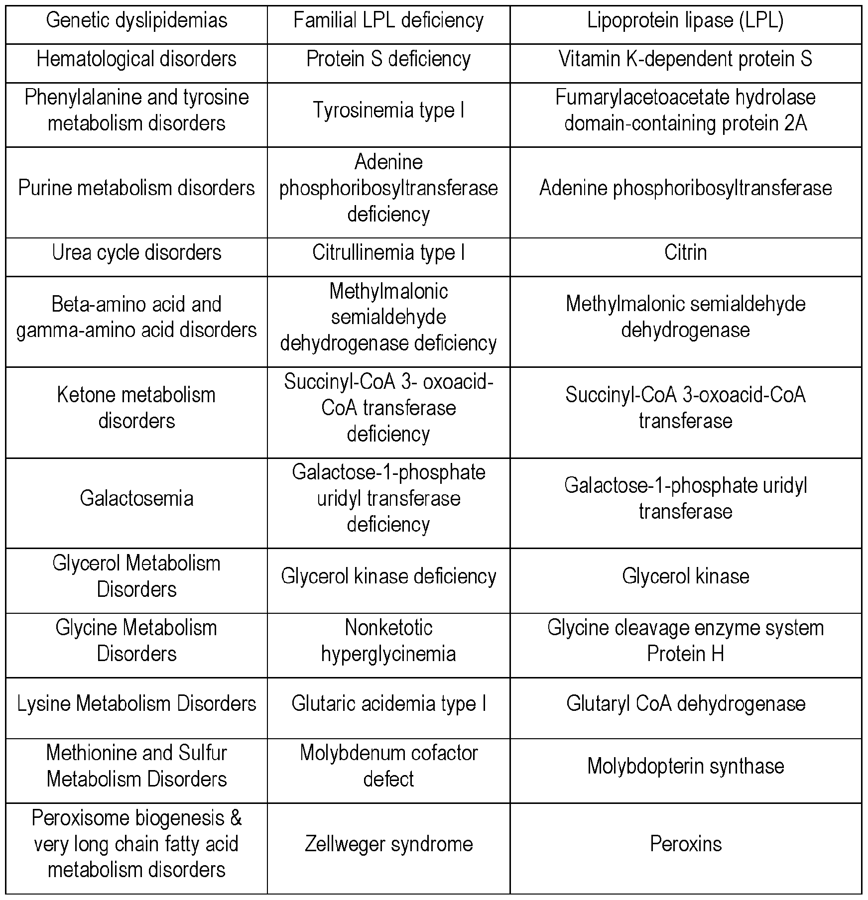

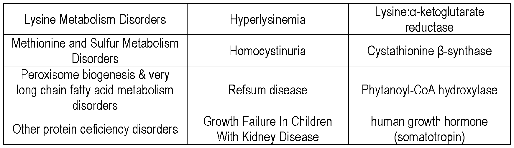
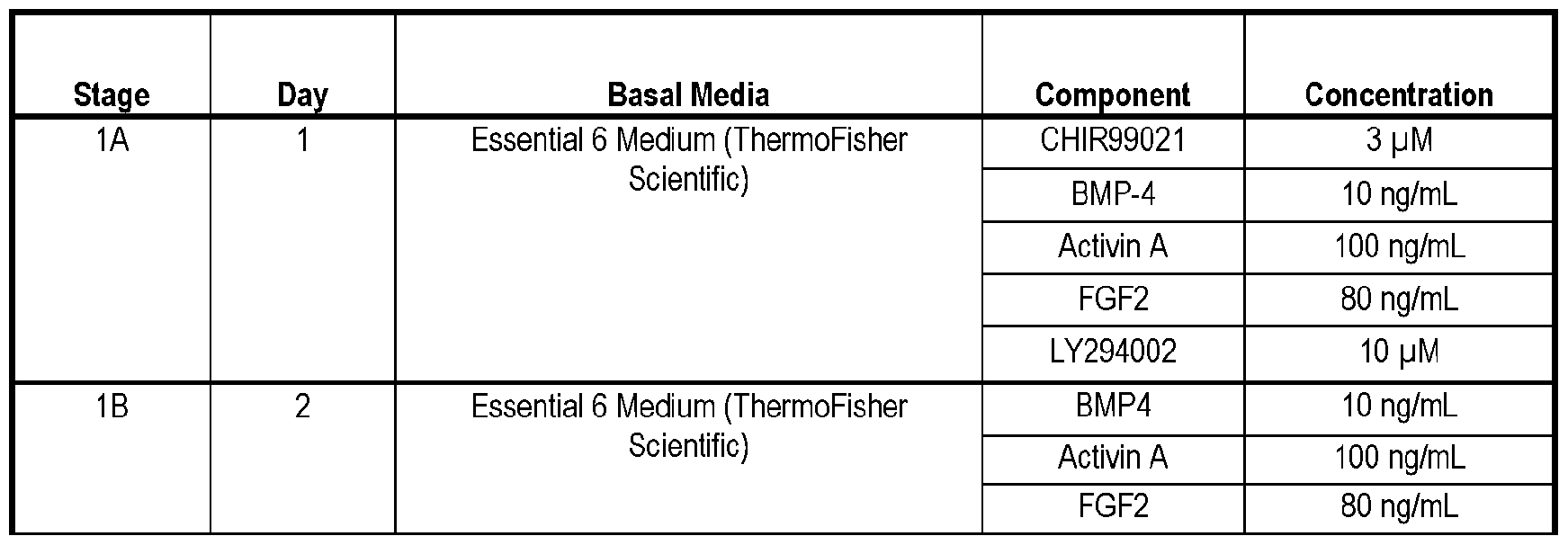
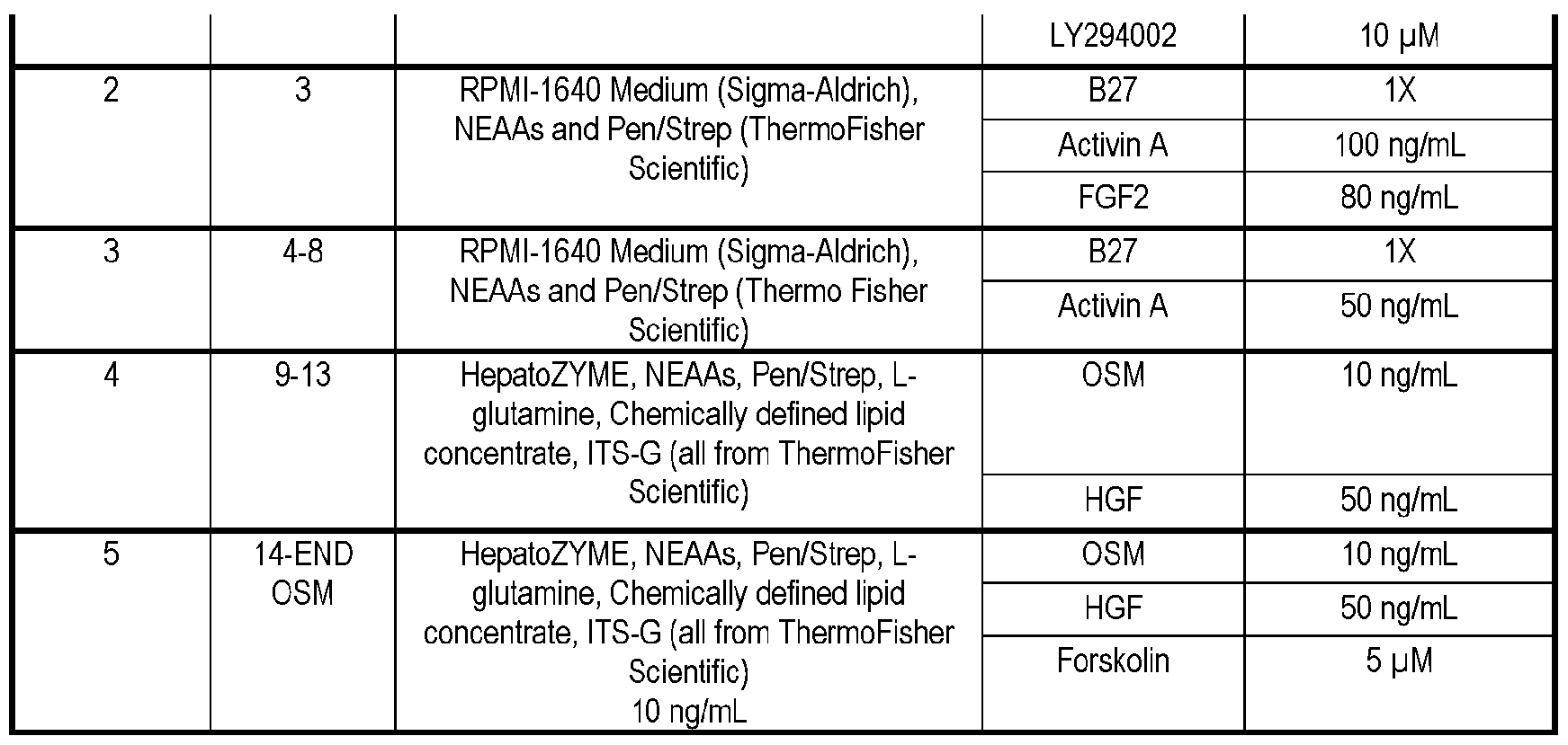


Claims
Priority Applications (1)
| Application Number | Priority Date | Filing Date | Title |
|---|---|---|---|
| EP23733576.5A EP4531879A1 (en) | 2022-05-25 | 2023-05-24 | Compositions of secretory and/or catalytic cells and methods using the same |
Applications Claiming Priority (12)
| Application Number | Priority Date | Filing Date | Title |
|---|---|---|---|
| US202263345838P | 2022-05-25 | 2022-05-25 | |
| US202263345840P | 2022-05-25 | 2022-05-25 | |
| US63/345,840 | 2022-05-25 | ||
| US63/345,838 | 2022-05-25 | ||
| US202263388953P | 2022-07-13 | 2022-07-13 | |
| US202263388955P | 2022-07-13 | 2022-07-13 | |
| US63/388,955 | 2022-07-13 | ||
| US63/388,953 | 2022-07-13 | ||
| US202263426226P | 2022-11-17 | 2022-11-17 | |
| US202263426219P | 2022-11-17 | 2022-11-17 | |
| US63/426,226 | 2022-11-17 | ||
| US63/426,219 | 2022-11-17 |
Publications (1)
| Publication Number | Publication Date |
|---|---|
| WO2023230524A1 true WO2023230524A1 (en) | 2023-11-30 |
Family
ID=86904302
Family Applications (1)
| Application Number | Title | Priority Date | Filing Date |
|---|---|---|---|
| PCT/US2023/067422 Ceased WO2023230524A1 (en) | 2022-05-25 | 2023-05-24 | Compositions of secretory and/or catalytic cells and methods using the same |
Country Status (2)
| Country | Link |
|---|---|
| EP (1) | EP4531879A1 (en) |
| WO (1) | WO2023230524A1 (en) |
Citations (42)
| Publication number | Priority date | Publication date | Assignee | Title |
|---|---|---|---|---|
| US3536809A (en) | 1969-02-17 | 1970-10-27 | Alza Corp | Medication method |
| US3598123A (en) | 1969-04-01 | 1971-08-10 | Alza Corp | Bandage for administering drugs |
| US3845770A (en) | 1972-06-05 | 1974-11-05 | Alza Corp | Osmatic dispensing device for releasing beneficial agent |
| US3916899A (en) | 1973-04-25 | 1975-11-04 | Alza Corp | Osmotic dispensing device with maximum and minimum sizes for the passageway |
| US4008719A (en) | 1976-02-02 | 1977-02-22 | Alza Corporation | Osmotic system having laminar arrangement for programming delivery of active agent |
| US5059595A (en) | 1989-03-22 | 1991-10-22 | Bioresearch, S.P.A. | Pharmaceutical compositions containing 5-methyltetrahydrofolic acid, 5-formyltetrahydrofolic acid and their pharmaceutically acceptable salts in controlled-release form active in the therapy of organic mental disturbances |
| US5073543A (en) | 1988-07-21 | 1991-12-17 | G. D. Searle & Co. | Controlled release formulations of trophic factors in ganglioside-lipsome vehicle |
| US5120548A (en) | 1989-11-07 | 1992-06-09 | Merck & Co., Inc. | Swelling modulated polymeric drug delivery device |
| US5354556A (en) | 1984-10-30 | 1994-10-11 | Elan Corporation, Plc | Controlled release powder and process for its preparation |
| US5591767A (en) | 1993-01-25 | 1997-01-07 | Pharmetrix Corporation | Liquid reservoir transdermal patch for the administration of ketorolac |
| US5639476A (en) | 1992-01-27 | 1997-06-17 | Euro-Celtique, S.A. | Controlled release formulations coated with aqueous dispersions of acrylic polymers |
| US5674533A (en) | 1994-07-07 | 1997-10-07 | Recordati, S.A., Chemical And Pharmaceutical Company | Pharmaceutical composition for the controlled release of moguisteine in a liquid suspension |
| US5733556A (en) | 1995-10-18 | 1998-03-31 | Akzo Nobel N.V. | Newcastle disease virus combination vaccine |
| US20110268708A1 (en) * | 2008-06-11 | 2011-11-03 | Ching Shwun Lin | Adipose tissue-derived stem cells for veterinary use |
| WO2012112982A2 (en) | 2011-02-18 | 2012-08-23 | Massachusetts Institute Of Technology | Hydrogel encapsulated cells and anti-inflammatory drugs |
| WO2012167223A1 (en) | 2011-06-02 | 2012-12-06 | Massachusetts Institute Of Technology | Modified alginates for cell encapsulation and cell therapy |
| US8691543B2 (en) | 2009-11-25 | 2014-04-08 | Worcester Polytechnic Institute | Nanofibrous scaffold comprising immobilized cells |
| WO2016019391A1 (en) | 2014-08-01 | 2016-02-04 | Massachusetts Institute Of Technology | Modified alginates for anti-fibrotic materials and applications |
| US20160324982A1 (en) | 2015-04-06 | 2016-11-10 | President And Fellows Of Harvard College | Compositions and methods for non-myeloablative conditioning |
| KR20160147929A (en) | 2014-04-25 | 2016-12-23 | 블루버드 바이오, 인코포레이티드. | Mnd promoter chimeric antigen receptors |
| WO2017075631A1 (en) | 2015-11-01 | 2017-05-04 | Massachusetts Institute Of Technology | Modified alginates for anti-fibrotic materials and applications |
| EP3219321A1 (en) * | 2016-03-14 | 2017-09-20 | TiGenix, S.A.U. | Adipose tissue-derived stromal stem cells for use in treating refractory complex perianal fistulas in crohn's disease |
| US20180015423A1 (en) | 2016-07-15 | 2018-01-18 | Korea Advanced Institute Of Science And Technology | 3d polymer nanofiber membrane composed of 1d individual polymer nanofibers which are quasi-aligned and cross-laminated like grid structure with functions of controlling pore distribution and size, and manufacturing method thereof |
| US20180077922A1 (en) | 2016-09-06 | 2018-03-22 | Glaxosmithkline Intellectual Property Development | Transduced cell cryoformulation |
| WO2018067615A1 (en) | 2016-10-03 | 2018-04-12 | Sigilon Therapeutics, Inc. | Compounds, devices, and uses thereof |
| US20180161185A1 (en) | 2016-12-14 | 2018-06-14 | eLum Technologies, Inc. | Electrospun stents, flow diverters, and occlusion devices and methods of making the same |
| WO2018140834A1 (en) | 2017-01-27 | 2018-08-02 | Cornell University | Zwitterionically modified polymers and hydrogels |
| WO2019090309A1 (en) | 2017-11-06 | 2019-05-09 | Massachusetts Institute Of Technology | Anti-inflammatory coatings to improve biocompatibility of neurological implants |
| WO2019169333A1 (en) | 2018-03-02 | 2019-09-06 | Sigilon Therapeutics, Inc. | Afibrotic compounds, devices, and uses thereof |
| WO2019169245A1 (en) | 2018-03-02 | 2019-09-06 | Sigilon Therapeutics, Inc. | Biocompatible hydrogel capsules and process for preparing same |
| WO2019195055A1 (en) | 2018-04-04 | 2019-10-10 | Sigilon Therapeutics, Inc. | Implantable particles and related methods |
| WO2020047124A1 (en) | 2018-08-28 | 2020-03-05 | Flagship Pioneering, Inc. | Methods and compositions for modulating a genome |
| WO2020069429A1 (en) | 2018-09-27 | 2020-04-02 | Sigilon Therapeutics, Inc. | Implantable devices for cell therapy and related methods |
| US10730983B2 (en) | 2016-06-13 | 2020-08-04 | Massachusetts Institute Of Technology | Biocompatible coatings and hydrogels for reducing foreign body response and fibrosis |
| WO2021062263A1 (en) | 2019-09-27 | 2021-04-01 | Sigilon Therapeutics, Inc. | Methods of evaluating polypeptide-modified polymers in compositions |
| WO2021062273A1 (en) | 2019-09-27 | 2021-04-01 | Sigilon Therapeutics, Inc. | Methods of evaluating small molecule-modified polymers in compositions |
| WO2021113751A1 (en) | 2019-12-04 | 2021-06-10 | Sigilon Therapeutics, Inc. | Apparatuses and methods for formation of particles |
| WO2021119522A1 (en) | 2019-12-13 | 2021-06-17 | Sigilon Therapeutics, Inc. | Compounds, polymers, devices, and uses thereof |
| WO2021178720A2 (en) | 2020-03-04 | 2021-09-10 | Flagship Pioneering Innovations Vi, Llc | Methods and compositions for modulating a genome |
| WO2022031862A2 (en) | 2020-08-04 | 2022-02-10 | Sigilon Therapeutics, Inc. | Heteroaryl and heterocyclyl compounds |
| WO2022060810A1 (en) | 2020-09-15 | 2022-03-24 | The Brigham And Women's Hospital, Inc. | In vivo adipose bioreactor and kits for the production and delivery of biologic agents |
| WO2022115609A1 (en) * | 2020-11-25 | 2022-06-02 | Flagship Pioneering, Inc. | Adipogenic cell compositions and methods |
-
2023
- 2023-05-24 WO PCT/US2023/067422 patent/WO2023230524A1/en not_active Ceased
- 2023-05-24 EP EP23733576.5A patent/EP4531879A1/en active Pending
Patent Citations (47)
| Publication number | Priority date | Publication date | Assignee | Title |
|---|---|---|---|---|
| US3536809A (en) | 1969-02-17 | 1970-10-27 | Alza Corp | Medication method |
| US3598123A (en) | 1969-04-01 | 1971-08-10 | Alza Corp | Bandage for administering drugs |
| US3845770A (en) | 1972-06-05 | 1974-11-05 | Alza Corp | Osmatic dispensing device for releasing beneficial agent |
| US3916899A (en) | 1973-04-25 | 1975-11-04 | Alza Corp | Osmotic dispensing device with maximum and minimum sizes for the passageway |
| US4008719A (en) | 1976-02-02 | 1977-02-22 | Alza Corporation | Osmotic system having laminar arrangement for programming delivery of active agent |
| US5354556A (en) | 1984-10-30 | 1994-10-11 | Elan Corporation, Plc | Controlled release powder and process for its preparation |
| US5073543A (en) | 1988-07-21 | 1991-12-17 | G. D. Searle & Co. | Controlled release formulations of trophic factors in ganglioside-lipsome vehicle |
| US5059595A (en) | 1989-03-22 | 1991-10-22 | Bioresearch, S.P.A. | Pharmaceutical compositions containing 5-methyltetrahydrofolic acid, 5-formyltetrahydrofolic acid and their pharmaceutically acceptable salts in controlled-release form active in the therapy of organic mental disturbances |
| US5120548A (en) | 1989-11-07 | 1992-06-09 | Merck & Co., Inc. | Swelling modulated polymeric drug delivery device |
| US5639476A (en) | 1992-01-27 | 1997-06-17 | Euro-Celtique, S.A. | Controlled release formulations coated with aqueous dispersions of acrylic polymers |
| US5591767A (en) | 1993-01-25 | 1997-01-07 | Pharmetrix Corporation | Liquid reservoir transdermal patch for the administration of ketorolac |
| US5674533A (en) | 1994-07-07 | 1997-10-07 | Recordati, S.A., Chemical And Pharmaceutical Company | Pharmaceutical composition for the controlled release of moguisteine in a liquid suspension |
| US5733556A (en) | 1995-10-18 | 1998-03-31 | Akzo Nobel N.V. | Newcastle disease virus combination vaccine |
| US20110268708A1 (en) * | 2008-06-11 | 2011-11-03 | Ching Shwun Lin | Adipose tissue-derived stem cells for veterinary use |
| US8691543B2 (en) | 2009-11-25 | 2014-04-08 | Worcester Polytechnic Institute | Nanofibrous scaffold comprising immobilized cells |
| US9867781B2 (en) | 2011-02-18 | 2018-01-16 | Massachusetts Institute Of Technology | Hydrogel encapsulated cells and anti-inflammatory drugs |
| US10278922B2 (en) | 2011-02-18 | 2019-05-07 | Massachusetts Institute Of Technology | Hydrogel encapsulated cells and anti-inflammatory drugs |
| WO2012112982A2 (en) | 2011-02-18 | 2012-08-23 | Massachusetts Institute Of Technology | Hydrogel encapsulated cells and anti-inflammatory drugs |
| US10709667B2 (en) | 2011-02-18 | 2020-07-14 | Massachusetts Institute Of Technology | Hydrogel encapsulated cells and anti-inflammatory drugs |
| WO2012167223A1 (en) | 2011-06-02 | 2012-12-06 | Massachusetts Institute Of Technology | Modified alginates for cell encapsulation and cell therapy |
| KR20160147929A (en) | 2014-04-25 | 2016-12-23 | 블루버드 바이오, 인코포레이티드. | Mnd promoter chimeric antigen receptors |
| WO2016019391A1 (en) | 2014-08-01 | 2016-02-04 | Massachusetts Institute Of Technology | Modified alginates for anti-fibrotic materials and applications |
| US10426735B2 (en) | 2014-08-01 | 2019-10-01 | Massachusetts Institute Of Technology | Modified alginates for anti-fibrotic materials and applications |
| US20160324982A1 (en) | 2015-04-06 | 2016-11-10 | President And Fellows Of Harvard College | Compositions and methods for non-myeloablative conditioning |
| WO2017075631A1 (en) | 2015-11-01 | 2017-05-04 | Massachusetts Institute Of Technology | Modified alginates for anti-fibrotic materials and applications |
| US10709818B2 (en) | 2015-11-01 | 2020-07-14 | Massachusetts Institute Of Technology | Modified alginates for anti-fibrotic materials and applications |
| EP3219321A1 (en) * | 2016-03-14 | 2017-09-20 | TiGenix, S.A.U. | Adipose tissue-derived stromal stem cells for use in treating refractory complex perianal fistulas in crohn's disease |
| US10730983B2 (en) | 2016-06-13 | 2020-08-04 | Massachusetts Institute Of Technology | Biocompatible coatings and hydrogels for reducing foreign body response and fibrosis |
| US20180015423A1 (en) | 2016-07-15 | 2018-01-18 | Korea Advanced Institute Of Science And Technology | 3d polymer nanofiber membrane composed of 1d individual polymer nanofibers which are quasi-aligned and cross-laminated like grid structure with functions of controlling pore distribution and size, and manufacturing method thereof |
| US20180077922A1 (en) | 2016-09-06 | 2018-03-22 | Glaxosmithkline Intellectual Property Development | Transduced cell cryoformulation |
| WO2018067615A1 (en) | 2016-10-03 | 2018-04-12 | Sigilon Therapeutics, Inc. | Compounds, devices, and uses thereof |
| US20180161185A1 (en) | 2016-12-14 | 2018-06-14 | eLum Technologies, Inc. | Electrospun stents, flow diverters, and occlusion devices and methods of making the same |
| WO2018140834A1 (en) | 2017-01-27 | 2018-08-02 | Cornell University | Zwitterionically modified polymers and hydrogels |
| WO2019090309A1 (en) | 2017-11-06 | 2019-05-09 | Massachusetts Institute Of Technology | Anti-inflammatory coatings to improve biocompatibility of neurological implants |
| WO2019169333A1 (en) | 2018-03-02 | 2019-09-06 | Sigilon Therapeutics, Inc. | Afibrotic compounds, devices, and uses thereof |
| WO2019169245A1 (en) | 2018-03-02 | 2019-09-06 | Sigilon Therapeutics, Inc. | Biocompatible hydrogel capsules and process for preparing same |
| WO2019195055A1 (en) | 2018-04-04 | 2019-10-10 | Sigilon Therapeutics, Inc. | Implantable particles and related methods |
| WO2020047124A1 (en) | 2018-08-28 | 2020-03-05 | Flagship Pioneering, Inc. | Methods and compositions for modulating a genome |
| WO2020069429A1 (en) | 2018-09-27 | 2020-04-02 | Sigilon Therapeutics, Inc. | Implantable devices for cell therapy and related methods |
| WO2021062263A1 (en) | 2019-09-27 | 2021-04-01 | Sigilon Therapeutics, Inc. | Methods of evaluating polypeptide-modified polymers in compositions |
| WO2021062273A1 (en) | 2019-09-27 | 2021-04-01 | Sigilon Therapeutics, Inc. | Methods of evaluating small molecule-modified polymers in compositions |
| WO2021113751A1 (en) | 2019-12-04 | 2021-06-10 | Sigilon Therapeutics, Inc. | Apparatuses and methods for formation of particles |
| WO2021119522A1 (en) | 2019-12-13 | 2021-06-17 | Sigilon Therapeutics, Inc. | Compounds, polymers, devices, and uses thereof |
| WO2021178720A2 (en) | 2020-03-04 | 2021-09-10 | Flagship Pioneering Innovations Vi, Llc | Methods and compositions for modulating a genome |
| WO2022031862A2 (en) | 2020-08-04 | 2022-02-10 | Sigilon Therapeutics, Inc. | Heteroaryl and heterocyclyl compounds |
| WO2022060810A1 (en) | 2020-09-15 | 2022-03-24 | The Brigham And Women's Hospital, Inc. | In vivo adipose bioreactor and kits for the production and delivery of biologic agents |
| WO2022115609A1 (en) * | 2020-11-25 | 2022-06-02 | Flagship Pioneering, Inc. | Adipogenic cell compositions and methods |
Non-Patent Citations (37)
| Title |
|---|
| "Medical Applications of Controlled Release", 1974, CRC PRES. |
| "Remington's Pharmaceutical Sciences", 1995, pages: 1447 - 1676 |
| AHN ET AL., SCI. REP., vol. 9, no. 1, 2019, pages 3087 |
| AMBATI ET AL., MBC OBES, vol. 3, 2016, pages 35 |
| BLACKFORD, S. J. I.NG, S. S.SEGAL, J. M.KING, A. J. F.AUSTIN, A. L.KENT, D.MOORE, J.SHELDON, M.ILIC, D.DHAWAN, A.: "Validation of Current Good Manufacturing Practice Compliant Human Pluripotent Stem Cell-Derived Hepatocytes for Cell-Based Therapy", STEM CELLS TRANSLATIONAL MEDICINE, vol. 8, no. 2, 2019, pages 124 - 137, XP055719473, DOI: 10.1002/sctm.18-0084 |
| CHAR6 ET AL., NUCLEUS, vol. 7, no. 3, 2016, pages 249 - 269 |
| COLLINS ET AL., J. LIPID RES., vol. 52, no. 9, 2011, pages 1683 - 1692 |
| CORRELL, SCIENCE, vol. 140, no. 26, 1963, pages 387 - 388 |
| DEUSE ET AL., NAT. BIOTECHNOL., vol. 37, 2019, pages 252 - 258 |
| DULL ET AL., J. VIROL., vol. 72, no. 11, 1998, pages 8463 - 8471 |
| DURING ET AL., ANN. NEUROL., vol. 25, 1989, pages 351 - 365 |
| FIORENZA CHRISTINA G. ET AL: "Lipodystrophy: pathophysiology and advances in treatment", NATURE REVIEWS. ENDOCRINOLOGY, vol. 7, no. 3, 16 November 2010 (2010-11-16), US, pages 137 - 150, XP093083085, ISSN: 1759-5029, Retrieved from the Internet <URL:http://www.nature.com/articles/nrendo.2010.199> DOI: 10.1038/nrendo.2010.199 * |
| FISHER ET AL., GENE THERAPY, vol. 24, 2017, pages 757 - 767 |
| GOODSON, MEDICAL APPLICATIONS OF CONTROLLED RELEASE, vol. 2, 1984, pages 115 - 138 |
| GRAVES ET AL., CELL. BIOCHEM., vol. 49, 1992, pages 219 - 244 |
| HOWARD ET AL., J. NEUROSURG, vol. 71, 1989, pages 105 |
| KILROY ET AL.: "Adipose-derived stem cells: Methods and protocols", 2018, SPRINGER NATURE, article "Isolation of murine adipose-derived stromal/stem cells for adipogenic differentiation or flow cytometry-based analysis", pages: 155 - 165 |
| KIM ET AL., PLOS ONE, vol. 12, no. 5, 2017, pages e0176722 |
| KOLODZIEJ ET AL., ADIPOCYTE, vol. 8, no. 1, 2019, pages 254 - 264 |
| KOSHIISHI ET AL., GENE, vol. 424, pages 1 - 2 |
| LANGER, SCIENCE, vol. 249, 1990, pages 1527 - 1533 |
| LEVY ET AL., SCIENCE, vol. 228, 1985, pages 190 |
| MIN ET AL., PNAS, vol. 116, no. 36, 2019, pages 17970 - 17979 |
| MOOLTENWELLS, J NATL CANCER INST, vol. 82, 1990, pages 297 - 300 |
| MOU ET AL., BIOENGINERING (BASEL, vol. 9, 2022, pages 188 |
| MULLER: "Drug Discovery and Evaluation: Pharmacological Assays", 2016, SPRINGER INTERNATIONAL PUBLISHING |
| NICODEMUSBRYANT, TISSUE ENGINEERING PART B: REVIEWS, 2008, pages 14 |
| RANGERPEPPAS, J. MACROMOL. SCI. REV. MACROMOL. CHEM., vol. 23, 1983, pages 61 |
| REBUFFE-SCRIVE ET AL., J. CLIN. ENDOCRINOL. METAB., vol. 71, no. 5, 1990, pages 1215 - 1219 |
| SANGRO ET AL., CANCER GENE THER, vol. 17, 2010, pages 837 - 843 |
| SEGAWA ET AL., J. ENDOCRINOL., vol. 200, no. 1, pages 107 - 116 |
| SHOHAM ET AL., BIOPHYS J., vol. 106, no. 6, 2014, pages 1421 - 1431 |
| STENKULAERLANSON-ALBERTSSON, AM J PHYSIOL REGUL INTEGR COMP PHYSIOL, vol. 315, 2018, pages R284 - R295 |
| THOMAS, QUARTERLY JOURNAL OF EXPERIMENTAL PHYSIOLOGY AND COGNATE MEDICAL SCIENCES, vol. 47, no. 2, 1962, pages 179 - 188 |
| VERBOVEN ET AL., SCIENTIFIC REPORTS, vol. 8, 2018, pages 4677 |
| VERES, A. ET AL., NATURE, vol. 569, no. 7756, 2019, pages 368 - 373 |
| WU ET AL., CLEVEL. CLIN. J. MED., vol. 87, no. 6, 2020, pages 367 - 476 |
Also Published As
| Publication number | Publication date |
|---|---|
| EP4531879A1 (en) | 2025-04-09 |
Similar Documents
| Publication | Publication Date | Title |
|---|---|---|
| KR102761577B1 (en) | Composition for promoting membrane fusion and its use | |
| JP7520717B2 (en) | Compositions and methods for membrane protein delivery - Patents.com | |
| US20210187018A1 (en) | Cytobiologics and therapeutic uses thereof | |
| KR20210131991A (en) | Compositions and methods for compartment-specific cargo delivery | |
| Nitahara-Kasahara et al. | Long-term engraftment of multipotent mesenchymal stromal cells that differentiate to form myogenic cells in dogs with Duchenne muscular dystrophy | |
| AU2021217399A1 (en) | Methods and use for bioengineering enucleated cells | |
| US20240009249A1 (en) | Adipogenic cell compositions and methods | |
| EP3662915B1 (en) | Mesenchymal stem cells and immunogens for use in inducing acquired immunological tolerance | |
| CN120112629A (en) | T cell generation method | |
| WO2023230524A1 (en) | Compositions of secretory and/or catalytic cells and methods using the same | |
| CN116801892A (en) | Adipogenic cell compositions and methods | |
| KR102915666B1 (en) | Compositions and methods for membrane protein delivery | |
| HK40025178A (en) | Compositions for facilitating membrane fusion and uses thereof | |
| KR20260018183A (en) | Compositions and methods for membrane protein delivery | |
| Dsouza | Cellule mesenchimali stromali/staminali come sistema per la veicolazione del gene del Pigment Epithelium-Derived Factor |
Legal Events
| Date | Code | Title | Description |
|---|---|---|---|
| 121 | Ep: the epo has been informed by wipo that ep was designated in this application |
Ref document number: 23733576 Country of ref document: EP Kind code of ref document: A1 |
|
| NENP | Non-entry into the national phase |
Ref country code: DE |
|
| WWE | Wipo information: entry into national phase |
Ref document number: 2023733576 Country of ref document: EP |
|
| ENP | Entry into the national phase |
Ref document number: 2023733576 Country of ref document: EP Effective date: 20250102 |
|
| WWP | Wipo information: published in national office |
Ref document number: 2023733576 Country of ref document: EP |