WO2023105526A1 - Multipotent lung progenitor cells for lung regeneration - Google Patents
Multipotent lung progenitor cells for lung regeneration Download PDFInfo
- Publication number
- WO2023105526A1 WO2023105526A1 PCT/IL2022/051302 IL2022051302W WO2023105526A1 WO 2023105526 A1 WO2023105526 A1 WO 2023105526A1 IL 2022051302 W IL2022051302 W IL 2022051302W WO 2023105526 A1 WO2023105526 A1 WO 2023105526A1
- Authority
- WO
- WIPO (PCT)
- Prior art keywords
- cells
- pulmonary
- population
- epithelial
- cell
- Prior art date
- Legal status (The legal status is an assumption and is not a legal conclusion. Google has not performed a legal analysis and makes no representation as to the accuracy of the status listed.)
- Ceased
Links
Classifications
-
- C—CHEMISTRY; METALLURGY
- C12—BIOCHEMISTRY; BEER; SPIRITS; WINE; VINEGAR; MICROBIOLOGY; ENZYMOLOGY; MUTATION OR GENETIC ENGINEERING
- C12N—MICROORGANISMS OR ENZYMES; COMPOSITIONS THEREOF; PROPAGATING, PRESERVING, OR MAINTAINING MICROORGANISMS; MUTATION OR GENETIC ENGINEERING; CULTURE MEDIA
- C12N5/00—Undifferentiated human, animal or plant cells, e.g. cell lines; Tissues; Cultivation or maintenance thereof; Culture media therefor
- C12N5/06—Animal cells or tissues; Human cells or tissues
- C12N5/0602—Vertebrate cells
- C12N5/0688—Cells from the lungs or the respiratory tract
- C12N5/0689—Stem cells; Progenitors
-
- A—HUMAN NECESSITIES
- A61—MEDICAL OR VETERINARY SCIENCE; HYGIENE
- A61K—PREPARATIONS FOR MEDICAL, DENTAL OR TOILETRY PURPOSES
- A61K35/00—Medicinal preparations containing materials or reaction products thereof with undetermined constitution
- A61K35/12—Materials from mammals; Compositions comprising non-specified tissues or cells; Compositions comprising non-embryonic stem cells; Genetically modified cells
- A61K35/42—Respiratory system, e.g. lungs, bronchi or lung cells
-
- A—HUMAN NECESSITIES
- A61—MEDICAL OR VETERINARY SCIENCE; HYGIENE
- A61P—SPECIFIC THERAPEUTIC ACTIVITY OF CHEMICAL COMPOUNDS OR MEDICINAL PREPARATIONS
- A61P11/00—Drugs for disorders of the respiratory system
-
- C—CHEMISTRY; METALLURGY
- C12—BIOCHEMISTRY; BEER; SPIRITS; WINE; VINEGAR; MICROBIOLOGY; ENZYMOLOGY; MUTATION OR GENETIC ENGINEERING
- C12N—MICROORGANISMS OR ENZYMES; COMPOSITIONS THEREOF; PROPAGATING, PRESERVING, OR MAINTAINING MICROORGANISMS; MUTATION OR GENETIC ENGINEERING; CULTURE MEDIA
- C12N5/00—Undifferentiated human, animal or plant cells, e.g. cell lines; Tissues; Cultivation or maintenance thereof; Culture media therefor
- C12N5/06—Animal cells or tissues; Human cells or tissues
- C12N5/0602—Vertebrate cells
- C12N5/0688—Cells from the lungs or the respiratory tract
-
- C—CHEMISTRY; METALLURGY
- C12—BIOCHEMISTRY; BEER; SPIRITS; WINE; VINEGAR; MICROBIOLOGY; ENZYMOLOGY; MUTATION OR GENETIC ENGINEERING
- C12N—MICROORGANISMS OR ENZYMES; COMPOSITIONS THEREOF; PROPAGATING, PRESERVING, OR MAINTAINING MICROORGANISMS; MUTATION OR GENETIC ENGINEERING; CULTURE MEDIA
- C12N2501/00—Active agents used in cell culture processes, e.g. differentation
- C12N2501/10—Growth factors
- C12N2501/11—Epidermal growth factor [EGF]
-
- C—CHEMISTRY; METALLURGY
- C12—BIOCHEMISTRY; BEER; SPIRITS; WINE; VINEGAR; MICROBIOLOGY; ENZYMOLOGY; MUTATION OR GENETIC ENGINEERING
- C12N—MICROORGANISMS OR ENZYMES; COMPOSITIONS THEREOF; PROPAGATING, PRESERVING, OR MAINTAINING MICROORGANISMS; MUTATION OR GENETIC ENGINEERING; CULTURE MEDIA
- C12N2501/00—Active agents used in cell culture processes, e.g. differentation
- C12N2501/10—Growth factors
- C12N2501/115—Basic fibroblast growth factor (bFGF, FGF-2)
-
- C—CHEMISTRY; METALLURGY
- C12—BIOCHEMISTRY; BEER; SPIRITS; WINE; VINEGAR; MICROBIOLOGY; ENZYMOLOGY; MUTATION OR GENETIC ENGINEERING
- C12N—MICROORGANISMS OR ENZYMES; COMPOSITIONS THEREOF; PROPAGATING, PRESERVING, OR MAINTAINING MICROORGANISMS; MUTATION OR GENETIC ENGINEERING; CULTURE MEDIA
- C12N2501/00—Active agents used in cell culture processes, e.g. differentation
- C12N2501/10—Growth factors
- C12N2501/155—Bone morphogenic proteins [BMP]; Osteogenins; Osteogenic factor; Bone inducing factor
-
- C—CHEMISTRY; METALLURGY
- C12—BIOCHEMISTRY; BEER; SPIRITS; WINE; VINEGAR; MICROBIOLOGY; ENZYMOLOGY; MUTATION OR GENETIC ENGINEERING
- C12N—MICROORGANISMS OR ENZYMES; COMPOSITIONS THEREOF; PROPAGATING, PRESERVING, OR MAINTAINING MICROORGANISMS; MUTATION OR GENETIC ENGINEERING; CULTURE MEDIA
- C12N2501/00—Active agents used in cell culture processes, e.g. differentation
- C12N2501/10—Growth factors
- C12N2501/165—Vascular endothelial growth factor [VEGF]
-
- C—CHEMISTRY; METALLURGY
- C12—BIOCHEMISTRY; BEER; SPIRITS; WINE; VINEGAR; MICROBIOLOGY; ENZYMOLOGY; MUTATION OR GENETIC ENGINEERING
- C12N—MICROORGANISMS OR ENZYMES; COMPOSITIONS THEREOF; PROPAGATING, PRESERVING, OR MAINTAINING MICROORGANISMS; MUTATION OR GENETIC ENGINEERING; CULTURE MEDIA
- C12N2501/00—Active agents used in cell culture processes, e.g. differentation
- C12N2501/10—Growth factors
- C12N2501/19—Growth and differentiation factors [GDF]
-
- C—CHEMISTRY; METALLURGY
- C12—BIOCHEMISTRY; BEER; SPIRITS; WINE; VINEGAR; MICROBIOLOGY; ENZYMOLOGY; MUTATION OR GENETIC ENGINEERING
- C12N—MICROORGANISMS OR ENZYMES; COMPOSITIONS THEREOF; PROPAGATING, PRESERVING, OR MAINTAINING MICROORGANISMS; MUTATION OR GENETIC ENGINEERING; CULTURE MEDIA
- C12N2501/00—Active agents used in cell culture processes, e.g. differentation
- C12N2501/20—Cytokines; Chemokines
- C12N2501/23—Interleukins [IL]
- C12N2501/2308—Interleukin-8 (IL-8)
-
- C—CHEMISTRY; METALLURGY
- C12—BIOCHEMISTRY; BEER; SPIRITS; WINE; VINEGAR; MICROBIOLOGY; ENZYMOLOGY; MUTATION OR GENETIC ENGINEERING
- C12N—MICROORGANISMS OR ENZYMES; COMPOSITIONS THEREOF; PROPAGATING, PRESERVING, OR MAINTAINING MICROORGANISMS; MUTATION OR GENETIC ENGINEERING; CULTURE MEDIA
- C12N2501/00—Active agents used in cell culture processes, e.g. differentation
- C12N2501/40—Regulators of development
- C12N2501/415—Wnt; Frizzeled
-
- C—CHEMISTRY; METALLURGY
- C12—BIOCHEMISTRY; BEER; SPIRITS; WINE; VINEGAR; MICROBIOLOGY; ENZYMOLOGY; MUTATION OR GENETIC ENGINEERING
- C12N—MICROORGANISMS OR ENZYMES; COMPOSITIONS THEREOF; PROPAGATING, PRESERVING, OR MAINTAINING MICROORGANISMS; MUTATION OR GENETIC ENGINEERING; CULTURE MEDIA
- C12N2501/00—Active agents used in cell culture processes, e.g. differentation
- C12N2501/70—Enzymes
- C12N2501/72—Transferases [EC 2.]
- C12N2501/727—Kinases (EC 2.7.)
-
- C—CHEMISTRY; METALLURGY
- C12—BIOCHEMISTRY; BEER; SPIRITS; WINE; VINEGAR; MICROBIOLOGY; ENZYMOLOGY; MUTATION OR GENETIC ENGINEERING
- C12N—MICROORGANISMS OR ENZYMES; COMPOSITIONS THEREOF; PROPAGATING, PRESERVING, OR MAINTAINING MICROORGANISMS; MUTATION OR GENETIC ENGINEERING; CULTURE MEDIA
- C12N2501/00—Active agents used in cell culture processes, e.g. differentation
- C12N2501/999—Small molecules not provided for elsewhere
-
- C—CHEMISTRY; METALLURGY
- C12—BIOCHEMISTRY; BEER; SPIRITS; WINE; VINEGAR; MICROBIOLOGY; ENZYMOLOGY; MUTATION OR GENETIC ENGINEERING
- C12N—MICROORGANISMS OR ENZYMES; COMPOSITIONS THEREOF; PROPAGATING, PRESERVING, OR MAINTAINING MICROORGANISMS; MUTATION OR GENETIC ENGINEERING; CULTURE MEDIA
- C12N2509/00—Methods for the dissociation of cells, e.g. specific use of enzymes
Definitions
- the present invention in some embodiments thereof, relates to pulmonary progenitor cells and, more particularly, but not exclusively, to methods of generating same and use of same in therapeutic applications.
- End stage respiratory diseases are among the leading causes of death worldwide, with more than 5.5 million deaths annually (World Health Organization data for 2020).
- Today, the only definitive treatment for these conditions is by replacement of the damaged organ with a lung transplant. Due to a shortage of suitable organs, many patients die on the transplant waiting list, and therefore lung diseases are prime candidates for stem cell therapy.
- BM- derived cells (7) lung-derived p63+ cells (2), LNEP (lineage negative epithelial progenitors) (3), and mouse and human sox9+ cells (4), (5).
- LNEP lineage negative epithelial progenitors
- mouse and human sox9+ cells (4), (5).
- fetal lung progenitors were suggested as an attractive source for transplantation in mice, provided that the lung stem cell niche in the recipient is vacated of endogenous lung progenitors by adequate conditioning.
- BMT bone marrow transplantation
- PCT publication no. WO/2013/084190 discloses a pharmaceutical composition
- a pharmaceutical composition comprising as an active ingredient an isolated population of cell suspension from a mammalian fetal pulmonary tissue, the fetal pulmonary tissue is at a developmental stage corresponding to that of a human pulmonary organ/tissue at a gestational stage selected from a range of about 20 to about 22 weeks of gestation.
- PCT publication no. WO/2017/203477 discloses a method of conditioning a subject in need of transplantation of progenitor cells in suspension of a tissue of interest.
- PCT publication no. WO/2017/203520 discloses a method of treating a pulmonary disorder or injury comprising administering to the subject non-syngeneic pulmonary tissue cells in suspension comprising an effective amount of hematopoietic precursor cells (HPCs) or supplemented with HPCs, wherein the effective amount is a sufficient amount to achieve tolerance to the pulmonary tissue cells in the absence of chronic immunosuppressive regimen.
- HPCs hematopoietic precursor cells
- a method of expanding in culture an isolated population of pulmonary cells comprising:
- a method of qualifying suitability of an isolated population of pulmonary cells for administration to a subject in need thereof comprising:
- a method of generating an isolated population of pulmonary cells comprising: (a) dissociating a pulmonary tissue so as to obtain a population of isolated pulmonary cells; and (b) contacting the population of isolated pulmonary cells with at least one agent capable of binding an epithelial cell marker and an endothelial cell marker, so as to select a cell population being double positive for expression of epithelial and endothelial cell markers, thereby generating the isolated population of pulmonary cells.
- the method further comprising expanding the pulmonary cells in a culture following step (b).
- the culture medium comprises a factor that promotes expansion of endothelial cells, a factor that promotes expansion of epithelial cells, and a factor that prevents differentiation.
- the method further comprising determining expression of the epithelial and endothelial cell markers on the pulmonary cells during and/or following the culture.
- expansion above a predetermined threshold of the cell population being double positive for expression of the epithelial and endothelial cell markers indicates the population of isolated pulmonary cells is suitable for administration to a subject in need thereof; and wherein no expansion or expansion below the predetermined threshold of the cell population being double positive for expression of the epithelial and endothelial cell markers indicates the population of isolated pulmonary cells is not suitable for administration to the subject.
- the factor that promotes expansion of endothelial cells is selected from the group consisting of vascular endothelial growth factor (VEGF), FGF, FGF2, IL-8 and BMP4.
- VEGF vascular endothelial growth factor
- FGF FGF
- FGF2 FGF2
- IL-8 IL-8
- BMP4 BMP4
- the factor that promotes expansion of endothelial cells comprises vascular endothelial growth factor (VEGF).
- VEGF vascular endothelial growth factor
- the factor that promotes expansion of epithelial cells is selected from the group consisting of epidermal growth factor (EGF), Noggin and R-Spondin.
- the factor that promotes expansion of epithelial cells comprises epidermal growth factor (EGF).
- the factor that prevents differentiation is selected from the group consisting of a ROCK inhibitor, a GSK3b inhibitor and an ALK5 inhibitor.
- the factor that prevents differentiation comprises a ROCK inhibitor.
- an isolated population of pulmonary cells comprising at least 40 % CD326+CD31+ cells.
- an isolated population of pulmonary cells obtained according to the method of some embodiments of the invention.
- a pharmaceutical composition comprising as an active ingredient the isolated population of pulmonary cells of some embodiments of the invention and a pharmaceutical acceptable carrier.
- a method of regenerating an epithelial and/or endothelial pulmonary tissue in a subject in need thereof comprising administering to the subject a therapeutically effective amount of the isolated population of pulmonary cells of some embodiments of the invention, thereby regenerating the epithelial and/or endothelial pulmonary tissue.
- a method of treating a pulmonary disorder or injury in a subject in need thereof comprising administering to the subject a therapeutically effective amount of the isolated population of pulmonary cells of some embodiments of the invention, thereby treating the pulmonary disorder or injury.
- a therapeutically effective amount of the isolated population of pulmonary cells of some embodiments of the invention for use in treating a pulmonary disorder or injury in a subject in need thereof.
- kits for isolation of pulmonary cells characterized as being double positive for expression of epithelial and endothelial cell markers, the kit comprising: (I) at least one agent capable of binding: (i) CD31 or CD144; and (ii) CD326, CD324, CD24, Aquaporin 5 (AQP-5), Podoplanin (PDPN), or Advanced Glycosylation End-Product Specific Receptor (RAGE); and (II) instructions for use.
- a cell bank comprising: (i) a plurality of isolated populations of pulmonary cells in suspension, the pulmonary cells being characterized as double positive for the expression of epithelial and endothelial cell markers, and wherein the plurality of the isolated populations of the pulmonary cells have been HLA typed to form an allogeneic cell bank, each individually disposed within separate containers; and (ii) a catalogue which comprises information about the HLA typed cells of the plurality of the isolated populations of the pulmonary cells.
- the epithelial cell marker comprises CD326, CD324, CD24, Aquaporin 5 (AQP-5), Podoplanin (PDPN), or Advanced Glycosylation End-Product Specific Receptor (RAGE).
- the endothelial cell marker comprises CD31 or CD144 (VE-cadherin).
- the cell population being double positive for expression of epithelial and endothelial cell markers comprises a CD326 + CD31 + signature.
- the cell population being double positive for expression of epithelial and endothelial cell markers comprises a CD324 + CD31 + signature.
- the cell population being double positive for expression of epithelial and endothelial cell markers comprises a CD326 + CD144 + signature.
- the cell population being double positive for expression of epithelial and endothelial cell markers comprises a CD324 + CD144 + signature.
- the method further comprises depleting CD45 expressing cells.
- the depleting CD45 expressing cells is affected by contacting the population of isolated pulmonary cells with an agent capable of binding CD45, so as to select a cell population being negative for expression of CD45.
- the method further comprises depleting T cells.
- the method further comprises expanding the pulmonary cells in a culture following step (b).
- the at least one agent capable of binding is an antibody.
- the antibody is a monospecific antibody. According to some embodiments of the invention, the antibody is a bispecific antibody.
- the dissociating is by enzymatic digestion.
- the method is affected ex vivo.
- the pulmonary tissue is a fetal pulmonary tissue.
- the pulmonary tissue is an adult pulmonary tissue.
- the pulmonary tissue is a human pulmonary tissue.
- the pulmonary tissue is from a cadaver donor.
- the pulmonary tissue is from a living donor.
- the pulmonary cells are capable of regenerating an epithelial pulmonary tissue.
- the pulmonary cells are capable of regenerating an endothelial pulmonary tissue.
- the cells are in suspension.
- the cells are embedded or attached to a scaffold.
- the pharmaceutical composition further comprises as an active ingredient hematopoietic precursor cells (HPCs).
- HPCs hematopoietic precursor cells
- the HPCs comprise T cell depleted immature hematopoietic cells.
- the method further comprises administering to the subject an agent capable of inducing damage to a pulmonary tissue prior to the administering, wherein the damage results in proliferation of resident stem cells in the pulmonary tissue.
- the method further comprises conditioning the subject under sublethal, lethal or supralethal conditioning protocol prior to the administering.
- the method further comprises administering to the subject an effective amount of hematopoietic precursor cells (HPCs).
- HPCs hematopoietic precursor cells
- the method further comprises treating the subject with an immunosuppressive agent following the administering.
- the isolated population of pulmonary cells for use further comprises the use of an agent capable of inducing damage to a pulmonary tissue, wherein the damage results in proliferation of resident stem cells in the pulmonary tissue.
- the isolated population of pulmonary cells for use further comprises a sublethal, lethal or supralethal conditioning protocol.
- the isolated population of pulmonary cells for use further comprises the use of an effective amount of hematopoietic precursor cells (HPCs).
- HPCs hematopoietic precursor cells
- the isolated population of pulmonary cells for use further comprises the use of an immunosuppressive agent.
- the agent capable of inducing damage to the pulmonary tissue is selected from the group consisting of a chemotherapeutic agent, an immunosuppressive agent, an amiodarone, a beta blockers, an ACE inhibitor, a nitrofurantoin, a procainamide, a quinidine, a tocainide, and a minoxidil.
- the agent capable of inducing damage to the pulmonary tissue comprises naphthalene.
- the conditioning protocol comprises reduced intensity conditioning (RIC).
- the conditioning protocol comprises at least one of total body irradiation (TBI), partial body irradiation, a chemotherapeutic agent and/or an antibody immunotherapy.
- the antibody immunotherapy comprises T cell debulking.
- the antibody immunotherapy comprises anti-thymocyte globulin (ATG) antibody, alemtuzumab, muromonab-CD3, or a combination thereof.
- ATG anti-thymocyte globulin
- the TBI comprises a single or fractionated irradiation dose within the range of 1-10 Gy.
- the HPCs comprise pulmonary tissue- derived CD34 + cells.
- the HPCs comprise bone marrow or mobilized peripheral blood CD34 + cells.
- the HPCs comprise T cell depleted immature hematopoietic cells.
- the isolated population of pulmonary cells and the HPCs are obtained from the same donor.
- the isolated population of pulmonary cells and the hematopoietic precursor cells (HPCs) are in separate formulations.
- the isolated population of pulmonary cells and the HPCs are in the same formulation.
- the isolated population of pulmonary cells and/or the HPCs are formulated for an intravenous or an intratracheal route of administration.
- the immunosuppressive agent comprises cyclophosphamide, busulfan, fludarabin, tacrolimus, cyclosporine, mycophenolate mofetil, azathioprine, everolimus, sirolimus, glucocorticoids, or combinations thereof.
- the subject is a human subject.
- the isolated population of pulmonary cells is non-syngeneic with the subject.
- the pulmonary disorder or injury comprises chronic inflammation of the lungs.
- the pulmonary disorder or injury is selected from the group consisting of cystic fibrosis, emphysema, asbestosis, chronic obstructive pulmonary disease (COPD), pulmonary fibrosis, idiopatic pulmonary fibrosis, pulmonary hypertension, lung cancer, sarcoidosis, acute lung injury (adult respiratory distress syndrome), respiratory distress syndrome of prematurity, chronic lung disease of prematurity (bronchopulmonarydysplasia), surfactant protein B deficiency, congenital diaphragmatic hernia, pulmonary alveolar proteinosis, pulmonary hypoplasia and asthma.
- cystic fibrosis cystic fibrosis
- emphysema asbestosis
- COPD chronic obstructive pulmonary disease
- pulmonary fibrosis idiopatic pulmonary fibrosis
- pulmonary hypertension lung cancer
- sarcoidosis acute lung injury (adult respiratory distress syndrome), respiratory distress syndrome of prematurity, chronic lung disease of pre
- FIGs. 1A-F demonstrate multi-lineage engraftment of mTom donor-derived cells in the recipient lung evaluated by sc-RNA seq.
- Figure 1A shows the design of cell sorting of host and donor lung cells from chimeric lungs. Three chimeric lungs were first verified to exhibit significant chimerism by fluorescent microscopy, pooled, enzymatically dissociated and FACS separated after gating on CD45-, single, live cells into donor and recipient compartments based on Td-Tomato expression.
- Figure IB shows FACS analysis of chimeric lung, transplanted with Td- Tomato cells and control non-transplanted lung.
- Figure 1E-F show heat maps identifing the functionally distinct gCap and aCap endothelial cells in donor ( Figure IE) and recipient ( Figure IF) compartments, respectively.
- FIGs. 2A-E demonstrate transplantation of fetal lung cells from Confetti donors.
- Figure 2A shows the experimental scheme using E16 fetal lung cells after induction of CRE recombination by Tamoxifen for implantation into immune deficient RAG recipients preconditioned with naphthalene and 6Gy TBI.
- FIG. 2D shows monochromatic patches at 6 weeks after transplantation of E16 fetal lung cells from R26R-Confetti donors into RAG2-/- recipient mouse.
- FIGs. 3A-G demonstrate lung chimerism analysis at 8 weeks after transplantation of adult R26R-Confetti lung cells.
- Figure 3 A is a schematic presentation of the experimental procedure.
- FIGs. 4A-H demonstrate donor derived lung patches after transplantation of different lung cell sub-populations.
- Figure 4 A shows the gating strategy for FACS sorting of CD45- lung cells into four subpopulations including CD326+ CD31-, CD326+CD31+, CD326-CD31+ and CD326- CD31- cells.
- Figure 4B shows visualization of double positive CD45-CD326+CD31+ lung cells by Imagestream analysis.
- Figure 4C is a schematic representation of the transplantation experiments.
- Figure 4D shows the percentage of each sorted subpopulation out of the CD45- non- hematopoietic lung cell population in 16 experiments.
- Figure 4F shows donor-derived lung patches 6 weeks after transplantation of 0.3- 0.5 x10 6 sorted double positive CD326+CD31+ cells, or single positive CD31+ endothelial cells from nTnG donors (red) mixed with 0.5 x10 6 unsorted cells from GFP+ donors (green).
- FIGs. 5A-K demonstrate different cellular composition of donor derived patches after transplantation of sorted CD326-CD31+ versus CD326+CD31+ lung cell subpopulations.
- Figure 5A-D show staining of typical lung patches derived from sorted nTnG CD326-CD31+ lung cells (red).
- Figure 5C shows staining for endothelial nuclear marker S0X17 (cyan) within donor-derived nTnG positive patch after transplantation of CD326-CD31 + cells.
- FIG. 5D shows staining for cell surface endothelial marker CD31 and epithelial marker HOPX in donor-derived patches formed after transplantation of CD326-CD31+ cells.
- CD31 and SOX17 On the left - double staining for CD31 and SOX17; on the right - Triple staining for nTnG+ (red), CD31 (blue) and HOPX (green).
- Figures 5E-F show staining of typical lung patches derived from sorted nTnG CD326+CD31 + lung progenitor cells, demonstrating donor-derived epithelial and endothelial cells.
- Figure 5F shows high magnification of typical staining for HOPX demonstrating donor (red, indicated with arrow) and host (indicated with arrow head) ATI cells.
- Figure 5H shows staining of a typical lung patch derived from sorted nTnG CD326+CD31+ lung cells demonstrating donor-derived epithelial ATI (AQP- 5+, purple,) and endothelial (SOX17+, green) cells.
- Figure 5J-K shows graphical summary of quantitative differences between the composition of patches derived from transplantation of CD326+CD31+ and CD326- CD31+ cells.
- FIGs. 6A-J demonstrate different transgenes expression in double positive CD326+ CD31+ patch- forming lung cell progenitors.
- Figure 6A-B show typical dot plots demonstrating GFP expression of gated CD326+ CD31+double positive cells.
- Figure 6D-Left shows culture of FACS purified CD326+VEcad (mT) and CD326+VEcad+(mG) lung cell populations from VEcad mTmG mice under 3D conditions (For FACS sorting scheme see Figure 14A).
- FIG. 6D-right shows absolute number of observed organoids per well upon seeding of 5X10 5 CD326+VEcad- or CD326+VEcad+ FACS sorted lung cells.
- Figure 6E Upper panel shows organoids exhibiting GFP (green, derived for VE-Cadhering expressing cells ) and the epithelial marker cytokeratin (magenta).
- Figure 6E Lower panel, left, shows organoids exhibiting GFP (green, derived from VE-Cadhering expressing cells) and the epithelial marker AQP-5 (Magenta) for alveolar ATI lung cells.
- Figures 6F-G show Imagestream analysis of CD326+CD31+ lung cell progenitors from Shh Cre nTnG mice or VEcad Cre nTnG mice, illustrating Shh and VE-cad expression in these cells.
- FIGs. 7A-E show staining of regenerative patches in the chimeric lung for epithelial and endothelial markers.
- FIGs. 8A-E demonstrate transplantation of adult R26R-Confetti BM from donors induced to express Cre-recombination by tamoxifen.
- Figure 8A is a schematic presentation of the spleen colony assay using adult BM from R26R-Confetti donors.
- Figure 8B-C show FACS analysis demonstrating expression of fluorescent Tags within LSK+ BM progenitors prior to BM transplantation.
- Figure 8D shows formation of monochromatic spleen colonies 9 days after transplantation of BM from R26R-Confetti donors into lethally irradiated mice.
- Figure 8E shows peripheral blood chimerism, demonstrating existence of fluorescent clones in the hematological compartment, originating from the transplanted Cr-recombined cells.
- FIGs. 9A-E demonstrate transplantation of E16 R26R-Confetti liver into NA+6Gy preconditioned mice.
- Figure 9A is the experimental workflow.
- Figure 9B shows FACS analysis of E16 confetti fetal liver 4 days after Tmx administration, demonstrating expression of the 4 fluorescent tags within the Scal+Ckit+ hematopoietic progenitor cell population.
- Figure 9C shows monochromatic spleen colonies generated by fetal liver cells transplanted into NA +6Gy TBI preconditioned mice.
- Scale bar 500 ⁇ m
- Figure 9D shows FACS analysis of peripheral blood from the chimeric mice 2 months after transplantation, demonstrating persistence of monochromatic clones in the hematological compartment.
- Figure 9E shows a representative lung two photon image of a mouse preconditioned with Na+6GY TBI and transplanted with E16 fetal liver cells, demonstrating presence of isolated fluorescent cells and absence of monochromatic donor-derived patches, confirming the unique ability of lung cells to mediate lung regeneration.
- Scale bar 50 ⁇ m
- FIGs. 10A-B demonstrate appearance of cleared chimeric lung sample prior to evaluation with LSM. Staining of cleared chimeric lungs is shown in Figures 3E-F.
- FIGs. 11A-B demonstrate staining of lungs from chimeric mice transplanted with FACS sorted CD326+CD31+ cells derived from mTmG mice.
- FIGs. 12A-D demonstrate long term chimerism at 9 months post-transplantation in mice transplanted with CD326+CD31+ nTnG FACS sorted cells.
- Figure 12D shows staining of chimeric lung with anti-Ho
- FIGs. 13A-E demonstrate FACS analysis of lungs from transgenic mice.
- FIGs. 14A-D demonstrate staining for epithelial markers of organoids grown from FACS purified CD326+VEcad- harvested from lungs of VEcad mTmG mice.
- Figure 14A shows the gating strategy for purification of single positive CD326+VE cad mG- and double positive CD326+VE cadmG+ cells from VEcad mTmG mice.
- Live CD45-TER119-Sytox- CD326+ single cells were further gated according to the expression of VE-cad mG, so as to purify CD326+VE- cad mG- and CD326+VE cad mG+ lung cell subpopulations.
- the isolated cells were used to generate lung organoids as described in Methods hereinbelow.
- im) and anti-SPC antibody ( Figure 14D) (cyan, scale bar 7 ⁇ m). Nuclei were stained by DAPI (blue). Images are representative of two independent experiments.
- FIGs. 15A-C demonstrate that different culture media have different effects on expansion of pulmonary cells which dually express endothelial and epithelial markers.
- Figure 15A Upper panel shows FACS analysis demonstrating levels of double positive CD326+CD31+ cells at different culture time points upon incubation in a conditioned medium (CM) obtained from mouse fibroblatss supplemented with EGF and a low concentration of ROCK inhibitor (5 ⁇ m) (marked as “original medium”).
- CM conditioned medium
- ROCK inhibitor 5 ⁇ m
- Figure 15A Lower Pannel shows FACS analysis demonstrating levels of double positive CD326+CD31+ cells at different culture time points upon incubation in CM supplemented with EGF, VEGF and a high ROCK inhibitor concentration (20 ⁇ m) (marked as “CM+Epi+Endo+High RICK-I”).
- Figure 15B shows total cells number at the indicated days in the two cultures described in Figures 15A.
- Figure 15C shows CD326+CD31+ double positive cells number at the indicated days of the two cultures describedin Figures 15A.
- the present invention in some embodiments thereof, relates to pulmonary progenitor cells and, more particularly, but not exclusively, to methods of generating same and use of same in therapeutic applications.
- Various cell populations have been shown to exhibit lung regenerative potential, including e.g. BM-derived or lung-derived cells cells, leading to marked lung host-donor chimerism.
- a large proportion of the donor derived patches exhibit different lineage lung compartments including epithelial and endothelial cells.
- the present inventors addressed the possibility that following transplantation of lung cells each donor patch originate from a single progenitor. To that end, as shown in the Examples section which follows (see Exmaple 1), fetal or adult lung cells from Rosa26-Confetti mice bearing a multicolor Cre reporter system (9) were transplanted to recipients preconditioned with naphthalene and TBI.
- This four-color Cre recombination system provides a fetal or adult lung cell preparation in which each cell expresses just one randomly determined colour.
- each cell expresses just one randomly determined colour.
- immunohistochemistry, confocal microscopy, as well as two-photon microscopy and light sheet microscopy demonstrated that all donor-derived lung patches developing after transplantation are monochromatic, strongly supporting the clonal origin of donor-derived lung patches observed after transplantation, in striking resemblance to the spleen colony forming cells typically identified after bone marrow transplantation.
- pulmonary progenitor cells which dually express endothelial and epithelial markers (see Example 1 of the Examples section which follows). These pulmonary progenitor cells were obtained from both fetal and adult pulmonary tissues. Furthermore, these pulmonary progenitor cells were capable of differentiating into endothelial lung cells as well as into epithelial lung cells, and consequently were capable of generating endothelial and epithelial lung tissues following transplantation into a recipient. Taken together, these results substantiate the use of the novel pulmonary progenitor cells for e.g. regeneration of pulmonary organs or tissues, such as for the treatment of lung injury or disease.
- a method of generating an isolated population of pulmonary cells comprising:
- the present inventors found out that different culturing conditions have different effects on expansion of this novel pulmonary progenitor cells polulation. Specifically, it was shown that while culturing dissociated lung cells in a medium comprising VEGF, EGF and a ROCK inhibitor led to marked expansion of cells dually expressing endothelial and epithelial markers, culturing these cells in a medium comprising only EGF and a ROCK inhibitor resulted in expansion of the total number of cells but not of the double positive cells (Example 2 of the Examples section which follows).
- a method of expanding in culture an isolated population of pulmonary cells comprising:
- a method of qualifying suitability of an isolated population of pulmonary cells for administration to a subject in need thereof comprising:
- the methods disclosed herein are affected ex-vivo.
- pulmonary tissue refers to a lung tissue or organ.
- the pulmonary tissue of the present invention may be a full or partial organ or tissue.
- the pulmonary tissue of some embodiments may comprise the right lung, the left lung, or both.
- the pulmonary tissue of some embodiments of the invention may comprise one, two, three, four or five lobes (from either the right or the left lung).
- the pulmonary tissue of some embodiments of the present invention may comprise one or more lung segments or lung lobules.
- the pulmonary tissue of some embodiments of the present invention may comprise any number of bronchi and bronchioles (e.g. bronchial tree) and any number of alveoli or alveolar sacs.
- the cells of the present invention may be obtained from a prenatal organism, postnatal organism, an adult or a cadaver donor. Such determinations are well within the ability of one of ordinary skill in the art.
- pulmonary cells of som erne bodiemtns of the invention may be of fresh or frozen (e.g., cryopreserved) preparations, as further discussed below.
- the pulmonary tissue is a human pulmonary tissue.
- the pulmonary tissue is from a cadaver donor.
- pulmonary tissue is from a living donor.
- the pulmonary tissue is from an adult origin (e.g. a mammalian organism at any stage after birth).
- the pulmonary tissue is from an embryonic origin.
- the pulmonary tissue is from a fetal origin.
- the embryonic or fetal organism may be of any of a human or xenogeneic origin (e.g. porcine) and at any stage of gestation. Such a determination is in the capacity of one of ordinary skill in the art.
- a human or xenogeneic origin e.g. porcine
- obtaining a pulmonary tissue may be effected by harvesting the tissue from a developing fetus, e.g. by a surgical procedure.
- the pulmonary tissue i.e. lung tissue
- the pulmonary tissue is obtained from a fetus at a stage of gestation corresponding to human canalicular stage of development (e.g. 16-25 weeks of gestation).
- the pulmonary tissue is obtained from a fetus at a stage of gestation corresponding to human 16-17 weeks of gestation, 16-18 weeks of gestation, 16-19 weeks of gestation, 16-20 weeks of gestation, 16-21 weeks of gestation, 16-22 weeks of gestation, 16-24 weeks of gestation, 17-18 weeks of gestation, 17-19 weeks of gestation, 17-20 weeks of gestation, 17-21 weeks of gestation, 17-22 weeks of gestation, 17-24 weeks of gestation, 18-19 weeks of gestation, 18-20 weeks of gestation, 18-21 weeks of gestation, 18-22 weeks of gestation, 18-24 weeks of gestation, 19-20 weeks of gestation, 19-21 weeks of gestation, 19-22 weeks of gestation, 19-23 weeks of gestation, 19-24 weeks of gestation, 20-21 weeks of gestation,
- the pulmonary tissue is obtained from a fetus at a stage of gestation corresponding to human 20-22 weeks of gestation.
- the pulmonary tissue is obtained from a fetus at a stage of gestation corresponding to human 21-22 days of gestation.
- the pulmonary tissue is obtained from a fetus at a stage of gestation corresponding to human 20-21 days of gestation.
- gestational stage of an organism is the time period elapsed following fertilization of the oocyte generating the organism.
- the following table provides an example of the gestational stages of human and porcine tissues at which these can provide fetal tissues which are essentially at corresponding developmental stages:
- the gestational stage (in days) of a tissue belonging to a given species which is at a developmental stage essentially corresponding to that of a porcine tissue can be calculated according to the following formula: [gestational stage of porcine tissue in days] / [gestational period of pig in days] x [gestational stage of tissue of given species in days].
- the gestational stage (in days) of a tissue belonging to a given species which is at a developmental stage essentially corresponding to that of a human tissue can be calculated according to the following formula: [gestational stage of human tissue in days] / [gestational period of humans in days] x [gestational stage of tissue of given species in days].
- the gestational stage of pigs is about 115 days and that of humans is about 280 days.
- obtaining a pulmonary tissue may be effected by harvesting the tissue from an organ donor by a surgical procedure e.g. laparotomy or laparoscopy.
- pulmonary cells as well as hematopoietic progenitor cells (as discussed in detail below) may be isolated therefrom according to methods known in the art, such methods depend on the source and lineage of the cells and may include, for example, flow cytometry and cell sorting as taught for example by www(dot)bio-rad(dot)com/en-uk/applications-technologies/isolation-maintenance-stem-cells.
- the pulmonary tissue need not be intact (i.e. maintain a tissue structure such that is suitable for a whole organ transplantation), however, the pulmonary tissue should comprise viable cells.
- the pulmonary tissue may be obtained from more than one donor.
- the pulmonary cells may comprise cells obtained from more than one cell donor.
- the present invention further contemplates generation of an isolated population of cells therefrom
- isolated population of pulmonary cells refers to isolated cells which do not form a tissue structure (i.e., no connective tissue structure).
- the pulmonary cells may be comprised in a suspension of single cells or cell aggregates of no more than 5, 10, 50, 100, 200, 300, 400, 500, 1000, 1500, 2000 cells in an aggregate.
- pulmonary cells in suspension refers to cells which have been isolated from their natural environment (e.g., the human body) are extracted from the pulmonary tissue while maintaining viability but do not maintain a tissue structure (i.e., no vascularized tissue structure) and are not attached to a solid support.
- the cell suspension of the invention may be obtained by any mechanical or chemical (e.g. enzymatic) means.
- Several methods exist for dissociating cell clusters to form cell suspensions e.g. single cell suspension
- cell suspensions e.g. single cell suspension
- aggregates e.g., physical forces (mechanical dissociation such as cell scraper, trituration through a narrow bore pipette, fine needle aspiration, vortex disaggregation and forced filtration through a fine nylon or stainless steel mesh), enzymes (enzymatic dissociation such as trypsin, collagenase, Acutase and the like) or a combination of both.
- the dissociating is by enzymatic digestion.
- enzymatic digestion of tissue/organ into isolate cells can be performed by subjecting the tissue to an enzyme such as type IV Collagenase (Worthington biochemical corporation, Lakewood, NJ, USA) and/or Dispase (Invitrogen Corporation products, Grand Island NY, USA).
- the tissue may be enzyme digested by finely mincing tissue with a razor blade in the presence of e.g. collagenase, dispase and CaCL at 37 °C for about 1 hour.
- the method may further comprise removal of nonspecific debris from the resultant cell suspension by, for example, sequential filtration through filters (e.g. 70- and 40-pm filters), essentially as described in the Examples section which follows.
- mechanical dissociation of tissue into isolated cells can be performed using a device designed to break the tissue to a predetermined size.
- a device designed to break the tissue to a predetermined size.
- mechanical dissociation can be manually performed using a needle such as a 27g needle (BD Microlance, Drogheda, Ireland) while viewing the tissue/cells under an inverted microscope.
- the dissociated cells are further broken to small clumps using e.g. 200 ⁇ l Gilson pipette tips (e.g., by pipetting up and down the cells).
- the cell suspension of pulmonary cells comprises viable cells.
- Cell viability may be monitored using any method known in the art, as for example, using a cell viability assay (e.g. MultiTox Multiplex Assay available from Promega), Flow cytometry, Trypan blue, etc.
- a cell viability assay e.g. MultiTox Multiplex Assay available from Promega
- Flow cytometry Trypan blue, etc.
- the pulmonary tissue and isolated cells derived therefrom comprise cells that express both epithelial and endothelial cell markers.
- these cells expressing both epithelial and endothelial cell markers are progenitor cells capable of differentiating into both endothelial lung cells and epithelial lung cells.
- Methods of determining differentiation include in-vitro and in-vivo (e.g. transplantation) methods well known to the skilled in the art. Non-limiting examples are provided in the Examples section which follows.
- the pulmonary cells are characterized by the expression of epithelial and endothelial cell markers.
- epidermal cell marker refers to a cell- surface protein characteristic of lung epitherlial cells.
- a marker includes, but is not limited to, CD326, CD324, CD24, Aquaporin 5 (AQP-5), Podoplanin (PDPN), and Advanced Glycosylation End-
- endothelial cell marker refers to a cell-surface protein characteristic of lung endothelial cells. Such a marker includes, but is not limited to, CD31 and CD144 (VE-cadherin).
- the pulmonary cell are characterized by the co- expression signature: CD326 + and CD31 + .
- the pulmonary cell are characterized by the co- expression signature: CD324 + and CD31 + .
- the pulmonary cell are characterized by the co- expression signature: CD24 + and CD31 + .
- the pulmonary cell are characterized by the co- expression signature: AQP-5 + and CD31 + .
- the pulmonary cell are characterized by the co- expression signature: PDPN + and CD31 + .
- the pulmonary cell are characterized by the co- expression signature: RAGE + and CD31 + .
- the pulmonary cell are characterized by the co- expression signature: CD326 + and CD144 + .
- the pulmonary cell are characterized by the co- expression signature: CD324 + and CD144 + .
- the pulmonary cell are characterized by the co- expression signature: CD24 + and CD144 + .
- the pulmonary cell are characterized by the co- expression signature: AQP-5 + and CD144 + .
- the pulmonary cell are characterized by the co- expression signature: PDPN + and CD144 + .
- the pulmonary cell are characterized by the co- expression signature: RAGE + and CD144 + .
- the pulmonary cells are further characterized by expression of at least one of Nkx 2.1, CD200, Akap5, Secl413, Prdx6 and Clic3.
- the pulmonary cells comprise a heterogeneous population of cells (e.g. unseparated population of cells) comprising the cells co-expressing the endothelial and epithelial markes.
- the pulmonary cells comprise a purified population of cells.
- the cells may be treated to remove specific population of cells therefrom (e.g. removal of a subpopulation) or to positively select a desired population (e.g. a cell population being double positive for expression of epithelial and endothelial cell markers).
- Purification of specific cell types may be carried out by any method known to one of skill in the art, such as for example, eradication (e.g. killing) with specific antibodies or by affinity based purification (e.g. such as by the use of MACS beads, FACS sorter and/or capture ELISA labeling) using specific antibodies which recognize any specific cell markers (e.g.
- FACS fluorescence activated cell sorting
- Any ligand-dependent separation techniques known in the art may be used in conjunction with both positive and negative separation techniques that rely on the physical properties of the cells rather than antibody affinity, including but not limited to elutriation and density gradient centrifugation.
- Other methods for cell sorting include, for example, panning and separation using affinity techniques, including those techniques using solid supports such as plates, beads and columns.
- biological samples may be separated by "panning" with an antibody attached to a solid matrix, e.g. to a plate.
- cells may be sorted/separated by magnetic separation techniques, and some of these methods utilize magnetic beads.
- Different magnetic beads are available from a number of sources, including for example, Dynal (Norway), Advanced Magnetics (Cambridge, MA, U.S.A.), Immuncon (Philadelphia, U.S.A.), Immuno tec (Marseille, France), Invitrogen, Stem cell Technologies (U.S.A) and Cellpro (U.S.A).
- antibodies can be biotinylated or conjugated with digoxigenin and used in conjunction with avidin or anti-digoxigenin coated affinity columns.
- different depletion/separation methods can be combined, for example, magnetic cell sorting can be combined with FACS, to increase the separation quality or to allow sorting by multiple parameters.
- such a selection is effected by contacting with an agent capable of binding the desired marker(s).
- an agent capable of binding the desired marker(s).
- an agent my be an antibody, for example.
- the antibody may be monospecific or at least bispecific.
- the pulmonary cells comprise a purified population of cells expressing both epithelial and endothelial markers.
- the selection is effected by contacting the population of isolated pulmonary cells with at least one agent capable of binding an epithelial cell marker and an endothelial cell marker, and selecting a cell population being double positive for expression of the epithelial and endothelial cell markers.
- the at least one agent is a single agent.
- the agent has specificity to both the endothelial marker and the epithelial marker.
- the at least one agent comprises at least two agents.
- at least one of the agents has specificity to the endothelial marker and the at least one of the agent has specificity to the epithelial marker.
- the at least one agent is an antibody.
- the at least one antibody is a monospecific antibody.
- contacting is effected with two distinct antibodies, one having specificity to the endothelial marker and the other having specificity to the epithelial marker.
- the antibody is a bispecific antibody.
- contacting may be effected with one antibody having specificity for both the endothelial and the epithelial markers.
- At least about 0.1 %, 1 %, 2 %, 5 %, 10 %, 20 %, 30 %, 40 %, 50 %, 60 %, 70 %, 80 %, 90 % or 95 % of the pulmonary cells generated by the method of some embodiments of the invention are characterized by the dual expression of epithelial and endothelial cell markers (e.g. are CD326 + CD31 + , CD324 + CD31 + , CD326 + CD144 + , or CD324 + CD144 + ).
- epithelial and endothelial cell markers e.g. are CD326 + CD31 + , CD324 + CD31 + , CD326 + CD144 + , or CD324 + CD144 + ).
- the pulmonary cells generated by the method of some embodiments of the invention are characterized by the dual expression of epithelial and endothelial cell markers (e.g. are CD326 + CD31 + , CD324 + CD31 + , CD326 + CD144 + , or CD324 + CD144 + ).
- epithelial and endothelial cell markers e.g. are CD326 + CD31 + , CD324 + CD31 + , CD326 + CD144 + , or CD324 + CD144 + ).
- At least about 0.1 %, 1 %, 2 %, 5 % or 10 % of the pulmonary cells generated by the method of some embodiments of the invention are characterized by the dual expression of epithelial and endothelial cell markers.
- about 0.1-10 % or 1-10 % of the pulmonary cells generated by the method of some embodiments of the invention are characterized by the dual expression of epithelial and endothelial cell markers.
- At least 20 %, 30 %, 40 % or 50 % of the pulmonary cells generated by the method of some embodiments of the invention are characterized by the dual expression of epithelial and endothelial cell markers.
- kits for isolation of pulmonary cells characterized as being double positive for expression of epithelial and endothelial cell markers, the kit comprising at least one agent capable of binding:
- CD326, CD324, CD24 Aquaporin 5 (AQP-5), Podoplanin (PDPN), or Advanced Glycosylation End-Product Specific Receptor (RAGE).
- AQP-5 Aquaporin 5
- PDPN Podoplanin
- RAGE Advanced Glycosylation End-Product Specific Receptor
- the kit further comprises instructions for use.
- the selection is effected prior to culturing.
- the selection is effected following or during culturing.
- the selection is effected prior to administration of the cells to a subject in need thereof.
- the pulmonary cells are characterized by the lack of expression of leukocyte cell markers.
- pulmonary cells are characterized by the lack of expression of CD45.
- the method comprises depleting CD45 expressing cells.
- depleting CD45 expressing cells is affected by contacting the population of isolated pulmonary cells with an agent capable of binding CD45, so as to select a cell population being negative for expression of CD45.
- the pulmonary cells comprise less than 10 %, less than 50 % or less that 2 % CD45+ cells.
- the pulmonary cells are depleted of T cells.
- the method comprises depleting T cells.
- depleting T cells expressing cells is affected by contacting the population of isolated pulmonary cells with an agent capable of binding T cells, so as to select a cell population being negative for T cells.
- the phrase “depleted of T cells” refers to a population of pulmonary cells which are depleted of T lymphocytes.
- the T cell depleted pulmonary cells may be depleted of CD3 + cells, CD2 + cells, CD8 + cells, CD4 + cells, ⁇ / ⁇ T cells and/or ⁇ / ⁇ T cells.
- the T cell depleted pulmonary cells comprise less than 10 %, less than 50 % or less that 2 % T cells.
- pulmonary cells are characterized by the lack of expression of CD3.
- the therapeutically effective amount of T cell depleted pulmonary cells comprises less than 50 x 10 5 CD3 + T cells, 40 x 10 5 CD3 + T cells, 30 x 10 5 CD3 + T cells, 20 x 10 5 CD3 + T cells, 15 x 10 5 CD3 + T cells, 10 x 10 5 CD3 + T cells, 9 x 10 5 CD3 + T cells, 8 x 10 5 CD3 + T cells, 7 x 10 5 CD3 + T cells, 6 x 10 5 CD3 + T cells, 5 x 10 5 CD3 + T cells, 4 x 10 5 CD3 + T cells, 3 x 10 5 CD3 + T cells, 2 x 10 5 CD3 + T cells, 1 x 10 5 CD3 + T cells or 5 x 10 4 CD3 + T cells per kilogram body weight of the subject.
- the pulmonary cells are characterized by the lack of expression of CD2.
- the pulmonary cells are characterized by the lack of expression of CD4.
- the pulmonary cells are characterized by the lack of expression of CD8.
- the therapeutically effective omount of T cell depleted pulmonary cells comprises less than 50 x 10 5 CD8 + cells, 25 x 10 5 CD8 + cells, 15 x 10 5 CD8 + cells, 10 x 10 5 CD8 + cells, 9 x 10 5 CD8 + cells, 8 x 10 5 CD8 + cells, 7 x 10 5 CD8 + cells, 6 x 10 5 CD8 + cells, 5 x 10 5 CD8 + cells, 4 x 10 5 CD8 + cells, 3 x 10 5 CD8 + cells, 2 x 10 5 CD8 + cells, 1 x 10 5 CD8 + cells, 9 x 10 4 CD8 + cells, 8 x 10 4 CD8 + cells, 7 x 10 4 CD8 + cells, 6 x 10 4 CD8 + cells, 5 x 10 4 CD8 + cells, 4 x 10 4 CD8 + cells, 3 x 10 4 CD8 + cells, 2 x 10 4 CD8 + cells or 1 x 10 4 CD8 + cells per kilogram body weight of the subject.
- the pulmonary cells are characterized by the lack of expression of ⁇ and ⁇ T cell receptor chains.
- the T cell depleted pulmonary cells are obtained by T cell debulking (TCD).
- T cell debulking may be effected using antibodies, including e.g. anti-CD8 antibodies, anti-CD4 antibodies, anti-CD3 antibodies, anti-CD2 antibodies, anti-TCRa/p antibodies and/or anti-TCR ⁇ / ⁇ antibodies.
- the pulmonary cells are depleted of B cells.
- the B cell depleted pulmonary cells comprise less than 10 %, less than 50 % or less that 2 % B cells.
- the therapeutically effective amoutnof pulmonary cells comprises less than 50 x 10 5 B cells, 40 x 10 5 B cells, 30 x 10 5 B cells, 20 x 10 5 B cells, 10 x 10 5 B cells, 9 x 10 5 B cells, 8 x 10 5 B cells, 7 x 10 5 B cells, 6 x 10 5 B cells, 5 x 10 5 B cells, 4 x 10 5 B cells, 3 x 10 5 B cells, 2 x 10 5 B cells or 1 x 10 5 B cells per kilogram body weight of the subject.
- depletion of B cells is effected by B cell debulking.
- B cell debulking may be effected using antibodies, including e.g. anti-CD19 or anti-CD20 antibodies.
- debulking in-vivo of B cells can be attained by infusion of anti-CD20 antibodies.
- T cell or B cell debulking may be effected in-vitro or in-vivo (e.g. in a donor prior to acquiring pulmonary tissue therefrom).
- the pulmonary cells comprise a heterogenous population of cells comprising, in addition ot the cells co-expressing endothelial and epithelial markers, hematopoietic progenitor or precursoe cells (HPCs), mesenchymal progenitor cells, epithelial cells, endothelial cells etc.
- HPCs hematopoietic progenitor or precursoe cells
- the pulmonaty cells are immediately used for transplantation.
- the pulmonary cells are cultured ex-vivo.
- the term “culturing” or “culture” refers to at least pulmonary cells and culture medium in an ex-vivo environment.
- the culture is maintained under conditions capable of at least supporting viability of the pulmonary cells.
- Such conditions include for example an appropriate temperature (e.g., 37 °C), atmosphere (e.g., % O 2 , % CO 2 ), pressure, pH, light, medium, supplements and the like.
- the culture medium of some embodiments can be a water-based medium which includes a combination of substances such as salts, nutrients, minerals, vitamins, amino acids, antibiotics nucleic acids, proteins such as cytokines, growth factors and hormones, all of which are needed for maintaining the pulmonary cells in an viable state.
- a culture medium can be a synthetic tissue culture medium such as RPMI-1640 (Life Technologies, Israel), Ko-DMEM (Gibco-Invitrogen Corporation products, Grand Island, NY, USA), DMEM/F12 (Biological Industries, Beit Haemek, Israel), Mab ADCB medium (HyClone, Utah, USA), DMEM/F12 (Biological Industries, Biet Haemek, Israel), conditioned medium (e.g. from a feeder medium e.g. iMEF) supplemented with the necessary additives.
- all ingredients included in the culture medium of the present invention are substantially pure, with a tissue culture grade.
- the medium is a conditioned medium
- CM conditioned medium
- CM conditioned medium
- a soluble factor culture-derived growth factor
- cells e.g. fibroblasts e.g. iMEF
- the conditioned medium is substantially free of cells.
- Techniques for isolating conditioned media from a cell culture are well known in the art.
- Conditioned medium can also be commercially obtained from e.g. R&D Systems (e.g. MEF conditioned media, Cat no. AR005).
- the culture may be in a glass, plastic or metal vessel that can provide an aseptic environment for tissue culturing.
- the culture vessel includes dishes, plates, flasks, bottles and vials.
- Culture vessels such as COSTAR®, NUNC® and FALCON® are commercially available from various manufacturers.
- the culture vessel is a tissue culture plate.
- the culture is maintained under sterile conditions.
- the culture is maintained at 37 - 38 °C.
- the pulmonary cells are cultured under conditions allowing their expansion.
- the pulmonary cells are expanded ex-vivo in a culture.
- expansion refers to an increase in the number of cells in a population by means of cell division.
- Methods of evaluating expansion include, but not limited to, proliferation assays such as CFSE and BrDU and determining cell number by direct cell counting and microscopic evaluation.
- the expansion is by at least about 1.5 fold, at least about 2 fold, at least about 3 fold, at least about 4 fold, at least about 5 fold, at least about 10 fold, at least about 20 fold, at least about 40 fold, at least about 80 fold, at least about 120 fold, at least about 140 fold or more over a given time interval (and as compared to non-expanded cells e.g. prior to culturing).
- the pulmonary cells are cultured ex-vivo so as to expand the cell population being double positive for expression of epithelial and endothelial cell markers described herein.
- such conditions comprise a culture medium comprising a factor that promotes expansion of endothelial cells, a factor that promotes expansion of epithelial cells and a factor that prevents differentiation.
- Endothelial cells are thin, flattened cells lining the interior surfaces of blood and lymphatic vessles, making up the endothelium
- the term “endothelial cells” refers to isolated endothelial cells at any developmental stage, from progenitor to mautre differentiated cells.
- the endothelial cells may express markers typical of the endothelial lineage including without limitation, CD31 , CD144 (VE-Cadeherin), CD54 (I-CAM1), vWF, VCAM, CD106 (V-CAM), VEGF-R2.
- Epithelial cells are cells lining any of the cavities or surfaces of structures throughout the mammalian body, making up the epithelium
- the basic cells types are squamous, cuboidal, and columnar, classed by their shape.
- epithelial cells refer to isolated epithelial cells at any developmental stage, from progenitos to mature differentiated cells.
- the epithelial cells may express markes typical of the epithelial lineage including cytokeratin, CD326, CD324, CD24, Aquaporin 5 (AQP-5), Podoplanin (PDPN), Advanced Glycosylation End-Product, HOPX, Cytokeratin, Nkx 2.1, SP-A, SP-B, SP-D, Clara Cell Protein (CC16, CC10), Mucin- associated Antigens: KL-6, 17-Q2, 17-B1.
- a factor that promotes expansion refers to a biomolecule e.g., amino acid-based or nucleic acid-based or a small molecule chemical which promotes expansion in culture.
- VEGF vascular endothelial growth factor
- b-FGF vascular endothelial growth factor
- FGF2 vascular endothelial growth factor2
- IL-8 vascular endothelial growth factor 8
- the factor that promotes expansion of endothelial cells comprises VEGF.
- Non-limiting examples of VEGF that can be used with specific embodiments of the invention include hVEGF 165, rhVEGF-121, rhVEGF-164, VEGF-c.
- VEGF is commericaly available from many vendors including e.g. Stemcell, R&D systems, Peprotech. According to specific embodiments, the VEGF is comprised in a medium such as an Endo medium which is commercially available from e.g. Sartorius. According to some embodiments of the invention, the factor that promotes expansion of endothelial cells (e.g. VEGF) is provided at a concentration of at least 0.1 ng / ml, at least 0.5 ng / ml, at least 1 ng / ml, at least 5 ng / ml, or at least 10 ng / ml.
- VEGF endothelial cells
- the factor that promotes expansion of endothelial cells is provided at a concentration of no more than 10 pg / ml, no more than 1 pg / ml, no more than 100 ng / ml.
- the factor that promotes expansion of endothelial cells is provided at a concentration of 5 - 100 ng / ml. According to specific embodiments , the factor that promotes expansion of endothelial cells (e.g. VEGF) is provided at a concentration of about 30 ng / ml.
- EGF epidermal growth factor
- Noggin Noggin
- R-Spondin R-Spondin
- the factor that promotes expansion of epithelial cells comprises EGF (e.g. hEGF).
- EGF e.g. hEGF
- EGF is commericaly available from many vendors including e.g. Stemcell, R&D systems, Sigma-Aldrich.
- the factor that promotes expansion of epithelial cells is provided at a concentration of at least 0.1 ng / ml, at least 0.5 ng / ml, at least 1 ng / ml, at least 5 ng / ml, or at least 10 ng / ml.
- the factor that promotes expansion of epithelial cells is provided at a concentration of no more than 10 pg / ml, no more than 1 pg / ml, no more than 100 ng / ml.
- the factor that promotes expansion of epithelial cells e.g. EGF
- EGF epithelial growth factor
- the factor that promotes expansion of epithelial cells e.g. EGF
- EGF epithelial growth factor
- a factor that prevents differentiation refers to a biomolecule e.g., amino acid-based or nucleic acid-based or a small molecule chemical which, alone or in combination with other factors, prevents differentiation of progenitor cells in culture (i.e. maintains their pluripotent state).
- Factors that prevent differentiation are well known in the art.
- Non-limiting examples which can be used with specific emobdiments of the invention include a ROCK inhibitor, a GSK3b inhibitor (e.g., CHIR99021), an ALK5 inhibitor (e.g. A83-O1).
- the factor that prevents differentiation comprises a ROCK inhibitor.
- ROCK inhibitors are known in the art and are comerically available. Non-limiting examples include Y27632 (TOCRIS, Catalogue number 1254), Blebbistatin (TOCRIS Catalogue number 1760) and Thiazovivin (Axon Medchem - Axon 1535).
- the factor that prevents differentiation e.g. ROCK inhibitor
- the factor that prevents differentiation is provided at a concentration of at least 0.1 pM, at least 0.5 pM, at least 1 pM, at least 5 pM, or at least 10 pM.
- the factor that prevents differentiation e.g. ROCK inhibitor
- the factor that prevents differentiation is provided at a concentration of no more than 10 mM, no more than 1 mM, no more than 100 pM.
- the factor that prevents differentiation e.g. ROCK inhibitor
- the factor that prevents differentiation is provided at a concentration of 5 - 50 pM.
- the factor that prevents differentiation e.g. ROCK inhibitor
- the factor that prevents differentiation is provided at a concentration of about 20 pM.
- culturing is effected until a desired number of viable cells is obtained.
- Measuring the number of cells can be carried out using any method known to one of skill in the art, e.g. by a counting chamber, by FACs analysis, or by a spectrophotometer.
- the culture or the expansion is effected for at least 12 hours, for at least 24 hours, for at least 36 hours, for at least 48 hours, for at least 72 hours.
- the culture or the expansion is effected for at least 4 days, at least 5 days, at least 6 days, at least 7 days, at least 10 days, at least 14 days, at least 21 days, at least 24 days.
- the culture or the expansion is effected for 20-30 days.
- the culture or the expansion is effected for up to 5 weeks or up to 4 weeks.
- the culture or the expansion is effected is effected until reaching a cell number of at least 1x10 7 cells.
- the culture or the expansion is effected is effected until reaching a cell number of at least 50,000, at least 100,000, at least 150,000, at least 200,000 cells expressing both epithelial and endothelial markers.
- the method further comprises determining expression of epithelial and endothelial cell markers on the pulmonary cells. According to specific emobdiments, the determining is effected following the selection.
- the determining is effected prior to the culture.
- the determining is effected during and/or following the culture.
- Methods of determining expression include flow cytometry, immunocytochemistry, western blot, PCR and the like.
- determining exapression is effected by flow cytometry.
- expansion above a predetermined threshold of a cell population being double positive for expression of the epithelial and endothelial cell markers indicates the population of isolated pulmonary cells is suitable for administration to the subject.
- no expansion or expansion below a predetermined threshold of a cell population being double positive for expression of the epithelial and endothelial cell markers indicates the population of isolated pulmonary cells is not suitable for administration to the subject.
- the cells are either cultured again until expansion of the double positive cells is obtained or discarde.
- such a predetermined thereshold is determined in comparison to the total number of the epithelial/endothelial double positive cells per se.
- expansion above a predetermined threshold is an increase of at least about 1.5 fold, at least about 2 fold, at least about 3 fold, at least about 4 fold, at least about 5 fold, at least about 10 fold or more in the number of the epithelial/endothelial double positive cells as compared to their number prior to the culturing.
- expansion above a predetermined threshold is an increase of at least 5 %, at least 10 %, at least 15 %, at least 20%, at least 30 %, at least 40 %, at least 50 %, at least 60 %, at least 70 %, at least 80 %, at least 90 % at least 100 % or more in the number of the epithelial/endothelial double positive cells as compared to their number prior to the culturing.
- expansion below a predetermined threshold is an increase of less than about 1.5 fold, less than about 2 fold, less than about 3 fold, less than about 4 fold, less than about 5 fold, less than about 10 fold in the number of the epithelial/endothelial double positive cells as compared to their number prior to the culturing.
- expansion below a predetermined threshold is an increase of less than 5 %, less than 10 %, less than 15 %, less than 20%, less than 30 %, less than 40 %, less than 50 %, less than 60 %, less than 70 %, less than 80 %, less than 90 % or less than 100 % in the number of the epithelial/endothelial double positive cells as compared to their number prior to the culturing.
- the determining is effected following at least 12 hours, at least 24 hours, at least 36 hours, at least 48 hours, or at least 72 hours of culture.
- the determining is effected following at least 4 days, at least 5 days, at least 6 days, at least 7 days, at least 10 days, at least 14 days, at least 21 days or at least 24 days of culture.
- the determining is effected following 20-30 days of culture.
- the determining is effected following up to 5 weeks or up to 4 weeks of culture.
- the pulmonary tissue or cells derived therefrom of some embodiments of the invention may be stored under appropriate conditions (typically by freezing) ar any step (e.g. following dissociation, following selection, prior to culturing, during culturing, following culturing) to keep the cells alive and functioning for use in transplantation.
- the pulmonary cells are stored as cryo-preserved populations.
- Other preservation methods are described in U.S. Pat. Nos. 5,656,498, 5,004,681, 5,192,553, 5,955,257, and 6,461,645. Methods for banking stem cells are described, for example, in U.S. Patent Application Publication No. 2003/0215942.
- a cell bank comprising:
- the present invention in some embodiments thereof, also contemplates cells obtainable or obtained by the methods disclosed herein.
- the isolated population of pulmonary cells comprises at least 10 %, at least 15 %, at least 20 %, at least 25 %, at least 30 %, at least35 %, at least 40 % cells expression both epithelial and endothelial markers e.g. CD326 + CD31 + cells.
- an isolated population of pulmonary cells comprising at least 40 % CD326 + CD31 + cells.
- the isolated population of pulmonary cells comprising at least 50 %, at least 60 %, at least 70 %, at least 80 % CD326 + CD31 + cells.
- the pulmonary cells disclosed herein are capable of regenerating an epithelial pulmonary tissue.
- the pulmonary cells disclosed herein are capable of regenerating an endothelial pulmonary tissue.
- the cells can be grown in 2D or 3D cultures.
- the cells are in suspension.
- the cells are embedded or attached to a scaffold or a carrier which allows growth in suspension.
- Scaffold material may comprise natural (e.g. fibrinogen, fibrin, thrombin, chitosan, collagen, alginate, poly(N-isopropylacrylamide), albumin, collagen, synthetic polyamino acids, prolamines, polysaccharides such as alginate, heparin, and other naturally occurring biodegradable polymers of sugar units) or synthetic organic polymers (e.g. such as PLGA, PMMA and PCL), that can be gelled, or polymerized or solidified (e.g., by aggregation, coagulation, hydrophobic interactions, or cross-linking) into a two-dimensional or a three-dimensional structure.
- natural e.g. fibrinogen, fibrin, thrombin, chitosan, collagen, alginate, poly(N-isopropylacrylamide), albumin, collagen, synthetic polyamino acids, prolamines, polysaccharides such as alginate, heparin, and other naturally occurring biode
- Polymers used in scaffold material compositions may be biocompatible, biodegradable and/or bioerodible and may act as adhesive substrates for cells.
- structural scaffold materials are easy to process into complex shapes and have a rigidity and mechanical strength suitable to maintain the desired shape under in vivo conditions.
- the structural scaffold materials may be non-resorbing or non- biodegradable polymers or materials. Such non- resorbing scaffold materials may be used to fabricate materials which are designed for long term or permanent implantation into a host organism
- the scaffolds may be made by any of a variety of techniques known to those skilled in the art. Salt-leaching, porogens, solid-liquid phase separation (sometimes termed freeze-drying), and phase inversion fabrication may all be used to produce porous scaffolds. Fiber pulling and weaving (see, e.g. Vacanti, et al., (1988) Journal of Pediatric Surgery, 23: 3-9) may be used to produce scaffolds having more aligned polymer threads. Those skilled in the art will recognize that standard polymer processing techniques may be exploited to create polymer scaffolds having a variety of porosities and microstructures.
- Scaffold materials are readily available to one of ordinary skill in the art, usually in the form of a solution (suppliers are, for example, BDH, United Kingdom, and Pronova Biomedical Technology a.s. Norway).
- supplies are, for example, BDH, United Kingdom, and Pronova Biomedical Technology a.s. Norway.
- F2064-00 entitled Standard Guide for Characterization and Testing of Alginates as Starting Materials Intended for Use in Biomedical and Tissue Engineering Medical Products Applications”.
- the present invention in some embodiments thereof, also contemplates administration of the pulmonary cells described herein to a subject.
- the method may be effected using pulmonary cells which are syngeneic or non- syngeneic with the subject.
- syngeneic cells refer to cells which are essentially genetically identical with the subject or essentially all lymphocytes of the subject.
- Examples of syngeneic cells include cells derived from the subject (also referred to in the art as an “autologous”), from a clone of the subject, or from an identical twin of the subject.
- the pulmonary tissue or cells are non-syngeneic with the subject.
- non-syngeneic cells refer to cells which are not essentially genetically identical with the subject or essentially all lymphocytes of the subject, such as allogeneic cells or xenogeneic cells.
- allogeneic refers to cells which are derived from a donor who is of the same species as the subject, but which is substantially non-clonal with the subject. Typically, outbred, non- zygotic twin mammals of the same species are allogeneic with each other. It will be appreciated that an allogeneic cell may be HLA identical, partially HLA identical or HLA non-identical (i.e. displaying one or more disparate HLA determinant) with respect to the subject.
- xenogeneic refers to a cell which substantially expresses antigens of a different species relative to the species of a substantial proportion of the lymphocytes of the subject. Typically, outbred mammals of different species are xenogeneic with each other.
- xenogeneic cells are derived from a variety of species.
- the pulmonary cells are derived from any mammal.
- Suitable species origins for the pulmonary cells comprise the major domesticated or livestock animals and primates.
- Such animals include, but are not limited to, porcines (e.g.
- bovines e.g., cow
- equines e.g., horse
- ovines e.g., goat, sheep
- felines e.g., Felis domesticd
- canines e.g., Canis domesticd
- rodents e.g., mouse, rat, rabbit, guinea pig, gerbil, hamster
- primates e.g., chimpanzee, rhesus monkey, macaque monkey, marmoset.
- Pulmonary cells of xenogeneic origin are preferably obtained from a source which is known to be free of zoonoses, such as porcine endogenous retroviruses.
- human-derived cells or tissues are preferably obtained from substantially pathogen-free sources.
- the pulmonary cells are non-syngeneic with the subject.
- the pulmonary cells are allogeneic with the subject.
- the pulmonary cells are xenogeneic with the subject.
- the subject is a human being and the pulmonary cells are from a mammalian origin (e.g. allogeneic or xenogeneic).
- a mammalian origin e.g. allogeneic or xenogeneic
- the subject is a human being and the pulmonary cells are from a human origin (e.g. syngeneic or non-syngeneic).
- a human origin e.g. syngeneic or non-syngeneic
- the subject is a human being and the pulmonary cells are from a xenogeneic origin (e.g. porcine origin).
- a xenogeneic origin e.g. porcine origin
- the pulmonary cells may be genetically modified prior to transplantation.
- pulmonary cells of some embodiments of the present invention comprise progenitor cells having the ability to differentiate into epithelial and endothelial cells, they may be used to treat a pulmonary disorder and/or regenerate a pulmonary tissue in a subject in need thereof.
- a method of regenerating an epithelial and/or endothelial pulmonary tissue in a subject in need thereof comprising administering to the subject a therapeutically effective amount of the isolated population of pulmonary cells disclosed herein, thereby regenerating the epithelial and/or endothelial pulmonary tissue.
- the isolated population of pulmonary cells disclosed herein for use in regenerating an epithelial and/or endothelial pulmonary tissue in a subject in need thereof.
- a method of treating a pulmonary disorder or injury in a subject in need thereof comprising administering to the subject a therapeutically effective amount of the isolated population of pulmonary cells disclosed herein, thereby treating the pulmonary disorder or injury.
- the isolated population of pulmonary cells disclosed herein for use in treating a pulmonary disorder or injury in a subject in need thereof.
- treating includes abrogating, substantially inhibiting, slowing or reversing the progression of a condition, substantially ameliorating clinical or aesthetical symptoms of a condition or substantially preventing the appearance of clinical or aesthetical symptoms of a condition.
- the term "subject” or “subject in need thereof’ refers to a mammal, preferably a human being, male or female at any age that suffers from or is predisposed to a pulmonary tissue damage or deficiency as a result of a disease, disorder or injury.
- the subject is in need of pulmonary cell or tissue transplantation (also referred to herein as recipient) due to a disorder or a pathological or undesired condition, state, or syndrome, or a physical, morphological or physiological abnormality which results in loss of organ functionality and is amenable to treatment via pulmonary cell or tissue transplantation.
- the subject is a human subject.
- pulmonary disorder or injury refers to any disease, disorder, condition or to any pathological or undesired condition, state, or syndrome, or to any physical, morphological or physiological abnormality which involves a loss or deficiency of pulmonary cells or tissues or in loss-of-function of pulmonary cells or tissues.
- Exemplary pulmonary diseases include but are not limited to, cystic fibrosis (CF), emphysema, asbestosis, chronic obstructive pulmonary disease (COPD), pulmonary fibrosis, idiopatic pulmonary fibrosis, pulmonary hypertension, lung cancer, sarcoidosis, acute lung injury (adult respiratory distress syndrome), respiratory distress syndrome of prematurity, chronic lung disease of prematurity (bronchopulmonarydysplasia), surfactant protein B deficiency, congenital diaphragmatic hernia, pulmonary alveolar proteinosis, pulmonary hypoplasia, pneumonia (e.g.
- pulmonary fibrosis including that caused by bacteria, viruses, or fungi
- asthma idiopathic pulmonary fibrosis
- nonspecific interstitial pneumonitis e.g. including that present with autoimmune conditions, such as lupus, rheumatoid arthritis or scleroderma
- hypersensitivity pneumonitis e.g. lupus, rheumatoid arthritis or scleroderma
- hypersensitivity pneumonitis e.g. including that present with autoimmune conditions, such as lupus, rheumatoid arthritis or scleroderma
- hypersensitivity pneumonitis e.g. including that present with autoimmune conditions, such as lupus, rheumatoid arthritis or scleroderma
- hypersensitivity pneumonitis e.g. including that present with autoimmune conditions, such as lupus, rheumatoid arthritis or scleroderma
- hypersensitivity pneumonitis
- the pulmonary disorder or injury comprises chronic inflammation of the lungs (e.g. an inflammation lasting for more than two weeks).
- Exemplary chronic inflammation conditions of the lungs include, but are not limited to, chronic airway inflammation, asthma, chronic obstructive pulmonary disease (COPD), lung cancer, cystic fibrosis (CF), granulomatous lung diseases, idiopatic pulmonary fibrosis, chronic lung disease of prematurity, radiation induced pneumonitis, lung diseases associated with systemic diseases such as scleroderma, lupus, dermatomyositis, sarcoidosis, and adult and neonatal respiratory distress syndrome.
- COPD chronic obstructive pulmonary disease
- COPD chronic obstructive pulmonary disease
- CF cystic fibrosis
- granulomatous lung diseases idiopatic pulmonary fibrosis
- chronic lung disease of prematurity a prematurity
- radiation induced pneumonitis lung diseases associated with systemic diseases such as scleroderma, lupus, dermatomyositis, sarcoidosis, and adult and neona
- the pulmonary disorder or injury is selected from the group consisting of cystic fibrosis, emphysema, asbestosis, chronic obstructive pulmonary disease (COPD), pulmonary fibrosis, idiopatic pulmonary fibrosis, pulmonary hypertension, lung cancer, sarcoidosis, acute lung injury (adult respiratory distress syndrome), respiratory distress syndrome of prematurity, chronic lung disease of prematurity (bronchopulmonarydysplasia), surfactant protein B deficiency, congenital diaphragmatic hernia, pulmonary alveolar proteinosis, pulmonary hypoplasia and asthma.
- cystic fibrosis cystic fibrosis
- emphysema asbestosis
- COPD chronic obstructive pulmonary disease
- pulmonary fibrosis idiopatic pulmonary fibrosis
- pulmonary hypertension lung cancer
- sarcoidosis acute lung injury (adult respiratory distress syndrome), respiratory distress syndrome of prematurity, chronic lung disease of pre
- the subject may benefit from transplantation of pulmonary cells or tissues.
- transplantation of the pulmonary cells results in regenerating of structural/functional pulmonary tissue.
- transplantation of the pulmonary cells results in generation of a chimeric lung (i.e. a lung comprising cells from genetically distinct origins).
- the pulmonary cells of some embodiments of the invention are capable of regenerating a structural/functional pulmonary tissue, including generation of a chimeric lung.
- the chimeric lung comprises alveolar, bronchial and/or bronchiolar structures, and/or vascular structures.
- the structural/functional pulmonary tissue of some embodiments comprises an ability to synthesize surfactant [e.g. clara cell secretory protein (CCSP), aquqporin-5 (AQP-5) and surfactant protein C (sp-C)], detectable by specific cell staining, and/or an ability to transport ions (e.g. as indicated by staining for CFTR-cystic fibrosis transmembrane regulator).
- surfactant e.g. clara cell secretory protein (CCSP), aquqporin-5 (AQP-5) and surfactant protein C (sp-C)
- the pulmonary cells of some embodiments of the invention are further capable of regenerating an epithelial, mesenchymal and/or endothelial tissue (e.g. as indicated by the formation of a complete chimeric lung tissue comprising all of these components).
- regenerating refers to reconstruction of an epithelial and/or endothelial pulmonary tissue.
- regenerating refers to at least about 10 %, 20 %, 30 %, 40 %, 50 %, 60 %, 70 %, 80 %, 90 % or 100 % increase in epithelial and/or endothelial tissue.
- Any method known to one of skill in the art may be used to assess regeneration including for example x-ray, ultrasound, CT, MRI, histological staining of a tissue sample etc.
- the functionality of a regenerated pulmonary tissue may be monitored following transplantation by standard pulmonary function tests, e.g. by analysis of functional properties of the developing implants, as indicated by the ability to synthesize surfactant, detectable by staining for surfactant protein C (sp-C) and the ability to transport ions, as indicated by staining for CFTR-cystic fibrosis transmembrane regulator.
- standard pulmonary function tests e.g. by analysis of functional properties of the developing implants, as indicated by the ability to synthesize surfactant, detectable by staining for surfactant protein C (sp-C) and the ability to transport ions, as indicated by staining for CFTR-cystic fibrosis transmembrane regulator.
- the method and uses disclosed herein may further advantageously comprise conditioning the subject prior to administration of the pulmonary cells.
- condition refers to the preparative treatment of a subject prior to transplantation.
- the subject is treated by a conditioning capable of vacating cell niches in the pulmonary tissue or organ.
- conditioning the subject is effected by administering to a subject a therapeutically effective amount of an agent capable of inducing damage to the pulmonary tissue wherein the damage results in proliferation of resident stem cells in the pulmonary tissue.
- the phrase “damage to the pulmonary tissue” refers to a localized injury to a pulmonary organ/tissue or a part thereof.
- proliferation of resident stem cells refers to the induction of cell division of endogenous stem cells residing within the pulmonary tissue once subjected to the agent.
- conditioning agents may be used in accordance with the present invention as long as the agent induces damage to at least a part of the pulmonary tissue which results in proliferation of resident stem cells within the pulmonary tissue.
- the agent may comprise a chemical, an antibiotic, a therapeutic drug, a toxin or an herb or an extract thereof.
- the conditioning protocol may be adjusted taking into consideration the age and condition (e.g. disease, disease stage) of the subject, such a determination is well within the capacity of those of skill in the art, especially in view of the disclosure provided herein.
- a therapeutically effective amount of conditioning is an amount of the conditioning agent sufficient for inducing localized pulmonary tissue damage and proliferation of resident stem cells, but not being toxic to other organs of the subject being treated (e.g. liver, kidneys, heart, etc.). Determination of such a therapeutically effective amount is well within the capability of those skilled in the art.
- agents causing pulmonary cell toxicity include but are not limited to, chemotherapeutic agents, immunosuppressive agents, amiodarone, beta blockers, ACE inhibitors, nitrofurantoin, procainamide, quinidine, tocainide, minoxidil, amiodarone, methotrexate, taxanes (e.g. paclitaxel and docetaxel), gemcitabine, bleomycin, mitomycin C, busulfan, cyclophosphamide, chlorambucil, nitrosourea (e.g., carmustine) and Sirolimus.
- chemotherapeutic agents include but are not limited to, chemotherapeutic agents, immunosuppressive agents, amiodarone, beta blockers, ACE inhibitors, nitrofurantoin, procainamide, quinidine, tocainide, minoxidil, amiodarone, methotrexate, taxanes (e.g. paclitaxel and docetaxel), gemcita
- the agent capable of inducing damage to said pulmonary tissue is selected from the group consisting of a chemotherapeutic agent, an immunosuppressive agent, an amiodarone, a beta blockers, an ACE inhibitor, a nitrofurantoin, a procainamide, a quinidine, a tocainide, and a minoxidil.
- the agent capable of inducing damage to the pulmonary tissue comprises naphthalene.
- naphthalene treatment is administered to the subject 1-10 days (e.g. 7, 6, 5, 4, 3, 2 days, e.g. 3 days) prior to administration of the pulmonary cells.
- Assessing pulmonary tissue damage can be carried out using any method known in the art, e.g. by pulmonary function tests, chest X-ray, by chest CT, or by PET scan. Determination of pulmonary damage is well within the capability of those skilled in the art.
- pulmonary tissue damage results in proliferation of resident stem cells within the tissue.
- Assessing proliferation of resident stem cells can be carried out using any method know to one of skill in the art, such as for example, by in-vivo imaging of cellular proliferation e.g. using a Positron emission tomography (PET) with a PET tracer e.g. 18F labeled 2-fluoro-2-deoxy-D-glucose (18FDG) or [18F] 3'-deoxy- 3 -fluorothymidine ((18)FLT) as taught by Francis et al, Gut. (2003) 52(11): 1602-6 and by Fuchs et al., J Nucl Med. (2013) 54(1): 151-8.
- PET Positron emission tomography
- the subject following administration of the agent capable of inducing damage to the tissue of interest, the subject is subjected to a second conditioning agent, i.e. an agent which ablates the resident stem cells in the tissue.
- a second conditioning agent i.e. an agent which ablates the resident stem cells in the tissue.
- an agent which ablates the resident stem cells in the tissue can be administered to the subject without prior conditioning with an agent which induces damage to the tissue (e.g. naphthalene).
- the agent which ablates the resident stem cells comprises a sublethal, lethal or supralethal conditioning protocol.
- the conditioning protocol comprises reduced intensity conditioning (RIC).
- the reduced intensity conditioning is effected for up to 2 weeks (e.g. 1-14, 1-10 or 1-7 days) prior to transplantation of the pulmonary cells.
- the conditioning protocol comprises a total body irradiation (TBI), total lymphoid irradiation (TH, i.e. exposure of all lymph nodes, the thymus, and spleen), partial body irradiation, T cell debulking (TCD), a chemotherapeutic agent and/or an antibody immunotherapy.
- TBI total body irradiation
- TH total lymphoid irradiation
- TCD T cell debulking
- chemotherapeutic agent a chemotherapeutic agent and/or an antibody immunotherapy.
- the TBI comprises a single or fractionated irradiation dose within the range of 0.5-1 Gy, 0.5- 1.5 Gy, 0.5-2.5 Gy, 0.5-5 Gy, 0.5-7.5 Gy, 0.5-10 Gy, 0.5- 15 Gy, 0.5-20 Gy, 1-1.5 Gy, 1-2 Gy, 1-2.5 Gy, 1-3 Gy, 1-3.5 Gy, 1-4 Gy, 1-4.5 Gy, 1-1.5 Gy, 1- 7.5 Gy, 1-10, Gy, 1-15, Gy, 1-12 Gy, 2-3 Gy, 2-4 Gy, 2-5 Gy, 2-6 Gy, 2-7 Gy, 2-8 Gy, 2-9 Gy, 2- 10 Gy, 2-15 Gy, 2-20 Gy, 3-4 Gy, 3-5 Gy, 3-6 Gy, 3-7 Gy, 3-8 Gy, 3-9 Gy, 3-10 Gy, 3-15 Gy, 3- 20 Gy, 4-5 Gy, 4-6 Gy, 4-7 Gy, 4-8 Gy, 4-9 Gy, 4-10 Gy, 4-15 Gy, 4-20 Gy, 5-6 Gy, 5-7 Gy, 5-8 Gy, 5-9 Gy, 5-10 Gy, 5-15 Gy, 5-20 Gy, 5-6 Gy,
- the TBI comprises a single or fractionated irradiation dose within the range of 1-20 Gy.
- the TBI comprises a single or fractionated irradiation dose within the range of 1-10 Gy.
- TBI treatment is administered to the subject 1-10 days (e.g. 1-3 days) prior to transplantation.
- the subject is conditioned once with TBI 1 or 2 days prior to transplantation.
- the TLI comprises an irradiation dose within the range of 0.5-1 Gy, 0.5- 1.5 Gy, 0.5-2.5 Gy, 0.5-5 Gy, 0.5-7.5 Gy, 0.5-10 Gy, 0.5-15 Gy, 0.5-20 Gy, 1-1.5 Gy, 1-2 Gy, 1-2.5 Gy, 1-3 Gy, 1-3.5 Gy, 1-4 Gy, 1-4.5 Gy, 1-1.5 Gy, 1-7.5 Gy, 1-10 Gy, 2- 3 Gy, 2-4 Gy, 2-5 Gy, 2-6 Gy, 2-7 Gy, 2-8 Gy, 2-9 Gy, 2-10 Gy, 3-4 Gy, 3-5 Gy, 3-6 Gy, 3-7 Gy, 3-8 Gy, 3-9 Gy, 3-10 Gy, 4-5 Gy, 4-6 Gy, 4-7 Gy, 4-8 Gy, 4-9 Gy, 4-10 Gy, 5-6 Gy, 5-7 Gy, 5-8 Gy, 5-9 Gy, 5-10 Gy, 6-7 Gy, 6-8 Gy, 6-9 Gy, 6-10 Gy, 7-8 Gy, 7-9 Gy, 7-10 Gy, 8-9 Gy, 8-10 Gy, 10-12 Gy, 10-15 Gy, 10-20 Gy,
- the TLI comprises a single or fractionated irradiation dose within the range of 1-20 Gy.
- the TLI comprises a single or fractionated irradiation dose within the range of 1-10 Gy.
- TLI treatment is administered to the subject 1-10 days (e.g. 1-3 days) prior to transplantation.
- the subject is conditioned once with TLI 1 or 2 days prior to transplantation.
- the subject may be treated by in-vivo T cell debulking e.g. by anti-CD4 antibody, anti-CD8 antibody, anti-CD3 (OKT3) antibody, anti-CD52 antibody (e.g. CAMPATH) and/or anti-thymocyte globulin (ATG) antibody (e.g. 10, 9, 8, 7, 6 or 5 days prior to transplantation at a therapeutic effective dose of about 100-500 pg, e.g. 300 pg each).
- anti-CD4 antibody anti-CD8 antibody
- anti-CD3 (OKT3) antibody anti-CD52 antibody
- AGT anti-thymocyte globulin
- the conditioning comprises a chemotherapeutic agent.
- chemotherapeutic agents include, but are not limited to, Busulfan, Myleran, Busulfex, Fludarabine, Melphalan, Dimethyl mileran and Thiotepa and cyclophosphamide.
- the chemotherapeutic agent/s may be administered to the subject in a single dose or in several doses e.g. 2, 3, 4, 5, 6, 7, 8, 9, 10 or more doses (e.g. daily doses) prior to transplantation.
- the subject is administered a chemotherapeutic agent (e.g. Fludarabine e.g. at a dose of about 30 mg/m 2 /day) for 3-7 consecutive days, e.g. 5 consecutive days, prior to transplantation (e.g. on days -7 to -3).
- the conditioning comprises an antibody immunotherapy.
- exemplary antibodies include, but are not limited to, an anti-CD52 antibody (e.g. Alemtuzumab sold under the brand names of e.g. Campath, MabCampath, Campath- 1H and Lemtrada) and an anti-thymocyte globulin (ATG) agent [e.g. Thymoglobulin (rabbit ATG, rATG, available from Genzyme) and Atgam (equine ATG, eATG, available from Pfizer)].
- Additional antibody immunotherapy may comprise anti-CD3 (OKT3), anti-CD4 or anti-CD8 agents.
- the antibody is administered to the subject in a single dose or in several doses e.g. 2, 3, 4, 5, 6, 7, 8, 9, 10 or more doses (e.g. daily doses) prior to transplantation (e.g. 4-8 days, e.g. 6 days, prior to transplantation).
- doses e.g. daily doses
- transplantation e.g. 4-8 days, e.g. 6 days, prior to transplantation.
- the conditioning comprises co- stimulatory blockade.
- the conditioning may comprise transiently administering to the subject at least one T-cell co- stimulation inhibitor and at least one CD40 ligand inhibitor, and more preferably may further comprise administering to the subject an inhibitor of T-cell proliferation.
- the T-cell co- stimulation inhibitor is CTLA4-Ig
- the CD40 ligand inhibitor is anti-CD40 ligand antibody
- the inhibitor of T-cell proliferation is rapamycin.
- the T-cell co- stimulation inhibitor may be an anti-CD40 antibody.
- the T-cell co- stimulation inhibitor may be an antibody specific for B7- 1, B7-2, CD28, anti-LFA-1 and/or anti-LF A3.
- the conditioning comprises naphthalene treatment (e.g. 10, 9, 8, 7, 6, 5, 4, 3 or 2 days, e.g. 3 days, prior to transplantation) and TBI treatment (e.g. 9, 8, 7, 6, 5, 4, 3, 2 or 1 days, e.g. 1 day, prior to transplantation, at a dose of e.g. 1-20 Gy, e.g. 6 Gy).
- naphthalene treatment e.g. 10, 9, 8, 7, 6, 5, 4, 3 or 2 days, e.g. 3 days, prior to transplantation
- TBI treatment e.g. 9, 8, 7, 6, 5, 4, 3, 2 or 1 days, e.g. 1 day, prior to transplantation, at a dose of e.g. 1-20 Gy, e.g. 6 Gy.
- the conditioning comprises T cell debulking treatment (e.g. 10, 9, 8, 7, 6, 5, 4, 3 or 2 days, e.g. 6 days, prior to transplantation, e.g. with anti- CD8 and/or anti-CD4 antibodies), naphthalene treatment (e.g. 10, 9, 8, 7, 6, 5, 4, 3 or 2 days, e.g. 3 days, prior to transplantation) and TBI treatment (e.g. 9, 8, 7, 6, 5, 4, 3, 2 or 1 days, e.g. 1 day, prior to transplantation, at a dose of e.g. 1-20 Gy, e.g. 6 Gy).
- T cell debulking treatment e.g. 10, 9, 8, 7, 6, 5, 4, 3 or 2 days, e.g. 6 days, prior to transplantation, e.g. with anti- CD8 and/or anti-CD4 antibodies
- naphthalene treatment e.g. 10, 9, 8, 7, 6, 5, 4, 3 or 2 days, e.g. 3 days, prior to transplantation
- TBI treatment e.g
- the conditioning comprises only TBI treatment (e.g. 9, 8, 7, 6, 5, 4, 3, 2 or 1 days, e.g. 1 day, prior to transplantation, at a dose of e.g. 1-20 Gy, e.g. 6 Gy).
- the subject may be administered with a post-transplant immunosuppressive regimen.
- the subject is treated with an immunosuppressive regimen for up to two weeks following administration of the pulmonary cells.
- the subject is treated with an immunosuppressive regimen for up to 5 days, 6 days, 7 days, 8 days, 9 days, 10 days, 11 days, 12 days, 13 days or 14 days following administration of the pulmonary cells.
- immunosuppressive regimens include administration of immunosuppressive drugs (also termed immunosuppressive agents) and/or immunosuppressive irradiation.
- immunosuppressive agents include, but are not limited to, methotrexate, cyclophosphamide, cyclosporine, cyclosporin A, chloroquine, hydroxychloroquine, sulfasalazine (sulphasalazopyrine), gold salts, D-penicillamine, leflunomide, azathioprine, anakinra, infliximab (REMICADE), etanercept, TNF.alpha. blockers, a biological agent that targets an inflammatory cytokine, and Non-Steroidal Anti-Inflammatory Drug (NSAIDs).
- methotrexate cyclophosphamide
- cyclosporine cyclosporin A
- chloroquine hydroxychloroquine
- sulfasalazine sulphasalazopyrine
- gold salts gold salts
- D-penicillamine leflunomide
- azathioprine anakin
- NSAIDs include, but are not limited to acetyl salicylic acid, choline magnesium salicylate, diflunisal, magnesium salicylate, salsalate, sodium salicylate, diclofenac, etodolac, fenoprofen, flurbiprofen, indomethacin, ketoprofen, ketorolac, meclofenamate, naproxen, nabumetone, phenylbutazone, piroxicam, sulindac, tolmetin, acetaminophen, ibuprofen, Cox-2 inhibitors, tramadol, rapamycin (sirolimus) and rapamycin analogs (such as CCI-779, RAD001, AP23573). These agents may be administered individually or in combination.
- the immunosuppressive agent comprises cyclophosphamide, busulfan, fludarabin, tacrolimus, cyclosporine, mycophenolate mofetil, azathioprine, everolimus, sirolimus, glucocorticoids, or combinations thereof.
- the immunosuppressive agent is cyclophosphamide.
- the present invention further contemplates administration of cyclophosphamide prior to transplantation (e.g. on days 4, 3 or 2 prior to transplantation, i.e. T- 4, -3 or -2) in addition to the administration following transplantation as described herein.
- the pulmonary cells administered to the subject comprise an effective amount of hematopoietic precursor cells (HPCs).
- HPCs hematopoietic precursor cells
- the pulmonary cells are administered to a subject in combination with an effective amount of hematopoietic precursor cells (HPCs).
- the HPCs may be administered prior to, concomitantly with, or following administration of the pulmonary cells.
- hematopoietic precursor cells or “HPCs” refers to a cell preparation comprising immature hematopoietic cells.
- Such cell preparation includes or is derived from a biological sample, for example, pulmonary tissue (e.g. fetal or adult tissue), bone marrow (e.g. T cell depleted bone marrow), mobilized peripheral blood (e.g. mobilization of CD34 + cells to enhance their concentration), cord blood (e.g. umbilical cord), fetal liver, yolk sac and/or placenta.
- purified CD34 + cells or other hematopoietic stem cells such as CD131 + cells, can be used in accordance with some embodiments of the present teachings, either with or without ex-vivo expansion.
- the HPCs comprise pulmonary tissue-derived CD34+ cells.
- the HPCs comprise bone marrow or mobilized peripheral blood CD34 + cells.
- the HPCs comprise T cell depleted immature hematopoietic cells.
- the isolated population of pulmonary cells and the HPCs are obtained from the same donor.
- an effective amount of HPCs refers to an amount sufficient to achieve tolerance to the pulmonary cells in the absence of chronic immunosuppressive regimen.
- the term “tolerance” refers to a condition in which there is a decreased responsiveness of the recipient's cells (e.g. recipient's T cells) when they come in contact with the donor’s cells (e.g. donor HPCs) as compared to the responsiveness of the recipient's cells in the absence of such a treatment method.
- Tolerance induction enables transplantation of a cell or tissue graft (e.g. pulmonary cells) with reduced risk of graft rejection or graft versus host disease (GVHD).
- a cell or tissue graft e.g. pulmonary cells
- GVHD graft versus host disease
- An effective amount of HPCs typically comprise about 1 X 10 5 - 10 X 10 7 cells per Kg body weight of the subject.
- the cells of some embodiments of the invention can be administered to an organism per se, or in a pharmaceutical composition where it is mixed with suitable carriers or excipients.
- a "pharmaceutical composition” refers to a preparation of one or more of the active ingredients described herein with other chemical components such as physiologically suitable carriers and excipients.
- the purpose of a pharmaceutical composition is to facilitate administration of a compound to an organism
- active ingredient refers to the pulmonary cells accountable for the biological effect.
- a pharmaceutical composition comprising as an active ingredient the isolated population of pulmonary cells and a pharmaceutical acceptable carrier.
- the pharmaceutical composition further comprises hematopoietic precursor cells (HPCs) as an active ingredient.
- HPCs hematopoietic precursor cells
- the isolated population of pulmonary cells and the hematopoietic precursor cells (HPCs) are in separate formulations.
- the isolated population of pulmonary cells and the HPCs are in the same formulation.
- physiologically acceptable carrier and “pharmaceutically acceptable carrier” which may be interchangeably used refer to a carrier or a diluent that does not cause significant irritation to an organism and does not abrogate the biological activity and properties of the administered compound.
- An adjuvant is included under these phrases.
- excipient refers to an inert substance added to a pharmaceutical composition to further facilitate administration of an active ingredient.
- excipients include calcium carbonate, calcium phosphate, various sugars and types of starch, cellulose derivatives, gelatin, vegetable oils and polyethylene glycols.
- Suitable routes of administration may, for example, include oral, rectal, transmucosal, especially transnasal, intestinal or parenteral delivery, including intramuscular, subcutaneous and intramedullary injections as well as intrathecal, direct intraventricular, intracardiac e.g., into the right or left ventricular cavity, into the common coronary artery, intravenous, intratracheal, intrabronchial, intralveolar, intraperitoneal, intranasal, or intraocular injections.
- oral, rectal, transmucosal especially transnasal, intestinal or parenteral delivery, including intramuscular, subcutaneous and intramedullary injections as well as intrathecal, direct intraventricular, intracardiac e.g., into the right or left ventricular cavity, into the common coronary artery, intravenous, intratracheal, intrabronchial, intralveolar, intraperitoneal, intranasal, or intraocular injections.
- administering is effected by an intravenous route.
- the cells are formulated for intravenous administration.
- administering is effected by an intratracheal route.
- the cells are formulated for intratracheal administration.
- administration of the pulmonary cells to the subject may be effected by administration thereof into various suitable anatomical locations so as to be of therapeutic effect.
- the pulmonary cells may be administered into a homotopic anatomical location (a normal anatomical location for the organ or tissue type of the cells), or into an ectopic anatomical location (an abnormal anatomical location for the organ or tissue type of the cells).
- the pulmonary cells may be advantageously implanted (e.g. transplanted) under the renal capsule, or into the kidney, the testicular fat, the sub cutis, the omentum, the portal vein, the liver, the spleen, the heart cavity, the heart, the chest cavity, the lung, the pancreas, the skin and/or the intra- abdominal space.
- neurosurgical strategies e.g., intracerebral injection or intracerebroventricular infusion
- molecular manipulation of the agent e.g., production of a chimeric fusion protein that comprises a transport peptide that has an affinity for an endothelial cell surface molecule in combination with an agent that is itself incapable of crossing the BBB
- pharmacological strategies designed to increase the lipid solubility of an agent (e.g., conjugation of water-soluble agents to lipid or cholesterol carriers)
- the transitory disruption of the integrity of the BBB by hyperosmotic disruption resulting from the infusion of a mannitol solution into the carotid artery or the use of a biologically active agent such as an angiotensin peptide).
- each of these strategies has limitations, such as the inherent risks associated with an invasive surgical procedure, a size limitation imposed by a limitation inherent in the endogenous transport systems, potentially undesirable biological side effects associated with the systemic administration of a chimeric molecule comprised of a carrier motif that could be active outside of the CNS, and the possible risk of brain damage within regions of the brain where the BBB is disrupted, which renders it a suboptimal delivery method.
- a tissue region of a patient e.g. pulmonary tissue
- compositions of some embodiments of the invention may be manufactured by processes well known in the art, e.g., by means of conventional mixing, dissolving, granulating, dragee- making, levigating, emulsifying, encapsulating, entrapping or lyophilizing processes.
- compositions for use in accordance with some embodiments of the invention thus may be formulated in conventional manner using one or more physiologically acceptable carriers comprising excipients and auxiliaries, which facilitate processing of the active ingredients into preparations which, can be used pharmaceutically. Proper formulation is dependent upon the route of administration chosen.
- the active ingredients of the pharmaceutical composition may be formulated in aqueous solutions, preferably in physiologically compatible buffers such as Hank’s solution, Ringer’s solution, or physiological salt buffer.
- physiologically compatible buffers such as Hank’s solution, Ringer’s solution, or physiological salt buffer.
- penetrants appropriate to the barrier to be permeated are used in the formulation. Such penetrants are generally known in the art.
- the pharmaceutical composition can be formulated readily by combining the active compounds with pharmaceutically acceptable carriers well known in the art.
- Such carriers enable the pharmaceutical composition to be formulated as tablets, pills, dragees, capsules, liquids, gels, syrups, slurries, suspensions, and the like, for oral ingestion by a patient.
- Pharmacological preparations for oral use can be made using a solid excipient, optionally grinding the resulting mixture, and processing the mixture of granules, after adding suitable auxiliaries if desired, to obtain tablets or dragee cores.
- Suitable excipients are, in particular, fillers such as sugars, including lactose, sucrose, mannitol, or sorbitol; cellulose preparations such as, for example, maize starch, wheat starch, rice starch, potato starch, gelatin, gum tragacanth, methyl cellulose, hydroxypropylmethyl-cellulose, sodium carbomethylcellulose; and/or physiologically acceptable polymers such as polyvinylpyrrolidone (PVP).
- disintegrating agents may be added, such as cross-linked polyvinyl pyrrolidone, agar, or alginic acid or a salt thereof such as sodium alginate.
- Dragee cores are provided with suitable coatings.
- suitable coatings For this purpose, concentrated sugar solutions may be used which may optionally contain gum arabic, talc, polyvinyl pyrrolidone, carbopol gel, polyethylene glycol, titanium dioxide, lacquer solutions and suitable organic solvents or solvent mixtures.
- Dyestuffs or pigments may be added to the tablets or dragee coatings for identification or to characterize different combinations of active compound doses.
- compositions which can be used orally include push-fit capsules made of gelatin as well as soft, sealed capsules made of gelatin and a plasticizer, such as glycerol or sorbitol.
- the push-fit capsules may contain the active ingredients in admixture with filler such as lactose, binders such as starches, lubricants such as talc or magnesium stearate and, optionally, stabilizers.
- the active ingredients may be dissolved or suspended in suitable liquids, such as fatty oils, liquid paraffin, or liquid polyethylene glycols.
- stabilizers may be added. All formulations for oral administration should be in dosages suitable for the chosen route of administration.
- the compositions may take the form of tablets or lozenges formulated in conventional manner.
- the active ingredients for use according to some embodiments of the invention are conveniently delivered in the form of an aerosol spray presentation from a pressurized pack or a nebulizer with the use of a suitable propellant, e.g., dichlorodifluoromethane, trichlorofluoromethane, dichloro-tetrafluoroethane or carbon dioxide.
- a suitable propellant e.g., dichlorodifluoromethane, trichlorofluoromethane, dichloro-tetrafluoroethane or carbon dioxide.
- the dosage unit may be determined by providing a valve to deliver a metered amount.
- Capsules and cartridges of, e.g., gelatin for use in a dispenser may be formulated containing a powder mix of the compound and a suitable powder base such as lactose or starch.
- compositions described herein may be formulated for parenteral administration, e.g., by bolus injection or continuous infusion.
- Formulations for injection may be presented in unit dosage form, e.g., in ampoules or in multidose containers with optionally, an added preservative.
- the compositions may be suspensions, solutions or emulsions in oily or aqueous vehicles, and may contain formulatory agents such as suspending, stabilizing and/or dispersing agents.
- compositions for parenteral administration include aqueous solutions of the active preparation in water-soluble form. Additionally, suspensions of the active ingredients may be prepared as appropriate oily or water based injection suspensions. Suitable lipophilic solvents or vehicles include fatty oils such as sesame oil, or synthetic fatty acids esters such as ethyl oleate, triglycerides or liposomes. Aqueous injection suspensions may contain substances, which increase the viscosity of the suspension, such as sodium carboxymethyl cellulose, sorbitol or dextran. Optionally, the suspension may also contain suitable stabilizers or agents which increase the solubility of the active ingredients to allow for the preparation of highly concentrated solutions.
- the active ingredient may be in powder form for constitution with a suitable vehicle, e.g., sterile, pyrogen- free water based solution, before use.
- a suitable vehicle e.g., sterile, pyrogen- free water based solution
- compositions of some embodiments of the invention may also be formulated in rectal compositions such as suppositories or retention enemas, using, e.g., conventional suppository bases such as cocoa butter or other glycerides.
- compositions suitable for use in context of some embodiments of the invention include compositions wherein the active ingredients are contained in an amount effective to achieve the intended purpose. More specifically, a therapeutically effective amount means an amount of active ingredients (e.g. pulmonary cells) effective to prevent, alleviate or ameliorate symptoms of a disorder (e.g., pulmonary disease or condition) orprolong the survival of the subject being treated. Determination of a therapeutically effective amount is well within the capability of those skilled in the art, especially in light of the detailed disclosure provided herein.
- the therapeutically effective amount or dose can be estimated initially from in vitro and cell culture assays.
- a dose can be formulated in animal models to achieve a desired concentration or titer. Such information can be used to more accurately determine useful doses in humans.
- An exemplary animal model which may be used to evaluate the therapeutically effective amount of pulmonary cells comprises the murine animal model (e.g. mice), in which lung injury is induced by e.g. intraperitoneal injection of naphthalene (e.g. more than 99 % pure) with or without further irradiation (e.g. 40-48 hours after naphthalene administration), as described in detail in the Examples section which follows.
- naphthalene e.g. more than 99 % pure
- further irradiation e.g. 40-48 hours after naphthalene administration
- an immune deficient mouse such as NOD-SCID mouse may be treated with naphthalene followed by irradiation (e.g. low does TBI e.g. 1-3 Gy) followed by administration of the pulmonary cells.
- the pulmonary cells are administered to the subject at a dose range of about 1-10 x 10 6 , 5-10 x 10 6 , 1-50 x 10 6 , 10-50 x 10 6 , 10-60 x 10 6 , 10-70 x 10 6 , 10-80 x 10 6 , 10-90 x 10 6 , 1-100 x 10 6 , 5-100 x 10 6 , 10-100 x 10 6 , 40-100 x 10 6 , 50-100 x 10 6 , 1- 200 x 10 6 , 5-200 x 10 6 , 10-200 x 10 6 , 50-200 x 10 6 , 100-200 x 10 6 , 1-500 x 10 6 , 5-500 x 10 6 , 10- 500 x 10 6 , 100-500 x 10 6 , 1-1000 x 10 6 , 5-1000 x 10 6 , 10-1000 x 10 6 , 40-1000 x 10 6 , 50-1000 x 10 6 , 100-1000 x 10 6 , 500
- CD45 depleted lung cells expanded in culture and comprising about 1-10 % cells being double positive for expression of epithelial and endothelial cell markers are administered to the subject at a dose range of about 1-10 x 10 6 , 5-10 x 10 6 , 1-50 x 10 6 , 10-50 x 10 6 , 10-60 x 10 6 , 10-70 x 10 6 , 10-80 x 10 6 , 10-90 x 10 6 , 1-100 x 10 6 , 5-100 x 10 6 , 10-100 x 10 6 , 40-100 x 10 6 , 50-100 x 10 6 , 1-200 x 10 6 , 5-200 x 10 6 , 10-200 x 10 6 , 50-200 x 10 6 , 100-200 x 10 6 , 1-500 x 10 6 , 5-500 x 10 6 , 10-500 x 10 6 , 100-500 x 10 6 , 1-1000 x 10 6 , 5-1000 x 10 6 , 10-1000
- the pulmonary cells are administered to the subject at a dose of at least about 1 x 10 6 , 1.5 x 10 6 , 2 x 10 6 , 2.5 x 10 6 , 3 x 10 6 , 3.5 x 10 6 ,4 x 10 6 , 4.5 x 10 6 , 5 x 10 6 , 5.5 x 10 6 , 6 x 10 6 , 6.5 x 10 6 , 7 x 10 6 , 7.5 x 10 6 , 8 x 10 6 , 8.5 x 10 6 , 9 x 10 6 ,
- lung cells expanded in culture and comprising cells selected or enriched (e.g. at least 20 %) for being double positive for expression of epithelial and endothelial cell markers are administered to the subject at a dose range of about 1 x 10 6 , 1.5 x 10 6 , 2 x 10 6 , 2.5 x 10 6 , 3 x 10 6 , 3.5 x 10 6 ,4 x 10 6 , 4.5 x 10 6 , 5 x 10 6 ,
- Toxicity and therapeutic efficacy of the active ingredients described herein can be determined by standard pharmaceutical procedures in vitro, in cell cultures or experimental animals.
- the data obtained from these in vitro and cell culture assays and animal studies can be used in formulating a range of dosage for use in human.
- the dosage may vary depending upon the dosage form employed and the route of administration utilized. The exact formulation, route of administration and dosage can be chosen by the individual physician in view of the patient's condition. (See e.g., Fingl, et al., 1975, in "The Pharmacological Basis of Therapeutics", Ch. 1 P-l).
- Dosage amount and interval may be adjusted individually to provide ample levels of the active ingredient which are sufficient to induce or suppress the biological effect (minimal effective concentration, MEC).
- MEC minimum effective concentration
- the MEC will vary for each preparation, but can be estimated from in vitro data. Dosages necessary to achieve the MEC will depend on individual characteristics and route of administration. Detection assays can be used to determine plasma concentrations.
- dosing can be of a single or a plurality of administrations, with course of treatment lasting from several days to several weeks or until cure is effected or diminution of the disease state is achieved.
- compositions to be administered will, of course, be dependent on the subject being treated, the severity of the affliction, the manner of administration, the judgment of the prescribing physician, etc.
- compositions of some embodiments of the invention may, if desired, be presented in a pack or dispenser device, such as an FDA approved kit, which may contain one or more unit dosage forms containing the active ingredient.
- the pack may, for example, comprise metal or plastic foil, such as a blister pack.
- the pack or dispenser device may be accompanied by instructions for administration.
- the pack or dispenser may also be accommodated by a notice associated with the container in a form prescribed by a governmental agency regulating the manufacture, use or sale of pharmaceuticals, which notice is reflective of approval by the agency of the form of the compositions or human or veterinary administration. Such notice, for example, may be of labeling approved by the U.S. Food and Drug Administration for prescription drugs or of an approved product insert.
- Compositions comprising a preparation of the invention formulated in a compatible pharmaceutical carrier may also be prepared, placed in an appropriate container, and labeled for treatment of an indicated condition, as is further detailed above.
- Encapsulation techniques are generally classified as microencapsulation, involving small spherical vehicles, and macroencapsulation, involving larger flat-sheet and hollow-fiber membranes (Uludag, H. et al. (2000). Technology of mammalian cell encapsulation. Adv Drug Deliv Rev 42, 29-64).
- microcapsules Methods of preparing microcapsules are known in the art and include for example those disclosed in: Lu, M. Z. et al. (2000). Cell encapsulation with alginate and alpha- phenoxycinnamylidene-acetylated poly( allylamine). Biotechnol Bioeng 70, 479-483; Chang, T. M. and Prakash, S. (2001) Procedures for microencapsulation of enzymes, cells and genetically engineered microorganisms. Mol Biotechnol 17, 249-260; and Lu, M. Z., et al. (2000). A novel cell encapsulation method using photosensitive poly( allylamine alpha- cyanocinnamylideneacetate). J Microencapsul 77, 245-521.
- microcapsules are prepared using modified collagen in a complex with a ter- polymer shell of 2-hydroxyethyl methylacrylate (HEMA), methacrylic acid (MAA), and methyl methacrylate (MMA), resulting in a capsule thickness of 2-5 ⁇ m.
- HEMA 2-hydroxyethyl methylacrylate
- MAA methacrylic acid
- MMA methyl methacrylate
- Such microcapsules can be further encapsulated with an additional 2-5 ⁇ m of ter-polymer shells in order to impart a negatively charged smooth surface and to minimize plasma protein absorption (Chia, S. M. et al. (2002). Multi-layered microcapsules for cell encapsulation. Biomaterials 23, 849-856).
- microcapsules are based on alginate, a marine polysaccharide (Sambanis, A. (2003). Encapsulated islets in diabetes treatment. Diabetes Thechnol Ther 5, 665-668), or its derivatives.
- microcapsules can be prepared by the polyelectrolyte complexation between the polyanions sodium alginate and sodium cellulose sulphate and the polycation poly (methylene-co- guanidine) hydrochloride in the presence of calcium chloride.
- compositions, method or structure may include additional ingredients, steps and/or parts, but only if the additional ingredients, steps and/or parts do not materially alter the basic and novel characteristics of the claimed composition, method or structure.
- the singular form “a”, “an” and “the” include plural references unless the context clearly dictates otherwise.
- the term “a compound” or “at leastone compound” may include a plurality of compounds, including mixtures thereof.
- range format is merely for convenience and brevity and should not be construed as an inflexible limitation on the scope of the invention. Accordingly, the description of a range should be considered to have specifically disclosed all the possible subranges as well as individual numerical values within that range. For example, description of a range such as from 1 to 6 should be considered to have specifically disclosed subranges such as from 1 to 3, from 1 to 4, from 1 to 5, from 2 to 4, from 2 to 6, from 3 to 6 etc., as well as individual numbers within that range, for example, 1, 2, 3, 4, 5, and 6. This applies regardless of the breadth of the range.
- a numerical range is indicated herein, it is meant to include any cited numeral (fractional or integral) within the indicated range.
- the phrases “ranging/ranges between” a first indicate number and a second indicate number and “ranging/ranges from” a first indicate number “to” a second indicate number are used herein interchangeably and are meant to include the first and second indicated numbers and all the fractional and integral numerals therebetween.
- method refers to manners, means, techniques and procedures for accomplishing a given task including, but not limited to, those manners, means, techniques and procedures either known to, or readily developed from known manners, means, techniques and procedures by practitioners of the chemical, pharmacological, biological, biochemical and medical arts.
- treating includes abrogating, substantially inhibiting, slowing or reversing the progression of a condition, substantially ameliorating clinical or aesthetical symptoms of a condition or substantially preventing the appearance of clinical or aesthetical symptoms of a condition.
- mice All mice were used at 6-12 weeks of age. Mice were kept in small cages (up to five animals in each cage) and fed sterile food and acid water. Randomization: Animals of the same age, sex and genetic background were randomly assigned to treatment groups. Pre-established exclusion criteria were based on IACUC guidelines, and included: systemic disease, toxicity, respiratory distress, interference with eating and drinking, substantial (above 15 %) weight loss. During the entire study period, most of the animals appeared to be in good health, and were included in the appropriate analysis. In all experiments the animals were randomly assigned to the treatment groups.
- Naphthalene lung injury - Naphthalene (> 99 % pure; Sigma- Aldrich) was dissolved in com oil, and administered at a dose of 200 mg per kg body weight, by inira-peritoneal injection, 40-48 hours before exposure to TBI, as previously described (6-8).
- naphthalene treated animals were further irradiated in an X-Ray Irradiator (40-48 hrs after naphthalene administration).
- C57BL/6, and Ragl 1 mice were irradiated with 6Gy TBI.
- Lung cells suspensions - Fetal or adult lung cell suspensions were obtained by enzymatic digestions, as previously described (6-8), with some modifications. Briefly, lung digestion was performed by either finely mincing tissue with a razor blade in the presence of 1 % collagenase, 2.4 U / ml Dispase II (Roche Diagnostics, Indianapolis, IN) 1 mg / ml DNAse-I (Roche Diagnostics, Indianapolis, IN) in PBS Ca + Mg + , or by enzymatic digestion of the lung tissues in the presence of 1 mg / ml collagenase, 2.4 U / ml Dispase II and 1 mg/ml DNAse-I (Roche Diagnostics) in Ca + Mg + phosphate buffered saline in GentleMACSTM Octo Dissociator with Heaters (Miltenyi Biotec), using mouse lung dissociation protocol provided by the vendor. Removal of nonspecific debris was accomplished by sequential filtration through 70- and 40- ⁇ m filters or 100-, 70- and
- Liver cells suspensions - Fetal or adult livers were placed in 40 ⁇ M cell strainers, mashed using the flat end of a plunger from a 5 mL syringe in the 6-well plates and dispensed in 3-4 mL of ice-cold DPBS. Fowllowing, the cells were rinsed with ice-cold DPBS to collect the filtered cell suspensions into 15 pL conical tubes. The cells were collected by centrifugation in a chilled centrifuge (1,300 rpm, for 10 minutes) and resuspended in 3-5 mL DPBS prior to injection. The cells were counted, using trypan blue and resuspended to make a stock solution of 10 7 cells / mL.
- Bone marrow cells suspensions - Bone marrow cells were prepared for transplantation by crushing the long bones of donor mice, using a mixer homogenizer (OMNI). The cells were depleted of CD4 and CD8 cells, using a Miltenyi magnetic microbead separation protocol, according to the manufacturer’s instructions.
- OMNI mixer homogenizer
- Fetal and adult lung cells transplantation - C57BL/6 mice or Rag1 -I- recipient mice were conditioned with naphthalene, and 48 hours later exposed to 6Gy TBI. The mice were transplanted with suspended Ix10 6 E15-E16 embryonic or 3-16xl0 6 adult mouse lung cells by injection into the tail vein 4-8 hours following irradiation.
- TMX administration - TMX (Sigma- Aldrich) was prepared in corn oil as a 20 mg / ml stock solution.
- IP intraperitoneally
- TMX - 5mg on days 5 and 4 prior to harvest of bone marrow or lungs.
- Lungs or BM were harvested 5 days after TMX administration.
- CRE recombination in Confetti embryos female mice were treated at E12 with a single IP dose of 5mg TMX. The embryos were harvested at E16, and lung and liver cells of the Confetti-positive embryos were used for transplantation experiments.
- Spleen colony assay - Bone marrow was harvested by flushing long bones of R26R- Confetti 8-week-old mice with ice cold PBS, 5 days after TMX administration, and BM cells were transplanted into lethally irradiated (lOGyTBI) mice by tail vein injection.
- lOGyTBI lethally irradiated mice
- E16 fetal Confetti lung and liver cell and adult Confetti lungs and bone marrow cells were analyzed.
- E16 fetal and adult lung cells were analyzed for YFP/GFP, RFP and CFP labeled cells as well as for epithelial (CD326), endothelial (CD31), and hemopoietic (CD45) lineage markers, to quantify the different cell populations within the cells marked with different fluorescent tags after Cre recombination, induced by TMX administration.
- TPLSM - A Zeiss LSM 880 upright microscope fitted with Coherent Chameleon Vision laser was used for explant lung tissue imaging experiments. Images were acquired with a femtosecond-pulsed two-photon laser tuned to 940 nm The microscope was fitted with a filter cube containing 565 LPXR to split the emission to a PMT detector (with a 579-631 nm filter for tdTomato fluorescence) and to an additional 505 LPXR mirror to further split the emission to 2 GaAsp detectors (with a 500-550 nm filter for GFP fluorescence). Images were acquired as 100 -150 ⁇ m Z-stacks with 1-5 ⁇ m steps between each Z-plane. The zoom was set to 0.7, and pictures were acquired at 512 x 512 x-y resolution.
- Tissue clearing - Mice were perfused with monomer solution containing 4 % PFA, 4 % acrylamide, 0.0125 % bis-acrylamide, and 0.1 % azo-initiator, VA-044.
- Light-Sheet microscopy For imaging of large lung volume, three-dimensional images of cleared lungs were acquired using an ultramicroscope II (La Vision BioTec GmbH, AstastraBe 14, 33617 Bielefeld / Germany) operated by the ImspectorPro software (La Vision BioTec).
- the light sheet was generated by a Superk Super- continuum white light laser with an emission range of 460- 800 nm, 1 mW / nm (NKT photonics, Blokken 84, DK-3460 Birkerpd).
- Excitation filters used to detect red and green patches were 545 / 25 and 470 / 40, respectively.
- the corresponding emission filters were 595 / 40 and 525 / 50.
- the microscope was equipped with a single lens configuration, 4X objective (LVMI-Fluor 4X/0.3; NA: 0.3; WD: 5.6-6.0 mm; RI range: 1.33-1.57).
- Z stacks were acquired with 5 ⁇ m steps, and two fields of view, 3510x4160 ⁇ m each, were acquired with 20 % overlap, and stitched using Imaris stitcher (BITPLANE by Oxford Instrument, www(dot)bitplane(dot)com).
- Imaging was performed using single side illumination, at two fields of view: 1093.91x1093.91x296.22 ⁇ m, and 437.56x437.56x315.33 ⁇ m
- the excitation lines for the red and green patches were 561 nm at 1 % and 488 nm at 2 %, with collected emission at 575-615 nm and 505-545 nm, respectively.
- Lung slices were analyzed by fluorescence microscopy or confocal microscopy.
- the actual number of “Confetti” patches (a group of more than 5 adjacent cells labelled with the same fluorescent tag: cytoplasmatic RFP and YFP, nuclear GFP and membrane CFP, was defined as a single patch) was counted per slice.
- Confocal Microscopy - Engrafted lung thin 12 ⁇ m sections were imaged using an upright laser scanning Leica TCS SP8 microscope, equipped with acousto optical beam splitter and acousto optical tunable filter (Leica microsystems CMS GmbH, Germany) for wavelength separation and 2 internal HyD detectors equipped with spectral separation.
- the confocal pinhole was open to 1AU (58.6 ⁇ m for 580 nm). Images were acquired using the 8k Hz resonant scanner in a 1024*1024, 8bit format at two magnifications.
- sequential imaging steps were applied, with sequence shift following each Z stack acquisition: the 1 st sequence applied excitation with an Ar laser at 2 % (of 30 % laser) at 488 nm, and HeNe633 laser at 1 %, with emission collected at 505-566 nm and 651- 708 nm, respectively.
- a Leica DMi8 inverted microscope was used equipped with a motorized stage for fast imaging. Imaging was done with a 10X air objective (HC PL FLUOTAR 10x/0.3 DRY, Leica microsystems) and recorded with a CCD camera (1392x1040, 8bit, Leica DFC7000 GT monochromatic, Leica microsystems).
- Live, single CD45- lung cells were FACS sorted into four subpopulations including CD326+ CD31-, CD326+CD31+, CD326-CD31+ and CD326-CD31- cells.
- the 0.3-0.5 x10 6 sorted cells from either mTmG or nTnG donors were transplanted with or without 0.5 x10 6 unseparated lung cells from GFP + mouse donors into naphthalene treated and irradiated C57BL mice.
- the lungs of transplanted mice were harvested and evaluated for the presence of donor-derived cells 6 to 24 weeks post-transplantation.
- Single-moleculefluorescentinsituhybridization (smFISH) - SmFISH was performed as previously described (42).
- smFISH lung tissues were harvested inflated and fixed in 4 % paraformaldehyde for 3 hours; incubated overnight with 30 % sucrose in 4 % paraformaldehyde, and then embedded in OCT and frozen. 6 ⁇ m cryosections were used for hybridization.
- smFish probes were coupled to Cy5. The probes were purchased from Stellaris® BiosearchTM Technologies.
- RNAseq analysis of the chimeric lungs - Chimeric lungs were harvested from C57B1 mice transplanted with TdTom single cell suspension, 6 months post-transplant. The lungs from three chimeric mice were enzymatically treated, dissociated into single cells, pooled, and FACS sorted for CD45- td-tomato positive and CD45- td-tomato negative cells.
- the single cell RNA transcriptome analysis was performed at the MD Anderson core lab, using lOx Genomics platform
- the Cell Ranger Single Cell Software Suite v3.0.1 www(dot)support(dot)10xgenomics(dot)com/single-cell-gene- expression/software/overview/welcome) was used for sample demultiplexing, alignment to the mmlO mouse reference genome and transcriptome, and generating filtered unique molecular identifier (UMI) count matrices, which were used for downstream analyses.
- UMI unique molecular identifier
- the R package Seurat (43-45) v3.1.1 was used to perform data QC, normalization, and integration of the three datasets.
- a single-sample gene set variation analysis was performed to calculate cluster-wise gene set enrichment scores using R package GSVA (51) vl.32.0 on MSigDB (52) hallmark gene sets (v7.0) and curated lung gene sets (www(dot)research(dot)cchmc(dot)org/pbge/lunggens/mainportal(dot)html).
- the resulting clusters were manually curated and/or merged based on marker genes and gene set analysis results.
- Epithelial cell colony forming assay - lung organoids were grown by culturing FACS sorted cells from VEcad mTmG adult mouse lungs in 50 % growth factor-reduced (GFR) Matrigel (BD Biosciences), on IBIDI u-slides with glass bottom (Cat.No: 81507).
- lung single cell suspensions were prepare by enzymatic digestion of the lung tissues in the presence of 1 mg / ml collagenase, 2.4 U / ml dispase and 1 mg / ml DNAse-I (Roche Diagnostics) in Ca + Mg + phosphate buffered saline in GentleMACSTM Octo Dissociator with Heaters (Miltenyi Biotec), using mouse lung dissociation protocol provided by the vendor. Sigle cells were stained with the conjugated antibodies for CD45, CD326 and CD31.
- CD326+ mG- and CD326+mG+ cells are purified by FACS, mixed with Matrigel and 10 pl of cell/Matrigel mixture containing 4-5xl0 5 cells is cultured per well. After few hours of solidification of the gel, 50 pl of epithelial growth medium was added on top of the cultured cells. The medium was replaced every 48-72 hours. The absolute number of epithelial clones was determined after 7-10 days in culture; in some experiments colony- forming efficiency was calculated as the number of growing clones per seeded cell number x 100, as a percentage (number of seeded cells that gave rise to growing clones divided by the total number of cells seeded in the well). All cell cultures were carried out at 37 °C in a 5 % CO2 humidified incubator. After 7-10 days in culture the whole mount epithelial colonies are stained and analyzed by confocal microscopy (Olympus FV3000).
- Lung cells culture - conditioned medium (CM) from irradiated mouse embryonic fibroblasts (iMEF) was collected every 24-48 hours. Lungs were freshly harvested from 6-12 weeks old C57BL/6 and enzymatically digested into single cell suspension as described hereinabove. Following, CD45+ cells were depleted by magnetic beads (CD45 microbeads, Miltenyi Biotec, #130-052-301), according to the manufacturer's instruction.
- 3xl0 6 cells per 10 ml CM or 3xl0 5 cells per 1/6 well tissue culture plate were then resuspended in CM supplemented with epidermal growth factor (hEGF, Stemcell, #78006 (20 pg / ml) and ROCK-inhibitor (Y- 27632, Tocris,#1254, (5 ⁇ m) or with epidermal growth factor (EGF) (20 pg / ml), ROCK-inhibitor (RI) (20 ⁇ m) and vascular endothelial growth factor (hVEGF, Stemcell, #78073), and cultured in a 6 well plate (3 x 10 5 cells per well) at 37 °C in a 5% CO2 humidified incubator. Medium was replaced every 48-72 hours. Cells were passaged, counted, and stained with anti-CD31 and anti- CD326 antibodies (see Table 3 here in below ) for FACS analysis every 72-96 hours to track cells growth. Table 3: list of antibodies
- FIG. 2A lung patch formation was investigated following transplantation of lung cell suspension from E16 R26R-Confetti donors into RAG-2 recipients preconditioned with naphthalene and 6Gy TBI (see schematic presentation in Figure 2A).
- the embryos were harvested at E16, Tamoxifen (TMX) was administered to pregnant females at E12, and the fetal lungs, expressing fluorescent cells were isolated under a fluorescent microscope.
- TMX Tamoxifen
- Figure 2B Two photon micrographs depict typical monochromatic cells prior to transplantation, each expressing one of the four fluorescent tags ( Figure 2C).
- FACS purified cell populations from the mouse lung were transplanted.
- labeled donor mice including GFP (C57BI76-Tg (CAG-EGFP)lOsb/J), mTmG (Gt(ROSA)26Sor tm4(ACTB-tdTomato ’ EGFP)LUO /J and nTnG (Gt(ROSA)26Sor tm1(CAG-tdTomato* ’ -EGFP*)Ees /J mice, (mTmG mice express membrane td-tomato, and nTnG express nuclear td-tomato) were used.
- the unique localization of the fluorescent tag either in the membrane or the nuclei of the sorted lung cells enabled tracking membranous, cytoplasmatic and nuclear epithelial and endothelial markers at the single cell level within the monochromatic patches of chimeric lungs and to characterize the cellular composition of the patches after transplantation of the sorted lung cell subpopulations.
- a putative multipotent lung progenitor capable of differentiating along these two distinct lineages was searched for.
- transgenic mice expressing GFP under different promoters which are typically activated in epithelial or endothelial cells such as Sonic hedgehog (Shh) and VE-cadherin (Cadherin 5) were used.
- Shh Cre mTmG and Shh Cre nTnG mice (generated by breeding of B6.Cg-Shh tml(EGFP/cre)Cjt /J with Gt(ROSA)26Sortm4(ACTB-tdTomato EGFP)Luo/J and B6N.129S6-Gt(ROSA)26Sor tml(CAG ⁇ tdTomato* -EGFP*)Ees /J respectively) express GFP in the epithelial lineage (19, 20), while VE cadherin Cre mTmG and VE cadherin Cre nTnG mice (generated by breeding of B6.129-Tg(Cdh5 - cre)lSpe/Jmice with Gt(ROSA)26Sortm4(ACTB-tdTomato,-EGFP)Luo/J and B6N.129S6- Qt(ROSA)26Sor tml(CAG ⁇ td
- CD326+CD31+ progenitors cells can generate epithelial organoids in- vitro and using this assay it was confirmed that VE-cadherin+ cells within the CD326+ CD31+ subpopulation purified from the lungs of VE cadherin Cre mTmG mouse donors, indeed are capable of forming epithelial colonies.
- the organoids generated from FACS purified double positive CD326+VEcad mG+ cells exhibited GFP+ epithelial cells, in contrast to the organoids generated from sorted CD326+VEcad mG- lung cells, which are negative for GFP and express mT ( Figures 6D and 14A-D).
- the epithelial character of the GFP+ organoids was confirmed by their staining for additional epithelial markers, such as cytokeratin, AQP-5 and SPC ( Figure 6E).
- CD326+CD31+ lung cell progenitors from Shh Cre nTnG mice or VEcad Cre nTnG mice were found to be also double positive for Shh and VE cad ( Figure. 6F-G and 13A- E).
- analysis of lungs from Nkx 2.1CreER2 mTmG (22), in which Cre recombination was induced by administration of single dose of Tamoxifen, 6 days prior to FACS analysis showed similarly high level of NKX2.1 (68 %) within the CD31+CD326+ lung cell population.
- the dual character of the double positive CD326+CD31+ progenitors was further substantiated by the demonstration that in transgenic mice expressing fluorescent tags under the Shh or VE-cadherin promoters, the double positive CD326+CD31+ cells also express both these typical epithelial and endothelial markers.
- the CD326+CD31+ cell fraction also comprises similar levels of NKX2.1 and significant although lower levels of cells expressing Ager and Hopx.
- the observed expression of NKX2.1 is in line with previous suggestions that this transcription factor which is a hallmark of pulmonary specification is expressed in endothelial cells in the developing lung (32).
- Hopx Homeobox only protein x
- ATI a multiligand pattern recognition receptor implicated in several disease states
- alveolar endothelium alveolar endothelium as well
- Hopx Homeobox only protein x
- ATI a multiligand pattern recognition receptor implicated in several disease states
- alveolar endothelium alveolar endothelium as well
- Hopx Homeobox only protein x
- the data reveal a novel lung subpopulation comprising multi-potent CD326+CD31+ lung progenitors capable of lung injury repair. While different lung progenitors restricted to differentiation along epithelial lineages have been described before, this unique progenitor exhibits a dual phenotype expressing well-established epithelial and endothelial markers, and can differentiate into both epithelial or endothelial fates following transplantation into lung-injured mice. This duality is of particular value for lung injury repair considering that all the major lung diseases exhibit not only epithelial injuries, but are also associated with endothelial damage.
- lung progenitors could also contribute to basic studies aiming at better understanding of fetal lung development as well as of steady state maintenance of different cellular lineages in the adult lung.
- lungs were freshly harvested from 6-12 weeks old C57BL/6 mice enzymatically digested into single cell suspension and depleted of CD45+ cells. Following, the cells were cultured in different media so as to optimize the most suitable conditions for expansion of CD31+CD326+ cells.
- Hopx expression defines a subset of multipotent hair follicle stem cells and a progenitor population primed to give rise to K6+ niche cells. Development. 140, 1655- 1664 (2013).
Landscapes
- Health & Medical Sciences (AREA)
- Life Sciences & Earth Sciences (AREA)
- Engineering & Computer Science (AREA)
- Biomedical Technology (AREA)
- Chemical & Material Sciences (AREA)
- Biotechnology (AREA)
- Zoology (AREA)
- Cell Biology (AREA)
- Bioinformatics & Cheminformatics (AREA)
- Organic Chemistry (AREA)
- Pulmonology (AREA)
- General Health & Medical Sciences (AREA)
- Genetics & Genomics (AREA)
- Wood Science & Technology (AREA)
- Animal Behavior & Ethology (AREA)
- Veterinary Medicine (AREA)
- Medicinal Chemistry (AREA)
- Public Health (AREA)
- Pharmacology & Pharmacy (AREA)
- General Engineering & Computer Science (AREA)
- Microbiology (AREA)
- Biochemistry (AREA)
- Virology (AREA)
- Physiology (AREA)
- Developmental Biology & Embryology (AREA)
- Immunology (AREA)
- Epidemiology (AREA)
- Nuclear Medicine, Radiotherapy & Molecular Imaging (AREA)
- General Chemical & Material Sciences (AREA)
- Chemical Kinetics & Catalysis (AREA)
- Medicines Containing Material From Animals Or Micro-Organisms (AREA)
- Micro-Organisms Or Cultivation Processes Thereof (AREA)
- Measuring Or Testing Involving Enzymes Or Micro-Organisms (AREA)
Abstract
Description


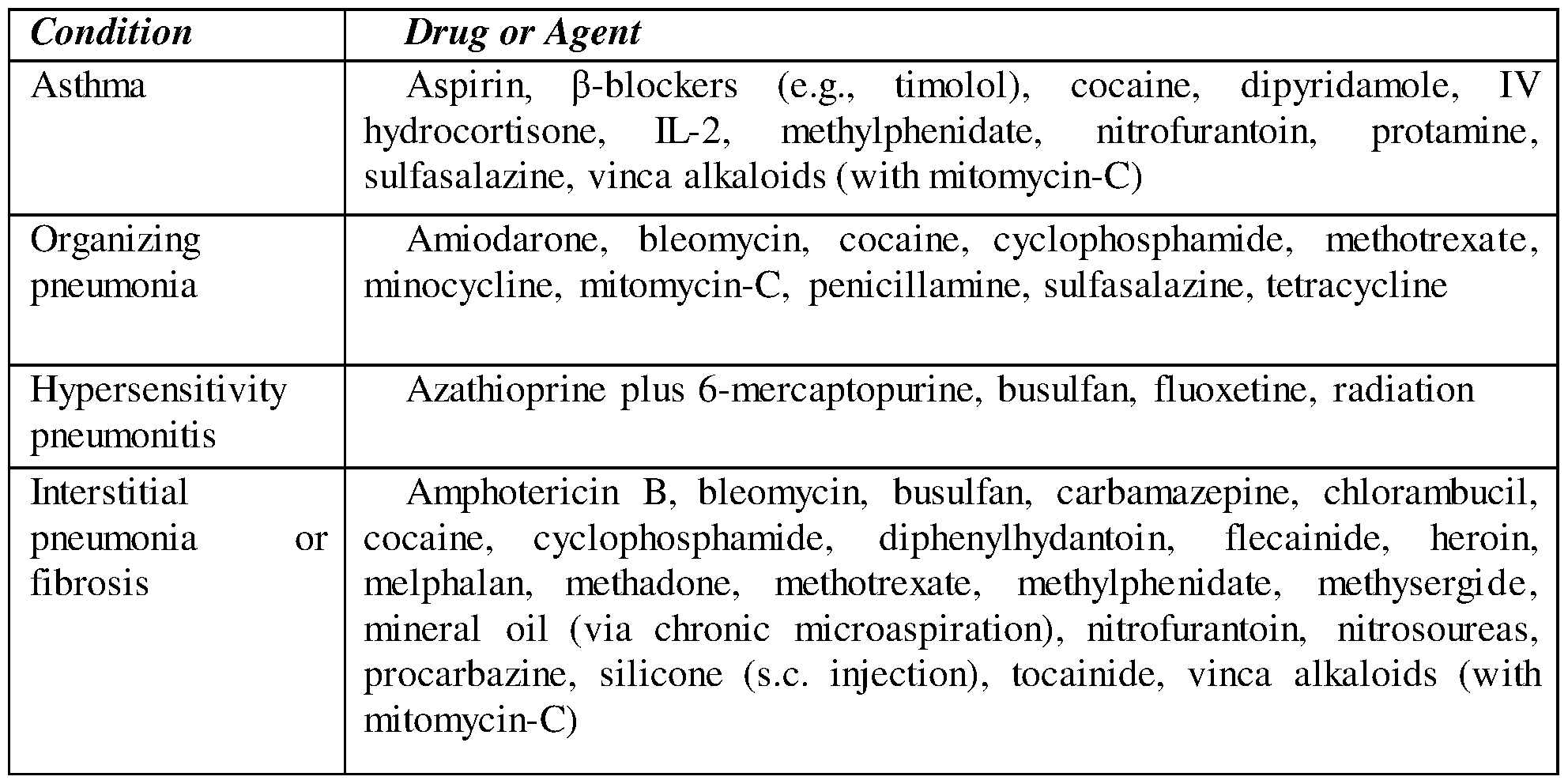


Claims
Priority Applications (7)
| Application Number | Priority Date | Filing Date | Title |
|---|---|---|---|
| KR1020247022673A KR20240128868A (en) | 2021-12-08 | 2022-12-08 | Multipotent lung progenitor cells for lung regeneration |
| JP2024534338A JP2024542847A (en) | 2021-12-08 | 2022-12-08 | Multipotent lung progenitor cells for lung regeneration |
| CA3240113A CA3240113A1 (en) | 2021-12-08 | 2022-12-08 | Multipotent lung progenitor cells for lung regeneration |
| EP22834741.5A EP4444864A1 (en) | 2021-12-08 | 2022-12-08 | Multipotent lung progenitor cells for lung regeneration |
| AU2022404031A AU2022404031A1 (en) | 2021-12-08 | 2022-12-08 | Multipotent lung progenitor cells for lung regeneration |
| CN202280091222.1A CN118696122A (en) | 2021-12-08 | 2022-12-08 | Multipotent lung progenitor cells for lung regeneration |
| US18/736,635 US20240325456A1 (en) | 2021-12-08 | 2024-06-07 | Multipotent lung progenitor cells for lung regeneration |
Applications Claiming Priority (2)
| Application Number | Priority Date | Filing Date | Title |
|---|---|---|---|
| US202163287147P | 2021-12-08 | 2021-12-08 | |
| US63/287,147 | 2021-12-08 |
Related Child Applications (1)
| Application Number | Title | Priority Date | Filing Date |
|---|---|---|---|
| US18/736,635 Continuation US20240325456A1 (en) | 2021-12-08 | 2024-06-07 | Multipotent lung progenitor cells for lung regeneration |
Publications (1)
| Publication Number | Publication Date |
|---|---|
| WO2023105526A1 true WO2023105526A1 (en) | 2023-06-15 |
Family
ID=84689188
Family Applications (1)
| Application Number | Title | Priority Date | Filing Date |
|---|---|---|---|
| PCT/IL2022/051302 Ceased WO2023105526A1 (en) | 2021-12-08 | 2022-12-08 | Multipotent lung progenitor cells for lung regeneration |
Country Status (8)
| Country | Link |
|---|---|
| US (1) | US20240325456A1 (en) |
| EP (1) | EP4444864A1 (en) |
| JP (1) | JP2024542847A (en) |
| KR (1) | KR20240128868A (en) |
| CN (1) | CN118696122A (en) |
| AU (1) | AU2022404031A1 (en) |
| CA (1) | CA3240113A1 (en) |
| WO (1) | WO2023105526A1 (en) |
Citations (33)
| Publication number | Priority date | Publication date | Assignee | Title |
|---|---|---|---|---|
| US3791932A (en) | 1971-02-10 | 1974-02-12 | Akzona Inc | Process for the demonstration and determination of reaction components having specific binding affinity for each other |
| US3839153A (en) | 1970-12-28 | 1974-10-01 | Akzona Inc | Process for the detection and determination of specific binding proteins and their corresponding bindable substances |
| US3850578A (en) | 1973-03-12 | 1974-11-26 | H Mcconnell | Process for assaying for biologically active molecules |
| US3850752A (en) | 1970-11-10 | 1974-11-26 | Akzona Inc | Process for the demonstration and determination of low molecular compounds and of proteins capable of binding these compounds specifically |
| US3853987A (en) | 1971-09-01 | 1974-12-10 | W Dreyer | Immunological reagent and radioimmuno assay |
| US3867517A (en) | 1971-12-21 | 1975-02-18 | Abbott Lab | Direct radioimmunoassay for antigens and their antibodies |
| US3879262A (en) | 1972-05-11 | 1975-04-22 | Akzona Inc | Detection and determination of haptens |
| US3901654A (en) | 1971-06-21 | 1975-08-26 | Biological Developments | Receptor assays of biologically active compounds employing biologically specific receptors |
| US3935074A (en) | 1973-12-17 | 1976-01-27 | Syva Company | Antibody steric hindrance immunoassay with two antibodies |
| US3984533A (en) | 1975-11-13 | 1976-10-05 | General Electric Company | Electrophoretic method of detecting antigen-antibody reaction |
| US3996345A (en) | 1974-08-12 | 1976-12-07 | Syva Company | Fluorescence quenching with immunological pairs in immunoassays |
| US4034074A (en) | 1974-09-19 | 1977-07-05 | The Board Of Trustees Of Leland Stanford Junior University | Universal reagent 2-site immunoradiometric assay using labelled anti (IgG) |
| US4098876A (en) | 1976-10-26 | 1978-07-04 | Corning Glass Works | Reverse sandwich immunoassay |
| US4666828A (en) | 1984-08-15 | 1987-05-19 | The General Hospital Corporation | Test for Huntington's disease |
| US4683202A (en) | 1985-03-28 | 1987-07-28 | Cetus Corporation | Process for amplifying nucleic acid sequences |
| US4801531A (en) | 1985-04-17 | 1989-01-31 | Biotechnology Research Partners, Ltd. | Apo AI/CIII genomic polymorphisms predictive of atherosclerosis |
| US4879219A (en) | 1980-09-19 | 1989-11-07 | General Hospital Corporation | Immunoassay utilizing monoclonal high affinity IgM antibodies |
| US5004681A (en) | 1987-11-12 | 1991-04-02 | Biocyte Corporation | Preservation of fetal and neonatal hematopoietic stem and progenitor cells of the blood |
| US5011771A (en) | 1984-04-12 | 1991-04-30 | The General Hospital Corporation | Multiepitopic immunometric assay |
| US5192659A (en) | 1989-08-25 | 1993-03-09 | Genetype Ag | Intron sequence analysis method for detection of adjacent and remote locus alleles as haplotypes |
| US5192553A (en) | 1987-11-12 | 1993-03-09 | Biocyte Corporation | Isolation and preservation of fetal and neonatal hematopoietic stem and progenitor cells of the blood and methods of therapeutic use |
| US5272057A (en) | 1988-10-14 | 1993-12-21 | Georgetown University | Method of detecting a predisposition to cancer by the use of restriction fragment length polymorphism of the gene for human poly (ADP-ribose) polymerase |
| US5281521A (en) | 1992-07-20 | 1994-01-25 | The Trustees Of The University Of Pennsylvania | Modified avidin-biotin technique |
| US5656498A (en) | 1994-02-22 | 1997-08-12 | Nippon Telegraph And Telephone Corporation | Freeze-dried blood cells, stem cells and platelets, and manufacturing method for the same |
| US5955257A (en) | 1997-10-21 | 1999-09-21 | Regents Of The University Of Minnesota | Infusible grade short-term cell storage medium for mononuclear cells |
| US20030215942A1 (en) | 2002-02-14 | 2003-11-20 | Stemcyte, Inc. | Undesignated allogeneic stem cell bank |
| WO2013084190A1 (en) | 2011-12-08 | 2013-06-13 | Yeda Research And Development Co. Ltd. | Mammalian fetal pulmonary cells and therapeutic use of same |
| WO2016203477A1 (en) | 2015-06-18 | 2016-12-22 | Yeda Research And Development Co. Ltd. | Conditioning protocols and use of same for tissue regeneration |
| WO2017203520A1 (en) | 2016-05-22 | 2017-11-30 | Yeda Research And Development Co. Ltd. | Methods of using pulmonary cells for transplantation and induction of tolerance |
| CN111534477A (en) * | 2020-05-13 | 2020-08-14 | 江苏省人民医院(南京医科大学第一附属医院) | Method for culturing primary epithelial stem cell balls of lung tissue of mouse |
| WO2020245832A1 (en) | 2019-06-07 | 2020-12-10 | Ramot At Tel-Aviv University Ltd. | Compositions and methods of using same for tissue regeneration |
| CN106967672B (en) * | 2017-03-24 | 2021-01-26 | 四川大学华西医院 | Lung and lung cancer tissue culture method and method for constructing lung cancer mouse animal model by using same |
| CN112501110A (en) * | 2020-11-26 | 2021-03-16 | 海西纺织新材料工业技术晋江研究院 | Standardized culture medium and method for three-dimensional culture of lung and lung cancer tissue organoid |
-
2022
- 2022-12-08 KR KR1020247022673A patent/KR20240128868A/en active Pending
- 2022-12-08 CA CA3240113A patent/CA3240113A1/en active Pending
- 2022-12-08 AU AU2022404031A patent/AU2022404031A1/en active Pending
- 2022-12-08 CN CN202280091222.1A patent/CN118696122A/en active Pending
- 2022-12-08 JP JP2024534338A patent/JP2024542847A/en active Pending
- 2022-12-08 EP EP22834741.5A patent/EP4444864A1/en active Pending
- 2022-12-08 WO PCT/IL2022/051302 patent/WO2023105526A1/en not_active Ceased
-
2024
- 2024-06-07 US US18/736,635 patent/US20240325456A1/en active Pending
Patent Citations (36)
| Publication number | Priority date | Publication date | Assignee | Title |
|---|---|---|---|---|
| US3850752A (en) | 1970-11-10 | 1974-11-26 | Akzona Inc | Process for the demonstration and determination of low molecular compounds and of proteins capable of binding these compounds specifically |
| US3839153A (en) | 1970-12-28 | 1974-10-01 | Akzona Inc | Process for the detection and determination of specific binding proteins and their corresponding bindable substances |
| US3791932A (en) | 1971-02-10 | 1974-02-12 | Akzona Inc | Process for the demonstration and determination of reaction components having specific binding affinity for each other |
| US3901654A (en) | 1971-06-21 | 1975-08-26 | Biological Developments | Receptor assays of biologically active compounds employing biologically specific receptors |
| US3853987A (en) | 1971-09-01 | 1974-12-10 | W Dreyer | Immunological reagent and radioimmuno assay |
| US3867517A (en) | 1971-12-21 | 1975-02-18 | Abbott Lab | Direct radioimmunoassay for antigens and their antibodies |
| US3879262A (en) | 1972-05-11 | 1975-04-22 | Akzona Inc | Detection and determination of haptens |
| US3850578A (en) | 1973-03-12 | 1974-11-26 | H Mcconnell | Process for assaying for biologically active molecules |
| US3935074A (en) | 1973-12-17 | 1976-01-27 | Syva Company | Antibody steric hindrance immunoassay with two antibodies |
| US3996345A (en) | 1974-08-12 | 1976-12-07 | Syva Company | Fluorescence quenching with immunological pairs in immunoassays |
| US4034074A (en) | 1974-09-19 | 1977-07-05 | The Board Of Trustees Of Leland Stanford Junior University | Universal reagent 2-site immunoradiometric assay using labelled anti (IgG) |
| US3984533A (en) | 1975-11-13 | 1976-10-05 | General Electric Company | Electrophoretic method of detecting antigen-antibody reaction |
| US4098876A (en) | 1976-10-26 | 1978-07-04 | Corning Glass Works | Reverse sandwich immunoassay |
| US4879219A (en) | 1980-09-19 | 1989-11-07 | General Hospital Corporation | Immunoassay utilizing monoclonal high affinity IgM antibodies |
| US5011771A (en) | 1984-04-12 | 1991-04-30 | The General Hospital Corporation | Multiepitopic immunometric assay |
| US4666828A (en) | 1984-08-15 | 1987-05-19 | The General Hospital Corporation | Test for Huntington's disease |
| US4683202A (en) | 1985-03-28 | 1987-07-28 | Cetus Corporation | Process for amplifying nucleic acid sequences |
| US4683202B1 (en) | 1985-03-28 | 1990-11-27 | Cetus Corp | |
| US4801531A (en) | 1985-04-17 | 1989-01-31 | Biotechnology Research Partners, Ltd. | Apo AI/CIII genomic polymorphisms predictive of atherosclerosis |
| US5004681A (en) | 1987-11-12 | 1991-04-02 | Biocyte Corporation | Preservation of fetal and neonatal hematopoietic stem and progenitor cells of the blood |
| US5192553A (en) | 1987-11-12 | 1993-03-09 | Biocyte Corporation | Isolation and preservation of fetal and neonatal hematopoietic stem and progenitor cells of the blood and methods of therapeutic use |
| US5004681B1 (en) | 1987-11-12 | 2000-04-11 | Biocyte Corp | Preservation of fetal and neonatal hematopoietic stem and progenitor cells of the blood |
| US6461645B1 (en) | 1987-11-12 | 2002-10-08 | Pharmastem Therapeutics, Inc. | Isolation and preservation of fetal and neonatal hematopoietic stem and progenitor cells of the blood |
| US5272057A (en) | 1988-10-14 | 1993-12-21 | Georgetown University | Method of detecting a predisposition to cancer by the use of restriction fragment length polymorphism of the gene for human poly (ADP-ribose) polymerase |
| US5192659A (en) | 1989-08-25 | 1993-03-09 | Genetype Ag | Intron sequence analysis method for detection of adjacent and remote locus alleles as haplotypes |
| US5281521A (en) | 1992-07-20 | 1994-01-25 | The Trustees Of The University Of Pennsylvania | Modified avidin-biotin technique |
| US5656498A (en) | 1994-02-22 | 1997-08-12 | Nippon Telegraph And Telephone Corporation | Freeze-dried blood cells, stem cells and platelets, and manufacturing method for the same |
| US5955257A (en) | 1997-10-21 | 1999-09-21 | Regents Of The University Of Minnesota | Infusible grade short-term cell storage medium for mononuclear cells |
| US20030215942A1 (en) | 2002-02-14 | 2003-11-20 | Stemcyte, Inc. | Undesignated allogeneic stem cell bank |
| WO2013084190A1 (en) | 2011-12-08 | 2013-06-13 | Yeda Research And Development Co. Ltd. | Mammalian fetal pulmonary cells and therapeutic use of same |
| WO2016203477A1 (en) | 2015-06-18 | 2016-12-22 | Yeda Research And Development Co. Ltd. | Conditioning protocols and use of same for tissue regeneration |
| WO2017203520A1 (en) | 2016-05-22 | 2017-11-30 | Yeda Research And Development Co. Ltd. | Methods of using pulmonary cells for transplantation and induction of tolerance |
| CN106967672B (en) * | 2017-03-24 | 2021-01-26 | 四川大学华西医院 | Lung and lung cancer tissue culture method and method for constructing lung cancer mouse animal model by using same |
| WO2020245832A1 (en) | 2019-06-07 | 2020-12-10 | Ramot At Tel-Aviv University Ltd. | Compositions and methods of using same for tissue regeneration |
| CN111534477A (en) * | 2020-05-13 | 2020-08-14 | 江苏省人民医院(南京医科大学第一附属医院) | Method for culturing primary epithelial stem cell balls of lung tissue of mouse |
| CN112501110A (en) * | 2020-11-26 | 2021-03-16 | 海西纺织新材料工业技术晋江研究院 | Standardized culture medium and method for three-dimensional culture of lung and lung cancer tissue organoid |
Non-Patent Citations (91)
| Title |
|---|
| "Current Protocols in Molecular Biology", vol. 1-3, 1994, APPLETON & LANGE, NORWALK |
| "Immobilized Cells and Enzymes", 1986, IRL PRESS |
| "Nucleic Acid Hybridization", 1985 |
| "PCR Protocols: A Guide To Methods And Applications", vol. 1-317, 1990, ACADEMIC PRESS |
| "Selected Methods in Cellular Immunology", 1980, W. H. FREEMAN AND CO. |
| "Transcription and Translation", 1984 |
| A. BAGGIOLINI ET AL.: "Premigratory and migratory neural crest cells are multipotent in vivo", CELL STEM CEL, vol. 16, 2015, pages 314 - 322 |
| A. BUTLERP. HOFFMANP. SMIBERTE. PAPALEXIR. SATIJA: "Integrating single-cell transcriptomic data across different conditions, technologies, and species", NAT. BIOTECHNOL., vol. 36, 2018, pages 411 - 420 |
| A. C. RIOSN. Y. FUG. J. LINDEMANJ. E. VISVADER: "In situ identification of bipotent stem cells in the mammary gland", NATURE, vol. 506, 2014, pages 322 |
| A. G. SCHEPERS ET AL.: "Lineage tracing reveals Lgr5+ stem cell activity in mouse intestinal adenomas", SCIENCE, vol. 337, 2012, pages 730 - 735 |
| A. GILLICH ET AL.: "Capillary cell-type specialization in the alveolus", NATURE, vol. 586, 2020, pages 785 - 789, XP037282837, DOI: 10.1038/s41586-020-2822-7 |
| A. LIBERZON ET AL.: "The molecular signatures database hallmark gene set collection", CELL SYSTEMS, vol. 1, 2015, pages 417 - 425 |
| A. LYUBIMOVA ET AL.: "Single-molecule mRNA detection and counting in mammalian tissue", NATURE PROTOCOLS, vol. 8, 2013, pages 1743 |
| B. D. HARFE ET AL.: "Evidence for an expansion-based temporal Shh gradient in specifying vertebrate digit identities", CELL, vol. 118, 2004, pages 517 - 528 |
| C. HILLEL-KARNIEL ET AL.: "Multi-lineage Lung Regeneration by Stem Cell Transplantation across Major Genetic Barriers", CELL REPORTS, vol. 30, 2020, pages 807 - 819 |
| C. L. SCHEELE ET AL.: "Identity and dynamics of mammary stem cells during branching morphogenesis", NATURE, vol. 542, 2017, pages 313 |
| C. M. TRIVEDI ET AL.: "Hopx and Hdac2 interact to modulate Gata4 acetylation and embryonic cardiac myocyte proliferation", DEVELOPMENTAL CEL, vol. 19, 2010, pages 450 - 459 |
| C. ROSEN ET AL.: "Preconditioning allows engraftment of mouse and human embryonic lung cells, enabling lung repair in mice", NAT. MED, vol. 21, 2015, pages 869 |
| C. TRAPNELL ET AL.: "The dynamics and regulators of cell fate decisions are revealed by pseudotemporal ordering of single cells", NAT. BIOTECHNOL, vol. 32, 2014, pages 381, XP055889676, DOI: 10.1038/nbt.2859 |
| CANAPLE, L ET AL.: "Improving cell encapsulation through size control", J BIOMATER SCI POLYM ED, vol. 13, 2002, pages 783 - 96 |
| CHIA, S. M ET AL.: "Multi-layered microcapsules for cell encapsulation", BIOMATERIALS, vol. 23, 2002, pages 849 - 856, XP004322709, DOI: 10.1016/S0142-9612(01)00191-0 |
| D. J. WEISS ET AL.: "Stem cells and cell therapies in lung biology and lung diseases", PROCEEDINGS OF THE AMERICAN THORACIC SOCIETY, vol. 8, 2011, pages 223 - 272 |
| D. J. WEISSL. A. ORTIZ: "Cell therapy trials for lung diseases: progress and cautions", AMERICAN JOURNAL OF RESPIRATORY AND CRITICAL CARE MEDICINE, vol. 188, 2013, pages 123 - 125 |
| D. N. WATKINS ET AL.: "Hedgehog signalling within airway epithelial progenitors and in small-cell lung cancer", NATURE, vol. 422, 2003, pages 313 - 317, XP002554660, DOI: 10.1038/nature01493 |
| D. PAN ET AL.: "SOX2, OCT3/4 and NANOG expression and cellular plasticity in rare human somatic cells requires CD73", CELL. SIGNAL., vol. 28, 2016, pages 1923 - 1932, XP029764441, DOI: 10.1016/j.cellsig.2016.09.008 |
| DANTAL J ET AL., LANCET, vol. 351, 1998, pages 623 |
| DESAI, T. A: "Microfabrication technology for pancreatic cell encapsulation", EXPERT OPIN BIOL THER, vol. 2, 2002, pages 633 - 646 |
| DONATI YVES ET AL: "Simultaneous isolation of endothelial and alveolar epithelial type I and type II cells during mouse lung development in the absence of a transgenic reporter", AMERICAN JOURNAL OF PHYSIOLOGY - LUNG CELLULAR AND MOLECULAR PHYSIOLOGY, vol. 318, no. 4, 1 April 2020 (2020-04-01), US, pages 619 - 630, XP093030298, ISSN: 1040-0605, DOI: 10.1152/ajplung.00227.2019 * |
| E. LAZZERI ET AL.: "Endocycle-related tubular cell hypertrophy and progenitor proliferation recover renal function after acute kidney injury", NATURE COMMUNICATIONS, vol. 9, 2018, pages 1344 |
| FLORIAN WEINBERGERET, CIRCULATION RESEARCH, vol. 120, 2017, pages 1487 - 1500 |
| FRANCIS ET AL., GUT, vol. 52, no. 11, 2003, pages 1602 - 6 |
| FSY WONGACY LO, J STEM CELL RES THER, vol. 5, 2015, pages 267 |
| FUCHS ET AL., J NUCL MED, vol. 54, no. 1, 2013, pages 151 - 8 |
| H. J. SNIPPERT ET AL.: "Intestinal crypt homeostasis results from neutral competition between symmetrically dividing Lgr5 stem cells", CELL., vol. 143, 2010, pages 134 - 144, XP028931150, DOI: 10.1016/j.cell.2010.09.016 |
| H. TANIGUCHIJ. LUZ. J. HUANG: "The spatial and temporal origin of chandelier cells in mouse neocortex", SCIENCE, vol. 339, 2013, pages 70 - 74 |
| HANTO DW., ANNU REV MED, vol. 46, 1995, pages 381 |
| HIGGINS RM ET AL., LANCET, vol. 348, 1996, pages 1208 |
| HILLEL-KARNIEL CARMIT ET AL: "Multi-lineage Lung Regeneration by Stem Cell Transplantation across Major Genetic Barriers", CELL REPORTS, vol. 30, no. 3, 21 January 2020 (2020-01-21), US, pages 807 - 819.e4, XP093030181, ISSN: 2211-1247, Retrieved from the Internet <URL:https://www.cell.com/cell-reports/pdf/S2211-1247(19)31715-2.pdf> DOI: 10.1016/j.celrep.2019.12.058 * |
| I. MILMAN KRENTSIS ET AL.: "Lung Injury Repair by Transplantation of Adult Lung Cells Following Preconditioning of Recipient Mice", STEM CELLS TRANSLATIONAL MEDICINE, vol. 7, 2018, pages 68 - 77 |
| J. CAO ET AL.: "The single-cell transcriptional landscape of mammalian organogenesis", NATURE, vol. 566, 2019, pages 496 - 502, XP036713041, DOI: 10.1038/s41586-019-0969-x |
| J. R. PRIGGE ET AL.: "Nuclear double-fluorescent reporter for in vivo and ex vivo analyses of biological transitions in mouse nuclei", MAMMALIAN GENOME, vol. 24, 2013, pages 389 - 399 |
| K. I. BOSTROMJ. YAOX. WUY. YAO: "Endothelial Cells May Have Tissue-Specific Origins", J. CELL. BIOL. HISTOL, vol. 1, 2018 |
| KIRKPATRICK CHROWLANDS DT JR, JAMA, vol. 268, 1992, pages 2952 |
| L. MCINNESJ. HEALYJ. MELVILLE: "Umap: Uniform manifold approximation and projection for dimension reduction", ARXIV PREPRINT ARXIV:1802.03426, 2018 |
| L. ZHANG ET AL.: "ZFP36L2 is required for self-renewal of early burst-forming unit erythroid progenitors", NATURE, vol. 499, 2013, pages 92 - 96 |
| LTLUDAG, H. ET AL.: "Technology of mammalian cell encapsulation", ADV DRUG DELIV REV, vol. 42, 2000, pages 29 - 64, XP002302229, DOI: 10.1016/S0169-409X(00)00053-3 |
| LU, M. Z. ET AL.: "A novel cell encapsulation method using photosensitive poly(allylamine alpha-cyanocinnamylideneacetate", J MICROENCAPSUL, vol. 17, 2000, pages 245 - 521 |
| LUM. Z ET AL.: "Cell encapsulation with alginate and alpha- phenoxycinnamylidene-acetylated poly(allylamine", BIOTECHNOL BIOENG, vol. 70, 2000, pages 479 - 483, XP008025769, DOI: 10.1002/1097-0290(20001205)70:5<479::AID-BIT1>3.0.CO;2-E |
| M. CHUNGB. L. HOGAN: "Ager-CreER T2: a new genetic tool for studying lung alveolar development, homeostasis, and repair", AMERICAN JOURNAL OF RESPIRATORY CELL AND MOLECULAR BIOLOGY, vol. 59, 2018, pages 706 - 712 |
| M. D. MUZUMDARB. TASICK. MIYAMICHIL. LIL. LUO: "A global double-fluorescent Cre reporter mouse", GENESIS, vol. 45, 2007, pages 593 - 605, XP055011399, DOI: 10.1002/dvg.20335 |
| M. GANUZA ET AL.: "Lifelong haematopoiesis is established by hundreds of precursors throughout mammalian ontogeny", NAT. CELL BIOL, vol. 19, 2017, pages 1153 |
| M. J. CHENT. YOKOMIZOB. M. ZEIGLERE. DZIERZAKN. A. SPECK: "Runxl is required for the endothelial to haematopoietic cell transition but not thereafter", NATURE, vol. 457, 2009, pages 887 - 891 |
| M. NICHANE ET AL.: "Isolation and 3D expansion of multipotent Sox9 mouse lung progenitors", NATURE METHODS, vol. 14, 2017, pages 1205, XP037152219, DOI: 10.1038/nmeth.4498 |
| MARIA IRIS HERMANNS ET AL: "Lung epithelial cell lines in coculture with human pulmonary microvascular endothelial cells: development of an alveolo-capillary barrier in vitro", LABORATORY INVESTIGATION, vol. 84, no. 6, 1 June 2004 (2004-06-01), The United States and Canadian Academy of Pathology, Inc., pages 736 - 752, XP055108914, ISSN: 0023-6837, DOI: 10.1038/labinvest.3700081 * |
| MARSHAK ET AL.: "Strategies for Protein Purification and Characterization - A Laboratory Course Manual", 1996, CSHL PRESS |
| MIDTHUN DE ET AL., MAYO CLIN PROC, vol. 72, 1997, pages 175 |
| MORRISON VA. ET AL., AM J MED, vol. 97, 1994, pages 14 |
| N. J. PALPANT ET AL.: "Chromatin and transcriptional analysis of mesoderm progenitor cells identifies HOPX as a regulator of primitive hematopoiesis", CELL REPORTS, vol. 20, 2017, pages 1597 - 1608 |
| N. TAKEDA ET AL.: "Hopx expression defines a subset of multipotent hair follicle stem cells and a progenitor population primed to give rise to K6+ niche cells", DEVELOPMENT., vol. 140, 2013, pages 1655 - 1664 |
| N. TAKEDA ET AL.: "Interconversion between intestinal stem cell populations in distinct niches", SCIENCE, vol. 334, 2011, pages 1420 - 1424 |
| NICHANE MASSIMO ET AL: "Isolation and 3D expansion of multipotent Sox9mouse lung progenitors", NATURE METHODS, NATURE PUBLISHING GROUP US, NEW YORK, vol. 14, no. 12, 6 November 2017 (2017-11-06), pages 1205 - 1212, XP037152219, ISSN: 1548-7091, [retrieved on 20171106], DOI: 10.1038/NMETH.4498 * |
| P. R. JAMIESON ET AL.: "Derivation of a robust mouse mammary organoid system for studying tissue dynamics", DEVELOPMENT, vol. 144, 2017, pages 1065 - 1071 |
| P. R. TATA ET AL.: "Dedifferentiation of committed epithelial cells into stem cells in vivo", NATURE, vol. 503, 2013, pages 218, XP037436696, DOI: 10.1038/nature12777 |
| P. R. TATAJ. RAJAGOPAL: "Plasticity in the lung: making and breaking cell identity", DEVELOPMENT, vol. 144, 2017, pages 755 - 766 |
| PERBAL, B.: "A Practical Guide to Molecular Cloning", 1984, JOHN WILEY & SONS |
| PRAKASH, S: "Procedures for microencapsulation of enzymes, cells and genetically engineered microorganisms", MOL BIOTECHNOL, vol. 17, 2001, pages 249 - 260 |
| Q. MA ET AL.: "Regeneration of functional alveoli by adult human SOX9 airway basal cell transplantation", PROTEIN & CELL, vol. 9, 2018, pages 267 - 282, XP055752615, DOI: 10.1007/s13238-018-0506-y |
| QIWANG MA ET AL: "Regeneration of functional alveoli by adult human SOX9+ airway basal cell transplantation", PROTEIN & CELL, vol. 9, no. 3, 17 January 2018 (2018-01-17), Beijing, CN, pages 267 - 282, XP055752615, ISSN: 1674-800X, DOI: 10.1007/s13238-018-0506-y * |
| R. JAIN ET AL.: "HEART DEVELOPMENT. Integration of Bmp and Wnt signaling by Hopx specifies commitment of cardiomyoblasts", SCIENCE, vol. 348, 2015, pages aaa6071 |
| R. JAIN ET AL.: "Plasticity of Hopx type I alveolar cells to regenerate type II cells in the lung", NATURE COMMUNICATIONS, vol. 6, 2015, pages 1 - 11 |
| R. SATIJAJ. A. FARRELLD. GENNERTA. F. SCHIERA. REGEV: "Spatial reconstruction of single-cell gene expression data", NAT. BIOTECHNOL., vol. 33, 2015, pages 495 - 502, XP055423072, DOI: 10.1038/nbt.3192 |
| ROCHKIND S ET AL., NEUROL RES, vol. 26, no. 2, 2004, pages 161 - 6 |
| ROCHKIND S, EUR SPINE J, vol. 15, no. 2, 2006, pages 234 - 45 |
| S. HANZELMANNR. CASTELOJ. GUINNEY: "GSVA: gene set variation analysis for microarray and RNA-seq data", BMC BIOINFORMATICS, vol. 14, 2013, pages 1 - 15 |
| S. ROY ET AL.: "Rare somatic cells from human breast tissue exhibit extensive lineage plasticity", PROC. NATL. ACAD. SCI. U. S. A., vol. 110, 2013, pages 4598 - 4603, XP055127541, DOI: 10.1073/pnas.1218682110 |
| S. T. BUCKLEYC. EHRHARDT: "The receptor for advanced glycation end products (RAGE) and the lung", JOURNAL OF BIOMEDICINE AND BIOTECHNOLOGY, vol. 2010, 2010 |
| SAMBANIS, A: "Encapsulated islets in diabetes treatment", DIABETES THECHNOL THER, vol. 5, 2003, pages 665 - 668, XP055861684, DOI: 10.1089/152091503322250686 |
| SAMBROOK ET AL.: "Current Protocols in Molecular Biology", 1989, JOHN WILEY AND SONS |
| SENDEROWICZ AM ET AL., ANN INTERN MED, vol. 126, 1997, pages 882 |
| SUTHANTHIRAN MSTROM TB., NEW ENGL. J. MED, vol. 331, 1996, pages 365 |
| T. J. DESAID. G. BROWNFIELDM. A. KRASNOW: "Alveolar progenitor and stem cells in lung development, renewal and cancer", NATURE, vol. 507, 2014, pages 190 - 194 |
| T. STUART ET AL.: "Comprehensive integration of single-cell data", CELL, vol. 177, 2019, pages 1888 - 1902 |
| V. A. TRAAGL. WALTMANN. J. VAN ECK: "From Louvain to Leiden: guaranteeing well-connected communities", SCIENTIFIC REPORTS, vol. 9, 2019, pages 1 - 12 |
| VACANTI ET AL., JOURNAL OF PEDIATRIC SURGERY, vol. 23, 1988, pages 3 - 9 |
| VINCENTI F. ET AL., NEW ENGL. J. MED, vol. 338, 1998, pages 161 |
| W. ZUO ET AL.: "p63 Krt5 distal airway stem cells are essential for lung regeneration", NATURE, 2014 |
| WILLIAMS, D: "Small is beautiful: microparticle and nanoparticle technology in medical devices", MED DEVICE TECHNOL, vol. 10, 1999, pages 6 - 9 |
| X. QIU ET AL.: "Reversed graph embedding resolves complex single-cell trajectories", NATURE METHODS, vol. 14, 2017, pages 979, XP037149680, DOI: 10.1038/nmeth.4402 |
| Y. LITINGTUNGL. LEIH. WESTPHALC. CHIANG: "Sonic hedgehog is essential to foregut development", NAT. GENET, vol. 1-4, 1998, pages 58 - 61 |
| Y. LIUW. ZHANG: "The role of HOPX in normal tissues and tumor progression", BIOSCI. REP, vol. 40, 2020 |
| Z. YIN ET AL.: "Hop functions downstream of Nkx2. 1 and GATA6 to mediate HDAC-dependent negative regulation of pulmonary gene expression", AMERICAN JOURNAL OF PHYSIOLOGY-LUNG CELLULAR AND MOLECULAR PHYSIOLOGY, vol. 291, 2006, pages L191 - L199 |
Also Published As
| Publication number | Publication date |
|---|---|
| US20240325456A1 (en) | 2024-10-03 |
| CN118696122A (en) | 2024-09-24 |
| AU2022404031A1 (en) | 2024-07-25 |
| KR20240128868A (en) | 2024-08-27 |
| EP4444864A1 (en) | 2024-10-16 |
| CA3240113A1 (en) | 2023-06-15 |
| JP2024542847A (en) | 2024-11-15 |
Similar Documents
| Publication | Publication Date | Title |
|---|---|---|
| US20200306319A1 (en) | Methods for treating radiation or chemical injury | |
| DK2366775T3 (en) | Methods for the expansion of the cells and the use of the thus-obtained cells and conditioned media for the therapy | |
| JP4950661B2 (en) | Neural tissue regeneration and repair using postpartum cells | |
| US20230256028A1 (en) | Methods of using pulmonary cells for transplantation and induction of tolerance | |
| US20160101133A1 (en) | Organoids comprising isolated renal cells and use thereof | |
| US20110171182A1 (en) | Methods for cell expansion and uses of cells and conditioned media produced thereby for therapy | |
| US20140017209A1 (en) | Methods for treating radiation or chemical injury | |
| US20110293583A1 (en) | Methods for cell expansion and uses of cells and conditioned media produced thereby for therapy | |
| EP3034087B1 (en) | Mammalian fetal pulmonary cells and therapeutic use of same | |
| US20010049139A1 (en) | Hepatic regeneration from hematopoietic stem cells | |
| WO2006040755A2 (en) | Novel methods for the in-vitro identification, isolation and differentiation of vasculogenic progenitor cells | |
| US20080194023A1 (en) | Generating vascular smooth muscle cells in vitro from ES cells | |
| US20210113627A1 (en) | Adherent stromal cells derived from placentas of multiple donors and uses thereof | |
| KR20230016653A (en) | Method for producing organoids from lung epithelial cells or lung cancer cells | |
| US20240325456A1 (en) | Multipotent lung progenitor cells for lung regeneration | |
| JP2008531007A (en) | Method for obtaining human hematopoietic stem cell population | |
| HK40086579A (en) | Method for producing organoid from lung epithelial cells or lung cancer cells | |
| Suh et al. | SPLENIC SYNTHESIS OF FACTOR VIII: BASIS FOR CELL-BASED TRANSPLANT THERAPY OF HEMOPHILIA A. | |
| NZ627071B2 (en) | Mammalian fetal pulmonary cells and therapeutic use of same | |
| AU2004220699A1 (en) | Novel Methods for the In-Vitro Identification, Isolation and Differentiation of Vasculogenic Progenitor Cells |
Legal Events
| Date | Code | Title | Description |
|---|---|---|---|
| 121 | Ep: the epo has been informed by wipo that ep was designated in this application |
Ref document number: 22834741 Country of ref document: EP Kind code of ref document: A1 |
|
| WWE | Wipo information: entry into national phase |
Ref document number: 3240113 Country of ref document: CA |
|
| ENP | Entry into the national phase |
Ref document number: 2024534338 Country of ref document: JP Kind code of ref document: A |
|
| WWE | Wipo information: entry into national phase |
Ref document number: AU2022404031 Country of ref document: AU |
|
| ENP | Entry into the national phase |
Ref document number: 20247022673 Country of ref document: KR Kind code of ref document: A |
|
| WWE | Wipo information: entry into national phase |
Ref document number: 2022834741 Country of ref document: EP |
|
| NENP | Non-entry into the national phase |
Ref country code: DE |
|
| ENP | Entry into the national phase |
Ref document number: 2022834741 Country of ref document: EP Effective date: 20240708 |
|
| ENP | Entry into the national phase |
Ref document number: 2022404031 Country of ref document: AU Date of ref document: 20221208 Kind code of ref document: A |
|
| WWE | Wipo information: entry into national phase |
Ref document number: 202280091222.1 Country of ref document: CN |