WO2022261515A1 - Method and system for improved management of genetic diseases - Google Patents
Method and system for improved management of genetic diseases Download PDFInfo
- Publication number
- WO2022261515A1 WO2022261515A1 PCT/US2022/033128 US2022033128W WO2022261515A1 WO 2022261515 A1 WO2022261515 A1 WO 2022261515A1 US 2022033128 W US2022033128 W US 2022033128W WO 2022261515 A1 WO2022261515 A1 WO 2022261515A1
- Authority
- WO
- WIPO (PCT)
- Prior art keywords
- genetic
- sequencing
- subject
- diagnosis
- list
- Prior art date
- Legal status (The legal status is an assumption and is not a legal conclusion. Google has not performed a legal analysis and makes no representation as to the accuracy of the status listed.)
- Ceased
Links
Classifications
-
- G—PHYSICS
- G16—INFORMATION AND COMMUNICATION TECHNOLOGY [ICT] SPECIALLY ADAPTED FOR SPECIFIC APPLICATION FIELDS
- G16H—HEALTHCARE INFORMATICS, i.e. INFORMATION AND COMMUNICATION TECHNOLOGY [ICT] SPECIALLY ADAPTED FOR THE HANDLING OR PROCESSING OF MEDICAL OR HEALTHCARE DATA
- G16H10/00—ICT specially adapted for the handling or processing of patient-related medical or healthcare data
- G16H10/60—ICT specially adapted for the handling or processing of patient-related medical or healthcare data for patient-specific data, e.g. for electronic patient records
-
- G—PHYSICS
- G16—INFORMATION AND COMMUNICATION TECHNOLOGY [ICT] SPECIALLY ADAPTED FOR SPECIFIC APPLICATION FIELDS
- G16H—HEALTHCARE INFORMATICS, i.e. INFORMATION AND COMMUNICATION TECHNOLOGY [ICT] SPECIALLY ADAPTED FOR THE HANDLING OR PROCESSING OF MEDICAL OR HEALTHCARE DATA
- G16H20/00—ICT specially adapted for therapies or health-improving plans, e.g. for handling prescriptions, for steering therapy or for monitoring patient compliance
- G16H20/10—ICT specially adapted for therapies or health-improving plans, e.g. for handling prescriptions, for steering therapy or for monitoring patient compliance relating to drugs or medications, e.g. for ensuring correct administration to patients
-
- G—PHYSICS
- G16—INFORMATION AND COMMUNICATION TECHNOLOGY [ICT] SPECIALLY ADAPTED FOR SPECIFIC APPLICATION FIELDS
- G16B—BIOINFORMATICS, i.e. INFORMATION AND COMMUNICATION TECHNOLOGY [ICT] SPECIALLY ADAPTED FOR GENETIC OR PROTEIN-RELATED DATA PROCESSING IN COMPUTATIONAL MOLECULAR BIOLOGY
- G16B20/00—ICT specially adapted for functional genomics or proteomics, e.g. genotype-phenotype associations
- G16B20/20—Allele or variant detection, e.g. single nucleotide polymorphism [SNP] detection
-
- G—PHYSICS
- G16—INFORMATION AND COMMUNICATION TECHNOLOGY [ICT] SPECIALLY ADAPTED FOR SPECIFIC APPLICATION FIELDS
- G16B—BIOINFORMATICS, i.e. INFORMATION AND COMMUNICATION TECHNOLOGY [ICT] SPECIALLY ADAPTED FOR GENETIC OR PROTEIN-RELATED DATA PROCESSING IN COMPUTATIONAL MOLECULAR BIOLOGY
- G16B35/00—ICT specially adapted for in silico combinatorial libraries of nucleic acids, proteins or peptides
- G16B35/10—Design of libraries
-
- G—PHYSICS
- G16—INFORMATION AND COMMUNICATION TECHNOLOGY [ICT] SPECIALLY ADAPTED FOR SPECIFIC APPLICATION FIELDS
- G16B—BIOINFORMATICS, i.e. INFORMATION AND COMMUNICATION TECHNOLOGY [ICT] SPECIALLY ADAPTED FOR GENETIC OR PROTEIN-RELATED DATA PROCESSING IN COMPUTATIONAL MOLECULAR BIOLOGY
- G16B40/00—ICT specially adapted for biostatistics; ICT specially adapted for bioinformatics-related machine learning or data mining, e.g. knowledge discovery or pattern finding
- G16B40/20—Supervised data analysis
-
- G—PHYSICS
- G16—INFORMATION AND COMMUNICATION TECHNOLOGY [ICT] SPECIALLY ADAPTED FOR SPECIFIC APPLICATION FIELDS
- G16H—HEALTHCARE INFORMATICS, i.e. INFORMATION AND COMMUNICATION TECHNOLOGY [ICT] SPECIALLY ADAPTED FOR THE HANDLING OR PROCESSING OF MEDICAL OR HEALTHCARE DATA
- G16H15/00—ICT specially adapted for medical reports, e.g. generation or transmission thereof
-
- G—PHYSICS
- G16—INFORMATION AND COMMUNICATION TECHNOLOGY [ICT] SPECIALLY ADAPTED FOR SPECIFIC APPLICATION FIELDS
- G16H—HEALTHCARE INFORMATICS, i.e. INFORMATION AND COMMUNICATION TECHNOLOGY [ICT] SPECIALLY ADAPTED FOR THE HANDLING OR PROCESSING OF MEDICAL OR HEALTHCARE DATA
- G16H50/00—ICT specially adapted for medical diagnosis, medical simulation or medical data mining; ICT specially adapted for detecting, monitoring or modelling epidemics or pandemics
- G16H50/20—ICT specially adapted for medical diagnosis, medical simulation or medical data mining; ICT specially adapted for detecting, monitoring or modelling epidemics or pandemics for computer-aided diagnosis, e.g. based on medical expert systems
-
- G—PHYSICS
- G16—INFORMATION AND COMMUNICATION TECHNOLOGY [ICT] SPECIALLY ADAPTED FOR SPECIFIC APPLICATION FIELDS
- G16H—HEALTHCARE INFORMATICS, i.e. INFORMATION AND COMMUNICATION TECHNOLOGY [ICT] SPECIALLY ADAPTED FOR THE HANDLING OR PROCESSING OF MEDICAL OR HEALTHCARE DATA
- G16H50/00—ICT specially adapted for medical diagnosis, medical simulation or medical data mining; ICT specially adapted for detecting, monitoring or modelling epidemics or pandemics
- G16H50/30—ICT specially adapted for medical diagnosis, medical simulation or medical data mining; ICT specially adapted for detecting, monitoring or modelling epidemics or pandemics for calculating health indices; for individual health risk assessment
Definitions
- the invention relates generally to targeted or precision treatment of genetic disease and more specifically to a method and system for early transition from symptom-based treatment to optimal, etiology-informed management of genetic disease.
- rWGS® removed one bottleneck, but exposed another downstream - delayed, variable, or absent implementation of optimal, specific treatments.
- Clinical trials of rWGS® have identified several factors that contribute to the gap between expected and observed clinical utility of genetic disease diagnoses: Firstly, exponential advances in genomics have outpaced medical education. Most healthcare providers lack adequate genomic literacy to practice genomic medicine, and depend upon other subspecialists, particularly medical geneticists, for translation of genome reports into treatment recommendations. Geographic distance to specialty centers correlates with time to diagnosis, receipt of specialty care, and outcomes in childhood genetic diseases. In quaternary hospitals, subspecialty and superspecialty consultation leads to delays in optimal treatment.
- the present invention provides a method and autonomous system for conducting genetic analysis.
- the invention provides for rapid diagnosis of genetic disease.
- the invention provides a method for conducting genetic analysis.
- the method includes: a) determining a phenome of a subject from an electronic medical record (EMR), wherein the phenome includes a plurality of clinical phenotypes extracted from the EMR; b) translating the clinical phenotypes into standardized vocabulary or vocabularies; c) generating a first list of potential differential diagnoses of the subject; d) performing genetic sequencing of a DNA sample from the subject; e) determining genetic variants of the DNA; f) analyzing the results of (c) and (e) to generate a second list of potential differential diagnoses of the subject, the second list being rank ordered; g) determining the efficacy and/or quality of evidence of efficacy of available treatments for the second list of potential differential diagnoses; h) analyzing the results of (f) and (g) to generate a third list of potential differential diagnoses of the subject, the third list being rank ordered, together with available treatments; and k)
- the method further includes: j) determining the availability of confirmatory tests for the third list of potential differential diagnoses.
- the method further includes: k) analyzing the results of (g) and (h) to generate a fourth list of potential differential diagnoses of the subject, the fourth list being rank ordered, together with avapilable confirmatory tests.
- the method further includes generating the EMR for the subject prior to determining the phenome of the subject.
- translating the clinical phenotypes into standardized vocabulary is performed by extraction of phenotypes by clinical natural language processing (CNLP) and then translation into one or more standardized vocabularies.
- genetic sequencing includes rWGS®, rapid whole exome sequencing (rWES), or rapid gene panel sequencing.
- the invention provides a system for performing the method of the invention.
- the system includes a controller having at least one processor and non- transitory memory.
- the controller is configured to perform one or more of the processes of the method as described herein.
- Figures 1A-1B depicts flow diagrams of the diagnosis of genetic diseases by standard and rapid genome sequencing.
- Figure 1 A is a flow diagram of the diagnosis of genetic diseases.
- Figure IB is a flow diagram of the diagnosis of genetic diseases.
- Figures 2A-2B depicts diagrams showing clinical natural language processing can extract a more detailed phenome than manual electronic health record (EHR) review or Online Mendelian Inheritance in ManTM (OMIMTM) clinical synopsis.
- EHR electronic health record
- OMIMTM Online Mendelian Inheritance in ManTM clinical synopsis.
- Figure 2A is a schematic diagram.
- Figure 2B is a schematic diagram.
- Figures 3A-3FI depicts a comparison of observed and expected phenotypic features of children with suspected genetic diseases.
- Figure 3A is a graphical diagram depicting data.
- Figure 3B is a graphical diagram depicting data.
- Figure 3C is a graphical diagram depicting data.
- Figure 3D is a Venn diagram depicting data.
- Figure 3E is a graphical diagram depicting data.
- Figure 3F is a graphical diagram depicting data.
- Figure 3G is a graphical diagram depicting data.
- Figure 3H is a Venn diagram depicting data.
- Figure 4 is a Venn diagram showing overlap of observed and expected patient phenotypic features in 95 children diagnosed with 97 genetic diseases.
- Figures 5A-5B is a series of graphs depicting precision, recall, and FI -score of phenotypic features identified manually, by CNLP, and OMIMTM.
- Figure 5A is a series of graphical diagrams depicting data.
- Figure 5B is a series of graphical diagrams depicting data.
- Figure 6 is a flow diagram illustrating the software components of the autonomous system and methodology for provisional diagnosis of genetic diseases by rapid genome sequencing in one aspect of the invention.
- Figure 7 is a flow diagram illustrating the software components of the autonomous system and methodology for provisional diagnosis of genetic diseases by rapid genome sequencing in one aspect of the invention.
- Figures 8A-8B is a flow diagram of the technological components of a 13.5-hour system for automated diagnosis and virtual acute management guidance of genetic diseases by rWGS® in an aspect of the invention.
- Figure 8A is a flow diagram showing the order and duration of laboratory steps and technologies.
- Figure 8B is a flow diagram showing the information flow from order placement in the EHR to return of diagnostic results together with specific management guidance for that genetic disease.
- Figure 9 is a flow diagram illustrating the development of Genome-To-Treatment (GTRX sm ), a virtual system for acute management guidance for rare genetic diseases.
- GTRX sm Genome-To-Treatment
- Figures 10A-10B illustrates GTRx SM disease, gene, and literature filtering, and final content.
- Figure 10A is a modified PRISMA flowchart showing filtering steps and summarizing results of review of 563 unique disease-gene dyads herein.
- Figure 1 OB is a diagram showing genetic disease types and disease genes featured in the first 100 GTRx SM genes reviewed herein.
- Figures 1 lA-1 ID depicts data derived using the system and methodology of the present invention.
- Figure 11 A shows clinical timeline of a patient.
- Figure 1 IB shows diagnostic timeline of a patient.
- Figure 11C shows clinical timeline of a patient.
- Figure 1 ID shows diagnostic timeline of a patient.
- Figure 12 is a graphical plot depicting data pertaining to genetic sequencing costs.
- the present invention is based on an innovative computational method and platform for genomic analysis. Described herein is a comprehensive, scalable, biotechnology solution to the Scylla and Charybdis of diagnostic and therapeutic odysseys in rapidly progressive childhood genetic diseases. As such, the invention provides Genome- to-Treatment (GTRx SM ), also referred to herein as the system or platform of the invention, which is an automated, virtual system for genetic disease diagnosis and acute management guidance.
- GTRx SM Genome- to-Treatment
- the present disclosure provides a platform for population-scale, provisional diagnosis of genetic diseases with automated phenotyping and interpretation. While many genetic diseases have effective treatments, they frequently progress rapidly to severe morbidity or mortality if those treatments are not implemented immediately. Since front-line physicians frequently lack familiarity with these diseases, timely molecular diagnosis may not improve outcomes.
- the present invention described herein is an automated, virtual system for genetic disease diagnosis and acute management guidance. Diagnosis is achieved in 13.5 hours by expedited whole genome sequencing, with superior analytic performance for structural and copy number variants. An expert panel adjudicated the indications, contraindications, efficacy, and evidence-of-efficacy of 9,911 drug, device, dietary, and surgical interventions for 563 severe, childhood, genetic diseases. The 421 (75%) diseases and 1,527 (15%) effective interventions retained are integrated with 13 genetic disease information resources and appended to diagnostic reports. This system provided correct diagnoses in four retrospectively and two prospectively tested infants. The present invention provides optimal outcomes in children with rapidly progressive genetic diseases.
- the invention provides a method for conducting genetic analysis.
- the analysis may be utilized to diagnose a disease or disorder, in particular a rare genetic disease.
- the method can also be utilized to rule out a genetic disease.
- the method of the invention is particularly useful in detecting and/or diagnosing a genetic disease in a subject that is less than 5 years old, such as an infant, neonate or fetus.
- the method further includes: j) determining the availability of confirmatory tests for the third list of potential differential diagnoses.
- the method further includes: k) analyzing the results of (g) and (h) to generate a fourth list of potential differential diagnoses of the subject, the fourth list being rank ordered, together with available confirmatory tests.
- the method may further include generating the EMR for the subject prior to determining the phenome of the subject.
- phenome refers to the set of all phenotypes expressed by a cell, tissue, organ, organism, or species. The phenome represents an organisms’ phenotypic traits.
- EMR electronic medical record and is used synonymously herein with “electronic health record” or “EHR”.
- the method includes determining a phenome of a subject from an electronic medical record (EMR). This is performed by extracting a plurality of clinical phenotypes from the EMR. Natural language processing and/or automated feature extraction from non- standardized and standardized fields of the EMR of a subject is used to create a list of the clinical features of disease in that individual.
- Translating the clinical phenotypes into standardized vocabulary is then performed utilizing a variety of computation methods known in the art. In one aspect, translation is performed by natural language processing. This type of processing is utilized for translation and mining of non-structured text. Alternatively, data organized in discrete or structured fields may be retrieved/translated utilizing a conventional query language known in the art.
- Embodiments of standardized vocabularies include the Human Phenotype Ontology, Systematized Nomenclature of Medicine - Clinical Terms, and International Classification of Diseases - Clinical Modification.
- the method also entails generating a series of lists (e.g., first, second, third, fourth, and the like) of potential differential diagnoses of the subject.
- the method entails generating a first list of potential differential diagnoses. This is performed by query of a database populated with known clinical phenotypes expressed in the same vocabulary as the standardized vocabulary of the translated clinical phenotypes.
- databases of known clinical phenotypes include Online Mendelian Inheritance in ManTM, Clinical SynopsisTM, and OrphanetTM Clinical Signs and Symptoms.
- the list may be generated with an algorithm that rank orders all potential differential diagnoses based on goodness of fit.
- the list may also be generated with an algorithm that rank orders all potential differential diagnoses based on the sum of the distances of the observed and expected phenotypes in the standardized, hierarchical vocabulary.
- Genetic variants are then determined from genomic sequencing performed on a DNA sample from the subject. In some aspects, this includes annotation and classification of the genetic variants. Annotation of all, or some, of the genetic variations in the subject’s genome is performed to identify all variants that are of categories such as uncertain significance (VUS), pathogenic (P) or likely pathogenic (LP) and to retain genetic variations with an allele frequency of ⁇ 5, 4, 3, 2, 1, 0.5, or 0.1% in a population of healthy individuals. The method may further include annotation of the genetic variants to identify and rank all diplotypes categorically, for example as being of uncertain significance (VUS), pathogenic (P) or likely pathogenic (LP) on the basis of pathogenicity.
- VUS uncertain significance
- P pathogenic
- LP likely pathogenic
- An embodiment of the classification system is the Joint Consensus Recommendation of the American College of Medical Genetics and Genomics and the Association for Molecular Pathology Standards and Guidelines for the Interpretation of Sequence Variants.
- the method may further include annotation of the pathogenicity of variants and diplotypes on a continuous, probabilistic scale, where a variant that is well established to be benign, for example, has a score of zero, and a variant that is well established to be pathogenic variant has a score of one, and likely benign, variants of uncertain significance, and likely pathogenic variants have scores between zero and one.
- a second list of potential differential diagnoses of the subject is then generated by comparing the annotated VUS, LP and P diplotypes on a regional genomic basis with corresponding genomic regions associated with the first list of potential differential diagnoses. Genetic variants are ranked based on a combination of rank of goodness of fit of clinical phenotypes, rank of pathogenicity of diplotypes, and/or allele frequencies of the genetic variants in a population of healthy individuals.
- the list of potential differential diagnoses may further include annotation of their probability of being causative of the patient’s condition on a continuous scale, rather than binary diagnosis/no diagnosis results.
- the genetic variants determined from the subject’s genome may be utilized to generate a probabilistic diagnosis for use in generating the second list of potential diagnoses.
- a report is then generated setting forth the potential differential diagnoses of the subject, preferably in order of score to identify the diagnosis with the highest probability.
- the method entails generating a third list, and optionally a fourth list of potential differential diagnoses. This is performed by query of a database populated with known clinical phenotypes expressed in the same vocabulary as the standardized vocabulary of the translated clinical phenotypes.
- Embodiments of databases of known clinical phenotypes include Online Mendelian Inheritance in ManTM, Clinical SynopsisTM, and OrphanetTM Clinical Signs and Symptoms.
- the lists may be generated with an algorithm that rank orders all potential differential diagnoses based on goodness of fit.
- the lists may also be generated with an algorithm that rank orders all potential differential diagnoses based on the sum of the distances of the observed and expected phenotypes in the standardized, hierarchical vocabulary.
- the method includes determining the efficacy and/or quality of evidence of efficacy of available treatments for the list of potential differential diagnoses.
- the generated list of potential differential diagnoses of the subject is rank order and accompanied by the suitable available treatments.
- Figure IB is a flow chart showing AI involved automated extraction of the phenome from subject’s EMR by clinical natural language processing (CNLP), translation from SNOMED-CTTM to Human Phenotype OntologyTM (HPOTM) terms (e.g., a standardized vocabulary), derivation of a comprehensive differential diagnosis gene list, identification of variants in genomic sequences, assembling those variants into likely pathogenic, causal diplotypes on a gene -by- gene basis, integration of the genotype and differential diagnosis lists, and retention of the highest ranking provisional diagnosis(es).
- CNLP clinical natural language processing
- HPOTM Human Phenotype OntologyTM
- Figure 7 is a flow diagram illustrating components of the autonomous system and methodology for diagnosis of genetic diseases by rapid genome sequencing.
- the method of the present invention allows for a myriad of genetic analysis types to identify disease.
- Methods described herein are useful in perinatal testing wherein the parental, e.g., maternal and/or paternal, genotypes are known.
- the methods are used to determine if a subject has inherited a deleterious combination of markers, e.g., mutations, from each parent putting the subject at risk for disease, e.g., Lesch-Nyhan syndrome.
- the disease may be an autosomal recessive disease, e.g., Spinal Muscular Atrophy.
- the disease may be X- linked, e.g., Fragile X syndrome.
- the disease may be a disease caused by a dominant mutation in a gene, e.g., Huntington's Disease.
- the maternal nucleic acid sequence is the reference sequence. In some aspects, the paternal nucleic acid sequence is the reference sequence. In some aspects, the marker(s), e.g. , mutation(s), are common to each parent. In some aspects, the marker(s), e.g., mutation(s), are specific to one parent.
- haplotypes of an individual such as maternal haplotypes, paternal haplotypes, or fetal haplotypes are constructed.
- the haplotypes comprise alleles co-located on the same chromosome of the individual.
- the process is also known as “haplotype phasing” or “phasing”.
- a haplotype may be any combination of one or more closely linked alleles inherited as a unit.
- the haplotypes may comprise different combinations of genetic variants. Artifacts as small as a single nucleotide polymorphism pair can delineate a distinct haplotype. Alternatively, the results from several loci could be referred to as a haplotype.
- a haplotype can be a set of SNPs on a single chromatid that is statistically associated to be likely to be inherited as a unit.
- the maternal haplotype is used to distinguish between a fetal genetic variant and a maternal genetic variant, or to determine which of the two maternal chromosomal loci was inherited by the fetus.
- the methods provided herein may be used to detect the presence or absence of a genetic variant in a region of interest in the genome of a subject, such as an infant or fetus in a pregnant woman, wherein the fetal genetic variant is an X-linked recessive genetic variant.
- X-linked recessive disorders arise more frequently in male fetus because males with the disorder are hemizygous for the particular genetic variant.
- Example X-linked recessive disorders that can be detected using the methods described herein include Duchenne muscular dystrophy, Becker's muscular dystrophy, X-linked agammaglobulinemia, hemophilia A, and hemophilia B. These X-linked recessive variants can be inherited variants or de novo variants.
- a method of detecting the presence or absence of a genetic variant in a region of interest in the genome of an infant or a fetus in a pregnant woman wherein the fetal genetic variant is a de novo genetic variant or a maternally or paternally inherited genetic variant.
- the mother’s and/or the father's genome is sequenced to reveal whether the genetic variant is a maternally or paternally inherited genetic variant or a de novo genetic variant. That is, if the fetal genetic variant is not present in the mother or the father, and the described method indicates that the fetal genetic variant is distinguishable from the maternal or the paternal genome, then the fetal genetic variant is a de novo variant.
- a method of determining whether a fetal genetic variant is an inherited genetic variant or a de novo genetic variant is a method of determining whether a fetal genetic variant is an inherited genetic variant or a de novo genetic variant.
- a method of detecting the presence or absence of a genetic variant in a region of interest in the genome of an infant or a fetus in a pregnant woman wherein the fetal genetic variant is a de novo copy number variant (such as a copy number loss variant) or a paternally- inherited copy number variant (such as a copy number loss variant).
- the father's genome is sequenced to reveal whether the copy number variant is a paternally inherited copy number variant or a de novo copy number variant.
- the fetal copy number variant is a de novo copy number variant. Accordingly, provided herein is a method of determining whether a fetal copy number variant is an inherited copy number variant or a de novo copy number variant.
- the methods provided herein allow for detecting the presence or absence of a genetic variant in a region of interest in the genome of an infant or fetus in a pregnant woman, wherein the fetal genetic variant is an autosomal recessive fetal genetic variant.
- the autosomal fetal genetic variant is an SNP.
- the fetal genetic variant is a copy number variant, such as a copy number loss variant, or a microdeletion.
- the methods provided herein allow for detecting the presence or absence of a genetic variant that is indicative of cancer.
- a subject having, or suspected of having and/or developing cancer can be assessed and/or treated (e.g., by administering one or more cancer treatments to the subject).
- a cancer can be an early stage cancer.
- a cancer can be an asymptomatic cancer.
- a cancer can be any type of cancer. Examples of types of cancers that can be assessed and/or treated as described herein include, without limitation, lung, colorectal, prostate, breast, pancreas, bile duct, liver, CNS, stomach, esophagus, gastrointestinal stromal tumor (GIST), uterus and ovarian cancer.
- cancers include, without limitation, myeloma, multiple myeloma, B-cell lymphoma, follicular lymphoma, lymphocytic leukemia, leukemia and myelogenous leukemia.
- the caner is brain or spinal cord tumor, neuroblastoma, Wilms tumor, rhabdomyosarcoma, retinoblastoma or bone cancer, such as osteosarcoma.
- the cancer is a solid tumor.
- the cancer is a sarcoma, carcinoma, or lymphoma.
- the cancer is lung, colorectal, prostate, breast, pancreas, bile duct, liver, CNS, stomach, esophagus, gastrointestinal stromal tumor (GIST), uterus or ovarian cancer.
- the cancer is a hematologic cancer.
- the cancer is myeloma, multiple myeloma, B-cell lymphoma, follicular lymphoma, lymphocytic leukemia, leukemia or myelogenous leukemia.
- a cancer treatment can be any appropriate cancer treatment.
- One or more cancer treatments described herein can be administered to a subject at any appropriate frequency (e.g., once or multiple times over a period of time ranging from days to weeks).
- cancer treatments include, without limitation adjuvant chemotherapy, neoadjuvant chemotherapy, radiation therapy, hormone therapy, cytotoxic therapy, immunotherapy, adoptive T cell therapy (e.g., chimeric antigen receptors and/or T cells having wild-type or modified T cell receptors), targeted therapy such as administration of kinase inhibitors (e.g., kinase inhibitors that target a particular genetic lesion, such as a translocation or mutation), (e.g., a kinase inhibitor, an antibody, a bispecific antibody), signal transduction inhibitors, bispecific antibodies or antibody fragments (e.g., BiTEs), monoclonal antibodies, immune checkpoint inhibitors, surgery (e.g., surgical resection), or any combination of the above.
- a cancer treatment can reduce the severity of the cancer, reduce a symptom of the cancer, and/or to reduce the number of cancer cells present within the subject.
- mutant when made in reference to an allele or sequence, generally refers to an allele or sequence that does not encode the phenotype most common in a particular natural population.
- a mutant allele can refer to an allele present at a lower frequency in a population relative to the wild-type allele.
- a mutant allele or sequence can refer to an allele or sequence mutated from a wild-type sequence to a mutated sequence that presents a phenotype associated with a disease state and/or drug resistant state. Mutant alleles and sequences may be different from wild-type alleles and sequences by only one base but can be different up to several bases or more.
- mutant when made in reference to a gene generally refers to one or more sequence mutations in a gene, including a point mutation, a single nucleotide polymorphism (SNP), an insertion, a deletion, a substitution, a transposition, a translocation, a copy number variation, or another genetic mutation, alteration or sequence variation.
- SNP single nucleotide polymorphism
- sequence variant refers to any variation in sequence relative to one or more reference sequences. Typically, the variant occurs with a lower frequency than the reference sequence for a given population of individuals for whom the reference sequence is known. In some cases, the reference sequence is a single known reference sequence, such as the genomic sequence of a single individual.
- the reference sequence is a consensus sequence formed by aligning multiple known sequences, such as the genomic sequence of multiple individuals serving as a reference population, or multiple sequencing reads of polynucleotides from the same individual.
- the variant occurs with a low frequency in the population (also referred to as a “rare” sequence variant).
- the variant may occur with a frequency of about or less than about 5%, 4%, 3%, 2%, 1.5%, 1%, 0.75%, 0.5%, 0.25%, 0.1%, 0.075%, 0.05%,
- a variant can be any variation with respect to a reference sequence.
- a sequence variation may consist of a change in, insertion of, or deletion of a single nucleotide, or of a plurality of nucleotides (e.g. 2, 3, 4, 5, 6, 7, 8,
- nucleotides 9, 10, or more nucleotides.
- nucleotides that are different may be contiguous with one another, or discontinuous.
- types of variants include single nucleotide polymorphisms (SNP), deletion/insertion polymorphisms (INDEL), copy number variants (CNV), loss of heterozygosity (LOH), microsatellite instability (MSI), variable number of tandem repeats (VNTR), and retrotransposon-based insertion polymorphisms.
- SNP single nucleotide polymorphisms
- INDEL deletion/insertion polymorphisms
- CNV copy number variants
- LH loss of heterozygosity
- MSI microsatellite instability
- VNTR variable number of tandem repeats
- retrotransposon-based insertion polymorphisms retrotransposon-based insertion polymorphisms.
- variants include those that occur within short tandem repeats (STR) and simple sequence repeats (SSR), or those occurring due to amplified fragment length polymorphisms (AFLP) or differences in epigenetic marks that can be detected (e.g. methylation differences).
- a variant can refer to a chromosome rearrangement, including but not limited to a translocation or fusion gene, or fusion of multiple genes resulting from, for example, chromothripsis.
- Sequencing may be by any method known in the art. Sequencing methods include, but are not limited to, Maxam-Gilbert sequencing-based techniques, chain-termination-based techniques, shotgun sequencing, bridge PCR sequencing, single-molecule real-time sequencing, ion semiconductor sequencing (Ion TorrentTM sequencing), nanopore sequencing, pyrosequencing (454), sequencing by synthesis, sequencing by ligation (SOLiDTM sequencing), sequencing by electron microscopy, dideoxy sequencing reactions (Sanger method), massively parallel sequencing, polony sequencing, and DNA nanoball sequencing.
- sequencing involves hybridizing a primer to the template to form a template/primer duplex, contacting the duplex with a polymerase enzyme in the presence of a detectably labeled nucleotides under conditions that permit the polymerase to add nucleotides to the primer in a template-dependent manner, detecting a signal from the incorporated labeled nucleotide, and sequentially repeating the contacting and detecting steps at least once, wherein sequential detection of incorporated labeled nucleotide determines the sequence of the nucleic acid.
- the sequencing comprises obtaining paired end reads.
- sequencing of the nucleic acid from the sample is performed using whole genome sequencing (WGS) or rapid WGS (rWGS®).
- targeted sequencing is performed and may be either DNA or RNA sequencing.
- the targeted sequencing may be to a subset of the whole genome.
- the targeted sequencing is to introns, exons, non-coding sequences or a combination thereof.
- targeted whole exome sequencing (WES) of the DNA from the sample is performed.
- the DNA is sequenced using a next generation sequencing platform (NGS), which is massively parallel sequencing.
- NGS technologies provide high throughput sequence information, and provide digital quantitative information, in that each sequence read that aligns to the sequence of interest is countable.
- clonally amplified DNA templates or single DNA molecules are sequenced in a massively parallel fashion within a flow cell (e.g., as described in WO 2014/015084).
- NGS provides quantitative information, in that each sequence read is countable and represents an individual clonal DNA template or a single DNA molecule.
- the sequencing technologies of NGS include pyrosequencing, sequencing- by-synthesis with reversible dye terminators, sequencing by oligonucleotide probe ligation and ion semiconductor sequencing.
- DNA from individual samples can be sequenced individually (i.e., singleplex sequencing) or DNA from multiple samples can be pooled and sequenced as indexed genomic molecules (i.e., multiplex sequencing) on a single sequencing run, to generate up to several hundred million reads of DNA sequences.
- Commercially available platforms include, e.g., platforms for sequencing-by-synthesis, ion semiconductor sequencing, pyrosequencing, reversible dye terminator sequencing, sequencing by ligation, single-molecule sequencing, sequencing by hybridization, and nanopore sequencing.
- the methodology of the disclosure utilizes systems such as those provided by Illumina, Inc, (HiSeqTM XI 0, HiSeqTM 1000, HiSeqTM 2000, HiSeqTM 2500, HiSeqTM 4000, NovaSeqTM 6000, Genome AnalyzersTM, MiSeqTM systems), Applied Biosystems Life Technologies (ABI PRISMTM Sequence detection systems, SOLiDTM System, Ion PGMTM Sequencer, ion ProtonTM Sequencer).
- systems such as those provided by Illumina, Inc, (HiSeqTM XI 0, HiSeqTM 1000, HiSeqTM 2000, HiSeqTM 2500, HiSeqTM 4000, NovaSeqTM 6000, Genome AnalyzersTM, MiSeqTM systems), Applied Biosystems Life Technologies (ABI PRISMTM Sequence detection systems, SOLiDTM System, Ion PGMTM Sequencer, ion ProtonTM Sequencer).
- rWGS® of DNA is performed. In some aspects, rWGS® is performed on samples of the subject, e.g., an infant, neonate or fetus. In some aspects, rWGS® is performed on maternal samples along with that of the subject. In some aspects, rWGS® is performed on paternal samples along with that of the subject. In some aspects, rWGS® is performed on maternal and paternal samples along with that of the subject. [00064] In some aspects, rapid whole exome sequencing (rWES) of DNA is performed. In some aspects, rWES is performed on samples of the subject, e.g., an infant, neonate or fetus.
- rWES rapid whole exome sequencing
- rWES is performed on maternal samples along with that of the subject. In some aspects, rWES is performed on paternal samples along with that of the subject. In some aspects, rWES is performed on maternal and paternal samples along with that of the subject.
- mutation refers to a change introduced into a reference sequence, including, but not limited to, substitutions, insertions, deletions (including truncations) relative to the reference sequence. Mutations can involve large sections of DNA (e.g., copy number variation). Mutations can involve whole chromosomes (e.g., aneuploidy). Mutations can involve small sections of DNA.
- mutations involving small sections of DNA include, e.g., point mutations or single nucleotide polymorphisms (SNPs), multiple nucleotide polymorphisms, insertions (e.g., insertion of one or more nucleotides at a locus but less than the entire locus), multiple nucleotide changes, deletions (e.g., deletion of one or more nucleotides at a locus), and inversions (e.g., reversal of a sequence of one or more nucleotides).
- SNPs single nucleotide polymorphisms
- insertions e.g., insertion of one or more nucleotides at a locus but less than the entire locus
- multiple nucleotide changes e.g., deletion of one or more nucleotides at a locus
- deletions e.g., deletion of one or more nucleotides at a locus
- inversions e
- the reference sequence is a parental sequence. In some aspects, the reference sequence is a reference human genome, e.g., hi 9. In some aspects, the reference sequence is derived from a noncancer (or non-tumor) sequence. In some aspects, the mutation is inherited. In some aspects, the mutation is spontaneous or de novo.
- a “gene” refers to a DNA segment that is involved in producing a polypeptide and includes regions preceding and following the coding regions as well as intervening sequences (introns) between individual coding segments (exons).
- polynucleotide refers to a polymeric form of nucleotides of any length, either deoxyribonucleotides or ribonucleotides, or analogs thereof. Polynucleotides may have any three-dimensional structure, and may perform any function, known or unknown.
- Polynucleotides may be single- or multi-stranded (e.g., single-stranded, double-stranded, and triple-helical) and contain deoxyribonucleotides, ribonucleotides, and/or analogs or modified forms of deoxyribonucleotides or ribonucleotides, including modified nucleotides or bases or their analogs. Because the genetic code is degenerate, more than one codon may be used to encode a particular amino acid, and the present invention encompasses polynucleotides which encode a particular amino acid sequence.
- modified nucleotide or nucleotide analog may be used, so long as the polynucleotide retains the desired functionality under conditions of use, including modifications that increase nuclease resistance (e.g., deoxy, 2'-0-Me, phosphorothioates, and the like). Labels may also be incorporated for purposes of detection or capture, for example, radioactive or nonradioactive labels or anchors, e.g., biotin.
- the term polynucleotide also includes peptide nucleic acids (PNA).
- Polynucleotides may be naturally occurring or non-naturally occurring. Polynucleotides may contain RNA, DNA, or both, and/or modified forms and/or analogs thereof.
- a sequence of nucleotides may be interrupted by non-nucleotide components.
- One or more phosphodiester linkages may be replaced by alternative linking groups.
- These alternative linking groups include, but are not limited to, embodiments wherein phosphate is replaced by P(0)S (“thioate”), P(S)S (“dithioate”), (0)NR.2 (“amidate”), P(0)R, P(0)OR', CO or Ctb (“formacetal”), in which each R or R' is independently H or substituted or unsubstituted alkyl (1-20 C) optionally containing an ether (—0—) linkage, aryl, alkenyl, cycloalkyl, cycloalkenyl or araldyl.
- polynucleotides coding or non-coding regions of a gene or gene fragment, intergenic DNA, loci (locus) defined from linkage analysis, exons, introns, messenger RNA (mRNA), transfer RNA, ribosomal RNA, short interfering RNA (siRNA), short-hairpin RNA (shRNA), micro-RNA (miRNA), small nucleolar RNA, ribozymes, cDNA, recombinant polynucleotides, branched polynucleotides, plasmids, vectors, isolated DNA of any sequence, isolated RNA of any sequence, nucleic acid probes, adapters, and primers.
- loci locus
- a polynucleotide may include modified nucleotides, such as methylated nucleotides and nucleotide analogs. If present, modifications to the nucleotide structure may be imparted before or after assembly of the polymer. The sequence of nucleotides may be interrupted by non-nucleotide components. A polynucleotide may be further modified after polymerization, such as by conjugation with a labeling component, tag, reactive moiety, or binding partner. Polynucleotide sequences, when provided, are listed in the 5' to 3' direction, unless stated otherwise.
- polypeptide refers to a composition comprised of amino acids and recognized as a protein by those of skill in the art.
- the conventional one-letter or three- letter code for amino acid residues is used herein.
- polypeptide and protein are used interchangeably herein to refer to polymers of amino acids of any length.
- the polymer may be linear or branched, it may include modified amino acids, and it may be interrupted by non-amino acids.
- the terms also encompass an amino acid polymer that has been modified naturally or by intervention; for example, disulfide bond formation, glycosylation, lipidation, acetylation, phosphorylation, or any other manipulation or modification, such as conjugation with a labeling component.
- polypeptides containing one or more analogs of an amino acid including, for example, unnatural amino acids, synthetic amino acids and the like), as well as other modifications known in the art.
- sample herein refers to any substance containing or presumed to contain nucleic acid.
- the sample can be a biological sample obtained from a subject.
- the nucleic acids can be RNA, DNA, e.g., genomic DNA, mitochondrial DNA, viral DNA, synthetic DNA, or cDNA reverse transcribed from RNA.
- the nucleic acids in a nucleic acid sample generally serve as templates for extension of a hybridized primer.
- the biological sample is a biological fluid sample.
- the fluid sample can be whole blood, plasma, serum, ascites, cerebrospinal fluid, sweat, urine, tears, saliva, buccal sample, cavity rinse, feces or organ rinse.
- the fluid sample can be an essentially cell-free liquid sample (e.g., plasma, serum, sweat, urine, and tears).
- the biological sample is a solid biological sample, e.g., feces or tissue biopsy, e.g., a tumor biopsy.
- a sample can also comprise in vitro cell culture constituents (including but not limited to conditioned medium resulting from the growth of cells in cell culture medium, recombinant cells and cell components).
- the sample is a biological sample that is a mixture of nucleic acids from multiple sources, i.e., there is more than one contributor to a biological sample, e.g., two or more individuals.
- the biological sample is a dried blood spot.
- the subject is typically a human but also can be any species with methylation marks on its genome, including, but not limited to, a dog, cat, rabbit, cow, bird, rat, horse, pig, or monkey.
- the subject is a human child. In some aspects, the child is less than 5, 4, 3, 2 or 1 year of age. In aspects, the subject is an infant, neonate or fetus.
- the present invention is described partly in terms of functional components and various processing steps. Such functional components and processing steps may be realized by any number of components, operations and techniques configured to perform the specified functions and achieve the various results.
- the present invention may employ various biological samples, biomarkers, elements, materials, computers, data sources, storage systems and media, information gathering techniques and processes, data processing criteria, statistical analyses, regression analyses and the like, which may carry out a variety of functions.
- the invention is described in the medical diagnosis context, the present invention may be practiced in conjunction with any number of applications, environments and data analyses; the systems described herein are merely exemplary applications for the invention.
- Methods for genetic analysis may be implemented in any suitable manner, for example using a computer program operating on the computer system.
- An exemplary genetic analysis system may be implemented in conjunction with a computer system, for example a conventional computer system comprising a processor and a random access memory, such as a remotely-accessible application server, network server, personal computer or workstation.
- the computer system also suitably includes additional memory devices or information storage systems, such as a mass storage system and a user interface, for example a conventional monitor, keyboard and tracking device.
- the computer system may, however, comprise any suitable computer system and associated equipment and may be configured in any suitable manner.
- the computer system comprises a stand-alone system.
- the computer system is part of a network of computers including a server and a database.
- the software required for receiving, processing, and analyzing genetic information may be implemented in a single device or implemented in a plurality of devices.
- the software may be accessible via a network such that storage and processing of information takes place remotely with respect to users.
- the genetic analysis system according to various aspects of the present invention and its various elements provide functions and operations to facilitate genetic analysis, such as data gathering, processing, analysis, reporting and/or diagnosis.
- the present genetic analysis system maintains information relating to samples and facilitates analysis and/or diagnosis.
- the computer system executes the computer program, which may receive, store, search, analyze, and report information relating to the genome.
- the computer program may comprise multiple modules performing various functions or operations, such as a processing module for processing raw data and generating supplemental data and an analysis module for analyzing raw data and supplemental data to generate a disease status model and/or diagnosis information.
- the procedures performed by the genetic analysis system may comprise any suitable processes to facilitate genetic analysis and/or disease diagnosis.
- the genetic analysis system is configured to establish a disease status model and/or determine disease status in a patient. Determining or identifying disease status may comprise generating any useful information regarding the condition of the patient relative to the disease, such as performing a diagnosis, providing information helpful to a diagnosis, assessing the stage or progress of a disease, identifying a condition that may indicate a susceptibility to the disease, identify whether further tests may be recommended, predicting and/or assessing the efficacy of one or more treatment programs, or otherwise assessing the disease status, likelihood of disease, or other health aspect of the patient.
- the genetic analysis system may also provide various additional modules and/or individual functions.
- the genetic analysis system may also include a reporting function, for example to provide information relating to the processing and analysis functions.
- the genetic analysis system may also provide various administrative and management functions, such as controlling access and performing other administrative functions.
- the genetic analysis system may also provide clinical decision support, to assist the physician in the provision of individualized genomic or precision medicine for the analyzed patient.
- the genetic analysis system suitably generates a disease status model and/or provides a diagnosis for a patient based on genomic data and/or additional subject data relating to the subject’s health or well-being.
- the genetic data may be acquired from any suitable biological samples.
- RAPID GENOME SEQUENCING FOR GENETIC DISEASE DIAGNOSIS [00079]
- CNLP clinical natural language processing
- EMR electronic medical records
- This study was designed to furnish training and test datasets to assist in the development of a prototypic, autonomous system for very rapid, population-scale, provisional diagnoses of genetic diseases by genomic sequencing, and separate datasets to test the analytic and diagnostic performance of the resultant system both retrospectively and prospectively.
- the 401 subjects analyzed herein were a convenience sample of the first symptomatic children who were enrolled in four studies that examined the diagnostic rate, time to diagnosis, clinical utility of diagnosis, outcomes, and healthcare utilization of rapid genomic sequencing at Rady Children’s Hospital, San Diego, USA (ClinicalTrials.gov Identifiers: NCT03211039, NCT02917460, and NCT03385876).
- NCT03211039 One of the studies was a randomized controlled trial of genome and exome sequencing (NCT03211039); the others were cohort studies. All subjects had a symptomatic illness of unknown etiology in which a genetic disorder was suspected. All subjects had a Rady Children’s Hospital Epic EHR and a genomic sequence (genome or exome) that had been interpreted manually for diagnosis of a genetic disease.
- Standard, clinical, rWGS® and rWES were performed in laboratories accredited by the College of American Pathologists (CAP) and certified through Clinical Laboratory Improvement Amendments (CLIA). Experts selected key clinical features representative of each child’s illness from the Epic EHR and mapped them to genetic diagnoses with PhenomizerTM or PhenolyzerTM. Trio EDTA-blood samples were obtained where possible. Genomic DNA was isolated with an EZ1 Advanced XLTM robot and the EZ1 DSP DNATM Blood kit (Qiagen). DNA quality was assessed with the Quant-iT Picogreen dsDNATM assay kit (ThermoFisher Scientific) using the Gemini EM Microplate ReaderTM (Molecular Devices).
- Exome enrichment was with the xGen Exome Research PanelTM vl.O (Integrated DNA Technologies), and amplification used the Herculase II FusionTM polymerase (Agilent). Sequences were aligned to human genome assembly GRCh37 (hgl9), and variants were identified with the DRAGENTM Platform (v.2.5.1, Illumina, San Diego). Structural variants were identified with MantaTM and CNVnatorTM (using DNAnexusTM), a combination that provided the highest sensitivity and precision in 21 samples with known structural variants (Table 6). Structural variants were filtered to retain those affecting coding regions of known disease genes and with allele frequencies ⁇ 2% in the RCIGM database.
- OpalTM annotated variants with respect to pathogenicity, generated a rank ordered differential diagnosis based on the disease gene algorithm VAAST, a gene burden test, and the algorithm PHEVOR (Phenotype Driven Variant Ontological Re -ranking), which combined the observed HPO phenotype terms from patients, and re-ranked disease genes based on the phenotypic match and the gene score. Automatically generated, ranked results were manual interpreted through iterative Opal searches.
- variants were filtered to retain those with allele frequencies of ⁇ 1% in the Exome Variant ServerTM, 1000 Genomes SamplesTM, and Exome Aggregation ConsortiumTM database. Variants were further filtered for de novo, recessive and dominant inheritance patterns. The evidence supporting a diagnosis was then manually evaluated by comparison with the published literature. Analysis, interpretation and reporting required an average of six hours of expert effort. If rWGS® or rWES established a provisional diagnosis for which a specific treatment was available to prevent morbidity or mortality, this was immediately conveyed to the clinical team, as described. All causative variants were confirmed by Sanger sequencing or chromosomal microarray, as appropriate. Secondary findings were not reported, but medically actionable incidental findings were reported if families consented to receiving this information.
- EHR documents containing unstructured data were passed through the CNLP engine.
- the natural language processing engine read the unstructured text and encoded it in structured format as post- coordinated SNOMEDTM expressions as shown in the example below which corresponds to HP0007973, retinal dysplasia:
- Each SNOMEDTM expression is made up of several parts, including the associated clinical finding, the temporal context, finding context and subject context all contained within the situational wrapper. Capturing fully post-coordinated SNOMEDTM expressions ensures that the correct context of the clinical note is preserved.
- HPOTM phenotypes cannot be found in SNOMEDTM and can only be represented using post-coordinated expressions, as shown in the following example which is the encoding of HP0008020, progressive cone dystrophy: [00092] 2437960091 Situation with explicit context): ⁇ 408731000
- 410511007 (Current or past), 246090004
- (312917007
- 255314001
- Sequencing libraries were prepared from 10pL of EDTA blood or five 3-mm punches from a Nucleic-Card MatrixTM dried blood spot (ThermoFisher) with Nextera DNA Flex Library PrepTM kits (Illumina) and five cycles of PCR, as described.
- libraries were prepared by HyperTM kits (KAPA Biosystems), as described above. Libraries were quantified with Quant-iT Picogreen dsDNATM assays (ThermoFisher). Libraries were sequenced (2 x 101 nt) without indexing on the SI FC with NovaseqTM 6000 SI reagent kits (Illumina). Sequences were aligned to human genome assembly GRCh37 (hgl9), and nucleotide variants were identified with the DRAGENTM Platform (v.2.5.1, Illumina).
- MOONTM Automated variant interpretation was performed using MOONTM (Diploid). Data sources and versions were ClinVarTM: 2018-04-29; dbNSFPTM: 3.5; dbSNPTM: 150; dbscSNVTM: 1.1; ApolloTM: 2018-07-20; EnsemblTM: 37; gnomADTM: 2.0.1; HPOTM: 2017- 10-05; DGVTM: 2016-03-01; dbVarTM: 2018-06-24; MOONTM: 2.0.5). MOONTM generated a list of potential provisional diagnoses by sequentially filtering and ranking variants using decision trees, Bayesian models, neural networks, and natural language processing. MOONTM was iteratively trained with thousands of prior patient samples uploaded by prior investigators. No samples analysed in this study were used in training of MOONTM.
- the filtering pipeline was designed to minimize false negatives.
- MOONTM excluded low quality and common variants (>2% in gnomADTM), and known likely benign/Benign variants in ClinVarTM. Only variants in coding regions, splice site regions and known pathogenic variants in non-coding regions were retained.
- a disease annotation was added to the remaining variants based on a proprietary disorder model.
- the disorder model performs natural language processing of the genetics literature to automatically extract associations between diseases, disease genes, inheritance patterns, specific clinical features, and other metadata on an ongoing basis.
- Subsequent steps included filtering on variant frequency, with variable frequency thresholds depending on the inheritance pattern of the associated disease, known pathogenicity of the variant, and typical age of onset range of the annotated disease.
- family analyses dueo/trio analysis
- Parent-child variant segregation was not applied as a strict filter criterion, thereby also ensuring that causal mutations following non-Mendelian inheritance (eg. with incomplete penetrance) were identified in family analyses.
- MOONTM removed known benign SV based on the Database of Genomic VariantsTM (DGVTM). SVs overlapping pathogenic SVs listed in dbVarTM were retained for analysis. From the remaining variants, MOONTM discarded SV that did not overlap with coding regions of known disease genes (ApolloTM). If a family analysis was performed, segregation of the SV was taken into account, although non- Mendelian inheritance patterns (for example, incomplete penetrance) were also supported.
- ⁇ C(phenotype) log (p phenotype), where p phenotype was the probability of observing the exact term or one of its subclasses across all diseases in OMIMTM. Since phenotypes that were extracted manually and by CNLP were restricted to subclasses of ‘Phenotypic abnormality’ (HP:0000118), OMIMTM terms that were subclasses of ‘Clinical Modifier’ (HP:0012823), ‘Frequency’ (HP:0040279), ‘Mode of inheritance’ (HP:0000005), and ‘Mortality/Aging’ (HP:0040006) were not included in the analyses.
- Phenotype sets were first compared visually by plotting the HPO graph for each patient with the R package hpoPlotTM v2.4. Summary statistics for outcomes of interest include the mean, standard deviation (SD), and range. Prior to testing for significant differences, outcome variables were tested for normality using the Shapiro-Wilk test. Due to deviations from normality, differences in phenotype counts and IC were evaluated with 2-sided Mann- Whitney U tests and when the data were paired, Wilcoxon signed-rank tests. Correlation was assessed with Spearman's rank correlation coefficient (r s ).
- the number of true positives, tp was defined in two ways. First, tp was set to the number of HPO terms that overlapped between sets of phenotypes. Second, tp was calculated based on terms that were up to one degree of separation apart within the HPOTM hierarchy (parent-child terms) between sets of phenotypes, allowing for inexact, but similar, matches. Additional graphics were produced with packages ggplot2 v 2.2.1 and eulerr v4.0.0. A significance cutoff of p ⁇ 0.05 was used for all analyses. [000108] RESULTS
- NexteraTM library preparation from dried blood spots took a mean of 2 hours and 45 minutes, compared with at least 10 hours by conventional DNA purification and library preparation (Truseq DNA PCR-free Library Prep KitTM, Illumina, Inc.; Table 1).
- Nextera FlexTM allowed samples to be prepared in batches and was amenable to automation with liquid-handling robots.
- Dynamic Read Analysis for GENomicsTM (DRAGENTM, Illumina) is a hardware and software platform for alignment and variant calling that has been highly optimized for speed, sensitivity and accuracy.
- the inventors wrote scripts to automate the transfer of files from the sequencer to the DRAGENTM platform.
- the DRAGENTM platform then automatically aligned the reads to the reference genome and identified and genotyped nucleotide variants. Alignment and variant calling took a median of 1 hour for 150 Gb of paired-end lOlnt sequences (primary and secondary analysis, Table 1).
- Genetic disease diagnosis requires determination of a differential diagnosis based on the overlap of the observed clinical features of a child’s illness (phenotypic features) with the expected features of all genetic diseases.
- comprehensive EHR review can take hours.
- manual phenotypic feature selection can be sparse and subjective, and even expert reviewers can carry an unwritten bias into interpretation (Figure 1A).
- the inventors sought automated, complete phenotypic feature extraction from EHRs, unbiased by expert opinion.
- the simplest approach would be to extract universal, structured phenotypic features, such as International Classification of Diseases (ICD) medical diagnosis codes, or Diagnosis Related Group (DRG) codes.
- ICD International Classification of Diseases
- DSG Diagnosis Related Group
- the inventors extracted clinical features from unstructured text in patient EHRs by CNLP that the inventors optimized for identification of patients with orphan diseases (CLiX ENRICHTM, ClinithinkTM Ltd.) ( Figure IB, 2A). The inventors then iteratively optimized the protocol for the Rady Children’s Hospital Epic EHRs using a training set of sixteen children who had received genomic sequencing for genetic disease diagnosis (Table 4).
- the standard output from CLiX ENRICHTM is in the form of Systematized Nomenclature of Medicine Clinical Terms (SNOMED-CTTM).
- SNOMED-CTTM Systematized Nomenclature of Medicine Clinical Terms
- our automated methods required phenotypic features described in the Human Phenotype Ontology (HPO), a hierarchical reference vocabulary designed for description of the clinical features of genetic diseases (Figure 2B).
- CNLP identified 27-fold more phenotypic features (mean 116.1, SD 93.6, range 13-521) than expert manual selection at interpretation (mean 4.2, SD 2.6, range 1-16), and 4-fold more than OMIM (mean 27.3, SD 22.8, range 1-100; Figure 3A, 3D) (45, 46).
- phenotypic features have high information content (IC, the logarithm of the probability of that phenotypic feature being observed in all OMIMTM diseases; Figure 2).
- IC the logarithm of the probability of that phenotypic feature being observed in all OMIMTM diseases; Figure 2).
- a potential concern was that phenotypic features extracted by CNLP would have less information content than those prioritized manually by experts during interpretation.
- MOONTM then compared the patient’s phenotypic features with those associated with each genetic disease and rank-ordered their likelihood of causing the child’s illness.
- the inventors also wrote scripts to transfer a patient’s nucleotide and structural variants automatically from the DRAGENTM platform to MOONTM as soon as it finished, without user intervention.
- SVs structural variants
- exome sequencing had a mean of 39,066 nucleotide variants and 10.3 SVs per patient.
- MOONTM retained 67,589 nucleotide variants and 12 SVs, and 791 nucleotide variants and 4.5 SVs, for rapid genome and exome sequencing, respectively, that had allele frequencies ⁇ 2% and affected known disease genes.
- a Bayesian framework and probabilistic model in MOONTM ranked the pathogenicity of these variants with 15 in silico prediction tools, ClinVarTM assertions, and inheritance pattern-based allele frequencies.
- a mean of five and three provisional diagnoses were ranked, respectively (Table 6). Since MOONTM was optimized for sensitivity, it shortlisted a median of 6 nucleotide variants per diagnosed subject (range 2-24), and often shortlisted false positive diagnoses in cases considered negative by manual interpretation.
- InterVarTM classified variants with regard to 18 of the 28 consensus pathogenicity recommendations, specifically triaging variants of uncertain significance (VUS).
- Automated interpretation took a median of five minutes from transfer of variants and HPOTM terms to display of the provisional diagnosis and supporting evidence, including patient phenotypic features matching that disorder, for laboratory director review.
- the time from blood or blood spot receipt to display of the correct diagnosis as the top ranked variant was 19: 14-20:25 hours (median 19:38 hours, Table 1, retrospective cases). This conformed well to a daily clinical operation cycle: sample receipt in the morning enabled library preparation in the afternoon, genome sequencing overnight, and provisional reporting early the following morning for laboratory director review.
- Neonate 213 had dextrocardia and transposition of the great vessels. He received singleton genome sequencing, and was diagnosed manually with autosomal dominant visceral heterotaxy type 5 associated with a likely pathogenic variant in NODAL (c.778G>A; p.Gly260Arg). This variant was filtered out by the autonomous system based on classification as a VUS by InterVarTM (based on PM1 - PP3 - PP5) and the presence of conflicting interpretations in ClinVar, including a ‘Likely Benign’ assertion.
- the inventors prospectively compared the performance of the autonomous diagnostic system with the fastest manual methods in seven seriously ill infants in intensive care units and three previously diagnosed infants (Table 1).
- the median time from blood sample to diagnosis with the autonomous platform was 19:56 hours (range 19:10 - 31:02 hours), compared with the median manual time of 48:23 hours (range 34:38 - 56:03hours).
- the autonomous system coupled with InterVarTM post-processing made three diagnoses and no false positive diagnoses. All three diagnoses were confirmed by manual methods and Sanger sequencing. The first was for patient 352, a seven-week-old female, admitted to the pediatric intensive care unit with diabetic ketoacidosis.
- the provisional result provided confidence in treatment with high-dose intravenous immunoglobulin (to maintain serum IgG >600 mg/dL) and six weeks of antibiotic treatment.
- This provisional diagnosis was verbally conveyed to the clinical team upon review of the autonomous result by a laboratory director. Clinical whole genome sequencing subsequently returned the same result and showed the variant to be maternally inherited.
- the third diagnosis was made in patient 412, a 3-day-old boy admitted to the neonatal ICU with seizures and a strong family history of infantile seizures responsive to phenobarbital.
- the autonomous system identified a likely pathogenic, heterozygous variant in the potassium voltage-gated channel, KQT-like subfamily, member 2 gene ( KCNQ2 C.1051OG).
- This gene is associated with autosomal dominant benign familial neonatal seizures 1 (OMIMTM disease record 121200).
- the diagnosis was made in 20:53 hours, which was 27:30 hours earlier than a concurrent run with the fastest manual methods.
- a verbal provisional result was conveyed to the clinical team upon review of the result by a laboratory director as the diagnosis provided confidence in treatment with phenobarbital and changed the prognosis.
- This disclosure demonstrated the automated extraction of a deep, digital phenome from the EHR.
- the analytic performance of the extraction of phenotypic features from the EHRs of children with genetic diseases by CNLP herein was considerably better than prior reports, and appeared adequate for replacement of expert manual EHR review.
- CNLP extracted 27-fold more phenotypic features from the EHR than those selected by experts during manual interpretation, consistent with prior reports.
- the mean information content of the CNLP phenome was greater than that of the phenotypic features selected by experts during manual interpretation.
- the superiority of deep CNLP phenomes was shown by substantially greater overlap with the expected (OMIMTM) clinical features than by those selected by experts during manual interpretation.
- Phenotypic features selected by experts during manual interpretation had poorer diagnostic utility than CNLP -based phenotypic features when used in the autonomous diagnostic system. This concurred with two recent reports of genomic sequencing of cohorts of patients in which the rate of diagnosis was greater when more than fifteen phenotypic features were used at time of interpretation that when one to five were used.
- re-analysis yields up to 8-10% new diagnoses per annum.
- Automated re-analysis could include updated CNLP of the EHR, which would useful when the phenotype evolves with time.
- a known risk of genetic testing is over-treatment as a result of over-diagnosis.
- Periodic, autonomous re-analysis would also detect cases where the diagnosis is changed as a result of reclassification of the causality of the gene or pathogenicity of the variant and/or phenome overlap was minimal.
- An autonomous system akin to an autopilot, can decrease the labor intensity of genome interpretation.
- the autonomous system has several limitations. Firstly, system performance is partly predicated on the quality of the history and physical examination, and completeness of the write-up in EHR notes.
- the performance of the autonomous diagnostic system is anticipated to improve with additional training, increased mapping of human phenotype ontology terms associated with genetic diseases in OMIMTM, OrphanetTM and the literature to SNOMED-CTTM, the native language of the CNLP, inclusion of phenotypes from structured EHR fields, measurements of phenotype severity (such as phenotype term frequency in EHR documents), and material negative phenotypes (pathognomonic phenotypes whose absence rules out a specific diagnosis).
- a quantitative data model is needed for improved multivariate matching of non-independent phenotypes that appropriately weights related, inexact phenotype matches.
- the autonomous system did not take advantage of commercial variant database annotations, such as the Human Gene Mutation DatabaseTM, and does not eliminate the labor-intensive literature curation which is the current standard for variant reporting. Diagnosis of genetic diseases due to structural variants requires standard library preparation and additional software steps that add several hours to turnaround time. Because the autonomous system utilizes the same knowledge of allele and disease frequencies as manual interpretation, which under-represent minority races or ethnicities, pathogenicity assertions in the latter groups are less certain. Likewise, as the autonomous system utilizes the same consensus guidelines for variant pathogenicity determination as manual interpretation, it is subject to the same general limitations of assertions of pathogenicity.
- Figure 1 Flow diagrams of the diagnosis of genetic diseases by standard and rapid genome sequencing.
- A Steps in conventional clinical diagnosis of a single patient by genome sequencing (GS) with manual analysis and interpretation in a minimum of 26 hours, but with mean time-to-diagnosis of sixteen days (8, 16-30). Genome sequencing was requested manually. The inventors extracted genomic DNA manually from blood, assessed
- FIG. 1 Clinical natural language processing can extract a more detailed phenome than manual EHR review or OMIMTM clinical synopsis.
- A. Example CNLP of a sentence from the EHR of an eight-day-old baby (patient 341) with maple syrup urine disease, showing four extracted HPO terms.
- B. Hierarchical display of HPO phenotypic features extracted by manual review of the EHR of neonate 341, CNLP (red), and expected phenotypic features (from the OMIMTM Clinical Synopsis, blue). Yellow circles:
- Phenotypic features extracted by both CNLP and expert review Purple circles: Phenotypic overlap between CNLP and OMIMTM. Grey circles: The location of parent terms of identified phenotypic features within the HPO hierarchy.
- Figure 3 Comparison of observed and expected phenotypic features of 375 children with suspected genetic diseases.
- A-D 101 children diagnosed with 105 genetic diseases.
- E-FI 274 children with suspected genetic diseases that were not diagnosed by genomic sequencing.
- Phenotypic features identified by manual EHR review are in yellow, those identified by CNLP are in red, and the expected phenotypic features, derived from the OMIMTM Clinical Synopsis, are in blue.
- C Correlation of the mean information content of phenotypic terms with the number of phenotypic terms in each patient.
- H Venn diagram showing overlap of phenotypic terms for undiagnosed patients by CNLP and manual methods.
- Figure 4. Venn diagram showing overlap of observed and expected patient phenotypic features in 95 children diagnosed with 97 genetic diseases. Phenotypic features identified by expert manual EHR review during interpretation are shown in yellow. Phenotypic features identified by CNLP are shown in red. The expected phenotypic features are derived from the OMIMTM Clinical Synopsis and are shown in blue.
- Phenotypes extracted by CNLP overlapped expected OMIMTM phenotypes (mean 4.55, SD 4.62, range 0-32) more than phenotypes that were manually extracted (mean 0.97, SD 1.03, range 0-4).
- Figure 5. Precision, recall, and FI -score of phenotypic features identified manually, by CNLP, and OMIMTM. Data are from 101 children with 105 genetic diseases. Precision (PPV) was given by tp/tp+fp, where tp were true positives and fp were false positives. Recall (sensitivity) was given by tp/tp+fh, where fh were false negatives.
- Precision Precision
- sensitivity was given by tp/tp+fh, where fh were false negatives.
- Manual vs cNLP - Precision mean 0.71, SD 0.28, range 0-1; Recall: mean 0.03, SD 0.02, range 0-0.1; Fi: mean 0.06, SD 0.04, range 0-0.17.
- Manual vs OMIMTM - Precision mean 0.4, SD 0.34, range 0-1; Recall: mean 0.09, SD 0.13, range 0-1; Fi: mean 0.13, SD 0.13, range 0-0.57.
- cNLP vs OMIMTM - Precision mean 0.09, SD 0.07, range 0-0.38; Recall: mean 0.29, SD 0.22, range 0-1; Fi: mean 0.12, SD 0.08, range 0-0.38.
- Manual vs cNLP - Precision mean 0.79, SD 0.24, range 0-1; Recall: mean 0.06, SD 0.04, range 0-0.19; Fi: mean 0.11, SD 0.07, range 0-0.32.
- FIG. Flow diagram of the software components of the autonomous system for provisional diagnosis of genetic diseases by rapid genome sequencing.
- SUPPLEMENTARY MATERIALS EXAMPLE 1
- Table 1 Duration and metrics for the major steps in the diagnosis of genetic diseases by genome sequencing using rapid standard methods (Std.) and a rapid, autonomous platform (Auto.).
- Primary (1°) and secondary (2°) Analysis conversion of raw data from base call to FASTQ format, read alignment to the reference genomes and variant calling.
- Tertiary (3°) Analysis Processing Time to process variants and phenotypic features and make them available for manual interpretation in OpalTM interpretation software (Fabric Genomics) or to display a provisional, automated diagnosis(es) in MOONTM interpretation software (Diploid).
- Dev. Delay global developmental delay.
- PPHN Persistent pulmonary hypertension of the newborn.
- HIE Hypoxic ischemic encephalopathy, n.a.: not applicable. * Included time to thaw a second set of NovaSeqTM reagents. ⁇ lncluded 10:20 hours of downtime, with manual restarting of the job, due to data center relocation. Patients 263, 6124 and 3003 were retrospectively analyzed by the autonomous system. Patient 263 was analyzed two times by the autonomous system. Patients 6194, 290, 352, 362, 412, and 7072 were prospectively analyzed by both autonomous and standard diagnostic methods.
- Table 2 Comparison of the analytic performance of standard and new library preparation, and standard and rapid genome sequencing in retrospective samples.
- the standard library preparation and genome sequencing methods were TruSeqTM PCR-ffee library preparation and 2 x 100 nt sequencing on a NovaSeqTM 6000 with S2 flow cell, respectively.
- the new library preparation and genome sequencing methods were Nextera FlexTM library preparation and 2 x 100 nt sequencing on a NovaSeqTM 6000 with SI flow cell, respectively.
- the “Median” column is the median of runs R17AA978, R17AA978, R17AA059, and R17AA119. Controls 1 and 2 are mean values for five and fifty-two samples, respectively.
- nt Nucleotides
- FC flowcell
- Gb gigabase
- Q Quality score
- OM1MTM Online Mendelian InheritanceTM in Man
- QC Quality Control
- CD Coding Domain
- Ti/Tv ratio ratio of the number of nucleotide transitions to the number of nucleotide transversions
- PPV Positive predictive value
- SNV single nucleotide variants
- indels nucleotide insertion-deletion variants.
- Table 3 Comparison of the analytic performance of standard and new library preparation and genome sequencing methods in seven matched prospective samples.
- the standard library preparation and genome sequencing methods were TruSeqTM PCR-free library preparation and NovaSeqTM 6000 with S2 flow cell, respectively, with the exception of subjects 7052 and 412, where the library preparation was done with the KAPA HyperTM kit.
- the new library preparation and genome sequencing methods were NexteraTM Flex library preparation and NovaSeqTM 6000 with S 1 flow cell, respectively.
- L lane
- R read
- nt Nucleotides
- Gb gigabase
- Q Quality score
- OMIMTM Online Mendelian Inheritance in ManTM
- QC Quality Control
- CD Coding Domain
- Ti/Tv ratio ratio of the number of nucleotide transitions to the number of nucleotide transversions.
- Table 4 Characteristics of sixteen children with genetic diseases used to train CNLP.
- EIEE Early Infantile Epileptic Encephalopathy
- AD Autosomal Dominant
- DN de novo
- P Pathogenic
- LP Likely Pathogenic
- M Male
- F Female
- S Singleton
- D Duo
- T Trio
- 1 Inherited
- XLD X-linked dominant
- MECRN Metabolic encephalomyopathic crises, recurrent, with rhabdomyolysis, cardiac arrhythmias, and neurodegeneration
- U undetermined
- OM1M Online Mendelian Inheritance in Man.
- EIEE Early Infantile Epileptic Encephalopathy
- AD Autosomal Dominant
- AR Autosomal Recessive
- DN de novo
- P Pathogenic
- LP Likely Pathogenic
- S Singleton
- T Trio
- I Inherited
- U undetermined
- OM1M Online Mendelian Inheritance in Man
- CF Clinical Feature.
- gVCF Genomic variant call file
- rWES rapid whole exome sequencing
- rWGS rapid whole genome sequencing
- SV structural variant.
- Table 7 Summary statistics of provisional diagnoses reported for rapid clinical genome sequencing. Total probands refers to children tested.
- EHR documents containing unstructured data were passed through the NLPTM engine.
- the NLPTM processing engine read the unstructured text and encoded it in structured format as post- coordinated SNOMED CTTM expressions. These encoded data were then interrogated by the CLiXTM query technology (abstraction). To trigger an HPO query, the encoded data had to contain either an exact match or one of its logical descendants (exploiting the parent-child hierarchy of the SNOMED CTTM ontology), resulting in a list of HPO terms for each patient.
- EHR data for cases from partner hospitals was imported as machine -readable .pdf files to CliXTM ENRICHTM v.6.7.
- the standard clinical rWGS® methods were DNA isolation from EDTA blood samples with the EZ1TM DSP DNA Blood Kit (Qiagen, Cat. No. 62124), followed by library preparation with the polymerase chain reaction (PCR)-ffee KAPA HyperPrepTM kit (Roche, Cat. No. KK8505), and 2 x 101 nucleotide (nt) sequencing on NovaSeqTM 6000 instruments (Illumina,
- Sequencing used SP flowcells and version 1.5 reagents (Illumina, Cat. No. 20040719), which were more cost effective and delivered better sequence quality than v.1.0 reagents. Sequences were aligned to human genome assembly GRCh37 (hgl9), and variants identified and genotyped with the DRAGENTM platform v.3.7.5 (Illumina). Automated variant interpretation was performed in parallel using MOONTM (InVitae), GEMTM (Fabric Genomics), and the Illumina TruSightTM Software Suite (TSSTM, Illumina).
- Inputs were the variant call file (vcf), list of observed HPO terms, and patient metadata (coded identifier, name, EHR number, ordering physician, date of birth, location, relationship to proband). All three software platforms (MOONTM, GEMTM, and TSSTM) generated a list of potential provisional diagnoses by sequentially filtering and ranking variants using decision trees, Bayesian models, neural networks, and natural language processing. The three software platforms ranked variants according to phenotypic match, pathogenicity, and rarity (Table 12).
- each of these components was integrated with a custom laboratory information management system (LIMSTM, L7 Inc.) and custom analysis pipeline (AxolotlTM v.5.0, Rady Children’s Institute for Genomic Medicine) that automated data transfers between steps.
- LIMSTM laboratory information management system
- AxolotlTM v.5.0 Rady Children’s Institute for Genomic Medicine
- Scripts were also written to identify published literature relating to each condition and identify pertinent treatments (GenomenonTM Inc. Collinso BiosciencesTM, EpamTM). Publications were included if they mentioned the condition, the specific variant identified, and a clinical intervention used to treat the condition. Intervention lists for each gene-condition association were curated manually for relevance and specificity to the intensive care setting.
- Phase 1 reviewers were provided with a prototype set of 10 genes in order to test the reviewer interface, after which a concordance analysis was performed and the RedCapTM interface was extensively revised in response to reviewer feedback. The reviewers then reviewed the same 10 gene set again, with an additional 5 genes associated with pre-selected retrospective cases. Reviewers chose whether to retain or delete previously curated interventions, and indicated in what age group the intervention may be initiated, in what time frame after diagnosis the intervention would optimally be initiated, contraindications, efficacy, and level of evidence available in support of the intervention (Box 1). A set of core inclusion and exclusion criteria for interventions was drafted and revised by the group, as detailed in the Supplementary Materials. After initial review of the 15 gene pilot set, the interventions on which consensus was not reached were discussed in roundtable discussion.
- GTRx SM information resources and the adjudicated interventions
- the user interface for GTRx SM was developed in partnership with Collinso BiosciencesTM. Automated scripts integrated the electronic acute disease management support system into MOONTM (Diploid), GEMTM (Fabric Genomics), and the Illumina TruSightTM Software Suite (Illumina). This provided an automated link to treatment guidance once a provisional genetic diagnosis was reached by the variant curation tool.
- the provisional management plan automatically generated by GTRx SM for each of the four retrospective cases were checked by a lab director and a clinician for accuracy.
- Source data are provided with this paper.
- the processed patient data generated in this study have been deposited in the Longitudinal Pediatric Data ResourceTM (LPDRTM) under accession code nbs000003.vl.p at nbstm.org/.
- the raw patient data are protected and not available due to data privacy and confidentiality laws.
- test order and patient metadata is transferred from the EHR to a custom ordering portal.
- a simpler, faster method of sequencing library preparation was developed that retained the capability to identify CNVs and SVs, using magnetic bead-linked transposomes (DNA polymerase chain reaction-free kit, Illumina). Incubation steps were maximally reduced from those in the manufacturer’s protocol ( Figure 8). Resultant library preparation took an average of 45 minutes from purified genomic DNA, and 72 minutes from blood (Table 8).
- NovaSeqTM 6000 instruments Illumina, average 11 hours 12 minutes. This employed a custom instrument run recipe with maximally reduced cycle time, and SP flowcells, which were imaged only on one surface of each of two lanes.
- DRAGEN TM v.3.7 for structural variants (SVs, size >50 nt) and CNVs (size >10 kb) was compared with the widely used methods MantaTM and CNVnatorTM, respectively. The latter require 2 hours and 22 minutes longer cloud-based computation per sample than DRAGEN TM.
- the recall (sensitivity) of DRAGEN TM was considerably superior for insertion SVs (average 27% with MantaTM, 49% with DRAGEN TM) and deletion CNVs (average 9% with CNVnator TM, 88% with DRAGEN TM, Table 9). Since the NIST reference sample contains only 33 CNVs, the latter values should not yet be regarded as general estimates of analytic performance.
- chromosomal microarray the most widely used diagnostic test for CNVs only detected one deletion CNV in this sample (Chr 7: 142, 824, 207-142, 893, 380del, 3% sensitivity), which was classified as benign. It should also be noted that the software used to calculate analytic performance for SV and CNV detection (Witty.Er), defines true positive matches more conservatively than in clinical diagnostic practice. [000184] Automated diagnosis of genetic diseases by genome sequencing.
- NLP Human Phenotype OntologyTM
- GTRx SM The clinical utility, ease of use and ease of comprehension of the GTRx SM information resource and management guidance was evaluated by nine senior neonatologists and pediatric intensivists who were not involved in its design or development. On a 10-point Likert scale, their median perception as to whether they would use GTRx SM was 9, ease of use was 9, and the utility of the information was 6 (data not shown). GTRx SM was perceived to meet clinical needs somewhat well. In response to specific feedback, the GTRx SM website was modified to increase ease of use, clarity, and to elicit ongoing feedback.
- the prototypic methods provided a provisional diagnosis in 13 hours and 32 minutes.
- Leigh syndrome is associated with infantile seizures. The provisional diagnosis of Leigh syndrome was immediately communicated to the neonatologist of record.
- the third patient, CSD709, a male was admitted to the neonatal ICU on the first day of life with respiratory failure, lactic acidosis, encephalopathy, hypotonia, multiple congenital anomalies (short long bones in the upper and lower limbs, posteriorly rotated ears, dysmorphic knees, and congenital heart disease (pulmonary artery stenosis, pulmonary arterial hypertension, aortic valve stenosis, and right ventricular hypertrophy))(Table 8).
- rWGS® was completed in 14 hours and 14 minutes by the prototypic methods but did not yield a provisional diagnosis. Standard clinical rWGS® methods completed in 27 hours and 46 minutes.
- the variant call file (vcf) did not contain a second variant in ADAMTSL2.
- ADAMTSL2 is located in a region that is affected by segmental duplication.
- Another innovation of the system described herein was ability to diagnose genetic diseases associated with most major classes of genomic variants. Hitherto, diagnostic speed was achieved at the expense of limitation to small (nucleotide) variants, which represent 75-80% of genetic disease diagnoses.
- methods for library preparation, variant calling, and automated interpretation were used that enabled structural and copy number variant (SV, CNV) diagnoses with improved performance.
- recall (sensitivity) for SVs and CNVs remain a weakness of short read sequencing (range 49% - 88%). The consequences of this for genetic disease diagnosis is not yet known. Further studies are needed to compare the diagnostic performance of these methods versus hybrid methods with short read sequencing and complementary technologies, such as long-read sequencing and optical mapping.
- GTRX SM virtual clinical decision support system
- GTRx SM adheres to the technical standards developed by the ACMG for diagnostic genomic sequencing. The most recent guidelines suggest the addition of references to treatments in reports of genes associated with a treatable genetic disorder.
- GTRx SM The resultant prototypic acute management guidance tool and information resource, GTRx SM , was intended for use by front-line neonatologists and intensivists upon receipt of results of rWGS® for children under their care in ICUs. It did not require genomic or genetic literacy. Version 1 of GTRx SM covers 457 genetic disorders that cause infant or early childhood ICU admission and that have somewhat effective, time-delimited treatments. GTRx SM is publicly available for research use at present.
- Version 1 of GTRx SM does not cover all genetic diseases of known molecular cause, that can be diagnosed by rWGS®, can lead to ICU admission in infancy, and have effective treatments.
- the literature related to disease treatments is continually being augmented. While pediatric geneticists were optimal subspecialists for initial review of disorders and interventions, many would benefit from additional sub- and super-specialist review.
- recent evidence supports the use of rWGS® for genetic disease diagnosis and management guidance in older children in pediatric ICUs.
- There are several, additional, complementary information resources that would enrich GTRx SM such as ClinGenTM, the Genetic Test RegistryTM, and Rx-GenesTM.
- ClinGenTM the Genetic Test RegistryTM
- Rx-GenesTM complementary information resources that would enrich GTRx SM
- GTRx SM will help standardize the reporting of variants of uncertain significance (VUS), which, at present, is predicated on the goodness of fit of the patient’s presentation and the phenotype associated with the variant containing gene.
- VUS Variable significance
- VUS reporting will be further prioritized by the availability of an effective treatment for the associated disease, akin to variant tiering in oncology 93 .
- the GTRx SM information resource will simplify the writing of rWGS® reports, extending the ability to automate diagnosis.
- GTRx SM provides access to information about each genetic disease, including inheritance, incidence, symptoms and signs, progression, complications and outcomes, and the causal gene, including function, and mechanism of disease.
- GTRx SM will evolve into a virtual physician assistant, equipping physicians to dynamically explore the goodness of fit of observed and various candidate disease phenotype sets. Where associated diplotypes are incomplete or include variants of uncertain significance, GTRx SM will allow ordering of confirmatory tests. GTRx SM will also assist physicians in decision making with regard to a possible trial of treatment for a potential diagnosis, guided by the risk: benefit ratio.
- GTRx SM will also assist front-line physicians to communicate with families about the ramifications of rare genetic disease diagnoses. GTRx SM is part of a major trend in medicine - adding artificial intelligence to physician competency to deliver “high-performance medicine”.
- GTRx SM is part of a major trend in medicine - adding artificial intelligence to physician competency to deliver “high-performance medicine”.
- described herein is a 13.5-hour prototypic system for automated genetic disease diagnosis and acute management guidance. The system was designed to expand the use of rWGS® by front-line physicians caring for critically ill infants and children in ICUs. At present, the system is prototypic and encompasses only -500 genetic diseases that progress rapidly, and for which effective treatments are available. Upon validation of clinical utility, expansion of the system to all genetic diseases and to dynamic filtering is envisaged, enabling front-line physicians to play a much more active role in evaluating potential genetic etiologies and their consequent therapies in their patients.
- FIG. 8 Flow diagrams of the technological components of a 13.5-hour system for automated diagnosis and virtual acute management guidance of genetic diseases by rWGS®. Innovations described herein are indicated by orange boxes A. The order and duration of laboratory steps and technologies.
- EHR Electronic Health Record
- EDTA EthyleneDiamineTetraAcetic acid
- gDNA genomic DeoxyriboNucleic Acid
- PCR Polymerase Chain Reaction
- QA Quality Assurance
- nt Nucleotide
- SNV Single Nucleotide Variant
- indel insertion-deletion nucleotide variant
- SV Structural Variant
- CNV Copy Number Variant
- GTRX sm Genome -to-Treatment.
- rWGS® Portal Custom software system for rWGS® ordering, accessioning, chain-of-custody, and return of results (v.3.2).
- LIMS Custom laboratory information management system for rWGS®, short tandem repeat profiling, confirmatory testing (Sanger sequencing and Multiplex Ligation-dependent Probe Amplification), and inventory management (L7 informatics).
- IR Information resource, *: HL7/FHIR or Continuity of Care Documents, ⁇ : bcl, ⁇ : vcf.
- FIG. 9 Flowchart of the development of GTRx SM , a virtual system for acute management guidance for rare genetic diseases.
- Phase 1 Compilation of a comprehensive gene- genetic disease list for severe, childhood-onset conditions in which an established treatment was available.
- Phase 2 integration of 13 information resources pertaining to rare genetic diseases.
- Phase 3 development of the GTRx SM web resource containing the integrated information resources.
- Phase 4 automated, artificial intelligence (Al)-based searching and manual curation of published evidence of treatments for each condition by three companies.
- Phase 5 development of a custom REDCapTM system for structured assessment of genes, disorders, and therapeutic interventions.
- Phase 6a independent manual review of curated interventions and assertions for the first 15 pilot gene-disease pairs by five experts.
- Phase 6b primary and secondary reviews of the remaining gene-disease pairs.
- Phase 8, upload of retained consensus records to the GTRx SM web resource.
- FIG. 10 GTRx SM disease, gene, and literature filtering, and final content.
- A A modified PRISMA flowchart showing filtering steps and summarizing results of review of 563 unique disease-gene dyads herein 84 .
- B Genetic disease types and disease genes featured in the first 100 GTRX SM genes reviewed herein.
- Figure 11 Clinical (a and c, dark blue circles) and diagnostic timelines (b and d, light blue circles) of infants AH638 (a and b) and CSD59F (c and d), who received both standard, clinical rWGS® and the 13.5-hour methods.
- ED Emergency Department.
- EEG Electroencephalogram.
- AI Artificial intelligence.
- DOL Day of life. Circles with vertical lines indicate interactions between neonatology, genomics, and biochemical genetics.
- Table 8 Analytic performance, reproducibility, and duration of the major steps in automated diagnosis of genetic diseases by accelerated rWGS®. Analytic and diagnostic reproducibility were examined for sample 362 from 19.5-hour rWGS® (16), reference samples NA12878 and NA24385, four retrospective samples/diagnoses (AG928/Hereditary fructose intolerance (compound heterozygous, pathogenic (P) SNVs in aldolase B [ALDOB c.448G>C, c.524C>A]); AG366/Omithine transcarbamylase deficiency (hemizygous, de novo, P, SNV in ornithine transcarbamylase ⁇ OTC c.275G>A]); AF414/Propionic acidemia (homozygous, likely pathogenic (LP) indel in a-subunit of propionyl-CoA carboxylase [PCC/i c.1899+4 1899+7del]);
- SV and CNV detection methods MC: Manta and CNVnator. : DRAGENTM version 3.7. D3.5: DRAGENTM version 3.5.3. MIMTM: Mendelian inheritance in man. Nt: Nucleotide. Gene symbols are shown in italics. Variant section headers are shown in bold.
- Table 9 Comparison of the analytic performance of standard, clinical rWGS® and the 13.5-hour method.
- the analytic performance of DRAGENTM v.3.7 for SNVs and indels was compared with DRAGENTM v2.5, the prior method (16), in reference samples NA12878 and NA24385, using NIST benchmark genotypes.
- the analytic performance of DRAGENTM v.3.7 for SVs and CNVs was compared with Manta and CNVnatorTM (MC) in triplicate libraries in reference sample NA24385, using NIST benchmark genotypes.
- SV and CNV evaluations used Witty.Er (What is true, thank you, earnestly) [75], with default settings except event reporting [ ⁇ em cts]).
- SVs were of size >50 nt and CNVs >10 kb.
- AD Autosomal Dominant
- DN de novo
- P Pathogenic
- LP Likely Pathogenic
- M Male
- F Female
- S Singleton
- T Trio
- I Inherited
- XL X linked
- Flet Fleterozygous
- Flom Flomozygous
- Flem Flemizygous
- OMIM Online Mendelian Inheritance in ManTM.
- Table 12 Analytic performance of three automated interpretation software systems, MOONTM (InVitae), GEMTM (Fabric Genomics) and TruSightTM (Illumina) in four retrospective cases and one prospective case, includes processing time for DRAGENTM v3.7.
- SNV single nucleotide variant
- SV structural variant
- CNV copy number variant.
Landscapes
- Engineering & Computer Science (AREA)
- Health & Medical Sciences (AREA)
- Medical Informatics (AREA)
- General Health & Medical Sciences (AREA)
- Public Health (AREA)
- Life Sciences & Earth Sciences (AREA)
- Physics & Mathematics (AREA)
- Bioinformatics & Cheminformatics (AREA)
- Epidemiology (AREA)
- Primary Health Care (AREA)
- Spectroscopy & Molecular Physics (AREA)
- Chemical & Material Sciences (AREA)
- Biotechnology (AREA)
- Evolutionary Biology (AREA)
- Bioinformatics & Computational Biology (AREA)
- Biophysics (AREA)
- Data Mining & Analysis (AREA)
- Theoretical Computer Science (AREA)
- Biomedical Technology (AREA)
- Databases & Information Systems (AREA)
- Molecular Biology (AREA)
- Proteomics, Peptides & Aminoacids (AREA)
- Genetics & Genomics (AREA)
- Pathology (AREA)
- Analytical Chemistry (AREA)
- Library & Information Science (AREA)
- Biochemistry (AREA)
- Artificial Intelligence (AREA)
- Bioethics (AREA)
- Computer Vision & Pattern Recognition (AREA)
- Evolutionary Computation (AREA)
- Software Systems (AREA)
- Medicinal Chemistry (AREA)
- Measuring Or Testing Involving Enzymes Or Micro-Organisms (AREA)
Abstract
Description
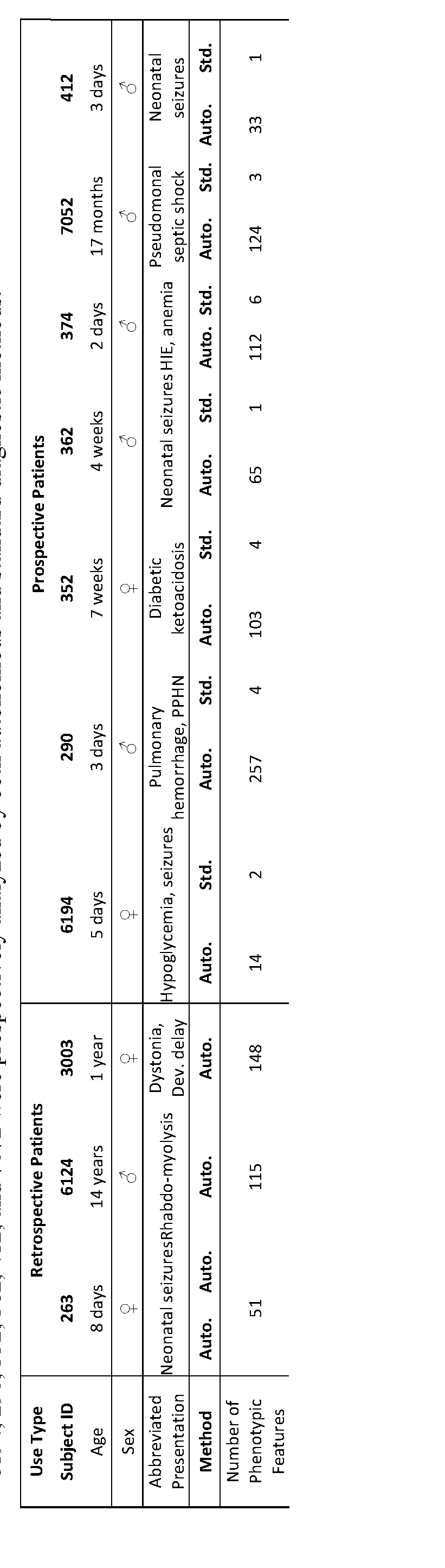
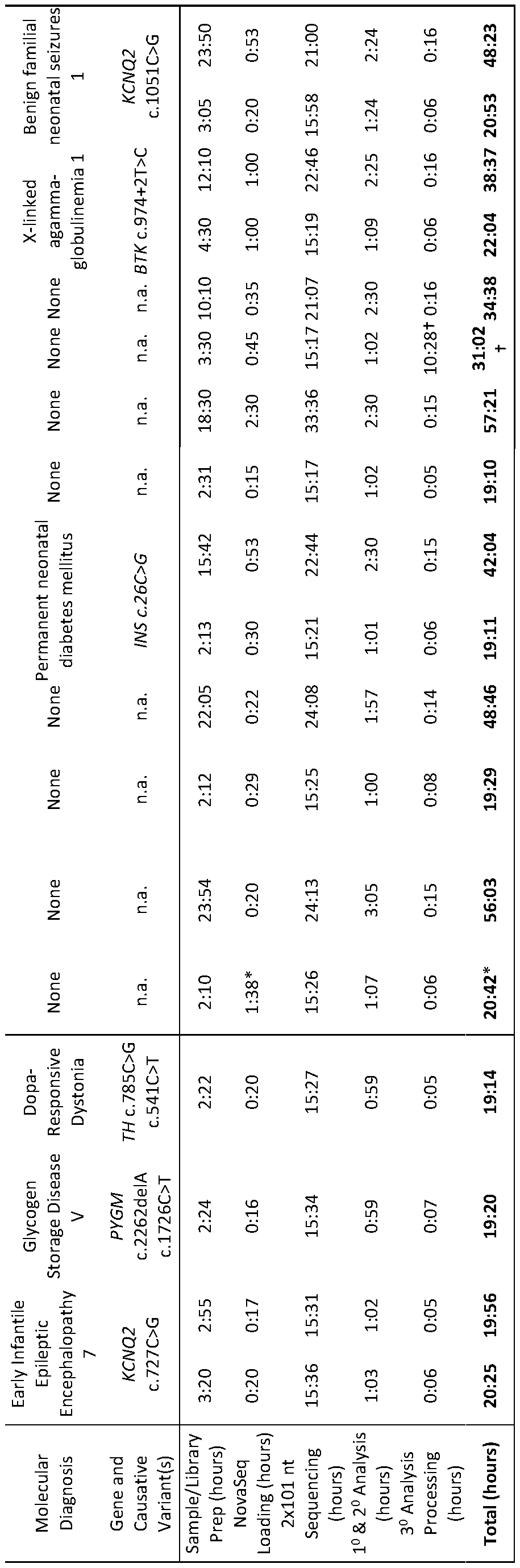
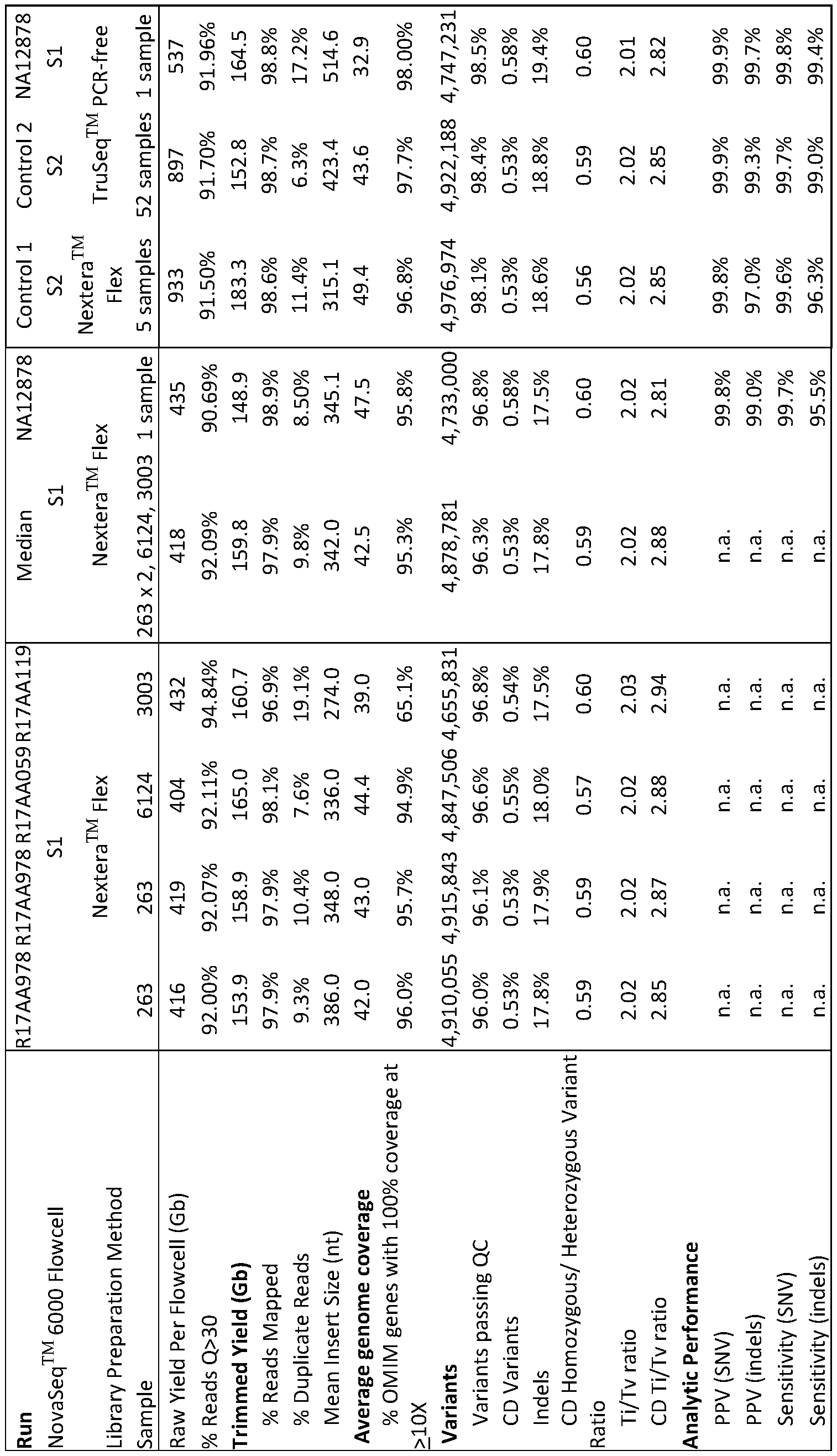
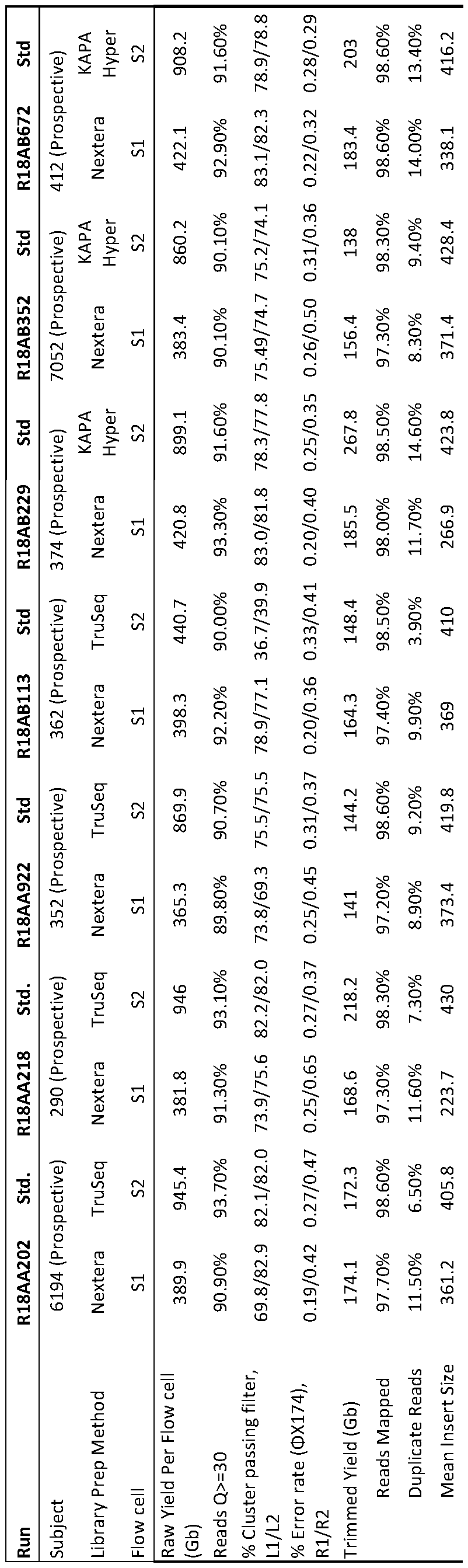
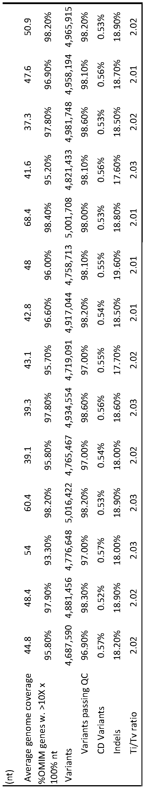

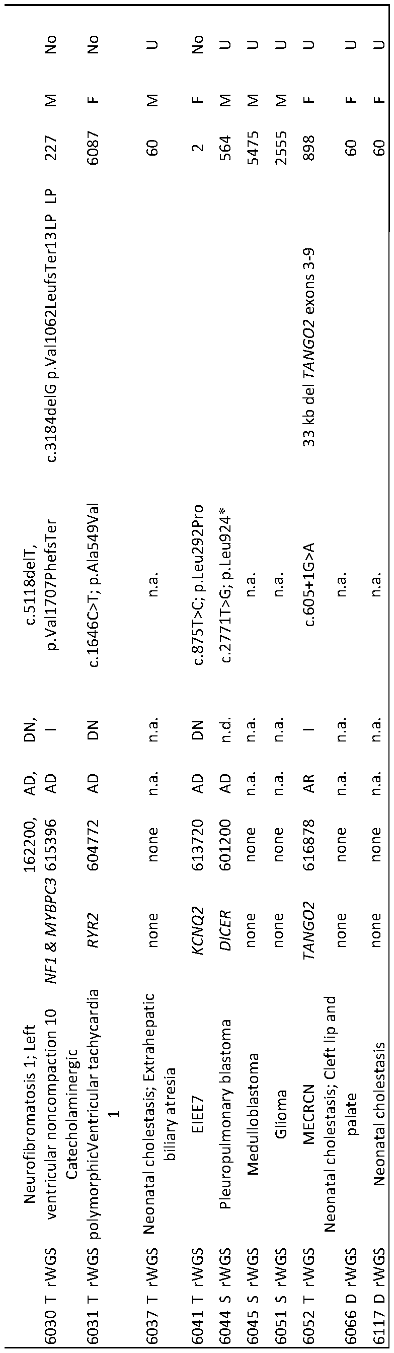
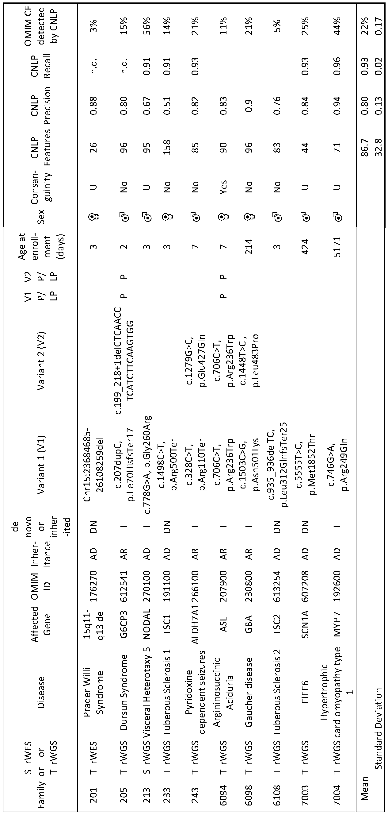
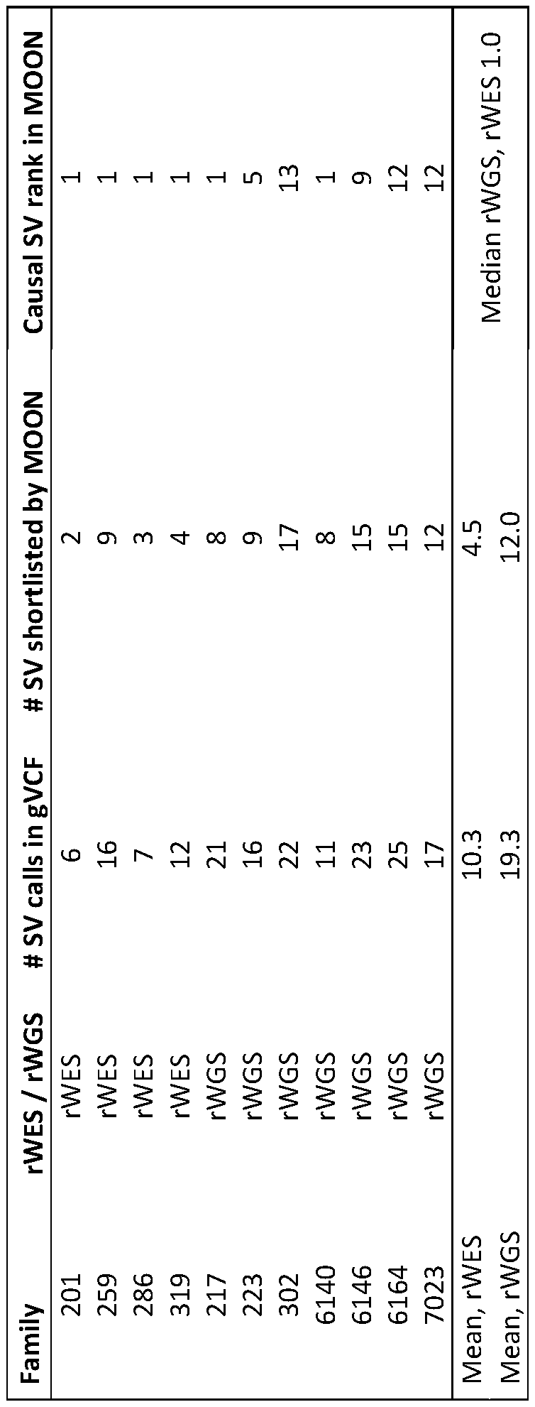


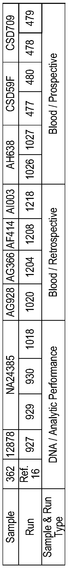

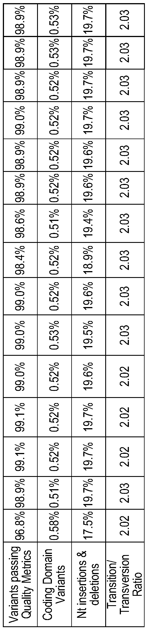

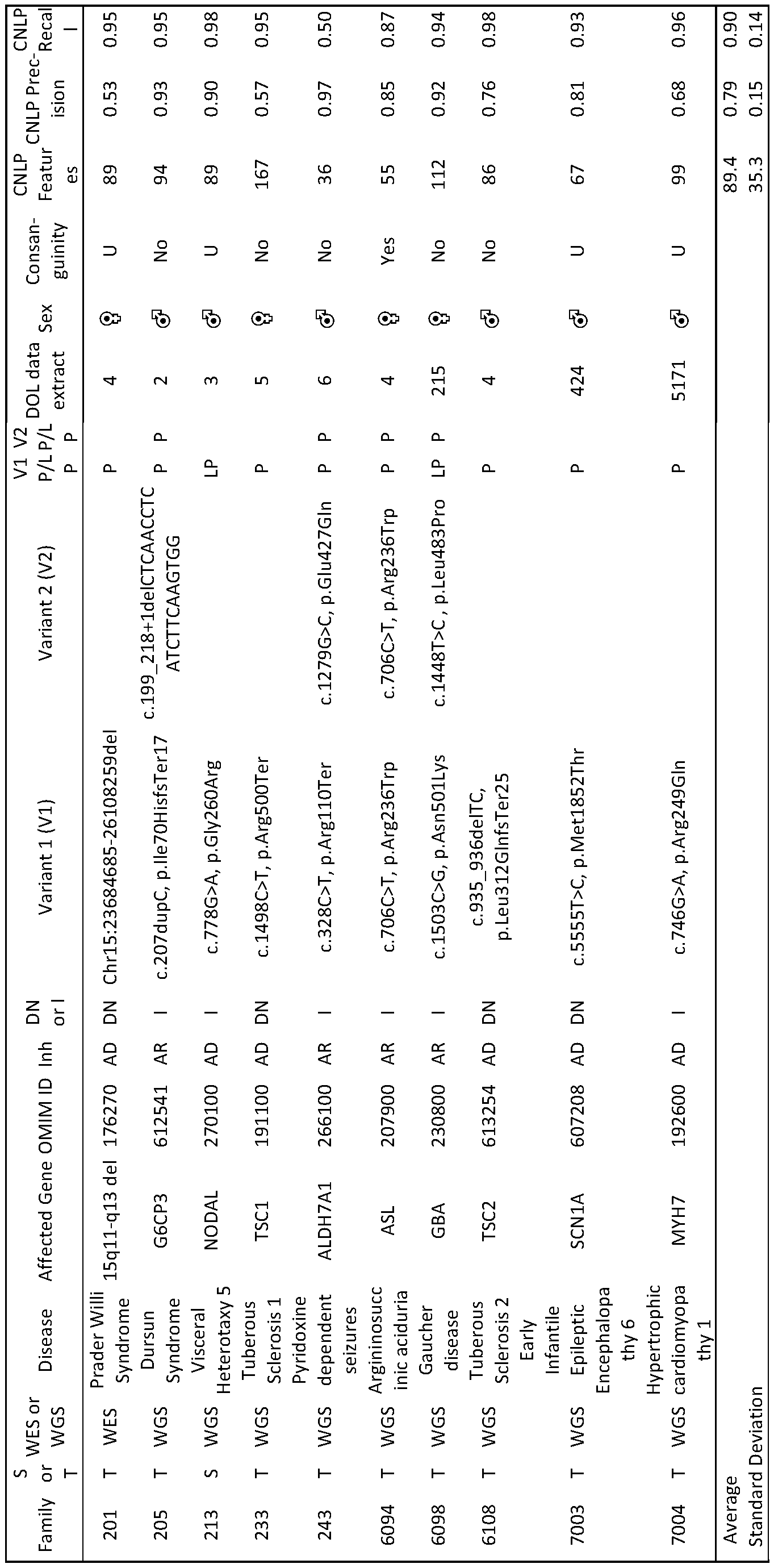
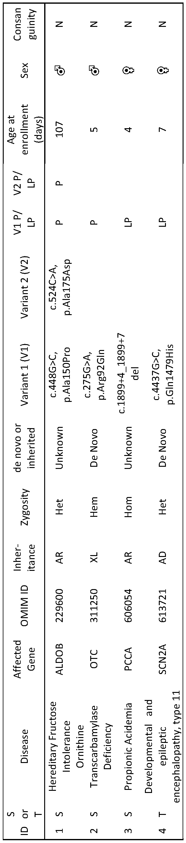
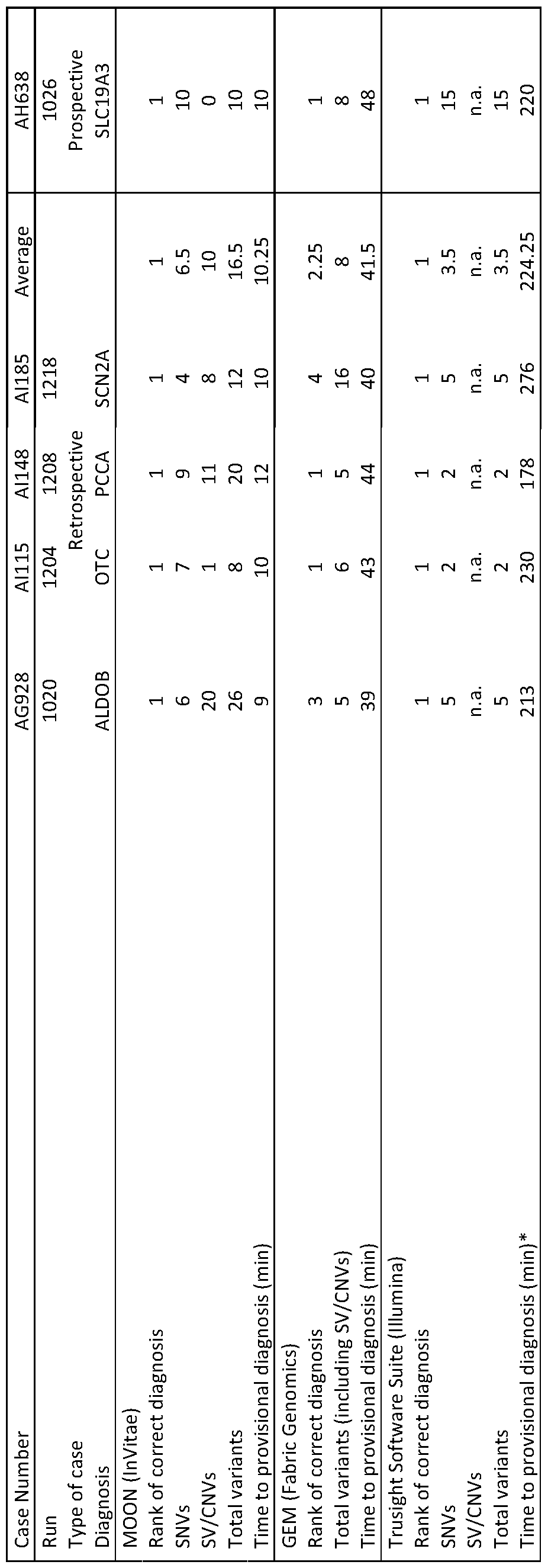
Claims
Priority Applications (3)
| Application Number | Priority Date | Filing Date | Title |
|---|---|---|---|
| EP22821168.6A EP4352731A4 (en) | 2021-06-11 | 2022-06-10 | PROCESS AND SYSTEM FOR IMPROVED MANAGEMENT OF GENETIC DISEASES |
| CA3221980A CA3221980A1 (en) | 2021-06-11 | 2022-06-10 | Method and system for improved management of genetic diseases |
| AU2022289398A AU2022289398A1 (en) | 2021-06-11 | 2022-06-10 | Method and system for improved management of genetic diseases |
Applications Claiming Priority (2)
| Application Number | Priority Date | Filing Date | Title |
|---|---|---|---|
| US202163209797P | 2021-06-11 | 2021-06-11 | |
| US63/209,797 | 2021-06-11 |
Publications (1)
| Publication Number | Publication Date |
|---|---|
| WO2022261515A1 true WO2022261515A1 (en) | 2022-12-15 |
Family
ID=84390055
Family Applications (1)
| Application Number | Title | Priority Date | Filing Date |
|---|---|---|---|
| PCT/US2022/033128 Ceased WO2022261515A1 (en) | 2021-06-11 | 2022-06-10 | Method and system for improved management of genetic diseases |
Country Status (5)
| Country | Link |
|---|---|
| US (1) | US20220399087A1 (en) |
| EP (1) | EP4352731A4 (en) |
| AU (1) | AU2022289398A1 (en) |
| CA (1) | CA3221980A1 (en) |
| WO (1) | WO2022261515A1 (en) |
Families Citing this family (2)
| Publication number | Priority date | Publication date | Assignee | Title |
|---|---|---|---|---|
| US20240360511A1 (en) * | 2023-04-27 | 2024-10-31 | Cardiai Technologies | Method for identifying and diagnosing genetic disorders and syetem thereof |
| WO2025072084A1 (en) * | 2023-09-25 | 2025-04-03 | Foundation Medicine, Inc. | Updating records based on consensus annotations of genetic variants |
Citations (3)
| Publication number | Priority date | Publication date | Assignee | Title |
|---|---|---|---|---|
| US8140270B2 (en) * | 2007-03-22 | 2012-03-20 | National Center For Genome Resources | Methods and systems for medical sequencing analysis |
| US20180004902A1 (en) * | 2015-01-05 | 2018-01-04 | Cincinnati Children's Hospital Medical Center | System and Method for Data Mining Very Large Drugs and Clinical Effects Databases |
| US20190325988A1 (en) * | 2018-04-18 | 2019-10-24 | Rady Children's Hospital Research Center | Method and system for rapid genetic analysis |
Family Cites Families (1)
| Publication number | Priority date | Publication date | Assignee | Title |
|---|---|---|---|---|
| US20120232930A1 (en) * | 2011-03-12 | 2012-09-13 | Definiens Ag | Clinical Decision Support System |
-
2022
- 2022-06-10 WO PCT/US2022/033128 patent/WO2022261515A1/en not_active Ceased
- 2022-06-10 CA CA3221980A patent/CA3221980A1/en active Pending
- 2022-06-10 US US17/838,115 patent/US20220399087A1/en active Pending
- 2022-06-10 EP EP22821168.6A patent/EP4352731A4/en active Pending
- 2022-06-10 AU AU2022289398A patent/AU2022289398A1/en active Pending
Patent Citations (3)
| Publication number | Priority date | Publication date | Assignee | Title |
|---|---|---|---|---|
| US8140270B2 (en) * | 2007-03-22 | 2012-03-20 | National Center For Genome Resources | Methods and systems for medical sequencing analysis |
| US20180004902A1 (en) * | 2015-01-05 | 2018-01-04 | Cincinnati Children's Hospital Medical Center | System and Method for Data Mining Very Large Drugs and Clinical Effects Databases |
| US20190325988A1 (en) * | 2018-04-18 | 2019-10-24 | Rady Children's Hospital Research Center | Method and system for rapid genetic analysis |
Non-Patent Citations (2)
| Title |
|---|
| CLARK MICHELLE M., ET AL.: "Diagnosis of genetic diseases in seriously ill children by rapid whole-genome sequencing and automated phenotyping and interpretation", SCIENCE TRANSLATIONAL MEDICINE, vol. 11, no. 489, 24 April 2019 (2019-04-24), XP093017613, ISSN: 1946-6234, DOI: 10.1126/scitranslmed.aat6177 * |
| See also references of EP4352731A4 * |
Also Published As
| Publication number | Publication date |
|---|---|
| AU2022289398A1 (en) | 2024-01-25 |
| CA3221980A1 (en) | 2022-12-15 |
| US20220399087A1 (en) | 2022-12-15 |
| EP4352731A4 (en) | 2025-04-02 |
| EP4352731A1 (en) | 2024-04-17 |
Similar Documents
| Publication | Publication Date | Title |
|---|---|---|
| Breuss et al. | Autism risk in offspring can be assessed through quantification of male sperm mosaicism | |
| US20190325988A1 (en) | Method and system for rapid genetic analysis | |
| Bick et al. | Whole exome and whole genome sequencing | |
| Miller et al. | A 26-hour system of highly sensitive whole genome sequencing for emergency management of genetic diseases | |
| US20250061972A1 (en) | Molecular response and progression detection from circulating cell free dna | |
| Gonzalez-Garay | The road from next-generation sequencing to personalized medicine | |
| EP4008005A1 (en) | Methods and systems for detecting microsatellite instability of a cancer in a liquid biopsy assay | |
| US20220215900A1 (en) | Systems and methods for joint low-coverage whole genome sequencing and whole exome sequencing inference of copy number variation for clinical diagnostics | |
| Wang et al. | A pipeline for RNA-seq based eQTL analysis with automated quality control procedures | |
| US20180300456A1 (en) | Population based treatment recommender using cell free dna | |
| JP2016540520A (en) | Methods and processes for non-invasive assessment of chromosomal changes | |
| Noll et al. | Clinical detection of deletion structural variants in whole-genome sequences | |
| US20240076744A1 (en) | METHODS AND SYSTEMS FOR mRNA BOUNDARY ANALYSIS IN NEXT GENERATION SEQUENCING | |
| US20220399087A1 (en) | Method and system for improved management of genetic diseases | |
| US20240371466A1 (en) | Method and system for newborn screening for genetic diseases by whole genome sequencing | |
| Chundru et al. | Federated analysis of autosomal recessive coding variants in 29,745 developmental disorder patients from diverse populations | |
| Reches et al. | From phenotyping to genotyping-bioinformatics for the busy clinician | |
| Petrillo et al. | Identification of a Homozygous Variant in the CYP21A2 Gene by Next-Generation Sequencing Analysis of Circulating Cell-Free Fetal DNA | |
| Deshpande | A Model to Predict the Phenotype for Copy Number Variants of Uncertain Significance | |
| Zhang et al. | Dinucleotide composition representation-based deep learning to predict scoliosis-associated Fibrillin-1 genotypes | |
| Sanders | Identification and assessment of a panel of genes for use in diagnostic genomic testing for recurrent pregnancy loss | |
| JP2025529155A (en) | High-resolution and non-invasive fetal sequencing | |
| Wu | Detection of aberrant events in RNA for clinical diagnostics | |
| Cormier | Leveraging Genetic Constraint to Predict Neglected RNA Splicing in Rare Human Disease | |
| Hambuch et al. | Implementation of Genome Sequencing Assays |
Legal Events
| Date | Code | Title | Description |
|---|---|---|---|
| 121 | Ep: the epo has been informed by wipo that ep was designated in this application |
Ref document number: 22821168 Country of ref document: EP Kind code of ref document: A1 |
|
| WWE | Wipo information: entry into national phase |
Ref document number: 3221980 Country of ref document: CA |
|
| WWE | Wipo information: entry into national phase |
Ref document number: 2022289398 Country of ref document: AU Ref document number: AU2022289398 Country of ref document: AU |
|
| WWE | Wipo information: entry into national phase |
Ref document number: 2022821168 Country of ref document: EP |
|
| NENP | Non-entry into the national phase |
Ref country code: DE |
|
| ENP | Entry into the national phase |
Ref document number: 2022821168 Country of ref document: EP Effective date: 20240111 |
|
| ENP | Entry into the national phase |
Ref document number: 2022289398 Country of ref document: AU Date of ref document: 20220610 Kind code of ref document: A |