WO2022244838A1 - Method for predicting in vivo pharmacokinetics of molecule - Google Patents
Method for predicting in vivo pharmacokinetics of molecule Download PDFInfo
- Publication number
- WO2022244838A1 WO2022244838A1 PCT/JP2022/020814 JP2022020814W WO2022244838A1 WO 2022244838 A1 WO2022244838 A1 WO 2022244838A1 JP 2022020814 W JP2022020814 W JP 2022020814W WO 2022244838 A1 WO2022244838 A1 WO 2022244838A1
- Authority
- WO
- WIPO (PCT)
- Prior art keywords
- cells
- molecule
- fcrn
- cell
- vitro
- Prior art date
- Legal status (The legal status is an assumption and is not a legal conclusion. Google has not performed a legal analysis and makes no representation as to the accuracy of the status listed.)
- Ceased
Links
Images


Classifications
-
- C—CHEMISTRY; METALLURGY
- C07—ORGANIC CHEMISTRY
- C07K—PEPTIDES
- C07K16/00—Immunoglobulins [IGs], e.g. monoclonal or polyclonal antibodies
-
- G—PHYSICS
- G01—MEASURING; TESTING
- G01N—INVESTIGATING OR ANALYSING MATERIALS BY DETERMINING THEIR CHEMICAL OR PHYSICAL PROPERTIES
- G01N33/00—Investigating or analysing materials by specific methods not covered by groups G01N1/00 - G01N31/00
- G01N33/48—Biological material, e.g. blood, urine; Haemocytometers
- G01N33/50—Chemical analysis of biological material, e.g. blood, urine; Testing involving biospecific ligand binding methods; Immunological testing
- G01N33/5005—Chemical analysis of biological material, e.g. blood, urine; Testing involving biospecific ligand binding methods; Immunological testing involving human or animal cells
- G01N33/5008—Chemical analysis of biological material, e.g. blood, urine; Testing involving biospecific ligand binding methods; Immunological testing involving human or animal cells for testing or evaluating the effect of chemical or biological compounds, e.g. drugs, cosmetics
- G01N33/502—Chemical analysis of biological material, e.g. blood, urine; Testing involving biospecific ligand binding methods; Immunological testing involving human or animal cells for testing or evaluating the effect of chemical or biological compounds, e.g. drugs, cosmetics for testing non-proliferative effects
-
- G—PHYSICS
- G01—MEASURING; TESTING
- G01N—INVESTIGATING OR ANALYSING MATERIALS BY DETERMINING THEIR CHEMICAL OR PHYSICAL PROPERTIES
- G01N33/00—Investigating or analysing materials by specific methods not covered by groups G01N1/00 - G01N31/00
- G01N33/48—Biological material, e.g. blood, urine; Haemocytometers
- G01N33/50—Chemical analysis of biological material, e.g. blood, urine; Testing involving biospecific ligand binding methods; Immunological testing
- G01N33/68—Chemical analysis of biological material, e.g. blood, urine; Testing involving biospecific ligand binding methods; Immunological testing involving proteins, peptides or amino acids
- G01N33/6854—Immunoglobulins
-
- C—CHEMISTRY; METALLURGY
- C07—ORGANIC CHEMISTRY
- C07K—PEPTIDES
- C07K16/00—Immunoglobulins [IGs], e.g. monoclonal or polyclonal antibodies
- C07K16/18—Immunoglobulins [IGs], e.g. monoclonal or polyclonal antibodies against material from animals or humans
- C07K16/28—Immunoglobulins [IGs], e.g. monoclonal or polyclonal antibodies against material from animals or humans against receptors, cell surface antigens or cell surface determinants
- C07K16/2878—Immunoglobulins [IGs], e.g. monoclonal or polyclonal antibodies against material from animals or humans against receptors, cell surface antigens or cell surface determinants against the NGF-receptor/TNF-receptor superfamily, e.g. CD27, CD30, CD40, CD95
-
- C—CHEMISTRY; METALLURGY
- C07—ORGANIC CHEMISTRY
- C07K—PEPTIDES
- C07K2317/00—Immunoglobulins specific features
- C07K2317/50—Immunoglobulins specific features characterized by immunoglobulin fragments
- C07K2317/52—Constant or Fc region; Isotype
-
- C—CHEMISTRY; METALLURGY
- C07—ORGANIC CHEMISTRY
- C07K—PEPTIDES
- C07K2317/00—Immunoglobulins specific features
- C07K2317/70—Immunoglobulins specific features characterized by effect upon binding to a cell or to an antigen
- C07K2317/77—Internalization into the cell
-
- C—CHEMISTRY; METALLURGY
- C07—ORGANIC CHEMISTRY
- C07K—PEPTIDES
- C07K2317/00—Immunoglobulins specific features
- C07K2317/90—Immunoglobulins specific features characterized by (pharmaco)kinetic aspects or by stability of the immunoglobulin
- C07K2317/94—Stability, e.g. half-life, pH, temperature or enzyme-resistance
-
- G—PHYSICS
- G01—MEASURING; TESTING
- G01N—INVESTIGATING OR ANALYSING MATERIALS BY DETERMINING THEIR CHEMICAL OR PHYSICAL PROPERTIES
- G01N2333/00—Assays involving biological materials from specific organisms or of a specific nature
- G01N2333/435—Assays involving biological materials from specific organisms or of a specific nature from animals; from humans
- G01N2333/52—Assays involving cytokines
- G01N2333/54—Interleukins [IL]
- G01N2333/5412—IL-6
-
- G—PHYSICS
- G01—MEASURING; TESTING
- G01N—INVESTIGATING OR ANALYSING MATERIALS BY DETERMINING THEIR CHEMICAL OR PHYSICAL PROPERTIES
- G01N2333/00—Assays involving biological materials from specific organisms or of a specific nature
- G01N2333/435—Assays involving biological materials from specific organisms or of a specific nature from animals; from humans
- G01N2333/705—Assays involving receptors, cell surface antigens or cell surface determinants
- G01N2333/70503—Immunoglobulin superfamily, e.g. VCAMs, PECAM, LFA-3
- G01N2333/70535—Fc-receptors, e.g. CD16, CD32, CD64 (CD2314/705F)
-
- G—PHYSICS
- G01—MEASURING; TESTING
- G01N—INVESTIGATING OR ANALYSING MATERIALS BY DETERMINING THEIR CHEMICAL OR PHYSICAL PROPERTIES
- G01N2333/00—Assays involving biological materials from specific organisms or of a specific nature
- G01N2333/435—Assays involving biological materials from specific organisms or of a specific nature from animals; from humans
- G01N2333/705—Assays involving receptors, cell surface antigens or cell surface determinants
- G01N2333/715—Assays involving receptors, cell surface antigens or cell surface determinants for cytokines; for lymphokines; for interferons
- G01N2333/7155—Assays involving receptors, cell surface antigens or cell surface determinants for cytokines; for lymphokines; for interferons for interleukins [IL]
Definitions
- the present invention relates to methods for measuring in vitro pharmacokinetics of molecules, methods for predicting in vivo pharmacokinetics of molecules, methods for screening molecules, and the like.
- mice are used to evaluate the pharmacokinetics (PK) required in the development process of pharmaceuticals such as antibody drugs.
- PK pharmacokinetics
- Non-Patent Documents 1 to 3 a method using the results of in vitro assays using cells has been known.
- Grevys et al. used human fetal Fc receptor (FcRn)-expressing cells (HMEC1-hFcRn) in a human microvascular endothelial cell line (HMEC1), and named it the HERA assay (human endothelial cell-based recycling assay). It discloses a method for measuring the amount of IgG antibody extracellularly excreted via FcRn in vitro and predicting its half-life in transgenic mice (Non-Patent Document 1). Jaramillo et al.
- Non-Patent Document 2 Madin-Darby canine kidney cells expressing human FcRn or rat FcRn, and measured the transcytosis activity, that is, the activity of the antibody to permeate the cells via FcRn. It is disclosed that the in vivo clearance of antibodies was ranked according to the results (Non-Patent Document 2). Similar to the method of Jaramillo et al., Chung et al. also performed transcytosis activity using cells expressing human FcRn in MDCK cells, and found a correlation between the measurement results and in vivo clearance in humans. (Non-Patent Document 3).
- an object of the present invention is to provide a method for predicting the in vivo pharmacokinetics of a molecule with higher sensitivity and more accuracy than ever before based on the results of in vitro pharmacokinetic measurements.
- the present inventors diligently investigated the cause of the insufficient accuracy of predicting in vivo pharmacokinetics based on in vitro pharmacokinetics with conventional methods. As a result, they found that prediction accuracy is low in conventional methods due to insufficient uptake of molecules into cells, and that prediction accuracy is improved by increasing the amount of uptake of molecules into cells. The present inventors completed the present invention by further research based on these findings.
- a method for measuring the in vitro pharmacokinetics of a molecule comprising the steps of: (a) contacting the molecule with a cell expressing FcRn in an aqueous medium so that the molecule is taken up by the cell so that the uptake amount is higher than 0.068 pmol/2 ⁇ 10 5 cells, a step having at least one characteristic selected from the following (i) to (iii); (i) the contact time between the molecule and the cell is 5 hours or longer; (ii) the cell is not washed under acidic conditions after contact with the molecule; and (iii) the cell expresses the target of the molecule on the cell surface; measuring in vitro pharmacokinetics, including The above method, wherein said molecule comprises an FcRn binding domain.
- the cells are CHO cells, HEK293 cells, COS-1 cells, COS-7 cells, MDCK cells, HMEC1 cells, HELA cells, HepG2 cells, or BaF cells the method of.
- the cells are liver parenchymal cells, liver non-parenchymal cells, liver sinusoidal endothelial cells, Kupffer cells, human umbilical vein endothelial cells, peripheral blood mononuclear cells PBMC, macrophages, mononuclear cells, B cells, T cells,
- [8] In vitro pharmacokinetics are the excretion amount from the cell into the culture medium, the excretion rate from the cell into the culture medium, the internalization rate, the amount of transcytosis, the Kp value, the intracellular molecule reduction rate, FcRn or the rate of binding to a target, or the rate of dissociation from FcRn or a target.
- FcRn is human FcRn, monkey FcRn, minipig FcRn, rat FcRn, mouse FcRn, rabbit FcRn, dog FcRn, or guinea pig FcRn.
- a method of predicting the in vivo pharmacokinetics of a molecule comprising: (a') a step of measuring in vitro pharmacokinetics by the method according to any one of [1] to [14], and (b') the measured value or in vitro evaluation parameter obtained in step (a') to predict the in vivo pharmacokinetics when the molecule is administered to a living body. [16] The method of [15], wherein the in vivo pharmacokinetics is bioavailability, volume of distribution, blood unbound fraction, clearance, urinary excretion rate, blood concentration half-life, or mean residence time. .
- a method of screening a molecule comprising: (a'') providing two or more different molecules that bind to the same target; (b'') measuring the in vitro pharmacokinetics of each of the two or more molecules prepared in step (a'') by the method described in any one of [1] to [14]; and (c') ') comparing the measured values or in vitro evaluation parameters for each of the two or more molecules obtained in step (b'') with each other and selecting the molecules that showed the desired values.
- the in vivo pharmacokinetics of molecules can be predicted with higher sensitivity and more accuracy than ever before, based on the results of in vitro pharmacokinetic measurements. Therefore, it becomes possible to easily and highly accurately predict the in vivo pharmacokinetics of a large number of candidate substances in the early stages of drug development. Moreover, the present invention can contribute to reducing the number of experimental animals used by reducing the number of in vivo pharmacokinetic tests. Furthermore, the present invention can contribute to the development of drugs with higher pharmacological effects by providing a method for efficiently screening drugs having desired pharmacokinetics.
- Figure 2 shows the results of mouse plasma pharmacokinetic evaluation of antibodies with different Fc regions (H237-G1d, H237-F1847m, H237-F1886m, H237-F1927m, and H237-F890).
- Black solid line black circle is H237-G1d
- black short dashed line black triangle is H237-F1847m
- black solid line white circle is H237-F1886m
- black long dashed line black square is H237-F1927m
- black solid line white triangle is H237- F890 is shown respectively.
- the results of measuring the in vitro cell uptake of antibodies having different Fc regions are shown. Each antibody was taken up by hFcRn-hIL6R-CHO cells or hFcRn-CHO cells at 37°C for 24 hours, and after washing with cold 2% FBS-containing PBS, the amount of uptake into the cells is shown. The results of measuring the cellular uptake of antibodies with different Fc regions over time are shown.
- FIG. 2 shows temporal changes in the intracellular residual amount and the amount excreted into the medium of antibodies having different Fc regions. After each antibody was taken up by the cells at 37°C for 24 hours, the medium was replaced with fresh medium and incubated for up to 4 hours.
- Black solid line black circle is H237-G1d
- black short dashed line black triangle is H237-F1847m
- black solid line white circle is H237-F1886m
- black long dashed line black square is H237-F1927m
- black solid line white triangle is H237- F890 is shown respectively.
- Figure 2 shows the correlation between the clearance index calculated in Example 4 and plasma half-life (a) or clearance (b) in mice.
- a first aspect of the present invention relates to a method for measuring in vitro pharmacokinetics of a molecule (hereinafter also referred to as the measuring method of the present invention).
- in vivo pharmacokinetics refers to a series of processes of absorption, distribution, metabolism, and excretion in vivo after administration of a drug (i.e., the molecule of the present invention). Refers to changes in the concentration (amount) of the drug. After administration of a drug, parallel processes of absorption, distribution, metabolism, and excretion occur in vivo.
- the basic pharmacokinetic (PK) parameters to decompose and describe these processes are (1) bioavailability (F), (2) volume of distribution (Vd or V), (3) ) fraction unbound in blood (fuB), (4) clearance (CL), and (5) cumulative amount of drug excreted in urine (Ae) have been established. (Biometrics Vol.
- PK parameters include blood concentration half-life (t 1/2 ), mean residence time (MRT), area under the first moment-time curve (AUMC), elimination rate constant (kel), post-dose zero time point concentration (C0 ) are known.
- in vitro pharmacokinetics refers to the behavior of a target molecule measured by contacting the target molecule with cells under artificially constructed conditions outside the body.
- “In vitro pharmacokinetics” includes, for example, intracellular to extracellular excretion rate, intracellular to extracellular excretion rate, internalization rate, transcytosis amount, Kp value, intracellular molecule reduction rate, FcRn or It can be expressed by, but not limited to, the rate of binding to a target, or the rate of dissociation from FcRn or a target.
- the amount of excretion (Efflux amount) from the inside of the cell to the outside of the cell is determined by exchanging the aqueous medium (e.g., medium, buffer, etc.) with one that does not contain the target molecule after contacting the target molecule with the cell for a predetermined time. It is determined by measuring the amount of the molecule of interest that is extracellularly excreted from the cells by detecting the molecule of interest in an aqueous medium.
- the discharge rate (Efflux rate) from the inside of the cell to the outside of the cell is determined by measuring the Efflux amount of the target molecule per unit time.
- the internalization rate is determined by contacting a molecule of interest with a cell for a predetermined period of time and measuring the amount of the molecule of interest that is taken up into the cell from the outside (eg, from medium, buffer, etc.) per unit time.
- cells that have been in contact with a molecule of interest for a predetermined period of time are acidified (pH less than 6.0, e.g. 0 or less, pH 3.5 or less, or pH 3.0 or less) to remove molecules of interest bound to the cell surface.
- the amount of the target molecule taken up (internalized) into cells can be measured more accurately.
- the amount of transcytosis is determined by measuring the amount of permeation from one side of the cell sheet to the other.
- the Kp value is known as the tissue-to-plasma drug concentration ratio (i.e., the ratio of the concentration of the molecule of interest between tissue and plasma) for in vivo pharmacokinetics, but is herein referred to as in vitro pharmacokinetics.
- tissue-to-plasma drug concentration ratio i.e., the ratio of the concentration of the molecule of interest between tissue and plasma
- in vitro pharmacokinetics refers to the ratio of concentrations of the molecule of interest between the cells and the aqueous medium (eg, culture medium). Kp values are determined by measuring the amount in cells and the amount in aqueous media.
- the Kp value as in vitro pharmacokinetics is a value calculated by (amount in cells)/(amount in aqueous medium).
- Intracellular molecule reduction rate is determined by exchanging the aqueous medium (e.g., medium, buffer solution, etc.) with one that does not contain the target molecule after contacting the target molecule with the cell for a predetermined time, and then detecting the target molecule in the cell. It is determined by measuring the amount of the molecule of interest that is depleted from the cell per unit time.
- the rate of FcRn or target binding is determined by contacting the molecule of interest with the cell for a predetermined short period of time (eg, seconds to minutes) and measuring the amount of molecule of interest bound to the FcRn or target per unit time. be.
- the rate of dissociation from FcRn or target is measured by contacting the molecule of interest with the cell for a given period of time (e.g., a time sufficient to reach equilibrium) followed by an aqueous medium (e.g., medium, buffer, etc.) free of the molecule of interest. It is determined by measuring the amount of the target molecule discharged into the aqueous medium per unit time by detecting the target molecule in the aqueous medium after exchanging with the substance.
- the contacting of the target molecule with the cell and the release of the target molecule into the aqueous medium are performed at a temperature at which the internalization of the target molecule into cells is inhibited (e.g., 4°C or lower). be able to.
- a "molecule” (also referred to as “a molecule in the present invention”) is taken up by a cell used in the assay method of the present invention, and within the cell is an intracellular organelle on an endosome. It has the property of being excreted outside the cell via the Fc receptor (FcRn) molecule. Such properties are due to the fact that the molecule contains an FcRn binding domain.
- FcRn Fc receptor
- FcRn is one of the receptors that recognize the Fc region of IgG antibodies.
- FcRn is expressed in the fetal placenta and is responsible for the transcytosis of IgG from the mother to the fetus.It is also expressed in the vascular endothelium, intestinal epithelial cells, and blood cells in adults, It is known that it is responsible for exocytosis and transcytosis from cells (Nature Reviews Immunology Vol. 7, p. 715-725 (2007)).
- Human FcRn is a dimeric protein consisting of a light chain called the ⁇ 2m subunit and a heavy chain called the ⁇ subunit with a transmembrane domain, and its structure resembles major histocompatibility complex (MHC) class I molecules. is doing. This FcRn dimer further dimers and binds to a single molecule of IgG (Annual Review of Cell and Developmental Biology Vol. 12, p. 181-220 (1996)). Unlike other IgG antibody Fc receptors, FcRn exhibits pH-dependent binding through electrostatic interactions between anionic residues on its ⁇ 2 domain and the CH2-CH3 hinge region of IgG. is known (Nature Reviews Immunology Vol. 7, p. 715-725 (2007)).
- IgG taken into cells by pinocytosis binds with high affinity to FcRn, escapes degradation in lysosomes, and then under neutral conditions (pH 7.4). It dissociates when it moves to the cell surface.
- This pH-dependent binding mode enables transcytosis and exocytosis of IgG, which contributes to the transport of IgG from the mother to the fetus and the extension of the blood half-life of IgG in vivo (approximately 20 days).
- FcRn-binding domains include, for example, antibody heavy chain constant regions (Fc regions) and fragments thereof.
- Fc regions antibody heavy chain constant regions
- albumin Another example of an FcRn binding domain is albumin and fragments thereof. It is known from the literature (J. Exp. Med. (2003) 197(3), 315-322) that albumin binds to FcRn.
- the FcRn-binding domain may contain mutations as long as it can bind to FcRn in an endosomal pH environment of less than pH 6.5.
- Fc-binding domains containing mutations include, but are not limited to, the mutated Fc regions of antibodies described in, for example, WO 2012/133782 A1, WO 2013/046704 A2, and WO 2017/046994 A1.
- a molecule in the present invention may further have the property of binding to a target (ie, target binding activity) or the property of catalyzing a reaction on the target (ie, enzymatic activity or catalytic activity).
- Molecules with target binding activity may function as agonists or antagonists. Having these properties, the molecule has a target binding domain or a catalytic domain.
- the molecule according to the invention comprises a target binding domain. Thereby, uptake into cells expressing the target on the cell surface may be increased.
- target refers to another molecule or structure that binds to the molecule of the present invention, or another molecule or structure that is catalyzed by the molecule of the present invention.
- Targets include proteins, nucleic acids, sugar chains, and the like.
- a “target” may also be called an antigen, a receptor, a substrate, etc., depending on the relationship with the molecule in the present invention.
- target-binding domain is not particularly limited as long as it can bind to the target, and domains with any structure can be used.
- target-binding domains include antigen-binding domains of antibodies, Avimers (International Publications WO2004/044011, WO2005/040229) containing about 35 amino acid modules (A domains) contained in various cell membrane proteins in vivo, Adnectin containing the 10Fn3 domain in fibronectin, which is a glycoprotein to be expressed (International Publication WO2002/032925), Affibody that scaffolds the IgG-binding domain consisting of 58 amino acids of Protein A (International Publication WO1995/001937), and a repeating sequence of 33 amino acids DARPins (Designed Ankyrin Repeat proteins) containing ankyrin repeat (AR) as a backbone (International Publication WO2002/020565), neutrophil gelatinase-associated lipocalin (NGAL), etc.
- DARPins Designed Ankyrin Repeat proteins
- an antigen-binding domain may be provided by one or more variable domains of an antibody.
- the antigen binding domain comprises an antibody light chain variable region (VL) and an antibody heavy chain variable region (VH).
- the target binding domain comprises antibody heavy and/or light chain variable regions.
- the target binding domain comprises or consists of the heavy and light chain variable regions of an antibody.
- the catalytic domain includes the catalytic domain in enzymes.
- the molecule in the present invention comprises an FcRn-binding domain and a target-binding domain, more preferably an antibody Fc region and heavy and light chain variable regions.
- Molecules in the present invention include pharmaceuticals and their candidates.
- pharmaceuticals and their candidates for example, in addition to proteins such as antibodies, which are the molecules described in the Examples, peptide compounds, nucleic acids, toxins, viruses, nanoparticles/microparticles, etc. Examples include DDS formulations, but are not limited to these as long as they can bind to FcRn.
- they may be prepared by conventional methods using techniques known in the art, depending on the type. According to the measurement method of the present invention, the in vitro pharmacokinetics of these molecules can be measured in the same manner as in the examples of the present application, and the in vivo pharmacokinetics can be predicted.
- protein refers to a polymer of amino acids linked via peptide bonds, and may also include peptide compounds.
- a protein may be naturally occurring or non-naturally occurring, such as a recombinant protein. Proteins include, for example, cytokines, physiologically active peptides, biological enzymes, antibodies, or variants thereof.
- antibody refers to an immunoglobulin that is natural or produced by partial or complete synthesis. Antibodies can be isolated from natural sources such as plasma or serum in which they naturally occur, culture supernatants of antibody-producing hybridoma cells, or can be partially or completely isolated by using techniques such as genetic recombination. can be synthesized. Examples of antibodies preferably include the immunoglobulin isotypes (ie, IgG, IgA, IgD, IgE, and IgM) and their isotypic subclasses.
- the antibody in the measuring method of the present invention is IgG.
- Antibodies can be either polyclonal antibodies or monoclonal antibodies.
- artificially modified genetically modified antibodies such as chimeric antibodies, humanized antibodies, and human antibodies, can be used for the purpose of reducing heterologous antigenicity.
- the antibody may be a bispecific antibody (bispecific antibody).
- An antibody may be a fragment of an antibody as long as it contains the "FcRn binding domain". Examples of such antibody fragments include Fc fragments, scFv-CH1-Fc, and the like.
- the "FcRn-binding domain" of an antibody is any domain capable of binding to FcRn, and includes, for example, the heavy chain constant region (Fc region) of an antibody.
- An antibody as a molecule in the present invention preferably comprises an "antigen-binding domain", more preferably comprising the heavy and light chain variable regions of the antibody. Thereby, when the cells express the antigen on the cell surface, the uptake amount of the molecules of the present invention into the cells can be increased. Methods for producing these antibodies are known to those skilled in the art (eg, WO 2013/081143, etc.).
- the "antigen" is not particularly limited in structure as long as it contains an epitope to which the antigen-binding domain binds.
- Antigens may be inorganic or organic.
- the antigen is 17-IA, 4-1BB, 4Dc, 6-keto-PGF1a, 8-iso-PGF2a, 8-oxo-dG, A1 adenosine receptor, A33, ACE, ACE-2 , activin, activin A, activin AB, activin B, activin C, activin RIA, activin RIA ALK-2, activin RIB ALK-4, activin RIIA, activin RIIB, ADAM, ADAM10, ADAM12, ADAM15, ADAM17/TACE, ADAM8, ADAM9, ADAMTS, ADAMTS4, ADAMTS5, addressin, aFGF, ALCAM, ALK, ALK-1, ALK-7, alpha-1-antitrypsin, alpha-
- T cell receptor alpha/beta TdT, TECK, TEM1, TEM5, TEM7, TEM8, TERT, testicular PLAP-like alkaline phosphatase, TfR, TGF, TGF-alpha, TGF-beta, TGF-beta Pan Specific, TGF-betaRI (ALK-5), TGF-betaRII, TGF-betaRIIb, TGF- betaRIII, TGF-beta1, TGF-beta2, TGF-beta3, TGF-beta4, TGF-beta5, thrombin, thymus Ck-1, thyroid stimulating hormone, Tie, TIMP, TIQ, tissue factor, TMEFF2, Tmpo, TMPRSS2, TNF, TNF-alpha, TNF-alphabeta, TNF-beta2, TNFc, TNF-RI, TNF-RII, TNFRSF10A (TRAIL R1 Apo-2, DR4), TN
- the antigen capable of forming a complex with the antibody is any of the antigens exemplified above, or a combination thereof. , in other words, may be monomeric or heteromultimeric.
- heteromultimers include IL-12, including IL-12p40 and IL-12p35, IL-23, including IL-12p40 and IL-23p19 (also called IL-30B), or EBI-3 and IL27p28.
- IL-23 containing, or heterodimers such as IL-35 containing IL-12p35 and EBI-3.
- antigens also include receptors
- these receptors may exist in soluble form in biological fluids such as plasma.
- soluble receptors are also included in the antigens of the present invention.
- a non-limiting embodiment of the soluble receptor is, for example, soluble IL-6R as described by Mullberg et al. (J. Immunol. (1994) 152 (10), 4958-4968). (for example, a protein consisting of amino acids 1 to 357 in the IL-6R polypeptide sequence represented by SEQ ID NO: 1 described in WO 2013/081143).
- extracellular fluid includes components in bone and cartilage such as plasma, interstitial fluid, lymph, dense connective tissue, cerebrospinal fluid, cerebrospinal fluid, aspirate fluid, or synovial fluid, alveolar fluid (bronchopulmonary lavage), ascites, pleural effusion, pericardial effusion, cystic effusion, or intracellular fluid such as aqueous humor (aqueous humor) Other body cavity fluids) is a general term.
- antigens examples include soluble antigens and membrane antigens, and it is well known to those skilled in the art which one each antigen corresponds to. For example, it can be classified by searching individual antigens on websites such as UniProtKB (https://www.uniprot.org/) and Human Protein Atlas (https://www.proteinatlas.org/). .
- the molecule in the present invention is an antibody
- the antibody may preferably be IgG.
- the molecules in the present invention are anti-IL-6R antibodies, and more particularly can be humanized anti-IL-6R antibodies.
- nucleic acid refers to DNA, RNA, and analogues thereof, and may be natural nucleic acids or synthetic nucleic acids. Analogues include artificial nucleic acids such as PNA and LNA. Nucleic acids may be single-stranded or double-stranded. Nucleic acids may also be modified. Modified forms include those chemically modified at internucleoside linkages, bases and/or sugars, and those having modified groups at the 5' and/or 3' ends.
- Internucleoside linkage modifications include phosphodiester linkages, phosphorothioate linkages, phosphorodithioate linkages, methylphosphonate linkages, phosphoramidate linkages, non-phosphate linkages, and methylphosphonothioate linkages, or combinations thereof.
- changes to Base modifications include changes to 5-propynyluracil, 2-aminoadenine, and the like.
- Sugar modifications include changes to 2'-fluororibose, 2'-O-methyl ribose, and the like.
- Nucleic acids are sometimes referred to as siRNAs, antisense RNAs, miRNAs, shRNAs, ribozymes, or aptamers, depending on their function or use.
- Nucleic acids used in the present invention also include CpG oligonucleotides that act on Toll-like receptor 9 (TLR9) to activate innate immunity.
- TLR9 Toll-like receptor 9
- the base length of the nucleic acid may be any length that allows it to be taken up into cells via Stabilin. be.
- Stabin when the molecule in the invention is a nucleic acid, its target (or receptor) can be Stabin.
- Stabin refers to proteins belonging to the family of transmembrane proteins known as nucleic acid receptors. Two types of homologues of Stabin-1 and Stabin-2 are known in mammals, and Stabin in the present invention may be either of them.
- Stabilin-1 NCBI accession number: NP_055951.2
- Stabilin-2 NCBI accession number: NP_060034.9 are known and are expressed in LSEC, spleen, adrenal cortex, lymph nodes, and sinusoidal macrophages. have been reported to occur.
- a "peptide compound” is a compound formed by an amide bond or an ester bond of an amino acid or an amino acid analogue.
- Molecular forms of peptide compounds include linear, cyclic, and cyclic having a linear portion.
- the number of amide bonds or ester bonds (the number and length of amino acids or amino acid analogues) is not particularly limited, but when it has a linear portion, it is preferable that the combined cyclic portion and linear portion are within 30 residues. More preferably, the total number of amino acids, including the cyclization sites and the linear sites, is 13 residues or less. More preferably, the total number of amino acids is 9 or more in order to obtain high metabolic stability.
- the number of amino acids and amino acid analogs constituting the cyclic portion is preferably 5-12. Furthermore, in addition to the above description, the number of amino acids and amino acid analogs constituting the cyclic portion is more preferably 5 to 11 residues, more preferably 7 to 11 residues. 9 to 11 residues are particularly preferred.
- the number of amino acids and amino acid analogues (the number of units) in the linear portion is preferably 0-8. Furthermore, 0 to 3 are preferred. In the present application, amino acids may include amino acid analogues unless otherwise specified.
- amino acid and “amino acid analogue” that constitute a peptide compound are sometimes referred to as “amino acid residue” and “amino acid analogue residue”, respectively.
- Amino acids are ⁇ , ⁇ and ⁇ amino acids, and naturally occurring amino acids (in this application, naturally occurring amino acids refer to 20 types of amino acids contained in proteins. Specifically, Gly, Ala, Ser, Thr, Val, Leu, Ile, Phe, Tyr, Trp, His, Glu, Asp, Gln, Asn, Cys, Met, Lys, Arg, Pro.), and may be non-natural amino acids.
- the ⁇ -amino acid may be an L-amino acid, a D-amino acid, or an ⁇ , ⁇ -dialkylamino acid.
- Amino acid side chains are not particularly limited, but may be selected freely from alkyl groups, alkenyl groups, alkynyl groups, aryl groups, heteroaryl groups, aralkyl groups, and cycloalkyl groups in addition to hydrogen atoms. Each may be provided with a substituent, and those substituents are also freely selected from arbitrary functional groups including, for example, N atom, O atom, S atom, B atom, Si atom, and P atom.
- amino acids and amino acid analogs that constitute peptide compounds include all corresponding isotopes. Isotopes of "amino acids” and “amino acid analogues” are those in which at least one atom is replaced with an atom with the same atomic number (number of protons) but a different mass number (sum of protons and neutrons). be.
- isotopes contained in "amino acids” and “amino acid analogues” constituting the peptide compound of the present invention include hydrogen atom, carbon atom, nitrogen atom, oxygen atom, phosphorus atom, sulfur atom, fluorine atom, chlorine atom, etc. , which include 2 H, 3 H, 13 C, 14 C, 15 N, 17 O, 18 O, 31 P, 32 P, 35 S, 18 F, 36 Cl, etc., respectively.
- amino acids with an amino group include Lys (lysine).
- amino acids with thiol groups can also be labeled with thiol-reactive fluorescent dyes. Cys (cysteine) is mentioned as such an amino acid.
- the molecule in the present invention is a peptide compound, preferably its target (or receptor) is PEPT1 or PEPT2.
- Nanoparticles and microparticles are known to be used in preparations intended for drug delivery (Drug Delivery System, commonly known as DDS). Examples include, but are not limited to, liposomes, micelles, dendrimers, nanoemulsions, iron nanoparticles, gold nanoparticles, PLGA particles (Organ Biology VOL.24 NO.1 2017, 54-60).
- molecules in the present invention include nanoparticles and microparticles bound with molecules that specifically bind to specific cells. For example, antigen-binding molecules against surface antigens of the cells can be bound to these particles. Also, molecules containing, for example, FcRn binding domains can be attached to these particles.
- the molecule in the present invention can be a nanoparticle/microparticle to which an antibody comprising an FcRn-binding domain and/or a target-binding domain is conjugated.
- the term “toxin” is not particularly limited as long as it can specifically deliver a cytotoxic agent, toxin, or radioactive isotope to specific cells and damage them.
- a molecule that specifically binds to the cell eg, an antigen-binding molecule for the cell surface antigen of the cell
- a cytotoxic agent, a toxin, or a radioactive isotope can be conjugated with a cytotoxic agent, a toxin, or a radioactive isotope to prepare a molecule.
- “Molecules that specifically bind to cells” include the aforementioned antibodies, nucleic acids, peptide compounds, and the like. Cytotoxic agents, toxins, or radioisotopes can be efficiently delivered to the cells using such molecules. As a result, the cells can be specifically injured.
- cytotoxic agents include maytansinoids (see U.S. Pat. Nos. 5,208,020, 5,416,064, and EP 0,425,235 B1); e.g. monomethyl auristatin drug moieties DE and DF (MMAE and MMAF) (U.S. Pat. No. 5,635,483). dolastatin; calicheamicin or derivatives thereof (U.S. Pat. Nos. 5,712,374, 5,714,586, 5,739,116, 5,767,285, 5,770,701, 5,770,710); 5,773,001 and 5,877,296; Hinman et al., Cancer Res. 53:3336-3342 (1993); and Lode et al., Cancer Res.
- toxins include enzymatically active toxins or fragments thereof, including but not limited to: diphtheria A chain, non-binding active fragments of diphtheria toxin, exotoxin A chain (Pseudomonas aeruginosa).
- radioisotopes include those of 211 At, 131 I, 125 I, 90 Y, 186 Re, 188 Re, 153 Sm, 212 Bi, 32 P, 212 Pb and Lu.
- a virus can also be used as a molecule in the present invention.
- the in vitro kinetics of the following viruses or viral proteins or parts thereof can be measured.
- Viruses used in gene therapy include retroviruses, adenoviruses, adeno-associated viruses, herpes simplex viruses, lentiviruses, poxviruses, Epstein-Barr viruses, etc. (Adv Biomed Res. (2012) 1: 27. doi : 10.4103/2277-9175.98152), drug delivery viruses such as Red clover necrotic mosaic virus (RCNMV) (Methods Mol Biol. 2011;726:207-221).
- Viral proteins or portions thereof include partial peptides of HIV-1 tat protein, human papillomavirus L2 peptide, HBV envelope L protein, and the like.
- the measurement method of the present invention comprises the following step (a), by contacting the molecule and FcRn-expressing cells in an aqueous medium, so that the uptake amount is higher than 0.068 pmol/2 ⁇ 10 5 cells. and (b) measuring the in vitro pharmacokinetics of said molecule, including.
- step (b) does not have to be started after step (a) is completed. That is, step (b) may be started after the uptake of the molecule of the present invention into cells in step (a) is completed, or the molecule is placed in a state where it can be taken up by cells in step (a). may be started when
- the cells used in the measurement method of the present invention are not particularly limited as long as they can be contacted with the target molecule in vitro and express FcRn. It can be a cultured cell, or an established cell line. Expression of FcRn is confirmed by staining cells with a fluorescently-labeled anti-FcRn antibody, measuring fluorescence by FACS, and confirming that the histogram shifts to a higher fluorescence intensity side than a control antibody (eg isotype control antibody). For a more quantitative assessment, cells may be analyzed by liquid chromatography-mass spectrometry (LC-MS) to determine the abundance of FcRn-derived peptides.
- LC-MS liquid chromatography-mass spectrometry
- the FcRn can be the FcRn of a species whose in vivo pharmacokinetics are to be predicted, such as human FcRn, monkey FcRn, minipig FcRn, rat FcRn, mouse FcRn, rabbit FcRn, dog FcRn, guinea pig FcRn, It can be hamster FcRn, chimpanzee FcRn, marmoset FcRn, ferret FcRn, or cat FcRn.
- a species whose in vivo pharmacokinetics are to be predicted such as human FcRn, monkey FcRn, minipig FcRn, rat FcRn, mouse FcRn, rabbit FcRn, dog FcRn, guinea pig FcRn, It can be hamster FcRn, chimpanzee FcRn, marmoset FcRn, ferret FcRn, or cat FcR
- the cell can be a cell transformed to express FcRn.
- Such transformation can be performed, for example, by introducing a polynucleotide encoding FcRn into the cell.
- promoters used for expression in general animal cells can be used, for example, promoters such as CMV, PGK, RSV, CAG, EF-1 alpha, SV40, TRE, Oct3/4, Nanog. (PLoS One. 2010; 5(5): e10611). By using them, a sufficient amount of FcRn can be expressed.
- the cell can be a cell transformed to express the molecular target of the present invention on the cell surface.
- the target is a protein
- such transformation can be accomplished by introducing into the cell a polynucleotide encoding the protein.
- the higher the expression level of the target the more the amount of uptake of the molecules in the present invention into cells can be increased.
- a promoter the same one as used for expressing FcRn can be used, thereby allowing sufficient amount of target to be expressed.
- the cells used to produce the transformed cells are not particularly limited as long as they are cells to which transformation techniques such as transfection and transduction to introduce foreign genes into cells can be applied.
- Such cells include, for example, CHO cells, HEK293 cells, COS-1 cells, COS-7 cells, MDCK cells, HMEC1 cells, HELA cells, HepG2 cells, or BaF cells, and in certain embodiments It can be CHO cells.
- the cells can be endogenous FcRn-expressing cells, that is, FcRn-expressing cells without manipulation for forced expression of exogenous FcRn.
- FcRn-expressing cells include, for example, liver parenchymal cells, liver non-parenchymal cells, liver sinusoidal endothelial cells, Kupffer cells, human umbilical vein endothelial cells, peripheral blood mononuclear cells PBMC, macrophages, mononuclear cells, B cells, T cells, platelets, NK cells, neutrophils, eosinophils, basophils, granulocytes, or dendritic cells.
- the cell is a cell that expresses an endogenous protein, which is a molecular target of the present invention, on the cell surface, that is, a cell that does not undergo an operation to force expression of an exogenous target protein. It can be a surface expressing cell. Such cells can be appropriately selected according to the target protein.
- cells or cell lines with high endocytic activity may be used as cells.
- the cellular uptake of the molecule in the present invention can be increased.
- Such cells or cell lines include, for example, phagocytic cells or cell lines thereof such as macrophages, neutrophils, eosinophils, monocytes. Phagocytic cells have strong phagocytosis and high uptake capacity.
- Macrophages include, for example, liver Kupffer cells, alveolar macrophages, brain microglia, and the like.
- the cell is a cell transformed to express FcRn, more preferably a cell transformed to express FcRn and expressing the molecular target of the present invention on the cell surface. It may be a cell transformed to do.
- aqueous medium means a liquid containing water as an essential component.
- the aqueous medium is not particularly limited as long as the cells used in the assay method of the present invention do not lose cellular functions such as endocytosis and the molecule of the present invention can exist stably.
- Aqueous media include, for example, buffers such as phosphate-buffered saline (PBS) and liquid media such as Dulbecco's Modified Eagle medium (DMEM).
- PBS phosphate-buffered saline
- DMEM Dulbecco's Modified Eagle medium
- the aqueous medium may be a liquid medium from the viewpoint of reducing the load on cells.
- the uptake of the molecule into cells in the present invention involves combining the molecule with cells in an aqueous medium under conditions where the cells used do not lose cellular functions such as endocytosis. It can be done by contacting. Such conditions can be appropriately set according to the cells to be used. For example, when mammalian-derived cells such as CHO cells are used, incubation can be performed at 30-40°C, preferably 36-38°C, in a liquid medium.
- the uptake of molecules in the present invention into cells is performed in an aqueous medium at a temperature that inhibits internalization of the molecule of interest into cells (e.g., at a temperature of 4° C. or lower). This can be done by contacting the molecule with the cell in a cell. Thereby, the molecule that is bound to the cell surface but not internalized can be measured, and for example, the dissociation rate from FcRn or the target can be measured more accurately.
- molecules are taken up into cells so that the amount of uptake is higher than 0.068 pmol/2 ⁇ 10 5 cells.
- Measurement of the amount of uptake is carried out by contacting the molecules of the present invention with cells for a predetermined time according to each in vitro pharmacokinetic to be measured, and then removing the aqueous medium containing the molecules that have not been taken up by the cells, This is done by measuring the amount of the molecule internalized into the cell and/or the amount of the molecule bound to the cell surface.
- an appropriate measuring means can be used according to the molecule.
- Measurement means using antibodies, measurement means for quantifying the molecule or fragments thereof by liquid chromatography-mass spectrometry (LC-MS), and the like are included.
- LC-MS liquid chromatography-mass spectrometry
- the amount of uptake can be measured using a label attached to the molecule of the present invention.
- the molecule in the present invention is a protein
- the protein can be labeled with a fluorescent dye or the like, and the abundance of the protein can be measured using the label.
- the protein labeling method is not limited to a specific method, and can be carried out by a conventional method using techniques commonly used in the art.
- protein labeling methods include fluorescent labeling, biotin labeling, peptide tag labeling (His tag, FLAG tag, HA tag, etc.), colloidal gold labeling, magnetic bead labeling, RI (Radio Isotope; radioactive isotope) labels, and enzyme labels (HRP (Horse Radish Peroxydase), AP (Alkaline Phosphatase), etc.).
- fluorescent labels include, for example, Rhodamin, VioBlue, DyLight 405, DY-405, Alexa Fluor 405, AMCA, AMCA-X, Pacific Blue, DY-415, Royal Blue, ATTO 425, Cy2, ATTO 465 , DY-475XL, NorthernLights 493, DY-490, DyLight 488, Alexa Fluor 488, 5-FITC, 5-FAM, DY-495-X5, DY-495, Fluorescein, FITC, ATTO 488, HiLyte Flour 488, MFP488, ATTO 495, and Oyster 500.
- Higher uptake than 0.068 pmol/2 ⁇ 10 5 cells may improve the accuracy of predicting in vivo pharmacokinetics from in vitro pharmacokinetics.
- the uptake of the molecules in the present invention into cells is higher than 0.070 pmol/2 ⁇ 10 5 cells, higher than 0.080 pmol/2 ⁇ 10 5 cells, higher than 0.090 pmol/2. ⁇ 10 5 cells or higher than 0.10 pmol/2 ⁇ 10 5 cells.
- the upper limit of the amount of uptake is not particularly limited, but for example, less than 0.42 pmol/2 ⁇ 10 5 cells, less than 0.40 pmol/2 ⁇ 10 5 cells, less than 0.30 pmol/2 ⁇ 10 5 cells, less than 0.20 pmol/2 ⁇ 10 5 cells cells, or less than 0.16 pmol/2 ⁇ 10 5 cells.
- molecules are taken up into cells so that the amount taken is, for example, higher than 0.068 pmol/2 ⁇ 10 5 cells, preferably higher than 0.070 pmol/2 ⁇ 10 5 cells, and higher than 0.080 pmol/2 ⁇ 10 5 cells.
- the molecule in the present invention is a protein, and step (a) is performed such that the uptake measured using a fluorescent dye attached to the molecule is higher than 0.068 pmol/2 ⁇ 10 5 cells. can break
- step (a) has at least one feature selected from (i)-(iii): (i) the contact time between the molecule and the cell is 5 hours or longer; (ii) the cells are not washed under acidic conditions after contact with the molecule, and (iii) the cells express the target of the molecule on their cell surface.
- the contact time is 5 hours or more, e.g. , or 24 hours or longer.
- the upper limit of the contact time is not particularly limited as long as the molecule in the present invention exists stably and cellular functions such as endocytosis are not lost.
- the contact time can be, for example, 72 hours or less, 48 hours or less, or 36 hours or less. Therefore, the contact time should be 5 hours or more (e.g. hours or more) and 72 hours or less (eg, 48 hours or less, or 36 hours or less).
- the cells may be washed before the in vitro pharmacokinetic measurement in step (b) (e.g., in vitro pharmacokinetics may when measuring efflux rates, intracellular molecule depletion rates, or dissociation rates from FcRn or targets), in which case washing under acidic conditions can remove cell surface bound molecules. Therefore, by not washing under acidic conditions, the cellular uptake of the molecules of the present invention can be increased.
- acidic conditions refer to pH less than 6.0, such as pH 5.5 or less, pH 5.0 or less, pH 4.5 or less, pH 4.0 or less, pH 3.5 or less, or pH 3.0 or less.
- the contact time between the molecules of the present invention and cells is not particularly limited as long as the molecules of the present invention are present stably and cell functions such as endocytosis are not lost. For example, 5 hours. Greater than or equal to, e.g., 6, 7, 8, 9, 10, 11, 12, 13, 14, 15, 16, 17, 18, 19, 20, 21, 22, 23, or 24 hours or more, 72 hours or less, 48 It can be an hour or less, or 36 hours or less.
- the cell is a cell that has been transformed to express the molecular target of the invention on its cell surface, or a cell that has not been so transformed and that is an endogenous cell that is the molecular target of the invention. It may be any cell that expresses a viral protein on the cell surface.
- the uptake of the molecule into cells can be increased. The higher the expression level of the target, the better.
- promoters commonly used for expression in animal cells such as CMV, PGK, RSV, CAG, EF-1 alpha, SV40, TRE, Oct3/4, Nanog
- a sufficient amount of the target can be expressed by using a promoter such as .
- step (a) the inclusion of at least one step selected from the following (iv) to (vii) in step (a) can increase the cellular uptake of the molecules of the present invention: (iv) adjusting the pH of the aqueous medium to 5.0 to 6.0; (v) when the molecule is an antibody, forming an immune complex (IC) between the antibody and its antigen; (vi) adding an anti-Fc antibody to the aqueous medium when said molecule comprises an Fc region; and (vii) adding an uptake enhancer to the aqueous medium.
- the molecules of the present invention are positively charged, making it easier to enter cells.
- the binding strength of the Fc region to FcRn increases in an endosomal pH environment of less than pH 6.5, so that it can easily enter cells. Therefore, by adjusting the pH of the aqueous medium to 5.0 to 6.0, the uptake amount of the molecule in the present invention into cells can be increased. Therefore, in a preferred embodiment, the molecule in the present invention comprises an Fc region and step (a) may comprise step (iv).
- uptake enhancers include uptake enhancers for proteins.
- uptake enhancers for proteins include BioPORTER® Protein Delivery Reagent (Genlantis Inc.), PULSin® Kit (Polyplus-transfection® SA), Pro-DeliverIN (OZ Biosciences), L17E Cytosolic Delivery Peptide (Peptide Research Institute, Inc.) and the like.
- Uptake-enhancing agents also include endocytosis-enhancing agents. Endocytosis promoters include, for example, okadaic acid (Drug Delivery System, 2016, Vol. 31, No. 1, p. 83-84).
- Uptake-enhancing agents also include substances that inhibit the efflux of molecules in the present invention from cells.
- Such substances include, for example, inhibitors of ABC transporters (ATP-binding cassette transporters).
- ABC transporter inhibitor those commonly used in the art can be used. abcam), erythromycin, thienylbutylisothiocyanate, and the like.
- a pharmaceutically active ingredient may be further added to the aqueous medium.
- the pharmaceutically active ingredient is not particularly limited as long as it is a drug that can be used in combination with the molecule in vivo.
- a therapeutic agent for diseases such as pericitis and hemophilia A can be mentioned.
- step (b) In the measurement of in vitro pharmacokinetics in step (b), appropriate numerical values are measured according to the type of in vitro pharmacokinetics. Measurements may be taken at specific points in time, or may be taken multiple times over time.
- the excretion when the in vitro pharmacokinetics is represented by the intracellular-to-extracellular excretion (Efflux amount), the excretion is an aqueous medium such as a medium after step (a) of the molecule of the present invention. It is determined by measuring the amount of the molecule excreted from the cell to the outside by detecting the molecule in an aqueous medium after exchanging it for one that does not contain .
- the in vitro pharmacokinetics is the intracellular-to-extracellular efflux rate (Efflux rate)
- the efflux rate includes the molecule of the present invention after step (a) in an aqueous medium such as a medium.
- the intracellular molecule depletion rate is determined after step (a) by changing an aqueous medium, such as a medium, to one that does not contain the molecules of the present invention. After the exchange, it is determined by detecting the molecule of interest in the cell and measuring the amount of the molecule of interest depleted from the cell per unit time.
- the dissociation rate from FcRn or target is determined by adding an aqueous medium, such as a medium, to the molecules of the invention after step (a). It is determined by measuring the amount of the molecule excreted into the aqueous medium per unit time by detecting the molecule in the aqueous medium after replacing it with one that does not contain it.
- Step (b) may be a step of measuring in vitro pharmacokinetics in step (a) in which the molecules of the present invention are incorporated into cells.
- In vitro pharmacokinetics so measured include, for example, internalization rate, amount of transcytosis, Kp value, rate of binding to FcRn or target, and the like.
- the internalization rate is It is determined by measuring the amount of the molecule that is taken into the cell from the outside.
- the cells contacted with the molecules of the present invention for a predetermined period of time are acidified (pH less than 6.0, e.g., pH 5.5 or less, pH 5.0 or less, pH 4.5 or less, pH 4.5 or less, pH 4.0 or lower, pH 3.5 or lower, or pH 3.0 or lower) to remove the molecules bound to the cell surface.
- acidified pH less than 6.0, e.g., pH 5.5 or less, pH 5.0 or less, pH 4.5 or less, pH 4.5 or less, pH 4.0 or lower, pH 3.5 or lower, or pH 3.0 or lower
- the amount of molecules taken up by cells over time is measured after the start of uptake, integration plot analysis is performed using the obtained measured values, and the internalization rate can be calculated from the initial slope.
- the amount of transcytosis is obtained by placing the molecule in the present invention in a state where it can be taken up by cells in step (a). is determined by measuring the amount that permeates the cell.
- cells cultured in sheets can be used.
- Commercially available products that can be used to measure the amount of transcytosis eg, Transwell® permeable supports (Corning), etc.
- the Kp value is obtained after a predetermined period of time has passed after the molecules of the present invention are placed in a state where they can be taken up by cells in step (a).
- the Kp value is calculated by (amount in cells)/(amount in aqueous medium).
- the rate of binding to FcRn or target is such that in step (a) the molecules of the invention can be taken up by cells. It is then determined by measuring the amount of FcRn or molecule of interest bound to the target per unit time.
- the measurement method of the present invention includes the following steps, (c) may further include a step of calculating in vitro evaluation parameters from the measurement results obtained in step (b).
- in vitro evaluation parameter means an index calculated from numerical values measured as in vitro pharmacokinetics. Calculation of in vitro assessment parameters facilitates evaluation of in vitro pharmacokinetics and prediction of in vivo pharmacokinetics.
- in vitro evaluation parameters include indices such as "clearance index” and "HERA index”.
- the "clearance index” is calculated by one of the following three methods based on the intracellular to extracellular excretion amount (Efflux amount).
- Method 1 Measure the amount of intracellular molecules at 0 minutes after the start of excretion and the amount of extracellular molecules at 240 minutes after the start of excretion. The value calculated by the amount of molecules) is defined as the clearance index.
- Method 2 Measure the amount of intracellular molecules at 0 minutes after the start of excretion and the amount of extracellular molecules at 120 and 240 minutes after the start of excretion (average of extracellular molecule amounts at 120 and 240 minutes )/(amount of intracellular molecule at 0 min) is defined as the clearance index.
- Method 3 Measure the amount of intracellular molecules at 0 minutes after the start of efflux and the amount of extracellular molecules at 60, 120 and 240 minutes after the start of efflux (extracellular at 60, 120 and 240 minutes). The value calculated by (average molecular amount)/(intracellular molecular amount at 0 min) is defined as the clearance index.
- a clearance index by Method 3 is calculated as an in vitro evaluation parameter.
- the "HERA index” is calculated by the following method based on the intracellular to extracellular excretion amount (Efflux amount). By incubating the molecules of the present invention with cells in a pH 6.0 buffer for 4 hours, the molecules are incorporated into the cells. The cells are then washed and a pH 7.4 buffer is added to expel the molecules from the cells. The amount of molecules ejected into the buffer (Rx) and the amount of molecules remaining inside the cell (RAx) are measured. The efflux (Rwt) and residual (RAwt) are similarly measured for a reference molecule (eg, a wild-type protein if the molecule in the present invention is a mutant protein). A value calculated by (Rx/Rwt)/(RAx/RAwt) is defined as the HERA score (Non-Patent Document 1).
- the measurement method of the present invention is a method for measuring the in vitro pharmacokinetics of an antibody, comprising the steps of: (a) contacting the antibody with FcRn-expressing cells in an aqueous medium so that the antibody is taken up by the cells so that the uptake amount is higher than 0.068 pmol/2 ⁇ 10 5 cells, A step having the following characteristics (i) to (iii), (i) the contact time between the antibody and the cell is 24 hours or longer; (ii) the cells are not washed under acidic conditions after contact with the antibody, and (iii) the cells express the target of the antibody on their cell surface.
- the in vitro assessment parameter can be the above method, wherein the in vitro assessment parameter is a clearance index.
- the measurement method of the present invention can be used for quality assurance or prediction of efficacy of pharmaceuticals containing the molecule of the present invention.
- the method of the present invention can be incorporated into a part of the manufacturing process of pharmaceuticals as a specification test for pharmaceuticals.
- the quality of pharmaceuticals can be maintained at a constant level by defining the range that should include in vitro pharmacokinetic measurement values or in vitro evaluation parameters as a standard and manufacturing products that meet that standard.
- drug efficacy can be predicted by measuring in vitro pharmacokinetics by the measuring method of the present invention.
- in some autoimmune diseases autoantibodies against self-antigens increase and attack the periphery, resulting in autoimmune reactions.
- immunoglobulin preparations that inject large amounts of human plasma-derived IgG intravenously (e.g., Hizentra (registered trademark) (CSF Behring)) has been reported, and the possibility of developing an FcRn inhibitor as a therapeutic agent for autoimmune diseases has also been reported (Folia Pharmacol. Jpn. ) 136, 280-284 (2010)).
- the prediction of drug efficacy can be the prediction of drug-drug interactions.
- step (a) the molecule of the present invention and another pharmaceutically active ingredient are brought into contact with cells, and the in vitro pharmacokinetics in the presence of the ingredient is measured to determine whether the ingredient is the molecule. It is possible to predict what kind of influence it will have on drug efficacy.
- a second aspect of the present invention relates to methods for predicting in vivo pharmacokinetics of molecules (hereinafter referred to as prediction methods of the present invention).
- the prediction method of the present invention includes the following steps (a') a step of measuring in vitro pharmacokinetics by the measurement method of the present invention, and (b') the measured value obtained in step (a') or in vitro evaluation A step of predicting in vivo pharmacokinetics when the molecule is administered to a living body from the parameters.
- Step (a') is performed according to the description in I above.
- step (b') from the measured value or in vitro evaluation parameter obtained in step (a'), the in vitro pharmacokinetic measurement value or in vitro evaluation parameter and the in vivo pharmacokinetic value are calculated in advance. Predict in vivo pharmacokinetics based on the correlation between Correlations are determined for each molecule, species, and type of in vitro and in vivo pharmacokinetics in the same manner as in the mouse specific example shown below.
- Reference molecules are molecules of the same type as the molecules of the present invention (e.g., proteins, peptide compounds, nucleic acids, toxins, viruses, DDS formulations such as nanoparticles and microparticles, etc.) and are selected from molecules having the same target. be.
- the molecule of the invention and the reference molecule are antibodies, they bind to the same antigen (preferably the same epitope).
- the reference molecule is the molecule (e.g., wild-type protein, wild-type peptide compounds, wild-type nucleic acids, etc.) and/or another molecule made similarly to the artifact.
- the number of reference molecules used for correlation determination is one or more, preferably two or more (eg, three or more, four or more, five or more, ten or more).
- in vitro pharmacokinetics of the reference molecule is measured by the measurement method of the present invention in the same way as the molecule of the present invention. If necessary, in vitro evaluation parameters are calculated from the results of in vitro pharmacokinetic measurements.
- the reference molecule was administered to FcRn-expressing mice via the tail vein, plasma antibody concentrations were measured over time up to 28 days after administration, and plasma half-life or clearance was calculated by noncompartmental model analysis. do.
- In vivo pharmacokinetics in the prediction method of the present invention are not particularly limited, and examples include bioavailability, volume of distribution, blood unbound fraction, clearance, urinary excretion rate, blood concentration half-life, or mean residence time.
- the in vivo pharmacokinetics is clearance or plasma half-life and the in vitro assessment parameter is clearance index.
- the living organism can be a human, monkey, minipig, rat, mouse, rabbit, dog, guinea pig, hamster, chimpanzee, marmoset, ferret, or cat.
- the living body is a non-human animal, such as a monkey, minipig, rat, mouse, rabbit, dog, or guinea pig. .
- the prediction method of the present invention in vitro studies can predict in vivo pharmacokinetics such as plasma half-life and clearance. Therefore, the prediction method of the present invention can be used as an alternative to in vivo pharmacokinetic studies using animals. As a result, the number of in vivo pharmacokinetic tests and the number of experimental animals used can be reduced, and the present invention is also useful from the viewpoint of animal ethics.
- a third aspect of the present invention relates to a molecular screening method (hereinafter referred to as the screening method of the present invention).
- the screening method of the present invention comprises the following steps (a''): preparing two or more different molecules that bind to the same target; (b'') measuring the in vitro pharmacokinetics of each of the two or more molecules prepared in step (a'') by the measurement method of the present invention; and (c'') in step (b'') Comparing the resulting measured values or in vitro assessment parameters for each of the two or more molecules with each other and selecting the molecules that exhibit the desired values.
- Each of the two or more molecules in step (a'') is the molecule in the present invention described in I above.
- the two or more molecules are of the same type of molecule, have the same target, and are different from each other.
- the two or more molecules are antibodies, they can be different variants from the same parent antibody.
- the step (b'') is performed according to the description of I above for each of the two or more molecules.
- step (c'') molecules that exhibit desirable in vitro pharmacokinetic measurements or in vitro evaluation parameter values are selected. Desirable values may vary depending on the type of in vitro pharmacokinetics, but may be, for example, values that indicate higher FcRn or target binding activity.
- Desirable values may vary depending on the type of in vitro pharmacokinetics, but may be, for example, values that indicate higher FcRn or target binding activity.
- the in vitro pharmacokinetics are intracellular-to-extracellular excretion rate, intracellular-to-extracellular excretion rate, transcytosis amount, or intracellular molecule reduction rate, the higher the value, the more FcRn-binding activity. is high.
- In vitro pharmacokinetics is also the rate of internalization, and when cells express the target, higher values indicate higher binding activity with the target.
- Each selected molecule can be used for applications (medicine, etc.) depending on its characteristics, and may be subjected to further testing.
- molecules with desired characteristics can be selected without conducting in vivo pharmacokinetic tests.
- the number of in vivo pharmacokinetic tests and the number of experimental animals used can be reduced, and the present invention is also useful from the viewpoint of animal ethics.
- Example 1 Mouse Plasma Pharmacokinetic Evaluation of Each Fc Variant (1-1) Characteristics of Fc Regions of Antibodies Used for Uptake Evaluation WO 2012/133782 A1, WO 2013/046704 A2, WO 2017/046994 A1, WO 2009 H237-G1d, H237-F1847m, H237-F1886m, H237-F1927m, and H237-F890, which are anti-IL-6R antibodies having the Fc described in /125825 A1, were used.
- the heavy chain sequence of H237-G1d is the amino acid sequence of SEQ ID NO: 79 of WO 2012/133782 A1.
- the heavy chain sequence of H237-F1847m is the amino acid sequence of SEQ ID NO: 50 of WO 2017/046994 A1.
- the heavy chain sequence of H237-F1886m is the amino acid sequence of SEQ ID NO: 52 of WO 2017/046994 A1.
- the heavy chain sequence of H237-F1927m is the amino acid sequence of SEQ ID NO: 54 of WO 2017/046994 A1.
- the heavy chain sequence of H237-F890 is the amino acid sequence of SEQ ID NO: 6 of WO 2013/046704 A2. Both of these light chain sequences are the amino acid sequence of SEQ ID NO: 27 of WO 2009/125825 A1.
- mice H237-G1d, H237-F1847m, H237-F1886m, H237-F1927m, and H237-F890 in mouse FcRn knockout/human FcRn transgenic mice (Tg#32, male) and 1000 mg/kg of Sanglopor (dried pH4-treated human immunoglobulin, CSL Behring) were administered to the tail vein, and blood samples were collected from the jugular vein over time from 5 minutes to 28 days later. rice field. The obtained blood was centrifuged (12000 rpm, 4° C., 5 minutes) to obtain plasma. Plasma antibody concentrations were measured using an electrochemiluminescence immunoassay (ECL) with capture and detection antibodies to the administered antibody. Using the obtained PK profile, non-compartment model analysis was performed to calculate the half-life and clearance.
- ECL electrochemiluminescence immunoassay
- H237-F1847m, H237-F1886m, and H237-F1927m showed a gradual slope in the elimination phase compared to H237-G1d and H237-F890, indicating a tendency of slower elimination from plasma.
- Table 1 shows the calculated PK parameters.
- H237-F1886m had the longest terminal half-life, and H237-F1927m and H237-F1847m were also longer than H237-G1d.
- H237-F890 showed the shortest half-life.
- H237-F1886m had the smallest clearance, and H237-F1927m and H237-F1886m were also smaller than H237-G1d.
- Example 2 Comparison of cell uptake of each Fc variant (2-1) Alexa647 labeling of Fc region variant antibody Using Alexa flour 647 labeling kit (Thermo Fisher Scientific), H237-G1d, H237-F1847m, H237 according to the attached protocol -F1886m, H237-F1927m and H237-F890 were labeled with Alexa647 (AF647).
- the concentration of each antibody and the labeling efficiency of the fluorescent substance were calculated by measuring the absorbance with Nanodrop (Thermo Fisher Scientific) and according to the formula described in the attached protocol.
- FBS-PBS FBS-containing PBS
- Fluorescence intensity of cells was measured using FACS CantoII (Becton, Dickinson and Company).
- fluorescence intensity of fluorescently labeled standard beads was also measured using Quantum MESF (Bangs Laboratories) according to the attached protocol. According to the attached protocol, a calibration curve was drawn from the geometric mean fluorescence intensity of each sample, and the amount of each antibody incorporated was calculated from the geometric mean fluorescence intensity of the sample into which each antibody had been incorporated.
- Example 3 Evaluation of Cellular Uptake of Each Fc Variant Over Time
- an AF647-labeled antibody was added to a final concentration of 50 ⁇ g/mL. 100 ⁇ L/well in a -well plate. The plate was then allowed to react over time at 37° C. for up to 24 hours with agitation. Then, the cells were ice-cooled, cold 2% FBS-containing PBS was added, and the cells were washed once with FBS-PBS or a medium (Acid) adjusted to pH 3.0. Cells were then recovered by centrifugation (1000 g, 3 min).
- Fluorescence intensity of cells was measured using FACS CantoII.
- fluorescence intensity of fluorescently labeled standard beads was also measured using Quantum MESF (Bangs Laboratories) according to the attached protocol. According to the attached protocol, a calibration curve was drawn from the geometric mean fluorescence intensity of each sample, and the amount of each antibody incorporated was calculated from the geometric mean fluorescence intensity of the sample into which each antibody had been incorporated.
- Example 4 Time-course evaluation of the amount of intracellular antibodies and the amount of efflux into the medium of each Fc variant (4-1)
- Time - course evaluation of the amount of intracellular antibodies and the amount of efflux into the medium AF647-labeled antibody was added to 50 ⁇ L of the cell solution containing hFcRn-hIL6R-CHO cells to a final concentration of 50 ⁇ g/mL, and adjusted to 100 ⁇ L/well in a 96-well plate. After that, it was incubated at 37°C for 24 hours. After that, the cells were ice-cooled, the cold 2% BSA-containing medium was added and removed, 100 ⁇ L of fresh 2% BSA-containing medium was added, the reaction was allowed to continue at 37° C.
- FIG. 4(a) A time-dependent decrease in the amount of intracellular antibodies was confirmed for all antibodies.
- the H237-F890 intracellular antibody level remained high.
- FIG. 4(b) shows the time transition of the amount of antibody excreted into the medium. It was confirmed that all antibodies were rapidly excreted until about 30 minutes after the start of excretion, and then reached a plateau. Up to 10 minutes after the start of excretion, the graphs for all antibodies showed similar slopes. -G1d showed the lowest amount.
- Example 5 Correlation Between Clearance Index and In Vivo Pharmacokinetics
- the present invention it is possible to predict the in vivo pharmacokinetics of a large number of drug candidate substances more easily and accurately than before.
- the present invention can contribute to the reduction of the number of experimental animals used, the development of drugs with higher pharmacological effects, and the like.
Landscapes
- Health & Medical Sciences (AREA)
- Life Sciences & Earth Sciences (AREA)
- Engineering & Computer Science (AREA)
- Immunology (AREA)
- Chemical & Material Sciences (AREA)
- Biomedical Technology (AREA)
- Molecular Biology (AREA)
- Hematology (AREA)
- Urology & Nephrology (AREA)
- General Health & Medical Sciences (AREA)
- Biochemistry (AREA)
- Medicinal Chemistry (AREA)
- Physics & Mathematics (AREA)
- General Physics & Mathematics (AREA)
- Pathology (AREA)
- Food Science & Technology (AREA)
- Cell Biology (AREA)
- Microbiology (AREA)
- Analytical Chemistry (AREA)
- Biotechnology (AREA)
- Toxicology (AREA)
- Tropical Medicine & Parasitology (AREA)
- Bioinformatics & Cheminformatics (AREA)
- Proteomics, Peptides & Aminoacids (AREA)
- Organic Chemistry (AREA)
- Biophysics (AREA)
- Genetics & Genomics (AREA)
- Peptides Or Proteins (AREA)
- Measuring Or Testing Involving Enzymes Or Micro-Organisms (AREA)
Abstract
Description
本発明は、分子のin vitro薬物動態を測定する方法、分子のin vivo薬物動態を予測する方法、分子のスクリーニング方法などに関する。 The present invention relates to methods for measuring in vitro pharmacokinetics of molecules, methods for predicting in vivo pharmacokinetics of molecules, methods for screening molecules, and the like.
抗体医薬品などの医薬品の開発過程で必要となる薬物動態(Pharmacokinetics; PK)の評価には、サルやマウスなどの実験動物が用いられている。しかしながら、実験動物を用いて多数の検体を評価することは困難であり、予め検体数を絞り込んだのちにin vivo薬物動態の評価が行われている。また倫理的観点からは、実験動物の使用数を削減することが求められている。これらの点からin vitroアッセイの結果に基づきin vivo薬物動態を予測する方法の重要性が高まっている。 Experimental animals such as monkeys and mice are used to evaluate the pharmacokinetics (PK) required in the development process of pharmaceuticals such as antibody drugs. However, it is difficult to evaluate a large number of specimens using experimental animals, and in vivo pharmacokinetics are evaluated after narrowing down the number of specimens in advance. Also, from an ethical point of view, it is required to reduce the number of experimental animals used. From these points, the importance of methods for predicting in vivo pharmacokinetics based on the results of in vitro assays is increasing.
従来から、in vivo薬物動態を予測する方法として、細胞を用いたin vitroアッセイの結果を利用する方法が知られている(非特許文献1~3)。
Grevysらは、ヒト微小血管内皮細胞株(HMEC1)にヒト胎児性Fc受容体(FcRn)を発現させた細胞(HMEC1-hFcRn)を用い、HERAアッセイ(human endothelial cell-based recycling assay)と名付けた手法により、in vitroでIgG抗体がFcRnを介して細胞外に排出される量を測定し、トランスジェニックマウスにおける半減期を予測する方法を開示している(非特許文献1)。
Jaramilloらは、Madin-Darbyイヌ腎臓(MDCK)細胞にヒトFcRnまたはラットFcRnを発現させた細胞を用い、抗体がFcRnを介して当該細胞を透過する活性、すなわちトランスサイトーシス活性を測定したこと、それにより抗体のin vivoクリアランスのランク付けを行ったことを開示している(非特許文献2)。
Chungらも、Jaramilloらの方法と同様に、MDCK細胞にヒトFcRnを発現させた細胞を用いてトランスサイトーシス活性をし、その測定結果とヒトにおけるin vivoクリアランスとの間に相関関係が見られたことを開示している(非特許文献3)。
Conventionally, as a method for predicting in vivo pharmacokinetics, a method using the results of in vitro assays using cells has been known (Non-Patent
Grevys et al. used human fetal Fc receptor (FcRn)-expressing cells (HMEC1-hFcRn) in a human microvascular endothelial cell line (HMEC1), and named it the HERA assay (human endothelial cell-based recycling assay). It discloses a method for measuring the amount of IgG antibody extracellularly excreted via FcRn in vitro and predicting its half-life in transgenic mice (Non-Patent Document 1).
Jaramillo et al. used Madin-Darby canine kidney (MDCK) cells expressing human FcRn or rat FcRn, and measured the transcytosis activity, that is, the activity of the antibody to permeate the cells via FcRn. It is disclosed that the in vivo clearance of antibodies was ranked according to the results (Non-Patent Document 2).
Similar to the method of Jaramillo et al., Chung et al. also performed transcytosis activity using cells expressing human FcRn in MDCK cells, and found a correlation between the measurement results and in vivo clearance in humans. (Non-Patent Document 3).
細胞を用いたin vitroアッセイの結果に基づきin vivo薬物動態を予測する従来の方法は、精度が不十分であった。
例えば、Grevysらの方法では、in vitroの測定結果から算出したHERAスコア((RX/RWT)/(RAX/RAWT):Rは所定の時間内に細胞に取り込まれたタンパク質が細胞外に放出される量を表し、RAは残存量を表し、Xは目的のタンパク質(変異体)を表し、WTは結果の標準化に用いた親タンパク質を表す。)とin vivo薬物動態との相関がみられたのは、比較する抗体同士の薬物動態の差が大きい場合に限られる。また、11日を超えるような、長い血中半減期を有する抗体のin vivo薬物動態を予測できることは示されていない。
またJaramilloらの方法では、Fc改変抗体の場合には、in vitroのトランスサイトーシス活性(flux)の逆数とin vivoクリアランスとの間に相関関係が見られたものの、in vivoクリアランスの差が小さい抗体同士を比較する場合、in vitroの測定結果からin vivoクリアランスを予測できる精度は得られていなかった(非特許文献2の図6A参照)。また、抗原が異なる抗体について分析した結果では、in vitroのデータとin vivoのデータとの間に相関関係は見られなかった(非特許文献2の図6B参照)。
Chungらの方法においても、in vivoクリアランスの差が小さい抗体同士を比較する場合、in vitroの測定結果からin vivoクリアランスを予測できる精度は得られていなかった。
したがって、本発明の目的は、in vitro薬物動態の測定結果に基づき、従来よりも高い感度で、より正確に、分子のin vivo薬物動態を予測する方法などを提供することにある。
Conventional methods for predicting in vivo pharmacokinetics based on the results of in vitro cell-based assays are not sufficiently accurate.
For example, in the method of Grevys et al., the HERA score ((R X /R WT )/(RA X /RA WT )) calculated from in vitro measurement results: RA represents the amount released outside, RA represents the residual amount, X represents the protein of interest (mutant), and WT represents the parent protein used to normalize the results.) and in vivo pharmacokinetics. was observed only when the pharmacokinetic differences between the antibodies being compared were large. Also, it has not been shown to predict the in vivo pharmacokinetics of antibodies with long serum half-lives, such as greater than 11 days.
Jaramillo et al. also found a correlation between the inverse transcytosis activity (flux) in vitro and in vivo clearance for Fc-engineered antibodies, but the difference in in vivo clearance was small. When comparing antibodies, in vitro measurement results did not provide accuracy for predicting in vivo clearance (see FIG. 6A of Non-Patent Document 2). Further, in the results of analyzing antibodies with different antigens, no correlation was found between the in vitro data and the in vivo data (see FIG. 6B of Non-Patent Document 2).
In the method of Chung et al., when comparing antibodies with small differences in in vivo clearance, in vitro measurement results did not provide the accuracy to predict in vivo clearance.
Accordingly, an object of the present invention is to provide a method for predicting the in vivo pharmacokinetics of a molecule with higher sensitivity and more accuracy than ever before based on the results of in vitro pharmacokinetic measurements.
本発明者らは、従来の方法ではin vitro薬物動態に基づくin vivo薬物動態の予測精度が不十分である原因を鋭意検討した。その結果、従来の方法では、細胞への分子の取り込みが十分でないために予測精度が低くなっており、細胞への分子の取り込み量を増やすことにより、予測精度が向上することを見出した。本発明者らは、これらの知見に基づきさらに研究を重ねることにより、本発明を完成させた。 The present inventors diligently investigated the cause of the insufficient accuracy of predicting in vivo pharmacokinetics based on in vitro pharmacokinetics with conventional methods. As a result, they found that prediction accuracy is low in conventional methods due to insufficient uptake of molecules into cells, and that prediction accuracy is improved by increasing the amount of uptake of molecules into cells. The present inventors completed the present invention by further research based on these findings.
すなわち、本発明は、以下の発明を提供する。
[1]分子のin vitro薬物動態を測定する方法であって、以下の工程、
(a)分子とFcRnを発現する細胞とを水性媒体中で接触させることにより、取り込み量が0.068 pmol/2×105cellsより高くなるように前記分子を前記細胞に取り込ませる工程であって、下記(i)~(iii)から選択される少なくとも1つの特徴を有する、工程、
(i)前記分子と前記細胞との接触時間が5時間以上である、
(ii)前記分子と接触させた後の前記細胞を酸性条件下で洗浄しない、および
(iii)前記細胞が、前記分子の標的を細胞表面に発現している、ならびに
(b)前記分子のin vitro薬物動態を測定する工程、
を含み、
前記分子は、FcRn結合ドメインを含む、前記方法。
[2]分子が、FcRn結合ドメインおよび標的結合ドメインを含む抗体である、[1]に記載の方法。
[3]前記細胞が、FcRnを発現するように形質転換された細胞である、[1]または[2]に記載の方法。
[4]前記細胞が、前記分子の標的を細胞表面に発現するように形質転換された細胞である、[1]~[3]のいずれかに記載の方法。
[5]前記細胞が、CHO細胞、HEK293細胞、COS-1細胞、COS-7細胞、MDCK細胞、HMEC1細胞、HELA細胞、HepG2細胞、またはBaF細胞である、[3]または[4]に記載の方法。
[6]前記細胞が、肝臓実質細胞、肝臓非実質細胞、肝類洞内皮細胞、クッパー細胞、ヒト臍帯静脈内皮細胞、末梢血単核球PBMC、マクロファージ、単核球、B細胞、T細胞、血小板、NK細胞、好中球、好酸球、好塩基球、顆粒球、または樹状細胞である、[1]または[2]に記載の方法。
[7]取り込み量が0.10 pmol/2×105cellsより高くなるように前記分子を前記細胞に取り込ませる、[1]~[6]のいずれかに記載の方法。
[8]in vitro薬物動態が、細胞内から培養液中への排出量、細胞内から培養液中への排出速度、内在化速度、トランスサイトーシス量、Kp値、細胞内分子減少速度、FcRnまたは標的への結合速度、あるいはFcRnまたは標的からの解離速度である、[1]~[7]のいずれかに記載の方法。
[9]FcRnが、ヒトFcRn、サルFcRn、ミニブタFcRn、ラットFcRn、マウスFcRn、ウサギFcRn、イヌFcRn、またはモルモットFcRnである、[1]~[8]のいずれかに記載の方法。
[10]以下の工程、
(c)工程(b)で得られた測定結果から、in vitro評価パラメーターを算出する工程
をさらに含む、[1]~[9]のいずれかに記載の方法。
[11]in vitro評価パラメーターが、クリアランスインデックスまたはHERAスコアである、[10]に記載の方法。
[12]前記分子を含む医薬品の品質確保または薬効の予測のために使用される、[1]~[11]のいずれかに記載の方法。
[13]前記分子の標的が、膜タンパク質である、[1]~[12]のいずれかに記載の方法。
[14]前記分子の標的が、ヒトIL6受容体である、[13]に記載の方法。
[15]分子のin vivo薬物動態を予測する方法であって、
(a’)[1]~[14]のいずれかに記載の方法により、in vitro薬物動態を測定する工程、および
(b’)工程(a’)で得られた測定値またはin vitro評価パラメーターから、前記分子を生体に投与した場合のin vivo薬物動態を予測する工程
を含む、前記方法。
[16]in vivo薬物動態が、バイオアベイラビリティ、分布容積、血中非結合形分率、クリアランス、尿中排泄率、血中濃度半減期、または平均滞留時間である、[15]に記載の方法。
[17]生体が、ヒト、サル、ミニブタ、ラット、マウス、ウサギ、イヌ、またはモルモットである、[15]または[16]に記載の方法。
[18]動物を用いた薬物動態試験の代替として使用される、[15]~[17]のいずれかに記載の方法。
[19]分子のスクリーニング方法であって、
(a’’)同一の標的に結合する異なる2以上の分子を準備する工程、
(b’’)工程(a’’)で準備した2以上の分子それぞれについて、[1]~[14]のいずれかに記載の方法により、in vitro薬物動態を測定する工程、および
(c’’)工程(b’’)で得られた、2以上の分子それぞれについての測定値またはin vitro評価パラメーターを相互に比較し、望ましい値を示した分子を選択する工程
を含む、前記方法。
That is, the present invention provides the following inventions.
[1] A method for measuring the in vitro pharmacokinetics of a molecule, comprising the steps of:
(a) contacting the molecule with a cell expressing FcRn in an aqueous medium so that the molecule is taken up by the cell so that the uptake amount is higher than 0.068 pmol/2×10 5 cells, a step having at least one characteristic selected from the following (i) to (iii);
(i) the contact time between the molecule and the cell is 5 hours or longer;
(ii) the cell is not washed under acidic conditions after contact with the molecule; and (iii) the cell expresses the target of the molecule on the cell surface; measuring in vitro pharmacokinetics,
including
The above method, wherein said molecule comprises an FcRn binding domain.
[2] The method of [1], wherein the molecule is an antibody comprising an FcRn-binding domain and a target-binding domain.
[3] The method of [1] or [2], wherein the cell is a cell transformed to express FcRn.
[4] The method according to any one of [1] to [3], wherein the cell is a cell transformed to express the molecular target on the cell surface.
[5] according to [3] or [4], wherein the cells are CHO cells, HEK293 cells, COS-1 cells, COS-7 cells, MDCK cells, HMEC1 cells, HELA cells, HepG2 cells, or BaF cells the method of.
[6] the cells are liver parenchymal cells, liver non-parenchymal cells, liver sinusoidal endothelial cells, Kupffer cells, human umbilical vein endothelial cells, peripheral blood mononuclear cells PBMC, macrophages, mononuclear cells, B cells, T cells, The method of [1] or [2], which is platelets, NK cells, neutrophils, eosinophils, basophils, granulocytes, or dendritic cells.
[7] The method according to any one of [1] to [6], wherein the molecule is taken up into the cells so that the uptake amount is higher than 0.10 pmol/2×10 5 cells.
[8] In vitro pharmacokinetics are the excretion amount from the cell into the culture medium, the excretion rate from the cell into the culture medium, the internalization rate, the amount of transcytosis, the Kp value, the intracellular molecule reduction rate, FcRn or the rate of binding to a target, or the rate of dissociation from FcRn or a target.
[9] The method of any one of [1] to [8], wherein FcRn is human FcRn, monkey FcRn, minipig FcRn, rat FcRn, mouse FcRn, rabbit FcRn, dog FcRn, or guinea pig FcRn.
[10] the following steps,
(c) The method according to any one of [1] to [9], further comprising a step of calculating in vitro evaluation parameters from the measurement results obtained in step (b).
[11] The method of [10], wherein the in vitro evaluation parameter is clearance index or HERA score.
[12] The method according to any one of [1] to [11], which is used for quality assurance or prediction of efficacy of a drug containing the molecule.
[13] The method according to any one of [1] to [12], wherein the molecular target is a membrane protein.
[14] The method of [13], wherein the target of the molecule is the human IL6 receptor.
[15] A method of predicting the in vivo pharmacokinetics of a molecule, comprising:
(a') a step of measuring in vitro pharmacokinetics by the method according to any one of [1] to [14], and (b') the measured value or in vitro evaluation parameter obtained in step (a') to predict the in vivo pharmacokinetics when the molecule is administered to a living body.
[16] The method of [15], wherein the in vivo pharmacokinetics is bioavailability, volume of distribution, blood unbound fraction, clearance, urinary excretion rate, blood concentration half-life, or mean residence time. .
[17] The method of [15] or [16], wherein the living organism is human, monkey, minipig, rat, mouse, rabbit, dog, or guinea pig.
[18] The method according to any one of [15] to [17], which is used as an alternative to pharmacokinetic tests using animals.
[19] A method of screening a molecule, comprising:
(a'') providing two or more different molecules that bind to the same target;
(b'') measuring the in vitro pharmacokinetics of each of the two or more molecules prepared in step (a'') by the method described in any one of [1] to [14]; and (c') ') comparing the measured values or in vitro evaluation parameters for each of the two or more molecules obtained in step (b'') with each other and selecting the molecules that showed the desired values.
本発明によれば、in vitro薬物動態の測定結果に基づき、従来よりも高い感度で、より正確に、分子のin vivo薬物動態を予測することができる。そのため、医薬品の開発段階初期において、簡便に、高精度で、多数の候補物質のin vivo薬物動態を予測することが可能となる。
また本発明は、in vivo薬物動態試験の回数を削減することにより、実験動物の使用数削減に貢献することができる。
さらに本発明は、所望の薬物動態を有する薬剤を効率よくスクリーニングする方法を提供することにより、より薬理効果の高い医薬品の開発に貢献することができる。
According to the present invention, the in vivo pharmacokinetics of molecules can be predicted with higher sensitivity and more accuracy than ever before, based on the results of in vitro pharmacokinetic measurements. Therefore, it becomes possible to easily and highly accurately predict the in vivo pharmacokinetics of a large number of candidate substances in the early stages of drug development.
Moreover, the present invention can contribute to reducing the number of experimental animals used by reducing the number of in vivo pharmacokinetic tests.
Furthermore, the present invention can contribute to the development of drugs with higher pharmacological effects by providing a method for efficiently screening drugs having desired pharmacokinetics.
以下、本発明の実施の形態について、詳細に説明する。ただし、本発明はこれに限定されるものではなく、記述した範囲内で種々の変形を加えた態様で実施できるものである。なお、本明細書において特記しない限り、数値範囲を表す「A~B」は、「A以上(Aを含みかつAより大きい)、B以下(Bを含みかつBより小さい)」を意味する。また、本明細書において特記しない限り、「Aおよび/またはB」は、「A、B、またはそれらの両方」を意味する。また、本明細書において引用された全ての先行技術文献は、参照により本明細書に組み入れられる。 Hereinafter, embodiments of the present invention will be described in detail. However, the present invention is not limited to this, and can be implemented in various modifications within the scope described. In this specification, unless otherwise specified, "A to B" representing a numerical range means "A or more (including A and greater than A), B or less (including B and less than B)". Also, unless otherwise stated herein, "A and/or B" means "A, B, or both." Also, all prior art documents cited herein are hereby incorporated by reference.
I.分子のin vitro薬物動態を測定する方法
本発明の第一の態様は、分子のin vitro薬物動態を測定する方法に関する(以下、本発明の測定方法とも称する)。
I. Method for Measuring In Vitro Pharmacokinetics of a Molecule A first aspect of the present invention relates to a method for measuring in vitro pharmacokinetics of a molecule (hereinafter also referred to as the measuring method of the present invention).
本明細書中、「in vivo薬物動態」とは、薬剤(すなわち、本発明における分子)が投与された後、生体内で吸収、分布、代謝、および排泄という一連の過程を経た、生体内の当該薬剤の濃度(量)の推移をいう。
薬剤が投与された後、生体内では、吸収(absorption)、分布(distribution)、代謝(metabolism)、および排泄(excretion)の過程が並行して進行する。これらの過程を分解して記述するための基本的な薬物動態(PK)パラメーターとして、(1)バイオアベイラビリティ(bioavailability: F)、(2)分布容積(volume of distribution: VdまたはV)、(3)血中非結合形分率(fraction unbound in blood: fuB)、(4)クリアランス(clearance: CL)、および(5)尿中排泄率(cumulative amount of drug excreted in urine: Ae)が確立されている(計量生物学 Vol. 36, Special Issue, S 3-S 18 (2015))。バイオアベイラビリティの指標として、血中濃度-時間曲線下面積(AUC)、最高血中濃度(Cmax)、最高血中濃度到達時間(Tmax)などが知られている。分布容積の指標として、定常状態における分布容積(Vss)などが知られている。その他のPKパラメーターとして、血中濃度半減期(t1/2)、平均滞留時間(MRT)、1次モーメント時間曲線下面積(AUMC)、消失速度定数(kel)、投与後ゼロ時点濃度(C0)などが知られている。
As used herein, the term "in vivo pharmacokinetics" refers to a series of processes of absorption, distribution, metabolism, and excretion in vivo after administration of a drug (i.e., the molecule of the present invention). Refers to changes in the concentration (amount) of the drug.
After administration of a drug, parallel processes of absorption, distribution, metabolism, and excretion occur in vivo. The basic pharmacokinetic (PK) parameters to decompose and describe these processes are (1) bioavailability (F), (2) volume of distribution (Vd or V), (3) ) fraction unbound in blood (fuB), (4) clearance (CL), and (5) cumulative amount of drug excreted in urine (Ae) have been established. (Biometrics Vol. 36, Special Issue, S 3-S 18 (2015)). Known indicators of bioavailability include the area under the blood concentration-time curve (AUC), the maximum blood concentration (Cmax), and the time to reach the maximum blood concentration (Tmax). As an index of the volume of distribution, the volume of distribution (V ss ) in a steady state is known. Other PK parameters include blood concentration half-life (t 1/2 ), mean residence time (MRT), area under the first moment-time curve (AUMC), elimination rate constant (kel), post-dose zero time point concentration (C0 ) are known.
本明細書中、「in vitro薬物動態」とは、生体外の人工的に構成された条件下において、対象分子を細胞と接触させることにより測定される、当該分子の挙動をいう。「in vitro薬物動態」は、例えば、細胞内から細胞外への排出量、細胞内から細胞外への排出速度、内在化速度、トランスサイトーシス量、Kp値、細胞内分子減少速度、FcRnまたは標的への結合速度、あるいはFcRnまたは標的からの解離速度によって表され得るが、これらに限定されない。
細胞内から細胞外への排出量(Efflux量)は、対象分子を細胞と所定の時間接触させた後、水性媒体(例えば培地、緩衝液など)を対象分子を含まないものに交換したのち、水性媒体中の対象分子を検出することにより、細胞から細胞外に排出される対象分子の量を測定することで決定される。
細胞内から細胞外への排出速度(Efflux速度)は、単位時間あたりの対象分子のEfflux量を測定することで決定される。
内在化速度は、対象分子を細胞と所定の時間接触させ、単位時間あたりに細胞外から(例えば培地、緩衝液などから)細胞に取り込まれる対象分子の量を測定することで決定される。好ましい態様において、対象分子と所定の時間接触させた細胞は、対象分子の量の測定前に、酸性(pH6.0未満、例えばpH5.5以下、pH5.0以下、pH4.5以下、pH4.0以下、pH3.5以下、またはpH3.0以下)の水性媒体で洗浄され、細胞表面に結合している対象分子が除去される。それにより、細胞内に取り込まれた(内在化された)対象分子の量をより正確に測定することができる。
トランスサイトーシス量は、細胞シートに対して一方の側から他方の側への透過量を測定することで決定される。例えば、トランズウェル(登録商標)システム(Corning)などを用いて測定することができる(非特許文献2および3参照)。
Kp値は、in vivo薬物動態に関しては組織-血漿間薬物濃度比(すなわち、組織と血漿との間の対象分子の濃度の比率)として知られているが、本明細書中においてin vitro薬物動態に関しては、細胞と水性媒体(例えば培地)との間の対象分子の濃度の比率をいう。Kp値は、細胞中の量および水性媒体中の量を測定することで決定される。本明細書中、in vitro薬物動態としてのKp値は、(細胞中の量)/(水性媒体中の量)により算出される値である。
細胞内分子減少速度は、対象分子を細胞と所定の時間接触させた後、水性媒体(例えば培地、緩衝液など)を対象分子を含まないものに交換したのち、細胞内の対象分子を検出することにより、単位時間あたりに細胞から減少する対象分子の量を測定することで決定される。
FcRnまたは標的への結合速度は、対象分子を細胞と所定の短い時間(例えば数秒間~数分間)接触させ、単位時間当たりにFcRnまたは標的に結合した対象分子の量を測定することで決定される。
FcRnまたは標的からの解離速度は、対象分子を細胞と所定の時間(例えば平衡状態に達するのに十分な時間)接触させた後、水性媒体(例えば培地、緩衝液など)を対象分子を含まないものに交換したのち、水性媒体中の対象分子を検出することにより、単位時間あたりに水性媒体中に排出される対象分子の量を測定することで決定される。好ましい実施形態において、対象分子と細胞との接触および対象分子の水性媒体中への放出は、対象分子の細胞内への内在化が抑制される温度(例えば4℃又はそれ以下の温度)で行うことができる。
As used herein, the term "in vitro pharmacokinetics" refers to the behavior of a target molecule measured by contacting the target molecule with cells under artificially constructed conditions outside the body. "In vitro pharmacokinetics" includes, for example, intracellular to extracellular excretion rate, intracellular to extracellular excretion rate, internalization rate, transcytosis amount, Kp value, intracellular molecule reduction rate, FcRn or It can be expressed by, but not limited to, the rate of binding to a target, or the rate of dissociation from FcRn or a target.
The amount of excretion (Efflux amount) from the inside of the cell to the outside of the cell is determined by exchanging the aqueous medium (e.g., medium, buffer, etc.) with one that does not contain the target molecule after contacting the target molecule with the cell for a predetermined time. It is determined by measuring the amount of the molecule of interest that is extracellularly excreted from the cells by detecting the molecule of interest in an aqueous medium.
The discharge rate (Efflux rate) from the inside of the cell to the outside of the cell is determined by measuring the Efflux amount of the target molecule per unit time.
The internalization rate is determined by contacting a molecule of interest with a cell for a predetermined period of time and measuring the amount of the molecule of interest that is taken up into the cell from the outside (eg, from medium, buffer, etc.) per unit time. In preferred embodiments, cells that have been in contact with a molecule of interest for a predetermined period of time are acidified (pH less than 6.0, e.g. 0 or less, pH 3.5 or less, or pH 3.0 or less) to remove molecules of interest bound to the cell surface. Thereby, the amount of the target molecule taken up (internalized) into cells can be measured more accurately.
The amount of transcytosis is determined by measuring the amount of permeation from one side of the cell sheet to the other. For example, it can be measured using a Transwell (registered trademark) system (Corning) (see Non-Patent Documents 2 and 3).
The Kp value is known as the tissue-to-plasma drug concentration ratio (i.e., the ratio of the concentration of the molecule of interest between tissue and plasma) for in vivo pharmacokinetics, but is herein referred to as in vitro pharmacokinetics. Regarding, refers to the ratio of concentrations of the molecule of interest between the cells and the aqueous medium (eg, culture medium). Kp values are determined by measuring the amount in cells and the amount in aqueous media. As used herein, the Kp value as in vitro pharmacokinetics is a value calculated by (amount in cells)/(amount in aqueous medium).
Intracellular molecule reduction rate is determined by exchanging the aqueous medium (e.g., medium, buffer solution, etc.) with one that does not contain the target molecule after contacting the target molecule with the cell for a predetermined time, and then detecting the target molecule in the cell. It is determined by measuring the amount of the molecule of interest that is depleted from the cell per unit time.
The rate of FcRn or target binding is determined by contacting the molecule of interest with the cell for a predetermined short period of time (eg, seconds to minutes) and measuring the amount of molecule of interest bound to the FcRn or target per unit time. be.
The rate of dissociation from FcRn or target is measured by contacting the molecule of interest with the cell for a given period of time (e.g., a time sufficient to reach equilibrium) followed by an aqueous medium (e.g., medium, buffer, etc.) free of the molecule of interest. It is determined by measuring the amount of the target molecule discharged into the aqueous medium per unit time by detecting the target molecule in the aqueous medium after exchanging with the substance. In a preferred embodiment, the contacting of the target molecule with the cell and the release of the target molecule into the aqueous medium are performed at a temperature at which the internalization of the target molecule into cells is inhibited (e.g., 4°C or lower). be able to.
本明細書中、「分子」(「本発明における分子」ともいう。)は、本発明の測定方法に用いられる細胞に取り込まれ、かつ、細胞内では細胞内小器官であるエンドソーム上の胎児性Fc受容体(FcRn)分子を介して細胞外へ排出される性質を有する。かかる性質は、分子がFcRn結合ドメインを含むことによる。 As used herein, a "molecule" (also referred to as "a molecule in the present invention") is taken up by a cell used in the assay method of the present invention, and within the cell is an intracellular organelle on an endosome. It has the property of being excreted outside the cell via the Fc receptor (FcRn) molecule. Such properties are due to the fact that the molecule contains an FcRn binding domain.
FcRnは、IgG抗体のFc領域を認識する受容体の一つである。FcRnは胎児期の胎盤に発現して母親から胎児へのIgGのトランスサイトーシスを担うほか、成体においても血管内皮、腸管上皮細胞、および血球系の細胞等に発現し、IgGやアルブミンの細胞内からのエキソサイトーシスやトランスサイトーシスを担うことが知られている(Nature Reviews Immunology Vol. 7, p. 715-725 (2007))。
ヒトFcRnはβ2mサブユニットと呼ばれる軽鎖と、膜貫通領域を持つαサブユニットと呼ばれる重鎖からなる二量体タンパク質であり、その構造は主要組織適合遺伝子複合体(MHC)クラスI分子に類似している。このFcRn二量体は更にダイマー化し、一分子のIgGと結合する(Annual Review of Cell and Developmental Biology Vol. 12, p. 181-220 (1996))。FcRnは、他のIgG抗体Fc受容体とは異なり、自身のα2ドメイン上のアニオン残基とIgGのCH2-CH3ヒンジ領域との静電的相互作用を介したpH依存的な結合性を示すことが知られている(Nature Reviews Immunology Vol. 7, p. 715-725 (2007))。
pH6.5未満であるエンドソーム内においては、ピノサイトーシスにより細胞内に取り込まれたIgGはFcRnと高親和性で結合し、ライソソームでの分解から逃れ、その後中性条件下(pH7.4)の細胞表面へ移動したところで解離する。このpH依存的な結合様式が、IgGのトランスサイトーシスやエキソサイトーシスを可能にしており、母親からの胎児へのIgGの輸送や、生体内IgGの血中半減期延長(約20日)に寄与している(Protein Cell Vol. 9(1), p. 15-32 (2018))。
FcRn is one of the receptors that recognize the Fc region of IgG antibodies. FcRn is expressed in the fetal placenta and is responsible for the transcytosis of IgG from the mother to the fetus.It is also expressed in the vascular endothelium, intestinal epithelial cells, and blood cells in adults, It is known that it is responsible for exocytosis and transcytosis from cells (Nature Reviews Immunology Vol. 7, p. 715-725 (2007)).
Human FcRn is a dimeric protein consisting of a light chain called the β2m subunit and a heavy chain called the α subunit with a transmembrane domain, and its structure resembles major histocompatibility complex (MHC) class I molecules. is doing. This FcRn dimer further dimers and binds to a single molecule of IgG (Annual Review of Cell and Developmental Biology Vol. 12, p. 181-220 (1996)). Unlike other IgG antibody Fc receptors, FcRn exhibits pH-dependent binding through electrostatic interactions between anionic residues on its α2 domain and the CH2-CH3 hinge region of IgG. is known (Nature Reviews Immunology Vol. 7, p. 715-725 (2007)).
In endosomes, which are less than pH 6.5, IgG taken into cells by pinocytosis binds with high affinity to FcRn, escapes degradation in lysosomes, and then under neutral conditions (pH 7.4). It dissociates when it moves to the cell surface. This pH-dependent binding mode enables transcytosis and exocytosis of IgG, which contributes to the transport of IgG from the mother to the fetus and the extension of the blood half-life of IgG in vivo (approximately 20 days). (Protein Cell Vol. 9(1), p. 15-32 (2018)).
FcRn結合ドメインとしては、例えば、抗体の重鎖定常領域(Fc領域)およびその断片が挙げられる。また、FcRn結合ドメインの別の例として、アルブミンおよびその断片が挙げられる。アルブミンがFcRnに結合することは文献(J. Exp. Med. (2003) 197(3), 315-322)により知られている。
FcRn結合ドメインは、pH6.5未満であるエンドソーム内のpH環境下でFcRnと結合し得る限り、変異を含んでいてもよい。変異を含むFc結合ドメインとしては、例えば、WO 2012/133782 A1、WO 2013/046704 A2、およびWO 2017/046994 A1に記載された抗体の変異Fc領域が挙げられるが、これらに限定されない。
FcRn-binding domains include, for example, antibody heavy chain constant regions (Fc regions) and fragments thereof. Another example of an FcRn binding domain is albumin and fragments thereof. It is known from the literature (J. Exp. Med. (2003) 197(3), 315-322) that albumin binds to FcRn.
The FcRn-binding domain may contain mutations as long as it can bind to FcRn in an endosomal pH environment of less than pH 6.5. Fc-binding domains containing mutations include, but are not limited to, the mutated Fc regions of antibodies described in, for example, WO 2012/133782 A1, WO 2013/046704 A2, and WO 2017/046994 A1.
本発明における分子は、標的と結合する性質(すなわち標的結合活性)または標的における反応を触媒する性質(すなわち酵素活性または触媒活性)をさらに有していてもよい。標的結合活性を有する分子は、アゴニストまたはアンタゴニストとして機能してもよい。これらの性質を有する場合、分子は、標的結合ドメインまたは触媒ドメインを有する。好ましい実施形態において、本発明における分子は、標的結合ドメインを含む。それにより、標的を細胞表面に発現している細胞への取り込み量が増加し得る。 A molecule in the present invention may further have the property of binding to a target (ie, target binding activity) or the property of catalyzing a reaction on the target (ie, enzymatic activity or catalytic activity). Molecules with target binding activity may function as agonists or antagonists. Having these properties, the molecule has a target binding domain or a catalytic domain. In a preferred embodiment, the molecule according to the invention comprises a target binding domain. Thereby, uptake into cells expressing the target on the cell surface may be increased.
本明細書中、「標的」は、本発明における分子と結合する別の分子または構造体、あるいは本発明における分子による触媒作用を受ける別の分子または構造体をいう。「標的」には、タンパク質、核酸、糖鎖などが含まれる。また「標的」は、本発明における分子との関係から抗原、受容体、基質などと呼ばれる場合もある。 As used herein, "target" refers to another molecule or structure that binds to the molecule of the present invention, or another molecule or structure that is catalyzed by the molecule of the present invention. "Targets" include proteins, nucleic acids, sugar chains, and the like. A "target" may also be called an antigen, a receptor, a substrate, etc., depending on the relationship with the molecule in the present invention.
標的結合ドメインは、標的と結合することができる限り、その構造は特に限定されず、どのような構造のドメインも使用され得る。標的結合ドメインとしては、例えば、抗体の抗原結合ドメイン、生体内の多様な細胞膜タンパクに含まれる35アミノ酸程度のモジュール(Aドメイン)を含むAvimer(国際公開WO2004/044011、WO2005/040229)、細胞膜に発現する糖タンパク質であるfibronectin中の10Fn3ドメインを含むAdnectin(国際公開WO2002/032925)、Protein Aの58アミノ酸からなるIgG結合ドメインをscaffoldとするAffibody(国際公開WO1995/001937)、33アミノ酸の繰り返し配列であるアンキリン反復(ankyrin repeat:AR)を骨格として含むDARPins(Designed Ankyrin Repeat proteins)(国際公開WO2002/020565)、好中球ゲラチナーゼ結合リポカリン(neutrophil gelatinase-associated lipocalin(NGAL))等のリポカリンを骨格として含むAnticalin(国際公開WO2003/029462)、ヤツメウナギ、ヌタウナギなど無顎類の獲得免疫システムにおいて機能するタンパク質で、ロイシン残基に富んだリピート(leucine-rich-repeat(LRR))モジュールを含む可変性リンパ球受容体(variable lymphocyte receptor(VLR))(国際公開WO2008/016854)などが挙げられる。
本明細書中、抗原結合ドメインは一または複数の抗体の可変ドメインより提供され得る。好ましくは、抗原結合ドメインは抗体軽鎖可変領域(VL)と抗体重鎖可変領域(VH)とを含む。こうした抗原結合ドメインの例としては、「scFv(single chain Fv)」、「単鎖抗体(single chain antibody)」、「Fv」、「scFv2(single chain Fv 2)」、「Fab」または「F(ab')2」等が好適に挙げられる。
特定の態様において、標的結合ドメインは、抗体の重鎖および/または軽鎖の可変領域を含む。好ましい態様において、標的結合ドメインは、抗体の重鎖および軽鎖の可変領域を含むか、またはそれらからなる。
The structure of the target-binding domain is not particularly limited as long as it can bind to the target, and domains with any structure can be used. Examples of target-binding domains include antigen-binding domains of antibodies, Avimers (International Publications WO2004/044011, WO2005/040229) containing about 35 amino acid modules (A domains) contained in various cell membrane proteins in vivo, Adnectin containing the 10Fn3 domain in fibronectin, which is a glycoprotein to be expressed (International Publication WO2002/032925), Affibody that scaffolds the IgG-binding domain consisting of 58 amino acids of Protein A (International Publication WO1995/001937), and a repeating sequence of 33 amino acids DARPins (Designed Ankyrin Repeat proteins) containing ankyrin repeat (AR) as a backbone (International Publication WO2002/020565), neutrophil gelatinase-associated lipocalin (NGAL), etc. Anticalin (International Publication WO2003/029462), a protein that functions in the adaptive immune system of agnatha such as lamprey and hagfish, and is variable containing a leucine-rich-repeat (LRR) module Lymphocyte receptor (variable lymphocyte receptor (VLR)) (International Publication WO2008/016854) and the like.
As used herein, an antigen-binding domain may be provided by one or more variable domains of an antibody. Preferably, the antigen binding domain comprises an antibody light chain variable region (VL) and an antibody heavy chain variable region (VH). Examples of such antigen-binding domains are "scFv (single chain Fv)", "single chain antibody (single chain antibody)", "Fv", "scFv2 (single chain Fv 2)", "Fab" or "F( ab') 2 '' and the like are preferably exemplified.
In certain embodiments, the target binding domain comprises antibody heavy and/or light chain variable regions. In preferred embodiments, the target binding domain comprises or consists of the heavy and light chain variable regions of an antibody.
触媒ドメインとしては、酵素における触媒ドメインが挙げられる。 The catalytic domain includes the catalytic domain in enzymes.
好ましい態様において、本発明における分子は、FcRn結合ドメインおよび標的結合ドメインを含み、より好ましくは、抗体のFc領域ならびに重鎖および軽鎖の可変領域を含む。 In a preferred embodiment, the molecule in the present invention comprises an FcRn-binding domain and a target-binding domain, more preferably an antibody Fc region and heavy and light chain variable regions.
本発明における分子には、医薬品およびその候補品が含まれ、例えば、実施例に記載された分子である抗体などのタンパク質の他、ペプチド化合物、核酸、トキシン、ウイルス、ナノ粒子・マイクロ粒子などのDDS製剤などが挙げられるが、FcRnに結合することができる限り、これらに限定されない。本発明における分子を調製する場合、その種類に応じて、当該技術分野において知られた技術を用いて常法により調製すればよい。本発明の測定方法によれば、これらの分子について本願実施例と同様にin vitro薬物動態を測定することができ、そのin vivo薬物動態を予測することができる。 Molecules in the present invention include pharmaceuticals and their candidates. For example, in addition to proteins such as antibodies, which are the molecules described in the Examples, peptide compounds, nucleic acids, toxins, viruses, nanoparticles/microparticles, etc. Examples include DDS formulations, but are not limited to these as long as they can bind to FcRn. When preparing the molecules of the present invention, they may be prepared by conventional methods using techniques known in the art, depending on the type. According to the measurement method of the present invention, the in vitro pharmacokinetics of these molecules can be measured in the same manner as in the examples of the present application, and the in vivo pharmacokinetics can be predicted.
本明細書中、「タンパク質」とは、ペプチド結合を介して連結されたアミノ酸のポリマーであり、ペプチド化合物も含まれ得る。タンパク質は、天然に存在するものであっても、組換えタンパク質などの天然に存在しないものであってもよい。タンパク質としては、例えば、サイトカイン、生理活性ペプチド、生体酵素、抗体、またはそれらの変異体が挙げられる。 As used herein, the term "protein" refers to a polymer of amino acids linked via peptide bonds, and may also include peptide compounds. A protein may be naturally occurring or non-naturally occurring, such as a recombinant protein. Proteins include, for example, cytokines, physiologically active peptides, biological enzymes, antibodies, or variants thereof.
本明細書中、「抗体」とは、天然のものであるかまたは部分的もしくは完全合成により製造された免疫グロブリンをいう。抗体はそれが天然に存在する血漿や血清等の天然資源や抗体を産生するハイブリドーマ細胞の培養上清から単離され得るし、または遺伝子組換え等の手法を用いることによって部分的にもしくは完全に合成され得る。抗体の例としては免疫グロブリンのアイソタイプ(すなわちIgG、IgA、IgD、IgE、およびIgM)およびそれらのアイソタイプのサブクラスが好適に挙げられる。ヒトの免疫グロブリンとして、IgG1、IgG2、IgG3、IgG4、IgA1、IgA2、IgD、IgE、IgMの9種類のサブクラスが知られている。好ましい態様において、本発明の測定方法における抗体は、IgGである。 As used herein, the term "antibody" refers to an immunoglobulin that is natural or produced by partial or complete synthesis. Antibodies can be isolated from natural sources such as plasma or serum in which they naturally occur, culture supernatants of antibody-producing hybridoma cells, or can be partially or completely isolated by using techniques such as genetic recombination. can be synthesized. Examples of antibodies preferably include the immunoglobulin isotypes (ie, IgG, IgA, IgD, IgE, and IgM) and their isotypic subclasses. Nine subclasses of IgG1, IgG2, IgG3, IgG4, IgA1, IgA2, IgD, IgE, and IgM are known as human immunoglobulins. In a preferred embodiment, the antibody in the measuring method of the present invention is IgG.
抗体は、ポリクローナル抗体またはモノクローナル抗体のいずれであってもよい。また本発明においては、異種抗原性を低下させること等を目的として人為的に改変した遺伝子組換え型抗体、例えば、キメラ抗体、ヒト化抗体、ヒト抗体などを使用することができる。また抗体は、二重特異性抗体(バイスペシフィック抗体)であってもよい。抗体は、「FcRn結合ドメイン」を含む限り、抗体の断片であってもよい。そのような抗体の断片としては、例えば、Fc断片、scFv-CH1-Fcなどが挙げられる。
抗体の「FcRn結合ドメイン」は、FcRnに結合することができればよく、例えば、抗体の重鎖定常領域(Fc領域)が挙げられる。
本発明における分子としての抗体は、「抗原結合ドメイン」を含むことが好ましく、抗体の重鎖および軽鎖の可変領域を含むことがより好ましい。それにより、細胞が抗原を細胞表面に発現している場合、本発明における分子の細胞への取り込み量が増加し得る。
これらの抗体を作製する方法は当業者において公知である(例えば、WO 2013/081143など)。
Antibodies can be either polyclonal antibodies or monoclonal antibodies. In addition, in the present invention, artificially modified genetically modified antibodies, such as chimeric antibodies, humanized antibodies, and human antibodies, can be used for the purpose of reducing heterologous antigenicity. Alternatively, the antibody may be a bispecific antibody (bispecific antibody). An antibody may be a fragment of an antibody as long as it contains the "FcRn binding domain". Examples of such antibody fragments include Fc fragments, scFv-CH1-Fc, and the like.
The "FcRn-binding domain" of an antibody is any domain capable of binding to FcRn, and includes, for example, the heavy chain constant region (Fc region) of an antibody.
An antibody as a molecule in the present invention preferably comprises an "antigen-binding domain", more preferably comprising the heavy and light chain variable regions of the antibody. Thereby, when the cells express the antigen on the cell surface, the uptake amount of the molecules of the present invention into the cells can be increased.
Methods for producing these antibodies are known to those skilled in the art (eg, WO 2013/081143, etc.).
本明細書中、「抗原」は、抗原結合ドメインが結合するエピトープを含む限りその構造は特に限定されない。抗原は無機物であってもよいし、有機物であってもよい。いくつかの態様において、抗原としては、17-IA、4-1BB、4Dc、6-ケト-PGF1a、8-イソ-PGF2a、8-オキソ-dG、A1アデノシン受容体、A33、ACE、ACE-2、アクチビン、アクチビンA、アクチビンAB、アクチビンB、アクチビンC、アクチビンRIA、アクチビンRIA ALK-2、アクチビンRIB ALK-4、アクチビンRIIA、アクチビンRIIB、ADAM、ADAM10、ADAM12、ADAM15、ADAM17/TACE、ADAM8、ADAM9、ADAMTS、ADAMTS4、ADAMTS5、アドレシン、aFGF、ALCAM、ALK、ALK-1、ALK-7、アルファ-1-アンチトリプシン、アルファ-V/ベータ-1 アンタゴニスト、ANG、Ang、APAF-1、APE、APJ、APP、APRIL、AR、ARC、ART、アルテミン、抗Id、ASPARTIC、心房性ナトリウム利尿因子、av/b3インテグリン、Axl、b2M、B7-1、B7-2、B7-H、B-リンパ球刺激因子(BlyS)、BACE、BACE-1、Bad、BAFF、BAFF-R、Bag-1、BAK、Bax、BCA-1、BCAM、Bcl、BCMA、BDNF、b-ECGF、bFGF、BID、Bik、BIM、BLC、BL-CAM、BLK、BMP、BMP-2、BMP-2a、BMP-3 オステオゲニン(Osteogenin)、BMP-4、BMP-2b、BMP-5、BMP-6、Vgr-1、BMP-7(OP-1)、BMP-8(BMP-8a、OP-2)、BMPR、BMPR-IA(ALK-3)、BMPR-IB(ALK-6)、BRK-2、RPK-1、BMPR-II(BRK-3)、BMP、b-NGF、BOK、ボンベシン、骨由来神経栄養因子、BPDE、BPDE-DNA、BTC、補体因子3(C3)、C3a、C4、C5、C5a、C10、CA125、CAD-8、カルシトニン、cAMP、癌胎児性抗原(CEA)、癌関連抗原、カテプシンA、カテプシンB、カテプシンC/DPPI、カテプシンD、カテプシンE、カテプシンH、カテプシンL、カテプシンO、カテプシンS、カテプシンV、カテプシンX/Z/P、CBL、CCI、CCK2、CCL、CCL1、CCL11、CCL12、CCL13、CCL14、CCL15、CCL16、CCL17、CCL18、CCL19、CCL2、CCL20、CCL21、CCL22、CCL23、CCL24、CCL25、CCL26、CCL27、CCL28、CCL3、CCL4、CCL5、CCL6、CCL7、CCL8、CCL9/10、CCR、CCR1、CCR10、CCR10、CCR2、CCR3、CCR4、CCR5、CCR6、CCR7、CCR8、CCR9、CD1、CD2、CD3、CD3E、CD4、CD5、CD6、CD7、CD8、CD10、CD11a、CD11b、CD11c、CD13、CD14、CD15、CD16、CD18、CD19、CD20、CD21、CD22、CD23、CD25、CD27L、CD28、CD29、CD30、CD30L、CD32、CD33(p67タンパク質)、CD34、CD38、CD40、CD40L、CD44、CD45、CD46、CD49a、CD52、CD54、CD55、CD56、CD61、CD64、CD66e、CD74、CD80(B7-1)、CD89、CD95、CD123、CD137、CD138、CD140a、CD146、CD147、CD148、CD152、CD164、CEACAM5、CFTR、cGMP、CINC、ボツリヌス菌毒素、ウェルシュ菌毒素、CKb8-1、CLC、CMV、CMV UL、CNTF、CNTN-1、COX、C-Ret、CRG-2、CT-1、CTACK、CTGF、CTLA-4、PD1、PDL1、LAG3、TIM3、galectin-9、CX3CL1、CX3CR1、CXCL、CXCL1、CXCL2、CXCL3、CXCL4、CXCL5、CXCL6、CXCL7、CXCL8、CXCL9、CXCL10、CXCL11、CXCL12、CXCL13、CXCL14、CXCL15、CXCL16、CXCR、CXCR1、CXCR2、CXCR3、CXCR4、CXCR5、CXCR6、サイトケラチン腫瘍関連抗原、DAN、DCC、DcR3、DC-SIGN、補体制御因子(Decay accelerating factor)、des(1-3)-IGF-I(脳IGF-1)、Dhh、ジゴキシン、DNAM-1、Dnase、Dpp、DPPIV/CD26、Dtk、ECAD、EDA、EDA-A1、EDA-A2、EDAR、EGF、EGFR(ErbB-1)、EMA、EMMPRIN、ENA、エンドセリン受容体、エンケファリナーゼ、eNOS、Eot、エオタキシン1、EpCAM、エフリンB2/EphB4、EPO、ERCC、E-セレクチン、ET-1、ファクターIIa、ファクターVII、ファクターVIIIc、ファクターIX、線維芽細胞活性化タンパク質(FAP)、Fas、FcR1、FEN-1、フェリチン、FGF、FGF-19、FGF-2、FGF-3、FGF-8、FGFR、FGFR-3、フィブリン、FL、FLIP、Flt-3、Flt-4、卵胞刺激ホルモン、フラクタルカイン、FZD1、FZD2、FZD3、FZD4、FZD5、FZD6、FZD7、FZD8、FZD9、FZD10、G250、Gas6、GCP-2、GCSF、GD2、GD3、GDF、GDF-1、GDF-3(Vgr-2)、GDF-5(BMP-14、CDMP-1)、GDF-6(BMP-13、CDMP-2)、GDF-7(BMP-12、CDMP-3)、GDF-8(ミオスタチン)、GDF-9、GDF-15(MIC-1)、GDNF、GDNF、GFAP、GFRa-1、GFR-アルファ1、GFR-アルファ2、GFR-アルファ3、GITR、グルカゴン、Glut4、糖タンパク質IIb/IIIa(GPIIb/IIIa)、GM-CSF、gp130、gp72、GRO、成長ホルモン放出因子、ハプテン(NP-capまたはNIP-cap)、HB-EGF、HCC、HCMV gBエンベロープ糖タンパク質、HCMV gHエンベロープ糖タンパク質、HCMV UL、造血成長因子(HGF)、Hep B gp120、ヘパラナーゼ、Her2、Her2/neu(ErbB-2)、Her3(ErbB-3)、Her4(ErbB-4)、単純ヘルペスウイルス(HSV) gB糖タンパク質、HSV gD糖タンパク質、HGFA、高分子量黒色腫関連抗原(HMW-MAA)、HIV gp120、HIV IIIB gp 120 V3ループ、HLA、HLA-DR、HM1.24、HMFG PEM、HRG、Hrk、ヒト心臓ミオシン、ヒトサイトメガロウイルス(HCMV)、ヒト成長ホルモン(HGH)、HVEM、I-309、IAP、ICAM、ICAM-1、ICAM-3、ICE、ICOS、IFNg、Ig、IgA受容体、IgE、IGF、IGF結合タンパク質、IGF-1R、IGFBP、IGF-I、IGF-II、IL、IL-1、IL-1R、IL-2、IL-2R、IL-4、IL-4R、IL-5、IL-5R、IL-6、IL-6R、IL-8、IL-9、IL-10、IL-12、IL-13、IL-15、IL-18、IL-18R、IL-21、IL-23、IL-27、インターフェロン(INF)-アルファ、INF-ベータ、INF-ガンマ、インヒビン、iNOS、インスリンA鎖、インスリンB鎖、インスリン様増殖因子1、インテグリンアルファ2、インテグリンアルファ3、インテグリンアルファ4、インテグリンアルファ4/ベータ1、インテグリンアルファ4/ベータ7、インテグリンアルファ5(アルファV)、インテグリンアルファ5/ベータ1、インテグリンアルファ5/ベータ3、インテグリンアルファ6、インテグリンベータ1、インテグリンベータ2、インターフェロンガンマ、IP-10、I-TAC、JE、カリクレイン2、カリクレイン5、カリクレイン6、カリクレイン11、カリクレイン12、カリクレイン14、カリクレイン15、カリクレインL1、カリクレインL2、カリクレインL3、カリクレインL4、KC、KDR、ケラチノサイト増殖因子(KGF)、ラミニン5、LAMP、LAP、LAP(TGF-1)、潜在的TGF-1、潜在的TGF-1 bp1、LBP、LDGF、LECT2、レフティ、ルイス-Y抗原、ルイス-Y関連抗原、LFA-1、LFA-3、Lfo、LIF、LIGHT、リポタンパク質、LIX、LKN、Lptn、L-セレクチン、LT-a、LT-b、LTB4、LTBP-1、肺表面、黄体形成ホルモン、リンホトキシンベータ受容体、Mac-1、MAdCAM、MAG、MAP2、MARC、MCAM、MCAM、MCK-2、MCP、M-CSF、MDC、Mer、METALLOPROTEASES、MGDF受容体、MGMT、MHC(HLA-DR)、MIF、MIG、MIP、MIP-1-アルファ、MK、MMAC1、MMP、MMP-1、MMP-10、MMP-11、MMP-12、MMP-13、MMP-14、MMP-15、MMP-2、MMP-24、MMP-3、MMP-7、MMP-8、MMP-9、MPIF、Mpo、MSK、MSP、ムチン(Muc1)、MUC18、ミュラー管抑制物質、Mug、MuSK、NAIP、NAP、NCAD、N-Cアドヘリン、NCA 90、NCAM、NCAM、ネプリライシン、ニューロトロフィン-3、-4、または-6、ニュールツリン、神経成長因子(NGF)、NGFR、NGF-ベータ、nNOS、NO、NOS、Npn、NRG-3、NT、NTN、OB、OGG1、OPG、OPN、OSM、OX40L、OX40R、p150、p95、PADPr、副甲状腺ホルモン、PARC、PARP、PBR、PBSF、PCAD、P-カドヘリン、PCNA、PDGF、PDGF、PDK-1、PECAM、PEM、PF4、PGE、PGF、PGI2、PGJ2、PIN、PLA2、胎盤性アルカリホスファターゼ(PLAP)、PlGF、PLP、PP14、プロインスリン、プロレラキシン、プロテインC、PS、PSA、PSCA、前立腺特異的膜抗原(PSMA)、PTEN、PTHrp、Ptk、PTN、R51、RANK、RANKL、RANTES、RANTES、レラキシンA鎖、レラキシンB鎖、レニン、呼吸器多核体ウイルス(RSV)F、RSV Fgp、Ret、リウマイド因子、RLIP76、RPA2、RSK、S100、SCF/KL、SDF-1、SERINE、血清アルブミン、sFRP-3、Shh、SIGIRR、SK-1、SLAM、SLPI、SMAC、SMDF、SMOH、SOD、SPARC、Stat、STEAP、STEAP-II、TACE、TACI、TAG-72(腫瘍関連糖タンパク質-72)、TARC、TCA-3、T細胞受容体(例えば、T細胞受容体アルファ/ベータ)、TdT、TECK、TEM1、TEM5、TEM7、TEM8、TERT、睾丸PLAP様アルカリホスファターゼ、TfR、TGF、TGF-アルファ、TGF-ベータ、TGF-ベータ Pan Specific、TGF-ベータRI(ALK-5)、TGF-ベータRII、TGF-ベータRIIb、TGF-ベータRIII、TGF-ベータ1、TGF-ベータ2、TGF-ベータ3、TGF-ベータ4、TGF-ベータ5、トロンビン、胸腺Ck-1、甲状腺刺激ホルモン、Tie、TIMP、TIQ、組織因子、TMEFF2、Tmpo、TMPRSS2、TNF、TNF-アルファ、TNF-アルファベータ、TNF-ベータ2、TNFc、TNF-RI、TNF-RII、TNFRSF10A(TRAIL R1 Apo-2、DR4)、TNFRSF10B(TRAIL R2 DR5、KILLER、TRICK-2A、TRICK-B)、TNFRSF10C(TRAIL R3 DcR1、LIT、TRID)、TNFRSF10D(TRAIL R4 DcR2、TRUNDD)、TNFRSF11A(RANK ODF R、TRANCE R)、TNFRSF11B(OPG OCIF、TR1)、TNFRSF12(TWEAK R FN14)、TNFRSF13B(TACI)、TNFRSF13C(BAFF R)、TNFRSF14(HVEM ATAR、HveA、LIGHT R、TR2)、TNFRSF16(NGFR p75NTR)、TNFRSF17(BCMA)、TNFRSF18(GITR AITR)、TNFRSF19(TROY TAJ、TRADE)、TNFRSF19L(RELT)、TNFRSF1A(TNF RI CD120a、p55-60)、TNFRSF1B(TNF RII CD120b、p75-80)、TNFRSF26(TNFRH3)、TNFRSF3(LTbR TNF RIII、TNFC R)、TNFRSF4(OX40 ACT35、TXGP1 R)、TNFRSF5(CD40 p50)、TNFRSF6(Fas Apo-1、APT1、CD95)、TNFRSF6B(DcR3 M68、TR6)、TNFRSF7(CD27)、TNFRSF8(CD30)、TNFRSF9(4-1BB CD137、ILA)、TNFRSF21(DR6)、TNFRSF22(DcTRAIL R2 TNFRH2)、TNFRST23(DcTRAIL R1 TNFRH1)、TNFRSF25(DR3 Apo-3、LARD、TR-3、TRAMP、WSL-1)、TNFSF10(TRAIL Apo-2リガンド、TL2)、TNFSF11(TRANCE/RANKリガンド ODF、OPGリガンド)、TNFSF12(TWEAK Apo-3リガンド、DR3リガンド)、TNFSF13(APRIL TALL2)、TNFSF13B(BAFF BLYS、TALL1、THANK、TNFSF20)、TNFSF14(LIGHT HVEMリガンド、LTg)、TNFSF15(TL1A/VEGI)、TNFSF18(GITRリガンド AITRリガンド、TL6)、TNFSF1A(TNF-a コネクチン(Conectin)、DIF、TNFSF2)、TNFSF1B(TNF-b LTa、TNFSF1)、TNFSF3(LTb TNFC、p33)、TNFSF4(OX40リガンド gp34、TXGP1)、TNFSF5(CD40リガンドCD154、gp39、HIGM1、IMD3、TRAP)、TNFSF6(Fasリガンド Apo-1リガンド、APT1リガンド)、TNFSF7(CD27リガンド CD70)、TNFSF8(CD30リガンドCD153)、TNFSF9(4-1BBリガンド CD137リガンド)、TP-1、t-PA、Tpo、TRAIL、TRAIL-R、TRAIL-R1、TRAIL-R2、TRANCE、トランスフェリン受容体、TRF、Trk、TROP-2、TLR(Toll-like receptor)1、TLR2、TLR3、TLR4、TLR5、TLR6、TLR7、TLR8、TLR9、TLR10、TSG、TSLP、腫瘍関連抗原CA125、腫瘍関連抗原発現ルイスY関連炭水化物、TWEAK、TXB2、Ung、uPAR、uPAR-1、ウロキナーゼ、VCAM、VCAM-1、VECAD、VE-Cadherin、VE-cadherin-2、VEFGR-1(flt-1)、VEGF、VEGFR、VEGFR-3(flt-4)、VEGI、VIM、ウイルス抗原、VLA、VLA-1、VLA-4、VNRインテグリン、フォン・ヴィレブランド因子、WIF-1、WNT1、WNT2、WNT2B/13、WNT3、WNT3A、WNT4、WNT5A、WNT5B、WNT6、WNT7A、WNT7B、WNT8A、WNT8B、WNT9A、WNT9A、WNT9B、WNT10A、WNT10B、WNT11、WNT16、XCL1、XCL2、XCR1、XCR1、XEDAR、XIAP、XPD、HMGB1、IgA、Aβ、CD81、CD97、CD98、DDR1、DKK1、EREG、Hsp90、IL-17/IL-17R、IL-20/IL-20R、酸化LDL、PCSK9、prekallikrein 、RON、TMEM16F、SOD1、Chromogranin A、Chromogranin B、tau、VAP1、高分子キニノーゲン、IL-31、IL-31R、Nav1.1、Nav1.2、Nav1.3、Nav1.4、Nav1.5、Nav1.6、Nav1.7、Nav1.8、Nav1.9、EPCR、C1、C1q、C1r、C1s、C2、C2a、C2b、C3、C3a、C3b、C4、C4a、C4b、C5、C5a、C5b、C6、C7、C8、C9、factor B、factor D、factor H、properdin、sclerostin、fibrinogen、fibrin、prothrombin、thrombin、組織因子、factor V、factor Va、factor VII、factor VIIa、factor VIII、factor VIIIa、factor IX、factor IXa、factor X、factor Xa、factor XI、factor Xia、factor XII、factor XIIa、factor XIII、factor XIIIa、TFPI、antithrombin III、EPCR、トロンボモデュリン、TAPI、tPA、plasminogen、plasmin、PAI-1、PAI-2、GPC3、Syndecan-1、Syndecan-2、Syndecan-3、Syndecan-4、LPA、S1Pなどが挙げられる。いくつかの態様において、抗原としては、ホルモンおよび成長因子のための受容体などが挙げられる。 As used herein, the "antigen" is not particularly limited in structure as long as it contains an epitope to which the antigen-binding domain binds. Antigens may be inorganic or organic. In some embodiments, the antigen is 17-IA, 4-1BB, 4Dc, 6-keto-PGF1a, 8-iso-PGF2a, 8-oxo-dG, A1 adenosine receptor, A33, ACE, ACE-2 , activin, activin A, activin AB, activin B, activin C, activin RIA, activin RIA ALK-2, activin RIB ALK-4, activin RIIA, activin RIIB, ADAM, ADAM10, ADAM12, ADAM15, ADAM17/TACE, ADAM8, ADAM9, ADAMTS, ADAMTS4, ADAMTS5, addressin, aFGF, ALCAM, ALK, ALK-1, ALK-7, alpha-1-antitrypsin, alpha-V/beta-1 antagonist, ANG, Ang, APAF-1, APE, APJ, APP, APRIL, AR, ARC, ART, artemin, anti-Id, ASPARTIC, atrial natriuretic factor, av/b3 integrin, Axl, b2M, B7-1, B7-2, B7-H, B-lymphocytes stimulatory factor (BlyS), BACE, BACE-1, Bad, BAFF, BAFF-R, Bag-1, BAK, Bax, BCA-1, BCAM, Bcl, BCMA, BDNF, b-ECGF, bFGF, BID, Bik, BIM, BLC, BL-CAM, BLK, BMP, BMP-2, BMP-2a, BMP-3 Osteogenin, BMP-4, BMP-2b, BMP-5, BMP-6, Vgr-1, BMP -7 (OP-1), BMP-8 (BMP-8a, OP-2), BMPR, BMPR-IA (ALK-3), BMPR-IB (ALK-6), BRK-2, RPK-1, BMPR -II (BRK-3), BMP, b-NGF, BOK, bombesin, bone-derived neurotrophic factor, BPDE, BPDE-DNA, BTC, complement factor 3 (C3), C3a, C4, C5, C5a, C10, CA125, CAD-8, calcitonin, cAMP, carcinoembryonic antigen (CEA), cancer-associated antigen, cathepsin A, cathepsin B, cathepsin C/DPPI, cathepsin D, cathepsin E, cathepsin H, cathepsin L, cathepsin O, cathepsin S , cathepsin V, cathepsin X/Z/P, CBL, CCI, CCK2, CCL, CCL1, CCL11, CCL12, CCL13, CCL14, CCL15, CCL16, CCL17, CCL 18, CCL19, CCL2, CCL20, CCL21, CCL22, CCL23, CCL24, CCL25, CCL26, CCL27, CCL28, CCL3, CCL4, CCL5, CCL6, CCL7, CCL8, CCL9/10, CCR, CCR1, CCR10, CCR10, CCR2, CCR3, CCR4, CCR5, CCR6, CCR7, CCR8, CCR9, CD1, CD2, CD3, CD3E, CD4, CD5, CD6, CD7, CD8, CD10, CD11a, CD11b, CD11c, CD13, CD14, CD15, CD16, CD18, CD19, CD20, CD21, CD22, CD23, CD25, CD27L, CD28, CD29, CD30, CD30L, CD32, CD33 (p67 protein), CD34, CD38, CD40, CD40L, CD44, CD45, CD46, CD49a, CD52, CD54, CD55, CD56, CD61, CD64, CD66e, CD74, CD80(B7-1), CD89, CD95, CD123, CD137, CD138, CD140a, CD146, CD147, CD148, CD152, CD164, CEACAM5, CFTR, cGMP, CINC, botulinum Mycotoxin, Welsh mycotoxin, CKb8-1, CLC, CMV, CMV UL, CNTF, CNTN-1, COX, C-Ret, CRG-2, CT-1, CTACK, CTGF, CTLA-4, PD1, PDL1, LAG3, TIM3, galectin-9, CX3CL1, CX3CR1, CXCL, CXCL1, CXCL2, CXCL3, CXCL4, CXCL5, CXCL6, CXCL7, CXCL8, CXCL9, CXCL10, CXCL11, CXCL12, CXCL13, CXCL14, CXCL15, CXCL16, CXCR, CXCR1, CXCR2, CXCR3, CXCR4, CXCR5, CXCR6, cytokeratin tumor-associated antigen, DAN, DCC, DcR3, DC-SIGN, Decay accelerating factor, des(1-3)-IGF-I (brain IGF- 1), Dhh, digoxin, DNAM-1, Dnase, Dpp, DPPIV/CD26, Dtk, ECAD, EDA, EDA-A1, EDA-A2, EDAR, EGF, EGFR (ErbB-1), EMA, EMMPRIN, ENA, endothelin receptor, Enkephalinase, eNOS, Eot, Eotaxin 1, EpCAM, Ephrin B2/EphB4, EPO, ERCC, E-selectin, ET-1, Factor IIa, Factor VII, Factor VIIIc, Factor IX, Fibroblast Activation Protein (FAP ), Fas, FcR1, FEN-1, Ferritin, FGF, FGF-19, FGF-2, FGF-3, FGF-8, FGFR, FGFR-3, Fibrin, FL, FLIP, Flt-3, Flt-4, follicle-stimulating hormone, fractalkine, FZD1, FZD2, FZD3, FZD4, FZD5, FZD6, FZD7, FZD8, FZD9, FZD10, G250, Gas6, GCP-2, GCSF, GD2, GD3, GDF, GDF-1, GDF-3 (Vgr-2), GDF-5 (BMP-14, CDMP-1), GDF-6 (BMP-13, CDMP-2), GDF-7 (BMP-12, CDMP-3), GDF-8 (myostatin ), GDF-9, GDF-15 (MIC-1), GDNF, GDNF, GFAP, GFRa-1, GFR-alpha1, GFR-alpha2, GFR-alpha3, GITR, Glucagon, Glut4, Glycoprotein IIb/ IIIa (GPIIb/IIIa), GM-CSF, gp130, gp72, GRO, growth hormone releasing factor, hapten (NP-cap or NIP-cap), HB-EGF, HCC, HCMV gB envelope glycoprotein, HCMV gH envelope glycoprotein , HCMV UL, hematopoietic growth factor (HGF), Hep B gp120, heparanase, Her2, Her2/neu (ErbB-2), Her3 (ErbB-3), Her4 (ErbB-4), herpes simplex virus (HSV) gB sugar protein, HSV gD glycoprotein, HGFA, high molecular weight melanoma-associated antigen (HMW-MAA), HIV gp120, HIV IIIB gp 120 V3 loop, HLA, HLA-DR, HM1.24, HMFG PEM, HRG, Hrk, human heart Myosin, human cytomegalovirus (HCMV), human growth hormone (HGH), HVEM, I-309, IAP, ICAM, ICAM-1, ICAM-3, ICE, ICOS, IFNg, Ig, IgA receptor, IgE, IGF , IGF-binding protein, IGF-1R, IGFBP, IGF-I, IGF-II, IL, I L-1, IL-1R, IL-2, IL-2R, IL-4, IL-4R, IL-5, IL-5R, IL-6, IL-6R, IL-8, IL-9, IL- 10, IL-12, IL-13, IL-15, IL-18, IL-18R, IL-21, IL-23, IL-27, interferon (INF)-alpha, INF-beta, INF-gamma, inhibin , iNOS, insulin A chain, insulin B chain, insulin-like growth factor 1, integrin alpha2, integrin alpha3, integrin alpha4, integrin alpha4/beta1, integrin alpha4/beta7, integrin alpha5 (alphaV) , integrin alpha5/beta1, integrin alpha5/beta3, integrin alpha6, integrin beta1, integrin beta2, interferon gamma, IP-10, I-TAC, JE, kallikrein 2, kallikrein 5, kallikrein 6, kallikrein 11, kallikrein 12, kallikrein 14, kallikrein 15, kallikrein L1, kallikrein L2, kallikrein L3, kallikrein L4, KC, KDR, keratinocyte growth factor (KGF), laminin 5, LAMP, LAP, LAP (TGF-1), potential TGF-1, cryptic TGF-1 bp1, LBP, LDGF, LECT2, lefty, Lewis-Y antigen, Lewis-Y associated antigen, LFA-1, LFA-3, Lfo, LIF, LIGHT, lipoprotein, LIX, LKN , Lptn, L-selectin, LT-a, LT-b, LTB4, LTBP-1, lung surface, luteinizing hormone, lymphotoxin beta receptor, Mac-1, MAdCAM, MAG, MAP2, MARC, MCAM, MCAM , MCK-2, MCP, M-CSF, MDC, Mer, METALLOPROTEASES, MGDF receptor, MGMT, MHC (HLA-DR), MIF, MIG, MIP, MIP-1-alpha, MK, MMAC1, MMP, MMP- 1, MMP-10, MMP-11, MMP-12, MMP-13, MMP-14, MMP-15, MMP-2, MMP-24, MMP-3, MMP-7, MMP-8, MMP-9, MPIF, Mpo, MSK, MSP, Mucin (Muc1), MUC18, Müllerian inhibitor, Mug, MuSK, NAIP, NAP, NCAD, N-C Adherin, NCA 90, NCAM, NC AM, neprilysin, neurotrophin-3, -4, or -6, neurturin, nerve growth factor (NGF), NGFR, NGF-beta, nNOS, NO, NOS, Npn, NRG-3, NT, NTN, OB, OGG1, OPG, OPN, OSM, OX40L, OX40R, p150, p95, PADPr, parathyroid hormone, PARC, PARP, PBR, PBSF, PCAD, P-cadherin, PCNA, PDGF, PDGF, PDK-1, PECAM, PEM, PF4, PGE, PGF, PGI2, PGJ2, PIN, PLA2, placental alkaline phosphatase (PLAP), PlGF, PLP, PP14, proinsulin, prorelaxin, protein C, PS, PSA, PSCA, prostate specific membrane antigen (PSMA) , PTEN, PTHrp, Ptk, PTN, R51, RANK, RANKL, RANTES, RANTES, relaxin A chain, relaxin B chain, renin, respiratory syncytial virus (RSV) F, RSV Fgp, Ret, rheumatoid factor, RLIP76, RPA2 , RSK, S100, SCF/KL, SDF-1, SERINE, serum albumin, sFRP-3, Shh, SIGIRR, SK-1, SLAM, SLPI, SMAC, SMDF, SMOH, SOD, SPARC, Stat, STEAP, STEAP- II, TACE, TACI, TAG-72 (tumor-associated glycoprotein-72), TARC, TCA-3, T cell receptor (e.g. T cell receptor alpha/beta), TdT, TECK, TEM1, TEM5, TEM7, TEM8, TERT, testicular PLAP-like alkaline phosphatase, TfR, TGF, TGF-alpha, TGF-beta, TGF-beta Pan Specific, TGF-betaRI (ALK-5), TGF-betaRII, TGF-betaRIIb, TGF- betaRIII, TGF-beta1, TGF-beta2, TGF-beta3, TGF-beta4, TGF-beta5, thrombin, thymus Ck-1, thyroid stimulating hormone, Tie, TIMP, TIQ, tissue factor, TMEFF2, Tmpo, TMPRSS2, TNF, TNF-alpha, TNF-alphabeta, TNF-beta2, TNFc, TNF-RI, TNF-RII, TNFRSF10A (TRAIL R1 Apo-2, DR4), TNFRSF10B (TRAIL R2 DR5, KILL ER, TRICK-2A, TRICK-B), TNFRSF10C (TRAIL R3 DcR1, LIT, TRID), TNFRSF10D (TRAIL R4 DcR2, TRUNDD), TNFRSF11A (RANK ODF R, TRANCE R), TNFRSF11B (OPG OCIF, TR1), TNFRSF12 (TWEAK R FN14), TNFRSF13B (TACI), TNFRSF13C (BAFF R), TNFRSF14 (HVEM ATAR, HveA, LIGHT R, TR2), TNFRSF16 (NGFR p75NTR), TNFRSF17 (BCMA), TNFRSF18 (GITR AITR), TNFRSF19 (TROY TAJ, TRADE), TNFRSF19L (RELT), TNFRSF1A (TNF RI CD120a, p55-60), TNFRSF1B (TNF RII CD120b, p75-80), TNFRSF26 (TNFRH3), TNFRSF3 (LTbR TNF RIII, TNFC R), TNFRSF4 (OX40 ACT35, TXGP1 R), TNFRSF5 (CD40 p50), TNFRSF6 (Fas Apo-1, APT1, CD95), TNFRSF6B (DcR3 M68, TR6), TNFRSF7 (CD27), TNFRSF8 (CD30), TNFRSF9 (4-1BB CD137, ILA ), TNFRSF21 (DR6), TNFRSF22 (DcTRAIL R2 TNFRH2), TNFRST23 (DcTRAIL R1 TNFRH1), TNFRSF25 (DR3 Apo-3, LARD, TR-3, TRAMP, WSL-1), TNFSF10 (TRAIL Apo-2 ligand, TL2 ), TNFSF11 (TRANCE/RANK ligand ODF, OPG ligand), TNFSF12 (TWEAK Apo-3 ligand, DR3 ligand), TNFSF13 (APRIL TALL2), TNFSF13B (BAFF BLYS, TALL1, THANK, TNFSF20), TNFSF14 (LIGHT HVEM ligand, LTg), TNFSF15 (TL1A/VEGI), TNFSF18 (GITR ligand AITR ligand, TL6), TNFSF1A (TNF-a Connectin, DIF, TNFSF2), TNFSF1B (TNF-b LTa, TNFSF1), TNFSF3 (LTb TNFC, p33), TNFSF4 (OX40 ligand gp34, TXGP1), TNFSF5 (CD40 ligand CD154, gp39, HIGM1, IMD3, TRAP), TNFSF6 (Fas ligand Apo-1 ligand, APT1 ligand), TNFSF7 (CD27 ligand CD70), TNFSF8 (CD30 ligand CD153), TNFSF9 (4-1BB ligand CD137 ligand), TP-1, t-PA, Tpo, TRAIL, TRAIL-R, TRAIL-R1, TRAIL-R2, TRANCE, transferrin receptor, TRF, Trk, TROP-2, TLR (Toll-like receptor) 1, TLR2, TLR3, TLR4, TLR5, TLR6, TLR7, TLR8, TLR9, TLR10, TSG, TSLP, tumor-associated antigen CA125, tumor-associated Antigen expression Lewis Y-related carbohydrate, TWEAK, TXB2, Ung, uPAR, uPAR-1, Urokinase, VCAM, VCAM-1, VECAD, VE-Cadherin, VE-cadherin-2, VEFGR-1 (flt-1), VEGF, VEGFR, VEGFR-3 (flt-4), VEGI, VIM, viral antigen, VLA, VLA-1, VLA-4, VNR integrin, von Willebrand factor, WIF-1, WNT1, WNT2, WNT2B/13, WNT3 , WNT3A, WNT4, WNT5A, WNT5B, WNT6, WNT7A, WNT7B, WNT8A, WNT8B, WNT9A, WNT9A, WNT9B, WNT10A, WNT10B, WNT11, WNT16, XCL1, XCL2, XCR1, XCR1, XEDAR, XIAP, XPD, HMGB1, IgA , Aβ, CD81, CD97, CD98, DDR1, DKK1, EREG, Hsp90, IL-17/IL-17R, IL-20/IL-20R, oxidized LDL, PCSK9, prekallikrein , RON, TMEM16F, SOD1, Chromogranin A, Chromogranin B, tau, VAP1, high molecular weight kininogen, IL-31, IL-31R, Nav1.1, Nav1.2, Nav1.3, Nav1.4, Nav1.5, Nav1.6, Nav1.7, Nav1.8, Nav1.9, EPCR, C1, C1q, C1r, C 1s, C2, C2a, C2b, C3, C3a, C3b, C4, C4a, C4b, C5, C5a, C5b, C6, C7, C8, C9, factor B, factor D, factor H, properdin, sclerostin, fibrinogen, fibrin , prothrombin, thrombin, tissue factor, factor V, factor Va, factor VII, factor VIIa, factor VIII, factor VIIIa, factor IX, factor IXa, factor X, factor Xa, factor XI, factor Xia, factor XII, factor XIIa, factor XIII, factor XIIIa, TFPI, antithrombin III, EPCR, thrombomodulin, TAPI, tPA, plasminogen, plasmain, PAI-1, PAI-2, GPC3, Syndecan-1, Syndecan-2, Syndecan-3, Syndecan- 4, LPA, S1P, etc. In some embodiments, antigens include receptors for hormones and growth factors.
二重特異性抗体等のように抗体が抗原分子中の複数のエピトープに結合する場合、当該抗体と複合体を形成することが可能な抗原は上記に例示される抗原のいずれか、またはその組合せ、換言すれば単量体またはヘテロ多量体であり得る。ヘテロ多量体の非限定な例として、IL-12p40およびIL-12p35を含むIL-12、IL-12p40および(IL-30Bとも呼ばれる)IL-23p19を含むIL-23、もしくはEBI-3およびIL27p28を含むIL-23、もしくはIL-12p35およびEBI-3を含むIL-35等のヘテロ二量体、が挙げられる。 When an antibody binds to multiple epitopes in an antigen molecule, such as a bispecific antibody, the antigen capable of forming a complex with the antibody is any of the antigens exemplified above, or a combination thereof. , in other words, may be monomeric or heteromultimeric. Non-limiting examples of heteromultimers include IL-12, including IL-12p40 and IL-12p35, IL-23, including IL-12p40 and IL-23p19 (also called IL-30B), or EBI-3 and IL27p28. IL-23 containing, or heterodimers such as IL-35 containing IL-12p35 and EBI-3.
上記の抗原の例示には受容体も記載されるが、これらの受容体は血漿中等の生体液中に可溶型で存在する場合がある。そのような可溶型受容体も、本発明における抗原に含まれる。可溶型受容体の非限定な一態様として、例えば、Mullbergら(J. Immunol. (1994) 152 (10), 4958-4968)によって記載されているような可溶型IL-6Rが例示される(例えば、WO 2013/081143に記載の配列番号:1で表されるIL-6Rポリペプチド配列のうち、1から357番目のアミノ酸からなるタンパク質)。
Although the above examples of antigens also include receptors, these receptors may exist in soluble form in biological fluids such as plasma. Such soluble receptors are also included in the antigens of the present invention. A non-limiting embodiment of the soluble receptor is, for example, soluble IL-6R as described by Mullberg et al. (J. Immunol. (1994) 152 (10), 4958-4968). (for example, a protein consisting of
上記の抗原の例示には可溶型抗原も記載されるが、当該抗原が存在する溶液に限定はなく生体液、すなわち生体内の脈管又は組織・細胞の間を満たす全ての液体に本可溶型抗原は存在し得る。非限定な一態様では、抗体が結合する抗原は、細胞外液に存在することができる。細胞外液とは、脊椎動物では血漿、組織間液、リンパ液、密な結合組織、脳脊髄液、髄液、穿刺液、または関節液等の骨および軟骨中の成分、肺胞液(気管支肺胞洗浄液)、腹水、胸水、心嚢水、嚢胞液、または眼房水(房水)等の細胞透過液(細胞の能動輸送・分泌活動の結果生じた各種腺腔内の液、および消化管腔その他の体腔内液)の総称をいう。 Examples of the above antigens include soluble antigens, but there is no limitation on the solution in which the antigen is present. Soluble antigen may be present. In one non-limiting aspect, the antigen to which the antibody binds can be present in the extracellular fluid. In vertebrates, extracellular fluid includes components in bone and cartilage such as plasma, interstitial fluid, lymph, dense connective tissue, cerebrospinal fluid, cerebrospinal fluid, aspirate fluid, or synovial fluid, alveolar fluid (bronchopulmonary lavage), ascites, pleural effusion, pericardial effusion, cystic effusion, or intracellular fluid such as aqueous humor (aqueous humor) Other body cavity fluids) is a general term.
上記の抗原の例示には可溶型抗原と膜型抗原が含まれるが、各抗原がいずれに該当するかは当業者にとって周知である。例えば、UniProtKB(https://www.uniprot.org/)、Human Protein Atlas(https://www.proteinatlas.org/)などのウェブサイトにおいて、個々の抗原を検索することにより分類することができる。 Examples of the above antigens include soluble antigens and membrane antigens, and it is well known to those skilled in the art which one each antigen corresponds to. For example, it can be classified by searching individual antigens on websites such as UniProtKB (https://www.uniprot.org/) and Human Protein Atlas (https://www.proteinatlas.org/). .
本発明における分子が抗体である場合、抗体は、好ましくはIgGであり得る。特定の実施形態において、本発明における分子は、抗IL-6R抗体であり、より詳細にはヒト化抗IL-6R抗体であり得る。 When the molecule in the present invention is an antibody, the antibody may preferably be IgG. In certain embodiments, the molecules in the present invention are anti-IL-6R antibodies, and more particularly can be humanized anti-IL-6R antibodies.
本発明において「核酸」とは、DNA、RNA、それらの類縁体をいい、天然の核酸であっても合成された核酸であってもよい。類縁体としては、PNA、LNA等の人工核酸が挙げられる。核酸は、一本鎖であっても二本鎖であってもよい。また核酸は、修飾体であってもよい。修飾体としては、ヌクレオシド間結合、塩基および/または糖において化学的に修飾されたもの、5'末端および/または3'末端に修飾基を有するものなどが挙げられる。ヌクレオシド間結合の修飾としては、ホスホジエステル結合、ホスホロチオエート結合、ホスホロジチオエート結合、メチルホスホネート結合、ホスホロアミデート結合、非リン酸結合、およびメチルホスホノチオエート結合のいずれか、またはそれらの組み合わせへの変更が挙げられる。塩基の修飾としては、5-プロピニルウラシル、2-アミノアデニンなどへの変更が挙げられる。糖の修飾としては、2'-フルオロリボース、2'-O-メチルリボースへなどへの変更が挙げられる。
核酸は、その機能または用途に応じて、siRNA、アンチセンスRNA、miRNA、shRNA、リボザイム、またはアプタマーと呼ばれることもある。本発明において用いられる核酸には、Toll 様受容体9(TLR9)に作用して自然免疫を活性化させるCpGオリゴヌクレオチドも含まれる。
核酸の塩基長は、Stabilinを介して細胞に取り込まれ得る長さであればよく、例えば4~100塩基長、10~50塩基長、10~40塩基長、または10~30塩基長の範囲である。
In the present invention, "nucleic acid" refers to DNA, RNA, and analogues thereof, and may be natural nucleic acids or synthetic nucleic acids. Analogues include artificial nucleic acids such as PNA and LNA. Nucleic acids may be single-stranded or double-stranded. Nucleic acids may also be modified. Modified forms include those chemically modified at internucleoside linkages, bases and/or sugars, and those having modified groups at the 5' and/or 3' ends. Internucleoside linkage modifications include phosphodiester linkages, phosphorothioate linkages, phosphorodithioate linkages, methylphosphonate linkages, phosphoramidate linkages, non-phosphate linkages, and methylphosphonothioate linkages, or combinations thereof. changes to Base modifications include changes to 5-propynyluracil, 2-aminoadenine, and the like. Sugar modifications include changes to 2'-fluororibose, 2'-O-methyl ribose, and the like.
Nucleic acids are sometimes referred to as siRNAs, antisense RNAs, miRNAs, shRNAs, ribozymes, or aptamers, depending on their function or use. Nucleic acids used in the present invention also include CpG oligonucleotides that act on Toll-like receptor 9 (TLR9) to activate innate immunity.
The base length of the nucleic acid may be any length that allows it to be taken up into cells via Stabilin. be.
一実施形態において、本発明における分子が核酸である場合、その標的(または受容体)はStabilinであり得る。
本明細書中、Stabilinとは、核酸受容体として知られる膜貫通タンパク質のファミリーに属するタンパク質をいう。哺乳動物においては、Stabilin-1およびStabilin-2の2種類の相同体が知られており、本発明においてStabilinはそれらのいずれであってもよい。ヒトにおいては、Stabilin-1(NCBI accession number: NP_055951.2)およびStabilin-2(NCBI accession number: NP_060034.9)が知られており、LSEC、脾臓、副腎皮質、リンパ節、および類洞マクロファージにおいて発現していることが報告されている。
In one embodiment, when the molecule in the invention is a nucleic acid, its target (or receptor) can be Stabin.
As used herein, Stabin refers to proteins belonging to the family of transmembrane proteins known as nucleic acid receptors. Two types of homologues of Stabin-1 and Stabin-2 are known in mammals, and Stabin in the present invention may be either of them. In humans, Stabilin-1 (NCBI accession number: NP_055951.2) and Stabilin-2 (NCBI accession number: NP_060034.9) are known and are expressed in LSEC, spleen, adrenal cortex, lymph nodes, and sinusoidal macrophages. have been reported to occur.
本発明において「ペプチド化合物」とは、アミノ酸あるいはアミノ酸類縁体がアミド結合あるいはエステル結合して形成される化合物である。ペプチド化合物の分子形は直鎖状、環状、あるいは直鎖部を有する環状のものが挙げられる。
アミド結合あるいはエステル結合の数(アミノ酸又はアミノ酸類縁体の数・長さ)は特に限定されないが、直鎖部を有する場合、環状部と直鎖部を併せて30残基以内が好ましい。環化部位と直鎖部位を併せた総アミノ酸数は13残基以下であることがより好ましい。高い代謝安定性を獲得するためには、総アミノ酸数が9以上であることがより好ましい。上記に加えて環状部を構成するアミノ酸及びアミノ酸類縁体の数は5~12であることが好ましい。さらに、上の記載に加えて環状部を構成するアミノ酸及びアミノ酸類縁体の数はより好ましくは5~11、さらに7~11残基が好ましい。特に9~11残基が好ましい。直鎖部のアミノ酸及びアミノ酸類縁体の数(ユニットの数)は0~8であることが好ましい。さらに、0~3が好ましい。尚、本願では特に限定しない限り、アミノ酸にはアミノ酸類縁体も含まれる場合があるものとする。
In the present invention, a "peptide compound" is a compound formed by an amide bond or an ester bond of an amino acid or an amino acid analogue. Molecular forms of peptide compounds include linear, cyclic, and cyclic having a linear portion.
The number of amide bonds or ester bonds (the number and length of amino acids or amino acid analogues) is not particularly limited, but when it has a linear portion, it is preferable that the combined cyclic portion and linear portion are within 30 residues. More preferably, the total number of amino acids, including the cyclization sites and the linear sites, is 13 residues or less. More preferably, the total number of amino acids is 9 or more in order to obtain high metabolic stability. In addition to the above, the number of amino acids and amino acid analogs constituting the cyclic portion is preferably 5-12. Furthermore, in addition to the above description, the number of amino acids and amino acid analogs constituting the cyclic portion is more preferably 5 to 11 residues, more preferably 7 to 11 residues. 9 to 11 residues are particularly preferred. The number of amino acids and amino acid analogues (the number of units) in the linear portion is preferably 0-8. Furthermore, 0 to 3 are preferred. In the present application, amino acids may include amino acid analogues unless otherwise specified.
なお、本明細書において、ペプチド化合物を構成する「アミノ酸」、「アミノ酸類縁体」を、それぞれ、「アミノ酸残基」、「アミノ酸類縁体残基」ということがある。
アミノ酸とはα、βおよびγアミノ酸であり、天然型アミノ酸(本願では天然型アミノ酸とはタンパク質に含まれる20種類のアミノ酸を指す。具体的にはGly、Ala、Ser、Thr、Val、Leu、Ile、Phe、Tyr、Trp、His、Glu、Asp、Gln、Asn、Cys、Met、Lys、Arg、Proを指す。)に限定されず、非天然型アミノ酸であってもよい。α-アミノ酸の場合、L型アミノ酸でもD型アミノ酸でもよく、α,α-ジアルキルアミノ酸でもよい。アミノ酸側鎖の選択は特に制限を設けないが、水素原子の他にも例えばアルキル基、アルケニル基、アルキニル基、アリール基、ヘテロアリール基、アラルキル基、シクロアルキル基から自由に選択される。それぞれには置換基が付与されていてもよく、それら置換基も例えば、N原子、O原子、S原子、B原子、Si原子、P原子を含む任意の官能基の中から自由に選択される(すなわち、置換されていてもよいアルキル基、アルケニル基、アルキニル基、アリール基、ヘテロアリール基、アラルキル基、シクロアルキル基など)。
ペプチド化合物を構成する「アミノ酸」、「アミノ酸類縁体」にはそれぞれに対応する全ての同位体を含む。「アミノ酸」、「アミノ酸類縁体」の同位体は、少なくとも1つの原子が、原子番号(陽子数)が同じで,質量数(陽子と中性子の数の和)が異なる原子で置換されたものである。本発明ペプチド化合物を構成する「アミノ酸」、「アミノ酸類縁体」に含まれる同位体の例としては、水素原子、炭素原子、窒素原子、酸素原子、リン原子、硫黄原子、フッ素原子、塩素原子などがあり、それぞれ、2H、3H、13C、14C、15N、17O、18O、31P、32P、35S、18F、36Cl等が含まれる。
In the present specification, "amino acid" and "amino acid analogue" that constitute a peptide compound are sometimes referred to as "amino acid residue" and "amino acid analogue residue", respectively.
Amino acids are α, β and γ amino acids, and naturally occurring amino acids (in this application, naturally occurring amino acids refer to 20 types of amino acids contained in proteins. Specifically, Gly, Ala, Ser, Thr, Val, Leu, Ile, Phe, Tyr, Trp, His, Glu, Asp, Gln, Asn, Cys, Met, Lys, Arg, Pro.), and may be non-natural amino acids. The α-amino acid may be an L-amino acid, a D-amino acid, or an α,α-dialkylamino acid. Amino acid side chains are not particularly limited, but may be selected freely from alkyl groups, alkenyl groups, alkynyl groups, aryl groups, heteroaryl groups, aralkyl groups, and cycloalkyl groups in addition to hydrogen atoms. Each may be provided with a substituent, and those substituents are also freely selected from arbitrary functional groups including, for example, N atom, O atom, S atom, B atom, Si atom, and P atom. (ie optionally substituted alkyl, alkenyl, alkynyl, aryl, heteroaryl, aralkyl, cycloalkyl, etc.).
"Amino acids" and "amino acid analogs" that constitute peptide compounds include all corresponding isotopes. Isotopes of "amino acids" and "amino acid analogues" are those in which at least one atom is replaced with an atom with the same atomic number (number of protons) but a different mass number (sum of protons and neutrons). be. Examples of isotopes contained in "amino acids" and "amino acid analogues" constituting the peptide compound of the present invention include hydrogen atom, carbon atom, nitrogen atom, oxygen atom, phosphorus atom, sulfur atom, fluorine atom, chlorine atom, etc. , which include 2 H, 3 H, 13 C, 14 C, 15 N, 17 O, 18 O, 31 P, 32 P, 35 S, 18 F, 36 Cl, etc., respectively.
ペプチド化合物を検出するために蛍光標識キットAlexa Fluor(R) 488 Protein Labeling Kit(invitrogen)を用いる場合には、アミノ基を有するアミノ酸を有することが望ましい。そのようなアミノ酸としてLys(リシン)が挙げられる。他に、チオール基を有するアミノ酸もチオール反応性蛍光色素により標識できる。このようなアミノ酸としてCys(システイン)が挙げられる。 When using the fluorescence labeling kit Alexa Fluor (R) 488 Protein Labeling Kit (Invitrogen) to detect peptide compounds, it is desirable to have an amino acid with an amino group. Such amino acids include Lys (lysine). Alternatively, amino acids with thiol groups can also be labeled with thiol-reactive fluorescent dyes. Cys (cysteine) is mentioned as such an amino acid.
本発明における分子がペプチド化合物である場合、好ましくは、その標的(または受容体)はPEPT1またはPEPT2である。 When the molecule in the present invention is a peptide compound, preferably its target (or receptor) is PEPT1 or PEPT2.
ナノ粒子・マイクロ粒子は薬物送達(薬物送達システム、Drug Delivery System、通称DDS)を目的とする製剤に用いられることが知られている。例としてはリポソーム、ミセル、デンドリマー、ナノエマルション、鉄ナノ粒子、金ナノ粒子、PLGA粒子が挙げられる(Organ Biology VOL.24 NO.1 2017、54-60)がこれに限定されるものではない。
好ましい実施形態において、本発明における分子には、特定の細胞に特異的に結合する分子を結合させたナノ粒子・マイクロ粒子が含まれる。例えば、当該細胞の表面抗原に対する抗原結合分子をこれらの粒子に結合させることができる。また、例えば、FcRn結合ドメインを含む分子をこれらの粒子に結合させることができる。一実施形態において、本発明における分子は、FcRn結合ドメインおよび/または標的結合ドメインを含む抗体を結合させたナノ粒子・マイクロ粒子であり得る。
Nanoparticles and microparticles are known to be used in preparations intended for drug delivery (Drug Delivery System, commonly known as DDS). Examples include, but are not limited to, liposomes, micelles, dendrimers, nanoemulsions, iron nanoparticles, gold nanoparticles, PLGA particles (Organ Biology VOL.24 NO.1 2017, 54-60).
In a preferred embodiment, molecules in the present invention include nanoparticles and microparticles bound with molecules that specifically bind to specific cells. For example, antigen-binding molecules against surface antigens of the cells can be bound to these particles. Also, molecules containing, for example, FcRn binding domains can be attached to these particles. In one embodiment, the molecule in the present invention can be a nanoparticle/microparticle to which an antibody comprising an FcRn-binding domain and/or a target-binding domain is conjugated.
本発明において「トキシン」は、細胞傷害剤、毒素、または放射性同位元素を特定の細胞に特異的に送達し、傷害することができるものであれば特に限定されない。例えば、当該細胞に特異的に結合する分子(例えば当該細胞の細胞表面抗原に対する抗原結合分子)に、細胞傷害剤、毒素、または放射性同位元素を結合させた分子を作製することができる。「細胞に特異的に結合する分子」としては、上記の抗体、核酸、ペプチド化合物などが挙げられる。このような分子を用いることで、当該細胞に対し、効率よく細胞傷害剤、毒素、または放射性同位元素を送達することができる。その結果、当該細胞を特異的に傷害することができる。
細胞傷害剤の例として、メイタンシノイド(米国特許第5,208,020号、第5,416,064号、および欧州特許第0,425,235号B1参照);例えばモノメチルオーリスタチン薬剤部分DEおよびDF(MMAEおよびMMAF)(米国特許第5,635,483号および第5,780,588号および第7,498,298号参照)などのオーリスタチン;ドラスタチン;カリケアマイシンまたはその誘導体(米国特許第5,712,374号、第5,714,586号、第5,739,116号、第5,767,285号、第5,770,701号、第5,770,710号、第5,773,001号、および第5,877,296号;Hinman et al., Cancer Res. 53:3336-3342 (1993);ならびにLode et al., Cancer Res. 58:2925-2928 (1998) 参照);ダウノマイシンまたはドキソルビシンなどのアントラサイクリン(Kratz et al., Current Med. Chem. 13:477-523 (2006);Jeffrey et al., Bioorganic & Med. Chem. Letters 16:358-362 (2006);Torgov et al., Bioconj. Chem. 16:717-721 (2005);Nagy et al., Proc. Natl. Acad. Sci. USA 97:829-834 (2000);Dubowchik et al., Bioorg. & Med. Chem. Letters 12:1529-1532 (2002);King et al., J. Med. Chem. 45:4336-4343 (2002);および米国特許第6,630,579号参照);メトトレキサート;ビンデシン;ドセタキセル、パクリタキセル、ラロタキセル、テセタキセル、およびオルタタキセルなどのタキサン;トリコテセン;ならびにCC1065が挙げられる。
毒素の例としては、これらに限定されるものではないが以下を含む酵素的に活性な毒素またはその断片が挙げられる:ジフテリアA鎖、ジフテリア毒素の非結合活性断片、外毒素A鎖(緑膿菌 (Pseudomonas aeruginosa) 由来)、リシンA鎖、アブリンA鎖、モデシンA鎖、アルファ-サルシン、シナアブラギリ (Aleurites fordii) タンパク質、ジアンチンタンパク質、ヨウシュヤマゴボウ (Phytolacca americana) タンパク質(PAPI、PAPIIおよびPAP-S)、ツルレイシ(momordica charantia) 阻害剤、クルシン (curcin)、クロチン、サボンソウ (saponaria officinalis) 阻害剤、ゲロニン、ミトゲリン(mitogellin)、レストリクトシン、フェノマイシン、エノマイシン、ならびにトリコテセン。
放射性同位元素の例としては、211At、131I、125I、90Y、186Re、188Re、153Sm、212Bi、32P、212PbおよびLuの放射性同位体が挙げられる。
In the present invention, the term “toxin” is not particularly limited as long as it can specifically deliver a cytotoxic agent, toxin, or radioactive isotope to specific cells and damage them. For example, a molecule that specifically binds to the cell (eg, an antigen-binding molecule for the cell surface antigen of the cell) can be conjugated with a cytotoxic agent, a toxin, or a radioactive isotope to prepare a molecule. "Molecules that specifically bind to cells" include the aforementioned antibodies, nucleic acids, peptide compounds, and the like. Cytotoxic agents, toxins, or radioisotopes can be efficiently delivered to the cells using such molecules. As a result, the cells can be specifically injured.
Examples of cytotoxic agents include maytansinoids (see U.S. Pat. Nos. 5,208,020, 5,416,064, and EP 0,425,235 B1); e.g. monomethyl auristatin drug moieties DE and DF (MMAE and MMAF) (U.S. Pat. No. 5,635,483). dolastatin; calicheamicin or derivatives thereof (U.S. Pat. Nos. 5,712,374, 5,714,586, 5,739,116, 5,767,285, 5,770,701, 5,770,710); 5,773,001 and 5,877,296; Hinman et al., Cancer Res. 53:3336-3342 (1993); and Lode et al., Cancer Res. 58:2925-2928 (1998)); daunomycin or doxorubicin. Anthracyclines such as Kratz et al., Current Med. Chem. 13:477-523 (2006); Jeffrey et al., Bioorganic & Med. Chem. Letters 16:358-362 (2006); Torgov et al., Bioconj. Chem. 16:717-721 (2005); Nagy et al., Proc. Natl. Acad. Sci. USA 97:829-834 (2000); Chem.: 1529-1532 (2002); King et al., J. Med. Chem. 45:4336-4343 (2002); and U.S. Patent No. 6,630,579); methotrexate; vindesine; taxanes such as orthotaxel; trichothecenes; and CC1065.
Examples of toxins include enzymatically active toxins or fragments thereof, including but not limited to: diphtheria A chain, non-binding active fragments of diphtheria toxin, exotoxin A chain (Pseudomonas aeruginosa). (from Pseudomonas aeruginosa), ricin A chain, abrin A chain, modeccin A chain, alpha-sarcin, Aleurites fordii protein, diantin protein, Phytolacca americana protein (PAPI, PAPII and PAP -S), momordica charantia inhibitor, curcin, crotin, saponaria officinalis inhibitor, gelonin, mitogellin, restrictocin, phenomycin, enomycin, and trichothecenes.
Examples of radioisotopes include those of 211 At, 131 I, 125 I, 90 Y, 186 Re, 188 Re, 153 Sm, 212 Bi, 32 P, 212 Pb and Lu.
本発明における分子として、ウイルスを用いることもできる。本発明の測定方法によれば、例えば、以下に示すウイルスまたはウイルスタンパク質もしくはその一部のin vitroにおける動態を測定することができる。ウイルスとしては、遺伝子治療に用いられるウイルス、例えばレトロウイルス、アデノウイルス、アデノ随伴ウイルス、単純ヘルペスウイルス、レンチウイルス、ポックスウイルス、エプスタイン・バーウイルスなど(Adv Biomed Res. (2012) 1: 27. doi:10.4103/2277-9175.98152)、薬物送達用ウイルス、例えばRed clover necrotic mosaic virus (RCNMV)など(Methods Mol Biol. 2011;726:207-221)が挙げられる。ウイルスタンパク質またはその一部としては、HIV-1のtatタンパクの部分ペプチド、ヒトパピローマウイルスのL2ペプチド、HBVのエンベロープLタンパクなどが挙げられる。 A virus can also be used as a molecule in the present invention. According to the measuring method of the present invention, for example, the in vitro kinetics of the following viruses or viral proteins or parts thereof can be measured. Viruses used in gene therapy include retroviruses, adenoviruses, adeno-associated viruses, herpes simplex viruses, lentiviruses, poxviruses, Epstein-Barr viruses, etc. (Adv Biomed Res. (2012) 1: 27. doi : 10.4103/2277-9175.98152), drug delivery viruses such as Red clover necrotic mosaic virus (RCNMV) (Methods Mol Biol. 2011;726:207-221). Viral proteins or portions thereof include partial peptides of HIV-1 tat protein, human papillomavirus L2 peptide, HBV envelope L protein, and the like.
一態様において、本発明の測定方法は、以下の工程
(a)分子とFcRnを発現する細胞とを水性媒体中で接触させることにより、取り込み量が0.068 pmol/2×105cellsより高くなるように前記分子を前記細胞に取り込ませる工程、および
(b)前記分子のin vitro薬物動態を測定する工程、
を含む。
In one aspect, the measurement method of the present invention comprises the following step (a), by contacting the molecule and FcRn-expressing cells in an aqueous medium, so that the uptake amount is higher than 0.068 pmol/2×10 5 cells. and (b) measuring the in vitro pharmacokinetics of said molecule,
including.
本発明の測定方法において、工程(b)は、工程(a)が完了した後に開始されなくともよい。すなわち、工程(b)は、工程(a)において本発明おける分子の細胞への取り込みを終了した後に開始されてもよいし、工程(a)において当該分子が細胞に取り込まれ得る状態に置かれたときに開始されてもよい。 In the measuring method of the present invention, step (b) does not have to be started after step (a) is completed. That is, step (b) may be started after the uptake of the molecule of the present invention into cells in step (a) is completed, or the molecule is placed in a state where it can be taken up by cells in step (a). may be started when
本発明の測定方法において使用される細胞は、対象分子との接触をin vitroで行うことができ、かつFcRnを発現するものであれば特に限定されず、例えば、生体から採取された細胞、初代培養細胞、または株化細胞であり得る。FcRnの発現は、蛍光標識した抗FcRn抗体で細胞を染色し、FACSにより蛍光を測定し、ヒストグラムが対照抗体(例えばアイソタイプコントロール抗体)よりも高蛍光強度側にシフトすることにより確認される。より定量的な評価を行う場合には、液体クロマトグラフィー質量分析計(LC-MS)により細胞を分析し、FcRn由来のペプチドの存在量を測定してもよい。 The cells used in the measurement method of the present invention are not particularly limited as long as they can be contacted with the target molecule in vitro and express FcRn. It can be a cultured cell, or an established cell line. Expression of FcRn is confirmed by staining cells with a fluorescently-labeled anti-FcRn antibody, measuring fluorescence by FACS, and confirming that the histogram shifts to a higher fluorescence intensity side than a control antibody (eg isotype control antibody). For a more quantitative assessment, cells may be analyzed by liquid chromatography-mass spectrometry (LC-MS) to determine the abundance of FcRn-derived peptides.
好ましい態様において、FcRnは、in vivo薬物動態を予測したい生物種のFcRnとすることができ、例えば、ヒトFcRn、サルFcRn、ミニブタFcRn、ラットFcRn、マウスFcRn、ウサギFcRn、イヌFcRn、モルモットFcRn、ハムスターFcRn、チンパンジーFcRn、マーモセットFcRn、フェレットFcRn、またはネコFcRnであり得る。 In a preferred embodiment, the FcRn can be the FcRn of a species whose in vivo pharmacokinetics are to be predicted, such as human FcRn, monkey FcRn, minipig FcRn, rat FcRn, mouse FcRn, rabbit FcRn, dog FcRn, guinea pig FcRn, It can be hamster FcRn, chimpanzee FcRn, marmoset FcRn, ferret FcRn, or cat FcRn.
一実施形態において、細胞は、FcRnを発現するように形質転換された細胞であり得る。そのような形質転換は、例えば、FcRnをコードするポリヌクレオチドを細胞に導入することにより行うことができる。プロモーターとしては、一般的な動物細胞での発現に用いられるプロモーターを使用することができ、例えばCMV、PGK、RSV、CAG、EF-1 alpha、SV40、TRE、Oct3/4、Nanogなどのプロモーターが挙げられる(PLoS One. 2010; 5(5): e10611)。それらを使用することで、十分な量のFcRnを発現させることができる。 In one embodiment, the cell can be a cell transformed to express FcRn. Such transformation can be performed, for example, by introducing a polynucleotide encoding FcRn into the cell. As a promoter, promoters used for expression in general animal cells can be used, for example, promoters such as CMV, PGK, RSV, CAG, EF-1 alpha, SV40, TRE, Oct3/4, Nanog. (PLoS One. 2010; 5(5): e10611). By using them, a sufficient amount of FcRn can be expressed.
一実施形態において、細胞は、本発明における分子の標的を細胞表面に発現するように形質転換された細胞であり得る。標的がタンパク質である場合、そのような形質転換は、当該タンパク質をコードするポリヌクレオチドを細胞に導入することにより行うことができる。標的の発現量が多いほど、本発明における分子の細胞への取り込み量をより増加させることができる。プロモーターとしては、FcRnを発現させる場合と同様のものを使用することができ、それにより十分な量の標的を発現させることができる。 In one embodiment, the cell can be a cell transformed to express the molecular target of the present invention on the cell surface. Where the target is a protein, such transformation can be accomplished by introducing into the cell a polynucleotide encoding the protein. The higher the expression level of the target, the more the amount of uptake of the molecules in the present invention into cells can be increased. As a promoter, the same one as used for expressing FcRn can be used, thereby allowing sufficient amount of target to be expressed.
上記形質転換された細胞の作製に用いられる細胞は、トランスフェクション、形質導入などの外来遺伝子を細胞に導入する形質転換技術を適用できる細胞であれば特に限定されない。そのような細胞としては、例えば、CHO細胞、HEK293細胞、COS-1細胞、COS-7細胞、MDCK細胞、HMEC1細胞、HELA細胞、HepG2細胞、またはBaF細胞が挙げられ、特定の実施形態においてはCHO細胞であり得る。 The cells used to produce the transformed cells are not particularly limited as long as they are cells to which transformation techniques such as transfection and transduction to introduce foreign genes into cells can be applied. Such cells include, for example, CHO cells, HEK293 cells, COS-1 cells, COS-7 cells, MDCK cells, HMEC1 cells, HELA cells, HepG2 cells, or BaF cells, and in certain embodiments It can be CHO cells.
一実施形態において、細胞は、内在性のFcRnを発現している細胞、すなわち、外来性のFcRnを強制発現させる操作を受けることなくFcRnを発現している細胞であり得る。そのような細胞としては、例えば、肝臓実質細胞、肝臓非実質細胞、肝類洞内皮細胞、クッパー細胞、ヒト臍帯静脈内皮細胞、末梢血単核球PBMC、マクロファージ、単核球、B細胞、T細胞、血小板、NK細胞、好中球、好酸球、好塩基球、顆粒球、または樹状細胞が挙げられる。 In one embodiment, the cells can be endogenous FcRn-expressing cells, that is, FcRn-expressing cells without manipulation for forced expression of exogenous FcRn. Such cells include, for example, liver parenchymal cells, liver non-parenchymal cells, liver sinusoidal endothelial cells, Kupffer cells, human umbilical vein endothelial cells, peripheral blood mononuclear cells PBMC, macrophages, mononuclear cells, B cells, T cells, platelets, NK cells, neutrophils, eosinophils, basophils, granulocytes, or dendritic cells.
一実施形態において、細胞は、本発明における分子の標的である内在性のタンパク質を細胞表面に発現している細胞、すなわち、外来性の標的タンパク質を強制発現させる操作を受けることなく標的タンパク質を細胞表面に発現している細胞であり得る。そのような細胞は、標的タンパク質に応じて適宜選択され得る。 In one embodiment, the cell is a cell that expresses an endogenous protein, which is a molecular target of the present invention, on the cell surface, that is, a cell that does not undergo an operation to force expression of an exogenous target protein. It can be a surface expressing cell. Such cells can be appropriately selected according to the target protein.
一実施形態において、細胞として、エンドサイトーシス活性の高い細胞または細胞株を使用してもよい。それにより、本発明における分子の細胞への取り込み量が増加し得る。そのような細胞または細胞株としては、例えば、マクロファージ、好中球、好酸球、単球などの貪食細胞またはその株化細胞が挙げられる。貪食細胞は、貪食作用が強く、高い取り込み能を有する。マクロファージには、例えば、肝臓のクッパー細胞、肺胞マクロファージ、脳のミクログリアなどが含まれる。 In one embodiment, cells or cell lines with high endocytic activity may be used as cells. Thereby, the cellular uptake of the molecule in the present invention can be increased. Such cells or cell lines include, for example, phagocytic cells or cell lines thereof such as macrophages, neutrophils, eosinophils, monocytes. Phagocytic cells have strong phagocytosis and high uptake capacity. Macrophages include, for example, liver Kupffer cells, alveolar macrophages, brain microglia, and the like.
好ましい実施形態において、細胞は、FcRnを発現するように形質転換された細胞であり、より好ましくは、FcRnを発現するように形質転換されており、かつ本発明における分子の標的を細胞表面に発現するように形質転換された細胞であり得る。 In a preferred embodiment, the cell is a cell transformed to express FcRn, more preferably a cell transformed to express FcRn and expressing the molecular target of the present invention on the cell surface. It may be a cell transformed to do.
本明細書中、「水性媒体」とは、水を必須構成成分とする液体を意味する。水性媒体は、本発明の測定方法において使用される細胞がエンドサイトーシスなどの細胞機能を失わず、本発明における分子が安定に存在できるものであれば、特に制限はない。水性媒体としては、例えば、リン酸緩衝生理食塩水(PBS)などの緩衝液、Dulbecco's Modified Eagle培地(DMEM)などの液体培地が挙げられる。好ましい実施形態において、水性媒体は、細胞に対する負荷を軽減する観点から、液体培地であり得る。 As used herein, the term "aqueous medium" means a liquid containing water as an essential component. The aqueous medium is not particularly limited as long as the cells used in the assay method of the present invention do not lose cellular functions such as endocytosis and the molecule of the present invention can exist stably. Aqueous media include, for example, buffers such as phosphate-buffered saline (PBS) and liquid media such as Dulbecco's Modified Eagle medium (DMEM). In a preferred embodiment, the aqueous medium may be a liquid medium from the viewpoint of reducing the load on cells.
工程(a)の一実施形態において、本発明における分子の細胞への取り込みは、使用される細胞がエンドサイトーシスなどの細胞機能を失わない条件下で、水性媒体中で当該分子と細胞とを接触させることによって行い得る。そのような条件は、使用する細胞に応じて適宜設定することができる。例えば、CHO細胞などの哺乳動物由来の細胞を使用する場合、液体培地中、30~40℃、好ましくは36~38℃でインキュベーションを行うことができる。
工程(a)の特定の実施形態において、本発明における分子の細胞への取り込みは、対象分子の細胞内への内在化が抑制される温度(例えば4℃又はそれ以下の温度)で、水性媒体中で当該分子と細胞とを接触させることによって行い得る。それにより、細胞表面に結合しているが内在化していない当該分子を測定対象とすることができ、例えば、FcRnまたは標的からの解離速度をより正確に測定し得る。
In one embodiment of step (a), the uptake of the molecule into cells in the present invention involves combining the molecule with cells in an aqueous medium under conditions where the cells used do not lose cellular functions such as endocytosis. It can be done by contacting. Such conditions can be appropriately set according to the cells to be used. For example, when mammalian-derived cells such as CHO cells are used, incubation can be performed at 30-40°C, preferably 36-38°C, in a liquid medium.
In certain embodiments of step (a), the uptake of molecules in the present invention into cells is performed in an aqueous medium at a temperature that inhibits internalization of the molecule of interest into cells (e.g., at a temperature of 4° C. or lower). This can be done by contacting the molecule with the cell in a cell. Thereby, the molecule that is bound to the cell surface but not internalized can be measured, and for example, the dissociation rate from FcRn or the target can be measured more accurately.
本発明における分子の細胞への取り込みは、取り込み量が0.068 pmol/2×105cellsより高くなるように行う。取り込み量の測定は、測定される各in vitro薬物動態に応じて、本発明における分子と細胞とを所定の時間接触させた後、細胞に取り込まれなかった当該分子を含む水性媒体を除去し、細胞に内在化した当該分子および/または細胞表面に結合している当該分子の量を測定することにより行う。取り込み量の測定には、当該分子に応じて適切な測定手段を用いることができ、例えば、蛍光色素などの標識を利用する測定手段、ELISA法(Enzyme-Linked Immuno Sorbent Assay)などの当該分子に対する抗体を利用する測定手段、液体クロマトグラフィー質量分析計(LC-MS)により当該分子またはその断片を定量する測定手段などが挙げられる。ELISA法またはLC-MSによる測定方法を用いる場合、細胞を可溶化し、細胞溶解液中に含まれる当該分子の濃度を定量すればよく、当該分野において一般的に用いられている技術を用いて常法により行うことができる。 In the present invention, molecules are taken up into cells so that the amount of uptake is higher than 0.068 pmol/2×10 5 cells. Measurement of the amount of uptake is carried out by contacting the molecules of the present invention with cells for a predetermined time according to each in vitro pharmacokinetic to be measured, and then removing the aqueous medium containing the molecules that have not been taken up by the cells, This is done by measuring the amount of the molecule internalized into the cell and/or the amount of the molecule bound to the cell surface. For measuring the amount of uptake, an appropriate measuring means can be used according to the molecule. Measurement means using antibodies, measurement means for quantifying the molecule or fragments thereof by liquid chromatography-mass spectrometry (LC-MS), and the like are included. When the ELISA method or LC-MS measurement method is used, cells are solubilized, and the concentration of the molecule contained in the cell lysate may be quantified using techniques commonly used in the art. It can be carried out by a conventional method.
一実施形態において、取り込み量の測定は、本発明における分子に付加した標識を利用して行われ得る。例えば、本発明における分子がタンパク質である場合、当該タンパク質に蛍光色素などの標識を付加し、当該標識を利用して当該タンパク質の存在量を測定することができる。タンパク質の標識方法は、特定の方法に限定されず、当該分野において一般的に用いられている技術を用いて常法により行うことができる。タンパク質を標識する方法としては、例えば、蛍光標識、ビオチン標識、ペプチド性のタグによる標識(His tag、FLAG tag、HA tagなど)、金コロイドによる標識、磁気ビーズによる標識、RI(Radio Isotope; 放射性同位元素)標識、および酵素標識(HRP(Horse Radish Peroxydase)、AP(Alkaline Phosphatase)など)が挙げられる。一般的に用いられる蛍光標識としては、例えば、Rhodamin、VioBlue、DyLight 405、DY-405、Alexa Fluor 405、AMCA、AMCA-X、Pacific Blue、DY-415、Royal Blue、ATTO 425、Cy2、ATTO 465、DY-475XL、NorthernLights 493、DY-490、DyLight 488、Alexa Fluor 488、5-FITC、5-FAM、DY-495-X5、DY-495、Fluorescein、FITC、ATTO 488、HiLyte Flour 488、MFP488、ATTO 495、およびOyster 500が挙げられる。
In one embodiment, the amount of uptake can be measured using a label attached to the molecule of the present invention. For example, when the molecule in the present invention is a protein, the protein can be labeled with a fluorescent dye or the like, and the abundance of the protein can be measured using the label. The protein labeling method is not limited to a specific method, and can be carried out by a conventional method using techniques commonly used in the art. Examples of protein labeling methods include fluorescent labeling, biotin labeling, peptide tag labeling (His tag, FLAG tag, HA tag, etc.), colloidal gold labeling, magnetic bead labeling, RI (Radio Isotope; radioactive isotope) labels, and enzyme labels (HRP (Horse Radish Peroxydase), AP (Alkaline Phosphatase), etc.). Commonly used fluorescent labels include, for example, Rhodamin, VioBlue, DyLight 405, DY-405, Alexa Fluor 405, AMCA, AMCA-X, Pacific Blue, DY-415, Royal Blue, ATTO 425, Cy2, ATTO 465 , DY-475XL, NorthernLights 493, DY-490, DyLight 488, Alexa Fluor 488, 5-FITC, 5-FAM, DY-495-X5, DY-495, Fluorescein, FITC, ATTO 488, HiLyte Flour 488, MFP488, ATTO 495, and
取り込み量が0.068 pmol/2×105cellsより高いと、in vitro薬物動態からin vivo薬物動態を予測する精度が向上し得る。好ましい態様において、本発明における分子の細胞への取り込みは、取り込み量が0.070 pmol/2×105cellsより高くなるように、0.080 pmol/2×105cellsより高くなるように、0.090 pmol/2×105cellsより高くなるように、または0.10 pmol/2×105cellsより高くなるように行われる。取り込み量の上限は、特に限定されないが、例えば、0.42 pmol/2×105cells未満、0.40 pmol/2×105cells未満、0.30 pmol/2×105cells未満、0.20 pmol/2×105cells未満、または0.16 pmol/2×105cells未満であり得る。本発明における分子の細胞への取り込みは、取り込み量が、例えば、0.068 pmol/2×105cellsより高く、好ましくは0.070 pmol/2×105cellsより高く、0.080 pmol/2×105cellsより高く、0.090 pmol/2×105cellsより高く、または0.10 pmol/2×105cellsより高く、かつ0.42 pmol/2×105cells未満、好ましくは、0.40 pmol/2×105cells未満、0.30 pmol/2×105cells未満、0.20 pmol/2×105cells未満、または0.16 pmol/2×105cells未満となるように行われ得る。 Higher uptake than 0.068 pmol/2×10 5 cells may improve the accuracy of predicting in vivo pharmacokinetics from in vitro pharmacokinetics. In a preferred embodiment, the uptake of the molecules in the present invention into cells is higher than 0.070 pmol/2×10 5 cells, higher than 0.080 pmol/2×10 5 cells, higher than 0.090 pmol/2. ×10 5 cells or higher than 0.10 pmol/2×10 5 cells. The upper limit of the amount of uptake is not particularly limited, but for example, less than 0.42 pmol/2×10 5 cells, less than 0.40 pmol/2×10 5 cells, less than 0.30 pmol/2×10 5 cells, less than 0.20 pmol/2×10 5 cells cells, or less than 0.16 pmol/2×10 5 cells. In the present invention, molecules are taken up into cells so that the amount taken is, for example, higher than 0.068 pmol/2×10 5 cells, preferably higher than 0.070 pmol/2×10 5 cells, and higher than 0.080 pmol/2×10 5 cells. Higher than 0.090 pmol/2×10 5 cells or higher than 0.10 pmol/2×10 5 cells and less than 0.42 pmol/2×10 5 cells, preferably less than 0.40 pmol/2×10 5 cells, 0.30 Less than pmol/2×10 5 cells, less than 0.20 pmol/2×10 5 cells, or less than 0.16 pmol/2×10 5 cells.
一実施形態において、本発明における分子はタンパク質であり、工程(a)は、当該分子に付加した蛍光色素を利用して測定した取り込み量が0.068 pmol/2×105cellsより高くなるように行われ得る。 In one embodiment, the molecule in the present invention is a protein, and step (a) is performed such that the uptake measured using a fluorescent dye attached to the molecule is higher than 0.068 pmol/2×10 5 cells. can break
一態様において、工程(a)は、下記(i)~(iii)から選択される少なくとも1つの特徴を有する:
(i)前記分子と前記細胞との接触時間が5時間以上である、
(ii)前記分子と接触させた後の前記細胞を酸性条件下で洗浄しない、および
(iii)前記細胞が、前記分子の標的を細胞表面に発現している。
In one aspect, step (a) has at least one feature selected from (i)-(iii):
(i) the contact time between the molecule and the cell is 5 hours or longer;
(ii) the cells are not washed under acidic conditions after contact with the molecule, and (iii) the cells express the target of the molecule on their cell surface.
(i)に関し、接触時間は、5時間以上、例えば、6、7、8、9、10、11、12、13、14、15、16、17、18、19、20、21、22、23、または24時間以上であり得る。接触時間の上限は、本発明における分子が安定に存在し、エンドサイトーシスなどの細胞機能が失われない限り、特に限定されない。接触時間は、例えば、72時間以下、48時間以下、または36時間以下であり得る。したがって、接触時間は、5時間以上(例えば、6、7、8、9、10、11、12、13、14、15、16、17、18、19、20、21、22、23、または24時間以上)、かつ72時間以下(例えば、48時間以下、または36時間以下)であり得る。 For (i), the contact time is 5 hours or more, e.g. , or 24 hours or longer. The upper limit of the contact time is not particularly limited as long as the molecule in the present invention exists stably and cellular functions such as endocytosis are not lost. The contact time can be, for example, 72 hours or less, 48 hours or less, or 36 hours or less. Therefore, the contact time should be 5 hours or more (e.g. hours or more) and 72 hours or less (eg, 48 hours or less, or 36 hours or less).
(ii)に関し、工程(b)のin vitro薬物動態の測定の前に細胞を洗浄する場合があるが(例えば、in vitro薬物動態として、細胞内から細胞外への排出量、細胞内から細胞外への排出速度、細胞内分子減少速度、あるいはFcRnまたは標的からの解離速度を測定する場合)、その際に、酸性条件下で洗浄すると、細胞表面に結合した分子が除去され得る。したがって、酸性条件下で洗浄しないことにより、本発明における分子の細胞への取り込み量を増加させることができる。ここで酸性条件とは、pH6.0未満、例えばpH5.5以下、pH5.0以下、pH4.5以下、pH4.0以下、pH3.5以下、またはpH3.0以下をいう。またこの態様においては、本発明における分子と細胞との接触時間は、本発明における分子が安定に存在し、エンドサイトーシスなどの細胞機能が失われない限り、特に限定されないが、例えば、5時間以上、例えば、6、7、8、9、10、11、12、13、14、15、16、17、18、19、20、21、22、23、または24時間以上、72時間以下、48時間以下、または36時間以下であり得る。 Regarding (ii), the cells may be washed before the in vitro pharmacokinetic measurement in step (b) (e.g., in vitro pharmacokinetics may when measuring efflux rates, intracellular molecule depletion rates, or dissociation rates from FcRn or targets), in which case washing under acidic conditions can remove cell surface bound molecules. Therefore, by not washing under acidic conditions, the cellular uptake of the molecules of the present invention can be increased. Here, acidic conditions refer to pH less than 6.0, such as pH 5.5 or less, pH 5.0 or less, pH 4.5 or less, pH 4.0 or less, pH 3.5 or less, or pH 3.0 or less. In this embodiment, the contact time between the molecules of the present invention and cells is not particularly limited as long as the molecules of the present invention are present stably and cell functions such as endocytosis are not lost. For example, 5 hours. Greater than or equal to, e.g., 6, 7, 8, 9, 10, 11, 12, 13, 14, 15, 16, 17, 18, 19, 20, 21, 22, 23, or 24 hours or more, 72 hours or less, 48 It can be an hour or less, or 36 hours or less.
(iii)に関し、細胞は、本発明における分子の標的を細胞表面に発現するように形質転換された細胞、または、そのような形質転換がなされておらず、本発明における分子の標的である内在性のタンパク質を細胞表面に発現している細胞のいずれであってもよい。本発明における分子が細胞表面に存在する標的に結合することで、当該分子の細胞への取り込み量を増加させることができる。標的の発現量は、多いほど好ましく、例えば、プロモーターとして一般的な動物細胞での発現に用いられるプロモーター、例えばCMV、PGK、RSV、CAG、EF-1 alpha、SV40、TRE、Oct3/4、Nanogなどのプロモーターなどを使用することで、十分な量の標的を発現させることができる。 With regard to (iii), the cell is a cell that has been transformed to express the molecular target of the invention on its cell surface, or a cell that has not been so transformed and that is an endogenous cell that is the molecular target of the invention. It may be any cell that expresses a viral protein on the cell surface. By binding the molecule of the present invention to a target present on the cell surface, the uptake of the molecule into cells can be increased. The higher the expression level of the target, the better. For example, promoters commonly used for expression in animal cells, such as CMV, PGK, RSV, CAG, EF-1 alpha, SV40, TRE, Oct3/4, Nanog A sufficient amount of the target can be expressed by using a promoter such as .
一態様において、工程(a)に下記(iv)~(vii)から選択される少なくとも1つの工程を含めることにより、本発明における分子の細胞への取り込み量を増加させ得る:
(iv)水性媒体のpHを5.0~6.0に調整する工程、
(v)前記分子が抗体である場合において、当該抗体とその抗原との間で免疫複合体(IC)を形成させる工程、
(vi)前記分子がFc領域を含む場合において、水性媒体に抗Fc抗体を添加する工程、および
(vii)水性媒体に取り込み促進剤を添加する工程。
In one aspect, the inclusion of at least one step selected from the following (iv) to (vii) in step (a) can increase the cellular uptake of the molecules of the present invention:
(iv) adjusting the pH of the aqueous medium to 5.0 to 6.0;
(v) when the molecule is an antibody, forming an immune complex (IC) between the antibody and its antigen;
(vi) adding an anti-Fc antibody to the aqueous medium when said molecule comprises an Fc region; and (vii) adding an uptake enhancer to the aqueous medium.
(iv)に関し、pHを5.0~6.0に調整した水性媒体中では、本発明における分子が正電荷を帯びることで細胞内に入りやすくなる。また、本発明における分子がFc領域を含む場合、Fc領域のFcRnに対する結合力はpH6.5未満であるエンドソーム内のpH環境下で高くなるため、細胞内に入りやすくなる。そのため、水性媒体のpHを5.0~6.0に調整することにより、本発明における分子の細胞への取り込み量を増加させ得る。したがって、好ましい実施形態において、本発明における分子はFc領域を含み、工程(a)は工程(iv)を含み得る。 Regarding (iv), in an aqueous medium with a pH adjusted to 5.0 to 6.0, the molecules of the present invention are positively charged, making it easier to enter cells. In addition, when the molecule of the present invention contains an Fc region, the binding strength of the Fc region to FcRn increases in an endosomal pH environment of less than pH 6.5, so that it can easily enter cells. Therefore, by adjusting the pH of the aqueous medium to 5.0 to 6.0, the uptake amount of the molecule in the present invention into cells can be increased. Therefore, in a preferred embodiment, the molecule in the present invention comprises an Fc region and step (a) may comprise step (iv).
(v)に関し、抗体と抗原との複合体、すなわち免疫複合体は、細胞に取り込まれやすいことが知られている(J Immunol. 1962 Oct;89:471-82およびInt Arch Allergy Appl Immunol. 1974;46(2):230-48)。本発明における分子が抗体である場合、当該分子を含む免疫複合体を細胞と接触させることにより、当該分子の細胞への取り込み量を増加させ得る。 Regarding (v), it is known that complexes between antibodies and antigens, that is, immune complexes, are easily taken up by cells (J Immunol. 1962 Oct;89:471-82 and Int Arch Allergy Appl Immunol. 1974 46(2):230-48). When the molecule in the present invention is an antibody, the uptake of the molecule into cells can be increased by contacting an immune complex containing the molecule with the cell.
(vi)に関し、本発明における分子のFc領域に抗Fc抗体が結合することで、複数の分子が会合し、それにより細胞に取り込まれやすくなることが知られている(J Immunol. 2013 Jun 15;190(12):6694-706)。本発明における分子がFc領域を含む場合、抗Fc抗体の添加により会合した当該分子を細胞と接触させることにより、当該分子の細胞への取り込み量を増加させ得る。
Regarding (vi), it is known that binding of anti-Fc antibodies to the Fc regions of the molecules of the present invention causes association of multiple molecules, thereby facilitating cell uptake (J Immunol. 2013
(vii)に関し、取り込み促進剤としては、タンパク質に対する取り込み促進剤が挙げられる。タンパク質に対する取り込み促進剤の例としては、BioPORTER(登録商標)タンパク質運搬試薬(Genlantis Inc.)、PULSin(登録商標)Kit(Polyplus-transfection(登録商標) SA)、Pro-DeliverIN(OZ Biosciences)、L17E Cytosolic Delivery Peptide((株)ペプチド研究所)などが挙げられる。
取り込み促進剤には、エンドサイトーシス促進剤も含まれる。エンドサイトーシス促進剤としては、例えば、オカダ酸(Drug Delivery System 2016年31巻1号 p. 83-84)などが挙げられる。
取り込み促進剤には、本発明における分子の細胞からの排出を阻害する物質も含まれる。そのような物質としては、例えば、ABCトランスポーター(ATP-binding cassette transporters)の阻害剤が挙げられる。ABCトランスポーターの阻害剤としては、当該分野において一般的に使用されるものを利用することができ、例えば、MRP2の阻害剤として知られるMK571、CeefourinTM 1(abcam社製)、CeefourinTM 2(abcam社製)、エリスロマイシン、チエニルブチルイソチオシアネートなどが挙げられる。
Regarding (vii), uptake enhancers include uptake enhancers for proteins. Examples of uptake enhancers for proteins include BioPORTER® Protein Delivery Reagent (Genlantis Inc.), PULSin® Kit (Polyplus-transfection® SA), Pro-DeliverIN (OZ Biosciences), L17E Cytosolic Delivery Peptide (Peptide Research Institute, Inc.) and the like.
Uptake-enhancing agents also include endocytosis-enhancing agents. Endocytosis promoters include, for example, okadaic acid (Drug Delivery System, 2016, Vol. 31, No. 1, p. 83-84).
Uptake-enhancing agents also include substances that inhibit the efflux of molecules in the present invention from cells. Such substances include, for example, inhibitors of ABC transporters (ATP-binding cassette transporters). As the ABC transporter inhibitor , those commonly used in the art can be used. abcam), erythromycin, thienylbutylisothiocyanate, and the like.
工程(a)においては、水性媒体中に、薬学的に活性な成分をさらに添加してもよい。それにより、当該成分の存在下における本発明における分子のin vitro薬物動態を評価することができる。薬学的に活性な成分は、当該分子とin vivoにおいて併用され得る薬剤であれば特に限定されず、例えば、癌、自己免疫疾患、感染症、神経疾患、骨粗鬆症、変形性膝関節症/肩関節周囲炎、血友病Aなどの疾患の治療剤が挙げられる。 In step (a), a pharmaceutically active ingredient may be further added to the aqueous medium. Thereby, the in vitro pharmacokinetics of the molecules according to the invention in the presence of the component can be evaluated. The pharmaceutically active ingredient is not particularly limited as long as it is a drug that can be used in combination with the molecule in vivo. A therapeutic agent for diseases such as pericitis and hemophilia A can be mentioned.
工程(b)におけるin vitro薬物動態の測定では、in vitro薬物動態の種類に応じて、適切な数値が測定される。測定は、特定の時点で行ってもよいし、経時的に複数回行ってもよい。 In the measurement of in vitro pharmacokinetics in step (b), appropriate numerical values are measured according to the type of in vitro pharmacokinetics. Measurements may be taken at specific points in time, or may be taken multiple times over time.
一実施形態において、in vitro薬物動態が細胞内から細胞外への排出量(Efflux量)によって表される場合、排出量は、工程(a)の後、培地などの水性媒体を本発明における分子を含まないものに交換したのち、水性媒体中の当該分子を検出することにより、細胞から細胞外に排出される当該分子の量を測定することで決定される。
一実施形態において、in vitro薬物動態が細胞内から細胞外への排出速度(Efflux速度)である場合、排出速度は、工程(a)の後、培地などの水性媒体を本発明における分子を含まないものに交換したのち、水性媒体中の当該分子を検出することにより、単位時間あたりの当該分子の排出量を測定することで決定される。
一実施形態において、in vitro薬物動態が細胞内分子減少速度によって表される場合、細胞内分子減少速度は、工程(a)の後、培地などの水性媒体を本発明における分子を含まないものに交換したのち、細胞内の当該分子を検出することにより、単位時間あたりに細胞から減少する当該分子の量を測定することで決定される。
一実施形態において、in vitro薬物動態がFcRnまたは標的からの解離速度によって表される場合、FcRnまたは標的からの解離速度は、工程(a)の後、培地などの水性媒体を本発明における分子を含まないものに交換したのち、水性媒体中の当該分子を検出することにより、単位時間あたりに水性媒体中に排出される当該分子の量を測定することで決定される。
In one embodiment, when the in vitro pharmacokinetics is represented by the intracellular-to-extracellular excretion (Efflux amount), the excretion is an aqueous medium such as a medium after step (a) of the molecule of the present invention. It is determined by measuring the amount of the molecule excreted from the cell to the outside by detecting the molecule in an aqueous medium after exchanging it for one that does not contain .
In one embodiment, when the in vitro pharmacokinetics is the intracellular-to-extracellular efflux rate (Efflux rate), the efflux rate includes the molecule of the present invention after step (a) in an aqueous medium such as a medium. It is determined by measuring the excreted amount of the molecule per unit time by detecting the molecule in the aqueous medium after replacing it with a non-existing one.
In one embodiment, when the in vitro pharmacokinetics is represented by the intracellular molecule depletion rate, the intracellular molecule depletion rate is determined after step (a) by changing an aqueous medium, such as a medium, to one that does not contain the molecules of the present invention. After the exchange, it is determined by detecting the molecule of interest in the cell and measuring the amount of the molecule of interest depleted from the cell per unit time.
In one embodiment, when the in vitro pharmacokinetics is represented by the dissociation rate from FcRn or target, the dissociation rate from FcRn or target is determined by adding an aqueous medium, such as a medium, to the molecules of the invention after step (a). It is determined by measuring the amount of the molecule excreted into the aqueous medium per unit time by detecting the molecule in the aqueous medium after replacing it with one that does not contain it.
工程(b)は、工程(a)において、本発明における分子を細胞に取り込ませる段階においてin vitro薬物動態を測定する工程であってもよい。そのようにして測定されるin vitro薬物動態としては、例えば、内在化速度、トランスサイトーシス量、Kp値、FcRnまたは標的への結合速度などが挙げられる。
一実施形態において、in vitro薬物動態が内在化速度によって表される場合、内在化速度は、工程(a)において本発明における分子が細胞に取り込まれ得る状態に置かれたのち、単位時間あたりに細胞外から細胞に取り込まれる当該分子の量を測定することで決定される。好ましい態様において、本発明における分子と所定の時間接触させた細胞は、当該分子の量の測定前に、酸性(pH6.0未満、例えばpH5.5以下、pH5.0以下、pH4.5以下、pH4.0以下、pH3.5以下、またはpH3.0以下)の水性媒体で洗浄され、細胞表面に結合している当該分子が除去される。また例えば、取り込みの開始後に経時的に細胞に取り込まれた分子の量を測定し、得られた測定値を用いてIntegration plot解析を行い、初期の傾きから内在化速度を算出することができる。
一実施形態において、in vitro薬物動態がトランスサイトーシス量によって表される場合、トランスサイトーシス量は、工程(a)において本発明における分子が細胞に取り込まれ得る状態に置かれたのち、当該分子が細胞を透過する量を測定することで決定される。この目的のために、シート状に培養した細胞を用いることができる。トランスサイトーシス量測定に用いられ得る市販の製品(例えばトランズウェル(登録商標)パーミアブルサポート(Corning)など)を用いてもよい。
一実施形態において、in vitro薬物動態がKp値によって表される場合、Kp値は、工程(a)において本発明における分子が細胞に取り込まれ得る状態に置かれ、所定の時間が経過したのち、当該分子の細胞中の量および水性媒体中の量を測定することで決定される。Kp値は、(細胞中の量)/(水性媒体中の量)により算出される。
一実施形態において、in vitro薬物動態がFcRnまたは標的への結合速度によって表される場合、FcRnまたは標的への結合速度は、工程(a)において本発明における分子が細胞に取り込まれ得る状態に置かれたのち、単位時間あたりにFcRnまたは標的に結合した当該分子の量を測定することで決定される。
Step (b) may be a step of measuring in vitro pharmacokinetics in step (a) in which the molecules of the present invention are incorporated into cells. In vitro pharmacokinetics so measured include, for example, internalization rate, amount of transcytosis, Kp value, rate of binding to FcRn or target, and the like.
In one embodiment, when the in vitro pharmacokinetics is expressed by the internalization rate, the internalization rate is It is determined by measuring the amount of the molecule that is taken into the cell from the outside. In a preferred embodiment, the cells contacted with the molecules of the present invention for a predetermined period of time are acidified (pH less than 6.0, e.g., pH 5.5 or less, pH 5.0 or less, pH 4.5 or less, pH 4.5 or less, pH 4.0 or lower, pH 3.5 or lower, or pH 3.0 or lower) to remove the molecules bound to the cell surface. Alternatively, for example, the amount of molecules taken up by cells over time is measured after the start of uptake, integration plot analysis is performed using the obtained measured values, and the internalization rate can be calculated from the initial slope.
In one embodiment, when the in vitro pharmacokinetics is represented by the amount of transcytosis, the amount of transcytosis is obtained by placing the molecule in the present invention in a state where it can be taken up by cells in step (a). is determined by measuring the amount that permeates the cell. For this purpose, cells cultured in sheets can be used. Commercially available products that can be used to measure the amount of transcytosis (eg, Transwell® permeable supports (Corning), etc.) may also be used.
In one embodiment, when the in vitro pharmacokinetics is represented by a Kp value, the Kp value is obtained after a predetermined period of time has passed after the molecules of the present invention are placed in a state where they can be taken up by cells in step (a). It is determined by measuring the amount of the molecule in cells and in an aqueous medium. The Kp value is calculated by (amount in cells)/(amount in aqueous medium).
In one embodiment, when the in vitro pharmacokinetics is represented by the rate of binding to FcRn or target, the rate of binding to FcRn or target is such that in step (a) the molecules of the invention can be taken up by cells. It is then determined by measuring the amount of FcRn or molecule of interest bound to the target per unit time.
一態様において、本発明の測定方法は、以下の工程、
(c)工程(b)で得られた測定結果から、in vitro評価パラメーターを算出する工程
をさらに含み得る。
In one aspect, the measurement method of the present invention includes the following steps,
(c) may further include a step of calculating in vitro evaluation parameters from the measurement results obtained in step (b).
本明細書中、「in vitro評価パラメーター」とは、in vitro薬物動態として測定された数値から算出される指標を意味する。in vitro評価パラメーターを算出することにより、in vitro薬物動態の評価およびin vivo薬物動態の予測が容易になる。 As used herein, the term "in vitro evaluation parameter" means an index calculated from numerical values measured as in vitro pharmacokinetics. Calculation of in vitro assessment parameters facilitates evaluation of in vitro pharmacokinetics and prediction of in vivo pharmacokinetics.
in vitro評価パラメーターとしては、例えば、「クリアランスインデックス」、「HERAインデックス」などの指標が挙げられる。 Examples of in vitro evaluation parameters include indices such as "clearance index" and "HERA index".
「クリアランスインデックス」は、細胞内から細胞外への排出量(Efflux量)に基づき、以下の3通りの方法のいずれかにより算出される。
Method1:排出開始後0分における細胞内の分子の量および排出開始後240分における細胞外の分子の量を測定し、(240分における細胞外の分子の量)/(0分における細胞内の分子の量)により算出される値をクリアランスインデックスとする。
Method2:排出開始後0分における細胞内の分子の量ならびに排出開始後120分および240分における細胞外の分子の量を測定し、(120分および240分における細胞外の分子の量の平均値)/(0分における細胞内の分子の量)により算出される値をクリアランスインデックスとする。
Method3:排出開始後0分における細胞内の分子の量ならびに排出開始後60分、120分および240分における細胞外の分子の量を測定し、(60分、120分および240分における細胞外の分子の量の平均値)/(0分における細胞内の分子の量)により算出される値をクリアランスインデックスとする。
The "clearance index" is calculated by one of the following three methods based on the intracellular to extracellular excretion amount (Efflux amount).
Method 1: Measure the amount of intracellular molecules at 0 minutes after the start of excretion and the amount of extracellular molecules at 240 minutes after the start of excretion. The value calculated by the amount of molecules) is defined as the clearance index.
Method 2: Measure the amount of intracellular molecules at 0 minutes after the start of excretion and the amount of extracellular molecules at 120 and 240 minutes after the start of excretion (average of extracellular molecule amounts at 120 and 240 minutes )/(amount of intracellular molecule at 0 min) is defined as the clearance index.
Method 3: Measure the amount of intracellular molecules at 0 minutes after the start of efflux and the amount of extracellular molecules at 60, 120 and 240 minutes after the start of efflux (extracellular at 60, 120 and 240 minutes The value calculated by (average molecular amount)/(intracellular molecular amount at 0 min) is defined as the clearance index.
好ましい実施形態において、in vitro評価パラメーターとして、Method3によるクリアランスインデックスが算出される。 In a preferred embodiment, a clearance index by Method 3 is calculated as an in vitro evaluation parameter.
「HERAインデックス」は、細胞内から細胞外への排出量(Efflux量)に基づき、以下の方法により算出される。
本発明における分子と細胞とをpH6.0の緩衝液中で4時間インキュベーションすることにより、当該分子を細胞内へ取り込ませる。その後、細胞を洗浄し、pH7.4の緩衝液を添加して、細胞から当該分子を排出させる。緩衝液中に排出された分子の量(Rx)および細胞内に残存する分子の量(RAx)を測定する。参照分子(例えば、本発明における分子が変異型タンパク質である場合には野生型タンパク質)についても同様に、排出量(Rwt)および残存量(RAwt)を測定する。(Rx/Rwt)/(RAx/RAwt)により算出される値をHERAスコアとする(非特許文献1)。
The "HERA index" is calculated by the following method based on the intracellular to extracellular excretion amount (Efflux amount).
By incubating the molecules of the present invention with cells in a pH 6.0 buffer for 4 hours, the molecules are incorporated into the cells. The cells are then washed and a pH 7.4 buffer is added to expel the molecules from the cells. The amount of molecules ejected into the buffer (Rx) and the amount of molecules remaining inside the cell (RAx) are measured. The efflux (Rwt) and residual (RAwt) are similarly measured for a reference molecule (eg, a wild-type protein if the molecule in the present invention is a mutant protein). A value calculated by (Rx/Rwt)/(RAx/RAwt) is defined as the HERA score (Non-Patent Document 1).
好ましい実施形態において、本発明の測定方法は、抗体のin vitro薬物動態を測定する方法であって、以下の工程、
(a)抗体とFcRnを発現する細胞とを水性媒体中で接触させることにより、取り込み量が0.068 pmol/2×105cellsより高くなるように前記抗体を前記細胞に取り込ませる工程であって、下記(i)~(iii)の特徴を有する、工程、
(i)前記抗体と前記細胞との接触時間が24時間以上である、
(ii)前記抗体と接触させた後の前記細胞を酸性条件下で洗浄しない、および
(iii)前記細胞が、前記抗体の標的を細胞表面に発現している、
(b)前記抗体のin vitro薬物動態を測定する工程、ならびに
(c)工程(b)で得られた測定結果から、in vitro評価パラメーターを算出する工程
を含み、
前記抗体は、FcRn結合ドメインおよび標的結合ドメインを含み、
in vitro薬物動態は、細胞内から細胞外への排出量であり、
in vitro評価パラメーターは、クリアランスインデックスである、前記方法であり得る。
In a preferred embodiment, the measurement method of the present invention is a method for measuring the in vitro pharmacokinetics of an antibody, comprising the steps of:
(a) contacting the antibody with FcRn-expressing cells in an aqueous medium so that the antibody is taken up by the cells so that the uptake amount is higher than 0.068 pmol/2×10 5 cells, A step having the following characteristics (i) to (iii),
(i) the contact time between the antibody and the cell is 24 hours or longer;
(ii) the cells are not washed under acidic conditions after contact with the antibody, and (iii) the cells express the target of the antibody on their cell surface.
(b) measuring the in vitro pharmacokinetics of the antibody, and (c) calculating in vitro evaluation parameters from the measurement results obtained in step (b),
said antibody comprises an FcRn binding domain and a target binding domain;
In vitro pharmacokinetics is the amount of intracellular to extracellular excretion,
The in vitro assessment parameter can be the above method, wherein the in vitro assessment parameter is a clearance index.
本発明の測定方法は、本発明における分子を含む医薬品の品質確保または薬効の予測のために使用され得る。 The measurement method of the present invention can be used for quality assurance or prediction of efficacy of pharmaceuticals containing the molecule of the present invention.
一実施形態において、医薬品の品質確保のために、例えば、本発明の方法を、医薬品の規格試験として、医薬品の製造工程の一部に組み込むことができる。in vitro薬物動態の測定値またはin vitro評価パラメーターが含まれるべき範囲を規格として定め、当該規格を満たす製品を製造することで、医薬品の品質を一定に保つことができる。 In one embodiment, for quality assurance of pharmaceuticals, for example, the method of the present invention can be incorporated into a part of the manufacturing process of pharmaceuticals as a specification test for pharmaceuticals. The quality of pharmaceuticals can be maintained at a constant level by defining the range that should include in vitro pharmacokinetic measurement values or in vitro evaluation parameters as a standard and manufacturing products that meet that standard.
一実施形態において、本発明の測定方法によりin vitro薬物動態を測定することにより、薬効を予測し得る。例えば、いくつかの自己免疫性疾患においては、自己抗原に対する自己抗体が増加し末梢を攻撃することで自己免疫反応を起こすことが知られている。細胞内に取り込まれた自己抗体が、FcRnを介して細胞外に排出されることを阻害することで自己抗体を減少させる試みとして、ヒト血漿由来IgGを大量に静注する免疫グロブリン製剤(例えば、ハイゼントラ(登録商標)(CSFベーリング社))の使用が報告されており、またFcRn阻害薬が自己免疫疾患治療薬として開発される可能性も報告されている(日薬理誌(Folia Pharmacol. Jpn.)136,280~284(2010))。したがって、本発明の測定方法により対象分子のin vitro薬物動態を測定し、それに基づき当該分子のFcRnとの結合性を評価することで、薬効を予測できる場合がある。
また本発明の測定方法によりin vitro薬物動態を測定し、それに基づきin vivo薬物動態を予測することにより、薬効を予測し得る(後述のII)。
特定の実施形態において、薬効の予測は、薬物間相互作用の予測であり得る。例えば、工程(a)において本発明における分子と薬学的に活性な他の成分とを細胞と接触させ、当該成分の存在下でのin vitro薬物動態を測定することにより、当該成分が当該分子の薬効にどのような影響を与えるかを予測することができる。
In one embodiment, drug efficacy can be predicted by measuring in vitro pharmacokinetics by the measuring method of the present invention. For example, it is known that in some autoimmune diseases, autoantibodies against self-antigens increase and attack the periphery, resulting in autoimmune reactions. As an attempt to reduce autoantibodies by inhibiting the intracellular excretion of autoantibodies through FcRn, immunoglobulin preparations that inject large amounts of human plasma-derived IgG intravenously (e.g., Hizentra (registered trademark) (CSF Behring)) has been reported, and the possibility of developing an FcRn inhibitor as a therapeutic agent for autoimmune diseases has also been reported (Folia Pharmacol. Jpn. ) 136, 280-284 (2010)). Therefore, by measuring the in vitro pharmacokinetics of the molecule of interest by the measurement method of the present invention and evaluating the FcRn-binding property of the molecule on the basis of this, it may be possible to predict the drug efficacy.
Also, by measuring in vitro pharmacokinetics by the measurement method of the present invention and predicting in vivo pharmacokinetics based thereon, drug efficacy can be predicted (II described later).
In certain embodiments, the prediction of drug efficacy can be the prediction of drug-drug interactions. For example, in step (a), the molecule of the present invention and another pharmaceutically active ingredient are brought into contact with cells, and the in vitro pharmacokinetics in the presence of the ingredient is measured to determine whether the ingredient is the molecule. It is possible to predict what kind of influence it will have on drug efficacy.
II.分子のin vivo薬物動態を予測する方法
本発明の第二の態様は、分子のin vivo薬物動態を予測する方法に関する(以下、本発明の予測方法と称する)。
II. Methods for Predicting In Vivo Pharmacokinetics of Molecules A second aspect of the present invention relates to methods for predicting in vivo pharmacokinetics of molecules (hereinafter referred to as prediction methods of the present invention).
本発明の予測方法は、以下の工程
(a’)本発明の測定方法により、in vitro薬物動態を測定する工程、および
(b’)工程(a’)で得られた測定値またはin vitro評価パラメーターから、前記分子を生体に投与した場合のin vivo薬物動態を予測する工程
を含む。
The prediction method of the present invention includes the following steps (a') a step of measuring in vitro pharmacokinetics by the measurement method of the present invention, and (b') the measured value obtained in step (a') or in vitro evaluation A step of predicting in vivo pharmacokinetics when the molecule is administered to a living body from the parameters.
工程(a’)は、上記Iの記載に従って行われる。 Step (a') is performed according to the description in I above.
工程(b’)では、工程(a’)で得られた測定値またはin vitro評価パラメーターから、あらかじめ算出しておいたin vitro薬物動態の測定値またはin vitro評価パラメーターとin vivo薬物動態との間の相関関係に基づき、in vivo薬物動態を予測する。相関関係は、以下に示すマウスを用いた場合の具体例と同様にして、各分子、生物種、ならびにin vitro薬物動態およびin vivo薬物動態の種類に応じて決定される。 In step (b'), from the measured value or in vitro evaluation parameter obtained in step (a'), the in vitro pharmacokinetic measurement value or in vitro evaluation parameter and the in vivo pharmacokinetic value are calculated in advance. Predict in vivo pharmacokinetics based on the correlation between Correlations are determined for each molecule, species, and type of in vitro and in vivo pharmacokinetics in the same manner as in the mouse specific example shown below.
ここでは、参照分子について、in vitro薬物動態として細胞内から細胞外への排出量を測定し、in vitro評価パラメーターとしてクリアランスインデックス(Method3)を算出し、in vivo薬物動態として血漿中半減期またはクリアランスを予測する例を示す。参照分子は、本発明における分子と同じ種類の分子(例えば、タンパク質、ペプチド化合物、核酸、トキシン、ウイルス、ナノ粒子・マイクロ粒子などのDDS製剤など)であって、同じ標的を有する分子から選択される。例えば、本発明における分子と参照分子とが抗体である場合、それらは同じ抗原(好ましくは同じエピトープ)に結合する。参照分子は、例えば、本発明における分子が人工物(例えば、変異型タンパク質、変異型ペプチド化合物、変異型核酸など)である場合、その作製において基準とした分子(例えば、野生型タンパク質、野生型ペプチド化合物、野生型核酸など)および/または当該人工物と同様にして作製された別の分子であり得る。相関関係の決定のために使用される参照分子は、1つ以上であり、好ましくは2つ以上(例えば、3つ以上、4つ以上、5つ以上、10個以上)である。 Here, for the reference molecule, the in vitro pharmacokinetics is measured from intracellular to extracellular excretion, the clearance index (Method3) is calculated as an in vitro evaluation parameter, and the plasma half-life or clearance is calculated as in vivo pharmacokinetics. Here is an example of predicting . Reference molecules are molecules of the same type as the molecules of the present invention (e.g., proteins, peptide compounds, nucleic acids, toxins, viruses, DDS formulations such as nanoparticles and microparticles, etc.) and are selected from molecules having the same target. be. For example, if the molecule of the invention and the reference molecule are antibodies, they bind to the same antigen (preferably the same epitope). For example, when the molecule in the present invention is an artificial product (e.g., mutant protein, mutant peptide compound, mutant nucleic acid, etc.), the reference molecule is the molecule (e.g., wild-type protein, wild-type peptide compounds, wild-type nucleic acids, etc.) and/or another molecule made similarly to the artifact. The number of reference molecules used for correlation determination is one or more, preferably two or more (eg, three or more, four or more, five or more, ten or more).
参照分子のin vitro薬物動態は、本発明における分子と同様に、本発明の測定方法によって測定される。必要に応じて、in vitro薬物動態の測定結果からin vitro評価パラメーターが算出される。 The in vitro pharmacokinetics of the reference molecule is measured by the measurement method of the present invention in the same way as the molecule of the present invention. If necessary, in vitro evaluation parameters are calculated from the results of in vitro pharmacokinetic measurements.
in vivo薬物動態については、FcRnを発現するマウスに参照分子を尾静脈投与し、投与から28日後まで血漿中抗体濃度を経時的に測定し、ノンコンパートメントモデル解析により血漿中半減期またはクリアランスを算出する。 For in vivo pharmacokinetics, the reference molecule was administered to FcRn-expressing mice via the tail vein, plasma antibody concentrations were measured over time up to 28 days after administration, and plasma half-life or clearance was calculated by noncompartmental model analysis. do.
参照分子について、in vitro薬物動態の測定値またはin vitro評価パラメーターの値と、血漿中半減期またはクリアランスの値とをプロットする。取得したデータに基づき、回帰直線を作成してもよい。このようにしてin vitro薬物動態の測定値またはin vitro評価パラメーターとin vivo薬物動態との間の相関関係を算出することができる。 For the reference molecule, plot the in vitro pharmacokinetic measurements or in vitro evaluation parameter values and plasma half-life or clearance values. A regression line may be created based on the acquired data. In this manner, correlations between in vitro pharmacokinetic measurements or in vitro assessment parameters and in vivo pharmacokinetics can be calculated.
本発明の予測方法におけるin vivo薬物動態は、特に限定されず、例えば、バイオアベイラビリティ、分布容積、血中非結合形分率、クリアランス、尿中排泄率、血中濃度半減期、または平均滞留時間であり得る。好ましい態様において、in vivo薬物動態は、クリアランスまたは血中濃度半減期であり、in vitro評価パラメーターはクリアランスインデックスである。 In vivo pharmacokinetics in the prediction method of the present invention are not particularly limited, and examples include bioavailability, volume of distribution, blood unbound fraction, clearance, urinary excretion rate, blood concentration half-life, or mean residence time. can be In a preferred embodiment, the in vivo pharmacokinetics is clearance or plasma half-life and the in vitro assessment parameter is clearance index.
一実施形態において、生体は、ヒト、サル、ミニブタ、ラット、マウス、ウサギ、イヌ、モルモット、ハムスター、チンパンジー、マーモセット、フェレット、またはネコであり得る。本発明の予測方法が、実験動物の使用数の削減を目的として使用される場合、生体は、非ヒト動物であり、例えば、サル、ミニブタ、ラット、マウス、ウサギ、イヌ、またはモルモットであり得る。 In one embodiment, the living organism can be a human, monkey, minipig, rat, mouse, rabbit, dog, guinea pig, hamster, chimpanzee, marmoset, ferret, or cat. When the prediction method of the present invention is used for the purpose of reducing the number of experimental animals used, the living body is a non-human animal, such as a monkey, minipig, rat, mouse, rabbit, dog, or guinea pig. .
本発明の予測方法によれば、in vitroの試験により、血漿中半減期、クリアランスなどのin vivo薬物動態を予測することができる。したがって、本発明の予測方法は、動物を用いたin vivo薬物動態試験の代替として使用され得る。その結果、in vivo薬物動態試験回数および実験動物の使用数を削減することができ、本発明は動物倫理の観点からも有用である。 According to the prediction method of the present invention, in vitro studies can predict in vivo pharmacokinetics such as plasma half-life and clearance. Therefore, the prediction method of the present invention can be used as an alternative to in vivo pharmacokinetic studies using animals. As a result, the number of in vivo pharmacokinetic tests and the number of experimental animals used can be reduced, and the present invention is also useful from the viewpoint of animal ethics.
III.分子のスクリーニング方法
本発明の第三の態様は、分子のスクリーニング方法に関する(以下、本発明のスクリーニング方法と称する)。
III. Molecular Screening Method A third aspect of the present invention relates to a molecular screening method (hereinafter referred to as the screening method of the present invention).
本発明のスクリーニング方法は、以下の工程
(a’’)同一の標的に結合する異なる2以上の分子を準備する工程、
(b’’)工程(a’’)で準備した2以上の分子それぞれについて、本発明の測定方法により、in vitro薬物動態を測定する工程、および
(c’’)工程(b’’)で得られた、2以上の分子それぞれについての測定値またはin vitro評価パラメーターを相互に比較し、望ましい値を示した分子を選択する工程
を含む。
The screening method of the present invention comprises the following steps (a''): preparing two or more different molecules that bind to the same target;
(b'') measuring the in vitro pharmacokinetics of each of the two or more molecules prepared in step (a'') by the measurement method of the present invention; and (c'') in step (b'') Comparing the resulting measured values or in vitro assessment parameters for each of the two or more molecules with each other and selecting the molecules that exhibit the desired values.
工程(a’’)における2以上の分子は、それぞれが上記Iに記載した本発明における分子である。当該2以上の分子は、同じ種類の分子であり、同じ標的を有し、かつ互いに異なる分子である。例えば、当該2以上の分子が抗体である場合、それらは、同じ親抗体由来の互いに異なる改変体であり得る。 Each of the two or more molecules in step (a'') is the molecule in the present invention described in I above. The two or more molecules are of the same type of molecule, have the same target, and are different from each other. For example, when the two or more molecules are antibodies, they can be different variants from the same parent antibody.
工程(b’’)は、2以上の分子それぞれについて、上記Iの記載に従って行われる。 The step (b'') is performed according to the description of I above for each of the two or more molecules.
工程(c’’)において、望ましいin vitro薬物動態の測定値またはin vitro評価パラメーターの値を示した分子を選択する。望ましい値は、in vitro薬物動態の種類によって変わり得るが、例えば、FcRnまたは標的との結合活性がより高いことを示す値であり得る。in vitro薬物動態が、細胞内から細胞外への排出量、細胞内から細胞外への排出速度、トランスサイトーシス量、または細胞内分子減少速度である場合、値が高いほどFcRnとの結合活性が高いことを示す。またin vitro薬物動態が、内在化速度であり、細胞が標的を発現している場合、値が高いほど標的との結合活性が高いことを示す。選択された各分子は、その特徴に応じた用途(医薬品など)に用いることができ、さらなる試験に供されてもよい。 In step (c''), molecules that exhibit desirable in vitro pharmacokinetic measurements or in vitro evaluation parameter values are selected. Desirable values may vary depending on the type of in vitro pharmacokinetics, but may be, for example, values that indicate higher FcRn or target binding activity. When the in vitro pharmacokinetics are intracellular-to-extracellular excretion rate, intracellular-to-extracellular excretion rate, transcytosis amount, or intracellular molecule reduction rate, the higher the value, the more FcRn-binding activity. is high. In vitro pharmacokinetics is also the rate of internalization, and when cells express the target, higher values indicate higher binding activity with the target. Each selected molecule can be used for applications (medicine, etc.) depending on its characteristics, and may be subjected to further testing.
本発明のスクリーニング方法によれば、in vivo薬物動態試験を行うことなく、所望の特徴を有する分子を選択することができる。その結果、in vivo薬物動態試験回数および実験動物の使用数を削減することができ、本発明は動物倫理の観点からも有用である。 According to the screening method of the present invention, molecules with desired characteristics can be selected without conducting in vivo pharmacokinetic tests. As a result, the number of in vivo pharmacokinetic tests and the number of experimental animals used can be reduced, and the present invention is also useful from the viewpoint of animal ethics.
なお、本明細書において引用された全ての先行技術文献は、参照により本明細書に組み入れられる。 All prior art documents cited in this specification are incorporated herein by reference.
本発明を実施例によりさらに詳しく説明する。 The present invention will be described in more detail with examples.
実施例1 各Fc改変体のマウス血漿中薬物動態評価
(1-1)取り込み評価に用いた抗体のFc領域の特性
WO 2012/133782 A1、WO 2013/046704 A2、WO 2017/046994 A1、WO 2009/125825 A1に記載のFcを持つ抗IL-6R抗体である、H237-G1d、H237-F1847m、H237-F1886m、H237-F1927m、H237-F890を用いた。
H237-G1dの重鎖配列はWO 2012/133782 A1の配列番号:79のアミノ酸配列である。
H237-F1847mの重鎖配列はWO 2017/046994 A1の配列番号:50のアミノ酸配列である。
H237-F1886mの重鎖配列はWO 2017/046994 A1の配列番号:52のアミノ酸配列である。
H237-F1927mの重鎖配列はWO 2017/046994 A1の配列番号:54のアミノ酸配列である。
H237-F890の重鎖配列はWO 2013/046704 A2の配列番号:6のアミノ酸配列である。
これらの軽鎖配列はいずれもWO 2009/125825 A1の配列番号:27のアミノ酸配列である。
Example 1 Mouse Plasma Pharmacokinetic Evaluation of Each Fc Variant (1-1) Characteristics of Fc Regions of Antibodies Used for Uptake Evaluation WO 2012/133782 A1, WO 2013/046704 A2, WO 2017/046994 A1, WO 2009 H237-G1d, H237-F1847m, H237-F1886m, H237-F1927m, and H237-F890, which are anti-IL-6R antibodies having the Fc described in /125825 A1, were used.
The heavy chain sequence of H237-G1d is the amino acid sequence of SEQ ID NO: 79 of WO 2012/133782 A1.
The heavy chain sequence of H237-F1847m is the amino acid sequence of SEQ ID NO: 50 of WO 2017/046994 A1.
The heavy chain sequence of H237-F1886m is the amino acid sequence of SEQ ID NO: 52 of WO 2017/046994 A1.
The heavy chain sequence of H237-F1927m is the amino acid sequence of SEQ ID NO: 54 of WO 2017/046994 A1.
The heavy chain sequence of H237-F890 is the amino acid sequence of SEQ ID NO: 6 of WO 2013/046704 A2.
Both of these light chain sequences are the amino acid sequence of SEQ ID NO: 27 of WO 2009/125825 A1.
(1-2)マウスにおける血漿中薬物動態評価
マウスFcRnノックアウト/ヒトFcRnトランスジェニックマウス(Tg#32、雄性)に、H237-G1d、H237-F1847m、H237-F1886m、H237-F1927m、およびH237-F890のいずれか1つの抗体1 mg/kgと、Sanglopor(乾燥pH4処理人免疫グロブリン、CSLベーリング)1000 mg/kgとを尾静脈投与し、5分後から28日後まで経時的に頸静脈採血を行った。得られた血液を遠心分離(12000rpm、4℃、5分)し、血漿を得た。血漿中抗体濃度は、投与抗体に対する捕獲抗体・検出抗体を使った電気化学発光免疫測定法(ECL)を用いて測定した。また、得られたPKプロファイルを用いて、Non-compartment model解析を行い、半減期およびクリアランスを算出した。
(1-2) Plasma pharmacokinetic evaluation in mice H237-G1d, H237-F1847m, H237-F1886m, H237-F1927m, and H237-F890 in mouse FcRn knockout/human FcRn transgenic mice (Tg#32, male) and 1000 mg/kg of Sanglopor (dried pH4-treated human immunoglobulin, CSL Behring) were administered to the tail vein, and blood samples were collected from the jugular vein over time from 5 minutes to 28 days later. rice field. The obtained blood was centrifuged (12000 rpm, 4° C., 5 minutes) to obtain plasma. Plasma antibody concentrations were measured using an electrochemiluminescence immunoassay (ECL) with capture and detection antibodies to the administered antibody. Using the obtained PK profile, non-compartment model analysis was performed to calculate the half-life and clearance.
結果を図1に示す。H237-F1847m、H237-F1886m、およびH237-F1927mは、H237-G1dおよびH237-F890に比べて消失相における傾きが緩やかであり、血漿中からの消失がより緩やかな傾向を示した。算出したPKパラメータを表1に示す。終末相における半減期はH237-F1886mが最も長く、H237-F1927mおよびH237-F1847mもH237-G1dに比べて長かった。H237-F890は最も短い半減期を示した。また、クリアランスは、H237-F1886mが最も小さく、H237-F1927mおよびH237-F1886mもH237-G1dに比べて小さかった。 The results are shown in Figure 1. H237-F1847m, H237-F1886m, and H237-F1927m showed a gradual slope in the elimination phase compared to H237-G1d and H237-F890, indicating a tendency of slower elimination from plasma. Table 1 shows the calculated PK parameters. H237-F1886m had the longest terminal half-life, and H237-F1927m and H237-F1847m were also longer than H237-G1d. H237-F890 showed the shortest half-life. In addition, H237-F1886m had the smallest clearance, and H237-F1927m and H237-F1886m were also smaller than H237-G1d.

実施例2 各Fc改変体の細胞取り込み比較
(2-1)Fc領域改変抗体のAlexa647標識
Alexa flour 647 labeling kit(Thermo Fisher Scientific)を用い、添付のプロトコルにしたがってH237-G1d、H237-F1847m、H237-F1886m、H237-F1927m、およびH237-F890をAlexa647(AF647)で標識した。各抗体の濃度、および蛍光物質の標識効率は、吸光度をNanodrop(Thermo Fisher Scientific)で測定し、添付のプロトコルに記載の計算式にしたがって算出した。
Example 2 Comparison of cell uptake of each Fc variant (2-1) Alexa647 labeling of Fc region variant antibody Using Alexa flour 647 labeling kit (Thermo Fisher Scientific), H237-G1d, H237-F1847m, H237 according to the attached protocol -F1886m, H237-F1927m and H237-F890 were labeled with Alexa647 (AF647). The concentration of each antibody and the labeling efficiency of the fluorescent substance were calculated by measuring the absorbance with Nanodrop (Thermo Fisher Scientific) and according to the formula described in the attached protocol.
(2-2)Fc領域改変抗体のhFcRn-hIL6R-CHO細胞およびhFcRn-CHO細胞における取り込み評価
完全培地(CHO-S-SFM II(Invitrogen))中に2x10⁵個のヒトFcRnおよびヒトIL-6Rを強制発現させたCHO細胞(hFcRn-hIL6R-CHO細胞(Chiome Bioscience);CHO細胞にCMVプロモーターを含む発現ベクター(pcDNA3.1 vector, Invitrogen)を導入して作製された。)またはヒトFcRnのみを強制発現させたCHO細胞(hFcRn-CHO細胞(Chiome Bioscience);CHO細胞にCMVプロモーターを含む発現ベクター(pcDNA3.1 vector, Invitrogen)を導入して作製された。)を含む細胞溶液50μLに、AF647標識した抗体を終濃度が50μg/mLになるように添加し、96-wellプレート中100μL/wellとした。その後、CO2インキュベーター内で37℃で24時間反応させた。その後氷冷し、細胞を冷2% FBS含有PBS(FBS-PBS)で洗浄した。細胞の蛍光強度はFACS CantoII(Becton, Dickinson and Company)を用いて測定した。
アッセイを行った細胞に加えて、Quantum MESF(Bangs Laboratories)を用い、添付のプロトコルにしたがって、蛍光標識された標品ビーズの蛍光強度も測定した。添付のプロトコルにしたがって、各標品の幾何平均蛍光強度から検量線を描き、各抗体を取り込ませた試料の幾何平均蛍光強度から、各抗体の取り込み量を算出した。
(2-2) Evaluation of Uptake of Fc Region-Modified Antibodies in hFcRn-hIL6R-CHO Cells and hFcRn-CHO Cells 2×10 5 human FcRn and human IL-6R in complete medium (CHO-S-SFM II (Invitrogen)) CHO cells (hFcRn-hIL6R-CHO cells (Chiome Bioscience); CHO cells were prepared by introducing an expression vector containing a CMV promoter (pcDNA3.1 vector, Invitrogen).) Or human FcRn only Forced expression of CHO cells (hFcRn-CHO cells (Chiome Bioscience); prepared by introducing an expression vector containing a CMV promoter into CHO cells (pcDNA3.1 vector, Invitrogen)) to 50 μL of a cell solution containing AF647 A labeled antibody was added to a final concentration of 50 μg/mL and adjusted to 100 μL/well in a 96-well plate. Then, it was reacted for 24 hours at 37°C in a CO2 incubator. After cooling on ice, the cells were washed with cold 2% FBS-containing PBS (FBS-PBS). Fluorescence intensity of cells was measured using FACS CantoII (Becton, Dickinson and Company).
In addition to assayed cells, fluorescence intensity of fluorescently labeled standard beads was also measured using Quantum MESF (Bangs Laboratories) according to the attached protocol. According to the attached protocol, a calibration curve was drawn from the geometric mean fluorescence intensity of each sample, and the amount of each antibody incorporated was calculated from the geometric mean fluorescence intensity of the sample into which each antibody had been incorporated.
結果を図2に示す。各抗体の取り込み量は、hFcRn-CHO細胞に比べてhFcRn-hIL6R-CHO細胞において、2.2~36倍の範囲で増加した。 The result is shown in figure 2. The uptake of each antibody ranged from 2.2 to 36-fold increased in hFcRn-hIL6R-CHO cells compared to hFcRn-CHO cells.
実施例3 各Fc改変体の経時的な細胞取り込み評価
2x10⁵個のhFcRn-hIL6R-CHO細胞を含む細胞溶液50μLに、AF647標識した抗体を終濃度が50μg/mLになるように添加し、96-wellプレート中100μL/wellとした。その後、プレートを撹拌しながら37℃で最大24時間まで経時的に反応させた。その後氷冷し、冷2% FBS含有PBSを加え、細胞をFBS-PBSもしくはpH3.0に調整した培地(Acid)で1回洗浄した。その後、遠心(1000g, 3min)により細胞を回収した。細胞の蛍光強度は、FACS CantoIIを用いて測定した。
アッセイを行った細胞に加えて、Quantum MESF(Bangs Laboratories)を用い、添付のプロトコルにしたがって、蛍光標識された標品ビーズの蛍光強度も測定した。添付のプロトコルにしたがって、各標品の幾何平均蛍光強度から検量線を描き、各抗体を取り込ませた試料の幾何平均蛍光強度から、各抗体の取り込み量を算出した。
Example 3 Evaluation of Cellular Uptake of Each Fc Variant Over
In addition to assayed cells, fluorescence intensity of fluorescently labeled standard beads was also measured using Quantum MESF (Bangs Laboratories) according to the attached protocol. According to the attached protocol, a calibration curve was drawn from the geometric mean fluorescence intensity of each sample, and the amount of each antibody incorporated was calculated from the geometric mean fluorescence intensity of the sample into which each antibody had been incorporated.
結果を図3(a)および(b)に示す。各抗体で時間依存的な取り込み量の増大が見られた。H237-F890およびH237-F1886mは、FBS-PBS洗浄およびAcid洗浄のいずれにおいても、H237-G1d、H237-F1847m、およびH237-F1927mに比べて、やや高い取り込み量を示した。
得られた抗体取り込み量の測定値を用いてIntegration plot解析を行い、初期の傾きから内在化速度を算出した。Integration plot解析の結果を図3(c)に示す。また算出された内在化速度を表2に示す。H237-F890は、H237-G1d、H237-F1847m、H237-F1886m、およびH237-F1927mに比べて、やや高い値を示した。
The results are shown in Figures 3(a) and (b). A time-dependent increase in uptake was observed for each antibody. H237-F890 and H237-F1886m showed slightly higher uptake than H237-G1d, H237-F1847m and H237-F1927m in both FBS-PBS and Acid washes.
Integration plot analysis was performed using the obtained antibody uptake measurement values, and the internalization rate was calculated from the initial slope. The results of integration plot analysis are shown in Fig. 3(c). Table 2 shows the calculated internalization rate. H237-F890 showed slightly higher values than H237-G1d, H237-F1847m, H237-F1886m and H237-F1927m.
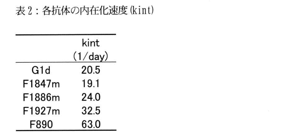
実施例4 各Fc改変体の細胞内抗体量および培地中への排出(efflux)量の経時的評価
(4-1)細胞内抗体量および培地中への排出量の経時的評価
2x10⁵個のhFcRn-hIL6R-CHO細胞を含む細胞溶液50μLに、AF647標識した抗体を終濃度が50μg/mLになるように添加し、96-wellプレート中100μL/wellとした。その後、37℃で24時間インキュベーションした。その後氷冷し、冷2% BSA含有培地の添加と除去を行い、新鮮な2%BSA含有培地を100μL加え、37℃で最大4時間まで反応させ、経時的にサンプリングした。各時点の試料を遠心した後、上清を回収するとともに、細胞をFBS-PBSで洗浄した。上清中の抗体濃度は、電気化学発光免疫測定法(ECL)を用いて測定した。また、細胞の蛍光強度は、FACS CantoIIを用いて測定し、標品ビーズの蛍光強度から細胞に含まれる抗体量を算出した。
Example 4 Time-course evaluation of the amount of intracellular antibodies and the amount of efflux into the medium of each Fc variant (4-1) Time - course evaluation of the amount of intracellular antibodies and the amount of efflux into the medium AF647-labeled antibody was added to 50 µL of the cell solution containing hFcRn-hIL6R-CHO cells to a final concentration of 50 µg/mL, and adjusted to 100 µL/well in a 96-well plate. After that, it was incubated at 37°C for 24 hours. After that, the cells were ice-cooled, the cold 2% BSA-containing medium was added and removed, 100 μL of fresh 2% BSA-containing medium was added, the reaction was allowed to continue at 37° C. for up to 4 hours, and samples were taken over time. After centrifuging the samples at each time point, the supernatant was collected and the cells were washed with FBS-PBS. Antibody concentrations in supernatants were determined using an electrochemiluminescent immunoassay (ECL). Moreover, the fluorescence intensity of the cells was measured using FACS CantoII, and the amount of antibody contained in the cells was calculated from the fluorescence intensity of the sample beads.
結果を図4(a)に示す。いずれの抗体についても、時間依存的な細胞内抗体量の減少が確認された。H237-F890の細胞内抗体量は、高い値で推移した。
また、培地中に排出された抗体量の時間推移を図4(b)に示す。いずれの抗体も排出の開始後30分前後まで、急速に排出され、その後プラトーに達することが確認された。排出の開始後10分までは、いずれの抗体についてのグラフも同様の傾きを示したが、60分以降は、各抗体で培地中抗体量に差が見られ、H237-F1886mが最も多く、H237-G1dが最も少ない量を示した。
The results are shown in FIG. 4(a). A time-dependent decrease in the amount of intracellular antibodies was confirmed for all antibodies. The H237-F890 intracellular antibody level remained high.
In addition, FIG. 4(b) shows the time transition of the amount of antibody excreted into the medium. It was confirmed that all antibodies were rapidly excreted until about 30 minutes after the start of excretion, and then reached a plateau. Up to 10 minutes after the start of excretion, the graphs for all antibodies showed similar slopes. -G1d showed the lowest amount.
(4-2)クリアランスインデックスの算出
排出開始後0分における細胞内抗体量、および各時点までに培地中に排出された抗体量を用いて、以下に示す3通りの計算式でクリアランスインデックスを算出した。
Method1:(240分における培地中の抗体量)/(0分における細胞内の抗体量)
Method2:(120分および240分における培地中の抗体量の平均)/(0分における細胞内の抗体量)
Method3:(60分、120分、および240分における培地中の抗体量)/(0分における細胞内の抗体量)
上記の各方法で算出したクリアランスインデックスの値を表4に示す。いずれの抗体においても、0.30~0.70程度の数値となった。各抗体について3つの方法で算出した値は、概ね同程度の値を示した。抗体間の値の大小も、3つの方法で同じ傾向を示した。
(4-2) Calculation of clearance index Using the amount of intracellular antibody at 0 minutes after the start of excretion and the amount of antibody excreted into the medium up to each time point, the clearance index was calculated using the following three formulas. did.
Method1: (Amount of antibody in medium at 240 minutes)/(Amount of intracellular antibody at 0 minute)
Method2: (Average amount of antibody in medium at 120 and 240 minutes)/(Amount of intracellular antibody at 0 minute)
Method3: (Amount of antibody in medium at 60, 120, and 240 minutes)/(Amount of intracellular antibody at 0 minute)
Table 4 shows the clearance index values calculated by the above methods. All the antibodies showed values of about 0.30 to 0.70. The values calculated by the three methods for each antibody showed roughly the same values. The magnitude of the values between antibodies also showed the same trend in the three methods.

実施例5 クリアランスインデックスとin vivo薬物動態との相関
実施例4で算出したクリアランスインデックスと実施例1で測定したin vivoにおける半減期またはクリアランスとの相関を評価した。結果を図5に示す。in vivoにおける半減期およびクリアランスのいずれも、クリアランスインデックスとの間に強い相関が見られた(それぞれR2=0.961およびR2=0.822)。したがって、Method1~3により算出されるクリアランスインデックスにより、in vivoにおける血漿中半減期またはクリアランスを予測可能であることが示された。
Example 5 Correlation Between Clearance Index and In Vivo Pharmacokinetics The correlation between the clearance index calculated in Example 4 and the in vivo half-life or clearance measured in Example 1 was evaluated. The results are shown in FIG. Both in vivo half-life and clearance were strongly correlated with the clearance index (R 2 =0.961 and R 2 =0.822, respectively). Therefore, it was shown that the plasma half-life or clearance in vivo can be predicted by the clearance index calculated by Methods 1-3.
本発明によれば、従来よりも簡便に、高精度で、多数の医薬品候補物質のin vivo薬物動態を予測することが可能となる。また本発明は、実験動物の使用数削減、より薬理効果の高い医薬品の開発などに貢献することができる。 According to the present invention, it is possible to predict the in vivo pharmacokinetics of a large number of drug candidate substances more easily and accurately than before. In addition, the present invention can contribute to the reduction of the number of experimental animals used, the development of drugs with higher pharmacological effects, and the like.
Claims (19)
(a)分子とFcRnを発現する細胞とを水性媒体中で接触させることにより、取り込み量が0.068 pmol/2×105cellsより高くなるように前記分子を前記細胞に取り込ませる工程であって、下記(i)~(iii)から選択される少なくとも1つの特徴を有する、工程、
(i)前記分子と前記細胞との接触時間が5時間以上である、
(ii)前記分子と接触させた後の前記細胞を酸性条件下で洗浄しない、および
(iii)前記細胞が、前記分子の標的を細胞表面に発現している、ならびに
(b)前記分子のin vitro薬物動態を測定する工程、
を含み、
前記分子は、FcRn結合ドメインを含む、前記方法。 A method of measuring the in vitro pharmacokinetics of a molecule comprising the steps of:
(a) contacting the molecule with a cell expressing FcRn in an aqueous medium so that the molecule is taken up by the cell so that the uptake amount is higher than 0.068 pmol/2×10 5 cells, a step having at least one characteristic selected from the following (i) to (iii);
(i) the contact time between the molecule and the cell is 5 hours or longer;
(ii) the cell is not washed under acidic conditions after contact with the molecule; and (iii) the cell expresses the target of the molecule on the cell surface; measuring in vitro pharmacokinetics,
including
The above method, wherein said molecule comprises an FcRn binding domain.
(c)工程(b)で得られた測定結果から、in vitro評価パラメーターを算出する工程
をさらに含む、請求項1~9のいずれか一項に記載の方法。 the following steps,
The method according to any one of claims 1 to 9, further comprising the step of (c) calculating in vitro evaluation parameters from the measurement results obtained in step (b).
(a’)請求項1~14のいずれか一項に記載の方法により、in vitro薬物動態を測定する工程、および
(b’)工程(a’)で得られた測定値またはin vitro評価パラメーターから、前記分子を生体に投与した場合のin vivo薬物動態を予測する工程
を含む、前記方法。 A method of predicting the in vivo pharmacokinetics of a molecule comprising:
(a') measuring in vitro pharmacokinetics by the method according to any one of claims 1 to 14; and (b') the measured values or in vitro evaluation parameters obtained in step (a') to predict the in vivo pharmacokinetics when the molecule is administered to a living body.
(a’’)同一の標的に結合する異なる2以上の分子を準備する工程、
(b’’)工程(a’’)で準備した2以上の分子それぞれについて、請求項1~14のいずれか一項に記載の方法により、in vitro薬物動態を測定する工程、および
(c’’)工程(b’’)で得られた、2以上の分子それぞれについての測定値またはin vitro評価パラメーターを相互に比較し、望ましい値を示した分子を選択する工程
を含む、前記方法。 A method of screening a molecule comprising:
(a'') providing two or more different molecules that bind to the same target;
(b'') measuring the in vitro pharmacokinetics of each of the two or more molecules provided in step (a'') by the method of any one of claims 1 to 14; and (c') ') comparing the measured values or in vitro evaluation parameters for each of the two or more molecules obtained in step (b'') with each other and selecting the molecules that showed the desired values.
Priority Applications (4)
| Application Number | Priority Date | Filing Date | Title |
|---|---|---|---|
| US18/559,876 US20250076283A1 (en) | 2021-05-19 | 2022-05-19 | Method for predicting in vivo pharmacokinetics of molecule |
| EP22804747.8A EP4342984A4 (en) | 2021-05-19 | 2022-05-19 | Method for predicting the in vivo pharmaceutical kinetics of a molecule |
| JP2023522714A JPWO2022244838A1 (en) | 2021-05-19 | 2022-05-19 | |
| CN202280035510.5A CN117321219A (en) | 2021-05-19 | 2022-05-19 | Methods for predicting the in vivo pharmacokinetics of molecules |
Applications Claiming Priority (2)
| Application Number | Priority Date | Filing Date | Title |
|---|---|---|---|
| JP2021-084932 | 2021-05-19 | ||
| JP2021084932 | 2021-05-19 |
Publications (1)
| Publication Number | Publication Date |
|---|---|
| WO2022244838A1 true WO2022244838A1 (en) | 2022-11-24 |
Family
ID=84141663
Family Applications (1)
| Application Number | Title | Priority Date | Filing Date |
|---|---|---|---|
| PCT/JP2022/020814 Ceased WO2022244838A1 (en) | 2021-05-19 | 2022-05-19 | Method for predicting in vivo pharmacokinetics of molecule |
Country Status (5)
| Country | Link |
|---|---|
| US (1) | US20250076283A1 (en) |
| EP (1) | EP4342984A4 (en) |
| JP (1) | JPWO2022244838A1 (en) |
| CN (1) | CN117321219A (en) |
| WO (1) | WO2022244838A1 (en) |
Families Citing this family (1)
| Publication number | Priority date | Publication date | Assignee | Title |
|---|---|---|---|---|
| CN120369962B (en) * | 2025-04-29 | 2025-09-30 | 中国人民解放军总医院第二医学中心 | CD3+CD4 +CCR1+; T cell in-diagnosis application in allergic respiratory diseases |
Citations (25)
| Publication number | Priority date | Publication date | Assignee | Title |
|---|---|---|---|---|
| US5208020A (en) | 1989-10-25 | 1993-05-04 | Immunogen Inc. | Cytotoxic agents comprising maytansinoids and their therapeutic use |
| WO1995001937A1 (en) | 1993-07-09 | 1995-01-19 | Association Gradient | Method for processing combustion residues and plant using same |
| EP0425235B1 (en) | 1989-10-25 | 1996-09-25 | Immunogen Inc | Cytotoxic agents comprising maytansinoids and their therapeutic use |
| US5635483A (en) | 1992-12-03 | 1997-06-03 | Arizona Board Of Regents Acting On Behalf Of Arizona State University | Tumor inhibiting tetrapeptide bearing modified phenethyl amides |
| US5712374A (en) | 1995-06-07 | 1998-01-27 | American Cyanamid Company | Method for the preparation of substantiallly monomeric calicheamicin derivative/carrier conjugates |
| US5714586A (en) | 1995-06-07 | 1998-02-03 | American Cyanamid Company | Methods for the preparation of monomeric calicheamicin derivative/carrier conjugates |
| US5739116A (en) | 1994-06-03 | 1998-04-14 | American Cyanamid Company | Enediyne derivatives useful for the synthesis of conjugates of methyltrithio antitumor agents |
| US5770710A (en) | 1987-10-30 | 1998-06-23 | American Cyanamid Company | Antitumor and antibacterial substituted disulfide derivatives prepared from compounds possessing a methlytrithio group |
| US5770701A (en) | 1987-10-30 | 1998-06-23 | American Cyanamid Company | Process for preparing targeted forms of methyltrithio antitumor agents |
| US5780588A (en) | 1993-01-26 | 1998-07-14 | Arizona Board Of Regents | Elucidation and synthesis of selected pentapeptides |
| WO2002020565A2 (en) | 2000-09-08 | 2002-03-14 | Universität Zürich | Collections of repeat proteins comprising repeat modules |
| WO2002032925A2 (en) | 2000-10-16 | 2002-04-25 | Phylos, Inc. | Protein scaffolds for antibody mimics and other binding proteins |
| WO2003029462A1 (en) | 2001-09-27 | 2003-04-10 | Pieris Proteolab Ag | Muteins of human neutrophil gelatinase-associated lipocalin and related proteins |
| US6630579B2 (en) | 1999-12-29 | 2003-10-07 | Immunogen Inc. | Cytotoxic agents comprising modified doxorubicins and daunorubicins and their therapeutic use |
| WO2004044011A2 (en) | 2002-11-06 | 2004-05-27 | Avidia Research Institute | Combinatorial libraries of monomer domains |
| WO2005040229A2 (en) | 2003-10-24 | 2005-05-06 | Avidia, Inc. | Ldl receptor class a and egf domain monomers and multimers |
| WO2008016854A2 (en) | 2006-08-02 | 2008-02-07 | The Uab Research Foundation | Methods and compositions related to soluble monoclonal variable lymphocyte receptors of defined antigen specificity |
| US7498298B2 (en) | 2003-11-06 | 2009-03-03 | Seattle Genetics, Inc. | Monomethylvaline compounds capable of conjugation to ligands |
| WO2009125825A1 (en) | 2008-04-11 | 2009-10-15 | 中外製薬株式会社 | Antigen-binding molecule capable of binding to two or more antigen molecules repeatedly |
| WO2012133782A1 (en) | 2011-03-30 | 2012-10-04 | 中外製薬株式会社 | Retention of antigen-binding molecules in blood plasma and method for modifying immunogenicity |
| WO2013046704A2 (en) | 2011-09-30 | 2013-04-04 | Chugai Seiyaku Kabushiki Kaisha | THERAPEUTIC ANTIGEN-BINDING MOLECULE WITH A FcRn-BINDING DOMAIN THAT PROMOTES ANTIGEN CLEARANCE |
| WO2013081143A1 (en) | 2011-11-30 | 2013-06-06 | 中外製薬株式会社 | Drug containing carrier into cell for forming immune complex |
| WO2017046994A1 (en) | 2015-09-18 | 2017-03-23 | Chugai Seiyaku Kabushiki Kaisha | Il-8-binding antibodies and uses thereof |
| JP2017517745A (en) * | 2014-03-21 | 2017-06-29 | エフ.ホフマン−ラ ロシュ アーゲーF. Hoffmann−La Roche Aktiengesellschaft | In vitro prediction of antibody in vivo half-life |
| WO2020037213A1 (en) * | 2018-08-17 | 2020-02-20 | Genentech, Inc. | In vitro transcytosis assay |
Family Cites Families (1)
| Publication number | Priority date | Publication date | Assignee | Title |
|---|---|---|---|---|
| US12235261B2 (en) * | 2017-10-20 | 2025-02-25 | Chugai Seiyaku Kabushiki Kaisha | Method for measuring cellular uptake of molecules |
-
2022
- 2022-05-19 JP JP2023522714A patent/JPWO2022244838A1/ja active Pending
- 2022-05-19 EP EP22804747.8A patent/EP4342984A4/en active Pending
- 2022-05-19 US US18/559,876 patent/US20250076283A1/en active Pending
- 2022-05-19 CN CN202280035510.5A patent/CN117321219A/en active Pending
- 2022-05-19 WO PCT/JP2022/020814 patent/WO2022244838A1/en not_active Ceased
Patent Citations (29)
| Publication number | Priority date | Publication date | Assignee | Title |
|---|---|---|---|---|
| US5770710A (en) | 1987-10-30 | 1998-06-23 | American Cyanamid Company | Antitumor and antibacterial substituted disulfide derivatives prepared from compounds possessing a methlytrithio group |
| US5770701A (en) | 1987-10-30 | 1998-06-23 | American Cyanamid Company | Process for preparing targeted forms of methyltrithio antitumor agents |
| EP0425235B1 (en) | 1989-10-25 | 1996-09-25 | Immunogen Inc | Cytotoxic agents comprising maytansinoids and their therapeutic use |
| US5416064A (en) | 1989-10-25 | 1995-05-16 | Immunogen, Inc. | Cytotoxic agents comprising maytansinoids and their therapeutic use |
| US5208020A (en) | 1989-10-25 | 1993-05-04 | Immunogen Inc. | Cytotoxic agents comprising maytansinoids and their therapeutic use |
| US5635483A (en) | 1992-12-03 | 1997-06-03 | Arizona Board Of Regents Acting On Behalf Of Arizona State University | Tumor inhibiting tetrapeptide bearing modified phenethyl amides |
| US5780588A (en) | 1993-01-26 | 1998-07-14 | Arizona Board Of Regents | Elucidation and synthesis of selected pentapeptides |
| WO1995001937A1 (en) | 1993-07-09 | 1995-01-19 | Association Gradient | Method for processing combustion residues and plant using same |
| US5739116A (en) | 1994-06-03 | 1998-04-14 | American Cyanamid Company | Enediyne derivatives useful for the synthesis of conjugates of methyltrithio antitumor agents |
| US5767285A (en) | 1994-06-03 | 1998-06-16 | American Cyanamid Company | Linkers useful for the synthesis of conjugates of methyltrithio antitumor agents |
| US5773001A (en) | 1994-06-03 | 1998-06-30 | American Cyanamid Company | Conjugates of methyltrithio antitumor agents and intermediates for their synthesis |
| US5877296A (en) | 1994-06-03 | 1999-03-02 | American Cyanamid Company | Process for preparing conjugates of methyltrithio antitumor agents |
| US5712374A (en) | 1995-06-07 | 1998-01-27 | American Cyanamid Company | Method for the preparation of substantiallly monomeric calicheamicin derivative/carrier conjugates |
| US5714586A (en) | 1995-06-07 | 1998-02-03 | American Cyanamid Company | Methods for the preparation of monomeric calicheamicin derivative/carrier conjugates |
| US6630579B2 (en) | 1999-12-29 | 2003-10-07 | Immunogen Inc. | Cytotoxic agents comprising modified doxorubicins and daunorubicins and their therapeutic use |
| WO2002020565A2 (en) | 2000-09-08 | 2002-03-14 | Universität Zürich | Collections of repeat proteins comprising repeat modules |
| WO2002032925A2 (en) | 2000-10-16 | 2002-04-25 | Phylos, Inc. | Protein scaffolds for antibody mimics and other binding proteins |
| WO2003029462A1 (en) | 2001-09-27 | 2003-04-10 | Pieris Proteolab Ag | Muteins of human neutrophil gelatinase-associated lipocalin and related proteins |
| WO2004044011A2 (en) | 2002-11-06 | 2004-05-27 | Avidia Research Institute | Combinatorial libraries of monomer domains |
| WO2005040229A2 (en) | 2003-10-24 | 2005-05-06 | Avidia, Inc. | Ldl receptor class a and egf domain monomers and multimers |
| US7498298B2 (en) | 2003-11-06 | 2009-03-03 | Seattle Genetics, Inc. | Monomethylvaline compounds capable of conjugation to ligands |
| WO2008016854A2 (en) | 2006-08-02 | 2008-02-07 | The Uab Research Foundation | Methods and compositions related to soluble monoclonal variable lymphocyte receptors of defined antigen specificity |
| WO2009125825A1 (en) | 2008-04-11 | 2009-10-15 | 中外製薬株式会社 | Antigen-binding molecule capable of binding to two or more antigen molecules repeatedly |
| WO2012133782A1 (en) | 2011-03-30 | 2012-10-04 | 中外製薬株式会社 | Retention of antigen-binding molecules in blood plasma and method for modifying immunogenicity |
| WO2013046704A2 (en) | 2011-09-30 | 2013-04-04 | Chugai Seiyaku Kabushiki Kaisha | THERAPEUTIC ANTIGEN-BINDING MOLECULE WITH A FcRn-BINDING DOMAIN THAT PROMOTES ANTIGEN CLEARANCE |
| WO2013081143A1 (en) | 2011-11-30 | 2013-06-06 | 中外製薬株式会社 | Drug containing carrier into cell for forming immune complex |
| JP2017517745A (en) * | 2014-03-21 | 2017-06-29 | エフ.ホフマン−ラ ロシュ アーゲーF. Hoffmann−La Roche Aktiengesellschaft | In vitro prediction of antibody in vivo half-life |
| WO2017046994A1 (en) | 2015-09-18 | 2017-03-23 | Chugai Seiyaku Kabushiki Kaisha | Il-8-binding antibodies and uses thereof |
| WO2020037213A1 (en) * | 2018-08-17 | 2020-02-20 | Genentech, Inc. | In vitro transcytosis assay |
Non-Patent Citations (33)
| Title |
|---|
| "NCBI", Database accession no. NP 055951.2 |
| ADV BIOMED RES., vol. 1, 2012, pages 27 |
| ALGIRDAS GREVYS; JEANNETTE NILSEN; KINE M K SAND; MULUNEH B DABA; INGER ØYNEBRÅTEN; MALIN BERN; MARTIN B MCADAM; STIAN FOSS; TILMA: "A human endothelial cell-based recycling assay for screening of FcRn targeted molecules", NATURE COMMUNICATIONS, vol. 9, no. 1, 1 December 2018 (2018-12-01), pages 1 - 14, XP055717922, DOI: 10.1038/s41467-018-03061-x * |
| ANNUAL REVIEW OF CELL AND DEVELOPMENTAL BIOLOGY, vol. 12, 1996, pages 181 - 220 |
| BIOMETRICS, vol. 36, 2015, pages S 3 - S 18 |
| CHUNG ET AL., J IMMUNOL METHODS., vol. 462, 2018, pages 101 |
| CLAUDIA A. CASTRO JARAMILLO, BELLI SARA, CASCAIS ANNE-CHRISTINE, DUDAL SHERRI, EDELMANN MARTIN R., HAAK MARKUS, BRUN MARIE-ELISE, : "Toward in vitro -to- in vivo translation of monoclonal antibody pharmacokinetics: Application of a neonatal Fc receptor-mediated transcytosis assay to understand the interplaying clearance mechanisms", MABS, vol. 9, no. 5, 4 July 2017 (2017-07-04), US , pages 781 - 791, XP055635634, ISSN: 1942-0862, DOI: 10.1080/19420862.2017.1320008 * |
| DRUG DELIVERY SYSTEM, vol. 31, no. 1, 2016, pages 83 - 84 |
| DUBOWCHIK ET AL., BIOORG. & MED. CHEM. LETTERS, vol. 12, 2002, pages 1529 - 1532 |
| FOLIA PHARMACOL. JPN., vol. 136, 2010, pages 280 - 284 |
| GREVYS ET AL., NAT COMMUN., vol. 9, 2018, pages 621 |
| HINMAN ET AL., CANCER RES., vol. 53, 1993, pages 3336 - 3342 |
| INT ARCH ALLERGY APPL IMMUNOL., vol. 46, no. 2, 1974, pages 230 - 48 |
| J IMMUNOL., vol. 190, no. 12, 15 June 2013 (2013-06-15), pages 6694 - 706 |
| J IMMUNOL., vol. 89, October 1962 (1962-10-01), pages 471 - 82 |
| J. EXP. MED., vol. 197, no. 3, 2003, pages 315 - 322 |
| JARAMILLO ET AL., MABS, vol. 9, 2017, pages 781 |
| JEFFREY ET AL., BIOORGANIC & MED. CHEM. LETTERS, vol. 16, 2006, pages 358 - 362 |
| JONES HANNAH M., TOLSMA JOHN, ZHANG ZHIWEI, JASPER PAUL, LUO HAOBIN, WEBER GREGORY L., WRIGHT KATHERINE, BARD JOEL, BELL ROBERT, M: "A Physiologically‐Based Pharmacokinetic Model for the Prediction of "Half‐Life Extension" and "Catch and Release" Monoclonal Antibody Pharmacokinetics", CPT: PHARMACOMETRICS & SYSTEMS PHARMACOLOGY, vol. 9, no. 9, 1 September 2020 (2020-09-01), pages 534 - 541, XP093007227, ISSN: 2163-8306, DOI: 10.1002/psp4.12547 * |
| KING ET AL., J. MED. CHEM., vol. 45, 2002, pages 4336 - 4343 |
| KRATZ ET AL., CURRENT MED. CHEM., vol. 13, 2006, pages 477 - 523 |
| LODE ET AL., CANCER RES., vol. 58, 1998, pages 2925 - 2928 |
| METHODS MOL BIOL., vol. 726, 2011, pages 207 - 221 |
| MULLBERG ET AL., J. IMMUNOL., vol. 152, no. 10, 1994, pages 4958 - 4968 |
| NAGY ET AL., PROC. NATL. ACAD. SCI. USA, vol. 97, 2000, pages 829 - 834 |
| NANOG, PLOS ONE., vol. 5, no. 5, 2010, pages e10611 |
| NATURE REVIEWS IMMUNOLOGY, vol. 7, 2007, pages 715 - 725 |
| ORGAN BIOLOGY, vol. 24, no. 1, 2017, pages 54 - 60 |
| PROTEIN CELL, vol. 9, no. 1, 2018, pages 15 - 32 |
| QI TIMOTHY, CAO YANGUANG: "In Translation: FcRn across the Therapeutic Spectrum", INTERNATIONAL JOURNAL OF MOLECULAR SCIENCES, vol. 22, no. 6, 1 January 2021 (2021-01-01), pages 1 - 29, XP093007218, DOI: 10.3390/ijms22063048 * |
| See also references of EP4342984A4 |
| SHAN CHUNG, YUWEN LINDA LIN, VAN NGUYEN, LYNN KAMEN, KAI ZHENG, BIANCA VORA, AN SONG: "Development of a label-free FcRn-mediated transcytosis assay for in vitro characterization of FcRn interactions with therapeutic antibodies and Fc-fusion proteins", JOURNAL OF IMMUNOLOGICAL METHODS, vol. 462, 1 November 2018 (2018-11-01), NL , pages 101 - 105, XP055684894, ISSN: 0022-1759, DOI: 10.1016/j.jim.2018.07.004 * |
| TORGOV ET AL., BIOCONJ. CHEM., vol. 16, 2005, pages 717 - 721 |
Also Published As
| Publication number | Publication date |
|---|---|
| EP4342984A1 (en) | 2024-03-27 |
| US20250076283A1 (en) | 2025-03-06 |
| JPWO2022244838A1 (en) | 2022-11-24 |
| CN117321219A (en) | 2023-12-29 |
| EP4342984A4 (en) | 2025-04-23 |
Similar Documents
| Publication | Publication Date | Title |
|---|---|---|
| JP7581290B2 (en) | FcγRIIb-specific Fc antibody | |
| JP7360242B2 (en) | Mouse FcγRII-specific Fc antibody | |
| US20200181258A1 (en) | Modified antibody constant region | |
| CN116096741B (en) | Compositions and methods of use for extending the half-life of therapeutic agents in felines | |
| JP7716979B2 (en) | Protease substrates and polypeptides containing protease cleavage sequences | |
| US20250321224A1 (en) | Method for measuring cellular uptake of molecules | |
| EP4342984A1 (en) | Method for predicting in vivo pharmacokinetics of molecule | |
| HK40103588A (en) | Method for predicting in vivo pharmacokinetics of molecule | |
| RU2749357C2 (en) | POLYPEPTIDE VARIANT THAT HAS PRESERVED OR DECREASED BINDING ACTIVITY TO FcγRIIa, PHARMACEUTICAL COMPOSITION CONTAINING IT AND THEIR APPLICATION |
Legal Events
| Date | Code | Title | Description |
|---|---|---|---|
| 121 | Ep: the epo has been informed by wipo that ep was designated in this application |
Ref document number: 22804747 Country of ref document: EP Kind code of ref document: A1 |
|
| WWE | Wipo information: entry into national phase |
Ref document number: 2023522714 Country of ref document: JP |
|
| WWE | Wipo information: entry into national phase |
Ref document number: 18559876 Country of ref document: US |
|
| WWE | Wipo information: entry into national phase |
Ref document number: 202280035510.5 Country of ref document: CN |
|
| WWE | Wipo information: entry into national phase |
Ref document number: 2022804747 Country of ref document: EP |
|
| NENP | Non-entry into the national phase |
Ref country code: DE |
|
| ENP | Entry into the national phase |
Ref document number: 2022804747 Country of ref document: EP Effective date: 20231219 |
|
| WWE | Wipo information: entry into national phase |
Ref document number: 11202308712V Country of ref document: SG |
|
| WWP | Wipo information: published in national office |
Ref document number: 18559876 Country of ref document: US |