WO2022187246A1 - Method and kit for monitoring non-small cell lung cancer - Google Patents
Method and kit for monitoring non-small cell lung cancer Download PDFInfo
- Publication number
- WO2022187246A1 WO2022187246A1 PCT/US2022/018340 US2022018340W WO2022187246A1 WO 2022187246 A1 WO2022187246 A1 WO 2022187246A1 US 2022018340 W US2022018340 W US 2022018340W WO 2022187246 A1 WO2022187246 A1 WO 2022187246A1
- Authority
- WO
- WIPO (PCT)
- Prior art keywords
- methylation
- lung cancer
- seq
- sequence identity
- probe
- Prior art date
- Legal status (The legal status is an assumption and is not a legal conclusion. Google has not performed a legal analysis and makes no representation as to the accuracy of the status listed.)
- Ceased
Links
Classifications
-
- C—CHEMISTRY; METALLURGY
- C12—BIOCHEMISTRY; BEER; SPIRITS; WINE; VINEGAR; MICROBIOLOGY; ENZYMOLOGY; MUTATION OR GENETIC ENGINEERING
- C12Q—MEASURING OR TESTING PROCESSES INVOLVING ENZYMES, NUCLEIC ACIDS OR MICROORGANISMS; COMPOSITIONS OR TEST PAPERS THEREFOR; PROCESSES OF PREPARING SUCH COMPOSITIONS; CONDITION-RESPONSIVE CONTROL IN MICROBIOLOGICAL OR ENZYMOLOGICAL PROCESSES
- C12Q1/00—Measuring or testing processes involving enzymes, nucleic acids or microorganisms; Compositions therefor; Processes of preparing such compositions
- C12Q1/68—Measuring or testing processes involving enzymes, nucleic acids or microorganisms; Compositions therefor; Processes of preparing such compositions involving nucleic acids
- C12Q1/6876—Nucleic acid products used in the analysis of nucleic acids, e.g. primers or probes
- C12Q1/6883—Nucleic acid products used in the analysis of nucleic acids, e.g. primers or probes for diseases caused by alterations of genetic material
- C12Q1/6886—Nucleic acid products used in the analysis of nucleic acids, e.g. primers or probes for diseases caused by alterations of genetic material for cancer
-
- C—CHEMISTRY; METALLURGY
- C12—BIOCHEMISTRY; BEER; SPIRITS; WINE; VINEGAR; MICROBIOLOGY; ENZYMOLOGY; MUTATION OR GENETIC ENGINEERING
- C12Q—MEASURING OR TESTING PROCESSES INVOLVING ENZYMES, NUCLEIC ACIDS OR MICROORGANISMS; COMPOSITIONS OR TEST PAPERS THEREFOR; PROCESSES OF PREPARING SUCH COMPOSITIONS; CONDITION-RESPONSIVE CONTROL IN MICROBIOLOGICAL OR ENZYMOLOGICAL PROCESSES
- C12Q2600/00—Oligonucleotides characterized by their use
- C12Q2600/112—Disease subtyping, staging or classification
-
- C—CHEMISTRY; METALLURGY
- C12—BIOCHEMISTRY; BEER; SPIRITS; WINE; VINEGAR; MICROBIOLOGY; ENZYMOLOGY; MUTATION OR GENETIC ENGINEERING
- C12Q—MEASURING OR TESTING PROCESSES INVOLVING ENZYMES, NUCLEIC ACIDS OR MICROORGANISMS; COMPOSITIONS OR TEST PAPERS THEREFOR; PROCESSES OF PREPARING SUCH COMPOSITIONS; CONDITION-RESPONSIVE CONTROL IN MICROBIOLOGICAL OR ENZYMOLOGICAL PROCESSES
- C12Q2600/00—Oligonucleotides characterized by their use
- C12Q2600/118—Prognosis of disease development
-
- C—CHEMISTRY; METALLURGY
- C12—BIOCHEMISTRY; BEER; SPIRITS; WINE; VINEGAR; MICROBIOLOGY; ENZYMOLOGY; MUTATION OR GENETIC ENGINEERING
- C12Q—MEASURING OR TESTING PROCESSES INVOLVING ENZYMES, NUCLEIC ACIDS OR MICROORGANISMS; COMPOSITIONS OR TEST PAPERS THEREFOR; PROCESSES OF PREPARING SUCH COMPOSITIONS; CONDITION-RESPONSIVE CONTROL IN MICROBIOLOGICAL OR ENZYMOLOGICAL PROCESSES
- C12Q2600/00—Oligonucleotides characterized by their use
- C12Q2600/154—Methylation markers
-
- C—CHEMISTRY; METALLURGY
- C12—BIOCHEMISTRY; BEER; SPIRITS; WINE; VINEGAR; MICROBIOLOGY; ENZYMOLOGY; MUTATION OR GENETIC ENGINEERING
- C12Q—MEASURING OR TESTING PROCESSES INVOLVING ENZYMES, NUCLEIC ACIDS OR MICROORGANISMS; COMPOSITIONS OR TEST PAPERS THEREFOR; PROCESSES OF PREPARING SUCH COMPOSITIONS; CONDITION-RESPONSIVE CONTROL IN MICROBIOLOGICAL OR ENZYMOLOGICAL PROCESSES
- C12Q2600/00—Oligonucleotides characterized by their use
- C12Q2600/16—Primer sets for multiplex assays
Definitions
- the present disclosure relates to methods or kits for monitoring non-small cell lung cancer in a subject in need thereof.
- the present disclosure also relates to methods, compositions and kits for detecting, diagnosing, prognosing, and characterizing non-small cell lung cancer in a subject in need thereof.
- Description of Related Art Cancers remain the leading causes of death worldwide.
- International Agency for Research on Cancer (IARC) estimated 10 million deaths and 19.3 million cases in 2020 in the recently released updates in Globocan 2020. Among all cancers, worldwide incidences of lung cancer rank top three in both men and women.
- the present disclosure is therefore provided with at least one biomarker and a method to characterize, diagnose, prognosticate, stratify and monitor the progression or recurrence of non-small cell lung cancer in a subject in need thereof.
- the present disclosure provides a method and biomarker that detect disease progression in a subject preceding radiographic detection, regardless of the mutation status or ethnic group of the subject.
- a method is provided to characterize non-small cell lung cancer in a subject in need thereof, comprising detecting a methylation level of at least one gene selected from the group consisting of KCNS2, HOXA9, SCT, BARHL2, and any combination thereof in a biological sample from the subject, wherein the biological sample contains circulating free DNA.
- the method of the present disclosure further comprises detecting a methylation level of KCNS2.
- the methylation level is detected by: at least one primer and a probe, wherein the primer pair has at least 80% sequence identity to SEQ ID NOs: 1 and 2, respectively, and at least one probe having at least 80% sequence identity to SEQ ID NO: 3; the primer pair has at least 80% sequence identity to SEQ ID NOs: 4 and 5, respectively, and at least one probe having at least 80% sequence identity to SEQ ID NO: 6; the primer pair has at least 80% sequence identity to SEQ ID NOs: 7 and 8, respectively, and at least one probe having at least 80% sequence identity to SEQ ID NO:9; the primer pair has at least 80% sequence identity to SEQ ID NOs: 10 and 11 and at least one probe having at least 80% sequence identity to SEQ ID NO: 12; or any combination thereof.
- the methylation level is detected by bisulfite sequencing, array or bead hybridization, quantitative real-time PCR, methylation- sensitive endonuclease digestion followed by sequencing, PCR and sequencing, methylation- specific PCR and pyrosequencing.
- the method to characterize non-small cell lung cancer in a subject in need thereof further comprises calculating a methylation risk score based on the methylation level of the at least one gene.
- the methylation level of the at least one gene is given different weights for calculating the methylation risk score.
- the calculation of the methylation risk score further comprises at least one of age, gender, active smoking status and former smoking status.
- a mathematical model is used on the four genes to calculate circulating methylation risk scores in the plasma cfDNA measured by methylation-specific droplet digital PCR.
- a mathematical model is used in calculating clinical methylation risk scores for lung cancer detection in the training and the validation cohorts of lung cancer patients at all stages.
- the methylation risk score is calculated as -1.547+0.024*SCT+0.084*HOXA9-0.039*BARHL2+0.031*KCNS2.
- the biological sample used in the method to characterize non-small cell lung cancer in a subject is a body fluid, e.g., blood, sputum, pleural fluid, cerebrospinal fluid or any combination thereof.
- the at least one gene tested in the method to characterize non-small cell lung cancer in a subject carries a driver mutation, a passenger mutation, or a combination thereof.
- a kit for characterizing non-small cell lung cancer is also provided.
- the kit comprises a primer pair and a probe for detecting a methylation level of at least one gene, wherein: the primer pair has at least 80% sequence identity to SEQ ID NOs: 1 and 2, respectively, and the probe has at least 80% sequence identity to SEQ ID NO: 3; the primer pair has at least 80% sequence identity to SEQ ID NOs: 4 and 5, respectively, and the probe has at least 80% sequence identity to SEQ ID NO: 6; the primer pair has at least 80% sequence identity to SEQ ID NOs: 7 and 8, respectively, and the probe has at least 80% sequence identity to SEQ ID NO: 9; or the primer pair has at least 80% sequence identity to SEQ ID NOs: 10 and 11, respectively, and the probe has at least 80% sequence identity to SEQ ID NO: 12; or any combination thereof.
- a method of evaluating a therapy of non-small cell lung cancer comprises obtaining a biological sample comprising circulating free DNA from a subject who had received the therapy of non-small cell lung cancer; detecting a methylation level of at least one gene selected from the group consisting of KCNS2, HOXA9, SCT, BARHL2, and any combination thereof; calculating a methylation risk score based on the methylation level of the at least one gene, wherein a change of the methylation risk score is indicative of efficacy of the therapy of non-small cell lung cancer.
- the method further comprises detecting a methylation level of KCNS2.
- an increase of the methylation risk score is indicative of disease progression.
- the therapy of non- small cell lung cancer is surgery, radiation therapy, chemotherapy, targeted therapy, immunotherapy. or any combination thereof.
- the disease progression comprises increase in tumor size or metastasis.
- Marker discovery phase In-silico analysis of genome-wide methylation data from surgically resected tumor tissues from the National Taiwan University Hospital and The Cancer Genome Atlas (TCGA) lung cancer cohorts. Differential methylation analyses between cancer tissues and adjacent lung tissues were performed to identify candidate methylated probes. Probes that were methylated in the peripheral blood mononuclear cells (PBMCs) of noncancer subjects from the Taiwan Biobank were excluded. A panel of cancer-specific methylated markers is designed for measurement by multiplex methylation-specific droplet digital PCR (MS- ddPCR), and these markers were verified using surgically resected lung cancer tissues.
- PBMCs peripheral blood mononuclear cells
- MS- ddPCR multiplex methylation-specific droplet digital PCR
- Assay development phase The methylation levels of the identified markers in the circulating cell- free DNAs from lung cancer patients were quantified at initial diagnosis before treatment vs. noncancer subjects using MS-ddPCR. A model fitting with k-fold cross validation was performed to generate methylation risk scores based on the marker panel.
- Longitudinal validation phase The cfDNA methylation scores of 268 samples (58 lung cancer patients) were obtained at initial diagnosis and follow-up visits every 3 months over the course of treatment. The relationships between methylation scores and radiological/clinical responses were recorded.
- FIGs.2A to 2C show discovery of markers from genome-wide methylation data of primary lung cancer tissues.
- FIG.2A illustrates the heatmaps showing the methylation levels ( ⁇ values) of the 4-probe panel (SCT, KCNS2, HOXA9 and BARHL2) measured by Infinium MethylationEPIC BeadChips and HumanMethylation450 in the surgically resected lung cancer and the adjacent lung tissues from the NTUH (Adeno (lung adenocarcinoma tissues): 30; SqCC (lung squamous cell carcinoma tissues): 35; AdjU (adjacent unaffected tissues): 15) and TCGA cohorts (Adeno 460, SqCC 370, AdjU 74).
- FIG.2B shows the ⁇ values of HOXA9, KCNS2, BARHL2 and SCT in lung cancer tissues vs. adjacent lung tissues (control, CTL) from the NTUH and TCGA cohorts measured by Infinium methylation arrays.
- FIG.2C shows the summary of sensitivity, specificity, positive predictive value (PPV) and negative predictive value (NPV) based on different combinations of the 4-probe panel in lung adenocarcinoma and squamous cell carcinoma patients from the NTUH and TCGA cohorts. ⁇ values greater than 0.5 are considered positive for individual probes.
- FIGs.3A and 3B show methylation levels of classic methylated genes in the TCGA lung adenocarcinoma and squamous cell carcinoma tissues.
- FIG.3A is the heatmap showing the ⁇ values of classic methylated genes in lung cancer measured by Infinium HumanMethylation450 BeadChips in the surgically resected lung cancer and adjacent lung tissues from the TCGA database (Adeno 460, SqCC 370, AdjU 74).
- FIG.3B shows a summary of in silico sensitivity, specificity, positive predictive value (PPV) and negative predictive value (NPV) of the classic methylated genes in lung adenocarcinoma and squamous cell carcinoma patients from the TCGA database.
- the genes with the mean ⁇ value of promoter probes greater than 0.5 are considered methylated.
- FIGs.4A and 4B show the probe design and validation in primary lung cancer tissues.
- FIG. 4A shows the schematic diagrams of probe positions relative to individual marker genes, where positions and lengths of amplicons and CpG island are indicated. Arrows indicate the direction of transcriptions.
- FIG. 4B shows a heatmap of methylation levels of individual candidate probes for primary lung cancer tissues measured by methylation-specific real-time PCR. Lung cancer cell lines (i.e., A549, H1703, H1792 and H1299) were used as positive controls. The peripheral blood mononuclear cells (PBMCs) from healthy volunteers served as negative controls.
- FIG.5 shows the representative scatter dot plots of candidate methylated probes measured by methylation-specific droplet digital PCR (MS-ddPCR) in H1703 and A549 lung cancer cell lines.
- MS-ddPCR methylation-specific droplet digital PCR
- FIGs. 6A and 6B show the concentrations and fragment sizes of plasma cfDNA in non- small cell lung cancer (NSCLC) patients vs. noncancer control subjects.
- FIGs. 7A to 7D show that four-probe panel of methylated cfDNA markers in the plasma distinguishes between lung cancer patients and noncancer controls.
- FIG. 7B shows the receiver operating characteristic (ROC) curve analysis of the four-probe panel and individual probes for the prediction of lung cancer versus noncancer controls.
- ROC receiver operating characteristic
- FIG.7C shows the percentages of clinically confirmed lung cancer patients who can be identified by methylation scores (> 0.13) and carcinoembryonic antigen (CEA > 5 ng/mL).
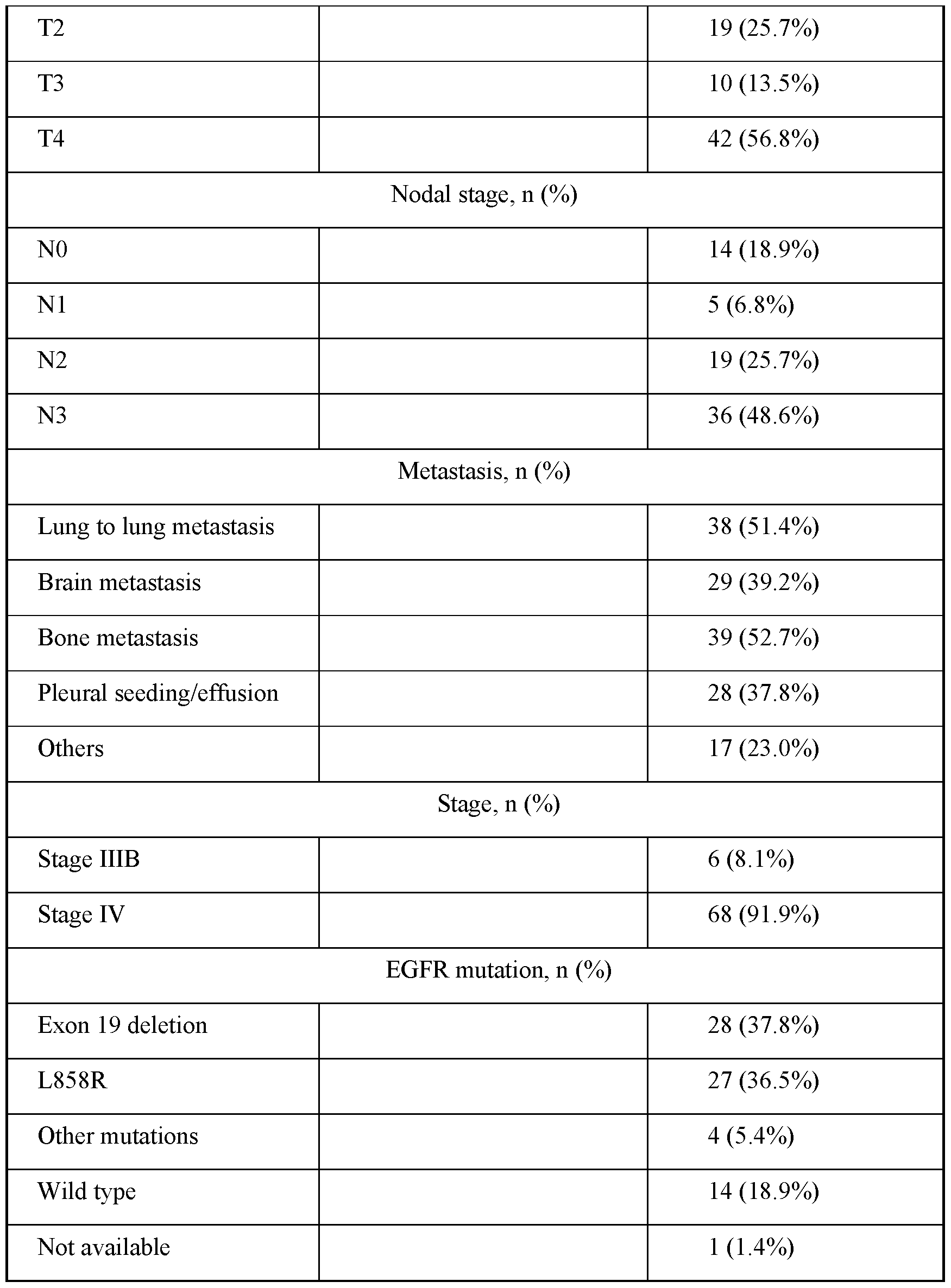
- FIG. 7D shows the relationships between methylation scores and smoking, gender, EGFR status, tumor staging, and metastatic sites.
- the central lines of box plots represent medians of methylation scores.
- the whiskers denote 1.5 * interquartile ranges (IQR).
- IQR interquartile ranges
- the p value was calculated by the Mann-Whitney test or one-way ANOVA with Tukey’s multiple comparison test.
- MPE malignant pleural effusion.
- FIGs.8A and 8B show the correlation plots of circulating methylated markers and the ages of subjects.
- FIG.8A shows the scatter plots showing the correlation between the methylation levels of individual probes — HOXA9, SCT, KCNS2 and BARHL2 (y axis) and age (x axis). A linear regression line was fitted in each plot. The correlation coefficients (r) and p values were calculated by spearman correlation test.
- FIG. 8B shows the scatter plots showing the correlation between the methylation risk scores (y axis) and age (x axis).
- FIGs.9A to 9I show that methylation scores in serial blood samples correspond to clinical responses.
- FIG.9B illustrates the spaghetti plots showing the dynamic changes in methylation risk scores during the treatment course for each lung cancer patient. Each line represents one subject in the study. Upper panel: patients with progressive disease.
- FIG. 9C shows the bar chart of estimated marginal means of methylation scores for patients with PD and those with PR/SD calculated from the generalized estimating equations (GEE) model. Error bars represent the 95% confidence interval (CI).
- FIG. 9D illustrates the bar chart showing methylation scores from individual visits of all patients during the treatment course categorized by different levels of methylation score ( ⁇ 0.13, 0.13- 5.56, >5.56). The shade of the bar chart represents the disease status at the time of blood collection.
- FIG. 9E illustrates the dot plot showing the methylation levels of blood samples collected at the initial and follow-up visits. The disease status was defined according to the Response Evaluation Criteria in Solid Tumors (RECIST) criteria 1.1.
- RECIST Response Evaluation Criteria in Solid Tumors
- FIG.9F shows the differences between the methylation risk score at each follow-up visit and that at initial visit (baseline). Increases in methylation scores are more associated with disease progression. The p value was calculated by the Mann- Whitney test. * p ⁇ 0.05.
- FIG.9G shows the differences between the methylation risk score at each follow-up visit and that at the prior visit. The p value was calculated by the Mann-Whitney test. *** p ⁇ 0.001.
- FIG. 9H shows the methylation scores of each subject at initial blood collection categorized by PR/SD or PD. The p value was calculated by the Mann-Whitney test.
- FIGs.10A to 10D show representative cases using the 4-gene methylation risk scores for disease monitoring.
- FIG. 10A shows a 46-year-old patient diagnosed with stage 4 lung adenocarcinoma carrying EGFR exon 19 deletions receiving erlotinib treatment. The patient achieved a partial response, and the disease was under control for approximately 800 days. Elevated methylation risk scores were noticed at the 10th and 11th visits. Enlargement of the primary lung tumor in the right upper lobe on chest tomography was observed at the 11th visit.
- FIG.10B shows a 56-year-old patient diagnosed with stage 4 lung adenocarcinoma with bone metastasis. No specific driver mutations were identified. The patient received systemic chemotherapy with carboplatin and pemetrexed. The methylation score was decreased after the therapy but elevated at the next visit when there was no evidence of clinical and radiographic progression at the time. Three months later, chest tomography showed a significant progression of the primary lung tumor which was associated with a marked increase in the circulating methylation score. Inferior vena cava thrombosis was also noted.
- FIG.10C shows a 69-year-old patient with stage 4 lung adenocarcinoma receiving erlotinib treatment.
- FIG. 10D shows a 48-year-old patient with stage IIIb lung adenocarcinoma receiving concurrent chemoradiotherapy (CCRT) and gefitinib. Following CCRT, the patient’s methylation scores declined and remained low for at least 600 days. The patient was clinically stable with no signs of tumor progression.
- FIG. 11 shows the flow chart of the procedures carried out in discovering biomarkers, developing assays with the biomarkers, and validating the biomarkers in the present disclosure.
- Marker discovery phase In-silico analysis of genome-wide methylation data from surgically resected tumor tissues from the National Taiwan University Hospital and The Cancer Genome Atlas (TCGA) lung cancer cohorts. Differential methylation analyses between cancer and adjacent lung tissues were performed to identify candidate methylated probes. Probes that were methylated in the peripheral blood mononuclear cells (PBMCs) of non-cancer subjects from the Taiwan Biobank were excluded.
- PBMCs peripheral blood mononuclear cells
- a panel of cancer-specific methylated markers is designed for measurement by multiplex methylation-specific droplet digital PCR (MS-ddPCR), and these markers were verified using surgically resected lung cancer tissues. These lung tissues were LUAD: lung adenocarcinoma, LUSC: lung squamous cell carcinoma.
- Assay development phase The methylation levels of the identified markers in the circulating cell- free DNAs from lung cancer patients were quantified at initial diagnosis before treatment vs. non-cancer subjects using MS-ddPCR.
- a logistical regression model was constructed to calculate methylation risk scores based on the four-gene combination in the training cohort.
- FIG.12 shows the receiver operating characteristic (ROC) curve analysis of methylation risk scores and methylation levels of individual probes for the diagnosis of lung cancer in the training cohort of patients with stage IV lung adenocarcinoma, and the ROC curve analysis of methylation risk scores and methylation levels of individual probes for patients with stage IV lung cancer and for patients at all stages in the validation cohort.
- FIG. 13 shows the relationships between methylation scores and clinical stages of non- small cell lung cancer. The values above the cutoff value (-0.764) are considered positive for the disease.
- FIG.14B shows a spaghetti plots demonstrating the dynamic changes in methylation risk scores during the treatment course for each lung cancer patient.
- FIG.14C shows a bar chart of estimated marginal means of methylation scores for patients with PD and those with PR/SD calculated from the generalized estimating equations (GEE) model. Error bars represent the 95% confidence interval.
- FIGs.15A to 15C show serial plasma cfDNA methylation scores for disease monitoring in three non-small cell lung cancer patients.
- FIG.15A shows a serial plasma cfDNA methylation scores for disease monitoring in non-small cell lung cancer of patient L14, a 56-year-old patient diagnosed with stage 4 lung adenocarcinoma with bone metastasis.
- FIG.15B shows a serial plasma cfDNA methylation scores for disease monitoring in non-small cell lung cancer of patient L12, a 72-year-old patient with stage 4 lung adenocarcinoma receiving afatinib treatment.
- the patient L12 responded to the therapy, but a metastatic liver tumor was noted at approximately 270 days despite the continued shrinkage of the primary lung tumor.
- FIG. 15B shows a serial plasma cfDNA methylation scores for disease monitoring in non-small cell lung cancer of patient L12, a 72-year-old patient with stage 4 lung adenocarcinoma receiving afatinib treatment.
- the patient L12 responded to the therapy, but a metastatic liver tumor was noted at approximately 270 days despite the continued shrinkage of the primary lung tumor.
- FIG. 15B shows a serial plasma cfDNA methylation scores for disease monitoring in non-small cell lung cancer of patient L12, a 72-year-old patient with stage 4 lung adenocarcinoma receiving
- 15C shows a serial plasma cfDNA methylation scores for disease monitoring in non-small cell lung cancer of patient L44, a 69- year-old patient with stage 4 lung adenocarcinoma receiving erlotinib treatment.
- the methylation scores of patient L44d decreased initially but increased again at approximately 9 months. New bone metastases at the lumbar spine and pelvis of patient L44 were noted six months later. Remarkably, the methylation score declined rapidly after local palliative radiotherapy (RT) were observed.
- RT local palliative radiotherapy
- the present disclosure provides a method and biomarkers to characterize, diagnose, stratify, prognosticate and monitor non-small cell lung cancer in a subject in need thereof by analyzing the levels of one or more biomarkers in a sample obtained from the subject.
- the one or more biomarkers is a methylation level of at least one gene selected from the group consisting of HOXA9, KCNS2, SCT, and BARHL2.
- the methylation level of the at least one gene is measured in a biological specimen containing circulating tumor DNA or cell- free DNA.
- the methylation level of at least one gene is evaluated in a biological specimen containing cell-free DNA to characterize, diagnose, stratify, prognosticate and monitor non-small cell lung cancer in a subject in need thereof.
- a methylation risk score is calculated based on the methylation level of one, two, three or four genes selected from the group consisting of HOXA9, KCNS2, SCT, and BARHL2.
- the present disclosure provides at least one biomarker and a method in distinguishing between a lung cancer patient and a noncancer control.
- the present disclosure provides a method for serial assessments of disease status and treatment responses.
- Circulating free DNA are DNA fragments released to the blood plasma.
- cfDNA can be used to describe various forms of DNA freely circulating in the bloodstream, including circulating tumor DNA (ctDNA), circulating cell-free mitochondrial DNA (ccf mtDNA), and cell-free fetal DNA (cffDNA).
- Circulating tumor DNA originates from tumor cells through mechanisms such as apoptosis, necrosis, or active release, and can reflect molecular characteristics of original tumor tissues.
- cfDNA and ctDNA may be used interchangeably to indicate the DNA fragments obtained from a biological specimen from a subject, such as blood plasma or other body fluid containing cfDNA.
- Body fluid is meant to be a biological sample obtained from a subject that is substantially devoid of intact cells. This may be derived from a biological sample that is itself substantially devoid of cells, or may be derived from a sample from which cells have been removed. Examples of cell-free body fluid include those derived from blood, such as serum or plasma; urine; or samples derived from other sources, such as semen, sputum, feces, ductal exudate, lymph, pleural fluid, cerebrospinal fluid or recovered lavage. cfDNA or ctDNA is used as a biological specimen containing a biomarker for monitoring therapeutic responses in cancer patients.
- driver mutations such as EGFR and KRAS.
- a driver mutation is a mutation that gives a selective advantage to a clone in its microenvironment through increasing either survival or reproduction thereof and therefore is causally implicated in oncogenesis.
- a driver mutation needs not be required for maintenance of the final cancer (although it often is), but it must have been selected at some points along the course of cancer development.
- a passenger mutation on the other hand, has not been selected, has not conferred clonal growth advantage and has therefore not contributed to cancer development. Passenger mutations are found within cancer genomes because somatic mutations without functional consequences often occur during cell division. Thus, a cell that acquires a driver mutation will have biologically inert somatic mutations within its genome.
- a disease status or treatment responses by testing for a specific driver mutation such as EGFR is limited to only a subset of patients.
- a specific driver mutation such as EGFR
- the clonal evolution of cancer mutations is a frequent event during the treatment course. It requires a large panel of markers for better coverage, which is not only costly but also unsuitable for longitudinal monitoring of disease burden, while the targets for detection are continually changing.
- DNA methylation often takes place at a common region near the gene promoter and is better suited for longitudinal tracking of dynamic disease burdens.
- DNA methylation is a biological process by which methyl groups are added to the DNA molecule.
- DNA methylation is one of many mechanisms in regulating a gene expression and can change the activity of a gene without changing the DNA sequence.
- DNA methylation When located in a gene promoter, DNA methylation typically acts to repress gene transcription, namely, to reduce gene expression and thereby suppress gene function.
- Aberrant DNA methylation is a hallmark of the carcinogenic process, as revealed by large-scale genomic/epigenomics studies. Similar to genetic mutations, DNA methylation is heritable and stable, thereby having great potential as a molecular marker. Unlike driver mutations with numerous hot spots, methylation regions around the promoter appear to be common for each gene across patient samples.
- the term “to characterize” in a subject or individual may include, but is not limited to, to provide the diagnosis of a disease or a condition, to determine the stratification of a disease risk, to assess the risk of a disease or a condition, to provide the prognosis of a disease or a condition, to determine a disease stage or a condition stage, to determine the severity of a disease or a condition, to evaluate the malignancy potential of a disease or a condition, to monitor a recurrence of cancer, to evaluate a drug efficacy, to describe a physiological condition, to evaluate an organ distress or organ rejection, to monitor disease or condition progression, to determine therapy-related association to a disease or a condition, or to describe a physiological or biological state.
- prognosis of cancer may include predicting the clinical outcome of the patient, assessing the risk of cancer recurrence, determining treatment modality, or determining treatment efficacy.
- the term “metastasis” describes the spread of a cancer from one part of the body to another. A tumor formed by cells that have spread can be called a “metastatic tumor” or a “metastasis.” The metastatic tumor often contains cells that are similar to those in the original (primary) tumor, and have, but are not limited to, genomic, epigenetic, transcriptomic, and metabolic alterations.
- progression describes the course of a disease or a condition, such as a cancer, as it becomes worse or spreads in the body.
- subject refers to a warm-blooded animal, such as a mammal that is afflicted with, or suspected of having, at risk for or being pre-disposed to, or being screened for cancer, e.g., actual or suspected cancer.
- a warm-blooded animal such as a mammal that is afflicted with, or suspected of having, at risk for or being pre-disposed to, or being screened for cancer, e.g., actual or suspected cancer.
- cancer e.g., actual or suspected cancer.
- these terms include, but are not limited to, domestic animals, sports animals, primates and humans.
- the terms refer to a human.
- detect includes assaying, or otherwise evaluating the target biomarker(s), such as the expression level or methylation level of selected gene(s) and the like, for ascertaining, establishing, characterizing, predicting or otherwise determining one or more factual characteristics of a cancer, such as stage, aggressiveness, metastatic potential or patient survival.
- a cut-off value or a standard may correspond to levels quantitated for samples from control healthy subjects with no disease or low-grade cancer or from other samples of the subject.
- the term “marker” or “biomarker” is a biological molecule, or a panel of biological molecules, whose certain characteristics such as methylation level or expression level in a tissue, cell or sample as compared to its level in normal or healthy tissue, cell or sample is associated with a disease state, such as an advanced stage of cancer progression, including disease in an early stage, e.g., prior to the detection of one or more symptoms associated with the disease.
- a disease state such as an advanced stage of cancer progression, including disease in an early stage, e.g., prior to the detection of one or more symptoms associated with the disease.
- sequence identity or, for example, comprising a “sequence having 80% sequence identity to,” as used herein, refers to the extent that sequences are identical on a nucleotide-by-nucleotide basis over a window of comparison.
- a “percentage of sequence identity” may be calculated by comparing two optimally aligned sequences over the window of comparison, determining the number of positions at which the identical nucleic acid base (e.g., A, T, C, G, I) occurs in both sequences to yield the number of matched positions, dividing the number of matched positions by the total number of positions in the window of comparison (i.e., the window size), and multiplying the result by 100 to yield the percentage of sequence homology.
- the identical nucleic acid base e.g., A, T, C, G, I
- nucleotides having at least about 80%, at least about 83%, at least about 85%, at least about 88%, at least about 90%, at least about 92%, at least about 95%, at least about 97%, at least about 98%, at least about 99% or 100% sequence identity to any of the reference sequences described herein (see, e.g., Sequence Listing), typically where the nucleotide variant maintains at least one biological activity or function of the reference nucleotide, such as, maintaining their complementarity with the target sequence that are complementary to the reference nucleotide.
- EXAMPLES Exemplary embodiments of the present disclosure are further described in the following examples, which should not be construed to limit the scope of the present disclosure.
- the extracted DNA was subjected to bisulfite treatment using the EZ DNA Methylation Kit (Zymo Research, Irvine, CA), followed by genome-wide methylation measurement using the Infinium MethylationEPIC Arrays (Illumina, San Diego, CA).
- Raw intensity data were obtained as IDAT files and processed using the R package “minfi” v1.30.01.
- Low-quality probes with a detection p-value > 0.01 and probes described as single nucleotide polymorphisms (SNPs), cross-reactive and genetic variants were removed.
- the methylation data were uploaded to the Gene Expression Omnibus (GEO) with the accession number GSE159350.
- GEO Gene Expression Omnibus
- the Infinium MethylationEPIC methylation data from the peripheral blood mononuclear cells (PBMCs) of 550 noncancer subjects from Taiwan Biobank were also obtained.
- Candidate cancer-specific methylated probes located within 1,500 bp around the transcription start sites were identified by differential methylation analyses between cancer tissues and unaffected adjacent tissues. Of all the identified probes, those that showed methylation signals in the peripheral blood mononuclear cells (PBMCs) of 550 noncancer subjects from the Taiwan Biobank were excluded. All data were analyzed using the R and Bioconductor packages.
- cfDNA plasma cell-free DNAs from healthy volunteers over 20 years old were collected as noncancer controls.
- Isolation of cfDNA from peripheral blood samples The blood samples were collected in BD Vacutainer blood collection tubes containing K2EDTA (BD Biosciences, San Jose, CA) and immediately processed to obtain plasma by centrifugation at 1,000 ⁇ g for 10 minutes at 4°C (Eppendorf, Enfield, CT).
- cfDNA extraction from 1 mL plasma was performed using the MagMAX Cell-Free DNA Isolation Kit according to the manufacturer’s instructions (Thermo Fisher Scientific, Waltham, MA). Subsequently, the extracted cfDNA underwent bisulfite conversion using the EZ DNA Methylation Kit (Zymo Research, Irvine, CA). Quality control of cfDNA was performed by an Agilent 2100 Bioanalyzer using an Agilent High Sensitivity DNA Kit (Agilent, Santa Clara, CA) to check the distribution of fragment sizes. The cfDNA concentrations were measured by Qubit 2.0 Fluorometer using Qubit dsDNA HS Assay Kits (Thermo Fisher Scientific, Waltham, MA).
- Methylation-specific droplet digital PCR (MS-ddPCR) Primers and probes specific for the identified genes including HOXA9, SCT, KCNS2 and BARHL2 were designed using MethPrimer 2.0 and shown in Table 1. PCR conditions were optimized using the genomic DNA of the H1703 lung cancer cell line (ATCC). The extracted plasma cfDNAs from lung cancer patients or noncancer controls were subjected to bisulfite conversion using an EZ DNA Methylation Kit (Zymo Research, Irvine, CA). Subsequently, the bisulfite-converted DNAs were added into the mixture of primers, probes and 2X supermix (Bio-Rad, Hercules, CA) with a final volume of 20 ⁇ L.
- MS-ddPCR Methylation-specific droplet digital PCR
- the mixtures and droplet generation oil (Bio-Rad, Hercules, CA) were added into the DG8 Cartridge (Bio-Rad, Hercules, CA), and the QX200 Droplet Generator (Bio-Rad, Hercules, CA) was used to generate emulsions.
- the droplets were transferred to another 96-well plate (Bio-Rad, Hercules, CA) and placed in a standard PCR thermocycler after proper sealing.
- the cycling conditions were as follows: initial degeneration at 95°C for 10 minutes, followed by 40 cycles of denaturation at 94°C for 30 seconds and annealing at 55°C for 60 seconds. After a final 10 minutes of 98°C, the reaction was held at 12°C.
- ROC receiver operating characteristic
- AUROC receiver operating characteristic curve
- Example 1 Marker discovery from genome-wide methylation data of primary lung cancer tissues To identify cancer-specific methylated markers for blood-based assays, genome-wide methylation profiling was carried out on surgically resected lung cancer tissues from 65 lung cancer patients (30 adenocarcinomas and 35 squamous cell carcinomas) and 15 paired adjacent lung tissues from the National Taiwan University Hospital (NTUH) using Infinium MethylationEPIC BeadChips.
- peripheral blood mononuclear cells constitute a significant source of genomic DNA contamination in circulating cell-free DNA
- the list of marker candidates is further refined by excluding probes with high methylation in the MethylationEPIC data of peripheral blood mononuclear cells (PBMCs) from 550 self-reported noncancer patients in the Taiwan Biobank.
- the different phases in discovering markers and developing and validating assays for characterizing disease state in a patient are depicted in FIG.1. Iterations over all possible combinations were carried out with each combination comprising less than five probes.
- a combination of four promoter probes offered the optimal distinction between cancer and adjacent noncancer tissues in both NTUH and TCGA lung cancer cohorts.
- HOXA9 homeobox A9
- KCNS2 potassium voltage-gated channel modifier subfamily S member 2
- BARHL2 BarH-like homeobox 2
- SCT secretin
- the assay was further optimized for multiplex MS-ddPCR that showed comparable signals as the single-plex assays for individual markers in serial dilutions, as shown in FIG.5.
- Example 3 Scoring system based on four methylated plasma cfDNA markers To evaluate the performance of the four-marker panel in the plasma of non-small cell lung cancer patients at presentation and follow-up visits for longitudinal monitoring of treatment responses, 74 patients with late-stage diseases and 63 self-reported healthy controls were prospectively enrolled at the National Taiwan University Hospital from Dec. 2015 to Nov. 2019. The demographics of the recruited individuals are shown in Table 3. Table 3. Demographics of lung cancer patients and noncancer control subjects n and quantitative analysis of four methylated markers using multiplex MS-ddPCR.
- the fragment sizes of cfDNA did not significantly differ between the two groups, as shown in FIGs.6A and 6B.
- the median methylation signals of individual markers were significantly higher in lung cancer patients when compared to noncancer subjects, e.g., 16.7 copies/mL vs.0.1 copies/mL in HOXA9 (p ⁇ 0.05), 16.5 copies/mL vs.0.1 copies/mL in SCT (p ⁇ 0.05), 91.5 copies/mL vs. 0.1 copies/mL in KCNS2 (p ⁇ 0.05), and 55.4 copies/mL vs.0.1 copies/mL in BARHL2 (p ⁇ 0.05), as shown in FIG.7A. While age is a known factor that may affect DNA methylation, the methylation values of the identified markers did not appear to correlate with age, as shown in FIGs.8A and 8B.
- a methylation risk score was calculated as: ⁇ 7.635 + 0.114*age ⁇ 1.224*gender + 2.946*active smoking status ⁇ 0.054*former smoking status +0.079*HOXA9 + 0.008*SCT + 0.004*KCNS2 ⁇ 0.008*BARHL2.
- the receiver operating characteristic (ROC) analysis estimated an AUC of 0.95 (95% confidence interval (CI), 0.92-0.98) with a sensitivity of 90.5% (95% CI, 81.7-95.3) and specificity 84.1% (95% CI, 73.2-91.1) at a cutoff score of 0.13 for the initial diagnosis of cancer.
- the AUC of individual markers are 0.67 for SCT, 0.66 for BARHL2, 0.85 for HOXA9 and 0.66 for KCNS2.
- the data are shown in FIG.7B and in Table 5 below.
- Table 5 Sensitivities and specificities of the four probes at the cutoffs corresponding to the highest area under the receiver operating characteristic (AUROC) curves for the diagnosis of lung cancer versus non-cancer controls Cutoff Sensitivity Specificity Therefore, a methylation risk score greater than 0.13 may identify 90.5% of lung cancer patients (67/74).
- carcinoembryonic antigen CEA
- CCA carcinoembryonic antigen
- Example 4 Longitudinal assessment of methylation risk scores in the cfDNA at serial follow- ups The clinical utility of methylation risk scores is assessed in the longitudinal follow-ups.
- cfDNA samples were obtained from non-small cell lung cancer patients every three months until disease progression (enlarged primary tumor or presence of new metastasis) according to the RECIST (Response Evaluation Criteria in Solid Tumors) criteria version 1.1. From January 2016 to November 2019, a total of 268 serial blood samples from 58 lung cancer patients were collected at the initial presentation and follow-up visits, the demographics and treatment effects are shown in FIG.9A.
- PFS median progression-free survival
- Example 5 Application of methylation risk scores in disease monitoring of EGFR wild-type and mutated lung cancers The clinical utility of methylation risk scores was assessed for disease monitoring in lung cancer patients with or without specific driver mutations and receiving different therapies. The dynamic changes of methylation risk scores along the treatment course were examined in individual patients.
- Patient #3 was a 46-year-old patient diagnosed with stage 4 lung adenocarcinoma carrying EGFR exon 19 deletions. He received erlotinib and soon achieved a partial response. His disease was under control for approximately 800 days, with the methylation risk scores remaining low at follow-up visits every three months.
- methylation score declined rapidly after local palliative radiotherapy (RT), shown in FIG.10C.
- RT local palliative radiotherapy
- CCRT chemoradiotherapy
- gefitinib gefitinib
- the patient’s methylation scores decreased significantly following CCRT and remained low for at least 600 days with no signs of molecular progression.
- Example 6 Genome-wide DNA methylation measurement of surgically resected lung cancer tissues in cohort 2 A similar flow as shown in Example 1 was used, but with a training cohort and a validation cohort, as shown in FIG. 11.
- the extracted DNA was subjected to bisulfite treatment using the EZ DNA Methylation kit (Zymo Research, Irvine, CA) followed by genome-wide methylation measurement using the Infinium MethylationEPIC Arrays (Illumina, San Diego, CA).
- Raw intensity data were obtained as IDAT files and processed using the R package minfiv1.30.016.
- the methylation data were uploaded to the Gene Expression Omnibus (GEO, RRID:SCR_005012) with the accession number GSE159350.
- the part of study was approved by the Institutional Review Board (IRB) of NTUH (201701010RINB and 201605088RIND).
- a logistic regression model based on the methylation levels of the four probes was used to calculate methylation risk scores. The differences in methylation scores between clinical characteristics were compared using the Mann-Whitney U test (between 2 groups) or Kruskal-Wallis test (between 3 or more groups).
- a multivariable logistic regression model was used to assess the independence of methylation score in predicting cancer after adjusting for age, gender, and parameters with p ⁇ 0.1 in the univariable analysis.
- Receiver operating characteristic (ROC) analyses were used to assess the performance of methylation risk scores and methylation levels of individual probes. Longitudinal follow-ups of individual patients were presented as a swimmer plot and a spaghetti plot using the R package ggplot2 (RRID:SCR_014601).
- peripheral blood mononuclear cells constitute a significant source of genomic DNA contamination in circulating cell-free DNA
- the list by excluding probes with high methylation levels in the PBMCs from 550 self-reported non- cancer patients in Taiwan Biobank www.twbiobank.org.tw was further refined.
- Taiwan Biobank www.twbiobank.org.tw
- iterations over all possible combinations composed of less than five probes were performed, and a combination of four promoter probes that offered the optimal distinction between cancer and adjacent non-cancer tissues in both NTUH and TCGA lung cancer cohorts was identified.
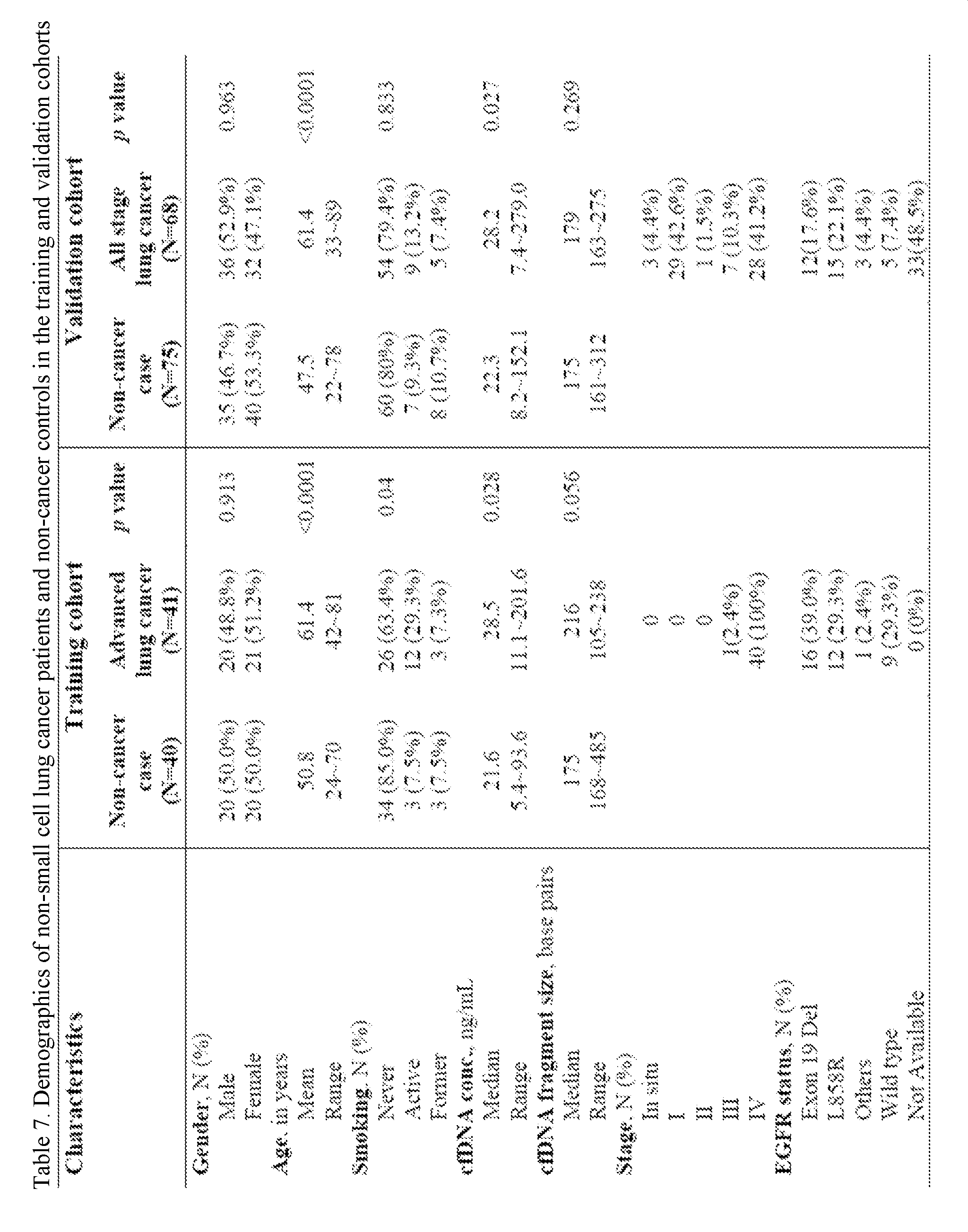
- Table 7 shows the demographics of non-small cell lung cancer patients and non-cancer controls in the training and validation cohorts.
- a methylation risk scoring system was developed by constructing a logistic regression model to assign appropriate weights to individual markers.
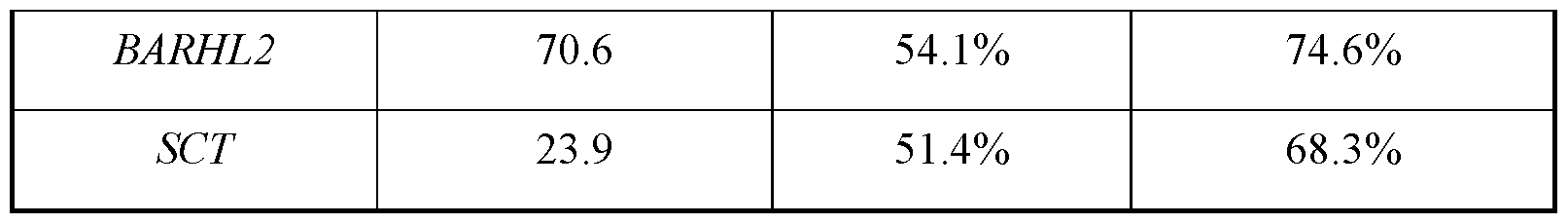
- the methylation risk score was calculated as -1.547+0.024*SCT+0.084*HOXA9-0.039*BARHL2+0.031*KCNS2.
- the methylation score achieved a sensitivity of 90.2% and a specificity of 70.0% for lung cancer diagnosis with a positive predictive value of 75.5% and a negative predictive value of 87.5%.
- the numbers outperformed the similar measures calculated using other cutoff values shown in Table 8.
- methylation scoring system was derived from only patients with advanced diseases, this system was applied in an independent validation cohort of 68 lung cancer patients at all stages versus 75 non-cancer controls for potentially broader applicability, especially for early-stage lung cancer since few tests were available for patients in this category, as shown in Table 7.
- the assay achieved a sensitivity of 82.8% and 92.6% for patients with stage I and IV diseases, respectively, as shown in FIG.13.
- carcinoembryonic antigen CEA
- CEA carcinoembryonic antigen
- the data indicate that methylation scores correlate with disease burden.
- FIG.14A shows a total of 261 serial blood samples from 57 lung cancer patients were collected at initial presentations and follow-up visits.
- FIG.14B shows the methylation scores were initially decreased but elevated when patients experienced disease progression (PD).
- the generalized estimating equations (GEE) approach that adjusts for repeated measurements from each visit was applied.
- GEE generalized estimating equations
- FIG. 14C a trend of a higher estimated marginal mean was found in the PD group (5.5 vs. 2.9, marginal mean difference, 2.63; 95% C.I., -0.6-5.9).
- Table 11 shows that the GEE model has an odds ratio of 0.251 (95% C.I.
- Patient L14 was a 56-year-old patient presenting with stage 4 lung adenocarcinoma negative for major driver mutations.
- the methylation scores did not show significant changes under carboplatin and pemetrexed treatment for about six months, which was correlated with stable disease. At approximately nine months, the methylation score skyrocketed, and clinical progression was evident.
- Patient L12 was a 72-year-old patient presenting with EGFR mutated lung adenocarcinoma receiving afatinib treatment. The patient responded to the treatment initially and the methylation scores decreased. However, the score increased again at approximately 270 days despite the shrinkage of the primary lung tumor on chest tomography.
Landscapes
- Chemical & Material Sciences (AREA)
- Life Sciences & Earth Sciences (AREA)
- Health & Medical Sciences (AREA)
- Organic Chemistry (AREA)
- Proteomics, Peptides & Aminoacids (AREA)
- Engineering & Computer Science (AREA)
- Immunology (AREA)
- Pathology (AREA)
- Analytical Chemistry (AREA)
- Zoology (AREA)
- Genetics & Genomics (AREA)
- Wood Science & Technology (AREA)
- Physics & Mathematics (AREA)
- Biotechnology (AREA)
- Microbiology (AREA)
- Molecular Biology (AREA)
- Hospice & Palliative Care (AREA)
- Biophysics (AREA)
- Oncology (AREA)
- Biochemistry (AREA)
- Bioinformatics & Cheminformatics (AREA)
- General Engineering & Computer Science (AREA)
- General Health & Medical Sciences (AREA)
- Measuring Or Testing Involving Enzymes Or Micro-Organisms (AREA)
- Investigating Or Analysing Biological Materials (AREA)
Abstract
Description


Claims
Priority Applications (3)
| Application Number | Priority Date | Filing Date | Title |
|---|---|---|---|
| JP2023553089A JP7663987B2 (en) | 2021-03-01 | 2022-03-01 | Method and kit for monitoring non-small cell lung cancer |
| US18/279,824 US20240068043A1 (en) | 2021-03-01 | 2022-03-01 | Method and kit for monitoring non-small cell lung cancer |
| EP22763898.8A EP4301877A4 (en) | 2021-03-01 | 2022-03-01 | Method and kit for monitoring non-small cell lung cancer |
Applications Claiming Priority (2)
| Application Number | Priority Date | Filing Date | Title |
|---|---|---|---|
| US202163154837P | 2021-03-01 | 2021-03-01 | |
| US63/154,837 | 2021-03-01 |
Publications (1)
| Publication Number | Publication Date |
|---|---|
| WO2022187246A1 true WO2022187246A1 (en) | 2022-09-09 |
Family
ID=83154802
Family Applications (1)
| Application Number | Title | Priority Date | Filing Date |
|---|---|---|---|
| PCT/US2022/018340 Ceased WO2022187246A1 (en) | 2021-03-01 | 2022-03-01 | Method and kit for monitoring non-small cell lung cancer |
Country Status (5)
| Country | Link |
|---|---|
| US (1) | US20240068043A1 (en) |
| EP (1) | EP4301877A4 (en) |
| JP (1) | JP7663987B2 (en) |
| TW (1) | TWI891981B (en) |
| WO (1) | WO2022187246A1 (en) |
Citations (3)
| Publication number | Priority date | Publication date | Assignee | Title |
|---|---|---|---|---|
| WO2014173905A2 (en) * | 2013-04-23 | 2014-10-30 | Institut D'investigació Biomèdica De Bellvitge (Idibell) | Methods and kits for prognosis of stage i nsclc by determining the methylation pattern of cpg dinucleotides |
| WO2019242753A1 (en) * | 2018-06-22 | 2019-12-26 | 深圳市圣必智科技开发有限公司 | Primer pair for detecting non-small cell lung cancer multiplex gene methylation, and reagent kit |
| CN110628900A (en) * | 2018-06-22 | 2019-12-31 | 深圳市圣必智科技开发有限公司 | Method for detecting methylation of multiple genes of non-small cell lung cancer |
Family Cites Families (5)
| Publication number | Priority date | Publication date | Assignee | Title |
|---|---|---|---|---|
| US8399193B2 (en) | 2007-08-30 | 2013-03-19 | City Of Hope | DNA methylation biomarkers for lung cancer |
| EP3795696B1 (en) | 2013-03-15 | 2023-04-26 | The Board of Trustees of the Leland Stanford Junior University | Identification and use of circulating nucleic acid tumor markers |
| US20200299779A1 (en) | 2019-02-13 | 2020-09-24 | Epigenomics Ag | Methods for detecting head and neck cancer |
| KR102234382B1 (en) | 2020-09-09 | 2021-03-30 | 강원대학교병원 | Non-small cell lung cancer progression stage detection method by differential methylation signal association analysis |
| CN112195245B (en) | 2020-10-16 | 2024-06-11 | 山东康华生物医疗科技股份有限公司 | Methylation gene combination related to lung cancer in blood plasma and application thereof |
-
2022
- 2022-03-01 WO PCT/US2022/018340 patent/WO2022187246A1/en not_active Ceased
- 2022-03-01 EP EP22763898.8A patent/EP4301877A4/en active Pending
- 2022-03-01 TW TW111107435A patent/TWI891981B/en active
- 2022-03-01 US US18/279,824 patent/US20240068043A1/en active Pending
- 2022-03-01 JP JP2023553089A patent/JP7663987B2/en active Active
Patent Citations (3)
| Publication number | Priority date | Publication date | Assignee | Title |
|---|---|---|---|---|
| WO2014173905A2 (en) * | 2013-04-23 | 2014-10-30 | Institut D'investigació Biomèdica De Bellvitge (Idibell) | Methods and kits for prognosis of stage i nsclc by determining the methylation pattern of cpg dinucleotides |
| WO2019242753A1 (en) * | 2018-06-22 | 2019-12-26 | 深圳市圣必智科技开发有限公司 | Primer pair for detecting non-small cell lung cancer multiplex gene methylation, and reagent kit |
| CN110628900A (en) * | 2018-06-22 | 2019-12-31 | 深圳市圣必智科技开发有限公司 | Method for detecting methylation of multiple genes of non-small cell lung cancer |
Non-Patent Citations (3)
| Title |
|---|
| LIU BIN, RICARTE FILHO JULIO, MALLISETTY APURVA, VILLANI CASSANDRA, KOTTOROU ANASTASIA, RODGERS KRISTEN, CHEN CHEN, ITO TOMOAKI, H: "Detection of Promoter DNA Methylation in Urine and Plasma Aids the Detection of Non–Small Cell Lung Cancer", CLINICAL CANCER RESEARCH, ASSOCIATION FOR CANCER RESEARCH, US, vol. 26, no. 16, 15 August 2020 (2020-08-15), US, pages 4339 - 4348, XP055963464, ISSN: 1078-0432, DOI: 10.1158/1078-0432.CCR-19-2896 * |
| See also references of EP4301877A1 * |
| WANG YIFAN, WANG YING, ZHANG YONGJUN: "Identification of prognostic signature of non–small cell lung cancer based on TCGA methylation data", SCIENTIFIC REPORTS, vol. 10, no. 1, 1 December 2020 (2020-12-01), XP055963466, DOI: 10.1038/s41598-020-65479-y * |
Also Published As
| Publication number | Publication date |
|---|---|
| TWI891981B (en) | 2025-08-01 |
| EP4301877A4 (en) | 2025-04-09 |
| TW202242147A (en) | 2022-11-01 |
| US20240068043A1 (en) | 2024-02-29 |
| JP7663987B2 (en) | 2025-04-17 |
| EP4301877A1 (en) | 2024-01-10 |
| JP2024508300A (en) | 2024-02-26 |
Similar Documents
| Publication | Publication Date | Title |
|---|---|---|
| JP7531217B2 (en) | Cell-free DNA for assessing and/or treating cancer - Patents.com | |
| Kneip et al. | SHOX2 DNA methylation is a biomarker for the diagnosis of lung cancer in plasma | |
| Esposito et al. | Monitoring tumor-derived cell-free DNA in patients with solid tumors: clinical perspectives and research opportunities | |
| Catarino et al. | Circulating DNA: diagnostic tool and predictive marker for overall survival of NSCLC patients | |
| De Rubis et al. | Circulating tumor DNA–Current state of play and future perspectives | |
| Stasik et al. | Evaluation of TERT promoter mutations in urinary cell-free DNA and sediment DNA for detection of bladder cancer | |
| CN105986034A (en) | Application of group of gastric cancer genes | |
| EP2831270B1 (en) | Biomarker for bladder cancer | |
| WO2016115354A1 (en) | Methods for cancer diagnosis and prognosis | |
| US20220307091A1 (en) | Unbiased dna methylation markers define an extensive field defect in histologically normal prostate tissues associated with prostate cancer: new biomarkers for men with prostate cancer | |
| JP2024001068A (en) | DNA methylation markers and their use for non-invasive detection of cancer | |
| JP2020519296A (en) | DNA methylation and mutation analysis method for bladder cancer monitoring | |
| CN107532208B (en) | Compositions and methods for determining prognosis of endometrial cancer | |
| ES3036448T3 (en) | A dna-methylation test for prostate cancer | |
| Varkalaite et al. | Liquid biopsy in gastric cancer: analysis of somatic cancer tissue mutations in plasma cell-free DNA for predicting disease state and patient survival | |
| Liu et al. | Protocadherin γ-A7 is down-regulated in colorectal cancer and associated with the prognosis in patients with wild-type KRAS | |
| US12071672B2 (en) | Unbiased DNA methylation markers define an extensive field defect in histologically normal prostate tissues associated with prostate cancer: new biomarkers for men with prostate cancer | |
| EP3368684B1 (en) | Biomarker for breast cancer | |
| US20230257824A1 (en) | Biomarkers for diagnosing and monitoring lung cancer | |
| US20240068043A1 (en) | Method and kit for monitoring non-small cell lung cancer | |
| EP4282984A1 (en) | Method for construction of multi-feature prediction model for cancer diagnosis | |
| EP2978861A2 (en) | Unbiased dna methylation markers define an extensive field defect in histologically normal prostate tissues associated with prostate cancer: new biomarkers for men with prostate cancer | |
| KR102914576B1 (en) | DNA methylation markers and their uses for noninvasive detection of cancer | |
| Zavarykina et al. | Circulating Tumor DNA And Its Potential Applications for Assessing Effectiveness of Neoadjuvant Drug Therapy in the Breast Cancer Patients | |
| Dumas et al. | The epiMelanoma test enables plasma-based detection of melanoma and prediction of immunotherapy response |
Legal Events
| Date | Code | Title | Description |
|---|---|---|---|
| 121 | Ep: the epo has been informed by wipo that ep was designated in this application |
Ref document number: 22763898 Country of ref document: EP Kind code of ref document: A1 |
|
| WWE | Wipo information: entry into national phase |
Ref document number: 2023553089 Country of ref document: JP Ref document number: 18279824 Country of ref document: US |
|
| WWE | Wipo information: entry into national phase |
Ref document number: 2022763898 Country of ref document: EP |
|
| NENP | Non-entry into the national phase |
Ref country code: DE |
|
| ENP | Entry into the national phase |
Ref document number: 2022763898 Country of ref document: EP Effective date: 20231002 |