WO2021137230A1 - Methods of culturing t cells and uses of same - Google Patents
Methods of culturing t cells and uses of same Download PDFInfo
- Publication number
- WO2021137230A1 WO2021137230A1 PCT/IL2020/051356 IL2020051356W WO2021137230A1 WO 2021137230 A1 WO2021137230 A1 WO 2021137230A1 IL 2020051356 W IL2020051356 W IL 2020051356W WO 2021137230 A1 WO2021137230 A1 WO 2021137230A1
- Authority
- WO
- WIPO (PCT)
- Prior art keywords
- cells
- specific embodiments
- amino acid
- cell
- cancer
- Prior art date
- Legal status (The legal status is an assumption and is not a legal conclusion. Google has not performed a legal analysis and makes no representation as to the accuracy of the status listed.)
- Ceased
Links
Classifications
-
- C—CHEMISTRY; METALLURGY
- C12—BIOCHEMISTRY; BEER; SPIRITS; WINE; VINEGAR; MICROBIOLOGY; ENZYMOLOGY; MUTATION OR GENETIC ENGINEERING
- C12N—MICROORGANISMS OR ENZYMES; COMPOSITIONS THEREOF; PROPAGATING, PRESERVING, OR MAINTAINING MICROORGANISMS; MUTATION OR GENETIC ENGINEERING; CULTURE MEDIA
- C12N5/00—Undifferentiated human, animal or plant cells, e.g. cell lines; Tissues; Cultivation or maintenance thereof; Culture media therefor
- C12N5/06—Animal cells or tissues; Human cells or tissues
- C12N5/0602—Vertebrate cells
- C12N5/0634—Cells from the blood or the immune system
- C12N5/0636—T lymphocytes
-
- A—HUMAN NECESSITIES
- A61—MEDICAL OR VETERINARY SCIENCE; HYGIENE
- A61K—PREPARATIONS FOR MEDICAL, DENTAL OR TOILETRY PURPOSES
- A61K40/00—Cellular immunotherapy
- A61K40/10—Cellular immunotherapy characterised by the cell type used
- A61K40/11—T-cells, e.g. tumour infiltrating lymphocytes [TIL] or regulatory T [Treg] cells; Lymphokine-activated killer [LAK] cells
-
- A—HUMAN NECESSITIES
- A61—MEDICAL OR VETERINARY SCIENCE; HYGIENE
- A61K—PREPARATIONS FOR MEDICAL, DENTAL OR TOILETRY PURPOSES
- A61K40/00—Cellular immunotherapy
- A61K40/40—Cellular immunotherapy characterised by antigens that are targeted or presented by cells of the immune system
- A61K40/41—Vertebrate antigens
- A61K40/42—Cancer antigens
-
- A—HUMAN NECESSITIES
- A61—MEDICAL OR VETERINARY SCIENCE; HYGIENE
- A61P—SPECIFIC THERAPEUTIC ACTIVITY OF CHEMICAL COMPOUNDS OR MEDICINAL PREPARATIONS
- A61P1/00—Drugs for disorders of the alimentary tract or the digestive system
- A61P1/18—Drugs for disorders of the alimentary tract or the digestive system for pancreatic disorders, e.g. pancreatic enzymes
-
- A—HUMAN NECESSITIES
- A61—MEDICAL OR VETERINARY SCIENCE; HYGIENE
- A61P—SPECIFIC THERAPEUTIC ACTIVITY OF CHEMICAL COMPOUNDS OR MEDICINAL PREPARATIONS
- A61P35/00—Antineoplastic agents
-
- C—CHEMISTRY; METALLURGY
- C12—BIOCHEMISTRY; BEER; SPIRITS; WINE; VINEGAR; MICROBIOLOGY; ENZYMOLOGY; MUTATION OR GENETIC ENGINEERING
- C12N—MICROORGANISMS OR ENZYMES; COMPOSITIONS THEREOF; PROPAGATING, PRESERVING, OR MAINTAINING MICROORGANISMS; MUTATION OR GENETIC ENGINEERING; CULTURE MEDIA
- C12N5/00—Undifferentiated human, animal or plant cells, e.g. cell lines; Tissues; Cultivation or maintenance thereof; Culture media therefor
- C12N5/06—Animal cells or tissues; Human cells or tissues
- C12N5/0602—Vertebrate cells
- C12N5/0634—Cells from the blood or the immune system
- C12N5/0636—T lymphocytes
- C12N5/0638—Cytotoxic T lymphocytes [CTL] or lymphokine activated killer cells [LAK]
-
- C—CHEMISTRY; METALLURGY
- C12—BIOCHEMISTRY; BEER; SPIRITS; WINE; VINEGAR; MICROBIOLOGY; ENZYMOLOGY; MUTATION OR GENETIC ENGINEERING
- C12N—MICROORGANISMS OR ENZYMES; COMPOSITIONS THEREOF; PROPAGATING, PRESERVING, OR MAINTAINING MICROORGANISMS; MUTATION OR GENETIC ENGINEERING; CULTURE MEDIA
- C12N2501/00—Active agents used in cell culture processes, e.g. differentation
- C12N2501/20—Cytokines; Chemokines
- C12N2501/23—Interleukins [IL]
- C12N2501/2302—Interleukin-2 (IL-2)
-
- C—CHEMISTRY; METALLURGY
- C12—BIOCHEMISTRY; BEER; SPIRITS; WINE; VINEGAR; MICROBIOLOGY; ENZYMOLOGY; MUTATION OR GENETIC ENGINEERING
- C12N—MICROORGANISMS OR ENZYMES; COMPOSITIONS THEREOF; PROPAGATING, PRESERVING, OR MAINTAINING MICROORGANISMS; MUTATION OR GENETIC ENGINEERING; CULTURE MEDIA
- C12N2501/00—Active agents used in cell culture processes, e.g. differentation
- C12N2501/20—Cytokines; Chemokines
- C12N2501/23—Interleukins [IL]
- C12N2501/2315—Interleukin-15 (IL-15)
-
- C—CHEMISTRY; METALLURGY
- C12—BIOCHEMISTRY; BEER; SPIRITS; WINE; VINEGAR; MICROBIOLOGY; ENZYMOLOGY; MUTATION OR GENETIC ENGINEERING
- C12N—MICROORGANISMS OR ENZYMES; COMPOSITIONS THEREOF; PROPAGATING, PRESERVING, OR MAINTAINING MICROORGANISMS; MUTATION OR GENETIC ENGINEERING; CULTURE MEDIA
- C12N2501/00—Active agents used in cell culture processes, e.g. differentation
- C12N2501/20—Cytokines; Chemokines
- C12N2501/23—Interleukins [IL]
- C12N2501/2321—Interleukin-21 (IL-21)
-
- C—CHEMISTRY; METALLURGY
- C12—BIOCHEMISTRY; BEER; SPIRITS; WINE; VINEGAR; MICROBIOLOGY; ENZYMOLOGY; MUTATION OR GENETIC ENGINEERING
- C12N—MICROORGANISMS OR ENZYMES; COMPOSITIONS THEREOF; PROPAGATING, PRESERVING, OR MAINTAINING MICROORGANISMS; MUTATION OR GENETIC ENGINEERING; CULTURE MEDIA
- C12N2501/00—Active agents used in cell culture processes, e.g. differentation
- C12N2501/998—Proteins not provided for elsewhere
Definitions
- the present invention in some embodiments thereof, relates to methods of culturing T cells and uses of same.
- T cell-based adoptive immune therapies involving in-vitro activation and expansion of T cells hold promise for the treatment of various diseases.
- adoptive transfer of antigen- specific CD8 + T cells can be used for the treatment of malignancies and infections, while specific regulatory T cells (Tregs) can be harnessed for suppression of autoimmune processes.
- Tregs specific regulatory T cells
- a major challenge for T cell based immune therapies is the necessity to identify T cells specifically reactive against cells associated with the disease of interest.
- 4-1BB (CD 137) is a member of the TNF receptor super family that functions as a costimulatory molecule promoting proliferation and survival of activated T cells. 4-1BB activation upregulates survival genes, enhances cell division, induces cytokine production and prevents activation induced cell death in T-cells.
- T cells that have recently been activated by T cell receptor (TCR) engagement and signaling. Consequently, upregulation of CD137 on recently activated T cells has been used to identify and isolate virus- and tumor-reactive T cells (e.g. Qunrui Ye et al. (2014) Clin Cancer Res. 2014 January 1; 20(1): 44-55; Sivan Seliktar- Ofir (2017) Front. Immunol. 8: 1211; Wolf et al. (2007) Blood. 110: 201-210; Parkhurst et al. (2017) Clin Cancer Res. 23(10): 2491-25; Tan et al., (2019) Journal for ImmunoTherapy of Cancer, volume 7, Article number: 232; and EP Patent Application Publication No. EP3118322).
- TCR T cell receptor
- 4-1BBL is the activating ligand of 4- IBB. 4-1BBL naturally forms a homo-trimer but signaling via 4-1BB requires significant oligomerization of 4-1BBL. 4-1BBL is present on a variety of antigen presenting cells (APCs), including dendritic cells (DCs), B cells, and macrophages.
- APCs antigen presenting cells
- DCs dendritic cells
- B cells B cells
- macrophages macrophages.
- Activating T cells in the presence of agonistic 4-1BB binding agents such as antibodies and 4-1BBL polypeptides has been suggested in the art (e.g. International Patent Applications Publication Nos. W02003049755; WO2018/127917; and WO2018/127919).
- a method of culturing T cells comprising:
- the culturing is effected for more than 10 days.
- the culturing is effected for about 14 days.
- the method being effected without isolation of 4-1BB positive cells prior to the (a) and/or the (b).
- the method being effected without isolation of 4-1BB positive cells during and/or following the (b).
- the method effected without isolation of the T cells from the immune cells prior to the (a) and/or the (b).
- the method comprising adding at least one cytokine to the immune cells in step (a) and culturing the immune cells with the cytokine in step (b).
- the method further comprising pre-culturing the immune cells with at least one cytokine prior to the (a).
- the pre-culturing is effected until the percentage of the T cells in the immune cells is at least 80 %.
- the pre-culturing is effected for 2-14 days.
- the pre-culturing is effected for 7-14 days.
- the method being effected without isolation of the T cells from the immune cells prior to the pre-culturing.
- the method being effected without adding to the immune cells a T cell stimulatory agent capable of at least transmitting a primary activating signal to the T cells.
- the culturing is effected under conditions which allow enrichment of T cell clones reactive to cells associated with the pathology.
- the immune cells are obtained from a blood sample of the subject.
- the immune cells comprise PBMCs.
- the immune cells are obtained from a tissue comprising cells associated with the pathology.
- the at least one cytokine is selected from the group consisting of IL-2, IL-15, IL-21, IL-12 and IL-7. According to some embodiments of the invention, the at least one cytokine comprises IL-2,
- the non-cellular agent is capable of binding a ligand or receptor of a type I membrane protein.
- the (a) further comprises adding a non- cellular agent capable of binding a ligand or receptor of a type I membrane protein.
- the type I membrane protein is PD1.
- the type I membrane protein is SIRPa.
- the non-cellular agent is soluble.
- the non-cellular agent is immobilized. According to some embodiments of the invention, the non-cellular agent is an antibody.
- the non-cellular agent capable of binding 4- 1BB and activating the 4- IBB signaling pathway is a polypeptide comprising an amino acid sequence of 4-1BBL.
- the non-cellular agent is a fusion polypeptide.
- the fusion polypeptide comprises an amino acid sequence of 4-1BBL and an amino acid sequence of PD1.
- the fusion polypeptide comprises an amino acid sequence having at least 80 % identity to SEQ ID NO: 49. According to some embodiments of the invention, the fusion polypeptide comprises SEQ ID NO: 49 or as set forth in SEQ ID NO: 49.
- the fusion polypeptide comprises an amino acid sequence having at least 80 % identity to SEQ ID NO: 49 or 74.
- the fusion polypeptide comprises SEQ ID NO: 49 or 74 or as set forth in SEQ ID NO: 49 or 74.
- the fusion polypeptide comprises an amino acid sequence of 4-1BBL and an amino acid sequence of SIRPa.
- the fusion polypeptide comprises an amino acid sequence having at least 80 % identity to SEQ ID NO: 89.
- the fusion polypeptide comprises SEQ ID NO: 89 or as set forth in SEQ ID NO: 89.
- the method further comprising expanding the T cells following the (b).
- the method further comprising isolating the T cells from the immune cells following the (b) and/or the expanding.
- the method further comprising determining a sequence of a T cell receptor (TCR) expressed by at least one of the T cells following the (b), the expanding or the isolating.
- TCR T cell receptor
- the method further comprising transducing a T cell with a nucleic acid sequence encoding the TCR.
- the method further comprising transducing the T cells with a nucleic acid sequence encoding a chimeric antigen receptor (CAR) following the (b), the expanding or the isolating.
- CAR chimeric antigen receptor
- the method comprising adoptively transferring the immune cells and/or the T cells following the (b), the expanding or the transducing to a subject in need thereof.
- T cells obtainable by the method.
- the T cells for use in adoptive cell therapy to a subject in need thereof.
- a method of treating a pathology that can benefit from adoptive T cell therapy in a subject in need thereof comprising administering to the subject a therapeutically effective amount of the T cells of claim 36, thereby treating the pathology.
- the immune cells and/or the T cells are autologous to the subject in need thereof.
- the pathology is selected from the group consisting of a hyper-proliferative disease, a disease associated with immune suppression and infection.
- the pathology is cancer
- the cancer is selected from the group consisting of gastrointestinal (GI) cancer, breast cancer, ovarian cancer and pancreatic cancer.
- GI gastrointestinal
- breast cancer breast cancer
- ovarian cancer pancreatic cancer
- the cancer is selected from the group consisting of pancreatic cancer, lung cancer, colon cancer and leukemia.
- the subject is human.
- FIG. 1 shows dot-plot FACS analysis demonstrating percentages of CD8 and CD4 T cells following initial 10 days culturing period of PBMCs obtained from four pancreatic cancer patients in a media containing hIL2, hIL-15 and hIL-21.
- FIG. 2 demonstrates IFNy levels in the culture media during a second 14 days culturing period of PBMCs obtained from two pancreatic cancer patients in the presence of PD1-4-1BBL fusion protein (marked as DSP 105, SEQ ID N049), as compared to cell cultured under the same conditions without the fusion protein.
- FIG. 3 demonstrates the percentage of three subpopulations of memory T cells from the total PBMC population following the second 14 days culturing period of PBMCs obtained from a pancreatic cancer patient in the presence of PD1-4-1BBL fusion protein (marked as DSP105, SEQ ID NO: 49), or with organoids as compared to control cells cultured under the same conditions without the fusion protein or the organoid.
- PD1-4-1BBL fusion protein marked as DSP105, SEQ ID NO: 49
- FIG. 4 shows representative microscope images demonstrating the effect of a PD1-4-1BBL fusion protein (marked as DSP105-G3, SEQ ID NO: 49, or DSP105-V10, SEQ ID NO: 74) on expansion of T-cell clusters obtained from a non-small cell lung cancer (NSCLC) patient (OL#l, Group 1, day 8 following treatment).
- NSCLC non-small cell lung cancer
- the present invention in some embodiments thereof, relates to methods of culturing T cells and uses of same.
- T cell-based adoptive immune therapies involving in-vitro activation and expansion of T cells, can be used for the treatment of various diseases including malignancies, infections and autoimmune diseases.
- a major challenge for T cell based immune therapies is the necessity to identify T cells specifically reactive against cells associated with the disease of interest.
- the present inventors Whilst reducing specific embodiments of the present invention to practice, the present inventors have now devised an ex-vivo environment for the clonal selection of cancer-specific T cells having a memory phenotype by culturing the T cells in the presence of a PD1-4-1BBL fusion protein.
- a method of culturing T cells comprising:
- T cells refers to a differentiated lymphocyte with a CD3+, T cell receptor (TCR)+ having either CD4+ or CD8+ phenotype.
- TCR T cell receptor
- the T cell may be either an effector or a regulatory T cell.
- the T cells are effector T cells
- effector T cells refers to a T cell that activates or directs other immune cells e.g. by producing cytokines or has a cytotoxic activity e.g., CD4+, Thl/Th2, CD8+ cytotoxic T lymphocyte.
- the T cells are regulatory T cells.
- the term “regulatory T cell” or “Treg” refers to a T cell that negatively regulates the activation of other T cells, including effector T cells, as well as innate immune system cells. Treg cells are characterized by sustained suppression of effector T cell responses. According to a specific embodiment, the Treg is a CD4+CD25+Foxp3+ T cell.
- the T cells are CD4+ T cells.
- the T cells are CD8+ T cells.
- the T cells comprise memory T cells.
- memory T cells include effector memory CD4+ T cells with a CD3+/CD4+/CD45RA- /CCR7- phenotype, central memory CD4+ T cells with a CD3+/CD4+/CD45RA-/CCR7+ phenotype, effector memory CD8+ T cells with a CD3+/CD8+ CD45RA-/CCR7-phenotype, central memory CD8+ T cells with a CD3+/CD8+ CD45RA-/CCR7+ phenotype, tissue-resident memory T cells with a CD3+/CD45RO+/CD69+/CD103+ or a CD3+/CD45RO+/CD69+/CD103- phenotype, or effector memory T cells with CD3+/CD45RO+/CD69-/CCR7- phenotype.
- the T cells comprise activated cells.
- activated T cells include effector activated CD4+ cells and, effector activated CD8+ T cells with a tissue resident memory or effector memory phenotype.
- the T cells comprise cells expressing 4-1BB.
- the immune cells comprise at least 70 %, at least 75 %, at least 80 %, at least 85 %, at least 90 %, or at least 95 % T cells.
- the immune cells comprise 45 - 70 % T cells.
- the immune cells comprise at least 70 % T cells.
- the immune cells comprise at least 80 % T cells.
- the immune cells comprise cells other than the T cells.
- the immune cells comprise phagocytic cells.
- phagocytic cells refer to a cell that is capable of phagocytosis and include both professional and non-professional phagocytic cells. Methods of analyzing phagocytosis are well known in the art and include for examples killing assays, flow cytometry and/or microscopic evaluation (live cell imaging, fluorescence microscopy, confocal microscopy, electron microscopy).
- the phagocytic cells are selected from the group consisting of monocytes, dendritic cells (DCs) and granulocytes.
- DCs dendritic cells
- the immune cells comprise monocytes.
- monocytes refers to both circulating monocytes and to macrophages (also referred to as mononuclear phagocytes) present in a tissue.
- the monocytes comprise macrophages.
- cell surface phenotype of macrophages include CD14, CD40, CDl lb, CD64, F4/80 (mice)/EMRl (human), lysozyme M, MAC-l/MAC-3 and CD68.
- the monocytes comprise circulating monocytes.
- cell surface phenotypes of circulating monocytes include CD 14 and CD 16 (e.g. CD14++ CD 16-, CD14+CD16++, CD14++CD16+).
- the immune cells do not comprise monocytes.
- the immune cells comprise DCs.
- DCs dendritic cells
- DCs refers to any member of a diverse population of morphologically similar cell types found in lymphoid or non-lymphoid tissues.
- DCs are a class of professional antigen presenting cells, and have a high capacity for sensitizing HLA-restricted T cells.
- DCs include, for example, plasmacytoid dendritic cells, myeloid dendritic cells (including immature and mature dendritic cells), Langerhans cells, interdigitating cells, follicular dendritic cells.
- Dendritic cells may be recognized by function, or by phenotype, particularly by cell surface phenotype.
- cell surface phenotype of DCs include CDla+, CD4+, CD86+, or HLA-DR.
- the term DCs encompasses both immature and mature DCs.
- the immune cells do not comprise DCs.
- the immune cells comprise granulocytes.
- granulocytes refer to polymorphonuclear leukocytes characterized by the presence of granules in their cytoplasm.
- the granulocytes comprise neutrophils.
- the granulocytes comprise mast-cells.
- the immune cells comprise B cells.
- B cells refers to a lymphocyte with a B cell receptor (BCR)+, CD 19+ and or B220+ phenotype. B cells are characterized by their ability to bind a specific antigen and elicit a humoral response.
- BCR B cell receptor
- the immune cells comprise NK cells.
- NK cells refers to differentiated lymphocytes with a CD 16+ CD56+ and/or CD57+ TCR- phenotype. NK are characterized by their ability to bind to and kill cells that fail to express “self’ MHC/HLA antigens by the activation of specific cytolytic enzymes, the ability to kill tumor cells or other diseased cells that express a ligand for NK activating receptors, and the ability to release protein molecules called cytokines that stimulate or inhibit the immune response.
- the immune cells comprise NKT cells.
- NKT cells refers to a specialized population of T cells that express a semi-invariant ab T-cell receptor, but also express a variety of molecular markers that are typically associated with NK cells, such as NK1.1.
- NKT cells include NK1.1+ and NK1.1-, as well as CD4+, CD4-, CD8+ and CD8- cells.
- the TCR on NKT cells is unique in that it recognizes glycolipid antigens presented by the MHC I-like molecule CD Id. NKT cells can have either protective or deleterious effects due to their abilities to produce cytokines that promote either inflammation or immune tolerance.
- the immune cells comprise antigen presenting cells (APCs).
- APCs antigen presenting cells
- the term “antigen presenting cell (APC)” refers to an immune cell capable of internalizing and processing an antigen, so that antigenic determinants are presented on the surface of the cell as MHC-associated complexes, in a manner capable of being recognized by T cells.
- APC include dendritic cells, mononuclear cells (e.g. macrophages) and B cells.
- the immune cells comprise less than 30 %, less than 20 %, less than 10 %, less than 5 % APCs.
- the immune cells do not comprise APCs.
- the immune cells are obtained from a blood sample of the subject.
- the immune cells comprise peripheral mononuclear blood cells (PBMCs).
- PBMCs peripheral mononuclear blood cells
- DCs dendritic cells
- the PBMCs are selected from the group consisting of dendritic cells (DCs), T cells, B cells, NK cells and NKT cells.
- DCs dendritic cells
- T cells T cells
- B cells B cells
- NK cells NKT cells
- the PBMCs comprise T cells, B cells, NK cells and NKT cells.
- PBMCs Methods of obtaining PBMCs are well known in the art, such as drawing whole blood from a subject and collection in a container containing an anti-coagulant (e.g. heparin or citrate); and apheresis. Following, the PBMCs are purified from the peripheral blood.
- an anti-coagulant e.g. heparin or citrate
- apheresis e.g. heparin or citrate
- the PBMCs are purified from the peripheral blood.
- There are several methods and reagents known to those skilled in the art for purifying PBMCs from whole blood such as leukapheresis, sedimentation, density gradient centrifugation (e.g. ficoll), centrifugal elutriation, fractionation and chemical lysis of e.g. red blood cells (e.g. by ACK).
- the immune cells are obtained from a tissue comprising cells associated with the pathology.
- Methods for obtaining a tissue sample from a subject are well known in the art and include, but are not limited to biopsy.
- the immune cells comprise tumor infiltration immune cells”.
- tumor infiltrating immune cells refer to white blood cells that have left the bloodstream and migrated into a tumor, and include both mononuclear and polymorph nuclear immune cells.
- the tumor infiltrating immune cells are selected from the group consisting of T cells, B cells, NK cells, macrophages, neutrophils, dendritic cells, mast cells, eosinophils and basophils.
- the immune cells comprise tumor infiltrating lymphocytes.
- TILs tumor infiltrating lymphocytes
- the TILs are selected from the group consisting of T cells, B cells and NK cells.
- Methods of obtaining TILs are well known in the art, such as obtaining tumor samples from a subject by e.g. biopsy, surgery or necropsy and preparing a single cell suspension thereof.
- the single cell suspension can be obtained in any suitable manner, e.g., mechanically (disaggregating the tumor using, e.g., a gentleMACS(TM) Dissociator, Miltenyi Biotec, Auburn, Calif.) or enzymatically (e.g., collagenase or DNase).
- the TILs can be purified from the cell suspension by e.g. density gradient centrifugation (e.g. ficoll).
- specific immune cell types comprising T cells may be further isolated.
- the term “isolating” refers to an active process of purifying a specific immune cell type(s) (e.g. T cell, 4-lBB + cell) from other immune cells comprised in a given immune cell sample.
- the result of the “isolation” process is an “isolated” immune cell type(s).
- the term “isolating” does not refer to enrichment of a specific immune cell population(s) due to culture conditions, also known as biological selection. According to specific embodiments, isolating refers to immune-isolating.
- the method is effected on T cells isolated from immune cells obtained from the subject.
- the method comprises isolating the T cells from the immune cells obtained from the subject prior to adding the non-cellular agent capable of binding 4-1BB and activating 4-1BB signaling pathway and/or culturing in the presence of same.
- the method is effected on immune cells not subjected to T cell isolation prior to adding the non-cellular agent capable of binding 4-1BB and activating 4- IBB signaling pathway and/or culturing in the presence of same.
- the method is effected without isolation of T cells from the immune cells prior to adding the non-cellular agent capable of binding 4- IBB and activating 4- IBB signaling pathway.
- the method is effected without isolation of T cells from the immune cells prior to culturing in the presence of the non-cellular agent capable of binding 4- 1BB and activating 4- IBB signaling pathway.
- 4- IBB positive cells are not isolated from the immune cells at any stage of the methods disclosed herein.
- the method is effected on immune cells not subjected to isolation of 4- IBB positive cells prior to adding the non-cellular agent capable of binding 4-1BB and activating 4-1BB signaling pathway and/or culturing in the presence of same.
- the method is effected without isolation of 4- IBB positive cells from the immune cells prior to adding the non-cellular agent capable of binding 4- 1BB and activating 4- IBB signaling pathway.
- the method is effected without isolation of 4- IBB positive cells from the immune cells prior to culturing in the presence of the non-cellular agent capable of binding 4- IBB and activating 4- IBB signaling pathway.
- An alternative method to increase the percentage of specific immune cell types comprising T cells is to culture the immune cells under conditions that allow enrichment of the desired cell types.
- the method comprises pre-culturing the immune cells prior to addition of the non-cellular agent capable of binding 4-1BB and activating 4-1BB signaling pathway.
- the method is effected without isolation of T cells from the immune cells prior to the pre-culturing.
- the method is effected without isolation of 4- IBB positive cells from the immune cells prior to the pre-culturing.
- the pre-culturing is effected for up to three weeks, up to two weeks or up to 1 week.
- the pre-culturing is effected for 1 - 14, 2 - 14, 5 - 14, 7 - 14, or 8-12 days.
- the pre-culturing is effected for at least 2 days. According to specific embodiments, the pre-culturing is effected for 2 - 14, 2 - 13, 2 - 12, 2 -
- the pre-culturing is effected for 2 - 14 days.
- the pre-culturing is effected for at least 3 days.
- the pre-culturing is effected for 3 - 14, 3 - 13, 3 - 12, 3 -
- the pre-culturing is effected for 3 - 14 days.
- the pre-culturing is effected for 7 - 14 days.
- the pre-culturing is effected for 3 - 10 days.
- the pre-culturing is effected about 10 days.
- the pre-culturing is effected until enrichment of T cells in the immune cells is achieved.
- the pre-culturing is effected until the percentage of T cells in the immune cells is at least 70%, at least 80 %, at least 85 %, at least 90 %, at least 95 %.
- the pre-culturing is effected until the percentage of T cells in the immune cells is at least 80 %.
- culturing refers to at least immune cells comprising T cells and medium in an ex-vivo, in-vitro environment.
- the culture is maintained under conditions necessary to support growth and survival, for example, an appropriate temperature (e.g., 37 °C) atmosphere (e.g., air plus 5 % CO2) and medium.
- an appropriate temperature e.g., 37 °C
- atmosphere e.g., air plus 5 % CO2
- the culture may be in a glass, plastic or metal vessel that can provide an aseptic environment for cell culturing.
- the culture vessel includes dishes, plates, flasks, bottles, vials, bags, bioreactors or any device that can be used to grow cells.
- the medium used can be a water-based medium which includes a combination of substances such as salts, nutrients, minerals, vitamins, amino acids, nucleic acids and/or proteins such as cytokines, growth factors and hormones, all of which are needed for cell proliferation and survival.
- specific mediums include serum-free dendritic cell medium (can be obtained from examples from CellGernix); or RPMI or DMEM (can be obtained for example from Sigma- Aldrich or Biological Industries, Beit Haemek, Israel), supplemented with the necessary additives.
- the medium may be supplemented with L-glutamine, non-essential amino acids, sodium pyruvate, antibiotic/ antimycotic solution, 2-mercaptoethanol and serum.
- the medium is supplemented with a cytokine.
- the method comprises adding at least one cytokine to the immune cells.
- pre-culturing of the immune cells prior to addition of the non-cellular agent capable of binding 4- IBB and activating 4- IBB signaling pathway is effected in the presence of at least one exogenous cytokine.
- culturing of the immune cells with the non-cellular agent capable of binding 4-1BB and activating 4-1BB signaling pathway is effected in the presence of at least one exogenous cytokine.
- the cytokine is capable of inducing survival, activation and/or proliferation of a T cell.
- the cytokine preferably induces survival and growth of T cells thereby enriching the population of T cells in the immune cells obtained from the subject.
- Non-limiting examples of cytokines that can be used according to specific embodiments of the present invention include IL-2, IL-6, IL-4, IL-12, IL-7, IFNa, IL-12, IFN-gamma, TNF-a, IL- 15, IL-1, IL-21 and GM-CSF.
- the cytokine is selected from the group consisting of IL- 2, IL-15, IL-21, IL12 and IL-7.
- the cytokine is cytokine comprises IL-2, IL-15 and IL-21.
- all ingredients included in the culture medium of the present invention are substantially pure, with a tissue culture grade.
- the culture medium is devoid of any animal contaminants, i.e., animal cells, fluid or pathogens (e.g., viruses infecting animal cells), i.e., being xeno-free.
- animal contaminants i.e., animal cells, fluid or pathogens (e.g., viruses infecting animal cells), i.e., being xeno-free.
- culture medium may be periodically refreshed to maintain sufficient levels of supplements and to remove metabolic waste products that can damage the cells.
- the method comprises adding to the immune cells a non-cellular agent capable of binding 4-1BB and activating said 4-1BB signaling pathway and culturing the immune cells in the presence of same.
- “adding” refers to adding to the culture/composition a substance which was not present in the immune cells sample. Also referred to as an exogenous agent.
- an “agent capable of binding 4-1BB and activating said 4-1BB signaling pathway” refers to a non-cellular (i.e., cell-free) substance that can bind 4-1BB which is expressed on a T cell and thereby transmit a co-stimulatory signal resulting in activation of the T cell.
- the term “4- IBB (also known as CD 137 and TNFRSF9)” refers to the polypeptide of the TNFRSF9 gene (Gene ID 3604).
- the 4- 1BB protein refers to the human protein, such as provided in the following GenBank Number NP_001552.
- Assays for testing binding are well known in the art and include, but not limited to flow cytometry, Biacore, bio-layer interferometry Blitz® assay, HPLC.
- the agent binds 4-1BB with a Kd >10 6 M, >10 7 M, >10 8 M or >10 9 M, each possibility represents a separate embodiment of the present invention.
- the agent binds 4- IBB with a Kd of about 0.1 - 1000 nM, 0.1 - 100 nM or 1-100 nM, each possibility represents a separate embodiment of the claimed invention.
- co-stimulatory signal refers to transmission of a secondary antigen independent stimulatory signal (e.g. 4- IBB signal) resulting in activation of the immune cell (e.g. T cell expressing 4-1BB).
- a secondary antigen independent stimulatory signal e.g. 4- IBB signal
- the immune cell e.g. T cell expressing 4-1BB
- activating refers to the process of stimulating an immune cell (e.g. T cell) that results in cellular proliferation, maturation, cytokine production and/or induction of regulatory or effector functions.
- an immune cell e.g. T cell
- Methods of determining signaling of a 4- IBB pathway include, but are not limited to, binding assay using e.g. Biacore, HPLC or flow cytometry, enzymatic activity assays such as kinase activity assays, and expression of molecules involved in the signaling cascade using e.g. PCR, Western blot, immunoprecipitation and immunohistochemistry. Additionally or alternatively, determining transmission of a 4- IBB signal can be effected by evaluating immune cell activation or function.
- Methods of evaluating immune cell activation or function include, but are not limited to, proliferation assays such as CFSE staining, MTS, Alamar blue, BRDU and thymidine incorporation, cytotoxicity assays such as CFSE staining, chromium release, Calcin AM, cytokine (e.g. IL-8) secretion assays such as intracellular cytokine staining, ELISPOT and ELISA, expression of activation markers such as CD25, CD69, CD137, CD107a, PD1, and CD62L using flow cytometry.
- proliferation assays such as CFSE staining, MTS, Alamar blue, BRDU and thymidine incorporation
- cytotoxicity assays such as CFSE staining, chromium release, Calcin AM, cytokine (e.g. IL-8) secretion assays such as intracellular cytokine staining, ELISPOT and ELISA, expression
- determining the activation of 4-1BB signaling pathway is effected in-vitro or ex-vivo e.g. in a mixed lymphocyte reaction (MLR).
- MLR mixed lymphocyte reaction
- 4-1BB signaling pathway For the same culture conditions the activation of 4-1BB signaling pathway is generally expressed in comparison to the signaling in a cell of the same species but not contacted with the non-cellular agent; or contacted with a vehicle control, also referred to as control.
- a vehicle control also referred to as control.
- Non-limiting examples of non-cellular agents capable of binding 4-1BB and activating said 4-1BB signaling pathway are described in details hereinbelow.
- the non-cellular agent is also capable of binding a ligand or receptor of a type I membrane protein.
- the method further comprises adding to the immune cells a non- cellular agent capable of binding a ligand or receptor of a type I membrane protein and culturing the immune cells in the presence of same.
- type I membrane protein refers to a transmembrane protein having an N-terminus extracellular domain.
- Type I membrane proteins include PD1, SIRPa, LAG3, BTN3A1, CD27, CD80, CD86, ENG, NLGN4X, CD84, TIGIT, CD40, IL-8, IL-10, CD164, LY6G6F, CD28, CTLA4, BTLA, LILRB1/2/3/4, TYROBP, ICOS, VEGFA, CSF1, CSF1R, VEGFB, BMP2, BMP3, GDNF, PDGFC, PDGFD, RAETIE, CD155, CD166, MICA, NRGl, HVEM, DR3, TEK, TGFB1, LY96, CD96, KIT, CD244, and GFER.
- the Type I membrane protein is an immune modulator.
- immune modulator refers to a protein that modulates an immune cell response (i.e. activation or function). Immune modulators can positively regulate immune cell activation or function or negatively regulate immune cell activation or function. Such immune modulators are known in the art and include an immune-check point protein, a cytokine and the like.
- the immune modulator is an immune activator.
- the immune modulator is an immune suppressor or inhibitor.
- Type I membrane protein immune modulators include, but are not limited to PD1, SIRPa, CD28, CSF1R, IL-8, IL-10, CTLA4, ICOS, CD27, CD80, CD86, TIGIT and LILRBl/2/3/4.
- the type I membrane protein is selected from the group consisting of PD1 and SIRPa.
- the type I membrane protein is PD1.
- the type I membrane protein is SIRPa.
- Assays for testing binding are well known in the art and are further described hereinabove.
- the agent binds a ligand or receptor of a type I membrane protein with a Kd >10 6 M, >10 7 M, >10 8 M or >10 9 M, each possibility represents a separate embodiment of the present invention.
- the agent binds a ligand or receptor of a type I membrane protein with a Kd of about 1 nM - 100 mM, 10- nM - 10 mM, 100 nM - 100 pM, 200 nM - 10 pM, each possibility represents a separate embodiment of the claimed invention.
- the agent binds a ligand or receptor of a type I membrane protein with a Kd of about 0.1 - 100 pM, 0.1 - 10 pM, 1-10 pM, 0.1-5 pM, or 1-2 pM, each possibility represents a separate embodiment of the present invention.
- Non-limiting examples of non-cellular agents capable of binding a ligand or receptor of a type I membrane protein are described in details hereinbelow.
- the non-cellular agent is an antibody, a polypeptide or a small molecule.
- the non-cellular agent is an antibody.
- the antibody is capable of specifically binding 4-1BB.
- the antibody specifically binds at least one epitope of 4- 1BB.
- the antibody is capable of specifically binding a ligand or a receptor of a type I membrane protein (e.g. PDL1, CD47).
- a type I membrane protein e.g. PDL1, CD47.
- the antibody specifically binds at least one epitope of a ligand or a receptor of a type I membrane protein (e.g. PDL1, CD47).
- a type I membrane protein e.g. PDL1, CD47.
- epitopic determinants refers to any antigenic determinant on an antigen to which the paratope of an antibody binds.
- Epitopic determinants usually consist of chemically active surface groupings of molecules such as amino acids or carbohydrate side chains and usually have specific three dimensional structural characteristics, as well as specific charge characteristics.
- antibody as used in this invention includes intact molecules as well as functional fragments thereof, such as Fab, F(ab')2, Fv, scFv, dsFv, or single domain molecules such as VH and VL that are capable of binding to an epitope of an antigen.
- the antibody may be mono-specific (capable of recognizing one epitope or protein), bi- specific (capable of binding two epitopes or proteins) or multi-specific (capable of recognizing multiple epitopes or proteins).
- the antibody is a bi-specific antibody binding 4- 1BB and a ligand or a receptor of a type I membrane protein (e.g. a 4-1BB-PDL1 antibody or a 4- 1BB-CD47 antibody).
- a type I membrane protein e.g. a 4-1BB-PDL1 antibody or a 4- 1BB-CD47 antibody.
- Suitable antibody fragments for practicing some embodiments of the invention include a complementarity-determining region (CDR) of an immunoglobulin light chain (referred to herein as “light chain”), a complementarity-determining region of an immunoglobulin heavy chain (referred to herein as “heavy chain”), a variable region of a light chain, a variable region of a heavy chain, a light chain, a heavy chain, an Fd fragment, and antibody fragments comprising essentially whole variable regions of both light and heavy chains such as an Fv, a single chain Fv Fv (scFv), a disulfide-stabilized Fv (dsFv), a single domain antibody (nanobody), an Fab, an Fab’, and an F(ab’)2.
- CDR complementarity-determining region
- light chain referred to herein as “light chain”
- heavy chain a complementarity-determining region of an immunoglobulin heavy chain
- variable region of a light chain a variable region of a heavy chain
- the antibody may be monoclonal or polyclonal.
- Non-limiting examples of anti-4- IBB antibodies that can be used with specific embodiments include Urelumab (BMS-663513 ), Utomilumab (PF-05082566), GEN 1042 [BioNTech SE (Genmab)], LOAd703 (Lokon Pharma), ADG-106 (Adagene), PRS-343 (Pieris Pharmaceuticals), CTX-471 (Compass Therapeutics LLC), INBRX-105 (Elpiscience; Inhibrx), LVGN6051 (Lyvgen Biopharma Ltd), MCLA-145 (Merus NV), MP-0310 (Molecular Partners), ATOR-1017 (Alligator Bioscience AB), RG-6076 (Roche), RG-7827 (Roche), AGEN-2373 (Agenus) and the antibodies disclosed in Xinyue Qi et al. (2019) Nature Communications volume 10, Article number: 2141; Marta Compte et al. (2016) Nature Communications volume 9, Article number: 4809; and
- Non-limiting examples of anti-PDLl antibodies that can be used with specific embodiments include Atezolisumab (Genentech), Avelumab (Merck Serono and Pfizer), Durvalumab (AstraZebeca), KN035 (3D Medicines) and Cosibelimab (Checkpoint Therapeutics).
- Non-limiting examples of anti-CD47 antibodies that can be used with specific embodiments include magrolimab (Forty Seven), AO-176 (Arch Oncology), IBI-188 (Innovent Biologies), CC- 90002 (Celgene), SRF-231 (Surface Oncology).
- Non-limiting examples of anti-4-lBB-PDLl bi-specific antibodies that can be used with specific embodiments include INBRX-105 (Inhibrx Inc), MCLA-145 (Merus NV), ABL-503 (TRIGR Therapeutics Inc), FS-222 (F-star Biotechnology Ltd), ND021 (Numab Innovation AG), PRS-344 (Pieris Pharmaceuticals Inc), ES101 (Elpiscience) and DuoBody®-PD-Llx4-lBB (Biontech and Genmab).
- the non-cellular agent is the naturally occurring activator of 4-1BB or a functional derivative or variant thereof which retains the ability to specifically bind 4- IBB and activates 4- IBB signaling.
- the non-cellular agent is a polypeptide comprising an amino acid sequence capable of binding 4- IBB and activating 4- IBB signaling pathway.
- polypeptide or “peptide” encompasses native peptides (either degradation products, synthetically synthesized peptides or recombinant peptides) and peptidomimetics (typically, synthetically synthesized peptides), as well as peptoids and semipeptoids which are peptide analogs, which may have, for example, modifications rendering the peptides more stable while in a body or more capable of penetrating into cells.
- amino acid or “amino acids” is understood to include the 20 naturally occurring amino acids; those amino acids often modified post-translationally in vivo, including, for example, hydroxyproline, phosphoserine and phosphothreonine; and other unusual amino acids including, but not limited to, 2-aminoadipic acid, hydroxylysine, isodesmosine, nor-valine, nordeucine and ornithine.
- amino acid includes both D- and L-amino acids.
- polypeptide of some embodiments of the present invention is a natively occurring protein
- polypeptides of some embodiments of the invention may be purified.
- polypeptides of some embodiments of the invention may be synthesized by any techniques that are known to those skilled in the art of peptide synthesis, such as, but not limited to, solid phase and recombinant techniques.
- the polypeptide comprises an amino acid sequence of 4- 1BBL.
- 4-1BBL also known as CD137L and TNFSF9 refers to the polypeptide of the TNFSF9 gene (Gene ID 8744) or a functional homolog e.g., functional fragment thereof.
- the term “4-1BBL” refers to a functional homolog of 4-1BBL polypeptide.
- 4-1BBL is human 4-1BBL.
- the 4-1BBL protein refers to the human protein, such as provided in the following GenBank Number NP_003802.
- 4-1BBL amino acid sequence comprises SEQ ID NO: 1. According to specific embodiments, 4-1BBL amino acid sequence consists of SEQ ID NO: 1.
- the phrase “functional homolog of a polypeptide of the TNFSF9 gene” or “functional fragment of a polypeptide of the TNFSF9 gene” refers to a portion of the polypeptide which maintains at least binding and activating 4- IBB signaling pathway activities of the full length 4-1BBL. According to specific embodiments, the functional homolog of fragment of the polypeptide of the TNFSF9 gene also maintains the ability of the full length 4-1BBL of forming a homotrimer.
- the 4-1BBL binds 4-1BB with a Kd of about 0.1 - 1000 nM, 0.1 - 100 nM, 1-100 nM, or 55 nM as determined by SPR, each possibility represents a separate embodiment of the claimed invention.
- trimerization methods of determining trimerization are well known in the art and include, but are not limited to NATIVE-PAGE, SEC-HPLC 2D gels, gel filtration, SEC-MALS, Analytical ultracentrifugation (AUC) Mass spectrometry (MS), capillary gel electrophoresis (CGE).
- AUC Analytical ultracentrifugation
- MS Mass spectrometry
- CGE capillary gel electrophoresis
- the 4-1BBL comprises an extracellular domain of said 4- 1BBL or a functional fragment thereof.
- 4-1BBL amino acid sequence comprises SEQ ID NO: 2.
- 4-1BBL amino acid sequence consists of SEQ ID NO: 2.
- 4-1BBL also encompasses functional homologues (naturally occurring or synthetically/recombinantly produced), which exhibit the desired activity (as defined hereinabove).
- Such homologues can be, for example, at least 70 %, at least 75 %, at least 80 %, at least 81 %, at least 82 %, at least 83 %, at least 84 %, at least 85 %, at least 86 %, at least 87 %, at least 88 %, at least 89 %, at least 90 %, at least 91 %, at least 92 %, at least 93 %, at least 94 %, at least 95 %, at least 96 %, at least 97 %, at least 98 %, at least 99 % or 100 % identical or homologous to SEQ ID NO: 1 or 2 (as further described hereinbelow).
- identity refers to global identity, /. e. , an identity over the entire amino acid or nucleic acid sequences disclosed herein and not over portions thereof.
- Sequence identity or homology can be determined using any protein or nucleic acid sequence alignment algorithm such as Blast, ClustalW, and MUSCLE.
- the homolog may also refer to an ortholog, a deletion, insertion, or substitution variant, including an amino acid substitution, as further described hereinbelow.
- the 4-1BBL polypeptide may comprise conservative and/or non-conservative amino acid substitutions.
- conservative substitution refers to the replacement of an amino acid present in the native sequence in the peptide with a naturally or non-naturally occurring amino or a peptidomimetics having similar steric properties.
- the side-chain of the native amino acid to be replaced is either polar or hydrophobic
- the conservative substitution should be with a naturally occurring amino acid, a non-naturally occurring amino acid or with a peptidomimetic moiety which is also polar or hydrophobic (in addition to having the same steric properties as the side-chain of the replaced amino acid).
- amino acid analogs synthetic amino acids
- a peptidomimetic of the naturally occurring amino acid is well documented in the literature known to the skilled practitioner.
- the substituting amino acid should have the same or a similar functional group in the side chain as the original amino acid.
- non-conservative substitutions refers to replacement of the amino acid as present in the parent sequence by another naturally or non-naturally occurring amino acid, having different electrochemical and/or steric properties.
- the side chain of the substituting amino acid can be significantly larger (or smaller) than the side chain of the native amino acid being substituted and/or can have functional groups with significantly different electronic properties than the amino acid being substituted.
- non-conservative substitutions of this type include the substitution of phenylalanine or cycohexylmethyl glycine for alanine, isoleucine for glycine, or -NH-CH[(-CH2)5_COOH]-CO- for aspartic acid.
- Those non conservative substitutions which fall under the scope of the present invention are those which still constitute a peptide having neuroprotective properties.
- the 4-1BBL amino acid sequence does not comprise the amino acid segment A1 - V6, A1 - G14 or A1-E23 corresponding to SEQ ID NO: 2.
- the 4-1BBL amino acid sequence does not comprise any of amino acid residues A1 - V6 or A1 - G14 or A1-E23 corresponding to SEQ ID NO: 2.
- the 4-1BBL amino acid sequence does not comprise the amino acid segment G198 - E205 corresponding to SEQ ID NO: 2. According to specific embodiments, the 4-1BBL amino acid sequence does not comprise any of amino acid residues G198 - E205 corresponding to SEQ ID NO: 2.
- the phrase “corresponding to SEQ ID NO: 2” intends to include the corresponding amino acid residue relative to any other 4-1BBL amino acid sequence.
- 4-1BBL amino acid sequence comprises 100-254 amino acids, 150-250 amino acids, 100-250 amino acids, 150-220 amino acids, 180-220 amino acids, 180 - 210 amino acids, 185 - 205 amino acids, 185 - 200 amino acids, 185 - 199 amino acids, 170 - 197 amino acids, 170 - 182 amino acids, 190-210 amino acids, each possibility represents a separate embodiment of the present invention.
- the 4-1BBL of some embodiments of the present invention is at least 80 %, at least 81 %, at least 82 %, at least 83 %, at least 84 %, at least 85 %, at least 86 %, at least 87 %, at least 88 %, at least 89 %, at least 90 %, at least 91 %, at least 92 %, at least 93 %, at least 94 %, at least 95 %, at least 96 %, at least 97 %, at least 98 %, at least 99 % or 100 % identical or homologous to SEQ ID NO: 2, 3, 4, 5, 6, 7, 8, 9, or 10, each possibility represents a separate embodiment of the present invention.
- the 4-1BBL amino acid sequence comprises an amino acid sequence selected from the group consisting of SEQ ID NO: 2-10.
- the 4-1BBL amino acid sequence consists of an amino acid sequence selected from the group consisting of SEQ ID NO: 2-10.
- the 4-1BBL of some embodiments of the present invention is at least 80 %, at least 81 %, at least 82 %, at least 83 %, at least 84 %, at least 85 %, at least 86 %, at least 87 %, at least 88 %, at least 89 %, at least 90 %, at least 91 %, at least 92 %, at least 93 %, at least 94 %, at least 95 %, at least 96 %, at least 97 %, at least 98 %, at least 99 % or 100 % identical or homologous to SEQ ID NO: 3.
- the 4-1BBL amino acid sequence comprises SEQ ID NO: 3.
- the 4-1BBL amino acid sequence consists of SEQ ID NO: 3.
- the amino acid sequence of 4-1BBL comprises three repeats of a 4-1BBL amino acid sequence.
- the three repeats have an identical amino acid sequence.
- the three repeats are distinct, i.e. have different amino acid sequences.
- the 4-1BBL amino acid sequence comprises three repeats of an amino acid sequence comprising SEQ ID NO: 3.
- the 4-1BBL amino acid sequence comprises three repeats of an amino acid sequence consisting of SEQ ID NO: 3.
- the amino acid sequence of 4-1BBL does not comprise a linker between each of the three repeats of the 4-1BBL amino acid sequence.
- the amino acid sequence of 4-1BBL comprises a linker between each of the three repeats of the 4-1BBL amino acid sequence.
- the linker may be derived from naturally-occurring multi- domain proteins or is an empirical linker as described, for example, in Chichili et ah, (2013), Protein Sci. 22(2): 153-167, Chen et al, (2013), Adv Drug Deliv Rev. 65(10): 1357-1369, the entire contents of which are hereby incorporated by reference.
- the linker may be designed using linker designing databases and computer programs such as those described in Chen et al., (2013), Adv Drug Deliv Rev. 65(10): 1357-1369 and Crasto et al., (2000), Protein Eng. 13(5):309-312, the entire contents of which are hereby incorporated by reference.
- the linker may be functional.
- the linker may function to improve the folding and/or stability, improve the expression, improve the pharmacokinetics, and/or improve the bioactivity of the polypeptide.
- the linker is a synthetic linker such as PEG.
- the linker is a polypeptide.
- the linker is at a length of one to six amino acids.
- the linker is substantially comprised of glycine and/or serine residues (e.g. about 30%, or about 40%, or about 50%, or about 60%, or about 70%, or about 80%, or about 90%, or about 95%, or about 97% or 100 % glycines and serines).
- the linker is a single amino acid linker.
- the one amino acid is glycine.
- the 4-1BBL amino acid sequence comprises an amino acid sequence having at least 80 %, at least 81 %, at least 82 %, at least 83 %, at least 84 %, at least 85 %, at least 86 %, at least 87 %, at least 88 %, at least 89 %, at least 90 %, at least 91 %, at least 92 %, at least 93 %, at least 94 %, at least 95 %, at least 96 %, at least 97 %, at least 98 %, at least 99 % or 100 % identity to SEQ ID NO: 24.
- the 4-1BBL amino acid sequence comprises SEQ ID NO: 24.
- the 4-1BBL amino acid sequence consists of SEQ ID NO: 24.
- the non-cellular agent is the naturally occurring type I membrane protein (e.g. PD-1, SIRPa) or a functional derivative or variant thereof which retains the ability to specifically bind its ligand or receptor.
- PD-1 e.g. PD-1, SIRPa
- a functional derivative or variant thereof which retains the ability to specifically bind its ligand or receptor.
- the non-cellular agent is a polypeptide comprising an amino acid sequence of a type I membrane protein.
- an amino acid sequence of a type I membrane protein refers to a contiguous amino acids sequence of a type I membrane protein capable of at least binding the native ligand or receptor of the type I membrane protein.
- such an amino acid sequence comprises an extracellular domain of the type I membrane protein or a functional fragment thereof.
- the polypeptide comprises an amino acid sequence of PD1.
- PD1 Programmed Death 1, also known as CD279
- CD279 refers to the polypeptide of the PDCD1 gene (Gene ID 5133) or a functional homolog e.g., functional fragment thereof.
- the term “PD1” refers to a functional homolog of PD1 polypeptide.
- PD1 is human PD1.
- the PD1 protein refers to the human protein, such as provided in the following GenBank Number NP_005009.
- PDL1 and PDL2 Two ligands for PD-1 have been identified so far, PDL1 and PDL2 (also known as B7-DC).
- the PDL1 protein refers to the human protein, such as provided in the following GenBank Number NP_001254635 and NP_054862.
- the PDL2 protein refers to the human protein, such as provided in the following GenBank Number NP_079515.
- PD1 amino acid sequence comprises SEQ ID NO: 25. According to specific embodiments, PD1 amino acid sequence consists of SEQ ID NO: 25.
- a functional homolog of the polypeptide of the PDCD1 gene or “a functional fragment of the polypeptide of the PDCD1 gene” refers to a portion of the polypeptide which maintains the activity of the full length PD1 e.g., PD-L1 binding.
- Assays for testing binding are well known in the art and include, but not limited to flow cytometry, Biacore, bio-layer interferometry Blitz® assay, HPLC.
- the PD1 binds PD-L1 with a Kd of 1 nM - 100 mM, 10- nM - 10 pM, 100 nM - 100 pM, 200 nM - 10 pM, as determined by SPR analysis, each possibility represents a separate embodiment of the present invention.
- the PD1 binds PDL1 with a Kd of about 270 nM as determined by SPR analysis.
- the PD1 binds PDL1 with a Kd of about 8-9 pM as determined by SPR analysis.
- the PD1 comprises an extracellular domain of said PD1 or a functional fragment thereof.
- PD1 amino acid sequence comprises SEQ ID NO: 26, 27 or 28.
- PD1 amino acid sequence consists of SEQ ID NO: 26, 27 or 28.
- PD1 also encompasses functional homologues (naturally occurring or synthetically/recombinantly produced), which exhibit the desired activity (i.e., binding PD-L1 and/or PD-L2).
- Such homologues can be, for example, at least 70 %, at least 75 %, at least 80 %, at least 81 %, at least 82 %, at least 83 %, at least 84 %, at least 85 %, at least 86 %, at least 87 %, at least 88 %, at least 89 %, at least 90 %, at least 91 %, at least 92 %, at least 93 %, at least 94 %, at least 95 %, at least 96 %, at least 97 %, at least 98 %, at least 99 % or 100 % identical or homologous to the polypeptide SEQ ID NO: 25, 26, 27 or 28 (as further described hereinbelow).
- the PD1 polypeptide may comprise conservative and/or non-conservative amino acid substitutions. Such substitution are known in the art and disclosed e.g. in Maute et al. PNAS, 2015 Nov 24;112(47): E6506-14; Ju Yeon et al. Nature Communications 2016 volume 7, Article number: 13354 (DOI: 10.1038/ncommsl3354); and Zack KM et al. Structure. 2015 23(12): 2341-2348 (D01:10.1016/j.str.2015.09.010), the contents of which are fully incorporated herein by reference.
- one or more amino acid mutations are located at an amino acid residue selected from: V39, L40, N41, Y43, R44, M45, S48, N49, Q50, T51, D52, K53, A56, Q63, G65, Q66, V72, H82, M83, R90, Y96, L97, A100, S102, L103, A104, P105, K106, and A107 corresponding to the PD1 amino acid sequence set forth in SEQ ID NO: 27.
- one or more amino acid mutations are located at an amino acid residue selected from: V39, L40, N41, Y43, R44, M45, S48, N49, Q50, T51, D52, K53, A56, Q63, G65, Q66, C68, V72, H82, M83, R90, Y96, L97, A100, S102, L103, A104, P105, K106, and A107 corresponding to the PD1 amino acid sequence set forth in SEQ ID NO: 27.
- one or more amino acid changes are selected from the group consisting of: (1) V39H or V39R; (2) L40V or L40I; (3) N41I or N41 V; (4) Y43F or Y43H; (5) R44Y or R44L; (6) M45Q, M45E, M45L, or M45D; (7) S48D, S48L, S48N, S48G, or S48V; (8) N49C, N49G, N49Y, or N49S; (9) Q50K, Q50E, or Q50H; (10) T51V, T51L, or T51A; (11) D52F, D52R, D52Y, or D52V; (12) K53T or K53L; (13) A56S or A56L; (14) Q63T, Q63I, Q63E, Q63L, or Q63P; (15) G65N, G65R, G65I, G65L, G65F, or G65V; (16) Q66P
- one or more amino acid changes are selected from the group consisting of: (1) V39H or V39R; (2) L40V or L40I; (3) N41I or N41 V; (4) Y43F or Y43H; (5) R44Y or R44L; (6) M45Q, M45E, M45L, or M45D; (7) S48D, S48L, S48N, S48G, or S48V; (8) N49C, N49G, N49Y, or N49S; (9) Q50K, Q50E, or Q50H; (10) T51V, T51L, or T51A; (11) D52F, D52R, D52Y, or D52V; (12) K53T or K53L; (13) A56S or A56L; (14) Q63T, Q63I, Q63E, Q63L, or Q63P; (15) G65N, G65R, G65I, G65L, G65F, or G65V; (16) Q66P
- an amino acid mutation is located at an amino acid residue C93 corresponding to the PD1 amino acid sequence set forth in SEQ ID NO: 25 (e.g. equivalent to an amino acid residue C68 corresponding to the PD1 amino acid sequence set forth in SEQ ID NO: 27).
- the PD1 polypeptide may comprise a C to S amino acid modification in a position corresponding to amino acid residue 93 of the PD1 amino acid sequence set forth in SEQ ID NO: 25 (e.g. equivalent to amino acid residue 68 of the PD1 amino acid sequence set forth in SEQ ID NO: 27).
- the PD1 amino acid sequence comprises SEQ ID NO: 29 or 30.
- PD1 amino acid sequence consists of SEQ ID NO: 29 or 30.
- corresponding to PD1 amino acid sequence as set forth in SEQ ID NO: 25 or 27 or “corresponding to SEQ ID NO: 25 or 27”, intends to include the corresponding amino acid residue relative to any other PD1 amino acid sequence.
- the PD1 of some embodiments of the present invention is at least 80 %, at least 81 %, at least 82 %, at least 83 %, at least 84 %, at least 85 %, at least 86 %, at least 87 %, at least 88 %, at least 89 %, at least 90 %, at least 91 %, at least 92 %, at least 93 %, at least 94 %, at least 95 %, at least 96 %, at least 97 %, at least 98 %, at least 99 % or 100 % identical or homologous to the polypeptide SEQ ID NO: 26, 27, 28, 29, 30, 31, 32, 34, 35, 36, 37, 38, 39, 40, 41, 42, 43, 44, 45, 46, 47 or 48, each possibility represents a separate embodiment of the present invention.
- the PD1 amino acid sequence does not comprise any of amino acid segments PI - L5 and/or F146 - V150 corresponding to SEQ ID NO: 28.
- the PD1 amino acid sequence does not comprise any of amino acid residues PI - L5 and/or F146 - V150 corresponding to SEQ ID NO: 28.
- PD1 amino acid sequence comprises 100 - 288 amino acids, 100-200 amino acids, 120-180 amino acids, 120-160, 130-170 amino acids, 130-160, 130- 150, 140-160 amino acids, 145-155 amino acids, 123-166 amino acids, 138-145 amino acids, 123 - 148 amino acids, 126-148 amino acids, 123 - 140 amino acids, 126 - 140 amino acids, 127 - 140 amino acids, 130 - 140 amino acids, each possibility represents a separate embodiment of the present invention.
- the PD1 amino acid sequence comprises an amino acid sequence selected from the group consisting of SEQ ID NO: 26-48.
- the PD1 amino acid sequence consists of an amino acid sequence selected from the group consisting of SEQ ID NO: 26-48.
- the PD1 of some embodiments of the present invention is at least 80 %, at least 81 %, at least 82 %, at least 83 %, at least 84 %, at least 85 %, at least 86 %, at least 87 %, at least 88 %, at least 89 %, at least 90 %, at least 91 %, at least 92 %, at least 93 %, at least 94 %, at least 95 %, at least 96 %, at least 97 %, at least 98 %, at least 99 % or 100 % identical or homologous to SEQ ID NO: 28.
- the PD1 amino acid sequence comprises SEQ ID NO: According to specific embodiments, the PD1 amino acid sequence consists of SEQ ID NO:
- the polypeptide comprises an amino acid sequence of SIRPa.
- SIRPa Signal Regulatory Protein Alpha, also known as CD172a
- CD172a the term “Signal Regulatory Protein Alpha, also known as CD172a” refers to the polypeptide of the SIRPA gene (Gene ID 140885) or a functional homolog e.g., functional fragment thereof.
- SIRPa refers to a functional homolog of SIRPa polypeptide.
- SIRPa is human SIRPa.
- the SIRPa protein refers to the human protein, such as provided in the following GenBank Number NP_001035111, NP_001035112, NP_001317657 or NP_542970.
- SIRPa amino acid sequence comprises SEQ ID NO: 82.
- SIRPa amino acid sequence consists of SEQ ID NO: 82.
- the phrase “functional homolog of the polypeptide of the SIRPA gene” or “functional fragment of the polypeptide of the SIRPl gene” refers to a portion of the polypeptide which maintains the activity of the full length SIRPa e.g., CD47 binding.
- the CD47 protein refers to the human protein, such as provided in the following GenBank Numbers NP_001768 or NP_942088.
- the SIRPa binds CD47 with a Kd of 0.1 - 100 mM, 0.1 - 10 pM, 1-10 pM, 0.1-5 pM, or 1-2 pM as determined by SPR, each possibility represents a separate embodiment of the present invention.
- the SIRPa comprises an extracellular domain of said SIRPa or a functional fragment thereof.
- SIRPa amino acid sequence comprises SEQ ID NO: 83.
- SIRPa amino acid sequence consists of SEQ ID NO: 83.
- SIRPa also encompasses functional homologues (naturally occurring or synthetically/recombinantly produced), which exhibit the desired activity (i.e., binding CD47).
- Such homologues can be, for example, at least 70 %, at least 75 %, at least 80 %, at least 81 %, at least 82 %, at least 83 %, at least 84 %, at least 85 %, at least 86 %, at least 87 %, at least 88 %, at least 89 %, at least 90 %, at least 91 %, at least 92 %, at least 93 %, at least 94 %, at least 95 %, at least 96 %, at least 97 %, at least 98 %, at least 99 % or 100 % identical or homologous to SEQ ID NO: 82 or 83 (as further described hereinbelow).
- the SIRPa polypeptide may comprise conservative and/or non-conservative amino acid substitutions. Such substitutions are known in the art and disclosed e.g. in Weiskopf K et al. Science. (2013); 341(6141):88-91, the contents of which are fully incorporated herein by reference.
- one or more amino acid mutations are located at an amino acid residue selected from: L4, V6, A21, A27, 131, E47, K53, E54, H56, V63, L66, K68, V92 and F96 corresponding to the SIRPa amino acid sequence set forth in SEQ ID NO: 83.
- the SIRPa amino acid sequence comprises a mutation at an amino acid residue selected from the group consisting of L4, A27, E47 and V92 corresponding to the SIRPa amino acid sequence set forth in SEQ ID NO: 83.
- one or more amino acid mutations are selected from the group consisting of: L4V or L4I, V6I or V6L, A21 V, A27I or A27L, 13 IF or 13 IT, E47V or E47L, K53R, E54Q, H56P or H56R, V63I, L66T or L66G, K68R, V92I and F94L or F94V corresponding to the SIRPa amino acid sequence set forth in SEQ ID NO: 83.
- the SIRPa amino acid sequence comprises a mutation selected from the group consisting of L4I, A27I, E47V and V92I corresponding to the SIRPa amino acid sequence set forth in SEQ ID NO: 83.
- the phrase “corresponding to the SIRPa amino acid sequence set forth in SEQ ID NO: 83” or “corresponding to SEQ ID NO: 83” intends to include the corresponding amino acid residue relative to any other SIRPa amino acid sequence.
- the SIRPa amino acid sequence comprises SEQ ID NO: 84.
- the SIRPa amino acid sequence consists of SEQ ID NO: 84.
- the SIRP amino acid sequence of some embodiments of the present invention is at least 80 %, at least 81 %, at least 82 %, at least 83 %, at least 84 %, at least 85 %, at least 86 %, at least 87 %, at least 88 %, at least 89 %, at least 90 %, at least 91 %, at least 92 %, at least 93 %, at least 94 %, at least 95 %, at least 96 %, at least 97 %, at least 98 %, at least 99 % or 100 % identical or homologous to the polypeptide SEQ ID NO: 83, 84, 85 or 86, each possibility represents a separate embodiment of the present invention.
- the SIRPa amino acid sequence does not comprise the amino acid segment K117 - Y343 corresponding to SEQ ID NO: 83.
- the SIRPa amino acid sequence does not comprise any of amino acid residues K117 - Y343 corresponding to SEQ ID NO: 83.
- SIRPa amino acid sequence comprises 100-504, 100-500 amino acids, 150-450 amino acids, 200-400 amino acids, 250-400 amino acids, 300-400 amino acids, 320-420 amino acids, 340-350 amino acids, 300-400 amino acids, 340-450 amino acids, 100-200 amino acids, 100 - 150 amino acids, 100 - 125 amino acids, 100 - 120 amino acids, 100 - 119 amino acids, 105 - 119 amino acids, 110 - 119 amino acids, 115 - 119 amino acids, 105 — 118 amino acids, 110 - 118 amino acids, 115 - 118 amino acids, 105 - 117 amino acids, 110 — 117 amino acids, 115 - 117 amino acids, each possibility represents a separate embodiment of the present invention.
- the SIRPa amino acid sequence comprises an amino acid sequence selected from the group consisting of SEQ ID NO: 83-86.
- the SIRPa of some embodiments of the present invention is at least 80 %, at least 81 %, at least 82 %, at least 83 %, at least 84 %, at least 85 %, at least 86 %, at least 87 %, at least 88 %, at least 89 %, at least 90 %, at least 91 %, at least 92 %, at least 93 %, at least 94 %, at least 95 %, at least 96 %, at least 97 %, at least 98 %, at least 99 % or 100 % identical or homologous to SEQ ID NO: 83.
- the SIRPa amino acid sequence comprises SEQ ID NO: 83.
- the SIRPa amino acid sequence consists of SEQ ID NO: 83.
- the polypeptide is a fusion polypeptide.
- fusion polypeptide refers to an amino acid sequence having two or more parts which are not found together in a single amino acid sequence in nature.
- the fusion polypeptide of some embodiments comprises an amino acid sequence capable of binding 4-1BB and activating 4-1BB signaling pathway (e.g. 4-1BBL) attached to (e.g., as a translational fusion) a heterologous amino acid sequence.
- 4-1BB signaling pathway e.g. 4-1BBL
- the fusion polypeptide of some embodiments comprises an amino acid sequence of a type I membrane protein attached to (e.g., as a translational fusion) a heterologous amino acid sequence.
- heterologous refers to an amino acid sequence which is not native to the recited amino acid sequence (e.g. an amino acid sequence capable of binding 4-1BB and activating 4- IBB signaling pathway) at least in localization or is completely absent from the native sequence of the recited amino acid sequence.
- the fusion polypeptide may or may not comprise a linker between the two or more parts (e.g. the amino acid sequence capable of binding 4-1BB and activating 4-1BB signaling pathway and the heterologous amino acid sequence).
- a linker between the two or more parts (e.g. the amino acid sequence capable of binding 4-1BB and activating 4-1BB signaling pathway and the heterologous amino acid sequence).
- Any linker known in the art can be used with specific embodiments of the invention. Non-limiting examples of linkers that can be used are further described hereinabove.
- the non-cellular agent can be an Fc-fusion polypeptide.
- Fc-fusion polypeptide refers to a polypeptide comprising an amino acid sequence capable of binding 4-1BB and activating 4-1BB signaling pathway (e.g. 4-1BBL) and/or a polypeptide comprising an amino acid sequence of a type I membrane protein (e.g. PD-1, SIRPa) combined with an Fc domain of an antibody.
- 4-1BB activating 4-1BB signaling pathway
- a polypeptide comprising an amino acid sequence of a type I membrane protein e.g. PD-1, SIRPa
- the Fc domain is of an IgG antibody.
- Fc-fusion proteins that can be used with specific embodiments are commercially available from e.g. AB Biosciences or ACRO Biosystems, Sino Biological or PeproTech.
- SIRPa-Fc proteins that can be used with specific embodiments include ALX-148 (ALX Oncology), TTI-621 and TTI-622 (Trillium Therapeutics).
- a fusion polypeptide that can be used with specific embodiments of the present invention is a fusion polypeptide comprising an amino acid sequence of 4-1BBL and an amino acid sequence of a type I membrane protein.
- the non-cellular agent that can be used with specific embodiments of the present invention is a fusion polypeptide comprising an amino acid sequence of 4-1BBL and an amino acid sequence of PD1 (also referred to herein as a PD1-4-1BBL fusion polypeptide).
- the PD1 is N-terminal to the 4-1BBL.
- the PD1 is C-terminal to the 4-1BBL.
- PD1-4-1BBL fusion polypeptide that can be used with specific embodiments of the present inventions are provided in SEQ ID NOs: 49-81.
- the PD1-4-1BBL fusion polypeptide of some embodiments of the present invention is comprises an amino acid sequence having at least 80 %, at least 81 %, at least 82 %, at least 83 %, at least 84 %, at least 85 %, at least 86 %, at least 87 %, at least 88 %, at least 89 %, at least 90 %, at least 91 %, at least 92 %, at least 93 %, at least 94 %, at least 95 %, at least 96 %, at least 97 %, at least 98 %, at least 99 % or 100 % identity to SEQ ID
- the PD1-4-1BBL fusion polypeptide comprises an amino acid sequence having at least 80 % identity to SEQ ID NO: 49.
- the PD1-4-1BBL fusion polypeptide comprises an amino acid sequence having at least 81 %, at least 82 %, at least 83 %, at least 84 %, at least 85 %, at least 86 %, at least 87 %, at least 88 %, at least 89 %, at least 90 %, at least 91 %, at least 92 %, at least 93 %, at least 94 %, at least 95 %, at least 96 %, at least 97 %, at least 98 %, at least 99 % or 100 % identity to SEQ ID NO: 49.
- the PD1-4-1BBL fusion polypeptide comprises SEQ ID NO: 49.
- the PD1-4-1BBL fusion polypeptide is as set forth in SEQ ID NO: 49.
- the PD1-4-1BBL fusion polypeptide comprises an amino acid sequence having at least 80 % identity to SEQ ID NO: 74.
- the PD1-4-1BBL fusion polypeptide comprises an amino acid sequence having at least 81 %, at least 82 %, at least 83 %, at least 84 %, at least 85 %, at least 86 %, at least 87 %, at least 88 %, at least 89 %, at least 90 %, at least 91 %, at least 92 %, at least 93 %, at least 94 %, at least 95 %, at least 96 %, at least 97 %, at least 98 %, at least 99 % or 100 % identity to SEQ ID NO: 74.
- the PD1-4-1BBL fusion polypeptide comprises SEQ ID NO: 74.
- the PD1-4-1BBL fusion polypeptide is as set forth in SEQ ID NO: 74.
- the non-cellular agent that can be used with specific embodiments of the present invention is a fusion polypeptide comprising an amino acid sequence of 4-1BBL and an amino acid sequence of SIRPa (also referred to herein as a SIRPa-4-lBBL fusion polypeptide).
- the SIRPa is N-terminal to the 4-1BBL.
- the SIRPa is C-terminal to the 4-1BBL.
- SIRPa-4-lBBL fusion polypeptides that can be used with specific embodiments of the present inventions are provided in SEQ ID NOs: 87-101.
- the SIRPa-4-lBBL fusion polypeptide of some embodiments of the present invention is comprises an amino acid sequence having at least 80 %, at least 81 %, at least 82 %, at least 83 %, at least 84 %, at least 85 %, at least 86 %, at least 87 %, at least 88 %, at least 89 %, at least 90 %, at least 91 %, at least 92 %, at least 93 %, at least 94 %, at least 95 %, at least 96 %, at least 97 %, at least 98 %, at least 99 % or 100 % identity to SEQ ID NOs: 87, 88, 89, 90, 91, 92, 93, 94, 95, 96, 97, 98, 99, 100 or 101, each possibility represents a separate embodiment of the present invention.
- the SIRPa-4-lBBL fusion polypeptide comprises an amino acid sequence having at least 80 % identity to SEQ ID NO: 89.
- the SIRPa-4-lBBL fusion polypeptide comprises an amino acid sequence having at least 81 %, at least 82 %, at least 83 %, at least 84 %, at least 85 %, at least 86 %, at least 87 %, at least 88 %, at least 89 %, at least 90 %, at least 91 %, at least 92 %, at least 93 %, at least 94 %, at least 95 %, at least 96 %, at least 97 %, at least 98 %, at least 99 % or 100 % identity to SEQ ID NO: 89.
- the SIRPa-IBBL fusion polypeptide comprises SEQ ID NO: 89.
- the SIRPa -4-1BBL fusion polypeptide is as set forth in SEQ ID NO: 89.
- the non-cellular agent is a small molecule.
- a non-limiting example of a small molecule capable of binding CD47 i.e. a receptor of SIRPa
- RRX-001 EpicentRx
- the non-cellular agent disclosed herein is soluble.
- the non-cellular agent capable of binding 4-1BB and activating said 4-1BB signaling pathway is a soluble active 4-1BBL, such as described for examples in PMID: 29456671.
- the non-cellular agent disclosed herein is immobilized (i.e. to the culture vessel or to a solid support within the culture vessel e.g., insert).
- the non-cellular agent is capable of forming homo- trimers.
- the agent comprises a detectable tag.
- detectable tag refers to any moiety that can be detected by a skilled practitioner using art known techniques. Detectable tags may be peptide sequences. Optionally the detectable tag may be removable by chemical agents or by enzymatic means, such as proteolysis. Detectable tags of some embodiments of the present invention can be used for purification of the agent.
- the term “detectable tag” includes chitin binding protein (CBP)-tag, maltose binding protein (MBP)-tag, glutathione-S-transferase (GST)-tag, poly(His)-tag, FLAG tag, Epitope tags, such as, V5-tag, c-myc- tag, and HA-tag, and fluorescence tags such as green fluorescent protein (GFP), red fluorescent protein (RFP), yellow fluorescent protein (YFP), blue fluorescent protein (BFP), and cyan fluorescent protein (CFP); as well as derivatives of these tags, or any tag known in the art.
- CBP chitin binding protein
- MBP maltose binding protein
- GST glutathione-S-transferase
- poly(His)-tag such as, V5-tag, c-myc- tag, and HA-tag
- fluorescence tags such as green fluorescent protein (GFP), red fluorescent protein (RFP), yellow fluorescent protein (YFP), blue fluorescent protein (BFP
- the cells are cultured in the presence of same.
- the method of culturing comprises repeated supplementation with the non-cellular agent i.e., the adding the non-cellular agent more than 1 time during the culturing period e.g., 2 times, 3 times 2-4 times.
- culturing with the non-cellular agent is effected for more than 7, more than 8, more than 9 more than 10, more than 11, more than 12 days.
- culturing with the non-cellular agent is effected for more than 10 days.
- culturing with the non-cellular agent is effected for about 14 days.
- culturing with the non-cellular agent is effected for 14 days.
- culturing with the non-cellular agent is effected for no longer that 21 days.
- culturing with the non-cellular agent is effected for no longer than 14 days.
- culturing with the non-cellular agent is effected for less than 15 days.
- culturing with the non-cellular agent is effected under conditions which allow enrichment of T cell clones reactive to cells associated with said pathology.
- culturing with the non-cellular agent is effected until at least 40%, at least 50 %, at least 60 %, at least 70 %, at least 80 %, at least 90 % of the T cells are reactive to cells associated with said pathology.
- culturing with the non-cellular agent is effected under conditions which allow enrichment of T cells having a memory phenotype.
- culturing with the non-cellular agent is effected until at least 30%, at least 40%, at least 50 %, at least 60 %, at least 70 %, at least 80 %, at least 90 % of the T cells are memory T cells.
- the method is effected on immune cells obtained from a subject that were not subjected to a culturing period with a T cell stimulatory agent capable of at least transmitting a primary activating signal to T cells.
- the pre-culturing is effected without adding to the immune cells a T cell stimulatory agent capable of at least transmitting a primary activating signal to T cells.
- the culturing is effected without adding to the immune cells a T cell stimulatory agent capable of at least transmitting a primary activating signal to T cells.
- the T cells may be further expanded.
- the method comprises expanding the T cells obtained by the method (e.g. following the culturing).
- Expansion of T cells means proliferation, a growth in cell number throughout a culturing stage.
- such an expansion is effected in the presence of a T cell stimulatory agent capable of at least transmitting a primary activating signal to T cells.
- a T cell stimulatory agent capable of at least transmitting a primary activating signal to T cells.
- T cell stimulatory agent refers to an agent capable of at least transmitting a primary activating signal [e.g. ligation of the T-Cell Receptor (TCR) with the Major Histocompatibility Complex (MHC)/peptide complex on the Antigen Presenting Cell (APC)] resulting in cellular proliferation, maturation, cytokine production, phagocytosis and/or induction of regulatory or effector functions of the T cell.
- a primary activating signal e.g. ligation of the T-Cell Receptor (TCR) with the Major Histocompatibility Complex (MHC)/peptide complex on the Antigen Presenting Cell (APC)
- T cell stimulatory can also transmit a secondary co-stimulatory signal.
- T cell stimulatory agents can activate the T cells in an antigen-dependent or -independent (i.e. polyclonal) manner.
- the T cell stimulatory agent comprises an antigen non specific stimulator.
- Non-specific stimulator are known to the skilled in the art.
- a non-limiting example of such non-specific stimulator is an agent capable of binding to a T cell surface structure and inducing the polyclonal stimulation of the T cell, such as but not limited to anti-CD3 antibody in combination with a co-stimulatory protein such as anti-CD28 antibody.
- Other non-limiting examples include anti-CD2, anti-CD137, anti-CD134, Notch-ligands, e.g. Delta-like 1/4, Jaggedl/2 either alone or in various combinations with anti-CD3.
- Other agents that can induce polyclonal stimulation of T cells include, but not limited to mitogens, PHA, PMA-ionomycin, CEB and CytoStim (Miltenyi Biotech).
- the antigen non-specific stimulator comprises anti-CD3 and anti-CD28 antibodies.
- the T cell stimulator comprises anti-CD3 and anti-CD28 coated beads, such as the CD3CD28 MACSiBeads obtained from Miltenyi Biotec.
- the T cell stimulatory agent comprises an antigen-specific stimulator.
- Non-limiting examples of antigen specific T cell stimulators include an antigen-loaded antigen presenting cell [APC, e.g. dendritic cell] and peptide loaded recombinant MHC.
- APC antigen-loaded antigen presenting cell
- a T cells stimulator can be a dendritic cell preloaded with a desired antigen (e.g. a tumor antigen) or transfected with mRNA coding for the desired antigen.
- cancer antigen refers to an antigen overexpressed or solely expressed by a cancerous cell as compared to a non-cancerous cell.
- a cancer antigen may be a known cancer antigen or a new specific antigen that develops in a cancer cell (i.e. neoantigens).
- Non-limiting examples for known cancer antigens include MAGE-AI, MAGE-A2, MAGE- A3, MAGE-A4, MAGE- AS, MAGE-A6, MAGE-A7, MAGE-AS, MAGE-A9, MAGE-AIO, MAGE-A11, MAGE-A12, GAGE-I, GAGE-2, GAGE-3, GAGE-4, GAGE-5, GAGE-6, GAGE-7, GAGE-8, BAGE-1, RAGE- 1, LB33/MUM-1, PRAME, NAG, MAGE-Xp2 (MAGE-B2), MAGE- Xp3 (MAGE-B3), MAGE-Xp4 (MAGE-B4), MAGE- C1/CT7, MAGE-C2, NY-ESO-1, LAGE-1, SSX-1, SSX-2(HOM-MEL-40), SSX-3, SSX-4, SSX-5, SCP-1 and XAGE, melanocyte differentiation antigens, p53, ras, CEA,
- tumor antigens that may be expressed are well-known in the art (see for example W000/20581; Cancer Vaccines and Immunotherapy (2000) Eds Stern, Beverley and Carroll, Cambridge University Press, Cambridge). The sequences of these tumor antigens are readily available from public databases but are also found in WO 1992/020356 AI, WO 1994/005304 AI, WO 1994/023031 AI, WO 1995/020974 AI, WO 1995/023874 AI & WO 1996/026214 AI.
- a tumor antigen may be identified using cancer cells obtained from the subject by e.g. biopsy.
- antigen specific T cell stimulator is a pathologic cell (e.g. cancer cell).
- the T cells obtained by the method are further isolated from the immune cells (e.g. following the culturing with the non-cellular agent and/or later on following the expanding). Methods of isolating T cells are known in the art and are further described hereinabove.
- the method further comprises determining a sequence of a T cell receptor (TCR) expressed by at least one of the T cells obtained by the method (e.g. following their culturing with the non-cellular agent, or later on following their expansion or isolation).
- TCR T cell receptor
- TCR sequence Methods of determining a TCR sequence are known in the art, and include for example Sanger sequencing, high-throughput sequencing of genomic DNA e.g. ImmunoSEQ (Adaptive Biotechnologies).
- TCR Once a desired TCR has been sequenced and selected T cells can be transduced with a nucleic acid sequence encoding this TCR.
- the TCR sequence selected for transduction is at least 5% from the total TCR population with at least 10-fold higher abundancy compared to the same TCR in control immune cells not cultured with the non-cellular agent.
- transduced with a nucleic acid sequence encoding a TCR or “transducing with a nucleic acid sequence encoding a TCR” refers to cloning of variable a- and b- chains from T cells with specificity against a desired antigen presented in the context of MHC.
- Methods of transducing with a TCR are known in the art and are disclosed e.g. in Nicholson et al. Adv Hematol. 2012; 2012:404081; Wang and Riviere Cancer Gene Ther. 2015 Mar;22(2):85-94); and Lamers et al, Cancer Gene Therapy (2002) 9, 613-623.
- the method further comprises transducing the T cells obtained by the method with a nucleic acid sequence encoding a chimeric antigen receptor (CAR) (e.g. following their culturing with the non-cellular agent, or later on following their expansion or isolation).
- CAR chimeric antigen receptor
- the phrase "transduced with a nucleic acid sequence encoding a CAR" or “transducing with a nucleic acid sequence encoding a CAR” refers to cloning of a nucleic acid sequence encoding a chimeric antigen receptor (CAR), wherein the CAR comprises an antigen recognition moiety and a T-cell activation moiety.
- a chimeric antigen receptor (CAR) is an artificially constructed hybrid protein or polypeptide containing an antigen binding domain of an antibody (e.g., a single chain variable fragment (scFv)) linked to T-cell signaling or T-cell activation domains.
- scFv single chain variable fragment
- the T cells are further expanded following the transducing.
- T cells and immune cell cultures obtainable according to the methods disclosed herein.
- a cell culture comprising immune cells comprising T cells obtained from a subject having a pathology and an exogenous non-cellular agent capable of binding 4- IBB and activating 4- IBB signaling pathway, wherein the culture is more than 7 days old.
- a cell culture comprising immune cells comprising T cells obtained from a subject having a pathology and an exogenous non-cellular agent capable of binding:
- a cell culture comprising immune cells comprising T cells obtained from a subject having a pathology, an exogenous non-cellular agent capable of binding 4- IBB and activating 4- IBB signaling pathway and an exogenous non-cellular agent capable of binding a ligand or a receptor of a type I membrane protein, wherein the culture is more than 7 days old.
- a cell culture comprising immune cells comprising T cells obtained from a subject having a pathology and a non-cellular agent capable of binding 4-1BB and activating said 4-1BB signaling pathway in a level above the level obtained in the cell culture without addition of the non-cellular agent, wherein the culture is more than 7 days old.
- a cell culture comprising immune cells comprising T cells obtained from a subject having a pathology and a non-cellular agent capable of binding:
- a cell culture comprising immune cells comprising T cells obtained from a subject having a pathology, a non-cellular agent capable of binding 4-1BB and activating said 4-1BB signaling pathway and a non-cellular agent capable of binding a ligand or receptor of a type I membrane protein, wherein the culture is more than 7 days old and wherein the non-cellular agent is provided in a level above the level obtained in the cell culture without addition of the non-cellular agent.
- the culture is more than 8, more than 9 more than 10, more than 11, more than 12 days old.
- the culture is more than 10 days old.
- the culture is about 14 days old.
- the culture is 14 days old.
- the culture further comprises at least one cytokine in a level above the level obtained in the cell culture without addition of the at least one cytokine.
- the culture does not comprise an exogenous T cell stimulatory agent capable of at least transmitting a primary activating signal to said T cells.
- the culture is characterized by enrichment of T cell clones reactive to cells associated with the pathology.
- the culture comprises at least 50 %, at least 60 %, at least 70 %, at least 80 %, at least 90 % T cells reactive to cells associated with said pathology.
- the culture is characterized by enrichment of T cells having a memory phenotype.
- the culture comprises at least 30 %, at least 40 %, at least 50 %, at least 60 %, at least 70 %, at least 80 %, at least 90 % memory T cells.
- T cell obtainable by the method disclosed herein.
- the immune cells or T cells used and/or obtained according to the present invention can be freshly isolated or stored e.g., cryopreserved (i.e. frozen) at e.g. liquid nitrogen temperature at any stage for long periods of time (e.g., months, years) for future use.
- cryopreserved i.e. frozen
- liquid nitrogen temperature e.g. liquid nitrogen temperature
- the cells obtained according to the present invention can be stored in a cell bank or a depository or storage facility.
- the present teachings further suggest the use of the immune cells or T cells and the methods of the present invention as, but not limited to, a source for adoptive immune cells therapies e.g. for diseases that can benefit from activating immune cells e.g. a hyper-proliferative disease; a disease associated with immune suppression and infections.
- a source for adoptive immune cells therapies e.g. for diseases that can benefit from activating immune cells e.g. a hyper-proliferative disease; a disease associated with immune suppression and infections.
- the method comprises adoptively transferring the immune cells and/or the T cells obtainable by the method to a subject in need thereof.
- the T cells obtainable by the method for use in adoptive cell therapy to a subject in need thereof.
- a method of treating a pathology that can benefit from adoptive T cell therapy in a subject in need thereof comprising administering to the subject a therapeutically effective amount of the T cells obtainable by the method disclosed herein, thereby treating the pathology.
- the T cells are isolated from the immune cells prior to their adoptive transfer.
- the T cells are not isolated from the immune cells prior to their adoptive transfer.
- the cells used according to specific embodiments of the present invention may be autologous or non-autologous; they can be syngeneic or non- syngeneic: allogeneic or xenogeneic to the subject in need thereof; each possibility represents a separate embodiment of the present invention.
- the cells are autologous to the subject.
- the subject in need thereof suffers from the same pathology as the subject the immune cells were obtained from.
- the term “subject” includes mammals, e.g., human beings at any age and of any gender. According to specific embodiments, the term “subject” refers to a subject who suffers from the pathology (disease, disorder or medical condition).
- the subject is a human.
- treating refers to inhibiting, preventing or arresting the development of a pathology (disease, disorder or medical condition) and/or causing the reduction, remission, or regression of a pathology or a symptom of a pathology.
- pathology disease, disorder or medical condition
- Those of skill in the art will understand that various methodologies and assays can be used to assess the development of a pathology, and similarly, various methodologies and assays may be used to assess the reduction, remission or regression of a pathology.
- Non-limiting examples of diseases that can benefit from adoptive T cell therapy include hyper- proliferative diseases, diseases associated with immune suppression, autoimmune diseases and infection.
- the disease comprises a hyper-proliferative disease.
- the hyper-proliferative disease comprises sclerosis, fibrosis, Idiopathic pulmonary fibrosis, psoriasis, systemic sclerosis/scleroderma, primary biliary cholangitis, primary sclerosing cholangitis, liver fibrosis, prevention of radiation-induced pulmonary fibrosis, myelofibrosis or retroperitoneal fibrosis.
- the hyper-proliferative disease comprises cancer.
- cancer encompasses both malignant and pre-malignant cancers.
- compositions and methods thereof may be applied for halting the progression of the pre-malignant cancer to a malignant form.
- Cancers which can be treated by the methods of some embodiments of the invention can be any solid or non-solid cancer and/or cancer metastasis.
- the cancer comprises malignant cancer.
- Cancers which can be treated by the methods of some embodiments of the invention can be any solid or non-solid cancer and/or cancer metastasis.
- cancer include but are not limited to, carcinoma, lymphoma, blastoma, sarcoma, and leukemia.
- cancers include squamous cell cancer, lung cancer (including small-cell lung cancer, non small-cell lung cancer, adenocarcinoma of the lung, and squamous carcinoma of the lung), cancer of the peritoneum, hepatocellular cancer, gastric or stomach cancer (including gastrointestinal cancer), pancreatic cancer, glioblastoma, cervical cancer, ovarian cancer, liver cancer, bladder cancer, hepatoma, breast cancer, colon cancer, colorectal cancer, endometrial or uterine carcinoma, salivary gland carcinoma, kidney or renal cancer, liver cancer, prostate cancer, vulval cancer, thyroid cancer, hepatic carcinoma and various types of head and neck cancer, as well as B-cell lymphoma (including low grade/follicular non-Hodgkin's lymphoma (NHL); small lymphocytic (SL) NHL; intermediate grade/follicular NHL; intermediate grade diffuse NHL; high grade immunoblastic NHL; Burkitt lymphoma, Diffused large B cell lymph
- NHL
- the cancer is selected from the group consisting of breast cancer, colorectal cancer, rectal cancer, non-small cell lung cancer, non-Hodgkins lymphoma (NHL), renal cell cancer, prostate cancer, liver cancer, pancreatic cancer, soft-tissue sarcoma, Kaposi's sarcoma, carcinoid carcinoma, head and neck cancer, melanoma, ovarian cancer, mesothelioma, and multiple myeloma.
- the cancerous conditions amenable for treatment of the invention include metastatic cancers.
- the cancer comprises pre-malignant cancer.
- Pre-malignant cancers are well characterized and known in the art (refer, for example, to Berman JJ. and Henson DE., 2003. Classifying the precancers: a metadata approach. BMC Med Inform Decis Mak. 3:8). Classes of pre-malignant cancers amenable to treatment via the method of the invention include acquired small or microscopic pre-malignant cancers, acquired large lesions with nuclear atypia, precursor lesions occurring with inherited hyperplastic syndromes that progress to cancer, and acquired diffuse hyperplasias and diffuse metaplasias.
- HGSIL High grade squamous intraepithelial lesion of uterine cervix
- AIN anal intraepithelial neoplasia
- dysplasia of vocal cord a malignant neoplasia
- PIN prostatic intraepithelial neoplasia
- Examples of acquired large lesions with nuclear atypia include tubular adenoma, AILD (angioimmunoblastic lymphadenopathy with dysproteinemia), atypical meningioma, gastric polyp, large plaque parapsoriasis, myelodysplasia, papillary transitional cell carcinoma in-situ, refractory anemia with excess blasts, and Schneiderian papilloma.
- Examples of precursor lesions occurring with inherited hyperplastic syndromes that progress to cancer include atypical mole syndrome, C cell adenomatosis and MEA.
- Examples of acquired diffuse hyperplasias and diffuse metaplasias include AIDS, atypical lymphoid hyperplasia, Paget's disease of bone, post-transplant lymphoproliferative disease and ulcerative colitis.
- the cancer is Acute Lymphocytic Leukemia (ALL), Acute Myeloid Leukemia, Anal Cancer, Basal Cell Carcinoma, B-Cell Non-Hodgkin Lymphoma, Bile Duct Cancer, Bladder Cancer, Breast Cancer, Cervical Cancer, Chronic Lymphocytic Leukemia (CLL), Chronic Myelocytic Leukemia (CML), Colorectal Cancer, Cutaneous T-Cell Lymphoma, Diffuse Large B-Cell Lymphoma, Endometrial Cancer, Esophageal Cancer, Fallopian Tube Cancer, Follicular Lymphoma, Gastric Cancer, Gastroesophageal (GE) Junction Carcinomas, Germ Cell Tumors, Germinomatous (Seminomatous), Germ Cell Tumors, Glioblastoma Multiforme (GBM), Gliosarcoma, Head And Neck Cancer, Hepatocellular Carcinoma, Hodgkin Lymphoma, Hypopha
- the cancer is Acute myeloid leukemia, Bladder Cancer, Breast Cancer, chronic lymphocytic leukemia, Chronic myelogenous leukemia, Colorectal cancer, Diffuse large B-cell lymphoma, Epithelial Ovarian Cancer, Epithelial Tumor, Fallopian Tube Cancer, Follicular Lymphoma, Glioblastoma multiform, Hepatocellular carcinoma, Head and Neck Cancer, Leukemia, Lymphoma, Mantle Cell Lymphoma, Melanoma, Mesothelioma, Multiple Myeloma, Nasopharyngeal Cancer, Non Hodgkin lymphoma, Non-small-cell lung carcinoma, Ovarian Cancer, Prostate Cancer or Renal cell carcinoma.
- the cancer is selected from the group consisting of Acute Lymphocytic Leukemia (ALL), Bladder Cancer, Breast Cancer, Colorectal Cancer, Head and Neck Cancer, Hepatocellular Carcinoma, Melanoma, Multiple Myeloma, Non-Small Cell Lung Cancer, Non-Hodgkin Lymphoma, Ovarian Cancer, Renal Cell Carcinoma.
- ALL Acute Lymphocytic Leukemia
- Bladder Cancer Bladder Cancer
- Breast Cancer Colorectal Cancer
- Head and Neck Cancer Hepatocellular Carcinoma
- Melanoma Melanoma
- Multiple Myeloma Non-Small Cell Lung Cancer
- Non-Hodgkin Lymphoma Non-Hodgkin Lymphoma
- Ovarian Cancer Renal Cell Carcinoma.
- the cancer is selected from the group consisting of Gastrointestinal (GI) cancers, Breast Cancer, Ovarian Cancer and Pancreatic Cancer. According to specific embodiments, the cancer is selected from the group consisting of pancreatic cancer, lung cancer, colon cancer and leukemia.
- GI Gastrointestinal
- the cancer is selected from the group consisting of pancreatic cancer, lung cancer, colon cancer and leukemia.
- the cancer is pancreatic cancer.
- the cancer is lung cancer.
- the lung cancer is non-small cell lung cancer (NSCLC).
- NSCLC non-small cell lung cancer
- the NSCLC is squamous cells carcinoma.
- the NSCLC is adenocarcinoma.
- the cancer is colon cancer with metastasis e.g. to the lungs.
- the cancer is leukemia.
- the leukemia is acute myeloid leukemia (AML).
- the disease comprises a disease associated with immune suppression.
- the disease comprises HIV, Measles, influenza, LCCM, RSV, Human Rhinoviruses, EBV, CMV or Parvo viruses.
- the disease comprises an infection.
- infection refers to a disease induced by a pathogen.
- pathogens include, viral pathogens, bacterial pathogens e.g., intracellular mycobacterial pathogens (such as, for example, Mycobacterium tuberculosis), intracellular bacterial pathogens (such as, for example, Listeria monocytogenes), or intracellular protozoan pathogens (such as, for example, Leishmania and Trypanosoma).
- viral pathogens causing infectious diseases treatable include, but are not limited to, retroviruses, circoviruses, parvoviruses, papovaviruses, adenoviruses, herpesviruses, iridoviruses, poxviruses, hepadnaviruses, picomaviruses, caliciviruses, togaviruses, flaviviruses, reoviruses, orthomyxoviruses, paramyxoviruses, rhabdoviruses, bunyaviruses, coronaviruses, arenaviruses, and filoviruses.
- viral infections which may be treated according to specific embodiments of the present invention include, but are not limited to, human immunodeficiency virus (HIV)- induced acquired immunodeficiency syndrome (AIDS), influenza, rhinoviral infection, viral meningitis, Epstein-Barr virus (EBV) infection, hepatitis A, B or C virus infection, measles, papilloma virus infection/warts, cytomegalovirus (CMV) infection, Herpes simplex virus infection, yellow fever, Ebola virus infection, rabies, etc.
- the disease is an autoimmune disease.
- Such autoimmune diseases include, but are not limited to, cardiovascular diseases, rheumatoid diseases, glandular diseases, gastrointestinal diseases, cutaneous diseases, hepatic diseases, neurological diseases, muscular diseases, nephric diseases, diseases related to reproduction, connective tissue diseases and systemic diseases.
- autoimmune cardiovascular diseases include, but are not limited to atherosclerosis (Matsuura E. et al, Lupus. 1998;7 Suppl 2:S135), myocardial infarction (Vaarala O. Lupus. 1998;7 Suppl 2:S132), thrombosis (Tincani A. et al, Lupus 1998;7 Suppl 2:S107-9), Wegener’s granulomatosis, Takayasu’s arteritis, Kawasaki syndrome (Praprotnik S. et al, Wien Klin Klin Klinschr 2000 Aug 25; 112 (15-16):660), anti-factor VIII autoimmune disease (Lacroix- Desmazes S.
- autoimmune rheumatoid diseases include, but are not limited to rheumatoid arthritis (Krenn V. et al, Histol Histopathol 2000 Jul;15 (3 ) : 791 ; Tisch R, McDevitt HO. Proc Natl Acad Sci units S A 1994 Jan 18;91 (2):437) and ankylosing spondylitis (Jan Voswinkel et al, Arthritis Res 2001; 3 (3): 189).
- autoimmune glandular diseases include, but are not limited to, pancreatic disease, Type I diabetes, thyroid disease, Graves’ disease, thyroiditis, spontaneous autoimmune thyroiditis, Hashimoto’s thyroiditis, idiopathic myxedema, ovarian autoimmunity, autoimmune anti-sperm infertility, autoimmune prostatitis and Type I autoimmune polyglandular syndrome.
- Such diseases include, but are not limited to autoimmune diseases of the pancreas, Type 1 diabetes (Castano L. and Eisenbarth GS. Ann. Rev. Immunol. 8:647; Zimmet P. Diabetes Res Clin Pract 1996 Oct;34 Suppl:S125), autoimmune thyroid diseases, Graves’ disease (Orgiazzi J.
- autoimmune gastrointestinal diseases include, but are not limited to, chronic inflammatory intestinal diseases (Garcia Herola A. et al, Gastroenterol Hepatol. 2000 Jan;23 (1): 16), celiac disease (Landau YE. and Shoenfeld Y. Harefuah 2000 Jan 16; 138 (2): 122), colitis, ileitis and Crohn’s disease.
- autoimmune cutaneous diseases include, but are not limited to, autoimmune bullous skin diseases, such as, but are not limited to, pemphigus vulgaris, bullous pemphigoid and pemphigus foliaceus.
- autoimmune hepatic diseases include, but are not limited to, hepatitis, autoimmune chronic active hepatitis (Franco A. et al, Clin Immunol Immunopathol 1990 Mar;54 (3):382), primary biliary cirrhosis (Jones DE. Clin Sci (Colch) 1996 Nov;91 (5):551; Strassburg CP. et al, Eur J Gastroenterol Hepatol. 1999 Jun;l 1 (6):595) and autoimmune hepatitis (Manns MP. J Hepatol 2000 Aug; 33 (2):326).
- autoimmune neurological diseases include, but are not limited to, multiple sclerosis (Cross AH. et al, J Neuroimmunol 2001 Jan 1 ; 112 (1-2): 1), Alzheimer’s disease (Oron L. et al, J Neural Transm Suppl. 1997;49:77), myasthenia gravis (Infante AJ. And Kraig E, Int Rev Immunol 1999; 18 (l-2):83; Oshima M. et al, Eur J Immunol 1990 Dec;20 (12):2563), neuropathies, motor neuropathies (Kornberg AJ. J Clin Neurosci.
- autoimmune muscular diseases include, but are not limited to, myositis, autoimmune myositis and primary Sjogren’s syndrome (Feist E. et al, Int Arch Allergy Immunol 2000 Sep; 123 (1):92) and smooth muscle autoimmune disease (Zauli D. etal, Biomed Pharmacother 1999 Jun;53 (5-6):234).
- autoimmune nephric diseases include, but are not limited to, nephritis and autoimmune interstitial nephritis (Kelly CJ. J Am Soc Nephrol 1990 Aug;l (2): 140).
- autoimmune diseases related to reproduction include, but are not limited to, repeated fetal loss (Tincani A. et al, Lupus 1998;7 Suppl 2:S107-9).
- autoimmune connective tissue diseases include, but are not limited to, ear diseases, autoimmune ear diseases (Yoo TJ. et al, Cell Immunol 1994 Aug;157 (1):249) and autoimmune diseases of the inner ear (Gloddek B. et al , Ann N Y Acad Sci 1997 Dec 29;830:266).
- autoimmune systemic diseases include, but are not limited to, systemic lupus erythematosus (Erikson J. et al, Immunol Res 1998; 17 (l-2):49) and systemic sclerosis (Renaudineau Y. et al, Clin Diagn Lab Immunol. 1999 Mar;6 (2):156); Chan OT. et al, Immunol Rev 1999 Jun;169: 107).
- the disease is graft rejection disease.
- diseases associated with transplantation of a graft include, but are not limited to, graft rejection, chronic graft rejection, subacute graft rejection, hyperacute graft rejection, acute graft rejection and graft versus host disease.
- the disease is an allergic disease.
- allergic diseases include, but are not limited to, asthma, hives, urticaria, pollen allergy, dust mite allergy, venom allergy, cosmetics allergy, latex allergy, chemical allergy, drug allergy, insect bite allergy, animal dander allergy, stinging plant allergy, poison ivy allergy and food allergy.
- the immune cells or T cells disclosed herein can be administered to a subject in combination with other established or experimental therapeutic regimen to treat the disease (e.g., before, simultaneously or following) including, but not limited to analgesics, chemotherapeutic agents, radiotherapeutic agents, cytotoxic therapies (e.g. immunoablative agents, conditioning), hormonal therapy, antibodies, immunosuppressive agents and other treatment regimens (e.g., surgery) which are well known in the art.
- analgesics chemotherapeutic agents, radiotherapeutic agents, cytotoxic therapies (e.g. immunoablative agents, conditioning), hormonal therapy, antibodies, immunosuppressive agents and other treatment regimens (e.g., surgery) which are well known in the art.
- the immune cells or T cells disclosed herein can be administered to a subject in combination with adoptive cell transplantation such as, but not limited to transplantation of bone marrow cells, hematopoietic stem cells, PBMCs, cord blood stem cells and/or induced pluripotent stem cells.
- adoptive cell transplantation such as, but not limited to transplantation of bone marrow cells, hematopoietic stem cells, PBMCs, cord blood stem cells and/or induced pluripotent stem cells.
- the immune cells or T cells of some embodiments of the invention can be administered to an organism per se, or in a pharmaceutical composition where it is mixed with suitable carriers or excipients.
- a "pharmaceutical composition” refers to a preparation of one or more of the active ingredients described herein with other chemical components such as physiologically suitable carriers and excipients.
- the purpose of a pharmaceutical composition is to facilitate administration of a compound to an organism.
- active ingredient refers to the immune cells or T cells of some embodiments of the invention accountable for the biological effect.
- physiologically acceptable carrier and “pharmaceutically acceptable carrier” which may be interchangeably used refer to a carrier or a diluent that does not cause significant irritation to an organism and does not abrogate the biological activity and properties of the administered compound.
- An adjuvant is included under these phrases.
- excipient refers to an inert substance added to a pharmaceutical composition to further facilitate administration of an active ingredient.
- excipients include calcium carbonate, calcium phosphate, various sugars and types of starch, cellulose derivatives, gelatin, vegetable oils and polyethylene glycols.
- Suitable routes of administration may, for example, include oral, rectal, transmucosal, especially transnasal, intestinal or parenteral delivery, including intramuscular, subcutaneous and intramedullary injections as well as intrathecal, direct intraventricular, intracardiac, e.g., into the right or left ventricular cavity, into the common coronary artery, intravenous, intraperitoneal, intranasal, or intraocular injections.
- neurosurgical strategies e.g., intracerebral injection or intracerebroventricular infusion
- molecular manipulation of the agent e.g., production of a chimeric fusion protein that comprises a transport peptide that has an affinity for an endothelial cell surface molecule in combination with an agent that is itself incapable of crossing the BBB
- pharmacological strategies designed to increase the lipid solubility of an agent (e.g., conjugation of water-soluble agents to lipid or cholesterol carriers)
- the transitory disruption of the integrity of the BBB by hyperosmotic disruption resulting from the infusion of a mannitol solution into the carotid artery or the use of a biologically active agent such as an angiotensin peptide).
- each of these strategies has limitations, such as the inherent risks associated with an invasive surgical procedure, a size limitation imposed by a limitation inherent in the endogenous transport systems, potentially undesirable biological side effects associated with the systemic administration of a chimeric molecule comprised of a carrier motif that could be active outside of the CNS, and the possible risk of brain damage within regions of the brain where the BBB is disrupted, which renders it a suboptimal delivery method.
- the route of administration includes, for example, an injection, ingestion, transfusion, implantation or transplantation.
- the compositions described herein may be administered to a patient subcutaneously, intradermally, intratumorally, intranodally, intramedullary, intramuscularly, by intravenous (i.v.) injection, or intraperitoneally.
- the pharmaceutical composition of the present invention is administered to a patient by intradermal or subcutaneous injection.
- the pharmaceutical composition of the present invention is preferably administered by i.v. injection.
- the pharmaceutical composition may be injected directly into a tumor, lymph node, or site of infection.
- compositions of some embodiments of the invention may be manufactured by processes well known in the art, e.g., by means of conventional mixing, dissolving, granulating, dragee-making, levitating, emulsifying, encapsulating, entrapping or lyophilizing processes.
- compositions for use in accordance with some embodiments of the invention thus may be formulated in conventional manner using one or more physiologically acceptable carriers comprising excipients and auxiliaries, which facilitate processing of the active ingredients into preparations which, can be used pharmaceutically. Proper formulation is dependent upon the route of administration chosen.
- the active ingredients of the pharmaceutical composition may be formulated in aqueous solutions, preferably in physiologically compatible buffers such as Hank’s solution, Ringer’s solution, or physiological salt buffer.
- penetrants appropriate to the barrier to be permeated are used in the formulation.
- penetrants are generally known in the art.
- the pharmaceutical composition can be formulated readily by combining the active compounds with pharmaceutically acceptable carriers well known in the art.
- Such carriers enable the pharmaceutical composition to be formulated as tablets, pills, dragees, capsules, liquids, gels, syrups, slurries, suspensions, and the like, for oral ingestion by a patient.
- Pharmacological preparations for oral use can be made using a solid excipient, optionally grinding the resulting mixture, and processing the mixture of granules, after adding suitable auxiliaries if desired, to obtain tablets or dragee cores.
- Suitable excipients are, in particular, fillers such as sugars, including lactose, sucrose, mannitol, or sorbitol; cellulose preparations such as, for example, maize starch, wheat starch, rice starch, potato starch, gelatin, gum tragacanth, methyl cellulose, hydroxypropylmethyl-cellulose, sodium carbomethylcellulose; and/or physiologically acceptable polymers such as polyvinylpyrrolidone (PVP).
- disintegrating agents may be added, such as cross-linked polyvinyl pyrrolidone, agar, or alginic acid or a salt thereof such as sodium alginate.
- Dragee cores are provided with suitable coatings.
- suitable coatings For this purpose, concentrated sugar solutions may be used which may optionally contain gum arabic, talc, polyvinyl pyrrolidone, carbopol gel, polyethylene glycol, titanium dioxide, lacquer solutions and suitable organic solvents or solvent mixtures.
- Dyestuffs or pigments may be added to the tablets or dragee coatings for identification or to characterize different combinations of active compound doses.
- compositions which can be used orally include push-fit capsules made of gelatin as well as soft, sealed capsules made of gelatin and a plasticizer, such as glycerol or sorbitol.
- the push-fit capsules may contain the active ingredients in admixture with filler such as lactose, binders such as starches, lubricants such as talc or magnesium stearate and, optionally, stabilizers.
- the active ingredients may be dissolved or suspended in suitable liquids, such as fatty oils, liquid paraffin, or liquid polyethylene glycols.
- stabilizers may be added. All formulations for oral administration should be in dosages suitable for the chosen route of administration.
- compositions may take the form of tablets or lozenges formulated in conventional manner.
- the active ingredients for use according to some embodiments of the invention are conveniently delivered in the form of an aerosol spray presentation from a pressurized pack or a nebulizer with the use of a suitable propellant, e.g., dichlorodifluoromethane, trichlorofluoromethane, dichloro-tetrafluoroethane or carbon dioxide.
- a suitable propellant e.g., dichlorodifluoromethane, trichlorofluoromethane, dichloro-tetrafluoroethane or carbon dioxide.
- the dosage unit may be determined by providing a valve to deliver a metered amount.
- Capsules and cartridges of, e.g., gelatin for use in a dispenser may be formulated containing a powder mix of the compound and a suitable powder base such as lactose or starch.
- composition described herein may be formulated for parenteral administration, e.g., by bolus injection or continuous infusion.
- Formulations for injection may be presented in unit dosage form, e.g., in ampoules or in multidose containers with optionally, an added preservative.
- the compositions may be suspensions, solutions or emulsions in oily or aqueous vehicles, and may contain formulatory agents such as suspending, stabilizing and/or dispersing agents.
- compositions for parenteral administration include aqueous solutions of the active preparation in water-soluble form. Additionally, suspensions of the active ingredients may be prepared as appropriate oily or water based injection suspensions. Suitable lipophilic solvents or vehicles include fatty oils such as sesame oil, or synthetic fatty acids esters such as ethyl oleate, triglycerides or liposomes.
- Aqueous injection suspensions may contain substances, which increase the viscosity of the suspension, such as sodium carboxymethyl cellulose, sorbitol or dextran.
- the suspension may also contain suitable stabilizers or agents which increase the solubility of the active ingredients to allow for the preparation of highly concentrated solutions.
- the active ingredient may be in powder form for constitution with a suitable vehicle, e.g., sterile, pyrogen-free water based solution, before use.
- a suitable vehicle e.g., sterile, pyrogen-free water based solution
- compositions of some embodiments of the invention may also be formulated in rectal compositions such as suppositories or retention enemas, using, e.g., conventional suppository bases such as cocoa butter or other glycerides.
- compositions suitable for use in context of the invention include compositions wherein the active ingredients are contained in an amount effective to achieve the intended purpose. More specifically, a therapeutically effective amount means an amount of active ingredients effective to prevent, alleviate or ameliorate symptoms of a pathology or prolong the survival of the subject being treated.
- compositions of the present invention can be administered at a dosage of 1 c 10 5 to 1 x 10 10 cells/kg body weight, including all integer values within this range.
- the cell compositions of some embodiments of the invention may also be administered multiple times at these dosages.
- the total number of cells is injected over three courses, e.g. 10 % on the first day, 30 % on the second day, and 60 % on the third day (see e.g. Lijun Zhao and Yu J. Cao, Front Immunol. 2019; 10: 2250).
- the cells can be administered by using infusion techniques that are commonly known in immunotherapy (see, e.g., Rosenberg et al., New Eng. J. of Med. 319:1676, 1988).
- the optimal dosage and treatment regime for a particular patient can readily be determined by one skilled in the art of medicine by monitoring the patient for signs of disease and adjusting the treatment accordingly.
- the therapeutically effective amount or dose can be estimated initially from in vitro and cell culture assays.
- a dose can be formulated in animal models to achieve a desired concentration or titer. Such information can be used to more accurately determine useful doses in humans.
- Toxicity and therapeutic efficacy of the active ingredients described herein can be determined by standard pharmaceutical procedures in vitro, in cell cultures or experimental animals. The data obtained from these in vitro and cell culture assays and animal studies can be used in formulating a range of dosage for use in human.
- the dosage may vary depending upon the dosage form employed and the route of administration utilized. The exact formulation, route of administration and dosage can be chosen by the individual physician in view of the patient's condition. (See e.g., Fingl, et al., 1975, in "The Pharmacological Basis of Therapeutics", Ch. 1 p.l).
- Dosage amount and interval may be adjusted individually to provide levels of the active ingredient are sufficient to induce or suppress the biological effect (minimal effective concentration, MEC).
- MEC minimum effective concentration
- the MEC will vary for each preparation, but can be estimated from in vitro data. Dosages necessary to achieve the MEC will depend on individual characteristics and route of administration. Detection assays can be used to determine plasma concentrations.
- dosing can be of a single or a plurality of administrations, with course of treatment lasting from several days to several weeks or until cure is effected or diminution of the disease state is achieved.
- compositions to be administered will, of course, be dependent on the subject being treated, the severity of the affliction, the manner of administration, the judgment of the prescribing physician, etc.
- compositions of some embodiments of the invention may, if desired, be presented in a pack or dispenser device, such as an FDA approved kit, which may contain one or more unit dosage forms containing the active ingredient.
- the pack may, for example, comprise metal or plastic foil, such as a blister pack.
- the pack or dispenser device may be accompanied by instructions for administration.
- the pack or dispenser may also be accommodated by a notice associated with the container in a form prescribed by a governmental agency regulating the manufacture, use or sale of pharmaceuticals, which notice is reflective of approval by the agency of the form of the compositions or human or veterinary administration. Such notice, for example, may be of labeling approved by the U.S. Food and Drug Administration for prescription drugs or of an approved product insert.
- Compositions comprising a preparation of the invention formulated in a compatible pharmaceutical carrier may also be prepared, placed in an appropriate container, and labeled for treatment of an indicated condition, as is further detailed above.
- an article of manufacture comprising a packaging material packaging a non-cellular agent capable of binding 4-1BB and activating said 4-1BB signaling pathway and at least one cytokine selected from the group consisting of IL-2, IL-15 and IL-21.
- an article of manufacture comprising a packaging material packaging a non-cellular agent capable of binding:
- a ligand or a receptor of a type I membrane protein and at least one cytokine selected from the group consisting of IL-2, IL-15 and IL-21.
- an article of manufacture comprising a packaging material packaging a non-cellular agent capable of binding 4-1BB and activating said 4-1BB signaling pathway, a non-cellular agent capable of binding a ligand or a receptor of a type I membrane protein and at least one cytokine selected from the group consisting of IL-2, IL-15 and IL-21.
- the article of manufacture comprises IL-2, IL-15 and IL-
- the non-cellular agent and the at least one cytokine are packaged in separate containers.
- the non-cellular agent and the at least one cytokine are packaged in a co-formulation.
- an article of manufacture comprising a packaging material packaging a non-cellular agent capable of binding 4-1BB and activating said 4-1BB signaling pathway and a nucleic acid sequence encoding a chimeric antigen receptor (CAR).
- a packaging material packaging a non-cellular agent capable of binding 4-1BB and activating said 4-1BB signaling pathway and a nucleic acid sequence encoding a chimeric antigen receptor (CAR).
- CAR chimeric antigen receptor
- an article of manufacture comprising a packaging material packaging a non-cellular agent capable of binding:
- an article of manufacture comprising a packaging material packaging a non-cellular agent capable of binding 4-1BB and activating said 4-1BB signaling pathway, a non-cellular agent capable of binding a ligand or a receptor of a type I membrane protein, and a nucleic acid sequence encoding a chimeric antigen receptor (CAR).
- a packaging material packaging a non-cellular agent capable of binding 4-1BB and activating said 4-1BB signaling pathway, a non-cellular agent capable of binding a ligand or a receptor of a type I membrane protein, and a nucleic acid sequence encoding a chimeric antigen receptor (CAR).
- the non-cellular agent and the nucleic acid sequence encoding a CAR are packaged in separate containers.
- the non-cellular agent and the nucleic acid sequence encoding a CAR are packaged in a co-formulation.
- compositions, method or structure may include additional ingredients, steps and/or parts, but only if the additional ingredients, steps and/or parts do not materially alter the basic and novel characteristics of the claimed composition, method or structure.
- a compound or “at least one compound” may include a plurality of compounds, including mixtures thereof.
- range format is merely for convenience and brevity and should not be construed as an inflexible limitation on the scope of the invention. Accordingly, the description of a range should be considered to have specifically disclosed all the possible subranges as well as individual numerical values within that range. For example, description of a range such as from 1 to 6 should be considered to have specifically disclosed subranges such as from 1 to 3, from 1 to 4, from 1 to 5, from 2 to 4, from 2 to 6, from 3 to 6 etc., as well as individual numbers within that range, for example, 1, 2, 3, 4, 5, and 6. This applies regardless of the breadth of the range.
- a numerical range is indicated herein, it is meant to include any cited numeral (fractional or integral) within the indicated range.
- the phrases “ranging/ranges between” a first indicate number and a second indicate number and “ranging/ranges from” a first indicate number “to” a second indicate number are used herein interchangeably and are meant to include the first and second indicated numbers and all the fractional and integral numerals therebetween.
- method refers to manners, means, techniques and procedures for accomplishing a given task including, but not limited to, those manners, means, techniques and procedures either known to, or readily developed from known manners, means, techniques and procedures by practitioners of the chemical, pharmacological, biological, biochemical and medical arts.
- sequences that substantially correspond to its complementary sequence as including minor sequence variations, resulting from, e.g., sequencing errors, cloning errors, or other alterations resulting in base substitution, base deletion or base addition, provided that the frequency of such variations is less than 1 in 50 nucleotides, alternatively, less than 1 in 100 nucleotides, alternatively, less than 1 in 200 nucleotides, alternatively, less than 1 in 500 nucleotides, alternatively, less than 1 in 1000 nucleotides, alternatively, less than 1 in 5,000 nucleotides, alternatively, less than 1 in 10,000 nucleotides.
- PD1-4-1BBL fusion protein (SEQ ID NO: 49) having a His tag at its N’ terminal, Ficoll-Paque (cat# 17-1440-03, GE Healthcare), Serum-free Dendritic Cell Medium (cat# 20801-0100, CellGernix), Pooled human AB serum (cat# IPLA-SerAB-13458, Inonative Research), hIL-2 (cat# cyt-209-b, Prospec), hIL-15 (cat# cyt-230b, Prospec), hIL-21 (cat# cyt-408-b, Prospec), Penicillin- Strepomycin (cat# 15140-122, Gibco), Amphotericin B solution (cat# A2942, Sigma), Bioworld Ciprofloxacin (Cipro) Hydrochloride (cat# 50255729, Fisher Scientific), RPMI 1640 (cat# 61870- 044, Glico), Fetal bovine serum (cat# 26140-079, Glico), human IFN-g
- Human PBMCs were isolated from peripheral blood of four pancreatic cancer patients using Ficoll. Following isolation, the PBMCs were cultured in serum-free dendritic cell medium supplemented with 1 % Penicillin-streptomycin, 1 % Amphotericin B, 0.1 % Ciprofloxacin, 10 % human AB serum, hIL-2 (1000 u / mL), hIL-15 (10 ng / mL) and hIL-21(10 ng / mL) for 10 days to enrich the T cell population.
- serum-free dendritic cell medium supplemented with 1 % Penicillin-streptomycin, 1 % Amphotericin B, 0.1 % Ciprofloxacin, 10 % human AB serum, hIL-2 (1000 u / mL), hIL-15 (10 ng / mL) and hIL-21(10 ng / mL) for 10 days to enrich the T cell population.
- the cells obtained from two of the patients were seeded in the same media with the addition of PD1-4-1BBL fusion protein (1 pg / mL) or an autologous organoid for additional 14 days, with refreshment of media and PD1- 4-1BBL on day 7.
- Generation of the autologous organoids was effects as described in Huang et al., Nat Med. 2015 21(11): 1364-71.
- PBMCs cultured under the same conditions without PD1-4- 1BBL or autologous organoid were used as a negative control.
- TCR deep sequencing (ImmunoSEQ) was performed by Adaptive Biotechnologies on genomic DNA. TCR sequencing was effected on 100,000-200000 reads per sample and analysis was effected using ImmunoSEQ, Analyses 3.0 (Adaptive Biotechnologies).
- the cell population was highly enriched for T-cells, with more than 75 % CD8 cytotoxic and CD4 helper T-cells (Figure 1).
- FIG. 2 Table 1A: The most 10 abundant TCR sequences and percentages following culturing of
- PBMCs obtained from pancreatic cancer patients with PD1-4-1BBL fusion protein (SEQ ID NO: 49)
- Table IB The most 10 abundant TCR sequence following culturing of PBMCs obtained from pancreatic cancer patients with an autologous organoid
- Table 1C The most 10 abundant TCR sequence following culturing of PBMCs obtained from pancreatic cancer patients with control media not containing PD1-4-1BBL fusion protein or autologous organoid EXAMPLE 2
- the TCR sequences of the top 2 T-cell clones generated following culturing with the PD 1-4- 1BBL fusion polypeptide as described in Example 1 hereinabove are used to generate two recombinant TCRs for each patient.
- two recombinant TCRs that are negatively selected during the process are generated.
- the recombinant TCRs are expressed in SKW (PMID: 340692) cells and populations of recombinant TCR expressing SKW cells are generated.
- the TCR expressing SKW cells are used to determine their response to Patient #3 or Patient #10 organoids.
- the recognition of the tumor cells is performed in assays such as ELISPOT and ELISA for IFN-g release (Tan et al., J Immunother Cancer. 2019 Aug 28;7(1):232).
- Cytotoxic assays to determine the killing effect of the clones on the matching tumor cells is performed by e.g. labelling the tumor cells with CFSE, co-culturing with the TCR expressing cells and measuring viability by flow cytometry assays (Tan et al., J Immunother Cancer. 2019 Aug 28;7(1):232).
- PD1-4-1BBL fusion protein (SEQ ID NO: 49) having a His tag at its N’ terminal, Ficoll-Paque (cat# 17-1440-03, GE Healthcare), Serum-free Dendritic Cell Medium (cat# 20801-0100, CellGernix), Pooled human AB serum (cat# IPLA-SerAB-13458, Inonative Research), hIL-2 (cat# cyt-209-b, Prospec), hIL-15 (cat# cyt-230b, Prospec), hIL-21 (cat# cyt-408-b, Prospec), Penicillin- Strepomycin (cat# 15140-122, Gibco), Amphotericin B solution (cat# A2942, Sigma), Bioworld Ciprofloxacin (Cipro) Hydrochloride (cat# 50255729, Fisher Scientific), RPMI 1640 (cat# 61870- 044, Glico), Fetal bovine serum (cat# 26140-079, Glico), Cell-IDTM Ci
- Human PBMCs were isolated from peripheral blood of pancreatic cancer patients using Ficoll. Following isolation, the PBMCs were cultured in serum-free dendritic cell medium supplemented with 1 % Penicillin-streptomycin, 1 % Amphotericin B, 0.1 % Ciprofloxacin, 10 % human AB serum, hIL-2 (1000 u / mL), hIL-15 (10 ng / mL) and hIL-21(10 ng / mL) for 10 days to enrich the T cell population.
- serum-free dendritic cell medium supplemented with 1 % Penicillin-streptomycin, 1 % Amphotericin B, 0.1 % Ciprofloxacin, 10 % human AB serum, hIL-2 (1000 u / mL), hIL-15 (10 ng / mL) and hIL-21(10 ng / mL) for 10 days to enrich the T cell population.
- the cells were seeded in the same media with the addition of PD1-4-1BBL fusion protein (1 pg / mL) or an autologous organoid for additional 14 days, with refreshment of media and PD1-4-1BBL on day 7.
- Generation of the autologous organoids was effects as described in Huang et al., Nat Med. 2015 21(11): 1364-71.
- PBMCs cultured under the same conditions without PD1-4-1BBL or autologous organoid were used as a negative control.
- cells were collected, and 1 c 10 6 cells were used for CyTOF analysis with 28 different antibody markers.
- the cells were stained with Cell-IDTM Cisplatin to test the viability.
- FcR Fc receptor
- CSB Maxpar Cell Staining Buffer
- the cells were analysed by Helios with WB injector (Fluidigm) through several runs at an acquisition rate of -500 events per second. Following the manufacturer's instructions, settings were on default.
- the short term signal fluctuations of Helios were normalized with EQ bead signals prior to data export and analysis.
- Abundance values obtained by mass cytometry were transformed using a scaled arcsine with a factor of 5, which diminished near-zero noise values in the measurements.
- Surface marker expression in each channel was normalized based on the signal intensity at the 99.5th percentile across all samples, thus yielding expression values as x-fold of the 99.5th percentile values.
- a series of gates for memory T cell subpopulations were based on the expression of: (1) CD103+, Tissue-resident memory T cell (CD103+, TRM): CD3+ ,CD45RO+ ,CD69+ and CD103+; (2) Tissue-resident memory T cell (TRM): CD3+, CD45RO+, CD69+ and CD103- and (3) Effect memory T cell (TEM): CD3+, CD45RO+, CD69- and CCR7-.
- mice are used to evaluate the anti-tumor activity of the T-cells generated following culturing with the PD1-4-1BBL fusion protein compared to their controls, by injecting them to the mice, and evaluating tumor growth inhibition and mice survival.
- PD1-4-1BBL fusion protein (SEQ ID NO: 49) having a His tag at its N’ terminal, PD1-4- 1BBL fusion protein (SEQ ID NO: 74), SIRPa-4-lBBL fusion protein (SEQ ID NO: 89), agonist anti-4-lBB antibody, “Urelumab-like” (GenScript, U5656DG120S01/P70011808), Ficoll-Paque (cat# 17-1440-03, GE Healthcare), Serum-free Dendritic Cell Medium (cat# 20801-0100, CellGernix), Pooled human AB serum (cat# IPLA-SerAB-13458, Inonative Research), hIL-2 (cat# cyt-209-b, Prospec), hIL-15 (cat# cyt-230b, Prospec), hIL-21 (cat# cyt-408-b, Prospec), Penicillin- Strepomycin (cat# 15140-122, Gibco), Amphotericin B solution (cat
- Cell growth - Human PBMCs were isolated from peripheral blood of six non-small cell lung cancer (NSCLC) patients (OL#l-5, OL#9; OL#l squamous cell carcinoma and the rest are adenocarcinoma) and two colon cancer patients with metastasis to the lung (OL#6 and OL#8), using Ficoll gradient according to the manufacturer’s instructions.
- NSCLC non-small cell lung cancer
- OL#6 and OL#8 two colon cancer patients with metastasis to the lung
- AML#6 bone marrow sample from AML patient was also studied.
- the PBMCs or the bone marrow sample were cultured in serum-free dendritic cell medium supplemented with 1 % Penicillin-streptomycin, 1 % Amphotericin B, 10 % human AB serum, hIL-2 (1000 u / mL), hTL- 15 (10 ng / mL) and hIL-21 (10 ng / mL).
- Cells were seeded in a 24-wells plate at a concentration of lxlO 6 cells per well and cultured for three days (for group 1) or 5-7 days (for group 2) to enrich the T cell population (expansion phase).
- the PD1-4-1BBL fusion protein (SEQ ID NO: 49 or 74, 1 pg / mL), SIRPa-4-lBBL fusion protein (SEQ ID NO: 89, 1 pg / mL), or the 4- 1BB antibody (0.5 or 2 pg / mL), was added for additional 8-9 days (for group 2) or 11-13 days (for group 1), with refreshment of media, including cytokines and proteins / antibody every 2-3 days (treatment phase). Cell growth was monitored every 2-3 days by live cell counting, using trypan blue. When the cells reached 80 - 90 % confluency, they were passaged by splitting 1 : 2 or 1 : 3 into a new 24-wells plate.
- TCR nb repertoire analysis Following 3 and 14-16 days in culture, cells were collected and analysed for TCR nb repertoire by flow cytometry, using Beckman Coulter kit.
- the kit allows studying the repertoire on T-cell subsets, using additional T-cell marker, CD8, conjugated to a different fluorophore.
- the kit is composed of 8 vials containing mixtures of conjugated TCR nb antibodies corresponding to 24 different specificities (about 70 % coverage of normal human TCR nb repertoire). The frequency of each specific TCR nb was calculated separately for the CD8 + T- cells or the CD8 T-cells. Samples were analyzed if the CD8 + or CD8 sub-population represented at least 15 % of the total sample.
- TCR nb repertoire demonstrated that addition of either the PD1-4-1BBL fusion protein (SEQ ID NO: 49 or 74), the SIRPa-4-lBBL fusion protein (SEQ ID NO: 89) or the anti-4-lBB antibody resulted in enrichment of specific T-cell families as summarized by the specific families of TCR nb frequencies in the two treatment groups for both CD8 + T-cells (Table 3 hereinbelow) and CD8 T-cells (Table 4 hereinbelow). In most samples, treatment with the fusion proteins and the antibody resulted in enrichment of few TCR nb families mainly in the CD8 + T-cell population.
- nb8 was enriched in the CD8 + T-cells in 4 samples (OL#l, OL#3, OL#4 and OL#5), in the 2 treatment groups following treatment with either the PD1-4-1BBL or SIRPa- 4-1BBL fusion protein
- nb3 was enriched in the CD8 + T-cells in 4 samples (OL#l, OL#2, OL#3 and OL#7), in the 2 treatment groups following treatment with either the PD1-4-1BBL or SIRPa- 4-1BBL fusion protein.
- Table 3 Frequencies (percentages) of TCR Ub families in CD8 + T cells.
Landscapes
- Health & Medical Sciences (AREA)
- Life Sciences & Earth Sciences (AREA)
- Engineering & Computer Science (AREA)
- Biomedical Technology (AREA)
- Immunology (AREA)
- Chemical & Material Sciences (AREA)
- Organic Chemistry (AREA)
- General Health & Medical Sciences (AREA)
- Bioinformatics & Cheminformatics (AREA)
- Biotechnology (AREA)
- Zoology (AREA)
- Genetics & Genomics (AREA)
- Wood Science & Technology (AREA)
- Animal Behavior & Ethology (AREA)
- Public Health (AREA)
- Veterinary Medicine (AREA)
- Cell Biology (AREA)
- Hematology (AREA)
- Biochemistry (AREA)
- Microbiology (AREA)
- General Engineering & Computer Science (AREA)
- Epidemiology (AREA)
- Medicinal Chemistry (AREA)
- Pharmacology & Pharmacy (AREA)
- Chemical Kinetics & Catalysis (AREA)
- General Chemical & Material Sciences (AREA)
- Nuclear Medicine, Radiotherapy & Molecular Imaging (AREA)
- Micro-Organisms Or Cultivation Processes Thereof (AREA)
- Medicines Containing Material From Animals Or Micro-Organisms (AREA)
- Developmental Biology & Embryology (AREA)
- Virology (AREA)
Abstract
Methods of culturing T cells are provided. Accordingly there is provided a method of culturing T cells comprising adding to immune cells comprising T cells obtained from a subject having a pathology a non-cellular agent capable of binding 4-1BB and activating said 4-1BB signaling pathway; and culturing the immune cells with said agent for more than 7 days. Also provided are T cells obtainable by the method and uses thereof.
Description
METHODS OF CULTURING T CELLS AND USES OF SAME
RELATED APPLICATION/S
This application claims the benefit of priority under 35 USC §119(e) ofU.S. Provisional Patent Application No. 62/955,463 filed 31 December 2019, the contents of which are incorporated herein by reference in their entirety.
SEQUENCE LISTING STATEMENT
The ASCII file, entitled 85494 Sequence Listing, created on December 28, 2020, comprising 241,664 bytes, submitted concurrently with the filing of this application is incorporated herein by reference.
BACKGROUND OF THE INVENTION
The present invention, in some embodiments thereof, relates to methods of culturing T cells and uses of same.
Cell-based adoptive immune therapies involving in-vitro activation and expansion of T cells hold promise for the treatment of various diseases. For example, adoptive transfer of antigen- specific CD8+ T cells can be used for the treatment of malignancies and infections, while specific regulatory T cells (Tregs) can be harnessed for suppression of autoimmune processes. A major challenge for T cell based immune therapies is the necessity to identify T cells specifically reactive against cells associated with the disease of interest.
4-1BB (CD 137) is a member of the TNF receptor super family that functions as a costimulatory molecule promoting proliferation and survival of activated T cells. 4-1BB activation upregulates survival genes, enhances cell division, induces cytokine production and prevents activation induced cell death in T-cells.
Expression of 4- IBB on T cells is transient and is limited to T cells that have recently been activated by T cell receptor (TCR) engagement and signaling. Consequently, upregulation of CD137 on recently activated T cells has been used to identify and isolate virus- and tumor-reactive T cells (e.g. Qunrui Ye et al. (2014) Clin Cancer Res. 2014 January 1; 20(1): 44-55; Sivan Seliktar- Ofir (2017) Front. Immunol. 8: 1211; Wolf et al. (2007) Blood. 110: 201-210; Parkhurst et al. (2017) Clin Cancer Res. 23(10): 2491-25; Tan et al., (2019) Journal for ImmunoTherapy of Cancer, volume 7, Article number: 232; and EP Patent Application Publication No. EP3118322).
4-1BBL is the activating ligand of 4- IBB. 4-1BBL naturally forms a homo-trimer but signaling via 4-1BB requires significant oligomerization of 4-1BBL. 4-1BBL is present on a
variety of antigen presenting cells (APCs), including dendritic cells (DCs), B cells, and macrophages.
Activating T cells in the presence of agonistic 4-1BB binding agents such as antibodies and 4-1BBL polypeptides has been suggested in the art (e.g. International Patent Applications Publication Nos. W02003049755; WO2018/127917; and WO2018/127919).
Additional Background art includes International Patent Applications Publication Nos. WO2017182672 and W02015039100.
SUMMARY OF THE INVENTION
According to an aspect of some embodiments of the present invention there is provided a method of culturing T cells, the method comprising:
(a) adding to immune cells comprising T cells obtained from a subject having a pathology a non-cellular agent capable of binding 4- IBB and activating the 4- IBB signaling pathway; and
(b) culturing the immune cells with the agent for more than 7 days.
According to some embodiments of the invention, the culturing is effected for more than 10 days.
According to some embodiments of the invention, the culturing is effected for about 14 days.
According to some embodiments of the invention, the method being effected without isolation of 4-1BB positive cells prior to the (a) and/or the (b).
According to some embodiments of the invention, the method being effected without isolation of 4-1BB positive cells during and/or following the (b).
According to some embodiments of the invention, the method effected without isolation of the T cells from the immune cells prior to the (a) and/or the (b).
According to some embodiments of the invention, the method comprising adding at least one cytokine to the immune cells in step (a) and culturing the immune cells with the cytokine in step (b).
According to some embodiments of the invention, the method further comprising pre-culturing the immune cells with at least one cytokine prior to the (a).
According to some embodiments of the invention, the pre-culturing is effected until the percentage of the T cells in the immune cells is at least 80 %.
According to some embodiments of the invention, the pre-culturing is effected for 2-14 days.
According to some embodiments of the invention, the pre-culturing is effected for 7-14 days.
According to some embodiments of the invention, the method being effected without isolation of the T cells from the immune cells prior to the pre-culturing.
According to some embodiments of the invention, the method being effected without adding to the immune cells a T cell stimulatory agent capable of at least transmitting a primary activating signal to the T cells.
According to some embodiments of the invention, the culturing is effected under conditions which allow enrichment of T cell clones reactive to cells associated with the pathology.
According to some embodiments of the invention, the immune cells are obtained from a blood sample of the subject. According to some embodiments of the invention, the immune cells comprise PBMCs.
According to some embodiments of the invention, the immune cells are obtained from a tissue comprising cells associated with the pathology.
According to some embodiments of the invention, the at least one cytokine is selected from the group consisting of IL-2, IL-15, IL-21, IL-12 and IL-7. According to some embodiments of the invention, the at least one cytokine comprises IL-2,
IL-15 and IL-21.
According to some embodiments of the invention, the non-cellular agent is capable of binding a ligand or receptor of a type I membrane protein.
According to some embodiments of the invention, the (a) further comprises adding a non- cellular agent capable of binding a ligand or receptor of a type I membrane protein.
According to some embodiments of the invention, the type I membrane protein is PD1.
According to some embodiments of the invention, the type I membrane protein is SIRPa.
According to some embodiments of the invention, the non-cellular agent is soluble.
According to some embodiments of the invention, the non-cellular agent is immobilized. According to some embodiments of the invention, the non-cellular agent is an antibody.
According to some embodiments of the invention, the non-cellular agent capable of binding 4- 1BB and activating the 4- IBB signaling pathway is a polypeptide comprising an amino acid sequence of 4-1BBL.
According to some embodiments of the invention, the non-cellular agent is a fusion polypeptide.
According to some embodiments of the invention, the fusion polypeptide comprises an amino acid sequence of 4-1BBL and an amino acid sequence of PD1.
According to some embodiments of the invention, the fusion polypeptide comprises an amino acid sequence having at least 80 % identity to SEQ ID NO: 49.
According to some embodiments of the invention, the fusion polypeptide comprises SEQ ID NO: 49 or as set forth in SEQ ID NO: 49.
According to some embodiments of the invention, the fusion polypeptide comprises an amino acid sequence having at least 80 % identity to SEQ ID NO: 49 or 74.
According to some embodiments of the invention, the fusion polypeptide comprises SEQ ID NO: 49 or 74 or as set forth in SEQ ID NO: 49 or 74.
According to some embodiments of the invention, the fusion polypeptide comprises an amino acid sequence of 4-1BBL and an amino acid sequence of SIRPa.
According to some embodiments of the invention, the fusion polypeptide comprises an amino acid sequence having at least 80 % identity to SEQ ID NO: 89.
According to some embodiments of the invention, the fusion polypeptide comprises SEQ ID NO: 89 or as set forth in SEQ ID NO: 89.
According to some embodiments of the invention, the method further comprising expanding the T cells following the (b).
According to some embodiments of the invention, the method further comprising isolating the T cells from the immune cells following the (b) and/or the expanding.
According to some embodiments of the invention, the method further comprising determining a sequence of a T cell receptor (TCR) expressed by at least one of the T cells following the (b), the expanding or the isolating.
According to some embodiments of the invention, the method further comprising transducing a T cell with a nucleic acid sequence encoding the TCR.
According to some embodiments of the invention, the method further comprising transducing the T cells with a nucleic acid sequence encoding a chimeric antigen receptor (CAR) following the (b), the expanding or the isolating.
According to some embodiments of the invention, the method comprising adoptively transferring the immune cells and/or the T cells following the (b), the expanding or the transducing to a subject in need thereof.
According to an aspect of some embodiments of the present invention there is provided T cells obtainable by the method.
According to an aspect of some embodiments of the present invention there is provided the T cells for use in adoptive cell therapy to a subject in need thereof.
According to an aspect of some embodiments of the present invention there is provided a method of treating a pathology that can benefit from adoptive T cell therapy in a subject in need
thereof, the method comprising administering to the subject a therapeutically effective amount of the T cells of claim 36, thereby treating the pathology.
According to some embodiments of the invention, the immune cells and/or the T cells are autologous to the subject in need thereof.
According to some embodiments of the invention, the pathology is selected from the group consisting of a hyper-proliferative disease, a disease associated with immune suppression and infection.
According to some embodiments of the invention, the pathology is cancer.
According to some embodiments of the invention, the cancer is selected from the group consisting of gastrointestinal (GI) cancer, breast cancer, ovarian cancer and pancreatic cancer.
According to specific embodiments, the cancer is selected from the group consisting of pancreatic cancer, lung cancer, colon cancer and leukemia.
According to some embodiments of the invention, the subject is human.
Unless otherwise defined, all technical and/or scientific terms used herein have the same meaning as commonly understood by one of ordinary skill in the art to which the invention pertains. Although methods and materials similar or equivalent to those described herein can be used in the practice or testing of embodiments of the invention, exemplary methods and/or materials are described below. In case of conflict, the patent specification, including definitions, will control. In addition, the materials, methods, and examples are illustrative only and are not intended to be necessarily limiting.
BRIEF DESCRIPTION OF THE SEVERAL VIEWS OF THE DRAWINGS
Some embodiments of the invention are herein described, by way of example only, with reference to the accompanying drawings. With specific reference now to the drawings in detail, it is stressed that the particulars shown are by way of example and for purposes of illustrative discussion of embodiments of the invention. In this regard, the description taken with the drawings makes apparent to those skilled in the art how embodiments of the invention may be practiced.
In the drawings:
FIG. 1 shows dot-plot FACS analysis demonstrating percentages of CD8 and CD4 T cells following initial 10 days culturing period of PBMCs obtained from four pancreatic cancer patients in a media containing hIL2, hIL-15 and hIL-21.
FIG. 2 demonstrates IFNy levels in the culture media during a second 14 days culturing period of PBMCs obtained from two pancreatic cancer patients in the presence of PD1-4-1BBL fusion
protein (marked as DSP 105, SEQ ID N049), as compared to cell cultured under the same conditions without the fusion protein.
FIG. 3 demonstrates the percentage of three subpopulations of memory T cells from the total PBMC population following the second 14 days culturing period of PBMCs obtained from a pancreatic cancer patient in the presence of PD1-4-1BBL fusion protein (marked as DSP105, SEQ ID NO: 49), or with organoids as compared to control cells cultured under the same conditions without the fusion protein or the organoid.
FIG. 4 shows representative microscope images demonstrating the effect of a PD1-4-1BBL fusion protein (marked as DSP105-G3, SEQ ID NO: 49, or DSP105-V10, SEQ ID NO: 74) on expansion of T-cell clusters obtained from a non-small cell lung cancer (NSCLC) patient (OL#l, Group 1, day 8 following treatment).
DESCRIPTION OF DETAILED EMBODIMENTS OF THE INVENTION
The present invention, in some embodiments thereof, relates to methods of culturing T cells and uses of same.
Before explaining at least one embodiment of the invention in detail, it is to be understood that the invention is not necessarily limited in its application to the details set forth in the following description or exemplified by the Examples. The invention is capable of other embodiments or of being practiced or carried out in various ways.
Cell-based adoptive immune therapies, involving in-vitro activation and expansion of T cells, can be used for the treatment of various diseases including malignancies, infections and autoimmune diseases. A major challenge for T cell based immune therapies is the necessity to identify T cells specifically reactive against cells associated with the disease of interest.
Whilst reducing specific embodiments of the present invention to practice, the present inventors have now devised an ex-vivo environment for the clonal selection of cancer-specific T cells having a memory phenotype by culturing the T cells in the presence of a PD1-4-1BBL fusion protein.
Thus, according to a first aspect of the present invention, there is provided a method of culturing T cells, the method comprising:
(a) adding to immune cells comprising T cells obtained from a subject having a pathology a non-cellular agent capable of binding 4-1BB and activating said 4-1BB signaling pathway; and
(b) culturing said immune cells with said agent for more than 7 days.
As used herein, the term “T cells” refers to a differentiated lymphocyte with a CD3+, T cell receptor (TCR)+ having either CD4+ or CD8+ phenotype. The T cell may be either an effector or a regulatory T cell.
According to specific embodiments, the T cells are effector T cells
As used herein, the term “effector T cells” refers to a T cell that activates or directs other immune cells e.g. by producing cytokines or has a cytotoxic activity e.g., CD4+, Thl/Th2, CD8+ cytotoxic T lymphocyte.
According to specific embodiments, the T cells are regulatory T cells.
As used herein, the term “regulatory T cell” or “Treg” refers to a T cell that negatively regulates the activation of other T cells, including effector T cells, as well as innate immune system cells. Treg cells are characterized by sustained suppression of effector T cell responses. According to a specific embodiment, the Treg is a CD4+CD25+Foxp3+ T cell.
According to specific embodiments, the T cells are CD4+ T cells.
According to other specific embodiments, the T cells are CD8+ T cells.
According to specific embodiments, the T cells comprise memory T cells. Non-limiting examples of memory T cells include effector memory CD4+ T cells with a CD3+/CD4+/CD45RA- /CCR7- phenotype, central memory CD4+ T cells with a CD3+/CD4+/CD45RA-/CCR7+ phenotype, effector memory CD8+ T cells with a CD3+/CD8+ CD45RA-/CCR7-phenotype, central memory CD8+ T cells with a CD3+/CD8+ CD45RA-/CCR7+ phenotype, tissue-resident memory T cells with a CD3+/CD45RO+/CD69+/CD103+ or a CD3+/CD45RO+/CD69+/CD103- phenotype, or effector memory T cells with CD3+/CD45RO+/CD69-/CCR7- phenotype.
According to specific embodiments, the T cells comprise activated cells. Non-limiting examples of activated T cells include effector activated CD4+ cells and, effector activated CD8+ T cells with a tissue resident memory or effector memory phenotype.
According to specific embodiments, the T cells comprise cells expressing 4-1BB.
According to specific embodiments, the immune cells comprise at least 70 %, at least 75 %, at least 80 %, at least 85 %, at least 90 %, or at least 95 % T cells.
According to specific embodiments, the immune cells comprise 45 - 70 % T cells.
According to specific embodiments, the immune cells comprise at least 70 % T cells.
According to specific embodiments, the immune cells comprise at least 80 % T cells.
Thus, according to specific embodiments, the immune cells comprise cells other than the T cells.
According to specific embodiments, the immune cells comprise phagocytic cells.
As used herein, the term “phagocytic cells” refer to a cell that is capable of phagocytosis and include both professional and non-professional phagocytic cells. Methods of analyzing phagocytosis are well known in the art and include for examples killing assays, flow cytometry and/or microscopic evaluation (live cell imaging, fluorescence microscopy, confocal microscopy, electron microscopy). According to specific embodiments, the phagocytic cells are selected from the group consisting of monocytes, dendritic cells (DCs) and granulocytes.
According to specific embodiments, the immune cells comprise monocytes.
According to specific embodiments, the term “monocytes” refers to both circulating monocytes and to macrophages (also referred to as mononuclear phagocytes) present in a tissue.
According to specific embodiments, the monocytes comprise macrophages. Typically, cell surface phenotype of macrophages include CD14, CD40, CDl lb, CD64, F4/80 (mice)/EMRl (human), lysozyme M, MAC-l/MAC-3 and CD68.
According to specific embodiments, the monocytes comprise circulating monocytes. Typically, cell surface phenotypes of circulating monocytes include CD 14 and CD 16 (e.g. CD14++ CD 16-, CD14+CD16++, CD14++CD16+).
According to specific embodiments, the immune cells do not comprise monocytes.
According to specific embodiments, the immune cells comprise DCs.
As used herein the term “dendritic cells (DCs)” refers to any member of a diverse population of morphologically similar cell types found in lymphoid or non-lymphoid tissues. DCs are a class of professional antigen presenting cells, and have a high capacity for sensitizing HLA-restricted T cells. DCs include, for example, plasmacytoid dendritic cells, myeloid dendritic cells (including immature and mature dendritic cells), Langerhans cells, interdigitating cells, follicular dendritic cells. Dendritic cells may be recognized by function, or by phenotype, particularly by cell surface phenotype. These cells are characterized by their distinctive morphology having veil-like projections on the cell surface, intermediate to high levels of surface HLA-class II expression and ability to present antigen to T cells, particularly to naive T cells (See Steinman R, et ah, Ann. Rev. Immunol. 1991; 9:271-196.). Typically, cell surface phenotype of DCs include CDla+, CD4+, CD86+, or HLA-DR. The term DCs encompasses both immature and mature DCs.
According to specific embodiments, the immune cells do not comprise DCs.
According to specific embodiments, the immune cells comprise granulocytes.
As used herein, the tern “granulocytes” refer to polymorphonuclear leukocytes characterized by the presence of granules in their cytoplasm.
According to specific embodiments, the granulocytes comprise neutrophils.
According to specific embodiments, the granulocytes comprise mast-cells.
According to specific embodiments, the immune cells comprise B cells.
As used herein the term “B cells” refers to a lymphocyte with a B cell receptor (BCR)+, CD 19+ and or B220+ phenotype. B cells are characterized by their ability to bind a specific antigen and elicit a humoral response.
According to specific embodiments, the immune cells comprise NK cells.
As used herein the term “NK cells” refers to differentiated lymphocytes with a CD 16+ CD56+ and/or CD57+ TCR- phenotype. NK are characterized by their ability to bind to and kill cells that fail to express “self’ MHC/HLA antigens by the activation of specific cytolytic enzymes, the ability to kill tumor cells or other diseased cells that express a ligand for NK activating receptors, and the ability to release protein molecules called cytokines that stimulate or inhibit the immune response.
According to specific embodiments, the immune cells comprise NKT cells.
As used herein the term “NKT cells” refers to a specialized population of T cells that express a semi-invariant ab T-cell receptor, but also express a variety of molecular markers that are typically associated with NK cells, such as NK1.1. NKT cells include NK1.1+ and NK1.1-, as well as CD4+, CD4-, CD8+ and CD8- cells. The TCR on NKT cells is unique in that it recognizes glycolipid antigens presented by the MHC I-like molecule CD Id. NKT cells can have either protective or deleterious effects due to their abilities to produce cytokines that promote either inflammation or immune tolerance.
According to specific embodiments, the immune cells comprise antigen presenting cells (APCs).
As used herein, the term “antigen presenting cell (APC)” refers to an immune cell capable of internalizing and processing an antigen, so that antigenic determinants are presented on the surface of the cell as MHC-associated complexes, in a manner capable of being recognized by T cells. Non-limiting examples of APC include dendritic cells, mononuclear cells (e.g. macrophages) and B cells.
According to specific embodiments, the immune cells comprise less than 30 %, less than 20 %, less than 10 %, less than 5 % APCs.
According to specific embodiments, the immune cells do not comprise APCs.
Methods of obtaining immune cells comprising T cells from a subject are well known in the art.
For example, according to specific embodiments, the immune cells are obtained from a blood sample of the subject.
Thus, according to specific embodiments, the immune cells comprise peripheral mononuclear blood cells (PBMCs).
As used herein the term “peripheral mononuclear blood cells (PBMCs)” refers to a blood cell having a single nucleus and includes lymphocytes, monocytes and dendritic cells (DCs).
According to specific embodiments, the PBMCs are selected from the group consisting of dendritic cells (DCs), T cells, B cells, NK cells and NKT cells.
According to specific embodiments, the PBMCs comprise T cells, B cells, NK cells and NKT cells.
Methods of obtaining PBMCs are well known in the art, such as drawing whole blood from a subject and collection in a container containing an anti-coagulant (e.g. heparin or citrate); and apheresis. Following, the PBMCs are purified from the peripheral blood. There are several methods and reagents known to those skilled in the art for purifying PBMCs from whole blood such as leukapheresis, sedimentation, density gradient centrifugation (e.g. ficoll), centrifugal elutriation, fractionation and chemical lysis of e.g. red blood cells (e.g. by ACK). Such methods are described for example in THE HANDBOOK OF EXPERIMENTAL IMMUNOLOGY, Volumes 1 to 4, (D.N. Weir, editor) and FLOW CYTOMETRY AND CELL SORTING (A. Radbruch, editor, Springer Verlag, 2000).
According to other specific embodiments, the immune cells are obtained from a tissue comprising cells associated with the pathology. Methods for obtaining a tissue sample from a subject are well known in the art and include, but are not limited to biopsy.
Thus, according to specific embodiments, the immune cells comprise tumor infiltration immune cells”.
As used herein the term “tumor infiltrating immune cells” refer to white blood cells that have left the bloodstream and migrated into a tumor, and include both mononuclear and polymorph nuclear immune cells.
According to specific embodiments, the tumor infiltrating immune cells are selected from the group consisting of T cells, B cells, NK cells, macrophages, neutrophils, dendritic cells, mast cells, eosinophils and basophils.
According to specific embodiments, the immune cells comprise tumor infiltrating lymphocytes.
As used herein the term “tumor infiltrating lymphocytes (TILs) refers to mononuclear white blood cells that have left the bloodstream and migrated into a tumor.
According to specific embodiments, the TILs are selected from the group consisting of T cells, B cells and NK cells.
Methods of obtaining TILs are well known in the art, such as obtaining tumor samples from a subject by e.g. biopsy, surgery or necropsy and preparing a single cell suspension thereof. The single cell suspension can be obtained in any suitable manner, e.g., mechanically (disaggregating the tumor using, e.g., a gentleMACS(TM) Dissociator, Miltenyi Biotec, Auburn, Calif.) or enzymatically (e.g., collagenase or DNase). Following, the TILs can be purified from the cell suspension by e.g. density gradient centrifugation (e.g. ficoll). Such methods are described for example in Qunrui Ye et al. (2014) Clin Cancer Res. 2014 January 1; 20(1): 44-55; Sivan Seliktar- Ofir (2017) Front. Immunol. 8:1211; and Besser et al. (2010) Clin Cancer Res. 16(9): 2646-55.
Regardless of the procedure employed, according to specific embodiments once immune cells are obtained from the subject, specific immune cell types comprising T cells may be further isolated.
As used herein the term “isolating” refers to an active process of purifying a specific immune cell type(s) (e.g. T cell, 4-lBB+ cell) from other immune cells comprised in a given immune cell sample. The result of the “isolation” process is an “isolated” immune cell type(s). The term “isolating” does not refer to enrichment of a specific immune cell population(s) due to culture conditions, also known as biological selection. According to specific embodiments, isolating refers to immune-isolating.
Thus, according to specific embodiments, the method is effected on T cells isolated from immune cells obtained from the subject.
According to specific embodiments, the method comprises isolating the T cells from the immune cells obtained from the subject prior to adding the non-cellular agent capable of binding 4-1BB and activating 4-1BB signaling pathway and/or culturing in the presence of same.
There are several methods and reagents known to those skilled in the art for purifying the desired immune cell, such as selection of specific cell types using cell surface markers (using e.g. FACS sorter or magnetic cell separation techniques such as are commercially available e.g. from Invitrogen, Stemcell Technologies, Cellpro, Advanced Magnetics, or Miltenyi Biotec.), and depletion of specific cell types by methods such as eradication (e.g. killing) with specific antibodies or by affinity based purification based on negative selection (using e.g. magnetic cell separation techniques, FACS sorter and/or capture ELISA labeling). Such methods are described for example in THE HANDBOOK OF EXPERIMENTAL IMMUNOLOGY, Volumes 1 to 4, (D.N. Weir, editor) and FLOW CYTOMETRY AND CELL SORTING (A. Radbruch, editor, Springer Verlag, 2000).
According to other specific embodiments, the method is effected on immune cells not subjected to T cell isolation prior to adding the non-cellular agent capable of binding 4-1BB and activating 4- IBB signaling pathway and/or culturing in the presence of same.
Thus, according to specific embodiments, the method is effected without isolation of T cells from the immune cells prior to adding the non-cellular agent capable of binding 4- IBB and activating 4- IBB signaling pathway.
According to specific embodiments, the method is effected without isolation of T cells from the immune cells prior to culturing in the presence of the non-cellular agent capable of binding 4- 1BB and activating 4- IBB signaling pathway.
According to specific embodiments, 4- IBB positive cells (e.g. T cells) are not isolated from the immune cells at any stage of the methods disclosed herein.
Thus, according to specific embodiments, the method is effected on immune cells not subjected to isolation of 4- IBB positive cells prior to adding the non-cellular agent capable of binding 4-1BB and activating 4-1BB signaling pathway and/or culturing in the presence of same.
According to specific embodiments, the method is effected without isolation of 4- IBB positive cells from the immune cells prior to adding the non-cellular agent capable of binding 4- 1BB and activating 4- IBB signaling pathway.
According to specific embodiments, the method is effected without isolation of 4- IBB positive cells from the immune cells prior to culturing in the presence of the non-cellular agent capable of binding 4- IBB and activating 4- IBB signaling pathway.
An alternative method to increase the percentage of specific immune cell types comprising T cells is to culture the immune cells under conditions that allow enrichment of the desired cell types.
Hence, according to specific embodiments, the method comprises pre-culturing the immune cells prior to addition of the non-cellular agent capable of binding 4-1BB and activating 4-1BB signaling pathway.
According to specific embodiments, the method is effected without isolation of T cells from the immune cells prior to the pre-culturing.
According to specific embodiments, the method is effected without isolation of 4- IBB positive cells from the immune cells prior to the pre-culturing.
According to specific embodiments the pre-culturing is effected for up to three weeks, up to two weeks or up to 1 week.
According to specific embodiments, the pre-culturing is effected for 1 - 14, 2 - 14, 5 - 14, 7 - 14, or 8-12 days.
According to specific embodiments, the pre-culturing is effected for at least 2 days.
According to specific embodiments, the pre-culturing is effected for 2 - 14, 2 - 13, 2 - 12, 2 -
11. 2 - 10, 2 - 9, 2 - 8 or 2 - 7 days.
According to specific embodiments, the pre-culturing is effected for 2 - 14 days.
According to specific embodiments, the pre-culturing is effected for at least 3 days.
According to specific embodiments, the pre-culturing is effected for 3 - 14, 3 - 13, 3 - 12, 3 -
11. 3 - 10, 3 - 9, 3 - 8 or 3 - 7 days.
According to specific embodiments, the pre-culturing is effected for 3 - 14 days.
According to specific embodiments, the pre-culturing is effected for 7 - 14 days.
According to specific embodiments, the pre-culturing is effected for 3 - 10 days.
According to specific embodiments, the pre-culturing is effected about 10 days.
According to specific embodiments, the pre-culturing is effected until enrichment of T cells in the immune cells is achieved.
Thus, according to specific embodiments, the pre-culturing is effected until the percentage of T cells in the immune cells is at least 70%, at least 80 %, at least 85 %, at least 90 %, at least 95 %.
According to a specific embodiment, the pre-culturing is effected until the percentage of T cells in the immune cells is at least 80 %.
As used herein, the term “culturing”, “pre-culturing” or "culture" refers to at least immune cells comprising T cells and medium in an ex-vivo, in-vitro environment.
The culture is maintained under conditions necessary to support growth and survival, for example, an appropriate temperature (e.g., 37 °C) atmosphere (e.g., air plus 5 % CO2) and medium.
The culture may be in a glass, plastic or metal vessel that can provide an aseptic environment for cell culturing. According to specific embodiments, the culture vessel includes dishes, plates, flasks, bottles, vials, bags, bioreactors or any device that can be used to grow cells.
Selection of the medium is well within the capabilities of skilled in the art. Thus, for example, the medium used can be a water-based medium which includes a combination of substances such as salts, nutrients, minerals, vitamins, amino acids, nucleic acids and/or proteins such as cytokines, growth factors and hormones, all of which are needed for cell proliferation and survival. Non limiting examples of specific mediums include serum-free dendritic cell medium (can be obtained from examples from CellGernix); or RPMI or DMEM (can be obtained for example from Sigma- Aldrich or Biological Industries, Beit Haemek, Israel), supplemented with the necessary additives. For Example, the medium may be supplemented with L-glutamine, non-essential amino acids, sodium pyruvate, antibiotic/ antimycotic solution, 2-mercaptoethanol and serum.
According to specific embodiments, the medium is supplemented with a cytokine.
Thus, according to specific embodiments, the method comprises adding at least one cytokine to the immune cells.
According to specific embodiments, pre-culturing of the immune cells prior to addition of the non-cellular agent capable of binding 4- IBB and activating 4- IBB signaling pathway is effected in the presence of at least one exogenous cytokine.
According to specific embodiments, culturing of the immune cells with the non-cellular agent capable of binding 4-1BB and activating 4-1BB signaling pathway is effected in the presence of at least one exogenous cytokine.
According to specific embodiments, the cytokine is capable of inducing survival, activation and/or proliferation of a T cell.
According to specific embodiments, the cytokine preferably induces survival and growth of T cells thereby enriching the population of T cells in the immune cells obtained from the subject.
Non-limiting examples of cytokines that can be used according to specific embodiments of the present invention include IL-2, IL-6, IL-4, IL-12, IL-7, IFNa, IL-12, IFN-gamma, TNF-a, IL- 15, IL-1, IL-21 and GM-CSF.
According to specific embodiments, the cytokine is selected from the group consisting of IL- 2, IL-15, IL-21, IL12 and IL-7.
According to specific embodiments, the cytokine is cytokine comprises IL-2, IL-15 and IL-21.
Preferably, all ingredients included in the culture medium of the present invention are substantially pure, with a tissue culture grade.
According to some embodiments of the invention, the culture medium is devoid of any animal contaminants, i.e., animal cells, fluid or pathogens (e.g., viruses infecting animal cells), i.e., being xeno-free.
It should be noted that the culture medium may be periodically refreshed to maintain sufficient levels of supplements and to remove metabolic waste products that can damage the cells.
As noted, the method comprises adding to the immune cells a non-cellular agent capable of binding 4-1BB and activating said 4-1BB signaling pathway and culturing the immune cells in the presence of same.
As used herein, “adding” refers to adding to the culture/composition a substance which was not present in the immune cells sample. Also referred to as an exogenous agent.
As used herein, an “agent capable of binding 4-1BB and activating said 4-1BB signaling pathway” refers to a non-cellular (i.e., cell-free) substance that can bind 4-1BB which is expressed on a T cell and thereby transmit a co-stimulatory signal resulting in activation of the T cell.
As used herein the term “4- IBB (also known as CD 137 and TNFRSF9)” refers to the polypeptide of the TNFRSF9 gene (Gene ID 3604). According to a specific embodiment, the 4- 1BB protein refers to the human protein, such as provided in the following GenBank Number NP_001552.
Assays for testing binding are well known in the art and include, but not limited to flow cytometry, Biacore, bio-layer interferometry Blitz® assay, HPLC.
According to specific embodiments, the agent binds 4-1BB with a Kd >106 M, >107 M, >10 8 M or >109 M, each possibility represents a separate embodiment of the present invention.
According to specific embodiments, the agent binds 4- IBB with a Kd of about 0.1 - 1000 nM, 0.1 - 100 nM or 1-100 nM, each possibility represents a separate embodiment of the claimed invention.
As used herein, the phrase “co-stimulatory signal” refers to transmission of a secondary antigen independent stimulatory signal (e.g. 4- IBB signal) resulting in activation of the immune cell (e.g. T cell expressing 4-1BB).
As used herein the terms “activating” or "activation" refer to the process of stimulating an immune cell (e.g. T cell) that results in cellular proliferation, maturation, cytokine production and/or induction of regulatory or effector functions.
Methods of determining signaling of a 4- IBB pathway are well known in the art, and include, but are not limited to, binding assay using e.g. Biacore, HPLC or flow cytometry, enzymatic activity assays such as kinase activity assays, and expression of molecules involved in the signaling cascade using e.g. PCR, Western blot, immunoprecipitation and immunohistochemistry. Additionally or alternatively, determining transmission of a 4- IBB signal can be effected by evaluating immune cell activation or function. Methods of evaluating immune cell activation or function are well known in the art and include, but are not limited to, proliferation assays such as CFSE staining, MTS, Alamar blue, BRDU and thymidine incorporation, cytotoxicity assays such as CFSE staining, chromium release, Calcin AM, cytokine (e.g. IL-8) secretion assays such as intracellular cytokine staining, ELISPOT and ELISA, expression of activation markers such as CD25, CD69, CD137, CD107a, PD1, and CD62L using flow cytometry.
According to specific embodiments, determining the activation of 4-1BB signaling pathway is effected in-vitro or ex-vivo e.g. in a mixed lymphocyte reaction (MLR).
For the same culture conditions the activation of 4-1BB signaling pathway is generally expressed in comparison to the signaling in a cell of the same species but not contacted with the non-cellular agent; or contacted with a vehicle control, also referred to as control.
Non-limiting examples of non-cellular agents capable of binding 4-1BB and activating said 4-1BB signaling pathway are described in details hereinbelow.
According to specific embodiments, the non-cellular agent is also capable of binding a ligand or receptor of a type I membrane protein.
Additionally or alternatively, the method further comprises adding to the immune cells a non- cellular agent capable of binding a ligand or receptor of a type I membrane protein and culturing the immune cells in the presence of same.
As used herein, the phrase “type I membrane protein” refers to a transmembrane protein having an N-terminus extracellular domain.
Non-limiting examples of such Type I membrane proteins include PD1, SIRPa, LAG3, BTN3A1, CD27, CD80, CD86, ENG, NLGN4X, CD84, TIGIT, CD40, IL-8, IL-10, CD164, LY6G6F, CD28, CTLA4, BTLA, LILRB1/2/3/4, TYROBP, ICOS, VEGFA, CSF1, CSF1R, VEGFB, BMP2, BMP3, GDNF, PDGFC, PDGFD, RAETIE, CD155, CD166, MICA, NRGl, HVEM, DR3, TEK, TGFB1, LY96, CD96, KIT, CD244, and GFER.
According to specific embodiments, the Type I membrane protein is an immune modulator.
As used herein the term "immune modulator” refers to a protein that modulates an immune cell response (i.e. activation or function). Immune modulators can positively regulate immune cell activation or function or negatively regulate immune cell activation or function. Such immune modulators are known in the art and include an immune-check point protein, a cytokine and the like.
According to specific embodiments, the immune modulator is an immune activator.
According to other specific embodiments, the immune modulator is an immune suppressor or inhibitor.
Non-limiting examples of Type I membrane protein immune modulators include, but are not limited to PD1, SIRPa, CD28, CSF1R, IL-8, IL-10, CTLA4, ICOS, CD27, CD80, CD86, TIGIT and LILRBl/2/3/4.
According to specific embodiments, the type I membrane protein is selected from the group consisting of PD1 and SIRPa.
According to specific embodiments, the type I membrane protein is PD1.
According to specific embodiments, the type I membrane protein is SIRPa. Assays for testing binding are well known in the art and are further described hereinabove.
According to specific embodiments, the agent binds a ligand or receptor of a type I membrane protein with a Kd >106 M, >107 M, >108 M or >109 M, each possibility represents a separate embodiment of the present invention.
According to specific embodiments, the agent binds a ligand or receptor of a type I membrane protein with a Kd of about 1 nM - 100 mM, 10- nM - 10 mM, 100 nM - 100 pM, 200 nM - 10 pM, each possibility represents a separate embodiment of the claimed invention.
According to specific embodiments, the agent binds a ligand or receptor of a type I membrane protein with a Kd of about 0.1 - 100 pM, 0.1 - 10 pM, 1-10 pM, 0.1-5 pM, or 1-2 pM, each possibility represents a separate embodiment of the present invention.
Non-limiting examples of non-cellular agents capable of binding a ligand or receptor of a type I membrane protein are described in details hereinbelow.
According to specific embodiments, the non-cellular agent is an antibody, a polypeptide or a small molecule.
According to specific embodiments, the non-cellular agent is an antibody.
According to specific embodiments the antibody is capable of specifically binding 4-1BB.
According to specific embodiments, the antibody specifically binds at least one epitope of 4- 1BB.
According to specific embodiments the antibody is capable of specifically binding a ligand or a receptor of a type I membrane protein (e.g. PDL1, CD47).
According to specific embodiments, the antibody specifically binds at least one epitope of a ligand or a receptor of a type I membrane protein (e.g. PDL1, CD47).
As used herein, the term "epitope" refers to any antigenic determinant on an antigen to which the paratope of an antibody binds. Epitopic determinants usually consist of chemically active surface groupings of molecules such as amino acids or carbohydrate side chains and usually have specific three dimensional structural characteristics, as well as specific charge characteristics.
The term "antibody" as used in this invention includes intact molecules as well as functional fragments thereof, such as Fab, F(ab')2, Fv, scFv, dsFv, or single domain molecules such as VH and VL that are capable of binding to an epitope of an antigen.
The antibody may be mono-specific (capable of recognizing one epitope or protein), bi- specific (capable of binding two epitopes or proteins) or multi-specific (capable of recognizing multiple epitopes or proteins).
Thus, according to specific embodiments, the antibody is a bi-specific antibody binding 4- 1BB and a ligand or a receptor of a type I membrane protein (e.g. a 4-1BB-PDL1 antibody or a 4- 1BB-CD47 antibody).
Suitable antibody fragments for practicing some embodiments of the invention include a complementarity-determining region (CDR) of an immunoglobulin light chain (referred to herein as “light chain”), a complementarity-determining region of an immunoglobulin heavy chain
(referred to herein as “heavy chain”), a variable region of a light chain, a variable region of a heavy chain, a light chain, a heavy chain, an Fd fragment, and antibody fragments comprising essentially whole variable regions of both light and heavy chains such as an Fv, a single chain Fv Fv (scFv), a disulfide-stabilized Fv (dsFv), a single domain antibody (nanobody), an Fab, an Fab’, and an F(ab’)2.
The antibody may be monoclonal or polyclonal.
Methods of producing polyclonal and monoclonal antibodies as well as fragments thereof are well known in the art (See for example, Harlow and Lane, Antibodies: A Laboratory Manual, Cold Spring Harbor Laboratory, New York, 1988, incorporated herein by reference).
Selection of the antibody used is well within the capability of those skilled in the art.
Non-limiting examples of anti-4- IBB antibodies that can be used with specific embodiments include Urelumab (BMS-663513 ), Utomilumab (PF-05082566), GEN 1042 [BioNTech SE (Genmab)], LOAd703 (Lokon Pharma), ADG-106 (Adagene), PRS-343 (Pieris Pharmaceuticals), CTX-471 (Compass Therapeutics LLC), INBRX-105 (Elpiscience; Inhibrx), LVGN6051 (Lyvgen Biopharma Ltd), MCLA-145 (Merus NV), MP-0310 (Molecular Partners), ATOR-1017 (Alligator Bioscience AB), RG-6076 (Roche), RG-7827 (Roche), AGEN-2373 (Agenus) and the antibodies disclosed in Xinyue Qi et al. (2019) Nature Communications volume 10, Article number: 2141; Marta Compte et al. (2018) Nature Communications volume 9, Article number: 4809; and International Patent Application Publication No. W02003049755, the contents of which are fully incorporated herein by reference.
Non-limiting examples of anti-PDLl antibodies that can be used with specific embodiments include Atezolisumab (Genentech), Avelumab (Merck Serono and Pfizer), Durvalumab (AstraZebeca), KN035 (3D Medicines) and Cosibelimab (Checkpoint Therapeutics).
Non-limiting examples of anti-CD47 antibodies that can be used with specific embodiments include magrolimab (Forty Seven), AO-176 (Arch Oncology), IBI-188 (Innovent Biologies), CC- 90002 (Celgene), SRF-231 (Surface Oncology).
Non-limiting examples of anti-4-lBB-PDLl bi-specific antibodies that can be used with specific embodiments include INBRX-105 (Inhibrx Inc), MCLA-145 (Merus NV), ABL-503 (TRIGR Therapeutics Inc), FS-222 (F-star Biotechnology Ltd), ND021 (Numab Innovation AG), PRS-344 (Pieris Pharmaceuticals Inc), ES101 (Elpiscience) and DuoBody®-PD-Llx4-lBB (Biontech and Genmab).
According to specific embodiments, the non-cellular agent is the naturally occurring activator of 4-1BB or a functional derivative or variant thereof which retains the ability to specifically bind 4- IBB and activates 4- IBB signaling.
Thus, according to specific embodiments, the non-cellular agent is a polypeptide comprising an amino acid sequence capable of binding 4- IBB and activating 4- IBB signaling pathway.
As used herein, the term "polypeptide" or “peptide” encompasses native peptides (either degradation products, synthetically synthesized peptides or recombinant peptides) and peptidomimetics (typically, synthetically synthesized peptides), as well as peptoids and semipeptoids which are peptide analogs, which may have, for example, modifications rendering the peptides more stable while in a body or more capable of penetrating into cells.
The term "amino acid" or "amino acids" is understood to include the 20 naturally occurring amino acids; those amino acids often modified post-translationally in vivo, including, for example, hydroxyproline, phosphoserine and phosphothreonine; and other unusual amino acids including, but not limited to, 2-aminoadipic acid, hydroxylysine, isodesmosine, nor-valine, nordeucine and ornithine. Furthermore, the term "amino acid" includes both D- and L-amino acids.
When referring to “an amino acid sequence” the meaning is to the chemical embodiment of the term and not the literal embodiment of the term.
As the polypeptide of some embodiments of the present invention is a natively occurring protein, the polypeptides of some embodiments of the invention may be purified.
Alternatively or additionally, the polypeptides of some embodiments of the invention may be synthesized by any techniques that are known to those skilled in the art of peptide synthesis, such as, but not limited to, solid phase and recombinant techniques.
For solid phase peptide synthesis, a summary of the many techniques may be found in J. M. Stewart and J. D. Young, Solid Phase Peptide Synthesis, W. H. Freeman Co. (San Francisco), 1963 and J. Meienhofer, Hormonal Proteins and Peptides, vol. 2, p. 46, Academic Press (New York), 1973. For classical solution synthesis see G. Schroder and K. Lupke, The Peptides, vol. 1, Academic Press (New York), 1965.
Large scale peptide synthesis is described by Andersson Biopolymers 2000;55(3):227-50.
According to specific embodiments, the polypeptide comprises an amino acid sequence of 4- 1BBL.
As used herein the term “4-1BBL (also known as CD137L and TNFSF9)” refers to the polypeptide of the TNFSF9 gene (Gene ID 8744) or a functional homolog e.g., functional fragment thereof. According to specific embodiments, the term “4-1BBL” refers to a functional homolog of 4-1BBL polypeptide. According to specific embodiments, 4-1BBL is human 4-1BBL. According to a specific embodiment, the 4-1BBL protein refers to the human protein, such as provided in the following GenBank Number NP_003802.
According to specific embodiments, 4-1BBL amino acid sequence comprises SEQ ID NO: 1.
According to specific embodiments, 4-1BBL amino acid sequence consists of SEQ ID NO: 1.
As use herein, the phrase “functional homolog of a polypeptide of the TNFSF9 gene” or “functional fragment of a polypeptide of the TNFSF9 gene” refers to a portion of the polypeptide which maintains at least binding and activating 4- IBB signaling pathway activities of the full length 4-1BBL. According to specific embodiments, the functional homolog of fragment of the polypeptide of the TNFSF9 gene also maintains the ability of the full length 4-1BBL of forming a homotrimer.
Assays for testing binding and activating 4- IBB signaling pathway are well known in the art and are further described hereinabove.
According to specific embodiments, the 4-1BBL binds 4-1BB with a Kd of about 0.1 - 1000 nM, 0.1 - 100 nM, 1-100 nM, or 55 nM as determined by SPR, each possibility represents a separate embodiment of the claimed invention.
Methods of determining trimerization are well known in the art and include, but are not limited to NATIVE-PAGE, SEC-HPLC 2D gels, gel filtration, SEC-MALS, Analytical ultracentrifugation (AUC) Mass spectrometry (MS), capillary gel electrophoresis (CGE).
According to specific embodiments, the 4-1BBL comprises an extracellular domain of said 4- 1BBL or a functional fragment thereof.
According to specific embodiments, 4-1BBL amino acid sequence comprises SEQ ID NO: 2.
According to specific embodiments, 4-1BBL amino acid sequence consists of SEQ ID NO: 2.
The term “4-1BBL” also encompasses functional homologues (naturally occurring or synthetically/recombinantly produced), which exhibit the desired activity (as defined hereinabove). Such homologues can be, for example, at least 70 %, at least 75 %, at least 80 %, at least 81 %, at least 82 %, at least 83 %, at least 84 %, at least 85 %, at least 86 %, at least 87 %, at least 88 %, at least 89 %, at least 90 %, at least 91 %, at least 92 %, at least 93 %, at least 94 %, at least 95 %, at least 96 %, at least 97 %, at least 98 %, at least 99 % or 100 % identical or homologous to SEQ ID NO: 1 or 2 (as further described hereinbelow).
As used herein, “identity” or “sequence identity” refers to global identity, /. e. , an identity over the entire amino acid or nucleic acid sequences disclosed herein and not over portions thereof.
Sequence identity or homology can be determined using any protein or nucleic acid sequence alignment algorithm such as Blast, ClustalW, and MUSCLE.
The homolog may also refer to an ortholog, a deletion, insertion, or substitution variant, including an amino acid substitution, as further described hereinbelow.
According to specific embodiments, the 4-1BBL polypeptide may comprise conservative and/or non-conservative amino acid substitutions.
The term “conservative substitution” as used herein, refers to the replacement of an amino acid present in the native sequence in the peptide with a naturally or non-naturally occurring amino or a peptidomimetics having similar steric properties. Where the side-chain of the native amino acid to be replaced is either polar or hydrophobic, the conservative substitution should be with a naturally occurring amino acid, a non-naturally occurring amino acid or with a peptidomimetic moiety which is also polar or hydrophobic (in addition to having the same steric properties as the side-chain of the replaced amino acid).
As naturally occurring amino acids are typically grouped according to their properties, conservative substitutions by naturally occurring amino acids can be easily determined bearing in mind the fact that in accordance with the invention replacement of charged amino acids by sterically similar non-charged amino acids are considered as conservative substitutions.
For producing conservative substitutions by non-naturally occurring amino acids it is also possible to use amino acid analogs (synthetic amino acids) well known in the art. A peptidomimetic of the naturally occurring amino acid is well documented in the literature known to the skilled practitioner.
When affecting conservative substitutions the substituting amino acid should have the same or a similar functional group in the side chain as the original amino acid.
The phrase "non-conservative substitutions" as used herein refers to replacement of the amino acid as present in the parent sequence by another naturally or non-naturally occurring amino acid, having different electrochemical and/or steric properties. Thus, the side chain of the substituting amino acid can be significantly larger (or smaller) than the side chain of the native amino acid being substituted and/or can have functional groups with significantly different electronic properties than the amino acid being substituted. Examples of non-conservative substitutions of this type include the substitution of phenylalanine or cycohexylmethyl glycine for alanine, isoleucine for glycine, or -NH-CH[(-CH2)5_COOH]-CO- for aspartic acid. Those non conservative substitutions which fall under the scope of the present invention are those which still constitute a peptide having neuroprotective properties.
According to specific embodiments, the 4-1BBL amino acid sequence does not comprise the amino acid segment A1 - V6, A1 - G14 or A1-E23 corresponding to SEQ ID NO: 2.
According to specific embodiments, the 4-1BBL amino acid sequence does not comprise any of amino acid residues A1 - V6 or A1 - G14 or A1-E23 corresponding to SEQ ID NO: 2.
According to specific embodiments, the 4-1BBL amino acid sequence does not comprise the amino acid segment G198 - E205 corresponding to SEQ ID NO: 2.
According to specific embodiments, the 4-1BBL amino acid sequence does not comprise any of amino acid residues G198 - E205 corresponding to SEQ ID NO: 2.
As used herein, the phrase “corresponding to SEQ ID NO: 2” intends to include the corresponding amino acid residue relative to any other 4-1BBL amino acid sequence.
According to specific embodiments, 4-1BBL amino acid sequence comprises 100-254 amino acids, 150-250 amino acids, 100-250 amino acids, 150-220 amino acids, 180-220 amino acids, 180 - 210 amino acids, 185 - 205 amino acids, 185 - 200 amino acids, 185 - 199 amino acids, 170 - 197 amino acids, 170 - 182 amino acids, 190-210 amino acids, each possibility represents a separate embodiment of the present invention.
The 4-1BBL of some embodiments of the present invention is at least 80 %, at least 81 %, at least 82 %, at least 83 %, at least 84 %, at least 85 %, at least 86 %, at least 87 %, at least 88 %, at least 89 %, at least 90 %, at least 91 %, at least 92 %, at least 93 %, at least 94 %, at least 95 %, at least 96 %, at least 97 %, at least 98 %, at least 99 % or 100 % identical or homologous to SEQ ID NO: 2, 3, 4, 5, 6, 7, 8, 9, or 10, each possibility represents a separate embodiment of the present invention.
According to specific embodiments, the 4-1BBL amino acid sequence comprises an amino acid sequence selected from the group consisting of SEQ ID NO: 2-10.
According to specific embodiments, the 4-1BBL amino acid sequence consists of an amino acid sequence selected from the group consisting of SEQ ID NO: 2-10.
The 4-1BBL of some embodiments of the present invention is at least 80 %, at least 81 %, at least 82 %, at least 83 %, at least 84 %, at least 85 %, at least 86 %, at least 87 %, at least 88 %, at least 89 %, at least 90 %, at least 91 %, at least 92 %, at least 93 %, at least 94 %, at least 95 %, at least 96 %, at least 97 %, at least 98 %, at least 99 % or 100 % identical or homologous to SEQ ID NO: 3.
According to specific embodiments, the 4-1BBL amino acid sequence comprises SEQ ID NO: 3.
According to specific embodiments, the 4-1BBL amino acid sequence consists of SEQ ID NO: 3.
According to specific embodiments, the amino acid sequence of 4-1BBL comprises three repeats of a 4-1BBL amino acid sequence.
According to specific embodiments, the three repeats have an identical amino acid sequence.
According to other specific embodiments, the three repeats are distinct, i.e. have different amino acid sequences.
According to specific embodiments, the 4-1BBL amino acid sequence comprises three repeats of an amino acid sequence comprising SEQ ID NO: 3.
According to specific embodiments, the 4-1BBL amino acid sequence comprises three repeats of an amino acid sequence consisting of SEQ ID NO: 3.
According to specific embodiments, the amino acid sequence of 4-1BBL does not comprise a linker between each of the three repeats of the 4-1BBL amino acid sequence.
According to specific embodiments, the amino acid sequence of 4-1BBL comprises a linker between each of the three repeats of the 4-1BBL amino acid sequence.
Any linker known in the art can be used with specific embodiments of the invention.
According to specific embodiments, the linker may be derived from naturally-occurring multi- domain proteins or is an empirical linker as described, for example, in Chichili et ah, (2013), Protein Sci. 22(2): 153-167, Chen et al, (2013), Adv Drug Deliv Rev. 65(10): 1357-1369, the entire contents of which are hereby incorporated by reference. In some embodiments, the linker may be designed using linker designing databases and computer programs such as those described in Chen et al., (2013), Adv Drug Deliv Rev. 65(10): 1357-1369 and Crasto et al., (2000), Protein Eng. 13(5):309-312, the entire contents of which are hereby incorporated by reference.
According to specific embodiments, the linker may be functional. For example, without limitation, the linker may function to improve the folding and/or stability, improve the expression, improve the pharmacokinetics, and/or improve the bioactivity of the polypeptide.
According to specific embodiments, the linker is a synthetic linker such as PEG.
According to specific embodiments, the linker is a polypeptide.
Non-limiting examples of polypeptide linkers include linkers having the sequence LE, GGGGS (SEQ ID NO: 11), (GGGGS)n (n=l -4) (SEQ ID NO: 12), GGGGSGGGG (SEQ ID NO: 13), (GGGGS)x2 (SEQ ID NO: 14), (GGGGS)x2+GGGG (SEQ ID NO: 15), (Gly>, (Gly)e, (EAAAK)n (n=l -3) (SEQ ID NO: 16), A(EAAAK)nA (n = 2-5) (SEQ ID NO: 17), AEAAAKEAAAKA (SEQ ID NO: 18), A(EAAAK) ALEA(EAAAK)4A (SEQ ID NO: 19), PAPAP (SEQ ID NO: 20), KESGSVSSEQ LAQFRSLD (SEQ ID NO: 21), EGKSSGSGSESKST (SEQ ID NO: 22), GSAGSAAGSGEF (SEQ ID NO: 23), and (XP)n, with X designating any amino acid, e.g., Ala, Lys, or Glu.
According to specific embodiments, the linker is at a length of one to six amino acids.
According to specific embodiments, the linker is substantially comprised of glycine and/or serine residues (e.g. about 30%, or about 40%, or about 50%, or about 60%, or about 70%, or about 80%, or about 90%, or about 95%, or about 97% or 100 % glycines and serines).
According to specific embodiments, the linker is a single amino acid linker.
In some embodiments of the invention, the one amino acid is glycine.
Thus, according to specific embodiments, the 4-1BBL amino acid sequence comprises an amino acid sequence having at least 80 %, at least 81 %, at least 82 %, at least 83 %, at least 84 %, at least 85 %, at least 86 %, at least 87 %, at least 88 %, at least 89 %, at least 90 %, at least 91 %, at least 92 %, at least 93 %, at least 94 %, at least 95 %, at least 96 %, at least 97 %, at least 98 %, at least 99 % or 100 % identity to SEQ ID NO: 24.
According to specific embodiments, the 4-1BBL amino acid sequence comprises SEQ ID NO: 24.
According to specific embodiments, the 4-1BBL amino acid sequence consists of SEQ ID NO: 24.
According to specific embodiments, the non-cellular agent is the naturally occurring type I membrane protein (e.g. PD-1, SIRPa) or a functional derivative or variant thereof which retains the ability to specifically bind its ligand or receptor.
Thus, according to specific embodiments, the non-cellular agent is a polypeptide comprising an amino acid sequence of a type I membrane protein.
As used herein, the phrase “an amino acid sequence of a type I membrane protein” refers to a contiguous amino acids sequence of a type I membrane protein capable of at least binding the native ligand or receptor of the type I membrane protein.
According to specific embodiments, such an amino acid sequence comprises an extracellular domain of the type I membrane protein or a functional fragment thereof.
According to specific embodiments, the polypeptide comprises an amino acid sequence of PD1.
As used herein the term “PD1 (Programmed Death 1, also known as CD279)” refers to the polypeptide of the PDCD1 gene (Gene ID 5133) or a functional homolog e.g., functional fragment thereof. According to specific embodiments, the term “PD1” refers to a functional homolog of PD1 polypeptide. According to specific embodiments, PD1 is human PD1. According to a specific embodiment, the PD1 protein refers to the human protein, such as provided in the following GenBank Number NP_005009.
Two ligands for PD-1 have been identified so far, PDL1 and PDL2 (also known as B7-DC). According to a specific embodiment, the PDL1 protein refers to the human protein, such as provided in the following GenBank Number NP_001254635 and NP_054862. According to a specific embodiment, the PDL2 protein refers to the human protein, such as provided in the following GenBank Number NP_079515.
According to specific embodiments, PD1 amino acid sequence comprises SEQ ID NO: 25.
According to specific embodiments, PD1 amino acid sequence consists of SEQ ID NO: 25.
As use herein, the phrase “a functional homolog of the polypeptide of the PDCD1 gene” or “a functional fragment of the polypeptide of the PDCD1 gene” refers to a portion of the polypeptide which maintains the activity of the full length PD1 e.g., PD-L1 binding.
Assays for testing binding are well known in the art and include, but not limited to flow cytometry, Biacore, bio-layer interferometry Blitz® assay, HPLC.
According to specific embodiments, the PD1 binds PD-L1 with a Kd of 1 nM - 100 mM, 10- nM - 10 pM, 100 nM - 100 pM, 200 nM - 10 pM, as determined by SPR analysis, each possibility represents a separate embodiment of the present invention.
According to specific embodiments, the PD1 binds PDL1 with a Kd of about 270 nM as determined by SPR analysis.
According to specific embodiments, the PD1 binds PDL1 with a Kd of about 8-9 pM as determined by SPR analysis.
According to specific embodiments, the PD1 comprises an extracellular domain of said PD1 or a functional fragment thereof.
According to specific embodiments, PD1 amino acid sequence comprises SEQ ID NO: 26, 27 or 28.
According to specific embodiments, PD1 amino acid sequence consists of SEQ ID NO: 26, 27 or 28.
The term “PD1” also encompasses functional homologues (naturally occurring or synthetically/recombinantly produced), which exhibit the desired activity (i.e., binding PD-L1 and/or PD-L2). Such homologues can be, for example, at least 70 %, at least 75 %, at least 80 %, at least 81 %, at least 82 %, at least 83 %, at least 84 %, at least 85 %, at least 86 %, at least 87 %, at least 88 %, at least 89 %, at least 90 %, at least 91 %, at least 92 %, at least 93 %, at least 94 %, at least 95 %, at least 96 %, at least 97 %, at least 98 %, at least 99 % or 100 % identical or homologous to the polypeptide SEQ ID NO: 25, 26, 27 or 28 (as further described hereinbelow).
According to specific embodiments, the PD1 polypeptide may comprise conservative and/or non-conservative amino acid substitutions. Such substitution are known in the art and disclosed e.g. in Maute et al. PNAS, 2015 Nov 24;112(47): E6506-14; Ju Yeon et al. Nature Communications 2016 volume 7, Article number: 13354 (DOI: 10.1038/ncommsl3354); and Zack KM et al. Structure. 2015 23(12): 2341-2348 (D01:10.1016/j.str.2015.09.010), the contents of which are fully incorporated herein by reference.
According to specific embodiments, one or more amino acid mutations are located at an amino acid residue selected from: V39, L40, N41, Y43, R44, M45, S48, N49, Q50, T51, D52, K53, A56,
Q63, G65, Q66, V72, H82, M83, R90, Y96, L97, A100, S102, L103, A104, P105, K106, and A107 corresponding to the PD1 amino acid sequence set forth in SEQ ID NO: 27.
According to specific embodiments, one or more amino acid mutations are located at an amino acid residue selected from: V39, L40, N41, Y43, R44, M45, S48, N49, Q50, T51, D52, K53, A56, Q63, G65, Q66, C68, V72, H82, M83, R90, Y96, L97, A100, S102, L103, A104, P105, K106, and A107 corresponding to the PD1 amino acid sequence set forth in SEQ ID NO: 27.
According to specific embodiments, one or more amino acid changes are selected from the group consisting of: (1) V39H or V39R; (2) L40V or L40I; (3) N41I or N41 V; (4) Y43F or Y43H; (5) R44Y or R44L; (6) M45Q, M45E, M45L, or M45D; (7) S48D, S48L, S48N, S48G, or S48V; (8) N49C, N49G, N49Y, or N49S; (9) Q50K, Q50E, or Q50H; (10) T51V, T51L, or T51A; (11) D52F, D52R, D52Y, or D52V; (12) K53T or K53L; (13) A56S or A56L; (14) Q63T, Q63I, Q63E, Q63L, or Q63P; (15) G65N, G65R, G65I, G65L, G65F, or G65V; (16) Q66P; (17) V72I; (18) H82Q; (19) M83L or M83F; (20) R90K; (21) Y96F; (22) L97Y, L97V, or L97I; (23) A100I or A100V; (24) S102T or S102A; (25) L103I, L103Y, or L103F; (26) A104S, A104H, or A104D; (27) P105A; (28) K106G, K106E, K106I, K106V, K106R, or K106T; and (29) A107P, A107I, or A107V corresponding to the PD1 amino acid sequence set forth in SEQ ID NO: 27.
According to specific embodiments, one or more amino acid changes are selected from the group consisting of: (1) V39H or V39R; (2) L40V or L40I; (3) N41I or N41 V; (4) Y43F or Y43H; (5) R44Y or R44L; (6) M45Q, M45E, M45L, or M45D; (7) S48D, S48L, S48N, S48G, or S48V; (8) N49C, N49G, N49Y, or N49S; (9) Q50K, Q50E, or Q50H; (10) T51V, T51L, or T51A; (11) D52F, D52R, D52Y, or D52V; (12) K53T or K53L; (13) A56S or A56L; (14) Q63T, Q63I, Q63E, Q63L, or Q63P; (15) G65N, G65R, G65I, G65L, G65F, or G65V; (16) Q66P; (17) C68S (18), V72I; (19) H82Q; (20) M83L or M83F; (21) R90K; (22) Y96F; (23) L97Y, L97V, or L97I; (24) A100I or A100V; (25) S102T or S102A; (26) L103I, L103Y, or L103F; (27) A104S, A104H, or A104D; (28) P105A; (29) K106G, K106E, K106I, K106V, K106R, or K106T; and (30) A107P, A107I, or A107V corresponding to the PD1 amino acid sequence set forth in SEQ ID NO: 27.
According to specific embodiments, an amino acid mutation is located at an amino acid residue C93 corresponding to the PD1 amino acid sequence set forth in SEQ ID NO: 25 (e.g. equivalent to an amino acid residue C68 corresponding to the PD1 amino acid sequence set forth in SEQ ID NO: 27).
According to specific embodiments, the PD1 polypeptide may comprise a C to S amino acid modification in a position corresponding to amino acid residue 93 of the PD1 amino acid sequence set forth in SEQ ID NO: 25 (e.g. equivalent to amino acid residue 68 of the PD1 amino acid sequence set forth in SEQ ID NO: 27).
Thus, according to specific embodiments, the PD1 amino acid sequence comprises SEQ ID NO: 29 or 30.
According to specific embodiments, PD1 amino acid sequence consists of SEQ ID NO: 29 or 30.
As used herein, “corresponding to PD1 amino acid sequence as set forth in SEQ ID NO: 25 or 27” or “corresponding to SEQ ID NO: 25 or 27“, intends to include the corresponding amino acid residue relative to any other PD1 amino acid sequence.
The PD1 of some embodiments of the present invention is at least 80 %, at least 81 %, at least 82 %, at least 83 %, at least 84 %, at least 85 %, at least 86 %, at least 87 %, at least 88 %, at least 89 %, at least 90 %, at least 91 %, at least 92 %, at least 93 %, at least 94 %, at least 95 %, at least 96 %, at least 97 %, at least 98 %, at least 99 % or 100 % identical or homologous to the polypeptide SEQ ID NO: 26, 27, 28, 29, 30, 31, 32, 34, 35, 36, 37, 38, 39, 40, 41, 42, 43, 44, 45, 46, 47 or 48, each possibility represents a separate embodiment of the present invention.
According to specific embodiments, the PD1 amino acid sequence does not comprise any of amino acid segments PI - L5 and/or F146 - V150 corresponding to SEQ ID NO: 28.
According to specific embodiments, the PD1 amino acid sequence does not comprise any of amino acid residues PI - L5 and/or F146 - V150 corresponding to SEQ ID NO: 28.
According to specific embodiments, PD1 amino acid sequence comprises 100 - 288 amino acids, 100-200 amino acids, 120-180 amino acids, 120-160, 130-170 amino acids, 130-160, 130- 150, 140-160 amino acids, 145-155 amino acids, 123-166 amino acids, 138-145 amino acids, 123 - 148 amino acids, 126-148 amino acids, 123 - 140 amino acids, 126 - 140 amino acids, 127 - 140 amino acids, 130 - 140 amino acids, each possibility represents a separate embodiment of the present invention.
According to specific embodiments, the PD1 amino acid sequence comprises an amino acid sequence selected from the group consisting of SEQ ID NO: 26-48.
According to specific embodiments, the PD1 amino acid sequence consists of an amino acid sequence selected from the group consisting of SEQ ID NO: 26-48.
The PD1 of some embodiments of the present invention is at least 80 %, at least 81 %, at least 82 %, at least 83 %, at least 84 %, at least 85 %, at least 86 %, at least 87 %, at least 88 %, at least 89 %, at least 90 %, at least 91 %, at least 92 %, at least 93 %, at least 94 %, at least 95 %, at least 96 %, at least 97 %, at least 98 %, at least 99 % or 100 % identical or homologous to SEQ ID NO: 28.
According to specific embodiments, the PD1 amino acid sequence comprises SEQ ID NO:
According to specific embodiments, the PD1 amino acid sequence consists of SEQ ID NO:
28.
According to specific embodiments, the polypeptide comprises an amino acid sequence of SIRPa.
As used herein the term “SIRPa (Signal Regulatory Protein Alpha, also known as CD172a)” refers to the polypeptide of the SIRPA gene (Gene ID 140885) or a functional homolog e.g., functional fragment thereof. According to specific embodiments, the term “SIRPa” refers to a functional homolog of SIRPa polypeptide. According to specific embodiments, SIRPa is human SIRPa. According to a specific embodiment, the SIRPa protein refers to the human protein, such as provided in the following GenBank Number NP_001035111, NP_001035112, NP_001317657 or NP_542970.
According to specific embodiments, SIRPa amino acid sequence comprises SEQ ID NO: 82.
According to specific embodiments, SIRPa amino acid sequence consists of SEQ ID NO: 82.
As use herein, the phrase “functional homolog of the polypeptide of the SIRPA gene” or “functional fragment of the polypeptide of the SIRPl gene” refers to a portion of the polypeptide which maintains the activity of the full length SIRPa e.g., CD47 binding.
Assays for testing binding are well known in the art and are further described hereinabove and below.
According to a specific embodiment, the CD47 protein refers to the human protein, such as provided in the following GenBank Numbers NP_001768 or NP_942088.
According to specific embodiments, the SIRPa binds CD47 with a Kd of 0.1 - 100 mM, 0.1 - 10 pM, 1-10 pM, 0.1-5 pM, or 1-2 pM as determined by SPR, each possibility represents a separate embodiment of the present invention.
According to specific embodiments, the SIRPa comprises an extracellular domain of said SIRPa or a functional fragment thereof.
According to specific embodiments, SIRPa amino acid sequence comprises SEQ ID NO: 83.
According to specific embodiments, SIRPa amino acid sequence consists of SEQ ID NO: 83.
The term “SIRPa” also encompasses functional homologues (naturally occurring or synthetically/recombinantly produced), which exhibit the desired activity (i.e., binding CD47). Such homologues can be, for example, at least 70 %, at least 75 %, at least 80 %, at least 81 %, at least 82 %, at least 83 %, at least 84 %, at least 85 %, at least 86 %, at least 87 %, at least 88 %, at least 89 %, at least 90 %, at least 91 %, at least 92 %, at least 93 %, at least 94 %, at least 95 %, at least 96 %, at least 97 %, at least 98 %, at least 99 % or 100 % identical or homologous to SEQ ID NO: 82 or 83 (as further described hereinbelow).
According to specific embodiments, the SIRPa polypeptide may comprise conservative and/or non-conservative amino acid substitutions. Such substitutions are known in the art and disclosed e.g. in Weiskopf K et al. Science. (2013); 341(6141):88-91, the contents of which are fully incorporated herein by reference.
According to specific embodiments, one or more amino acid mutations are located at an amino acid residue selected from: L4, V6, A21, A27, 131, E47, K53, E54, H56, V63, L66, K68, V92 and F96 corresponding to the SIRPa amino acid sequence set forth in SEQ ID NO: 83.
According to specific embodiments, the SIRPa amino acid sequence comprises a mutation at an amino acid residue selected from the group consisting of L4, A27, E47 and V92 corresponding to the SIRPa amino acid sequence set forth in SEQ ID NO: 83.
According to specific embodiments, one or more amino acid mutations are selected from the group consisting of: L4V or L4I, V6I or V6L, A21 V, A27I or A27L, 13 IF or 13 IT, E47V or E47L, K53R, E54Q, H56P or H56R, V63I, L66T or L66G, K68R, V92I and F94L or F94V corresponding to the SIRPa amino acid sequence set forth in SEQ ID NO: 83.
According to specific embodiments, the SIRPa amino acid sequence comprises a mutation selected from the group consisting of L4I, A27I, E47V and V92I corresponding to the SIRPa amino acid sequence set forth in SEQ ID NO: 83.
As used herein, the phrase “corresponding to the SIRPa amino acid sequence set forth in SEQ ID NO: 83” or “corresponding to SEQ ID NO: 83” intends to include the corresponding amino acid residue relative to any other SIRPa amino acid sequence.
According to specific embodiments, the SIRPa amino acid sequence comprises SEQ ID NO: 84.
According to specific embodiments, the SIRPa amino acid sequence consists of SEQ ID NO: 84.
The SIRP amino acid sequence of some embodiments of the present invention is at least 80 %, at least 81 %, at least 82 %, at least 83 %, at least 84 %, at least 85 %, at least 86 %, at least 87 %, at least 88 %, at least 89 %, at least 90 %, at least 91 %, at least 92 %, at least 93 %, at least 94 %, at least 95 %, at least 96 %, at least 97 %, at least 98 %, at least 99 % or 100 % identical or homologous to the polypeptide SEQ ID NO: 83, 84, 85 or 86, each possibility represents a separate embodiment of the present invention.
According to specific embodiments, the SIRPa amino acid sequence does not comprise the amino acid segment K117 - Y343 corresponding to SEQ ID NO: 83.
According to specific embodiments, the SIRPa amino acid sequence does not comprise any of amino acid residues K117 - Y343 corresponding to SEQ ID NO: 83.
According to specific embodiments, SIRPa amino acid sequence comprises 100-504, 100-500 amino acids, 150-450 amino acids, 200-400 amino acids, 250-400 amino acids, 300-400 amino acids, 320-420 amino acids, 340-350 amino acids, 300-400 amino acids, 340-450 amino acids, 100-200 amino acids, 100 - 150 amino acids, 100 - 125 amino acids, 100 - 120 amino acids, 100 - 119 amino acids, 105 - 119 amino acids, 110 - 119 amino acids, 115 - 119 amino acids, 105 — 118 amino acids, 110 - 118 amino acids, 115 - 118 amino acids, 105 - 117 amino acids, 110 — 117 amino acids, 115 - 117 amino acids, each possibility represents a separate embodiment of the present invention.
According to specific embodiments, the SIRPa amino acid sequence comprises an amino acid sequence selected from the group consisting of SEQ ID NO: 83-86.
The SIRPa of some embodiments of the present invention is at least 80 %, at least 81 %, at least 82 %, at least 83 %, at least 84 %, at least 85 %, at least 86 %, at least 87 %, at least 88 %, at least 89 %, at least 90 %, at least 91 %, at least 92 %, at least 93 %, at least 94 %, at least 95 %, at least 96 %, at least 97 %, at least 98 %, at least 99 % or 100 % identical or homologous to SEQ ID NO: 83.
According to specific embodiments, the SIRPa amino acid sequence comprises SEQ ID NO: 83.
According to specific embodiments, the SIRPa amino acid sequence consists of SEQ ID NO: 83. According to specific embodiments, the polypeptide is a fusion polypeptide.
As used herein, the term “fusion polypeptide” refers to an amino acid sequence having two or more parts which are not found together in a single amino acid sequence in nature.
The fusion polypeptide of some embodiments comprises an amino acid sequence capable of binding 4-1BB and activating 4-1BB signaling pathway (e.g. 4-1BBL) attached to (e.g., as a translational fusion) a heterologous amino acid sequence.
The fusion polypeptide of some embodiments comprises an amino acid sequence of a type I membrane protein attached to (e.g., as a translational fusion) a heterologous amino acid sequence.
As used herein, the term “heterologous” refers to an amino acid sequence which is not native to the recited amino acid sequence (e.g. an amino acid sequence capable of binding 4-1BB and activating 4- IBB signaling pathway) at least in localization or is completely absent from the native sequence of the recited amino acid sequence.
The fusion polypeptide may or may not comprise a linker between the two or more parts (e.g. the amino acid sequence capable of binding 4-1BB and activating 4-1BB signaling pathway and the heterologous amino acid sequence). Any linker known in the art can be used with specific
embodiments of the invention. Non-limiting examples of linkers that can be used are further described hereinabove.
Thus, for example, the non-cellular agent can be an Fc-fusion polypeptide.
As used herein the term, “Fc-fusion polypeptide” refers to a polypeptide comprising an amino acid sequence capable of binding 4-1BB and activating 4-1BB signaling pathway (e.g. 4-1BBL) and/or a polypeptide comprising an amino acid sequence of a type I membrane protein (e.g. PD-1, SIRPa) combined with an Fc domain of an antibody.
According to specific embodiments, the Fc domain is of an IgG antibody.
Several Fc-fusion proteins that can be used with specific embodiments are commercially available from e.g. AB Biosciences or ACRO Biosystems, Sino Biological or PeproTech.
Non-limiting examples of SIRPa-Fc proteins that can be used with specific embodiments include ALX-148 (ALX Oncology), TTI-621 and TTI-622 (Trillium Therapeutics).
Another example of a fusion polypeptide that can be used with specific embodiments of the present invention is a fusion polypeptide comprising an amino acid sequence of 4-1BBL and an amino acid sequence of a type I membrane protein.
Thus, according to specific embodiments, the non-cellular agent that can be used with specific embodiments of the present invention is a fusion polypeptide comprising an amino acid sequence of 4-1BBL and an amino acid sequence of PD1 (also referred to herein as a PD1-4-1BBL fusion polypeptide).
According to specific embodiments, the PD1 is N-terminal to the 4-1BBL.
According to other specific embodiments, the PD1 is C-terminal to the 4-1BBL.
Thus, non-limiting examples of PD1-4-1BBL fusion polypeptide that can be used with specific embodiments of the present inventions are provided in SEQ ID NOs: 49-81.
According to specific embodiments, the PD1-4-1BBL fusion polypeptide of some embodiments of the present invention is comprises an amino acid sequence having at least 80 %, at least 81 %, at least 82 %, at least 83 %, at least 84 %, at least 85 %, at least 86 %, at least 87 %, at least 88 %, at least 89 %, at least 90 %, at least 91 %, at least 92 %, at least 93 %, at least 94 %, at least 95 %, at least 96 %, at least 97 %, at least 98 %, at least 99 % or 100 % identity to SEQ ID
NO: 49, 50, 51, 52, 53, 54, 55, 56, 57, 58, 59, 60, 61, 62, 63, 64, 65, 66, 67, 68, 69, 70, 71, 72, 73, 74, 75, 76, 77, 78, 79, 80 or 81, each possibility represents a separate embodiment of the present invention.
According to specific embodiments, the PD1-4-1BBL fusion polypeptide comprises an amino acid sequence having at least 80 % identity to SEQ ID NO: 49.
According to specific embodiments, the PD1-4-1BBL fusion polypeptide comprises an amino acid sequence having at least 81 %, at least 82 %, at least 83 %, at least 84 %, at least 85 %, at least 86 %, at least 87 %, at least 88 %, at least 89 %, at least 90 %, at least 91 %, at least 92 %, at least 93 %, at least 94 %, at least 95 %, at least 96 %, at least 97 %, at least 98 %, at least 99 % or 100 % identity to SEQ ID NO: 49.
According to specific embodiments, the PD1-4-1BBL fusion polypeptide comprises SEQ ID NO: 49.
According to specific embodiments, the PD1-4-1BBL fusion polypeptide is as set forth in SEQ ID NO: 49.
According to specific embodiments, the PD1-4-1BBL fusion polypeptide comprises an amino acid sequence having at least 80 % identity to SEQ ID NO: 74.
According to specific embodiments, the PD1-4-1BBL fusion polypeptide comprises an amino acid sequence having at least 81 %, at least 82 %, at least 83 %, at least 84 %, at least 85 %, at least 86 %, at least 87 %, at least 88 %, at least 89 %, at least 90 %, at least 91 %, at least 92 %, at least 93 %, at least 94 %, at least 95 %, at least 96 %, at least 97 %, at least 98 %, at least 99 % or 100 % identity to SEQ ID NO: 74.
According to specific embodiments, the PD1-4-1BBL fusion polypeptide comprises SEQ ID NO: 74.
According to specific embodiments, the PD1-4-1BBL fusion polypeptide is as set forth in SEQ ID NO: 74.
According to specific embodiments, the non-cellular agent that can be used with specific embodiments of the present invention is a fusion polypeptide comprising an amino acid sequence of 4-1BBL and an amino acid sequence of SIRPa (also referred to herein as a SIRPa-4-lBBL fusion polypeptide).
According to specific embodiments, the SIRPa is N-terminal to the 4-1BBL.
According to other specific embodiments, the SIRPa is C-terminal to the 4-1BBL.
Thus, non-limiting examples of SIRPa-4-lBBL fusion polypeptides that can be used with specific embodiments of the present inventions are provided in SEQ ID NOs: 87-101.
According to specific embodiments, the SIRPa-4-lBBL fusion polypeptide of some embodiments of the present invention is comprises an amino acid sequence having at least 80 %, at least 81 %, at least 82 %, at least 83 %, at least 84 %, at least 85 %, at least 86 %, at least 87 %, at least 88 %, at least 89 %, at least 90 %, at least 91 %, at least 92 %, at least 93 %, at least 94 %, at least 95 %, at least 96 %, at least 97 %, at least 98 %, at least 99 % or 100 % identity to SEQ ID
NOs: 87, 88, 89, 90, 91, 92, 93, 94, 95, 96, 97, 98, 99, 100 or 101, each possibility represents a separate embodiment of the present invention.
According to specific embodiments, the SIRPa-4-lBBL fusion polypeptide comprises an amino acid sequence having at least 80 % identity to SEQ ID NO: 89.
According to specific embodiments, the SIRPa-4-lBBL fusion polypeptide comprises an amino acid sequence having at least 81 %, at least 82 %, at least 83 %, at least 84 %, at least 85 %, at least 86 %, at least 87 %, at least 88 %, at least 89 %, at least 90 %, at least 91 %, at least 92 %, at least 93 %, at least 94 %, at least 95 %, at least 96 %, at least 97 %, at least 98 %, at least 99 % or 100 % identity to SEQ ID NO: 89.
According to specific embodiments, the SIRPa-IBBL fusion polypeptide comprises SEQ ID NO: 89.
According to specific embodiments, the SIRPa -4-1BBL fusion polypeptide is as set forth in SEQ ID NO: 89.
According to specific embodiments, the non-cellular agent is a small molecule.
A non-limiting example of a small molecule capable of binding CD47 (i.e. a receptor of SIRPa) that can be used with specific embodiments is RRX-001 (EpicentRx).
According to specific embodiments, the non-cellular agent disclosed herein is soluble.
According to a specific embodiment, the non-cellular agent capable of binding 4-1BB and activating said 4-1BB signaling pathway is a soluble active 4-1BBL, such as described for examples in PMID: 29456671.
According to specific embodiments, the non-cellular agent disclosed herein is immobilized (i.e. to the culture vessel or to a solid support within the culture vessel e.g., insert).
According to specific embodiments, the non-cellular agent is capable of forming homo- trimers.
According to specific embodiments, the agent comprises a detectable tag. As used herein, in one embodiment the term “detectable tag” refers to any moiety that can be detected by a skilled practitioner using art known techniques. Detectable tags may be peptide sequences. Optionally the detectable tag may be removable by chemical agents or by enzymatic means, such as proteolysis. Detectable tags of some embodiments of the present invention can be used for purification of the agent. For example the term “detectable tag” includes chitin binding protein (CBP)-tag, maltose binding protein (MBP)-tag, glutathione-S-transferase (GST)-tag, poly(His)-tag, FLAG tag, Epitope tags, such as, V5-tag, c-myc- tag, and HA-tag, and fluorescence tags such as green fluorescent protein (GFP), red fluorescent protein (RFP), yellow fluorescent protein (YFP), blue fluorescent protein (BFP), and cyan fluorescent protein (CFP); as well as derivatives of these tags, or any tag known in the art. The term “detectable tag” also
includes the term “detectable marker”.
Following addition of the non-cellular agent to the immune cells the cells are cultured in the presence of same.
According to specific embodiments, the method of culturing comprises repeated supplementation with the non-cellular agent i.e., the adding the non-cellular agent more than 1 time during the culturing period e.g., 2 times, 3 times 2-4 times.
According to specific embodiments, culturing with the non-cellular agent is effected for more than 7, more than 8, more than 9 more than 10, more than 11, more than 12 days.
According to specific embodiments, culturing with the non-cellular agent is effected for more than 10 days.
According to specific embodiments, culturing with the non-cellular agent is effected for about 14 days.
According to specific embodiments, culturing with the non-cellular agent is effected for 14 days.
According to specific embodiments, culturing with the non-cellular agent is effected for no longer that 21 days.
According to specific embodiments, culturing with the non-cellular agent is effected for no longer than 14 days.
According to specific embodiments, culturing with the non-cellular agent is effected for less than 15 days.
According to specific embodiments, culturing with the non-cellular agent is effected under conditions which allow enrichment of T cell clones reactive to cells associated with said pathology.
According to specific embodiments, culturing with the non-cellular agent is effected until at least 40%, at least 50 %, at least 60 %, at least 70 %, at least 80 %, at least 90 % of the T cells are reactive to cells associated with said pathology.
According to specific embodiments, culturing with the non-cellular agent is effected under conditions which allow enrichment of T cells having a memory phenotype.
According to specific embodiments, culturing with the non-cellular agent is effected until at least 30%, at least 40%, at least 50 %, at least 60 %, at least 70 %, at least 80 %, at least 90 % of the T cells are memory T cells.
Thus, according to specific embodiments, the method is effected on immune cells obtained from a subject that were not subjected to a culturing period with a T cell stimulatory agent capable of at least transmitting a primary activating signal to T cells.
According to specific embodiments, the pre-culturing is effected without adding to the immune cells a T cell stimulatory agent capable of at least transmitting a primary activating signal to T cells.
According to specific embodiments, the culturing is effected without adding to the immune cells a T cell stimulatory agent capable of at least transmitting a primary activating signal to T cells.
However, following the culturing with the non-cellular agent, the T cells may be further expanded.
Thus, according to specific embodiments, the method comprises expanding the T cells obtained by the method (e.g. following the culturing).
Expansion of T cells means proliferation, a growth in cell number throughout a culturing stage.
Typically, such an expansion is effected in the presence of a T cell stimulatory agent capable of at least transmitting a primary activating signal to T cells. Methods of determining the amount of the stimulatory agent and the ratio between the stimulatory agent and the T cells are well within the capabilities of the skilled in the art and thus are not specified herein.
As used herein the phrase, “T cell stimulatory agent” refers to an agent capable of at least transmitting a primary activating signal [e.g. ligation of the T-Cell Receptor (TCR) with the Major Histocompatibility Complex (MHC)/peptide complex on the Antigen Presenting Cell (APC)] resulting in cellular proliferation, maturation, cytokine production, phagocytosis and/or induction of regulatory or effector functions of the T cell. According to specific embodiments, the T cell stimulatory can also transmit a secondary co-stimulatory signal.
T cell stimulatory agents can activate the T cells in an antigen-dependent or -independent (i.e. polyclonal) manner.
According to specific embodiments, the T cell stimulatory agent comprises an antigen non specific stimulator.
Non-specific stimulator are known to the skilled in the art. A non-limiting example of such non-specific stimulator is an agent capable of binding to a T cell surface structure and inducing the polyclonal stimulation of the T cell, such as but not limited to anti-CD3 antibody in combination with a co-stimulatory protein such as anti-CD28 antibody. Other non-limiting examples include anti-CD2, anti-CD137, anti-CD134, Notch-ligands, e.g. Delta-like 1/4, Jaggedl/2 either alone or in various combinations with anti-CD3. Other agents that can induce polyclonal stimulation of T cells include, but not limited to mitogens, PHA, PMA-ionomycin, CEB and CytoStim (Miltenyi Biotech). According to specific embodiments, the antigen non-specific stimulator comprises anti-CD3 and anti-CD28 antibodies. According to specific embodiments,
the T cell stimulator comprises anti-CD3 and anti-CD28 coated beads, such as the CD3CD28 MACSiBeads obtained from Miltenyi Biotec.
According to specific embodiments, the T cell stimulatory agent comprises an antigen-specific stimulator.
Non-limiting examples of antigen specific T cell stimulators include an antigen-loaded antigen presenting cell [APC, e.g. dendritic cell] and peptide loaded recombinant MHC. Thus, for example, a T cells stimulator can be a dendritic cell preloaded with a desired antigen (e.g. a tumor antigen) or transfected with mRNA coding for the desired antigen.
As used herein, the term “cancer antigen” refers to an antigen overexpressed or solely expressed by a cancerous cell as compared to a non-cancerous cell. A cancer antigen may be a known cancer antigen or a new specific antigen that develops in a cancer cell (i.e. neoantigens).
Non-limiting examples for known cancer antigens include MAGE-AI, MAGE-A2, MAGE- A3, MAGE-A4, MAGE- AS, MAGE-A6, MAGE-A7, MAGE-AS, MAGE-A9, MAGE-AIO, MAGE-A11, MAGE-A12, GAGE-I, GAGE-2, GAGE-3, GAGE-4, GAGE-5, GAGE-6, GAGE-7, GAGE-8, BAGE-1, RAGE- 1, LB33/MUM-1, PRAME, NAG, MAGE-Xp2 (MAGE-B2), MAGE- Xp3 (MAGE-B3), MAGE-Xp4 (MAGE-B4), MAGE- C1/CT7, MAGE-C2, NY-ESO-1, LAGE-1, SSX-1, SSX-2(HOM-MEL-40), SSX-3, SSX-4, SSX-5, SCP-1 and XAGE, melanocyte differentiation antigens, p53, ras, CEA, MUCT, PMSA, PSA, tyrosinase, Melan-A, MART -I, gplOO, gp75, alphaactinin-4, Bcr-Abl fusion protein, Casp-8, beta-catenin, cdc27, cdk4, cdkn2a, coa-1, dek-can fusion protein, EF2, ETV6-AML1 fusion protein, LDLR-fucosyltransferaseAS fusion protein, HLA-A2, HLA-A11, hsp70-2, KIAA0205, Mart2, Mum-2, and 3, neo-PAP, myosin class I, OS-9, pml-RAR alpha fusion protein, PTPRK, K-ras, N-ras, Triosephosphate isomerase, GnTV, Herv-K-mel, NA-88, SP17, and TRP2-Int2, (MART -I), E2A-PRL, H4-RET, IGH-IGK, MYL-RAR, Epstein Barr virus antigens, EBNA, human papillomavirus (HPV) antigens E6 and E7, TSP-180, MAGE-4, MAGE-5, MAGE-6, pl85erbB2, plSOerbB-3, c-met, nm-23Hl, PSA, TAG-72-4, CA 19-9, CA 72-4, CAM 17.1, NuMa, K-ras, alpha.-fetoprotein, 13HCG, BCA225, BTAA, CA 125, CA 15-3 (CA 27.29\BCAA), CA 195, CA 242, CA-50, CAM43, CD68\KP1, C0- 029, FGF-5, 0250, Ga733 (EpCAM), HTgp-175, M344, MA-50, MG7-Ag, MOV18, NBU70K, NYCO-I, RCASI, SDCCAG16, TA-90 (Mac-2 binding protein\cyclophilin C-associated protein), TAAL6, TAG72, TLP, TPS, tyrosinase related proteins, TRP-1, or TRP-2.
Other tumor antigens that may be expressed are well-known in the art (see for example W000/20581; Cancer Vaccines and Immunotherapy (2000) Eds Stern, Beverley and Carroll, Cambridge University Press, Cambridge). The sequences of these tumor antigens are readily
available from public databases but are also found in WO 1992/020356 AI, WO 1994/005304 AI, WO 1994/023031 AI, WO 1995/020974 AI, WO 1995/023874 AI & WO 1996/026214 AI.
Alternatively, or additionally, a tumor antigen may be identified using cancer cells obtained from the subject by e.g. biopsy.
Another non-limiting example of antigen specific T cell stimulator is a pathologic cell (e.g. cancer cell).
According to specific embodiments, the T cells obtained by the method are further isolated from the immune cells (e.g. following the culturing with the non-cellular agent and/or later on following the expanding). Methods of isolating T cells are known in the art and are further described hereinabove.
As the ex-vivo environment developed by the present inventors induced clonal selection of cancer-specific T cells, specific embodiments suggest such an ex-vivo environment as a platform for generating engineered T cells transduced with a T cell receptor.
Thus, according to specific embodiments, the method further comprises determining a sequence of a T cell receptor (TCR) expressed by at least one of the T cells obtained by the method (e.g. following their culturing with the non-cellular agent, or later on following their expansion or isolation).
Methods of determining a TCR sequence are known in the art, and include for example Sanger sequencing, high-throughput sequencing of genomic DNA e.g. ImmunoSEQ (Adaptive Biotechnologies).
Once a desired TCR has been sequenced and selected T cells can be transduced with a nucleic acid sequence encoding this TCR.
According to specific embodiments, the TCR sequence selected for transduction is at least 5% from the total TCR population with at least 10-fold higher abundancy compared to the same TCR in control immune cells not cultured with the non-cellular agent.
As used herein the phrase "transduced with a nucleic acid sequence encoding a TCR" or “transducing with a nucleic acid sequence encoding a TCR” refers to cloning of variable a- and b- chains from T cells with specificity against a desired antigen presented in the context of MHC. Methods of transducing with a TCR are known in the art and are disclosed e.g. in Nicholson et al. Adv Hematol. 2012; 2012:404081; Wang and Riviere Cancer Gene Ther. 2015 Mar;22(2):85-94); and Lamers et al, Cancer Gene Therapy (2002) 9, 613-623.
Further, as the ex-vivo environment developed by the present inventors generated T cells with a memory phenotype, specific embodiments suggest such an ex-vivo environment as a platform for generating engineered CAR T cells with memory phenotype.
Thus, according to specific embodiments, the method further comprises transducing the T cells obtained by the method with a nucleic acid sequence encoding a chimeric antigen receptor (CAR) (e.g. following their culturing with the non-cellular agent, or later on following their expansion or isolation).
As used herein, the phrase "transduced with a nucleic acid sequence encoding a CAR" or “transducing with a nucleic acid sequence encoding a CAR” refers to cloning of a nucleic acid sequence encoding a chimeric antigen receptor (CAR), wherein the CAR comprises an antigen recognition moiety and a T-cell activation moiety. A chimeric antigen receptor (CAR) is an artificially constructed hybrid protein or polypeptide containing an antigen binding domain of an antibody (e.g., a single chain variable fragment (scFv)) linked to T-cell signaling or T-cell activation domains. Method of transducing with a CAR are known in the art and are disclosed e.g. in Davila et al. Oncoimmunology. 2012 Dec 1 ; 1 (9): 1577-1583; Wang and Riviere Cancer Gene Ther. 2015 Mar;22(2):85-94); Maus et al. Blood. 2014 Apr 24;123(17):2625-35; Porter DL. The New England journal of medicine. 2011, 365(8):725-733; Jackson HJ, Nat Rev Clin Oncol. 2016; 13 (6): 370-383; and Globerson-Levin et al. Mol Ther. 2014;22(5): 1029-1038.
According to specific embodiments, the T cells are further expanded following the transducing.
Specific embodiments of the present invention also contemplates T cells and immune cell cultures obtainable according to the methods disclosed herein.
Thus, according to an aspect of the present invention, there is provided a cell culture comprising immune cells comprising T cells obtained from a subject having a pathology and an exogenous non-cellular agent capable of binding 4- IBB and activating 4- IBB signaling pathway, wherein the culture is more than 7 days old.
According to an alternative or additional aspect of the present invention, there is provided a cell culture comprising immune cells comprising T cells obtained from a subject having a pathology and an exogenous non-cellular agent capable of binding:
(i) 4- IBB and activating 4- IBB signaling pathway; and
(ii)a ligand or a receptor of a type I membrane protein, wherein the culture is more than 7 days old.
According to an alternative or additional aspect of the present invention, there is provided a cell culture comprising immune cells comprising T cells obtained from a subject having a pathology, an exogenous non-cellular agent capable of binding 4- IBB and activating 4- IBB signaling pathway and an exogenous non-cellular agent capable of binding a ligand or a receptor of a type I membrane protein, wherein the culture is more than 7 days old.
According to an alternative or additional aspect of the present invention, there is provided a cell culture comprising immune cells comprising T cells obtained from a subject having a pathology and a non-cellular agent capable of binding 4-1BB and activating said 4-1BB signaling pathway in a level above the level obtained in the cell culture without addition of the non-cellular agent, wherein the culture is more than 7 days old.
According to an alternative or additional aspect of the present invention, there is provided a cell culture comprising immune cells comprising T cells obtained from a subject having a pathology and a non-cellular agent capable of binding:
(i) 4- IBB and activating said 4- IBB signaling pathway; and
(ii) a ligand or a receptor of a type I membrane protein, in a level above the level obtained in the cell culture without addition of the non-cellular agent, wherein the culture is more than 7 days old.
According to an alternative or additional aspect of the present invention, there is provided a cell culture comprising immune cells comprising T cells obtained from a subject having a pathology, a non-cellular agent capable of binding 4-1BB and activating said 4-1BB signaling pathway and a non-cellular agent capable of binding a ligand or receptor of a type I membrane protein, wherein the culture is more than 7 days old and wherein the non-cellular agent is provided in a level above the level obtained in the cell culture without addition of the non-cellular agent.
According to specific embodiments, the culture is more than 8, more than 9 more than 10, more than 11, more than 12 days old.
According to specific embodiments, the culture is more than 10 days old.
According to specific embodiments, the culture is about 14 days old.
According to specific embodiments, the culture is 14 days old.
According to specific embodiments, the culture further comprises at least one cytokine in a level above the level obtained in the cell culture without addition of the at least one cytokine.
According to specific embodiments, the culture does not comprise an exogenous T cell stimulatory agent capable of at least transmitting a primary activating signal to said T cells.
According to specific embodiments, the culture is characterized by enrichment of T cell clones reactive to cells associated with the pathology.
According to specific embodiments, the culture comprises at least 50 %, at least 60 %, at least 70 %, at least 80 %, at least 90 % T cells reactive to cells associated with said pathology.
According to specific embodiments, the culture is characterized by enrichment of T cells having a memory phenotype.
According to specific embodiments, the culture comprises at least 30 %, at least 40 %, at least 50 %, at least 60 %, at least 70 %, at least 80 %, at least 90 % memory T cells.
According to an alternative or additional aspect of the present invention, there is provided a T cell obtainable by the method disclosed herein.
According to specific embodiments, the immune cells or T cells used and/or obtained according to the present invention can be freshly isolated or stored e.g., cryopreserved (i.e. frozen) at e.g. liquid nitrogen temperature at any stage for long periods of time (e.g., months, years) for future use.
Methods of cryopreservation are commonly known by one of ordinary skill in the art and are disclosed e.g. in International Patent Application Publication Nos. W02007054160 and WO 2001039594 and US Patent Application Publication No. US20120149108.
According to specific embodiments, the cells obtained according to the present invention can be stored in a cell bank or a depository or storage facility.
Consequently, the present teachings further suggest the use of the immune cells or T cells and the methods of the present invention as, but not limited to, a source for adoptive immune cells therapies e.g. for diseases that can benefit from activating immune cells e.g. a hyper-proliferative disease; a disease associated with immune suppression and infections.
Thus, according to specific embodiments, the method comprises adoptively transferring the immune cells and/or the T cells obtainable by the method to a subject in need thereof.
According to an aspect of the present invention, there is provided the T cells obtainable by the method for use in adoptive cell therapy to a subject in need thereof.
According to an additional or an alternative aspect of the present invention, there is provided a method of treating a pathology that can benefit from adoptive T cell therapy in a subject in need thereof, the method comprising administering to the subject a therapeutically effective amount of the T cells obtainable by the method disclosed herein, thereby treating the pathology.
According to specific embodiments, the T cells are isolated from the immune cells prior to their adoptive transfer.
According to other specific embodiments, the T cells are not isolated from the immune cells prior to their adoptive transfer.
The cells used according to specific embodiments of the present invention may be autologous or non-autologous; they can be syngeneic or non- syngeneic: allogeneic or xenogeneic to the subject in need thereof; each possibility represents a separate embodiment of the present invention.
According to specific embodiments, the cells are autologous to the subject.
According to specific embodiments, the subject in need thereof suffers from the same pathology as the subject the immune cells were obtained from.
As used herein, the term “subject” includes mammals, e.g., human beings at any age and of any gender. According to specific embodiments, the term “subject” refers to a subject who suffers from the pathology (disease, disorder or medical condition).
According to specific embodiments, the subject is a human.
The term “treating” or “treatment” refers to inhibiting, preventing or arresting the development of a pathology (disease, disorder or medical condition) and/or causing the reduction, remission, or regression of a pathology or a symptom of a pathology. Those of skill in the art will understand that various methodologies and assays can be used to assess the development of a pathology, and similarly, various methodologies and assays may be used to assess the reduction, remission or regression of a pathology.
Non-limiting examples of diseases that can benefit from adoptive T cell therapy include hyper- proliferative diseases, diseases associated with immune suppression, autoimmune diseases and infection.
According to specific embodiments, the disease comprises a hyper-proliferative disease.
According to specific embodiments, the hyper-proliferative disease comprises sclerosis, fibrosis, Idiopathic pulmonary fibrosis, psoriasis, systemic sclerosis/scleroderma, primary biliary cholangitis, primary sclerosing cholangitis, liver fibrosis, prevention of radiation-induced pulmonary fibrosis, myelofibrosis or retroperitoneal fibrosis.
According to other specific embodiments, the hyper-proliferative disease comprises cancer.
As used herein, the term cancer encompasses both malignant and pre-malignant cancers.
With regard to pre-malignant or benign forms of cancer, optionally the compositions and methods thereof may be applied for halting the progression of the pre-malignant cancer to a malignant form.
Cancers which can be treated by the methods of some embodiments of the invention can be any solid or non-solid cancer and/or cancer metastasis.
According to specific embodiments, the cancer comprises malignant cancer.
Cancers which can be treated by the methods of some embodiments of the invention can be any solid or non-solid cancer and/or cancer metastasis. Examples of cancer include but are not limited to, carcinoma, lymphoma, blastoma, sarcoma, and leukemia. More particular examples of such cancers include squamous cell cancer, lung cancer (including small-cell lung cancer, non small-cell lung cancer, adenocarcinoma of the lung, and squamous carcinoma of the lung), cancer of the peritoneum, hepatocellular cancer, gastric or stomach cancer (including gastrointestinal
cancer), pancreatic cancer, glioblastoma, cervical cancer, ovarian cancer, liver cancer, bladder cancer, hepatoma, breast cancer, colon cancer, colorectal cancer, endometrial or uterine carcinoma, salivary gland carcinoma, kidney or renal cancer, liver cancer, prostate cancer, vulval cancer, thyroid cancer, hepatic carcinoma and various types of head and neck cancer, as well as B-cell lymphoma (including low grade/follicular non-Hodgkin's lymphoma (NHL); small lymphocytic (SL) NHL; intermediate grade/follicular NHL; intermediate grade diffuse NHL; high grade immunoblastic NHL; Burkitt lymphoma, Diffused large B cell lymphoma (DLBCL), high grade lymphoblastic NHL; high-grade small non-cleaved cell NHL; bulky disease NHL; mantle cell lymphoma; AIDS-related lymphoma; and Waldenstrom's Macroglobulinemia); T cell lymphoma, Hodgkin lymphoma, chronic lymphocytic leukemia (CLL); acute lymphoblastic leukemia (ALL); Acute myeloid leukemia (AML), Acute promyelocytic leukemia (APL), Hairy cell leukemia; chronic myeloblastic leukemia (CML); and post-transplant lymphoproliferative disorder (PTLD), as well as abnormal vascular proliferation associated with phakomatoses, edema (such as that associated with brain tumors), and Meigs' syndrome. Preferably, the cancer is selected from the group consisting of breast cancer, colorectal cancer, rectal cancer, non-small cell lung cancer, non-Hodgkins lymphoma (NHL), renal cell cancer, prostate cancer, liver cancer, pancreatic cancer, soft-tissue sarcoma, Kaposi's sarcoma, carcinoid carcinoma, head and neck cancer, melanoma, ovarian cancer, mesothelioma, and multiple myeloma. The cancerous conditions amenable for treatment of the invention include metastatic cancers.
According to specific embodiments, the cancer comprises pre-malignant cancer.
Pre-malignant cancers (or pre-cancers) are well characterized and known in the art (refer, for example, to Berman JJ. and Henson DE., 2003. Classifying the precancers: a metadata approach. BMC Med Inform Decis Mak. 3:8). Classes of pre-malignant cancers amenable to treatment via the method of the invention include acquired small or microscopic pre-malignant cancers, acquired large lesions with nuclear atypia, precursor lesions occurring with inherited hyperplastic syndromes that progress to cancer, and acquired diffuse hyperplasias and diffuse metaplasias. Examples of small or microscopic pre-malignant cancers include HGSIL (High grade squamous intraepithelial lesion of uterine cervix), AIN (anal intraepithelial neoplasia), dysplasia of vocal cord, aberrant crypts (of colon), PIN (prostatic intraepithelial neoplasia). Examples of acquired large lesions with nuclear atypia include tubular adenoma, AILD (angioimmunoblastic lymphadenopathy with dysproteinemia), atypical meningioma, gastric polyp, large plaque parapsoriasis, myelodysplasia, papillary transitional cell carcinoma in-situ, refractory anemia with excess blasts, and Schneiderian papilloma. Examples of precursor lesions occurring with inherited hyperplastic syndromes that progress to cancer include atypical mole syndrome, C cell
adenomatosis and MEA. Examples of acquired diffuse hyperplasias and diffuse metaplasias include AIDS, atypical lymphoid hyperplasia, Paget's disease of bone, post-transplant lymphoproliferative disease and ulcerative colitis.
According to specific embodiments, the cancer is Acute Lymphocytic Leukemia (ALL), Acute Myeloid Leukemia, Anal Cancer, Basal Cell Carcinoma, B-Cell Non-Hodgkin Lymphoma, Bile Duct Cancer, Bladder Cancer, Breast Cancer, Cervical Cancer, Chronic Lymphocytic Leukemia (CLL), Chronic Myelocytic Leukemia (CML), Colorectal Cancer, Cutaneous T-Cell Lymphoma, Diffuse Large B-Cell Lymphoma, Endometrial Cancer, Esophageal Cancer, Fallopian Tube Cancer, Follicular Lymphoma, Gastric Cancer, Gastroesophageal (GE) Junction Carcinomas, Germ Cell Tumors, Germinomatous (Seminomatous), Germ Cell Tumors, Glioblastoma Multiforme (GBM), Gliosarcoma, Head And Neck Cancer, Hepatocellular Carcinoma, Hodgkin Lymphoma, Hypopharyngeal Cancer, Laryngeal Cancer, Leiomyosarcoma, Mantle Cell Lymphoma, Melanoma, Merkel Cell Carcinoma, Multiple Myeloma, Neuroendocrine Tumors, Non-Hodgkin Lymphoma, Non-Small Cell Lung Cancer, Oral Cavity (Mouth) Cancer, Oropharyngeal Cancer, Osteosarcoma, Ovarian Cancer, Pancreatic Cancer, Peripheral Nerve Sheath Tumor (Neurofibrosarcoma), Peripheral T-Cell Lymphomas (PTCL), Peritoneal Cancer, Prostate Cancer, Renal Cell Carcinoma, Salivary Gland Cancer, Skin Cancer, Small-Cell Lung Cancer, Soft Tissue Sarcoma, Squamous Cell Carcinoma, Synovial Sarcoma, Testicular Cancer, Thymic Carcinoma, Thyroid Cancer, Ureter Cancer, Urethral Cancer, Uterine Cancer, Vaginal Cancer or Vulvar Cancer.
According to specific embodiments, the cancer is Acute myeloid leukemia, Bladder Cancer, Breast Cancer, chronic lymphocytic leukemia, Chronic myelogenous leukemia, Colorectal cancer, Diffuse large B-cell lymphoma, Epithelial Ovarian Cancer, Epithelial Tumor, Fallopian Tube Cancer, Follicular Lymphoma, Glioblastoma multiform, Hepatocellular carcinoma, Head and Neck Cancer, Leukemia, Lymphoma, Mantle Cell Lymphoma, Melanoma, Mesothelioma, Multiple Myeloma, Nasopharyngeal Cancer, Non Hodgkin lymphoma, Non-small-cell lung carcinoma, Ovarian Cancer, Prostate Cancer or Renal cell carcinoma.
According to specific embodiments, the cancer is selected from the group consisting of Acute Lymphocytic Leukemia (ALL), Bladder Cancer, Breast Cancer, Colorectal Cancer, Head and Neck Cancer, Hepatocellular Carcinoma, Melanoma, Multiple Myeloma, Non-Small Cell Lung Cancer, Non-Hodgkin Lymphoma, Ovarian Cancer, Renal Cell Carcinoma.
According to specific embodiments, the cancer is selected from the group consisting of Gastrointestinal (GI) cancers, Breast Cancer, Ovarian Cancer and Pancreatic Cancer.
According to specific embodiments, the cancer is selected from the group consisting of pancreatic cancer, lung cancer, colon cancer and leukemia.
According to specific embodiments, the cancer is pancreatic cancer.
According to specific embodiments, the cancer is lung cancer.
According to specific embodiments, the lung cancer is non-small cell lung cancer (NSCLC).
According to specific embodiments, the NSCLC is squamous cells carcinoma.
According to specific embodiments, the NSCLC is adenocarcinoma.
According to specific embodiments, the cancer is colon cancer with metastasis e.g. to the lungs.
According to specific embodiments, the cancer is leukemia.
According to specific embodiments, the leukemia is acute myeloid leukemia (AML).
According to specific embodiments, the disease comprises a disease associated with immune suppression.
According to specific embodiments, the disease comprises HIV, Measles, influenza, LCCM, RSV, Human Rhinoviruses, EBV, CMV or Parvo viruses.
According to specific embodiments, the disease comprises an infection.
As used herein, the term “infection” or "infectious disease" refers to a disease induced by a pathogen. Specific examples of pathogens include, viral pathogens, bacterial pathogens e.g., intracellular mycobacterial pathogens (such as, for example, Mycobacterium tuberculosis), intracellular bacterial pathogens (such as, for example, Listeria monocytogenes), or intracellular protozoan pathogens (such as, for example, Leishmania and Trypanosoma).
Specific types of viral pathogens causing infectious diseases treatable according to specific embodiments of the present invention include, but are not limited to, retroviruses, circoviruses, parvoviruses, papovaviruses, adenoviruses, herpesviruses, iridoviruses, poxviruses, hepadnaviruses, picomaviruses, caliciviruses, togaviruses, flaviviruses, reoviruses, orthomyxoviruses, paramyxoviruses, rhabdoviruses, bunyaviruses, coronaviruses, arenaviruses, and filoviruses.
Specific examples of viral infections which may be treated according to specific embodiments of the present invention include, but are not limited to, human immunodeficiency virus (HIV)- induced acquired immunodeficiency syndrome (AIDS), influenza, rhinoviral infection, viral meningitis, Epstein-Barr virus (EBV) infection, hepatitis A, B or C virus infection, measles, papilloma virus infection/warts, cytomegalovirus (CMV) infection, Herpes simplex virus infection, yellow fever, Ebola virus infection, rabies, etc.
According to specific embodiments, the disease is an autoimmune disease. Such autoimmune diseases include, but are not limited to, cardiovascular diseases, rheumatoid diseases, glandular diseases, gastrointestinal diseases, cutaneous diseases, hepatic diseases, neurological diseases, muscular diseases, nephric diseases, diseases related to reproduction, connective tissue diseases and systemic diseases.
Examples of autoimmune cardiovascular diseases include, but are not limited to atherosclerosis (Matsuura E. et al, Lupus. 1998;7 Suppl 2:S135), myocardial infarction (Vaarala O. Lupus. 1998;7 Suppl 2:S132), thrombosis (Tincani A. et al, Lupus 1998;7 Suppl 2:S107-9), Wegener’s granulomatosis, Takayasu’s arteritis, Kawasaki syndrome (Praprotnik S. et al, Wien Klin Wochenschr 2000 Aug 25; 112 (15-16):660), anti-factor VIII autoimmune disease (Lacroix- Desmazes S. et al, Semin Thromb Hemost.2000;26 (2): 157), necrotizing small vessel vasculitis, microscopic polyangiitis, Churg and Strauss syndrome, pauci-immune focal necrotizing and crescentic glomerulonephritis (Noel LH. Ann Med Interne (Paris). 2000 May;151 (3): 178), antiphospholipid syndrome (FlamholzR. etal, J Clin Apheresis 1999; 14 (4): 171), antibody -induced heart failure (Wallukat G. et al, Am J Cardiol. 1999 Jun 17;83 (12A):75H), thrombocytopenic purpura (MocciaF. Ann Ital Med Int. 1999 Apr-Jun;14 (2): 114; Semple JW. etal, Blood 1996 May 15;87 (10):4245), autoimmune hemolytic anemia (Efremov DG. etal, LeukLymphoma 1998 Jan;28 (3-4):285; Sallah S. et al, Ann Hematol 1997 Mar;74 (3): 139), cardiac autoimmunity in Chagas’ disease (Cunha-Neto E. etal, J Clin Invest 1996 Oct 15;98 (8): 1709) and anti-helper T lymphocyte autoimmunity (Caporossi AP. etal, Viral Immunol 1998; 11 (1):9).
Examples of autoimmune rheumatoid diseases include, but are not limited to rheumatoid arthritis (Krenn V. et al, Histol Histopathol 2000 Jul;15 (3 ) : 791 ; Tisch R, McDevitt HO. Proc Natl Acad Sci units S A 1994 Jan 18;91 (2):437) and ankylosing spondylitis (Jan Voswinkel et al, Arthritis Res 2001; 3 (3): 189).
Examples of autoimmune glandular diseases include, but are not limited to, pancreatic disease, Type I diabetes, thyroid disease, Graves’ disease, thyroiditis, spontaneous autoimmune thyroiditis, Hashimoto’s thyroiditis, idiopathic myxedema, ovarian autoimmunity, autoimmune anti-sperm infertility, autoimmune prostatitis and Type I autoimmune polyglandular syndrome. Such diseases include, but are not limited to autoimmune diseases of the pancreas, Type 1 diabetes (Castano L. and Eisenbarth GS. Ann. Rev. Immunol. 8:647; Zimmet P. Diabetes Res Clin Pract 1996 Oct;34 Suppl:S125), autoimmune thyroid diseases, Graves’ disease (Orgiazzi J. Endocrinol Metab Clin North Am 2000 Jun;29 (2):339; Sakata S. et al, Mol Cell Endocrinol 1993 Mar;92 (1):77), spontaneous autoimmune thyroiditis (Braley -Mullen H. and Yu S, J Immunol 2000 Dec 15; 165 (12):7262), Hashimoto’s thyroiditis (Toyoda N. et al, Nippon Rinsho 1999 Aug;57 (8): 1810),
idiopathic myxedema (Mitsuma T. Nippon Rinsho. 1999 Aug;57 (8): 1759), ovarian autoimmunity (Garza KM. et al, J Reprod Immunol 1998 Feb;37 (2): 87), autoimmune anti-sperm infertility (Diekman AB. et al, Am J Reprod Immunol. 2000 Mar;43 (3): 134), autoimmune prostatitis (Alexander RB. et al, Urology 1997 Dec;50 (6):893) and Type I autoimmune polyglandular syndrome (Hara T. et al, Blood. 1991 Mar 1;77 (5): 1127).
Examples of autoimmune gastrointestinal diseases include, but are not limited to, chronic inflammatory intestinal diseases (Garcia Herola A. et al, Gastroenterol Hepatol. 2000 Jan;23 (1): 16), celiac disease (Landau YE. and Shoenfeld Y. Harefuah 2000 Jan 16; 138 (2): 122), colitis, ileitis and Crohn’s disease.
Examples of autoimmune cutaneous diseases include, but are not limited to, autoimmune bullous skin diseases, such as, but are not limited to, pemphigus vulgaris, bullous pemphigoid and pemphigus foliaceus.
Examples of autoimmune hepatic diseases include, but are not limited to, hepatitis, autoimmune chronic active hepatitis (Franco A. et al, Clin Immunol Immunopathol 1990 Mar;54 (3):382), primary biliary cirrhosis (Jones DE. Clin Sci (Colch) 1996 Nov;91 (5):551; Strassburg CP. et al, Eur J Gastroenterol Hepatol. 1999 Jun;l 1 (6):595) and autoimmune hepatitis (Manns MP. J Hepatol 2000 Aug; 33 (2):326).
Examples of autoimmune neurological diseases include, but are not limited to, multiple sclerosis (Cross AH. et al, J Neuroimmunol 2001 Jan 1 ; 112 (1-2): 1), Alzheimer’s disease (Oron L. et al, J Neural Transm Suppl. 1997;49:77), myasthenia gravis (Infante AJ. And Kraig E, Int Rev Immunol 1999; 18 (l-2):83; Oshima M. et al, Eur J Immunol 1990 Dec;20 (12):2563), neuropathies, motor neuropathies (Kornberg AJ. J Clin Neurosci. 2000 May;7 (3): 191); Guillain-Barre syndrome and autoimmune neuropathies (Kusunoki S. Am J Med Sci. 2000 Apr;319 (4):234), myasthenia, Lambert-Eaton myasthenic syndrome (Takamori M. Am J Med Sci. 2000 Apr;319 (4):204); paraneoplastic neurological diseases, cerebellar atrophy, paraneoplastic cerebellar atrophy and stiff- man syndrome (Hiemstra HS. et al, Proc Natl Acad Sci units S A 2001 Mar 27;98 (7):3988); non- paraneoplastic stiff man syndrome, progressive cerebellar atrophies, encephalitis, Rasmussen’s encephalitis, amyotrophic lateral sclerosis, Sydeham chorea, Gilles de la Tourette syndrome and autoimmune polyendocrinopathies (Antoine JC. and Honnorat J. Rev Neurol (Paris) 2000 Jan; 156 (1):23); dysimmune neuropathies (Nobile-Orazio E. et al, Electroencephalogr Clin Neurophysiol Suppl 1999;50:419); acquired neuromyotonia, arthrogryposis multiplex congenita (Vincent A. etal, Ann N Y Acad Sci. 1998 May 13;841:482), neuritis, optic neuritis (Soderstrom M. et al, J Neurol Neurosurg Psychiatry 1994 May;57 (5):544) and neurodegenerative diseases.
Examples of autoimmune muscular diseases include, but are not limited to, myositis, autoimmune myositis and primary Sjogren’s syndrome (Feist E. et al, Int Arch Allergy Immunol 2000 Sep; 123 (1):92) and smooth muscle autoimmune disease (Zauli D. etal, Biomed Pharmacother 1999 Jun;53 (5-6):234).
Examples of autoimmune nephric diseases include, but are not limited to, nephritis and autoimmune interstitial nephritis (Kelly CJ. J Am Soc Nephrol 1990 Aug;l (2): 140).
Examples of autoimmune diseases related to reproduction include, but are not limited to, repeated fetal loss (Tincani A. et al, Lupus 1998;7 Suppl 2:S107-9).
Examples of autoimmune connective tissue diseases include, but are not limited to, ear diseases, autoimmune ear diseases (Yoo TJ. et al, Cell Immunol 1994 Aug;157 (1):249) and autoimmune diseases of the inner ear (Gloddek B. et al , Ann N Y Acad Sci 1997 Dec 29;830:266).
Examples of autoimmune systemic diseases include, but are not limited to, systemic lupus erythematosus (Erikson J. et al, Immunol Res 1998; 17 (l-2):49) and systemic sclerosis (Renaudineau Y. et al, Clin Diagn Lab Immunol. 1999 Mar;6 (2):156); Chan OT. et al, Immunol Rev 1999 Jun;169: 107).
According to specific embodiments, the disease is graft rejection disease. Examples of diseases associated with transplantation of a graft include, but are not limited to, graft rejection, chronic graft rejection, subacute graft rejection, hyperacute graft rejection, acute graft rejection and graft versus host disease.
According to specific embodiments, the disease is an allergic disease. Examples of allergic diseases include, but are not limited to, asthma, hives, urticaria, pollen allergy, dust mite allergy, venom allergy, cosmetics allergy, latex allergy, chemical allergy, drug allergy, insect bite allergy, animal dander allergy, stinging plant allergy, poison ivy allergy and food allergy.
According to specific embodiments, the immune cells or T cells disclosed herein can be administered to a subject in combination with other established or experimental therapeutic regimen to treat the disease (e.g., before, simultaneously or following) including, but not limited to analgesics, chemotherapeutic agents, radiotherapeutic agents, cytotoxic therapies (e.g. immunoablative agents, conditioning), hormonal therapy, antibodies, immunosuppressive agents and other treatment regimens (e.g., surgery) which are well known in the art.
According to specific embodiments, the immune cells or T cells disclosed herein can be administered to a subject in combination with adoptive cell transplantation such as, but not limited to transplantation of bone marrow cells, hematopoietic stem cells, PBMCs, cord blood stem cells and/or induced pluripotent stem cells.
The immune cells or T cells of some embodiments of the invention can be administered to an organism per se, or in a pharmaceutical composition where it is mixed with suitable carriers or excipients.
As used herein a "pharmaceutical composition" refers to a preparation of one or more of the active ingredients described herein with other chemical components such as physiologically suitable carriers and excipients. The purpose of a pharmaceutical composition is to facilitate administration of a compound to an organism.
Herein the term "active ingredient" refers to the immune cells or T cells of some embodiments of the invention accountable for the biological effect.
Hereinafter, the phrases "physiologically acceptable carrier" and "pharmaceutically acceptable carrier" which may be interchangeably used refer to a carrier or a diluent that does not cause significant irritation to an organism and does not abrogate the biological activity and properties of the administered compound. An adjuvant is included under these phrases.
Herein the term "excipient" refers to an inert substance added to a pharmaceutical composition to further facilitate administration of an active ingredient. Examples, without limitation, of excipients include calcium carbonate, calcium phosphate, various sugars and types of starch, cellulose derivatives, gelatin, vegetable oils and polyethylene glycols.
Techniques for formulation and administration of drugs may be found in “Remington’s Pharmaceutical Sciences”, Mack Publishing Co., Easton, PA, latest edition, which is incorporated herein by reference.
Suitable routes of administration may, for example, include oral, rectal, transmucosal, especially transnasal, intestinal or parenteral delivery, including intramuscular, subcutaneous and intramedullary injections as well as intrathecal, direct intraventricular, intracardiac, e.g., into the right or left ventricular cavity, into the common coronary artery, intravenous, intraperitoneal, intranasal, or intraocular injections.
Conventional approaches for drug delivery to the central nervous system (CNS) include: neurosurgical strategies (e.g., intracerebral injection or intracerebroventricular infusion); molecular manipulation of the agent (e.g., production of a chimeric fusion protein that comprises a transport peptide that has an affinity for an endothelial cell surface molecule in combination with an agent that is itself incapable of crossing the BBB) in an attempt to exploit one of the endogenous transport pathways of the BBB; pharmacological strategies designed to increase the lipid solubility of an agent (e.g., conjugation of water-soluble agents to lipid or cholesterol carriers); and the transitory disruption of the integrity of the BBB by hyperosmotic disruption (resulting from the infusion of a mannitol solution into the carotid artery or the use of a biologically active agent such as an
angiotensin peptide). However, each of these strategies has limitations, such as the inherent risks associated with an invasive surgical procedure, a size limitation imposed by a limitation inherent in the endogenous transport systems, potentially undesirable biological side effects associated with the systemic administration of a chimeric molecule comprised of a carrier motif that could be active outside of the CNS, and the possible risk of brain damage within regions of the brain where the BBB is disrupted, which renders it a suboptimal delivery method. Alternately, one may administer the pharmaceutical composition in a local rather than systemic manner, for example, via injection of the pharmaceutical composition directly into a tissue region of a patient.
According to one embodiment, the route of administration includes, for example, an injection, ingestion, transfusion, implantation or transplantation. The compositions described herein may be administered to a patient subcutaneously, intradermally, intratumorally, intranodally, intramedullary, intramuscularly, by intravenous (i.v.) injection, or intraperitoneally. In one embodiment, the pharmaceutical composition of the present invention is administered to a patient by intradermal or subcutaneous injection. In another embodiment, the pharmaceutical composition of the present invention is preferably administered by i.v. injection. The pharmaceutical composition may be injected directly into a tumor, lymph node, or site of infection.
Pharmaceutical compositions of some embodiments of the invention may be manufactured by processes well known in the art, e.g., by means of conventional mixing, dissolving, granulating, dragee-making, levitating, emulsifying, encapsulating, entrapping or lyophilizing processes.
Pharmaceutical compositions for use in accordance with some embodiments of the invention thus may be formulated in conventional manner using one or more physiologically acceptable carriers comprising excipients and auxiliaries, which facilitate processing of the active ingredients into preparations which, can be used pharmaceutically. Proper formulation is dependent upon the route of administration chosen.
For injection, the active ingredients of the pharmaceutical composition may be formulated in aqueous solutions, preferably in physiologically compatible buffers such as Hank’s solution, Ringer’s solution, or physiological salt buffer.
For transmucosal administration, penetrants appropriate to the barrier to be permeated are used in the formulation. Such penetrants are generally known in the art.
For oral administration, the pharmaceutical composition can be formulated readily by combining the active compounds with pharmaceutically acceptable carriers well known in the art. Such carriers enable the pharmaceutical composition to be formulated as tablets, pills, dragees, capsules, liquids, gels, syrups, slurries, suspensions, and the like, for oral ingestion by a patient. Pharmacological preparations for oral use can be made using a solid excipient, optionally grinding
the resulting mixture, and processing the mixture of granules, after adding suitable auxiliaries if desired, to obtain tablets or dragee cores. Suitable excipients are, in particular, fillers such as sugars, including lactose, sucrose, mannitol, or sorbitol; cellulose preparations such as, for example, maize starch, wheat starch, rice starch, potato starch, gelatin, gum tragacanth, methyl cellulose, hydroxypropylmethyl-cellulose, sodium carbomethylcellulose; and/or physiologically acceptable polymers such as polyvinylpyrrolidone (PVP). If desired, disintegrating agents may be added, such as cross-linked polyvinyl pyrrolidone, agar, or alginic acid or a salt thereof such as sodium alginate.
Dragee cores are provided with suitable coatings. For this purpose, concentrated sugar solutions may be used which may optionally contain gum arabic, talc, polyvinyl pyrrolidone, carbopol gel, polyethylene glycol, titanium dioxide, lacquer solutions and suitable organic solvents or solvent mixtures. Dyestuffs or pigments may be added to the tablets or dragee coatings for identification or to characterize different combinations of active compound doses.
Pharmaceutical compositions which can be used orally, include push-fit capsules made of gelatin as well as soft, sealed capsules made of gelatin and a plasticizer, such as glycerol or sorbitol. The push-fit capsules may contain the active ingredients in admixture with filler such as lactose, binders such as starches, lubricants such as talc or magnesium stearate and, optionally, stabilizers. In soft capsules, the active ingredients may be dissolved or suspended in suitable liquids, such as fatty oils, liquid paraffin, or liquid polyethylene glycols. In addition, stabilizers may be added. All formulations for oral administration should be in dosages suitable for the chosen route of administration.
For buccal administration, the compositions may take the form of tablets or lozenges formulated in conventional manner.
For administration by nasal inhalation, the active ingredients for use according to some embodiments of the invention are conveniently delivered in the form of an aerosol spray presentation from a pressurized pack or a nebulizer with the use of a suitable propellant, e.g., dichlorodifluoromethane, trichlorofluoromethane, dichloro-tetrafluoroethane or carbon dioxide. In the case of a pressurized aerosol, the dosage unit may be determined by providing a valve to deliver a metered amount. Capsules and cartridges of, e.g., gelatin for use in a dispenser may be formulated containing a powder mix of the compound and a suitable powder base such as lactose or starch.
The pharmaceutical composition described herein may be formulated for parenteral administration, e.g., by bolus injection or continuous infusion.
Formulations for injection may be presented in unit dosage form, e.g., in ampoules or in multidose containers with optionally, an added preservative. The compositions may be suspensions,
solutions or emulsions in oily or aqueous vehicles, and may contain formulatory agents such as suspending, stabilizing and/or dispersing agents.
Pharmaceutical compositions for parenteral administration include aqueous solutions of the active preparation in water-soluble form. Additionally, suspensions of the active ingredients may be prepared as appropriate oily or water based injection suspensions. Suitable lipophilic solvents or vehicles include fatty oils such as sesame oil, or synthetic fatty acids esters such as ethyl oleate, triglycerides or liposomes.
Aqueous injection suspensions may contain substances, which increase the viscosity of the suspension, such as sodium carboxymethyl cellulose, sorbitol or dextran. Optionally, the suspension may also contain suitable stabilizers or agents which increase the solubility of the active ingredients to allow for the preparation of highly concentrated solutions.
Alternatively, the active ingredient may be in powder form for constitution with a suitable vehicle, e.g., sterile, pyrogen-free water based solution, before use.
The pharmaceutical composition of some embodiments of the invention may also be formulated in rectal compositions such as suppositories or retention enemas, using, e.g., conventional suppository bases such as cocoa butter or other glycerides.
Pharmaceutical compositions suitable for use in context of the invention include compositions wherein the active ingredients are contained in an amount effective to achieve the intended purpose. More specifically, a therapeutically effective amount means an amount of active ingredients effective to prevent, alleviate or ameliorate symptoms of a pathology or prolong the survival of the subject being treated.
Determination of a therapeutically effective amount is well within the capability of those skilled in the art, especially in light of the detailed disclosure provided herein.
When "therapeutic amount" is indicated, the precise amount of the compositions of the present invention to be administered can be determined by a physician with consideration of individual differences in age, weight, disease state, e.g. tumor size, extent of infection or metastasis, and the condition of the patient (subject). According to specific embodiments, a pharmaceutical composition comprising the T cells described herein may be administered at a dosage of 1 c 105 to 1 x 10¹⁰ cells/kg body weight, including all integer values within this range.
The cell compositions of some embodiments of the invention may also be administered multiple times at these dosages. Sometimes, to investigate the patient’s tolerance, the total number of cells is injected over three courses, e.g. 10 % on the first day, 30 % on the second day, and 60 % on the third day (see e.g. Lijun Zhao and Yu J. Cao, Front Immunol. 2019; 10: 2250).
The cells can be administered by using infusion techniques that are commonly known in immunotherapy (see, e.g., Rosenberg et al., New Eng. J. of Med. 319:1676, 1988). The optimal dosage and treatment regime for a particular patient can readily be determined by one skilled in the art of medicine by monitoring the patient for signs of disease and adjusting the treatment accordingly.
For any preparation used in the methods of the invention, the therapeutically effective amount or dose can be estimated initially from in vitro and cell culture assays. For example, a dose can be formulated in animal models to achieve a desired concentration or titer. Such information can be used to more accurately determine useful doses in humans.
Toxicity and therapeutic efficacy of the active ingredients described herein can be determined by standard pharmaceutical procedures in vitro, in cell cultures or experimental animals. The data obtained from these in vitro and cell culture assays and animal studies can be used in formulating a range of dosage for use in human.
The dosage may vary depending upon the dosage form employed and the route of administration utilized. The exact formulation, route of administration and dosage can be chosen by the individual physician in view of the patient's condition. (See e.g., Fingl, et al., 1975, in "The Pharmacological Basis of Therapeutics", Ch. 1 p.l).
Dosage amount and interval may be adjusted individually to provide levels of the active ingredient are sufficient to induce or suppress the biological effect (minimal effective concentration, MEC). The MEC will vary for each preparation, but can be estimated from in vitro data. Dosages necessary to achieve the MEC will depend on individual characteristics and route of administration. Detection assays can be used to determine plasma concentrations.
Depending on the severity and responsiveness of the condition to be treated, dosing can be of a single or a plurality of administrations, with course of treatment lasting from several days to several weeks or until cure is effected or diminution of the disease state is achieved.
The amount of a composition to be administered will, of course, be dependent on the subject being treated, the severity of the affliction, the manner of administration, the judgment of the prescribing physician, etc.
Compositions of some embodiments of the invention may, if desired, be presented in a pack or dispenser device, such as an FDA approved kit, which may contain one or more unit dosage forms containing the active ingredient. The pack may, for example, comprise metal or plastic foil, such as a blister pack. The pack or dispenser device may be accompanied by instructions for administration. The pack or dispenser may also be accommodated by a notice associated with the container in a form prescribed by a governmental agency regulating the manufacture, use or sale
of pharmaceuticals, which notice is reflective of approval by the agency of the form of the compositions or human or veterinary administration. Such notice, for example, may be of labeling approved by the U.S. Food and Drug Administration for prescription drugs or of an approved product insert. Compositions comprising a preparation of the invention formulated in a compatible pharmaceutical carrier may also be prepared, placed in an appropriate container, and labeled for treatment of an indicated condition, as is further detailed above.
According to another aspect of the present invention there is provided an article of manufacture comprising a packaging material packaging a non-cellular agent capable of binding 4-1BB and activating said 4-1BB signaling pathway and at least one cytokine selected from the group consisting of IL-2, IL-15 and IL-21.
According to an additional or an alternative aspect of the present invention there is provided an article of manufacture comprising a packaging material packaging a non-cellular agent capable of binding:
(i) 4- IBB and activating said 4- IBB signaling pathway; and
(ii) a ligand or a receptor of a type I membrane protein, and at least one cytokine selected from the group consisting of IL-2, IL-15 and IL-21.
According to an additional or an alternative aspect of the present invention there is provided an article of manufacture comprising a packaging material packaging a non-cellular agent capable of binding 4-1BB and activating said 4-1BB signaling pathway, a non-cellular agent capable of binding a ligand or a receptor of a type I membrane protein and at least one cytokine selected from the group consisting of IL-2, IL-15 and IL-21.
According to specific embodiments, the article of manufacture comprises IL-2, IL-15 and IL-
21
According to specific embodiments, the non-cellular agent and the at least one cytokine are packaged in separate containers.
According to specific embodiments, the non-cellular agent and the at least one cytokine are packaged in a co-formulation.
According to another aspect of the present invention there is provided an article of manufacture comprising a packaging material packaging a non-cellular agent capable of binding 4-1BB and activating said 4-1BB signaling pathway and a nucleic acid sequence encoding a chimeric antigen receptor (CAR).
According to another aspect of the present invention there is provided an article of manufacture comprising a packaging material packaging a non-cellular agent capable of binding:
(i) 4- IBB and activating said 4- IBB signaling pathway; and
(ii) a ligand or a receptor of a type I membrane protein, and a nucleic acid sequence encoding a chimeric antigen receptor (CAR).
According to another aspect of the present invention there is provided an article of manufacture comprising a packaging material packaging a non-cellular agent capable of binding 4-1BB and activating said 4-1BB signaling pathway, a non-cellular agent capable of binding a ligand or a receptor of a type I membrane protein, and a nucleic acid sequence encoding a chimeric antigen receptor (CAR).
According to specific embodiments, the non-cellular agent and the nucleic acid sequence encoding a CAR are packaged in separate containers.
According to specific embodiments, the non-cellular agent and the nucleic acid sequence encoding a CAR are packaged in a co-formulation.
As used herein the term “about” refers to ± 10 %
The terms "comprises", "comprising", "includes", "including", “having” and their conjugates mean "including but not limited to".
The term “consisting of’ means “including and limited to”.
The term "consisting essentially of' means that the composition, method or structure may include additional ingredients, steps and/or parts, but only if the additional ingredients, steps and/or parts do not materially alter the basic and novel characteristics of the claimed composition, method or structure.
As used herein, the singular form "a", "an" and "the" include plural references unless the context clearly dictates otherwise. For example, the term "a compound" or "at least one compound" may include a plurality of compounds, including mixtures thereof.
Throughout this application, various embodiments of this invention may be presented in a range format. It should be understood that the description in range format is merely for convenience and brevity and should not be construed as an inflexible limitation on the scope of the invention. Accordingly, the description of a range should be considered to have specifically disclosed all the possible subranges as well as individual numerical values within that range. For example, description of a range such as from 1 to 6 should be considered to have specifically disclosed subranges such as from 1 to 3, from 1 to 4, from 1 to 5, from 2 to 4, from 2 to 6, from 3 to 6 etc., as well as individual numbers within that range, for example, 1, 2, 3, 4, 5, and 6. This applies regardless of the breadth of the range.
Whenever a numerical range is indicated herein, it is meant to include any cited numeral (fractional or integral) within the indicated range. The phrases “ranging/ranges between” a first indicate number and a second indicate number and “ranging/ranges from” a first indicate number
“to” a second indicate number are used herein interchangeably and are meant to include the first and second indicated numbers and all the fractional and integral numerals therebetween.
As used herein the term "method" refers to manners, means, techniques and procedures for accomplishing a given task including, but not limited to, those manners, means, techniques and procedures either known to, or readily developed from known manners, means, techniques and procedures by practitioners of the chemical, pharmacological, biological, biochemical and medical arts.
When reference is made to particular sequence listings, such reference is to be understood to also encompass sequences that substantially correspond to its complementary sequence as including minor sequence variations, resulting from, e.g., sequencing errors, cloning errors, or other alterations resulting in base substitution, base deletion or base addition, provided that the frequency of such variations is less than 1 in 50 nucleotides, alternatively, less than 1 in 100 nucleotides, alternatively, less than 1 in 200 nucleotides, alternatively, less than 1 in 500 nucleotides, alternatively, less than 1 in 1000 nucleotides, alternatively, less than 1 in 5,000 nucleotides, alternatively, less than 1 in 10,000 nucleotides.
EXAMPLES
Reference is now made to the following examples, which together with the above descriptions illustrate some embodiments of the invention in a non limiting fashion.
Generally, the nomenclature used herein and the laboratory procedures utilized in the present invention include molecular, biochemical, microbiological and recombinant DNA techniques. Such techniques are thoroughly explained in the literature. See, for example, "Molecular Cloning: A laboratory Manual" Sambrook et al., (1989); "Current Protocols in Molecular Biology" Volumes I-III Ausubel, R. M., ed. (1994); Ausubel et al., "Current Protocols in Molecular Biology", John Wiley and Sons, Baltimore, Maryland (1989); Perbal, "A Practical Guide to Molecular Cloning", John Wiley & Sons, New York (1988); Watson et al., "Recombinant DNA", Scientific American Books, New York; Birren et al. (eds) "Genome Analysis: A Laboratory Manual Series", Vols. 1-4, Cold Spring Harbor Laboratory Press, New York (1998); methodologies as set forth in U.S. Pat. Nos. 4,666,828; 4,683,202; 4,801,531; 5,192,659 and 5,272,057; "Cell Biology: A Laboratory Handbook", Volumes I-III Cellis, J. E., ed. (1994); "Culture of Animal Cells - A Manual of Basic Technique" by Freshney, Wiley-Liss, N. Y. (1994), Third Edition; "Current Protocols in Immunology" Volumes I-III Coligan J. E., ed. (1994); Stites et al. (eds), "Basic and Clinical Immunology" (8th Edition), Appleton & Lange, Norwalk, CT (1994); Mishell and Shiigi (eds), "Selected Methods in Cellular Immunology", W. H. Freeman
and Co., New York (1980); available immunoassays are extensively described in the patent and scientific literature, see, for example, U.S. Pat. Nos. 3,791,932; 3,839,153; 3,850,752; 3,850,578; 3,853,987; 3,867,517; 3,879,262; 3,901,654; 3,935,074; 3,984,533; 3,996,345; 4,034,074; 4,098,876; 4,879,219; 5,011,771 and 5,281,521; "Oligonucleotide Synthesis" Gait, M. J., ed. (1984); “Nucleic Acid Hybridization" Hames, B. D., and Higgins S. J., eds. (1985); "Transcription and Translation" Hames, B. D., and Higgins S. J., eds. (1984); "Animal Cell Culture" Freshney, R. T, ed. (1986); "Immobilized Cells and Enzymes" IRL Press, (1986); "A Practical Guide to Molecular Cloning" Perbal, B., (1984) and "Methods in Enzymology" Vol. 1-317, Academic Press; "PCR Protocols: A Guide To Methods And Applications", Academic Press, San Diego, CA (1990); Marshak et ak, "Strategies for Protein Purification and Characterization - A Laboratory Course Manual" CSHL Press (1996); all of which are incorporated by reference as if fully set forth herein. Other general references are provided throughout this document. The procedures therein are believed to be well known in the art and are provided for the convenience of the reader. All the information contained therein is incorporated herein by reference.
EXAMPLE 1
CULTURING PBMCs IN THE PRESENCE OF PD1-4-1BBL ENRICHES FOR TUMOR
REACTIVE T CELL CLONES
Materials
PD1-4-1BBL fusion protein (SEQ ID NO: 49) having a His tag at its N’ terminal, Ficoll-Paque (cat# 17-1440-03, GE Healthcare), Serum-free Dendritic Cell Medium (cat# 20801-0100, CellGernix), Pooled human AB serum (cat# IPLA-SerAB-13458, Inonative Research), hIL-2 (cat# cyt-209-b, Prospec), hIL-15 (cat# cyt-230b, Prospec), hIL-21 (cat# cyt-408-b, Prospec), Penicillin- Strepomycin (cat# 15140-122, Gibco), Amphotericin B solution (cat# A2942, Sigma), Bioworld Ciprofloxacin (Cipro) Hydrochloride (cat# 50255729, Fisher Scientific), RPMI 1640 (cat# 61870- 044, Glico), Fetal bovine serum (cat# 26140-079, Glico), human IFN-g pre coated ELISA kit (cat# Bgk01579, Biogems), anti-CD4 antibody (cat# 317436, Biolegend), anti-CD8 antibody (cat#347313, BD), DNA extraction kit (Cat#51104, QIAGEN)
Methods
Human PBMCs were isolated from peripheral blood of four pancreatic cancer patients using Ficoll. Following isolation, the PBMCs were cultured in serum-free dendritic cell medium supplemented with 1 % Penicillin-streptomycin, 1 % Amphotericin B, 0.1 % Ciprofloxacin, 10 % human AB serum, hIL-2 (1000 u / mL), hIL-15 (10 ng / mL) and hIL-21(10 ng / mL) for 10 days to enrich the T cell population. In the next step, the cells obtained from two of the patients (namely
patients 3 and 10) were seeded in the same media with the addition of PD1-4-1BBL fusion protein (1 pg / mL) or an autologous organoid for additional 14 days, with refreshment of media and PD1- 4-1BBL on day 7. Generation of the autologous organoids was effects as described in Huang et al., Nat Med. 2015 21(11): 1364-71. PBMCs cultured under the same conditions without PD1-4- 1BBL or autologous organoid were used as a negative control. Media was collected on days 7 and 14 for quantification of IFN-g using a human IFN-g pre coated ELISA kit (cat# Bgk01579, Biogems). Following culturing, cells were collected, DNA was extracted using DNA extraction kit (Cat#51104, QIAGEN).
TCR deep sequencing (ImmunoSEQ) was performed by Adaptive Biotechnologies on genomic DNA. TCR sequencing was effected on 100,000-200000 reads per sample and analysis was effected using ImmunoSEQ, Analyses 3.0 (Adaptive Biotechnologies).
Results
Following an initial 10 days period of culturing isolated PBMCs obtained from pancreatic cancer patients in a media containing hIL-2, hIL-15 and hIL-21, the cell population was highly enriched for T-cells, with more than 75 % CD8 cytotoxic and CD4 helper T-cells (Figure 1).
Addition of PD1-4-1BBL fusion protein to a second 14 days culturing period resulted in enrichment of specific T-cell clones as can be observed by the TCR sequencing (Table 1A hereinbelow). In patient #3, the two most abundant clones represent 90 % of the total population and in patient # 10, the 4 most abundant clones represent 70% of the population; while, in comparison, in both patients the 10 most abundant clones in the negative control culture (not comprising PD1-4-1BBL fusion protein nor organoid, Table 1C hereinbelow) represent ~ 25 % and ~9 %, respectively. Similarly, co-culturing the cells in the second 14 days culturing period with an autologous organoid resulted in enrichment of specific T-cell clones as well (Table IB hereinbelow); with the 4 most abundant clones in patient #3 representing ~63 % of the cells and the 2 most abundant clones in patient #10 representing 84 % of the population.
Moreover, there was an overlap between the highest represented clones in the cells cultured in the presence of the PD1-4-1BBL fusion protein and the cells co-cultured with the autologous organoids, indicating that binding of the PD1-4-1BBL protein to 4-1BB expressed on activated T- cells, enabled enrichment of tumor reactive T-cell clones. Specifically, in patient #3 the most abundant clone was identical and in patient #10 the second abundant clone was identical. Such clones were not identified as one of the 10 most abundant clones in the negative control culture.
The level of IFNy in the cell media was similar when the PBMCs were cultured in the presence of PD1-4-1BBL or co-cultured with the organoid and higher compared to the control cultured PBMCs. FIG. 2
Table 1A: The most 10 abundant TCR sequences and percentages following culturing of
PBMCs obtained from pancreatic cancer patients with PD1-4-1BBL fusion protein (SEQ ID NO: 49)

Table IB: The most 10 abundant TCR sequence following culturing of PBMCs obtained from pancreatic cancer patients with an autologous organoid

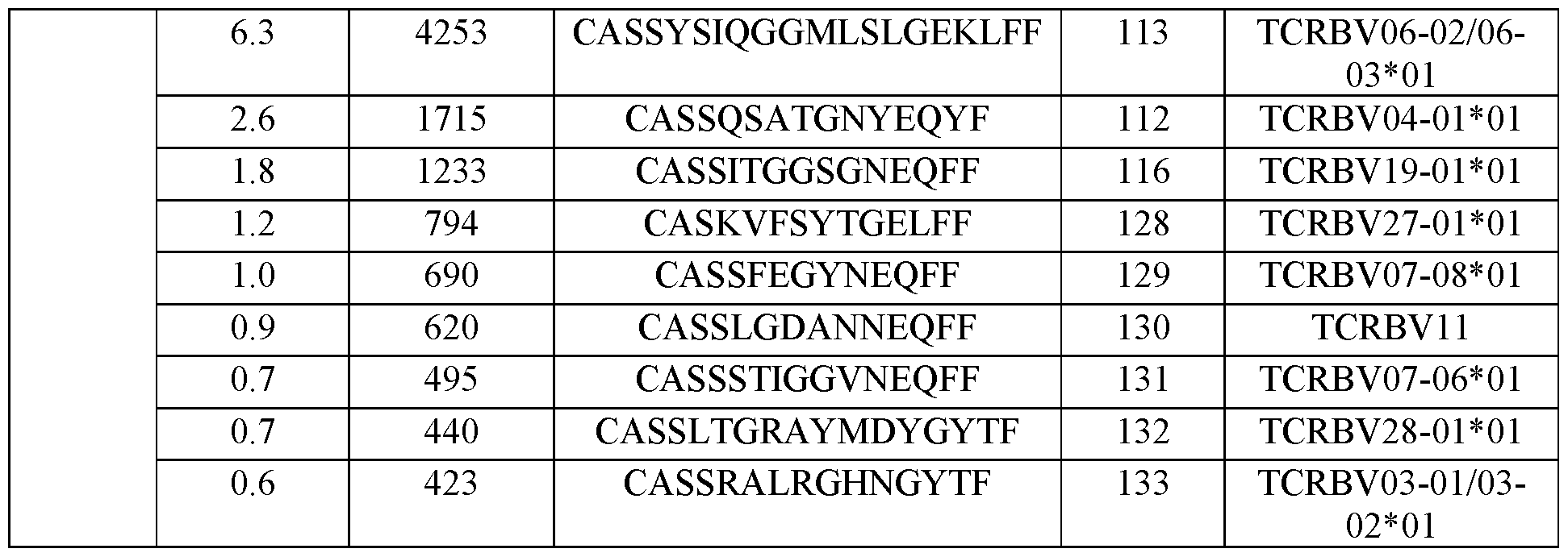
Table 1C: The most 10 abundant TCR sequence following culturing of PBMCs obtained from pancreatic cancer patients with control media not containing PD1-4-1BBL fusion protein or autologous organoid
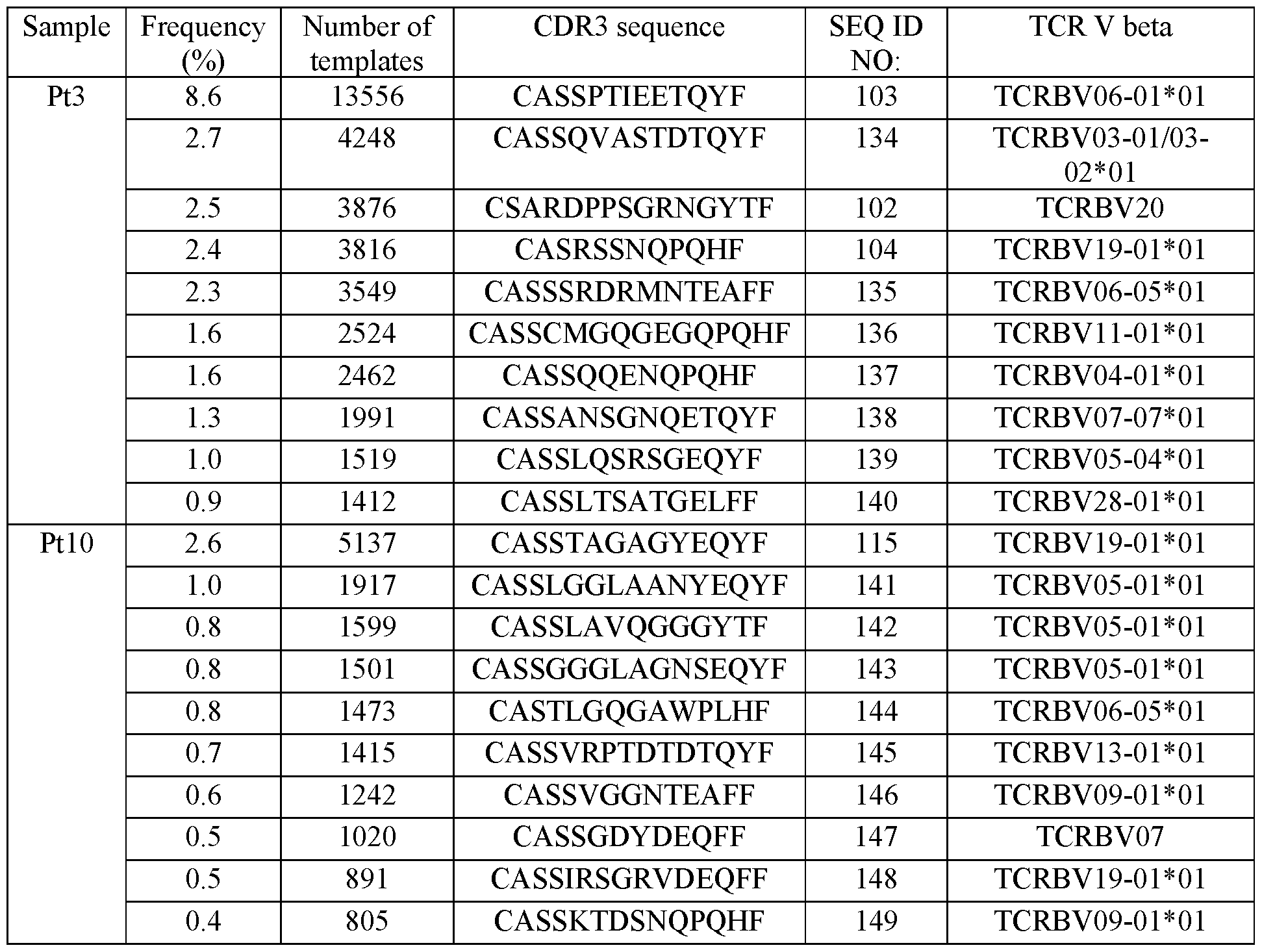 EXAMPLE 2
GENERATION OF RECOMBINANT TCRs HAVING SEQUENCES OF THE
ENRICHED T CELLS CLONES
The TCR sequences of the top 2 T-cell clones generated following culturing with the PD 1-4- 1BBL fusion polypeptide as described in Example 1 hereinabove are used to generate two recombinant TCRs for each patient. As a control, two recombinant TCRs that are negatively selected during the process are generated. The recombinant TCRs are expressed in SKW (PMID: 340692) cells and populations of recombinant TCR expressing SKW cells are generated.
Following, the TCR expressing SKW cells are used to determine their response to Patient #3 or Patient #10 organoids. The recognition of the tumor cells is performed in assays such as ELISPOT and ELISA for IFN-g release (Tan et al., J Immunother Cancer. 2019 Aug 28;7(1):232). Cytotoxic assays, to determine the killing effect of the clones on the matching tumor cells is performed by e.g. labelling the tumor cells with CFSE, co-culturing with the TCR expressing cells and measuring viability by flow cytometry assays (Tan et al., J Immunother Cancer. 2019 Aug 28;7(1):232).
EXAMPLE 3
CULTURING PBMCs IN THE PRESENCE OF PD1-4-1BBL ENRICHES FOR T CELLS
HAVING A MEMORY PHENOTYPE
Materials
PD1-4-1BBL fusion protein (SEQ ID NO: 49) having a His tag at its N’ terminal, Ficoll-Paque (cat# 17-1440-03, GE Healthcare), Serum-free Dendritic Cell Medium (cat# 20801-0100, CellGernix), Pooled human AB serum (cat# IPLA-SerAB-13458, Inonative Research), hIL-2 (cat# cyt-209-b, Prospec), hIL-15 (cat# cyt-230b, Prospec), hIL-21 (cat# cyt-408-b, Prospec), Penicillin- Strepomycin (cat# 15140-122, Gibco), Amphotericin B solution (cat# A2942, Sigma), Bioworld Ciprofloxacin (Cipro) Hydrochloride (cat# 50255729, Fisher Scientific), RPMI 1640 (cat# 61870- 044, Glico), Fetal bovine serum (cat# 26140-079, Glico), Cell-ID™ Cisplatin (Fluidigm,201064), Human TruStain FcX™ (Fc Receptor Blocking Solution) (Biolegend, 422302), Maxpar Cell Staining Buffer (CSB) (Fluidigm #201068), Antibody staining panel (listed in Table 2), DNA Intercalator (Fluidigm, # 201192A), Fix and Perm Buffer (Fluidigm, # 201067), Q Four Element Calibration Beads (Fluidigm, # 201078).
Methods
Human PBMCs were isolated from peripheral blood of pancreatic cancer patients using Ficoll. Following isolation, the PBMCs were cultured in serum-free dendritic cell medium supplemented
with 1 % Penicillin-streptomycin, 1 % Amphotericin B, 0.1 % Ciprofloxacin, 10 % human AB serum, hIL-2 (1000 u / mL), hIL-15 (10 ng / mL) and hIL-21(10 ng / mL) for 10 days to enrich the T cell population. In the next step, the cells were seeded in the same media with the addition of PD1-4-1BBL fusion protein (1 pg / mL) or an autologous organoid for additional 14 days, with refreshment of media and PD1-4-1BBL on day 7. Generation of the autologous organoids was effects as described in Huang et al., Nat Med. 2015 21(11): 1364-71. PBMCs cultured under the same conditions without PD1-4-1BBL or autologous organoid were used as a negative control. Following culturing, cells were collected, and 1 c 106 cells were used for CyTOF analysis with 28 different antibody markers. First, the cells were stained with Cell-ID™ Cisplatin to test the viability. Following blocking the Fc receptor (FcR), the cells were transferred into Maxpar Cell Staining Buffer (CSB) for antibody staining. Stained cells were washed twice with 2 mL CSB, and incubated overnight at 4 °C with 1 mL DNA Intercalator, which was diluted in a Fix and Perm Buffer at a final concentration of 125 nM, to facilitate the discrimination between singlets, doublets, and triplets. Prior to data acquisition, cell pellets were resuspended in CSB containing 10 % EQ Four Element Calibration Beads and filtered through a 35-pm membrane. The cells were analysed by Helios with WB injector (Fluidigm) through several runs at an acquisition rate of -500 events per second. Following the manufacturer's instructions, settings were on default. The short term signal fluctuations of Helios were normalized with EQ bead signals prior to data export and analysis. Abundance values obtained by mass cytometry were transformed using a scaled arcsine with a factor of 5, which diminished near-zero noise values in the measurements. Surface marker expression in each channel was normalized based on the signal intensity at the 99.5th percentile across all samples, thus yielding expression values as x-fold of the 99.5th percentile values. A series of gates for memory T cell subpopulations were based on the expression of: (1) CD103+, Tissue-resident memory T cell (CD103+, TRM): CD3+ ,CD45RO+ ,CD69+ and CD103+; (2) Tissue-resident memory T cell (TRM): CD3+, CD45RO+, CD69+ and CD103- and (3) Effect memory T cell (TEM): CD3+, CD45RO+, CD69- and CCR7-.
Results
Culturing the PBMCs in the presence of the PD1-4-1BBL fusion protein resulted in enrichment of T cells with memory phenotype similar to culturing the PBMCs with the organoids (Figure 3 and Table 2 hereinbelow). The CD3+ TRM population in the PD1-4-1BBL treated cells was >35 % while with the organoids it was 30 %. TRM subpopulation reached > 30 % in the PD1- 4-1BBL treated cells with - 50 % in the organoid culture, indicating high enrichment of T-cells with memory phenotype.
Table 2: Abundance of memory T cells

EXAMPLE 4
THE IN-VIVO ANTI-TUMOR EFFECT OF THE ENRICHED T CELLS CLONES Autologous resected cancer samples are xenografted and expanded in nude mice to develop cohorts of tumor-bearing mice (Rubio- Viqueira et ak, Clin Cancer Res. 2006 Aug 1 ; 12(15):4652- 61). The xenografted tumors maintain their fundamental genotypic features despite serial passages and recapitulate the genetic heterogeneity of the original cancer tissue. These tumor-bearing mice are used to evaluate the anti-tumor activity of the T-cells generated following culturing with the PD1-4-1BBL fusion protein compared to their controls, by injecting them to the mice, and evaluating tumor growth inhibition and mice survival.
EXAMPLE 5
CULTURING PBMCs IN THE PRESENCE OF PD1-4-1BBL, SIRPa-4-lBBL OR AGONIST 4-1BB ANTIBODY ENRICHES T CELL SUB-POPULATIONS
Materials
PD1-4-1BBL fusion protein (SEQ ID NO: 49) having a His tag at its N’ terminal, PD1-4- 1BBL fusion protein (SEQ ID NO: 74), SIRPa-4-lBBL fusion protein (SEQ ID NO: 89), agonist anti-4-lBB antibody, “Urelumab-like” (GenScript, U5656DG120S01/P70011808), Ficoll-Paque (cat# 17-1440-03, GE Healthcare), Serum-free Dendritic Cell Medium (cat# 20801-0100, CellGernix), Pooled human AB serum (cat# IPLA-SerAB-13458, Inonative Research), hIL-2 (cat# cyt-209-b, Prospec), hIL-15 (cat# cyt-230b, Prospec), hIL-21 (cat# cyt-408-b, Prospec), Penicillin- Strepomycin (cat# 15140-122, Gibco), Amphotericin B solution (cat# A2942, Sigma), RPMI 1640 (cat# 61870-044, Glico), Fetal bovine serum (cat# 26140-079, Glico), 0.5% Trypan Blue Solution (catt#03-102-lB, Biological Industries), Fixable VioBlue viability stain 450 (cat#562247, BD), human trustain FcX (cat# 422302, BioLegend), Briliant Stain Buffer (cat#566385, BD), and a kit
for TCR nb families: Beta Mark TCR Vbeta Repertoire Kit (cat# IM3497, Beckman Coulter) and APC/Cy7 anti human CD8 (cat# 301016, BioLegend).
Methods
Cell growth - Human PBMCs were isolated from peripheral blood of six non-small cell lung cancer (NSCLC) patients (OL#l-5, OL#9; OL#l squamous cell carcinoma and the rest are adenocarcinoma) and two colon cancer patients with metastasis to the lung (OL#6 and OL#8), using Ficoll gradient according to the manufacturer’s instructions. In parallel, a bone marrow sample from AML patient was also studied (AML#6). Following isolation, the PBMCs or the bone marrow sample were cultured in serum-free dendritic cell medium supplemented with 1 % Penicillin-streptomycin, 1 % Amphotericin B, 10 % human AB serum, hIL-2 (1000 u / mL), hTL- 15 (10 ng / mL) and hIL-21 (10 ng / mL). Cells were seeded in a 24-wells plate at a concentration of lxlO6 cells per well and cultured for three days (for group 1) or 5-7 days (for group 2) to enrich the T cell population (expansion phase). In the next step, the PD1-4-1BBL fusion protein (SEQ ID NO: 49 or 74, 1 pg / mL), SIRPa-4-lBBL fusion protein (SEQ ID NO: 89, 1 pg / mL), or the 4- 1BB antibody (0.5 or 2 pg / mL), was added for additional 8-9 days (for group 2) or 11-13 days (for group 1), with refreshment of media, including cytokines and proteins / antibody every 2-3 days (treatment phase). Cell growth was monitored every 2-3 days by live cell counting, using trypan blue. When the cells reached 80 - 90 % confluency, they were passaged by splitting 1 : 2 or 1 : 3 into a new 24-wells plate.
TCR nb repertoire analysis - Following 3 and 14-16 days in culture, cells were collected and analysed for TCR nb repertoire by flow cytometry, using Beckman Coulter kit. The kit allows studying the repertoire on T-cell subsets, using additional T-cell marker, CD8, conjugated to a different fluorophore. The kit is composed of 8 vials containing mixtures of conjugated TCR nb antibodies corresponding to 24 different specificities (about 70 % coverage of normal human TCR nb repertoire). The frequency of each specific TCR nb was calculated separately for the CD8+T- cells or the CD8 T-cells. Samples were analyzed if the CD8+ or CD8 sub-population represented at least 15 % of the total sample.
Results
Evaluation of cell growth demonstrated that addition of either the PD1-4-1BBL fusion protein (SEQ ID NO: 49 or 74) or the SIRPa-4-lBBL fusion protein (SEQ ID NO: 89) resulted in expansion of T-cell clusters (Representative images are shown in Figure 4).
Evaluation of TCR nb repertoire demonstrated that addition of either the PD1-4-1BBL fusion protein (SEQ ID NO: 49 or 74), the SIRPa-4-lBBL fusion protein (SEQ ID NO: 89) or the anti-4-lBB antibody resulted in enrichment of specific T-cell families as summarized by the
specific families of TCR nb frequencies in the two treatment groups for both CD8+ T-cells (Table 3 hereinbelow) and CD8 T-cells (Table 4 hereinbelow). In most samples, treatment with the fusion proteins and the antibody resulted in enrichment of few TCR nb families mainly in the CD8+ T-cell population. Some families appear to be enriched in samples obtained from few patients; for example, nb8 was enriched in the CD8+ T-cells in 4 samples (OL#l, OL#3, OL#4 and OL#5), in the 2 treatment groups following treatment with either the PD1-4-1BBL or SIRPa- 4-1BBL fusion protein; nb3 was enriched in the CD8+ T-cells in 4 samples (OL#l, OL#2, OL#3 and OL#7), in the 2 treatment groups following treatment with either the PD1-4-1BBL or SIRPa- 4-1BBL fusion protein. Table 3: Frequencies (percentages) of TCR Ub families in CD8+ T cells.

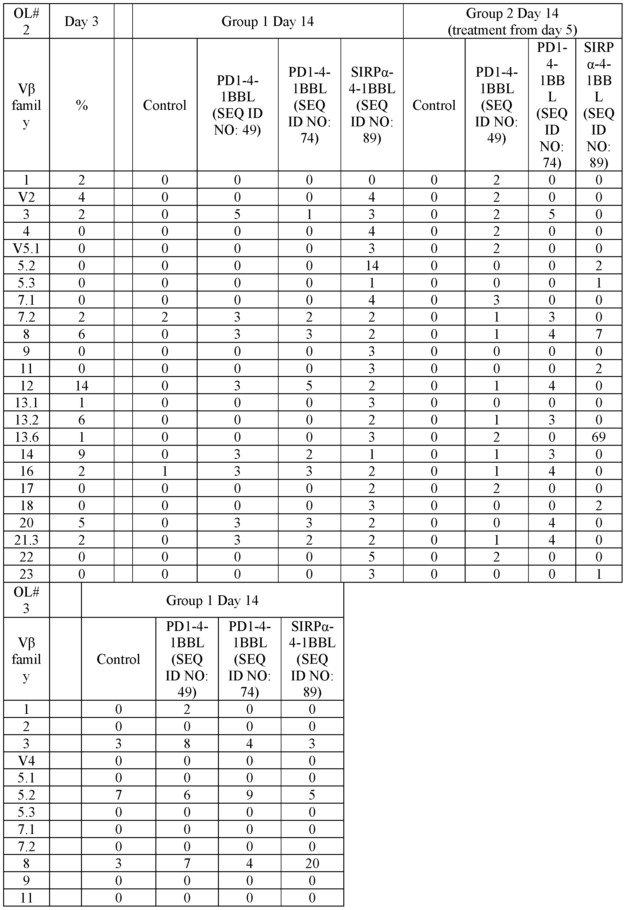


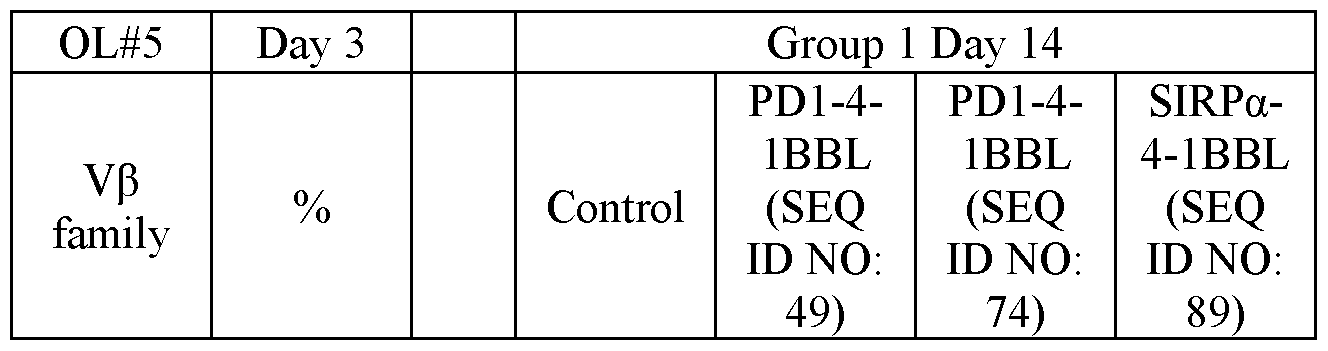
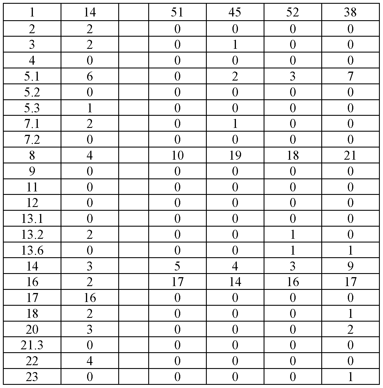
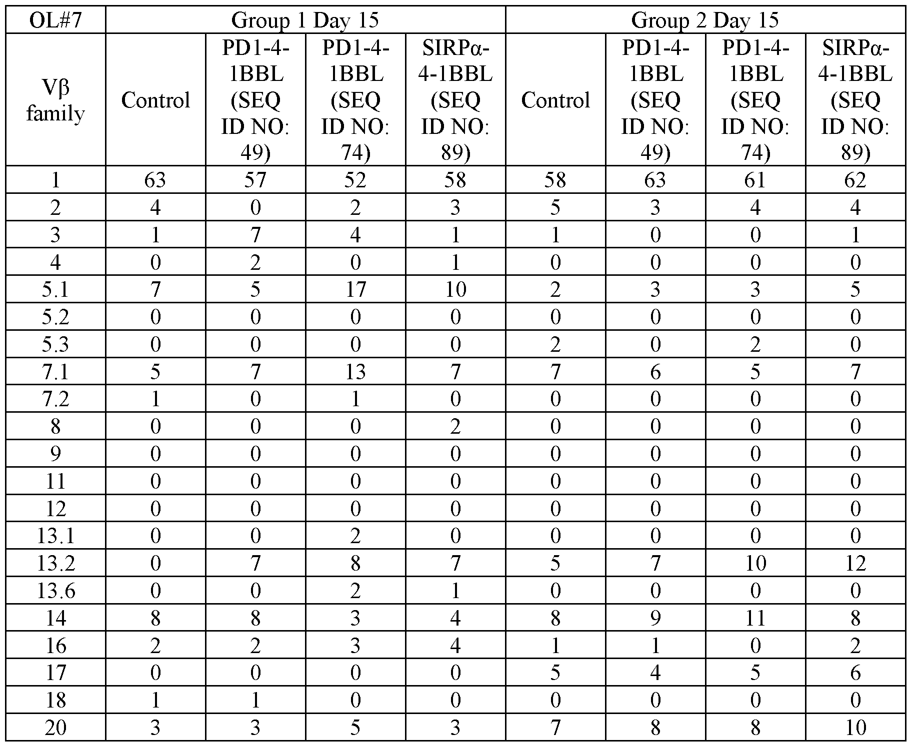

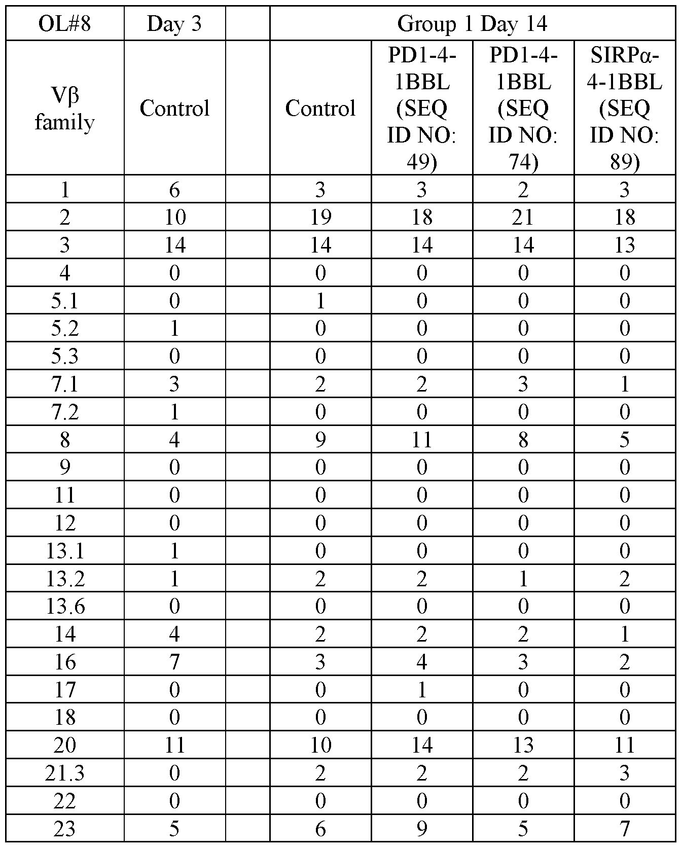


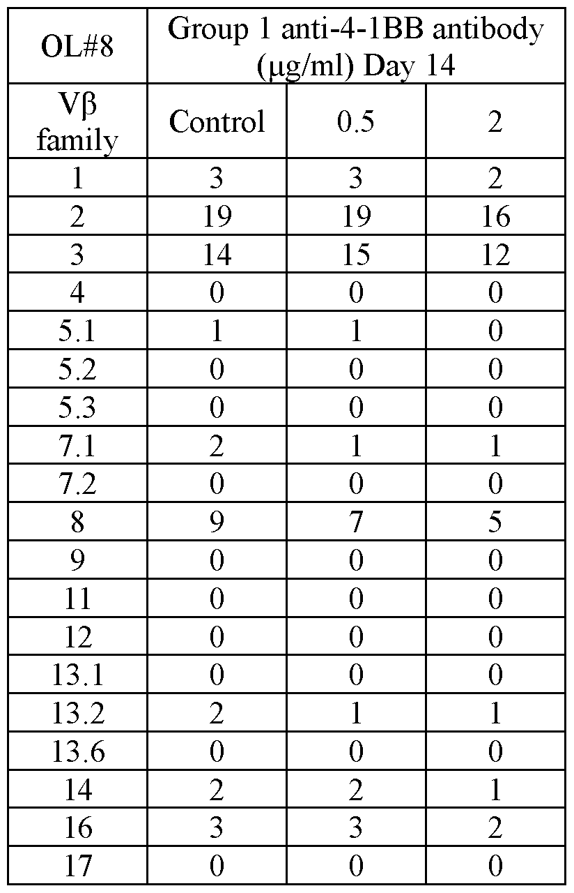

Table 4: Frequencies (percentages) of TCR Ub families in CD8 T cells


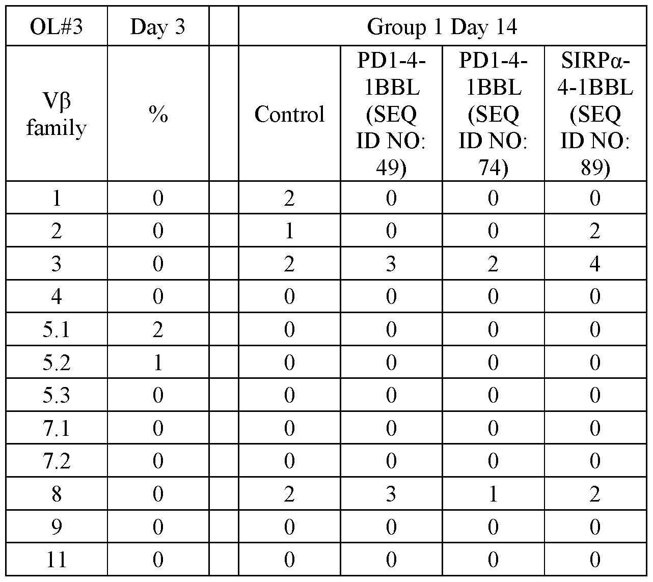


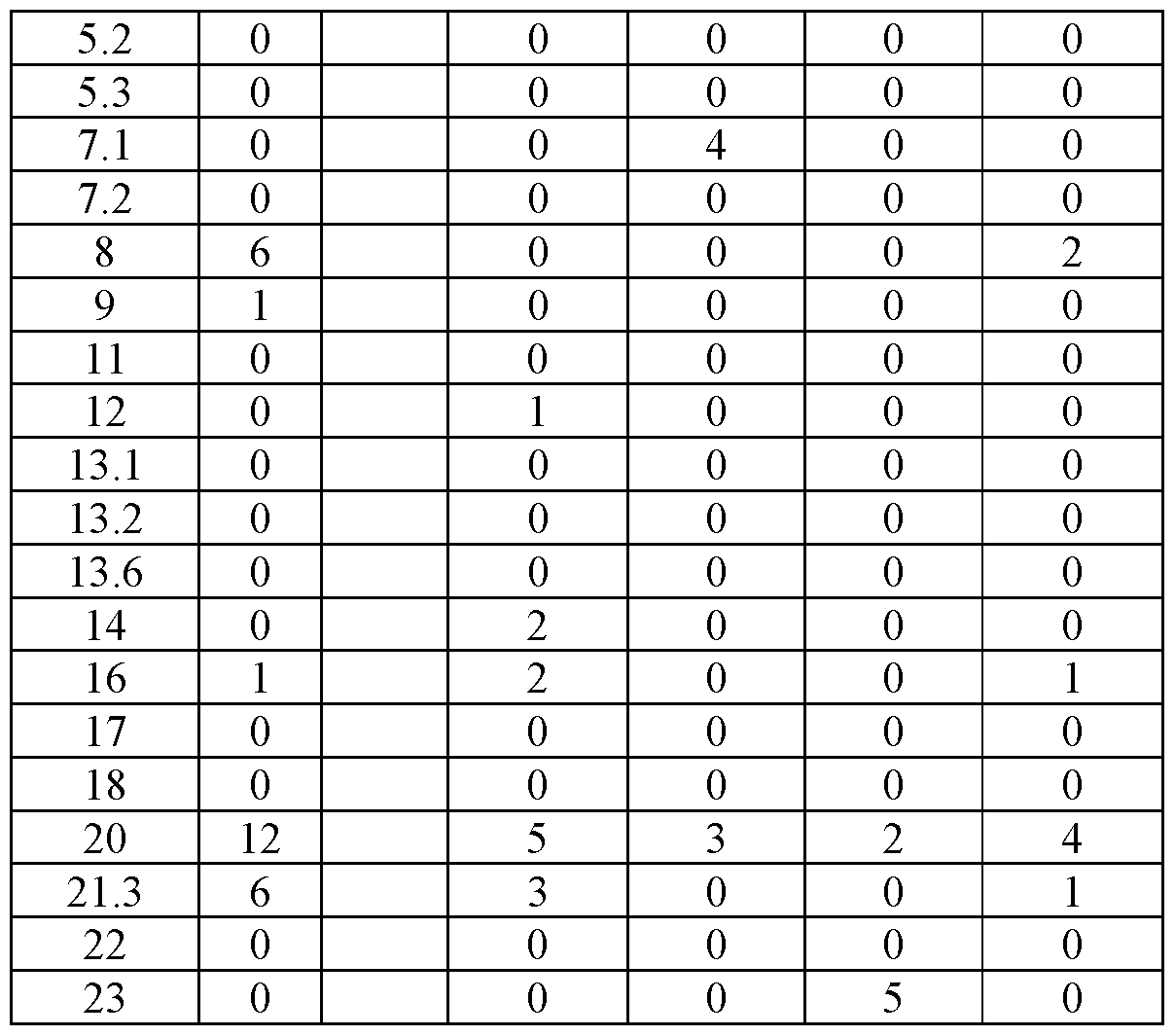
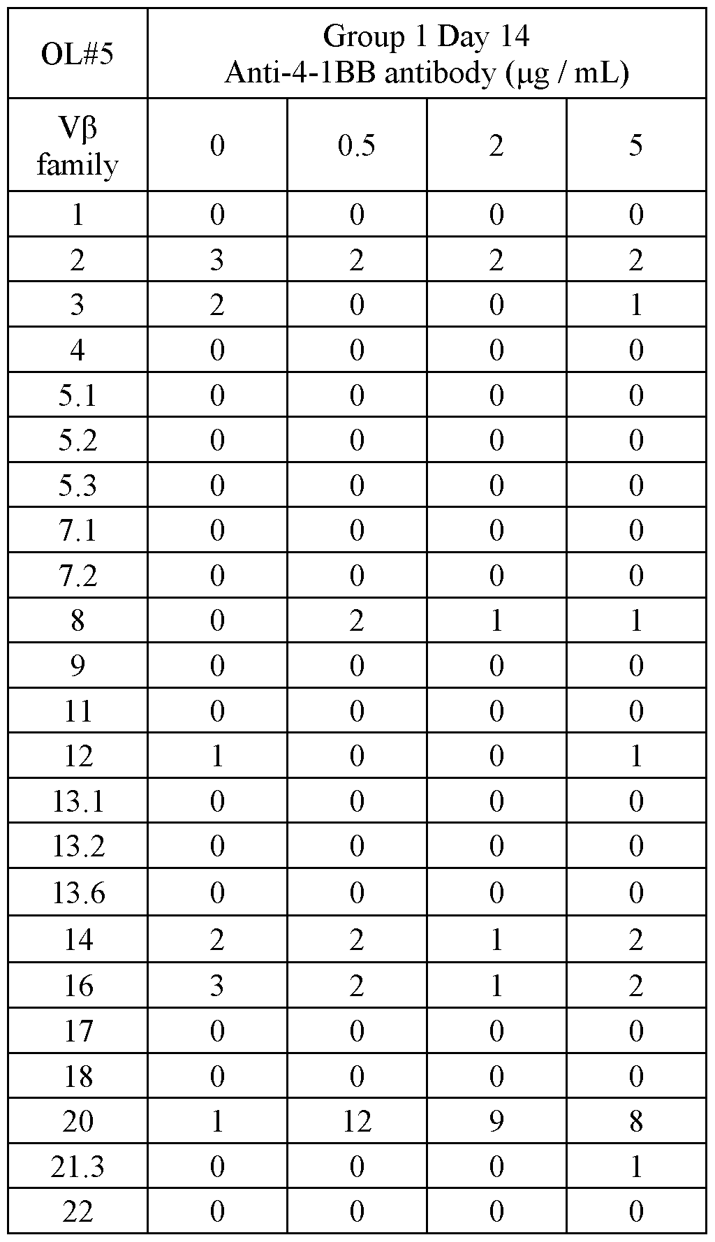
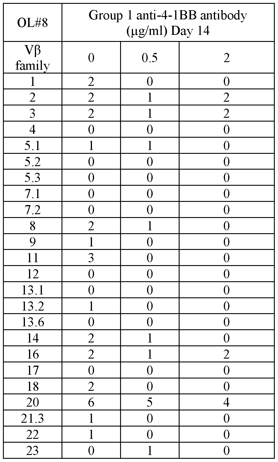
Although the invention has been described in conjunction with specific embodiments thereof, it is evident that many alternatives, modifications and variations will be apparent to those skilled in the art. Accordingly, it is intended to embrace all such alternatives, modifications and variations that fall within the spirit and broad scope of the appended claims.
All publications, patents and patent applications mentioned in this specification are herein incorporated in their entirety by reference into the specification, to the same extent as if each individual publication, patent or patent application was specifically and individually indicated to be incorporated herein by reference. In addition, citation or identification of any reference in this application shall not be construed as an admission that such reference is available as prior art to the present invention. To the extent that section headings are used, they should not be construed as necessarily limiting. In addition, any priority document(s) of this application is/are hereby incorporated herein by reference in its/their entirety.
Claims
1. A method of culturing T cells, the method comprising:
(a) adding to immune cells comprising T cells obtained from a subject having a pathology a non-cellular agent capable of binding 4-1BB and activating said 4-1BB signaling pathway; and
(b) culturing said immune cells with said agent for more than 7 days.
2. The method of claim 1, wherein said culturing is effected for more than 10 days.
3. The method of claim 1, wherein said culturing is effected for about 14 days.
4. The method of any one of claims 1-3, being effected without isolation of 4-1BB positive cells prior to said (a) and/or said (b).
5. The method of any one of claims 1-4, being effected without isolation of 4- IBB positive cells during and/or following said (b).
6. The method of any one of claims 1-5, being effected without isolation of said T cells from said immune cells prior to said (a) and/or said (b).
7. The method of any one of claims 1-6, comprising adding at least one cytokine to said immune cells in step (a) and culturing said immune cells with said cytokine in step (b).
8. The method of any one of claims 1-7, further comprising pre-culturing said immune cells with at least one cytokine prior to said (a).
9. The method of claim 8, wherein said pre-culturing is effected until the percentage of said T cells in said immune cells is at least 80 %.
10. The method of claim 8, wherein said pre-culturing is effected for 2-14 days.
11. The method of any one of claims 8-10, being effected without isolation of said T cells from said immune cells prior to said pre-culturing.
12. The method of any one of claims 1-11, being effected without adding to said immune cells a T cell stimulatory agent capable of at least transmitting a primary activating signal to said T cells.
13. The method of any one of claims 1-12, wherein said culturing is effected under conditions which allow enrichment of T cell clones reactive to cells associated with said pathology.
14. The method of any one of claims 1-13 wherein said immune cells are obtained from a blood sample of said subject.
15. The method of claim 14, wherein said immune cells comprise PBMCs.
16. The method of any one of claims 1-13, wherein said immune cells are obtained from a tissue comprising cells associated with said pathology.
17. The method of any one of claims 7-16, wherein said at least one cytokine is selected from the group consisting of IL-2, IL-15, IL-21, IL-12 and IL-7.
18. The method of any one of claims 7-16, wherein said at least one cytokine comprises IL-2, IL-15 and IL-21.
19. The method of any one of claims 1-18, wherein said non-cellular agent is capable of binding a ligand or receptor of a type I membrane protein.
20. The method of any one of claims 1-18, wherein said (a) further comprises adding a non-cellular agent capable of binding a ligand or receptor of a type I membrane protein.
21. The method of any one of claims 19-20, wherein said type I membrane protein is PD1.
22. The method of any one of claims 19-20, wherein said type I membrane protein is SIRPa.
23. The method of any one of claims 1-22, wherein said non-cellular agent is soluble.
24. The method of any one of claims 1-22, wherein said non-cellular agent is immobilized.
25. The method of any one of claim 1-24, wherein said non-cellular agent is an antibody.
26. The method of any one of claim 1-24, wherein said non-cellular agent capable of binding 4- IBB and activating said 4- IBB signaling pathway is a polypeptide comprising an amino acid sequence of 4-1BBL.
27. The method of any one of claims 1-24, wherein said non-cellular agent is a fusion polypeptide.
28. The method of claim 27, wherein said fusion polypeptide comprises an amino acid sequence of 4-1BBL and an amino acid sequence of PD1.
29. The method of claim 28, wherein said fusion polypeptide comprises an amino acid sequence having at least 80 % identity to SEQ ID NO: 49 or 74.
30. The method of claim 28, wherein said fusion polypeptide comprises SEQ ID NO: 49 or 74 or as set forth in SEQ ID NO: 49 or 74.
31. The method of claim 27, wherein said fusion polypeptide comprises an amino acid sequence of 4-1BBL and an amino acid sequence of SIRPa.
32. The method of claim 31, wherein said fusion polypeptide comprises an amino acid sequence having at least 80 % identity to SEQ ID NO: 89.
33. The method of claim 31, wherein said fusion polypeptide comprises SEQ ID NO: 89 or as set forth in SEQ ID NO: 89.
34. The method of any one of claims 1-33, further comprising expanding said T cells following said (b).
35. The method of any one of claims 1-34, further comprising isolating said T cells from said immune cells following said (b) and/or said expanding.
36. The method of any one of claims 1-35, further comprising determining a sequence of a T cell receptor (TCR) expressed by at least one of said T cells following said (b), said expanding or said isolating.
37. The method of claim 36, further comprising transducing a T cell with a nucleic acid sequence encoding said TCR.
38. The method of any one of claims 1-35, further comprising transducing said T cells with a nucleic acid sequence encoding a chimeric antigen receptor (CAR) following said (b), said expanding or said isolating.
39. The method of any one of claims 1-38, comprising adoptively transferring said immune cells and/or said T cells following said (b), said expanding or said transducing to a subject in need thereof.
40. T cells obtainable by the method of any one of claims 1-38.
41. The T cells of claim 40 for use in adoptive cell therapy to a subject in need thereof.
42. A method of treating a pathology that can benefit from adoptive T cell therapy in a subject in need thereof, the method comprising administering to the subject a therapeutically effective amount of the T cells of claim 40, thereby treating the pathology.
43. The method or the T cells for use of any one of claims 39 and 42, wherein said immune cells and/or said T cells are autologous to said subject in need thereof.
44. The method or the T cells for use of any one of claims 1-43, wherein said pathology is selected from the group consisting of a hyper-proliferative disease, a disease associated with immune suppression and infection.
45. The method or the T cells for use of any one of claims 1-44, wherein said pathology is cancer.
46. The method or the T cells for use of claim 45, wherein said cancer is selected from the group consisting of gastrointestinal (GI) cancer, breast cancer, ovarian cancer and pancreatic cancer.
47. The method or the T cells for use of claim 45, wherein said cancer is selected from the group consisting of pancreatic cancer, lung cancer, colon cancer and leukemia.
48. The method or the T cells for use of any one of claims 1-47, wherein said subject is human.
Priority Applications (1)
| Application Number | Priority Date | Filing Date | Title |
|---|---|---|---|
| US17/788,307 US20230048361A1 (en) | 2019-12-31 | 2020-12-30 | Methods of culturing t cells and uses of same |
Applications Claiming Priority (2)
| Application Number | Priority Date | Filing Date | Title |
|---|---|---|---|
| US201962955463P | 2019-12-31 | 2019-12-31 | |
| US62/955,463 | 2019-12-31 |
Publications (1)
| Publication Number | Publication Date |
|---|---|
| WO2021137230A1 true WO2021137230A1 (en) | 2021-07-08 |
Family
ID=74194810
Family Applications (1)
| Application Number | Title | Priority Date | Filing Date |
|---|---|---|---|
| PCT/IL2020/051356 Ceased WO2021137230A1 (en) | 2019-12-31 | 2020-12-30 | Methods of culturing t cells and uses of same |
Country Status (2)
| Country | Link |
|---|---|
| US (1) | US20230048361A1 (en) |
| WO (1) | WO2021137230A1 (en) |
Cited By (1)
| Publication number | Priority date | Publication date | Assignee | Title |
|---|---|---|---|---|
| WO2023121890A1 (en) * | 2021-12-23 | 2023-06-29 | Fbd Biologics Limited | Cd47/4-1bb-targeting protein complex and methods of use thereof |
Families Citing this family (2)
| Publication number | Priority date | Publication date | Assignee | Title |
|---|---|---|---|---|
| CA3047708A1 (en) | 2017-01-05 | 2018-07-12 | Kahr Medical Ltd. | A sirpalpha-41bbl fusion protein and methods of use thereof |
| US12134638B2 (en) | 2018-07-11 | 2024-11-05 | Kahr Medical Ltd. | SIRPalpha-4-1BBL variant fusion protein and methods of use thereof |
Citations (39)
| Publication number | Priority date | Publication date | Assignee | Title |
|---|---|---|---|---|
| US3791932A (en) | 1971-02-10 | 1974-02-12 | Akzona Inc | Process for the demonstration and determination of reaction components having specific binding affinity for each other |
| US3839153A (en) | 1970-12-28 | 1974-10-01 | Akzona Inc | Process for the detection and determination of specific binding proteins and their corresponding bindable substances |
| US3850752A (en) | 1970-11-10 | 1974-11-26 | Akzona Inc | Process for the demonstration and determination of low molecular compounds and of proteins capable of binding these compounds specifically |
| US3850578A (en) | 1973-03-12 | 1974-11-26 | H Mcconnell | Process for assaying for biologically active molecules |
| US3853987A (en) | 1971-09-01 | 1974-12-10 | W Dreyer | Immunological reagent and radioimmuno assay |
| US3867517A (en) | 1971-12-21 | 1975-02-18 | Abbott Lab | Direct radioimmunoassay for antigens and their antibodies |
| US3879262A (en) | 1972-05-11 | 1975-04-22 | Akzona Inc | Detection and determination of haptens |
| US3901654A (en) | 1971-06-21 | 1975-08-26 | Biological Developments | Receptor assays of biologically active compounds employing biologically specific receptors |
| US3935074A (en) | 1973-12-17 | 1976-01-27 | Syva Company | Antibody steric hindrance immunoassay with two antibodies |
| US3984533A (en) | 1975-11-13 | 1976-10-05 | General Electric Company | Electrophoretic method of detecting antigen-antibody reaction |
| US3996345A (en) | 1974-08-12 | 1976-12-07 | Syva Company | Fluorescence quenching with immunological pairs in immunoassays |
| US4034074A (en) | 1974-09-19 | 1977-07-05 | The Board Of Trustees Of Leland Stanford Junior University | Universal reagent 2-site immunoradiometric assay using labelled anti (IgG) |
| US4098876A (en) | 1976-10-26 | 1978-07-04 | Corning Glass Works | Reverse sandwich immunoassay |
| US4666828A (en) | 1984-08-15 | 1987-05-19 | The General Hospital Corporation | Test for Huntington's disease |
| US4683202A (en) | 1985-03-28 | 1987-07-28 | Cetus Corporation | Process for amplifying nucleic acid sequences |
| US4801531A (en) | 1985-04-17 | 1989-01-31 | Biotechnology Research Partners, Ltd. | Apo AI/CIII genomic polymorphisms predictive of atherosclerosis |
| US4879219A (en) | 1980-09-19 | 1989-11-07 | General Hospital Corporation | Immunoassay utilizing monoclonal high affinity IgM antibodies |
| US5011771A (en) | 1984-04-12 | 1991-04-30 | The General Hospital Corporation | Multiepitopic immunometric assay |
| WO1992020356A1 (en) | 1991-05-23 | 1992-11-26 | Ludwig Institute For Cancer Research | Tumor rejection antigen precursors, tumor rejection antigens and uses thereof |
| US5192659A (en) | 1989-08-25 | 1993-03-09 | Genetype Ag | Intron sequence analysis method for detection of adjacent and remote locus alleles as haplotypes |
| US5272057A (en) | 1988-10-14 | 1993-12-21 | Georgetown University | Method of detecting a predisposition to cancer by the use of restriction fragment length polymorphism of the gene for human poly (ADP-ribose) polymerase |
| US5281521A (en) | 1992-07-20 | 1994-01-25 | The Trustees Of The University Of Pennsylvania | Modified avidin-biotin technique |
| WO1994005304A1 (en) | 1992-08-31 | 1994-03-17 | Ludwig Institute For Cancer Research | Isolated nonapeptide derived from mage-3 gene and presented by hla-a1, and uses thereof |
| WO1994023031A1 (en) | 1993-03-26 | 1994-10-13 | Ludwig Institute For Cancer Research | Isolated nucleic acid molecules coding for tumor rejection antigen precursor mage-3 and uses thereof |
| WO1995020974A1 (en) | 1994-02-01 | 1995-08-10 | Ludwig Institute For Cancer Research | Monoclonal anbibodies which bind to tumor rejection antigen precursor mage-1, recombinant mage-1, and mage-1 derived immunogenic peptides |
| WO1995023874A1 (en) | 1994-03-01 | 1995-09-08 | Ludwig Institute For Cancer Research | Determination of cancerous conditions by mage gene expression |
| WO1996026214A1 (en) | 1995-02-23 | 1996-08-29 | Ludwig Institute For Cancer Research | Isolated nonapeptides presented by hla molecules, and uses thereof |
| WO2000020581A1 (en) | 1998-10-05 | 2000-04-13 | Ludwig Institute For Cancer Research | Mage-a3 peptides presented by hla class ii molecules |
| WO2001039594A2 (en) | 1999-12-03 | 2001-06-07 | Dendreon Corporation | Cryopreservation of antigen-loaded dendritic cells and their precursors in serum-free media |
| WO2003049755A1 (en) | 2001-10-09 | 2003-06-19 | Mayo Foundation For Medical Education And Research | Enhancement of immune responses by 4-1bb-binding agents |
| WO2007054160A2 (en) | 2005-11-09 | 2007-05-18 | Paul-Ehrlich-Institut Bundesamt Für Sera Und Impfstoffe | Method for the cryopreservation of human blood |
| US20120149108A1 (en) | 2009-08-19 | 2012-06-14 | Masashige Tanabe | Cell preservation method |
| US20140212446A1 (en) * | 2004-05-27 | 2014-07-31 | The Trustees Of The University Of Pennsylvania | Novel Artificial Antigen Presenting Cells and Uses Thereof |
| WO2015039100A1 (en) | 2013-09-16 | 2015-03-19 | The Trustees Of The University Of Pennsylvania | Cd137 enrichment for efficient tumor infiltrating lymphocyte selection |
| EP3118322A1 (en) | 2014-03-12 | 2017-01-18 | National Cancer Center | Method for isolating and proliferating self-tumor antigen-specific cd8+ t cells |
| WO2017182672A1 (en) | 2016-04-22 | 2017-10-26 | Alligator Bioscience Ab | Novel bispecific polypeptides against cd137 |
| WO2018127917A1 (en) | 2017-01-05 | 2018-07-12 | Kahr Medical Ltd. | A pd1-41bbl fusion protein and methods of use thereof |
| WO2018127919A1 (en) | 2017-01-05 | 2018-07-12 | Kahr Medical Ltd. | A SIRP1 alpha-41BBL FUSION PROTEIN AND METHODS OF USE THEREOF |
| WO2020012485A1 (en) * | 2018-07-11 | 2020-01-16 | Kahr Medical Ltd. | Pd1-4-1bbl variant fusion protein and methods of use thereof |
-
2020
- 2020-12-30 WO PCT/IL2020/051356 patent/WO2021137230A1/en not_active Ceased
- 2020-12-30 US US17/788,307 patent/US20230048361A1/en active Pending
Patent Citations (40)
| Publication number | Priority date | Publication date | Assignee | Title |
|---|---|---|---|---|
| US3850752A (en) | 1970-11-10 | 1974-11-26 | Akzona Inc | Process for the demonstration and determination of low molecular compounds and of proteins capable of binding these compounds specifically |
| US3839153A (en) | 1970-12-28 | 1974-10-01 | Akzona Inc | Process for the detection and determination of specific binding proteins and their corresponding bindable substances |
| US3791932A (en) | 1971-02-10 | 1974-02-12 | Akzona Inc | Process for the demonstration and determination of reaction components having specific binding affinity for each other |
| US3901654A (en) | 1971-06-21 | 1975-08-26 | Biological Developments | Receptor assays of biologically active compounds employing biologically specific receptors |
| US3853987A (en) | 1971-09-01 | 1974-12-10 | W Dreyer | Immunological reagent and radioimmuno assay |
| US3867517A (en) | 1971-12-21 | 1975-02-18 | Abbott Lab | Direct radioimmunoassay for antigens and their antibodies |
| US3879262A (en) | 1972-05-11 | 1975-04-22 | Akzona Inc | Detection and determination of haptens |
| US3850578A (en) | 1973-03-12 | 1974-11-26 | H Mcconnell | Process for assaying for biologically active molecules |
| US3935074A (en) | 1973-12-17 | 1976-01-27 | Syva Company | Antibody steric hindrance immunoassay with two antibodies |
| US3996345A (en) | 1974-08-12 | 1976-12-07 | Syva Company | Fluorescence quenching with immunological pairs in immunoassays |
| US4034074A (en) | 1974-09-19 | 1977-07-05 | The Board Of Trustees Of Leland Stanford Junior University | Universal reagent 2-site immunoradiometric assay using labelled anti (IgG) |
| US3984533A (en) | 1975-11-13 | 1976-10-05 | General Electric Company | Electrophoretic method of detecting antigen-antibody reaction |
| US4098876A (en) | 1976-10-26 | 1978-07-04 | Corning Glass Works | Reverse sandwich immunoassay |
| US4879219A (en) | 1980-09-19 | 1989-11-07 | General Hospital Corporation | Immunoassay utilizing monoclonal high affinity IgM antibodies |
| US5011771A (en) | 1984-04-12 | 1991-04-30 | The General Hospital Corporation | Multiepitopic immunometric assay |
| US4666828A (en) | 1984-08-15 | 1987-05-19 | The General Hospital Corporation | Test for Huntington's disease |
| US4683202A (en) | 1985-03-28 | 1987-07-28 | Cetus Corporation | Process for amplifying nucleic acid sequences |
| US4683202B1 (en) | 1985-03-28 | 1990-11-27 | Cetus Corp | |
| US4801531A (en) | 1985-04-17 | 1989-01-31 | Biotechnology Research Partners, Ltd. | Apo AI/CIII genomic polymorphisms predictive of atherosclerosis |
| US5272057A (en) | 1988-10-14 | 1993-12-21 | Georgetown University | Method of detecting a predisposition to cancer by the use of restriction fragment length polymorphism of the gene for human poly (ADP-ribose) polymerase |
| US5192659A (en) | 1989-08-25 | 1993-03-09 | Genetype Ag | Intron sequence analysis method for detection of adjacent and remote locus alleles as haplotypes |
| WO1992020356A1 (en) | 1991-05-23 | 1992-11-26 | Ludwig Institute For Cancer Research | Tumor rejection antigen precursors, tumor rejection antigens and uses thereof |
| US5281521A (en) | 1992-07-20 | 1994-01-25 | The Trustees Of The University Of Pennsylvania | Modified avidin-biotin technique |
| WO1994005304A1 (en) | 1992-08-31 | 1994-03-17 | Ludwig Institute For Cancer Research | Isolated nonapeptide derived from mage-3 gene and presented by hla-a1, and uses thereof |
| WO1994023031A1 (en) | 1993-03-26 | 1994-10-13 | Ludwig Institute For Cancer Research | Isolated nucleic acid molecules coding for tumor rejection antigen precursor mage-3 and uses thereof |
| WO1995020974A1 (en) | 1994-02-01 | 1995-08-10 | Ludwig Institute For Cancer Research | Monoclonal anbibodies which bind to tumor rejection antigen precursor mage-1, recombinant mage-1, and mage-1 derived immunogenic peptides |
| WO1995023874A1 (en) | 1994-03-01 | 1995-09-08 | Ludwig Institute For Cancer Research | Determination of cancerous conditions by mage gene expression |
| WO1996026214A1 (en) | 1995-02-23 | 1996-08-29 | Ludwig Institute For Cancer Research | Isolated nonapeptides presented by hla molecules, and uses thereof |
| WO2000020581A1 (en) | 1998-10-05 | 2000-04-13 | Ludwig Institute For Cancer Research | Mage-a3 peptides presented by hla class ii molecules |
| WO2001039594A2 (en) | 1999-12-03 | 2001-06-07 | Dendreon Corporation | Cryopreservation of antigen-loaded dendritic cells and their precursors in serum-free media |
| WO2003049755A1 (en) | 2001-10-09 | 2003-06-19 | Mayo Foundation For Medical Education And Research | Enhancement of immune responses by 4-1bb-binding agents |
| US20140212446A1 (en) * | 2004-05-27 | 2014-07-31 | The Trustees Of The University Of Pennsylvania | Novel Artificial Antigen Presenting Cells and Uses Thereof |
| WO2007054160A2 (en) | 2005-11-09 | 2007-05-18 | Paul-Ehrlich-Institut Bundesamt Für Sera Und Impfstoffe | Method for the cryopreservation of human blood |
| US20120149108A1 (en) | 2009-08-19 | 2012-06-14 | Masashige Tanabe | Cell preservation method |
| WO2015039100A1 (en) | 2013-09-16 | 2015-03-19 | The Trustees Of The University Of Pennsylvania | Cd137 enrichment for efficient tumor infiltrating lymphocyte selection |
| EP3118322A1 (en) | 2014-03-12 | 2017-01-18 | National Cancer Center | Method for isolating and proliferating self-tumor antigen-specific cd8+ t cells |
| WO2017182672A1 (en) | 2016-04-22 | 2017-10-26 | Alligator Bioscience Ab | Novel bispecific polypeptides against cd137 |
| WO2018127917A1 (en) | 2017-01-05 | 2018-07-12 | Kahr Medical Ltd. | A pd1-41bbl fusion protein and methods of use thereof |
| WO2018127919A1 (en) | 2017-01-05 | 2018-07-12 | Kahr Medical Ltd. | A SIRP1 alpha-41BBL FUSION PROTEIN AND METHODS OF USE THEREOF |
| WO2020012485A1 (en) * | 2018-07-11 | 2020-01-16 | Kahr Medical Ltd. | Pd1-4-1bbl variant fusion protein and methods of use thereof |
Non-Patent Citations (99)
| Title |
|---|
| "GenBank", Database accession no. NP_001254635 |
| "Immobilized Cells and Enzymes", 1986, IRL PRESS |
| "NP_001035111", Database accession no. NP_001317657 |
| "Nucleic Acid Hybridization", 1985 |
| "PCR Protocols: A Guide To Methods And Applications", vol. 1-317, 1990, ACADEMIC PRESS |
| "Selected Methods in Cellular Immunology", 1980, W. H. FREEMAN AND CO. |
| ALEXANDER RB ET AL., UROLOGY, vol. 50, no. 6, December 1997 (1997-12-01), pages 893 |
| ANDERSSON, BIOPOLYMERS, vol. 1-4, no. 3, 2000, pages 227 - 50 |
| ANKE REDEKER ET AL: "Improving Adoptive T Cell Therapy: The Particular Role of T Cell Costimulation, Cytokines, and Post-Transfer Vaccination", FRONTIERS IN IMMUNOLOGY, vol. 7, 6 September 2016 (2016-09-06), CH, XP055459807, ISSN: 1664-3224, DOI: 10.3389/fimmu.2016.00345 * |
| ANTOINE JCHONNORAT J, REV NEUROL (PARIS, vol. 156, no. 1, January 2000 (2000-01-01), pages 23 |
| BESSER ET AL., CLIN CANCER RES, vol. 16, no. 9, 2010, pages 2646 - 55 |
| BRALEY-MULLEN HYU S, J IMMUNOL, vol. 165, no. 12, 15 December 2000 (2000-12-15), pages 7262 |
| CAPOROSSI AP ET AL., VIRAL IMMUNOL, vol. 11, no. 1, 1998, pages 9 |
| CASTANO LEISENBARTH GS, ANN. REV. IMMUNOL., vol. 8, pages 647 |
| CHAN OT ET AL., IMMUNOL REV, vol. 169, June 1999 (1999-06-01), pages 107 |
| CHAO WANG ET AL: "4-1BBL Induces TNF Receptor-Associated Factor 1-Dependent Bim Modulation in Human T Cells and Is a Critical Component in the Costimulation-Dependent Rescue of Functionally Impaired HIV-Specific CD8 T Cells", THE JOURNAL OF IMMUNOLOGY, vol. 179, no. 12, 3 December 2007 (2007-12-03), US, pages 8252 - 8263, XP055738122, ISSN: 0022-1767, DOI: 10.4049/jimmunol.179.12.8252 * |
| CHEN ET AL., ADV DRUG DELIV REV, vol. 65, no. 10, 2013, pages 1357 - 1369 |
| CHICHILI ET AL., PROTEIN SCI, vol. 22, no. 2, 2013, pages 153 - 167 |
| CRASTO ET AL., PROTEIN ENG, vol. 13, no. 5, 2000, pages 309 - 312 |
| CROSS AH ET AL., J NEUROIMMUNOL, vol. 112, no. 1-2, 1 January 2001 (2001-01-01), pages 1 |
| CUNHA-NETO E ET AL., J CLIN INVEST, vol. 98, no. 8, 15 October 1996 (1996-10-15), pages 1709 |
| DAVILA ET AL., ONCOIMMUNOLOGY, vol. 1, no. 9, 1 December 2012 (2012-12-01), pages 1577 - 1583 |
| DIEKMAN AB ET AL., AM J REPROD IMMUNOL, vol. 43, no. 3, March 2000 (2000-03-01), pages 134 |
| EFREMOV DG ET AL., LEUK LYMPHOMA, vol. 28, no. 3-4, January 1998 (1998-01-01), pages 285 |
| ERIKSON J ET AL., IMMUNOL RES, vol. 17, no. 1-2, 1998, pages 49 |
| FEIST E ET AL., INT ARCH ALLERGY IMMUNOL, vol. 123, no. 1, September 2000 (2000-09-01), pages 92 |
| FINGL ET AL.: "The Pharmacological Basis of Therapeutics", 1975, pages: 1 |
| FLAMHOLZ R ET AL., J CLIN APHERESIS, vol. 14, no. 4, 1999, pages 171 |
| FRANCO A ET AL., CLIN IMMUNOL IMMUNOPATHOL, vol. 54, no. 3, March 1990 (1990-03-01), pages 382 |
| FRESHNEY: "Culture of Animal Cells - A Manual of Basic Technique", vol. I-III, 1994, APPLETON & LANGE |
| G. SCHRODERK. LUPKE: "The Peptides", vol. 1, 1965, ACADEMIC PRESS |
| GARCIA HEROLA A ET AL., GASTROENTEROL HEPATOL, vol. 23, no. 1, January 2000 (2000-01-01), pages 16 |
| GARZA KM ET AL., J REPROD IMMUNOL, vol. 37, no. 2, February 1998 (1998-02-01), pages 87 |
| GLOBERSON-LEVIN ET AL., MOL THER, vol. 22, no. 5, 2014, pages 1029 - 1038 |
| GLODDEK B ET AL., ANN N Y ACAD SCI, vol. 830, 29 December 1997 (1997-12-29), pages 266 |
| HARA T ET AL., BLOOD, vol. 77, no. 5, 1 March 1991 (1991-03-01), pages 1127 |
| HIEMSTRA HS ET AL., PROC NATL ACAD SCI UNITS S A, vol. 98, no. 7, 27 March 2001 (2001-03-27), pages 3988 |
| HUANG ET AL., NAT MED, vol. 21, no. 11, 2015, pages 1364 - 71 |
| INFANTE AJKRAIG E, INT REV IMMUNOL, vol. 18, no. 1-2, 1999, pages 83 |
| J. M. STEWARTJ. D. YOUNG: "Solid Phase Peptide Synthesis", 1963, W. H. FREEMAN CO. |
| J. MEIENHOFER: "Hormonal Proteins and Peptides", vol. 2, 1973, ACADEMIC PRESS, pages: 46 |
| JACKSON HJ, NAT REV CLIN ONCOL, vol. 13, no. 6, 2016, pages 370 - 383 |
| JAN VOSWINKEL ET AL., ARTHRITIS RES, vol. 3, no. 3, 2001, pages 189 |
| JONES DE, CLIN SCI (COLCH, vol. 91, no. 5, November 1996 (1996-11-01), pages 551 |
| JU YEON ET AL., NATURE COMMUNICATIONS, vol. 7, 2016 |
| KELLY CJ, J AM SOC NEPHROL, vol. 1, no. 2, August 1990 (1990-08-01), pages 140 |
| KORNBERG AJ, J CLIN NEUROSCI, vol. 7, no. 3, May 2000 (2000-05-01), pages 191 |
| KRENN V ET AL., HISTOL HISTOPATHOL, vol. 15, no. 3, July 2000 (2000-07-01), pages 791 |
| LACROIX-DESMAZES S ET AL., SEMIN THROMB HEMOST, vol. 26, no. 2, 2000, pages 157 |
| LAMERS ET AL., CANCER GENE THERAPY, vol. 9, 2002, pages 613 - 623 |
| LANDAU YESHOENFELD Y, HAREFUAH, vol. 138, no. 2, 16 January 2000 (2000-01-16), pages 122 |
| LIJUN ZHAOYU J. CAO, FRONT IMMUNOL, vol. 10, 2019, pages 2250 |
| MANNS MP, J HEPATOL, vol. 33, no. 2, August 2000 (2000-08-01), pages 326 |
| MARSHAK ET AL.: "Strategies for Protein Purification and Characterization - A Laboratory Course Manual", 1996, CSHL PRESS |
| MARTA COMPTE ET AL., NATURE COMMUNICATIONS, vol. 9, 2018 |
| MAUS ET AL., BLOOD, vol. 123, no. 17, 24 April 2014 (2014-04-24), pages 2625 - 35 |
| MAUTE ET AL., PNAS, vol. 112, no. 47, 24 November 2015 (2015-11-24), pages E6506 - 14 |
| MOCCIA F, ANN ITAL MED INT, vol. 14, no. 2, April 1999 (1999-04-01), pages 114 |
| NICHOLSON ET AL., ADV HEMATOL, 2012, pages 404081 |
| NOBILE-ORAZIO E ET AL., ELECTROENCEPHALOGR CLIN NEUROPHYSIOL, vol. 50, 1999, pages 419 |
| NOEL LH, ANN MED INTERNE (PARIS, vol. 151, no. 3, May 2000 (2000-05-01), pages 178 |
| ORGIAZZI J, ENDOCRINOL METAB CLIN NORTH AM, vol. 29, no. 2, June 2000 (2000-06-01), pages 339 |
| ORON L ET AL., J NEURAL TRANSM, vol. 49, 1997, pages 77 |
| OSHIMA M ET AL., EUR J IMMUNOL, vol. 20, no. 12, December 1990 (1990-12-01), pages 2563 |
| PARKHURST ET AL., CLIN CANCER RES, vol. 23, no. 10, 2017, pages 2491 - 25 |
| PERBAL, B., A PRACTICAL GUIDE TO MOLECULAR CLONING, 1984 |
| PERBAL: "A Practical Guide to Molecular Cloning", 1988, JOHN WILEY & SONS |
| PORTER DL, THE NEW ENGLAND JOURNAL OF MEDICINE, vol. 365, no. 8, 2011, pages 725 - 733 |
| PRAPROTNIK S ET AL., WIEN KLIN WOCHENSCHR, vol. 112, no. 15-16, 25 August 2000 (2000-08-25), pages 660 |
| QUNRUI YE ET AL., CLIN CANCER RES, vol. 20, no. 1, 1 January 2014 (2014-01-01), pages 44 - 55 |
| RENAUDINEAU Y ET AL., CLIN DIAGN LAB IMMUNOL, vol. 6, no. 2, March 1999 (1999-03-01), pages 156 |
| ROSENBERG ET AL., NEW ENG. J. OF MED., vol. 319, 1988, pages 1676 |
| RUBIO-VIQUEIRA ET AL., CLIN CANCER RES, vol. 12, no. 15, 1 August 2006 (2006-08-01), pages 4652 - 61 |
| SAKATA S ET AL., MOL CELL ENDOCRINOL, vol. 92, no. 1, March 1993 (1993-03-01), pages 77 |
| SALLAH S ET AL., ANN HEMATOL, vol. 74, no. 3, March 1997 (1997-03-01), pages 139 |
| SAMBROOK ET AL.: "Current Protocols in Molecular Biology", 1989, JOHN WILEY AND SONS |
| SEMPLE JW ET AL., BLOOD, vol. 87, no. 10, 15 May 1996 (1996-05-15), pages 4245 |
| SIVAN SELIKTAR-OFIR, FRONT. IMMUNOL., vol. 8, 2017, pages 1211 |
| SODERSTROM M ET AL., J NEUROL NEUROSURG PSYCHIATRY, vol. 57, no. 5, May 1994 (1994-05-01), pages 544 |
| STEINMAN R ET AL., ANN. REV. IMMUNOL., vol. 9, 1991, pages 271 - 196 |
| STRASSBURG CP ET AL., EUR J GASTROENTEROL HEPATOL, vol. 11, no. 6, June 1999 (1999-06-01), pages 595 |
| TAKAMORI M, AM J MED SCI, vol. 319, no. 4, April 2000 (2000-04-01), pages 204 |
| TAN ET AL., J IMMUNOTHER CANCER, vol. 7, no. 1, 28 August 2019 (2019-08-28), pages 232 |
| TAN ET AL., JOURNAL FOR IMMUNOTHERAPY OF CANCER, vol. 7, 2019 |
| TINCANI A ET AL., LUPUS, vol. 7, 1998, pages S107 - 9 |
| TISCH RMCDEVITT HO, PROC NATL ACAD SCI UNITS S A, vol. 91, no. 2, 18 January 1994 (1994-01-18), pages 437 |
| TOYODA N ET AL., NIPPON RINSHO, vol. 57, no. 8, August 1999 (1999-08-01), pages 1759 |
| VINCENT A ET AL., ANN N Y ACAD SCI, vol. 841, 13 May 1998 (1998-05-13), pages 482 |
| WALLUKAT G ET AL., AM J CARDIOL, vol. 83, no. 12A, 17 June 1999 (1999-06-17), pages 75H |
| WANGRIVIERE, CANCER GENE THER, vol. 22, no. 2, 2015, pages 85 - 94 |
| WANGRIVIERE, CANCER GENE THER, vol. 22, no. 2, March 2015 (2015-03-01), pages 85 - 94 |
| WATSON ET AL.: "Genome Analysis: A Laboratory Manual Series", vol. 1-4, 1998, COLD SPRING HARBOR LABORATORY PRESS |
| WEISKOPF K ET AL., SCIENCE, vol. 341, no. 6141, 2013, pages 88 - 91 |
| WOLF ET AL., BLOOD, vol. 110, 2007, pages 201 - 210 |
| XINYUE QI ET AL., NATURE COMMUNICATIONS, vol. 10, 2019 |
| YOO TJ ET AL., CELL IMMUNOL, vol. 157, no. 1, August 1994 (1994-08-01), pages 249 |
| ZACK KM ET AL., STRUCTURE, vol. 23, no. 12, 2015, pages 2341 - 2348 |
| ZAULI D ET AL., BIOMED PHARMACOTHER, vol. 53, no. 5-6, June 1999 (1999-06-01), pages 234 |
| ZIMMET P, DIABETES RES CLIN PRACT, vol. 34, October 1996 (1996-10-01), pages S125 |
Cited By (1)
| Publication number | Priority date | Publication date | Assignee | Title |
|---|---|---|---|---|
| WO2023121890A1 (en) * | 2021-12-23 | 2023-06-29 | Fbd Biologics Limited | Cd47/4-1bb-targeting protein complex and methods of use thereof |
Also Published As
| Publication number | Publication date |
|---|---|
| US20230048361A1 (en) | 2023-02-16 |
Similar Documents
| Publication | Publication Date | Title |
|---|---|---|
| US12145990B2 (en) | T cell modification and use thereof | |
| JP7258364B2 (en) | Methods of activating, modifying and expanding γδT cells for the treatment of cancer and related malignancies | |
| EP3914270A1 (en) | Compositions and methods for targeting mutant ras | |
| US20100330056A1 (en) | Enhanced generation of cytotoxic t-lymphocytes by il-21 mediated foxp3 suppression | |
| US20180161366A1 (en) | Methods of obtaining mononuclear blood cells and uses thereof | |
| CN115896120A (en) | Immune active cell for expressing immune function regulating factor and expression vector | |
| US12098387B2 (en) | Methods for generating engineered human primary blood dendritic cell lines | |
| CN103442768A (en) | Compositions and methods for treating cancer | |
| JP2021512637A (en) | Cyclin A1-specific T cell receptor and its use | |
| US20230048361A1 (en) | Methods of culturing t cells and uses of same | |
| US20250163128A1 (en) | T cell expressing an fc gamma receptor and methods of use thereof | |
| US20250241849A1 (en) | Compositions and methods for treating immunological dysfunction | |
| EP4168438A1 (en) | Multi subunit protein modules, cells expressing same and uses thereof | |
| WO2020112815A1 (en) | Anti-lmp2 tcr-t cell therapy for the treatment of ebv-associated cancers | |
| US20230048719A1 (en) | Methods of culturing t cells with a 4-1bbl fusion polypeptide and uses of same | |
| WO2022111451A1 (en) | Ras mutant epitope peptide and t cell receptor recognizing ras mutant | |
| US20240207315A1 (en) | Chimeric receptors and uses thereof | |
| HK40050970A (en) | Anti-lmp2 tcr-t cell therapy for the treatment of ebv-associated cancers |
Legal Events
| Date | Code | Title | Description |
|---|---|---|---|
| 121 | Ep: the epo has been informed by wipo that ep was designated in this application |
Ref document number: 20845240 Country of ref document: EP Kind code of ref document: A1 |
|
| NENP | Non-entry into the national phase |
Ref country code: DE |
|
| 122 | Ep: pct application non-entry in european phase |
Ref document number: 20845240 Country of ref document: EP Kind code of ref document: A1 |