WO2021131021A1 - 抗ctla-4抗体およびその使用 - Google Patents
抗ctla-4抗体およびその使用 Download PDFInfo
- Publication number
- WO2021131021A1 WO2021131021A1 PCT/JP2019/051447 JP2019051447W WO2021131021A1 WO 2021131021 A1 WO2021131021 A1 WO 2021131021A1 JP 2019051447 W JP2019051447 W JP 2019051447W WO 2021131021 A1 WO2021131021 A1 WO 2021131021A1
- Authority
- WO
- WIPO (PCT)
- Prior art keywords
- antibody
- amino acid
- seq
- hvr
- ctla
- Prior art date
- Legal status (The legal status is an assumption and is not a legal conclusion. Google has not performed a legal analysis and makes no representation as to the accuracy of the status listed.)
- Ceased
Links
Images


Classifications
-
- C—CHEMISTRY; METALLURGY
- C07—ORGANIC CHEMISTRY
- C07K—PEPTIDES
- C07K16/00—Immunoglobulins [IGs], e.g. monoclonal or polyclonal antibodies
-
- C—CHEMISTRY; METALLURGY
- C07—ORGANIC CHEMISTRY
- C07K—PEPTIDES
- C07K16/00—Immunoglobulins [IGs], e.g. monoclonal or polyclonal antibodies
- C07K16/18—Immunoglobulins [IGs], e.g. monoclonal or polyclonal antibodies against material from animals or humans
- C07K16/28—Immunoglobulins [IGs], e.g. monoclonal or polyclonal antibodies against material from animals or humans against receptors, cell surface antigens or cell surface determinants
- C07K16/2803—Immunoglobulins [IGs], e.g. monoclonal or polyclonal antibodies against material from animals or humans against receptors, cell surface antigens or cell surface determinants against the immunoglobulin superfamily
- C07K16/2818—Immunoglobulins [IGs], e.g. monoclonal or polyclonal antibodies against material from animals or humans against receptors, cell surface antigens or cell surface determinants against the immunoglobulin superfamily against CD28 or CD152
-
- A—HUMAN NECESSITIES
- A61—MEDICAL OR VETERINARY SCIENCE; HYGIENE
- A61K—PREPARATIONS FOR MEDICAL, DENTAL OR TOILETRY PURPOSES
- A61K39/00—Medicinal preparations containing antigens or antibodies
- A61K39/395—Antibodies; Immunoglobulins; Immune serum, e.g. antilymphocytic serum
-
- A—HUMAN NECESSITIES
- A61—MEDICAL OR VETERINARY SCIENCE; HYGIENE
- A61P—SPECIFIC THERAPEUTIC ACTIVITY OF CHEMICAL COMPOUNDS OR MEDICINAL PREPARATIONS
- A61P35/00—Antineoplastic agents
-
- C—CHEMISTRY; METALLURGY
- C07—ORGANIC CHEMISTRY
- C07K—PEPTIDES
- C07K16/00—Immunoglobulins [IGs], e.g. monoclonal or polyclonal antibodies
- C07K16/18—Immunoglobulins [IGs], e.g. monoclonal or polyclonal antibodies against material from animals or humans
- C07K16/28—Immunoglobulins [IGs], e.g. monoclonal or polyclonal antibodies against material from animals or humans against receptors, cell surface antigens or cell surface determinants
-
- C—CHEMISTRY; METALLURGY
- C07—ORGANIC CHEMISTRY
- C07K—PEPTIDES
- C07K16/00—Immunoglobulins [IGs], e.g. monoclonal or polyclonal antibodies
- C07K16/46—Hybrid immunoglobulins
-
- C—CHEMISTRY; METALLURGY
- C07—ORGANIC CHEMISTRY
- C07K—PEPTIDES
- C07K19/00—Hybrid peptides, i.e. peptides covalently bound to nucleic acids, or non-covalently bound protein-protein complexes
-
- C—CHEMISTRY; METALLURGY
- C12—BIOCHEMISTRY; BEER; SPIRITS; WINE; VINEGAR; MICROBIOLOGY; ENZYMOLOGY; MUTATION OR GENETIC ENGINEERING
- C12N—MICROORGANISMS OR ENZYMES; COMPOSITIONS THEREOF; PROPAGATING, PRESERVING, OR MAINTAINING MICROORGANISMS; MUTATION OR GENETIC ENGINEERING; CULTURE MEDIA
- C12N15/00—Mutation or genetic engineering; DNA or RNA concerning genetic engineering, vectors, e.g. plasmids, or their isolation, preparation or purification; Use of hosts therefor
- C12N15/09—Recombinant DNA-technology
- C12N15/11—DNA or RNA fragments; Modified forms thereof; Non-coding nucleic acids having a biological activity
- C12N15/62—DNA sequences coding for fusion proteins
-
- C—CHEMISTRY; METALLURGY
- C12—BIOCHEMISTRY; BEER; SPIRITS; WINE; VINEGAR; MICROBIOLOGY; ENZYMOLOGY; MUTATION OR GENETIC ENGINEERING
- C12N—MICROORGANISMS OR ENZYMES; COMPOSITIONS THEREOF; PROPAGATING, PRESERVING, OR MAINTAINING MICROORGANISMS; MUTATION OR GENETIC ENGINEERING; CULTURE MEDIA
- C12N15/00—Mutation or genetic engineering; DNA or RNA concerning genetic engineering, vectors, e.g. plasmids, or their isolation, preparation or purification; Use of hosts therefor
- C12N15/09—Recombinant DNA-technology
- C12N15/63—Introduction of foreign genetic material using vectors; Vectors; Use of hosts therefor; Regulation of expression
-
- A—HUMAN NECESSITIES
- A61—MEDICAL OR VETERINARY SCIENCE; HYGIENE
- A61K—PREPARATIONS FOR MEDICAL, DENTAL OR TOILETRY PURPOSES
- A61K39/00—Medicinal preparations containing antigens or antibodies
- A61K2039/505—Medicinal preparations containing antigens or antibodies comprising antibodies
-
- C—CHEMISTRY; METALLURGY
- C07—ORGANIC CHEMISTRY
- C07K—PEPTIDES
- C07K2317/00—Immunoglobulins specific features
- C07K2317/20—Immunoglobulins specific features characterized by taxonomic origin
- C07K2317/24—Immunoglobulins specific features characterized by taxonomic origin containing regions, domains or residues from different species, e.g. chimeric, humanized or veneered
-
- C—CHEMISTRY; METALLURGY
- C07—ORGANIC CHEMISTRY
- C07K—PEPTIDES
- C07K2317/00—Immunoglobulins specific features
- C07K2317/30—Immunoglobulins specific features characterized by aspects of specificity or valency
- C07K2317/34—Identification of a linear epitope shorter than 20 amino acid residues or of a conformational epitope defined by amino acid residues
-
- C—CHEMISTRY; METALLURGY
- C07—ORGANIC CHEMISTRY
- C07K—PEPTIDES
- C07K2317/00—Immunoglobulins specific features
- C07K2317/50—Immunoglobulins specific features characterized by immunoglobulin fragments
- C07K2317/51—Complete heavy chain or Fd fragment, i.e. VH + CH1
-
- C—CHEMISTRY; METALLURGY
- C07—ORGANIC CHEMISTRY
- C07K—PEPTIDES
- C07K2317/00—Immunoglobulins specific features
- C07K2317/50—Immunoglobulins specific features characterized by immunoglobulin fragments
- C07K2317/52—Constant or Fc region; Isotype
-
- C—CHEMISTRY; METALLURGY
- C07—ORGANIC CHEMISTRY
- C07K—PEPTIDES
- C07K2317/00—Immunoglobulins specific features
- C07K2317/50—Immunoglobulins specific features characterized by immunoglobulin fragments
- C07K2317/55—Fab or Fab'
-
- C—CHEMISTRY; METALLURGY
- C07—ORGANIC CHEMISTRY
- C07K—PEPTIDES
- C07K2317/00—Immunoglobulins specific features
- C07K2317/50—Immunoglobulins specific features characterized by immunoglobulin fragments
- C07K2317/56—Immunoglobulins specific features characterized by immunoglobulin fragments variable (Fv) region, i.e. VH and/or VL
- C07K2317/565—Complementarity determining region [CDR]
-
- C—CHEMISTRY; METALLURGY
- C07—ORGANIC CHEMISTRY
- C07K—PEPTIDES
- C07K2317/00—Immunoglobulins specific features
- C07K2317/70—Immunoglobulins specific features characterized by effect upon binding to a cell or to an antigen
- C07K2317/72—Increased effector function due to an Fc-modification
-
- C—CHEMISTRY; METALLURGY
- C07—ORGANIC CHEMISTRY
- C07K—PEPTIDES
- C07K2317/00—Immunoglobulins specific features
- C07K2317/70—Immunoglobulins specific features characterized by effect upon binding to a cell or to an antigen
- C07K2317/73—Inducing cell death, e.g. apoptosis, necrosis or inhibition of cell proliferation
- C07K2317/732—Antibody-dependent cellular cytotoxicity [ADCC]
-
- C—CHEMISTRY; METALLURGY
- C07—ORGANIC CHEMISTRY
- C07K—PEPTIDES
- C07K2317/00—Immunoglobulins specific features
- C07K2317/70—Immunoglobulins specific features characterized by effect upon binding to a cell or to an antigen
- C07K2317/76—Antagonist effect on antigen, e.g. neutralization or inhibition of binding
-
- C—CHEMISTRY; METALLURGY
- C07—ORGANIC CHEMISTRY
- C07K—PEPTIDES
- C07K2317/00—Immunoglobulins specific features
- C07K2317/90—Immunoglobulins specific features characterized by (pharmaco)kinetic aspects or by stability of the immunoglobulin
- C07K2317/92—Affinity (KD), association rate (Ka), dissociation rate (Kd) or EC50 value
-
- C—CHEMISTRY; METALLURGY
- C07—ORGANIC CHEMISTRY
- C07K—PEPTIDES
- C07K2319/00—Fusion polypeptide
- C07K2319/30—Non-immunoglobulin-derived peptide or protein having an immunoglobulin constant or Fc region, or a fragment thereof, attached thereto
Definitions
- the present invention relates to an anti-CTLA-4 antibody and its usage.
- Immune checkpoints are thought to play an important role in maintaining homeostasis in the immune system. On the other hand, it has become clear that some tumors use immune checkpoints to perform immune escape.
- CTLA-4 cytotoxic T-lymphocyte-associated antigen 4
- PD-1 programmed cell death 1
- PD-L1 programmed cell death ligand 1
- CTLA-4 is a glycoprotein belonging to the immunoglobulin superfamily whose gene was cloned from the cDNA library of killer T cell clones derived from mice in 1987 (see, for example, Non-Patent Document 2). It is known that the immune response of T cells is suppressed via CTLA-4. Since it is thought that suppressing the function of CTLA-4 and promoting the activation of T cells leads to cancer regression, in 1996, administration of anti-CTLA-4 antibody to cancer-bearing mice had a tumor regression effect.
- Non-Patent Document 3 has been reported to have been observed (see, eg, Non-Patent Document 3). Since 2000, the efficacy of anti-CTLA-4 antibody in humans has been evaluated, and in 2011, the world's first anti-human CTLA-4 monoclonal antibody (ipilimumab) was released by the US Food and Drug Administration (FDA). Approved as an immunostimulatory antibody drug. A large number of anti-CTLA-4 monoclonal antibodies other than ipilimumab have been produced (see, for example, Patent Document 1, Patent Document 2, Patent Document 3, and Patent Document 4), and their development as pharmaceuticals has been attempted. Such drugs that release the immunosuppressive mechanism by inhibiting the immune checkpoint and eventually enhance the immune activity are called immune checkpoint inhibitors.
- immune checkpoint inhibitors Such drugs that release the immunosuppressive mechanism by inhibiting the immune checkpoint and eventually enhance the immune activity.
- T cells have an immunosuppressive function, but they were identified as CD25-positive and CD4-positive T cells in 1995, and are regulatory T cells.
- CD25-positive and CD4-positive T cells are regulatory T cells.
- regulatory T cells include CD25-positive and CD4-positive T cells.
- Non-Patent Document 4 the Foxp3 gene, a master gene that is specifically expressed on regulatory T cells and controls its development and function, was identified. Foxp3 regulates the expression of various immune response-related genes as a transcription factor. Foxp3, among others, is involved in the constitutive expression of CTLA-4 in regulatory T cells, which is thought to play an important role in the immunosuppressive function of regulatory T cells (eg, non-regulatory T cells). See Patent Document 5).
- regulatory T cells Infiltration of regulatory T cells into tumor tissue is thought to result in diminished or impaired immune surveillance mechanisms against tumors. In fact, it has been clarified that regulatory T cells are increased in many human carcinomas (see, for example, Non-Patent Document 6), and local infiltration of regulatory T cells into tumors is found in cancer patients. It has been reported that it can be a poor prognosis factor. Conversely, if regulatory T cells can be removed or reduced from tumor tissue, it is expected to lead to enhancement of antitumor immunity. Currently, the development of cancer immunotherapy targeting regulatory T cells is being vigorously promoted.
- ipilimumab an anti-CTLA-4 antibody
- autoimmune diseases in order to systemically enhance immune activity.
- 60% of patients who received ipilimumab had adverse events, many of which were autoimmune diseases of the skin or gastrointestinal tract.
- immunosuppressive drugs may be administered to patients who have been treated with ipilimumab. It is desired to develop a new drug capable of maintaining an antitumor immune response while suppressing the side effects of such an immune checkpoint inhibitor.
- ADCC antibody-dependent cellular cytotoxicity
- CDC complement-dependent cellular cytotoxicity
- ADCP antibody-dependent cellular cytotoxicity
- Patent Document 7 Patent Document 8
- Patent Document 9 Patent Document 10
- the target antigen is specifically expressed only in the lesion site, but in many cases, the same antigen is also expressed in normal tissue, which is a non-lesion site. It can cause unwanted side effects from a therapeutic point of view.
- an antibody against a tumor antigen can exert a damaging activity against tumor cells by ADCC or the like, but if the same antigen is expressed in normal tissues, it may also damage normal cells.
- ADCC a damaging activity against tumor cells
- a technique for creating a molecule has been developed (see, for example, Patent Document 11).
- the present invention provides an anti-CTLA-4 antibody and a method for using the same.
- the present invention also provides polypeptides containing mutant Fc regions and methods for producing them.
- an anti-CTLA-4 antibody having CTLA-4 binding activity depending on the concentration of an adenosine-containing compound which has at least one characteristic selected from the following (a) to (i): (a) The binding activity in the presence of 100 ⁇ M adenosine-containing compound is more than twice as high as that in the absence of the adenosine-containing compound. (b) The KD value in the presence of 100 ⁇ M adenosine-containing compound is 5 ⁇ 10 -7 M or less. (c) The KD value in the absence of the adenosine-containing compound is 1 ⁇ 10 -6 M or more.
- [2] The antibody according to [1], which is a monoclonal antibody.
- [4] The antibody according to any one of [1] to [3], which is an antibody fragment that binds to CTLA-4.
- HVR-H1 containing the amino acid sequence SX 1 TMN, where X 1 is H, A, R, or K (SEQ ID NO: 223)
- X 4 contains N or T
- X 5 is Y or W
- X 6 is S or H HVR-L1 (SEQ ID NO: 226)
- (b) contains the amino acid sequence X 1 TX 2 X 3 KPX 4 , where X 1 is HVR-L2 (SEQ ID NO: 227) where E, F, or Y, X 2 is S or I, X 3 is K or S, X 4 is S, E, or K
- Amino acid sequence X 1 The antibody according to [5], which comprises TYAAPLGPX 2 and further comprises HVR-L3 (SEQ ID NO: 228) in which X 1 is S or Q and X 2 is M or T.
- Heavy chain variable domain FR1 containing the amino acid sequence of any one of SEQ ID NO: 229 to 232, FR2 containing the amino acid sequence of SEQ ID NO: 233, FR3 containing the amino acid sequence of SEQ ID NO: 234, and SEQ ID NO::
- [9] (a) VH sequence having at least 95% sequence identity with any one amino acid sequence of SEQ ID NO: 83-86, 98, 135-141; (b) SEQ ID NO: 88-95, 97, VL sequence having at least 95% sequence identity with any one of 99, 134, 144-149; or (c) SEQ ID NO: 83-86, 98, 135-141
- Antibodies [10] The antibody according to any one of [1] to [3] and [5] to [9], which is a full-length IgG1 antibody. [11] The Fc region is a mutant Fc region containing an amino acid modification, and the mutant Fc region is at least one Fc ⁇ receptor selected from the group consisting of Fc ⁇ RIa, Fc ⁇ RIIa, Fc ⁇ RIIb, and Fc ⁇ RIIIa as compared with the natural Fc region. The antibody according to [10], which has enhanced binding activity to. [12] An isolated nucleic acid encoding the antibody according to any one of [1] to [11]. [13] A host cell containing the nucleic acid according to [12].
- [14] A method for producing an antibody, which comprises culturing the host cell according to [13] so that the antibody is produced.
- a pharmaceutical preparation comprising the antibody according to any one of [1] to [11] and a pharmaceutically acceptable carrier.
- the pharmaceutical preparation according to [18], wherein the tumor is a solid tumor infiltrated with regulatory T (Treg) cells.
- the pharmaceutical preparation according to [20], wherein the cell damage is due to ADCC activity, CDC activity, or ADCP activity.
- control anti-CTLA-4 antibody is an anti-CTLA-4 antibody that does not have CTLA-4 binding activity depending on the concentration of the adenosine-containing compound.
- the pharmaceutical formulation according to the section. [30] The pharmaceutical preparation according to [29], wherein the side effect is an autoimmune disease. [31] The pharmaceutical preparation according to any one of [18], [19], [26] to [30], wherein the tumor is breast cancer or liver cancer. [32] A polypeptide containing a mutant Fc region containing an amino acid modification in the parent Fc region, wherein the parent Fc region is composed of two polypeptide chains and the mutant Fc region contains an amino acid modification at the following positions.
- Polypeptide (i) EU numbered positions 234, 235, 236, 239, 250, 268, 270, 298, 307, and 326 in the first polypeptide of the parent Fc region, and (ii) EU numbered positions 236, 250, 270, 298, 307, 326, and 334 in the second polypeptide of the parent Fc region.
- the polypeptide according to [32] wherein the mutant Fc region further comprises an amino acid modification at position 332 represented by EU numbering in the first polypeptide of the parent Fc region.
- the polypeptide according to [32] or [33], wherein the mutant Fc region further comprises an amino acid modification at position 332 represented by EU numbering in the second polypeptide of the parent Fc region.
- mutant Fc region further comprises an amino acid modification at position 330 represented by EU numbering in the second polypeptide of the parent Fc region. peptide.
- mutant Fc region further comprises an amino acid modification at position 356 represented by EU numbering in the first polypeptide of the parent Fc region. peptide.
- mutant Fc region further comprises an amino acid modification at position 366 represented by EU numbering in the first polypeptide of the parent Fc region. peptide.
- mutant Fc region further comprises an amino acid modification at position 439 represented by EU numbering in the second polypeptide of the parent Fc region. peptide.
- mutant Fc region further comprises an amino acid modification at positions 366, 368, and 407 represented by EU numbering in the second polypeptide of the parent Fc region.
- polypeptide according to any one of [32] to [39], which comprises at least one amino acid modification selected from the amino acid modifications described below: (i) Tyr or Phe at position 234 represented by EU numbering, Gln at position 235, Trp at position 236, Met at position 239, Val at position 250, position 268 in the first polypeptide of the parent Fc region.
- the mutant Fc region further comprises the amino acid modification of any of the following (a) to (d) in the first polypeptide and / or the second polypeptide of the parent Fc region.
- a method for producing a polypeptide containing a mutant Fc region which comprises a step of introducing an amino acid modification into the parent Fc region, wherein the parent Fc region is composed of two polypeptide chains and is located at the following position.
- Amino acid modification is introduced, method: (i) EU numbered positions 234, 235, 236, 239, 250, 268, 270, 298, 307, and 326 in the first polypeptide of the parent Fc region, and (ii) EU numbered positions 236, 250, 270, 298, 307, 326, and 334 in the second polypeptide of the parent Fc region.
- FIG. 1 is a diagram showing the ATP, ADP or AMP concentration-dependent binding activity of the anti-CTLA-4 antibody ABAM004 to CTLA-4, as described in Example 1-9.
- FIG. 2 is a diagram showing the AMP concentration-dependent binding activity of the anti-CTLA-4 antibody ABAM004 to CTLA-4 expressing cells, as described in Example 1-10.
- FIG. 3 is a diagram showing ADCC activity of the anti-CTLA-4 antibody ABAM004 against CTLA-4 expressing cells in the presence and absence of AMP, as described in Example 1-11.
- FIG. 4 is a diagram showing the mode of binding between the ABAM004 Fab fragment and AMP, as described in Example 2-13.
- FIG. 5 is a diagram showing the mode of binding between the ABAM004 Fab fragment and AMP, human CTLA4 (hCTLA4), as described in Example 2-14.
- the heavy chain of the antibody is shown in black, the light chain is shown in gray, hCTLA4 is shown in white, and AMP is shown in a ball-and-stick model.
- FIG. 6 is a diagram in which the epitope of the ABAM004Fab fragment is mapped into the amino acid sequence of hCTLA4 as described in Example 2-14.
- the amino acid residue shown in black indicates the amino acid residue of hCTLA4 containing one or more non-hydrogen atoms located within 4.2 ⁇ from either part of ABAM004 or AMP in the crystal structure.
- Amino acid residues shown in gray indicate residues for which a model was not constructed because they were disordered in the crystal structure.
- FIG. 6 is a diagram in which the epitope of the ABAM004Fab fragment is mapped into the amino acid sequence of hCTLA4 as described in Example 2-14.
- the amino acid residue shown in black indicates the amino acid residue of hCTLA4 containing one or more non-hydrogen atoms located within 4.2 ⁇ from either part of ABAM004 or AMP in the crystal structure.
- Amino acid residues shown in gray indicate residues for which a model was not constructed because
- FIG. 7 shows the structure obtained by extracting the antibody and AMP from the crystal structures of the complex of ABAM004Fab fragment alone and AMP and the ternary complex of AMP and CTLA4, as described in Example 2-15. It is a superposed figure. In the figure, the heavy chain of the antibody is shown in black, the light chain is shown in gray, and AMP is shown in a ball-and-stick model. The thin line shows the structure of the ABAM004 Fab fragment alone, the medium-thick line shows the structure of the two-way complex with AMP, and the thick line shows the structure of the three-way complex.
- FIG. 8 shows the ATP, ADP or AMP concentration-dependent binding activity of the anti-CTLA-4 antibody ABAM004 and its variant 04H0150 / 04L0072 to CTLA-4, as described in Example 3-2. Is. As the notation in the figure, WT indicates ABAM004, and H150L072 indicates 04H0150 / 04L0072.
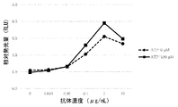
- FIG. 9 is a diagram showing the ATP concentration-dependent neutralizing activity of the anti-CTLA-4 antibody SW1077 against CTLA-4, as described in Example 3-6.
- FIG. 10 is a diagram showing the antitumor effect of the anti-CTLA-4 antibody mNS-mFa55 (control antibody) in a mouse model transplanted with an FM3A cell line as described in Example 3-7-4.
- FIG. 11 shows the antitumor effect of anti-CTLA-4 antibody SW1208-mFa55 (switch antibody) in a mouse model transplanted with FM3A cell line, as described in Example 3-7-4. Is.
- FIG. 11 shows the antitumor effect of anti-CTLA-4 antibody SW1208-mFa55 (switch antibody) in a mouse model transplanted with FM3A cell line, as described in Example 3-7-4. Is.
- FIG. 12 shows the administration of anti-CTLA-4 antibody mNS-mFa55 (control antibody) and SW1208-mFa55 (switch antibody) in a mouse model transplanted with FM3A cell line as described in Example 3-7-7. It is a figure which shows the change of the ratio of the effector Treg cell in the tumor.
- mNS-mFa55 was administered at 0.1 mg / kg, 1 mg / kg, 10 mg / kg, 100 mg / kg via the tail vein
- SW1208-mFa55 was administered at 0.1 mg / kg, 1 mg / kg, 10 mg / kg, It was administered from the tail vein at 100 mg / kg and 500 mg / kg.
- FIG. 13 shows the administration of anti-CTLA-4 antibody mNS-mFa55 (control antibody) and SW1208-mFa55 (switch antibody) in a mouse model transplanted with FM3A cell line as described in Example 3-7-8. It is a figure which shows the change of the ratio of activated helper T cells in the spleen.
- mNS-mFa55 was administered at 0.1 mg / kg, 1 mg / kg, 10 mg / kg, 100 mg / kg via the tail vein, and SW1208-mFa55 was administered at 0.1 mg / kg, 1 mg / kg, 10 mg / kg, It was administered from the tail vein at 100 mg / kg and 500 mg / kg.
- the spleen was sampled 6 days after administration, and the increase or decrease of activated helper T cells was evaluated by FACS analysis.
- FIG. 14 shows the antitumor effect of the anti-CTLA-4 antibody SW1389-mFa55 (switch antibody) in a mouse model transplanted with the Hepa1-6 / hGPC3 cell line as described in Example 4-3-5. It is a figure.
- FIG. 15 shows the antitumor effect of the anti-CTLA-4 antibody hNS-mFa55 (control antibody) in a mouse model transplanted with the Hepa1-6 / hGPC3 cell line as described in Example 4-3-5. It is a figure.
- FIG. 16 shows anti-CTLA-4 antibody hNS-mFa55 (control antibody) and SW1389-mFa55 (switch) in a mouse model transplanted with a Hepa1-6 / hGPC3 cell line as described in Example 4-3-8. It is a figure which shows the change of the ratio of effector Treg cells in a tumor at the time of administration (antibody).
- hNS-mFa55 0.1 mg / kg, 1 mg / kg, 10 mg / kg, 30 mg / kg, SW1389-mFa55 0.1 mg / kg, 1 mg / kg, 10 mg / kg, 100 mg / kg, 500 mg It was administered from the tail vein at / kg. Tumors were collected 6 days after administration, and the increase or decrease of effector Treg was evaluated by FACS analysis. The vertical axis is the ratio of effector Treg (CD4 + FoxP3 + CCR7 low KLRG1 + ) to CD45 + cells. Shows the average value of n 3.
- FIG. 17 shows the anti-CTLA-4 antibody hNS-mFa55 (control antibody) and SW1389-mFa55 (switch) in a mouse model transplanted with a Hepa1-6 / hGPC3 cell line as described in Example 4-3-9. It is a figure which shows the change of the ratio of activated helper T cells in the spleen at the time of administration (antibody).
- hNS-mFa55 0.1 mg / kg, 1 mg / kg, 10 mg / kg, 30 mg / kg
- FIG. 18 shows the antitumor effect of the anti-CTLA-4 antibody SW1610-mFa55 (switch antibody) in a mouse model transplanted with a Hepa1-6 / hGPC3 cell line, as described in Examples 5-4-5. It is a figure.
- the antibody was administered from the tail vein at 0.3 mg / kg, 1 mg / kg, and 3 mg / kg.
- FIG. 21 shows anti-CTLA-4 antibodies SW1610-mFa55, SW1612-mFa55 and SW1615-mFa55 in a mouse model transplanted with a Hepa1-6 / hGPC3 cell line, as described in Examples 5-4-8.
- the negative control antibody KLH-mFa55 was administered at 400 mg / kg via the tail vein at kg, 200 mg / kg, 400 mg / kg. Tumors were collected 6 days after administration, and the increase or decrease of effector Treg was evaluated by FACS analysis.
- FIG. 22 shows anti-CTLA-4 antibodies SW1610-mFa55, SW1612-mFa55 and SW1615-mFa55 in a mouse model transplanted with a Hepa1-6 / hGPC3 cell line, as described in Examples 5-4-9. It is also a figure which shows the change of the ratio of activated helper T cells in the spleen at the time of administration of a switch antibody).
- the negative control antibody KLH-mFa55 was administered at 400 mg / kg via the tail vein at kg, 200 mg / kg, 400 mg / kg.
- the spleen was sampled 6 days after administration, and the increase or decrease of activated helper T cells was evaluated by FACS analysis.
- IgG1 is MDX10D1H-G1m / MDX10D1L-k0MT
- GASDALIE is MDX10D1H-GASDALIE / MDX10D1L-k0MT
- ART6 is MDX10D1H-Kn462 / MDX10D1H-Hl445 / MDX10D1L-k0MT -Represents Hl443 / MDX10D1L-k0MT respectively.
- IgG1 is an antibody having a control constant region
- GASDALIE is an antibody having a constant region described in the prior art
- ART6 and ART8 are antibodies having a modified constant region prepared in Example 6-1. is there.
- FIG. 24 is a diagram showing a comparison of ADCP activity in vitro of antibodies having various modified constant regions with enhanced binding to Fc ⁇ R, as described in Example 6-3.
- IgG1 is MDX10D1H-G1m / MDX10D1L-k0MT
- GASDIE is MDX10D1H-GASDIE / MDX10D1L-k0MT
- ART6 is MDX10D1H-Kn462 / MDX10D1H-Hl445 / MDX10D1L-k0MT -Represents Hl443 / MDX10D1L-k0MT respectively.
- IgG1 is an antibody having a control constant region
- GASDIE is an antibody having a constant region described in the prior art
- ART6 and ART8 are antibodies having a modified constant region prepared in Example 6-1. is there.
- FIG. 25 is a diagram showing ADCC activity of the anti-CTLA4 switch antibody SW1389-ART6 in vitro having a modified constant region with enhanced binding to Fc ⁇ R, as described in Example 6-4.
- FIG. 26 is a diagram showing ADCC activity of the anti-CTLA4 switch antibody SW1610-ART6 in vitro having a modified constant region with enhanced binding to Fc ⁇ R, as described in Example 6-4.
- FIG. 27 is a diagram showing ADCC activity of the anti-CTLA4 switch antibody SW1612-ART6 in vitro having a modified constant region with enhanced binding to Fc ⁇ R, as described in Example 6-4.
- FIG. 28 shows the neutralizing activity of the anti-CTLA4 switch antibody SW1389 against CTLA4 (the activity of releasing the signal of CTLA4 that suppresses the activation of effector cells) as described in Example 6-5. It is a figure which shows.
- FIG. 29 shows the neutralizing activity of the anti-CTLA4 switch antibody SW1610 against CTLA4 (the activity of releasing the signal of CTLA4 that suppresses the activation of effector cells) as described in Example 6-5.
- FIG. 30 shows the neutralizing activity of the anti-CTLA4 switch antibody SW1612 against CTLA4 (the activity of releasing the signal of CTLA4 that suppresses the activation of effector cells) as described in Example 6-5. It is a figure which shows.
- FIG. 29 shows the neutralizing activity of the anti-CTLA4 switch antibody SW1610 against CTLA4 (the activity of releasing the signal of CTLA4 that suppresses the activation of effector cells) as described in Example 6-5.
- FIG. 30 shows the neutralizing activity of the
- FIG. 31 shows the neutralizing activity of the anti-CTLA4 switch antibody SW1615 on CTLA4 (the activity of releasing the signal of CTLA4 that suppresses the activation of effector cells) as described in Example 6-5. It is a figure which shows.
- FIG. 32 is a diagram showing the in vitro cytotoxic activity of the anti-CTLA4 switch antibody SW1389-ART5 + ACT1 on CTLA4-positive regulatory T cells, as described in Examples 6-6.
- FIG. 33 is a diagram showing the in vitro cytotoxic activity of the anti-CTLA4 switch antibody SW1389-ART6 + ACT1 on CTLA4-positive regulatory T cells, as described in Examples 6-6.
- FIG. 34 is a diagram showing the in vitro cytotoxic activity of the anti-CTLA4 switch antibody SW1610-ART5 + ACT1 on CTLA4-positive regulatory T cells, as described in Examples 6-6.
- FIG. 35 is a diagram showing the in vitro cytotoxic activity of the anti-CTLA4 switch antibody SW1610-ART6 + ACT1 on CTLA4-positive regulatory T cells, as described in Examples 6-6.
- acceptor human framework is derived from the human immunoglobulin framework or human consensus framework defined below, the light chain variable domain (VL) framework or the heavy chain variable domain (VH).
- a framework that includes the amino acid sequence of the framework may contain the same amino acid sequence thereof, or may include a modification of the amino acid sequence.
- the number of amino acid changes is 10 or less, 9 or less, 8 or less, 7 or less, 6 or less, 5 or less, 4 or less, 3 or less, or 2 or less.
- the VL acceptor human framework is sequence identical to the VL human immunoglobulin framework sequence or human consensus framework sequence.
- Antibody-dependent cell-mediated cytotoxicity or “ADCC” (antibody-dependent cell-mediated cytotoxicity) means that the secreted immunoglobulin is on specific cytotoxic cells (eg, NK cells, neutrophils and macrophages). It binds to Fc receptors (FcR) present in the cell, which allows these cytotoxic effector cells to specifically bind to antigen-bearing target cells, which are then killed by cellular cytotoxicity. It refers to a form of cellular cytology that enables you to do so.
- NK cells which are the primary cells that mediate ADCC, express only Fc ⁇ RIII, and monocytes express Fc ⁇ RI, Fc ⁇ RII, and Fc ⁇ RIII.
- FcR FcR on hematopoietic cells
- Table 3 The expression of FcR on hematopoietic cells is summarized in Table 3 on page 464 of Ravetch and Kinet, Annu. Rev. Immunol 9: 457-92 (1991).
- in vitro ADCC measurements can be performed, such as those described in US Pat. No. 5,500,362 or 5,821,337 or US Pat. No. 6,737,056 (Presta). Effector cells useful for such methods include PBMC and NK cells.
- the ADCC activity of the molecule of interest may be assessed in vivo in an animal model such as the animal model disclosed in Clynes et al. PNAS (USA) 95: 652-656 (1998). ..
- cytotoxic activity examples include antibody-dependent cell-mediated cytotoxicity (ADCC) activity described above, complement-dependent cytotoxicity (CDC) activity described later, and the like. And cytotoxic activity by T cells and the like.
- CDC activity means cytotoxic activity by the complement system.
- ADCC activity means an activity in which an antibody binds to an antigen existing on the cell surface of a target cell, and further, the effector cell binds to the antibody, so that the effector cell damages the target cell.
- ADCC activity means an activity in which an antibody binds to an antigen existing on the cell surface of a target cell, and further, the effector cell binds to the antibody, so that the effector cell damages the target cell.
- ADCC activity means an activity in which an antibody binds to an antigen existing on the cell surface of a target cell, and further, the effector cell binds to the antibody, so that the effector cell damages the target cell.
- Neutralizing activity refers to an activity that inhibits the biological activity by binding an antibody to a molecule involved in some biological activity.
- biological activity is provided by ligand-receptor binding.
- the antibody binds to the ligand or receptor, thereby inhibiting the binding of the ligand to the receptor.
- Antibodies having such neutralizing activity are called neutralizing antibodies.
- the neutralizing activity of a test substance can be measured by comparing the biological activity in the presence of the ligand between the conditions in the presence or absence of the test substance.
- ADCP antibody-dependent cellular phagocytosis
- phagocytic immune cells eg, macrophages, in which either all or part of the antibody-covered cells bind to the immunoglobulin Fc region. It means a process that is taken up inside (neutrophils, and dendritic cells).
- binding activity refers to the non-covalent interaction between one or more binding sites of a molecule (eg, an antibody) and a molecule's binding partner (eg, an antigen). It refers to the total strength.
- binding activity is not strictly limited to a 1: 1 interaction between members of a binding pair (eg, antibody and antigen). For example, when the members of a binding pair reflect a 1: 1 interaction in monovalent, the binding activity refers to the unique binding affinity (“affinity”). If the members of the binding pair are capable of both monovalent and multivalent binding, the binding activity is the sum of these binding forces.
- binding activity of the molecule X to its partner Y can generally be expressed by the dissociation constant (KD) or the "analyte binding amount per unit ligand amount". Binding activity can be measured by conventional methods known in the art, including those described herein. Specific examples and exemplary embodiments for measuring binding activity are described below.
- an “affinity mature” antibody is one or more modifications that result in improved affinity of the antibody for the antigen in one or more hypervariable regions (HVR) compared to the unmodified parent antibody. It refers to an antibody accompanied by.
- HVR hypervariable regions
- anti-CTLA-4 antibody or "antibody that binds to CTLA-4" is an antibody that can bind to CTLA-4 with sufficient affinity, so that the antibody targets CTLA-4.
- the degree of binding of an anti-CTLA-4 antibody to an unrelated non-CTLA-4 protein is the binding of the antibody to CTLA-4 when measured (eg, by radioimmunoassay (RIA)). It is less than about 10% of.
- the antibody that binds CTLA-4 is ⁇ 1 ⁇ M, ⁇ 100 nM, ⁇ 10 nM, ⁇ 1 nM, ⁇ 0.1 nM, ⁇ 0.01 nM, or ⁇ 0.001 nM (eg, 10 -8 M or less). , For example, 10 -8 M to 10 -13 M, for example, 10 -9 M to 10 -13 M).
- the anti-CTLA-4 antibody binds to a CTLA-4 epitope conserved between CTLA-4 from different species.
- antibody is used in the broadest sense, and is not limited to, but is not limited to, a monoclonal antibody, a polyclonal antibody, and a multispecific antibody (for example, as long as it exhibits a desired antigen-binding activity). Includes various antibody structures, including bispecific antibodies) and antibody fragments.
- Antibody fragment refers to a molecule other than the complete antibody, which comprises a portion of the complete antibody that binds to the antigen to which the complete antibody binds.
- Examples of antibody fragments are, but are not limited to, Fv, Fab, Fab', Fab'-SH, F (ab') 2 ; diabody; linear antibody; single chain antibody molecule (eg, scFv). ); And includes multispecific antibodies formed from antibody fragments.
- an "antibody that binds to the same epitope" as a reference antibody is an antibody that blocks, for example, 50% or more of the reference antibody from binding to its own antigen in a competitive assay, and / or a reference antibody is a competitive assay. In, for example, 50% or more of the above-mentioned antibody blocks binding to its own antigen.
- An exemplary competitive assay is provided herein.
- autoimmune diseases refers to a non-malignant disease or disorder that arises from the individual's own tissue and is directed at the individual's own tissue.
- autoimmune diseases explicitly exclude malignant or cancerous diseases or conditions, especially B-cell lymphoma, acute lymphoblastic leukemia (ALL), and chronic lymphocytes. Exclude chronic lymphocytic leukemia (CLL), hairy cell leukemia, and chronic myeloblastic leukemia.
- autoimmune diseases or disorders include, but are not limited to, inflammatory reactions such as inflammatory skin diseases including psoriasis and dermatitis (eg, atopic dermatitis); systemic Syndrome and sclerosis; Reactions associated with inflammatory bowel disease (eg, Crohn's disease and ulcerative colitis); Respiratory distress syndrome (adult respiratory distress syndrome: including ARDS); Dermatitis; Menelitis; encephalitis; vegetationitis; colitis; glomerular nephritis; allergic conditions such as eczema and asthma and other conditions with T cell infiltration and chronic inflammatory response; atherosclerosis; leukocyte adhesion failure; joints Rheumatoid; systemic lupus erythematosus (SLE) (including but not limited to lupus nephritis, cutaneous lupus); diabetes (eg, type I diabetes or insulin-dependent diabetes); multiple sclerosis; Raynaud's syndrome; autoimmune Immune thyroiditis; Ha
- cancer and “cancerous” refer to or describe a physiological condition in a mammal that is typically characterized by unregulated cell growth / proliferation.
- examples of cancer include breast cancer and liver cancer.
- complement-dependent cytotoxicity refers to cell death in which the Fc effector domain of an antibody bound to a target activates a series of enzymatic reactions resulting in the formation of holes in the membrane of the target cell. Means a mechanism for inducing.
- the antigen-antibody complex formed on the target cell binds to and activates complement component C1q, which in turn activates the complement cascade to kill the target cell. Bring. Activation of complement may also result in the deposition of complement components on the surface of target cells, which promotes ADCC by binding to complement receptors on leukocytes (eg, CR3). ..
- “Chemotherapy agent” refers to a chemical compound useful for the treatment of cancer.
- chemotherapeutic agents include: alkylating agents such as thiotepa and cyclophosphamide (CYTOXAN®); alkyl sulfonates such as busulfan, improsulfan, and piposulfan; benzodopa, carbocon.
- alkylating agents such as thiotepa and cyclophosphamide (CYTOXAN®)
- alkyl sulfonates such as busulfan, improsulfan, and piposulfan
- benzodopa carbocon.
- Aziridines such as meturedopa, and uredopa; ethyleneimine and methylolmelamine, including altretamine, triethylenemelamine, triethylenephosphorumamide, triethylenethiophosphoramide, and trimetyromeramine; acetogenin (particularly bratacin).
- Delta-9-tetrahydrocannabinol (Dronavinol, MARINOL®); Beta-rapacon; Lapacol; Corhitin; Betulic acid; Camptothecin (synthetic analog topotecan (HYCAMTIN®), CPT-11 (Including irinotecan, CAMPTOSAR®), acetylcamptothecin, scopolectin, and 9-aminocamptothecin); briostatin; calistatin; CC-1065 (including its adzelesin, calzelesin, and bizelesin synthetic analogs); Podophilotoxin; Podophyllic acid; Teniposide; Cryptothecin (particularly cryptophycin 1 and cryptophycin 8); Drastatin; Duocarmycin (including synthetic analogs KW-2189 and CB1-TM1); Eloyterobin; Pankratisstatin; Sarcodi Cutin; spongistatin
- Nitrogen mustard such as carmustin, chlorozothocin, hotemstin, romustin, nimustin, and ranimustin; engineinote antibiotics ⁇ eg, calikeamycin, especially calikeamycin gamma 1I and calikeamycin omega I1 (eg Nicolaou et al., Angelw. Chem Intl. Ed.
- Anti-adr such as aminoglutetimide, mitotan, trilostane, etc. enals
- Folic acid supplements such as phoric acid; acegraton; aldphosphamide glycoside; aminolevulinic acid; eniluracil; amsacrine; bestlabsyl; vinorelbine; edatorexate; defofamine; demecortin; diaziquone; elflornitine Elliptinium acetate; Epotylon; Etoglucid; Gallium nitrate; Hydrochiurea; Lentinan; Ronidamine; Maytansinoids such as maytancin and ansamitecin; Mitoguazone; Mitoxaliplatin; Mopidamole; Nitraerine; Pentostatin; Phenamet; Pirarubicin; Losoxanthron; 2-ethylhydrazide; procarbazine; PSK® polysaccharide complex (JHS Natural Products
- Vaccines such as ALLOVECTIN® vaccine, LEUVECTIN® vaccine, and VAXID® vaccine
- topoisomerase 1 inhibitors eg, LURTOTECAN®
- rmRH eg, ABARELIX®
- BAY439006 solafenib; Bayer
- SU-11248 subunitinib, SUTENT®, Pfizer
- perihosin, COX-2 inhibitor eg, selecoxib or etricoxyb
- proteosome inhibitor eg, PS341
- bortezomib eg, PS341) VELCADE®
- CCI-779 tipifarnib (R11577); sorafenib, ABT510; oblimersen sodium (GENASENSE®
- CHOP which is an abbreviation for vincristine and predonisolone combination therapy
- FOLFOX which is an abbreviation for treatment regimen with oxaliplatin (ELOXANTIN TM) in combination with 5-FU and leucovorin.
- chimeric antibody is one in which a portion of the heavy chain and / or light chain is derived from a particular source or species, while the rest of the heavy chain and / or light chain is derived from a different source or species. It refers to an antibody.
- the "class" of an antibody refers to the type of constant domain or constant region in the heavy chain of the antibody.
- Heavy chain constant domains corresponding to different classes of immunoglobulins are referred to as ⁇ , ⁇ , ⁇ , ⁇ , and ⁇ , respectively.
- cytotoxic agent refers to a substance that inhibits or impedes cell function and / or causes cell death or destruction.
- Cell-damaging agents are, but are not limited to, radioactive isotopes (eg, 211 At, 131 I, 125 I, 90 Y, 186 Re, 188 Re, 153 Sm, 212 Bi, 32 P, 212 Pb.
- chemotherapeutic agents or chemotherapeutic agents eg methotrexate, adriamycin, binca alkaloids (vincristine, vinblastin, etopocid), doxorubicin, melfaran, mitomycin C, chlorambusyl, daunorubicin, or other intercurry Drugs
- Growth inhibitors Enzymes such as nucleic acid-degrading enzymes and fragments thereof;
- Antibiotics For example, small molecule toxins or enzymatically active toxins of bacterial, fungal, plant, or animal origin (fragments and / or variants thereof) Includes toxins (including the body); and various chemotherapeutic agents disclosed above.
- Effective cells are leukocytes that express one or more FcRs and exert effector functions.
- the cells express at least Fc ⁇ RIII and exert ADCC effector function.
- white blood cells that mediate ADCC include peripheral blood mononuclear cells (PBMC), natural killer (NK) cells, monocytes, cytotoxic T cells and neutrophils.
- Effector cells can be isolated from natural sources, such as blood. In certain embodiments, the effector cells can be human effector cells.
- “Effector function” refers to the biological activity caused by the Fc region of an antibody, which differs depending on the isotype of the antibody.
- Examples of antibody effector function include: C1q binding and complement-dependent cytotoxicity (CDC); Fc receptor binding; antibody-dependent cellular cytotoxicity (CDC).
- epitope includes any determinant that can be bound by an antibody.
- An epitope is a region of an antigen that is bound by an antibody that targets the antigen and comprises certain amino acids that come into direct contact with the antibody.
- Epitope determinants can include chemically active surface molecules such as amino acids, sugar side chains, phosphoryl groups or sulfonyl groups and have specific three-dimensional structural and / or specific charge properties. Can be done.
- an antibody specific for a particular target antigen preferentially recognizes an epitope on that target antigen in a complex mixture of proteins and / or macromolecules.
- Fc receptor refers to a receptor that binds to the Fc region of an antibody.
- the FcR is a native human FcR.
- the FcR is one that binds to an IgG antibody (gamma receptor) and forms the receptors of the Fc ⁇ RI, Fc ⁇ RII, and Fc ⁇ RIII subclasses by allelic variants and alternative splicing of these receptors.
- Fc ⁇ RII receptors include Fc ⁇ RIIA (“activating receptor”) and Fc ⁇ RIIB (“inhibiting receptor”), which have similar amino acid sequences that differ primarily in their cytoplasmic domain.
- the activation receptor Fc ⁇ RIIA contains an immunoreceptor tyrosine-based activation motif (ITAM) in its cytoplasmic domain.
- the inhibitory receptor Fc ⁇ RIIB contains an immunoreceptor tyrosine-based inhibition motif (ITIM) in its cytoplasmic domain (see, for example, Daeron, Annu. Rev. Immunol. 15: 203-234 (1997)). thing).
- FcR is, for example, Ravetch and Kinet, Annu. Rev. Immunol 9: 457-492 (1991); Capel et al., Immunomethods 4: 25-34 (1994); and de Haas et al., J. Lab. Clin. It is reviewed in Med 126: 330-341 (1995).
- Other FcRs, including those identified in the future, are also included in the term "FcR" herein.
- Fc receptor or “FcR” also refers to the transfer of maternal IgG to the fetal (Guyer et al., J. Immunol. 117: 587 (1976) and Kim et al., J. Immunol. 24: 249. (1994)) and includes the neonatal receptor FcRn, which is responsible for the regulation of immunoglobulin homeostasis. Methods for measuring binding to FcRn are known (eg, Ghetie and Ward., Immunol. Today 18 (12): 592-598 (1997); Ghetie et al., Nature Biotechnology, 15 (7): 637- 640 (1997); Hinton et al., J. Biol. Chem. 279 (8): 6213-6216 (2004); See WO2004 / 92219 (Hinton et al.)).
- Binding to human FcRn and serum half-life of human FcRn high affinity binding polypeptides in vivo can be determined, for example, in transgenic mouse or transfected human cell lines expressing human FcRn, or in polypeptides with mutant Fc regions. Can be measured in administered primates.
- WO2000 / 42072 (Presta) describes antibody variants with improved or reduced binding to FcR. See also Shields et al. J. Biol. Chem. 9 (2): 6591-6604 (2001).
- the term "Fc region” is used to define the C-terminal region of an immunoglobulin heavy chain that contains at least part of the constant region.
- the term includes the Fc region of a native sequence and the mutant Fc region.
- the human IgG heavy chain Fc region extends from Cys226 or from Pro230 to the carboxyl terminus of the heavy chain.
- the C-terminal lysine (Lys447) or glycine-lysine (Gly446-Lys447) in the Fc region may or may not be present.
- the numbering of amino acid residues in the Fc region or constant region is Kabat et al., Sequences of Proteins of Immunological Interest, 5th Ed. Public Health Service, National Institutes of Health, Bethesda, follow the EU numbering system (also known as the EU index) described in MD 1991.
- Fc region-containing antibody refers to an antibody containing an Fc region.
- the C-terminal lysine of the Fc region (residue 447 according to the EU numbering system) or the C-terminal glycine-lysine of the Fc region (residues 446-447) is, for example, during antibody purification or the nucleic acid encoding the antibody. Can be removed by the recombination operation of. Therefore, a composition containing an antibody having an Fc region according to the present invention is an antibody with G446-K447, an antibody with G446 without K447, an antibody from which G446-K447 is completely removed, or an antibody of the above three types. May include a mixture of.
- variable region refers to the heavy or light chain domain of an antibody involved in binding the antibody to an antigen.
- the heavy and light chain variable domains of native antibodies are similar, with each domain usually containing four conserved framework regions (FR) and three hypervariable regions (HVR). It has a structure (see, for example, Kindt al. Kuby Immunology, 6th ed., WH Freeman and Co., page 91 (2007)).
- One VH or VL domain will be sufficient to confer antigen binding specificity.
- antibodies that bind to a particular antigen may be isolated by screening complementary libraries of VL or VH domains using the VH or VL domains from antibodies that bind to that antigen, respectively. See, for example, Portolano et al., J. Immunol. 150: 880-887 (1993); Clarkson et al., Nature 352: 624-628 (1991).
- variable domain FR refers to variable domain residues other than hypervariable region (HVR) residues.
- a variable domain FR usually consists of four FR domains: FR1, FR2, FR3, and FR4.
- sequences of HVR and FR usually appear in VH (or VL) in the following order: FR1-H1 (L1) -FR2-H2 (L2) -FR3-H3 (L3) -FR4.
- full-length antibody “complete antibody,” and “whole antibody” are used interchangeably herein and have a structure substantially similar to that of a native antibody, or are defined herein.
- the “functional Fc region” has the "effector function” of the natural sequence Fc region.
- effector functions include C1q binding; CDC; Fc receptor binding; ADCC; phagocytosis; down-regulation of cell surface receptors (eg, B cell receptors; B cell receptors: BCR) and the like.
- Such effector functions generally require the Fc region to be combined with a binding domain (eg, an antibody variable domain) and are evaluated using, for example, the various metrics disclosed in the definitions herein. Can be done.
- human antibody is an antibody having an amino acid sequence corresponding to the amino acid sequence of an antibody produced by a human or human cell or an antibody derived from a non-human source using a human antibody repertoire or other human antibody coding sequence. This definition of human antibody explicitly excludes humanized antibodies that contain non-human antigen-binding residues.
- the "Human Consensus Framework” is a framework that shows the most commonly occurring amino acid residues in the selection group of human immunoglobulin VL or VH framework sequences.
- the selection of human immunoglobulin VL or VH sequences is from a subgroup of variable domain sequences.
- the subgroups of the array are the subgroups in Kabat et al., Sequences of Proteins of Immunological Interest, Fifth Edition, NIH Publication 91-3242, Bethesda MD (1991), vols. 1-3.
- the subgroup is the subgroup ⁇ I by Kabat et al. Above.
- the subgroup is Subgroup III by Kabat et al. Above.
- a “humanized” antibody is a chimeric antibody that contains an amino acid residue from a non-human HVR and an amino acid residue from a human FR.
- the humanized antibody comprises substantially all of at least one, typically two, variable domains, in which all or substantially all HVRs (eg, CDRs) are non-existent.
- all or substantially all FRs correspond to those of human antibodies.
- the humanized antibody may optionally include at least a portion of the antibody constant region derived from the human antibody.
- the "humanized form" of an antibody (eg, a non-human antibody) refers to an antibody that has undergone humanization.
- hypervariable region or “HVR” are hypervariable in sequence (“complementarity determining regions” or “CDRs”) and / or structurally defined. Refers to each region of the variable domain of an antibody that forms a loop (“hypervariable loop”) and / or contains antigen contact residues (“antigen contact”). Usually, the antibody contains 6 HVRs: 3 for VH (H1, H2, H3) and 3 for VL (L1, L2, L3).
- Illustrative HVRs herein include: (a) At amino acid residues 26-32 (L1), 50-52 (L2), 91-96 (L3), 26-32 (H1), 53-55 (H2), and 96-101 (H3).
- the resulting hypervariable loop (Chothia and Lesk, J. Mol. Biol. 196: 901-917 (1987)); (b) At amino acid residues 24-34 (L1), 50-56 (L2), 89-97 (L3), 31-35b (H1), 50-65 (H2), and 95-102 (H3).
- the resulting CDR (Kabat et al., Sequences of Proteins of Immunological Interest, 5th Ed. Public Health Service, National Institutes of Health, Bethesda, MD (1991)); (c) At amino acid residues 27c-36 (L1), 46-55 (L2), 89-96 (L3), 30-35b (H1), 47-58 (H2), and 93-101 (H3).
- HVR residues and other residues in the variable domain are numbered herein according to Kabat et al., Supra.
- Immunoconjugate is an antibody conjugated to one or more heterologous molecules (heterologous molecules include, but are not limited to, cytotoxic agents).
- an “isolated” antibody is one that has been isolated from its original environmental components.
- the antibody is subjected to, for example, electrophoresis (eg, SDS-PAGE, isoelectric focusing (IEF), capillary electrophoresis) or chromatograph (eg, ion exchange or reverse phase HPLC). Measured and purified to a purity greater than 95% or 99%. See, for example, Flatman et al., J. Chromatogr. B 848: 79-87 (2007) for a review of methods for assessing antibody purity.
- electrophoresis eg, SDS-PAGE, isoelectric focusing (IEF), capillary electrophoresis
- chromatograph eg, ion exchange or reverse phase HPLC
- isolated nucleic acid refers to a nucleic acid molecule isolated from its original environmental components.
- An isolated nucleic acid contains a nucleic acid molecule contained within a cell that normally contains the nucleic acid molecule, but the nucleic acid molecule is on a chromosome that is extrachromosomal or different from its original chromosomal location. Exists in position.
- nucleic acid encoding an antibody refers to one or more nucleic acid molecules encoding the heavy and light chains (or fragments thereof) of an antibody, on one vector or separate vectors. Includes nucleic acid molecules that are present and nucleic acid molecules that are present at one or more positions in the host cell.
- vector refers to a nucleic acid molecule that can augment another nucleic acid to which it is linked.
- the term includes a vector as a self-replicating nucleic acid structure and a vector incorporated into the genome of the host cell into which it has been introduced. Certain vectors can result in the expression of nucleic acids to which they are operably linked. Such vectors are also referred to herein as "expression vectors.”
- host cell refers to cells into which foreign nucleic acids have been introduced, including progeny of such cells. ..
- Host cells include “transformants” and “transformants”, which include primary transformants and progeny derived from those cells regardless of the number of passages.
- the offspring do not have to be exactly the same in the content of the parent cell and nucleic acid and may contain mutations.
- Variant progeny with the same function or biological activity as those used when the original transformed cells were screened or selected are also included herein.
- mutant antibodies eg., mutant antibodies that contain naturally occurring mutations, or mutant antibodies that occur during the production of monoclonal antibody preparations, such variants are usually slightly present. Except for (amounts present), they are identical and / or bind to the same epitope.
- Each monoclonal antibody in a monoclonal antibody preparation is for a single determinant on an antigen, as opposed to a polyclonal antibody preparation that typically comprises different antibodies against different determinants (epitopes).
- the modifier "monoclonal” should not be construed as requiring the production of an antibody by any particular method, indicating the characteristic of the antibody that it is obtained from a substantially homogeneous population of antibodies.
- the monoclonal antibody used according to the present invention is not limited to these, but is a hybridoma method, a recombinant DNA method, a phage display method, and a transgenic animal containing all or a part of the human immunoglobulin locus. It may be made by a variety of methods, including methods that utilize, such methods and other exemplary methods for making monoclonal antibodies are described herein.
- naked antibody refers to an antibody that is not conjugated to a heterologous part (eg, cytotoxic part) or radiolabel.
- the naked antibody may be present in the pharmaceutical formulation.
- Natural antibody refers to an immunoglobulin molecule with various naturally occurring structures.
- a native IgG antibody is a heterotetrameric glycoprotein of approximately 150,000 daltons composed of two identical disulfide-bonded light chains and two identical heavy chains.
- VH variable region
- CH2, and CH3 constant domains
- each light chain has a variable region (VL), also called a variable light chain domain or a light chain variable domain, followed by a stationary light chain (CL) domain.
- VH variable region
- VL variable region
- the light chain of an antibody may be assigned to one of two types, called kappa ( ⁇ ) and lambda ( ⁇ ), based on the amino acid sequence of its constant domain.
- the “natural sequence Fc region” includes the same amino acid sequence as the amino acid sequence of the Fc region found in nature.
- the natural sequence human Fc region is the natural sequence human IgG1 Fc region (non-A and A allotype); the natural sequence human IgG2 Fc region; the natural sequence human IgG3 Fc region; and the natural sequence human IgG4 Fc region, and the natural Includes those variants present in.
- the "mutant Fc region” includes an amino acid sequence different from that of the natural sequence Fc region due to at least one amino acid modification (modification), preferably one or more amino acid substitutions.
- the mutant Fc region has at least one amino acid substitution in the native sequence Fc region or in the Fc region of the parent polypeptide, eg, about 1 to, as compared to the native sequence Fc region or the Fc region of the parent polypeptide. It has about 10 amino acid substitutions, preferably about 1 to about 5 amino acid substitutions.
- the mutant Fc regions herein are preferably at least about 80% homologous to the native sequence Fc region and / or the Fc region of the parent polypeptide, more preferably at least about 90% homology to them, most preferably. Have at least about 95% homology with them.
- Percent (%) amino acid sequence identity to a reference polypeptide sequence is any conservative substitution sequence after aligning the sequences for maximum percent sequence identity and, if necessary, introducing gaps. It is defined as the percentage ratio of amino acid residues in the candidate sequence that are identical to the amino acid residues in the reference polypeptide sequence when not considered part of the identity. Alignment for the purpose of determining percent amino acid sequence identity can be done by various methods within the skill of the art, such as BLAST, BLAST-2, ALIGN, Megalign (DNASTAR) software, or GENETYX®®. This can be achieved by using publicly available computer software such as Company Genetics. One of skill in the art can determine the appropriate parameters for sequence alignment, including any algorithm required to achieve maximum alignment over the overall length of the sequence being compared.
- the ALIGN-2 Sequence Comparison Computer Program is the work of Genetec, the source code of which was submitted to the United States Copyright Office (Wasington DC, 20559) along with user documentation under the United States Copyright Registration Number TXU510087. It is registered.
- the ALIGN-2 program is publicly available from Genentech, Inc., South San Francisco, California and may be compiled from source code.
- the ALIGN-2 program is compiled for use on UNIX operating systems, including Digital UNIX V4.0D. All sequence comparison parameters are set by the ALIGN-2 program and do not fluctuate. In situations where ALIGN-2 is used for amino acid sequence comparison,% amino acid sequence identity (or given amino acid sequence) of a given amino acid sequence A to, to, or to a given amino acid sequence B.
- a given amino acid sequence A which has or contains some% amino acid sequence identity to, to, or to B) is calculated as: Hundredfold.
- X is the number of amino acid residues scored by the sequence alignment program ALIGN-2 as a match that is identical in the alignment of A and B in that program
- Y is the total number of amino acid residues in B. .. It will be appreciated that if the length of amino acid sequence A is not equal to the length of amino acid sequence B, then the% amino acid sequence identity of A to B is not equal to the% amino acid sequence identity of B to A. Let's go. Unless otherwise stated, all% amino acid sequence identity values used herein are obtained using the ALIGN-2 computer program as described in the previous paragraph.
- pharmaceutical preparation is a preparation in a form in which the biological activity of the active ingredient contained therein can exert its effect, and to the extent unacceptable to the subject to which the preparation is administered.
- the "individual” or “target” is a mammal. Mammals are, but are not limited to, domestic animals (eg, cows, sheep, cats, dogs, horses), primates (eg, non-human primates such as humans and monkeys), rabbits, and , Includes primates (eg, mice and rats). In certain embodiments, the individual or subject is a human.
- “Pharmaceutically acceptable carrier” refers to an ingredient other than the active ingredient in a pharmaceutical formulation that is non-toxic to the subject.
- Pharmaceutically acceptable carriers include, but are not limited to, buffers, excipients, stabilizers, or preservatives.
- an "effective amount" of an agent refers to an amount at a required dose and over a required period of time that is effective in achieving the desired therapeutic or prophylactic result. ..
- package insert is commonly included in commercial packaging of therapeutic products and contains information about indications, dosages, doses, dosage regimens, concomitant therapies, contraindications, and / or warnings regarding the use of such therapeutic products. It is used to refer to the instruction manual.
- CTLA-4" as used herein is from any vertebrate source, including mammals such as primates (eg, humans) and rodents (eg, mice and rats), unless otherwise indicated. Refers to any natural CTLA-4.
- the term includes CTLA-4 that has not undergone “full length” processing, as well as any form of CTLA-4 that results from intracellular processing.
- the term also includes naturally occurring variants of CTLA-4, such as splice variants and allelic variants.
- CTLA-4 amino acid sequence is set to SEQ ID NO: 214
- a mouse CTLA-4 amino acid sequence is set to SEQ ID NO: 247
- a monkey CTLA-4 amino acid sequence is set to SEQ ID NO: 248
- a human CTLA-4 amino acid sequence is set to SEQ ID NO: 248.
- the amino acid sequence of the extracellular domain is shown in SEQ ID NO: 28.
- CTLA-4 may be referred to as CTLA4.
- Treg cells are a subpopulation of T cells that regulate the immune system, maintain tolerance to autoantigens, and suppress autoimmune diseases. These cells generally suppress or down-regulate the induction and proliferation of effector T cells.
- the best understood Treg cells are those that express CD4, CD25, and Foxp3 (CD4 + CD25 + Treg cells). These Tregs are different from helper T cells.
- Several different methods are used to identify and monitor Treg cells. When defined by CD4 and CD25 expression (CD4 + CD25 + cells), Treg cells make up about 5-10% of the mature CD4 + T cell subpopulation in mice and humans, while about 1-2% of Tregs. Can be measured in whole blood.
- Foxp3 expression may be added and measured (CD4 + CD25 + Foxp3 + cells). Also, as another marker, the absence or low level expression of CD127 may be used in combination with the presence of CD4 and CD25.
- Treg cells also express high levels of CTLA-4 and GITR. Tregs can also be identified by the methods described in the Examples below.
- the terms “substantially similar,” “substantially equal,” or “substantially the same” are referred to / compared between two numbers (eg, with respect to the antibodies of the invention). Similarities (between those relating to antibodies), those skilled in the art will have little or no biological and / at all in terms of the biological characteristics in which the difference between those two numbers is measured by the number (eg, KD value). Or, it is high enough to be regarded as not statistically significant.
- treatment is clinically intended to alter the natural course of the individual being treated. It means intervention and can be performed both for prevention and during the course of the clinical condition.
- the desired effects of treatment are, but are not limited to, prevention of the onset or recurrence of the disease, relief of symptoms, reduction of any direct or indirect pathological effects of the disease, prevention of metastasis, prevention of the disease, of the disease. Includes reduced rate of progression, recovery or alleviation of disease state, and ameliorated or improved prognosis.
- the antibodies of the invention are used to delay the onset of a disease or slow the progression of a disease.
- tumor refers to all neoplastic cell growth and proliferation and all precancerous and cancerous cells and tissues, whether malignant or benign.
- cancer refers to all neoplastic cell growth and proliferation and all precancerous and cancerous cells and tissues, whether malignant or benign.
- cancer refers to all neoplastic cell growth and proliferation and all precancerous and cancerous cells and tissues, whether malignant or benign.
- cancer refers to all neoplastic cell growth and proliferation and all precancerous and cancerous cells and tissues, whether malignant or benign.
- tumor tissue means a tissue containing at least one tumor cell.
- Tumor tissue usually consists of a population of tumor cells (parenchyma) that is the main component of the tumor, and connective tissue and blood vessels (interstitium) that exist between them and support the tumor. Some have a clear distinction between the two, while others are a mixture of the two. Immune cells may infiltrate the tumor tissue.
- non-tumor tissue means a tissue other than the tumor tissue in the living body. Healthy / normal tissue that is not in a diseased state is a typical example of non-tumor tissue.
- the invention is based in part on anti-CTLA-4 antibodies and their use.
- antibodies that bind CTLA-4 are provided.
- the antibodies of the present invention are useful, for example, for the diagnosis or treatment of cancer.
- the invention provides isolated antibodies that bind to CTLA-4.
- the anti-CTLA-4 antibodies of the invention have CTLA-4 binding activity that depends on the concentration of the adenosine-containing compound.
- the binding activity to CTLA-4 is higher in the presence of the adenosine-containing compound than in the absence of the adenosine-containing compound.
- the binding activity to CTLA-4 is higher in the presence of a high concentration of adenosine-containing compound than in the presence of a low concentration of adenosine-containing compound.
- the difference in binding activity to CTLA-4 is, for example, 2-fold or greater, 3-fold or greater, 5-fold or greater, 10-fold or greater, 20-fold or greater, 30-fold or greater, 50-fold or greater, 100-fold or greater, 200-fold.
- 300 times or more 500 times or more, 1 x 10 3 times or more, 2 x 10 3 times or more, 3 x 10 3 times or more, 5 x 10 3 times or more, 1 x 10 4 times or more, 2 x 10 4 times or more
- the binding activity of the anti-CTLA-4 antibody can be represented by a KD (Dissociation constant) value.
- the KD value of the anti-CTLA-4 antibody in the presence of the adenosine-containing compound is smaller than in the absence of the adenosine-containing compound.
- the KD value of the anti-CTLA-4 antibody is smaller in the presence of a high concentration of adenosine-containing compound than in the presence of a low concentration of adenosine-containing compound.
- the difference in KD value of anti-CTLA-4 antibody is, for example, 2 times or more, 3 times or more, 5 times or more, 10 times or more, 20 times or more, 30 times or more, 50 times or more, 100 times or more, 200 times. Double or more, 300 times or more, 500 times or more, 1 ⁇ 10 3 times or more, 2 ⁇ 10 3 times or more, 3 ⁇ 10 3 times or more, 5 ⁇ 10 3 times or more, 1 ⁇ 10 4 times or more, 2 ⁇ 10 4 More than double, 3 x 10 4 times or more, 5 x 10 4 times or more, or 1 x 10 5 times or more.
- the KD value of the anti-CTLA-4 antibody in the presence of an adenosine-containing compound or in the presence of a high concentration of adenosine-containing compound is, for example, 9 ⁇ 10 -7 M or less, 8 ⁇ 10 -7 M or less, 7 ⁇ 10 -7.
- Absence of adenosine-containing compound, or KD value of anti-CTLA-4 antibody in the presence of low concentrations of adenosine-containing compound for example 1 ⁇ 10 -8 M or more, 2 ⁇ 10 -8 M or more, 3 ⁇ 10 - 8 M or more, 4 ⁇ 10 -8 M or more, 5 ⁇ 10 -8 M or more, 6 ⁇ 10 -8 M or more, 7 ⁇ 10 -8 M or more, 8 ⁇ 10 -8 M or more, 9 ⁇ 10 -8 M or more Above, 1 x 10 -7 M or above, 2 x 10 -7 M or above, 3 x 10 -7 M or above, 4 x 10 -7 M or above, 5 x 10 -7 M or above, 6 x 10 -7 M or above, 7 ⁇ 10 -7 M or more, 8 ⁇ 10 -7 M or more, 9 ⁇ 10 -7 M or more, 1 ⁇ 10 -6 M or more, 2 ⁇ 10 -6 M or more, 3 ⁇ 10 -6 M or more
- the binding activity of the anti-CTLA-4 antibody may be expressed by a kd (Dissociation rate constant) value instead of the KD value.
- the binding activity of the anti-CTLA-4 antibody may be expressed as the amount of CTLA-4 bound per unit antibody amount.
- the amount of antibody bound on the sensor chip and the amount of antigen bound thereto are measured as reaction units (RU), respectively.
- the value obtained by dividing the antigen binding amount there by the antibody binding amount can be defined as the antigen binding amount per unit antibody amount. Specific methods for measuring and calculating such a binding amount are described in Examples described later.
- the amount of CTLA-4 bound is greater in the presence of the adenosine-containing compound than in the absence of the adenosine-containing compound.
- the amount of CTLA-4 bound is greater in the presence of a high concentration of adenosine-containing compound than in the presence of a low concentration of adenosine-containing compound.
- the difference in the binding amount of CTLA-4 is, for example, 2 times or more, 3 times or more, 5 times or more, 10 times or more, 20 times or more, 30 times or more, 50 times or more, 100 times or more, 200 times or more.
- the value of CTLA-4 binding in the presence of an adenosine-containing compound or a high concentration of adenosine-containing compound is, for example, 0.01 or more, 0.02 or more, 0.03 or more, 0.04 or more, 0.05 or more, 0.06 or more, 0.07 or more, It can be 0.08 or more, 0.09 or more, 0.1 or more, 0.2 or more, 0.3 or more, 0.4 or more, 0.5 or more, 0.6 or more, 0.7 or more, 0.8 or more, 0.9 or more, or 1 or more.
- the value of CTLA-4 binding in the absence of adenosine-containing compound or in the presence of low-concentration adenosine-containing compound is, for example, 0.5 or less, 0.4 or less, 0.3 or less, 0.2 or less, 0.1 or less, 0.09 or less, 0.08 or less. , 0.07 or less, 0.06 or less, 0.05 or less, 0.04 or less, 0.03 or less, 0.02 or less, 0.01 or less, 0.009 or less, 0.008 or less, 0.007 or less, 0.006 or less, 0.005 or less, 0.004 or less, 0.003 or less, 0.002 or less, or 0.001 or less There can be.
- the KD value, kd value, binding amount value, etc. represented herein are measured or calculated by performing a surface plasmon resonance assay at 25 ° C or 37 ° C (eg,). See Example 3 herein).
- concentration of the adenosine-containing compound any concentration can be selected as long as a difference in the binding activity of the anti-CTLA-4 antibody is detected.
- high concentrations include, for example, 1 nM or higher, 3 nM or higher, 10 nM or higher, 30 nM or higher, 100 nM or higher.
- the high concentration here can be sufficient so that each anti-CTLA-4 antibody exhibits maximum binding activity.
- 1 ⁇ M, 10 ⁇ M, 100 ⁇ M, 1 mM, or a sufficient amount such that each anti-CTLA-4 antibody exhibits maximum binding activity can be selected as the high concentration here.
- low concentrations include, for example, 1 mM or lower, 300 ⁇ M or lower, 100 ⁇ M or lower, 30 ⁇ M or lower, 10 ⁇ M or lower. , 3 ⁇ M or lower, 1 ⁇ M or lower, 300 nM or lower, 100 nM or lower, 30 nM or lower, 10 nM or lower, 3 nM or lower concentration, 1 nM or lower concentration, 300 pM or lower concentration, 100 pM or lower concentration, 30 pM or lower concentration, 10 pM or lower concentration, 3 pM or lower concentration Lower concentrations, 1 pM or less, etc. can be mentioned.
- the concentration at which each anti-CTLA-4 antibody exhibits the minimum binding activity can be set to the low concentration here.
- the case where the substantial concentration is zero (in the absence of the adenosine-containing compound) can also be selected as one embodiment of the low concentration.
- 1 mM, 100 ⁇ M, 10 ⁇ M, 1 ⁇ M, the concentration at which each anti-CTLA-4 antibody exhibits the minimum binding activity, or the absence of an adenosine compound is selected as the low concentration here. be able to.
- the ratio of high concentration to low concentration is, for example, 3 times or more, 10 times or more, 30 times or more, 100 times or more, 300 times or more, 1 ⁇ 10 3 times or more, 3 ⁇ 10 3 times or more, 1 ⁇ 10 4 times or more, 3 ⁇ 10 4 fold or more, 1 ⁇ 10 5 times or more, 3 ⁇ 10 5 times or more, 1 ⁇ 10 6 times or more, 3 ⁇ 10 6 times or more, 1 ⁇ 10 7 times or more, 3 ⁇ 10 7 times or more, 1 ⁇ 10 8 times or more, 3 ⁇ 10 8 times Or more, 1 x 10 9 times or more, 3 x 10 9 times or more, 1 x 10 10 times or more, 3 x 10 10 times or more, 1 x 10 11 times or more, 3 You can select a value of ⁇ 10 11 times or more, and 1 ⁇ 10 12 times or more.
- the anti-CTLA-4 antibody of the present invention also has binding activity to an adenosine-containing compound.
- the amount of binding of the adenosine-containing compound per unit antibody amount can be calculated and used as the binding activity of the anti-CTLA-4 antibody to the adenosine-containing compound. Specific methods for measuring and calculating such a binding amount are described in Examples described later.
- the value of the binding amount of the adenosine-containing compound per unit antibody amount of the anti-CTLA-4 antibody of the present invention is, for example, 0.0001 or more, 0.0002 or more, 0.0003 or more, 0.0004 or more, 0.0005 or more, 0.0006 or more, 0.0007 or more, 0.0008 or more, 0.0009. It can be 0.001 or more, 0.002 or more, 0.003 or more, 0.004 or more, 0.005 or more, 0.006 or more, 0.007 or more, 0.008 or more, 0.009 or more, or 0.01 or more.
- the anti-CTLA-4 antibody of the present invention forms a tripartite complex with an adenosine-containing compound and CTLA-4.
- the anti-CTLA-4 antibody binds to an adenosine-containing compound via heavy chains CDR1, CDR2, CDR3.
- the anti-CTLA-4 antibody has a binding motif to an adenosine-containing compound. Binding motifs for adenosine-containing compounds exist, for example, at positions 33, 52, 52a, 53, 56, 58, 95, 96, 100a, 100b, and 100c represented by Kabat numbering. It may be composed of at least one amino acid.
- the anti-CTLA-4 antibody is represented by, for example, Kabat numbering at positions 33, 52, 52a, 53, 56, 58, 95, 96, 100a, 100b, 100c. It binds to an adenosine-containing compound via at least one amino acid selected from the group consisting of.
- the anti-CTLA-4 antibody is represented by Kabat numbering, Thr at 33, Ser at 52, Ser at 52a, Arg at 53, Tyr at 56, Tyr at 58, Tyr at 95. It has at least one amino acid selected from the group consisting of Tyr at position 96, Met at position 100a, Leu at position 100b, and Trp at position 100c.
- CTLA-4 may further bind to the complex formed by binding the anti-CTLA-4 antibody and the adenosine-containing compound.
- the adenosine-containing compound may be present at the interface where the anti-CTLA-4 antibody and CTLA-4 interact and bind to both of them. It can be confirmed that the anti-CTLA-4 antibody forms a tripartite complex together with the adenosine-containing compound and CTLA-4 by a method such as crystal structure analysis described later (see Examples).
- the anti-CTLA-4 antibody of the present invention comprises the third amino acid (Met), the 33rd amino acid (Glu), and the 35th amino acid of human CTLA-4 (extracellular domain, SEQ ID NO: 28).
- These amino acids may constitute epitopes for the anti-CTLA-4 antibodies of the invention.
- the anti-CTLA-4 antibody of the invention binds to the region of amino acids 97-106 (Leu) of human CTLA-4 (extracellular domain, SEQ ID NO: 28). .. In another embodiment, the anti-CTLA-4 antibody of the invention binds to the region of amino acids 99 to 106 (Leu) of human CTLA-4 (extracellular domain, SEQ ID NO: 28). ..
- the anti-CTLA-4 antibody of the invention relates to ABAM004 (VH, SEQ ID NO: 10; VL, SEQ ID NO: 11; HVR-H1, SEQ ID NO: 100; HVR- H2, SEQ ID NO: 101; HVR-H3, SEQ ID NO: 102; HVR-L1, SEQ ID NO: 113; HVR-L2, SEQ ID NO: 114; HVR-L3, SEQ ID NO: 115).
- the anti-CTLA-4 antibody of the invention binds to the same epitope as ABAM004.
- the binding of ABAM004 to CTLA-4 is, for example, 10% or more, 15% or more, 20% or more, 25% or more, 30% or more, 35% or more, 40% or more. , 45% or more, 50% or more, 55% or more, 60% or more, 65% or more, 70% or more, 75% or more, 80% or more, 85% or more, 90% or more, or 95% or more can be reduced. ..
- An exemplary competitive assay is provided herein.
- the anti-CTLA-4 antibody of the present invention exhibits cytotoxic activity against CTLA-4 expressing cells. If CTLA-4 is expressed on the surface of a target cell and an anti-CTLA-4 antibody binds to it, the cell can be injured. Cell damage is caused by antibody-bound effector cells such as antibody-dependent cellular cytotoxicity (ADCC) and antibody-dependent cellular cytotoxicity (ADCP). It may be, or it may be caused by antibody-bound cellular cytotoxicity (CDC) or the like. Alternatively, it may be caused by an antibody-bound cytotoxic agent (eg, a radioisotope, a chemotherapeutic agent, etc.), such as an immunoconjugate.
- ADCC antibody-dependent cellular cytotoxicity
- ADCP antibody-dependent cellular cytotoxicity
- CDC antibody-bound cellular cytotoxicity
- an antibody-bound cytotoxic agent eg, a radioisotope, a chemotherapeutic agent, etc.
- the cytotoxicity here may include an action of inducing cell death, an action of suppressing cell proliferation, an action of impairing cell function, and the like.
- a sufficient amount of anti-CTLA-4 antibody is present, for example, 10% or more, 15% or more, 20% or more, 25% or more, 30% or more, 35% or more, 40% or more, 45% or more, 50% or more. , 55% or more, 60% or more, 65% or more, 70% or more, 75% or more, 80% or more, 85% or more, 90% or more, or 95% or more of CTLA-4 expressing cells.
- Measurements of such cytotoxic activity can be made in comparison to measurements in the absence of antibodies or in the presence of negative control antibodies.
- An exemplary cytotoxic assay is provided herein.
- the anti-CTLA-4 antibody of the present invention exhibits neutralizing activity against CTLA-4.
- CTLA-4 is known to function by interacting with its ligands, CD80 (B7-1) or CD86 (B7-2).
- the anti-CTLA-4 antibody inhibits the interaction of CTLA-4 with CD80 (B7-1) or CD86 (B7-2).
- the interaction between CTLA-4 and CD80 (B7-1) or CD86 (B7-2) is, for example, 10% or more, 15% or more, 20% or more, 25.
- % Or more, 30% or more, 35% or more, 40% or more, 45% or more, 50% or more, 55% or more, 60% or more, 65% or more, 70% or more, 75% or more, 80% or more, 85% or more , 90% or more, or 95% or more can be inhibited. Measurements of such inhibitory activity can be made in comparison to measurements in the absence of antibodies or in the presence of negative control antibodies. Specific methods for measuring neutralizing activity are provided herein.
- the anti-CTLA-4 antibody of the present invention binds to CTLA-4 derived from multiple animal species.
- animal species include mammals such as humans, monkeys, mice, rats, hamsters, guinea pigs, rabbits, pigs, cows, goats, horses, sheep, camels, dogs and cats.
- the anti-CTLA-4 antibody binds CTLA-4 from human and non-human (eg, monkey, mouse, rat, etc.).
- the amino acid sequence of human CTLA-4 is shown in SEQ ID NO: 214
- the amino acid sequence of monkey CTLA-4 is shown in SEQ ID NO: 247
- the amino acid sequence of mouse CTLA-4 is shown in SEQ ID NO: 248.
- the amino acid sequence of CTLA-4 derived from other animal species can also be appropriately determined by a method known to those skilled in the art.
- the adenosine-containing compounds in the present invention include, for example, adenosine (ADO), adenosine triphosphate (ATP), adenosine diphosphate (ADP), adenosine monophosphate (AMP), cyclic adenosine monophosphate. (CAMP), deoxyadenosine (dADO), deoxyadenosine triphosphate (dATP), deoxyadenosine diphosphate (dADP), deoxyadenosine monophosphate (dAMP), adenosine ⁇ -thiotriphosphate (ATP ⁇ S), etc. Can be done.
- ADO adenosine
- ATP adenosine triphosphate
- ADP adenosine diphosphate
- AMP adenosine monophosphate
- CAMP cyclic adenosine monophosphate
- deoxyadenosine (dADO) deoxyadenosine triphosphate
- the invention comprises (a) HVR-H1 comprising the amino acid sequence of SEQ ID NO: 223; (b) HVR-H2 comprising the amino acid sequence of SEQ ID NO: 224; and (c) amino acids of SEQ ID NO: 225.
- the antibody comprises (a) HVR-H1 comprising the amino acid sequence of SEQ ID NO: 223; (b) HVR-H2 comprising the amino acid sequence of SEQ ID NO: 224; (c) amino acid of SEQ ID NO: 225. Includes HVR-H3 containing sequences.
- the invention comprises (a) HVR-L1 comprising the amino acid sequence of SEQ ID NO: 226; (b) HVR-L2 comprising the amino acid sequence of SEQ ID NO: 227; and (c) SEQ ID NO: 228.
- an antibody comprising at least one, at least two, or all three VL HVR sequences selected from HVR-L3 containing an amino acid sequence.
- the antibody comprises (a) HVR-L1 comprising the amino acid sequence of SEQ ID NO: 226; (b) HVR-L2 comprising the amino acid sequence of SEQ ID NO: 227; (c) amino acid of SEQ ID NO: 228. Includes HVR-L3 containing sequences.
- the antibody of the present invention is (a) VH domain, (i) HVR-H1 containing the amino acid sequence of SEQ ID NO: 223, (ii) HVR- containing the amino acid sequence of SEQ ID NO: 224.
- VH domain containing at least one, at least two, or all three VH HVR sequences selected from H2 and HVR-H3 containing the amino acid sequence of (iii) SEQ ID NO: 225;
- VL A domain containing i) HVR-L1 containing the amino acid sequence of SEQ ID NO: 226, (ii) HVR-L2 containing the amino acid sequence of SEQ ID NO: 227, and (c) containing the amino acid sequence of SEQ ID NO: 228.
- VL domains including at least one, at least two, or all three VL HVR sequences selected from HVR-L3.
- the present invention comprises (a) HVR-H1 containing the amino acid sequence of SEQ ID NO: 223; (b) HVR-H2 containing the amino acid sequence of SEQ ID NO: 224; (c) SEQ ID NO: 225. HVR-H3 containing the amino acid sequence of; (d) HVR-L1 containing the amino acid sequence of SEQ ID NO: 226; (e) HVR-L2 containing the amino acid sequence of SEQ ID NO: 227; (f) SEQ ID NO:: Provided is an antibody comprising HVR-L3 containing an amino acid sequence selected from 228.
- the invention comprises (a) HVR-H1 comprising the amino acid sequence of SEQ ID NO: 100; (b) HVR-H2 comprising the amino acid sequence of SEQ ID NO: 101; and (c) amino acids of SEQ ID NO: 102.
- antibodies comprising at least one, at least two, or all three VH HVR sequences selected from HVR-H3 containing sequences.
- the antibody comprises (a) HVR-H1 comprising the amino acid sequence of SEQ ID NO: 100; (b) HVR-H2 comprising the amino acid sequence of SEQ ID NO: 101; (c) amino acid of SEQ ID NO: 102. Includes HVR-H3 containing sequences.
- the invention comprises (a) HVR-L1 comprising the amino acid sequence of SEQ ID NO: 113; (b) HVR-L2 comprising the amino acid sequence of SEQ ID NO: 114; and (c) SEQ ID NO: 115.
- an antibody comprising at least one, at least two, or all three VL HVR sequences selected from HVR-L3 containing an amino acid sequence.
- the antibody comprises (a) HVR-L1 comprising the amino acid sequence of SEQ ID NO: 113; (b) HVR-L2 comprising the amino acid sequence of SEQ ID NO: 114; (c) amino acid of SEQ ID NO: 115. Includes HVR-L3 containing sequences.
- the antibody of the present invention is (a) VH domain, (i) HVR-H1 containing the amino acid sequence of SEQ ID NO: 100, (ii) HVR- containing the amino acid sequence of SEQ ID NO: 101.
- VH domain containing at least one, at least two, or all three VH HVR sequences selected from H2 and HVR-H3 containing the amino acid sequence of (iii) SEQ ID NO: 102;
- VL A domain containing i) HVR-L1 containing the amino acid sequence of SEQ ID NO: 113, (ii) HVR-L2 containing the amino acid sequence of SEQ ID NO: 114, and (c) containing the amino acid sequence of SEQ ID NO: 115.
- VL domains including at least one, at least two, or all three VL HVR sequences selected from HVR-L3.
- the present invention comprises (a) HVR-H1 containing the amino acid sequence of SEQ ID NO: 100; (b) HVR-H2 containing the amino acid sequence of SEQ ID NO: 101; (c) SEQ ID NO: 102. HVR-H3 containing the amino acid sequence of; (d) HVR-L1 containing the amino acid sequence of SEQ ID NO: 113; (e) HVR-L2 containing the amino acid sequence of SEQ ID NO: 114; (f) SEQ ID NO:: Provided are an antibody comprising HVR-L3 containing an amino acid sequence selected from 115.
- the invention comprises (a) HVR-H1 comprising the amino acid sequence of SEQ ID NO: 100; (b) HVR-H2 comprising the amino acid sequence of SEQ ID NO: 104; and (c) amino acids of SEQ ID NO: 102.
- the antibody comprises (a) HVR-H1 comprising the amino acid sequence of SEQ ID NO: 100; (b) HVR-H2 comprising the amino acid sequence of SEQ ID NO: 104; (c) amino acid of SEQ ID NO: 102. Includes HVR-H3 containing sequences.
- the invention comprises (a) HVR-L1 comprising the amino acid sequence of SEQ ID NO: 116; (b) HVR-L2 comprising the amino acid sequence of SEQ ID NO: 117; and (c) SEQ ID NO: 115.
- an antibody comprising at least one, at least two, or all three VL HVR sequences selected from HVR-L3 containing an amino acid sequence.
- the antibody comprises (a) HVR-L1 comprising the amino acid sequence of SEQ ID NO: 116; (b) HVR-L2 comprising the amino acid sequence of SEQ ID NO: 117; (c) amino acid of SEQ ID NO: 115. Includes HVR-L3 containing sequences.
- the antibody of the present invention is (a) VH domain, (i) HVR-H1 containing the amino acid sequence of SEQ ID NO: 100, (ii) HVR- containing the amino acid sequence of SEQ ID NO: 104.
- VH domain containing at least one, at least two, or all three VH HVR sequences selected from H2 and HVR-H3 containing the amino acid sequence of (iii) SEQ ID NO: 102;
- VL A domain containing i) HVR-L1 containing the amino acid sequence of SEQ ID NO: 116, (ii) HVR-L2 containing the amino acid sequence of SEQ ID NO: 117, and (c) containing the amino acid sequence of SEQ ID NO: 115.
- VL domains including at least one, at least two, or all three VL HVR sequences selected from HVR-L3.
- the present invention comprises (a) HVR-H1 containing the amino acid sequence of SEQ ID NO: 100; (b) HVR-H2 containing the amino acid sequence of SEQ ID NO: 104; (c) SEQ ID NO: 102. HVR-H3 containing the amino acid sequence of; (d) HVR-L1 containing the amino acid sequence of SEQ ID NO: 116; (e) HVR-L2 containing the amino acid sequence of SEQ ID NO: 117; (f) SEQ ID NO:: Provided is an antibody comprising HVR-L3 containing an amino acid sequence selected from 115.
- the invention comprises (a) HVR-H1 comprising the amino acid sequence of SEQ ID NO: 105; (b) HVR-H2 comprising the amino acid sequence of SEQ ID NO: 106; and (c) amino acids of SEQ ID NO: 102.
- the antibody comprises (a) HVR-H1 comprising the amino acid sequence of SEQ ID NO: 105; (b) HVR-H2 comprising the amino acid sequence of SEQ ID NO: 106; (c) amino acid of SEQ ID NO: 102. Includes HVR-H3 containing sequences.
- the invention comprises (a) HVR-L1 comprising the amino acid sequence of SEQ ID NO: 122; (b) HVR-L2 comprising the amino acid sequence of SEQ ID NO: 117; and (c) SEQ ID NO: 133.
- an antibody comprising at least one, at least two, or all three VL HVR sequences selected from HVR-L3 containing an amino acid sequence.
- the antibody comprises (a) HVR-L1 comprising the amino acid sequence of SEQ ID NO: 122; (b) HVR-L2 comprising the amino acid sequence of SEQ ID NO: 117; (c) amino acid of SEQ ID NO: 133. Includes HVR-L3 containing sequences.
- the antibody of the present invention is (a) VH domain, (i) HVR-H1 containing the amino acid sequence of SEQ ID NO: 105, (ii) HVR- containing the amino acid sequence of SEQ ID NO: 106.
- VH domain containing at least one, at least two, or all three VH HVR sequences selected from H2 and HVR-H3 containing the amino acid sequence of (iii) SEQ ID NO: 102;
- VL A domain containing i) HVR-L1 containing the amino acid sequence of SEQ ID NO: 122, (ii) HVR-L2 containing the amino acid sequence of SEQ ID NO: 117, and (c) containing the amino acid sequence of SEQ ID NO: 133.
- VL domains including at least one, at least two, or all three VL HVR sequences selected from HVR-L3.
- the present invention comprises (a) HVR-H1 containing the amino acid sequence of SEQ ID NO: 105; (b) HVR-H2 containing the amino acid sequence of SEQ ID NO: 106; (c) SEQ ID NO: 102. HVR-H3 containing the amino acid sequence of; (d) HVR-L1 containing the amino acid sequence of SEQ ID NO: 122; (e) HVR-L2 containing the amino acid sequence of SEQ ID NO: 117; (f) SEQ ID NO:: Provided is an antibody comprising HVR-L3 containing an amino acid sequence selected from 133.
- the invention comprises (a) HVR-H1 comprising the amino acid sequence of SEQ ID NO: 107; (b) HVR-H2 comprising the amino acid sequence of SEQ ID NO: 108; and (c) amino acids of SEQ ID NO: 102.
- the antibody comprises (a) HVR-H1 comprising the amino acid sequence of SEQ ID NO: 107; (b) HVR-H2 comprising the amino acid sequence of SEQ ID NO: 108; (c) amino acid of SEQ ID NO: 102. Includes HVR-H3 containing sequences.
- the invention comprises (a) HVR-L1 comprising the amino acid sequence of SEQ ID NO: 121; (b) HVR-L2 comprising the amino acid sequence of SEQ ID NO: 123; and (c) SEQ ID NO: 153.
- an antibody comprising at least one, at least two, or all three VL HVR sequences selected from HVR-L3 containing an amino acid sequence.
- the antibody comprises (a) HVR-L1 comprising the amino acid sequence of SEQ ID NO: 121; (b) HVR-L2 comprising the amino acid sequence of SEQ ID NO: 123; (c) amino acid of SEQ ID NO: 153. Includes HVR-L3 containing sequences.
- the antibody of the present invention is (a) VH domain, (i) HVR-H1 containing the amino acid sequence of SEQ ID NO: 107, (ii) HVR- containing the amino acid sequence of SEQ ID NO: 108.
- VH domain containing at least one, at least two, or all three VH HVR sequences selected from H2 and HVR-H3 containing the amino acid sequence of (iii) SEQ ID NO: 102;
- VL A domain containing i) HVR-L1 containing the amino acid sequence of SEQ ID NO: 121, (ii) HVR-L2 containing the amino acid sequence of SEQ ID NO: 123, and (c) containing the amino acid sequence of SEQ ID NO: 153.
- VL domains including at least one, at least two, or all three VL HVR sequences selected from HVR-L3.
- the present invention comprises (a) HVR-H1 containing the amino acid sequence of SEQ ID NO: 107; (b) HVR-H2 containing the amino acid sequence of SEQ ID NO: 108; (c) SEQ ID NO: 102. HVR-H3 containing the amino acid sequence of; (d) HVR-L1 containing the amino acid sequence of SEQ ID NO: 121; (e) HVR-L2 containing the amino acid sequence of SEQ ID NO: 123; (f) SEQ ID NO:: Provided is an antibody comprising HVR-L3 containing an amino acid sequence selected from 153.
- the present invention comprises (a) HVR-H1 comprising the amino acid sequence of SEQ ID NO: 107; (b) HVR-H2 comprising the amino acid sequence of SEQ ID NO: 110; and (c) amino acids of SEQ ID NO: 102.
- the antibody comprises (a) HVR-H1 comprising the amino acid sequence of SEQ ID NO: 107; (b) HVR-H2 comprising the amino acid sequence of SEQ ID NO: 110; (c) amino acid of SEQ ID NO: 102. Includes HVR-H3 containing sequences.
- the invention comprises (a) HVR-L1 comprising the amino acid sequence of SEQ ID NO: 122; (b) HVR-L2 comprising the amino acid sequence of SEQ ID NO: 117; and (c) SEQ ID NO: 133.
- an antibody comprising at least one, at least two, or all three VL HVR sequences selected from HVR-L3 containing an amino acid sequence.
- the antibody comprises (a) HVR-L1 comprising the amino acid sequence of SEQ ID NO: 122; (b) HVR-L2 comprising the amino acid sequence of SEQ ID NO: 117; (c) amino acid of SEQ ID NO: 133. Includes HVR-L3 containing sequences.
- the antibody of the present invention is (a) VH domain, (i) HVR-H1 containing the amino acid sequence of SEQ ID NO: 107, (ii) HVR- containing the amino acid sequence of SEQ ID NO: 110.
- VH domain containing at least one, at least two, or all three VH HVR sequences selected from H2 and HVR-H3 containing the amino acid sequence of (iii) SEQ ID NO: 102;
- VL A domain containing i) HVR-L1 containing the amino acid sequence of SEQ ID NO: 122, (ii) HVR-L2 containing the amino acid sequence of SEQ ID NO: 117, and (c) containing the amino acid sequence of SEQ ID NO: 133.
- VL domains including at least one, at least two, or all three VL HVR sequences selected from HVR-L3.
- the present invention comprises (a) HVR-H1 containing the amino acid sequence of SEQ ID NO: 107; (b) HVR-H2 containing the amino acid sequence of SEQ ID NO: 110; (c) SEQ ID NO: 102. HVR-H3 containing the amino acid sequence of; (d) HVR-L1 containing the amino acid sequence of SEQ ID NO: 122; (e) HVR-L2 containing the amino acid sequence of SEQ ID NO: 117; (f) SEQ ID NO:: Provided is an antibody comprising HVR-L3 containing an amino acid sequence selected from 133.
- the invention comprises (a) HVR-H1 comprising the amino acid sequence of SEQ ID NO: 107; (b) HVR-H2 comprising the amino acid sequence of SEQ ID NO: 112; and (c) amino acids of SEQ ID NO: 102.
- the antibody comprises (a) HVR-H1 comprising the amino acid sequence of SEQ ID NO: 107; (b) HVR-H2 comprising the amino acid sequence of SEQ ID NO: 112; (c) amino acid of SEQ ID NO: 102. Includes HVR-H3 containing sequences.
- the invention comprises (a) HVR-L1 comprising the amino acid sequence of SEQ ID NO: 128; (b) HVR-L2 comprising the amino acid sequence of SEQ ID NO: 117; and (c) SEQ ID NO: 133.
- an antibody comprising at least one, at least two, or all three VL HVR sequences selected from HVR-L3 containing an amino acid sequence.
- the antibody comprises (a) HVR-L1 comprising the amino acid sequence of SEQ ID NO: 128; (b) HVR-L2 comprising the amino acid sequence of SEQ ID NO: 117; (c) amino acid of SEQ ID NO: 133. Includes HVR-L3 containing sequences.
- the antibody of the present invention is (a) VH domain, (i) HVR-H1 containing the amino acid sequence of SEQ ID NO: 107, (ii) HVR- containing the amino acid sequence of SEQ ID NO: 112.
- VH domain containing at least one, at least two, or all three VH HVR sequences selected from H2 and HVR-H3 containing the amino acid sequence of (iii) SEQ ID NO: 102;
- VL A domain containing i) HVR-L1 containing the amino acid sequence of SEQ ID NO: 128, (ii) HVR-L2 containing the amino acid sequence of SEQ ID NO: 117, and (c) containing the amino acid sequence of SEQ ID NO: 133.
- VL domains including at least one, at least two, or all three VL HVR sequences selected from HVR-L3.
- the present invention comprises (a) HVR-H1 containing the amino acid sequence of SEQ ID NO: 107; (b) HVR-H2 containing the amino acid sequence of SEQ ID NO: 112; (c) SEQ ID NO: 102. HVR-H3 containing the amino acid sequence of; (d) HVR-L1 containing the amino acid sequence of SEQ ID NO: 128; (e) HVR-L2 containing the amino acid sequence of SEQ ID NO: 117; (f) SEQ ID NO:: Provided is an antibody comprising HVR-L3 containing an amino acid sequence selected from 133.
- the present invention comprises (a) HVR-H1 comprising the amino acid sequence of SEQ ID NO: 107; (b) HVR-H2 comprising the amino acid sequence of SEQ ID NO: 111; and (c) amino acids of SEQ ID NO: 152.
- the antibody comprises (a) HVR-H1 comprising the amino acid sequence of SEQ ID NO: 107; (b) HVR-H2 comprising the amino acid sequence of SEQ ID NO: 111; (c) amino acid of SEQ ID NO: 152. Includes HVR-H3 containing sequences.
- the invention comprises (a) HVR-L1 comprising the amino acid sequence of SEQ ID NO: 128; (b) HVR-L2 comprising the amino acid sequence of SEQ ID NO: 117; and (c) SEQ ID NO: 133.
- an antibody comprising at least one, at least two, or all three VL HVR sequences selected from HVR-L3 containing an amino acid sequence.
- the antibody comprises (a) HVR-L1 comprising the amino acid sequence of SEQ ID NO: 128; (b) HVR-L2 comprising the amino acid sequence of SEQ ID NO: 117; (c) amino acid of SEQ ID NO: 133. Includes HVR-L3 containing sequences.
- the antibody of the present invention is (a) VH domain, (i) HVR-H1 containing the amino acid sequence of SEQ ID NO: 107, (ii) HVR- containing the amino acid sequence of SEQ ID NO: 111.
- VH domain containing at least one, at least two, or all three VH HVR sequences selected from H2 and HVR-H3 containing the amino acid sequence of (iii) SEQ ID NO: 152;
- VL A domain containing i) HVR-L1 containing the amino acid sequence of SEQ ID NO: 128, (ii) HVR-L2 containing the amino acid sequence of SEQ ID NO: 117, and (c) containing the amino acid sequence of SEQ ID NO: 133.
- VL domains including at least one, at least two, or all three VL HVR sequences selected from HVR-L3.
- the present invention comprises (a) HVR-H1 containing the amino acid sequence of SEQ ID NO: 107; (b) HVR-H2 containing the amino acid sequence of SEQ ID NO: 111; (c) SEQ ID NO: 152. HVR-H3 containing the amino acid sequence of; (d) HVR-L1 containing the amino acid sequence of SEQ ID NO: 128; (e) HVR-L2 containing the amino acid sequence of SEQ ID NO: 117; (f) SEQ ID NO:: Provided is an antibody comprising HVR-L3 containing an amino acid sequence selected from 133.
- the invention comprises (a) HVR-H1 comprising the amino acid sequence of SEQ ID NO: 107; (b) HVR-H2 comprising the amino acid sequence of SEQ ID NO: 112; and (c) amino acids of SEQ ID NO: 102.
- the antibody comprises (a) HVR-H1 comprising the amino acid sequence of SEQ ID NO: 107; (b) HVR-H2 comprising the amino acid sequence of SEQ ID NO: 112; (c) amino acid of SEQ ID NO: 102. Includes HVR-H3 containing sequences.
- the invention comprises (a) HVR-L1 comprising the amino acid sequence of SEQ ID NO: 129; (b) HVR-L2 comprising the amino acid sequence of SEQ ID NO: 117; and (c) SEQ ID NO: 133.
- an antibody comprising at least one, at least two, or all three VL HVR sequences selected from HVR-L3 containing an amino acid sequence.
- the antibody comprises (a) HVR-L1 comprising the amino acid sequence of SEQ ID NO: 129; (b) HVR-L2 comprising the amino acid sequence of SEQ ID NO: 117; (c) amino acid of SEQ ID NO: 133. Includes HVR-L3 containing sequences.
- the antibody of the present invention is (a) VH domain, (i) HVR-H1 containing the amino acid sequence of SEQ ID NO: 107, (ii) HVR- containing the amino acid sequence of SEQ ID NO: 112.
- VH domain containing at least one, at least two, or all three VH HVR sequences selected from H2 and HVR-H3 containing the amino acid sequence of (iii) SEQ ID NO: 102;
- VL A domain containing i) HVR-L1 containing the amino acid sequence of SEQ ID NO: 129, (ii) HVR-L2 containing the amino acid sequence of SEQ ID NO: 117, and (c) containing the amino acid sequence of SEQ ID NO: 133.
- VL domains including at least one, at least two, or all three VL HVR sequences selected from HVR-L3.
- the present invention comprises (a) HVR-H1 containing the amino acid sequence of SEQ ID NO: 107; (b) HVR-H2 containing the amino acid sequence of SEQ ID NO: 112; (c) SEQ ID NO: 102. HVR-H3 containing the amino acid sequence of; (d) HVR-L1 containing the amino acid sequence of SEQ ID NO: 129; (e) HVR-L2 containing the amino acid sequence of SEQ ID NO: 117; (f) SEQ ID NO:: Provided is an antibody comprising HVR-L3 containing an amino acid sequence selected from 133.
- the present invention comprises (a) HVR-H1 comprising the amino acid sequence of SEQ ID NO: 107; (b) HVR-H2 comprising the amino acid sequence of SEQ ID NO: 111; and (c) amino acids of SEQ ID NO: 152.
- the antibody comprises (a) HVR-H1 comprising the amino acid sequence of SEQ ID NO: 107; (b) HVR-H2 comprising the amino acid sequence of SEQ ID NO: 111; (c) amino acid of SEQ ID NO: 152. Includes HVR-H3 containing sequences.
- the invention comprises (a) HVR-L1 comprising the amino acid sequence of SEQ ID NO: 129; (b) HVR-L2 comprising the amino acid sequence of SEQ ID NO: 117; and (c) SEQ ID NO: 133.
- an antibody comprising at least one, at least two, or all three VL HVR sequences selected from HVR-L3 containing an amino acid sequence.
- the antibody comprises (a) HVR-L1 comprising the amino acid sequence of SEQ ID NO: 129; (b) HVR-L2 comprising the amino acid sequence of SEQ ID NO: 117; (c) amino acid of SEQ ID NO: 133. Includes HVR-L3 containing sequences.
- the antibody of the present invention is (a) VH domain, (i) HVR-H1 containing the amino acid sequence of SEQ ID NO: 107, (ii) HVR- containing the amino acid sequence of SEQ ID NO: 111.
- VH domain containing at least one, at least two, or all three VH HVR sequences selected from H2 and HVR-H3 containing the amino acid sequence of (iii) SEQ ID NO: 152;
- VL A domain containing i) HVR-L1 containing the amino acid sequence of SEQ ID NO: 129, (ii) HVR-L2 containing the amino acid sequence of SEQ ID NO: 117, and (c) containing the amino acid sequence of SEQ ID NO: 133.
- VL domains including at least one, at least two, or all three VL HVR sequences selected from HVR-L3.
- the present invention comprises (a) HVR-H1 containing the amino acid sequence of SEQ ID NO: 107; (b) HVR-H2 containing the amino acid sequence of SEQ ID NO: 111; (c) SEQ ID NO: 152. HVR-H3 containing the amino acid sequence of; (d) HVR-L1 containing the amino acid sequence of SEQ ID NO: 129; (e) HVR-L2 containing the amino acid sequence of SEQ ID NO: 117; (f) SEQ ID NO:: Provided is an antibody comprising HVR-L3 containing an amino acid sequence selected from 133.
- the present invention comprises (a) HVR-H1 comprising the amino acid sequence of SEQ ID NO: 107; (b) HVR-H2 comprising the amino acid sequence of SEQ ID NO: 109; and (c) amino acids of SEQ ID NO: 102.
- the antibody comprises (a) HVR-H1 comprising the amino acid sequence of SEQ ID NO: 107; (b) HVR-H2 comprising the amino acid sequence of SEQ ID NO: 109; (c) amino acid of SEQ ID NO: 102. Includes HVR-H3 containing sequences.
- the invention comprises (a) HVR-L1 comprising the amino acid sequence of SEQ ID NO: 130; (b) HVR-L2 comprising the amino acid sequence of SEQ ID NO: 117; and (c) SEQ ID NO: 133.
- an antibody comprising at least one, at least two, or all three VL HVR sequences selected from HVR-L3 containing an amino acid sequence.
- the antibody comprises (a) HVR-L1 comprising the amino acid sequence of SEQ ID NO: 130; (b) HVR-L2 comprising the amino acid sequence of SEQ ID NO: 117; (c) amino acid of SEQ ID NO: 133. Includes HVR-L3 containing sequences.
- the antibody of the present invention is (a) VH domain, (i) HVR-H1 containing the amino acid sequence of SEQ ID NO: 107, (ii) HVR- containing the amino acid sequence of SEQ ID NO: 109.
- VH domain containing at least one, at least two, or all three VH HVR sequences selected from H2 and HVR-H3 containing the amino acid sequence of (iii) SEQ ID NO: 102;
- VL A domain containing i) HVR-L1 containing the amino acid sequence of SEQ ID NO: 130, (ii) HVR-L2 containing the amino acid sequence of SEQ ID NO: 117, and (c) containing the amino acid sequence of SEQ ID NO: 133.
- VL domains including at least one, at least two, or all three VL HVR sequences selected from HVR-L3.
- the present invention comprises (a) HVR-H1 containing the amino acid sequence of SEQ ID NO: 107; (b) HVR-H2 containing the amino acid sequence of SEQ ID NO: 109; (c) SEQ ID NO: 102. HVR-H3 containing the amino acid sequence of; (d) HVR-L1 containing the amino acid sequence of SEQ ID NO: 130; (e) HVR-L2 containing the amino acid sequence of SEQ ID NO: 117; (f) SEQ ID NO:: Provided is an antibody comprising HVR-L3 containing an amino acid sequence selected from 133.
- any one or more amino acids of the anti-CTLA-4 antibody described above have been replaced at the following HVR positions: -In HVR-H1 (SEQ ID NO: 223): Position 2 -In HVR-H2 (SEQ ID NO: 224): positions 4, 5, 7, 13, and 16 -In HVR-H3 (SEQ ID NO: 225): Position 3 -In HVR-L1 (SEQ ID NO: 226): Positions 1, 3, 6, 11, 12, and 14 -In HVR-L2 (SEQ ID NO: 227): positions 1, 3, 4, and 7 -In HVR-L3 (SEQ ID NO: 228): positions 1 and 10
- substitutions provided herein are conservative substitutions.
- any one or more of the following substitutions may be made in any combination: -In HVR-H1 (SEQ ID NO: 100): H2A, R or K -In HVR-H2 (SEQ ID NO: 101): S4T; R5Q; G7H; D13E or R; K16R -In HVR-H3 (SEQ ID NO: 102): K3A -In HVR-L1 (SEQ ID NO: 113): T1D, Q or E; T3P; D6G; N11T; Y12W; S14H -In HVR-L2 (SEQ ID NO: 114): E1F or Y; S3I; K4S; S7E or K -In HVR-L3 (SEQ ID NO: 115): S1Q; M10T
- the anti-CTLA-4 antibody has been humanized.
- the anti-CTLA-4 antibody comprises HVR in any of the above aspects and further comprises an acceptor human framework (eg, a human immunoglobulin framework or a human consensus framework).
- the anti-CTLA-4 antibody comprises HVR in any of the aforementioned embodiments and further comprises VH or VL comprising the FR sequence.
- the anti-CTLA-4 antibody comprises the following heavy and / or light chain variable domain FR sequences: For the heavy chain variable domain, FR1 has the amino acid sequence of any one of SEQ ID NOs: 229-232.
- FR2 contains the amino acid sequence of SEQ ID NO: 233
- FR3 contains the amino acid sequence of SEQ ID NO: 23
- FR4 contains the amino acid sequence of SEQ ID NO: 235.
- FR1 contains the amino acid sequence of any one of SEQ ID NOs: 236 to 23
- FR2 contains the amino acid sequence of any one of SEQ ID NOs: 240 to 241
- FR3 contains the amino acid sequence of any one of SEQ ID NOs: 240 to 241.
- FR4 contains any one of the amino acid sequences of SEQ ID NOs: 245-246.
- the anti-CTLA-4 antibody is at least 90%, 91%, 92%, 93%, 94%, 95%, 96%, 97%, 98%, for the amino acid sequence of SEQ ID NO: 10.
- VH sequences having at least 90%, 91%, 92%, 93%, 94%, 95%, 96%, 97%, 98%, or 99% identity are relative to the reference sequence.
- Substitution eg, conservative substitution
- insertion, or deletion but the anti-CTLA-4 antibody containing the sequence retains the ability to bind CTLA-4.
- the anti-CTLA-4 antibody comprises the VH sequence at SEQ ID NO: 10, including those containing post-translational modifications of that sequence.
- the VH comprises (a) HVR-H1 containing the amino acid sequence of SEQ ID NO: 100, (b) HVR-H2 containing the amino acid sequence of SEQ ID NO: 101, and (c) SEQ ID NO: 102.
- HVR-H3s containing amino acid sequences.
- Post-translational modifications include, but are not limited to, modification of heavy or light chain N-terminal glutamine or glutamate to pyroglutamylation by pyroglutamylation.
- Anti-CTLA-4 antibodies are provided that contain a light chain variable domain (VL) with sequence identity.
- VL light chain variable domain
- a VL sequence having at least 90%, 91%, 92%, 93%, 94%, 95%, 96%, 97%, 98%, or 99% identity is relative to the reference sequence.
- Substitution eg, conservative substitution
- insertion, or deletion but the anti-CTLA-4 antibody containing the sequence retains the ability to bind CTLA-4.
- the anti-CTLA-4 antibody comprises the VL sequence at SEQ ID NO: 11, including those containing post-translational modifications of that sequence.
- the VL comprises (a) HVR-L1 containing the amino acid sequence of SEQ ID NO: 113, (b) HVR-L2 containing the amino acid sequence of SEQ ID NO: 114, and (c) SEQ ID NO: 115.
- HVR-L3 Includes one, two, or three HVRs selected from HVR-L3 containing the amino acid sequence.
- Post-translational modifications include, but are not limited to, modification of heavy or light chain N-terminal glutamine or glutamate to pyroglutamylation by pyroglutamylation.
- Anti-CTLA-4 antibodies are provided that contain a light chain variable domain (VL) with sequence identity.
- VL light chain variable domain
- a VL sequence having at least 90%, 91%, 92%, 93%, 94%, 95%, 96%, 97%, 98%, or 99% identity is relative to the reference sequence.
- Substitution eg, conservative substitution
- insertion, or deletion but the anti-CTLA-4 antibody containing the sequence retains the ability to bind CTLA-4.
- the anti-CTLA-4 antibody comprises the VL sequence at SEQ ID NO: 149, including those containing post-translational modifications of that sequence.
- the VL comprises (a) HVR-L1 containing the amino acid sequence of SEQ ID NO: 130, (b) HVR-L2 containing the amino acid sequence of SEQ ID NO: 117, and (c) SEQ ID NO: 133.
- HVR-L3 Includes one, two, or three HVRs selected from HVR-L3 containing the amino acid sequence.
- Post-translational modifications include, but are not limited to, modification of heavy or light chain N-terminal glutamine or glutamate to pyroglutamylation by pyroglutamylation.
- an anti-CTLA-4 antibody comprising VH in any of the above embodiments and VL in any of the above embodiments.
- the antibody comprises the VH and VL sequences in SEQ ID NO: 10 and SEQ ID NO: 11, respectively, including those containing post-translational modifications of such sequences.
- the antibody comprises the VH and VL sequences in SEQ ID NO: 98 and SEQ ID NO: 99, respectively, including those containing post-translational modifications of such sequences.
- the antibody comprises the VH and VL sequences in SEQ ID NO: 83 and SEQ ID NO: 97, respectively, including those containing post-translational modifications of such sequences.
- the antibody comprises the VH and VL sequences in SEQ ID NO: 86 and SEQ ID NO: 134, respectively, including those containing post-translational modifications of such sequences. In one embodiment, the antibody comprises the VH and VL sequences in SEQ ID NO: 136 and SEQ ID NO: 95, respectively, including those containing post-translational modifications of that sequence. In one embodiment, the antibody comprises the VH and VL sequences in SEQ ID NO: 140 and SEQ ID NO: 146, respectively, including those containing post-translational modifications of such sequences.
- the antibody comprises the VH and VL sequences in SEQ ID NO: 141 and SEQ ID NO: 146, respectively, including those containing post-translational modifications of such sequences. In one embodiment, the antibody comprises the VH and VL sequences in SEQ ID NO: 140 and SEQ ID NO: 147, respectively, including those containing post-translational modifications of such sequences. In one embodiment, the antibody comprises the VH and VL sequences in SEQ ID NO: 141 and SEQ ID NO: 147, respectively, including those containing post-translational modifications of such sequences.
- the antibody comprises the VH and VL sequences in SEQ ID NO: 136 and SEQ ID NO: 149, respectively, including those containing post-translational modifications of such sequences.
- a heterogeneous anti-CTLA-4 antibody containing at least two different variable regions selected from the variable regions containing the VH and VL sequences described above is provided.
- the antibody contained post-translational modifications of the VH and VL sequences in SEQ ID NO: 140 and SEQ ID NO: 146, and the VH and VL sequences in SEQ ID NO: 141 and SEQ ID NO: 146, respectively. Including things.
- the antibody contained post-translational modifications of the VH and VL sequences in SEQ ID NO: 140 and SEQ ID NO: 147, and the VH and VL sequences in SEQ ID NO: 141 and SEQ ID NO: 147, respectively.
- Post-translational modifications include, but are not limited to, modification of heavy or light chain N-terminal glutamine or glutamate to pyroglutamylation by pyroglutamylation.
- the amino acid at the N-terminal of the heavy chain or light chain of the anti-CTLA-4 antibody provided herein is glutamine
- the amino acid at the N-terminal of the heavy chain or light chain of the anti-CTLA-4 antibody provided herein is glutamic acid
- the amino acid at the N-terminal of the heavy chain or light chain of the anti-CTLA-4 antibody provided herein is glutamic acid
- the amino acid may be replaced with glutamine
- the invention provides an antibody that binds to the same epitope as the anti-CTLA-4 antibody provided herein.
- antibodies that bind to the same epitopes as the antibodies listed in Tables 4, 9, 14, and 19 are provided.
- An antibody that binds to an epitope in a fragment of CTLA-4 containing at least one amino acid selected from the group consisting of the amino acid (Tyr) and the 106th amino acid (Leu) is provided.
- an antibody that binds to an epitope in a fragment of CTLA-4 consisting of amino acids 97 to 106 (Leu) of SEQ ID NO: 28 is provided. In certain embodiments, an antibody that binds to an epitope in a fragment of CTLA-4 consisting of amino acids 99 to 106 (Leu) of SEQ ID NO: 28 is provided.
- the anti-CTLA-4 antibody is a monoclonal antibody, including a chimeric, humanized, or human antibody.
- the anti-CTLA-4 antibody is an antibody fragment, such as, for example, an Fv, Fab, Fab', scFv, diabody, or F (ab') 2 fragment.
- the antibody is a full-length antibody, such as, for example, a complete IgG1 antibody or a complete IgG4 antibody, another antibody class or isotype as defined herein.
- the anti-CTLA-4 antibody of the present invention contains an Fc region.
- the anti-CTLA-4 antibody of the invention comprises a constant region.
- the constant region may be a heavy chain constant region (including an Fc region), a light chain constant region, or both.
- the Fc region is the Fc region of a native sequence.
- exemplary heavy chain constant regions derived from native antibodies include human IgG1 (SEQ ID NO: 249), human IgG2 (SEQ ID NO: 250), human IgG3 (SEQ ID NO: 251), human IgG4 (SEQ ID NO: 252).
- Other heavy chain constant regions include human IgG1 (SEQ ID NO: 249), human IgG2 (SEQ ID NO: 250), human IgG3 (SEQ ID NO: 251), human IgG4 (SEQ ID NO: 252).
- a heavy chain constant region such as SEQ ID NO: 82 and SEQ ID NO: 158 can be mentioned.
- Examples of exemplary light chain constant regions derived from native antibodies include human kappa chain (SEQ ID NO: 33, SEQ ID NO: 63, SEQ ID NO: 159), human ⁇ chain (SEQ ID NO: 53, SEQ ID NO: 87). Can be mentioned as a light chain constant region such as.
- the Fc region is a mutant Fc region prepared by modifying the Fc region of a natural sequence by amino acid modification.
- the mutant Fc region has enhanced binding activity to at least one Fc ⁇ receptor selected from the group consisting of Fc ⁇ RIa, Fc ⁇ RIIa, Fc ⁇ RIIb, Fc ⁇ RIIIa as compared to the Fc region of the native sequence.
- the mutant Fc region has enhanced binding activity to Fc ⁇ RIIa and Fc ⁇ RIIIa as compared to the Fc region of the native sequence.
- heavy chain constant regions containing such mutant Fc regions are, for example, the heavy chain constant regions shown in Tables 26 to 30, SEQ ID NOs: 31, 32, 41 to 46, 65, 66, 81, 207, 239. , 253-271, 276, 277, 278, and the like.
- the Fc region of the natural sequence is usually configured as a homodimer consisting of two identical polypeptide chains.
- the mutant Fc region may be a homodimer composed of polypeptide chains of the same sequence or a heterodimer composed of polypeptide chains of different sequences.
- the heavy chain constant region containing the Fc region may be a homodimer composed of polypeptide chains having the same sequence, or a heterodimer composed of polypeptide chains having different sequences from each other. There may be.
- heterogeneous heavy chain constant regions include, for example, a heavy chain constant region containing a polypeptide chain of SEQ ID NO: 31 and a polypeptide chain of SEQ ID NO: 32, a polypeptide chain of SEQ ID NO: 43 and a poly of SEQ ID NO: 44.
- Heavy chain constant region Heavy chain constant region, heavy chain constant region containing polypeptide chain of SEQ ID NO: 261 and polypeptide chain of SEQ ID NO: 263, heavy chain definition containing polypeptide chain of SEQ ID NO: 262 and polypeptide chain of SEQ ID NO: 264 Normal region, heavy chain constant region containing polypeptide chain of SEQ ID NO: 265 and polypeptide chain of SEQ ID NO: 267, heavy chain constant region containing polypeptide chain of SEQ ID NO: 266 and polypeptide chain of SEQ ID NO: 268.
- the anti-CTLA-4 antibody may incorporate any of the features described in items 1-7 below, alone or in combination.
- the binding activity of the antibodies provided herein is ⁇ 10 ⁇ M, ⁇ 1 ⁇ M, ⁇ 100 nM, ⁇ 10 nM, ⁇ 1 nM, ⁇ 0.1 nM, The dissociation constant (KD) of ⁇ 0.01 nM or ⁇ 0.001 nM (eg, 10 -8 M or less, eg 10 -8 M to 10 -13 M, eg 10 -9 M to 10 -13 M).
- KD dissociation constant
- the binding activity of an antibody is measured by a radiolabeled antigen binding assay (RIA).
- RIA is performed with the Fab version of the antibody of interest and its antigen.
- Fab's in-solution binding affinity for antigen equilibrates Fab with the lowest concentration of (125 I) labeled antigen in the presence of an increasing series of unlabeled antigens, and then coats the bound antigen with an anti-Fab antibody. Measured by capture with a plate (see, eg, Chen et al., J. Mol. Biol. 293: 865-881 (1999)).
- MICROTITER® multi-well plates were coated overnight with 5 ⁇ g / ml capture anti-Fab antibody (Cappel Labs) in 50 mM sodium carbonate (pH 9.6). Then block with 2% (w / v) bovine serum albumin in PBS for 2-5 hours at room temperature (approximately 23 ° C). On a non-adsorbed plate (Nunc # 269620), 100 pM or 26 pM [ 125 I] -antigen (eg, Presta et al., Cancer Res. 57: 4593-4599 (1997) anti-VEGF antibody, Fab- Mix with the serial dilution of Fab of interest (similar to evaluation 12).
- the Fab of interest is then incubated overnight, which can be continued for a longer period of time (eg, about 65 hours) to ensure equilibrium is achieved.
- the mixture is then transferred to a capture plate for incubation at room temperature (eg, 1 hour).
- the solution is then removed and the plate is washed 8 times with 0.1% polysorbate 20 in PBS (TWEEN-20®).
- PBS TWEEN-20®
- the concentration of each Fab that gives 20% or less of maximum binding is selected for use in the competitive binding assay.
- BIACORE trademark registration
- BIACORE trademark registration
- 4000 GE Healthcare, Uppsala, Sweden
- Ligand capture method is used.
- BIACORE registered trademark
- Control Software is used to operate the equipment.
- an amine coupling kit GE Healthcare, Uppsala, Sweden
- a carboxymethyl dextran-coated sensor chip GE Healthcare, Uppsala, Sweden
- the ligand trapping molecule is diluted with a 10 mM sodium acetate solution at the appropriate pH and injected at the appropriate flow rate and injection time.
- a buffer solution containing 0.05% polysorbate 20 (Tween (registered trademark) -20 as another name) is used as a buffer solution for measurement, the flow velocity is 10-30 ⁇ L / min, and the measurement temperature is preferably 25 ° C. Measured at 37 ° C.
- the measurement is carried out by capturing the antibody as a ligand in the ligand capture molecule, the antibody is injected to capture the target amount, and then the antigen or Fc receptor prepared using the measurement buffer is serially diluted. An object (analyte) is injected.
- the antigen or Fc receptor is captured as a ligand by the ligand capture molecule for measurement
- the antigen or Fc receptor is injected to capture the target amount, and then the measurement buffer is used.
- a stepwise dilution of the antibody (analyte) is infused.
- the measurement results are analyzed using BIACORE® Evaluation Software.
- the calculation of kinetic parameters is performed by simultaneously fitting the binding and dissociation sensorgrams using a 1: 1 Binding model, with binding rate (kon or ka), dissociation rate (koff or). kd), equilibrium dissociation constant (KD) can be calculated. If the binding activity is weak, especially if the dissociation is fast and it is difficult to calculate the kinetic parameters, the equilibrium dissociation constant (KD) may be calculated using the Steady state model.
- the "analyte binding amount per unit ligand amount" can be calculated by dividing the binding amount (RU) of the analyte at a specific concentration by the ligand capture amount (RU). good.
- the antibodies provided herein are antibody fragments.
- Antibody fragments include, but are not limited to, Fab, Fab', Fab'-SH, F (ab') 2 , Fv, and scFv fragments, as well as other fragments described below. See Hudson et al. Nat. Med. 9: 129-134 (2003) for a review of specific antibody fragments. As a review of scFv fragments, for example, Pluckthun, in The Pharmacology of Monoclonal Antibodies, vol. 113, Rosenburg and Moore eds., (Springer-Verlag, New York), pp. 269-315 (1994); 16185; and see US Pat. Nos.
- Diabody is an antibody fragment with two antigen binding sites, which may be divalent or bispecific.
- Triabody and tetrabody are also described in Hudson et al., Nat. Med. 9: 129-134 (2003).
- a single domain antibody is an antibody fragment containing all or part of the heavy chain variable domain of an antibody, or all or part of the light chain variable domain.
- the single domain antibody is a human single domain antibody (Domantis, Inc., Waltham, MA; see, eg, US Pat. No. 6,248,516 B1).
- Antibody fragments vary, including, but not limited to, the proteolytic digestion of complete antibodies described herein, production by recombinant host cells (eg, E. coli or phage). It can be made by the method of.
- the antibodies provided herein are chimeric antibodies. Specific chimeric antibodies are described, for example, in US Pat. No. 4,816,567; and Morrison et al., Proc. Natl. Acad. Sci. USA, 81: 6851-6855 (1984).
- the chimeric antibody comprises a non-human variable region (eg, a variable region derived from a non-human primate such as mouse, rat, hamster, rabbit, or monkey) and a human constant region.
- the chimeric antibody is a "class switch" antibody whose class or subclass has changed from that of the parent antibody. Chimeric antibodies also include their antigen-binding fragments.
- the chimeric antibody is a humanized antibody.
- non-human antibodies are humanized to reduce immunogenicity to humans while preserving the specificity and affinity of the parent non-human antibody.
- a humanized antibody comprises one or more variable domains, in which the HVR (eg CDR (or portion thereof)) is derived from a non-human antibody and the FR (or portion thereof) is in the human antibody sequence. Derived from.
- the humanized antibody optionally comprises at least a portion of the human constant region.
- some FR residues in a humanized antibody are, for example, the antibody from which the non-human antibody (eg, the HVR residue was derived, in order to restore or improve the specificity or affinity of the antibody. ) Is replaced with the corresponding residue.
- the human framework areas that can be used for humanization are not limited to these: selected using the "best fit" method (see Sims et al. J. Immunol. 151: 2296 (1993)).
- Framework region Framework region derived from the consensus sequence of human antibodies in a particular subgroup of light or heavy chain variable regions (Carter et al. Proc. Natl. Acad. Sci. USA, 89: 4285 (1992) and Presta et al. J. Immunol., 151: 2623 (1993)); Human maturation (somatic cell mutation) framework region or human germ cell system framework region (for example, Almagro and Francsson, Front. Biosci.
- the antibodies provided herein are human antibodies.
- Human antibodies can be produced by a variety of techniques known in the art. Human antibodies are outlined in van Dijk and van de Winkel, Curr. Opin. Pharmacol. 5: 368-374 (2001) and Lonberg, Curr. Opin. Immunol. 20: 450-459 (2008).
- Human antibodies may be prepared by administering an immunogen to a transgenic animal modified to produce a fully human antibody or a fully antibody with a human variable region in response to an antigen challenge (load).
- Such animals typically contain all or part of the human immunoglobulin locus, and all or part of the human immunoglobulin locus replaces the endogenous immunoglobulin locus, or extrachromosomally or It exists in a state of being randomly incorporated into the chromosome of the animal.
- the endogenous immunoglobulin locus is usually inactivated. See Lonberg, Nat. Biotech. 23: 1117-1125 (2005) for a review of how to obtain human antibodies from transgenic animals. Also, for example, U.S. Pat. Nos.
- Human antibodies can also be produced by methods based on hybridomas.
- Human myeloma and mouse-human heteromyeloma cell lines for the production of human monoclonal antibodies have already been described (eg, Kozbor J. Immunol., 133: 3001 (1984); Brodeur et al., Monoclonal Antibody Production. See Techniques and Applications, pp.51-63 (Marcel Dekker, Inc., New York, 1987); and Boerner et al., J. Immunol., 147: 86 (1991)).
- Human antibodies produced via human B cell hybridoma technology are also described in Li et al., Proc. Natl. Acad. Sci. USA, 103: 3557-3562 (2006).
- Additional methods include, for example, US Pat. No. 7,189,826 (described for the production of monoclonal human IgM antibodies from hybridoma cell lines) and Ni, Xiandai Mianyixue, 26 (4): 265-268 (2006) (humans). -Includes those described in (listing human hybridomas).
- Human hybridoma technology (trioma technology) is also Vollmers and Brandlein, Histopathology and Histopathology, 20 (3): 927-937 (2005) and Vollmers and Brandlein, Methods and Findings in Experimental and Clinical Pharmacology, 27 (3) It is described in (2005).
- Human antibodies can also be generated by isolating Fv clone variable domain sequences selected from the human-derived phage display library. Such variable domain sequences can then be combined with the desired human constant domain. The method for selecting human antibodies from the antibody library is described below.
- the antibodies of the invention may be isolated by screening a combinatorial library for antibodies with one or more desired activities. For example, various methods are known in the art for generating phage display libraries and screening such libraries for antibodies with the desired binding properties. Such methods are reviewed in Hoogenboom et al. In Methods in Molecular Biology 178: 1-37 (O'Brien et al., Ed., Human Press, Totowa, NJ, 2001) and, for example, McCafferty. et al., Nature 348: 552-554; Clackson et al., Nature 352: 624-628 (1991); Marks et al., J. Mol. Biol.
- the repertoire of VH and VL genes is cloned separately by the polymerase chain reaction (PCR) and randomly recombined in the phage library, which the phage library is winter et al.
- Antigen-binding phage can be screened as described in., Ann. Rev. Immunol., 12: 433-455 (1994).
- Phage typically present an antibody fragment, either as a single chain Fv (scFv) fragment or as a Fab fragment.
- the naive repertoire is cloned (eg, from humans) and broadly non-self and without immunization.
- a single source of antibodies to autoantigens can also be provided.
- the naive library cloned the pre-reorganized V-gene segment from stem cells and hypervariable CDR3 as described in Hoogenboom and Winter, J. Mol. Biol., 227: 381-388 (1992). It can also be made synthetically by using PCR primers that encode the region and contain random sequences to achieve reconstruction in vitro.
- Patent documents describing the human antibody phage library include, for example: US Pat. No.
- Antibodies or antibody fragments isolated from the human antibody library are considered human antibodies or human antibody fragments herein.
- the antibodies provided herein are multispecific antibodies (eg, bispecific antibodies).
- Multispecific antibodies are monoclonal antibodies that have binding specificity at at least two different sites.
- one of the binding specificities is for CTLA-4 and the other is for any other antigen.
- the bispecific antibody may bind to two different epitopes of CTLA-4.
- Bispecific antibodies may be used to localize cytotoxic agents to cells expressing CTLA-4.
- Bispecific antibodies can be prepared as full-length antibodies or as antibody fragments.
- Multispecific antibodies manipulate electrostatic steering effects to create Fc heterodimer molecules (WO2009 / 089004A1); crosslink two or more antibodies or fragments (US patent). See Nos.
- an antibody or fragment also includes a "dual acting Fab” or "DAF” that comprises one antigen binding site that binds CTLA-4 to another different antigen (eg, US Patent Application Publication No. 2008. / See No. 0069820).
- amino acid sequence variants of the antibodies provided herein are also considered. For example, it may be desirable to improve the binding affinity and / or other biological properties of the antibody.
- Amino acid sequence variants of an antibody may be prepared by introducing appropriate modifications into the nucleotide sequence encoding the antibody, or by peptide synthesis. Such modifications include, for example, deletion of the antibody from the amino acid sequence and / or insertion of the antibody into the amino acid sequence and / or substitution of residues in the amino acid sequence of the antibody. Any combination of deletions, insertions, and substitutions can be made to reach the final construct, provided that the final construct has the desired characteristics (eg, antigen binding).
- antibody variants with one or more amino acid substitutions are provided.
- Target sites for substitutional mutagenesis include HVR and FR.
- Conservative substitutions are shown under the heading "Favorable substitutions" in Table 1. More substantive changes are provided under the "Exemplary Substitution” heading in Table 1 and are detailed below with reference to the class of amino acid side chains.
- Amino acid substitutions may be introduced into the antibody of interest and the product may be screened for the desired activity, for example, retained / improved antigen binding, reduced immunogenicity, or improved ADCC or CDC. Good.
- Amino acids can be grouped according to common side chain properties: (1) Hydrophobicity: norleucine, methionine (Met), alanine (Ala), valine (Val), leucine (Leu), isoleucine (Ile); (2) Neutral hydrophilicity: Cysteine (Cys), Serine (Ser), Threonine (Thr), Asparagine (Asn), Glutamine (Gln); (3) Acid: Aspartic acid (Asp), glutamic acid (Glu); (4) Basic: histidine (His), lysine (Lys), arginine (Arg); (5) Residues affecting chain orientation: glycine (Gly), proline (Pro); (6) Aromaticity: tryptophan (Trp), tyrosine (Tyr), phenylalanine (Phe).
- Non-conservative replacement refers to exchanging one member of these classes for one of another class.
- substitution variant involves substitution of one or more hypervariable region residues of the parent antibody (eg, humanized or human antibody).
- the resulting variants selected for further study are modified (eg, improved) (eg, increased affinity, decreased immunogenicity) in certain biological properties compared to the parent antibody. ) And / or will substantially retain certain biological properties of the parent antibody.
- An exemplary substitution variant is an affinity maturation antibody, which can be appropriately made, for example, using phage display-based affinity maturation techniques (eg, those described herein). Briefly, one or more HVR residues are mutated and the mutated antibody is presented on the phage to screen for specific biological activity (eg, binding affinity).
- Modifications can be made, for example, in HVR to improve antibody affinity.
- HVR HVR "hotspots", ie residues encoded by codons that frequently mutate during the somatic cell maturation process (eg, Chowdhury, Methods Mol. Biol. 207: 179-196). (See (2008)) and / or at residues in contact with the antigen, and the resulting mutant VH or VL can be tested for binding affinity.
- Affinity maturation by construction and reselection from secondary libraries is, for example, Hoogenboom et al. In Methods in Molecular Biology 178: 1-37 (O'Brien et al., Ed., Human Press, Totowa, NJ, (2001).
- affinity maturation diversity is introduced into the variable gene selected for maturation by any variety of methods (eg, error prone PCR, chain shuffling or oligonucleotide-directed mutagenesis).
- a secondary library is created. The library is then screened to identify any antibody variant with the desired affinity.
- Another way to introduce diversity involves an HVR-oriented approach that randomizes several HVR residues (eg, 4-6 residues at a time). HVR residues involved in antigen binding can be specifically identified using, for example, alanine scanning mutagenesis or modeling. In particular, CDR-H3 and CDR-L3 are often targeted.
- substitutions, insertions, or deletions can be made within one or more HVRs, as long as such modifications do not substantially reduce the ability of the antibody to bind the antigen.
- conservative modifications that do not substantially reduce binding affinity eg, conservative substitutions as provided herein
- modifications can be, for example, outside the antigen contact residues of HVR.
- each HVR is unmodified or contains only one, two, or three amino acid substitutions.
- a useful method for identifying antibody residues or regions that can be targeted for mutagenesis is described by Cunningham and Wells (1989) Science, 244: 1081-1085, as "alanine scanning mutagenesis". It is called.
- a residue or group of target residues eg, charged residues such as arginine, aspartic acid, histidine, lysine, and glutamic acid
- neutral or negatively charged amino acids eg, alanine or
- It is replaced with polyalanine
- Further substitutions can be introduced at amino acid positions that are functionally sensitive to this initial substitution.
- the crystal structure of the antigen-antibody complex may be analyzed to identify the point of contact between the antibody and the antigen.
- Such contact residues and neighboring residues may be targeted as substitution candidates or excluded from substitution candidates. Mutants can be screened to determine if they contain the desired properties.
- Insertion of an amino acid sequence is in the length range of the polypeptide containing 1 to 100 or more residues at the amino and / or carboxyl terminus. Including fusion.
- An example of a terminal insertion comprises an antibody with a methionyl residue at the N-terminus.
- Other insertion variants of the antibody molecule include those fused to the N- or C-terminus of the antibody with an enzyme (eg, for ADEPT) or a polypeptide that increases the plasma half-life of the antibody.
- the antibodies provided herein have been modified to increase or decrease the degree to which the antibody is glycosylated. Addition or deletion of glycosylation sites to an antibody can be easily achieved by modifying the amino acid sequence to create or remove one or more glycosylation sites.
- the carbohydrate added to it may be modified.
- Native antibodies produced by mammalian cells typically contain branched bifurcated oligosaccharides, which are usually added by N-linkage to Asn297 in the CH2 domain of the Fc region.
- Oligosaccharides include various carbohydrates such as, for example, mannose, N-acetylglucosamine (GlcNAc), galactose, and sialic acid, as well as fucose added to GlcNAc in the "stem" of the bifurcated oligosaccharide structure.
- modifications of oligosaccharides in the antibodies of the invention may be made to produce antibody variants with certain improved properties.
- an antibody variant having a carbohydrate structure lacking fucose added (directly or indirectly) to the Fc region is provided.
- the amount of fucose in such antibodies can be 1% -80%, 1% -65%, 5% -65% or 20% -40%.
- the amount of fucose is the sum of all sugar structures added to Asn297 (eg, complex, hybrid, and high mannose structures) as measured by MALDI-TOF mass spectrometry, eg, as described in WO 2008/077546. Is determined by calculating the average amount of fucose in the sugar chain for Asn297.
- Asn297 represents an asparagine residue located around position 297 of the Fc region (EU numbering of Fc region residues).
- Asn297 may be located ⁇ 3 amino acids upstream or downstream at position 297, ie between positions 294 and 300.
- Such fucosylated variants may have improved ADCC function. See, for example, US Patent Application Publication No. 2003/0157108 (Presta, L.); No. 2004/0093621 (Kyowa Hakko Kogyo Co., Ltd).
- Bioeng. 87: 614 (2004) is included.
- An example of a cell line capable of producing a defucosylated antibody is a Lec13 CHO cell lacking protein fucosylation (Ripka et al. Arch. Biochem. Biophys. 249: 533-545 (1986); US Patent Application Publication No. US2003 / 0157108 A1, Presta, L; and WO2004 / 056312A1, Adams et al., Especially Example 11) and knockout cell lines, eg alpha-1,6-fucosyltransferase gene FUT8 knockout CHO cells (eg Yamane-Ohnuki) Et al. Biotech. Bioeng. 87: 614 (2004); Kanda, Y. et al., Biotechnol. Bioeng., 94 (4): 680-688 (2006); and WO 2003/085107) .
- a cell line capable of producing a defucosylated antibody is a Lec13 CHO cell lacking protein
- an antibody mutant having a bisected oligosaccharide in which the bifurcated oligosaccharide added to the Fc region of the antibody is bisected by GlcNAc is further provided.
- Such antibody variants may have reduced fucosylation and / or improved ADCC function. Examples of such antibody variants are described, for example, in WO2003 / 011878 (Jean-Mairet et al.); US Pat. No. 6,602,684 (Umana et al.); And US2005 / 0123546 (Umana et al.). There is.
- Antibody variants with at least one galactose residue in the oligosaccharide added to the Fc region are also provided. Such antibody variants may have improved CDC function. Such antibody variants are described, for example, in WO 1997/30087 (Patel et al.); WO 1998/58964 (Raju, S.); and WO 1999/22764 (Raju, S.).
- Fc region variant In certain embodiments, one or more amino acid modifications may be introduced into the Fc region of the antibody provided herein to generate an Fc region variant.
- the Fc region variant may include a human Fc region sequence (eg, the Fc region of human IgG1, IgG2, IgG3, or IgG4) that comprises an amino acid modification (eg, substitution) at one or more amino acid positions.
- antibody variants having some, but not all, effector functions are also within the consideration of the present invention, where the effector function is important for the half-life of the antibody, although its in vivo half-life is important.
- Certain effector functions (such as complement and ADCC) are good candidates for application when they are unnecessary or harmful.
- In vitro and / or in vivo cytotoxicity measurements can be performed to confirm reduced / deficient CDC and / or ADCC activity.
- Fc receptor (FcR) binding measurements can be performed to ensure that an antibody lacks Fc ⁇ R binding (and thus is likely to lack ADCC activity) while maintaining FcRn binding capacity.
- NK cells which are the primary cells that mediate ADCC, express only Fc ⁇ RIII, while monocytes express Fc ⁇ RI, Fc ⁇ RII, and Fc ⁇ RIII.
- the expression of FcR on hematopoietic cells is summarized in Table 3 on page 464 of Ravetch and Kinet, Annu. Rev. Immunol. 9: 457-492 (1991).
- a non-limiting example of an in vitro assay for assessing ADCC activity of a molecule of interest is US Pat. No. 5,500,362 (eg, Hellstrom, I. et al. Proc. Nat'l Acad. Sci. USA). 83: See 7059-7063 (1986)) and Hellstrom, I et al., Proc.
- a non-radioactive measurement method may be used (eg, ACT1 TM non-radioactive cytotoxicity assay for flow cytometry (Cell Technology, Inc. Mountain View, CA); and CytoTox 96® non-radioactive cytotoxicity. See assays method (Promega, Madison, WI)). Effector cells useful for such a measurement method include peripheral blood mononuclear cells (PBMC) and natural killer (NK) cells.
- PBMC peripheral blood mononuclear cells
- NK natural killer
- the ADCC activity of the molecule of interest is assessed in vivo in an animal model as described, for example, in Clynes et al. Proc. Nat'l Acad. Sci. USA 95: 652-656 (1998). May be done.
- C1q binding measurement may be performed to confirm that the antibody cannot bind to C1q and therefore lacks CDC activity. See, for example, WO2006 / 029879 and WO2005 / 100402 C1q and C3c binding ELISAs.
- CDC measurements may be performed to evaluate complement activation (eg, Gazzano-Santoro et al., J. Immunol.
- Antibodies with reduced effector function include those with one or more substitutions of Fc region residues 238, 265, 269, 270, 297, 327, and 329 (US Pat. No. 6,737,056).
- Fc variants include so-called "DANA" Fc variants (US Pat. No. 7,332,581) with substitutions of residues 265 and 297 for alanine at amino acid positions 265, 269, 270, 297, and 327.
- DANA Fc variants with two or more substitutions.
- the antibody variant undergoes one or more amino acid substitutions that improve ADCC (eg, substitutions at positions 298, 333, and / or 334 (residues in EU numbering) of the Fc region). Includes the accompanying Fc region.
- FcRn responsible for translocating maternal IgGs to the fetal
- FcRn responsible for translocating maternal IgGs to the fetal
- Such Fc variants include Fc region residues: 238, 256, 265, 272, 286, 303, 305, 307, 311, 312, 317, 340, 356, 360, 362, 376, 378, 380, 382. , 413, 424, or 434 with one or more substitutions (eg, substitution of Fc region residue 434 (US Pat. No. 7,371,826)).
- Cysteine-modified antibody variants it may be desirable to produce a cysteine-modified antibody (eg, "thioMAbs") in which one or more residues of the antibody are replaced with cysteine residues.
- the residue to be substituted occurs at an accessible site of the antibody.
- reactive thiol groups are placed at accessible sites of the antibody, and the reactive thiol groups attach the antibody to other moieties (drug moiety or linker-drug moiety). Etc.) and may be used to create an immunoconjugate as described in more detail herein.
- any one or more of the following residues may be replaced with cysteine: light chain V205 (Kabat numbering); heavy chain A118 (EU numbering); and heavy chain Fc region S400. (EU numbering).
- Cysteine-modified antibodies may be produced, for example, as described in US Pat. No. 7,521,541.
- the antibodies provided herein may be further modified to include additional non-protein moieties known and readily available in the art. Suitable moieties for derivatizing antibodies include, but are not limited to, water-soluble polymers.
- Non-limiting examples of water-soluble polymers are, but are not limited to, polyethylene glycol (PEG), ethylene glycol / propylene glycol copolymers, carboxymethyl cellulose, dextran, polyvinyl alcohol, polyvinyl pyrrolidone, poly 1, 3 Dioxolan, poly 1, 3, 6 trioxane, ethylene / maleic anhydride copolymer, polyamino acid (either homopolymer or random copolymer), and dextran or poly (n-vinylpyrrolidone) polyethylene glycol, polypropylene glycol homopolymer, polypropylene Includes oxide / ethylene oxide copolymers, polyoxyethylated polyols (eg, glycerol), polyvinyl alcohol, and mixtures thereof.
- PEG polyethylene glycol
- ethylene glycol / propylene glycol copolymers carboxymethyl cellulose
- dextran polyvinyl alcohol
- polyvinyl pyrrolidone
- Polyethylene glycol propionaldehyde will be advantageous in production due to its stability to water.
- the polymer may have any molecular weight and may or may not be branched.
- the number of polymers added to the antibody can vary, and they can be the same molecule or different molecules as long as one or more polymers are added. In general, the number and / or type of polymer used for derivatization is not limited to these, but the particular property or function of the antibody to be improved, the antibody derivative under specified conditions. It can be decided based on consideration such as whether or not it is used for therapy.
- a conjugate of an antibody and a non-protein moiety that can be selectively heated by exposure to radiation is provided.
- the non-protein moiety is a carbon nanotube (Kam et al., Proc. Natl. Acad. Sci. USA 102: 11600-11605 (2005)).
- Radiation can be of any wavelength, and is not limited to these, such as heating the non-protein portion to a temperature that does not harm normal cells but kills cells in the vicinity of the antibody-non-protein portion. Including wavelength.
- Antibodies can be produced using recombination methods and configurations, as described, for example, in US Pat. No. 4,816,567.
- an isolated nucleic acid encoding the anti-CTLA-4 antibody described herein is provided.
- Such nucleic acids may encode an amino acid sequence comprising the VL of the antibody and / or an amino acid sequence comprising VH (eg, the light chain and / or heavy chain of the antibody).
- one or more vectors containing such nucleic acids eg, expression vectors
- a host cell containing such a nucleic acid is provided.
- the host cell comprises (1) a vector containing a nucleic acid encoding an amino acid sequence containing VL of the antibody and an amino acid sequence containing VH of the antibody, or (2) an amino acid containing VL of the antibody. It comprises a first vector containing the nucleic acid encoding the sequence and a second vector containing the nucleic acid encoding the amino acid sequence containing the VH of the antibody (eg, transformed).
- the host cell is eukaryotic (eg, Chinese hamster ovary (CHO) cells) or lymphoid cells (eg, Y0, NS0, Sp2 / 0 cells).
- the host cell containing the nucleic acid encoding the antibody is cultured as described above, and optionally, the antibody is cultured in the host cell (or host cell culture).
- a method of making an anti-CTLA-4 antibody, including recovery from the medium), is provided.
- nucleic acid encoding the antibody eg, as described above
- nucleic acid encoding the antibody eg, as described above
- oligonucleotide probes capable of specifically binding to genes encoding the heavy and light chains of an antibody.
- Suitable host cells for cloning or expression of the antibody-encoding vector include the prokaryotic or eukaryotic cells described herein.
- the antibody may be produced in bacteria, especially if glycosylation and Fc effector function are not required.
- For expression of antibody fragments and polypeptides in bacteria see, for example, US Pat. Nos. 5,648,237, 5,789,199, and 5,840,523 (in addition, Charlton, Methods in Molecular, which describes the expression of antibody fragments in E. coli. See also Biology, Vol. 248 (BKC Lo, ed., Humana Press, Totowa, NJ, 2003), pp.245-254).
- the antibody may be isolated from the bacterial cell paste in a soluble fraction and can be further purified.
- Eukaryotes such as filamentous fungi or yeasts, including strains of fungi and yeasts whose glycosylation pathways are "humanized", resulting in the production of antibodies with partial or complete human glycosylation patterns in addition to prokaryotes.
- Nuclear microorganisms are suitable cloning or expression hosts for antibody coding vectors. See Gerngross, Nat. Biotech. 22: 1409-1414 (2004) and Li et al., Nat. Biotech. 24: 210-215 (2006).
- invertebrates and vertebrates are also suitable host cells for the expression of glycosylated antibodies.
- invertebrate cells include plant and insect cells. Numerous baculovirus strains have been identified that are used for conjugation with insect cells, especially for transformation of Spodoptera frugiperda cells.
- Plant cell cultures can also be used as hosts. See, for example, US Pat. Nos. 5,959,177, 6,040,498, 6,420,548, 7,125,978, and 6,417,429 (described PLANTIBODIES TM technology for producing antibodies in transgenic plants).
- Vertebrate cells can also be used as hosts.
- a mammalian cell line adapted to grow in a suspended state would be useful.
- Another example of a useful mammalian host cell line is the SV40-transformed monkey kidney CV1 strain (COS-7); human fetal kidney strain (Graham et al., J. Gen Virol. 36: 59 (1977). ); 293 or 293 cells described in etc.); Puppies hamster kidney cells (BHK); Mammalian cell line cells (TM4 cells described in Mather, Biol. Reprod.
- Monkey kidney cells (CV1) ); African green monkey kidney cells (VERO-76); Human cervical cancer cells (HELA); Canine kidney cells (MDCK); Buffalo rat hepatocytes (BRL 3A); Human lung cells (W138); Human hepatocytes (W138) Hep G2); Mammalian breast cancer (MMT 060562); TRI cells (for example, described in Mather et al., Annals NY Acad. Sci. 383: 44-68 (1982)); MRC5 cells; and FS4 cells.
- Other useful mammalian host cell lines include Chinese hamster ovary (CHO) cells, including DHFR-CHO cells (Urlaub et al., Proc. Natl.
- Polyclonal antibodies are preferably produced in animals by multiple subcutaneous (sc) or intraperitoneal (ip) injections of the relevant antigen and adjuvant.
- Animals (usually non-human mammals), for example (for rabbits or mice, respectively), combine 100 ⁇ g or 5 ⁇ g of protein or conjugate with a 3-fold volume of Freund's complete adjuvant and inject the solution intradermally at multiple sites. By doing so, it is immunized against an antigen, immunogenic conjugate, or derivative.
- the animal is boosted with 1/5 to 1/10 of the initial amount of peptide or conjugate in Freund's complete adjuvant by subcutaneous injection at multiple sites.
- blood is drawn from the animal and the serum is assayed for antibody titers. Reimmunize the animal until the titer reaches the plateau.
- the animal is boost-immunized with a conjugate of the same antigen but conjugated to another protein and / or via another cross-linking reagent.
- Conjugates can also be prepared as protein fusions in recombinant cell cultures.
- a flocculant such as alum is also preferably used to enhance the immune response.
- Monoclonal antibodies are obtained from a population of substantially homogeneous antibodies, i.e., the individual antibodies that make up the population may have a small amount of naturally occurring potential mutations and / or post-translational modifications (eg, eg). It is the same except for isomerization and amidation). Therefore, the modifier "monoclonal" indicates an antibody characteristic that it is not a mixture of distinct antibodies.
- a monoclonal antibody can be prepared using the hybridoma method first described in Kohler et al., Nature 256 (5517): 495-497 (1975).
- the hybridoma method has the ability to immunize a mouse or other suitable host animal, such as a hamster, as described herein to produce or produce an antibody that specifically binds to the protein used for immunization. Induces certain lymphocytes. Alternatively, lymphocytes can be immunized in vitro.
- the immunizing agent typically includes an antigenic protein or a fusion variant thereof.
- peripheral blood lymphocytes PBLs
- spleen cells or lymph node cells are used when non-human mammalian sources are desired.
- the lymphocytes are then fused with the immortalized cell line using an appropriate fusion agent such as polyethylene glycol to form hybridoma cells (Goding, Monoclonal Antibodies: Principles and Practice, Academic Press (1986), pp. 59- 103).
- Immortalized cell lines are usually transformed mammalian cells, especially rodent, bovine and human-origin myeloma cells. Usually, rat or mouse myeloma cell lines are utilized.
- the hybridoma cells thus prepared are seeded and proliferated in a suitable culture medium preferably containing one or more substances that inhibit the growth or survival of unfused parental myeloma cells.
- a suitable culture medium preferably containing one or more substances that inhibit the growth or survival of unfused parental myeloma cells.
- the parent myeloma cells lack the enzyme hypoxanthine annin phosphoribosyl transferase (HGPRT or HPRT)
- the culture medium for hybridomas typically contains hypoxanthine, aminopterin, which are substances that interfere with the growth of HGPRT-deficient cells. And will contain thymidine (HAT medium).
- Preferred immortalized myeloma cells are cells that fuse efficiently, assist in the stable, high-level production of antibodies by selected antibody-producing cells, and are sensitive to media such as HAT medium. ..
- mouse myeloma strains such as those derived from MOPC-21 and MPC-11 mouse tumors available from the Salk Institute Cell Distribution Center in San Diego, California, USA, and the American Type Culture Collection in Manassas, Virginia, USA.
- SP-2 cells and derivatives thereof, such as X63-Ag8-653 are preferred.
- human myeloma cell lines and mouse-human heteromyeloma cell lines have also been described (Kozbor et al., J Immunol. 133 (6): 3001-3005 (1984); Brön et al. ., Monoclonal Antibody Production Techniques and Applications, Marcel Dekker, Inc., New York, pp. 51-63 (1987)).
- the culture medium in which hybridoma cells are proliferating is assayed for the production of monoclonal antibody against the antigen.
- the binding specificity of the monoclonal antibody produced by the hybridoma cells is determined by immunoprecipitation or by in vitro binding assay, such as radioimmunoassay (RIA) or enzyme-linked immunosorbent assay (ELISA).
- RIA radioimmunoassay
- ELISA enzyme-linked immunosorbent assay
- the clones can be subcloned by limiting dilution and propagated by standard methods (Goding, supra). .. Suitable culture media for this purpose include, for example, D-MEM or RPMI-1640 medium. Hybridoma cells can also grow in vivo as tumors in mammals.
- Monoclonal antibodies secreted by subclones are obtained from culture medium, ascites, or serum by conventional immunoglobulin purification methods such as, for example, protein A-cepharose, hydroxyapatite chromatography, gel electrophoresis, dialysis, or affinity chromatography. , Properly separated.
- Antibodies may be produced by immunizing a suitable host animal against the antigen.
- the antigen is a polypeptide comprising full length CTLA-4.
- the antigen is a polypeptide containing soluble CTLA-4.
- the antigen is a polypeptide comprising the region corresponding to amino acids 97-106 (Leu) of human CTLA-4 (extracellular domain, SEQ ID NO: 28).
- the antigen is a polypeptide comprising the region corresponding to amino acids 99 to 106 (Leu) of human CTLA-4 (extracellular domain, SEQ ID NO: 28).
- the present invention also includes antibodies produced by immunizing an animal against an antigen.
- the antibody may incorporate any of the features described in the exemplary anti-CTLA-4 antibody described above, alone or in combination.
- the anti-CTLA-4 antibodies provided herein are identified, screened, or revealed for physical / chemical properties and / or biological activity by a variety of metrics known in the art. May be made.
- the antibodies of the invention are tested for their antigen binding activity by known methods such as ELISA, Western blot, surface plasmon resonance assay.
- the anti-CTLA-4 antibodies described herein for binding to CTLA-4 (eg, the anti-CTLA-4 antibodies described in Tables 4, 9, 14, and 19).
- Competitive assays can be used to identify the antibodies to be used.
- the binding of the reference antibody to CTLA-4 is blocked (eg, reduced).
- binding is inhibited by at least 80%, 85%, 90%, 95%, or more.
- such competing antibodies are bound by the anti-CTLA-4 antibodies described herein (eg, the anti-CTLA-4 antibodies described in Tables 4, 9, 14, and 19). It binds to the same epitope (eg, linear or conformational epitope).
- epitope eg, linear or conformational epitope.
- a detailed exemplary method for mapping an epitope to which an antibody binds is provided in Morris (1996) "Epitope Mapping Protocols," in Methods in Molecular Biology vol. 66 (Humana Press, Totowa, NJ).
- immobilized CTLA-4 is tested for the first labeled antibody that binds to CTLA-4, and its ability to compete with the first antibody for binding to CTLA-4. Incubate in a solution containing a second unlabeled antibody. The second antibody may be present in the hybridoma supernatant. As a control, immobilized CTLA-4 is incubated in a solution containing the first labeled antibody but not the second unlabeled antibody. After incubation under conditions that allow binding of the first antibody to CTLA-4, excess unbound antibody is removed and the amount of label bound to immobilized CTLA-4 is measured.
- Biological activity may include, for example, cell proliferation inhibitory activity, cytotoxic activity (eg ADCC / CDC activity, ADCP activity), immunostimulatory activity, CTLA-4 inhibitory activity. Also provided are antibodies having such biological activity in vivo and / or in vitro.
- the antibodies of the invention are tested for such biological activity.
- the antibodies of the invention are tested for their ability to inhibit cell growth or proliferation in vitro.
- Methods for measuring inhibition of cell growth or proliferation are well known in the art.
- Certain cell proliferation measures exemplified by the "cell killing” measures described herein, measure cell viability.
- One such measurement method is the CellTiter-Glo TM Luminescent Cell Viability Assay commercially available from Promega (Madison, WI). This method determines the number of live cells in a culture based on the abundance of ATP, which is an indicator of metabolically active cells. See Crouch et al (1993) J. Immunol. Meth. 160: 81-88, US Pat. No. 6,602,677.
- the measurement method may be performed in a 96- or 384-well format that allows for automated high-throughput screening (HTS). See Cree et al (1995) AntiCancer Drugs 6: 398-404.
- the assay procedure involves adding a single reagent, the CellTiter-Glo® reagent, directly to the cultured cells. This causes the cells to lyse and the luciferase reaction produces a luminescent signal.
- the luminescence signal is proportional to the abundance of ATP, which is directly proportional to the number of living cells present in the culture.
- the data can be recorded by a luminometer or a CCD camera imager.
- the light emission value is represented by the relative light emission amount (relative light unit: RLU).
- Another cell proliferation assay is a colorimetric assay that measures the oxidation of 3- (4,5-dimethylthiazole-2-yl) -2,5-diphenyltetrazolium bromide to formazan by mitochondrial reductase, the MTT. It is an assay. Similar to the CellTiter-Glo TM assay, this assay shows the number of metabolically active cells present in a cell culture. See, for example, Mosmann (1983) J. Immunol. Meth. 65: 55-63 and Zhang et al. (2005) Cancer Res. 65: 3877-3882.
- the cells used in any of the above in vitro measurements include cells or cell lines that naturally express CTLA-4 or have been engineered to express CTLA-4. Such cells also include cell lines that express CTLA-4 and cell lines that normally do not express CTLA-4 but are transfected with a nucleic acid encoding CTLA-4.
- anti-CTLA-4 antibodies are tested for their ability to inhibit cell growth or proliferation in vivo.
- anti-CTLA-4 antibodies are tested for their ability to inhibit tumor growth in vivo.
- In vivo model systems such as xenograft models, can be used for such tests.
- human tumor cells are introduced into properly immunodeficient non-human animals such as thymic "nude" mice.
- the antibody of the present invention is administered to this animal. Measure the ability of an antibody to inhibit or reduce tumor growth.
- the human tumor cell is a tumor cell derived from a human patient.
- human tumor cells are introduced into a properly immunodeficient non-human animal by subcutaneous injection or by transplantation into a suitable site such as a breast fat pad.
- Typical assays for measuring the ADCC activity of therapeutic antibodies are based on the 51 Cr release assay and include the following steps: Labeling target cells with [51 Cr] Na 2 CrO 4 ; Cell surface. The step of opsonizing the target cells expressing the antigen on the antibody with the antibody; the step of combining the opsonized radiolabeled target cells and the effector cells in a suitable ratio in the microtiter plate in the presence or absence of the test antibody. Incubate the cell mixture for preferably 16-18 hours, preferably at 37 ° C.; collect the supernatant; and analyze the radioactivity in the supernatant sample.
- Target cells Graphs can be created by varying the effector cell ratio or antibody concentration.
- CDC complement-dependent cytotoxicity
- Illustrative assays for ADCP activity can include: coating target bioparticles such as Escherichia coli-labeled FITC (Molecular Probes) or chlorophyll-FITC with test antibody; forming opsonized particles.
- the opsonized particles described above were applied to THP-1 effector cells (monocytic cell line, available from ATCC) at 1: 1, 10: 1, 30: 1, 60: 1, 75: 1, or 100: 1.
- Add by ratio to generate Fc ⁇ R-mediated phagocytosis preferably the cells and E. coli-FITC / antibody are incubated at 37 ° C. for 1.5 hours; after incubation, trypan blue is added to the cells (preferably 2 at room temperature).
- the gate is preferably set on THP-1 cells and the median fluorescence intensity is measured.
- the ADCP assay is performed on E. coli-FITC (control), E. coli-FITC and THP-1 cells (used as Fc ⁇ R-independent ADCP activity), E. coli-FITC, THP-1 cells and test in medium. It is performed using an antibody (used as Fc ⁇ R-dependent ADCP activity).
- the cytotoxic activity of an antibody is usually accompanied by the binding of the antibody to the cell surface. Whether or not the antigen is expressed on the surface of the target cell can be appropriately confirmed by a method known to those skilled in the art such as FACS.
- Immunity activation can be detected using a cell-mediated immune response or a humoral immune response as an index. Specifically, increased expression of cytokines (eg, IL-6, G-CSF, IL-12, TNF ⁇ and IFN ⁇ , etc.) or their receptors, immune cells (eg, B cells, T cells, NK cells, etc.) , Macrophages, monocytes, etc.), including promotion of proliferation, enhancement of activated state, enhancement of function, or enhancement of cytotoxic activity. In particular, activation of T cells can be detected by measuring upregulation of activation markers such as CD25, CD69 and ICOS.
- cytokines eg, IL-6, G-CSF, IL-12, TNF ⁇ and IFN ⁇ , etc.
- immune cells eg, B cells, T cells, NK cells, etc.
- monocytes e.g, monocytes, etc.
- activation of T cells can be detected by measuring upregulation of activation markers such as CD25, CD69 and ICOS
- patients receiving the anti-CTLA-4 antibody ipilimumab are known to have an increase in ICOS + CD4 + T cells in peripheral blood after administration, which is systemic due to anti-CTLA-4 antibody administration. It is considered to be the effect of activation of immune status (Cancer Immunol. Res. (2013) 1 (4): 229-234).
- T cells Activation of T cells requires not only stimulation via the antigen receptor (TCR) but also auxiliary stimulation via CD28.
- TCR antigen receptor
- CD28 on the surface of T cells binds to B7-1 (CD80) or B7-2 (CD86) present on the surface of antigen-presenting cells, an auxiliary signal is transmitted into the T cells and the T cells are activated. ..
- CTLA-4 is expressed on the surface of activated T cells. Since CTLA-4 binds to CD80 and CD86 with a stronger affinity than CD28, it interacts with CD80 and CD86 preferentially over CD28, and as a result suppresses T cell activation.
- the inhibitory activity on CTLA-4 can be measured as the activity that inhibits the binding of CTLA-4 to CD80 or CD86.
- the assay for measuring inhibitory activity on CTLA-4 involves the following steps: binding the purified CTLA-4 protein to a support such as a microtiter plate or magnetic beads, test antibody and labeled soluble. A step of adding CD80 or CD86, a step of flushing out unbound components, and a step of quantifying bound labeled CD80 or CD86. Whether or not the test antibody cross-reacts with CD28 can be confirmed by performing a similar assay in which CTLA-4 is replaced with CD28.
- the inhibitory activity against CTLA-4 can also be measured by using a functional assay that detects activation of T cells as described above. For example, in a system in which a T cell population is stimulated with cells expressing CD80 or CD86 to measure T cell activation, when a test antibody having CTLA-4 inhibitory activity is added to the T cell population, Further enhancement of activation is brought about.
- the invention also presents one or more cytotoxic agents (eg, chemotherapeutic agents or chemotherapeutic agents, growth inhibitors, toxins (eg, protein toxins of bacterial, fungal, plant or animal origin, enzymatically active).
- cytotoxic agents eg, chemotherapeutic agents or chemotherapeutic agents, growth inhibitors, toxins (eg, protein toxins of bacterial, fungal, plant or animal origin, enzymatically active).
- toxins eg, protein toxins of bacterial, fungal, plant or animal origin, enzymatically active.
- immunoconjugates comprising the anti-CTLA-4 antibodies herein conjugated to toxins, or fragments thereof) or radioactive isotopes).
- the immunoconjugate is an antibody-drug conjugate (ADC) in which the antibody is conjugated to one or more agents, including but not limited to:
- ADC antibody-drug conjugate
- agents including but not limited to:
- Maytansinoids see US Pat. Nos. 5,208,020, 5,416,064, and European Patents No. 0,425,235 B1
- MMAE and MMAF monomethyl auristatin drug conjugates DE and DF
- Oristatins such as (see and 7,498,298); Drastatin; Calicaremycin or derivatives thereof (US Pat. Nos.
- the immunoconjugate comprises an antibody described herein conjugated to an enzymatically active toxin or fragment thereof, including but not limited to: diphtheria A chain. , Non-binding active fragment of diphtheria toxin, exotoxin A chain (derived from Pseudomonas aeruginosa), ricin A chain, abrin A chain, modesin A chain, alpha-salcin, cinnaabragiri (Aleurites fordii) protein, dianthin Protein, Pokeweed (Phytolaccaamericana) Protein (PAPI, PAPII and PAP-S), Pseudomonas aeruginosa (momordica charantia) Inhibitor, Curcin, Crotin, Saponaria officinalis (saponaria officinalis) Inhibitor, Geronin, Mitogerin Restrictocin, phenomycin, enomycin, and tricotesen.
- the immunoconjugate comprises an antibody described herein conjugated to a radioactive atom to form a radioactive conjugate.
- Various radioisotopes are available for the production of radioactive conjugates. Examples include radioactive isotopes of 211 At, 131 I, 125 I, 90 Y, 186 Re, 188 Re, 153 Sm, 212 Bi, 32 P, 212 Pb and Lu.
- the radioconjugate is a radioatom for scintigraphy examination (eg Tc-99m or 123 I), or nuclear magnetic resonance (NMR) imaging (as magnetic resonance imaging, MRI).
- Can include spin labels for (also known as) (eg iodine-123, iodine-131, indium-111, fluorine-19, carbon-13, nitrogen-15, oxygen-17, gadolinium, manganese, or iron) .
- Antibody and cytotoxic agent conjugates can be made using a variety of bifunctional protein conjugates.
- bifunctional protein conjugates difunctional derivatives of N-succinimidyl-3- (2-pyridyldithio) propionate (SPDP), succinimidyl-4- (N-maleimidemethyl) cyclohexane-1-carboxylate (SMCC), iminothiolane (IT), and imide ester.
- dimethyl HCl of adipimide acid dimethyl HCl of adipimide acid
- active ester for example, disuccinimidyl sverate
- aldehyde for example, glutaaldehyde
- bis-azido compound for example, bis (p-azidobenzoyl) hexanediamine
- bis- Diazonium derivatives eg, bis- (p-diazonium benzoyl) -ethylenediamine
- diisocyanates eg, toluene2,6-diisocyanate
- bis-active fluorine compounds eg, 1,5-difluoro-2,4-dinitrobenzene.
- the ricin immunotoxin can be prepared as described in Vitetta et al., Science 238: 1098 (1987).
- Carbon-14-labeled 1-isothiocyanatobenzyl-3-methyldiethylenetriaminepentaacetic acid (MX-DTPA) is an exemplary chelating agent for the conjugation of radionuclides to antibodies. See WO94 / 11026.
- the linker can be a "cleavable linker" that facilitates the release of cytotoxic agents within the cell.
- acid instability linkers, peptidase sensitive linkers, photoinstability linkers, dimethyl linkers, or disulfide-containing linkers (Chari et al., Cancer Res. 52: 127-131 (1992); US Pat. No. 5,208,020) are used. obtain.
- Immunoconjugates or ADCs herein are commercially available (eg, from Pierce Biotechnology, Inc., Rockford, IL., USA) BMPS, EMCS, GMBS, HBVS, LC-SMCC, MBS, MPBH, SBAP, SIA, SIAB, SMCC, SMPB, SMPH, Sulfo-EMCS, Sulfo-GMBS, Sulfo-KMUS, Sulfo-MBS, Sulfo-SIAB, Sulfo-SMCC, and Sulfo-SMPB, as well as SVSB (Succinimidyl- (4-vinylsulfone)) Conjugates prepared with cross-linking reagents containing, but not limited to, benzoates) are explicitly considered, but are not limited thereto.
- any of the anti-CTLA-4 antibodies provided herein are useful for detecting the presence of CTLA-4 in a biological sample.
- the term "detection" as used herein includes quantitative or qualitative detection.
- the biological sample is a cell or tissue such as serum, whole blood, blood, biopsy sample, tissue sample, cell suspension, saliva, sputum, oral fluid, cerebrospinal fluid, sheep water, etc. Includes ascites, milk, primary milk, mammary gland secretions, lymph, urine, sweat, tears, gastric fluid, joint fluid, ascites, ophthalmic fluid, or mucus.
- an anti-CTLA-4 antibody for use in a diagnostic or detection method is provided.
- a method of detecting the presence of CTLA-4 in a biological sample is provided.
- the method involves contacting a biological sample with an anti-CTLA-4 antibody described herein under conditions that allow binding of the anti-CTLA-4 antibody to CTLA-4. Includes detecting whether a complex has been formed between an anti-CTLA-4 antibody and CTLA-4.
- Such a method can be an in vitro method or an in vivo method.
- the anti-CTLA-4 antibody is used to select a subject suitable for treatment with the anti-CTLA-4 antibody, eg, when CTLA-4 is a biomarker for selecting a patient.
- the antibody of the present invention can be used, for example, for checking the state of immune response and diagnosing dysfunction of the immune system.
- labeled anti-CTLA-4 antibodies are provided.
- Labels are detected indirectly through labels or moieties that are directly detected (eg, fluorescent labels, color-developing labels, high electron density labels, chemiluminescent labels, and radiolabels) and, for example, enzymatic reactions or intermolecular interactions.
- moieties eg, enzymes or ligands.
- Exemplary labels include, but are not limited to: radioactive isotopes 32 P, 14 C, 125 I, 3 H and 131 I, peroxidases such as rare earth chelate or fluoresane and derivatives thereof.
- luciferases such as dancil, umveriferone, firefly luciferase and bacterial luciferase (US Pat. No. 4,737,456), luciferin, 2,3-dihydrophthalazindione, horseradish peroxidase (HRP), alkali Dye precursor with phosphatase, ⁇ -galactosidase, glucoamylase, lysozyme, monosaccharide oxidase (eg glucose oxidase, galactose oxidase and glucose-6-phosphate dehydrogenase), heterocyclic oxidases such as uricase and xanthin oxidase, hydrogen peroxide Linked with an enzyme that oxidizes (eg, HRP, lactoperoxidase, or microperoxidase), biotin / avidin, spin-labeled, bacteriophage-labeled, stable
- HRP horseradish peroxida
- compositions of the anti-CTLA-4 antibodies described herein contain an antibody of the desired purity, one or more of any pharmaceutically acceptable carriers (Remington's Pharmaceutical Sciences 16th edition, Osol). , A. Ed. (1980))) is prepared in the form of a lyophilized formulation or aqueous solution.
- Pharmaceutically acceptable carriers are generally non-toxic to recipients at doses and concentrations when used and include, but are not limited to: phosphates, citruses.
- Buffers such as acid salts and other organic acids; antioxidants, including ascorbic acid and methionine; preservatives (octadecyldimethylbenzylammonium chloride; hexamethonium chloride; benzalkonium chloride; benzethonium chloride; phenol, butyl, Or benzyl alcohol; alkylparabens such as methyl or propylparaben; catechol; resorcinol; cyclohexanol; 3-pentanol; and m-cresol, etc.); small molecules (less than about 10 residues) polypeptides; serum albumin, gelatin, or Proteins such as immunoglobulins; hydrophilic polymers such as polyvinylpyrrolidone; amino acids such as glycine, glutamine, asparagine, histidine, arginine, or lysine; monosaccharides, disaccharides, and other carbohydrates, including glucose, mannose, or de
- the exemplary pharmaceutically acceptable carriers herein are further human soluble such as soluble neutral active hyaluronidase glycoprotein (sHASEGP) (eg, rHuPH20 (HYLENEX®, Baxter International, Inc.)). Contains interstitial drug dispersants such as PH-20 hyaluronidase glycoprotein). Specific exemplary sHASEGPs and their uses (including rHuPH20) are described in US Patent Application Publication Nos. 2005/0260186 and 2006/0104968. In one aspect, sHASEGP is combined with one or more additional glycosaminoglycanases, such as chondroitinase.
- sHASEGP is combined with one or more additional glycosaminoglycanases, such as chondroitinase.
- Aqueous antibody preparations include those described in US Pat. No. 6,171,586 and WO 2006/044908, the latter preparation containing a histidine-acetate buffer.
- the formulations herein may contain more than one active ingredient if required for the particular indication being treated. Those with complementary activities that do not adversely affect each other are preferable. Such active ingredients are present in a suitable combination in an amount that is effective for the intended purpose.
- the active ingredient is incorporated into microcapsules prepared, for example, by a droplet formation (coacervation) technique or by interfacial polymerization (eg, hydroxymethyl cellulose or gelatin microcapsules, and poly (methyl methacrylate) microcapsules, respectively). It may be incorporated into a colloidal drug delivery system (eg, liposomes, albumin globules, microemulsions, nanoparticles, and nanocapsules) or it may be incorporated into macroemulsions. Such a method is disclosed in Remington's Pharmaceutical Sciences 16th edition, Osol, A. Ed. (1980).
- a sustained release preparation may be prepared.
- a preferred example of a sustained release formulation comprises a semi-permeable matrix of a solid hydrophobic polymer containing an antibody, which matrix is in the form of a shaped article such as a film or microcapsules.
- the formulations used for in vivo administration are usually sterile. Aseptic conditions are easily achieved, for example by filtering through sterile filtration membranes.
- anti-CTLA-4 antibodies are provided for use as pharmaceuticals.
- anti-CTLA-4 antibodies are provided for use in the treatment of tumors.
- anti-CTLA-4 antibodies are provided for use in therapeutic methods.
- the present invention is an anti-CTLA-4 antibody for use in a method of treating an individual having a tumor, the method comprising administering to the individual an effective amount of the anti-CTLA-4 antibody. I will provide a. In one such embodiment, the method further comprises administering to the individual an effective amount of at least one additional therapeutic agent (eg, as described below).
- the invention provides anti-CTLA-4 antibodies for use in cell injury.
- the present invention relates to a method of injuring a cell in an individual, for use in a method comprising administering to the individual an effective amount of an anti-CTLA-4 antibody to injure the cell.
- the invention provides anti-CTLA-4 antibodies for use in activating immunity.
- the present invention is for use in a method of activating immunity in an individual, comprising the step of administering to the individual an effective amount of anti-CTLA-4 antibody to activate immunity.
- the "individual" by any of the above embodiments is preferably human.
- the tumor is a solid tumor.
- tumor cells usually proliferate to form a population, which mainly forms tumor tissue.
- the tumor tissue in the living body is often infiltrated with immune cells such as lymphocytes, which also form a part of the tumor tissue.
- the tumor tissue is infiltrated with immune cells, particularly regulatory T (Treg) cells.
- Treg regulatory T
- cell damage is triggered by ADCC activity, CDC activity, or ADCP activity.
- cells expressing CTLA-4 on the cell surface are injured.
- the injured cell is a Treg cell.
- Treg cells infiltrating the tumor tissue are injured.
- damage to Treg cells activates immunity (immunosuppression by Treg cells is released).
- immunity in tumor tissue especially anti-tumor immunity, is activated.
- immune activation is T cell activation.
- the degree of medicinal effect brought about by the anti-CTLA-4 antibody of the present invention depends on the tissue in the individual. In certain embodiments, the degree varies depending on the concentration of the adenosine-containing compound in the tissue. In a further embodiment, the effect is increased in tissues with a high concentration of adenosine-containing compounds as compared to tissues with a low concentration of adenosine-containing compounds. Examples of tissues having a high concentration of adenosine-containing compounds include tumor tissues. Examples of tissues having a low concentration of adenosine-containing compounds include non-tumor tissues such as normal tissues. In some embodiments, immunity is more strongly activated in tumor tissue than in non-tumor tissue.
- immunity is activated at lower doses in tumor tissue compared to non-tumor tissue.
- the therapeutic effect is observed at a lower dose than where side effects are observed.
- the therapeutic effect is the manifestation of an antitumor effect (eg, tumor regression, induction of cell death on tumor cells or suppression of growth) and side effects are autoimmune diseases (normal tissue due to excessive immune response). (Including injury to).
- the degree of medicinal effect provided by the anti-CTLA-4 antibody of the present invention is adenosine-containing compound-dependent (ie, varying with the concentration of the adenosine-containing compound) binding activity to CTLA-4. It depends on whether you have it or not.
- the anti-CTLA-4 antibody of the invention is an antibody whose binding activity to CTLA-4 increases as the concentration of the adenosine-containing compound increases.
- the control anti-CTLA-4 antibody is an antibody that does not have CTLA-4 binding activity depending on the concentration of the adenosine-containing compound.
- an antibody that does not have CTLA-4 binding activity depending on the concentration of the adenosine-containing compound has a CTLA-4 binding activity difference of, for example, more than double in the presence and absence of the compound. It means an antibody that is smaller, less than 1.8 times, less than 1.5 times, less than 1.3 times, less than 1.2 times, or less than 1.1 times. It is desirable that the anti-CTLA-4 antibody of the present invention and the control anti-CTLA-4 antibody have substantially the same CTLA-4 binding activity in the presence of a sufficient amount of adenosine-containing compound.
- the anti-CTLA-4 antibody of the present invention and the control anti-CTLA-4 antibody have different pharmaceutical effects brought about by each antibody.
- the medicinal effect is different in tissues with low concentrations of adenosine-containing compounds.
- tissues having a low concentration of adenosine-containing compounds include non-tumor tissues such as normal tissues.
- the anti-CTLA-4 antibody may also be provided as a pharmaceutical preparation containing the same.
- the anti-CTLA-4 antibody of the invention has a lower level of immune activation as compared to a control anti-CTLA-4 antibody.
- the dose required to activate immunity with the anti-CTLA-4 antibody of the invention as compared to the control anti-CTLA-4 antibody. Is high.
- the anti-CTLA-4 antibody of the invention has a lower level of side effects compared to the control anti-CTLA-4 antibody in tissues with low concentrations of adenosine-containing compounds.
- the anti-CTLA-4 antibody of the present invention has a higher dose when side effects are observed as compared to the control anti-CTLA-4 antibody. ..
- the side effect is an autoimmune disease, including damage to normal tissue due to an excessive immune response.
- the anti-CTLA-4 antibody of the present invention and the control anti-CTLA-4 antibody have substantially the same medicinal effects brought about by each antibody.
- the medicinal effects are substantially equal in tissues with high concentrations of adenosine-containing compounds.
- tissues having a high concentration of adenosine-containing compounds include tumor tissues.
- the anti-CTLA-4 antibody can be provided as a pharmaceutical preparation containing the same.
- the anti-CTLA-4 antibody of the invention and the control anti-CTLA-4 antibody have substantially equal levels of immune activation.
- the anti-CTLA-4 antibody of the invention and the control anti-CTLA-4 antibody are substantially at the dose required to activate immunity. Equal to In some embodiments, in tissues with high concentrations of adenosine-containing compounds, the anti-CTLA-4 antibody of the invention and the control anti-CTLA-4 antibody have substantially equal levels of therapeutic effect. In some embodiments, in tissues with high concentrations of adenosine-containing compounds, the anti-CTLA-4 antibody of the invention and the control anti-CTLA-4 antibody are substantially at doses when a therapeutic effect is observed. be equivalent to. In certain embodiments, the therapeutic effect is the expression of an antitumor effect (eg, tumor retraction, induction of cell death on tumor cells or suppression of growth).
- an antitumor effect eg, tumor retraction, induction of cell death on tumor cells or suppression of growth.
- the tumor is selected from the group consisting of breast cancer and liver cancer.
- the invention provides the use of anti-CTLA-4 antibodies in the manufacture or preparation of pharmaceuticals.
- the drug is for the treatment of tumors.
- the drug is for use in a method of treating a tumor, comprising the step of administering an effective amount of the drug to an individual having the tumor.
- the method further comprises administering to the individual an effective amount of at least one additional therapeutic agent (eg, as described below).
- the drug is for cell damage.
- the drug is for use in a method of damaging cells in an individual, comprising the step of administering an effective amount of the drug to the individual to damage the cells.
- the drug is for immune activation.
- the drug is for use in a method of activating immunity in an individual, comprising the step of administering an effective amount of the drug to the individual to activate immunity.
- the "individual" according to any of the above embodiments may be human.
- the present invention provides a method of treating a tumor.
- the method comprises administering an effective amount of anti-CTLA-4 antibody to an individual having such a tumor.
- the method further comprises administering to the individual an effective amount of at least one additional therapeutic agent (as described below).
- the "individual" according to any of the above embodiments may be human.
- the present invention provides a method for injuring cells in an individual.
- the method comprises administering to the individual an effective amount of anti-CTLA-4 antibody to damage the cells.
- the invention provides a method for activating immunity in an individual.
- the method comprises administering to an individual an effective amount of an anti-CTLA-4 antibody to activate immunity.
- the "individual" is a human.
- the invention provides a pharmaceutical formulation (eg, for use in any of the therapeutic methods described above), including any of the anti-CTLA-4 antibodies provided herein. ..
- the pharmaceutical formulation comprises any of the anti-CTLA-4 antibodies provided herein and a pharmaceutically acceptable carrier.
- the invention provides a pharmaceutical formulation for use in the treatment of tumors.
- the invention provides a pharmaceutical formulation for use in cell injury.
- the invention provides a pharmaceutical formulation for use in activating immunity.
- the pharmaceutical formulation comprises any of the anti-CTLA-4 antibodies provided herein and at least one additional therapeutic agent (eg, as described below).
- the invention is for preparing a pharmaceutical or pharmaceutical formulation comprising the step of mixing any of the anti-CTLA-4 antibodies provided herein with a pharmaceutically acceptable carrier.
- a method is provided (eg for use in any of the therapeutic methods described above).
- the method for preparing a pharmaceutical or pharmaceutical formulation further comprises adding at least one additional therapeutic agent to the pharmaceutical or pharmaceutical formulation.
- the antibodies of the invention can be used in therapy either alone or in combination with other agents.
- the antibodies of the invention may be co-administered with at least one additional therapeutic agent.
- the additional therapeutic agent is an immune checkpoint inhibitor, an EGFR inhibitor, a HER2 inhibitor, or a chemotherapeutic agent.
- immune checkpoint inhibitors include anti-CTLA-4 inhibitors, anti-PD-1 inhibitors, anti-PD-L1 inhibitors, anti-PD-L2 inhibitors, anti-TIM-3 inhibitors, and anti-LAG-3 inhibitors.
- Anti-TIGIT inhibitor, anti-BTLA inhibitor, anti-VISTA inhibitor and the like.
- the anti-CTLA-4 inhibitor include ipilimumab and tremelimumab.
- Examples of the anti-PD-1 inhibitor include nivolumab and pembrolizumab.
- Examples of the anti-PD-L1 inhibitor include atezolizumab, durvalumab, avelumab and the like.
- Examples of the anti-PD-L2 inhibitor include an anti-PD-L2 inhibitor antibody.
- Examples of the anti-TIM-3 inhibitor include an anti-TIM-3 inhibitor antibody.
- Examples of the anti-LAG-3 inhibitor include an anti-LAG-3 inhibitory antibody.
- Examples of the anti-TIGIT inhibitor include an anti-TIGIT inhibitor antibody.
- Examples of the anti-BTLA inhibitor include an anti-BTLA inhibitor antibody.
- Examples of the anti-VISTA inhibitor include an anti-VISTA inhibitor antibody.
- Examples of the EGFR inhibitor include cetuximab, panitumumab, nimotuzumab, necitumumab, zalutumumab and the like.
- Examples of the HER2 inhibitor include trastuzumab, trastuzumab emtansine, and pertuzumab.
- the combination therapy as described above includes combination administration (two or more therapeutic agents are contained in the same or separate preparations) and individual administration, and in the case of individual administration, administration of the antibody of the present invention is an additional treatment. Prior to administration of the agent, it may occur at the same time as administration of the additional therapeutic agent and / or following administration of the additional therapeutic agent. In one embodiment, administration of the anti-CTLA-4 antibody and administration of the additional therapeutic agent is within about 1 month, or about 1, 2, or 3 weeks, respectively, or about 1, 2, 3, 4, 5, respectively. Or it will be done within 6 days.
- the antibodies of the invention can also be used in combination with radiation therapy.
- the antibodies of the invention are any suitable means, including parenteral administration, intrapulmonary administration, and nasal administration, and if desired for topical treatment, intralesional administration. Can be administered by.
- Parenteral injections include intramuscular, intravenous, intraarterial, intraperitoneal, or subcutaneous administration. Dosing can be done by any suitable route, depending in part on whether the administration is short-term or long-term, for example by injection, such as intravenous or subcutaneous injection.
- Various dosing schedules including but not limited to, single doses or repeated doses over various time points, bolus doses, and pulsed infusions are within consideration herein.
- the antibody of the present invention is formulated, administered, and administered in a manner consistent with good medical practice. Factors to be considered in this regard are the particular disorder being treated, the particular mammal being treated, the clinical symptoms of the individual patient, the cause of the disorder, the site of delivery of the drug, the method of administration, the administration. Schedule, and other factors known to healthcare professionals. Antibodies, if not necessarily, are optionally formulated with one or more agents currently used to prevent or treat the disorder in question. The effective amount of such other agents depends on the amount of antibody present in the formulation, the type of disorder or treatment, and the other factors discussed above. These are usually at the same doses and routes of administration as described herein, or at approximately 1-99% of the doses described herein, or any dose deemed empirically / clinically appropriate. And used on any route.
- the appropriate dose of the antibody of the invention is the type of disease being treated, of the antibody. It will depend on the type, severity and course of the disease, whether the antibody is given prophylactically or therapeutically, drug history, patient's clinical history and response to the antibody, and the discretion of the attending physician. ..
- the antibody is preferably administered to the patient once or over a series of treatments. Depending on the type and severity of the disease, for example, from about 1 ⁇ g / kg to 15 mg / kg (eg, from 0.1 mg / kg), whether by single or multiple separate doses or by continuous infusion.
- 10 mg / kg) of antibody may be the first candidate dose for administration to a patient.
- One typical daily dose may range from about 1 ⁇ g / kg to 100 mg / kg or more, depending on the factors mentioned above. For repeated doses over several days or longer, depending on the circumstances, treatment is usually maintained until the desired suppression of disease symptoms occurs.
- One exemplary dose of antibody ranges from about 0.05 mg / kg to about 10 mg / kg. Thus, one or more doses (or any combination thereof) of about 0.5 mg / kg, 2.0 mg / kg, 4.0 mg / kg, or 10 mg / kg may be administered to the patient.
- Such doses may be administered intermittently, eg, weekly or every 3 weeks (eg, such that the patient receives about 2 to about 20, or eg, about 6 doses of antibody).
- a high initial loading dose may be followed by a single or multiple low doses. The course of this therapy is easily monitored by conventional techniques and measurements.
- products that include equipment useful for the treatment, prevention, and / or diagnosis of the above-mentioned disorders.
- the product includes the container and the label on the container or the package insert attached to the container.
- Preferred containers include, for example, bottles, vials, syringes, IV (intravenous) solution bags and the like.
- the containers may be made of various materials such as glass and plastic.
- the container may hold the composition alone, in combination with another composition effective for the treatment, prevention, and / or diagnosis of symptoms, and a sterile access port. It may have (eg, the container may be a solution bag or vial for intravenous administration with a stopper that can be pierced by a hypodermic needle).
- At least one active ingredient in the composition is the antibody of the invention.
- the label or package insert indicates that the composition is to be used to treat the selected condition.
- the product is (a) a first container with a composition containing an antibody of the invention contained therein; and (b) a second container.
- a second container may be included with a composition comprising an additional cytotoxic agent or otherwise therapeutic agent contained therein.
- the product in this aspect of the invention may further include a package insert indicating that the composition can be used to treat a particular condition.
- the product further comprises a second (or) pharmaceutically acceptable buffer, such as Injectable Antibacterial Water (BWFI), Phosphate Buffered Saline, Ringer Solution, and Dextrose Solution.
- BWFI Injectable Antibacterial Water
- a third) container may be included. It may further include other equipment desired from a commercial or user standpoint, such as other buffers, diluents, filters, needles, and syringes.
- any of the products described above may contain the immunoconjugates of the invention in place of or in addition to the anti-CTLA-4 antibody.
- the invention provides an isolated polypeptide containing a mutant Fc region.
- the polypeptide is an antibody.
- the polypeptide is an Fc fusion protein.
- the mutant Fc region is at least one compared to the corresponding sequence in the Fc region of the native sequence or reference variant sequence (sometimes collectively referred to herein as the "parent" Fc region). Includes amino acid residue modifications (eg, substitutions).
- the Fc region of the native sequence is usually constructed as a homodimer consisting of two identical polypeptide chains.
- the amino acid modification in the mutant Fc region of the present invention may be introduced into either one of the two polypeptide chains in the parent Fc region, or may be introduced into both of the two polypeptide chains.
- the present invention provides a mutant Fc region with modified function as compared to the parent Fc region.
- the mutant Fc region of the present invention has enhanced binding activity to the Fc ⁇ receptor as compared with the parent Fc region.
- the mutant Fc region of the present invention has enhanced binding activity to at least one Fc ⁇ receptor selected from the group consisting of Fc ⁇ RIa, Fc ⁇ RIIa, Fc ⁇ RIIb, and Fc ⁇ RIIIa, as compared to the parent Fc region.
- the mutant Fc region of the invention has enhanced binding activity to Fc ⁇ RIIa.
- the mutant Fc region of the invention has enhanced binding activity to Fc ⁇ RIIIa.
- the mutant Fc region of the present invention has enhanced binding activity to Fc ⁇ RIIa and Fc ⁇ RIIIa.
- the mutant Fc region of the invention is at least one of at least one position selected from the group consisting of positions 234, 235, 236, 298, 330, 332, and 334 represented by EU numbering.
- positions 234, 235, 236, 298, 330, 332, and 334 represented by EU numbering.
- the binding activity of the parent Fc region and the mutant Fc region can be expressed by a KD (Dissociation constant) value.
- KD dissociation constant
- the ratio of [KD value of parent Fc region to Fc ⁇ RIIa] / [KD value of mutant Fc region to Fc ⁇ RIIa] is, for example, 1.5 or more, 2 or more, 3 or more, 4 or more, 5 or more, 6 or more. 7 or more, 8 or more, 9 or more, 10 or more, 15 or more, 20 or more, 25 or more, 30 or more, 40 or more, or 50 or more.
- the Fc ⁇ RIIa may be Fc ⁇ RIIa R or Fc ⁇ RIIa H. It may be both.
- the value of the ratio of [binding activity of parent Fc region to Fc ⁇ RIIIa] / [binding activity of mutant Fc region to Fc ⁇ RIIIa] is, for example, 2 or more, 3 or more, 5 or more, 10 or more, 20 or more, 30 or more. , 50 or more, 100 or more, 200 or more, 300 or more, 500 or more, 1 ⁇ 10 3 or more, 2 ⁇ 10 3 or more, 3 ⁇ 10 3 or more, or 5 ⁇ 10 3 or more.
- the Fc ⁇ RIIIa may be Fc ⁇ RIIIa F or Fc ⁇ RIIIa V. It may be both.
- the KD value of the mutant Fc region for Fc ⁇ RIIa is, for example, 1.0 ⁇ 10 -6 M or less, 5.0 ⁇ 10 -7 M or less, 3.0 ⁇ 10 -7 M or less, 2.0 ⁇ 10 -7 M or less, 1.0 ⁇ 10 -7 M or less, 5.0 ⁇ 10 -8 M or less, 3.0 ⁇ 10 -8 M or less, 2.0 ⁇ 10 -8 M or less, 1.0 ⁇ 10 -8 M or less, 5.0 ⁇ 10 -9 M or less, 3.0 ⁇ 10 - 9 M or less, 2.0 ⁇ 10 -9 M or less, or 1.0 ⁇ 10 -9 M or less.
- the Fc ⁇ RIIa may be Fc ⁇ RIIa R or Fc ⁇ RIIa H. It may be both.
- the KD value of the mutant Fc region for Fc ⁇ RIIIa is, for example, 1.0 ⁇ 10 -6 M or less, 5.0 ⁇ 10 -7 M or less, 3.0 ⁇ 10 -7 M or less, 2.0 ⁇ 10 -7 M or less, 1.0 ⁇ 10 -7 M or less, 5.0 ⁇ 10 -8 M or less, 3.0 ⁇ 10 -8 M or less, 2.0 ⁇ 10 -8 M or less, 1.0 ⁇ 10 -8 M or less, 5.0 ⁇ 10 -9 M or less, 3.0 ⁇ 10 - 9 M or less, 2.0 x 10 -9 M, 1.0 x 10 -9 M or less, 5.0 x 10 -10 M or less, 3.0 x 10 -10 M or less, 2.0 x 10 -10 M, or 1.0 x 10 -10 M or less Is.
- the Fc ⁇ ⁇ 10 -6 M or less 5.0 ⁇
- the binding activity of the parent Fc region and the mutant Fc region may be expressed by a kd (Dissociation rate constant) value instead of the KD value.
- the binding activity of the parent Fc region and the mutant Fc region may be expressed by the amount of the Fc region bound to the Fc ⁇ receptor per unit amount.
- the amount of binding of the Fc region immobilized on the sensor chip and the amount of binding of the Fc ⁇ receptor bound thereto are measured as resonance units (RU), respectively.
- the value obtained by dividing the binding amount of the Fc ⁇ receptor there by the binding amount of the Fc region can be defined as the binding amount of the Fc region to the Fc ⁇ receptor per unit amount. Specific methods for measuring and calculating such a binding amount are described in Examples described later.
- the value of the ratio of [the amount of the mutant Fc region bound to Fc ⁇ RIIa] / [the amount of the parent Fc region bound to Fc ⁇ RIIa] is, for example, 1.5 or more, 2 or more, 3 or more, 4 or more, 5 or more, 6 Above, 7 or above, 8 or above, 9 or above, 10 or above, 15 or above, 20 or above, 25 or above, 30 or above, 40 or above, or 50 or above.
- the value of the ratio of [amount of binding of mutant Fc region to Fc ⁇ RIIIa] / [amount of binding of parent Fc region to Fc ⁇ RIIIa] is, for example, 2 or more, 3 or more, 5 or more, 10 or more, 20 or more, 30. 50 or more, 100 or more, 200 or more, 300 or more, 500 or more, 1 ⁇ 10 3 or more, 2 ⁇ 10 3 or more, 3 ⁇ 10 3 or more, or 5 ⁇ 10 3 or more.
- the KD value, kd value, binding amount value, etc. represented herein are measured or calculated by performing a surface plasmon resonance assay at 25 ° C or 37 ° C (eg, the present invention). See Example 6 of the specification).
- the mutant Fc region of the present invention has improved selectivity between the active Fc ⁇ receptor and the inhibitory Fc ⁇ receptor as compared with the parent Fc region.
- the mutant Fc region of the present invention has a greater binding activity to the active Fc ⁇ receptor than the binding activity to the inhibitory Fc ⁇ receptor as compared to the parent Fc region.
- the active Fc ⁇ receptor is at least one Fc ⁇ receptor selected from the group consisting of Fc ⁇ RIa, Fc ⁇ RIIaR, Fc ⁇ RIIaH, Fc ⁇ RIIIaF, Fc ⁇ RIIIaV, and the inhibitory Fc ⁇ receptor is Fc ⁇ RIIb. ..
- the mutant Fc region of the invention has improved selectivity between Fc ⁇ RIIa and Fc ⁇ RIIb. In some embodiments, the mutant Fc region of the invention has improved selectivity between Fc ⁇ RIIIa and Fc ⁇ RIIb. In a further aspect, the mutant Fc region of the invention has improved selectivity between Fc ⁇ RIIa and Fc ⁇ RIIb and between Fc ⁇ RIIIa and Fc ⁇ RIIb.
- the mutant Fc region of the invention comprises at least one amino acid modification at at least one position selected from the group consisting of positions 236, 239, 268, 270, and 326 represented by EU numbering. ..
- the amino acid modifications described in WO2013 / 002362 and WO2014 / 104165 can be used in the present invention as well.
- the binding activity of the parent Fc region and the mutant Fc region can be expressed by a KD (Dissociation constant) value.
- the mode of binding activity to Fc ⁇ RIIa and Fc ⁇ RIIIa is as described above.
- the ratio of [KD value of parent Fc region to Fc ⁇ RIIb] / [KD value of mutant Fc region to Fc ⁇ RIIb] is, for example, 10 or less, 5 or less, 3 or less, 2 or less, 1 or less, 0.5 or less. , 0.3 or less, 0.2 or less, or 0.1 or less.
- the binding activity of the parent Fc region and the mutant Fc region may be expressed by a kd (Dissociation rate constant) value instead of the KD value.
- the binding activity of the parent Fc region and the mutant Fc region may be expressed by the amount of binding of the Fc region to the Fc ⁇ receptor per unit amount described above.
- the value of the ratio of [the amount of the mutant Fc region bound to Fc ⁇ RIIb] / [the amount of the parent Fc region bound to Fc ⁇ RIIb] is, for example, 10 or less, 5 or less, 3 or less, 2 or less, 1 or less, 0.5. Below, it is 0.3 or less, 0.2 or less, or 0.1 or less.
- the binding amount of the mutant Fc region to Fc ⁇ RIIb is, for example, 0.5 or less, 0.3 or less, 0.2 or less, 0.1 or less, 0.05 or less, 0.03 or less, 0.02 or less, 0.01 or less, 0.005 or less, 0.003 or less, 0.002 or less, Or 0.001 or less.
- the mutant Fc region of the present invention has improved stability as compared with the parent Fc region.
- stability is thermodynamic stability.
- the thermodynamic stability of a polypeptide can be determined using, for example, the Tm value as an index.
- the Tm value can be measured using a method known to those skilled in the art, such as CD (circular dichroism), DSC (differential scanning calorimeter), and DSF (differential scanning calorimetry).
- the mutant Fc region of the present invention has a Tm value of 0.1 degree or more, 0.2 degree or more, 0.3 degree or more, 0.4 degree or more, 0.5 degree or more, 1 degree or more, 2 as compared with the parent Fc region. It has risen above degree, above 3 degrees, above 4 degrees, above 5 degrees, and above 10 degrees.
- the mutant Fc region of the invention comprises at least one amino acid modification at at least one position selected from the group consisting of positions 250 and 307 represented by EU numbering.
- the amino acid modifications described in WO 2013/118858 can be used similarly in the present invention.
- the mutant Fc region of the present invention is composed of two polypeptide chains having different sequences from each other.
- the mutant Fc region of the invention promotes heterodimerization between the first and second polypeptides.
- a heterodimer protein is produced using a recombination method, different peptide chains are preferentially associated with each other to form a homodimer rather than the same polypeptide chains being associated to form a homodimer. It is preferable to form.
- Whether or not the heterodimerization of the mutant Fc region is promoted is determined by, for example, separating the homodimer and the heterodimer from the produced mutant Fc region by a method such as chromatography, and the respective component ratios. Can be judged by asking for.
- the mutant Fc region of the invention is an amino acid modification of at least one position selected from the group consisting of positions 349, 356, 366, 368, 407, and 439 represented by EU numbering. including.
- the amino acid modifications described in WO2006 / 106905 and WO1996 / 027011 can be used in the present invention as well.
- the mutant Fc region of the present invention has enhanced binding activity to FcRn under acidic pH.
- acidic pH means pH 4.0-6.5.
- the acidic pH is pH 4.0, 4.1, 4.2, 4.3, 4.4, 4.5, 4.6, 4.7, 4.8, 4.9, 5.0, 5.1, 5.2, 5.3, 5.4, 5.5, 5.6, 5.7, 5.8, 5.9, 6.0. , 6.1, 6.2, 6.3, 6.4, and 6.5, at least one selected from the group.
- the acidic pH is pH 5.8.
- the mutant Fc region of the invention comprises at least one amino acid modification at at least one position selected from the group consisting of positions 428, 434, 436, 438, and 440 represented by EU numbering. ..
- the amino acid modifications described in WO 2016/125495 can be used similarly in the present invention.
- the mutant Fc regions of the invention are located at positions 234, 235, 236, 239, 250, 268, 270, 298, 307, 326, 330, 332, 334, 349, 356, 366 represented by EU numbering.
- mutant Fc regions of the invention include amino acid modifications at positions 234, 235, 236, 239, 250, 268, 270, 298, 307, 326, and 334 represented by EU numbering.
- positions 234, 235, 236, 239, 250, 268, 270, 298, 307, and 326 in the first polypeptide of the parent Fc region, and (ii). Includes amino acid modifications at positions 236, 250, 270, 298, 307, 326, and 334 represented by EU numbering in the second polypeptide of the parent Fc region.
- mutant Fc region of the invention further comprises an amino acid modification at position 332 represented by EU numbering. In a further aspect, it involves an amino acid modification at position 332, represented by EU numbering, in the first polypeptide of the parent Fc region.
- mutant Fc region of the invention further comprises an amino acid modification at position 356 represented by EU numbering. In a further aspect, it involves an amino acid modification at position 356, represented by EU numbering, in the first polypeptide of the parent Fc region.
- mutant Fc region of the invention further comprises an amino acid modification at position 366 represented by EU numbering.
- it involves an amino acid modification at position 366, represented by EU numbering, in the first polypeptide of the parent Fc region.
- mutant Fc region of the invention further comprises an amino acid modification at position 349, represented by EU numbering.
- it involves an amino acid modification at position 349, represented by EU numbering, in the first polypeptide of the parent Fc region.
- mutant Fc region of the invention further comprises an amino acid modification at position 332 represented by EU numbering. In a further aspect, it involves an amino acid modification at position 332, represented by EU numbering, in the second polypeptide of the parent Fc region.
- the mutant Fc region of the invention further comprises an amino acid modification at position 330 represented by EU numbering. In a further aspect, it involves an amino acid modification at position 330, represented by EU numbering, in the second polypeptide of the parent Fc region.
- mutant Fc region of the invention further comprises an amino acid modification at position 439 represented by EU numbering.
- it involves an amino acid modification at position 439, represented by EU numbering, in the second polypeptide of the parent Fc region.
- mutant Fc region of the invention further comprises amino acid modifications at positions 366, 368, and 407 represented by EU numbering. In a further aspect, it involves amino acid modifications at positions 366, 368, and 407, represented by EU numbering, in the second polypeptide of the parent Fc region.
- mutant Fc region of the invention further comprises an amino acid modification at position 356 represented by EU numbering. In a further aspect, it involves an amino acid modification at position 356, represented by EU numbering, in the second polypeptide of the parent Fc region.
- the mutant Fc regions of the invention are located at positions 234, 235, 236, 239, 250, 268, 270, 298, 307, 326, 334, 349, 356, 366, 368 represented by EU numbering.
- positions 234, 235, 236, 239, 250, 268, 270, 298, 307, 326, 349, and 366, represented by EU numbering, in the first polypeptide of the parent Fc region and (ii) include amino acid modifications at positions 236, 250, 270, 298, 307, 326, 334, 356, 366, 368, and 407 represented by EU numbering in the second polypeptide of the parent Fc region.
- the mutant Fc regions of the invention are amino acid modifications at positions 234, 235, 236, 239, 250, 268, 270, 298, 307, 326, 334, 356, and 439 represented by EU numbering. including.
- mutant Fc regions of the invention are located at positions 234, 235, 236, 239, 250, 268, 270, 298, 307, 326, 330, 332, 334, 349, 356 represented by EU numbering. , 366, 368, and 407.
- positions 234, 235, 236, 239, 250, 268, 270, 298, 307, 326, 349, and 366 represented by EU numbering, in the first polypeptide of the parent Fc region
- Amino acids at positions 236, 250, 270, 298, 307, 326, 330, 332, 334, 356, 366, 368, and 407 represented by EU numbering in the second polypeptide of the parent Fc region.
- the mutant Fc regions of the invention are located at positions 234, 235, 236, 239, 250, 268, 270, 298, 307, 326, 330, 332, 334, 356, and expressed in EU numbering. Includes amino acid modifications in 439.
- positions 234, 235, 236, 239, 250, 268, 270, 298, 307, 326, and 356 in the first polypeptide of the parent Fc region and ( ii) Includes amino acid modifications at positions 236, 250, 270, 298, 307, 326, 330, 332, 334, and 439 represented by EU numbering in the second polypeptide of the parent Fc region.
- mutant Fc regions of the invention are located at positions 234, 235, 236, 239, 250, 268, 270, 298, 307, 326, 332, 334, 349, 356, 366 represented by EU numbering.
- 368, and 407 include amino acid modifications.
- positions 234, 235, 236, 239, 250, 268, 270, 298, 307, 326, 332, 349, and EU numbering in the first polypeptide of the parent Fc region, and 366 and (ii) Amino acids at positions 236, 250, 270, 298, 307, 326, 332, 334, 356, 366, 368, and 407 represented by EU numbering in the second polypeptide of the parent Fc region. Including modifications.
- the mutant Fc regions of the invention are at positions 234, 235, 236, 239, 250, 268, 270, 298, 307, 326, 332, 334, 356, and 439 represented by EU numbering.
- positions 234, 235, 236, 239, 250, 268, 270, 298, 307, 326, 332, and 356, represented by EU numbering, in the first polypeptide of the parent Fc region and (ii) include amino acid modifications at positions 236, 250, 270, 298, 307, 326, 332, 334, and 439 represented by EU numbering in the second polypeptide of the parent Fc region.
- the mutant Fc regions of the invention are located at positions 234, 235, 236, 239, 250, 268, 270, 298, 307, 326, 330, 332, 334, 366, 368 represented by EU numbering.
- amino acid modifications in 407. positions 234, 235, 236, 239, 250, 268, 270, 298, 307, 326, and 366 in the first polypeptide of the parent Fc region, and ( ii) includes amino acid modifications at positions 236, 250, 270, 298, 307, 326, 330, 332, 334, 366, 368, and 407 represented by EU numbering in the second polypeptide of the parent Fc region.
- the mutant Fc regions of the invention are located at positions 234, 235, 236, 239, 250, 268, 270, 298, 307, 326, 332, 334, 366, 368, and expressed in EU numbering. Includes amino acid modification in 407.
- positions 234, 235, 236, 239, 250, 268, 270, 298, 307, 326, 332, and 366, represented by EU numbering, in the first polypeptide of the parent Fc region and (ii) include amino acid modifications at positions 236, 250, 270, 298, 307, 326, 332, 334, 366, 368, and 407 represented by EU numbering in the second polypeptide of the parent Fc region.
- mutant Fc regions of the invention are located at positions 234, 235, 236, 239, 250, 268, 270, 298, 307, 326, 330, 332, 334, 349, 356 represented by EU numbering. , 366, 368, and 407.
- positions 234, 235, 236, 239, 250, 268, 270, 298, 307, 326, 349, and 366 represented by EU numbering, in the first polypeptide of the parent Fc region
- Amino acids at positions 236, 250, 270, 298, 307, 326, 330, 332, 334, 356, 366, 368, and 407 represented by EU numbering in the second polypeptide of the parent Fc region.
- mutant Fc regions of the invention are located at positions 234, 235, 236, 239, 250, 268, 270, 298, 307, 326, 332, 334, 349, 356, 366 represented by EU numbering.
- 368, and 407 include amino acid modifications.
- positions 234, 235, 236, 239, 250, 268, 270, 298, 307, 326, 332, 349, and EU numbering in the first polypeptide of the parent Fc region, and 366 and (ii) Amino acids at positions 236, 250, 270, 298, 307, 326, 332, 334, 356, 366, 368, and 407 represented by EU numbering in the second polypeptide of the parent Fc region. Including modifications.
- the mutant Fc region of the invention is (i) Tyr, Phe at position 234, represented by EU numbering; Gln at position 235; Trp at position 236; position in the first polypeptide of the parent Fc region. Met at 239; Val at position 250; Asp at position 268; Glu at position 270; Ala at position 298; Pro at position 307; Asp at position 326; Glu at position 332; Cys at position 349; Lys at position 356; Trp at position 366, and (ii) Ala at position 236 represented by EU numbering in the second polypeptide of the parent Fc region; Val at position 250; Glu at position 270; Ala at position 298; Pro at position 307.
- the mutant Fc region of the present invention further comprises any of the following amino acid modifications (a)-(d): (a) Ala at position 434, represented by EU numbering, (b) Ala at position 434, Thr at position 436, Arg at position 438, Glu at position 440, represented by EU numbering. (c) Leu at position 428, Ala at position 434, Thr at position 436, Arg at position 438, Glu at position 440, represented by EU numbering. (d) Leu at position 428, Ala at position 434, Arg at position 438, Glu at position 440, represented by EU numbering.
- the invention provides a polypeptide comprising any one amino acid sequence of SEQ ID NO: 43-46, 65, 66, 81, 207, 239, 253-271, 276, 277, 278.
- Fc ⁇ receptor refers to a receptor capable of binding to the Fc region of IgG1, IgG2, IgG3, and IgG4 monoclonal antibodies. Virtually means any member of the gene-encoded protein family. In humans, this family includes Fc ⁇ RI (CD64) containing isoforms Fc ⁇ RIa, Fc ⁇ RIb, and Fc ⁇ RIc; isoforms Fc ⁇ RIIa (including allotypes H131 (H) and R131 (R)), Fc ⁇ RIIb (Fc ⁇ RIIb-1 and Fc ⁇ RIIb).
- Fc ⁇ RI CD64
- Fc ⁇ RII including -2
- Fc ⁇ RIIc containing Fc ⁇ RIIc
- Fc ⁇ RIII CD16
- isoforms Fc ⁇ RIIIa including allotypes V158 and F158
- Fc ⁇ RIIIb including allotypes Fc ⁇ RIIIb-NA1 and Fc ⁇ RIIIb-NA2
- Fc ⁇ RIIb1 and Fc ⁇ RIIb2 have been reported as splicing variants of human Fc ⁇ RIIb.
- human Fc ⁇ RIIb3 contains all splicing variants registered with NCBI, NP_001002273.1, NP_001002274.1, NP_001002275.1, NP_001177757.1, and NP_003992.3.
- human Fc ⁇ RIIb in addition to Fc ⁇ RIIb, includes all previously reported genetic polymorphisms (Arthritis Rheum. 48: 3242-3252 (2003); Kono et al., Hum. Mol. Genet. 14: 2881- 2892 (2005); and Kyogoju et al., Arthritis Rheum. 46: 1242-1254 (2002)) and all genetic polymorphisms that will be reported in the future.
- Fc ⁇ RIIa There are two allotypes of Fc ⁇ RIIa, one in which the amino acid at position 131 of Fc ⁇ RIIa is histidine (H type) and the other in which the amino acid at position 131 is replaced with arginine (R type) (Warrmerdam, J). . Exp. Med. 172: 19-25 (1990)).
- Fc ⁇ R includes, but is not limited to, Fc ⁇ R derived from humans, mice, rats, rabbits, and monkeys, and may be derived from any organism.
- Mouse Fc ⁇ Rs include, but are not limited to, Fc ⁇ RI (CD64), Fc ⁇ RII (CD32), Fc ⁇ RIII (CD16), and Fc ⁇ RIII-2 (CD16-2), as well as any mouse Fc ⁇ R or Fc ⁇ R isoform. Absent.
- the amino acid sequence of human Fc ⁇ RI is set forth in SEQ ID NO: 131 (NP_000557.1); the amino acid sequence of human Fc ⁇ RIIa is SEQ ID NO: 132 (AAH20823.1), SEQ ID NO: 142, SEQ ID NO: 143, or SEQ ID NO: 150.
- the amino acid sequence of human Fc ⁇ RIIb is set forth in SEQ ID NO: 151 (AAI46679.1), SEQ ID NO: 169, or SEQ ID NO: 172; the amino acid sequence of human Fc ⁇ RIIIa is SEQ ID NO: 174 (AAH33678.1), SEQ ID NO: 175, SEQ ID NO: 176, or SEQ ID NO: 177; and the amino acid sequence of human Fc ⁇ RIIIb is set forth in SEQ ID NO: 178 (AAI28563.1).
- human FcRn is structurally similar to major histocompatibility complex (MHC) class I polypeptides and has 22-29% sequence identity with class I MHC molecules.
- MHC major histocompatibility complex
- FcRn is expressed as a heterodimer consisting of a transmembrane ⁇ chain or heavy chain complexed with a soluble ⁇ chain or light chain ( ⁇ 2 microglobulin).
- ⁇ chain of FcRn consists of three extracellular domains ( ⁇ 1, ⁇ 2, ⁇ 3), and the short cytoplasmic domain anchors the protein to the cell surface.
- the ⁇ 1 and ⁇ 2 domains interact with the FcRn-binding domain in the Fc region of the antibody (Raghavan et al. (Immunity (1994) 1, 303-315).
- the amino acid sequence of human FcRn is in SEQ ID NO: 179 (NP_004098.1).
- the amino acid sequence of ⁇ 2 microglobulin is set forth in SEQ ID NO: 180.
- the "parent Fc region" used in the present specification refers to the Fc region before the introduction of the amino acid modification described in the present specification.
- the parent Fc region is the Fc region of a native sequence (or the Fc region of a native antibody).
- Antibodies include, for example, IgA (IgA1, IgA2), IgD, IgE, IgG (IgG1, IgG2, IgG3, IgG4), IgM and the like.
- Antibodies can be derived from humans or monkeys (eg, cynomolgus monkeys, rhesus monkeys, marmosets, chimpanzees, or baboons). Natural antibodies may contain naturally occurring mutations.
- the amino acid sequence at positions 356-358 can be either DEL or EEM.
- the parent Fc region is a heavy chain constant of human IgG1 (SEQ ID NO: 249), human IgG2 (SEQ ID NO: 250), human IgG3 (SEQ ID NO: 251), or human IgG4 (SEQ ID NO: 252). It is an Fc region derived from the region.
- the parent Fc region is an Fc region derived from the heavy chain constant region of SEQ ID NO: 82 or SEQ ID NO: 158.
- the parent Fc region is an Fc region (Fc region of a reference variant sequence) created by adding an amino acid modification other than the amino acid modifications described herein to the Fc region of a native sequence. May be good.
- the Fc region of the native sequence is usually constructed as a homodimer consisting of two identical polypeptide chains.
- amino acid modifications performed for other purposes can be combined in the mutant Fc regions described herein.
- amino acid substitutions that enhance FcRn binding activity Hinton et al., J. Immunol. 176 (1): 346-356 (2006); Dall'Acqua et al., J. Biol. Chem. 281 (33): 23514-23524 (2006); Petkova et al., Intl. Immunol. 18 (12): 1759-1769 (2006); Zalevsky et al., Nat. Biotechnol.
- a polypeptide having the property of repeatedly binding to multiple antigen molecules described in the polypeptide, WO2009 / 125825, WO2012 / 073992, or WO2013 / 047752 can be combined with the mutant Fc region described herein. ..
- the amino acid modifications disclosed in EP1752471 and EP1772465 may be combined in CH3 of the mutant Fc region described herein for the purpose of conferring binding ability to other antigens.
- an amino acid modification (WO2012 / 016227) that lowers the pI of the constant region may be combined in the mutant Fc region described herein for the purpose of increasing plasma retention.
- amino acid modifications that increase pI in the constant region may be combined in the mutant Fc region described herein for the purpose of facilitating uptake into cells.
- an amino acid modification (WO2016 / 125495) that increases the pI of the constant region may be combined in the mutant Fc region described herein for the purpose of promoting the elimination of the target molecule from plasma.
- modifications may include, for example, substitutions at at least one position selected from the group consisting of positions 311, 343, 384, 399, 400, and 413, represented by EU numbering. ..
- substitutions can be substitutions of the amino acids at each position by Lys or Arg.
- heterodimerized antibody production technique described in WO2011 / 028952 which uses the binding of antibodies CH1 and CL and the binding of VH and VL, can also be used.
- Heterozygous by preparing two homodimerized antibodies in advance, incubating the antibodies under reducing conditions to dissociate them, and rebinding them, similar to the method described in WO2008 / 119353 and WO2011 / 131746. It is also possible to use a dimerized antibody production technique.
- the polypeptide containing the homologous mutant Fc region as an impurity is also expressed.
- the polypeptide containing the non-homologous mutant Fc region can be efficiently obtained by separating and purifying the polypeptide containing the homologous mutant Fc region using a known technique. Homo dimerization using ion exchange chromatography by introducing amino acid modifications that cause a difference in isoelectric point between the homodimerized antibody and the heterodimerized antibody into the variable region of the two types of antibody heavy chains.
- protein A by substituting amino acid residues at positions 435 and 436 (EU numbering) located at the protein A binding site of the antibody heavy chain with amino acids such as Tyr or His to result in different protein A binding affinity.
- Chromatography can be used to efficiently purify heterodimerized antibodies.
- amino acid modification means any substitution, deletion, addition, insertion, and modification, or a combination thereof.
- amino acid modification can be paraphrased as amino acid mutation.
- the number of amino acid modifications introduced into the Fc region is not limited. In certain embodiments, it can be 1, 2 or less, 3 or less, 4 or less, 5 or less, 6 or less, 8 or less, 10 or less, 12 or less, 14 or less, 16 or less, 18 or less, or 20 or less.
- the present invention provides a method for producing a polypeptide containing a mutant Fc region. In a further aspect, the invention provides a method of producing a polypeptide comprising a mutated Fc region with altered function.
- the polypeptide is an antibody.
- the polypeptide is an Fc fusion protein.
- those methods involve introducing at least one amino acid modification into the parent Fc region. In certain embodiments, these methods include (i) providing a polypeptide comprising a parent Fc region and (ii) introducing at least one amino acid modification into the parent Fc region. In certain embodiments, those methods may further include the step of measuring the function of the polypeptide comprising the (iii) mutant Fc region.
- the native Fc region is usually composed of two identical polypeptide chains. Amino acid modifications to the parent Fc region may be introduced into either one of the two polypeptide chains of the parent Fc region, or may be introduced into both of the two polypeptide chains.
- a method for producing a polypeptide containing a mutant Fc region in (i) a step of providing one or more nucleic acids encoding a polypeptide containing a parent Fc region, and (ii) a parent Fc region of the nucleic acid.
- the method may further comprise the step of recovering the polypeptide containing the mutant Fc region from the host cell culture described in (v) (iv).
- the nucleic acid prepared in (ii) may be contained in one or more vectors (for example, an expression vector).
- the amino acid modifications used in this process are any single modification selected from among the amino acid modifications that may be included in the mutant Fc region described above, a combination of single modifications, or Table 26. -Selected from the combination modifications shown in Table 30.
- the Fc region may be obtained by partially digesting IgG1, IgG2, IgG3, IgG4 monoclonal antibody, etc. using a protease such as pepsin, and then re-eluting the fraction adsorbed on the protein A column.
- the protease is not particularly limited as long as it can digest the full-length antibody, thereby producing Fab and F (ab') 2 in a restrictive manner by appropriately setting enzymatic reaction conditions such as pH.
- Examples include pepsin and papain.
- polypeptide containing the mutant Fc region of the present invention may be produced by other methods known in the art in addition to the above-mentioned production method. Polypeptides containing mutant Fc regions produced by the production methods described herein are also included in the present invention.
- a method for determining the binding activity of a polypeptide containing a mutant Fc region to one or more FcR family members is described herein or is known in the art.
- binding measures include, but are not limited to, surface plasmon resonance assays, amplified luminescence proximity homogenius assays (ALPHA) screening, ELISA, and fluorescence activated cell sorting (FACS) (Lazar et al., Proc). . Natl. Acad. Sci. USA (2006) 103 (11): 4005-4010).
- the binding activity of a polypeptide containing a mutant Fc region to FcR family members can be measured using a surface plasmon resonance assay.
- a surface plasmon resonance assay For example, immobilization on a sensor chip using known methods and reagents (eg, protein A, protein L, protein A / G, protein G, anti- ⁇ chain antibody, anti- ⁇ chain antibody, antigen peptide, antigen protein, etc.)
- FcRs are allowed to interact with the polypeptide containing the captured mutant Fc region as an antigen.
- FcR may be immobilized or captured on a sensor chip and a polypeptide containing a mutant Fc region may be used as an analyte.
- the difference in resonance unit (RU) value in the sensorgram (that is, the amount of FcR binding) before and after the interaction with FcR is determined by the binding activity of the polypeptide containing the mutant Fc region to the FcR. It can also be used as an index.
- the difference in RU value in the sensorgram ie, the amount of binding of the polypeptide containing the mutant Fc region
- a correction value obtained by dividing the FcR binding amount that is, the FcR binding amount per unit amount of the polypeptide containing the mutant Fc region
- the present invention provides a pharmaceutical preparation comprising a polypeptide containing a mutant Fc region provided herein.
- the pharmaceutical formulation further comprises a pharmaceutically acceptable carrier.
- Example 0 Concept of a switch antibody that exerts antibody-dependent cytotoxic activity against cell surface markers of regulatory T cells only in a cancer microenvironment Ipyrimumab activates effector T cells by CTLA4 expressed on the surface of effector T cells. It was thought that the antitumor effect was exerted by inhibiting the suppression, but recently it was reported that antibody-dependent cytotoxic activity (ADCC activity) against CTLA4-expressing T cells is also important, and it is in tumors. Regulatory T cell depletion and ADCC activity have been found to be important mechanisms of action of the antitumor effect of anti-CTLA4 antibodies.
- ADCC activity by IgG1 antibody is such that cytotoxic activity is induced by binding the constant region of the antibody to Fc ⁇ R of NK cells and macrophages, and an antibody having a modified constant region that enhances the binding is more effective. It is also known to induce strong cytotoxic activity and exert an antitumor effect.
- Example 1 Acquisition of an antibody that binds to an antigen in the presence of ATP or a metabolite thereof from a naive library and a rational design antibody library using a phage display technique.
- (1-1) Preparation of antigen for obtaining an antibody that binds to an antigen in the presence of a small molecule Biotinylated mouse CTLA4 extracellular space (mCTLA4), human CTLA4 extracellular space (hCTLA4), and abatacept are prepared.
- mCTLA4 Biotinylated mouse CTLA4 extracellular space
- hCTLA4 human CTLA4 extracellular space
- hCTLA4-His-BAP a fusion of His tag and BAP tag at the C-terminal hCTLA4-His-BAP (SEQ ID NO: 1) was gene-synthesized and inserted into an animal expression plasmid.
- the antigen protein was expressed and purified using the following method. Prepared for human fetal kidney cell-derived FreeStyle 293-F strain (Invitrogen) suspended in FreeStyle 293 Expression Medium medium (Invitrogen) at a cell density of 1.33 ⁇ 10 6 cells / mL and seeded in flasks. The plasmid was introduced by the lipofection method.
- NP_033973.2 in which the His tag is fused to the extracellular region of mCTLA4 and Abatacept (Alfresa Corporation) in which the human IgG1 constant region is fused to hCTLA4 are available. It was biotinylated by the amine coupling method (PIERCE Cat.No.21329).
- Phage produced from Escherichia coli carrying the constructed phagemid for phage display was purified by a general method. Then, a phage library solution dialyzed against TBS was obtained. Panning was performed using the antigen immobilized on the magnetic beads. NeutrAvidin coated beads (Sera-Mag SpeedBeads NeutrAvidin-coated) or Streptavidin coated beads (Dynabeads M-280 Streptavidin) were used as the magnetic beads.
- Purified phage to which TBS was added was subjected to ELISA by the following procedure. StreptaWell 96 microtiter plates (Roche) were coated overnight with 100 ⁇ L TBS containing mCTLA4-His-Biotin. After each well of the plate was washed with TBST to remove mCTLA4-His-Biotin not bound to the plate, the well was blocked with 250 ⁇ L of 2% skim milk-TBS for 1 hour or longer. 2% skim milk-MCTLA4-His- in which antibody-presenting phage are present in each well by removing the TBS and then allowing the plate with the purified phage prepared in each well to stand at room temperature for 1 hour.
- clones having an absorbance higher than 0.2 in the presence of ATP are determined to be positive, and clones having an absorbance ratio higher than 2 in the presence / absence of ATP have an ATP-dependent antigen-binding ability (switch clone).
- switch clone a small molecule / small molecule
- SM Small Molecule
- Phages were produced by a general method from Escherichia coli carrying the constructed phagemid for phage display.
- a phage library solution was obtained by diluting a population of phages precipitated by adding 2.5 M NaCl / 10% PEG to the culture solution of Escherichia coli in which phages were produced with TBS.
- BSA was added to the phage library solution so as to have a final concentration of 4%.
- Panning was performed using the antigen immobilized on the magnetic beads.
- NeutrAvidin coated beads Sera-Mag SpeedBeads NeutrAvidin-coated
- Streptavidin coated beads Dynabeads M-280 Streptavidin
- Biotinylated Abatacept-Biotin was used as the antigen.
- the Nucleo Fast 96 with 100 ⁇ L of H 2 O added to each well was washed again by centrifugation (4,500 g, 30 minutes). Finally, 100 ⁇ L of TBS was added, and the phage solution contained in the supernatant of each well of the NucleoFast 96 that had been allowed to stand at room temperature for 5 minutes was recovered.
- TBS or TBS SM / TBS
- ATP or its metabolite was added was subjected to ELISA by the following procedure. StreptaWell 96 microtiter plates (Roche) were coated overnight with 100 ⁇ L TBS containing the biotin-labeled antigen (Abatacept-Biotin) prepared in Example 1-1. After free Abatacept-Biotin was removed by washing each well of the plate with TBST, the well was blocked with 250 ⁇ L of 2% Skim Milk-TBS for at least 1 hour.
- clones ABADh11-4_020, ABADh11-4_086, ABADh12-4_014, ABADh12-5_001, ABADh12-5_046, ABADh5-5_041 which were judged to have binding activity to biotin-labeled abatacept in the presence of ATP and its metabolites were obtained.
- the clone names were reassigned as ABAM001, ABAM002, ABAM003, ABAM004, ABAM005, and ABAM006, respectively (Table 4).
- the prepared plasmid was introduced by the lipofection method.
- Antibodies were purified from culture supernatants cultured in a CO 2 incubator (37 ° C., 8% CO 2 , 90 rpm) for 4 days using rProtein A SepharoseTM Fast Flow (Amersham Biosciences) by methods known to those of skill in the art.
- the absorbance of the purified antibody solution at 280 nm was measured using a spectrophotometer. From the obtained measured values, the concentration of the purified antibody was calculated using the extinction coefficient calculated by the PACE method (Protein Science (1995) 4, 2411-2423).
- the StreptaWell 96 microtiter plate (Roche) was coated with 100 ⁇ L TBS containing hCTLA4-His-Biotin at room temperature for 1 hour or longer. After hCTLA4-His-Biotin not bound to the plate was removed by washing each well of the plate with Wash buffer, the well was blocked with Blocking Buffer 250 ⁇ L for 1 hour or more. To each well from which Blocking Buffer was removed, 100 ⁇ L of each of purified IgG prepared to 2.5 ⁇ g / mL with Sample Buffer containing AMP at a final concentration of 1 mM was added, and the plate was allowed to stand at room temperature for 1 hour.
- each IgG was bound to hCTLA4-His-Biotin present in each well.
- Wash Buffer containing AMP After washing with Wash Buffer containing AMP at a final concentration of 1 mM, a plate to which HRP-binding anti-human IgG antibody (BIOSOURCE) diluted with Sample Buffer was added to each well was incubated for 1 hour. After washing with Wash Buffer containing each small molecule, the color development reaction of the solution in each well to which TMB single solution (ZYMED) was added was stopped by the addition of sulfuric acid, and then the color development was measured by the absorbance at 450 nm. Was done. As the Buffer, a Buffer containing the composition shown in Table 5 was used.
- ABAM004 suspended in TBS 500 nM hCTLA4-His-BAP and 10 concentrations of ATP, ADP or AMP diluted at a ratio of 4 from 4000 ⁇ M, and 2 mM MgCl 2 were added.
- the containing solution was injected into each flow cell at a flow rate of 10 ⁇ L / min for 3 minutes. These 3 minutes were used as the binding phase of hCTLA4-His-BAP, and 2 minutes after switching to the running buffer was used as the dissociated phase of hCTLA4-His-BAP. After completion of the dissociation phase, the regenerated solution was injected at a flow rate of 30 ⁇ l / min for 30 seconds.
- the above is the binding activity measurement cycle of ABAM004.
- the amount of hCTLA-4-His-BAP that interacted with ABAM004 in the binding phase was corrected for the amount of captured antibody.
- Biacore T200 Evaluation Software Version: 2.0 and Microsoft Excel 2013 (Microsoft) were used to analyze and plot the data.
- Figure 1 shows the amount of binding of ABAM004 and hCTLA4-His-BAP in the presence of ATP and its metabolites obtained by this measurement.
- ABAM004 has the property of binding to hCTLA4 using not only ATP but also ATP metabolites as a switch.
- the antibody has the strongest binding activity especially in the presence of AMP.
- AMP was added to FACS Buffer to a final concentration of 0, 0.4, 4, 40, 200, 1000 ⁇ M and used as Wash Buffer.
- FITC-labeled secondary was used.
- the antibody (Goat F (ab'2) Anti-Human IgG Mouse ads-FITC, Beckman 732598) was added and allowed to stand again at 4 ° C. for 30 minutes in the dark. After the washing operation was performed again, it was measured and analyzed by a Flow cytometer (FACS CyAn TM ADP). The result is shown in figure 2.
- ABAM004 showed AMP concentration-dependent binding activity to hCTLA4-expressing cells, and showed AMP concentration-dependent binding activity not only to soluble antigen but also to membrane-type antigen.
- a human PBMC solution was prepared.
- Peripheral blood was collected from healthy volunteers (adult males) using a syringe in which 200 ⁇ L of 1000 units / mL heparin solution (Novo-heparin injection 5,000 units, Novo Nordisk) was injected in advance.
- the peripheral blood diluted 2-fold with PBS (-) was divided into 4 equal parts, and 15 ml of Ficoll-Paque PLUS was pre-injected and centrifuged.
- Leucosep lymphocyte separation tube (Greiner bio-one). ) was added.
- the separation tube into which the peripheral blood was dispensed was centrifuged at a rate of 2150 rpm for 10 minutes at room temperature, and then the mononuclear cell fraction was separated.
- the cells After washing the cells contained in the fraction layer once with RPMI-1640 (Nacalai Tesque) containing 10% FBS (hereinafter referred to as 10% FBS / RPMI), the cells are placed in 10% FBS / RPMI. Suspended to a cell density of 1 ⁇ 10 7 cells / mL. The cell suspension was used as a human PBMC solution for subsequent experiments.
- hCTLA4-CHO cells in which human CTLA4 extracellular space was forcibly expressed in CHO cells were suspended and prepared in 10% FBS / RPMI so as to be 2 ⁇ 10 5 cells / mL. Further, AMP (sigma) diluted to 4 mM using RPMI was used as an AMP solution for the subsequent tests.
- ADCC activity was evaluated by LDH (lactate dehydrogenase) release.
- 50 ⁇ L of antibody solution prepared at each concentration (0, 0.04, 0.4, 4, 40 ⁇ g / mL) was added to each well of the 96-well U-bottom plate, and 50 ⁇ L of target cells were seeded (5). 1 x 10 4 cells / well).
- 50 ⁇ l of the AMP solution was added, and the mixture was allowed to stand at room temperature for 15 minutes.
- A represents the average value of LDH activity (OD492 nm) in the well to which each test antibody was added.
- B represents the average value of LDH activity (OD492 nm) in the wells to which 10 ⁇ L of 20% Triton-X aqueous solution was added to the wells after the reaction.
- C represents the average value of LDH activity (OD492 nm) in wells supplemented with 150 ⁇ L of 10% FBS / RPMI or 100 ⁇ L of 10% FBS / RPMI and 50 ⁇ L of AMP solution to the target cells.
- D represents the average value of LDH activity (OD492nm) in wells containing only 10% FBS / RPMI. The test was carried out in duplicate, and the average value of ADCC activity (%) in the test reflecting the ADCC activity of the test antibody was calculated. The results are shown in Figure 3.
- the antibody ABAM004 has antigen-binding activity in the presence of AMP and has the ability to kill target cells by exerting ADCC activity.
- Example 2 Crystal structure analysis of anti-CTLA4 antibody having ATP-dependent binding properties
- the crystal structure of the complex of ABAM004 and the complex of the Fab fragment of ABAM004 and the extracellular domain of AMP and hCTLA4 was analyzed.
- the structure was determined by the molecular substitution method using the program Phaser (J. Appl. Cryst. (2007) 40, 658-674).
- the Fab fragment search model was derived from the published Fab crystal structure (PDB code: 4NKI).
- the model was built with the Coot program (ActaCryst. D66: 486-501 (2010)) and refined with the program Refmac5 (ActaCryst. D67: 355-467 (2011)).
- the crystallographic reliability factor (R) of the diffraction intensity data from 52.92-1.70 ⁇ was 16.92% and the Free R value was 21.22%.
- Structural refinement statistics are shown in Table 7.
- the structure was determined by the molecular substitution method using the program Phaser (J. Appl. Cryst. (2007) 40, 658-674).
- the Fab fragment search model was derived from the published Fab crystal structure (PDB code: 4NKI).
- the model was built with the Coot program (ActaCryst. D66: 486-501 (2010)) and refined with the program Refmac5 (ActaCryst. D67: 355-367 (2011)).
- the crystallographic reliability factor (R) of the diffraction intensity data from 47.33-2.89 ⁇ was 19.97% and the Free R value was 25.62%.
- Structural refinement statistics are shown in Table 7.
- hCTLA4 extracellular domain The extracellular domain of hCTLA4 is restricted to abatacept by Endoproteinase Lys-C (Roche, Catalog No. 11047825001), followed by a protein A column (MabSelect) for removing Fc fragments. Prepared by the method using SuRe, GE Healthcare), and loading onto a gel filtration column (Superdex200 10/300, GE Healthcare). Fractions containing the extracellular domain of hCTLA4 were pooled and stored at -80 ° C.
- the structure was determined by the molecular substitution method using the program Phaser (J. Appl. Cryst. (2007) 40, 658-674).
- the Fab fragment search model is derived from the published Fab crystal structure (PDB code: 4NKI), and the hCTLA4 extracellular domain search model is the published human CTLA4 crystal structure (PDB code: 3OSK, J. It was derived from Biol. Chem. 286: 6685-6696 (2011)).
- the model was built with the Coot program (ActaCryst. D66: 486-501 (2010)) and refined with the program Refmac5 (ActaCryst. D67: 355-367 (2011)).
- the crystallographic reliability factor (R) of the diffraction intensity data from 52.81-3.09 ⁇ was 23.49% and the Free R value was 31.02%.
- Structural refinement statistics are shown in Table 7.
- the adenine ring portion of AMP is the side chains of T33 belonging to the heavy chain CDR1 of the antibody, Y95, L100B, and W100C belonging to CDR3, and the main chains of G96 and M100A. Recognized by.
- the carbonyl oxygen in the main chain of G96 and M100A forms a hydrogen bond with N at position 6 of AMP
- the main chain amide NH group of W100C forms a hydrogen bond with N at position 1 of Y95, L100B and W100C. It was found that the side chain strongly recognizes the adenine ring portion by forming an interaction utilizing the pi electron of the adenine ring portion.
- the ribose moiety is recognized by the van der Waals interaction and the interaction by the pi-electrons of Y56 by the side chains of T33 belonging to heavy chain CDR1 and Y56 and Y58 belonging to CDR2.
- the phosphate group moiety is recognized by the side chains of T33 belonging to the heavy chain CDR1, S52, S52A, and R53 belonging to CDR2, and the main chain of S52A.
- the hydrogen bond formed by the side chain of T33 and the main chain amide NH group of S52A with the phosphate group and the van der Waals interaction between S52 and R53 play an important role in recognizing the phosphate group. It is thought that there is.
- the amino acid residue numbering of Fab is based on the Kabat numbering scheme.
- FIGS. 5 and 6 Identification of the epitope of ABAM004
- the epitope of the ABAM004 Fab contact region is mapped in the hCTLA4 crystal structure and the amino acid sequence, respectively.
- the epitope contains an amino acid residue of hCTLA4 containing one or more non-hydrogen atoms located within 4.2 ⁇ of either portion of ABAM004 Fab or AMP in the crystal structure.
- At least the antigens M3, E33, R35, T53, E97, M99, Y100, P101, P102, P103, Y104, Y105, L106 are recognized by the heavy chain CDR2, CDR3, light chain CDR1, CDR3 and AMP of the antibody.
- the loop consisting of the antigens M99 to Y104 is strongly recognized by the antibody so as to be buried in the CDR loop of the antibody, and it is considered that the loop plays a major role in the antigen recognition by the antibody.
- Example 3 Preparation of modified CTLA4 antibody and evaluation of its activity (3-1) Preparation of CTLA4 Binding Activity-Enhanced Variant of ABAM004 Antibody ABAM004 (VH SEQ ID NO: 10, VL SEQ ID NO: 11) obtained from the human rational design phage library described in Example 1 has a modified amino acid sequence.
- the CTLA4 binding activity in the absence of the sequence and the ATP analog was reduced, the human CTLA4 binding activity in the presence of the ATP analog was improved, and the binding to the ATP and ATP analogs was improved.
- the body was created. Furthermore, a variant in which all the amino acids contained in the CDR were replaced with Ala or Pro was also prepared.
- the point mutant was measured by Biacore T200 or Biacore 4000 (GE Healthcare) for human CTLA4 (Abatacept and hCTLA4-His-BAP) binding activity in the absence of ATP and in the presence of ATP, ADP or AMP, and improved the binding activity. Mutations to cause were screened.
- a mutant was prepared by combining mutations with improved binding activity, and the KD value was calculated by Biacore. As a result, it was clarified that substituting the heavy chain of ABAM004 with H32A, S52aT and the light chain with T24D, T26P, E50F (the number is Kabat number) improves the binding property of ABAM004.
- the mutant is referred to as 04H0150 / 04L0072 (VH SEQ ID NO: 47, VL SEQ ID NO: 48).
- Protein A / G (Pierce) suspended in 10 mM sodium acetate pH 4.0 was added and bound at 10 ⁇ L / min for 30 minutes. The excess active group on the flow cell was then blocked by adding 1 M ethanolamine-HCl at 10 ⁇ L / min for 10 minutes.
- the dissociation phase was defined as 2 minutes after switching to the running buffer. After the dissociation phase was completed, the regenerated solution was injected at a flow rate of 30 ⁇ L / min for 30 seconds, and the above was the binding activity measurement cycle. The amount of hCTLA4-His-BAP that interacted with ABAM004 and 04H0150 / 04L0072 in the binding phase was corrected for the amount of captured antibody and is shown in FIG.
- the small molecule concentration in the binding phase was maintained at 62.5 ⁇ M or 1 mM, and 8 concentrations of hCTLA4-His-BAP diluted at a common ratio of 2 from 2000 nM were used for the binding phase. It was.
- the KD values obtained from the analysis of the binding amount of hCTLA4-His-BAP are shown in Table 8. Biacore T200 Evaluation Software Version: 2.0 and Microsoft Excel 2013 (Microsoft) were used to analyze and plot the data. A steady state affinity model was used to calculate the KD value.
- SM represents the small molecule (ATP, ADP or AMP) used in each measurement.
- a human CTLA4 solution with a small molecule ATP, ADP, or AMP
- a human CTLA4 solution without a small molecule was allowed to interact with the antibody in the presence of the small molecule and in the absence of the small molecule.
- the binding ability to human CTLA4 was evaluated. Measurements were performed at 25 ° C using Tris buffered saline, 0.02% PS20 as the running buffer.
- a combination of modifications found using the above method that enhances binding to human CTLA4 in the presence of small molecules and modifications that reduce binding to human CTLA4 in the absence of small molecules are better.
- An anti-human CTLA4 antibody showing a profile was generated.
- the antibody heavy chain gene was prepared by combining the modification found by the comprehensive modification introduction and the modification to the Framework.
- antibody light chain 04L0072-lam1 (SEQ ID NO: 208) having 04L0072 as the light chain variable region and human ⁇ chain lam1 (SEQ ID NO: 87) as the light chain constant region.
- the antibody light chain gene was generated.
- a variant in which the framework of the light chain variable region and the constant region were replaced with the sequence of the human kappa chain was also prepared.
- the gene for the antibody light chain MDX10D1L-k0MT (SEQ ID NO: 211) having SEQ ID NO: 155) was prepared.
- An antibody was expressed and purified by a method known to those skilled in the art by combining these genes to prepare a target anti-CTLA4 antibody.
- Table 9 lists the heavy chain variable region, light chain variable region, heavy chain constant region, light chain constant region, and hypervariable region (Hyper Variable Region) SEQ ID NOs of the prepared antibody.
- the antibodies in the present specification are named according to the following rules; (heavy chain variable region)-(heavy chain constant region) / (light chain variable region)-(light chain constant region). For example, if the antibody name is 04H0150-G1m / 04L0072-lam1, the heavy chain variable region of this antibody is 04H0150, the heavy chain constant region is G1m, the light chain variable region is 04L0072, and the light chain constant region is lam1. means.
- Binding measurements of the antibody produced to human CTLA4 were performed using Biacore T200.
- As a running buffer 20 mM ACES (pH 7.4), 150 mM NaCl, 2 mM MgCl 2 , 0.05% Tween 20 with ATP added to the desired concentration was used, and the measurement was performed at 37 ° C. ..
- the antibody was captured by interacting an antibody solution prepared with an ATP-free running buffer with a chip in which Protein G (CALBIOCHEM) was immobilized on Series S Sensor Chip CM3 (GE Healthcare).
- the binding rate constant ka (L / mol / s) and the dissociation rate constant kd (1 / s) are calculated by globally fitting the sensorgrams obtained by the measurement with a 1: 1 Langmuir binding model.
- the dissociation constant KD (mol / L) was calculated from that value.
- the dissociation constant KD (mol / L) was calculated by the steady state model.
- the amount of CTLA4 bound per unit antibody amount was calculated by correcting the amount of CTLA4 bound obtained from the sensorgram obtained by the measurement with the amount of antibody captured on the chip surface. Table 10 shows the results of these measurements.
- binding to human CTLA4 in the table indicates the amount of human CTLA4 bound per unit antibody amount when human CTLA4 was allowed to interact at 1000 nM under each of the listed ATP concentration conditions, and "human CTLA4".
- KD (M) for human CTLA4 indicates the dissociation constant for human CTLA4 under each ATP concentration condition.
- the KD values marked with * in the table were calculated using the steady state model. It was shown that all the variants prepared using 04H0150-G1m / 04L0072-lam1 as the parent antibody had enhanced binding in the presence of ATP as compared with 04H0150-G1m / 04L0072-lam1.
- 04H0150-G1m / 04L0072-lam1 and their variants had a larger amount of binding in the presence of 10 ⁇ M than under the condition where ATP was present at 1 ⁇ M, and also had a higher amount of binding in the presence of 100 ⁇ M. It was shown to bind to human CTLA4 in an ATP concentration-dependent manner.
- the comparison target MDX10D1H-G1m / MDX10D1L-k0MT did not show such ATP concentration-dependent binding to human CTLA4.
- 04H1077-G1m / 04L1086-lam1's light chain framework and constant region replaced with human kappa chain 04H1077-G1m / 04L1305-k0MT binds to human CTLA4 in the absence of ATP compared to 04H1077-G1m / 04L1086-lam1 was enhanced, but ATP concentration-dependent binding was also enhanced. From these results, it was shown that the property of binding to human CTLA4 in an ATP-dependent manner is maintained even when substitution with the human kappa chain sequence is performed.
- 04H1077-G1m / 04L1066-lam1, 04H1077-G1m / 04L1305-k0MT, 04H1207-G1m / 04L1086-lam1 are existing anti-human CTLA4 antibodies under the condition that ATP is 100 ⁇ M. It showed almost the same binding activity as MDX10D1H-G1m / MDX10D1L-k0MT, and 04H1208-G1m / 04L1407-k0MT showed stronger binding activity than MDX10D1H-G1m / MDX10D1L-k0MT under the condition that ATP was 10 ⁇ M or more.
- mice CTLA4 were prepared and Biacore T200 under the same conditions as the measurement of binding to human CTLA4 (Table 11).
- the preparation of mouse CTLA4 was performed as follows.
- a mouse CTLA4 extracellular region ligated with a His tag (mCTLA4-His) (SEQ ID NO: 49) was gene-synthesized and inserted into an animal expression plasmid.
- the plasmid was introduced by the lipofection method.
- the absorbance of the purified antigen solution at 280 nm was measured using a spectrophotometer. From the obtained measured values, the concentration of the purified antigen was calculated using the extinction coefficient calculated by the PACE method (Protein Science (1995) 4, 2411-2423).
- binding to mouse CTLA4 in the table indicates the amount of mouse CTLA4 binding per unit antibody amount when mouse CTLA4 was allowed to interact at 1000 nM under each of the listed ATP concentration conditions, and "mouse”.
- KD (M) for CTLA4" indicates the dissociation constant for mouse CTLA4 under each ATP concentration condition. Both hUH02-G1d / hUL01-k0 bind to mouse CTLA4 to the same extent regardless of ATP concentration, whereas 04H1077-G1m-04L1086-lam1 and 04H1208-G1m / 04L1407-k0MT are both ATP concentration dependent. Was shown to bind to mouse CTLA4.
- the binding ability of 4H1077-G1m-04L1086-lam1 to mouse CTLA4 in the presence of 100 ⁇ M ATP is about 5 compared to the binding ability to human CTLA4. It was twice as weak, and the binding ability of 04H1208-G1m / 04L1407-k0MT to mouse CTLA4 was about twice as weak as that of human CTLA4.
- Anti-mCTLA4 control antibody (hUH02-mFa55 / hUL01-mk1 abbreviation: mNS-mFa55) and anti-CTLA4 switch (04H1077-mFa55 / 04L1086-ml0r abbreviation: SW1077-mFa55, 04H1208-mFa55 / 04L1407s-mk1 Abbreviation: SW1208-mFa55) was produced.
- the mNS-mFa55 antibody uses the heavy chain variable region hUH02 (SEQ ID NO: 16) and the light chain variable region hUL01 (SEQ ID NO: 17), and the constant region is the mouse heavy chain constant region mFa55 (SEQ ID NO: 18) and the wild type.
- Mouse light chain constant region mk1 (SEQ ID NO: 19) was used. At this time, a mouse heavy chain constant region modified to enhance binding to the Fc ⁇ receptor was used, and the mouse was expressed and purified by a method known to those skilled in the art.
- the SW1077-mFa55 antibody uses the heavy chain variable region 04H1077 (SEQ ID NO: 20) and the light chain variable region 04L1086 (SEQ ID NO: 21), and the constant region is the mouse heavy chain constant region mFa55 (SEQ ID NO: 18) and the wild type.
- Mouse light chain constant region ml0r (SEQ ID NO: 22) was used.
- a mouse heavy chain constant region modified to enhance binding to the Fc ⁇ receptor was used, and the mouse was expressed and purified by a method known to those skilled in the art.
- the SW1208-mFa55 antibody uses the heavy chain variable region 04H1208 (SEQ ID NO: 23) and the light chain variable region 04L1407s (SEQ ID NO: 24), and the constant region is the mouse heavy chain constant region mFa55 (SEQ ID NO: 18) and the wild type.
- Mouse light chain constant region mk1 (SEQ ID NO: 19) was used.
- a mouse heavy chain constant region modified to enhance binding to the Fc ⁇ receptor was used, and the mouse was expressed and purified by a method known to those skilled in the art.
- the mixture was mixed to a total of 100 ⁇ L, added to each well, and allowed to stand at 37 ° C. for 1 hour. Each well was then washed 3 times with TBS, 0.1% Tween 20, prepared to contain the same ATP concentration as the solution added to each well. Add 100 ⁇ L of anti-His-tag mAb-HRP-Direct (MBL Life Sciences) diluted 10000 times with blocking buffer to each well so that it contains the same ATP concentration as the solution added to each well for 1 hour. It was allowed to stand at 37 ° C. Each well was then washed 3 times with TBS, 0.1% Tween 20, prepared to contain the same ATP concentration as the solution added to each well.
- Tumor volume major axis x minor axis x minor axis / 2
- the administered drug to the FM3A cell transplantation model was the anti-mouse CTLA4 control antibody (mNS-mFa55) and anti-CTLA4 switch antibody (SW1208-mFa55) prepared in Example 3-4. And said. SW1208-mFa55 is 0.005 mg / mL so that mNS-mFa55 is 0.0005 mg / mL, 0.005 mg / mL, 0.0125 mg / mL, 0.05 mg / mL, 0.5 mg / mL, 1.5 mg / mL, 5 mg / mL.
- mNS-mFa55 was 0.01 mg / kg, 0.1 mg / kg, 0.25 mg / kg, 1 mg / kg, 10 mg / kg.
- SW1208-mFa55 was administered to mice at doses of 0.1 mg / kg, 1 mg / kg, 10 mg / kg, 100 mg / kg, and 500 mg / kg, respectively, at doses of 30 mg / kg and 100 mg / kg. did.
- the prepared administration solution was administered at a dose of 20 mL / kg from the tail vein.
- Table 12 shows the details of the drug treatment in measuring the antitumor effect.
- TGI tumor growth inhibition rate
- Anti-CD45 antibody (BD, clone: 30-F11), anti-CD3 antibody (BD, clone: 145-2C11), anti-CD4 antibody (BD, clone: RM4-5), anti-FoxP3 antibody (eBioscience, clone: FJK-16s) ), Anti-ICOS antibody (eBioscience, clone: 7E17G9), anti-KLRG1 antibody (Biolegend, clone: 2F1 / KLRG1). FACS analysis was performed on BD LSR Fortessa X-20 (BD).
- Example 4 Preparation of modified CTLA4 antibody and evaluation of its activity Further modification and evaluation of the anti-CTLA4 switch antibody prepared in Example 3 were carried out.
- 04H1389-G1m / 04L1086-lam1 in which modifications of R53Q and G55H were introduced into the heavy chain variable region of 04H1207-G1m / 04L1086-lam1 and 04H1208-G1m / 04L1086-lam1 prepared in Example 3 And 04H1382-G1m / 04L1086-lam1 was produced.
- 04H1389-G1m / 04L1305-k0MT was prepared by substituting the light chain of 04H1389-G1m / 04L1086-lam1 with the sequence of human ⁇ chain.
- Table 14 lists the heavy chain variable region, light chain variable region, heavy chain constant region, light chain constant region, and hypervariable region (Hyper Variable Region) SEQ ID NOs of these antibodies.
- Biacore T200 evaluated the binding of the prepared variants to ATP and to human CTLA4.
- the binding measurement to ATP was performed at 37 ° C. using 20 mM ACES (pH 7.4), 150 mM NaCl, 2 mM MgCl 2, 0.05% Tween 20 as a running buffer.
- the antibody was captured by interacting an antibody solution prepared with running buffer with a chip in which Sure Protein A (GE Healthcare) was immobilized on Series S Sensor Chip CM3 (GE Healthcare).
- the binding ability to the antibody was evaluated by interacting with the ATP solution prepared in the running buffer. Chips were regenerated with 25 mM NaOH and 10 mM Glycine-HCl (pH 1.5) and repeated antibody captures and measurements were made.
- the amount of ATP bound to ATP of each antibody is calculated by correcting the amount of binding when ATP is injected at a concentration of 100 nM with the amount of antibody captured on the chip surface. It was. Binding measurements to human CTLA4 were performed using Biacore T200 by the method described in Example 3-3. Table 15 shows the results of these measurements.
- 04H1389-G1m / 04L1086-lam1 and 04H1382-G1m / 04L1086-lam1 have enhanced binding ability to ATP compared to the parent antibodies 04H1207-G1m / 04L1086-lam1 and 04H1208-G1m / 04L1086-lam1 before the introduction of R53Q / G55H. It had been.
- 04H1389-G1m / 04L1305-k0MT which replaces the light chain of 04H1389-G1m / 04L1086-lam1 with the sequence of human ⁇ chain, also has the same ATP binding ability as 04H1389-G1m / 04L1086-lam1 and becomes ATP-dependent human CTLA4. It was shown to have the binding ability of.
- the hNS-mFa55 antibody uses the heavy chain variable region MDX10D1H (SEQ ID NO: 26) and the light chain variable region MDX10D1L (SEQ ID NO: 27), and the constant region is the mouse heavy chain constant region mFa55 (SEQ ID NO: 18) and the wild type.
- the mouse light chain constant region mk1 (SEQ ID NO: 19) is used.
- a mouse heavy chain constant region modified to enhance binding to the Fc ⁇ receptor was used, and the mouse was expressed and purified by a method known to those skilled in the art.
- the SW1389-mFa55 antibody uses the heavy chain variable region 04H1389 (SEQ ID NO: 29) and the light chain variable region 04L1305 (SEQ ID NO: 30), and the constant region is the mouse heavy chain constant region mFa55 (SEQ ID NO: 18) and the wild type.
- the mouse light chain constant region mk1 (SEQ ID NO: 19) is used.
- a mouse heavy chain constant region modified to enhance binding to the Fc ⁇ receptor was used, and the mouse was expressed and purified by a method known to those skilled in the art.
- the binding of the prepared hNS-mFa55 and SW1389-mFa55 to human CTLA4 was evaluated.
- As a running buffer 20 mM ACES (pH 7.4), 150 mM NaCl, 2 mM MgCl 2 , 0.05% Tween 20 with ATP added to the desired concentration was used, and the measurement was performed at 37 ° C. ..
- an antibody is produced by interacting an antibody solution prepared with an ATP-free running buffer with a chip in which Rabbit Anti-Mouse IgG (Thermo Fisher Scientific) is immobilized on Series S Sensor Chip CM5 (GE Healthcare). It was captured.
- the binding rate constant ka (L / mol / s) and the dissociation rate constant kd (1 / s) are calculated by globally fitting the sensorgrams obtained by the measurement with a 1: 1 Langmuir binding model.
- the dissociation constant KD (mol / L) was calculated from that value. Table 16 shows the results of these measurements.
- SW1389-mFa55 was also shown to bind to human CTLA4 in an ATP-dependent manner, similar to 04H1389-G1m / 04L1305-k0MT produced in the same variable region and human constant region shown in Table 15.
- Tumor volume major axis x minor axis x minor axis / 2
- SW1389-mFa55 was 0.1 mg / kg, 1 mg / kg, 10 mg / kg, 100 mg / kg, and hNS-mFa55 was 0.1.
- Mice were administered at mg / kg, 1 mg / kg, 10 mg / kg, and 30 mg / kg via the tail vein.
- Table 17 shows the details of the drug treatment in measuring the antitumor effect.
- TGI tumor growth inhibition
- Anti-CD45 antibody (BD, clone: 30-F11), anti-CD3 antibody (BD, clone: UCHT1), anti-CD4 antibody (BD, clone: RM4-5), anti-FoxP3 antibody (eBioscience, clone: FJK-16s), Anti-ICOS antibody (eBioscience, clone: 7E17G9), anti-CCR7 antibody (Biolegend, clone: 4B12), anti-KLRG1 antibody (Biolegend, clone: 2F1 / KLRG1). FACS analysis was performed on BD LSR Fortessa X-20 (BD).
- the NS-ART1 antibody uses the heavy chain variable region MDX10D1H (SEQ ID NO: 26) and the light chain variable region MDX10D1L (SEQ ID NO: 27), and the constant region is the human heavy chain constant region Kn125 (SEQ ID NO: 27) described in prior patent WO2013 / 002362.
- SEQ ID NO: 31 human heavy chain constant region Hl076 (SEQ ID NO: 32), and human light chain constant region k0MT (SEQ ID NO: 33) are used.
- a human heavy chain constant region modified to enhance the binding to the Fc ⁇ receptor was used, and was expressed and purified by a method known to those skilled in the art.
- the SW1389-ART1 antibody uses the heavy chain variable region 04H1389 (SEQ ID NO: 29) and the light chain variable region 04L1305 (SEQ ID NO: 30), and the constant region is the human heavy chain constant region Kn125 (SEQ ID NO: 31) and human weight.
- the chain constant region Hl076 (SEQ ID NO: 32) and the human light chain constant region k0MT (SEQ ID NO: 33) are used.
- a human heavy chain constant region modified to enhance the binding to the Fc ⁇ receptor was used, and was expressed and purified by a method known to those skilled in the art.
- Heterodimerized antibodies (antibodies having two different heavy chain polypeptides and / or two different light chain polypeptides) herein are named according to the following rules; Heavy chain variable region)-(first heavy chain constant region) / (first light chain variable region)-(first light chain constant region) // (second heavy chain variable region)-(first Second heavy chain constant region) / (second light chain variable region)-(second light chain constant region).
- Heavy chain variable region -(first heavy chain constant region) / (first light chain variable region)-(first light chain constant region) // (second heavy chain variable region)-(first Second heavy chain constant region) / (second light chain constant region).
- the antibody name is 04H1389-Kn125 / 04L1305-k0MT // 04H1389-Hl076 / 04L1305-k0MT
- the first heavy chain variable region of this antibody is 04H1389
- the first heavy chain constant region is Kn125, and the first.
- Light chain variable region is 04L1305, first light chain constant region is k0MT, second heavy chain variable region is 04H1389, second heavy chain constant region is Hl076, second light chain variable region is 04L1305, second The light chain constant region of is k0MT.
- Example 4-4 Male cynomolgus monkeys (3 each) were prepared with NS-ART1 antibody and SW1389-ART1 antibody prepared in Example 4-4 for the purpose of evaluating and comparing toxicity including systemic reaction. Example) was administered at 60 mg / kg once a week for a total of 5 times. The administration was performed slowly intravenously using a syringe pump, and general condition observation, body weight measurement, blood / blood chemical examination, bone marrow examination, pathological examination, and plasma drug concentration measurement were performed. Expression of anti-drug antibody was observed during the administration period for both antibodies, but exposure was maintained until the end of the administration period.
- SW1389-ART1 antibody reduces toxicity in vivo.
- Example 5 Preparation of modified CTLA4 antibody and evaluation of its activity Further modification and evaluation of the anti-CTLA4 switch antibody prepared in Example 4 were carried out.
- Light chains 04L1594-lam1, 04L1581-lam1, 04L1610-lam1, 04L1612-lam1, 04L1610-lam1 in Table 19 have been modified to CDR and framework for the parent antibody light chain 04L1086-lam1 and are human. It has a ⁇ -chain germline array framework and a constant region.
- 04L1615-k0MT, 04L1616-k0MT, and 04L1617-k0MT have been modified to CDR for 04L1086-lam1 and have a framework and constant region of the germline sequence of the human ⁇ chain.
- the heavy chain variable region 04H1389v373 is the one in which the modification of the heavy chain variable region 04H1389 of the parent antibody is introduced into the CDR, and 04H1637, 04H1643, 04H1654, 04H1656, 04H1642, 04H1735 is the one in which the modification is introduced into the CDR of 04H1389.
- the binding activity of the prepared variant to human CTLA4 was evaluated by the method described in Example 3-3 (Table 20).
- the KD values marked with * in the table were calculated using the steady state model. All variants prepared using 04H1389-G1m / 04L1086-lam1 as the parent antibody bind to human CTLA4 in an ATP-dependent manner and show a KD of 3.7 ⁇ 10 -8 M under the condition that ATP is 10 ⁇ M. It was shown to have a stronger binding ability than the antibody. It was also shown that all of these antibodies have stronger binding ability than the existing anti-human CTLA4 antibody MDX10D1H-G1m / MDX10D1L-k0MT under the condition that ATP is present at 10 ⁇ M.
- the binding ability of the prepared variant to human CTLA4 in the presence of ADP and AMP was evaluated by Biacore T200 and compared with the binding ability in the presence of ATP.
- the binding ability to human CTLA4 in the presence of ADP, AMP was performed using the method described in Example 3-3, as in the evaluation of the binding ability in the presence of ATP, and instead of ATP, ADP, Alternatively, it was performed using AMP (Table 21).
- the existing human CTLA4 antibody MDX10D1H-G1m / MDX10D1L-k0MT showed similar kinetic parameters regardless of the type and presence of small molecules, whereas the ATP-dependent anti-CTLA4 antibody eventually showed similar parameters. Also binds to human CTLA4 in the presence of ADP and AMP as well as ATP, and the binding ability in the presence of these small molecules is higher than the binding ability in the absence of small molecules shown in Table 20. it was high. Therefore, it was shown that these antibodies are ATP, ADP, and AMP-dependently bound to CTLA4. These antibodies had the highest binding ability in the presence of ATP, followed by the highest binding ability in the presence of ADP, and the lowest binding ability in the presence of AMP.
- the binding ability in the presence of ADP was about 3 times stronger than the binding ability in the presence of AMP, and the binding ability in the presence of ATP was about 5 times stronger than the binding ability in the presence of AMP. ..
- the ka values were similar in the presence of all small molecules, but there was a difference in the kd values, dissociation in the presence of ADP rather than in the presence of ATP, and in the presence of AMP rather than in the presence of ADP. It was shown that the difference in KD value depending on the type of small molecule is due to the difference in dissociation rate.
- Cynomolgus monkey CTLA4 was prepared by the following method. CyCTLA4-His-BAP (SEQ ID NO: 50), which is a fusion of His tag and BAP tag at the C-terminus of the extracellular region C of cynomolgus monkey CTLA4, was gene-synthesized and inserted into an animal expression plasmid.
- the absorbance of the purified antigen solution at 280 nm was measured using a spectrophotometer. From the obtained measured values, the concentration of the purified antigen was calculated using the extinction coefficient calculated by the PACE method (Protein Science (1995) 4, 2411-2423).
- the SW1615-mFa55 antibody uses the heavy chain variable region 04H1389 (SEQ ID NO: 29) and the light chain variable region 04L1615 (SEQ ID NO: 34), and the constant region is the mouse heavy chain constant region mFa55 (SEQ ID NO: 18) and the wild type.
- the mouse light chain constant region mk1 (SEQ ID NO: 19) is used.
- a mouse heavy chain constant region modified to enhance binding to the Fc ⁇ receptor was used, and the mouse was expressed and purified by a method known to those skilled in the art.
- one heavy chain variable region 04H1654 (SEQ ID NO: 35) is a mouse heavy chain constant region mFa55m2P1 (SEQ ID NO: 36) as a constant region, and the other heavy chain variable region 04H1656 (SEQ ID NO: 37) is used.
- one heavy chain variable region 04H1654 (SEQ ID NO: 35) is a mouse heavy chain constant region mFa55m2P1 (SEQ ID NO: 36) as a constant region, and the other heavy chain variable region 04H1656 (SEQ ID NO: 37) is used.
- the heavy chain variable region IC17Hdk (SEQ ID NO: 51) was linked to the mouse heavy chain constant region mFa55 (SEQ ID NO: 18) as the constant region, and the light chain variable region IC17L (SEQ ID NO: 52) was wild.
- Type mouse light chain constant region mk1 (SEQ ID NO: 19) was used and expressed and purified by a method known to those skilled in the art.
- Tumor volume major axis x minor axis x minor axis / 2
- the administered drug to the Hepa1-6 / hGPC3 cell transplantation model is the anti-CTLA4 switch antibody (SW1610-mFa55, SW1612-mFa55, SW1615-mFa55) prepared in Example 5-2. ).
- the administered drug was prepared using His-buffer (20 mM His-HCl, 150 mM NaCl, pH 6.0) so as to be 0.03 mg / mL, 0.1 mg / mL, and 0.3 mg / mL.
- TGI tumor growth inhibition rate
- SW1610-mFa55 was 50 mg / kg, 100 mg / kg, 200 mg / kg 10 days after transplantation.
- SW1612-mFa55 at 50 mg / kg, 100 mg / kg, 200 mg / kg, SW1615-mFa55 at 50 mg / kg, 100 mg / kg, 200 mg / kg, 400 mg / kg to mice via tail vein did.
- the negative control antibody IC17Hdk-mFa55 / IC17L-mk1 (abbreviation: KLH-mFa55) was administered to the control group from the tail vein at 400 mg / kg.
- Table 25 shows details of drug treatment for evaluation of Treg cells in tumors and verification of systemic effects in the spleen.
- Anti-CD45 antibody (BD, clone: 30-F11), anti-CD3 antibody (BD, clone: UCHT1), anti-CD4 antibody (BD, clone: RM4-5), anti-FoxP3 antibody (eBioscience, clone: FJK-16s), Anti-ICOS antibody (eBioscience, clone: 7E17G9), anti-CCR7 antibody (Biolegend, clone: 4B12), anti-KLRG1 antibody (Biolegend, clone: 2F1 / KLRG1). FACS analysis was performed on BD LSR Fortessa X-20 (BD).
- G1d which has 04H1637 (SEQ ID NO: 138) as one of the heavy chain variable regions and from which Gly and Lys at the C-terminal of the human IgG1 heavy chain constant region have been removed.
- L234Y / L235Q / G236W / S239M / H268D / D270E / S298A was introduced, and a gene for antibody heavy chain 04H1637-Kn125 (SEQ ID NO: 162) having a modified Y349C / T366W that promotes heterodimerization in the CH3 region was prepared.
- SEQ ID NO: 162 a gene for antibody heavy chain 04H1637-Kn125 having a modified Y349C / T366W that promotes heterodimerization in the CH3 region was prepared.
- 04L1610-lam1 (SEQ ID NO: 161) as the antibody light chain
- a heterodimer 04H1637-Kn125 / 04L1610-lam1 / 04H1637-Hl076 / 04L1610-lam1 was prepared by a method known to those skilled in the art.
- L234F reported as a modification that changes the binding to Fc ⁇ R in WO2013 / 002362 in addition to L235Q, G236W, S239M, H268D, D270E, S298A, K326D, and K334E. , A330K, Mol.
- the CH2 region has the same modification as Hl441, and the CH3 region contains the modified K439E that promotes heterodimerization described in WO2006 / 106905.
- a gene for antibody heavy chain 04H1656-HT441 (SEQ ID NO: 170) having 04H1656 (SEQ ID NO: 141) as a heavy chain variable region was prepared.
- the genes of 04H1656-HT445 (SEQ ID NO: 171), 04H1654-KT461 (SEQ ID NO: 183), and 04H1656-HT443 (SEQ ID NO: 173) were prepared.
- 04H1656-HT445 (SEQ ID NO: 171)
- the gene for 04H1656-HT482 (SEQ ID NO: 185) into which N434A / Y436T / Q438R / S440E was introduced was prepared.
- antibody heavy chain 04H1654-KT481 (SEQ ID NO: 186) and antibody heavy chain 04H1656-HT498 (SEQ ID NO: 187) were prepared by introducing the same modification into 04H1654-KT461 and 04H1656-HT443, respectively.
- 04L1610-lam1 or 04L1612-lam1 (SEQ ID NO: 188) was used as the light chain to prepare the desired heterodimerized antibody.
- the extracellular domain of Fc ⁇ R was prepared by the following method. First, the synthesis of the gene in the extracellular domain of Fc ⁇ R was carried out by a method known to those skilled in the art. At that time, the sequence of each Fc ⁇ R was prepared based on the information registered in NCBI. Specifically, NCBI accession # NM_000566.3 sequence for Fc ⁇ RI, NCBI accession # NM_001136219.1 sequence for Fc ⁇ RIIa, NCBI accession # NM_004001.3 sequence for Fc ⁇ RIIb, and NCBI accession for Fc ⁇ RIIIa. It was prepared based on the sequence of # NM_001127593.1, and a His tag was added to the C-terminal.
- Fc ⁇ RIIa For the polymorphic site of Fc ⁇ RIIa, refer to J. Exp. Med., 1990, 172, 19-25, and for the polymorphic site of Fc ⁇ RIIIa, refer to J. Clin. Invest., 1997, 100, 1059-1070.
- An expression vector was prepared by inserting the obtained gene fragment into an animal cell expression vector.
- the prepared expression vector was transiently introduced into human fetal renal cell carcinoma cell-derived FreeStyle293 cells (Invitrogen) to express the target protein. After the culture supernatant was collected, it was passed through a 0.22 ⁇ m filter and purified in the following four steps in principle.
- the first step is cation exchange column chromatography (SP Sepharose FF)
- the second step is affinity column chromatography for His tags (HisTrap HP)
- the third step is gel filtration column chromatography (Superdex200)
- the fourth step is sterile. Filtration was performed.
- anion exchange column chromatography using Qsepharose FF was performed in the first step.
- the concentration of the purified protein was calculated by measuring the absorbance at 280 nm using a spectrophotometer and using the extinction coefficient calculated from the obtained value by a method such as PACE (Protein Science, 1995). , 4, 2411-2423).
- Human FcRn was prepared by the method described in WO2010/107110.
- the interaction analysis between the prepared antibody and human Fc ⁇ R was performed by the following method using Biacore T200. 50 mM Na-Phosphate, 150 mM NaCl, 0.05% Tween 20 (pH 7.4) were used as the running buffer, and the measurement was carried out at 25 ° C.
- As the sensor chip a chip in which CaptureSelect Human Fab-lambda Kinetics Biotin Conjugate (Thermo Fisher Scientific) was solid-phased with Series SSA (GE Healthcare) was used. The antibody of interest was captured on this chip and each Fc ⁇ R diluted in running buffer was allowed to interact.
- the chip was regenerated with 10 mM Glycine-HCl (pH 1.5) and 25 mM NaOH, and the antibody was repeatedly captured for measurement.
- the dissociation constant KD (mol / L) for Fc ⁇ R of each antibody is calculated using Biacore T200 Evaluation Software 2.0, the dissociation constant for Fc ⁇ RIa and Fc ⁇ RIIIa is calculated using the 1: 1 Langmuir binding model, and the dissociation constant for Fc ⁇ RIIa is calculated using the Steady state affinity model.
- the binding amount of Fc ⁇ RIIb per unit antibody amount was calculated by correcting the binding amount of Fc ⁇ RIIb obtained from the sensorgram obtained by the measurement with the amount of antibody captured on the chip surface.
- the interaction analysis between the prepared antibody and human FcRn was performed using Biacore T200 by the following method. 50 mM Na-Phosphate, 150 mM NaCl, 0.05% Tween20 (pH 6.0) was used as the running buffer, and the measurement was performed at 25 ° C.
- a chip in which CaptureSelect Human Fab-lambda Kinetics Biotin Conjugate (Thermo Fisher Scientific) was solid-phased was used for Series S SA (GE Healthcare). The antibody of interest was captured on this chip and FcRn diluted in running buffer was allowed to interact with it.
- Chips were regenerated with 10 mM Glycine-HCl (pH 1.5) and 25 mM NaOH, and repeated antibody captures and measurements were made.
- the dissociation constant for FcRn of each antibody was calculated by the Steady state model using Biacore T200 Evaluation Software 2.0. Table 26 shows the results of these measurements.
- KD (M) for hFcRn and “KD (M) for hFc ⁇ Rs” in the table indicate the dissociation constants for hFcRn and each Fc ⁇ R, respectively, and the "binding amount” is when Fc ⁇ RIIb is allowed to interact at 1000 nM. The amount of Fc ⁇ RIIb bound per unit antibody amount of is shown.
- Relative values of G1m and hFcRn to KD and “Relative values of G1m and hFc ⁇ Rs to KD” are values obtained by dividing the KD value of 04H1637-G1m / 04L1610-lam1 for hFcRn and each Fc ⁇ R by the KD value of each variant, respectively.
- “Relative binding amount” indicates the value obtained by dividing the binding amount of each variant to Fc ⁇ RIIb by the binding amount of 04H1637-G1m / 04L1610-lam1.
- the amino acid sequences of antibody heavy chain 04H1637-G1m and antibody light chain 04L1610-lam1 are shown in SEQ ID NOs: 160 and 161 respectively.
- 04L1610-lam1 is 04H1637-Kn462 / 04L1610-lam1 / 04H1637-Hl445 / 04L1610-lam1 and 04H1654-KT461 / 04L1610-lam1 / 04H1656-HT443 / 04L1610-lam1 and 04H1654-KT461 / 04L1612-lam1 / 04H1656- It was shown that HT443 / 04L1610-lam1 has a binding profile to Fc ⁇ R equivalent to 04H1637-Kn461 / 04L1610-lam1 / 04H1637-Hl443 / 04L1610-lam1.
- 04H1656-HT451 (sequence) was introduced into 04H1656-HT441, which is a combination of a modification that enhances binding to human FcRn and a modification that reduces binding to Rheumatoid factor under acidic conditions.
- the gene of number: 272 was prepared.
- the amino acid sequence of the antibody heavy chain HT451 is shown in SEQ ID NO: 276.
- a heterodimerized antibody was prepared by combining 04H1654-KT473 and 04H1656-HT451 and using 04L1610-lam1 as the antibody light chain. Table 27 shows the results of interaction analysis between the prepared antibody and human FcRn and human Fc ⁇ R.
- the produced heterodimerized antibodies 04H1654-KT462 / 04L1610-lam1 / 04H1656-HT441 / 04L1610-lam1 and 04H1654-KT473 / 04L1610-lam1 / 04H1656-HT451 / 04L1610-lam1 are both of the natural human IgG1. It was shown that the binding to the active Fc ⁇ Rs Fc ⁇ RIIa and Fc ⁇ RIIIa was enhanced as compared with 04H1656-G1m / 04L1610-lam1 which has a constant region.
- Fc ⁇ RIIb which is an inhibitory Fc ⁇ R
- 04H1654-KT462 / 04L1610-lam1 / 04H1656-HT441 / 04L1610-lam1 was introduced with N434A / Y436T / Q438R / S440E 04H1654-KT473 / 04L1610-lam1 / 04H1656-HT451 / 04L1610-lam1 was introduced before In comparison, it was shown that the binding ability to human FcRn was enhanced.
- Fc ⁇ R binding-enhancing variants prepared using the different heterodimerization modifications described in Nat. Biotechnol., 1998, 16, 677-681 were evaluated for their binding activity to human FcRn and Fc ⁇ R. It has 04H1389 (SEQ ID NO: 136) as a heavy chain variable region, and has the same modification as KT462 in the CH2 region of the constant region in which Gly and Lys at the C-terminal of human IgG1 (IGHG1 * 03) have been removed as a heavy chain constant region.
- the antibody heavy chain 04H1389-Ks462 (SEQ ID NO: 191) was introduced and T366W was introduced into the CH3 region as a heterodimerization modification, and Y349C / T366W was used as a heterodimerization modification 04H1389-Km462 (SEQ ID NO: 199). Gene was created. In addition, the same modification as HT445 was introduced into the CH2 region, and the antibody heavy chain 04H1389-Hs445 (SEQ ID NO: 192) into which T366S / L368A / Y407V was introduced as a heterodimerization modification in the CH3 region, and E356C / T366S as a heterodimerization modification.
- modified N434A / Y436T / Q438R / S440E that improve blood kinetics for these antibody heavy chain constant regions Ks462, Hs445, Ks461, Hs443, Km462, Hm445, Km461, and Hm443 have been introduced and have 04H389 as a variable region.
- Antibody heavy chain 04H1389-Ks473 (SEQ ID NO: 195), 04H1389-Hs482 (SEQ ID NO: 196), 04H1389-Ks481 (SEQ ID NO: 197), 04H1389-Hs498 (SEQ ID NO: 198), 04H1389-Km473 (SEQ ID NO::: The genes of 203), 04H1389-Hm482 (SEQ ID NO: 204), 04H1389-Km481 (SEQ ID NO: 205), 04H1389-Hm498 (SEQ ID NO: 206) were prepared. 04L1615-k0MT (SEQ ID NO: 190) was used as the light chain to produce the desired heterodimer.
- a homodimer 04H1389-G1m / 04L16150k0MT having 04H1389-G1m (SEQ ID NO: 189) was prepared.
- the interaction analysis between the prepared antibody and human Fc ⁇ R was performed using Biacore T200. 50 mM Na-Phosphate, 150 mM NaCl, 0.05% Tween 20 (pH 7.4) were used as the running buffer, and the measurement was carried out at 25 ° C.
- As the sensor chip a chip in which CaptureSelect Human Fab-kappa Kinetics Biotin Conjugate (Thermo Fisher Scientific) was solid-phased was used for Series SSA (GE Healthcare).
- the antibody of interest was captured against this chip and each Fc ⁇ R diluted in running buffer was allowed to interact.
- the chip was regenerated with 10 mM Glycine-HCl (pH 1.5) and 25 mM NaOH, and the antibody was repeatedly captured for measurement.
- the dissociation constant KD (mol / L) for Fc ⁇ R of each antibody is calculated using Biacore T200 Evaluation Software 2.0, the dissociation constant for Fc ⁇ RIa and Fc ⁇ RIIIa is calculated using the 1: 1 Langmuir binding model, and the dissociation constant for Fc ⁇ RIIa is calculated using the Steady state affinity model. Was done.
- the binding amount of Fc ⁇ RIIb per unit antibody amount was calculated by correcting the binding amount of Fc ⁇ RIIb obtained from the sensorgram obtained by the measurement with the amount of antibody captured on the chip surface.
- For the measurement of binding to FcRn 50 mM Na-Phosphate, 150 mM NaCl, 0.05% Tween 20 (pH 6.0) were used as running buffers, and the dissociation constant KD (mol / L) was calculated by the steady state model (Table 28). ).
- the dissociation constants for Fc ⁇ RIIIa in Table 28 the values indicated by “*” are the values calculated by the Steady state affinity model.
- 04H1389-Hm441 (SEQ ID NO: 136) having 04H1389 (SEQ ID NO: 136) as a heavy chain variable region, the same modification as HT441 was introduced into the heavy chain CH2 region, and Y349C / T366W was used as a heterodimerization modification in the CH3 region (04H1389-Hm441).
- the gene of SEQ ID NO: 273) was prepared.
- 04H1389-Hm451 was prepared with N434A / Y436T / Q438R / S440E introduced, which is a combination of modifications that enhance binding to human FcRn and modifications that reduce binding to Rheumatoid factor under acidic conditions (sequence).
- the amino acid sequences of the antibody heavy chains Hm441 and Hm451 are shown in SEQ ID NOs: 277 and 278, respectively.
- Heterodimerized antibodies were prepared using 04H1389-Km473, 04H1389-Hm451 or 04H1389-Hm482 as the antibody heavy chain and 04L1305-k0MT as the antibody light chain.
- Table 29 shows the results of interaction analysis between the prepared antibody and human FcRn and human Fc ⁇ R.
- the produced heterodimerized antibodies 04H1389-Km473 / 04L1305-k0MT // 04H1389-Hm451 / 04L1305-k0MT and 04H1389-Km473 / 04L1305-k0MT // H1389-Hm482 / 04L1305-k0MT are all of the natural human IgG1. It was shown that the binding to the active Fc ⁇ Rs Fc ⁇ RIIa and Fc ⁇ RIIIa was enhanced as compared with 04H1389-G1m / 04L1305-k0MT having a constant region. It was also shown that all of these antibodies have enhanced binding ability to human FcRn as compared with 04H1389-G1m / 04L1305-k0MT.
- MDX10D1H-Kn462 (SEQ ID NO: 219), MDX10D1H-Hl445 (SEQ ID NO: 220), MDX10D1H-Kn461 (SEQ ID NO: 221), MDX10D1H-Hl443 (SEQ ID NO: 222), and CH2 region of native human IgG1
- the gene for MDX10D1H-G1m (SEQ ID NO: 210) was prepared.
- the antibody heavy chain MDX10D1H-GASDIE having the modified G236A / S239D / I332E described in Mol.
- KD (M) for hFc ⁇ R indicates the dissociation constant for each of the listed Fc ⁇ Rs
- binding amount is the amount of Fc ⁇ RIIb per unit antibody amount when Fc ⁇ RIIb was allowed to interact at 1000 nM. Indicates the amount of binding.
- "Relative value of G1m and hFc ⁇ Rs to KD” indicates the value obtained by dividing the KD value of MDX10D1H-G1m / MDX10D1L-k0MT for each Fc ⁇ R by the KD value of each variant, and "relative binding amount” is the value obtained by dividing each variant with respect to Fc ⁇ RIIb.
- the value obtained by dividing the binding amount of MDX10D1H-G1m / MDX10D1L-k0MT by the binding amount of MDX10D1H-G1m / MDX10D1L-k0MT is shown.
- the binding to Fc ⁇ RIIIa was enhanced as compared with the enhanced antibodies MDX10D1H-GASDIE / MDX10D1L-k0MT and MDX10D1H-GASDALIE / MDX10D1L-k0MT.
- MDX10D1H-Kn462 / MDX10D1H-Hl445 / MDX10D1L-k0MT has about 2-fold enhanced binding to Fc ⁇ RIIaH as compared with the existing Fc ⁇ RIIa-enhancing antibody MDX10D1H-GASDIE / MDX10D1L-k0MT.
- the Bio-glo Luciferase Assay System (Buffer and Substrate) was used as the Bio-Glo reagent. The luminescence of each well was then measured with a plate reader. The value obtained by dividing the luminescence value of each well by the luminescence value of the antibody-free well was defined as Fold induction, which was used as an index for evaluating ADCC of each antibody. The results obtained are shown in FIG. In the figure, Fold induction is referred to as relative emission amount (RLU).
- RLU relative emission amount
- hFc ⁇ RIIaH-expressing Jurkat cells included in the kit as an effector cell solution.
- the hFc ⁇ RIIaH-expressing Jurkat cells had an extracellular fluid density of 8.25 ⁇ 10 5 / mL. The plate was then allowed to stand at room temperature for 15 minutes and 75 ⁇ l of Bio-Glo reagent was added to each well.
- the Bio-glo Luciferase Assay System (Buffer and Substrate) was used as the Bio-Glo reagent. The luminescence of each well was then measured with a plate reader. The value obtained by dividing the luminescence value of each well by the luminescence value of the antibody-free well was defined as Fold induction, which was used as an index for evaluating ADCP of each antibody. The results obtained are shown in FIG. In the figure, Fold induction is referred to as relative emission amount (RLU).
- RLU relative emission amount
- one heavy chain variable region 04H1654 (SEQ ID NO: 35) is a human heavy chain constant region Kn462 (SEQ ID NO: 43) as a constant region, and the other heavy chain variable region 04H1656 (SEQ ID NO: 37) is used.
- one heavy chain variable region 04H1654 (SEQ ID NO: 35) is a human heavy chain constant region Kn462 (SEQ ID NO: 43) as a constant region, and the other heavy chain variable region 04H1656 (SEQ ID NO: 37) is used.
- the SW1389-ART6 antibody has two heavy chain variable regions 04H1389 (SEQ ID NO: 29) as constant regions, human heavy chain constant region Kn462 (SEQ ID NO: 43) and human heavy chain constant region Hl445 (SEQ ID NO: 44), respectively.
- the light chain variable region 04L1305 (SEQ ID NO: 30) was further linked, and was expressed and purified by a method known to those skilled in the art using the wild-type human light chain constant region k0MT (SEQ ID NO: 33).
- the hFc ⁇ RIIIaV ADCC Reporter Bioassay, Core Kit (Promega) was used to measure in vitro ADCC activity.
- To each well of the 96-well plate 12.5 ⁇ L of hCTLA4-CHO cells whose concentration was adjusted to 2 ⁇ 10 6 / mL by medium was added as target cells, and Assay Buffer (4% Low IgG Serum in RPMI1640) was added to the medium. Used. Next, the ATP solution diluted with assy buffer so that the final concentration was 0,100 ⁇ M, and SW1389-ART6 diluted with assay buffer so that the final concentration was 0, 0.001, 0.01, 0.1, 1, 10 ⁇ g / mL.
- SW1610-ART6 and SW1612-ART6 antibody solutions were added sequentially, and finally 25 ⁇ L of hFc ⁇ RIIIaV-expressing Jurkat cells (included in the kit) prepared in 3 ⁇ 10 6 / mL with medium as an effector cell solution was added, for a total of After mixing to 75 ⁇ L, the cells were allowed to stand in a 5% CO 2 incubator for 6 hours at 37 ° C. The plate was then allowed to stand at room temperature for 15 minutes and 75 ⁇ L of Bio-Glo reagent was added to each well. The Bio-glo Luciferase Assay System (Buffer and Substrate) was used as the Bio-Glo reagent.
- Buffer and Substrate was used as the Bio-Glo reagent.
- SW1612 antibody 04H1654 (SEQ ID NO: 35) and 04H1656 (SEQ ID NO: 37) were used as the heavy chain variable region, and 04L1612 (SEQ ID NO: 40) was used as the light chain variable region. It was expressed and purified by a method known to those skilled in the art.
- SW1615 antibody 04H1389 (SEQ ID NO: 29) was used as the heavy chain variable region and 04L1615 (SEQ ID NO: 34) was used as the light chain variable region. Purified.
- CTLA-4 Blockade Bioassay Promega was used to measure in vitro neutralization activity.
- aAPC-Raji cells attached to the Kit whose concentration was adjusted to 1 ⁇ 10 6 / mL by medium was added as target cells, and Assay Buffer (10% FBS in RPMI1640) was added to the medium. Used. Next, the ATP solution diluted with assy buffer so that the final concentration was 0,100 ⁇ M, and SW1389, SW1610 diluted with assay buffer so that the final concentration was 0, 0.001, 0.01, 0.1, 1, 10 ⁇ g / mL.
- SW1612 and SW1615 variable regions were added sequentially, and finally 25 ⁇ L of IL2-luc2-CTLA4-Jurkat cells (included in the kit) prepared in 2 ⁇ 10 6 / mL with medium as effector cell solution.
- the cells were allowed to stand in a 5% CO 2 incubator for 6 hours at 37 ° C.
- the plate was then allowed to stand at room temperature for 15 minutes and 75 ⁇ L of Bio-Glo reagent was added to each well.
- the Bio-glo Luciferase Assay System Buffer and Substrate was used as the Bio-Glo reagent.
- the luminescence of each well was then measured with a plate reader.
- Fold induction The value obtained by dividing the luminescence value of each well by the luminescence value of the antibody-free well was defined as Fold induction, which was used as an index for evaluating the neutralization activity of each antibody.
- the results obtained are shown in FIGS. 28 (SW1389), 29 (SW1610), 30 (SW1612) and 31 (SW1615).
- Fold induction is referred to as relative emission amount (RLU).
- the SW1610-ART5 + ACT1 antibody has 04H1654-KT473 (SEQ ID NO: 184) as one heavy chain, 04H1656-HT451 (SEQ ID NO: 272) as the other heavy chain, and 04L1610-lam1 (SEQ ID NO::) as the light chain. 161) was used and expressed and purified by a method known to those skilled in the art.
- the SW1610-ART6 + ACT1 antibody has 04H1654-KT473 (SEQ ID NO: 184) as one heavy chain, 04H1656-HT482 (SEQ ID NO: 185) as the other heavy chain, and 04L1610-lam1 (SEQ ID NO::) as the light chain.
- SW1389-ART5 + ACT1 antibody has 04H1389-Km473 (SEQ ID NO: 203) as one heavy chain, 04H1389-Hm451 (SEQ ID NO: 274) as the other heavy chain, and 04L1305-k0MT (SEQ ID NO::) as the light chain. 275) was expressed and purified by a method known to those skilled in the art.
- the SW1389-ART6 + ACT1 antibody has 04H1389-Km473 (SEQ ID NO: 203) as one heavy chain, 04H1389-Hm482 (SEQ ID NO: 204) as the other heavy chain, and 04L1305-k0MT (SEQ ID NO::) as the light chain. 275) was expressed and purified by a method known to those skilled in the art.
- human PBMC CTL Cryopreserved Human PBMC, CTL
- IL-2 interleukin 2
- RPMI RPMI / 10% FBS
- ATP solution 50 ⁇ L was added to 0 or 400 ⁇ M, suspended well, and then allowed to stand at 37 ° C for 6 hours in a CO 2 incubator (every 2 hours). ATP solution adjusted to 0 or 4000 ⁇ M was added twice in total (5 ⁇ L each). After 6 hours, PBMCs were collected, washed twice with auto MACS Rinsing Solution (Milteny), reacted with the following antibodies, and the fraction of immune cells present was analyzed by FACS analysis.
- BD LSR Fortessa X-20 FACS analysis was performed on BD LSR Fortessa X-20 (BD). The proportion of CTLA4-positive regulatory T cells in living cells at each antibody concentration was calculated, and the value at antibody concentration 0 was 100%. The relative value was defined as the survival rate (%) of CTLA4-positive regulatory T cells and used as an index when evaluating the cytotoxic activity of each antibody. The obtained results are shown in Fig. 32 (SW1389-ART5 + ACT1). , Figure 33 (SW1389-ART6 + ACT1), Figure 34 (SW1610-ART5 + ACT1) and Figure 35 (SW1610-ART6 + ACT1).
- the anti-CTLA-4 antibodies of the present disclosure have immune cell activating activity, cytotoxic activity, and / or antitumor activity, but have low activity on non-tumor tissues such as normal tissues. It can be used in the development, manufacture, provision, use, etc. of drugs with few side effects.
- the polypeptide containing the mutant Fc region of the present disclosure and the method for producing and using the same can be used in the development, production, provision, use, etc. of such a drug.
Landscapes
- Health & Medical Sciences (AREA)
- Chemical & Material Sciences (AREA)
- Organic Chemistry (AREA)
- Life Sciences & Earth Sciences (AREA)
- Genetics & Genomics (AREA)
- Immunology (AREA)
- General Health & Medical Sciences (AREA)
- Medicinal Chemistry (AREA)
- Molecular Biology (AREA)
- Biochemistry (AREA)
- Biophysics (AREA)
- Engineering & Computer Science (AREA)
- Proteomics, Peptides & Aminoacids (AREA)
- Bioinformatics & Cheminformatics (AREA)
- Biomedical Technology (AREA)
- Pharmacology & Pharmacy (AREA)
- Public Health (AREA)
- Animal Behavior & Ethology (AREA)
- Veterinary Medicine (AREA)
- Zoology (AREA)
- Biotechnology (AREA)
- General Engineering & Computer Science (AREA)
- Wood Science & Technology (AREA)
- Chemical Kinetics & Catalysis (AREA)
- Nuclear Medicine, Radiotherapy & Molecular Imaging (AREA)
- General Chemical & Material Sciences (AREA)
- Microbiology (AREA)
- Plant Pathology (AREA)
- Physics & Mathematics (AREA)
- Mycology (AREA)
- Epidemiology (AREA)
- Peptides Or Proteins (AREA)
- Medicines Containing Antibodies Or Antigens For Use As Internal Diagnostic Agents (AREA)
- Preparation Of Compounds By Using Micro-Organisms (AREA)
- Micro-Organisms Or Cultivation Processes Thereof (AREA)
Abstract
Description
CTLA-4は1987年にマウスに由来するキラーT細胞クローンのcDNAライブラリから遺伝子がクローニングされた、免疫グロブリンスーパーファミリーに属する糖タンパク質である(例えば非特許文献2を参照)。CTLA-4を介してT細胞の免疫応答が抑制されることが知られている。CTLA-4の機能を抑制してT細胞の活性化を促進させることが癌の退縮につながるとの考えから、1996年には担癌マウスへの抗CTLA-4抗体の投与により腫瘍の退縮効果が観察されたことが報告されている(例えば非特許文献3を参照)。2000年から、ヒトにおける抗CTLA-4抗体の有効性の評価が進められ、2011年には抗ヒトCTLA-4モノクローナル抗体(イピリムマブ)が米国Food and Drug Administration(FDA,食品医薬品局)から世界初の免疫活性化抗体医薬として承認を受けた。イピリムマブ以外にも多数の抗CTLA-4モノクローナル抗体が作製され(例えば特許文献1、特許文献2、特許文献3、特許文献4を参照)、それらの医薬品としての開発が試みられている。免疫チェックポイントを阻害することでその免疫抑制機構を解除し、結果的に免疫活性を高めるこうした薬剤は、免疫チェックポイント阻害剤と呼ばれている。
一方で、T細胞の中には免疫抑制機能を有する細胞が一部存在することが以前より知られていたが、それが1995年にCD25陽性CD4陽性のT細胞として同定され、制御性T細胞と名付けられた(例えば非特許文献4を参照)。2003年には、制御性T細胞に特異的に発現して、その発生および機能を制御するマスター遺伝子であるFoxp3遺伝子が同定された。Foxp3は転写因子として様々な免疫応答関連遺伝子の発現を制御している。Foxp3は、なかでも、制御性T細胞におけるCTLA-4の恒常的な発現に関与しており、それが制御性T細胞による免疫抑制機能に重要な役割を果たしていると考えられている(例えば非特許文献5を参照)。
腫瘍組織に制御性T細胞が浸潤することで、それが腫瘍に対する免疫監視機構を減弱あるいは阻害する結果につながっていると考えられている。実際に、ヒトの多くの癌腫において制御性T細胞が増加していることが明らかにされており(例えば非特許文献6を参照)、制御性T細胞の腫瘍の局所への浸潤が癌患者の予後不良因子となり得ることが報告されている。逆に、腫瘍組織から制御性T細胞を除去あるいは減少させることができれば、抗腫瘍免疫の増強につながると期待される。現在、制御性T細胞を標的とした癌免疫療法の開発が精力的に進められつつある。
抗CTLA-4抗体であるイピリムマブの投与により抗腫瘍免疫は増強されるが、一方で、免疫活性を全身的に増強するために自己免疫疾患を発症することが報告されている。ある臨床試験においてはイピリムマブを投与した患者の60%に有害事象が見られ、その多くが皮膚あるいは消化管に関する自己免疫疾患であった。他の臨床試験においてもイピリムマブを投与した患者のうち約半数が同様の自己免疫疾患を発症したと報告されている。このような副作用を抑えるため、イピリムマブを投与した患者に免疫抑制剤が投与されるケースもある。こうした免疫チェックポイント阻害剤の副作用を抑えつつ抗腫瘍免疫応答を維持することが可能な新たな薬剤の開発が望まれている。
IgG抗体の細胞傷害性エフェクター機能である抗体依存性細胞傷害活性(ADCC)、補体依存性細胞障害活性(CDC)、抗体依存性細胞貪食活性(ADCP)は、抗体により抗腫瘍効果を得るための有望な手段として注目されている(例えば非特許文献7、非特許文献8を参照)。IgG抗体のFc領域が、ナチュラルキラー細胞やマクロファージ等のエフェクター細胞の表面に存在する抗体レセプター(FcγR)あるいは各種補体成分と結合することによって、これらのエフェクター機能が誘起される。これまでにFc領域の変異体に関する多数の研究が行われ、野生型よりも高いFcγR結合活性など、様々な特性を有する変異体が取得されている(例えば特許文献5、特許文献6、非特許文献9、非特許文献10を参照)。また、抗体のFc領域は、FcγRと1:1で結合し、FcγRをlower hingeおよびCH2領域で非対称に認識していることが報告されている(例えば非特許文献11を参照)。このことから、抗体のFc領域を構成する二本のポリペプチド鎖に異なる改変を加え、非対称なFc領域変異体を作製することにより、FcγRとの相互作用を最適化する方法も報告されている(例えば特許文献7、特許文献8、特許文献9、特許文献10を参照)。
治療用抗体を生体内に投与した場合、その標的となる抗原が病変部位にのみ特異的に発現していることが望ましいが、多くの場合、非病変部位である正常組織にも同じ抗原が発現しており、それが治療の観点からは望ましくない副作用の原因となり得る。例えば、腫瘍抗原に対する抗体は、ADCC等によって腫瘍細胞に対する傷害活性を発揮し得る一方で、正常組織にも同じ抗原が発現していた場合、正常細胞をも傷害してしまう可能性がある。上記のような問題を解決するために、標的となる組織(例えば腫瘍組織)に特定の化合物が多量に存在する現象に着目し、そうした化合物の濃度に応じて抗原に対する結合活性が変化する抗原結合分子を創作する技術が開発された(例えば特許文献11を参照)。
〔1〕 アデノシン含有化合物の濃度に依存したCTLA-4結合活性を有する抗CTLA-4抗体であって、以下の(a)から(i)より選択される少なくとも一つの特徴を有する抗体:
(a) 100 μMのアデノシン含有化合物の存在下での結合活性が、アデノシン含有化合物の非存在下での結合活性と比べて、2倍以上高い、
(b) 100 μMのアデノシン含有化合物の存在下でのKD値が5×10⁻⁷ M以下である、
(c) アデノシン含有化合物の非存在下でのKD値が1×10⁻⁶ M以上である、
(d) アデノシン含有化合物およびCTLA-4とともに三者複合体を形成する、
(e) ヒトCTLA-4(細胞外ドメイン、配列番号:28)の97番目のアミノ酸から106番目のアミノ酸の領域に結合する、
(f) CTLA-4への結合に関して、ABAM004(VH、配列番号:10;およびVL、配列番号:11)と競合する、
(g) ABAM004(VH、配列番号:10;およびVL、配列番号:11)によって結合されるのと同じエピトープに結合する、
(h) CTLA-4発現細胞に対して細胞傷害活性を示す、および
(i) ヒトおよびマウス由来のCTLA-4に結合する。
〔2〕 モノクローナル抗体である、〔1〕に記載の抗体。
〔3〕 ヒト抗体、ヒト化抗体、またはキメラ抗体である、〔1〕または〔2〕に記載の抗体。
〔4〕 CTLA-4に結合する抗体断片である、〔1〕から〔3〕のいずれか一項に記載の抗体。
〔5〕 (a) アミノ酸配列SX1TMNを含み、X1はH、A、R、またはKであるHVR-H1(配列番号:223)、(b) アミノ酸配列SISX1X2SX3YIYYAX4SVX5Gを含み、X1はSまたはT、X2はRまたはQ、X3はGまたはH、X4はD、E、またはR、X5はKまたはRであるHVR-H2(配列番号:224)、および (c) アミノ酸配列YGX1REDMLWVFDYを含み、X1はKまたはAであるHVR-H3(配列番号:225)を含む、〔1〕から〔4〕のいずれか一項に記載の抗体。
〔6〕 (a) アミノ酸配列X1GX2STX3VGDYX4X5VX6を含み、X1はT、D、Q、またはE、X2はTまたはP、X3はDまたはG、X4はNまたはT、X5はYまたはW、X6はSまたはHであるHVR-L1(配列番号:226)、(b) アミノ酸配列X1TX2X3KPX4を含み、X1はE、F、またはY、X2はSまたはI、X3はKまたはS、X4はS、E、またはKであるHVR-L2(配列番号:227)、および (c) アミノ酸配列X1TYAAPLGPX2を含み、X1はSまたはQ、X2はMまたはTであるHVR-L3(配列番号:228)をさらに含む、〔5〕に記載の抗体。
〔7〕 配列番号:229~232のいずれか1つのアミノ酸配列を含む重鎖可変ドメインFR1、配列番号:233のアミノ酸配列を含むFR2、配列番号:234のアミノ酸配列を含むFR3、および配列番号:235のアミノ酸配列を含むFR4をさらに含む、〔5〕に記載の抗体。
〔8〕 配列番号:236~238のいずれか1つのアミノ酸配列を含む軽鎖可変ドメインFR1、配列番号:240~241のいずれか1つのアミノ酸配列を含むFR2、配列番号:242~244のいずれか1つのアミノ酸配列を含むFR3、および配列番号:245~246のいずれか1つのアミノ酸配列を含むFR4をさらに含む、〔6〕に記載の抗体。
〔9〕 (a) 配列番号:83~86、98、135~141のいずれか1つのアミノ酸配列と少なくとも95%の配列同一性を有するVH配列;(b) 配列番号:88~95、97、99、134、144~149のいずれか1つのアミノ酸配列と少なくとも95%の配列同一性を有するVL配列;または (c) 配列番号:83~86、98、135~141のいずれか1つのアミノ酸配列を有するVH配列、および配列番号:88~95、97、99、134、144~149のいずれか1つのアミノ酸配列を有するVL配列を含む、〔1〕から〔4〕のいずれか一項に記載の抗体。
〔10〕 全長IgG1抗体である、〔1〕から〔3〕、および〔5〕から〔9〕のいずれか一項に記載の抗体。
〔11〕 Fc領域がアミノ酸改変を含む変異Fc領域であって、当該変異Fc領域が天然型Fc領域に比べて、FcγRIa、FcγRIIa、FcγRIIb、FcγRIIIaからなる群より選択される少なくとも一つのFcγ受容体に対する結合活性が増強している、〔10〕に記載の抗体。
〔12〕 〔1〕から〔11〕のいずれか一項に記載の抗体をコードする、単離された核酸。
〔13〕 〔12〕に記載の核酸を含む、宿主細胞。
〔14〕 抗体を製造する方法であって、抗体が製造されるように〔13〕に記載の宿主細胞を培養することを含む、方法。
〔15〕 〔1〕から〔11〕のいずれか一項に記載の抗体および薬学的に許容される担体を含む、薬学的製剤。
〔16〕 抗体がイムノコンジュゲートである、〔15〕に記載の薬学的製剤。
〔17〕 免疫チェックポイント阻害剤、EGFR阻害剤、HER2阻害剤、および化学療法剤からなる群より選択される少なくとも一つと組み合わせて用いられる、〔15〕または〔16〕に記載の薬学的製剤。
〔18〕 腫瘍の治療における使用のための、〔15〕から〔17〕のいずれか一項に記載の薬学的製剤。
〔19〕 腫瘍が、制御性T(Treg)細胞が浸潤している固形腫瘍である、〔18〕に記載の薬学的製剤。
〔20〕 細胞の傷害における使用のための、〔15〕から〔17〕のいずれか一項に記載の薬学的製剤。
〔21〕 Treg細胞の傷害における使用のための、〔15〕から〔17〕のいずれか一項に記載の薬学的製剤。
〔22〕 細胞の傷害がADCC活性、CDC活性、またはADCP活性による、〔20〕に記載の薬学的製剤。
〔23〕 Treg細胞の傷害により免疫が活性化される、〔20〕または〔21〕に記載の薬学的製剤。
〔24〕 免疫の活性化における使用のための、〔15〕から〔17〕のいずれか一項に記載の薬学的製剤。
〔25〕 免疫の活性化がT細胞の活性化である、〔24〕に記載の薬学的製剤。
〔26〕 腫瘍組織における免疫が活性化される、〔24〕または〔25〕に記載の薬学的製剤。
〔27〕 対照となる抗CTLA-4抗体を含む薬学的製剤と比較して、非腫瘍組織における免疫の活性化のレベルが低い、〔24〕から〔26〕のいずれか一項に記載の薬学的製剤。
〔28〕 対照となる抗CTLA-4抗体を含む薬学的製剤と比較して、副作用のレベルが低い、〔24〕から〔27〕のいずれか一項に記載の薬学的製剤。
〔29〕 対照となる抗CTLA-4抗体が、アデノシン含有化合物の濃度に依存したCTLA-4結合活性を有していない抗CTLA-4抗体である、〔24〕から〔28〕のいずれか一項に記載の薬学的製剤。
〔30〕 副作用が自己免疫疾患である、〔29〕に記載の薬学的製剤。
〔31〕 腫瘍が乳癌または肝癌である、〔18〕、〔19〕、〔26〕から〔30〕のいずれか一項に記載の薬学的製剤。
〔32〕 親Fc領域においてアミノ酸改変を含む変異Fc領域を含むポリペプチドであって、親Fc領域が二本のポリペプチド鎖によって構成され、かつ変異Fc領域が、以下の位置におけるアミノ酸改変を含む、ポリペプチド:
(i) 親Fc領域の第一のポリペプチドにおける、EUナンバリングで表される位置234、235、236、239、250、268、270、298、307、および326、ならびに
(ii) 親Fc領域の第二のポリペプチドにおける、EUナンバリングで表される位置236、250、270、298、307、326、および334。
〔33〕 変異Fc領域が、さらに親Fc領域の第一のポリペプチドにおける、EUナンバリングで表される位置332におけるアミノ酸改変を含む、〔32〕に記載のポリペプチド。
〔34〕 変異Fc領域が、さらに親Fc領域の第二のポリペプチドにおける、EUナンバリングで表される位置332におけるアミノ酸改変を含む、〔32〕または〔33〕に記載のポリペプチド。
〔35〕 変異Fc領域が、さらに親Fc領域の第二のポリペプチドにおける、EUナンバリングで表される位置330におけるアミノ酸改変を含む、〔32〕から〔34〕のいずれか一項に記載のポリペプチド。
〔36〕 変異Fc領域が、さらに親Fc領域の第一のポリペプチドにおける、EUナンバリングで表される位置356におけるアミノ酸改変を含む、〔32〕から〔35〕のいずれか一項に記載のポリペプチド。
〔37〕 変異Fc領域が、さらに親Fc領域の第一のポリペプチドにおける、EUナンバリングで表される位置366におけるアミノ酸改変を含む、〔32〕から〔36〕のいずれか一項に記載のポリペプチド。
〔38〕 変異Fc領域が、さらに親Fc領域の第二のポリペプチドにおける、EUナンバリングで表される位置439におけるアミノ酸改変を含む、〔32〕から〔37〕のいずれか一項に記載のポリペプチド。
〔39〕 変異Fc領域が、さらに親Fc領域の第二のポリペプチドにおける、EUナンバリングで表される位置366、368、および407におけるアミノ酸改変を含む、〔32〕から〔38〕のいずれか一項に記載のポリペプチド。
〔40〕 以下に記載のアミノ酸改変の中から選択される少なくとも1つのアミノ酸改変を含む、〔32〕から〔39〕のいずれか一項に記載のポリペプチド:
(i) 親Fc領域の第一のポリペプチドにおける、EUナンバリングで表される位置234におけるTyr、もしくはPhe、位置235におけるGln、位置236におけるTrp、位置239におけるMet、位置250におけるVal、位置268におけるAsp、位置270におけるGlu、位置298におけるAla、位置307におけるPro、位置326におけるAsp、位置332におけるGlu、位置349におけるCys、位置356におけるLys、および位置366におけるTrp、ならびに
(ii) 親Fc領域の第二のポリペプチドにおける、EUナンバリングで表される位置236におけるAla、位置250におけるVal、位置270におけるGlu、位置298におけるAla、位置307におけるPro、位置326におけるAsp、位置330におけるMet、Lys、位置332におけるAsp、もしくはGlu、位置334におけるGlu、位置356におけるCys、位置366におけるSer、位置368におけるAla、位置407におけるVal、および位置439におけるGlu。
〔41〕 変異Fc領域が、さらに親Fc領域の第一のポリペプチドおよび/または第二のポリペプチドにおける、以下の(a)~(d)のいずれかのアミノ酸改変を含む、〔32〕から〔40〕のいずれか一項に記載のポリペプチド:
(a) EUナンバリングで表される位置434におけるAla、
(b) EUナンバリングで表される位置434におけるAla、位置436におけるThr、位置438におけるArg、位置440におけるGlu、
(c) EUナンバリングで表される位置428におけるLeu、位置434におけるAla、位置436におけるThr、位置438におけるArg、位置440におけるGlu、
(d) EUナンバリングで表される位置428におけるLeu、位置434におけるAla、位置438におけるArg、位置440におけるGlu。
〔42〕 親Fc領域に比べて、変異Fc領域において、FcγRIa、FcγRIIa、FcγRIIb、FcγRIIIaからなる群より選択される少なくとも一つのFcγ受容体に対する結合活性が増強している、〔32〕から〔41〕のいずれか一項に記載のポリペプチド。
〔43〕 親Fc領域に比べて、変異Fc領域において、FcγRIIaおよびFcγRIIIaに対する結合活性が増強している、〔42〕に記載のポリペプチド。
〔44〕 親Fc領域に比べて、変異Fc領域において、活性型Fcγ受容体と阻害型Fcγ受容体との間の選択性が向上している、〔32〕から〔43〕のいずれか一項に記載のポリペプチド。
〔45〕 活性型Fcγ受容体がFcγRIa、FcγRIIa、FcγRIIIaからなる群より選択される少なくとも1つのFcγ受容体であり、阻害型Fcγ受容体がFcγRIIbである、〔44〕に記載のポリペプチド。
〔46〕 変異Fc領域を含むポリペプチドが抗体である、〔32〕から〔45〕のいずれか一項に記載のポリペプチド。
〔47〕 親Fc領域にアミノ酸改変を導入する工程を含む、変異Fc領域を含むポリペプチドを製造する方法であって、親Fc領域が二本のポリペプチド鎖によって構成され、かつ以下の位置にアミノ酸改変が導入される、方法:
(i) 親Fc領域の第一のポリペプチドにおける、EUナンバリングで表される位置234、235、236、239、250、268、270、298、307、および326、ならびに
(ii) 親Fc領域の第二のポリペプチドにおける、EUナンバリングで表される位置236、250、270、298、307、326、および334。
別途定義しない限り、本明細書で使用される技術用語および科学用語は、本発明が属する技術分野の当業者によって一般的に理解されるのと同じ意味を有する。Singleton et al., Dictionary of Microbiology and Molecular Biology 2nd ed., J. Wiley & Sons (New York, N.Y. 1994)、およびMarch, Advanced Organic Chemistry Reactions, Mechanisms and Structure 4th ed., John Wiley & Sons (New York, N.Y. 1992)は、本出願において使用される用語の多くに対する一般的指針を当業者に提供する。特許出願および刊行物を含む、本明細書に引用される全ての参考文献は、その全体が参照により本明細書に組み入れられる。
(a) アミノ酸残基26-32 (L1)、50-52 (L2)、91-96 (L3)、26-32 (H1)、53-55 (H2)、および96-101 (H3)のところで生じる超可変ループ (Chothia and Lesk, J. Mol. Biol. 196: 901-917 (1987));
(b) アミノ酸残基24-34 (L1)、50-56 (L2)、89-97 (L3)、31-35b (H1)、50-65 (H2)、および95-102 (H3)のところで生じるCDR (Kabat et al., Sequences of Proteins of Immunological Interest, 5th Ed. Public Health Service, National Institutes of Health, Bethesda, MD (1991));
(c) アミノ酸残基27c-36 (L1)、46-55 (L2)、89-96 (L3)、30-35b (H1)、47-58 (H2)、および93-101 (H3) のところで生じる抗原接触 (MacCallum et al. J. Mol. Biol. 262: 732-745 (1996));ならびに、
(d) HVRアミノ酸残基46-56 (L2)、47-56 (L2)、48-56 (L2)、49-56 (L2)、26-35 (H1)、26-35b (H1)、49-65 (H2)、93-102 (H3)、および94-102 (H3)を含む、(a)、(b)、および/または(c)の組合せ。
別段示さない限り、HVR残基および可変ドメイン中の他の残基(例えば、FR残基)は、本明細書では上記のKabatらにしたがって番号付けされる。
アミノ酸配列比較にALIGN-2が用いられる状況では、所与のアミノ酸配列Aの、所与のアミノ酸配列Bへの、またはそれとの、またはそれに対する%アミノ酸配列同一性(あるいは、所与のアミノ酸配列Bへの、またはそれとの、またはそれに対する、ある%アミノ酸配列同一性を有するまたは含む所与のアミノ酸配列A、ということもできる)は、次のように計算される:分率X/Yの100倍。ここで、Xは配列アラインメントプログラムALIGN-2によって、当該プログラムのAおよびBのアラインメントにおいて同一である一致としてスコアされたアミノ酸残基の数であり、YはB中のアミノ酸残基の全数である。アミノ酸配列Aの長さがアミノ酸配列Bの長さと等しくない場合、AのBへの%アミノ酸配列同一性は、BのAへの%アミノ酸配列同一性と等しくないことが、理解されるであろう。別段特に明示しない限り、本明細書で用いられるすべての%アミノ酸配列同一性値は、直前の段落で述べたとおりALIGN-2コンピュータプログラムを用いて得られるものである。
一局面において、本発明は、抗CTLA-4抗体およびそれらの使用に一部基づくものである。特定の態様において、CTLA-4に結合する抗体が提供される。本発明の抗体は、例えば、癌の診断または治療のために、有用である。
一局面において、本発明はCTLA-4に結合する、単離された抗体を提供する。特定の態様において、本発明の抗CTLA-4抗体は、アデノシン含有化合物の濃度に依存したCTLA-4結合活性を有する。いくつかの態様において、アデノシン含有化合物の非存在下に比べて、アデノシン含有化合物の存在下におけるCTLA-4への結合活性がより高い。あるいは別の態様において、低濃度のアデノシン含有化合物の存在下に比べて、高濃度のアデノシン含有化合物の存在下におけるCTLA-4への結合活性がより高い。さらなる態様において、CTLA-4への結合活性の差は、例えば2倍以上、3倍以上、5倍以上、10倍以上、20倍以上、30倍以上、50倍以上、100倍以上、200倍以上、300倍以上、500倍以上、1×10³倍以上、2×10³倍以上、3×10³倍以上、5×10³倍以上、1×10⁴倍以上、2×10⁴倍以上、3×10⁴倍以上、5×10⁴倍以上、または1×10⁵倍以上である。
-HVR-H1(配列番号:223)における:ポジション2
-HVR-H2(配列番号:224)における:ポジション4、5、7、13、および16
-HVR-H3(配列番号:225)における:ポジション3
-HVR-L1(配列番号:226)における:ポジション1、3、6、11、12、および14
-HVR-L2(配列番号:227)における:ポジション1、3、4、および7
-HVR-L3(配列番号:228)における:ポジション1、および10
-HVR-H1(配列番号:100)において: H2A、RまたはK
-HVR-H2(配列番号:101)において: S4T;R5Q;G7H;D13EまたはR;K16R
-HVR-H3(配列番号:102)において: K3A
-HVR-L1(配列番号:113)において: T1D、QまたはE;T3P;D6G;N11T;Y12W;S14H
-HVR-L2(配列番号:114)において: E1FまたはY;S3I;K4S;S7EまたはK
-HVR-L3(配列番号:115)において: S1Q;M10T
特定の態様において、本明細書で提供される抗体の結合活性(binding activity)は、≦10 μM、≦1 μM、≦100 nM、≦10 nM、≦1 nM、≦0.1 nM、≦0.01 nM、または≦0.001 nM(例えば、10-8 M以下、例えば10-8 M~10-13 M、例えば10-9 M~10-13 M)の解離定数 (KD) である。
特定の態様において、本明細書で提供される抗体は、抗体断片である。抗体断片は、これらに限定されるものではないが、Fab、Fab'、Fab'-SH、F(ab')2、Fv、および scFv断片、ならびに、後述する他の断片を含む。特定の抗体断片についての総説として、Hudson et al. Nat. Med. 9: 129-134 (2003) を参照のこと。scFv断片の総説として、例えば、Pluckthun, in The Pharmacology of Monoclonal Antibodies, vol. 113, Rosenburg and Moore eds., (Springer-Verlag, New York), pp. 269-315 (1994);加えて、WO93/16185;ならびに米国特許第5,571,894号および第5,587,458号を参照のこと。サルベージ受容体結合エピトープ残基を含みインビボ (in vivo) における半減期の長くなったFabおよびF(ab')2断片についての論説として、米国特許第5,869,046号を参照のこと。
特定の態様において、本明細書で提供される抗体は、キメラ抗体である。特定のキメラ抗体が、例えば、米国特許第4,816,567号;および、Morrison et al., Proc. Natl. Acad. Sci. USA, 81: 6851-6855 (1984) に記載されている。一例では、キメラ抗体は、非ヒト可変領域(例えば、マウス、ラット、ハムスター、ウサギ、またはサルなどの非ヒト霊長類に由来する可変領域)およびヒト定常領域を含む。さらなる例において、キメラ抗体は、親抗体のものからクラスまたはサブクラスが変更された「クラススイッチ」抗体である。キメラ抗体は、その抗原結合断片も含む。
特定の態様において、本明細書で提供される抗体は、ヒト抗体である。ヒト抗体は、当該技術分野において知られる種々の手法によって製造され得る。ヒト抗体は、van Dijk and van de Winkel, Curr. Opin. Pharmacol. 5: 368-374 (2001) および Lonberg, Curr. Opin. Immunol. 20: 450-459 (2008) に、概説されている。
本発明の抗体は、所望の1つまたは複数の活性を伴う抗体についてコンビナトリアルライブラリをスクリーニングすることによって単離してもよい。例えば、ファージディスプレイライブラリの生成や、所望の結合特性を備える抗体についてそのようなライブラリをスクリーニングするための、様々な方法が当該技術分野において知られている。そのような方法は、Hoogenboom et al. in Methods in Molecular Biology 178: 1-37 (O'Brien et al., ed., Human Press, Totowa, NJ, 2001) において総説されており、さらに例えば、McCafferty et al., Nature 348: 552-554;Clackson et al., Nature 352: 624-628 (1991);Marks et al., J. Mol. Biol. 222: 581-597 (1992);Marks and Bradbury, in Methods in Molecular Biology 248: 161-175 (Lo, ed., Human Press, Totowa, NJ, 2003);Sidhu et al., J. Mol. Biol. 338(2): 299-310 (2004);Lee et al., J. Mol. Biol. 340(5): 1073-1093 (2004);Fellouse, Proc. Natl. Acad. Sci. USA 101(34): 12467-12472 (2004);およびLee et al., J. Immunol. Methods 284(1-2): 119-132 (2004) に記載されている。
特定の態様において、本明細書で提供される抗体は、多重特異性抗体(例えば、二重特異性抗体)である。多重特異性抗体は、少なくとも2つの異なる部位に結合特異性を有する、モノクローナル抗体である。特定の態様において、結合特異性の1つは、CTLA-4に対するものであり、もう1つは他の任意の抗原へのものである。特定の態様において、二重特異性抗体は、CTLA-4の異なった2つのエピトープに結合してもよい。二重特異性抗体は、CTLA-4を発現する細胞に細胞傷害剤を局在化するために使用されてもよい。二重特異性抗体は、全長抗体としてまたは抗体断片として調製され得る。
特定の態様において、本明細書で提供される抗体のアミノ酸配列変異体も、考慮の内である。例えば、抗体の結合アフィニティおよび/または他の生物学的特性を改善することが、望ましいこともある。抗体のアミノ酸配列変異体は、抗体をコードするヌクレオチド配列に適切な修飾を導入すること、または、ペプチド合成によって、調製されてもよい。そのような修飾は、例えば、抗体のアミノ酸配列からの欠失、および/または抗体のアミノ酸配列中への挿入、および/または抗体のアミノ酸配列中の残基の置換を含む。最終構築物が所望の特徴(例えば、抗原結合性)を備えることを前提に、欠失、挿入、および置換の任意の組合せが、最終構築物に至るために行われ得る。
特定の態様において、1つまたは複数のアミノ酸置換を有する抗体変異体が提供される。置換的変異導入の目的部位は、HVRおよびFRを含む。保存的置換を、表1の「好ましい置換」の見出しの下に示す。より実質的な変更を、表1の「例示的な置換」の見出しの下に提供するとともに、アミノ酸側鎖のクラスに言及しつつ下で詳述する。アミノ酸置換は目的の抗体に導入されてもよく、産物は、例えば、保持/改善された抗原結合性、減少した免疫原性、または改善したADCCまたはCDCなどの、所望の活性についてスクリーニングされてもよい。

(1) 疎水性:ノルロイシン、メチオニン (Met)、アラニン (Ala)、バリン (Val)、ロイシン (Leu)、イソロイシン (Ile);
(2) 中性の親水性:システイン (Cys)、セリン (Ser)、トレオニン (Thr)、アスパラギン (Asn)、グルタミン (Gln);
(3) 酸性:アスパラギン酸 (Asp)、グルタミン酸 (Glu);
(4) 塩基性:ヒスチジン (His)、リジン (Lys)、アルギニン (Arg);
(5) 鎖配向に影響する残基:グリシン (Gly)、プロリン (Pro);
(6) 芳香族性:トリプトファン (Trp)、チロシン (Tyr)、フェニルアラニン (Phe)。
非保存的置換は、これらのクラスの1つのメンバーを、別のクラスのものに交換することをいう。
特定の態様において、本明細書で提供される抗体は、抗体がグリコシル化される程度を増加させるまたは減少させるように改変されている。抗体へのグリコシル化部位の追加または削除は、1つまたは複数のグリコシル化部位を作り出すまたは取り除くようにアミノ酸配列を改変することにより、簡便に達成可能である。
特定の態様において、本明細書で提供される抗体のFc領域に1つまたは複数のアミノ酸修飾を導入して、それによりFc領域変異体を生成してもよい。Fc領域変異体は、1つまたは複数のアミノ酸ポジションのところでアミノ酸修飾(例えば、置換)を含む、ヒトFc領域配列(例えば、ヒトIgG1、IgG2、IgG3、またはIgG4のFc領域)を含んでもよい。
特定の態様において、抗体の1つまたは複数の残基がシステイン残基で置換された、システイン改変抗体(例えば、「thioMAbs」)を作り出すことが望ましいだろう。特定の態様において、置換を受ける残基は、抗体の、アクセス可能な部位に生じる。それらの残基をシステインで置換することによって、反応性のチオール基が抗体のアクセス可能な部位に配置され、当該反応性のチオール基は、当該抗体を他の部分(薬剤部分またはリンカー‐薬剤部分など)にコンジュゲートして本明細書でさらに詳述するようにイムノコンジュゲートを作り出すのに使用されてもよい。特定の態様において、以下の残基の任意の1つまたは複数が、システインに置換されてよい:軽鎖のV205(Kabatナンバリング);重鎖のA118(EUナンバリング);および重鎖Fc領域のS400(EUナンバリング)。システイン改変抗体は、例えば、米国特許第7,521,541号に記載されるようにして生成されてもよい。
特定の態様において、本明細書で提供される抗体は、当該技術分野において知られておりかつ容易に入手可能な追加の非タンパク質部分を含むように、さらに修飾されてもよい。抗体の誘導体化に好適な部分は、これに限定されるものではないが、水溶性ポリマーを含む。水溶性ポリマーの非限定的な例は、これらに限定されるものではないが、ポリエチレングリコール (PEG)、エチレングリコール/プロピレングリコールのコポリマー、カルボキシメチルセルロース、デキストラン、ポリビニルアルコール、ポリビニルピロリドン、ポリ1, 3ジオキソラン、ポリ1, 3, 6トリオキサン、エチレン/無水マレイン酸コポリマー、ポリアミノ酸(ホモポリマーまたはランダムコポリマーのいずれでも)、および、デキストランまたはポリ(n-ビニルピロリドン)ポリエチレングリコール、ポリプロピレングリコールホモポリマー、ポリプロピレンオキシド/エチレンオキシドコポリマー、ポリオキシエチル化ポリオール類(例えばグリセロール)、ポリビニルアルコール、および、これらの混合物を含む。ポリエチレングリコールプロピオンアルデヒドは、その水に対する安定性のために、製造において有利であるだろう。ポリマーは、いかなる分子量でもよく、枝分かれしていてもしていなくてもよい。抗体に付加されるポリマーの数には幅があってよく、1つ以上のポリマーが付加されるならそれらは同じ分子であってもよいし、異なる分子であってもよい。一般的に、誘導体化に使用されるポリマーの数および/またはタイプは、これらに限定されるものではないが、改善されるべき抗体の特定の特性または機能、抗体誘導体が規定の条件下での療法に使用されるか否か、などへの考慮に基づいて、決定することができる。
例えば、米国特許第4,816,567号に記載されるとおり、抗体は組み換えの方法や構成を用いて製造することができる。一態様において、本明細書に記載の抗CTLA-4抗体をコードする、単離された核酸が提供される。そのような核酸は、抗体のVLを含むアミノ酸配列および/またはVHを含むアミノ酸配列(例えば、抗体の軽鎖および/または重鎖)をコードしてもよい。さらなる態様において、このような核酸を含む1つまたは複数のベクター(例えば、発現ベクター)が提供される。さらなる態様において、このような核酸を含む宿主細胞が提供される。このような態様の1つでは、宿主細胞は、(1) 抗体のVLを含むアミノ酸配列および抗体のVHを含むアミノ酸配列をコードする核酸を含むベクター、または、(2) 抗体のVLを含むアミノ酸配列をコードする核酸を含む第一のベクターと抗体のVHを含むアミノ酸配列をコードする核酸を含む第二のベクターを含む(例えば、形質転換されている)。一態様において、宿主細胞は、真核性である(例えば、チャイニーズハムスター卵巣 (CHO) 細胞)またはリンパ系の細胞(例えば、Y0、NS0、Sp2/0細胞))。一態様において、抗CTLA-4抗体の発現に好適な条件下で、上述のとおり当該抗体をコードする核酸を含む宿主細胞を培養すること、および任意で、当該抗体を宿主細胞(または宿主細胞培養培地)から回収することを含む、抗CTLA-4抗体を作製する方法が提供される。
本明細書で提供される抗CTLA-4抗体は、当該技術分野において知られている種々の測定法によって、同定され、スクリーニングされ、または物理的/化学的特性および/または生物学的活性について明らかにされてもよい。
一局面において、本発明の抗体は、例えばELISA、ウエスタンブロット、表面プラズモン共鳴アッセイ等の公知の方法によって、その抗原結合活性に関して試験される。
一局面において、生物学的活性を有する抗CTLA-4抗体のそれを同定するための測定法が提供される。生物学的活性は、例えば、細胞増殖阻害活性、細胞傷害活性(例えば、ADCC/CDC活性、ADCP活性)、免疫賦活化活性、CTLA-4阻害活性を含んでよい。また、このような生物学的活性をインビボおよび/またはインビトロで有する抗体が、提供される。
本発明はまた、1つまたは複数の細胞傷害剤(例えば化学療法剤または化学療法薬、増殖阻害剤、毒素(例えば細菌、真菌、植物もしくは動物起源のタンパク質毒素、酵素的に活性な毒素、もしくはそれらの断片)または放射性同位体)にコンジュゲートされた本明細書の抗CTLA-4抗体を含むイムノコンジュゲートを提供する。
特定の態様において、本明細書で提供される抗CTLA-4抗体のいずれも、生物学的サンプルにおけるCTLA-4の存在を検出するのに有用である。本明細書で用いられる用語「検出」は、定量的または定性的な検出を包含する。特定の態様において、生物学的サンプルは、細胞または組織、例えば、血清、全血、血奬、生検試料、組織試料、細胞懸濁液、唾液、痰、口腔液、脳脊髄液、羊水、腹水、乳汁、初乳、乳腺分泌物、リンパ液、尿、汗、涙液、胃液、関節液、腹水、眼液、または粘液を含む。
本明細書に記載の抗CTLA-4抗体の薬学的製剤は、所望の純度を有する抗体を、1つまたは複数の任意の薬学的に許容される担体 (Remington's Pharmaceutical Sciences 16th edition, Osol, A. Ed. (1980)) と混合することによって、凍結乾燥製剤または水溶液の形態で、調製される。薬学的に許容される担体は、概して、用いられる際の用量および濃度ではレシピエントに対して非毒性であり、これらに限定されるものではないが、以下のものを含む:リン酸塩、クエン酸塩、および他の有機酸などの緩衝液;アスコルビン酸およびメチオニンを含む、抗酸化剤;保存料(オクタデシルジメチルベンジル塩化アンモニウム;塩化ヘキサメトニウム;塩化ベンザルコニウム;塩化ベンゼトニウム;フェノール、ブチル、またはベンジルアルコール;メチルまたはプロピルパラベンなどのアルキルパラベン;カテコール;レソルシノール;シクロヘキサノール;3-ペンタノール;およびm-クレゾールなど);低分子(約10残基未満)ポリペプチド;血清アルブミン、ゼラチン、または免疫グロブリンなどのタンパク質;ポリビニルピロリドンなどの親水性ポリマー;グリシン、グルタミン、アスパラギン、ヒスチジン、アルギニン、またはリジンなどのアミノ酸;グルコース、マンノース、またはデキストリンを含む、単糖、二糖、および他の炭水化物;EDTAなどのキレート剤;スクロース、マンニトール、トレハロース、ソルビトールなどの、砂糖類;ナトリウムなどの塩形成対イオン類;金属錯体(例えば、Zn-タンパク質錯体);および/またはポリエチレングリコール (PEG) などの非イオン系表面活性剤。本明細書の例示的な薬学的に許容される担体は、さらに、可溶性中性活性型ヒアルロニダーゼ糖タンパク質 (sHASEGP)(例えば、rHuPH20 (HYLENEX(登録商標)、Baxter International, Inc.) などのヒト可溶性PH-20ヒアルロニダーゼ糖タンパク質)などの間質性薬剤分散剤を含む。特定の例示的sHASEGPおよびその使用方法は(rHuPH20を含む)、米国特許出願公開第2005/0260186号および第2006/0104968号に記載されている。一局面において、sHASEGPは、コンドロイチナーゼなどの1つまたは複数の追加的なグリコサミノグリカナーゼと組み合わせられる。
本明細書で提供される抗CTLA-4抗体のいずれも、治療的な方法において使用されてよい。
一局面において、医薬品としての使用のための、抗CTLA-4抗体が提供される。さらなる局面において、腫瘍の治療における使用のための、抗CTLA-4抗体が提供される。特定の態様において、治療方法における使用のための、抗CTLA-4抗体が提供される。特定の態様において、本発明は、腫瘍を有する個体を治療する方法であって、当該個体に抗CTLA-4抗体の有効量を投与する工程を含む方法における使用のための、抗CTLA-4抗体を提供する。このような態様の1つにおいて、方法は、当該個体に少なくとも1つの(例えば後述するような)追加治療剤の有効量を投与する工程を、さらに含む。さらなる態様において、本発明は、細胞の傷害における使用のための抗CTLA-4抗体を提供する。特定の態様において、本発明は、個体において細胞を傷害する方法であって、細胞を傷害するために当該個体に抗CTLA-4抗体の有効量を投与する工程を含む方法における使用のための、抗CTLA-4抗体を提供する。さらなる態様において、本発明は、免疫の活性化における使用のための抗CTLA-4抗体を提供する。特定の態様において、本発明は、個体において免疫を活性化する方法であって、免疫を活性化するために当該個体に抗CTLA-4抗体の有効量を投与する工程を含む方法における使用のための、抗CTLA-4抗体を提供する。上記態様の任意のものによる「個体」は、好適にはヒトである。
本発明の別の局面において、上述の障害の治療、予防、および/または診断に有用な器材を含んだ製品が、提供される。製品は、容器、および当該容器上のラベルまたは当該容器に付属する添付文書を含む。好ましい容器としては、例えば、ボトル、バイアル、シリンジ、IV(intravenous)溶液バッグなどが含まれる。容器類は、ガラスやプラスチックなどの、様々な材料から形成されていてよい。容器は組成物を単体で保持してもよいし、症状の治療、予防、および/または診断のために有効な別の組成物と組み合わせて保持してもよく、また、無菌的なアクセスポートを有していてもよい(例えば、容器は、皮下注射針によって突き通すことのできるストッパーを有する静脈内投与用溶液バッグまたはバイアルであってよい)。組成物中の少なくとも1つの有効成分は、本発明の抗体である。ラベルまたは添付文書は、組成物が選ばれた症状を治療するために使用されるものであることを示す。さらに製品は、(a) 第一の容器であって、その中に収められた本発明の抗体を含む組成物を伴う、第一の容器;および、(b) 第二の容器であって、その中に収められたさらなる細胞傷害剤またはそれ以外で治療的な剤を含む組成物を伴う、第二の容器を含んでもよい。本発明のこの態様における製品は、さらに、組成物が特定の症状を治療するために使用され得ることを示す、添付文書を含んでもよい。あるいはまたは加えて、製品はさらに、注射用制菌水 (BWFI)、リン酸緩衝生理食塩水、リンガー溶液、およびデキストロース溶液などの、薬学的に許容される緩衝液を含む、第二の(または第三の)容器を含んでもよい。他の緩衝液、希釈剤、フィルター、針、およびシリンジなどの、他の商業的観点またはユーザの立場から望ましい器材をさらに含んでもよい。
<変異Fc領域を含むポリペプチド>
(a) EUナンバリングで表される位置434におけるAla、
(b) EUナンバリングで表される位置434におけるAla、位置436におけるThr、位置438におけるArg、位置440におけるGlu、
(c) EUナンバリングで表される位置428におけるLeu、位置434におけるAla、位置436におけるThr、位置438におけるArg、位置440におけるGlu、
(d) EUナンバリングで表される位置428におけるLeu、位置434におけるAla、位置438におけるArg、位置440におけるGlu。
イピリムマブはエフェクターT細胞表面に発現するCTLA4によるエフェクターT細胞の活性化抑制を阻害することで抗腫瘍効果が発揮されていると考えられていたが、最近、CTLA4発現T細胞に対する抗体依存的細胞傷害活性(ADCC活性)も重要であることが報告され、腫瘍中の制御性T細胞の除去とADCC活性が抗CTLA4抗体の抗腫瘍効果の重要な作用機序であることが見出されている。
(1-1)低分子存在下において抗原に結合する抗体の取得のための抗原調製
抗原として、ビオチン化されたマウスCTLA4細胞外領域(mCTLA4)、ヒトCTLA4細胞外領域(hCTLA4)、アバタセプトが調製された。具体的にはhCTLA4細胞外領域はC末端にHisタグおよびBAPタグが融合したものhCTLA4-His-BAP(配列番号:1)が遺伝子合成され、動物発現用プラスミドへ挿入された。抗原タンパク質は以下の方法を用いて発現、精製された。FreeStyle 293 Expression Medium培地(Invitrogen)に1.33×10⁶細胞/mLの細胞密度で懸濁されて、フラスコに播種されたヒト胎児腎細胞由来FreeStyle 293-F株(Invitrogen)に対して、調製されたプラスミドがリポフェクション法により導入された。プラスミド導入後3時間後にビオチンが終濃度100 μMとなるように添加され、CO2インキュベーター(37℃、8% CO2、125 rpm)で4日間培養された培養上清から、当業者公知の方法で抗原が精製された。分光光度計を用いて、精製された抗原溶液の280 nmでの吸光度が測定された。得られた測定値からPACE法により算出された吸光係数を用いて精製された抗原の濃度が算出された(Protein Science (1995) 4, 2411-2423)。一方mCTLA4の細胞外領域にHisタグが融合されたmCTLA4-His(Sino Biologics Inc. 50503-M08H, Accession No. NP_033973.2)およびhCTLA4にヒトIgG1定常領域が融合したアバタセプト(アルフレッサ株式会社)は、アミンカップリング法によりビオチン化された(PIERCE Cat.No.21329)。
ヒトPBMCから作成したポリA RNAや、市販されているヒトポリA RNAなどを鋳型として当業者に公知な方法に従い、互いに異なるヒト抗体配列のFabドメインを提示する複数のファージからなるヒト抗体ファージディスプレイライブラリが構築された。
(1-2)で得られた大腸菌のシングルコロニーから、常法(Methods Mol. Biol. (2002) 178, 133-145)に習い、ファージ含有培養上清が回収された。NucleoFast 96 (MACHERY-NAGEL)を用いて回収された培養上清が限外濾過された。培養上清各100 μLがNucleoFast 96の各ウェルにアプライされ、4,500 g、45分間遠心分離を行いフロースルーが除去された。H2O 100 μLを加え、再度4,500 g、30分間遠心分離による洗浄が行われた。その後、TBS 100 μLを加え、室温で5分間静置した後、上清に含まれるファージ液が回収された。

先行特許WO2015/083764において構築されたラショナルデザイン抗体ファージディスプレイライブラリから、ATPまたはATP代謝物(たとえばADP、AMP、アデノシン(ADO)など)存在条件下で抗原に対する結合活性を示す抗体が取得された。取得のために、ATPまたはATP代謝物存在下でビーズにキャプチャーされた抗原に対して結合能を示す抗体を提示しているファージが回収され、その後ATPまたはATP代謝物の非存在条件下でビーズから溶出された溶出液からファージが回収された。
上記の方法によって得られた大腸菌のシングルコロニーから、常法(Methods Mol. Biol. (2002) 178, 133-145)に倣い、ファージ含有培養上清が回収された。NucleoFast 96(MACHEREY-NAGEL)を用いて、回収された培養上清が限外ろ過された。培養上清各100 μLが各ウェルにアプライされたNucleoFast 96を遠心分離(4,500 g、45分間)することによってフロースルーが除去された。100 μLのH2Oが各ウェルに加えられた当該NucleoFast 96が、再度遠心分離(4,500 g、30分間)によって洗浄された。最後にTBS 100 μLが加えられ、室温で5分間静置された当該NucleoFast 96の各ウェルの上清に含まれるファージ液が回収された。
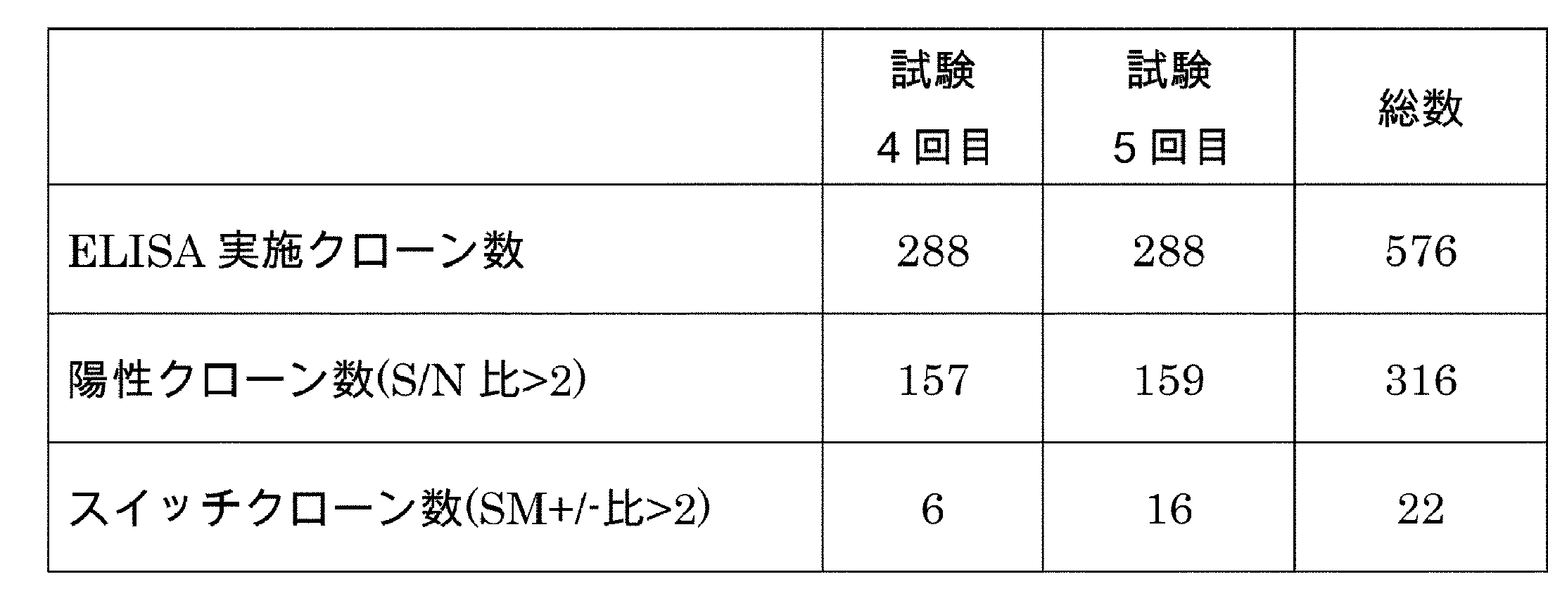
ファージELISAの結果、ATPおよびその代謝物が存在する条件下で抗原に対する結合活性があると判断されたクローンから特異的なプライマーlacPF(配列番号:2), G1seqR(配列番号:3)を用いて増幅された遺伝子の塩基配列が解析された。解析の結果、ATPおよびその代謝物存在下でビオチン標識アバタセプトに対する結合活性を有すると判断されたクローンABADh11-4_020、ABADh11-4_086、ABADh12-4_014、ABADh12-5_001、ABADh12-5_046、ABADh5-5_041が取得され、それぞれABAM001、ABAM002、ABAM003、ABAM004、ABAM005、ABAM006とクローン名が再付与された(表4)。

ヒトラショナルデザインファージライブラリから取得されたABAM001、ABAM002、ABAM003、ABAM004、ABAM005、ABAM006の可変領域をコードする遺伝子はヒトIgG1/Lambdaの動物発現用プラスミドへ挿入された。以下の方法を用いて抗体が発現された。FreeStyle 293 Expression Medium培地(Invitrogen)に1.33×10⁶細胞/mLの細胞密度で懸濁されて、6well plateの各ウェルへ3 mLずつ播種されたヒト胎児腎細胞由来FreeStyle 293-F株(Invitrogen)に対して、調製されたプラスミドがリポフェクション法により導入された。CO2インキュベーター(37℃、8 % CO2、90 rpm)で4日間培養された培養上清から、rProtein A SepharoseTM Fast Flow(Amersham Biosciences)を用いて当業者公知の方法で抗体が精製された。分光光度計を用いて、精製された抗体溶液の280 nmでの吸光度が測定された。得られた測定値からPACE法により算出された吸光係数を用いて精製された抗体の濃度が算出された(Protein Science (1995) 4, 2411-2423)。
取得されたABAM001、ABAM002、ABAM003、ABAM004、ABAM005、ABAM006の6抗体がIgG ELISAに供された。また表5に示すBufferが適宜調製された。抗原としてビオチン標識されたヒトCTLA4(hCTLA4-His-Biotin)が用いられた。
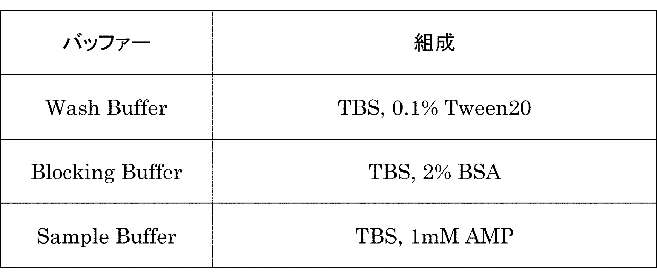

CTLA4スイッチ抗体としてABAM004の更なる評価が行われた。
Biacore T200(GE Healthcare)を用いて、ABAM004とhCTLA4-His-BAPとの抗原抗体反応の相互作用が解析された。アミンカップリング法でprotein A/G(Pierce)が適当量固定化されたSensor chip CM5(GE Healthcare)にABAM004をキャプチャーさせ、抗原である実施例1-1で調製されたhCTLA4-His-BAPを相互作用させた。ランニングバッファーにはTBSが用いられ、再生溶液としては10 mM Glycine-HCl (pH 1.5)が用いられた。
Flow cytometerを用いて、ABAM004とヒトCTLA4との抗原抗体相互作用がAMP存在下、非存在下でどのように変化するのか評価された。ヒトCTLA4を安定発現するCHO細胞(hCTLA4-CHO細胞)が適切な濃度で準備された。この時懸濁には0.1 % BSA入りPBS(FACS Buffer)が用いられた。当該細胞溶液100 μLに対し、終濃度10 mg/mLとなるように抗体が添加されたのち、AMPが終濃度0, 0.4, 4, 40, 200, 1000 μMになるように添加され、4℃で30分間静置された。その後FACS BufferにAMPが終濃度0, 0.4, 4, 40, 200, 1000 μMになるように添加されたものをWash Bufferとして使用し、当該細胞株が洗浄されたのち、FITC標識された二次抗体(Goat F(ab'2) Anti-Human IgG Mouse ads-FITC、Beckman 732598)が添加され、再び4℃で30分間遮光下に静置された。再度洗浄操作が行われた後、Flow cytometer (FACS CyAnTM ADP)で測定、解析された。結果を図2に示す。
ATP依存的に抗原に結合する抗体は以下の方法に従って抗体濃度依存的なADCC活性が測定された。この時、ヒト末梢血単核球(以下、ヒトPBMCと指称する。)をエフェクター細胞として用いて被験抗体のADCC活性が以下のように測定された。
ADCC活性(%)={(A-D)-(C-D)}×100/{(B-D)-(C-D)}
(2-1)AMP をスイッチとする抗CTLA4結合抗体ABAM004のX線結晶構造解析
実施例1においてライブラリより取得されたAMPをスイッチとするhCTLA4結合抗体ABAM004のFab断片単独、ABAM004のFab断片とAMPの複合体、並びにABAM004のFab断片とAMP、hCTLA4の細胞外ドメインの複合体の結晶構造が解析された。
結晶化用ABAM004全長抗体の調製及び精製は当業者公知の方法により行われた。
ABAM004のFab断片を、rLys-C(Promega、カタログ番号V1671)による制限消化、続いて、Fc断片を除去するためのプロテインAカラム(MabSelect SuRe、GE Healthcare)、カチオン交換カラム(HiTrap SP HP、GE Healthcare)、およびゲル濾過カラム(Superdex200 16/60、GE Healthcare)へのローディングを用いるという、従来の方法により調製された。Fab断片を含む画分はプールされ、-80℃で保存された。
2-3の方法で精製した結晶化用ABAM004のFab断片を約13 mg/mLに濃縮し、シッティングドロップ蒸気拡散法により20℃で結晶化を行った。リザーバー溶液は、0.1 M MES pH 6.5、25 %w/v ポリエチレングリコール4000から成るものであった。これにより得られた結晶を0.08 M MES pH 6.5、20% w/v ポリエチレングリコール4000、20% エチレングリコールの溶液に浸漬した。
X線回折データを、高エネルギー加速器研究機構の放射光施設フォトンファクトリーのBL-17Aで測定した。測定中、結晶を常に-178℃の窒素流下に置いて凍結状態を維持し、ビームラインに接続されたQuantum 270 CCD検出器(ADSC)を用いて、結晶を1回に0.5°回転させながら、合計360枚のX線回折画像を収集した。セルパラメータの決定、回折スポットのインデックス付け、および回折像から得られた回折データの処理を、Xia2プログラム(J. Appl. Cryst. 43: 186-190 (2010))、XDSパッケージ(Acta. Cryst. D66: 125-132 (2010))、およびScala(Acta. Cryst. D62: 72-82 (2006))を用いて行い、最終的に分解能1.70Åまでの回折強度データを取得した。結晶学的データ統計値が表7に示される。

ABAM004のFab断片を、パパイン(Roche Diagnostics、カタログ番号1047825)による制限消化、続いて、Fc断片を除去するためのプロテインAカラム(MabSelect SuRe、GE Healthcare)、カチオン交換カラム(HiTrap SP HP、GE Healthcare)、およびゲル濾過カラム(Superdex200 16/60、GE Healthcare)へのローディングを用いるという、従来の方法により調製した。Fab断片を含む画分をプールして、-80℃で保存した。
2-6の方法で精製した結晶化用ABAM004のFab断片を約13 mg/mLに濃縮したものに対して、終濃度が2 mMになるようにAMPを添加し、シッティングドロップ蒸気拡散法により20℃で結晶化を行った。リザーバー溶液は、0.1 M Morpheus buffer 2 pH 7.5、37.5% w/v MPD_P1K_P3350、10% Morpheus Carboxylic acids(Morpheus、Molecular Dimensions)から成るものであった。
X線回折データを、高エネルギー加速器研究機構の放射光施設フォトンファクトリーのBL-1Aで測定した。測定中、結晶を常に-178℃の窒素流下に置いて凍結状態を維持し、ビームラインに接続されたPilatus 2M 検出器(DECTRIS)を用いて、結晶を1回に0.25°回転させながら、合計720枚のX線回折画像を収集した。セルパラメータの決定、回折スポットのインデックス付け、および回折像から得られた回折データの処理を、Xia2プログラム(J. Appl. Cryst. 43: 186-190 (2010))、XDSパッケージ(Acta. Cryst. D66: 125-132 (2010))、およびScala(Acta. Cryst. D62: 72-82 (2006))を用いて行い、最終的に分解能1.70Åまでの回折強度データを取得した。結晶学的データ統計値が表7に示される。
hCTLA4の細胞外ドメインは、アバタセプトにEndoproteinase Lys-C(Roche、カタログ番号11047825001)による制限消化、続いて、Fc断片を除去するためのプロテインAカラム(MabSelect SuRe、GE Healthcare)、およびゲル濾過カラム(Superdex200 10/300、GE Healthcare)へのローディングを用いるという方法により調製した。hCTLA4の細胞外ドメインを含む画分をプールして、-80℃で保存した。
2-9の方法で精製したhCTLA4細胞外ドメインを、2-6の方法で精製したABAM004のFab断片と1.5:1のモル比で混合し、終濃度が2 mMになるようにAMPを添加した。複合体を、25 mM HEPES pH7.5、100 mM NaCl、2 mM AMPで平衡化したカラムを用いたゲル濾過クロマトグラフィー(Superdex200 10/300、GE Healthcare)により精製した。
精製した複合体を約8 mg/mLに濃縮し、シーディング法と組み合わせたシッティングドロップ蒸気拡散法により20℃で結晶化を行った。リザーバー溶液は、0.1 M Morpheus buffer 1 pH 6.5、37.5% w/v M1K3350、10% halogens(Morpheus、Molecular Dimensions)から成るものであった。
X線回折データを、SPring-8のBL32XUで測定した。測定中、結晶を常に-178℃の窒素流下に置いて凍結状態を維持し、ビームラインに接続されたMX-225HS CCD検出器(RAYONIX)を用いて、結晶を1回に1.0°回転させながら、合計180枚のX線回折画像を収集した。セルパラメータの決定、回折スポットのインデックス付け、および回折像から得られた回折データの処理を、Xia2プログラム(J. Appl. Cryst. 43: 186-190 (2010))、XDSパッケージ(Acta. Cryst. D66: 125-132 (2010))、およびScala(Acta. Cryst. D62: 72-82 (2006))を用いて行い、最終的に分解能1.70Åまでの回折強度データを取得した。結晶学的データ統計値が表7に示される。
本結晶構造より、AMPは、主に当該抗体の重鎖によって認識されることがわかった。
AMPのアデニン環部分は重鎖CDR1、3によって、リボース部分並びにリン酸基部分は、CDR1、2によって認識される。
図5および、図6では、ABAM004 Fab接触領域のエピトープが、それぞれ、hCTLA4結晶構造中およびアミノ酸配列中にマッピングされている。エピトープは、結晶構造においてABAM004 FabまたはAMPのいずれかの部分から4.2Å以内の距離に位置する非水素原子を1個以上含むhCTLA4のアミノ酸残基を含んでいる。
少なくとも、抗原のM3、E33、R35、T53、E97、M99、Y100、P101、P102、P103、Y104、Y105、L106は、当該抗体の重鎖CDR2、CDR3、軽鎖CDR1、CDR3ならびに、AMPによって認識されることが結晶構造から明らかになった。特に、抗原のM99からY104から成るループが、当該抗体のCDRループの中に埋もれるように当該抗体に強く認識されており、抗体による抗原認識に大きな役割を果たしていると考えられる。
図7は、ABAM004 Fab単独の結晶構造、ABAM004 FabとAMPの複合体の結晶構造、並びにABAM004 FabとAMP及びCTLA4から成る3者複合体の結晶構造から、当該抗体の可変領域を抽出し、重鎖を中心にして重ね合わせた図である。AMP依存的な抗原結合は、実施例2-14で示したAMPとCTLA4の間の直接的な相互作用だけではなく、AMPの結合に伴う当該抗体の構造変化が重要であると考えられる。
(3-1)ABAM004抗体のCTLA4結合活性増強改変体の作製
実施例1に記載のヒトラショナルデザインファージライブラリから取得されたABAM004(VH配列番号:10、VL配列番号:11)はアミノ酸配列が改変され、当該配列とATP類似体非存在下のCTLA4結合活性が低下され、ATP類似体存在下でのヒトCTLA4結合活性が向上され、ATPおよびATP類似体への結合が向上された。このために実施例2に記載の方法で取得されたABAM004とAMPの共結晶構造及びABAM004とAMP、ヒトCTLA4の共結晶構造を基に当該結合に関与することが予想される残基の点変異体が作製された。さらにCDRに含まれる全アミノ酸をAlaあるいはProで置換した改変体も作製された。当該点変異体はBiacore T200あるいはBiacore 4000(GE Healthcare)によりATP非存在下およびATP、ADPあるいはAMP存在下のヒトCTLA4(AbataceptとhCTLA4-His-BAP)結合活性が測定され、当該結合活性を向上させる変異がスクリーニングされた。結合活性を向上させた変異を組み合わせた変異体が作製され、BiacoreによりKD値算出が行われた。その結果、ABAM004の重鎖をH32A、S52aT、軽鎖をT24D、T26P、E50F(番号はKabat番号)に置換するとABAM004の当該結合特性を向上させることが明らかになった。当該変異体を04H0150/04L0072(VH配列番号:47、VL配列番号:48)と呼称する。
最初にBiacore T200測定用にチップが作成された。Biacore T200の設定温度は25℃、流速は10 μL/minとされた。ランニングバッファーにはHBS-EP+が用いられた。Sensor chip CM5(GE Healthcare)に対し、NHS(N-hydroxysuccinimide)とEDC(N-ethyl-N'-(dimethylaminopropyl) carbodiimide)の等量混合液が流速10 μL/minで10分間添加されflow cellが活性化された。次に10 mM sodium acetate pH 4.0に懸濁した25 μg/mLのProtein A/G(Pierce)が10 μL/minで30分間添加され結合された。その後、flow cell上の過剰の活性基は1 M ethanolamine-HClが10 μL/minで10分間添加されブロックされた。

より優れた抗CTLA4抗体を作製するため、実施例3-1において作製された抗ヒトCTLA4抗体の可変領域である04H0150/04L0072に対してアミノ酸改変が網羅的に導入された。PCR等当業者公知の方法により04H0150および04L0072の全てのCDRに対してシステインを除く全18アミノ酸への置換体が作製された。作製された約1200個の改変体のヒトCTLA4に対する結合測定がBiacore 4000を用いて実施された。Series S Sensor Chip CM5(GE Healthcare)にProtein A/G(Thermo Fisher Scientific)を固定化したチップに対して、抗体改変体の培養上清を相互作用させて抗体がキャプチャーされた。次に小分子(ATP、ADP、あるいはAMP)を添加したヒトCTLA4溶液、あるいは小分子を添加していないヒトCTLA4溶液を相互作用させ、小分子存在下、および小分子非存在下での抗体とヒトCTLA4との結合能が評価された。ランニングバッファーとしてTris buffered saline, 0.02% PS20を用い、25℃で測定が行われた。
なお、本明細書における抗体は、以下の規則に従い命名されている;(重鎖可変領域)-(重鎖定常領域)/(軽鎖可変領域)-(軽鎖定常領域)。例えば、04H0150-G1m/04L0072-lam1という抗体名であれば、本抗体の重鎖可変領域は04H0150、重鎖定常領域はG1m、軽鎖可変領域は04L0072、軽鎖定常領域はlam1であることを意味する。


マウスCTLA4細胞外領域にHisタグが連結されたもの(mCTLA4-His)(配列番号:49)が遺伝子合成され、動物発現用プラスミドへ挿入された。FreeStyle 293 Expression Medium培地(Invitrogen)に1.33×10⁶細胞/mLの細胞密度で懸濁されて、フラスコに播種されたヒト胎児腎細胞由来FreeStyle 293-F株(Invitrogen)に対して、調製されたプラスミドがリポフェクション法により導入された。分光光度計を用いて、精製された抗原溶液の280 nmでの吸光度が測定された。得られた測定値からPACE法により算出された吸光係数を用いて精製された抗原の濃度が算出された(Protein Science (1995) 4, 2411-2423)。
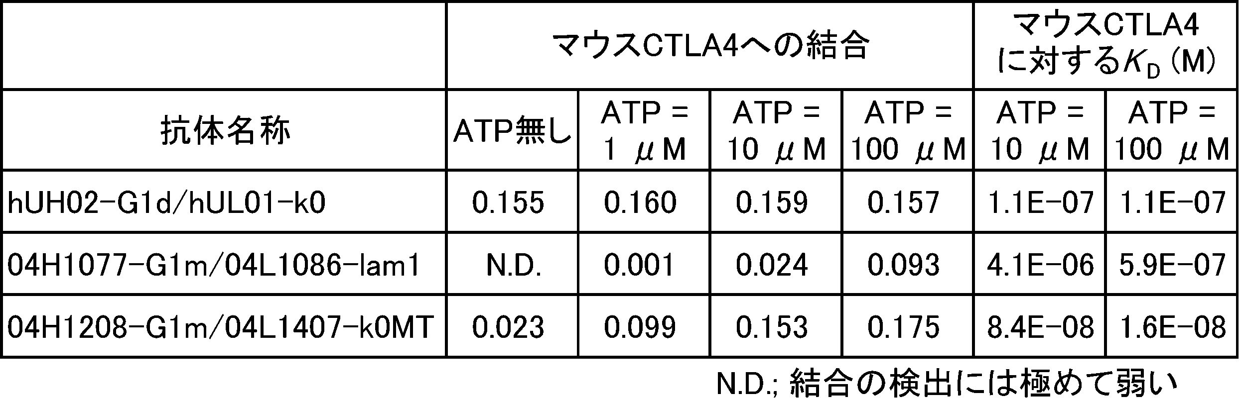
抗mCTLA4コントロール抗体(hUH02-mFa55/hUL01-mk1 略名:mNS-mFa55)および抗CTLA4スイッチ(04H1077-mFa55/04L1086-ml0r 略名:SW1077-mFa55、04H1208-mFa55/04L1407s-mk1 略名:SW1208-mFa55)が作製された。mNS-mFa55抗体は、重鎖可変領域hUH02(配列番号:16)および軽鎖可変領域hUL01(配列番号:17)を用い、定常領域はマウス重鎖定常領域mFa55(配列番号:18)および野生型マウス軽鎖定常領域mk1(配列番号:19)を使用した。この時Fcγ受容体への結合を増強するよう改変を加えたマウス重鎖定常領域を使用し、当業者公知の方法で発現、精製された。
SW1077-mFa55抗体は、重鎖可変領域04H1077(配列番号:20)および軽鎖可変領域04L1086(配列番号:21)を用い、定常領域はマウス重鎖定常領域mFa55(配列番号:18)および野生型マウス軽鎖定常領域ml0r(配列番号:22)を使用した。この時Fcγ受容体への結合を増強するよう改変を加えたマウス重鎖定常領域を使用し、当業者公知の方法で発現、精製された。
SW1208-mFa55抗体は、重鎖可変領域04H1208(配列番号:23)および軽鎖可変領域04L1407s(配列番号:24)を用い、定常領域はマウス重鎖定常領域mFa55(配列番号:18)および野生型マウス軽鎖定常領域mk1(配列番号:19)を使用した。この時Fcγ受容体への結合を増強するよう改変を加えたマウス重鎖定常領域を使用し、当業者公知の方法で発現、精製された。
実施例3-4で調製された抗CTLA4スイッチ抗体(SW1077-mFa55)の中和活性は競合ELISA法により評価された。96ウェルのプレートに、mCTLA4にヒト定常領域を連結したmCTLA4-Fc(配列番号:25)を0.1 M NaHCO3, 0.05% NaN3で5 μg/mL (55 nM) に希釈し、作製されたmCTLA4-Fc溶液を100 μLずつ加え、4℃で一晩静置し、プレート表面に固定化した。TBS, 0.1% Tween20で3回washしたのちに、TBSで2%に希釈したBSA溶液を各ウェルに250 μL加え、プレート表面をブロッキングした。その後、3回washした。最終濃度が55 nMになるようにTBSで希釈したマウスCD86にヒト定常領域およびHisタグが融合されたmCD86-Fc-His(Sino Biologics Inc. 50068-M03H, Accession No. NP_062261.3)、最終濃度が6.25, 1.56, 0.390, 0.0977, 0.0061, 0 μg/mLとなるように希釈したSW1077-mFa55抗体溶液、および最終濃度が0, 1, 10, 100 μMとなるように希釈したATP溶液とをそれぞれ合計100 μLとなるように混合し、各ウェルに加えて、1時間37℃で静置した。その後、各ウェルに加えられた溶液のATP濃度と同じATP濃度を含むように調製されたTBS, 0.1% Tween20で各ウェルを3回washした。各ウェルに加えられた溶液のATP濃度と同じATP濃度を含むようにブロッキングバッファーで10000倍に希釈したanti-His-tag mAb-HRP-Direct(MBLライフサイエンス)を各ウェルに100 μL加え1時間37℃で静置した。その後、各ウェルに加えられた溶液のATP濃度と同じATP濃度を含むように調製されたTBS, 0.1% Tween20で各ウェルを3回washした。そこへTMB溶液を各ウェルに100 μL加え1時間37℃で静置し、1M H2SO4を各ウェルに50 μL加え反応を停止し、吸光マイクロプレートリーダー(Wako Sunrise)で450 nmの吸光度を検出した。
同ATP濃度条件下での、抗体未添加ウェルの吸光度の値をmCTLA4-mCD86結合率100%とし、抗体添加でその結合率がどの程度低下するかを評価した。その結果を図9に示す。
(3-7-1)細胞株および同系腫瘍株移植マウスモデルの作製
細胞は理化学研究所より購入したマウス乳癌株FM3A細胞を用いた。FM3A細胞は10% Bovine serum(Thermo Fisher Scientific)を含むRPMI1640培地(SIGMA)にて維持継代した。マウスは日本チャールス・リバー社から購入したC3H/HeNマウス(7週齢、♀)を用いた。マウスの腹部皮下にFM3A細胞を移植し、移植腫瘍の体積がおよそ150 mm3から300 mm3になった時点でモデル成立とした。
腫瘍体積=長径×短径×短径/2
FM3A細胞移植モデルへの投与薬剤は、実施例3-4で調製された抗マウスCTLA4コントロール抗体(mNS-mFa55)および抗CTLA4スイッチ抗体(SW1208-mFa55)とした。mNS-mFa55は0.0005 mg/mL, 0.005 mg/mL, 0.0125 mg/mL, 0.05 mg/mL, 0.5 mg/mL, 1.5 mg/mL, 5 mg/mLになるよう、SW1208-mFa55は0.005 mg/mL, 0.05 mg/mL, 0.5 mg/mL, 5 mg/mL, 25 mg/mLになるよう、それぞれHis-buffer(20 mM His-HCl, 150 mM NaCl, pH 6.0)を用いて調製した。
移植後9日目に、mNS-mFa55は0.01 mg/kg, 0.1 mg/kg, 0.25 mg/kg, 1 mg/kg, 10 mg/kg, 30 mg/kg, 100 mg/kgの用量で、SW1208-mFa55は0.1 mg/kg, 1 mg/kg, 10 mg/kg, 100 mg/kg, 500 mg/kgの用量で、それぞれマウスに投与した。投与は、調製した投与液を20 mL/kgの用量で、尾静脈より行った。
抗腫瘍効果測定に際しての薬剤処置に関する詳細を表12に示した。
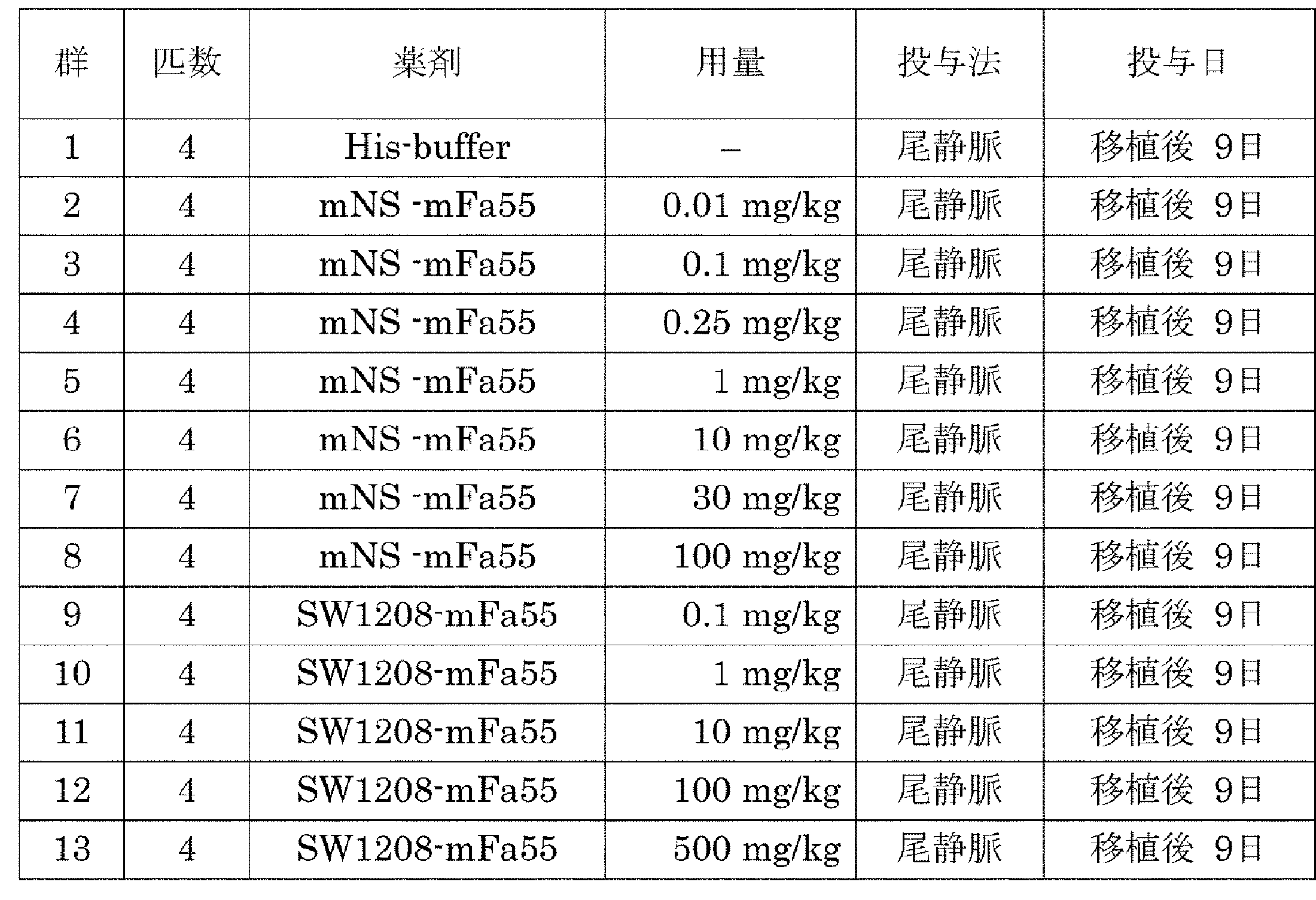
抗腫瘍効果については、(3-7-1)に記した計算式にて算出した腫瘍体積で評価した。
TGI(%)=(1-(測定時における着目する群の腫瘍体積の平均値-初回投与時における着目する群の腫瘍体積の平均値)÷(測定時におけるcontrol群の腫瘍体積の平均値-初回投与時のcontrol群の腫瘍体積の平均値))×100
移植後7日目に抗マウスCTLA4コントロール抗体(mNS-mFa55)を0.1 mg/kg, 1 mg/kg, 10 mg/kg, 100 mg/kgにて尾静脈より投与し、抗CTLA4スイッチ抗体(SW1208-mFa55)を0.1 mg/kg, 1 mg/kg, 10 mg/kg, 100 mg/kg, 500 mg/kgにて尾静脈より投与した。腫瘍内のTreg細胞の評価及び脾臓での全身性作用検証に際しての薬剤処置に関する詳細を表13に示した。

抗体投与後6日目に、麻酔下でマウスを安楽死処置し、腫瘍と脾臓を摘出した。摘出した脾臓より、10% FBS(SIGMA)を含むRPMI-1640培地(SIGMA)を用いて細胞懸濁液を調製した後、Mouse Erythrocyte Lysing kit(R&D)を用いて溶血処理し、脾臓細胞を調製した。摘出した腫瘍は、Tumor dissociation kit, mouse(miltenyi)を用いて破砕した。脾臓細胞、腫瘍を破砕したものをともに以下の抗体と反応させ、FACS解析によって存在する免疫細胞の画分を解析した。抗CD45抗体(BD、クローン: 30-F11)、抗CD3抗体(BD、クローン: 145-2C11)、抗CD4抗体(BD、クローン: RM4-5)、抗FoxP3抗体(eBioscience、クローン: FJK-16s)、抗ICOS抗体(eBioscience、クローン: 7E17G9)、抗KLRG1抗体(Biolegend、クローン: 2F1/KLRG1)。FACS解析はBD LSRFortessa X-20(BD)にて行った。
抗マウスCTLA4コントロール抗体(mNS-mFa55)及び抗CTLA4スイッチ抗体(SW1208-mFa55)投与時の腫瘍内のeffector Treg細胞(CD4+ FoxP3+ KLRG1+)の変化を評価した。結果、mNS-mFa55もSW1208-mFa55も、1 mg/kg以上のdoseにおいて、effector Tregの割合がCD45陽性細胞の0.2%未満まで減少した(図12)。
mNS-mFa55及びSW1208-mFa55投与時の脾臓内の活性化helper T 細胞(CD4+ Foxp3- ICOS+)の変化を、FACS解析にて評価した。結果、mNS-mFa55投与時には脾臓での活性化helper T 細胞のCD45陽性細胞に対する割合が顕著に増加したが、SW1208-mFa55は投与doseを上げていっても、脾臓での活性化helper T 細胞のCD45陽性細胞に対する割合を顕著に増加させることは無かった(図13)。当該スイッチ抗体はコントロール抗体と同等の薬効を示す一方で、腫瘍以外の組織では反応を起こさないことが確認され、腫瘍局所でのみ活性を示すというコンセプトがマウス生体内で証明された。
実施例3において作製された抗CTLA4スイッチ抗体の更なる改変と評価が実施された。
実施例2-1において実施された構造解析の結果より、抗体重鎖のCDR2がAMPのリン酸基と相互作用していることが示された。小分子がATPの場合には、γリン酸基が重鎖CDR2と立体障害を起こす可能性が考えられたため、この領域のアミノ酸を置換し、ATPとの結合能の増強が検討された。具体的には、実施例3において作製された04H1207-G1m/04L1086-lam1および04H1208-G1m/04L1086-lam1の重鎖可変領域に対してR53QとG55Hの改変を導入した04H1389-G1m/04L1086-lam1と、04H1382-G1m/04L1086-lam1が作製された。また04H1389-G1m/04L1086-lam1の軽鎖をヒトκ鎖の配列に置換した04H1389-G1m/04L1305-k0MTが作製された。表14はこれらの抗体の重鎖可変領域、軽鎖可変領域、重鎖定常領域、軽鎖定常領域、および超可変領域(Hyper Variable Region)の配列番号の一覧を示す。


抗ヒトCTLA4コントロール抗体(MDX10D1H-mFa55/MDX10D1L-mk1 略名:hNS-mFa55)および抗CTLA4スイッチ抗体(04H1389-mFa55/04L1305-mk1 略名:SW1389-mFa55)が作製された。hNS-mFa55抗体は、重鎖可変領域MDX10D1H(配列番号:26)および軽鎖可変領域MDX10D1L(配列番号:27)を用い、定常領域はマウス重鎖定常領域mFa55(配列番号:18)および野生型マウス軽鎖定常領域mk1(配列番号:19)を使用している。この時Fcγ受容体への結合を増強するよう改変を加えたマウス重鎖定常領域を使用し、当業者公知の方法で発現、精製された。
SW1389-mFa55抗体は、重鎖可変領域04H1389(配列番号:29)および軽鎖可変領域04L1305(配列番号:30)を用い、定常領域はマウス重鎖定常領域mFa55(配列番号:18)および野生型マウス軽鎖定常領域mk1(配列番号:19)を使用している。この時Fcγ受容体への結合を増強するよう改変を加えたマウス重鎖定常領域を使用し、当業者公知の方法で発現、精製された。

(4-3-1)細胞株
Hepa1-6/hGPC3細胞を用いた。この細胞株は、マウス肝癌株Hepa1-6細胞をATCCより購入し、human Glypican 3(hGPC3、配列番号:181)遺伝子をtransfectionによって恒常発現させ、クローン化したものである。Hepa1-6/hGPC3細胞を10% FBS(SIGMA)および600 μg/mL GENETICIN(gibco)を含むD-MEM(high glucose)培地(SIGMA)にて維持継代した。
Human CTLA4 knock-in マウス(Blood (2005) 106(9): 3127-3133)と社内で作製したhuman CD3 EDG replaced mouse(Sci Rep (2017) 7: 45839)の交雑系統である、human CTLA4 KI, human CD3 EDG replacedマウス(hCTLA4 KI hCD3 EDG replacedマウス)を用いた。hCTLA4 KI hCD3 EDG replacedマウスの皮下にHepa1-6/hGPC3細胞を移植し、移植腫瘍の体積の平均がおよそ200 mm3から400 mm3になった時点でモデル成立とした。
腫瘍体積=長径×短径×短径/2
Hepa1-6/hGPC3細胞移植モデルへの投与薬剤としては、実施例4-2で調製された抗CTLA4スイッチ抗体(SW1389-mFa55)または、抗ヒトCTLA4コントロール抗体(hNS-mFa55)をそれぞれ 0.01, 0.1, 1, 5, 10, 20 mg/mL, 0.01, 0.1, 1, 3 mg/mLになるようにHis buffer(150 mM NaCl/ 20 mM His-HCl buffer pH 6.0)を用いて調製した。
移植後7日目にSW1389-mFa55を0.1 mg/kg, 1 mg/kg, 10 mg/kg, 100 mg/kg、hNS-mFa55を0.1 mg/kg, 1 mg/kg, 10 mg/kg, 30 mg/kgにてマウスに尾静脈より投与した。抗腫瘍効果測定に際しての薬剤処置に関する詳細を表17に示した。

抗腫瘍効果については、(4-3-2)に記した計算式にて算出した腫瘍体積で評価した。統計解析にはJMP 11.2.1(SAS Institute Inc.)を用いた。
TGI=(1-(測定時における着目する群の腫瘍体積の平均値-抗体投与前の時点での腫瘍体積の平均値)÷(測定時におけるcontrol群腫瘍体積の平均値-抗体投与前の時点での腫瘍体積の平均値))×100
移植後7日目にSW1389-mFa55を0.1 mg/kg, 1 mg/kg, 10 mg/kg, 100 mg/kg, 500 mg/kgにてマウスに尾静脈より投与し、hNS-mFa55を0.1 mg/kg, 1 mg/kg, 10 mg/kg, 30 mg/kgにて尾静脈より投与した。腫瘍内のTreg細胞の評価及び脾臓での全身性作用検証に際しての薬剤処置に関する詳細を表18に示した。

抗体投与後6日目に、麻酔下でマウスを安楽死処置し、腫瘍と脾臓を摘出した。摘出した脾臓より、10% FBS(SIGMA)を含むRPMI-1640培地(SIGMA)を用いて細胞懸濁液を調製した後、Mouse Erythrocyte Lysing kit(R&D)を用いて溶血処理し、脾臓細胞を調製した。摘出した腫瘍は、Tumor dissociation kit, mouse(miltenyi)を用いて破砕した。脾臓細胞、腫瘍を破砕したものをともに以下の抗体と反応させ、FACS解析によって存在する免疫細胞の画分を解析した。抗CD45抗体(BD、クローン: 30-F11)、抗CD3抗体(BD、クローン: UCHT1)、抗CD4抗体(BD、クローン: RM4-5)、抗FoxP3抗体(eBioscience、クローン: FJK-16s)、抗ICOS抗体(eBioscience、クローン: 7E17G9)、抗CCR7抗体(Biolegend、クローン: 4B12)、抗KLRG1抗体(Biolegend、クローン: 2F1/KLRG1)。FACS解析はBD LSRFortessa X-20(BD)にて行った。
SW1389-mFa55投与時および、hNS-mFa55投与時の腫瘍内のeffector Treg細胞(CD4+ FoxP3+ CCR7lowKLRG1+)の変化を評価した。結果、hNS-mFa55 もSW1389-mFa55も、1 mg/kg以上のdoseにおいて、effector Tregの割合がCD45陽性細胞の0.2%未満まで減少した(図16)。
hNS-mFa55またはSW1389-mFa55投与時の、脾臓内の活性化helper T細胞(CD4+ Foxp3- ICOS+)の変化を、FACS解析にて評価した。結果、hNS-mFa55投与時には脾臓での活性化helper T細胞のCD45陽性細胞に対する割合が顕著に増加したが、SW1389-mFa55投与時には投与doseを上げていっても脾臓における活性化helper T細胞の顕著な増加は見られなかった(図17)。当該スイッチ抗体はコントロール抗体と同等の薬効を示す一方で、腫瘍以外の組織では反応を起こさないことが確認され、腫瘍局所でのみ活性を示すというコンセプトがヒトCTLA4 KIマウス生体内で証明された。
抗CTLA4コントロール抗体(MDX10D1H-Kn125/MDX10D1L-k0MT//MDX10D1H-Hl076/MDX10D1L-k0MT 略名:NS-ART1)および抗CTLA4スイッチ抗体(04H1389-Kn125/04L1305-k0MT//04H1389-Hl076/04L1305-k0MT 略名:SW1389-ART1)が作製された。NS-ART1抗体は、重鎖可変領域MDX10D1H(配列番号:26)および軽鎖可変領域MDX10D1L(配列番号:27)を用い、定常領域は先行特許WO2013/002362に記載のヒト重鎖定常領域Kn125(配列番号:31)およびヒト重鎖定常領域Hl076(配列番号:32)、ヒト軽鎖定常領域k0MT(配列番号:33)を使用している。この時Fcγ受容体への結合を増強するよう改変を加えたヒト重鎖定常領域を使用し、当業者公知の方法で発現、精製された。
SW1389-ART1抗体は、重鎖可変領域04H1389(配列番号:29)および軽鎖可変領域04L1305(配列番号:30)を用い、定常領域はヒト重鎖定常領域Kn125(配列番号:31)およびヒト重鎖定常領域Hl076(配列番号:32)、ヒト軽鎖定常領域k0MT(配列番号:33)を使用している。この時Fcγ受容体への結合を増強するよう改変を加えたヒト重鎖定常領域を使用し、当業者公知の方法で発現、精製された。
なお、本明細書におけるヘテロ二量化抗体(二本の異なる重鎖ポリペプチド、および/または二本の異なる軽鎖ポリペプチドを有する、抗体)は、以下の規則に従い命名されている;(第一の重鎖可変領域)-(第一の重鎖定常領域)/(第一の軽鎖可変領域)-(第一の軽鎖定常領域)//(第二の重鎖可変領域)-(第二の重鎖定常領域)/(第二の軽鎖可変領域)-(第二の軽鎖定常領域)。例えば、04H1389-Kn125/04L1305-k0MT//04H1389-Hl076/04L1305-k0MTという抗体名であれば、本抗体の第一の重鎖可変領域は04H1389、第一の重鎖定常領域はKn125、第一の軽鎖可変領域は04L1305、第一の軽鎖定常領域はk0MT、第二の重鎖可変領域は04H1389、第二の重鎖定常領域はHl076、第二の軽鎖可変領域は04L1305、第二の軽鎖定常領域はk0MTであることを意味する。
全身反応をはじめとする毒性を評価・比較する目的で,実施例4-4で調製されたNS-ART1抗体及びSW1389-ART1抗体をそれぞれ雄性カニクイザル(各3例)に60 mg/kg,毎週1回,計5回投与した。投与はシリンジポンプを用いて低速静脈内投与し,一般状態観察,体重測定,血液・血液化学的検査,骨髄検査,病理学的検査及び血漿中薬物濃度測定を実施した。
両抗体ともに投与期間中から抗薬物抗体の発現はみられたが,投与期間終了時まで曝露は維持された。NS-ART1抗体投与により自己抗体の発現,炎症性変化(血管炎,炎症性細胞浸潤),貧血性変化,T細胞の活性化等,全身性に自己免疫疾患様変化が認められた一方,SW1389-ART1抗体投与では前述の変化が認められなかった。よって,これらの結果から,SW1389-ART1抗体は生体における毒性を軽減することが示された。
実施例4において作製された抗CTLA4スイッチ抗体の更なる改変と評価が実施された。
04H1389-G1m/04L1086-lam1のCDRに対して、アミノ酸改変が網羅的に導入され、より優れたプロファイルを有する改変が探索された。アミノ酸改変の網羅的導入および評価は実施例3-3に記載の方法を用いて行われた。ここで見出された改変の組み合わせおよびFrameworkを置換した改変体が作製された。表19はこれらの抗体の重鎖可変領域、軽鎖可変領域、重鎖定常領域、軽鎖定常領域、および超可変領域(Hyper Variable Region)の配列番号の一覧を示す。表19中の軽鎖04L1594-lam1、04L1581-lam1、04L1610-lam1、04L1612-lam1、04L1610-lam1は親抗体の軽鎖04L1086-lam1に対してCDRおよびframeworkへの改変が導入されており、ヒトλ鎖のgermline配列のframeworkと定常領域を有する。また04L1615-k0MT、 04L1616-k0MT、 04L1617-k0MTは、04L1086-lam1に対してCDRへの改変が導入され、かつヒトκ鎖のgermline配列のframeworkと定常領域を有する。重鎖可変領域04H1389v373は親抗体の重鎖可変領域04H1389のCDRへの改変が導入されたもの、04H1637、04H1643、04H1654、04H1656、04H1642、04H1735は04H1389のCDRに改変が導入されるとともに、Frameworkの配列を異なるgermlineに置換した重鎖可変領域である。


カニクイザルCTLA4細胞外領域C末端にHisタグおよびBAPタグが融合したものcyCTLA4-His-BAP(配列番号:50)が遺伝子合成され、動物発現用プラスミドへ挿入された。FreeStyle 293 Expression Medium培地(Invitrogen)に1.33×10⁶細胞/mLの細胞密度で懸濁されて、フラスコに播種されたヒト胎児腎細胞由来FreeStyle 293-F株(Invitrogen)に対して、調製されたプラスミドがリポフェクション法により導入され、プラスミド導入後3時間後にビオチンが終濃度100 μMとなるように添加され、CO2インキュベーター(37℃、8% CO2, 125 rpm)で4日間培養された。各検体の培養上清から、当業者公知の方法で抗原が精製された。分光光度計を用いて、精製された抗原溶液の280 nmでの吸光度が測定された。得られた測定値からPACE法により算出された吸光係数を用いて精製された抗原の濃度が算出された(Protein Science (1995) 4, 2411-2423)。
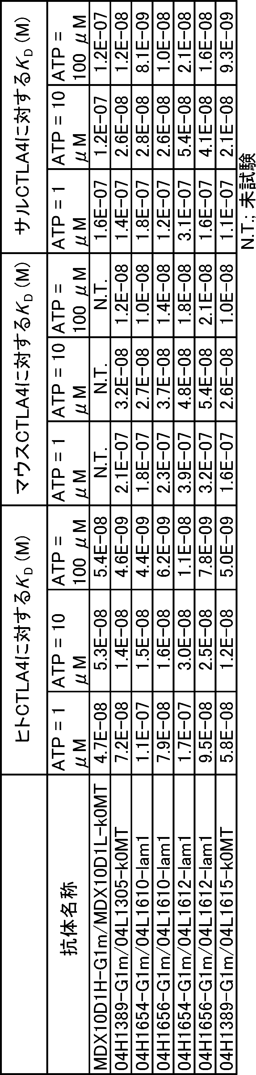
改変された抗CTLA4スイッチ抗体(04H1654-mFa55m2P1/04L1610-ml0r//04H1656-mFa55m2N1/04L1610-ml0r 略名:SW1610-mFa55、04H1654-mFa55m2P1/04L1612-ml0r//04H1656-mFa55m2N1/04L1612-ml0r 略名:SW1612-mFa55、および04H1389-mFa55/04L1615-mk1 略名:SW1615-mFa55)、および陰性コントロール抗体(IC17Hdk-mFa55/IC17L-mk1 略名:KLH-mFa55)が作製された。
SW1615-mFa55抗体は、重鎖可変領域04H1389(配列番号:29)および軽鎖可変領域04L1615(配列番号:34)を用い、定常領域はマウス重鎖定常領域mFa55(配列番号:18)および野生型マウス軽鎖定常領域mk1(配列番号:19)を使用している。この時Fcγ受容体への結合を増強するよう改変を加えたマウス重鎖定常領域を使用し、当業者公知の方法で発現、精製された。
SW1610-mFa55抗体は、一方の重鎖可変領域04H1654(配列番号:35)は定常領域としてマウス重鎖定常領域mFa55m2P1(配列番号:36)を、もう一方の重鎖可変領域04H1656(配列番号:37)はマウス重鎖定常領域mFa55m2N1(配列番号:38)を連結し、さらに軽鎖可変領域04L1610(配列番号:39)は野生型マウス軽鎖定常領域ml0r(配列番号:22)を使用し、当業者公知の方法で発現、精製された。
SW1612-mFa55抗体は、一方の重鎖可変領域04H1654(配列番号:35)は定常領域としてマウス重鎖定常領域mFa55m2P1(配列番号:36)を、もう一方の重鎖可変領域04H1656(配列番号:37)はマウス重鎖定常領域mFa55m2N1(配列番号:38)を連結し、さらに軽鎖可変領域04L1612(配列番号:40)は野生型マウス軽鎖定常領域ml0r(配列番号:22)を使用し、当業者公知の方法で発現、精製された。
陰性コントロール抗体は、重鎖可変領域IC17Hdk(配列番号:51)は定常領域としてマウス重鎖定常領域mFa55(配列番号:18)を連結し、さらに軽鎖可変領域IC17L(配列番号:52)は野生型マウス軽鎖定常領域mk1(配列番号:19)を使用し、当業者公知の方法で発現、精製された。
マウス定常領域を有する抗CTLA4抗体の抗原への結合能が実施例4-2に記載の方法で評価された(表23)。マウス定常領域を有するこれらの抗体はいずれも、同じ可変領域でかつヒト定常領域を有する表22に記載の抗体と同等のATP依存的なヒトCTLA4への結合能を有することが示された。

(5-4-1)細胞株
Hepa1-6/hGPC3細胞を用いた。この細胞株は、マウス肝癌株Hepa1-6細胞をATCCより購入し、human Glypican 3(hGPC3)遺伝子をtransfectionによって恒常発現させ、クローン化したものである。Hepa1-6/hGPC3細胞は10% FBS(SIGMA)および0.6 mg/mL G418(ナカライテスク)を含むD-MEM(high glucose)培地(SIGMA)にて維持継代した。
Human CTLA4 knock-in マウス(Blood (2005) 106(9): 3127-3133)と社内で作製したhuman CD3 EDG replaced mouse(Sci Rep (2017) 7: 45839)の交雑系統である、human CTLA4 KI, human CD3 EDG replacedマウス(hCTLA4 KI hCD3 EDG replacedマウス)を用いた。hCTLA4 KI hCD3 EDG replacedマウスの皮下にHepa1-6/hGPC3細胞を移植し、移植腫瘍の体積の平均がおよそ200 mm3から400 mm3になった時点でモデル成立とした。
腫瘍体積=長径×短径×短径/2
Hepa1-6/hGPC3細胞移植モデルへの投与薬剤は、実施例5-2で調製された抗CTLA4スイッチ抗体(SW1610-mFa55、SW1612-mFa55、SW1615-mFa55)とした。投与薬剤は0.03 mg/mL, 0.1 mg/mL, 0.3 mg/mLになるようにHis-buffer(20 mM His-HCl, 150 mM NaCl, pH 6.0)を用いて調製した。
移植後8日目に抗CTLA4スイッチ抗体3検体をそれぞれ0.3 mg/kg, 1 mg/kg, 3 mg/kgにてマウスに尾静脈より投与した。抗腫瘍効果測定に際しての薬剤処置に関する詳細を表24に示した。
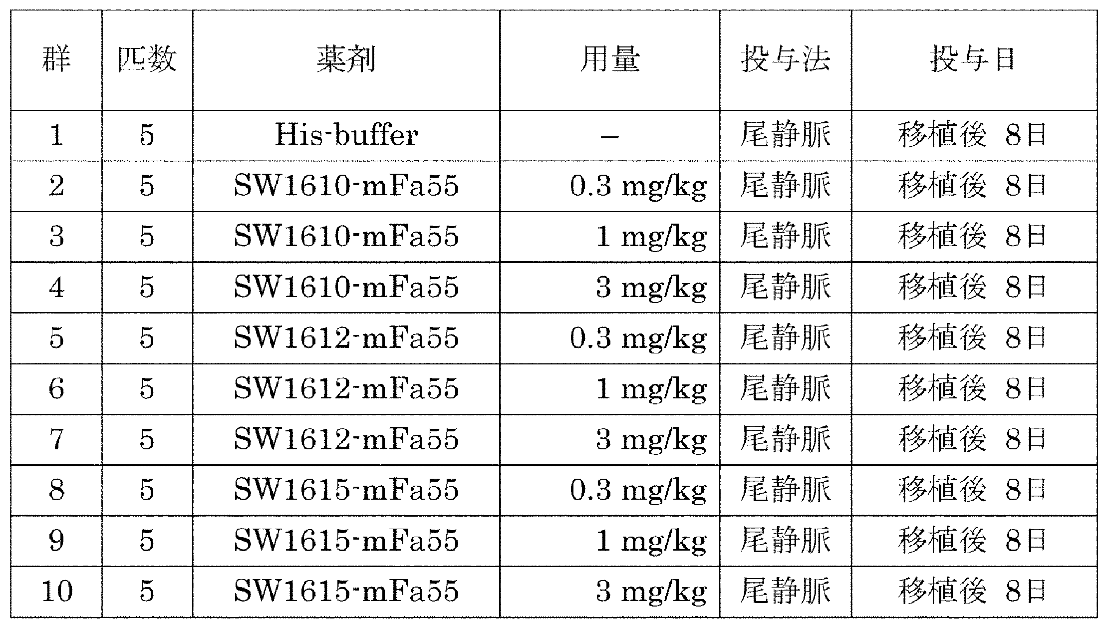
抗腫瘍効果については、(5-4-2)に記した計算式にて算出した腫瘍体積で評価した。
TGI(%)=(1-(測定時における着目する群の腫瘍体積の平均値-初回投与時における着目する群の腫瘍体積の平均値)÷(測定時におけるcontrol群の腫瘍体積の平均値-初回投与時のcontrol群の腫瘍体積の平均値))×100
移植後10日目にSW1610-mFa55を50 mg/kg, 100 mg/kg, 200 mg/kg、SW1612-mFa55を50 mg/kg, 100 mg/kg, 200 mg/kg、SW1615-mFa55を50 mg/kg, 100 mg/kg, 200 mg/kg, 400mg/kgにてマウスに尾静脈より投与した。また、コントロール群には、陰性コントロール抗体であるIC17Hdk-mFa55/IC17L-mk1(略名:KLH-mFa55)を400 mg/kgにて尾静脈より投与した。腫瘍内のTreg細胞の評価及び脾臓での全身性作用検証に際しての薬剤処置に関する詳細を表25に示した。

抗体投与後6日目に、麻酔下でマウスを安楽死処置し、腫瘍と脾臓を摘出した。摘出した脾臓より、10% FBS(SIGMA)を含むRPMI-1640培地(SIGMA)を用いて細胞懸濁液を調製した後、Mouse Erythrocyte Lysing kit(R&D)を用いて溶血処理し、脾臓細胞を調製した。摘出した腫瘍は、Tumor dissociation kit, mouse(miltenyi)を用いて破砕した。脾臓細胞、腫瘍を破砕したものをともに以下の抗体と反応させ、FACS解析によって存在する免疫細胞の画分を解析した。抗CD45抗体(BD、クローン: 30-F11)、抗CD3抗体(BD、クローン: UCHT1)、抗CD4抗体(BD、クローン: RM4-5)、抗FoxP3抗体(eBioscience、クローン: FJK-16s)、抗ICOS抗体(eBioscience、クローン: 7E17G9)、抗CCR7抗体(Biolegend、クローン: 4B12)、抗KLRG1抗体(Biolegend、クローン: 2F1/KLRG1)。FACS解析はBD LSRFortessa X-20(BD)にて行った。
抗CTLA4スイッチ抗体投与時の腫瘍内のeffector Treg細胞(CD4+ FoxP3+ CCR7lowKLRG1+)の変化を評価した。結果、SW1610-mFa55、SW1612-mFa55およびSW1615-mFa55は投与した全てのdoseにおいて、effector Tregの割合がCD45陽性細胞の0.2%未満まで減少した(図21)。
抗CTLA4スイッチ抗体投与時の脾臓内の活性化helper T細胞(CD4+ Foxp3- ICOS+)の変化を、FACS解析にて評価した。結果、SW1610-mFa55およびSW1612-mFa55は評価したdoseのうち、50 mg/kgのdoseにおいて、SW1615-mFa55は評価したdoseのうち、200 mg/kg以下のdoseにおいて脾臓での活性化helper T細胞のCD45陽性細胞に対する割合を、有意に増加させることは無かった。有意差検定はJMP 11.2.1(SAS Institute Inc.)を用い、KLH-mFa55投与群に対してDunnettの検定を行った(図22)。当該スイッチ抗体はいずれも薬効を示す一方で、腫瘍以外の組織では反応を起こさない事が確認され、腫瘍局所でのみ活性を示すという性質を有することが確認された。
細胞傷害性エフェクター機能であるADCCおよびADCPを増強した抗体を作製するため、活性型FcγRであるFcγRIIIaおよびFcγRIIaに対する結合能を増強したFc領域改変体の作製が検討された。
WO2013/002362に記載されているFcγRへの結合能を増強した重鎖定常領域Kn125およびHl076を有し、重鎖可変領域として04H1637を、軽鎖として04L1610-lam1を有するヘテロ二量化抗体、04H1637-Kn125/04L1610-lam1//04H1637-Hl076/04L1610-lam1が作製された。具体的には、一方の重鎖可変領域として04H1637(配列番号:138)を有し、ヒトIgG1重鎖定常領域のC末端のGlyおよびLysが除去されたG1d(配列番号:158)に対して、L234Y/L235Q/G236W/S239M/H268D/D270E/S298Aが導入され、さらにCH3領域にヘテロ二量化を促進する改変Y349C/T366Wを有する抗体重鎖04H1637-Kn125(配列番号:162)の遺伝子が作製された。同様に、もう一方の重鎖可変領域として04H1637(配列番号:138)を有し、ヒトIgG1重鎖定常領域G1d(配列番号:158)に対して、D270E/K326D/A330M/K334Eが導入され、さらにCH3領域にヘテロ二量化を促進する改変D356C/T366S/L368A/Y407Vを有する抗体重鎖04H1637-Hl076(配列番号:163)の遺伝子が作製された。抗体軽鎖として04L1610-lam1(配列番号:161)を用い、当業者公知の方法によりヘテロ二量体04H1637-Kn125/04L1610-lam1//04H1637-Hl076/04L1610-lam1が作製された。ここでCH2領域に導入されている改変のうち、L235Q、G236W、S239M、H268D、D270E、S298A、K326D、K334Eに加えて、WO2013/002362においてFcγRへの結合を変化させる改変として報告されているL234F、A330K、Mol. Cancer Ther., 2008, 7, 2517-2527およびWO2004/029207に報告されているG236A、I332E、I332D、WO2013/118858において安定性を向上させる改変として報告されているT250V、T307Pを組み合わせた抗体重鎖04H1637-Kn462(配列番号:164)、04H1637-Hl441(配列番号:165)、04H1637-Hl445(配列番号:166)、04H1637-Kn461(配列番号:167)、04H1637-Hl443(配列番号:168)の遺伝子が作製された。また、ヒトIgG1(IGHG1*03)のC末端のGlyおよびLysが除去され、CH2領域にKn462と同じ改変を有し、CH3領域にWO2006/106905に記載のヘテロ二量化を促進する改変E356Kを有し、重鎖可変領域として04H1654(配列番号:140)をもつ抗体重鎖04H1654-KT462(配列番号:182)の遺伝子が作製された。同様に、ヒトIgG1(IGHG1*03)のC末端のGlyおよびLysが除去され、CH2領域にHl441と同じ改変を有し、CH3領域にWO2006/106905に記載のヘテロ二量化を促進する改変K439Eを有し、重鎖可変領域として04H1656(配列番号:141)をもつ抗体重鎖04H1656-HT441(配列番号:170)の遺伝子が作製された。以下同様に、04H1656-HT445(配列番号:171)、04H1654-KT461(配列番号:183)、04H1656-HT443(配列番号:173)の遺伝子が作製された。さらに、Mabs, 2017, 9, 844-853に記載されている抗体の血中動態を改善する改変の組み合わせが検討された。具体的には、04H1654-KT462(配列番号:182)のCH3領域に対して、酸性条件下においてヒトFcRnに対する結合を増強する改変およびRheumatoid factorに対する結合を低減する改変の組み合わせであるN434A/Y436T/Q438R/S440Eが導入された04H1654-KT473(配列番号:184)の遺伝子が作製された。同様に04H1656-HT445(配列番号:171)に対して、N434A/Y436T/Q438R/S440Eが導入された04H1656-HT482(配列番号:185)の遺伝子が作製された。同様に、04H1654-KT461, 04H1656-HT443に対して同じ改変を導入した抗体重鎖04H1654-KT481(配列番号:186)、抗体重鎖04H1656-HT498(配列番号:187)がそれぞれ作製された。これらの重鎖を組み合わせ、04L1610-lam1あるいは04L1612-lam1(配列番号:188)が軽鎖として用いられ、目的とするヘテロ二量化抗体が作製された。
表26はこれらの測定結果を示す。
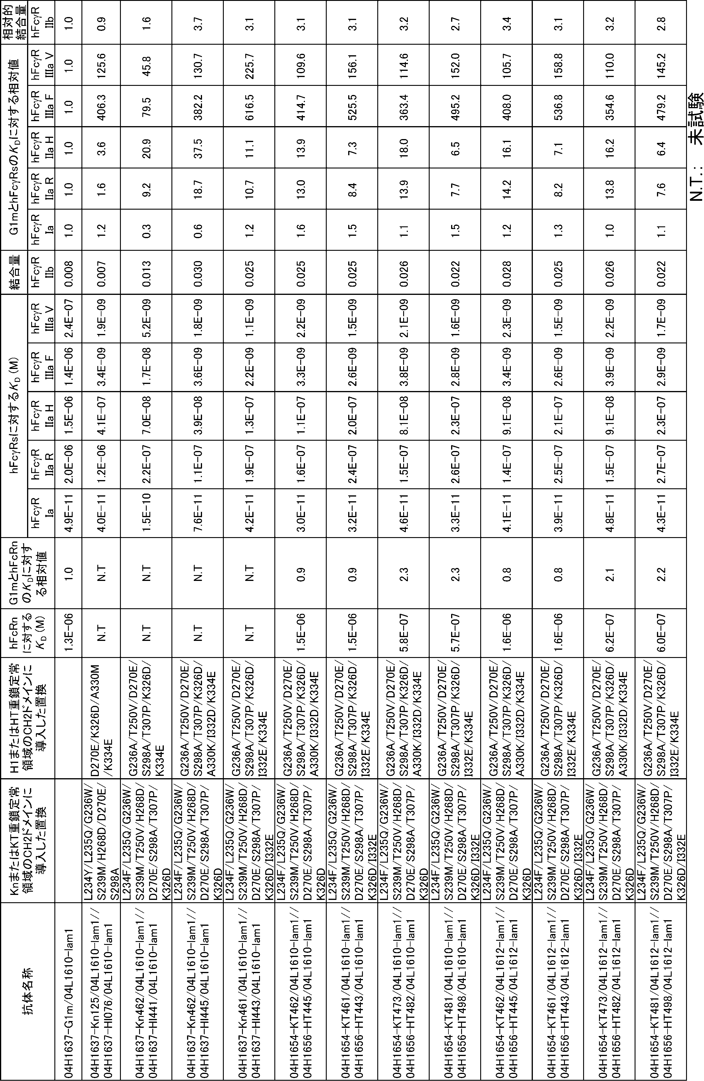


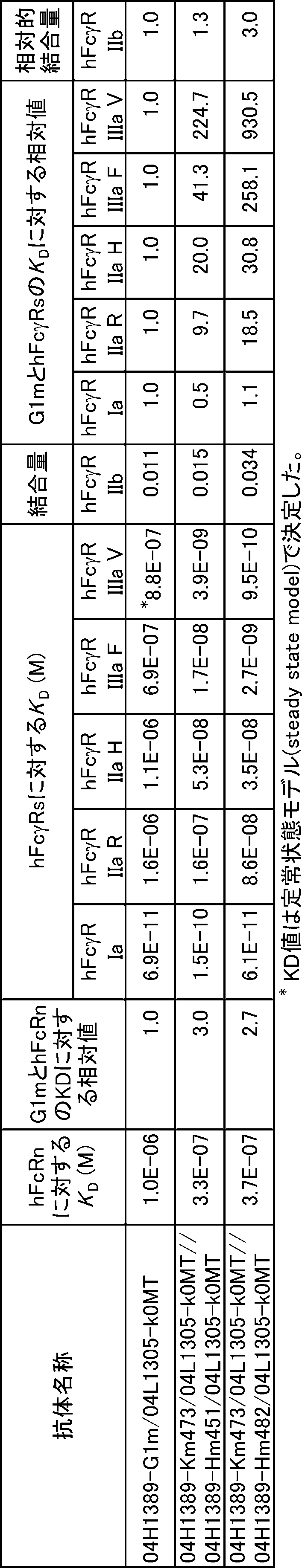

作製されたヘテロ二量体、MDX10D1H-Kn125/MDX10D1H-Hl076/MDX10D1L-k0MT、MDX10D1H-Kn462/MDX10D1H-Hl445/MDX10D1L-k0MT、MDX10D1H-Kn461/MDX10D1H-Hl443/MDX10D1L-k0MTはいずれも既存のFcγR結合増強抗体であるMDX10D1H-GASDIE/MDX10D1L-k0MT、MDX10D1H-GASDALIE/MDX10D1L-k0MTと比較してFcγRIIIaへの結合が増強されていた。またMDX10D1H-Kn462/MDX10D1H-Hl445/MDX10D1L-k0MTは既存のFcγRIIa増強抗体であるMDX10D1H-GASDIE/MDX10D1L-k0MTと比較して、FcγRIIaHへの結合が約2倍増強されていることが示された。
In vitro ADCC活性測定にはhFcγRIIIaV ADCC Reporter Bioassay, Core Kit(Promega)が用いられた。96ウェルプレートの各ウェルに、培地により2×10⁶/mLに濃度を調製した hCTLA4-CHO細胞をターゲット細胞として25 μLずつ加えられ、培地にはAssay Buffer(90% RPMI1640, 10% FBS)が使用された。次に最終濃度が0, 0.001, 0.01, 0.1, 1 μg/mLとなるようにassay bufferで希釈した各抗体溶液がそれぞれ25 μL加えられ、最後にエフェクター細胞溶液として培地により6×10⁶/mLに調製されたhFcγRIIIaV 発現Jurkat細胞(キットに付属)が25 μL加えられ、合計75 μLとなるように混合された後、5% CO2インキュベーター内にて37℃で一晩静置された。その後プレートは室温で15分静置され、各ウェルにBio-Glo reagentが75 μLずつ加えられた。Bio-Glo reagentにはBio-glo Luciferase Assay System (Buffer and Substrate)が使用された。その後各ウェルの発光はプレートリーダーで測定された。各ウェルの発光の値を抗体未添加ウェルの発光の値で割った値をFold inductionとし、各抗体のADCCを評価する指標とした。得られた結果を図23に示す。図中では、Fold inductionを相対発光量(RLU)と表記する。
In vitro ADCC活性測定にはhFcγRIIaH ADCP Reporter Bioassay, Core Kit(Promega)を用いた。96ウェルプレートの各ウェルに、培地により1×10⁶/mLに濃度を調製した hCTLA4-CHO細胞をターゲット細胞として25 μLずつ加えられ、培地にはAssay Buffer(4% Low IgG serum in RPMI1640)が使用された。次に最終濃度が0, 0.001, 0.01, 0.1, 1 μg/mLとなるようにassay bufferで希釈した各抗体溶液がそれぞれ25 μL加えられ、最後にエフェクター細胞溶液としてキットに付属のhFcγRIIaH 発現Jurkat細胞が25 μL加えられ、合計75 μLとなるように混合された後、5% CO2インキュベーター内にて37℃で一晩静置された。hFcγRIIaH 発現Jurkat細胞はその細胞液密度が8.25×10⁵/mLであった。その後プレートは室温で15分静置され、各ウェルにBio-Glo reagentが75 μlずつ加えられた。Bio-Glo reagentにはBio-glo Luciferase Assay System (Buffer and Substrate)が使用された。その後各ウェルの発光はプレートリーダーで測定された。各ウェルの発光の値を抗体未添加ウェルの発光の値で割った値をFold inductionとし、各抗体のADCPを評価する指標とした。得られた結果を図24に示す。図中では、Fold inductionを相対発光量(RLU)と表記する。
改変されたFcを有する抗CTLA4スイッチ抗体(04H1654-Kn462/04L1610-lam1//04H1656-Hl445/04L1610-lam1 略名:SW1610-ART6、04H1654-Kn462/04L1612-lam1//04H1656-Hl445/04L1612-lam1 略名:SW1612-ART6、および04H1389-Kn462/04L1305-k0MT//04H1389-Hl445/04L1305-k0MT 略名:SW1389-ART6)が作製された。
SW1610-ART6抗体は、一方の重鎖可変領域04H1654(配列番号:35)は定常領域としてヒト重鎖定常領域Kn462(配列番号:43)を、もう一方の重鎖可変領域04H1656(配列番号:37)はヒト重鎖定常領域Hl445(配列番号:44)を連結し、さらに軽鎖可変領域04L1610(配列番号:39)は野生型ヒト軽鎖定常領域lam1(配列番号:53)を使用し、当業者公知の方法で発現、精製された。
SW1612-ART6抗体は、一方の重鎖可変領域04H1654(配列番号:35)は定常領域としてヒト重鎖定常領域Kn462(配列番号:43)を、もう一方の重鎖可変領域04H1656(配列番号:37)はヒト重鎖定常領域Hl445(配列番号:44)を連結し、さらに軽鎖可変領域04L1612(配列番号:40)は野生型ヒト軽鎖定常領域lam1(配列番号:53)を使用し、当業者公知の方法で発現、精製された。
SW1389-ART6抗体は、二つの重鎖可変領域04H1389(配列番号:29)は定常領域としてヒト重鎖定常領域Kn462(配列番号:43)およびヒト重鎖定常領域Hl445(配列番号:44)をそれぞれ連結し、さらに軽鎖可変領域04L1305(配列番号:30)は野生型ヒト軽鎖定常領域k0MT(配列番号:33)を使用し、当業者公知の方法で発現、精製された。
改変された抗CTLA4スイッチ抗体として、SW1389, SW1610, SW1612, SW1615の可変領域を持つヒト抗体が作成された。
SW1389抗体は、重鎖可変領域として04H1389(配列番号:29)、軽鎖可変領域として04L1305(配列番号:30)が使用され、ヒト定常領域に連結されたのち、当業者公知の方法で発現、精製された。
SW1610抗体は、重鎖可変領域として04H1654(配列番号:35)および04H1656(配列番号:37)、軽鎖可変領域として04L1610(配列番号:39)が使用され、ヒト定常領域に連結されたのち、当業者公知の方法で発現、精製された。
SW1612抗体は、重鎖可変領域として04H1654(配列番号:35)および04H1656(配列番号:37)、軽鎖可変領域として04L1612(配列番号:40)が使用され、ヒト定常領域に連結されたのち、当業者公知の方法で発現、精製された。
SW1615抗体は、重鎖可変領域として04H1389(配列番号:29)、軽鎖可変領域として04L1615(配列番号:34)が使用され、ヒト定常領域に連結されたのち、当業者公知の方法で発現、精製された。
In vitro 中和活性測定にはCTLA-4 Blockade Bioassay(Promega)が用いられた。96ウェルプレートの各ウェルに、培地により1×10⁶/mLに濃度を調製した Kit付属のaAPC-Raji細胞がターゲット細胞として25μLずつ加えられ、培地にはAssay Buffer(10% FBS in RPMI1640)が使用された。次に最終濃度が0, 100 μMとなるようにassay bufferで希釈したATP溶液、最終濃度が0, 0.001, 0.01, 0.1, 1 ,10 μg/mLとなるようにassay bufferで希釈したSW1389、SW1610, SW1612およびSW1615の可変領域を持つ抗体溶液が順次加えられ、最後にエフェクター細胞溶液として培地により2×10⁶/mLに調製されたIL2-luc2-CTLA4-Jurkat細胞(キットに付属)が25 μL加えられ、合計75 μLとなるように混合された後、5% CO2インキュベーター内にて6時間37℃で静置された。その後プレートは室温で15分静置され、各ウェルにBio-Glo reagentが75 μLずつ加えられた。Bio-Glo reagentにはBio-glo Luciferase Assay System (Buffer and Substrate)が使用された。その後各ウェルの発光はプレートリーダーで測定された。各ウェルの発光の値を抗体未添加ウェルの発光の値で割った値をFold inductionとし、各抗体の中和活性を評価する指標とした。得られた結果を図28(SW1389)、図29(SW1610)、図30(SW1612)および図31(SW1615)に示す。図中では、Fold inductionを相対発光量(RLU)と表記する。
改変されたFcを有する抗CTLA4スイッチ抗体(04H1654-KT473/04L1610-lam1//04H1656-HT451/04L1610-lam1 略名:SW1610-ART5+ACT1、04H1654-KT473/04L1610-lam1//04H1656-HT482/04L1610-lam1 略名:SW1610-ART6+ACT1、04H1389-Km473/04L1305-k0MT//04H1389-Hm451/04L1305-k0MT 略名:SW1389-ART5+ACT1)、04H1389-Km473/04L1305-k0MT//04H1389-Hm482/04L1305-k0MT 略名:SW1389-ART6+ACT1)が作製された。
SW1610-ART5+ACT1抗体は、一方の重鎖として04H1654-KT473(配列番号:184)、もう一方の重鎖として04H1656-HT451(配列番号:272)、さらに軽鎖として04L1610-lam1(配列番号:161)を使用し、当業者公知の方法で発現、精製された。
SW1610-ART6+ACT1抗体は、一方の重鎖として04H1654-KT473(配列番号:184)、もう一方の重鎖として04H1656-HT482(配列番号:185)、さらに軽鎖として04L1610-lam1(配列番号:161)を使用し、当業者公知の方法で発現、精製された。
SW1389-ART5+ACT1抗体は、一方の重鎖として04H1389-Km473(配列番号:203)、もう一方の重鎖として04H1389-Hm451(配列番号:274)、さらに軽鎖として04L1305-k0MT(配列番号:275)を使用し、当業者公知の方法で発現、精製された。
SW1389-ART6+ACT1抗体は、一方の重鎖として04H1389-Km473(配列番号:203)、もう一方の重鎖として04H1389-Hm482(配列番号:204)、さらに軽鎖として04L1305-k0MT(配列番号:275)を使用し、当業者公知の方法で発現、精製された。
この結果から改変されたFcを有する抗CTLA4スイッチ抗体のCTLA4陽性制御性T細胞に対する細胞傷害活性はATP存在下・非存在下で異なり、ATP依存的なCTLA4陽性制御性T細胞に対する細胞傷害活性が存在することが確認された。
Claims (10)
- アデノシン含有化合物の濃度に依存したCTLA-4結合活性を有する抗CTLA-4抗体であって、以下の(a)から(i)より選択される少なくとも一つの特徴を有する抗体:
(a) 100 μMのアデノシン含有化合物の存在下での結合活性が、アデノシン含有化合物の非存在下での結合活性と比べて、2倍以上高い、
(b) 100 μMのアデノシン含有化合物の存在下でのKD値が5×10⁻⁷ M以下である、
(c) アデノシン含有化合物の非存在下でのKD値が1×10⁻⁶ M以上である、
(d) アデノシン含有化合物およびCTLA-4とともに三者複合体を形成する、
(e) ヒトCTLA-4(細胞外ドメイン、配列番号:28)の97番目のアミノ酸から106番目のアミノ酸の領域に結合する、
(f) CTLA-4への結合に関して、ABAM004(VH、配列番号:10;およびVL、配列番号:11)と競合する、
(g) ABAM004(VH、配列番号:10;およびVL、配列番号:11)と同じエピトープに結合する、
(h) CTLA-4発現細胞に対して細胞傷害活性を示す、および
(i) ヒトおよびマウス由来のCTLA-4に結合する。 - (a) アミノ酸配列SX1TMNを含み、X1はH、A、R、またはKであるHVR-H1(配列番号:223)、(b) アミノ酸配列SISX1X2SX3YIYYAX4SVX5Gを含み、X1はSまたはT、X2はRまたはQ、X3はGまたはH、X4はD、E、またはR、X5はKまたはRであるHVR-H2(配列番号:224)、および (c) アミノ酸配列YGX1REDMLWVFDYを含み、X1はKまたはAであるHVR-H3(配列番号:225)を含む、請求項1に記載の抗体。
- (a) アミノ酸配列X1GX2STX3VGDYX4X5VX6を含み、X1はT、D、Q、またはE、X2はTまたはP、X3はDまたはG、X4はNまたはT、X5はYまたはW、X6はSまたはHであるHVR-L1(配列番号:226)、(b) アミノ酸配列X1TX2X3KPX4を含み、X1はE、F、またはY、X2はSまたはI、X3はKまたはS、X4はS、E、またはKであるHVR-L2(配列番号:227)、および (c) アミノ酸配列X1TYAAPLGPX2を含み、X1はSまたはQ、X2はMまたはTであるHVR-L3(配列番号:228)をさらに含む、請求項2に記載の抗体。
- (a) 配列番号:83~86、98、135~141のいずれか1つのアミノ酸配列と少なくとも95%の配列同一性を有するVH配列;(b) 配列番号:88~95、97、99、134、144~149のいずれか1つのアミノ酸配列と少なくとも95%の配列同一性を有するVL配列;または (c) 配列番号:83~86、98、135~141のいずれか1つのアミノ酸配列を有するVH配列、および配列番号:88~95、97、99、134、144~149のいずれか1つのアミノ酸配列を有するVL配列を含む、請求項1に記載の抗体。
- 請求項1から4のいずれか一項に記載の抗体および薬学的に許容される担体を含む、薬学的製剤。
- 腫瘍の治療における使用のための、請求項5に記載の薬学的製剤。
- 非腫瘍組織に比べて、腫瘍組織においてより低い用量で免疫が活性化される、請求項6に記載の薬学的製剤。
- 親Fc領域においてアミノ酸改変を含む変異Fc領域を含むポリペプチドであって、親Fc領域が二本のポリペプチド鎖によって構成され、かつ変異Fc領域が、以下の位置におけるアミノ酸改変を含む、ポリペプチド:
(i) 親Fc領域の第一のポリペプチドにおける、EUナンバリングで表される位置234、235、236、239、250、268、270、298、307、および326、ならびに
(ii) 親Fc領域の第二のポリペプチドにおける、EUナンバリングで表される位置236、250、270、298、307、326、および334。 - 親Fc領域に比べて、変異Fc領域において、FcγRIa、FcγRIIa、FcγRIIb、FcγRIIIaからなる群より選択される少なくとも一つのFcγ受容体に対する結合活性が増強している、請求項8に記載のポリペプチド。
- 親Fc領域に比べて、変異Fc領域において、活性型Fcγ受容体と阻害型Fcγ受容体との間の選択性が向上している、請求項8または9に記載のポリペプチド。
Priority Applications (21)
| Application Number | Priority Date | Filing Date | Title |
|---|---|---|---|
| KR1020227024949A KR102645629B1 (ko) | 2019-12-27 | 2019-12-27 | 항ctla-4 항체 및 그의 사용 |
| CA3162444A CA3162444C (en) | 2019-12-27 | 2019-12-27 | Anti-ctla-4 antibody and use thereof |
| BR112022011723-0A BR112022011723B1 (pt) | 2019-12-27 | Anticorpo anti-ctla-4, e formulação farmacêutica | |
| JP2021566725A JP7373588B2 (ja) | 2019-12-27 | 2019-12-27 | 抗ctla-4抗体およびその使用 |
| PH1/2022/551580A PH12022551580A1 (en) | 2019-12-27 | 2019-12-27 | Anti-ctla-4 antibody and use thereof |
| IL294226A IL294226B2 (en) | 2019-12-27 | 2019-12-27 | Anti-CTLA-4 antibodies and their use |
| CN201980103496.6A CN115942956B (zh) | 2019-12-27 | 2019-12-27 | 抗-ctla-4抗体及其用途 |
| MX2022007840A MX2022007840A (es) | 2019-12-27 | 2019-12-27 | Anticuerpo anti antigeno-4 asociado al linfocito t citotoxico (ctla4) y uso del mismo. |
| PCT/JP2019/051447 WO2021131021A1 (ja) | 2019-12-27 | 2019-12-27 | 抗ctla-4抗体およびその使用 |
| MYPI2022003419A MY202559A (en) | 2019-12-27 | 2019-12-27 | Anti-ctla-4 antibody and use thereof |
| CN202511042001.7A CN120757649A (zh) | 2019-12-27 | 2019-12-27 | 抗-ctla-4抗体及其用途 |
| PE2022001330A PE20221585A1 (es) | 2019-12-27 | 2019-12-27 | Anticuerpo anti antigeno-4 asociado al linfocito t citotoxico (ctla-4) y uso del mismo |
| KR1020247007451A KR20240035914A (ko) | 2019-12-27 | 2019-12-27 | 항ctla-4 항체 및 그의 사용 |
| CR20220357A CR20220357A (es) | 2019-12-27 | 2019-12-27 | Anticuerpo anti antígeno-4 asociado al linfocito t citotóxico (ctla-4) y uso del mismo |
| AU2019479791A AU2019479791B2 (en) | 2019-12-27 | 2019-12-27 | Anti-CTLA-4 antibody and use thereof |
| EP19958033.3A EP4082570A4 (en) | 2019-12-27 | 2019-12-27 | ANTI-CTLA-4 ANTIBODIES AND USE THEREOF |
| US17/788,998 US20230058982A1 (en) | 2019-12-27 | 2019-12-27 | Anti-ctla-4 antibody and use thereof |
| UAA202202688A UA128549C2 (uk) | 2019-12-27 | 2019-12-27 | Антитіло до ctla-4 та його застосування |
| CONC2022/0010422A CO2022010422A2 (es) | 2019-12-27 | 2022-07-25 | Anticuerpo anti antígeno-4 asociado al linfocito t citotóxico (ctla-4) y uso del mismo |
| JP2023181535A JP7624493B2 (ja) | 2019-12-27 | 2023-10-23 | 抗ctla-4抗体およびその使用 |
| JP2025007421A JP2025061475A (ja) | 2019-12-27 | 2025-01-20 | 抗ctla-4抗体およびその使用 |
Applications Claiming Priority (1)
| Application Number | Priority Date | Filing Date | Title |
|---|---|---|---|
| PCT/JP2019/051447 WO2021131021A1 (ja) | 2019-12-27 | 2019-12-27 | 抗ctla-4抗体およびその使用 |
Publications (1)
| Publication Number | Publication Date |
|---|---|
| WO2021131021A1 true WO2021131021A1 (ja) | 2021-07-01 |
Family
ID=76573011
Family Applications (1)
| Application Number | Title | Priority Date | Filing Date |
|---|---|---|---|
| PCT/JP2019/051447 Ceased WO2021131021A1 (ja) | 2019-12-27 | 2019-12-27 | 抗ctla-4抗体およびその使用 |
Country Status (16)
| Country | Link |
|---|---|
| US (1) | US20230058982A1 (ja) |
| EP (1) | EP4082570A4 (ja) |
| JP (3) | JP7373588B2 (ja) |
| KR (2) | KR20240035914A (ja) |
| CN (2) | CN115942956B (ja) |
| AU (1) | AU2019479791B2 (ja) |
| CA (1) | CA3162444C (ja) |
| CO (1) | CO2022010422A2 (ja) |
| CR (1) | CR20220357A (ja) |
| IL (1) | IL294226B2 (ja) |
| MX (1) | MX2022007840A (ja) |
| MY (1) | MY202559A (ja) |
| PE (1) | PE20221585A1 (ja) |
| PH (1) | PH12022551580A1 (ja) |
| UA (1) | UA128549C2 (ja) |
| WO (1) | WO2021131021A1 (ja) |
Cited By (8)
| Publication number | Priority date | Publication date | Assignee | Title |
|---|---|---|---|---|
| CN117545779A (zh) * | 2021-06-25 | 2024-02-09 | 中外制药株式会社 | 抗ctla-4抗体的用途 |
| US11912989B2 (en) | 2013-12-04 | 2024-02-27 | Chugai Seiyaku Kabushiki Kaisha | Antigen-binding molecules, the antigen-binding activity of which varies according to the concentration of compounds, and libraries of said molecules |
| EP4206223A4 (en) * | 2020-08-28 | 2024-12-04 | Chugai Seiyaku Kabushiki Kaisha | HETERODIMERIC FC POLYPEPTIDE |
| EP4361273A4 (en) * | 2021-06-25 | 2025-03-05 | Chugai Seiyaku Kabushiki Kaisha | ANTI-CTLA-4 ANTIBODIES |
| US12269876B2 (en) | 2012-02-09 | 2025-04-08 | Chugai Seiyaku Kabushiki Kaisha | Modified Fc region of antibody |
| US12371511B2 (en) | 2011-06-30 | 2025-07-29 | Chugai Seiyaku Kabushiki Kaisha | Heterodimerized polypeptide |
| US12415857B2 (en) | 2021-06-25 | 2025-09-16 | Chugai Seiyaku Kabushiki Kaisha | Anti-CTLA-4 antibody and use thereof |
| US12522648B2 (en) | 2012-06-14 | 2026-01-13 | Chugai Seiyaku Kabushiki Kaisha | Antigen-binding molecule containing modified Fc region |
Families Citing this family (2)
| Publication number | Priority date | Publication date | Assignee | Title |
|---|---|---|---|---|
| AU2023367781A1 (en) | 2022-10-25 | 2025-06-05 | Seismic Therapeutic, Inc. | VARIANT IgG FC POLYPEPTIDES AND USES THEREOF |
| WO2025119346A1 (en) * | 2023-12-07 | 2025-06-12 | LaNova Medicines Limited | Anp-dependent antibodies |
Citations (129)
| Publication number | Priority date | Publication date | Assignee | Title |
|---|---|---|---|---|
| US4676980A (en) | 1985-09-23 | 1987-06-30 | The United States Of America As Represented By The Secretary Of The Department Of Health And Human Services | Target specific cross-linked heteroantibodies |
| US4737456A (en) | 1985-05-09 | 1988-04-12 | Syntex (U.S.A.) Inc. | Reducing interference in ligand-receptor binding assays |
| US4816567A (en) | 1983-04-08 | 1989-03-28 | Genentech, Inc. | Recombinant immunoglobin preparations |
| EP0404097A2 (de) | 1989-06-22 | 1990-12-27 | BEHRINGWERKE Aktiengesellschaft | Bispezifische und oligospezifische, mono- und oligovalente Rezeptoren, ihre Herstellung und Verwendung |
| EP0425235A2 (en) | 1989-10-25 | 1991-05-02 | Immunogen Inc | Cytotoxic agents comprising maytansinoids and their therapeutic use |
| WO1993001161A1 (en) | 1991-07-11 | 1993-01-21 | Pfizer Limited | Process for preparing sertraline intermediates |
| US5208020A (en) | 1989-10-25 | 1993-05-04 | Immunogen Inc. | Cytotoxic agents comprising maytansinoids and their therapeutic use |
| WO1993008829A1 (en) | 1991-11-04 | 1993-05-13 | The Regents Of The University Of California | Compositions that mediate killing of hiv-infected cells |
| WO1993016185A2 (en) | 1992-02-06 | 1993-08-19 | Creative Biomolecules, Inc. | Biosynthetic binding protein for cancer marker |
| WO1994011026A2 (en) | 1992-11-13 | 1994-05-26 | Idec Pharmaceuticals Corporation | Therapeutic application of chimeric and radiolabeled antibodies to human b lymphocyte restricted differentiation antigen for treatment of b cell lymphoma |
| WO1994029351A2 (en) | 1993-06-16 | 1994-12-22 | Celltech Limited | Antibodies |
| WO1995033844A1 (de) | 1994-06-03 | 1995-12-14 | GSF - Forschungszentrum für Umwelt und Gesundheit GmbH | Verfahren zur herstellung von heterologen bispezifischen antikörpern |
| US5500362A (en) | 1987-01-08 | 1996-03-19 | Xoma Corporation | Chimeric antibody with specificity to human B cell surface antigen |
| WO1996027011A1 (en) | 1995-03-01 | 1996-09-06 | Genentech, Inc. | A method for making heteromultimeric polypeptides |
| US5571894A (en) | 1991-02-05 | 1996-11-05 | Ciba-Geigy Corporation | Recombinant antibodies specific for a growth factor receptor |
| US5587458A (en) | 1991-10-07 | 1996-12-24 | Aronex Pharmaceuticals, Inc. | Anti-erbB-2 antibodies, combinations thereof, and therapeutic and diagnostic uses thereof |
| US5624821A (en) | 1987-03-18 | 1997-04-29 | Scotgen Biopharmaceuticals Incorporated | Antibodies with altered effector functions |
| US5635483A (en) | 1992-12-03 | 1997-06-03 | Arizona Board Of Regents Acting On Behalf Of Arizona State University | Tumor inhibiting tetrapeptide bearing modified phenethyl amides |
| US5648237A (en) | 1991-09-19 | 1997-07-15 | Genentech, Inc. | Expression of functional antibody fragments |
| WO1997030087A1 (en) | 1996-02-16 | 1997-08-21 | Glaxo Group Limited | Preparation of glycosylated antibodies |
| US5712374A (en) | 1995-06-07 | 1998-01-27 | American Cyanamid Company | Method for the preparation of substantiallly monomeric calicheamicin derivative/carrier conjugates |
| US5714586A (en) | 1995-06-07 | 1998-02-03 | American Cyanamid Company | Methods for the preparation of monomeric calicheamicin derivative/carrier conjugates |
| US5739116A (en) | 1994-06-03 | 1998-04-14 | American Cyanamid Company | Enediyne derivatives useful for the synthesis of conjugates of methyltrithio antitumor agents |
| US5750373A (en) | 1990-12-03 | 1998-05-12 | Genentech, Inc. | Enrichment method for variant proteins having altered binding properties, M13 phagemids, and growth hormone variants |
| US5770701A (en) | 1987-10-30 | 1998-06-23 | American Cyanamid Company | Process for preparing targeted forms of methyltrithio antitumor agents |
| US5770710A (en) | 1987-10-30 | 1998-06-23 | American Cyanamid Company | Antitumor and antibacterial substituted disulfide derivatives prepared from compounds possessing a methlytrithio group |
| US5770429A (en) | 1990-08-29 | 1998-06-23 | Genpharm International, Inc. | Transgenic non-human animals capable of producing heterologous antibodies |
| US5780588A (en) | 1993-01-26 | 1998-07-14 | Arizona Board Of Regents | Elucidation and synthesis of selected pentapeptides |
| US5789199A (en) | 1994-11-03 | 1998-08-04 | Genentech, Inc. | Process for bacterial production of polypeptides |
| US5821337A (en) | 1991-06-14 | 1998-10-13 | Genentech, Inc. | Immunoglobulin variants |
| WO1998050431A2 (en) | 1997-05-02 | 1998-11-12 | Genentech, Inc. | A method for making multispecific antibodies having heteromultimeric and common components |
| US5840523A (en) | 1995-03-01 | 1998-11-24 | Genetech, Inc. | Methods and compositions for secretion of heterologous polypeptides |
| WO1998058964A1 (en) | 1997-06-24 | 1998-12-30 | Genentech, Inc. | Methods and compositions for galactosylated glycoproteins |
| US5869046A (en) | 1995-04-14 | 1999-02-09 | Genentech, Inc. | Altered polypeptides with increased half-life |
| WO1999022764A1 (en) | 1997-10-31 | 1999-05-14 | Genentech, Inc. | Methods and compositions comprising glycoprotein glycoforms |
| US5959177A (en) | 1989-10-27 | 1999-09-28 | The Scripps Research Institute | Transgenic plants expressing assembled secretory antibodies |
| WO1999051642A1 (en) | 1998-04-02 | 1999-10-14 | Genentech, Inc. | Antibody variants and fragments thereof |
| US6040498A (en) | 1998-08-11 | 2000-03-21 | North Caroline State University | Genetically engineered duckweed |
| US6075181A (en) | 1990-01-12 | 2000-06-13 | Abgenix, Inc. | Human antibodies derived from immunized xenomice |
| WO2000042072A2 (en) | 1999-01-15 | 2000-07-20 | Genentech, Inc. | Polypeptide variants with altered effector function |
| WO2000061739A1 (fr) | 1999-04-09 | 2000-10-19 | Kyowa Hakko Kogyo Co., Ltd. | Methode de regulation de l'activite d'une molecule immunologiquement fonctionnelle |
| US6150584A (en) | 1990-01-12 | 2000-11-21 | Abgenix, Inc. | Human antibodies derived from immunized xenomice |
| US6171586B1 (en) | 1997-06-13 | 2001-01-09 | Genentech, Inc. | Antibody formulation |
| US6194551B1 (en) | 1998-04-02 | 2001-02-27 | Genentech, Inc. | Polypeptide variants |
| WO2001014424A2 (en) | 1999-08-24 | 2001-03-01 | Medarex, Inc. | Human ctla-4 antibodies and their uses |
| WO2001029246A1 (fr) | 1999-10-19 | 2001-04-26 | Kyowa Hakko Kogyo Co., Ltd. | Procede de production d'un polypeptide |
| US6248516B1 (en) | 1988-11-11 | 2001-06-19 | Medical Research Council | Single domain ligands, receptors comprising said ligands methods for their production, and use of said ligands and receptors |
| US6267958B1 (en) | 1995-07-27 | 2001-07-31 | Genentech, Inc. | Protein formulation |
| WO2002031140A1 (en) | 2000-10-06 | 2002-04-18 | Kyowa Hakko Kogyo Co., Ltd. | Cells producing antibody compositions |
| US6420548B1 (en) | 1999-10-04 | 2002-07-16 | Medicago Inc. | Method for regulating transcription of foreign genes |
| US20020164328A1 (en) | 2000-10-06 | 2002-11-07 | Toyohide Shinkawa | Process for purifying antibody |
| WO2003011878A2 (en) | 2001-08-03 | 2003-02-13 | Glycart Biotechnology Ag | Antibody glycosylation variants having increased antibody-dependent cellular cytotoxicity |
| US20030115614A1 (en) | 2000-10-06 | 2003-06-19 | Yutaka Kanda | Antibody composition-producing cell |
| US6602677B1 (en) | 1997-09-19 | 2003-08-05 | Promega Corporation | Thermostable luciferases and methods of production |
| US6602684B1 (en) | 1998-04-20 | 2003-08-05 | Glycart Biotechnology Ag | Glycosylation engineering of antibodies for improving antibody-dependent cellular cytotoxicity |
| US20030157108A1 (en) | 2001-10-25 | 2003-08-21 | Genentech, Inc. | Glycoprotein compositions |
| US6630579B2 (en) | 1999-12-29 | 2003-10-07 | Immunogen Inc. | Cytotoxic agents comprising modified doxorubicins and daunorubicins and their therapeutic use |
| WO2003084570A1 (en) | 2002-04-09 | 2003-10-16 | Kyowa Hakko Kogyo Co., Ltd. | DRUG CONTAINING ANTIBODY COMPOSITION APPROPRIATE FOR PATIENT SUFFERING FROM FcϜRIIIa POLYMORPHISM |
| WO2003085107A1 (en) | 2002-04-09 | 2003-10-16 | Kyowa Hakko Kogyo Co., Ltd. | Cells with modified genome |
| WO2003085119A1 (en) | 2002-04-09 | 2003-10-16 | Kyowa Hakko Kogyo Co., Ltd. | METHOD OF ENHANCING ACTIVITY OF ANTIBODY COMPOSITION OF BINDING TO FcϜ RECEPTOR IIIa |
| WO2004029207A2 (en) | 2002-09-27 | 2004-04-08 | Xencor Inc. | Optimized fc variants and methods for their generation |
| US20040093621A1 (en) | 2001-12-25 | 2004-05-13 | Kyowa Hakko Kogyo Co., Ltd | Antibody composition which specifically binds to CD20 |
| US6737056B1 (en) | 1999-01-15 | 2004-05-18 | Genentech, Inc. | Polypeptide variants with altered effector function |
| US20040110282A1 (en) | 2002-04-09 | 2004-06-10 | Kyowa Hakko Kogyo Co., Ltd. | Cells in which activity of the protein involved in transportation of GDP-fucose is reduced or lost |
| US20040109865A1 (en) | 2002-04-09 | 2004-06-10 | Kyowa Hakko Kogyo Co., Ltd. | Antibody composition-containing medicament |
| US20040132140A1 (en) | 2002-04-09 | 2004-07-08 | Kyowa Hakko Kogyo Co., Ltd. | Production process for antibody composition |
| WO2004056312A2 (en) | 2002-12-16 | 2004-07-08 | Genentech, Inc. | Immunoglobulin variants and uses thereof |
| WO2004092219A2 (en) | 2003-04-10 | 2004-10-28 | Protein Design Labs, Inc | Alteration of fcrn binding affinities or serum half-lives of antibodies by mutagenesis |
| US20050014934A1 (en) | 2002-10-15 | 2005-01-20 | Hinton Paul R. | Alteration of FcRn binding affinities or serum half-lives of antibodies by mutagenesis |
| US20050079574A1 (en) | 2003-01-16 | 2005-04-14 | Genentech, Inc. | Synthetic antibody phage libraries |
| WO2005035778A1 (ja) | 2003-10-09 | 2005-04-21 | Kyowa Hakko Kogyo Co., Ltd. | α1,6-フコシルトランスフェラーゼの機能を抑制するRNAを用いた抗体組成物の製造法 |
| WO2005035586A1 (ja) | 2003-10-08 | 2005-04-21 | Kyowa Hakko Kogyo Co., Ltd. | 融合蛋白質組成物 |
| US20050119455A1 (en) | 2002-06-03 | 2005-06-02 | Genentech, Inc. | Synthetic antibody phage libraries |
| US20050123546A1 (en) | 2003-11-05 | 2005-06-09 | Glycart Biotechnology Ag | Antigen binding molecules with increased Fc receptor binding affinity and effector function |
| WO2005053742A1 (ja) | 2003-12-04 | 2005-06-16 | Kyowa Hakko Kogyo Co., Ltd. | 抗体組成物を含有する医薬 |
| WO2005100402A1 (en) | 2004-04-13 | 2005-10-27 | F.Hoffmann-La Roche Ag | Anti-p-selectin antibodies |
| US20050260186A1 (en) | 2003-03-05 | 2005-11-24 | Halozyme, Inc. | Soluble glycosaminoglycanases and methods of preparing and using soluble glycosaminoglycanases |
| US20050266000A1 (en) | 2004-04-09 | 2005-12-01 | Genentech, Inc. | Variable domain library and uses |
| US6982321B2 (en) | 1986-03-27 | 2006-01-03 | Medical Research Council | Altered antibodies |
| US20060025576A1 (en) | 2000-04-11 | 2006-02-02 | Genentech, Inc. | Multivalent antibodies and uses therefor |
| WO2006019447A1 (en) | 2004-07-15 | 2006-02-23 | Xencor, Inc. | Optimized fc variants |
| WO2006029879A2 (en) | 2004-09-17 | 2006-03-23 | F.Hoffmann-La Roche Ag | Anti-ox40l antibodies |
| WO2006044908A2 (en) | 2004-10-20 | 2006-04-27 | Genentech, Inc. | Antibody formulation in histidine-acetate buffer |
| US7041870B2 (en) | 2000-11-30 | 2006-05-09 | Medarex, Inc. | Transgenic transchromosomal rodents for making human antibodies |
| WO2006053301A2 (en) | 2004-11-12 | 2006-05-18 | Xencor, Inc. | Fc variants with altered binding to fcrn |
| US20060104968A1 (en) | 2003-03-05 | 2006-05-18 | Halozyme, Inc. | Soluble glycosaminoglycanases and methods of preparing and using soluble glycosaminogly ycanases |
| US7087409B2 (en) | 1997-12-05 | 2006-08-08 | The Scripps Research Institute | Humanization of murine antibody |
| WO2006106905A1 (ja) | 2005-03-31 | 2006-10-12 | Chugai Seiyaku Kabushiki Kaisha | 会合制御によるポリペプチド製造方法 |
| US7125978B1 (en) | 1999-10-04 | 2006-10-24 | Medicago Inc. | Promoter for regulating expression of foreign genes |
| EP1752471A1 (en) | 2005-01-05 | 2007-02-14 | f-star Biotechnologische Forschungs- und Entwicklungsges.m.b.H. | Synthetic immunoglobulin domains with binding properties engineered in regions of the molecule different from the complementarity determining regions |
| US7189826B2 (en) | 1997-11-24 | 2007-03-13 | Institute For Human Genetics And Biochemistry | Monoclonal human natural antibodies |
| US20070061900A1 (en) | 2000-10-31 | 2007-03-15 | Murphy Andrew J | Methods of modifying eukaryotic cells |
| US20070117126A1 (en) | 1999-12-15 | 2007-05-24 | Genentech, Inc. | Shotgun scanning |
| US20070160598A1 (en) | 2005-11-07 | 2007-07-12 | Dennis Mark S | Binding polypeptides with diversified and consensus vh/vl hypervariable sequences |
| WO2007114325A1 (ja) | 2006-03-31 | 2007-10-11 | Chugai Seiyaku Kabushiki Kaisha | 二重特異性抗体を精製するための抗体改変方法 |
| US20070237764A1 (en) | 2005-12-02 | 2007-10-11 | Genentech, Inc. | Binding polypeptides with restricted diversity sequences |
| US20070292936A1 (en) | 2006-05-09 | 2007-12-20 | Genentech, Inc. | Binding polypeptides with optimized scaffolds |
| US20080069820A1 (en) | 2006-08-30 | 2008-03-20 | Genentech, Inc. | Multispecific antibodies |
| WO2008077546A1 (en) | 2006-12-22 | 2008-07-03 | F. Hoffmann-La Roche Ag | Antibodies against insulin-like growth factor i receptor and uses thereof |
| WO2008119353A1 (en) | 2007-03-29 | 2008-10-09 | Genmab A/S | Bispecific antibodies and methods for production thereof |
| US20090002360A1 (en) | 2007-05-25 | 2009-01-01 | Innolux Display Corp. | Liquid crystal display device and method for driving same |
| US7498298B2 (en) | 2003-11-06 | 2009-03-03 | Seattle Genetics, Inc. | Monomethylvaline compounds capable of conjugation to ligands |
| WO2009041613A1 (ja) | 2007-09-26 | 2009-04-02 | Chugai Seiyaku Kabushiki Kaisha | 抗体定常領域改変体 |
| US7521541B2 (en) | 2004-09-23 | 2009-04-21 | Genetech Inc. | Cysteine engineered antibodies and conjugates |
| US7527791B2 (en) | 2004-03-31 | 2009-05-05 | Genentech, Inc. | Humanized anti-TGF-beta antibodies |
| WO2009086320A1 (en) | 2007-12-26 | 2009-07-09 | Xencor, Inc | Fc variants with altered binding to fcrn |
| WO2009089004A1 (en) | 2008-01-07 | 2009-07-16 | Amgen Inc. | Method for making antibody fc-heterodimeric molecules using electrostatic steering effects |
| WO2009125825A1 (ja) | 2008-04-11 | 2009-10-15 | 中外製薬株式会社 | 複数分子の抗原に繰り返し結合する抗原結合分子 |
| WO2010107110A1 (ja) | 2009-03-19 | 2010-09-23 | 中外製薬株式会社 | 抗体定常領域改変体 |
| WO2011028952A1 (en) | 2009-09-02 | 2011-03-10 | Xencor, Inc. | Compositions and methods for simultaneous bivalent and monovalent co-engagement of antigens |
| WO2011122011A2 (en) | 2010-03-30 | 2011-10-06 | Chugai Seiyaku Kabushiki Kaisha | Antibodies with modified affinity to fcrn that promote antigen clearance |
| WO2011131746A2 (en) | 2010-04-20 | 2011-10-27 | Genmab A/S | Heterodimeric antibody fc-containing proteins and methods for production thereof |
| WO2012016227A2 (en) | 2010-07-29 | 2012-02-02 | Xencor, Inc. | Antibodies with modified isoelectric points |
| WO2012032080A1 (en) * | 2010-09-07 | 2012-03-15 | F-Star Biotechnologische Forschungs- Und Entwicklungsges.M.B.H | Stabilised human fc |
| WO2012058768A1 (en) | 2010-11-05 | 2012-05-10 | Zymeworks Inc. | Stable heterodimeric antibody design with mutations in the fc domain |
| WO2012073992A1 (ja) | 2010-11-30 | 2012-06-07 | 中外製薬株式会社 | 複数分子の抗原に繰り返し結合する抗原結合分子 |
| WO2012132067A1 (ja) | 2011-03-30 | 2012-10-04 | 中外製薬株式会社 | 抗原結合分子の血漿中滞留性と免疫原性を改変する方法 |
| WO2013002362A1 (ja) | 2011-06-30 | 2013-01-03 | 中外製薬株式会社 | ヘテロ二量化ポリペプチド |
| WO2013046704A2 (en) | 2011-09-30 | 2013-04-04 | Chugai Seiyaku Kabushiki Kaisha | THERAPEUTIC ANTIGEN-BINDING MOLECULE WITH A FcRn-BINDING DOMAIN THAT PROMOTES ANTIGEN CLEARANCE |
| WO2013047752A1 (ja) | 2011-09-30 | 2013-04-04 | 中外製薬株式会社 | 抗原の消失を促進する抗原結合分子 |
| WO2013118858A1 (ja) | 2012-02-09 | 2013-08-15 | 中外製薬株式会社 | 抗体のFc領域改変体 |
| WO2013180201A1 (ja) | 2012-05-30 | 2013-12-05 | 中外製薬株式会社 | 会合化した抗原を消失させる抗原結合分子 |
| WO2013180200A1 (ja) | 2012-05-30 | 2013-12-05 | 中外製薬株式会社 | 標的組織特異的抗原結合分子 |
| JP2014509857A (ja) * | 2011-03-16 | 2014-04-24 | アムジエン・インコーポレーテツド | Fc変異体 |
| WO2014104165A1 (ja) | 2012-12-27 | 2014-07-03 | 中外製薬株式会社 | ヘテロ二量化ポリペプチド |
| WO2014145159A2 (en) | 2013-03-15 | 2014-09-18 | Permeon Biologics, Inc. | Charge-engineered antibodies or compositions of penetration-enhanced targeting proteins and methods of use |
| WO2015083764A1 (ja) | 2013-12-04 | 2015-06-11 | 中外製薬株式会社 | 化合物の濃度に応じて抗原結合能の変化する抗原結合分子及びそのライブラリ |
| WO2015083674A1 (ja) | 2013-12-06 | 2015-06-11 | 株式会社ニコン | 撮像素子および撮像装置 |
| WO2016125495A1 (en) | 2015-02-05 | 2016-08-11 | Chugai Seiyaku Kabushiki Kaisha | Antibodies comprising an ion concentration dependent antigen-binding domain, fc region variants, il-8-binding antibodies, and uses therof |
Family Cites Families (10)
| Publication number | Priority date | Publication date | Assignee | Title |
|---|---|---|---|---|
| CA2356215C (en) | 1998-12-23 | 2015-11-24 | Pfizer Inc. | Human monoclonal antibodies to ctla-4 |
| GB201103955D0 (en) | 2011-03-09 | 2011-04-20 | Antitope Ltd | Antibodies |
| DK3303394T3 (da) | 2015-05-29 | 2020-07-06 | Agenus Inc | Anti-ctla-4-antistoffer og fremgangsmåder til anvendelse deraf |
| EA038179B1 (ru) | 2015-11-18 | 2021-07-20 | Мерк Шарп И Доум Корп. | Ctla4-связывающие вещества |
| EP3634484A4 (en) * | 2017-06-05 | 2022-01-12 | The Council of the Queensland Institute of Medical Research | A COMBINATION OF OR A BI-SPECIFIC BINDING MOLECULE TO AN IMMUNOCONTROL MOLECULE ANTAGONIST AND A RANK-L (NF-κB-LIGAND) ANTAGONIST FOR CANCER THERAPY OR PROPHYLAXIS AND USES THEREOF |
| WO2019148444A1 (en) | 2018-02-02 | 2019-08-08 | Adagene Inc. | Anti-ctla4 antibodies and methods of making and using the same |
| EP3835321A4 (en) * | 2018-08-10 | 2022-11-02 | Chugai Seiyaku Kabushiki Kaisha | ANTI-CD137 ANTIGEN-BINDING MOLECULE AND USE THEREOF |
| NZ775959A (en) * | 2018-10-31 | 2022-08-26 | Bioatla Inc | Anti-ctla4 antibodies, antibody fragments, their immunoconjugates and uses thereof |
| CR20240026A (es) * | 2021-06-25 | 2024-03-14 | Chugai Pharmaceutical Co Ltd | Anticuerpo anti-ctla-4 |
| AU2022297107B2 (en) * | 2021-06-25 | 2024-08-01 | Chugai Seiyaku Kabushiki Kaisha | Use of anti-ctla-4 antibody |
-
2019
- 2019-12-27 MY MYPI2022003419A patent/MY202559A/en unknown
- 2019-12-27 KR KR1020247007451A patent/KR20240035914A/ko active Pending
- 2019-12-27 UA UAA202202688A patent/UA128549C2/uk unknown
- 2019-12-27 WO PCT/JP2019/051447 patent/WO2021131021A1/ja not_active Ceased
- 2019-12-27 KR KR1020227024949A patent/KR102645629B1/ko active Active
- 2019-12-27 IL IL294226A patent/IL294226B2/en unknown
- 2019-12-27 CN CN201980103496.6A patent/CN115942956B/zh active Active
- 2019-12-27 JP JP2021566725A patent/JP7373588B2/ja active Active
- 2019-12-27 CN CN202511042001.7A patent/CN120757649A/zh active Pending
- 2019-12-27 PE PE2022001330A patent/PE20221585A1/es unknown
- 2019-12-27 PH PH1/2022/551580A patent/PH12022551580A1/en unknown
- 2019-12-27 AU AU2019479791A patent/AU2019479791B2/en active Active
- 2019-12-27 US US17/788,998 patent/US20230058982A1/en active Pending
- 2019-12-27 CR CR20220357A patent/CR20220357A/es unknown
- 2019-12-27 EP EP19958033.3A patent/EP4082570A4/en active Pending
- 2019-12-27 CA CA3162444A patent/CA3162444C/en active Active
- 2019-12-27 MX MX2022007840A patent/MX2022007840A/es unknown
-
2022
- 2022-07-25 CO CONC2022/0010422A patent/CO2022010422A2/es unknown
-
2023
- 2023-10-23 JP JP2023181535A patent/JP7624493B2/ja active Active
-
2025
- 2025-01-20 JP JP2025007421A patent/JP2025061475A/ja active Pending
Patent Citations (141)
| Publication number | Priority date | Publication date | Assignee | Title |
|---|---|---|---|---|
| US4816567A (en) | 1983-04-08 | 1989-03-28 | Genentech, Inc. | Recombinant immunoglobin preparations |
| US4737456A (en) | 1985-05-09 | 1988-04-12 | Syntex (U.S.A.) Inc. | Reducing interference in ligand-receptor binding assays |
| US4676980A (en) | 1985-09-23 | 1987-06-30 | The United States Of America As Represented By The Secretary Of The Department Of Health And Human Services | Target specific cross-linked heteroantibodies |
| US6982321B2 (en) | 1986-03-27 | 2006-01-03 | Medical Research Council | Altered antibodies |
| US5500362A (en) | 1987-01-08 | 1996-03-19 | Xoma Corporation | Chimeric antibody with specificity to human B cell surface antigen |
| US5648260A (en) | 1987-03-18 | 1997-07-15 | Scotgen Biopharmaceuticals Incorporated | DNA encoding antibodies with altered effector functions |
| US5624821A (en) | 1987-03-18 | 1997-04-29 | Scotgen Biopharmaceuticals Incorporated | Antibodies with altered effector functions |
| US5770701A (en) | 1987-10-30 | 1998-06-23 | American Cyanamid Company | Process for preparing targeted forms of methyltrithio antitumor agents |
| US5770710A (en) | 1987-10-30 | 1998-06-23 | American Cyanamid Company | Antitumor and antibacterial substituted disulfide derivatives prepared from compounds possessing a methlytrithio group |
| US6248516B1 (en) | 1988-11-11 | 2001-06-19 | Medical Research Council | Single domain ligands, receptors comprising said ligands methods for their production, and use of said ligands and receptors |
| EP0404097A2 (de) | 1989-06-22 | 1990-12-27 | BEHRINGWERKE Aktiengesellschaft | Bispezifische und oligospezifische, mono- und oligovalente Rezeptoren, ihre Herstellung und Verwendung |
| EP0425235A2 (en) | 1989-10-25 | 1991-05-02 | Immunogen Inc | Cytotoxic agents comprising maytansinoids and their therapeutic use |
| US5416064A (en) | 1989-10-25 | 1995-05-16 | Immunogen, Inc. | Cytotoxic agents comprising maytansinoids and their therapeutic use |
| US5208020A (en) | 1989-10-25 | 1993-05-04 | Immunogen Inc. | Cytotoxic agents comprising maytansinoids and their therapeutic use |
| US5959177A (en) | 1989-10-27 | 1999-09-28 | The Scripps Research Institute | Transgenic plants expressing assembled secretory antibodies |
| US6417429B1 (en) | 1989-10-27 | 2002-07-09 | The Scripps Research Institute | Transgenic plants expressing assembled secretory antibodies |
| US6150584A (en) | 1990-01-12 | 2000-11-21 | Abgenix, Inc. | Human antibodies derived from immunized xenomice |
| US6075181A (en) | 1990-01-12 | 2000-06-13 | Abgenix, Inc. | Human antibodies derived from immunized xenomice |
| US5770429A (en) | 1990-08-29 | 1998-06-23 | Genpharm International, Inc. | Transgenic non-human animals capable of producing heterologous antibodies |
| US5750373A (en) | 1990-12-03 | 1998-05-12 | Genentech, Inc. | Enrichment method for variant proteins having altered binding properties, M13 phagemids, and growth hormone variants |
| US5571894A (en) | 1991-02-05 | 1996-11-05 | Ciba-Geigy Corporation | Recombinant antibodies specific for a growth factor receptor |
| US5821337A (en) | 1991-06-14 | 1998-10-13 | Genentech, Inc. | Immunoglobulin variants |
| WO1993001161A1 (en) | 1991-07-11 | 1993-01-21 | Pfizer Limited | Process for preparing sertraline intermediates |
| US5648237A (en) | 1991-09-19 | 1997-07-15 | Genentech, Inc. | Expression of functional antibody fragments |
| US5587458A (en) | 1991-10-07 | 1996-12-24 | Aronex Pharmaceuticals, Inc. | Anti-erbB-2 antibodies, combinations thereof, and therapeutic and diagnostic uses thereof |
| WO1993008829A1 (en) | 1991-11-04 | 1993-05-13 | The Regents Of The University Of California | Compositions that mediate killing of hiv-infected cells |
| WO1993016185A2 (en) | 1992-02-06 | 1993-08-19 | Creative Biomolecules, Inc. | Biosynthetic binding protein for cancer marker |
| WO1994011026A2 (en) | 1992-11-13 | 1994-05-26 | Idec Pharmaceuticals Corporation | Therapeutic application of chimeric and radiolabeled antibodies to human b lymphocyte restricted differentiation antigen for treatment of b cell lymphoma |
| US5635483A (en) | 1992-12-03 | 1997-06-03 | Arizona Board Of Regents Acting On Behalf Of Arizona State University | Tumor inhibiting tetrapeptide bearing modified phenethyl amides |
| US5780588A (en) | 1993-01-26 | 1998-07-14 | Arizona Board Of Regents | Elucidation and synthesis of selected pentapeptides |
| WO1994029351A2 (en) | 1993-06-16 | 1994-12-22 | Celltech Limited | Antibodies |
| US5773001A (en) | 1994-06-03 | 1998-06-30 | American Cyanamid Company | Conjugates of methyltrithio antitumor agents and intermediates for their synthesis |
| WO1995033844A1 (de) | 1994-06-03 | 1995-12-14 | GSF - Forschungszentrum für Umwelt und Gesundheit GmbH | Verfahren zur herstellung von heterologen bispezifischen antikörpern |
| US5767285A (en) | 1994-06-03 | 1998-06-16 | American Cyanamid Company | Linkers useful for the synthesis of conjugates of methyltrithio antitumor agents |
| US5739116A (en) | 1994-06-03 | 1998-04-14 | American Cyanamid Company | Enediyne derivatives useful for the synthesis of conjugates of methyltrithio antitumor agents |
| US5877296A (en) | 1994-06-03 | 1999-03-02 | American Cyanamid Company | Process for preparing conjugates of methyltrithio antitumor agents |
| US5789199A (en) | 1994-11-03 | 1998-08-04 | Genentech, Inc. | Process for bacterial production of polypeptides |
| US5840523A (en) | 1995-03-01 | 1998-11-24 | Genetech, Inc. | Methods and compositions for secretion of heterologous polypeptides |
| US5731168A (en) | 1995-03-01 | 1998-03-24 | Genentech, Inc. | Method for making heteromultimeric polypeptides |
| WO1996027011A1 (en) | 1995-03-01 | 1996-09-06 | Genentech, Inc. | A method for making heteromultimeric polypeptides |
| US5869046A (en) | 1995-04-14 | 1999-02-09 | Genentech, Inc. | Altered polypeptides with increased half-life |
| US5712374A (en) | 1995-06-07 | 1998-01-27 | American Cyanamid Company | Method for the preparation of substantiallly monomeric calicheamicin derivative/carrier conjugates |
| US5714586A (en) | 1995-06-07 | 1998-02-03 | American Cyanamid Company | Methods for the preparation of monomeric calicheamicin derivative/carrier conjugates |
| US6267958B1 (en) | 1995-07-27 | 2001-07-31 | Genentech, Inc. | Protein formulation |
| WO1997030087A1 (en) | 1996-02-16 | 1997-08-21 | Glaxo Group Limited | Preparation of glycosylated antibodies |
| WO1998050431A2 (en) | 1997-05-02 | 1998-11-12 | Genentech, Inc. | A method for making multispecific antibodies having heteromultimeric and common components |
| US6171586B1 (en) | 1997-06-13 | 2001-01-09 | Genentech, Inc. | Antibody formulation |
| WO1998058964A1 (en) | 1997-06-24 | 1998-12-30 | Genentech, Inc. | Methods and compositions for galactosylated glycoproteins |
| US6602677B1 (en) | 1997-09-19 | 2003-08-05 | Promega Corporation | Thermostable luciferases and methods of production |
| WO1999022764A1 (en) | 1997-10-31 | 1999-05-14 | Genentech, Inc. | Methods and compositions comprising glycoprotein glycoforms |
| US7189826B2 (en) | 1997-11-24 | 2007-03-13 | Institute For Human Genetics And Biochemistry | Monoclonal human natural antibodies |
| US7087409B2 (en) | 1997-12-05 | 2006-08-08 | The Scripps Research Institute | Humanization of murine antibody |
| US6194551B1 (en) | 1998-04-02 | 2001-02-27 | Genentech, Inc. | Polypeptide variants |
| WO1999051642A1 (en) | 1998-04-02 | 1999-10-14 | Genentech, Inc. | Antibody variants and fragments thereof |
| US6602684B1 (en) | 1998-04-20 | 2003-08-05 | Glycart Biotechnology Ag | Glycosylation engineering of antibodies for improving antibody-dependent cellular cytotoxicity |
| US6040498A (en) | 1998-08-11 | 2000-03-21 | North Caroline State University | Genetically engineered duckweed |
| US7332581B2 (en) | 1999-01-15 | 2008-02-19 | Genentech, Inc. | Polypeptide variants with altered effector function |
| US7371826B2 (en) | 1999-01-15 | 2008-05-13 | Genentech, Inc. | Polypeptide variants with altered effector function |
| WO2000042072A2 (en) | 1999-01-15 | 2000-07-20 | Genentech, Inc. | Polypeptide variants with altered effector function |
| US6737056B1 (en) | 1999-01-15 | 2004-05-18 | Genentech, Inc. | Polypeptide variants with altered effector function |
| WO2000061739A1 (fr) | 1999-04-09 | 2000-10-19 | Kyowa Hakko Kogyo Co., Ltd. | Methode de regulation de l'activite d'une molecule immunologiquement fonctionnelle |
| WO2001014424A2 (en) | 1999-08-24 | 2001-03-01 | Medarex, Inc. | Human ctla-4 antibodies and their uses |
| US7125978B1 (en) | 1999-10-04 | 2006-10-24 | Medicago Inc. | Promoter for regulating expression of foreign genes |
| US6420548B1 (en) | 1999-10-04 | 2002-07-16 | Medicago Inc. | Method for regulating transcription of foreign genes |
| WO2001029246A1 (fr) | 1999-10-19 | 2001-04-26 | Kyowa Hakko Kogyo Co., Ltd. | Procede de production d'un polypeptide |
| US20070117126A1 (en) | 1999-12-15 | 2007-05-24 | Genentech, Inc. | Shotgun scanning |
| US6630579B2 (en) | 1999-12-29 | 2003-10-07 | Immunogen Inc. | Cytotoxic agents comprising modified doxorubicins and daunorubicins and their therapeutic use |
| US20060025576A1 (en) | 2000-04-11 | 2006-02-02 | Genentech, Inc. | Multivalent antibodies and uses therefor |
| US20020164328A1 (en) | 2000-10-06 | 2002-11-07 | Toyohide Shinkawa | Process for purifying antibody |
| WO2002031140A1 (en) | 2000-10-06 | 2002-04-18 | Kyowa Hakko Kogyo Co., Ltd. | Cells producing antibody compositions |
| US20030115614A1 (en) | 2000-10-06 | 2003-06-19 | Yutaka Kanda | Antibody composition-producing cell |
| US20070061900A1 (en) | 2000-10-31 | 2007-03-15 | Murphy Andrew J | Methods of modifying eukaryotic cells |
| US7041870B2 (en) | 2000-11-30 | 2006-05-09 | Medarex, Inc. | Transgenic transchromosomal rodents for making human antibodies |
| WO2003011878A2 (en) | 2001-08-03 | 2003-02-13 | Glycart Biotechnology Ag | Antibody glycosylation variants having increased antibody-dependent cellular cytotoxicity |
| US20030157108A1 (en) | 2001-10-25 | 2003-08-21 | Genentech, Inc. | Glycoprotein compositions |
| US20040093621A1 (en) | 2001-12-25 | 2004-05-13 | Kyowa Hakko Kogyo Co., Ltd | Antibody composition which specifically binds to CD20 |
| US20040110282A1 (en) | 2002-04-09 | 2004-06-10 | Kyowa Hakko Kogyo Co., Ltd. | Cells in which activity of the protein involved in transportation of GDP-fucose is reduced or lost |
| WO2003084570A1 (en) | 2002-04-09 | 2003-10-16 | Kyowa Hakko Kogyo Co., Ltd. | DRUG CONTAINING ANTIBODY COMPOSITION APPROPRIATE FOR PATIENT SUFFERING FROM FcϜRIIIa POLYMORPHISM |
| WO2003085107A1 (en) | 2002-04-09 | 2003-10-16 | Kyowa Hakko Kogyo Co., Ltd. | Cells with modified genome |
| WO2003085119A1 (en) | 2002-04-09 | 2003-10-16 | Kyowa Hakko Kogyo Co., Ltd. | METHOD OF ENHANCING ACTIVITY OF ANTIBODY COMPOSITION OF BINDING TO FcϜ RECEPTOR IIIa |
| US20040110704A1 (en) | 2002-04-09 | 2004-06-10 | Kyowa Hakko Kogyo Co., Ltd. | Cells of which genome is modified |
| US20040109865A1 (en) | 2002-04-09 | 2004-06-10 | Kyowa Hakko Kogyo Co., Ltd. | Antibody composition-containing medicament |
| US20040132140A1 (en) | 2002-04-09 | 2004-07-08 | Kyowa Hakko Kogyo Co., Ltd. | Production process for antibody composition |
| US20050119455A1 (en) | 2002-06-03 | 2005-06-02 | Genentech, Inc. | Synthetic antibody phage libraries |
| WO2004029207A2 (en) | 2002-09-27 | 2004-04-08 | Xencor Inc. | Optimized fc variants and methods for their generation |
| US20050014934A1 (en) | 2002-10-15 | 2005-01-20 | Hinton Paul R. | Alteration of FcRn binding affinities or serum half-lives of antibodies by mutagenesis |
| WO2004056312A2 (en) | 2002-12-16 | 2004-07-08 | Genentech, Inc. | Immunoglobulin variants and uses thereof |
| US20050079574A1 (en) | 2003-01-16 | 2005-04-14 | Genentech, Inc. | Synthetic antibody phage libraries |
| US20060104968A1 (en) | 2003-03-05 | 2006-05-18 | Halozyme, Inc. | Soluble glycosaminoglycanases and methods of preparing and using soluble glycosaminogly ycanases |
| US20050260186A1 (en) | 2003-03-05 | 2005-11-24 | Halozyme, Inc. | Soluble glycosaminoglycanases and methods of preparing and using soluble glycosaminoglycanases |
| WO2004092219A2 (en) | 2003-04-10 | 2004-10-28 | Protein Design Labs, Inc | Alteration of fcrn binding affinities or serum half-lives of antibodies by mutagenesis |
| WO2005035586A1 (ja) | 2003-10-08 | 2005-04-21 | Kyowa Hakko Kogyo Co., Ltd. | 融合蛋白質組成物 |
| WO2005035778A1 (ja) | 2003-10-09 | 2005-04-21 | Kyowa Hakko Kogyo Co., Ltd. | α1,6-フコシルトランスフェラーゼの機能を抑制するRNAを用いた抗体組成物の製造法 |
| US20050123546A1 (en) | 2003-11-05 | 2005-06-09 | Glycart Biotechnology Ag | Antigen binding molecules with increased Fc receptor binding affinity and effector function |
| US7498298B2 (en) | 2003-11-06 | 2009-03-03 | Seattle Genetics, Inc. | Monomethylvaline compounds capable of conjugation to ligands |
| WO2005053742A1 (ja) | 2003-12-04 | 2005-06-16 | Kyowa Hakko Kogyo Co., Ltd. | 抗体組成物を含有する医薬 |
| US7527791B2 (en) | 2004-03-31 | 2009-05-05 | Genentech, Inc. | Humanized anti-TGF-beta antibodies |
| US20050266000A1 (en) | 2004-04-09 | 2005-12-01 | Genentech, Inc. | Variable domain library and uses |
| WO2005100402A1 (en) | 2004-04-13 | 2005-10-27 | F.Hoffmann-La Roche Ag | Anti-p-selectin antibodies |
| WO2006019447A1 (en) | 2004-07-15 | 2006-02-23 | Xencor, Inc. | Optimized fc variants |
| WO2006029879A2 (en) | 2004-09-17 | 2006-03-23 | F.Hoffmann-La Roche Ag | Anti-ox40l antibodies |
| US7521541B2 (en) | 2004-09-23 | 2009-04-21 | Genetech Inc. | Cysteine engineered antibodies and conjugates |
| WO2006044908A2 (en) | 2004-10-20 | 2006-04-27 | Genentech, Inc. | Antibody formulation in histidine-acetate buffer |
| WO2006053301A2 (en) | 2004-11-12 | 2006-05-18 | Xencor, Inc. | Fc variants with altered binding to fcrn |
| EP1772465A1 (en) | 2005-01-05 | 2007-04-11 | f-star Biotechnologische Forschungs- und Entwicklungsges.m.b.H. | Synthetic immunoglobulin domains with binding properties engineered in regions of the molecule different from the complementarity determining regions |
| EP1752471A1 (en) | 2005-01-05 | 2007-02-14 | f-star Biotechnologische Forschungs- und Entwicklungsges.m.b.H. | Synthetic immunoglobulin domains with binding properties engineered in regions of the molecule different from the complementarity determining regions |
| WO2006106905A1 (ja) | 2005-03-31 | 2006-10-12 | Chugai Seiyaku Kabushiki Kaisha | 会合制御によるポリペプチド製造方法 |
| US20070160598A1 (en) | 2005-11-07 | 2007-07-12 | Dennis Mark S | Binding polypeptides with diversified and consensus vh/vl hypervariable sequences |
| US20070237764A1 (en) | 2005-12-02 | 2007-10-11 | Genentech, Inc. | Binding polypeptides with restricted diversity sequences |
| WO2007114325A1 (ja) | 2006-03-31 | 2007-10-11 | Chugai Seiyaku Kabushiki Kaisha | 二重特異性抗体を精製するための抗体改変方法 |
| US20070292936A1 (en) | 2006-05-09 | 2007-12-20 | Genentech, Inc. | Binding polypeptides with optimized scaffolds |
| US20080069820A1 (en) | 2006-08-30 | 2008-03-20 | Genentech, Inc. | Multispecific antibodies |
| WO2008077546A1 (en) | 2006-12-22 | 2008-07-03 | F. Hoffmann-La Roche Ag | Antibodies against insulin-like growth factor i receptor and uses thereof |
| WO2008119353A1 (en) | 2007-03-29 | 2008-10-09 | Genmab A/S | Bispecific antibodies and methods for production thereof |
| US20090002360A1 (en) | 2007-05-25 | 2009-01-01 | Innolux Display Corp. | Liquid crystal display device and method for driving same |
| WO2009041613A1 (ja) | 2007-09-26 | 2009-04-02 | Chugai Seiyaku Kabushiki Kaisha | 抗体定常領域改変体 |
| WO2009086320A1 (en) | 2007-12-26 | 2009-07-09 | Xencor, Inc | Fc variants with altered binding to fcrn |
| WO2009089004A1 (en) | 2008-01-07 | 2009-07-16 | Amgen Inc. | Method for making antibody fc-heterodimeric molecules using electrostatic steering effects |
| WO2009125825A1 (ja) | 2008-04-11 | 2009-10-15 | 中外製薬株式会社 | 複数分子の抗原に繰り返し結合する抗原結合分子 |
| WO2010107110A1 (ja) | 2009-03-19 | 2010-09-23 | 中外製薬株式会社 | 抗体定常領域改変体 |
| WO2011028952A1 (en) | 2009-09-02 | 2011-03-10 | Xencor, Inc. | Compositions and methods for simultaneous bivalent and monovalent co-engagement of antigens |
| WO2011122011A2 (en) | 2010-03-30 | 2011-10-06 | Chugai Seiyaku Kabushiki Kaisha | Antibodies with modified affinity to fcrn that promote antigen clearance |
| WO2011131746A2 (en) | 2010-04-20 | 2011-10-27 | Genmab A/S | Heterodimeric antibody fc-containing proteins and methods for production thereof |
| WO2012016227A2 (en) | 2010-07-29 | 2012-02-02 | Xencor, Inc. | Antibodies with modified isoelectric points |
| WO2012032080A1 (en) * | 2010-09-07 | 2012-03-15 | F-Star Biotechnologische Forschungs- Und Entwicklungsges.M.B.H | Stabilised human fc |
| WO2012058768A1 (en) | 2010-11-05 | 2012-05-10 | Zymeworks Inc. | Stable heterodimeric antibody design with mutations in the fc domain |
| WO2012073992A1 (ja) | 2010-11-30 | 2012-06-07 | 中外製薬株式会社 | 複数分子の抗原に繰り返し結合する抗原結合分子 |
| JP2014509857A (ja) * | 2011-03-16 | 2014-04-24 | アムジエン・インコーポレーテツド | Fc変異体 |
| WO2012132067A1 (ja) | 2011-03-30 | 2012-10-04 | 中外製薬株式会社 | 抗原結合分子の血漿中滞留性と免疫原性を改変する方法 |
| WO2013002362A1 (ja) | 2011-06-30 | 2013-01-03 | 中外製薬株式会社 | ヘテロ二量化ポリペプチド |
| JP2014528906A (ja) * | 2011-09-30 | 2014-10-30 | 中外製薬株式会社 | 抗原クリアランスを促進するFcRn結合ドメインを有する治療用抗原結合分子 |
| WO2013047752A1 (ja) | 2011-09-30 | 2013-04-04 | 中外製薬株式会社 | 抗原の消失を促進する抗原結合分子 |
| WO2013046704A2 (en) | 2011-09-30 | 2013-04-04 | Chugai Seiyaku Kabushiki Kaisha | THERAPEUTIC ANTIGEN-BINDING MOLECULE WITH A FcRn-BINDING DOMAIN THAT PROMOTES ANTIGEN CLEARANCE |
| WO2013118858A1 (ja) | 2012-02-09 | 2013-08-15 | 中外製薬株式会社 | 抗体のFc領域改変体 |
| WO2013180201A1 (ja) | 2012-05-30 | 2013-12-05 | 中外製薬株式会社 | 会合化した抗原を消失させる抗原結合分子 |
| WO2013180200A1 (ja) | 2012-05-30 | 2013-12-05 | 中外製薬株式会社 | 標的組織特異的抗原結合分子 |
| WO2014104165A1 (ja) | 2012-12-27 | 2014-07-03 | 中外製薬株式会社 | ヘテロ二量化ポリペプチド |
| WO2014145159A2 (en) | 2013-03-15 | 2014-09-18 | Permeon Biologics, Inc. | Charge-engineered antibodies or compositions of penetration-enhanced targeting proteins and methods of use |
| WO2015083764A1 (ja) | 2013-12-04 | 2015-06-11 | 中外製薬株式会社 | 化合物の濃度に応じて抗原結合能の変化する抗原結合分子及びそのライブラリ |
| WO2015083674A1 (ja) | 2013-12-06 | 2015-06-11 | 株式会社ニコン | 撮像素子および撮像装置 |
| WO2016125495A1 (en) | 2015-02-05 | 2016-08-11 | Chugai Seiyaku Kabushiki Kaisha | Antibodies comprising an ion concentration dependent antigen-binding domain, fc region variants, il-8-binding antibodies, and uses therof |
Non-Patent Citations (138)
| Title |
|---|
| "NCBI", Database accession no. NM 001127593.1 |
| "Remington's Pharmaceutical Sciences", 1980 |
| ACTA CRYST., vol. D66, 2010, pages 486 - 501 |
| ACTA CRYST., vol. D67, 2011, pages 355 - 367 |
| ACTA. CRYST., vol. D62, 2006, pages 72 - 82 |
| ACTA. CRYST., vol. D66, 2010, pages 125 - 132 |
| ALMAGROFRANSSON, FRONT. BIOSCI., vol. 13, 2008, pages 1619 - 1633 |
| ARTHRITIS RHEUM, vol. 48, 2003, pages 3242 - 3252 |
| BACA ET AL., J. BIOL. CHEM., vol. 272, 1997, pages 10678 - 10684 |
| BLOOD, vol. 106, no. 9, 2005, pages 3127 - 3133 |
| BOERNER ET AL., J. IMMUNOL., vol. 147, 1991, pages 60 |
| BRENNAN ET AL., SCIENCE, vol. 229, 1985, pages 81 |
| BRUGGEMANN, M. ET AL., J. EXP. MED., vol. 166, 1987, pages 1351 - 1361 |
| BRUNET ET AL., NATURE, vol. 328, 1987, pages 267 - 270 |
| CANCER IMMUNOL. RES., vol. 1, no. 4, 2013, pages 229 - 234 |
| CAPEL ET AL., IMMUNOMETHODS, vol. 4, 1994, pages 25 - 34 |
| CARTER ET AL., PROC. NATL. ACAD. SCI. USA, vol. 89, 1992, pages 4285 |
| CHARI ET AL., CANCER RES., vol. 52, 1992, pages 127 - 131 |
| CHEN ET AL., J. MOL. BIOL., vol. 293, 1999, pages 865 - 881 |
| CHOTHIALESK, J. MOL. BIOL., vol. 196, 1987, pages 901 - 917 |
| CHOWDHURY, METHODS MOL. BIOL., vol. 207, 2008, pages 179 - 196 |
| CHU ET AL., MOL IMMUNOL, vol. 45, 2008, pages 3926 - 3933 |
| CLACKSON ET AL., NATURE, vol. 352, 1991, pages 624 - 628 |
| CLYNES ET AL., NAT MED, vol. 6, 2000, pages 443 - 446 |
| CLYNES ET AL., PNAS (USA, vol. 95, 1998, pages 652 - 656 |
| CLYNES ET AL., PROC NATL ACAD SCI U S A, vol. 95, 1998, pages 652 - 656 |
| CLYNES ET AL., PROC. NAT'L ACAD. SCI. USA, vol. 95, 1998, pages 652 - 656 |
| CRAGG, M.S. ET AL., BLOOD, vol. 101, 2003, pages 1045 - 1052 |
| CRAGG, M.S.M.J. GLENNIE, BLOOD, vol. 103, 2004, pages 2738 - 2743 |
| CREE ET AL., ANTICANCER DRUGS, vol. 6, 1995, pages 398 - 404 |
| CROUCH ET AL., J. IMMUNOL. METH., vol. 160, 1993, pages 81 - 88 |
| CUNNINGHAMWELLS, SCIENCE, vol. 244, 1989, pages 1081 - 1085 |
| DAERON, ANNU. REV. IMMUNOL., vol. 15, 1997, pages 203 - 234 |
| DALL'ACQUA ET AL., J. BIOL. CHEM., vol. 281, no. 33, 2006, pages 23514 - 23524 |
| DALL'ACQUA ET AL., METHODS, vol. 36, 2005, pages 61 - 68 |
| DE HAAS ET AL., J. LAB. CLIN. MED., vol. PCR 2: A Practical Approach, 1995, pages 330 - 341 |
| DUBOWCHIK ET AL., BIOORG. & MED. CHEM. LETTERS, vol. 12, 2002, pages 1529 - 1532 |
| FELLOUSE, PROC. NATL. ACAD. SCI. USA, vol. 101, no. 34, 2004, pages 12467 - 12472 |
| FLATMAN ET AL., J. CHROMATOGR. B, vol. 848, 2007, pages 79 - 87 |
| FUTA MIMOTO, TOMOYUKI IGAWA, TAICHI KURAMOCHI, HITOSHI KATADA, SHOJIRO KADONO, TAKAYUKI KAMIKAWA, MEIRI SHIDA-KAWAZOE, KUNIHIRO HA: "Novel asymmetrically engineered antibody Fc variant with superior FcγR binding affinity and specificity compared with afucosylated Fc variant", MABS, vol. 5, no. 2, 1 March 2013 (2013-03-01), pages 229 - 236, XP055108766, ISSN: 19420862, DOI: 10.4161/mabs.23452 * |
| GAZZANO-SANTORO ET AL., J. IMMUNOL. METHODS, vol. 202, 1996, pages 163 |
| GERNGROSS, NAT. BIOTECH., vol. 22, 2004, pages 1409 - 1414 |
| GHETIE ET AL., IMMUNOL. TODAY, vol. 18, no. 12, 1997, pages 592 - 598 |
| GHETIE ET AL., NATURE BIOTECHNOLOGY, vol. 15, no. 7, 1997, pages 637 - 640 |
| GODING: "Monoclonal Antibodies: Principles and Practice", 1986, ACADEMIC PRESS, pages: 59 - 103 |
| GRAHAM ET AL., J. GEN VIROL., vol. 36, 1977, pages 59 |
| GRIFFITHS ET AL., EMBO J, vol. 12, 1993, pages 725 - 734 |
| GRUBER ET AL., J. IMMUNOL., vol. 152, 1994, pages 5368 |
| GUYER ET AL., J. IMMUNOL., vol. 117, 1976, pages 587 |
| HA DANBEE, TANAKA ATSUSHI, KIBAYASHI TATSUYA, TANEMURA ATSUSHI, SUGIYAMA DAISUKE, WING JAMES BADGER, LIM EE LYN, TENG KAREN WEI WE: "Differential control of human Treg and effector T cells in tumor immunity by Fc-engineered anti–CTLA-4 antibody", PROCEEDINGS OF THE NATIONAL ACADEMY OF SCIENCES, NATIONAL ACADEMY OF SCIENCES, vol. 116, no. 2, 8 January 2019 (2019-01-08), pages 609 - 618, XP055794945, ISSN: 0027-8424, DOI: 10.1073/pnas.1812186116 * |
| HARLOWLANE: "Antibodies: A Laboratory Manual", 1988, COLD SPRING HARBOR LABORATORY |
| HELLSTROM, I ET AL., PROC. NAT'L ACAD. SCI. USA, vol. 82, 1985, pages 1499 - 1502 |
| HELLSTROM, I. ET AL., PROC. NAT'L ACAD. SCI. USA, vol. 83, 1986, pages 7059 - 7063 |
| HINMAN ET AL., CANCER RES., vol. 53, 1993, pages 3336 - 3342 |
| HINTON ET AL., J. BIOL. CHEM., vol. 279, no. 8, 2004, pages 6213 - 6216 |
| HINTON ET AL., J. IMMUNOL., vol. 176, no. 1, 2006, pages 346 - 356 |
| HOLLINGER ET AL., PROC. NATL. ACAD. SCI. USA, vol. 90, 1993, pages 6444 - 6448 |
| HOOGENBOOMWINTER, J. MOL. BIOL., vol. 227, 1992, pages 381 - 388 |
| HUDSON ET AL., NAT. MED., vol. 9, 2003, pages 129 - 134 |
| IDUSOGIE ET AL., J. IMMUNOL., vol. 164, 2000, pages 4178 - 4184 |
| J EXP MED, vol. 170, 1989, pages 1369 - 1385 |
| J. APPL. CRYST., vol. 40, 2007, pages 658 - 674 |
| J. APPL. CRYST., vol. 43, 2010, pages 186 - 190 |
| J. BIOL. CHEM., vol. 286, 2011, pages 6685 - 6696 |
| J. CLIN. INVEST., vol. 100, 1997, pages 1059 - 1070 |
| J. STRUCT. BIOL., vol. 194, 2016, pages 78 - 89 |
| JEFFREY ET AL., BIOORGANIC & MED. CHEM. LETTERS, vol. 16, 2006, pages 358 - 362 |
| JOHN O. RICHARDS, SHER KARKI, GREG A. LAZAR, HSING CHEN, WEI DANG, AND JOHN R. DESJARLAIS: "Optimization of antibody binding to FcãRIIa enhances macrophage phagocytosis of tumor cells", MOLECULAR CANCER THERAPEUTICS, AMERICAN ASSOCIATION FOR CANCER RESEARCH, US, vol. 7, no. 8, 1 August 2008 (2008-08-01), US , pages 2517 - 2527, XP002631327, ISSN: 1535-7163, DOI: 10.1158/1535-7163.MCT-08-0201 * |
| KAM ET AL., PROC. NATL. ACAD. SCI. USA, vol. 102, 2005, pages 11600 - 11605 |
| KANDA, Y. ET AL., BIOTECHNOL. BIOENG., vol. 94, no. 4, 2006, pages 680 - 688 |
| KING ET AL., J. MED. CHEM., vol. 45, 2002, pages 4336 - 4343 |
| KLIMKA ET AL., BR. J. CANCER, vol. 83, 2000, pages 252 - 260 |
| KOHLER ET AL., NATURE, vol. 256, no. 5517, 1975, pages 495 - 497 |
| KONO ET AL., HUM. MOL. GENET., vol. 14, 2005, pages 2881 - 2892 |
| KOSTELNY ET AL., J. IMMUNOL., vol. 148, no. 5, 1992, pages 1547 - 1553 |
| KOZBOR ET AL., J. IMMUNOL., vol. 133, no. 6, 1984, pages 3001 - 3005 |
| KRATZ ET AL., CURRENT MED. CHEM., vol. 13, 2006, pages 477 - 523 |
| KYOGOJU ET AL., ARTHRITIS RHEUM, vol. 46, 2002, pages 1242 - 1254 |
| LAZAR ET AL., PROC NATL ACAD SCI U S A, vol. 103, 2006, pages 4005 - 4010 |
| LAZAR ET AL., PROC. NATL. ACAD. SCI. USA, vol. 103, no. 11, 2006, pages 4005 - 4010 |
| LEACH ET AL., SCIENCE, vol. 271, 1996, pages 1734 - 1736 |
| LEE ET AL., J. IMMUNOL. METHODS, vol. 284, no. 1-2, 2004, pages 119 - 132 |
| LI ET AL., NAT. BIOTECH., vol. 24, 2006, pages 210 - 215 |
| LODE ET AL., CANCER RES., vol. 58, 1998, pages 2925 - 2928 |
| LONBERG, CURR. OPIN. IMMUNOL., vol. 20, 2008, pages 450 - 459 |
| LONBERG, NAT. BIOTECH., vol. 23, 2005, pages 1117 - 1125 |
| MABS, vol. 9, 2017, pages 844 - 853 |
| MACCALLUM ET AL., J. MOL. BIOL., vol. 262, 1996, pages 732 - 745 |
| MATHER ET AL., ANNALS N.Y. ACAD. SCI., vol. 383, 1982, pages 44 - 68 |
| MATHER, BIOL. REPROD., vol. 23, 1980, pages 243 - 251 |
| MCCAFFERTY ET AL., NATURE, vol. 305, 1983, pages 537 - 554 |
| METHODS MOL. BIOL., vol. 178, 2002, pages 133 - 145 |
| MIMOTO F.; KADONO S.; KATADA H.; IGAWA T.; KAMIKAWA T.; HATTORI K.: "Crystal structure of a novel asymmetrically engineered Fc variant with improved affinity for Fc ", MOLECULAR IMMUNOLOGY, PERGAMON, GB, vol. 58, no. 1, 14 December 2013 (2013-12-14), GB , pages 132 - 138, XP028548629, ISSN: 0161-5890, DOI: 10.1016/j.molimm.2013.11.017 * |
| MOL. CANCER THER., vol. 7, 2008, pages 2517 - 2527 |
| MORRISON ET AL., PROC. NATL. ACAD. SCI. USA, vol. 81, 1984, pages 6851 - 6855 |
| MOSMANN, J. IMMUNOL. METH., vol. 65, 1983, pages 55 - 63 |
| MUNSON, ANAL. BIOCHEM., vol. 107, no. 1, 1980, pages 220 - 239 |
| NAGY ET AL., PROC. NATL. ACAD. SCI. USA, vol. 97, 2000, pages 829 - 834 |
| NAT. BIOTECHNOL., vol. 16, 1998, pages 677 - 681 |
| NEZU JUNICHI, IGAWA, T.: "Chugai's Strategy for Drug Discovery Research", GHUGAI'S ANTIBODY ENGINEERING TECHNOLOGIES FOR INNOVATIVE DRUG DISCOVERY, pages 1 - 80, XP055894629, [retrieved on 20220223] * |
| NI, XIANDAI MIANYIXUE, vol. 26, no. 4, 2006, pages 265 - 268 |
| NICOLAOU ET AL., ANGEW. CHEM INTL. ED. ENGL., vol. 33, 1994, pages 183 - 186 |
| NISHIKAWASAKAGUCHI, INT J CANCER, vol. 127, 2010, pages 759 - 767 |
| OKAZAKI ET AL., J. MOL. BIOL., vol. 336, no. 5, 2004, pages 1239 - 1249 |
| PADLAN, MOL. IMMUNOL., vol. 1-3, 1991, pages 489 - 498 |
| PARDOLL, NAT REV CANCER, vol. 12, 2012, pages 252 - 264 |
| PETKOVA ET AL., INTL. IMMUNOL., vol. 18, no. 12, 2006, pages 1759 - 1769 |
| PETKOVA, S.B. ET AL., INT'L. IMMUNOL., vol. 18, no. 12, 2006, pages 1759 - 1769 |
| PORTOLANO ET AL., J. IMMUNOL., vol. 151, 1993, pages 2623 - 887 |
| PRESTA ET AL., CANCER RES., vol. 57, 1997, pages 4593 - 4599 |
| PROTEIN SCIENCE, vol. 4, 1995, pages 2411 - 2423 |
| QUEEN ET AL., PROC. NATL ACAD. SCI. USA, vol. 86, 1989, pages 10029 - 10033 |
| RADAEV ET AL., J BIOL CHEM, vol. 276, 2001, pages 16469 - 16477 |
| RAGHAVAN ET AL., IMMUNITY, vol. 1, 1994, pages 303 - 315 |
| RAVETCHKINET, ANNU. REV. IMMUNOL, vol. 9, 1991, pages 457 - 492 |
| RAVETCHKINET, ANNU. REV. IMMUNOL., vol. 9, 1991, pages 457 - 492 |
| RIECHMANN ET AL., NATURE, vol. 322, 1988, pages 738 - 740 |
| RIPKA ET AL., ARCH. BIOCHEM. BIOPHYS., vol. 249, 1986, pages 533 - 545 |
| ROSOK ET AL., J. BIOL. CHEM., vol. 271, 1996, pages 22611 - 22618 |
| SAKAGUCHI ET AL., J IMMUNOL, vol. 155, 1995, pages 1151 - 1164 |
| SCI REP, vol. 7, 2017, pages 45839 |
| SHIELDS ET AL., J. BIOL. CHEM., vol. 9, no. 2, 2001, pages 6591 - 6604 |
| TAKAHASHI ET AL., J EXP MED, vol. 192, 2000, pages 303 - 310 |
| TORGOV ET AL., BIOCONJ. CHEM., vol. 16, 2005, pages 717 - 721 |
| TRAUNECKER ET AL., EMBO J., vol. 10, 1991, pages 3655 |
| URLAUB ET AL., PROC. NATL. ACAD. SCI. USA, vol. 77, 1980, pages 4216 |
| VAN DIJKVAN DE WINKEL, CURR. OPIN. PHARMACOL., vol. 5, 2001, pages 368 - 374 |
| VITETTA ET AL., SCIENCE, vol. 238, 1987, pages 1098 |
| VOLLMERSBRANDLEIN, HISTOLOGY AND HISTOPATHOLOGY, vol. 20, no. 3, 2005, pages 927 - 937 |
| VOLLMERSBRANDLEIN, METHODS AND FINDINGS IN EXPERIMENTAL AND CLINICAL PHARMACOLOGY, vol. 27, no. 3, 2005, pages 185 - 191 |
| WARRMERDAM, J. EXP. MED., vol. 172, 1990, pages 19 - 25 |
| WINTER ET AL., ANN. REV. IMMUNOL., vol. 113, 1994, pages 433 - 455 |
| WRIGHT ET AL., TIBTECH, vol. 15, 1997, pages 26 - 32 |
| YAMANE-OHNUKI ET AL., BIOTECH. BIOENG., vol. 87, 2004, pages 614 |
| YAZAKIWU: "Methods in Molecular Biology", vol. 248, 1996, HUMANA PRESS, article "Epitope Mapping Protocols", pages: 255 - 268 |
| Z. LIU, K. GUNASEKARAN, W. WANG, V. RAZINKOV, L. SEKIROV, E. LENG, H. SWEET, I. FOLTZ, M. HOWARD, A.-M. ROUSSEAU, C. KOZLOSKY, W. : "Asymmetrical Fc Engineering Greatly Enhances Antibody-dependent Cellular Cytotoxicity (ADCC) Effector Function and Stability of the Modified Antibodies", JOURNAL OF BIOLOGICAL CHEMISTRY, AMERICAN SOCIETY FOR BIOCHEMISTRY AND MOLECULAR BIOLOGY, vol. 289, no. 6, 7 February 2014 (2014-02-07), pages 3571 - 3590, XP055170896, ISSN: 00219258, DOI: 10.1074/jbc.M113.513366 * |
| ZALEVSKY ET AL., NAT. BIOTECHNOL., vol. 28, no. 2, 2010, pages 157 - 159 |
| ZHANG ET AL., CANCER RES., vol. 65, 2005, pages 3877 - 3882 |
Cited By (11)
| Publication number | Priority date | Publication date | Assignee | Title |
|---|---|---|---|---|
| US12371511B2 (en) | 2011-06-30 | 2025-07-29 | Chugai Seiyaku Kabushiki Kaisha | Heterodimerized polypeptide |
| US12269876B2 (en) | 2012-02-09 | 2025-04-08 | Chugai Seiyaku Kabushiki Kaisha | Modified Fc region of antibody |
| US12522648B2 (en) | 2012-06-14 | 2026-01-13 | Chugai Seiyaku Kabushiki Kaisha | Antigen-binding molecule containing modified Fc region |
| US11912989B2 (en) | 2013-12-04 | 2024-02-27 | Chugai Seiyaku Kabushiki Kaisha | Antigen-binding molecules, the antigen-binding activity of which varies according to the concentration of compounds, and libraries of said molecules |
| EP4206223A4 (en) * | 2020-08-28 | 2024-12-04 | Chugai Seiyaku Kabushiki Kaisha | HETERODIMERIC FC POLYPEPTIDE |
| CN117545779A (zh) * | 2021-06-25 | 2024-02-09 | 中外制药株式会社 | 抗ctla-4抗体的用途 |
| EP4361273A4 (en) * | 2021-06-25 | 2025-03-05 | Chugai Seiyaku Kabushiki Kaisha | ANTI-CTLA-4 ANTIBODIES |
| EP4361176A4 (en) * | 2021-06-25 | 2025-03-12 | Chugai Seiyaku Kabushiki Kaisha | USE OF ANTI-CTLA-4 ANTIBODIES |
| CN117545779B (zh) * | 2021-06-25 | 2025-04-04 | 中外制药株式会社 | 抗ctla-4抗体的用途 |
| US12415857B2 (en) | 2021-06-25 | 2025-09-16 | Chugai Seiyaku Kabushiki Kaisha | Anti-CTLA-4 antibody and use thereof |
| US12448451B2 (en) | 2021-06-25 | 2025-10-21 | Chugai Seiyaku Kabushiki Kaisha | Anti-CTLA-4 antibody and use thereof |
Also Published As
| Publication number | Publication date |
|---|---|
| CA3162444C (en) | 2024-04-30 |
| JP7624493B2 (ja) | 2025-01-30 |
| KR20220119433A (ko) | 2022-08-29 |
| CN120757649A (zh) | 2025-10-10 |
| CN115942956A (zh) | 2023-04-07 |
| JP7373588B2 (ja) | 2023-11-02 |
| CR20220357A (es) | 2022-08-24 |
| JPWO2021131021A1 (ja) | 2021-07-01 |
| AU2019479791A1 (en) | 2022-07-14 |
| EP4082570A1 (en) | 2022-11-02 |
| PE20221585A1 (es) | 2022-10-06 |
| UA128549C2 (uk) | 2024-08-07 |
| MX2022007840A (es) | 2022-07-19 |
| MY202559A (en) | 2024-05-08 |
| JP2024010057A (ja) | 2024-01-23 |
| CN115942956B (zh) | 2025-08-15 |
| CA3162444A1 (en) | 2021-07-01 |
| IL294226B1 (en) | 2025-01-01 |
| IL294226A (en) | 2022-08-01 |
| CO2022010422A2 (es) | 2022-09-09 |
| KR20240035914A (ko) | 2024-03-18 |
| US20230058982A1 (en) | 2023-02-23 |
| BR112022011723A2 (pt) | 2022-09-06 |
| IL294226B2 (en) | 2025-05-01 |
| KR102645629B1 (ko) | 2024-03-07 |
| JP2025061475A (ja) | 2025-04-10 |
| EP4082570A4 (en) | 2023-09-13 |
| AU2019479791B2 (en) | 2024-05-02 |
| PH12022551580A1 (en) | 2023-11-20 |
Similar Documents
| Publication | Publication Date | Title |
|---|---|---|
| JP7624493B2 (ja) | 抗ctla-4抗体およびその使用 | |
| JP7472405B2 (ja) | 抗ctla-4抗体 | |
| KR102690145B1 (ko) | 항ctla-4 항체의 사용 | |
| US12415857B2 (en) | Anti-CTLA-4 antibody and use thereof | |
| TWI861258B (zh) | 抗ctla-4抗體及其之用途 | |
| EA045931B1 (ru) | Анти-ctla-4 антитело и его применение | |
| HK40083599A (en) | Anti-ctla-4 antibody and use thereof | |
| HK40083599B (zh) | 抗-ctla-4抗体及其用途 |
Legal Events
| Date | Code | Title | Description |
|---|---|---|---|
| 121 | Ep: the epo has been informed by wipo that ep was designated in this application |
Ref document number: 19958033 Country of ref document: EP Kind code of ref document: A1 |
|
| ENP | Entry into the national phase |
Ref document number: 2021566725 Country of ref document: JP Kind code of ref document: A |
|
| ENP | Entry into the national phase |
Ref document number: 3162444 Country of ref document: CA |
|
| REG | Reference to national code |
Ref country code: BR Ref legal event code: B01A Ref document number: 112022011723 Country of ref document: BR |
|
| WWE | Wipo information: entry into national phase |
Ref document number: 140150140003002433 Country of ref document: IR |
|
| ENP | Entry into the national phase |
Ref document number: 2019479791 Country of ref document: AU Date of ref document: 20191227 Kind code of ref document: A |
|
| ENP | Entry into the national phase |
Ref document number: 20227024949 Country of ref document: KR Kind code of ref document: A |
|
| WWE | Wipo information: entry into national phase |
Ref document number: NC2022/0010422 Country of ref document: CO |
|
| NENP | Non-entry into the national phase |
Ref country code: DE |
|
| ENP | Entry into the national phase |
Ref document number: 2019958033 Country of ref document: EP Effective date: 20220727 |
|
| ENP | Entry into the national phase |
Ref document number: 112022011723 Country of ref document: BR Kind code of ref document: A2 Effective date: 20220614 |
|
| WWP | Wipo information: published in national office |
Ref document number: NC2022/0010422 Country of ref document: CO |
|
| WWE | Wipo information: entry into national phase |
Ref document number: 522433150 Country of ref document: SA |
|
| WWG | Wipo information: grant in national office |
Ref document number: 140150140003002433 Country of ref document: IR |
|
| WWP | Wipo information: published in national office |
Ref document number: 140150140003002433 Country of ref document: IR |
|
| WWW | Wipo information: withdrawn in national office |
Ref document number: 1020247007451 Country of ref document: KR |
|
| WWG | Wipo information: grant in national office |
Ref document number: 789656 Country of ref document: NZ |
|
| WWG | Wipo information: grant in national office |
Ref document number: 201980103496.6 Country of ref document: CN |
|
| WWG | Wipo information: grant in national office |
Ref document number: 522433150 Country of ref document: SA |