WO2020139977A1 - Use of glucocorticoid steroids in preventing and treating conditions of muscle wasting, aging and metabolic disorder - Google Patents
Use of glucocorticoid steroids in preventing and treating conditions of muscle wasting, aging and metabolic disorder Download PDFInfo
- Publication number
- WO2020139977A1 WO2020139977A1 PCT/US2019/068618 US2019068618W WO2020139977A1 WO 2020139977 A1 WO2020139977 A1 WO 2020139977A1 US 2019068618 W US2019068618 W US 2019068618W WO 2020139977 A1 WO2020139977 A1 WO 2020139977A1
- Authority
- WO
- WIPO (PCT)
- Prior art keywords
- seq
- annexin
- muscle
- cct
- act
- Prior art date
- Legal status (The legal status is an assumption and is not a legal conclusion. Google has not performed a legal analysis and makes no representation as to the accuracy of the status listed.)
- Ceased
Links
Classifications
-
- A—HUMAN NECESSITIES
- A61—MEDICAL OR VETERINARY SCIENCE; HYGIENE
- A61K—PREPARATIONS FOR MEDICAL, DENTAL OR TOILETRY PURPOSES
- A61K31/00—Medicinal preparations containing organic active ingredients
- A61K31/33—Heterocyclic compounds
-
- A—HUMAN NECESSITIES
- A61—MEDICAL OR VETERINARY SCIENCE; HYGIENE
- A61K—PREPARATIONS FOR MEDICAL, DENTAL OR TOILETRY PURPOSES
- A61K31/00—Medicinal preparations containing organic active ingredients
- A61K31/56—Compounds containing cyclopenta[a]hydrophenanthrene ring systems; Derivatives thereof, e.g. steroids
- A61K31/57—Compounds containing cyclopenta[a]hydrophenanthrene ring systems; Derivatives thereof, e.g. steroids substituted in position 17 beta by a chain of two carbon atoms, e.g. pregnane or progesterone
- A61K31/573—Compounds containing cyclopenta[a]hydrophenanthrene ring systems; Derivatives thereof, e.g. steroids substituted in position 17 beta by a chain of two carbon atoms, e.g. pregnane or progesterone substituted in position 21, e.g. cortisone, dexamethasone, prednisone or aldosterone
-
- A—HUMAN NECESSITIES
- A61—MEDICAL OR VETERINARY SCIENCE; HYGIENE
- A61K—PREPARATIONS FOR MEDICAL, DENTAL OR TOILETRY PURPOSES
- A61K38/00—Medicinal preparations containing peptides
- A61K38/16—Peptides having more than 20 amino acids; Gastrins; Somatostatins; Melanotropins; Derivatives thereof
- A61K38/17—Peptides having more than 20 amino acids; Gastrins; Somatostatins; Melanotropins; Derivatives thereof from animals; from humans
- A61K38/1703—Peptides having more than 20 amino acids; Gastrins; Somatostatins; Melanotropins; Derivatives thereof from animals; from humans from vertebrates
- A61K38/1709—Peptides having more than 20 amino acids; Gastrins; Somatostatins; Melanotropins; Derivatives thereof from animals; from humans from vertebrates from mammals
-
- A—HUMAN NECESSITIES
- A61—MEDICAL OR VETERINARY SCIENCE; HYGIENE
- A61P—SPECIFIC THERAPEUTIC ACTIVITY OF CHEMICAL COMPOUNDS OR MEDICINAL PREPARATIONS
- A61P21/00—Drugs for disorders of the muscular or neuromuscular system
- A61P21/06—Anabolic agents
Definitions
- Sequence Listing is“2018-192R_Seqlisting.txt", which was created on December 23, 2019 and is 132,364 bytes in size. The subject matter of the Sequence Listing is incorporated herein in its entirety by reference.
- Muscle metabolism is fundamental for ergogenic performance and whole-body homeostasis (Ahn et al., 2016; Bentzinger et al., 2008; Shintaku et al., 2016). Catabolism of branched-chain amino acids (BCAA) improves muscle metabolism and glucose handling (D'Antona et al., 2010; White et al., 2018). In the mdx model of Duchenne muscular dystrophy (DMD) and in mouse models of aging and obesity, muscle mitochondrial function and NAD + levels are impaired (Ryu et al., 2016; Zhang et al., 2016), and mechanisms to offset these deficiencies are useful to improve muscle function.
- BCAA branched-chain amino acids
- Glucocorticoid (GC) steroids have broad metabolic effects, mainly through interaction of the activated glucocorticoid receptor (GR) with co-factors to regulate gene expression (Vockley et al., 2016). Glucocorticoids prolong ambulation in DMD (McDonald et al., 2018). However, chronic daily intake of glucocorticoids has adverse consequences like metabolic dysfunction and obesity (Nadal et al., 2017). GC steroids have not been recommended for other genetic forms of muscular dystrophies and in dysferlin-deficient muscular dystrophy are harmful (Walter et al., 2013). Alternative GC dosing strategies may limit side effects (Connolly et al., 2002), but the mechanisms and clinical benefit of these strategies are debated. SUMMARY
- Impaired metabolic homeostasis drives many conditions including diabetes, obesity, and deconditioning, and burdens the population by manifesting as muscle wasting/weakness, exercise intolerance and unhealthy aging. Novel strategies are needed to restore metabolic homeostasis and thereby improve quality of life.
- Glucocorticoids are widely prescribed drugs for chronic inflammatory conditions, but their daily administration causes adverse side effects including muscle atrophy, obesity, and osteoporosis, often overshadowing primary drug benefits.
- the methods of the disclosure are useful in treating or ameliorating additional indications, and the molecular and metabolic mechanisms associated with the favorable reprogramming induced by once-weekly glucocorticoids is described herein.
- Once-weekly glucocorticoids increased glucose uptake, nutrient metabolism and energy production in muscle, blunting fat accrual and insulin resistance.
- This glucocorticoid-induced program correlated with increased production of the anti-adiposity molecule adiponectin, and with a corresponding profile of circulating metabolic biomarkers.
- the present disclosure provides, in some aspects, methods for preventing and treating aging, obesity, and dysmetabolism.
- Applications for the methods and compositions provided herein include, but are not limited to, treatment or prevention of muscle wasting, treatment or prevention of unhealthy aging, treatment or prevention of metabolic disorders, treatment or prevention of sarcopenia, treatment or prevention of obesity, enhancement of nutrient metabolism, enhancement of energy production, enhancement of energy expenditure, enhancement of exercise tolerance, enhancement of insulin sensitivity, enhancement of adiponectin production, reduced
- osteoporosis reduced muscle wasting, reduced insulin resistance, and reduced fat accrual.
- Advantages provided by the disclosure include, but are not limited to, once-weekly dosing of an FDA approved drug for new therapeutic indications targeting a potentially large patient population, favorable metabolic reprogramming induced by once-weekly glucocorticoids is applicable to a range of conditions, from muscle wasting and sarcopenia to diabetes and obesity, multiple dosing routes elicit this same beneficial effect (in mice both oral and intraperitoneal injection yield the same effect), once-weekly glucocorticoids promotes production and sensitivity to the anti-adiposity molecule adiponectin, glucocorticoid steroids can be administered independent of sex, age, concomitant medical conditions, glucocorticoid steroids can be administered independent of genetic mutation, weekly glucocorticoid steroids promotes exercise tolerance and performance, and clinically-relevant biomarkers to follow favorable metabolic reprogramming in humans.
- Glucocorticoid steroids are widely prescribed drugs for chronic inflammatory conditions, and their daily intake generally correlates with muscle wasting and weakness, osteoporosis, obesity and metabolic disorders.
- glucocorticoids e.g ., prednisone, deflazacort; 1 mg/kg
- mdx three murine models of muscle disease
- Dysf-null Sgcg-null
- the present disclosure provides a method of administering a glucocorticoid steroid to a patient, wherein the patient has a serum or plasma level of one or more of the following biomarkers that is:
- the administering of the glucocorticoid steroid comprises once-weekly administration of the glucocorticoid steroid.
- the patient suffers from muscle wasting, obesity, a metabolic disorder, sarcopenia, an inflammatory disorder, a muscle injury, or a combination thereof.
- the once-weekly administration of glucocorticoid steroid comprises a single dose of about 0.1 to about 5 mg/kg.
- the once-weekly administration of glucocorticoid steroid comprises a single dose of about 1 mg/kg.
- the once-weekly administration of glucocorticoid steroid comprises a single dose of about 0.75 mg/kg.
- the muscle wasting is aging-related muscle wasting, disease- related muscle wasting, diabetes-associated muscle wasting, muscle atrophy, sarcopenia, cardiomyopathy, a chronic myopathy, an inflammatory myopathy, a muscular dystrophy, or a combination thereof.
- the cardiomyopathy is hypertrophic, dilated, congenital, arrhythmogenic, restrictive, ischemic, or heart failure.
- the heart failure includes reduced ejection fraction.
- the heart failure includes preserved ejection fraction.
- the metabolic disorder is metabolic syndrome, insulin resistance, a nutrition disorder, exercise intolerance, or a combination thereof.
- the administering results in one or more of decreased insulin resistance, increased glucose tolerance, increased muscle mass, decreased hyperinsulinemia, increased lean mass, increased force, increased systolic function, increased diastolic function, decreased muscle fibrosis, increased exercise tolerance, increased nutrient catabolism, increased energy production, increased serum adiponectin, decreased serum branched chain amino acids (BCAA), decreased serum lipid level, decreased serum ketone level, decreased hyperglycemia, increased serum cortisol level, increased serum corticosterone, and decreased adipocyte size compared to administering the glucocorticoid steroid in a dosing regimen that is not once-weekly or to not administering the glucocorticoid steroid.
- BCAA serum branched chain amino acids
- a method as disclosed herein further comprises administering an effective amount of (i) an agent that increases the activity of an annexin protein, (ii) mitsugumin 53 (MG53), (iii) a modulator of latent TGF-b binding protein 4 (LTBP4), (iv) a modulator of transforming growth factor b (TGF-b) activity, (v) a modulator of androgen response, (vi) a modulator of an inflammatory response, (vii) a promoter of muscle growth, (viii) a chemotherapeutic agent, (ix) a modulator of fibrosis, (x) a modulator of glucose homeostasis, (xi) a modulator of metabolic function, or a combination thereof.
- an agent that increases the activity of an annexin protein e.g., mitsugumin 53 (MG53), (iii) a modulator of latent TGF-b binding protein 4 (LTBP4), (iv) a modulator of
- the agent that increases the activity of an annexin protein is selected from the group consisting of a recombinant protein, a steroid, and a polynucleotide capable of expressing an annexin protein.
- the polynucleotide is associated with a
- the polynucleotide is contained in a vector.
- the vector is within a chloroplast.
- the vector is a viral vector.
- the viral vector is selected from the group consisting of a herpes virus vector, an adeno-associated virus (AAV) vector, an adeno virus vector, and a lentiviral vector.
- the AAV vector is recombinant AAV5, AAV6, AAV8, AAV9, or AAV74.
- the AAV74 vector is AAVrh74.
- gene editing mediated by CRISPR is used to induce genetic changes within heart or muscle for treatment (See, e.g., Pickar-Oliver & Gersbach, Nat Rev Mol Cell Biol 2019, incorporated herein by reference in its entirety).
- the CRISPR-mediated genetic changes include, but are not limited to, gene replacement, gene reintroduction, gene correction and gene re-framing in order to restore defective protein function or to treat an underlying condition (See, e.g., Maeder ML, Gersbach CA, MOL THER, 2016 24(3);430-46, incorporated herein by reference in its entirety).
- the agent increases the activity of annexin A1 (SEQ ID NO: 1 ), annexin A2 (SEQ ID NO: 2 or SEQ ID NO: 3), annexin A3 (SEQ ID NO: 4), annexin A4 (SEQ ID NO: 5), annexin A5 (SEQ ID NO: 6), annexin A6 (SEQ ID NO: 7, SEQ ID NO: 8, SEQ ID NO:
- annexin A7 SEQ ID NO: 9 or SEQ ID NO: 10
- annexin A8 SEQ ID NO: 1 1 or SEQ ID NO: 12
- annexin A9 SEQ ID NO: 13
- annexin A10 SEQ ID NO: 14
- annexin A1 1 SEQ ID NO: 15 or SEQ ID NO: 16
- annexin A13 SEQ ID NO: 17 or SEQ ID NO: 18
- the agent increases the activity of annexin A1 (SEQ ID NO: 1 ), annexin A2 (SEQ ID NO: 2 or SEQ ID NO: 3), and annexin A6 (SEQ ID NO: 7, SEQ ID NO: 8, SEQ ID NO: 44, or a combination thereof).
- annexin A1 SEQ ID NO: 1
- annexin A2 SEQ ID NO: 2 or SEQ ID NO: 3
- annexin A6 SEQ ID NO: 7, SEQ ID NO: 8, SEQ ID NO: 44, or a combination thereof.
- the agent increases the activity of annexin A2 (SEQ ID NO: 2 or SEQ ID NO: 3) and annexin A6 (SEQ ID NO: 7, SEQ ID NO: 8, SEQ ID NO: 44, or a combination thereof). In further embodiments, the agent increases the activity of annexin A1 (SEQ ID NO: 1 ) and annexin A6 (SEQ ID NO: 7, SEQ ID NO: 8, SEQ ID NO: 44, or a combination thereof). In some embodiments, the agent increases the activity of annexin A6 (SEQ ID NO: 7, SEQ ID NO: 8, SEQ ID NO: 44, or a combination thereof).
- Figure 1 shows that pulsatile (weekly) glucocorticoid exposure enhanced
- mice were treated with weekly (pulsatile) or daily 1 mg/kg intraperitoneal prednisone administration, the most commonly used glucocorticoid steroid.
- PCA Principal Component Analysis
- B Heatmaps of metabolite levels showed that pulsatile prednisone increased BCAA and glutamine catabolism to TCA cycle, increasing ATP and phosphocreatine levels. Weekly prednisone enhanced glycolysis and NAD levels.
- C Muscle respirometry showed that weekly prednisone led to higher basal oxygen consumption in the presence of valine and higher basal lactate production in the presence of glucose.
- Figure 2 shows epigenetic programs in steroid-treated dystrophic muscles.
- Myofiber-specific FI3K27 acetylation profiles were integrated with RNAseq data from treated mdx muscle.
- A PCA analysis of H3K27ac profiles from quadriceps myofibers separates the prednisone regimens from each other and from vehicle treated controls.
- B Gene Ontology (GO) analysis of concordant genes with both increased RNAseq expression and H3K27 acetylation revealed that weekly prednisone enriched for nutrient metabolism and muscle function pathways, while daily prednisone enriched for atrophy-related terms.
- Klf15 and Mef2C were among top concordant in upregulation and acetylation after weekly prednisone, while Foxo3 and other atrophy agonists were concordant after daily prednisone.
- D Representative H3K27ac markings across gene loci had divergent acetylation enrichment with respect to weekly or daily prednisone (blue arrows, gain; red arrow, loss of H3K27ac signal).
- E Glucocorticoid Response Elements (GRE), Kit response elements (KRE) and MEF2 binding sites were among top acetylation-enriched motifs after weekly prednisone, while the F0X03 binding motif was among the top enriched motifs after daily prednisone.
- Figure 3 shows that KLF15 and MEF2C mediate a genomewide program to support BCAA utilization, glucose metabolism, and NAD biogenesis in dystrophic muscle.
- A Pathway analysis showed that pulsatile prednisone increased transcription of genes regulating BCAA, glucose and NAD synthesis.
- H3K27ac ChIP-seq showed GRE enrichment after both weekly and daily steroids, but increased enrichment of KRE and MEF2 sites only after weekly prednisone.
- B Molecular model of the pro-ergogenic transcriptional program driven by pulsatile glucocorticoids.
- Figure 4 shows that pulsatile glucocorticoids reduce BCAA accumulation and improve insulin sensitivity in dystrophic mice and humans with DMD.
- A Long-term pulsatile prednisone improved morbidity of mdx mice. Metabolic cage analysis showed increased V0 and energy expenditure during the nocturnal activity phase. Treatment increased force ( tibialis ) and muscle mass ( gastrocnemius ), and reduced circulating levels of BCAA, free fatty acids and ketones, indicating higher nutrient disposal.
- Figure 5 shows that pulsatile steroid treatment improves energy production and function in dystrophic mdx mice.
- A-C Weekly prednisone increased ATP and NAD + levels in quadriceps muscle of mdx mice, as shown by HPLC measurements. Weekly prednisone also increased blood lactate and glycogen levels. Daily prednisone had opposing effects.
- D (D)
- Figure 6 shows gene expression and acetylation profiles elicited by weekly or daily prednisone in dystrophic mouse muscle.
- A After daily prednisone, Klf15 and Mef2C showed reduced expression and K27 acetylation in treated mdx myofibers.
- B FOX03 sites of upregulated wasting agonists were enriched in K27ac mark after daily prednisone, but not weekly prednisone.
- C Pathway-centered analysis showed that weekly prednisone increased transcription/acetylation levels of genes involved in fatty acid and ketone metabolism, whereas atrophy agonists were activated after daily prednisone.
- Figure 7 shows that weekly and daily prednisone have opposing effects on insulin resistance in treated mdx mice.
- A At endpoint, treatment increased levels of ATP, NAD and glycogen in muscle.
- B Weekly prednisone maintained glycemia unchanged while increasing blood lactate levels at endpoint.
- C Long-term weekly prednisone improved striated muscle function, as shown by grip strength, whole-body plethysmography and echocardiography.
- Curves meanis.e.m.; box plots, histograms depict single values and meanis.e.m.; * , P ⁇ 0.05 vs vehicle, Welch's unpaired t-test (two-tailed); #, P ⁇ 0.05 vs vehicle, 2-way ANOVA test.
- FIG. 8 shows that metabolic reprogramming improves muscle performance in Dysf- null mice, a model of limb girdle muscular dystrophy.
- prednisone i.p. 1 mg/kg once weekly
- vehicle from the age of approximately 9 months.
- A Weekly prednisone did not induce significant changes in body weight trend in treated Dysf-null mice.
- B CSA of myofibers, but not adipocytes, was increased after treatment.
- C Grip strength and endpoint tibialis anterior tetanic and specific forces were increased after weekly prednisone.
- FIG. 9 shows that pulsatile (weekly) glucocorticoid exposure curbed metabolic dysfunction in mice under diet-induced obesity.
- Wildtype (WT) mice were fed high-fat chow and treated with either vehicle or weekly (pulsatile) 1 mg/kg intraperitoneal prednisone administration for 8 weeks.
- weekly prednisone slightly but significantly reduced gain of body weight and fat mass, while improved lean mass retention.
- weekly prednisone reduced the gain of hyperglycemia, as shown by fasting blood glucose levels over time. At diet exposure endpoint, obese mice treated with weekly prednisone showed improved body-wide glucose homeostasis, as shown by glucose and insulin tolerance tests.
- FIG. 10 shows that pulsatile (weekly) glucocorticoid treatment improved energy production and muscle function in aging mice.
- Wildtype (WT) mice were treated with either vehicle or weekly (pulsatile) 1 mg/kg intraperitoneal prednisone administration for 40 weeks from the age of 6 weeks.
- A As compared to vehicle treatment, weekly prednisone increased levels of ATP, NAD+ and glycogen in muscle and heart tissues.
- B In aged mice, weekly prednisone improved grip strength, tetanic and specific force, and muscle mass, seen as myofiber cross- sectional area (CSA).
- C Weekly prednisone improved parameters of respiratory function over time, as measured by whole-body plethysmography.
- D Weekly prednisone improved parameters of cardiac contractile function over time, as measured by echocardiography.
- Figure 11 shows that pulsatile glucocorticoid treatment increased circulating adiponectin levels in mice and humans, including dystrophic mdx mice (A), in dystrophic DMD patients (B), in mice under diet-induced obesity (C), and in aging mice (D). Dosing was weekly 1 mg/kg in mice, while weekend (two consecutive days per week) 1 -4mg/kg in humans.
- FIG. 12 shows that pulsatile (weekly) glucocorticoid exposure curbed metabolic dysfunction in wildtype mice with high fat diet-induced obesity.
- Wildtype (WT) mice were fed high-fat chow and treated with either vehicle or once weekly (pulsatile) 1 mg/kg intraperitoneal prednisone administration for 12 weeks.
- A-B As compared to vehicle treatment, weekly prednisone reduced gain of body weight, while improving retention of lean mass, myofiber mass and specific force (measured in tibialis anterior).
- C As compared to vehicle treatment, weekly prednisone reduced accrual of whole-body fat mass and adipocyte mass in the ventral fat pad.
- an agent that "increases the activity of an annexin protein" is one that increases a property of an annexin protein as a calcium-binding membrane associated repair protein that enhances restoration of membrane integrity.
- Increasing the activity of the annexin protein means that administration of the agent results in an overall increase in the activity (i.e., the increase in activity derived from administration of the agent plus any endogenous activity) of one or more annexin proteins as disclosed herein.
- treating refers to an intervention performed with the intention of preventing the further development of or altering the pathology of a disease or infection. Accordingly, “treatment” refers to both therapeutic treatment and prophylactic or preventative measures. “Preventing” refers to a preventative measure taken with a subject not having a condition or disease.
- an "effective amount" of a compound described herein refers to an amount sufficient to elicit the desired biological response, e.g., treating the condition.
- the effective amount of a compound described herein may vary depending on such factors as the desired biological endpoint, the
- the present disclosure provides methods for administering a glucocorticoid steroid to a patient, wherein the patient has a serum or plasma level of one or more of the following biomarkers that is:
- the administering of the glucocorticoid steroid comprises once-weekly administration of the glucocorticoid steroid.
- the once-weekly dosing comprises administering about 1 mg/kg of the glucocorticoid steroid for patients having a body weight that is up to about 70 kg.
- the once-weekly dosing comprises administering about 0.75 mg/kg of the glucocorticoid steroid for patients having a body weight that is greater than about 70 kg.
- the disclosure also provides methods for administering a glucocorticoid steroid to a patient, wherein the patient has a serum or plasma level of one or more of the following biomarkers that is:
- administering of the glucocorticoid steroid comprises administration of the glucocorticoid steroid more than once per week.
- the glucocorticoid steroid is administered once every 2-3 days, or once every 4-5 days, or once every 5-6 days.
- administration of the glucocorticoid steroid requires one or more doses daily or weekly. Regardless of the frequency of glucocorticoid steroid administration, it is contemplated that in various embodiments each dose that is administered is from about 0.75 mg/kg to about 1 mg/kg.
- Patients having levels of one or more of the foregoing biomarkers according to the above levels are identified as those who would benefit from once weekly (or once every 2-3 days, or once every 4-5 days, or once every 5-6 days) administration of the glucocorticoid steroid.
- the disclosure provides improved methods for administering a glucocorticoid steroid to a patient, wherein the patient has a serum or plasma level of one or more of the following biomarkers that is: (a) less than about 18 pg/dL morning fasting cortisol; (b) at least about 90 mg/dL fasting morning glucose; (c) at least about 160 pmol/L insulin; (d) at least about 40 pmol/L isoleucine; (e) at least about 100 pmol/L leucine; (f) at least about 120 pmol/L valine; (g) at least about 700 pmol/L combined branched chain amino acids; (h) at least about 1 10 mg/dL triglycerides; (i) at least about 300 pmol/L non-esterified fatty acids; and/or (j) at least about 100 pmol/L combined ketones, comprising adjusting the frequency of administration of the glucocorticoid steroid
- the improved method of administration results in a decrease in frequency or a reduction in severity of adverse events (e.g ., muscle atrophy, obesity, diabetes) that can occur with daily administration of the glucocorticoid steroid.
- Serum or plasma levels of the biomarkers listed above are measured via tests known in the art and described herein. These tests include, but are not limited to, standard clinical assays for molecule quantitation in blood, serum or plasma samples, such as enzymatic dosing (colorimetry), enzyme-linked immunosorbent assay (ELISA), radioimmunoassay (RIA), blood monitoring devices (glucometer).
- a patient“in medical need of treatment or prevention” is one who has been diagnosed by a physician as being in need of treatment or prevention.
- methods of administering a glucocorticoid steroid according to the disclosure further comprises administering an effective amount of an agent that increases the activity of an annexin protein.
- the annexin protein family is characterized by the ability to bind phospholipids and actin in a Ca 2+ -dependent manner. Annexins preferentially bind phosphatidylserine,
- annexin A5 genetic variants are associated with pregnancy loss (de Laat et al., 2006).
- the annexin family is known to comprise over 160 distinct proteins that are present in more than 65 unique species (Gerke and Moss, 2002).
- Humans have 12 different annexin genes, characterized by distinct tissue expression and localization. Annexins are involved in a variety of cellular processes including membrane permeability, mobility, vesicle fusion, and membrane bending. These properties are Ca 2+ -dependent. Although annexins do not contain EF hand domains, calcium ions bind to the individual annexin repeat domains. Differential Ca 2+ affinity allows each annexin protein to respond to changes in intracellular calcium levels under unique
- the annexin family of proteins contains a conserved carboxy-terminal core domain composed of multiple annexin repeats and a variable amino-terminal head.
- the amino- terminus differs in length and amino acid sequence amongst the annexin family members.
- Annexin proteins have the potential to self- oligomerize and interact with membrane surfaces and actin in the presence of Ca 2+ (Zaks and Creutz, 1991 , Hayes et al., Traffic. 5: 571 -576 (2004), Boye et al., Sci Rep. 8: 10309 (2016)).
- the amino-terminal region is thought to bind actin or one lipid membrane in a Ca 2+ -dependent manner, while the annexin core region binds an additional lipid membrane.
- Annexins do not contain a predicted hydrophobic signal sequence targeting the annexins for classical secretion through the endoplasmic reticulum, yet annexins are found both on the interior and exterior of the cell (Christmas et al., 1991 ; Deora et al., 2004; Wallner et al.,
- annexins may be released through exocytosis or cell lysis, although the method of externalization may vary by cell type. Functionally, localization both inside and outside the cell adds to the complexity of the roles annexins play within tissues and cell types.
- Annexin A5 is used commonly as a marker for apoptosis due to its high affinity to
- PS phosphatidylserine
- Annexins have been shown to have anti-inflammatory, pro-fibrinolytic, and anti-thrombotic effects.
- the annexin A1 -deleted mouse model exhibits an exacerbated inflammatory response when challenged and is resistant to the anti-inflammatory effects of glucocorticoids (Hannon et al., 2003).
- the annexin A2 null-mouse develops fibrin accumulation in the microvasculature and is defective in clearance of arterial thrombi (Ling et al., 2004).
- annexin proteins may function as a diagnostic marker for a number of diseases due to the strong correlation between high expression levels of annexins and the clinical severity of disease (Cagliani et al., 2005).
- the disclosure contemplates methods of administering a
- the methods further comprise administering an effective amount of: (i) an agent that increases the activity of an annexin protein, (ii) mitsugumin 53 (MG53), (iii) a modulator of latent TGF-b binding protein 4 (LTBP4), (iv) a modulator of transforming growth factor b (TGF-b) activity, (v) a modulator of androgen response, (vi) a modulator of an inflammatory response, (vii) a promoter of muscle growth, (viii) a chemotherapeutic agent, (ix) a modulator of fibrosis, (x) a modulator of glucose homeostasis, (xi) a modulator of metabolic function, or a combination thereof.
- an agent that increases the activity of an annexin protein e.g., mitsugumin 53 (MG53), (iii) a modulator of latent TGF-b binding protein 4 (LTBP4), (iv) a modulator of transforming growth factor
- Methods of the disclosure include those in which a recombinant protein is
- a protein refers to a polymer comprised of amino acid residues.
- Annexin protein as used herein includes without limitation a wild type annexin protein, an annexin-like protein, or a fragment, analog, variant, fusion or mimetic, each as described herein.
- An "annexin peptide” is a shorter version ( e.g ., about 50 amino acids or less) of a wild type annexin protein, an annexin- like protein, or a fragment, analog, variant, fusion or mimetic that is sufficient to increase the overall activity of the annexin protein to which the annexin peptide is related.
- Proteins of the present disclosure may be either naturally occurring or non-naturally occurring.
- Naturally occurring proteins include without limitation biologically active proteins that exist in nature or can be produced in a form that is found in nature by, for example, chemical synthesis or recombinant expression techniques.
- Naturally occurring proteins also include post- translationally modified proteins, such as, for example and without limitation, glycosylated proteins.
- Non-naturally occurring proteins contemplated by the present disclosure include but are not limited to synthetic proteins, as well as fragments, analogs and variants of naturally occurring or non-naturally occurring proteins as defined herein.
- Non-naturally occurring proteins also include proteins or protein substances that have D-amino acids, modified, derivatized, or non-naturally occurring amino acids in the D- or L- configuration and/or peptidomimetic units as part of their structure.
- protein typically refers to large polypeptides.
- peptide generally refers to short ⁇ e.g., about 50 amino acids or less) polypeptides.
- Non-naturally occurring proteins are prepared, for example, using an automated protein synthesizer or, alternatively, using recombinant expression techniques using a modified oligonucleotide which encodes the desired protein.
- fragment of a protein is meant to refer to any portion of a protein smaller than the full-length protein expression product.
- an "analog” refers to any of two or more proteins substantially similar in structure and having the same biological activity, but can have varying degrees of activity, to either the entire molecule, or to a fragment thereof. Analogs differ in the composition of their amino acid sequences based on one or more mutations involving substitution, deletion, insertion and/or addition of one or more amino acids for other amino acids. Substitutions can be conservative or non-conservative based on the physico-chemical or functional relatedness of the amino acid that is being replaced and the amino acid replacing it. [0050] As used herein a "variant" refers to a protein or analog thereof that is modified to comprise additional chemical moieties not normally a part of the molecule.
- Such moieties may modulate, for example and without limitation, the molecule's solubility, absorption, and/or biological half-life. Moieties capable of mediating such effects are disclosed in Remington's Pharmaceutical Sciences (1980). Procedures for coupling such moieties to a molecule are well known in the art.
- polypeptides are modified by biotinylation, glycosylation, PEGylation, and/or polysialylation.
- Fusion proteins including fusion proteins wherein one fusion component is a fragment or a mimetic, are also contemplated.
- a "mimetic” as used herein means a peptide or protein having a biological activity that is comparable to the protein of which it is a mimetic.
- the recombinant protein is a recombinant wild type annexin protein, an annexin-like protein, or a fragment of a wild type annexin protein or annexin-like protein that exhibits one or more biological activities of an annexin protein.
- annexin-like protein is meant a protein having sufficient amino acid sequence identity to a referent wild type annexin protein to exhibit the activity of an annexin protein, for example and without limitation, activity as a calcium-binding membrane associated repair protein that enhances restoration of membrane integrity through facilitating the formation of a macromolecular repair complex at the membrane lesion including proteins such as annexin A1 (SEQ ID NO: 1 ), annexin A2 (SEQ ID NO: 2 or SEQ ID NO: 3), EHD2, dysferlin, and MG53.
- the annexin-like protein is a protein having about or at least about 75% amino acid sequence identity with a referent wild type human annexin protein (e.g ., annexin A1 (SEQ ID NO: 1 ), annexin A2 (SEQ ID NO: 2 or SEQ ID NO: 3), annexin A3 (SEQ ID NO: 4), annexin A4 (SEQ ID NO: 5), annexin A5 (SEQ ID NO: 6), annexin A6 (SEQ ID NO: 7, SEQ ID NO: 8, SEQ ID NO: 44, or a combination thereof), annexin A7 (SEQ ID NO: 9 or SEQ ID NO:
- a referent wild type human annexin protein e.g ., annexin A1 (SEQ ID NO: 1 ), annexin A2 (SEQ ID NO: 2 or SEQ ID NO: 3), annexin A3 (SEQ ID NO: 4), annexin A4 (SEQ ID
- the annexin-like protein is a protein having about or at least about 80%, about or at least about 85%, about or at least about 90%, about or at least about 95%, or about 99% amino acid sequence identity with a wild type human annexin protein.
- an agent of the disclosure is an annexin protein that comprises a post-translational modification.
- the post-translational modification increases production of an annexin or annexin-like protein, increases solubility of an annexin or annexin-like protein, decreases aggregation of an annexin or annexin-like protein, increases the half-life of an annexin or annexin-like protein, increases the stability of an annexin or annexin- like protein, enhances target membrane engagement of an annexin or annexin-like protein, or is a codon-optimized version of an annexin or annexin-like protein.
- compositions that increase the activity of annexin A1 (SEQ ID NO: 1 ), annexin A2 (SEQ ID NO: 2 and/or SEQ ID NO: 3), annexin A3 (SEQ ID NO: 4), annexin A4 (SEQ ID NO: 5), annexin A5 (SEQ ID NO: 6), annexin A6 (SEQ ID NO: 7, SEQ ID NO: 8, SEQ ID NO: 44, or a combination thereof), annexin A7 (SEQ ID NO: 9 and/or SEQ ID NO: 10), annexin A8 (SEQ ID NO: 1 1 and/or SEQ ID NO: 12), annexin A9 (SEQ ID NO: 13), annexin A10 (SEQ ID NO: 14), annexin A1 1 (SEQ ID NO: 15 and/or SEQ ID NO: 16), and annexin A13 (SEQ ID NO: 17 and/or SEQ ID NO:
- annexin A2 is identified herein by SEQ ID NO: 2 and/or SEQ ID NO: 3
- SEQ ID NO: 3 the different sequence identifiers serve to identify isoforms of the particular annexin protein, and that the isoforms may be used interchangeably or in combination in methods and compositions of the disclosure.
- the disclosure also contemplates corresponding polynucleotides that encode each of the foregoing annexin proteins.
- the following polynucleotides are contemplated for use according to the disclosure.
- the following polynucleotides are messenger RNA (mRNA) sequences contemplated for use with a vector of the disclosure to increase activity of an annexin protein.
- mRNA messenger RNA
- mRNA sequences relating to annexin A2 are identified herein by SEQ ID NO: 20 and SEQ ID NO: 21
- the different sequence identifiers serve to identify transcript variants that may be utilized with a vector of the disclosure to be translated into the particular annexin protein, and that the transcript variants may be used interchangeably or in combination in the methods and compositions of the disclosure.
- NM 001 002858.2 Homo sapiens annexin A2 (ANXA2), transcript variant 1 , mRNA
- NM 005139.3 Homo sapiens annexin A3 (ANXA3), mRNA (SEQ ID NO: 22):
- NM 001 193544.1 Homo sapiens annexin A6 (ANXA6), transcript variant 2, mRNA (SEQ ID NO: 26):
- NM 004034.3 Homo sapiens annexin A7 (ANXA7), transcript variant 2, mRNA (SEQ ID NO: 28):
- NM 003568.3 Homo sapiens annexin A9 (ANXA9), mRNA (SEQ ID NO: 31 ):
- NM 007193.4 Homo sapiens annexin A10 (ANXA10), mRNA (SEQ ID NO: 32):
- CAAAT ATTTT CAT CCCT G AGGTT AACAATT ACCAT CAAAAT GTTTT GT GG AGACT AT GT GCA AGG AACCAT CTT CCCAGCT CCCAATTT CAAT CCCAT AAT GG AT GCCCAAAT GCT AGG AGG A GCACT CCAAGG ATTT GACT GT G ACAAAG ACAT GCT GAT CAACATT CT GACT CAGCGCT GCA AT GCACAAAGG AT GAT GATT GCAG AGGCAT ACCAG AGCAT GT AT GGCCGGG ACCT GATT G GGG AT AT G AGGG AGCAGCTTT CGG AT CACTT CAAAG AT GT GAT GGCT GGCCT CAT GT ACC CACCACCACT GT AT GAT GCT CAT G AGCT CT GGCAT GCCAT G AAGGGAGT AGGCACT GAT G AG AATT GCCT CATT G AAAT ACT AGCTT CAAG AACAAAT GG AG AAATTTT CC AG AT GCG AG AA GCCT ACT CC
- NM_145868.2 Homo sapiens annexin A1 1 (ANXA1 1 ), transcript variant b, mRNA (SEQ ID NO: 33):
- an agent of the disclosure that increases activity of an annexin protein is a polynucleotide capable of expressing an annexin protein as described herein.
- the term "nucleotide” or its plural as used herein is interchangeable with modified forms as discussed herein and otherwise known in the art.
- the art uses the term "nucleobase” which embraces naturally-occurring nucleotide, and non-naturally-occurring nucleotides which include modified nucleotides.
- nucleotide or nucleobase means the naturally occurring nucleobases A, G, C, T, and U.
- Non-naturally occurring nucleobases include, for example and without limitations, xanthine, diaminopurine, 8-oxo-N6-methyladenine, 7-deazaxanthine, 7-deazaguanine, N4,N4-ethanocytosin, N',N'-ethano-2,6-diaminopurine, 5- methylcytosine (mC), 5-(C3-C6)-alkynyl-cytosine, 5-fluorouracil, 5-bromouracil,
- nucleobase also includes not only the known purine and pyrimidine heterocycles, but also heterocyclic analogues and tautomers thereof. Further naturally and non- naturally occurring nucleobases include those disclosed in U.S. Patent No.
- polynucleotides also include one or more "nucleosidic bases” or “base units” which are a category of non-naturally-occurring nucleotides that include compounds such as heterocyclic compounds that can serve like nucleobases, including certain "universal bases” that are not nucleosidic bases in the most classical sense but serve as nucleosidic bases.
- Universal bases include 3-nitropyrrole, optionally substituted indoles (e.g ., 5-nitroindole), and optionally substituted hypoxanthine.
- Other desirable universal bases include, pyrrole, diazole or triazole derivatives, including those universal bases known in the art.
- Modified nucleobases include without limitation, 5-methylcytosine (5-me-C), 5-hydroxymethyl cytosine, xanthine,
- hypoxanthine 2-aminoadenine, 6-methyl and other alkyl derivatives of adenine and guanine, 2- propyl and other alkyl derivatives of adenine and guanine, 2-thiouracil, 2-thiothymine and 2- thiocytosine, 5-halouracil and cytosine, 5-propynyl uracil and cytosine and other alkynyl derivatives of pyrimidine bases, 6-azo uracil, cytosine and thymine, 5-uracil (pseudouracil), 4- thiouracil, 8-halo, 8-amino, 8-thiol, 8-thioalkyl, 8-hydroxyl and other 8-substituted adenines and guanines, 5-halo particularly 5-bromo, 5-trifluoromethyl and other 5-substituted uracils and cytosines, 7-methylguanine and 7-methyladenine, 2-F-
- Further modified bases include tricyclic pyrimidines such as phenoxazine cytidine(1 H-pyrimido[5 ,4-b][1 ,4]benzoxazin-2(3H)-one), phenothiazine cytidine (1 H-pyrimido[5 ,4-b][1 ,4]benzothiazin-2(3H)-one), G-clamps such as a substituted phenoxazine cytidine ⁇ e.g.
- Modified bases may also include those in which the purine or pyrimidine base is replaced with other heterocycles, for example 7-deaza-adenine, 7-deazaguanosine, 2-aminopyridine and 2-pyridone.
- nucleobases include those disclosed in U.S. Pat. No. 3,687,808, those disclosed in The Concise Encyclopedia Of Polymer Science And Engineering, pages 858-859, Kroschwitz, J. I., ed. John Wiley & Sons, 1990, those disclosed by Englisch et at., 1991 , Angewandte Chemie, International Edition, 30: 613, and those disclosed by Sanghvi, Y. S., Chapter 15, Antisense Research and Applications, pages 289-302, Crooke, S. T. and Lebleu,
- Solid-phase synthesis methods are preferred for both polyribonucleotides and polydeoxyribonucleotides (the well-known methods of synthesizing DNA are also useful for synthesizing RNA). Polynucleotides and polyribonucleotides can also be prepared
- Non-naturally occurring nucleobases can be incorporated into the polynucleotide, as well. See, e.g., U.S. Patent No. 7,223,833;
- a polynucleotide of the disclosure is associated with a nanoparticle.
- Nanoparticles contemplated by the disclosure are generally known in the art and include, without limitation, organic and inorganic nanoparticles.
- Organic nanoparticles include polymer and liposomal nanoparticles, while inorganic nanoparticles include metallic ⁇ e.g., gold, silver) nanoparticles.
- Nanoparticles contemplated for use may be from about 1 to about 250 nanometers (nm), or from about 10 to about 100 nm, or from about 20 to about 50 nm, in diameter.
- the agent that increases the activity of an annexin protein is a steroid.
- the steroid is a corticosteroid, a glucocorticoid, or a mineralocorticoid.
- the corticosteroid is
- the corticosteroid is salmeterol, fluticasone, or budesonide.
- an additional steroid i.e., a steroid in addition to the glucocorticoid steroid being administered to a patient is administered.
- the steroid is an anabolic steroid.
- anabolic steroids include, but are not limited to, testosterone or related steroid compounds with muscle growth inducing properties, such as cyclostanazol or methadrostenol, prohomones or derivatives thereof, modulators of estrogen, and selective androgen receptor modulators (SARMS).
- An appropriate expression vector may be used to deliver exogenous nucleic acid to a recipient muscle cell in the methods of the disclosure.
- the expression vector In order to achieve effective gene therapy, the expression vector must be designed for efficient cell uptake and gene product expression.
- the vector is within a chloroplast.
- the vector is a viral vector.
- the viral vector is selected from the group consisting of a herpes virus vector, an adeno-associated virus (AAV) vector, an adeno virus vector, and a lentiviral vector.
- adenovirus or adeno-associated virus (AAV) based vectors for gene delivery have been described [Berkner, Current Topics in Microbiol and Imunol. 158: 39-66 (1992); Stratford-Perricaudet et al., Hum. Gene Ther. 1 : 241 -256 (1990); Rosenfeld et al., Cell 8: 143- 144 (1992); Stratford-Perricaudet et al., J. Clin. Invest. 90: 626-630 (1992)].
- the adeno-associated virus vector is AAV5, AAV6, AAV8, AAV9, or AAV74.
- the adeno-associated virus vector is AAV9. In further embodiments, the adeno-associated virus vector is AAVrh74.
- gene editing mediated by CRISPR is used to induce genetic changes within heart or muscle for treatment.
- CRISPR clustered regularly interspaced short palindromic repeats
- LTBP4 is located on human chromosome 19q13.1 -q13.2, and is an extracellular matrix protein that binds and sequesters TQRb. LTBP4 modifies murine muscular dystrophy through a polymorphism in the Ltbp4 gene. See U.S. Patent No. 9,873,739, which is incorporated by reference herein in its entirety. There are two common variants of the Ltbp4 gene in mice.
- mice including the mdx mouse, have the Ltbp4 insertion allele (Ltbp4 l/I ).
- TGF-b Transforming Growth Factor-b superfamily is a family of secreted proteins that is comprised of over 30 members including activins, nodals, bone morphogenic proteins (BMPs) and growth and differentiation factors (GDFs). Superfamily members are generally ubiquitously expressed and regulate numerous cellular processes including growth, development, and regeneration. Mutations in TGF- b superfamily members result in a multitude of diseases including autoimmune disease, cardiac disease, fibrosis and cancer.
- TGF- b ligand family includes TGF-bI , TGF ⁇ 2, and TGF ⁇ 3.
- TGF- b is secreted into the extracellular matrix in an inactive form bound to latency associated peptide (LAP).
- Latent TGF- b proteins LTBPs
- Extracellular proteases cleave LTBP/LAP/TGF-b releasing TGF- b.
- TGF-b is free to bind its receptors TGFBRI or TGFBRII.
- TGF-b /receptor binding activates downstream canonical and non-canonical SMAD pathways, including activation of SMAD factors, leading to gene transcription.
- TGF-b signaling has emerged as a prominent mediator of the fibrotic response and disease progression in muscle disease and its expression is upregulated in dystrophy in both mouse and human.
- Blockade of TGF-b signaling in mice through expression of a dominant negative receptor (TGFBRII) expression improved the dystrophic pathology, enhanced regeneration, and reduced muscle injury of d-sarcoglycan-null mice, a mouse model of muscular dystrophy (Accornero, McNally et al Flum Mol Genet 2014).
- TGFBRII dominant negative receptor
- Therapeutics contemplated as effective against TGF-b signaling include galunisertib (LY2157299 monohydrate), TEW-7917, monoclonal antibodies against TGF-b ligands ( TGF-b 1 , 2, 3 alone or pan 1 ,2,3), Fresolimemub (GC-1008), TGF-b peptide P144, LY2382770, small molecule, SB-525334, and GW788388.
- SARMs are a class of androgen receptor ligands that activate androgenic signaling and exist in nonsteroidal and steroidal forms. Studies have shown that SARMs have the potential to increase both muscle and bone mass.
- Testosterone is one of the most well-known SARMs, which promotes skeletal muscle growth in healthy and diseased tissue.
- Testosterone and dihydrotestosterone (DHT) promote myocyte differentiation and upregulate follistatin, while also downregulates TGF-b signaling, resulting in muscle growth (Singh et al 2003, Singh et al 2009, Gupta et al 2008). It is conceivable that SARM-mediated inhibition of TGF-b protects against muscle injury and improves repair.
- SARMS may include, testosterone, estrogen, dihydrotestosterone, estradiol, include
- a modulator of an inflammatory response includes the following agents.
- the modulator of an inflammatory response is a beta2- adrenergic receptor agonist (e.g ., albuterol).
- beta2-adrenergic receptor agonist is used herein to define a class of drugs which act on the b2 ⁇ Gbhb3 ⁇ 4 ⁇ o receptor, thereby causing smooth muscle relaxation resulting in dilation of bronchial passages, vasodilation in muscle and liver, relaxation of uterine muscle and release of insulin.
- the beta2- adrenergic receptor agonist for use according to the disclosure is albuterol, an
- Albuterol is thought to slow disease progression by suppressing the infiltration of macrophages and other immune cells that contribute to inflammatory tissue loss. Albuterol also appears to have some anabolic effects and promotes the growth of muscle tissue. Albuterol may also suppress protein degradation (possibly via calpain inhibition).
- DMD Duchenne Muscular Dystrophy
- nNOS neuronal nitric oxide synthase
- NO nitric oxide
- modulators of an inflammatory response suitable for use in compositions of the disclosure are Nuclear Factor Kappa-B (NF-KB) inhibitors.
- NF-KB is a major transcription factor modulating cellular immune, inflammatory and proliferative responses.
- NF-KB functions in activated macrophages to promote inflammation and muscle necrosis and in skeletal muscle fibers to limit regeneration through the inhibition of muscle progenitor cells. The activation of this factor in DMD contributes to diseases pathology.
- NF-KB plays an important role in the progression of muscular dystrophy and the IKK/NF-KB signaling pathway is a potential therapeutic target for the treatment of a TGFb-related disease.
- Inhibitors of NF-KB enhance muscle function, decrease serum creatine kinase (CK) level and muscle necrosis and enhance muscle regeneration.
- Edasalonexent is a small molecule inhibitor NF-KB. Edasalonexent administered orally as 100mg/kg delayed muscle disease progression in Duchenne muscular dystrophy boys.
- specific inhibition of NF-KB -mediated signaling by IKK has similar benefits.
- the modulator of an inflammatory response is a tumor necrosis factor alpha antagonist.
- TNF-a is one of the key cytokines that triggers and sustains the inflammation response.
- the modulator of an inflammatory response is the TNF-a antagonist infliximab.
- TNF-a antagonists for use according to the disclosure include, in addition to infliximab (RemicadeTM), a chimeric monoclonal antibody comprising murine VK and VFI domains and human constant Fc domains. The drug blocks the action of TNF-a by binding to it and preventing it from signaling the receptors for TNF-a on the surface of cells.
- TNF-a antagonist for use according to the disclosure is adalimumab (FlumiraTM).
- Adalimumab is a fully human monoclonal antibody.
- Another TNF-a antagonist for use according to the disclosure is etanercept (EnbrelTM).
- Etanercept is a dimeric fusion protein comprising soluble human TNF receptor linked to an Fc portion of an lgG1. It is a large molecule that binds to TNF-a and thereby blocks its action. Etanercept mimics the inhibitory effects of naturally occurring soluble TNF receptors, but as a fusion protein it has a greatly extended half-life in the bloodstream and therefore a more profound and long-lasting inhibitory effect.
- TNF-a antagonist for use according to the disclosure is pentoxifylline
- Dosing Remicade is administered by intravenous infusion, typically at 2-month intervals.
- the recommended dose is 3 mg/kg given as an intravenous infusion followed with additional similar doses at 2 and 6 weeks after the first infusion, then every 8 weeks thereafter.
- consideration may be given to adjusting the dose up to 10 mg/kg or treating as often as every 4 weeks.
- Flumira is marketed in both preloaded 0.8 ml (40 mg) syringes and also in preloaded pen devices, both injected
- Etanercept can be administered at a dose of 25 mg (twice weekly) or 50 mg (once weekly).
- the modulator of an inflammatory response is cyclosporin.
- Cyclosporin A the main form of the drug, is a cyclic nonribosomal peptide of 1 1 amino acids produced by the fungus Tolypocladium inflatum. Cyclosporin is thought to bind to the cytosolic protein cyclophilin (immunophilin) of immunocompetent lymphocytes (especially T- lymphocytes). This complex of cyclosporin and cyclophylin inhibits calcineurin, which under normal circumstances is responsible for activating the transcription of interleukin-2. It also inhibits lymphokine production and interleukin release and therefore leads to a reduced function of effector T-cells.
- Cyclosporin may be administered at a dose of 1 -10 mg/kg/day.
- a therapeutically effective amount of a promoter of muscle growth is administered to a patient.
- Promoters of muscle growth contemplated by the disclosure include, but are not limited to, insulin-like growth factor-1 (IGF- 1 ), Akt/protein kinase B, clenbuterol, creatine, decorin (see U.S. Patent Publication Number 20120058955), a steroid (for example and without limitation, a corticosteroid or a glucocorticoid steroid), testosterone and a myostatin antagonist.
- Myostatin is upregulated after exposure to chronic daily steroids but not with steroids administered less frequently (e.g ., weekly (Quattrocelli JCI 2017)). Accordingly, another class of promoters of muscle growth suitable for use in the combinations of the disclosure is the class of myostatin antagonists.
- Myostatin also known as growth/differentiation factor 8 (GDF-8) is a transforming growth factor-b (T ⁇ Rb) superfamily member involved in the regulation of skeletal muscle mass. Most members of the TGF ⁇ -GDF family are widely expressed and are pleiotropic; however, myostatin is primarily expressed in skeletal muscle tissue where it negatively controls skeletal muscle growth. Myostatin is synthesized as an inactive
- myostatin antagonist defines a class of agents that inhibits or blocks at least one activity of myostatin, or alternatively, blocks or reduces the expression of myostatin or its receptor (for example, by interference with the binding of myostatin to its receptor and/or blocking signal transduction resulting from the binding of myostatin to its receptor). Such agents therefore include agents which bind to myostatin itself or to its receptor.
- Myostatin antagonists for use according to the disclosure include antibodies to GDF-8; antibodies to GDF-8 receptors; soluble GDF-8 receptors and fragments thereof ⁇ e.g., the ActRIIB fusion polypeptides as described in U.S. Patent Publication Number 2004/0223966, which is incorporated herein by reference in its entirety, including soluble ActRIIB receptors in which ActRIIB is joined to the Fc portion of an immunoglobulin); GDF-8 propeptide and modified forms thereof ( e.g ., as described in WO 2002/068650 or U.S. Pat. No.
- GDF-8 propeptide is joined to the Fc portion of an immunoglobulin and/or form in which GDF-8 is mutated at an aspartate (asp) residue, e.g., asp-99 in murine GDF-8 propeptide and asp-100 in human GDF-8 propeptide); a small molecule inhibitor of GDF-8; follistatin (e.g., as described in U.S. Pat. No. 6,004,937, incorporated herein by reference) or follistatin-domain- containing proteins (e.g., GASP-1 or other proteins as described in U.S. Patent Number 7,192,717 and U.S. Patent No. 7,572,763, each incorporated herein by reference); and modulators of metalloprotease activity that affect GDF-8 activation, as described in U.S. Patent Publication Number 2004/01381 18, incorporated herein by reference.
- asp aspartate
- Additional myostatin antagonists include myostatin antibodies which bind to and inhibit or neutralize myostatin (including the myostatin proprotein and/or mature protein, in monomeric or dimeric form).
- Myostatin antibodies are mammalian or non-mammalian derived antibodies, for example an IgNAR antibody derived from sharks, or humanized antibodies, or comprise a functional fragment derived from antibodies. Such antibodies are described, for example, in WO 2005/094446 and WO 2006/1 16269, the content of which is incorporated herein by reference.
- Myostatin antibodies also include those antibodies that bind to the myostatin proprotein and prevent cleavage into the mature active form. Additional antibody antagonists include the antibodies described in U.S.
- the GDF-8 inhibitor is a monoclonal antibody or a fragment thereof that blocks GDF-8 binding to its receptor.
- Further embodiments include murine monoclonal antibody JA-16 (as described in U.S. Patent Number 7,320,789 (ATCC Deposit No. PTA-4236); humanized derivatives thereof and fully human monoclonal anti-GDF-8 antibodies (e.g., Myo29, Myo28 and Myo22, ATCC Deposit Nos. PTA-4741 , PTA-4740, and PTA-4739, respectively, or derivatives thereof) as described in U.S. Patent Number 7,261 ,893 and incorporated herein by reference.
- myostatin antagonists include soluble receptors which bind to myostatin and inhibit at least one activity thereof.
- soluble receptor herein includes truncated versions or fragments of the myostatin receptor that specifically bind myostatin thereby blocking or inhibiting myostatin signal transduction. Truncated versions of the myostatin receptor, for example, include the naturally occurring soluble domains, as well as variations produced by proteolysis of the N- or C-termini. The soluble domain includes all or part of the extracellular domain of the receptor, either alone or attached to additional peptides or other moieties.
- activin receptors can form the basis of soluble receptor antagonists.
- Soluble receptor fusion proteins can also be used, including soluble receptor Fc (see U.S. Patent Publication Number 2004/0223966 and WO 2006/012627, both of which are incorporated herein by reference in their entireties).
- myostatin antagonists based on the myostatin receptors are ALK-5 and/or ALK-7 inhibitors (see for example WO 2006/025988 and WO 2005/084699, each incorporated herein by reference).
- ALK-5 and/or ALK-7 inhibitors see for example WO 2006/025988 and WO 2005/084699, each incorporated herein by reference.
- TGF-b cytokine myostatin signals through a family of single
- transmembrane serine/threonine kinase receptors These receptors can be divided in two classes, the type I or activin-like kinase (ALK) receptors and type II receptors.
- ALK receptors are distinguished from the Type II receptors in that the ALK receptors (a) lack the serine/threonine-rich intracellular tail, (b) possess serine/threonine kinase domains that are highly homologous among Type I receptors, and (c) share a common sequence motif called the GS domain, consisting of a region rich in glycine and serine residues.
- the GS domain is at the amino terminal end of the intracellular kinase domain and is believed to be critical for activation by the Type II receptor.
- TGF-b signaling requires both the ALK (Type I) and Type II receptors.
- Type II receptor phosphorylates the GS domain of the Type 1 receptor for T ⁇ Rb ALK5, in the presence of T ⁇ Rb.
- the ALK5 in turn, phosphorylates the cytoplasmic proteins smad2 and smad3 at two carboxy terminal serines.
- the Type II receptors regulate cell proliferation and the Type I receptors regulate matrix production.
- Various ALK5 receptor inhibitors have been described (see, for example, U.S. Patent Number 6,465,493, U.S. Patent Number 6,906,089, U.S.
- the myostatin antagonists for use according to the disclosure may comprise the myostatin binding domain of an ALK5 and/or ALK7 receptor.
- myostatin antagonists include soluble ligand antagonists that compete with myostatin for binding to myostatin receptors.
- soluble ligand antagonist herein refers to soluble peptides, polypeptides or peptidomimetics capable of non-productively binding the myostatin receptor(s) (e.g ., the activin type HB receptor (ActRHA)) and thereby competitively blocking myostatin-receptor signal transduction.
- Soluble ligand antagonists include variants of myostatin, also referred to as "myostatin analogs" that have homology to, but not the activity of, myostatin.
- Additional myostatin antagonists contemplated by the disclosure include inhibitory nucleic acids as described herein. These antagonists include antisense or sense
- RNA interference produced by the introduction of specific small interfering RNA (siRNA), may also be used to inhibit or eliminate the activity of myostatin.
- myostatin antagonists include, but are not limited to, follistatin, the myostatin prodomain, growth and differentiation factor 1 1 (GDF-1 1 ) prodomain, prodomain fusion proteins, antagonistic antibodies or antibody fragments that bind to myostatin, antagonistic antibodies or antibody fragments that bind to the activin type IEB receptor, soluble activin type IHB receptor, soluble activin type IEB receptor fusion proteins, soluble myostatin analogs (soluble ligands), polynucleotides, small molecules, peptidomimetics, and myostatin binding agents.
- Other antagonists include the peptide immunogens described in U.S.
- Patent Number 6,369,201 and WO 2001/05820 each of which is incorporated herein by reference
- myostatin multimers and immunoconjugates capable of eliciting an immune response and thereby blocking myostatin activity.
- Other antagonists include the protein inhibitors of myostatin described in WO 2002/085306 (incorporated herein by reference), which include the truncated Activin type II receptor, the myostatin pro-domain, and follistatin.
- myostatin inhibitors include those released into culture from cells overexpressing myostatin (see WO 2000/43781 ), dominant negative myostatin proteins (see WO 2001/53350) including the protein encoded by the Piedmontese allele, and mature myostatin peptides having a C-terminal truncation at a position either at or between amino acid positions 335 to 375.
- the small peptides described in U.S. Patent Publication Number 2004/0181033 (incorporated herein by reference) that comprise the amino acid sequence WMCPP, are also suitable for use in the compositions of the disclosure.
- Chemotherapeutic agents contemplated for use in the methods of the disclosure include, without limitation, alkylating agents including: nitrogen mustards, such as mechlor- ethamine, cyclophosphamide, ifosfamide, melphalan and chlorambucil; nitrosoureas, such as carmustine (BCNU), lomustine (CCNU), and semustine (methyl-CCNU);
- alkylating agents including: nitrogen mustards, such as mechlor- ethamine, cyclophosphamide, ifosfamide, melphalan and chlorambucil; nitrosoureas, such as carmustine (BCNU), lomustine (CCNU), and semustine (methyl-CCNU);
- ethylenimines/methylmelamine such as thriethylenemelamine (TEM), triethylene,
- thiophosphoramide thiotepa
- HMM hexamethylmelamine
- alkyl sulfonates such as busulfan
- triazines such as dacarbazine (DTIC)
- antimetabolites including folic acid analogs such as methotrexate and trimetrexate, pyrimidine analogs such as 5-fluorouracil, fluorodeoxyuridine, gemcitabine, cytosine arabinoside (AraC, cytarabine), 5-azacytidine, 2,2 ' - difluorodeoxycytidine, purine analogs such as 6-mercaptopurine, 6-thioguanine, azathioprine, 2'-deoxycoformycin (pentostatin), erythrohydroxynonyladenine (EHNA), fludarabine phosphate, and 2-chlorodeoxyadenosine (cladribine, 2-CdA); natural products including antimitotic drugs such as paclitaxe
- a "modulator of fibrosis” as used herein is synonymous with antifibrotic agent.
- antifibrotic agent refers to a chemical compound that has antifibrotic activity (i.e., prevents or reduces fibrosis) in mammals. This takes into account the abnormal formation of fibrous connective tissue, which is typically comprised of collagen. These compounds may have different mechanisms of action, some reducing the formation of collagen or another protein, others enhancing the catabolism or removal of collagen in the affected area of the body. All such compounds having activity in the reduction of the presence of fibrotic tissue are included herein, without regard to the particular mechanism of action by which each such drug functions.
- Antifibrotic agents useful in the methods and compositions of the disclosure include those described in U.S.
- Additional antifibrotic agents contemplated by the disclosure include, but are not limited to, Type II interferon receptor agonists (e.g ., interferon-gamma); pirfenidone and pirfenidone analogs; anti- angiogenic agents, such as VEGF antagonists, VEGF receptor antagonists, bFGF antagonists, bFGF receptor antagonists, TQRb antagonists, TQRb receptor antagonists; anti-inflammatory agents, IL-1 antagonists, such as IL-1 Ra, angiotensin-converting-enzyme (ACE) inhibitors, angiotensin receptor blockers and aldosterone antagonists.
- Type II interferon receptor agonists e.g ., interferon-gamma
- pirfenidone and pirfenidone analogs include anti- angiogenic agents, such as VEGF antagonists, VEGF receptor antagonists, bFGF antagonists, bFGF receptor antagonists, TQRb antagonist
- a method of administering a glucocorticoid steroid to a patient further comprises administering a modulator of glucose homeostasis.
- Modulators of glucose homeostasis contemplated by the disclosure include, but are not limited to, a peptide as disclosed in U.S. Patent Application Publication No. 2019/0091282 (incorporated by reference herein in its entirety), stem cell factor (see, e.g., U.S. Patent
- insulin and other agents that are commonly used to control blood glucose such as but not limited to metformin, pioglitazone, repaglinide, acarbose, sitagliptin, liraglutide, sulfonylureas ⁇ e.g., acetohexamide, carbutamide, chlorpropamide, glycyclamide (tolhexamide), metahexamide, tolazamide, tolbutamide, glibenclamide (glyburide), glibornuride, gliclazide, glipizide, gliquidone, glisoxepide, glyclopyramide, glimepride), sodium- glucose cotransporter-2 inhibitors ⁇ e.g., canagliflozin, dapagliflozin, empagliflozin, ertugliflozin, ipragliflozin, luseogliflozin,
- a method of administering a glucocorticoid steroid to a patient further comprises administering a modulator of metabolic function.
- Modulators of metabolic function contemplated by the disclosure include, but are not limited to, pharmacological modulators of the peroxisome proliferator-activator receptor family members ⁇ e.g., clofibrate, gemfibrozil, ciprofibrate, bezafibrate, fenofibrate, thiazolidinediones, indoles, GW-9662, GW501516, aleglitazar, muraglitazar, tesaglitazar, saroglitazar),
- pharmacological modulators of cholesterol and tryglyceride levels ⁇ e.g., statins, niacin, bile acid resins), amino acid supplements ⁇ e.g., leucine, isoleucine, valine), hormonal modulators of satiety and adiposity ⁇ e.g., leptin, adiponectin), performance-enhancing drugs (ergogenic aids; e.g., human growth hormone, caffeine, ephedrine, methylphenidate, amphetamine).
- statins e.g., statins, niacin, bile acid resins
- amino acid supplements e.g., leucine, isoleucine, valine
- hormonal modulators of satiety and adiposity e.g., leptin, adiponectin
- performance-enhancing drugs e.g., human growth hormone, caffeine, ephedrine, methylphenidate, amphe
- the disclosure provides methods and compositions for treating, delaying onset, enhancing recovery from, or preventing a condition of muscle wasting, aging, and metabolic disorder, comprising administering a glucocorticoid steroid to a patient in need thereof.
- a patient is one that is suffering from, for example, muscle wasting, obesity, a metabolic disorder, sarcopenia, an inflammatory disorder, a muscle injury, or a combination thereof.
- the muscle wasting is aging-related muscle wasting, disease- related muscle wasting, diabetes-associated muscle wasting, muscle atrophy, sarcopenia, cardiomyopathy, a chronic myopathy, an inflammatory myopathy (for example and without limitation: polymyositis, dermatomyositis), a muscular dystrophy, or a combination thereof.
- the metabolic disorder is type I diabetes, type II diabetes, metabolic syndrome, insulin resistance, a nutrition disorder, exercise intolerance, or a combination thereof.
- glucocorticoid steroids can effectively counteract the beneficial effects of anti- myostatin therapies in myopathic muscle (Hammers et al, JCI Insight 2019 in press,
- the patient may be suffering from Duchenne Muscular Dystrophy, Limb Girdle Muscular Dystrophy, Becker Muscular Dystrophy, Emery-Dreifuss Muscular Dystrophy (EDMD), Myotonic Dystrophy, Fascioscapulohumeral Dystrophy (FSHD), Oculopharyngeal Muscular Dystrophy, Distal Muscular Dystrophy, Congenital Muscular Dystrophy, cystic fibrosis, pulmonary fibrosis, muscle atrophy, spinal muscle atrophy, amyotrophic lateral sclerosis (motor neuron disease, Lou Gehrig’s disease), cerebral palsy, an epithelial disorder, an epidermal disorder, a kidney disorder, a liver disorder, sarcopenia, cardiomyopathy, myopathy, cystic fibrosis, pulmonary fibrosis, cardiomyopathy (including hypertrophic, dilated, congenital, arrhythmogenic, restrictive, ischemic, or heart failure), acute
- osteoarthritis gout, other arthritic conditions; sepsis; septic shock; endotoxic shock; gram negative sepsis; toxic shock syndrome; myofacial pain syndrome (MPS); Shigellosis; asthma; adult respiratory distress syndrome; inflammatory bowel disease; Crohn's disease; psoriasis; eczema; ulcerative colitis; glomerular nephritis; scleroderma; chronic thyroiditis; Grave's disease; Ormond's disease; autoimmune gastritis; myasthenia gravis; autoimmune hemolytic anemia; autoimmune neutropenia; thrombocytopenia; pancreatic fibrosis; chronic active hepatitis including hepatic fibrosis; renal fibrosis, irritable bowel syndrome; pyresis; restenosis; cerebral malaria; stroke and ischemic injury; neural trauma; Huntington's disease; Parkinson's disease; allergies, including allergic rhinitis and allergic conjunctiv
- osteopetrosis thrombosis; silicosis; pulmonary sarcosis; bone resorption diseases, such as osteoporosis or multiple myeloma-related bone disorders
- cancer including but not limited to metastatic breast carcinoma, colorectal carcinoma, malignant melanoma, gastric cancer, and non-small cell lung cancer; graft-versus-host reaction; and auto-immune diseases, such as multiple sclerosis, lupus and fibromyalgia
- viral diseases such as Herpes Zoster, Herpes Simplex I or II, influenza virus, Severe Acute Respiratory Syndrome (SARS) and
- cardiomyopathy refers to any disease or dysfunction of the myocardium (heart muscle) in which the heart is abnormally enlarged, thickened and/or stiffened. As a result, the heart muscle's ability to pump blood is usually weakened, often leading to congestive heart failure.
- the disease or disorder can be, for example, inflammatory, metabolic, toxic, infiltrative, fibrotic, hematological, genetic, or unknown in origin.
- cardiomyopathies may result from a lack of oxygen.
- Other diseases include those that result from myocardial injury which involves damage to the muscle or the myocardium in the wall of the heart as a result of disease or trauma.
- Myocardial injury can be attributed to many things such as, but not limited to, cardiomyopathy, myocardial infarction, or congenital heart disease.
- the cardiac disorder may be pediatric in origin.
- Cardiomyopathy includes, but is not limited to, cardiomyopathy (dilated, hypertrophic, restrictive, arrhythmogenic, ischemic, genetic, idiopathic and unclassified cardiomyopathy), sporadic dilated cardiomyopathy, X-linked Dilated
- Cardiomyopathy acute and chronic heart failure, right heart failure, left heart failure, biventricular heart failure, congenital heart defects, myocardiac fibrosis, mitral valve stenosis, mitral valve insufficiency, aortic valve stenosis, aortic valve insufficiency, tricuspidal valve stenosis, tricuspidal valve insufficiency, pulmonal valve stenosis, pulmonal valve insufficiency, combined valve defects, myocarditis, acute myocarditis, chronic myocarditis, viral myocarditis, diastolic heart failure, systolic heart failure, diabetic heart failure and accumulation diseases.
- the heart failure includes reduced ejection fraction.
- the heart failure includes preserved ejection fraction.
- administration of the glucocorticoid steroid and optional further agent(s)/compound(s) as described herein provide one or more benefits related to specific therapeutic endpoints relative to a patient not receiving the glucocorticoid steroid and optional further agent(s)/compound(s).
- the administering results in one or more of decreased insulin resistance, increased glucose tolerance, increased muscle mass, decreased hyperinsulinemia, increased lean mass, increased force, increased systolic function, increased diastolic function, decreased muscle fibrosis, increased exercise tolerance, increased nutrient catabolism, increased energy production (as measured by increased muscle nicotinamide adenine dinucleotide (NAD) and/or increased muscle adenosine triphosphate (ATP)), increased serum adiponectin, decreased serum branched chain amino acids (BCAA), decreased serum lipid level, decreased serum ketone level, decreased hyperglycemia, increased serum cortisol level, increased serum corticosterone, and decreased adipocyte size compared to administering the glucocorticoid steroid in a dosing regimen that is not once-weekly or to not administering the glucocorticoid steroid.
- NAD muscle nicotinamide adenine dinucleotide
- creatine kinase is a clinically validated serum biomarker of skeletal muscle, cardiac, kidney, and brain injury.
- Lactate dehydrogenase is a clinically validated serum biomarker of skeletal muscle, cardiac, kidney, liver, lung, and brain injury. Creatine kinase and lactate dehydrogenase levels in serum are elevated with both acute and chronic tissue injury. In theoretical or verified conditions of comparable muscle mass levels, a reduction in creatine kinase and/or lactate dehydrogenase may be indicative of enhanced repair or protection against injury.
- AST Aspartate aminotransferase
- ALT alanine transaminase
- ALT alanine transaminase
- Reduction in AST, ALT, or troponin in the acute period following injury may indicate enhanced repair or protection against injury.
- Evan’s blue due is a vital dye that binds serum albumin and is normally excluded from healthy, intact muscle.
- ICG Indocyanine green
- histological benefits may be noted in the muscle of treated patients, including decreased necrosis, decreased inflammation, reduced fibrosis, reduced fatty infiltrate and reduced edema. These beneficial effects may also be visible through MR and PET imaging.
- a particular administration regimen for a particular subject will depend, in part, upon the agent and optional additional agent used, the amount of the agent and optional additional agent administered, the route of administration, the particular ailment being treated, and the cause and extent of any side effects.
- the amount of glucocorticoid steroid and other agents/compounds disclosed herein administered to a subject is an amount sufficient to effect the desired response. Dosage typically depends upon a variety of factors, including the particular agent and/or additional agent employed, the age and body weight of the subject, as well as the existence and severity of any disease or disorder in the subject. The size of the dose also will be determined by the route, timing, and frequency of administration.
- the clinician may titer the dosage and modify the route of administration to obtain optimal therapeutic effect, and conventional range-finding techniques are known to those of ordinary skill in the art.
- the amount of glucocorticoid steroid that is administered as a once-weekly single dose is from about 0.1 to about 5 mg/kg. In further embodiments, the amount of glucocorticoid steroid that is
- administered as a once-weekly single dose is from about 0.1 to about 4 mg/kg, or about 0.1 to about 3 mg/kg, or about 0.1 to about 2 mg/kg, or about 0.1 to about 1 mg/kg, or about 0.5 to about 4 mg/kg, or about 0.5 to about 3 mg/kg, or about 0.5 to about 2 mg/kg, or about 0.5 to about 1 mg/kg, or about 0.5 to about 0.8 mg/kg, or about 1 to about 4 mg/kg, or about 1 to about 3 mg/kg, or about 1 to about 2 mg/kg.
- the amount of glucocorticoid steroid that is administered as a once-weekly single dose is or is at least about 0.1 , is or is at least about 0.2, is or is at least about 0.3, is or is at least about 0.4, is or is at least about 0.5, is or is at least about 0.6, is or is at least about 0.7, is or is at least about 0.75, is or is at least about 0.8, is or is at least about 0.9, is or is at least about 1 , is or is at least about 1.5, is or is at least about 2, is or is at least about 2.5, is or is at least about 3, is or is at least about 3.5, is or is at least about 4, is or is at least about 4.5, or is or is at least about 5 mg/kg.
- the amount of glucocorticoid steroid that is administered as a once-weekly single dose is less than about 0.2, less than about 0.3, less than about 0.4, less than about 0.5, less than about 0.6, less than about 0.7, less than about 0.8, less than about 0.9, less than about 1 , less than about 1.5, less than about 2, less than about 2.5, less than about 3, less than about 3.5, less than about 4, less than about 4.5, or less than about 5 mg/kg.
- the frequency of glucocorticoid steroid administration ranges from one dose every day to one dose every 14 days. In further embodiments, the frequency of glucocorticoid steroid
- administration is about one dose every 3 days, or about one dose every 4 days, or about one dose every 5 days, or about one dose every 6 days, or about one dose every 7 days, or about one dose every 8 days, or about one dose every 9 days, or about one dose every 10 days.
- the methods of the disclosure comprise administering an agent/compound of the disclosure (e.g ., a protein), e.g., from about 0.1 pg/kg up to about 100 mg/kg or more, depending on the factors mentioned above.
- the dosage may range from 1 pg/kg up to about 75 mg/kg; or 5 pg/kg up to about 50 mg/kg; or 10 pg/kg up to about 20 mg/kg.
- the dose comprises about 0.5 mg/kg to about 20 mg/kg (e.g., about 1 mg/kg, 1 .5 mg/kg, 2 mg/kg, 2.3 mg/kg, 2.5 mg/kg, 3 mg/kg, 3.5 mg/kg, 4 mg/kg, 4.5 mg/kg, 5 mg/kg, 5.5 mg/kg, 6 mg/kg, 6.5 mg/kg, 7 mg/kg, 8 mg/kg, 9 mg/kg, or 10 mg/kg) of agent and optional additional agent.
- agent/compound are administered, the above dosages are contemplated to represent the amount of each agent administered, or in further embodiments the dosage represents the total dosage administered.
- a chronic condition it is envisioned that a subject will receive the glucocorticoid steroid and/or the further
- agent/compound over a treatment course lasting weeks, months, or years.
- administration of the further agent/compound may require one or more doses daily or weekly. Dosages are also contemplated for once daily, twice daily (BID) or three times daily (TID) dosing. A unit dose may be formulated in either capsule or tablet form.
- the further agent/compound is administered to treat an acute condition (e.g., acute muscle injury or acute myocardial injury) for a relatively short treatment period, e.g., one to 14 days.
- a physiologically-acceptable composition comprising, in various embodiments, the glucocorticoid steroid and/or the further
- agent/compound are well known in the art. Although more than one route can be used to administer an agent and/or additional agent, a particular route can provide a more immediate and more effective avenue than another route. Depending on the circumstances, a
- compositions of the disclosure are applied or instilled into body cavities, absorbed through the skin or mucous membranes, ingested, inhaled, and/or introduced into circulation.
- a composition of the disclosure is administered intravenously, intraarterially, or intraperitoneally to introduce the composition into circulation.
- Non-intravenous administration also is appropriate, particularly with respect to low molecular weight therapeutics.
- a pharmaceutical composition orally topically, sublingually, vaginally, rectally; through injection by intracerebral (intra-parenchymal), intracerebroventricular, intramuscular, intra-ocular, intraportal, intralesional, intramedullary, intrathecal, intraventricular, transdermal, subcutaneous, intranasal, urethral, or enteral means; by sustained release systems; or by implantation devices.
- the composition is administered regionally via intraarterial or intravenous administration to a region of interest, e.g., via the femoral artery for delivery to the leg.
- the composition is administered regionally via intraarterial or intravenous administration to a region of interest, e.g., via the femoral artery for delivery to the leg.
- the composition is administered regionally via intraarterial or intravenous administration to a region of interest, e.g., via the femoral artery for delivery to the leg.
- the composition is
- the device in one aspect is implanted into any suitable tissue, and delivery of the composition is, in various embodiments, effected via diffusion, time-release bolus, or continuous administration. In other embodiments, the composition is administered directly to exposed tissue during surgical procedures or treatment of injury, or is administered via transfusion of blood products.
- Therapeutic delivery approaches are well known to the skilled artisan, some of which are further described, for example, in U.S. Patent No. 5,399,363.
- the composition is formulated into a physiologically acceptable composition
- a carrier i.e., vehicle, adjuvant, buffer, or diluent.
- the particular carrier employed is limited only by chemico-physical considerations, such as solubility and lack of reactivity with the agent and/or additional agent, by the route of administration, and by the requirement of compatibility with the recipient organism.
- Physiologically acceptable carriers are well known in the art.
- Illustrative pharmaceutical forms suitable for injectable use include, without limitation, sterile aqueous solutions or dispersions and sterile powders for the extemporaneous preparation of sterile injectable solutions or dispersions (for example, see U.S. Patent No. 5,466,468).
- injectable formulations are further described in, e.g., Pharmaceutics and Pharmacy Practice, J. B. Lippincott Co., Philadelphia.
- a pharmaceutical composition as provided herein is optionally placed within containers/kits, along with packaging material that provides instructions regarding the use of such pharmaceutical compositions.
- such instructions include a tangible expression describing the reagent concentration, as well as, in certain embodiments, relative amounts of excipient ingredients or diluents that may be necessary to reconstitute the pharmaceutical composition.
- the disclosure thus includes embodiments for administering to a subject a
- glucocorticoid steroid optionally in combination with one or more further agent(s)/compound(s), each being administered according to a regimen suitable for that medicament.
- Administration strategies include concurrent administration (i.e., substantially simultaneous administration) and non-concurrent administration (i.e., administration at different times, in any order, whether overlapping or not). It will be appreciated that different components are optionally administered in the same or in separate compositions, and by the same or different routes of administration.
- polynucleotides/vectors that encode the protein are specifically contemplated, and the reverse also is true. With respect to elements described as one or more members of a set, it should be understood that all combinations within the set are contemplated.
- glucocorticoid steroid optionally in combination with one or more further agent(s)/compound(s) described herein (or nucleic acids encoding any of the further agent(s)/compound(s) described herein) also is provided in a composition.
- glucocorticoid steroid optionally in combination with one or more further agent(s)/compound(s) described herein is formulated with a physiologically-acceptable (i.e., pharmacologically acceptable) carrier, buffer, or diluent, as described further herein.
- a physiologically-acceptable carrier i.e., pharmacologically acceptable
- physiologically acceptable salts means any salts that are pharmaceutically acceptable. Some examples of appropriate salts include acetate, trifluoroacetate, hydrochloride, hydrobromide, sulfate, citrate, tartrate, glycolate, and oxalate.
- glucocorticoid steroids produce muscle atrophy, but intermittent steroid exposure can promote muscle growth, especially in dystrophic muscle. It is disclosed herein that intermittent prednisone treatment of two mouse models of muscular dystrophy, mdx and dysferlin-null, enhanced mitochondrial respiration through branched-chain amino acid catabolism, while increasing glycolysis and NAD + levels. Integration of transcriptomic and epigenomic analyses of glucocorticoid-treated myofibers identified a glucocorticoid receptor- responsive KLF15-MEF2C axis driving a genomewide nutrient metabolic shift. Metabolic profiling and live animal imaging showed improvement of branched-chain amino acid metabolism and glucose uptake in muscle.
- Serum biomarkers from Duchenne Muscular Dystrophy patients supported that intermittent steroid use augmented BCAA disposal while blunting obesity and insulin resistance compared to chronic daily exposure. Together these findings showed that pulsatile administration of glucocorticoids promotes pro-ergogenic muscle remodeling, favoring enhanced branched-chain amino acid utilization and increasing insulin sensitivity.
- pulsatile GC steroids induce a distinct epigenomic program in dystrophic muscle centered on the transcriptional regulators KLF15 and MEF2C.
- Glucocorticoid-responsive metabolic reprogramming enhanced BCAA utilization and energy production in mdx and even in dysferlin-deficient mice.
- pulsatile compared to daily GC steroids, reduced obesity and biomarkers of insulin resistance and BCAAs in DMD patients.
- this treatment is a candidate for a large set of new and unanticipated indications, ranging from muscle wasting to unhealthy aging and metabolic disorders.
- mice were fed ad libitum with Mouse Breeder Sterilizable Diet (#7904; Harlan Teklad, Indianapolis, IN) and maintained on a 12-hour light/dark cycle mdx mice from the DBA/2J background were obtained from the Jackson Laboratory (Bar Harbor, ME; stock #013141 ) and interbred. Male mice were used for reported experiments. Age at start was approximately 6 months for short-term experiments, approximately 6 weeks for long-term experiments.
- Dysferlin-null mice from the 129T2/SvEmsJ background were previously characterized (Demonbreun et al., 201 1 ; Demonbreun et al., 2014). Age at start was approximately 9 months for long-term experiments.
- Dysf-null and wildtype mice both females and males (approximately 1 :1 ratio) were randomized in treatment groups.
- Prednisone (#P6254; Sigma-Aldrich; St. Louis,
- MO was resuspended in DMSO (#D2650; Sigma-Aldrich; St. Louis, MO) to a stock
- MA were used to inject the intraperitoneal cavity of non-sedated animals. All animal analyses both during treatment and at endpoint were conducted blinded to treatment groups.
- ECG data individuals with DMD undergo 12 lead ECGs on a GE MAC5500HD (Milwaukee, Wisconsin) on standard ECG paper (10mv, 25mm/s, 150Hz) as part of their clinical care. ECGs were collected at the same clinic visit as blood collection or at prior clinic encounter, approximately 6 months prior. ECG's were read and confirmed by a pediatric cardiologist at our institution.
- ECG's were read and confirmed by a pediatric cardiologist at our institution.
- For heart function measurements individuals with DMD undergo routine echocardiogram assessment annually. Echocardiographic measurements used in this study were either performed at the same clinic visit as serum collection or during most recent clinic encounter, approximately 6 months prior. Echocardiography was performed on a Philips iE33 Ultrasound machine (Philips, Andover, MA) and read routinely by pediatric cardiologists at our institution. All analyses related to serum samples were conducted blinded to treatment groups and to other clinical
- Glycogen was quantitated using the Glycogen Assay Kit (#ab65620; Abeam, Cambridge, MA) from approximately 25 mg frozen- pulverized whole tissue, following manufacturer’s instructions and internal standards for calculating mg/mg values.
- Glycogen Assay Kit #ab65620; Abeam, Cambridge, MA
- NAD + and ATP were measured by high- pressure liquid chromatography (HPLC) with Shimadzu LC-20A pump (Shimadzu Scientific Instr Inc, Addison, IL) and UV-VIS detector, using a Supelco LC-18-T column (15 cm c 4.6 cm;
- the HPLC was run at a flow rate of 1 ml/min with 100% buffer A (0.5 M KH2PO4, 0.5 M K2HPO4) from 0 to 5 min, a linear gradient to 95% buffer A/5% buffer B (100% methanol) from 5 to 6 min, 95% buffer A/5% buffer B from 6 to 1 1 min, a linear gradient to 85% buffer A/15% buffer B from 1 1 to 13 min, 85% buffer A/15% buffer B from 13 to 23 min, and a linear gradient to 100% buffer A from 23 to 30 minutes.
- 100% buffer A 0.5 M KH2PO4, 0.5 M K2HPO4
- ATP and NAD + eluted as sharp peaks at 3 and 14 minutes, respectively, and were normalized to tissue weight of frozen liver tissue for calculating pmol/mg values.
- Corticosterone was measured in mouse serum and cortisol was measured in human serum using dedicated ELISA kits (#ADI-900-097, Enzo Life Science, Farmingdale, NY; #K7430-100, BioVision, Milpitas, CA) according to manufacturer’s instructions and internal standards to calculate ng/ml values.
- Insulin levels were quantitated in mouse and human serum with species-specific ELISA kits (#10-1247-01 (mouse- specific); #10-1 1 13-01 (human-specific); Mercodia, Uppsala, Sweden), following manufacturer’s instructions and internal standards to calculate ng/ml values.
- Free fatty acids were quantitated using Enzychrom Free Fatty Acid Assay kit (#EFFA-100; BioAssay Systems, Flayward, CA), following kit’s instructions and standards to calculate mM (serum) and nmol/mg (tissue) values.
- ketone body dosing beta-hydroxybutyrate was quantitated using a dedicated colorimetric assay kit (#700190; Cayman Chemical, Ann Arbor, Ml), following manufacturer’s instructions and standards to calculate mM (serum) and nmol/mg (tissue) values.
- BCAA levels (not discriminating individual amino acid concentrations) were assayed using a dedicated colorimetric kit (#ab83374; Abeam, Cambridge, MA), following manufacturer’s instructions and standards to calculate mM (serum) and nmol/mg extracted protein (tissue) values.
- Lysis buffer consisted of 10mM HEPES (pH 7.3; Cat # H3375), 10mM KCI (Cat # P9541 ), 5mM MgCI 2 (Cat # M8266), 0.5mM DTT (Cat # 646563), 3mg/ml cytochalasin B (C6762; all reagents from Sigma, St. Louis, MO); protease inhibitor cocktail (Cat # 1 1852700, Roche, Mannheim, Germany)).
- Myofibers were them homogenized by means of Mini- Bead Beater- 16 (Cat # 607, Biospec, Bartlesville, OK) for 30 sec, then by rotating at 4°C for 30 minutes. Samples were centrifuged at 3000g for 5 minutes at 4°C; supernatant was removed; pellet was resuspended in cell lysis buffer as per reported conditions(Carey et al., 2009), supplementing the cell lysis buffer with 3m9LhI cytochalasin B and rotating for 10 minutes at 4°C.
- Nuclei were pelleted at 300g for 10 minutes at 4°C, and subsequently processed following reported protocol with the adjustment of adding 3mg/ml cytochalasin B into all solutions for chromatin preparation and sonication, antibody incubation, and wash steps. Chromatin was then sonicated for 15 cycles (30 sec, high power; 30 sec pause; 200mI volume) in a water bath sonicator set at 4°C (Bioruptor 300; Diagenode, Denville, NJ). After centrifuging at 10000g for 10 minutes at 4°C, sheared chromatin was checked on agarose gel for a shear band comprised between approximately 150 and approximately 600bp.
- DNA was purified using the MinElute purification kit (cat #28004; Qiagen, Hilden, Germany), quantitated using Qubit reader and reagents.
- Library preparation and sequencing were conducted at the NU Genomics Core, using TrueSeq ChiP-seq library prep (with size exclusion) on 5ng chromatin per ChIP sample or pooled input, and HiSeq 50bp single-read sequencing (approximately 60 million read coverage per sample). Peak analysis was conducted using HOMER software (v4.10, (Heinz et al., 2010)) and synthax (e.g ., makeTag Directory, makeUCSCfile, findPeaks, mergePeaks,
- RNA-seq RNA-seq datasets used for analyses in this work can be accessed on the NCBI GEO databse (GSE95682). Total RNA was purified from approximately 30mg quadriceps muscle tissue of treated and control D B A/2 J-mdx male 6 month-old mice with the RNeasy Protect Mini Kit (Cat #74124; Qiagen, Hilden, Germany) as per manufacturer’s instructions.
- RNA quantity and quality were respectively analyzed with Qubit fluorometer (Cat #Q33216; Thermo Fisher Scientific, Waltham, MA) and 2100 Bioanalyzer (Cat #G2943; Agilent Technologies, Santa Clara, CA). Libraries were prepared from approximately 1 mg RNA/sample with TruSeq Stranded Total RNA Library Prep Kit (Cat #RS-122-2203; lllumina, San Diego, CA). Libraries were sequenced through the NextSeq 500 System (high-throughput, paired-end 150bp fragment sequencing; #SY-415-1001 ; lllumina, San Diego, CA).
- Muscle metabolomics Total hydrophilic metabolite content was extracted from quadriceps muscle tissue at treatment endpoint through methanol :water (80:20) extraction, adapting conditions described previously (Bruno et al., 2018). Briefly, total metabolite content from quadriceps muscle was obtained from approximately 100mg (wet weight) quadriceps muscle tissue per animal. Frozen (-80°C) muscle was pulverized in liquid nitrogen and homogenized with approximately 250mI 2.3mm zirconia/silica beads (Cat # 1 1079125z,
- HPLC-MS/MS Chromatography and High-Resolution Mass Spectrometry and Tandem Mass Spectrometry
- system consisted of a Thermo Q-Exactive in line with an electrospray source and an Ultimate3000 (Thermo) series HPLC consisting of a binary pump, degasser, and auto-sampler outfitted with a Xbridge Amide column (Waters; dimensions of 4.6 mm x 100 mm and a 3.5 pm particle size).
- the gradient was as following: 0 min, 15% A; 2.5 min, 30% A; 7 min, 43% A; 16 min, 62% A; 16.1 -18 min, 75% A; 18-25 min, 15% A with a flow rate of 400 pL/min.
- the capillary of the ESI source was set to 275 °C, with sheath gas at 45 arbitrary units, auxiliary gas at 5 arbitrary units and the spray voltage at 4.0 kV.
- an m/z scan range from 70 to 850 was chosen and MS1 data was collected at a resolution of 70,000.
- the automatic gain control (AGC) target was set at 1 10 6 and the maximum injection time was 200 ms.
- the top 5 precursor ions were subsequently fragmented, in a data-dependent manner, using the higher energy collisional dissociation (HCD) cell set to 30% normalized collision energy in MS2 at a resolution power of 17,500.
- HCD collisional dissociation
- the sample volumes of 25 pi were injected.
- Data acquisition and analysis were carried out by Xcalibur 4.0 software and Tracefinder 2.1 software, respectively (both from Thermo Fisher Scientific).
- Metabolite levels were analyzed as peak area normalized to wet tissue weight and total iron content.
- Gene- metabolite pathway enrichment was conducted using the MetaboAnalyst platform (v4.0; Joint Pathway Analysis mode) (Chong et al., 2018).
- Multi-modal imaging FDG-PET, microCT, MRI. Mice were anesthetized in an induction chamber with 3% isoflurane in oxygen, weighed, and then transferred to a dedicated imaging bed with isoflurane delivered via nosecone at 1 -2%. Mice were placed in the prone position on a plastic bed and immobilized to minimize changes in position between scans.
- Respiratory signals were monitored using a digital monitoring system developed by Mediso (Mediso-USA, Boston, MA). Mice were imaged with a preclinical microPET/CT imaging system (nanoScan PET/CT, Mediso-USA, Boston, MA). CT data was acquired with a 2.2x
- magnification ⁇ 60 pm focal spot, 2 x 2 binning, with 480 projection views over a full circle, using 50 kVp/520 mA, with a 300 ms exposure time.
- the projection data was reconstructed with a voxel size of 250 pm and using filtered (Butterworth filter) backprojection software from Mediso.
- a bone mineral density standard (GRM GmbH, Moehrendorf, Germany) with hydroxyapatite (HA) from 0 to 1200 mg HA/cm 3 was used to convert the CT images from Hounsfield units to bone mineral density.
- the HA standard was imaged with the same parameters.
- FDG F-fluordeoxyglucose
- MRI was performed on a 9.4T Bruker Biospec MRI system with a 30 cm bore, a 12 cm gradient insert, and an AutoPac laser positioned motorized bed (Bruker Biospin Inc, Billerica, MA). Respiratory signals and temperature were monitored using an MR-compatible physiologic monitoring system (SA Instruments, Stonybrook, NY); a warm water circulating system was used to maintain body temperature. A 72mm quadrature volume coil (Bruker Biospin, Inc, Billerica, MA) was used to image each mouse’s whole body in two overlapping fields of view.
- the mouse was positioned with the thorax at the magnet’s isocenter and imaged using a Ti-weighted accelerated spin echo sequence (Rapid Acquisition with Relaxation Enhancement, RARE) with five pairs of interleaved axial slice stacks covering brain to mid-abdomen.
- TR was nominally set at 1000 ms; with respiratory gating the functional TR was approximately 1500 ms (range 1300- 2000 ms).
- Each image stack was acquired with and without fat saturation. Acquisition time was approximately 3 minutes per scan.
- the imaging bed was moved deeper into the magnet and two more pairs of interleaved image stacks were acquired to cover the lower abdomen and legs. Parameters were the same as above, except for a 1 mm gap between slices and 3 signal averages.
- the reconstructed data was visualized in Amira 6.5 (FEI, Houston, TX).
- the interleaved MRI stacks for upper body and lower body were individually merged, then normalized to the water signal from the reference standard. Then the upper and lower body stacks were registered to each other using a combination of normalized mutual information and manual registration, and merged to create whole body fat-suppressed and non-fat-suppressed MR images.
- a difference (fat only) image was created by subtracting the normalized fat-suppressed image from the normalized non-fat-suppressed image and segmented by thresholding (using the water and canola oil references as a guide). A small amount of manual segmentation was necessary in regions near the testes where fat
- CT images were registered to the MRI data using normalized mutual information.
- the fat region of interest (ROI) was used in both the MRI data and FDG-PET data for quantitative analysis. Additionally, each leg was segmented into its own ROI for FDG-PET analysis using the MRI images without fat saturation.
- a skeleton ROI was generated for each mouse by using a 750 HU threshold in the CT image.
- the % injected dose (%ID) of FDG in fat and muscle tissue was calculated by dividing the total PET signal found in the ROI with the total PET signal in a mouse whole-body ROI. Mass of body fat was
- Luciferase experiments in live myofibers were obtained cloning genomic sequences in the pGL4.23 backbone (#E841 1 ; Promega, Madison, Wl) using the Kpnl-Xhol sites upstream of the minimal promoter site. Fragments were cloned conserving the genomic orientation with regards to transcriptional orientation, adding Kpnl and Xhol tails to the appropriate extremities via Phusion PCR.
- Wildtype fragments with responsive site ablation were cloned from wildtype C57BI/6J genomic DNA, while mutated fragments (D sites) were amplified from ad-hoc synthetized DNA
- Flexor digitorum brevis (FDB) fibers were transfected by in vivo electroporation. Methods were described previously in (DiFranco et al., 2009) with modifications described in (Demonbreun and McNally, 2015). Briefly, the hindlimb footpad was injected with 10 mI hyaluronidase (8units) (Cat #H4272, Sigma, St. Louis, MO).
- endotoxin-free plasmid (10 mI luciferase vector, 2 mI Renilla vector, 3 mI Klf15 vector (#MR206548; Origene, Rockville, MD) or Mef2C vector (#32515; Addgene, Cambridge, MA; (Kozhemyakina et al., 2009)
- endotoxin-free plasmid 10 mI luciferase vector, 2 mI Renilla vector, 3 mI Klf15 vector (#MR206548; Origene, Rockville, MD) or Mef2C vector (#32515; Addgene, Cambridge, MA; (Kozhemyakina et al., 2009)
- Electroporation was conducted by applying 20 pulses, 20 ms in duration/each, at 1 Hz, at 100 V/cm.
- luciferase assay was performed on whole, electroporated FDB muscles. Muscles were minced and homogenized in lysate buffer and experiments were performed according to Dual Luciferase Assay Kit (Cat #1910; Promega, Madison, Wl) instructions. Luminescence was recorded at the Synergy HTX multi-mode 96-well plate reader (BioTek®, Winooski, VT). Raw values were normalized to Renilla luciferase, then to protein content (MyHC) and finally to vehicle-treated muscles with same plasmids. Results are expressed as fold change to average vehicle. All luciferase quantitation assays were conducted blinded to treatment groups.
- Tissue respirometry Whole-tissue analysis of basal rates of oxygen consumption (OCR) and extracellular acidification (ECAR) was conducted adapting reported conditions for intact muscle tissue analysis (Shintaku and Guttridge, 2016) to the XF96 Extracellular Flux Analyzer platform (Agilent, Santa Clara, CA). Immediately after mouse sacrification, target muscle (quadriceps) tissues were quickly collected, rinsed in clean PBS buffer and dissected into approximately 2x2x2 mm pieces. At least three biopsies were sampled for each tissue.
- OCR basal rates of oxygen consumption
- ECAR extracellular acidification
- Each biopsy was placed at the bottom of a dedicated 96-microplate well (#101085; Agilent, Santa Clara, CA), covered with 225 mI of basal respirometry medium and equilibrated at 37°C in a C0 2 -free incubator for 1 hour.
- Respirometry medium was based on XF Base Medium without Phenol Red (#103335-100; Agilent, Santa Clara, CA) supplemented with either 10 mM glucose, 2 mM glutamine, or 2mM valine. pH was adjusted to 7.4 for all media.
- Nutrients (#G7021 , #V0500, Millipore-Sigma, St Louis, MO; #25030-081 , Thermo Fisher, Waltham, MA) were diluted from 100X stock solutions in XF Base Medium.
- a Seahorse XFe96 FluxPak cartridge (#102601 -100; Agilent, Santa Clara, CA), previously hydrated overnight with 300 mI/well XF calibrant (#100840; Agilent, Santa Clara, CA) at 37°C in a C0 - free incubator, was loaded with 25 mI appropriate chemical compounds in designated ports and calibrated in the Analyzer.
- Respirometry analysis was then performed on equilibrated tissue biopsies using the following protocol for each basal or post-injection read: 3 min mix, 5 min delay, 2 min measure. Basal rate reads were collected for 6 consecutive times, then drugs were injected and control reads gathered for additional 3 consecutive times.
- Drugs to validate basal metabolic rates (catalogue number, referenced inhibitory activity and final concentration are reported after each compound; all compounds from Millipore-Sigma, St Louis, MO): to control OCR values, R162 (#538098; inhibitor of glutamate dehydrogenase (Choi and Park, 2018)), I OOmGh; DE-NONOate (#D184-50; inhibitor of methylmalonyl-CoA mutase (Kambo et al., 2005)), 5mM; to control ECAR values, Fx1 1 (#427218-1 Omg; inhibitor of lactate dehydrogenase (Xian et al., 2015)).
- 2-NBDG uptake assay and glycemia/lactate monitoring were conducted adapting previously reported conditions (Zou et al., 2005). FDB muscles were collected and carefully treated with collagenase type II and hand pipetting to liberate single myofibers, following reported procedures (Demonbreun and McNally, 2015). Myofibers from two FDB muscles were collected in 1 ml Ringer’s solution (for 1 I, 7.2 g NaCI, 0.17 g CaCI 2 , 0.37 g KCI; pH, 7.4).
- insulin (#12585014; Thermo Fisher, Waltham, MA) was added to a final 85 mM concentration.
- negative control wells were further supplemented with 10 mM cytochalasin B (#C6762; Millipore Sigma, St Louis, MO).
- Myofibers were incubated for 30 minutes in a 37°C/10% C0 2 incubator, then washed twice in Ringer’s solution and immediately imaged in fresh Ringers’ solution, using the same integration and objective settings used for pre-incubation pictures.
- 2-NBDG uptake was quantitated as relative fluorescent units, calculated as intra-myofiber fluorescence after incubation subtracted of average baseline fluorescence.
- Fluorescence intensity was quantitated through serial analysis of acquired images (3 areas of approximately 85mhi 2 were analyzed for average fluorescence value per myofiber; > 10 myofibers were analyzed per mouse) with ImageJ software (Schneider et al., 2012). All glucose uptake assays were conducted blinded to treatment groups.
- Glucose was measured in blood (first drop from tail venipuncture) or serum (5 mI of 1 :2 dilution) with an AimStrip Plus glucometer system (Germaine Laboratories, San Antonio, TX) and expressed as mg/dl values. Lactate was measured in blood (second drop from tail venipuncture) or serum (5 mI of 1 :2 dilution) with a Lactate Plus reader (Nova Biomedical, Waltham, MA) and expressed as mM values. Fasting glycemia was measured in mice after 4 hours fasting (7 AM - 1 1 AM). Glucose, insulin and pyruvate tolerance tests were conducted after 4 hours fasting in individual cages immediately after baseline fasting glucose monitoring.
- mice were injected with either 1 g/kg glucose (#D8375-1g; Millipore Sigma, St Louis, MO), or 0.5U/kg insulin (#12585014; Thermo Fisher, Waltham, MA), or 2.5 g/kg pyruvate (#P5280-25g; Millipore Sigma, St Louis, MO) in 200 mI intraperitoneal injections, and glucose was then monitored by tail venipuncture at 10 min, 20 min, 30 min, 60 min, 120 min after injection. All glucose and pyruvate tolerance tests were conducted blinded to treatment groups.
- MRI scan Magnetic resonance imaging (MRI) scans to determine fat and lean mass ratios (% of total body weight) were conducted in non-anesthetized, non-fasted mice at 2 PM using the EchoMFtM OOH Whole Body Composition analyzer (EchoMRI, Houston, TX). Mice were weighed immediately prior to MRI scan. Before each measurement session, system was calibrated using the standard internal calibrator tube (canola oil). Mice were typically scanned in sample tubes dedicated to mice comprised between 20 g and 40 g body mass. Data were collected through built-in software EchoMRI version 140320. Data were analyzed when hydration ratio > 85 %. MRI scans were conducted blinded to treatment groups.
- EchoMFtM OOH Whole Body Composition analyzer EchoMFtM OOH Whole Body Composition analyzer
- Imaging was performed using a Zeiss Axio Observer A1 microscope, using 10X and 20X (short-range) objectives. Brightfield pictures were acquired via Gryphax software (version 1 .0.6.598; Jenoptik, Jena, Germany).
- CK dosing Serum creatine kinase (CK) was analyzed in triplicate for each mouse using the EnzyChrom Creatine Kinase Assay (Cat # ECPK-100; BioAssay Systems, Hayward, CA) following manufacturer’s instructions. Results were acquired with the Synergy HTX multi- mode plate reader (BioTek®, Winooski, VT) and expressed as U/ml for murine and U/l for human samples. Both HOP and CK dosing assays were conducted blinded to treatment groups.
- Muscle function whole-body plethysmography, echocardiography.
- Forelimb grip strength was monitored using a meter (Cat #1027SM; Columbus Instruments, Columbus, OH) blinded to treatment groups. Animals performed ten pulls with 5 seconds rest on a flat surface between pulls. Immediately before sacrifice, in situ tetanic force from tibialis anterior muscle was measured using a Whole Mouse Test System (Cat #1300A; Aurora Scientific, Aurora, ON, Canada) with a 1 N dual-action lever arm force transducer (300C-LR, Aurora Scientific, Aurora, ON, Canada) in anesthetized animals (0.8 l/min of 1.5% isoflurane in 100% 0 2 ).
- Tetanic isometric contraction was induced with following specifications: initial delay, 0.1 sec; frequency, 200Hz; pulse width, 0.5 msec; duration, 0.5 sec; using 100mA stimulation (Quattrocelli et al., 2015). Length was adjusted to a fixed baseline of 50mN resting tension for all
- mice were placed in a calibrated cylindrical chamber at room temperature. Each mouse was allowed to acclimate to the plethysmography chamber for 120 minutes before recording was initiated. Data was recorded for a total of 15 minutes broken into 3 consecutive 5-minute periods. All physiological studies were conducted blinded to treatment groups. Cardiac function was assessed by echocardiography, which was conducted under anesthesia (0.8L/min of 1.5% vaporized isoflurane in 100% 0 2 ) on mice between 2 and 5 days before sacrifice. Echocardiography was performed using a Visual Sonics Vevo 2100 imaging system with an MS550D 22-55 MHz solid-state transducer (FujiFilm, Toronto, ON, Canada).
- Protein analysis Protein lysates from approximately 50mg muscle tissue were obtained with homogenization at the TissueLyser II (cat #85300; Qiagen, Hilden, Germany) for two rounds of 2 minutes each with 2 minutes pause in between, using sample plates chilled at - 20°C o/n and one stainless 5mm bead per sample (cat#69989; Qiagen, Hilden, Germany).
- Each tissue was homogenized in 250mI RIPA buffer (cat #89900, Thermo Scientific, Waltham, MA) supplemented with protease and phosphatase inhibitors (cat #04693232001 and #04906837001 , Roche, Basel, Switzerland). Homogenized samples were then sonicated for 15 cycles (30 sec, high power; 30 sec pause; 200mI volume) in a water bath sonicator set at 4°C (Bioruptor 300; Diagenode, Denville, NJ) and approximately 10mg protein lysate was mixed with 1 :1 volume of 2x Laemmli buffer (cat#161 -0737; Bio-Rad, Hercules, CA) and incubated at 95°C for 15 minutes.
- Protein electrophoresis was performed in 4-15% gradient gels (cat#456-1086; Bio-Rad, Hercules, CA) in running buffer containing 25mM TRIS, 192mM glycine, 0.1% SDS, pH 8.3. Proteins were then blotted on 0.2mhi PVDF membranes (cat#16220177; Bio-Rad, Hercules, CA), previously activated for 3 minutes in 100% methanol, in transfer buffer containing 25mM TRIS, 192mM glycine, 20% methanol at 300mA for approximately 3.5 hours at 4°C. Membranes were washed with TBS-T buffer containing 20mM TRIS, 150mM NaCI, 0.1% Tween-20, pH 7.6, and then blocked with StartingBlock (cat#37543, Thermo Scientific,
- Stacks of p-value were analyzed with Benjamini- Hochberg test to calculate a q-value (metabolomics, epigenomics). Data were presented as single values (dot plots, histograms) when the number of data points was less than 15. In analyses pooling larger data point sets per group (typically > 50 data points), Tukey distribution bars were used to emphasize data range distribution. Analyses pooling data points over time were presented as marked line plots. Tables, dot plots, histograms and marked line plots depict mean ⁇ SEM. Box plots depict the T ukey distribution of the data pool.
- KLF15 and MEF2C mediate genomewide program supporting BCAA utilization, glucose metabolism and NAD biogenesis in dystrophic muscle.
- pathways of BCAA utilization, glucose metabolism and NAD biogenesis were interrogated.
- Pathway-centered heat-maps show that weekly prednisone led to a concerted upregulation in expression and H3K27ac marking at promoters and enhancers containing GRE, KRE and MEF2 sites in loci of key genes involved in these metabolic cascades, along with the transcription factors Kit 15 and Mef2C ( Figure 3A).
- a prednisone pulse (1 mg/kg
- a Klf15 overexpression pulse or the combination thereof.
- Prednisone and Klf15 pulses had an additive effect on Flue reporter activity, whereas Flue upregulation was blunted in the absence of GRE-KRE sites ( Figure 3C).
- MEF2 site-containing regulatory regions of Bckdha, Pck1 and Nmnat3 demonstrated the same pattern.
- Klf15 and Mef2C pulses had an additive effect on Flue activation, while Flue activity remained unchanged with AMEF2 reporter vectors ( Figure 3D). Together KLF15 and MEF2C cooperate with activated GR to enhance BCAA utilization, glucose metabolism and NAD biogenesis.
- Pulsatile glucocorticoids reduce BCAA accumulation and improve insulin sensitivity in dystrophic mice and humans with Duchenne Muscular Dystrophy.
- Prednisone treatment improved morbidity and increased oxygen consumption (V 0 2 ) and energy expenditure during nocturnal activity (Figure 4A).
- the same effects were seen after 40 weeks of weekly prednisone with an increase in ATP, NAD + , and glycogen in muscle and blood lactate with no change in blood glucose ( Figure 7A-B).
- 40 wk-treated mice showed increased muscle mass and force, and reduced levels of BCAA, free fatty acids and ketones in circulation and peripheral tissues, indicating higher levels of BCAA utilization and nutrient sensitivity (Figure 4A; Table 2).
- Favorable muscle reprogramming correlated with improved performance of limb muscles, respiratory muscles and heart ( Figure 7C). Therefore, BCAA utilization and pro-ergogenic reprogramming were durable in long-term weekly prednisone treated mdx mice.
- Pulsatile glucocorticoid treatment promotes BCAA disposal and lean mass improvement in DMD, curtailing the dysmetabolism caused by daily glucocorticoid intake.
- MEASUREMENTS mean ⁇ s.e.m mean ⁇ s.e.m P value
- Pulsatile prednisone increased muscle mass and improved performance of limb muscles, respiratory muscles, and heart (Figure 8), expanding this favorable metabolic reprogramming regimen to a pathologically distinct form of muscular dystrophy.
- Table 4. Weekly steroid dosing promotes favorable remodeling of glucose, fatty acid and ketone metabolism in Dysf-null mice. vehicle weekly prednisone
- FIG. 10 Wildtype mice were treated with either vehicle or weekly (pulsatile) 1 mg/kg intraperitoneal prednisone administration for 40 weeks from the age of 6 weeks.
- Figure 10A As compared to vehicle treatment, weekly prednisone increased levels of ATP, NAD+ and glycogen in muscle and heart tissues.
- Figure 10B In aged mice, weekly prednisone improved grip strength, tetanic and specific force, and muscle mass, seen as myofiber cross-sectional area (CSA).
- Figure 10C Weekly prednisone improved parameters of respiratory function over time, as measured by whole-body
- Glucocorticoids are among the most highly prescribed drugs worldwide and are part of the standard of care to promote ambulation in DMD patients despite adverse side effects (McDonald et al., 2018). Studies of glucocorticoid effects in muscle are dominated by atrophic remodeling, which is especially prominent in mouse models (Schakman et al., 2009). Distinct from human muscle, mouse muscle has a higher ratio of type lib myofibers, defined by fast myosin isoforms and a high reliance on glycolysis (Schiaffino and Reggiani, 201 1 ).
- KLF15 is a circadian factor controlling amino acid metabolism that has been implicated in pro-ergogenic glucocorticoid cascades (Morrison-Nozik et al., 2015; Sun et al., 2016).
- the combination of KLF15 and MEF2C advances those findings to define a molecular regulatory combination effective for promoting muscle performance in dystrophic muscle.
- Chromatin immunoprecipitation ChIP
- MetaboAnalyst 4.0 towards more transparent and integrative metabolomics analysis.
- Nitric oxide inhibits mammalian methylmalonyl-CoA mutase. J Biol Chem 280, 10073-10082.
- Dysferlin stabilizes stress-induced Ca2+ signaling in the transverse tubule membrane. Proc Natl Acad Sci U S A 110, 20831 - 20836.
- ClustVis a web tool for visualizing clustering of multivariate data using Principal Component Analysis and heatmap. Nucleic Acids Res 43, W566-570.
- edgeR a Bioconductor package for differential expression analysis of digital gene expression data. Bioinformatics 26, 139-140.
- mice are an inappropriate positive control for long-term preclinical studies in the mdx mouse.
- Annexin A5 polymorphism (-1 C->T) and the presence of anti-annexin A5 antibodies in the antiphospholipid syndrome. Annals of the rheumatic diseases. 65:1468-1472.
Landscapes
- Health & Medical Sciences (AREA)
- Life Sciences & Earth Sciences (AREA)
- Chemical & Material Sciences (AREA)
- Medicinal Chemistry (AREA)
- Pharmacology & Pharmacy (AREA)
- Animal Behavior & Ethology (AREA)
- General Health & Medical Sciences (AREA)
- Public Health (AREA)
- Veterinary Medicine (AREA)
- Epidemiology (AREA)
- Bioinformatics & Cheminformatics (AREA)
- Engineering & Computer Science (AREA)
- Organic Chemistry (AREA)
- Gastroenterology & Hepatology (AREA)
- Orthopedic Medicine & Surgery (AREA)
- Physical Education & Sports Medicine (AREA)
- Chemical Kinetics & Catalysis (AREA)
- General Chemical & Material Sciences (AREA)
- Neurology (AREA)
- Marine Sciences & Fisheries (AREA)
- Nuclear Medicine, Radiotherapy & Molecular Imaging (AREA)
- Zoology (AREA)
- Endocrinology (AREA)
- Immunology (AREA)
- Proteomics, Peptides & Aminoacids (AREA)
- Medicines That Contain Protein Lipid Enzymes And Other Medicines (AREA)
- Pharmaceuticals Containing Other Organic And Inorganic Compounds (AREA)
Abstract
Chronic glucocorticoid steroids produce muscle atrophy, but intermittent steroid exposure can promote muscle growth and function. It is disclosed herein that, in contrast to daily administration of a steroid, once-weekly steroid administration improved muscle mass and exercise tolerance in normal subjects as well as multiple models of muscle disease.
Description
USE OF GLUCOCORTICOID STEROIDS IN PREVENTING AND TREATING CONDITIONS OF MUSCLE WASTING, AGING AND METABOLIC DISORDER
CROSS-REFERENCE TO RELATED APPLICATIONS
[0001] This application claims the priority benefit under 35 U.S.C. §1 19(e) of U.S. Provisional Patent Application No. 62/785,029, filed December 26, 2018 and U.S. Provisional Patent Application No. 62/876,238, filed July 19, 2019, which are incorporated herein by reference in their entirety.
STATEMENT OF GOVERNMENT INTEREST
[0002] This invention was made with government support under grant numbers U54
AR052646, R01 NS047726, and K01 DK121875 awarded by the National Institutes of Health. The government has certain rights in the invention.
INCORPORATION BY REFERENCE OF MATERIAL SUBMITTED ELECTRONICALLY
[0003] The Sequence Listing, which is a part of the present disclosure, is submitted concurrently with the specification as a text file. The name of the text file containing the
Sequence Listing is“2018-192R_Seqlisting.txt", which was created on December 23, 2019 and is 132,364 bytes in size. The subject matter of the Sequence Listing is incorporated herein in its entirety by reference.
BACKGROUND
[0004] Muscle metabolism is fundamental for ergogenic performance and whole-body homeostasis (Ahn et al., 2016; Bentzinger et al., 2008; Shintaku et al., 2016). Catabolism of branched-chain amino acids (BCAA) improves muscle metabolism and glucose handling (D'Antona et al., 2010; White et al., 2018). In the mdx model of Duchenne muscular dystrophy (DMD) and in mouse models of aging and obesity, muscle mitochondrial function and NAD+ levels are impaired (Ryu et al., 2016; Zhang et al., 2016), and mechanisms to offset these deficiencies are useful to improve muscle function.
[0005] Glucocorticoid (GC) steroids have broad metabolic effects, mainly through interaction of the activated glucocorticoid receptor (GR) with co-factors to regulate gene expression (Vockley et al., 2016). Glucocorticoids prolong ambulation in DMD (McDonald et al., 2018). However, chronic daily intake of glucocorticoids has adverse consequences like metabolic dysfunction and obesity (Nadal et al., 2017). GC steroids have not been recommended for other genetic forms of muscular dystrophies and in dysferlin-deficient muscular dystrophy are harmful (Walter et al., 2013). Alternative GC dosing strategies may limit side effects (Connolly et al., 2002), but the mechanisms and clinical benefit of these strategies are debated.
SUMMARY
[0006] Impaired metabolic homeostasis drives many conditions including diabetes, obesity, and deconditioning, and burdens the population by manifesting as muscle wasting/weakness, exercise intolerance and unhealthy aging. Novel strategies are needed to restore metabolic homeostasis and thereby improve quality of life. Glucocorticoids are widely prescribed drugs for chronic inflammatory conditions, but their daily administration causes adverse side effects including muscle atrophy, obesity, and osteoporosis, often overshadowing primary drug benefits. It is disclosed herein that, in contrast to daily regimen, once-weekly steroids improved muscle mass and exercise tolerance in normal mice and multiple mouse models of muscle disease (Quattrocelli et al JCI 2017, Quattrocelli et al AJP 2017; Quattrocelli et al., JCI Insight. 2019 Dec 19;4(24). pii: 132402. doi: 10.1 172/jci.insight.132402). These benefits were achieved without eliciting the negative metabolic or endocrine side effects associated with daily dosing (Quattrocelli et al JCI 2017, Quattrocelli et al AJP 2017, Quattrocelli et al., JCI Insight. 2019 Dec 19;4(24). pii: 132402. doi: 10.1 172/jci.insight.132402).
[0007] It is further contemplated that the methods of the disclosure are useful in treating or ameliorating additional indications, and the molecular and metabolic mechanisms associated with the favorable reprogramming induced by once-weekly glucocorticoids is described herein. Once-weekly glucocorticoids increased glucose uptake, nutrient metabolism and energy production in muscle, blunting fat accrual and insulin resistance. This glucocorticoid-induced program correlated with increased production of the anti-adiposity molecule adiponectin, and with a corresponding profile of circulating metabolic biomarkers. These trends are clinically relevant, as similar biomarker profiles were observed in patients with Duchenne Muscular Dystrophy receiving intermittent versus daily glucocorticoid steroids. Additionally, favorable muscle metabolic remodeling was observed in experimental conditions of mice with aging- related muscle wasting. Furthermore, in mouse models of obesity, once-weekly glucocorticoids reduced fat accrual while increasing lean mass, exercise tolerance and adiponectin levels. The data provided herein indicate that once-weekly glucocorticoids remodel muscle metabolism and body-wide homeostasis, counteracting insulin resistance and wasting associated with aging and metabolic disorders.
[0008] The present disclosure provides, in some aspects, methods for preventing and treating aging, obesity, and dysmetabolism.
[0009] Applications for the methods and compositions provided herein include, but are not limited to, treatment or prevention of muscle wasting, treatment or prevention of unhealthy
aging, treatment or prevention of metabolic disorders, treatment or prevention of sarcopenia, treatment or prevention of obesity, enhancement of nutrient metabolism, enhancement of energy production, enhancement of energy expenditure, enhancement of exercise tolerance, enhancement of insulin sensitivity, enhancement of adiponectin production, reduced
osteoporosis, reduced muscle wasting, reduced insulin resistance, and reduced fat accrual.
[0010] Advantages provided by the disclosure include, but are not limited to, once-weekly dosing of an FDA approved drug for new therapeutic indications targeting a potentially large patient population, favorable metabolic reprogramming induced by once-weekly glucocorticoids is applicable to a range of conditions, from muscle wasting and sarcopenia to diabetes and obesity, multiple dosing routes elicit this same beneficial effect (in mice both oral and intraperitoneal injection yield the same effect), once-weekly glucocorticoids promotes production and sensitivity to the anti-adiposity molecule adiponectin, glucocorticoid steroids can be administered independent of sex, age, concomitant medical conditions, glucocorticoid steroids can be administered independent of genetic mutation, weekly glucocorticoid steroids promotes exercise tolerance and performance, and clinically-relevant biomarkers to follow favorable metabolic reprogramming in humans.
[0011] It is shown herein that:
• Once-weekly glucocorticoid steroids increase nutrient metabolism, including glucose, amino acids, fatty acids and ketone bodies in muscle,
• Once-weekly glucocorticoid steroids enhance nutrient flux and improves energy balance in muscle.
• Once-weekly glucocorticoid steroids increase production and circulating levels of adiponectin.
• Once-weekly glucocorticoid steroids decrease tissue and circulating levels of free fatty acids and ketone bodies.
• Once-weekly glucocorticoid steroids reduce weight and fat accrual in obesity.
• Once-weekly glucocorticoid steroids preserve or increase lean mass in obesity.
• Once-weekly glucocorticoid steroids enhance muscle function including grip strength, running capacity and force generation.
• Once-weekly glucocorticoid steroids increase muscle mass in aging, dysmetabolic and wasting conditions.
• Once-weekly glucocorticoid steroids enhance cardiac functional output parameters.
• Once-weekly glucocorticoid steroids enhance breathing parameters measured by whole- body plethysmography.
• Once-weekly glucocorticoid steroids do not induce osteoporosis.
• Once-weekly glucocorticoid steroids reduce negative effects normally associated with daily GCs, including bone loss, atrophy, and adrenal dysfunction.
[0012] Glucocorticoid steroids are widely prescribed drugs for chronic inflammatory conditions, and their daily intake generally correlates with muscle wasting and weakness, osteoporosis, obesity and metabolic disorders. However, it is disclosed herein that changing the dosing frequency of glucocorticoids ( e.g ., prednisone, deflazacort; 1 mg/kg) to once-weekly improved muscle force and mass in three murine models of muscle disease (mdx; Dysf-null; Sgcg-null), contrary to daily dosing that induced the known adverse side effects. (Quattrocelli et al, J Clin Invest 2017; Quattrocelli et al, Am J Pathol 2017; Quattrocelli et al., JCI Insight. 2019 Dec 19;4(24). pii: 132402. doi: 10.1 172/jci. insight.132402).
[0013] As disclosed herein, multiple profiling approaches were integrated to define the molecular pathways enabled by weekly glucocorticoid dosing. Combining epigenomics
(H3K27ac ChIP-seq), transcriptomics (RNA-seq) and metabolomics (untargeted mass spectroscopy), showed that once-weekly prednisone stimulates muscle metabolism of amino acids, glucose and fatty acids, which associates with increased muscle performance and metabolic function (Quattrocelli et al., JCI Insight. 2019 Dec 19;4(24). pii: 132402. doi:
10.1 172/jci. insight.132402).
[0014] In some aspects, the present disclosure provides a method of administering a glucocorticoid steroid to a patient, wherein the patient has a serum or plasma level of one or more of the following biomarkers that is:
(a) less than about 18 mg/dL morning fasting cortisol;
(b) at least about 90 mg/dL fasting morning glucose;
(c) at least about 160 pmol/L insulin;
(d) at least about 40 mhioI/I_ isoleucine;
(e) at least about 100 mhioI/I_ leucine;
(f) at least about 120 mhioI/I_ valine;
(g) at least about 700 mhioI/I_ combined branched chain amino acids;
(h) at least about 1 10 mg/dL triglycerides;
(i) at least about 300 mhioI/I_ non-esterified fatty acids; and/or
(j) at least about 100 mhioI/I_ combined ketones;
wherein the administering of the glucocorticoid steroid comprises once-weekly administration of the glucocorticoid steroid. In some embodiments, the patient suffers from muscle wasting, obesity, a metabolic disorder, sarcopenia, an inflammatory disorder, a muscle injury, or a combination thereof. In further embodiments, the once-weekly administration of glucocorticoid steroid comprises a single dose of about 0.1 to about 5 mg/kg. In some embodiments, the once-weekly administration of glucocorticoid steroid comprises a single dose of about 1 mg/kg. In further embodiments, the once-weekly administration of glucocorticoid steroid comprises a single dose of about 0.75 mg/kg.
[0015] In some embodiments, the muscle wasting is aging-related muscle wasting, disease- related muscle wasting, diabetes-associated muscle wasting, muscle atrophy, sarcopenia, cardiomyopathy, a chronic myopathy, an inflammatory myopathy, a muscular dystrophy, or a combination thereof. In further embodiments, the cardiomyopathy is hypertrophic, dilated, congenital, arrhythmogenic, restrictive, ischemic, or heart failure. In some embodiments, the heart failure includes reduced ejection fraction. In further embodiments, the heart failure includes preserved ejection fraction.
[0016] In some embodiments, the metabolic disorder is metabolic syndrome, insulin resistance, a nutrition disorder, exercise intolerance, or a combination thereof.
[0017] In some embodiments, the administering results in one or more of decreased insulin resistance, increased glucose tolerance, increased muscle mass, decreased hyperinsulinemia, increased lean mass, increased force, increased systolic function, increased diastolic function, decreased muscle fibrosis, increased exercise tolerance, increased nutrient catabolism, increased energy production, increased serum adiponectin, decreased serum branched chain amino acids (BCAA), decreased serum lipid level, decreased serum ketone level, decreased hyperglycemia, increased serum cortisol level, increased serum corticosterone, and decreased adipocyte size compared to administering the glucocorticoid steroid in a dosing regimen that is not once-weekly or to not administering the glucocorticoid steroid.
[0018] In any of the aspects or embodiments of the disclosure, a method as disclosed herein further comprises administering an effective amount of (i) an agent that increases the activity of
an annexin protein, (ii) mitsugumin 53 (MG53), (iii) a modulator of latent TGF-b binding protein 4 (LTBP4), (iv) a modulator of transforming growth factor b (TGF-b) activity, (v) a modulator of androgen response, (vi) a modulator of an inflammatory response, (vii) a promoter of muscle growth, (viii) a chemotherapeutic agent, (ix) a modulator of fibrosis, (x) a modulator of glucose homeostasis, (xi) a modulator of metabolic function, or a combination thereof. In some embodiments, the agent that increases the activity of an annexin protein is selected from the group consisting of a recombinant protein, a steroid, and a polynucleotide capable of expressing an annexin protein. In further embodiments, the polynucleotide is associated with a
nanoparticle. In some embodiments, the polynucleotide is contained in a vector. In further embodiments, the vector is within a chloroplast. In still further embodiments, the vector is a viral vector. In yet additional embodiments, the viral vector is selected from the group consisting of a herpes virus vector, an adeno-associated virus (AAV) vector, an adeno virus vector, and a lentiviral vector. In some embodiments, the AAV vector is recombinant AAV5, AAV6, AAV8, AAV9, or AAV74. In further embodiments, the AAV74 vector is AAVrh74. In some
embodiments, gene editing mediated by CRISPR (clustered regularly interspaced short palindromic repeats), Cas9, or a functional equivalent thereof, is used to induce genetic changes within heart or muscle for treatment (See, e.g., Pickar-Oliver & Gersbach, Nat Rev Mol Cell Biol 2019, incorporated herein by reference in its entirety). In further embodiments, the CRISPR-mediated genetic changes include, but are not limited to, gene replacement, gene reintroduction, gene correction and gene re-framing in order to restore defective protein function or to treat an underlying condition (See, e.g., Maeder ML, Gersbach CA, MOL THER, 2016 24(3);430-46, incorporated herein by reference in its entirety).
[0019] In some embodiments, the agent increases the activity of annexin A1 (SEQ ID NO: 1 ), annexin A2 (SEQ ID NO: 2 or SEQ ID NO: 3), annexin A3 (SEQ ID NO: 4), annexin A4 (SEQ ID NO: 5), annexin A5 (SEQ ID NO: 6), annexin A6 (SEQ ID NO: 7, SEQ ID NO: 8, SEQ ID NO:
44, or a combination thereof), annexin A7 (SEQ ID NO: 9 or SEQ ID NO: 10), annexin A8 (SEQ ID NO: 1 1 or SEQ ID NO: 12), annexin A9 (SEQ ID NO: 13), annexin A10 (SEQ ID NO: 14), annexin A1 1 (SEQ ID NO: 15 or SEQ ID NO: 16), annexin A13 (SEQ ID NO: 17 or SEQ ID NO: 18), or a combination thereof. In some embodiments, the agent increases the activity of annexin A1 (SEQ ID NO: 1 ), annexin A2 (SEQ ID NO: 2 or SEQ ID NO: 3), and annexin A6 (SEQ ID NO: 7, SEQ ID NO: 8, SEQ ID NO: 44, or a combination thereof). In some
embodiments, the agent increases the activity of annexin A2 (SEQ ID NO: 2 or SEQ ID NO: 3) and annexin A6 (SEQ ID NO: 7, SEQ ID NO: 8, SEQ ID NO: 44, or a combination thereof). In further embodiments, the agent increases the activity of annexin A1 (SEQ ID NO: 1 ) and
annexin A6 (SEQ ID NO: 7, SEQ ID NO: 8, SEQ ID NO: 44, or a combination thereof). In some embodiments, the agent increases the activity of annexin A6 (SEQ ID NO: 7, SEQ ID NO: 8, SEQ ID NO: 44, or a combination thereof).
[0020] Other features and advantages of the disclosure will be better understood by reference to the following further description, including the figures and the examples.
BRIEF DESCRIPTION OF THE FIGURES
[0021] Figure 1 shows that pulsatile (weekly) glucocorticoid exposure enhanced
mitochondrial respiration in dystrophic muscle through BCAA. mdx mice were treated with weekly (pulsatile) or daily 1 mg/kg intraperitoneal prednisone administration, the most commonly used glucocorticoid steroid. (A) Principal Component Analysis (PCA) of 171 metabolites showed treatment-specific clustering of muscle tissues. (B) Heatmaps of metabolite levels showed that pulsatile prednisone increased BCAA and glutamine catabolism to TCA cycle, increasing ATP and phosphocreatine levels. Weekly prednisone enhanced glycolysis and NAD levels. (C) Muscle respirometry showed that weekly prednisone led to higher basal oxygen consumption in the presence of valine and higher basal lactate production in the presence of glucose. (D) Weekly prednisone increased BCKDHA levels and reduced its phosphorylation in muscle. (E-F) Weekly treatment increased amino acid sensing, mTOR pathway activation, protein translation, marked by puromycin integration in proteins, and mass in quadriceps muscle. (G) 18FDG-PET of live animals showed increased glucose uptake in striated muscles (arrows) after weekly prednisone (bl., bladder). Curves depict meanis.e.m.; histograms depict single values and meanis.e.m.; box plots, Tukey distribution; n=3 mice/group (A-B; J-L), n=6 mice/group (C-D). *, P<0.05 vs vehicle, 2-way ANOVA test with T ukey’s multiple comparison (C,G), 1 -way ANOVA test with T ukey’s multiple comparison (D), Welch’s unpaired t- test (two-tailed) (E-F). For additional data, see also Figure 5.
[0022] Figure 2 shows epigenetic programs in steroid-treated dystrophic muscles. Myofiber- specific FI3K27 acetylation profiles were integrated with RNAseq data from treated mdx muscle. (A) PCA analysis of H3K27ac profiles from quadriceps myofibers separates the prednisone regimens from each other and from vehicle treated controls. (B) Gene Ontology (GO) analysis of concordant genes with both increased RNAseq expression and H3K27 acetylation revealed that weekly prednisone enriched for nutrient metabolism and muscle function pathways, while daily prednisone enriched for atrophy-related terms. (C) Klf15 and Mef2C were among top concordant in upregulation and acetylation after weekly prednisone, while Foxo3 and other atrophy agonists were concordant after daily prednisone. (D) Representative H3K27ac markings
across gene loci had divergent acetylation enrichment with respect to weekly or daily prednisone (blue arrows, gain; red arrow, loss of H3K27ac signal). (E) Glucocorticoid Response Elements (GRE), Kit response elements (KRE) and MEF2 binding sites were among top acetylation-enriched motifs after weekly prednisone, while the F0X03 binding motif was among the top enriched motifs after daily prednisone. N=3 mice/group for K27ac ChIP-seq, n=5 mice/group for RNAseq; q-value, Benjamini-Flochberg test. For additional data, see also Figure 6.
[0023] Figure 3 shows that KLF15 and MEF2C mediate a genomewide program to support BCAA utilization, glucose metabolism, and NAD biogenesis in dystrophic muscle. (A) Pathway analysis showed that pulsatile prednisone increased transcription of genes regulating BCAA, glucose and NAD synthesis. H3K27ac ChIP-seq showed GRE enrichment after both weekly and daily steroids, but increased enrichment of KRE and MEF2 sites only after weekly prednisone. (B) Molecular model of the pro-ergogenic transcriptional program driven by pulsatile glucocorticoids. (C) Luciferase reporter plasmids were electroporated into mdx muscle and native or mutant regulatory regions from Mef2c, Bckdha, Pck1 and Nmnat3 were evaluated. Prednisone pulse and Klf15 overexpression had additive effects in increasing GRE-KRE activation ex vivo. (D) Prednisone pulse, Klf15 and Mef2C overexpression had additive effects on MEF2 sites from Bckdha, Nmnat3, Pck1 loci. Changes were blunted after specific deletion of target sites (D). N=4 mice/group (C). Histograms, single values and meanis.e.m.; *, P<0.05 vs vehicle; $, P<0.05 vs single-factor treatment; 1 -way ANOVA test with Tukey’s multiple comparison. For additional data, see also Figure 6.
[0024] Figure 4 shows that pulsatile glucocorticoids reduce BCAA accumulation and improve insulin sensitivity in dystrophic mice and humans with DMD. (A) Long-term pulsatile prednisone improved morbidity of mdx mice. Metabolic cage analysis showed increased V0 and energy expenditure during the nocturnal activity phase. Treatment increased force ( tibialis ) and muscle mass ( gastrocnemius ), and reduced circulating levels of BCAA, free fatty acids and ketones, indicating higher nutrient disposal. (B) Serum biomarkers comparing DMD patients receiving either daily GC steroids or weekend GC steroids. Weekend glucocorticoids in DMD patients correlated with reduced obesity and decreased levels of circulating BCAAs and insulin resistance. (C) Long-term weekly prednisone treatment of Dysf-null mice, a model of limb girdle muscular dystrophy. Weekly prednisone improved BCAA utilization and increased ATP, NAD+ and glycogen content in striated muscles. Curves, meanis.e.m.; histograms depict single values and meanis.e.m.; box plots, Tukey distribution; n=10 mice/group (A,C); n=12
patients/group (C). *, Welch’s unpaired t-test (two-tailed) (C-G). For additional data, see also
Figures 7-8 and Tables 2-4.
[0025] Figure 5 shows that pulsatile steroid treatment improves energy production and function in dystrophic mdx mice. (A-C) Weekly prednisone increased ATP and NAD+ levels in quadriceps muscle of mdx mice, as shown by HPLC measurements. Weekly prednisone also increased blood lactate and glycogen levels. Daily prednisone had opposing effects. (D)
Weekly prednisone increased insulin sensitivity, while daily regimen induced insulin resistance. (E) Glycemia progressively increased with daily prednisone but not weekly prednisone. (F) Unlike weekly treatment, daily treatment induced adipocyte hypertrophy. (G) Endpoint tolerance tests showed that daily prednisone induced glucose intolerance and pyruvate intolerance.
Conversely, weekly prednisone did not increase glucose intolerance and had modest effects on pyruvate intolerance. (H) Steroid regimens comparably increased liver gluconeogenesis, as assessed though glycogen levels. (I) Weekly prednisone increased glucose uptake in muscle, as shown by 2-NBDG uptake in live dystrophic myofibers. (J-L) Multi-modal imaging in live animals showed that weekly prednisone reduced glucose uptake in fat tissue, did not increase fat mass and did not induce osteoporosis. Curves depict meanis.e.m.; histograms depict single values and meanis.e.m.; box plots, Tukey distribution; n=6 mice/group (A-l); n=3 mice/group (J-L); *, P<0.05 vs vehicle, 1 -way ANOVA test with Tukey’s multiple comparison (A-l), 2-way ANOVA test with Tukey’s multiple comparison (J-L); #, P<0.05 vs vehicle, 2-way ANOVA test with Tukey’s multiple comparison.
[0026] Figure 6 shows gene expression and acetylation profiles elicited by weekly or daily prednisone in dystrophic mouse muscle. (A) After daily prednisone, Klf15 and Mef2C showed reduced expression and K27 acetylation in treated mdx myofibers. (B) FOX03 sites of upregulated wasting agonists were enriched in K27ac mark after daily prednisone, but not weekly prednisone. (C) Pathway-centered analysis showed that weekly prednisone increased transcription/acetylation levels of genes involved in fatty acid and ketone metabolism, whereas atrophy agonists were activated after daily prednisone. N=3 mice/group for K27ac ChIP-seq, n=5 mice/group for RNAseq.
[0027] Figure 7 shows that weekly and daily prednisone have opposing effects on insulin resistance in treated mdx mice. (A) At endpoint, treatment increased levels of ATP, NAD and glycogen in muscle. (B) Weekly prednisone maintained glycemia unchanged while increasing blood lactate levels at endpoint. (C) Long-term weekly prednisone improved striated muscle function, as shown by grip strength, whole-body plethysmography and echocardiography.
Curves, meanis.e.m.; box plots, histograms depict single values and meanis.e.m.; *, P<0.05 vs vehicle, Welch's unpaired t-test (two-tailed); #, P<0.05 vs vehicle, 2-way ANOVA test.
[0028] Figure 8 shows that metabolic reprogramming improves muscle performance in Dysf- null mice, a model of limb girdle muscular dystrophy. Dysf-null mice (n=10/cohort) were treated for 32 weeks with either prednisone (i.p. 1 mg/kg once weekly), or vehicle from the age of approximately 9 months. (A) Weekly prednisone did not induce significant changes in body weight trend in treated Dysf-null mice. (B) CSA of myofibers, but not adipocytes, was increased after treatment. (C) Grip strength and endpoint tibialis anterior tetanic and specific forces were increased after weekly prednisone. (D-E) Respiratory muscle and systolic functions were enhanced by treatment. Curves depict meanis.e.m.; histograms depict single values and meanis.e.m.; n=10 mice/cohort; *, P<0.05 vs vehicle; Welch's unpaired t-test (two-tailed); #, P<0.05 vs vehicle, 2-way ANOVA test.
[0029] Figure 9 shows that pulsatile (weekly) glucocorticoid exposure curbed metabolic dysfunction in mice under diet-induced obesity. Wildtype (WT) mice were fed high-fat chow and treated with either vehicle or weekly (pulsatile) 1 mg/kg intraperitoneal prednisone administration for 8 weeks. (A) As compared to vehicle treatment, weekly prednisone slightly but significantly reduced gain of body weight and fat mass, while improved lean mass retention. (B) Weekly prednisone reduced the gain of hyperglycemia, as shown by fasting blood glucose levels over time. At diet exposure endpoint, obese mice treated with weekly prednisone showed improved body-wide glucose homeostasis, as shown by glucose and insulin tolerance tests. (C) Weekly prednisone improved grip strength (forelimbs, bilateral), tetanic force production (tibialis anterior, in situ) and aerobic exercise capacity (run-to-exhaustion, treadmill) at the end of high-fat diet regimen. Curves depict meanis.e.m.; histograms depict single values and meanis.e.m.; n=5 mice/group. *, P<0.05 vs vehicle, Welch’s unpaired t-test (two-tailed); #, P<0.05 vs vehicle, 2- way ANOVA test.
[0030] Figure 10 shows that pulsatile (weekly) glucocorticoid treatment improved energy production and muscle function in aging mice. Wildtype (WT) mice were treated with either vehicle or weekly (pulsatile) 1 mg/kg intraperitoneal prednisone administration for 40 weeks from the age of 6 weeks. (A) As compared to vehicle treatment, weekly prednisone increased levels of ATP, NAD+ and glycogen in muscle and heart tissues. (B) In aged mice, weekly prednisone improved grip strength, tetanic and specific force, and muscle mass, seen as myofiber cross- sectional area (CSA). (C) Weekly prednisone improved parameters of respiratory function over time, as measured by whole-body plethysmography. (D) Weekly prednisone improved
parameters of cardiac contractile function over time, as measured by echocardiography.
Curves depict meanis.e.m.; histograms depict single values and meanis.e.m.; n=10 mice/group. *, P<0.05 vs vehicle, Welch’s unpaired t-test (two-tailed); #, P<0.05 vs vehicle, 2- way ANOVA test.
[0031] Figure 11 shows that pulsatile glucocorticoid treatment increased circulating adiponectin levels in mice and humans, including dystrophic mdx mice (A), in dystrophic DMD patients (B), in mice under diet-induced obesity (C), and in aging mice (D). Dosing was weekly 1 mg/kg in mice, while weekend (two consecutive days per week) 1 -4mg/kg in humans.
Histograms depict single values and meanis.e.m.; (A) n= 6 mice/group; (B) n=12
patients/group; (C) n= 5 mice/group; (D) n= 10mice/group. *, P<0.05 vs vehicle, Welch’s unpaired t-test (two-tailed).
[0032] Figure 12 shows that pulsatile (weekly) glucocorticoid exposure curbed metabolic dysfunction in wildtype mice with high fat diet-induced obesity. Wildtype (WT) mice were fed high-fat chow and treated with either vehicle or once weekly (pulsatile) 1 mg/kg intraperitoneal prednisone administration for 12 weeks. (A-B) As compared to vehicle treatment, weekly prednisone reduced gain of body weight, while improving retention of lean mass, myofiber mass and specific force (measured in tibialis anterior). (C) As compared to vehicle treatment, weekly prednisone reduced accrual of whole-body fat mass and adipocyte mass in the ventral fat pad. (D) These changes correlated with significant improvement of endpoint grip strength and running endurance in mice treated with weekly prednisone as compared to mice treated with vehicle. (E) Weekly prednisone reduced the gain of hyperglycemia, as shown by fasting blood glucose levels over time. (F) At diet exposure endpoint, obese mice treated with weekly prednisone showed improved body-wide glucose homeostasis, as shown by glucose and insulin tolerance tests (left and center panels), and these findings are relevant to the use of intermittent steroids to treat diabetes mellitus. This once weekly glucocorticoid exposure also improved ex vivo uptake of the fluorescent analog 2-NBDG in freshly isolated myofibers, in both absence and presence of insulin (left panel). (G) At the end of treatment, quadriceps muscles were isolated and exposed ex vivo to either 10mM glucose or 1 mM palmitate-BSA in the presence of 1x/min electrical stimulation. Muscles from glucocorticoid treated mice showed higher levels of ATP and phosphocreatine production as compared to vehicle-treated control muscles after both normal and high-fat diet regimens. (H) Metabolic cage assays showed that weekly prednisone increased oxidative capacity and energy expenditure in the active phase as compared to vehicle treatment, in mice fed with either normal or high-fat diet chow. Curves depict interquartile range and single values.; histograms depict single values and meanis.e.m.; n=10 mice/group in A-E;
n=3 mice/group in F-H. *, P<0.05 vs same-diet vehicle control, 1 -way ANOVA with Tukey’s multi-comparison; #, P<0.05 vs vehicle, 2-way ANOVA test.
DETAILED DESCRIPTION
[0033] Once-daily versus once-weekly (pulsatile) dosing of GC steroids was compared in dystrophic muscle repair (Quattrocelli et al., 2017a; Quattrocelli et al., 2017b, Quattrocelli et al., JCI Insight. 2019 Dec 19;4(24). pii: 132402. doi: 10.1 172/jci.insight.132402). It was found that pulsatile and daily steroids both improved muscle repair. However, it was unexpectedly found that pulsatile dosing enhanced muscle performance, while daily dosing elicited muscle wasting. Moreover, in normal mice, once weekly steroids promoted lean mass in high fat diet fed animals. This was also unexpected because chronic daily glucocorticoids are associated with increased obesity and diabetes (Fardet and Feve, Drugs 2014), and once weekly
glucocorticoids elicited the opposite effect.
[0034] As used in this specification and the enumerated paragraphs herein, the singular forms "a," "an," and "the" include plural reference unless the context clearly dictates otherwise.
[0035] As used herein, an agent that "increases the activity of an annexin protein" is one that increases a property of an annexin protein as a calcium-binding membrane associated repair protein that enhances restoration of membrane integrity. Increasing the activity of the annexin protein means that administration of the agent results in an overall increase in the activity (i.e., the increase in activity derived from administration of the agent plus any endogenous activity) of one or more annexin proteins as disclosed herein.
[0036] As used herein, the term "treating" or "treatment" refers to an intervention performed with the intention of preventing the further development of or altering the pathology of a disease or infection. Accordingly, "treatment" refers to both therapeutic treatment and prophylactic or preventative measures. "Preventing" refers to a preventative measure taken with a subject not having a condition or disease.
[0037] As used herein, an "effective amount" of a compound described herein refers to an amount sufficient to elicit the desired biological response, e.g., treating the condition. As will be appreciated by those of ordinary skill in this art, the effective amount of a compound described herein may vary depending on such factors as the desired biological endpoint, the
pharmacokinetics of the compound, the condition being treated, the mode of administration, and the age and health of the subject. An effective amount encompasses therapeutic and prophylactic treatment.
METHODS OF ADMINISTERING A GLUCOCORTICOID STEROID
[0038] In some aspects, the present disclosure provides methods for administering a glucocorticoid steroid to a patient, wherein the patient has a serum or plasma level of one or more of the following biomarkers that is:
(a) less than about 18 mg/dL morning fasting cortisol;
(b) at least about 90 mg/dL fasting morning glucose;
(c) at least about 160 pmol/L insulin;
(d) at least about 40 pmol/L isoleucine;
(e) at least about 100 mGTΐoI/L leucine;
(f) at least about 120 mGTΐoI/L valine;
(g) at least about 700 mGTΐoI/L combined branched chain amino acids;
(h) at least about 1 10 mg/dL triglycerides;
(i) at least about 300 mGTΐoI/L non-esterified fatty acids; and/or
(j) at least about 100 mGTΐoI/L combined ketones;
wherein the administering of the glucocorticoid steroid comprises once-weekly administration of the glucocorticoid steroid. In some embodiments, the once-weekly dosing comprises administering about 1 mg/kg of the glucocorticoid steroid for patients having a body weight that is up to about 70 kg. In further embodiments, the once-weekly dosing comprises administering about 0.75 mg/kg of the glucocorticoid steroid for patients having a body weight that is greater than about 70 kg. In further aspects, the disclosure also provides methods for administering a glucocorticoid steroid to a patient, wherein the patient has a serum or plasma level of one or more of the following biomarkers that is:
(a) less than about 18 pg/dL morning fasting cortisol;
(b) at least about 90 mg/dL fasting morning glucose;
(c) at least about 160 pmol/L insulin;
(d) at least about 40 pmol/L isoleucine;
(e) at least about 100 pmol/L leucine;
(f) at least about 120 pmol/L valine;
(g) at least about 700 pmol/L combined branched chain amino acids;
(h) at least about 1 10 mg/dL triglycerides;
(i) at least about 300 pmol/L non-esterified fatty acids; and/or
(j) at least about 100 pmol/L combined ketones;
wherein the administering of the glucocorticoid steroid comprises administration of the glucocorticoid steroid more than once per week. In some embodiments, the glucocorticoid steroid is administered once every 2-3 days, or once every 4-5 days, or once every 5-6 days. Thus, in various embodiments, administration of the glucocorticoid steroid requires one or more doses daily or weekly. Regardless of the frequency of glucocorticoid steroid administration, it is contemplated that in various embodiments each dose that is administered is from about 0.75 mg/kg to about 1 mg/kg. Patients having levels of one or more of the foregoing biomarkers according to the above levels are identified as those who would benefit from once weekly (or once every 2-3 days, or once every 4-5 days, or once every 5-6 days) administration of the glucocorticoid steroid. In some embodiments, the disclosure provides improved methods for administering a glucocorticoid steroid to a patient, wherein the patient has a serum or plasma level of one or more of the following biomarkers that is: (a) less than about 18 pg/dL morning fasting cortisol; (b) at least about 90 mg/dL fasting morning glucose; (c) at least about 160 pmol/L insulin; (d) at least about 40 pmol/L isoleucine; (e) at least about 100 pmol/L leucine; (f) at least about 120 pmol/L valine; (g) at least about 700 pmol/L combined branched chain amino acids; (h) at least about 1 10 mg/dL triglycerides; (i) at least about 300 pmol/L non-esterified fatty acids; and/or (j) at least about 100 pmol/L combined ketones, comprising adjusting the frequency of administration of the glucocorticoid steroid to the patient from daily administration to administration that is once-weekly, once every 2-3 days, once every 4-5 days, or once every 5-6 days. In various embodiments, the improved method of administration results in a decrease in frequency or a reduction in severity of adverse events ( e.g ., muscle atrophy, obesity, diabetes) that can occur with daily administration of the glucocorticoid steroid. Serum or plasma levels of the biomarkers listed above are measured via tests known in the art and described herein. These tests include, but are not limited to, standard clinical assays for molecule quantitation in blood, serum or plasma samples, such as enzymatic dosing (colorimetry), enzyme-linked immunosorbent assay (ELISA), radioimmunoassay (RIA), blood monitoring devices (glucometer).
[0039] Patients in medical need of treatment or prevention of muscle wasting, and/or treatment or prevention of unhealthy aging, and/or treatment or prevention of metabolic
disorders, and/or treatment or prevention of sarcopenia, and/or treatment or prevention of obesity, and/or enhancement of nutrient metabolism, and/or enhancement of energy production, and/or enhancement of energy expenditure, and/or enhancement of exercise tolerance, and/or enhancement of insulin sensitivity, and/or enhancement of adiponectin production, and/or reduced osteoporosis, and/or reduced muscle wasting, and/or reduced insulin resistance, and/or reduced fat accrual, and who have levels of one or more of the foregoing biomarkers according to the above levels are identified as those who would benefit from once weekly administration of the glucocorticoid steroid. In addition, in those conditions where daily administration of the glucocorticoid steroid would induce any of the above conditions, once weekly administration of the glucocorticoid steroid would be used to avoid metabolic
derangement. A patient“in medical need of treatment or prevention” is one who has been diagnosed by a physician as being in need of treatment or prevention.
ANNEXIN PROTEINS
[0040] In some embodiments, methods of administering a glucocorticoid steroid according to the disclosure further comprises administering an effective amount of an agent that increases the activity of an annexin protein.
[0041] The annexin protein family is characterized by the ability to bind phospholipids and actin in a Ca2+-dependent manner. Annexins preferentially bind phosphatidylserine,
phosphatidylinositols, and cholesterol (Gerke et al., 2005). In humans, dominant or recessive mutations in annexin genes have not been associated with muscle disease. However, annexin A5 genetic variants are associated with pregnancy loss (de Laat et al., 2006). The annexin family is known to comprise over 160 distinct proteins that are present in more than 65 unique species (Gerke and Moss, 2002). Humans have 12 different annexin genes, characterized by distinct tissue expression and localization. Annexins are involved in a variety of cellular processes including membrane permeability, mobility, vesicle fusion, and membrane bending. These properties are Ca2+-dependent. Although annexins do not contain EF hand domains, calcium ions bind to the individual annexin repeat domains. Differential Ca2+ affinity allows each annexin protein to respond to changes in intracellular calcium levels under unique
spatiotemporal conditions (Blackwood and Ernst, 1990).
[0042] Structurally, the annexin family of proteins contains a conserved carboxy-terminal core domain composed of multiple annexin repeats and a variable amino-terminal head. The amino- terminus differs in length and amino acid sequence amongst the annexin family members.
Additionally, post-translational modifications alter protein function and protein localization
(Goulet et al., 1992; Kaetzel et al., 2001 ). Annexin proteins have the potential to self- oligomerize and interact with membrane surfaces and actin in the presence of Ca2+ (Zaks and Creutz, 1991 , Hayes et al., Traffic. 5: 571 -576 (2004), Boye et al., Sci Rep. 8: 10309 (2018)). The amino-terminal region is thought to bind actin or one lipid membrane in a Ca2+-dependent manner, while the annexin core region binds an additional lipid membrane.
[0043] Annexins do not contain a predicted hydrophobic signal sequence targeting the annexins for classical secretion through the endoplasmic reticulum, yet annexins are found both on the interior and exterior of the cell (Christmas et al., 1991 ; Deora et al., 2004; Wallner et al.,
1986). The process by which the annexins are externalized remains unknown. It is
hypothesized that annexins may be released through exocytosis or cell lysis, although the method of externalization may vary by cell type. Functionally, localization both inside and outside the cell adds to the complexity of the roles annexins play within tissues and cell types. Annexin A5 is used commonly as a marker for apoptosis due to its high affinity to
phosphatidylserine (PS). During cell death and injury, PS reverses membrane orientation from the inner to outer membrane, providing access for annexin binding from the cell exterior.
Annexins have been shown to have anti-inflammatory, pro-fibrinolytic, and anti-thrombotic effects. The annexin A1 -deleted mouse model exhibits an exacerbated inflammatory response when challenged and is resistant to the anti-inflammatory effects of glucocorticoids (Hannon et al., 2003). The annexin A2 null-mouse develops fibrin accumulation in the microvasculature and is defective in clearance of arterial thrombi (Ling et al., 2004). Although little is known about the precise function of extracellular annexins, the expression level of annexin proteins may function as a diagnostic marker for a number of diseases due to the strong correlation between high expression levels of annexins and the clinical severity of disease (Cagliani et al., 2005).
[0044] In some aspects, the disclosure contemplates methods of administering a
glucocorticoid steroid to a patient, wherein the patient has a certain serum or plasma level of one or more biomarkers as disclosed herein, and in some embodiments the methods further comprise administering an effective amount of: (i) an agent that increases the activity of an annexin protein, (ii) mitsugumin 53 (MG53), (iii) a modulator of latent TGF-b binding protein 4 (LTBP4), (iv) a modulator of transforming growth factor b (TGF-b) activity, (v) a modulator of androgen response, (vi) a modulator of an inflammatory response, (vii) a promoter of muscle growth, (viii) a chemotherapeutic agent, (ix) a modulator of fibrosis, (x) a modulator of glucose homeostasis, (xi) a modulator of metabolic function, or a combination thereof.
PROTEINS/RECOMBINANT PROTEINS
[0045] Methods of the disclosure include those in which a recombinant protein is
administered to a patient in need thereof in a therapeutically effective amount. As used herein a "protein" refers to a polymer comprised of amino acid residues. "Annexin protein" as used herein includes without limitation a wild type annexin protein, an annexin-like protein, or a fragment, analog, variant, fusion or mimetic, each as described herein. An "annexin peptide" is a shorter version ( e.g ., about 50 amino acids or less) of a wild type annexin protein, an annexin- like protein, or a fragment, analog, variant, fusion or mimetic that is sufficient to increase the overall activity of the annexin protein to which the annexin peptide is related.
[0046] Proteins of the present disclosure may be either naturally occurring or non-naturally occurring. Naturally occurring proteins include without limitation biologically active proteins that exist in nature or can be produced in a form that is found in nature by, for example, chemical synthesis or recombinant expression techniques. Naturally occurring proteins also include post- translationally modified proteins, such as, for example and without limitation, glycosylated proteins. Non-naturally occurring proteins contemplated by the present disclosure include but are not limited to synthetic proteins, as well as fragments, analogs and variants of naturally occurring or non-naturally occurring proteins as defined herein. Non-naturally occurring proteins also include proteins or protein substances that have D-amino acids, modified, derivatized, or non-naturally occurring amino acids in the D- or L- configuration and/or peptidomimetic units as part of their structure. The term "protein" typically refers to large polypeptides. The term "peptide" generally refers to short {e.g., about 50 amino acids or less) polypeptides.
[0047] Non-naturally occurring proteins are prepared, for example, using an automated protein synthesizer or, alternatively, using recombinant expression techniques using a modified oligonucleotide which encodes the desired protein.
[0048] As used herein a "fragment" of a protein is meant to refer to any portion of a protein smaller than the full-length protein expression product.
[0049] As used herein an "analog" refers to any of two or more proteins substantially similar in structure and having the same biological activity, but can have varying degrees of activity, to either the entire molecule, or to a fragment thereof. Analogs differ in the composition of their amino acid sequences based on one or more mutations involving substitution, deletion, insertion and/or addition of one or more amino acids for other amino acids. Substitutions can be conservative or non-conservative based on the physico-chemical or functional relatedness of the amino acid that is being replaced and the amino acid replacing it.
[0050] As used herein a "variant" refers to a protein or analog thereof that is modified to comprise additional chemical moieties not normally a part of the molecule. Such moieties may modulate, for example and without limitation, the molecule's solubility, absorption, and/or biological half-life. Moieties capable of mediating such effects are disclosed in Remington's Pharmaceutical Sciences (1980). Procedures for coupling such moieties to a molecule are well known in the art. In various aspects, polypeptides are modified by biotinylation, glycosylation, PEGylation, and/or polysialylation.
[0051] Fusion proteins, including fusion proteins wherein one fusion component is a fragment or a mimetic, are also contemplated. A "mimetic" as used herein means a peptide or protein having a biological activity that is comparable to the protein of which it is a mimetic.
[0052] In any of the aspects or embodiments of the disclosure, the recombinant protein is a recombinant wild type annexin protein, an annexin-like protein, or a fragment of a wild type annexin protein or annexin-like protein that exhibits one or more biological activities of an annexin protein. By "annexin-like protein" is meant a protein having sufficient amino acid sequence identity to a referent wild type annexin protein to exhibit the activity of an annexin protein, for example and without limitation, activity as a calcium-binding membrane associated repair protein that enhances restoration of membrane integrity through facilitating the formation of a macromolecular repair complex at the membrane lesion including proteins such as annexin A1 (SEQ ID NO: 1 ), annexin A2 (SEQ ID NO: 2 or SEQ ID NO: 3), EHD2, dysferlin, and MG53. In some embodiments, the annexin-like protein is a protein having about or at least about 75% amino acid sequence identity with a referent wild type human annexin protein ( e.g ., annexin A1 (SEQ ID NO: 1 ), annexin A2 (SEQ ID NO: 2 or SEQ ID NO: 3), annexin A3 (SEQ ID NO: 4), annexin A4 (SEQ ID NO: 5), annexin A5 (SEQ ID NO: 6), annexin A6 (SEQ ID NO: 7, SEQ ID NO: 8, SEQ ID NO: 44, or a combination thereof), annexin A7 (SEQ ID NO: 9 or SEQ ID NO:
10), annexin A8 (SEQ ID NO: 1 1 or SEQ ID NO: 12), annexin A9 (SEQ ID NO: 13), annexin A10 (SEQ ID NO: 14), annexin A1 1 (SEQ ID NO: 15 or SEQ ID NO: 16), or annexin A13 (SEQ ID NO: 17 or SEQ ID NO: 18)). In further embodiments, the annexin-like protein is a protein having about or at least about 80%, about or at least about 85%, about or at least about 90%, about or at least about 95%, or about 99% amino acid sequence identity with a wild type human annexin protein.
[0053] In some embodiments, an agent of the disclosure is an annexin protein that comprises a post-translational modification. In various embodiments, the post-translational modification increases production of an annexin or annexin-like protein, increases solubility of an annexin or
annexin-like protein, decreases aggregation of an annexin or annexin-like protein, increases the half-life of an annexin or annexin-like protein, increases the stability of an annexin or annexin- like protein, enhances target membrane engagement of an annexin or annexin-like protein, or is a codon-optimized version of an annexin or annexin-like protein.
[0054] The disclosure also contemplates, in various embodiments, compositions that increase the activity of annexin A1 (SEQ ID NO: 1 ), annexin A2 (SEQ ID NO: 2 and/or SEQ ID NO: 3), annexin A3 (SEQ ID NO: 4), annexin A4 (SEQ ID NO: 5), annexin A5 (SEQ ID NO: 6), annexin A6 (SEQ ID NO: 7, SEQ ID NO: 8, SEQ ID NO: 44, or a combination thereof), annexin A7 (SEQ ID NO: 9 and/or SEQ ID NO: 10), annexin A8 (SEQ ID NO: 1 1 and/or SEQ ID NO: 12), annexin A9 (SEQ ID NO: 13), annexin A10 (SEQ ID NO: 14), annexin A1 1 (SEQ ID NO: 15 and/or SEQ ID NO: 16), and annexin A13 (SEQ ID NO: 17 and/or SEQ ID NO: 18) in any combination. Note that when more than one sequence identifier is used to identify an annexin protein herein ( e.g ., annexin A2 is identified herein by SEQ ID NO: 2 and/or SEQ ID NO: 3) it will be understood that the different sequence identifiers serve to identify isoforms of the particular annexin protein, and that the isoforms may be used interchangeably or in combination in methods and compositions of the disclosure.
[0055] Refseq Accession Number NP 000691 .1 annexin A1 [Homo sapiens] (SEQ ID NO: 1 ):
MAMVSEFLKQAWFIENEEQEYVQTVKSSKGGPGSAVSPYPTFNPSSDVAALHKAIMVKGVDE
ATIIDILTKRNNAQRQQIKAAYLQETGKPLDETLKKALTGHLEEVVLALLKTPAQFDADELRAAMK
GLGTDEDTLIEILASRTNKEIRDINRVYREELKRDLAKDITSDTSGDFRNALLSLAKGDRSEDFGV
NEDLADSDARALYEAGERRKGTDVNVFNTILTTRSYPQLRRVFQKYTKYSKHDMNKVLDLELK
GDIEKCLTAIVKCATSKPAFFAEKLHQAMKGVGTRHKALIRIMVSRSEIDMNDIKAFYQKMYGISL
CQAILDETKGDYEKILVALCGGN
[0056] Refseq Accession Number NP 001002858.1 annexin A2 isoform 1 [Homo sapiens] (SEQ ID NO: 2):
MGRQLAGCGDAGKKASFKMSTVHEILCKLSLEGDHSTPPSAYGSVKAYTNFDAERDALNIETAI
KTKGVDEVTIVNILTNRSNAQRQDIAFAYQRRTKKELASALKSALSGHLETVILGLLKTPAQYDA
SELKASMKGLGTDEDSLIEIICSRTNQELQEINRVYKEMYKTDLEKDIISDTSGDFRKLMVALAKG
RRAEDGSVIDYELIDQDARDLYDAGVKRKGTDVPKWISIMTERSVPHLQKVFDRYKSYSPYDM
LESIRKEVKGDLENAFLNLVQCIQNKPLYFADRLYDSMKGKGTRDKVLIRIMVSRSEVDMLKIRS
EFKRKYGKSLYYYIQQDTKGDYQKALLYLCGGDD
[0057] Refseq Accession Number NP 001 129487.1 annexin A2 isoform 2 [Homo sapiens] (SEQ ID NO: 3):
MSTVHEILCKLSLEGDHSTPPSAYGSVKAYTNFDAERDALNIETAIKTKGVDEVTIVNILTNRSNA
QRQDIAFAYQRRTKKELASALKSALSGHLETVILGLLKTPAQYDASELKASMKGLGTDEDSLIEII
CSRTNQELQEINRVYKEMYKTDLEKDIISDTSGDFRKLMVALAKGRRAEDGSVIDYELIDQDAR
DLYDAGVKRKGTDVPKWISIMTERSVPHLQKVFDRYKSYSPYDMLESIRKEVKGDLENAFLNLV
QCIQNKPLYFADRLYDSMKGKGTRDKVLIRIMVSRSEVDMLKIRSEFKRKYGKSLYYYIQQDTK
GDYQKALLYLCGGDD
[0058] Refseq Accession Number NP 005130.1 annexin A3 [Homo sapiens] (SEQ ID NO: 4):
MASIWVGHRGTVRDYPDFSPSVDAEAIQKAIRGIGTDEKMLISILTERSNAQRQLIVKEYQAAYG
KELKDDLKGDLSGHFEHLMVALVTPPAVFDAKQLKKSMKGAGTNEDALIEILTTRTSRQMKDIS
QAYYTVYKKSLGDDISSETSGDFRKALLTLADGRRDESLKVDEHLAKQDAQILYKAGENRWGT
DEDKFTEILCLRSFPQLKLTFDEYRNISQKDIVDSIKGELSGHFEDLLLAIVNCVRNTPAFLAERL
HRALKGIGTDEFTLNRIMVSRSEIDLLDIRTEFKKHYGYSLYSAIKSDTSGDYEITLLKICGGDD
[0059] Refseq Accession Number NP 001 144.1 annexin A4 isoform a [Homo sapiens] (SEQ ID NO: 5):
MAMATKGGTVKAASGFNAMEDAQTLRKAMKGLGTDEDAIISVLAYRNTAQRQEIRTAYKSTIG
RDLIDDLKSELSGNFEQVIVGMMTPTVLYDVQELRRAMKGAGTDEGCLIEILASRTPEEIRRISQ
TYQQQYGRSLEDDIRSDTSFMFQRVLVSLSAGGRDEGNYLDDALVRQDAQDLYEAGEKKWG
TDEVKFLTVLCSRNRNHLLHVFDEYKRISQKDIEQSIKSETSGSFEDALLAIVKCMRNKSAYFAE
KLYKSMKGLGTDDNTLIRVMVSRAEIDMLDIRAHFKRLYGKSLYSFIKGDTSGDYRKVLLVLCG
GDD
[0060] Refseq Accession Number NP 001 145.1 annexin A5 [Homo sapiens] (SEQ ID NO: 6):
MAQVLRGTVTDFPGFDERADAETLRKAMKGLGTDEESILTLLTSRSNAQRQEISAAFKTLFGRD
LLDDLKSELTGKFEKLIVALMKPSRLYDAYELKHALKGAGTNEKVLTEIIASRTPEELRAIKQVYE
EEYGSSLEDDVVGDTSGYYQRMLVVLLQANRDPDAGIDEAQVEQDAQALFQAGELKWGTDEE
KFITIFGTRSVSHLRKVFDKYMTISGFQIEETIDRETSGNLEQLLLAVVKSIRSIPAYLAETLYYAM
KGAGTDDHTLIRVMVSRSEIDLFNIRKEFRKNFATSLYSMIKGDTSGDYKKALLLLCGEDD
[0061] Refseq Accession Number NP 001 146.2 annexin A6 isoform 1 [Homo sapiens] (SEQ ID NO: 7):
MAKPAQGAKYRGSIHDFPGFDPNQDAEALYTAMKGFGSDKEAILDIITSRSNRQRQEVCQSYK
SLYGKDLIADLKYELTGKFERLIVGLMRPPAYCDAKEIKDAISGIGTDEKCLIEILASRTNEQMHQL
VAAYKDAYERDLEADIIGDTSGHFQKMLVVLLQGTREEDDVVSEDLVQQDVQDLYEAGELKWG
TDEAQFIYILGNRSKQHLRLVFDEYLKTTGKPIEASIRGELSGDFEKLMLAVVKCIRSTPEYFAER
LFKAMKGLGTRDNTLIRIMVSRSELDMLDIREIFRTKYEKSLYSMIKNDTSGEYKKTLLKLSGGD
DDAAGQFFPEAAQVAYQMWELSAVARVELKGTVRPANDFNPDADAKALRKAMKGLGTDEDTII
DIITHRSNVQRQQIRQTFKSHFGRDLMTDLKSEISGDLARLILGLMMPPAHYDAKQLKKAMEGA
GTDEKALIEILATRTNAEIRAINEAYKEDYHKSLEDALSSDTSGHFRRILISLATGHREEGGENLD
QAREDAQVAAEILEIADTPSGDKTSLETRFMTILCTRSYPHLRRVFQEFIKMTNYDVEHTIKKEM
SGDVRDAFVAIVQSVKNKPLFFADKLYKSMKGAGTDEKTLTRIMVSRSEIDLLNIRREFIEKYDK
SLHQAIEGDTSGDFLKALLALCGGED
[0062] Refseq Accession Number NP 001 180473.1 annexin A6 isoform 2 [Homo sapiens] (SEQ ID NO: 8):
MKGFGSDKEAILDIITSRSNRQRQEVCQSYKSLYGKDLIADLKYELTGKFERLIVGLMRPPAYCD
AKEIKDAISGIGTDEKCLIEILASRTNEQMHQLVAAYKDAYERDLEADIIGDTSGHFQKMLVVLLQ
GTREEDDVVSEDLVQQDVQDLYEAGELKWGTDEAQFIYILGNRSKQHLRLVFDEYLKTTGKPIE
ASIRGELSGDFEKLMLAVVKCIRSTPEYFAERLFKAMKGLGTRDNTLIRIMVSRSELDMLDIREIF
RTKYEKSLYSMIKNDTSGEYKKTLLKLSGGDDDAAGQFFPEAAQVAYQMWELSAVARVELKGT
VRPANDFNPDADAKALRKAMKGLGTDEDTIIDIITHRSNVQRQQIRQTFKSHFGRDLMTDLKSEI
SGDLARLILGLMMPPAHYDAKQLKKAMEGAGTDEKALIEILATRTNAEIRAINEAYKEDYHKSLE
DALSSDTSGHFRRILISLATGHREEGGENLDQAREDAQVAAEILEIADTPSGDKTSLETRFMTIL
CTRSYPHLRRVFQEFIKMTNYDVEHTIKKEMSGDVRDAFVAIVQSVKNKPLFFADKLYKSMKGA
GTDEKTLTRIMVSRSEIDLLNIRREFIEKYDKSLHQAIEGDTSGDFLKALLALCGGED
[0063] Refseq Accession Number NP 001 147.1 annexin A7 isoform 1 [Homo sapiens] (SEQ ID NO: 9):
MSYPGYPPTGYPPFPGYPPAGQESSFPPSGQYPYPSGFPPMGGGAYPQVPSSGYPGAGGY
PAPGGYPAPGGYPGAPQPGGAPSYPGVPPGQGFGVPPGGAGFSGYPQPPSQSYGGGPAQV
PLPGGFPGGQMPSQYPGGQPTYPSQPATVTQVTQGTIRPAANFDAIRDAEILRKAMKGFGTDE
QAIVDVVANRSNDQRQKIKAAFKTSYGKDLIKDLKSELSGNMEELILALFMPPTYYDAWSLRKA
MQGAGTQERVLIEILCTRTNQEIREIVRCYQSEFGRDLEKDIRSDTSGHFERLLVSMCQGNRDE
NQSINHQMAQEDAQRLYQAGEGRLGTDESCFNMILATRSFPQLRATMEAYSRMANRDLLSSV
SREFSGYVESGLKTILQCALNRPAFFAERLYYAMKGAGTDDSTLVRIVVTRSEIDLVQIKQMFAQ
MYQKTLGTMIAGDTSGDYRRLLLAIVGQ
[0064] Refseq Accession Number NP 004025.1 annexin A7 isoform 2 [Homo sapiens] (SEQ ID NO: 10):
MSYPGYPPTGYPPFPGYPPAGQESSFPPSGQYPYPSGFPPMGGGAYPQVPSSGYPGAGGY
PAPGGYPAPGGYPGAPQPGGAPSYPGVPPGQGFGVPPGGAGFSGYPQPPSQSYGGGPAQV
PLPGGFPGGQMPSQYPGGQPTYPSQINTDSFSSYPVFSPVSLDYSSEPATVTQVTQGTIRPAA
NFDAIRDAEILRKAMKGFGTDEQAIVDVVANRSNDQRQKIKAAFKTSYGKDLIKDLKSELSGNM
EELILALFMPPTYYDAWSLRKAMQGAGTQERVLIEILCTRTNQEIREIVRCYQSEFGRDLEKDIR
SDTSGHFERLLVSMCQGNRDENQSINHQMAQEDAQRLYQAGEGRLGTDESCFNMILATRSFP
QLRATMEAYSRMANRDLLSSVSREFSGYVESGLKTILQCALNRPAFFAERLYYAMKGAGTDDS
TLVRIVVTRSEIDLVQIKQMFAQMYQKTLGTMIAGDTSGDYRRLLLAIVGQ
[0065] Refseq Accession Number NP 001258631.1 annexin A8 isoform 1 [Homo sapiens] (SEQ ID NO: 1 1 ):
MAWWKSWIEQEGVTVKSSSHFNPDPDAETLYKAMKGIGVGSQLLSHQAAAFAFPSSALTSVS
PWGQQGHLCCNPAGTNEQAIIDVLTKRSNTQRQQIAKSFKAQFGKDLTETLKSELSGKFERLIV
ALMYPPYRYEAKELHDAMKGLGTKEGVIIEILASRTKNQLREIMKAYEEDYGSSLEEDIQADTSG
YLERILVCLLQGSRDDVSSFVDPGLALQDAQDLYAAGEKIRGTDEMKFITILCTRSATHLLRVFE
EYEKIANKSIEDSIKSETHGSLEEAMLTVVKCTQNLHSYFAERLYYAMKGAGTRDGTLIRNIVSR
SEIDLNLIKCHFKKMYGKTLSSMIMEDTSGDYKNALLSLVGSDP
[0066] Refseq Accession Number NP 001035173.1 annexin A8 isoform 2 [Homo sapiens] (SEQ ID NO: 12):
MAWWKSWIEQEGVTVKSSSHFNPDPDAETLYKAMKGIGTNEQAIIDVLTKRSNTQRQQIAKSF
KAQFGKDLTETLKSELSGKFERLIVALMYPPYRYEAKELHDAMKGLGTKEGVIIEILASRTKNQL
REIMKAYEEDYGSSLEEDIQADTSGYLERILVCLLQGSRDDVSSFVDPGLALQDAQDLYAAGEK
IRGTDEMKFITILCTRSATHLLRVFEEYEKIANKSIEDSIKSETHGSLEEAMLTVVKCTQNLHSYFA
ERLYYAMKGAGTRDGTLIRNIVSRSEIDLNLIKCHFKKMYGKTLSSMIMEDTSGDYKNALLSLVG
SDP
[0067] Refseq Accession Number NP 003559.2 annexin A9 [Homo sapiens] (SEQ ID NO:
13):
MSVTGGKMAPSLTQEILSHLGLASKTAAWGTLGTLRTFLNFSVDKDAQRLLRAITGQGVDRSAI
VDVLTNRSREQRQLISRNFQERTQQDLMKSLQAALSGNLERIVMALLQPTAQFDAQELRTALK
ASDSAVDVAIEILATRTPPQLQECLAVYKHNFQVEAVDDITSETSGILQDLLLALAKGGRDSYSGI
IDYNLAEQDVQALQRAEGPSREETWVPVFTQRNPEHLIRVFDQYQRSTGQELEEAVQNRFHG
DAQVALLGLASVIKNTPLYFADKLHQALQETEPNYQVLIRILISRCETDLLSIRAEFRKKFGKSLYS
SLQDAVKGDCQSALLALCRAEDM
[0068] Refseq Accession Number NP 009124.2 annexin A10 [Homo sapiens] (SEQ ID NO:
14):
MFCGDYVQGTIFPAPNFNPIMDAQMLGGALQGFDCDKDMLINILTQRCNAQRMMIAEAYQSMY
GRDLIGDMREQLSDHFKDVMAGLMYPPPLYDAHELWHAMKGVGTDENCLIEILASRTNGEIFQ
MREAYCLQYSNNLQEDIYSETSGHFRDTLMNLVQGTREEGYTDPAMAAQDAMVLWEACQQK
TGEHKTMLQMILCNKSYQQLRLVFQEFQNISGQDMVDAINECYDGYFQELLVAIVLCVRDKPAY
FAYRLYSAIHDFGFHNKTVIRILIARSEIDLLTIRKRYKERYGKSLFHDIRNFASGHYKKALLAICAG
DAEDY
[0069] Refseq Accession Number NP 665875.1 annexin A1 1 isoform 1 [Homo sapiens]
(SEQ ID NO: 15):
MSYPGYPPPPGGYPPAAPGGGPWGGAAYPPPPSMPPIGLDNVATYAGQFNQDYLSGMAAN
MSGTFGGANMPNLYPGAPGAGYPPVPPGGFGQPPSAQQPVPPYGMYPPPGGNPPSRMPSY
PPYPGAPVPGQPMPPPGQQPPGAYPGQPPVTYPGQPPVPLPGQQQPVPSYPGYPGSGTVT
PAVPPTQFGSRGTITDAPGFDPLRDAEVLRKAMKGFGTDEQAIIDCLGSRSNKQRQQILLSFKT
AYGKDLIKDLKSELSGNFEKTILALMKTPVLFDIYEIKEAIKGVGTDEACLIEILASRSNEHIRELNR
AYKAEFKKTLEEAIRSDTSGHFQRLLISLSQGNRDESTNVDMSLAQRDAQELYAAGENRLGTD
ESKFNAVLCSRSRAHLVAVFNEYQRMTGRDIEKSICREMSGDLEEGMLAVVKCLKNTPAFFAE
RLNKAMRGAGTKDRTLIRIMVSRSETDLLDIRSEYKRMYGKSLYHDISGDTSGDYRKILLKICGG
ND
[0070] Refseq Accession Number NP 001265338.1 annexin A1 1 isoform 2 [Homo sapiens] (SEQ ID NO: 16):
MPPIGLDNVATYAGQFNQDYLSGMAANMSGTFGGANMPNLYPGAPGAGYPPVPPGGFGQPP
SAQQPVPPYGMYPPPGGNPPSRMPSYPPYPGAPVPGQPMPPPGQQPPGAYPGQPPVTYPG
QPPVPLPGQQQPVPSYPGYPGSGTVTPAVPPTQFGSRGTITDAPGFDPLRDAEVLRKAMKGF
GTDEQAIIDCLGSRSNKQRQQILLSFKTAYGKDLIKDLKSELSGNFEKTILALMKTPVLFDIYEIKE
AIKGVGTDEACLIEILASRSNEHIRELNRAYKAEFKKTLEEAIRSDTSGHFQRLLISLSQGNRDES
TNVDMSLAQRDAQELYAAGENRLGTDESKFNAVLCSRSRAHLVAVFNEYQRMTGRDIEKSICR
EMSGDLEEGMLAVVKCLKNTPAFFAERLNKAMRGAGTKDRTLIRIMVSRSETDLLDIRSEYKRM
YGKSLYHDISGDTSGDYRKILLKICGGND
[0071] Refseq Accession Number NP 004297.2 annexin A13 isoform a [Homo sapiens]
(SEQ ID NO: 17):
MGNRHAKASSPQGFDVDRDAKKLNKACKGMGTNEAAIIEILSGRTSDERQQIKQKYKATYGKE
LEEVLKSELSGNFEKTALALLDRPSEYAARQLQKAMKGLGTDESVLIEVLCTRTNKEIIAIKEAYQ
RLFDRSLESDVKGDTSGNLKKILVSLLQANRNEGDDVDKDLAGQDAKDLYDAGEGRWGTDEL
AFNEVLAKRSYKQLRATFQAYQILIGKDIEEAIEEETSGDLQKAYLTLVRCAQDCEDYFAERLYK
SMKGAGTDEETLIRIVVTRAEVDLQGIKAKFQEKYQKSLSDMVRSDTSGDFRKLLVALLH
[0072] Refseq Accession Number NP 001003954.1 annexin A13 isoform b [Homo sapiens] (SEQ ID NO: 18):
MGNRHSQSYTLSEGSQQLPKGDSQPSTVVQPLSHPSRNGEPEAPQPAKASSPQGFDVDRDA
KKLNKACKGMGTNEAAIIEILSGRTSDERQQIKQKYKATYGKELEEVLKSELSGNFEKTALALLD
RPSEYAARQLQKAMKGLGTDESVLIEVLCTRTNKEIIAIKEAYQRLFDRSLESDVKGDTSGNLKK
ILVSLLQANRNEGDDVDKDLAGQDAKDLYDAGEGRWGTDELAFNEVLAKRSYKQLRATFQAY
QILIGKDIEEAIEEETSGDLQKAYLTLVRCAQDCEDYFAERLYKSMKGAGTDEETLIRIVVTRAEV
DLQGIKAKFQEKYQKSLSDMVRSDTSGDFRKLLVALLH
[0073] Refseq Accession Number NP 001350043.1 annexin A6 isoform 3 [Homo sapiens] (SEQ ID NO: 44):
MAKPAQGAKYRGSIHDFPGFDPNQDAEALYTAMKGFGSDKEAILDIITSRSNRQRQEVCQSYK
SLYGKDLIADLKYELTGKFERLIVGLMRPPAYCDAKEIKDAISGIGTDEKCLIEILASRTNEQMHQL
VAAYKDAYERDLEADIIGDTSGHFQKMLVVLLQGTREEDDVVSEDLVQQDVQDLYEAGELKWG
TDEAQFIYILGNRSKQHLRLVFDEYLKTTGKPIEASIRGELSGDFEKLMLAVVKCIRSTPEYFAER
LFKAMKGLGTRDNTLIRIMVSRSELDMLDIREIFRTKYEKSLYSMIKNDTSGEYKKTLLKLSGGD
DDAAGQFFPEAAQVAYQMWELSAVARVELKGTVRPANDFNPDADAKALRKAMKGLGTDEDTII
DIITHRSNVQRQQIRQTFKSHFGRDLMTDLKSEISGDLARLILGLMMPPAHYDAKQLKKAMEGA
GTDEKALIEILATRTNAEIRAINEAYKEDYHKSLEDALSSDTSGHFRRILISLATGHREEGGENLD
QAREDAQEIADTPSGDKTSLETRFMTILCTRSYPHLRRVFQEFIKMTNYDVEHTIKKEMSGDVR
DAFVAIVQSVKNKPLFFADKLYKSMKGAGTDEKTLTRIMVSRSEIDLLNIRREFIEKYDKSLHQAI
EGDTSGDFLKALLALCGGED
[0074] The disclosure also contemplates corresponding polynucleotides that encode each of the foregoing annexin proteins. The following polynucleotides are contemplated for use according to the disclosure. Specifically, the following polynucleotides are messenger RNA (mRNA) sequences contemplated for use with a vector of the disclosure to increase activity of an annexin protein. As discussed above, when more than one sequence identifier is used to identify an mRNA sequence in relation to the same annexin species herein ( e.g ., mRNA sequences relating to annexin A2 are identified herein by SEQ ID NO: 20 and SEQ ID NO: 21 ) it will be understood that the different sequence identifiers serve to identify transcript variants that may be utilized with a vector of the disclosure to be translated into the particular annexin protein, and that the transcript variants may be used interchangeably or in combination in the methods and compositions of the disclosure.
[0075] NM 000700.3 Homo sapiens annexin A1 (ANXA1 ), mRNA (SEQ ID NO: 19):
AGT GT G AAAT CTT CAGAG AAG AATTT CT CTTT AGTT CTTT GCAAG AAGGT AG AG AT AAAG AC ACTTTTT CAAAAAT GGCAAT GGT AT CAG AATT CCT CAAGCAGGCCT GGTTT ATT G AAAAT G A AG AGCAGG AAT AT GTT CAAACT GT G AAGT CAT CCAAAGGT GGT CCCGG AT C AGCGGT GAG CCCCT AT CCT ACCTT CAAT CCAT CCT CGG AT GT CGCT GCCTT GCAT AAGGCCAT AAT GGTT AAAGGT GT GG AT G AAGCAACCAT CATT G ACATT CT AACT AAGCG AAACAAT GCACAGCGT C AACAG AT CAAAGCAGCAT AT CT CCAGG AAACAGG AAAGCCCCT GG AT G AAACACT G AAG AA AGCCCTT ACAGGT CACCTT G AGG AGGTT GTTTT AGCT CT GCT AAAAACT CCAGCGCAATTT GAT GCT GAT G AACTT CGT GCT GCCAT G AAGGGCCTT GG AACT GAT GAAG AT ACT CT AATT G AG ATTTT GGCAT CAAGAACT AACAAAG AAAT CAG AG ACATT AACAGGGT CT ACAG AG AGG A ACT GAAG AG AG AT CT GGCCAAAG ACAT AACCT CAG ACACAT CT GG AG ATTTT CGG AACGCT TT GCTTT CT CTT GCT AAGGGT G ACCG AT CT GAGG ACTTT GGT GT G AAT G AAG ACTT GGCTG ATT CAG AT GCCAGGGCCTT GT AT G AAGCAGG AG AAAGG AG AAAGGGG ACAG ACGT AAACG T GTT CAAT ACCAT CCTT ACCACCAG AAGCT AT CCACAACTT CGCAG AGT GTTT CAG AAAT AC ACCAAGT ACAGT AAGCAT G ACAT G AACAAAG TT CT GG ACCT GG AGTT G AAAGGT G ACATT G AG AAAT GCCT CACAGCT AT CGT G AAGT GCGCCACAAGCAAACC AGCTTT CTTT GCAG AG AA GCTT CAT CAAGCCAT G AAAGGT GTT GG AACT CGCCAT AAGGCATT GAT CAGG ATT AT GGTT T CCCGTT CT G AAATT G ACAT G AAT GAT AT CAAAGCATT CT AT CAG AAGAT GT AT GGT AT CT C CCTTT GCCAAGCCAT CCT GG AT G AAACCAAAGG AG ATT AT GAG AAAAT CCT GGT GGCT CTT T GT GG AGG AAACT AAACATT CCCTT GAT GGT CT CAAGCT AT GAT CAG AAG ACTTT AATT AT A T ATTTT CAT CCT AT AAGCTT AAAT AGG AAAGTTT CTT CAACAGG ATT ACAGT GT AGCT ACCT A CAT GCT G AAAAAT AT AGCCTTT AAAT CATTTTT AT ATT AT AACT CT GT AT AAT AG AG AT AAGT CCATTTTTT AAAAAT GTTTT CCCCAAACCAT AAAACCCT AT ACAAGTT GTT CT AGT AACAAT A CAT GAG AAAG AT GT CT AT GT AGCT G AAAAT AAAAT G ACGT CACAAG ACAA
[0076] NM 001 002858.2 Homo sapiens annexin A2 (ANXA2), transcript variant 1 , mRNA
(SEQ ID NO: 20):
GCT CAGCATTT GGGGACGCT CT CAGCT CT CGGCGCACGGCCCAGGT AAGCGGGGCGCGC CCT GCCCGCCCGCG AT GGGCCGCCAGCT AGCGGGGT GT GG AG ACGCT GGG AAG AAGGC TT CCTT CAAAAT GT CT ACT GTT CACG AAAT CCT GT GCAAGCT CAGCTT GG AGGGT GAT CAC T CT ACACCCCCAAGT GCAT AT GGGT CT GT CAAAGCCT AT ACT AACTTT GAT GCT G AGCGGG AT GCTTT G AACATT G AAACAGCCAT CAAG ACCAAAGGT GT GG AT G AGGT CACCATT GT CAA CATTTT G ACCAACCGCAGCAAT GCACAG AG ACAGG AT ATT GCCTT CGCCT ACCAG AG AAG GACCAAAAAGG AACTT GCAT CAGCACT G AAGT CAGCCTT AT CT GGCCACCT GG AG ACGGT G ATTTT GGGCCT ATT GAAG ACACCT GCT CAGT AT G ACGCTT CT G AGCT AAAAGCTT CCAT G
AAGGGGCT GGG AACCG ACG AGG ACT CT CT CATT GAG AT CAT CT GCT CCAG AACCAACCAG G AGCT GCAGG AAATT AACAG AGT CT ACAAGG AAAT GT ACAAG ACT GAT CT GGAG AAGG ACA TT ATTT CGG ACACAT CT GGT G ACTT CCGCAAGCT GAT GGTT GCCCT GGCAAAGGGT AG AAG AGCAG AGG AT GGCTCT GT CATT GATT AT G AACT GATT G ACCAAG AT GCT CGGG AT CT CT AT G ACGCT GG AGT G AAG AGG AAAGG AACT GAT GTT CCCAAGT GG AT CAGCAT CAT G ACCG AG CGG AGCGT GCCCCACCT CCAG AAAGT ATTT GAT AGGT ACAAG AG TT ACAGCCCTT AT G ACA T GTT GG AAAGCAT CAGG AAAG AGGTT AAAGGAG ACCT GG AAAAT GCTTT CCT GAACCT GGT T CAGT GCATT CAG AACAAGCCCCT GT ATTTT GCT GAT CGGCT GT AT G ACT CCAT G AAGGGC AAGGGG ACGCG AG AT AAGGT CCT GAT CAG AAT CAT GGT CT CCCGCAGT G AAGT GG ACAT G TT G AAAATT AGGT CT GAATT CAAG AG AAAGT ACGGCAAGT CCCT GT ACT ATT AT AT CCAGCA AG ACACT AAGGGCG ACT ACCAG AAAGCGCT GCT GT ACCT GT GT GGT GGAG AT G ACT G AAG CCCG ACACGGCCT G AGCGT CCAG AAAT GGT GCT CACCAT GCTT CCAGCT AACAGGT CT AG AAAACCAGCTT GCG AAT AACAGT CCCCGT GGCCAT CCCT GT G AGGGT G ACGTT AGCATT A CCCCCAACCT CATTTT AGTT GCCT AAGCATT GCCT GGCCTT CCT GT CT AGT CT CT CCT GT AA GCCAAAG AAAT G AACATT CCAAGG AGTT GG AAGT G AAGT CT AT GAT GT G AAACACTTT GCC T CCT GT GT ACT GT GT CAT AAACAG AT G AAT AAACT G AATTT GT ACTTT AG AAACACGT ACTTT GT GGCCCT GCTTT CAACT GAATT GTTT G AAAATT AAACGT GCTT GGGGTT CAGCT GGT GAG GCT GT CCCT GT AGG AAG AAAGCT CT GGG ACT G AGCT GT ACAGT AT GGTT GCCCCT AT CCA AGT GT CGCT ATTT AAGTT AAATTT AAAT G AAAT AAAAT AAAAT AAAAT CAAAAAAA
[0077] NM 001 136015.2 Homo sapiens annexin A2 (ANXA2), transcript variant 4, mRNA (SEQ ID NO: 21 ):
GCT CAGCATTT GGGGACGCT CT CAGCT CT CGGCGCACGGCCCAGGGT G AAAAT GTTT GCC ATT AAACT CACAT G AAGT AGG AAAT ATTT AT AT GG AT ACAAAAGGCACCT GCAT GGG AT AAT GT CAAATTT CAT AG AT ACT GCTTT GT GCTT CCTT CAAAAT GT CT ACT GTT CACG AAAT CCT GT GCAAGCT CAGCTT GGAGGGT GAT CACT CT ACACCCCCAAGT GCAT AT GGGT CT GT CAAAG CCT AT ACT AACTTT GAT GCT G AGCGGG AT GCTTT G AACATT G AAACAGCCAT CAAG ACCAA AGGT GT GG AT G AGGT CACCATT GT CAACATTTT G ACCAACCGCAGCAAT GC ACAG AG ACA GG AT ATT GCCTT CGCCT ACCAG AG AAGG ACCAAAAAGGAACTT GCAT CAGCACT G AAGT CA GCCTT AT CT GGCCACCT GG AG ACGGT G ATTTT GGGCCT ATT G AAG ACACCT GCT CAGT AT G ACGCTT CT G AGCT AAAAGCTT CCAT G AAGGGGCT GGG AACCG ACG AGG ACT CT CT CATT G AG AT CAT CT GCT CCAG AACCAACCAGG AGCT GCAGG AAATT AACAG AGT CT ACAAGG AAAT GT ACAAG ACT GAT CT GGAG AAGG ACATT ATTT CGG ACACAT CT GGT GACTT CCGCAAGCT G AT GGTT GCCCT GGCAAAGGGT AG AAG AGCAG AGG AT GGCTCT GT CATT GATT AT G AACT G ATT G ACCAAG AT GCT CGGG AT CT CT AT G ACGCT GG AGT G AAG AGG AAAGG AACT GAT GTT
CCCAAGT GG AT CAGCAT CAT G ACCG AGCGGAGCGT GCCCCACCT CCAG AAAGT ATTT GAT AGGT ACAAG AGTT ACAGCCCTT AT G ACAT GTT GG AAAGCAT CAGG AAAG AGGTT AAAGG AG ACCT GG AAAAT GCTTT CCT G AACCT GGTT CAGT GCATT CAG AACAAGCCCCT GT ATTTT GCT GAT CGGCT GT AT G ACT CCAT G AAGGGCAAGGGG ACGCG AG AT AAGGT CCT GAT CAG AAT C AT GGT CT CCCGCAGT G AAGT GG ACAT GTT G AAAATT AGGT CT G AATT CAAG AG AAAGT ACG GCAAGT CCCT GT ACT ATT AT AT CCAGCAAG ACACT AAGGGCG ACT ACCAG AAAGCGCT GCT GT ACCT GT GT GGT GGAG AT G ACT G AAGCCCG ACACGGCCT G AGCGT CCAG AAAT GGT GCT CACCAT GCTT CCAGCT AACAGGT CT AG AAAACCAGCTT GCG AAT AACAGT CCCCGT GGCCA T CCCT GT G AGGGT G ACGTT AGCATT ACCCCCAACCT CATTTT AGTT GCCT AAGCATT GCCT GGCCTT CCT GT CT AGT CT CT CCT GT AAGCCAAAG AAAT G AACATT CCAAGG AGTT GG AAGT G AAGT CT AT GAT GT G AAACACTTT GCCT CCT GT GT ACT GT GT CAT AAACAG AT G AAT AAACT G AATTT GT ACTTT AG AAACACGT ACTTT GT GGCCCT GCTTT CAACT G AATT GTTT G AAAATT A AACGT GCTT GGGGTT CAGCT GGT G AGGCT GT CCCT GT AGG AAG AAAGCT CT GGG ACT GAG CT GT ACAGT AT GGTT GCCCCT AT CCAAGT GT CGCT ATTT AAGTT AAATTT AAAT G AAAT AAAA T AAAAT AAAAT CAAAAAAA
[0078] NM 005139.3 Homo sapiens annexin A3 (ANXA3), mRNA (SEQ ID NO: 22):
AGCGCGG AGCACCT GCGCCCGCGGCT G ACACCTT CGCT CGCAGTTT GTT CGCAGTTT ACT CGCACACCAGTTT CCCCCACCGCGCTTT GGATT AGT GT GAT CT CAGCT CAAGGCAAAGGT GGG AT AT CAT GGCAT CT AT CT GGGTT GGACACCG AGG AACAGT AAGAG ATT AT CCAG ACTT T AGCCCAT CAGT GG AT GCT G AAGCT ATT CAG AAAGCAAT CAG AGG AATT GG AACT GAT GAG AAAAT GCT CAT CAGCATT CT G ACT GAG AGGT CAAAT GCACAGCGGCAGCT GATT GTT AAGG AAT AT CAAGCAGCAT AT GG AAAGG AGCT G AAAG AT G ACTT G AAGGGT GAT CT CT CT GGCCA CTTT G AGCAT CT CAT GGT GGCCCT AGT G ACT CCACCAGCAGT CTTT GAT GCAAAGCAGCT A AAG AAAT CCAT G AAGGGCGCGGG AACAAACGAAG AT GCCTT GATT G AAAT CTT AACT ACCA GG ACAAGCAGGCAAAT G AAGG AT AT CT CT CAAGCCT ATT AT ACAGT AT ACAAG AAG AGT CT T GGAG AT G ACATT AGTT CCG AAACAT CT GGT GACTT CCGG AAAGCT CT GTT GACTTT GGCA GAT GGCAG AAG AG AT G AAAGT CT G AAAGT GG AT G AGCAT CT GGCCAAACAAGAT GCCCAG ATT CT CT AT AAAGCT GGT G AG AACAG AT GGGGCACGG AT G AAG ACAAATT C ACT GAG AT CC T GT GTTT AAGG AGCTTT CCT CAATT AAAACT AACATTT GAT G AAT ACAG AAAT AT CAGCCAAA AGG ACATT GT GG ACAGCAT AAAAGG AG AATT AT CT GGGCATTTT G AAG ACTT ACT GTT GGC CAT AGTT AATT GT GT G AGG AACACGCCGGCCTTTTT AGCCG AAAG ACT GCAT CG AGCCTT G AAGGGT ATT GG AACT GAT G AGTTT ACT CT G AACCG AAT AAT GGT GT CCAG AT CAG AAATT G ACCTTTT GG ACATT CG AACAG AGTT CAAG AAGCATT AT GGCT ATT CCCT AT ATT CAGCAATT AAAT CGG AT ACTT CT GGAG ACT AT G AAAT CACACT CTT AAAAAT CT GT GGT GGAG AT G ACT G
AACCAAG AAG AT AAT CT CCAAAGGT CCACG AT GGGCTTT CCCAACAGCT CCACCTT ACTT C TT CT CAT ACT ATTT AAG AG AACAAGCAAAT AT AAACAGCAACTT GT GTT CCT AACAGG AATTT T CATT GTT CT AT AACAACAACAACAAAAGCG ATT ATT ATTTT AG AGCAT CT CATTT AT AAT GT AGCAGCT CAT AAAT G AAATT G AAAAT GGT ATT AAAG AT CT GCAACT ACT AT CCAACTT AT ATT T CT GCTTT CAAAGTT AAG AAT CTTT AT AGTT CT ACT CCATT AAAT AT AAAGCAAG AT AAT AAA AATT GTT GCTTTT GTT AAAA
[0079] NM 001 153.5 Homo sapiens annexin A4 (ANXA4), transcript variant 2, mRNA (SEQ ID NO: 23):
GT G ACCT CCGCAGCCGCAG AGG AGG AGCGCAGCCCGGCCT CG AAG AACTT CT GCTT GGG TGGCT G AACT CT GAT CTT G ACCT AG AGT CAT GGCCAT GGCAACCAAAGG AGGT ACT GT CAA AGCT GCTT CAGG ATT CAAT GCCAT GG AAG AT GCCCAG ACCCT G AGGAAGGCCAT G AAAGG GCT CGGCACCG AT G AAG ACGCCATT ATT AGCGT CCTT GCCT ACCGCAACACCGCCCAGCG CCAGG AG AT CAGG ACAGCCT ACAAG AGCACCAT CGGCAGGG ACTT GAT AG ACG ACCT G AA GT CAG AACT G AGT GGCAACTT CG AGCAGGT GATT GT GGGG AT GAT G ACGCCCACGGT GCT GT AT G ACGT GCAAG AGCT GCG AAGGGCCAT G AAGGG AGCCGGCACT GAT G AGGGCT GCC T AATT GAG AT CCT GGCCT CCCGG ACCCCT G AGG AG AT CCGGCGCAT AAGCCAAACCT ACC AGCAGCAAT AT GG ACGG AGCCTT G AAG AT G ACATT CGCT CT G ACACAT CGTT CAT GTT CCA GCG AGT GCTGGT GT CT CT GT CAGCT GGT GGG AGGG AT GAAGG AAATT AT CT GG ACG AT GC T CT CGT G AG ACAGG AT GCCCAGG ACCT GT AT G AGGCT GG AG AG AAG AAAT GGGGG ACAG AT G AGGT G AAATTT CT AACT GTT CT CT GTT CCCGG AACCG AAAT CACCT GTT GCAT GT GTTT GAT G AAT ACAAAAGG AT AT CACAG AAGG AT ATT G AACAG AGT ATT AAAT CT G AAACAT CT GG T AGCTTT G AAG AT GCT CT GCT GGCT AT AGT AAAGT GCAT G AGG AACAAAT CT GCAT ATTTT G CT GAAAAGCT CT AT AAAT CG AT G AAGGGCTT GGGCACCG AT GAT AACACCCT CAT CAG AGT GAT GGTTT CT CGAGCAG AAATT G ACAT GTT GGAT AT CCGGGCACACTT CAAGAG ACT CT AT GG AAAGT CT CT GT ACT CGTT CAT CAAGGGT GACACAT CT GG AG ACT ACAGG AAAGT ACT GC TT GTT CT CT GT GG AGGAG AT GATT AAAAT AAAAAT CCCAG AAGG ACAGG AGGATT CT CAAC ACTTT G AATTTTTTT AACTT CATTTTT CT ACACT GCT ATT AT CATT AT CT CAG AAT GCTT ATTT CCAATT AAAACGCCT ACAGCT GCCT CCT AG AAT AT AG ACT GT CT GT ATT ATT ATT CACCT AT AATT AGT CATT AT GAT GCTTT AAAGCT GT ACTT GCATTT CAAAGCTT AT AAGAT AT AAAT GG A G ATTTT AAAGT AG AAAT AAAT AT GT ATT CCAT GTTTTT AAAAG ATT ACTTT CT ACTTT GT GTTT CACAG ACATT G AAT AT ATT AAATT ATT CCAT ATTTT CTTTT CAGT G AAAAATTTTTT AAAT GG A AG ACT GTT CT AAAAT CACTTTTTT CCCT AAT CCAATTTTT AG AGT GGCT AGT AGTTT CTT CAT TT G AAATT GT AAGCAT CCGGT CAGT AAG AAT GCCCAT CCAGTTTT CT AT ATTT CAT AGT CAA AGCCTT G AAAGCAT CT ACAAAT CT CTTTTTTT AGGTTTT GT CCAT AGCAT CAGTT GAT CCTT A
CT AAGTTTTT CAT GGGAG ACTT CCTT CAT CACAT CTT AT GTT G AAAT CACTTT CT GT AGT CAA AGT AT ACCAAAACCAATTT AT CT G AACT AAATT CT AAAGT AT GGTT AT ACAAACCAT AT ACAT CT GGTT ACCAAACAT AAAT GCT G AACATT CCAT ATT ATT AT AGTT AAT GT CTT AAT CCAGCTT GCAAGT G AAT GG AAAAAAAAAT AAGCTT CAAACT AGGT ATT CT GGG AAT GAT GT AAT GCT CT G AATTT AGT AT GAT AT AAAG AAAACTTTTTT GT GCT AAAAAT ACTTTTT AAAAT CAATTTT GTT GATT GT AGT AATTT CT ATTT GCACT GT GCCTTT CAACT CCAG AAACATT CT GAAG AT GT ACTT GG ATTT AATT AAAAAGTT CACTTT GT AAG AACGT GG AAAAAT AATTTT AATTT AAAAAT GGTG TTTTT AGGCCGGGGGCGGGGGCT CACGCCAGT AAT CCCAACACTTT GGG AGGCCAAGGC GGGT GG AT CACCT AAGGT CAGG AGTT CAAGACT AGCCT GGCCAACAT GG AG AAACT GCAT CT CT ACT AAAAAT AT AAAAATT AGCCGGGT GT GGT GGCT GGT GCCT GT AAT CCCAGCCACT CGG AGGCT G AGT CAGGG AG AACT GCTT G AACCCAGG AGGCAGG AGGCAAAGGTT GCAGT G AGCCG AG AT CACGCCAGCCT GGGCG ACAG AGCG AG AAT CCAT CT AAAAAAAAAAAAAAA AAAAGT GT CTTT AAAGT G AGGT AT AGT CTTT CT CT GAT CCACTTTT CACCTT CT G AGGTTTTT CAT CTT GGCCCCT G AAAGG AGCT ATTTTT G AAGG ACTT GT GTT ACT CAGTTT CT ACAGG AAT T ACAAG AT AAG AAAAAAAAAAT CAT ATTT AGT CTT AT GCGT GCCT ACT GGCT AAT GTT CACAT AT GCCAAACACT ACT CAAT AACAT AAAAT AAT GT AT G AACTT ATT CT CT GG AAAT G AGT GAT GCCCT CT GCT CT AAGT AG ACCATTT AT ATT AAAT AT CAT AAAT GT AT AAAGG ACATT CAT ATT CTTA
[0080] NM_001 154.4 Homo sapiens annexin A5 (ANXA5), mRNA (SEQ ID NO: 24):
AGT CT AGGT GCAGCT GCCGG AT CCTT CAGCGT CT GCAT CT CGGCGT CGCCCCGCGT ACCG T CGCCCGGCT CT CCGCCGCT CT CCCGGGGTTT CGGGGCACTT GGGT CCCACAGT CT GGT CCT GCTT CACCTT CCCCT G ACCT G AGT AGT CGCCAT GGCACAGGTT CT CAG AGGCACT GT G ACT G ACTT CCCT GGATTT GAT G AGCGGGCT GAT GCAG AAACT CTT CGG AAGGCT AT G AAA GGCTT GGGCACAG AT G AGG AG AGCAT CCT GACT CT GTT G ACAT CCCG AAGT AAT GCT CAG CGCCAGG AAAT CT CT GCAGCTTTT AAG ACT CT GTTT GGCAGGG AT CTT CT GG AT G ACCT G A AAT CAG AACT AACT GGAAAATTT G AAAAATT AATT GT GGCTCT GAT G AAACCCT CT CGGCTT T AT GAT GCTT AT G AACT G AAACAT GCCTT G AAGGG AGCT GG AACAAAT G AAAAAGT ACT G A CAG AAATT ATT GCTT CAAGG ACACCT GAAG AACT G AG AGCCAT CAAACAAGTTT AT GAAG A AG AAT AT GGCT CAAGCCT GG AAG AT G ACGT GGT GGGGG ACACTT CAGGGT ACT ACCAGCG GAT GTT GGT GGTT CT CCTT CAGGCT AACAG AG ACCCT GAT GCT GG AATT GAT G AAGCT CAA GTT G AACAAG AT GCT CAGGCTTT ATTT CAGGCT GG AG AACTT AAAT GGGGG ACAG AT GAAG AAAAGTTT AT CACCAT CTTT GG AACACG AAGT GT GT CT CATTT G AG AAAGGT GTTT G ACAAG T ACAT GACT AT AT CAGG ATTT CAAATT G AGG AAACCATT G ACCGCG AG ACTT CT GGCAATTT AG AGCAACT ACT CCTT GCT GTT GT G AAAT CT ATT CG AAGT AT ACCT GCCT ACCTT GCAG AG A
CCCT CT ATT AT GCT AT G AAGGG AGCT GGG ACAG AT GAT CAT ACCCT CAT CAGAGT CAT GGT TT CCAGG AGT GAG ATT GAT CT GTTT AACAT CAGG AAGG AGTTT AGG AAG AATTTT GCCACC T CT CTTT ATT CCAT GATT AAGGG AG AT ACAT CT GGGG ACT AT AAG AAAGCT CTT CT GCTGCT CT GT GG AG AAG AT G ACT AACGT GT CACGGGG AAG AGCT CCCT GCT GT GT GCCT GCACCAC CCCACT GCCTT CCTT CAGCACCTTT AGCT GCATTT GT AT GCCAGT GCTT AACACATT GCCTT ATT CAT ACT AGCAT GCT CAT G ACCAACACAT ACACGT CAT AG AAG AAAAT AGT GGT GCTT CT TT CT GAT CT CT AGT GG AG AT CT CTTT G ACT GCT GT AGT ACT AAAGT GT ACTT AAT GTT ACT AA GTTT AAT GCCT GGCCATTTT CCATTT AT AT AT ATTTTTT AAG AGGCT AGAGT GCTTTT AGCCT TTTTT AAAAACT CCATTT AT ATT ACATTT GT AACCAT GAT ACTTT AAT CAG AAGCTT AGCCTT G AAATT GT G AACT CTT GG AAAT GTT ATT AGT G AAGTT CGCAACT AAACT AAACCT GT AAAATT A T GAT GATT GT ATT CAAAAG ATT AAT G AAAAAT AAACATTT CT GT CCCCCT G AATT AT GT GT AC AT GT GT GTTT AG ATTT ATT ATT AAATTT ATTT AACAAT GTT
[0081] NM 001 155.5 Homo sapiens annexin A6 (ANXA6), transcript variant 1 , mRNA (SEQ ID NO: 25):
GCGGTT GCTGCT GGGCT AACGGGCT CCG AT CCAGCG AGCGCT GCGT CCT CG AGT CCCT G CGCCCGT GCGT CCGT CT GCG ACCCG AGGCCT CCGCT GCGCGT GG ATT CT GCT GCG AACC GG AG ACCAT GGCCAAACCAGCACAGGGT GCCAAGT ACCGGGGCT CCAT CCAT GACTT CCC AGGCTTT G ACCCCAACCAGG AT GCCG AGGCT CT GT ACACT GCCAT G AAGGG CTTT GGCAG T G ACAAGG AGGCCAT ACT GG ACAT AAT CACCT CACGG AGCAACAGGCAG AGGCAGG AGGT CT GCCAG AGCT ACAAGT CCCT CT ACGGCAAGG ACCT CATT GCT G ATTT AAAGT AT G AATT G ACGGGCAAGTTT G AACGGTT GATT GT GGGCCT GAT G AGGCCACCT GCCT ATT GT GAT GCC AAAG AAATT AAAG AT GCCAT CT CGGGCATT GGCACT GAT G AG AAGT GCCT CATT GAG AT CT T GGCTT CCCGG ACCAAT G AGCAG AT GCACCAGCT GGT GGCAGCAT ACAAAG AT GCCT ACG AGCGGG ACCT GG AGGCT G ACAT CAT CGGCGACACCT CT GGCCACTT CCAG AAG AT GCTT G TGGT CCT GCT CCAGGG AACCAGGG AGG AGG AT G ACGT AGT G AGCGAGG ACCT GGT ACAA CAGG AT GT CCAGG ACCT AT ACG AGGCAGGGG AACT G AAAT GGGG AACAG AT GAAGCCCA GTT CATTT ACAT CTT GGG AAAT CGCAGCAAGCAGCAT CTT CGGTT GGT GTT CG AT G AGT AT CT GAAGACCACAGGGAAGCCGATT GAAGCCAGCAT CCGAGGGGAGCT GT CT GGGGACTT T G AG AAGCT AAT GCT GGCCGT AGT G AAGT GT AT CCGG AGCACCCCGG AAT ATTTT GCT G AA AGGCT CTT CAAGGCT AT G AAGGGCCT GGGG ACT CGGG ACAACACCCT GAT CCGCAT CAT G GT CT CCCGT AGT G AGTT GG ACAT GCT CG ACATT CGGG AG AT CTT CCGG ACCAAGT AT GAG AAGT CCCT CT ACAGCAT GAT CAAG AAT G ACACCT CT GGCG AGT ACAAG AAG ACT CT GCT G A AGCT GT CT GGGGG AGAT GAT GAT GCTGCT GGCCAGTT CTT CCCGGAGGCAGCGCAGGT G GCCT AT CAG AT GT GGG AACTT AGT GCAGT GGCCCG AGT AG AGCT GAAGGG AACT GT GCGC
CCAGCCAAT G ACTT CAACCCT G ACGCAG AT GCCAAAGCGCT GCGG AAAGCC AT G AAGGG A CT CGGG ACT G ACG AAG ACACAAT CAT CG AT AT CAT CACGCACCGCAGCAAT GT CCAGCGG CAGCAG AT CCGGCAG ACCTT CAAGT CT CACTTT GGCCGGG ACTT AAT G ACT GACCT G AAGT CT GAG AT CT CT GG AGACCT GGCAAGGCT GATT CT GGGGCT CAT GAT GCCACCGGCCCATT ACG AT GCCAAGCAGTT G AAG AAGGCCAT GG AGGG AGCCGGCACAGAT G AAAAGGCT CTT A TT G AAAT CCT GGCCACT CGG ACCAAT GCT G AAAT CCGGGCCAT CAAT G AGGCCT AT AAGG AGG ACT AT CACAAGT CCCT GG AGG AT GCT CT G AGCT CAG ACACAT CT GGCC ACTT CAGG A GG AT CCT CATTT CT CT GGCCACGGGGCAT CGT G AGG AGGG AGG AG AAAACCT GG ACCAG GCACGGG AAG AT GCCCAGGT GGCTGCT G AGAT CTT GG AAAT AGCAG ACACACCT AGT GG A G ACAAAACTT CCTT GG AG ACACGTTT CAT G ACG AT CCT GT GT ACCCGG AGCT AT CCGCACC T CCGG AG AGT CTT CCAGG AGTT CAT CAAG AT G ACCAACT AT G ACGT GG AGCACACCAT CAA G AAGG AG AT GT CT GGGG AT GT CAGGG AT GCATTT GT GGCCATT GTT CAAAGT GT CAAG AA CAAGCCT CT CTT CTTT GCCG ACAAACTTT ACAAAT CCAT G AAGGGT GCT GGCACAG AT GAG AAG ACT CT G ACCAGG AT CAT GGT AT CCCGCAGT GAG ATT GACCT GCT CAAC AT CCGG AGG G AATT CATT GAG AAAT AT G ACAAGT CT CT CCACCAAGCCATT G AGGGT G ACACCT CCGG AG ACTT CCT G AAGGCCTT GCT GGCT CT CT GT GGT GGT G AGG ACT AGGGCCACAGCTTT GGCG GGCACTT CT GCCAAG AAAT GGTT AT CAGCACCAGCCGCCAT GGCCAAGCCT GATT GTT CC AGCT CCAG AG ACT AAGG AAGGGGCAGGGGT GGGGGG AGGGGTT GGGTT GGGCT CTT AT C TT CAGT GG AGCTT AGG AAACGCT CCCACT CCCACGGGCCAT CG AGGGCCCAGCACGGCT G AGCGGCT G AAAAACCGT AGCCAT AGAT CCT GT CCACCT CCACT CCCCT CT G ACCCT CAG GCTTT CCCAGCTT CCT CCCCTT GCT ACAGCCT CT GCCCT GGTTT GGGCT AT GT CAG AT CCA AAAACAT CCT G AACCT CT GT CT GT AAAAT G AGT AGT GT CT GT ACTTT GAAT G AGGGGGTT G GT GGCAGGGGCCAGTT G AAT GT GCT GGGCGGGGT GGT GGG AAGG AT AGT AAAT GTGCTG GGGCAAACT G ACAAAT CTT CCCAT CCATTT CACCACCCAT CT CCAT CCAGGCCGCGCT AG A GT ACT GG ACCAGG AATTT GG AT GCCT GGGTT CAAAT CT GCAT CT GCCAT GC ACTT GTTT CT G ACCTT AGGCCAGCCCCTTT CCCT CCCT G AGT CT CT ATTTT CTT AT CT ACAAT G AG ACAGTT GG ACAAAAAAAT CTT GGCTT CCCTT CT AACATT AACTT CCT AAAGT AT GCCT CCG ATT CATT CCCTT G ACACTTTTT ATTT CT AAGG AAG AAAT AAAAAG AG AT ACACAAACACAT AAACACA
[0082] NM 001 193544.1 Homo sapiens annexin A6 (ANXA6), transcript variant 2, mRNA (SEQ ID NO: 26):
AG AG ACCAG AG AGCAT CCAG AGGCCT GGCCGGGGT CCT GCAGT GCAG ACGTT GGG AGGC ACGG AG ACGGGG AG AGGGGG AGGCGGT CCAGG ACT CACT CT GCT CCACCT CT GACT CCT T G AAGGGT GCCAAGT ACCGGGGCT CCAT CCAT G ACTT CCCAGGCTTT G ACCCCAACCAGG AT GCCG AGGCT CT GT ACACT GCCAT G AAGGGCTTT GGCAGT G ACAAGG AGGCCAT ACT GG
ACAT AAT CACCT CACGG AGCAACAGGCAG AGGCAGG AGGT CT GCCAG AGCT ACAAGT CCC T CT ACGGCAAGG ACCT CATT GCT G ATTT AAAGT AT G AATT G ACGGGCAAGTTT GAACGGTT GATT GT GGGCCT GAT G AGGCCACCT GCCT ATT GT GAT GCCAAAG AAATT AAAG AT GCCAT C T CGGGCATT GGCACT GAT G AG AAGT GCCT CATT GAG AT CTT GGCTT CCCGG ACCAAT GAG CAG AT GCACCAGCT GGT GGCAGCAT ACAAAG AT GCCT ACG AGCGGG ACCT GG AGGCT G A CAT CAT CGGCG ACACCT CT GGCCACTT CCAG AAG AT GCTT GT GGT CCT GCT CCAGGG AAC CAGGG AGG AGG AT G ACGT AGT G AGCG AGG ACCT GGT ACAACAGG AT GT CCAGG ACCT AT ACG AGGCAGGGG AACT G AAAT GGGG AACAG AT G AAGCCCAGTT CATTT ACAT CTT GGG AA AT CGCAGCAAGCAGCAT CTT CGGTT GGT GTT CG AT G AGT AT CT G AAGACCACAGGG AAGC CG ATT G AAGCCAGCAT CCG AGGGG AGCT GT CT GGGG ACTTT G AG AAGCT AAT GCT GGCCG T AGT G AAGT GT AT CCGG AGCACCCCGG AAT ATTTT GCT G AAAGGCT CTT CAAGGCT AT G AA GGGCCT GGGG ACT CGGG ACAACACCCT GAT CCGCAT CAT GGT CT CCCGT AGT G AGTT GG A CAT GCT CG ACATT CGGG AG AT CTT CCGG ACCAAGT AT GAG AAGT CCCT CT ACAGCAT GAT C AAG AAT G ACACCT CT GGCG AGT ACAAG AAG ACT CT GCT G AAGCT GT CT GGGGG AG AT GAT GAT GCTGCT GGCCAGTT CTT CCCGG AGGCAGCGCAGGT GGCCT AT CAG AT GT GGG AACTT AGT GCAGT GGCCCG AGT AG AGCT G AAGGG AACT GT GCGCCCAGCCAAT G ACTT CAACCCT G ACGCAG AT GCCAAAGCGCT GCGG AAAGCCAT G AAGGG ACT CGGGACT G ACGAAG ACAC AAT CAT CG AT AT CAT CACGCACCGCAGCAAT GT CCAGCGGCAGCAG AT CCGGCAG ACCTT CAAGT CT CACTTT GGCCGGG ACTT AAT G ACT G ACCT G AAGT CT GAG AT CT CT GG AG ACCT G GCAAGGCT GATT CT GGGGCT CAT GAT GCCACCGGCCCATT ACG AT GCCAAGCAGTT G AAG AAGGCCAT GG AGGG AGCCGGCACAG AT G AAAAGGCT CTT ATT G AAAT CCT GGCCACT CGG ACCAAT GCT G AAAT CCGGGCCAT CAAT G AGGCCT AT AAGG AGG ACT AT CAC AAGT CCCT G G AGG AT GCT CT G AGCT CAG ACACAT CT GGCCACTT CAGG AGG AT CCT CATTT CT CT GGCCA CGGGGCAT CGT G AGGAGGG AGG AG AAAACCT GG ACCAGGCACGGG AAG ATGCCCAGGT GGCTGCT GAG AT CTT GG AAAT AGCAG ACACACCT AGT GG AG ACAAAACTT CCTT GG AG ACA CGTTT CAT G ACG AT CCT GT GT ACCCGG AGCT AT CCGCACCT CCGG AG AGT CTT CCAGG AG TT CAT CAAG AT G ACCAACT AT G ACGT GG AGCACACCAT CAAG AAGGAG AT GT CT GGGG AT GT CAGGG AT GCATTT GT GGCCATT GTT CAAAGT GT CAAG AACAAGCCT CT CTT CTTT GCCG ACAAACTTT ACAAAT CCAT G AAGGGT GCT GGCACAG AT GAG AAG ACT CT G ACCAGG AT CAT GGT AT CCCGCAGT GAG ATT G ACCT GCT CAACAT CCGG AGGG AATT CATT G AGAAAT AT G AC AAGT CT CT CCACCAAGCCATT G AGGGT G ACACCT CCGG AG ACTT CCT G AAGGCCTT GCTG GCT CT CT GT GGT GGT G AGG ACT AGGGCCACAGCTTT GGCGGGCACTT CT GCCAAG AAAT G GTT AT CAGCACCAGCCGCCAT GGCCAAGCCT GATT GTT CCAGCT CCAG AG ACT AAGG AAG GGGCAGGGGT GGGGGG AGGGGTT GGGTT GGGCT CTT AT CTT CAGT GG AGCTT AGG AAAC
GCT CCCACT CCCACGGGCCAT CG AGGGCCCAGCACGGCT G AGCGGCT G AAAAACCGT AG CCAT AG AT CCT GT CCACCT CCACT CCCCT CT G ACCCT CAGGCTTT CCCAGCTT CCT CCCCT TGCT ACAGCCT CT GCCCT GGTTT GGGCT AT GT CAG AT CCAAAAACAT CCT G AACCT CT GT C T GT AAAAT G AGT AGT GT CT GT ACTTT G AAT G AGGGGGTT GGT GGCAGGGGCCAGTT G AAT GT GCT GGGCGGGGT GGT GGG AAGG AT AGT AAAT GT GCT GGGGCAAACT G ACAAAT CTT CC CAT CCATTT CACCACCCAT CT CCAT CCAGGCCGCGCT AG AGT ACT GG ACCAGG AATTT GG A T GCCT GGGTT CAAAT CT GCAT CT GCCAT GCACTT GTTT CT G ACCTT AGGCC AGCCCCTTT C CCT CCCT G AGT CT CT ATTTT CTT AT CT ACAAT G AG ACAGTT GG ACAAAAAAAT CTT GGCTT C CCTT CT AACATT AACTT CCT AAAGT AT GCCT CCG ATT CATT CCCTT G ACACTTTTT ATTT CT A AGG AAG AAAT AAAAAG AG AT ACACAAACACAT AAACACAAAAAAAAAAA
[0083] NM 001 156.5 Homo sapiens annexin A7 (ANXA7), transcript variant 1 , mRNA (SEQ ID NO: 27):
AT CTT GCGGG AG ACCGGGTT GGGCT GT G ACGCT GCTGCT GGGGT CAG AAT GT CAT ACCCA GGCTAT CCCCCAACAGGCT ACCCACCTTT CCCT GG AT AT CCT CCT GCAGGT CAGG AGT CAT CTTTT CCCCCTT CT GGT CAGT AT CCTT AT CCT AGT GGCTTT CCT CCAAT GGGAGG AGGT GC CT ACCCACAAGT GCCAAGT AGT GGCT ACCCAGG AGCT GG AGGCT ACCCT GCGCCT GG AG GTT AT CCAGCCCCT GG AGGCT AT CCT GGT GCCCCACAGCCAGGGGG AGCT CCAT CCT AT C CCGG AGTT CCT CCAGGCCAAGG ATTT GG AGT CCCACCAGGT GG AGCAGGCTTTT CT GGGT AT CCACAGCCACCTT CACAGT CTT AT GG AGGT GGT CCAGCACAGGTT CCACT ACCT GGTG GCTTT CCT GG AGG ACAG AT GCCTT CT CAGT AT CCT GG AGG ACAACCT ACTT ACCCT AGT CA GCCT GCCACAGT G ACT CAGGT CACT CAAGGAACT AT CCG ACCAGCT GCCAACTT CG AT GC T AT AAG AG AT GCAG AAATT CTT CGT AAGGCAAT G AAGGGTTTT GGG ACAG AT G AGCAGGCA ATT GT GG AT GT GGT GGCCAACCGTT CCAAT GAT CAG AGGCAAAAAATT AAAGCAGCATTT A AG ACCT CCT AT GGCAAGG ATTT AAT CAAAG AT CT CAAAT CAG AGTT AAGT GGAAAT AT GG AA G AACT GAT CCT GGCCCT CTT CAT GCCT CCT ACGT ATT ACG AT GCCT GG AGCTT ACGG AAAG CAAT GCAGGG AGCAGG AACT CAGG AACGT GT ATT GATT GAG ATTTT GT GCACAAG AACAAA T CAGG AAAT CCG AG AAATT GT CAG AT GTT AT CAGT CAG AATTT GG ACG AG ACCTT G AAAAG G ACATT AGGT CAG AT ACAT CAGG ACATTTT G AACGTTT ACTT GT GT CCAT GT GCCAGGG AA AT CGT GAT G AG AACCAG AGT AT AAACCACCAAAT GGCT CAGG AAG AT GCT CAGCGT CT CT A T CAAGCT GGT G AGGGG AG ACT AGGG ACCG AT G AAT CTT GCTTT AACAT GAT CCTT GCCACA AG AAGCTTT CCT CAGCT GAG AGCT ACCAT GG AGGCTT ATT CT AGG AT GGCT AAT CG AG ACT T GTT AAGCAGT GT G AGCCGT G AGTTTT CCGGAT AT GT AG AAAGT GGTTT G AAG ACCAT CTT GCAGT GT GCCCT G AACCGCCCT GCCTT CTTT GCT GAG AGGCT CT ACT AT GCT AT G AAAGGT GCT GGCACAG AT G ACT CCACCCT GGT CCGG ATT GT GGT CACT CG AAGT GAG ATT G ACCTT
GT ACAAAT AAAACAG AT GTT CGCT CAG AT GT AT CAG AAG ACT CT GGGCACAAT GATT GCAG GT G ACACG AGT GG AG ATT ACCG AAG ACTT CTT CT GGCT ATT GT GGGCCAGT AGG AGGG AT TTTTTTTTTTTT AAT G AAAAAAAATTT CT ATT CAT AGCTT AT CCTT CAG AGCAAT G ACCT GCAT GCAGCAAT AT CAAACAT CAGCT AACCG AAAG AGCTTT CT GT CAAGG ACCGT AT CAGGGT AA T GT GCTT GGTTT GCACAT GTT GTT ATT GCCTT AATT CT AATTTT ATTTT GTT CT CT ACAT ACAA T CAAT GT AAAGCCAT AT CACAAT GAT ACAGT AAT ATT GCAAT GTTT GT AAACCTT CATT CTT A CT AGTTT CATT CT AAT CAAG AT GT CAAATT G AAT AAAAAT CACAGCAAT CT CT GATT CT GT GT AAT AAT ATT G AAT AATTTTTT AG AAGGTT ACT G AAAGCT CT GCCTT CCGG AAT CCCT CT AAGT CT GCTT GAT AG AGT GG AT AGT GT GTT AAAACT GT GT ACTTT AAAAAAAAATT CAACCTTT ACA T CT AG AAT AATTT GCAT CT CATTTT GCCT AAATT GGTT CT GT ATT CAT AAACACTTT CCACAT AG AAAAT AG ATT AGT ATT ACCT GT GGCACCTTTT AAG AAAGGGT CAAAT GTTT AT AT GCTT A AG AT ACAT AGCCT ACTTTTTTTT CGCAGTT GTTTT CTTTTTTT AAATT G AGTT AT G ACAAAT AA AAAATT GCAT AT ATTT AAGGT GT ACAAT AT GGT GTTTT GAT AT CAGCATT CCTT GT GT AAT G A TT CCACAATT AAGGT CAGGCT AATT ACGT AT CT GT CACCTT G ACAT AGTT ACCATTTTTT CAT GT GT GGT G AAAACACTT AAG AT CT ACT ACCTT AGCAAATTTT AAGT GTT CAGT ACATT ATT AA CT AT AG AT ACT GT GCT CT ACATT AAACCT CT AGCATTT ATT CGTTTT AT AACT G AAAGTTT AT ACCCTTT G ACCAACAT CT CCCCATTTT CCCCACCT CT CACCT GG ACAACCACCACT GT GTTT AAGTT CAGCT ATTTT AG ATT CCACGT AT AAAT GGT AT ACAAT A
[0084] NM 004034.3 Homo sapiens annexin A7 (ANXA7), transcript variant 2, mRNA (SEQ ID NO: 28):
GCCCACCCT GGGCCCGCCCCCGGCT CCAT CTT GCGGG AG ACCGGGTT GGGCT GT G ACGC TGCTGCT GGGGT CAGAAT GT CAT ACCCAGGCT AT CCCCCAACAGGCT ACCCACCTTT CCCT GG AT AT CCT CCT GCAGGT CAGG AGT CAT CTTTT CCCCCTT CT GGT CAGT AT CCTT AT CCT A GT GGCTTT CCT CCAAT GGG AGG AGGT GCCT ACCCACAAGT GCCAAGT AGT GGCT ACCCAG G AGCT GG AGGCT ACCCT GCGCCT GG AGGTT AT CCAGCCCCT GG AGGCT AT CCTGGT GCC CCACAGCCAGGGGG AGCT CCAT CCT AT CCCGG AGTT CCT CCAGGCCAAGG ATTTGG AGT C CCACCAGGT GG AGCAGGCTTTT CTGGGT AT CCACAGCCACCTT CACAGT CTT AT GG AGGT GGT CCAGCACAGGTT CCACT ACCT GGT GGCTTT CCT GG AGG ACAGAT GCCTT CT CAGT AT CCT GG AGG ACAACCT ACTT ACCCT AGT CAG AT CAAT ACAG ATT CTTTTT CTT CCT AT CCT GT TTT CT CT CCT GTTT CTTT GG ATT AT AGCAGT G AACCT GCCACAGT G ACT CAGGT CACT CAAG G AACT AT CCG ACCAGCT GCCAACTT CG AT GCT AT AAG AG AT GCAG AAATT CTT CGT AAGGC AAT G AAGGGTTTT GGG ACAG AT G AGCAGGCAATT GT GG AT GT GGT GGCCAACCGTT CCAA T GAT CAG AGGCAAAAAATT AAAGCAGCATTT AAG ACCT CCT AT GGCAAGG ATTT AAT CAAAG AT CT CAAAT CAG AGTT AAGT GG AAAT AT GG AAG AACT GAT CCT GGCCCT CTT CAT GCCT CC
T ACGT ATT ACG AT GCCT GG AGCTT ACGG AAAGCAAT GCAGGG AGCAGG AACT CAGG AACG T GT ATT GATT G AG ATTTT GT GCACAAG AACAAAT CAGG AAAT CCG AG AAATT GT CAG AT GTT AT CAGT CAG AATTT GGACG AG ACCTT G AAAAGG ACATT AGGT CAG AT ACAT CAGG ACATTT T G AACGTTT ACTT GT GT CCAT GT GCCAGGG AAAT CGT GAT G AG AACCAG AGT AT AAACCAC CAAAT GGCT CAGG AAG AT GCT CAGCGT CT CT AT CAAGCT GGT G AGGGG AG ACT AGGG ACC GAT G AAT CTT GCTTT AACAT GAT CCTT GCCACAAG AAGCTTT CCT CAGCT G AG AGCT ACCAT GG AGGCTT ATT CT AGG AT GGCT AAT CG AG ACTT GTT AAGCAGT GT GAGCCGT GAGTTTT CC GG AT AT GT AG AAAGT GGTTT G AAG ACCAT CTT GCAGT GT GCCCT G AACCGCCCT GCCTT CT TT GCT G AG AGGCT CT ACT AT GCT AT G AAAGGT GCT GGCACAG AT G ACT CCACCCT GGT CC GG ATT GT GGT CACT CG AAGT GAG ATT G ACCTT GT ACAAAT AAAACAGAT GTT CGCT CAG AT GT AT CAG AAG ACT CT GGGCACAAT GATT GCAGGT G ACACG AGT GG AG ATT ACCG AAG ACT T CTT CTGGCT ATT GT GGGCCAGT AGG AGGG ATTTTTTTTTTTTT AAT G AAAAAAAATTT CT AT T CAT AGCTT AT CCTT CAG AGCAAT G ACCT GCAT GCAGCAAT AT CAAACAT CAGCT AACCG AA AG AGCTTT CT GT CAAGG ACCGT AT CAGGGT AAT GT GCTT GGTTT GCACAT GTT GTT ATT GC CTT AATT CT AATTTT ATTTT GTT CT CT ACAT ACAAT CAAT GT AAAGCCAT AT CACAAT GAT ACA GT AAT ATT GCAAT GTTT GT AAACCTT CATT CTT ACT AGTTT CATT CT AAT C AAG AT GT CAAATT G AAT AAAAAT CACAGCAAT CT CT GATT CT GT GT AAT AAT ATT G AAT AATTTTTT AG AAGGTT A CT GAAAGCT CT GCCTT CCGG AAT CCCT CT AAGT CT GCTT GAT AG AGT GG AT AGT GT GTT AA AACT GT GT ACTTT AAAAAAAAATT CAACCTTT ACAT CT AG AAT AATTT GCAT CT CATTTT GCC T AAATT GGTT CT GT ATT CAT AAACACTTT CCACAT AG AAAAT AG ATT AGT ATT ACCT GT GGCA CCTTTT AAG AAAGGGT CAAAT GTTT AT AT GCTT AAG AT ACAT AGCCT ACTTTTTTTT CGCAGT T GTTTT CTTTTTTT AAATT G AGTT AT G ACAAAT AAAAAATT GCAT AT ATTT AAGGT GT ACAAT A TGGT GTTTT GAT AT CAGCATT CCTT GT GT AAT GATT CCACAATT AAGGT CAGGCT AATT ACG T AT CT GT CACCTT G ACAT AGTT ACCATTTTTT CAT GT GT GGT G AAAACACTT AAG AT CT ACT A CCTT AGCAAATTTT AAGT GTT CAGT ACATT ATT AACT AT AG AT ACT GT GCT CT ACATT AAACC T CT AGCATTT ATT CGTTTT AT AACT G AAAGTTT AT ACCCTTT G ACCAACAT CT CCCCATTTT C CCCACCT CT CACCT GG ACAACCACCACT GT GTTT AAGTT CAGCT ATTTT AG ATT CCACGT AT AAAT GGT AT ACAAT AAAAAAAAAAAAAAA
[0085] NM 001 271702.1 Homo sapiens annexin A8 (ANXA8), transcript variant 1 , mRNA (SEQ ID NO: 29):
CT GGGT GGGGCCT GGG AGCCACAGG AG AT GCCCAAAGCCAGGCAG AGCCCGGGGGCG A GGGG ACGGCAGGCAGGT GT GGCGCT GCCCT GGGCGGGCTT GCACCCCCACACCCAAGT G AGCGGCCT GCT CACT CCT CAGCT GCAGG AGCCAG ACGT GT GG AGT CCCAGCAG AGGCC AACCT GT GT CT CTT CAT CT CCCT GGG AAAGGT GCCCCCG AGGT G AAAG AG AT GGCCT GGT
GG AAAT CCT GG ATT G AACAGG AGGGT GT CACAGT G AAG AGCAGCT CCCACTT CAACCCAG ACCCT GAT GCAG AG ACCCT CT ACAAAGCCAT G AAGGGG AT CGGT GT CGGGT CCCAACT GC T CAGCCACCAAGCAGCT GCCTT CGCCTT CCCCT CCT CCGCCCT CACCAGT GT GT CACCCT GGGGGCAGCAGGGT CACTT GT GCT GT AACCCT GCAGGG ACCAACG AGCAGGCT AT CAT C GAT GT GCT CACCAAG AG AAGCAACACGCAGCGGCAGCAG AT CGCCAAGT CCTT CAAGGCT CAGTT CGGCAAGG ACCT CACT G AG ACCTT G AAGT CT G AGCT CAGT GGCAAGTTT G AG AGG CT CATT GT GGCCCTT AT GT ACCCGCCAT ACAG AT ACG AAGCCAAGGAGCT GCAT G ACGCC AT G AAGGGCTT AGG AACCAAGG AGGGT GT CAT CATT GAG AT CCT GGCCT CT CGG ACCAAG AACCAGCT GCGGG AGAT AAT G AAGGCGT AT GAGG AAG ACT ATGGGT CCAGCCT GG AGG A GG ACAT CCAAGCAG ACACAAGT GGCT ACCT GG AG AGG AT CCT GGT GT GCCT CCT GCAGG GCAGCAGGG AT GAT GT G AGCAGCTTT GT GGACCCAGG ACT GGCCCT CCAAG ACGCACAG GAT CT GT AT GCGGCAGGCG AG AAG ATT CGT GGG ACT GAT GAG AT G AAATT CAT CACCAT C CT GT GCACGCGCAGT GCCACT CACCT GCT G AG AGT GTTT G AAG AGT AT G AG AAAATT GCC AACAAG AGCATT G AGG ACAGCAT CAAG AGT G AG ACCCAT GGCT CACT GG AGG AGGCCAT G CT CACT GT GGT G AAAT GCACCCAAAACCT CCACAGCT ACTTT GCAG AG AG ACT CT ACT AT G CCAT G AAGGG AGCAGGG ACGCGT GAT GGG ACCCT GAT AAG AAACAT CGTTT CAAGG AGCG AG ATT G ACTT AAAT CTT AT CAAAT GT CACTT CAAG AAG AT GT ACGGCAAG ACCCT CAGCAGC AT GAT CAT GG AAG ACACCAGCGGT G ACT ACAAG AACGCCCT GCT G AGCCT GGT GGGCAGC G ACCCCT GAGGCACAG AAG AACAAG AGCAAAGACCAT G AAGCCAG AGT CT CCAGG ACT CC T CACT CAACCT CGGCCAT GG ACGCAGGTT GGGT GT G AGGGGGGT CCCAGCCTTT CGGT CT T CT ATTT CCCT ATTT CCAGT GCTTT CCAGCCGGGTTT CT GACCCAG AGGGT GG AACCGGCC T GG ACT CCT CTT CCCAACTT CCT CCAGGT CATTT CCCAGT GT G AGCACAAT GCCAACCTT A GT GTTT CT CCAGCCAGACAG AT GCCT CAGCAT G AAGGGCTT GGGG ACTT GT GG AT CATT C CTT CCT CCCT GCAGG AGCTT CCCAAGCT GGT CACAG AGT CT CCT GGGCACAGGTT AT ACA G ACCCCAGCCCCATT CCCAT CT ACT G AAACAGGGT CT CCACAAG AGGGGCCAGGG AAT AT GGGTTTTT AACAAGCGT CTT ACAAAACACTT CT CT AT CAT GCAGCCGG AG AGCT GGCT GGG AGCCCTTTT GTTTT AGAACACACAT CCTT CAGCAGCT G AG AAACG AACACG AAT CCAT CCC AACCG AG AT GCCATT AACATT CAT CT AAAAAT GTT AGGCT CT AAAT GG ACG AAAAATT CT CT CGCCAT CTT AAT AACAAAAT AAACT ACAAATT CCT G ACCCAAGG ACACT GT GTT AT AAG AGG CGT GGGCT CCCCT GGTGGCT G ACCAGGT CAGCT GCCCT GGCCTT GCACCCCT CT GCAT G CAGCACAG AAGGGT GT G ACCAT GCCCT CAGCACCACT CTT GT CCCCACT G AACGGCAACT GAG ACT GGGT ACCT GG AG ATT CT G AAGT GCCTTT GCT GT GGTTTT CAAAAT AAT AAAG ATTT GT ATT C AACT C AAAAAAAAAAAAAAAAAAAAAAAAAAAAAAA
[0086] NM 001040084.2 Homo sapiens annexin A8 (ANXA8), transcript variant 2, mRNA (SEQ ID NO: 30):
CT GGGT GGGGCCT GGG AGCCACAGG AG AT GCCCAAAGCCAGGCAG AGCCCGGGGGCG A GGGG ACGGCAGGCAGGT GT GGCGCT GCCCT GGGCGGGCTT GCACCCCCACACCCAAGT G AGCGGCCT GCT CACT CCT CAGCT GCAGG AGCCAG ACGT GT GG AGT CCCAGCAG AGGCC AACCT GT GT CT CTT CAT CT CCCT GGG AAAGGT GCCCCCG AGGT G AAAG AG AT GGCCT GGT GG AAAT CCT GG ATT G AACAGG AGGGT GT CACAGT G AAG AGCAGCT CCCACTT CAACCCAG ACCCT GAT GCAG AG ACCCT CT ACAAAGCCAT G AAGGGG AT CGGG ACCAACG AGCAGGCT A T CAT CG AT GT GCT CACCAAG AG AAGCAACACGCAGCGGCAGCAG AT CGCCAAGT CCTT CA AGGCT CAGTT CGGCAAGG ACCT CACT G AG ACCTT G AAGT CT G AGCT CAGT GGCAAGTTT G AG AGGCT CATT GT GGCCCTT AT GT ACCCGCCAT ACAG AT ACG AAGCCAAGG AGCT GCAT G ACGCCAT G AAGGGCTT AGGAACCAAGG AGGGT GT CAT CATT GAG AT CCT GGCCT CT CGG A CCAAG AACCAGCT GCGGG AG AT AAT G AAGGCGT AT G AGG AAG ACT ATGGGT CCAGCCT GG AGG AGG ACAT CCAAGCAG ACACAAGT GGCT ACCT GG AG AGG AT CCT GGT GT GCCT CCT GC AGGGCAGCAGGG AT GAT GT G AGCAGCTTT GT GG ACCCAGG ACT GGCCCT CCAAG ACGCA CAGG AT CT GT AT GCGGCAGGCG AG AAG ATT CGT GGG ACT GAT GAG AT G AAATT CAT CACC AT CCT GT GCACGCGCAGT GCCACT CACCT GCT GAG AGT GTTT G AAGAGT AT GAG AAAATT G CCAACAAG AGCATT G AGG ACAGCAT CAAG AGT G AG ACCCAT GGCT CACT GG AGG AGGCCA TGCT CACT GT GGT G AAAT GCACCCAAAACCT CCACAGCT ACTTT GCAGAG AGACT CT ACT A T GCCAT G AAGGG AGCAGGG ACGCGT GAT GGG ACCCT GAT AAG AAACAT CGTTT CAAGG AG CG AG ATT G ACTT AAAT CTT AT CAAAT GT CACTT CAAG AAG AT GT ACGGCAAG ACCCT CAGCA GCAT GAT CAT GG AAGACACCAGCGGT G ACT ACAAG AACGCCCT GCT G AGCCT GGT GGGCA GCG ACCCCT G AGGCACAG AAG AACAAG AGCAAAG ACCAT G AAGCCAG AGT CT CCAGG ACT CCT CACT CAACCT CGGCCAT GG ACGCAGGTT GGGT GT G AGGGGGGT CCCAGCCTTT CGG T CTT CT ATTT CCCT ATTT CCAGT GCTTT CCAGCCGGGTTT CT GACCCAG AGGGT GG AACCG GCCT GG ACT CCT CTT CCCAACTT CCT CCAGGT CATTT CCCAGT GT G AGCAC AAT GCCAACC TT AGT GTTT CT CCAGCCAG ACAG AT GCCT CAGCAT G AAGGGCTT GGGG ACTT GT GG AT CAT T CCTT CCT CCCT GCAGG AGCTT CCCAAGCT GGT CACAG AGT CT CCT GGGCACAGGTT AT A CAG ACCCCAGCCCCATT CCCAT CT ACT G AAACAGGGT CT CCACAAG AGGGGCCAGGG AAT AT GGGTTTTT AACAAGCGT CTT ACAAAACACTT CT CT AT CAT GCAGCCGG AGAGCT GGCTG GG AGCCCTTTT GTTTT AG AACACACAT CCTT CAGCAGCT G AG AAACG AACACGAAT CCAT C CCAACCG AG AT GCCATT AACATT CAT CT AAAAAT GTT AGGCT CT AAAT GG ACGAAAAATT CT CT CGCCAT CTT AAT AACAAAAT AAACT ACAAATT CCT G ACCCAAGG ACACT GT GTT AT AAG A GGCGT GGGCT CCCCT GGT GGCT G ACCAGGT CAGCT GCCCT GGCCTT GCACCCCT CT GCA
T GCAGCACAG AAGGGT GT G ACCAT GCCCT CAGCACCACT CTT GT CCCCACT G AACGGCAA CT GAG ACT GGGT ACCT GG AG ATT CT G AAGT GCCTTT GCT GT GGTTTT CAAAAT AAT AAAG AT TT GT ATT C AACT C AAAAAAAAAA
[0087] NM 003568.3 Homo sapiens annexin A9 (ANXA9), mRNA (SEQ ID NO: 31 ):
CT CT ACCAGGCCACACCGG AGGCAGT GCT CACACAGGCAAGCT ACCAGGCC ACAACAAC G ACACCCACCT CACCT CT GGCACCT CT G AGCAT CCACGT ACTT GCAAG AACT CTT GCT CAC AT CAGCT AAG AG ATT GCACCT GCT G ACCT AG AG ATT CCGGCCT GT GCT CCT GT GCTGCT G A GCAGGGCAACCAGT AGCACCAT GT CT GT G ACT GGCGGG AAG AT GGCACCGT CCCT CACC CAGG AG AT CCT CAGCCACCT GGGCCT GGCCAGCAAG ACT GCAGCGT GGGGG ACCCT GGG CACCCT CAGG ACCTT CTT G AACTT CAGCGT GG ACAAGG AT GCGCAG AGGCT ACT G AGGGC CATT ACT GGCCAAGGCGT GG ACCGCAGT GCCATT GT GG ACGT GCT G ACCAACCGG AGCA G AG AGCAAAGGCAGCT CAT CT CACG AAACTT CCAGG AGCGCACCCAACAGG ACCT GAT G A AGT CT CT ACAGGCAGCACTTT CCGGCAACCT GG AG AGG ATT GT GAT GGCTCT GCT GCAGC CCACAGCCCAGTTT GACGCCCAGG AATT G AGG ACAGCT CT G AAGGCCT CAG ATT CTGCTG T GG ACGT GGCCATT GAAATT CTT GCCACT CGAACCCCACCCCAGCT GCAGG AGT GCCT GG CAGT CT ACAAACACAATTT CCAGGT GG AGGCT GT GG AT G ACAT CACAT CT G AG ACCAGT GG CAT CTT GCAGG ACCT GCT GTT GGCCCT GGCCAAGGGGGGCCGT G ACAGCT ACT CT GG AAT CATT G ACT AT AAT CT GGCAG AACAAG AT GT CCAGGCACT GCAGCGGGCAG AAGG ACCT AG CAG AG AGG AAACAT GGGT CCCAGT CTT CACCCAGCG AAAT CCT G AACACCT CAT CCG AGT GTTT GAT CAGT ACCAGCGG AGCACT GGGCAAG AGCT GG AGG AGGCT GT CCAG AACCGTTT CCAT GG AG AT GCT CAGGT GGCT CT GCT CGGCCT AGCTT CGGT GAT CAAG AACACACCGCT GT ACTTT GCT G ACAAACTT CAT CAAGCCCT CCAGG AAACT G AGCCCAATT ACCAAGT CCT G ATT CGCAT CCTT AT CT CT CG AT GT GAG ACT GACCTT CT G AGT AT CAG AGCT GAGTT CAGG A AG AAATTT GGG AAGT CCCT CT ACT CTT CT CT CCAGG AT GCAGT G AAAGGGG ATT GCCAGT C AGCCCT CCT GGCCTT GT GCAGGGCT G AAG ACAT GT GAG ACTT CCCT GCCCCACCCCACAT G ACAT CCG AGG AT CT G AG ATTT CCGT GTTT GGCT G AACCT GGG AG ACCAGCT GGGCCT CC AAGT AGG AT AACCCCT CACT G AGCACCACATT CT CT AGCTT CTT GTT G AGGCT GG AACT GT TT CTTT AAAAT CCCTT AATTTT CCCAT CT CAAAATT AT AT CT GT ACCT GGGT CAT CCAGCT CC TT CTT GGGT GT GGGGAAAT G AGTTTT CTTT GAT AGTTT CT GCCT CACT CAT CCCT CCT GT AC CCT GGCCAG AACAT CT CACT GAT ACT CG AATT CTTTT GGCAAA
[0088] NM 007193.4 Homo sapiens annexin A10 (ANXA10), mRNA (SEQ ID NO: 32):
AT CCAG ATTT GCTTTT ACATTTT CTT GCCT G AGT CT G AGGT G AACAGT G AACAT ATTT ACATT T G ATTT AACAGT G AACCTT AATT CTTT CT GGCTT CACAGT G AAACAAGTTT AT GCAAT CG AT
CAAAT ATTTT CAT CCCT G AGGTT AACAATT ACCAT CAAAAT GTTTT GT GG AGACT AT GT GCA
AGG AACCAT CTT CCCAGCT CCCAATTT CAAT CCCAT AAT GG AT GCCCAAAT GCT AGG AGG A GCACT CCAAGG ATTT GACT GT G ACAAAG ACAT GCT GAT CAACATT CT GACT CAGCGCT GCA AT GCACAAAGG AT GAT GATT GCAG AGGCAT ACCAG AGCAT GT AT GGCCGGG ACCT GATT G GGG AT AT G AGGG AGCAGCTTT CGG AT CACTT CAAAG AT GT GAT GGCT GGCCT CAT GT ACC CACCACCACT GT AT GAT GCT CAT G AGCT CT GGCAT GCCAT G AAGGGAGT AGGCACT GAT G AG AATT GCCT CATT G AAAT ACT AGCTT CAAG AACAAAT GG AG AAATTTT CC AG AT GCG AG AA GCCT ACT GCTT GCAAT ACAGCAAT AACCT CCAAG AGG ACATTT ATT CAG AG ACCT CAGG AC ACTT CAG AG AT ACT CT CAT G AACTT GGT CCAGGGG ACCAG AG AGG AAGG AT AT ACAG ACC CT GCG AT GGCTGCT CAGG AT GCAAT GGT CCT AT GGG AAGCCT GT CAGCAG AAG ACGGGG G AGCACAAAACCAT GCT GCAAAT GAT CCT GT GCAACAAG AGCT ACCAGCAGCT GCGGCT G GTTTT CCAGG AATTT CAAAAT ATTT CT GGGCAAG AT AT GGT AG AT GCCATT AAT G AAT GTT A T GAT GG AT ACTTT CAGG AGCT GCT GGTT GCAATT GTT CT CT GT GTT CG AG ACAAACCAGCC T ATTTT GCTT AT AG ATT AT AT AGT GCAATT CAT G ACTTT GGTTT CCAT AAT AAAACT GT AAT CA GG ATT CT CATT GCCAGAAGT G AAAT AG ACCT GCT G ACCAT AAGG AAACG AT ACAAAG AGCG AT AT GG AAAAT CCCT ATTT CAT GAT AT CAG AAATTTT GCTT CAGGGCATT AT AAG AAAGCAC T GCTT GCCAT CT GT GCT GGT GAT GCT G AGG ACT ACT AAAAT G AAG AGG ACTT GG AGT ACT G T GCACT CCT CTTT CT AG ACACTT CCAAAT AG AG ATTTT CT CACAAATTT GT ACT GTT CAT GGC ACT ATT AACAAAACT AT ACAAT CAT ATTTT CT CTT CT AT CTTT G AAATT ATT CT AAGCCAAAG A AAACT AT G AAT G AAAGT AT AT GAT ACT G AATTT GCCT ACT AT CCT G AATTT GCCT ACT AT CT A AT CAGCAATT AAAT AAATT GT GCAT GAT GGAAT AAT AGAAAAATT GCATTGGAAT AGATTTT A TTT AAAT GT G AACCAT CAACAACCT ACAACAA
[0089] NM_145868.2 Homo sapiens annexin A1 1 (ANXA1 1 ), transcript variant b, mRNA (SEQ ID NO: 33):
GG AGTTTT CCGCCCGGCGCT G ACGGCT GCT GCGCCCGCGGCT CCCCAGT GCCCCG AGT G CCCCGCGGGCCCCGCG AGCGGG AGT GGG ACCCAGCCCCT AGGCAGAACCCAGGCGCCG CGCCCGGG ACGCCCGCGG AG AG AGCCACT CCCGCCCACGT CCCATTT CGCCCCT CGCGT CCGG AGT CCCCGT GGCCAGGT GT GT GT CT GGGG AAG AG ACTT ACAG AAGTG GAG TT GCT G AGT CAAAG AT CT AACCAT G AGCT ACCCT GGCT AT CCCCCGCCCCC AGGT GGCT ACCCAC CAGCT GCACCAGGT GGT GGT CCCT GGGG AGGT GCT GCCT ACCCT CCT CCGCCCAGCAT G CCCCCCAT CGGGCT GG AT AACGT GGCCACCT AT GCGGGGCAGTT CAACCAGGACT AT CT C T CGGG AAT GGCGGCCAACAT GT CT GGG ACATTT GG AGG AGCCAACAT GCCC AACCT GT AC CCT GGGGCCCCT GGGGCT GGCT ACCCACCAGT GCCCCCT GGCGGCTTT GGGCAGCCCCC CT CT GCCCAGCAGCCT GTT CCT CCCT AT GGGAT GT AT CCACCCCCAGG AGG AAACCCACC CT CCAGG AT GCCCT CAT AT CCGCCAT ACCCAGGGGCCCCT GT GCCGGGCCAGCCCAT GC
CACCCCCCGG ACAGCAGCCCCCAGGGGCCT ACCCT GGGCAGCCACCAGT G ACCT ACCCT GGT CAGCCT CCAGT GCCACT CCCT GGGCAGCAGCAGCCAGT GCCG AGCT ACCCAGG AT A CCCGGGGT CT GGG ACT GT CACCCCCGCT GT GCCCCCAACCCAGTTT GG AAGCCG AGGCA CCAT CACT GAT GCT CCCGGCTTT G ACCCCCT GCG AG AT GCCG AGGT CCT GCGG AAGGCCA T G AAAGGCTT CGGG ACGG AT G AGCAGGCCAT CATT G ACT GCCT GGGG AGT CGCT CCAACA AGCAGCGGCAGCAG AT CCT ACTTT CCTT CAAG ACGGCTT ACGGCAAGG ATTT GAT CAAAG A T CT G AAAT CT G AACT GT CAGG AAACTTT G AG AAG ACAAT CTT GGCT CT GAT GAAG ACCCCA GT CCT CTTT G ACATTT AT GAG AT AAAGG AAGCCAT CAAGGGGGTT GGCACT GAT G AAGCCT GCCT GATT GAG AT CCT CGCTT CCCGCAGCAAT G AGCACAT CCG AG AATT AAACAG AGCCT A CAAAGCAG AATT CAAAAAG ACCCT GG AAG AGGCCATT CG AAGCG ACACAT CAGGGCACTT CCAGCGGCT CCT CAT CT CT CT CT CT CAGGG AAACCGT GAT G AAAGCACAAACGT GG ACAT GT CACT CGCCCAG AG AG AT GCCCAGG AGCT GT AT GCGGCCGGGG AG AACCGCCT GGG AA CAG ACG AGT CCAAGTT CAAT GCGGTT CT GT GCT CCCGG AGCCGGGCCCACCT GGT AGCA GTTTT CAAT G AGT ACCAG AG AAT G ACAGGCCGGG ACATT G AG AAG AGCAT CT GCCGGG AG AT GT CCGGGG ACCT GG AGG AGGGCAT GCT GGCCGT GGT G AAAT GT CT CAAG AAT ACCCCA GCCTT CTTT GCGG AG AGGCT CAACAAGGCCAT G AGGGGGGCAGG AACAAAGGACCGG AC CCT GATT CGCAT CAT GGT GT CT CGCAGCG AGACCG ACCT CCT GG ACAT CAG AT CAG AGT A T AAGCGG AT GT ACGGCAAGT CGCT GT ACCACG ACAT CT CGGG AG AT ACTT C AGGGG ATT A CCGG AAG ATT CT GCT GAAG AT CT GT GGT GGCAAT G ACT G AACAGT GACT GGT GGCT CACT T CT GCCCACCT GCCGGCAACACCAGT GCCAGG AAAAGGCCAAAAG AAT GT CT GTTT CT AA CAAAT CCACAAAT AGCCCCG AG ATT CACCGT CCT AG AGCTT AGGCCT GT CTT CCACCCCT C CT GACCCGT AT AGT GT GCCACAGG ACCT GGGT CGGT CT AG AACT CT CT CAGG AT GCCTTTT CT ACCCCAT CCCT CACAGCCT CTT GCTGCT AAAAT AG AT GTTT CATTTTT CT GACT CAT GCA AT CATT CCCCTTT GCCT GT GGCT AAG ACTT GGCTT CATTT CGT CAT GT AATT GT AT ATTTTT A TTT GG AGGCAT ATTTT CTTTT CTT ACAGT CATT GCCAG ACAG AGGCAT ACAAGT CT GTTT GC T GCAT ACACATTT CT GGT G AGGGCG ACT GGGT GGGT G AAGCACCGT GT CCT CGCT G AGG A G AG AAAGGG AGGCGT GCCT G AG AAGGT AGCCT GT GCAT CT GGT G AGT GT GT CACG AGCTT T GTT ACT GCCAAACT CACT CCTTTTT AG AAAAAACAAAAAAAAAGGGCCAG AAAGT CATT CC TT CCAT CTT CCTT GCAG AAACCACG AG AACAAAGCCAGTT CCCT GT CAGT G ACAGGGCTT C TT GT AATTT GT GGT AT GT GCCTT AAACCT G AAT GT CT GT AGCCAAAACTT GTTT CCACATT AA G AGT CAGCCAGCT CT GG AAT GGT CT GG AAAT GT CTT CCT GGT ACCAACTT GTTTT CTT CT G CTT GATT CT GCCCT GT GGCT CAG AGGT CT GGCCTT AT CAGCCAGT G AAAGTT CAT GT AACC TT ACGT AG AG ATTT GT GT GCAGG AAACCCT G AGCAT ACACT AGTTT GCAGGG ACT CGT AAG G ACAT GGG AAGGG AGGTT CCCG AAAT CCAGGCAGG AGGCCCAG ACACCT G AAAGGCAAA
GGG AT CTT GGTT GGTT GCAGGT GCAGT G AAGT CCACT G AAGGT GT GGT GCG AAG AAT GCA GT CCTT CACCCAGGT CCCAGG AGGG AAG AAGGGT GT GT GCT AATT CCT GGT GCCCCT CGG CGGGGGCCAG AG AG AAGG AT GGGG ACAACCCAG AG AGT CACAAG ACCAGTGCCT CCCCT CAGGGT GCCT CCAGGCT G AAAGGGGCT CCT GGCTCTGGT CT CT GGGG ACCCT GT GCCCG TT GGTT GGT GGT GT G AGGG AAG AG AAT CCAT AAG AG AGTTT CT G AGAATT AT GGT GT CAT G T CCAG AAGCT AG AGCTT ACCTT GCAT CAGGGGT CT CCACCCACT CCTTTT CCAACCT CCT G CGTT G AGGTTT AG AAAAG AG AG AAT CG ACT AGGCACT AT GGCT CACGCCT GT AAT CCAAGG ACTTT GGG AAGCT G AGGT G AG AGG AT CACTT G AGCT CAGG AGTT CAAG ACT AGCCT AGCC AACAGCG AG ACCCCT GT CT CT ACT AAAAAATTT GGCCAGGCGT GGT GGCT CACGGCT GT A AT CCCAGCACTTT GGG AGGT G AGGCGGGCAG AT CACCT G AGGT CAGG AGTT CG AG ACCC AGCCT GGCCAACAT GGT G AAACCCCAT CT CT ACT AAAAAT ACAAAAATT AGCCAGGCAT GG T GGCACATT CCT GT AAT CCCAGCT ACACAGGAT GCT G AGGCAGG AGAAT CACTT G AACCCA GG AGGCAG AGGTT GT AGT G AGCT GAG AT CACACCATT GCACTT CAACCT GGGT GG ACAG A GT GAG ACT CT GT CT CAAAAAAAAAAAAAAATTT ACCT GGCATT GT AGT GCATT CCCT AT AGT CGGCT ACT CT GG AGGCT G AGGCAGG AAG AT CCTT AG AGCCCAAG AAATT G AGGCCGT AGT AAGCT GT GATT ACACCACT GCACT CCAGCCT GGACAACAGAGCG AG ACCTT GT CT CAAAT G AG AAAAAAACAAAAAG AAAT GGG AG AAT CCAG AG AG ACT AGGCT AG AT CAAGCCT GCTGG GT CCT GGCAGG AGCCCCAGGG AGT AGCT CAT CT GCAG ACATTT GCTT G AGG ACT ACCCCC T AAACAT AAAGG AAG AAT G ACAT CCG AAGGGT GT GG AGCAGCCAT GAGCT G AG AACT AGC CT GGT CT ACCT GAG ATT GAT GGCAGGT CCT GGT CAACACGT CAGCT CT GCGT CAG AGT CC AT GCCT CAAGCCCAAGCT G AAGCCCCAT CCCT GCTGCT CT CCCAAG AACT CCT CT GCT AG GGCAGGCCCCTT GCCCTT GGGT GCCAGGT GGG ACCT GCCT GAT GGG AT GGGGT GCTT GG CAT AT ACAACTT GCCAT G AACT CAAGGT G ACCCT GGGGGCCT CCT G AATT GT GAT GGGGC CT AG AACCAAT GT GCT CT GAT GT G ACCAT ATT CT GT G ACATT ACCTT GCCCT GTTT ACT CCA AAGTT CCCAGCCT GGT GCCCAGCAGGCAAT ATT GCACCT ACAG ACACATTT ACTTT GGTTT CCAAAGT GTTTTT AG ACATTT G AATTT GTT GCCAACATTT AAACATT G AG AG ATTT CAT ATTT TT AAAAAT CT GG AATT CT GGCTT CT CTT G AAAACT CAG AAATT CT GGCACT AT GGGGCTT GC ATT CCT GCAT GGCT GG AGCT G AGTT GCAGCT GCCCCTTT AGGCCT GT ACT CCTT ATTT GCT AT AGGCT CCGT CTT GT ATT ACACT AAGCCCAT GT CACCCATTT GGCT CCTGCAGGCCTTT G GGTTT GAG ACCCT GGT CT ACACACTT GG AG ACCACCT GTT GT AAAGT ACAT GG AT GT GCTT TGGT CAAGG AAT AG ACCAAGGT GG AT AT CCAGGCCAG AGT G ACT CAGCG AGTTT AGGT CA CAGGCGT AT ACT CCACTT GTT AT AT AACCT GCTT GT GT AAGTT CAT ACTT GGCT CAAAGCCA CT ATT GTTT GG AAAAGGT AT AACT GCCCT GCT G ACGCT GT ACAG AT GTT CTT GGGCT CGG A T GGGCAT GGCT CCACGT GGT GT GCACT AGCACCCAG AG AG AGT G AAGCT ATT GACCCCT G
T AAGGG AG AGT G ACCAT CT GGCAG AT AG AT AG AGGGG AGCCAGG ACAT GGCT CAGCTT GT GCCCAG AGGG AG AGTT AAGCCGCT G ACCCT GT AGCCAGGG AGT GCACCT GCAAGCAT GG GGGT GGCAGG AGCCACAG AGCT GGCTGCT G AG AGG AGCT GCAG AT CT GG AG AAG ACAGC CT AGGT AAAGGT GG ACAGT GT G AG AGCT GCT GAT GAG AT AGCT GCT G AAT AAAACT ACATT TT ACCT GCCT AT GGCCCGCCAGGTTTT CTTT CAGCT AT CGCCCAT CCACCCAGT CCCCT CG AACCT CAGCAT GGGCT GG AACCT G ACCCT GGGCAT G ACATTT GGCAT AGTT GT GG ACCT G ACACCT GT GTTT GT CCT AGT CCT GTTT CT CCCT GCCTT CCT GTT CCT CT CGCT GCCCT CAT G GT CACT CCCAAG AG AT CCAACCCAT GTT AAGT ATGGGCT GG AGG ACT GCAT G AAT GCCT CA T GAT CTT CCCAG AGGCAAAGGCACCT ACT GCCTT CCAAGGT CAGT GGG AGGTT GGG AT CA ACACT GTTT ATT AT GCTT AGG ACAAAAAAG AT AGGG AG AAAG AT GT GCAACCTT ACAGGT C AT CTTT CT GGG AT AG AACACAAT GGGT CTT CT CCT GCCT CCT GG AT AT GTT AGT CAAGGCC AGT CCAT GCT ACACAT CT AGT CT G ACTT CT AAAAT AG AAGCACCAG AT G AATT CAGCCCT G A G AG AATTTT CAGCAGCT GT GGGGGCGCT GG AGG AAACACT ATT AAAT AGTTTT GCACCT G A G ACAG AT AGCCT CACT CGCCT CACCCT AGT CCT GGT GGCATTT GT CT CAGGT GCAAAATTT AAG AAAG AAACCTT GGAGT GCT CACCCT GT GGCT GGGT AG AT GGT CCT AAAGT GGT GGTT TT CAAGCCT G AGT GT GT AT CAGG AT CAT CAGGGG AGCTT GCT AAAG AGCAGTT CCT GCGG T CAG ACCCT CAT GCATTTT G AGCAGGT GT GGGG ACT GGG AAACT GCAT CT GT AACCT GCT GT AAT CT AACGCTT AT CT AAAT ACT ACT GT GCT CACACAG AG AACACCGCAAAAGT AG AGG T GTT CCT CCAG AGGGCAGGT G AGCAG AT GGCACAGT CT GCTT GG AATT CAGT CAGGT GAT GAG AG AT GAG AT G AGGCACT CCT AGCTTT GGG AAG AGGG AGCT G AAAG AT G AACCTTT GC AGGT GCCCACGGT CAAAGT GGT GGTTT AAT GCCAT GCCAT GCCCATTTT CT GTT GGCCTT G GCAGGG AGTT ACAGCCCT ACCTT AGG ACCT GGCT CCTT ATTT CT GCT GT AGGCT CTTT CCT GCCCT GGCCG AG AT GGAGT GG AAT GAG ACCT AG AAACAT CAAGCT AAAT ACAT GT CCT CA G AAAG AT AAAGGTTT ACATTTT CACCCCCAT CAAAT CT G AAAGCT CT CTGCCT GT GTTTTT C T AAGGG AT AGGG ACAT CATT ACT CAGT CCACAACCT GG ACT CAT GT AGGGT CCCCT GT CAG T AAAGG AGT CAGT CAAGCCCACCAGGT AT ACCAAGG ACT CTT ACCCT CAGCCCCT ACT CCT T GG AAAGCT GCCCCTT GGCCT AAT ATT GGT GTTT AGCTT G AGCCT G ACT CCTT CT CAACAC T AAG AGCT GAT G AAGT CCT GAAGCAG AAAG AGCT CT G ACCT GAG AGT CAAACAT CCTT ATT CT GAT CT CAGCT CAGCCCCT G ATTT GTT GT GT G ACCCT GG AT AT GT CACTT CCT GT CTTTTT G ACTTTTT AAAAT G AAGGGT AG ACT AG AGG AG AGCTT CT AAAACTTT AAT GT GGT CAACG AA AT GG AAT AGG AAATT CCACAAGT CT GT CCTT CCACAAAAGCAGCAAAT AAGGT GGCAAAAA CT CAAATTT AT GGG AACT CT GG AAACG AATT GAAAGTTT ACAGCAAT CAGGT G AAT ACCT AA G AAT AAAAGCT GG ATTT AGT AAG A
[0090] NM 001278409.1 Homo sapiens annexin A1 1 (ANXA1 1 ), transcript variant f, mRNA (SEQ ID NO: 34):
GCACT GCCT CT GGCACCT GGGGCAGCCGCGCCCGCGG AGTTTT CCGCCCGGCGCT G ACG GCTGCTGCGCCCGCGGCTCCCCAGTGCCCCGAGTGCCCCGCGGGCCCCGCGAGCGGGA GT GGG ACCCAGCCCCT AGGCAG AACCCAGGCGCCGCGCCCGGG ACGCCCGCGG AG AG A GCCACT CCCGCCCACGT CCCATTT CGCCCCT CGCGT CCGG AGT CCCCGT GGCCAGGT GT GT GT CT GGGG AAG AG ACTT ACAG AAGT GG AGTT GCT G AGT CAAAG AT CT AACCAT G AGCT A CCCT GGCTAT CCCCCGCCCCCAGGT GGCT ACCCACCAGCT GCACCAGGTT GGCT GGCAC T GGCCT GGGTT CT CT CT CT AT AGT AG AAAT CCT GCCAT CCAG AT CCT GCCACTGCCACCTT TGCT AGCACAGCT G AGCAGCCT CT G AGCAGCAAG AG AGG AGG AGGCAGG AAATTT AGGG AAGGTT CTT CCT GG AGGGT CT GG AGCCCT GG AG AT G AAG AGCCG AT CCG AAGCT GCCAT G T AG AGG AAAGCAT CT AACAGGCCAG AGGCCCCAT GAT GAT GT CG AAT GCCC AT CGGGCAC CCAGCT G AGCCCT GCAGGT GGT GGT CCCT GGGG AGGT GCT GCCT ACCCT CCT CCGCCCA GCAT GCCCCCCAT CGGGCT GG AT AACGT GGCCACCT AT GCGGGGCAGTT CAACCAGG AC T AT CT CT CGGG AAT GGCGGCCAACAT GT CT GGG ACATTT GG AGG AGCCAAC AT GCCCAAC CT GT ACCCT GGGGCCCCT GGGGCT GGCT ACCCACCAGT GCCCCCT GGCGGCTTT GGGCA GCCCCCCT CT GCCCAGCAGCCT GTT CCT CCCT AT GGG AT GT AT CCACCCCCAGG AGG AAA CCCACCCT CCAGG AT GCCCT CAT AT CCGCCAT ACCCAGGGGCCCCT GT GCCGGGCCAGC CCAT GCCACCCCCCGG ACAGCAGCCCCCAGGGGCCT ACCCT GGGCAGCCACCAGT G ACC T ACCCT GGT CAGCCT CCAGT GCCACT CCCT GGGCAGCAGCAGCCAGT GCCG AGCT ACCC AGG AT ACCCGGGGT CT GGG ACT GT CACCCCCGCT GT GCCCCCAACCCAGTTT GG AAGCC G AGGCACCAT CACT GAT GCT CCCGGCTTT G ACCCCCT GCG AG AT GCCG AGGT CCT GCGG AAGGCCAT G AAAGGCTT CGGG ACGG AT G AGCAGGCCAT CATT G ACT GCCT GGGG AGT CG CT CCAACAAGCAGCGGCAGCAG AT CCT ACTTT CCTT CAAG ACGGCTT ACGGCAAGG ATTT G AT CAAAG AT CT G AAAT CT G AACT GT CAGG AAACTTT G AG AAG ACAAT CTT GGCTCT GAT G AA G ACCCCAGT CCT CTTT G ACATTT AT GAG AT AAAGG AAGCCAT CAAGGGGGTT GGCACT GAT G AAGCCT GCCT GATT GAG AT CCT CGCTT CCCGCAGCAAT G AGCACAT CCG AG AATT AAACA G AGCCT ACAAAGCAGAATT CAAAAAG ACCCT GG AAG AGGCCATT CGAAGCG ACACAT CAG GGCACTT CCAGCGGCT CCT CAT CT CT CT CT CT CAGGG AAACCGT GAT G AAAGCACAAACGT GG ACAT GT CACT CGCCCAG AG AG AT GCCCAGG AGCT GT AT GCGGCCGGGG AGAACCGCC T GGG AACAG ACG AGT CCAAGTT CAAT GCGGTT CT GT GCT CCCGG AGCCGGGCCCACCT G GT AGCAGTTTT CAAT G AGT ACCAG AG AAT G ACAGGCCGGG ACATT G AG AAG AGCAT CTGC CGGG AG AT GT CCGGGG ACCT GG AGG AGGGCAT GCT GGCCGT GGT GAAAT GT CT CAAG AA T ACCCCAGCCTT CTTT GCGG AG AGGCT CAACAAGGCCAT G AGGGGGGCAGG AACAAAGG
ACCGG ACCCT GATT CGCAT CAT GGT GT CT CGCAGCG AG ACCG ACCT CCT GG ACAT CAG AT CAG AGT AT AAGCGG AT GT ACGGCAAGT CGCT GT ACCACG ACAT CT CGGG AG AT ACTT CAG GGG ATT ACCGG AAG ATT CT GCT G AAG AT CT GT GGT GGCAAT G ACT GAACAGT G ACT GGTG GCT CACTT CT GCCCACCT GCCGGCAACACCAGT GCCAGG AAAAGGCCAAAAGAAT GT CT G TTT CT AACAAAT CCACAAAT AGCCCCG AG ATT CACCGT CCT AG AGCTT AGGCCT GT CTT CC ACCCCT CCT G ACCCGT AT AGT GT GCCACAGG ACCT GGGT CGGT CT AG AACT CT CT CAGG A T GCCTTTT CT ACCCCAT CCCT CACAGCCT CTT GCTGCT AAAAT AG AT GTTT CATTTTT CT G A CT CAT GCAAT CATT CCCCTTT GCCT GT GGCT AAG ACTT GGCTT CATTT CGT CAT GT AATT GT AT ATTTTT ATTT GG AGGCAT ATTTT CTTTT CTT ACAGT CATT GCCAG ACAG AGGCAT ACAAGT CT GTTT GCT GCAT ACACATTT CT GGT G AGGGCG ACT GGGT GGGT G AAGCACCGT GT CCT C GCT G AGG AG AG AAAGGG AGGCGT GCCT G AGAAGGT AGCCT GT GCAT CT GGT G AGT GT GT CACG AGCTTT GTT ACT GCCAAACT CACT CCTTTTT AG AAAAAACAAAAAAAAAGGGCCAG AA AGT CATT CCTT CCAT CTT CCTT GCAG AAACCACG AG AACAAAGCCAGTT CCCT GT CAGT G A CAGGGCTT CTT GT AATTT GT GGT AT GT GCCTT AAACCT G AAT GT CT GT AGCCAAAACTT GTT T CCACATT AAG AGT CAGCCAGCT CT GG AAT GGT CT GG AAAT GT CA
[0091] NM 004306.4 Homo sapiens annexin A13 (ANXA13), transcript variant 1 , mRNA (SEQ ID NO: 35):
GCCT GT AGG AGG ACT GAT CT CTT GAT G AAAT ACAG AAAAACCAT CT CAG AAAAAGG AAAAT GGGCAAT CGT CAT GCT AAAGCG AGCAGT CCT CAGGGTTTT GAT GT GG AT CG AG AT GCCAA AAAGCT G AACAAAGCCT GCAAAGG AAT GGGG ACCAAT G AAGCAGCCAT CATT G AAAT CTT A T CGGGCAGG ACAT CAG AT G AG AGGCAACAAAT CAAGCAAAAGT ACAAGGCAACGT ACGGC AAGG AGCT GG AGG AAGT ACT CAAG AGT G AGCT G AGT GG AAACTT CG AG AAG ACAGCGTT G GCCCTT CT GG ACCGT CCCAGCG AGT ACGCCGCCCGGCAGCT GCAGAAGGCT AT G AAGGG T CT GGGCACAG AT G AGT CCGT CCT CATT G AGGT CCT GT GCACG AGG ACCAAT AAGG AAAT CAT CGCCATT AAAG AGGCCT ACCAAAGGCT ATTT GAT AGG AGCCT CG AAT CAG AT GT CAAA GGT GAT ACAAGT GG AAACCT AAAAAAAAT CCT GGT GT CT CT GCT GCAGGCT AAT CGCAAT G AAGG AG AT G ACGT GGACAAAG AT CT AGCT GGT CAGG AT GCCAAAG AT CT GT AT GAT GCAG GGG AAGGCCGCT GGGGCACT GAT G AGCTT GCGTT CAAT G AAGT CCT GGCCAAG AGG AGC T ACAAGCAGTT ACG AGCCACCTTT CAAGCCT AT CAAATT CT CATT GGCAAAGACAT AG AAG A AGCCATT G AAG AAG AAACAT CAGGCG ACTT GCAG AAGGCCT ATTT AACT CT CGT GAG AT GT GCCCAGG ATT GT G AGG ACT ATTTT GCT G AACGT CT GT ACAAGT CG AT G AAGGGT GCGGGG ACCG AT G AGG AG ACGTT GATT CGCAT AGT CGT G ACCAGGGCCG AGGT GG ACCTT CAGGG GAT CAAAGCAAAGTT CCAAG AG AAGT AT CAG AAGT CT CT CT CT G ACAT GGTT CGCT CAG AT ACCT CCGGGG ACTT CCGG AAACT GCT AGT AGCCCT CTT GCACT G AGCCAAGCCAGGGCAA
T AGG AACACAGGGT GG AACCGCCTTT GT CAAG AGCACATT CCAAAT CAAACTTGCAAAT G A G ACT CCCGCACG AAAACCCTT AAG AGT CCCGG ATT ACTTT CTT GGCAGCTT AAGT GGCGCA GCCAGGCCAAGCT GT GT AAGTT AAGGGCAGT AACGTT AAG AT GCGT GGGCAGGGCACCTT G AACT CT GGCTT AGCAAGCAT CT AGGCT GCCT CTT CACTTT CTTTT AGCAT GGT AACT GG AT GTTTT CT AAACACT AAT G AAAT CAGCAGTT GAT G AAAAAACT AT GCATTT GT AAT GGCACATT T AG AAGG AT AT GCAT CACACAAGT AAGGT ACAGG AAAG ACAAAATT AAACAATTT ATT AATT TT CCTT CT GT GT GTT CAATTT G AAAGCCT CATT GTT AATT AAAGTT GT GG ATT AT GCCT CT A
[0092] NM 001 003954.2 Homo sapiens annexin A13 (ANXA13), transcript variant 2, mRNA (SEQ ID NO: 36):
ATT AT GT CCGGGGGG AAAACT GTT GT AAACTTT GCCT GT AGG AGG ACT GAT CT CTT AAT G A AAT ACAG AAAAACCAT CT CAG AAAAAGG AAAAT GGGCAAT CGT CAT AGCCAGT CGT ACACC CT CT CAG AAGGCAGT CAACAGTT GCCT AAAGGGG ACT CCCAACCCT CG ACAGT CGT GCAG CCT CT CAGCCACCCAT CACGG AAT GG AG AGCCAG AGGCCCCACAGCCT GCT AAAGCG AG CAGT CCT CAGGGTTTT GAT GT GG AT CG AG AT GCCAAAAAGCT G AACAAAGCCTGCAAAGG AAT GGGG ACCAAT G AAGCAGCCAT CATT G AAAT CTT AT CGGGCAGGACAT CAG AT GAG AG GCAACAAAT CAAGCAAAAGT ACAAGGCAACGT ACGGCAAGG AGCT GG AGG AAGT ACT CAA G AGT G AGCT G AGT GG AAACTT CG AG AAG ACAGCGTT GGCCCTT CT GG ACCGT CCCAGCG A GT ACGCCGCCCGGCAGCT GCAG AAGGCT AT G AAGGGT CT GGGCACAG AT G AGT CCGT CC T CATT G AGGT CCT GT GCACG AGG ACCAAT AAGG AAAT CAT CGCCATT AAAG AGGCCT ACCA AAGGCT ATTT GAT AGG AGCCT CG AAT CAG AT GT CAAAGGT GAT ACAAGT GG AAACCT AAAA AAAAT CCT GGT GT CT CT GCT GCAGGCT AAT CGCAAT G AAGG AG AT GACGT GGACAAAG AT C T AGCT GGT CAGG AT GCCAAAG AT CT GT AT GAT GCAGGGG AAGGCCGCT GGGGCACT GAT G AGCTT GCGTT CAAT G AAGT CCT GGCCAAG AGG AGCT ACAAGCAGTT ACG AGCCACCTTT CA AGCCT AT CAAATT CT CATT GGCAAAG ACAT AG AAG AAGCCATT G AAG AAG AAACAT CAGGC G ACTT GCAG AAGGCCT ATTT AACT CT CGT GAG AT GT GCCCAGG ATT GT G AGG ACT ATTTT G CT GAACGT CT GT ACAAGT CG AT G AAGGGT GCGGGG ACCG AT G AGG AG ACGTT GATT CGCA T AGT CGT G ACCAGGGCCG AGGT GG ACCTT CAGGGG AT CAAAGCAAAGTT CCAAG AG AAGT AT CAG AAGT CT CT CT CT G ACAT GGTT CGCT CAG AT ACCT CCGGGG ACTT CCGG AAACT GCT AGT AGCCCT CTT GCACT G AGCCAAGCCAGGGCAAT AGG AACACAGGGT GG AACCGCCTTT GT CAAG AGCACATT CCAAAT CAAACTT GCAAAT GAG ACT CCCGCACGAAAACCCTT AAG AG T CCCGG ATT ACTTT CTT GGCAGCTT AAGT GGCGCAGCCAGGCCAAGCT GT GT AAGTT AAG GGCAGT AACGTT AAGAT GCGT GGGCAGGGCACCTT G AACT CT GGCTT AGCAAGCAT CT AG GCT GCCT CTT CACTTT CTTTT AGCAT GGT AACT GG AT GTTTT CT AAACACT AAT G AAAT CAG CAGTT GAT G AAAAAACT AT GCATTT GT AAT GGCACATTT AG AAGG AT AT GCAT CACACAAGT
AAGGT ACAGG AAAG ACAAAATT AAACAATTT ATT AATTTT CCTT CT GT GT GTT CAATTT G AAA GCCT CATT GTT AATT AAAGTT GT GG ATT AT GCCT CT AAAAAAAAAAAAAAAAAAAAAA
[0093] NM 0013631 14.2 Homo sapiens annexin A6 (ANXA6), transcript variant 3, mRNA (SEQ ID NO: 45):
GCGGTT GCTGCT GGGCT AACGGGCT CCG AT CCAGCG AGCGCT GCGT CCT CG AGT CCCT G CGCCCGT GCGT CCGT CT GCG ACCCG AGGCCT CCGCT GCGCGT GG ATT CT GCT GCG AACC GG AG ACCAT GGCCAAACCAGCACAGGGT GCCAAGT ACCGGGGCT CCAT CCAT GACTT CCC AGGCTTT G ACCCCAACCAGG AT GCCG AGGCT CT GT ACACT GCCAT G AAGGGCTTT GGCAG T G ACAAGG AGGCCAT ACT GG ACAT AAT CACCT CACGG AGCAACAGGCAG AGGCAGG AGGT CT GCCAG AGCT ACAAGT CCCT CT ACGGCAAGG ACCT CATT GCT G ATTT AAAGT AT G AATT G ACGGGCAAGTTT G AACGGTT GATT GT GGGCCT GAT G AGGCCACCT GCCT ATT GT GAT GCC AAAG AAATT AAAG AT GCCAT CT CGGGCATT GGCACT GAT G AG AAGT GCCT CATT GAG AT CT T GGCTT CCCGG ACCAAT G AGCAG AT GCACCAGCT GGT GGCAGCAT ACAAAG AT GCCT ACG AGCGGG ACCT GG AGGCT G ACAT CAT CGGCGACACCT CT GGCCACTT CCAG AAG AT GCTT G TGGT CCT GCT CCAGGG AACCAGGG AGG AGG AT G ACGT AGT G AGCGAGG ACCT GGT ACAA CAGG AT GT CCAGG ACCT AT ACG AGGCAGGGG AACT G AAAT GGGG AACAG AT GAAGCCCA GTT CATTT ACAT CTT GGG AAAT CGCAGCAAGCAGCAT CTT CGGTT GGT GTT CG AT G AGT AT CT GAAGACCACAGGGAAGCCGATT GAAGCCAGCAT CCGAGGGGAGCT GT CT GGGGACTT T G AG AAGCT AAT GCT GGCCGT AGT G AAGT GT AT CCGG AGCACCCCGG AAT ATTTT GCT G AA AGGCT CTT CAAGGCT AT G AAGGGCCT GGGG ACT CGGG ACAACACCCT GAT CCGCAT CAT G GT CT CCCGT AGT G AGTT GG ACAT GCT CG ACATT CGGG AG AT CTT CCGG ACCAAGT AT GAG AAGT CCCT CT ACAGCAT GAT CAAG AAT G ACACCT CT GGCG AGT ACAAG AAG ACT CT GCT G A AGCT GT CT GGGGG AGAT GAT GAT GCTGCT GGCCAGTT CTT CCCGGAGGCAGCGCAGGT G GCCT AT CAG AT GT GGG AACTT AGT GCAGT GGCCCG AGT AG AGCT GAAGGG AACT GT GCGC CCAGCCAAT GACTT CAACCCT G ACGCAG AT GCCAAAGCGCT GCGG AAAGCC AT G AAGGG A CT CGGG ACT G ACG AAG ACACAAT CAT CG AT AT CAT CACGCACCGCAGCAAT GT CCAGCGG CAGCAG AT CCGGCAG ACCTT CAAGT CT CACTTT GGCCGGG ACTT AAT G ACT GACCT G AAGT CT GAG AT CT CT GG AGACCT GGCAAGGCT GATT CT GGGGCT CAT GAT GCCACCGGCCCATT ACG AT GCCAAGCAGTT G AAG AAGGCCAT GG AGGG AGCCGGCACAGAT G AAAAGGCT CTT A TT G AAAT CCT GGCCACT CGG ACCAAT GCT G AAAT CCGGGCCAT CAAT G AGGCCT AT AAGG AGG ACT AT CACAAGT CCCT GG AGG AT GCT CT G AGCT CAG ACACAT CT GGCCACTT CAGG A GG AT CCT CATTT CT CT GGCCACGGGGCAT CGT G AGG AGGG AGG AG AAAACCT GG ACCAG GCACGGG AAG AT GCCCAGG AAAT AGCAG ACACACCT AGT GG AG ACAAAACTT CCTT GG AG ACACGTTT CAT G ACG AT CCT GT GT ACCCGG AGCT AT CCGCACCT CCGG AG AGT CTT CCAG
G AGTT CAT CAAG AT G ACCAACT AT G ACGT GG AGCACACCAT CAAG AAGG AG AT GTCTGGG GAT GT CAGGG AT GCATTT GT GGCCATT GTT CAAAGT GT CAAG AACAAGCCT CT CTT CTTT G CCG ACAAACTTT ACAAAT CCAT G AAGGGT GCT GGCACAG AT G AG AAG ACT CT GACCAGG AT CAT GGT AT CCCGCAGT GAG ATT G ACCT GCT CAACAT CCGG AGGG AATT CATT GAG AAAT AT G ACAAGT CT CT CCACCAAGCCATT G AGGGT G ACACCT CCGG AG ACTT CCT G AAGGCCTT G CT GGCT CT CT GT GGT GGT G AGG ACT AGGGCCACAGCTTT GGCGGGCACTT CT GCCAAG AA AT GGTT AT CAGCACCAGCCGCCAT GGCCAAGCCT GATT GTT CCAGCT CCAG AG ACT AAGG AAGGGGCAGGGGT GGGGGG AGGGGTT GGGTT GGGCT CTT AT CTT CAGT GG AGCTT AGG A AACGCT CCCACT CCCACGGGCCAT CG AGGGCCCAGCACGGCT G AGCGGCT G AAAAACCG T AGCCAT AG AT CCT GT CCACCT CCACT CCCCT CT G ACCCT CAGGCTTT CCC AGCTT CCT CC CCTT GCT ACAGCCT CT GCCCT GGTTT GGGCT AT GT CAG AT CCAAAAACAT CCT G AACCT CT GT CT GT AAAAT G AGT AGT GT CT GT ACTTT G AAT G AGGGGGTT GGT GGCAGGGGCCAGTT G AAT GT GCT GGGCGGGGT GGT GGG AAGG AT AGT AAAT GT GCT GGGGCAAACT GACAAAT CT T CCCAT CCATTT CACCACCCAT CT CCAT CCAGGCCGCGCT AG AGT ACT GG ACCAGG AATTT GG AT GCCT GGGTT CAAAT CT GCAT CT GCCAT GCACTT GTTT CT G ACCTT AGGCCAGCCCCT TT CCCT CCCT G AGT CT CT ATTTT CTT AT CT ACAAT G AG ACAGTT GG ACAAAAAAAT CTT GGC TT CCCTT CT AACATT AACTT CCT AAAGT AT GCCT CCG ATT CATT CCCTT G ACACTTTTT ATTT CT AAGG AAG AAAT AAAAAG AG AT ACACAAACACAT AAACACA
POLYNUCLEOTIDES
[0094] In some embodiments, an agent of the disclosure that increases activity of an annexin protein is a polynucleotide capable of expressing an annexin protein as described herein. The term "nucleotide" or its plural as used herein is interchangeable with modified forms as discussed herein and otherwise known in the art. In certain instances, the art uses the term "nucleobase" which embraces naturally-occurring nucleotide, and non-naturally-occurring nucleotides which include modified nucleotides. Thus, nucleotide or nucleobase means the naturally occurring nucleobases A, G, C, T, and U. Non-naturally occurring nucleobases include, for example and without limitations, xanthine, diaminopurine, 8-oxo-N6-methyladenine, 7-deazaxanthine, 7-deazaguanine, N4,N4-ethanocytosin, N',N'-ethano-2,6-diaminopurine, 5- methylcytosine (mC), 5-(C3-C6)-alkynyl-cytosine, 5-fluorouracil, 5-bromouracil,
pseudoisocytosine, 2-hydroxy-5-methyl-4-tr- iazolopyridin, isocytosine, isoguanine, inosine and the "non-naturally occurring" nucleobases described in Benner et ai, U.S. Patent No. 5,432,272 and Susan M. Freier and Karl-Heinz Altmann, 1997, Nucleic Acids Research, vol. 25: pp 4429- 4443. The term "nucleobase" also includes not only the known purine and pyrimidine heterocycles, but also heterocyclic analogues and tautomers thereof. Further naturally and non-
naturally occurring nucleobases include those disclosed in U.S. Patent No. 3,687,808 (Merigan, et al.), in Chapter 15 by Sanghvi, in Antisense Research and Application, Ed. S. T. Crooke and B. Lebleu, CRC Press, 1993, in Englisch et ai, 1991 , Angewandte Chemie, International Edition, 30: 613-722 (see especially pages 622 and 623, and in the Concise Encyclopedia of Polymer Science and Engineering, J. I. Kroschwitz Ed., John Wiley & Sons, 1990, pages 858- 859, Cook, Anti-Cancer Drug Design 1991 , 6, 585-607, each of which are hereby incorporated by reference in their entirety). In various aspects, polynucleotides also include one or more "nucleosidic bases" or "base units" which are a category of non-naturally-occurring nucleotides that include compounds such as heterocyclic compounds that can serve like nucleobases, including certain "universal bases" that are not nucleosidic bases in the most classical sense but serve as nucleosidic bases. Universal bases include 3-nitropyrrole, optionally substituted indoles ( e.g ., 5-nitroindole), and optionally substituted hypoxanthine. Other desirable universal bases include, pyrrole, diazole or triazole derivatives, including those universal bases known in the art.
[0095] Modified nucleotides are described in EP 1 072 679 and WO 97/12896, the
disclosures of which are incorporated herein by reference. Modified nucleobases include without limitation, 5-methylcytosine (5-me-C), 5-hydroxymethyl cytosine, xanthine,
hypoxanthine, 2-aminoadenine, 6-methyl and other alkyl derivatives of adenine and guanine, 2- propyl and other alkyl derivatives of adenine and guanine, 2-thiouracil, 2-thiothymine and 2- thiocytosine, 5-halouracil and cytosine, 5-propynyl uracil and cytosine and other alkynyl derivatives of pyrimidine bases, 6-azo uracil, cytosine and thymine, 5-uracil (pseudouracil), 4- thiouracil, 8-halo, 8-amino, 8-thiol, 8-thioalkyl, 8-hydroxyl and other 8-substituted adenines and guanines, 5-halo particularly 5-bromo, 5-trifluoromethyl and other 5-substituted uracils and cytosines, 7-methylguanine and 7-methyladenine, 2-F-adenine, 2-amino-adenine, 8-azaguanine and 8-azaadenine, 7-deazaguanine and 7-deazaadenine and 3-deazaguanine and 3- deazaadenine. Further modified bases include tricyclic pyrimidines such as phenoxazine cytidine(1 H-pyrimido[5 ,4-b][1 ,4]benzoxazin-2(3H)-one), phenothiazine cytidine (1 H-pyrimido[5 ,4-b][1 ,4]benzothiazin-2(3H)-one), G-clamps such as a substituted phenoxazine cytidine {e.g. 9- (2-aminoethoxy)-H-pyrimido[5,4-b][1 ,4]benzox- azin-2(3H)-one), carbazole cytidine (2H- pyrimido[4,5-b]indol-2-one), pyridoindole cytidine (Fi-pyrido[3',2,:4,5]pyrrolo[2,3-d]pyrimidin-2- one). Modified bases may also include those in which the purine or pyrimidine base is replaced with other heterocycles, for example 7-deaza-adenine, 7-deazaguanosine, 2-aminopyridine and 2-pyridone. Additional nucleobases include those disclosed in U.S. Pat. No. 3,687,808, those disclosed in The Concise Encyclopedia Of Polymer Science And Engineering, pages 858-859,
Kroschwitz, J. I., ed. John Wiley & Sons, 1990, those disclosed by Englisch et at., 1991 , Angewandte Chemie, International Edition, 30: 613, and those disclosed by Sanghvi, Y. S., Chapter 15, Antisense Research and Applications, pages 289-302, Crooke, S. T. and Lebleu,
B., ed., CRC Press, 1993. Certain of these bases are useful for increasing binding affinity and include 5-substituted pyrimidines, 6-azapyrimidines and N-2, N-6 and 0-6 substituted purines, including 2-aminopropyladenine, 5-propynyluracil and 5-propynylcytosine. 5-methylcytosine substitutions have been shown to increase nucleic acid duplex stability by 0.6-1.2° C and are, in certain aspects combined with 2'-0-methoxyethyl sugar modifications. See, U.S. Patent Nos. 3,687,808, U.S. Pat. Nos. 4,845,205; 5,130,302; 5,134,066; 5,175,273; 5,367,066; 5,432,272; 5,457,187; 5,459,255; 5,484,908; 5,502,177; 5,525,71 1 ; 5,552,540; 5,587,469; 5,594,121 , 5,596,091 ; 5,614,617; 5,645,985; 5,830,653; 5,763,588; 6,005,096; 5,750,692 and 5,681 ,941 , the disclosures of which are incorporated herein by reference.
[0096] Methods of making polynucleotides of a predetermined sequence are well-known. See, e.g., Sambrook et ai, Molecular Cloning: A Laboratory Manual (2nd ed. 1989) and F. Eckstein (ed.) Oligonucleotides and Analogues, 1 st Ed. (Oxford University Press, New York,
1991 ). Solid-phase synthesis methods are preferred for both polyribonucleotides and polydeoxyribonucleotides (the well-known methods of synthesizing DNA are also useful for synthesizing RNA). Polynucleotides and polyribonucleotides can also be prepared
enzymatically via, e.g., polymerase chain reaction (PCR). Non-naturally occurring nucleobases can be incorporated into the polynucleotide, as well. See, e.g., U.S. Patent No. 7,223,833;
Katz, J. Am. Chem. Soc., 74:2238 (1951 ); Yamane, et ai, J. Am. Chem. Soc., 83:2599 (1961 ); Kosturko, et ai, Biochemistry, 13:3949 (1974); Thomas, J. Am. Chem. Soc., 76:6032 (1954); Zhang, et ai, J. Am. Chem. Soc., 127:74-75 (2005); and Zimmermann, et ai, J. Am. Chem. Soc., 124:13684-13685 (2002).
[0097] In various embodiments, a polynucleotide of the disclosure is associated with a nanoparticle. Nanoparticles contemplated by the disclosure are generally known in the art and include, without limitation, organic and inorganic nanoparticles. Organic nanoparticles include polymer and liposomal nanoparticles, while inorganic nanoparticles include metallic {e.g., gold, silver) nanoparticles. Nanoparticles contemplated for use may be from about 1 to about 250 nanometers (nm), or from about 10 to about 100 nm, or from about 20 to about 50 nm, in diameter.
STEROIDS
[0098] In some embodiments of the disclosure, the agent that increases the activity of an annexin protein is a steroid. In further embodiments, the steroid is a corticosteroid, a glucocorticoid, or a mineralocorticoid. In still further embodiments, the corticosteroid is
Betamethasone, Budesonide, Cortisone, Dexamethasone, Hydrocortisone, Methylprednisolone, Prednisolone, Prednisone, Deflazacort, or a derivative thereof. In some embodiments, the corticosteroid is salmeterol, fluticasone, or budesonide. Thus, in some embodiments, an additional steroid (i.e., a steroid in addition to the glucocorticoid steroid being administered to a patient) is administered.
[0099] In some embodiments, the steroid is an anabolic steroid. In further embodiments anabolic steroids, include, but are not limited to, testosterone or related steroid compounds with muscle growth inducing properties, such as cyclostanazol or methadrostenol, prohomones or derivatives thereof, modulators of estrogen, and selective androgen receptor modulators (SARMS).
VECTORS
[0100] An appropriate expression vector may be used to deliver exogenous nucleic acid to a recipient muscle cell in the methods of the disclosure. In order to achieve effective gene therapy, the expression vector must be designed for efficient cell uptake and gene product expression. In some embodiments, the vector is within a chloroplast. In some embodiments, the vector is a viral vector. In some embodiments, the viral vector is selected from the group consisting of a herpes virus vector, an adeno-associated virus (AAV) vector, an adeno virus vector, and a lentiviral vector.
[0101] Use of adenovirus or adeno-associated virus (AAV) based vectors for gene delivery have been described [Berkner, Current Topics in Microbiol and Imunol. 158: 39-66 (1992); Stratford-Perricaudet et al., Hum. Gene Ther. 1 : 241 -256 (1990); Rosenfeld et al., Cell 8: 143- 144 (1992); Stratford-Perricaudet et al., J. Clin. Invest. 90: 626-630 (1992)]. In various embodiments, the adeno-associated virus vector is AAV5, AAV6, AAV8, AAV9, or AAV74. In some embodiments, the adeno-associated virus vector is AAV9. In further embodiments, the adeno-associated virus vector is AAVrh74. In further embodiments, gene editing mediated by CRISPR (clustered regularly interspaced short palindromic repeats) is used to induce genetic changes within heart or muscle for treatment.
[0102] Specific methods for gene therapy useful in the context of the present disclosure depend largely upon the expression system employed; however, most methods involve insertion of coding sequence at an appropriate position within the expression vector, and subsequent delivery of the expression vector to the target muscle tissue for expression.
[0103] Additional delivery systems useful in the practice of the methods of the disclosure are discussed in U.S. Patent Publication Numbers 2012/0046345 and 2012/0039806, each of which is incorporated herein by reference in its entirety.
MODULATORS OF LTBP4
[0104] LTBP4 is located on human chromosome 19q13.1 -q13.2, and is an extracellular matrix protein that binds and sequesters TQRb. LTBP4 modifies murine muscular dystrophy through a polymorphism in the Ltbp4 gene. See U.S. Patent No. 9,873,739, which is incorporated by reference herein in its entirety. There are two common variants of the Ltbp4 gene in mice.
Most strains of mice, including the mdx mouse, have the Ltbp4 insertion allele (Ltbp4l/I).
Insertion of 36 base pairs (12 amino acids) into the proline-rich region of LTBP4 encoded by Ltbp^1 leads to milder disease. Deletion of 36 bp/12aa in the proline-rich region is associated with more severe disease ( Ubp4D/D ). It was found that the Ltbp4 genotype correlated strongly with two different aspects of muscular dystrophy pathology, i.e., membrane leakage and fibrosis, and these features define DMD pathology.
[0105] Modulators of LTBP4 are described in U.S. Patent No. 9,873,739, which is
incorporated by reference herein in its entirety.
MODULATORS OF TGF-b ACTIVITY
[0106] Transforming Growth Factor-b (TGF-b) superfamily is a family of secreted proteins that is comprised of over 30 members including activins, nodals, bone morphogenic proteins (BMPs) and growth and differentiation factors (GDFs). Superfamily members are generally ubiquitously expressed and regulate numerous cellular processes including growth, development, and regeneration. Mutations in TGF- b superfamily members result in a multitude of diseases including autoimmune disease, cardiac disease, fibrosis and cancer.
[0107] TGF- b ligand family includes TGF-bI , TGF^2, and TGF^3. TGF- b is secreted into the extracellular matrix in an inactive form bound to latency associated peptide (LAP). Latent TGF- b proteins (LTBPs) bind the TGF-b /LAP complex and provide yet another level of regulation. Extracellular proteases cleave LTBP/LAP/TGF-b releasing TGF- b. As a result, TGF-b is free to bind its receptors TGFBRI or TGFBRII. TGF-b /receptor binding, activates
downstream canonical and non-canonical SMAD pathways, including activation of SMAD factors, leading to gene transcription. TGF-b signaling has emerged as a prominent mediator of the fibrotic response and disease progression in muscle disease and its expression is upregulated in dystrophy in both mouse and human. Blockade of TGF-b signaling in mice through expression of a dominant negative receptor (TGFBRII) expression, improved the dystrophic pathology, enhanced regeneration, and reduced muscle injury of d-sarcoglycan-null mice, a mouse model of muscular dystrophy (Accornero, McNally et al Flum Mol Genet 2014). Additionally, antibody-mediated blockade of TGF-b signaling with a pan anti-TGF-b antibody,
1 d1 1 monocloncal antibody, improved respiratory outcome measures in a mouse model of Duchenne muscular dystrophy (Nelson, Wentworth et al Am J Pathol 201 1 ). Thus, therapeutic approaches against TGF-b signaling are contemplated herein to improve repair and delay disease progression.
[0108] Therapeutics contemplated as effective against TGF-b signaling include galunisertib (LY2157299 monohydrate), TEW-7917, monoclonal antibodies against TGF-b ligands ( TGF-b 1 , 2, 3 alone or pan 1 ,2,3), Fresolimemub (GC-1008), TGF-b peptide P144, LY2382770, small molecule, SB-525334, and GW788388.
MODULATORS OF AN ANDROGEN RESPONSE
[0109] Selective androgen receptor modulators (SARMs) are a class of androgen receptor ligands that activate androgenic signaling and exist in nonsteroidal and steroidal forms. Studies have shown that SARMs have the potential to increase both muscle and bone mass.
Testosterone is one of the most well-known SARMs, which promotes skeletal muscle growth in healthy and diseased tissue. Testosterone and dihydrotestosterone (DHT) promote myocyte differentiation and upregulate follistatin, while also downregulates TGF-b signaling, resulting in muscle growth (Singh et al 2003, Singh et al 2009, Gupta et al 2008). It is conceivable that SARM-mediated inhibition of TGF-b protects against muscle injury and improves repair.
SARMS may include, testosterone, estrogen, dihydrotestosterone, estradiol, include
dihydronandrolone, nandrolone, nandrolone decanoate, Ostarine, Ligandrol, LGD-3303, andarine, cardarine, 7-alpha methyl, 19-nortestosterone aryl-propionamide, bicyclic hydantoin, quinolinones, tetrahydroquinoline analog, benizimidazole, imidazolopyrazole, indole, and pyrazoline derivatives, azasteroidal derivatives, and aniline, diaryl aniline, and bezoxazepinones derivatives.
MODULATORS OF AN INFLAMMATORY RESPONSE
[0110] A modulator of an inflammatory response includes the following agents. In some embodiments of the disclosure, the modulator of an inflammatory response is a beta2- adrenergic receptor agonist ( e.g ., albuterol). The term beta2-adrenergic receptor agonist is used herein to define a class of drugs which act on the b2^Gbhb¾ίo receptor, thereby causing smooth muscle relaxation resulting in dilation of bronchial passages, vasodilation in muscle and liver, relaxation of uterine muscle and release of insulin. In one embodiment, the beta2- adrenergic receptor agonist for use according to the disclosure is albuterol, an
immunosuppressant drug that is widely used in inhalant form for asthmatics. Albuterol is thought to slow disease progression by suppressing the infiltration of macrophages and other immune cells that contribute to inflammatory tissue loss. Albuterol also appears to have some anabolic effects and promotes the growth of muscle tissue. Albuterol may also suppress protein degradation (possibly via calpain inhibition).
[0111] In Duchenne Muscular Dystrophy (DMD), the loss of dystrophin leads to breaks in muscle cell membrane, and destabilizes neuronal nitric oxide synthase (nNOS), a protein that normally generates nitric oxide (NO). It is thought that at least part of the muscle degeneration observed in DMD patients may result from the reduced production of muscle membrane- associated neuronal nitric oxide synthase. This reduction may lead to impaired regulation of the vasoconstrictor response and eventual muscle damage.
[0112] In one embodiment, modulators of an inflammatory response suitable for use in compositions of the disclosure are Nuclear Factor Kappa-B (NF-KB) inhibitors. NF-KB is a major transcription factor modulating cellular immune, inflammatory and proliferative responses. NF-KB functions in activated macrophages to promote inflammation and muscle necrosis and in skeletal muscle fibers to limit regeneration through the inhibition of muscle progenitor cells. The activation of this factor in DMD contributes to diseases pathology. Thus, NF-KB plays an important role in the progression of muscular dystrophy and the IKK/NF-KB signaling pathway is a potential therapeutic target for the treatment of a TGFb-related disease. Inhibitors of NF-KB (for example and without limitation, IRFI 042, a vitamin E analog) enhance muscle function, decrease serum creatine kinase (CK) level and muscle necrosis and enhance muscle regeneration. Edasalonexent is a small molecule inhibitor NF-KB. Edasalonexent administered orally as 100mg/kg delayed muscle disease progression in Duchenne muscular dystrophy boys. Furthermore, specific inhibition of NF-KB -mediated signaling by IKK has similar benefits.
[0113] In a further embodiment, the modulator of an inflammatory response is a tumor necrosis factor alpha antagonist. TNF-a is one of the key cytokines that triggers and sustains the inflammation response. In one specific embodiment of the disclosure, the modulator of an inflammatory response is the TNF-a antagonist infliximab.
[0114] TNF-a antagonists for use according to the disclosure include, in addition to infliximab (Remicade™), a chimeric monoclonal antibody comprising murine VK and VFI domains and human constant Fc domains. The drug blocks the action of TNF-a by binding to it and preventing it from signaling the receptors for TNF-a on the surface of cells. Another TNF-a antagonist for use according to the disclosure is adalimumab (Flumira™). Adalimumab is a fully human monoclonal antibody. Another TNF-a antagonist for use according to the disclosure is etanercept (Enbrel™). Etanercept is a dimeric fusion protein comprising soluble human TNF receptor linked to an Fc portion of an lgG1. It is a large molecule that binds to TNF-a and thereby blocks its action. Etanercept mimics the inhibitory effects of naturally occurring soluble TNF receptors, but as a fusion protein it has a greatly extended half-life in the bloodstream and therefore a more profound and long-lasting inhibitory effect.
[0115] Another TNF-a antagonist for use according to the disclosure is pentoxifylline
(Trental™), chemical name 1 -(5-oxohexyl)-3,7-dimethylxanthine. The usual dosage in controlled-release tablet form is one tablet (400 mg) three times a day with meals.
[0116] Dosing: Remicade is administered by intravenous infusion, typically at 2-month intervals. The recommended dose is 3 mg/kg given as an intravenous infusion followed with additional similar doses at 2 and 6 weeks after the first infusion, then every 8 weeks thereafter. For patients who have an incomplete response, consideration may be given to adjusting the dose up to 10 mg/kg or treating as often as every 4 weeks. Flumira is marketed in both preloaded 0.8 ml (40 mg) syringes and also in preloaded pen devices, both injected
subcutaneously, typically by the patient at home. Etanercept can be administered at a dose of 25 mg (twice weekly) or 50 mg (once weekly).
[0117] In another embodiment of the disclosure, the modulator of an inflammatory response is cyclosporin. Cyclosporin A, the main form of the drug, is a cyclic nonribosomal peptide of 1 1 amino acids produced by the fungus Tolypocladium inflatum. Cyclosporin is thought to bind to the cytosolic protein cyclophilin (immunophilin) of immunocompetent lymphocytes (especially T- lymphocytes). This complex of cyclosporin and cyclophylin inhibits calcineurin, which under normal circumstances is responsible for activating the transcription of interleukin-2. It also inhibits lymphokine production and interleukin release and therefore leads to a reduced function
of effector T-cells. It does not affect cytostatic activity. It has also an effect on mitochondria, preventing the mitochondrial PT pore from opening, thus inhibiting cytochrome c release (a potent apoptotic stimulation factor). Cyclosporin may be administered at a dose of 1 -10 mg/kg/day.
PROMOTERS OF MUSCLE GROWTH
[0118] In some embodiments of the disclosure, a therapeutically effective amount of a promoter of muscle growth is administered to a patient. Promoters of muscle growth contemplated by the disclosure include, but are not limited to, insulin-like growth factor-1 (IGF- 1 ), Akt/protein kinase B, clenbuterol, creatine, decorin (see U.S. Patent Publication Number 20120058955), a steroid (for example and without limitation, a corticosteroid or a glucocorticoid steroid), testosterone and a myostatin antagonist.
Myostatin Antagonists
[0119] Myostatin is upregulated after exposure to chronic daily steroids but not with steroids administered less frequently ( e.g ., weekly (Quattrocelli JCI 2017)). Accordingly, another class of promoters of muscle growth suitable for use in the combinations of the disclosure is the class of myostatin antagonists. Myostatin, also known as growth/differentiation factor 8 (GDF-8) is a transforming growth factor-b (TΰRb) superfamily member involved in the regulation of skeletal muscle mass. Most members of the TGF^-GDF family are widely expressed and are pleiotropic; however, myostatin is primarily expressed in skeletal muscle tissue where it negatively controls skeletal muscle growth. Myostatin is synthesized as an inactive
preproprotein which is activated by proteolyic cleavage. The precursor protein is cleaved to produce an approximately 109-amino-acid COOH-terminal protein which, in the form of a homodimer of about 25 kDa, is the mature, active form. The mature dimer appears to circulate in the blood as an inactive latent complex bound to the propeptide. As used herein the term "myostatin antagonist" defines a class of agents that inhibits or blocks at least one activity of myostatin, or alternatively, blocks or reduces the expression of myostatin or its receptor (for example, by interference with the binding of myostatin to its receptor and/or blocking signal transduction resulting from the binding of myostatin to its receptor). Such agents therefore include agents which bind to myostatin itself or to its receptor.
[0120] Myostatin antagonists for use according to the disclosure include antibodies to GDF-8; antibodies to GDF-8 receptors; soluble GDF-8 receptors and fragments thereof {e.g., the ActRIIB fusion polypeptides as described in U.S. Patent Publication Number 2004/0223966, which is incorporated herein by reference in its entirety, including soluble ActRIIB receptors in
which ActRIIB is joined to the Fc portion of an immunoglobulin); GDF-8 propeptide and modified forms thereof ( e.g ., as described in WO 2002/068650 or U.S. Pat. No. 7,202,210, including forms in which GDF-8 propeptide is joined to the Fc portion of an immunoglobulin and/or form in which GDF-8 is mutated at an aspartate (asp) residue, e.g., asp-99 in murine GDF-8 propeptide and asp-100 in human GDF-8 propeptide); a small molecule inhibitor of GDF-8; follistatin (e.g., as described in U.S. Pat. No. 6,004,937, incorporated herein by reference) or follistatin-domain- containing proteins (e.g., GASP-1 or other proteins as described in U.S. Patent Number 7,192,717 and U.S. Patent No. 7,572,763, each incorporated herein by reference); and modulators of metalloprotease activity that affect GDF-8 activation, as described in U.S. Patent Publication Number 2004/01381 18, incorporated herein by reference.
[0121] Additional myostatin antagonists include myostatin antibodies which bind to and inhibit or neutralize myostatin (including the myostatin proprotein and/or mature protein, in monomeric or dimeric form). Myostatin antibodies are mammalian or non-mammalian derived antibodies, for example an IgNAR antibody derived from sharks, or humanized antibodies, or comprise a functional fragment derived from antibodies. Such antibodies are described, for example, in WO 2005/094446 and WO 2006/1 16269, the content of which is incorporated herein by reference. Myostatin antibodies also include those antibodies that bind to the myostatin proprotein and prevent cleavage into the mature active form. Additional antibody antagonists include the antibodies described in U.S. Patent Number 6,096,506 and U.S. Patent Number 6,468,535 (each of which is incorporated herein by reference). In some embodiments, the GDF-8 inhibitor is a monoclonal antibody or a fragment thereof that blocks GDF-8 binding to its receptor. Further embodiments include murine monoclonal antibody JA-16 (as described in U.S. Patent Number 7,320,789 (ATCC Deposit No. PTA-4236); humanized derivatives thereof and fully human monoclonal anti-GDF-8 antibodies (e.g., Myo29, Myo28 and Myo22, ATCC Deposit Nos. PTA-4741 , PTA-4740, and PTA-4739, respectively, or derivatives thereof) as described in U.S. Patent Number 7,261 ,893 and incorporated herein by reference.
[0122] In still further embodiments, myostatin antagonists include soluble receptors which bind to myostatin and inhibit at least one activity thereof. The term "soluble receptor" herein includes truncated versions or fragments of the myostatin receptor that specifically bind myostatin thereby blocking or inhibiting myostatin signal transduction. Truncated versions of the myostatin receptor, for example, include the naturally occurring soluble domains, as well as variations produced by proteolysis of the N- or C-termini. The soluble domain includes all or part of the extracellular domain of the receptor, either alone or attached to additional peptides or other moieties. Because myostatin binds activin receptors (including the activin type IEB
receptor (ActRHB) and activin type HA receptor (ActRHA)), activin receptors can form the basis of soluble receptor antagonists. Soluble receptor fusion proteins can also be used, including soluble receptor Fc (see U.S. Patent Publication Number 2004/0223966 and WO 2006/012627, both of which are incorporated herein by reference in their entireties).
[0123] Other myostatin antagonists based on the myostatin receptors are ALK-5 and/or ALK-7 inhibitors (see for example WO 2006/025988 and WO 2005/084699, each incorporated herein by reference). As a TGF-b cytokine, myostatin signals through a family of single
transmembrane serine/threonine kinase receptors. These receptors can be divided in two classes, the type I or activin-like kinase (ALK) receptors and type II receptors. The ALK receptors are distinguished from the Type II receptors in that the ALK receptors (a) lack the serine/threonine-rich intracellular tail, (b) possess serine/threonine kinase domains that are highly homologous among Type I receptors, and (c) share a common sequence motif called the GS domain, consisting of a region rich in glycine and serine residues. The GS domain is at the amino terminal end of the intracellular kinase domain and is believed to be critical for activation by the Type II receptor. Several studies have shown that TGF-b signaling requires both the ALK (Type I) and Type II receptors. Specifically, the Type II receptor phosphorylates the GS domain of the Type 1 receptor for TΰRb ALK5, in the presence of TΰRb. The ALK5, in turn, phosphorylates the cytoplasmic proteins smad2 and smad3 at two carboxy terminal serines. Generally, it is believed that in many species, the Type II receptors regulate cell proliferation and the Type I receptors regulate matrix production. Various ALK5 receptor inhibitors have been described (see, for example, U.S. Patent Number 6,465,493, U.S. Patent Number 6,906,089, U.S. Patent Publication Numbers 2003/0166633, 2004/0063745 and 2004/0039198, the disclosures of which are incorporated herein by reference). Thus, the myostatin antagonists for use according to the disclosure may comprise the myostatin binding domain of an ALK5 and/or ALK7 receptor.
[0124] Other myostatin antagonists include soluble ligand antagonists that compete with myostatin for binding to myostatin receptors. The term "soluble ligand antagonist" herein refers to soluble peptides, polypeptides or peptidomimetics capable of non-productively binding the myostatin receptor(s) ( e.g ., the activin type HB receptor (ActRHA)) and thereby competitively blocking myostatin-receptor signal transduction. Soluble ligand antagonists include variants of myostatin, also referred to as "myostatin analogs" that have homology to, but not the activity of, myostatin. Such analogs include truncates (such as N- or C-terminal truncations, substitutions, deletions, and other alterations in the amino acid sequence, such as variants having non-amino acid substitutions).
[0125] Additional myostatin antagonists contemplated by the disclosure include inhibitory nucleic acids as described herein. These antagonists include antisense or sense
polynucleotides comprising a single-stranded polynucleotide sequence (either RNA or DNA) capable of binding to target mRNA (sense) or DNA (antisense) sequences. Thus, RNA interference (RNAi) produced by the introduction of specific small interfering RNA (siRNA), may also be used to inhibit or eliminate the activity of myostatin.
[0126] In specific embodiments, myostatin antagonists include, but are not limited to, follistatin, the myostatin prodomain, growth and differentiation factor 1 1 (GDF-1 1 ) prodomain, prodomain fusion proteins, antagonistic antibodies or antibody fragments that bind to myostatin, antagonistic antibodies or antibody fragments that bind to the activin type IEB receptor, soluble activin type IHB receptor, soluble activin type IEB receptor fusion proteins, soluble myostatin analogs (soluble ligands), polynucleotides, small molecules, peptidomimetics, and myostatin binding agents. Other antagonists include the peptide immunogens described in U.S. Patent Number 6,369,201 and WO 2001/05820 (each of which is incorporated herein by reference) and myostatin multimers and immunoconjugates capable of eliciting an immune response and thereby blocking myostatin activity. Other antagonists include the protein inhibitors of myostatin described in WO 2002/085306 (incorporated herein by reference), which include the truncated Activin type II receptor, the myostatin pro-domain, and follistatin. Other myostatin inhibitors include those released into culture from cells overexpressing myostatin (see WO 2000/43781 ), dominant negative myostatin proteins (see WO 2001/53350) including the protein encoded by the Piedmontese allele, and mature myostatin peptides having a C-terminal truncation at a position either at or between amino acid positions 335 to 375. The small peptides described in U.S. Patent Publication Number 2004/0181033 (incorporated herein by reference) that comprise the amino acid sequence WMCPP, are also suitable for use in the compositions of the disclosure.
CHEMOTHERAPEUTIC AGENTS
[0127] Chemotherapeutic agents contemplated for use in the methods of the disclosure include, without limitation, alkylating agents including: nitrogen mustards, such as mechlor- ethamine, cyclophosphamide, ifosfamide, melphalan and chlorambucil; nitrosoureas, such as carmustine (BCNU), lomustine (CCNU), and semustine (methyl-CCNU);
ethylenimines/methylmelamine such as thriethylenemelamine (TEM), triethylene,
thiophosphoramide (thiotepa), hexamethylmelamine (HMM, altretamine); alkyl sulfonates such as busulfan; triazines such as dacarbazine (DTIC); antimetabolites including folic acid analogs
such as methotrexate and trimetrexate, pyrimidine analogs such as 5-fluorouracil, fluorodeoxyuridine, gemcitabine, cytosine arabinoside (AraC, cytarabine), 5-azacytidine, 2,2'- difluorodeoxycytidine, purine analogs such as 6-mercaptopurine, 6-thioguanine, azathioprine, 2'-deoxycoformycin (pentostatin), erythrohydroxynonyladenine (EHNA), fludarabine phosphate, and 2-chlorodeoxyadenosine (cladribine, 2-CdA); natural products including antimitotic drugs such as paclitaxel, vinca alkaloids including vinblastine (VLB), vincristine, and vinorelbine, taxotere, estramustine, and estramustine phosphate; epipodophylotoxins such as etoposide and teniposide; antibiotics such as actimomycin D, daunomycin (rubidomycin), doxorubicin, mitoxantrone, idarubicin, bleomycins, plicamycin (mithramycin), mitomycin C, and actinomycin; enzymes such as L-asparaginase; biological response modifiers such as interferon-alpha, IL-2, G-CSF and GM-CSF; miscellaneous agents including platinum coordination complexes such as cisplatin and carboplatin, anthracenediones such as mitoxantrone, substituted urea such as hydroxyurea, methylhydrazine derivatives including N-methylhydrazine (Ml FI) and procarbazine, adrenocortical suppressants such as mitotane (o,r'-DDD) and aminoglutethimide; hormones and antagonists including adrenocorticosteroid antagonists such as prednisone and equivalents, dexamethasone and aminoglutethimide; progestin such as hydroxyprogesterone caproate, medroxyprogesterone acetate and megestrol acetate; estrogen such as diethylstilbestrol and ethinyl estradiol equivalents; antiestrogen such as tamoxifen; androgens including testosterone propionate and fluoxymesterone/equivalents; antiandrogens such as flutamide, gonadotropin releasing hormone analogs and leuprolide; and non-steroidal antiandrogens such as flutamide.
MODULATORS OF FIBROSIS
[0128] A "modulator of fibrosis" as used herein is synonymous with antifibrotic agent. The term "antifibrotic agent" refers to a chemical compound that has antifibrotic activity (i.e., prevents or reduces fibrosis) in mammals. This takes into account the abnormal formation of fibrous connective tissue, which is typically comprised of collagen. These compounds may have different mechanisms of action, some reducing the formation of collagen or another protein, others enhancing the catabolism or removal of collagen in the affected area of the body. All such compounds having activity in the reduction of the presence of fibrotic tissue are included herein, without regard to the particular mechanism of action by which each such drug functions. Antifibrotic agents useful in the methods and compositions of the disclosure include those described in U.S. Patent Number 5,720,950, incorporated herein by reference. Additional antifibrotic agents contemplated by the disclosure include, but are not limited to, Type II interferon receptor agonists ( e.g ., interferon-gamma); pirfenidone and pirfenidone analogs; anti- angiogenic agents, such as VEGF antagonists, VEGF receptor antagonists, bFGF antagonists,
bFGF receptor antagonists, TQRb antagonists, TQRb receptor antagonists; anti-inflammatory agents, IL-1 antagonists, such as IL-1 Ra, angiotensin-converting-enzyme (ACE) inhibitors, angiotensin receptor blockers and aldosterone antagonists.
MODULATORS OF GLUCOSE HOMEOSTASIS
[0129] In some embodiments of the disclosure, a method of administering a glucocorticoid steroid to a patient further comprises administering a modulator of glucose homeostasis.
[0130] Modulators of glucose homeostasis contemplated by the disclosure include, but are not limited to, a peptide as disclosed in U.S. Patent Application Publication No. 2019/0091282 (incorporated by reference herein in its entirety), stem cell factor (see, e.g., U.S. Patent
Application Publication No. 2019/0070261 ), insulin and other agents that are commonly used to control blood glucose, such as but not limited to metformin, pioglitazone, repaglinide, acarbose, sitagliptin, liraglutide, sulfonylureas {e.g., acetohexamide, carbutamide, chlorpropamide, glycyclamide (tolhexamide), metahexamide, tolazamide, tolbutamide, glibenclamide (glyburide), glibornuride, gliclazide, glipizide, gliquidone, glisoxepide, glyclopyramide, glimepride), sodium- glucose cotransporter-2 inhibitors {e.g., canagliflozin, dapagliflozin, empagliflozin, ertugliflozin, ipragliflozin, luseogliflozin, remogliflozin, sergliflozin, sotagliflozin, tofogliflozin).
MODULATORS OF METABOLIC FUNCTION
[0131] In some embodiments of the disclosure, a method of administering a glucocorticoid steroid to a patient further comprises administering a modulator of metabolic function.
[0132] Modulators of metabolic function contemplated by the disclosure include, but are not limited to, pharmacological modulators of the peroxisome proliferator-activator receptor family members {e.g., clofibrate, gemfibrozil, ciprofibrate, bezafibrate, fenofibrate, thiazolidinediones, indoles, GW-9662, GW501516, aleglitazar, muraglitazar, tesaglitazar, saroglitazar),
pharmacological modulators of cholesterol and tryglyceride levels {e.g., statins, niacin, bile acid resins), amino acid supplements {e.g., leucine, isoleucine, valine), hormonal modulators of satiety and adiposity {e.g., leptin, adiponectin), performance-enhancing drugs (ergogenic aids; e.g., human growth hormone, caffeine, ephedrine, methylphenidate, amphetamine).
DISORDERS/INJURIES
[0133] In various aspects, the disclosure provides methods and compositions for treating, delaying onset, enhancing recovery from, or preventing a condition of muscle wasting, aging, and metabolic disorder, comprising administering a glucocorticoid steroid to a patient in need thereof.
[0134] Such a patient is one that is suffering from, for example, muscle wasting, obesity, a metabolic disorder, sarcopenia, an inflammatory disorder, a muscle injury, or a combination thereof. In some embodiments, the muscle wasting is aging-related muscle wasting, disease- related muscle wasting, diabetes-associated muscle wasting, muscle atrophy, sarcopenia, cardiomyopathy, a chronic myopathy, an inflammatory myopathy (for example and without limitation: polymyositis, dermatomyositis), a muscular dystrophy, or a combination thereof. In further embodiments, the metabolic disorder is type I diabetes, type II diabetes, metabolic syndrome, insulin resistance, a nutrition disorder, exercise intolerance, or a combination thereof. It was generally understood in the art that administration of glucocorticoid steroids can actually lead to adverse events such as diabetes, obesity, and cardiovascular events (see, e.g., Fardet et al., Drugs 74: 1731 -1745 (2014)). Moreover, it has recently been shown that daily
administration of glucocorticoid steroids can effectively counteract the beneficial effects of anti- myostatin therapies in myopathic muscle (Hammers et al, JCI Insight 2019 in press,
https:/7doi.orq/10.1 172/jcUnsight.133276. As disclosed herein, however, it was unexpectedly found that administering glucocorticoid steroids according to the methods of the disclosure can treat, delay onset, enhance recovery from, or prevent conditions such as obesity, diabetes, and cardiovascular events.
[0135] Thus, the patient may be suffering from Duchenne Muscular Dystrophy, Limb Girdle Muscular Dystrophy, Becker Muscular Dystrophy, Emery-Dreifuss Muscular Dystrophy (EDMD), Myotonic Dystrophy, Fascioscapulohumeral Dystrophy (FSHD), Oculopharyngeal Muscular Dystrophy, Distal Muscular Dystrophy, Congenital Muscular Dystrophy, cystic fibrosis, pulmonary fibrosis, muscle atrophy, spinal muscle atrophy, amyotrophic lateral sclerosis (motor neuron disease, Lou Gehrig’s disease), cerebral palsy, an epithelial disorder, an epidermal disorder, a kidney disorder, a liver disorder, sarcopenia, cardiomyopathy, myopathy, cystic fibrosis, pulmonary fibrosis, cardiomyopathy (including hypertrophic, dilated, congenital, arrhythmogenic, restrictive, ischemic, or heart failure), acute lung injury, acute muscle injury, acute myocardial injury, radiation-induced injury, colon cancer, idiopathic pulmonary fibrosis, idiopathic interstitial pneumonia, autoimmune lung diseases, benign prostate hypertrophy, cerebral infarction, musculoskeletal fibrosis, post-surgical adhesions, liver cirrhosis, renal fibrotic disease, fibrotic vascular disease, neurofibromatosis, Alzheimer's disease, diabetic retinopathy, skin lesions, lymph node fibrosis associated with HIV, chronic obstructive pulmonary disease (COPD), inflammatory pulmonary fibrosis, rheumatoid arthritis; rheumatoid spondylitis;
osteoarthritis; gout, other arthritic conditions; sepsis; septic shock; endotoxic shock; gram negative sepsis; toxic shock syndrome; myofacial pain syndrome (MPS); Shigellosis; asthma;
adult respiratory distress syndrome; inflammatory bowel disease; Crohn's disease; psoriasis; eczema; ulcerative colitis; glomerular nephritis; scleroderma; chronic thyroiditis; Grave's disease; Ormond's disease; autoimmune gastritis; myasthenia gravis; autoimmune hemolytic anemia; autoimmune neutropenia; thrombocytopenia; pancreatic fibrosis; chronic active hepatitis including hepatic fibrosis; renal fibrosis, irritable bowel syndrome; pyresis; restenosis; cerebral malaria; stroke and ischemic injury; neural trauma; Huntington's disease; Parkinson's disease; allergies, including allergic rhinitis and allergic conjunctivitis; cachexia; Reiter's syndrome; acute synoviitis; muscle degeneration, bursitis; tendonitis; tenosynoviitis;
osteopetrosis; thrombosis; silicosis; pulmonary sarcosis; bone resorption diseases, such as osteoporosis or multiple myeloma-related bone disorders; cancer, including but not limited to metastatic breast carcinoma, colorectal carcinoma, malignant melanoma, gastric cancer, and non-small cell lung cancer; graft-versus-host reaction; and auto-immune diseases, such as multiple sclerosis, lupus and fibromyalgia; viral diseases such as Herpes Zoster, Herpes Simplex I or II, influenza virus, Severe Acute Respiratory Syndrome (SARS) and
cytomegalovirus.
[0136] As used herein, "cardiomyopathy" refers to any disease or dysfunction of the myocardium (heart muscle) in which the heart is abnormally enlarged, thickened and/or stiffened. As a result, the heart muscle's ability to pump blood is usually weakened, often leading to congestive heart failure. The disease or disorder can be, for example, inflammatory, metabolic, toxic, infiltrative, fibrotic, hematological, genetic, or unknown in origin. Such cardiomyopathies may result from a lack of oxygen. Other diseases include those that result from myocardial injury which involves damage to the muscle or the myocardium in the wall of the heart as a result of disease or trauma. Myocardial injury can be attributed to many things such as, but not limited to, cardiomyopathy, myocardial infarction, or congenital heart disease. The cardiac disorder may be pediatric in origin. Cardiomyopathy includes, but is not limited to, cardiomyopathy (dilated, hypertrophic, restrictive, arrhythmogenic, ischemic, genetic, idiopathic and unclassified cardiomyopathy), sporadic dilated cardiomyopathy, X-linked Dilated
Cardiomyopathy (XLDC), acute and chronic heart failure, right heart failure, left heart failure, biventricular heart failure, congenital heart defects, myocardiac fibrosis, mitral valve stenosis, mitral valve insufficiency, aortic valve stenosis, aortic valve insufficiency, tricuspidal valve stenosis, tricuspidal valve insufficiency, pulmonal valve stenosis, pulmonal valve insufficiency, combined valve defects, myocarditis, acute myocarditis, chronic myocarditis, viral myocarditis, diastolic heart failure, systolic heart failure, diabetic heart failure and accumulation diseases. In
some embodiments, the heart failure includes reduced ejection fraction. In further embodiments, the heart failure includes preserved ejection fraction.
THERAPEUTIC ENDPOINTS
[0137] In various aspects of the disclosure, administration of the glucocorticoid steroid and optional further agent(s)/compound(s) as described herein provide one or more benefits related to specific therapeutic endpoints relative to a patient not receiving the glucocorticoid steroid and optional further agent(s)/compound(s). For example and without limitation, the administering results in one or more of decreased insulin resistance, increased glucose tolerance, increased muscle mass, decreased hyperinsulinemia, increased lean mass, increased force, increased systolic function, increased diastolic function, decreased muscle fibrosis, increased exercise tolerance, increased nutrient catabolism, increased energy production (as measured by increased muscle nicotinamide adenine dinucleotide (NAD) and/or increased muscle adenosine triphosphate (ATP)), increased serum adiponectin, decreased serum branched chain amino acids (BCAA), decreased serum lipid level, decreased serum ketone level, decreased hyperglycemia, increased serum cortisol level, increased serum corticosterone, and decreased adipocyte size compared to administering the glucocorticoid steroid in a dosing regimen that is not once-weekly or to not administering the glucocorticoid steroid. Each of the foregoing markers is quantifiable by methods known in the art.
[0138] In addition, creatine kinase (CK) is a clinically validated serum biomarker of skeletal muscle, cardiac, kidney, and brain injury. Lactate dehydrogenase (LDH) is a clinically validated serum biomarker of skeletal muscle, cardiac, kidney, liver, lung, and brain injury. Creatine kinase and lactate dehydrogenase levels in serum are elevated with both acute and chronic tissue injury. In theoretical or verified conditions of comparable muscle mass levels, a reduction in creatine kinase and/or lactate dehydrogenase may be indicative of enhanced repair or protection against injury. Aspartate aminotransferase (AST) is yet another clinically validated serum biomarker of skeletal muscle, cardiac, kidney, liver, and brain injury. Additionally, increased serum troponin is indicative of cardiac injury, while elevated alanine transaminase (ALT) is a biomarker of liver injury. Reduction in AST, ALT, or troponin in the acute period following injury may indicate enhanced repair or protection against injury. Evan’s blue due is a vital dye that binds serum albumin and is normally excluded from healthy, intact muscle.
Membrane disruption due to acute or chronic injury promotes the influx of dye into the damaged cell. Evan’s blue dye is commonly used to quantify cellular damage in experimental settings, measuring inherent dye fluorescence and/or through measuring radiolabeled-dye uptake.
Reduction in dye uptake after acute injury or in models of chronic damage would indicate protection against injury and/or enhanced repair. Indocyanine green (ICG) is a near-infared dye that binds plasma proteins and is used clinically to evaluate blood flow and tissue damage (ischemia; necrosis) in organs including heart, liver, kidney, skin, vasculature, lung, muscle and eye. Improved blood flow and reduction in ischemic areas indicate protection from injury and / or improved repair.
[0139] Additionally, histological benefits may be noted in the muscle of treated patients, including decreased necrosis, decreased inflammation, reduced fibrosis, reduced fatty infiltrate and reduced edema. These beneficial effects may also be visible through MR and PET imaging.
DOSING/ADMINISTRATION/KITS
[0140] A particular administration regimen for a particular subject will depend, in part, upon the agent and optional additional agent used, the amount of the agent and optional additional agent administered, the route of administration, the particular ailment being treated, and the cause and extent of any side effects. The amount of glucocorticoid steroid and other agents/compounds disclosed herein administered to a subject ( e.g ., a mammal, such as a human) is an amount sufficient to effect the desired response. Dosage typically depends upon a variety of factors, including the particular agent and/or additional agent employed, the age and body weight of the subject, as well as the existence and severity of any disease or disorder in the subject. The size of the dose also will be determined by the route, timing, and frequency of administration. Accordingly, the clinician may titer the dosage and modify the route of administration to obtain optimal therapeutic effect, and conventional range-finding techniques are known to those of ordinary skill in the art. In various embodiments, the amount of glucocorticoid steroid that is administered as a once-weekly single dose is from about 0.1 to about 5 mg/kg. In further embodiments, the amount of glucocorticoid steroid that is
administered as a once-weekly single dose is from about 0.1 to about 4 mg/kg, or about 0.1 to about 3 mg/kg, or about 0.1 to about 2 mg/kg, or about 0.1 to about 1 mg/kg, or about 0.5 to about 4 mg/kg, or about 0.5 to about 3 mg/kg, or about 0.5 to about 2 mg/kg, or about 0.5 to about 1 mg/kg, or about 0.5 to about 0.8 mg/kg, or about 1 to about 4 mg/kg, or about 1 to about 3 mg/kg, or about 1 to about 2 mg/kg. In further embodiments, the amount of glucocorticoid steroid that is administered as a once-weekly single dose is or is at least about 0.1 , is or is at least about 0.2, is or is at least about 0.3, is or is at least about 0.4, is or is at least about 0.5, is or is at least about 0.6, is or is at least about 0.7, is or is at least about 0.75, is or is at least
about 0.8, is or is at least about 0.9, is or is at least about 1 , is or is at least about 1.5, is or is at least about 2, is or is at least about 2.5, is or is at least about 3, is or is at least about 3.5, is or is at least about 4, is or is at least about 4.5, or is or is at least about 5 mg/kg. In further embodiments, the amount of glucocorticoid steroid that is administered as a once-weekly single dose is less than about 0.2, less than about 0.3, less than about 0.4, less than about 0.5, less than about 0.6, less than about 0.7, less than about 0.8, less than about 0.9, less than about 1 , less than about 1.5, less than about 2, less than about 2.5, less than about 3, less than about 3.5, less than about 4, less than about 4.5, or less than about 5 mg/kg. In some embodiments, the frequency of glucocorticoid steroid administration ranges from one dose every day to one dose every 14 days. In further embodiments, the frequency of glucocorticoid steroid
administration is about one dose every 3 days, or about one dose every 4 days, or about one dose every 5 days, or about one dose every 6 days, or about one dose every 7 days, or about one dose every 8 days, or about one dose every 9 days, or about one dose every 10 days.
[0141] Regarding the other agents/compounds disclosed herein, and in various embodiments, the methods of the disclosure comprise administering an agent/compound of the disclosure ( e.g ., a protein), e.g., from about 0.1 pg/kg up to about 100 mg/kg or more, depending on the factors mentioned above. In other embodiments, the dosage may range from 1 pg/kg up to about 75 mg/kg; or 5 pg/kg up to about 50 mg/kg; or 10 pg/kg up to about 20 mg/kg. In certain embodiments, the dose comprises about 0.5 mg/kg to about 20 mg/kg (e.g., about 1 mg/kg, 1 .5 mg/kg, 2 mg/kg, 2.3 mg/kg, 2.5 mg/kg, 3 mg/kg, 3.5 mg/kg, 4 mg/kg, 4.5 mg/kg, 5 mg/kg, 5.5 mg/kg, 6 mg/kg, 6.5 mg/kg, 7 mg/kg, 8 mg/kg, 9 mg/kg, or 10 mg/kg) of agent and optional additional agent. In embodiments in which a glucocorticoid steroid and a further
agent/compound are administered, the above dosages are contemplated to represent the amount of each agent administered, or in further embodiments the dosage represents the total dosage administered. In some embodiments wherein a chronic condition is treated, it is envisioned that a subject will receive the glucocorticoid steroid and/or the further
agent/compound over a treatment course lasting weeks, months, or years.
[0142] In some embodiments, administration of the further agent/compound may require one or more doses daily or weekly. Dosages are also contemplated for once daily, twice daily (BID) or three times daily (TID) dosing. A unit dose may be formulated in either capsule or tablet form. In other embodiments, the further agent/compound is administered to treat an acute condition (e.g., acute muscle injury or acute myocardial injury) for a relatively short treatment period, e.g., one to 14 days.
[0143] Suitable methods of administering a physiologically-acceptable composition (comprising, in various embodiments, the glucocorticoid steroid and/or the further
agent/compound) are well known in the art. Although more than one route can be used to administer an agent and/or additional agent, a particular route can provide a more immediate and more effective avenue than another route. Depending on the circumstances, a
pharmaceutical composition is applied or instilled into body cavities, absorbed through the skin or mucous membranes, ingested, inhaled, and/or introduced into circulation. In some embodiments, a composition of the disclosure is administered intravenously, intraarterially, or intraperitoneally to introduce the composition into circulation. Non-intravenous administration also is appropriate, particularly with respect to low molecular weight therapeutics. In certain circumstances, it is desirable to deliver a pharmaceutical composition orally, topically, sublingually, vaginally, rectally; through injection by intracerebral (intra-parenchymal), intracerebroventricular, intramuscular, intra-ocular, intraportal, intralesional, intramedullary, intrathecal, intraventricular, transdermal, subcutaneous, intranasal, urethral, or enteral means; by sustained release systems; or by implantation devices. If desired, the composition is administered regionally via intraarterial or intravenous administration to a region of interest, e.g., via the femoral artery for delivery to the leg. In one embodiment, the composition is
administered via implantation of a membrane, sponge, or another appropriate material within or upon which the desired agent and optional additional agent has been absorbed or
encapsulated. Where an implantation device is used, the device in one aspect is implanted into any suitable tissue, and delivery of the composition is, in various embodiments, effected via diffusion, time-release bolus, or continuous administration. In other embodiments, the composition is administered directly to exposed tissue during surgical procedures or treatment of injury, or is administered via transfusion of blood products. Therapeutic delivery approaches are well known to the skilled artisan, some of which are further described, for example, in U.S. Patent No. 5,399,363.
[0144] In some embodiments facilitating administration, the composition is formulated into a physiologically acceptable composition comprising a carrier (i.e., vehicle, adjuvant, buffer, or diluent). The particular carrier employed is limited only by chemico-physical considerations, such as solubility and lack of reactivity with the agent and/or additional agent, by the route of administration, and by the requirement of compatibility with the recipient organism.
Physiologically acceptable carriers are well known in the art. Illustrative pharmaceutical forms suitable for injectable use include, without limitation, sterile aqueous solutions or dispersions and sterile powders for the extemporaneous preparation of sterile injectable solutions or
dispersions (for example, see U.S. Patent No. 5,466,468). Injectable formulations are further described in, e.g., Pharmaceutics and Pharmacy Practice, J. B. Lippincott Co., Philadelphia.
Pa., Banker and Chalmers eds., pages 238-250 (1982), and ASHP Handbook on Injectable Drugs, Toissel, 4th ed., pages 622-630 (1986), incorporated herein by reference).
[0145] A pharmaceutical composition as provided herein is optionally placed within containers/kits, along with packaging material that provides instructions regarding the use of such pharmaceutical compositions. Generally, such instructions include a tangible expression describing the reagent concentration, as well as, in certain embodiments, relative amounts of excipient ingredients or diluents that may be necessary to reconstitute the pharmaceutical composition.
[0146] The disclosure thus includes embodiments for administering to a subject a
glucocorticoid steroid optionally in combination with one or more further agent(s)/compound(s), each being administered according to a regimen suitable for that medicament. Administration strategies include concurrent administration (i.e., substantially simultaneous administration) and non-concurrent administration (i.e., administration at different times, in any order, whether overlapping or not). It will be appreciated that different components are optionally administered in the same or in separate compositions, and by the same or different routes of administration.
[0147] All publications, patents and patent applications cited in this specification are herein incorporated by reference as if each individual publication or patent application were specifically and individually indicated to be incorporated by reference. In addition, the entire document is intended to be related as a unified disclosure, and it should be understood that all combinations of features described herein are contemplated, even if the combination of features are not found together in the same sentence, or paragraph, or section of this document. For example, where protein therapy is described, embodiments involving polynucleotide therapy (using
polynucleotides/vectors that encode the protein) are specifically contemplated, and the reverse also is true. With respect to elements described as one or more members of a set, it should be understood that all combinations within the set are contemplated.
COMPOSITIONS
[0148] Any of the glucocorticoid steroid, optionally in combination with one or more further agent(s)/compound(s) described herein (or nucleic acids encoding any of the further agent(s)/compound(s) described herein) also is provided in a composition. In this regard, glucocorticoid steroid, optionally in combination with one or more further agent(s)/compound(s) described herein is formulated with a physiologically-acceptable (i.e., pharmacologically
acceptable) carrier, buffer, or diluent, as described further herein. Optionally, a
protein/recombinant protein as disclosed herein is in the form of a physiologically acceptable salt, which is encompassed by the disclosure. "Physiologically acceptable salts" means any salts that are pharmaceutically acceptable. Some examples of appropriate salts include acetate, trifluoroacetate, hydrochloride, hydrobromide, sulfate, citrate, tartrate, glycolate, and oxalate.
EXAMPLES
[0149] Chronic glucocorticoid steroids produce muscle atrophy, but intermittent steroid exposure can promote muscle growth, especially in dystrophic muscle. It is disclosed herein that intermittent prednisone treatment of two mouse models of muscular dystrophy, mdx and dysferlin-null, enhanced mitochondrial respiration through branched-chain amino acid catabolism, while increasing glycolysis and NAD+ levels. Integration of transcriptomic and epigenomic analyses of glucocorticoid-treated myofibers identified a glucocorticoid receptor- responsive KLF15-MEF2C axis driving a genomewide nutrient metabolic shift. Metabolic profiling and live animal imaging showed improvement of branched-chain amino acid metabolism and glucose uptake in muscle. Serum biomarkers from Duchenne Muscular Dystrophy patients supported that intermittent steroid use augmented BCAA disposal while blunting obesity and insulin resistance compared to chronic daily exposure. Together these findings showed that pulsatile administration of glucocorticoids promotes pro-ergogenic muscle remodeling, favoring enhanced branched-chain amino acid utilization and increasing insulin sensitivity.
[0150] The present disclosure demonstrates that pulsatile GC steroids induce a distinct epigenomic program in dystrophic muscle centered on the transcriptional regulators KLF15 and MEF2C. Glucocorticoid-responsive metabolic reprogramming enhanced BCAA utilization and energy production in mdx and even in dysferlin-deficient mice. Moreover, it was found that pulsatile, compared to daily GC steroids, reduced obesity and biomarkers of insulin resistance and BCAAs in DMD patients. Together, these findings define the molecular and metabolic mechanisms of pro-ergogenic glucocorticoid treatments in mice and humans with muscular dystrophies.
[0151] By means of multi-modal live imaging and serum biomarker analyses in mice and humans, it is disclosed herein that once-weekly glucocorticoids increases glucose uptake in muscle but not in fat; does not induce osteoporosis, an important adverse side effect of current glucocorticoid indications. Weekly steroids enhance production and circulation of adiponectin,
an anti-adiposity peptide, while decreasing free fatty acid and ketone body levels, markers of metabolic dysfunction. Similar biomarker profiles were observed in boys with Duchenne muscular dystrophy (DMD), where metabolic biomarkers reflected weekend glucocorticoid intake reduces the metabolic and endocrinologic adverse side effects caused by daily glucocorticoid intake. Daily glucocorticoid treated DMD boys showed biomarkers of insulin resistance, osteoporosis and obesity as described herein.
[0152] Whether weekly steroid dosing was beneficial in aging mice was tested, where mice were treated for 12 months with weekly prednisone. Once-weekly prednisone was found to improve muscle mass and strength, cardiac function and respiratory function in aged mice. Moreover, once-weekly prednisone promoted muscle bioenergetics, seen as higher levels of ATP, NAD+ and glycogen. Serum levels of adiponectin, free fatty acids and ketone body showed similar profiles in aging mice treated with once-weekly prednisone as described herein.
[0153] Using a mouse model of obesity (mice fed a high-fat diet for 8 weeks), it was found that once-weekly prednisone decreased weight and fat accrual while improving lean mass. Once- weekly glucocorticoid intake was linked to increased force production and endurance, as well as improved glucose homeostasis, insulin sensitivity and adiponectin levels in obese mice as described herein.
[0154] Once-weekly glucocorticoid steroids improves energy production, metabolic function and muscle mass. Thus, in some aspects, this treatment is a candidate for a large set of new and unanticipated indications, ranging from muscle wasting to unhealthy aging and metabolic disorders.
Example 1
Methods
[0155] Animal handling and steroid regimens. Mice were housed in a pathogen-free dedicated vivarium in accordance with Institutional Animal Care and Use Committee (IACUC) guidelines. Euthanasia was performed through carbon dioxide inhalation followed by cervical dislocation and heart removal. All methods using living animals in this study were performed in ethical accordance with the American Veterinary Medical Association (AVMA) and under protocols fully approved by both the Institutional Animal Care and Use Committee (IACUC) at Northwestern University Feinberg School of Medicine (protocol number IS000000761 ).
Consistent with the ethical approvals, all efforts were made to minimize suffering. Mice were fed ad libitum with Mouse Breeder Sterilizable Diet (#7904; Harlan Teklad, Indianapolis, IN) and
maintained on a 12-hour light/dark cycle mdx mice from the DBA/2J background were obtained from the Jackson Laboratory (Bar Harbor, ME; stock #013141 ) and interbred. Male mice were used for reported experiments. Age at start was approximately 6 months for short-term experiments, approximately 6 weeks for long-term experiments. Dysferlin-null (Dysf-null) mice from the 129T2/SvEmsJ background were previously characterized (Demonbreun et al., 201 1 ; Demonbreun et al., 2014). Age at start was approximately 9 months for long-term experiments. For experiments with Dysf-null and wildtype mice, both females and males (approximately 1 :1 ratio) were randomized in treatment groups. Prednisone (#P6254; Sigma-Aldrich; St. Louis,
MO) was resuspended in DMSO (#D2650; Sigma-Aldrich; St. Louis, MO) to a stock
concentration of 5 mg/ml. Dosing was based on weekly weight measurements (1 mg/kg body weight, (Sali et al., 2012)) in 100 mI total PBS volume per dose. Mice were injected daily via intraperitoneal injection at 7AM. On injection days, stock solutions stored at -20°C were diluted into sterile Eppendorf tubes containing sterile phosphate buffered saline (PBS) (#14190;
Thermo Fisher, Waltham, MA). Puromycin (cat# A1 1 13803, Thermo Scientific, Waltham, MA) was administered i.p. as 0.040 pmol/body g, and tissues were snap-frozen 30 minutes after injection. Sterile BD Micro-Fine IV Insulin Syringes (#14-829-1 A; Fisher Scientific, Waltham,
MA) were used to inject the intraperitoneal cavity of non-sedated animals. All animal analyses both during treatment and at endpoint were conducted blinded to treatment groups.
[0156] Human sample collection. Individuals in Muscular Dystrophy Association Clinic at the Ann & Robert H. Lurie Children's Hospital of Chicago with a confirmed genetic diagnosis of Duchenne Muscular Dystrophy (DMD) were asked for consent as part of a clinical trial
(NCT03319030). Institutional approval was granted by the institution's Institutional Review Board (2017-1264). All protocols and consents were conducted in accordance with the
Declaration of Helsinki and other international ethical guidelines. Blood samples were sterilely collected in a red top tube at end of individual’s clinic appointment (generally late morning - early afternoon) on Thursdays. Samples were centrifuged at 2000 g for 10 minutes at 4°C. Serum was isolated, pre-aliquoted for downstream assays to avoid repeated freeze/thaw and stored at -80°C. Dual X-ray absorptiometry (DEXA) data were collected from regular measurements that individuals with DMD undergo annually as part of standard of care. All scans were performed on a GE Lunar iDXA (Boston, MA) during same clinic visit as blood sample collection or at most recent clinic visit, approximately 6 months prior. Z-scores were established based on age-standardized controls provided by computer program on machine.
For Brooke’s functional scoring, physical therapists assessed the Brooke's Functional scale score at each clinic visit and documented it as part of their clinic notes. The scale is scored on
a 9-point scale: a score of 1 indicates the highest level of ambulation versus a score of 9 indicates the individual is confined to a wheelchair. Data were collected on day of blood collection. For 10-meter run tests, individuals diagnosed with DMD and who are ambulatory perform the 10-meter run test as part of their clinical assessment. Physical therapist timed individuals with a stopwatch. Individuals performed 10-meter run test as fast as safely permissible while barefoot. Data were collected on day of blood collection. For ECG data, individuals with DMD undergo 12 lead ECGs on a GE MAC5500HD (Milwaukee, Wisconsin) on standard ECG paper (10mv, 25mm/s, 150Hz) as part of their clinical care. ECGs were collected at the same clinic visit as blood collection or at prior clinic encounter, approximately 6 months prior. ECG's were read and confirmed by a pediatric cardiologist at our institution. For heart function measurements, individuals with DMD undergo routine echocardiogram assessment annually. Echocardiographic measurements used in this study were either performed at the same clinic visit as serum collection or during most recent clinic encounter, approximately 6 months prior. Echocardiography was performed on a Philips iE33 Ultrasound machine (Philips, Andover, MA) and read routinely by pediatric cardiologists at our institution. All analyses related to serum samples were conducted blinded to treatment groups and to other clinical
assessments.
[0157] Dosing of metabolic and endocrine biomarkers. Glycogen was quantitated using the Glycogen Assay Kit (#ab65620; Abeam, Cambridge, MA) from approximately 25 mg frozen- pulverized whole tissue, following manufacturer’s instructions and internal standards for calculating mg/mg values. For measurement of whole-tissue ATP/NAD+ levels, approximately 25 mg frozen-pulverized tissue was extracted in 10% perchloric acid and neutralized in 0.75 M K2CO3, as previously described (Ramsey et al., 2009). NAD+ and ATP were measured by high- pressure liquid chromatography (HPLC) with Shimadzu LC-20A pump (Shimadzu Scientific Instr Inc, Addison, IL) and UV-VIS detector, using a Supelco LC-18-T column (15 cm c 4.6 cm;
#58970-U; Millipore-Sigma, St Louis, MO). The HPLC was run at a flow rate of 1 ml/min with 100% buffer A (0.5 M KH2PO4, 0.5 M K2HPO4) from 0 to 5 min, a linear gradient to 95% buffer A/5% buffer B (100% methanol) from 5 to 6 min, 95% buffer A/5% buffer B from 6 to 1 1 min, a linear gradient to 85% buffer A/15% buffer B from 1 1 to 13 min, 85% buffer A/15% buffer B from 13 to 23 min, and a linear gradient to 100% buffer A from 23 to 30 minutes. ATP and NAD+ eluted as sharp peaks at 3 and 14 minutes, respectively, and were normalized to tissue weight of frozen liver tissue for calculating pmol/mg values. Corticosterone was measured in mouse serum and cortisol was measured in human serum using dedicated ELISA kits (#ADI-900-097, Enzo Life Science, Farmingdale, NY; #K7430-100, BioVision, Milpitas, CA) according to
manufacturer’s instructions and internal standards to calculate ng/ml values. Insulin levels were quantitated in mouse and human serum with species-specific ELISA kits (#10-1247-01 (mouse- specific); #10-1 1 13-01 (human-specific); Mercodia, Uppsala, Sweden), following manufacturer’s instructions and internal standards to calculate ng/ml values. Free fatty acids were quantitated using Enzychrom Free Fatty Acid Assay kit (#EFFA-100; BioAssay Systems, Flayward, CA), following kit’s instructions and standards to calculate mM (serum) and nmol/mg (tissue) values. For ketone body dosing, beta-hydroxybutyrate was quantitated using a dedicated colorimetric assay kit (#700190; Cayman Chemical, Ann Arbor, Ml), following manufacturer’s instructions and standards to calculate mM (serum) and nmol/mg (tissue) values. For BCAA dosing, BCAA levels (not discriminating individual amino acid concentrations) were assayed using a dedicated colorimetric kit (#ab83374; Abeam, Cambridge, MA), following manufacturer’s instructions and standards to calculate mM (serum) and nmol/mg extracted protein (tissue) values. All dosing assays relied on triplicates for each standard or sample; tests were run on either serum or approximately 25 mg frozen-pulverized whole tissue (treated according to each kit’s procedure). Colorimetric reactions were quantitated using a Synergy FITX multi-mode plate reader
(BioTek®, Winooski, VT) and averaging four reads/sample at appropriate wavelengths. All dosing assays were conducted blinded to treatment groups.
[0158] H3K27ac ChIP-seq on muscle myofibers. Freshly-isolated whole quadriceps muscles (both per mouse) were finely minced and digested in 10ml/muscle of PBS
supplemented with 1 mM CaCI2 and 100U/ml collagenase II (Cat # 17101 , Life Technologies, Grand Island, NY) at 37°C for 1 hour with shaking. The suspension was then filtered through a 40mhi strainer (Cat # 22363547, Fisher Scientific, Waltham, MA) and the unfiltered fraction (enriched in myofibers) was kept for further steps. Separation of mononuclear fraction in the filtered fraction was confirmed at the microscope. Myofibers were fixed in 10 ml 1% PFA for 30 minutes at room temperature with gentle nutation. Fixation was quenched 1 ml of 1.375M glycine (Cat # BP381 -5, Fisher Scientific, Waltham, MA) with gentle nutation for 5 minutes at room temperature. After centrifugation at 3000g for 5 minutes, myofibers were lysed in 1 4ml lysis buffer with approximately 250mI 2.3mm zirconia silica beads (Cat # 1 1079125z, BioSpec, Bartlesville, OK). Lysis buffer consisted of 10mM HEPES (pH 7.3; Cat # H3375), 10mM KCI (Cat # P9541 ), 5mM MgCI2 (Cat # M8266), 0.5mM DTT (Cat # 646563), 3mg/ml cytochalasin B (C6762; all reagents from Sigma, St. Louis, MO); protease inhibitor cocktail (Cat # 1 1852700, Roche, Mannheim, Germany)). Myofibers were them homogenized by means of Mini- Bead Beater- 16 (Cat # 607, Biospec, Bartlesville, OK) for 30 sec, then by rotating at 4°C for 30 minutes. Samples were centrifuged at 3000g for 5 minutes at 4°C; supernatant was removed;
pellet was resuspended in cell lysis buffer as per reported conditions(Carey et al., 2009), supplementing the cell lysis buffer with 3m9LhI cytochalasin B and rotating for 10 minutes at 4°C. Nuclei were pelleted at 300g for 10 minutes at 4°C, and subsequently processed following reported protocol with the adjustment of adding 3mg/ml cytochalasin B into all solutions for chromatin preparation and sonication, antibody incubation, and wash steps. Chromatin was then sonicated for 15 cycles (30 sec, high power; 30 sec pause; 200mI volume) in a water bath sonicator set at 4°C (Bioruptor 300; Diagenode, Denville, NJ). After centrifuging at 10000g for 10 minutes at 4°C, sheared chromatin was checked on agarose gel for a shear band comprised between approximately 150 and approximately 600bp. Two mg of chromatin was kept for pooled input controls, whereas leftover chromatin (approximately 50mg) used for each pull-down reaction: 5mI H3K27ac primary antibody (cat #39133, Active Motif, Carlsbad, CA) in 2ml volume rotating at 4°C overnight. Chromatin complexes were precipitated with 100mI proteinA/G magnetic beads (cat #88803; Thermo Scientific, Waltham, MA). After washes and elution, samples were treated with proteinase K (cat #19131 ; Qiagen, Hilden, Germany) at 55°C and cross-linking was reversed through overnight incubation at 65°C. DNA was purified using the MinElute purification kit (cat #28004; Qiagen, Hilden, Germany), quantitated using Qubit reader and reagents. Library preparation and sequencing were conducted at the NU Genomics Core, using TrueSeq ChiP-seq library prep (with size exclusion) on 5ng chromatin per ChIP sample or pooled input, and HiSeq 50bp single-read sequencing (approximately 60 million read coverage per sample). Peak analysis was conducted using HOMER software (v4.10, (Heinz et al., 2010)) and synthax ( e.g ., makeTag Directory, makeUCSCfile, findPeaks, mergePeaks,
annotatePeaks.pl, getDifferentialPeakReplicates.pl, findMotifsGenome.pl) after aligning fastq files to the mm10 mouse genome using bowtie2 (Langmead and Salzberg, 2012). Homer motifs used for peak annotation after unsupervised motif analysis were gre.motif, klf3. motif and mef2c.motif. PCA was conducted using ClustVis (Metsalu and Vilo, 2015). Gene ontology pathway enrichment was conducted (cutoff, 1.5-fold transcriptional change) using the Gene Onthology analysis tool (Ashburner et al., 2000).
[0159] RNA-seq. RNA-seq datasets used for analyses in this work can be accessed on the NCBI GEO databse (GSE95682). Total RNA was purified from approximately 30mg quadriceps muscle tissue of treated and control D B A/2 J-mdx male 6 month-old mice with the RNeasy Protect Mini Kit (Cat #74124; Qiagen, Hilden, Germany) as per manufacturer’s instructions.
RNA quantity and quality were respectively analyzed with Qubit fluorometer (Cat #Q33216; Thermo Fisher Scientific, Waltham, MA) and 2100 Bioanalyzer (Cat #G2943; Agilent
Technologies, Santa Clara, CA). Libraries were prepared from approximately 1 mg RNA/sample with TruSeq Stranded Total RNA Library Prep Kit (Cat #RS-122-2203; lllumina, San Diego, CA). Libraries were sequenced through the NextSeq 500 System (high-throughput, paired-end 150bp fragment sequencing; #SY-415-1001 ; lllumina, San Diego, CA). Raw reads were aligned with TopHat v2.1 .0 to the mm10 genome assembly (grcm38, version 78) (Trapnell et al., 2009). Transcripts were assessed and raw read counts per gene were quantified with HTseq (Anders et al., 2015). Reads Per Kilobase of transcript per Million mapped reads (RPKM) and fold- changes between groups were calculated using EdgeR from the Bioconductor package
(Robinson et al., 2010). Differentially expressed genes were identified by adjusted P-value <0.05. Heatmaps were visualized with GiTools (Perez-Llamas and Lopez-Bigas, 201 1 ).
[0160] Muscle metabolomics. Total hydrophilic metabolite content was extracted from quadriceps muscle tissue at treatment endpoint through methanol :water (80:20) extraction, adapting conditions described previously (Bruno et al., 2018). Briefly, total metabolite content from quadriceps muscle was obtained from approximately 100mg (wet weight) quadriceps muscle tissue per animal. Frozen (-80°C) muscle was pulverized in liquid nitrogen and homogenized with approximately 250mI 2.3mm zirconia/silica beads (Cat # 1 1079125z,
BioSpec, Bartlesville, OK) in 1 ml methanol/water 80:20 (vol/vol) by means of Mini-BeadBeater- 16 (Cat # 607, Biospec, Bartlesville, OK) for 1 minute. After centrifuging at 5000rpm for 5 minutes, 200mI of supernatant were transferred into a tube pre-added with 800pL of ice-cold methanol/water 80% (vol/vol). Samples were vortexed for 1 minute, and then centrifuged at approximately 20,160 xg for 15 minutes at 4°C. Metabolite-containing extraction solution was then dried using SpeedVac (medium power). 200 ul of 50% Acetonitrile were added to the tube for reconstitution following by overtaxing for 1 minute. Samples solution were then centrifuged for 15 minutes at 20,000g, 4°C. Supernatant was collected for LC-MS analysis for Hydrophilic Metabolites Profiling as follows. Samples were analyzed by High-Performance Liquid
Chromatography and High-Resolution Mass Spectrometry and Tandem Mass Spectrometry (HPLC-MS/MS). Specifically, system consisted of a Thermo Q-Exactive in line with an electrospray source and an Ultimate3000 (Thermo) series HPLC consisting of a binary pump, degasser, and auto-sampler outfitted with a Xbridge Amide column (Waters; dimensions of 4.6 mm x 100 mm and a 3.5 pm particle size). The mobile phase A contained 95% (vol/vol) water, 5% (vol/vol) acetonitrile, 20 mM ammonium hydroxide, 20 mM ammonium acetate, pH = 9.0; B was 100% Acetonitrile. The gradient was as following: 0 min, 15% A; 2.5 min, 30% A; 7 min, 43% A; 16 min, 62% A; 16.1 -18 min, 75% A; 18-25 min, 15% A with a flow rate of 400 pL/min. The capillary of the ESI source was set to 275 °C, with sheath gas at 45 arbitrary units, auxiliary
gas at 5 arbitrary units and the spray voltage at 4.0 kV. In positive/negative polarity switching mode, an m/z scan range from 70 to 850 was chosen and MS1 data was collected at a resolution of 70,000. The automatic gain control (AGC) target was set at 1 106 and the maximum injection time was 200 ms. The top 5 precursor ions were subsequently fragmented, in a data-dependent manner, using the higher energy collisional dissociation (HCD) cell set to 30% normalized collision energy in MS2 at a resolution power of 17,500. The sample volumes of 25 pi were injected. Data acquisition and analysis were carried out by Xcalibur 4.0 software and Tracefinder 2.1 software, respectively (both from Thermo Fisher Scientific). Metabolite levels were analyzed as peak area normalized to wet tissue weight and total iron content. Gene- metabolite pathway enrichment was conducted using the MetaboAnalyst platform (v4.0; Joint Pathway Analysis mode) (Chong et al., 2018).
[0161] Multi-modal imaging (FDG-PET, microCT, MRI). Mice were anesthetized in an induction chamber with 3% isoflurane in oxygen, weighed, and then transferred to a dedicated imaging bed with isoflurane delivered via nosecone at 1 -2%. Mice were placed in the prone position on a plastic bed and immobilized to minimize changes in position between scans.
Respiratory signals were monitored using a digital monitoring system developed by Mediso (Mediso-USA, Boston, MA). Mice were imaged with a preclinical microPET/CT imaging system (nanoScan PET/CT, Mediso-USA, Boston, MA). CT data was acquired with a 2.2x
magnification, <60 pm focal spot, 2 x 2 binning, with 480 projection views over a full circle, using 50 kVp/520 mA, with a 300 ms exposure time. The projection data was reconstructed with a voxel size of 250 pm and using filtered (Butterworth filter) backprojection software from Mediso. A bone mineral density standard (GRM GmbH, Moehrendorf, Germany) with hydroxyapatite (HA) from 0 to 1200 mg HA/cm3 was used to convert the CT images from Hounsfield units to bone mineral density. The HA standard was imaged with the same parameters. For PET imaging, a target of 10 MBq of 18F-fluordeoxyglucose (FDG) was injected intravenously after mice had been fasted for four hours. PET acquisition parameters were as follows: 1 :1 coincidence detection and 30-minute acquisition time. MLEM reconstruction was used with CT for attenuation correction and scattering. Pixel size was set to 0.3 0.3 mm. After completion of PET/CT, each mouse was transferred to the MRI scanner and a reference standard consisting of one tube of canola oil and one tube of water was positioned above its back. MRI was performed on a 9.4T Bruker Biospec MRI system with a 30 cm bore, a 12 cm gradient insert, and an AutoPac laser positioned motorized bed (Bruker Biospin Inc, Billerica, MA). Respiratory signals and temperature were monitored using an MR-compatible physiologic monitoring system (SA Instruments, Stonybrook, NY); a warm water circulating system was used to
maintain body temperature. A 72mm quadrature volume coil (Bruker Biospin, Inc, Billerica, MA) was used to image each mouse’s whole body in two overlapping fields of view. First, the mouse was positioned with the thorax at the magnet’s isocenter and imaged using a Ti-weighted accelerated spin echo sequence (Rapid Acquisition with Relaxation Enhancement, RARE) with five pairs of interleaved axial slice stacks covering brain to mid-abdomen. TR was nominally set at 1000 ms; with respiratory gating the functional TR was approximately 1500 ms (range 1300- 2000 ms). The following additional parameters were used: TE = 6.25 ms, RARE factor 4, MTX = 256 x 256, FOV 45 x 45 mm, 15 slices of 1 mm thick, 4 mm gap between slices, and 2 signal averages. Each image stack was acquired with and without fat saturation. Acquisition time was approximately 3 minutes per scan. After imaging the upper portion of the mouse, the imaging bed was moved deeper into the magnet and two more pairs of interleaved image stacks were acquired to cover the lower abdomen and legs. Parameters were the same as above, except for a 1 mm gap between slices and 3 signal averages. The reconstructed data was visualized in Amira 6.5 (FEI, Houston, TX). The interleaved MRI stacks for upper body and lower body were individually merged, then normalized to the water signal from the reference standard. Then the upper and lower body stacks were registered to each other using a combination of normalized mutual information and manual registration, and merged to create whole body fat-suppressed and non-fat-suppressed MR images. A difference (fat only) image was created by subtracting the normalized fat-suppressed image from the normalized non-fat-suppressed image and segmented by thresholding (using the water and canola oil references as a guide). A small amount of manual segmentation was necessary in regions near the testes where fat
suppression pulses were less effective. CT images were registered to the MRI data using normalized mutual information. The fat region of interest (ROI) was used in both the MRI data and FDG-PET data for quantitative analysis. Additionally, each leg was segmented into its own ROI for FDG-PET analysis using the MRI images without fat saturation. A skeleton ROI was generated for each mouse by using a 750 HU threshold in the CT image. The % injected dose (%ID) of FDG in fat and muscle tissue was calculated by dividing the total PET signal found in the ROI with the total PET signal in a mouse whole-body ROI. Mass of body fat was
determined by multiplying the volume of fat ROIs with the average density of adipose tissue (0.92 g/cm3) (Hill et al., 2007). The HA standard was segmented with ROIs of 0, 50, 200, 800, and 1200 mg/cm3 and used to create a linear correlation between HU and bone density with a r2 of 0.99.
[0162] Metabolic cages. V0 (ml/h/kg) and energy expenditure to body weight (kcal/h/kg) were assessed via indirect calorimetry using the TSE Automated Phenotyping System
PhenoMaster (TSE system, Chesterfield, MO). Mice were singly housed in their home cages in an enclosed environmental chamber (part of the TSE system) with controlled temperature and light/dark cycles (12 hours each; 6AM-6PM). After a three-day period of acclimation to the metabolic chamber, data collection started at 48 hours after prednisone or vehicle injection and lasted for 5 days. Measurements of C02 production and 02 consumption occurred using the attached gas analyzer to assess energy expenditure. In addition, physical activity in three dimensions was monitored via infrared beam breaks through frames mounted on the perimeter of the metabolic cages. Enrichment items were omitted to avoid insulation from sensors and infrared light beam path obstruction. Results are expressed as 12 hour-period values
(light/dark; 10 values per mouse). Metabolic cage assays were conducted blinded to treatment groups.
[0163] Luciferase experiments in live myofibers. Luciferase plasmids containing regulatory fragments were obtained cloning genomic sequences in the pGL4.23 backbone (#E841 1 ; Promega, Madison, Wl) using the Kpnl-Xhol sites upstream of the minimal promoter site. Fragments were cloned conserving the genomic orientation with regards to transcriptional orientation, adding Kpnl and Xhol tails to the appropriate extremities via Phusion PCR.
Wildtype fragments with responsive site ablation were cloned from wildtype C57BI/6J genomic DNA, while mutated fragments (D sites) were amplified from ad-hoc synthetized DNA
oligonucleotides, using genomic sequences from the C57BI/6J genomic background (see Table 5 for a complete list of sequences). Flexor digitorum brevis (FDB) fibers were transfected by in vivo electroporation. Methods were described previously in (DiFranco et al., 2009) with modifications described in (Demonbreun and McNally, 2015). Briefly, the hindlimb footpad was injected with 10 mI hyaluronidase (8units) (Cat #H4272, Sigma, St. Louis, MO). After two hours, up to 40 pg in 20 mI of endotoxin-free plasmid (10 mI luciferase vector, 2 mI Renilla vector, 3 mI Klf15 vector (#MR206548; Origene, Rockville, MD) or Mef2C vector (#32515; Addgene, Cambridge, MA; (Kozhemyakina et al., 2009)) was injected into the footpad. Electroporation was conducted by applying 20 pulses, 20 ms in duration/each, at 1 Hz, at 100 V/cm. Animals were allowed to recover for a minimum of seven days and not more than ten days after electroporation to avoid examining injured muscle and to allow sufficient time for plasmid expression (Kerr et al., 2013). GR activation was promoted with a pulse of 1 mg/kg i.p.
prednisone 24 hours before luciferase analysis. Ex vivo luciferase assay was performed on whole, electroporated FDB muscles. Muscles were minced and homogenized in lysate buffer and experiments were performed according to Dual Luciferase Assay Kit (Cat #1910; Promega, Madison, Wl) instructions. Luminescence was recorded at the Synergy HTX multi-mode 96-well
plate reader (BioTek®, Winooski, VT). Raw values were normalized to Renilla luciferase, then to protein content (MyHC) and finally to vehicle-treated muscles with same plasmids. Results are expressed as fold change to average vehicle. All luciferase quantitation assays were conducted blinded to treatment groups.
Table 5. Regulatory sequences and transcription factor binding sites for luciferase assays in electroporated myofibers.

[0164] Tissue respirometry. Whole-tissue analysis of basal rates of oxygen consumption (OCR) and extracellular acidification (ECAR) was conducted adapting reported conditions for intact muscle tissue analysis (Shintaku and Guttridge, 2016) to the XF96 Extracellular Flux Analyzer platform (Agilent, Santa Clara, CA). Immediately after mouse sacrification, target muscle (quadriceps) tissues were quickly collected, rinsed in clean PBS buffer and dissected into approximately 2x2x2 mm pieces. At least three biopsies were sampled for each tissue. Each biopsy was placed at the bottom of a dedicated 96-microplate well (#101085; Agilent, Santa Clara, CA), covered with 225 mI of basal respirometry medium and equilibrated at 37°C in a C02-free incubator for 1 hour. Respirometry medium was based on XF Base Medium without Phenol Red (#103335-100; Agilent, Santa Clara, CA) supplemented with either 10 mM glucose, 2 mM glutamine, or 2mM valine. pH was adjusted to 7.4 for all media. Nutrients (#G7021 , #V0500, Millipore-Sigma, St Louis, MO; #25030-081 , Thermo Fisher, Waltham, MA) were diluted from 100X stock solutions in XF Base Medium. During biopsy equilibration, a Seahorse XFe96 FluxPak cartridge (#102601 -100; Agilent, Santa Clara, CA), previously hydrated overnight with 300 mI/well XF calibrant (#100840; Agilent, Santa Clara, CA) at 37°C in a C0 - free incubator, was loaded with 25 mI appropriate chemical compounds in designated ports and calibrated in the Analyzer. Respirometry analysis was then performed on equilibrated tissue biopsies using the following protocol for each basal or post-injection read: 3 min mix, 5 min delay, 2 min measure. Basal rate reads were collected for 6 consecutive times, then drugs were injected and control reads gathered for additional 3 consecutive times. Drugs to validate basal metabolic rates (catalogue number, referenced inhibitory activity and final concentration are reported after each compound; all compounds from Millipore-Sigma, St Louis, MO): to control OCR values, R162 (#538098; inhibitor of glutamate dehydrogenase (Choi and Park, 2018)), I OOmGh; DE-NONOate (#D184-50; inhibitor of methylmalonyl-CoA mutase (Kambo et al., 2005)), 5mM; to control ECAR values, Fx1 1 (#427218-1 Omg; inhibitor of lactate dehydrogenase (Xian et al., 2015)). Compound concentrations were determined on literature and/or preliminary test assays on wildtype muscle biopsies, and the concentration of the compound when loaded
in the cartridge port was 10X in appropriate solvent (typically DMSO or ddH20). OCR/ECAR reads were averaged for same tissue replicates and subtracted of background noise values (empty wells with only medium and appropriate compound). OCR/ECAR reads were then normalized to biopsy dry weight, measured after overnight incubation of biopsy plate after respirometry analysis at 55°C, hence obtaining pmol 02/min/mg values for OCR and
mph/min/mg values for ECAR. All respirometry analyses were conducted blinded to treatment groups.
[0165] 2-NBDG uptake assay and glycemia/lactate monitoring. 2-NBDG uptake assay in live myofibers was conducted adapting previously reported conditions (Zou et al., 2005). FDB muscles were collected and carefully treated with collagenase type II and hand pipetting to liberate single myofibers, following reported procedures (Demonbreun and McNally, 2015). Myofibers from two FDB muscles were collected in 1 ml Ringer’s solution (for 1 I, 7.2 g NaCI, 0.17 g CaCI2, 0.37 g KCI; pH, 7.4). 200 mI of myofiber suspension were dispensed per well of chambered coverglass (#155382; Thermo Fisher, Waltham, MA) and imaged as baseline condition for both transmitted light (1 ms integration) and green fluorescent channels (100 ms integration) at the Zeiss Axio Observer A1 microscope, using 20X short-range objective and the ZEN 2 software (version 201 1 ; Zeiss, Jena, Germany). Immediately after baseline imaging, myofibers were supplemented with 2 mM glucose (#D8375-1 g; Millipore Sigma, St Louis, MO) and 50 mM 2-NBDG (#1 1046; Cayman Chemical, Ann Arbor, Ml). For insulin-dependent uptake, insulin (#12585014; Thermo Fisher, Waltham, MA) was added to a final 85 mM concentration. To control Glut1 -/Glut4-dependent uptake, negative control wells were further supplemented with 10 mM cytochalasin B (#C6762; Millipore Sigma, St Louis, MO). Myofibers were incubated for 30 minutes in a 37°C/10% C02 incubator, then washed twice in Ringer’s solution and immediately imaged in fresh Ringers’ solution, using the same integration and objective settings used for pre-incubation pictures. 2-NBDG uptake was quantitated as relative fluorescent units, calculated as intra-myofiber fluorescence after incubation subtracted of average baseline fluorescence. Fluorescence intensity was quantitated through serial analysis of acquired images (3 areas of approximately 85mhi2 were analyzed for average fluorescence value per myofiber; > 10 myofibers were analyzed per mouse) with ImageJ software (Schneider et al., 2012). All glucose uptake assays were conducted blinded to treatment groups.
[0166] Glucose was measured in blood (first drop from tail venipuncture) or serum (5 mI of 1 :2 dilution) with an AimStrip Plus glucometer system (Germaine Laboratories, San Antonio, TX) and expressed as mg/dl values. Lactate was measured in blood (second drop from tail
venipuncture) or serum (5 mI of 1 :2 dilution) with a Lactate Plus reader (Nova Biomedical, Waltham, MA) and expressed as mM values. Fasting glycemia was measured in mice after 4 hours fasting (7 AM - 1 1 AM). Glucose, insulin and pyruvate tolerance tests were conducted after 4 hours fasting in individual cages immediately after baseline fasting glucose monitoring. Mice were injected with either 1 g/kg glucose (#D8375-1g; Millipore Sigma, St Louis, MO), or 0.5U/kg insulin (#12585014; Thermo Fisher, Waltham, MA), or 2.5 g/kg pyruvate (#P5280-25g; Millipore Sigma, St Louis, MO) in 200 mI intraperitoneal injections, and glucose was then monitored by tail venipuncture at 10 min, 20 min, 30 min, 60 min, 120 min after injection. All glucose and pyruvate tolerance tests were conducted blinded to treatment groups.
[0167] MRI scan. Magnetic resonance imaging (MRI) scans to determine fat and lean mass ratios (% of total body weight) were conducted in non-anesthetized, non-fasted mice at 2 PM using the EchoMFtM OOH Whole Body Composition analyzer (EchoMRI, Houston, TX). Mice were weighed immediately prior to MRI scan. Before each measurement session, system was calibrated using the standard internal calibrator tube (canola oil). Mice were typically scanned in sample tubes dedicated to mice comprised between 20 g and 40 g body mass. Data were collected through built-in software EchoMRI version 140320. Data were analyzed when hydration ratio > 85 %. MRI scans were conducted blinded to treatment groups.
[0168] Histology. Excised tissues (muscles, omental fat, heart) were placed in 10% formaldehyde (Cat #245-684; Fisher Scientific, Waltham, MA) for histologic processing. Seven mhi sections from the center of paraffin-embedded muscles were stained with hematoxylin and eosin (H&E; cat #12013B, 1070C; Newcomer Supply, Middleton, Wl) and Masson’s trichrome (Cat #HT-15; Sigma-Aldrich; St. Louis, MO). Myofiber/adipocyte CSA quantitation was conducted on 400 myofibers/adipocytes per tissue per mouse. Imaging was performed using a Zeiss Axio Observer A1 microscope, using 10X and 20X (short-range) objectives. Brightfield pictures were acquired via Gryphax software (version 1 .0.6.598; Jenoptik, Jena, Germany).
Area quantitation was performed by means of ImageJ (Schneider et al., 2012). Sample processing, imaging and CSA quantitation were conducted blinded to treatment groups.
[0169] CK dosing. Serum creatine kinase (CK) was analyzed in triplicate for each mouse using the EnzyChrom Creatine Kinase Assay (Cat # ECPK-100; BioAssay Systems, Hayward, CA) following manufacturer’s instructions. Results were acquired with the Synergy HTX multi- mode plate reader (BioTek®, Winooski, VT) and expressed as U/ml for murine and U/l for human samples. Both HOP and CK dosing assays were conducted blinded to treatment groups.
[0170] Muscle function, whole-body plethysmography, echocardiography. Forelimb grip strength was monitored using a meter (Cat #1027SM; Columbus Instruments, Columbus, OH) blinded to treatment groups. Animals performed ten pulls with 5 seconds rest on a flat surface between pulls. Immediately before sacrifice, in situ tetanic force from tibialis anterior muscle was measured using a Whole Mouse Test System (Cat #1300A; Aurora Scientific, Aurora, ON, Canada) with a 1 N dual-action lever arm force transducer (300C-LR, Aurora Scientific, Aurora, ON, Canada) in anesthetized animals (0.8 l/min of 1.5% isoflurane in 100% 02). Tetanic isometric contraction was induced with following specifications: initial delay, 0.1 sec; frequency, 200Hz; pulse width, 0.5 msec; duration, 0.5 sec; using 100mA stimulation (Quattrocelli et al., 2015). Length was adjusted to a fixed baseline of 50mN resting tension for all
muscles/conditions. Fatigue analysis was conducted by repeating tetanic contractions every 10 seconds until complete exhaustion of the muscle (50 cycles). Time of contraction was assessed as time to max tetanic value within the 0.0-0.5 sec range of each tetanic contraction, while time of relaxation was assessed as time to 90% min tetanic value within the 0.5-0.8 sec range of every tetanus. Unanesthetized whole-body plethysmography (WBP) was used to measure respiratory function using a Buxco Finepointe 4-site apparatus (Data Sciences International,
New Brighton, MN). Individual mice were placed in a calibrated cylindrical chamber at room temperature. Each mouse was allowed to acclimate to the plethysmography chamber for 120 minutes before recording was initiated. Data was recorded for a total of 15 minutes broken into 3 consecutive 5-minute periods. All physiological studies were conducted blinded to treatment groups. Cardiac function was assessed by echocardiography, which was conducted under anesthesia (0.8L/min of 1.5% vaporized isoflurane in 100% 02) on mice between 2 and 5 days before sacrifice. Echocardiography was performed using a Visual Sonics Vevo 2100 imaging system with an MS550D 22-55 MHz solid-state transducer (FujiFilm, Toronto, ON, Canada). Longitudinal and circumferential strain measurements were calculated using parasternal long- axis and short-axis B-mode recordings of three consecutive cardiac cycles, analyzed by the Vevo Strain software (FujiFilm, Toronto, ON, Canada). Recording and analysis were conducted blinded to treatment group.
[0171] Protein analysis. Protein lysates from approximately 50mg muscle tissue were obtained with homogenization at the TissueLyser II (cat #85300; Qiagen, Hilden, Germany) for two rounds of 2 minutes each with 2 minutes pause in between, using sample plates chilled at - 20°C o/n and one stainless 5mm bead per sample (cat#69989; Qiagen, Hilden, Germany).
Each tissue was homogenized in 250mI RIPA buffer (cat #89900, Thermo Scientific, Waltham, MA) supplemented with protease and phosphatase inhibitors (cat #04693232001 and
#04906837001 , Roche, Basel, Switzerland). Homogenized samples were then sonicated for 15 cycles (30 sec, high power; 30 sec pause; 200mI volume) in a water bath sonicator set at 4°C (Bioruptor 300; Diagenode, Denville, NJ) and approximately 10mg protein lysate was mixed with 1 :1 volume of 2x Laemmli buffer (cat#161 -0737; Bio-Rad, Hercules, CA) and incubated at 95°C for 15 minutes. Protein electrophoresis was performed in 4-15% gradient gels (cat#456-1086; Bio-Rad, Hercules, CA) in running buffer containing 25mM TRIS, 192mM glycine, 0.1% SDS, pH 8.3. Proteins were then blotted on 0.2mhi PVDF membranes (cat#16220177; Bio-Rad, Hercules, CA), previously activated for 3 minutes in 100% methanol, in transfer buffer containing 25mM TRIS, 192mM glycine, 20% methanol at 300mA for approximately 3.5 hours at 4°C. Membranes were washed with TBS-T buffer containing 20mM TRIS, 150mM NaCI, 0.1% Tween-20, pH 7.6, and then blocked with StartingBlock (cat#37543, Thermo Scientific,
Waltham, MA). Primary antibody incubation was performed overnight at 4°C with the following antibodies: rabbit anti-phospho BCKDHA (ser293; cat#A304-672A-T), anti-total BCKDHA (cat#A303-790A-T), rabbit anti-mTOR (cat#A301 -143A-T), rabbit anti-RagC (cat# A304-299A- T), rabbit anti-S6K (cat# A300-510A-T), rabbit anti-4EBP1 (cat# A300-501 A-T; Bethyl
Laboratories, Montgomery, TX); rabbit anti-phopsho-S6K (Thr389; cat# AP0564), rabbit anti- phosho-4EBP1 (Ser65; cat# AP0032; ABclonal, Woburn, MA); mouse anti-myosin heavy chain (cat# MF20), mouse anti-puromycin (cat#PMY-2A4; DSHB, Iowa City, IA). Secondary antibody incubation was performed at room temperature for 1 hour with the following antibodies: donkey anti-rabbit and anti-mouse (cat#sc-2313 and #2314; Santa-Cruz Biotechnology; Dallas, TX). Blots were developed with SuperSignal Femto (cat#34096; Thermo Scientific, Waltham, MA) using the iBrightCLI 000 developer system (cat #A32749; Thermo Scientific, Waltham, MA) with automatic exposure settings. Protein density was analyzed using the Gel Analysis tool in ImageJ software (Schneider et al., 2012). Only bands from samples run and blotted in parallel on the same gels/membranes were analyzed for ratios. Phospshorylation levels were quantitated as ratio versus total protein; co-IP levels were quantitated as ratio versus bait protein; total protein levels were quantitated as ratio to housekeeping/structural protein control. Image acquisition and densitometric analysis were conducted blinded to treatment group.
[0172] Statistical analysis. Statistical analyses were performed using Prism software v7.0a (Graphpad, La Jolla, CA). Normality of data pools was tested with the Pearson-D’Agostino normality test. When comparing two groups, two-tailed Student’s t-test with Welch’s correction (unequal variances) was used. When comparing three groups of data for one variable, one-way ANOVA with T ukey multi-comparison was used. When comparing data groups for more than one related variable, two-way ANOVA was used and the Tukey multi-comparison additionally
used when comparing more than two data groups. For ANOVA and t-test analyses, a P value less than 0.05 was considered significant. Stacks of p-value were analyzed with Benjamini- Hochberg test to calculate a q-value (metabolomics, epigenomics). Data were presented as single values (dot plots, histograms) when the number of data points was less than 15. In analyses pooling larger data point sets per group (typically > 50 data points), Tukey distribution bars were used to emphasize data range distribution. Analyses pooling data points over time were presented as marked line plots. Tables, dot plots, histograms and marked line plots depict mean ± SEM. Box plots depict the T ukey distribution of the data pool.
Example 2
[0173] Pulsatile glucocorticoid exposure enhanced mitochondrial respiration in dystrophic muscle through BCAA. Weekly prednisone promotes dystrophic muscle growth and force, while daily dosing evokes wasting and weakness (Quattrocelli et al., 2017a;
Quattrocelli et al., 2017b). To pinpoint the metabolic pathways altered in muscle by these prednisone regimens, unbiased metabolomics was performed on mdx muscles (n=3, 4 wk exposure). Principal component analyses (PCA) showed clustering of metabolite profiles according to steroid regimen across 171 hydrophilic metabolites (Figure 1A). Weekly prednisone increased ATP, phosphocreatine, and NAD+ (Figure 1 B, left). This correlated with increased catabolism of BCAAs, seen as reduced levels of precursors and intermediates and increased levels of oxoglutarate and succinate in the TCA cycle (Figure 1 B, center). Glycolysis was also increased with increased levels of pyruvate and lactate (Figure 1 B, right).
Conversely, daily prednisone correlated with loss of both NAD+ and NADH, and substantial impairment of BCAA and glucose catabolism (Figure 1 B). Opposing shifts were confirmed by HPLC analyses of muscle ATP and NAD+ and by resting blood lactate (Figure 5A and 5B), and these changes correlated with parallel shifts in muscle glycogen content (Figure 5C).
[0174] Respirometry assays on quadriceps muscle (n=6 mice/group) showed that, opposite to daily dosing, weekly prednisone improved valine-fueled oxygen consumption and glucose- fueled lactate production (Figure 1C). In quadriceps muscle, weekly prednisone also increased total levels and reduced phosphorylation of branched chain keto acid dehydrogenase
(BCKDHA), which commits BCAA to oxidative metabolism and is inhibited by phosphorylation (White et al., 2018) (Figure 1 D). Thus, pulsatile prednisone improved BCAA utilization to mitochondrial respiration and energy production, while increasing glucose catabolism and NAD+ levels.
[0175] Daily prednisone impaired glucose homeostasis in mdx mice (Figure 5D-I; Table 1).
In contrast, weekly prednisone-treated mice showed higher insulin sensitivity (Figure 5D; Table 1), thereby offsetting glucocorticoid-driven gluconeogenesis and normalizing glycemia (Figure
5E-H). Weekly prednisone enhanced myogenic glucose uptake, as quantitated through 2-NBDG (fluorescent glucose analog) in isolated myofibers (Figure 5I). Protein analysis for mechanistic target of rapamycin (mTOR) pathway members in mdx muscle showed that, after a 12-week- long treatment, weekly prednisone increased levels of mTOR-bound RagC and phosphorylation of S6K and 4EBP1 (Figure 1 E), indicating increased amino acid sensing and mTOR activation (Sancak et al., 2008). Accordingly, weekly treatment increased protein translation and muscle mass, as shown by increased puromycin incorporation and myofiber size (Figure 1 F). With multi-modal imaging of live animals, weekly prednisone was found to increase muscle uptake of 18FDG, a glucose analog, while decreasing uptake in fat (Figure 1G; Figure 5J). Daily dosing induces obesity and osteoporosis. However, magnetic resonance and tomography showed that weekly prednisone did not increase fat mass accumulation or reduce bone mineral density
(Figure 5K-L). Thus, compared to daily intake, a weekly GC steroids improved amino acid
sensing, insulin sensitivity, and muscle growth in mdx mice.
Table 1. Weekly and daily prednisone regimens exert opposing effects on BCAA disposal and insulin sensitivity in mdx mice (4-week treatment). vehicle daily prednisone weekly prednisone
P value P value mean ± mean ±
SERUM mean ± s.e.m vs vs s.e.m s.e.m
vehicle vehicle

TISSUE BCAA (nmol/mg)


TISSUE FREE FA TTY ACIDS (nmol/mg)
TISSUE b-HYDROXYBUTYRA TE (nmol/mg)

[0176] Epigenetic programs in steroid-treated dystrophic muscles. T o explore the epigenetic and transcriptional programs elicited by steroid treatment of dystrophic muscle, the genomewide distribution of histone 3 lysine 27 acetylation (H3K27ac), a marker of
transcriptional activation at enhancers and promoters (Rivera and Ren, 2013), was analyzed. H3K27ac analysis of the myofiber fraction of mdx muscle (n=3 mice/group) was integrated with the muscle-matched RNAseq transcriptome (GSE95682; n=5 mice/group). PCA analysis of global H3K27ac data clustered the profiles according to prednisone regimen (Figure 2A). Gene ontology (GO) analysis was conducted on concordant genes, i.e. genes with concordant gain in promoter acetylation and transcriptional activation or vice versa. For weekly prednisone, the GO terms for nutrient metabolism and muscle function were highly enriched, while GO terms for muscle atrophy were enriched for daily prednisone (Figure 2B). Notably, the glucocorticoid receptor (GR) gene, Nr3c1, was not significantly changed in H3K27ac marking or expression, suggesting GR activity and/or downstream cascades as mediators (Figure 2C).
[0177] Weekly prednisone increased H3K27ac marks and transcription of Klf15, a GR- activated KLF factor (Morrison-Nozik et al., 2015), and Mef2C, a regulator of muscle growth (Lin et al., 1997), along with BCAA and glucose pathway genes (Figure 2C-D). These changes were regimen-sensitive, as daily prednisone correlated with reduced levels of Klf15 and Mef2C
(Figure 6A) and upregulation of Foxo3 and other muscle atrophy factors (Figure 2C-D).
Unbiased motif analysis on differential FI3K27ac peaks showed that weekly prednisone induced higher FI3K27ac marking at GR elements (GRE), KLF-responsive elements (KRE) and MEF2 binding sites, consistent with increased activities of GR, KLF15 and MEF2C, but not at binding sites for canonical myogenic factors like MyoD and myogenin (Figure 2E). In contrast, daily prednisone induced FI3K27ac enrichment at GRE and FOX03 sites, but not KRE or MEF2 sites,
of atrogenes such as Fbxo32, Trim63, Mstn, Atf4, Gadd45a and Cdknla (p21) (Figure 2E; Figure 6B), consistent with a muscle wasting profile (Bodine et al., 2001 ; Bullard et al., 2016; Sandri et al., 2004).
[0178] KLF15 and MEF2C mediate genomewide program supporting BCAA utilization, glucose metabolism and NAD biogenesis in dystrophic muscle. To determine the epigenomic impact of glucocorticoids on metabolic networks, pathways of BCAA utilization, glucose metabolism and NAD biogenesis were interrogated. Pathway-centered heat-maps show that weekly prednisone led to a concerted upregulation in expression and H3K27ac marking at promoters and enhancers containing GRE, KRE and MEF2 sites in loci of key genes involved in these metabolic cascades, along with the transcription factors Kit 15 and Mef2C (Figure 3A). Daily prednisone induced similar enrichment in H3K27ac at GRE sites, but had opposing effects on H3K27ac marking at KRE and MEF2 sites and expression levels, highlighting the importance of KLF15 and MEF2C in discriminating between GR-responsive pro-ergogenic and pro-atrophic programs (Figure 3A). Gene pathways involved in fatty acid and ketone body metabolism were also upregulated by weekly prednisone, reflecting activated muscle metabolism (Figure 6C). In aggregate with motif density analyses, these data depict an epigenomic program of functional cooperation between activated GR, KLF15 and MEF2C in driving a pro-ergogenic
reprogramming of muscle metabolism (Figure 3B).
[0179] This hypothesis was tested in myofibers by expressing reporter constructs carrying GRE-KRE and MEF2 genomic sites upstream from key downstream regulators including Mef2C, Bckdha (BCAA utilization), Pck1 (glucose metabolism) and Nmnat3 (NAD biogenesis). Reporter activation was monitored by measuring firefly luciferase (Flue) activation in
electroporated mdx myofibers (n=4 mice/group) in the presence of either a prednisone pulse (1 mg/kg), or a Klf15 overexpression pulse, or the combination thereof. Experiments were controlled with similar vectors specifically deleted for the GRE and KRE sequences (AGRE- KRE). Prednisone and Klf15 pulses had an additive effect on Flue reporter activity, whereas Flue upregulation was blunted in the absence of GRE-KRE sites (Figure 3C). Moreover, MEF2 site-containing regulatory regions of Bckdha, Pck1 and Nmnat3 demonstrated the same pattern. Prednisone, Klf15 and Mef2C pulses had an additive effect on Flue activation, while Flue activity remained unchanged with AMEF2 reporter vectors (Figure 3D). Together KLF15 and MEF2C cooperate with activated GR to enhance BCAA utilization, glucose metabolism and NAD biogenesis.
[0180] Pulsatile glucocorticoids reduce BCAA accumulation and improve insulin sensitivity in dystrophic mice and humans with Duchenne Muscular Dystrophy. To test the durability of favorable muscle reprogramming, mdx male mice were treated with weekly prednisone for 40 weeks beginning at 6 weeks of age (n=10 mice/group). Prednisone treatment improved morbidity and increased oxygen consumption (V 02) and energy expenditure during nocturnal activity (Figure 4A). The same effects were seen after 40 weeks of weekly prednisone with an increase in ATP, NAD+, and glycogen in muscle and blood lactate with no change in blood glucose (Figure 7A-B). Furthermore, 40 wk-treated mice showed increased muscle mass and force, and reduced levels of BCAA, free fatty acids and ketones in circulation and peripheral tissues, indicating higher levels of BCAA utilization and nutrient sensitivity (Figure 4A; Table 2). Favorable muscle reprogramming correlated with improved performance of limb muscles, respiratory muscles and heart (Figure 7C). Therefore, BCAA utilization and pro-ergogenic reprogramming were durable in long-term weekly prednisone treated mdx mice.
Table 2. Long-term weekly prednisone boosts BCAA disposal and utilization in peripheral tissues, along with free fatty acids and ketone bodies. vehicle weekly prednisone
BLOOD and SERUM mean ± s.e.m mean ± s.e.m P value


TISSUE BCAA (nmol/mg)


TISSUE FREE FA TTY ACIDS (nmol/mg)


TISSUE b-HYDROXYBUTYRA TE (nmol/mg)


[0181] To evaluate the clinical relevance of intermittent glucocorticoid treatment in humans, data and samples from DMD patients were analyzed. In DMD, most patients receive daily steroids, but pulsatile weekend high-dose treatment (two consecutive days per week) has been proposed as alternative to improve ambulation and limit side effects (Connolly et al., 2002).
Clinical data and serum biomarkers were compared from DMD boys receiving daily (1 - 2.5mg/kg) or weekend (1 -4mg/kg) steroids (n=12 patients/group; 7/12 on prednisone and 5/12 on deflazacort in each group), matching age, treatment duration and body mass index (Table 3). As shown by dual-energy X-ray absorptiometry (DEXA) scans, weekend steroid treatment was associated with decreased fat mass ratio by approximately 30% and increased lean mass by approximately 30% (Figure 4B). Weekend dosing was linked to lower levels of circulating BCAA, glucose, insulin, free fatty acids and ketone bodies (Figure 4B). Importantly, daily and weekend regimens had comparable effects on ambulation, serum creatinine kinase levels, and cardiac function (Table 3). Pulsatile glucocorticoid treatment promotes BCAA disposal and lean mass improvement in DMD, curtailing the dysmetabolism caused by daily glucocorticoid intake.
Table 3. In DMD patients, intermittent glucocorticoids mitigate biomarkers of obesity and metabolic syndrome compared to daily glucocorticoids. daily GC regimen weekend GC regimen
MEASUREMENTS mean ± s.e.m mean ± s.e.m P value


MOBILITY and MUSCLE DAMAGE
HEART FUNCTION
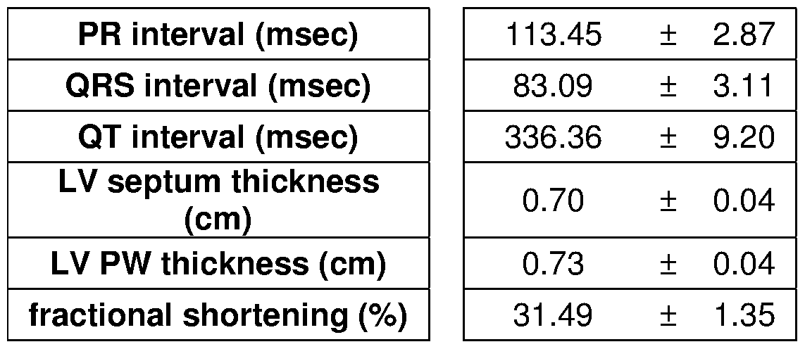
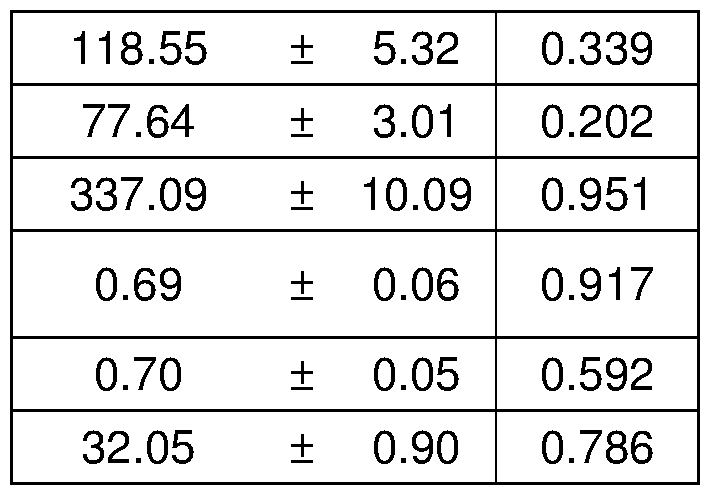
[0182] To explore whether pulsatile glucocorticoids may be useful in other forms of muscular dystrophy, the metabolic effects in a mouse model of limb girdle muscular dystrophy was interrogated. A form of muscular dystrophy for which clinical data suggested a deleterious effects from daily prednisone in patients (Walter et al., 2013) was specifically selected.
Dysferlin deficient ( Dysf-null) mice, a genetic model of this disease, received long-term treatment with weekly prednisone for 32 weeks from the age of disease onset (approximately 9 months; n=10 mice/group; randomized males/females). Consistent with observations in mdx mice and DMD patients, intermittent prednisone improved BCAA utilization in muscle (Figure 4C). Circulating free fatty acids and ketone bodies were also decreased after treatment (Table 4). Furthermore, endpoint levels of ATP, NAD+, and glycogen were increased in muscle and heart (Figure 4C). Pulsatile prednisone increased muscle mass and improved performance of limb muscles, respiratory muscles, and heart (Figure 8), expanding this favorable metabolic reprogramming regimen to a pathologically distinct form of muscular dystrophy.
Table 4. Weekly steroid dosing promotes favorable remodeling of glucose, fatty acid and ketone metabolism in Dysf-null mice. vehicle weekly prednisone
BLOOD and SERUM mean ± s.e.m mean ± s.e.m P value
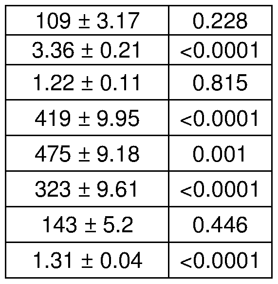


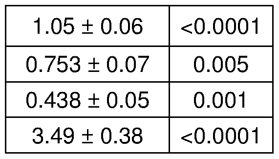
TISSUE b-HYDROXYBUTYRA TE (nmol/mg)
[0183] To investigate the impact of pulsatile glucocorticoids in conditions of metabolic stress, the effects of this drug regimen were monitored in experimental conditions of obesity (Figure 9). Wildtype (WT) mice were fed high-fat chow and treated with either vehicle or weekly (pulsatile)
1 mg/kg intraperitoneal prednisone administration for 8 weeks. (Figure 9A) As compared to vehicle treatment, weekly prednisone slightly but significantly reduced gain of body weight and fat mass, while improved lean mass retention. (Figure 9B) Weekly prednisone reduced the gain of hyperglycemia, as shown by fasting blood glucose levels over time. At diet exposure endpoint, obese mice treated with weekly prednisone showed improved body-wide glucose
homeostasis, as shown by glucose and insulin tolerance tests. (Figure 9C) Weekly prednisone improved grip strength (forelimbs, bilateral), tetanic force production (tibialis anterior, in situ) and aerobic exercise capacity (run-to-exhaustion, treadmill) at the end of high-fat diet regimen.
[0184] Whether pulsatile glucocorticoid treatment improved energy production and muscle function in aging mice was investigated next (Figure 10). Wildtype (WT) mice were treated with either vehicle or weekly (pulsatile) 1 mg/kg intraperitoneal prednisone administration for 40 weeks from the age of 6 weeks. (Figure 10A) As compared to vehicle treatment, weekly prednisone increased levels of ATP, NAD+ and glycogen in muscle and heart tissues. (Figure 10B) In aged mice, weekly prednisone improved grip strength, tetanic and specific force, and muscle mass, seen as myofiber cross-sectional area (CSA). (Figure 10C) Weekly prednisone improved parameters of respiratory function over time, as measured by whole-body
plethysmography. (Figure 10D) Weekly prednisone improved parameters of cardiac contractile function over time, as measured by echocardiography.
[0185] Considering the beneficial metabolic remodeling, the effects of pulsatile steroid administration on adiponectin levels were tested (Figure 11). The analyses showed that pulsatile glucocorticoid treatment increased circulating adiponectin levels in mice and humans, including dystrophic mdx mice (Figure 11 A), in dystrophic DMD patients (Figure 11 B), in mice under diet-induced obesity (Figure 11 C), and in aging mice (Figure 11 D). Experiments were also performed to evaluate longer term outcomes from weekly steroids in a mouse model of obesity. Wildtype (WT) mice were fed high-fat chow and treated with either vehicle or once weekly (pulsatile) 1 mg/kg intraperitoneal prednisone administration for 12 weeks. Figure 12 shows that 12-week-long pulsatile glucocorticoid exposure curbed obesity, insulin resistance, and metabolic dysfunction in wildtype mice with high fat diet-induced obesity.
Discussion
[0186] Glucocorticoids are among the most highly prescribed drugs worldwide and are part of the standard of care to promote ambulation in DMD patients despite adverse side effects (McDonald et al., 2018). Studies of glucocorticoid effects in muscle are dominated by atrophic remodeling, which is especially prominent in mouse models (Schakman et al., 2009). Distinct from human muscle, mouse muscle has a higher ratio of type lib myofibers, defined by fast myosin isoforms and a high reliance on glycolysis (Schiaffino and Reggiani, 201 1 ). Fast myofibers are more susceptible than slow myofibers to FOX03 activation and, hence, to glucocorticoid-driven atrophy (Sandri et al., 2006). Pulsatile glucocorticoids were discovered to induce a pro-ergogenic program supported by BCAA-mediated mitochondrial respiration and
aerobic energy production, directed by a distinct epigenomic and transcriptional program linking GR to KLF15 and the muscle factor MEF2C. KLF15 is a circadian factor controlling amino acid metabolism that has been implicated in pro-ergogenic glucocorticoid cascades (Morrison-Nozik et al., 2015; Sun et al., 2016). The combination of KLF15 and MEF2C advances those findings to define a molecular regulatory combination effective for promoting muscle performance in dystrophic muscle.
[0187] Muscle catabolism of BCAA influences muscle function and whole-body metabolic homeostasis (Li et al., 2017; White et al., 2018), whereas disruption of BCAA disposal and utilization, including its accumulation in circulation and tissues, is associated with metabolic dysfunction and obesity (Lynch and Adams, 2014). The data presented here support that pulsatile glucocorticoids couple higher BCAA-mediated mitochondrial respiration to increased glycolysis, resulting in improved energy production and insulin sensitivity. Moreover, pulsatile steroid dosing increased NAD biogenesis pathway expression and NAD+ levels, further stabilizing favorable reprogramming of dystrophic muscle metabolism (Zhang et al., 2016). The combination of BCAA-mediated respiration, glycolysis and NAD repletion boosts energy production and muscle function in dystrophic muscle.
[0188] Strikingly, metabolic programming by pulsatile glucocorticoids was not limited to dystrophin-linked muscular dystrophy but was also seen in a genetic model of limb-girdle muscular dystrophy linked to a completely distinct cellular defect. There are currently no indications for glucocorticoids in muscular dystrophies beyond DMD, and efficacy has been questioned in small studies of daily steroid dosing (Godfrey et al., 2006; Walter et al., 2013). Intriguingly, it was recently reported that a glucocorticoid-KLF15-BCAA axis benefits a mouse model of spinal muscular atrophy, a genetic disorder with a significant neuronal component (Walter et al., 2018). It is therefore possible that favorable metabolic reprogramming by pulsed glucocorticoid regimens is applicable beyond muscle.
[0189] The findings disclosed herein demonstrate that pulsatile glucocorticoids enable a GR- KLF15-MEF2C axis in dystrophic muscle to support BCAA utilization and energy production, providing useful signatures to monitor these effects in other conditions of diseased, normal or aging muscle.
REFERENCES
Ahn, B., Soundarapandian, M.M., Sessions, FI., Peddibhotla, S., Roth, G.P., Li, J.L., Sugarman, E., Koo, A., Malany, S., Wang, M., et al. (2016). MondoA coordinately regulates skeletal myocyte lipid homeostasis and insulin signaling. J Clin Invest 126 3567-3579.
Bentzinger, C.F., Romanino, K., Cloetta, D., Lin, S., Mascarenhas, J.B., Oliveri, F., Xia, J., Casanova, E., Costa, C.F., Brink, M., et al. (2008). Skeletal muscle-specific ablation of
raptor, but not of rictor, causes metabolic changes and results in muscle dystrophy. Cell Metab 8, 41 1 -424.
Bodine, S.C., Latres, E., Baumhueter, S., Lai, V.K., Nunez, L., Clarke, B.A., Poueymirou, W.T., Panaro, F.J., Na, E., Dharmarajan, K., et al. (2001 ). Identification of ubiquitin ligases required for skeletal muscle atrophy. Science 294, 1704-1708.
Bullard, S.A., Seo, S., Schilling, B., Dyle, M.C., Dierdorff, J.M., Ebert, S.M., DeLau, A.D.,
Gibson, B.W., and Adams, C.M. (2016). Gadd45a Protein Promotes Skeletal Muscle
Atrophy by Forming a Complex with the Protein Kinase MEKK4. J Biol Chem 291, 17496- 17509.
Connolly, A.M., Schierbecker, J., Renna, R., and Florence, J. (2002). High dose weekly oral prednisone improves strength in boys with Duchenne muscular dystrophy. Neuromuscul Disord 12, 917-925.
D'Antona, G., Ragni, M., Cardile, A., Tedesco, L., Dossena, M., Bruttini, F., Caliaro, F., Corsetti, G., Bottinelli, R., Carruba, M.O., et al. (2010). Branched-chain amino acid supplementation promotes survival and supports cardiac and skeletal muscle mitochondrial biogenesis in middle-aged mice. Cell Metab 12, 362-372.
Godfrey, C., Escolar, D., Brockington, M., Clement, E.M., Mein, R., Jimenez-Mallebrera, C., Torelli, S., Feng, L., Brown, S.C., Sewry, C.A., et al. (2006). Fukutin gene mutations in steroid-responsive limb girdle muscular dystrophy. Ann Neurol 60, 603-610.
Li, T., Zhang, Z., Kolwicz, S.C., Jr., Abell, L., Roe, N.D., Kim, M., Zhou, B., Cao, Y., Ritterhoff,
J., Gu, FI., et al. (2017). Defective Branched-Chain Amino Acid Catabolism Disrupts Glucose Metabolism and Sensitizes the Heart to Ischemia-Reperfusion Injury. Cell Metab 25, 374- 385.
Lin, Q., Schwarz, J., Bucana, C., and Olson, E.N. (1997). Control of mouse cardiac
morphogenesis and myogenesis by transcription factor MEF2C. Science 276, 1404-1407.
Lynch, C.J., and Adams, S.H. (2014). Branched-chain amino acids in metabolic signalling and insulin resistance. Nat Rev Endocrinol 10, 723-736.
McDonald, C.M., Henricson, E.K., Abresch, R.T., Duong, T., Joyce, N.C., Hu, F., Clemens,
P.R., Hoffman, E.P., Cnaan, A., Gordish-Dressman, H., et al. (2018). Long-term effects of glucocorticoids on function, quality of life, and survival in patients with Duchenne muscular dystrophy: a prospective cohort study. Lancet 391, 451 -461.
Morrison-Nozik, A., Anand, P., Zhu, H., Duan, Q., Sabeh, M., Prosdocimo, D.A., Lemieux, M.E., Nordsborg, N., Russell, A.P., MacRae, C.A., et al. (2015). Glucocorticoids enhance muscle endurance and ameliorate Duchenne muscular dystrophy through a defined metabolic program. Proc Natl Acad Sci U S A 112, E6780-6789.
Nadal, A., Quesada, I., Tuduri, E., Nogueiras, R., and Alonso-Magdalena, P. (2017). Endocrine- disrupting chemicals and the regulation of energy balance. Nat Rev Endocrinol 13, 536-546.
Quattrocelli, M., Barefield, D.Y., Warner, J.L., Vo, A.H., Hadhazy, M., Earley, J.U., Demonbreun, A.R., and McNally, E.M. (2017a). Intermittent glucocorticoid steroid dosing enhances muscle repair without eliciting muscle atrophy. J Clin Invest 127, 2418-2432.
Quattrocelli, M., Salamone, I.M., Page, P.G., Warner, J.L., Demonbreun, A.R., and McNally,
E.M. (2017b). Intermittent Glucocorticoid Dosing Improves Muscle Repair and Function in Mice with Limb-Girdle Muscular Dystrophy. Am J Pathol 187, 2520-2535.
Rivera, C.M., and Ren, B. (2013). Mapping human epigenomes. Cell 155, 39-55.
Ryu, D., Zhang, H., Ropelle, E.R., Sorrentino, V., Mazala, D.A., Mouchiroud, L., Marshall, P.L., Campbell, M.D., Ali, A.S., Knowels, G.M., et al. (2016). NAD+ repletion improves muscle function in muscular dystrophy and counters global PARylation. Sci Transl Med 8, 361 ra139.
Sancak, Y., Peterson, T.R., Shaul, Y.D., Lindquist, R.A., Thoreen, C.C., Bar-Peled, L., and
Sabatini, D.M. (2008). The Rag GTPases bind raptor and mediate amino acid signaling to mTORCI . Science 320, 1496-1501.
Sandri, M., Lin, J., Handschin, C., Yang, W., Arany, Z.P., Lecker, S.H., Goldberg, A.L., and Spiegelman, B.M. (2006). PGC-1 alpha protects skeletal muscle from atrophy by
suppressing Fox03 action and atrophy-specific gene transcription. Proc Natl Acad Sci U S A 103, 16260-16265.
Sandri, M., Sandri, C., Gilbert, A., Skurk, C., Calabria, E., Picard, A., Walsh, K., Schiaffino, S., Lecker, S.H., and Goldberg, A.L. (2004). Foxo transcription factors induce the atrophy- related ubiquitin ligase atrogin-1 and cause skeletal muscle atrophy. Cell 117, 399-412.
Schakman, O., Gilson, FI., Kalista, S., and Thissen, J.P. (2009). Mechanisms of muscle atrophy induced by glucocorticoids. Horm Res 72 Suppl 1, 36-41 .
Schiaffino, S., and Reggiani, C. (201 1 ). Fiber types in mammalian skeletal muscles. Physiol Rev 91, 1447-1531 .
Shintaku, J., Peterson, J.M., Talbert, E.E., Gu, J.M., Ladner, K.J., Williams, D.R., Mousavi, K., Wang, R., Sartorelli, V., and Guttridge, D.C. (2016). MyoD Regulates Skeletal Muscle Oxidative Metabolism Cooperatively with Alternative NF-kappaB. Cell Rep 17, 514-526.
Sun, FI., Olson, K.C., Gao, C., Prosdocimo, D.A., Zhou, M., Wang, Z., Jeyaraj, D., Youn, J.Y., Ren, S., Liu, Y., et al. (2016). Catabolic Defect of Branched-Chain Amino Acids Promotes Heart Failure. Circulation 133, 2038-2049.
Vockley, C.M., D'lppolito, A.M., McDowell, I.C., Majoros, W.H., Safi, A., Song, L., Crawford,
G.E., and Reddy, T.E. (2016). Direct GR Binding Sites Potentiate Clusters of TF Binding across the Human Genome. Cell 166, 1269-1281 e1219.
Walter, L.M., Deguise, M.O., Meijboom, K.E., Betts, C.A., Ahlskog, N., van Westering, T.L.E., Hazell, G., McFall, E., Kordala, A., Hammond, S.M., et al. (2018). Interventions Targeting Glucocorticoid-Kruppel-like Factor 15-Branched-Chain Amino Acid Signaling Improve Disease Phenotypes in Spinal Muscular Atrophy Mice. EBioMedicine 31, 226-242.
Walter, M.C., Reilich, P., Thiele, S., Schessl, J., Schreiber, H., Reiners, K., Kress, W., Muller- Reible, C., Vorgerd, M., Urban, P., et al. (2013). Treatment of dysferlinopathy with deflazacort: a double-blind, placebo-controlled clinical trial. Orphanet J Rare Dis 8, 26.
White, P.J., McGarrah, R.W., Grimsrud, P.A., Tso, S.C., Yang, W.H., Haldeman, J.M., Grenier- Larouche, T., An, J., Lapworth, A.L., Astapova, L, et al. (2018). The BCKDH Kinase and Phosphatase Integrate BCAA and Lipid Metabolism via Regulation of ATP-Citrate Lyase.
Cell Metab 27, 1281 -1293 e1287.
Zhang, H., Ryu, D., Wu, Y., Gariani, K., Wang, X., Luan, P., D'Amico, D., Ropelle, E.R., Lutolf, M.P., Aebersold, R., et al. (2016). NAD(+) repletion improves mitochondrial and stem cell function and enhances life span in mice. Science 352, 1436-1443.
Anders, S., Pyl, P.T., and Huber, W. (2015). HTSeq-a Python framework to work with high- throughput sequencing data. Bioinformatics 31, 166-169.
Ashburner, M., Ball, C.A., Blake, J.A., Botstein, D., Butler, H., Cherry, J.M., Davis, A.P.,
Dolinski, K., Dwight, S.S., Eppig, J.T., et al. (2000). Gene ontology: tool for the unification of biology. The Gene Ontology Consortium. Nat Genet 25, 25-29.
Bruno, C., Patin, F., Bocca, C., Nadal-Desbarats, L., Bonnier, F., Reynier, P., Emond, P.,
Vourc'h, P., Joseph-Delafont, K., Corcia, P., et al. (2018). The combination of four analytical methods to explore skeletal muscle metabolomics: Better coverage of metabolic pathways or a marketing argument? J Pharm Biomed Anal 148, 273-279.
Carey, M.F., Peterson, C.L., and Smale, S.T. (2009). Chromatin immunoprecipitation (ChIP).
Cold Spring Harb Protoc 2009, pdb prot5279.
Choi, Y.K., and Park, K.G. (2018). Targeting Glutamine Metabolism for Cancer Treatment.
Biomol Ther (Seoul) 26, 19-28.
Chong, J., Soufan, O., Li, C., Caraus, L, Li, S., Bourque, G., Wishart, D.S., and Xia, J. (2018).
MetaboAnalyst 4.0: towards more transparent and integrative metabolomics analysis.
Nucleic Acids Res.
Demonbreun, A.R., Fahrenbach, J.P., Deveaux, K., Earley, J.U., Pytel, P., and McNally, E.M. (201 1 ). Impaired muscle growth and response to insulin-like growth factor 1 in dysferlin- mediated muscular dystrophy. Hum Mol Genet 20, 779-789.
Demonbreun, A.R., and McNally, E.M. (2015). DNA Electroporation, Isolation and Imaging of Myofibers. J Vis Exp, e53551.
Demonbreun, A.R., Rossi, A.E., Alvarez, M.G., Swanson, K.E., Deveaux, H.K., Earley, J.U., Hadhazy, M., Vohra, R., Walter, G.A., Pytel, P., et al. (2014). Dysferlin and myoferlin regulate transverse tubule formation and glycerol sensitivity. Am J Pathol 184, 248-259.
DiFranco, M., Quinonez, M., Capote, J., and Vergara, J. (2009). DNA transfection of
mammalian skeletal muscles using in vivo electroporation. J Vis Exp.
Heinz, S., Benner, C., Spann, N., Bertolino, E., Lin, Y.C., Laslo, P., Cheng, J.X., Murre, C., Singh, H., and Glass, C.K. (2010). Simple combinations of lineage-determining transcription factors prime cis-regulatory elements required for macrophage and B cell identities. Mol Cell 38, 576-589.
Hill, A.M., LaForgia, J., Coates, A.M., Buckley, J.D., and Howe, P.R. (2007). Estimating
abdominal adipose tissue with DXA and anthropometry. Obesity (Silver Spring) 15, 504-510.
Kambo, A., Sharma, V.S., Casteel, D.E., Woods, V.L., Jr., Pilz, R.B., and Boss, G.R. (2005).
Nitric oxide inhibits mammalian methylmalonyl-CoA mutase. J Biol Chem 280, 10073-10082.
Kerr, J.P., Ziman, A.P., Mueller, A.L., Muriel, J.M., Kleinhans-Welte, E., Gumerson, J.D., Vogel, S.S., Ward, C.W., Roche, J.A., and Bloch, R.J. (2013). Dysferlin stabilizes stress-induced Ca2+ signaling in the transverse tubule membrane. Proc Natl Acad Sci U S A 110, 20831 - 20836.
Kozhemyakina, E., Cohen, T., Yao, T.P., and Lassar, A.B. (2009). Parathyroid hormone-related peptide represses chondrocyte hypertrophy through a protein phosphatase 2A/histone deacetylase 4/MEF2 pathway. Mol Cell Biol 29, 5751 -5762.
Langmead, B., and Salzberg, S.L. (2012). Fast gapped-read alignment with Bowtie 2. Nat
Methods 9, 357-359.
Metsalu, T., and Vilo, J. (2015). ClustVis: a web tool for visualizing clustering of multivariate data using Principal Component Analysis and heatmap. Nucleic Acids Res 43, W566-570.
Perez-Llamas, C., and Lopez-Bigas, N. (201 1 ). Gitools: analysis and visualisation of genomic data using interactive heat-maps. PLoS One 6, e19541 .
Quattrocelli, M., Swinnen, M., Giacomazzi, G., Camps, J., Barthelemy, I., Ceccarelli, G.,
Caluwe, E., Grosemans, H., Thorrez, L., Pelizzo, G., et al. (2015). Mesodermal iPSC- derived progenitor cells functionally regenerate cardiac and skeletal muscle. J Clin Invest 125, 4463-4482.
Ramsey, K.M., Yoshino, J., Brace, C.S., Abrassart, D., Kobayashi, Y., Marcheva, B., Hong,
H.K., Chong, J.L., Buhr, E.D., Lee, C., et al. (2009). Circadian clock feedback cycle through NAMPT-mediated NAD+ biosynthesis. Science 324, 651 -654.
Robinson, M.D., McCarthy, D.J., and Smyth, G.K. (2010). edgeR: a Bioconductor package for differential expression analysis of digital gene expression data. Bioinformatics 26, 139-140.
Sali, A., Guerron, A.D., Gordish-Dressman, H., Spurney, C.F., lantorno, M., Hoffman, E.P., and Nagaraju, K. (2012). Glucocorticoid-treated mice are an inappropriate positive control for long-term preclinical studies in the mdx mouse. PLoS One 7, e34204.
Schneider, C.A., Rasband, W.S., and Eliceiri, K.W. (2012). NIH Image to ImageJ: 25 years of image analysis. Nat Methods 9, 671 -675.
Shintaku, J., and Guttridge, D.C. (2016). Analysis of Aerobic Respiration in Intact Skeletal
Muscle Tissue by Microplate-Based Respirometry. Methods Mol Biol 1460, 337-343.
Trapnell, C., Pachter, L., and Salzberg, S.L. (2009). TopHat: discovering splice junctions with RNA-Seq. Bioinformatics 25, 1 105-1 1 1 1.
Xian, Z.Y., Liu, J.M., Chen, Q.K., Chen, H.Z., Ye, C.J., Xue, J., Yang, H.Q., Li, J.L., Liu, X.F., and Kuang, S.J. (2015). Inhibition of LDHA suppresses tumor progression in prostate cancer. Tumour Biol 36 8093-8100.
Zou, C., Wang, Y., and Shen, Z. (2005). 2-NBDG as a fluorescent indicator for direct glucose uptake measurement. J Biochem Biophys Methods 64 207-215.
Gerke, V., C.E. Creutz, and S.E. Moss. 2005. Annexins: linking Ca2+ signalling to membrane dynamics. Nat Rev Mol Cell Biol. 6:449-461.
de Laat, B., R.H. Derksen, I.J. Mackie, M. Roest, S. Schoormans, B.J. Woodhams, P.G. de Groot, and W.L. van Heerde. 2006. Annexin A5 polymorphism (-1 C->T) and the presence of anti-annexin A5 antibodies in the antiphospholipid syndrome. Annals of the rheumatic diseases. 65:1468-1472.
Gerke, V., and S.E. Moss. 2002. Annexins: from structure to function. Physiol Rev. 82:331 -371.
Blackwood, R.A., and J.D. Ernst. 1990. Characterization of Ca2(+)-dependent phospholipid binding, vesicle aggregation and membrane fusion by annexins. The Biochemical journal. 266:195-200.
Goulet, F., K.G. Moore, and A.C. Sartorelli. 1992. Glycosylation of annexin I and annexin II.
Biochemical and biophysical research communications. 188:554-558.
Kaetzel, M.A., Y.D. Mo, T.R. Mealy, B. Campos, W. Bergsma-Schutter, A. Brisson, J.R.
Dedman, and B.A. Seaton. 2001. Phosphorylation mutants elucidate the mechanism of annexin IV-mediated membrane aggregation. Biochemistry. 40:4192-4199.
Zaks, W.J., and C.E. Creutz. 1991 . Ca(2+)-dependent annexin self-association on membrane surfaces. Biochemistry. 30:9607-9615.
Christmas, P., J. Callaway, J. Fallon, J. Jones, and FIT. Haigler. 1991. Selective secretion of annexin 1 , a protein without a signal sequence, by the human prostate gland. The Journal of biological chemistry. 266:2499-2507.
Deora, A.B., G. Kreitzer, A.T. Jacovina, and K.A. Hajjar. 2004. An annexin 2 phosphorylation switch mediates p1 1 -dependent translocation of annexin 2 to the cell surface. The Journal of biological chemistry. 279:4341 1 -43418.
Wallner, B.P., R.J. Mattaliano, C. Hession, R.L. Cate, R. Tizard, L.K. Sinclair, C. Foeller, E.P.
Chow, J.L. Browing, K.L. Ramachandran, and et al. 1986. Cloning and expression of human lipocortin, a phospholipase A2 inhibitor with potential anti-inflammatory activity. Nature. 320:77-81.
Hannon, R., J.D. Croxtall, S.J. Getting, F. Roviezzo, S. Yona, M.J. Paul-Clark, F.N. Gavins, M.
Perretti, J.F. Morris, J.C. Buckingham, and R.J. Flower. 2003. Aberrant inflammation and resistance to glucocorticoids in annexin 1 -/- mouse. FASEB J. 17:253-255.
Ling, Q., A.T. Jacovina, A. Deora, M. Febbraio, R. Simantov, R.L. Silverstein, B. Hempstead, W.H. Mark, and K.A. Hajjar. 2004. Annexin II regulates fibrin homeostasis and
neoangiogenesis in vivo. The Journal of clinical investigation. 1 13:38-48.
Cagliani, R., F. Magri, A. Toscano, L. Merlini, F. Fortunato, C. Lamperti, C. Rodolico, A. Prelle, M. Sironi, M. Aguennouz, P. Ciscato, A. Uncini, M. Moggio, N. Bresolin, and G.P. Comi. 2005. Mutation finding in patients with dysferlin deficiency and role of the dysferlin interacting proteins annexin A1 and A2 in muscular dystrophies. Human mutation. 26:283.
Claims
1. A method of administering a glucocorticoid steroid to a patient, wherein the patient has a serum or plasma level of one or more of the following biomarkers that is:
(a) less than about 18 mg/dL morning fasting cortisol;
(b) at least about 90 mg/dL fasting morning glucose;
(c) at least about 160 pmol/L insulin;
(d) at least about 40 mGTΐoI/L isoleucine;
(e) at least about 100 mGTΐoI/L leucine;
(f) at least about 120 mGTΐoI/L valine;
(g) at least about 700 mGTΐoI/L combined branched chain amino acids;
(h) at least about 1 10 mg/dL triglycerides;
(i) at least about 300 mGTΐoI/L non-esterified fatty acids; and
(j) at least about 100 mGTΐoI/L combined ketones;
wherein the administering of the glucocorticoid steroid comprises once-weekly administration of the glucocorticoid steroid.
2. The method of claim 1 , wherein the patient suffers from muscle wasting, obesity, a metabolic disorder, sarcopenia, an inflammatory disorder, a muscle injury, or a combination thereof.
3. The method of claim 1 or claim 2, wherein the once-weekly administration of glucocorticoid steroid comprises a single dose of about 0.1 to about 5 mg/kg.
4. The method of any one of claims 1 -3, wherein the once-weekly administration of glucocorticoid steroid comprises a single dose of about 1 mg/kg.
5. The method of any one of claims 1 -3, wherein the once-weekly administration of glucocorticoid steroid comprises a single dose of about 0.75 mg/kg.
6. The method of any one of claims 2-5, wherein the muscle wasting is aging- related muscle wasting, disease-related muscle wasting, diabetes-associated muscle wasting, muscle atrophy, sarcopenia, cardiomyopathy, a chronic myopathy, an inflammatory myopathy, a muscular dystrophy, or a combination thereof.
7. The method of any one of claims 1 -6, wherein the metabolic disorder is metabolic syndrome, insulin resistance, a nutrition disorder, exercise intolerance, or a combination thereof.
8. The method of claim 6, wherein the cardiomyopathy is hypertrophic, dilated, congenital, arrhythmogenic, restrictive, ischemic, or heart failure.
9. The method of claim 8, wherein the heart failure includes reduced ejection fraction.
10. The method of claim 8, wherein the heart failure includes preserved ejection fraction.
1 1 . The method of any one of claims 1 -10, wherein the administering results in one or more of decreased insulin resistance, increased glucose tolerance, increased muscle mass, decreased hyperinsulinemia, increased lean mass, increased force, increased systolic function, increased diastolic function, decreased muscle fibrosis, increased exercise tolerance, increased nutrient catabolism, increased energy production, increased serum adiponectin, decreased serum branched chain amino acids (BCAA), decreased serum lipid level, decreased serum ketone level, decreased hyperglycemia, increased serum cortisol level, increased serum corticosterone, and decreased adipocyte size compared to administering the glucocorticoid steroid in a dosing regimen that is not once-weekly or to not administering the glucocorticoid steroid.
12. The method of any one of claims 1 -1 1 , further comprising administering an effective amount of (i) an agent that increases the activity of an annexin protein, (ii) mitsugumin 53 (MG53), (iii) a modulator of latent TGF-b binding protein 4 (LTBP4), (iv) a modulator of transforming growth factor b (TGF-b) activity, (v) a modulator of androgen response, (vi) a modulator of an inflammatory response, (vii) a promoter of muscle growth, (viii) a
chemotherapeutic agent, (ix) a modulator of fibrosis, (x) a modulator of glucose homeostasis, (xi) a modulator of metabolic function, or a combination thereof.
13. The method of claim 12, wherein the agent that increases the activity of an annexin protein is selected from the group consisting of a recombinant protein, a steroid, and a polynucleotide capable of expressing an annexin protein.
14. The method of claim 13, wherein the polynucleotide is associated with a nanoparticle.
15. The method of claim 13, wherein the polynucleotide is contained in a vector.
16. The method of claim 15, wherein the vector is within a chloroplast.
17. The method of claim 15 wherein the vector is a viral vector.
18. The method of claim 17wherein the viral vector is selected from the group consisting of a herpes virus vector, an adeno-associated virus (AAV) vector, an adeno virus vector, and a lentiviral vector.
19. The method of claim 18 wherein the AAV vector is recombinant AAV5, AAV6, AAV8, AAV9, or AAV74.
20. The method of claim 19, wherein the AAV74 vector is AAVrh74.
21 . The method of any one of claims 12-20, wherein the agent increases the activity of annexin A1 (SEQ ID NO: 1 ), annexin A2 (SEQ ID NO: 2 or SEQ ID NO: 3), annexin A3 (SEQ ID NO: 4), annexin A4 (SEQ ID NO: 5), annexin A5 (SEQ ID NO: 6), annexin A6 (SEQ ID NO: 7, SEQ ID NO: 8, SEQ ID NO: 44, or a combination thereof), annexin A7 (SEQ ID NO: 9 or SEQ ID NO: 10), annexin A8 (SEQ ID NO: 1 1 or SEQ ID NO: 12), annexin A9 (SEQ ID NO: 13), annexin A10 (SEQ ID NO: 14), annexin A1 1 (SEQ ID NO: 15 or SEQ ID NO: 16), annexin A13 (SEQ ID NO: 17 or SEQ ID NO: 18), or a combination thereof.
22. The method of claim 21 , wherein the agent increases the activity of annexin A1 (SEQ ID NO: 1 ), annexin A2 (SEQ ID NO: 2 or SEQ ID NO: 3), and annexin A6 (SEQ ID NO: 7, SEQ ID NO: 8, SEQ ID NO: 44, or a combination thereof).
23. The method of claim 21 , wherein the agent increases the activity of annexin A2 (SEQ ID NO: 2 or SEQ ID NO: 3) and annexin A6 (SEQ ID NO: 7, SEQ ID NO: 8, SEQ ID NO: 44, or a combination thereof).
24. The method of claim 21 , wherein the agent increases the activity of annexin A1 (SEQ ID NO: 1 ) and annexin A6 (SEQ ID NO: 7, SEQ ID NO: 8, SEQ ID NO: 44, or a
combination thereof).
25. The method of claim 21 , wherein the agent increases the activity of annexin A6 (SEQ ID NO: 7, SEQ ID NO: 8, SEQ ID NO: 44, or a combination thereof).
Priority Applications (1)
| Application Number | Priority Date | Filing Date | Title |
|---|---|---|---|
| US17/416,792 US20220062299A1 (en) | 2018-12-26 | 2019-12-26 | Use of glucocorticoid steroids in preventing and treating conditions of muscle wasting, aging and metabolic disorder |
Applications Claiming Priority (4)
| Application Number | Priority Date | Filing Date | Title |
|---|---|---|---|
| US201862785029P | 2018-12-26 | 2018-12-26 | |
| US62/785,029 | 2018-12-26 | ||
| US201962876238P | 2019-07-19 | 2019-07-19 | |
| US62/876,238 | 2019-07-19 |
Publications (1)
| Publication Number | Publication Date |
|---|---|
| WO2020139977A1 true WO2020139977A1 (en) | 2020-07-02 |
Family
ID=69326745
Family Applications (1)
| Application Number | Title | Priority Date | Filing Date |
|---|---|---|---|
| PCT/US2019/068618 Ceased WO2020139977A1 (en) | 2018-12-26 | 2019-12-26 | Use of glucocorticoid steroids in preventing and treating conditions of muscle wasting, aging and metabolic disorder |
Country Status (2)
| Country | Link |
|---|---|
| US (1) | US20220062299A1 (en) |
| WO (1) | WO2020139977A1 (en) |
Families Citing this family (2)
| Publication number | Priority date | Publication date | Assignee | Title |
|---|---|---|---|---|
| CN118001372A (en) * | 2023-07-04 | 2024-05-10 | 上海萨美细胞技术有限公司 | Use of annexin for anti-aging |
| CN119405781A (en) * | 2024-12-19 | 2025-02-11 | 中日友好医院(中日友好临床医学研究所) | Application of MG53 recombinant protein |
Citations (58)
| Publication number | Priority date | Publication date | Assignee | Title |
|---|---|---|---|---|
| US3687808A (en) | 1969-08-14 | 1972-08-29 | Univ Leland Stanford Junior | Synthetic polynucleotides |
| US4845205A (en) | 1985-01-08 | 1989-07-04 | Institut Pasteur | 2,N6 -disubstituted and 2,N6 -trisubstituted adenosine-3'-phosphoramidites |
| US5130302A (en) | 1989-12-20 | 1992-07-14 | Boron Bilogicals, Inc. | Boronated nucleoside, nucleotide and oligonucleotide compounds, compositions and methods for using same |
| US5134066A (en) | 1989-08-29 | 1992-07-28 | Monsanto Company | Improved probes using nucleosides containing 3-dezauracil analogs |
| US5175273A (en) | 1988-07-01 | 1992-12-29 | Genentech, Inc. | Nucleic acid intercalating agents |
| US5367066A (en) | 1984-10-16 | 1994-11-22 | Chiron Corporation | Oligonucleotides with selectably cleavable and/or abasic sites |
| US5399363A (en) | 1991-01-25 | 1995-03-21 | Eastman Kodak Company | Surface modified anticancer nanoparticles |
| US5432272A (en) | 1990-10-09 | 1995-07-11 | Benner; Steven A. | Method for incorporating into a DNA or RNA oligonucleotide using nucleotides bearing heterocyclic bases |
| US5457187A (en) | 1993-12-08 | 1995-10-10 | Board Of Regents University Of Nebraska | Oligonucleotides containing 5-fluorouracil |
| US5459255A (en) | 1990-01-11 | 1995-10-17 | Isis Pharmaceuticals, Inc. | N-2 substituted purines |
| US5466468A (en) | 1990-04-03 | 1995-11-14 | Ciba-Geigy Corporation | Parenterally administrable liposome formulation comprising synthetic lipids |
| US5484908A (en) | 1991-11-26 | 1996-01-16 | Gilead Sciences, Inc. | Oligonucleotides containing 5-propynyl pyrimidines |
| US5502177A (en) | 1993-09-17 | 1996-03-26 | Gilead Sciences, Inc. | Pyrimidine derivatives for labeled binding partners |
| US5525711A (en) | 1994-05-18 | 1996-06-11 | The United States Of America As Represented By The Secretary Of The Department Of Health And Human Services | Pteridine nucleotide analogs as fluorescent DNA probes |
| US5552540A (en) | 1987-06-24 | 1996-09-03 | Howard Florey Institute Of Experimental Physiology And Medicine | Nucleoside derivatives |
| US5594121A (en) | 1991-11-07 | 1997-01-14 | Gilead Sciences, Inc. | Enhanced triple-helix and double-helix formation with oligomers containing modified purines |
| US5596091A (en) | 1994-03-18 | 1997-01-21 | The Regents Of The University Of California | Antisense oligonucleotides comprising 5-aminoalkyl pyrimidine nucleotides |
| US5614617A (en) | 1990-07-27 | 1997-03-25 | Isis Pharmaceuticals, Inc. | Nuclease resistant, pyrimidine modified oligonucleotides that detect and modulate gene expression |
| WO1997012896A1 (en) | 1995-10-04 | 1997-04-10 | Epoch Pharmaceuticals, Inc. | Selective binding complementary oligonucleotides |
| US5645985A (en) | 1991-11-26 | 1997-07-08 | Gilead Sciences, Inc. | Enhanced triple-helix and double-helix formation with oligomers containing modified pyrimidines |
| US5681941A (en) | 1990-01-11 | 1997-10-28 | Isis Pharmaceuticals, Inc. | Substituted purines and oligonucleotide cross-linking |
| US5720950A (en) | 1990-05-14 | 1998-02-24 | University Of Medicine & Dentistry Of New Jersey | Polymers containing antifibrotic agents, compositions containing such polymers, and methods of preparation and use |
| US5750692A (en) | 1990-01-11 | 1998-05-12 | Isis Pharmaceuticals, Inc. | Synthesis of 3-deazapurines |
| US5830653A (en) | 1991-11-26 | 1998-11-03 | Gilead Sciences, Inc. | Methods of using oligomers containing modified pyrimidines |
| US6004937A (en) | 1998-03-09 | 1999-12-21 | Genetics Institute, Inc. | Use of follistatin to modulate growth and differentiation factor 8 [GDF-8] and bone morphogenic protein 11 [BMP-11] |
| WO2000043781A2 (en) | 1999-01-21 | 2000-07-27 | Metamorphix, Inc. | Growth differentiation factor inhibitors and uses therefor |
| US6096506A (en) | 1993-03-19 | 2000-08-01 | The Johns Hopkins University School Of Medicine | Antibodies specific for growth differentiation factor-8 and methods of using same |
| WO2001005820A2 (en) | 1999-07-20 | 2001-01-25 | Pharmexa A/S | Method for down-regulating gdf-8 activity |
| EP1072679A2 (en) | 1999-07-20 | 2001-01-31 | Agilent Technologies Inc. | Method of producing nucleic acid molecules with reduced secondary structure |
| WO2001053350A1 (en) | 2000-01-18 | 2001-07-26 | Agresearch Limited | Myostatin and mimetics thereof |
| US6369201B1 (en) | 1998-02-19 | 2002-04-09 | Metamorphix International, Inc. | Myostatin multimers |
| WO2002068650A2 (en) | 2001-02-08 | 2002-09-06 | Wyeth | Modified and stabilized gdf propeptides and uses thereof |
| US6465493B1 (en) | 1999-04-09 | 2002-10-15 | Smithkline Beecham Corporation | Triarylimidazoles |
| US6468535B1 (en) | 1993-03-19 | 2002-10-22 | The Johns Hopkins University School Of Medicine | Growth differentiation factor-8 |
| WO2002085306A2 (en) | 2001-04-24 | 2002-10-31 | The Johns Hopkins University | Use of follistatin to increase muscle mass |
| US20030166633A1 (en) | 2000-02-21 | 2003-09-04 | Gaster Laramie Mary | Pyridinylimidazoles |
| US20040039198A1 (en) | 2000-11-16 | 2004-02-26 | Bender Paul E. | Compounds |
| US20040063745A1 (en) | 2001-02-02 | 2004-04-01 | Francoise Jeanne Gellibert | 2-amino-4-(pyridin-2-yl)-thiazole derivatives as transforming growth factor beta (tgf-beta) inhibitors |
| US20040138118A1 (en) | 2002-09-16 | 2004-07-15 | Neil Wolfman | Metalloprotease activation of myostatin, and methods of modulating myostatin activity |
| US20040181033A1 (en) | 2002-12-20 | 2004-09-16 | Hq Han | Binding agents which inhibit myostatin |
| US20040223966A1 (en) | 2002-10-25 | 2004-11-11 | Wolfman Neil M. | ActRIIB fusion polypeptides and uses therefor |
| US6906089B2 (en) | 2000-03-27 | 2005-06-14 | Smithkline Beecham Corporation | Triarylimidazole derivatives as cytokine inhibitors |
| WO2005084699A1 (en) | 2004-03-02 | 2005-09-15 | Acceleron Pharma Inc. | Alk7 and myostatin inhibitors and uses thereof |
| WO2005094446A2 (en) | 2004-03-23 | 2005-10-13 | Eli Lilly And Company | Anti-myostatin antibodies |
| WO2006012627A2 (en) | 2004-07-23 | 2006-02-02 | Acceleron Pharma Inc. | Actrii receptor polypeptides, methods and compositions |
| WO2006025988A1 (en) | 2004-07-29 | 2006-03-09 | Schering-Plough Ltd. | Use of alk 5 inhibitors to modulate or inhibit myostatin activity leading to increased lean tissue accretion in animals |
| WO2006116269A2 (en) | 2005-04-25 | 2006-11-02 | Pfizer Inc. | Antibodies to myostatin |
| US7192717B2 (en) | 2002-02-21 | 2007-03-20 | Wyeth | GASP1: a follistatin domain containing protein |
| US7223833B1 (en) | 1991-05-24 | 2007-05-29 | Isis Pharmaceuticals, Inc. | Peptide nucleic acid conjugates |
| US7261893B2 (en) | 2002-10-22 | 2007-08-28 | Wyeth | Neutralizing antibodies against GDF-8 and uses therefor |
| US7320789B2 (en) | 2001-09-26 | 2008-01-22 | Wyeth | Antibody inhibitors of GDF-8 and uses thereof |
| US7572763B2 (en) | 2002-02-21 | 2009-08-11 | Wyeth | Follistatin domain containing proteins |
| US20120039806A1 (en) | 2009-03-23 | 2012-02-16 | Mireille Hanna Lahoud | Compounds and Methods for Modulating an Immune Response |
| US20120046345A1 (en) | 2009-05-08 | 2012-02-23 | Opko Curna, Llc | Treatment of dystrophin family related diseases by inhibition of natural antisense transcript to dmd family |
| US20120058955A1 (en) | 2009-03-18 | 2012-03-08 | Association Francaise Contre Les Myopathies | Use of decorine for increasing muscle mass |
| US9873739B2 (en) | 2012-08-01 | 2018-01-23 | Ikaika Therapeutics, Llc | Mitigating tissue damage and fibrosis via latent transforming growth factor beta binding protein (LTBP4) |
| US20190070261A1 (en) | 2012-08-21 | 2019-03-07 | Ali Nayer | Materials and methods for modulating glucose uptake |
| US20190091282A1 (en) | 2015-07-16 | 2019-03-28 | Nuritas Limited | Peptides for use in promoting transport of glucose |
-
2019
- 2019-12-26 WO PCT/US2019/068618 patent/WO2020139977A1/en not_active Ceased
- 2019-12-26 US US17/416,792 patent/US20220062299A1/en active Pending
Patent Citations (62)
| Publication number | Priority date | Publication date | Assignee | Title |
|---|---|---|---|---|
| US3687808A (en) | 1969-08-14 | 1972-08-29 | Univ Leland Stanford Junior | Synthetic polynucleotides |
| US5367066A (en) | 1984-10-16 | 1994-11-22 | Chiron Corporation | Oligonucleotides with selectably cleavable and/or abasic sites |
| US4845205A (en) | 1985-01-08 | 1989-07-04 | Institut Pasteur | 2,N6 -disubstituted and 2,N6 -trisubstituted adenosine-3'-phosphoramidites |
| US5552540A (en) | 1987-06-24 | 1996-09-03 | Howard Florey Institute Of Experimental Physiology And Medicine | Nucleoside derivatives |
| US5175273A (en) | 1988-07-01 | 1992-12-29 | Genentech, Inc. | Nucleic acid intercalating agents |
| US5134066A (en) | 1989-08-29 | 1992-07-28 | Monsanto Company | Improved probes using nucleosides containing 3-dezauracil analogs |
| US5130302A (en) | 1989-12-20 | 1992-07-14 | Boron Bilogicals, Inc. | Boronated nucleoside, nucleotide and oligonucleotide compounds, compositions and methods for using same |
| US5750692A (en) | 1990-01-11 | 1998-05-12 | Isis Pharmaceuticals, Inc. | Synthesis of 3-deazapurines |
| US5459255A (en) | 1990-01-11 | 1995-10-17 | Isis Pharmaceuticals, Inc. | N-2 substituted purines |
| US5587469A (en) | 1990-01-11 | 1996-12-24 | Isis Pharmaceuticals, Inc. | Oligonucleotides containing N-2 substituted purines |
| US5681941A (en) | 1990-01-11 | 1997-10-28 | Isis Pharmaceuticals, Inc. | Substituted purines and oligonucleotide cross-linking |
| US5466468A (en) | 1990-04-03 | 1995-11-14 | Ciba-Geigy Corporation | Parenterally administrable liposome formulation comprising synthetic lipids |
| US5720950A (en) | 1990-05-14 | 1998-02-24 | University Of Medicine & Dentistry Of New Jersey | Polymers containing antifibrotic agents, compositions containing such polymers, and methods of preparation and use |
| US5614617A (en) | 1990-07-27 | 1997-03-25 | Isis Pharmaceuticals, Inc. | Nuclease resistant, pyrimidine modified oligonucleotides that detect and modulate gene expression |
| US5432272A (en) | 1990-10-09 | 1995-07-11 | Benner; Steven A. | Method for incorporating into a DNA or RNA oligonucleotide using nucleotides bearing heterocyclic bases |
| US5399363A (en) | 1991-01-25 | 1995-03-21 | Eastman Kodak Company | Surface modified anticancer nanoparticles |
| US7223833B1 (en) | 1991-05-24 | 2007-05-29 | Isis Pharmaceuticals, Inc. | Peptide nucleic acid conjugates |
| US5594121A (en) | 1991-11-07 | 1997-01-14 | Gilead Sciences, Inc. | Enhanced triple-helix and double-helix formation with oligomers containing modified purines |
| US5645985A (en) | 1991-11-26 | 1997-07-08 | Gilead Sciences, Inc. | Enhanced triple-helix and double-helix formation with oligomers containing modified pyrimidines |
| US5484908A (en) | 1991-11-26 | 1996-01-16 | Gilead Sciences, Inc. | Oligonucleotides containing 5-propynyl pyrimidines |
| US5830653A (en) | 1991-11-26 | 1998-11-03 | Gilead Sciences, Inc. | Methods of using oligomers containing modified pyrimidines |
| US6096506A (en) | 1993-03-19 | 2000-08-01 | The Johns Hopkins University School Of Medicine | Antibodies specific for growth differentiation factor-8 and methods of using same |
| US6468535B1 (en) | 1993-03-19 | 2002-10-22 | The Johns Hopkins University School Of Medicine | Growth differentiation factor-8 |
| US5763588A (en) | 1993-09-17 | 1998-06-09 | Gilead Sciences, Inc. | Pyrimidine derivatives for labeled binding partners |
| US5502177A (en) | 1993-09-17 | 1996-03-26 | Gilead Sciences, Inc. | Pyrimidine derivatives for labeled binding partners |
| US6005096A (en) | 1993-09-17 | 1999-12-21 | Gilead Sciences, Inc. | Pyrimidine derivatives |
| US5457187A (en) | 1993-12-08 | 1995-10-10 | Board Of Regents University Of Nebraska | Oligonucleotides containing 5-fluorouracil |
| US5596091A (en) | 1994-03-18 | 1997-01-21 | The Regents Of The University Of California | Antisense oligonucleotides comprising 5-aminoalkyl pyrimidine nucleotides |
| US5525711A (en) | 1994-05-18 | 1996-06-11 | The United States Of America As Represented By The Secretary Of The Department Of Health And Human Services | Pteridine nucleotide analogs as fluorescent DNA probes |
| WO1997012896A1 (en) | 1995-10-04 | 1997-04-10 | Epoch Pharmaceuticals, Inc. | Selective binding complementary oligonucleotides |
| US6369201B1 (en) | 1998-02-19 | 2002-04-09 | Metamorphix International, Inc. | Myostatin multimers |
| US6004937A (en) | 1998-03-09 | 1999-12-21 | Genetics Institute, Inc. | Use of follistatin to modulate growth and differentiation factor 8 [GDF-8] and bone morphogenic protein 11 [BMP-11] |
| WO2000043781A2 (en) | 1999-01-21 | 2000-07-27 | Metamorphix, Inc. | Growth differentiation factor inhibitors and uses therefor |
| US6465493B1 (en) | 1999-04-09 | 2002-10-15 | Smithkline Beecham Corporation | Triarylimidazoles |
| WO2001005820A2 (en) | 1999-07-20 | 2001-01-25 | Pharmexa A/S | Method for down-regulating gdf-8 activity |
| EP1072679A2 (en) | 1999-07-20 | 2001-01-31 | Agilent Technologies Inc. | Method of producing nucleic acid molecules with reduced secondary structure |
| WO2001053350A1 (en) | 2000-01-18 | 2001-07-26 | Agresearch Limited | Myostatin and mimetics thereof |
| US20030166633A1 (en) | 2000-02-21 | 2003-09-04 | Gaster Laramie Mary | Pyridinylimidazoles |
| US6906089B2 (en) | 2000-03-27 | 2005-06-14 | Smithkline Beecham Corporation | Triarylimidazole derivatives as cytokine inhibitors |
| US20040039198A1 (en) | 2000-11-16 | 2004-02-26 | Bender Paul E. | Compounds |
| US20040063745A1 (en) | 2001-02-02 | 2004-04-01 | Francoise Jeanne Gellibert | 2-amino-4-(pyridin-2-yl)-thiazole derivatives as transforming growth factor beta (tgf-beta) inhibitors |
| WO2002068650A2 (en) | 2001-02-08 | 2002-09-06 | Wyeth | Modified and stabilized gdf propeptides and uses thereof |
| US7202210B2 (en) | 2001-02-08 | 2007-04-10 | Wyeth | Modified and stabilized GDF propeptides and uses thereof |
| WO2002085306A2 (en) | 2001-04-24 | 2002-10-31 | The Johns Hopkins University | Use of follistatin to increase muscle mass |
| US7320789B2 (en) | 2001-09-26 | 2008-01-22 | Wyeth | Antibody inhibitors of GDF-8 and uses thereof |
| US7572763B2 (en) | 2002-02-21 | 2009-08-11 | Wyeth | Follistatin domain containing proteins |
| US7192717B2 (en) | 2002-02-21 | 2007-03-20 | Wyeth | GASP1: a follistatin domain containing protein |
| US20040138118A1 (en) | 2002-09-16 | 2004-07-15 | Neil Wolfman | Metalloprotease activation of myostatin, and methods of modulating myostatin activity |
| US7261893B2 (en) | 2002-10-22 | 2007-08-28 | Wyeth | Neutralizing antibodies against GDF-8 and uses therefor |
| US20040223966A1 (en) | 2002-10-25 | 2004-11-11 | Wolfman Neil M. | ActRIIB fusion polypeptides and uses therefor |
| US20040181033A1 (en) | 2002-12-20 | 2004-09-16 | Hq Han | Binding agents which inhibit myostatin |
| WO2005084699A1 (en) | 2004-03-02 | 2005-09-15 | Acceleron Pharma Inc. | Alk7 and myostatin inhibitors and uses thereof |
| WO2005094446A2 (en) | 2004-03-23 | 2005-10-13 | Eli Lilly And Company | Anti-myostatin antibodies |
| WO2006012627A2 (en) | 2004-07-23 | 2006-02-02 | Acceleron Pharma Inc. | Actrii receptor polypeptides, methods and compositions |
| WO2006025988A1 (en) | 2004-07-29 | 2006-03-09 | Schering-Plough Ltd. | Use of alk 5 inhibitors to modulate or inhibit myostatin activity leading to increased lean tissue accretion in animals |
| WO2006116269A2 (en) | 2005-04-25 | 2006-11-02 | Pfizer Inc. | Antibodies to myostatin |
| US20120058955A1 (en) | 2009-03-18 | 2012-03-08 | Association Francaise Contre Les Myopathies | Use of decorine for increasing muscle mass |
| US20120039806A1 (en) | 2009-03-23 | 2012-02-16 | Mireille Hanna Lahoud | Compounds and Methods for Modulating an Immune Response |
| US20120046345A1 (en) | 2009-05-08 | 2012-02-23 | Opko Curna, Llc | Treatment of dystrophin family related diseases by inhibition of natural antisense transcript to dmd family |
| US9873739B2 (en) | 2012-08-01 | 2018-01-23 | Ikaika Therapeutics, Llc | Mitigating tissue damage and fibrosis via latent transforming growth factor beta binding protein (LTBP4) |
| US20190070261A1 (en) | 2012-08-21 | 2019-03-07 | Ali Nayer | Materials and methods for modulating glucose uptake |
| US20190091282A1 (en) | 2015-07-16 | 2019-03-28 | Nuritas Limited | Peptides for use in promoting transport of glucose |
Non-Patent Citations (98)
| Title |
|---|
| "Pharmaceutics and Pharmacy Practice", 1982, J. B. LIPPINCOTT CO., pages: 238 - 250 |
| AHN, B.SOUNDARAPANDIAN, M.M.SESSIONS, H.PEDDIBHOTLA, S.ROTH, G.P.LI, J.L.SUGARMAN, E.KOO, A.MALANY, S.WANG, M. ET AL.: "MondoA coordinately regulates skeletal myocyte lipid homeostasis and insulin signaling", J CLIN INVEST, vol. 126, 2016, pages 3567 - 3579 |
| ANDERS, S.PYL, P.T.HUBER, W.: "HTSeq--a Python framework to work with high-throughput sequencing data", BIOINFORMATICS, vol. 31, 2015, pages 166 - 169 |
| ASHBURNER, M.BALL, C.A.BLAKE, J.A.BOTSTEIN, D.BUTLER, H.CHERRY, J.M.DAVIS, A.P.DOLINSKI, K.DWIGHT, S.S.EPPIG, J.T. ET AL.: "Gene ontology: tool for the unification of biology. The Gene Ontology Consortium", NAT GENET, vol. 25, 2000, pages 25 - 29 |
| BENTZINGER, C.F.ROMANINO, K.CLOETTA, D.LIN, S.MASCARENHAS, J.B.OLIVERI, F.XIA, J.CASANOVA, E.COSTA, C.F.BRINK, M. ET AL.: "Skeletal muscle-specific ablation of raptor, but not of rictor, causes metabolic changes and results in muscle dystrophy", CELL METAB, vol. 8, 2008, pages 411 - 424 |
| BERKNER, CURRENT TOPICS IN MICROBIOL. AND IMUNOL., vol. 158, 1992, pages 39 - 66 |
| BLACKWOOD, R.A.J.D. ERNST: "Characterization of Ca2(+)-dependent phospholipid binding, vesicle aggregation and membrane fusion by annexins", THE BIOCHEMICAL JOURNAL, vol. 266, 1990, pages 195 - 200 |
| BODINE, S.C.LATRES, E.BAUMHUETER, S.LAI, V.K.NUNEZ, L.CLARKE, B.A.POUEYMIROU, W.T.PANARO, F.J.NA, E.DHARMARAJAN, K. ET AL.: "Identification of ubiquitin ligases required for skeletal muscle atrophy", SCIENCE, vol. 294, 2001, pages 1704 - 1708, XP002386330, DOI: 10.1126/science.1065874 |
| BOYE ET AL., SCI REP., vol. 8, 2018, pages 10309 |
| BRUNO, C.PATIN, F.BOCCA, C.NADAL-DESBARATS, L.BONNIER, F.REYNIER, P.EMOND, P.VOURC'H, P.JOSEPH-DELAFONT, K.CORCIA, P. ET AL.: "The combination of four analytical methods to explore skeletal muscle metabolomics: Better coverage of metabolic pathways or a marketing argument?", J PHARM BIOMED ANAL, vol. 148, 2018, pages 273 - 279, XP085252221, DOI: 10.1016/j.jpba.2017.10.013 |
| BULLARD, S.A.SEO, S.SCHILLING, B.DYLE, M.C.DIERDORFF, J.M.EBERT, S.M.DELAU, A.D.GIBSON, B.W.ADAMS, C.M.: "Gadd45a Protein Promotes Skeletal Muscle Atrophy by Forming a Complex with the Protein Kinase MEKK4", J BIOL CHEM, vol. 291, 2016, pages 17496 - 17509 |
| CAGLIANI, R.F. MAGRIA. TOSCANOL. MERLINIF. FORTUNATOC. LAMPERTIC. RODOLICOA. PRELLEM. SIRONIM. AGUENNOUZ: "Mutation finding in patients with dysferlin deficiency and role of the dysferlin interacting proteins annexin A1 and A2 in muscular dystrophies", HUMAN MUTATION, vol. 26, 2005, pages 283 |
| CAREY, M.F.PETERSON, C.L.SMALE, S.T.: "Chromatin immunoprecipitation (ChIP", COLD SPRING HARB PROTOC 2009, 2009, pages prot5279 |
| CHOI, Y.K.PARK, K.G.: "Targeting Glutamine Metabolism for Cancer Treatment", BIOMOL THER (SEOUL, vol. 26, 2018, pages 19 - 28 |
| CHONG, J.SOUFAN, O.LI, C.CARAUS, I.LI, S.BOURQUE, G.WISHART, D.S.XIA, J.: "MetaboAnalyst 4.0: towards more transparent and integrative metabolomics analysis", NUCLEIC ACIDS RES., 2018 |
| CHRISTMAS, P.J. CALLAWAYJ. FALLONJ. JONESH.T. HAIGLER: "Selective secretion of annexin 1, a protein without a signal sequence, by the human prostate gland", THE JOURNAL OF BIOLOGICAL CHEMISTRY, vol. 266, 1991, pages 2499 - 2507 |
| CONNOLLY, A.M.SCHIERBECKER, J.RENNA, R.FLORENCE, J.: "High dose weekly oral prednisone improves strength in boys with Duchenne muscular dystrophy", NEUROMUSCUL DISORD, vol. 12, 2002, pages 917 - 925 |
| COOK, ANTI-CANCER DRUG DESIGN, vol. 6, 1991, pages 585 - 607 |
| D'ANTONA, G.RAGNI, M.CARDILE, A.TEDESCO, L.DOSSENA, M.BRUTTINI, F.CALIARO, F.CORSETTI, G.BOTTINELLI, R.CARRUBA, M.O. ET AL.: "Branched-chain amino acid supplementation promotes survival and supports cardiac and skeletal muscle mitochondrial biogenesis in middle-aged mice", CELL METAB, vol. 12, 2010, pages 362 - 372, XP055294173, DOI: 10.1016/j.cmet.2010.08.016 |
| DE LAAT, B.R.H. DERKSENI.J. MACKIEM. ROESTS. SCHOORMANSB.J. WOODHAMSP.G. DE GROOTW.L. VAN HEERDE: "Annexin A5 polymorphism (-1 C-->T) and the presence of anti-annexin A5 antibodies in the antiphospholipid syndrome", ANNALS OF THE RHEUMATIC DISEASES, vol. 65, 2006, pages 1468 - 1472, XP008124504, DOI: 10.1136/ard.2005.045237 |
| DEMONBREUN, A.R.FAHRENBACH, J.PDEVEAUX, K.EARLEY, J.U.PYTEL, P.MCNALLY, E.M.: "Impaired muscle growth and response to insulin-like growth factor 1 in dysferlin-mediated muscular dystrophy", HUM MOL GENET, vol. 20, 2011, pages 779 - 789 |
| DEMONBREUN, A.R.MCNALLY, E.M.: "DNA Electroporation, Isolation and Imaging of Myofibers", J VIS EXP, 2015, pages e53551 |
| DEMONBREUN, A.R.ROSSI, A.E.ALVAREZ, M.G.SWANSON, K.E.DEVEAUX, H.K.EARLEY, J.U.HADHAZY, M.VOHRA, R.WALTER, G.A.PYTEL, P. ET AL.: "Dysferlin and myoferlin regulate transverse tubule formation and glycerol sensitivity", AM J PATHOL, vol. 184, 2014, pages 248 - 259 |
| DEORA, A.B.G. KREITZERA.T. JACOVINAK.A. HAJJAR: "An annexin 2 phosphorylation switch mediates p11-dependent translocation of annexin 2 to the cell surface", THE JOURNAL OF BIOLOGICAL CHEMISTRY, vol. 279, 2004, pages 43411 - 43418 |
| DIFRANCO, M.QUINONEZ, M.CAPOTE, J.VERGARA, J.: "DNA transfection of mammalian skeletal muscles using in vivo electroporation", J VIS EXP, 2009 |
| ENGLISCH ET AL., ANGEWANDTE CHEMIE, vol. 30, 1991, pages 613 - 722 |
| FARDET ET AL., DRUGS, vol. 74, 2014, pages 1731 - 1745 |
| GERKE, V.C.E. CREUTZS.E. MOSS: "Annexins: linking Ca2+ signalling to membrane dynamics", NAT REV MOL CELL BIOL., vol. 6, 2005, pages 449 - 461, XP009111656 |
| GERKE, V.S.E. MOSS: "Annexins: from structure to function", PHYSIOL REV., vol. 82, 2002, pages 331 - 371 |
| GODFREY, C.ESCOLAR, D.BROCKINGTON, M.CLEMENT, E.M.MEIN, R.JIMENEZ-MALLEBRERA, C.TORELLI, S.FENG, L.BROWN, S.C.SEWRY, C.A. ET AL.: "Fukutin gene mutations in steroid-responsive limb girdle muscular dystrophy", ANN NEUROL, vol. 60, 2006, pages 603 - 610 |
| GOULET, F.K.G. MOOREA.C. SARTORELLI: "Glycosylation of annexin I and annexin II", BIOCHEMICAL AND BIOPHYSICAL RESEARCH COMMUNICATIONS, vol. 188, 1992, pages 554 - 558, XP024770356, DOI: 10.1016/0006-291X(92)91091-4 |
| HAMMERS ET AL., JCI INSIGHT, 2019, Retrieved from the Internet <URL:httpsi//doi.orq/10.1172/jcLinsiqbt.133276> |
| HANNON, R.J.D. CROXTALLS.J. GETTINGF. ROVIEZZOS. YONAM.J. PAUL-CLARKF.N. GAVINSM. PERRETTIJ.F. MORRISJ.C. BUCKINGHAM: "Aberrant inflammation and resistance to glucocorticoids in annexin 1-/- mouse", FASEB J., vol. 17, 2003, pages 253 - 255 |
| HAYES ET AL., TRAFFIC, vol. 5, 2004, pages 571 - 576 |
| HEINZ, S.BENNER, C.SPANN, N.BERTOLINO, E.LIN, Y.C.LASLO, P.CHENG, J.X.MURRE, C.SINGH, H.GLASS, C.K.: "Simple combinations of lineage-determining transcription factors prime cis-regulatory elements required for macrophage and B cell identities", MOL CELL, vol. 38, 2010, pages 576 - 589, XP028380241, DOI: 10.1016/j.molcel.2010.05.004 |
| HILL, A.M.LAFORGIA, J.COATES, A.M.BUCKLEY, J.D.HOWE, P.R.: "Obesity", vol. 15, 2007, SILVER SPRING, article "Estimating abdominal adipose tissue with DXA and anthropometry", pages: 504 - 510 |
| KAETZEL, M.A.Y.D. MOT.R. MEALYB. CAMPOSW. BERGSMA-SCHUTTERA. BRISSONJ.R. DEDMANB.A. SEATON: "Phosphorylation mutants elucidate the mechanism of annexin IV-mediated membrane aggregation", BIOCHEMISTRY, vol. 40, 2001, pages 4192 - 4199 |
| KAMBO, A.SHARMA, V.S.CASTEEL, D.E.WOODS, V.L., JR.PILZ, R.B.BOSS, G.R.: "Nitric oxide inhibits mammalian methylmalonyl-CoA mutase", J BIOL CHEM, vol. 280, 2005, pages 10073 - 10082 |
| KATZ, J. AM. CHEM. SOC., vol. 74, 1951, pages 2238 |
| KERR, J.P.ZIMAN, A.P.MUELLER, A.L.MURIEL, J.M.KLEINHANS-WELTE, E.GUMERSON, J.D.VOGEL, S.S.WARD, C.W.ROCHE, J.A.BLOCH, R.J.: "Dysferlin stabilizes stress-induced Ca2+ signaling in the transverse tubule membrane", PROC NATL ACAD SCI U S A, vol. 110, 2013, pages 20831 - 20836 |
| KOSTURKO ET AL., BIOCHEMISTRY, vol. 13, 1974, pages 3949 |
| KOZHEMYAKINA, E.COHEN, T.YAO, T.P.LASSAR, A.B.: "Parathyroid hormone-related peptide represses chondrocyte hypertrophy through a protein phosphatase 2A/histone deacetylase 4/MEF2 pathway", MOL CELL BIOL, vol. 29, 2009, pages 5751 - 5762 |
| LANGMEAD, B.SALZBERG, S.L.: "Fast gapped-read alignment with Bowtie 2", NAT METHODS, vol. 9, 2012, pages 357 - 359, XP002715401, DOI: 10.1038/nmeth.1923 |
| LI, T.ZHANG, Z.KOLWICZ, S.C., JR.ABELL, L.ROE, N.D.KIM, M.ZHOU, B.CAO, Y.RITTERHOFF, J.GU, H. ET AL.: "Defective Branched-Chain Amino Acid Catabolism Disrupts Glucose Metabolism and Sensitizes the Heart to Ischemia-Reperfusion Injury", CELL METAB, vol. 25, 2017, pages 374 - 385, XP029914169, DOI: 10.1016/j.cmet.2016.11.005 |
| LIN, Q.SCHWARZ, J.BUCANA, C.OLSON, E.N.: "Control of mouse cardiac morphogenesis and myogenesis by transcription factor MEF2C", SCIENCE, vol. 276, 1997, pages 1404 - 1407, XP002905115, DOI: 10.1126/science.276.5317.1404 |
| LING, Q.A.T. JACOVINAA. DEORAM. FEBBRAIOR. SIMANTOVR.L. SILVERSTEINB. HEMPSTEADW.H. MARKK.A. HAJJAR: "Annexin II regulates fibrin homeostasis and neoangiogenesis in vivo", THE JOURNAL OF CLINICAL INVESTIGATION, vol. 113, 2004, pages 38 - 48 |
| LYNCH, C.J.ADAMS, S.H.: "Branched-chain amino acids in metabolic signalling and insulin resistance", NAT REV ENDOCRINOL, vol. 10, 2014, pages 723 - 736 |
| MATTIA QUATTROCELLI ET AL: "Intermittent glucocorticoid steroid dosing enhances muscle repair without eliciting muscle atrophy", JOURNAL OF CLINICAL INVESTIGATION, vol. 127, no. 6, 1 June 2017 (2017-06-01), GB, pages 2418 - 2432, XP055675785, ISSN: 0021-9738, DOI: 10.1172/JCI91445 * |
| MCDONALD, C.M.HENRICSON, E.K.ABRESCH, R.T.DUONG, T.JOYCE, N.C.HU, F.CLEMENS, P.R.HOFFMAN, E.P.CNAAN, A.GORDISH-DRESSMAN, H. ET AL.: "Long-term effects of glucocorticoids on function, quality of life, and survival in patients with Duchenne muscular dystrophy: a prospective cohort study", LANCET, vol. 391, 2018, pages 451 - 461, XP085377935, DOI: 10.1016/S0140-6736(17)32160-8 |
| MCNALLY ET AL., HUM MOL GENET, 2014 |
| METSALU, T.VILO, J.: "ClustVis: a web tool for visualizing clustering of multivariate data using Principal Component Analysis and heatmap", NUCLEIC ACIDS RES, vol. 43, 2015, pages W566 - 570 |
| MORRISON-NOZIK, A.ANAND, P.ZHU, H.DUAN, Q.SABEH, M.PROSDOCIMO, D.A.LEMIEUX, M.E.NORDSBORG, N.RUSSELL, A.P.MACRAE, C.A. ET AL.: "Glucocorticoids enhance muscle endurance and ameliorate Duchenne muscular dystrophy through a defined metabolic program", PROC NATL ACAD SCI U S A, vol. 112, 2015, pages E6780 - 6789 |
| NADAL, A.QUESADA, I.TUDURI, E.NOGUEIRAS, R.ALONSO-MAGDALENA, P.: "Endocrine-disrupting chemicals and the regulation of energy balance", NAT REV ENDOCRINOL, vol. 13, 2017, pages 536 - 546 |
| NELSON, WENTWORTH ET AL., AM J PATHOL, 2011 |
| PEREZ-LLAMAS, C.LOPEZ-BIGAS, N.: "Gitools: analysis and visualisation of genomic data using interactive heat-maps", PLOS ONE, vol. 6, 2011, pages e19541 |
| QUATTROCELLI ET AL., AJP, 2017 |
| QUATTROCELLI ET AL., JCI INSIGHT, vol. 4, no. 24, 19 December 2019 (2019-12-19), pages 132402 |
| QUATTROCELLI ET AL., JCI INSIGHT., vol. 4, no. 24, 19 December 2019 (2019-12-19), pages 132402 |
| QUATTROCELLI ET AL., JCI, 2017 |
| QUATTROCELLI, M.BAREFIELD, D.Y.WARNER, J.L.VO, A.H.HADHAZY, M.EARLEY, J.U.DEMONBREUN, A.R.MCNALLY, E.M.: "Intermittent glucocorticoid steroid dosing enhances muscle repair without eliciting muscle atrophy", J CLIN INVEST, vol. 127, 2017, pages 2418 - 2432 |
| QUATTROCELLI, M.SALAMONE, I.M.PAGE, P.G.WARNER, J.L.DEMONBREUN, A.R.MCNALLY, E.M.: "Intermittent Glucocorticoid Dosing Improves Muscle Repair and Function in Mice with Limb-Girdle Muscular Dystrophy", AM J PATHOL, vol. 187, 2017, pages 2520 - 2535 |
| QUATTROCELLI, M.SWINNEN, M.GIACOMAZZI, G.CAMPS, J.BARTHELEMY, I.CECCARELLI, G.CALUWE, E.GROSEMANS, H.THORREZ, L.PELIZZO, G. ET AL.: "Mesodermal iPSC-derived progenitor cells functionally regenerate cardiac and skeletal muscle", J CLIN INVEST, vol. 125, 2015, pages 4463 - 4482 |
| RAMSEY, K.M.YOSHINO, J.BRACE, C.S.ABRASSART, D.KOBAYASHI, Y.MARCHEVA, B.HONG, H.K.CHONG, J.L.BUHR, E.D.LEE, C. ET AL.: "Circadian clock feedback cycle through NAMPT-mediated NAD+ biosynthesis", SCIENCE, vol. 324, 2009, pages 651 - 654, XP055041363, DOI: 10.1126/science.1171641 |
| REMINGTON'S PHARMACEUTICAL SCIENCES, 1980 |
| RIVERA, C.M.REN, B.: "Mapping human epigenomes", CELL, vol. 155, 2013, pages 39 - 55, XP028729747, DOI: 10.1016/j.cell.2013.09.011 |
| ROBINSON, M.D.MCCARTHY, D.J.SMYTH, G.K.: "edgeR: a Bioconductor package for differential expression analysis of digital gene expression data", BIOINFORMATICS, vol. 26, 2010, pages 139 - 140 |
| ROSENFELD ET AL., CELL, vol. 8, 1992, pages 143 - 144 |
| RYU, D.ZHANG, H.ROPELLE, E.R.SORRENTINO, V.MAZALA, D.A.MOUCHIROUD, L.MARSHALL, P.L.CAMPBELL, M.D.ALI, A.S.KNOWELS, G.M. ET AL.: "NAD+ repletion improves muscle function in muscular dystrophy and counters global PARylation", SCI TRANSL MED, vol. 8, no. 361, 2016, pages ra139 |
| SALI, A.GUERRON, A.D.GORDISH-DRESSMAN, H.SPURNEY, C.F.LANTORNO, M.HOFFMAN, E.P.NAGARAJU, K.: "Glucocorticoid-treated mice are an inappropriate positive control for long-term preclinical studies in the mdx mouse", PLOS ONE, vol. 7, 2012, pages e34204 |
| SAMBROOK ET AL.: "Molecular Cloning: A Laboratory Manual", 1989 |
| SANCAK, Y.PETERSON, T.R.SHAUL, Y.D.LINDQUIST, R.A.THOREEN, C.C.BAR-PELED, L.SABATINI, D.M.: "The Rag GTPases bind raptor and mediate amino acid signaling to mTORCI", SCIENCE, vol. 320, 2008, pages 1496 - 1501 |
| SANDRI, M.LIN, J.HANDSCHIN, C.YANG, W.ARANY, Z.P.LECKER, S.H.GOLDBERG, A.L.SPIEGELMAN, B.M.: "PGC-1 alpha protects skeletal muscle from atrophy by suppressing Fox03 action and atrophy-specific gene transcription", PROC NATL ACAD SCI U S A, vol. 103, 2006, pages 16260 - 16265, XP055243355, DOI: 10.1073/pnas.0607795103 |
| SANDRI, M.SANDRI, C.GILBERT, A.SKURK, C.CALABRIA, E.PICARD, A.WALSH, K.SCHIAFFINO, S.LECKER, S.H.GOLDBERG, A.L.: "Foxo transcription factors induce the atrophy-related ubiquitin ligase atrogin-1 and cause skeletal muscle atrophy", CELL, vol. 117, 2004, pages 399 - 412, XP002338896 |
| SANGHVI, Y. S.: "Antisense Research and Applications", 1993, CRC PRESS, pages: 289 - 302 |
| SCHAKMAN, O.GILSON, H.KALISTA, S.THISSEN, J.P.: "Mechanisms of muscle atrophy induced by glucocorticoids", HORM RES, vol. 72, no. 1, 2009, pages 36 - 41 |
| SCHIAFFINO, S.REGGIANI, C.: "Fiber types in mammalian skeletal muscles", PHYSIOL REV, vol. 91, 2011, pages 1447 - 1531 |
| SCHNEIDER, C.A.RASBAND, W.S.ELICEIRI, K.W.: "NIH Image to ImageJ: 25 years of image analysis", NAT METHODS, vol. 9, 2012, pages 671 - 675, XP055403257 |
| SHINTAKU, J.GUTTRIDGE, D.C.: "Analysis of Aerobic Respiration in Intact Skeletal Muscle Tissue by Microplate-Based Respirometry", METHODS MOL BIOL, vol. 1460, 2016, pages 337 - 343 |
| SHINTAKU, J.PETERSON, J.M.TALBERT, E.E.GU, J.M.LADNER, K.J.WILLIAMS, D.R.MOUSAVI, K.WANG, R.SARTORELLI, V.GUTTRIDGE, D.C.: "MyoD Regulates Skeletal Muscle Oxidative Metabolism Cooperatively with Alternative NF-kappaB", CELL REP, vol. 17, 2016, pages 514 - 526 |
| STRATFORD-PERRICAUDET ET AL., HUM. GENE THER., vol. 1, 1990, pages 241 - 256 |
| STRATFORD-PERRICAUDET ET AL., J. CLIN. INVEST., vol. 90, 1992, pages 626 - 630 |
| SUN, H.OLSON, K.C.GAO, C.PROSDOCIMO, D.A.ZHOU, M.WANG, Z.JEYARAJ, D.YOUN, J.Y.REN, S.LIU, Y. ET AL.: "Catabolic Defect of Branched-Chain Amino Acids Promotes Heart Failure", CIRCULATION, vol. 133, 2016, pages 2038 - 2049 |
| SUSAN M. FREIERKARL-HEINZ ALTMANN, NUCLEIC ACIDS RESEARCH, vol. 25, 1997, pages 4429 - 4443 |
| THOMAS, J. AM. CHEM. SOC., vol. 76, 1954, pages 6032 |
| TRAPNELL, C.PACHTER, L.SALZBERG, S.L.: "TopHat: discovering splice junctions with RNA-Seq", BIOINFORMATICS, vol. 25, 2009, pages 1105 - 1111, XP055597009, DOI: 10.1093/bioinformatics/btp120 |
| VOCKLEY, C.M.D'IPPOLITO, A.M.MCDOWELL, I.C.MAJOROS, W.H.SAFI, A.SONG, L.CRAWFORD, G.E.REDDY, T.E.: "Direct GR Binding Sites Potentiate Clusters of TF Binding across the Human Genome", CELL, vol. 166, 2016, pages 1269 - 1281 |
| VOLOVITZ ET AL: "Normal diurnal variation in serum cortisol concentration in asthmatic children treated with inhaled budesonide", JOURNAL OF ALLERGY AND CLINICAL IMMUNOLOGY, ELSEVIER, AMSTERDAM, NL, vol. 96, no. 6, 1 December 1995 (1995-12-01), pages 874 - 878, XP005145431, ISSN: 0091-6749, DOI: 10.1016/S0091-6749(95)70222-9 * |
| WALLNER, B.P.R.J. MATTALIANOC. HESSIONR.L. CATER. TIZARDL.K. SINCLAIRC. FOELLERE.P. CHOWJ.L. BROWINGK.L. RAMACHANDRAN ET AL.: "Cloning and expression of human lipocortin, a phospholipase A2 inhibitor with potential anti-inflammatory activity", NATURE, vol. 320, 1986, pages 622 - 630 |
| WALTER, L.M.DEGUISE, M.O.MEIJBOOM, K.E.BETTS, C.A.AHLSKOG, N.VAN WESTERING, T.L.E.HAZELL, G.MCFALL, E.KORDALA, A.HAMMOND, S.M. ET : "Interventions Targeting Glucocorticoid-Kruppel-like Factor 15-Branched-Chain Amino Acid Signaling Improve Disease Phenotypes in Spinal Muscular Atrophy Mice", EBIOMEDICINE, vol. 31, 2018, pages 226 - 242 |
| WALTER, M.C.REILICH, P.THIELE, S.SCHESSL, J.SCHREIBER, H.REINERS, K.KRESS, W.MULLER-REIBLE, C.VORGERD, M.URBAN, P. ET AL.: "Treatment of dysferlinopathy with deflazacort: a double-blind, placebo-controlled clinical trial", ORPHANET J RARE DIS, vol. 8, 2013, pages 26, XP021147228, DOI: 10.1186/1750-1172-8-26 |
| WHITE, P.J.MCGARRAH, R.W.GRIMSRUD, P.A.TSO, S.C.YANG, W.H.HALDEMAN, J.M.GRENIER-LAROUCHE, T.AN, J.LAPWORTH, A.L.ASTAPOVA, I. ET AL: "The BCKDH Kinase and Phosphatase Integrate BCAA and Lipid Metabolism via Regulation of ATP-Citrate Lyase", CELL METAB, vol. 27, 2018, pages 1281 - 1293, XP055665291, DOI: 10.1016/j.cmet.2018.04.015 |
| XIAN, Z.Y.LIU, J.M.CHEN, Q.K.CHEN, H.Z.YE, C.J.XUE, J.YANG, H.Q.LI, J.L.LIU, X.F.KUANG, S.J.: "Inhibition of LDHA suppresses tumor progression in prostate cancer", TUMOUR BIOL, vol. 36, 2015, pages 8093 - 8100, XP036223888, DOI: 10.1007/s13277-015-3540-x |
| YAMANE ET AL., J. AM. CHEM. SOC., vol. 83, 1961, pages 2599 |
| ZAKS, W.J.C.E. CREUTZ: "Ca(2+)-dependent annexin self-association on membrane surfaces", BIOCHEMISTRY, vol. 30, 1991, pages 9607 - 9615 |
| ZHANG ET AL., J. AM. CHEM. SOC., vol. 127, 2005, pages 74 - 75 |
| ZHANG, H.RYU, D.WU, Y.GARIANI, K.WANG, X.LUAN, P.D'AMICO, D.ROPELLE, E.R.LUTOLF, M.P.AEBERSOLD, R. ET AL.: "NAD(+) repletion improves mitochondrial and stem cell function and enhances life span in mice", SCIENCE, vol. 352, 2016, pages 1436 - 1443 |
| ZIMMERMANN ET AL., J. AM. CHEM. SOC., vol. 124, 2002, pages 13684 - 13685 |
| ZOU, C.WANG, Y.SHEN, Z.: "2-NBDG as a fluorescent indicator for direct glucose uptake measurement", J BIOCHEM BIOPHYS METHODS, vol. 64, 2005, pages 207 - 215, XP005096964, DOI: 10.1016/j.jbbm.2005.08.001 |
Also Published As
| Publication number | Publication date |
|---|---|
| US20220062299A1 (en) | 2022-03-03 |
Similar Documents
| Publication | Publication Date | Title |
|---|---|---|
| US20230036788A1 (en) | Compositions and methods of using tyrosine kinase inhibitors | |
| Cheng et al. | Expression of organic anion transporter 2 in the human kidney and its potential role in the tubular secretion of guanine-containing antiviral drugs | |
| JP6124986B2 (en) | Growth and differentiation factor (GDF) for treating diastolic heart failure | |
| RU2695228C2 (en) | Discontinuous introduction of mdm2 inhibitor | |
| WO2020132647A1 (en) | Use of annexins in preventing and treating muscle membrane injury | |
| JP2017536408A (en) | Kinase inhibitor prodrugs for cancer treatment | |
| AU2021249111A1 (en) | Methods and compositions for treating cancer | |
| WO2020139977A1 (en) | Use of glucocorticoid steroids in preventing and treating conditions of muscle wasting, aging and metabolic disorder | |
| Harahap et al. | Salbutamol inhibits ubiquitin-mediated survival motor neuron protein degradation in spinal muscular atrophy cells | |
| AU2012308097B2 (en) | Treatment of bone diseases | |
| US20240294988A1 (en) | Methods of determining responsiveness to chemotherapeutic compounds for cancer therapy | |
| US20250312332A1 (en) | Treatment of muscle fibrosis | |
| JP2017513843A (en) | Compositions and methods for the treatment or prevention of neurodegenerative disorders | |
| JP7613693B2 (en) | Treatment of Cardiac Disease by Inhibiting PP2A Anchoring | |
| US20230159934A1 (en) | Insulin receptor aptamer and pharmaceutical composition for treating diabetes comprising the same | |
| Oh et al. | Urolithin A Restores Mitochondrial Function and Reverses Cardiac Remodeling in HFpEF | |
| Luttman | Exploiting Metabolic Vulnerabilities In Solid Tumors Treated With ABL Kinase Allosteric Inhibitors | |
| HK40057503A (en) | Compositions and methods of using tyrosine kinase inhibitors | |
| Zhao | Targeting Metabolic Dysregulation for the Treatment of Radiation Fibrosis | |
| JP2021038183A (en) | Agent for preventing or treating heart failure, and pharmaceutical composition for preventing or treating heart failure | |
| KR20200071446A (en) | Compositon for preventing or treating ostarthritis using inhibiting oxysterol production | |
| HK1245678B (en) | Compositions and methods of using tyrosine kinase inhibitors |
Legal Events
| Date | Code | Title | Description |
|---|---|---|---|
| 121 | Ep: the epo has been informed by wipo that ep was designated in this application |
Ref document number: 19843038 Country of ref document: EP Kind code of ref document: A1 |
|
| NENP | Non-entry into the national phase |
Ref country code: DE |
|
| 122 | Ep: pct application non-entry in european phase |
Ref document number: 19843038 Country of ref document: EP Kind code of ref document: A1 |