WO2020005584A1 - Sample preparation method and system - Google Patents
Sample preparation method and system Download PDFInfo
- Publication number
- WO2020005584A1 WO2020005584A1 PCT/US2019/037245 US2019037245W WO2020005584A1 WO 2020005584 A1 WO2020005584 A1 WO 2020005584A1 US 2019037245 W US2019037245 W US 2019037245W WO 2020005584 A1 WO2020005584 A1 WO 2020005584A1
- Authority
- WO
- WIPO (PCT)
- Prior art keywords
- liquid composition
- dna
- nucleic acid
- detergent
- solid support
- Prior art date
- Legal status (The legal status is an assumption and is not a legal conclusion. Google has not performed a legal analysis and makes no representation as to the accuracy of the status listed.)
- Ceased
Links
Classifications
-
- C—CHEMISTRY; METALLURGY
- C12—BIOCHEMISTRY; BEER; SPIRITS; WINE; VINEGAR; MICROBIOLOGY; ENZYMOLOGY; MUTATION OR GENETIC ENGINEERING
- C12Q—MEASURING OR TESTING PROCESSES INVOLVING ENZYMES, NUCLEIC ACIDS OR MICROORGANISMS; COMPOSITIONS OR TEST PAPERS THEREFOR; PROCESSES OF PREPARING SUCH COMPOSITIONS; CONDITION-RESPONSIVE CONTROL IN MICROBIOLOGICAL OR ENZYMOLOGICAL PROCESSES
- C12Q1/00—Measuring or testing processes involving enzymes, nucleic acids or microorganisms; Compositions therefor; Processes of preparing such compositions
- C12Q1/68—Measuring or testing processes involving enzymes, nucleic acids or microorganisms; Compositions therefor; Processes of preparing such compositions involving nucleic acids
- C12Q1/6806—Preparing nucleic acids for analysis, e.g. for polymerase chain reaction [PCR] assay
-
- C—CHEMISTRY; METALLURGY
- C12—BIOCHEMISTRY; BEER; SPIRITS; WINE; VINEGAR; MICROBIOLOGY; ENZYMOLOGY; MUTATION OR GENETIC ENGINEERING
- C12N—MICROORGANISMS OR ENZYMES; COMPOSITIONS THEREOF; PROPAGATING, PRESERVING, OR MAINTAINING MICROORGANISMS; MUTATION OR GENETIC ENGINEERING; CULTURE MEDIA
- C12N15/00—Mutation or genetic engineering; DNA or RNA concerning genetic engineering, vectors, e.g. plasmids, or their isolation, preparation or purification; Use of hosts therefor
- C12N15/09—Recombinant DNA-technology
- C12N15/10—Processes for the isolation, preparation or purification of DNA or RNA
- C12N15/1003—Extracting or separating nucleic acids from biological samples, e.g. pure separation or isolation methods; Conditions, buffers or apparatuses therefor
- C12N15/1006—Extracting or separating nucleic acids from biological samples, e.g. pure separation or isolation methods; Conditions, buffers or apparatuses therefor by means of a solid support carrier, e.g. particles, polymers
-
- C—CHEMISTRY; METALLURGY
- C12—BIOCHEMISTRY; BEER; SPIRITS; WINE; VINEGAR; MICROBIOLOGY; ENZYMOLOGY; MUTATION OR GENETIC ENGINEERING
- C12Q—MEASURING OR TESTING PROCESSES INVOLVING ENZYMES, NUCLEIC ACIDS OR MICROORGANISMS; COMPOSITIONS OR TEST PAPERS THEREFOR; PROCESSES OF PREPARING SUCH COMPOSITIONS; CONDITION-RESPONSIVE CONTROL IN MICROBIOLOGICAL OR ENZYMOLOGICAL PROCESSES
- C12Q1/00—Measuring or testing processes involving enzymes, nucleic acids or microorganisms; Compositions therefor; Processes of preparing such compositions
- C12Q1/68—Measuring or testing processes involving enzymes, nucleic acids or microorganisms; Compositions therefor; Processes of preparing such compositions involving nucleic acids
- C12Q1/6876—Nucleic acid products used in the analysis of nucleic acids, e.g. primers or probes
- C12Q1/6888—Nucleic acid products used in the analysis of nucleic acids, e.g. primers or probes for detection or identification of organisms
- C12Q1/689—Nucleic acid products used in the analysis of nucleic acids, e.g. primers or probes for detection or identification of organisms for bacteria
Definitions
- the present disclosure relates to the field of nucleic acid isolation. More specifically, the disclosure relates to a method of preparing DNA from a biological sample. Still more specifically, the disclosure relates to automated methods and systems for isolating DNA from a biological sample, and then amplifying the isolated DNA.
- nucleic acid amplification techniques are now commonly used for synthesizing and detecting vanishingly small quantities of a nucleic acid target. These techniques conventionally employ one or more oligonucleotide primers and a nucleic acid- polymerizing enzyme to synthesize copies of one or both strands of a nucleic acid template. Many different methods have been used for preparing biological samples in advance of the amplification procedure.
- Automated systems frequently are employed for preparing nucleic acids from biological samples, and can streamline laboratory workflows by further conducting the nucleic acid amplification and detection step.
- the number and type of onboard reagents can be critical limitations when working with assays that are not packaged or configured as individual tests.
- the number and type of reagents available for performing the sample preparation step may constrain the range of sample preparation methods that can be practiced. The usefulness of automated sample processing instruments would therefore be limited without adding to the collection of onboard reagents.
- the technique disclosed herein provides a convenient method for preparing biological samples to be tested for the presence of nucleic acid targets using in vitro nucleic acid amplification.
- This method advantageously provides reliable DNA isolation results, even with organisms that are difficult to lyse, while dramatically improving detectability of certain nucleic acid targets.
- the disclosure concerns a method of processing a biological sample.
- the method includes the steps of: (a) mixing the biological sample with an alkaline composition that lyses cells and denatures DNA to create a first liquid composition, where the first liquid composition has a pH in the range of from about pH 12.0 to about pH 13.5; (b) mixing the first liquid composition with a pH buffered detergent reagent to create a second liquid composition having a pH lower than about pH 9.5, where the pH buffered detergent reagent includes a pH buffer, a detergent, and a solid support particle that captures DNA; and (c) isolating the solid support particle, and any DNA captured thereon, from the second liquid composition.
- the first liquid composition can be substantially free of detergents.
- the biological sample includes viable bacterial cells
- the method can further include the step of culturing the viable bacterial cells to increase the number of bacterial cells.
- the second liquid composition can have a pH in the range of from about pH 8.0 to about pH 9.2.
- each of steps (a)-(c) is conducted onboard an automated instrument that includes a robotic fluid transfer device.
- the method further includes the step of (d) performing an in vitro nucleic acid amplification reaction using DNA isolated in step (c) as templates, and detecting products of the in vitro nucleic acid amplification reaction.
- detecting products of the in vitro nucleic acid amplification reaction can be used to indicate the presence of a species of Gram-positive bacteria in the biological sample.
- step (d) is also carried out onboard the automated instrument.
- step (a) includes combining the biological sample and the alkaline composition in a reaction vessel to create the first liquid composition
- step (b) includes adding the pH buffered detergent reagent to the reaction vessel containing the first liquid composition to create the second liquid composition
- each of steps (a) and (b) is performed using the robotic fluid transfer device of the automated instrument.
- the automated instrument further includes a transport mechanism that moves the reaction vessel from one position within the automated instrument to a different position within the automated instrument.
- step (b) and before step (c) there is the step of incubating the first liquid composition for a period of time between 1 minute and 10 minutes.
- the incubating step includes heating the first liquid composition.
- the incubating step includes transporting the reaction vessel from a first position in the automated instrument to a second position in the automated instrument, and the second position in the automated instrument is at a temperature higher than the temperature at the first position in the automated instrument.
- the solid support particle includes a magnetically attractable particle.
- the solid support particle in step (b) is a solid support particle that captures DNA independent of base sequence
- step (c) includes washing the solid support particle to remove any material not immobilized thereon, and then retaining the solid support particle after washing, whereby captured DNA is isolated.
- the alkaline composition includes a strong base in aqueous solution at a concentration of from about 0.1 N to about 2.2 N.
- the pH of the first liquid composition is in the range of from about pH 12.5 to about pH 13.2.
- the pH of the second liquid composition is in the range of from about pH 7.6 to about pH 8.8.
- the alkaline composition includes the strong base at a concentration of from about 1.0 N to about 1.7 N.
- the strong base is any of NaOH, KOH, and LiOH.
- all steps of the method are carried out under automated process control.
- a“biological sample” is any tissue or polynucleotide- containing material obtained from a human, animal or environmental sample.
- Biological samples in accordance with the disclosed technique include peripheral blood, plasma, serum or other body fluid, bone marrow or other organ, biopsy tissues, clinical or screening swabs ( e.g . , nasal swabs), or other materials of biological origin.
- Biological samples can be contained in a liquid transport medium for ease of transport and processing.
- Example biological samples include or contain bacteria, such as Gram-positive bacteria.
- an“alkaline composition” is an aqueous solution comprising a strong base. Strong bases can ionize or dissociate completely in solution to yield hydroxide ions. Commonly, strong bases are formed from the hydroxides of alkali metals or alkaline earth metals. Examples of strong bases include KOH, NaOH, and LiOH.
- a“first liquid composition” that results from mixing a biological sample with an alkaline composition is said to be“substantially free of detergents” when the detergent concentration is less than 0.001% (w/v). This allows for the presence of trace amounts of detergent in the first liquid composition, as might be carried in from a prior sample transporting or processing step.
- “alkaline shock” refers to a transient high pH effected by first combining a biological sample with a pH buffer and a detergent to result in a first composition, and then mixing with that first composition an amount of an alkaline composition.
- polynucleotide means either RNA or DNA, along with any synthetic nucleotide analogs or other molecules that may be present in the sequence and that do not prevent hybridization of the polynucleotide with a second molecule having a complementary sequence.
- a“detectable label” is a chemical species that can be detected or can lead to a detectable response.
- Detectable labels in accordance with the disclosed technique can be linked to polynucleotide probes either directly or indirectly, and include radioisotopes, enzymes, haptens, chromophores such as dyes or particles that impart a detectable color (e.g. , latex beads or metal particles), luminescent compounds (e.g. , bioluminescent, phosphorescent or chemiluminescent moieties) and fluorescent compounds.
- A“homogeneous assay” refers to a detection procedure that does not require physical separation of hybridized probe from non-hybridized probe prior to determining the extent of specific probe hybridization.
- nucleic acid amplification refers to an in vitro procedure for obtaining multiple copies of a target nucleic acid sequence, its complement or fragments thereof.
- target nucleic acid or“target” is meant a nucleic acid containing a target nucleic acid sequence.
- a target nucleic acid sequence that is to be amplified will be positioned between two oppositely disposed amplification oligonucleotides, and will include the portion of the target nucleic acid that is fully complementary to each of the amplification oligonucleotides.
- target nucleic acid sequence or“target sequence” or“target region” is meant a specific deoxyribonucleotide or ribonucleotide sequence comprising all or part of the nucleotide sequence of a single-stranded nucleic acid molecule, and the
- an "oligonucleotide” or“oligomer” is a polymeric chain of at least two, generally between about five and about 100, chemical subunits, each subunit comprising a nucleotide base moiety, a sugar moiety, and a linking moiety that joins the subunits in a linear spacial configuration.
- Common nucleotide base moieties are guanine (G), adenine (A), cytosine (C), thymine (T) and uracil (U), although other rare or modified nucleotide bases able to hydrogen bond are well known to those skilled in the art.
- Oligonucleotides may optionally include analogs of any of the sugar moieties, the base moieties, and the backbone constituents. Preferred oligonucleotides of the present technique fall in a size range of about 10 to about 100 residues. Oligonucleotides may be purified from naturally occurring sources, but preferably are synthesized using any of a variety of well-known enzymatic or chemical methods.
- the term“probe” refers to an oligonucleotide that interacts with a target nucleic acid to form a detectable complex.
- a probe optionally may contain a detectable moiety which either may be attached to the end(s) of the probe or may be internal.
- The“target” of a probe generally refers to a sequence contained within an amplified nucleic acid sequence which hybridizes specifically to at least a portion of a probe oligonucleotide using standard hydrogen bonding ( i.e . , base pairing).
- a probe may comprise target-specific sequences and optionally other sequences that are non-complementary to the target sequence that is to be detected.
- Particular examples of probes include invasive probes and primary probes, as disclosed in the published patent application identified as U.S. 2018/0163259 Al, the entire disclosure of this application being incorporated by reference.
- amplification oligonucleotide is meant an oligonucleotide that is capable of participating in a nucleic acid amplification reaction to bring about the synthesis of multiple copies of a template nucleic acid sequence, or its complement. It is common for amplification reactions to employ at least two amplification oligonucleotides, with at least one of the amplification oligonucleotides serving as an amplification primer.
- an“amplification primer,” or more simply“primer,” is an oligonucleotide that hybridizes to a target nucleic acid, or its complement, and can be extended in a template-dependent primer extension reaction.
- amplification primers may be optionally modified oligonucleotides which are capable of hybridizing to a template nucleic acid, and which have a 3' end that can be extended by a DNA polymerase activity.
- a primer will have a downstream target-complementary sequence, and optionally an upstream sequence that is not complementary to target nucleic acids.
- the optional upstream sequence may, for example, serve as an RNA polymerase promoter or contain restriction endonuclease cleavage sites.
- target capture or simply“capture” is meant a general process for capturing a polynucleotide from the solution phase onto a solid support.
- target capture can be mediated by formation of a hybrid duplex between a target nucleic acid from solution and a oligonucleotide bound directly or indirectly to a solid support, such as a bead (e.g. , a microbead) or particle (e.g., a microparticle).
- a bead e.g. , a microbead
- particle e.g., a microparticle
- capture oligonucleotide is meant at least one nucleic acid
- a capture oligonucleotide that provides means for specifically joining a target sequence and an immobilized oligonucleotide due to base pair hybridization.
- a capture oligonucleotide preferably includes two binding regions: a target sequence-binding region and an immobilized probe -binding region, usually contiguous on the same oligonucleotide, although the capture oligonucleotide may include a target sequence-binding region and an immobilized probe -binding region which are present on two different oligonucleotides joined together by one or more linkers.
- an immobilized probe -binding region may be present on a first oligonucleotide, the target sequence-binding region may be present on a second oligonucleotide, and the two different oligonucleotides are joined by hydrogen bonding with a linker that is a third oligonucleotide containing sequences that hybridize specifically to the sequences of the first and second oligonucleotides.
- immobilized oligonucleotide or“immobilized nucleic acid,” and variants thereof, is meant a nucleic acid that joins, directly or indirectly, a capture oligonucleotide to an immobilized support.
- An immobilized probe is an oligonucleotide joined to a solid support that facilitates separation of bound target sequence from unbound material in a sample.
- An“immobilizable” oligonucleotide is an oligonucleotide that can, by way of complementary base interactions with an oligonucleotide immobilized directly to a solid support, become immobilized to the solid support.
- separating or“purifying” or“isolating” is meant that one or more components of the biological sample are removed from one or more other components of the sample.
- Sample components include nucleic acids in a generally aqueous solution phase which may also include materials such as proteins, carbohydrates, lipids and labeled probes.
- the separating or purifying step removes at least about 70%, more preferably at least about 90% and, even more preferably, at least about 95% of the other components present in the sample.
- the disclosed method permits DNA to be isolated from a biological sample after performing certain processing steps.
- a“multiplex” assay is a type of assay that measures multiple analytes (two or more) in a single run of the assay.
- compositions or kits or methods described herein include the ability to selectively detect target nucleic acids in biological samples such as a nasal or other swab harboring bacteria. Any component(s), composition(s), or method step(s) that have a material effect on the basic and novel characteristics of the present disclosure would fall outside of this term.
- the method can be used to isolate DNA from viral, bacterial or eukaryotic sources, and can enhance the sensitivity of amplification-based assays conducted using the isolated nucleic acids as templates.
- the method can be used to isolate DNA from organisms that are otherwise difficult to lyse.
- the method can be used to isolate DNA that is substantially free of RNA.
- advantages of the disclosed approach can be realized by combining a biological sample with an aqueous solution of a strong base, where the solution of strong base is substantially free of pH buffers and detergents, and then neutralizing the alkaline combination with a pH buffer in the presence of detergent.
- Process advantages can also be realized by integrating a target capture step, for example by including target capture reagents (e.g., magnetically attractable beads, optionally including an immobilized polynucleotide) in the reaction mixture at the time the neutralization step is occurring. This is very different from the“alkaline shock” sample preparation technique, described in commonly owned U.S. Pat. No. 7,510,837, which avoids prolonged direct exposure of the biological sample to strong alkaline conditions.
- target capture reagents e.g., magnetically attractable beads, optionally including an immobilized polynucleotide
- the disclosed technique allows for combining or mixing of detergent with the base -treated sample undergoing processing during the pH neutralizing step when target capture also is taking place. As indicated by the evidence presented below, certain advantages of the disclosed technique are not achieved when the order of reagent addition is reversed.
- biological sample embraces a wide variety of sample types. Of particular interest, however, are bacteria that typically are difficult to lyse. Exemplary bacteria exhibiting this characteristic include Gram-positive bacteria.
- Gram positive bacteria characteristically possess a thick peptidoglycan layer in the bacterial cell wall.
- Peptidoglycan is a polymer of sugars and amino acids that forms a mesh-like layer outside the plasma membrane of most bacteria.
- the peptidoglycan layer of the cell wall in Gram-positive bacteria is generally more than twice as thick as it is in Gram-negative bacteria. This makes Gram-positive bacteria substantially more resistant to lysis and release of nucleic acids.
- the disclosed sample preparation or processing technique can be used for isolating DNA from a wide variety of organisms, the technique has particular advantages when used for isolating DNA from Gram-positive bacteria.
- the isolation procedure is conducted onboard an automated sample preparation instrument.
- the automated sample preparation instrument also performs nucleic acid amplification and monitors formation of amplification products.
- Gram-positive bacteria used for illustrating the utility of the improved sample preparation technique were
- Staphylococcus aureus Clostridium difficile
- Streptococcus agalactiae a Group B Streptococcus
- Buffers useful for carrying out the sample preparation method preferably have pKa values in the range of from about 6.0 to about 9.0.
- An exemplary pH buffer used for demonstrating utility of the disclosed technique is HEPES (N-2-Hydroxyethylpiperazine- N’-2 -Ethane Sulfonic Acid), which has a pKa of 7.55 at 20°C, and which has its strongest buffer capacity in the range of from pH 6.8 to pH 8.2.
- HEPES N-2-Hydroxyethylpiperazine- N’-2 -Ethane Sulfonic Acid
- success of the technique is not limited by the use of any particular pH buffer.
- sample preparation is carried out in a multistep procedure.
- a biological sample optionally contained in a liquid transport medium, is first combined with an aliquot of a concentrated alkaline reagent to effect cell lysis and denature DNA.
- This alkaline treatment can be conducted for a period of time between one second and one hour, and optionally can be conducted at elevated temperature.
- the alkaline mixture is combined with a pH buffer, a detergent, and reagents to facilitate capture of nucleic acids onto a solid support.
- these different components can be combined with the alkaline mixture in a specified order (e.g., pH buffer/detergent/target capture reagents; or pH buffer/target capture reagents/detergent).
- the pH buffer and detergent are combined with each other and delivered as a single reagent, followed by delivery of the target capture reagent.
- all three different components are first combined with each other, and then delivered to the alkaline mixture (i.e ., the combination of the biological sample and alkaline reagent) as a single reagent.
- the result of combining the first liquid composition with the pH buffer, detergent, and target capture reagents is a second liquid composition having a pH below pH 9.5.
- a pH buffered detergent reagent that includes solid support beads or particles that capture nucleic acids, additionally includes one or more immobilizable or immobilized oligonucleotides.
- the solid support displays or harbors bound (e.g., covalently bound) oligonucleotides that participate in capture of nucleic acids from the surrounding solution phase.
- pH buffered detergent reagents that include solid support beads, where concentrations of the pH buffer fall in the range of from about 200 mM to about 1.0 M, which produced final pH buffer concentrations (i.e ., after combining the pH buffered detergent and capture reagent with the biological sample) in the range of from about 200 mM to about 600 mM, more preferably 300 mM to 500 mM, and still more preferably about 400 mM.
- the amount of added pH buffer can be adjusted to bring the final pH of the mixture into one of the ranges specified herein.
- the liquid composition that results from combining a biological sample and an alkaline composition (sometimes referred to herein as the“first” liquid composition) has a pH in the range of from pH 12.0 to pH 13.5, still more preferably in the range of from pH 12.5 to pH 13.2, or yet still more preferably in the range of from pH 12.8 to pH 13.1.
- the added alkaline composition preferably includes a strong base in an aqueous solution. Those having an ordinary level of skill in the art will understand that a strong base is fully ionic, and can dissociate completely in aqueous solution to yield hydroxide ions.
- the first liquid composition with a pH buffer, a detergent, and target capture reagents (e.g ., including magnetically attractable beads optionally having a polynucleotide immobilized thereon) results in a liquid composition (sometimes referred to herein as the“second” liquid composition) having a pH lower than pH 9.5.
- the second liquid composition has a pH in the range of from pH 7.0 to pH 9.5, still more preferably in the range of from pH 7.4 to pH 9.0, and yet still more preferably in the range of from pH 7.6 to pH 8.8.
- a first liquid composition that includes the combination of a biological sample and an alkaline composition is mixed with each of a pH buffer and a detergent, and optionally target capture reagents, to result in a second liquid composition.
- the second liquid composition is mixed and preferably allowed to incubate with an immobilized capture probe, and optionally also a soluble capture probe capable of forming a bridge between an immobilized probe and a target nucleic acid of interest.
- the alkaline composition and the biological sample are first combined in a tube or other reaction vessel.
- the alkaline composition can be added to a tube or other reaction vessel that already contains the biological sample.
- Each of the pH buffer, the detergent, and the target capture reagents can then be mixed with the first liquid composition to create the second liquid composition.
- the pH buffer, detergent, and magnetically attractable beads displaying an immobilized polynucleotide are delivered to the first liquid composition by a single reagent addition.
- the target capture reagents include a soluble capture probe that is partially complementary to the immobilized polynucleotide of the magnetically attractable beads.
- Substances which may be used as the alkaline composition to effect cellular lysis and denaturation of nucleic acids may be any solid, liquid or gaseous agent which creates a strong alkaline solution when dissolved in aqueous solution.
- Strong bases are highly preferred for use as alkaline compositions (sometimes referred to herein as“alkaline hydroxides”).
- alkaline hydroxides include sodium hydroxide (NaOH), lithium hydroxide (LiOH), potassium hydroxide (KOH), and the like.
- solid alkaline compositions can be combined with the biological sample, preferred alkaline compositions include a strong base in aqueous solution.
- Alkaline conditions in the first liquid composition resulting from combining the biological sample and alkaline composition can be neutralized by addition of a pH buffer to result in a second liquid composition having a pH lower than pH 9.5.
- the second liquid composition optionally further includes a detergent and target capture reagents (e.g. , magnetically attractable beads having a polynucleotide immobilized thereon).
- the pH buffer, detergent, and target capture reagents optionally can be combined prior to mixing with the first liquid composition.
- Detergents that can be used in connection with the disclosed sample preparation technique include anionic detergents, non-ionic detergents, zwitterionic detergents, or cationic detergents. Of these, the anionic and non-ionic detergents are the most preferred.
- the final detergent concentration in the second liquid composition is preferably between 0.01% (w/v) and 5.0 % (w/v), more preferably in the concentration range of between 1.0% (w/v) to 2.0% (w/v), an yet still more preferably in the concentration range of from 1.2% (w/v) to 1.8% (w/v).
- Strong anionic detergents including sulfates of alkyl alcohols and N- acyl-amino acids are highly preferred. While the precise nature of the detergent used for conducting the sample preparation procedure is not believed critical, examples of particularly preferred detergents include lithium lauryl sulfate (LLS), and sodium dodecyl sulfate (SDS).
- LLS lithium lauryl sulfate
- SDS sodium dodecyl sulfate
- a biological sample is combined in a tube or reaction vessel with an alkaline composition (e.g. , a strong base in an aqueous solution) to result in a first liquid composition.
- an alkaline composition e.g. , a strong base in an aqueous solution
- the first liquid composition can be agitated to ensure homogeneity.
- denatured DNA is stable under the strongly alkaline conditions of the first liquid composition, which denature nucleases that may be present in the first liquid composition.
- the first liquid composition is mixed with each of a pH buffer, a detergent, and reagents for target capture of DNA (e.g.
- the pH buffer and detergent are first combined with each other before addition to the first liquid composition.
- the pH buffer, the detergent, and the target capture reagents are combined before addition to the first liquid composition.
- DNA can be isolated from the second liquid composition. This can involve separating or isolating a solid support having DNA captured thereon.
- this isolating step can involve washing the solid support with a wash buffer that permits captured DNA to remain immobilized to the solid support while further removing non-immobilized components of the second liquid composition.
- the length of time during which the target-capture step is performed is desirably no longer than necessary.
- DNA liberated from the biological sample will be stable in the second liquid composition (e.g., due to the presence of the detergent), allowing the mixtures to stand for at least a few hours is not believed barmful to the target DNA.
- the sample preparation method preferably is carried out in a disposable reaction vessel, such as a plastic tube, or a disposable unit comprising a plurality of tubes held in a spaced-apart configuration.
- a disposable reaction vessel such as a plastic tube, or a disposable unit comprising a plurality of tubes held in a spaced-apart configuration.
- the disposable reaction vessel containing a biological sample is preferably positioned within an automated instrument or analytical device at the time that the alkaline composition (e.g. , an alkaline hydroxide solution) is added to create a first liquid composition.
- the addition step is preferably carried out by an automated or robotic pipetting device.
- the alkaline composition added to the biological sample is sufficient for lysing or disrupting biological membranes, such as cell walls of bacteria (e.g., Gram-positive bacteria), cell membranes, viral envelopes, and the like, even in the absence of added detergent.
- the disposable reaction vessel is loaded into the analytical device, and an automated or robotic pipetting device adds to the vessel an aliquot of the biological sample and an aliquot of the alkaline composition.
- An automated or robotic pipetting device next can add to the first liquid composition contained in the disposable reaction vessel each of: a pH buffer, a detergent, and target capture reagents (e.g., magnetically attractable beads).
- the pH buffer, detergent, and the target capture reagents can all be added to the first liquid composition in a single reagent addition to result in creation of the second liquid composition.
- the contents of the tube can then be agitated to ensure complete mixing, and the mixed sample incubated at a temperature and for a period of time sufficient to permit capture of the liberated
- RNA-free DNA can be isolated by this sample preparation technique.
- nuclease enzymes are substantially inactivated by the harsh alkaline and detergent conditions of the first and second liquid compositions, there is no substantial chemical degradation that is known to occur by extended or variable periods of standing, as may occur when different analytical protocols are executed on the automated analyzer in a single daily cycle of laboratory testing.
- plastic material of the disposable reaction vessel must be chemically resistant to each mixture that is to be contained therein.
- the disclosed sample preparation method has particular value when including a target capture procedure that enriches the sample for nucleic acids.
- a target capture procedure that enriches the sample for nucleic acids.
- Separate preferred embodiments rely on non-specific target capture (i.e. , where nucleic acids are captured in a manner substantially independent of the base sequence of the nucleic acids), and on sequence-specific target capture.
- Either or both of these methods can employ an immobilizable or immobilized capture oligonucleotide, and each method can capture single- stranded (e.g.,“denatured”) DNA.
- Preferred capture oligonucleotides include a first sequence that is complementary to a polynucleotide containing a target sequence that is to be amplified, covalently attached to a second sequence (e.g. , a "tail" sequence) that serves as a target for immobilization on a solid support.
- a second sequence e.g. , a "tail" sequence
- Any backbone to link the base sequence of a capture oligonucleotide may be used.
- the capture oligonucleotide includes a DNA backbone.
- the capture oligonucleotide includes at least one sugar-phosphate backbone analog. For example, there can be at least one methoxy linkage in the backbone.
- the tail sequence which is preferably at the 3' end of a capture oligonucleotide, is used to hybridize to a complementary base sequence to provide a means for capturing the hybridized target nucleic acid in preference to other components in the biological sample.
- any base sequence that hybridizes to a complementary base sequence may be used in the tail sequence, it is preferred that the hybridizing sequence span a length of about 5-50 nucleotide residues.
- Particularly preferred tail sequences are substantially homopolymeric, containing about 10 to about 40 nucleotide residues, or more preferably about 14 to about 30 residues.
- a capture oligonucleotide optionally can include a first sequence that specifically binds a target polynucleotide, and a second sequence that specifically binds an oligo(dT) stretch immobilized to a solid support.
- an assay for detecting nucleic acid sequences in a biological sample includes the steps of capturing the target nucleic acid using the capture oligonucleotide, amplifying the captured target region using at least one, and preferably at least two amplification oligonucleotides, or at least two primers, and detecting the amplified nucleic acid by first hybridizing an oligonucleotide probe to a sequence contained in the amplified nucleic acid. A signal resulting from the bound probe preferably is detected.
- the probe harbors a detectable label (e.g., a fluorescent label), or includes a stretch of nucleotides at its 5’ end (e.g., a probe participating in an invasive cleavage assay).
- the capturing step preferably uses a capture oligonucleotide where, under hybridizing conditions, one portion of the capture obgonucleotide specifically hybridizes to a sequence in the target nucleic acid and a tail portion serves as one component of a binding pair, such as a ligand (e.g., a biotin-avidin binding pair) that allows the target region to be separated from other components of the sample.
- a binding pair such as a ligand (e.g., a biotin-avidin binding pair) that allows the target region to be separated from other components of the sample.
- the tail portion of the capture oligonucleotide is a sequence that hybridizes to a complementary sequence immobilized to a solid support particle.
- the capture oligonucleotide and the target nucleic acid are in solution to take advantage of solution phase hybridization kinetics.
- Hybridization produces a capture oligonucleotide: target nucleic acid complex which can bind an immobilized probe through hybridization of the tail portion of the capture oligonucleotide with a complementary immobilized sequence.
- a complex comprising a target nucleic acid, capture oligonucleotide and immobilized probe is formed under hybridization conditions.
- the immobilized probe is a repetitious sequence, and more preferably a homopolymeric sequence (e.g., poly- A, poly-T, poly-C or poly-G), which is complementary to the tail sequence and attached to a solid support.
- the immobilized probe would contain a poly-T sequence, although any combination of complementary sequences may be used.
- the capture oligonucleotide may also contain "spacer" residues, which are one or more bases located between the base sequence that hybridizes to the target and the base sequence of the tail that hybridizes to the immobilized probe.
- Any solid support may be used for binding the target nucleic acid:capture oligonucleotide complex.
- Useful supports may be either matrices, beads or particles free in solution (e.g.,
- Methods of attaching an immobilized probe to the solid support are well known.
- the support is preferably a particle which can be retrieved from solution using standard methods (e.g. , centrifugation, magnetic attraction of magnetic particles, and the like).
- Preferred supports are paramagnetic monodisperse particles ( i.e . , uniform in size ⁇ about 5%).
- Retrieving the target nucleic acid:capture oligonucleotide: immobilized probe complex effectively concentrates the target nucleic acid, relative to its concentration in the biological sample, and purifies the target nucleic acid from amplification inhibitors which may be present in the biological sample.
- the captured target nucleic acid may be washed one or more times, further purifying the target, for example, by resuspending the particles with the attached target nucleic acid:capture oligonucleotide immobilized probe complex in a washing solution and then retrieving the particles with the attached complex from the washing solution.
- the capturing step takes place by sequentially hybridizing the capture oligonucleotide with the target nucleic acid and then adjusting the hybridization conditions to allow hybridization of the tail portion of the capture
- the target nucleic acid can then be amplified.
- the target nucleic acid optionally can be amplified without releasing it from the capture oligonucleotide.
- Useful capture oligonucleotides may contain mismatches to the
- Amplification methods useful in connection with the sample preparation techniques include: Transcription Mediated Amplification (TMA), Nucleic Acid Sequence- Based Amplification (NASBA), the Polymerase Chain Reaction (PCR), Strand Displacement Amplification (SDA), and amplification methods using self-replicating polynucleotide molecules and replication enzymes such as MDV-l RNA and Q-beta enzyme. Methods for carrying out these various amplification techniques respectively can be found in U.S. Patent No. 5,399,491, published European patent application EP 0 525 882, U.S. Patent No.
- target nucleic acid sequences are amplified using a
- the reverse transcriptase which provides the DNA polymerase activity also possesses an endogenous RNase H activity.
- One of the primers used in this procedure contains a promoter sequence positioned upstream of a sequence that is complementary to one strand of a target nucleic acid that is to be amplified.
- a promoter-primer hybridizes to the target RNA at a defined site.
- Reverse transcriptase creates a complementary DNA copy of the target RNA by extension from the 3' end of the promoter-primer.
- RNA polymerase recognizes the promoter sequence in this double-stranded DNA template and initiates transcription.
- Each of the newly synthesized RNA amplicons re-enters the TMA process and serves as a template for a new round of replication, thereby leading to an exponential expansion of the RNA amplicon. Since each of the DNA templates can make 100-1000 copies of RNA amplicon, this expansion can result in the production of 10 billion amplicons in less than one hour. The entire process is autocatalytic and is performed at a constant temperature.
- target nucleic acid sequences are amplified by PCR.
- detection of amplification products can take place as the reaction is occurring (i.e ., so-called“real time PCR).
- a fluorescent signal that increases with time or cycle number indicates the presence of amplification products in the reaction.
- kits that can be used for carrying out the disclosed sample preparation procedures.
- Kits typically will include in separate vials or containers: an alkaline composition (e.g. , a strong base), a pH buffer, a detergent, and target capture reagents.
- an alkaline composition e.g. , a strong base
- a pH buffer e.g. a buffer
- a detergent e.g. a detergent
- target capture reagents e.g., a aqueous containing a liquid component
- one or more of these reagents is a dry, lyophilized, or semi-solid composition which can be reconstituted with a liquid component, such as water, prior to use.
- the alkaline composition requires reconstitution with a liquid agent prior to use.
- the alkaline composition is packaged in the kit as a liquid composition.
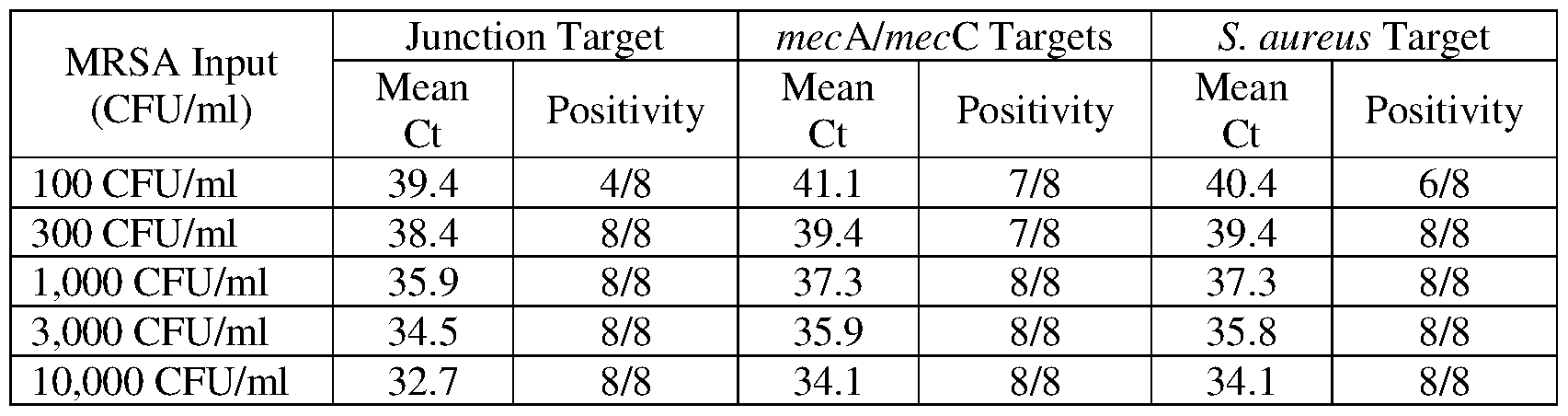
- Example 1 demonstrates that a lysis protocol employing the combination of a strong base and a detergent gave good results with a prototype real time multiplex assay that detected methicillin-resistant S. aureus (MRSA). Results indicated 100% detection of the target organism at an input level of 1 ,000 CFU/ml.
- Tubes containing samples of MRSA bacteria (strain ATCC BAA-41) at 100 CFU/ml - 10,000 CFU/ml in modified Liquid Amies transport medium (Copan Diagnostics Inc.; Corona, CA).
- This detergent-free transport medium which preserves viability of bacterial cells for subsequent culture or propagation, includes NaCl, KC1, CaCh. MgCL. monopotassium phosphate, disodium phosphate, and sodium thioglycollate in an aqueous solution.
- Sample lysis was effected by combining 126 pl of a lysis reagent (“LR-A”) with 300 m ⁇ of the liquid containing the biological sample to be tested, thereby resulting in a first liquid composition.
- the LR-A used in the procedure was a detergent-free aqueous solution that included 0.4 N LiOH, 8% LLS, 2% TRITON X-100, and 0.05% of an anti-foaming reagent.
- TRITON X-100 was included in the LR-A reagent to aid in processing any samples containing added mucin (e.g., to model nasal swab samples).
- the resulting alkaline mixtures were incubated for 10 minutes at 43°C to model the workflow in an automated sample processing instrument.
- Capture of nucleic acid targets onto solid supports involved mixing 450 m ⁇ of a target capture reagent with the first liquid composition to give a second liquid composition.
- the target capture reagent was an aqueous solution that included LiOH, HEPES buffer, lithium lauryl sulfate, succinic acid, an anti-foaming agent, poly(dT)i4 magnetic beads, and a target capture oligonucleotide having a poly(dA)3o tail at its 3’ -end.
- Non-specific capture oligonucleotides used in the procedure were in accordance with the description contained in U.S. Pat. No.
- Example 2 describes use of the lysis reagent and conditions from Example 1 for isolating nucleic acids from a different organism. As indicated below, C. difficile bacteria did not lyse efficiently to yield amplifiable nucleic acids by this approach.
- Lysis and target capture reagents and conditions from Example 1 were next used for isolating nucleic acids from C. difficile bacteria.
- the isolated nucleic acids served as templates in a prototype real time amplified assay for detecting C. difficile bacteria, where results from the assay indicated success of the sample processing procedure.
- “Up-front” sample processing was tested by comparing results from the prototype C. difficile assay, where the variable was the composition of the liquid reagent used for transporting the biological sample.
- the sample transport tube contained 2.9 ml of a phosphate -buffered (pH 6.6 to pH 6.8) solution that included 3% (w/v) lithium lauryl sulfate.
- this solution protects released nucleic acids by inhibiting the activity of nuclease enzymes that may be present in the sample.
- STM was substituted by a lysis reagent that was 0.4 N LiOH and 10% LLS.
- Results of the amplification reaction confirmed that both sample processing procedures gave good final results from the amplification and detection assays. Regardless of whether samples of C. difficile bacteria were introduced into tubes containing STM or lysis reagent, each condition correctly yielded 3/3 positive results. There was, however, a key difference in the nature of the measurements leading to these conclusions. Whereas the sample processed using lysis buffer gave a Ct value of about 22.2, the sample processed using STM gave a Ct value of about 26.7. This substantial difference in the time of emergence during the real time amplification indicated that at least 10 fold more DNA was liberated when the biological sample was contained in the alkaline detergent lysis reagent rather than STM.
- Example 3 describes procedures used for exploring the benefits of increasing the concentration of strong base in the lysis reagent. The experiment described below particularly compared the effect of including or omitting detergent in the alkaline lysis reagent. Surprisingly, the presence of detergent actually reduced assay sensitivity when the concentration of strong base was increased.
- TRITON X-100 e.g. , pH buffered detergent solution containing target capture reagents
- Table 2 presents results confirming the finding that samples including MRSA at levels of at least 1 ,000 CFU/ml yielded positive detection of all three targets needed to make the MRSA identification.
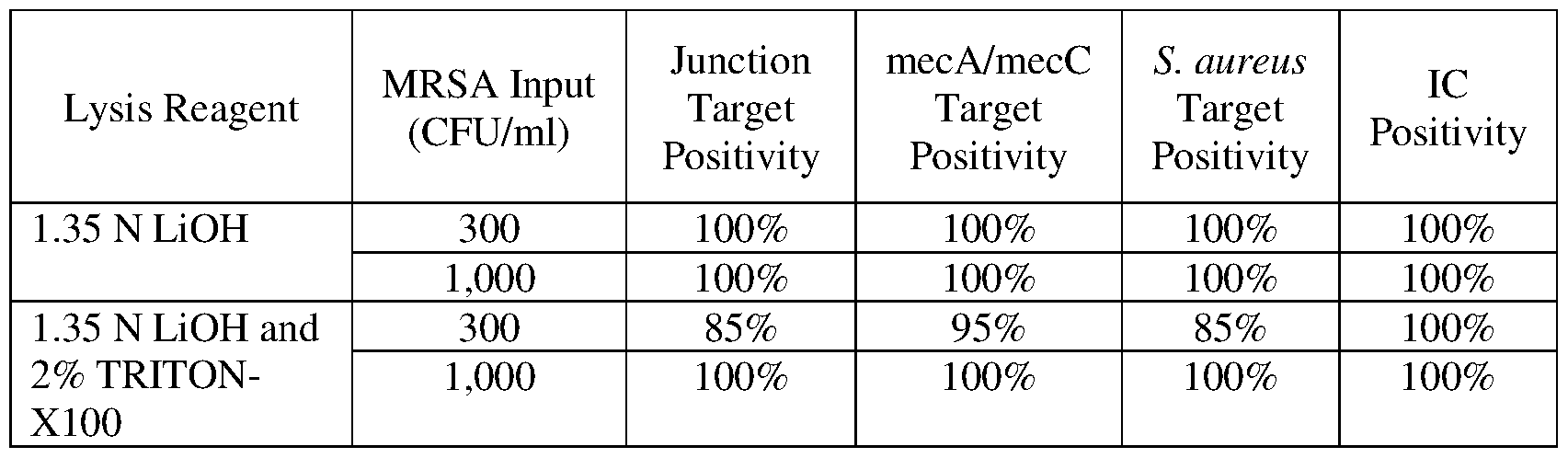
- the table further includes results for positive detection of the internal control (IC), which does not depend on successful bacterial lysis to generate a positive result. Indeed, the IC template nucleic acid that was amplified and detected in the MRSA assay was always added as a component of the pH buffered detergent solution containing target capture reagents. Significantly, increasing the strong base concentration from 0.4 N to 1.35 N increased the efficiency of bacterial lysis to the point where each target could be detected in the amplified assay at an MRSA input level of 300 CFU/ml.
- IC internal control
- lysis reagent containing the combination of 1.35 N LiOH and 2%
- TRITON X-100 did not yield the same high level of sensitivity that was achieved when detergent was omitted. Indeed, use of the strong base as a lysis reagent in the absence of pH buffer and detergent permitted 100% detection of 300 CFU/ml, a level of sensitivity better than that achieved using the LR-A of Example 1. Moreover, further increasing the concentration of strong base to 2.5 N undesirably caused formation of a precipitate in lysis reagents including LLS at a level of at least 8%.
- Example 4 describes procedures illustrating useful pH conditions for isolation of nucleic acids from C. difficile bacteria. As described elsewhere herein, the pH conditions demonstrated as useful in this Example fell in the range of desired pH conditions for lysing cells and capturing nucleic acids onto solid supports. Example 4
- Readings for pH were determined at each of three steps where sample transport, bacterial lysis, and target capture took place. It was determined that the biological sample in STM had pH 6.6, as expected. Combining 300 pl of biological sample with 126 pl of Li OH solution having a concentration falling in the range of from 1.325 N to 2.5 N yielded pH of about pH 12.95 to about pH 12.99. Mixing this alkaline sample with 450 m ⁇ of target capture reagent yielded a pH falling in the range of from pH 7.91 to pH 8.75.
- nucleic acids were subjected to nucleic acid amplification and detection using a prototype amplified assay for detecting C. difficile bacteria. Efficiency of lysis and target capture were assessed using results from the amplified assay.
- the target capture reagent e.g. , the pH buffered detergent solution
- Example 5 describes results indicating that detergent can be required for best results in sample preparation procedures employing strong base at certain concentrations.
- Example 6 demonstrates how a sample preparation technique employing contact of a biological sample with an alkaline composition that lacked a detergent and pH buffer, followed by neutralizing in the presence of detergent and target capture reagents could improve results in a prototype amplified assay for detecting Group B Streptococcus (GBS) bacteria.
- GBS bacteria are another group of bacteria that, like S. aureus and C. difficile are known to be difficult to lyse. Streptococcus agalactiae served as a
- a prototype assay for detecting nucleic acids of GBS bacteria was used to compare effectiveness of two different sample preparation techniques.
- the two methods employed substantially identical reagents, but differed in procedural steps used for processing samples, with very different results.
- a first method involved combining the biological sample and a pH buffered detergent solution that included target capture reagents (e.g. , magnetic beads displaying an immobilized oligonucleotide, etc.) to create a first liquid composition having a pH falling in the range of from pH 6.5 to pH 8.0.
- target capture reagents e.g. , magnetic beads displaying an immobilized oligonucleotide, etc.
- an aliquot of a strong base solution (1.68 N Li OH) was mixed with the first liquid composition to create a second liquid composition having a pH less than pH 9.5 (e.g., pH 8.2 to pH 9.2) and providing conditions appropriate for capture of nucleic acids by immobilization to a solid support. Captured nucleic acids were purified by washing the solid support with a wash buffer.
- a second method involved combining the biological sample with an aliquot of an alkaline composition (1.68 N LiOH) that lyses cells and denatures nucleic acids to create a first liquid composition.
- the alkaline composition that was mixed with the biological sample was free of detergents and pH buffers, and the pH of the first liquid composition was greater than pH 12.5 (e.g.
- a pH buffered detergent solution that included target capture reagents (e.g., magnetic beads displaying an immobilized oligonucleotide, etc.) was mixed with the first liquid composition to create a second liquid composition having a pH less than pH 9.5 (e.g., pH 7.8 to pH 8.9) and providing conditions appropriate for capture of nucleic acids by immobilization to a solid support. Captured nucleic acids were purified by washing the solid support with a wash buffer.
- the second method was useful for isolating DNA and substantially eliminating RNA from the finally purified nucleic acid composition.
- Testing carried out using a known input level of an RNA viral target prepared by the two different methods illustrated this fact. More specifically, quantitative real time PCR amplification and detection revealed that the difference in the time of emergence for the two run curves was about 24 Ct intervals. This meant that the number of copies of the RNA target was reduced by about 16 million fold using the second sample method.
- Results presented in Table 4 confirmed that the sample preparation technique employing the alkaline composition (strong base without pH buffer or detergent) for lysing bacteria gave superior results. More particularly, the limit of detection was determined to be about 39 CFU/ml with this technique. Conversely, use of an alkaline shock technique for lysing GBS bacteria gave a limit of detection of 1,416 CFU/ml. Again, first treating with strong base followed by neutralizing in the presence of pH buffer, detergent, and target capture reagents gave superior results in the automated sample preparation workflow.
- Example 6 also was used for obtaining efficient lysis and capture of nucleic acids from biological samples that included either S. aureus or C. difficile bacteria.
- a first liquid composition can be said to be“substantially free of detergents” when the detergent concentration is less than 0.001% (w/v).
- Biological samples stored in modified Liquid Amies transport medium and then combined or mixed with an alkaline composition lacking detergent yielded first liquid compositions that were completely free of detergents, and gave excellent results in the DNA isolation procedure.
- bacteria transported in media lacking detergent remained viable and could be cultured for species verification or typing using conventional microbiological techniques.
- a single biological sample could be used for both molecular analysis and microbiological analysis.
- the presence of detergent in the first liquid composition did not compromise the ability to achieve good results with the DNA isolation method.
- the first liquid composition that resulted from combining the biological sample (stored in STM) and the alkaline composition in the preceding Example had a detergent concentration of about 2% (w/v), and also gave excellent results, as indicated by efficient detection of GBS sequences in the PCR protocol.
Landscapes
- Chemical & Material Sciences (AREA)
- Life Sciences & Earth Sciences (AREA)
- Organic Chemistry (AREA)
- Engineering & Computer Science (AREA)
- Health & Medical Sciences (AREA)
- Zoology (AREA)
- Wood Science & Technology (AREA)
- Analytical Chemistry (AREA)
- Proteomics, Peptides & Aminoacids (AREA)
- Genetics & Genomics (AREA)
- Bioinformatics & Cheminformatics (AREA)
- Biotechnology (AREA)
- General Engineering & Computer Science (AREA)
- Molecular Biology (AREA)
- Microbiology (AREA)
- Biophysics (AREA)
- Physics & Mathematics (AREA)
- Biochemistry (AREA)
- General Health & Medical Sciences (AREA)
- Biomedical Technology (AREA)
- Immunology (AREA)
- Plant Pathology (AREA)
- Crystallography & Structural Chemistry (AREA)
- Chemical Kinetics & Catalysis (AREA)
- Measuring Or Testing Involving Enzymes Or Micro-Organisms (AREA)
- Apparatus Associated With Microorganisms And Enzymes (AREA)
Abstract
Description


Claims
Priority Applications (7)
| Application Number | Priority Date | Filing Date | Title |
|---|---|---|---|
| EP19734637.2A EP3814496B1 (en) | 2018-06-28 | 2019-06-14 | Sample preparation method and system |
| US17/255,500 US12344884B2 (en) | 2018-06-28 | 2019-06-14 | Sample preparation method and system |
| AU2019294109A AU2019294109C1 (en) | 2018-06-28 | 2019-06-14 | Sample preparation method and system |
| CA3103268A CA3103268A1 (en) | 2018-06-28 | 2019-06-14 | Sample preparation method and system |
| JP2020572965A JP7599340B2 (en) | 2018-06-28 | 2019-06-14 | Sample preparation methods and systems |
| US18/946,760 US20250137034A1 (en) | 2018-06-28 | 2024-11-13 | Sample preparation method and system |
| AU2025205173A AU2025205173A1 (en) | 2018-06-28 | 2025-07-07 | Sample preparation method and system |
Applications Claiming Priority (2)
| Application Number | Priority Date | Filing Date | Title |
|---|---|---|---|
| US201862691478P | 2018-06-28 | 2018-06-28 | |
| US62/691,478 | 2018-06-28 |
Related Child Applications (2)
| Application Number | Title | Priority Date | Filing Date |
|---|---|---|---|
| US17/255,500 A-371-Of-International US12344884B2 (en) | 2018-06-28 | 2019-06-14 | Sample preparation method and system |
| US18/946,760 Continuation US20250137034A1 (en) | 2018-06-28 | 2024-11-13 | Sample preparation method and system |
Publications (1)
| Publication Number | Publication Date |
|---|---|
| WO2020005584A1 true WO2020005584A1 (en) | 2020-01-02 |
Family
ID=67108216
Family Applications (1)
| Application Number | Title | Priority Date | Filing Date |
|---|---|---|---|
| PCT/US2019/037245 Ceased WO2020005584A1 (en) | 2018-06-28 | 2019-06-14 | Sample preparation method and system |
Country Status (6)
| Country | Link |
|---|---|
| US (2) | US12344884B2 (en) |
| EP (1) | EP3814496B1 (en) |
| JP (1) | JP7599340B2 (en) |
| AU (2) | AU2019294109C1 (en) |
| CA (1) | CA3103268A1 (en) |
| WO (1) | WO2020005584A1 (en) |
Cited By (2)
| Publication number | Priority date | Publication date | Assignee | Title |
|---|---|---|---|---|
| WO2022219514A1 (en) * | 2021-04-16 | 2022-10-20 | Grifols Diagnostic Solutions Inc. | Compositions and methods for storing a biological sample |
| WO2024133437A1 (en) * | 2022-12-21 | 2024-06-27 | Illumina, Inc. | Use of dried sodium hydroxide to denaturate double stranded dna |
Families Citing this family (1)
| Publication number | Priority date | Publication date | Assignee | Title |
|---|---|---|---|---|
| US12522817B2 (en) * | 2019-11-14 | 2026-01-13 | Gen-Probe Incorporated | Compositions and methods for capturing target nucleic acids |
Citations (15)
| Publication number | Priority date | Publication date | Assignee | Title |
|---|---|---|---|---|
| US4965188A (en) | 1986-08-22 | 1990-10-23 | Cetus Corporation | Process for amplifying, detecting, and/or cloning nucleic acid sequences using a thermostable enzyme |
| EP0525882A1 (en) | 1991-08-02 | 1993-02-03 | Akzo Nobel N.V. | Quantification of nucleic acid |
| US5399491A (en) | 1989-07-11 | 1995-03-21 | Gen-Probe Incorporated | Nucleic acid sequence amplification methods |
| US5455166A (en) | 1991-01-31 | 1995-10-03 | Becton, Dickinson And Company | Strand displacement amplification |
| US5472840A (en) | 1988-09-30 | 1995-12-05 | Amoco Corporation | Nucleic acid structures with catalytic and autocatalytic replicating features and methods of use |
| WO1998050583A1 (en) | 1997-05-02 | 1998-11-12 | Gen-Probe Incorporated | Two-step hybridization and capture of a polynucleotide |
| US20040229268A1 (en) * | 2003-05-13 | 2004-11-18 | Hogan James J. | Method and kit for identifying antibiotic-resistant microorganisms |
| US20070031880A1 (en) * | 2003-02-06 | 2007-02-08 | Becton, Dickinson And Company | Chemical treatment of biological samples for nucleic acid extraction and kits therefor |
| US7510837B2 (en) | 2005-02-18 | 2009-03-31 | Gen-Probe Incorporated | Sample preparation method incorporating an alkaline shock |
| WO2011083076A1 (en) * | 2010-01-08 | 2011-07-14 | Roche Diagnostics Gmbh | Improved recovery of nucleic acids from magnetic glass particles |
| US20120058011A1 (en) * | 2005-10-21 | 2012-03-08 | Alan Wirbisky | Compact apparatus, compositions and methods for purifying nucleic acids |
| US20140205996A1 (en) * | 2004-06-28 | 2014-07-24 | Becton, Dickinson And Company | Dissolvable films and methods including the same |
| US9051601B2 (en) | 2006-08-01 | 2015-06-09 | Gen-Probe Incorporated | Methods of nonspecific target capture of nucleic acids |
| US20150218653A1 (en) * | 2012-08-30 | 2015-08-06 | Qiagen Gmbh | Method of determining the presence or absence of a target nucleic acid in a cell sample |
| US20180163259A1 (en) | 2015-05-01 | 2018-06-14 | Gen-Probe Incorporated | Multiplex invasive cleavage assays |
Family Cites Families (11)
| Publication number | Priority date | Publication date | Assignee | Title |
|---|---|---|---|---|
| US4623627A (en) | 1983-08-19 | 1986-11-18 | Cetus Corporation | Monoclonal antibody having specificity for the double-stranded conformation of native DNA and diagnostic methods using same |
| US4935342A (en) | 1986-12-01 | 1990-06-19 | Syngene, Inc. | Method of isolating and purifying nucleic acids from biological samples |
| US20020019007A1 (en) * | 1998-09-18 | 2002-02-14 | Jensen Wayne A. | PCR methods and materials |
| FR2833273B1 (en) | 2001-12-07 | 2004-08-13 | Commissariat Energie Atomique | METHOD FOR THE DETECTION AND QUANTITATIVE ANALYSIS OF NUCLEIC ACID MOLECULES COMPRISING A STRONG BASED EXTRACTION STAGE AND APPLICATIONS |
| US7727718B2 (en) | 2005-01-04 | 2010-06-01 | Molecular Research Center, Inc. | Reagents for storage and preparation of samples for DNA analysis |
| CA2650079A1 (en) | 2006-04-25 | 2007-11-01 | Vib Vzw | A sex-specific marker for shrimps and prawns |
| CN101663041B (en) | 2007-03-05 | 2012-05-23 | Om药物公司 | Bacterial extracts for respiratory disorders and methods of making same |
| EP2743705B1 (en) * | 2010-07-23 | 2016-10-12 | Beckman Coulter, Inc. | System or method of including analytical units |
| JP6060567B2 (en) | 2012-08-30 | 2017-01-18 | 凸版印刷株式会社 | Nucleic acid extraction method from embedded tissue |
| EP4067505A1 (en) | 2013-03-15 | 2022-10-05 | Abbott Molecular Inc. | Compositions and methods for nucleic acid extraction |
| WO2017088169A1 (en) | 2015-11-27 | 2017-06-01 | Coyote Bioscience Co., Ltd. | Methods and systems for nucleic acid amplification |
-
2019
- 2019-06-14 JP JP2020572965A patent/JP7599340B2/en active Active
- 2019-06-14 US US17/255,500 patent/US12344884B2/en active Active
- 2019-06-14 WO PCT/US2019/037245 patent/WO2020005584A1/en not_active Ceased
- 2019-06-14 CA CA3103268A patent/CA3103268A1/en active Pending
- 2019-06-14 EP EP19734637.2A patent/EP3814496B1/en active Active
- 2019-06-14 AU AU2019294109A patent/AU2019294109C1/en active Active
-
2024
- 2024-11-13 US US18/946,760 patent/US20250137034A1/en active Pending
-
2025
- 2025-07-07 AU AU2025205173A patent/AU2025205173A1/en active Pending
Patent Citations (16)
| Publication number | Priority date | Publication date | Assignee | Title |
|---|---|---|---|---|
| US4965188A (en) | 1986-08-22 | 1990-10-23 | Cetus Corporation | Process for amplifying, detecting, and/or cloning nucleic acid sequences using a thermostable enzyme |
| US5472840A (en) | 1988-09-30 | 1995-12-05 | Amoco Corporation | Nucleic acid structures with catalytic and autocatalytic replicating features and methods of use |
| US5399491A (en) | 1989-07-11 | 1995-03-21 | Gen-Probe Incorporated | Nucleic acid sequence amplification methods |
| US5455166A (en) | 1991-01-31 | 1995-10-03 | Becton, Dickinson And Company | Strand displacement amplification |
| EP0525882A1 (en) | 1991-08-02 | 1993-02-03 | Akzo Nobel N.V. | Quantification of nucleic acid |
| WO1998050583A1 (en) | 1997-05-02 | 1998-11-12 | Gen-Probe Incorporated | Two-step hybridization and capture of a polynucleotide |
| US20070031880A1 (en) * | 2003-02-06 | 2007-02-08 | Becton, Dickinson And Company | Chemical treatment of biological samples for nucleic acid extraction and kits therefor |
| US20040229268A1 (en) * | 2003-05-13 | 2004-11-18 | Hogan James J. | Method and kit for identifying antibiotic-resistant microorganisms |
| US20140205996A1 (en) * | 2004-06-28 | 2014-07-24 | Becton, Dickinson And Company | Dissolvable films and methods including the same |
| US8420317B2 (en) | 2005-02-18 | 2013-04-16 | Gen-Probe Incorporated | Alkaline shock-based method of processing a biological sample |
| US7510837B2 (en) | 2005-02-18 | 2009-03-31 | Gen-Probe Incorporated | Sample preparation method incorporating an alkaline shock |
| US20120058011A1 (en) * | 2005-10-21 | 2012-03-08 | Alan Wirbisky | Compact apparatus, compositions and methods for purifying nucleic acids |
| US9051601B2 (en) | 2006-08-01 | 2015-06-09 | Gen-Probe Incorporated | Methods of nonspecific target capture of nucleic acids |
| WO2011083076A1 (en) * | 2010-01-08 | 2011-07-14 | Roche Diagnostics Gmbh | Improved recovery of nucleic acids from magnetic glass particles |
| US20150218653A1 (en) * | 2012-08-30 | 2015-08-06 | Qiagen Gmbh | Method of determining the presence or absence of a target nucleic acid in a cell sample |
| US20180163259A1 (en) | 2015-05-01 | 2018-06-14 | Gen-Probe Incorporated | Multiplex invasive cleavage assays |
Non-Patent Citations (1)
| Title |
|---|
| LIZARDI ET AL., BIOTECHNOLOGY, vol. 6, 1988, pages 1197 |
Cited By (2)
| Publication number | Priority date | Publication date | Assignee | Title |
|---|---|---|---|---|
| WO2022219514A1 (en) * | 2021-04-16 | 2022-10-20 | Grifols Diagnostic Solutions Inc. | Compositions and methods for storing a biological sample |
| WO2024133437A1 (en) * | 2022-12-21 | 2024-06-27 | Illumina, Inc. | Use of dried sodium hydroxide to denaturate double stranded dna |
Also Published As
| Publication number | Publication date |
|---|---|
| AU2019294109A1 (en) | 2021-02-18 |
| CA3103268A1 (en) | 2020-01-02 |
| AU2025205173A1 (en) | 2025-07-24 |
| EP3814496B1 (en) | 2023-11-22 |
| US12344884B2 (en) | 2025-07-01 |
| US20250137034A1 (en) | 2025-05-01 |
| JP7599340B2 (en) | 2024-12-13 |
| JP2021528089A (en) | 2021-10-21 |
| US20210269854A1 (en) | 2021-09-02 |
| AU2019294109C1 (en) | 2025-08-07 |
| AU2019294109B2 (en) | 2025-04-10 |
| EP3814496A1 (en) | 2021-05-05 |
Similar Documents
| Publication | Publication Date | Title |
|---|---|---|
| JP2024037912A (en) | Polynucleotide capture materials and methods for their use | |
| US7803581B2 (en) | Alkaline shock-based preparation of nucleic acids | |
| US20250137034A1 (en) | Sample preparation method and system | |
| CN110088297A (en) | Padlock probe detection method | |
| US20120064511A1 (en) | Generic Buffer For Amplification | |
| JP2025109742A (en) | Method for isolating nucleic acid from a specimen in a liquid cytological preservative containing formaldehyde | |
| AU2010208311B2 (en) | Sequence-specific large volume sample preparation method and assay | |
| WO2025137492A1 (en) | Method for enriching pathogen dna and implementation thereof | |
| WO2025036740A1 (en) | Devices and applications of coupled nucleic acid amplification tests and crispr systems | |
| CN109790567A (en) | 6 internal contrast composition of Ф, device and method | |
| EP4490321A1 (en) | Method of detection of a target nucleic acid sequence in a single reaction vessel | |
| WO2023170144A1 (en) | Method of detection of a target nucleic acid sequence |
Legal Events
| Date | Code | Title | Description |
|---|---|---|---|
| 121 | Ep: the epo has been informed by wipo that ep was designated in this application |
Ref document number: 19734637 Country of ref document: EP Kind code of ref document: A1 |
|
| ENP | Entry into the national phase |
Ref document number: 3103268 Country of ref document: CA |
|
| ENP | Entry into the national phase |
Ref document number: 2020572965 Country of ref document: JP Kind code of ref document: A |
|
| NENP | Non-entry into the national phase |
Ref country code: DE |
|
| ENP | Entry into the national phase |
Ref document number: 2019734637 Country of ref document: EP Effective date: 20210128 |
|
| ENP | Entry into the national phase |
Ref document number: 2019294109 Country of ref document: AU Date of ref document: 20190614 Kind code of ref document: A |
|
| WWG | Wipo information: grant in national office |
Ref document number: 17255500 Country of ref document: US |