WO2017014582A1 - Bio-ink composition having improved physical and biological properties - Google Patents
Bio-ink composition having improved physical and biological properties Download PDFInfo
- Publication number
- WO2017014582A1 WO2017014582A1 PCT/KR2016/007962 KR2016007962W WO2017014582A1 WO 2017014582 A1 WO2017014582 A1 WO 2017014582A1 KR 2016007962 W KR2016007962 W KR 2016007962W WO 2017014582 A1 WO2017014582 A1 WO 2017014582A1
- Authority
- WO
- WIPO (PCT)
- Prior art keywords
- cells
- tissue
- ink composition
- bio
- bio ink
- Prior art date
- Legal status (The legal status is an assumption and is not a legal conclusion. Google has not performed a legal analysis and makes no representation as to the accuracy of the status listed.)
- Ceased
Links
Classifications
-
- A—HUMAN NECESSITIES
- A61—MEDICAL OR VETERINARY SCIENCE; HYGIENE
- A61L—METHODS OR APPARATUS FOR STERILISING MATERIALS OR OBJECTS IN GENERAL; DISINFECTION, STERILISATION OR DEODORISATION OF AIR; CHEMICAL ASPECTS OF BANDAGES, DRESSINGS, ABSORBENT PADS OR SURGICAL ARTICLES; MATERIALS FOR BANDAGES, DRESSINGS, ABSORBENT PADS OR SURGICAL ARTICLES
- A61L27/00—Materials for grafts or prostheses or for coating grafts or prostheses
- A61L27/50—Materials characterised by their function or physical properties, e.g. injectable or lubricating compositions, shape-memory materials, surface modified materials
- A61L27/54—Biologically active materials, e.g. therapeutic substances
-
- A—HUMAN NECESSITIES
- A61—MEDICAL OR VETERINARY SCIENCE; HYGIENE
- A61L—METHODS OR APPARATUS FOR STERILISING MATERIALS OR OBJECTS IN GENERAL; DISINFECTION, STERILISATION OR DEODORISATION OF AIR; CHEMICAL ASPECTS OF BANDAGES, DRESSINGS, ABSORBENT PADS OR SURGICAL ARTICLES; MATERIALS FOR BANDAGES, DRESSINGS, ABSORBENT PADS OR SURGICAL ARTICLES
- A61L27/00—Materials for grafts or prostheses or for coating grafts or prostheses
- A61L27/36—Materials for grafts or prostheses or for coating grafts or prostheses containing ingredients of undetermined constitution or reaction products thereof, e.g. transplant tissue, natural bone, extracellular matrix
- A61L27/3604—Materials for grafts or prostheses or for coating grafts or prostheses containing ingredients of undetermined constitution or reaction products thereof, e.g. transplant tissue, natural bone, extracellular matrix characterised by the human or animal origin of the biological material, e.g. hair, fascia, fish scales, silk, shellac, pericardium, pleura, renal tissue, amniotic membrane, parenchymal tissue, fetal tissue, muscle tissue, fat tissue, enamel
-
- A—HUMAN NECESSITIES
- A61—MEDICAL OR VETERINARY SCIENCE; HYGIENE
- A61L—METHODS OR APPARATUS FOR STERILISING MATERIALS OR OBJECTS IN GENERAL; DISINFECTION, STERILISATION OR DEODORISATION OF AIR; CHEMICAL ASPECTS OF BANDAGES, DRESSINGS, ABSORBENT PADS OR SURGICAL ARTICLES; MATERIALS FOR BANDAGES, DRESSINGS, ABSORBENT PADS OR SURGICAL ARTICLES
- A61L27/00—Materials for grafts or prostheses or for coating grafts or prostheses
- A61L27/14—Macromolecular materials
- A61L27/20—Polysaccharides
-
- A—HUMAN NECESSITIES
- A61—MEDICAL OR VETERINARY SCIENCE; HYGIENE
- A61L—METHODS OR APPARATUS FOR STERILISING MATERIALS OR OBJECTS IN GENERAL; DISINFECTION, STERILISATION OR DEODORISATION OF AIR; CHEMICAL ASPECTS OF BANDAGES, DRESSINGS, ABSORBENT PADS OR SURGICAL ARTICLES; MATERIALS FOR BANDAGES, DRESSINGS, ABSORBENT PADS OR SURGICAL ARTICLES
- A61L27/00—Materials for grafts or prostheses or for coating grafts or prostheses
- A61L27/14—Macromolecular materials
- A61L27/22—Polypeptides or derivatives thereof, e.g. degradation products
- A61L27/222—Gelatin
-
- A—HUMAN NECESSITIES
- A61—MEDICAL OR VETERINARY SCIENCE; HYGIENE
- A61L—METHODS OR APPARATUS FOR STERILISING MATERIALS OR OBJECTS IN GENERAL; DISINFECTION, STERILISATION OR DEODORISATION OF AIR; CHEMICAL ASPECTS OF BANDAGES, DRESSINGS, ABSORBENT PADS OR SURGICAL ARTICLES; MATERIALS FOR BANDAGES, DRESSINGS, ABSORBENT PADS OR SURGICAL ARTICLES
- A61L27/00—Materials for grafts or prostheses or for coating grafts or prostheses
- A61L27/14—Macromolecular materials
- A61L27/26—Mixtures of macromolecular compounds
-
- A—HUMAN NECESSITIES
- A61—MEDICAL OR VETERINARY SCIENCE; HYGIENE
- A61L—METHODS OR APPARATUS FOR STERILISING MATERIALS OR OBJECTS IN GENERAL; DISINFECTION, STERILISATION OR DEODORISATION OF AIR; CHEMICAL ASPECTS OF BANDAGES, DRESSINGS, ABSORBENT PADS OR SURGICAL ARTICLES; MATERIALS FOR BANDAGES, DRESSINGS, ABSORBENT PADS OR SURGICAL ARTICLES
- A61L27/00—Materials for grafts or prostheses or for coating grafts or prostheses
- A61L27/36—Materials for grafts or prostheses or for coating grafts or prostheses containing ingredients of undetermined constitution or reaction products thereof, e.g. transplant tissue, natural bone, extracellular matrix
- A61L27/3641—Materials for grafts or prostheses or for coating grafts or prostheses containing ingredients of undetermined constitution or reaction products thereof, e.g. transplant tissue, natural bone, extracellular matrix characterised by the site of application in the body
- A61L27/3645—Connective tissue
- A61L27/3654—Cartilage, e.g. meniscus
-
- A—HUMAN NECESSITIES
- A61—MEDICAL OR VETERINARY SCIENCE; HYGIENE
- A61L—METHODS OR APPARATUS FOR STERILISING MATERIALS OR OBJECTS IN GENERAL; DISINFECTION, STERILISATION OR DEODORISATION OF AIR; CHEMICAL ASPECTS OF BANDAGES, DRESSINGS, ABSORBENT PADS OR SURGICAL ARTICLES; MATERIALS FOR BANDAGES, DRESSINGS, ABSORBENT PADS OR SURGICAL ARTICLES
- A61L27/00—Materials for grafts or prostheses or for coating grafts or prostheses
- A61L27/36—Materials for grafts or prostheses or for coating grafts or prostheses containing ingredients of undetermined constitution or reaction products thereof, e.g. transplant tissue, natural bone, extracellular matrix
- A61L27/3641—Materials for grafts or prostheses or for coating grafts or prostheses containing ingredients of undetermined constitution or reaction products thereof, e.g. transplant tissue, natural bone, extracellular matrix characterised by the site of application in the body
- A61L27/367—Muscle tissue, e.g. sphincter
-
- A—HUMAN NECESSITIES
- A61—MEDICAL OR VETERINARY SCIENCE; HYGIENE
- A61L—METHODS OR APPARATUS FOR STERILISING MATERIALS OR OBJECTS IN GENERAL; DISINFECTION, STERILISATION OR DEODORISATION OF AIR; CHEMICAL ASPECTS OF BANDAGES, DRESSINGS, ABSORBENT PADS OR SURGICAL ARTICLES; MATERIALS FOR BANDAGES, DRESSINGS, ABSORBENT PADS OR SURGICAL ARTICLES
- A61L27/00—Materials for grafts or prostheses or for coating grafts or prostheses
- A61L27/36—Materials for grafts or prostheses or for coating grafts or prostheses containing ingredients of undetermined constitution or reaction products thereof, e.g. transplant tissue, natural bone, extracellular matrix
- A61L27/38—Materials for grafts or prostheses or for coating grafts or prostheses containing ingredients of undetermined constitution or reaction products thereof, e.g. transplant tissue, natural bone, extracellular matrix containing added animal cells
- A61L27/3804—Materials for grafts or prostheses or for coating grafts or prostheses containing ingredients of undetermined constitution or reaction products thereof, e.g. transplant tissue, natural bone, extracellular matrix containing added animal cells characterised by specific cells or progenitors thereof, e.g. fibroblasts, connective tissue cells, kidney cells
- A61L27/3808—Endothelial cells
-
- A—HUMAN NECESSITIES
- A61—MEDICAL OR VETERINARY SCIENCE; HYGIENE
- A61L—METHODS OR APPARATUS FOR STERILISING MATERIALS OR OBJECTS IN GENERAL; DISINFECTION, STERILISATION OR DEODORISATION OF AIR; CHEMICAL ASPECTS OF BANDAGES, DRESSINGS, ABSORBENT PADS OR SURGICAL ARTICLES; MATERIALS FOR BANDAGES, DRESSINGS, ABSORBENT PADS OR SURGICAL ARTICLES
- A61L27/00—Materials for grafts or prostheses or for coating grafts or prostheses
- A61L27/36—Materials for grafts or prostheses or for coating grafts or prostheses containing ingredients of undetermined constitution or reaction products thereof, e.g. transplant tissue, natural bone, extracellular matrix
- A61L27/38—Materials for grafts or prostheses or for coating grafts or prostheses containing ingredients of undetermined constitution or reaction products thereof, e.g. transplant tissue, natural bone, extracellular matrix containing added animal cells
- A61L27/3804—Materials for grafts or prostheses or for coating grafts or prostheses containing ingredients of undetermined constitution or reaction products thereof, e.g. transplant tissue, natural bone, extracellular matrix containing added animal cells characterised by specific cells or progenitors thereof, e.g. fibroblasts, connective tissue cells, kidney cells
- A61L27/3813—Epithelial cells, e.g. keratinocytes, urothelial cells
-
- A—HUMAN NECESSITIES
- A61—MEDICAL OR VETERINARY SCIENCE; HYGIENE
- A61L—METHODS OR APPARATUS FOR STERILISING MATERIALS OR OBJECTS IN GENERAL; DISINFECTION, STERILISATION OR DEODORISATION OF AIR; CHEMICAL ASPECTS OF BANDAGES, DRESSINGS, ABSORBENT PADS OR SURGICAL ARTICLES; MATERIALS FOR BANDAGES, DRESSINGS, ABSORBENT PADS OR SURGICAL ARTICLES
- A61L27/00—Materials for grafts or prostheses or for coating grafts or prostheses
- A61L27/36—Materials for grafts or prostheses or for coating grafts or prostheses containing ingredients of undetermined constitution or reaction products thereof, e.g. transplant tissue, natural bone, extracellular matrix
- A61L27/38—Materials for grafts or prostheses or for coating grafts or prostheses containing ingredients of undetermined constitution or reaction products thereof, e.g. transplant tissue, natural bone, extracellular matrix containing added animal cells
- A61L27/3804—Materials for grafts or prostheses or for coating grafts or prostheses containing ingredients of undetermined constitution or reaction products thereof, e.g. transplant tissue, natural bone, extracellular matrix containing added animal cells characterised by specific cells or progenitors thereof, e.g. fibroblasts, connective tissue cells, kidney cells
- A61L27/3817—Cartilage-forming cells, e.g. pre-chondrocytes
-
- A—HUMAN NECESSITIES
- A61—MEDICAL OR VETERINARY SCIENCE; HYGIENE
- A61L—METHODS OR APPARATUS FOR STERILISING MATERIALS OR OBJECTS IN GENERAL; DISINFECTION, STERILISATION OR DEODORISATION OF AIR; CHEMICAL ASPECTS OF BANDAGES, DRESSINGS, ABSORBENT PADS OR SURGICAL ARTICLES; MATERIALS FOR BANDAGES, DRESSINGS, ABSORBENT PADS OR SURGICAL ARTICLES
- A61L27/00—Materials for grafts or prostheses or for coating grafts or prostheses
- A61L27/36—Materials for grafts or prostheses or for coating grafts or prostheses containing ingredients of undetermined constitution or reaction products thereof, e.g. transplant tissue, natural bone, extracellular matrix
- A61L27/38—Materials for grafts or prostheses or for coating grafts or prostheses containing ingredients of undetermined constitution or reaction products thereof, e.g. transplant tissue, natural bone, extracellular matrix containing added animal cells
- A61L27/3804—Materials for grafts or prostheses or for coating grafts or prostheses containing ingredients of undetermined constitution or reaction products thereof, e.g. transplant tissue, natural bone, extracellular matrix containing added animal cells characterised by specific cells or progenitors thereof, e.g. fibroblasts, connective tissue cells, kidney cells
- A61L27/3821—Bone-forming cells, e.g. osteoblasts, osteocytes, osteoprogenitor cells
-
- A—HUMAN NECESSITIES
- A61—MEDICAL OR VETERINARY SCIENCE; HYGIENE
- A61L—METHODS OR APPARATUS FOR STERILISING MATERIALS OR OBJECTS IN GENERAL; DISINFECTION, STERILISATION OR DEODORISATION OF AIR; CHEMICAL ASPECTS OF BANDAGES, DRESSINGS, ABSORBENT PADS OR SURGICAL ARTICLES; MATERIALS FOR BANDAGES, DRESSINGS, ABSORBENT PADS OR SURGICAL ARTICLES
- A61L27/00—Materials for grafts or prostheses or for coating grafts or prostheses
- A61L27/36—Materials for grafts or prostheses or for coating grafts or prostheses containing ingredients of undetermined constitution or reaction products thereof, e.g. transplant tissue, natural bone, extracellular matrix
- A61L27/38—Materials for grafts or prostheses or for coating grafts or prostheses containing ingredients of undetermined constitution or reaction products thereof, e.g. transplant tissue, natural bone, extracellular matrix containing added animal cells
- A61L27/3804—Materials for grafts or prostheses or for coating grafts or prostheses containing ingredients of undetermined constitution or reaction products thereof, e.g. transplant tissue, natural bone, extracellular matrix containing added animal cells characterised by specific cells or progenitors thereof, e.g. fibroblasts, connective tissue cells, kidney cells
- A61L27/3826—Muscle cells, e.g. smooth muscle cells
-
- A—HUMAN NECESSITIES
- A61—MEDICAL OR VETERINARY SCIENCE; HYGIENE
- A61L—METHODS OR APPARATUS FOR STERILISING MATERIALS OR OBJECTS IN GENERAL; DISINFECTION, STERILISATION OR DEODORISATION OF AIR; CHEMICAL ASPECTS OF BANDAGES, DRESSINGS, ABSORBENT PADS OR SURGICAL ARTICLES; MATERIALS FOR BANDAGES, DRESSINGS, ABSORBENT PADS OR SURGICAL ARTICLES
- A61L27/00—Materials for grafts or prostheses or for coating grafts or prostheses
- A61L27/36—Materials for grafts or prostheses or for coating grafts or prostheses containing ingredients of undetermined constitution or reaction products thereof, e.g. transplant tissue, natural bone, extracellular matrix
- A61L27/38—Materials for grafts or prostheses or for coating grafts or prostheses containing ingredients of undetermined constitution or reaction products thereof, e.g. transplant tissue, natural bone, extracellular matrix containing added animal cells
- A61L27/3804—Materials for grafts or prostheses or for coating grafts or prostheses containing ingredients of undetermined constitution or reaction products thereof, e.g. transplant tissue, natural bone, extracellular matrix containing added animal cells characterised by specific cells or progenitors thereof, e.g. fibroblasts, connective tissue cells, kidney cells
- A61L27/383—Nerve cells, e.g. dendritic cells, Schwann cells
-
- A—HUMAN NECESSITIES
- A61—MEDICAL OR VETERINARY SCIENCE; HYGIENE
- A61L—METHODS OR APPARATUS FOR STERILISING MATERIALS OR OBJECTS IN GENERAL; DISINFECTION, STERILISATION OR DEODORISATION OF AIR; CHEMICAL ASPECTS OF BANDAGES, DRESSINGS, ABSORBENT PADS OR SURGICAL ARTICLES; MATERIALS FOR BANDAGES, DRESSINGS, ABSORBENT PADS OR SURGICAL ARTICLES
- A61L27/00—Materials for grafts or prostheses or for coating grafts or prostheses
- A61L27/36—Materials for grafts or prostheses or for coating grafts or prostheses containing ingredients of undetermined constitution or reaction products thereof, e.g. transplant tissue, natural bone, extracellular matrix
- A61L27/38—Materials for grafts or prostheses or for coating grafts or prostheses containing ingredients of undetermined constitution or reaction products thereof, e.g. transplant tissue, natural bone, extracellular matrix containing added animal cells
- A61L27/3804—Materials for grafts or prostheses or for coating grafts or prostheses containing ingredients of undetermined constitution or reaction products thereof, e.g. transplant tissue, natural bone, extracellular matrix containing added animal cells characterised by specific cells or progenitors thereof, e.g. fibroblasts, connective tissue cells, kidney cells
- A61L27/3834—Cells able to produce different cell types, e.g. hematopoietic stem cells, mesenchymal stem cells, marrow stromal cells, embryonic stem cells
-
- A—HUMAN NECESSITIES
- A61—MEDICAL OR VETERINARY SCIENCE; HYGIENE
- A61L—METHODS OR APPARATUS FOR STERILISING MATERIALS OR OBJECTS IN GENERAL; DISINFECTION, STERILISATION OR DEODORISATION OF AIR; CHEMICAL ASPECTS OF BANDAGES, DRESSINGS, ABSORBENT PADS OR SURGICAL ARTICLES; MATERIALS FOR BANDAGES, DRESSINGS, ABSORBENT PADS OR SURGICAL ARTICLES
- A61L27/00—Materials for grafts or prostheses or for coating grafts or prostheses
- A61L27/50—Materials characterised by their function or physical properties, e.g. injectable or lubricating compositions, shape-memory materials, surface modified materials
-
- B—PERFORMING OPERATIONS; TRANSPORTING
- B29—WORKING OF PLASTICS; WORKING OF SUBSTANCES IN A PLASTIC STATE IN GENERAL
- B29C—SHAPING OR JOINING OF PLASTICS; SHAPING OF MATERIAL IN A PLASTIC STATE, NOT OTHERWISE PROVIDED FOR; AFTER-TREATMENT OF THE SHAPED PRODUCTS, e.g. REPAIRING
- B29C67/00—Shaping techniques not covered by groups B29C39/00 - B29C65/00, B29C70/00 or B29C73/00
- B29C67/02—Moulding by agglomerating
-
- B—PERFORMING OPERATIONS; TRANSPORTING
- B33—ADDITIVE MANUFACTURING TECHNOLOGY
- B33Y—ADDITIVE MANUFACTURING, i.e. MANUFACTURING OF THREE-DIMENSIONAL [3-D] OBJECTS BY ADDITIVE DEPOSITION, ADDITIVE AGGLOMERATION OR ADDITIVE LAYERING, e.g. BY 3-D PRINTING, STEREOLITHOGRAPHY OR SELECTIVE LASER SINTERING
- B33Y70/00—Materials specially adapted for additive manufacturing
-
- C—CHEMISTRY; METALLURGY
- C09—DYES; PAINTS; POLISHES; NATURAL RESINS; ADHESIVES; COMPOSITIONS NOT OTHERWISE PROVIDED FOR; APPLICATIONS OF MATERIALS NOT OTHERWISE PROVIDED FOR
- C09D—COATING COMPOSITIONS, e.g. PAINTS, VARNISHES OR LACQUERS; FILLING PASTES; CHEMICAL PAINT OR INK REMOVERS; INKS; CORRECTING FLUIDS; WOODSTAINS; PASTES OR SOLIDS FOR COLOURING OR PRINTING; USE OF MATERIALS THEREFOR
- C09D11/00—Inks
- C09D11/02—Printing inks
- C09D11/03—Printing inks characterised by features other than the chemical nature of the binder
-
- C—CHEMISTRY; METALLURGY
- C09—DYES; PAINTS; POLISHES; NATURAL RESINS; ADHESIVES; COMPOSITIONS NOT OTHERWISE PROVIDED FOR; APPLICATIONS OF MATERIALS NOT OTHERWISE PROVIDED FOR
- C09D—COATING COMPOSITIONS, e.g. PAINTS, VARNISHES OR LACQUERS; FILLING PASTES; CHEMICAL PAINT OR INK REMOVERS; INKS; CORRECTING FLUIDS; WOODSTAINS; PASTES OR SOLIDS FOR COLOURING OR PRINTING; USE OF MATERIALS THEREFOR
- C09D11/00—Inks
- C09D11/02—Printing inks
- C09D11/04—Printing inks based on proteins
-
- C—CHEMISTRY; METALLURGY
- C09—DYES; PAINTS; POLISHES; NATURAL RESINS; ADHESIVES; COMPOSITIONS NOT OTHERWISE PROVIDED FOR; APPLICATIONS OF MATERIALS NOT OTHERWISE PROVIDED FOR
- C09D—COATING COMPOSITIONS, e.g. PAINTS, VARNISHES OR LACQUERS; FILLING PASTES; CHEMICAL PAINT OR INK REMOVERS; INKS; CORRECTING FLUIDS; WOODSTAINS; PASTES OR SOLIDS FOR COLOURING OR PRINTING; USE OF MATERIALS THEREFOR
- C09D11/00—Inks
- C09D11/02—Printing inks
- C09D11/14—Printing inks based on carbohydrates
-
- A—HUMAN NECESSITIES
- A61—MEDICAL OR VETERINARY SCIENCE; HYGIENE
- A61L—METHODS OR APPARATUS FOR STERILISING MATERIALS OR OBJECTS IN GENERAL; DISINFECTION, STERILISATION OR DEODORISATION OF AIR; CHEMICAL ASPECTS OF BANDAGES, DRESSINGS, ABSORBENT PADS OR SURGICAL ARTICLES; MATERIALS FOR BANDAGES, DRESSINGS, ABSORBENT PADS OR SURGICAL ARTICLES
- A61L2300/00—Biologically active materials used in bandages, wound dressings, absorbent pads or medical devices
- A61L2300/40—Biologically active materials used in bandages, wound dressings, absorbent pads or medical devices characterised by a specific therapeutic activity or mode of action
- A61L2300/412—Tissue-regenerating or healing or proliferative agents
- A61L2300/414—Growth factors
-
- A—HUMAN NECESSITIES
- A61—MEDICAL OR VETERINARY SCIENCE; HYGIENE
- A61L—METHODS OR APPARATUS FOR STERILISING MATERIALS OR OBJECTS IN GENERAL; DISINFECTION, STERILISATION OR DEODORISATION OF AIR; CHEMICAL ASPECTS OF BANDAGES, DRESSINGS, ABSORBENT PADS OR SURGICAL ARTICLES; MATERIALS FOR BANDAGES, DRESSINGS, ABSORBENT PADS OR SURGICAL ARTICLES
- A61L2400/00—Materials characterised by their function or physical properties
- A61L2400/18—Modification of implant surfaces in order to improve biocompatibility, cell growth, fixation of biomolecules, e.g. plasma treatment
-
- B—PERFORMING OPERATIONS; TRANSPORTING
- B29—WORKING OF PLASTICS; WORKING OF SUBSTANCES IN A PLASTIC STATE IN GENERAL
- B29C—SHAPING OR JOINING OF PLASTICS; SHAPING OF MATERIAL IN A PLASTIC STATE, NOT OTHERWISE PROVIDED FOR; AFTER-TREATMENT OF THE SHAPED PRODUCTS, e.g. REPAIRING
- B29C64/00—Additive manufacturing, i.e. manufacturing of three-dimensional [3D] objects by additive deposition, additive agglomeration or additive layering, e.g. by 3D printing, stereolithography or selective laser sintering
- B29C64/10—Processes of additive manufacturing
- B29C64/106—Processes of additive manufacturing using only liquids or viscous materials, e.g. depositing a continuous bead of viscous material
- B29C64/112—Processes of additive manufacturing using only liquids or viscous materials, e.g. depositing a continuous bead of viscous material using individual droplets, e.g. from jetting heads
-
- B—PERFORMING OPERATIONS; TRANSPORTING
- B29—WORKING OF PLASTICS; WORKING OF SUBSTANCES IN A PLASTIC STATE IN GENERAL
- B29K—INDEXING SCHEME ASSOCIATED WITH SUBCLASSES B29B, B29C OR B29D, RELATING TO MOULDING MATERIALS OR TO MATERIALS FOR MOULDS, REINFORCEMENTS, FILLERS OR PREFORMED PARTS, e.g. INSERTS
- B29K2005/00—Use of polysaccharides or derivatives as moulding material
-
- B—PERFORMING OPERATIONS; TRANSPORTING
- B29—WORKING OF PLASTICS; WORKING OF SUBSTANCES IN A PLASTIC STATE IN GENERAL
- B29K—INDEXING SCHEME ASSOCIATED WITH SUBCLASSES B29B, B29C OR B29D, RELATING TO MOULDING MATERIALS OR TO MATERIALS FOR MOULDS, REINFORCEMENTS, FILLERS OR PREFORMED PARTS, e.g. INSERTS
- B29K2995/00—Properties of moulding materials, reinforcements, fillers, preformed parts or moulds
- B29K2995/0037—Other properties
- B29K2995/0056—Biocompatible, e.g. biopolymers or bioelastomers
-
- B—PERFORMING OPERATIONS; TRANSPORTING
- B33—ADDITIVE MANUFACTURING TECHNOLOGY
- B33Y—ADDITIVE MANUFACTURING, i.e. MANUFACTURING OF THREE-DIMENSIONAL [3-D] OBJECTS BY ADDITIVE DEPOSITION, ADDITIVE AGGLOMERATION OR ADDITIVE LAYERING, e.g. BY 3-D PRINTING, STEREOLITHOGRAPHY OR SELECTIVE LASER SINTERING
- B33Y10/00—Processes of additive manufacturing
Definitions
- the present invention relates to a bio ink composition with improved physical and biological properties, and more particularly, high viscosity, easy shear printing tendency, strong shear-thinning tendency, rapid crosslink formation and It relates to a bio ink composition exhibiting suitable mechanical properties after printing.
- Three-dimensional cell culture technology has been developed to produce cells and tissues in a laboratory-like environment in a laboratory (in v ro), and is used in various research fields related to cell growth and differentiation, organization and organ formation. Is being applied. Such tissue-like organs could be useful in the study of drug toxicity and pharmacokinetics on behalf of real tissues or organs, thus reducing the direct application of experiments to human specimens and other mammals. do. In addition, it contributes to the engineering design of tissues and organs as an important element of tissue engineering techniques for the purpose of replacing or treating damaged tissues and organs. Three-dimensional bioprinting technology has become a useful device for the precise manufacture of such tissue-like organs and implantable constructs.
- biomaterials that carry living cells during bioprinting namely bio-inks
- bio-inks exhibit many limitations in their utilization.
- the characteristics of the bio ink required to be applied to bio printing require excellent biocompatibility, and have a physical property that can be printed in a desired pattern by smoothly passing through a fine diameter dispensing nozzle. In other words, it should be able to provide a cell-specific signal after printing and maintain a mechanical support role.
- ratio Naturally derived or synthetic hydrogel bioinks have been developed and used in three-dimensional bioprinting fields.
- these existing hydrogel-based bioinks can be used in physical and biological materials such as biocompatibility, printing suitability, geometrical precision and precision. There is considerable limitation in terms of science.
- the present invention provides a bioink composition
- a bioink composition comprising a specific amount of cell carriers, viscosity enhancers, lubricants, and structural materials to maintain high viscosity, strong shear thinning, fast crosslinking and printed structures. It has been found that the above problems can be solved by exhibiting mechanical properties, thereby completing the present invention.
- tissue-derived extracellular matrix components may be added to the bio ink composition to have tissue specificity or specific cell differentiation regulating agents may be added to induce differentiation into specific tissues.
- the object of the present invention is a cell 0.05-60 6 / mL, cell carrier 0.1-10 w / v%, viscosity enhancer 0.01-1 wo, lubricant 1-30 v / v% and structural material 0.1-10 / ⁇ ⁇ It is to provide a bio ink composition comprising a. Another object of the present invention is to (a) layering the bio ink composition of the present invention in a three-dimensional bio printer; (b) three-dimensional printing a desired tissue like organ; And (c) to provide a tissue-like organ manufacturing method comprising the step of crosslinking the three-dimensional printed bio ink composition. Another object of the present invention is to provide a tissue analogous organ prepared according to the above method.
- the present invention provides a cell 0.05-60 6 / mL, cell carrier 0.1-10 w / v%, viscosity enhancer 0.01-1 w / v%, lubricant 1-30 v / It provides a bio ink composition comprising v% and the structural material 0.1-10 / ⁇ .
- the present invention comprises the steps of (a) layering the bio ink composition of the present invention to a three-dimensional bio printer; (b) three-dimensional printing the desired tissue-like organ; And (c) cross-linking the three-dimensional printed bio ink composition.
- the present invention provides a tissue-like organ prepared according to the above method.
- the invention includes cells 0.05-60 6 / mL, cell carrier 0.1-10 w / vo, viscosity enhancer 0.01-1 w / v%, lubricant 1-30 v / v% and structural material 0.1-10 / ⁇ ⁇ It provides a bio ink composition.
- the cells are preferably, stem cells, osteoblasts, myoblasts, myocytes, tenocytes, neuroblasts, fibroblasts, glioblasts, embryos Germ cells, hepatocytes, renal cells, sertoli cells, chondrocytes, epithelial cells, cardiovascular cells, keratinocytes and smooth muscle cells ( selected from the group consisting of smooth muscle cells, cardiomyocytes, glial cells, endothelial cells, hormone secreting cells, immune cells, pancreatic islet cells and neurons It may be any one or more, but is not limited thereto.
- the cell type used in the engineered tissue of the present invention may be cultured in any manner known in the art.
- Cell and tissue culture methods are known in the art and are described, for example, in Cell & Tissue Culture: Laboratory Procedures; Freshney (1987), Culture of Animal Cells: A Manual of Basic Techniques. The contents of which are incorporated herein by reference.
- General mammalian cell culture techniques, cell lines, and cell culture systems that may be used with the present invention are also described in Doyle, A., Griffiths, JB, Newell, DG, (eds.) Cell and Tissue Culture: Laboratory Procedures, Wiley (1998), and the objection to this information.
- the contents are incorporated herein by reference.
- the cells may also be incubated with cell differentiation materials that induce differentiation of the cells along the desired cell line. For example, stem cells are incubated in contact with differentiation medium to produce a range of cell types.
- the stem cells include, but are not limited to, osteogenic differentiation medium, chondrogenic differentiation medium, adipogenic differentiation medium, neuronal differentiation medium, cardiomyocyte differentiation medium, and enterocyte differentiation medium (e.g., Incubated with contact with differentiation medium).
- the cells may be cultured with growth factors, cytokines and the like.
- a "growth factor” refers to a protein, polypeptide, or polypeptide complex comprising a cytokine, which is produced by a cell and can affect itself and / or various other adjacent or isolated cells.
- growth factors affect the growth and / or differentiation of certain types of cells, either developmentally or in response to a number of biochemical or environmental stimuli, but not all but some growth factors are hormones.
- Fibroblastic growth factor including insulin, insulin-like growth factor (IGF), nerve growth factor (NGF), vascular endothelial growth factor (VEGF), keratinocyte growth factor (KGF), and basic FGF (bFGF) FGF), platelet-derived growth factors (PDGF), including PDGF-AA and PDGF-AB, bone morphogenic proteins (BMP), hepatocyte growth factor (HGF), transforming growth factors, including BMP-2 and BMP-7, etc.
- IGF insulin-like growth factor
- NEF nerve growth factor
- VEGF vascular endothelial growth factor
- KGF keratinocyte growth factor
- bFGF basic FGF
- PDGF platelet-derived growth factors
- BMP bone morphogenic proteins
- HGF hepatocyte growth factor
- transforming growth factors including BMP-2 and BMP-7, etc.
- TG Fa transforming growth factor beta
- EGF epidermal growth factor
- GM-CSF granulocyte-macrophage colony-stimulating factor
- 33 G-CSF
- IL-6 interleukin-6
- IL-8 growth factors, among others, in Molecular Cell Biology, Scientific Amer i canBooks, Darnel 1 et al., Eds. , 1986; Principlengs of Tissue Engineering, 2d ed., Lanza et al., Eds., Academic Press, 200. Those skilled in the art will appreciate any and all culture-derived growths in the conditioned media described herein.
- the cell carrier in the bio ink composition of the present invention is 0.01-30 w / v%, 0.05-20 w / v%, 0.1-10 w / v%, 0.5 —10 w / v%, 1-10 w / v%, 1.5-10 w / v%, 2-10 w, 0.1-8 w / v%, 0.5-8 w / v%, 1-8 / v% , 2-8 ⁇ '0.1-6 w / v%, 0.5-6 w / v%, 1-6 wM, 2-6 w / v%, 0.1-5 vi Mo, 0.5-5 w, 1-5 w / v% or 2-5 w / v, but are not limited thereto.
- the viscosity enhancer is 0.01-3 w / v%,, 0.05-3 w / v%, 0.1-3 w / v%, 0.01-2 w / v%, 0.05-2 w / v%, 0.1-2 w / v%, 0.01-1 w / v%, 0.05-1 w / v%, 0.1-1 w / v%, 0.01-0.8 w / v%, 0.05- 0.8 w / v% or 0.1-0.8 w / v3 ⁇ 4 may be included, but is not limited thereto.
- the lubricant is 0.1-50 v / v%, 0.5-50 v / v%, 1-50 v / v%, 0.1-40 v / v%, 0.5-40 v / v%, 1-40 v / v%, 0. 1-30 v / v%, 0.5-30 v / v, 1-30 vMo, 0. 1-20 v / v%, 0.5-20 v / v%, 1-20 v / v%, 0.1-15 v / v%, 0.5-15 v / v% or 1-15 v / v%, but is not limited thereto.
- the structural material is 0.1-30 w / v, 0.5-30 w / v%, 1-30 w / v, 0.1-20 w / v%, 0.5-20 w / v%, 1-20 w / v%, 0.1-10 w / v%, 0.5-10 w / v%, 1-10 wM, 0.1-1 w / v%, 0.5-8 w / v %, 1-8 w / v%, 0.1-5 wM, 0.5-5 w / v%, or 1-5 w / v, but is not limited thereto.
- the inventors of the present invention have tested the physical and biological properties of a composition having various combinations of compositions to develop a bio ink composition exhibiting physical and biological properties suitable for application to three-dimensional bioprinting.
- the bio ink composition consisting of a combination of components exhibits high viscosity, strong shear-thiming tendency, fast crosslinking and suitable mechanical properties after printing, making it particularly suitable for three-dimensional bioprinting for the preparation of tissue-like organs.
- the cell carrier material may be selected to be suitable for preparing a uniform cell suspension and exhibiting excellent printing tendency. Non-limiting examples thereof include gelatin, collagen, alginate, agar, agar, and the like. It may be any one or more selected from the group consisting of agarose, pluroni c and polyvinyl alcohol.
- the viscosity enhancer may be selected to be suitable for maintaining excellent printing tendency and initial strength of the bio ink, and non-limiting examples thereof may be haaluronic acid or dextran. .
- the lubricant minimizes shear rate. It is preferred to be selected from materials that can digest and improve the di spensing speed, and non-limiting examples thereof include glycerol.
- the structural material is preferably selected from materials capable of maintaining rapid crosslinking formation and mechanical rigidity, and non-limiting examples thereof include fibrinogen and hyaluronic acid modified to be chemically crosslinked.
- fibrinogen and hyaluronic acid modified to be chemically crosslinked include fibrinogen and hyaluronic acid modified to be chemically crosslinked.
- gelatin modified to be chemically crosslinked e.g., gelatin methacrylate, cylatated gelatin, etc.
- collagen, alginate, Methyl cellulose, chitosan, chitin, synthetic peptides and polyethylene glycol may be any one selected from the group consisting of hydrogels.
- the gelatin is particularly suitable as the cell carrier because it exhibits temperature-sensitive properties.
- gelatin has a characteristic of liquefying at 37 ° C and solidifying below room temperature.
- the fibrinogen is not only suitable as a structural material in terms of stability of the gel, but may also be selected as a preferable structural material in that it creates a microenvironment suitable for attachment and differentiation of cells after the cells are printed.
- Hyaluronic acid and glycerol can be suitably used in the composition of the present invention in terms of dispensing uniformity and anti-clogging effect of nozzles in three-dimensional bioprinting applications, respectively.
- the di spensing rate of the composition varies depending on the amount of gelatin contained in the bio ink composition.
- the bio ink composition of the present invention it can be seen that gelatin has a high contribution in terms of pattern resolution and hyaluronic acid in terms of pattern uniformity.
- cells are bioprinted by depositing or extruding bio ink from a three-dimensional bioprinter.
- the bio ink of the present invention comprises a liquid, semisolid, or solid composition comprising a plurality of cells.
- Bio ink is liquid or semi-finished Somatic cell solutions, cell suspensions, or cell concentrates.
- the bio ink composition comprises the steps of 1) mixing a plurality of cells or cell populations with a biocompatible liquid or gel to produce a bio ink, and 2) densifying the bio ink to obtain a desired cell density and viscosity.
- a bio ink having. Specifically, densification of the bio ink is realized by centrifugation, tangential flow filtration ("TF), or a combination thereof, and densification of the bio ink produces an extrudable composition to form a multicellular aggregate or multicellular sieve.
- extrudeable is meant that can be formed by passing a nozzle or orifice (e.g., one or more holes or tubes) (e.g. under pressure).
- Densification of bio-inks also allows cells to grow to an appropriate density The cell density required for the bio ink depends on the cell to be used and the tissue or organ to be prepared
- the present invention also provides a bio ink composition wherein the bio ink composition further comprises tissue-derived components.
- Tissue-derived components are specific to animals such as cartilage, kidneys, heart, liver, and muscles. This means that the decellularized and gelled material of the extracellular matrix is the main component, which may be included to enhance the tissue specificity of the bioink composition.
- Normal growth and differentiation in similar organs requires the creation of a natural environment similar to the microenvironment of the original tissue in the body. It is virtually impossible to reproduce all the properties of the same natural tissue-derived component, and by adding de-saturated tissue-derived components to the bio ink composition, it is possible to create a microenvironment in which cells can grow and differentiate normally.
- tissue-derived components interact with cells in the human body
- Each tissue-specific composition and phase prepared through the present invention can provide a favorable environment for the growth and differentiation of cells, and thus, by adding tissue-derived components to the bioink composition of the present invention, Can maintain its normal shape and preserve its function smoothly.
- the tissue from which the tissue-derived component can be obtained in the present invention is not particularly limited, and may be selected and decellularized according to a tissue-like organ to be prepared and then gelled. Methods for decellularizing and gelling tissues are well known in the art and can be readily selected and practiced by those skilled in the art.
- the present invention as a result of printing muscle cells using a bio ink composition containing a component derived from muscle tissue compared to the composition containing no tissue-derived components confirmed that the cell proliferation and differentiation is improved could.
- the present invention also provides a bio ink composition, characterized in that the bio ink composition further comprises a differentiation control material. Differentiation regulating substances may be included to induce differentiation of stem cells (including embryonic stem cells, induced pluripotent stem cells, and adult stem cells) into specific cells, which are included in the bio ink composition.
- growth factors such as TGF-beta3 when differentiating bioprinted stem cells into osteoblasts or osteoblastic protein -2 (BMP-2) or chondrocytes, and dexamethasone or muscle cells when osteoblasts
- chemicals such as 5-azacyt idine may be used.
- cytokines, peptides and low molecular weight chemicals may be included.
- the bio-ink composition of the present invention contains a growth factor, cytokine, peptide, and low molecular weight chemicals that can induce the differentiation of stem cells into specific tissue cells. Tissue-like structures, including, can be utilized as a functional bio ink that can be induced to differentiate into specific tissues.
- the bio ink composition may further include a cell culture medium.
- the cell culture medium is a concept including any medium suitable for the cells of interest, non-limiting examples of cell culture medium include Dulbecco's phosphate complete saline, Earl balanced salt, Hank balanced salt, Tirod salt, Egg Seever solution, Gay balanced salt solution, Crab-Henzelite denatured buffer, Crab-ringer bicarbonate buffer, Puck saline, Dulbecco Modified Eagle Medium, Dulbecco Modified Eagle Medium / Nutrient F-12 Ham, Nutrient Complex F-10 Ham (Ham's F-10), Medium 199, Eagle Minimum Essential Medium, RPMI— 1640 Medium, Ames Medium, BGJb Medium ⁇ - ⁇ Shan ⁇ ⁇ ⁇ ⁇ ⁇ )])), Click Badge, CMRL-1066 Badge, Fisher Badge, Glass Cow Minimum Essential Badge (GMEM), Escobe Modified Dulbecco Badge (IMDM), L-15 Leibovitz, McCoy
- the cell culture medium may further comprise albumin, selenium, transferrin, fetuin, sugar, amino acid, vitamin, growth factor, cytokine, hormone, antibiotic, lipid, lipid carrier, cyclodextrin, or a combination thereof. It may include.
- the bio ink composition may further include a substance and / or an antioxidant for promoting cell adhesion.
- the bio ink composition of the present invention may further include a substance that inhibits cell death (eg, necrosis, apoptosis, or autonomous hop action).
- substances that inhibit cell death include small molecules, antibodies, peptides, peptibodies, anti-TNF substances, substances that inhibit the activity of interleukin, substances that inhibit the activity of interferon, and granulocyte colony—stimulating factor Substance that inhibits the activity of macrophage inflammatory protein, substance that inhibits the activity of TGF-B (transforming growth factor B), substance that inhibits the activity of matrix metalloproteinase (MMP) From a substance that inhibits the activity of the carspace, a substance that inhibits the activity of the MAPK / JNK signaling cascade, a substance that inhibits the activity of Src kinase, a substance that inhibits the activity of JAK (janus kinase), or a combination thereof Can be chosen.
- TGF-B transforming growth factor B
- MMP matrix
- the present invention also provides a composition, wherein the bio ink composition further comprises a thrombin solution or an optical initiator (photoinit iator).
- the invention also comprises the steps of (a) filling the three-dimensional bio printer with the bio ink composition of the present invention; (b) three-dimensional printing a desired tissue like organ; And (c) crosslinking the three-dimensional printed bio ink composition.
- the bio ink composition provided in the present invention can be used to prepare various tissue similar organs. It is easy to Bioprinting technology is developing in the art as a useful tool for making tissue analogues.
- bio ink compositions for use in bioprinting have a problem that it is difficult to have a sufficient physical rigidity because of the liquid form, or that the survival of the cells in the printing process is not sufficiently guaranteed because of the physical rigidity.
- the bio-ink composition of the present invention has the advantage of maintaining sufficient physical stiffness and ensuring the survival of cells in spraying by a bioprinter in preparing tissue-like organs by applying to three-dimensional bioprinting. It exhibits very suitable physical and biological properties for manufacturing.
- the result of culturing for one week after bioprinting by applying the bioink composition of the present invention to a bioprinter confirming that the survival rate of the cells is maintained at 80% or more by the nozzle Cell death was minimized during extrusion and spraying, and the microenvironment for the survival of the cells was provided.
- bio ink composition in the present invention enables the lamination of three-dimensional structures.
- the bio ink composition of the present invention containing a sufficient amount of cells can be used to form tissue-like organs by lamination by a three-dimensional bio printer.
- bioprinting means three-dimensional accurate cell deposition (eg, cell solutions, cell-containing gels, cell suspensions) through a methodology compatible with automated, computer-assisted, three-dimensional prototype commercialization devices (eg bioprinters).
- the bioprinting method in the present invention is continuous and / or substantially continuous.
- Non-limiting examples of continuous bioprinting methods include bioprinting from a bioprinter via a dispensing tip (eg, syringe, capillary, etc.) that is connected to a reservoir of bio ink. Is to spray the croissant.
- the continuous bioprinting method is spraying bio ink in a repeating pattern of functional units.
- the repeating functional unit has any suitable geometry, including, for example, circular, square, rectangular, triangular, polygonal, and irregular geometries.
- the repeating pattern of bioprinted functional units is bioprinted (eg, stacked) adjacently to comprise layers and where multiple layers form engineered tissues or organs. Specifically, 2, 3, 4, 5, 6, 7, 8, 9, 10, 11, 12, 13, 14, 15 or more layers are bioprinted adjacent to form engineered tissues or organs. Bioprinted functional units are repeated in a tessel lated pattern.
- the "lattice pattern" is a planar figure which layeres planes which do not overlap and do not have gaps.
- Advantages of continuous and / or lattice bioprinting may include increased productivity of bioprinted tissue. Another non-limiting potential advantage is that it eliminates the need to align the bioprinter elements with previously deposited bio inks.
- Continuous bioprinting can also facilitate the printing of larger tissue from a large reservoir of bio ink, optionally using a syringe mechanism. Proper and / or optimal spray distance from the bioprint does not produce material flaking or adhesion to the spray needle.
- Bio printer injection tips are about, 5, 10, 20, 50, 100, 150, 200, 250, 300, 350, 400, 450, 500, 550, 600, 650, 700, 750, 800, 850, 900, 950 , An inner diameter of 1000 or more and an increment within this range.
- the bioprinter's bio ink reservoir is about .5, 1, 2, 3, 4, 5, 6, 7, 8, 9, 10, 15, 20, 25, 30, 35, 40, 45, 50, 55 It has a volume of more than 60, 65, 70, 75, 80, 85, 90, 95, 100 cubic centimeters and an increment within this range.
- the pump speed may be appropriate and / or optimal when the residual pressure rise in the system is low. Good pump speed may depend on the ratio between the cross-sectional area of the reservoir and the injection needle, with higher ratios requiring lower pip rates.
- tissue analogues can be generated by the methods described above.
- the pattern or stacking arrangement for stacking the bio ink composition may be determined by the size and diameter of the tissue-like organ to be manufactured.
- the number of cells included in the bio ink used to manufacture tissue-like organs is the type of cell, bio ink composition. It can be adjusted according to the content of the cell nutrients contained in.
- the type of cells included in the bio ink composition may be variously changed depending on the type of tissue-like organ to be prepared according to the above method. Those skilled in the art to which the present invention pertains may select and apply an appropriate cell according to the type of tissue-like organ to be prepared by three-dimensional bioprinting. After the bio ink composition is sprayed and laminated by the three-dimensional bio printer, the bio ink composition may be exposed to ultraviolet rays or by adding a crosslinking solution to promote crosslinking of the bioink composition.
- the ultraviolet light can be directly exposed to the surface of the stacked bio ink composition, for example, 1 ⁇ 1 from the laminated bio ink composition of ultraviolet light having a wavelength of 365 nm generated from an 8-watt (UV) generator It may be exposed for 1 second to 1000 seconds at a distance of 20 cm, or may be exposed for 20 seconds to 500 seconds, or 40 seconds to 240 seconds.
- UV exposure distance and time will be appreciated by those skilled in the art that a short distance and a strong wavelength can form a sufficient crosslink even with short exposure time. .
- An optical initiator may be used to promote the crosslinking, and the optical initiator refers to a material that causes rapid crosslinking upon exposure to light.
- suitable optical initiators include acetophenone, benzoin methyl ethyr, diethoxyacetophenone, benzoyl phosphine oxide and 1-hydroxysaclonucleus silphenyl ketone. The amount of optical initiator added may vary depending on the wavelength and time of light exposed.
- Preferred wavelengths of ultraviolet light for crosslinking can be 300 nm to 400 nra, more preferably 350 nm to 400 nm, 350 nm to 390 nm, 355 nra to 380 nm, 360 nm to 370 nm, most preferably It may be 365 nm.
- the method of the present invention may further include the step of culturing the tissue-like organ in culture.
- the culture and the culture medium are as described above.
- the present invention also relates to tissue similar organs or in vivo transplantation prepared according to the above method. Provide a good tissue structure.
- Three-dimensional bioprinting technology has the potential to produce clinically applicable tissue or organ structures in terms of clinical shape and size.
- the bio-ink composition of the present invention can produce a complex tissue structure that can be applied anatomically or functionally.
- tissue-specific cell types and bioinks applied to three-dimensional structures it is possible to take advantage of the reproductive capacity inherent in the cells, thereby producing the necessary tissues or organs.
- the size of the nozzle allows for micromanipulation of the bio-inks containing the cells in terms of volume and location.
- the geometric and constitutive structure of tissue-like organs can be controlled. Accordingly, tissue structures bioprinted using the bio ink composition according to the present invention provide structures having anatomical and functional similarities with mammalian tissues and organs.
- the composition of the present invention not only provides a single structure of a tissue-like organ or a transplantable tissue in the body, but also provides a tissue structure that is remarkably improved in functionality by which several cells can be simultaneously printed to reproduce the tissue-to-tissue connection. It can manufacture.
- the tissue construct printed using liver cells and vascular cells the tissue construct printed using bone cells and chondrocytes, the tissue construct printed using muscle cells and tendon cells and nerve cells and By constructing a tissue construct printed using muscle cells, a structure similar to the actual tissue structure in the body was reproduced.
- the bio ink composition of the present invention has high viscosity, strong shear-thinning tendency, rapid crosslink formation, and suitable mechanical properties after printing, which is very useful for the preparation of tissue-like organs using three-dimensional bioprinting. Can be used. [Brief Description of Drawings]
- FIG. 1 is a schematic diagram showing a process of manufacturing a three-dimensional hydrogel tissue structure using the bio ink of the present invention.
- Figure 2 is a result of evaluating the structural stability after 21 days of culture in the cell culture conditions of the structure prepared with the bio ink of the present invention (A: appearance change of the tissue structure after 21 days culture, B: 21 days after bioprinting tissue Measurement of the size change of a structure).
- 3 is a view showing what changes in the stability of the bio-printed structure according to the content of the viscosity enhancer included in the bioink composition.
- FIG. 4 is a view evaluating the printing performance of the bio ink, the resolution according to the ejection rate, it is confirmed that the bio ink of the present invention improved the pattern resolution as the ejection rate is increased and maintains uniformity under the used ejection rate Feed rate.
- FIG. 5 shows the results of fluorescence staining of cells for 7 days after three-dimensional printing of various compositions of bio-inks containing 3T3 fibroblasts (Gel-MA 50: 50% methaacrylated gelatin) Means).
- FIG. 6 shows the results of fluorescence staining for the survival of cells for 7 days after three-dimensional printing of bioinks of various compositions including C2C12 myoblast cell line (Gel-MA: gelatin methacrylate).
- FIG. 7 is a result of the muscle cell printing using the bio-ink composition containing the components derived from the muscle tissue, the result of confirming that the cell proliferation and differentiation is improved compared to the control group does not contain the component derived from the manipulation (control : Tissue-derived untreated control, muscle ECM: group of compositions containing extracellular matrix derived from muscle).
- Figure 8 is a schematic diagram showing a method for including a peptide that mimics the growth factor involved in the regulation of differentiation of cells into the bio ink composition through chemical bonding.
- 9 is a tissue similar organ produced using the bio ink composition of the present invention This is a photograph (A: liver tissue-like organ, B: cardiomyocyte-like organ, C: muscle tissue-like organ).
- FIG. 10 shows the results of manufacturing a three-dimensional bioprinted engineered tissue construct using the bioink composition of the present invention (A: ear tissue construct, B: nasal tissue construct, C: vascular like tissue construct).
- A ear tissue construct
- B nasal tissue construct
- C vascular like tissue construct
- 11 shows the results of reproducing tissue-to-tissue linkage by simultaneously printing several cells (A: liver-vascular cell printing, B: bone-cartilage cell printing, and C: muscle-tendon cell printing).
- Figure 12 is the result of the formation of muscle tissue structure with improved functionality by simultaneously printing nerve cells and muscle cells using the composition of the present invention (red: muscle, green: nerve cells)
- the 3T3 fibroblast line (ATCC, Manassas VA, USA) is a high glucose DMEM culture (Life Technologies) containing 10% fetal bovine serum (FBS, Life Technologies, Carsbad, Calif., USA) and 1% viticillin / streptomycin. Cultured at 37 degrees, 5% C0 2 cell culture.
- Chondrocytes were isolated from enzymes in the ear cartilage on rabbits and cultured in DMEM / F-12 cultures (Life Technologies) containing 10% FBS and 1% feticillin / streptomycin at 37 degrees and 5% C0 2 cell culture. It became.
- the C2C12 muscle cell line (ATCC) was cultured in a high glucose DMEM medium containing 10% FBS and 1% feticillin / straptomycin and was treated with DMEM / F-12 medium for muscle fibrosis. It was later incubated with 1% horse serum (HS, Life Technologies).
- HS horse serum
- the bio ink composition of the present invention has a viscous viscosity and temperature sensitive gelatin (35 mg / mL, Sigma-Aldr i ch, St. Loui s, MO, USA) so that printing can be performed in a uniformly mixed form of cells. ), Hyaluronic acid (3 mg / mL, Sigma-Aldr ich) which can enhance the viscosity of bio ink and improve pattern uniformity during printing, and glycerol (10 v / v%) as a lubricant to reduce nozzle clogging , Sigma-Aldr i ch) and chemically modified gelatin (20 mg / mL methacrylated gelat in) which provides structural stability by crosslinking after printing.
- Hyaluronic acid (3 mg / mL, Sigma-Aldr ich) which can enhance the viscosity of bio ink and improve pattern uniformity during printing
- glycerol (10 v / v%) as a lubricant to
- the uniformity of the pattern and the stability of the printed structure were evaluated when printing the bio ink of the present invention. This is an index indicating the superiority of the bio ink of the present invention.
- the bio ink of the present invention was layered in a bio printer and printed through a 300 um aperture nozzle at a pressure of 50-80 kPa.
- the bioprinter used in this experiment consists of a three-axis moving stage, a pneumatically injectable dispensing model, and a scanning reservoir and nozzle for layering the bio ink.
- Dispensing rate is the volume of bio ink printed per hour. As the dispensing rate decreases, the resolution of the pattern increases.
- Coeff icient of variat ion is an index indicating the uniformity of the printed pattern. The larger the value of this index, the more equal the di spensing rate. As a result, gelatin improved the pattern resolution above 35 mg / mL, but did not change uniformity.
- the inclusion of gelatin and HA in the bio ink composition of the present invention is essential.
- the lattice-shaped structure was printed three-dimensionally using the bio ink of the present invention. Bioprinted structures were crosslinked by exposure to UV for 1-1000 seconds. It was immersed in the cell culture and incubated at 37 ° C. and then the size change was measured over time. The evaluation was conducted for 21 days. The results are shown in FIG. 2.
- the stability of the bioprinted structure was found to increase with increasing content of hyaluronic acid.
- the bioink of the present invention was printed through a nozzle of 200 urn diameter under the adjustment of the ejection rate of 100-700 mm / min. The results for this are shown in FIG. 4.
- the bio ink of the present invention improved the pattern resolution as the ejection rate increased and maintained uniformity under the used ejection rate.
- the bio ink of the present invention was mixed with a 3T3 fibroblast line and laminated to a bio printer, and the pattern was passed through a nozzle of 300 urn diameter at a pressure of 50-80 kPa. To form a three-dimensional structure. Three-dimensional constructs containing bioprinted cells were evaluated for cell viability using Live / Dead assay kit (Li fe Technologies). Bio ink composition used in this experiment was prepared according to the method of Example 2, gelatin The molecular weight and degree of crosslinking (20, 50 and 80%) of the gelatin in the methacrylate was changed to change the physical strength of the bio ink.
- Bio-Ink Compositions Containing Tissue-Derived Ingredients It is virtually impossible to reproduce all the properties of the same natural tissue-derived component, and thus, by adding decellularized tissue-derived components to the bio ink composition, it is possible to determine whether cells can normally grow and differentiate within the bioprinted structure. We wanted to confirm wealth.
- the composition used is the bio ink composition (20 mg / ml Gel -MA 50 (bloom 300), 30 mg / ml gelatin (bloom 90-100), 3 mg / ml HA, 10% glycerol, which was previously tested.
- muscle tissue-derived components Contains 12% of muscle tissue-derived components (cells: C2C12 myoblast cel ll ine (lxlO 6 cel ls / ml), ejection rate: 120 ⁇ s / min, nozzle size: 300 ⁇ Tef lon nozzle.).
- the muscle tissue-derived component is isolated by dissolving the extracellular matrix protein remaining after decellularization of pig muscle tissue with weak acid and pepsin. It was neutralized and used in the bioink construct.
- the main components of muscle tissue-derived components are collagen, glycosaminoglycans, growth factors and cytokines. The results for this are shown in FIG. 7.
- the present invention provides a bio-ink composition containing a differentiation regulator to induce differentiation of stem cells (including embryonic stem cells, induced pluripotent stem cells, and adult stem cells) into specific cells included in the bio-ink composition.
- a differentiation regulator to induce differentiation of stem cells (including embryonic stem cells, induced pluripotent stem cells, and adult stem cells) into specific cells included in the bio-ink composition.
- BGF-2 which is used to induce differentiation of bioprinted stem cells into osteocytes (BMP-2, SEQ ID NO: 1)
- TGF-beta3 used to induce differentiation into chondrocytes.
- Peptides mimicking the same growth factor (SEQ ID NO: 2, SEQ ID NO: 3) or vascular endothelial growth factor (SEQ ID NO: 4) used to induce differentiation into vascular endothelial cells were included in the bio ink composition through chemical bonding.
- SEQ ID NO: 4 A schematic diagram of a method for preparing a bio ink composition including a growth factor involved in KLTWQELYQLKYKGI differentiation control is shown in FIG. 8.
- the bioinks of the present invention were used to prepare various tissue-like organs and implantable tissue constructs.
- the histological staining image or bioimage obtained by bio-imaging technology such as CT or MRI
- the converted motion file was linked to a bio printer to print a specific structure. The results are shown in FIGS. 9 to 12.
- liver tissue As shown in FIG. 9, it was confirmed that liver tissue (FIG. 9A), myocardial tissue (FIG. 9B) and muscle tissue (FIG. 9C) similar organs were formed well using the bio ink composition of the present invention. We confirmed that we can print internal structures with elaborate directionality associated with functionality.
- the three-dimensional bioprinted engineered ears (FIG. 10A), nose (FIG. 10B) and tubular tissue (FIG. 10C) structures can also be produced well, and the bioinks developed in the present invention. It was confirmed that the composition can be used to print a variety of transplantable tissues and organs.
- FIG. 11A the linkage between hepatocytes and vascular cells
- FIG. 11B the linkage between bone cells and chondrocytes
- FIG. 11C the linkage between muscle cells and tendon cells
- FIG. 12 the linkage between neurons and muscle cells.
Landscapes
- Health & Medical Sciences (AREA)
- Life Sciences & Earth Sciences (AREA)
- Engineering & Computer Science (AREA)
- Chemical & Material Sciences (AREA)
- Biomedical Technology (AREA)
- Medicinal Chemistry (AREA)
- Cell Biology (AREA)
- General Health & Medical Sciences (AREA)
- Epidemiology (AREA)
- Public Health (AREA)
- Veterinary Medicine (AREA)
- Transplantation (AREA)
- Animal Behavior & Ethology (AREA)
- Oral & Maxillofacial Surgery (AREA)
- Dermatology (AREA)
- Chemical Kinetics & Catalysis (AREA)
- Botany (AREA)
- Zoology (AREA)
- Urology & Nephrology (AREA)
- Materials Engineering (AREA)
- Organic Chemistry (AREA)
- Wood Science & Technology (AREA)
- Molecular Biology (AREA)
- Vascular Medicine (AREA)
- Mechanical Engineering (AREA)
- Manufacturing & Machinery (AREA)
- Rheumatology (AREA)
- Hematology (AREA)
- Developmental Biology & Embryology (AREA)
- Orthopedic Medicine & Surgery (AREA)
- Immunology (AREA)
- General Chemical & Material Sciences (AREA)
- Micro-Organisms Or Cultivation Processes Thereof (AREA)
- Materials For Medical Uses (AREA)
- Inks, Pencil-Leads, Or Crayons (AREA)
- Medicinal Preparation (AREA)
Abstract
Description
【명세서】 【Specification】
【발명의 명칭】 [Name of invention]
물리적 및 생물학적 특성이 개선된 바이오 잉크 조성물 Bio ink composition with improved physical and biological properties
【기술분야】 Technical Field
본 출원은 2015년 7월 21일에 출원된 대한민국 특허출원 제 10-2015-0103313 호를 우선권으로 주장하고, 상기 명세서 전체는 본 출원의 참고문헌이다. 본 발명은 물리적 및 생물학적 특성이 개선된 바이오 잉크 조성물에 관한 발 명으로, 보다 상세하게는 세포적합성을 가지며 프린팅이 용이한 높은 점성, 강한 전단 감소 (shear-thinning) 경향, 빠른 가교결합의 형성 및 프린팅 후 적절한 기계 적 특성을 나타내는 바이오 잉크 조성물에 관한 것이다. This application claims the priority of Korean Patent Application No. 10-2015-0103313 filed on July 21, 2015, the entirety of which is a reference of the present application. The present invention relates to a bio ink composition with improved physical and biological properties, and more particularly, high viscosity, easy shear printing tendency, strong shear-thinning tendency, rapid crosslink formation and It relates to a bio ink composition exhibiting suitable mechanical properties after printing.
【배경기술】 Background Art
삼차원적인 세포 배양기술은 실험실 내 ( in v ro)에서 생체조직과 유사한 환경에서 세포 및 조직을 제조할 수 있는 기술로 발달하여 세포의 성장과 분화, 조 직 및 기관의 형성과 관련된 여러 연구 분야에 적용되어지고 있다. 이러한 조직 유 사기관은 실제 조직이나 장기를 대신하여 약물의 독성 및 약물 동력학 연구에서 유 용하게 사용되어 질 수 있으며, 이에 따라 인간 검체 및 기타 포유동물에의 직접적 인 실험 적용을 줄일 수 있을 것으로 사료된다. 또한, 손상된 조직 및 장기를 대체 또는 치료하기 위한 목적인 조직공학 (t i ssue engineer ing) 기법의 중요한 요소로 조직 및 장기의 공학적 설계에 기여하고 있다. 삼차원 바이오 프린팅 기술은 이러한 조직 유사기관 및 이식 가능한 구조체 를 정밀하게 제조하기 위한 유용한 장치가 되었다. 이러한 기술은 실제 인간의 조 직을 거의 그대로 모방한 미세 및 거대 조직 구조체를 생성하는 것을 가능하게 하 고 있다. 하지만 바이오 프린팅시 살아있는 세포를 운반하는 생체재료, 즉, 바이오 잉크 (bio-ink)는 그 활용도에 있어서 많은 한계점을 나타내고 있다. 바이오 프린팅 에 적용되기 위해 요구되는 바이오 잉크의 특성으로는 우수한 생체적합성이 요구되 고, 미세구경의 디스펜싱 노즐 (di spensing nozzle)을 원활히 통과하여 원하는 패턴 으로 프린팅이 될 수 있는 물리적 성질을 가져야 하며, 프린팅 후 세포-특이적 신 호를 제공하면서 기계적인 지지체 역할을 유지할 수 있어야 한다는 것 등이다. 비 록, 삼차원 바이오 프린팅 분야에서 천연 유래 또는 합성 하이드로겔 바이오 잉크 가 개발되어 현재 사용돠고 있지만, 이러한 기존 하이드로겔을 바탕으로 한 바이오 잉크는 생체적합성, 프린팅 적합성, 기하학적 정밀성, 정밀도와 같은 물리적 및 생 물학적 측면에서 상당한 한계점을 보이고 있다. Three-dimensional cell culture technology has been developed to produce cells and tissues in a laboratory-like environment in a laboratory (in v ro), and is used in various research fields related to cell growth and differentiation, organization and organ formation. Is being applied. Such tissue-like organs could be useful in the study of drug toxicity and pharmacokinetics on behalf of real tissues or organs, thus reducing the direct application of experiments to human specimens and other mammals. do. In addition, it contributes to the engineering design of tissues and organs as an important element of tissue engineering techniques for the purpose of replacing or treating damaged tissues and organs. Three-dimensional bioprinting technology has become a useful device for the precise manufacture of such tissue-like organs and implantable constructs. This technology makes it possible to create microstructures and large tissue structures that closely mimic real human tissues. However, biomaterials that carry living cells during bioprinting, namely bio-inks, exhibit many limitations in their utilization. The characteristics of the bio ink required to be applied to bio printing require excellent biocompatibility, and have a physical property that can be printed in a desired pattern by smoothly passing through a fine diameter dispensing nozzle. In other words, it should be able to provide a cell-specific signal after printing and maintain a mechanical support role. ratio Naturally derived or synthetic hydrogel bioinks have been developed and used in three-dimensional bioprinting fields. However, these existing hydrogel-based bioinks can be used in physical and biological materials such as biocompatibility, printing suitability, geometrical precision and precision. There is considerable limitation in terms of science.
【발명의 내용】 [Content of invention]
【해결하려는 과제】 [Problem to solve]
이에, 본 발명은 특정한 함량의 세포 운반물질, 점성 증강제, 윤활제 및 구 조물질올 포함하는 바이오 잉크 조성물이 높은 점성, 강한 전단 감소 (shear- thinning) 경향, 빠른 가교결합 및 프린팅된 구조물을 유지하는 기계적 특성을 나 타내어 상기한 문제점들을 해결할 수 있음을 발견하고 본 발명을 완성하게 되었다. 또한, 이 바이오 잉크 조성물에 조직유래 세포외기질 성분이 첨가되어 조직특이성 을 가지거나 특정한 세포분화 조절물질이 첨가되어 특정조직으로의 분화를 유도할 수 있다. Accordingly, the present invention provides a bioink composition comprising a specific amount of cell carriers, viscosity enhancers, lubricants, and structural materials to maintain high viscosity, strong shear thinning, fast crosslinking and printed structures. It has been found that the above problems can be solved by exhibiting mechanical properties, thereby completing the present invention. In addition, tissue-derived extracellular matrix components may be added to the bio ink composition to have tissue specificity or specific cell differentiation regulating agents may be added to induce differentiation into specific tissues.
따라서 본 발명의 목적은 세포 0.05-606/mL, 세포 운반물질 0.1-10 w/v%, 점 성 증강제 0.01-1 w o, 윤활제 1-30 v/v% 및 구조물질 0.1-10 /\^을 포함하는 바 이오 잉크 조성물을 제공하는 것이다. 본 발명의 다른 목적은 (a) 본 발명의 바이오 잉크 조성물을 삼차원 바이오 프린터에 층전하는 단계; (b) 목적하는 조직 유사기관을 삼차원 프린팅 하는 단계; 및 (c) 삼차원 프린팅된 바이오 잉크 조성물을 가교결합 시키는 단계를 포함하는 조직 유사기관 제조 방법올 제공하는 것이다. 본 발명의 다른 목적은 상기 방법에 따라 제조된 조직 유사기관을 제공하는 것이다. Therefore, the object of the present invention is a cell 0.05-60 6 / mL, cell carrier 0.1-10 w / v%, viscosity enhancer 0.01-1 wo, lubricant 1-30 v / v% and structural material 0.1-10 / \ ^ It is to provide a bio ink composition comprising a. Another object of the present invention is to (a) layering the bio ink composition of the present invention in a three-dimensional bio printer; (b) three-dimensional printing a desired tissue like organ; And (c) to provide a tissue-like organ manufacturing method comprising the step of crosslinking the three-dimensional printed bio ink composition. Another object of the present invention is to provide a tissue analogous organ prepared according to the above method.
【과제의 해결 수단】 상기 목적을 달성하기 위해 본 발명은 세포 0.05-606/mL, 세포 운반물질 0.1-10 w/v%, 점성 증강제 0.01-1 w/v%, 윤활제 1-30 v/v% 및 구조물질 0.1-10 /^을 포함하는 바이오 잉크 조성물을 제공한다. 본 발명의 다른 목적을 달성하기 위하여 본 발명은 (a) 본 발명의 바이오 잉크 조성물을 삼차원 바이오 프린터에 층전하는 단계; (b) 목적하는 조직 유사기 관을 삼차원 프린팅 하는 단계; 및 (c) 삼차원 프린팅된 바이오 잉크 조성물올 가 교결합 시키는 단계를 포함하는 조직 유사기관 제조 방법을 제공한다. 본 발명의 다른 목적을 달성하기 위하여 본 발명은 상기 방법에 따라 제조 된 조직 유사기관을 제공한다. 이하 본 발명을 상세히 설명한다. 본 발명은 세포 0.05-606/mL, 세포 운반물질 0.1-10 w/vo, 점성 증강제 0.01-1 w/v%, 윤활제 1-30 v/v% 및 구조물질 0.1-10 /\^을 포함하는 바이오 잉크 조성물을 제공한다. 본 발명에서 상기 세포는 바람직하게는, 줄기세포, 조골세포 (osteoblast), 근아세포 (myoblast), 건세포 (tenocyte), 신경아세포 (neuroblast), 섬유아세포 (fibroblast), 신경교아세포 (glioblast), 배세포 (germ cell), 간세포 (hepatocyte) , 신장세포 (renal cell), 지대세포 (Sertoli cell), 연골세포 (chondrocyte), 상피세포 (epithelial cell), 심혈관세포, 각질세포 (keratinocyte), 평활근세포 (smooth muscle cell), 심장근세포 (cardiomyocyte), 신경교세포 (glial cell), 내피세포 (endothelial cell), 호르몬 분비세포, 면역세포, 췌장섬세포 (pancreatic islet cell) 및 신경세포 (neuron)로 이루어진 군에서 선택된 어느 하나 이상일 수 있으 나, 이에 제한되는 것은 아니다. 본 발명의 조작된 조직에서 사용되는 세포 유형은 당업계에 공지된 임의의 방식으로 배양될 수 있다. 세포 및 조직 배양 방법은 당업계에 공지되어 있고, 예 를 들어 문헌 [Cell & Tissue Culture: Laboratory Procedures ;Freshney (1987), Culture of Animal Cells: A Manual of Basic Techniques]에 기술되어 있고, 상기 정보에 대한 이의 내용은 본원에 참고 인용된다. 일반적인 포유동물 세포 배양 기 술, 세포주, 및 본 발명과 함께 사용될 수 있는 세포 배양 시스템이 또한 문헌 [Doyle, A., Griffiths, J. B. , Newell, D. G., (eds.) Cell and Tissue Culture: Laboratory Procedures, Wiley (1998)]에 기술되어 있고, 상기 정보에 대한 이의 내용은 본원에 참고 인용된다. 세포는 또한 원하는 세포주를 따라 세포의 분화를 유도하는 세포 분화 물질 과 배양될 수도 있다. 예를 들면, 줄기세포는 분화 배지와 접촉하여 인큐베이션 됨 으로써 일정 범위의 세포 유형을 생성한다. 다수 유형의 분화 배지가 적당하다. 상 기 줄기 세포는 비제한적 예로서 골원성 (osteogenic) 분화 배지, 연골원성 (chondrogenic) 분화 배지, 지방생성 (adipogenic) 분화 배지, 신경 분화 배지 , 심 근세포 분화 배지, 및 장세포 분화 배지 (예, 장표피)를 포함하는 분화 배지와 접촉 하여 인큐베이션 될 수 있다. 추가적으로, 세포는 성장인자, 사이토카인 등과 배양될 수 있다. "성장인 자''는 세포에 의해 생성되고 그 자체 및 /또는 여러 가지의 다른 인접한 또는 동떨 어진 세포에게 영향을 줄 수 있는, 사이토카인을 포함하는 단백질, 폴리펩티드, 또 는 폴리펩티드 복합체를 지칭한다. 통상, 성장인자는 발생적으로 또는 다수의 생화 학적인 또는 환경적인 자극에 반응하여 특정 유형의 세포의 성장 및 /또는 분화에 영향을 미친다. 전부는 아니지만 일부의 성장인자는 호르몬이다. 예시적 성장인자 는 인슐린, 인술린 유사 성장인자 (IGF), 신경 성장인자 (NGF), 혈관 내피 성장인자 (VEGF) , 케라티노사이트 성장인자 (KGF), 기본 FGF(bFGF)를 포함하는 섬유아세포 성 장인자 (FGF), PDGF-AA 및 PDGF-AB를 포함하는 혈소판 유래 성장인자 (PDGF), BMP-2 및 BMP-7등을 포함하는 뼈형성단백질 (BMP), 간세포 성장인자 (HGF), 형질전환 성장 인자 알파 (TGF-a), TGFP1 및 TGF|33을 포함하는 형질전환 성장인자 베타 (TGF-β ), 표피 성장인자 (EGF), 과립구-대식세포 콜로니-자극인자 (GM-CSF), 과립구 콜로 니-자극인자 (G-CSF), 인터류킨 -6(IL-6), IL-8 둥이 있다. 성장인자는, 다른 곳 중 에서도, [Molecular Cell Biology, Sc i ent i f i c Amer i canBooks , Darnel 1 et al . , eds., 1986; Principlengs of Tissue Engineering, 2d ed. , Lanza et al . , eds., Academic Press, 200이에서 논의된다. 당업자는 본원에 기술된 조건화 배지에 서의 임의의 배양물 및 모든 배양물 유래 성장 인자가 본 발명의 범위 내에 있다는 것을 이해할 수 있다. 본 발명의 바이오 잉크 조성물에서 상기 세포 운반물질은 0.01-30 w/v% , 0.05-20 w/v% , 0.1-10 w/v%, 0.5—10 w/v%, 1-10 w/v%, 1.5-10 w/v%, 2-10 w , 0.1-8 w/v%, 0.5-8 w/v%, 1-8 /v%, 2-8 ΦΛ' 0.1-6 w/v%, 0.5-6 w/v%, 1-6 wM, 2-6 w/v%, 0. 1-5 vi Mo , 0.5-5 w , 1-5 w/v% 또는 2—5 w/v 포함될 수 있으나, 이 에 제한되는 것은 아니다. In order to achieve the above object, the present invention provides a cell 0.05-60 6 / mL, cell carrier 0.1-10 w / v%, viscosity enhancer 0.01-1 w / v%, lubricant 1-30 v / It provides a bio ink composition comprising v% and the structural material 0.1-10 / ^. In order to achieve another object of the present invention, the present invention comprises the steps of (a) layering the bio ink composition of the present invention to a three-dimensional bio printer; (b) three-dimensional printing the desired tissue-like organ; And (c) cross-linking the three-dimensional printed bio ink composition. In order to achieve another object of the present invention, the present invention provides a tissue-like organ prepared according to the above method. Hereinafter, the present invention will be described in detail. The invention includes cells 0.05-60 6 / mL, cell carrier 0.1-10 w / vo, viscosity enhancer 0.01-1 w / v%, lubricant 1-30 v / v% and structural material 0.1-10 / \ ^ It provides a bio ink composition. In the present invention, the cells are preferably, stem cells, osteoblasts, myoblasts, myocytes, tenocytes, neuroblasts, fibroblasts, glioblasts, embryos Germ cells, hepatocytes, renal cells, sertoli cells, chondrocytes, epithelial cells, cardiovascular cells, keratinocytes and smooth muscle cells ( selected from the group consisting of smooth muscle cells, cardiomyocytes, glial cells, endothelial cells, hormone secreting cells, immune cells, pancreatic islet cells and neurons It may be any one or more, but is not limited thereto. The cell type used in the engineered tissue of the present invention may be cultured in any manner known in the art. Cell and tissue culture methods are known in the art and are described, for example, in Cell & Tissue Culture: Laboratory Procedures; Freshney (1987), Culture of Animal Cells: A Manual of Basic Techniques. The contents of which are incorporated herein by reference. General mammalian cell culture techniques, cell lines, and cell culture systems that may be used with the present invention are also described in Doyle, A., Griffiths, JB, Newell, DG, (eds.) Cell and Tissue Culture: Laboratory Procedures, Wiley (1998), and the objection to this information. The contents are incorporated herein by reference. The cells may also be incubated with cell differentiation materials that induce differentiation of the cells along the desired cell line. For example, stem cells are incubated in contact with differentiation medium to produce a range of cell types. Many types of differentiation media are suitable. The stem cells include, but are not limited to, osteogenic differentiation medium, chondrogenic differentiation medium, adipogenic differentiation medium, neuronal differentiation medium, cardiomyocyte differentiation medium, and enterocyte differentiation medium (e.g., Incubated with contact with differentiation medium). In addition, the cells may be cultured with growth factors, cytokines and the like. A "growth factor" refers to a protein, polypeptide, or polypeptide complex comprising a cytokine, which is produced by a cell and can affect itself and / or various other adjacent or isolated cells. Usually, growth factors affect the growth and / or differentiation of certain types of cells, either developmentally or in response to a number of biochemical or environmental stimuli, but not all but some growth factors are hormones. Fibroblastic growth factor including insulin, insulin-like growth factor (IGF), nerve growth factor (NGF), vascular endothelial growth factor (VEGF), keratinocyte growth factor (KGF), and basic FGF (bFGF) FGF), platelet-derived growth factors (PDGF), including PDGF-AA and PDGF-AB, bone morphogenic proteins (BMP), hepatocyte growth factor (HGF), transforming growth factors, including BMP-2 and BMP-7, etc. Alpha (TG Fa), transforming growth factor beta (TGF-β), epidermal growth factor (EGF), granulocyte-macrophage colony-stimulating factor (GM-CSF), granulocyte colony-stimulating factor (TGFP1 and TGF | 33) G-CSF), interleukin-6 (IL-6), IL-8, and growth factors, among others, in Molecular Cell Biology, Scientific Amer i canBooks, Darnel 1 et al., Eds. , 1986; Principlengs of Tissue Engineering, 2d ed., Lanza et al., Eds., Academic Press, 200. Those skilled in the art will appreciate any and all culture-derived growths in the conditioned media described herein. It can be appreciated that the factor is within the scope of the present invention The cell carrier in the bio ink composition of the present invention is 0.01-30 w / v%, 0.05-20 w / v%, 0.1-10 w / v%, 0.5 —10 w / v%, 1-10 w / v%, 1.5-10 w / v%, 2-10 w, 0.1-8 w / v%, 0.5-8 w / v%, 1-8 / v% , 2-8 ΦΛ '0.1-6 w / v%, 0.5-6 w / v%, 1-6 wM, 2-6 w / v%, 0.1-5 vi Mo, 0.5-5 w, 1-5 w / v% or 2-5 w / v, but are not limited thereto.
본 발명의 바이오 잉크 조성물에서 상기 점성 증강제는 0.01-3 w/v%, , 0.05-3 w/v%, 0. 1-3 w/v%, 0.01-2 w/v%, 0.05-2 w/v%, 0. 1-2 w/v%, 0.01-1 w/v%, 0.05-1 w/v%, 0. 1-1 w/v%, 0.01-0.8 w/v%, 0.05-0.8 w/v% 또는 0. 1-0.8 w/v¾ 포함 될 수 있으나, 이에 제한되는 것은 아니다. In the bio ink composition of the present invention, the viscosity enhancer is 0.01-3 w / v%,, 0.05-3 w / v%, 0.1-3 w / v%, 0.01-2 w / v%, 0.05-2 w / v%, 0.1-2 w / v%, 0.01-1 w / v%, 0.05-1 w / v%, 0.1-1 w / v%, 0.01-0.8 w / v%, 0.05- 0.8 w / v% or 0.1-0.8 w / v¾ may be included, but is not limited thereto.
본 발명의 바이오 잉크 조성물에서 상기 윤활제는 0. 1-50 v/v%, 0.5-50 v/v%, 1-50 v/v%, 0. 1-40 v/v%, 0.5-40 v/v%, 1-40 v/v%, 0. 1-30 v/v%, 0.5-30 v/v , 1-30 vMo , 0. 1-20 v/v%, 0.5-20 v/v%, 1-20 v/v%, 0. 1-15 v/v%, 0.5-15 v/v%또는 1-15 v/v%포함될 수 있으나, 이에 제한되는 것은 아니다. In the bio ink composition of the present invention, the lubricant is 0.1-50 v / v%, 0.5-50 v / v%, 1-50 v / v%, 0.1-40 v / v%, 0.5-40 v / v%, 1-40 v / v%, 0. 1-30 v / v%, 0.5-30 v / v, 1-30 vMo, 0. 1-20 v / v%, 0.5-20 v / v %, 1-20 v / v%, 0.1-15 v / v%, 0.5-15 v / v% or 1-15 v / v%, but is not limited thereto.
본 발명의 바이오 잉크 조성물에서 상기 구조물질은 0. 1-30 w/v , 0.5-30 w/v%, 1-30 w/v , 0. 1-20 w/v%, 0.5-20 w/v%, 1-20 w/v%, 0. 1-10 w/v%, 0.5-10 w/v%, 1-10 wM, 0. 1-8 w/v%, 0.5-8 w/v%, 1-8 w/v%, 0. 1-5 wM, 0.5—5 w/v% 또 는 1-5 w/v 포함될 수 있으나, 이에 제한되는 것은 아니다. 본 발명의 발명자들은 삼차원적 바이오 프린팅에 적용되기에 적합한 물리적 및 생물학적 특성을 나타내는 바이오 잉크 조성물을 개발하기 위해 다양한 조합의 구성을 갖는 조성물의 물리적 및 생물학적 특성을 테스트하여 본 결과, 상기 조합 및 상기 함량의 구성성분의 조합으로 이루어진 바이오 잉크 조성물이 높은 점성, 강한 전단 감소 (shear-thiming) 경향, 빠른 가교결합 및 프린팅 후 적절한 기계적 특성을 나타내어 조직 유사기관의 제조를 위한 삼차원적 바이오 프린팅에 특히 적 합하다는 것을 발견하였다. 본 발명의 바이오 잉크 조성물에서 상기 세포 운반물질은 균일한 세포 현탁 액의 제조 및 우수한 프린팅 경향성을 나타내는데 적합한 것을 선택할 수 있으며, 이의 비제한적인 예시로는 젤라틴, 콜라겐, 알기네이트, 아가 (agar) , 아가로스, 플 루로닉 (pluroni c) 및 폴리비닐알콜로 이루어진 군에서 선택되는 어느 하나 이상일 수 있다. In the bio ink composition of the present invention, the structural material is 0.1-30 w / v, 0.5-30 w / v%, 1-30 w / v, 0.1-20 w / v%, 0.5-20 w / v%, 1-20 w / v%, 0.1-10 w / v%, 0.5-10 w / v%, 1-10 wM, 0.1-1 w / v%, 0.5-8 w / v %, 1-8 w / v%, 0.1-5 wM, 0.5-5 w / v%, or 1-5 w / v, but is not limited thereto. The inventors of the present invention have tested the physical and biological properties of a composition having various combinations of compositions to develop a bio ink composition exhibiting physical and biological properties suitable for application to three-dimensional bioprinting. The bio ink composition consisting of a combination of components exhibits high viscosity, strong shear-thiming tendency, fast crosslinking and suitable mechanical properties after printing, making it particularly suitable for three-dimensional bioprinting for the preparation of tissue-like organs. Found. In the bio ink composition of the present invention, the cell carrier material may be selected to be suitable for preparing a uniform cell suspension and exhibiting excellent printing tendency. Non-limiting examples thereof include gelatin, collagen, alginate, agar, agar, and the like. It may be any one or more selected from the group consisting of agarose, pluroni c and polyvinyl alcohol.
본 발명의 바이오 잉크 조성물에서 상기 점성 증강제는 우수한 프린팅 경향 성 및 바이오 잉크의 초기 강성 (strength)을 유지하는데 적합한 것을 선택할 수 있 으며, 이의 비제한적인 예시로는 하알루론산 또는 덱스트란일 수 있다. In the bio ink composition of the present invention, the viscosity enhancer may be selected to be suitable for maintaining excellent printing tendency and initial strength of the bio ink, and non-limiting examples thereof may be haaluronic acid or dextran. .
본 발명의 바이오 잉크 조성물에서 상기 윤활제는 전단률 (shear rate)을 최 소화 할 수 있고, 분배 속도 (di spensing speed)를 개선할 수 있는 물질 중에서 선 택되는 것이 바람직하며, 이의 비제한적인 예시로는 글리세를을 들 수 있다. In the bio ink composition of the present invention, the lubricant minimizes shear rate. It is preferred to be selected from materials that can digest and improve the di spensing speed, and non-limiting examples thereof include glycerol.
본 발명의 바이오 잉크 조성물에서 상기 구조물질은 빠른 가교결합의 형성 및 기계적 강성을 유지할 수 있는 물질로부터 선택되는 것이 바람직하며, 이의 비 제한적인 예시로는 피브리노겐, 화학적으로 가교할 수 있도록 변형된 히알루론산 ( 예, 메타아크릴레이티드 히알루론산, 사이올레이티드 히알루론산, 등) , 화학적으로 가교할 수 있도록 변형된 젤라틴 (예, 젤라틴 메타아크릴레이티드, 사이을레이티드 젤라틴, 등), 콜라겐, 알기네이트, 메틸 셀를로오스, 키토산, 키틴, 합성펩타이드 및 폴리에틸렌 글리콜 기초의 하이드로겔로 이루어진 군에서 선택된 어느 하나 이 상일 수 있다. 상기 젤라틴은 온도-민감성 특성을 나타내기 때문에 상기 세포 운반물질로 서 특히 적합하다. 즉, 젤라틴은 37°C에서 액상화되며 상온 이하에서 고체화 되는 특성을 지니고 있다. 상기 피브리노겐은 겔의 안정성 측면에서 구조물질로서 적합 할 뿐만 아니라, 세포가 프린팅된 이후에 세포의 부착 및 분화에 적합한 미세환경 을 조성한다는 점에서 바람직한 구조물질로 선택될 수 있다. 히알루론산과 글리세 롤은 각각 삼차원적 바이오 프린팅 적용시 분배 균일성 (di spens ing uni formi ty) 및 노즐의 막힘 방지효과 측면에서 본 발명의 조성물에 적합하게 사용될 수 있다. 구체적으로, 본 발명의 일실시예에 따르면, 바이오 잉크 조성물에 포함되는 젤라틴의 함량에 따라서 조성물의 di spens ing rate가 달라지는 것을 확인하였다. Di spensing rate가 낮아질수록 패턴의 분해능은 증가하는데, 젤라틴이 35 mg/mL 이 상이 될수록 패턴의 분해능이 증가하는 것을 확인할 수 있었다. 이와는 달리, 다양 한 농도의 히알루론산을 바이오 잉크 조성물에 포함하여 그 물성을 평가하여 본 결 과, 히알루론산은 조성물의 di spensing rate에는 영향을 미치지는 않지만, 패턴의 균일성을 현저하게 향상시켜주는 효과가 있다는 것을 확인할 수 있었다 (실시예 3) . In the bio ink composition of the present invention, the structural material is preferably selected from materials capable of maintaining rapid crosslinking formation and mechanical rigidity, and non-limiting examples thereof include fibrinogen and hyaluronic acid modified to be chemically crosslinked. (Eg, methacrylated hyaluronic acid, thiolated hyaluronic acid, etc.), gelatin modified to be chemically crosslinked (e.g., gelatin methacrylate, cylatated gelatin, etc.), collagen, alginate, Methyl cellulose, chitosan, chitin, synthetic peptides and polyethylene glycol may be any one selected from the group consisting of hydrogels. The gelatin is particularly suitable as the cell carrier because it exhibits temperature-sensitive properties. That is, gelatin has a characteristic of liquefying at 37 ° C and solidifying below room temperature. The fibrinogen is not only suitable as a structural material in terms of stability of the gel, but may also be selected as a preferable structural material in that it creates a microenvironment suitable for attachment and differentiation of cells after the cells are printed. Hyaluronic acid and glycerol can be suitably used in the composition of the present invention in terms of dispensing uniformity and anti-clogging effect of nozzles in three-dimensional bioprinting applications, respectively. Specifically, according to one embodiment of the present invention, it was confirmed that the di spensing rate of the composition varies depending on the amount of gelatin contained in the bio ink composition. As the di spensing rate was lowered, the resolution of the pattern was increased. As the gelatin was more than 35 mg / mL, the resolution of the pattern was increased. On the contrary, the hyaluronic acid of various concentrations was included in the bio ink composition to evaluate its physical properties. As a result, hyaluronic acid does not affect the di spensing rate of the composition, but it significantly improves the uniformity of the pattern. It was confirmed that there was an effect (Example 3).
즉, 본 발명의 바이오 잉크 조성물에 있어서 젤라틴은 패턴의 분해능 측면 에서, 히알루론산은 패턴의 균일성 측면에서 기여도가 높다는 것을 알 수 있다. 본 발명에서 세포는 삼차원적 바이오프린터로부터 바이오 잉크를 침착 또는 압출시킴으로써 바이오 프린팅된다. 본 발명의 바이오 잉크는 복수의 세포를 포함 하는 액체, 반고체, 또는 고체 조성물을 포함한다. 바이오 잉크는 액체 또는 반고 체 세포 용액, 세포 현탁액, 또는 세포 농축물을 포함한다. 상기 바이오 잉크 조성 물은 1) 복수의 세포 또는 세포 웅집체와 생체적합성 액체 또는 겔을 소정의 비을 에서 흔합하여 바이오잉크를 제조하는 단계, 및 2) 바이오 잉크를 치밀화하여 원하 는 세포 밀도 및 점도를 갖는 바이오 잉크를 제조하는 단계에 의해 제조된다. 구체 적으로, 바이오 잉크의 치밀화는 원심분리, 접선류 여과 ( "TF ) , 또는 이의 조합에 의해 실현되며 바이오 잉크의 치밀화는 압출가능한 조성물을 제조하여 다세포 웅집 체 또는 다세포체를 형성한다. 상기 "압출가능한 "이란 노즐 (nozzle) 또는 오리피스 (예, 하나 이상의 구멍 또는 튜브)를 (예를 들어, 압력 하에서) 통과시킴으로써 성 형될 수 있는 것을 의미한다. 또한 바이오 잉크의 치밀화는 적당한 밀도로 세포를 성장시키는 것으로부터 유도된다. 바이오 잉크에 필요한 세포 밀도는 사용할 세포 및 제조할 조직 또는 장기에 따라 달라진다. 본 발명은 또한, 상기 바이오 잉크 조성물은 조직유래 성분이 추가로 포함 된 것을 특징으로 하는 바이오 잉크 조성물을 제공한다. 조직유래 성분은 연골, 신장, 심장, 간, 근육 등과 같은 동물의 특정조직이 탈세포화 되고 세포외기질을 주성분으로 하는 물질의 겔 (gel )화 된 것을 의미하며, 이는 바이오 잉크 조성물의 조직특이성을 강화하기 위하여 포함될 수 있다. 바이오 잉크 조성물에 포함된 세포가 삼차원 프린팅된 조직 유사기관 내에 서 정상적으로 성장, 분화하기 위해서는 신체 내 원 조직의 미세환경 (microenvi ronment )과 유사한 자연적인 환경을 조성할 필요가 있다. 인위적으로 생 성된 어떠한 조성물도 세포외기질 (extracel lular matr ix)과 같은 자연적인 조직유 래 성분이 갖고 있는 특성을 모두 재연하는 것이 사실상 불가능하며, 따라서 탈세 포화된 조직유래 성분을 바이오 잉크 조성물에 추가함으로써, 세포가 정상적으로 성장하고 분화할 수 있는 미세환경을 조성해 줄 수 있다. 탈세포화된 조직유래 성분은 인체 내에서 세포와의 상호작용을 통해 조성된 각각의 조직 특화된 구성성분 및 위상을 갖고 있기 때문에, 세포의 성장 및 분화에 유리한 환경을 제공하여 줄 수 있다. 따라서, 본 발명의 바이오 잉크 조성물에 조 직유래 성분을 추가함으로써, 세포가 정상적인 형태를 유지하며 그 기능을 원활히 보존할 수 있도록 할 수 있다. 본 발명에서의 상기 조직유래 성분을 얻을 수 있는 조직은 특별히 제한되지 않으며, 제조하고자 하는 조직 유사기관에 따라 선택하여 탈세포화한 후 겔 (gel )화 하여 사용할 수 았다. 조직을 탈세포화 하는 방법 및 겔 (gel )화 하는 방법은 당업 계에 공지되어 있으며, 본 발명이 속하는 기술분야에서 통상의 기술자가 용이하게 선택하여 실시할 수 있을 것이다. 본 발명의 일실시예에 따르면, 근육조직 유래의 성분이 포함된 바이오 잉크 조성물을 이용하여 근육세포를 프린팅한 결과 조직유래성분이 포함되지 않은 조성 물과 비교하여 세포의 증식과 분화가 향상된 것을 확인할 수 있었다. 본 발명은 또한, 상기 바이오 잉크 조성물은 분화조절 물질이 추가로 포함 된 것을 특징으로 하는 바이오 잉크 조성물을 제공한다. 분화조절 물질은 바이오 잉크 조성물에 포함된 줄기세포 (배아줄기세포, 유 도만능줄기세포, 성체줄기세포를 포함하는)가 특정 세포로 분화되는 것을 유도하기 위해 포함될 수 있으며, 이에 대한 비제한적인 예시로는 바이오 프린팅된 줄기세포 를 골세포로 분화 유도할 때 뼈형성단백질 -2(BMP-2)나 연골세포로 분화할 때 TGF- beta3 와 같은 성장인자, 골세포로 분화할 때 덱사메타손이나 근육세포로 분화할 때 5-아자시티딘 (5-azacyt idine) 과 같은 화학물질올 예로 할 수 있다. 이 외에도 추가적으로 사이토카인, 펩타이드 및 저분자 화학물질 등이 포함될 수 있다. 이와 같은 예에서 보듯이 본 발명의 바이오 잉크 조성물은 줄기세포를 특정한 조직세포 로 분화할 수 있도록 유도할 수 있는 성장인자, 사이토카인, 펩타이드 및 저분자 화학물질 등을 함유하여 바이오프린팅 후에 제조된 줄기세포를 포함한 조직유사 구 조체를 특정조직으로 분화가 되도록 유도할 수 있는 기능성 바이오 잉크로 활용될 수 있다. 본 발명에 있어서 상기 바이오 잉크 조성물은 세포 배양 배지를 추가적으로 포함할 수 있다. 상기 세포 배양 배지는 목적하는 세포에 적합한 임의의 배지를 포 함하는 개념이며, 세포 배양 배지의 비제한적인 예시로는 둘베코 인산 완층 식염 수, 얼 균형화 염, 행크 균형화 염, 티로드 염, 알시버 용액, 게이 균형화 염 용 액, 크랩 -헨젤라이트 변성 완층제, 크랩 -링거 중탄산 완층제, 퍽 식염수, 둘베코 변성 이글 배지, 둘베코 변성 이글 배지 /영양소 F-12 Ham, 영양소 흔합물 F- 10Ham(Ham' s F-10) , 배지 199, 이글 최소 필수 배지, RPMI— 1640 배지, 에임즈 배 지, BGJb 배지^^ -^샨 。^。^ ^^ ^)] ), 클릭 배지, CMRL-1066 배지, 피셔 배 지, 글라스코우 최소 필수 배지 (GMEM) , 이스코브 변성 둘베코 배지 ( IMDM) , L-15 배 지 (Leibovitz) , 맥코이 5A 변성 배지, NCTC 배지, 스윔 S-77 배지, 웨이마우스 배 지,월리엄 배지 E, 또는 이의 조합을 포함하나 이에 제한되는 것은 아니다. 또한, 상기 세포 배양 배지는 알부민, 셀레늄, 트랜스페린, 페투인, 슈가, 아미노산, 비 타민, 성장 인자, 사이토카인, 호르몬, 항생물질, 지질, 지질 담체, 시클로덱스트 린, 또는 이의 조합을 추가로 포함할 수 있다. 본 발명에서 바이오 잉크 조성물은 세포 접착을 촉진하는 물질 및 /또는 산 화방지제를 추가로 포함할 수 있다. That is, in the bio ink composition of the present invention, it can be seen that gelatin has a high contribution in terms of pattern resolution and hyaluronic acid in terms of pattern uniformity. In the present invention, cells are bioprinted by depositing or extruding bio ink from a three-dimensional bioprinter. The bio ink of the present invention comprises a liquid, semisolid, or solid composition comprising a plurality of cells. Bio ink is liquid or semi-finished Somatic cell solutions, cell suspensions, or cell concentrates. The bio ink composition comprises the steps of 1) mixing a plurality of cells or cell populations with a biocompatible liquid or gel to produce a bio ink, and 2) densifying the bio ink to obtain a desired cell density and viscosity. It is produced by the step of preparing a bio ink having. Specifically, densification of the bio ink is realized by centrifugation, tangential flow filtration ("TF), or a combination thereof, and densification of the bio ink produces an extrudable composition to form a multicellular aggregate or multicellular sieve. By extrudeable "is meant that can be formed by passing a nozzle or orifice (e.g., one or more holes or tubes) (e.g. under pressure). Densification of bio-inks also allows cells to grow to an appropriate density The cell density required for the bio ink depends on the cell to be used and the tissue or organ to be prepared The present invention also provides a bio ink composition wherein the bio ink composition further comprises tissue-derived components. Tissue-derived components are specific to animals such as cartilage, kidneys, heart, liver, and muscles. This means that the decellularized and gelled material of the extracellular matrix is the main component, which may be included to enhance the tissue specificity of the bioink composition. Normal growth and differentiation in similar organs requires the creation of a natural environment similar to the microenvironment of the original tissue in the body. It is virtually impossible to reproduce all the properties of the same natural tissue-derived component, and by adding de-saturated tissue-derived components to the bio ink composition, it is possible to create a microenvironment in which cells can grow and differentiate normally. Decellularized tissue-derived components interact with cells in the human body Each tissue-specific composition and phase prepared through the present invention can provide a favorable environment for the growth and differentiation of cells, and thus, by adding tissue-derived components to the bioink composition of the present invention, Can maintain its normal shape and preserve its function smoothly. The tissue from which the tissue-derived component can be obtained in the present invention is not particularly limited, and may be selected and decellularized according to a tissue-like organ to be prepared and then gelled. Methods for decellularizing and gelling tissues are well known in the art and can be readily selected and practiced by those skilled in the art. According to one embodiment of the present invention, as a result of printing muscle cells using a bio ink composition containing a component derived from muscle tissue compared to the composition containing no tissue-derived components confirmed that the cell proliferation and differentiation is improved Could. The present invention also provides a bio ink composition, characterized in that the bio ink composition further comprises a differentiation control material. Differentiation regulating substances may be included to induce differentiation of stem cells (including embryonic stem cells, induced pluripotent stem cells, and adult stem cells) into specific cells, which are included in the bio ink composition. For example, growth factors such as TGF-beta3 when differentiating bioprinted stem cells into osteoblasts or osteoblastic protein -2 (BMP-2) or chondrocytes, and dexamethasone or muscle cells when osteoblasts For example, chemicals such as 5-azacyt idine may be used. In addition, cytokines, peptides and low molecular weight chemicals may be included. As shown in this example, the bio-ink composition of the present invention contains a growth factor, cytokine, peptide, and low molecular weight chemicals that can induce the differentiation of stem cells into specific tissue cells. Tissue-like structures, including, can be utilized as a functional bio ink that can be induced to differentiate into specific tissues. In the present invention, the bio ink composition may further include a cell culture medium. The cell culture medium is a concept including any medium suitable for the cells of interest, non-limiting examples of cell culture medium include Dulbecco's phosphate complete saline, Earl balanced salt, Hank balanced salt, Tirod salt, Egg Seever solution, Gay balanced salt solution, Crab-Henzelite denatured buffer, Crab-ringer bicarbonate buffer, Puck saline, Dulbecco Modified Eagle Medium, Dulbecco Modified Eagle Medium / Nutrient F-12 Ham, Nutrient Complex F-10 Ham (Ham's F-10), Medium 199, Eagle Minimum Essential Medium, RPMI— 1640 Medium, Ames Medium, BGJb Medium ^^-^ Shan。 ^。 ^ ^^ ^)])), Click Badge, CMRL-1066 Badge, Fisher Badge, Glass Cow Minimum Essential Badge (GMEM), Escobe Modified Dulbecco Badge (IMDM), L-15 Leibovitz, McCoy 5A denatured medium, NCTC medium, Swim S-77 medium, Waymouth medium, Wallium medium E, or combinations thereof. The cell culture medium may further comprise albumin, selenium, transferrin, fetuin, sugar, amino acid, vitamin, growth factor, cytokine, hormone, antibiotic, lipid, lipid carrier, cyclodextrin, or a combination thereof. It may include. In the present invention, the bio ink composition may further include a substance and / or an antioxidant for promoting cell adhesion.
또한 본 발명의 바이오 잉크 조성물은 세포사 (예, 괴사, 세포사멸, 또는 자 율홉수작용)를 억제하는 물질을 추가로 포함할 수 있다. 세포사를 억제하는 물질의 비제한적 예로서, 소분자, 항체, 펩티드, 펩티바디 , 항 -TNF 물질, 인터류킨의 활성 을 억제하는 물질, 인터페론의 활성을 억제하는 물질, GCSF (과립구 콜로니—자극 인 자)의 활성을 억제하는 물질, 대식세포 염증성 단백질의 활성을 억제하는 물질, TGF-B (형질전환 성장 인자 B)의 활성을 억제하는 물질, MMP (매트릭스 메탈로프로티 나제)의 활성을 억제하는 물질, 카스페이스의 활성을 억제하는 물질, MAPK/JNK 신 호전달 캐스케이드의 활성을 억제하는 물질, Src 키나아제의 활성을 억제하는 물 질, JAK (야누스 키나아제)의 활성을 억제하는 물질, 또는 이의 조합으로부터 선택 될 수 있다. 본 발명은 또한, 상기 바이오 잉크 조성물은 트롬빈 용액 또는 광학개시제 (photoinit iator)를 추가로 포함하는 것을 특징으로 하는 조성물을 제공한다. 본 발명은 또한 (a) 본 발명의 바이오 잉크 조성물을 삼차원 바이오 프린터 에 충전하는 단계; (b) 목적하는 조직 유사기관을 삼차원 프린팅 하는 단계; 및 (c) 삼차원 프린팅된 바이오 잉크 조성물을 가교결합 시키는 단계를 포함하는 조직 유사기관 제조 방법을 제공한다. 본 발명에서 제공되는 바이오 잉크 조성물은 다양한 조직 유사기관의 제조 에 용이하다. 바이오 프린팅 기술은 조직 유사기관을 제조하기 위한 유용한 도구로 서 당업계에서 발전되고 있다. 한편, 바이오 프린팅에 사용되기 위한 종래의 바이 오 잉크 조성물들은 액상 형태이기 때문에 층분한 물리적 강성을 갖기가 힘들거나, 또는 지나치게 물리적 강성이 강하여 프린팅 과정에서 세포의 생존이 층분히 보장 되지 않는다는 문제점이 있었다. 본 발명의 바이오 잉크 조성물은 삼차원적 바이오 프린팅에 적용하여 조직 유사기관을 제조함에 있어서, 층분한 물리적 강성은 유지 하고 바이오 프린터에 의해 분사됨에 있어서 세포의 생존이 충분히 보장된다는 장 점이 있어 조직 유사기관의 제조에 굉장히 적합한 물리적 및 생물학적 특성을 나타 낸다. 본 발명의 일실시예에 따르면, 본 발명의 바이오 잉크 조성물을 바이오 프 린터에 적용하여 바이오 프린팅을 한 후 일주일간 배양하여 본 결과, 세포의 생존 율이 80% 이상 유지되는 것을 확인하여 노즐에 의해 압출, 분사되는 과정에서 세포 의 사멸이 최소화 된다는 점 및 세포의 생존에 유리한 미세환경이 층분히 제공되고 있다는 점을 알 수 있었다. In addition, the bio ink composition of the present invention may further include a substance that inhibits cell death (eg, necrosis, apoptosis, or autonomous hop action). Non-limiting examples of substances that inhibit cell death include small molecules, antibodies, peptides, peptibodies, anti-TNF substances, substances that inhibit the activity of interleukin, substances that inhibit the activity of interferon, and granulocyte colony—stimulating factor Substance that inhibits the activity of macrophage inflammatory protein, substance that inhibits the activity of TGF-B (transforming growth factor B), substance that inhibits the activity of matrix metalloproteinase (MMP) From a substance that inhibits the activity of the carspace, a substance that inhibits the activity of the MAPK / JNK signaling cascade, a substance that inhibits the activity of Src kinase, a substance that inhibits the activity of JAK (janus kinase), or a combination thereof Can be chosen. The present invention also provides a composition, wherein the bio ink composition further comprises a thrombin solution or an optical initiator (photoinit iator). The invention also comprises the steps of (a) filling the three-dimensional bio printer with the bio ink composition of the present invention; (b) three-dimensional printing a desired tissue like organ; And (c) crosslinking the three-dimensional printed bio ink composition. The bio ink composition provided in the present invention can be used to prepare various tissue similar organs. It is easy to Bioprinting technology is developing in the art as a useful tool for making tissue analogues. On the other hand, conventional bio ink compositions for use in bioprinting have a problem that it is difficult to have a sufficient physical rigidity because of the liquid form, or that the survival of the cells in the printing process is not sufficiently guaranteed because of the physical rigidity. . The bio-ink composition of the present invention has the advantage of maintaining sufficient physical stiffness and ensuring the survival of cells in spraying by a bioprinter in preparing tissue-like organs by applying to three-dimensional bioprinting. It exhibits very suitable physical and biological properties for manufacturing. According to one embodiment of the present invention, the result of culturing for one week after bioprinting by applying the bioink composition of the present invention to a bioprinter, confirming that the survival rate of the cells is maintained at 80% or more by the nozzle Cell death was minimized during extrusion and spraying, and the microenvironment for the survival of the cells was provided.
또한, 본 발명의 바이오 잉크 조성물로 제조된 구조 조직체를 배양배지에서 21일간 배양한 후에도 그 형태 및 크기가 양호하게 유지되는 것을 확인하여 조직 유사기관 또는 조직 구조체를 제조하기에 적합한 물리적 강성을 보유하고 있다는 것을 확인하였다. 본 발명에서의 바이오 잉크 조성물은 삼차원적인 구조체의 적층을 가능하게 한다. 층분한 양의 세포가 포함된 본 발명의 바이오 잉크 조성물이 삼차원 바이오 프린터에 의해 적층됨으로써 조직 유사기관을 형성하는데 이용될 수 있다. 본 발명에서의 "바이오 프린팅 "이란 자동화된, 컴퓨터 보조의, 삼차원 시제 품화 장치 (예, 바이오프린터)와 상용되는 방법론을 통해 삼차원의 정확한 세포 침 착 (예, 세포 용액, 세포 함유 겔, 세포 현탁액, 세포 농축물, 다세포 웅집체, 다세 포체 등)을 이용하는 것을 의미한다. 본 발명에서의 바이오 프린팅 방법은 연속 및 /또는 실질적으로 연속이다. 연속 바이오 프린팅 방법의 비제한적 예는 바이오 잉크의 저장소에 연결되는 분사 팁 (di spense t ip) (예 , 주사기, 모세관 등)을 통해 바이오 프린터로부터 바이오 잉 크를 분사하는 것이다. 연속 바이오 프린팅 방법은 기능 단위의 반복 패턴에서 바 이오 잉크를 분사하는 것이다. 상기 반복 기능 단위는 예를 들어 원형, 정사각형, 직사각형, 삼각형, 다각형, 및 불규칙 기하구조를 포함하는 임의의 적당한 기하구 조를 갖는다. 추가 구체예에서, 바이오 프린탕된 기능 단위의 반복 패턴은 층을 포 함하고 복수의 층이 조작된 조직 또는 장기를 형성하기 위해 인접하게 바이오 프린 팅된다 (예를 들면, 적층된다) . 구체적으로, 2 , 3 , 4, 5, 6, 7 , 8, 9, 10, 11, 12, 13 , 14, 15 이상의 층이 조작된 조직 또는 장기를 형성하기 위해 인접하게 바이오 프린팅 된다. 바이오 프린팅된 기능 단위는 격자무늬 (tessel lated) 패턴으로 반복된다. " 격자무늬 패턴"은 중첩되지 않고 갭이 없는 평면을 층전하는 평면 도형이다. 연속 및 /또는 격자무늬 바이오프린팅의 이점은 바이오 프린팅된 조직의 증가된 생산성을 포함할 수 있다. 또 다른 비제한적 잠재적 이점은 이전에 침착된 바이오 잉크의 요 소와 바이오 프린터를 정렬할 필요를 없앨 수 있다는 것이다. 연속 바이오프린팅은 또한 경우에 따라 시린지 메커니즘을 사용하여 바이오 잉크의 대형 저장소로부터 보다 큰 조직을 인쇄하는 것을 용이하게 할 수 있다. 바이오 프린트로부터 적당한 및 /또는 최적의 분사 거리는 재료 편팡화 또는 분사 바늘에의 부착화를 생성하지 않는다. 바이오 프린터 분사 팁은 약, 5, 10, 20, 50, 100 , 150, 200, 250 , 300 , 350 , 400, 450 , 500, 550, 600, 650, 700 , 750, 800, 850 , 900, 950 , 1000 이상 및 이 범위 내의 증분의 내경을 갖는다. 또한, 바이오프린터의 바이오 잉크 저장소는 약 .5, 1 , 2, 3 , 4, 5, 6, 7, 8, 9 , 10, 15 , 20, 25, 30 , 35, 40, 45 , 50, 55, 60, 65 , 70, 75, 80, 85 , 90, 95, 100 입방 센티 미터 이상 및 이 범위 내의 증분의 용적을 갖는다. 펌프 속도는 시스템에서의 잔류 압력 상승이 낮을 경우 적당하고 /하거나 최적일 수 있다. 양호한 펌프 속도는 저장 소의 단면적과 분사 바늘 사이의 비율에 의존할 수 있고, 보다 높은 비율은 보다 낮은 핍프 속도를 필요로 한다. 다양한 종류의 조직 유사기관이 상기한 방법에 의해 생성될 수 있다. 바이 오 잉크 조성물을 적층하는 패턴이나 적층 배열은 제조하고자 하는 조직 유사기관 의 크기 및 직경 등에 의해 결정될 수 있다. 또한, 조직 유사기관을 제조하기 위해 사용되는 바이오 잉크에 포함되는 세포의 개수는 세포의 종류, 바이오 잉크 조성물 에 포함된 세포 영양성분의 함량 등에 따라 조절될 수 있다. 또한, 바이오 잉크 조 성물에 포함되는 세포의 종류는 상기 방법에 따라 제조하고자 하는 조직 유사기관 의 종류에 따라 다양하게 변경이 가능하다. 본 발명이 속하는 기술분야에서 통상의 기술을 가진 자라면, 삼차원 바이오 프린팅으로 통해 제조하고자 하는 조직 유사기 관의 종류에 따라 적절한 세포를 선택하여 이에 적용할 수 있을 것이다. 바이오 잉크 조성물이 삼차원 바이오 프린터에 의해 분사되어 적층된 이후 에는 이를 자외선에 노출시키거나 또는 가교 결합용액을 첨가함으로써 바이오 잉크 조성물의 가교결합을 촉진할 수 있다. 이러한 가교결합은 적층된 바이오 잉크 조성 물이 보다 단단한 구조물로 완성될 수 있도록 해준다. 한편, 상기 자외선은 적층된 바이오 잉크 조성물의 표면에 직접적으로 노출시킬 수 있으며, 예를 들어 8-와트 (watt ) 자외선 발생기로부터 발생된 365 nm의 파장을 갖는 자외선의 적층된 바이오 잉크 조성물로부터 1~20 cm 거리에서 1초 내지는 1000초간 노출이 될 수 있고, 또 는 20초 내지 500초, 또는 40초 내지 240초간 노출될 수 있다. 이러한 자외선 노출 거리 및 시간은 본 발명이 속하는 기술분야에서 통상의 지식을 가진 기술자라면, 짧은 거리 및 강한 파장이라면 짧은 시간 동안의 노출로도 층분한 가교결합을 형성 할 수 있다는 것을 인지할 수 있을 것이다. 상기 가교결합을 촉진하기 위해서 광학개시제 (photoini t i ator)를 사용할 수 있으며, 이러한 광학개시제는 빛에 노출됨에 따라 신속한 가교결합을 유발하는 물 질을 의미한다. 적절한 광학개시제의 비제한적인 예시로는, 아세토페논, 벤조인 메 틸 에티르, 디에록시아세토페논, 벤조일 포스핀 옥사이드 및 1-히드록시사아클로핵 실 페닐 케톤 등을 들 수 있다. 첨가되는 광학개시제의 양은 노출되는 빛의 파장 및 시간에 따라 달라질 수 있다. 가교결합을 위한 바람직한 자외선의 파장은 300 nm 내지 400 nra일 수 있으며 , 보다 바람직하게는 350 nm 내지 400 nm, 350 nm 내지 390 nm, 355 nra 내지 380 nm, 360 nm 내지 370 nm, 가장 바람직하게는 365 nm일 수 있다. 한편 본 발명의 방법은 (d) 상기 조직 유사기관을 배양액에서 배양하는 단 계를 추가로 포함할 수 있다. 배양 및 배양 배지에 대해서는 상기한 바와 같다. 본 발명은 또한 상기 방법에 따라 제조된 조직 유사기관 또는 체내 이식 가 능한 조직 구조체를 제공한다. 삼차원 바이오 프린팅 기술은 임상적으로 모양 및 크기 면에서 임상적으로 적용이 가능한 조직 또는 기관 구조물을 제작할 수 있는 가능성을 가지고 있다. 본 발명의 일실시예에서는, 본 발명의 바이오 잉크 조성물을 이용하여 해부학적으로 또는 기능적으로 적용 가능한 복잡한 조직 구조체를 생산할 수 있음을 시연하였다. 조직-특이적인 세포 종류 및 삼차원적 구조물에 적용된 바이오 잉크의 조합을 통하 여, 세포에 내재된 재생산 능력을 이용할 수 있고, 이를 통해 필요한 조직 또는 장 기를 생산해 내는 것이 가능할 수 있다. 삼차원적 바이오 프링팅된 조직 구조체에 서, 노즐의 크기는 세포가 포함된 바이오 잉크의 집적을 부피 및 위치 측면에서 미 세한 조작이 가능하도록 해준다 . 뿐만 아니라, 조직 유사기관의 기하학적 및 구성 적 구조를 조절할 수 있다. 따라, 본 발명에 따른 바이오 잉크 조성물을 이용하여 바이오 프린팅된 조직 구조체들은, 포유류 조직 및 기관과 해부학적으로 및 기능학 적으로 유사성을 지닌 구조체를 제공해준다. 본 발명의 조성물을 이용하면 조직 유사기관 또는 체내 이식 가능한 조직의 단일 구조체를 제공할 수 있을 뿐만 아니라 여러 가지 세포를 동시에 프린팅하여 조직과 조직간의 연계를 재연할 수 있는, 기능성이 현저히 향상된 조직 구조체를 제조할 수 있다. 본 발명의 일실시예에서는 간 세포와 혈관세포를 이용하여 프린팅한 조직 구조체, 골 세포와 연골세포를 이용하여 프린팅한 조직 구조체, 근육세포와 힘줄세 포를 이용하여 프린팅한 조직 구조체 및 신경세포와 근육세포를 이용하여 프린팅한 조직 구조체를 제조함으로써 체내의 실제 조직 구조와 기능적으로 유사한 구조체를 재연하였다. In addition, it is confirmed that the shape and size of the structural tissue prepared with the bio-ink composition of the present invention is maintained even after 21 days in culture medium to have a physical rigidity suitable for producing a tissue-like organ or tissue structure It was confirmed that there is. The bio ink composition in the present invention enables the lamination of three-dimensional structures. The bio ink composition of the present invention containing a sufficient amount of cells can be used to form tissue-like organs by lamination by a three-dimensional bio printer. In the present invention, "bioprinting" means three-dimensional accurate cell deposition (eg, cell solutions, cell-containing gels, cell suspensions) through a methodology compatible with automated, computer-assisted, three-dimensional prototype commercialization devices (eg bioprinters). , Cell concentrate, multicellular aggregates, multicellular cells, etc.). The bioprinting method in the present invention is continuous and / or substantially continuous. Non-limiting examples of continuous bioprinting methods include bioprinting from a bioprinter via a dispensing tip (eg, syringe, capillary, etc.) that is connected to a reservoir of bio ink. Is to spray the croissant. The continuous bioprinting method is spraying bio ink in a repeating pattern of functional units. The repeating functional unit has any suitable geometry, including, for example, circular, square, rectangular, triangular, polygonal, and irregular geometries. In further embodiments, the repeating pattern of bioprinted functional units is bioprinted (eg, stacked) adjacently to comprise layers and where multiple layers form engineered tissues or organs. Specifically, 2, 3, 4, 5, 6, 7, 8, 9, 10, 11, 12, 13, 14, 15 or more layers are bioprinted adjacent to form engineered tissues or organs. Bioprinted functional units are repeated in a tessel lated pattern. The "lattice pattern" is a planar figure which layeres planes which do not overlap and do not have gaps. Advantages of continuous and / or lattice bioprinting may include increased productivity of bioprinted tissue. Another non-limiting potential advantage is that it eliminates the need to align the bioprinter elements with previously deposited bio inks. Continuous bioprinting can also facilitate the printing of larger tissue from a large reservoir of bio ink, optionally using a syringe mechanism. Proper and / or optimal spray distance from the bioprint does not produce material flaking or adhesion to the spray needle. Bio printer injection tips are about, 5, 10, 20, 50, 100, 150, 200, 250, 300, 350, 400, 450, 500, 550, 600, 650, 700, 750, 800, 850, 900, 950 , An inner diameter of 1000 or more and an increment within this range. In addition, the bioprinter's bio ink reservoir is about .5, 1, 2, 3, 4, 5, 6, 7, 8, 9, 10, 15, 20, 25, 30, 35, 40, 45, 50, 55 It has a volume of more than 60, 65, 70, 75, 80, 85, 90, 95, 100 cubic centimeters and an increment within this range. The pump speed may be appropriate and / or optimal when the residual pressure rise in the system is low. Good pump speed may depend on the ratio between the cross-sectional area of the reservoir and the injection needle, with higher ratios requiring lower pip rates. Various kinds of tissue analogues can be generated by the methods described above. The pattern or stacking arrangement for stacking the bio ink composition may be determined by the size and diameter of the tissue-like organ to be manufactured. In addition, the number of cells included in the bio ink used to manufacture tissue-like organs is the type of cell, bio ink composition. It can be adjusted according to the content of the cell nutrients contained in. In addition, the type of cells included in the bio ink composition may be variously changed depending on the type of tissue-like organ to be prepared according to the above method. Those skilled in the art to which the present invention pertains may select and apply an appropriate cell according to the type of tissue-like organ to be prepared by three-dimensional bioprinting. After the bio ink composition is sprayed and laminated by the three-dimensional bio printer, the bio ink composition may be exposed to ultraviolet rays or by adding a crosslinking solution to promote crosslinking of the bioink composition. This crosslinking allows the stacked bio ink compositions to be completed into more rigid structures. On the other hand, the ultraviolet light can be directly exposed to the surface of the stacked bio ink composition, for example, 1 ~ 1 from the laminated bio ink composition of ultraviolet light having a wavelength of 365 nm generated from an 8-watt (UV) generator It may be exposed for 1 second to 1000 seconds at a distance of 20 cm, or may be exposed for 20 seconds to 500 seconds, or 40 seconds to 240 seconds. Such UV exposure distance and time will be appreciated by those skilled in the art that a short distance and a strong wavelength can form a sufficient crosslink even with short exposure time. . An optical initiator may be used to promote the crosslinking, and the optical initiator refers to a material that causes rapid crosslinking upon exposure to light. Non-limiting examples of suitable optical initiators include acetophenone, benzoin methyl ethyr, diethoxyacetophenone, benzoyl phosphine oxide and 1-hydroxysaclonucleus silphenyl ketone. The amount of optical initiator added may vary depending on the wavelength and time of light exposed. Preferred wavelengths of ultraviolet light for crosslinking can be 300 nm to 400 nra, more preferably 350 nm to 400 nm, 350 nm to 390 nm, 355 nra to 380 nm, 360 nm to 370 nm, most preferably It may be 365 nm. Meanwhile, the method of the present invention may further include the step of culturing the tissue-like organ in culture. The culture and the culture medium are as described above. The present invention also relates to tissue similar organs or in vivo transplantation prepared according to the above method. Provide a good tissue structure. Three-dimensional bioprinting technology has the potential to produce clinically applicable tissue or organ structures in terms of clinical shape and size. In one embodiment of the present invention, it was demonstrated that the bio-ink composition of the present invention can produce a complex tissue structure that can be applied anatomically or functionally. Through a combination of tissue-specific cell types and bioinks applied to three-dimensional structures, it is possible to take advantage of the reproductive capacity inherent in the cells, thereby producing the necessary tissues or organs. In three-dimensional bioprinted tissue constructs, the size of the nozzle allows for micromanipulation of the bio-inks containing the cells in terms of volume and location. In addition, the geometric and constitutive structure of tissue-like organs can be controlled. Accordingly, tissue structures bioprinted using the bio ink composition according to the present invention provide structures having anatomical and functional similarities with mammalian tissues and organs. The composition of the present invention not only provides a single structure of a tissue-like organ or a transplantable tissue in the body, but also provides a tissue structure that is remarkably improved in functionality by which several cells can be simultaneously printed to reproduce the tissue-to-tissue connection. It can manufacture. In one embodiment of the present invention, the tissue construct printed using liver cells and vascular cells, the tissue construct printed using bone cells and chondrocytes, the tissue construct printed using muscle cells and tendon cells and nerve cells and By constructing a tissue construct printed using muscle cells, a structure similar to the actual tissue structure in the body was reproduced.
【발명의 효과】 【Effects of the Invention】
본 발명의 바이오 잉크 조성물은 높은 점성, 강한 전단 감소 (shear- thinning) 경향, 빠른 가교결합의 형성 및 프린팅 후 적절한 기계적 특성을 나타내 기 때문에 삼차원적 바이오 프린팅을 이용한 조직 유사기관의 제조에 매우 유용하 게 사용될 수 있다. 【도면의 간단한 설명】 The bio ink composition of the present invention has high viscosity, strong shear-thinning tendency, rapid crosslink formation, and suitable mechanical properties after printing, which is very useful for the preparation of tissue-like organs using three-dimensional bioprinting. Can be used. [Brief Description of Drawings]
도 1은 본 발명의 바이오 잉크를 이용하여 삼차원적 하이드로겔 조직 구조체 를 제조하는 과정을 나타낸 모식도이다. 도 2는 본 발명의 바이오 잉크로 제조된 구조체의 세포배양조건에서 21일간 배양 후 구조안정성을 평가한 결과이다 (A: 21일 배양 후 조직 구조체의 외관변화, B: 바이오 프린팅된 후 21일간 조직구조체의 크기 변화측정) . 도 3은 바이오잉크 조성물에 포함되는 점성 증강제의 함량에 따라, 바이오 프린팅된 구조체의 안정성에 어떤 변화가 나타내는지를 확인한 도면이다. 도 4는 바이오 잉크의 프린팅 성능, 분출율에 따른 해상도를 평가한 도면으 로, 본 발명의 바이오 잉크는 분출율이 증가함에 따라 패턴분해능이 향상되었고 사 용된 분출율하에서 균일성을 유지함을 확인한 도면이다 (feed rate : 분출율) . 도 5는 3T3 섬유아세포가 포함된 다양한 조성의 바이오 잉크를 삼차원적 프 란팅 한 후 7일간 세포의 생존여부를 형광 염색을 통해 평가한 결과이다 (Gel-MA 50: 50% 메타아크릴레이션 된 젤라틴을 의미함) . 도 6은 C2C12 myoblast 세포주 포함된 다양한 조성의 바이오 잉크를 삼차원 적 프린팅 한 후 7일간 세포의 생존여부를 형광 염색을 통해 평가한 결과이다 (Gel- MA: 젤라틴 메타아클릴레이트) . 도 7은 근육조직 유래의 성분이 포함된 바이오 잉크 조성물을 이용하여 근육 세포를 프린팅한 결과, 조작유래의 성분이 포함되지 않은 대조군과 비교하여 세포 의 증식과 분화가 더 향상된 것을 확인한 결과이다 (control : 조직유래성분 미처리 대조군, muscle ECM: 근육유래의 세포외 기질이 포함된 조성물군) . 도 8은 세포의 분화조절에 관여하는 성장인자를 모사한 펩타이드를 화학적 결합을 통해 바이오 잉크 조성물에 포함시키는 방법을 나타낸 모식도이다. 도 9는 본 발명의 바이오 잉크 조성물을 이용하여 제작된 조직 유사기관을 나타낸 사진이다 (A : 간조직 유사 기관, B : 심근조직 유사기관, C : 근육조직 유사기 관) . 도 10은 본 발명의 바이오 잉크 조성물을 이용하여 3차원적 바이오 프린팅된 조작된 조직 구조체를 제조한 결과이다 (A: 귀 조직 구조체, B : 코 조직 구조체, C : 혈관 유사 조직 구조체) . 도 11은 여러 가지 세포를 동시에 프린팅 함으로써 조직과 조직간의 연계를 재연할 수 있음을 확인한 결과이다 (A: 간 -혈관 세포 프린팅, B: 뼈 -연골 세포 프린 팅, C: 근육 -힘줄 세포 프린팅) 도 12는 본 발명의 조성물을 이용하여 신경세포와 근육세포를 동시에 프린팅 함으로써 기능성이 향상된 근육조직 구조체를 형성한 결과이다 (빨간색: 근육, 녹 색: 신경세포) 1 is a schematic diagram showing a process of manufacturing a three-dimensional hydrogel tissue structure using the bio ink of the present invention. Figure 2 is a result of evaluating the structural stability after 21 days of culture in the cell culture conditions of the structure prepared with the bio ink of the present invention (A: appearance change of the tissue structure after 21 days culture, B: 21 days after bioprinting tissue Measurement of the size change of a structure). 3 is a view showing what changes in the stability of the bio-printed structure according to the content of the viscosity enhancer included in the bioink composition. 4 is a view evaluating the printing performance of the bio ink, the resolution according to the ejection rate, it is confirmed that the bio ink of the present invention improved the pattern resolution as the ejection rate is increased and maintains uniformity under the used ejection rate Feed rate. FIG. 5 shows the results of fluorescence staining of cells for 7 days after three-dimensional printing of various compositions of bio-inks containing 3T3 fibroblasts (Gel-MA 50: 50% methaacrylated gelatin) Means). FIG. 6 shows the results of fluorescence staining for the survival of cells for 7 days after three-dimensional printing of bioinks of various compositions including C2C12 myoblast cell line (Gel-MA: gelatin methacrylate). 7 is a result of the muscle cell printing using the bio-ink composition containing the components derived from the muscle tissue, the result of confirming that the cell proliferation and differentiation is improved compared to the control group does not contain the component derived from the manipulation (control : Tissue-derived untreated control, muscle ECM: group of compositions containing extracellular matrix derived from muscle). Figure 8 is a schematic diagram showing a method for including a peptide that mimics the growth factor involved in the regulation of differentiation of cells into the bio ink composition through chemical bonding. 9 is a tissue similar organ produced using the bio ink composition of the present invention This is a photograph (A: liver tissue-like organ, B: cardiomyocyte-like organ, C: muscle tissue-like organ). FIG. 10 shows the results of manufacturing a three-dimensional bioprinted engineered tissue construct using the bioink composition of the present invention (A: ear tissue construct, B: nasal tissue construct, C: vascular like tissue construct). 11 shows the results of reproducing tissue-to-tissue linkage by simultaneously printing several cells (A: liver-vascular cell printing, B: bone-cartilage cell printing, and C: muscle-tendon cell printing). Figure 12 is the result of the formation of muscle tissue structure with improved functionality by simultaneously printing nerve cells and muscle cells using the composition of the present invention (red: muscle, green: nerve cells)
【발명올 실시하기 위한 구체적인 내용】 [Specific contents to carry out invention]
이하 본 발명을 상세히 설명한다. Hereinafter, the present invention will be described in detail.
단, 하기 실시예는 본 발명을 예시하는 것일 뿐, 본 발명의 내용이 하기 실 시예에 한정되는 것은 아니다. However, the following examples are merely to illustrate the invention, but the content of the present invention is not limited to the following examples.
<실시예 1> <Example 1>
세포배양 Cell culture
본 발명의 바이오 잉크 조성물을 평가하기 위해 다음과 같은 다양한 세포가 활용되었다. 3T3 섬유아세포주 (ATCC, Manassas VA, USA)는 10% 소태아혈청 (FBS, Life Technologies, Carsbad, CA, USA)와 1%쩨티실린 /스트렙토마이신이 함유된 고 포도당 DMEM배양액 (Life Technologies)으로 37도, 5% C02세포배양기에서 배양되었 다. The following various cells were utilized to evaluate the bio ink composition of the present invention. The 3T3 fibroblast line (ATCC, Manassas VA, USA) is a high glucose DMEM culture (Life Technologies) containing 10% fetal bovine serum (FBS, Life Technologies, Carsbad, Calif., USA) and 1% viticillin / streptomycin. Cultured at 37 degrees, 5% C0 2 cell culture.
연골세포는토끼에 귀연골에서효소로 분리되어 10% FBS와 1%페티실린 /스트 렙토마이신이 함유된 DMEM/F-12 배양액 (Life Techologies)으로 37도, 5% C02세포배 양기에서 배양되었다. Chondrocytes were isolated from enzymes in the ear cartilage on rabbits and cultured in DMEM / F-12 cultures (Life Technologies) containing 10% FBS and 1% feticillin / streptomycin at 37 degrees and 5% C0 2 cell culture. It became.
C2C12 근육세포주 (ATCC)는 10% FBS와 1% 페티실린 /스트랩토마이신이 함유된 고포도당 DMEM 배양액으로 배양되었으며 근육섬유화되기 위해 DMEM/F-12 배양액과 1% 말혈청 (HS, Life Technologies)으로 추후 배양되었다. <실시예 2> The C2C12 muscle cell line (ATCC) was cultured in a high glucose DMEM medium containing 10% FBS and 1% feticillin / straptomycin and was treated with DMEM / F-12 medium for muscle fibrosis. It was later incubated with 1% horse serum (HS, Life Technologies). <Example 2>
바이오 잉크 조성물의 제조 Preparation of Bio Ink Composition
본 발명의 바이오 잉크 조성물은 세포가 균일하게 흔합된 형태로 프린팅이 수행될 수 있도록 층분한 점도를 가지며 온도에 민감한 젤라틴 (35 mg/mL , Sigma- Aldr i ch, St . Loui s , MO, USA) , 프린팅시 바이오 잉크의 점도를 강화하고 패턴의 균일성을 향상시킬 수 있는 히알루론산 (3 mg/mL , Sigma-Aldr ich) , 노즐 막힘 현상 을 줄여주기 위한 윤활제로 글리세롤 ( 10 v/v%, Sigma-Aldr i ch) 및 프린팅 후 가교 에 의해 구조 안정성을 제공하는 화학적으로 변형된 젤라틴 (20 mg/mL methacrylated gelat in)이다. 각 조성물은 37°C에서 하루 동안 교반되어 균일하게 흔합되었다. 완전히 흔합되어 용해된 바이오 잉크 조성물을 0.45 urn 주사형 필터를 이용하여 멸균하였다. The bio ink composition of the present invention has a viscous viscosity and temperature sensitive gelatin (35 mg / mL, Sigma-Aldr i ch, St. Loui s, MO, USA) so that printing can be performed in a uniformly mixed form of cells. ), Hyaluronic acid (3 mg / mL, Sigma-Aldr ich) which can enhance the viscosity of bio ink and improve pattern uniformity during printing, and glycerol (10 v / v%) as a lubricant to reduce nozzle clogging , Sigma-Aldr i ch) and chemically modified gelatin (20 mg / mL methacrylated gelat in) which provides structural stability by crosslinking after printing. Each composition was stirred at 37 ° C. for 1 day and evenly mixed. The fully mixed and dissolved bio ink composition was sterilized using a 0.45 urn injectable filter.
<실시예 3> <Example 3>
바이오 잉크 조성물의 분배율 ( di spensing rate)과 구조 안정성 평가 Evaluation of di spensing rate and structural stability of bio ink composition
본 발명의 바이오 잉크의 프린팅시 패턴의 균일성과 프린트된 구조체의 안정 성을 평가하였다. 이는 본 발명의 바이오 잉크의 우수성을 나타내는 지표가 된다. 평가를 위해 본 발명의 바이오 잉크가 바이오 프린터에 층전되었으며 50-80 kPa의 압력으로 300 um 구경의 노즐을 통해 프린트 되었다. 본 실험에 사용된 바이오 프 린터는 삼축으로 움직이는 스테이지, 공기압으로 사출이 가능한 디스펜싱 모들 및 바이오 잉크를 층전하는 주사형 저장소와 노즐로 구성되어 있다. The uniformity of the pattern and the stability of the printed structure were evaluated when printing the bio ink of the present invention. This is an index indicating the superiority of the bio ink of the present invention. For evaluation, the bio ink of the present invention was layered in a bio printer and printed through a 300 um aperture nozzle at a pressure of 50-80 kPa. The bioprinter used in this experiment consists of a three-axis moving stage, a pneumatically injectable dispensing model, and a scanning reservoir and nozzle for layering the bio ink.
젤라틴 또는 히알루론산의 농도에 따른 Di spensing rate 변화를 평가하기 위 해, 젤라틴 또는 히알루론산을 제외한 나머지 구성성분들의 함량은 상기 <실시예 2>와 동일하게 유지하고 젤라틴 또는 히알루론산의 함량을 변화 시켜가며, 시간에 따른 프린팅된 바이오 잉크의 부피를 측정하였다. 측정된 값을 이용하여 coeff i cient of var iat ion(COV) 값올 계산하여 농도에 따른 프린팅의 성능을 평가 하고 이를 토대로 최적함량을 결정했다. 이에 대한 결과를 하기 표 1에 나타내었다. 【표 1】 In order to evaluate the change of Di spensing rate according to the concentration of gelatin or hyaluronic acid, the content of the remaining components except for gelatin or hyaluronic acid is maintained in the same manner as in <Example 2> by changing the content of gelatin or hyaluronic acid The volume of the printed bio ink was measured over time. Using the measured values, the coeff i cient of var iat ion (COV) value was calculated to evaluate the performance of printing according to the concentration and based on this, the optimum content was determined. The results are shown in Table 1 below. Table 1
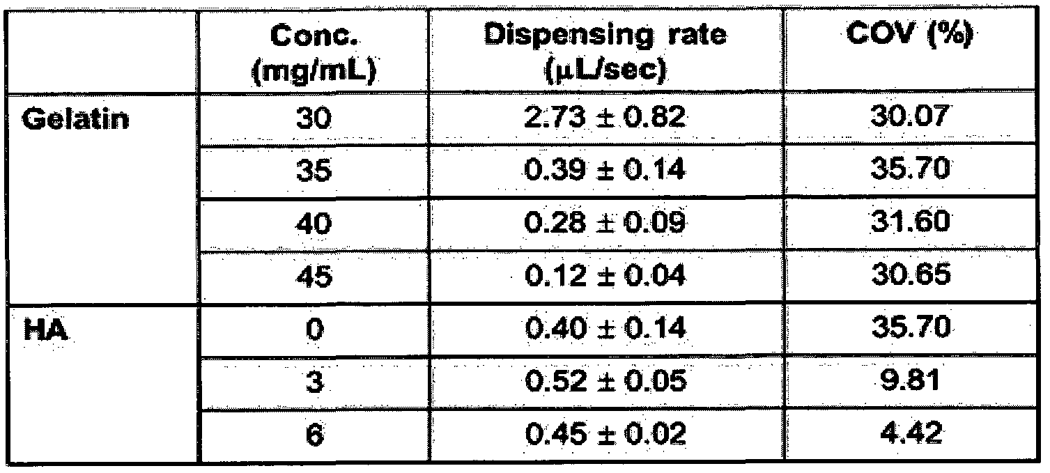
상기 표 1에 나타낸 바와 같이, 젤라틴 농도에 따라 dispensing rate이 달라 지는 것을 알 수 있었다. Dispensing rate은 프린팅되는 바이오 잉크의 부피를 시 간당으로 표시한 것이다. Dispensing rate가 낮아질수록 패턴의 분해능은 증가한다 고 할 수 있다. As shown in Table 1, it was found that the dispensing rate varies depending on the gelatin concentration. Dispensing rate is the volume of bio ink printed per hour. As the dispensing rate decreases, the resolution of the pattern increases.
젤라틴의 경우 35 mg/mL이상에서 패턴의 분해능이 급격히 증가했다. Coeff icient of variat ion (COV)는 프린팅된 패턴의 균일성 (uni formi ty)을 나타내 는 지수이다. 이 지수의 값이 클수록 di spensing rate이 블균등하다고 할 수 있다. 결과적으로 젤라틴은 35 mg/mL 이상에서 패턴의 분해능은 향상되지만 균일성에는 변화가 없었다. In the case of gelatin, the resolution of the pattern drastically increased above 35 mg / mL. Coeff icient of variat ion (COV) is an index indicating the uniformity of the printed pattern. The larger the value of this index, the more equal the di spensing rate. As a result, gelatin improved the pattern resolution above 35 mg / mL, but did not change uniformity.
이와는 다르게, 히알루론산을 젤라틴 하이드로겔에 첨가하였을 때 dispensing rate의 변화는 크지 않았다 (HA: 히알루론산) . 하지만 낮은 C0V 수치에 서 보여주는 것과 같이 프린팅된 패턴의 균일성이 현저하게 향상 시켜줌을 보여주 었다. 즉, HA의 함유는 패턴의 분해능에는 기여도가 낮지만 패턴의 균일성에 큰 영 향을 주고 있다는 것을 알 수 있었다. In contrast, there was no significant change in dispensing rate when hyaluronic acid was added to gelatin hydrogels (HA: hyaluronic acid). However, as shown by the low C0V value, the uniformity of the printed pattern was shown to be remarkably improved. In other words, it was found that the HA content had a low contribution to the pattern resolution, but had a great influence on the uniformity of the pattern.
따라서 본 발명의 바이오 잉크 조성물에서의 젤라틴과 HA의 함유는 필수적이 라고 할 수 있다. 또한, 본 발명의 바이오 잉크의 구조 안정성을 평가하기 위해, 본 발명의 바 이오 잉크를 이용하여 격자모양의 구조체를 삼차원적으로 프린팅하였다. 바이오 프 린팅된 구조체는 UV에 1 - 1000 초간 노출되어 가교되었다. 이를 세포 배양액에 담 가서 37°C 인큐베이션 한 후 시간에 따라 크기의 변화를 측정하였다. 이에 대한 평 가를 21일간수행하였다. 이에 대한 결과를 도 2에 나타내었다. Therefore, it can be said that the inclusion of gelatin and HA in the bio ink composition of the present invention is essential. In addition, in order to evaluate the structural stability of the bio ink of the present invention, the lattice-shaped structure was printed three-dimensionally using the bio ink of the present invention. Bioprinted structures were crosslinked by exposure to UV for 1-1000 seconds. It was immersed in the cell culture and incubated at 37 ° C. and then the size change was measured over time. The evaluation was conducted for 21 days. The results are shown in FIG. 2.
도 2에 나타낸 바와 같이, 바이오 프린팅된 구조체가 21일간 배양된 이후에, 초기 (Day 0) 크기의 약 70% 정도의 크기를 유지해 그 구조 안정성이 우수한 것을 알 수 있었다. 그 다음으로, 바이오잉크 조성물에 포함되는 점성 증강제의 함량에 따라, 바 이오 프린팅된 구조체의 안정성에 어떤 변화가 나타내는지 확인하고자 하였다. 상 기 실시예 2와 동일한 방법으로 바이오 잉크 조성물을 제조하되, 점성 증가제인 히 알루론산의 농도만 0, 1 , 2 및 3 mg/ml로 변화시켜가며 구조 안정성을 평가하였다. 이에 대한 결과를 도 3에 나타내었다. As shown in FIG. 2, after the bioprinted structure was incubated for 21 days, it was found that the structural stability was excellent by maintaining about 70% of the initial (Day 0) size. Next, to determine the change in the stability of the bio-printed structure according to the content of the viscosity enhancer included in the bioink composition. A bio ink composition was prepared in the same manner as in Example 2, but only the concentration of hyaluronic acid as a viscosity increasing agent was changed to 0, 1, 2, and 3 mg / ml, and structural stability was evaluated. The results for this are shown in FIG. 3.
도 3에 나타낸 바와 같이, 바이오 프린팅된 구조체의 안정성은 히알루론산의 함량이 증가함에 따라 높아지는 것을 알 수 있었다. As shown in FIG. 3, the stability of the bioprinted structure was found to increase with increasing content of hyaluronic acid.
<실시예 4> <Example 4>
바이오 잉크 조성물의 성능평가: 분출율과 패턴분해능 Performance Evaluation of Bio-Ink Compositions: Ejection Rate and Pattern Resolution
본 발명의 바이오 잉크의 프린팅의 성능, 분출율에 따른 해상도를 평가하기 위해 본 발명의 바이오 잉크를 100 - 700 mm/min의 분출율의 조정하에 200 urn 구경 의 노즐을 통해 프린트 하였다. 이에 대한 결과를 도 4에 나타내었다. In order to evaluate the resolution according to the printing performance and the ejection rate of the bioink of the present invention, the bioink of the present invention was printed through a nozzle of 200 urn diameter under the adjustment of the ejection rate of 100-700 mm / min. The results for this are shown in FIG. 4.
도 4에 나타낸 바와 같이, 본 발명의 바이오 잉크는 분출율이 증가함에 따라 패턴분해능이 향상되었고 사용된 분출율하에서 균일성을 유지하였다. As shown in Fig. 4, the bio ink of the present invention improved the pattern resolution as the ejection rate increased and maintained uniformity under the used ejection rate.
<실시예 5> Example 5
바이오 잉크 조성물 내 세포 생존율 평가 Evaluation of Cell Viability in Bio Ink Composition
본 발명에 따른 바이오 잉크 조성물의 세포 적합성을 평가하기 위해, 본 발 명의 바이오 잉크를 3T3 섬유아세포주와 흔합하여 바이오 프린터에 층전하였으며 50-80 kPa의 압력으로 300 urn 구경의 노즐올 통과하며 패턴을 형성하며 삼차원적 구조체를 완성했다. 바이오 프린트된 세포가 포함된 삼차원 구조체는 Live/Dead assay ki t (Li fe Technologies)를 사용하여 세포의 생존율을 평가하였다. 본 실험에 사용된 바이오 잉크 조성물은 상기 실시예 2의 방법에 따라 제조하였으며, 젤라틴 메타아크릴레이트에서 젤라틴의 분자량 및 가교정도 (20, 50 및 80%)를 변화시켜 바 이오 잉크의 물리적 강도에 변화를 주었다. In order to evaluate the cell suitability of the bio ink composition according to the present invention, the bio ink of the present invention was mixed with a 3T3 fibroblast line and laminated to a bio printer, and the pattern was passed through a nozzle of 300 urn diameter at a pressure of 50-80 kPa. To form a three-dimensional structure. Three-dimensional constructs containing bioprinted cells were evaluated for cell viability using Live / Dead assay kit (Li fe Technologies). Bio ink composition used in this experiment was prepared according to the method of Example 2, gelatin The molecular weight and degree of crosslinking (20, 50 and 80%) of the gelatin in the methacrylate was changed to change the physical strength of the bio ink.
0.5 uL/ml Calcein-AM과 2 uL/mL Eth-D가 용해되어 있는 인산완층액 (PBS, Li fe Technologies)를 세포와 함께 프린트된 바이오 잉크 구조체에 노출시켰다. 2 시간 뒤에 형광현미경으로 관찰하여 녹색 형광 (calcein)을 띄는 세포, 즉 살아있는 세포를 확인하였다. 세포생존율이 80% 이상의 조건에서 바이오 잉크의 최적화를 결 정하였다. 이에 대한 결과를 도 5에 나타내었다. 도 5에서 Gel-MA 50은 50% 메타아크릴 레이션 된 젤라틴을 의미한다. Complete phosphate solution (PBS, Li fe Technologies) in which 0.5 uL / ml Calcein-AM and 2 uL / mL Eth-D were dissolved was exposed to the printed bio ink construct with the cells. After 2 hours, the cells were observed with a fluorescence microscope to identify green fluorescent cells (ie, living cells). The optimization of bio ink was determined under the conditions of cell survival rate of 80% or more. The results for this are shown in FIG. 5. Gel-MA 50 in Figure 5 means 50% methacrylated gelatin.
도 5에 나타낸 바와 같이, 바이오 프린트된 삼차원 구조체 내의 대부분의 세 포에서 녹색 형광이 관찰되었으며, 이는 살아있는 세포를 나타낸다. 이를 정량화했 을 때, 실험에 사용된 모든 조성물 조건에서 세포 생존율이 90% 이상 (그래프 참 조)으로 나타났다. 즉, 본 발명의 바이오 잉크 조성물은 프린팅시 발생하는 높은 전단 압력에서 세포의 손상을 최소화 해주고 있다는 것을 알 수 있었다. 상기 실험과 동일한 조성의 바이오 잉크에서, Gel-MA 50의 농도만 변화시키 고 - 50% 메타아크릴레이션된 젤라틴을 두 가지 농도 20 mg/ml과 30 mg/ml로 실험 에 사용함- C2C12 myoblast 세포주를 포함시켜 프린팅 하였다. 프린팅 조건은 분출 율 120 mm/min 이었으며, 노즐크기는 300 μ Tef lon nozzle을 사용하였다. 이에 대한 결과를 도 6에 나타내았다. As shown in FIG. 5, green fluorescence was observed in most cells in the bioprinted three-dimensional construct, indicating live cells. When quantified, cell viability was greater than 90% (see graph) under all composition conditions used in the experiment. That is, the bio ink composition of the present invention was found to minimize damage to the cells at high shear pressure generated during printing. In the bio ink of the same composition as the above experiment, only the concentration of Gel-MA 50 was changed-50% methacrylated gelatin was used in the experiment at two concentrations of 20 mg / ml and 30 mg / ml-C2C12 myoblast cell line It was included and printed. The printing conditions were the ejection rate of 120 mm / min and the nozzle size was 300 μ Tef lon nozzle. The results are shown in FIG. 6.
도 6에 나타낸 바와 같이, 모든 조성에서 바이오 프린트된 삼차원 구조체 내 의 대부분의 세포가 녹색 형광을 나타내었으며, 이는 살아있는 세포를 나타내는 것 이다. 보통 젤의 강도를 높이기 위해 젤라틴의 농도를 높여주는데 이렇게 되면 젤 내의 di ffusion rate가 감소되어 세포의 생존률이 감소될 수 있다. 본 발명에 따른 바이오 잉크 조성물의 경우 Gel-MA 50이 30 mg/ml 농도에서도 세포생존률에 영향을 미치지 않음을 확인하였다. As shown in FIG. 6, most of the cells in the bioprinted three-dimensional structure at all compositions showed green fluorescence, indicating living cells. Normally, the gel concentration is increased to increase the strength of the gel, which reduces the di ffusion rate in the gel, which may reduce cell survival. In the case of the bio ink composition according to the present invention, it was confirmed that Gel-MA 50 did not affect cell viability even at a concentration of 30 mg / ml.
<실시예 6> <Example 6>
조직유래 성분이 포함된 바이오 잉크 조성물의 세포 증식 및 분화능 평가 인위적으로 생성된 어떠한 조성물도 세포외기질 (extracel lular matrix)과 같은 자연적인 조직유래 성분이 갖고 있는 특성을 모두 재연하는 것이 사실상 불가 능하며, 따라서 탈세포화된 조직유래 성분을 바이오 잉크 조성물에 추가함으로써, 바이오 프린팅된 구조체 내에서 세포가 정상적으로 성장하고 분화할 수 있는지 여 부를 확인하고자 하였다. 사용된 조성물은 앞서 실험된 바이오 잉크 조성물 (20 mg/ml Gel -MA 50 (bloom 300), 30 mg/ml 젤라틴 (bloom 90-100) , 3 mg/ml HA, 10% 글리세를입니다. Evaluation of Cell Proliferation and Differentiation Capability of Bio-Ink Compositions Containing Tissue-Derived Ingredients It is virtually impossible to reproduce all the properties of the same natural tissue-derived component, and thus, by adding decellularized tissue-derived components to the bio ink composition, it is possible to determine whether cells can normally grow and differentiate within the bioprinted structure. We wanted to confirm wealth. The composition used is the bio ink composition (20 mg / ml Gel -MA 50 (bloom 300), 30 mg / ml gelatin (bloom 90-100), 3 mg / ml HA, 10% glycerol, which was previously tested.
)에 근육조직유래 성분을 12% 함유시켰다 (세포: C2C12 myoblast cel l l ine ( lxlO6 cel ls/ml ) , 분출율: 120 瞧 /min, 노즐크기: 300 μ Tef lon nozzle. ) . 상기 근육조직 유래 성분은 돼지의 근육조직을 탈세포화 한 후 남은 세포외 기질 단백질을 약산성 과 펩신으로 용해하여 분리한 것이다. 이를 중성화 시킨 후에 바이오잉크 구성물에 함유시켜 사용하였다. 근육조직유래 성분의 주성분으로는 콜라겐, 글리코사미노글 리칸, 성장인자 및 사이토카인이다. 이에 대한 결과를 도 7에 나타내었다. ) Contains 12% of muscle tissue-derived components (cells: C2C12 myoblast cel ll ine (lxlO 6 cel ls / ml), ejection rate: 120 μs / min, nozzle size: 300 μ Tef lon nozzle.). The muscle tissue-derived component is isolated by dissolving the extracellular matrix protein remaining after decellularization of pig muscle tissue with weak acid and pepsin. It was neutralized and used in the bioink construct. The main components of muscle tissue-derived components are collagen, glycosaminoglycans, growth factors and cytokines. The results for this are shown in FIG. 7.
도 7에 나타낸 바와 같이, 근육조직 유래의 성분이 포함된 바이오 잉크 조 성물을 이용하여 근육세포를 프린팅한 결과, 조직유래의 성분이 포함되지 않은 대 조군과 비교하여 세포의 증식과 분화가 더 향상된 것올 확인할 수 있었다. As shown in FIG. 7, as a result of printing muscle cells using a bio-ink composition containing components derived from muscle tissue, cell proliferation and differentiation were improved as compared to the control group containing no tissue-derived components. I could confirm that.
<실시예 7> <Example 7>
분화조절물질이 포함된 바이오 잉크 조성물의 제조 Preparation of Bio-Ink Compositions Containing Differentiation Regulators
본 발명자는 바이오 잉크 조성물에 포함된 줄기세포 (배아줄기세포, 유도만 능줄기세포, 성체줄기세포를 포함하는)가 특정 세포로 분화되는 것을 유도하기 위 해 분화조절물질이 포함된 바이오 잉크 조성물을 제조하였다. 구체적으로, 본 실시예에서는 바이오 프린팅된 줄기세포를 골세포로 분화 유도할 때 사용하는 뼈형성단백질 -2(BMP-2, 서열번호 1), 연골세포로 분화 유도할 때 사용하는 TGF-beta3와 같은 성장인자 (서열번호 2, 서열번호 3) 또는 혈관내피세 포로 분화 유도할 때 사용하는 혈관내피세포 성장인자 (서열번호 4)를 모사한 펩타 이드를 화학적 결합을 통해 바이오 잉크 조성물에 포함시켰다. The present invention provides a bio-ink composition containing a differentiation regulator to induce differentiation of stem cells (including embryonic stem cells, induced pluripotent stem cells, and adult stem cells) into specific cells included in the bio-ink composition. Prepared. Specifically, in the present embodiment, BGF-2, which is used to induce differentiation of bioprinted stem cells into osteocytes (BMP-2, SEQ ID NO: 1), and TGF-beta3 used to induce differentiation into chondrocytes. Peptides mimicking the same growth factor (SEQ ID NO: 2, SEQ ID NO: 3) or vascular endothelial growth factor (SEQ ID NO: 4) used to induce differentiation into vascular endothelial cells were included in the bio ink composition through chemical bonding.
서열번호 1 : KIPKASSVPTELSAISTLYL SEQ ID NO: KIPKASSVPTELSAISTLYL
서열번호 2: ANVAENA 서열번호 3 : LIANAK SEQ ID NO: ANVAENA SEQ ID NO: 3 LIANAK
서열번호 4: KLTWQELYQLKYKGI 분화조절에 관여하는 성장인자가 포함된 바이오 잉크 조성물의 제조방법 모 식도를 도 8에 나타내었다. SEQ ID NO: 4: A schematic diagram of a method for preparing a bio ink composition including a growth factor involved in KLTWQELYQLKYKGI differentiation control is shown in FIG. 8.
<실시예 8> <Example 8>
바이오 잉크 조성물을 이용한 조직 유사기관 및 이식 가능한 조직 구조체의 제조 Preparation of Tissue-like Organs and Implantable Tissue Constructs Using Bio-Ink Compositions
본 발명의 바이오 잉크를 이용하여 여러 가지 조직 유사기관 및 이식 가능한 조직 구조체를 제조하였다. 이를 위해 특정 조직의 특성이 되는 구조를 획득하기 위해 조직학적 염색 이미지나 바이오 이미지 (CT 나 MRI 같은 바이오 이미징 기술로 획득된)를 삼차원적 CAD 모델로 전환하고 이를 다시 프린팅이 가능한 모션 파일로 전환하였다. 전환된 모션 파일은 바이오 프린터에 연동되어 특정 구조체로 프린팅 작업을수행하였다. 이에 대한 결과를 도 9 내지 도 12에 나타내었다. The bioinks of the present invention were used to prepare various tissue-like organs and implantable tissue constructs. To do this, the histological staining image or bioimage (obtained by bio-imaging technology such as CT or MRI) was converted into a three-dimensional CAD model to obtain a structure characteristic of a specific tissue, which was then converted into a printable motion file. . The converted motion file was linked to a bio printer to print a specific structure. The results are shown in FIGS. 9 to 12.
도 9에 나타낸 바와 같이 , 본 발명의 바이오 잉크 조성물을 이용하여 간 조 직 (도 9A) , 심근 조직 (도 9B) 및 근육 조직 (도 9C) 유사기관이 양호하게 형성되는 것을 확인하여, 조직의 기능성과 연관되는 정교한 방향성을 가지는 내부구조를 프 린팅할 수 있다는 것을 확인하였다. As shown in FIG. 9, it was confirmed that liver tissue (FIG. 9A), myocardial tissue (FIG. 9B) and muscle tissue (FIG. 9C) similar organs were formed well using the bio ink composition of the present invention. We confirmed that we can print internal structures with elaborate directionality associated with functionality.
도 10에 나타낸 바와 같이, 3차원적 바이오 프린팅된 조작된 귀 (도 10A) , 코 (도 10B) 및 관모양 조직 (도 10C) 구조체 또한 양호하게 제조할 수 있어, 본 발명 에서 개발된 바이오 잉크 조성물을 이용하여 이식이 가능한 여러 가지 조직과 장기 를 프린팅할 수 있음을 확인하였다. As shown in FIG. 10, the three-dimensional bioprinted engineered ears (FIG. 10A), nose (FIG. 10B) and tubular tissue (FIG. 10C) structures can also be produced well, and the bioinks developed in the present invention. It was confirmed that the composition can be used to print a variety of transplantable tissues and organs.
도 11 및 도 12에 나타낸 바와 같이, 본 발명의 조성물을 이용하여 여러 가 지 세포를 동시에 프린팅 함으로써 조직과 조직간의 연계작용 또한 재연할 수 있음 을 확인하였다. 즉, 도 11A에서는 간 세포와 혈관세포의 연계작용, 도 11B에서는 뼈 세포와 연골세포의 연계작용, 도 11C에서는 근육세포와 힘줄세포의 연계작용 및 도 12에서는 신경세포와 근육세포의 연계작용을 확인함으로써, 본 발명의 조성물을 이용하여 기능성이 향상된 조직 구조체를 제조할 수 있음을 알 수 있었다. As shown in Figure 11 and 12, it was confirmed that by linking several cells at the same time using the composition of the present invention can also reproduce the linkage between tissue and tissue. That is, in FIG. 11A, the linkage between hepatocytes and vascular cells, in FIG. 11B, the linkage between bone cells and chondrocytes, in FIG. 11C, the linkage between muscle cells and tendon cells, and in FIG. 12, the linkage between neurons and muscle cells. By confirming, it was found that the tissue construct with improved functionality can be produced using the composition of the present invention.
Claims
Priority Applications (2)
| Application Number | Priority Date | Filing Date | Title |
|---|---|---|---|
| US15/746,091 US20180280578A1 (en) | 2015-07-21 | 2016-07-21 | Bio-ink composition having improved physical and biological properties |
| EP16828086.5A EP3326661B8 (en) | 2015-07-21 | 2016-07-21 | Bio-ink composition having improved physical and biological properties |
Applications Claiming Priority (2)
| Application Number | Priority Date | Filing Date | Title |
|---|---|---|---|
| KR10-2015-0103313 | 2015-07-21 | ||
| KR20150103313 | 2015-07-21 |
Publications (1)
| Publication Number | Publication Date |
|---|---|
| WO2017014582A1 true WO2017014582A1 (en) | 2017-01-26 |
Family
ID=57834952
Family Applications (1)
| Application Number | Title | Priority Date | Filing Date |
|---|---|---|---|
| PCT/KR2016/007962 Ceased WO2017014582A1 (en) | 2015-07-21 | 2016-07-21 | Bio-ink composition having improved physical and biological properties |
Country Status (4)
| Country | Link |
|---|---|
| US (1) | US20180280578A1 (en) |
| EP (1) | EP3326661B8 (en) |
| KR (1) | KR101913374B1 (en) |
| WO (1) | WO2017014582A1 (en) |
Cited By (3)
| Publication number | Priority date | Publication date | Assignee | Title |
|---|---|---|---|---|
| KR20200039055A (en) * | 2018-10-02 | 2020-04-16 | 한국과학기술연구원 | Hydrogel composition and bioinks composition comprising the same |
| EP3769796A1 (en) | 2019-07-22 | 2021-01-27 | Fundacja Badan i Rozwoju Nauki | Detergent-free decellularized extracellular matrix preparation method and bioinks for 3d printing |
| US11370929B2 (en) * | 2018-01-25 | 2022-06-28 | National Taiwan University | Bioink set and applications thereof for three-dimensional printing of cells |
Families Citing this family (40)
| Publication number | Priority date | Publication date | Assignee | Title |
|---|---|---|---|---|
| US20180117219A1 (en) | 2015-04-29 | 2018-05-03 | Northwestern University | 3d printing of biomedical implants |
| US10857260B2 (en) * | 2017-01-12 | 2020-12-08 | The George Washington University | Coaxial needle for fabricating a multi scale, multi layer blood vessel or vascular network employing 3D bioprinting |
| KR101949846B1 (en) * | 2017-09-27 | 2019-02-19 | 울산과학기술원 | Manufacturing Method of Structure of Artificial Vascular by Using 3D Printing, Structure of Artificial Vascular Manufactured by the Same, Manufacturing Method of Artificial Vascular using the Same and Artificial Vascular Manufactured by the Same |
| EP3485918A1 (en) * | 2017-11-21 | 2019-05-22 | ETH Zürich | Gel composition for 3d printing of cell suspensions |
| US20210038762A1 (en) | 2018-01-31 | 2021-02-11 | Rokit Healthcare Inc. | Bioink composition for cartilage regeneration, method for manufacturing customized scaffold for cartilage regeneration using same, and customized scaffold for cartilage regeneration manufactured using manufacturing method |
| AU2018405440B2 (en) * | 2018-01-31 | 2024-10-17 | Rokit Healthcare Inc. | Bioink composition for dermis regeneration sheet, method for manufacturing customized dermis regeneration sheet using same, and customized dermis regeneration sheet manufactured using manufacturing method |
| KR102146682B1 (en) * | 2018-06-18 | 2020-08-21 | 주식회사 티앤알바이오팹 | Hybrid bio ink, manufacturing method thereof, and artificial tissue manufacturing method using the same |
| KR102209623B1 (en) * | 2018-10-05 | 2021-01-29 | 주식회사 이노리젠 | Composition of bio-ink having improved physical and biological properties |
| KR102489607B1 (en) * | 2018-10-24 | 2023-01-17 | 포항공과대학교 산학협력단 | Method of manufacturing cell spheroid using three-dimensional printing method |
| US11781101B2 (en) * | 2018-11-05 | 2023-10-10 | Regents Of The University Of Minnesota | 3D-printed models of biological microenvironments |
| WO2020111868A1 (en) * | 2018-11-30 | 2020-06-04 | 주식회사 엘앤씨바이오 | Bio-ink composition for 3d printing, containing human-derived component and having tissue-specific cell differentiation effect, and preparation method therefor |
| CN111375091B (en) * | 2018-12-30 | 2021-09-10 | 中国科学院沈阳自动化研究所 | Photosensitive composite biological ink for 3D printing and preparation method thereof |
| KR102126733B1 (en) | 2019-01-08 | 2020-06-26 | 성균관대학교산학협력단 | Method of preparing a structure consisted of arranged fiber structure and cells |
| EP3931532A4 (en) * | 2019-02-26 | 2022-10-12 | CELLINK Bioprinting AB | Systems and methods for real-time optoelectronic assessments of fluid volume in fluid dispensing systems |
| KR102156310B1 (en) * | 2019-03-06 | 2020-09-15 | 울산과학기술원 | A method for manufacturing cell spheroid using bio-ink |
| US20240158727A1 (en) * | 2019-03-06 | 2024-05-16 | Osaka University | Cell tissue production method, cell tissue production set, and cultivation container containing cell tissue produced by said production method |
| JP7256494B2 (en) * | 2019-03-06 | 2023-04-12 | 国立大学法人大阪大学 | Cell tissue preparation method and cell tissue preparation set |
| EP3939625B1 (en) | 2019-03-11 | 2024-08-14 | Rokit Healthcare Inc. | Method for producing composition for regenerating cartilage using freeze-dried hyaline cartilage powder, composition for regenerating cartilage produced using same, method for producing scaffold for regenerating patient-specific cartilage using composition for regenerating cartilage, and scaffold for regenerating patient-specific cartilage |
| CN109822889B (en) * | 2019-03-22 | 2024-03-26 | 深圳大洲医学科技有限公司 | Multichannel 3D printing spray head, preparation method thereof and cell/biological ink conveying system |
| KR102261908B1 (en) * | 2019-05-15 | 2021-06-08 | 주식회사 이노리젠 | Dual-crosslinkable two component type composition of bio-ink and Method for Producing Tissue-Like Structure Using the Same |
| WO2020257206A1 (en) | 2019-06-18 | 2020-12-24 | University Of Washington | Protein-based resins for additive manufacturing |
| KR102283464B1 (en) * | 2019-06-28 | 2021-08-02 | 경북대학교 산학협력단 | Wound Dressing Based on Hydrogel Comprising VEGF Peptide and Method for Producing the Same |
| WO2021006664A1 (en) * | 2019-07-10 | 2021-01-14 | 사회복지법인 삼성생명공익재단 | Bioink composition for forming bone tissue |
| WO2021006669A1 (en) * | 2019-07-10 | 2021-01-14 | 사회복지법인 삼성생명공익재단 | Bioink composition for cartilage tissue formation |
| WO2021040169A1 (en) * | 2019-08-30 | 2021-03-04 | 주식회사 이노리젠 | Bio-ink composition comprising methacrylated low-molecular weight collagen and method of preparing tissue analog structure using same |
| KR102337350B1 (en) * | 2019-11-22 | 2021-12-08 | 아주대학교산학협력단 | Injectable hydrogel composition having endogenous progenitor or stem cell recruitment and induction of vascular differentiation of recruited cells |
| KR102284592B1 (en) | 2020-01-31 | 2021-08-02 | 성균관대학교산학협력단 | Method for producing arranged structure using biocompatible polymer and arranged structure produced using same |
| KR102706453B1 (en) * | 2020-05-19 | 2024-09-13 | 주식회사 이노리젠 | Composition of Bio-ink Comprising Human Derived Collagen and Method for Producing Tissue-Like Structure Using the Same |
| US11918703B2 (en) | 2020-08-13 | 2024-03-05 | Universidad De Los Andes | Extrudable photocrosslinkable hydrogel and method for its preparation |
| US12103239B2 (en) * | 2020-10-27 | 2024-10-01 | Korea Advanced Institute Of Science And Technology | Method and system for manufacturing high-resolution structure using size-tunable hydrogel |
| CN112791239B (en) * | 2021-01-14 | 2021-10-26 | 浙江大学 | Preparation method of super-bionic soft and hard tissue composite scaffold |
| WO2022153319A1 (en) * | 2021-01-18 | 2022-07-21 | The State Of Israel, Ministry Of Agriculture & Rural Development, Agricultural Research Organization (Aro) (Volcani Institute) | A sodium alginate, gelatin, collagen and fibrin (agcf) based bio-ink for the bioprinting of a 3d biogel-based tissue/structure |
| WO2022182147A1 (en) * | 2021-02-24 | 2022-09-01 | 주식회사 스페바이오 | Method for producing cell aggregate or extracellular vesicle, and extracellular vesicle produced thereby |
| KR102780671B1 (en) * | 2021-02-24 | 2025-03-14 | 주식회사 스페바이오 | A preparation method of extracellular vesicles with cellular spheroid and extracellular vesicles made thereby |
| CN113398332B (en) * | 2021-08-20 | 2022-01-11 | 北京大学第三医院(北京大学第三临床医学院) | A 3D biomimetic biological scaffold containing stem cell exosomes and its application |
| KR102796480B1 (en) | 2021-11-29 | 2025-04-15 | 동의대학교 산학협력단 | Method for manufacturing artificial skin tissue using three-dimensional bioprinting and artificial skin tissue manufactured by thereof |
| WO2023101071A1 (en) * | 2021-12-03 | 2023-06-08 | 주식회사 팡세 | Three-dimensional alignment culture method of cells |
| WO2023107871A2 (en) * | 2021-12-06 | 2023-06-15 | Ohio State Innovation Foundation | Compositions and methods for production and use of a scalable human cell-derived extracellular matrix |
| KR20230093606A (en) * | 2021-12-20 | 2023-06-27 | 포항공과대학교 산학협력단 | Extracellular matrix-based hybrid ink and mufacturing method of the same |
| WO2024003287A1 (en) | 2022-06-30 | 2024-01-04 | Danmarks Tekniske Universitet | 3d printing transparent bioink formulation |
Citations (3)
| Publication number | Priority date | Publication date | Assignee | Title |
|---|---|---|---|---|
| US6773713B2 (en) * | 2001-02-23 | 2004-08-10 | University Of Massachusetts | Injection molding of living tissues |
| US20120089238A1 (en) * | 2010-10-06 | 2012-04-12 | Hyun-Wook Kang | Integrated organ and tissue printing methods, system and apparatus |
| US20150057786A1 (en) * | 2010-10-21 | 2015-02-26 | Organovo, Inc. | Devices, systems, and methods for the fabrication of tissue |
Family Cites Families (5)
| Publication number | Priority date | Publication date | Assignee | Title |
|---|---|---|---|---|
| US20090208466A1 (en) * | 2006-04-21 | 2009-08-20 | James Yoo | Ink-jet printing of tissues |
| US8790683B2 (en) * | 2009-12-22 | 2014-07-29 | National Cheng Kung University | Cell tissue gel containing collagen and hyaluronan |
| CA2848044C (en) | 2011-09-12 | 2021-01-19 | Organovo, Inc. | Platform for engineered implantable tissues and organs and methods of making the same |
| US9499779B2 (en) * | 2012-04-20 | 2016-11-22 | Organovo, Inc. | Devices, systems, and methods for the fabrication of tissue utilizing UV cross-linking |
| US9442105B2 (en) * | 2013-03-15 | 2016-09-13 | Organovo, Inc. | Engineered liver tissues, arrays thereof, and methods of making the same |
-
2016
- 2016-07-21 WO PCT/KR2016/007962 patent/WO2017014582A1/en not_active Ceased
- 2016-07-21 KR KR1020160092614A patent/KR101913374B1/en active Active
- 2016-07-21 EP EP16828086.5A patent/EP3326661B8/en active Active
- 2016-07-21 US US15/746,091 patent/US20180280578A1/en not_active Abandoned
Patent Citations (3)
| Publication number | Priority date | Publication date | Assignee | Title |
|---|---|---|---|---|
| US6773713B2 (en) * | 2001-02-23 | 2004-08-10 | University Of Massachusetts | Injection molding of living tissues |
| US20120089238A1 (en) * | 2010-10-06 | 2012-04-12 | Hyun-Wook Kang | Integrated organ and tissue printing methods, system and apparatus |
| US20150057786A1 (en) * | 2010-10-21 | 2015-02-26 | Organovo, Inc. | Devices, systems, and methods for the fabrication of tissue |
Non-Patent Citations (3)
| Title |
|---|
| FERRIS, C. J. ET AL.: "Bio-ink for On-demand Printing of Living Cells", BIOMATERIALS SCIENCE, vol. 1, 2013, pages 224 - 230, XP055348567 * |
| MELCHELS, F. P. W. ET AL.: "Development and Characterisation of a New Biolink for Additive Tissue Manufacturing", JOURNAL OF MATERIALS CHEMISTRY B, vol. 2, 2014, pages 2282 - 2289, XP055348585 * |
| See also references of EP3326661A4 * |
Cited By (6)
| Publication number | Priority date | Publication date | Assignee | Title |
|---|---|---|---|---|
| US11370929B2 (en) * | 2018-01-25 | 2022-06-28 | National Taiwan University | Bioink set and applications thereof for three-dimensional printing of cells |
| KR20200039055A (en) * | 2018-10-02 | 2020-04-16 | 한국과학기술연구원 | Hydrogel composition and bioinks composition comprising the same |
| KR102168094B1 (en) | 2018-10-02 | 2020-10-20 | 한국과학기술연구원 | Hydrogel composition and bioinks composition comprising the same |
| EP3769796A1 (en) | 2019-07-22 | 2021-01-27 | Fundacja Badan i Rozwoju Nauki | Detergent-free decellularized extracellular matrix preparation method and bioinks for 3d printing |
| WO2021014359A1 (en) | 2019-07-22 | 2021-01-28 | Fundacja Badan I Rozwoju Nauki | Detergent-free decellularized extracellular matrix preparation method and bioinks for 3d printing |
| US12208179B2 (en) | 2019-07-22 | 2025-01-28 | Polbionica Sp. z o. o. | Detergent-free decellularized extracellular matrix preparation methods and bioinks for 3D printing |
Also Published As
| Publication number | Publication date |
|---|---|
| KR20170012099A (en) | 2017-02-02 |
| EP3326661B8 (en) | 2020-12-30 |
| EP3326661B1 (en) | 2020-11-18 |
| EP3326661A4 (en) | 2018-08-01 |
| US20180280578A1 (en) | 2018-10-04 |
| EP3326661A1 (en) | 2018-05-30 |
| KR101913374B1 (en) | 2018-10-30 |
Similar Documents
| Publication | Publication Date | Title |
|---|---|---|
| EP3326661B1 (en) | Bio-ink composition having improved physical and biological properties | |
| US11124774B2 (en) | Engineered liver tissues, arrays thereof, and methods of making the same | |
| EP2755599B1 (en) | Platform for engineered implantable tissues and organs and methods of making the same | |
| CN108273140B (en) | Bio-brick for bio-printing and use thereof | |
| HK1200483A1 (en) | Engineered tissues for in vitro research uses, arrays thereof, and methods of making the same | |
| KR102261908B1 (en) | Dual-crosslinkable two component type composition of bio-ink and Method for Producing Tissue-Like Structure Using the Same | |
| JP2018532418A (en) | Improved method for tissue manufacture | |
| KR102706453B1 (en) | Composition of Bio-ink Comprising Human Derived Collagen and Method for Producing Tissue-Like Structure Using the Same | |
| KR102198455B1 (en) | Bioink Composition for Production of Tumor Microenvironmental Vascular Model and Printing Technique Using the Same | |
| KR102345701B1 (en) | Composition of Bio-ink Comprising Human Derived Gelatin and Methacrylated Low Molecular Weight Collagen and Method for Producing Tissue-Like Structure Using the Same | |
| KR20200052468A (en) | Functional bio-ink composition comprising vascular endothelial growth factor for inducing angiogenesis | |
| KR102209623B1 (en) | Composition of bio-ink having improved physical and biological properties | |
| KR20200052467A (en) | Functional bio-ink composition comprising transforming growth factor β for inducing production of cartilage | |
| KR20230079777A (en) | Method for manufacturing artificial skin tissue using three-dimensional bioprinting and artificial skin tissue manufactured by thereof | |
| KR20200052466A (en) | Bio-ink composition comprising bone morphogenetic protein for promoting bone formation and odontoblast differentiation | |
| Dimengo | An Optimized Bioink for Three-Dimensional Printing of Endothelial Cells into a Vascular-Like Structure |
Legal Events
| Date | Code | Title | Description |
|---|---|---|---|
| 121 | Ep: the epo has been informed by wipo that ep was designated in this application |
Ref document number: 16828086 Country of ref document: EP Kind code of ref document: A1 |
|
| NENP | Non-entry into the national phase |
Ref country code: DE |
|
| NENP | Non-entry into the national phase |
Ref country code: JP |
|
| WWE | Wipo information: entry into national phase |
Ref document number: 15746091 Country of ref document: US |