WO2016131895A1 - Method for quantification of virus particles using capillary zone electrophoresis - Google Patents
Method for quantification of virus particles using capillary zone electrophoresis Download PDFInfo
- Publication number
- WO2016131895A1 WO2016131895A1 PCT/EP2016/053405 EP2016053405W WO2016131895A1 WO 2016131895 A1 WO2016131895 A1 WO 2016131895A1 EP 2016053405 W EP2016053405 W EP 2016053405W WO 2016131895 A1 WO2016131895 A1 WO 2016131895A1
- Authority
- WO
- WIPO (PCT)
- Prior art keywords
- concentration
- virus particles
- adenovirus
- sample
- capillary tube
- Prior art date
- Legal status (The legal status is an assumption and is not a legal conclusion. Google has not performed a legal analysis and makes no representation as to the accuracy of the status listed.)
- Ceased
Links
Classifications
-
- G—PHYSICS
- G01—MEASURING; TESTING
- G01N—INVESTIGATING OR ANALYSING MATERIALS BY DETERMINING THEIR CHEMICAL OR PHYSICAL PROPERTIES
- G01N27/00—Investigating or analysing materials by the use of electric, electrochemical, or magnetic means
- G01N27/26—Investigating or analysing materials by the use of electric, electrochemical, or magnetic means by investigating electrochemical variables; by using electrolysis or electrophoresis
- G01N27/416—Systems
- G01N27/447—Systems using electrophoresis
- G01N27/44704—Details; Accessories
- G01N27/44747—Composition of gel or of carrier mixture
-
- C—CHEMISTRY; METALLURGY
- C12—BIOCHEMISTRY; BEER; SPIRITS; WINE; VINEGAR; MICROBIOLOGY; ENZYMOLOGY; MUTATION OR GENETIC ENGINEERING
- C12Q—MEASURING OR TESTING PROCESSES INVOLVING ENZYMES, NUCLEIC ACIDS OR MICROORGANISMS; COMPOSITIONS OR TEST PAPERS THEREFOR; PROCESSES OF PREPARING SUCH COMPOSITIONS; CONDITION-RESPONSIVE CONTROL IN MICROBIOLOGICAL OR ENZYMOLOGICAL PROCESSES
- C12Q1/00—Measuring or testing processes involving enzymes, nucleic acids or microorganisms; Compositions therefor; Processes of preparing such compositions
- C12Q1/02—Measuring or testing processes involving enzymes, nucleic acids or microorganisms; Compositions therefor; Processes of preparing such compositions involving viable microorganisms
- C12Q1/04—Determining presence or kind of microorganism; Use of selective media for testing antibiotics or bacteriocides; Compositions containing a chemical indicator therefor
-
- C—CHEMISTRY; METALLURGY
- C12—BIOCHEMISTRY; BEER; SPIRITS; WINE; VINEGAR; MICROBIOLOGY; ENZYMOLOGY; MUTATION OR GENETIC ENGINEERING
- C12Q—MEASURING OR TESTING PROCESSES INVOLVING ENZYMES, NUCLEIC ACIDS OR MICROORGANISMS; COMPOSITIONS OR TEST PAPERS THEREFOR; PROCESSES OF PREPARING SUCH COMPOSITIONS; CONDITION-RESPONSIVE CONTROL IN MICROBIOLOGICAL OR ENZYMOLOGICAL PROCESSES
- C12Q1/00—Measuring or testing processes involving enzymes, nucleic acids or microorganisms; Compositions therefor; Processes of preparing such compositions
- C12Q1/70—Measuring or testing processes involving enzymes, nucleic acids or microorganisms; Compositions therefor; Processes of preparing such compositions involving virus or bacteriophage
-
- G—PHYSICS
- G01—MEASURING; TESTING
- G01N—INVESTIGATING OR ANALYSING MATERIALS BY DETERMINING THEIR CHEMICAL OR PHYSICAL PROPERTIES
- G01N27/00—Investigating or analysing materials by the use of electric, electrochemical, or magnetic means
- G01N27/26—Investigating or analysing materials by the use of electric, electrochemical, or magnetic means by investigating electrochemical variables; by using electrolysis or electrophoresis
- G01N27/416—Systems
- G01N27/447—Systems using electrophoresis
- G01N27/44756—Apparatus specially adapted therefor
- G01N27/44791—Microapparatus
-
- C—CHEMISTRY; METALLURGY
- C12—BIOCHEMISTRY; BEER; SPIRITS; WINE; VINEGAR; MICROBIOLOGY; ENZYMOLOGY; MUTATION OR GENETIC ENGINEERING
- C12Q—MEASURING OR TESTING PROCESSES INVOLVING ENZYMES, NUCLEIC ACIDS OR MICROORGANISMS; COMPOSITIONS OR TEST PAPERS THEREFOR; PROCESSES OF PREPARING SUCH COMPOSITIONS; CONDITION-RESPONSIVE CONTROL IN MICROBIOLOGICAL OR ENZYMOLOGICAL PROCESSES
- C12Q2565/00—Nucleic acid analysis characterised by mode or means of detection
- C12Q2565/10—Detection mode being characterised by the assay principle
- C12Q2565/125—Electrophoretic separation
Definitions
- the present invention is directed to the analysis of biological samples in general, and more specifically to the quantitative analysis of biological samples utilizing capillary electrophoretic techniques.
- the invention in particular is directed to the quantitative analysis of biological samples containing adenovirus.
- Viruses in particular adenoviruses, are potentially useful vectors for vaccination and/or gene therapy.
- Recombinant adenoviruses are a preferred class of viral vectors for use in vaccination or gene therapy. Robust and high yield processes for large scale
- qPCR quantitative polymerase chain reaction
- AEX-HPLC anion exchange chromatography
- qPCR determines the concentration of adenovirus particles indirectly by determining the DNA content of the particles instead of determining the intact particles themselves.
- AEX-HPLC with UV detection makes use of a charge-based separation of intact adenoviruses from sample matrix components. Quantification is performed by external calibration using an adenovirus reference standard.
- the AEX-HPLC method suffers from adsorption, carry-over and recovery issues for samples containing cell lysate or high salt concentrations, and is therefore not suitable to determine the concentration of adenoviruses accurately and precisely throughout the entire production process.
- the present invention provides a novel method for quantification of virus particles, in particular adenovirus particles, in a biological sample, said method comprising the steps of: a) introducing said biological sample comprising virus particles into a capillary tube containing a buffer solution;
- comparing the electropherogram with an electropherogram generated from a reference sample containing a known concentration of said virus particles preferably by comparing the peak area of the electophoretical fraction corresponding to the virus particles with the peak area of a reference sample, comprising containing a defined concentration of virus particles.
- the method according to the invention has been shown to be a robust, accurate, reliable and fast method for the quantification of (adeno)virus particles in all types of biological samples derived from both upstream and downstream development and
- Samples with highly different matrices can be analysed in long sequences without the need for prior processing, and/or for example the drawbacks of adsorption, carry-over or poor recovery.
- the total analysis time from process sampling to reported result can be less than 2 hours.
- the method according to the invention requires no special treatment for detection.
- the virus particles are detected directly and staining or labelling of the virus particles is not needed. Description of the Figures
- FIG. 1 A) Electropherograms of Ad26 (a) lysed harvest, (b) clarified harvest, (c) anion- exchange product, (d) diafiltration/ultrafiltration product, (e) drug substance, and (f) reference standard of adenovirus type 26. Lysed harvest, clarified harvest and anion exchange product were treated with benzonase. Conditions: Buffer: 125 mM tris - 338 mM tricine, pH 7.7, and 0.2% polysorbate-20 effective length of capillary: 8.5 cm, applied voltage: -25 kV. All samples were diluted to lxlO 11 vp/ml. B) Flowchart of downstream process samples.
- FIG. 2 Electropherograms of (a) Ad5, (b) Ad35, and (c) Ad26. Conditions: Buffer: 125 mM tris - 338 mM tricine, pH 7.7, and 0.2% polysorbate-20 effective length of capillary: 8.5 cm, applied voltage: -25 kV. All samples were diluted to lxlO 11 vp/ml.
- FIG 3 Electropherograms of Ad35 anion-exchange product injected with 5 kPa during (a) 50 s, (b) 20 s, (c) 10 s, or (d) 5 s.
- Buffer 113 mM tris - 338 mM tricine, pH 7.7, and 0.2% polysorbate-20, effective length of capillary: 8.5 cm, applied voltage: -25 kV. All samples were diluted to lxlO 11 vp/ml.
- the present invention provides a novel simple and reliable method for quantification of virus particles, in particular adenovirus particles, in a biological sample, said method comprising:
- the concentration of virus particles, in particular the adenovirus particles, in said biological sample is determined by comparing the peak area of the electophoretical fraction corresponding to the (adeno)virus particles with the peak area of a reference sample containing a known concentration of said (adeno)virus particles.
- CZE capillary electrophoresis
- CZE capillary zonal electrophoresis
- the constituents of the sample are resolved into discrete zones in the capillary tube during their migration through the tube.
- An on-capillary or on-line detector can be used to continuously monitor the separation and provide data as to the various constituents based upon the discrete zones, i.e. the discrete electrophoretical fractions.
- the peak area of the virus correlates with the quantity of the virus and is suitable for the direct quantification of the virus particle concentration.
- CZE can be used to accurately quantify the concentration of virus particles, in particular adenovirus particles, in biological samples derived from different stages throughout the manufacturing process, i.e. from the start of the process wherein the samples may contain cell lysate, cell debri and high concentrations, towards the end of the manufacturing process, wherein the samples are purified samples such as drug substances and drug product.
- the determination is based on the direct detection of intact particles, not on the indirect determination by measuring particle components such as DNA or R A.
- the present invention demonstrates that CZE provides a high level of reproducibility and robustness over a range of assay conditions.
- the present invention allows for standardization and optimization of commercial production of virus based vaccines and/or gene therapies, in particular adenovirus based vaccines and/or gene therapies.
- the virus containing preparation can be brought on the capillary and the quantity of (adeno)virus particles in the preparation can be calculated from the specific peak areas.
- Samples with highly different matrices can be analysed in long sequences without the drawbacks of the methods of the prior art, such as for example adsorption, carry-over or poor recovery. With the method of the invention, it is thus possible to measure several complex samples without the drawbacks that are seen with the methods of the prior art.
- the actual run time of individual samples can be less than 5 minutes and the total analysis time from process sampling to reported result can be less than 2 hours.
- the invention thus provides a fast and accurate means for determining the virus concentration during the manufacturing process of virus-based vaccines, in particular adenovirus-based vaccines, thereby overcoming most of the problems associated with the methods known in the art.
- the buffer is carefully designed to enhance detectability and recovery of the virus particles and to reduce aggregation or adsorption of components of the biological sample comprising the virus particles.
- the buffer solution comprises
- the buffer solution comprises
- tris(hydroxymethyl)aminomethane in a concentration of between about 100 and 300 mM and tricine in a concentration of between 100 and 412.5 mM, and has a pH of between 7.5 and 8.5.
- the buffer solution comprises
- the buffer solution comprises
- the capillary tube is treated such that adsorption of components of the biological sample comprising the (adeno)virus particles is prevented or greatly reduced.
- the capillary tube is flushed by reversed flushing with phosphoric acid in a concentration of between about 5 and 20 mM prior to a flush with the buffer solution. In certain preferred embodiments, the capillary tube is flushed by reversed flushing with 10 mM phosphoric acid prior to a flush with the buffer solution.
- the buffer solution further comprises a surfactant to reduce virus particle aggregation or capillary wall adsorption.
- the buffer solution further comprises a neutral surfactant to reduce virus particle aggregation or capillary wall adsorption.
- the buffer solution further comprises polysorbate 20 in a concentration of between 0.02 and 0.3% v/v, preferably in a concentration of between 0.1 and 0.3% v/v, more preferably a concentration of 0.2% v/v.
- the separation of the virus particles is performed at between 10 and 40 ° C.
- the separation of the viral particles is performed at
- the method is conducted using neutral coated capillary tubes.
- the method is conducted in a polyvinyl alcohol (PVA) or a fluorocarbon polymer (e.g. ⁇ SIL-FC) coated capillary tube.
- PVA polyvinyl alcohol
- a fluorocarbon polymer e.g. ⁇ SIL-FC
- the method is conducted using a capillary tube ranging between 30 and 100 cm in total length and between 8 and 100 cm effective length (length from inlet to detector).
- CZE is conducted using a capillary of 30 cm in total length and 8.5 cm in effective length.
- the capillary tube has 10- 200 ⁇ , preferably a 50 ⁇ , inside diameter.
- an electrical field is applied to said capillary tube of sufficient voltage to allow for the separation of the virus particles.
- This separation is typically attained by the use of high voltages, typically 5,000 to 30,000 volts, preferably 10,000 to 30,000 volts, which can generate electroosmotic and electrophoretic flow of the buffer solution and ionic species within the capillary tube.
- the field strength applied for separation is typically between 30 and 100 kV/m and preferably 70 - 80 kV/m.
- the biological sample can be any (aqueous) liquid sample matrix containing organic or inorganic components, protein moieties, nucleotides or other viruses, such as samples from upstream or downstream process development and
- the biological sample is a sample derived from cell culture, i.e. from an upstream process. Said biological sample thus may contain other constituents, such as cell lysate, cell debri, and/or high salt concentrations. In addition, also during downstream processing, it is very important to monitor virus recovery.
- the biological sample may be derived from a downstream process, i.e. may be a high salt sample or purified sample, such as for example in the case of drug substance and drug product. It is an advantage of the method according to the invention that the method can be used for all samples obtained throughout the
- the quantification method used in the art such as AEX-HPLC, is not suitable for samples containing cells and high concentrations of salt.
- the method of the present invention now enables one of skill in the art to quantify virus particles that are present in a biological sample, even if the biological sample comprises other constituents, such as cells, cell debri, nucleotides, proteins etc., in a very robust, rapid and accurate way.
- the process of capillary zonal electrophoresis can be carried out in any apparatus in which the suitable electrophoretic field can be generated and in which the resulting electrophoretical fractions can be detected and/or in which the capillary tube can be flushed backwards.
- the capillary tube typically is filled with buffer solution and sample injection is performed by a hydrodynamic injection method, wherein a pressure drop (e.g. 5 kPa during 5 s) is applied along the capillary either by high pressure at the injection side, vacuum at the detector side, or hydrostatic pressure using gravity.
- the electrical field is applied by means of a high voltage power supply.
- the virus particles are detected directly and staining or labelling of the virus particles are not needed.
- a detector for example a detector comprising of an ultraviolet emitter (deuterium lamp) and monochromator to select the desired wavelength, as well as a photodetector to detect the ultraviolet light that has passed through the sample, is located at the end of the capillary which is opposite to the injection site.
- the signal preferably is detected at a wavelength of between 200-400 nm. In some embodiments the wavelength is 214 nm. Preferably, the signal detected is proportional to the quantity of the virus particles present in the sample.
- a plot of the detector response versus the migration time is called an electropherogram.
- a computer connected to the detector allows data acquisition and interpretation of the electropherogram.
- the concentration of virus particles in the biological sample is determined by comparing the electropherogram with an electropherogram generated from a reference sample containing a known concentration of said virus particles.
- such quantitative analysis of the biological sample is performed by comparing the peak areas of the electrophoretical fractions corresponding to the (adeno)virus particles with the peak area of a reference sample, comprising a defined concentration of (adeno)virus particles.
- the reference sample can for example be used to prepare a standard curve of the peak area versus the (adeno)virus particle concentration. Quantification of the (adeno)virus particles present in the biological sample is then subsequently carried out based on the standard curve prepared for the reference sample.
- the method of the invention is suitable for quantification of different types of virus particles in a biological sample, such as for example polioviruses.
- the virus particles are recombinant adenovirus particles.
- the adenoviruses are deficient in at least one essential gene function of the El region, e.g., the Ela region and/or the Elb region, of the adenoviral genome that is required for viral replication.
- Such recombinant adenoviruses are usually deficient in at least the El region, and are propagated in complementing cells providing the El -region, such as 293 cells, or El -immortalized retina cells such as PER.C6 cells (see for instance US patent 5,994,128).
- the adenovirus particles are adenovirus 5 particles, adenovirus 26 particles or adenovirus 35 particles.
- the separation buffer used was a 50 mM phosphate buffer pH 8.0 with 10 mM putrescine added as EOF modifier.
- SDS was added to CsCl purified samples to prevent adsorption and aggregation.
- a phosphate buffer pH 8.0 with putrescine was tested for quantifying adenovirus particles. No virus particles were observed. Furthermore, adenovirus particles are not compatible with SDS, as the particles degrade.
- Tricine has a pK a of 8.15 and
- tris(hydroxymethyl)aminomethane has a pK a of 8.07.
- the tris-tricine buffer contains both a buffering co-ion as well as a buffering counter-ion.
- the pH close to the pK a S as well as the relative high concentration gives a strong buffering capacity.
- the use of zwitterions with low conductivity means that the high concentration results in a moderate current, so there will be no excessive Joule heating at normal electric field strengths.
- the high buffer concentration reduces virus particle aggregation. This was apparent by the disappearance of the typical spikes as mentioned by Azizi (Analytical Chemistry, 84, 9585- 9591, 2012) and the normal peak shapes that were obtained for adenovirus particles.
- the high buffer concentration has a high sample stacking potential. This means that it is possible to inject large volumes of diluted sample as well as that the method will be less sensible to peak distortions and migration variability caused by sample matrix variability.
- the tris(hydroxymethyl)aminomethane - tricine buffer was optimized in a full factorial design to obtain the best separation efficiency and signal-to-noise ratio for the adenovirus particles and to reduce the total run time.
- the tris(hydroxymethyl) was tested in a range of 100 - 300 mM, and tricine was tested between 100 - 412.5 mM.
- the data were evaluated based on resolution between adenovirus and matrix components, total run time, and signal-to-noise ratio. The best results, regarding sample criteria as described above, were 100 - 150 mM tris(hydroxymethyl)aminomethane and 300 - 350 mM tricine at pH 7.5 - 7.8.
- Virus sample adsorption is a major issue for any separation technique.
- CE the sample only is in contact with the sample vial and the capillary.
- a neutral coated capillary tube was used and surfactant was added to the separation buffer and the capillary conditioning was investigated in detail.
- SDS is not a suitable surfactant to add to the separation buffer, as adenovirus particles degrade in the presence of SDS.
- Polysorbate is a neutral surfactant.
- the use of a neutral surfactant has the additional advantage that it does not contribute to the total current in the system and the concentration can be varied with little risk of additional Joule heating and thus virus particle band broadening.
- Polysorbate 20 was added to the run buffer in a concentration of 0.2% v/v, but was suitable in a range between 0.02 and 0.3%> v/v. Adsorption was further reduced by optimized capillary conditioning. The best results were obtained by flushing 2 minutes with 10 mM phosphoric acid prior to a 2 minutes flush with the buffer solution. It proved to be crucial that this flushing is performed from outlet to inlet, that is, reversed flushing. Reducing the separation temperature to lower temperatures such as 15 °C further reduced aggregation and adsorption.
- the intended use of the developed CZE method is categorized as a quantitative test (also called assay) of the virus particles in samples of drug substance or drug product. Assay procedures are intended to measure the analyte present in a given matrix.
- the developed CZE method was validated according ICH Q2. The following characteristics for assay procedures were assessed: accuracy, precision (repeatability and intermediate precision), specificity, linearity, and range (Fig. 2).
- Repeatability expresses the precision under the same operating conditions over a short interval of time (ICH Q2).
- the repeatability of the adenovirus peak area was, on each day, between 2.1 and 4.8% CV (coefficient of variation) and between 0.55 and 0.82% CV for the migration time.
- the intermediate precision (intermediate precision expresses within- laboratories variations: different days, different analysts, different equipment, etc.) was also calculated based on the migration time and peak areas and is expressed as the total coefficient of variation.
- the intermediate precision, over 3 days, of the adenovirus peak area was 7.8% CV and was 2.5% CV for the migration time.
- the linearity of an analytical procedure is its ability (within a given range) to obtain test results which are directly proportional to the concentration (amount) of analyte in the sample.
- the linearity of adenovirus concentration and the absorption was measured on 3 different days based on 5 concentration levels and each concentration level was
- the accuracy of an analytical procedure expresses the closeness of agreement between the value which is accepted either as a conventional true value or an accepted reference value and the value found.
- the range of an analytical procedure is the interval between the upper and lower concentration (amounts) of analyte in the sample (including these concentrations) for which it has been demonstrated that the analytical procedure has a suitable level of precision, accuracy and linearity.
- the range of the analytical method was determined to be 0.18xl0 n to 1.8x 10 11 virus particle per ml. In this range accuracy, linearity and precision was demonstrated.
- Adenovirus type 35 could be separated from adenovirus type 26. None of the known (internal standard, HEPES) or unknown peaks interfered with either adenovirus type 26 or 35 (See Figure 1 A).
- the CZE method according to the invention was applied for the analysis of a series of adenovirus type 26 process samples according to the process depicted in Figure IB.
- adenovirus type 26 process samples Prior to analysis, the lysed harvest, bulk clarified harvest, and anion exchange product samples were treated with benzonase (0.2 units benzonase per mililiter, 31 mM MgCl, incubation 1 hour at RT).
- benzonase 0.2 units benzonase per mililiter, 31 mM MgCl, incubation 1 hour at RT.
- a reference sample also calibration standard
- adenovirus type 26 material was used with a known concentration.
- Capillary dimensions 8.5 cm effective length, 50 ⁇ inner diameter
- Buffer system Tris(hydroxymethyl)aminomethane - Tricine - Polysorbate20
- FIG. 1 A A selection of the data obtained for the reference sample and the test samples is shown in figure 1 A.
- the adenovirus peak is clearly separated from all other unknown and known peaks in all materials.
- the adenovirus fraction had the same migration time in all types of material (approx. 2.2 min).
- the AEX product sample (Fig. 1 trace C) clearly shows the HEPES peak originating from the AEX buffer.
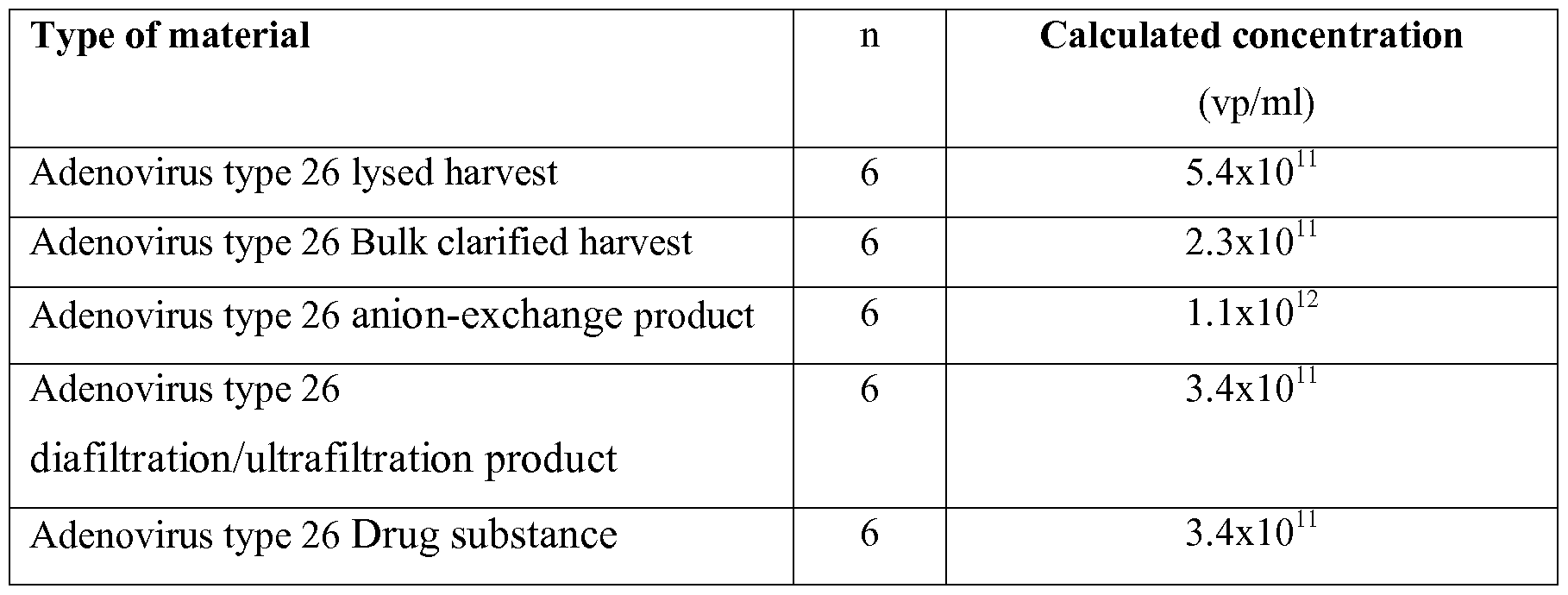
- Table 2 Overview of calculated concentrations (in vp/ml) for lysed harvest, clarified harvest, anion-exchange product, diafiltration/ultrafiltration product, drug substance in comparison with the reference material
- the composition of the separation buffer solution according to the invention allows larger injection volumes. In this way, better sensitivity can be achieved.
- FIG 3 an example of Ad35 anion exchange product is shown at different injection volumes (pressure x time). It was shown that with a lOx longer injection there was still separation of the adenovirus peak. At this level of injection volume the precision of respectively the adenovirus peak migration time and the corrected adenovirus peak areas are 2% RSD and 3% RSD. No difference in accuracy and capillary lifetime were observed between different inj ection vo lumes .
Landscapes
- Life Sciences & Earth Sciences (AREA)
- Chemical & Material Sciences (AREA)
- Health & Medical Sciences (AREA)
- Molecular Biology (AREA)
- Immunology (AREA)
- Organic Chemistry (AREA)
- Physics & Mathematics (AREA)
- Analytical Chemistry (AREA)
- Biochemistry (AREA)
- General Health & Medical Sciences (AREA)
- Zoology (AREA)
- Proteomics, Peptides & Aminoacids (AREA)
- Wood Science & Technology (AREA)
- Engineering & Computer Science (AREA)
- Electrochemistry (AREA)
- General Physics & Mathematics (AREA)
- Chemical Kinetics & Catalysis (AREA)
- Pathology (AREA)
- Dispersion Chemistry (AREA)
- Microbiology (AREA)
- Biophysics (AREA)
- Biotechnology (AREA)
- Bioinformatics & Cheminformatics (AREA)
- General Engineering & Computer Science (AREA)
- Genetics & Genomics (AREA)
- Virology (AREA)
- Toxicology (AREA)
- Measuring Or Testing Involving Enzymes Or Micro-Organisms (AREA)
- Micro-Organisms Or Cultivation Processes Thereof (AREA)
- Apparatus Associated With Microorganisms And Enzymes (AREA)
Abstract
Description
Claims
Priority Applications (5)
| Application Number | Priority Date | Filing Date | Title |
|---|---|---|---|
| EP16705157.2A EP3259374B1 (en) | 2015-02-19 | 2016-02-18 | Method for quantification of virus particles using capillary zone electrophoresis. |
| US15/552,069 US10345259B2 (en) | 2015-02-19 | 2016-02-18 | Method for quantification of virus particles using capillary zone electrophoresis |
| AU2016221726A AU2016221726B2 (en) | 2015-02-19 | 2016-02-18 | Method for quantification of virus particles using capillary zone electrophoresis |
| CA2976826A CA2976826A1 (en) | 2015-02-19 | 2016-02-18 | Method for quantification of virus particles using capillary zone electrophoresis |
| JP2017543987A JP6677737B2 (en) | 2015-02-19 | 2016-02-18 | Quantitative method for virus particles using capillary zone electrophoresis |
Applications Claiming Priority (2)
| Application Number | Priority Date | Filing Date | Title |
|---|---|---|---|
| EP15155743 | 2015-02-19 | ||
| EP15155743.6 | 2015-02-19 |
Publications (1)
| Publication Number | Publication Date |
|---|---|
| WO2016131895A1 true WO2016131895A1 (en) | 2016-08-25 |
Family
ID=52686084
Family Applications (1)
| Application Number | Title | Priority Date | Filing Date |
|---|---|---|---|
| PCT/EP2016/053405 Ceased WO2016131895A1 (en) | 2015-02-19 | 2016-02-18 | Method for quantification of virus particles using capillary zone electrophoresis |
Country Status (6)
| Country | Link |
|---|---|
| US (1) | US10345259B2 (en) |
| EP (1) | EP3259374B1 (en) |
| JP (1) | JP6677737B2 (en) |
| AU (1) | AU2016221726B2 (en) |
| CA (1) | CA2976826A1 (en) |
| WO (1) | WO2016131895A1 (en) |
Cited By (1)
| Publication number | Priority date | Publication date | Assignee | Title |
|---|---|---|---|---|
| CZ308274B6 (en) * | 2018-12-07 | 2020-04-08 | Fakultní nemocnice u sv. Anny v Brně | An affinity carrier for the selective uptake and / or separation of bacteriophages and a method for the selective uptake and / or separation of bacteriophages by capillary electrophoresis |
Families Citing this family (1)
| Publication number | Priority date | Publication date | Assignee | Title |
|---|---|---|---|---|
| CN110596225B (en) * | 2019-10-21 | 2022-03-18 | 兰州大学 | Method for improving repeatability of capillary electrophoresis |
Citations (2)
| Publication number | Priority date | Publication date | Assignee | Title |
|---|---|---|---|---|
| US5723031A (en) | 1994-10-31 | 1998-03-03 | Bayer Aktiengesellschaft | Method for the analytical separation of viruses |
| US5994128A (en) | 1995-06-15 | 1999-11-30 | Introgene B.V. | Packaging systems for human recombinant adenovirus to be used in gene therapy |
Family Cites Families (6)
| Publication number | Priority date | Publication date | Assignee | Title |
|---|---|---|---|---|
| US4639421A (en) * | 1984-10-01 | 1987-01-27 | Becton, Dickinson And Company | Fluorescent gram stain |
| US5993626A (en) * | 1997-01-24 | 1999-11-30 | Landers; James P. | Capillary electrophoresis of transferrin glycoforms |
| US20110165578A1 (en) * | 2008-09-09 | 2011-07-07 | Daniela Verthelyi | Methods for detecting and quantifying oversulfated glycosaminoglycans |
| EP2426488B1 (en) * | 2009-04-28 | 2016-04-20 | Wako Pure Chemical Industries, Ltd. | Measurement method utilizing internal standard substance |
| WO2011124715A1 (en) * | 2010-04-09 | 2011-10-13 | Marcella Chiari | Silane copolymers and uses thereof |
| JP6211283B2 (en) * | 2013-03-29 | 2017-10-11 | ヒューマン・メタボローム・テクノロジーズ株式会社 | How to detect encephalopathy |
-
2016
- 2016-02-18 WO PCT/EP2016/053405 patent/WO2016131895A1/en not_active Ceased
- 2016-02-18 EP EP16705157.2A patent/EP3259374B1/en active Active
- 2016-02-18 JP JP2017543987A patent/JP6677737B2/en active Active
- 2016-02-18 US US15/552,069 patent/US10345259B2/en active Active
- 2016-02-18 AU AU2016221726A patent/AU2016221726B2/en active Active
- 2016-02-18 CA CA2976826A patent/CA2976826A1/en active Pending
Patent Citations (2)
| Publication number | Priority date | Publication date | Assignee | Title |
|---|---|---|---|---|
| US5723031A (en) | 1994-10-31 | 1998-03-03 | Bayer Aktiengesellschaft | Method for the analytical separation of viruses |
| US5994128A (en) | 1995-06-15 | 1999-11-30 | Introgene B.V. | Packaging systems for human recombinant adenovirus to be used in gene therapy |
Non-Patent Citations (8)
| Title |
|---|
| AFNAN AZIZI ET AL: "Viral Quantitative Capillary Electrophoresis for Counting and Quality Control of RNA Viruses", ANALYTICAL CHEMISTRY, 9 October 2012 (2012-10-09), XP055205038, ISSN: 0003-2700, DOI: 10.1021/ac302525y * |
| AZIZI ET AL., ANALYTICAL CHEMISTRY, vol. 84, 2012, pages 9585 - 9591 |
| AZIZI, ANALYTICAL CHEMISTRY, vol. 84, 2012, pages 9585 - 9591 |
| GLEB G. MIRONOV ET AL: "Viral Quantitative Capillary Electrophoresis for Counting Intact Viruses", ANALYTICAL CHEMISTRY, vol. 83, no. 13, 21 March 2011 (2011-03-21), pages 5431 - 5435, XP055205032, ISSN: 0003-2700, DOI: 10.1021/ac201006u * |
| MANN ET AL., JOURNAL OF CHROMATOGRAPHY A, vol. 895, 2000, pages 329 - 337 |
| MATHIS ET AL., JOURNAL OF VIROLOGICAL METHODS, vol. 169, 2010, pages 13 - 21 |
| MATHIS P K ET AL: "Separation of rotavirus double-layered particles and triple-layered particles by capillary zone electrophoresis", JOURNAL OF VIROLOGICAL METHODS, ELSEVIER BV, NL, vol. 169, no. 1, 1 October 2010 (2010-10-01), pages 13 - 21, XP027507644, ISSN: 0166-0934, [retrieved on 20100625], DOI: 10.1016/J.JVIROMET.2010.06.006 * |
| MIRONOV ET AL., ANALYTICAL CHEMISTRY, vol. 83, 2011, pages 5431 - 5435 |
Cited By (1)
| Publication number | Priority date | Publication date | Assignee | Title |
|---|---|---|---|---|
| CZ308274B6 (en) * | 2018-12-07 | 2020-04-08 | Fakultní nemocnice u sv. Anny v Brně | An affinity carrier for the selective uptake and / or separation of bacteriophages and a method for the selective uptake and / or separation of bacteriophages by capillary electrophoresis |
Also Published As
| Publication number | Publication date |
|---|---|
| AU2016221726B2 (en) | 2022-03-31 |
| EP3259374B1 (en) | 2019-08-21 |
| CA2976826A1 (en) | 2016-08-25 |
| JP6677737B2 (en) | 2020-04-08 |
| JP2018505689A (en) | 2018-03-01 |
| US20180011056A1 (en) | 2018-01-11 |
| EP3259374A1 (en) | 2017-12-27 |
| US10345259B2 (en) | 2019-07-09 |
| AU2016221726A1 (en) | 2017-08-31 |
Similar Documents
| Publication | Publication Date | Title |
|---|---|---|
| Wang et al. | Developing an anion exchange chromatography assay for determining empty and full capsid contents in AAV6. 2 | |
| Farmerie et al. | Recent advances in isoelectric focusing of proteins and peptides | |
| Jacobs et al. | Electroelution of fixed and stained membrane proteins from preparative sodium dodecyl sulfate-polyacrylamide gels into a membrane trap | |
| Jing et al. | Methods for measuring aptamer-protein equilibria: a review | |
| US6537793B2 (en) | Method of separating viral particles | |
| Schnabel et al. | Determination of the p I of human rhinovirus serotype 2 by capillary isoelectric focusing | |
| CN102016560A (en) | Analysis of DNA by Capillary Electrophoresis | |
| JPH08504277A (en) | Use of Capillary Electrophoresis to Quantify Concentrations of Protein Components and Total Protein in Liquids | |
| Geurink et al. | Sixteen capillary electrophoresis applications for viral vaccine analysis | |
| AU2016221726B2 (en) | Method for quantification of virus particles using capillary zone electrophoresis | |
| Mann et al. | Capillary zone electrophoresis of a recombinant adenovirus | |
| Zhu et al. | Alternate injections coupled with flow-gated capillary electrophoresis for rapid and accurate quantitative analysis of urine samples | |
| Wang et al. | A platform method for plasmid isoforms analysis by capillary gel electrophoresis | |
| Zhang et al. | Evaluation of the oxidative deoxyribonucleic acid damage biomarker 8-hydroxy-2′-deoxyguanosine in the urine of leukemic children by micellar electrokinetic capillary chromatography | |
| Oita et al. | Poliovirus separation from cell extracts using capillary electrophoresis: Potential use in vaccine production and control? | |
| Haraga et al. | Purification of anionic fluorescent probes through precise fraction collection with a two‐point detection system using multiple‐stacking preparative capillary transient isotachophoresis | |
| JP2023510841A (en) | Capillary electrophoresis for viral vector isolation, analysis, characterization and quantification | |
| CN114460219A (en) | Method for isolating viral vectors | |
| Kolivoška et al. | Electrophoresis on a microfluidic chip for analysis of fluorescence‐labeled human rhinovirus | |
| Zhao et al. | Determination of rutin, chlorogenic acid and quercetin in solidaginis by large volume sample stacking with polarity switching and acid barrage stacking | |
| Xu et al. | Development of fully automated quantitative capillary electrophoresis with high accuracy and repeatability | |
| CN115308351A (en) | Liquid chromatography analysis method for AAV (adeno-associated Virus) vacant shell rate | |
| Shih et al. | Validation of a quantitative method for detection of adenovirus aggregation | |
| ROZING¹ et al. | Instrumentation for capillary electrochromatography | |
| Chen et al. | Determination of sodium acetate in antisense oligonucleotides by capillary zone electrophoresis |
Legal Events
| Date | Code | Title | Description |
|---|---|---|---|
| 121 | Ep: the epo has been informed by wipo that ep was designated in this application |
Ref document number: 16705157 Country of ref document: EP Kind code of ref document: A1 |
|
| ENP | Entry into the national phase |
Ref document number: 2976826 Country of ref document: CA |
|
| REEP | Request for entry into the european phase |
Ref document number: 2016705157 Country of ref document: EP |
|
| ENP | Entry into the national phase |
Ref document number: 2017543987 Country of ref document: JP Kind code of ref document: A |
|
| WWE | Wipo information: entry into national phase |
Ref document number: 15552069 Country of ref document: US |
|
| NENP | Non-entry into the national phase |
Ref country code: DE |
|
| ENP | Entry into the national phase |
Ref document number: 2016221726 Country of ref document: AU Date of ref document: 20160218 Kind code of ref document: A |