WO2015183902A1 - Ox40l fusion proteins and uses thereof - Google Patents
Ox40l fusion proteins and uses thereof Download PDFInfo
- Publication number
- WO2015183902A1 WO2015183902A1 PCT/US2015/032598 US2015032598W WO2015183902A1 WO 2015183902 A1 WO2015183902 A1 WO 2015183902A1 US 2015032598 W US2015032598 W US 2015032598W WO 2015183902 A1 WO2015183902 A1 WO 2015183902A1
- Authority
- WO
- WIPO (PCT)
- Prior art keywords
- cells
- hexameric protein
- cell
- human
- protein
- Prior art date
- Legal status (The legal status is an assumption and is not a legal conclusion. Google has not performed a legal analysis and makes no representation as to the accuracy of the status listed.)
- Ceased
Links
- 0 CCC(C(*)****C)N=O Chemical compound CCC(C(*)****C)N=O 0.000 description 3
- KDIAMAVWIJYWHN-UHFFFAOYSA-N CCCC1CCCC1 Chemical compound CCCC1CCCC1 KDIAMAVWIJYWHN-UHFFFAOYSA-N 0.000 description 1
Classifications
-
- C—CHEMISTRY; METALLURGY
- C07—ORGANIC CHEMISTRY
- C07K—PEPTIDES
- C07K14/00—Peptides having more than 20 amino acids; Gastrins; Somatostatins; Melanotropins; Derivatives thereof
- C07K14/435—Peptides having more than 20 amino acids; Gastrins; Somatostatins; Melanotropins; Derivatives thereof from animals; from humans
- C07K14/705—Receptors; Cell surface antigens; Cell surface determinants
- C07K14/70575—NGF/TNF-superfamily, e.g. CD70, CD95L, CD153, CD154
-
- C—CHEMISTRY; METALLURGY
- C07—ORGANIC CHEMISTRY
- C07K—PEPTIDES
- C07K14/00—Peptides having more than 20 amino acids; Gastrins; Somatostatins; Melanotropins; Derivatives thereof
- C07K14/435—Peptides having more than 20 amino acids; Gastrins; Somatostatins; Melanotropins; Derivatives thereof from animals; from humans
- C07K14/52—Cytokines; Lymphokines; Interferons
-
- A—HUMAN NECESSITIES
- A61—MEDICAL OR VETERINARY SCIENCE; HYGIENE
- A61K—PREPARATIONS FOR MEDICAL, DENTAL OR TOILETRY PURPOSES
- A61K38/00—Medicinal preparations containing peptides
- A61K38/16—Peptides having more than 20 amino acids; Gastrins; Somatostatins; Melanotropins; Derivatives thereof
- A61K38/17—Peptides having more than 20 amino acids; Gastrins; Somatostatins; Melanotropins; Derivatives thereof from animals; from humans
- A61K38/19—Cytokines; Lymphokines; Interferons
- A61K38/191—Tumor necrosis factors [TNF], e.g. lymphotoxin [LT], i.e. TNF-beta
-
- A—HUMAN NECESSITIES
- A61—MEDICAL OR VETERINARY SCIENCE; HYGIENE
- A61K—PREPARATIONS FOR MEDICAL, DENTAL OR TOILETRY PURPOSES
- A61K39/00—Medicinal preparations containing antigens or antibodies
- A61K39/39—Medicinal preparations containing antigens or antibodies characterised by the immunostimulating additives, e.g. chemical adjuvants
-
- A—HUMAN NECESSITIES
- A61—MEDICAL OR VETERINARY SCIENCE; HYGIENE
- A61P—SPECIFIC THERAPEUTIC ACTIVITY OF CHEMICAL COMPOUNDS OR MEDICINAL PREPARATIONS
- A61P35/00—Antineoplastic agents
-
- A—HUMAN NECESSITIES
- A61—MEDICAL OR VETERINARY SCIENCE; HYGIENE
- A61P—SPECIFIC THERAPEUTIC ACTIVITY OF CHEMICAL COMPOUNDS OR MEDICINAL PREPARATIONS
- A61P35/00—Antineoplastic agents
- A61P35/02—Antineoplastic agents specific for leukemia
-
- A—HUMAN NECESSITIES
- A61—MEDICAL OR VETERINARY SCIENCE; HYGIENE
- A61P—SPECIFIC THERAPEUTIC ACTIVITY OF CHEMICAL COMPOUNDS OR MEDICINAL PREPARATIONS
- A61P37/00—Drugs for immunological or allergic disorders
- A61P37/02—Immunomodulators
- A61P37/04—Immunostimulants
-
- C—CHEMISTRY; METALLURGY
- C07—ORGANIC CHEMISTRY
- C07K—PEPTIDES
- C07K19/00—Hybrid peptides, i.e. peptides covalently bound to nucleic acids, or non-covalently bound protein-protein complexes
-
- C—CHEMISTRY; METALLURGY
- C12—BIOCHEMISTRY; BEER; SPIRITS; WINE; VINEGAR; MICROBIOLOGY; ENZYMOLOGY; MUTATION OR GENETIC ENGINEERING
- C12N—MICROORGANISMS OR ENZYMES; COMPOSITIONS THEREOF; PROPAGATING, PRESERVING, OR MAINTAINING MICROORGANISMS; MUTATION OR GENETIC ENGINEERING; CULTURE MEDIA
- C12N15/00—Mutation or genetic engineering; DNA or RNA concerning genetic engineering, vectors, e.g. plasmids, or their isolation, preparation or purification; Use of hosts therefor
- C12N15/09—Recombinant DNA-technology
- C12N15/11—DNA or RNA fragments; Modified forms thereof; Non-coding nucleic acids having a biological activity
- C12N15/62—DNA sequences coding for fusion proteins
-
- A—HUMAN NECESSITIES
- A61—MEDICAL OR VETERINARY SCIENCE; HYGIENE
- A61K—PREPARATIONS FOR MEDICAL, DENTAL OR TOILETRY PURPOSES
- A61K39/00—Medicinal preparations containing antigens or antibodies
- A61K2039/555—Medicinal preparations containing antigens or antibodies characterised by a specific combination antigen/adjuvant
- A61K2039/55511—Organic adjuvants
- A61K2039/55516—Proteins; Peptides
-
- A—HUMAN NECESSITIES
- A61—MEDICAL OR VETERINARY SCIENCE; HYGIENE
- A61K—PREPARATIONS FOR MEDICAL, DENTAL OR TOILETRY PURPOSES
- A61K39/00—Medicinal preparations containing antigens or antibodies
- A61K2039/60—Medicinal preparations containing antigens or antibodies characteristics by the carrier linked to the antigen
- A61K2039/6031—Proteins
- A61K2039/6056—Antibodies
-
- C—CHEMISTRY; METALLURGY
- C07—ORGANIC CHEMISTRY
- C07K—PEPTIDES
- C07K2319/00—Fusion polypeptide
- C07K2319/30—Non-immunoglobulin-derived peptide or protein having an immunoglobulin constant or Fc region, or a fragment thereof, attached thereto
-
- C—CHEMISTRY; METALLURGY
- C07—ORGANIC CHEMISTRY
- C07K—PEPTIDES
- C07K2319/00—Fusion polypeptide
- C07K2319/70—Fusion polypeptide containing domain for protein-protein interaction
- C07K2319/73—Fusion polypeptide containing domain for protein-protein interaction containing coiled-coiled motif (leucine zippers)
Definitions
- OX40 (CD 134; TNFRSF4) is a tumor necrosis factor receptor found primarily on activated CD4 + and CD8 + T-cells, regulatory T cells (Treg) and natural killer (NK) cells (Croft et al., 2009, Immunol Rev. 229: 173-91).
- OX40 has one known endogenous ligand, OX40 ligand (OX40L; CD152; TNFSF4), that exists in a trimeric form and can cluster OX40 resulting in potent cell signaling events within T cells (Croft et al., 2009, Immunol Rev. 229: 173-91).
- OX40 signaling through OX40 on activated CD4 + and CD8 + T cells leads to enhanced cytokine production, granzyme and perforin release and expansion of effector and memory T cell pools (Jensen et al., 2010, Semin Oncol. 37:524-32).
- OX40 signaling on Treg cells inhibits expansion of Tregs, shuts down the induction of Tregs and blocks Treg-suppressive function (Voo et al., 2013, J Immunol. 191:3641-50; Vu et al, 2007, Blood. 110:2501-10).
- Treg cell inhibition and co-stimulation of effector T cells were shown to be necessary for tumor growth inhibition of OX40 agonists (Piconese et al., 2008, J Exp Med. 205:825-39). Many strategies and technologies have been explored to enhance the anti-tumor effect of OX40 agonist therapy through combinations with vaccines, chemotherapy, radiotherapy, and immunotherapy (Jensen et al., 2010, Semin Oncol. 37:524-32; Melero et al., 2013, Clin Cancer Res. 19:997-1008).
- polypeptide subunits each including, as a fusion polypeptide, the receptor-binding domain of OX40 Ligand (OX40L), a trimerization domain and a human IgG4 Fc domain, which are capable of forming stable multimeric, e.g., hexameric proteins.
- OX40L OX40 Ligand
- trimerization domain a trimerization domain
- human IgG4 Fc domain a human IgG4 Fc domain
- the disclosure provides a single-chain polypeptide subunit that includes: a human IgG4 Fc domain; a functional trimerization domain; and a receptor binding domain of OX40L.
- a polypeptide subunit as provided can self-assemble into a trimeric or a hexameric protein.
- the polypeptide subunit includes, from the amino terminus to the carboxy terminus, the human IgG4 Fc domain, followed by the trimerization domain, followed by the OX40L receptor binding domain.
- the IgG4 Fc domain includes an IgG4 hinge region, which can include a mutation that confers complete inter heavy chain disulfide bond formation, e.g., a serine to proline mutation at position 228 (S228P) according to EU numbering.
- the hinge region includes amino acids 1 to 12 of SEQ ID NO: 4.
- the human IgG4 Fc domain can further include a CH2 region, and/or a CH3 region.
- the human IgG4 Fc domain includes amino acids 1 to 229 of SEQ ID NO: 4.
- the trimerization domain provided by the disclosure can include an alpha-helical coiled coil domain, a leucine zipper domain, or a combination thereof.
- the trimerization domain can be derived from TRAF2; Thrombospondin 1; Matrilin-4; CMP (matrilin-1); HSF1; or Cubilin.
- the trimerization domain includes a human TRAF2 trimerization domain.
- the TRAF2 trimerization domain includes amino acids 310 to 349 of human TRAF2 (SEQ ID NO: 2).
- the OX40L receptor binding domain can include amino acids 51 to 183 of human OX40L (SEQ ID NO: 1).
- the polypeptide subunit provided by the disclosure can self-assemble into a hexameric fusion protein that can specifically bind to human OX40.
- the polypeptide subunit includes the amino acid sequence SEQ ID NO: 4.
- a polypeptide subunit as provided herein can further include an associated heterologous agent.
- a heterologous agent as provided herein can be a heterologous polypeptide that is fused to the polypeptide subunit via a peptide bond.
- the heterologous polypeptide can be fused, for example, to the N-terminus of the IgG4-Fc domain, to the C-terminus of the receptor binding domain of OX40L, between the C-terminus of the IgG4-Fc domain and to the N-terminus of the trimerization domain, or between the C-terminus of the trimerization domain and to the N-terminus of the receptor binding domain of OX40L.
- the heterologous agent can be chemically conjugated to the polypeptide subunit and can be, for example a cytotoxic molecule, a stabilizing agent, an immune response modifier, or a detectable agent.
- the disclosure provides polypeptide subunits that can be used as controls.
- the disclosure provides a polypeptide subunit where the OX40L receptor binding domain includes amino acids 51 to 183 of human OX40L (SEQ ID NO: 1), except for a single phenylalanine to alanine substitution at position 180 (F180A).
- a hexameric fusion protein that self- assembles from this polypeptide subunit is incapable of binding to human OX40.
- this control polypeptide subunit includes the amino acid sequence SEQ ID NO: 6.
- the disclosure provides a trimeric protein that includes three polypeptide subunits as provided herein.
- the disclosure provides a hexameric protein that includes six polypeptide subunits as described above.
- the six polypeptide subunits each include the amino acid sequence SEQ ID NO: 4.
- An exemplary hexameric fusion protein is OX40L IgG4P Fusion Protein.
- a hexameric protein as provided by the disclosure can specifically bind to OX40 as expressed on activated CD4 + or CD8 + T cells from human, cynomolgus monkey, and/or rhesus monkey.
- the hexameric protein can specifically bind to OX40 as expressed on primary activated CD4 + T cells from human, cynomolgus monkey, and/or rhesus monkey.
- the binding affinity for human OX40 expressed on primary activated human CD4 + T cells is about 1.0 pM to about 8.0 pM, e.g., about 3.6 pM as measured by flow cytometry.
- the hexameric protein can achieve 20% receptor occupancy on primary activated human CD4 + T cells (EC 2 o) at about 0.5 to about 3.0 pM, e.g., about 1.8 pM, 50% receptor occupancy on primary activated human CD4 + T cells (EC 50 ) at about 3.0 to about 10 pM, e.g., about 6.6 pM , and 90% receptor occupancy on primary activated human CD4 + T cells (EC 90 ) at about 35 to about 70 pM, e.g., about 53 pM, all as measured by flow cytometry.
- a hexameric protein as provided by the disclosure has a binding affinity for human OX40 expressed on OX40-overexpressing Jurkat cells of about 1.0 pM to about 24 pM, e.g., about 12 pM, as measured by flow cytometry.
- the hexameric protein can achieve EC 20 on OX40-overexpressing Jurkat cells at about 1.0 to about 15 pM, e.g., about 7.2 pM, EC 50 on OX40-overexpressing Jurkat cells at about 1.0 to about 40 pM, e.g., about 16 pM, and EC 90 on OX40-overexpressing Jurkat cells at about 10 to about 100 pM, e.g., about 57 pM, all as measured by flow cytometry.
- a hexameric protein as provided by the disclosure has a binding affinity for cynomolgus monkey OX40 expressed on primary activated cynomolgus monkey CD4 + T cells is about 1.0 pM to about 50 pM, e.g., about 24 pM, as measured by flow cytometry.
- a hexameric protein as provided by the disclosure has a binding affinity for rhesus monkey OX40 expressed on primary activated rhesus monkey CD4 + T cells is about 1.0 pM to about 50 pM, e.g., about 21 pM, as measured by flow cytometry.
- a hexameric protein as provided by the disclosure can induce dose- dependent proliferation of activated CD4 + T cells and dose-dependent cytokine release from activated CD4 + T cells in a plate-based assay.
- a 20% maximal proliferation response (EC 2 o) can be achieved in primary activated human CD4 + T cells at a hexameric protein concentration of about 1.0 pM to about 15 pM, e.g., about 8.0 pM
- a 50% maximal proliferation response (EC 50 ) can be achieved in primary activated human CD4 + T cells at a hexameric protein concentration of about 5.0 pM to about 25 pM, e.g., about 14 pM
- a 90% maximal proliferation response (EC 90 ) can be achieved in primary activated human CD4 + T cells at a hexameric protein concentration of about 50 pM to about 65 pM, e.g., about 57 pM, all as measured by flow cytometry
- the hexameric protein can induce dose-dependent release of IFNy, TNFa, IL-5, IL- 10, IL-2, IL-4, IL-13, IL-8, IL-12 p70, and/or IL- ⁇ .
- the cytokines released include IFNy, TNFa, IL-5, and/or IL-10.
- a hexameric protein as provided by the disclosure can achieve CD4 + T cell proliferation and cytokine release in primary activated cynomolgus monkey CD4 + T cells and in primary activated rhesus monkey CD4 + T cells.
- a hexameric protein as provided by the disclosure can activate the NFKB pathway in OX40 expressing T cells in the presence of FcyR-expressing cells.
- the OX40-expressing T cells can be OX40-expressing Jurkat NFKB-lucif erase reporter cells that produce luciferase in response to stimulation of the NFKB signaling pathway.
- the OX40 expressed by these cells can be, e.g., human OX40, cynomolgus monkey OX40 or rhesus monkey OX40.
- a hexameric protein as provided by the disclosure when administered as an effective dose to a subject in need of cancer treatment, can inhibit tumor growth in the subject, for example, in the presence of T-cells.
- tumor growth can be inhibited by at least 10%, at least 20%, at least 30%, at least 40%, and least 50%, at least 60%, or at least 70% compared to administration of an isotype-matched control.
- a hexameric protein as provided by the disclosure can induce proliferation of activated, OX40-expressing CD4 + T cells through binding to OX40, but does not substantially trigger complement-dependent or antibody-dependent cytotoxicity against the activated CD4 + T cells.
- a hexameric protein as provided herein does not bind to Clq or trigger NK cell-mediated antibody-dependent cellular cytotoxicity of activated CD4 + T cells.
- a hexameric protein as provided by the disclosure upon co-administration with a vaccine antigen to a subject, can increase CD4+ central memory (cM) T-cell proliferation, effector memory (eM) CD8+ T cell proliferation, NK cell proliferation, and/or B cell proliferation.
- CD8+ eM T-cell proliferation, NK cell proliferation, B cell proliferation, or any combination thereof is measured as intracellular Ki67 expression in peripheral blood mononuclear cells.
- a hexameric protein as provided by the disclosure upon co-administration with a vaccine antigen, including, but not limited to an antigen from respiratory syncytial virus (RSV), hepatitis B or C virus, Ebola virus, influenza virus, Pseudomonas aeruginosa, Clostridium difficile, or Staphylococcus aureus, to a subject, can increase antigen- specific T-cell response that can be measured, e.g., as increased interferon gamma (IFN-gamma) expression.
- RSV respiratory syncytial virus
- hepatitis B or C virus hepatitis B or C virus
- Ebola virus influenza virus
- Pseudomonas aeruginosa Clostridium difficile
- Staphylococcus aureus Staphylococcus aureus
- a hexameric protein as provided by the disclosure upon co-administration with a vaccine antigen to a subject, can increase antigen- specific IgG titers.
- the antigen can be a soluble F glycoprotein of respiratory syncytial virus (RSV sF), and coadministration of the hexameric protein and the RSV antigen to a subject can increase titers of RSV- neutralizing antibodies.
- RSV sF respiratory syncytial virus
- the subject to which the hexameric protein and vaccine antigen is coadministered is a non-human primate.
- a hexameric protein as provided by the disclosure can inhibit regulatory T cell (Treg) mediated suppression of effector T cell proliferation, e.g., in a plate-based assay.
- the effector T cells are effector CD4+ T cells.
- the ratio of effector cells to Treg cells can be about 1:0.5 to about 1:3, e.g., about 1: 1 or about 1:2.
- composition that includes a hexameric protein as provided herein, and a carrier.
- the disclosure provides a polynucleotide that includes a nucleic acid encoding a polypeptide subunit as provided herein, or a hexameric protein as provided herein.
- the polynucleotide can include the nucleic acid sequence SEQ ID NO: 3.
- the disclosure further provides a vector that incorporates the polynucleotide as provided and a host cell that incorporates the polynucleotide or the vector as provided.
- the disclosure provides a method of producing a polypeptide subunit of as provided herein or a hexameric protein as provided herein, where the method includes culturing the provided host cell under conditions in which the polypeptide subunit or hexameric protein encoded by the polynucleotide is expressed, and recovering the polypeptide subunit or hexameric protein.
- the disclosure provides a method to promote survival or proliferation of activated T cells, where the method includes contacting activated T cells with a hexameric protein as provided herein such that the hexameric protein can specifically bind to OX40 on the surface of the T cells.
- the disclosure further provides a method of inducing cytokine release from activated T cells, where the method includes contacting activated T cells with a hexameric protein as provided herein such that the hexameric protein can specifically bind to OX40 on the surface of the T cells.
- the cytokine can be IFNy, TNFa, IL-5, IL- 10, IL-2, IL-4, IL- 13, IL-8, IL- 12 p70, and/or IL- 1 ⁇ , e.g., IFNy, TNFa, IL-5, and/or IL- 10.
- the activated T cells are activated CD4 + T cells and/or activated CD8 + T cells.
- the activated CD4 + T cells can be human CD4 + T cells, cynomolgus monkey CD4 + T cells, and/or rhesus monkey CD4 + T cells.
- the disclosure provides a method to reduce regulatory T cell (Treg)- mediated suppression of activated T cell proliferation, where the method includes contacting a mixture of activated T cells and Treg cells with the hexameric protein of any one of claims 26 to 70, wherein the hexameric protein can specifically bind to OX40 on the surface of the T cells.
- Treg regulatory T cell
- the disclosure provides a method of promoting T cell activation, where the method includes contacting T cells with a hexameric protein as provided herein such that the hexameric protein can specifically bind to OX40 on the surface of the T cells.
- this method further includes cross-linking of the hexameric protein through interaction of the IgG4-Fc domain with a cell expressing FcyR.
- the cell expressing FcyR can be, e.g., a B cell, a monocyte, a macrophage, a myeloid or plasmacytoid dendritic cell, a follicular dendritic cell, a Langerhans cell, an endothelial cell, an NK cell, an activated T cell, a neutrophil, an eosinophil, a platelet, a mast cell, a CD45 + cell from a primary human tumor or tumor-draining or non-draining lymph node, and/or a CD45+ cell from other secondary or tertiary lymphoid structures.
- a B cell e.g., a B cell, a monocyte, a macrophage, a myeloid or plasmacytoid dendritic cell, a follicular dendritic cell, a Langerhans cell, an endothelial cell, an NK cell, an activated T cell, a neutrophil, an e
- T cell activation can be measured through stimulation of the NFKB signal transduction pathway, and in certain aspects the activated T cells are activated CD4 + T cells and/or activated CD8 + T cells, for example, human CD4 + T cells, cynomolgus monkey CD4 + T cells, and/or rhesus monkey CD4 + T cells.
- the methods provided above include administering an effective amount of the hexameric protein or composition as provided by the disclosure to a subject in need of treatment.
- the disclosure provides a method of treating cancer in a subject, where the method includes administering to a subject in need of treatment an effective amount of a hexameric protein as provided herein.
- the cancer is a solid tumor.
- administration of the hexameric protein or composition can inhibit tumor growth, can promote tumor reduction, or both. In certain aspects tumor growth inhibition is achieved in the presence of T-cells.
- the disclosure provides a method of enhancing an immune response in a subject, where the method includes administering to a subject in need thereof a therapeutically effective amount of a hexameric protein as provided by the disclosure, or a composition as provided by the disclosure.
- the subject can be a human subject.
- the disclosure provides a method of inducing expression of ICOS on T cells, comprising contacting T cells with a hexameric protein as provided by the disclosure, wherein the hexameric protein can specifically bind to OX40 on the surface of the T cells.
- the disclosure provides a method of inducing expression of PD-1 on T cells, comprising contacting T cells with the hexameric protein as provided by the disclsoure, wherein the hexameric protein can specifically bind to OX40 on the surface of the T cells.
- Figure 1 Schematically illustrates an exemplary multimeric protein, namely an OX40L fusion protein.
- FIG. 1 Amino Acid Sequence of OX40L IgG4P Fusion Protein (N to C terminus, SEQ ID NO: 4). Amino Acid Sequence huIgG4PFcTF2OX40L F180A is disclosed as SEQ ID NO: 12.
- FIG. 3A OX40L IgG4P Fusion Protein and control protein binding to OX40 expressed on the surface of primary activated human CD4 + T cells. Data was used for determination of apparent affinity (K d ) and apparent concentrations to achieve receptor occupancy of 20%, 50% and 90% (EC 2 o, EC50, and EC90 values).
- FIG. 3B OX40L IgG4P Fusion Protein and control protein binding to human OX40 expressed on Jurkat T cells. Data was used for determination of apparent affinity (K d ) and apparent concentrations to achieve receptor occupancy of 20%, 50% and 90% (EC 20 , EC 50 , and EC 90 values).
- FIG. 4A OX40L IgG4P Fusion Protein lacks binding to OX40 expressed on primary activated mouse CD4 + T cells.
- MFI mean fluorescence intensity.
- FIG. 4B OX40L IgG4P Fusion Protein lacks binding to OX40 expressed on primary activated rat CD4 + T cells.
- MFI mean fluorescence intensity.
- FIG. 5A-C Binding of OX40L IgG4P Fusion Protein to TNFRSF-expressing HEK293 and OX40-expressing Jurkat.
- A-B Transient expression of TNFRSF members, as indicated to left of histograms, in HEK293 cells and binding to TNFRSF- specific mAbs or to OX40L IgG4P Fusion Protein, as indicated above histograms.
- Gray histogram fluorochrome-conjugated isotype control antibody binding for TNFRSF- specific mAb or goat anti-human AlexaFluor ® 647 secondary antibody binding control for OX40L IgG4P Fusion Protein; Open histogram, TNFRSF- specific mAb or OX40L IgG4P Fusion Protein binding.
- C Binding of OX40L IgG4P Fusion Protein to OX40- expressing Jurkat as a positive control. Gray histogram, goat anti-human AlexaFluor ® 647 secondary antibody binding control; Open histogram, OX40L IgG4P Fusion Protein binding.
- FIG. 6 Schematic diagram of the OX40L IgG4P Fusion Protein plate-bound bioactivity assay.
- Q OX40L IgG4P Fusion Protein.
- ⁇ anti-CD3 clone OKT3.
- Figure 7A-E OX40L IgG4P Fusion Protein co- stimulates primary CD4 T cell to proliferate and release cytokines in a plate-based bioactivity assay.
- FIG. 8 Cell systems used for measuring OX40L IgG4P Fusion Protein bioactivity.
- OX40L IgG4P Fusion Protein cross linking by FcyR-expressing cells mediates the clustering of OX40 on the OX40-expressing Jurkat NFKB-lucif erase clone 64 reporter cell line, resulting in NFKB-dependent production of luciferase that can be measured as a surrogate for NFKB activity and T cell activation.
- FIG. 9A-C Bioactivity of OX40L IgG4P Fusion Protein when cross-linked or not by FcyR.
- A Reporter activity after cross-linking of OX40L IgG4P Fusion Protein by CD32A- expressing HEK293.
- B Reporter activity in the presence of HEK293 parental cells without FcyR expression.
- C Reporter activity with drug and without HEK293 cells. RLU, relative light units.
- Figure 10 Examples of OX40L IgG4P Fusion Protein bioactivity using Raji, CD32B- expressing HEK293, CD32A-expressing HEK293, or CD45+ cells isolated from primary human tumors for FcyR mediated drug cross-linking and the OX40-expressing Jurkat NFkB-luciferase clone 64 reporter cells for bioactivity readout.
- A OX40L IgG4P Fusion Protein activity clustered by Raji B cells;
- B CD32B-expressing HEK293;
- C CD32A-expressing HEK293;
- D CD45+ cells isolated from primary human tumors.
- RLU Relative Light Units.
- M Molarity.
- FIG. 14A-D Assessment of natural killer cell-mediated antibody-dependent cellular cytotoxicity of OX40L IgG4P Fusion Protein, OX40L IgGl Fusion Protein and anti-CD20 antibodies, Experiment #1. Specific killing of OX40-expressing CD4 + T cells by primary activated NK cells mediated by the OX40L fusion protein IgGl, but not by OX40L IgG4P Fusion Protein nor by F180A OX40L fusion protein IgGl and IgG4P controls: (A) Donor 1 NK cells incubated with activated CD4 + T cells; (B) Donor 1 NK cells incubated with Toledo B cells; (C) Donor 2 NK cells incubated with activated CD4 + T cells.
- NK cells used in the ADCC assays were capable of mediating efficient lysis of the Toledo B cells bound to rituximab: (D) Donor 2 NK cell combined with Toledo B cells. Experimental replicates were conducted in triplicate. Error bars represent standard deviation of the mean.
- Figure 15 Assessment of natural killer cell-mediated antibody-dependent cellular cytotoxicity of OX40L IgG4P Fusion Protein and the OX40 ligand fusion protein IgGl, Experiment #2. Specific killing of OX40-expressing CD4 + T cells by primary activated NK cells mediated by the OX40L fusion protein IgGl, but not by OX40L IgG4P Fusion Protein nor by F180A OX40L fusion protein IgGl and IgG4P controls. Experimental replicates were conducted in triplicate. Error bars represent standard deviation of the mean.
- FIG. 16A-B Assessment of OX40L IgG4P Fusion Protein binding to purified Clq protein.
- A The indicated concentrations of purified human Clq protein were injected onto the biosensor chip. Blank represents injections of PBS/0.005% Tween 20 vehicle alone,
- B In the same experiment, control human IgGl bound purified human Clq.
- FIG. 17 Effect of OX40L IgG4P Fusion Protein on Growth of A375 Cells in a Mouse Xenograft Model.
- Six NOD/SCID mice in each group were engrafted SC on Day 1 with A375 cells mixed with alloreactive human CD4 + and CD8 + T cell lines at E:T ratio 1:6.
- Test article (Isotype control or OX40L IgG4P Fusion Protein) was administered IP on Days 4, 7, 9 and 12. Mean values of tumor volumes are shown.
- a comparison between OX40L IgG4P Fusion Protein- treated and the Isotype control-treated animals was made, and intergroup differences were analyzed for statistical significance by a Mann- Whitney rank sum test. Error bars represent standard error of the mean.
- TGI tumor growth inhibition
- FIG. 18 Effect of OX40L IgG4P Fusion Protein on Growth of A375 Cells in a Mouse Xenograft Model.
- Six NOD/SCID mice in each group were engrafted SC on Day 1 with A375 cells alone (no T cells) or mixed with alloreactive human CD4 + and CD8 + T cell lines (ratio of 2: 1) at E:T ratio 1:6 Test article (Isotype control or OX40L IgG4P Fusion Protein) was administered IP on Days 3, 6, 8 and 10. Mean values of tumor volumes are shown.
- a comparison between OX40L IgG4P Fusion Protein-treated and the Isotype control-treated animals was made, and intergroup differences were analyzed for statistical significance by a Mann- Whitney rank sum test.
- FIG. 19 Effect of Murine OX40 Ligand Fusion Protein on Growth of Renca Cell Line in a Mouse Syngeneic Model.
- Ten BALB/c mice in each group were inoculated SC on Day 1 with Renca cells.
- Control article (isotype control) and test article (mOX40L FP) were administered IP on Days 4 and 7.
- Mean (panel A) and individual (pane B) values of tumor volumes are shown.
- a comparison between mOX40L FP-treated and the Isotype control-treated animals was made, and intergroup differences were analyzed for statistical significance by the method described in Section 6.8 using GraphPad Prism 6.0 software. Error bars represent standard error of the mean.
- SC subcutaneous
- IP intraperitoneal
- TGI tumor growth inhibition.
- FIG. 20 Effect of Murine OX40 Ligand Fusion Protein on Growth of CT26 Cell Line in a Mouse Syngeneic Model.
- Ten BALB/c mice in each group were inoculated SC on Day 1 with CT26 cells.
- Control article (isotype control) and test article (mOX40L FP) were administered IP on Days 4 and 6.
- Mean (panel A) and individual (panel B) values of tumor volumes are shown.
- a comparison between mOX40L FP-treated and the isotype control-treated animals was made, and intergroup differences were analyzed for statistical significance by the method described in Section 6.8 using GraphPad Prism 6.0 software. Error bars represent standard error of the mean.
- SC subcutaneous
- IP intraperitoneal
- TGI tumor growth inhibition.
- FIG. 22 Effect of Murine OX40 Ligand Fusion Protein on Growth of MCA205 Cell Line in a Mouse Syngeneic Model.
- a comparison between mOX40L FP-treated and the isotype control-treated animals was made, and intergroup differences were analyzed for statistical significance by the method described in Section 6.8 using GraphPad Prism 6.0 software. Error bars represent standard error of the mean.
- SC subcutaneous
- IP intraperitoneal
- TGI tumor growth inhibition.
- FIG. 23 OX40L IgG4P Fusion Protein Overcomes Regulatory T Cell Suppression of Effector CD4+ T cells.
- Effector CD4+ T cells were labelled with carboxyfluorescein diacetate succinimidyl ester (CFSE) and cultured, with and without regulatory T cells (Tregs) at the ratios indicated, for 4 days in the presence of anti-CD3, anti-CD28 and test or control articles.
- FIG. 24 OX40L IgG4P Fusion Protein Effects on Regulatory T Cell Suppression of Effector CD4+ T cells are Concentration Dependent. Effector CD4+ T cells were labelled with carboxyfluorescein diacetate succinimidyl ester (CFSE) and cultured, with and without regulatory T cells (Tregs) at a 1: 1 ratio, for 4 days in the presence of anti-CD3, anti-CD28 and test or control articles. The percentage of divided effector CD4+ T cells at the end of the assay was assessed by flow cytometry. Error bars represent the standard error of the mean from duplicate assay wells. Significance was calculated using the Student's T test where *p ⁇ 0.05; **p ⁇ 0.01.
- Figure 25 Study design for monitoring pharmacodynamics effects of OX40L fusion protein IgG4-FP administered via the intravenous route to Rhesus monkeys.
- FIG. 26 A-D Pharmacodynamics (as measured by Ki-67 expression) of total CD4 memory T-cells (A), naive CD4 T cells (B), total memory CD8 T-cells (C), and naive CD8 T-cells (D).
- Figure 27 A-D Pharmacodynamics (as measured by ICOS expression) of total CD4 memory T-cells (A), naive CD4 T cells (B), total memory CD 8 T-cells (C), and naive CD 8 T-cells (D).
- Figure 28 A-D Pharmacodynamics (as measured by PD-1 expression) of total CD4 memory T-cells (A), naive CD4 T cells (B), total memory CD 8 T-cells (C), and naive CD 8 T-cells (D).
- FIG. 29 Pharmacodynamics (as measured by Ki-67 expression) as proliferation of Lin- CD20+ B cells.
- Engagement of the OX40 receptor on T cells results in an increased response of the T cells, e.g., CD4 + T-cells to the antigen.
- engagement refers to binding to and stimulation of at least one activity mediated by the OX40 receptor.
- engagement of the OX40 receptor on antigen specific T cells e.g., CD4 + T-cells results in increased T cell proliferation as compared to the response to antigen alone, and increased cytokine production.
- the elevated response to the antigen can be maintained for a period of time substantially longer than in the absence of OX40 receptor engagement.
- OX40 agonists can enhance antigen specific immune responses in a subject, such as a human subject, when administered to the subject during or shortly after priming of T-cells by an antigen.
- OX40 agonists include OX40 ligand ("OX40L”), such as soluble OX40L fusion proteins and anti- OX40 antibodies or fragments thereof.
- a specific example is a fusion polypeptide subunit comprising the receptor binding domain of OX40L, a trimerization domain, e.g., an isoleucine zipper domain from TRAF-2, and a human IgG4 Fc domain, where the polypeptide subunit self-assembles into a multimeric (e.g., trimeric or hexameric) fusion protein.
- a multimeric e.g., trimeric or hexameric
- nucleic acids including polynucleotide sequences that encode such fusion polypeptides.
- This disclosure also provides methods for enhancing an antigen specific immune response in a subject using the multimeric OX40L fusion polypeptides.
- the compositions and methods disclosed herein with respect to OX40L fusion proteins can be more generally applied to the production and use of multimeric (e.g., trimeric and hexameric) receptor-binding fusion proteins.
- a or “an” entity refers to one or more of that entity; for example, “polypeptide subunit” is understood to represent one or more polypeptide subunits.
- polypeptide subunit is understood to represent one or more polypeptide subunits.
- the terms “a” (or “an”), “one or more,” and “at least one” can be used interchangeably herein.
- polypeptide is intended to encompass a singular “polypeptide” as well as plural “polypeptides,” and refers to a molecule composed of monomers (amino acids) linearly linked by amide bonds (also known as peptide bonds).
- polypeptide refers to any chain or chains of two or more amino acids, and does not refer to a specific length of the product.
- polypeptides peptides, dipeptides, tripeptides, oligopeptides, "protein,” “amino acid chain,” or any other term used to refer to a chain or chains of two or more amino acids are included within the definition of "polypeptide,” and the term “polypeptide” can be used instead of, or interchangeably with any of these terms.
- polypeptide is also intended to refer to the products of post-expression modifications of the polypeptide, including without limitation glycosylation, acetylation, phosphorylation, amidation, derivatization by known protecting/blocking groups, proteolytic cleavage, or modification by non-standard amino acids.
- a polypeptide can be derived from a natural biological source or produced by recombinant technology, but is not necessarily translated from a designated nucleic acid sequence. It can be generated in any manner, including by chemical synthesis.
- a "protein” as used herein can refer to a single polypeptide, i.e., a single amino acid chain as defined above, but can also refer to two or more polypeptides that are associated, e.g., by disulfide bonds, hydrogen bonds, or hydrophobic interactions, to produce a multimeric protein.
- polypeptide subunit refers to a polypeptide chain of amino acids which can interact with other polypeptide subunits, either identical or different, to form a multimeric protein, e.g., a hexameric protein as described herein.
- a polypeptide as disclosed herein can be of a size of about 3 or more, 5 or more, 10 or more, 20 or more, 25 or more, 50 or more, 75 or more, 100 or more, 200 or more, 500 or more, 1,000 or more, or 2,000 or more amino acids.
- Polypeptides can have a defined three-dimensional structure, although they do not necessarily have such structure. Polypeptides with a defined three-dimensional structure are referred to as folded, and polypeptides that do not possess a defined three-dimensional structure, but rather can adopt a large number of different conformations, and are referred to as unfolded.
- an "isolated" polypeptide or a fragment, variant, or derivative thereof is intended a polypeptide that is not in its natural milieu. No particular level of purification is required.
- an isolated polypeptide can be removed from its native or natural environment.
- Recombinantly produced polypeptides and proteins expressed in host cells are considered isolated as disclosed herein, as are native or recombinant polypeptides that have been separated, fractionated, or partially or substantially purified by any suitable technique.
- polypeptides disclosed herein are fragments, derivatives, analogs, or variants of the foregoing polypeptides, and any combination thereof.
- fragment can include any polypeptide or protein that retain at least some of the activities of the complete polypeptide or protein, but which is structurally different. Fragments of polypeptides include, for example, proteolytic fragments, as well as deletion fragments.
- variants include fragments as described above, and also polypeptides with altered amino acid sequences due to amino acid substitutions, deletions, or insertions. Variants can occur spontaneously or be intentionally constructed.
- variants can be produced using art-known mutagenesis techniques.
- Variant polypeptides can comprise conservative or non-conservative amino acid substitutions, deletions or additions.
- Derivatives are polypeptides that have been altered so as to exhibit additional features not found on the native polypeptide. Examples include fusion proteins.
- Variant polypeptides can also be referred to herein as "polypeptide analogs.”
- a "derivative” refers to a subject polypeptide having one or more amino acids chemically derivatized by reaction of a functional side group. Also included as “derivatives" are those peptides that contain one or more standard or synthetic amino acid derivatives of the twenty standard amino acids.
- 4-hydroxyproline can be substituted for proline; 5-hydroxylysine can be substituted for lysine; 3-methylhistidine can be substituted for histidine; homoserine can be substituted for serine; and ornithine can be substituted for lysine.
- a “conservative amino acid substitution” is one in which one amino acid is replaced with another amino acid having a similar side chain.
- Families of amino acids having similar side chains have been defined in the art, including basic side chains (e.g., lysine, arginine, histidine), acidic side chains (e.g., aspartic acid, glutamic acid), uncharged polar side chains (e.g.
- asparagine, glutamine, serine, threonine, tyrosine, cysteine nonpolar side chains (e.g., glycine, alanine, valine, leucine, isoleucine, proline, phenylalanine, methionine, tryptophan), beta-branched side chains (e.g. , threonine, valine, isoleucine) and aromatic side chains (e.g. , tyrosine, phenylalanine, tryptophan, histidine).
- nonpolar side chains e.g., glycine, alanine, valine, leucine, isoleucine, proline, phenylalanine, methionine, tryptophan
- beta-branched side chains e.g. , threonine, valine, isoleucine
- aromatic side chains e.g. , tyrosine, phenylalanine, tryptophan, his
- the term "antibody” refers to at least the minimal portion of an antibody which is capable of binding to antigen, e.g., at least the variable domain of a heavy chain (VH) and the variable domain of a light chain (VL) in the context of a typical antibody produced by a B cell.
- VH variable domain of a heavy chain
- VL variable domain of a light chain
- Basic antibody structures in vertebrate systems are relatively well understood. See, e.g., Harlow et al., Antibodies: A Laboratory Manual, (Cold Spring Harbor Laboratory Press, 2nd ed. 1988).
- Antibodies or antigen-binding fragments, variants, or derivatives thereof include, but are not limited to, polyclonal, monoclonal, human, humanized, or chimeric antibodies, single chain antibodies, epitope-binding fragments, e.g., Fab, Fab' and F(ab')2, Fd, Fvs, single-chain Fvs (scFv), single-chain antibodies, disulfide-linked Fvs (sdFv), fragments comprising either a VL or VH domain, fragments produced by a Fab expression library.
- ScFv molecules are known in the art and are described, e.g., in US patent 5,892,019.
- Immunoglobulin or antibody molecules encompassed by this disclosure can be of any type (e.g., IgG, IgE, IgM, IgD, IgA, and IgY), class (e.g., IgGl, IgG2, IgG3, IgG4, IgAl and IgA2) or subclass of immunoglobulin molecule.
- polynucleotide is intended to encompass a singular nucleic acid as well as plural nucleic acids, and refers to an isolated nucleic acid molecule or construct, e.g., messenger RNA (mRNA) or plasmid DNA (pDNA).
- mRNA messenger RNA
- pDNA plasmid DNA
- a polynucleotide can comprise a conventional phosphodiester bond or a non-conventional bond (e.g., an amide bond, such as found in peptide nucleic acids (PNA)).
- PNA peptide nucleic acids
- nucleic acid refers to any one or more nucleic acid segments, e.g., DNA or RNA fragments, present in a polynucleotide.
- isolated nucleic acid or polynucleotide is intended a nucleic acid molecule, DNA or RNA, which has been removed from its native environment.
- a recombinant polynucleotide encoding a polypeptide subunit contained in a vector is considered isolated as disclosed herein.
- Further examples of an isolated polynucleotide include recombinant polynucleotides maintained in heterologous host cells or purified (partially or substantially) polynucleotides in solution.
- Isolated RNA molecules include in vivo or in vitro RNA transcripts of polynucleotides. Isolated polynucleotides or nucleic acids further include such molecules produced synthetically.
- polynucleotide or a nucleic acid can be or can include a regulatory element such as a promoter, ribosome binding site, or a transcription terminator.
- a "coding region” is a portion of nucleic acid comprising codons translated into amino acids. Although a “stop codon” (TAG, TGA, or TAA) is not translated into an amino acid, it can be considered to be part of a coding region, but any flanking sequences, for example promoters, ribosome binding sites, transcriptional terminators, introns, and the like, are not part of a coding region. Two or more coding regions can be present in a single polynucleotide construct, e.g. , on a single vector, or in separate polynucleotide constructs, e.g. , on separate (different) vectors.
- any vector can contain a single coding region, or can comprise two or more coding regions, e.g. , a single vector can separately encode an immunoglobulin heavy chain variable region and an immunoglobulin light chain variable region.
- a vector, polynucleotide, or nucleic acid can encode heterologous coding regions, either fused or unfused to a nucleic acid encoding a polypeptide subunit or fusion protein as provided herein.
- Heterologous coding regions include without limitation specialized elements or motifs, such as a secretory signal peptide or a heterologous functional domain.
- the polynucleotide or nucleic acid is DNA.
- a polynucleotide comprising a nucleic acid that encodes a polypeptide normally can include a promoter and/or other transcription or translation control elements operably associated with one or more coding regions.
- An operable association or linkage is when a coding region for a gene product, e.g. , a polypeptide, is associated with one or more regulatory sequences in such a way as to place expression of the gene product under the influence or control of the regulatory sequence(s).
- Two DNA fragments are "operably associated” or “operably linked” if induction of promoter function results in the transcription of mRNA encoding the desired gene product and if the nature of the linkage between the two DNA fragments does not interfere with the ability of the expression regulatory sequences to direct the expression of the gene product or interfere with the ability of the DNA template to be transcribed.
- a promoter region would be operably associated with a nucleic acid encoding a polypeptide if the promoter was capable of effecting transcription of that nucleic acid.
- the promoter can be a cell-specific promoter that directs substantial transcription of the DNA only in predetermined cells.
- transcription control elements besides a promoter, for example enhancers, operators, repressors, and transcription termination signals, can be operably associated with the polynucleotide to direct cell-specific transcription.
- Suitable promoters and other transcription control regions are disclosed herein.
- transcription control regions are known to those skilled in the art. These include, without limitation, transcription control regions that function in vertebrate cells, such as, but not limited to, promoter and enhancer segments from cytomegaloviruses (the immediate early promoter, in conjunction with intron-A), simian virus 40 (the early promoter), and retroviruses (such as Rous sarcoma virus).
- Other transcription control regions include those derived from vertebrate genes such as actin, heat shock protein, bovine growth hormone and rabbit ⁇ -globin, as well as other sequences capable of controlling gene expression in eukaryotic cells. Additional suitable transcription control regions include tissue-specific promoters and enhancers as well as lymphokine-inducible promoters (e.g. , promoters inducible by interferons or interleukins).
- translation control elements include, but are not limited to ribosome binding sites, translation initiation and termination codons, and elements derived from picomaviruses (particularly an internal ribosome entry site, or IRES, also referred to as a CITE sequence).
- a polynucleotide can be RNA, for example, in the form of messenger RNA (mRNA).
- mRNA messenger RNA
- Polynucleotide and nucleic acid coding regions can be associated with additional coding regions that encode secretory or signal peptides, which direct the secretion of a polypeptide encoded by a polynucleotide as disclosed herein, e.g. , a polynucleotide encoding a polypeptide subunit provided herein.
- proteins secreted by mammalian cells have a signal peptide or secretory leader sequence that is cleaved from the mature protein once export of the growing protein chain across the rough endoplasmic reticulum has been initiated.
- polypeptides secreted by vertebrate cells generally have a signal peptide fused to the N-terminus of the polypeptide, which is cleaved from the complete or "full length" polypeptide to produce a secreted or "mature” form of the polypeptide.
- the native signal peptide e.g. , an immunoglobulin heavy chain or light chain signal peptide is used, or a functional derivative of that sequence that retains the ability to direct the secretion of the polypeptide that is operably associated with it.
- a heterologous mammalian signal peptide, or a functional derivative thereof can be used.
- the wild-type leader sequence can be substituted with the leader sequence of human tissue plasminogen activator (TPA) or mouse ⁇ - glucuronidase.
- a "vector” is nucleic acid molecule as introduced into a host cell, thereby producing a transformed host cell.
- a vector can include nucleic acid sequences that permit it to replicate in a host cell, such as an origin of replication.
- a vector can also include one or more selectable marker gene and other genetic elements known in the art.
- a "transformed” cell, or a "host” cell is a cell into which a nucleic acid molecule has been introduced by molecular biology techniques.
- transformation encompasses all techniques by which a nucleic acid molecule can be introduced into such a cell, including transfection with viral vectors, transformation with plasmid vectors, and introduction of naked DNA by electroporation, lipofection, and particle gun acceleration.
- a transformed cell or a host cell can be a bacterial cell or a eukaryotic cell.
- a molecule e.g., an OX40L or receptor- binding fragment thereof, binds to another molecule, e.g., OX40, via its receptor-binding domain, and that the binding entails some complementarity between the antigen binding domain and the epitope.
- a ligand is said to "specifically bind” to a receptor when it binds to that receptor, via its receptor-binding domain more readily than it would bind to a random, unrelated molecule.
- the term "specificity" is used herein to qualify the relative affinity by which a certain ligand binds to a certain receptor.
- ligand "A” can be deemed to have a higher specificity for a given receptor than ligand "B,” or ligand “A” can be said to bind to receptor "C” with a higher specificity than it has for related receptor "D.”
- a receptor-binding domain it is intended a binding domain comprised in a ligand, e.g., an OX40L as disclosed herein.
- a ligand e.g., an OX40L-IgG4-Fc polypeptide subunit or multimeric fusion protein as disclosed herein can bind to a receptor, e.g., OX40, with an off rate (k(off)) of less than or equal to 5 X 10 "2 sec “1 , 10 "2 sec “1 , 5 X 10 "3 sec “1 or 10 "3 sec “1 .
- a receptor e.g., OX40
- a ligand e.g., an OX40L-IgG4-Fc polypeptide subunit or multimeric fusion protein as disclosed herein can bind to a receptor, e.g., OX40, with an off rate (k(off)) less than or equal to 5 X 10 "4 sec “1 , 10 "4 sec “1 , 5 X 10 "5 sec “1 , or 10 "5 sec “1 5 X 10 "6 sec “1 , 10 “6 sec “1 , 5 X 10 "7 sec “1 or 10 “7 sec “1 .
- the terms “inhibit,” “block,” and “suppress” are used interchangeably herein and refer to any statistically significant decrease in biological activity, including full blocking of the activity. For example, “inhibition” can refer to a decrease of about 10%, 20%, 30%, 40%, 50%, 60%, 70%, 80%, 90% or 100% in biological activity.
- the term “affinity” refers to a measure of the strength of the binding of a ligand to its cognate receptor.
- the term “avidity” refers to the overall stability of the complex between a population of ligands and receptors, that is, the functional combining strength of a combination of ligands and receptors, e.g., interaction of a hexameric OX40L-IgG4 Fusion Protein to cell surface OX40.
- Avidity is related to both the affinity of individual receptor binding domains in the population with specific receptors, and also the valencies of the ligands and the receptors.
- a ligand e.g., an OX40L-IgG4-Fc polypeptide subunit or multimeric fusion protein as disclosed herein can also be described or specified in terms of its binding affinity to a ligand.
- a ligand can bind to a receptor with a dissociation constant or K D no greater than 5 x 10 " M, 10 "2 M, 5 x 10 "3 M, 10 "3 M, 5 x 10 "4 M, 10 "4 M, 5 x 10 "5 M, 10 "5 M, 5 x 10 "6 M, 10 “6 M, 5 x 10 "7 M, 10 "7 M, 5 x 10 "8 M, 10 “8 M, 5 x 10 "9 M, 10 "9 M, 5 x 10 "10 M, 10 “10 M, 5 x 10 "11 M, 10 "11 M, 5 x 10 "12 M, 10 "12 M, 5 x 10 "13 M, 10 "13 M, 5 x 10 "14 M, 10 "
- a ligand e.g., an OX40L-IgG4-Fc polypeptide subunit or multimeric fusion protein as disclosed herein can bind to a receptor, e.g., OX40, with an on rate (k(on)) of greater than or equal to 10 3 M "1 sec “1 , 5 X 10 3 M “1 sec “1 , 10 4 M “1 sec “1 or 5 X 10 4 M “1 sec “1 .
- a receptor e.g., OX40
- a ligand e.g., an OX40L-IgG4- Fc polypeptide subunit or multimeric fusion protein as disclosed herein can bind to a receptor, e.g., OX40, with an on rate (k(on)) greater than or equal to 10 5 M "1 sec “1 , 5 X 10 5 M “1 sec “1 , 10 6 M “1 sec “1 , or 5 X 10 6 M “1 sec “1 or 10 7 M “1 sec “1 .
- a receptor e.g., OX40
- OX40 or "OX40 receptor” is a protein (also variously termed CD 134, tumor necrosis factor receptor superfamily member 4, and ACT-35) expressed on the surface of activated T cells, e.g., CD4 + and CD8 + T-cells, as well as on Foxp3 + CD4 + regulatory T cells (Tregs). Naive CD4 + and CD8 + T cells do not express OX40 (Croft, M., (2010) Ann Rev Immunol 28:57-78).
- OX40 ligand (also variously termed tumor necrosis factor ligand superfamily member 4, gp34, TAX transcriptionally-activated glycoprotein- 1, and CD252) is found largely on antigen presenting cells (APCs), and can be induced on activated B cells, dendritic cells (DCs), Langerhans cells, plamacytoid DCs, and macrophages (Croft, M., (2010) Ann Rev Immunol 28:57-78). Other cells, including activated T cells, NK cells, mast cells, endothelial cells, and smooth muscle cells can express OX40L in response to inflammatory cytokines (Id.).
- OX40L specifically binds to the OX40 receptor.
- the human protein is described in PCT Publication No. WO 95/21915.
- the mouse OX40L is described in U.S. Pat. No. 5,457,035.
- OX40L is expressed on the surface of cells and includes an intracellular, a transmembrane and an extracellular receptor-binding domain.
- a functionally active soluble form of OX40L can be produced by deleting the intracellular and transmembrane domains as described, e.g., in U.S. Pat. Nos. 5,457,035 and 6,312,700, and WO 95/21915, the disclosures of which are incorporated herein for all purposes.
- a functionally active form of OX40L is a form that retains the capacity to bind specifically to OX40, that is, that possesses an OX40 "receptor binding domain.”
- An example is amino acids 51 to 183 of SEQ ID NO: 1, human OX40L. Methods of determining the ability of an OX40L molecule or derivative to bind specifically to OX40 are discussed below.
- OX40L and its derivatives are described in WO 95/21915, which also describes proteins comprising the soluble form of OX40L linked to other peptides, such as human immunoglobulin ("Ig") Fc regions, that can be produced to facilitate purification of OX40 ligand from cultured cells, or to enhance the stability of the molecule after in vivo administration to a mammal (see also, U.S. Pat. No. 5,457,035 and PCT Publication No. WP 2006/121810, both of which are incorporated by reference herein in their entireties).
- Ig human immunoglobulin
- OX40L includes the entire OX40 ligand, soluble OX40 ligand, and functionally active portions of the OX40 ligand. Also included within the definition of OX40L are OX40 ligand variants which vary in amino acid sequence from naturally occurring OX40 ligand molecules but which retain the ability to specifically bind to an OX40 receptor. Such variants are described in U.S. Pat. No. 5,457,035 and WO 95/21915.
- the disclosure provides mutants of OX40L which have lost the ability to specifically bind to OX40, for example amino acids 51 to 183 of SEQ ID NO: 1, in which the phenylalanine at position 180 of the receptor- binding domain of human OX40L has been replaced with alanine (F180A).
- trimerization domain is an amino acid sequence within a polypeptide that promotes assembly of the polypeptide into trimers.
- a trimerization can promote assembly into trimers via associations with other trimerization domains (of additional polypeptides with the same or a different amino acid sequence).
- trimerization domains of additional polypeptides with the same or a different amino acid sequence.
- trimerization domains of additional polypeptides with the same or a different amino acid sequence.
- the term is also used to refer to a polynucleotide that encodes such a peptide or polypeptide.
- Fc domain refers to a portion of an antibody constant region.
- Fc domain refers to a protease (e.g., papain) cleavage product encompassing the paired CH2, CH3 and hinge regions of an antibody.
- Fc domain or Fc refers to any polypeptide (or nucleic acid encoding such a polypeptide), regardless of the means of production, that includes all or a portion of the CH2, CH3 and hinge regions of an immunoglobulin polypeptide, e.g., a human IgG4 Fc.
- CH2 domain includes the portion of the Fc domain of a heavy chain molecule that extends, e.g., from about amino acid 244 to amino acid 360 of an antibody using conventional numbering schemes (amino acids 244 to 360, Kabat numbering system; and amino acids 231-340, EU numbering system). It is also well documented that the CH3 domain extends from the CH2 domain to the C-terminal of the IgG molecule and comprises approximately 108 amino acids.
- hinge region includes the portion of the Fc domain of a heavy chain molecule that joins the CHI domain to the CH2 domain. This hinge region comprises approximately 25 amino acids and is flexible, thus allowing the two N-terminal antigen binding regions to move independently. Hinge regions can be subdivided into three distinct domains: upper, middle, and lower hinge domains (Roux et al., J. Immunol. 7(57:4083 (1998)).
- disulfide bond includes the covalent bond formed between two sulfur atoms.
- the amino acid cysteine comprises a thiol group that can form a disulfide bond or bridge with a second thiol group.
- a human IgG4 Fc domain can be mutated in the hinge region to insure disulfide bond formation between two hinge regions, specifically, a serine to proline mutation at position 228 (according to EU numbering). Human IgG4 Fc domains comprising the S228P mutation are referred to herein as "IgG4P Fc domains.”
- an "in-frame fusion” refers to the joining of two or more polynucleotide open reading frames (ORFs) to form a continuous longer ORF, in a manner that maintains the correct translational reading frame of the original ORFs.
- ORFs polynucleotide open reading frames
- a recombinant fusion protein is a single protein containing two or more segments that correspond to polypeptides encoded by the original ORFs (which segments are not normally so joined in nature.), e.g., an OX40L IgG4P fusion protein as provided herein.
- the reading frame is thus made continuous throughout the fused segments, the segments can be physically or spatially separated by, for example, in-frame linker sequence.
- a “linear sequence” or a “sequence” is an order of amino acids in a polypeptide in an amino to carboxyl terminal direction in which amino acids that neighbor each other in the sequence are contiguous in the primary activated structure of the polypeptide.
- expression refers to a process by which a gene produces a biochemical, for example, a polypeptide.
- the process includes any manifestation of the functional presence of the gene within the cell including, without limitation, gene knockdown as well as both transient expression and stable expression. It includes without limitation transcription of the gene into messenger RNA (mRNA), and the translation of such mRNA into polypeptide(s). If the final desired product is a biochemical, expression includes the creation of that biochemical and any precursors.
- mRNA messenger RNA
- a gene product can be either a nucleic acid, e.g., a messenger RNA produced by transcription of a gene, or a polypeptide which is translated from a transcript.
- Gene products described herein further include nucleic acids with post transcriptional modifications, e.g., polyadenylation, or polypeptides with post translational modifications, e.g., methylation, glycosylation, the addition of lipids, association with other protein subunits, proteolytic cleavage, and the like.
- treating a cancer patient refers to reducing the potential for disease pathology, reducing the occurrence of disease symptoms, e.g., to an extent that the subject has a longer survival rate or reduced discomfort.
- treating can refer to the ability of a therapy when administered to a subject, to reduce disease symptoms, signs, or causes. Treating also refers to mitigating or decreasing at least one clinical symptom and/or inhibition or delay in the progression of the condition and/or prevention or delay of the onset of a disease or illness.
- subject or “individual” or “animal” or “patient” or “mammal,” is meant any subject, particularly a mammalian subject, for whom diagnosis, prognosis, or therapy is desired.
- Mammalian subjects include humans, domestic animals, farm animals, sports animals, and zoo animals, including, e.g., humans, non-human primates, dogs, cats, guinea pigs, rabbits, rats, mice, horses, cattle, bears, and so on.
- composition refers to a preparation that is in such form as to permit the biological activity of the active ingredient to be effective, and that contains no additional components that are unacceptably toxic to a subject to which the composition would be administered.
- Such composition can be sterile.
- an “effective amount” of an antibody as disclosed herein is an amount sufficient to carry out a specifically stated purpose.
- An “effective amount” can be determined empirically and in a routine manner, in relation to the stated purpose.
- the present disclosure relates to an OX40L fusion polypeptide subunit that can assemble into a hexameric protein with an increased ability to stimulate human T cells.
- the polypeptide subunit provided herein comprises a human IgG4 Fc domain, e.g., a human IgG4P Fc domain, a trimerization domain, e.g., a TRAF-2 isoleucine zipper trimerization domain, and the receptor binding domain of human OX40L.
- An exemplary embodiment is illustrated schematically in FIG. 1.
- the IgG4 Fc domain, the trimerization domain and the OX40L receptor binding domain are arranged in an N-terminal to C-terminal direction.
- An exemplary OX40L fusion polypeptide subunit is represented by SEQ ID NO: 4.
- the OX40L receptor binding domain is an extracellular domain of a human OX40L.
- the sequence of one such a domain is represented by amino acids 51 to 183 of SEQ ID NO: 1.
- OX40 receptor is suitable in the fusion polypeptides and methods described herein.
- Adjacent to the OX40L receptor binding domain is a trimerization domain.
- the trimerization domain serves to promote self-assembly of individual OX40L fusion polypeptide molecules into a trimeric protein.
- an OX40L fusion polypeptide with a trimerization domain self-assembles into a trimeric OX40L fusion protein.
- the trimerization domain is a leucine zipper domain.
- An exemplary leucine zipper domain is the engineered yeast GCN4 leucine variant described by Harbury et al. (1993) Science 262: 1401-1407, the disclosure of which is incorporated herein for all purposes.
- trimerization domains include: TNF receptor- associated factor-2 (TRAF2) (GENBANK® Accession No. Q12933 [gi:23503103]; amino acids 310-349); Thrombospondin 1 (Accession No. P07996 [gi: 135717]; amino acids 291-314); Matrilin- 4 (Accession No. 095460 [gi: 14548117]; amino acids 594-618; cartilage matrix protein (matrilin-1) (Accession No. NP002370 [gi:4505111]; amino acids 463-496; Heat shock transcription factor (HSF) (Accession No.
- TNF receptor- associated factor-2 (TRAF2) (GENBANK® Accession No. Q12933 [gi:23503103]; amino acids 310-349); Thrombospondin 1 (Accession No. P07996 [gi: 135717]; amino acids 291-314); Matrilin- 4 (Accession No. 095460 [g
- the trimerization domain comprises amino acids 310 to 349 of human TRAF2 (SEQ ID NO: 2).
- the fusion polypeptide includes an immunoglobulin domain, such as a constant region or "Fc" domain.
- the present disclosure provides a human IgG4 Fc domain including at least the hinge region.
- the human IgG4 Fc domain further includes the CH2 domain.
- the human IgG4 Fc domain further includes the CH3 domain.
- the hinge region comprises a serine to proline mutation at position 228 (according to EU numbering) which confers complete inter-heavy chain disulfide bond formation.
- the hinge region comprises amino acids 1 to 12 of SEQ ID NO: 4.
- the human IgG4 Fc domain comprises amino acids 1 to 229 of SEQ ID NO: 4. [00119] In combination with the trimerization domain which brings together three OX40L receptor binding domains, the disulfide bond formation between two IgG4 Fc domains results in the formation of a hexameric protein (Fig. 1).
- the immunoglobulin domain serves as a dimerization domain that promotes assembly between two trimeric fusion polypeptides into a stable hexamer (that is a multimer that contains six OX40L fusion polypeptide subunits) via interactions between unpaired immunoglobulin domains.
- the human IgG4 Fc domain provides stability to the hexameric protein without promoting effector functions such as antibody dependent cellular cytotoxicity (ADCC) or complement-dependent cellular cytotoxicity.
- ADCC antibody dependent cellular cytotoxicity
- complement-dependent cellular cytotoxicity complement-dependent cellular cytotoxicity
- this disclosure provides a single-chain polypeptide subunit that self-assembles to form a hexameric protein that can specifically binds to OX40.
- An exemplary polypeptide subunit comprises a human IgG4 Fc domain, a functional trimerization domain, and a receptor binding domain of OX40L.
- the polypeptide subunit can self-assemble into a trimeric or a hexameric protein.
- the polypeptide subunit is arranged, from the amino terminus to the carboxy terminus, as follows: the human IgG4 Fc domain, followed by the trimerization domain, followed by the OX40L receptor binding domain. The three domains can be immediately adjacent.
- the carboxy terminus of the human IgG4 Fc domain is fused directly to the amino terminus of the trimerization domain, and the carboxy terminus of the trimerization domain is fused directly to the amino terminus of the OX40L receptor binding domain.
- the three domains can be separated by one or more linkers, spacers, or other heterologous polypeptides
- an OX40L fusion polypeptide subunit as provided herein can specifically bind to human OX40.
- an OX40L fusion polypeptide subunit as provided herein can specifically bind to a non-human primate OX40, e.g., cynomolgus monkey OX40 or rhesus monkey OX40.
- an OX40L fusion polypeptide subunit as provided herein does not bind to mouse OX40 or to rat OX40.
- An OX40L subunit polypeptide as provided herein can contain one or more conservative amino acid changes, e.g., up to ten conservative changes (e.g., two substituted amino acids, three substituted amino acids, four substituted amino acids, or five substituted amino acids, etc.), provided that the changes can be made in the polypeptide without changing a biochemical function of the OX40L fusion polypeptide subunit or hexameric protein.
- one or more conservative changes can be made in an OX40L receptor binding domain without changing its ability to bind to OX40.
- one or more conservative changes can be made in trimerization domain without altering its ability to trimerize.
- part of a polypeptide domain can be deleted without impairing or eliminating all of its functions.
- insertions or additions can be made in the polypeptide chain, for example, adding epitope tags, without impairing or eliminating its functions, as described below.
- Other modifications that can be made without materially impairing one or more functions of a polypeptide include, for example, in vivo or in vitro chemical and biochemical modifications that incorporate unusual amino acids. Such modifications include, for example, acetylation, carboxylation, phosphorylation, glycosylation, labeling, e.g., with radionuclides, and various enzymatic modifications, as will be readily appreciated by those of ordinary skill in the art.
- a variety of methods for labeling polypeptides, and labels useful for such purposes are well known in the art, and include radioactive isotopes such as 32 P, fluorophores, chemiluminescent agents, enzymes, and antiligands.
- the fusion polypeptide subunit can further include a heterologous agent, e.g., a stabilizing agent, an immune response modifier, or a detectable agent.
- a heterologous agent e.g., a stabilizing agent, an immune response modifier, or a detectable agent.
- the heterologous agent comprises one or more additional polypeptide sequences fused to the polypeptide subunit via a peptide bond, such as a signal sequence (e.g., a secretory signal sequence), a linker sequence, an amino acid tag or label, or a peptide or polypeptide sequence that facilitates purification.
- the heterologous polypeptide can be fused to the N-terminus of the IgG4-Fc domain, the heterologous polypeptide can be fused to the C-terminus of the receptor binding domain of OX40L, the heterologous polypeptide can be fused to the C-terminus of the IgG4- Fc domain and to the N-terminus of the trimerization domain, or the heterologous polypeptide can be fused to the C-terminus of the trimerization domain and to the N-terminus of the receptor binding domain of OX40L.
- the heterologous polypeptide can be fused internally within any of the IgG4 Fc domain, the trimerization domain, or the OX40L receptor binding domain, as long as the functional characteristics of the domains are maintained.
- the heterologous agent can be chemically conjugated to the polypeptide subunit.
- exemplary heterologous agents that can be chemically conjugated to the polypeptide subunit include, without limitation, linkers, drugs, toxins, imaging agents, radioactive compounds, organic and inorganic polymers, and any other compositions which might provide a desired activity that is not provided by the polypeptide subunit itself.
- Specific agents include, without limitation, polyethylene glycol (PEG), a cytotoxic agent, a radionuclide, an imaging agent, biotin.
- the disclosure provides certain OX40L-related polypeptide subunits to be used as controls or research tools.
- the disclosure provides an OX40L- related subunit polypeptide as described above, where the OX40L receptor binding domain comprises amino acids 51 to 183 of human OX40L (SEQ ID NO: 1), except for a single phenylalanine to alanine substitution at position 180 (F180A).
- This OX40L-related polypeptide subunit, comprising SEQ ID NO: 6, is incapable of binding to human OX40.
- the disclosure provides OX40L polypeptide subunits that can form a hexameric protein as described above, but where the OX40L receptor binding domain is a mouse or rat OX40L receptor binding domain, and the Fc domain is a murine IgGl Fc domain, and the trimerization domain is a mouse TRAF2 trimerization domain.
- This mouse-derived construct can be used to conduct in vivo experiments in rodents.
- OX40L fusion polypeptide subunit as described above can self-assemble into a trimeric or hexameric OX40L fusion protein. Accordingly, the disclosure provides a hexameric protein comprising six polypeptide subunits as described above.
- An exemplary polypeptide subunit described in the Examples self-assembles into a hexameric protein designated herein as "OX40L IgG4P Fusion Protein.” Except where specifically noted, the term "OX40L IgG4P Fusion Protein" as used herein refers to a human OX40L IgG4P Fusion Protein.
- an OX40L fusion polypeptide subunit can also self-assemble into a trimeric protein comprising three polypeptide subunits. This could occur, for example, where an Fc domain does not exhibit complete dimerization.
- a hexameric protein as provided herein, e.g., OX40L IgG4P Fusion Protein, can specifically bind to OX40 as expressed on primary activated T cells, e.g., primary activated CD4 + T cells, from human, cynomolgus monkey, rhesus monkey, or any combination thereof.
- a hexameric protein as provided herein can bind to human OX40 expressed on primary activated human CD4 + T cells with a binding affinity of about 0.01 pM to about 1 nM, e.g., about 0.1 pM to about 100 pM, e.g., about 1 pM to about 10 pM, e.g., about 1.0 pM to about 8.0 pM, all as measured by flow cytometry.
- a hexameric protein as provided herein can bind to human OX40 expressed on primary activated human CD4 + T cells with a binding affinity of about 0.1 pM, about 0.5 pM, about 1 pM, about 2 pM, about 3 pM, about 4 pM, about 5 pM, about 6 pM, about 7 pM, about 8 pM, about 9 pM, about 10 pM, about 20 pM, about 30 pM, about 40 pM, about 50 pM, about 60 pM, about 70 pM, about 80 pM, about 90 pM, about 100 pM, about 500 pM, or about 1 nM, all as measured by flow cytometry.
- a hexameric protein as provided herein can bind to human OX40 expressed on primary activated human CD4 + T cells with a binding affinity of about 3.6 pM. Binding affinity can be measured by a number of different methods and/or instruments, and the relative binding affinities can vary depending on the method or instrument, as is well understood by persons or ordinary skill in the art.
- a hexameric protein as provided herein e.g., OX40L IgG4P Fusion Protein, given its multivalent characteristic can occupy and cross-link some or all of the OX40 molecules on the surface of a cell, e.g., a primary activated human CD4 + T cell.
- a hexameric protein as provided herein can bind to human OX40 expressed on primary activated human CD4 + T cells, and can achieve 20% receptor occupancy on primary activated human CD4 + T cells (EC 2 o) at a concentration of about 0.01 pM to about 1 nM, e.g., about 0.1 pM to about 100 pM, e.g., about 0.5 pM to about 10 pM, e.g., or about 0.5 to about 3.0 pM, e.g., about 0.5 pM, about 0.6 pM, about 0.7 pM, about 0.8 pM, about 0.9 pM, about 2 pM, about 2 pM, or about 3 pM, as measured by flow cytometry.
- a hexameric protein as provided herein can bind to human OX40 expressed on primary activated human CD4 + T cells, and can achieve 50% receptor occupancy on primary activated human CD4 + T cells (EC 50 ) at a concentration of about 0.01 pM to about 1 nM, e.g., about 0.1 pM to about 100 pM, e.g., about 0.5 pM to about 10 pM, e.g., or about 3.0 pM to about 10 pM, e.g., about 2 pM, about 3 pM, about 4 pM, about 5 pM, about 6 pM, about 7 pM, about 8 pM, about 9 pM, or about 10 pM, as measured by flow cytometry.
- a hexameric protein as provided herein can bind to human OX40 expressed on primary activated human CD4 + T cells, and can achieve 90% receptor occupancy on primary activated human CD4 + T cells (EC 90 ) at a concentration of about 0.1 pM to about 10 nM, e.g., about 1 pM to about 1 nM, e.g., about 10 pM to about 100 pM, e.g., or about 35 to about 70 pM, e.g., about 30 pM, about 40 pM, about 50 pM, about 60 pM, or about 70 pM, as measured by flow cytometry.
- OX40L IgG4P Fusion Protein can bind to human OX40 expressed on primary activated human CD4 + T cells, and can achieve 90% receptor occupancy on primary activated human CD4 + T cells (EC 90 ) at a concentration of about 0.1 pM to about 10 nM, e.g., about 1 pM to about 1
- hexameric protein as provided herein e.g., OX40L IgG4P Fusion
- Protein can bind to human OX40 expressed on primary activated human CD4 + T cells, and can achieve EC 20 a concentration of about 1.8 pM, EC 50 at a concentration about 6.6 pM, and EC 90 at a concentration of about 53 pM, all as measured by flow cytometry.
- hexameric protein as provided herein e.g., OX40L IgG4P Fusion
- Protein can bind to human OX40 expressed on OX40-overexpressing Jurkat cells with a binding affinity of about 1.0 pM to about 24 pM, e.g., about 12 pM as measured by flow cytometry.
- hexameric protein as provided herein e.g., OX40L IgG4P Fusion
- Protein can bind to human OX40 expressed on OX40-overexpressing Jurkat cells, and can achieve EC 20 at a concentration of about 1.0 to about 15 pM, EC 50 at a concentration of about 1.0 to about 40 pM, and EC 90 at a concentration about 10 to about 100 pM as measured by flow cytometry.
- a hexameric protein as provided herein can bind to human OX40 expressed on OX40-overexpressing Jurkat cells and can achieve EC 20 at a concentration of about 7.2 pM, EC 50 at a concentration of about 16 pM, and EC 90 at a concentration of about 57 pM, as measured by flow cytometry.
- hexameric protein as provided herein e.g., OX40L IgG4P
- a hexameric protein as provided herein, e.g., OX40L IgG4P Fusion Protein can bind to rhesus monkey OX40 expressed on primary activated rhesus monkey CD4 + T cells with a binding affinity of about 1.0 pM to about 50 pM, e.g., about 21 pM, as measured by flow cytometry.
- a hexameric protein as provided herein, e.g., OX40L IgG4P Fusion Protein can bind to rhesus monkey OX40 expressed on primary activated rhesus monkey CD4 + T cells with a binding affinity of about 1.0 pM to about 50 pM, e.g., about 21 pM, as measured by flow cytometry.
- a hexameric protein as provided herein, e.g., OX40L IgG4P Fusion Protein can bind to cynomolgus monkey OX40 expressed on primary activated cy
- a 20% maximal proliferation response (EC 2 o) can be achieved in primary activated human CD4 + T cells at a hexameric protein concentration of about 1.0 pM to about 15 pM, e.g., about 8.0 pM
- a 50% maximal proliferation response (EC 50 ) can be achieved in primary activated human CD4 + T cells at a hexameric protein concentration of about 5.0 pM to about 25 pM, e.g., about 14 pM
- a 90% maximal proliferation response (EC 90 ) can be achieved in primary activated human CD4 + T cells at a hexameric protein concentration of about 50 pM to about 65 pM, e.g., about 57 pM, all as measured by flow
- hexameric protein as provided herein e.g., OX40L IgG4P Fusion
- the Protein can induce dose-dependent cytokine release from activated CD4 + T cells, e.g., human primary activated CD4 + T cells.
- the released cytokine is IFNy, TNFa, IL-5, IL-10, IL-2, IL-4, IL-13, IL-8, IL-12 p70, IL- ⁇ , or any combination thereof.
- the cytokine is IFNy, TNFa, IL-5, IL-10, or any combination thereof.
- a hexameric protein as provided herein can achieve CD4 + T cell proliferation and cytokine release in primary activated cynomolgus monkey CD4 + T cells and in primary activated rhesus monkey CD4 + T cells.
- hexameric protein as provided herein e.g., OX40L IgG4P
- a hexameric protein as provided herein, e.g., OX40L IgG4P Fusion Protein can activate the NFKB pathway in OX40-expressing Jurkat NFKB-luciferase reporter cells that produce luciferase in response to stimulation of the NFKB signaling pathway.
- a hexameric protein as provided herein, e.g., OX40L IgG4P Fusion Protein can activate the NFKB pathway in cells expressing human OX40, cynomolgus monkey OX40 or rhesus monkey OX40.
- hexameric protein as provided herein, e.g., OX40L IgG4P
- Fusion Protein can facilitate cancer treatment, e.g., by slowing tumor growth, stalling tumor growth, or reducing the size of existing tumors, when administrated as an effective dose to a subject in need of cancer treatment.
- the facilitation of cancer treatment can be achieved in the presence of T-cells.
- a hexameric protein as provided herein, e.g., OX40L IgG4P Fusion Protein when administered as an effective dose to a subject in need of treatment, can reduce tumor growth by at least 10%, at least 20%, at least 30%, at least 40%, and least 50%, at least 60%, or at least 70% compared to administration of an isotype-matched control molecule.
- hexameric protein as provided herein e.g., OX40L IgG4P
- a hexameric protein as provided herein e.g., OX40L IgG4P Fusion Protein, can induce proliferation of activated, OX40- expressing CD4 + T cells through binding to OX40, but does not bind to Clq or trigger NK cell- mediated antibody-dependent cellular cytotoxicity of activated CD4 + T cells.
- the disclosure further provides a polynucleotide comprising a nucleic acid that encodes an OX40L fusion polypeptide subunit, or a hexameric protein as provided herein, e.g., OX40L IgG4P Fusion Protein.
- An exemplary polynucleotide sequence that encodes a polypeptide subunit of OX40L IgG4P Fusion Protein is represented by SEQ ID NO: 3.
- nucleic acid sequences encoding the IgG4 Fc domain, the trimerization domain and the OX40L receptor binding domains are joined in a 5' to 3' orientation, e.g., contiguously linked in a 5' to 3' orientation.
- the provided polynucleotide can further comprise a signal sequence encoding, e.g., a secretory signal peptide or membrane localization sequence.
- a signal sequence encoding e.g., a secretory signal peptide or membrane localization sequence.
- Polynucleotides encoding any and all OX40L fusion polypeptide subunits or multimeric, e.g., hexameric proteins comprising the subunits, are provided by this disclosure.
- the disclosure provides a polynucleotide comprising a nucleic acid that encodes OX40L IgG4P Fusion Protein.
- the nucleic acid sequence comprises SEQ ID NO: 3.
- Polynucleotides encoding control proteins provided herein, e.g., the disclosure provides a polynucleotide comprising a nucleic acid that encodes HuIgG-4FcPTF2OX40L F180A.
- the nucleic acid comprises SEQ ID NO: 5.
- Polynucleotides encoding an OX40L fusion polypeptide subunit, or a hexameric protein as provided herein, e.g., OX40L IgG4P Fusion Protein include deoxyribonucleotides (DNA, cDNA) or ribodeoxynucletides (RNA) sequences, or modified forms of either nucleotide, which encode the fusion polypeptides described herein.
- the term includes single and double stranded forms of DNA and/or RNA.
- polynucleotides comprising nucleic acid sequences comprising one or a small number of deletions, additions and/or substitutions. Such changes can be contiguous or can be distributed at different positions in the nucleic acid.
- a substantially identical nucleic acid sequence can, for example, have 1, or 2, or 3, or 4, or even more nucleotide deletions, additions and/or substitutions.
- the one or more deletions, additions and/or substitutions do not alter the reading frame encoded by the polynucleotide sequence, such that a modified ("mutant") but substantially identical polypeptide is produced upon expression of the nucleic acid.
- sequence identity is expressed in terms of the similarity between the sequences, otherwise referred to as sequence identity. Sequence identity is frequently measured in terms of percentage identity (or similarity); the higher the percentage, the more similar are the primary activated structures of the two sequences. "Percent (%) identity” is defined herein as the percentage of amino acid residues in a candidate sequence that are identical with the amino acid residues in a selected sequence, after aligning the sequences and introducing gaps in the candidate and/or selected sequence, if necessary, to achieve the maximum percent sequence identity, and not considering any conservative amino acid substitutions as part of the sequence identity.
- a polynucleotide comprising a nucleic acid that encodes an OX40L fusion polypeptide subunit, or a hexameric protein as provided herein, e.g., OX40L IgG4P Fusion Protein can be at least about 95%, or at least 96%, frequently at least 97%, 98%, or 99% identical to SEQ ID NO: 4 or to at least one subsequence thereto. Alignment for purposes of determining percent homology (i.e. , sequence similarity) or percent identity can be achieved in various ways that are within the skill in the art, for instance, using publicly or proprietary algorithms. For instance, sequence similarity can be determined using pairwise alignment methods, e.g.
- sequence alignment can be achieved using structural alignment methods (e.g. , methods using secondary or tertiary structure information to align two or more sequences), or hybrid methods combining sequence, structural, and phylogenetic information to identify and optimally align candidate protein sequences.
- NCBI Basic Local Alignment Search Tool (BLAST) (Altschul et al, J. Mol.
- Biol. (1990) 215:403) is available from several sources, including the National Center for Biotechnology Information (NCBI, Bethesda, Md.) and on the internet, for use in connection with the sequence analysis programs blastp, blastn, blastx, tblastn and tblastx. A description of how to determine sequence identity using this program is available on the NCBI website on the internet.
- NCBI National Center for Biotechnology Information
- nucleic acid sequence that is substantially identical, or substantially similar to
- SEQ ID NO: 4 is encompassed within the present disclosure.
- a sequence is substantially identical to SEQ ID NO: 4 if the sequence is identical, on a nucleotide by nucleotide basis, with at least a subsequence of the reference sequence ⁇ e.g., SEQ ID NO: 4).
- Such nucleic acids can include, e.g., insertions, deletions, and substitutions relative to SEQ ID NO: 4.
- nucleic acids can be at least about 70%, 80%, 90%, 95%, 96%, 97%, 98% or even 99% identical to a reference nucleic acid, or encode a polypeptide at least about 70%, 80%, 90%, 95%, 96%, 97%, 98% or even 99% identical to the reference polypeptide sequence, e.g., SEQ ID NO: 4.
- a polynucleotide comprising a nucleic acid encoding an OX40L fusion polypeptide subunit, or a hexameric protein as provided herein, e.g., OX40L IgG4P Fusion Protein can also include polynucleotide sequences, such as expression regulatory sequences and/or vector sequences that facilitate the expression or replication of the nucleic acids.
- a polynucleotide comprising a nucleic acid encoding an OX40L fusion polypeptide subunit, or a hexameric protein as provided herein, e.g., OX40L IgG4P Fusion Protein, can include additional coding sequences that confer functional attributes on the encoded polypeptide. Such sequences include secretory signal sequences and membrane localization signals.
- a polynucleotide comprising a nucleic acid encoding an OX40L fusion polypeptide subunit, or a hexameric protein as provided herein, e.g., OX40L IgG4P Fusion Protein, can be introduced into a vector, such as a eukaryotic expression vector, by conventional techniques. Accordingly, the disclosure provides a vector comprising a polynucleotide as provided herein.
- An expression vector is designed to permit the transcription of the polynucleotide sequence encoding an OX40L fusion polypeptide subunit, or a hexameric protein as provided herein, e.g., OX40L IgG4P Fusion Protein in cells by providing regulatory sequences that initiate and enhance the transcription of the cDNA and ensure its proper splicing and polyadenylation.
- OX40L IgG4P Fusion Protein e.g., OX40L IgG4P Fusion Protein
- Useful expression vectors for eukaryotic hosts include, for example, vectors comprising expression control sequences from SV40, bovine papilloma virus, adenovirus and cytomegalovirus.
- Useful expression vectors for bacterial hosts include known bacterial plasmids, such as plasmids from E. coli, including pCR 1, pBR322, pMB9 and their derivatives, wider host range plasmids, such as Ml 3 and filamentous single- stranded DNA phages.
- Suitable host cells for expression of an OX40L fusion polypeptide subunit, or a hexameric protein as provided herein, e.g., OX40L IgG4P Fusion Protein include prokaryotes, yeast, insect or higher eukaryotic cells under the control of appropriate promoters.
- Prokaryotes include gram negative or gram positive organisms, for example E. coli or bacilli.
- Higher eukaryotic cells include established cell lines of mammalian origin as described below. Cell-free translation systems could also be employed. Additional information regarding methods of protein production, including antibody production, can be found, e.g., in U.S. Patent Publication No. 2008/0187954, U.S. Patent Nos. 6,413,746 and 6,660,501, and International Patent Publication No. WO 2004/009823, each of which is hereby incorporated by reference herein in its entirety.
- a host cell comprising a polynucleotide or vector as provided herein.
- Suitable mammalian host cell lines include HEK-293 and HEK-293T, the COS-7 lines of monkey kidney cells, described by Gluzman (Cell 23: 175, 1981), and other cell lines including, for example, L cells, C127, 3T3, Chinese hamster ovary (CHO), HeLa and BHK cell lines.
- Mammalian expression vectors can comprise nontranscribed elements such as an origin of replication, a suitable promoter and enhancer linked to the gene to be expressed, and other 5' or 3' flanking nontranscribed sequences, and 5' or 3' nontranslated sequences, such as necessary ribosome binding sites, a polyadenylation site, splice donor and acceptor sites, and transcriptional termination sequences.
- nontranscribed elements such as an origin of replication, a suitable promoter and enhancer linked to the gene to be expressed, and other 5' or 3' flanking nontranscribed sequences, and 5' or 3' nontranslated sequences, such as necessary ribosome binding sites, a polyadenylation site, splice donor and acceptor sites, and transcriptional termination sequences.
- proteins such as an OX40L fusion polypeptide subunit, or a hexameric protein as provided herein, e.g., OX40L IgG4P Fusion Protein
- purified proteins have many uses, including for instance functional analyses, antibody production, and diagnostics, as well as the prophylactic and therapeutic uses described below.
- polypeptide subunits or hexameric proteins provided herein can be used to produce pharmaceutical compositions, including vaccine compositions suitable for prophylactic and/or therapeutic administration.
- OX40L fusion polypeptide subunit or a hexameric protein as provided herein, e.g., OX40L IgG4P Fusion Protein produced by a transformed host, can be purified according to any suitable method.
- standard methods include chromatography (e.g., ion exchange, affinity and sizing column chromatography), centrifugation, differential solubility, or by any other standard technique for protein purification.
- Affinity tags such as hexahistidine (SEQ ID NO: 11), maltose binding domain, influenza coat sequence and glutathione-S -transferase can be attached to the protein to allow easy purification by passage over an appropriate affinity column.
- Isolated proteins can also be physically characterized using such techniques as proteolysis, nuclear magnetic resonance and x- ray crystallography.
- supernatants from systems that secrete recombinant protein into culture media can be first concentrated using a commercially available protein concentration filter, for example, an Amicon or Millipore Pellicon ultrafiltration unit. Following the concentration step, the concentrate can be applied to a suitable purification matrix.
- a suitable purification matrix for example, an anion exchange resin can be employed, for example, a matrix or substrate having pendant diethylaminoethyl (DEAE) groups.
- the matrices can be acrylamide, agarose, dextran, cellulose or other types commonly employed in protein purification.
- a cation exchange step can be employed. Suitable cation exchangers include various insoluble matrices comprising sulfopropyl or carboxymethyl groups.
- RP-HPLC reversed-phase high performance liquid chromatography
- hydrophobic RP-HPLC media e.g., silica gel having pendant methyl or other aliphatic groups
- OX40L fusion polypeptide subunit or a hexameric protein as provided herein, e.g., OX40L IgG4P Fusion Protein produced in bacterial culture, can be isolated, for example, by initial extraction from cell pellets, followed by one or more concentration, salting-out, aqueous ion exchange or size exclusion chromatography steps. High performance liquid chromatography (HPLC) can be employed for final purification steps.
- Microbial cells employed in expression of a recombinant protein can be disrupted by any convenient method, including freeze-thaw cycling, sonication, mechanical disruption, or use of cell lysing agents.
- OX40L IgG4P Fusion Protein as provided herein, to a subject in need thereof, e.g., to enhance an immune response in a cancer patient, e.g., to inhibit or reduce tumor growth, are well known to or can be readily determined by those skilled in the art.
- the route of administration of a hexameric protein as provided herein, e.g., OX40L IgG4P Fusion Protein can be, for example, oral, parenteral, by inhalation or topical.
- parenteral as used herein includes, e.g., intravenous, intraarterial, intraperitoneal, intramuscular, subcutaneous, rectal, or vaginal administration.
- a suitable pharmaceutical composition can comprise, without limitation, a buffer (e.g. acetate, phosphate or citrate buffer), a surfactant (e.g. polysorbate), a stabilizer agent (e.g. human albumin), etc.
- a hexameric protein as provided herein e.g., OX40L IgG4P Fusion Protein as provided herein can be delivered directly to the site of the adverse cellular population thereby increasing the exposure of the diseased tissue to the therapeutic agent.
- compositions provided herein can be orally administered in an acceptable dosage form including, e.g., capsules, tablets, aqueous suspensions or solutions. Certain pharmaceutical compositions also can be administered by nasal aerosol or inhalation. Such compositions can be prepared as solutions in saline, employing benzyl alcohol or other suitable preservatives, absorption promoters to enhance bioavailability, and/or other conventional solubilizing or dispersing agents.
- Protein that can be combined with carrier materials to produce a single dosage form will vary depending upon the subject treated and the particular mode of administration.
- the composition can be administered as a single dose, multiple doses or over an established period of time in an infusion. Dosage regimens also can be adjusted to provide the optimum desired response (e.g., a therapeutic or prophylactic response).
- terapéuticaally effective dose or amount or “effective amount” is intended an amount of a hexameric protein as provided herein, e.g., OX40L IgG4P Fusion Protein, that when administered brings about a positive therapeutic response with respect to treatment of a patient with a disease or condition to be treated.
- kits that comprise a hexameric protein as provided herein, e.g., OX40L IgG4P Fusion Protein described herein and that can be used to perform the methods described herein.
- a kit comprises at least one purified a hexameric protein as provided herein, e.g., OX40L IgG4P Fusion Protein, in one or more containers.
- OX40L IgG4P Fusion Protein can be readily incorporated into one of the established kit formats that are well known in the art.
- a hexameric protein as provided herein, e.g., OX40L IgG4P Fusion Protein can be assayed for specific and/or selective binding by any method known in the art.
- the immunoassays that can be used include but are not limited to competitive and non-competitive assay systems using techniques such as Western blots, radioimmunoassays, ELISA (enzyme linked immunosorbent assay), fluorescent focus assay (FFA), microneutralization assay, hemagglutination inhibition assay (HAI), "sandwich” immunoassays, immunoprecipitation assays, precipitin reactions, gel diffusion precipitin reactions, immunodiffusion assays, agglutination assays, complement-fixation assays, immunoradiometric assays, fluorescent immunoassays, to name but a few.
- an antigen-specific immune response in a subject ⁇ e.g., a mammalian subject, such as a human subject
- OX40 on activated T cells e.g., activated CD4 + T-cells during or after antigen activation
- activated T cells e.g., activated CD4 + T-cells during or after antigen activation
- the method of choice will primarily depend upon the type of antigen against which it is desired to enhance the immune response, and various methods available are discussed below.
- a hexameric protein as provided herein can be administered to a subject, e.g., a human patient such that it is presented to T-cells of the subject during or shortly after priming of the T-cells by antigen.
- a subject e.g., a human patient
- Exemplary methods of activating an immune response in a subject e.g., a human subject using a hexameric protein as provided herein, e.g., OX40L IgG4P Fusion Protein, are presented in PCT Publication No. WO 2006/121810, which is incorporated by reference herein in its entirety.
- the disclosure provides a method to promote survival or proliferation of activated T cells, e.g., activated CD4 + T cells, comprising contacting the activated T cells, e.g., activated CD4 + T cells, with a hexameric protein as provided herein, e.g., OX40L IgG4P Fusion Protein, under conditions where the hexameric protein can specifically bind to OX40 on the surface of the T cells, e.g., activated CD4 + T cells.
- the contacting is in vitro.
- the contacting is in vivo, e.g. via administration of an effective dose of the hexameric protein to a subject in need of treatment.
- the contacting can occur at the same time as T cell activation, e.g., antigen activation, in certain aspects the contacting can occur after T cell activation.
- the disclosure provides a method of inducing cytokine release from activated T cells, e.g., activated CD4 + T cells, comprising contacting the activated T cells, e.g., activated CD4 + T cells, with a hexameric protein as provided herein, e.g., OX40L IgG4P Fusion Protein, wherein the hexameric protein can specifically bind to OX40 on the surface of the activated T cells, e.g., activated CD4 + T cells.
- the contacting is in vitro.
- the contacting is in vivo, e.g. via administration of an effective dose of the hexameric protein to a subject in need of treatment.
- the contacting can occur at the same time as T cell activation, e.g., antigen activation, in certain aspects the contacting can occur after T cell activation.
- the cytokine can be IFNy, TNFa, IL-5, IL-10, IL-2, IL-4, IL-13, IL-8, IL-12 p70, IL- ⁇ , or any combination thereof. In certain aspects the cytokine is IFNy, TNFa, IL-5, IL-10, or any combination thereof.
- the activated T cells e.g., activated CD4 + T cells are human CD4 +
- T cells cynomolgus monkey CD4 + T cells, rhesus monkey CD4 + T cells, or a combination thereof.
- the disclosure further provides a method of promoting T cell activation, comprising contacting T cells with a hexameric protein as provided herein, e.g., OX40L IgG4P Fusion Protein, wherein the hexameric protein can specifically bind to OX40 on the surface of the T cells.
- a hexameric protein as provided herein, e.g., OX40L IgG4P Fusion Protein, wherein the hexameric protein can specifically bind to OX40 on the surface of the T cells.
- the contacting occurs in the presence of antigen, e.g., a tumor antigen.
- the method further comprising cross-linking of the hexameric protein through interaction of the IgG4-Fc domain with a cell expressing FcyR, e.g., a B cell, monocyte, macrophage, myeloid or plasmacytoid dendritic cell, follicular dendritic cell, Langerhans cell, endothelial cell, NK cell, activated T cell, neutrophil, eosinophil, platelet, mast cell, a CD45 + cell from a primary human tumor or tumor- draining or non-draining lymph node, a CD45 + cell from other secondary or tertiary lymphoid structures, or a combination thereof.
- FcyR e.g., a B cell, monocyte, macrophage, myeloid or plasmacytoid dendritic cell, follicular dendritic cell, Langerhans cell, endothelial cell, NK cell, activated T cell, neutrophil, eosinophil,
- the T cell activation can be measured through stimulation of the NFKB signal transduction pathway.
- the contacting is in vitro.
- the contacting is in vivo, e.g. via administration of an effective dose of the hexameric protein to a subject in need of treatment.
- the disclosure further provides a method of inducing expression of ICOS or
- PD-1 Programmed cell death protein 1 (PD-1) on T-cells, comprising contacting T-cells with a hexameric protein as provided herein, e.g., OX40L IgG4P Fusion Protein, wherein the hexameric protein can specifically bind to OX40 on the surface of the T cells.
- a hexameric protein as provided herein, e.g., OX40L IgG4P Fusion Protein, wherein the hexameric protein can specifically bind to OX40 on the surface of the T cells.
- the disclosure further provides a method of treating cancer in a subject, comprising administering to a subject in need of treatment an effective amount of a hexameric protein as provided herein, e.g., OX40L IgG4P Fusion Protein, or a composition or formulation comprising the hexameric protein.
- the cancer is a solid tumor.
- administration of the hexameric protein or composition can inhibit tumor growth; can promote tumor reduction, or both.
- the tumor growth inhibition is achieved in the presence of T- cells.
- cancer refers to or describe the physiological condition in mammals that is typically characterized by unregulated cell growth.
- cancers include but are not limited to, carcinoma including adenocarcinomas, lymphomas, blastomas, melanomas, sarcomas, and leukemias.
- cancers include squamous cell cancer, small-cell lung cancer, non- small cell lung cancer, gastrointestinal cancer, Hodgkin's and non-Hodgkin's lymphoma, pancreatic cancer, glioblastoma, glioma, cervical cancer, ovarian cancer, liver cancer such as hepatic carcinoma and hepatoma, bladder cancer, breast cancer (including hormonally mediated breast cancer, see, e.g., Innes et al. (2006) Br. J.
- Cancer 94: 1057-1065 colon cancer, colorectal cancer, endometrial carcinoma, myeloma (such as multiple myeloma), salivary gland carcinoma, kidney cancer such as renal cell carcinoma and Wilms' tumors, basal cell carcinoma, melanoma, prostate cancer, vulval cancer, thyroid cancer, testicular cancer, esophageal cancer, various types of head and neck cancer including, but not limited to, squamous cell cancers, and cancers of mucinous origins, such as, mucinous ovarian cancer, cholangiocarcinoma (liver) and renal papillary carcinoma.
- myeloma such as multiple myeloma
- salivary gland carcinoma such as multiple myeloma
- kidney cancer such as renal cell carcinoma and Wilms' tumors
- basal cell carcinoma melanoma
- prostate cancer vulval cancer
- thyroid cancer testicular cancer
- esophageal cancer various types of head and neck cancer including, but
- Some embodiments are directed to a method of preventing or treating a cancer in a subject in need thereof, comprising administering to the subject an effective amount of a hexameric protein as provided herein, e.g., OX40L IgG4P Fusion Protein, a composition or formulation comprising the hexameric protein, or a polynucleotide, a vector, or a host cell as described herein.
- Effective doses of compositions for treatment of cancer vary depending upon many different factors, including means of administration, target site, physiological state of the patient, whether the patient is human or an animal, other medications administered, and whether treatment is prophylactic or therapeutic.
- the patient is a human but non-human mammals including transgenic mammals can also be treated. Treatment dosages can be titrated using routine methods known to those of skill in the art to optimize safety and efficacy.
- compositions of the disclosure can be administered by any suitable method, e.g., parenterally, intraventricularly, orally, by inhalation spray, topically, rectally, nasally, buccally, vaginally or via an implanted reservoir.
- parenteral as used herein includes subcutaneous, intravenous, intramuscular, intra- articular, intra-synovial, intrasternal, intrathecal, intrahepatic, intralesional and intracranial injection or infusion techniques.
- the disclosure further provides a method of enhancing an immune response in a subject comprising administering to a subject in need thereof a therapeutically effective amount of a hexameric protein as provided herein, e.g., OX40L IgG4P Fusion Protein, or a composition or formulation comprising the hexameric protein.
- a hexameric protein as provided herein, e.g., OX40L IgG4P Fusion Protein, or a composition or formulation comprising the hexameric protein.
- the subject to be treated can be any animal, e.g., mammal, in need of treatment, in certain aspects, subject is a human subject.
- a preparation to be administered to a subject is a hexameric protein as provided herein, e.g., OX40L IgG4P Fusion Protein, administered in conventional dosage form, and in some aspects, combined with a pharmaceutical excipient, carrier or diluent as described elsewhere herein.
- a hexameric protein as provided herein, e.g., OX40L IgG4P Fusion Protein can be administered by any suitable method as described elsewhere herein, e.g., via IV infusion.
- a hexameric protein as provided herein, e.g., OX40L IgG4P Fusion Protein can be introduced into a tumor, or in the vicinity of a tumor cell.
- All types of tumors are potentially amenable to treatment by this approach including, without limitation, carcinoma of the breast, lung, pancreas, ovary, kidney, colon and bladder, as well as melanomas, sarcomas and lymphomas.
- mOX40L FP A mouse OX40 ligand mouse IgGl fusion protein mOX40L FP (Y182A) A mouse OX40 ligand mouse IgGl fusion protein with a point mutation in the OX40L that renders the fusion protein incapable of binding to murine OX40
- NIP228 A mouse IgGl kappa monoclonal antibody against 4- hydroxy-3-iodo-5-nitrophenylacetic acid
- OX40L IgG4P Fusion Protein is a genetically engineered human OX40 ligand
- the N- terminal Fc consists of the hinge region (HC) and the gamma heavy chain constant domains 2 and 3 (CH2, CH3) of human immunoglobulin G4 (IgG4).
- the core hinge region contains a serine to proline mutation at position 228 (according to EU numbering) which confers complete inter-heavy chain disulfide bond formation.
- the IgG4P Fc domain is fused directly to an isoleucine zipper trimerization domain derived from amino acid residues 310-349 of human tumor necrosis factor 2 (TRAF2). Fused to the c-terminus of the TRAF2 domain are amino acid residues 51-183 of the extracellular receptor binding domain (RBD) of human OX40L (gene name TNFSF4).
- the TRAF2 domain stabilizes the homotrimeric structure of OX40L RBDs to enable OX40 binding and activation, while the IgG4P Fc domain confers serum stability, dimerization of OX40L trimers, and facilitates Fey receptor clustering of the hexameric fusion protein.
- OX40L IgG4P Fusion Protein was genetically engineered by fusing two separate
- An in-house plasmid containing the S228P IgG4P mutation previously introduced into the IgG4 DNA sequence by site-directed mutagenesis was used as a template in polymerase chain reaction (PCR) to generate the IgG4P Fc DNA fragment.
- PCR polymerase chain reaction
- a separate in-house plasmid containing a DNA sequence encoding a fusion of the TRAF2 and OX40L RBD was used as a PCR template to generate the second DNA fragment.
- the latter fragment was recombined with the IgG4P Fc fragment by overlap extension PCR resulting in a single DNA fragment encoding the full-length fusion protein.
- the oligonucleotide primers used in the PCR were designed to introduce a 5' BssHII restriction site and a 3' Nhel restriction site to digest, ligate and clone the full-length fragment into an in-house pOE plasmid expression vector.
- the nucleotide sequence of the insert comprises the following nucleotide sequence (SEQ ID NO: 3), and the expression product from this construct, also referred to herein as huIgGFcPTF2OX40L comprises the following amino acid sequence (SEQ ID NO: 4):
- SEQ ID NO: 3 DNA Sequence of huIgG4FcPTF2OX40L (5' to 3' Open Reading Frame)
- the IgG4P-Fc domain is shown in regular font with the S228P mutation underlined, the TRAF2 trimerization domain is underlined, and the RBD of OX40L is depicted in BOLD.
- a variant (F180A) was also produced that encodes a phenylalanine (F) to alanine (A) mutation at the amino acid corresponding to position 180 in OX40L.
- This mutation ablates OX40 receptor binding activity of the OX40L protein, thus the resulting molecule can serve as an experimental control to validate the specific activity of the fusion protein.
- This variant was generated by site-directed mutagenesis using a primer encoding alanine at position 180 and the plasmid encoding the fusion protein as template.
- the nucleotide sequence of the insert comprises the following nucleotide sequence (SEQ ID NO: 5), and the expression product from this construct, also referred to herein as huIgGFcPTF2OX40L F180A comprises the following amino acid sequence (SEQ ID NO: 6):
- the IgG4P-Fc domain is shown in regular font with the S228P mutation underlined, the TRAF2 trimerization domain is underlined, the RBD of OX40L is depicted in BOLD, and the F180A mutation is double-underlined.
- IgG4P Fc domain with a human IgGl Fc domain Rather than utilizing overlap extension PCR, this construct was designed in silico and prepared by gene synthesis (GeneArt®, Life Technologies). As previously described, the synthesized gene fragment was used as PCR template to introduce BssHII and Nhel restriction sites for digestion and ligation into the pOE plasmid expression vector.
- the nucleotide sequence of the insert comprises the following nucleotide sequence (SEQ ID NO: 7), and the expression product from this construct, also referred to herein as huIgGFcPTF2OX40L F180A comprises the following amino acid sequence (SEQ ID NO: 8):
- SEQ ID NO: 7 DNA Sequence of huIgGlTF2OX40L (5' to 3' Open Reading Frame)
- SEQ ID NO: 8 Amino Acid Sequence of huIgGlFcTF2OX40L (N to C terminus)
- the IgGl-Fc domain is shown in regular font, the TRAF2 trimerization domain is underlined, and the RBD of OX40L is depicted in BOLD.
- HEK293 cells for transient expression, then the soluble fusion proteins was purified from the culture media by Protein A chromatography and gel filtration.
- the purified fusion proteins underwent biochemical characterization using a homogenous time-resolved fluorescence (HTRF) epitope competition assay (Cisbio, catalog # 62C16PAE).
- HTRF competition assay was performed to assess the binding activity of the huIgG4PFcTF2OX40L to the V158 high affinity variant of CD16a (FcyRIIIa) which mediates antibody-dependent cellular cytotoxicity (ADCC).
- FcyRIIIa and ⁇ -chain are co-expressed in HEK293 cells and labeled with SNAP-terbium donor dye.
- Preparations of huIgG4PFcTF2OX40L protein at concentrations between 0.1-1 ⁇ are added to the labeled cells, followed by addition of acceptor dye-labeled human IgG (IgG-d2).
- IgG-d2 bound to labeled FcyRIIIa and triggered a measurable FRET signal.
- Competition of IgG-d2 binding by huIgG4PFcTF2OX40L inhibited FRET, and this negative signal is inversely proportional to the binding affinity of the fusion protein for FcyRIIIa.
- HuIgG4PFcTF2OX40L bound to FcyRIIIa, with a half maximal response (EC50) of 500 nM.
- human IgGl monoclonal antibody bound with an EC50 of 220 nM, indicating that the fusion protein has -2.3 fold decreased affinity for FcyRIIIa relative to a typical IgGl antibody.
- the huIgGlFcTF2OX40L variant was also tested in the HTRF assay and it bound FcyRIIIa with an EC50 of 4 nM, or -55 fold increased affinity relative to the IgGl antibody.
- EXAMPLE 2 Binding Affinity and Receptor Occupancy of OX40L IgG4P Fusion Protein to Native OX40 Expressed on the Surface of Activated Human, Non-human
- cell-based equilibrium binding assays were performed to measure the apparent affinity of binding of OX40L IgG4P Fusion Protein to OX40 expressed on the cell surface of human, non-human primate, rat and mouse T cells. Additionally, the equilibrium binding assays were conducted to determine what concentrations of OX40L IgG4P Fusion Protein achieved 20%, 50%, or 90% human OX40 receptor occupancy (EC 2 o, EC 50 , and EC 90 ).
- CD4 + cells were first isolated from sodium heparin anti-coagulated whole blood obtained from healthy donors through the
- RT fetal bovine serum
- FACS buffer 2% heat inactivated newborn calf serum in PBS, pH 7.2
- DM-L media layered onto DM-L media followed by centrifugation at 380g (1200 rpm) in a table-top centrifuge for 20 min with the break off.
- the buffy coat containing human CD4 + T cells was removed with a 10 mL pipette and the cells washed once with RT FACS buffer and once with RT complete RPMI media.
- Cells for binding experiments were subsequently collected by centrifugation (380g for 5 min at RT), suspended in FACS buffer and counted on a Beckman Coulter ViCell counter to determine cell viability. Cells determined to be >95 viable were subsequently used for binding assays after dilution to 1.0 x 10 6 cells per mL in FACS buffer. Then, 100 ⁇ L ⁇ of viable cells (100,000) were added to each well of a non-TC treated 96-well round-bottom plate for binding assays.
- Cells were then incubated with 100 ⁇ ⁇ of FACS buffer containing 25 ⁇ g/mL AlexaFluor ® 647 labeled goat anti-human secondary antibody and 1 ⁇ g/mL propidium iodide (PI) for 30 minutes at 4°C in the dark. Following secondary antibody incubation, cells were washed two times with 4°C FACS buffer (200 ⁇ ⁇ per wash) and then suspended in 100 ⁇ L ⁇ FACS buffer for flow cytometry analysis on a BD LSRII flow cytometer.
- FACS buffer containing 25 ⁇ g/mL AlexaFluor ® 647 labeled goat anti-human secondary antibody and 1 ⁇ g/mL propidium iodide (PI) for 30 minutes at 4°C in the dark. Following secondary antibody incubation, cells were washed two times with 4°C FACS buffer (200 ⁇ ⁇ per wash) and then suspended in 100 ⁇ L ⁇ FACS buffer for flow cytometry analysis on a BD
- This mutated control protein represents OX40L IgG4P Fusion Protein with a single amino acid mutation at position 180 (F to A amino acid change) that prevents OX40L binding to OX40 but does not affect the overall structure of OX40L IgG4P Fusion Protein; it does not bind to native OX40 and thus demonstrates that receptor occupancy on activated T cells is mediated specifically by OX40L:OX40 interactions.
- Flow Jo cytometry analysis software (TreeStar) was used. After fluorescence compensation using compensation controls to define the compensation matrix, live (PI negative) cells were gated and the mean fluorescence intensity (MFI) of secondary antibody determined. MFI of OX40L IgG4P Fusion Protein binding was plotted versus protein concentration (M) to determine binding curves, and the apparent equilibrium dissociation binding constant (3 ⁇ 4) and concentrations representing receptor occupancy of 20, 50 and 90% (EC 2 o, EC 50 , and EC 90 respectively) were determined in GraphPad prism using a single site (hyperbola) non-linear regression equations and ECf calculations using sigmoidal dose-response curves (variable slope), respectively.
- MFI mean fluorescence intensity
- Table 2-2 Apparent Affinity (Kd) and Receptor Occupancy Values (EC 20 , EC50, EC90) for Binding of OX40L IgG4P Fusion Protein to human OX40-expressing Jurkat NFkB-luciferase
- OX40L IgG4P Fusion Protein binding to mouse OX40 was investigated using OX40- expressing activated primary activated mouse CD4 + T cells.
- Mouse CD4 + T cells were isolated from harvested normal Balb/C mouse spleens according to the following protocol:
- Splenocytes were centrifuged at 330 x g (1200 rpm) for 10 min at RT in a tabletop centrifuge to pellet cells.
- Mouse CD4 + T cell isolation was performed with a Miltenyi process kit according to manufacturer's instructions. Next, the CD4 + positive T cells were suspended at 1.5 x 10 6 per mL in complete media.
- Activated CD4 + T cells were removed from the incubation plate and 100,000 cells were transferred to each well of a non-tissue culture treated 96- well round-bottom plate for binding assays and cells washed once with FACS buffer (PBS pH 7.2 with 2% FBS).
- Binding was performed with 1 ⁇ g/mL OX40L IgG4P Fusion Protein and rat anti- mouse clone 0X86 (positive control) antibodies serially diluted 3-fold in FACS buffer for a 10-point data curve, and 1 ⁇ g/mL F180A mutant OX40L fusion protein IgG4P (negative control) was diluted in deep well polypropylene plates 6-fold for a 3 point data curve. 50 ⁇ ⁇ of FACS buffer containing OX40L IgG4P Fusion Protein or antibodies were added to cells in duplicate and incubated for 45 minutes at 4°C. Following primary activated incubation, cells were washed twice with 200 ⁇ ⁇ of cold (4°C) FACS buffer per wash.
- Flow Jo cytometry analysis software (TreeStar, Ashland, Oregon) was used to determine OX40L IgG4P Fusion Protein and control protein binding to cells. Using compensation controls to define the compensation matrix, live (PI negative) cells were gated and the mean fluorescence intensity (MFI) of secondary antibody determined. MFI of OX40L IgG4P Fusion Protein binding was plotted versus protein concentration (nM) to determine binding curves, and the apparent equilibrium dissociation binding constant (K d ) was determined using GraphPad prism (La Jolla, CA) and using a single-site (hyperbola) non-linear regression equation.
- MFI mean fluorescence intensity
- OX40L IgG4P Fusion Protein does not bind to activated mouse CD4 + T cells.
- Rat CD4 + cells were isolated from fresh harvested normal Sprague Dawley rat spleens according to the following protocol: 1. Spleens were mashed against a 70 ⁇ nylon filter with the back of a sterile 3 mL syringe plunger to release splenocytes and the filter rinsed with complete media (RPMI 1640 with 10% FBS, 1% antibiotic/antimycotic solution, and 55 ⁇ BME).
- Splenocytes were centrifuged at 330 x g (1200 rpm) for 10 min at RT in a tabletop centrifuge to pellet cells.
- Rat CD4 + T cell isolation was performed with an R&D Systems Magcellect kit according to manufacturer's instructions. Next, the CD4 + positive T cells were suspended at 1.5 x 10 6 per mL in complete media.
- Rat CD4 + T cells (1 x 10 6 per mL complete media) were cultured in a T75 cell culture flask with 1 ⁇ g/mL concanavalin A (Con A) and 500 IU/mL IL-2 to activate T cells and induce OX40 expression and incubated overnight in a humidified tissue culture incubator at 37 °C and 5% C0 2 .
- Activated CD4 + T cells were removed from the flask and 100,000 cells were transferred to each well of a non-tissue culture treated 96-well round-bottom plate for binding assays and cells washed once with FACS buffer.
- Binding was performed with 10 ⁇ g/mL OX40L IgG4P Fusion Protein and mouse anti-rat clone OX40 (positive control) antibodies serially diluted 3-fold for a 10 point data curve in FACS buffer and 10 ⁇ g/mL F180A mutant OX40L fusion protein IgG4P (negative control) serially diluted 6-fold for a 3 point data curve in deep well polypropylene plates.
- 100 ⁇ ⁇ of OX40L IgG4P Fusion Protein, control protein, or antibodies were added to cells in duplicate and incubated for 1 hour at 4°C. Following primary activated incubation, cells were washed twice with 200 ⁇ ⁇ of cold (4°C) FACS buffer per wash.
- Flow Jo cytometry analysis software (TreeStar, Ashland, OR) was used to determine OX40L IgG4P Fusion Protein and control protein binding to cells. Using compensation controls to define the compensation matrix, live (PI negative) cells were gated and the mean fluorescence intensity (MFI) of secondary antibody determined. MFI of OX40L IgG4P Fusion Protein binding was plotted versus protein concentration (nM) to determine binding curves, and the apparent equilibrium dissociation binding constant (Ka) was determined using GraphPad prism (San Diego, CA) and using a single-site (hyperbola) non-linear regression equation.
- Ka apparent equilibrium dissociation binding constant
- IgG4P Fusion Protein does not bind to activated rat CD4 + T cells.
- the equilibrium binding constant (K ⁇ j) for OX40L IgG4P Fusion Protein binding to cynomolgous monkey (cyno) OX40 was determined using OX40-expressing activated primary activated cyno CD4 + T cells.
- PBMC Peripheral blood mononuclear cells
- antibiotics/antimycotics was added to the pellet to stop the lysing process at the end of incubation time.
- CD4 + T cell isolation was performed with a Miltenyi non-human primate kit according to manufacturer's instructions, then CD4 + T cells counted on a ViCell counter and suspended at 1 x 10 6 per mL in complete media as described above.
- Cyno CD4 + T cells (1 x 10 6 per mL in complete media) were cultured in a T75 cell culture flask with 2 ⁇ g/mL PHA-L and 20 IU/mL IL-2 to activate T cells and induce OX40 expression and incubated 48 hours in a humidified tissue culture incubator at 37°C and 5% C0 2 .
- Activated CD4 + T cells were removed from the flask and 100,000 cells were transferred to each well of a non-tissue culture treated 96-well round-bottom plate for binding assays and cells washed once with 200 ⁇ ⁇ FACS buffer.
- Binding was performed with 1 ⁇ g/mL OX40L IgG4P Fusion Protein serially diluted 3-fold in FACS buffer for a 10 point data curve and 1 ⁇ g/mL F180A mutant OX40L fusion protein IgG4P (negative control) serially diluted 8-fold in deep well polypropylene plates for a 3 point data curve. 100 ⁇ ⁇ of FACS buffer containing OX40L IgG4P Fusion Protein or control protein was added to cells in duplicate and incubated for 1 hour at 4°C. Following primary activated incubation, cells were washed twice with 200 ⁇ ⁇ of cold (4°C) FACS buffer per wash.
- OX40L IgG4P Fusion Protein and control protein binding to cells were gated and the mean fluorescence intensity (MFI) of secondary antibody determined.
- MFI of OX40L IgG4P Fusion Protein binding was plotted versus protein concentration (nM) to determine binding curves, and the apparent equilibrium dissociation binding constant (Ka) was determined using GraphPad prism (San Diego, CA) and using a single site (hyperbola) non-linear regression equation.
- Table 2-4 Apparent Affinity (K d ) for Binding of OX40L IgG4P Fusion Protein to cynomolgous monkey OX40-expressing Activated Primary activated CD4 + T cells
- the equilibrium binding constant (Ka) for OX40L IgG4P Fusion Protein binding to rhesus monkey OX40 was determined using OX40-expressing activated primary activated rhesus CD4 + T cells.
- Lymphocyte separation media was diluted to 95% by adding 5 ml of PBS to 95 mL of LSM at RT.
- Heparinized rhesus blood was diluted 1 : 1 with PBS at RT and 20 mL of diluted whole blood was layered onto 15 ml of 95% LSM in a 50 ml conical centrifuge tube.
- PBMC Peripheral blood mononuclear cells
- antibiotics/antimycotics was added to the pellet to stop the lysing process at the end of incubation time.
- Rhesus CD4 + T cells (1 x 10 6 per mL in complete media) were cultured in a T75 cell culture flask with 2 ⁇ g/mL PHA-L and 20 IU/mL IL-2 to activate T cells and induce OX40 expression and incubated 48 hours in a humidified tissue culture incubator at 37°C and 5% C0 2 .
- Activated CD4 + T cells were removed from the flask and 100,000 cells were transferred to each well of a non-tissue culture treated 96-well round-bottom plate for binding assays and cells washed once with 200 ⁇ ⁇ FACS buffer.
- Binding was performed with 1 ⁇ g/mL OX40L IgG4P Fusion Protein serially diluted 3-fold in FACS buffer in deep well polypropylene plates for a 10 point (assay 1) or 12 point data curve (assay 2) and 1 ⁇ g/mL F180A mutant OX40L fusion protein IgG4P (negative control) was diluted 6-fold for a 2 data points.
- 100 ⁇ ⁇ of FACS buffer containing OX40L IgG4P Fusion Protein or control protein were added to cells in duplicate wells and incubated for 1 hour at 4°C. Following primary activated incubation, cells were washed twice with 200 ⁇ ⁇ of cold (4°C) FACS buffer per wash.
- OX40L IgG4P Fusion Protein and control protein binding to cells were gated and the mean fluorescence intensity (MFI) of secondary antibody determined.
- MFI of OX40L IgG4P Fusion Protein binding was plotted versus protein concentration (nM) to determine binding curves, and the apparent equilibrium dissociation binding constant (K ⁇ j) was determined using GraphPad prism (San Diego, CA) and using a single site (hyperbola) non-linear regression equation.
- IgG4P Fusion Protein binding to human, mouse, rat, cyno, rhesus monkey OX40, a non-linear regression (curve fit) equation for one site binding (hyperbola) was employed using GraphPad Prism version 5.01 for Windows, GraphPad Software, San Diego California USA, www.graphpad.com.
- ECf is the concentration of agonist that gives a response f percent of the way between Bottom and Top, and in this case represents 20, 50, and 90% receptor occupancy when set to 20, 50, or 90, respectively, and where the Top of the calculated curve represents 100% receptor occupancy.
- EC 2 o, EC 50 or EC 90 values determined from binding assays from primary activated human T cells or OX40 expressing Jurkat, a 2-sided unpaired Student's t test with 95% confidence level and Welch's correction to account for data sets with different standard deviations was utilized in Graphpad Prism software.
- Anti-rat CD3 antibody purified NA/LE BD Biosciences, San Jose, CA
- BSA Bovine serum albumin
- Ethylenediaminetetraacetic acid Life Technologies, Carlsbad, CA
- Lymphocyte separation media MP Biomedicals, Santa Ana, CA
- Non-human primate CD4+ T cell isolation kit Miltenyi, San Diego, CA
- PBS Phosphate Buffer Saline
- the resulting ratios of the mean K ⁇ j values for OX40L IgG4P Fusion Protein bound to OX40 on the cell surface of cynomolgous monkey compared to human primary activated T cells and rhesus macaque compared to human primary activated T cells was calculated to be 7 and 6, respectively.
- OX40L IgG4P Fusion Protein did not bind to OX40 expressed on the cell surface of rat or mouse T cells. Additionally, the concentration of OX40L IgG4P Fusion Protein that achieves 20%, 50%, or 90% human OX40 receptor occupancy (EC 2 o, EC 50 , and EC 90 ) at equilibrium was calculated to be 1.8 pM, 6.6 pM and 53 pM, respectively.
- EXAMPLE 3 Determination of the Binding Specificity of OX40L IgG4P Fusion Protein for OX40 Compared to Related Human Tumor Necrosis Factor Receptor
- OX40L IgG4P Fusion Protein was determined in flow-cytometry-based cell binding assays using human embryonic kidney (HEK293) cells transiently transfected with cDNA constructs directing the expression of TNFR superfamily (TNFRSF) members with high sequence identity to OX40.
- HEK293 human embryonic kidney
- TNFRSF TNFR superfamily
- OX40 a sequence homology search was conducted using the protein sequence of OX40.
- the UniProt website http://www.uniprot.org
- Table 3-1 Amino acid Sequence identity of twelve TNFRSF members with the highest homology to human OX40.
- cDNA constructs capable of directing the expression of individual TNFRSF members when transfected into mammalian cells were obtained from Origene Technologies. These cDNA constructs were amplified and purified by the Protein Sciences group at Medlmmune in Gaithersburg, MD for use in transient transfections.
- HEK293 cells were individually transfected using Lipofectamine 2000 combined with 2.5 ⁇ g DNA of an expression vector encoding one of the TNFRSF members; manufacturer's suggested protocol for Lipofectamine 2000 was followed.
- FCS flow cytometry standard
- FlowJo software To analyze mAb binding, cells were first gated for viable (PI negative) cells, and then the MFI plotted versus number of events to generate binding histograms. Also, the geometric mean fluorescence intensity of viable cells was determined for each binding so that the fold MFI over background (isotype control or secondary antibody alone) could be determined.
- BSA Bovine serum albumin
- CD137 TNFRSF9
- pCMV6-XL5 expression Origene Technologies, Inc. Rockville, MD vector
- FACS buffer PBS + 2% heat inactivated Materials from Life Technologies, Carlsbad, newborn calf serum CA
- GITR TNFRSF 18
- Origene Technologies, Inc. Rockville, MD Item Source
- HVEM (TNFRSF14) pCMV6-XL4 Origene Technologies, Inc., Rockville, MD expression vector
- APC anti-mouse IgGl isotype Biolegend, San Diego, CA
- NGFR TNFRSF16
- pCMV6-XL5 Origene Technologies, Inc. Rockville, MD expression vector
- PBS Phosphate Buffered Saline
- sequence homology BLAST search on OX40 identified 12 human TNFRSF proteins that shared 10% or more amino acid sequence identity with OX40. The proteins and the percentage of sequence identity are listed in Table 3-1.
- OX40 binding of OX40L IgG4P Fusion Protein to a Jurkat cell line engineered to express human OX40 or to transiently transfected HEK293 cells that expressed human NGFR, LTpR, TNFR2, GITR, CD 137 or HVEM was assessed by flow cytometry. Binding of OX40L IgG4P Fusion Protein to a Jurkat cell line that expressed OX40 as determined by MFI was 17-fold greater than the MFI of the secondary antibody alone ( Figure 5A-B).
- MFI mean fluorescence intensity
- the bioactivity of OX40L IgG4P Fusion Protein was determined using primary activated human CD4 + T cells in a plate-based assay. Activated primary activated human CD4 + cells were plated onto plate-captured sub-optimal anti-CD3 (OKT3) monoclonal antibody for TCR stimulation together with a dose titration of OX40L IgG4P Fusion Protein for OX40 receptor-mediated co-stimulation. CD4 + T cell proliferation as well as cytokine release were determined to measure OX40 bioactivity.
- OX40L IgG4P Fusion Protein To determine the ability of OX40L IgG4P Fusion Protein to enhance activation of T cells concomitantly with activation through the TCR (co-stimulation), plate-based bioactivity assays were performed and T cell activation assessed. Measurements of T cell co- stimulation included determination of CD4 + T cell proliferation and cytokine release.
- a mutated version of OX40L IgG4P Fusion Protein containing an OX40L ECD that is incapable of binding OX40 F180A OX40L fusion protein IgG4P was used in place of OX40L IgG4P Fusion Protein to demonstrate the requirement of OX40 engagement for co-stimulation.
- OX40L IgG4P Fusion Protein was determined using activated primary activated human CD4 + T cells in a plate-based drug capture assay with CD4 + T cell proliferation as well as cytokine release as activity readouts ( Figure 6).
- cytokine release For analysis of cytokine release, cell culture supernatants obtained after 72 hours of culture were analyzed for cytokine content using a 10-plex Thl/Th2 cytokine analysis kit from MSD according to the manufacturer's protocol. This kit employs an electrochemical detection method to quantitatively measure the following cytokines: IFNy, IL-2, IL-4, IL-5, IL-8, IL-10, IL-12 p70, IL- 13, and IL- ⁇ . In previous experiments, a 72 hour time point has been shown to qualitatively reflect cytokine release at earlier time points, and therefore does not bias for or against the detection of certain Thl/Th2 cytokines under these conditions.
- BSA Bovine serum albumin
- FACS buffer PBS + 2% heat inactivated Materials from Life Technologies, Carlsbad, newborn calf serum CA
- PBS Phosphate Buffer Saline
- Thl/Th2 multi-cytokine detection array Mesoscale Discovery Rockville, MD
- Example of outcomes that gave dose-responses for one of four donors is shown in Figure 7A-E.
- a summary of data for the donors is summarized in Table 4-2. Because poorly formed dose-response curves were observed for IL-2, IL-4, IL-13, IL-8, IL-12 p70, and IL- ⁇ cytokine release, potency values were not available for these measures.
- the mean EC 2 o, EC 50 , and EC 90 values for four donors with respect to CD4 + T cell proliferation were 8.0 + 7.6, 14 + 11, and 57 + 5.8 pM.
- the mean EC 2 o, EC 50 , and EC 90 values for cytokine release were as follows: INFy, 45 + 39, 65 + 41, and 140 + 45 pM; TNFa, 22 + 17, 48 + 26, and 230 + 132 pM; IL-10, 32 + 20, 53 + 34, and 137 + 70 pM; IL-5, 43 + 28, 59 + 40, and 134 + 57 pM.
- the proliferation of CD4 + T cells was the most sensitive measure of OX40L IgG4P Fusion
- Table 4-2 Proliferation and cytokine release activity of OX40L IgG4P Fusion Protein in a plate-based bioactivity assay.
- EXAMPLE 5 In vitro activity of OX40L IgG4P Fusion Protein: 2-cell based
- OX40L IgG4P Fusion Protein was determined using an OX40-expressing Jurkat NFkB-luciferase reporter cell line in two-cell based activity assays. Specifically, OX40 Jurkat NFkB-luciferase reporter cells were plated together with FcyR- expressing HEK293 cells, tumor cell lines, or with CD45 + cells isolated from primary human tumors together with a dose titration of OX40L IgG4P Fusion Protein for OX40 receptor-mediated activation of NFkB signaling.
- NFkB signaling is a well-known downstream event in OX40 signaling, and correlates well qualitatively with other measures of T cell activation such as proliferation and cytokine release.
- T cell activation was assessed. Measurement of T cell activation was accomplished by using an OX40-expressing Jurkat NFkB- luciferase T cell reporter line that produces luciferase in response to stimulation of the NFkB signaling pathway ( Figure 8), a quintessential result of primary activated human T cell activation. The amount of luciferase, and thus T cell activation, can be measured by adding a luciferase substrate to cell lysates and measuring light emitted by a reaction product using a luminometer.
- OX40L IgG4P Fusion Protein T cell activation was measured for soluble OX40L IgG4P Fusion Protein as well as FcyR cross-linked OX40L IgG4P Fusion Protein using cells that express different complements of FcyRs.
- a mutated version of OX40L IgG4P Fusion Protein containing a single F to A amino acid substitution in the OX40L ECD F180A OX40L FP IgG4P, that renders the ECD incapable of binding OX40, was used in place of OX40L IgG4P Fusion Protein to demonstrate the specificity of the effects for OX40 engagement.
- OX40L IgG4P Fusion Protein was tested for bioactivity using a 2-cell bioactivity assay that uses the human OX40-expressing Jurkat NFkB -luciferase reporter clone 64 (OX40 Jurkat reporter) for readout of OX40 agonism (NFkB activity) and a second FcyR-expressing cell line that mediates OX40L IgG4P Fusion Protein cross linking that clusters OX40 on the OX40-expressing Jurkat reporter cells ( Figure 8).
- the FcyR-expressing cell lines used for drug cross linking included the Raji B cell tumor line, CD32B-expressing HEK293, CD32A-expressing HEK293 or CD45 + immune cells isolated from primary human tumors or normal adjacent tissue.
- OX40L IgG4P Fusion Protein To determine the soluble activity of OX40L IgG4P Fusion Protein, bioactivity assays were conducted using parental HEK293 cells (FcyR null) in place of FcyR-expressing cell lines, or in the absence of a second cross- linking cell line altogether. To demonstrate the requirement of OX40 engagement on reporter cells, a mutated OX40L fusion protein IgG4P or a mutated OX40L fusion protein IgGl reduced for OX40 binding was used in place of OX40L IgG4P Fusion Protein.
- OX40 Jurkat reporter cells Prior to use, OX40 Jurkat reporter cells were cultured in complete RPMI at 37 °C and
- OX40-expressing Jurkat NFkB -luciferase reporter cells plus OX40L IgG4P Fusion Protein in the absence of a second cell type or in the presence of HEK293 cells lacking exogenously expressed FcyRs. Under these conditions, OX40L IgG4P Fusion Protein exhibited little reporter activity. However, in the presence of an FcR-expressing cell line, such as CD32A-expressing HEK293, OX40L IgG4P Fusion Protein demonstrates potent T cell stimulation activity as measured by NFkB pathway stimulation ( Figure 9).
- OX40L IgG4P Fusion Protein demonstrates potent bioactivity in 2-cell assays using Raji B cells, CD32B-expressing HEK293 cells, CD32A-expressing HEK293, or CD45+ cell isolated from primary human tumors for drug cross-linking (Figure 10).
- Table 5-2 The mean + standard deviation EC 20 , EC 50 , and EC 90 values for the assays were 35 + 29 pM, 59 + 47, and 141 + 114 pM, respectively.
- Table 5-2 2-cell bioactivity of OX40L IgG4P Fusion Protein. As indicated, assays used Raji, CD32B-expressing HEK293, CD32A -expressing HEK293, or CD45+ cells isolated from primary human tumors with OX40-expressing Jurkat NFkB-luciferase clone 64 reporter cells.
- OX40L IgG4P Fusion Protein mediates the activation of human T cells as measured through the activation of the NFkB pathway in the OX40-expressing Jurkat NFkB-luciferase reporter line. This bioactivity is low in the absence of cells that express FcRs capable of cross linking the drug via the Fc domains of OX40L IgG4P Fusion Protein.
- FcR-expressing cells Raji, CD32A- or CD32B-expressing HEK293
- Cynomolgus (cyno) and rhesus macaque monkeys have been used for safety and pharmacokinetic/pharmacodynamic (PK/PD) modeling for OX40L IgG4P Fusion Protein and the species share identical OX40 sequence.
- PK/PD pharmacokinetic/pharmacodynamic
- the bioactivity of OX40L IgG4P Fusion Protein was determined using a Jurkat NFKB -luciferase reporter cell line expressing cyno/rhesus monkey OX40 in a two-cell based activity assay.
- NFKB-luciferase reporter cells were plated together with an Fey receptor-expressing rhesus B cell line or isolated rhesus cells containing antigen presenting cells, then treated with a dose titration of OX40L IgG4P Fusion Protein for OX40 receptor- mediated activation of NFKB signaling.
- NFKB signaling is a well- known downstream event in OX40 signaling, and can correlate with other measures of T cell activation such as proliferation and cytokine release.
- OX40L IgG4P Fusion Protein was determined using primary activated rhesus CD4 + T cells in a plate-based proliferation assay.
- Primary activated rhesus CD4 + T cells were plated onto plate-captured sub-optimal anti-CD3 monoclonal antibody for TCR stimulation together with a dose titration of OX40L IgG4P Fusion Protein for OX40 receptor- mediated co-stimulation.
- CD4 + T cell proliferation was measured to determine OX40 bioactivity.
- OX40L IgG4P Fusion Protein was tested for bioactivity using a two -cell assay with
- Fey receptor-expressing cells used to mediate OX40L IgG4P Fusion Protein cross linking and consequently, OX40 clustering on Jurkat reporter cells. Since cyno and rhesus monkeys share identical OX40 sequence, the first cell type is a cyno/rhesus OX40-expressing Jurkat NFKB- luciferase reporter cell line (clone B2) used for readout of OX40 agonism (NFKB activity). The second cell type is an Fey receptor-expressing cell line, rhesus B cell line LCL8664, or Fey receptor- expressing immune cells from whole blood of a normal rhesus monkey.
- Complete medium (RPMI with 10% FBS) was added to cyno/rhesus OX40- expressing Jurkat NFKB-luciferase reporter clone B2 and rhesus B cell line LCL8664 to achieve cell density at approximately 500,000 and 400,000 cells per mL, respectively, to promote good cell viability in the assay.
- Cyno/rhesus OX40-expressing Jurkat NFKB-luciferase reporter clone B2 and rhesus B cell line LCL8664 were collected in separate 50 mL conical centrifuge tubes, pelleted at 330 x g and the supernatants discarded. Cells were then suspended in 10 mL complete medium, the cell suspensions counted using a ViCell counter and cell concentration for both cell lines adjusted to 2.5 x 10 6 in complete medium previously described.
- Cyno/rhesus OX40-expressing Jurkat NFKB-luciferase reporter clone B2 and rhesus B cell line LCL8664 were mixed at equal volume to a final concentration of 1.25 x 10 6 of each cell per mL, then 80 ⁇ L ⁇ of the mixed cell suspension was dispensed to each well of a 96-well bottom non-tissue culture treated plate.
- OX40L IgG4P Fusion Protein or F180A mutant OX40L fusion protein IgG4P was then added to each well, and the plates transferred to a 37°C incubator with a humidified 5% C0 2 atmosphere.
- the cell mixture was centrifuged at 300 x g for 10 minutes; then the supernatant was discarded.
- Lymphocyte separation media was diluted to 95% by adding 5 mL of PBS to 95 mL of LSM at RT. 2.
- Fresh heparinized rhesus blood was diluted 1: 1 with PBS at RT, and then 20 mL of diluted whole blood was overlayed onto 15 mL of 95% LSM in a 50 mL conical centrifuge tube.
- PBMC Peripheral blood mononuclear cells
- PBMC cells were then pelleted in a centrifuge at 330 x g for 10 min and washed twice with 20 mL of cold Miltenyi MACS buffer.
- Rhesus CD4 + T cells (1 x 10 6 per mL in complete medium) were cultured for 48 hours in a humidified tissue culture incubator at 37°C and 5% C0 2 in a T75 cell culture flask with 5 ⁇ g/mL Concanavalin A and 1000 IU/mL IL-2 to activate T cells and induce OX40 expression.
- Non-tissue culture treated round-bottom 96 well assay plates were coated with 100 ⁇ L ⁇ of 2 ⁇ g/mL of goat anti-mouse IgG (Fey- specific) and 2 ⁇ g/mL goat anti-human IgG (Fcy-specific) in PBS, then incubated overnight at 4°C.
- the antibody-coated plates were washed once with 200 ⁇ ⁇ of PBS, and then the plates were blocked with 1% BSA in PBS (1% BS A/PBS) for 90 minutes at 37°C. 2. Plates were washed twice with 200 ⁇ ⁇ of PBS; then 100 ⁇ ⁇ of 2 ng/mL anti-CD3 clone SP34-2 in 1% BS A/PBS was added per well and incubated for 90 minutes at 37°C.
- Activated Rhesus CD4 + T cells were adjusted to 1 x 10 6 viable cells/mL in RT complete medium as previously described.
- Rhesus CD4 + T cells were labeled with carboxy fluorescein diacetate succinimidyl ester (CFSE) according to the manufacturer's instructions, with the exception of using 1.25 ⁇ CFSE as opposed to the recommended 5 ⁇ , and incubated 10 minutes at 37°C. After cell labeling, cells were suspended in complete medium and previously described and concentration adjusted to 1.5 x 10 6 per ml
- CD4 + T cells as shown in Table 6-3.
- live (PI negative) events were gated using
- BSA Bovine serum albumin
- FACS buffer PBS + 2% heat inactivated Materials from Life Technologies, Carlsbad, newborn calf serum CA
- Lymphocyte separation media MP Biomedicals, Santa Ana, CA
- Non-human primate CD4+ T cell isolation kit Miltenyi, San Diego, CA
- PBS Phosphate Buffer Saline
- Raji B cell line is 3.6 fold higher.
- Table 6-2 Mean EC50 Value for OX40L IgG4P Fusion Protein in Cyno Rhesus OX40- expressing Jurkat NF B -luciferase Clone B2 and LCL8664 Rhesus B Cell Bioactivity Assays
- Table 6-3 EC50 value for OX40L IgG4P Fusion Protein in Cyno Rhesus OX40 Expressing Jurkat NFKB Luciferase Clone B2 and Fc Gamma Receptor Expressing Rhesus Immune Cells
- OX40L IgG4P Fusion Protein 1 50.8 (45.7, 56.4)
- NIP228 IgG4P isotype 1
- No activity CI confidence interval
- Results for the rhesus primary activated T cell proliferation assay are shown in Table 6-4 and Figure 13.
- the EC 50 value for OX40L IgG4P Fusion Protein activity in the rhesus CD4 + T cell proliferation assay compared to human CD4 + T cell proliferation is 9.6 fold higher.
- Table 6-4 Mean EC 50 for OX40L IgG4P Fusion Protein in Primary activated Rhesus CD4 + T
- Table 6-5 Mean EC50 Values for OX40L IgG4P Fusion Protein in Cyno Rhesus OX40- expressing Jurkat NFicB-lucif erase Clone B2 and LCL8664 Rhesus B Cell Bioactivity Assays Compared to Human OX40-Expressing Jurkat NFicB-luciferase Clone 64 and Raji Human B
- OX40L IgG4P Fusion Protein activated the OX40 signaling pathway in cyno/rhesus
- OX40 expressing T cells in the presence of rhesus B cells or rhesus immune cells expressing Fey receptors from whole lysed blood.
- OX40L IgG4P Fusion Protein induced proliferation of primary activated rhesus CD4 + T cells.
- This example assesses the ability of OX40L IgG4P Fusion Protein to bind to Clq, and to trigger natural killer (NK) cell-mediated antibody-dependent cellular cytotoxicity (ADCC) against primary activated human CD4 + T cells expressing high levels of OX40.
- NK natural killer
- ADCC antibody-dependent cellular cytotoxicity
- a version of OX40L IgG4P Fusion Protein and of the OX40L FP IgGl bearing a point mutation (F180A) in the OX40L receptor binding domain has reduced binding capacity for OX40 and was produced to evaluate the contribution of the OX40 binding-domain to trigger ADCC against cells expressing OX40.
- the anti-CD20 antibody rituximab binds to B cells expressing CD20 and was used as a positive control for ADCC experiments. Because primary activated human CD4 + T cells do not express CD20, a separate assay using the Toledo B cell line, which does express CD20, was conducted to confirm that the ADCC assay system was valid.
- a human IgGl control antibody was used as a positive control for Clq binding assays.
- Blood was diluted 1: 1 with sterile room temperature FACS buffer (2% heat inactivated newborn calf serum in PBS, pH 7.2) and layered onto DM-L media followed by centrifugation at 380g (1200 rpm) in a table-top centrifuge for 20 min with the break off.
- FACS buffer 2% heat inactivated newborn calf serum in PBS, pH 7.2
- the buffy coat containing human CD4 + T cells was removed with a 10 mL pipette and the cells washed once with RT FACS buffer and once with RT complete RPMI media.
- effector NK cells were isolated from sodium heparin anti-coagulated blood from the Medlmmune Blood Donor Program using the same protocol as above for CD4 + T cells with the exception that 1 mL of RosetteSep NK cell isolation kit antibody mix was used in place of 1 mL of RosetteSep CD4 + T cell isolation kit antibody mix.
- Isolated primary activated human NK cells were washed two times with warm complete RPMI media (RPMI-1640 plus 10% FBS) and then suspended in complete RPMI at a concentration of 2.67 x 10 6 per ml Likewise, activated primary activated human CD4 + T cells were also washed two times in warm complete RPMI and suspended at a concentration of 2.67 x 10 5 per ml Thereafter, 75 ⁇ ⁇ of primary activated human NK cells (200,000) and 75 ⁇ ⁇ of activated primary activated human CD4 + T cells (20,000) were added to wells of sterile non-TC treated round bottom 96-well plates for an effector-to-target ratio of 10: 1.
- a ProteOn XPR36 instrument was used to determine the binding parameters of human complement protein Clq purified from a pool of human sera to OX40L IgG4P Fusion Protein. Standard amine coupling was used to immobilize OX40L IgG4P Fusion Protein to the surface of a GLC biosensor chip. Human Clq was suspended in PBS/0.005% Tween 20, pH 7.4 at five concentrations ranging from 26 nM to 1.6 nM. The samples were injected at 30 ⁇ 7 ⁇ for 200 seconds, and the dissociation phase was followed for 600 seconds. Sensorgram data was processed using ProteOn Manager 2.1 software (Bio-Rad) using 1:1 fitting.
- BSA Bovine serum albumin
- PBS Phosphate Buffer Saline
- Therapeutic antibodies can generally be classified into three categories (Jiang et al,
- Class I and Class II antibodies bind to cell- bound antigens
- Class III antibodies bind to and neutralize soluble antigens.
- the mechanism of action of Class I antibodies involves Fc effector functions, such as CDC and ADCC.
- Fc effector functions are not expected to be part of the mechanism of action of Class II and Class III antibodies.
- OX40L IgG4P Fusion Protein is a human OX40 ligand IgG4P fusion protein that specifically binds to OX40, a cell surface-associated protein. IgG4P-containing molecules demonstrate relatively low affinity interactions with Fc gamma receptors and lack effector functions (Jiang et al, 2011).
- OX40L IgG4P Fusion Protein is classified as a Class II antibody, and is not expected to trigger measurable effector function.
- the lack of effector function of OX40L IgG4P Fusion Protein was confirmed in ADCC assays and a Clq binding assay.
- OX40L IgG4P Fusion Protein-mediated ADCC was evaluated using enriched primary activated human NK cells as effector cells and activated human CD4 + cells that express high levels of OX40 as target cells.
- the ADCC activity of OX40L IgG4P Fusion Protein and the OX40L fusion protein IgGl was evaluated; anti-CD20 antibody (rituximab) was used as a positive control antibody.
- OX40L fusion protein IgGl demonstrated concentration-dependent ADCC activity ( Figure 14A and C; Figure 15). In contrast, and as expected based on the IgG4P Fc domain, OX40L IgG4P Fusion Protein did not have any detectable ADCC ( Figure 14A and C; Figure 15).
- a human IgGl antibody was used as a positive control antibody.
- IgGl demonstrated a concentration-dependent ability to bind to purified human Clq protein as expected ( Figure 16).
- OX40L IgG4P Fusion Protein did not have any detectable binding to Clq protein ( Figure 16).
- OX40L IgG4P Fusion Protein does not bind to Clq or trigger ADCC against activated CD4 + T cells that express high levels of OX40.
- EXAMPLE 8 Activity of OX40L IgG4P Fusion Protein in Mouse Models of Human
- This example tests whether OX40L IgG4P Fusion Protein can be effective as a single-agent therapy for the treatment of cancers and if T cells contribute to the in vivo anticancer activity of OX40L IgG4P Fusion Protein.
- This study was conducted in xenograft models of human cancers in immunocompromised NOD/SCID (non-obese diabetic/severe combined immunodeficient, Table 8-1) mice.
- DMEM Dulbecco's modified Eagle medium
- FCS fetal calf serum
- Human cancerous A375 cells were harvested from cell cultures, washed once with and resuspended in phosphate buffered saline (PBS). A375 cells were subsequently mixed with CD4 + and CD8 + T cell lines alloreactive to A375 tumor cell lines before implanted into animals. [00281] To generate CD4 + and CD8 + T cell lines, human peripheral blood mononuclear cells
- PBMCs peripheral blood mononuclear cells
- PBMCs peripheral blood mononuclear cells
- CD4 + and CD8 + T cells were cultured separately for 7 to 10 days in medium supplemented with recombinant human interleukin 2 (rhIL-2) and each combined with mitomyosin C-treated A375 cells. T cells were collected and separately cultured again for 7 to 10 days in medium supplemented with rhIL-2 and combined with mitomyosin C-treated A375 cells. CD4 + and CD8 + T cells were collected and combined at a 2: 1 ratio.
- rhIL-2 recombinant human interleukin 2
- A375 cells and PBMCs enriched for CD4 + and CD8 + T cells were mixed at a ratio of
- Xenografts were established by subcutaneous (SC) injection of 3.5 x 10 6 cells (A375 cells mixed with human T cells at an effector-to-target [E:T] ratio of 1:6 and suspended in 200 ⁇ ⁇ of PBS) into the right flanks of the animals.
- SC subcutaneous
- Test and control articles were administered intraperitoneally (IP) in a total volume of 200 ⁇ L ⁇
- IP intraperitoneally
- the first dose of test and control articles was administered on Day 3 or 4 after implantation of cancerous/effector T cells.
- the animals received up to 3 additional doses of the test and control articles on days listed in the figure legends Figures 17 and 18.
- the formation of tumors was observed in each animal 1- or 2-times a week.
- the primary activated endpoint in this study was either a tumor volume of 2000 mm or gross tumor necrosis.
- Tumors were measured by caliper and tumor volumes (V) were calculated using the following formula:
- tumor volume [length (mm) x width (mm) ] 12
- % TGI percent tumor growth inhibition
- OX40L IgG4P Fusion Protein significantly inhibited growth of A375 cells by up to
- OX40L IgG4P Fusion Protein inhibited growth of A375 cells in the presence of human T cells, but not when T cells were absent.
- Table 8-3 Treatment Groups and Percent TGI in A375 Xenograft Model on Day 28
- TGI tumor growth inhibition
- NA not applicable
- IP intraperitoneal
- V volume
- Table 8-4 Treatment Groups and Percent TGI in A375 Xenograft Model on Day 30
- TGI tumor growth inhibition
- NA not applicable
- IP intraperitoneal
- V volume
- OX40L IgG4P Fusion Protein demonstrated potent anticancer activity in mouse models of human cancer. These results provide evidence that 1) OX40L IgG4P Fusion Protein can be effective as a single-agent therapy for the treatment of patients with cancer, 2) T cells thus contribute to the anticancer activity of OX40L IgG4P Fusion Protein in vivo.
- mice OX40 agonism a mouse OX40 ligand mouse IgGl fusion protein (mOX40L FP) that specifically binds to and triggers signaling by the mouse OX40 receptor was produced.
- the DNA and amino acid sequences are presented as SEQ ID NO: 9 and SEQ ID NO: 10, respectively.
- mOX40L FP was selected as a surrogate mouse OX40L FP for OX40L IgG4P Fusion
- SEQ ID NO: 9 DNA Sequence of mouse construct mIgGlniTF2mOX40L (5' to 3' Open Reading Frame)
Landscapes
- Health & Medical Sciences (AREA)
- Life Sciences & Earth Sciences (AREA)
- Chemical & Material Sciences (AREA)
- Organic Chemistry (AREA)
- General Health & Medical Sciences (AREA)
- Medicinal Chemistry (AREA)
- Genetics & Genomics (AREA)
- Immunology (AREA)
- Pharmacology & Pharmacy (AREA)
- Animal Behavior & Ethology (AREA)
- Public Health (AREA)
- Veterinary Medicine (AREA)
- Engineering & Computer Science (AREA)
- Biochemistry (AREA)
- Molecular Biology (AREA)
- Zoology (AREA)
- General Chemical & Material Sciences (AREA)
- Chemical Kinetics & Catalysis (AREA)
- Biophysics (AREA)
- Proteomics, Peptides & Aminoacids (AREA)
- Bioinformatics & Cheminformatics (AREA)
- Gastroenterology & Hepatology (AREA)
- Epidemiology (AREA)
- Microbiology (AREA)
- Biomedical Technology (AREA)
- Nuclear Medicine, Radiotherapy & Molecular Imaging (AREA)
- Toxicology (AREA)
- Mycology (AREA)
- Biotechnology (AREA)
- General Engineering & Computer Science (AREA)
- Wood Science & Technology (AREA)
- Cell Biology (AREA)
- Plant Pathology (AREA)
- Oncology (AREA)
- Hematology (AREA)
- Physics & Mathematics (AREA)
- Peptides Or Proteins (AREA)
- Micro-Organisms Or Cultivation Processes Thereof (AREA)
- Medicines That Contain Protein Lipid Enzymes And Other Medicines (AREA)
- Preparation Of Compounds By Using Micro-Organisms (AREA)
Abstract
Description

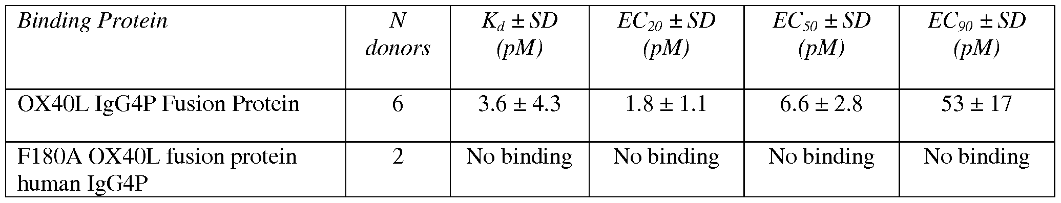
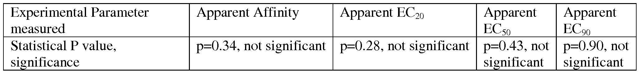


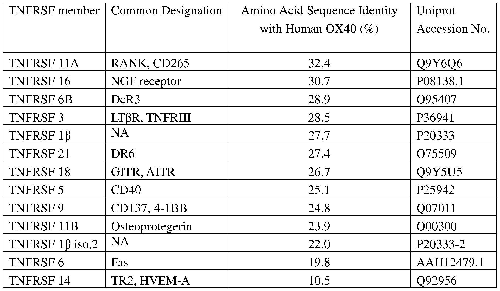
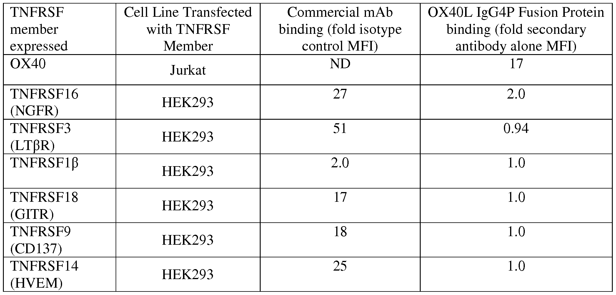


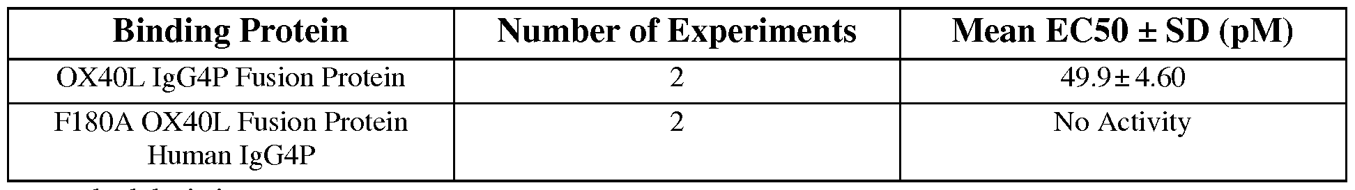
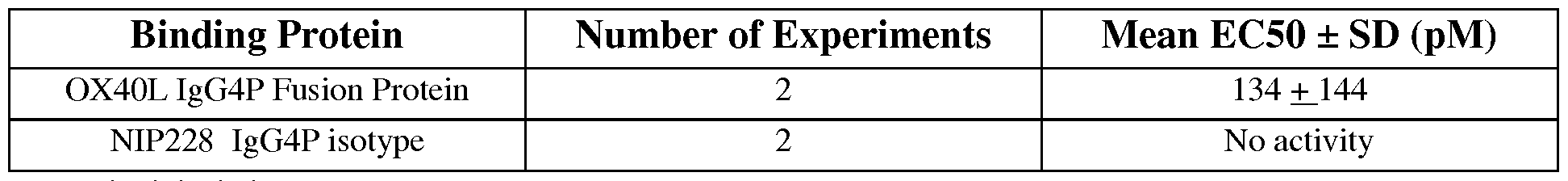
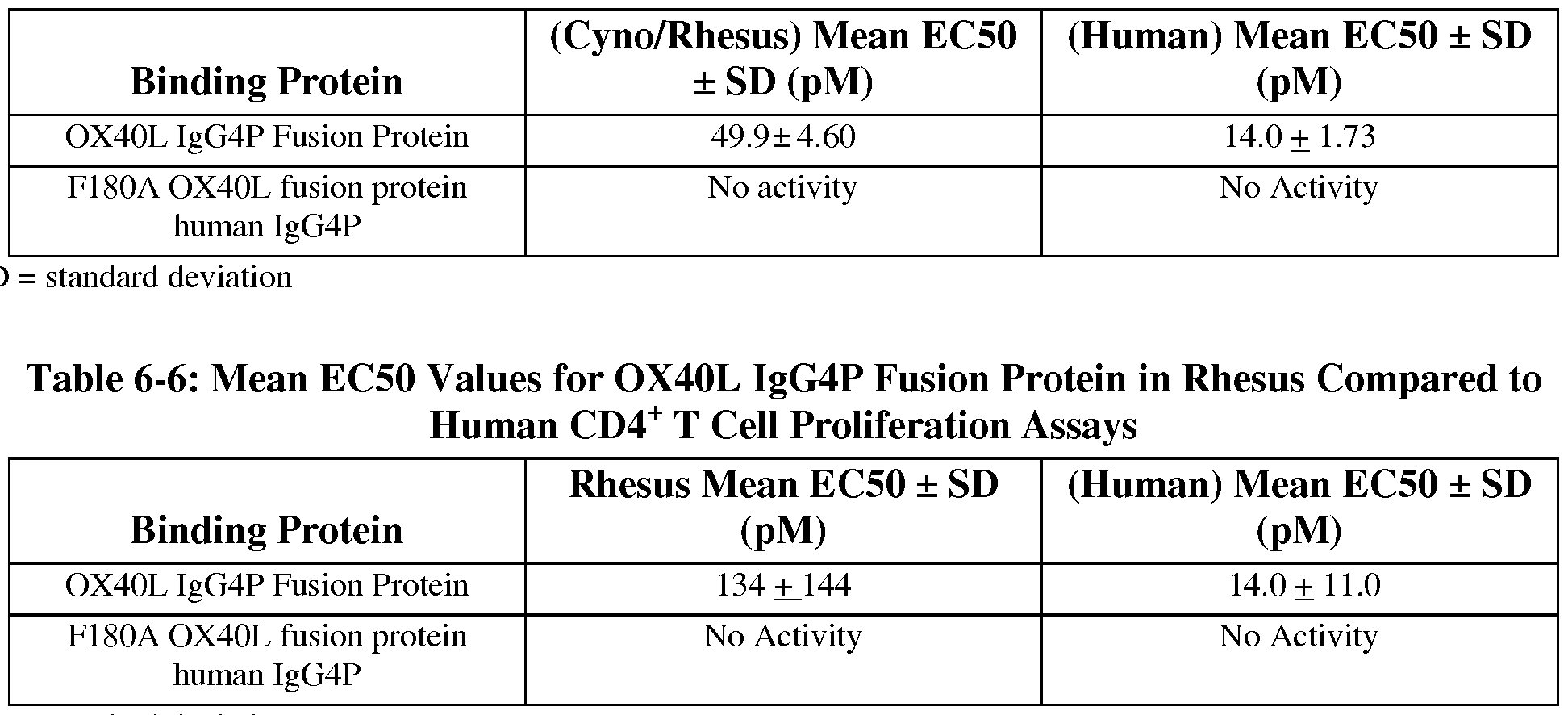

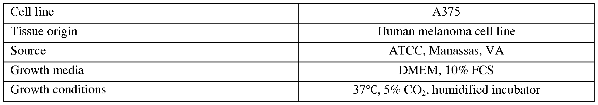


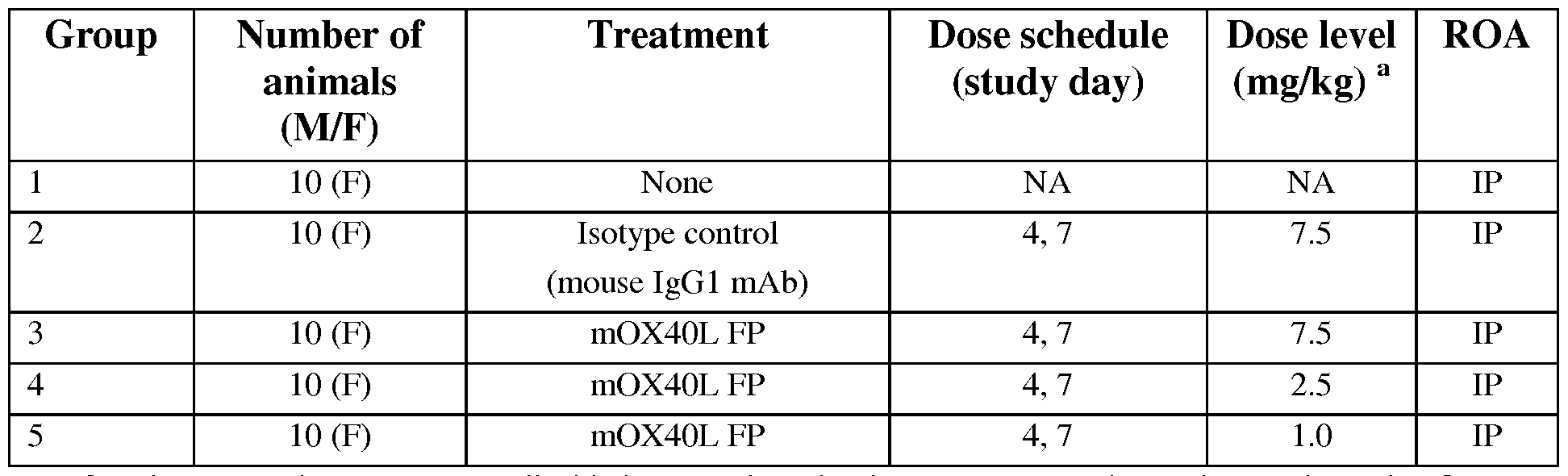
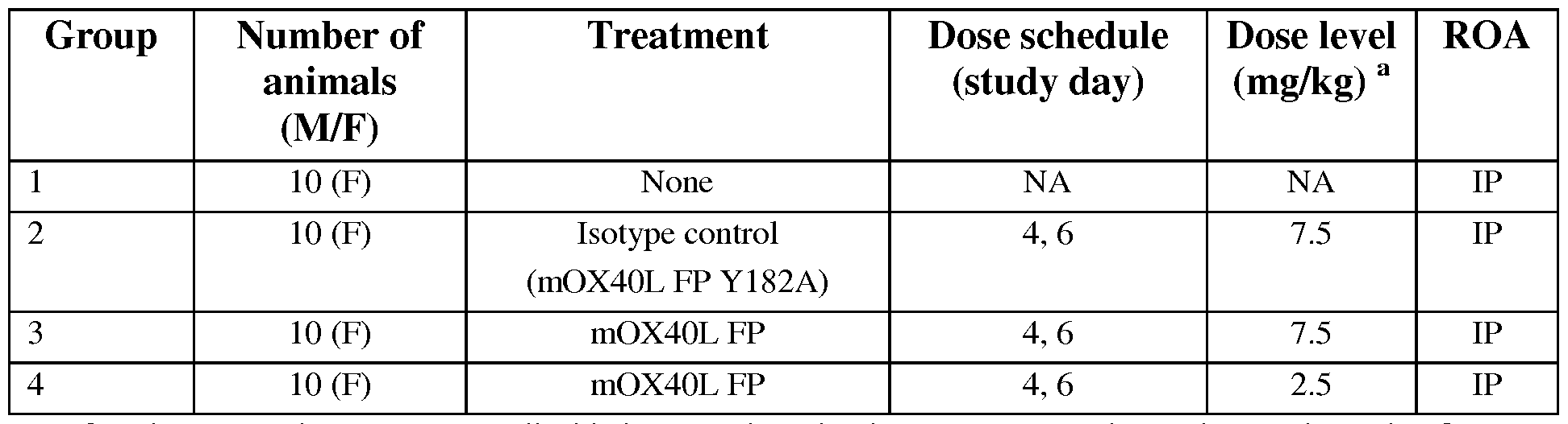

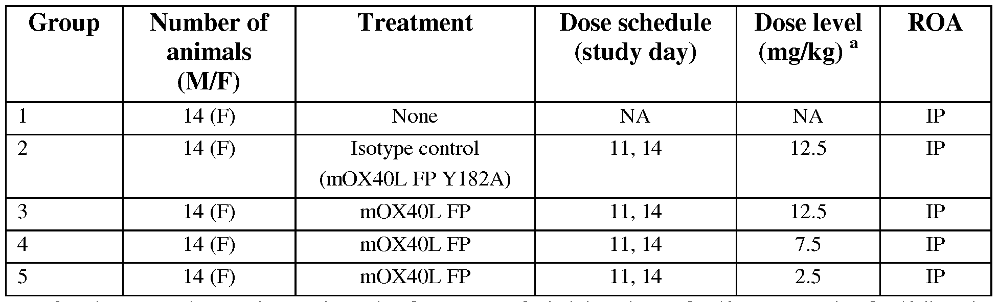

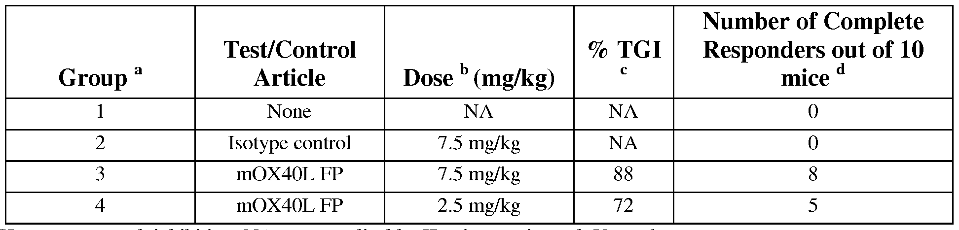
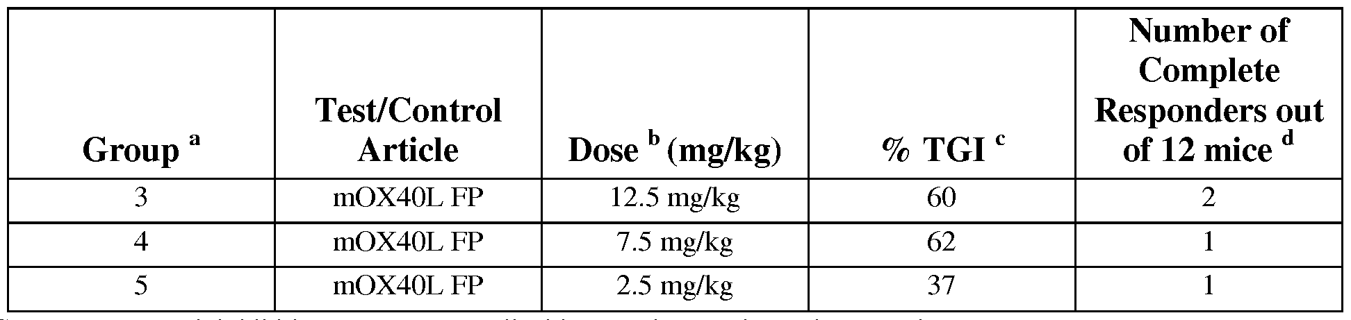
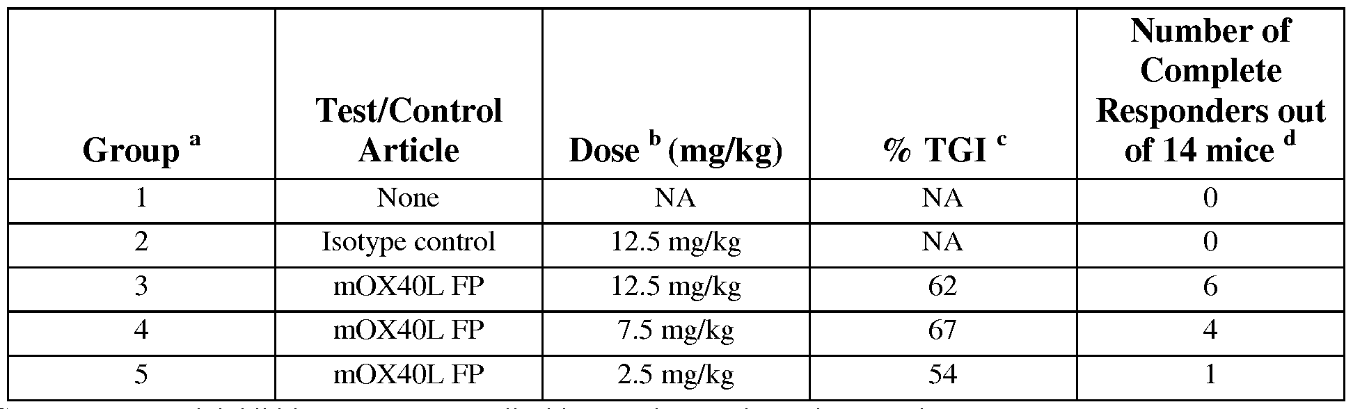
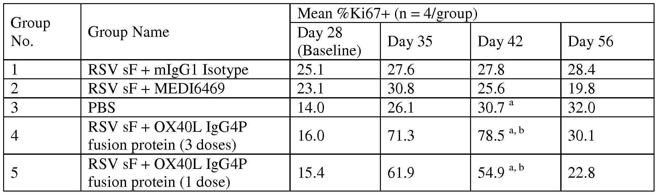

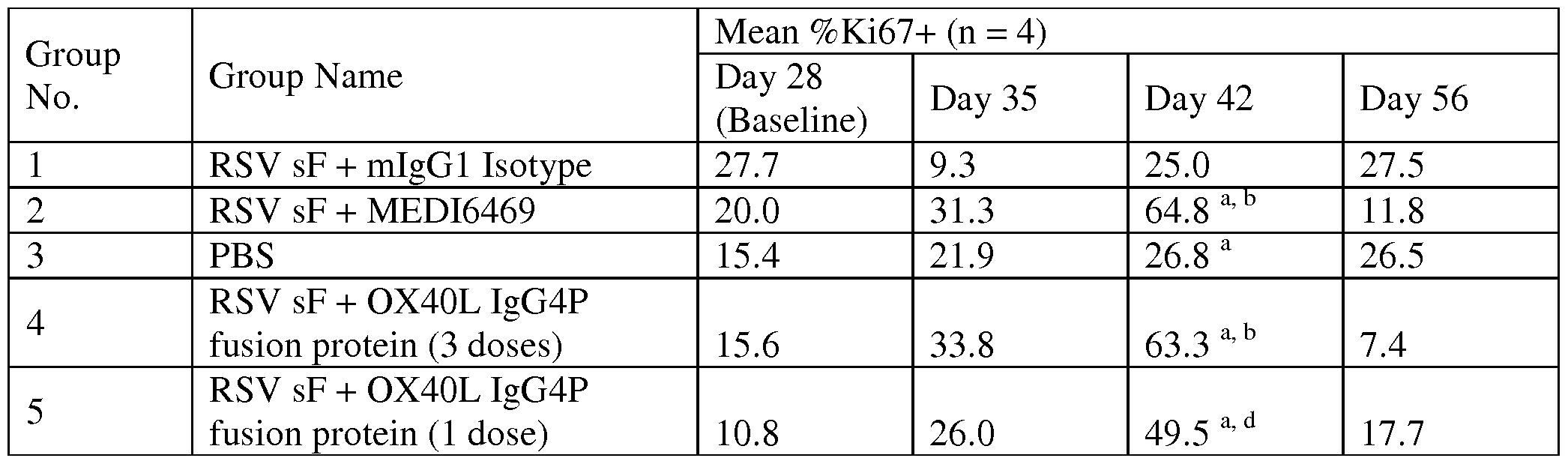
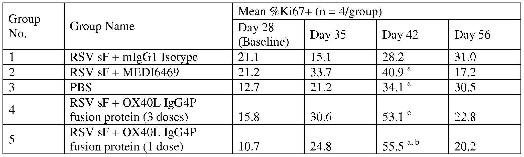


Claims
Priority Applications (6)
| Application Number | Priority Date | Filing Date | Title |
|---|---|---|---|
| MX2016014647A MX360295B (en) | 2014-05-29 | 2015-05-27 | Ox40l fusion proteins and uses thereof. |
| EP15799339.5A EP3148573A4 (en) | 2014-05-29 | 2015-05-27 | Ox40l fusion proteins and uses thereof |
| BR112016027845A BR112016027845A2 (en) | 2014-05-29 | 2015-05-27 | ox40l fusion proteins and uses thereof |
| RU2016150096A RU2016150096A (en) | 2014-05-29 | 2015-05-27 | OX40L-BASED FUSION PROTEINS AND WAYS OF THEIR APPLICATION |
| JP2016569060A JP6666267B2 (en) | 2014-05-29 | 2015-05-27 | OX40L fusion proteins and uses thereof |
| CN201580028116.9A CN106456734A (en) | 2014-05-29 | 2015-05-27 | OX40L fusion proteins and uses thereof |
Applications Claiming Priority (4)
| Application Number | Priority Date | Filing Date | Title |
|---|---|---|---|
| US201462004555P | 2014-05-29 | 2014-05-29 | |
| US62/004,555 | 2014-05-29 | ||
| US201562111796P | 2015-02-04 | 2015-02-04 | |
| US62/111,796 | 2015-02-04 |
Publications (1)
| Publication Number | Publication Date |
|---|---|
| WO2015183902A1 true WO2015183902A1 (en) | 2015-12-03 |
Family
ID=53540957
Family Applications (1)
| Application Number | Title | Priority Date | Filing Date |
|---|---|---|---|
| PCT/US2015/032598 Ceased WO2015183902A1 (en) | 2014-05-29 | 2015-05-27 | Ox40l fusion proteins and uses thereof |
Country Status (10)
| Country | Link |
|---|---|
| US (1) | US9718870B2 (en) |
| EP (1) | EP3148573A4 (en) |
| JP (1) | JP6666267B2 (en) |
| CN (1) | CN106456734A (en) |
| BR (1) | BR112016027845A2 (en) |
| GB (1) | GB2532817A (en) |
| MX (1) | MX360295B (en) |
| RU (1) | RU2016150096A (en) |
| TW (1) | TW201623327A (en) |
| WO (1) | WO2015183902A1 (en) |
Cited By (14)
| Publication number | Priority date | Publication date | Assignee | Title |
|---|---|---|---|---|
| GB2532817A (en) * | 2014-05-29 | 2016-06-01 | Medimmune Llc | OX40L fusion proteins and uses thereof |
| WO2017167672A1 (en) | 2016-03-29 | 2017-10-05 | F. Hoffmann-La Roche Ag | Trimeric costimulatory tnf family ligand-containing antigen binding molecules |
| EP3243836A1 (en) * | 2016-05-11 | 2017-11-15 | F. Hoffmann-La Roche AG | C-terminally fused tnf family ligand trimer-containing antigen binding molecules |
| WO2018115003A3 (en) * | 2016-12-19 | 2018-08-09 | Glenmark Pharmaceuticals S.A. | Novel tnfr agonists and uses thereof |
| US10086042B2 (en) | 2015-10-01 | 2018-10-02 | Heat Biologics, Inc. | Compositions and methods for adjoining type I and type II extracellular domains as heterologous chimeric proteins |
| WO2018178074A1 (en) | 2017-03-29 | 2018-10-04 | F. Hoffmann-La Roche Ag | Trimeric antigen binding molecules specific for a costimulatory tnf receptor |
| US10392445B2 (en) | 2014-11-14 | 2019-08-27 | Hoffmann-La Roche Inc. | Tumor necrosis factor (TNF) family ligand trimer-containing antigen-binding molecules |
| US10464981B2 (en) | 2015-03-31 | 2019-11-05 | Hoffmann-La Roche, Inc. | Tumor necrosis factor (TNF) family ligand trimer-containing antigen binding molecules |
| US10513548B2 (en) | 2017-02-27 | 2019-12-24 | Shattuck Labs, Inc. | CSF1R-based chimeric proteins |
| JP2020517913A (en) * | 2017-04-28 | 2020-06-18 | ファイブ プライム セラピューティクス, インコーポレイテッド | Method of treatment with CD80 extracellular domain polypeptide |
| US10780121B2 (en) | 2018-08-29 | 2020-09-22 | Shattuck Labs, Inc. | FLT3L-based chimeric proteins |
| US10899817B2 (en) | 2017-02-27 | 2021-01-26 | Shattuck Labs, Inc. | TIGIT- and light-based chimeric proteins |
| US11192933B2 (en) | 2017-02-27 | 2021-12-07 | Shattuck Labs, Inc. | VSIG8-based chimeric proteins |
| US11286300B2 (en) | 2015-10-01 | 2022-03-29 | Hoffmann-La Roche Inc. | Humanized anti-human CD19 antibodies and methods of use |
Families Citing this family (16)
| Publication number | Priority date | Publication date | Assignee | Title |
|---|---|---|---|---|
| US10046047B2 (en) * | 2015-02-06 | 2018-08-14 | Heat Biologics, Inc. | Vector co-expressing vaccine and costimulatory molecules |
| EP3314255B1 (en) * | 2015-06-25 | 2019-07-31 | H. Hoffnabb-La Roche Ag | Cell based assay for determining antibody or ligand binding and function |
| CA2994346A1 (en) | 2015-08-12 | 2017-02-16 | Medimmune Limited | Gitrl fusion proteins and uses thereof |
| CA3040123A1 (en) | 2016-10-11 | 2018-04-19 | University Of Miami | Vectors and vaccine cells for immunity against zika virus |
| US10331232B2 (en) * | 2016-10-27 | 2019-06-25 | Fluidity Technologies, Inc. | Controller with situational awareness display |
| US10331233B2 (en) * | 2016-10-27 | 2019-06-25 | Fluidity Technologies, Inc. | Camera and sensor controls for remotely operated vehicles and virtual environments |
| AU2017371225A1 (en) * | 2016-12-09 | 2019-05-16 | Seagen Inc. | Bivalent antibodies masked by coiled coils |
| WO2018187260A1 (en) | 2017-04-04 | 2018-10-11 | Heat Biologics, Inc. | Intratumoral vaccination |
| US11377490B2 (en) * | 2017-05-31 | 2022-07-05 | Sichuan Clover Biopharmaceuticals, Inc | Method for treating cancer using disulfide-linked trimeric 4-1BBL |
| CN109839509A (en) * | 2019-02-15 | 2019-06-04 | 浠思(上海)生物技术有限公司 | Utilize the method for the HTRF binding analysis experimental technique screening potential agonist of OX40/OX40L |
| CA3131700A1 (en) | 2019-02-27 | 2020-09-03 | Ionis Pharmaceuticals, Inc. | Modulators of malat1 expression |
| PE20221506A1 (en) * | 2019-12-17 | 2022-10-04 | Amgen Inc | DUAL INTERLEUKIN-2/TNF RECEPTOR AGONIST FOR USE IN THERAPY |
| JP2023525140A (en) * | 2020-05-13 | 2023-06-14 | アンセルム(アンスティチュート・ナシオナル・ドゥ・ラ・サンテ・エ・ドゥ・ラ・ルシェルシュ・メディカル) | Recombinant proteins with OX40 activation properties |
| EP4277639A4 (en) * | 2021-01-15 | 2025-03-05 | Seattle Children's Hospital d/b/a Seattle Children's Research Institute | HYBRID AND TRUSIONED IMMUNE CELL PROTEINS |
| US20250319144A1 (en) * | 2021-11-19 | 2025-10-16 | Nankai University | Recombinant Armed Oncolytic Virus Composition and Use Thereof in TIL Adoptive Therapy |
| CN114966061B (en) * | 2022-07-28 | 2022-10-21 | 中国食品药品检定研究院 | A kind of biological activity detection method of anti-OX40 antibody |
Citations (4)
| Publication number | Priority date | Publication date | Assignee | Title |
|---|---|---|---|---|
| WO2002072605A2 (en) * | 2001-03-07 | 2002-09-19 | Merck Patent Gmbh | Expression technology for proteins containing a hybrid isotype antibody moiety |
| US7959925B2 (en) * | 2005-05-06 | 2011-06-14 | Providence Health System | Trimeric OX40-immunoglobulin fusion protein and methods of use |
| US20110243966A1 (en) * | 2007-05-14 | 2011-10-06 | Biogen Idec Ma Inc. | SINGLE CHAIN Fc (ScFc) REGIONS, BINDING POLYPEPTIDES COMPRISING SAME, AND METHODS RELATED THERETO |
| US20130345406A1 (en) * | 2007-05-31 | 2013-12-26 | Genmab A/S | Stable igg4 antibodies |
Family Cites Families (18)
| Publication number | Priority date | Publication date | Assignee | Title |
|---|---|---|---|---|
| US5681718A (en) | 1986-03-14 | 1997-10-28 | Celltech Limited | Methods for enhanced production of tissue plasminogen activator in cell culture using alkanoic acids or salts thereof |
| US5892019A (en) | 1987-07-15 | 1999-04-06 | The United States Of America, As Represented By The Department Of Health And Human Services | Production of a single-gene-encoded immunoglobulin |
| US5457035A (en) | 1993-07-23 | 1995-10-10 | Immunex Corporation | Cytokine which is a ligand for OX40 |
| US6242566B1 (en) | 1994-02-10 | 2001-06-05 | Board Of Trustees Of The Leland Stanford Junior University | Ligand (ACT-4-L) to a receptor on the surface of activated CD4+ T-cells |
| US6312700B1 (en) | 1998-02-24 | 2001-11-06 | Andrew D. Weinberg | Method for enhancing an antigen specific immune response with OX-40L |
| US7056695B2 (en) * | 2000-03-02 | 2006-06-06 | Xencor | TNF-α variants |
| AU2003210806A1 (en) * | 2002-03-05 | 2003-09-22 | Eli Lilly And Company | Heterologous g-csf fusion proteins |
| GB0216648D0 (en) | 2002-07-18 | 2002-08-28 | Lonza Biologics Plc | Method of expressing recombinant protein in CHO cells |
| EP1725585A2 (en) | 2004-03-10 | 2006-11-29 | Lonza Ltd | Method for producing antibodies |
| EP1810979B1 (en) * | 2004-09-22 | 2012-06-20 | Kyowa Hakko Kirin Co., Ltd. | STABILIZED HUMAN IgG4 ANTIBODIES |
| AU2013263717B2 (en) * | 2005-05-06 | 2016-05-19 | Providence Health & Services - Oregon | Trimeric OX40L-immunoglobulin fusion protein and methods of use |
| TWI461436B (en) * | 2005-11-25 | 2014-11-21 | Kyowa Hakko Kirin Co Ltd | Human CD134 (OX40) human monoclonal antibody and method of making and using same |
| US20070172504A1 (en) * | 2005-12-08 | 2007-07-26 | University Of Lousville Research Foundation, Inc. | In vivo cell surface engineering |
| US8637637B2 (en) * | 2010-01-12 | 2014-01-28 | Bill Nai-Chau Sun | Fc fusion proteins of human growth hormone |
| GB201014033D0 (en) * | 2010-08-20 | 2010-10-06 | Ucb Pharma Sa | Biological products |
| HK1205691A1 (en) * | 2012-03-02 | 2015-12-24 | Providence Health & Services - Oregon | Dual ox40 agonist/il-2 cancer therapy methods |
| WO2015183902A1 (en) * | 2014-05-29 | 2015-12-03 | Medimmune, Llc | Ox40l fusion proteins and uses thereof |
| EP3550019A1 (en) * | 2014-10-24 | 2019-10-09 | Astrazeneca AB | Combination |
-
2015
- 2015-05-27 WO PCT/US2015/032598 patent/WO2015183902A1/en not_active Ceased
- 2015-05-27 MX MX2016014647A patent/MX360295B/en active IP Right Grant
- 2015-05-27 US US14/722,679 patent/US9718870B2/en not_active Expired - Fee Related
- 2015-05-27 CN CN201580028116.9A patent/CN106456734A/en active Pending
- 2015-05-27 JP JP2016569060A patent/JP6666267B2/en not_active Expired - Fee Related
- 2015-05-27 GB GB1509062.4A patent/GB2532817A/en not_active Withdrawn
- 2015-05-27 BR BR112016027845A patent/BR112016027845A2/en not_active Application Discontinuation
- 2015-05-27 RU RU2016150096A patent/RU2016150096A/en not_active Application Discontinuation
- 2015-05-27 EP EP15799339.5A patent/EP3148573A4/en not_active Withdrawn
- 2015-05-29 TW TW104117533A patent/TW201623327A/en unknown
Patent Citations (4)
| Publication number | Priority date | Publication date | Assignee | Title |
|---|---|---|---|---|
| WO2002072605A2 (en) * | 2001-03-07 | 2002-09-19 | Merck Patent Gmbh | Expression technology for proteins containing a hybrid isotype antibody moiety |
| US7959925B2 (en) * | 2005-05-06 | 2011-06-14 | Providence Health System | Trimeric OX40-immunoglobulin fusion protein and methods of use |
| US20110243966A1 (en) * | 2007-05-14 | 2011-10-06 | Biogen Idec Ma Inc. | SINGLE CHAIN Fc (ScFc) REGIONS, BINDING POLYPEPTIDES COMPRISING SAME, AND METHODS RELATED THERETO |
| US20130345406A1 (en) * | 2007-05-31 | 2013-12-26 | Genmab A/S | Stable igg4 antibodies |
Non-Patent Citations (1)
| Title |
|---|
| See also references of EP3148573A4 * |
Cited By (46)
| Publication number | Priority date | Publication date | Assignee | Title |
|---|---|---|---|---|
| GB2532817A (en) * | 2014-05-29 | 2016-06-01 | Medimmune Llc | OX40L fusion proteins and uses thereof |
| US11306154B2 (en) | 2014-11-14 | 2022-04-19 | Hoffmann-La Roche Inc. | Methods of treating cancer by administering antigen-binding molecules comprising a TNF family ligand trimer |
| US12441812B2 (en) | 2014-11-14 | 2025-10-14 | Hoffmann-La Roche Inc. | Methods of treating cancer by administering tumor necrosis factor (TNF) family ligand trimer-containing antigen-binding molecules |
| US10392445B2 (en) | 2014-11-14 | 2019-08-27 | Hoffmann-La Roche Inc. | Tumor necrosis factor (TNF) family ligand trimer-containing antigen-binding molecules |
| US11267903B2 (en) | 2014-11-14 | 2022-03-08 | Hofmann-La Roche Inc. | Antigen-binding molecules comprising a tumor necrosis factor (TNF) family ligand trimer |
| US10464981B2 (en) | 2015-03-31 | 2019-11-05 | Hoffmann-La Roche, Inc. | Tumor necrosis factor (TNF) family ligand trimer-containing antigen binding molecules |
| US12202870B2 (en) | 2015-03-31 | 2025-01-21 | Hoffmann-La Roche Inc. | Antigen binding molecules comprising a trimeric TNF family ligand and encoding polynucleotides thereof |
| US11547742B1 (en) | 2015-10-01 | 2023-01-10 | Heat Biologics, Inc. | Compositions and methods for adjoining type I and type II extracellular domains as heterologous chimeric proteins |
| US10543253B2 (en) | 2015-10-01 | 2020-01-28 | Heat Biologics, Inc. | Compositions and methods for adjoining type I and type II extracellular domains as heterologous chimeric proteins |
| US10183060B2 (en) | 2015-10-01 | 2019-01-22 | Heat Biologics, Inc. | Compositions and methods for adjoining type I and type II extracellular domains as heterologous chimeric proteins |
| US10188701B2 (en) | 2015-10-01 | 2019-01-29 | Heat Biologics, Inc. | Compositions and methods for adjoining type I and type II extracellular domains as heterologous chimeric proteins |
| US11654180B2 (en) | 2015-10-01 | 2023-05-23 | Heat Biologies, Inc. | Compositions and methods for adjoining type I and type II extracellular domains as heterologous chimeric proteins |
| US10086042B2 (en) | 2015-10-01 | 2018-10-02 | Heat Biologics, Inc. | Compositions and methods for adjoining type I and type II extracellular domains as heterologous chimeric proteins |
| US12178847B2 (en) | 2015-10-01 | 2024-12-31 | Kopfkino Ip, Llc | Compositions and methods for adjoining type I and type II extracellular domains as heterologous chimeric proteins |
| US10660936B2 (en) | 2015-10-01 | 2020-05-26 | Heat Biologics, Inc. | Compositions and methods for adjoining type I and type II extracellular domains as heterologous chimeric proteins |
| US10449233B2 (en) | 2015-10-01 | 2019-10-22 | Heat Biologics, Inc. | Compositions and methods for adjoining type I and type II extracellular domains as heterologous chimeric proteins |
| US10653748B2 (en) | 2015-10-01 | 2020-05-19 | Heat Biologics, Inc. | Compositions and methods for adjoining type I and type II extracellular domains as heterologous chimeric proteins |
| US10493128B2 (en) | 2015-10-01 | 2019-12-03 | Heat Biologics, Inc. | Compositions and methods for adjoining type I and type II extracellular domains as heterologous chimeric proteins |
| US11286300B2 (en) | 2015-10-01 | 2022-03-29 | Hoffmann-La Roche Inc. | Humanized anti-human CD19 antibodies and methods of use |
| US10525102B2 (en) | 2015-10-01 | 2020-01-07 | Heat Biologics, Inc. | Compositions and methods for adjoining type I and type II extracellular domains as heterologous chimeric proteins |
| US10646545B2 (en) | 2015-10-01 | 2020-05-12 | Heat Biologics, Inc. | Compositions and methods for adjoining type I and type II extracellular domains as heterologous chimeric proteins |
| CN108966652A (en) * | 2016-03-29 | 2018-12-07 | 豪夫迈·罗氏有限公司 | Antigen-binding molecules containing trimeric co-stimulatory TNF family ligands |
| WO2017167672A1 (en) | 2016-03-29 | 2017-10-05 | F. Hoffmann-La Roche Ag | Trimeric costimulatory tnf family ligand-containing antigen binding molecules |
| EP3231813A1 (en) | 2016-03-29 | 2017-10-18 | F. Hoffmann-La Roche AG | Trimeric costimulatory tnf family ligand-containing antigen binding molecules |
| CN109311973A (en) * | 2016-05-11 | 2019-02-05 | 豪夫迈·罗氏有限公司 | Antigen-binding molecules containing C-terminally fused TNF family ligand trimers |
| JP7043422B2 (en) | 2016-05-11 | 2022-03-29 | エフ・ホフマン-ラ・ロシュ・アクチェンゲゼルシャフト | C-terminal fusion TNF family ligand trimer-containing antigen-binding molecule |
| EP3243836A1 (en) * | 2016-05-11 | 2017-11-15 | F. Hoffmann-La Roche AG | C-terminally fused tnf family ligand trimer-containing antigen binding molecules |
| WO2017194438A1 (en) * | 2016-05-11 | 2017-11-16 | F. Hoffmann-La Roche Ag | C-terminally fused tnf family ligand trimer-containing antigen binding molecules |
| CN109311973B (en) * | 2016-05-11 | 2023-04-04 | 豪夫迈·罗氏有限公司 | Antigen binding molecules comprising C-terminal fused TNF family ligand trimers |
| JP2019521655A (en) * | 2016-05-11 | 2019-08-08 | エフ・ホフマン−ラ・ロシュ・アクチェンゲゼルシャフト | C-terminal fused TNF family ligand trimer-containing antigen-binding molecule |
| WO2018115003A3 (en) * | 2016-12-19 | 2018-08-09 | Glenmark Pharmaceuticals S.A. | Novel tnfr agonists and uses thereof |
| CN110291108A (en) * | 2016-12-19 | 2019-09-27 | 格兰马克药品股份有限公司 | Novel TNFR agonists and their uses |
| US11267856B2 (en) | 2017-02-27 | 2022-03-08 | Shattuck Labs, Inc. | CSF1R-CD40L chimeric proteins |
| US12071465B2 (en) | 2017-02-27 | 2024-08-27 | Shattuck Labs, Inc. | Methods of making and using extracellular domain-based chimeric proteins |
| US10513548B2 (en) | 2017-02-27 | 2019-12-24 | Shattuck Labs, Inc. | CSF1R-based chimeric proteins |
| US11267857B2 (en) | 2017-02-27 | 2022-03-08 | Shattuck Labs, Inc. | CSF1R-based chimeric proteins |
| US11332509B2 (en) | 2017-02-27 | 2022-05-17 | Shattuck Labs, Inc. | Methods of making and using extracellular domain-based chimeric proteins |
| US10899817B2 (en) | 2017-02-27 | 2021-01-26 | Shattuck Labs, Inc. | TIGIT- and light-based chimeric proteins |
| US11192934B2 (en) | 2017-02-27 | 2021-12-07 | Shattuck Labs, Inc. | VSIG8-based chimeric proteins |
| US10927159B2 (en) | 2017-02-27 | 2021-02-23 | Shattuck Labs, Inc. | TIGIT- and light-based chimeric proteins |
| US11834488B2 (en) | 2017-02-27 | 2023-12-05 | Shattuck Labs | VSIG8-based chimeric proteins |
| US11192933B2 (en) | 2017-02-27 | 2021-12-07 | Shattuck Labs, Inc. | VSIG8-based chimeric proteins |
| WO2018178074A1 (en) | 2017-03-29 | 2018-10-04 | F. Hoffmann-La Roche Ag | Trimeric antigen binding molecules specific for a costimulatory tnf receptor |
| JP2020517913A (en) * | 2017-04-28 | 2020-06-18 | ファイブ プライム セラピューティクス, インコーポレイテッド | Method of treatment with CD80 extracellular domain polypeptide |
| US11896618B2 (en) | 2018-08-29 | 2024-02-13 | Shattuck Labs, Inc. | FLT3L-based chimeric proteins |
| US10780121B2 (en) | 2018-08-29 | 2020-09-22 | Shattuck Labs, Inc. | FLT3L-based chimeric proteins |
Also Published As
| Publication number | Publication date |
|---|---|
| RU2016150096A (en) | 2018-07-02 |
| US20160024176A1 (en) | 2016-01-28 |
| MX2016014647A (en) | 2017-05-30 |
| BR112016027845A2 (en) | 2017-10-31 |
| JP6666267B2 (en) | 2020-03-13 |
| CN106456734A (en) | 2017-02-22 |
| TW201623327A (en) | 2016-07-01 |
| EP3148573A1 (en) | 2017-04-05 |
| US9718870B2 (en) | 2017-08-01 |
| RU2016150096A3 (en) | 2019-01-24 |
| MX360295B (en) | 2018-10-29 |
| GB201509062D0 (en) | 2015-07-08 |
| EP3148573A4 (en) | 2017-11-15 |
| JP2017519494A (en) | 2017-07-20 |
| GB2532817A (en) | 2016-06-01 |
Similar Documents
| Publication | Publication Date | Title |
|---|---|---|
| US9718870B2 (en) | OX40L fusion proteins and uses thereof | |
| US10428131B2 (en) | GITRL fusion proteins comprising a human coronin 1a derived trimerization domain | |
| EP3204422B1 (en) | Humanized anti-ox40 antibodies and uses thereof | |
| JP2019501883A (en) | Antibodies and methods of use thereof | |
| Ahmed et al. | TCR-mimic bispecific antibodies targeting LMP2A show potent activity against EBV malignancies | |
| AU2012369202A1 (en) | Cancer treatment and monitoring methods using OX40 agonists | |
| Gombos et al. | Toxicological and pharmacological assessment of AGEN1884, a novel human IgG1 anti-CTLA-4 antibody | |
| TW202112826A (en) | Anti-tigit antibodies and methods of use | |
| Herrera | Development of Antigen-Selective Immunotherapeutic Approaches and Discovery of Novel Immune Pathways | |
| WO2024209072A1 (en) | Multispecific binding agents against pd-l1 and cd137 for treating cancer | |
| Cassotta | Clonal selection and immunodominance in the human T cell response to microbial antigens and biologicals | |
| WO2025114541A1 (en) | Antibodies capable of binding to ox40 in combination therapy | |
| WO2023218046A1 (en) | Binding agents capable of binding to cd27 in combination therapy |
Legal Events
| Date | Code | Title | Description |
|---|---|---|---|
| 121 | Ep: the epo has been informed by wipo that ep was designated in this application |
Ref document number: 15799339 Country of ref document: EP Kind code of ref document: A1 |
|
| WWE | Wipo information: entry into national phase |
Ref document number: MX/A/2016/014647 Country of ref document: MX |
|
| ENP | Entry into the national phase |
Ref document number: 2016569060 Country of ref document: JP Kind code of ref document: A |
|
| NENP | Non-entry into the national phase |
Ref country code: DE |
|
| REG | Reference to national code |
Ref country code: BR Ref legal event code: B01A Ref document number: 112016027845 Country of ref document: BR |
|
| REEP | Request for entry into the european phase |
Ref document number: 2015799339 Country of ref document: EP |
|
| WWE | Wipo information: entry into national phase |
Ref document number: 2015799339 Country of ref document: EP |
|
| ENP | Entry into the national phase |
Ref document number: 2016150096 Country of ref document: RU Kind code of ref document: A |
|
| ENP | Entry into the national phase |
Ref document number: 112016027845 Country of ref document: BR Kind code of ref document: A2 Effective date: 20161128 |