WO2013019730A1 - Antibodies to tip-1 and grp78 - Google Patents
Antibodies to tip-1 and grp78 Download PDFInfo
- Publication number
- WO2013019730A1 WO2013019730A1 PCT/US2012/048856 US2012048856W WO2013019730A1 WO 2013019730 A1 WO2013019730 A1 WO 2013019730A1 US 2012048856 W US2012048856 W US 2012048856W WO 2013019730 A1 WO2013019730 A1 WO 2013019730A1
- Authority
- WO
- WIPO (PCT)
- Prior art keywords
- antibody
- seq
- chain variable
- epitope
- acid sequence
- Prior art date
- Legal status (The legal status is an assumption and is not a legal conclusion. Google has not performed a legal analysis and makes no representation as to the accuracy of the status listed.)
- Ceased
Links
Classifications
-
- C—CHEMISTRY; METALLURGY
- C07—ORGANIC CHEMISTRY
- C07K—PEPTIDES
- C07K16/00—Immunoglobulins [IGs], e.g. monoclonal or polyclonal antibodies
- C07K16/18—Immunoglobulins [IGs], e.g. monoclonal or polyclonal antibodies against material from animals or humans
- C07K16/28—Immunoglobulins [IGs], e.g. monoclonal or polyclonal antibodies against material from animals or humans against receptors, cell surface antigens or cell surface determinants
- C07K16/30—Immunoglobulins [IGs], e.g. monoclonal or polyclonal antibodies against material from animals or humans against receptors, cell surface antigens or cell surface determinants from tumour cells
-
- A—HUMAN NECESSITIES
- A61—MEDICAL OR VETERINARY SCIENCE; HYGIENE
- A61B—DIAGNOSIS; SURGERY; IDENTIFICATION
- A61B90/00—Instruments, implements or accessories specially adapted for surgery or diagnosis and not covered by any of the groups A61B1/00 - A61B50/00, e.g. for luxation treatment or for protecting wound edges
- A61B90/36—Image-producing devices or illumination devices not otherwise provided for
- A61B90/361—Image-producing devices, e.g. surgical cameras
-
- A—HUMAN NECESSITIES
- A61—MEDICAL OR VETERINARY SCIENCE; HYGIENE
- A61K—PREPARATIONS FOR MEDICAL, DENTAL OR TOILETRY PURPOSES
- A61K39/00—Medicinal preparations containing antigens or antibodies
- A61K39/395—Antibodies; Immunoglobulins; Immune serum, e.g. antilymphocytic serum
- A61K39/39533—Antibodies; Immunoglobulins; Immune serum, e.g. antilymphocytic serum against materials from animals
- A61K39/3955—Antibodies; Immunoglobulins; Immune serum, e.g. antilymphocytic serum against materials from animals against proteinaceous materials, e.g. enzymes, hormones, lymphokines
-
- A—HUMAN NECESSITIES
- A61—MEDICAL OR VETERINARY SCIENCE; HYGIENE
- A61K—PREPARATIONS FOR MEDICAL, DENTAL OR TOILETRY PURPOSES
- A61K49/00—Preparations for testing in vivo
- A61K49/001—Preparation for luminescence or biological staining
- A61K49/0013—Luminescence
- A61K49/0017—Fluorescence in vivo
- A61K49/005—Fluorescence in vivo characterised by the carrier molecule carrying the fluorescent agent
- A61K49/0058—Antibodies
-
- A—HUMAN NECESSITIES
- A61—MEDICAL OR VETERINARY SCIENCE; HYGIENE
- A61K—PREPARATIONS FOR MEDICAL, DENTAL OR TOILETRY PURPOSES
- A61K51/00—Preparations containing radioactive substances for use in therapy or testing in vivo
- A61K51/02—Preparations containing radioactive substances for use in therapy or testing in vivo characterised by the carrier, i.e. characterised by the agent or material covalently linked or complexing the radioactive nucleus
- A61K51/04—Organic compounds
- A61K51/08—Peptides, e.g. proteins, carriers being peptides, polyamino acids, proteins
- A61K51/10—Antibodies or immunoglobulins; Fragments thereof, the carrier being an antibody, an immunoglobulin or a fragment thereof, e.g. a camelised human single domain antibody or the Fc fragment of an antibody
- A61K51/1045—Antibodies or immunoglobulins; Fragments thereof, the carrier being an antibody, an immunoglobulin or a fragment thereof, e.g. a camelised human single domain antibody or the Fc fragment of an antibody against animal or human tumor cells or tumor cell determinants
- A61K51/1066—Antibodies or immunoglobulins; Fragments thereof, the carrier being an antibody, an immunoglobulin or a fragment thereof, e.g. a camelised human single domain antibody or the Fc fragment of an antibody against animal or human tumor cells or tumor cell determinants the tumor cell being from skin
-
- A—HUMAN NECESSITIES
- A61—MEDICAL OR VETERINARY SCIENCE; HYGIENE
- A61K—PREPARATIONS FOR MEDICAL, DENTAL OR TOILETRY PURPOSES
- A61K51/00—Preparations containing radioactive substances for use in therapy or testing in vivo
- A61K51/02—Preparations containing radioactive substances for use in therapy or testing in vivo characterised by the carrier, i.e. characterised by the agent or material covalently linked or complexing the radioactive nucleus
- A61K51/04—Organic compounds
- A61K51/08—Peptides, e.g. proteins, carriers being peptides, polyamino acids, proteins
- A61K51/10—Antibodies or immunoglobulins; Fragments thereof, the carrier being an antibody, an immunoglobulin or a fragment thereof, e.g. a camelised human single domain antibody or the Fc fragment of an antibody
- A61K51/1093—Antibodies or immunoglobulins; Fragments thereof, the carrier being an antibody, an immunoglobulin or a fragment thereof, e.g. a camelised human single domain antibody or the Fc fragment of an antibody conjugates with carriers being antibodies
-
- A—HUMAN NECESSITIES
- A61—MEDICAL OR VETERINARY SCIENCE; HYGIENE
- A61N—ELECTROTHERAPY; MAGNETOTHERAPY; RADIATION THERAPY; ULTRASOUND THERAPY
- A61N5/00—Radiation therapy
- A61N5/10—X-ray therapy; Gamma-ray therapy; Particle-irradiation therapy
-
- A—HUMAN NECESSITIES
- A61—MEDICAL OR VETERINARY SCIENCE; HYGIENE
- A61P—SPECIFIC THERAPEUTIC ACTIVITY OF CHEMICAL COMPOUNDS OR MEDICINAL PREPARATIONS
- A61P35/00—Antineoplastic agents
-
- C—CHEMISTRY; METALLURGY
- C07—ORGANIC CHEMISTRY
- C07K—PEPTIDES
- C07K16/00—Immunoglobulins [IGs], e.g. monoclonal or polyclonal antibodies
- C07K16/18—Immunoglobulins [IGs], e.g. monoclonal or polyclonal antibodies against material from animals or humans
-
- C—CHEMISTRY; METALLURGY
- C07—ORGANIC CHEMISTRY
- C07K—PEPTIDES
- C07K16/00—Immunoglobulins [IGs], e.g. monoclonal or polyclonal antibodies
- C07K16/18—Immunoglobulins [IGs], e.g. monoclonal or polyclonal antibodies against material from animals or humans
- C07K16/28—Immunoglobulins [IGs], e.g. monoclonal or polyclonal antibodies against material from animals or humans against receptors, cell surface antigens or cell surface determinants
- C07K16/30—Immunoglobulins [IGs], e.g. monoclonal or polyclonal antibodies against material from animals or humans against receptors, cell surface antigens or cell surface determinants from tumour cells
- C07K16/3023—Lung
-
- C—CHEMISTRY; METALLURGY
- C07—ORGANIC CHEMISTRY
- C07K—PEPTIDES
- C07K16/00—Immunoglobulins [IGs], e.g. monoclonal or polyclonal antibodies
- C07K16/18—Immunoglobulins [IGs], e.g. monoclonal or polyclonal antibodies against material from animals or humans
- C07K16/28—Immunoglobulins [IGs], e.g. monoclonal or polyclonal antibodies against material from animals or humans against receptors, cell surface antigens or cell surface determinants
- C07K16/30—Immunoglobulins [IGs], e.g. monoclonal or polyclonal antibodies against material from animals or humans against receptors, cell surface antigens or cell surface determinants from tumour cells
- C07K16/3053—Skin, nerves, brain
-
- A—HUMAN NECESSITIES
- A61—MEDICAL OR VETERINARY SCIENCE; HYGIENE
- A61B—DIAGNOSIS; SURGERY; IDENTIFICATION
- A61B5/00—Measuring for diagnostic purposes; Identification of persons
- A61B5/48—Other medical applications
- A61B5/4848—Monitoring or testing the effects of treatment, e.g. of medication
-
- A—HUMAN NECESSITIES
- A61—MEDICAL OR VETERINARY SCIENCE; HYGIENE
- A61B—DIAGNOSIS; SURGERY; IDENTIFICATION
- A61B6/00—Apparatus or devices for radiation diagnosis; Apparatus or devices for radiation diagnosis combined with radiation therapy equipment
- A61B6/02—Arrangements for diagnosis sequentially in different planes; Stereoscopic radiation diagnosis
- A61B6/03—Computed tomography [CT]
- A61B6/032—Transmission computed tomography [CT]
-
- A—HUMAN NECESSITIES
- A61—MEDICAL OR VETERINARY SCIENCE; HYGIENE
- A61B—DIAGNOSIS; SURGERY; IDENTIFICATION
- A61B6/00—Apparatus or devices for radiation diagnosis; Apparatus or devices for radiation diagnosis combined with radiation therapy equipment
- A61B6/46—Arrangements for interfacing with the operator or the patient
- A61B6/461—Displaying means of special interest
- A61B6/463—Displaying means of special interest characterised by displaying multiple images or images and diagnostic data on one display
-
- A—HUMAN NECESSITIES
- A61—MEDICAL OR VETERINARY SCIENCE; HYGIENE
- A61B—DIAGNOSIS; SURGERY; IDENTIFICATION
- A61B6/00—Apparatus or devices for radiation diagnosis; Apparatus or devices for radiation diagnosis combined with radiation therapy equipment
- A61B6/48—Diagnostic techniques
- A61B6/481—Diagnostic techniques involving the use of contrast agents
-
- A—HUMAN NECESSITIES
- A61—MEDICAL OR VETERINARY SCIENCE; HYGIENE
- A61B—DIAGNOSIS; SURGERY; IDENTIFICATION
- A61B6/00—Apparatus or devices for radiation diagnosis; Apparatus or devices for radiation diagnosis combined with radiation therapy equipment
- A61B6/50—Apparatus or devices for radiation diagnosis; Apparatus or devices for radiation diagnosis combined with radiation therapy equipment specially adapted for specific body parts; specially adapted for specific clinical applications
- A61B6/508—Apparatus or devices for radiation diagnosis; Apparatus or devices for radiation diagnosis combined with radiation therapy equipment specially adapted for specific body parts; specially adapted for specific clinical applications for non-human patients
-
- A—HUMAN NECESSITIES
- A61—MEDICAL OR VETERINARY SCIENCE; HYGIENE
- A61N—ELECTROTHERAPY; MAGNETOTHERAPY; RADIATION THERAPY; ULTRASOUND THERAPY
- A61N5/00—Radiation therapy
- A61N5/10—X-ray therapy; Gamma-ray therapy; Particle-irradiation therapy
- A61N2005/1085—X-ray therapy; Gamma-ray therapy; Particle-irradiation therapy characterised by the type of particles applied to the patient
- A61N2005/1087—Ions; Protons
-
- A—HUMAN NECESSITIES
- A61—MEDICAL OR VETERINARY SCIENCE; HYGIENE
- A61N—ELECTROTHERAPY; MAGNETOTHERAPY; RADIATION THERAPY; ULTRASOUND THERAPY
- A61N5/00—Radiation therapy
- A61N5/10—X-ray therapy; Gamma-ray therapy; Particle-irradiation therapy
- A61N2005/1092—Details
- A61N2005/1098—Enhancing the effect of the particle by an injected agent or implanted device
-
- C—CHEMISTRY; METALLURGY
- C07—ORGANIC CHEMISTRY
- C07K—PEPTIDES
- C07K2317/00—Immunoglobulins specific features
- C07K2317/30—Immunoglobulins specific features characterized by aspects of specificity or valency
- C07K2317/34—Identification of a linear epitope shorter than 20 amino acid residues or of a conformational epitope defined by amino acid residues
-
- C—CHEMISTRY; METALLURGY
- C07—ORGANIC CHEMISTRY
- C07K—PEPTIDES
- C07K2317/00—Immunoglobulins specific features
- C07K2317/50—Immunoglobulins specific features characterized by immunoglobulin fragments
- C07K2317/56—Immunoglobulins specific features characterized by immunoglobulin fragments variable (Fv) region, i.e. VH and/or VL
- C07K2317/565—Complementarity determining region [CDR]
-
- C—CHEMISTRY; METALLURGY
- C07—ORGANIC CHEMISTRY
- C07K—PEPTIDES
- C07K2317/00—Immunoglobulins specific features
- C07K2317/60—Immunoglobulins specific features characterized by non-natural combinations of immunoglobulin fragments
- C07K2317/62—Immunoglobulins specific features characterized by non-natural combinations of immunoglobulin fragments comprising only variable region components
- C07K2317/622—Single chain antibody (scFv)
-
- C—CHEMISTRY; METALLURGY
- C07—ORGANIC CHEMISTRY
- C07K—PEPTIDES
- C07K2317/00—Immunoglobulins specific features
- C07K2317/70—Immunoglobulins specific features characterized by effect upon binding to a cell or to an antigen
- C07K2317/73—Inducing cell death, e.g. apoptosis, necrosis or inhibition of cell proliferation
-
- C—CHEMISTRY; METALLURGY
- C07—ORGANIC CHEMISTRY
- C07K—PEPTIDES
- C07K2317/00—Immunoglobulins specific features
- C07K2317/90—Immunoglobulins specific features characterized by (pharmaco)kinetic aspects or by stability of the immunoglobulin
- C07K2317/94—Stability, e.g. half-life, pH, temperature or enzyme-resistance
Definitions
- the invention encompasses antibodies useful in the recognition of tumor cells and tumor specific delivery of drugs and therapies.
- FIG. 1 depicts dotblots showing that anti-GRP78 antibody 2D6F9 is of the lgG1 isotype.
- FIG. 2 depicts a graph showing the stability of the anti-GRP78 antibody 2D6F9. Antibody stocks stored at -20 °C and tested over six months are stable when tested by Elisa. Dilutions are shown on the right hand legend.
- FIG. 3 depicts images of irradiated GL261 tumor bearing mice treated with anti-GRP78 antibody 2D6F9.
- A (A), (B) and (C) represent three different mice with tumors on their right hind limbs and no tumors on their left hind limb.
- Each mouse was exposed to three separate 3Gy doses of radiation, separated by approximately 6 hours. Following radiation exposure, each mouse was administered antibody at 50 ⁇ g/mouse via i.v. Images were taken at 18, 24, 48, 72, 96, 120, 144, 168, 192, 216, 240, 264, 288, and 312 hours.
- the antiGRP78 antibody 2D6F9 was conjugated with Alexa Fluor 750, and the images show accumulation of the antibody at the site of tumor only.
- FIG. 4 depicts images of an irradiated GL261 tumor bearing mouse treated with anti-GRP78 antibody 2D6F9.
- the mouse was exposed to a single dose of 3Gy radiation on the hind left limb while there was no radiation exposure on the hind right limb. Following radiation exposure, each mouse was administered antibody at 50 ⁇ g/mouse via i.v. Images were taken at 2 hours and 1 , 2, 3, 5, 6, 7, 8, 9, 10, and 1 1 days.
- the antiGRP78 antibody 2D6F9 was conjugated with Alexa Fluor 750, and the images show accumulation of the antibody on the irradiated side.
- FIG. 5 depicts control images of GL261 tumor bearing mice treated with normal mouse IgG.
- a and B depict two different non-irradiated mice bearing tumors on the right hind limb. Each mouse was administered antibody at 50 ⁇ g/mouse via i.v. Images were taken at 18, 24, 48, 72, 96, 120, and 144 hours. The antiGRP78 antibody 2D6F9 was conjugated with Alexa Fluor 750, and the images show no accumulation of antibody.
- FIG. 6 depicts images and graphs showing distribution of 64Cu- anti-GRP78 antibody 2D6F9.
- (A) and (B) show different mice 24 hours after
- FIG. 1 depicts mice 48 hours after administration of 50 ⁇ g 64Cu-anti-GRP78 antibody 2D6F9. The left hindlimb was irradiated with 3Gy and the right hindlimb was not irradiated.
- C depicts mice 48 hours after administration of 50 ⁇ g 64Cu-anti-GRP78 antibody 2D6F9. The left hindlimb was irradiated with 3Gy and the right hindlimb was not irradiated.
- D depicts a graph showing the ex-vivo biodistribution of 64Cu-NOTA-anti-GRP78 2D6F9 antibody.
- FIG. 7 depicts the variable region sequences for anti-GRP78 antibody 2D6F9.
- FIG. 8 depicts a graph showing the radiance emitted from the anti- GRP78 antibody 2D6F9 conjugated to Alexa Fluor 750 over time.
- FIG. 9 depicts images of an irradiated D54 tumor bearing mouse treated with anti-GRP78 antibody 2D6F9.
- the mouse was exposed to a single dose of 3Gy radiation on the hind left limb while there was no radiation exposure on the hind right limb. Following radiation exposure, each mouse was administered antibody at 50 ⁇ g/mouse via i.v. Images were taken at 2 hours and 1 , 2, 3, 4, 5, 6, and 7 days.
- the antiGRP78 antibody 2D6F9 was conjugated with Alexa Fluor 750.
- FIG. 10 depicts two graphs showing urine (A) and feces (B)
- the heavy dash lines represent fit with an uptake function used to calculate the total amount of activity excreted expressed in units of hours.
- the light dash lines show the fit accounting for radio-active decay.
- FIG. 11 depicts a plot showing blood clearance.
- FIG. 12 depicts two graphs showing urine (A) and feces (B)
- the heavy dash lines represent fit with an uptake function used to calculate the total amount of activity excreted expressed in units of hours.
- the light dash lines show the fit accounting for radioactive decay.
- FIG. 13 depicts an image of a Western blot of GRP78 expression in XRT-treated and untreated (No Tx) MDA-MB-231 breast carcinoma tumor sections showing that GRP78 is upregulated to the membrane in response to XRT.
- FIG. 14 depicts micrograph images showing GRP78 is induced in HUVECs grown in coculture with lung cancer cells after XRT treatment.
- Lung cancer cells (3 x 10 5 ) and HUVECs (1 ⁇ 10 4 ) were cocultured for 24 hours before treatment with 3 Gy XRT.
- AlexaFluor594-labeled GRP78 antibodies were added to the culture plates.
- FIG. 15 depicts a plot showing that 2D6F3 enhances radiation induced cytotoxicity. Cancer cells were plated on 10 cm dishes and treated with Control IgG, 2D6F9 5ug, or no antibody. 12 hours later cells were treated with 2 Gy or 0 Gy. The bar graph shows the percentage of MDA-MB231 glioma cells forming colonies after 7 days of incubation normalized to the untreated control. Shown is the mean and SEM of 3 experiments * p ⁇ 0.01 .
- FIG. 16 depicts images of mice and a graph showing 1 D6B2, 2D6F3 and scFv binding in irradiated NSCLC in mice. Shown are near infrared images of mice obtained 48 hours after administration. Tumors were implanted into the hind limb of mice and treated with 3 Gy.
- 1 D6B2 Ab was labeled with near infrared fluoraphore ALX750 and injected immediately after irradiation.
- B 2D6F3 antibody labeled with ALX750 and injected immediately after treatment of tumors with 3Gy (left tumor) or 0 Gy (right tumor).
- FIG. 17 depicts a graph showing specificity and binding activity to TIP-1 antigen using TE1 1 anti TIP-1 ScFv antibody produced and prepared in two individual batches (TE1 1 (1 ) and TE1 1 (2)).
- FIG. 18 depicts a graph showing a test of DOTA-conjugate TE1 1 anti TIP-1 ScFv antibody.
- FIG. 19 depicts images of irradiated tumor bearing mice treated with anti-TIP-1 scFv antibody TE1 1 . The mice were exposed to a single dose of 3Gy radiation on the hind left limb while there was no radiation exposure on the hind right limb. Following radiation exposure, each mouse was administered antibody. Images were taken at 2, 16, 24, 46, and 72 hours.
- FIG. 20 depicts images of irradiated tumor bearing mice treated with control scFv antibody.
- the mice were exposed to a single dose of 3Gy radiation on the hind left limb while there was no radiation exposure on the hind right limb. Following radiation exposure, each mouse was administered antibody. Images were taken at 2, 16, 24, 46, and 72 hours.
- FIG. 21 depicts a graph showing the radiance emitted from the anti- Tip1 scFv antibody TE1 1 over time.
- the present invention encompasses antibodies that recognize tumor cells.
- the antibodies may be used to provide tumor specific delivery, for instance, of drugs or therapeutic agents, as well as enhancing the efficacy of radiotherapy.
- the present invention provides for antibodies that bind to GRP78 and TIP-1 .
- these antibodies specifically bind tumor cells and not normal cells.
- antibodies of the invention are antibodies of the invention.
- antibodies of the invention may bind to extracellular, transmembrane or intracellular epitopes on irradiated tumor related cells.
- the present invention encompasses antibodies that recognize tumor cells.
- antibodies useful herein include those antibodies which have been isolated, characterized, purified, are functional and have been recovered
- an antibody refers to an immunoglobulin molecule capable of specific binding to a target, such as a carbohydrate, polynucleotide, lipid, or polypeptide through at least one antigen recognition site.
- a target such as a carbohydrate, polynucleotide, lipid, or polypeptide through at least one antigen recognition site.
- an antibody encompasses not only intact polyclonal or monoclonal antibodies, but also fragments thereof (such as Fab, Fab', F(ab')2. Fv), single chain (ScFv), mutants thereof, fusion proteins comprising an antibody portion, humanized antibodies, chimeric antibodies, diabodies, linear antibodies, single chain antibodies, multispecific antibodies (e.g., bispecific antibodies) and any other modified configuration of the immunoglobulin molecule that comprises an antigen recognition site of the required specificity.
- An antibody includes an antibody of any class, such as IgG, IgA, or IgM (or sub-class thereof), or the antibody need not be of any particular class. As long as the protein retains the ability specifically to bind its intended target, it is included within the term "antibody.”
- antibody also included within the definition "antibody” for example are single chain forms, generally designated Fv or scFv, regions, of antibodies with this specificity. These scFvs comprise heavy and light chain variable regions connected by a linker. In most instances, but not all, the linker may be a peptide. A linker peptide is preferably from about 10 to 25 amino acids in length. Preferably, a linker peptide is rich in glycine, as well as serine or theronine. ScFvs can be used to facilitate phage display or can be used for flow cytometry, immunohistochemistry, or as targeting domains. Methods of making and using scFvs are known in the art.
- the basic structural unit of an antibody useful herein comprises a tetramer.
- Each tetramer is composed of two identical pairs of polypeptide chains, each pair having one "light " (about 25 kDa) and one "heavy" chain (about 50-70 kDa).
- the amino-terminal portion of each chain includes a variable region of about 100 to 1 10 or more amino acids primarily responsible for antigen recognition.
- the carboxy-terminal portion of each chain defines a constant region primarily responsible for effector function.
- immunoglobulins can be assigned to different classes.
- heavy-chains are classified as alpha, delta, epsilon, gamma, or mu, and define the antibody's isotype as IgA, IgD, IgE, IgG, and IgM, respectively.
- isotypes may be further divided into subclasses (isotypes), e.g., lgG1 , lgG2, lgG3, lgG4, lgA1 and lgA2.
- Light chains are classified as kappa and lambda.
- variable and constant regions are joined by a "J" region of about 12 or more amino acids, with the heavy chain also including a "D” region of about 10 more amino acids.
- the variable regions of each light/heavy chain pair form the antibody binding site.
- an intact antibody has two binding sites.
- the chains exhibit the same general structure of relatively conserved framework regions (FR) joined by three hypervariable regions, also called complementarily determining regions (hereinafter referred to as "CDRs.")
- CDRs complementarily determining regions
- both light and heavy chains comprise the domains FR1 , CDR1 , FR2, CDR2, FR3, CDR3 and FR4 respectively.
- the assignment of amino acids to each domain is in accordance with known conventions (See, Kabat "Sequences of Proteins of Immunological Interest” National Institutes of Health, Bethesda, Md., 1987 and 1991 ; Chothia, et al, J. Mol. Bio. (1987) 196:901 -917; Chothia, et al., Nature (1989) 342:878-883).
- the antibodies of the invention may be monoclonal antibodies.
- “Monoclonal antibody” refers to an antibody that is derived from a single copy or clone.
- a monoclonal antibody is not limited to antibodies produced through hybridoma technology.
- Monoclonal antibodies may be produced using e.g., hybridoma techniques well known in the art, as well as recombinant technologies, phage display technologies, synthetic technologies or combinations of such technologies and other technologies readily known in the art.
- a monoclonal antibody may encompass not only intact monoclonal antibodies and full-length monoclonal antibodies, but also fragments thereof (such as Fab, Fab', F(ab')2, Fv), single chain (ScFv), mutants thereof, fusion proteins comprising an antibody portion, humanized monoclonal antibodies, chimeric monoclonal antibodies, and any other modified configuration of the immunoglobulin molecule that comprises an antigen recognition site of the required specificity and the ability to bind to an antigen.
- the monoclonal antibody may be labeled with a detectable label, immobilized on a solid phase and/or conjugated with a heterologous compound (e.g., an enzyme or toxin) according to methods known in the art.
- a heterologous compound e.g., an enzyme or toxin
- the antibodies useful in the discovery are produced recombinantly, as manipulation of the typically murine or other non-human antibodies with the appropriate specificity is required in order to convert them to humanized form.
- Antibodies may or may not be glycosylated, though glycosylated antibodies are preferred.
- Antibodies are properly cross-linked via disulfide bonds, as is known.
- Antibodies useful herein include those which are isolated, characterized, purified, functional and have been recovered (obtained) from a process for their preparation and thus available for use herein in a useful form in a
- antibodies of the invention are generated with appropriate specificity by standard techniques of immunization of mammals, forming hybridomas from the antibody-producing cells of said mammals or otherwise
- such antibodies may be generated by immunizing a human, rabbit, rat or mouse, for example, with a peptide representing an epitope encompassing a region of the GRP78 or TIP1 protein coding sequences or an appropriate subregion thereof.
- Materials for recombinant manipulation may be obtained by retrieving the nucleotide sequences encoding the desired antibody from the hybridoma or other cell that produces it. These nucleotide sequences may then be manipulated and isolated, characterized, purified and, recovered to provide them in humanized form, if desired.
- One aspect of the present invention encompasses an antibody that binds to GRP78.
- such an antibody is encoded by a heavy chain variable region nucleic acid sequence that comprises at least about 60% homology to SEQ ID NO:5.
- an antibody comprises at least 60, 65, 70, 75, 80, 85, 90, or 95% homology with SEQ ID NO: 5.
- the heavy chain variable region nucleic acid sequence has at least about 95, 96, 97, 98, or 99% sequence homology with SEQ ID NO:5.
- the heavy chain variable region nucleic acid sequence encodes the amino acid sequence of SEQ ID NO:6.
- an isolated antibody of the present invention comprises a heavy chain variable region amino acid sequence with at least about 60% homology to SEQ ID NO: 6. In some embodiments, an antibody comprises at least 60, 65, 70, 75, 80, 85, 90, or 95% homology with SEQ ID NO: 6. In one embodiment, the heavy chain variable region nucleic acid sequence has at least about 95, 96, 97, 98, or 99% sequence homology with SEQ ID NO:6.
- an antibody that binds to GRP78 is an isolated antibody that comprises at least one heavy chain variable domain
- an antibody of the invention comprises a heavy chain CDR1 region that comprises at least five contiguous amino acids of SEQ ID NO: 15 (SFTGYFMN).
- the heavy chain CDR1 region may comprise 6, 7, or 8 contiguous amino acids of SEQ ID NO: 15.
- an antibody of the invention comprises a heavy chain CDR1 region comprising SEQ ID NO:15.
- an antibody of the invention comprises a heavy chain CDR2 region that comprises at least five contiguous amino acids of SEQ ID NO: 16 (IGRIDPYNGNIFYNQ).
- the heavy chain CDR2 region may comprise 6, 7, 8, 9, 10, 1 1 , 12, 13, 14, or 15 contiguous amino acids of SEQ ID NO: 16.
- an antibody of the invention comprises a heavy chain CDR2 region comprising SEQ ID NO:16.
- an antibody of the invention comprises a heavy chain CDR3 region that comprises at least five contiguous amino acids of SEQ ID NO: 17 (AVYYCGRSYGNY).
- the heavy chain CDR3 region may comprise 6, 7, 8, 9, 10, 1 1 , or 12 contiguous amino acids of SEQ ID NO: 17.
- an antibody of the invention comprises a heavy chain CDR3 region comprising SEQ ID NO:17.
- a heavy chain variable region of an antibody of the present invention may comprise a CDR1 of SEQ ID NO: 15 and a CDR2 of SEQ ID NO:16. In another embodiment, a heavy chain variable region of an antibody of the present invention may comprise a CDR1 of SEQ ID NO:15 and a CDR3 of SEQ ID NO: 17. In yet another embodiment, a heavy chain variable region of an antibody of the present invention may comprise a CDR2 of SEQ ID NO: 16 and a CDR3 of SEQ ID NO:17. In still another embodiment, a heavy chain variable region of an antibody of the present invention may comprise a CDR1 of SEQ ID NO:15, a CDR2 of SEQ ID NO:16, and a CDR3 of SEQ ID NO:17.
- a CDR sequence may have one, two, or three amino acid substitutions. These substitutions may be conservative or non-conservative, providing the antibody specifically recognizes GRP78.
- such an antibody is encoded by a light chain variable region nucleic acid sequence that comprises at least about 60% homology to SEQ ID NO:7.
- an antibody comprises at least 60, 65, 70, 75, 80, 85, 90, or 95% homology with SEQ ID NO: 7.
- the light chain variable region nucleic acid sequence has at least about 95, 96, 97, 98, or 99% sequence homology with SEQ ID NO:7.
- the light chain variable region nucleic acid sequence encodes the amino acid sequence of SEQ ID NO:8.
- an isolated antibody of the present invention comprises a light chain variable region amino acid sequence with at least about 60% homology to SEQ ID NO: 8. In some embodiments, an antibody comprises at least 60, 65, 70, 75, 80, 85, 90, or 95% homology with SEQ ID NO: 8. In one embodiment, the heavy chain variable region nucleic acid sequence has at least about 95, 96, 97, 98, or 99% sequence homology with SEQ ID NO:8.
- an antibody that binds to GRP78 is an isolated antibody that comprises at least one light chain variable domain complementary determining region (CDR) sequence.
- a light chain variable domain comprises three CDR sequences (CDR1 , CDR2, and CDR3), separated by framework regions.
- an antibody of the invention comprises a light chain CDR1 region that comprises at least five contiguous amino acids of SEQ ID NO: 18 (GETITINCRA).
- the light chain CDR1 region may comprise 6, 7, 8, 9, or 10 contiguous amino acids of SEQ ID NO: 18.
- an antibody of the invention comprises a light chain CDR1 region that comprises at least five contiguous amino acids of SEQ ID NO: 18 (GETITINCRA).
- the light chain CDR1 region may comprise 6, 7, 8, 9, or 10 contiguous amino acids of SEQ ID NO: 18.
- an antibody of the invention comprises a light chain CDR2 region that comprises at least five contiguous amino acids of SEQ ID NO: 19 (KPGKTNKLLIYF).
- the light chain CDR2 region may comprise 6, 7, 8, 9, 10, 1 1 , or 12 contiguous amino acids of SEQ ID NO: 19.
- an antibody of the invention comprises a light chain CDR2 region that comprises SEQ ID NO:19.
- an antibody of the invention comprises a light chain CDR3 region that comprises at least five contiguous amino acids of SEQ ID NO: 20 (EPEDFAMYFCQQHNE).
- the light chain CDR3 region may comprise 6, 7, 8, 9, 10, 1 1 , 12, 13, 14, or 15 contiguous amino acids of SEQ ID NO: 20.
- an antibody of the invention comprises a light chain CDR3 region that comprises SEQ ID NO:20.
- a light chain variable region of an antibody of the present invention may comprise a CDR1 of SEQ ID NO: 18 and a CDR2 of SEQ ID NO:19.
- a light chain variable region of an antibody of the present invention may comprise a CDR1 of SEQ ID NO:18 and a CDR3 of SEQ I D NO: 20.
- a light chain variable region of an antibody of the present invention may comprise a CDR2 of SEQ ID NO: 19 and a CDR3 of SEQ ID NO:20.
- a light chain variable region of an antibody of the present invention may comprise a CDR1 of SEQ ID NO:18, a CDR2 of SEQ ID NO:19, and a CDR3 of SEQ ID NO:20.
- a CDR sequence may have one, two, or three amino acid substitutions. These substitutions may be conservative or non-conservative, providing the antibody specifically recognizes GRP78.
- an antibody of the invention is encoded by a heavy chain variable region nucleic acid sequence of SEQ ID NO:5 and a light chain variable region nucleic acid sequence of SEQ ID NO:7.
- an antibody of the invention is encoded by a heavy chain variable region nucleic acid that encodes SEQ ID NO:6 and a light chain variable region nucleic acid sequence that encodes SEQ ID NO:8.
- an antibody of the invention comprises a heavy chain variable region of SEQ ID NO:6 and a light chain variable region of SEQ ID NO:8.
- an antibody of the invention may comprise a combination of CDR sequences listed in Table A below.
- Table A CDR combinations comprising antibodies that recognize GRP78
- an isolated antibody of the present invention that binds to GPR78 recognizes an epitope within the amino acid sequence of SEQ ID NO:1 .
- a GRP78 antibody of the invention may recognize an epitope with 5, 6, 7, 8, or 9 contiguous amino acids of SEQ ID NO:1 .
- an isolated antibody of the present invention that binds to GPR78 recognizes an epitope within the amino acid sequence of SEQ ID NO:2.
- a GRP78 antibody of the invention may recognize an epitope with 5, 6, 7, 8, or 9 contiguous amino acids of SEQ ID NO: 2.
- an isolated antibody of the present invention that binds to GPR78 recognizes an epitope within the amino acid sequence of SEQ ID NO:3.
- a GRP78 antibody of the invention may recognize an epitope with 5, 6, 7, 8, 9, 10, 1 1 , 12, 13, 14, or 15 contiguous amino acids of SEQ ID NO:3.
- an isolated antibody of the present invention that binds to GPR78 recognizes an epitope within the amino acid sequence of SEQ ID NO:4.
- a GRP78 antibody of the invention may recognize an epitope with 5, 6, 7, 8, or 9 contiguous amino acids of SEQ ID NO: 4.
- the isolated antibody of the present invention that binds to TIP-1 recognizes an epitope within the amino acid sequence of SEQ ID NO:9.
- a TIP-1 antibody of the invention may recognize an epitope with 5, 6, 7, 8, or 9 contiguous amino acids of SEQ ID NO: 9.
- the isolated antibody of the present invention that binds to TIP-1 recognizes an epitope within the amino acid sequence of SEQ ID NO:10.
- a TIP-1 antibody of the invention may recognize an epitope with 5, 6, 7, 8, or 9 contiguous amino acids of SEQ ID NO: 10.
- the isolated antibody of the present invention that binds to TIP-1 recognizes an epitope within the amino acid sequence of SEQ ID NO:1 1 .
- a TIP-1 antibody of the invention may recognize an epitope with 5, 6, 7, 8, 9, 10, 1 1 , 12, 13, 14, or 15 contiguous amino acids of SEQ ID NO:1 1 .
- the isolated antibody of the present invention that binds to TIP-1 recognizes an epitope within the amino acid sequence of SEQ ID NO:12.
- a TIP-1 antibody of the invention may recognize an epitope with 5, 6, 7, 8, or 9 contiguous amino acids of SEQ ID NO: 12.
- Yet another embodiment provides for the isolated antibody of the present invention that binds to TIP-1 , wherein the antibody recognizes an epitope within the amino acid sequence of SEQ ID NO: 13.
- a TIP-1 antibody of the invention may recognize an epitope with 5, 6, 7, 8, 9, or 10 contiguous amino acids of SEQ ID NO: 13.
- the isolated antibody of the present invention that binds to TIP-1 recognizes an epitope within the amino acid sequence of SEQ ID NO:14.
- a TIP-1 antibody of the invention may recognize an epitope with 5, 6, 7, 8, 9, 10, 1 1 , or 12 contiguous amino acids of SEQ ID NO:14.
- the isolated antibody of the present invention that binds to TIP-1 recognizes an epitope within the amino acid sequence of SEQ ID NO:21 .
- a TIP-1 antibody of the invention may recognize an epitope with 5, 6, 7, 8, or 9 contiguous amino acids of SEQ ID NO: 21 .
- an isolated antibody of the invention that binds to TIP-1 is an scFv antibody encoded by a nucleic acid sequence that encodes SEQ ID NO 22 (Fig. 22).
- an isolated antibody of the invention that binds to TIP-1 is an scFv antibody encoded by a nucleic acid sequence that encodes SEQ ID NO 23 (Fig. 22).
- the antibodies of the present invention may also be chimeric antibodies.
- these chimeric antibodies involve the merging of a portion of a monoclonal antibody with antibody-producing DNA in living cells to produce a
- the chimeric antibody comprises mouse elements conjugated to the genetic material of another species.
- the chimeric antibody comprises mouse and human elements to form humanized antibodies. The process of humanization decreases the potential for the antibody to induce an immune response in a human host.
- humanized antibody includes an anti-ApoE antibody that is composed partially or fully of amino acid sequences derived from a human antibody germline by altering the sequence of an antibody having non-human complementarity determining regions ("CDR").
- CDR complementarity determining regions
- the simplest such alteration may consist simply of substituting the constant region of a human antibody for the murine constant region, thus resulting in a human/murine chimera which may have sufficiently low immunogenicity to be acceptable for pharmaceutical use.
- the variable region of the antibody and even the CDR is also humanized by techniques that are by now well known in the art.
- the framework regions of the variable regions are substituted by the corresponding human framework regions leaving the non-human CDR substantially intact, or even replacing the CDR with sequences derived from a human genome.
- CDRs may also be randomly mutated such that binding activity and affinity for ApoE is maintained or enhanced in the context of fully human germline framework regions or framework regions that are substantially human.
- Substantially human frameworks have at least 90%, 95%, or 99% sequence identity with a known human framework sequence.
- Fully useful human antibodies are produced in genetically modified mice whose immune systems have been altered to correspond to human immune systems. As mentioned above, it is sufficient for use in the methods of this discovery, to employ an immunologically specific fragment of the antibody, including fragments representing single chain forms.
- humanized immunoglobulins may be carried out as follows.
- an amino acid falls under the following category, the framework amino acid of a human immunoglobulin to be used (acceptor)
- immunoglobulin is replaced by a framework amino acid from a CDR-providing nonhuman immunoglobulin (donor immunoglobulin): (a) the amino acid in the human framework region of the acceptor immunoglobulin is unusual for human immunoglobulin at that position, whereas the corresponding amino acid in the donor immunoglobulin is typical for human immunoglobulin at that position; (b) the position of the amino acid is immediately adjacent to one of the CDRs; or (c) any side chain atom of a framework amino acid is within about 5-6 angstroms (center-to-center) of any atom of a CDR amino acid in a three dimensional immunoglobulin model (Queen, et al., op.
- One embodiment of the present invention encompasses a humanized antibody that binds to GRP78.
- an embodiment of the invention encompasses a humanized antibody where amino acids in the framework region of either the heavy or light chain variable regions are humanized, leaving the CDRs intact.
- an antibody may comprise a combination of CDRs listed in Table A, and a humanized framework region.
- the sequence of one or more CDR regions may be humanized as well as the framework region.
- a humanized GRP78 antibody recognizes at least one epitope from the group consisting of SEQ ID NO:1 , 2, 3, or 4.
- the present invention encompasses a humanized antibody that binds to TIP-1 .
- an embodiment of the invention encompasses a humanized antibody where amino acids in the framework region of either the heavy or light chain variable regions are humanized, leaving the CDRs intact.
- a humanized TIP-1 antibody recognizes at least one epitope from the group consisting of SEQ ID NO:9, 10, 1 1 , 12, 13, 14, or 21 .
- scFv antibody that binds to TIP-1 .
- an antibody of the present invention is conjugated to a therapeutic agent.
- a scFv of the present invention is conjugated to a therapeutic agent.
- the therapeutic agent preferably reduces or interferes with tumor growth or otherwise reduces the effect of the tumor within the body or organism.
- a therapeutic agent that reduces the symptoms produced by the tumor or reduces tumor growth is suitable for the present invention.
- any therapeutic agent that reduces the symptoms associated with tumor cell growth will work for purposes of the present invention.
- Non- limiting examples of therapeutic agents may include drugs, therapeutic compounds, genetic materials, metals (such as radioactive isotopes), proteins, peptides,
- Such therapeutic agents may be water soluble or may be hydrophobic.
- Non-limiting examples of therapeutic agents may include immune-related agents, thyroid agents, respiratory products, antineoplastic agents, anti-helmintics, anti-malarials, mitotic inhibitors, hormones, anti- protozoans, anti-tuberculars, cardiovascular products, blood products, biological response modifiers, anti-fungal agents, vitamins, peptides, anti-allergic agents, anticoagulation agents, circulatory drugs, metabolic potentiators, anti-virals, anti-anginals, antibiotics, anti-inflammatories, anti-rheumatics, narcotics, cardiac glycosides, neuromuscular blockers, sedatives, local anesthetics, general anesthetics, or radioactive atoms or ions.
- Non-limiting examples of therapeutic agents are included in Table C below.
- An isolated antibody of the present invention may be conjugated to one, two, three, four, or five therapeutic agents. Methods of conjugating an antibody to a therapeutic agent are known in the art. Generally speaking, the conjugation should not interfere with the antibody recognizing its target, and should not interfere with the active site of the target.
- a scFv may be generated with a cleavable linkage between the scFv and therapeutic agent. Such a linker may allow release of the therapeutic agent at a specific cellular location.
- antineoplastic agents platinum compounds (e.g., spiroplatin, cisplatin, and carboplatin), methotrexate, fluorouracil, adriamycin, mitomycin, ansamitocin, bleomycin, cytosine arabinoside, arabinosyl adenine, mercaptopolylysine, vincristine, busulfan, chlorambucil, melphalan (e.g., PAM, L-PAM or phenylalanine mustard),
- proguanil chloroguanide
- mefloquine mefloquine
- quinine quinine
- halofantrine artemesinin and derivaties
- primaquine primaquine
- doxycycline doxycycline
- tetracycline tetracycline
- hydrocortisone acetate hydrocortisone cypionate, hydrocortisone sodium phosphate, hydrocortisone sodium succinate, methylprednisolone,
- antituberculars para-aminosalicylic acid isoniazid, capreomycin sulfate cycloserine, ethambutol hydrochloride ethionamide, pyrazinamide, rifampin, and
- cardiovascular products chelating agents and mercurial diuretics and cardiac glycosides
- lymphokines e.g., bacterial
- endotoxin such as lipopolysaccharide, macrophage activation factor), sub-units of bacteria (such as Mycobacteria, Corynebacteria), the synthetic
- anti-coagulation agents phenprocoumon and heparin
- antivirals acyclovir, amantadine azidothymidine (AZT, DDI,
- Foscarnet or Zidovudine
- ribavirin and vidarabine monohydrate adenine arabinoside, ara-A
- antianginals diltiazem, nifedipine, verapamil, erythritol tetranitrate, isosorbide dinitrate, nitroglycerin (glyceryl trinitrate) and pentaerythritol tetranitrate
- cefadroxil cephalexin, cephradine erythromycin, clindamycin, lincomycin, amoxicillin, ampicillin, bacampicillin, carbenicillin, dicloxacillin, cyclacillin, picloxacillin, hetacillin, methicillin, nafcillin, oxacillin, penicillin including penicillin G and penicillin V, ticarcillin rifampin, aminoglycosides and tetracycline antiinflammatories diflunisal, ibuprofen, indomethacin, meclofenamate, mefenamic acid, naproxen, oxyphenbutazone, phenylbutazone, piroxicam, sulindac, tolmetin, aspirin and salicylates
- cyclosporine (Cyclosporin A), D-penicillamine, etanercept, gold salts (sodium aurothiomalate, auranofin), infliximab, leflunomide, methotrexate, minocycline (a tetracycline antibiotic), sulfasalazine narcotics Paregoric, opiates, codeine, heroin, methadone, morphine and opium
- hydrochloride etidocaine hydrochloride, lidocaine hydrochloride, mepivacaine hydrochloride, procaine hydrochloride and tetracaine hydrochloride
- radiopharmaceuticals such as radioactive iodine
- an isolated antibody of the present invention may be used in treating and preventing cancer and associated diseases in a subject.
- the antibodies of the present invention may be conjugated to radioisotopes or chemotherapeutic compounds in order to provide specific delivery of radiation and chemotherapy to the site of a tumor.
- the antibodies of the present invention may be part of a combination therapy.
- a combination therapy would include the use of the antibody of the present invention along with a radiation therapy or chemotherapy course of treatment.
- monoclonal antibodies, such as those described herein may increase the susceptibility of tumor cells to the effects of chemotherapy or radiation.
- the antibodies of the invention may be used to enhance the efficacy of cancer radiotherapy.
- an antibody of the invention may be conjugated to an imaging agent.
- an scVf may be conjugated to an imaging agent.
- imaging agents may include, but are not limited to, imaging/tracking agents that may be used for microscopy, e.g. fluorescent microscopy, confocal microscopy, or electron microscopy, magnetic resonance imaging, tomography, such as gamma (SPECT/CT, planar) and positron emission tomography (PET/CT), radiography, or ultrasound.
- Imaging/tracking agents may be detectable in situ, in vivo, ex vivo, and in vitro.
- imaging/tracking agents may include luminescent molecules
- chemiluminescent molecules fluorochromes, fluorescent quenching agents, colored molecules, radioisotopes, scintillants, massive labels (for detection via mass changes), biotin, avidin, streptavidin, protein A, protein G, antibodies or fragments thereof, Grb2, polyhistidine, Ni2+, Flag tags, myc tags, heavy metals, enzymes, alkaline phosphatase, peroxidase, luciferase, electron donors/acceptors, acridinium esters, and colorimetric substrates.
- the skilled artisan would readily recognize other useful labels that are not mentioned above, which may be employed in the operation of the present invention.
- the antibodies are as described in Sections I - VI above.
- the subject, the cancer, and the administration of the antibodies are described below.
- a method of the invention may be used to detect or treat a tumor in a subject that is a human, a livestock animal, a companion animal, a lab animal, or a zoological animal.
- the subject may be a rodent, e.g. a mouse, a rat, a guinea pig, etc.
- the subject may be a livestock animal.
- suitable livestock animals may include pigs, cows, horses, goats, sheep, llamas and alpacas.
- the subject may be a
- companion animal Non-limiting examples of companion animals may include pets such as dogs, cats, rabbits, and birds.
- the subject may be a zoological animal.
- a "zoological animal" refers to an animal that may be found in a zoo. Such animals may include non-human primates, large cats, wolves, and bears.
- the animal is a laboratory animal.
- Non-limiting examples of a laboratory animal may include rodents, canines, felines, and non-human primates.
- the animal is a rodent.
- rodents may include mice, rats, guinea pigs, etc.
- the genotype of the sterile animal can and may vary depending on the intended use of the animal.
- the animal is a mouse
- the mouse may be a C57BL/6 mouse, a Balb/c mouse, a 129sv, a GL261 tumor bearing mouse, a D54 tumor bearing mouse, or any other laboratory strain.
- An antibody of the invention may be used to treat or recognize tumor derived from a neoplasm or a cancer.
- the neoplasm may be malignant or benign, the cancer may be primary or metastatic; the neoplasm or cancer may be early stage or late stage.
- Non-limiting examples of neoplasms or cancers that may be treated include acute lymphoblastic leukemia, acute myeloid leukemia, adrenocortical carcinoma, AIDS-related cancers, AIDS-related lymphoma, anal cancer, appendix cancer, astrocytomas (childhood cerebellar or cerebral), basal cell carcinoma, bile duct cancer, bladder cancer, bone cancer, brainstem glioma, brain tumors (cerebellar astrocytoma, cerebral astrocytoma/malignant glioma, ependymoma, medulloblastoma, supratentorial primitive neuroectodermal tumors, visual pathway and hypothalamic gliomas), breast cancer, bronchial adenomas/carcinoids, Burkitt lymphoma, carcinoid tumors (childhood, gastrointestinal), carcinoma of unknown primary, central nervous system lymphoma (primary), cerebellar astrocytoma, cerebral
- ependymoma esophageal cancer
- Ewing's sarcoma in the Ewing family of tumors
- extracranial germ cell tumor childhood
- extragonadal germ cell tumor extrahepatic bile duct cancer
- eye cancers intraocular melanoma, retinoblastoma
- gallbladder cancer gastric (stomach) cancer
- gastrointestinal carcinoid tumor gastrointestinal stromal tumor
- germ cell tumors childhood extracranial, extragonadal, ovarian
- gestational trophoblastic tumor gliomas (adult, childhood brain stem, childhood cerebral astrocytoma, childhood visual pathway and hypothalamic)
- gastric carcinoid hairy cell leukemia, head and neck cancer
- hepatocellular (liver) cancer Hodgkin lymphoma, hypopharyngeal cancer, hypothalamic and visual pathway glioma (childhood), intraocular melanoma, islet cell carcinoma,
- bone/osteosarcoma medulloblastoma (childhood), melanoma, intraocular melanoma, Merkel cell carcinoma, mesotheliomas (adult malignant, childhood), metastatic squamous neck cancer with occult primary, mouth cancer, multiple endocrine neoplasia syndrome (childhood), multiple myeloma/plasma cell neoplasm, mycosis fungoides, myelodysplastic syndromes, myelodysplastic/myeloproliferative diseases, myelogenous leukemia (chronic), myeloid leukemias (adult acute, childhood acute), multiple myeloma, myeloproliferative disorders (chronic), nasal cavity and paranasal sinus cancer, nasopharyngeal carcinoma, neuroblastoma, non-Hodgkin lymphoma, non-small cell lung cancer, oral cancer, oropharyngeal cancer, osteosarcoma/malignant fibrous his
- a pharmacologically effective amount of an antibody of the invention may be administered to a subject.
- Administration is performed using standard effective techniques, include peripherally (i.e. not by administration into the central nervous system) or locally to the central nervous system.
- Peripheral administration includes but is not limited to intravenous, intraperitoneal, subcutaneous, pulmonary, transdermal, intramuscular, intranasal, buccal, sublingual, or suppository administration.
- Local administration, including directly into the central nervous system (CNS) includes but is not limited to via a lumbar, intraventricular or intraparenchymal catheter or using a surgically implanted controlled release formulation.
- compositions for effective administration are deliberately designed to be appropriate for the selected mode of administration, and pharmaceutically acceptable excipients such as compatible dispersing agents, buffers, surfactants, preservatives, solubilizing agents, isotonicity agents, stabilizing agents and the like are used as appropriate.
- pharmaceutically acceptable excipients such as compatible dispersing agents, buffers, surfactants, preservatives, solubilizing agents, isotonicity agents, stabilizing agents and the like are used as appropriate.
- intraperitoneal or subcutaneous injection is a preferred method of administration to a living patient.
- Suitable vehicles for such injections are straightforward.
- administration may also be effected through the mucosal membranes by means of nasal aerosols or suppositories.
- Suitable formulations for such modes of administration are well known and typically include surfactants that facilitate cross- membrane transfer.
- surfactants are often derived from steroids or are cationic lipids, such as N-[1 -(2,3-dioleoyl)propyl]-N,N,N-trimethyl ammonium chloride (DOTMA) or various compounds such as cholesterol hemisuccinate, phosphatidyl glycerols and the like.
- DOTMA cationic lipids
- the concentration of antibody in formulations to be administered is an effective amount and ranges from as low as about 0.1 % by weight to as much as about 15 or about 20% by weight and will be selected primarily based on fluid volumes, viscosities, and so forth, in accordance with the particular mode of administration selected if desired.
- a typical composition for injection to a living patient could be made up to contain 1 mL sterile buffered water of phosphate buffered saline and about 1 -1000 mg of any one of or a combination of the humanized antibody of the present discovery.
- the formulation could be sterile filtered after making the formulation, or otherwise made microbiologically acceptable.
- a typical composition for intravenous infusion could have volumes between 1 -250 mL of fluid, such as sterile Ringer's solution, and 1 -100 mg per ml, or more in anti-tau antibody concentration.
- Therapeutic agents of the discovery can be frozen or lyophilized for storage and reconstituted in a suitable sterile carrier prior to use. Lyophilization and reconstitution may lead to varying degrees of antibody activity loss (e.g. with conventional immune globulins, IgM antibodies tend to have greater activity loss than IgG antibodies).
- Dosages administered are effective dosages and may have to be adjusted to compensate.
- the pH of the formulations generally pharmaceutical grade quality, will be selected to balance antibody stability (chemical and physical) and comfort to the patient when administered. Generally, a pH between 4 and 8 is tolerated. Doses will vary from individual to individual based on size, weight, and other physiobiological characteristics of the individual receiving the successful administration.
- the term "effective amount” means an amount of a substance such as a compound that leads to measurable and beneficial effects for the subject administered the substance, i.e., significant efficacy.
- the effective amount or dose of compound administered according to this discovery will be determined by the circumstances surrounding the case, including the compound administered, the route of administration, the status of the symptoms being treated and similar patient and administration situation considerations among other considerations.
- the dose administered may be about 0.01 , 0.02, 0.03, 0.04, 0.05 0.06, 0.07, 0.08, 0.09, 0.1 , 0.01 1 , 0.012, 0.013, 0.013, 0.014, 0.015, 0.016, 0.017, 0.018, 0.019, 0.02, 0.021 , 0.022, 0.023, 0.023, 0.024, 0.025, 0.026, 0.027, 0.028, 0.029, 0.03, 0.031 , 0.032, 0.033, 0.033, 0.034, 0.035, 0.036, 0.037, 0.038, 0.039, 0.04, 0.041 , 0.042, 0.043, 0.043, 0.044, 0.045, 0.046, 0.047, 0.048, 0.049, 0.05, 0.051 , 0.052, 0.053, 0.053,
- the dose administered may be about 0.01 , 0.02, 0.03, 0.04, 0.05 0.06, 0.07, 0.08, 0.09, 0.1 , 0.01 1 , 0.012, 0.013, 0.013, 0.014, 0.015, 0.016, 0.017, 0.018, 0.019, 0.02, 0.021 , 0.022, 0.023, 0.023, 0.024, 0.025, 0.026, 0.027, 0.028, 0.029, 0.03, 0.031 , 0.032, 0.033, 0.033, 0.034, 0.035, 0.036, 0.037, 0.038, 0.039, 0.04, 0.041 , 0.042, 0.043, 0.043, 0.044, 0.045, 0.046, 0.047, 0.048, 0.049, 0.05, 0.051 , 0.052, 0.053, 0.053, 0.054,
- the frequency of dosing may be daily or once, twice, three times or more per week or per month, as needed as to effectively treat the symptoms.
- the timing of administration of the treatment relative to the disease itself and duration of treatment will be determined by the circumstances surrounding the case. Treatment could begin immediately, such as at the site of the injury as administered by emergency medical personnel. Treatment could begin in a hospital or clinic itself, or at a later time after discharge from the hospital or after being seen in an outpatient clinic. Duration of treatment could range from a single dose administered on a one-time basis to a life-long course of therapeutic treatments.
- intraventricular administration may be employed provided proper formulation is utilized herein.
- Typical dosage levels can be determined and optimized using standard clinical techniques and will be dependent on the mode of administration.
- antibody refers to an immunoglobulin derived molecule that specifically recognizes either GRP78 or TIP-1 .
- An antibody of the invention may be a full length antibody (IgM, IgG, IgA, IgE) or may be an antibody fragment (Fab, F(ab')2, scFv).
- An antibody may be chimeric or may be humanized.
- CDR means "complementary determining region.” CDRs may also be referred to as hypervariable regions.
- light chain is the small polypeptide subunit of the antibody.
- a typical antibody comprises two light chains and two heavy chains.
- the "heavy chain” is the large polypeptide subunit of the antibody.
- the heavy chain of an antibody contain a series of immunoglobulin domains, with at least one variable domain and at least one constant domain.
- “Humanized”, as used herein, refers to the process where monoclonal antibodies are produced using recombinant DNA to create constructs capable of expression in human cell culture. Any known techniques for producing these constructs will work for purposes of the present invention.
- “single chain variable fragments” or “scFv” or “scFvs” refer to fusion proteins of the variable regions of the heavy and light chains of immunoglobulins connected via a linker.
- the linker is a peptide of about 10 to 25 amino acids.
- a "therapeutic agent” for purposes of the present invention refers to an agent that reduces tumor growth, any related cancer growth, or reduces the symptoms associated with cancerous cell growth.
- the therapeutic agent that is preferably conjugated to the antibody of the present invention is preferably a biologic, pharmaceutical or chemical agent.
- a non-limiting list of therapetuic agents that may be suitable for use in the present invention is described above.
- imaging agent refers to any agent that can be used to locate and produce an image of cancerous cell growth or tumors.
- imaging agents A non-limiting list of imagining agents that may be suitable for use in the present invention is described above.
- GL261 murine glioma cancer cell line was purchased from
- Tumor-bearing mice were treated with anywhere from one to three once-daily doses of 3 Gy XRT or sham XRT (three per group) and injected with peptide or antibody 3 hours after the last XRT treatment. Mice were then injected with an antibody to 78-kDa glucose regulated protein (GRP78) conjugated with AlexaFluor750 and tumors were removed 7 days after labeled antibody injection; polyclonal serum IgG antibody was used as a control. Near IR images were taken using the I VIS imaging system with an ICG filter setting at various time points after the injection.
- GRP78 glucose regulated protein
- mice were boosted with equivalent amounts of antigen without adjuvant two to three times at two week intervals.
- the mouse polyclonal antibody titer was evaluated by ELISA and Western blot methods. Mice exhibiting high immune response to either antigen were chosen as B cell donors. Spleen were removed and homogenized in RPMI 1640 culture medium free of serum and other additives. Spleen cells were combined with Sp2/O mouse myeloma (2 x 10 7 per spleen). Mixed cells were washed twice and centrifuged at 1200 rpm for 8 minutes at room temperature. Supernatant was removed, and the cell pellet was lightly agitated to loosen the cells. Approximately 1 ml PEG (polyethylene glycol 1500)
- Resulting single-cell clones were retested by the aforementioned methods to detect antigen-positive monoclonal antibody-producing hybridomas.
- Positive hybridoma clones were transferred to individual flasks to expand cell number from one cell clone, and incubated with serum-free medium for antibody production.
- the antibodies produced by positive clones in serum-free medium were harvested twice a week for further monoclonal antibody purification. Filtered monoclonal antibody collected from serum-free medium was purified by protein A and protein G columns. The concentrated purified monoclonal antibody was assayed and stored at -20 °C.
- GRP78 A monoclonal antibody against GRP78 was created, and named 2D6F9.
- the antibody comprised a heavy chain variable region amino acid sequence of SEQ ID NO:6 (minus the leader sequence) and a light chain variable region amino acid sequence of SEQ ID NO:8 (minus the leader sequence).
- the antibody was an lgG1 isotype, and as shown in Fig. 2 was stable at -20 °C for at least six months.
- Epitope mapping was performed to determine that 2D6F9 recognized SEQ ID NO. 1 , 2, 3, and 4 derived from human GRP78.
- the antibody conjugated to Alexa Fluor 750 recognized tumor cells from the GL261 tumor model that were exposed to radiation (see Fig. 3), but did not recognize non-irradiated tumor tissue or normal tissue (either irradiated or non- irradiated). For comparison purposes a control non-specific IgG antibody was used in Fig. 5.
- At least three monoclonal antibodies against TIP-1 were created, and named 1 A6H14, 3A415, and 2C6F3.
- Epitope mapping was performed to determine that 1 A6H14 recognized SEQ ID NO: 9, 10, and 1 1 derived from human TIP-1 , that 3A415 recognized SEQ ID NO: 12, 13, and 14 derived from human TIP-1 (See Table A), and that 2C6F3 recognized SEQ ID NO:21 derived from human TIP-1 .
- the antibodies were shown to recognize irradiated tumor cells compared to non-irradiated tumor tissue or normal tissue (either irradiated or non- irradiated).
- the following organs were harvested, weighed and counted for radio-activity in a gamma counter: blood, lungs, liver, spleen, kidneys, bladder, muscle, fat, heart, brain, bone, red marrow, testes, adrenals, thyroid, pancreas, stomach, small intestines, and upper and lower large intestines.
- the animals were maintained in metabolic cages where urine and feces excretion were collected, weighted and counted for radio-activity.
- the biodostribution data are presented in Table 3.
- the residence time represents the cumulative presence time of radioactivity in any given organ and is expressed in units of time (either seconds, minutes or hours). Multiplying these values by unit of radioactivity leads to the number of radio-active decays occurring in any given organ and is therefore proportional to radiation dose.
- the organ residence times (in hour) for each harvested organ were calculated by numerical integration of the time activity data expressed in percent injected dose per gram of tissue. The following initial organ activity content immediately after injection was assumed to be: 5.9% in the lungs, 2.9% in the liver, 1 .5% in the spleen and kidneys, 3.7% in the bone, and 72% in the blood.
- the amount of excreted activity is therefore equal to 9.4 hr.
- the remainder of the body residence time was calculated from the maximum theoretical residence time minus the excreted residence time minus the sum of all residence times measured in the organ above at the exception of blood and fat. This resulted in a residence time associated to the remainder of the body of 0.78 hr.
- the blood and bone mass was assumed to be 8% and 15% of the human body mass respectively.
- the errors bars on the measured residence times were determined from the standard deviation of the biodistribution data points.
- the largest residence times are observed in the liver, muscle and bone.
- the residence time in the blood is also very high with a value of 4.58 hr and is dependent on the relative slow clearance of the activity from the blood (Fig. 11 ).
- the blood clearance was observed to clearing component with a biological half-life of 7.3 hr using a mono-exponential model.
- the calculated doses are for the human male model, dose to female organs (breasts, uterus and ovaries) are also provided in italic.
- the radiation doses above include contribution from betas (both minus and plus) and gamma rays emitted from 64 Cu, and include contribution from activity within one organ to itself and from neighboring organs. Due to the nature and energy of the beta particles, the self- organ dose contribution from the beta particles dominates the dose contributions.
- the largest radiation dose is observed in the liver and urinary bladder wall with values of 0.295 and 0.321 rad per mCi injected.
- the effective dose is calculated at 0.083 rem/mCi. Based on RDRC limit of 5rem to any organ, this indicates a 16 mCi maximum injection.
- the following organs were harvested, weighed and counted for radio-activity in a gamma counter: blood, lungs, liver, spleen, kidneys, bladder, muscle, fat, heart, brain, bone, red marrow, testes, adrenals, thyroid, pancreas, stomach, small intestines, upper and lower large intestines.
- the animals were
- the residence times represent the cumulative presence time of radioactivity in any given organ and are expressed in units of second or hours (hr).
- the organ residence times (in hours) for each harvested organ were calculated by numerical integration of the time activity data expressed in percent injected dose per gram of tissue.
- the following initial organ activity content immediately after injection was assumed to be: 5.9% in the lungs, 2.9% in the liver, 1 .5% in the spleen and kidneys, 3.7% in the bone, and 72% in the blood. It was assumed that no biological excretion occur beyond the last measured time point and that radioactivity only decreased due to physical decay.
- the animal organ residence times were then scaled to human organ weight by the "relative organ mass scaling" method.
- the filling fraction of 65% and the filling half-life of 3.47 hr was used in the MIRD voiding model along with a voiding interval of 2hr to yield a bladder residence time of 0.533 hr, and a modeled amount of activity excreted in urine of 8.83 hr.
- the amount of excreted activity is therefore equal to 10.5 hr.
- the remainder of the body residence time was calculated from the maximum theoretical residence time minus the excreted residence time minus the sum of all residence times measured in the organ above at the exception of blood and fat. This resulted in a residence time associated to the remainder of the body of 0.27 hr.
- the blood and bone mass was assumed to be 8% and 15% of the human body mass respectively.
- the errors bars on the measured residence times were determined from the standard deviation of the biodistribution data points. The largest residence times are observed in the liver, muscle and bone.
- the residence time in the blood is also very high with a value of 2.64 hr and is dependent on the relative slow clearance of the activity from the blood.
- the blood clearance half-life was measured at 36 hr using a mono-exponential model with an initial concentration of 1 1 .3% of the injected dose.
- the calculated doses are for the human male model, dose to female organs (breasts, uterus and ovaries) are also provided in italics.
- the radiation doses above include contribution from betas (both minus and plus) and gamma rays emitted from 64 Cu, and include contribution from activity within one organ to itself and from neighboring organs. Due to the nature and energy of the beta particles, the self- organ dose contribution from the beta particles dominates the dose contributions. The largest radiation dose is observed in the liver and spleen with values of 0.62 and 0.6 rad per mCi injected. The effective dose is calculated at 0.088 rem/mCi.
- Example 5 GRP78 surface expression increases on irradiated NSCLC.
- Lung cancer cell membrane preparations were studied on Western immunoblot. Antibodies to GRP78 showed an increase in this protein on the cell membrane in response to 3 Gy of irradiation in lung cancer cells (Fig. 13). GRP78 not only remained within the membrane preparations, but was also secreted into the medium of irradiated NSCLC cells. Fig. 14 shows that HUVEC cells irradiated together with lung cancer cells show an increase in GRP78 surface staining. Radiation induction of GRP78 on the surface of endothelial cells was not accomplished when endothelial cells were irradiated alone.
- Anti-GRP78 antibodies reduce NSCLC cell survival and enhance cytotoxicity of radiotherapy.
- TE1 1 anti Tip 1 scFv antibody and a DOTA-conjugated TE1 1 anti Tip 1 scFv antibody were generated.
- the sequence of two TE1 1 clones are shown in Fig. 22.
- TE1 1 antibody was tested using ELISA (Fig. 17 and 18).
- ELISA plates were coated with Tip 1 antigen at 10 g/ml in PBS, washed 3 times, and blocked with 2% BSA.
- TE1 1 or Dota-conjugated TE1 1 in PBS was added and, after incubation, washed 3 x before adding anti myc antibody at 5 pg/ml, followed by goat anti mouse IgG-HRP, washed 3 x before adding ABTS substrate and reading at 405 nm.
- the reaction volume in each step is 50 ⁇ / well.
- the anti TIP1 scFV antibody TE1 1 (Fig. 19) recognized tumor cells that were exposed to radiation, but a control scFv antibody did not (Fig. 20). See also Fig. 21.
Landscapes
- Health & Medical Sciences (AREA)
- Life Sciences & Earth Sciences (AREA)
- Chemical & Material Sciences (AREA)
- Immunology (AREA)
- General Health & Medical Sciences (AREA)
- Proteomics, Peptides & Aminoacids (AREA)
- Medicinal Chemistry (AREA)
- Organic Chemistry (AREA)
- Veterinary Medicine (AREA)
- Public Health (AREA)
- Animal Behavior & Ethology (AREA)
- Molecular Biology (AREA)
- Engineering & Computer Science (AREA)
- Biomedical Technology (AREA)
- Epidemiology (AREA)
- Pharmacology & Pharmacy (AREA)
- Genetics & Genomics (AREA)
- Biophysics (AREA)
- Cell Biology (AREA)
- Biochemistry (AREA)
- Optics & Photonics (AREA)
- Physics & Mathematics (AREA)
- Nuclear Medicine, Radiotherapy & Molecular Imaging (AREA)
- Surgery (AREA)
- Pathology (AREA)
- Oncology (AREA)
- Medical Informatics (AREA)
- Oral & Maxillofacial Surgery (AREA)
- Heart & Thoracic Surgery (AREA)
- Neurology (AREA)
- Radiology & Medical Imaging (AREA)
- Endocrinology (AREA)
- Bioinformatics & Cheminformatics (AREA)
- Microbiology (AREA)
- Mycology (AREA)
- Chemical Kinetics & Catalysis (AREA)
- General Chemical & Material Sciences (AREA)
- Peptides Or Proteins (AREA)
- Medicines Containing Antibodies Or Antigens For Use As Internal Diagnostic Agents (AREA)
Abstract
The present invention is directed towards isolated antibodies that bind to GRP78 and TIP-1.
Description
ANTIBODIES TO TIP-1 and GRP78
GOVERNMENTAL RIGHTS
[0001 ] This invention was made with government support under grants R01 -CA125757, R21 -CA128456-01 , R01 -CA1 12385-01 , and R01 -CA88076, awarded by the National Institutes of Health (NIH). The government has certain rights in the invention.
FIELD OF INVENTION
[0002] The invention encompasses antibodies useful in the recognition of tumor cells and tumor specific delivery of drugs and therapies.
BACKGROUND OF THE INVENTION
[0003] In the United States, the probability that an individual, over the course of a lifetime, will develop or die from cancer is 1 in 2 for men and 1 in 3 for women. Tumor-specific drug delivery and therapy methods have the potential to reduce or prevent tumor growth in organisms allowing them to lead longer, healthier lives.
Many anti-tumor drugs, however, are also toxic to non-tumor cells, resulting in hard to tolerate side-effects. Hence, there is a need in the art for a way to deliver anti-tumor agents specifically to tumor cells to reduce tumor cell growth.
REFERENCE TO COLOR FIGURES
[0004] The application file contains at least one photograph executed in color. Copies of this patent application publication with color photographs will be provided by the Office upon request and payment of the necessary fee.
BRIEF DESCRIPTION OF THE FIGURES
[0005] FIG. 1 depicts dotblots showing that anti-GRP78 antibody 2D6F9 is of the lgG1 isotype.
[0006] FIG. 2 depicts a graph showing the stability of the anti-GRP78 antibody 2D6F9. Antibody stocks stored at -20 °C and tested over six months are stable when tested by Elisa. Dilutions are shown on the right hand legend.
[0007] FIG. 3 depicts images of irradiated GL261 tumor bearing mice treated with anti-GRP78 antibody 2D6F9. (A), (B) and (C) represent three different mice with tumors on their right hind limbs and no tumors on their left hind limb. Each mouse was exposed to three separate 3Gy doses of radiation, separated by approximately 6 hours. Following radiation exposure, each mouse was administered antibody at 50 μg/mouse via i.v. Images were taken at 18, 24, 48, 72, 96, 120, 144, 168, 192, 216, 240, 264, 288, and 312 hours. The antiGRP78 antibody 2D6F9 was conjugated with Alexa Fluor 750, and the images show accumulation of the antibody at the site of tumor only.
[0008] FIG. 4 depicts images of an irradiated GL261 tumor bearing mouse treated with anti-GRP78 antibody 2D6F9. The mouse was exposed to a single dose of 3Gy radiation on the hind left limb while there was no radiation exposure on the hind right limb. Following radiation exposure, each mouse was administered antibody at 50 μg/mouse via i.v. Images were taken at 2 hours and 1 , 2, 3, 5, 6, 7, 8, 9, 10, and 1 1 days. The antiGRP78 antibody 2D6F9 was conjugated with Alexa Fluor 750, and the images show accumulation of the antibody on the irradiated side.
[0009] FIG. 5 depicts control images of GL261 tumor bearing mice treated with normal mouse IgG. (A) and (B) depict two different non-irradiated mice bearing tumors on the right hind limb. Each mouse was administered antibody at 50 μg/mouse via i.v. Images were taken at 18, 24, 48, 72, 96, 120, and 144 hours. The antiGRP78 antibody 2D6F9 was conjugated with Alexa Fluor 750, and the images show no accumulation of antibody. (C) and (D) depict two different irradiated mice bearing tumors on the right hind limb. Each mouse was exposed to a single dose of 3Gy radiation. Following exposure, each mouse was administered antibody at 50 μg/mouse via i.v. Images were taken at 18, 24, 48, 72, 96, 120, and 144 hours. The antiGRP78 antibody 2D6F9 was conjugated with Alexa Fluor 750, and the images show no accumulation of antibody. (E) depicts an irradiated mouse bearing tumors on the right
hind limb. The mouse was exposed to three separate 3Gy doses of radiation, separated by approximately 6 hours. Following exposure, the mouse was administered antibody at 50 μg/mouse via i.v. Images were taken at 18, 24, 48, 72, 96, 120, 144, and 168 hours. The anti-GRP78 antibody 2D6F9 was conjugated with Alexa Fluor 750, and the images show no accumulation of antibody.
[0010] FIG. 6 depicts images and graphs showing distribution of 64Cu- anti-GRP78 antibody 2D6F9. (A) and (B) show different mice 24 hours after
administration of 50 μg 64Cu-anti-GRP78 antibody 2D6F9. The left hindlimb was irradiated with 3Gy and the right hindlimb was not irradiated. (C) depicts mice 48 hours after administration of 50 μg 64Cu-anti-GRP78 antibody 2D6F9. The left hindlimb was irradiated with 3Gy and the right hindlimb was not irradiated. (D) depicts a graph showing the ex-vivo biodistribution of 64Cu-NOTA-anti-GRP78 2D6F9 antibody.
[001 1 ] FIG. 7 depicts the variable region sequences for anti-GRP78 antibody 2D6F9.
[0012] FIG. 8 depicts a graph showing the radiance emitted from the anti- GRP78 antibody 2D6F9 conjugated to Alexa Fluor 750 over time.
[0013] FIG. 9 depicts images of an irradiated D54 tumor bearing mouse treated with anti-GRP78 antibody 2D6F9. The mouse was exposed to a single dose of 3Gy radiation on the hind left limb while there was no radiation exposure on the hind right limb. Following radiation exposure, each mouse was administered antibody at 50 μg/mouse via i.v. Images were taken at 2 hours and 1 , 2, 3, 4, 5, 6, and 7 days. The antiGRP78 antibody 2D6F9 was conjugated with Alexa Fluor 750.
[0014] FIG. 10 depicts two graphs showing urine (A) and feces (B)
excretion data plotted as a function of time. The heavy dash lines represent fit with an uptake function used to calculate the total amount of activity excreted expressed in units of hours. The light dash lines show the fit accounting for radio-active decay.
[0015] FIG. 11 depicts a plot showing blood clearance.
[0016] FIG. 12 depicts two graphs showing urine (A) and feces (B)
excretion data plotted as a function of time. The heavy dash lines represent fit with an
uptake function used to calculate the total amount of activity excreted expressed in units of hours. The light dash lines show the fit accounting for radioactive decay.
[0017] FIG. 13 depicts an image of a Western blot of GRP78 expression in XRT-treated and untreated (No Tx) MDA-MB-231 breast carcinoma tumor sections showing that GRP78 is upregulated to the membrane in response to XRT.
[0018] FIG. 14 depicts micrograph images showing GRP78 is induced in HUVECs grown in coculture with lung cancer cells after XRT treatment. Lung cancer cells (3 x 10⁵) and HUVECs (1 χ 104) were cocultured for 24 hours before treatment with 3 Gy XRT. Three hours posttreatment, AlexaFluor594-labeled GRP78 antibodies were added to the culture plates.
[0019] FIG. 15 depicts a plot showing that 2D6F3 enhances radiation induced cytotoxicity. Cancer cells were plated on 10 cm dishes and treated with Control IgG, 2D6F9 5ug, or no antibody. 12 hours later cells were treated with 2 Gy or 0 Gy. The bar graph shows the percentage of MDA-MB231 glioma cells forming colonies after 7 days of incubation normalized to the untreated control. Shown is the mean and SEM of 3 experiments *p<0.01 .
[0020] FIG. 16 depicts images of mice and a graph showing 1 D6B2, 2D6F3 and scFv binding in irradiated NSCLC in mice. Shown are near infrared images of mice obtained 48 hours after administration. Tumors were implanted into the hind limb of mice and treated with 3 Gy. (A) 1 D6B2 Ab was labeled with near infrared fluoraphore ALX750 and injected immediately after irradiation. (B) 2D6F3 antibody labeled with ALX750 and injected immediately after treatment of tumors with 3Gy (left tumor) or 0 Gy (right tumor). (C) scFv antibody K13 labeled with ALX750 and injected immediately after irradiation of tumors. (D) Graph showing the time course of radiance (photons/sec/cm2) from NIR images acquired daily after administration of 2D6F3.
[0021 ] FIG. 17 depicts a graph showing specificity and binding activity to TIP-1 antigen using TE1 1 anti TIP-1 ScFv antibody produced and prepared in two individual batches (TE1 1 (1 ) and TE1 1 (2)).
[0022] FIG. 18 depicts a graph showing a test of DOTA-conjugate TE1 1 anti TIP-1 ScFv antibody.
[0023] FIG. 19 depicts images of irradiated tumor bearing mice treated with anti-TIP-1 scFv antibody TE1 1 . The mice were exposed to a single dose of 3Gy radiation on the hind left limb while there was no radiation exposure on the hind right limb. Following radiation exposure, each mouse was administered antibody. Images were taken at 2, 16, 24, 46, and 72 hours.
[0024] FIG. 20 depicts images of irradiated tumor bearing mice treated with control scFv antibody. The mice were exposed to a single dose of 3Gy radiation on the hind left limb while there was no radiation exposure on the hind right limb. Following radiation exposure, each mouse was administered antibody. Images were taken at 2, 16, 24, 46, and 72 hours.
[0025] FIG. 21 depicts a graph showing the radiance emitted from the anti- Tip1 scFv antibody TE1 1 over time.
[0026] FIG. 22 depicts sequences of two anti-Tipl scFv antibody clones (SEQ ID NO:22 -upper panel; SEQ ID NO:23 - lower panel). Blue=leader sequence, Green=epitope tag.
DETAILED DESCRIPTION
[0027] The present invention encompasses antibodies that recognize tumor cells. The antibodies may be used to provide tumor specific delivery, for instance, of drugs or therapeutic agents, as well as enhancing the efficacy of radiotherapy. In particular, the present invention provides for antibodies that bind to GRP78 and TIP-1 . Advantageously, these antibodies specifically bind tumor cells and not normal cells.
[0028] In an exemplary embodiment, antibodies of the invention
specifically bind to epitopes exposed on irradiated tumor related cells. For instance, antibodies of the invention may bind to extracellular, transmembrane or intracellular epitopes on irradiated tumor related cells.
I. Antibodies
[0029] The present invention encompasses antibodies that recognize tumor cells. In an aspect, antibodies useful herein include those antibodies which have been isolated, characterized, purified, are functional and have been recovered
(obtained) for use in a functional therapeutic composition which is administered to a living patient.
[0030] The term "antibody" refers to an immunoglobulin molecule capable of specific binding to a target, such as a carbohydrate, polynucleotide, lipid, or polypeptide through at least one antigen recognition site. As used herein, an antibody encompasses not only intact polyclonal or monoclonal antibodies, but also fragments thereof (such as Fab, Fab', F(ab')2. Fv), single chain (ScFv), mutants thereof, fusion proteins comprising an antibody portion, humanized antibodies, chimeric antibodies, diabodies, linear antibodies, single chain antibodies, multispecific antibodies (e.g., bispecific antibodies) and any other modified configuration of the immunoglobulin molecule that comprises an antigen recognition site of the required specificity.
[0031 ] An antibody includes an antibody of any class, such as IgG, IgA, or IgM (or sub-class thereof), or the antibody need not be of any particular class. As long as the protein retains the ability specifically to bind its intended target, it is included within the term "antibody."
[0032] Also included within the definition "antibody" for example are single chain forms, generally designated Fv or scFv, regions, of antibodies with this specificity. These scFvs comprise heavy and light chain variable regions connected by a linker. In most instances, but not all, the linker may be a peptide. A linker peptide is preferably from about 10 to 25 amino acids in length. Preferably, a linker peptide is rich in glycine, as well as serine or theronine. ScFvs can be used to facilitate phage display or can be used for flow cytometry, immunohistochemistry, or as targeting domains. Methods of making and using scFvs are known in the art.
[0033] The basic structural unit of an antibody useful herein comprises a tetramer. Each tetramer is composed of two identical pairs of polypeptide chains, each pair having one "light" (about 25 kDa) and one "heavy" chain (about 50-70 kDa). The
amino-terminal portion of each chain includes a variable region of about 100 to 1 10 or more amino acids primarily responsible for antigen recognition. The carboxy-terminal portion of each chain defines a constant region primarily responsible for effector function.
[0034] Depending on the antibody amino acid sequence of the constant domain of its heavy chains, immunoglobulins can be assigned to different classes. In mammals, heavy-chains are classified as alpha, delta, epsilon, gamma, or mu, and define the antibody's isotype as IgA, IgD, IgE, IgG, and IgM, respectively. Several of these isotypes may be further divided into subclasses (isotypes), e.g., lgG1 , lgG2, lgG3, lgG4, lgA1 and lgA2. Light chains are classified as kappa and lambda.
[0035] The subunit structures and three-dimensional configurations of different classes of immunoglobulins are well known. Within light and heavy chains, the variable and constant regions are joined by a "J" region of about 12 or more amino acids, with the heavy chain also including a "D" region of about 10 more amino acids. The variable regions of each light/heavy chain pair form the antibody binding site. Thus, an intact antibody has two binding sites. The chains exhibit the same general structure of relatively conserved framework regions (FR) joined by three hypervariable regions, also called complementarily determining regions (hereinafter referred to as "CDRs.") The CDRs from the two chains are aligned by the framework regions, enabling binding to a specific epitope. From N-terminal to C-terminal, both light and heavy chains comprise the domains FR1 , CDR1 , FR2, CDR2, FR3, CDR3 and FR4 respectively. The assignment of amino acids to each domain is in accordance with known conventions (See, Kabat "Sequences of Proteins of Immunological Interest" National Institutes of Health, Bethesda, Md., 1987 and 1991 ; Chothia, et al, J. Mol. Bio. (1987) 196:901 -917; Chothia, et al., Nature (1989) 342:878-883).
[0036] In some embodiments, the antibodies of the invention may be monoclonal antibodies. "Monoclonal antibody" refers to an antibody that is derived from a single copy or clone. A monoclonal antibody is not limited to antibodies produced through hybridoma technology. Monoclonal antibodies may be produced using e.g., hybridoma techniques well known in the art, as well as recombinant technologies,
phage display technologies, synthetic technologies or combinations of such technologies and other technologies readily known in the art. A monoclonal antibody may encompass not only intact monoclonal antibodies and full-length monoclonal antibodies, but also fragments thereof (such as Fab, Fab', F(ab')2, Fv), single chain (ScFv), mutants thereof, fusion proteins comprising an antibody portion, humanized monoclonal antibodies, chimeric monoclonal antibodies, and any other modified configuration of the immunoglobulin molecule that comprises an antigen recognition site of the required specificity and the ability to bind to an antigen. Furthermore, the monoclonal antibody may be labeled with a detectable label, immobilized on a solid phase and/or conjugated with a heterologous compound (e.g., an enzyme or toxin) according to methods known in the art.
[0037] Preferably, but not necessarily, the antibodies useful in the discovery are produced recombinantly, as manipulation of the typically murine or other non-human antibodies with the appropriate specificity is required in order to convert them to humanized form. Antibodies may or may not be glycosylated, though glycosylated antibodies are preferred. Antibodies are properly cross-linked via disulfide bonds, as is known.
[0038] Antibodies useful herein include those which are isolated, characterized, purified, functional and have been recovered (obtained) from a process for their preparation and thus available for use herein in a useful form in a
therapeutically and medicinally sufficient amount.
[0039] In an aspect, antibodies of the invention are generated with appropriate specificity by standard techniques of immunization of mammals, forming hybridomas from the antibody-producing cells of said mammals or otherwise
immortalizing them, and culturing the hybridomas or immortalized cells to assess them for the appropriate specificity. In the present case, such antibodies may be generated by immunizing a human, rabbit, rat or mouse, for example, with a peptide representing an epitope encompassing a region of the GRP78 or TIP1 protein coding sequences or an appropriate subregion thereof. Materials for recombinant manipulation may be obtained by retrieving the nucleotide sequences encoding the desired antibody from the
hybridoma or other cell that produces it. These nucleotide sequences may then be manipulated and isolated, characterized, purified and, recovered to provide them in humanized form, if desired.
(a) antibodies that bind to GRP78
[0040] One aspect of the present invention encompasses an antibody that binds to GRP78.
(i) heavy chain sequence
[0041 ] In one embodiment, such an antibody is encoded by a heavy chain variable region nucleic acid sequence that comprises at least about 60% homology to SEQ ID NO:5. In some embodiments, an antibody comprises at least 60, 65, 70, 75, 80, 85, 90, or 95% homology with SEQ ID NO: 5. In one embodiment, the heavy chain variable region nucleic acid sequence has at least about 95, 96, 97, 98, or 99% sequence homology with SEQ ID NO:5. In a preferred embodiment, the heavy chain variable region nucleic acid sequence encodes the amino acid sequence of SEQ ID NO:6.
[0042] In other embodiments, an isolated antibody of the present invention comprises a heavy chain variable region amino acid sequence with at least about 60% homology to SEQ ID NO: 6. In some embodiments, an antibody comprises at least 60, 65, 70, 75, 80, 85, 90, or 95% homology with SEQ ID NO: 6. In one embodiment, the heavy chain variable region nucleic acid sequence has at least about 95, 96, 97, 98, or 99% sequence homology with SEQ ID NO:6.
[0043] In certain embodiments, an antibody that binds to GRP78 is an isolated antibody that comprises at least one heavy chain variable domain
complementary determining region (CDR) sequence. Typically, a heavy chain variable domain comprises three CDR sequences (CDR1 , CDR2, and CDR3), separated by framework regions. In one embodiment, an antibody of the invention comprises a heavy chain CDR1 region that comprises at least five contiguous amino acids of SEQ ID NO: 15 (SFTGYFMN). For instance, the heavy chain CDR1 region may comprise 6, 7, or 8
contiguous amino acids of SEQ ID NO: 15. In another embodiment, an antibody of the invention comprises a heavy chain CDR1 region comprising SEQ ID NO:15.
[0044] In other embodiments, an antibody of the invention comprises a heavy chain CDR2 region that comprises at least five contiguous amino acids of SEQ ID NO: 16 (IGRIDPYNGNIFYNQ). For instance, the heavy chain CDR2 region may comprise 6, 7, 8, 9, 10, 1 1 , 12, 13, 14, or 15 contiguous amino acids of SEQ ID NO: 16. In another embodiment, an antibody of the invention comprises a heavy chain CDR2 region comprising SEQ ID NO:16.
[0045] In yet another embodiment, an antibody of the invention comprises a heavy chain CDR3 region that comprises at least five contiguous amino acids of SEQ ID NO: 17 (AVYYCGRSYGNY). For instance, the heavy chain CDR3 region may comprise 6, 7, 8, 9, 10, 1 1 , or 12 contiguous amino acids of SEQ ID NO: 17. In another embodiment, an antibody of the invention comprises a heavy chain CDR3 region comprising SEQ ID NO:17.
[0046] A heavy chain variable region of an antibody of the present invention may comprise a CDR1 of SEQ ID NO: 15 and a CDR2 of SEQ ID NO:16. In another embodiment, a heavy chain variable region of an antibody of the present invention may comprise a CDR1 of SEQ ID NO:15 and a CDR3 of SEQ ID NO: 17. In yet another embodiment, a heavy chain variable region of an antibody of the present invention may comprise a CDR2 of SEQ ID NO: 16 and a CDR3 of SEQ ID NO:17. In still another embodiment, a heavy chain variable region of an antibody of the present invention may comprise a CDR1 of SEQ ID NO:15, a CDR2 of SEQ ID NO:16, and a CDR3 of SEQ ID NO:17.
[0047] In each of the above embodiments, a CDR sequence may have one, two, or three amino acid substitutions. These substitutions may be conservative or non-conservative, providing the antibody specifically recognizes GRP78.
(ii) light chain sequence
[0048] In an embodiment, such an antibody is encoded by a light chain variable region nucleic acid sequence that comprises at least about 60% homology to
SEQ ID NO:7. In some embodiments, an antibody comprises at least 60, 65, 70, 75, 80, 85, 90, or 95% homology with SEQ ID NO: 7. In one embodiment, the light chain variable region nucleic acid sequence has at least about 95, 96, 97, 98, or 99% sequence homology with SEQ ID NO:7. In a preferred embodiment, the light chain variable region nucleic acid sequence encodes the amino acid sequence of SEQ ID NO:8.
[0049] In other embodiments, an isolated antibody of the present invention comprises a light chain variable region amino acid sequence with at least about 60% homology to SEQ ID NO: 8. In some embodiments, an antibody comprises at least 60, 65, 70, 75, 80, 85, 90, or 95% homology with SEQ ID NO: 8. In one embodiment, the heavy chain variable region nucleic acid sequence has at least about 95, 96, 97, 98, or 99% sequence homology with SEQ ID NO:8.
[0050] In certain embodiments, an antibody that binds to GRP78 is an isolated antibody that comprises at least one light chain variable domain complementary determining region (CDR) sequence. Typically, a light chain variable domain comprises three CDR sequences (CDR1 , CDR2, and CDR3), separated by framework regions. In one embodiment, an antibody of the invention comprises a light chain CDR1 region that comprises at least five contiguous amino acids of SEQ ID NO: 18 (GETITINCRA). For instance, the light chain CDR1 region may comprise 6, 7, 8, 9, or 10 contiguous amino acids of SEQ ID NO: 18. In another embodiment, an antibody of the invention
comprises a light chain CDR1 region that comprises SEQ ID NO:18.
[0051 ] In an alternative embodiment, an antibody of the invention comprises a light chain CDR2 region that comprises at least five contiguous amino acids of SEQ ID NO: 19 (KPGKTNKLLIYF). For instance, the light chain CDR2 region may comprise 6, 7, 8, 9, 10, 1 1 , or 12 contiguous amino acids of SEQ ID NO: 19. In another embodiment, an antibody of the invention comprises a light chain CDR2 region that comprises SEQ ID NO:19.
[0052] In yet another embodiment, an antibody of the invention comprises a light chain CDR3 region that comprises at least five contiguous amino acids of SEQ ID NO: 20 (EPEDFAMYFCQQHNE). For instance, the light chain CDR3 region may
comprise 6, 7, 8, 9, 10, 1 1 , 12, 13, 14, or 15 contiguous amino acids of SEQ ID NO: 20. In another embodiment, an antibody of the invention comprises a light chain CDR3 region that comprises SEQ ID NO:20.
[0053] A light chain variable region of an antibody of the present invention may comprise a CDR1 of SEQ ID NO: 18 and a CDR2 of SEQ ID NO:19. In another embodiment, a light chain variable region of an antibody of the present invention may comprise a CDR1 of SEQ ID NO:18 and a CDR3 of SEQ I D NO: 20. In yet another embodiment, a light chain variable region of an antibody of the present invention may comprise a CDR2 of SEQ ID NO: 19 and a CDR3 of SEQ ID NO:20. In still another embodiment, a light chain variable region of an antibody of the present invention may comprise a CDR1 of SEQ ID NO:18, a CDR2 of SEQ ID NO:19, and a CDR3 of SEQ ID NO:20.
[0054] In each of the above embodiments, a CDR sequence may have one, two, or three amino acid substitutions. These substitutions may be conservative or non-conservative, providing the antibody specifically recognizes GRP78.
(Hi) preferred embodiments
[0055] In preferred embodiments, an antibody of the invention is encoded by a heavy chain variable region nucleic acid sequence of SEQ ID NO:5 and a light chain variable region nucleic acid sequence of SEQ ID NO:7. In another preferred embodiment, an antibody of the invention is encoded by a heavy chain variable region nucleic acid that encodes SEQ ID NO:6 and a light chain variable region nucleic acid sequence that encodes SEQ ID NO:8. In still other preferred embodiments, an antibody of the invention comprises a heavy chain variable region of SEQ ID NO:6 and a light chain variable region of SEQ ID NO:8.
[0056] In an exemplary embodiment, an antibody of the invention may comprise a combination of CDR sequences listed in Table A below.
Table A: CDR combinations comprising antibodies that recognize GRP78
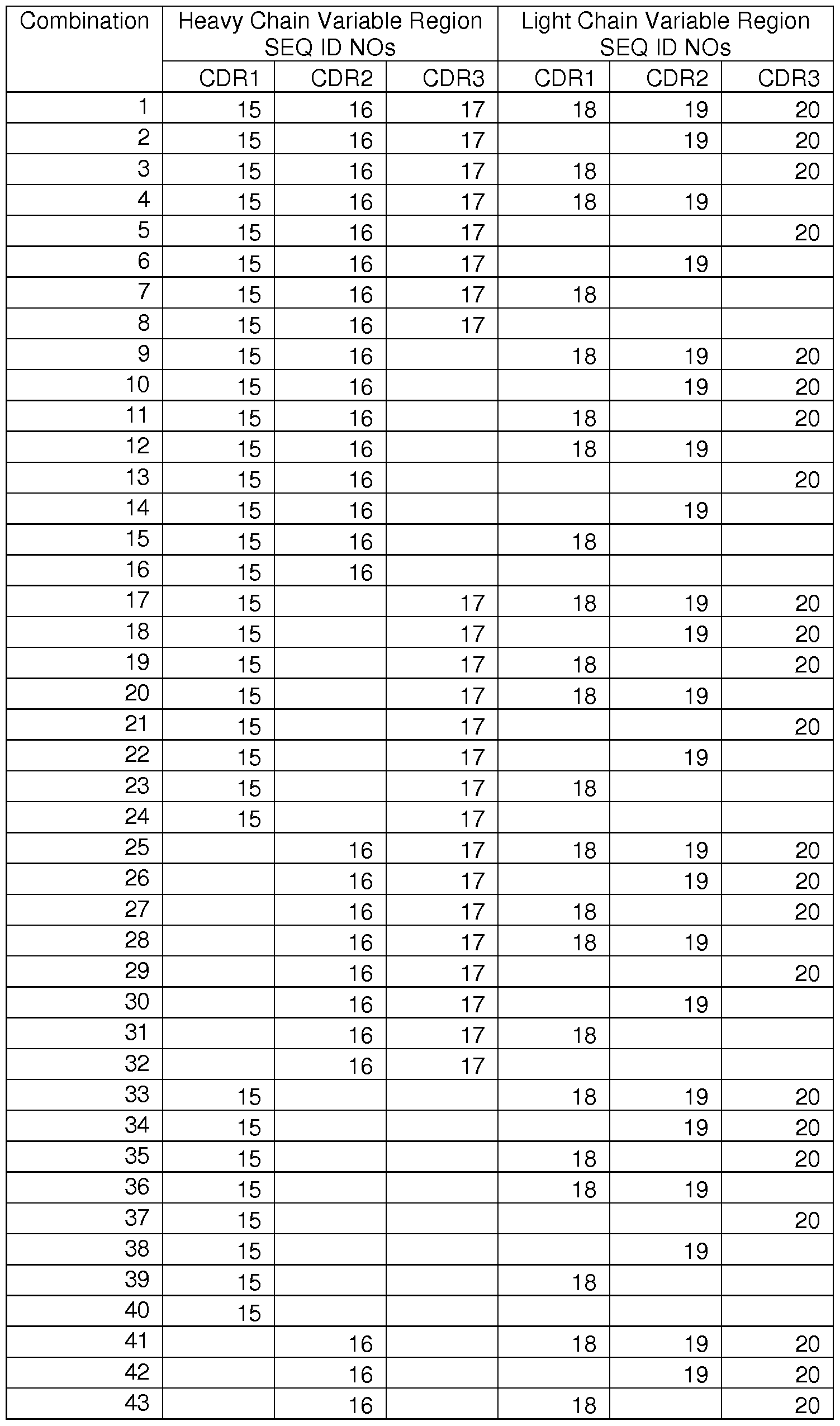
45 16 20
46 16 19
47 16 18
48 16
49 17 18 19 20
50 17 19 20
51 17 18 20
52 17 18 19
53 17 20
54 17 19
55 17 18
56 17
57 18 19 20
58 19 20
59 18 20
60 18 19
61 20
62 19
63 18
[0057] Each of the sequences referred to above may be found in Table B below.

CATTAGCAAATA I I I AGCCTGGTATCAAGAGAAACCTGGGA
AAACTAATAAGCTTCTTATCTACTTTGGATCCACTTTGCAATC
TGGGATTCCATCAAGGTTCAGTGGCAGTGGATCTGGTACAG
ATTTCACTCTCACCATCAGTAGCCTGGAGCCTGAAGATTTTG
CAATGTA I I I CTGTCAACAGCATAATGAATACCCGTACACGT
TCGGAGGGGGGACCAAGCTGGAAATGAAA
8 DVQITQSPSYLAASPGETITINCRASKSISKYLAWYQEKPGKTN
KLLIYFGSTLQSGIPSRFSGSGSGTDFTLTISSLEPEDFAMYFC
QQHNEYPYTFGGGTKLEMK
9 QNPFSEDKTD
10 IDQDPSQNPF
1 1 IDQDPSQNPFSEDKTD
12 EIAGLQIGDK
13 IGDKIMQVNG
14 EIAGLQIGDKIMQVNG
15 SFTGYFMN
16 IGRIDPYNGNIFYNQ
17 AVYYCGRSYGNY
18 GETITINCRA
19 KPGKTNKLLIYF
20 EPEDFAMYFCQQHNE
21 QPVTAVVQRV
(iv) GRP78 epitopes
[0058] In one embodiment, an isolated antibody of the present invention that binds to GPR78 recognizes an epitope within the amino acid sequence of SEQ ID NO:1 . For instance, a GRP78 antibody of the invention may recognize an epitope with 5, 6, 7, 8, or 9 contiguous amino acids of SEQ ID NO:1 .
[0059] In yet another embodiment, an isolated antibody of the present invention that binds to GPR78 recognizes an epitope within the amino acid sequence of SEQ ID NO:2. For instance, a GRP78 antibody of the invention may recognize an epitope with 5, 6, 7, 8, or 9 contiguous amino acids of SEQ ID NO: 2.
[0060] In yet a further embodiment, an isolated antibody of the present invention that binds to GPR78 recognizes an epitope within the amino acid sequence of SEQ ID NO:3. For instance, a GRP78 antibody of the invention may recognize an
epitope with 5, 6, 7, 8, 9, 10, 1 1 , 12, 13, 14, or 15 contiguous amino acids of SEQ ID NO:3.
[0061 ] In an additional embodiment, an isolated antibody of the present invention that binds to GPR78 recognizes an epitope within the amino acid sequence of SEQ ID NO:4. For instance, a GRP78 antibody of the invention may recognize an epitope with 5, 6, 7, 8, or 9 contiguous amino acids of SEQ ID NO: 4.
(b) antibodies that bind to TIP-1
[0062] In one embodiment, the isolated antibody of the present invention that binds to TIP-1 recognizes an epitope within the amino acid sequence of SEQ ID NO:9. For instance, a TIP-1 antibody of the invention may recognize an epitope with 5, 6, 7, 8, or 9 contiguous amino acids of SEQ ID NO: 9.
[0063] In yet another embodiment, the isolated antibody of the present invention that binds to TIP-1 recognizes an epitope within the amino acid sequence of SEQ ID NO:10. For instance, a TIP-1 antibody of the invention may recognize an epitope with 5, 6, 7, 8, or 9 contiguous amino acids of SEQ ID NO: 10.
[0064] In yet a further embodiment, the isolated antibody of the present invention that binds to TIP-1 recognizes an epitope within the amino acid sequence of SEQ ID NO:1 1 . For instance, a TIP-1 antibody of the invention may recognize an epitope with 5, 6, 7, 8, 9, 10, 1 1 , 12, 13, 14, or 15 contiguous amino acids of SEQ ID NO:1 1 .
[0065] In an additional embodiment, the isolated antibody of the present invention that binds to TIP-1 recognizes an epitope within the amino acid sequence of SEQ ID NO:12. For instance, a TIP-1 antibody of the invention may recognize an epitope with 5, 6, 7, 8, or 9 contiguous amino acids of SEQ ID NO: 12.
[0066] Yet another embodiment provides for the isolated antibody of the present invention that binds to TIP-1 , wherein the antibody recognizes an epitope within the amino acid sequence of SEQ ID NO: 13. For instance, a TIP-1 antibody of the invention may recognize an epitope with 5, 6, 7, 8, 9, or 10 contiguous amino acids of SEQ ID NO: 13.
[0067] In still another embodiment, the isolated antibody of the present invention that binds to TIP-1 recognizes an epitope within the amino acid sequence of SEQ ID NO:14. For instance, a TIP-1 antibody of the invention may recognize an epitope with 5, 6, 7, 8, 9, 10, 1 1 , or 12 contiguous amino acids of SEQ ID NO:14.
[0068] In another embodiment, the isolated antibody of the present invention that binds to TIP-1 recognizes an epitope within the amino acid sequence of SEQ ID NO:21 . For instance, a TIP-1 antibody of the invention may recognize an epitope with 5, 6, 7, 8, or 9 contiguous amino acids of SEQ ID NO: 21 .
[0069] In yet another embodiment, an isolated antibody of the invention that binds to TIP-1 is an scFv antibody encoded by a nucleic acid sequence that encodes SEQ ID NO 22 (Fig. 22).
[0070] In another embodiment, an isolated antibody of the invention that binds to TIP-1 is an scFv antibody encoded by a nucleic acid sequence that encodes SEQ ID NO 23 (Fig. 22).
(c) Humanized Antibodies
[0071 ] The antibodies of the present invention may also be chimeric antibodies. Preferably, these chimeric antibodies involve the merging of a portion of a monoclonal antibody with antibody-producing DNA in living cells to produce a
monoclonal antibody that has material from more than one species of animal. This procedure is well known in the art and any known method to produce chimeric antibodies is suitable for purposes of the present invention. In a preferred embodiment, the chimeric antibody comprises mouse elements conjugated to the genetic material of another species. In a particularly preferred embodiment, the chimeric antibody comprises mouse and human elements to form humanized antibodies. The process of humanization decreases the potential for the antibody to induce an immune response in a human host.
[0072] As used herein "humanized antibody" includes an anti-ApoE antibody that is composed partially or fully of amino acid sequences derived from a human antibody germline by altering the sequence of an antibody having non-human
complementarity determining regions ("CDR"). The simplest such alteration may consist simply of substituting the constant region of a human antibody for the murine constant region, thus resulting in a human/murine chimera which may have sufficiently low immunogenicity to be acceptable for pharmaceutical use. Preferably, however, the variable region of the antibody and even the CDR is also humanized by techniques that are by now well known in the art. The framework regions of the variable regions are substituted by the corresponding human framework regions leaving the non-human CDR substantially intact, or even replacing the CDR with sequences derived from a human genome. CDRs may also be randomly mutated such that binding activity and affinity for ApoE is maintained or enhanced in the context of fully human germline framework regions or framework regions that are substantially human. Substantially human frameworks have at least 90%, 95%, or 99% sequence identity with a known human framework sequence. Fully useful human antibodies are produced in genetically modified mice whose immune systems have been altered to correspond to human immune systems. As mentioned above, it is sufficient for use in the methods of this discovery, to employ an immunologically specific fragment of the antibody, including fragments representing single chain forms.
[0073] If desired, the design of humanized immunoglobulins may be carried out as follows. When an amino acid falls under the following category, the framework amino acid of a human immunoglobulin to be used (acceptor
immunoglobulin) is replaced by a framework amino acid from a CDR-providing nonhuman immunoglobulin (donor immunoglobulin): (a) the amino acid in the human framework region of the acceptor immunoglobulin is unusual for human immunoglobulin at that position, whereas the corresponding amino acid in the donor immunoglobulin is typical for human immunoglobulin at that position; (b) the position of the amino acid is immediately adjacent to one of the CDRs; or (c) any side chain atom of a framework amino acid is within about 5-6 angstroms (center-to-center) of any atom of a CDR amino acid in a three dimensional immunoglobulin model (Queen, et al., op. cit., and Co, ct al, Proc. Natl. Acad. Sci. USA (1991 ) 88:2869). When each of the amino acids in the human framework region of the acceptor immunoglobulin and a corresponding
amino acid in the donor immunoglobulin is unusual for human immunoglobulin at that position, such an amino acid is replaced by an amino acid typical for human
immunoglobulin at that position.
[0074] One embodiment of the present invention encompasses a humanized antibody that binds to GRP78. In particular, an embodiment of the invention encompasses a humanized antibody where amino acids in the framework region of either the heavy or light chain variable regions are humanized, leaving the CDRs intact. For instance, in one embodiment, an antibody may comprise a combination of CDRs listed in Table A, and a humanized framework region. In another embodiment, the sequence of one or more CDR regions may be humanized as well as the framework region.
[0075] In an exemplary embodiment, a humanized GRP78 antibody recognizes at least one epitope from the group consisting of SEQ ID NO:1 , 2, 3, or 4.
[0076] In an alternative embodiment, the present invention encompasses a humanized antibody that binds to TIP-1 . In particular, an embodiment of the invention encompasses a humanized antibody where amino acids in the framework region of either the heavy or light chain variable regions are humanized, leaving the CDRs intact.
[0077] In an exemplary embodiment, a humanized TIP-1 antibody recognizes at least one epitope from the group consisting of SEQ ID NO:9, 10, 1 1 , 12, 13, 14, or 21 .
[0078] In another alternative embodiment, the present invention
encompasses a scFv antibody that binds to TIP-1 .
(d) Antibody Conjugates
[0079] In another aspect, an antibody of the present invention is conjugated to a therapeutic agent. In some embodiments, a scFv of the present invention is conjugated to a therapeutic agent. The therapeutic agent preferably reduces or interferes with tumor growth or otherwise reduces the effect of the tumor within the body or organism. A therapeutic agent that reduces the symptoms produced by the tumor or reduces tumor growth is suitable for the present invention.
[0080] Additionally, any therapeutic agent that reduces the symptoms associated with tumor cell growth will work for purposes of the present invention. Non- limiting examples of therapeutic agents may include drugs, therapeutic compounds, genetic materials, metals (such as radioactive isotopes), proteins, peptides,
carbohydrates, lipids, steroids, nucleic acid based materials, or derivatives, analogues, or combinations thereof in their native form or derivatized with hydrophobic or charged moieties to enhance incorporation or adsorption into a cell. Such therapeutic agents may be water soluble or may be hydrophobic. Non-limiting examples of therapeutic agents may include immune-related agents, thyroid agents, respiratory products, antineoplastic agents, anti-helmintics, anti-malarials, mitotic inhibitors, hormones, anti- protozoans, anti-tuberculars, cardiovascular products, blood products, biological response modifiers, anti-fungal agents, vitamins, peptides, anti-allergic agents, anticoagulation agents, circulatory drugs, metabolic potentiators, anti-virals, anti-anginals, antibiotics, anti-inflammatories, anti-rheumatics, narcotics, cardiac glycosides, neuromuscular blockers, sedatives, local anesthetics, general anesthetics, or radioactive atoms or ions. Non-limiting examples of therapeutic agents are included in Table C below. An isolated antibody of the present invention may be conjugated to one, two, three, four, or five therapeutic agents. Methods of conjugating an antibody to a therapeutic agent are known in the art. Generally speaking, the conjugation should not interfere with the antibody recognizing its target, and should not interfere with the active site of the target. In some instances, a scFv may be generated with a cleavable linkage between the scFv and therapeutic agent. Such a linker may allow release of the therapeutic agent at a specific cellular location.
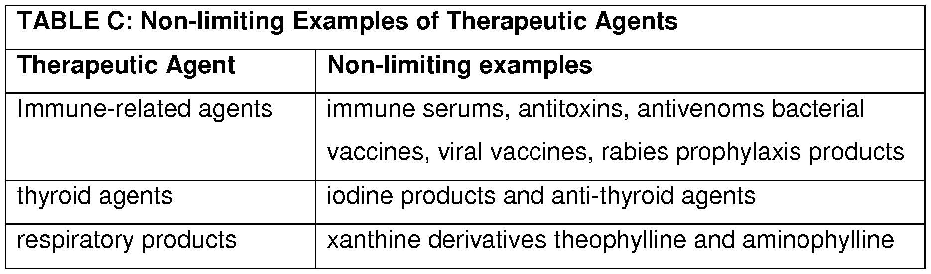
mercaptopurine, mitotane, procarbazine
hydrochloride dactinomycin (actinomycin D), daunorubicin hydrochloride, doxorubicin
hydrochloride, paclitaxel and other taxenes, rapamycin, manumycin A, TNP-470, plicamycin (mithramycin), aminoglutethimide, estramustine phosphate sodium, flutamide, leuprolide acetate, megestrol acetate, tamoxifen citrate, testolactone, trilostane, amsacrine (m-AMSA), asparaginase (L- asparaginase) Erwina asparaginase, interferon a-2a, interferon a-2b, teniposide (VM-26), vinblastine sulfate (VLB), vincristine sulfate, bleomycin sulfate, hydroxyurea, procarbazine, and dacarbazine anti-helmintics pyrantel pamoate, piperazine, tetrachloroethylene, thiabendazole, niclosamide
antimalarials Chloroquine, amodiaquine, antifolate drugs,
proguanil (chloroguanide), mefloquine, quinine, halofantrine, artemesinin and derivaties, primaquine, doxycycline, tetracycline, and clindamycin
mitotic inhibitors etoposide, colchicine, and the vinca alkaloids hormones androgens, progestins, estrogens and antiestrogens, growth hormone, melanocyte stimulating hormone, estradiol, beclomethasone dipropionate,
betamethasone, betamethasone acetate and
betamethasone sodium phosphate, vetamethasone disodium phosphate, vetamethasone sodium phosphate, cortisone acetate, dexamethasone, dexamethasone acetate, dexamethasone sodium phosphate, flunisolide, hydrocortisone,
hydrocortisone acetate, hydrocortisone cypionate, hydrocortisone sodium phosphate, hydrocortisone sodium succinate, methylprednisolone,
methylprednisolone acetate, methylprednisolone sodium succinate, paramethasone acetate, prednisolone, prednisolone acetate, prednisolone sodium phosphate, prednisolone tebutate, prednisone, triamcinolone, triamcinolone acetonide, triamcinolone diacetate, triamcinolone hexacetonide, fludrocortisone acetate, oxytocin, vassopressin, glucagon and their derivatives
antiprotozoans chloroquine, hydroxychloroquine, metronidazole, quinine and meglumine antimonite
antituberculars para-aminosalicylic acid, isoniazid, capreomycin sulfate cycloserine, ethambutol hydrochloride ethionamide, pyrazinamide, rifampin, and
streptomycin sulfate
cardiovascular products chelating agents and mercurial diuretics and cardiac glycosides
blood products parenteral iron, hemin, hematoporphyrins and their derivatives
biological response muramyldipeptide, muramyltripeptide, microbial cell modifiers wall components, lymphokines (e.g., bacterial
endotoxin such as lipopolysaccharide, macrophage activation factor), sub-units of bacteria (such as
Mycobacteria, Corynebacteria), the synthetic
dipeptide N-acetyl-muramyl-L-alanyl-D-isoglutamine anti-fungal agents ketoconazole, nystatin, griseofulvin, flucytosine (5- fc), miconazole, amphotericin B, ricin, cyclosporins, and β-lactam antibiotics (e.g., sulfazecin)
vitamins cyanocobalamin neinoic acid, retinoids and
derivatives such as retinol palmitate, and a- tocopherol
peptides manganese super oxide dismutase; enzymes such as alkaline phosphatase
anti-allergic agents Amelexanox
anti-coagulation agents phenprocoumon and heparin
circulatory drugs Propranolol
metabolic potentiators Glutathione
antivirals acyclovir, amantadine azidothymidine (AZT, DDI,
Foscarnet, or Zidovudine), ribavirin and vidarabine monohydrate (adenine arabinoside, ara-A) antianginals diltiazem, nifedipine, verapamil, erythritol tetranitrate, isosorbide dinitrate, nitroglycerin (glyceryl trinitrate) and pentaerythritol tetranitrate
antibiotics dapsone, chloramphenicol, neomycin, cefaclor,
cefadroxil, cephalexin, cephradine erythromycin, clindamycin, lincomycin, amoxicillin, ampicillin, bacampicillin, carbenicillin, dicloxacillin, cyclacillin, picloxacillin, hetacillin, methicillin, nafcillin, oxacillin, penicillin including penicillin G and penicillin V, ticarcillin rifampin, aminoglycosides and tetracycline antiinflammatories diflunisal, ibuprofen, indomethacin, meclofenamate, mefenamic acid, naproxen, oxyphenbutazone, phenylbutazone, piroxicam, sulindac, tolmetin,
aspirin and salicylates
antirheumatics Adalimumab, azathioprine, chloroquine and
hydroxychloroquine (antimalarials),
cyclosporine (Cyclosporin A), D-penicillamine, etanercept, gold salts (sodium aurothiomalate, auranofin), infliximab, leflunomide, methotrexate, minocycline (a tetracycline antibiotic), sulfasalazine narcotics Paregoric, opiates, codeine, heroin, methadone, morphine and opium
cardiac glycosides deslanoside, digitoxin, digoxin, digitalin and digitalis neuromuscular blockers atracurium mesylate, gallamine triethiodide,
hexafluorenium bromide, metocurine iodide, pancuronium bromide, succinylcholine chloride (suxamethonium chloride), tubocurarine chloride and vecuronium bromide
sedatives (hypnotics) amobarbital, amobarbital sodium, aprobarbital,
butabarbital sodium, chloral hydrate, ethchlorvynol, ethinamate, flurazepam hydrochloride, glutethimide, methotrimeprazine hydrochloride, methyprylon, midazolam hydrochloride, paraldehyde,
pentobarbital, pentobarbital sodium, phenobarbital sodium, secobarbital sodium, talbutal, temazepam and triazolam
local anesthetics bupivacaine hydrochloride, chloroprocaine
hydrochloride, etidocaine hydrochloride, lidocaine hydrochloride, mepivacaine hydrochloride, procaine hydrochloride and tetracaine hydrochloride
general anesthetics droperidol, etomidate, fentanyl citrate with droperidol, ketamine hydrochloride, methohexital sodium and thiopental sodium
radioactive particles or strontium, iodide rhenium, yttrium, and
ions radiopharmaceuticals, such as radioactive iodine,
copper and phosphorus product
II. Method of Use
[0081 ] In another aspect, an isolated antibody of the present invention, as described above, may be used in treating and preventing cancer and associated diseases in a subject. The antibodies of the present invention may be conjugated to radioisotopes or chemotherapeutic compounds in order to provide specific delivery of radiation and chemotherapy to the site of a tumor. Further, the antibodies of the present invention may be part of a combination therapy. Preferably, a combination therapy would include the use of the antibody of the present invention along with a radiation therapy or chemotherapy course of treatment. It has also been suggested that monoclonal antibodies, such as those described herein, may increase the susceptibility of tumor cells to the effects of chemotherapy or radiation. In preferred embodiments, the antibodies of the invention may be used to enhance the efficacy of cancer radiotherapy.
[0082] In yet another aspect, the present invention provides a method of imaging a cancer. As such, an antibody of the invention may be conjugated to an imaging agent. For instance, an scVf may be conjugated to an imaging agent. Suitable imaging agents may include, but are not limited to, imaging/tracking agents that may be used for microscopy, e.g. fluorescent microscopy, confocal microscopy, or electron microscopy, magnetic resonance imaging, tomography, such as gamma (SPECT/CT, planar) and positron emission tomography (PET/CT), radiography, or ultrasound.
Imaging/tracking agents may be detectable in situ, in vivo, ex vivo, and in vitro. In general, imaging/tracking agents may include luminescent molecules,
chemiluminescent molecules, fluorochromes, fluorescent quenching agents, colored molecules, radioisotopes, scintillants, massive labels (for detection via mass changes), biotin, avidin, streptavidin, protein A, protein G, antibodies or fragments thereof, Grb2, polyhistidine, Ni2+, Flag tags, myc tags, heavy metals, enzymes, alkaline phosphatase, peroxidase, luciferase, electron donors/acceptors, acridinium esters, and colorimetric
substrates. The skilled artisan would readily recognize other useful labels that are not mentioned above, which may be employed in the operation of the present invention.
[0083] The antibodies are as described in Sections I - VI above. The subject, the cancer, and the administration of the antibodies are described below.
(a) subject
[0084] A method of the invention may be used to detect or treat a tumor in a subject that is a human, a livestock animal, a companion animal, a lab animal, or a zoological animal. In one embodiment, the subject may be a rodent, e.g. a mouse, a rat, a guinea pig, etc. In another embodiment, the subject may be a livestock animal. Non- limiting examples of suitable livestock animals may include pigs, cows, horses, goats, sheep, llamas and alpacas. In yet another embodiment, the subject may be a
companion animal. Non-limiting examples of companion animals may include pets such as dogs, cats, rabbits, and birds. In yet another embodiment, the subject may be a zoological animal. As used herein, a "zoological animal" refers to an animal that may be found in a zoo. Such animals may include non-human primates, large cats, wolves, and bears. In preferred embodiments, the animal is a laboratory animal. Non-limiting examples of a laboratory animal may include rodents, canines, felines, and non-human primates. In certain embodiments, the animal is a rodent. Non-limiting examples of rodents may include mice, rats, guinea pigs, etc. The genotype of the sterile animal can and may vary depending on the intended use of the animal. In embodiments where the animal is a mouse, the mouse may be a C57BL/6 mouse, a Balb/c mouse, a 129sv, a GL261 tumor bearing mouse, a D54 tumor bearing mouse, or any other laboratory strain.
(b) tumor
[0085] An antibody of the invention may be used to treat or recognize tumor derived from a neoplasm or a cancer. The neoplasm may be malignant or benign, the cancer may be primary or metastatic; the neoplasm or cancer may be early stage or late stage. Non-limiting examples of neoplasms or cancers that may be treated
include acute lymphoblastic leukemia, acute myeloid leukemia, adrenocortical carcinoma, AIDS-related cancers, AIDS-related lymphoma, anal cancer, appendix cancer, astrocytomas (childhood cerebellar or cerebral), basal cell carcinoma, bile duct cancer, bladder cancer, bone cancer, brainstem glioma, brain tumors (cerebellar astrocytoma, cerebral astrocytoma/malignant glioma, ependymoma, medulloblastoma, supratentorial primitive neuroectodermal tumors, visual pathway and hypothalamic gliomas), breast cancer, bronchial adenomas/carcinoids, Burkitt lymphoma, carcinoid tumors (childhood, gastrointestinal), carcinoma of unknown primary, central nervous system lymphoma (primary), cerebellar astrocytoma, cerebral astrocytoma/malignant glioma, cervical cancer, childhood cancers, chronic lymphocytic leukemia, chronic myelogenous leukemia, chronic myeloproliferative disorders, colon cancer, cutaneous T-cell lymphoma, desmoplastic small round cell tumor, endometrial cancer,
ependymoma, esophageal cancer, Ewing's sarcoma in the Ewing family of tumors, extracranial germ cell tumor (childhood), extragonadal germ cell tumor, extrahepatic bile duct cancer, eye cancers (intraocular melanoma, retinoblastoma), gallbladder cancer, gastric (stomach) cancer, gastrointestinal carcinoid tumor, gastrointestinal stromal tumor, germ cell tumors (childhood extracranial, extragonadal, ovarian), gestational trophoblastic tumor, gliomas (adult, childhood brain stem, childhood cerebral astrocytoma, childhood visual pathway and hypothalamic), gastric carcinoid, hairy cell leukemia, head and neck cancer, hepatocellular (liver) cancer, Hodgkin lymphoma, hypopharyngeal cancer, hypothalamic and visual pathway glioma (childhood), intraocular melanoma, islet cell carcinoma, Kaposi sarcoma, kidney cancer (renal cell cancer), laryngeal cancer, leukemias (acute lymphoblastic, acute myeloid, chronic lymphocytic, chronic myelogenous, hairy cell), lip and oral cavity cancer, liver cancer (primary), lung cancers (non-small cell, small cell), lymphomas (AIDS-related, Burkitt, cutaneous T-cell, Hodgkin, non-Hodgkin, primary central nervous system),
macroglobulinemia (Waldenstrom), malignant fibrous histiocytoma of
bone/osteosarcoma, medulloblastoma (childhood), melanoma, intraocular melanoma, Merkel cell carcinoma, mesotheliomas (adult malignant, childhood), metastatic squamous neck cancer with occult primary, mouth cancer, multiple endocrine neoplasia
syndrome (childhood), multiple myeloma/plasma cell neoplasm, mycosis fungoides, myelodysplastic syndromes, myelodysplastic/myeloproliferative diseases, myelogenous leukemia (chronic), myeloid leukemias (adult acute, childhood acute), multiple myeloma, myeloproliferative disorders (chronic), nasal cavity and paranasal sinus cancer, nasopharyngeal carcinoma, neuroblastoma, non-Hodgkin lymphoma, non-small cell lung cancer, oral cancer, oropharyngeal cancer, osteosarcoma/malignant fibrous histiocytoma of bone, ovarian cancer, ovarian epithelial cancer (surface epithelial- stromal tumor), ovarian germ cell tumor, ovarian low malignant potential tumor, pancreatic cancer, pancreatic cancer (islet cell), paranasal sinus and nasal cavity cancer, parathyroid cancer, penile cancer, pharyngeal cancer, pheochromocytoma, pineal astrocytoma, pineal germinoma, pineoblastoma and supratentorial primitive neuroectodermal tumors (childhood), pituitary adenoma, plasma cell neoplasia, pleuropulmonary blastoma, primary central nervous system lymphoma, prostate cancer, rectal cancer, renal cell carcinoma (kidney cancer), renal pelvis and ureter transitional cell cancer, retinoblastoma, rhabdomyosarcoma (childhood), salivary gland cancer, sarcoma (Ewing family of tumors, Kaposi, soft tissue, uterine), Sezary syndrome, skin cancers (nonmelanoma, melanoma), skin carcinoma (Merkel cell), small cell lung cancer, small intestine cancer, soft tissue sarcoma, squamous cell carcinoma, squamous neck cancer with occult primary (metastatic), stomach cancer, supratentorial primitive neuroectodermal tumor (childhood), T-Cell lymphoma (cutaneous), testicular cancer, throat cancer, thymoma (childhood), thymoma and thymic carcinoma, thyroid cancer, thyroid cancer (childhood), transitional cell cancer of the renal pelvis and ureter, trophoblastic tumor (gestational), enknown primary site (adult, childhood), ureter and renal pelvis transitional cell cancer, urethral cancer, uterine cancer (endometrial), uterine sarcoma, vaginal cancer, visual pathway and hypothalamic glioma (childhood), vulvar cancer, Waldenstrom macroglobulinemia, and Wilms tumor (childhood). In preferred embodiments, the neoplasm or cancer is non-small cell lung carcinoma.
(c) administration
[0086] In certain aspects, a pharmacologically effective amount of an antibody of the invention, including immunologically reactive fragments, may be administered to a subject. Administration is performed using standard effective techniques, include peripherally (i.e. not by administration into the central nervous system) or locally to the central nervous system. Peripheral administration includes but is not limited to intravenous, intraperitoneal, subcutaneous, pulmonary, transdermal, intramuscular, intranasal, buccal, sublingual, or suppository administration. Local administration, including directly into the central nervous system (CNS) includes but is not limited to via a lumbar, intraventricular or intraparenchymal catheter or using a surgically implanted controlled release formulation.
[0087] Pharmaceutical compositions for effective administration are deliberately designed to be appropriate for the selected mode of administration, and pharmaceutically acceptable excipients such as compatible dispersing agents, buffers, surfactants, preservatives, solubilizing agents, isotonicity agents, stabilizing agents and the like are used as appropriate. Remington's Pharmaceutical Sciences, Mack
Publishing Co., Easton Pa., 16Ed ISBN: 0-912734-04-3, latest edition, incorporated herein by reference in its entirety, provides a compendium of formulation techniques as are generally known to practitioners. It may be particularly useful to alter the solubility characteristics of the antibodies useful in this discovery, making them more lipophilic, for example, by encapsulating them in liposomes or by blocking polar groups.
[0088] Effective peripheral systemic delivery by intravenous or
intraperitoneal or subcutaneous injection is a preferred method of administration to a living patient. Suitable vehicles for such injections are straightforward. In addition, however, administration may also be effected through the mucosal membranes by means of nasal aerosols or suppositories. Suitable formulations for such modes of administration are well known and typically include surfactants that facilitate cross- membrane transfer. Such surfactants are often derived from steroids or are cationic lipids, such as N-[1 -(2,3-dioleoyl)propyl]-N,N,N-trimethyl ammonium chloride (DOTMA)
or various compounds such as cholesterol hemisuccinate, phosphatidyl glycerols and the like.
[0089] The concentration of antibody in formulations to be administered is an effective amount and ranges from as low as about 0.1 % by weight to as much as about 15 or about 20% by weight and will be selected primarily based on fluid volumes, viscosities, and so forth, in accordance with the particular mode of administration selected if desired. A typical composition for injection to a living patient could be made up to contain 1 mL sterile buffered water of phosphate buffered saline and about 1 -1000 mg of any one of or a combination of the humanized antibody of the present discovery. The formulation could be sterile filtered after making the formulation, or otherwise made microbiologically acceptable. A typical composition for intravenous infusion could have volumes between 1 -250 mL of fluid, such as sterile Ringer's solution, and 1 -100 mg per ml, or more in anti-tau antibody concentration. Therapeutic agents of the discovery can be frozen or lyophilized for storage and reconstituted in a suitable sterile carrier prior to use. Lyophilization and reconstitution may lead to varying degrees of antibody activity loss (e.g. with conventional immune globulins, IgM antibodies tend to have greater activity loss than IgG antibodies). Dosages administered are effective dosages and may have to be adjusted to compensate. The pH of the formulations generally pharmaceutical grade quality, will be selected to balance antibody stability (chemical and physical) and comfort to the patient when administered. Generally, a pH between 4 and 8 is tolerated. Doses will vary from individual to individual based on size, weight, and other physiobiological characteristics of the individual receiving the successful administration.
[0090] As used herein, the term "effective amount" means an amount of a substance such as a compound that leads to measurable and beneficial effects for the subject administered the substance, i.e., significant efficacy. The effective amount or dose of compound administered according to this discovery will be determined by the circumstances surrounding the case, including the compound administered, the route of administration, the status of the symptoms being treated and similar patient and administration situation considerations among other considerations.
[0091 ] In some embodiments, when the antibody is an anti-TIP1 antibody labled with 64Cu, the dose administered may be about 0.01 , 0.02, 0.03, 0.04, 0.05 0.06, 0.07, 0.08, 0.09, 0.1 , 0.01 1 , 0.012, 0.013, 0.013, 0.014, 0.015, 0.016, 0.017, 0.018, 0.019, 0.02, 0.021 , 0.022, 0.023, 0.023, 0.024, 0.025, 0.026, 0.027, 0.028, 0.029, 0.03, 0.031 , 0.032, 0.033, 0.033, 0.034, 0.035, 0.036, 0.037, 0.038, 0.039, 0.04, 0.041 , 0.042, 0.043, 0.043, 0.044, 0.045, 0.046, 0.047, 0.048, 0.049, 0.05, 0.051 , 0.052, 0.053, 0.053, 0.054, 0.055, 0.056, 0.057, 0.058, 0.059, 0.06, 0.061 , 0.062, 0.063, 0.063, 0.064, 0.065, 0.066, 0.067, 0.068, 0.069, 0.07, 0.071 , 0.072, 0.073, 0.073, 0.074, 0.075, 0.076, 0.077, 0.078, 0.079, 0.08, 0.081 , 0.082, 0.083, 0.083, 0.084, 0.085, 0.086, 0.087, 0.088, 0.089, 0.09, 0.091 , 0.092, 0.093, 0.093, 0.094, 0.095, 0.096, 0.097, 0.098, 0.099, or about 0.1 rem/mCi.
[0092] In some embodiments, when the antibody is an anti-GRP78 labled with 64Cu, the dose administered may be about 0.01 , 0.02, 0.03, 0.04, 0.05 0.06, 0.07, 0.08, 0.09, 0.1 , 0.01 1 , 0.012, 0.013, 0.013, 0.014, 0.015, 0.016, 0.017, 0.018, 0.019, 0.02, 0.021 , 0.022, 0.023, 0.023, 0.024, 0.025, 0.026, 0.027, 0.028, 0.029, 0.03, 0.031 , 0.032, 0.033, 0.033, 0.034, 0.035, 0.036, 0.037, 0.038, 0.039, 0.04, 0.041 , 0.042, 0.043, 0.043, 0.044, 0.045, 0.046, 0.047, 0.048, 0.049, 0.05, 0.051 , 0.052, 0.053, 0.053, 0.054, 0.055, 0.056, 0.057, 0.058, 0.059, 0.06, 0.061 , 0.062, 0.063, 0.063, 0.064, 0.065, 0.066, 0.067, 0.068, 0.069, 0.07, 0.071 , 0.072, 0.073, 0.073, 0.074, 0.075, 0.076, 0.077, 0.078, 0.079, 0.08, 0.081 , 0.082, 0.083, 0.083, 0.084, 0.085, 0.086, 0.087, 0.088, 0.089, 0.09, 0.091 , 0.092, 0.093, 0.093, 0.094, 0.095, 0.096, 0.097, 0.098, 0.099, or about 0.1 rem/mCi.
[0093] The frequency of dosing may be daily or once, twice, three times or more per week or per month, as needed as to effectively treat the symptoms. The timing of administration of the treatment relative to the disease itself and duration of treatment will be determined by the circumstances surrounding the case. Treatment could begin immediately, such as at the site of the injury as administered by emergency medical personnel. Treatment could begin in a hospital or clinic itself, or at a later time after discharge from the hospital or after being seen in an outpatient clinic. Duration of
treatment could range from a single dose administered on a one-time basis to a life-long course of therapeutic treatments.
[0094] Although the foregoing methods appear the most convenient and most appropriate and effective for administration of proteins such as antibodies, by suitable adaptation, other effective techniques for administration, such as
intraventricular administration, transdermal administration and oral administration may be employed provided proper formulation is utilized herein.
[0095] In addition, it may be desirable to employ controlled release formulations using biodegradable films and matrices, or osmotic mini-pumps, or delivery systems based on dextran beads, alginate, or collagen.
[0096] Typical dosage levels can be determined and optimized using standard clinical techniques and will be dependent on the mode of administration.
DEFINITIONS
[0097] As used herein, "antibody" refers to an immunoglobulin derived molecule that specifically recognizes either GRP78 or TIP-1 . An antibody of the invention may be a full length antibody (IgM, IgG, IgA, IgE) or may be an antibody fragment (Fab, F(ab')2, scFv). An antibody may be chimeric or may be humanized.
[0098] As used herein, "CDR" means "complementary determining region." CDRs may also be referred to as hypervariable regions.
[0099] As used herein, "light chain" is the small polypeptide subunit of the antibody. A typical antibody comprises two light chains and two heavy chains.
[0100] As used herein, the "heavy chain" is the large polypeptide subunit of the antibody. The heavy chain of an antibody contain a series of immunoglobulin domains, with at least one variable domain and at least one constant domain.
[0101 ] "Humanized", as used herein, refers to the process where monoclonal antibodies are produced using recombinant DNA to create constructs capable of expression in human cell culture. Any known techniques for producing these constructs will work for purposes of the present invention.
[0102] As used herein, "single chain variable fragments" or "scFv" or "scFvs", refer to fusion proteins of the variable regions of the heavy and light chains of immunoglobulins connected via a linker. In some embodiment, the linker is a peptide of about 10 to 25 amino acids.
[0103] A "therapeutic agent" for purposes of the present invention, refers to an agent that reduces tumor growth, any related cancer growth, or reduces the symptoms associated with cancerous cell growth. The therapeutic agent that is preferably conjugated to the antibody of the present invention is preferably a biologic, pharmaceutical or chemical agent. A non-limiting list of therapetuic agents that may be suitable for use in the present invention is described above.
[0104] As used herein, "imaging agent" refers to any agent that can be used to locate and produce an image of cancerous cell growth or tumors. A non-limiting list of imagining agents that may be suitable for use in the present invention is described above.
EXAMPLES
[0105] The following examples illustrate various iterations of the invention.
Materials and Methods
Tumor model
[0106] GL261 murine glioma cancer cell line was purchased from
American Type Culture Collection. D54 cells were similarly purchased commercially. Heterotopic tumor models were developed by s.c. inoculating cell suspensions (6 χ 106 cells) into nude or C57/BI6 mice.
Near IR imaging
[0107] Tumor-bearing mice were treated with anywhere from one to three once-daily doses of 3 Gy XRT or sham XRT (three per group) and injected with peptide or antibody 3 hours after the last XRT treatment. Mice were then injected with an antibody to 78-kDa glucose regulated protein (GRP78) conjugated with AlexaFluor750 and tumors were removed 7 days after labeled antibody injection; polyclonal serum IgG
antibody was used as a control. Near IR images were taken using the I VIS imaging system with an ICG filter setting at various time points after the injection.
Anti Tip 1 and anti-GRP78 monoclonal antibody production and purification.
[0108] BALB/C mice were initially immunized with 50 mg of Tip-1 or
GRP78 antigen mixed with equivalent amounts of Titermax adjuvant (CytRx
Corporation) for each mouse. One month after initial immunization, mice were boosted with equivalent amounts of antigen without adjuvant two to three times at two week intervals. The mouse polyclonal antibody titer was evaluated by ELISA and Western blot methods. Mice exhibiting high immune response to either antigen were chosen as B cell donors. Spleen were removed and homogenized in RPMI 1640 culture medium free of serum and other additives. Spleen cells were combined with Sp2/O mouse myeloma (2 x 10⁷ per spleen). Mixed cells were washed twice and centrifuged at 1200 rpm for 8 minutes at room temperature. Supernatant was removed, and the cell pellet was lightly agitated to loosen the cells. Approximately 1 ml PEG (polyethylene glycol 1500)
(Roche) was added to fuse the cells. The fused cells were washed once with plain medium and finally re-suspended in RPMI 1640 medium supplemented with 10% fetal bovine serum (Gemini Bioproducts, CA), L-glutamine, antibiotics and HAT (Sigma), plated into 24 well tissue culture plates and incubated at 37°C in a humidified CO2 incubator. Fifteen days after fusion, hybridoma culture supernatants were removed from individual wells and transferred to separate 96 well microtiter plates for ELISA, Western blot and antibody printing assays against Tip-1 antigen. Hybridomas that produced antibodies positive by immunoassay were chosen for sub-cloning by using limiting dilution. Resulting single-cell clones were retested by the aforementioned methods to detect antigen-positive monoclonal antibody-producing hybridomas. Positive hybridoma clones were transferred to individual flasks to expand cell number from one cell clone, and incubated with serum-free medium for antibody production. The antibodies produced by positive clones in serum-free medium were harvested twice a week for further monoclonal antibody purification. Filtered monoclonal antibody collected from serum-free medium was purified by protein A and protein G columns. The concentrated purified monoclonal antibody was assayed and stored at -20 °C.
Example 1
[0109] GRP78. A monoclonal antibody against GRP78 was created, and named 2D6F9. The antibody comprised a heavy chain variable region amino acid sequence of SEQ ID NO:6 (minus the leader sequence) and a light chain variable region amino acid sequence of SEQ ID NO:8 (minus the leader sequence). As shown in Fig 1 , the antibody was an lgG1 isotype, and as shown in Fig. 2 was stable at -20 °C for at least six months. Epitope mapping was performed to determine that 2D6F9 recognized SEQ ID NO. 1 , 2, 3, and 4 derived from human GRP78.
[01 10] The antibody (conjugated to Alexa Fluor 750) recognized tumor cells from the GL261 tumor model that were exposed to radiation (see Fig. 3), but did not recognize non-irradiated tumor tissue or normal tissue (either irradiated or non- irradiated). For comparison purposes a control non-specific IgG antibody was used in Fig. 5.
Example 2
[01 1 1 ] At least three monoclonal antibodies against TIP-1 were created, and named 1 A6H14, 3A415, and 2C6F3. Epitope mapping was performed to determine that 1 A6H14 recognized SEQ ID NO: 9, 10, and 1 1 derived from human TIP-1 , that 3A415 recognized SEQ ID NO: 12, 13, and 14 derived from human TIP-1 (See Table A), and that 2C6F3 recognized SEQ ID NO:21 derived from human TIP-1 .
[01 12] The antibodies were shown to recognize irradiated tumor cells compared to non-irradiated tumor tissue or normal tissue (either irradiated or non- irradiated).
Example 3. Dosimetry of labeled anti-GRP78 2D6F9 antibody.
Biodistribution in healthy mice
[01 13] Human radiation dosimetry estimates were calculated from animal biodistribution data obtained by standard method of organ dissection and using the standard MIRD methodology. Animal biodistribution data was obtained using 30 C57 mature male mice injected with 16 μΟί/100μΙ_ of 64Cu-DOTA-2D6F9 antibody divided in
6 groups of 5 animals. The average animal mouse weight was 25.0 g. Animals were euthanized and organs were harvested in groups of five at the following time points: 1 , 2, 6, 12, 24 and 48 hours post injection. The following organs were harvested, weighed and counted for radio-activity in a gamma counter: blood, lungs, liver, spleen, kidneys, bladder, muscle, fat, heart, brain, bone, red marrow, testes, adrenals, thyroid, pancreas, stomach, small intestines, and upper and lower large intestines. The animals were maintained in metabolic cages where urine and feces excretion were collected, weighted and counted for radio-activity. The biodostribution data are presented in Table 3.

Small 1 .95 +/- 1 .92 +/- 0.33 1 .95 +/- 1 .69 +/- 1 .55 +/- 1 .60 +/- Intestines 0.20 0.28 0.18 0.48 0.22
Upper L. 1 .86 +/- 2.1 1 +/- 0.34 1 .66 +/- 1 .53 +/- 1 .58 +/- 1 .46 +/- Intestines 0.40 0.23 0.17 0.47 0.24
Lower L. 0.92 +/- 1 .71 +/- 0.18 1 .96 +/- 1 .50 +/- 1 .66 +/- 1 .74 +/- Intestines 0.12 0.46 0.17 0.46 0.21
Residence times
[01 14] The residence time represents the cumulative presence time of radioactivity in any given organ and is expressed in units of time (either seconds, minutes or hours). Multiplying these values by unit of radioactivity leads to the number of radio-active decays occurring in any given organ and is therefore proportional to radiation dose. Using the above animal biodistribution data, the organ residence times (in hour) for each harvested organ were calculated by numerical integration of the time activity data expressed in percent injected dose per gram of tissue. The following initial organ activity content immediately after injection was assumed to be: 5.9% in the lungs, 2.9% in the liver, 1 .5% in the spleen and kidneys, 3.7% in the bone, and 72% in the blood. It was assumed that no biological excretion occur beyond the last measured time point and that radio-activity only decreased due to physical decay. The animal organ residence times were then scaled to human organ weight by the "relative organ mass scaling" method. Organ residence times are presented in Table 4. The cumulative urine and feces activity (in percent injected) were plotted as a function of time and an uptake function was fitted to the data (F(t) = AO (1 -exp(-A1 t) ), see Fig. 10A and B). Analytical integration, accounting for radio-active decay, yielded to an excreted residence time of 1 .62 hr in the feces. Analytical integration of the excreted urine data resulted in a urine residence times of 7.75 hr. The filling fraction of 73% and the filling half-life of 9.23 hr (=ln(2)/0.075hr-1 ) was used in the MIRD voiding model along with a voiding interval of 2hr to yield a bladder residence time of 0.425 hr. The amount of excreted activity is therefore equal to 9.4 hr. The remainder of the body residence time was calculated from the maximum theoretical residence time minus the excreted residence time minus the sum of all residence times measured in the organ above at the exception of blood and fat. This resulted in a residence time associated to the remainder of the body of 0.78 hr.
The blood and bone mass was assumed to be 8% and 15% of the human body mass respectively. The errors bars on the measured residence times were determined from the standard deviation of the biodistribution data points. The largest residence times are observed in the liver, muscle and bone. The residence time in the blood is also very high with a value of 4.58 hr and is dependent on the relative slow clearance of the activity from the blood (Fig. 11 ). The blood clearance was observed to clearing component with a biological half-life of 7.3 hr using a mono-exponential model.

Radiation dosimetry
[01 15] The residence times of Table 4 were entered in the program OLINDA/EXM for 64Cu and using the standard MIRD adult male model. The following
additional assumptions were made: 10% of the blood residence time was assigned to the heart left ventricle; the bone activity was assigned in equal part to the cortical and trabecular bone source organs. Organ radiation dose estimates for the adult male model are presented in Table 5. The error bars on the dose estimates were assumed to be in proportion of the organ residence times uncertainties.

(rem/mCi) 0.083
[01 16] Although the calculated doses are for the human male model, dose to female organs (breasts, uterus and ovaries) are also provided in italic. The radiation doses above include contribution from betas (both minus and plus) and gamma rays emitted from 64Cu, and include contribution from activity within one organ to itself and from neighboring organs. Due to the nature and energy of the beta particles, the self- organ dose contribution from the beta particles dominates the dose contributions. The largest radiation dose is observed in the liver and urinary bladder wall with values of 0.295 and 0.321 rad per mCi injected. The effective dose is calculated at 0.083 rem/mCi. Based on RDRC limit of 5rem to any organ, this indicates a 16 mCi maximum injection.
Example 4. Dosimetry of labeled anti-TIP1 2C6F3 antibody.
Biodistribution in Healthy Mice
[01 17] Human radiation dosimetry estimates were calculated from animal biodistribution data obtained by standard method of organ dissection and using the standard MIRD methodology. Animal biodistribution data was obtained using 30 C57 mature male mice injected with 24 μΰί/100μΙ_ of [64Cu]-DOTA-2C6F3 antibody divided in 6 groups of 5 animals. The average animal mouse weight was 23.3 g. Animals were euthanized and organs were harvested in groups of five at the following time points: 1 , 2, 6, 12, 24 and 48 hours post injection. The following organs were harvested, weighed and counted for radio-activity in a gamma counter: blood, lungs, liver, spleen, kidneys, bladder, muscle, fat, heart, brain, bone, red marrow, testes, adrenals, thyroid, pancreas, stomach, small intestines, upper and lower large intestines. The animals were
maintained in metabolic cages where urine and feces excretion were collected, weighted and counted for radioactivity. The biodistribution data are presented in Table 6.
Table 6. Organ activity concentration from mice dissection expressed in percent injected per gram of tissue.
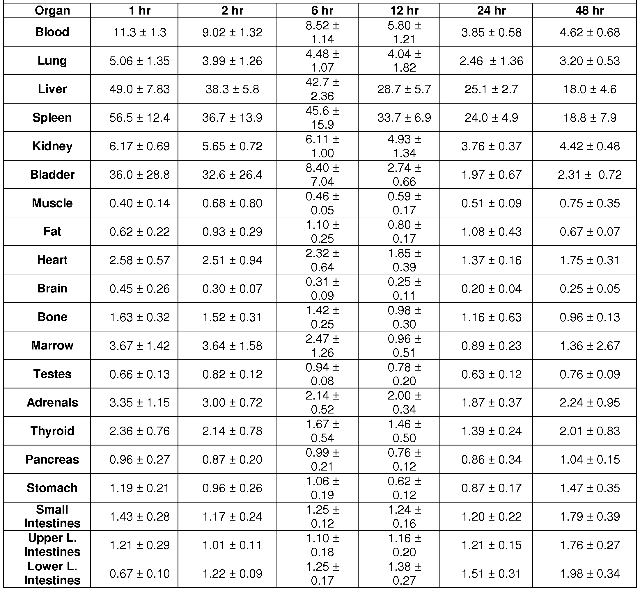
Residence Times
[01 18] The residence times represent the cumulative presence time of radioactivity in any given organ and are expressed in units of second or hours (hr).
Multiplying these values by unit of radioactivity leads to the number of radioactive
decays occurring in any given organ and is therefore proportional to radiation dose. Using the above animal biodistribution data, the organ residence times (in hours) for each harvested organ were calculated by numerical integration of the time activity data expressed in percent injected dose per gram of tissue. The following initial organ activity content immediately after injection was assumed to be: 5.9% in the lungs, 2.9% in the liver, 1 .5% in the spleen and kidneys, 3.7% in the bone, and 72% in the blood. It was assumed that no biological excretion occur beyond the last measured time point and that radioactivity only decreased due to physical decay. The animal organ residence times were then scaled to human organ weight by the "relative organ mass scaling" method. Organ residence times are presented in Table 7. The cumulative urine and feces activity (in percent injected) were plotted as a function of time and an uptake function was fitted to the data (F(t) = AO (1 -exp(-A1 t) ), see Fig. 12A and B). Analytical integration, accounting for radioactive decay, yielded to an excreted residence time of 1 .62 hr in the feces. Analytical integration of the excreted urine data resulted in a urine residence times of 9.36 hr. The filling fraction of 65% and the filling half-life of 3.47 hr (=ln(2)/0.020hr-1 ) was used in the MIRD voiding model along with a voiding interval of 2hr to yield a bladder residence time of 0.533 hr, and a modeled amount of activity excreted in urine of 8.83 hr. The amount of excreted activity is therefore equal to 10.5 hr. The remainder of the body residence time was calculated from the maximum theoretical residence time minus the excreted residence time minus the sum of all residence times measured in the organ above at the exception of blood and fat. This resulted in a residence time associated to the remainder of the body of 0.27 hr. The blood and bone mass was assumed to be 8% and 15% of the human body mass respectively. The errors bars on the measured residence times were determined from the standard deviation of the biodistribution data points. The largest residence times are observed in the liver, muscle and bone. The residence time in the blood is also very high with a value of 2.64 hr and is dependent on the relative slow clearance of the activity from the blood. The blood clearance half-life was measured at 36 hr using a mono-exponential model with an initial concentration of 1 1 .3% of the injected dose.

Radiation Dosimetry
[01 19] The residence times of Table 7 were entered in the program OLINDA/EXM for 64Cu and using the standard MIRD adult male model. The following additional assumptions were made: 10% of the blood residence time was assigned to the heart left ventricle; the bone activity was assigned in equal part to the cortical and trabecular bone source organs. The error bars on the dose estimates were assumed to be in proportion of the organ residence times uncertainties.
[0120] Although the calculated doses are for the human male model, dose to female organs (breasts, uterus and ovaries) are also provided in italics. The radiation doses above include contribution from betas (both minus and plus) and gamma rays emitted from 64Cu, and include contribution from activity within one organ to itself and from neighboring organs. Due to the nature and energy of the beta particles, the self- organ dose contribution from the beta particles dominates the dose contributions. The largest radiation dose is observed in the liver and spleen with values of 0.62 and 0.6 rad per mCi injected. The effective dose is calculated at 0.088 rem/mCi.
Example 5. GRP78 surface expression increases on irradiated NSCLC.
[0121 ] Lung cancer cell membrane preparations were studied on Western immunoblot. Antibodies to GRP78 showed an increase in this protein on the cell membrane in response to 3 Gy of irradiation in lung cancer cells (Fig. 13). GRP78 not only remained within the membrane preparations, but was also secreted into the medium of irradiated NSCLC cells. Fig. 14 shows that HUVEC cells irradiated together with lung cancer cells show an increase in GRP78 surface staining. Radiation induction of GRP78 on the surface of endothelial cells was not accomplished when endothelial cells were irradiated alone.
Example 6. Anti-GRP78 antibodies reduce NSCLC cell survival and enhance cytotoxicity of radiotherapy.
[0122] To determine whether anti-GRP-78 monoclonal antibodies enhance the efficacy of radiotherapy in NSCLC, the colony forming assay (Fig. 15) was utilized. 2D6F9 antibody, 5 μ9/ιτιΙ was added to lung cancer cells at 6 hours after plating. Cells were then treated with 2 Gy or sham irradiation (0 Gy). The antibody alone reduced plating efficiency to 49% and reduced survival to 6% after 2 Gy irradiation. In
comparison, 2Gy alone or with control antibody reduced survival by less than 20% (p<0.01 ).
Example 7. Anti-GRP78 antibodies achieve specific binding to NSCLC in the mouse models.
[0123] To determine whether antibodies against the C- and N-terminal domains of GRP78 achieve cancer specific binding, lung cancer tumors in mice (Fig. 16) were studied. Lung cancer tumors were grown both within the hind limb of mice. Antibodies were labeled with ALX750 and imaged by NIR imaging. Each of the antibodies 1 D6B2, 2D6F3 and scFv K13 achieve specific binding to the irradiated NSCLC. The kinetic curves show that the antibodies rapidly bind to the irradiated tumor as they are cleared from the circulation through enterohepatic clearance. Binding within
tumors persist for over one week for IgG antibodies and 3 days for scFv. All antibodies also bind specifically to human NSCLCs H460 and LLC in irradiated nude mice.
Example 8. Anti Tip 1 scFv antibody.
[0124] TE1 1 anti Tip 1 scFv antibody and a DOTA-conjugated TE1 1 anti Tip 1 scFv antibody were generated. The sequence of two TE1 1 clones are shown in Fig. 22.
[0125] The TE1 1 antibody was tested using ELISA (Fig. 17 and 18). In short, ELISA plates were coated with Tip 1 antigen at 10 g/ml in PBS, washed 3 times, and blocked with 2% BSA. TE1 1 or Dota-conjugated TE1 1 in PBS was added and, after incubation, washed 3 x before adding anti myc antibody at 5 pg/ml, followed by goat anti mouse IgG-HRP, washed 3 x before adding ABTS substrate and reading at 405 nm. The reaction volume in each step is 50 μΙ / well.
[0126] The anti TIP1 scFV antibody TE1 1 (Fig. 19) recognized tumor cells that were exposed to radiation, but a control scFv antibody did not (Fig. 20). See also Fig. 21.
Claims
1 . An isolated antibody that binds to GRP78, wherein the antibody comprises a heavy chain variable domain comprising a CDR1 , CDR2, and a CDR3, wherein the CDR3 comprises at least five contiguous amino acids of SEQ ID NO:17.
2. The antibody of claim 1 , wherein the heavy chain variable domain CDR2 comprises at least five contiguous amino acids of SEQ ID NO:16.
3. The antibody of claim 1 , wherein the heavy chain variable domain CDR1 comprises at least five contiguous amino acids of SEQ ID NO:15.
4. The antibody of claim 1 , wherein the heavy chain variable domain CDR1 comprises at least five contiguous amino acids of SEQ ID NO:15 and the heavy chain variable domain CDR2 comprises at least five contiguous amino acids of SEQ ID NO:16.
5. The antibody of claim 4, wherein the heavy chain variable domain CDR1 comprises SEQ ID NO:15, the heavy chain variable region domain CDR2 comprises SEQ ID NO:16, and the heavy chain variable region domain CDR3 comprises SEQ ID NO:17.
6. The antibody of claim 1 , wherein the antibody further comprises a light chain variable domain comprising a CDR1 , CDR2, and a CDR3, wherein the CDR3 comprises at least five contiguous amino acids of SEQ ID NO:20.
7. The antibody of claim 1 , wherein the light chain variable domain CDR2
comprises at least five contiguous amino acids of SEQ ID NO:19.
8. The antibody of claim 1 , wherein the light chain variable domain CDR1 comprises at least five contiguous amino acids of SEQ ID NO:18.
9. The antibody of claim 1 , wherein the light chain variable domain CDR1 comprises at least five contiguous amino acids of SEQ ID NO:18 and the light chain variable domain CDR2 comprises at least five contiguous amino acids of SEQ ID NO:19.
10. The antibody of claim 4, wherein the light chain variable domain CDR1 comprises SEQ ID NO:18, the light chain variable region domain CDR2 comprises SEQ ID NO:19, and the light chain variable region domain CDR3 comprises SEQ ID NO:20.
1 1 . The antibody of claim 1 , wherein the antibody recognizes an epitope within the amino acid sequence of SEQ ID NO:1 .
12. The antibody of claim 1 , wherein the antibody recognizes an epitope within the amino acid sequence of SEQ ID NO:2.
13. The antibody of claim 1 , wherein the antibody recognizes an epitope within the amino acid sequence of SEQ ID NO:3.
14. The antibody of claim 1 , wherein the antibody recognizes an epitope within the amino acid sequence of SEQ ID NO:4.
15. The antibody of claim 1 , wherein the antibody is humanized.
16. The antibody of claim 1 , wherein the antibody is a single chain variable fragment (scFv).
17. The antibody of claim 1 , wherein the antibody is chimeric.
18. The antibody of claim 16, wherein the single chain variable fragment is conjugated to a human constant domain.
19. The antibody of claim 16, wherein the single chain variable fragment is conjugated to a therapeutic agent.
20. The antibody of claim 16, wherein the single chain variable fragment is conjugated to an imaging agent.
21 . An antibody that binds to GRP78 that recognizes an epitope within the amino acid sequence of SEQ ID NO:1 .
22. An antibody that binds to GRP78 that recognizes an epitope within the amino acid sequence of SEQ ID NO:2.
23. An antibody that binds to GRP78 that recognizes an epitope within the amino acid sequence of SEQ ID NO:3.
24. An antibody that binds to GRP78 that recognizes an epitope within the amino acid sequence of SEQ ID NO:4.
25. An antibody that binds to TIP-1 that recognizes an epitope within the amino acid sequence of SEQ ID NO:9.
26. An antibody that binds to TIP-1 that recognizes an epitope within the amino acid sequence of SEQ ID NO:10.
27. An antibody that binds to TIP-1 that recognizes an epitope within the amino acid sequence of SEQ ID NO:1 1 .
28. An antibody that binds to TIP-1 that recognizes an epitope within the amino acid sequence of SEQ ID NO:12.
29. An antibody that binds to TIP-1 that recognizes an epitope within the amino acid sequence of SEQ ID NO:13.
30. An antibody that binds to TIP-1 that recognizes an epitope within the amino acid sequence of SEQ ID NO:14.
31 . An antibody that binds to TIP-1 that recognizes an epitope within the amino acid sequence of SEQ ID NO:21 .
32. A method of enhancing radiotherapy in a subject, the method comprising administering a pharmacologically effective amount of anti-TIP1 antibody to the subject.
33. A method of enhancing radiotherapy in a subject, the method comprising administering a pharmacologically effective amount of anti-GRP78 antibody to the subject.
Priority Applications (2)
| Application Number | Priority Date | Filing Date | Title |
|---|---|---|---|
| US14/166,251 US9738725B2 (en) | 2011-07-29 | 2014-01-28 | Antibodies to TIP-1 |
| US15/628,209 US10259884B2 (en) | 2011-07-29 | 2017-06-20 | Antibodies to GRP78 |
Applications Claiming Priority (2)
| Application Number | Priority Date | Filing Date | Title |
|---|---|---|---|
| US201161513333P | 2011-07-29 | 2011-07-29 | |
| US61/513,333 | 2011-07-29 |
Related Child Applications (1)
| Application Number | Title | Priority Date | Filing Date |
|---|---|---|---|
| US14/166,251 Continuation-In-Part US9738725B2 (en) | 2011-07-29 | 2014-01-28 | Antibodies to TIP-1 |
Publications (1)
| Publication Number | Publication Date |
|---|---|
| WO2013019730A1 true WO2013019730A1 (en) | 2013-02-07 |
Family
ID=47629635
Family Applications (2)
| Application Number | Title | Priority Date | Filing Date |
|---|---|---|---|
| PCT/US2012/048856 Ceased WO2013019730A1 (en) | 2011-07-29 | 2012-07-30 | Antibodies to tip-1 and grp78 |
| PCT/US2015/013241 Ceased WO2015116653A1 (en) | 2011-07-29 | 2015-01-28 | Antibodies to tip-1 and grp78 |
Family Applications After (1)
| Application Number | Title | Priority Date | Filing Date |
|---|---|---|---|
| PCT/US2015/013241 Ceased WO2015116653A1 (en) | 2011-07-29 | 2015-01-28 | Antibodies to tip-1 and grp78 |
Country Status (3)
| Country | Link |
|---|---|
| US (2) | US9738725B2 (en) |
| EP (1) | EP3099718A1 (en) |
| WO (2) | WO2013019730A1 (en) |
Cited By (9)
| Publication number | Priority date | Publication date | Assignee | Title |
|---|---|---|---|---|
| WO2015116653A1 (en) * | 2011-07-29 | 2015-08-06 | Washington University | Antibodies to tip-1 and grp78 |
| US9758591B2 (en) | 2012-08-24 | 2017-09-12 | The Regents Of The University Of California | Antibodies and vaccines for use in treating ROR1 cancers and inhibiting metastasis |
| WO2018022479A1 (en) * | 2016-07-25 | 2018-02-01 | Biogen Ma Inc. | Anti-hspa5 (grp78) antibodies and uses thereof |
| US10086073B2 (en) | 2001-10-03 | 2018-10-02 | Washington University | Ligands to radiation-induced molecules |
| US10449261B2 (en) | 2014-07-24 | 2019-10-22 | Washington University | Compositions targeting radiation-induced molecules and methods of use thereof |
| US10688181B2 (en) | 2016-06-27 | 2020-06-23 | The Regents Of The University Of California | Cancer treatment combinations |
| US11306151B2 (en) | 2018-03-08 | 2022-04-19 | Phanes Therapeutics, Inc. | Anti-TIP-1 antibodies and uses thereof |
| US11352436B2 (en) | 2017-02-10 | 2022-06-07 | Washington University | Antibodies to TIP1 and methods of use thereof |
| US12269859B2 (en) | 2016-12-02 | 2025-04-08 | Angeles Therapeutics, Inc. | Synthetic immune receptors and methods of use thereof |
Families Citing this family (3)
| Publication number | Priority date | Publication date | Assignee | Title |
|---|---|---|---|---|
| PT3604339T (en) | 2011-01-14 | 2021-04-13 | Univ California | Therapeutic antibodies against ror-1 protein and methods for use of same |
| WO2018057703A1 (en) | 2016-09-21 | 2018-03-29 | Stc.Unm | EphA5- AND GRP78 BINDING ANTIBODIES AND USES THEREOF AND SELECTION OF PHAGE-DISPLAYED ACCESSIBLE RECOMBINANT TARGETED ANTIBODIES |
| EP3999547A4 (en) * | 2019-07-16 | 2023-07-12 | Washington University | ANTI-GRP78 ANTIBODIES AND METHODS OF USE THEREOF |
Citations (8)
| Publication number | Priority date | Publication date | Assignee | Title |
|---|---|---|---|---|
| US6673545B2 (en) * | 2000-07-28 | 2004-01-06 | Incyte Corporation | Prostate cancer markers |
| US20070081993A1 (en) * | 2003-05-31 | 2007-04-12 | Peter Kufer | Pharmaceutical composition comprising a bispecific antibody for epcam |
| US7230083B2 (en) * | 1997-03-12 | 2007-06-12 | Smithkline Beecham Corporation | Anti-alphabeta3 humanized monoclonal antibodies |
| US20080118978A1 (en) * | 2006-04-28 | 2008-05-22 | Takashi Sato | Anti-tumor agent |
| US20080305111A1 (en) * | 2006-06-22 | 2008-12-11 | Vaccinex, Inc. | Anti-C35 antibodies for treating cancer |
| US20100041074A1 (en) * | 2007-02-27 | 2010-02-18 | Naoki Kimura | Pharmaceutical composition comprising anti-grp78 antibody as active ingredient |
| US20100111959A1 (en) * | 2008-08-20 | 2010-05-06 | Swanson Ronald V | Engineered anti-il-13 antibodies, compositions, methods and uses |
| US20100111852A1 (en) * | 2006-12-14 | 2010-05-06 | Forerunner Pharma Research Co., Ltd. | Anti-Claudin 3 Monoclonal Antibody and Treatment and Diagnosis of Cancer Using the Same |
Family Cites Families (64)
| Publication number | Priority date | Publication date | Assignee | Title |
|---|---|---|---|---|
| US4281061A (en) | 1979-07-27 | 1981-07-28 | Syva Company | Double antibody for enhanced sensitivity in immunoassay |
| US4515165A (en) | 1980-02-04 | 1985-05-07 | Energy Conversion Devices, Inc. | Apparatus and method for detecting tumors |
| US4670386A (en) | 1980-05-23 | 1987-06-02 | Stephen Sugaar | Cancer tests using tumor antigen generated lymphokines and compositions |
| US5223409A (en) | 1988-09-02 | 1993-06-29 | Protein Engineering Corp. | Directed evolution of novel binding proteins |
| US5328840A (en) | 1989-08-15 | 1994-07-12 | The Research Foundation Of The State University Of New York | Method for preparing targeted carrier erythrocytes |
| US5382680A (en) | 1990-12-07 | 1995-01-17 | The Center For Innovative Technology | Allosteric hemoglobin modifier compounds |
| US5277892A (en) | 1990-08-08 | 1994-01-11 | Rhomed Incorporated | In vivo lymphocyte tagging |
| US5264563A (en) | 1990-08-24 | 1993-11-23 | Ixsys Inc. | Process for synthesizing oligonucleotides with random codons |
| US6605712B1 (en) | 1990-12-20 | 2003-08-12 | Arch Development Corporation | Gene transcription and ionizing radiation: methods and compositions |
| US5645815A (en) | 1991-02-08 | 1997-07-08 | Diatide, Inc. | Radiolabled compounds for thrombus imaging |
| US5830856A (en) | 1991-02-08 | 1998-11-03 | Diatide, Inc. | Radiolabeled compounds for thrombus imaging |
| EP0584264A1 (en) | 1991-05-16 | 1994-03-02 | Cold Spring Harbor Laboratory | D-type cyclin and uses related thereto |
| US5889169A (en) | 1991-05-16 | 1999-03-30 | Cold Spring Harbor Laboratory | Cell cycle regulatory protein p16 gene |
| US5334369A (en) | 1991-06-11 | 1994-08-02 | Medical University Of South Carolina | Platelet receptor antagonists useful in detecting intravascular platelet aggregation |
| US5733731A (en) | 1991-10-16 | 1998-03-31 | Affymax Technologies N.V. | Peptide library and screening method |
| WO1993014791A2 (en) | 1992-01-27 | 1993-08-05 | Cryopharm Corporation | Method of inactivation of viral and bacterial blood contaminants |
| ES2193143T3 (en) | 1992-03-05 | 2003-11-01 | Univ Texas | USE OF IMMUNOCONJUGADOS FOR THE DIAGNOSIS AND / OR THERAPY OF VASCULARIZA TUMORS. |
| US5660827A (en) | 1992-03-05 | 1997-08-26 | Board Of Regents, The University Of Texas System | Antibodies that bind to endoglin |
| US5965132A (en) | 1992-03-05 | 1999-10-12 | Board Of Regents, The University Of Texas System | Methods and compositions for targeting the vasculature of solid tumors |
| US6107059A (en) | 1992-04-29 | 2000-08-22 | Affymax Technologies N.V. | Peptide library and screening method |
| US6383470B1 (en) | 1992-09-26 | 2002-05-07 | Thomas Fritzsch | Microparticle preparations made of biodegradable copolymers |
| US5922545A (en) | 1993-10-29 | 1999-07-13 | Affymax Technologies N.V. | In vitro peptide and antibody display libraries |
| US6316208B1 (en) | 1994-01-07 | 2001-11-13 | Memorial Sloan-Kettering Cancer Center | Methods for determining isolated p27 protein levels and uses thereof |
| JPH10504534A (en) | 1994-06-16 | 1998-05-06 | ネオルクス コーポレイション | Radiolabeled annexin-galactose conjugate |
| US5759542A (en) | 1994-08-05 | 1998-06-02 | New England Deaconess Hospital Corporation | Compositions and methods for the delivery of drugs by platelets for the treatment of cardiovascular and other diseases |
| US5516881A (en) | 1994-08-10 | 1996-05-14 | Cornell Research Foundation, Inc. | Aminoxyl-containing radical spin labeling in polymers and copolymers |
| DE4429461A1 (en) | 1994-08-19 | 1996-02-22 | Merck Patent Gmbh | Adhesion receptor antagonists |
| CA2164167A1 (en) | 1994-12-20 | 1996-06-21 | Izak Bahar | Assay normalization method |
| US6033847A (en) | 1995-02-06 | 2000-03-07 | St. Jude Children's Research Hospital | InK4c-p18 and InK4d-p19, inhibitors of cyclin-dependent kinases CDK4 and CDK6, and uses thereof |
| US5962424A (en) | 1995-02-21 | 1999-10-05 | Arch Development Corporation | Methods and compositions for targeting selectins |
| US6521211B1 (en) | 1995-06-07 | 2003-02-18 | Bristol-Myers Squibb Medical Imaging, Inc. | Methods of imaging and treatment with targeted compositions |
| US6068829A (en) | 1995-09-11 | 2000-05-30 | The Burnham Institute | Method of identifying molecules that home to a selected organ in vivo |
| US6007994A (en) | 1995-12-22 | 1999-12-28 | Yale University | Multiparametric fluorescence in situ hybridization |
| US6403383B1 (en) | 1996-03-11 | 2002-06-11 | American Bio Medica Corp. | Diagnostic test kit for immunological assays of fluid samples |
| US5858683A (en) | 1996-08-30 | 1999-01-12 | Matritech, Inc. | Methods and compositions for the detection of cervical cancer |
| US6576239B1 (en) | 1996-09-10 | 2003-06-10 | The Burnham Institute | Angiogenic homing molecules and conjugates derived therefrom |
| WO1998010795A2 (en) | 1996-09-10 | 1998-03-19 | The Burnham Institute | Tumor homing molecules, conjugates derived therefrom, and methods of using same |
| CA2269335C (en) | 1996-10-21 | 2010-05-25 | Quadrant Healthcare (Uk) Limited | Platelet substitutes and conjugation methods suitable for their preparation |
| AU8403098A (en) | 1997-07-15 | 1999-02-10 | Deaconess Hospital | Reagents and methods for diagnosis and prognosis of proliferative disor ders |
| US6174687B1 (en) | 1999-02-26 | 2001-01-16 | The Burnham Institute | Methods of identifying lung homing molecules using membrane dipeptidase |
| US6232287B1 (en) | 1998-03-13 | 2001-05-15 | The Burnham Institute | Molecules that home to various selected organs or tissues |
| US6852318B1 (en) | 1998-05-08 | 2005-02-08 | The Regents Of The University Of California | Methods for detecting and inhibiting angiogenesis |
| US6593084B2 (en) | 1998-10-13 | 2003-07-15 | Robert E. Bird | Carcinogen assay |
| AU3105700A (en) | 1998-12-08 | 2000-06-26 | Board Of Regents, The University Of Texas System | Methods for detection of antiestrogen-resistant breast cancer |
| US6630570B1 (en) | 1999-04-09 | 2003-10-07 | Insitut für Diagnostikforschung GmbH | Short-chain peptide-dye conjugates as contrast media for optical diagnosis |
| US6159443A (en) | 1999-04-29 | 2000-12-12 | Vanderbilt University | X-ray guided drug delivery |
| US7049140B1 (en) | 1999-04-29 | 2006-05-23 | Vanderbilt University | X-ray guided drug delivery |
| AU6608600A (en) | 1999-07-29 | 2001-02-19 | University Of Vermont And State Agricultural College, The | In vivo methods for the identification of target specific binding molecules in a human and their use in cancer detection |
| US6277974B1 (en) | 1999-12-14 | 2001-08-21 | Cogent Neuroscience, Inc. | Compositions and methods for diagnosing and treating conditions, disorders, or diseases involving cell death |
| US7230088B2 (en) | 2001-07-03 | 2007-06-12 | Mallinckrodt, Inc. | Compounds for dual photodiagnosis and therapy |
| MXPA02011414A (en) | 2000-05-22 | 2003-06-06 | Chiesi Farma Spa | Stable pharmaceutical solution formulations for pressurised metered dose inhalers. |
| DE10063179A1 (en) | 2000-12-18 | 2002-06-20 | Bayer Ag | Detecting tumor cells and their precursors in cervical smears, by simultaneous detection of two markers, suitable for automation and providing an objective diagnosis |
| EP1364201A4 (en) | 2001-02-06 | 2005-01-05 | Univ Auburn | DEVICES FOR DETECTING LIGANDS AND USE THEREOF |
| US20030083261A1 (en) | 2001-04-30 | 2003-05-01 | Hongtao Yu | Class of 12mer peptides that inhibit the function of the mitotic check point protein Mad2 |
| US7306925B2 (en) | 2001-11-09 | 2007-12-11 | Vanderbilt University | Phage antibodies to radiation-inducible neoantigens |
| US8101157B2 (en) | 2001-10-03 | 2012-01-24 | Vanderbilt University | Ligands to radiation-induced molecules |
| AU2002330139A1 (en) | 2001-10-03 | 2003-04-14 | Vanderbilt University | In vivo panning for ligands to radiation-induced molecules |
| US7906102B2 (en) | 2001-10-03 | 2011-03-15 | Vanderbilt University | Ligands to radiation-induced molecules |
| CA2501752A1 (en) | 2002-10-10 | 2004-04-22 | Wyeth | Compositions, organisms and methodologies employing a novel human kinase |
| US20060046271A1 (en) | 2004-09-02 | 2006-03-02 | Vanderbilt University | Assessment of cancer susceptibility to molecular targeted therapy by use of recombinant peptides |
| US8022623B2 (en) | 2008-08-15 | 2011-09-20 | General Electric Company | Ultra-thin multi-substrate color tunable OLED device |
| WO2013019730A1 (en) | 2011-07-29 | 2013-02-07 | The Washington University | Antibodies to tip-1 and grp78 |
| WO2013049830A2 (en) | 2011-09-30 | 2013-04-04 | The Washington University | Tip-1 and grp-78 binding peptides and method of identifying peptide receptors |
| WO2018148595A1 (en) | 2017-02-10 | 2018-08-16 | Washington University | Antibodies to tip1 and methods of use thereof |
-
2012
- 2012-07-30 WO PCT/US2012/048856 patent/WO2013019730A1/en not_active Ceased
-
2014
- 2014-01-28 US US14/166,251 patent/US9738725B2/en not_active Expired - Fee Related
-
2015
- 2015-01-28 EP EP15743922.5A patent/EP3099718A1/en not_active Withdrawn
- 2015-01-28 WO PCT/US2015/013241 patent/WO2015116653A1/en not_active Ceased
-
2017
- 2017-06-20 US US15/628,209 patent/US10259884B2/en active Active
Patent Citations (8)
| Publication number | Priority date | Publication date | Assignee | Title |
|---|---|---|---|---|
| US7230083B2 (en) * | 1997-03-12 | 2007-06-12 | Smithkline Beecham Corporation | Anti-alphabeta3 humanized monoclonal antibodies |
| US6673545B2 (en) * | 2000-07-28 | 2004-01-06 | Incyte Corporation | Prostate cancer markers |
| US20070081993A1 (en) * | 2003-05-31 | 2007-04-12 | Peter Kufer | Pharmaceutical composition comprising a bispecific antibody for epcam |
| US20080118978A1 (en) * | 2006-04-28 | 2008-05-22 | Takashi Sato | Anti-tumor agent |
| US20080305111A1 (en) * | 2006-06-22 | 2008-12-11 | Vaccinex, Inc. | Anti-C35 antibodies for treating cancer |
| US20100111852A1 (en) * | 2006-12-14 | 2010-05-06 | Forerunner Pharma Research Co., Ltd. | Anti-Claudin 3 Monoclonal Antibody and Treatment and Diagnosis of Cancer Using the Same |
| US20100041074A1 (en) * | 2007-02-27 | 2010-02-18 | Naoki Kimura | Pharmaceutical composition comprising anti-grp78 antibody as active ingredient |
| US20100111959A1 (en) * | 2008-08-20 | 2010-05-06 | Swanson Ronald V | Engineered anti-il-13 antibodies, compositions, methods and uses |
Non-Patent Citations (1)
| Title |
|---|
| MUNRO ET AL.: "An Hsp70-like protein in the ER: identity with the 78 kd glucose-regulated protein and immunoglobulin heavy chain binding protein.", CELL, vol. 46, no. 2, 18 July 1986 (1986-07-18), pages 291 - 300 * |
Cited By (15)
| Publication number | Priority date | Publication date | Assignee | Title |
|---|---|---|---|---|
| US10086073B2 (en) | 2001-10-03 | 2018-10-02 | Washington University | Ligands to radiation-induced molecules |
| US9738725B2 (en) | 2011-07-29 | 2017-08-22 | Washington University | Antibodies to TIP-1 |
| WO2015116653A1 (en) * | 2011-07-29 | 2015-08-06 | Washington University | Antibodies to tip-1 and grp78 |
| US10259884B2 (en) | 2011-07-29 | 2019-04-16 | Washington University | Antibodies to GRP78 |
| US9758591B2 (en) | 2012-08-24 | 2017-09-12 | The Regents Of The University Of California | Antibodies and vaccines for use in treating ROR1 cancers and inhibiting metastasis |
| US10344096B2 (en) | 2012-08-24 | 2019-07-09 | The Regents Of The University Of California | Antibodies and vaccines for use in treating ROR1 cancers and inhibiting metastasis |
| US12162950B2 (en) | 2012-08-24 | 2024-12-10 | The Regents Of The University Of California | Antibodies and vaccines for use in treating ROR1 cancers and inhibiting metastasis |
| US11312787B2 (en) | 2012-08-24 | 2022-04-26 | The Regents Of The University Of California | Antibodies and vaccines for use in treating ROR1 cancers and inhibiting metastasis |
| US10449261B2 (en) | 2014-07-24 | 2019-10-22 | Washington University | Compositions targeting radiation-induced molecules and methods of use thereof |
| US11654193B2 (en) | 2016-06-27 | 2023-05-23 | The Regents Of The University Of California | Cancer treatment combinations |
| US10688181B2 (en) | 2016-06-27 | 2020-06-23 | The Regents Of The University Of California | Cancer treatment combinations |
| WO2018022479A1 (en) * | 2016-07-25 | 2018-02-01 | Biogen Ma Inc. | Anti-hspa5 (grp78) antibodies and uses thereof |
| US12269859B2 (en) | 2016-12-02 | 2025-04-08 | Angeles Therapeutics, Inc. | Synthetic immune receptors and methods of use thereof |
| US11352436B2 (en) | 2017-02-10 | 2022-06-07 | Washington University | Antibodies to TIP1 and methods of use thereof |
| US11306151B2 (en) | 2018-03-08 | 2022-04-19 | Phanes Therapeutics, Inc. | Anti-TIP-1 antibodies and uses thereof |
Also Published As
| Publication number | Publication date |
|---|---|
| US9738725B2 (en) | 2017-08-22 |
| EP3099718A1 (en) | 2016-12-07 |
| US20140316186A1 (en) | 2014-10-23 |
| US10259884B2 (en) | 2019-04-16 |
| WO2015116653A1 (en) | 2015-08-06 |
| US20170298142A1 (en) | 2017-10-19 |
Similar Documents
| Publication | Publication Date | Title |
|---|---|---|
| US10259884B2 (en) | Antibodies to GRP78 | |
| JP5161254B2 (en) | α-fetoprotein Immu31 antibody and fusion protein and method of use thereof | |
| CN102174108B (en) | The anti-CD74 antibody of internalization and using method | |
| CN101824090B (en) | Monoclonal antibody hpam4 | |
| JP4354280B2 (en) | RS7 antibody | |
| CN106573983B (en) | For the radiolabeled antibody fragment used in the prevention and/or treatment of cancer | |
| JP6917357B2 (en) | Radiolabeled antibody fragment used for cancer treatment | |
| US10066008B2 (en) | Monoclonal antibodies to human 14-3-3 epsilon and human 14-3-3 epsilon sv | |
| US11352436B2 (en) | Antibodies to TIP1 and methods of use thereof | |
| US10758612B2 (en) | Antibodies to tumor associated complex N-glycans with terminal GlcNAcβ residues and methods of use thereof | |
| US20220259322A1 (en) | Anti-grp78 antibodies and method of use thereof | |
| WO2010131590A1 (en) | Diagnostic agent and therapeutic agent for tumors | |
| BR112018000672B1 (en) | KIT AND USE OF A RADIOACTIVELY LABELED HEAVY CHAIN VARIABLE DOMAIN DERIVED FROM A HEAVY CHAIN ANTIBODY (VHH) OR A FUNCTIONAL FRAGMENT THEREOF IN THE TREATMENT OF CANCER |
Legal Events
| Date | Code | Title | Description |
|---|---|---|---|
| 121 | Ep: the epo has been informed by wipo that ep was designated in this application |
Ref document number: 12819373 Country of ref document: EP Kind code of ref document: A1 |
|
| NENP | Non-entry into the national phase |
Ref country code: DE |
|
| 122 | Ep: pct application non-entry in european phase |
Ref document number: 12819373 Country of ref document: EP Kind code of ref document: A1 |