WO2012021407A2 - Biomarkers for stroke - Google Patents
Biomarkers for stroke Download PDFInfo
- Publication number
- WO2012021407A2 WO2012021407A2 PCT/US2011/046777 US2011046777W WO2012021407A2 WO 2012021407 A2 WO2012021407 A2 WO 2012021407A2 US 2011046777 W US2011046777 W US 2011046777W WO 2012021407 A2 WO2012021407 A2 WO 2012021407A2
- Authority
- WO
- WIPO (PCT)
- Prior art keywords
- biomarkers
- stroke
- sample
- seq
- subject
- Prior art date
- Legal status (The legal status is an assumption and is not a legal conclusion. Google has not performed a legal analysis and makes no representation as to the accuracy of the status listed.)
- Ceased
Links
Classifications
-
- G—PHYSICS
- G01—MEASURING; TESTING
- G01N—INVESTIGATING OR ANALYSING MATERIALS BY DETERMINING THEIR CHEMICAL OR PHYSICAL PROPERTIES
- G01N33/00—Investigating or analysing materials by specific methods not covered by groups G01N1/00 - G01N31/00
- G01N33/48—Biological material, e.g. blood, urine; Haemocytometers
- G01N33/50—Chemical analysis of biological material, e.g. blood, urine; Testing involving biospecific ligand binding methods; Immunological testing
- G01N33/68—Chemical analysis of biological material, e.g. blood, urine; Testing involving biospecific ligand binding methods; Immunological testing involving proteins, peptides or amino acids
- G01N33/6893—Chemical analysis of biological material, e.g. blood, urine; Testing involving biospecific ligand binding methods; Immunological testing involving proteins, peptides or amino acids related to diseases not provided for elsewhere
-
- C—CHEMISTRY; METALLURGY
- C12—BIOCHEMISTRY; BEER; SPIRITS; WINE; VINEGAR; MICROBIOLOGY; ENZYMOLOGY; MUTATION OR GENETIC ENGINEERING
- C12Q—MEASURING OR TESTING PROCESSES INVOLVING ENZYMES, NUCLEIC ACIDS OR MICROORGANISMS; COMPOSITIONS OR TEST PAPERS THEREFOR; PROCESSES OF PREPARING SUCH COMPOSITIONS; CONDITION-RESPONSIVE CONTROL IN MICROBIOLOGICAL OR ENZYMOLOGICAL PROCESSES
- C12Q1/00—Measuring or testing processes involving enzymes, nucleic acids or microorganisms; Compositions therefor; Processes of preparing such compositions
- C12Q1/68—Measuring or testing processes involving enzymes, nucleic acids or microorganisms; Compositions therefor; Processes of preparing such compositions involving nucleic acids
- C12Q1/6876—Nucleic acid products used in the analysis of nucleic acids, e.g. primers or probes
- C12Q1/6883—Nucleic acid products used in the analysis of nucleic acids, e.g. primers or probes for diseases caused by alterations of genetic material
-
- C—CHEMISTRY; METALLURGY
- C12—BIOCHEMISTRY; BEER; SPIRITS; WINE; VINEGAR; MICROBIOLOGY; ENZYMOLOGY; MUTATION OR GENETIC ENGINEERING
- C12Q—MEASURING OR TESTING PROCESSES INVOLVING ENZYMES, NUCLEIC ACIDS OR MICROORGANISMS; COMPOSITIONS OR TEST PAPERS THEREFOR; PROCESSES OF PREPARING SUCH COMPOSITIONS; CONDITION-RESPONSIVE CONTROL IN MICROBIOLOGICAL OR ENZYMOLOGICAL PROCESSES
- C12Q2600/00—Oligonucleotides characterized by their use
- C12Q2600/158—Expression markers
-
- G—PHYSICS
- G01—MEASURING; TESTING
- G01N—INVESTIGATING OR ANALYSING MATERIALS BY DETERMINING THEIR CHEMICAL OR PHYSICAL PROPERTIES
- G01N2800/00—Detection or diagnosis of diseases
- G01N2800/28—Neurological disorders
- G01N2800/2871—Cerebrovascular disorders, e.g. stroke, cerebral infarct, cerebral haemorrhage, transient ischemic event
-
- G—PHYSICS
- G01—MEASURING; TESTING
- G01N—INVESTIGATING OR ANALYSING MATERIALS BY DETERMINING THEIR CHEMICAL OR PHYSICAL PROPERTIES
- G01N2800/00—Detection or diagnosis of diseases
- G01N2800/52—Predicting or monitoring the response to treatment, e.g. for selection of therapy based on assay results in personalised medicine; Prognosis
-
- G—PHYSICS
- G01—MEASURING; TESTING
- G01N—INVESTIGATING OR ANALYSING MATERIALS BY DETERMINING THEIR CHEMICAL OR PHYSICAL PROPERTIES
- G01N2800/00—Detection or diagnosis of diseases
- G01N2800/56—Staging of a disease; Further complications associated with the disease
-
- G—PHYSICS
- G01—MEASURING; TESTING
- G01N—INVESTIGATING OR ANALYSING MATERIALS BY DETERMINING THEIR CHEMICAL OR PHYSICAL PROPERTIES
- G01N2800/00—Detection or diagnosis of diseases
- G01N2800/60—Complex ways of combining multiple protein biomarkers for diagnosis
Definitions
- This application generally relates to the diagnosis, management and therapy of stroke.
- the invention relates to methods and kits for the diagnosis and monitoring the progression and treatment of stroke in a subject.
- Stroke is a debilitating condition with limited treatment options.
- thrombolysis is the only approved therapy for stroke.
- thrombolysis is effective in only 3-5% of stroke patients, primarily due to its very short therapeutic window.
- Stabilization of stroke patients inside and outside the therapeutic window for thrombolysis is also essential to prevent the progression of the initial ischemic lesion into expanding regions of brain damage.
- Imaging techniques including computed tomography (CT) scanning and magnetic resonance imaging (MRI) are very useful in detecting the occurrence, type and severity of strokes.
- CT computed tomography
- MRI magnetic resonance imaging
- these technologies are expensive and require considerable amounts of time to develop a complete and correct diagnosis. This is particularly true in rural areas and cities with large, underserved populations.
- Prompt diagnosis of a stroke is important, as delays in diagnosis and medical intervention may contribute to clinical deterioration and disability. Early diagnosis enables clinicians to more effectively choose the emergency intervention such as anti-platelet and/or neuroprotective therapy, and also to make better prognoses of disease outcome. Successful treatment of stroke requires rapid state diagnosis. Delays in diagnosis reduce the amount of time available in which the brain can respond to reperfusion, and significantly increase the risk of hemorrhage after most of the permanent injury has occurred.
- An ideal biomarker should be specific to the type of stroke (such as ischemic stroke), sensitive (early and immediate release), predictive (proportionate to the extent of injury), robust (accurate and inexpensive), non-invasive and bridge pre-clinical results and clinical validation.
- Advanced technologies including genomic analysis and proteomics have facilitated the discovery of effective cancer biomarkers and could lead to discovery of stroke markers.
- One advantage of high throughput microarray-based genomic and proteomic analyses is the capacity to identify a group or cluster of genes and proteins with altered expression patterns in tissue or body fluids that encode putative secreted or cell-surface proteins.
- An ischemic stroke genomic biomarker would be a measurable RNA characteristic that is an indicator of normal biological processes, ischemic injury and/or response to therapeutic interventions.
- a proteomic biomarker would detect peptides or proteins in plasma or serum. It is also important that a stroke biomarker correlate well with findings from neuroimaging and clinical diagnostic examinations.
- One aspect of the present invention relates to a method for diagnosing stroke in a subject.
- the method comprises the steps of (a) measuring the level of one or more biomarkers in a sample from the subject; (b) comparing the level of the one or more biomarkers to a reference level of the one or more biomarkers; and (c) making a diagnosis based on the result of the comparing step.
- the one or more biomarkers are polynucleotides.
- the one or more biomarkers are proteins or peptides.
- step (a) includes measuring a panel of three or more biomarkers in the sample from the subject.
- step (a) includes measuring a panel of three or more biomarkers in the sample from the subject.
- the panel comprises at least one immediate early stroke biomarker (i.e., markers diferentially expressed at 1 hour post-stroke), at least one early stroke biomarker (i.e., markers diferentially expressed at 2 hour post-stroke) and at least one late stroke biomarker (i.e., diferentially expressed at 24 hour post-stroke).
- the panel comprises at least two immediate early stroke biomarker (diferentially expressed at 1 hour post-stroke), at least two early stroke biomarker (diferentially expressed at 2 hour post-stroke) and at least two late stroke biomarker (diferentially expressed at 24 hour post-stroke). In certain other embodiments, the panel comprises at least three immediate early stroke biomarkers, at least three early stroke biomarkers and at least three late stroke biomarkers.
- Another aspect of the present invention relates to a method for determining disease progression in a subject after a stroke.
- the method comprises the steps of (a) measuring the level of one or more biomarkers in a first sample obtained from the subject at a first time point; (b) measuring the level of the one or more biomarkers in a second sample obtained from the subject at a second time point; (c) comparing the level of the one or more biomarkers at the first time point to the level of the one or more biomarkers at the second time point; and (d) determining the disease progression between the first and the second time point based on the result of step (c).
- the one or more biomarkers comprise gene products expressed from genes selected from the group consisting of IL-la, IL- ⁇ , IL-lra, IL-3, IL-2, IL-4, IL-5, IL-6, IL-7, IL-8, IL-9, IL-10, IL-12 (p40), IL-12(p70), IL-13, IL-15, IL-17, EGF, Eotaxin, FGF-2, FTL-3 ligand, Fractalkine, G-CSF, G -CSF, GRO, IFN-a2, IFN- ⁇ , IP- 10, MCP-1, MCP-3, MCD, MIP-la, ⁇ - ⁇ , PDGF-aa, PGDF-aa bb, RANTES, sCD40L, sIL2- ra, TNF-a, TNF- ⁇ , VEGF and genes listed in Tables 4, 5, 6 and 8.
- Another aspect of the present invention relates to a method for determining the efficacy of a treatment for stroke in a subject.
- the method comprises the steps of (a) measuring the level of one or more biomarkers in a first sample obtained from the subject at a first time point; (b) measuring the level of the one or more biomarkers in a second sample obtained from the subject at a second time point, wherein the subject is under treatment at the second time point; (c) comparing the level of the one or more biomarkers at the first time point to the level of the one or more biomarkers at the second time point; and (d) determimng the efficacy of the treatment based on the result of step (c).
- the one or more biomarkers comprise gene products expressed from genes selected from the group consisting of IL-la, IL- ⁇ , IL-lra, IL-3, IL-2, IL-4, IL-5, IL-6, IL-7, IL-8, IL-9, IL-10, IL-12 (p40), IL-12(p70), IL-13, IL-15, IL-17, EGF, Eotaxin, FGF-2, FTL-3 ligand, Fractalkine, G-CSF, GM-CSF, GRO, IFN-a2, IFN- ⁇ , ⁇ -10, MCP-1, MCP-3, MCD, MIP-l , ⁇ - ⁇ , PDGF-aa, PGDF-aa bb, RANTES, sCD40L, sIL2-ra, TNF-a, TNF- ⁇ , VEGF and genes listed in Tables 4, 5, 6 and 8.
- kits for detecting biomarkers for stroke in a biological sample comprises (a) reagents for detecting a panel of biomarkers for stroke, and (b) a instruction listing the reference range for each of the biomarkers.
- the panel of biomarkers comprise two or more biomarkers, and wherein the two or more biomarkers comprise gene products expressed from genes selected from the group consisting of IL-la, IL- ⁇ , IL-lra, IL-3, IL-2, IL-4, IL-5, IL-6, IL-7, IL-8, IL-9, IL-10, IL-12 (p40), IL-12(p70), IL-13, IL-15, IL-17, EGF, Eotaxin, FGF-2, FTL-3 ligand, Fractalkine, G- CSF, GM-CSF, GRO, IFN-a2, ⁇ - ⁇ , IP-10, MCP-1, MCP-3, MCD, MIP-la, ⁇ - ⁇ , PDGF-aa, PGDF-aa bb, RANTES, sCD40L, sIL2-ra, TNF-a, TNF- ⁇ , VEGF, NMDA receptor, neuronal specific enolase, GFAP,
- Figure 1 shows diffusion weighted (DWI; Figures 1 A, 1C) and T2-weighted ( Figures IB, ID) MRI images collected immediately after and 24 hours following middle cerebral artery occlusion (MCAO) in a monkey subject (Monkey A035), increase in infarct size as measured by DWI 24 hours following MCAO ( Figure IE), and infarct size as measured by DWI correlated with the motor impairment observed in the task-oriented neurological assessment ( Figure IF).
- DWI diffusion weighted
- Figures IB, ID T2-weighted
- Figure 2 shows microarray analysis of R A from PBMCs of non-human primates following MCAO. Blood was collected before MCAO (Pre-O) and after ischemia (1 , 2 and 24 hours). Green indicates genes with low level or no expression. Red indicates genes induced following ischemia.
- Figure 3 shows levels of the cytokine monocyte chemotactic protein- 1 (MCP- 1) in serum increase significantly 1-2 hours after MCAO then return to baseline at 24 hours following stroke using a Luminex assay ( Figure 1A) and a Western blot analysis of serum confirming the Luminex results ( Figure lb).
- Figure 4 shows peptidomic analysis of proteins from serum of non-human primates following MCAO. Blood was collected before MCAO (Pre-O) and after ischemia (1, 2 and 24 hours). Green indicates proteins with low level expression. Red indicates proteins increased following ischemia.
- a “biomarker,” as used herein, refers to a small molecule, protein, or nucleic acid that can be detected and measured in parts of the body like the blood or tissue whose presence or concentration reflects the presence, severity, type or progression of stroke in a subject. More generally a biomarker is anything that can be used as an indicator of a particular disease state or some other biological state of an organism. In molecular terms biomarker is the subset of markers that might be detected in a subject using genomics, proteomics technologies or imaging technologies.
- a biomarker may include any of, but is not limited to, a cytokine, chemokine, growth factor, enzyme, or a protein associated with thrombosis.
- a biomarker can also include a nucleic acid that encodes any of the above proteins or an mRNA or microRNA that is differentially expressed during or following a stroke.
- a biomarker may be a gene product whose presence is increased following a stroke or whose presence is decreased following a stroke.
- gene product or "expression product of a gene” refers to the transcriptional products of a gene, such as mRNAs and cDNAs encoded by the gene, and/or the translational products of a gene, such as peptides encoded by the gene, and fragments thereof.
- antibody refers to immunoglobulin molecules and immunologically active portions of immunoglobulin (Ig) molecules, i.e., "antigen binding fragments thereof or molecules that contain an antigen binding site that specifically binds (immunoreacts with) an antigen.
- Ig immunoglobulin
- antibody is used in the broadest sense and specifically covers monoclonal antibodies (including full length monoclonal antibodies), polyclonal antibodies, multispecific antibodies (e.g., bispecific antibodies), and antibody fragments so long as they exhibit the desired biological activity.
- specifically bind or “immunoreacts with” is meant that the antibody reacts with one or more antigenic
- antibody also includes antibody fragments that comprise a portion of a full length antibody, generally the antigen binding or variable region thereof.
- antibody fragments include Fab, Fab', F(ab')2, and Fv fragments; diabodies; linear antibodies; single-chain antibody (scFv) molecules; and multispecific antibodies formed from antibody fragments.
- the term "monoclonal antibody” as used herein refers to an antibody obtained from a population of substantially homogeneous antibodies, i.e., the individual antibodies comprising the population are identical except for possible naturally occurring mutations that may be present in minor amounts.
- the monoclonal antibodies herein specifically include "chimeric" antibodies in which a portion of the heavy and/or light chain is identical with or homologous to corresponding sequences in antibodies derived from a particular species or belonging to a particular antibody class or subclass, while the remainder of the chain(s) is identical with or homologous to corresponding sequences in antibodies derived from another species or belonging to another antibody class or subclass, as well as fragments of such antibodies, so long as they exhibit the desired biological activity (U.S. Pat. No. 4,816,567; and Morrison et al., Proc. Natl. Acad. Sci. USA 81 :6851-6855 (1984)).
- biological sample refers to a sample of biological material obtained from a mammal subject, preferably a human subject, including a tissue, a tissue sample, a cell sample, peripheral blood mononuclear cells, and a biological fluid, e.g., blood, plasma, serum, saliva, urine, cerebral or spinal fluid, and lymph liquid.
- a biological sample may be obtained in the form of, e.g., a tissue biopsy, such as, an aspiration biopsy, a brush biopsy, a surface biopsy, a needle biopsy, a punch biopsy, an excision biopsy, an open biopsy, an incision biopsy and an endoscopic biopsy.
- the biological sample is a blood, serum or plasma sample.
- the biological sample is peripheral blood mononuclear cells.
- An "isolate" of a biological sample refers to a material or composition (e.g., a biological material or composition) which has been separated, derived, extracted, purified or isolated from the sample and preferably is substantially free of undesirable compositions and/or impurities or contaminants associated with the biological sample.
- the term "increased level” refers to a level that is higher than a normal or control level customarily defined or used in the relevant art.
- an increased level of immunostaining in a tissue is a level of immuno staining that would be considered higher than the level of immunostaining in a control tissue by a person of ordinary skill in the art.
- the term "decreased level” refers to a level that is lower than a normal or control level customarily defined or used in the relevant art.
- a decreased level of immunostaining in a tissue is a level of immunostaining that would be considered lower than the level of immunostaining in a control tissue by a person of ordinary skill in the art.
- Ranges may be expressed herein as from “about” one particular value, and/or to "about” another particular value. When such a range is expressed, another embodiment includes from the one particular value and/or to the other particular value. Similarly, when values are expressed as approximations, by use of the antecedent "about,” it will be understood that the particular value forms another embodiment. It will be further understood that the endpoints of each of the ranges are significant both in relation to the other endpo nt, and independently of the other endpoint. It is also understood that there are a number of values disclosed herein, and that each value is also herein disclosed as “about” that particular value in addition to the value itself. For example, if the value “10” is disclosed, then “about 10" is also disclosed.
- the term "expression level of a stroke biomarker” maybe measured at the transcription level, in which case the presence and/or the amount of a polynucleotide is determined, or at the translation level, in which case the presence and/or the amount of a polypeptide is determined. Stroke biomarker expression may be characterized using any suitable method.
- One aspect of the present invention relates to a method for diagnosing stroke in a subject.
- One aspect of the present invention relates to a method for diagnosing stroke in a subject. The method comprises the steps of (a) measuring the level of one or more biomarkers in a sample from the subject; (b) comparing the level of the one or more biomarkers to a reference level of the one or more biomarkers; and (c) making a diagnosis based on the result of the comparing step.
- the stroke biomarker level in the biological sample is compared with a stroke marker level associated with a reference sample, such as a normal control sample.
- a reference sample such as a normal control sample.
- the phrase "normal control level" refers to the level of a stroke biomarker typically found in a biological sample of a population not suffering from stroke.
- the reference sample is preferably of a similar nature to that of the test sample. For example, if the test sample comprises patient serum, the reference sample should also be serum.
- the stroke biomarker level in the biological samples from control and test subjects may be determined at the same time or, alternatively, the normal control level may be determined by a statistical method based on the results obtained by analyzing the level of the stroke biomarker in samples previously collected from a control group.
- biomarkers are gene products expressed from genes selected from the group consisting of IL-la, IL- ⁇ , IL-lra, IL-3, IL-2, IL-4, IL-5, IL-6, IL-7, IL-8, IL-9, IL-10, IL-12 (p40), IL-12(p70), IL-13, IL-15, IL-17, EGF, Eotaxin, FGF-2, FTL-3 ligand, Fractalkine, G-CSF, GM-CSF, GRO, IFN-a2, IFN- ⁇ , IP-10, MCP-1, MCP-3, MCD, MIP-la, ⁇ - ⁇ , PDGF-aa, PGDF-aa bb, RANTES, sCD40L, sIL2-ra, TNF-a, TNF- ⁇ , VEGF, NMDA receptor, neuronal specific enolase, GFAP, Apo C-III, MMP-9, D-di
- the biomarkers are expression products of cytokine genes, chemokine genes or growth factor genes.
- the cytokine, chemokine or growth factor genes are selected from genes encoding IL-la, IL- ⁇ , IL-lra, IL-2, IL-3, IL-4, IL-5, IL-6, IL-7, IL-8, IL-9, IL-10, IL-12 (p40), IL-12 (p70), IL-13, IL-15, IL-17, epidermal growth factor (EGF), Eotaxin, FGF-2, FTL-3 ligand, Fractalkine, G- CSF, GM-CSF, GRO, IFN-a2, IFN- ⁇ , IP-10, monocyte chemotactic protein-1 (MCP-1), MCP-3, MCD, ⁇ - ⁇ , ⁇ - ⁇ , PDGF-aa, PDGF-aa bb, RANTES, sCD40L
- the biomarkers are protein or peptide markers.
- the biomarkers are nucleic acid biomarkers.
- the nucleic acid biomarkers are DNA or RNA biomarkers.
- expression of the stroke biomarkers is determined at the mRNA level by quantitative RT-PCR, Northern blot or other methods known to a person of ordinary skill in the art.
- a panel of multiple biomarkers is measured in step (a).
- Multimarker panels improve the diagnostic sensitivity and specificity of individual biomarkers. For example, by simultaneously targeting different components of the ischemic cascade, a panel of biomarkers can distinguish patients with acute stroke from age and gender-matched control subjects and establish the timing and severity of the stroke.
- multimarker panels include biomarkers that indicate stroke incidence, timing after ischemia and severity of neuronal damage. In other embodiments, the panels would also include markers to confirm or rule out hemorrhage and transient ischemic attack (e.g.
- step (a) includes measuring a panel of three or more biomarkers in the sample from the subject.
- the panel comprises at least one immediate early stroke biomarker (diferentially expressed at 1 hour post-stroke), at least one early stroke biomarker (diferentially expressed at 2 hour post-stroke) and at least one late stroke biomarker (diferentially expressed at 24 hour post-stroke).
- the panel comprises at least two immediate early stroke biomarker
- the panel comprises at least three immediate early stroke biomarker (diferentially expressed at 1 hour post-stroke), at least three early stroke biomarker (diferentially expressed at 2 hour post- stroke) and at least three late stroke biomarker (diferentially expressed at 24 hour post- stroke).
- Examples of immediate early stroke biomarkers include, but are not limited to, gene products of CD 163, PKC delta, pyruvate kinase, muscle (PKM2), thyroid hormone receptor associated protein 2, thyroid hormone receptor associated protein 5, nod-like receptor (NLR) family, pyrin domain containing 1, pancreatic ribonuclease (Rnase 1), cytochrome b-245, beta polypeptide, MCP-1, as well as the genes listed in Table 4.
- Examples of early stroke biomarkers include, but are not limited to, gene products of CD 163, PKC delta, AKT1 substrate 1 (proline-rich) isoform 1 , Cmtm3, WD repeat 19 (WDR19), alpha-2 type LX collagen, thyroid hormone receptor associated protein 2, thyroid hormone receptor associated protein 5, nod-like receptor (NLR) family, pyrin domain containing 1, pancreatic ribonuclease (Rnase 1), complement factor H isoform a precursor, MCP-1, as well as the genes listed in Table 5.
- Examples of late stroke biomarkers include, but are not limited to, gene products of NOD9 (NLRK1), aspartyl-tRNAsynthetase, lymphocyte cytosolic protein 1 (L- plastin), chitinase 1 (chitotriosidase), proteasome subunit alpha type 1(PSMA4), PKC delta, Cmtm3, WDR19, alpha-2 type IX collagen, PKM2, thyroid hormone receptor associated protein 2, thyroid hormone receptor associated protein 5, nod-like receptor family, pyrin domain containing 1, pancreatic ribonuclease (Rnase 1), cytochrome b-245, beta polypeptide, complement factor H isoform a precursor, as well as the genes listed in Table 6.
- NLRK1 gene products of NOD9
- L- plastin lymphocyte cytosolic protein 1
- chitinase 1 chitotriosidase
- proteasome subunit alpha type 1(PSMA4) proteasome subunit
- the biomarker panel further comprises comprises one or more stroke incidence biomarkers that can only be detected in blood after stroke.
- biomarkers include, but are not limited to, alpha-2-plasmin inhibitor, WDR19, alpha-2 type DC collagen, PKM2, thyroid hormone receptor associated protein 2, thyroid hormone receptor associated protein 5, nod-like receptor family, pyrin domain containing 1 pancreatic ribonuclease (Rnase 1), and cytochrome b-245, beta polypeptide S100-B.
- the biomarker panel further comprises one or more stroke severity biomarkers whose levels increase in blood over time after stroke.
- biomarkers include, but are not limited to, PKC delta, chemokine-like superfamily 3 (Cmtm3), and NLR family, pyrin domain containing 1.
- the above-described biomarker panel may further comprise one or more neuronal injury markers, such as gene products of NMD A receptor and neuronal specific enolase.
- the above-described biomarker panel may further comprise one or more emorrhagic stroke markers, such as GFAP, Apo C-III and MMP-9.
- the above-described biomarker panel may further comprise one or more biomarkes selected from the group consisting of D-dimer, CRP, brain natriuretic peptide (BNP) and S1000B.
- the biomarker panel comprises at least one expression product from the genes listed in Table 4, at least one expression product from the genes listed in Table 5 and at least one expression product from the genes listed in Table 6. In other embodiments, the biomarker panel comprises at least three expression product from the genes listed in Table 4, at least three expression product from the genes listed in Table 5 and at least three expression product from the genes listed in Table 6. In other embodiments, the biomarker panel comprises at least five expression product from the genes listed in Table 4, at least five expression product from the genes listed in Table 5 and at least five expression product from the genes listed in Table 6. These biomarker panels may further include expression products from the stroke severity biomarkers, neuronal injury markers, and emorrhagic stroke markers. [0050] In some embodiments, the biomarkers and biomarker panels are specific for ischemic stroke. In related embodiments, the ischemic stroke is large vessel ischemic stroke.
- the biomarkers are detected in a tissue sample.
- the biomarkers are detected in a body fluid.
- body fluid refers to blood, blood plasma or serum, urine, saliva and cerebrospinal fluid.
- the biomarkers are detected in cells obtained from a body fluid, for example, peripheral blood mononuclear cells isolated from a blood sample.
- the measuring step includes measuring two or more biomarkers in the sample from the subject.
- the measuring step includes measuring three or more biomarkers in the sample from the subject.
- the measuring step includes measuring four or more biomarkers in the sample from the subject.
- the measuring step includes measuring five, 10, 15, 20, 25 or more biomarkers in the sample from the subject.
- Another aspect of the present invention is a method for determining disease progression in a subject after a stroke.
- the method comprises the steps of (a) measuring the level of one or more biomarkers in a sample from the subject receiving the treatment and (b) determining the disease progression based on the level of the one or more biomarkers in the sample.
- Another aspect of the present invention relates to a method for determining disease progression in a subject after a stroke.
- the method comprises the steps of (a) measuring the level of one or more biomarkers in a first sample obtained from the subject at a first time point; (b) measuring the level of the one or more biomarkers in a second sample obtained from the subject at a second time point; (c) comparing the level of the one or more biomarkers at the first time point to the level of the one or more biomarkers at the second time point; and (d) determining the disease progression between the first and the second time point based on the result of step (c).
- Another aspect of the present invention relates to a method for determining the efficacy of a treatment for stroke in a subject.
- the method comprises the steps of (a) measuring the level of one or more biomarkers in a first sample obtained from the subject at a first time point; (b) measuring the level of the one or more biomarkers in a second sample obtained from the subject at a second time point, wherein the subject is under treatment at the second time point; (c) comparing the level of the one or more biomarkers at the first time point to the level of the one or more biomarkers at the second time point; and (d) determining the efficacy of the treatment based on the result of step (c).
- Methods for detecting stroke biomarkers in a tissue or body fluid sample have many applications.
- one or more biomarkers can be measured to aid in the diagnosis of stroke.
- the methods for detection of the biomarkers can be used to determine the severity of a stroke.
- the methods for detection of the biomarkers can be used to determine the time elapsed following the occurrence of a stroke.
- the methods for detection of the biomarkers can be used to determine what type of stroke a subject experienced.
- differences in the types of stroke biomarkers, and/or the quantity of a particular biomarker in the subject may elucidate whether the subject experienced a hemorrhagic stroke or an ischemic stroke, or whether an ischemic stroke was a large vessel ischemic stroke.
- the methods for detecting markers can be used to monitor the progression of stroke in a subject.
- the methods for detection of the biomarkers can be used to monitor a subject's response to treatment or to determine the type of treatment that should be used for that subject.
- biomarkers can be used to detect whether a subject has experienced transient ischemia, such as that associated with a reperfusion injury.
- biomarkers can elucidate whether a subject has experienced a permanent stroke or a transient stroke.
- biomarkers can be used to assess the effectiveness of a neuroprotective drug or compound.
- a cerebrospinal fluid, blood or serum sample is contacted with at least one antibody specific for a biomarker for stroke.
- a sample can be obtained by lysing the PBMCs by methods known in the art and contacting the lysate with at least one antibody specific for a biomarker for stroke.
- biomarkers may also be detected in saliva, urine, tears, and specific subsets of leukocytes, such as B lymphocytes, T lymphocytes, granulocytes (such as neutrophils), and monocytes.
- Exemplary biomarkers for stroke include gene products expressed from genes selected from the group consisting of IL-l a, IL- ⁇ ⁇ , IL-lra, IL-3, IL-2, IL-4, IL-5, IL-6, IL-7, IL-8, IL-9, IL-10, IL-12 (p40), IL-12(p70), IL-13, IL-15, IL-17, EGF, Eotaxin, FGF-2, FTL-3 ligand, Fractalkine, G-CSF, GM-CSF, GRO, IFN-a2, IFN- ⁇ , IP-10, MCP-1, MCP-3, MCD, MIP-la, ⁇ - ⁇ ⁇ , PDGF-aa, PGDF-aa bb, RANTES, sCD40L, sIL2-ra, TNF-a, TNF- ⁇ , VEGF and genes listed in Tables 4, 5, 6 and 8.
- Stroke biomarkers can be detected and/or quantified using any of suitable immunological binding assays known in the art.
- useful assays include, for example, an enzyme immune assay (EIA) such as enzyme-linked immunosorbent assay (ELISA), a radioimmunoassay (RIA), a Western blot assay, a slot blot assay or a dipstick assay.
- EIA enzyme immune assay
- ELISA enzyme-linked immunosorbent assay
- RIA radioimmunoassay
- Western blot assay a Western blot assay
- slot blot assay or a dipstick assay.
- a sample from a subject can be contacted with an antibody that specifically binds a stroke biomarker.
- the antibody can be fixed to a solid support to facilitate washing and subsequent isolation of the complex, prior to contacting the antibody with a sample.
- solid supports include glass or plastic in the form of a microtiter plate, a stick, a bead, or a microbead.
- solid supports encompassed herein include those formed partially or entirely of glass (e.g., controlled pore glass),
- polysaccharides e.g., agarose
- polyacrylamides e.g., silicones
- plastics such as polystyrene, polypropylene and polyvinyl alcohol.
- the sample can be diluted with a suitable diluant or eluant before contacting the sample to the antibody.
- the mixture is washed and the antibody-biomarker complex formed can be detected.
- This can be accomplished by incubating the washed mixture with a detection reagent.
- This detection reagent may be a second antibody which is labeled with a detectable label, for example.
- detectable labels include magnetic beads (e.g., DYNABEADSTM), fluorescent dyes, radiolabels, enzymes (for example, horse radish peroxidase, alkaline phosphatase and others commonly used in an ELISA), and colorimetric labels such as colloidal gold or colored glass or plastic beads.
- the biomarker in the sample can be detected using an indirect assay, wherein, for example, a second, labeled antibody is used to detect bound biomarker-specific antibody, and/or in a competition or inhibition assay wherein, for example, a monoclonal antibody which binds to a distinct epitope of the marker is incubated simultaneously with the mixture.
- Immunoassays can be used to determine presence or absence of a stroke biomarker in a sample as well as the quantity of a biomarker in a sample. First, a test amount of a biomarker in a sample can be detected using the immunoassay methods described above.
- a marker If a marker is present in the sample, it will form an antibody-marker complex with an antibody that specifically binds the marker under suitable incubation conditions described above.
- the amount of an antibody-marker complex can be determined by comparing to a standard.
- a standard can be a known compound or another protein known to be present in a sample, for example.
- the test amount of marker need not be measured in absolute units, as long as the unit of measurement can be compared to a control.
- the stroke biomarkers are detected using enzyme- linked immunosorbent assay (ELISA) which is typically carried out using antibody coated assay plate or wells.
- ELISA enzyme- linked immunosorbent assay
- Commonly used ELISA assay employs either a sandwich immunoassay or a competitive binding immunoassay.
- a sandwich immunoassay is a method using two antibodies, which bind to different sites on the antigen or ligand.
- the primary antibody which is highly specific for the antigen, is attached to a solid surface.
- the antigen is then added followed by addition of a second antibody referred to as the detection antibody.
- the detection antibody binds the antigen to a different epitope than the primary antibody. As a result the antigen is
- the antibody binding affinity for the antigen is usually the main determinant of immunoassay sensitivity. As the antigen concentration increases the amount of detection antibody increases leading to a higher measured response.
- the standard curve of a sandwich-binding assay has a positive slope.
- an enzyme is attached to the secondary antibody which must be generated in a different species than primary antibodies ⁇ i.e. if the primary antibody is a rabbit antibody than the secondary antibody would be an anti-rabbit from goat, chicken, etc., but not rabbit).
- the substrate for the enzyme is added to the reaction that forms a colorimetric readout as the detection signal. The signal generated is proportional to the amount of target antigen present in the sample.
- the antibody linked reporter used to measure the binding event determines the detection mode.
- a spectrophotometric plate reader may be used for colorimetric detection.
- Several types of reporters have been developed in order to increase sensitivity in an immunoassay. For example, chemiluminescent substrates have been developed which further amplify the signal and can be read on a luminescent plate reader. Also, a fluorescent readout where the enzyme step of the assay is replaced with a fluorophor tagged antibody is becoming quite popular. This readout is then measured using a fluorescent plate reader.
- a competitive binding assay is based upon the competition of labeled and unlabeled ligand for a limited number of antibody binding sites.
- Competitive inhibition assays are often used to measure small analytes. These assays are also used when a matched pair of antibodies to the analyte does not exist. Only one antibody is used in a competitive binding ELISA. This is due to the steric hindrance that occurs if two antibodies would attempt to bind to a very small molecule.
- a fixed amount of labeled ligand (tracer) and a variable amount of unlabeled ligand are incubated with the antibody.
- the amount of labeled ligand is a function of the total concentration of labeled and unlabeled ligand. As the concentration of unlabeled ligand is increased, less labeled ligand can bind to the antibody and the measured response decreases. Thus the lower the signal, the more unlabeled analyte there is in the sample.
- the standard curve of a competitive binding assay has a negative slope.
- the stroke biomarkers are detected using antibody coated microbeads.
- the microbeads are magnetic beads.
- the beads are internally color-coded with fluorescent dyes and the surface of the bead is tagged with an anti-stroke biomarker antibody that can bind a stroke biomarker in a test sample.
- the stroke biomarker in turn, is either directly labeled with a fluorescent tag or indirectly labeled with an anti-marker antibody conjugated to a fluorescent tag.
- the beads can be internally coded by different sizes.
- the assay can measure up to hundreds of different stroke biomarkers.
- a mixture containing the color/size-coded beads, fluorescence labeled anti-marker antibodies, and the sample are combined and injected into an instrument that uses precision fluidics to align the beads.
- the beads then pass through a laser and, on the basis of their color or size, either get sorted or measured for color intensity, which is processed into quantitative data for each reaction.
- the system can read and quantitate only fluorescence on beads without removing unbound fluorophores in solution.
- the assays can be multiplexed by differentiating various colored or sized beads. Real time measurement is achievable when a sample is directly required for unlabeled samples.
- Standard assay steps include incubation of a sample with anti-marker antibody coated beads, incubation with biotin or fluorophore-labeled secondary antibody, and detection of fluorescence signals. Fluorescent signals can be developed on bead (by adding streptavidin- fluorophore conjugates for biotinylated secondary antibody) and read out by a bead analyzer. Depending on the anti-marker immobilized on the bead surface, a bead-based immunoassay can be a sandwich type or a competitive type immunoassay.
- the stroke biomarkers in a liquid biosample are detected using a test stick or dip stick.
- the test stick typically contains a fluid impermeable housing and a fluid permeable "stick" having one or more detection zones.
- each detection zone contains a dried binding reagent that binds to a stroke biomarker in a biosample.
- the dried binding reagent is a labeled binding reagent.
- the test stick may further comprise a control zone to indicate that the assay test has been carried out satisfactorily, namely the reagents were present in the test stick and that they become mobilized during running the test and have been transported along the flow path.
- the control zone can also indicate that the reagents within the device are capable of immunochemical interactions, confirming the chemical integrity of the device. This is important when considering the storage and shipment of the device under desiccated conditions within a certain temperature range.
- the control zone is typically positioned downstream from the detection zone(s) and may, for example, comprise an immobilized binding reagent for a labeled binding reagent.
- the labeled binding reagent may be present in a mobilizable form upstream from the control zone and detection zone.
- the labeled binding reagent may be the same or different to the labeled binding reagent for the stroke biomarker.
- the test stick comprise a porous sample receiver in fluid connection with and upstream from one or more flow-paths.
- the porous sample receiver may be common to all assays. Thus a fluid sample applied to the common sample application region of the device is able to travel along the one or more flow-paths to the respective detection zones.
- the porous sample receiver may be provided within a housing or may at least partially extend out of said housing and may serve for example to collect a body fluid.
- the porous sample receiver may also act as a fluid reservoir.
- the porous sample receiving member can be made from any bibulous, porous or fibrous material capable of absorbing liquid rapidly. The porosity of the material can be unidirectional (i.e.
- Porous plastics material such as polypropylene, polyethylene (preferably of very high molecular weight), polyvinylidene fluoride, ethylene vinylacetate, acrylonitrile and polytetrafluoro- ethylene can be used.
- Other suitable materials include glass-fiber.
- an absorbent "sink” can be provided at the distal end of the carrier material.
- the absorbent sink may comprise, for example, Whatman 3MM chromatography paper, and should provide sufficient absorptive capacity to allow any unbound labeled binding reagent to wash out of the detection zone(s).
- a sink it can be sufficient to have a length of porous solid phase material which extends beyond the detection zone(s).
- the remainder of the porous solid phase material may be treated to block any remaining binding sites. Blocking can be achieved by treatment for example with protein (e.g. bovine serum albumin or milk protein), or with polyvinyl alcohol or ethanolamine, or combinations thereof.
- the porous carrier may further comprise a sugar such as sucrose or lactose and/or other substances, such as polyvinyl alcohol (PVA) or polyvinyl pyrrolidone (PVP).
- PVA polyvinyl alcohol
- PVP polyvinyl pyrrolidone
- Such materials could be applied to the porous carrier as a first application followed by the application of the label; alternatively, such materials could be mixed with the label and applied to the porous carrier or combinations of both. Such material may be deposited upstream from or at the labeled binding reagent.
- the porous carrier may not be blocked at the point of
- the means for blocking the porous carrier are included in a material upstream from the porous carrier.
- the means for blocking the porous carrier are mobilized and the blocking means flow into and through the porous carrier, blocking as the flow progresses.
- the blocking means include proteins such as BSA and casein as well as polymers such as PVP, PVA as well as sugars and detergents such as Triton-XlOO.
- the blocking means could be present in the macroporous carrier material.
- the dried binding reagents may be provided on a porous carrier material provided upstream from a porous carrier material comprising the detection zone.
- the upstream porous carrier material may be macroporous.
- the macroporous carrier material should be low or non-protein-binding, or should be easily blockable by means of reagents such as BSA or PVA, to minimize non-specific binding and to facilitate free movement of the labeled reagent after the macroporous body has become moistened with the liquid sample.
- the macroporous carrier material can be pre-treated with a surface active agent or solvent, if necessary, to render it more hydrophilic and to promote rapid uptake of the liquid sample.
- Suitable materials for a macroporous carrier include plastic materials such as polyethylene and polypropylene, or other materials such as paper or glass-fiber.
- the macroporous body may have a pore size at least ten times greater than the maximum particle size of the particle label. Larger pore sizes give better release of the labeled reagent.
- the labeled binding reagent may be provided on a non-porous substrate provided upstream from the detection zone, said non-porous substrate forming part of the flow-path.
- the test stick may further comprise a sample receiving member for receiving the fluid sample.
- the sample receiving member may extend from the housing.
- the housing may be constructed of a fluid impermeable material.
- the housing will also desirably exclude ambient light.
- the housing will be considered to substantially exclude ambient light if less than 10%, preferably less than 5%, and most preferably less than 1 %, of the visible light incident upon the exterior of the device penetrates to the interior of the device.
- a light-impermeable synthetic plastics material such as polycarbonate, ABS, polystyrene, polystyrol, high density polyethylene, or polypropylene containing an appropriate light-blocking pigment is a suitable choice for use in fabrication of the housing.
- An aperture may be provided on the exterior of the housing which communicates with the assay provided within the interior space within the housing. Alternatively, the aperture may serve to allow a porous sample receiver to extend from the housing to a position external from the housing.
- the stroke biomarkers are detected by a protein microarray containing immobilized stroke biomarker-specific antibodies on its surface.
- the microarray can be used in a "sandwich” assay in which the antibody on the microarray captures a stroke biomarker in the test sample and the captured marker is detected by a labeled secondary antibody that specifically binds to the captured marker.
- the secondary antibody is biotinylated or enzyme-labeled. The detection is achieved by subsequent incubation with a streptavidin-fluorophore conjugate (for fluorescence detection) or an enzyme substrate (for colorimetric detection).
- a microarray assay contains multiple incubation steps, including incubation with the samples and incubation with various reagents ⁇ e.g., primary antibodies, secondary antibodies, reporting reagents, etc.). Repeated washes are also needed between the incubation steps.
- the microarray assays is performed in a fast assay mode that requires only one or two incubations. It is also conceivable that the formation of a detectable immune complex ⁇ e.g., a captured stroke biomarker/anti-marker antibody/label complex) may be achieved in a single incubation step by exposing the protein microarray to a mixture of the sample and all the necessary reagents.
- the primary and secondary antibodies are the same antibody.
- the protein microarray provides a competitive immunoassay. Briefly, a microarray comprising immobilized anti-marker antibodies is incubated with a test sample in the presence of a labeled stroke biomarker standard. The labeled stroke biomarker competes with the unlabeled stroke biomarker in the test sample for the binding to the immobilized antigen-specific antibody. In such a competitive setting, an increased concentration of the specific stroke biomarker in the test sample would lead to a decreased binding of the labeled stroke biomarker standard to the immobilized antibody and hence a reduced signal intensity from the label.
- the microarray can be processed in manual, semi-automatic or automatic modes.
- Manual mode refers to manual operations for all assay steps including reagent and sample delivery onto microarrays, sample incubation and microarray washing.
- Semiautomatic modes refer to manual operation for sample and reagent delivery onto microarray, while incubation and washing steps operate automatically.
- three steps can be controlled by a computer or an integrated breadboard unit with a keypad.
- the microarray can be processed with a ProteinArray Workstation (PerkinElmer Life Sciences, Boston, Mass.) or Assay 1200TM. Workstation (Zyomyx, Hayward, Calif.).
- Scanners by fluorescence, colorimetric and chemiluminescence can be used to detect microarray signals and capture microarray images. Quantitation of microarray-based assays can also be achieved by other means, such as mass spectrometry and surface plasma resonance. Captured microarray images can be analyzed by stand-alone image analysis software or with image acquisition and analysis software package. For example, quantification of an antigen microarray can be achieved with a fluorescent PMT-based scanner— ScanArr ay 3000 (General Scanning, Watertown, Mass.) or colorimetric CCD-based scanner- VisionSpot (Allied Biotech, Ijamsville, Md.). Typically, the image analysis would include data acquisition and preparation of assay report with separate software packages.
- the stroke biomarkers may be detected using implantable biosensors.
- Biosensors are electronic devices that produce electronic signals as the result of biological interactions.
- the biosensors use antibodies, receptors, nucleic acids, or other members of a binding pair to bind with a stroke biomarker, which is typically the other member of the binding pair.
- Biosensors may be used with a blood sample to determine the presence of a stroke biomarker without the need for sample preparation and/or separation steps typically required for the automated immunoassay systems.
- the senor is a nanoscale device.
- the sensor system includes a biological recognition element attached to a nanowire and a detector that is capable of determining a property associated with the nanowire.
- the biological recognition element is one member of a binding pair ⁇ e.g., a receptor of the stroke biomarker or an anti-stroke biomarker antibody) where the stroke biomarker being measured is the other member of the binding pair.
- the nanowire sensor includes a semiconductor nanowire with an exterior surface formed thereon to form a gate electrode and a first end in electrical contact with a conductor to form a source electrode and a second end in contact with a conductor to form a drain electrode.
- the senor is a field effect transistor comprising a substrate formed of an insulating material, a source electrode, a drain electrode and a semiconductor nanowire disposed there between with a biological recognition element attached on a surface of the nanowire.
- a binding event occurs between the biological recognition element and its specific binding partner, a detectable change is caused in a current-voltage characteristic of the field effect transistor.
- the sensor system includes an array of sensors.
- One or more of the sensors in the array is associated with a protective member that prevents the associated sensor from interacting with the surrounding environment.
- the protective member may be disabled, thereby allowing the sensor to begin operating to interact with the surrounding fluid or tissue so that the biological recognition element can interact with the other member of its binding pair if that pair member is present.
- the protective member is formed of a conductive material that can oxidize, is biocompatible, bio-absorbable, and that may be dissolved in solution such as blood upon application of an electric potential.
- a sensor may be formed within a well of a substrate that is capped by a conductive material such as a biocompatible metal or an electrically-erodible polymer.
- the protective member is formed using a material that dissolves over a predetermined period of time. Implantable biosensors are described in, for example, US Patent Application
- the standard expression level of a stroke biomarker such as the concentration of a stroke biomarker in a biological sample
- concentration of a stroke biomarker in a biological sample in healthy individuals can be measured to determine the standard concentration of said stroke biomarker statistically.
- S.D. standard deviation
- standard values can also be set based on the actual expression level (e.g., blood concentration of a stroke biomarker) in stroke patients.
- standard values set this way minimize the percentage of false positives, and are selected from a range of values satisfying conditions that can maximize detection sensitivity.
- the percentage of false positives refers to a percentage, among healthy individuals, of patients whose concentration of a stroke biomarker in a biological sample is judged to be higher or lower than a standard value.
- the percentage, among healthy individuals, of patients whose concentration of said stroke biomarker in a biological sample is judged to be lower or higher, respectively, than a standard value indicates specificity. That is, the sum of the false positive percentage and the specificity is always 1.
- the detection sensitivity refers to the percentage of patients whose concentration of a stroke biomarker in a biological sample is judged to deviate from a standard value, among all stroke patients within a population of individuals for whom the presence of stroke has been determined.
- test sensitivity is the ability of a screening test to identify stroke, also characterized by being a test with high sensitivity has few false negatives, additionally a test independent of stroke prevalence.
- the test sensitivity is calculated as true positive tests per total affected patients tested, expressed as a percentage.
- Test specificity refers to a screening test which is correctly negative in the absence of stroke, has high specificity and few false positives, is independent of stroke prevalence. The test specificity is calculated as true negative tests per unaffected individuals tested, expressed as a percentage.
- PV Physical Predictive Value
- NDV Negative Predictive Value
- each of the values for sensitivity, specificity, positive predictive value, and negative predictive value which are indexes for evaluating the diagnostic accuracy, varies depending on the standard value forjudging the level of the concentration of a stroke biomarker in a biological sample.
- a standard value is usually set such that the false positive ratio is low and the sensitivity is high.
- the false positive ratio is decreased, the detection sensitivity increases.
- the false positive ratio also increases, it is difficult to satisfy the conditions to have a low false positive ratio.
- values that give the following predicted results may be selected as the preferable standard values in the present invention: (1) standard values for which the false positive ratio is 50% or less (that is, standard values for which the specificity is not less than 50%) and (2) standard values for which the sensitivity is not less than 20%.
- the standard values can be set using receiver operating characteristic (ROC) curve.
- An ROC curve is a graph that shows the detection sensitivity on the vertical axis and the false positive ratio on the horizontal axis.
- a ROC curve can be obtained by plotting the changes in the sensitivity and the false positive ratio, which were obtained after continuously varying the standard value for determining the high/low degree of the concentration of a stroke biomarker in a biological sample.
- the "standard value" for obtaining the ROC curve is a value temporarily used for the statistical analyses.
- the standard value for obtaining the ROC curve can generally be continuously varied within a range that allows to cover all selectable standard values.
- the standard value can be varied between the smallest and largest measured stroke biomarker values in biological samples from an analyzed population.
- a preferable standard value to be used in the present invention can be selected from a range that satisfies the above-mentioned conditions.
- a standard value can be selected based on a ROC curve produced by varying the standard values from a range that comprises most of the measured stroke biomarker level in a biological sample.
- kits for detecting the stroke biomarkers of the present invention may contain reagents for the detection of one or more biomarkers and a list of reference levels of the one or more biomarkers.
- the biomarker are gene products expressed from genes selected from the group consisting of IL-la, IL- ⁇ , IL-lra, IL-3, IL-2, IL-4, IL-5, IL-6, IL-7, IL-8, IL-9, IL-10, IL-12 (p40), IL-12(p70), IL-13, IL-15, IL-17, EGF, Eotaxin, FGF-2, FTL-3 ligand, Fractalkine, G-CSF, GM-CSF, GRO, IFN-a2, IFN- ⁇ , IP-10, MCP-1, MCP-3, MCD, ⁇ - ⁇ , ⁇ - ⁇ ⁇ , PDGF-aa, PGDF-aa bb, RANTES, sCD40L, sIL2-ra, TNF-a, TNF- ⁇ , VEGF and genes listed in Tables 4, 5, 6 and 8.
- the kit comprises reagents for the detection of a biomarker panel.
- the biomarker panel comprises two or more biomarkers.
- the biomarker panel comprises five or more biomarkers.
- the biomarker panel comprises ten or more biomarkers.
- the biomarker panel comprises twenty or more biomarkers.
- kits of the invention have many applications.
- the kits can be used to diagnose whether a subject has had a stroke, how severe that stroke was, and how long ago the subject had the stroke.
- the kits can be used to determine what type of stroke a subject experienced. For example, differences in the types of stroke biomarkers, and/or the quantity of a particular biomarker in the subject may elucidate whether the subject experienced a hemorrhagic stroke or an ischemic stroke, or whether an ischemic stroke was a large vessel ischemic stroke.
- the kit can be used to monitor the progression of stroke in a subject or a subject's response to treatment or to determine the type of treatment that should be used for that subject.
- kits comprises (a) an antibody that specifically binds to a stroke biomarker; and (b) a detection reagent.
- the antibody is a monoclonal antibody.
- the detecting means of the kit may include a second, labeled monoclonal antibody. Alternatively, or in addition, the detecting means may include a labeled, competing antigen.
- the kit may further comprise instructions for suitable operation parameters in the form of a label or a separate insert.
- the kit may further comprise a standard or control information so that the test sample can be compared with the control information standard to determine if the test amount of a marker detected in a sample is a diagnostic amount consistent with a diagnosis of stroke in general, the type of stroke, degree of severity of the stroke, time elapsed since the occurrence of the stroke, progression of the stroke, and/or effect of treatment on the subject.
- the kit can further comprise instructions for suitable operational parameters in the form of a label or a separate insert.
- the kit may have standard instructions informing a consumer how to wash the probe after a sample is contacted on the probe.
- the kit may have instructions for pre-fractionating a sample to reduce complexity of proteins in the sample.
- the kit may have instructions for automating the fractionation or other processes.
- EXAMPLE 1 DETERMINATION OF BIOMARKE S IN A NON-HUMAN PRIMATE MODEL OF STROKE
- SPF Pathogen Free
- rhesus monkeys ⁇ Macaco, mulata Three juvenile healthy Specific Pathogen Free (SPF) rhesus monkeys ⁇ Macaco, mulata), weighing 4.5-6.5 kg were screened for metabolic diseases by complete blood count and serum chemistry analysis. Coagulation evaluation tests were also performed. Animals were fasted 12 hours prior to surgery. Anesthesia was induced by ketamine (10-20mg/kg IM). Atropine (O.Q4mg/kg) was administered IM as part of the anesthesia pre-medications. Anesthesia was maintained with propofol (0.3-0.4mg/kg/min) in a constant rate into the saphenous vein. A 3.0-3.5 mmID cuffed endotracheal tube was placed.
- a guide wire was introduced and a 4 fr. femoral sheath placed, while irrigating with a normal saline pressure bag.
- a 4 fr. diagnostic catheter (Terumo, Somerset, NJ) was introduced over a 0.035 fr. guide wire (Terumo, Somerset, NJ) and navigated the abdominal thoracic aorta until the aortic arch was reached, and from there the brachiocephalic trunk, common carotid artery and internal carotid artery were catheterized.
- thrombogenic silk sutures (3-0 silk, Ethicon, San Lorenzo, PR) into the MCA through the catheter in saline using a 3cc syringe.
- the incisions were closed with non-absorbent 4-0 sterile suture, and the monkeys were transported to the MRI facility for imaging. After the MRI, animals were returned to the recovery room and closely monitored by veterinary staff. The endotracheal tubes were removed when the swallowing reflex was restored. Once recovered enough to be able to sit, they were returned to their cages.
- MRI was performed immediately after (1-3 hours following MRI while the animal was under anesthesia from the surgery) and 24 hours following MCAO. Images were acquired using a Philips Achieva 1.5-T scanner (Philips Medical Systems, Best, Netherlands) using an 8-channel SENSE head coil. Coronal slices were obtained perpendicular to a line joining the inferior surface of the genu and the splenium of the corpus callosum, which facilitated the re-acquisition of similar slices on the second imaging session.
- the diffusion- weighted images were registered to account for motion prior to generating Apparent Diffusion Coefficient (ADC) maps using the scanner's diffusion analysis software.
- Infarct volumes were approximated from both the ADC map and the T2-weighted images.
- Regions of interest (ROI) were hand-drawn by a radiologist on a slice-by-slice basis using the OsiriX software package (Rosset et al., 2004). The ROIs were drawn along the outer margin of the lesions.
- the total lesion volume was approximated by multiplying the total ROI area over all slices by the corresponding slice thickness.
- Neurological Scale are measured in an ordinal scale, they were analyzed with the
- Fluoro jadeB was carried out according to the manufacturer's protocols. Sections were mounted with an antifade medium.
- Venous blood samples were collected at the pre-occlusion and 1-hour, 2-hours and 24-hours post-occlusion time points.
- the serum and peripheral blood mononuclear cells were separated and stored at -80°C for protein and RNA analysis.
- Cerebrospinal fluid was collected according to well-established protocols.
- Figures 1 A-D show representative MRI images of the evolution of cerebral infarction from one of the monkeys using diffusion weighted (DWI) and T2-weighted imaging.
- DWI diffusion weighted
- T2-weighted imaging Immediately after MCAO, restricted diffusion in the infarcted tissues was observed in two of the three monkeys observed ( Figures 1 A and IE).
- Figure IB T2 -weighted images of any of the monkeys
- Figure IB The infarcts, as defined by DWI, expanded 24 hours following MCAO in all animals ( Figure 1C). There was a corresponding increased T2 signal as well after 24 hours ( Figure ID).
- EXAMPLE 2 RN A/PROTEIN ISOLATION AND MICROARRAY ANALYSIS IN NON- HUMAN PRIMATE MODEL OF STROKE
- RNA and protein from PBMCs and brain biopsy punches were isolated using TRIZOL (InVitrogen, Inc., Carlsbad, CA) according to the manufacturers instructions.
- TRIZOL InVitrogen, Inc., Carlsbad, CA
- total RNA from PBMCs was converted to double-stranded cDNA.
- cRNA was synthesized using a RNA transcript labeling kit (Enzo Diagnostics, Farmingdale, NY, USA). Biotin-labeled cRNA was cleaned up using a GeneChip Sample Cleanup Module (Affymetrix Inc, Santa Clara, CA, USA). Twenty ⁇ g of the in vitro transcription product was fragmented in Fragmentation Buffer, by placing at 94°C for 35 minutes.
- Biotinylated cRNA was hybridized to an GeneChip® Rhesus Macaque Genome Array (Affymetrix, Inc. Santa Clara, CA). The chips were hybridized at 45°C for 16 h, and then washed, stained with streptavidin-phycoerythrin, and scanned (GeneChip® 3000 7G Scanner) according to manufacturing guidelines.
- Ingenuity Pathway Analysis tools (Ingenuity Systems, www.ingenuity.com) was used to analyze gene networks, biological functions and canonical pathways. A data set containing gene identifiers and corresponding expression values was uploaded into in the application. Each gene identifier was mapped to its corresponding gene object in the
- Self organizing map clustering and hierarchical clustering were used to identity candidates that were not present in blood prior to stroke and were induced in particular patterns following MCAO. Biomarkers that appeared in both the genomic and proteomic analyses were identified. IPA was used to predict whether these proteins are normally expressed in blood or perhaps released from the brain in response to ischemia and neuronal injury.
- Figure 2 is a representative image showing that a number of genes are induced in PBMCs (red bars) following stroke in the NHP stroke model compared to no or low expression at baseline (green bars).
- Genes that are differentially regulated in the NHP stroke model are listed in Tables 1-3.
- the corresponding human genes are listed in Tables 4-6. These gene products can be used as immediate early biomarkers (1 hr post stroke), early biomarkers (2 hr post stroke) or late biomarkers (24 hr post stroke) for stroke.
- Stroke biomarker candidates identified from the genomic analysis as well as novel proteins were examined using LUMINEX technology, Western blot, peptidomics and mass spectroscopy.
- FIG. 3 A is a graph showing the protein levels of the cytokine MCP-1 in serum following MCAO in the NHP.
- MCP-1 increases significantly 1-2 hours after MCAO then returns to baseline at 24 hours following stroke using a LUMINEX assay.
- Western blot analysis of proteins from PBMCs indicates that the increased MCP-1 found in serum was induced in cells flowing stroke in the NHP ( Figure 3B).
- Plasma or serum samples ( ⁇ 0.5 ⁇ 1) were first processed to remove larger proteins by passing them through a Microcon (Millipore cat # 42406) YM-10 filter at 20,000 x g for 20 min as described (Faith Maria, et al. MCP 2006;5(6):998-1005). The filtrate was acidified to 0.25% final concentration of acetic acid before separation. The resulting peptides ( ⁇ ⁇ ) will be then separated on a 0.1 x 50 mm CI 8 column (Michrom Bioresources) using a linear gradient of acetonitrile (Sigma) in water (Burdick and Jackson) from 5% to 35% over 30 minutes.
- Table 4 Immediate early (1-hr post stroke) biomarkers identified by RNA analysis.
- TIMP metallopeptidase inhibitor 2 SEQ ID NO:7
- NP 148978.2 cyclin-dependent kinase 16 isoform 2 SEQ ID NO:21
- NP_001171726.1 trimethyllysine dioxygenase, mitochondrial isoform 2 SEQ ID NO:29
- NP_001973.2 receptor tyrosine-protein kinase erbB-3 isoform 1 SEQ ID NO:40
- NP 064530.1 endophilin-B2 SEQ ID NO:48 Table 5: Early biomarkers (2-hr post stroke) biomarkers identified by RNA analysis.
- NP_003134.1 single-stranded DNA-binding protein, mitochondrial SEQ ID NO:55
- NP_000031.1 apolipoprotein C-III precursor SEQ ID NO:68
- centromere protein F SEQ ID NO:74 fNP_543153.1 sodium-dependent phosphate transport protein 2C SEQ ID NO:75
- amyloid beta A4 precursor protein-binding family B SEQ ID NO:84 member 1 -interacting protein
- NP_054872.2 zinc finger CCCH domain-containing protein 7A SEQ ID NO:6
- NP_115652.2 rhomboid domain-containing protein 1 SEQ ID NO:31
- NP_055047.2 vesicle-associated membrane protein 2 SEQ ID NO:90
- NP_001973.2 receptor tyrosine-protein kinase erbB-3 isoform 1 SEQ ID NO:40 precursor
- NP_001171597.1 immunoglobulin lambda-like polypeptide 5 SEQ ID NO:95
- Table 7 Proteins differentially expressed in the NHP model following MCAO
- NPJ 03480.2 nuclear receptor-interacting protein 1 SEQ ID NO 121
- NP_056031.2 inositol hexakisphosphate and diphosphoinositol- SEQ ID O:132 pentakisphosphate kinase 2
- NP 066288.2 deformed epidermal autoregulatory factor 1 homolog SEQ ID NO:135
Landscapes
- Life Sciences & Earth Sciences (AREA)
- Health & Medical Sciences (AREA)
- Chemical & Material Sciences (AREA)
- Engineering & Computer Science (AREA)
- Proteomics, Peptides & Aminoacids (AREA)
- Molecular Biology (AREA)
- Organic Chemistry (AREA)
- Analytical Chemistry (AREA)
- Immunology (AREA)
- Genetics & Genomics (AREA)
- Pathology (AREA)
- Urology & Nephrology (AREA)
- Hematology (AREA)
- Zoology (AREA)
- Wood Science & Technology (AREA)
- Physics & Mathematics (AREA)
- Biomedical Technology (AREA)
- Biochemistry (AREA)
- General Health & Medical Sciences (AREA)
- Biotechnology (AREA)
- Microbiology (AREA)
- General Physics & Mathematics (AREA)
- Biophysics (AREA)
- Cell Biology (AREA)
- Medicinal Chemistry (AREA)
- Food Science & Technology (AREA)
- Bioinformatics & Cheminformatics (AREA)
- General Engineering & Computer Science (AREA)
- Measuring Or Testing Involving Enzymes Or Micro-Organisms (AREA)
- Investigating Or Analysing Biological Materials (AREA)
Abstract
Biomarkers for stroke and methods for their detection are disclosed. In one aspect, the present application discloses biomarkers for the diagnosis of stroke in a subject. In another aspect, the application discloses a method for the diagnosis of stroke in a subject. The method comprises detection of stroke biomarkers in cerebrospinal fluid, blood, serum or PMBCs of a subject. Also disclosed is a kit for the detection of biomarkers for the diagnosis of stroke in a subject.
Description
TITLE
BIOMARKERS FOR STROKE
[0001] This application claims the priority from U.S. Provisional Patent Application No. 61/344,517, filed on August 13, 2010. The entirety of the aforementioned application is incorporated herein by reference.
FIELD
[0002] This application generally relates to the diagnosis, management and therapy of stroke. In particular, the invention relates to methods and kits for the diagnosis and monitoring the progression and treatment of stroke in a subject.
BACKGROUND
[0003] Stroke is a debilitating condition with limited treatment options. Currently, thrombolysis is the only approved therapy for stroke. However, thrombolysis is effective in only 3-5% of stroke patients, primarily due to its very short therapeutic window.
Stabilization of stroke patients inside and outside the therapeutic window for thrombolysis is also essential to prevent the progression of the initial ischemic lesion into expanding regions of brain damage. Imaging techniques, including computed tomography (CT) scanning and magnetic resonance imaging (MRI) are very useful in detecting the occurrence, type and severity of strokes. However, these technologies are expensive and require considerable amounts of time to develop a complete and correct diagnosis. This is particularly true in rural areas and cities with large, underserved populations.
[0004] In addition to the failure to produce effective therapies for stroke treatment, a dramatic disparity exists in the rate of mortality from stroke between African- Americans and white counterparts. Reducing health disparities associated with stroke remains a major public health challenge in the United States. Despite dramatic declines in stroke mortality between 1970 and 1990 for both white and African- Americans, African- American men and women still have almost twice the rate of death due to stroke as their white counterparts.
[0005] Prompt diagnosis of a stroke is important, as delays in diagnosis and medical intervention may contribute to clinical deterioration and disability. Early diagnosis enables clinicians to more effectively choose the emergency intervention such as anti-platelet and/or
neuroprotective therapy, and also to make better prognoses of disease outcome. Successful treatment of stroke requires rapid state diagnosis. Delays in diagnosis reduce the amount of time available in which the brain can respond to reperfusion, and significantly increase the risk of hemorrhage after most of the permanent injury has occurred.
[0006] There has been a long-felt need to develop rapid, accessible and easy to use diagnostic tools to identify and treat stroke symptoms. The use of blood biomarkers for stroke has been long considered an excellent method to determine the occurrence, timing, subtype and severity of stroke. Blood biomarkers can also be used to determine the efficacy of existing and novel treatment strategies for stroke.
[0007] The search for specific, reliable and clinically useful biomarkers has been largely unsuccessful. Many biomarkers that were believed to have promise are only detectable many hours or days after a stroke, by which time they would no longer be helpful (Anand, N et al. (2005) Cerebrovasc Dis 20:213-219). Additionally, some assays for markers of interest require labor-intensive laboratory work, requiring long turn-around time for results and limited availability.
[0008] An ideal biomarker should be specific to the type of stroke (such as ischemic stroke), sensitive (early and immediate release), predictive (proportionate to the extent of injury), robust (accurate and inexpensive), non-invasive and bridge pre-clinical results and clinical validation. Advanced technologies including genomic analysis and proteomics have facilitated the discovery of effective cancer biomarkers and could lead to discovery of stroke markers. One advantage of high throughput microarray-based genomic and proteomic analyses is the capacity to identify a group or cluster of genes and proteins with altered expression patterns in tissue or body fluids that encode putative secreted or cell-surface proteins. An ischemic stroke genomic biomarker would be a measurable RNA characteristic that is an indicator of normal biological processes, ischemic injury and/or response to therapeutic interventions. A proteomic biomarker would detect peptides or proteins in plasma or serum. It is also important that a stroke biomarker correlate well with findings from neuroimaging and clinical diagnostic examinations.
SUMMARY
[0009] One aspect of the present invention relates to a method for diagnosing stroke in a subject. The method comprises the steps of (a) measuring the level of one or more
biomarkers in a sample from the subject; (b) comparing the level of the one or more biomarkers to a reference level of the one or more biomarkers; and (c) making a diagnosis based on the result of the comparing step. The one or more biomarkers comprise gene products expressed from genes selected from the group consisting of IL-la, IL-Ιβ, IL-lra, IL- 3, IL-2, IL-4, IL-5, IL-6, IL-7, IL-8, IL=9, IL-10, IL-12 (p40), IL-12(p70), IL-13, IL-15, IL-17, EGF, Eotaxin, FGF-2, FTL-3 ligand, Fractalkine, G-CSF, GM-CSF, GRO, IFN-a2, IFN-γ, IP- 10, MCP-1, MCP-3, MCD, MIP-la, ΜΓΡ-Ιβ, PDGF-aa, PGDF-aa bb, RANTES, sCD40L, sIL2-ra, TNF-a, TNF-β, VEGF and genes listed in Tables 4, 5, 6 and 8.
[0010] In one embodiment, the one or more biomarkers are polynucleotides.
[0011] In another embodiment, the one or more biomarkers are proteins or peptides.
[0012] In another embodiment, step (a) includes measuring a panel of three or more biomarkers in the sample from the subject.
[0013] In another embodiment, step (a) includes measuring a panel of three or more biomarkers in the sample from the subject. In certain embodiments, the panel comprises at least one immediate early stroke biomarker (i.e., markers diferentially expressed at 1 hour post-stroke), at least one early stroke biomarker (i.e., markers diferentially expressed at 2 hour post-stroke) and at least one late stroke biomarker (i.e., diferentially expressed at 24 hour post-stroke). In certain other embodiments, the panel comprises at least two immediate early stroke biomarker (diferentially expressed at 1 hour post-stroke), at least two early stroke biomarker (diferentially expressed at 2 hour post-stroke) and at least two late stroke biomarker (diferentially expressed at 24 hour post-stroke). In certain other embodiments, the panel comprises at least three immediate early stroke biomarkers, at least three early stroke biomarkers and at least three late stroke biomarkers.
[0014] Another aspect of the present invention relates to a method for determining disease progression in a subject after a stroke. The method comprises the steps of (a) measuring the level of one or more biomarkers in a first sample obtained from the subject at a first time point; (b) measuring the level of the one or more biomarkers in a second sample obtained from the subject at a second time point; (c) comparing the level of the one or more biomarkers at the first time point to the level of the one or more biomarkers at the second time point; and (d) determining the disease progression between the first and the second time point based on the result of step (c). The one or more biomarkers comprise gene products expressed from genes selected from the group consisting of IL-la, IL-Ιβ, IL-lra, IL-3, IL-2,
IL-4, IL-5, IL-6, IL-7, IL-8, IL-9, IL-10, IL-12 (p40), IL-12(p70), IL-13, IL-15, IL-17, EGF, Eotaxin, FGF-2, FTL-3 ligand, Fractalkine, G-CSF, G -CSF, GRO, IFN-a2, IFN-γ, IP- 10, MCP-1, MCP-3, MCD, MIP-la, ΜΓΡ-Ιβ, PDGF-aa, PGDF-aa bb, RANTES, sCD40L, sIL2- ra, TNF-a, TNF-β, VEGF and genes listed in Tables 4, 5, 6 and 8.
[0015] Another aspect of the present invention relates to a method for determining the efficacy of a treatment for stroke in a subject. The method comprises the steps of (a) measuring the level of one or more biomarkers in a first sample obtained from the subject at a first time point; (b) measuring the level of the one or more biomarkers in a second sample obtained from the subject at a second time point, wherein the subject is under treatment at the second time point; (c) comparing the level of the one or more biomarkers at the first time point to the level of the one or more biomarkers at the second time point; and (d) determimng the efficacy of the treatment based on the result of step (c). The one or more biomarkers comprise gene products expressed from genes selected from the group consisting of IL-la, IL-Ιβ, IL-lra, IL-3, IL-2, IL-4, IL-5, IL-6, IL-7, IL-8, IL-9, IL-10, IL-12 (p40), IL-12(p70), IL-13, IL-15, IL-17, EGF, Eotaxin, FGF-2, FTL-3 ligand, Fractalkine, G-CSF, GM-CSF, GRO, IFN-a2, IFN-γ, ΓΡ-10, MCP-1, MCP-3, MCD, MIP-l , ΜΙΡ-Ιβ, PDGF-aa, PGDF-aa bb, RANTES, sCD40L, sIL2-ra, TNF-a, TNF-β, VEGF and genes listed in Tables 4, 5, 6 and 8.
[0016] Another aspect of the present invention relates to a kit for detecting biomarkers for stroke in a biological sample. The kit comprises (a) reagents for detecting a panel of biomarkers for stroke, and (b) a instruction listing the reference range for each of the biomarkers. The panel of biomarkers comprise two or more biomarkers, and wherein the two or more biomarkers comprise gene products expressed from genes selected from the group consisting of IL-la, IL-Ιβ, IL-lra, IL-3, IL-2, IL-4, IL-5, IL-6, IL-7, IL-8, IL-9, IL-10, IL-12 (p40), IL-12(p70), IL-13, IL-15, IL-17, EGF, Eotaxin, FGF-2, FTL-3 ligand, Fractalkine, G- CSF, GM-CSF, GRO, IFN-a2, ΓΕΝ-γ, IP-10, MCP-1, MCP-3, MCD, MIP-la, ΜΙΡ-Ιβ, PDGF-aa, PGDF-aa bb, RANTES, sCD40L, sIL2-ra, TNF-a, TNF-β, VEGF, NMDA receptor, neuronal specific enolase, GFAP, Apo C-III, MMP-9, D-dimer, CRP, brain natriuretic peptide, S100B and genes listed in Tables 4, 5, 6 and 8.
BRIEF DESCRIPTION OF THE DRAWINGS
[0017] Figure 1 shows diffusion weighted (DWI; Figures 1 A, 1C) and T2-weighted (Figures IB, ID) MRI images collected immediately after and 24 hours following middle cerebral artery occlusion (MCAO) in a monkey subject (Monkey A035), increase in infarct size as measured by DWI 24 hours following MCAO (Figure IE), and infarct size as measured by DWI correlated with the motor impairment observed in the task-oriented neurological assessment (Figure IF).
[0018] Figure 2 shows microarray analysis of R A from PBMCs of non-human primates following MCAO. Blood was collected before MCAO (Pre-O) and after ischemia (1 , 2 and 24 hours). Green indicates genes with low level or no expression. Red indicates genes induced following ischemia.
[0019] Figure 3 shows levels of the cytokine monocyte chemotactic protein- 1 (MCP- 1) in serum increase significantly 1-2 hours after MCAO then return to baseline at 24 hours following stroke using a Luminex assay (Figure 1A) and a Western blot analysis of serum confirming the Luminex results (Figure lb).
[0020] Figure 4 shows peptidomic analysis of proteins from serum of non-human primates following MCAO. Blood was collected before MCAO (Pre-O) and after ischemia (1, 2 and 24 hours). Green indicates proteins with low level expression. Red indicates proteins increased following ischemia.
DETAILED DESCRIPTION
[0021] The following detailed description is presented to enable any person skilled in the art to make and use the invention. For purposes of explanation, specific nomenclature is set forth to provide a thorough understanding of the present invention. However, it will be apparent to one skilled in the art that these specific details are not required to practice the invention. Descriptions of specific applications are provided only as representative examples. The present invention is not intended to be limited to the embodiments shown, but is to be accorded the widest possible scope consistent with the principles and features disclosed herein.
[0022] As used herein, the following terms shall have the following meanings:
[0023] A "biomarker," as used herein, refers to a small molecule, protein, or nucleic acid that can be detected and measured in parts of the body like the blood or tissue whose
presence or concentration reflects the presence, severity, type or progression of stroke in a subject. More generally a biomarker is anything that can be used as an indicator of a particular disease state or some other biological state of an organism. In molecular terms biomarker is the subset of markers that might be detected in a subject using genomics, proteomics technologies or imaging technologies. A biomarker may include any of, but is not limited to, a cytokine, chemokine, growth factor, enzyme, or a protein associated with thrombosis. A biomarker can also include a nucleic acid that encodes any of the above proteins or an mRNA or microRNA that is differentially expressed during or following a stroke. A biomarker may be a gene product whose presence is increased following a stroke or whose presence is decreased following a stroke.
[0024] As used herein, the term "gene product" or "expression product of a gene" refers to the transcriptional products of a gene, such as mRNAs and cDNAs encoded by the gene, and/or the translational products of a gene, such as peptides encoded by the gene, and fragments thereof.
[0025] As used herein, the term "antibody" refers to immunoglobulin molecules and immunologically active portions of immunoglobulin (Ig) molecules, i.e., "antigen binding fragments thereof or molecules that contain an antigen binding site that specifically binds (immunoreacts with) an antigen. The term "antibody" is used in the broadest sense and specifically covers monoclonal antibodies (including full length monoclonal antibodies), polyclonal antibodies, multispecific antibodies (e.g., bispecific antibodies), and antibody fragments so long as they exhibit the desired biological activity. By "specifically bind" or "immunoreacts with" is meant that the antibody reacts with one or more antigenic
determinants of the desired antigen and does not react (i.e., bind) with other polypeptides or binds at much lower affinity with other polypeptides. The term "antibody" also includes antibody fragments that comprise a portion of a full length antibody, generally the antigen binding or variable region thereof. Examples of antibody fragments include Fab, Fab', F(ab')2, and Fv fragments; diabodies; linear antibodies; single-chain antibody (scFv) molecules; and multispecific antibodies formed from antibody fragments. In certain embodiments of the invention, it may be desirable to use an antibody fragment, rather than an intact antibody, to increase tumor penetration, for example. In this case, it may be desirable to use an antibody fragment that has been modified by any means known in the art in order to increase its serum half life.
[0026] The term "monoclonal antibody" as used herein refers to an antibody obtained from a population of substantially homogeneous antibodies, i.e., the individual antibodies comprising the population are identical except for possible naturally occurring mutations that may be present in minor amounts. The monoclonal antibodies herein specifically include "chimeric" antibodies in which a portion of the heavy and/or light chain is identical with or homologous to corresponding sequences in antibodies derived from a particular species or belonging to a particular antibody class or subclass, while the remainder of the chain(s) is identical with or homologous to corresponding sequences in antibodies derived from another species or belonging to another antibody class or subclass, as well as fragments of such antibodies, so long as they exhibit the desired biological activity (U.S. Pat. No. 4,816,567; and Morrison et al., Proc. Natl. Acad. Sci. USA 81 :6851-6855 (1984)).
[0027] The term "biological sample" refers to a sample of biological material obtained from a mammal subject, preferably a human subject, including a tissue, a tissue sample, a cell sample, peripheral blood mononuclear cells, and a biological fluid, e.g., blood, plasma, serum, saliva, urine, cerebral or spinal fluid, and lymph liquid. A biological sample may be obtained in the form of, e.g., a tissue biopsy, such as, an aspiration biopsy, a brush biopsy, a surface biopsy, a needle biopsy, a punch biopsy, an excision biopsy, an open biopsy, an incision biopsy and an endoscopic biopsy. In one embodiment, the biological sample is a blood, serum or plasma sample. In another embodiment, the biological sample is peripheral blood mononuclear cells.
[0028] An "isolate" of a biological sample (e.g., an isolate of a tissue or tumor sample) refers to a material or composition (e.g., a biological material or composition) which has been separated, derived, extracted, purified or isolated from the sample and preferably is substantially free of undesirable compositions and/or impurities or contaminants associated with the biological sample.
[0029] The term "increased level" refers to a level that is higher than a normal or control level customarily defined or used in the relevant art. For example, an increased level of immunostaining in a tissue is a level of immuno staining that would be considered higher than the level of immunostaining in a control tissue by a person of ordinary skill in the art.
[0030] The term "decreased level" refers to a level that is lower than a normal or control level customarily defined or used in the relevant art. For example, a decreased level
of immunostaining in a tissue is a level of immunostaining that would be considered lower than the level of immunostaining in a control tissue by a person of ordinary skill in the art.
[0031] Ranges may be expressed herein as from "about" one particular value, and/or to "about" another particular value. When such a range is expressed, another embodiment includes from the one particular value and/or to the other particular value. Similarly, when values are expressed as approximations, by use of the antecedent "about," it will be understood that the particular value forms another embodiment. It will be further understood that the endpoints of each of the ranges are significant both in relation to the other endpo nt, and independently of the other endpoint. It is also understood that there are a number of values disclosed herein, and that each value is also herein disclosed as "about" that particular value in addition to the value itself. For example, if the value "10" is disclosed, then "about 10" is also disclosed. It is also understood that when a value is disclosed that "less than or equal to" the value, "greater than or equal to the value" and possible ranges between values are also disclosed, as appropriately understood by the skilled artisan. For example, if the value "10" is disclosed the "less than or equal to 10" as well as "greater than or equal to 10" is also disclosed.
[0032] The term "expression level of a stroke biomarker" maybe measured at the transcription level, in which case the presence and/or the amount of a polynucleotide is determined, or at the translation level, in which case the presence and/or the amount of a polypeptide is determined. Stroke biomarker expression may be characterized using any suitable method.
[0033] One aspect of the present invention relates to a method for diagnosing stroke in a subject. One aspect of the present invention relates to a method for diagnosing stroke in a subject. The method comprises the steps of (a) measuring the level of one or more biomarkers in a sample from the subject; (b) comparing the level of the one or more biomarkers to a reference level of the one or more biomarkers; and (c) making a diagnosis based on the result of the comparing step.
[0034] In one embodiment, the stroke biomarker level in the biological sample is compared with a stroke marker level associated with a reference sample, such as a normal control sample. The phrase "normal control level" refers to the level of a stroke biomarker typically found in a biological sample of a population not suffering from stroke. The reference sample is preferably of a similar nature to that of the test sample. For example, if
the test sample comprises patient serum, the reference sample should also be serum. The stroke biomarker level in the biological samples from control and test subjects may be determined at the same time or, alternatively, the normal control level may be determined by a statistical method based on the results obtained by analyzing the level of the stroke biomarker in samples previously collected from a control group.
[0035] In one embodiment, biomarkers are gene products expressed from genes selected from the group consisting of IL-la, IL-Ιβ, IL-lra, IL-3, IL-2, IL-4, IL-5, IL-6, IL-7, IL-8, IL-9, IL-10, IL-12 (p40), IL-12(p70), IL-13, IL-15, IL-17, EGF, Eotaxin, FGF-2, FTL-3 ligand, Fractalkine, G-CSF, GM-CSF, GRO, IFN-a2, IFN-γ, IP-10, MCP-1, MCP-3, MCD, MIP-la, ΜΙΡ-Ιβ, PDGF-aa, PGDF-aa bb, RANTES, sCD40L, sIL2-ra, TNF-a, TNF-β, VEGF, NMDA receptor, neuronal specific enolase, GFAP, Apo C-III, MMP-9, D-dimer, CRP, brain natriuretic peptide, S 100B and genes listed in Tables 4, 5, 6 and 8.
[0036] In a related embodiment, the biomarkers are expression products of cytokine genes, chemokine genes or growth factor genes. In a further related embodiment, the cytokine, chemokine or growth factor genes are selected from genes encoding IL-la, IL-Ιβ, IL-lra, IL-2, IL-3, IL-4, IL-5, IL-6, IL-7, IL-8, IL-9, IL-10, IL-12 (p40), IL-12 (p70), IL-13, IL-15, IL-17, epidermal growth factor (EGF), Eotaxin, FGF-2, FTL-3 ligand, Fractalkine, G- CSF, GM-CSF, GRO, IFN-a2, IFN-γ, IP-10, monocyte chemotactic protein-1 (MCP-1), MCP-3, MCD, ΜΙΡ-Ια, ΜΙΡ-Ιβ, PDGF-aa, PDGF-aa bb, RANTES, sCD40L, sIL2-ra, TNF- a, TNF-β, and VEGF.
[0037] In certain embodiments, the biomarkers are protein or peptide markers.
[0038] In certain other embodiments, the biomarkers are nucleic acid biomarkers. In certain related embodiments, the nucleic acid biomarkers are DNA or RNA biomarkers. In certain embodiments, expression of the stroke biomarkers is determined at the mRNA level by quantitative RT-PCR, Northern blot or other methods known to a person of ordinary skill in the art.
[0039] In some embodiments, a panel of multiple biomarkers is measured in step (a). Multimarker panels improve the diagnostic sensitivity and specificity of individual biomarkers. For example, by simultaneously targeting different components of the ischemic cascade, a panel of biomarkers can distinguish patients with acute stroke from age and gender-matched control subjects and establish the timing and severity of the stroke. In some embodiments, multimarker panels include biomarkers that indicate stroke incidence, timing
after ischemia and severity of neuronal damage. In other embodiments, the panels would also include markers to confirm or rule out hemorrhage and transient ischemic attack (e.g.
irreversible neuronal damage).
[0040] In other embodiments, step (a) includes measuring a panel of three or more biomarkers in the sample from the subject. In certain embodiments, the panel comprises at least one immediate early stroke biomarker (diferentially expressed at 1 hour post-stroke), at least one early stroke biomarker (diferentially expressed at 2 hour post-stroke) and at least one late stroke biomarker (diferentially expressed at 24 hour post-stroke). In certain other embodiments, the panel comprises at least two immediate early stroke biomarker
(diferentially expressed at 1 hour post-stroke) , at least two early stroke biomarker
(diferentially expressed at 2 hour post-stroke) and at least two late stroke biomarker
(diferentially expressed at 24 hour post-stroke). In certain other embodiments, the panel comprises at least three immediate early stroke biomarker (diferentially expressed at 1 hour post-stroke), at least three early stroke biomarker (diferentially expressed at 2 hour post- stroke) and at least three late stroke biomarker (diferentially expressed at 24 hour post- stroke).
[0041] Examples of immediate early stroke biomarkers include, but are not limited to, gene products of CD 163, PKC delta, pyruvate kinase, muscle (PKM2), thyroid hormone receptor associated protein 2, thyroid hormone receptor associated protein 5, nod-like receptor (NLR) family, pyrin domain containing 1, pancreatic ribonuclease (Rnase 1), cytochrome b-245, beta polypeptide, MCP-1, as well as the genes listed in Table 4.
[0042] Examples of early stroke biomarkers include, but are not limited to, gene products of CD 163, PKC delta, AKT1 substrate 1 (proline-rich) isoform 1 , Cmtm3, WD repeat 19 (WDR19), alpha-2 type LX collagen, thyroid hormone receptor associated protein 2, thyroid hormone receptor associated protein 5, nod-like receptor (NLR) family, pyrin domain containing 1, pancreatic ribonuclease (Rnase 1), complement factor H isoform a precursor, MCP-1, as well as the genes listed in Table 5.
[0043] Examples of late stroke biomarkers include, but are not limited to, gene products of NOD9 (NLRK1), aspartyl-tRNAsynthetase, lymphocyte cytosolic protein 1 (L- plastin), chitinase 1 (chitotriosidase), proteasome subunit alpha type 1(PSMA4), PKC delta, Cmtm3, WDR19, alpha-2 type IX collagen, PKM2, thyroid hormone receptor associated protein 2, thyroid hormone receptor associated protein 5, nod-like receptor family, pyrin
domain containing 1, pancreatic ribonuclease (Rnase 1), cytochrome b-245, beta polypeptide, complement factor H isoform a precursor, as well as the genes listed in Table 6.
[0044] In certain embodiments, the biomarker panel further comprises comprises one or more stroke incidence biomarkers that can only be detected in blood after stroke.
Examples of such biomarkers include, but are not limited to, alpha-2-plasmin inhibitor, WDR19, alpha-2 type DC collagen, PKM2, thyroid hormone receptor associated protein 2, thyroid hormone receptor associated protein 5, nod-like receptor family, pyrin domain containing 1 pancreatic ribonuclease (Rnase 1), and cytochrome b-245, beta polypeptide S100-B.
[0045] In certain other embodiments, the biomarker panel further comprises one or more stroke severity biomarkers whose levels increase in blood over time after stroke.
Examples of such biomarkers include, but are not limited to, PKC delta, chemokine-like superfamily 3 (Cmtm3), and NLR family, pyrin domain containing 1.
[0046] In other embodiments, the above-described biomarker panel may further comprise one or more neuronal injury markers, such as gene products of NMD A receptor and neuronal specific enolase.
[0047] In other embodiments, the above-described biomarker panel may further comprise one or more emorrhagic stroke markers, such as GFAP, Apo C-III and MMP-9.
[0048] In other embodiments, the above-described biomarker panel may further comprise one or more biomarkes selected from the group consisting of D-dimer, CRP, brain natriuretic peptide (BNP) and S1000B.
[0049] In other embodiments, the biomarker panel comprises at least one expression product from the genes listed in Table 4, at least one expression product from the genes listed in Table 5 and at least one expression product from the genes listed in Table 6. In other embodiments, the biomarker panel comprises at least three expression product from the genes listed in Table 4, at least three expression product from the genes listed in Table 5 and at least three expression product from the genes listed in Table 6. In other embodiments, the biomarker panel comprises at least five expression product from the genes listed in Table 4, at least five expression product from the genes listed in Table 5 and at least five expression product from the genes listed in Table 6. These biomarker panels may further include expression products from the stroke severity biomarkers, neuronal injury markers, and emorrhagic stroke markers.
[0050] In some embodiments, the biomarkers and biomarker panels are specific for ischemic stroke. In related embodiments, the ischemic stroke is large vessel ischemic stroke.
[0051] In another embodiment, the biomarkers are detected in a tissue sample.
[0052] In certain other embodiments, the biomarkers are detected in a body fluid. As used hereinafter, the term "body fluid" refers to blood, blood plasma or serum, urine, saliva and cerebrospinal fluid. In a related embodiment, the biomarkers are detected in cells obtained from a body fluid, for example, peripheral blood mononuclear cells isolated from a blood sample.
[0053] In another embodiment, the measuring step includes measuring two or more biomarkers in the sample from the subject.
[0054] In another embodiment, the measuring step includes measuring three or more biomarkers in the sample from the subject.
[0055] In another embodiment, the measuring step includes measuring four or more biomarkers in the sample from the subject.
[0056] In another embodiment, the measuring step includes measuring five, 10, 15, 20, 25 or more biomarkers in the sample from the subject.
[0057] Another aspect of the present invention is a method for determining disease progression in a subject after a stroke. The method comprises the steps of (a) measuring the level of one or more biomarkers in a sample from the subject receiving the treatment and (b) determining the disease progression based on the level of the one or more biomarkers in the sample.
[0058] Another aspect of the present invention relates to a method for determining disease progression in a subject after a stroke. The method comprises the steps of (a) measuring the level of one or more biomarkers in a first sample obtained from the subject at a first time point; (b) measuring the level of the one or more biomarkers in a second sample obtained from the subject at a second time point; (c) comparing the level of the one or more biomarkers at the first time point to the level of the one or more biomarkers at the second time point; and (d) determining the disease progression between the first and the second time point based on the result of step (c).
[0059] Another aspect of the present invention relates to a method for determining the efficacy of a treatment for stroke in a subject. The method comprises the steps of (a) measuring the level of one or more biomarkers in a first sample obtained from the subject at a
first time point; (b) measuring the level of the one or more biomarkers in a second sample obtained from the subject at a second time point, wherein the subject is under treatment at the second time point; (c) comparing the level of the one or more biomarkers at the first time point to the level of the one or more biomarkers at the second time point; and (d) determining the efficacy of the treatment based on the result of step (c).
Detection of Biomarkers for Stroke in a Subject
[0060] Methods for detecting stroke biomarkers in a tissue or body fluid sample have many applications. For example, one or more biomarkers can be measured to aid in the diagnosis of stroke. In another example, the methods for detection of the biomarkers can be used to determine the severity of a stroke. In another example, the methods for detection of the biomarkers can be used to determine the time elapsed following the occurrence of a stroke. In another example, the methods for detection of the biomarkers can be used to determine what type of stroke a subject experienced. For example, differences in the types of stroke biomarkers, and/or the quantity of a particular biomarker in the subject may elucidate whether the subject experienced a hemorrhagic stroke or an ischemic stroke, or whether an ischemic stroke was a large vessel ischemic stroke. In yet another example, the methods for detecting markers can be used to monitor the progression of stroke in a subject. In still another example, the methods for detection of the biomarkers can be used to monitor a subject's response to treatment or to determine the type of treatment that should be used for that subject. In still another example, biomarkers can be used to detect whether a subject has experienced transient ischemia, such as that associated with a reperfusion injury. In a further example, biomarkers can elucidate whether a subject has experienced a permanent stroke or a transient stroke. In another example, biomarkers can be used to assess the effectiveness of a neuroprotective drug or compound.
[0061] In order to detect stroke biomarkers in a subject, a cerebrospinal fluid, blood or serum sample is contacted with at least one antibody specific for a biomarker for stroke. Alternatively, a sample can be obtained by lysing the PBMCs by methods known in the art and contacting the lysate with at least one antibody specific for a biomarker for stroke. In addition, biomarkers may also be detected in saliva, urine, tears, and specific subsets of leukocytes, such as B lymphocytes, T lymphocytes, granulocytes (such as neutrophils), and monocytes.
[0062] Exemplary biomarkers for stroke include gene products expressed from genes selected from the group consisting of IL-l a, IL-Ι β, IL-lra, IL-3, IL-2, IL-4, IL-5, IL-6, IL-7, IL-8, IL-9, IL-10, IL-12 (p40), IL-12(p70), IL-13, IL-15, IL-17, EGF, Eotaxin, FGF-2, FTL-3 ligand, Fractalkine, G-CSF, GM-CSF, GRO, IFN-a2, IFN-γ, IP-10, MCP-1, MCP-3, MCD, MIP-la, ΜΙΡ-Ι β, PDGF-aa, PGDF-aa bb, RANTES, sCD40L, sIL2-ra, TNF-a, TNF-β, VEGF and genes listed in Tables 4, 5, 6 and 8.
[0063] Stroke biomarkers can be detected and/or quantified using any of suitable immunological binding assays known in the art. Useful assays include, for example, an enzyme immune assay (EIA) such as enzyme-linked immunosorbent assay (ELISA), a radioimmunoassay (RIA), a Western blot assay, a slot blot assay or a dipstick assay.
[0064] Generally, a sample from a subject can be contacted with an antibody that specifically binds a stroke biomarker. Optionally, the antibody can be fixed to a solid support to facilitate washing and subsequent isolation of the complex, prior to contacting the antibody with a sample. Examples of solid supports include glass or plastic in the form of a microtiter plate, a stick, a bead, or a microbead. Examples of solid supports encompassed herein include those formed partially or entirely of glass (e.g., controlled pore glass),
polysaccharides (e.g., agarose), polyacrylamides, silicones, and plastics such as polystyrene, polypropylene and polyvinyl alcohol. The sample can be diluted with a suitable diluant or eluant before contacting the sample to the antibody.
[0065] After incubating the sample with antibodies, the mixture is washed and the antibody-biomarker complex formed can be detected. This can be accomplished by incubating the washed mixture with a detection reagent. This detection reagent may be a second antibody which is labeled with a detectable label, for example. Exemplary detectable labels include magnetic beads (e.g., DYNABEADS™), fluorescent dyes, radiolabels, enzymes (for example, horse radish peroxidase, alkaline phosphatase and others commonly used in an ELISA), and colorimetric labels such as colloidal gold or colored glass or plastic beads. Alternatively, the biomarker in the sample can be detected using an indirect assay, wherein, for example, a second, labeled antibody is used to detect bound biomarker-specific antibody, and/or in a competition or inhibition assay wherein, for example, a monoclonal antibody which binds to a distinct epitope of the marker is incubated simultaneously with the mixture.
[0066] Immunoassays can be used to determine presence or absence of a stroke biomarker in a sample as well as the quantity of a biomarker in a sample. First, a test amount of a biomarker in a sample can be detected using the immunoassay methods described above. If a marker is present in the sample, it will form an antibody-marker complex with an antibody that specifically binds the marker under suitable incubation conditions described above. The amount of an antibody-marker complex can be determined by comparing to a standard. A standard can be a known compound or another protein known to be present in a sample, for example. As noted above, the test amount of marker need not be measured in absolute units, as long as the unit of measurement can be compared to a control.
Enzyme-linked Immunosorbent Assay (ELISA)
[0067] In certain embodiments, the stroke biomarkers are detected using enzyme- linked immunosorbent assay (ELISA) which is typically carried out using antibody coated assay plate or wells. Commonly used ELISA assay employs either a sandwich immunoassay or a competitive binding immunoassay.
[0068] Briefly, a sandwich immunoassay is a method using two antibodies, which bind to different sites on the antigen or ligand. The primary antibody, which is highly specific for the antigen, is attached to a solid surface. The antigen is then added followed by addition of a second antibody referred to as the detection antibody. The detection antibody binds the antigen to a different epitope than the primary antibody. As a result the antigen is
'sandwiched' between the two antibodies. The antibody binding affinity for the antigen is usually the main determinant of immunoassay sensitivity. As the antigen concentration increases the amount of detection antibody increases leading to a higher measured response. The standard curve of a sandwich-binding assay has a positive slope. To quantify the extent of binding different reporters can be used. Typically an enzyme is attached to the secondary antibody which must be generated in a different species than primary antibodies {i.e. if the primary antibody is a rabbit antibody than the secondary antibody would be an anti-rabbit from goat, chicken, etc., but not rabbit). The substrate for the enzyme is added to the reaction that forms a colorimetric readout as the detection signal. The signal generated is proportional to the amount of target antigen present in the sample.
[0069] The antibody linked reporter used to measure the binding event determines the detection mode. A spectrophotometric plate reader may be used for colorimetric detection. Several types of reporters have been developed in order to increase sensitivity in an
immunoassay. For example, chemiluminescent substrates have been developed which further amplify the signal and can be read on a luminescent plate reader. Also, a fluorescent readout where the enzyme step of the assay is replaced with a fluorophor tagged antibody is becoming quite popular. This readout is then measured using a fluorescent plate reader.
[0070] A competitive binding assay is based upon the competition of labeled and unlabeled ligand for a limited number of antibody binding sites. Competitive inhibition assays are often used to measure small analytes. These assays are also used when a matched pair of antibodies to the analyte does not exist. Only one antibody is used in a competitive binding ELISA. This is due to the steric hindrance that occurs if two antibodies would attempt to bind to a very small molecule. A fixed amount of labeled ligand (tracer) and a variable amount of unlabeled ligand are incubated with the antibody. According to law of mass action the amount of labeled ligand is a function of the total concentration of labeled and unlabeled ligand. As the concentration of unlabeled ligand is increased, less labeled ligand can bind to the antibody and the measured response decreases. Thus the lower the signal, the more unlabeled analyte there is in the sample. The standard curve of a competitive binding assay has a negative slope.
Microbeads
[0071] In certain other embodiments, the stroke biomarkers are detected using antibody coated microbeads. In some embodiments, the microbeads are magnetic beads. In other embodiments, the beads are internally color-coded with fluorescent dyes and the surface of the bead is tagged with an anti-stroke biomarker antibody that can bind a stroke biomarker in a test sample. The stroke biomarker, in turn, is either directly labeled with a fluorescent tag or indirectly labeled with an anti-marker antibody conjugated to a fluorescent tag. Hence, there are two sources of color, one from the bead and the other from the fluorescent tag. Alternatively, the beads can be internally coded by different sizes.
[0072] By using a blend of different fluorescent intensities from the two dyes, as well as beads of different sizes, the assay can measure up to hundreds of different stroke biomarkers. During the assay, a mixture containing the color/size-coded beads, fluorescence labeled anti-marker antibodies, and the sample are combined and injected into an instrument that uses precision fluidics to align the beads. The beads then pass through a laser and, on the basis of their color or size, either get sorted or measured for color intensity, which is processed into quantitative data for each reaction.
[0073] When samples are directly labeled with fluorophores, the system can read and quantitate only fluorescence on beads without removing unbound fluorophores in solution. The assays can be multiplexed by differentiating various colored or sized beads. Real time measurement is achievable when a sample is directly required for unlabeled samples.
Standard assay steps include incubation of a sample with anti-marker antibody coated beads, incubation with biotin or fluorophore-labeled secondary antibody, and detection of fluorescence signals. Fluorescent signals can be developed on bead (by adding streptavidin- fluorophore conjugates for biotinylated secondary antibody) and read out by a bead analyzer. Depending on the anti-marker immobilized on the bead surface, a bead-based immunoassay can be a sandwich type or a competitive type immunoassay.
Test Stick
[0074] In some other embodiments, the stroke biomarkers in a liquid biosample are detected using a test stick or dip stick. The test stick typically contains a fluid impermeable housing and a fluid permeable "stick" having one or more detection zones. In one embodiment, each detection zone contains a dried binding reagent that binds to a stroke biomarker in a biosample. In another embodiment, the dried binding reagent is a labeled binding reagent. In another embodiment, the test stick may further comprise a control zone to indicate that the assay test has been carried out satisfactorily, namely the reagents were present in the test stick and that they become mobilized during running the test and have been transported along the flow path. The control zone can also indicate that the reagents within the device are capable of immunochemical interactions, confirming the chemical integrity of the device. This is important when considering the storage and shipment of the device under desiccated conditions within a certain temperature range. The control zone is typically positioned downstream from the detection zone(s) and may, for example, comprise an immobilized binding reagent for a labeled binding reagent. The labeled binding reagent may be present in a mobilizable form upstream from the control zone and detection zone. The labeled binding reagent may be the same or different to the labeled binding reagent for the stroke biomarker.
[0075] In one embodiment, the test stick comprise a porous sample receiver in fluid connection with and upstream from one or more flow-paths. The porous sample receiver may be common to all assays. Thus a fluid sample applied to the common sample application region of the device is able to travel along the one or more flow-paths to the respective
detection zones. The porous sample receiver may be provided within a housing or may at least partially extend out of said housing and may serve for example to collect a body fluid. The porous sample receiver may also act as a fluid reservoir. The porous sample receiving member can be made from any bibulous, porous or fibrous material capable of absorbing liquid rapidly. The porosity of the material can be unidirectional (i.e. with pores or fibers running wholly or predominantly parallel to an axis of the member) or multidirectional (omnidirectional, so that the member has an amorphous sponge-like structure). Porous plastics material, such as polypropylene, polyethylene (preferably of very high molecular weight), polyvinylidene fluoride, ethylene vinylacetate, acrylonitrile and polytetrafluoro- ethylene can be used. Other suitable materials include glass-fiber.
[0076] If desired, an absorbent "sink" can be provided at the distal end of the carrier material. The absorbent sink may comprise, for example, Whatman 3MM chromatography paper, and should provide sufficient absorptive capacity to allow any unbound labeled binding reagent to wash out of the detection zone(s). As an alternative to such a sink it can be sufficient to have a length of porous solid phase material which extends beyond the detection zone(s).
[0077] Following the application of a binding reagent to a detection zone, the remainder of the porous solid phase material may be treated to block any remaining binding sites. Blocking can be achieved by treatment for example with protein (e.g. bovine serum albumin or milk protein), or with polyvinyl alcohol or ethanolamine, or combinations thereof. To assist the free mobility of the labeled binding reagent when the porous carrier is moistened with the sample, the porous carrier may further comprise a sugar such as sucrose or lactose and/or other substances, such as polyvinyl alcohol (PVA) or polyvinyl pyrrolidone (PVP). Such material maybe deposited, for example, as an aqueous solution in the region to which the labeled binding reagent is to be applied. Such materials could be applied to the porous carrier as a first application followed by the application of the label; alternatively, such materials could be mixed with the label and applied to the porous carrier or combinations of both. Such material may be deposited upstream from or at the labeled binding reagent.
[0078] Alternatively, the porous carrier may not be blocked at the point of
manufacture; instead the means for blocking the porous carrier are included in a material upstream from the porous carrier. On wetting the test strip, the means for blocking the porous carrier are mobilized and the blocking means flow into and through the porous carrier,
blocking as the flow progresses. The blocking means include proteins such as BSA and casein as well as polymers such as PVP, PVA as well as sugars and detergents such as Triton-XlOO. The blocking means could be present in the macroporous carrier material.
[0079] The dried binding reagents may be provided on a porous carrier material provided upstream from a porous carrier material comprising the detection zone. The upstream porous carrier material may be macroporous. The macroporous carrier material should be low or non-protein-binding, or should be easily blockable by means of reagents such as BSA or PVA, to minimize non-specific binding and to facilitate free movement of the labeled reagent after the macroporous body has become moistened with the liquid sample. The macroporous carrier material can be pre-treated with a surface active agent or solvent, if necessary, to render it more hydrophilic and to promote rapid uptake of the liquid sample. Suitable materials for a macroporous carrier include plastic materials such as polyethylene and polypropylene, or other materials such as paper or glass-fiber. In the case that the labeled binding reagent is labeled with a detectable particle, the macroporous body may have a pore size at least ten times greater than the maximum particle size of the particle label. Larger pore sizes give better release of the labeled reagent. As an alternative to a macroporous carrier, the labeled binding reagent may be provided on a non-porous substrate provided upstream from the detection zone, said non-porous substrate forming part of the flow-path.
[0080] In another embodiment, the test stick may further comprise a sample receiving member for receiving the fluid sample. The sample receiving member may extend from the housing.
[0081] The housing may be constructed of a fluid impermeable material. The housing will also desirably exclude ambient light. The housing will be considered to substantially exclude ambient light if less than 10%, preferably less than 5%, and most preferably less than 1 %, of the visible light incident upon the exterior of the device penetrates to the interior of the device. A light-impermeable synthetic plastics material such as polycarbonate, ABS, polystyrene, polystyrol, high density polyethylene, or polypropylene containing an appropriate light-blocking pigment is a suitable choice for use in fabrication of the housing. An aperture may be provided on the exterior of the housing which communicates with the assay provided within the interior space within the housing. Alternatively, the aperture may serve to allow a porous sample receiver to extend from the housing to a position external from the housing.
Microarray
[0082] In other embodiments, the stroke biomarkers are detected by a protein microarray containing immobilized stroke biomarker-specific antibodies on its surface. The microarray can be used in a "sandwich" assay in which the antibody on the microarray captures a stroke biomarker in the test sample and the captured marker is detected by a labeled secondary antibody that specifically binds to the captured marker. In a preferred embodiment, the secondary antibody is biotinylated or enzyme-labeled. The detection is achieved by subsequent incubation with a streptavidin-fluorophore conjugate (for fluorescence detection) or an enzyme substrate (for colorimetric detection).
[0083] Typically, a microarray assay contains multiple incubation steps, including incubation with the samples and incubation with various reagents {e.g., primary antibodies, secondary antibodies, reporting reagents, etc.). Repeated washes are also needed between the incubation steps. In one embodiment, the microarray assays is performed in a fast assay mode that requires only one or two incubations. It is also conceivable that the formation of a detectable immune complex {e.g., a captured stroke biomarker/anti-marker antibody/label complex) may be achieved in a single incubation step by exposing the protein microarray to a mixture of the sample and all the necessary reagents. In one embodiment, the primary and secondary antibodies are the same antibody.
[0084] In another embodiment, the protein microarray provides a competitive immunoassay. Briefly, a microarray comprising immobilized anti-marker antibodies is incubated with a test sample in the presence of a labeled stroke biomarker standard. The labeled stroke biomarker competes with the unlabeled stroke biomarker in the test sample for the binding to the immobilized antigen-specific antibody. In such a competitive setting, an increased concentration of the specific stroke biomarker in the test sample would lead to a decreased binding of the labeled stroke biomarker standard to the immobilized antibody and hence a reduced signal intensity from the label.
[0085] The microarray can be processed in manual, semi-automatic or automatic modes. Manual mode refers to manual operations for all assay steps including reagent and sample delivery onto microarrays, sample incubation and microarray washing. Semiautomatic modes refer to manual operation for sample and reagent delivery onto microarray, while incubation and washing steps operate automatically. In an automatic mode, three steps (sample/reagent delivery, incubation and washing) can be controlled by a computer or an
integrated breadboard unit with a keypad. For example, the microarray can be processed with a ProteinArray Workstation (PerkinElmer Life Sciences, Boston, Mass.) or Assay 1200™. Workstation (Zyomyx, Hayward, Calif.). Scanners by fluorescence, colorimetric and chemiluminescence, can be used to detect microarray signals and capture microarray images. Quantitation of microarray-based assays can also be achieved by other means, such as mass spectrometry and surface plasma resonance. Captured microarray images can be analyzed by stand-alone image analysis software or with image acquisition and analysis software package. For example, quantification of an antigen microarray can be achieved with a fluorescent PMT-based scanner— ScanArr ay 3000 (General Scanning, Watertown, Mass.) or colorimetric CCD-based scanner- VisionSpot (Allied Biotech, Ijamsville, Md.). Typically, the image analysis would include data acquisition and preparation of assay report with separate software packages. To speed up the whole assay process from capturing an image to generating an assay report, all the analytical steps including image capture, image analysis, and report generation, can be confined in and/or controlled by one software package. Such an unified control system would provide the image analysis and the generation of assay report in a user- friendly manner.
Implantable Biosensors
[0086] In at-risk subjects, the stroke biomarkers may be detected using implantable biosensors. Biosensors are electronic devices that produce electronic signals as the result of biological interactions. In one embodiment, the biosensors use antibodies, receptors, nucleic acids, or other members of a binding pair to bind with a stroke biomarker, which is typically the other member of the binding pair. Biosensors may be used with a blood sample to determine the presence of a stroke biomarker without the need for sample preparation and/or separation steps typically required for the automated immunoassay systems.
[0087] In one embodiment, the sensor is a nanoscale device. The sensor system includes a biological recognition element attached to a nanowire and a detector that is capable of determining a property associated with the nanowire. The biological recognition element is one member of a binding pair {e.g., a receptor of the stroke biomarker or an anti-stroke biomarker antibody) where the stroke biomarker being measured is the other member of the binding pair. Preferably, the nanowire sensor includes a semiconductor nanowire with an exterior surface formed thereon to form a gate electrode and a first end in electrical contact with a conductor to form a source electrode and a second end in contact with a conductor to
form a drain electrode. In one embodiment the sensor is a field effect transistor comprising a substrate formed of an insulating material, a source electrode, a drain electrode and a semiconductor nanowire disposed there between with a biological recognition element attached on a surface of the nanowire. When a binding event occurs between the biological recognition element and its specific binding partner, a detectable change is caused in a current-voltage characteristic of the field effect transistor.
[0088] In another embodiment, the sensor system includes an array of sensors. One or more of the sensors in the array is associated with a protective member that prevents the associated sensor from interacting with the surrounding environment. At a selected time, the protective member may be disabled, thereby allowing the sensor to begin operating to interact with the surrounding fluid or tissue so that the biological recognition element can interact with the other member of its binding pair if that pair member is present.
[0089] In another embodiment, the protective member is formed of a conductive material that can oxidize, is biocompatible, bio-absorbable, and that may be dissolved in solution such as blood upon application of an electric potential. For example, a sensor may be formed within a well of a substrate that is capped by a conductive material such as a biocompatible metal or an electrically-erodible polymer. In another embodiment, the protective member is formed using a material that dissolves over a predetermined period of time. Implantable biosensors are described in, for example, US Patent Application
Publication No. 20050049472, which is incorporated herein by reference.
Determination of Standard Value Specificity and Sensitivity
[0090] In the present invention, the standard expression level of a stroke biomarker, such as the concentration of a stroke biomarker in a biological sample, can be determined statistically. For example, the concentration of a stroke biomarker in a biological sample in healthy individuals can be measured to determine the standard concentration of said stroke biomarker statistically. When a statistically sufficient population can be gathered, a value in the range of twice or three times the standard deviation (S.D.) from the mean value is often used as the standard value. Therefore, values corresponding to the mean value + 2 x .S.D. or mean value + 3 x S.D. maybe used as standard values. The standard values set as described theoretically comprise 90% and 99.7% of healthy individuals, respectively.
[0091] Alternatively, standard values can also be set based on the actual expression level (e.g., blood concentration of a stroke biomarker) in stroke patients. Generally, standard
values set this way minimize the percentage of false positives, and are selected from a range of values satisfying conditions that can maximize detection sensitivity. Herein, the percentage of false positives refers to a percentage, among healthy individuals, of patients whose concentration of a stroke biomarker in a biological sample is judged to be higher or lower than a standard value. On the contrary, the percentage, among healthy individuals, of patients whose concentration of said stroke biomarker in a biological sample is judged to be lower or higher, respectively, than a standard value indicates specificity. That is, the sum of the false positive percentage and the specificity is always 1. The detection sensitivity refers to the percentage of patients whose concentration of a stroke biomarker in a biological sample is judged to deviate from a standard value, among all stroke patients within a population of individuals for whom the presence of stroke has been determined.
[0092] As used herein, the term "test sensitivity" is the ability of a screening test to identify stroke, also characterized by being a test with high sensitivity has few false negatives, additionally a test independent of stroke prevalence. The test sensitivity is calculated as true positive tests per total affected patients tested, expressed as a percentage. Test specificity" refers to a screening test which is correctly negative in the absence of stroke, has high specificity and few false positives, is independent of stroke prevalence. The test specificity is calculated as true negative tests per unaffected individuals tested, expressed as a percentage.
[0093] The term "PPV" (Positive Predictive Value) is the percent of patients with a positive test having had a stroke, and thus assesses reliability of positive test. Calculation:
1. PPV=(True positive)/(True+False positives).
[0094] The term "NPV" (Negative Predictive Value) refers to patients with negative test that have not had a stroke, and assesses reliability of negative test. Calculation:
2. NPV=(True negative)/(true and false negatives).
[0095] As the relationship shown above indicates, each of the values for sensitivity, specificity, positive predictive value, and negative predictive value, which are indexes for evaluating the diagnostic accuracy, varies depending on the standard value forjudging the level of the concentration of a stroke biomarker in a biological sample.
[0096] A standard value is usually set such that the false positive ratio is low and the sensitivity is high. However, as also apparent from the relationship shown above, there is a trade-off between the false positive ratio and sensitivity. That is, if the standard value is decreased, the detection sensitivity increases. However, since the false positive ratio also
increases, it is difficult to satisfy the conditions to have a low false positive ratio. Considering this situation, for example, values that give the following predicted results may be selected as the preferable standard values in the present invention: (1) standard values for which the false positive ratio is 50% or less (that is, standard values for which the specificity is not less than 50%) and (2) standard values for which the sensitivity is not less than 20%.
[0097] The standard values can be set using receiver operating characteristic (ROC) curve. An ROC curve is a graph that shows the detection sensitivity on the vertical axis and the false positive ratio on the horizontal axis. A ROC curve can be obtained by plotting the changes in the sensitivity and the false positive ratio, which were obtained after continuously varying the standard value for determining the high/low degree of the concentration of a stroke biomarker in a biological sample.
[0098] The "standard value" for obtaining the ROC curve is a value temporarily used for the statistical analyses. The standard value for obtaining the ROC curve can generally be continuously varied within a range that allows to cover all selectable standard values. For example, the standard value can be varied between the smallest and largest measured stroke biomarker values in biological samples from an analyzed population.
[0099] Based on the obtained ROC curve, a preferable standard value to be used in the present invention can be selected from a range that satisfies the above-mentioned conditions. Alternatively, a standard value can be selected based on a ROC curve produced by varying the standard values from a range that comprises most of the measured stroke biomarker level in a biological sample.
Kits
[0100] In yet another aspect, the invention provides kits for detecting the stroke biomarkers of the present invention. For example, the kits may contain reagents for the detection of one or more biomarkers and a list of reference levels of the one or more biomarkers.
[0101] In certain embodiments, the biomarker are gene products expressed from genes selected from the group consisting of IL-la, IL-Ιβ, IL-lra, IL-3, IL-2, IL-4, IL-5, IL-6, IL-7, IL-8, IL-9, IL-10, IL-12 (p40), IL-12(p70), IL-13, IL-15, IL-17, EGF, Eotaxin, FGF-2, FTL-3 ligand, Fractalkine, G-CSF, GM-CSF, GRO, IFN-a2, IFN-γ, IP-10, MCP-1, MCP-3, MCD, ΜΙΡ-Ια, ΜΙΡ-Ι β, PDGF-aa, PGDF-aa bb, RANTES, sCD40L, sIL2-ra, TNF-a, TNF- β, VEGF and genes listed in Tables 4, 5, 6 and 8.
[0102] In other embodiments, the kit comprises reagents for the detection of a biomarker panel. In certain embodiments, the biomarker panel comprises two or more biomarkers. In certain other embodiments, the biomarker panel comprises five or more biomarkers. In certain other embodiments, the biomarker panel comprises ten or more biomarkers. In certain other embodiments, the biomarker panel comprises twenty or more biomarkers.
[0103] The kits of the invention have many applications. For example, the kits can be used to diagnose whether a subject has had a stroke, how severe that stroke was, and how long ago the subject had the stroke. In another example, the kits can be used to determine what type of stroke a subject experienced. For example, differences in the types of stroke biomarkers, and/or the quantity of a particular biomarker in the subject may elucidate whether the subject experienced a hemorrhagic stroke or an ischemic stroke, or whether an ischemic stroke was a large vessel ischemic stroke. In still another example, the kit can be used to monitor the progression of stroke in a subject or a subject's response to treatment or to determine the type of treatment that should be used for that subject.
[0104] In one embodiment, a kit comprises (a) an antibody that specifically binds to a stroke biomarker; and (b) a detection reagent. Such kits can be prepared from the materials described above, and the previous discussion regarding the materials (e.g., antibodies, detection reagents, immobilized supports, etc.) is fully applicable to this. In a specific embodiment, the antibody is a monoclonal antibody. The detecting means of the kit may include a second, labeled monoclonal antibody. Alternatively, or in addition, the detecting means may include a labeled, competing antigen. In some embodiments, the kit may further comprise instructions for suitable operation parameters in the form of a label or a separate insert.
[0105] Optionally, the kit may further comprise a standard or control information so that the test sample can be compared with the control information standard to determine if the test amount of a marker detected in a sample is a diagnostic amount consistent with a diagnosis of stroke in general, the type of stroke, degree of severity of the stroke, time elapsed since the occurrence of the stroke, progression of the stroke, and/or effect of treatment on the subject.
[0106] Optionally, the kit can further comprise instructions for suitable operational parameters in the form of a label or a separate insert. For example, the kit may have standard
instructions informing a consumer how to wash the probe after a sample is contacted on the probe. In another example, the kit may have instructions for pre-fractionating a sample to reduce complexity of proteins in the sample. In another example, the kit may have instructions for automating the fractionation or other processes.
[0107] The present invention is further illustrated by the following examples which should not be construed as limiting. The contents of all references, patents and published patent applications cited throughout this application, as well as the Figures and Tables, are incorporated herein by reference.
EXAMPLE 1 : DETERMINATION OF BIOMARKE S IN A NON-HUMAN PRIMATE MODEL OF STROKE
[0108] Three juvenile healthy Specific Pathogen Free (SPF) rhesus monkeys {Macaco, mulata), weighing 4.5-6.5 kg were screened for metabolic diseases by complete blood count and serum chemistry analysis. Coagulation evaluation tests were also performed. Animals were fasted 12 hours prior to surgery. Anesthesia was induced by ketamine (10-20mg/kg IM). Atropine (O.Q4mg/kg) was administered IM as part of the anesthesia pre-medications. Anesthesia was maintained with propofol (0.3-0.4mg/kg/min) in a constant rate into the saphenous vein. A 3.0-3.5 mmID cuffed endotracheal tube was placed. Animals were connected to monitoring equipment (BM5 VET, Bionet America, CA) which included EKG, pulse oxymeter, capnographer, HR, RR, rectal temperature, and placed on an Air Warmer (Bair Hugger; Arizant, Eden Prairie, MN). Physiologic variables were kept within the normal range. Following sterile preparation with betadine and alcohol scrubbing of the monkey's inguinal area, the area was infiltrated with lidocaine 2% to prevent vasospasm of the femoral artery as well as local anesthesia. The Seldinger technique was used, which consists of an incision in the groin near the pulsation wave of the femoral artery. Using a Micropuncture Introducer Set 21G angiography puncture needle (Cook Medical, Bloomington, IN), a guide wire was introduced and a 4 fr. femoral sheath placed, while irrigating with a normal saline pressure bag. A 4 fr. diagnostic catheter (Terumo, Somerset, NJ) was introduced over a 0.035 fr. guide wire (Terumo, Somerset, NJ) and navigated the abdominal thoracic aorta until the aortic arch was reached, and from there the brachiocephalic trunk, common carotid artery and internal carotid artery were catheterized. Confirmation of catheter placement was done by injection of contrast material (Conray; Covidien, Hazelwood, MO) under digital angiographic control by using a C-Arm Fluoroscopy system. A Rapid Transit Microcatheter (Cordis,
Bridgewater, NJ) with a Traxcess 0.014 guiding wire (Terumo, Somerset, NJ) was introduced and navigated into the Ml segment of the right middle cerebral artery (MCA). Occlusion of the Ml segment of the MCA was accomplished by injecting 6 to 8 (2mm length)
thrombogenic silk sutures (3-0 silk, Ethicon, San Lorenzo, PR) into the MCA through the catheter in saline using a 3cc syringe. The incisions were closed with non-absorbent 4-0 sterile suture, and the monkeys were transported to the MRI facility for imaging. After the MRI, animals were returned to the recovery room and closely monitored by veterinary staff. The endotracheal tubes were removed when the swallowing reflex was restored. Once recovered enough to be able to sit, they were returned to their cages.
[0109] MRI was performed immediately after (1-3 hours following MRI while the animal was under anesthesia from the surgery) and 24 hours following MCAO. Images were acquired using a Philips Achieva 1.5-T scanner (Philips Medical Systems, Best, Netherlands) using an 8-channel SENSE head coil. Coronal slices were obtained perpendicular to a line joining the inferior surface of the genu and the splenium of the corpus callosum, which facilitated the re-acquisition of similar slices on the second imaging session. T2-weighted images were acquired with a Turbo Spin Echo sequence with 0.76 x 0.76 x 3mm voxel resolution (TE=110 ms, TR=2316-2322 ms, Turbo factor = 10, NSA = 2, FOV = 17 cm, matrix ==224x222, 30 slices, no gap). Diffusion-weighted images were acquired with a single- shot Spin Echo EPI sequence with 1.72 x 1.72 x 2mm resolution (b-values=0 and 1,000 s/mm2, TE=58 ms, TR=9,977- 10,047 ms, SENSE = 2, NSA = 6, FOV = 11 cm, matrix = 64x64, 41 slices, no gap). The diffusion- weighted images were registered to account for motion prior to generating Apparent Diffusion Coefficient (ADC) maps using the scanner's diffusion analysis software. Infarct volumes were approximated from both the ADC map and the T2-weighted images. Regions of interest (ROI) were hand-drawn by a radiologist on a slice-by-slice basis using the OsiriX software package (Rosset et al., 2004). The ROIs were drawn along the outer margin of the lesions. The total lesion volume was approximated by multiplying the total ROI area over all slices by the corresponding slice thickness.
[0110] Spontaneous behavior was recorded simultaneously on two different neurological scales: Standard Neurological Scale (Spetzler et al. 1980; Mack et al. 2003; and D'Arceuil et al. 2006) and Task-Oriented Neurological Scale (Mack et al. 2003). Each subject was observed twice: one or two days before MCAO to establish the baseline behavior and 20 to 23 hours after MCAO to test for the effect of the occlusion. In each session,
behavior was recorded for 30 minutes by one observer who was not informed on which side of the brain the MCAO was performed. In addition to manual scoring, the animals were video recorded during each session. Since the scores obtained with the Standard
Neurological Scale are measured in an ordinal scale, they were analyzed with the
nonpar ametric Wilcoxon signed rank test. On the other hand, the scores obtained with the task-oriented scales are measured on a numeric scale and therefore, these data were analyzed with the paired t-test. After having the last MRI, animals were perfused with PBS under deep anesthesia. Brains were rapidly removed, cut continuously into 2cm coronal slices, and biopsy punches were taken from peri-infarct regions based on the MRI data. To measure infarction, sections were stained with 2,3,5-triphenyltetrazolium chloride (TTC) then placed directly in 10% formalin. Coronal 20 mm fixed, frozen sections were used for
histochemistry. Fluoro jadeB was carried out according to the manufacturer's protocols. Sections were mounted with an antifade medium.
[0111] Venous blood samples were collected at the pre-occlusion and 1-hour, 2-hours and 24-hours post-occlusion time points. The serum and peripheral blood mononuclear cells (PBMCs) were separated and stored at -80°C for protein and RNA analysis. Cerebrospinal fluid (CSF) was collected according to well-established protocols.
[0112] Figures 1 A-D show representative MRI images of the evolution of cerebral infarction from one of the monkeys using diffusion weighted (DWI) and T2-weighted imaging. Immediately after MCAO, restricted diffusion in the infarcted tissues was observed in two of the three monkeys observed (Figures 1 A and IE). There were minimal or no significant abnormalities in the T2 -weighted images of any of the monkeys (Figure IB) as seen in human stroke patients. The infarcts, as defined by DWI, expanded 24 hours following MCAO in all animals (Figure 1C). There was a corresponding increased T2 signal as well after 24 hours (Figure ID). While there was inter-animal variability in the size of the infarcts, the calculated infarct volumes increased in size compared to measurements obtained the previous day (Figure IE). Using a Task-Oriented Neurological Scale, the comparison of scores obtained before and after MCAO indicated a selective and significant (p<0.05) impairment in the category Distal Strength and Coordination, while no differences were observed in the categories measuring awareness and self care (Behavior) or Tone and Posture. The Task-Oriented Neurological Scale reflected a robust impairment in the left arm motor function with little or no impairment of the general neurobehavioral status of the animal. We
also observed a correlation between the infarct size measured by DWI and the Distal Strength and Coordination measure (Figure IF).
EXAMPLE 2: RN A/PROTEIN ISOLATION AND MICROARRAY ANALYSIS IN NON- HUMAN PRIMATE MODEL OF STROKE
[0113] RNA and protein from PBMCs and brain biopsy punches were isolated using TRIZOL (InVitrogen, Inc., Carlsbad, CA) according to the manufacturers instructions. For microarray analysis, total RNA from PBMCs was converted to double-stranded cDNA. cRNA was synthesized using a RNA transcript labeling kit (Enzo Diagnostics, Farmingdale, NY, USA). Biotin-labeled cRNA was cleaned up using a GeneChip Sample Cleanup Module (Affymetrix Inc, Santa Clara, CA, USA). Twenty μg of the in vitro transcription product was fragmented in Fragmentation Buffer, by placing at 94°C for 35 minutes. Following fragmentation, 15 μg of the biotinylated cRNA was hybridized to an GeneChip® Rhesus Macaque Genome Array (Affymetrix, Inc. Santa Clara, CA). The chips were hybridized at 45°C for 16 h, and then washed, stained with streptavidin-phycoerythrin, and scanned (GeneChip® 3000 7G Scanner) according to manufacturing guidelines.
[0114] Data analysis was performed using Affymetrix Expression Console™ software that supports probe set summarization and CHP file generation of 3' expression using the MAS5 Statistical algorithm. Resulting CHP data was normalized and further analyzed using Genespring GX10 (Agilent Technologies, Santa Clara, CA) and Genesis (Institute for Biomedical Engineering, Graz University of Technology (IBMT-TUG), Austria) Software. Gene expression values between time-points that increased or decreased by 2-fold or more were statistically significant (p<0.005) using 2-way ANOVA. Data were analyzed using Expression Console (Affymetrix Inc, Santa Clara, CA USA) and Genespring GX-10 (Agilent Technologies, Santa Clara, CA) software for initial analysis, normalization and replicate analysis. Ingenuity Pathway Analysis tools (Ingenuity Systems, www.ingenuity.com) was used to analyze gene networks, biological functions and canonical pathways. A data set containing gene identifiers and corresponding expression values was uploaded into in the application. Each gene identifier was mapped to its corresponding gene object in the
Ingenuity Pathways Knowledge Base. Single array analysis was used to build the databases of gene expression profiles. Expression Console normalized data for each chip. Detection p- value (set at p<0.5) was used to statistically determine whether a transcript is expressed on a chip. The software generated a present (P), marginal (M) or absent (A) call for each
transcript based on the p-value. Transcripts were considered differentially expressed if statistical logic generated calls of present and were then grouped as 'known genes' or 'unknown genes'. Genes that were absent in all controls (pre-occlusion) and present at either time point are idenitifed in Tables 1-3. Self organizing map clustering and hierarchical clustering were used to identity candidates that were not present in blood prior to stroke and were induced in particular patterns following MCAO. Biomarkers that appeared in both the genomic and proteomic analyses were identified. IPA was used to predict whether these proteins are normally expressed in blood or perhaps released from the brain in response to ischemia and neuronal injury.
[0115] Figure 2 is a representative image showing that a number of genes are induced in PBMCs (red bars) following stroke in the NHP stroke model compared to no or low expression at baseline (green bars). Genes that are differentially regulated in the NHP stroke model are listed in Tables 1-3. The corresponding human genes are listed in Tables 4-6. These gene products can be used as immediate early biomarkers (1 hr post stroke), early biomarkers (2 hr post stroke) or late biomarkers (24 hr post stroke) for stroke.
EXAMPLE 3: HIGH THROUGHPUT PROTEOMIC ANALYSIS OF BLOOD.
CEREBROSPINAL FLUID AND BRAIN TISSUES FROM MONKEYS FOLLOWING FOCAL ISCHEMIA
[0116] Stroke biomarker candidates identified from the genomic analysis as well as novel proteins were examined using LUMINEX technology, Western blot, peptidomics and mass spectroscopy.
[0117] Blood was collected from animals before MCAO and 1, 2 and 24 hours following stroke. Cytokines, chemokines and growth factors in blood serum, CSF and brain extracts were analyzed by multiplex bead immunoassay (LUMINEX 100, Luminex
Corporation, Austin, TX, USA) according to the manufacturer's instructions and using kits from Biosource (Cat. No.; LHC 0001, LHC 0151, LHC 9121, LHC 0171, Invitrogen, Carlsbad, CA, USA). Pre-occlusion and 1, 2 and 24-hour serum samples from three Rhesus monkeys were evaluated simultaneously for circulating cytokines, chemokines and growth factors (IL-la, IL-Ιβ, IL-lra, IL-3, IL-2, IL-4, IL-5, IL-6, IL-7, IL-8, IL-9, IL-10, IL-12 (p40), IL-12(p70), IL-13, IL-15, IL-17, EGF, Eotaxin, FGF-2, FTL-3 ligand, Fractalkine, G-CSF, GM-CSF, GRO, IFN-a2, IFN-γ, IP-10, MCP-1, MCP-3, MCD, MIP-la, ΜΙΡ-Ι β, PDGF-aa, PGDF-aa bb, RANTES, sCD40L, sIL2-ra, TNF-a, TNF-β and VEGF) using a commercially
available multiplex colorimetric bead-based cytokine immunoassay coupled with the
LUMINEX system and human-specific bead sets (BioRad, San Diego, CA), according to the manufacturer's instructions. The results were interpolated from 5-parameter-fit standard curves generated using the relevant recombinant human proteins (BioRad). Proteins were also validated using antibodies in Western and dot blot analyses. In additional to the cytokines, chemokines and growth factors mentioned, expression of proteins identified by the microarray screen and compare to other stroke biomarkers from the published literature was examined (Foerch et al., 2009). Figure 3 A is a graph showing the protein levels of the cytokine MCP-1 in serum following MCAO in the NHP. MCP-1 increases significantly 1-2 hours after MCAO then returns to baseline at 24 hours following stroke using a LUMINEX assay. Western blot analysis of proteins from PBMCs indicates that the increased MCP-1 found in serum was induced in cells flowing stroke in the NHP (Figure 3B).
[0118] Plasma or serum samples (~0.5μ1) were first processed to remove larger proteins by passing them through a Microcon (Millipore cat # 42406) YM-10 filter at 20,000 x g for 20 min as described (Faith Maria, et al. MCP 2006;5(6):998-1005). The filtrate was acidified to 0.25% final concentration of acetic acid before separation. The resulting peptides (Ι ΟμΙ) will be then separated on a 0.1 x 50 mm CI 8 column (Michrom Bioresources) using a linear gradient of acetonitrile (Sigma) in water (Burdick and Jackson) from 5% to 35% over 30 minutes. The column was then washed with 80% acetonitrile for 10 minutes followed by re-equilibration at 2% acetonitrile for an additional 10 minutes. Spectra were collected on an LTQ mass spectrometer (Thermo Scientific) using Excalibur 2.2 software. The minimum signal for DTA generation was set at 200.0 with an isolation width of 2.00, normalized collision energy of 35.0. Precursor scans were followed by MS/MS scans of the three most intense ions. Raw files were searched against a human database using Bio Works 3.3 software. Each sample was run in duplicate and all SRF files produced by BioWorks were compared using ProteoIQ (Bioinquire, Athens GA).
[0119] Confidence levels for each sample were set from false discovery rates of 0.05 determined from decoy database searches. Figure 4 is a representative image showing that several proteins are increased (red bars) and decreased (green) in serum following stroke in the NHP stroke model. The numbers indicate the number of individual peptides in the serum that correspond to the presence of the specific protein. The result of the proteomic analysis is summarized in Tables 7 and 8.
[0120] The above description is for the purpose of teaching the person of ordinary skill in the art how to practice the present invention, and it is not intended to detail all those obvious modifications and variations of it which will become apparent to the skilled worker upon reading the description. It is intended, however, that all such obvious modifications and variations be included within the scope of the present invention, which is defined by the following claims. The claims are intended to cover the components and steps in any sequence which is effective to meet the objectives there intended, unless the context specifically indicates the contrary.
Table 1 : Differentially expressed genes in PBMCs of NHPs 1-hr following MCAO

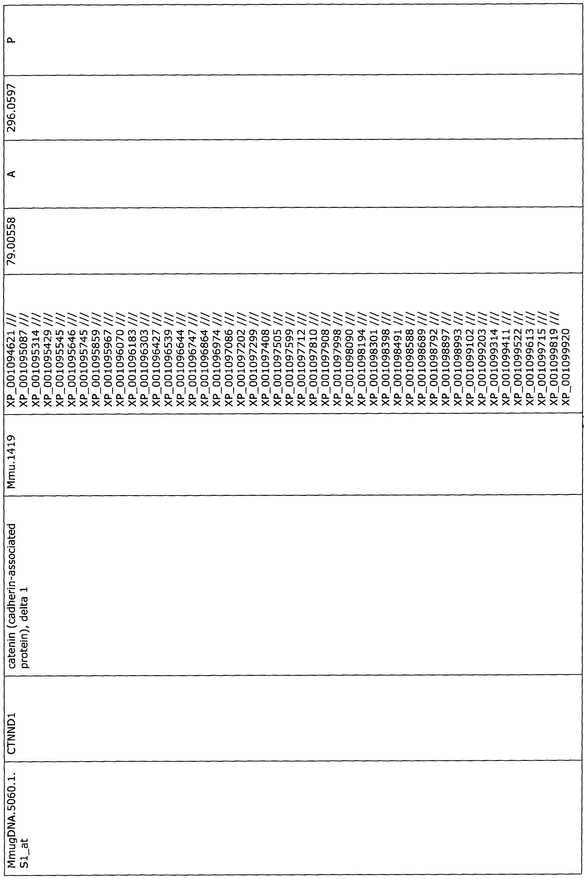

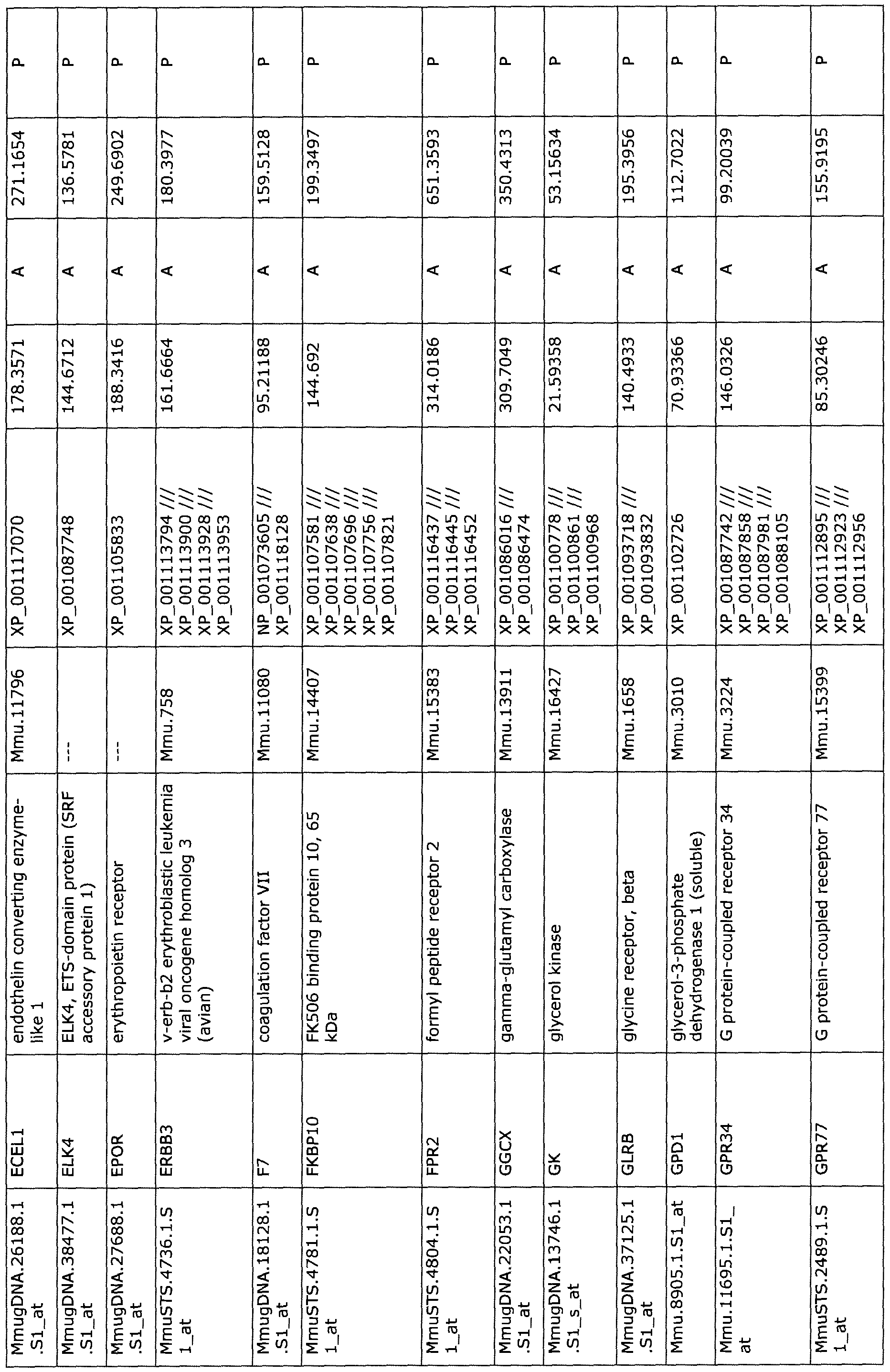
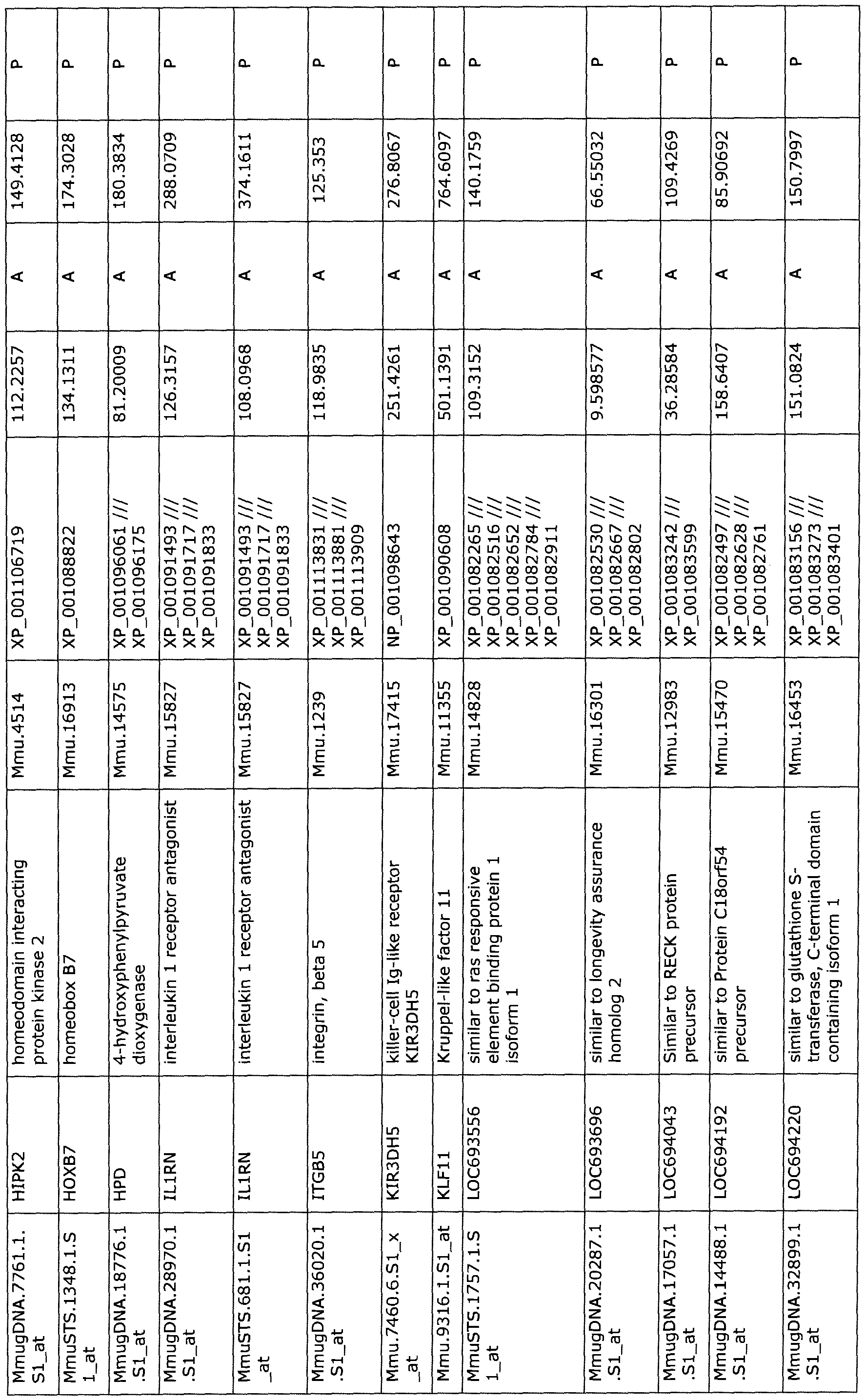
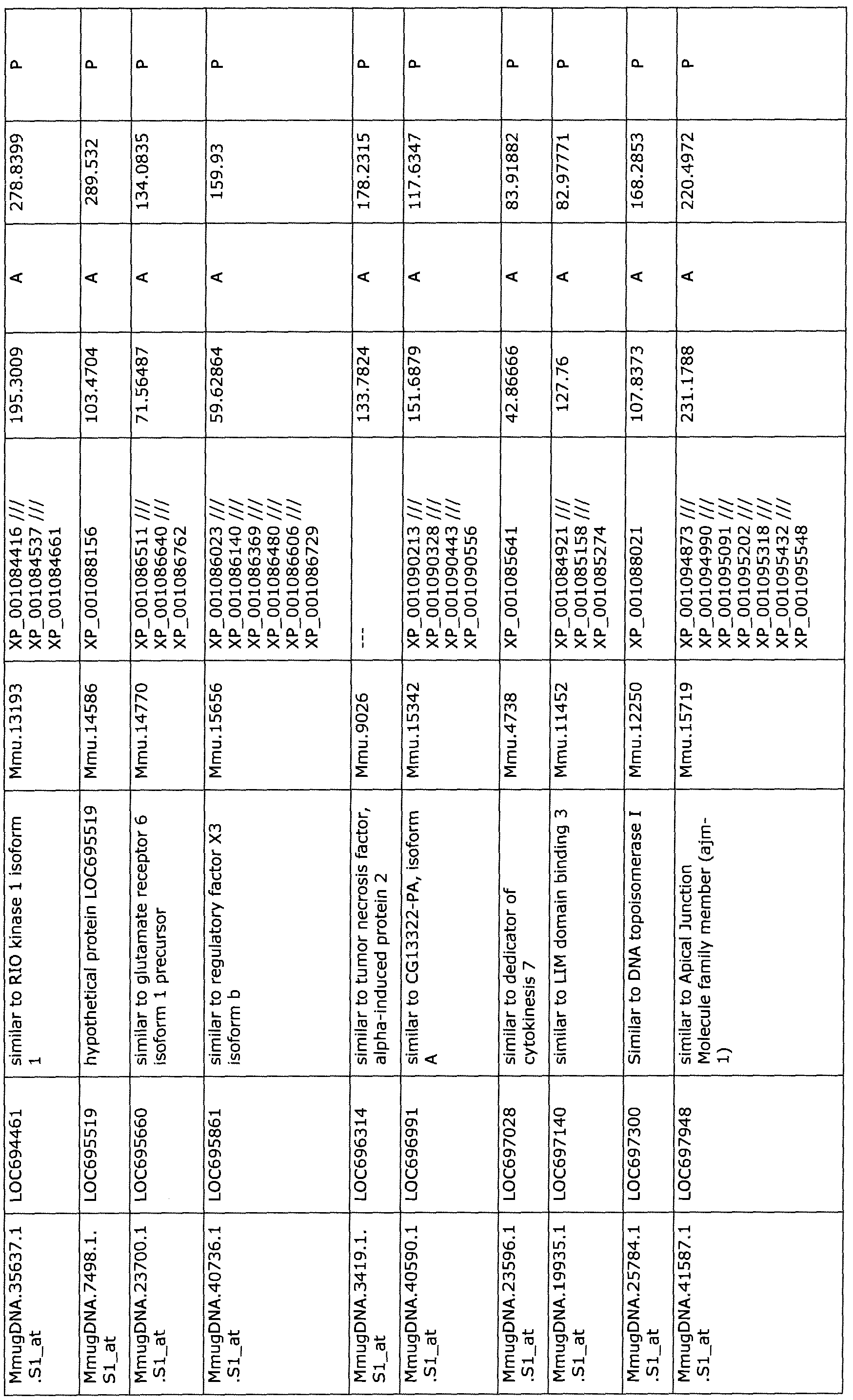
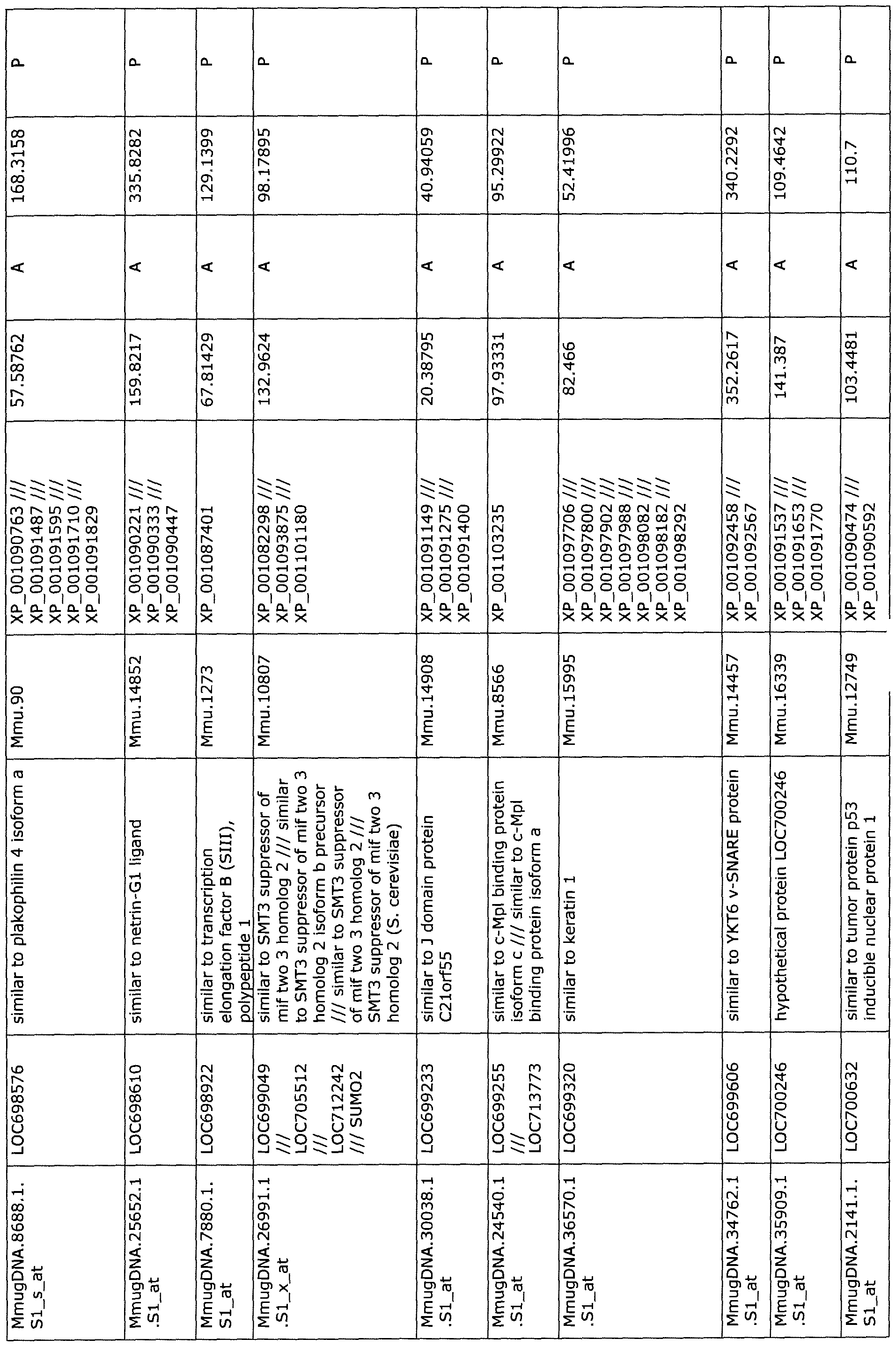
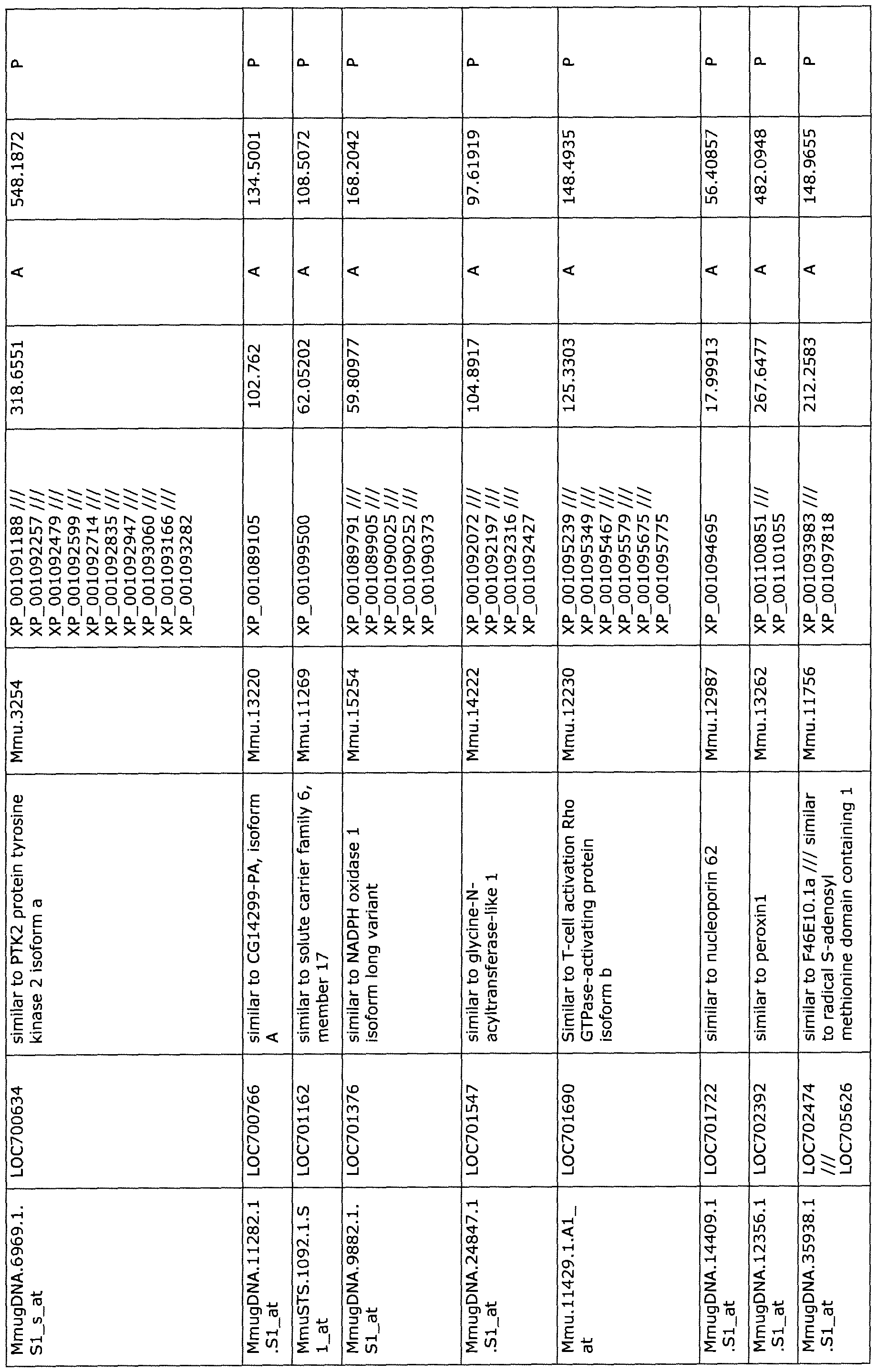
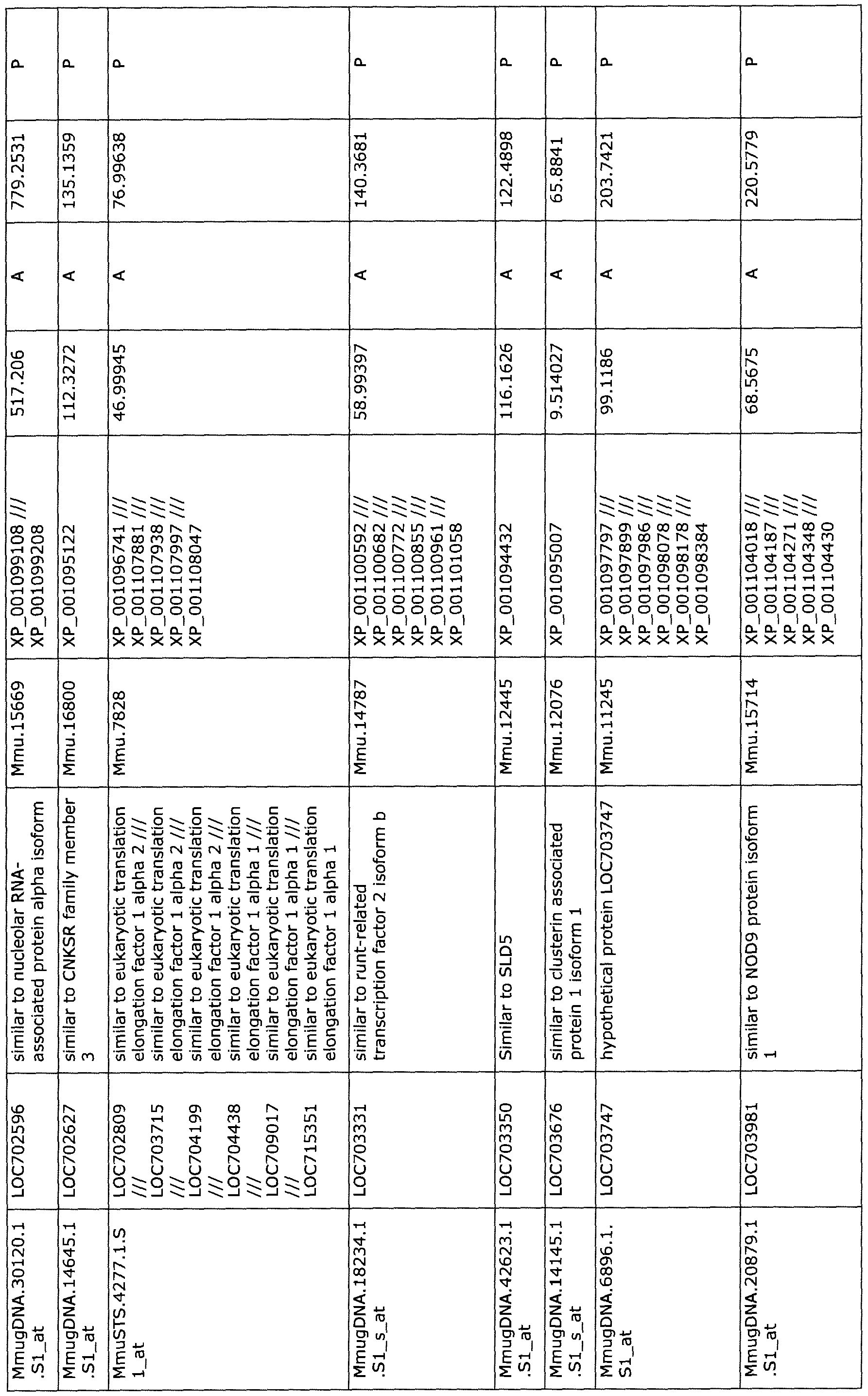


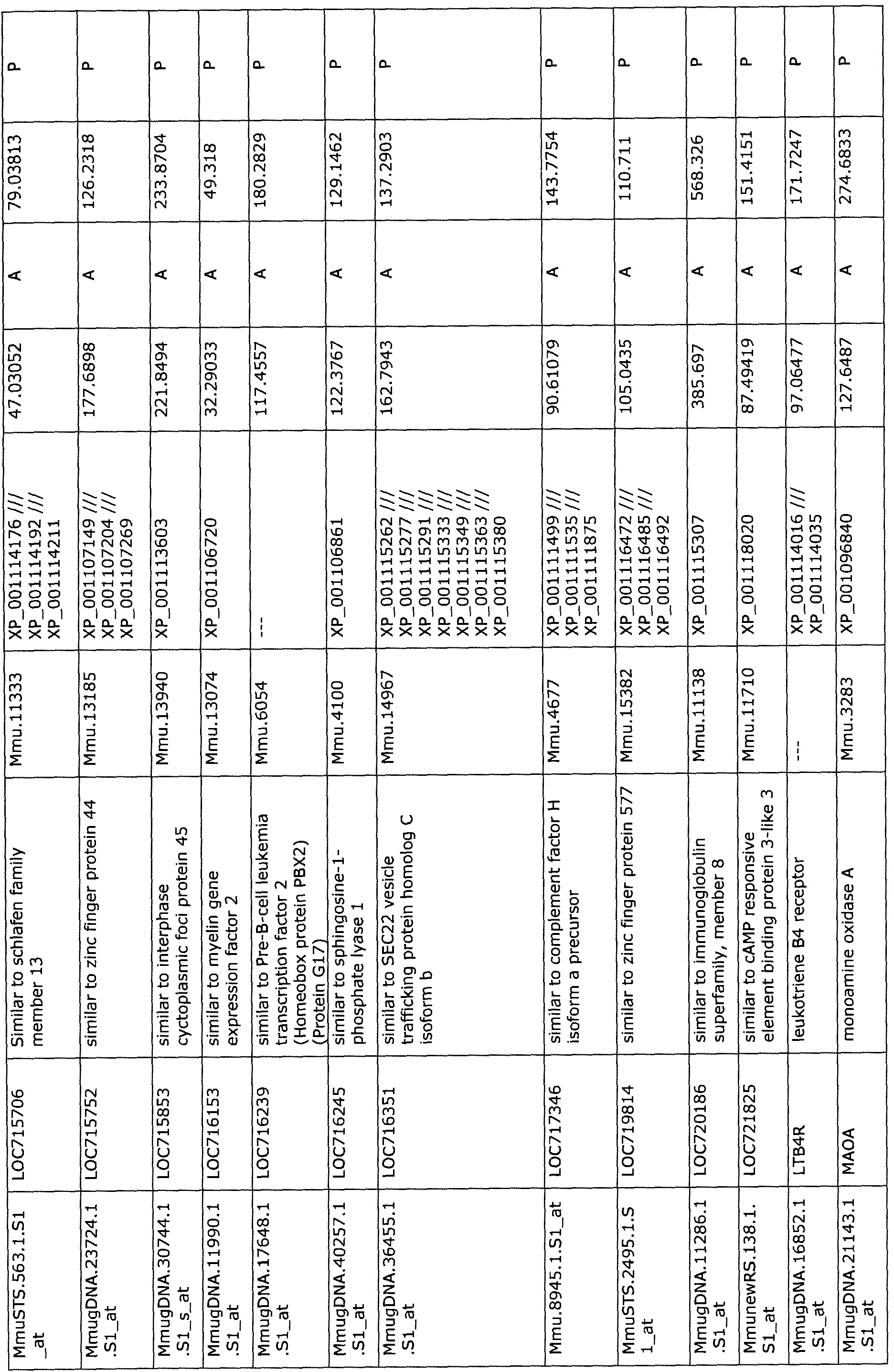


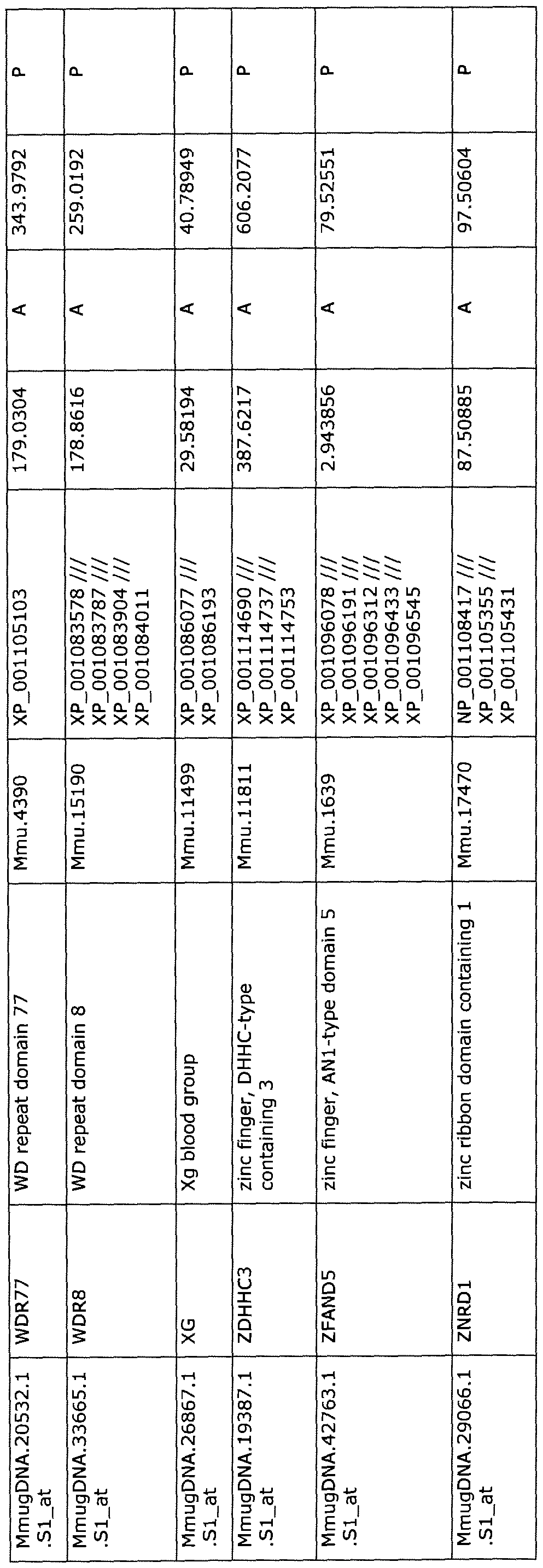
Table 2: Differentially expressed genes in PBMCs of NHPs 2-hr following MCAO
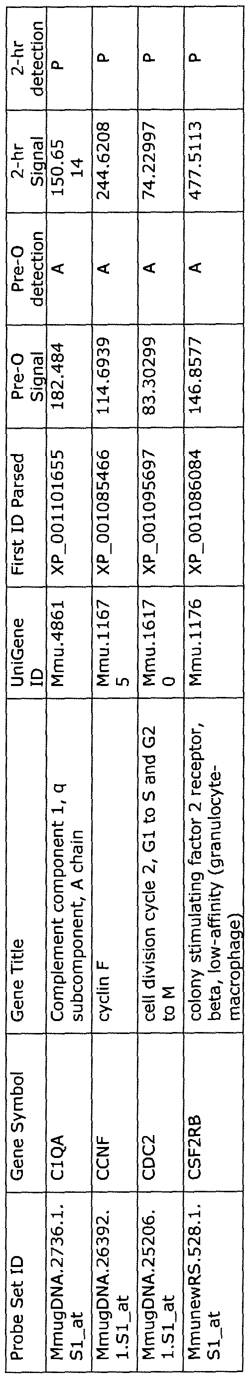
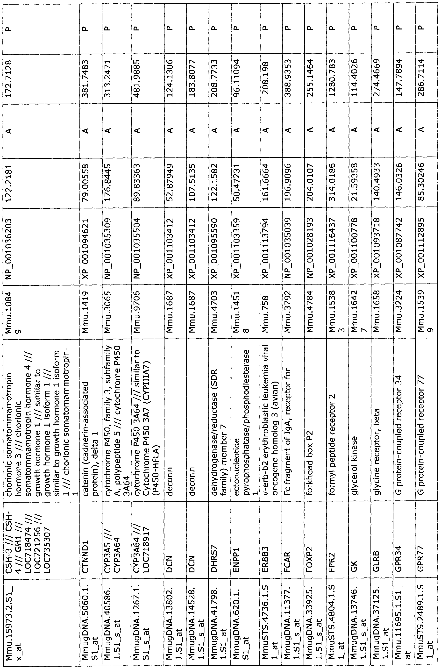
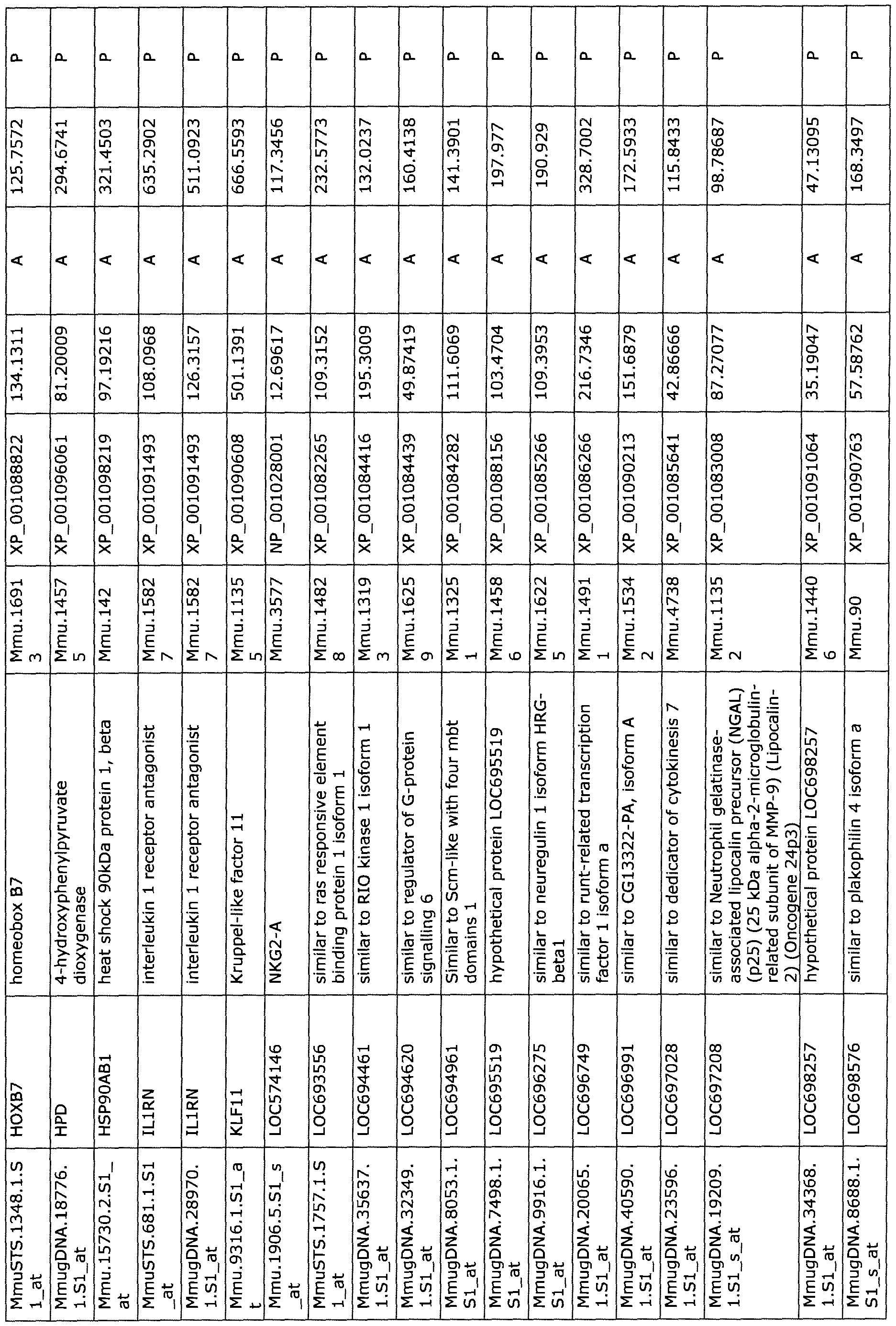

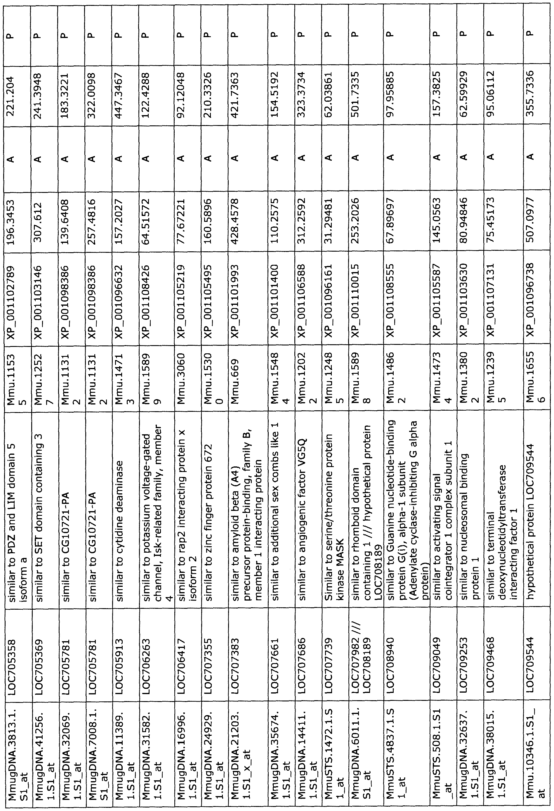
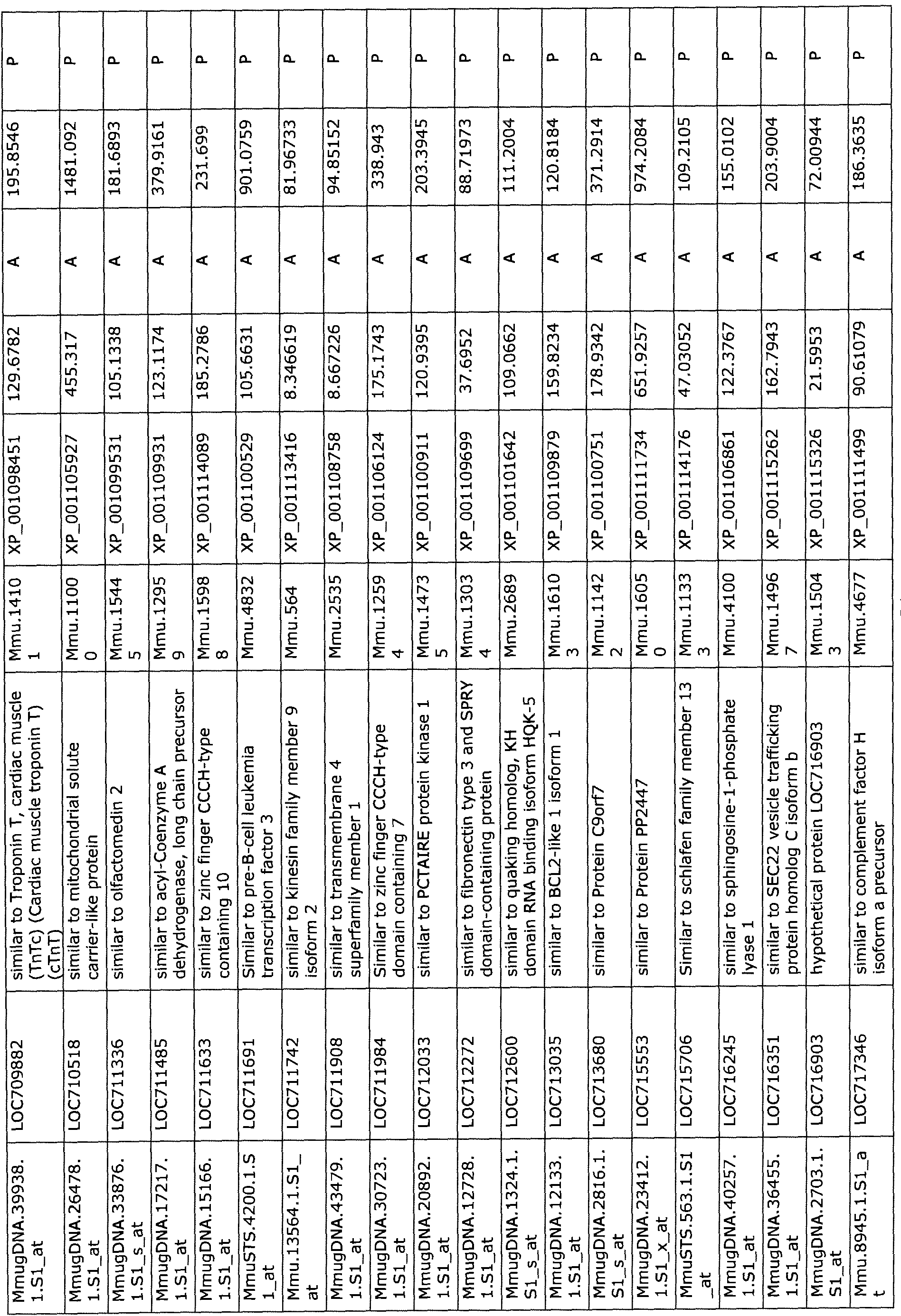
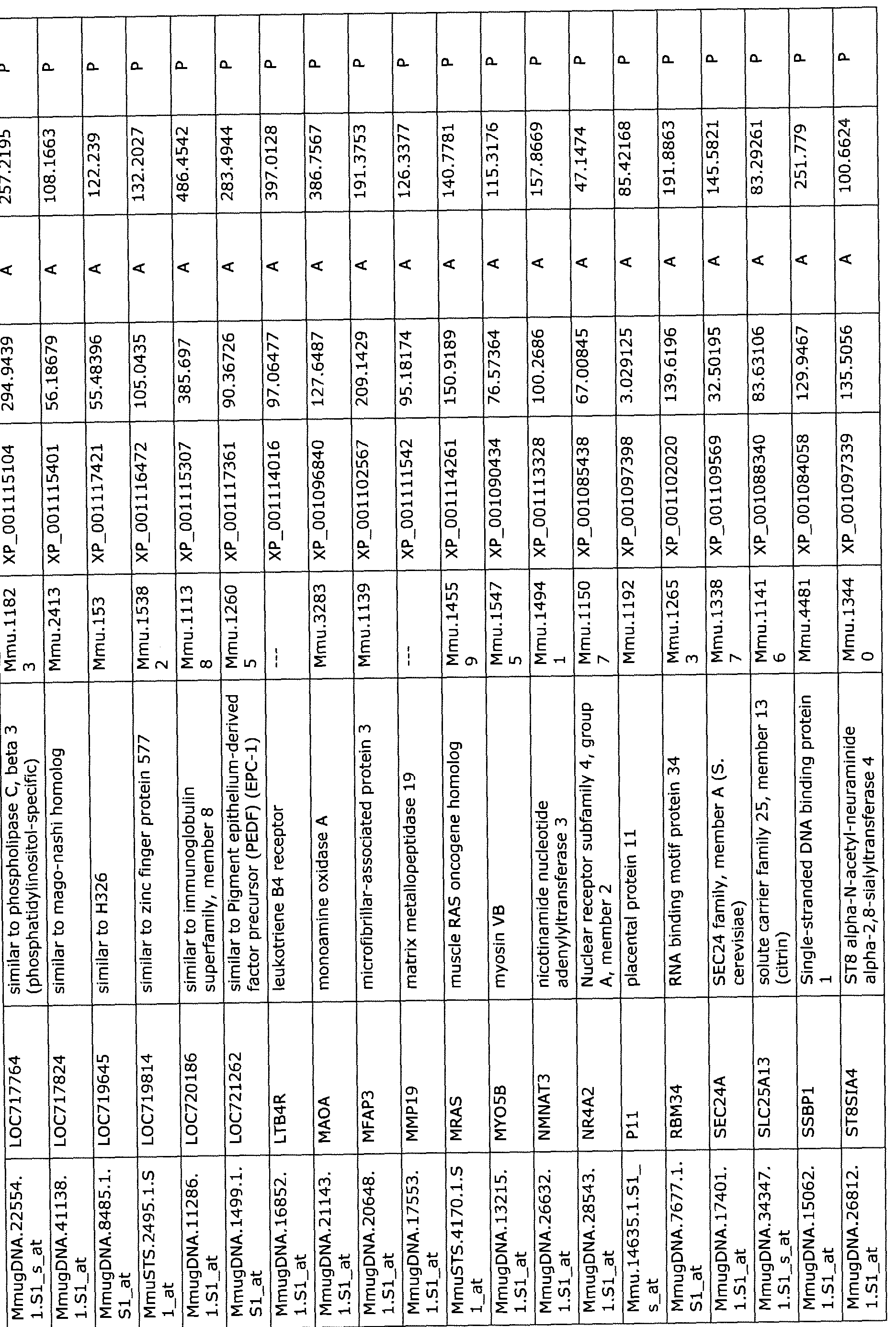
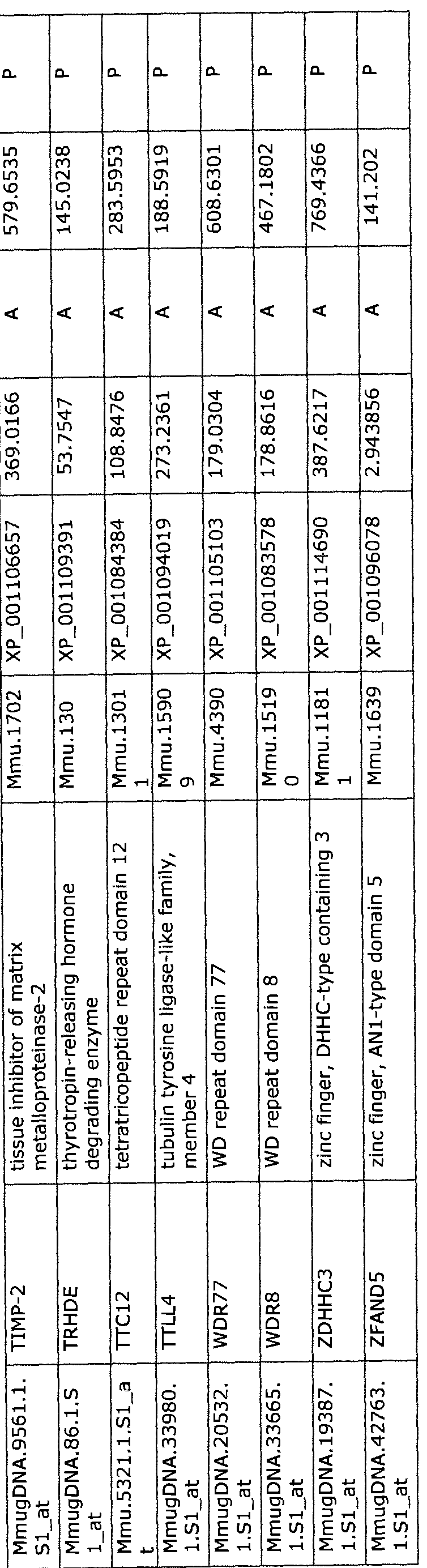
Table 3: Differentially expressed genes in PBMCs of NHPs 24-hr following MCAO

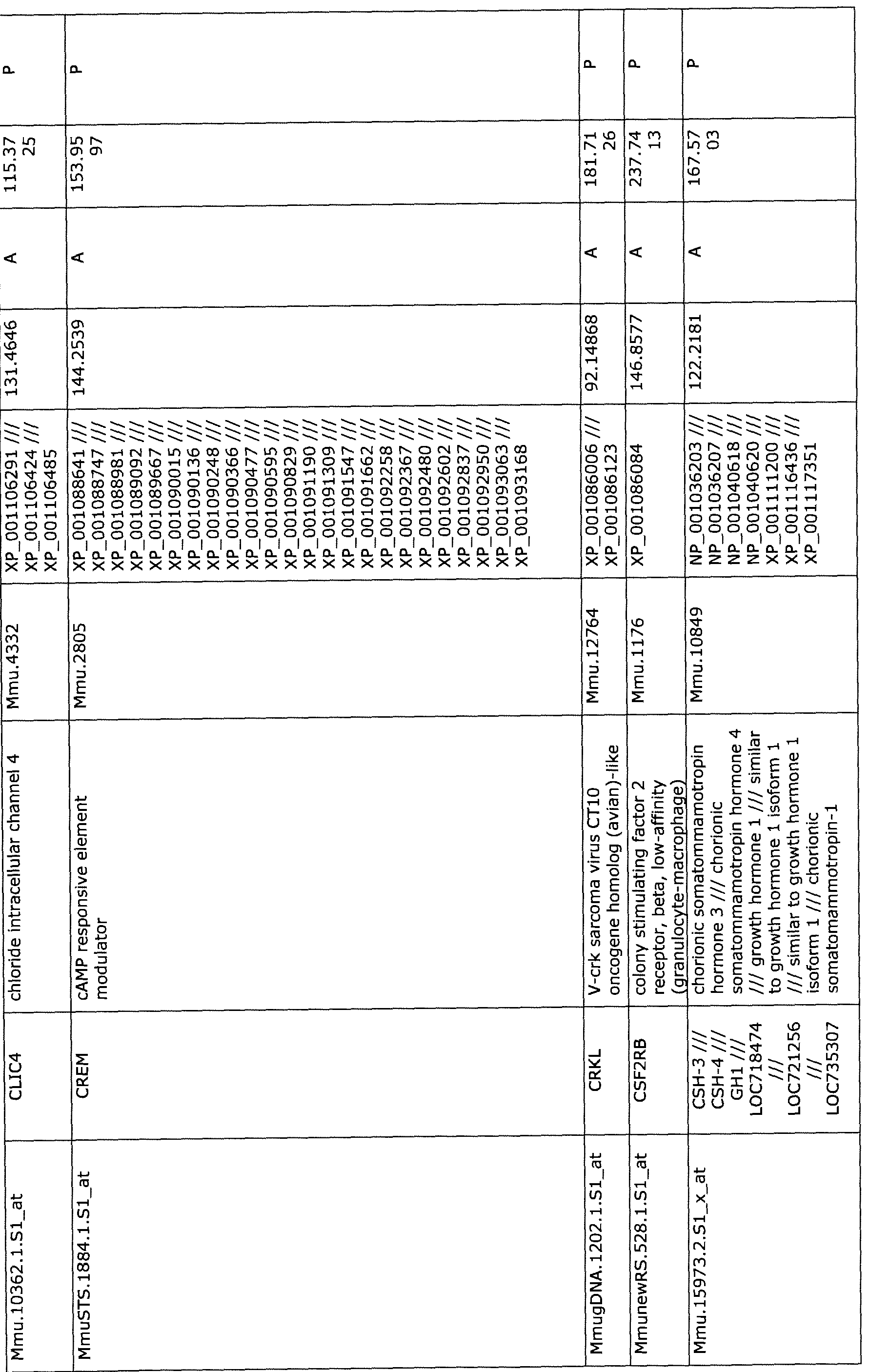
MmugDNA.5060.1.Sl_at CTNNDl catenin (cadherin-associated Mmu.1419 XP_001094621 /// 79.00558 247.86 protein), delta 1 XP_001095087 /// 38
XP_001095314 ///
XP_001095429 ///
XP_001095545 ///
XP_001095646 ///
XP_001095745 ///
XP_001095859 ///
XP_001095967 ///
XP_001096070 ///
XP_001096183 ///
XP_001096303 ///
XP_001096427 ///
XP_001096539 ///
XP_001096644 ///
XP_001096747 ///
XP_001096864 ///
XP_ 001096974 ///
XP_001097086 ///
XP_001097202 ///
XP_001097299 ///
XP_001097408 ///
oe XP_001097505 ///
XP_001097599 ///
XP_001097712 ///
XP_001097810 ///
XP_001097908 ///
XP_001097998 ///
XP_001098090 ///
XP_001098194 ///
XP_001098301 ///
XP_001098398 ///
XP_001098491 ///
XP_001098588 ///
XP_001098689 ///
XP_001098792 ///
XP_001098897 ///
XP_001098993 ///
XP_001099102 ///
XP_001099203 ///
XP_001099314 ///
XP_001099411 ///
XP_001099522 ///
XP_001099613 ///
XP_001099715 ///
XP_001099819 ///
XP 001099920
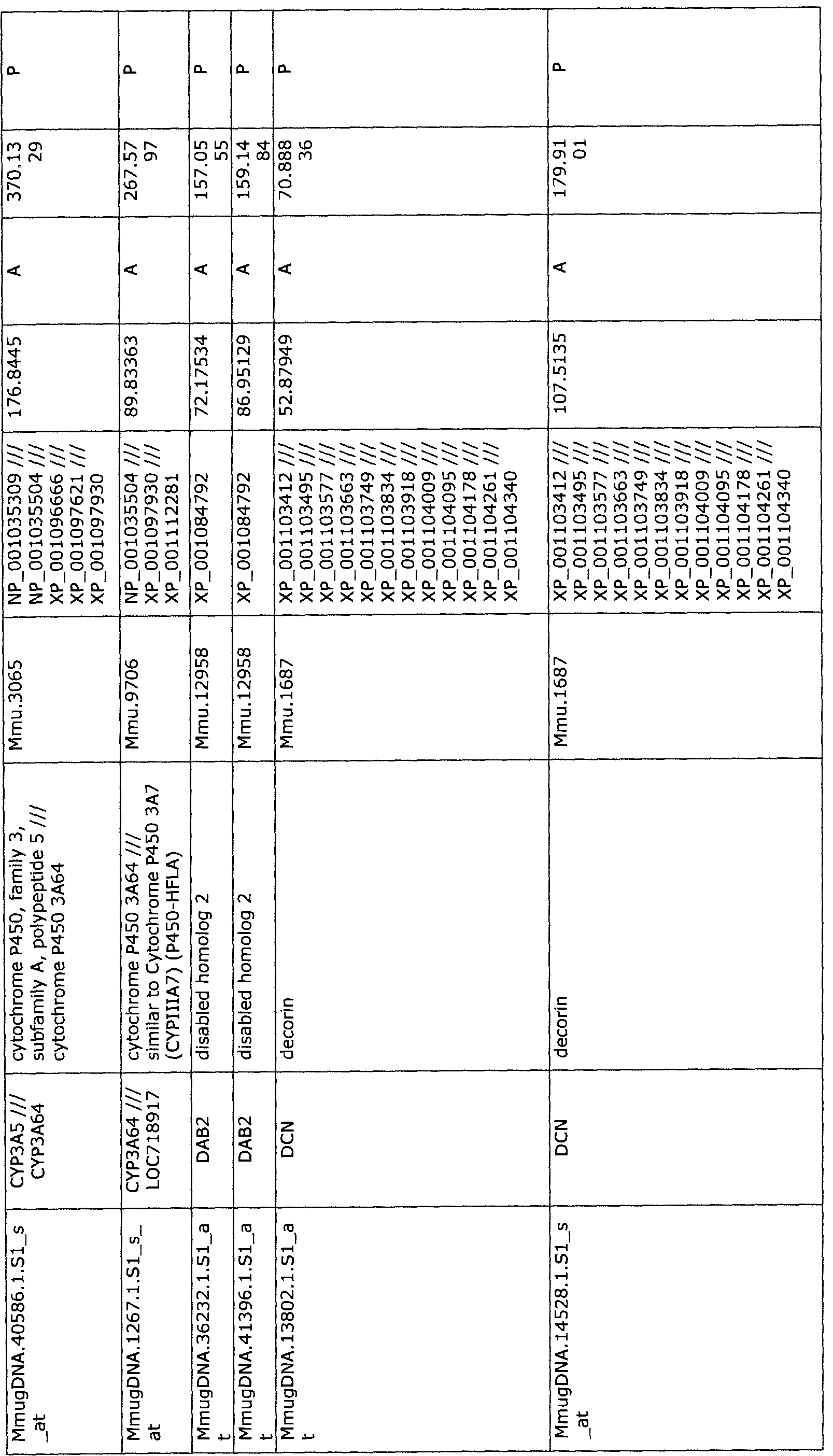

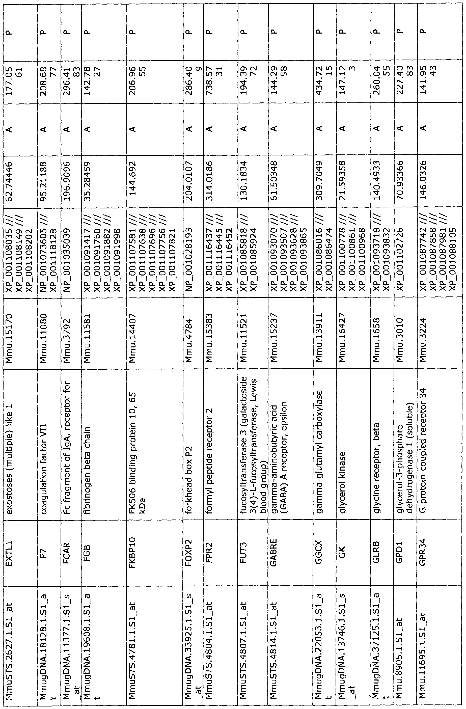


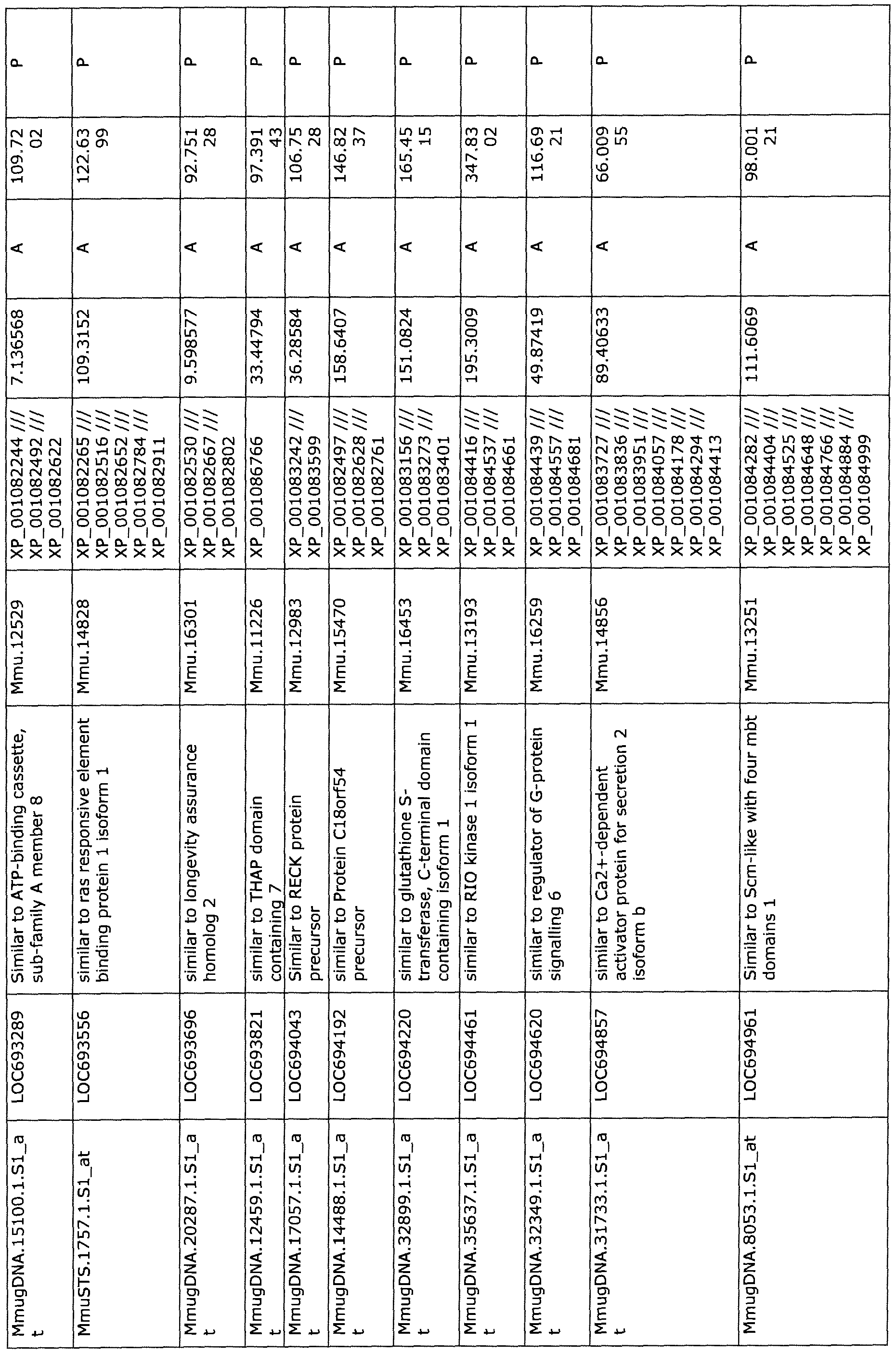
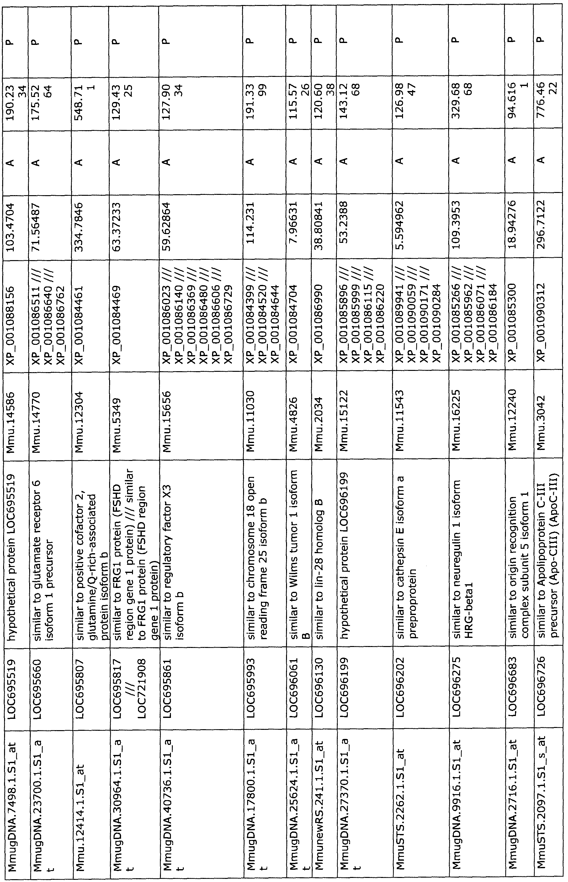


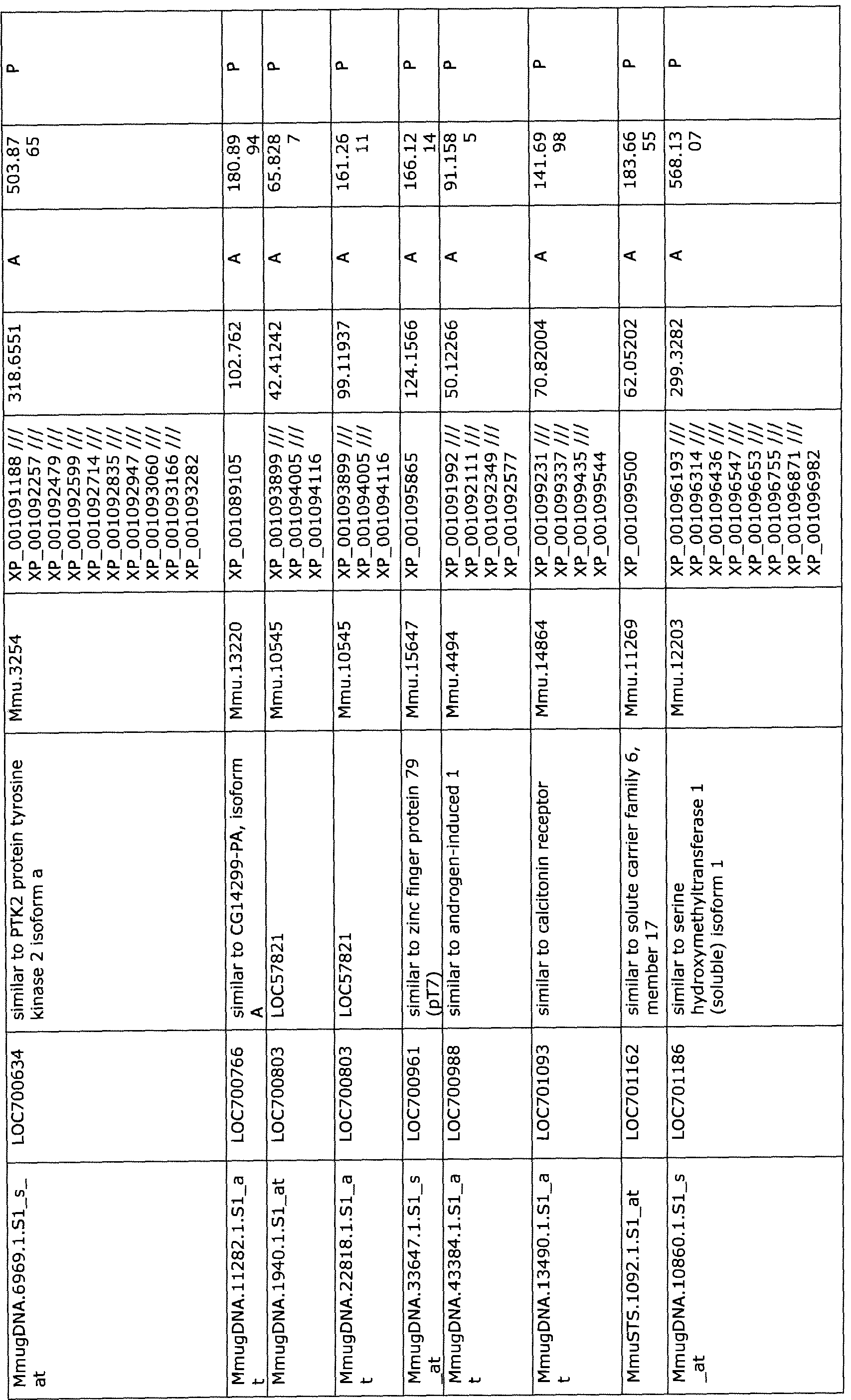
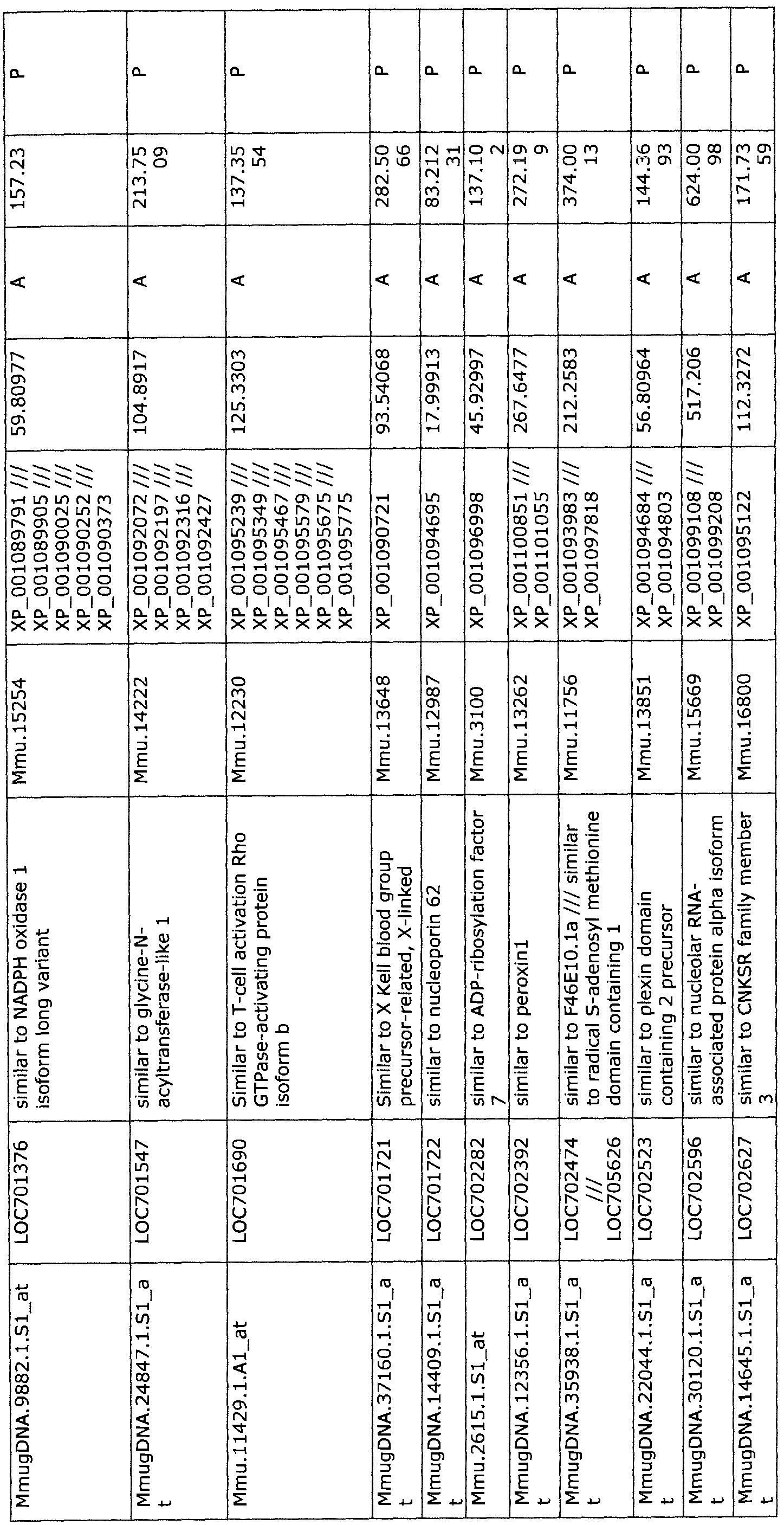
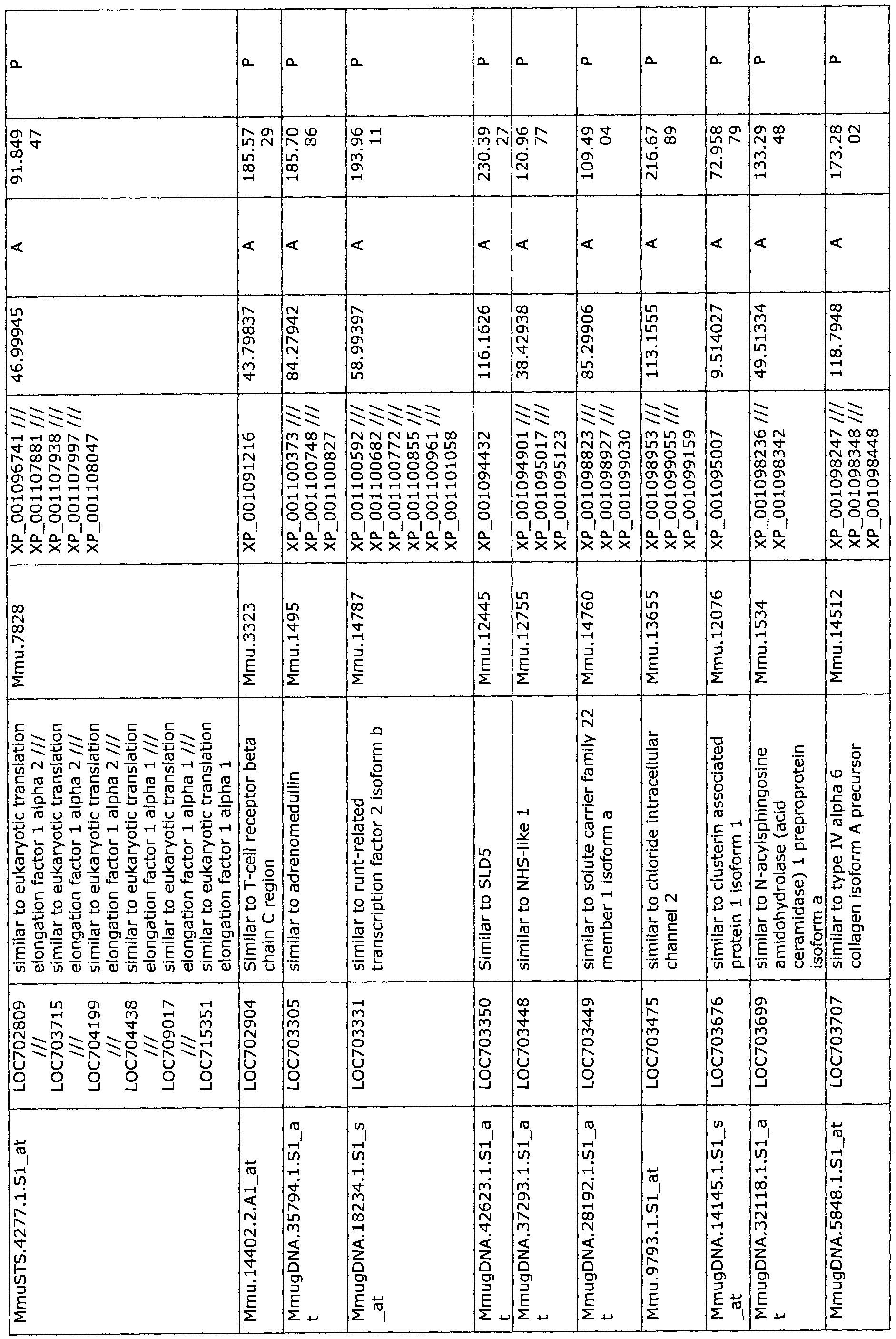
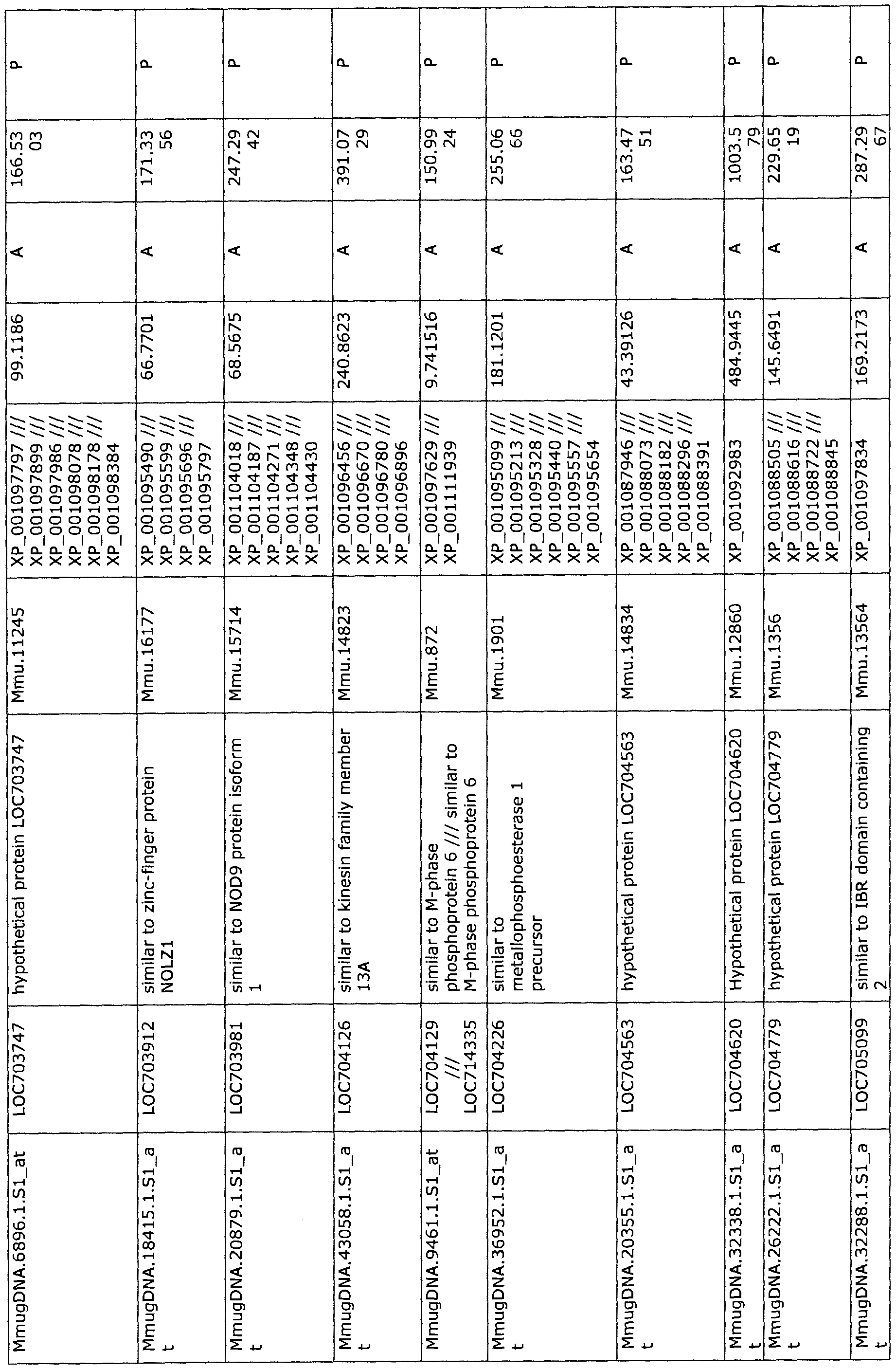
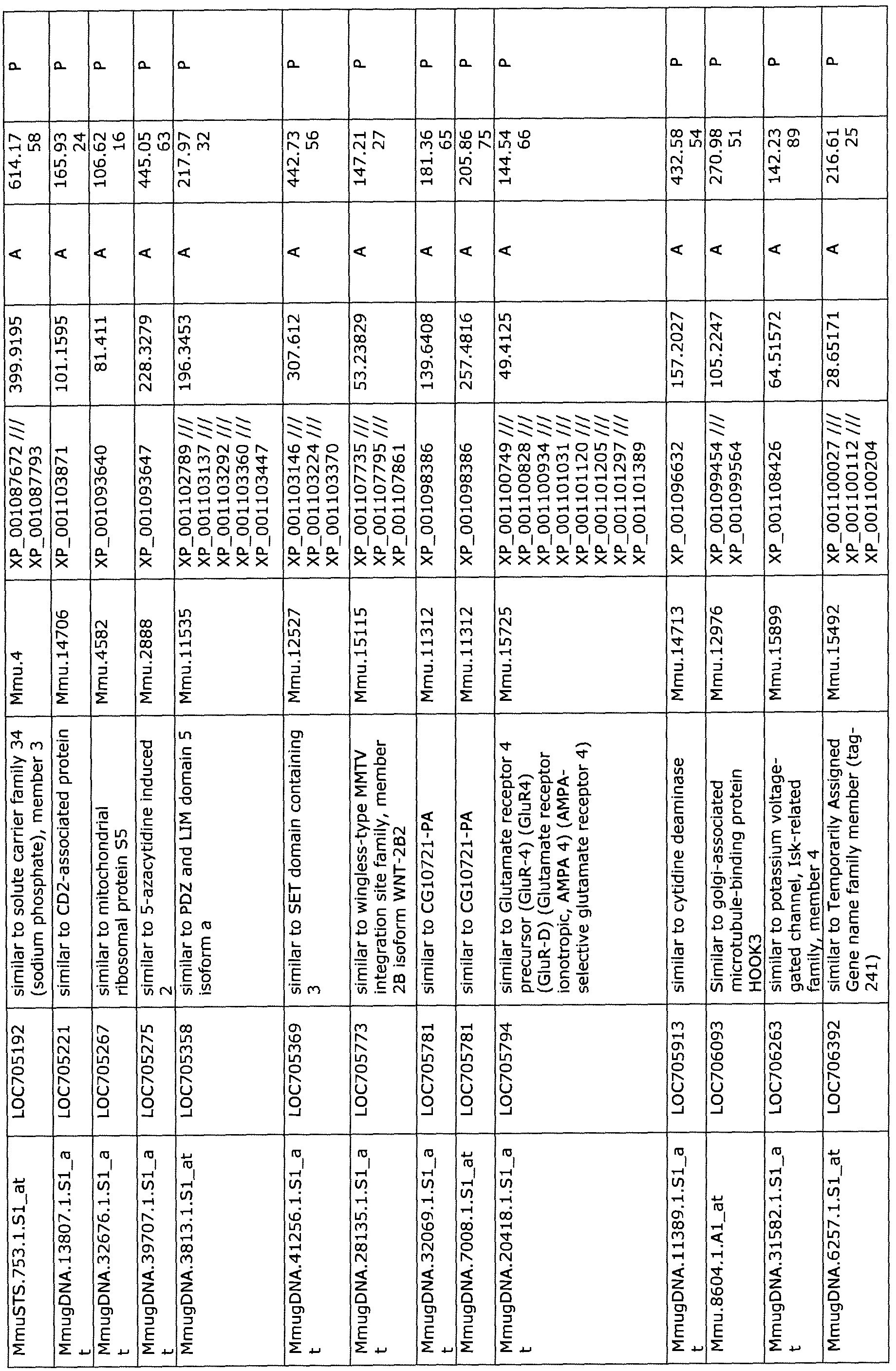
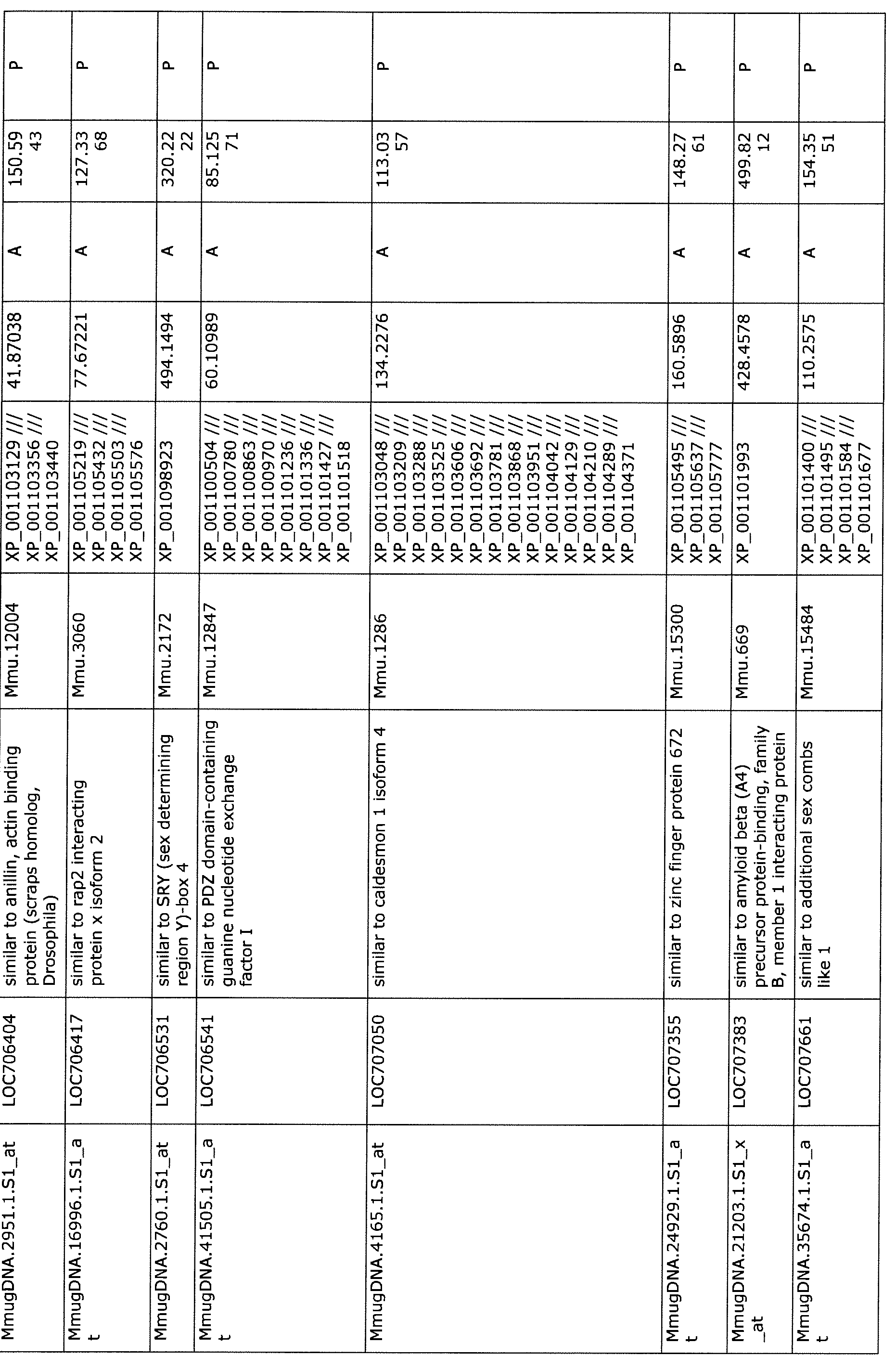
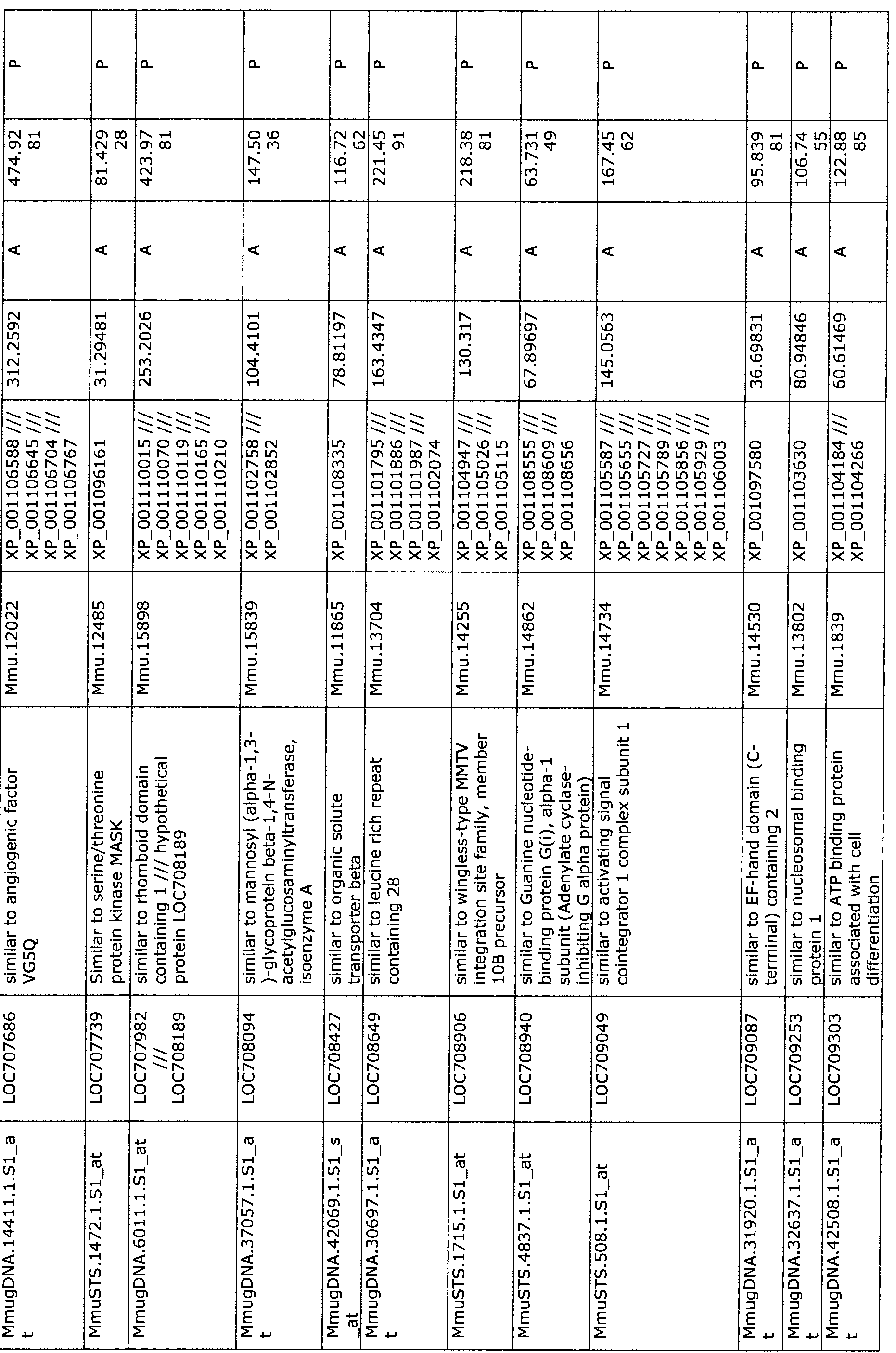
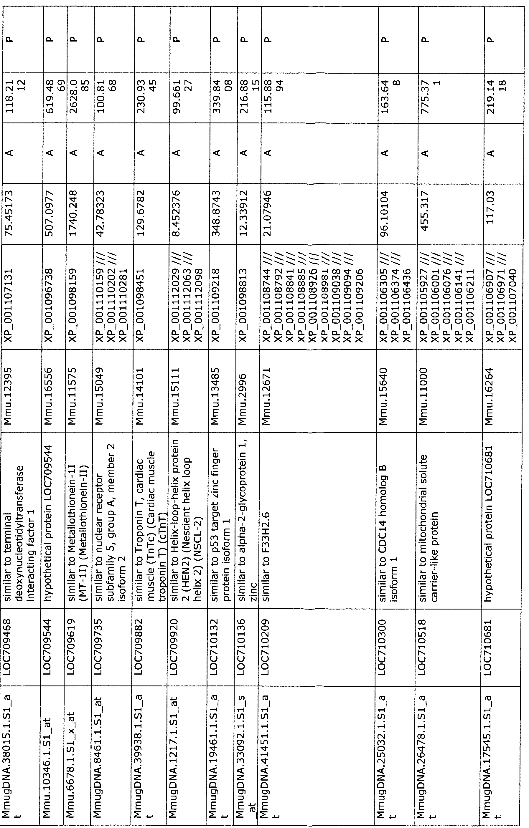


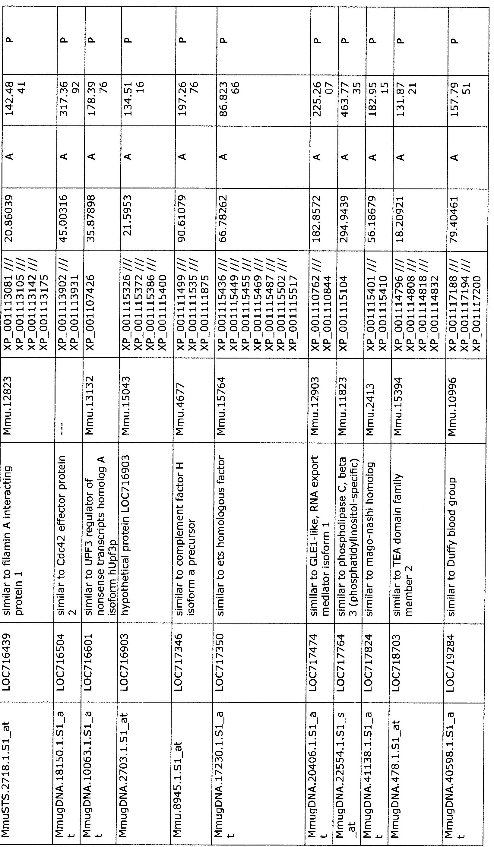
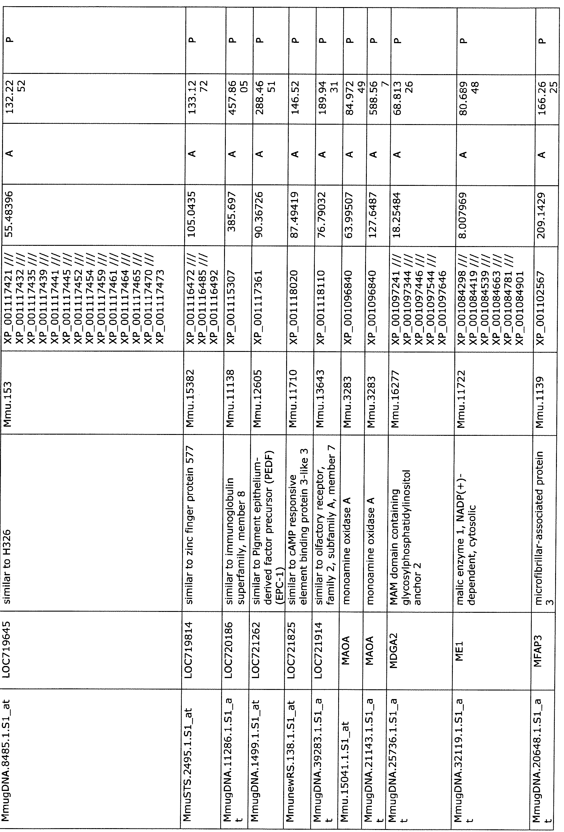
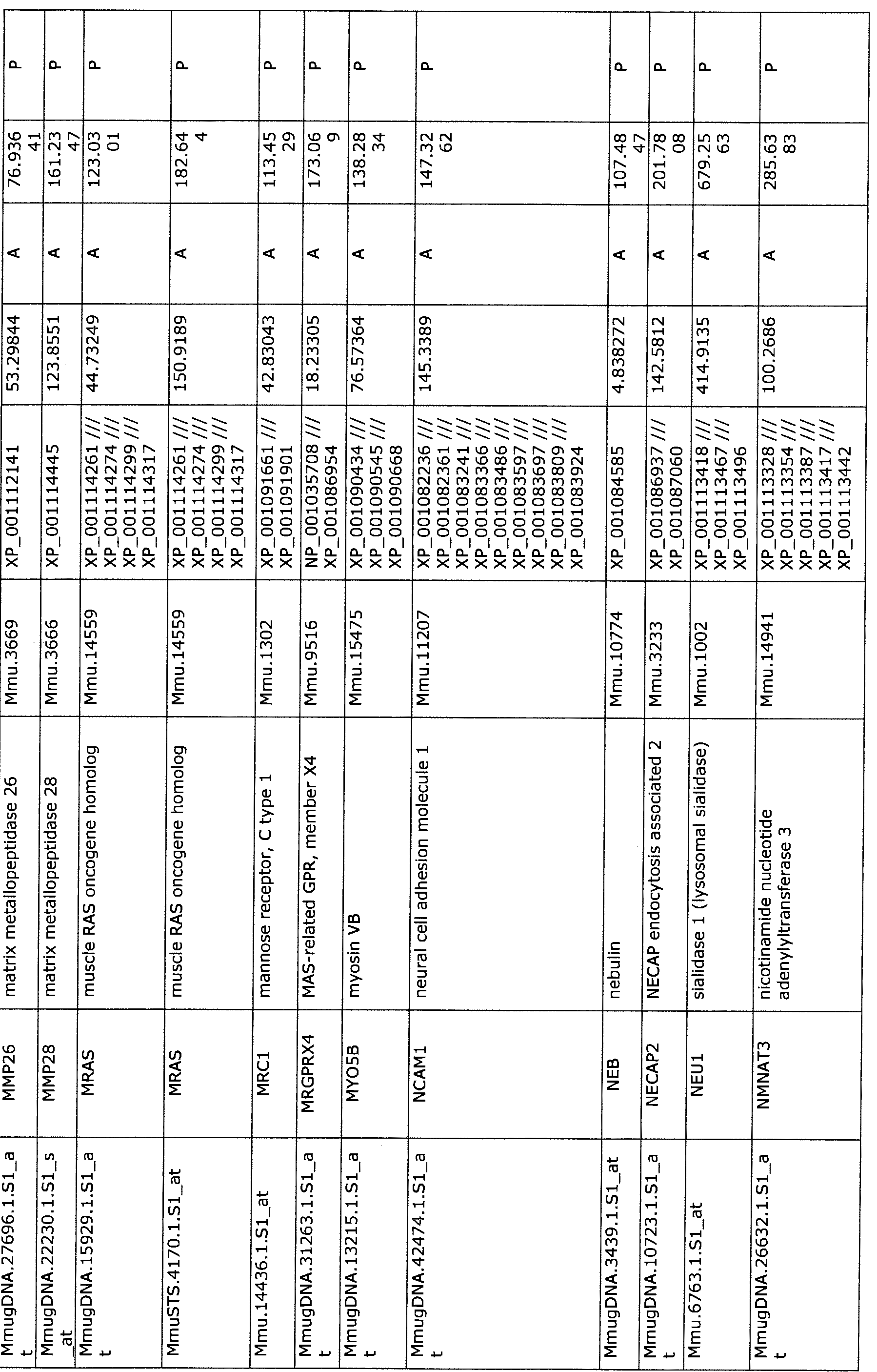

MmugDNA.31823.1.Sl_a PRKACB protein kinase, cAMP-dependent, Mmu.1940 XP 001105459 /// 145.2954 A 122.26 P t catalytic, beta XP 001105534 /// 89
XP 001105605 ///
XP 001105676 ///
XP 001105747 ///
XP 001105803 ///
XP 001105875 ///
XP 001105953 ///
XP 001106026 ///
XP 001106097 ///
XP_001106164
MmugDNA.36754. l.Sl_a PRR3 proline rich 3 Mmu.12623 XP_001100535 92.55525 A 104.11 P t 83
Mmu. l2.1.Sl_at RASSF8 Ras association (RalGDS/AF-6) Mmu.13028 XP 001102028 /// 31.62563 A 82.270 P domain family (N-terminal) XP 001102115 /// 78 member 8 XP_001102201
MmuSTS.2090.1.Sl_at RBM11 RNA binding motif protein 11 Mmu.12892 XP_001083260 81.05433 A 85.995 P
3
MmugDNA.7677.1.Sl_at RBM34 RNA binding motif protein 34 Mmu.12653 XP 001102020 /// 139.6196 A 60.824 P
XP_001102111 75
MmugDNA.17401.1.Sl_a SEC24A SEC24 family, member A (S. Mmu.13387 XP 001109569 /// 32.50195 A 160.13 P t cerevisiae) XP_001109620 17
MmugDNA.18877.1.Sl_a SF3B14 similar to splicing factor 3B, 14 Mmu.942 XP_001098393 96.04313 A 144.18 P t kDa subunit 95
MmuSTS.3315.1.Sl_at SH3GLB2 SH3-domain GRB2-like Mmu.17131 XP_001109262 415.9608 A 360.32 P endophilin B2 15
MmugDNA.28147.1.Sl_a SLC13A2 solute carrier family 13 (sodium- Mmu.11776 XP_001106342 83.45712 A 172.62 P t dependent dicarboxylate 72
transporter), member 2
MmugDNA.20608.1.Sl_a SLC25A11 Solute carrier family 25 Mmu.11522 XP 001097307 /// 159.8107 A 199.76 P t (mitochondrial carrier; XP 001097413 /// 57
oxoglutarate carrier), member XP_001097514
11
MmugDNA.34347.1.Sl_s SLC25A13 solute carrier family 25, member Mmu.11416 XP_001088340 83.63106 A 130.70 P at 13 (citrin) 75
MmugDNA.43541.1.Sl a SLC25A17 Solute carrier family 25 Mmu.4154 XP_001102293 135.3007 A 217.93 P t (mitochondrial carrier; 23
peroxisomal membrane protein,
34kDa), member 17
MmugDNA.26126.1.Sl a S0X2 SRY (sex determining region Y)- Mmu.10959 NP 001136412 /// 25.83881 A 100.85 P t box 2 XP_001106891 55
MmuAffx.l.l.Sl_at SRY sex determining region Y Mmu.3544 NP_001028008 56.02947 A 91.998 P

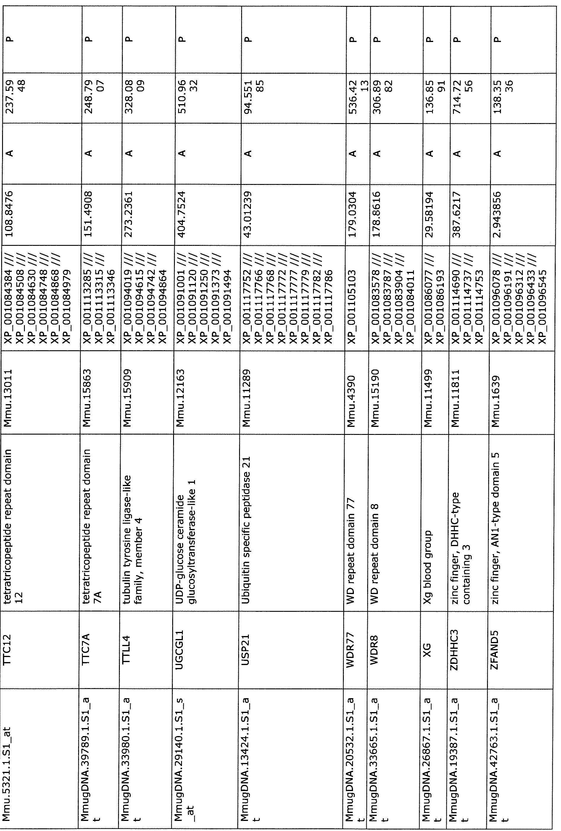

Table 4: Immediate early (1-hr post stroke) biomarkers identified by RNA analysis.
Gene identifier Gene name Amino Acid Sequence
NP 006546.1 synaptobrevin homolog YKT6 SEQ ID NO: 1
NP 001776.1 cytidine deaminase SEQ ID NO:2
EAW60338.1 hypothetical protein FLJ20232 SEQ ID NO:3
EAW67321.1 centrosomal protein 164kDa SEQ ID NO:4
EAW71459.1 WD repeat domain 8 SEQ ID NO:5
EAW85140.1 zinc finger CCCH-type containing 7A SEQ ID NO:6
EAW89539.1 TIMP metallopeptidase inhibitor 2 SEQ ID NO:7
EAW96888.1 zinc finger CCCH-type containing 10 SEQ ID NO:8
BAB47442.1 KIAA1813 protein SEQ ID NO:9
BAB59061.1 Pexlp-634del690 SEQ ID NO: 10
AAH09411.1 WDR77 protein SEQ ID NO; 11
NP 001078929.1 catenin delta- 1 isoform 1A SEQ ID NO: 12
NP 071426.1 leucine-rich repeat-containing protein 4 precursor SEQ ID NO: 13
NP 001099038.1 kinesin-like protein KIF13A isoform d SEQ ID NO: 14
BAG53572.1 unnamed protein product SEQ ID NO: 15
BAG54027.1 unnamed protein product SEQ ID NO: 16
BAG57307.1 unnamed protein product SEQ ID NO: 17
BAG63947.1 unnamed protein product SEQ ID NO: 18
BAG64130.1 unnamed protein product SEQ ID NO: 19
BAG58900.1 unnamed protein product SEQ ID NO:20
NP 148978.2 cyclin-dependent kinase 16 isoform 2 SEQ ID NO:21
NP 001135741.1 vitamin K-dependent gamma-carboxylase isoform 2 SEQ ID NO:22
NP 631981.2 nucleolar protein 6 gamma isoform SEQ ID NO:23
AAM94901.1 AF311906 1 LIR-D 1 precursor SEQ ID NO:24
AAH35404.1 PTK2 protein SEQ ID NO:25
NP 733840.1 NLR family member XI isoform 2 SEQ ID NO:26
AAN75525.1 Rap 1 -interacting adaptor molecule SEQ ID NO:27
NP 001171187.1 Krueppel-like factor 11 isoform b SEQ ID NO:28
NP_001171726.1 trimethyllysine dioxygenase, mitochondrial isoform 2 SEQ ID NO:29
precursor
CAD91343.1 cytochrome P450 SEQ ID NO:30
NP 115652.2 rhomboid domain-containing protein 1 SEQ ID NO:31
NP 079480.2 traB domain-containing protein SEQ ID NO:32
NP 954574.1 histone-lysine N-methyltransferase setd3 isoform b SEQ ID NO:33
NP 000112.1 erythropoietin receptor precursor SEQ ID NO:34
NP 001453.1 N-formyl peptide receptor 2 SEQ ID NO:35
NP 002420.1 matrix metalloproteinase-19 isoform rasi-l preproprotein SEQ ID NO:36
NP 003098.1 transcription factor SOX-4 SEQ ID NO:37
NP 003134.1 single-stranded DNA-binding protein, mitochondrial SEQ ID NO:38
precursor
AAH16977.2 PBX3 protein SEQ ID NO:39
NP_001973.2 receptor tyrosine-protein kinase erbB-3 isoform 1 SEQ ID NO:40
precursor
CAH72013.1 RIO kinase 1 (yeast) SEQ ID NO:41
2IRT A Chain, Initial Crystallographic Analyses Of A SEQ ID NO:42
Recombinant Interleukin-1 Receptor Antagonist Protein
BAA91249.1 unnamed protein product SEQ ID NO:43
NP 000447.2 sulfite oxidase, mitochondrial precursor SEQ ID NO:44
CAB86601.1 xce SEQ ID NO:45
NP 057692.1 armadillo repeat-containing X-linked protein 1 SEQ ID NO:46
NP 057696.2 mitoferrin-1 SEQ ID NO:47
NP 064530.1 endophilin-B2 SEQ ID NO:48
Table 5: Early biomarkers (2-hr post stroke) biomarkers identified by RNA analysis.
Gene identifier Gene name Amino Acid Sequence
NP 060288.3 WD repeat-containing protein WRAP73 SEQ ID NO:49
NP 001776.1 cytidine deaminase SEQ ID NO:50
NP_001128651.1 palmitoyltransferase ZDHHC3 isoform 1 SEQ ID O:51
NP_004197.1 vesicle-trafficking protein SEC22c isoform b SEQ ID NO:52
P_001001890.1 runt-related transcription factor 1 isoform AMLlb SEQ ID NO:53
NP_000457.1 peroxisome biogenesis factor 1 SEQ ID NO:54
NP 071426.1 leucine-rich repeat-containing protein 4 precursor SEQ ID NO: 13
NP_003134.1 single-stranded DNA-binding protein, mitochondrial SEQ ID NO:55
NP 001003700.1 ras-responsive element-binding protein 1 isoform 3 SEQ ID NO:56
NP_001099038.1 kinesin-like protein KIF13A isoform d SEQ ID NO:57
NP 877434.2 E3 ubiquitin-protein ligase RNF144B SEQ ID NO:58
NP 031381.2 heat shock protein HSP 90-beta SEQ ID NO:59
NP 001011516.1 PDZ and LIM domain protein 5 isoform e SEQ ID NO:60
NP_000815.1 glycine receptor subunit beta isoform A precursor SEQ ID NO:61
NP_060516.2 angiogenic factor with G patch and FHA domains 1 SEQ ID NO:62
NP_057113.1 dehydrogenase/reductase SDR family member 7 precursor SEQ ID NO:63
NP_954574.1 histone-lysine N-methyltransferase setd3 isoform b SEQ ID NO:33
NP 057696.2 mitoferrin-1 SEQ ID NO:47
NP_039258.1 pro-neuregulin-1, membrane-bound isoform isoform SEQ ID NO:64
HRG-alpha
NP 005598.3 focal adhesion kinase 1 isoform b SEQ ID NO:65
Table 6: Late biomarkers ( 24-hr post stroke) biomarkers identified by RNA analysis.
Gene identifier Gene name Amino Acid Sequence
NP_005944.1 metallothionein-2 SEQ ID NO:66
NP_057692.1 armadillo repeat-containing X-linked protein 1 SEQ ID NO:46
NP 001128250.1 pre-B-cell leukemia transcription factor 3 isoform 2 SEQ ID NO:67
NP_000031.1 apolipoprotein C-III precursor SEQ ID NO:68
NP_057696.2 mitoferrin-1 SEQ ID NO:47
NP 079480.2 traB domain-containing protein SEQ ID NO:69
NP 001128651.1 palmitoyltransferase ZDHHC3 isoform 1 SEQ ID NO:70
NP_000425.1 sialidase-1 precursor SEQ ID NO:71
NP_006238.1 serine/threonine-protein phosphatase 5 isoform 1 SEQ ID NO:72
NP 631981.2 nucleolar protein 6 gamma isoform SEQ ID NO:73
NP 057427.3 centromere protein F SEQ ID NO:74 fNP_543153.1 sodium-dependent phosphate transport protein 2C SEQ ID NO:75
NP_001171187.1 Krueppel-like factor 11 isoform b SEQ ID NO:28
NP 000231.1 amine oxidase SEQ ID NO:76
NP 004160.3 serine hydroxymethyltransferase, cytosolic isoform 1 SEQ ID NO:77
NP 848695.1 minor histocompatibility antigen H13 isoform 2 SEQ ID NO:78
NP_056973.2 mediator of RNA polymerase II transcription subunit 15 SEQ ID NO:79 isoform b
P 077007.1 methylosome protein 50 SEQ ID NO:80
NPJ)64505.1 UDP-glucose:glycoprotein glucosyltransferase 1 precursor SEQ ID NO: 81
NP 001001890.1 runt-related transcription factor 1 isoform AMLlb SEQ ID NO: 82
NP 005598.3 focal adhesion kinase 1 isoform b SEQ ID NO:83
NP_061916.3 amyloid beta A4 precursor protein-binding family B SEQ ID NO:84 member 1 -interacting protein
NP_060516.2 angiogenic factor with G patch and FHA domains 1 SEQ ID NO:62
NP_000923.1 l-phosphatidylinositol-4,5-bisphosphate SEQ ID NO:85 phosphodiesterase beta-3 isoform 1
NP 071426.1 leucine-rich repeat-containing protein 4 precursor SEQ ID NO: 13
NP 443100.1 immunoglobulin superfamily member 8 SEQ ID NO:86
NP 006546.1 synaptobrevin homolog YKT6 SEQ ID NO: 1
NP 001 129247.1 calcium channel flower homolog isoform b SEQ ID NO:87
NP_071906.1 5-azacytidine-induced protein 2 isoform a SEQ ID NO:88
NP 954574.1 histone-lysine N-methyltransferase setd3 isoform b SEQ ID NO:33
NP 001135741.1 vitamin K-dependent gamma-carboxylase isoform 2 SEQ ID NO:22
NP_001776.1 cytidine deaminase SEQ ID NO:2
|NP_031381.2 heat shock protein HSP 90-beta SEQ ID NO:59
NP_054872.2 zinc finger CCCH domain-containing protein 7A SEQ ID NO:6
NP_115652.2 rhomboid domain-containing protein 1 SEQ ID NO:31
NP_001034750.1 toll/interleukin-1 receptor domain-containing adapter SEQ ID NO:89 protein isoform a
NP_055047.2 vesicle-associated membrane protein 2 SEQ ID NO:90
NP_000568.1 interleukin-1 receptor antagonist protein isoform 3 SEQ ID NO:91
NP_039258.1 pro-neuregulin-1, membrane-bound isoform isoform SEQ ID NO:64
HRG-alpha
NP_060288.3 WD repeat-containing protein WRAP73 SEQ ID NO:49
NP_598012.1 decorin isoform c precursor SEQ ID NO:92
NP_001973.2 receptor tyrosine-protein kinase erbB-3 isoform 1 SEQ ID NO:40 precursor
NP 001166237.1 forkhead box protein P2 isoform V SEQ ID NO:93
NP_ 000457.1 peroxisome biogenesis factor 1 SEQ ID NO:54
NP_0001 12.1 erythropoietin receptor precursor SEQ ID NO:34
NP OO 1078929.1 catenin delta- 1 isoform 1A SEQ ID NO: 12
|NP_001 176.1 zinc-alpha-2 -glycoprotein precursor SEQ ID NO:94
NP_001171597.1 immunoglobulin lambda-like polypeptide 5 SEQ ID NO:95
NP_077278.1 matrix metalloproteinase-28 isoform 1 preproprotein SEQ ID NO:96
Table 7: Proteins differentially expressed in the NHP model following MCAO

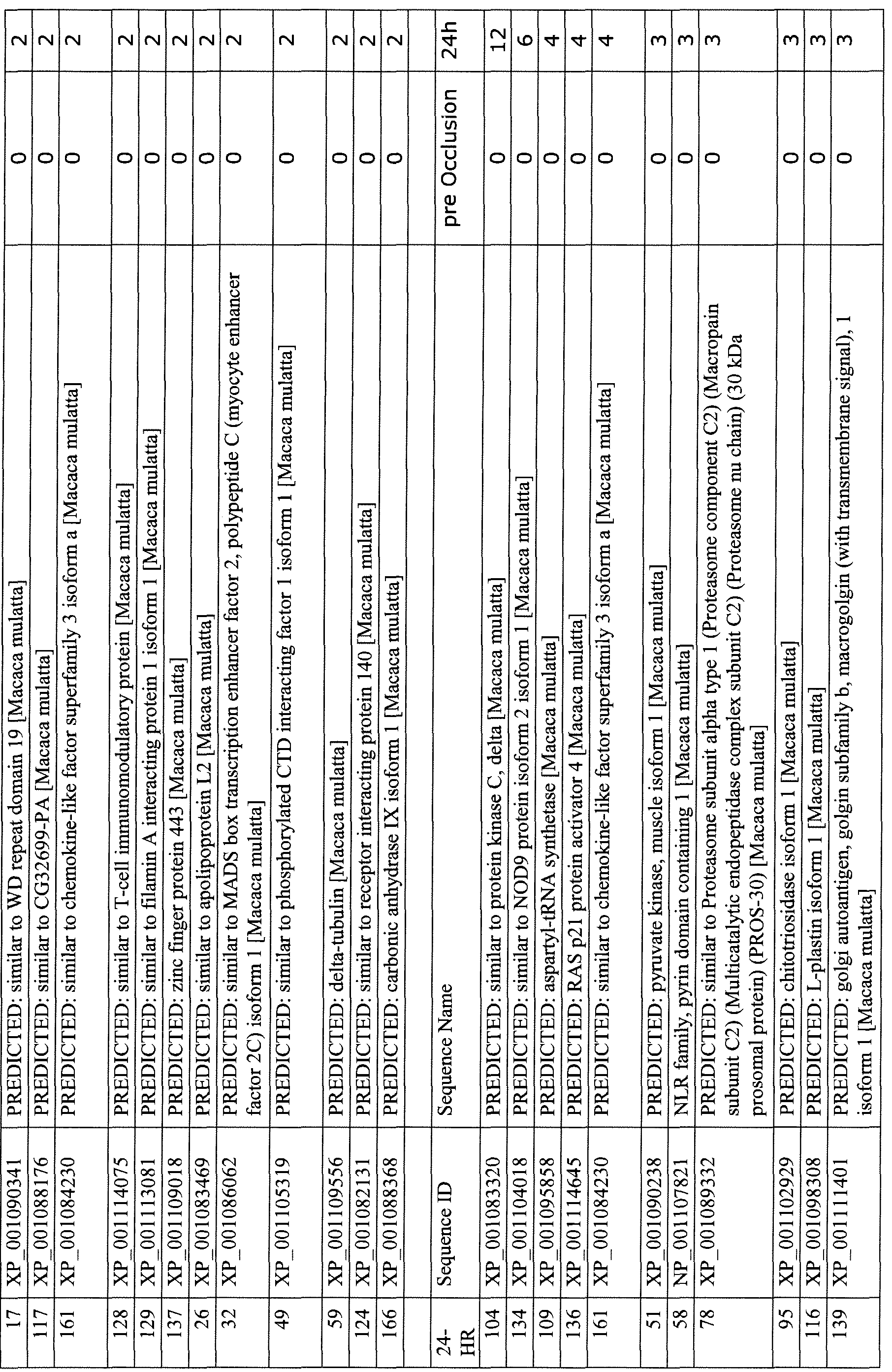
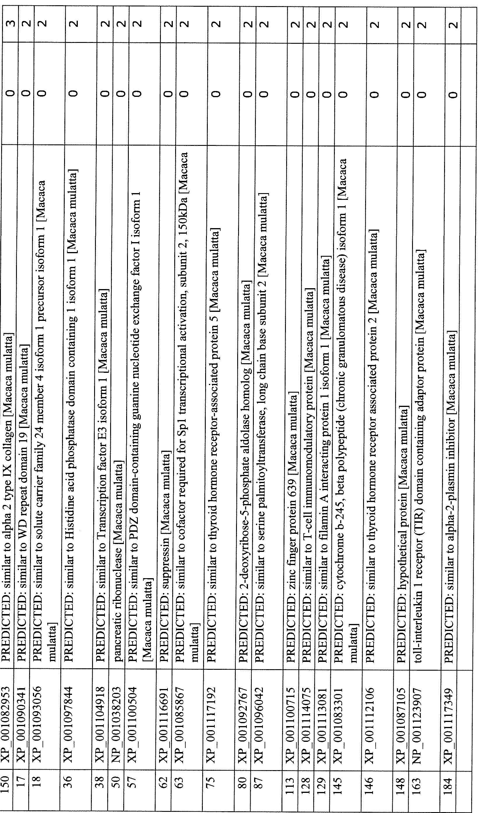
Table 8: Biomarkers identified by the proteomics study
Gene identifier Gene name Amino Acid
Sequence
Immediate early stroke biomarkers, 1-hr post stroke
NP 004235.3 scavenger receptor cysteine-rich type 1 protein Ml 30 isoform SEQ ID NO:97
NP_056150.1 mediator of RNA polymerase II transcription subunit 13-like SEQ ID NO:98
NP 000388.2 cytochrome b-245 heavy chain SEQ ID NO:99
NP 006245.2 protein kinase C delta type SEQ ID NO: 100
NP 001193726.1 pyruvate kinase isozymes M1/M2 isoform d SEQ ID NO: 101
NP 004478.3 Golgin subfamily B member 1 SEQ ID NO-.102
NP 002924.1 ribonuclease pancreatic precursor SEQ ID NO: 103
NP 001 157281.1 WD repeat-containing protein 81 isoform 1 SEQ ID NO: 104
NP 036336.2 general transcription factor 3C polypeptide 4 SEQ ID NO: 105
Early stroke biomarkers, 2-hr post stroke
NP 004235.3 scavenger receptor cysteine-rich type 1 protein Ml 30 isoform SEQ ID NO 97
NP 001843.1 collagen alpha-2(IX) chain precursor SEQ ID NO 106
NP 001092103.1 proline-rich AKT1 substrate 1 SEQ ID NO 107
NP 006245.2 protein kinase C delta type SEQ ID NO 100
NP 002924.1 ribonuclease pancreatic precursor SEQ ID NO 103
NP 000177.2 complement factor H isoform a precursor SEQ ID NO 108
NP 001157281.1 WD repeat-containing protein 81 isoform 1 SEQ ID NO 104
NP 004854.1 serine palmitoyltransferase 2 SEQ ID NO 109
NP 000349.1 transforming growth factor-beta-induced protein ig-h3 precursor SEQ ID NO 110
NP 079408.3 WD repeat-containing protein 19 SEQ ID NO 11 1
NP 060309.2 lysophosphatidylcholine acyltransferase 2 SEQ ID NO 112
NP 653202.1 CKLF-like MARVEL transmembrane domain-containing protein 3 SEQ ID NO 113
NP 110417.2 T-cell immunomodulatory protein precursor SEQ ID NO 114
NP 056502.1 filamin-A-interacting protein 1 SEQ ID NO 115
NP 001074290.1 zinc finger protein 799 SEQ ID NO 116
NP 112092.1 apolipoprotein L2 SEQ ID NO 117
NP 001 124477.1 myocyte-specific enhancer factor 2C isoform 2 SEQ ID NO 118
NP 071387.1 phosphorylated CTD-interacting factor 1 SEQ ID NO 119
NP 001180539.1 tubulin delta chain isoform 3 SEQ ID NO 120
NPJ)03480.2 nuclear receptor-interacting protein 1 SEQ ID NO 121
NP_001207.2 carbonic anhydrase 9 precursor SEQ ID NO 122
Late stroke biomarkers, 24-hr post stroke
NP 006245.2 protein kinase C delta type SEQ ID NO: 100
NPJ733840.1 NLR family member XI isoform 2 [Homo sapiens] SEQ ID NO:26
NP_001340.2 aspartyl-tRNA synthetase, cytoplasmic SEQ ID NO: 123
XP_003119104.1 PREDICTED: putative Ras GTPase-activating protein 4B isoform SEQ ID NO: 124
5
NP 653202.1 CKLF-like MARVEL transmembrane domain-containing protein 3 SEQ ID NO: 113
NP 001193726.1 pyruvate kinase isozymes M1/M2 isoform SEQ ID NO: 101
NP 127497.1 NACHT, LRR and PYD domains-containing protein 1 isoform 1 SEQ ID NO: 125
NP 002777.1 proteasome subunit alpha type-1 isoform 2 SEQ ID NO: 126
NP 003456.1 chitotriosidase-1 precursor SEQ ID NO: 127
NP 002289.2 plastin-2 SEQ ID NO: 128
NP 004478.3 Golgin subfamily B member 1 SEQ ID NO: 129
NP 001843.1 collagen alpha-2(IX) chain precursor SEQ ID NO: 130
NP 079408.3 WD repeat-containing protein 19 SEQ ID NO:l l l
NP 705932.2 sodium/potassium/calcium exchanger 4 isoform 1 precursor SEQ ID NO: 131
NP_056031.2 inositol hexakisphosphate and diphosphoinositol- SEQ ID O:132 pentakisphosphate kinase 2
NP 006512.2 transcription factor E3 SEQ ID NO: 133
NP 002924.1 ribonuclease pancreatic precursor SEQ ID NO: 103
NP 001157861.1 rap guanine nucleotide exchange factor 6 isoform 5 SEQ ID NO: 134
NP 066288.2 deformed epidermal autoregulatory factor 1 homolog SEQ ID NO:135
NP 004220.2 mediator of RNA polymerase II transcription subunit 14 SEQ ID NO: 136
NP 005472.2 mediator of RNA polymerase II transcription subunit 16 SEQ ID NO:137
NP 057038.2 putative deoxyribose-phosphate aldolase SEQ ID NO: 138
NP 004854.1 serine palmitoyltransferase 2 SEQ ID NO: 139
NP 057415.1 zinc finger protein 639 SEQ ID NO: 140
NP 110417.2 T-cell immunomodulatory protein precursor SEQ ID NO:114
NP 056502.1 filamin-A-interacting protein 1 SEQ ID NO:115
NP 000388.2 cytochrome b-245 heavy chain SEQ ID NO:99
NP 056150.1 mediator of RNA polymerase II transcription subunit 13-like SEQ ID NO:98
NP 689956.2 BRCA1 -associated ATM activator 1 SEQ ID NO: 141
NP_001034750.1 toll interleukin- 1 receptor domain-containing adapter protein SEQ ID NO: 142 isoform a
NP 001157281.1 WD repeat-containing protein 81 isoform 1 SEQ ID NO: 104
Claims
1. A method for determining stroke in a subject comprising:
(a) measuring the level of one or more biomarkers in a sample from the subject;
(b) comparing the level of the one or more biomarkers to a reference level of the one or more biomarkers; and
wherein the one or more biomarkers comprise gene products expressed from genes selected from the group consisting of IL-la, IL-Ιβ, IL-lra, IL-3, IL-2, IL-4, IL-5, IL-6, IL-7, IL-8, IL-9, IL-10, IL-12 (p40), IL-12(p70), IL-13, IL-15, IL-17, EGF, Eotaxin, FGF-2, FTL-3 ligand, Fractalkine, G-CSF, GM-CSF, GRO, IFN-a2, IFN-γ, IP-10, MCP-1, MCP-3, MCD, MIP-la, ΜΙΡ-Ιβ, PDGF-aa, PGDF-aa bb, RANTES, sCD40L, sIL2-ra, TNF-a, TNF-β, VEGF and genes listed in Tables 4, 5, 6 and 8.
2. The method of Claim 1, wherein the one or more biomarkers are polynucleotides.
3. The method of Claim 1, wherein the one or more biomarkers are peptides.
4. The method of Claim 1, wherein step (a) includes measuring a panel of three or more biomarkers in the sample from the subject.
5. The method of Claim 4, wherein the three or more biomarkers comprise expression product of at least one gene listed in Table 4, at least one gene listed in Table 5 and at least one gene listed in Table 6.
6. The method of Claim 4, wherein the three or more biomarkers comprise expression product of at least one gene selected from the group consisting of CD 163, PKC delta, pyruvate kinase, muscle (PKM2), thyroid hormone receptor associated protein 2, thyroid hormone receptor associated protein 5, nod-like receptor (NLR) family, pyrin domain containing 1, pancreatic ribonuclease (Rnase 1) and cytochrome b-245, beta polypeptide, expression product of at least one gene selected from the group consisting of CD 163, PKC delta, AKT1 substrate 1 (proline-rich) isoform 1, Cmtm3, WD repeat 19 (WDR19), alpha-2 type FX collagen, thyroid hormone receptor associated protein 2, thyroid hormone receptor associated protein 5, nod-like receptor (NLR) family, pyrin domain containing 1, pancreatic ribonuclease (Rnase 1), and complement factor H isoform a precursor; and expression product of at least one gene selected from the group consisting of NOD9 (NLRK1), aspartyl-tRNAsynthetase, lymphocyte cytosolic protein 1 (L-plastin), chitinase 1 (chitotriosidase), proteasome subunit alpha type 1(PSMA4), PKC delta, Cmtm3, WDR19, alpha-2 type IX collagen, PKM2, thyroid hormone receptor associated protein 2, thyroid hormone receptor associated protein 5, nod-like receptor family, pyrin domain containing 1 , pancreatic ribonuclease (Rnase 1), cytochrome b-245, beta polypeptide, and complement factor H isoform a precursor.
7. The method of Claim 1 , wherein step (a) includes measuring a panel of six or more biomarkers in the sample from the subject.
8. The method of Claim 7, wherein the six or more biomarkers comprise expression product of at least two genes listed in Table 4, at least two genes listed in Table 5 and at least two genes listed in Table 6.
9. The method of Claim 1 , wherein step (a) includes measuring a panel of nine or more biomarkers in the sample from the subject.
10. The method of Claim 9, wherein the nine or more biomarkers comprise expression product of at least three genes listed in Table 4, at least three genes listed in Table 5 and at least three genes listed in Table 6.
11. The method of Claim 1, wherein step (a) includes measuring a panel of twenty or more biomarkers in the sample from said subject.
12. The method of Claim 1 further compring step (c) making a determination of whether said subject having stroke based on the result of said comparing step (b).
13. The method of Claim 1 , wherein the sample is a body fluid sample or a tissue sample.
14. The method of Claim 13, wherein the body fluid sample is a blood sample.
15. The method of Claim 13, wherein the body fluid sample is a plasma or serum sample.
16. The method of Claim 13, wherein the body fluid sample is a cerebrospinal fluid sample.
17. The method of Claim 1, wherein the sample is peripheral blood mononuclear cells (PBMCs).
18. A method for determining disease progression in a subject after a stroke, comprising:
(a) measuring the level of one or more biomarkers in a first sample obtained from the subject at a first time point;
(b) measuring the level of the one or more biomarkers in a second sample obtained from the subject at a second time point;
(c) comparing the level of the one or more biomarkers at the first time point to the level of the one or more biomarkers at the second time point; and
(d) determining the disease progression between the first and the second time point based on the result of step (c), wherein the one or more biomarkers comprise gene products expressed from genes selected from the group consisting of IL-la, IL-Ι β, IL-lra, IL-3, IL-2, IL-4, IL-5, IL-6, IL-7, IL-8, IL-9, IL-10, IL-12 (p40), IL-12(p70), IL-13, IL-15, IL-17, EGF, Eotaxin, FGF-2, FTL-3 ligand, Fractalkine, G-CSF, GM-CSF, GRO, IFN-a2, IFN-γ, IP-10, MCP-1 , MCP-3, MCD, MIP-la, ΜΙΡ-Ι β, PDGF-aa, PGDF-aa bb, RANTES, sCD40L, sIL2-ra, TNF-a, TNF-β, VEGF and genes listed in Tables 4, 5, 6 and 8.
19. A method for determining the efficacy of a treatment for stroke in a subject, comprising:
(a) measuring the level of one or more biomarkers in a first sample obtained from the subject at a first time point;
(b) measuring the level of the one or more biomarkers in a second sample obtained from the subject at a second time point, wherein the subject is under treatment at the second time point;
(c) comparing the level of the one or more biomarkers at the first time point to the level of the one or more biomarkers at the second time point; and
(d) determining the efficacy of the treatment based on the result of step (c), wherein the one or more biomarkers comprise gene products expressed from genes selected from the group consisting of IL-la, IL-Ιβ, IL-lra, IL-3, IL-2, IL-4, IL-5, IL-6, IL-7, IL-8, IL-9, IL-10, IL-12 (p40), IL-12(p70), IL-13, IL-15, IL-17, EGF, Eotaxin, FGF-2, FTL-3 ligand, Fractalkine, G-CSF, GM-CSF, GRO, IFN-a2, IFN-γ, IP-10, MCP-1, MCP-3, MCD, MIP-la, ΜΙΡ-Ι β, PDGF-aa, PGDF-aa bb, RANTES, sCD40L, sIL2-ra, TNF-a, TNF-β, VEGF, NMDA receptor, neuronal specific enolase, GFAP, Apo C-III, MMP-9, D-dimer, CRP, brain natriuretic peptide, S100B and genes listed in Tables 4, 5, 6 and 8.
20. A kit for detecting biomarkers for stroke in a biological sample, comprising:
(a) reagents for detecting a panel of biomarkers for stroke, and
(b) a instruction listing the reference range for each of the biomarkers, wherein the panel of biomarkers comprise two or more biomarkers, and wherein the two or more biomarkers comprise gene products expressed from genes selected from the group consisting of IL-la, IL-Ι β, IL-lra, IL-3, IL-2, IL-4, IL-5, IL-6, IL-7, IL-8, IL-9, IL-10, IL-12 (p40), IL-12(p70), IL-13, IL-15, IL-17, EGF, Eotaxin, FGF-2, FTL-3 ligand,
Fractalkine, G-CSF, GM-CSF, GRO, IFN-a2, IFN-γ, IP-10, MCP-1, MCP-3, MCD, MIP-l a, ΜΙΡ-Ιβ, PDGF-aa, PGDF-aa bb, RANTES, sCD40L, sIL2-ra, TNF-a, TNF-β, VEGF, NMDA receptor, neuronal specific enolase, GFAP, Apo C-III, MMP-9, D-dimer, CRP, brain natriuretic peptide, S100B and genes listed in Tables 4, 5, 6 and 8.
Priority Applications (2)
| Application Number | Priority Date | Filing Date | Title |
|---|---|---|---|
| CN201180049856.2A CN103299191B (en) | 2010-08-13 | 2011-08-05 | Biomarkers of Stroke |
| HK14100671.0A HK1187980B (en) | 2010-08-13 | 2011-08-05 | Biomarkers for stroke |
Applications Claiming Priority (2)
| Application Number | Priority Date | Filing Date | Title |
|---|---|---|---|
| US34451710P | 2010-08-13 | 2010-08-13 | |
| US61/344,517 | 2010-08-13 |
Publications (2)
| Publication Number | Publication Date |
|---|---|
| WO2012021407A2 true WO2012021407A2 (en) | 2012-02-16 |
| WO2012021407A3 WO2012021407A3 (en) | 2012-08-16 |
Family
ID=45565263
Family Applications (1)
| Application Number | Title | Priority Date | Filing Date |
|---|---|---|---|
| PCT/US2011/046777 Ceased WO2012021407A2 (en) | 2010-08-13 | 2011-08-05 | Biomarkers for stroke |
Country Status (3)
| Country | Link |
|---|---|
| US (2) | US20120040858A1 (en) |
| CN (2) | CN106011233A (en) |
| WO (1) | WO2012021407A2 (en) |
Cited By (3)
| Publication number | Priority date | Publication date | Assignee | Title |
|---|---|---|---|---|
| AU2014407088B2 (en) * | 2014-09-26 | 2021-09-23 | Somalogic Operating Co., Inc. | Cardiovascular risk event prediction and uses thereof |
| US11320436B2 (en) | 2020-07-16 | 2022-05-03 | Immunovia Ab | Methods, arrays and uses thereof |
| US11525832B2 (en) | 2007-03-27 | 2022-12-13 | Immunovia Ab | Protein signature/markers for the detection of adenocarcinoma |
Families Citing this family (46)
| Publication number | Priority date | Publication date | Assignee | Title |
|---|---|---|---|---|
| US10779855B2 (en) | 2011-08-05 | 2020-09-22 | Route 92 Medical, Inc. | Methods and systems for treatment of acute ischemic stroke |
| EP4529864A3 (en) | 2011-08-05 | 2025-06-04 | Route 92 Medical, Inc. | System for treatment of acute ischemic stroke |
| JP2015514685A (en) * | 2012-03-01 | 2015-05-21 | ザ リージェンツ オブ ザ ユニバーシティ オブ カリフォルニア | New targets for the diagnosis and treatment of diabetes and cardiovascular disease |
| US20140178869A1 (en) * | 2012-04-05 | 2014-06-26 | Advanced Cell Diagnostics, Inc. | Detection of immunoglobulin light chain restriction by rna in situ hybridization |
| AU2014204083A1 (en) * | 2013-01-03 | 2015-07-23 | Meso Scale Technologies, Llc | Assay panels |
| GB201319878D0 (en) * | 2013-11-11 | 2013-12-25 | Immunovia Ab | Method, Array and use thereof |
| US9265512B2 (en) | 2013-12-23 | 2016-02-23 | Silk Road Medical, Inc. | Transcarotid neurovascular catheter |
| JP6312302B2 (en) * | 2014-01-06 | 2018-04-18 | 公益財団法人ヒューマンサイエンス振興財団 | Diagnostic marker for cerebral infarction |
| US11065019B1 (en) | 2015-02-04 | 2021-07-20 | Route 92 Medical, Inc. | Aspiration catheter systems and methods of use |
| WO2016126974A1 (en) | 2015-02-04 | 2016-08-11 | Route 92 Medical, Inc. | Rapid aspiration thrombectomy system and method |
| WO2017019563A1 (en) | 2015-07-24 | 2017-02-02 | Route 92 Medical, Inc. | Anchoring delivery system and methods |
| CN107709991B (en) * | 2015-10-05 | 2021-03-19 | 广东盛泽康华生物医药有限公司 | Methods and devices for diagnosing ocular surface inflammation and dry eye disease |
| US10716915B2 (en) | 2015-11-23 | 2020-07-21 | Mivi Neuroscience, Inc. | Catheter systems for applying effective suction in remote vessels and thrombectomy procedures facilitated by catheter systems |
| CN113350655B (en) | 2016-02-24 | 2024-03-19 | 禾木(中国)生物工程有限公司 | Nerve vascular catheter with enhanced flexibility |
| WO2018001389A1 (en) * | 2016-06-27 | 2018-01-04 | Dongping Lin | Implantable monitor |
| BR112019004203A2 (en) * | 2016-09-02 | 2019-06-25 | Univ Illinois | construct, pharmaceutical composition, and methods for inhibiting angiogenesis and for treating a disease or disorder in an individual distinguished by excessive vascularization. |
| EP3565511B1 (en) | 2017-01-06 | 2023-10-18 | Incept, LLC | Thromboresistant coatings for aneurysm treatment devices |
| EP3568186B1 (en) | 2017-01-10 | 2022-09-14 | Route 92 Medical, Inc. | Aspiration catheter systems |
| WO2018136745A1 (en) | 2017-01-20 | 2018-07-26 | Route 92 Medical, Inc. | Single operator intracranial medical device delivery systems and methods of use |
| WO2019104142A2 (en) | 2017-11-21 | 2019-05-31 | Georgia State University Research Foundation , Inc. | System and methods to promote cellular regeneration following ischemia |
| CN108872589B (en) * | 2018-01-03 | 2021-09-03 | 深圳市人民医院 | Cerebral infarction peripheral blood marker and application thereof |
| AU2019262972B2 (en) | 2018-05-01 | 2025-02-27 | Incept, Llc | Devices and methods for removing obstructive material from an intravascular site |
| US11395665B2 (en) | 2018-05-01 | 2022-07-26 | Incept, Llc | Devices and methods for removing obstructive material, from an intravascular site |
| CN108586584A (en) * | 2018-05-03 | 2018-09-28 | 杭州史迪姆生物科技有限公司 | Functional polypeptide and application thereof |
| EP3793660B1 (en) | 2018-05-17 | 2024-11-13 | Route 92 Medical, Inc. | Aspiration catheter systems |
| CN109061139A (en) * | 2018-06-19 | 2018-12-21 | 温州医科大学附属第医院 | Application of the serum inflammatory biomarker in prevention and treatment acute cerebral ischemic infarction |
| US11517335B2 (en) | 2018-07-06 | 2022-12-06 | Incept, Llc | Sealed neurovascular extendable catheter |
| US11471582B2 (en) | 2018-07-06 | 2022-10-18 | Incept, Llc | Vacuum transfer tool for extendable catheter |
| US11766539B2 (en) | 2019-03-29 | 2023-09-26 | Incept, Llc | Enhanced flexibility neurovascular catheter |
| CN110261617B (en) * | 2019-05-14 | 2022-07-01 | 深圳市人民医院 | Peripheral blood markers of cerebral hemorrhage and their applications |
| AR119529A1 (en) * | 2019-08-02 | 2021-12-22 | Idp Discovery Pharma S L | MELANOCYTE REGULATING PEPTIDES |
| CN113347916B (en) | 2019-10-15 | 2025-04-25 | 因普瑞缇夫护理公司 | Systems and methods for multivariate stroke detection |
| WO2021127004A1 (en) | 2019-12-18 | 2021-06-24 | Imperative Care, Inc. | Methods and systems for treating venous thromboembolic disease |
| US11633272B2 (en) | 2019-12-18 | 2023-04-25 | Imperative Care, Inc. | Manually rotatable thrombus engagement tool |
| US20210315598A1 (en) | 2019-12-18 | 2021-10-14 | Imperative Care, Inc. | Methods of placing large bore aspiration catheters |
| CA3171899A1 (en) | 2020-03-10 | 2021-09-16 | Imperative Care, Inc. | Enhanced flexibility neurovascular catheter |
| GB2594094A (en) * | 2020-04-17 | 2021-10-20 | Pockit Diagnostics Ltd | Method for diagnosing stroke caused by large vessel occlusion |
| US11207497B1 (en) | 2020-08-11 | 2021-12-28 | Imperative Care, Inc. | Catheter with enhanced tensile strength |
| EP4225167A4 (en) | 2020-10-09 | 2024-10-09 | Route 92 Medical, Inc. | Aspiration catheter systems and methods of use |
| US20230052862A1 (en) | 2021-08-12 | 2023-02-16 | Imperative Care, Inc. | Sterile packaging assembly for robotic interventional device |
| USD1077996S1 (en) | 2021-10-18 | 2025-06-03 | Imperative Care, Inc. | Inline fluid filter |
| CN115166256B (en) * | 2022-06-16 | 2025-07-18 | 郑州大学第一附属医院 | Application of amino acid site modification in collagen in pancreatic cancer detection |
| CN115616222A (en) * | 2022-10-08 | 2023-01-17 | 广州俊通生物科技有限公司 | Multi-connected detection kit for neuropathy in-vitro diagnosis and preparation method thereof |
| CN116047072A (en) * | 2022-12-29 | 2023-05-02 | 成都思瑞多医疗科技有限公司 | Sialidase NEU1 chemiluminescent detection kit and method for detecting sialidase NEU1 |
| CN117866046B (en) * | 2023-12-28 | 2024-11-26 | 广州杰尔克生物技术有限公司 | A chemotherapeutic drug sensitizing polypeptide and its application |
| CN118294673B (en) * | 2024-04-03 | 2025-01-21 | 泰臻(江苏)生物科技有限公司 | Application of histidine-rich glycoprotein in predicting, preventing or treating hemorrhagic transformation after thrombolytic therapy in patients with cerebral infarction |
Family Cites Families (14)
| Publication number | Priority date | Publication date | Assignee | Title |
|---|---|---|---|---|
| GB9018195D0 (en) * | 1990-08-18 | 1990-10-03 | Fisons Plc | Analytical device |
| US7625697B2 (en) * | 1994-06-17 | 2009-12-01 | The Board Of Trustees Of The Leland Stanford Junior University | Methods for constructing subarrays and subarrays made thereby |
| US6267722B1 (en) * | 1998-02-03 | 2001-07-31 | Adeza Biomedical Corporation | Point of care diagnostic systems |
| US6528321B1 (en) * | 2000-06-26 | 2003-03-04 | Beckman Coulter, Inc. | Opposable-element chromatographic assay device for detection of analytes in whole blood samples |
| WO2002040668A2 (en) * | 2000-11-15 | 2002-05-23 | Apoxis Sa | Proteins and dna sequences underlying these proteins used for treating inflammations |
| US20030104393A1 (en) * | 2000-11-28 | 2003-06-05 | Sharp Frank R. | Blood assessment of injury |
| US7118862B2 (en) * | 2001-04-18 | 2006-10-10 | Dana-Farber Cancer Institute, Inc. | Induction of apoptosis by cellular stress |
| WO2003032894A2 (en) * | 2001-10-12 | 2003-04-24 | Pfizer Products Inc. | Method of monitoring neuroprotective treatment |
| ES2624547T3 (en) * | 2001-11-14 | 2017-07-14 | Janssen Biotech, Inc. | Anti il 6 antibodies, compositions, methods and uses |
| CA2390820A1 (en) * | 2002-06-17 | 2003-12-17 | St. Vincent's Hospital Sydney Limited | Methods of diagnosis, prognosis and treatment of cardiovascular disease |
| US7459314B2 (en) * | 2003-02-13 | 2008-12-02 | Inverness Medical Switzerland Gmbh | Lateral flow immunoassay controls |
| US7632234B2 (en) * | 2003-08-29 | 2009-12-15 | Medtronic, Inc. | Implantable biosensor devices for monitoring cardiac marker molecules |
| US7392140B2 (en) * | 2003-09-23 | 2008-06-24 | Prediction Sciences, Llc | Cellular fibronectin as a diagnostic marker in stroke and methods of use thereof |
| US20120322682A1 (en) * | 2011-06-20 | 2012-12-20 | William Marsh Rice University | Brain injury biomarker panel |
-
2011
- 2011-08-05 WO PCT/US2011/046777 patent/WO2012021407A2/en not_active Ceased
- 2011-08-05 US US13/204,358 patent/US20120040858A1/en not_active Abandoned
- 2011-08-05 CN CN201610320067.2A patent/CN106011233A/en active Pending
- 2011-08-05 CN CN201180049856.2A patent/CN103299191B/en not_active Expired - Fee Related
-
2018
- 2018-02-21 US US15/901,523 patent/US20190004065A1/en not_active Abandoned
Non-Patent Citations (4)
| Title |
|---|
| ADIBHATLA, R. M. ET AL.: 'Secretory phospholipase A2 IIA is up-regulated by TNF-alpha and IL-lalpha/beta after transient focal cerebral ischemia in rat.' BRAIN RESEARCH vol. 1134, 2007, pages 199 - 205 * |
| BOUTIN, H. ET AL.: 'Role of IL-lalpha and IL-beta in ischemic brain damage.' J. NEUROSCI. vol. 21, no. 15, 2001, pages 5528 - 5534 * |
| HILL, J. K. ET AL.: 'Temporal modulation of cytokine expression following focal cerebral ischemia in mice.' BRAIN RESEARCH vol. 820, 1999, pages 45 - 54 * |
| LEGOS, J. J. ET AL.: 'Quantitative changes in interleukin proteins following focal stroke in the rat.' NEUROSCIENCE LETTERS vol. 282, 2000, pages 189 - 192 * |
Cited By (3)
| Publication number | Priority date | Publication date | Assignee | Title |
|---|---|---|---|---|
| US11525832B2 (en) | 2007-03-27 | 2022-12-13 | Immunovia Ab | Protein signature/markers for the detection of adenocarcinoma |
| AU2014407088B2 (en) * | 2014-09-26 | 2021-09-23 | Somalogic Operating Co., Inc. | Cardiovascular risk event prediction and uses thereof |
| US11320436B2 (en) | 2020-07-16 | 2022-05-03 | Immunovia Ab | Methods, arrays and uses thereof |
Also Published As
| Publication number | Publication date |
|---|---|
| HK1187980A1 (en) | 2014-04-17 |
| WO2012021407A3 (en) | 2012-08-16 |
| CN103299191B (en) | 2016-06-15 |
| US20120040858A1 (en) | 2012-02-16 |
| US20190004065A1 (en) | 2019-01-03 |
| CN103299191A (en) | 2013-09-11 |
| CN106011233A (en) | 2016-10-12 |
Similar Documents
| Publication | Publication Date | Title |
|---|---|---|
| WO2012021407A2 (en) | Biomarkers for stroke | |
| US8697370B2 (en) | Biomarker for diagnosis, prediction and/or prognosis of sepsis and uses thereof | |
| JP4912894B2 (en) | Identification of oncoprotein biomarkers using proteomic technology | |
| US9175346B2 (en) | Evaluation method for arteriosclerosis | |
| US20230266338A1 (en) | Serologic assay for silent brain ischemia | |
| KR20070051286A (en) | Urine Marker for Bladder Cancer Detection | |
| US20180238890A1 (en) | Methods and materials for detection, diagnosis and management of ovarian cancer | |
| CN109073654A (en) | For predicting the method and composition of premature labor | |
| CA2799351A1 (en) | Methods and devices for diagnosing alzheimers disease | |
| EP3245520B1 (en) | Serum biomarker panels for bipolar disorder | |
| WO2024183369A1 (en) | Use of cited4 and/or metrn in differential diagnosis of intervertebral disc degeneration degree | |
| EP2286226A2 (en) | Serum markers for type ii diabetes mellitus | |
| KR101512121B1 (en) | A Kit for Diagnosing or Prognostic Analysis of Atopic Dermatitis | |
| JP2009500641A (en) | Diagnostic method for multiple sclerosis | |
| EP3872494A1 (en) | Blood biomarker for detecting deposition of amyloid beta in brains of groups with normal cognitive function and mild cognitive impairment | |
| US20140248637A1 (en) | Composition for diagnosis of lung cancer and diagnosis kit of lung cancer | |
| KR101995189B1 (en) | Biomarker for non-invasive in vitro diagnosis of a Hepatocellular carcinoma and biokit for diagnosis thereof comprising the same | |
| HK1230242A1 (en) | Biomarkers for stroke | |
| HK1187980B (en) | Biomarkers for stroke | |
| CN113718032A (en) | Application of biomarker in early detection of cervical cancer | |
| WO2024059913A1 (en) | Methods and kits for assessing medical risk | |
| CN119985977A (en) | Application of urinary secretory Ly-6/uPAR-related protein (SLURP1) and immunoglobulin lambda variable 3-91 (IGLV3-9) in the diagnosis and treatment of prostate cancer | |
| WO2021024009A1 (en) | Methods and compositions for providing colon cancer assessment using protein biomarkers | |
| JP2016169998A (en) | Monitoring marker for heart disease and its usage |
Legal Events
| Date | Code | Title | Description |
|---|---|---|---|
| 121 | Ep: the epo has been informed by wipo that ep was designated in this application |
Ref document number: 11816862 Country of ref document: EP Kind code of ref document: A2 |
|
| NENP | Non-entry into the national phase in: |
Ref country code: DE |
|
| 122 | Ep: pct application non-entry in european phase |
Ref document number: 11816862 Country of ref document: EP Kind code of ref document: A2 |