WO2009134944A2 - Methods of determining the health status of an individual - Google Patents
Methods of determining the health status of an individual Download PDFInfo
- Publication number
- WO2009134944A2 WO2009134944A2 PCT/US2009/042187 US2009042187W WO2009134944A2 WO 2009134944 A2 WO2009134944 A2 WO 2009134944A2 US 2009042187 W US2009042187 W US 2009042187W WO 2009134944 A2 WO2009134944 A2 WO 2009134944A2
- Authority
- WO
- WIPO (PCT)
- Prior art keywords
- cells
- state
- cell
- individual
- status
- Prior art date
- Legal status (The legal status is an assumption and is not a legal conclusion. Google has not performed a legal analysis and makes no representation as to the accuracy of the status listed.)
- Ceased
Links
Classifications
-
- G—PHYSICS
- G01—MEASURING; TESTING
- G01N—INVESTIGATING OR ANALYSING MATERIALS BY DETERMINING THEIR CHEMICAL OR PHYSICAL PROPERTIES
- G01N33/00—Investigating or analysing materials by specific methods not covered by groups G01N1/00 - G01N31/00
- G01N33/48—Biological material, e.g. blood, urine; Haemocytometers
- G01N33/50—Chemical analysis of biological material, e.g. blood, urine; Testing involving biospecific ligand binding methods; Immunological testing
- G01N33/53—Immunoassay; Biospecific binding assay; Materials therefor
- G01N33/569—Immunoassay; Biospecific binding assay; Materials therefor for microorganisms, e.g. protozoa, bacteria, viruses
- G01N33/56966—Animal cells
-
- G—PHYSICS
- G01—MEASURING; TESTING
- G01N—INVESTIGATING OR ANALYSING MATERIALS BY DETERMINING THEIR CHEMICAL OR PHYSICAL PROPERTIES
- G01N33/00—Investigating or analysing materials by specific methods not covered by groups G01N1/00 - G01N31/00
- G01N33/48—Biological material, e.g. blood, urine; Haemocytometers
- G01N33/50—Chemical analysis of biological material, e.g. blood, urine; Testing involving biospecific ligand binding methods; Immunological testing
- G01N33/5005—Chemical analysis of biological material, e.g. blood, urine; Testing involving biospecific ligand binding methods; Immunological testing involving human or animal cells
- G01N33/5091—Chemical analysis of biological material, e.g. blood, urine; Testing involving biospecific ligand binding methods; Immunological testing involving human or animal cells for testing the pathological state of an organism
-
- G01N33/57505—
Definitions
- One aspect of this invention provides a method for determining the status of an individual.
- the invention provides methods to determining the status of an individual by identifying a rare cell population associated with a status.
- the status is a health status.
- the invention provides a method of predicting a change in a health status in an individual from a first state to a second state comprising: determining the presence of a first and second class of cells in a sample from the individual, the presence being determined by a method comprising determining an activation level of an intracellular activatable element in single cells from said sample, classifying single cells into the first and second class, wherein at least one class is classified based on the activation level; calculating a ratio of the first and second class of cells and using the ratio to predict said change in health status; and predicting a change in a health status in the individual from a first state to a second state when said ratio exceeds a threshold number.
- the threshold number expressed as a percentage is 30%.
- the threshold number expressed as a percentage is 5%. In some embodiments threshold number expressed as a percentage is 1%. In some embodiments, the threshold number expressed as cell frequency is 10 " . In some embodiments, the threshold number expressed as cell frequency is 10 "3 . In some embodiments, the threshold number expressed as cell frequency is 10 "4 .
- the second state is the location of an individual on a continuum that comprises normal, pre-pathological, and pathological states. In some embodiments, the pathological state of the continuum is an immunologic, malignant, or proliferative disorder or a combination thereof. In some embodiments, the status is a predicted response to a treatment for a pre-pathological or pathological condition, or a response to treatment for a pre-pathological or pathological condition.
- the pathological state is a malignant disorder.
- the malignant disorder is a solid tumor or a hematologic malignancy.
- the malignant disorder includes metastases.
- the malignant disorder is non-B cell lineage derived.
- the non-B cell lineage derived malignant disorder is selected from the group consisting of Acute Myeloid Leukemia (AML), Chronic Myeloid Leukemia (CML), non-B cell Acute Lymphocytic Leukemia (ALL ), non-B cell lymphomas, myelodysplastic disorders, myeloproliferative disorders, myelofibroses, polycythemias, thrombocythemias, and non-B atypical immune lymphoproliferations.
- the non-B cell lineage derived malignant disorder is AML.
- the pathological state is a malignant disorder that is derived from a B cell or B cell lineage.
- the malignant disorder is a B-CeIl or B cell lineage derived disorder is selected from the group consisting of Chronic Lymphocytic Leukemia (CLL), B cell lymphocyte lineage leukemia, B cell lymphocyte lineage lymphoma, Multiple Myeloma, and plasma cell disorders.
- CLL Chronic Lymphocytic Leukemia
- B cell lymphocyte lineage leukemia B cell lymphocyte lineage lymphoma
- Multiple Myeloma Multiple Myeloma
- plasma cell disorders a cell disorders.
- the B-CeIl or B cell lineage derived disorder is selected from the group consisting of Chronic Lymphocytic Leukemia (CLL), B cell lymphocyte lineage leukemia, B cell lymphocyte lineage lymphoma, Multiple Myeloma, and plasma cell disorders.
- CLL Chronic Lymphocytic Leukemia
- B cell lymphocyte lineage leukemia B cell lymphocyte lineage lymphoma
- Multiple Myeloma Multiple Mye
- B cell lineage derived disorder is CLL.
- the methods of the invention further comprise predicting a response to a treatment for a pre-pathological or pathological condition, or a response to treatment for a pre-pathological or pathological condition.
- the activation levels of a plurality of intracellular activatable elements in single cells are determined. In some embodiments, the activation level of at least about 2, 3, 4, 5, 6, 7, 8, 9, 10, or more than 10 intracellular (counting by whole numbers) activatable elements is determined.
- the plurality of cells obtained from the individual is first exposed to a modulator before determining said activation levels of said activatable element(s).
- the plurality of cells is divided into separate groups and each group is subjected to a different modulator.
- the sample from the individual is a blood sample.
- the sample is a biopsy sample or a surgical sample.
- calculating a ratio of the classes of cells comprises a determination of the number of cells in one or more particular classes of cells.
- the status of the individual is determined by a process comprising determining whether or not the number of cells in one or more of said classes is greater than, less than, or equal to a threshold number.
- the threshold number of cells in one or more classes is about 0, 1, 5, 10, 50, 100, 500, 1000, 10,000, 100,000, or 1,000,000.
- determining the status of an individual comprises determining whether or not the number of cells in a class is greater than a threshold number of 0.
- the class is a predefined class.
- the class is a class of cells wherein one or more activation levels of the cells are different when compared to determinations made in healthy control samples, or when compared to previous determinations made in a series of samples from said individual.
- the one or more different activation levels comprise one or more additional activation levels compared to healthy controls or previous samples from said individual.
- one or more different activation levels comprises one or fewer activation levels compared to healthy controls or previous samples from said individual.
- the ratio is determined by comparing the number of cells in one or more particular class or classes of cells to the number of cells in one or more other class or classes of cells, or to the total number of cells in the sample or a fraction of the sample.
- the status is determined by a process comprising determining whether or not said ratio is greater than, less than, or equal to a threshold number.
- the threshold ratio expressed as a percentage, is about 0%, 0.0000001%, 0.000001%, 0.00001%,
- the determination of a status in an individual is performed on a plurality of samples from the individual.
- the plurality of samples comprises samples from different locations in the individual, samples taken at different times from the individual, samples treated in different ways prior to determining the activation level, or a combination thereof.
- the plurality of samples comprises a series of samples taken from the individual at different times.
- the method further comprises determining of the rate of change in the number of cells in one or more of said classes, or determining the rate of change of the ratio of the number of cells in one or more particular class or classes of cells to the number of cells in one or more other class or classes of cells, or to the total number of cells in the sample or a fraction of the sample.
- the rate of change is expressed as the doubling time of said cells.
- the status is determined by a process comprising analyzing said rate of change.
- the method of determining the status of an individual further comprises determining an appropriate course of treatment for said individual based on said status of the individual.
- the appropriate course of treatment comprises watchful waiting, supportive therapy, initiating a therapy, not initiating a therapy, stopping, shortening, prolonging, or modifying an existing therapy, adding an additional therapy to existing therapy, or combinations of the foregoing.
- therapy is selected from the group consisting of surgical excision, transplantation, or the administration of a physical, chemical, or biological agent, or combinations thereof.
- one or more characteristics of the individual is determined, and the status of the individual is then determined based on both quantitative analysis of classes of cells and the one or more characteristics of the individual. In some embodiments, the determination of an appropriate course of treatment is also based on one or more characteristics of the individual. In some embodiments, the one or more characteristics comprise physical characteristics, clinical status, treatment characteristics, and biochemical/molecular markers. [0018] In some embodiments, the modulator is an activator or an inhibitor.
- the modulator is a growth factor, cytokine, adhesion molecule modulator, hormone, small molecule, polynucleotide, antibody, natural compound, lactone, chemotherapeutic agent, immune modulator, carbohydrate, protease, ion, reactive oxygen species, or radiation.
- the modulator is a B cell receptor modulator.
- the B cell receptor modulator is a B cell receptor activator.
- the B cell receptor activator is a cross-linker of the B cell receptor complex or the B cell co-receptor complex. [0019] In some embodiments, the cross-linker is an antibody or a molecular binding entity.
- the modulator is an inhibitor that inhibits a cellular factor or a plurality of factors that participates in a signaling cascade in the cell.
- the inhibitor is a phosphatase inhibitor.
- the phosphatase inhibitor is H 2 O 2 .
- the cells are further subjected to a second modulator.
- the two modulators are a B cell receptor activator and a phosphatase inhibitor.
- the modulators are F(ab) 2 IgM or biotinylated F(ab) 2 IgM and H 2 O 2 .
- the activation state is selected from the group consisting of cleavage by extracellular or intracellular protease exposure, novel hetero-oligomer formation, glycosylation state, phosphorylation state, acetylation state, methylation state, biotinylation state, glutamylation state, glycylation state, hydroxylation state, isomerization state, prenylation state, myristoylation state, lipoylation state, phosphopantetheinylation state, sulfation state, ISGylation state, nitrosylation state, palmitoylation state, SUMOylation state, ubiquitination state, neddylation state, citrullination state, deamidation state, disulfide bond formation state, proteolytic cleavage state, translocation state, changes in protein turnover, multi-protein complex state, oxidation state, multi-lipid complex, and biochemical changes in cell membrane.
- the activation state is a phosphorylation state.
- the activatable element is selected from the group consisting of proteins, carbohydrates, lipids, nucleic acids and metabolites.
- the activatable element is a protein.
- the protein is a protein subject to phosphorylation and/or dephosphorylation.
- the protein is selected from the group consisting of kinases, phosphatases, lipid signaling molecules, adaptor/scaffold proteins, cytokines, cytokine regulators, ubiquitination enzymes, adhesion molecules, cytoskeletal proteins, heterotrimeric G proteins, small molecular weight GTPases, guanine nucleotide exchange factors, GTPase activating proteins, caspases, proteins involved in apoptosis, cell cycle regulators, molecular chaperones, metabolic enzymes, vesicular transport proteins, hydroxylases, isomerases, deacetylases, methylases, demethylases, tumor suppressor genes, proteases, ion channels, molecular transporters, transcription factors/DNA binding factors, regulators of transcription, and regulators of translation.
- the protein is selected from the group consisting of HER receptors, PDGF receptors, Kit receptor, FGF receptors, Eph receptors, Trk receptors, IGF receptors, Insulin receptor, Met receptor, Ret, VEGF receptors, TIEl, TIE2, FAK, Jakl, Jak2, Jak3, Tyk2, Src, Lyn, Fyn, Lck, Fgr, Yes, Csk, AbI, Btk, ZAP70, Syk, IRAKs, cRaf, ARaf, BRAF, Mos, Lim kinase, ILK, TpI, ALK, TGF ⁇ receptors, BMP receptors, MEKKs, ASK, MLKs, DLK, PAKs, Mek 1, Mek 2, MKK3/6, MKK4/7, ASKl,Cot, NIK, Bub, Myt 1, Weel, Casein kinases, PDKl, SGKl, SGK2, SG
- the protein is selected from the group consisting of Erk, Syk, Zap70, Lyn, Btk, BLNK, CbI, PLC ⁇ 2, Akt, ReIA, p38, S6. In some embodiments, the protein is S6.
- the activatable element is responsive to a change in metabolic state, temperature, local ion concentration, or heterologous protein expression. [0024] In some embodiments, the activation level is determined by a process comprising the binding of a binding element which is specific to a particular activation state of the particular activatable element. In some embodiments, the binding element comprises a protein. In some embodiments, the protein is an antibody.
- the antibody binds to an activatable element selected from the group consisting of kinases, phosphatases, adaptor/scaffold proteins, ubiquitination enzymes, adhesion molecules, contractile proteins, cytoskeletal proteins, heterotrimeric G proteins, small molecular weight GTPases, guanine nucleotide exchange factors, GTPase activating proteins, caspases and proteins involved in apoptosis, ion channels, molecular transporters, molecular chaperones, metabolic enzymes, vesicular transport proteins, hydroxylases, isomerases, transferases, deacetylases, methylases, demethylases, proteases, esterases, hydrolases, DNA binding proteins and transcription factors.
- an activatable element selected from the group consisting of kinases, phosphatases, adaptor/scaffold proteins, ubiquitination enzymes, adhesion molecules, contractile proteins, cytoskeletal proteins, heterotrimeric G proteins, small
- the antibody binds to an activatable element selected from the group consisting of HER receptors, PDGF receptors, Kit receptor, FGF receptors, Eph receptors, Trk receptors, IGF receptors, Insulin receptor, Met receptor, Ret, VEGF receptors, TIEl, TIE2, FAK, Jakl, Jak2, Jak3, Tyk2, Src, Lyn, Fyn, Lck, Fgr, Yes, Csk, AbI, Btk, ZAP70, Syk, IRAKs, cRaf, ARaf, BRAF, Mos, Lim kinase, ILK, TpI, ALK, TGF ⁇ receptors, BMP receptors, MEKKs, ASK, MLKs, DLK, PAKs, Mek 1, Mek 2, MKK3/6, MKK4/7, ASKl, Cot, NIK, Bub, Myt 1, Weel, Casein kinases, PDKl, SG
- the step of finding the activation level comprises the use of flow cytometry, immunofluorescence, confocal microscopy, immunohistochemistry, immunoelectronmicroscopy, nucleic acid amplification, gene array, protein array, mass spectrometry, patch clamp, 2-dimensional gel electrophoresis, differential display gel electrophoresis, microsphere-based multiplex protein assays, ELISA, and label-free cellular assays to determine the activation levels of the plurality of intracellular activatable elements in single cells.
- the determining step comprises the use of flow cytometry.
- the classifying of single cells is further based on the presence or absence of one or more cell surface markers, intracellular markers, or combinations thereof.
- the invention provides a method of detecting the presence or absence of disease- associated cells in an individual who has received treatment comprising: subjecting a plurality of cells in a sample from said individual to a modulator; determining the response of single cells in the plurality of cells to said modulator; and determining the presence or absence of the disease-associated cells based on the response.
- the method further comprises determining the status of the individual based on said presence or absence of disease-associated cells.
- the disease associated cells are rare cells.
- the response to the modulator comprises determining the activation level of an intracellular activatable element in said single cells.
- the method further comprises dividing the sample into a plurality of subsamples, and subjecting each subsample to a different modulator.
- the invention provides a method of detecting the minimal residual status of a disease in an individual who has received treatment comprising subjecting a plurality of cells in a sample from an individual to a modulator; determining the activation levels of a plurality of intracellular activatable elements in single cells in response to the modulator by a process comprising the binding of a plurality of binding elements which are specific to a particular activation state of a particular activatable element, wherein the single cells are placed into one or more classes based on said response to said modulator or modulators; determining the presence or absence of said disease-associated cells based on the response, wherein determining the presence or absence of the disease-associated cells comprises quantitative analysis of the one or more classes; and determining the minimal residual status of a disease, wherein the minimal residual status is based on the presence or absence of a small
- determining the response to the modulator comprises determining the activation levels of a plurality of intracellular activatable elements in said single cells. In some embodiments, the activation level of at least 2, 3, 4, 5, 6, 7, 8, 9, 10, or more than 10 (counting by whole numbers) intracellular activatable elements is determined. In some embodiments, the single cells are placed into one or more classes based on said response to said modulator or modulators. In some embodiments, the classes are predefined classes. [0031] In some embodiments, the determining of the presence or absence of said disease-associated cells comprises quantitative analysis of classes. In some embodiments, the classes are predefined classes.
- the quantitative analysis of classes comprises determining whether or not said number of said cells in one or more of said classes is greater than, less than, or equal to a threshold number.
- the threshold number is about 0, 1, 5, 10, 50, 100, 500, 1000, 10,000, 100,000, or 1,000,000.
- the method comprises determining whether or not said number of cells in a class is greater than the threshold number 0. [0032]
- the method further comprises the determination of the ratio of the number of cells in one or more particular class or classes of cells to the number of cells in one or more other class or classes of cells, or to the total number of cells in the sample or a fraction of the sample.
- detecting the presence or absence of disease-associated cells is determined by a process comprising determining whether or not said ratio is greater than, less than, or equal to a threshold number.
- the threshold ratio expressed as a percentage, is about 0%, 0.0000001%, 0.000001%, 0.00001%, 0.0001%, 001%, 0.005%, 0.01%, 0.05%, 0.1%, 0.5%, 1.0%, 5.0%, 10%, 20%, 40%, 60%, 80%, 90%, 95%, or 100%.
- the quantitative analysis is performed on a plurality of samples from said individual.
- the plurality of samples comprises samples from different locations in the individual, samples taken at different times from the individual, samples treated in different ways prior to determining the activation level, or a combination thereof. In some embodiments, the plurality of samples comprises a series of samples taken from the individual at different times.
- the method further comprises determining the rate of change in the number of cells in one or more of said classes, or determining the rate of change of the ratio of the number of cells in one or more particular class or classes of cells to the number of cells in one or more other class or classes of cells, or to the total number of cells in the sample or a fraction of the sample.
- the method further comprises determining an appropriate course of treatment for said individual based on said status of the individual.
- the appropriate course of treatment comprises watchful waiting, supportive therapy, initiating a therapy, not initiating a therapy, stopping, shortening, prolonging, or modifying an existing therapy, adding an additional therapy to existing therapy, or combinations of the foregoing.
- the individual has received treatment for a malignant disorder.
- the malignant disorder is a solid tumor or a hematologic malignancy.
- the malignant disorder is non-B cell lineage derived.
- the non-B cell lineage derived malignant disorder is selected from the group consisting of Acute myeloid leukemia (AML), Chronic Myeloid Leukemia (CML), non-B cell Acute lymphocytic leukemia (ALL ), non-B cell lymphomas, myelodysplasia disorders, myeloproliferative disorders, myelofibroses, polycythemias, thrombocythemias, and non-B cell atypical immune lymphoproliferations.
- the non-B cell lineage derived malignant disorder is AML.
- the malignant disorder is a B cell or B cell lineage derived disorder.
- the malignant disorder is a B-CeIl or B cell lineage derived disorder is selected from the group consisting of Chronic Lymphocytic Leukemia (CLL), B cell lymphocyte lineage leukemia, B cell lymphocyte lineage lymphoma, Multiple Myeloma, and plasma cell disorders.
- CLL Chronic Lymphocytic Leukemia
- B cell lymphocyte lineage leukemia B cell lymphocyte lineage lymphoma
- Multiple Myeloma Multiple Myeloma
- plasma cell disorders a cell disorders.
- the B-CeIl or B cell lineage derived disorder is CLL.
- the status is expressed as a likelihood of return or progression of a condition, or likelihood of a new condition developing.
- the modulator is an activator or an inhibitor.
- the modulator is a growth factor, cytokine, adhesion molecule modulator, hormone, small molecule, polynucleotide, antibody, natural compound, lactone, chemotherapeutic agent, immune modulator, carbohydrate, protease, ion, reactive oxygen species, or radiation.
- the modulator is a B cell receptor modulator.
- the B cell receptor modulator is a B cell receptor activator.
- the B cell receptor activator is a crosslinker is selected from the group consisting of F(ab) 2 IgM, IgG, IgD, polyclonal BCR antibodies, monoclonal BCR antibodies, Fc receptor derived binding elements.
- the modulator is an inhibitor, and wherein said inhibitor is an inhibitor of a cellular factor or a plurality of factors that participates in a signaling cascade in said cell.
- the inhibitor is a phosphatase inhibitor.
- the phosphatase inhibitor is selected from the group consisting OfH 2 O 2 , siRNA, miRNA, Cantharidin, (-)-p-Bromotetramisole, Microcystin LR, Sodium Orthovanadate, Sodium Pervanadate, Vanadyl sulfate, Sodium oxodiperoxo(l,10-phenanthroline)vanadate, bis(maltolato)oxovanadium(IV), Sodium Molybdate, Sodium Perm olybdate, Sodium Tartrate, Imidazole, Sodium Fluoride, ⁇ -Glycerophosphate, Sodium Pyrophosphate Decahydrate, Calyculin A, Discodermia calyx, bpV(phen), mpV(pic), DMHV, Cypermethrin, Dephostatin, Okadaic Acid, NIPP-I, N-(9,10-Dioxo-9,10-d
- the method further comprises subjecting the cells to a second modulator concurrently with the first modulator.
- the modulators are a B cell receptor activator and a phosphatase inhibitor.
- the modulators are F(ab) 2 IgM or biotinylated F(ab) 2 IgM and H 2 O 2 .
- the activation level is based on the activation state selected from the group consisting of cleavage by extracellular or intracellular protease exposure, novel hetero-oligomer formation, glycosylation state, phosphorylation state, acetylation state, methylation state, biotinylation state, glutamylation state, glycylation state, hydroxylation state, isomerization state, prenylation state, myristoylation state, lipoylation state, phosphopantetheinylation state, sulfation state, ISGylation state, nitrosylation state, palmitoylation state, SUMOylation state, ubiquitination state, neddylation state, citrullination state, deamidation state, disulfide bond formation state, proteolytic cleavage state, translocation state, changes in protein turnover, multi-protein complex state, oxidation state, multi-lipid complex, and biochemical changes in cell membrane.
- the activation state selected from the group consisting of clea
- the activatable element is selected from the group consisting of proteins, carbohydrates, lipids, nucleic acids and metabolites. In some embodiments, the activatable element is a protein. In some embodiments, the protein is a protein subject to phosphorylation and/or dephosphorylation.
- the protein is selected from the group consisting of kinases, phosphatases, lipid signaling molecules, adaptor/scaffold proteins, cytokines, cytokine regulators, ubiquitination enzymes, adhesion molecules, cytoskeletal proteins, heterotrimeric G proteins, small molecular weight GTPases, guanine nucleotide exchange factors, GTPase activating proteins, caspases, proteins involved in apoptosis, cell cycle regulators, molecular chaperones, metabolic enzymes, vesicular transport proteins, hydroxylases, isomerases, deacetylases, methylases, demethylases, tumor suppressor genes, proteases, ion channels, molecular transporters, transcription factors/DNA binding factors, regulators of transcription, and regulators of translation.
- the protein is selected from the group consisting of HER receptors, PDGF receptors, Kit receptor, FGF receptors, Eph receptors, Trk receptors, IGF receptors, Insulin receptor, Met receptor, Ret, VEGF receptors, TIEl, TIE2, FAK, Jakl, Jak2, Jak3, Tyk2, Src, Lyn, Fyn, Lck, Fgr, Yes, Csk, AbI, Btk, ZAP70, Syk, IRAKs, cRaf, ARaf, BRAF, Mos, Lim kinase, ILK, TpI, ALK, TGF ⁇ receptors, BMP receptors, MEKKs, ASK, MLKs, DLK, PAKs, Mek 1, Mek 2, MKK3/6, MKK4/7, ASKl,Cot, NIK, Bub, Myt 1, Weel, Casein kinases, PDKl, SGKl, SGK2, SG
- the protein is selected from the group consisting of Erk, Syk, Zap70, Lyn, Btk, BLNK, CbI, PLC ⁇ 2, Akt, ReIA, p38, S6. In some embodiments, the protein is S6.
- the activation level is determined by a process comprising the binding of a binding element which is specific to a particular activation state of the particular activatable element.
- the binding element comprises a protein.
- the protein is an antibody.
- the antibody binds to a activatable element selected from the group consisting of kinases, phosphatases, adaptor/scaffold proteins, ubiquitination enzymes, adhesion molecules, contractile proteins, cytoskeletal proteins, heterotrimeric G proteins, small molecular weight GTPases, guanine nucleotide exchange factors, GTPase activating proteins, caspases and proteins involved in apoptosis, ion channels, molecular transporters, molecular chaperones, metabolic enzymes, vesicular transport proteins, hydroxylases, isomerases, transferases, deacetylases, methylases, demethylases, proteases, esterases, hydrolases, DNA binding proteins and transcription factors.
- a activatable element selected from the group consisting of kinases, phosphatases, adaptor/scaffold proteins, ubiquitination enzymes, adhesion molecules, contractile proteins, cytoskeletal proteins, heterotrimeric G proteins
- the antibody binds to an activatable element selected from the group consisting of HER receptors, PDGF receptors, Kit receptor, FGF receptors, Eph receptors, Trk receptors, IGF receptors, Insulin receptor, Met receptor, Ret, VEGF receptors, TIEl, TIE2, FAK, Jakl, Jak2, Jak3, Tyk2, Src, Lyn, Fyn, Lck, Fgr, Yes, Csk, AbI, Btk, ZAP70, Syk, IRAKs, cRaf, ARaf, BRAF, Mos, Lim kinase, ILK, TpI, ALK, TGF ⁇ receptors, BMP receptors, MEKKs, ASK, MLKs, DLK, PAKs, Mek 1, Mek 2, MKK3/6, MKK4/7, ASKl, Cot, NIK, Bub, Myt 1, Weel, Casein kinases, PDKl, SG
- the step of determining the activation level comprises the use of flow cytometry, immunofluorescence, confocal microscopy, immunohistochemistry, immunoelectronmicroscopy, nucleic acid amplification, gene array, protein array, mass spectrometry, patch clamp, 2-dimensional gel electrophoresis, differential display gel electrophoresis, microsphere-based multiplex protein assays, ELISA, and label-free cellular assays to determine the activation level of one or more intracellular activatable element in single cells.
- the determining step comprises the use of flow cytometry.
- determining the presence or absence of the disease-associated cells is further based on the presence or absence of one or more cell surface markers, the presence or absence of one or more intracellular markers, or a combination thereof.
- Figure 1 is a graph illustrating the change in the number of a predefined class of cells over time. Here, the cell number is increasing and by the sixth measurement has exceeded the threshold number.
- Figure 2 illustrates the detection and quantification of multiple predefined classes of cells in a sample.
- 2A Numerous predefined classes can be observed and quantified when multiple binding elements to intracellular activatable elements are employed, particularly if physical parameters like cell volume or density and additional biochemical information such as the expression level of cell surface markers or nuclear antigens is employed.
- 2B Various comparisons can be made between classes including taking the ratio of the cell numbers found in particular classes.
- Figure 3 is a graph illustrating the change in the ratio of predefined classes over time. Here, the ratio has decreased over time and by the fourth measurement has dropped below the threshold number
- Figure 4 is a graph illustrating the rate of change in the cell number two different predefined classes of cells over time. In one cell population, illustrated by the thick line, the rate of change in the cell population is decreasing, while in the other population, illustrated by the thin line, the rate of change is increasing.
- Figure 5 shows identification of relevant subpopulations in BMMCs from MDS patients. Myeloblasts, mature monocytes, nRBCs, and lymphocytes are gated based on CD45, CD235ab, CD71, CD34, CD33 and CDl Ib expression as well as FSC and SSC profiles.
- Figure 6 shows identification of erythroid cells at different developmental stages from normal and MDS patient bone marrow based on their CD235ab and CD71 expression profiles.
- Figure 7 shows analysis of erythroid precursors in normal versus MDS bone marrow. The results reveal a block of erythroid differentiation in MDS.
- Figure 8 shows STAT5 and STATl phosphorylation in rRBCs from normal and MDS patients in response to erythropoietin (EPO) stimulation. nRBC subpopulation from MDS patients exhibits STAT5 phosphorylation in response to EPO stimulation.
- EPO erythropoietin
- Figure 9 shows STAT5 and STATl phosphorylation in rRBCs from normal and MDS patients in response to interferon gamma (IFN ⁇ ) stimulation. nRBC subpopulation from MDS patients exhibits STATl phosphorylation in response to IFN ⁇ stimulation.
- IFN ⁇ interferon gamma
- Figure 10 shows a concentration dependent loss of CD34+ myeloblast cells in healthy BMMCs in the presence of 5-Azacytidine.
- Figure 11 shows that Decitabine (Dacogen) does not affect the viability of CD34+ myeloblast cells.
- Figure 12 shows a concentration dependent loss of CD34+ myeloblast cells in healthy BMMCs in the presence of Vorinostat (Zolinza).
- Figure 13 shows CD45RA/RO/RB expression profiles of mature monocytes, myeloblasts, and lymphocytes
- Figure 14 shows CD45RA/RO/RB expression profiles of mature monocytes, myeloblasts, and lymphocytes from bone marrow of MDS patient 03.
- Figure 15 is a diagram showing the method of determining a status of an individual at different stages. The method can be applied to an individual before a diagnosis, an individual undergoing a treatment, or an individual undergoing remission or having a relapse.
- Figure 16 shows p-Stat5 and p-Statl levels in myeloid cells from a patient at the time of diagnosis or at relapse.
- Figure 17 shows p-AKT and p-S6 levels in myeloid cells from a patient at the time of diagnosis and post induction therapy.
- Figure 18 shows p-AKT and p-S6 levels in CD33 + , CDl Ib " , CD34 + cells in an AML patient.
- Figure 19 shows the frequency of pAKT/pS6 myeloid cells responsive to SCF in different AML patients.
- One embodiment of the invention is directed to methods for determining the status of an individual by determining the activation level of individual cells in one or more samples obtained from the individual.
- the status of an individual will be the health status, but any type of status can be determined if it can be correlated to the status of single cells in a sample from the individual.
- the invention provides methods for determining the status of an individual by detecting one or more rare cell populations.
- the invention provides methods for the determination of the status of an individual by analyzing one or more rare populations of cells, usually not detectable by other methods known in the art, while keeping a high level of statistical significance in the determination.
- the invention provides methods for early determination of the individual status. For example, in the case of diagnosis of a pathological state the invention provides for early diagnosis of the pathological state, e.g., before the individual presents any symptoms.
- the status of the individual is the minimal status of a pathological state.
- the invention is directed to determining the minimal status of a pathological state in an individual by determining the activation level of individual cells in one or more samples obtained from the individual.
- the "minimal status" of a pathological state as used herein refers to the minimum number of cells indicative of a pathological state.
- the minimal status of a pathological state in the minimum numbers of cells required to make a diagnosis for the pathological state.
- the finding of 0 cells associated with a pathological state may be determinative as to minimal status of a pathological state.
- the finding of 0 cells associated with a pathological state provides evidence that the individual does not have the pathological state or has not experienced a recurrence.
- the presence of 1 cell associated with a pathological state may be determinative of an individual's status.
- the threshold number is 0, and finding even a single cell (more than zero) is indicative of the minimal status of the pathological state.
- the finding of 1 cell that is associated with a highly malignant cancer phenotype indicates that the in the case of cancer, the disease process has begun, but may be yet to manifest disease symptoms.
- the detection of cells associated with the pathological state indicates that treatment is incomplete.
- a finding of a number higher than a threshold of cells associated with a pathological state may be determinative of an individual's status, wherein the threshold in the minimum number of cells required to make a determination of the individual's status. For example, a finding of equal or higher that 10 ⁇ 4 cells associated with a cancer phenotype may indicate that the individual is at risk of having a relapse, whereas a finding of less than 10 " * cells may indicate that the individual is at very low risk of relapse. [0074] In some embodiments, the status of the single cells in the sample is determined, e.g., by determining the status of one or more activatable elements in the cells.
- the activatable elements may be proteins; in some embodiments, the activatable elements are phosphoproteins.
- the cells may then be classified into one or more classes, depending on the activation level of the one or more activatable elements, and a quantitative analysis is performed on the number of cells in one or more of the classes. In some embodiments, cells are treated with a modulator before their status is determined. See U.S. S.N. 10/898,734.
- the health status of an individual places the individual along a health continuum that typically runs from a healthy state to one or more pre -pathologic states, and finally to a pathologic state.
- the health continuum may run from a healthy state to a pathological state without an intervening pre-pathologic state.
- the health continuum may also comprise a partial continuum of the aforementioned states or a portion of one state.
- the health continuum may be related to the general health status of an individual, an organ or organ system or the individual component tissues of an organ. Additionally, the health continuum may be specific for a family of related diseases or disorder, a particular disease or disorder or a subtype of a disease or disorder. See Haskell et al, Cancer Treatment, 5 th Ed., W.B. Saunders and Co., 2001
- Diseases, disorders, and conditions encompassed by a health continuum can include an immunologic, malignant, or proliferative disease or disorder, or one that has characteristics from a combination of these disorders. See Immunobiology, Janeway et al. 7 th Ed., Garland. Diseases that are especially likely to progress along a continuum from health to prepathological to pathological are cancers, which typically require a series of genetic changes in order to progress to malignancy. Cancers that are especially amenable to evaluation and intervention include those that are associated with the blood, i.e., hematologic malignancies, because blood is easily sampled and processed.
- AML An example of a malignancy that progresses along such a continuum, which serves as an example of disorders that may be evaluated by the methods of the invention, is AML.
- AML can be preceded by a prepathological stage, myelodysplasia disorder (MDS).
- MDS myelodysplasia disorder
- the methods of the invention allow monitoring of an individual at a series of time points to determine where on the continuum from healthy, through MDS (prepathological) to AML (pathological), the individual is situated. See Haskell et al, Cancer Treatment, 5 th Ed., W.B. Saunders and Co., 2001
- Knowing the health status of an individual allows for the diagnosis, prognosis, choice or modification of treatment, and/or monitoring of a disease, disorder, or condition.
- a health care practitioner can assess whether the individual is in the normal range for a particular condition or whether the individual has a pre -pathological or pathological condition warranting monitoring and/or treatment.
- This type of methodology can be particularly important with diseases or conditions where an individual is asymptomatic and appears normal. This is often the case with many types of cancer, which may be asymptomatic for months or years and which, at the time symptoms appear, may be much less amenable to treatment than if they had been detected earlier.
- the determination of the health status may also indicate response of an individual to treatment for a condition. Such information allows for ongoing monitoring of the condition and/or additional treatment.
- the invention provides for the detection of the presence of disease-associated cells or the absence or reduction of cells necessary for normal physiology in an individual that is being treated, or was previously treated, for the disease or condition.
- the disease-associated cells may be cancerous and may be present at sufficiently low numbers so as not to cause overt symptoms or be detectable by imaging modalities, clinical exam, or routine clinical screening labs e.g. complete blood count.
- the invention provides for the detection of a slight reduction in a normal cell population that precedes or accompanies a disease process.
- the disease process comprises a malignancy.
- the determination of the health status of an individual may be used to ascertain whether a previous condition or treatment has induced a new pre-pathological or pathological condition that requires monitoring and/or treatment.
- a previous condition or treatment has induced a new pre-pathological or pathological condition that requires monitoring and/or treatment.
- treatment for many forms of cancers e.g. lymphomas and childhood leukemias
- the methods of the present invention allow for the early detection and treatment of such leukemias.
- the status of an individual can indicate an individual's predicted or actual response to treatment for a pre-pathological or pathological condition.
- This predictive information can be obtained through the analysis of the same, additional or different parameters than those used to place the individual along the health continuum.
- Predictive information may be used to determine the best therapy for an individual, which may include the determination that the best therapy for a patient is supportive care.
- the status of an individual may indicate an individual's immunologic status and may reflect a general immunologic status, an organ or tissue specific status, or a disease related status.
- the methods involve analysis of one or more samples from an individual.
- An individual is any multicellular organism; in some embodiments, the individual is an animal, e.g., a mammal. In some embodiments, the individual is a human.
- the sample may be any suitable type that allows for the analysis of single cells. Samples may be obtained once or multiple times from an individual. Multiple samples may be obtained from different locations in the individual (e.g., blood samples, bone marrow samples and/or lymph node samples), at different times from the individual (e.g., a series of samples taken to monitor response to treatment or to monitor for return of a pathological condition), or any combination thereof. These and other possible sampling combinations based on the sample type, location and time of sampling allows for the detection of the presence of pre-pathological or pathological cells, the measurement treatment response and also the monitoring for disease.
- samples may be obtained once or multiple times from an individual. Multiple samples may be obtained from different locations in the individual (e.g., blood samples, bone marrow samples and/or lymph node samples), at different times from the individual (e.g., a series of samples taken to monitor response to treatment or to monitor for return of a pathological condition), or any combination thereof.
- samples When samples are obtained as a series, e.g., a series of blood samples obtained after treatment, the samples may be obtained at fixed intervals, at intervals determined by the status of the most recent sample or samples or by other characteristics of the individual, or some combination thereof. For example, samples may be obtained at intervals of approximately 1, 2, 3, or 4 weeks, at intervals of approximately 1, 2, 3, 4, 5, 6, 7, 8, 9, 10, or 11 months, at intervals of approximately 1, 2, 3, 4, 5, or more than 5 years, or some combination thereof. It will be appreciated that an interval may not be exact, according to an individual's availability for sampling and the availability of sampling facilities, thus approximate intervals corresponding to an intended interval scheme are encompassed by the invention.
- an individual who has undergone treatment for a cancer may be sampled (e.g., by blood draw) relatively frequently (e.g., every month or every three months) for the first six months to a year after treatment, then, if no abnormality is found, less frequently (e.g., at times between six months and a year) thereafter. If, however, any abnormalities or other circumstances are found in any of the intervening times, or during the sampling, sampling intervals may be modified.
- Fluid samples include normal and pathologic bodily fluids and aspirates of those fluids. Fluid samples also comprise rinses of organs and cavities (lavage and perfusions). Bodily fluids include whole blood, bone marrow aspirate, synovial fluid, cerebrospinal fluid, saliva, sweat, tears, semen, sputum, mucus, menstrual blood, breast milk, urine, lymphatic fluid, amniotic fluid, placental fluid and effusions such as cardiac effusion, joint effusion, pleural effusion, and peritoneal cavity effusion (ascites). Rinses can be obtained from numerous organs, body cavities, passage ways, ducts and glands.
- Sites that can be rinsed include lungs (bronchial lavage), stomach (gastric lavage), gastrointestinal track (gastrointestinal lavage), colon (colonic lavage), vagina, bladder (bladder irrigation), breast duct (ductal lavage), oral, nasal, sinus cavities, and peritoneal cavity (peritoneal cavity perfusion).
- the sample or samples is blood.
- Solid tissue samples may also be used, either alone or in conjunction with fluid samples.
- Solid samples may be derived from individuals by any method known in the art including surgical specimens, biopsies, and tissue scrapings, including cheek scrapings.
- Surgical specimens include samples obtained during exploratory, cosmetic, reconstructive, or therapeutic surgery.
- Biopsy specimens can be obtained through numerous methods including bite, brush, cone, core, cytological, aspiration, endoscopic, excisional, exploratory, fine needle aspiration, incisional, percutaneous, punch, stereotactic, and surface biopsy.
- the sample is a blood sample.
- the sample is a bone marrow sample.
- the sample is a lymph node sample.
- the sample is cerebrospinal fluid. In some embodiments, combinations of one or more of a blood, bone marrow, cerebrospinal fluid, and lymph node sample are used.
- a sample may be obtained from an apparently healthy individual during a routine checkup and analyzed so as to provide an assessment of the individual's general health status.
- a sample may be taken to screen for commonly occurring diseases. Such screening may encompass testing for a single disease, a family of related diseases or a general screening for multiple, unrelated diseases. Screening can be performed weekly, bi-weekly, monthly, bi-monthly, every several months, annually, or in several year intervals and may replace or complement existing screening modalities.
- an individual with a known increased probability of disease occurrence may be monitored regularly to detect for the appearance of a particular disease or class of diseases.
- An increased probability of disease occurrence can be based on familial association, age, previous genetic testing results, or occupational, environmental or therapeutic exposure to disease causing agents.
- Breast and ovarian cancer related to inherited mutations in the genes BRCAl and BRC A2 are examples of diseases with a familial association wherein susceptible individuals can be identified through genetic testing. Another example is the presence of inherited mutations in the adenomatous polyposis coli gene predisposing individuals to colorectal cancer.
- Examples of environmental or therapeutic exposure include individuals occupationally exposed to benzene that have increased risk for the development of various forms of leukemia, and individuals therapeutically exposed to alkylating agents for the treatment of earlier malignancies.
- Individuals with increased risk for specific diseases can be monitored regularly for the first signs of an appearance of an abnormal cell population. Monitoring can be performed weekly, bi-weekly, monthly, bi-monthly, every several months, annually, or in several year intervals, or any combination thereof. Monitoring may replace or complement existing screening modalities. Through routine monitoring, early detection of the presence of disease causative or associated cells may result in increased treatment options including treatments with lower toxicity and increased chance of disease control or cure.
- testing can be performed to confirm or refute the presence of a suspected genetic or physiologic abnormality associated with increased risk of disease.
- Such testing methodologies can replace other confirmatory techniques like cytogenetic analysis or fluorescent in situ histochemistry (FISH).
- FISH fluorescent in situ histochemistry
- testing can be performed to confirm or refute a diagnosis of a pre-pathological or pathological condition.
- a plurality of single cells from the appropriate location can be sample and analyzed to predict the response of the individual to available treatment options.
- an individual treated with the intent to reduce in number or ablate cells that are causative or associated with a pre-pathological or pathological condition can be monitored to assess the decrease in such cells over time.
- a reduction in causative or associated cells may or may not be associated with the disappearance or lessening of disease symptoms. If the anticipated decrease in cell number does not occur, further treatment with the same or a different treatment regiment may be warranted.
- an individual treated to reverse or arrest the progression of a pre-pathological condition can be monitored to assess the reversion rate or percentage of cells arrested at the pre-pathological status point. If the anticipated reversion rate is not seen or cells do not arrest at the desired pre-pathological status point further treatment with the same or a different treatment regiment can be considered.
- cells of an individual can be analyzed to see if treatment with a differentiating agent has pushed a cell type along a specific tissue lineage and to terminally differentiate with subsequent loss of proliferative or renewal capacity.
- Such treatment may be used preventively to keep the number of dedifferentiated cells associated with disease at a low level thereby preventing the development of overt disease.
- such treatment may be used in regenerative medicine to coax or direct pluripotent or multipotent stem cells down a desired tissue or organ specific lineage and thereby accelerate or improve the healing process.
- Individuals may also be monitored for the appearance or increase in cell number of another predefined class or classes of cells that are associated with a good prognosis.
- one or more samples may be taken from the individual, and subjected to a modulator, as described herein.
- the sample is divided into subsamples that are each subjected to a different modulator. After treatment with the modulator, single cells in the sample or subsample are analyzed to determine their activation level(s).
- the analysis includes the determination of the activation level of an intracellular element, e.g., a protein.
- the analysis includes the determination of the activation level of an activatable element, e.g., an intracellular activatable element such as a protein, e.g., a phosphoprotein. Determination of the status may be achieved by the use of activation state-specific binding elements, such as antibodies, as described herein. A plurality of activatable elements may be examined. Single cells may be placed into predefined classes, and the status of the individual determined based on the classes into which cells are categorized. In some embodiments, a quantitative analysis of the number of cells in one or more classes is performed to determine the status of the individual.
- fluid samples can be analyzed in their native state with or without the addition of a diluent or buffer.
- fluid samples may be further processed to obtain enriched or purified cell populations prior to analysis.
- Numerous enrichment and purification methodologies for bodily fluids are known in the art.
- a common method to separate cells from plasma in whole blood is through centrifugation using heparinized tubes. By incorporating a density gradient, further separation of the lymphocytes from the red blood cells can be achieved.
- a variety of density gradient media are known in the art including sucrose, dextran, bovine serum albumin (BSA), FICOLL diatrizoate (Pharmacia), FICOLL metrizoate (Nycomed), PERCOLL (Pharmacia), metrizamide, and heavy salts such as cesium chloride.
- BSA bovine serum albumin
- FICOLL diatrizoate Pharmacia
- FICOLL metrizoate Nycomed
- PERCOLL Pharmacia
- metrizamide metrizamide
- heavy salts such as cesium chloride.
- red blood cells can be removed through lysis with an agent such as ammonium chloride prior to centrifugation.
- Whole blood can also be applied to filters that are engineered to contain pore sizes that select for the desired cell type or class.
- rare pathogenic cells can be filtered out of diluted, whole blood following the lysis of red blood cells by using filters with pore sizes between 5 to 10 ⁇ m, as disclosed in U.S. Patent Application No. 09/790,673.
- whole blood can be separated into its constituent cells based on size, shape, deformability or surface receptors or surface antigens by the use of a microfluidic device as disclosed in U.S. Patent Application No. 10/529,453.
- Select cell populations can also be enriched for or isolated from whole blood through positive or negative selection based on the binding of antibodies or other entities that recognize cell surface or cytoplasmic constituents.
- U.S. Patent No. 6,190,870 to Schmitz et al. discloses the enrichment of tumor cells from peripheral blood by magnetic sorting of tumor cells that are magnetically labeled with antibodies directed to tissue specific antigens.
- Solid tissue samples may require the disruption of the extracellular matrix or tissue stroma and the release of single cells for analysis.
- Various techniques are known in the art including enzymatic and mechanical degradation employed separately or in combination.
- An example of enzymatic dissociation using collagenase and protease can be found in Wolters GHJ et al.
- Examples of mechanical dissociation can be found in Singh, NP. Technical Note: A rapid method for the preparation of single-cell suspensions from solid tissues. Cytometry 31 :229-232 (1998).
- single cells may be removed from solid tissue through microdissection including laser capture microdissection as disclosed in Laser Capture Microdissection, Emmert-Buck, M. R. et al. Science, 274(8):998-1001, 1996.
- single cells can be analyzed within a tissue sample, such as a tissue section or slice, without requiring the release of individual cells before determining step is performed.
- the sample may be treated with at least one modulator.
- Such treatment can yield information regarding the state of single cells that is useful in determining the status of the individual.
- the sample is divided into subsamples which are each treated with a different modulator.
- a modulator causes modification of one or more activatable elements of a cell (e.g., activation or deactivation), a change in expression of an element, or the localization of an element, generally as part of a signaling pathway, in at least one type of cell.
- a modulator may be an activator or an inhibitor — e.g., a modulator may activate one or more activatable elements in one or more cellular signaling pathways, or inhibit one or more activatable elements in one or more cellular pathways. See U.S. Ser. Nos. 10/193,462; 11/655,785; 11/655,789; 10/346,620; 11/655,821; 10/898,734; and 11/338,957.
- Cells can be treated with a modulator as a single pulse, or with sequential pulses. With sequential treatment, a modulator can be used at the same concentration and duration of exposure or at different concentrations and exposure. In some embodiments, cells are treated with two modulators. In some embodiments, cells are treated with 3, 4, 5, 6, 7, 8, 9, 10, or more modulators. These modulators can both be activators, inhibitors, or one can be an activator and the other an inhibitor. Treatment can consist of simultaneous or sequential exposure to a combination of modulators. As an illustrative example, a cell can be treated simultaneously with a B cell receptor activator such as F(ab) 2 lgM and a phosphatase inhibitor like H 2 O 2 .
- a B cell receptor activator such as F(ab) 2 lgM and a phosphatase inhibitor like H 2 O 2 .
- Modulation can be performed in a variety of environments.
- cells are exposed to a modulator immediately after collection.
- purification of cells is performed after modulation.
- whole blood is collected to which is added a modulator.
- cells are modulated after processing for single cells or purified fractions of single cells.
- whole blood can be collected and processed for an enriched fraction of lymphocytes that is then exposed to a modulator.
- cells are cultured post collection in a suitable media before exposure to a modulator.
- the media is a growth media.
- the growth media is a complex media that may include serum.
- the growth media comprises serum.
- the serum is selected from the group consisting of fetal bovine serum, bovine serum, human serum, porcine serum, horse serum, and goat serum.
- the serum level ranges from 0.0001% to 30 %.
- the growth media is a chemically defined minimal media and is without serum.
- cells are cultured in a differentiating media.
- Modulators include chemical and biological entities, and physical or environmental stimuli. Modulators can act extracellularly or intracellularly. Chemical and biological modulators include growth factors, cytokines, neurotransmitters, adhesion molecules, hormones, small molecules, inorganic compounds, polynucleotides, antibodies, natural compounds, lectins, lactones, chemotherapeutic agents, biological response modifiers, carbohydrate, proteases and free radicals. Modulators include complex and undefined biologic compositions that may comprise cellular or botanical extracts, cellular or glandular secretions, physiologic fluids such as serum, amniotic fluid, or venom.
- Physical and environmental stimuli include electromagnetic, ultraviolet, infrared or particulate radiation, redox potential and pH, the presence or absences of nutrients, changes in temperature, changes in oxygen partial pressure, changes in ion concentrations and the application of oxidative stress.
- Modulators can be endogenous or exogenous and may produce different effects depending on the concentration and duration of exposure to the single cells or whether they are used in combination or sequentially with other modulators. Modulators can act directly on the activatable elements or indirectly through the interaction with one or more intermediary biomolecule. Indirect modulation includes alterations of gene expression wherein the expressed gene product is the activatable element or is a modulator of the activatable element.
- Modulators that are activators include ligands for cell surface receptors such as hormones, growth factors and cytokines.
- Other extracellular activators include antibodies or molecular binding entities that recognize cell surface markers or receptors including B cell receptor complex, B cell co-receptor complex or surface immunoglobulins.
- cell surface markers, receptors or immunoglobulins are crosslinked by the activators.
- the crosslinking activator is a polyclonal IgM antibody, a monoclonal IgM antibody, F(ab) 2 IgM, biotinylated F(ab) 2 IgM, biotinylated polyclonal anti-IgM, or biotinylated monoclonal anti- IgM.
- the modulator is a B cell receptor modulator. In some embodiments, the B cell receptor modulator is a B cell receptor activator.
- B cell receptor activator is a cross-linker of the B cell receptor complex or the B-cell co- receptor complex.
- cross-linker is an antibody or molecular binding entity.
- the cross-linker is an antibody.
- the antibody is a multivalent antibody.
- the antibody is a monovalent, bivalent, or multivalent antibody made more multivalent by attachment to a solid surface or tethered on a nanoparticle surface to increase the local valency of the epitope binding domain.
- the cross-linker is a molecular binding entity.
- the molecular binding entity acts upon or binds the B cell receptor complex via carbohydrates or an epitope in the complex.
- the molecular is a monovalent, bivalent, or multivalent is made more multivalent by attachment to a solid surface or tethered on a nanoparticle surface to increase the local valency of the epitope binding domain.
- the cross-linking of the B cell receptor complex or the B-cell co-receptor complex comprises binding of an antibody or molecular binding entity to the cell and then causing its crosslinking via interaction of the cell with a solid surface that causes crosslinking of the BCR complex via antibody or molecular binding entity.
- the crosslinker is F(ab) 2 IgM, IgG, IgD, polyclonal BCR antibodies, monoclonal BCR antibodies, or Fc receptor derived binding elements.
- the Ig is derived from a species selected from the group consisting of mouse, goat, rabbit, pig, rat, horse, cow, shark, chicken, or llama.
- the crosslinker is F(ab) 2 IgM, Polyclonal anti-IgM, Monoclonal anti-IgM, Biotinylated F(ab) 2 IgCM, Biotinylated Polyclonal anti-IgM, or Biotinylated Monoclonal anti-IgM.
- Inhibitory modulators include inhibitors of a cellular factor or a plurality of cellular factors that participate in a cell signaling pathway.
- Inhibitors include a phosphatase inhibitor, such as H 2 O 2 , siRNA, miRNA, cantharidin, (-)-p-Bromotetramisole, Microcystin LR, Sodium Orthovanadate, Sodium Pervanadate, Vanadyl sulfate, Sodium oxodiperoxo(l,10-phenanthroline)vanadate, bis(maltolato)oxovanadium(IV), Sodium Molybdate, Sodium Permolybdate, Sodium Tartrate, Imidazole, Sodium Fluoride, ⁇ -Glycerophosphate, Sodium Pyrophosphate Decahydrate, Calyculin A, Discodermia calyx, bpV(phen), mpV(pic), DMHV, Cypermethrin, Dephostatin,
- the modulator is the phosphatase inhibitor H 2 O 2 .
- the methods of the invention provides for the use of more than one modulator.
- the methods of the invention utilize a B cell receptor activator and a phosphatase inhibitor.
- the methods of the invention utilize F(ab) 2 lgM or biotinylated F(ab) 2 lgM and H 2 O 2 .
- the sample is analyzed to find the activation level of an activatable element in single cells. Any suitable analysis that allows determination of the activation level of an activatable element within single cells, which provides information useful for determining the status of the individual from whom the sample was taken, may be used.
- Examples include flow cytometry, immunohistochemistry, immunofluorescent histochemistry with or without confocal microscopy, immunoelectronmicroscopy, nucleic acid amplification, gene array, protein array, mass spectrometry, patch clamp, 2-dimensional gel electrophoresis, differential display gel electrophoresis, microsphere -based multiplex protein assays, ELISA, Inductively Coupled Plasma Mass Spectrometer (ICP-MS) and label-free cellular assays. Additional information for the further discrimination between single cells can be obtained by many methods known in the art including the determination of the presence of absence of extracellular and/or intracellular markers, the presence of metabolites, gene expression profiles, DNA sequence analysis, and karyotyping.
- the activation level of one or more activatable elements in single cells in the sample determined.
- Cellular constituents that may include activatable elements include without limitation proteins, carbohydrates, lipids, nucleic acids and metabolites.
- the activatable element may be a portion of the cellular constituent, for example, an amino acid residue in a protein that may undergo phosphorylation, or it may be the cellular constituent itself, for example, a protein that is activated by translocation, change in conformation (due to, e.g., change in pH or ion concentration), by proteolytic cleavage, and the like.
- a change occurs to the activatable element, such as covalent modification of the activatable element (e.g., binding of a molecule or group to the activatable element, such as phosphorylation) or a conformational change.
- Such changes generally contribute to changes in particular biological, biochemical, or physical properties of the cellular constituent that contains the activatable element.
- the state of the cellular constituent that contains the activatable element is determined to some degree, though not necessarily completely, by the state of a particular activatable element of the cellular constituent.
- a protein may have multiple activatable elements, and the particular activation states of these elements may overall determine the activation state of the protein; the state of a single activatable element is not necessarily determinative. Additional factors, such as the binding of other proteins, pH, ion concentration, interaction with other cellular constituents, and the like, can also affect the state of the cellular constituent.
- the activation levels of a plurality of intracellular activatable elements in single cells are determined. In some embodiments, at least about 2, 3, 4, 5, 6, 7, 8, 9, 10 or more than 10 intracellular activatable elements are determined.
- Activation states of activatable elements may result from chemical additions or modifications of biomolecules and include biochemical processes such as glycosylation, phosphorylation, acetylation, methylation, biotinylation, glutamylation, glycylation, hydroxylation, isomerization, prenylation, myristoylation, lipoylation, phosphopantetheinylation, sulfation, ISGylation, nitrosylation, palmitoylation, SUMOylation, ubiquitination, neddylation, citrullination, amidation, and disulfide bond formation, disulfide bond reduction.
- biochemical processes such as glycosylation, phosphorylation, acetylation, methylation, biotinylation, glutamylation, glycylation, hydroxylation, isomerization, prenylation, myristoylation, lipoylation, phosphopantetheinylation, sulfation, ISGylation,
- biomolecules include the formation of protein carbonyls, direct modifications of protein side chains, such as o-tyrosine, chloro-, nitrotyrosine, and dityrosine, and protein adducts derived from reactions with carbohydrate and lipid derivatives.
- modifications may be non-covalent, such as binding of a ligand or binding of an allosteric modulator.
- proteins that may include activatable elements include, but are not limited to kinases, phosphatases, lipid signaling molecules, adaptor/scaffold proteins, cytokines, cytokine regulators, ubiquitination enzymes, adhesion molecules, cytoskeletal/contractile proteins, heterotrimeric G proteins, small molecular weight GTPases, guanine nucleotide exchange factors, GTPase activating proteins, caspases, proteins involved in apoptosis, cell cycle regulators, molecular chaperones, metabolic enzymes, vesicular transport proteins, hydroxylases, isomerases, deacetylases, methylases, demethylases, tumor suppressor genes, proteases, ion channels, molecular transporters, transcription factors/DNA binding factors, regulators of transcription, and regulators of translation.
- activatable elements Examples of activatable elements, activation states and methods of determining the activation level of activatable elements are described in US Publication Number 20060073474 entitled “Methods and compositions for detecting the activation state of multiple proteins in single cells” and US Publication Number 20050112700 entitled “Methods and compositions for risk stratification” the content of which are incorporate here by reference.
- the protein is selected from the group consisting of HER receptors, PDGF receptors, Kit receptor, FGF receptors, Eph receptors, Trk receptors, IGF receptors, Insulin receptor, Met receptor, Ret, VEGF receptors, TIEl, TIE2, FAK, Jakl, Jak2, Jak3, Tyk2, Src, Lyn, Fyn, Lck, Fgr, Yes, Csk, AbI, Btk, ZAP70, Syk, IRAKs, cRaf, ARaf, BRAF, Mos, Lim kinase, ILK, TpI, ALK, TGF ⁇ receptors, BMP receptors, MEKKs, ASK, MLKs, DLK, PAKs, Mek 1, Mek 2, MKK3/6, MKK4/7, ASKl,Cot, NIK, Bub, Myt 1, Weel, Casein kinases, PDKl, SGKl, SGK2, SG
- the protein is selected from the group consisting of Erk, Syk, Zap70, Lyn, Btk, BLNK, CbI, PLC ⁇ 2, Akt, ReIA, p38, S6. In another embodiment, the protein is S6.
- the activation state of an activatable element is determined by contacting a cell with a binding element that is specific for an activation state of the activatable element.
- the term "Binding element” includes any molecule, e.g., peptide, nucleic acid, small organic molecule which is capable of detecting an activation state of an activatable element over another activation state of the activatable element.
- the binding element is a peptide, polypeptide, oligopeptide or a protein.
- the peptide, polypeptide, oligopeptide or protein may be made up of naturally occurring amino acids and peptide bonds, or synthetic peptidomimetic structures.
- amino acid or “peptide residue”, as used herein include both naturally occurring and synthetic amino acids.
- homo-phenylalanine, citrulline and noreleucine are considered amino acids for the purposes of the invention.
- the side chains may be in either the (R) or the (S) configuration.
- the amino acids are in the (S) or L-configuration. If non-naturally occurring side chains are used, non-amino acid substituents may be used, for example to prevent or retard in vivo degradation.
- Proteins including non-naturally occurring amino acids may be synthesized or in some cases, made recombinantly; see van Hest et al., FEBS Lett 428:(l-2) 68-70 May 22, 1998 and Tang et al., Abstr. Pap Am. Chem. S218: U138 Part 2 Aug. 22, 1999, both of which are expressly incorporated by reference herein.
- Methods of the present invention may be used to detect any particular activatable element in a sample that is antigenically detectable and antigenically distinguishable from other activatable element which is present in the sample.
- the activation state-specific antibodies of the present invention can be used in the present methods to identify distinct signaling cascades of a subset or subpopulation of complex cell populations; and the ordering of protein activation (e.g., kinase activation) in potential signaling hierarchies.
- protein activation e.g., kinase activation
- the expression and phosphorylation of one or more polypeptides are detected and quantified using methods of the present invention.
- the expression and phosphorylation of one or more polypeptides that are cellular components of a cellular pathway are detected and quantified using methods of the present invention.
- the term "activation state-specific antibody” or “activation state antibody” or grammatical equivalents thereof refer to an antibody that specifically binds to a corresponding and specific antigen.
- the corresponding and specific antigen is a specific form of an activatable element.
- the binding of the activation state-specific antibody is indicative of a specific activation state of a specific activatable element.
- the binding element is an antibody. In some embodiment, the binding element is an activation state-specific antibody.
- antibody includes full length antibodies and antibody fragments, and may refer to a natural antibody from any organism, an engineered antibody, or an antibody generated recombinantly for experimental, therapeutic, or other purposes as further defined below.
- antibody fragments as are known in the art, such as Fab, Fab', F(ab')2, Fv, scFv, or other antigen-binding subsequences of antibodies, either produced by the modification of whole antibodies or those synthesized de novo using recombinant DNA technologies.
- antibody comprises monoclonal and polyclonal antibodies. Antibodies can be antagonists, agonists, neutralizing, inhibitory, or stimulatory.
- the antibodies of the present invention may be nonhuman, chimeric, humanized, or fully human.
- Chimeric antibodies comprise the variable region of a nonhuman antibody, for example VH and VL domains of mouse or rat origin, operably linked to the constant region of a human antibody (see for example U.S. Pat. No. 4,816,567).
- the antibodies of the present invention are humanized.
- humanized antibody as used herein is meant an antibody comprising a human framework region (FR) and one or more complementarity determining regions (CDR's) from a non-human (usually mouse or rat) antibody.
- the non-human antibody providing the CDR's is called the “donor” and the human immunoglobulin providing the framework is called the “acceptor”.
- Humanization relies principally on the grafting of donor CDRs onto acceptor (human) VL and VH frameworks (Winter U.S. Pat. No. 5,225,539). This strategy is referred to as "CDR grafting". "Backmutation" of selected acceptor framework residues to the corresponding donor residues is often required to regain affinity that is lost in the initial grafted construct (U.S.
- the humanized antibody optimally also will comprise at least a portion of an immunoglobulin constant region, typically that of a human immunoglobulin, and thus will typically comprise a human Fc region.
- humanized murine monoclonal antibodies are also known in the art, for example antibodies binding human protein C (O'Connor et al., 1998, Protein Eng 11:321-8), interleukin 2 receptor (Queen et al., 1989, Proc Natl Acad Sci, USA 86:10029-33), and human epidermal growth factor receptor 2 (Carter et al., 1992, Proc Natl. Acad Sci USA 89:4285-9).
- the antibodies of the present invention may be fully human, that is the sequences of the antibodies are completely or substantially human.
- aglycosylated antibody as used herein is meant an antibody that lacks carbohydrate attached at position 297 of the Fc region, wherein numbering is according to the EU system as in Kabat.
- the aglycosylated antibody may be a deglycosylated antibody, which is an antibody for which the Fc carbohydrate has been removed, for example chemically or enzymatically.
- the aglycosylated antibody may be a nonglycosylated or unglycosylated antibody, that is an antibody that was expressed without Fc carbohydrate, for example by mutation of one or residues that encode the glycosylation pattern or by expression in an organism that does not attach carbohydrates to proteins, for example bacteria.
- activation state specific antibodies can be used to detect kinase activity, however additional means for determining kinase activation are provided by the present invention.
- substrates that are specifically recognized by protein kinases and phosphorylated thereby are known.
- Antibodies that specifically bind to such phosphorylated substrates but do not bind to such non-phosphorylated substrates may be used to determine the presence of activated kinase in a sample.
- an element activation profile is determined using a multiplicity of activation state antibodies that have been immobilized. Antibodies may be non-diffusibly bound to an insoluble support having isolated sample-receiving areas (e.g.
- the insoluble supports may be made of any composition to which the compositions can be bound, is readily separated from soluble material, and is otherwise compatible with the overall method of screening.
- the surface of such supports may be solid or porous and of any convenient shape.
- suitable insoluble supports include microtiter plates, arrays, membranes, and beads. These are typically made of glass, plastic (e.g., polystyrene), polysaccharides, nylon or nitrocellulose, teflonTM, etc.
- Microtiter plates and arrays are especially convenient because a large number of assays can be carried out simultaneously, using small amounts of reagents and samples. In some cases magnetic beads and the like are included.
- the particular manner of binding of the composition is not crucial so long as it is compatible with the reagents and overall methods of the invention, maintains the activity of the composition and is nondiffusable.
- Methods of binding include the use of antibodies (which do not sterically block either the ligand binding site or activation sequence when the protein is bound to the support), direct binding to "sticky" or ionic supports, chemical crosslinking, the synthesis of the antibody on the surface, etc. Following binding of the antibody, excess unbound material is removed by washing. The sample receiving areas may then be blocked through incubation with bovine serum albumin (BSA), casein or other innocuous protein or other moiety.
- BSA bovine serum albumin
- an activated isoform of an activatable element is distinguishable from the antigenicity of non-activated isoform of an activatable element or from the antigenicity of an isoform of a different activation state.
- an activated isoform of an element possesses an epitope that is absent in a non- activated isoform of an element, or vice versa.
- this difference is due to covalent addition of moieties to an element, such as phosphate moieties, or due to a structural change in an element, as through protein cleavage, or due to an otherwise induced conformational change in an element which causes the element to present the same sequence in an antigenically distinguishable way.
- such a conformational change causes an activated isoform of an element to present at least one epitope that is not present in a non-activated isoform, or to not present at least one epitope that is presented by a non-activated isoform of the element.
- the epitopes for the distinguishing antibodies are centered around the active site of the element, although as is known in the art, conformational changes in one area of an element may cause alterations in different areas of the element as well.
- proteins that can be analyzed with the methods described herein include, but are not limited to, kinases, HER receptors, PDGF receptors, Kit receptor, FGF receptors, Eph receptors, Trk receptors, IGF receptors, Insulin receptor, Met receptor, Ret, VEGF receptors, TIEl, TIE2, FAK, Jakl, Jak2, Jak3, Tyk2, Src, Lyn, Fyn, Lck, Fgr, Yes, Csk, AbI, Btk, ZAP70, Syk, IRAKs, cRaf, ARaf, BRAF, Mos, Lim kinase, ILK, TpI, ALK, TGF ⁇ receptors, BMP receptors, MEKKs, ASK, MLKs, DLK, PAKs, Mek 1, Mek 2, MKK3/6, MKK4/7, ASKl, Cot, NIK, Bub, Myt 1, Weel, Casein kinases, PDKl, SGK
- cells are classified, at least in part, based on cell surface markers.
- Antibodies to such markers are well-known and commercially available.
- the cell surface markers of interest that may be used in the methods of the invention include CD2, CD3, CD4, CD5, CD7, CD9, CDlO, CDI l, CDl Ib, CD13, CD14, CD15, cCD15, CD19, CD20, CD21, CD22, CD23, CD24, CD31, CD33, CD34, CD36, CD37, CD38, CD39, CD40, CD43, CD44, CD45, cCD45, CD48, CD54, CD56, CD61, CD64, CD65, CD70, CD79b, CD81, CD87, CDl 16, CDl 17, CD133, CD135, CD235a, Integrin ⁇ 7, CXCR5, LAIR-I, CCR6, kappa light chain, lambda light chain, HLA-
- the cell surface markers of interest include, but are not limited to cell adhesion molecule (EpCAM), also known as epithelial-specific antigen (ESA), carcinoembryonic antigen (CEA), fetal oncogene platelet derived growth factor receptor (PDGFR), epidermal growth factor receptors (EGFR), Her2, Her3, Her 4, cKit, fibroblast growth factor receptor (FGFR,), insulin like growth factor 1 receptor (IGFlR,) insulin receptor (IR), vascular endothelial growth factor receptor 1, (VEGFRl), VEGFR2, VEGFR3, TIERs, Ephs, Integrin family, and cadherins.
- EpCAM cell adhesion molecule
- ESA epithelial-specific antigen
- CEA carcinoembryonic antigen
- EGFR epidermal growth factor receptors
- Her2, Her3, Her 4, cKit fibroblast growth factor receptor
- FGFR fibroblast growth factor receptor
- IGFlR insulin like growth factor 1 receptor
- an epitope-recognizing fragment of an activation state antibody rather than the whole antibody is used.
- the epitope-recognizing fragment is immobilized.
- the antibody light chain that recognizes an epitope is used.
- a recombinant nucleic acid encoding a light chain gene product that recognizes an epitope may be used to produce such an antibody fragment by recombinant means well known in the art.
- Non-activation state antibodies may also be used in the present invention.
- non- activation state antibodies bind to epitopes in both activated and non-activated forms of an element. Such antibodies may be used to determine the amount of non-activated plus activated element in a sample.
- non-activation state antibodies bind to epitopes present in non-activated forms of an element but absent in activated forms of an element. Such antibodies may be used to determine the amount of non-activated element in a sample.
- Both types of non-activation state antibodies may be used to determine if a change in the amount of activation state element, for example from samples before and after treatment with a candidate bioactive agent as described herein, coincide with changes in the amount of non-activation state element.
- such antibodies can be used to determine whether an increase in activated element is due to activation of non-activation state element, or due to increased expression of the element, or both.
- antibodies are immobilized using beads analogous to those known and used for standardization in flow cytometry. Attachment of a multiplicity of activation state specific antibodies to beads may be done by methods known in the art and/or described herein. Such conjugated beads may be contacted with sample, preferably cell extract, under conditions that allow for a multiplicity of activated elements, if present, to bind to the multiplicity of immobilized antibodies. A second multiplicity of antibodies comprising non-activation state antibodies which are uniquely labeled may be added to the immobilized activation state specific antibody- activated element complex and the beads may be sorted by FACS on the basis of the presence of each label, wherein the presence of label indicates binding of corresponding second antibody and the presence of corresponding activated element.
- aromatic amino acids of protein binding elements may be replaced with D- or L-naphylalanine, D- or L-phenylglycine, D- or L-2-thieneylalanine, D- or L-I-, 2-, 3- or 4- pyreneylalanine, D- or L-3-thieneylalanine, D- or L-(2-pyridinyl)-alanine, D- or L-(3-pyridinyl)-alanine, D- or L- (2-pyrazinyl)-alanine, D- or L-(4-isopropyl)-phenylglycine, D-(trifluoromethyl)-phenylglycine, D- (trifluoromethyl)-phenylalanine, D-p-fluorophenylalanine, D- or L-p-biphenylphenylalanine, D- or L-p- methoxybiphenylphenylalan
- Acidic amino acids can be substituted with non-carboxylate amino acids while maintaining a negative charge, and derivatives or analogs thereof, such as the non-limiting examples of (phosphono)alanine, glycine, leucine, isoleucine, threonine, or serine; or sulfated (e.g., — SO3H) threonine, serine, or tyrosine.
- Other substitutions may include nonnatural hydroxylated amino acids may made by combining "alkyl" with any natural amino acid.
- alkyl refers to a branched or unbranched saturated hydrocarbon group of 1 to 24 carbon atoms, such as methyl, ethyl, n-propyl, isoptopyl, n-butyl, isobutyl, t-butyl, octyl, decyl, tetradecyl, hexadecyl, eicosyl, tetracisyl and the like.
- Alkyl includes heteroalkyl, with atoms of nitrogen, oxygen and sulfur. In some embodiments, alkyl groups herein contain 1 to 12 carbon atoms.
- Basic amino acids may be substituted with alkyl groups at any position of the naturally occurring amino acids lysine, arginine, ornithine, citrulline, or (guanidino)-acetic acid, or other (guanidino)alkyl-acetic acids, where "alkyl" is define as above.
- Nitrile derivatives e.g., containing the CN-moiety in place of COOH
- methionine sulfoxide may be substituted for methionine.
- any amide linkage in any of the polypeptides may be replaced by a ketomethylene moiety.
- Such derivatives are expected to have the properly of increased stability to degradation by enzymes, and therefore possess advantages for the formulation of compounds which may have increased in vivo half lives, as administered by oral, intravenous, intramuscular, intraperitoneal, topical, rectal, intraocular, or other routes.
- Additional amino acid modifications of amino acids of variant polypeptides of to the present invention may include the following: Cysteinyl residues may be reacted with alpha-haloacetates (and corresponding amines), such as 2-chloroacetic acid or chloroacetamide, to give carboxymethyl or carboxyamidomethyl derivatives.
- Cysteinyl residues may also be derivatized by reaction with compounds such as bromotrifluoroacetone, alpha- bromo-beta-(5-imidozoyl)propionic acid, chloroacetyl phosphate, N-alkylmaleimides, 3-nitro-2-pyridyl disulfide, methyl 2-pyridyl disulfide, p-chloromercuribenzoate, 2-chloromercuri-4-nitrophenol, or chloro-7-nitrobenzo-2-oxa-
- Histidyl residues may be derivatized by reaction with compounds such as diethylprocarbonate e.g., at pH
- Lysinyl and amino terminal residues may be reacted with compounds such as succinic or other carboxylic acid anhydrides. Derivatization with these agents is expected to have the effect of reversing the charge of the lysinyl residues.
- Suitable reagents for derivatizing alpha-amino-containing residues include compounds such as imidoesters, e.g., as methyl picolinimidate; pyridoxal phosphate; pyridoxal; chloroborohydride; trinitrobenzene sulfonic acid; O-methylisourea; 2,4 pentanedione; and transaminase-catalyzed reaction with glyoxylate.
- Arginyl residues may be modified by reaction with one or several conventional reagents, among them phenylglyoxal, 2,3-butanedione, 1,2-cyclohexanedione, and ninhydrin according to known method steps.
- arginine residues requires that the reaction be performed in alkaline conditions because of the high pKa of the guanidine functional group. Furthermore, these reagents may react with the groups of lysine as well as the arginine epsilon-amino group.
- the specific modification of tyrosyl residues per se is well known, such as for introducing spectral labels into tyrosyl residues by reaction with aromatic diazonium compounds or tetranitromethane .
- N-acetylimidizol and tetranitromethane may be used to form O-acetyl tyrosyl species and 3-nitro derivatives, respectively.
- Carboxyl side groups (aspartyl or glutamyl) may be selectively modified by reaction with carbodiimides (R'- N-- C-- N-- R') such as l-cyclohexyl-3-(2-mo ⁇ holiny- l-(4-ethyl) carbodiimide or l-ethyl-3-(4- azonia-4,4-dimethylpentyl) carbodiimide.
- aspartyl and glutamyl residues may be converted to asparaginyl and glutaminyl residues by reaction with ammonium ions.
- Glutaminyl and asparaginyl residues may be frequently deamidated to the corresponding glutamyl and aspartyl residues. Alternatively, these residues may be deamidated under mildly acidic conditions. Either form of these residues falls within the scope of the present invention.
- the activation state-specific binding element is a peptide comprising a recognition structure that binds to a target structure on an activatable protein.
- recognition structures are well known in the art and can be made using methods known in the art, including by phage display libraries (see e.g.,
- fluorophores can be attached to such antibodies for use in the methods of the present invention.
- a variety of recognitions structures are known in the art (e.g., Cochran et al., J. Am. Chem. Soc.
- the activation state- specific antibody is a protein that only binds to an isoform of a specific activatable protein that is phosphorylated and does not bind to the isoform of this activatable protein when it is not phosphorylated or nonphosphorylated.
- the activation state-specific antibody is a protein that only binds to an isoform of an activatable protein that is intracellular and not extracellular, or vice versa.
- the recognition structure is an anti-laminin single-chain antibody fragment (scFv) (see e.g., Sanz et al., Gene Therapy (2002) 9: 1049-53; Tse et al., J. MoI. Biol. (2002) 317:85-94, each expressly incorporated herein by reference).
- scFv anti-laminin single-chain antibody fragment
- the binding element is a nucleic acid.
- nucleic acid include nucleic acid analogs, for example, phosphoramide (Beaucage et al., Tetrahedron 49(10): 1925 (1993) and references therein; Letsinger, J. Org. Chem. 35:3800 (1970); Sblul et al., Eur. J. Biochem. 81:579 (1977); Letsinger et al., Nucl. Acids Res. 14:3487 (1986); Sawai et al, Chem. Lett. 805 (1984), Letsinger et al., J. Am. Chem. Soc.
- ribose -phosphate backbone may be done to facilitate the addition of additional moieties such as labels, or to increase the stability and half-life of such molecules in physiological environments.
- all of these nucleic acid analogs may find use in the present invention.
- mixtures of naturally occurring nucleic acids and analogs can be made.
- mixtures of different nucleic acid analogs, and mixtures of naturally occurring nucleic acids and analogs may be made.
- peptide nucleic acids which includes peptide nucleic acid analogs are used.
- PNA peptide nucleic acids
- These backbones are substantially non-ionic under neutral conditions, in contrast to the highly charged phosphodiester backbone of naturally occurring nucleic acids.
- the nucleic acids may be single stranded or double stranded, as specified, or contain portions of both double stranded or single stranded sequence.
- the nucleic acid may be DNA, both genomic and cDNA, RNA or a hybrid, where the nucleic acid contains any combination of deoxyribo- and ribo-nucleotides, and any combination of bases, including uracil, adenine, thymine, cytosine, guanine, inosine, xathanine hypoxathanine, isocytosine, isoguanine, etc.
- the binding element is a synthetic compound. Any numbers of techniques are available for the random and directed synthesis of a wide variety of organic compounds and biomolecules, including expression of randomized oligonucleotides. See for example WO 94/24314, hereby expressly incorporated by reference, which discusses methods for generating new compounds, including random chemistry methods as well as enzymatic methods.
- some embodiments utilize natural compounds, as binding elements, in the form of bacterial, fungal, plant and animal extracts that are available or readily produced.
- binding element is a small organic compound.
- Binding elements can be synthesized from a series of substrates that can be chemically modified. "Chemically modified” herein includes traditional chemical reactions as well as enzymatic reactions. These substrates generally include, but are not limited to, alkyl groups (including alkanes, alkenes, alkynes and heteroalkyl), aryl groups (including arenes and heteroaryl), alcohols, ethers, amines, aldehydes, ketones, acids, esters, amides, cyclic compounds, heterocyclic compounds
- the binding element is a carbohydrate.
- carbohydrate is meant to include any compound with the general formula (CH 2 O) n .
- Examples of carbohydrates are di-, tri- and oligosaccharides, as well polysaccharides such as glycogen, cellulose, and starches.
- the binding element is a lipid.
- lipid herein is meant to include any water insoluble organic molecule that is soluble in nonpolar organic solvents. Examples of lipids are steroids, such as cholesterol, and phospholipids such as sphingomeylin.
- the methods and compositions of the instant invention provide binding elements comprising a label or tag.
- label is meant a molecule that can be directly (i.e., a primary label) or indirectly (i.e., a secondary label) detected; for example a label can be visualized and/or measured or otherwise identified so that its presence or absence can be known.
- a compound can be directly or indirectly conjugated to a label which provides a detectable signal, e.g. radioisotopes, fluorescers, enzymes, antibodies, particles such as magnetic particles, chemiluminescers, or specific binding molecules, etc.
- Specific binding molecules include pairs, such as biotin and streptavidin, digoxin and antidigoxin etc.
- labels include, but are not limited to, optical fluorescent and chromogenic dyes including labels, label enzymes and radioisotopes.
- one or more binding elements are uniquely label.
- uniquely labeled is meant that a first activation state antibody recognizing a first activated element comprises a first label, and second activation state antibody recognizing a second activated element comprises a second label, wherein the first and second labels are detectable and distinguishable, making the first antibody and the second antibody uniquely labeled.
- labels fall into four classes: a) isotopic labels, which may be radioactive or heavy isotopes; b) magnetic, electrical, thermal labels; c) colored, optical labels including luminescent, phosphorous and fluorescent dyes or moieties; and d) binding partners. Labels can also include enzymes (horseradish peroxidase, etc.) and magnetic particles.
- the detection label is a primary label.
- a primary label is one that can be directly detected, such as a fluorophore.
- Labels include optical labels such as fluorescent dyes or moieties.
- Fluorophores can be either "small molecule" fluors, or proteinaceous fluors (e.g. green fluorescent proteins and all variants thereof).
- Suitable fluorescent labels include, but are not limited to, fluorescein, rhodamine, tetramethylrhodamine, eosin, erythrosin, coumarin, methyl-coumarins, pyrene, Malacite green, stilbene, Lucifer Yellow, Cascade BlueTM, Texas Red, IAEDANS, EDANS, BODIPY FL, LC Red 640, Cy 5, Cy 5.5, LC Red 705 and Oregon green.
- Suitable optical dyes are described in the 1996 Molecular Probes Handbook by Richard P. Haugland, hereby expressly incorporated by reference.
- Suitable fluorescent labels also include, but are not limited to, green fluorescent protein (GFP; Chalfie, et al., Science 263(5148):802-805 (Feb. 11, 1994); and EGFP; Clontech-Genbank Accession Number U55762 ), blue fluorescent protein (BFP; 1. Quantum Biotechnologies, Inc. 1801 de Maisonneuve Blvd. West, 8th Floor, Montreal (Quebec) Canada H3H 1J9; 2. Stauber, R. H. Biotechniques 24(3):462-471 (1998); 3. Heim, R. and Tsien, R. Y. Curr. Biol.
- EYFP enhanced yellow fluorescent protein
- Clontech Laboratories, Inc. 1020 East Meadow Circle, Palo Alto, Calif. 94303
- luciferase Ichiki, et al., J. Immunol. 150(12):5408-5417 (1993)
- .beta.-galactosidase Nolan, et al., Proc Natl Acad Sci USA 85(8):2603-2607 (April 1988)
- Renilla WO 92/15673 WO 95/07463; WO 98/14605; WO 98/26277; WO 99/49019; U.S. Pat. No. 5,292,658; U.S. Pat. No.
- labels for use in the present invention include: Alexa-Fluor dyes (Alexa Fluor 350, Alexa Fluor 430, Alexa Fluor 488, Alexa Fluor 546, Alexa Fluor 568, Alexa Fluor 594, Alexa Fluor 633, Alexa Fluor 660, Alexa Fluor 680), Cascade Blue, Cascade Yellow and R-phycoerythrin (PE) (Molecular Probes) (Eugene, Oreg.), FITC, Rhodamine, and Texas Red (Pierce, Rockford, 111.), Cy5, Cy5.5, Cy7 (Amersham Life Science, Pittsburgh, Pa.).
- Alexa-Fluor dyes Alexa Fluor 350, Alexa Fluor 430, Alexa Fluor 488, Alexa Fluor 546, Alexa Fluor 568, Alexa Fluor 594, Alexa Fluor 633, Alexa Fluor 660, Alexa Fluor 680
- Cascade Blue Cascade Yellow and R-phycoerythrin (PE
- Tandem conjugate protocols for Cy5PE, Cy5.5PE, Cy7PE, Cy5.5APC, Cy7APC can be found at http://www.drmr.com/abcon/index.html.
- the fluorescent label is a GFP and, more preferably, a Renilla, Ptilosarcus, or Aequorea species of GFP.
- a secondary detectable label is used.
- a secondary label is one that is indirectly detected; for example, a secondary label can bind or react with a primary label for detection, can act on an additional product to generate a primary label (e.g. enzymes), etc.
- Secondary labels include, but are not limited to, one of a binding partner pair; chemically modifiable moieties; nuclease inhibitors, enzymes such as horseradish peroxidase, alkaline phosphatases, lucifierases, etc.
- the secondary label is a binding partner pair.
- the label may be a hapten or antigen, which will bind its binding partner.
- suitable binding partner pairs include, but are not limited to: antigens (such as proteins (including peptides) and small molecules) and antibodies (including fragments thereof (F Abs, etc.)); proteins and small molecules, including biotin/streptavidin; enzymes and substrates or inhibitors; other protein-protein interacting pairs; receptor-ligands; and carbohydrates and their binding partners.
- Nucleic acid— nucleic acid binding proteins pairs are also useful. Binding partner pairs include, but are not limited to, biotin (or imino-biotin) and streptavidin, digeoxinin and Abs, and ProlinxTM reagents.
- the binding partner pair comprises an antigen and an antibody that will specifically bind to the antigen.
- specifically bind herein is meant that the partners bind with specificity sufficient to differentiate between the pair and other components or contaminants of the system. The binding should be sufficient to remain bound under the conditions of the assay, including wash steps to remove non-specific binding.
- the dissociation constants of the pair will be less than about 10 "4 to 10 "9 M “1 , with less than about 10 "5 to 10 "9 M "1 being preferred and less than about 10 "7 to 10 "9 M “1 being particularly preferred.
- the secondary label is a chemically modifiable moiety.
- labels comprising reactive functional groups are incorporated into the molecule to be labeled.
- the functional group can then be subsequently labeled (e.g. either before or after the assay) with a primary label.
- Suitable functional groups include, but are not limited to, amino groups, carboxy groups, maleimide groups, oxo groups and thiol groups, with amino groups and thiol groups being particularly preferred.
- primary labels containing amino groups can be attached to secondary labels comprising amino groups, for example using linkers as are known in the art; for example, homo-or hetero-bifunctional linkers as are well known (see 1994 Pierce Chemical Company catalog, technical section on cross-linkers, pages 155-200, incorporated herein by reference).
- multiple fluorescent labels are employed in the methods and compositions of the present invention.
- each label is distinct and distinguishable from other labels.
- antibody-label conjugation may be performed using standard procedures or by using protein-protein/protein-dye crosslinking kits from Molecular Probes (Eugene, Oreg.).
- labeled antibodies are used for functional analysis of activatable proteins in cells.
- FRET finds use in the instant invention, for example, in detecting activation states that involve clustering or multimerization wherein the proximity of two FRET labels is altered due to activation.
- at least two fluorescent labels are used which are members of a fluorescence resonance energy transfer (FRET) pair.
- FRET is phenomenon known in the art wherein excitation of one fluorescent dye is transferred to another without emission of a photon.
- a FRET pair consists of a donor fluorophore and an acceptor fluorophore. The fluorescence emission spectrum of the donor and the fluorescence absorption spectrum of the acceptor must overlap, and the two molecules must be in close proximity.
- the distance between donor and acceptor at which 50% of donors are deactivated (transfer energy to the acceptor) is defined by the Forster radius (Ro), which is typically 10-100 A.
- Ro Forster radius
- Changes in the fluorescence emission spectrum comprising FRET pairs can be detected, indicating changes in the number of that are in close proximity (i.e., within 100 521 of each other). This will typically result from the binding or dissociation of two molecules, one of which is labeled with a FRET donor and the other of which is labeled with a FRET acceptor, wherein such binding brings the FRET pair in close proximity. Binding of such molecules will result in an increased fluorescence emission of the acceptor and/or quenching of the fluorescence emission of the donor.
- FRET pairs (donor/acceptor) useful in the invention include, but are not limited to, EDANS/fluorescein, IAEDANS/fluorescein, fluorescein/tetramethybhodamine, fluorescein/LC Red 640, fluorescein/Cy 5, fluorescein/Cy 5.5 and fluorescein/LC Red 705.
- a fluorescent donor molecule and a nonfluorescent acceptor molecule may be employed.
- fluorescent emission of the donor will increase when quencher is displaced from close proximity to the donor and fluorescent emission will decrease when the quencher is brought into close proximity to the donor.
- Useful quenchers include, but are not limited to, TAMRA, DABCYL, QSY 7 and QSY 33.
- Useful fluorescent donor/quencher pairs include, but are not limited to EDANS/D ABCYL, Texas Red/DABCYL, BODIPY/DABCYL, Lucifer yellow/DABCYL, coumarin/DABCYL and fluorescein/QSY 7 dye.
- FRET and fluorescence quenching allow for monitoring of binding of labeled molecules over time, providing continuous information regarding the time course of binding reactions.
- changes in the degree of FRET are determined as a function of the change in the ratio of the amount of fluorescence from the donor and acceptor moieties, a process referred to as "ratioing.” Changes in the absolute amount of substrate, excitation intensity, and turbidity or other background absorbances in the sample at the excitation wavelength affect the intensities of fluorescence from both the donor and acceptor approximately in parallel. Therefore the ratio of the two emission intensities is a more robust and preferred measure of cleavage than either intensity alone.
- the ratio-metric fluorescent reporter system described herein has significant advantages over existing reporters for protein integration analysis, as it allows sensitive detection and isolation of both expressing and non- expressing single living cells.
- the assay system uses a non-toxic, non-polar fluorescent substrate that is easily loaded and then trapped intracellularly. Modification of the fluorescent substrate by a cognate protein yields a fluorescent emission shift as substrate is converted to product.
- the reporter readout is ratiometric it is unique among reporter protein assays in that it controls for variables such as the amount of substrate loaded into individual cells.
- the stable, easily detected, intracellular readout eliminates the need for establishing clonal cell lines prior to expression analysis.
- This system and other analogous flow sorting systems can be used to isolate cells having a particular receptor element clustering and/or activation profile from pools of millions of viable cells.
- label enzyme an enzyme that may be reacted in the presence of a label enzyme substrate that produces a detectable product.
- Suitable label enzymes for use in the present invention include but are not limited to, horseradish peroxidase, alkaline phosphatase and glucose oxidase. Methods for the use of such substrates are well known in the art.
- the presence of the label enzyme is generally revealed through the enzyme's catalysis of a reaction with a label enzyme substrate, producing an identifiable product.
- Such products may be opaque, such as the reaction of horseradish peroxidase with tetramethyl benzedine, and may have a variety of colors.
- label enzyme substrates such as Luminol (available from Pierce Chemical Co.) have been developed that produce fluorescent reaction products.
- Methods for identifying label enzymes with label enzyme substrates are well known in the art and many commercial kits are available. Examples and methods for the use of various label enzymes are described in Savage et al., Previews 247:6-9 (1998), Young, J. Virol. Methods 24:227-236 (1989), which are each hereby incorporated by reference in their entirety.
- radioisotope any radioactive molecule. Suitable radioisotopes for use in the invention include, but are not limited to 14 C, 3 H, 32 P, . 33 P, 35 S, 125 I, and 131 I. The use of radioisotopes as labels is well known in the art.
- labels may be indirectly detected, that is, the tag is a partner of a binding pair.
- partner of a binding pair is meant one of a first and a second moiety, wherein the first and the second moiety have a specific binding affinity for each other.
- Suitable binding pairs for use in the invention include, but are not limited to, antigens/antibodies (for example, digoxigenin/anti-digoxigenin, dinitrophenyl (DNP)/anti-DNP, dansyl-X-anti- dansyl, Fluorescein/anti-fluorescein, lucifer yellow/anti-lucifer yellow, and rhodamine anti-rhodamine), biotin/avidin (or biotin/streptavidin) and calmodulin binding protein (CBP)/calmodulin.
- antigens/antibodies for example, digoxigenin/anti-digoxigenin, dinitrophenyl (DNP)/anti-DNP, dansyl-X-anti- dansyl, Fluorescein/anti-fluorescein, lucifer yellow/anti-lucifer yellow, and rhodamine anti-rhodamine
- biotin/avidin or biotin/streptavidin
- CBP
- binding pairs include polypeptides such as the FLAG-peptide [Hopp et al., BioTechnology, 6: 1204-1210 (1988)]; the KT3 epitope peptide [Martin et al., Science, 255: 192-194 (1992)]; tubulin epitope peptide [Skinner et al., J. Biol. Chem., 266:15163-15166 (1991)]; and the T7 gene 10 protein peptide tag [Lutz-Freyermuth et al., Proc. Natl. Acad. Sci. USA, 87:6393-6397 (1990)] and the antibodies each thereto.
- polypeptides such as the FLAG-peptide [Hopp et al., BioTechnology, 6: 1204-1210 (1988)]; the KT3 epitope peptide [Martin et al., Science, 255: 192-194 (1992)]; tubulin epitope peptide [Skinner
- binding pair partners may be used in applications other than for labeling, as is described herein.
- a partner of one binding pair may also be a partner of another binding pair.
- an antigen first moiety
- first moiety may bind to a first antibody (second moiety) that may, in turn, be an antigen for a second antibody (third moiety).
- second moiety an antigen for a second antibody (third moiety).
- a partner of a binding pair may comprise a label, as described above. It will further be appreciated that this allows for a tag to be indirectly labeled upon the binding of a binding partner comprising a label. Attaching a label to a tag that is a partner of a binding pair, as just described, is referred to herein as "indirect labeling".
- surface substrate binding molecule or “attachment tag” and grammatical equivalents thereof is meant a molecule have binding affinity for a specific surface substrate, which substrate is generally a member of a binding pair applied, incorporated or otherwise attached to a surface.
- Suitable surface substrate binding molecules and their surface substrates include, but are not limited to poly-histidine (poly-his) or poly-histidine-glycine (poly-his-gly) tags and Nickel substrate; the Glutathione-S Transferase tag and its antibody substrate (available from Pierce Chemical); the flu HA tag polypeptide and its antibody 12CA5 substrate [Field et al., MoI. Cell.
- surface binding substrate molecules useful in the present invention include, but are not limited to, polyhistidine structures (His-tags) that bind nickel substrates, antigens that bind to surface substrates comprising antibody, haptens that bind to avidin substrate (e.g., biotin) and CBP that binds to surface substrate comprising calmodulin.
- His-tags polyhistidine structures
- antigens that bind to surface substrates comprising antibody
- haptens that bind to avidin substrate (e.g., biotin)
- CBP that binds to surface substrate comprising calmodulin.
- tag-components of the invention can be made in various ways, depending largely upon the form of the tag. Components of the invention and tags are preferably attached by a covalent bond.
- tag-polypeptides by recombinant means when the tag is also a polypeptide is described below.
- Production of tag-labeled proteins is well known in the art and kits for such production are commercially available (for example, from Kodak and Sigma).
- Examples of tag labeled proteins include, but are not limited to, a Flag-polypeptide and His-polypeptide. Methods for the production and use of tag-labeled proteins are found, for example, in Winston et al., Genes and Devel. 13:270-283 (1999), incorporated herein in its entirety, as well as product handbooks provided with the above-mentioned kits.
- Biotinylation of target molecules and substrates is well known, for example, a large number of biotinylation agents are known, including amine-reactive and thiol-re active agents, for the biotinylation of proteins, nucleic acids, carbohydrates, carboxylic acids; see chapter 4, Molecular Probes Catalog, Haugland, 6th Ed. 1996, hereby incorporated by reference.
- a biotinylated substrate can be attached to a biotinylated component via avidin or streptavidin.
- haptenylation reagents are also known (Id.).
- the tag is functionalized to facilitate covalent attachment.
- the covalent attachment of the tag may be either direct or via a linker.
- the linker is a relatively short coupling moiety, which is used to attach the molecules.
- a coupling moiety may be synthesized directly onto a component of the invention and contains at least one functional group to facilitate attachment of the tag.
- the coupling moiety may have at least two functional groups, which are used to attach a functionalized component to a functionalized tag, for example.
- the linker is a polymer.
- covalent attachment is accomplished either directly, or through the use of coupling moieties from the component or tag to the polymer.
- the covalent attachment is direct, that is, no linker is used.
- the component preferably contains a functional group such as a carboxylic acid that is used for direct attachment to the functionalized tag. It should be understood that the component and tag may be attached in a variety of ways, including those listed above.
- the tag is attached to the amino or carboxl terminus of the polypeptide. As will be appreciated by those in the art, the above description of the covalent attachment of a label applies to the attachment of virtually any two molecules of the present disclosure.
- the tag is functionalized to facilitate covalent attachment, as is generally outlined above.
- tags are commercially available which contain functional groups, including, but not limited to, isothiocyanate groups, amino groups, haloacetyl groups, maleimides, succinimidyl esters, and sulfonyl halides, all of which may be used to covalently attach the tag to a second molecule, as is described herein.
- the choice of the functional group of the tag will depend on the site of attachment to either a linker, as outlined above or a component of the invention.
- amino modified or hydrazine modified tags will be used for coupling via carbodiimide chemistry, for example using 1 - ethyl-3-(3-dimethylaminopropyl)-carbodiimi- de (EDAC) as is known in the art (see Set 9 and Set 11 of the Molecular Probes Catalog, supra; see also the Pierce 1994 Catalog and Handbook, pages T-155 to T-200, both of which are hereby incorporated by reference).
- the carbodiimide is first attached to the tag, such as is commercially available for many of the tags described herein.
- the detection of the status of the one or more activatable elements can be carried out by a person, such as a technician in the laboratory. Alternatively, the detection of the status of the one or more activatable elements can be carried out using automated systems. In either case, the detection of the status of the one or more activatable elements for use according to the methods of this invention can be performed according to standard techniques and protocols well-established in the art. [00197] One or more activatable elements can be detected and/or quantified by any method that detect and/or quantitates the presence of the activatable element of interest.
- Such methods may include radioimmunoassay (RIA) or enzyme linked immunoabsorbance assay (ELISA), immunohistochemistry, immunofluorescent histochemistry with or without confocal microscopy, reversed phase assays, homogeneous enzyme immunoassays, and related non- enzymatic techniques, Western blots, whole cell staining , immunoelectronmicroscopy, nucleic acid amplification, gene array, protein array, mass spectrometry, patch clamp, 2-dimensional gel electrophoresis, differential display gel electrophoresis, microsphere -based multiplex protein assays, label-free cellular assays and flow cytometry, etc.
- RIA radioimmunoassay
- ELISA enzyme linked immunoabsorbance assay
- 4,568,649 describes ligand detection systems, which employ scintillation counting. These techniques are particularly useful for modified protein parameters. Cell readouts for proteins and other cell determinants can be obtained using fluorescent or otherwise tagged reporter molecules. Flow cytometry methods are useful for measuring intracellular parameters.
- the present invention provides methods for determining an activatable element's activation profile for a single cell.
- the methods may comprise analyzing cells by flow cytometry on the basis of the activation state of at least two activatable elements.
- Binding elements e.g. activation state-specific antibodies
- non- binding elements systems as described above can be used in any system described herein.
- FACS systems are used or systems dedicated to high throughput screening, e.g. 96 well or greater microtiter plates.
- Methods of performing assays on fluorescent materials are well known in the art and are described in, e.g., Lakowicz, J. R., Principles of Fluorescence Spectroscopy, New York: Plenum Press (1983); Herman, B., Resonance energy transfer microscopy, in: Fluorescence Microscopy of Living Cells in Culture, Part B, Methods in Cell Biology, vol. 30, ed. Taylor, D. L. & Wang, Y.-L., San Diego: Academic Press (1989), pp. 219-243; Turro, N. J., Modern Molecular Photochemistry, Menlo Park: Benjamin/Cummings Publishing Col, Inc. (1978), pp. 296-361.
- Fluorescence in a sample can be measured using a fluorimeter.
- excitation radiation from an excitation source having a first wavelength, passes through excitation optics.
- the excitation optics cause the excitation radiation to excite the sample.
- fluorescent proteins in the sample emit radiation that has a wavelength that is different from the excitation wavelength.
- Collection optics then collect the emission from the sample.
- the device can include a temperature controller to maintain the sample at a specific temperature while it is being scanned.
- a multi-axis translation stage moves a microtiter plate holding a plurality of samples in order to position different wells to be exposed.
- the multi-axis translation stage, temperature controller, auto-focusing feature, and electronics associated with imaging and data collection can be managed by an appropriately programmed digital computer.
- the computer also can transform the data collected during the assay into another format for presentation.
- known robotic systems and components can be used.
- Quantum dot labels are commercially available through Invitrogen, http : //probe s .invitrogen. com/products/qdot/.
- Quantum dot labeled antibodies can be used alone or they can be employed in conjunction with organic fluorochrome-conjugated antibodies to increase the total number of labels available. As the number of labeled antibodies increase so does the ability for subtyping known cell populations. Additionally, activation state-specific antibodies can be labeled using chelated or caged lanthanides as disclosed by Erkki, J. et al. Lanthanide chelates as new fluorochrome labels for cytochemistry. J. Histochemistry Cytochemistry, 36: 1449-1451, 1988, and U.S. Patent No. 7,018850, entitled Salicylamide -Lanthanide Complexes for Use as Luminescent Markers.
- Quantum dot methods See, e.g., Goldman et al., J. Am. Chem. Soc. (2002) 124:6378-82; Pathak et al. J. Am. Chem. Soc. (2001) 123:4103-4; and Remade et al., Proc. Natl. Sci. USA (2000) 18:553-8, each expressly incorporated herein by reference) as well as confocal microscopy.
- flow cytometry involves the passage of individual cells through the path of a laser beam.
- the scattering the beam and excitation of any fluorescent molecules attached to, or found within, the cell is detected by photomultiplier tubes to create a readable output, e.g. size, granularity, or fluorescent intensity.
- the detecting, sorting, or isolating step of the methods of the present invention can entail fluorescence- activated cell sorting (FACS) techniques, where FACS is used to select cells from the population containing a particular surface marker, or the selection step can entail the use of magnetically responsive particles as retrievable supports for target cell capture and/or background removal.
- FACS fluorescence- activated cell sorting
- a FACS cell sorter e.g. a FACSVantageTM Cell Sorter, Becton Dickinson Immunocytometry Systems, San Jose, Calif.
- a FACS cell sorter e.g. a FACSVantageTM Cell Sorter, Becton Dickinson Immunocytometry Systems, San Jose, Calif.
- a modulator e.g. a FACSVantageTM Cell Sorter, Becton Dickinson Immunocytometry Systems, San Jose, Calif.
- the cells are first contacted with fluorescent-labeled activation state-specific binding elements (e.g. antibodies) directed against specific activation state of specific activatable elements.
- the amount of bound binding element on each cell can be measured by passing droplets containing the cells through the cell sorter. By imparting an electromagnetic charge to droplets containing the positive cells, the cells can be separated from other cells. The positively selected cells can then be harvested in sterile collection vessels.
- positive cells can be sorted using magnetic separation of cells based on the presence of an isoform of an activatable element.
- cells to be positively selected are first contacted with specific binding element (e.g., an antibody or reagent that binds an isoform of an activatable element).
- specific binding element e.g., an antibody or reagent that binds an isoform of an activatable element.
- retrievable particles e.g., magnetically responsive particles
- the cell-binding element-particle complex can then be physically separated from non-positive or non-labeled cells, for example, using a magnetic field.
- magnetically responsive particles the positive or labeled cells can be retained in a container using a magnetic filed while the negative cells are removed.
- methods for the determination of a receptor element activation state profile for a single cell comprise providing a population of cells and analyze the population of cells by flow cytometry. Preferably, cells are analyzed on the basis of the activation state of at least two activatable elements. In some embodiments, a multiplicity of activatable element activation-state antibodies is used to simultaneously determine the activation state of a multiplicity of elements.
- cell analysis by flow cytometry on the basis of the activation state of at least two elements is combined with a determination of other flow cytometry readable outputs, such as the presence of surface markers, granularity and cell size to provide a correlation between the activation state of a multiplicity of elements and other cell qualities measurable by flow cytometry for single cells.
- the present invention also provides for the ordering of element clustering events in signal transduction.
- the present invention allows the artisan to construct an element clustering and activation hierarchy based on the correlation of levels of clustering and activation of a multiplicity of elements within single cells. Ordering can be accomplished by comparing the activation state of a cell or cell population with a control at a single time point, or by comparing cells at multiple time points to observe subpopulations arising out of the others.
- the present invention provides a valuable method of determining the presence of cellular subsets within cellular populations. Ideally, signal transduction pathways are evaluated in homogeneous cell populations to ensure that variances in signaling between cells do not qualitatively nor quantitatively mask signal transduction events and alterations therein. As the ultimate homogeneous system is the single cell, the present invention allows the individual evaluation of cells to allow true differences to be identified in a significant way.
- the invention provides methods of distinguishing cellular subsets within a larger cellular population.
- these cellular subsets often exhibit altered biological characteristics (e.g. activation states, altered response to modulators) as compared to other subsets within the population.
- the methods of the invention allow the identification of subsets of cells from a population such as primary cell populations, e.g. peripheral blood mononuclear cells that exhibit altered responses (e.g. response associated with presence of a condition) as compared to other subsets.
- this type of evaluation distinguishes between different activation states, altered responses to modulators, cell lineages, cell differentiation states, etc.
- these methods provide for the identification of distinct signaling cascades for both artificial and stimulatory conditions in complex cell populations, such a peripheral blood mononuclear cells, or naive and memory lymphocytes.
- cells are dispersed into a single cell suspension, e.g. by enzymatic digestion with a suitable protease, e.g. collagenase, dispase, etc; and the like.
- An appropriate solution is used for dispersion or suspension.
- Such solution will generally be a balanced salt solution, e.g. normal saline, PBS, Hanks balanced salt solution, etc., conveniently supplemented with fetal calf serum or other naturally occurring factors, in conjunction with an acceptable buffer at low concentration, generally from 5-25 mM.
- Convenient buffers include HEPESl phosphate buffers, lactate buffers, etc.
- the cells may be fixed, e.g.
- one or more cells are contained in a well of a 96 well plate or other commercially available multiwell plate.
- the reaction mixture or cells are in a FACS machine.
- Other multiwell plates useful in the present invention include, but are not limited to 384 well plates and 1536 well plates. Still other vessels for containing the reaction mixture or cells and useful in the present invention will be apparent to the skilled artisan.
- the addition of the components of the assay for detecting the activation state or activity of an activatable element, or modulation of such activation state or activity may be sequential or in a predetermined order or grouping under conditions appropriate for the activity that is assayed for. Such conditions are described here and known in the art. Moreover, further guidance is provided below (see, e.g., in the Examples).
- the instant methods and compositions find use in a variety of other assay formats in addition to flow cytometry analysis. For example, a chip analogous to a DNA chip can be used in the methods of the present invention. Arrayers and methods for spotting nucleic acid to a chip in a prefigured array are known.
- a chip comprises a multiplicity of element activation state binding elements, and is used to determine an element activation state profile for elements present on the surface of a cell.
- a chip comprises a multiplicity of the "second set binding elements," in this case generally unlabeled. Such a chip is contacted with sample, preferably cell extract, and a second multiplicity of binding elements comprising element activation state specific binding elements is used in the sandwich assay to simultaneously determine the presence of a multiplicity of activated elements in sample.
- each of the multiplicity of activation state-specific binding elements is uniquely labeled to facilitate detection.
- confocal microscopy can be used to detect activation profiles for individual cells. Confocal microscopy relies on the serial collection of light from spatially filtered individual specimen points, which is then electronically processed to render a magnified image of the specimen. The signal processing involved confocal microscopy has the additional capability of detecting labeled binding elements within single cells, accordingly in this embodiment the cells can be labeled with one or more binding elements.
- the binding elements used in connection with confocal microscopy are antibodies conjugated to fluorescent labels, however other binding elements, such as other proteins or nucleic acids are also possible.
- the methods and compositions of the instant invention can be used in conjunction with an "In-CeIl Western Assay.”
- an assay cells are initially grown in standard tissue culture flasks using standard tissue culture techniques. Once grown to optimum confluency, the growth media is removed and cells are washed and trypsinized. The cells can then be counted and volumes sufficient to transfer the appropriate number of cells are aliquoted into microwell plates (e.g., Nunc TM 96 Microwell TM plates). The individual wells are then grown to optimum confluency in complete media whereupon the media is replaced with serum-free media. At this point controls are untouched, but experimental wells are incubated with a modulator, e.g. EGF.
- a modulator e.g. EGF
- the plates can be scanned using an imager such as the Odyssey Imager (LiCor, Lincoln Nebr.) using techniques described in the Odyssey Operator's Manual vl.2., which is hereby incorporated in its entirety. Data obtained by scanning of the multiwell plate can be analyzed and activation profiles determined as described below.
- an imager such as the Odyssey Imager (LiCor, Lincoln Nebr.) using techniques described in the Odyssey Operator's Manual vl.2., which is hereby incorporated in its entirety. Data obtained by scanning of the multiwell plate can be analyzed and activation profiles determined as described below.
- the detecting is by high pressure liquid chromatography (HPLC), for example, reverse phase HPLC, and in a further aspect, the detecting is by mass spectrometry.
- HPLC high pressure liquid chromatography
- mass spectrometry mass spectrometry
- the activation level of an activatable element is measured using Inductively Coupled Plasma Mass Spectrometer (ICP-MS).
- ICP-MS Inductively Coupled Plasma Mass Spectrometer
- a binding element that has been labeled with a specific element binds to the activatable element.
- the elemental composition of the cell, including the labeled binding element that is bound to the activatable element is measured.
- the presence and intensity of the signals corresponding to the labels on the binding element indicates the level of the activatable element on that cell (Tanner et al. Spectrochimica Acta Part B: Atomic Spectroscopy, (2007), 62(3):188-195.).
- Flow cytometry or capillary electrophoresis formats can be used for individual capture of magnetic and other beads, particles, cells, and organisms.
- the software program modules allow creation, modification, and running of methods.
- the system diagnostic modules allow instrument alignment, correct connections, and motor operations. Customized tools, labware, and liquid, particle, cell and organism transfer patterns allow different applications to be performed. Databases allow method and parameter storage. Robotic and computer interfaces allow communication between instruments.
- the methods of the invention include the use of liquid handling components.
- the liquid handling systems can include robotic systems comprising any number of components.
- any or all of the steps outlined herein may be automated; thus, for example, the systems may be completely or partially automated.
- Fully robotic or microfluidic systems include automated liquid-, particle-, cell- and organism-handling including high throughput pipetting to perform all steps of screening applications.
- This includes liquid, particle, cell, and organism manipulations such as aspiration, dispensing, mixing, diluting, washing, accurate volumetric transfers; retrieving, and discarding of pipet tips; and repetitive pipetting of identical volumes for multiple deliveries from a single sample aspiration.
- These manipulations are cross-contamination-free liquid, particle, cell, and organism transfers.
- This instrument performs automated replication of microplate samples to filters, membranes, and/or daughter plates, high-density transfers, full-plate serial dilutions, and high capacity operation.
- chemically derivatized particles, plates, cartridges, tubes, magnetic particles, or other solid phase matrix with specificity to the assay components are used.
- the binding surfaces of microplates, tubes or any solid phase matrices include non-polar surfaces, highly polar surfaces, modified dextran coating to promote covalent binding, antibody coating, affinity media to bind fusion proteins or peptides, surface-fixed proteins such as recombinant protein A or G, nucleotide resins or coatings, and other affinity matrix are useful in this invention.
- platforms for multi-well plates, multi-tubes, holders, cartridges, minitubes, deep- well plates, microfuge tubes, cryovials, square well plates, filters, chips, optic fibers, beads, and other solid-phase matrices or platform with various volumes are accommodated on an upgradable modular platform for additional capacity.
- This modular platform includes a variable speed orbital shaker, and multi-position work decks for source samples, sample and reagent dilution, assay plates, sample and reagent reservoirs, pipette tips, and an active wash station.
- the methods of the invention include the use of a plate reader.
- thermocycler and thermoregulating systems are used for stabilizing the temperature of heat exchangers such as controlled blocks or platforms to provide accurate temperature control of incubating samples from 0° C. to 100° C.
- interchangeable pipet heads with single or multiple magnetic probes, affinity probes, or pipetters robotically manipulate the liquid, particles, cells, and organisms.
- Multi-well or multi-tube magnetic separators or platforms manipulate liquid, particles, cells, and organisms in single or multiple sample formats.
- the instrumentation will include a detector, which can be a wide variety of different detectors, depending on the labels and assay.
- useful detectors include a microscope(s) with multiple channels of fluorescence; plate readers to provide fluorescent, ultraviolet and visible spectrophotometric detection with single and dual wavelength endpoint and kinetics capability, fluorescence resonance energy transfer (FRET), luminescence, quenching, two-photon excitation, and intensity redistribution; CCD cameras to capture and transform data and images into quantifiable formats; and a computer workstation.
- the robotic apparatus includes a central processing unit which communicates with a memory and a set of input/output devices (e.g., keyboard, mouse, monitor, printer, etc.) through a bus. Again, as outlined below, this may be in addition to or in place of the CPU for the multiplexing devices of the invention.
- a central processing unit which communicates with a memory and a set of input/output devices (e.g., keyboard, mouse, monitor, printer, etc.) through a bus.
- input/output devices e.g., keyboard, mouse, monitor, printer, etc.
- a user may analyze the signaling in subpopulations based on surface markers. For example, the user could look at: "stem cell populations" by CD34+ CD38- or CD34+ CD33- expressing cells; drug transporter positive cells; e.g. P- P-glycoprotein positive cells; or multiple leukemic subclones based on CD33, CD45, HLA-DR, CDl Ib and analyzing signaling in each subpopulation.
- a user may analyze the data based on intracellular markers, such as transcription factors or other intracellular proteins; based on a functional assay (e.g., dye efflux assay to determine drug transporter + cells or fluorescent glucose uptake) or based on other fluorescent markers.
- gates are used to identify the presence of specific subpopulations in existing independent data.
- the existing independent data can be data stored in a computer from a previous patient, or data from independent studies using different patients.
- flow cytometry prior to analyzing of data the populations of interest and the method for characterizing these populations are determined. For instance, there are at least two general ways of identifying populations for data analysis: (i) "Outside-in" comparison of Parameter sets for individual samples or subset (e.g., patients in a trial). In this more common case, cell populations are homogenous or lineage gated in such a way as to create distinct sets considered to be homogenous for targets of interest.
- sample-level comparison would be the identification of signaling profiles in tumor cells of a patient and correlation of these profiles with non-random distribution of clinical responses. This is considered an outside-in approach because the population of interest is pre-defined prior to the mapping and comparison of its profile to other populations, (ii) "Inside-out" comparison of Parameters at the level of individual cells in a heterogeneous population.
- This would be the signal transduction state mapping of mixed hematopoietic cells under certain conditions and subsequent comparison of computationally identified cell clusters with lineage specific markers. This could be considered an inside-out approach to single cell studies as it does not presume the existence of specific populations prior to classification.
- a major drawback of this approach is that it creates populations which, at least initially, require multiple transient markers to enumerate and may never be accessible with a single cell surface epitope. As a result, the biological significance of such populations can be difficult to determine.
- the main advantage of this unconventional approach is the unbiased tracking of cell populations without drawing potentially arbitrary distinctions between lineages or cell types.
- Each of these techniques capitalizes on the ability of flow cytometry to deliver large amounts of multiparameter data at the single cell level.
- a condition e.g. neoplastic or hematopoetic condition
- a third "meta-level" of data exists because cells associated with a condition (e.g. cancer cells) are generally treated as a single entity and classified according to historical techniques.
- These techniques have included organ or tissue of origin, degree of differentiation, proliferation index, metastatic spread, and genetic or metabolic data regarding the patient.
- the present invention uses variance mapping techniques for mapping condition signaling space. These methods represent a significant advance in the study of condition biology because it enables comparison of conditions independent of a putative normal control.
- Traditional differential state analysis methods e.g., DNA microarrays, subtractive Northern blotting
- They rely on multiple clusterings and reclusterings to group and then further stratify patient samples according to phenotype.
- variance mapping of condition states compares condition samples first with themselves and then against the parent condition population.
- activation states with the most diversity among conditions provide the core parameters in the differential state analysis. Given a pool of diverse conditions, this technique allows a researcher to identify the molecular events that underlie differential condition pathology (e.g., cancer responses to chemotherapy), as opposed to differences between conditions and a proposed normal control.
- the activation state of an individual activatable element is either in the on or off state.
- an individual phosphorylatable site on a protein will either be phosphorylated and then be in the "on” state or it will not be phosphorylated and hence, it will be in the "off state.
- the terms "on” and “off,” when applied to an activatable element that is a part of a cellular constituent, are used here to describe the state of the activatable element (e.g., phosphorylated is "on” and non-phosphorylated is "off), and not the overall state of the cellular constituent of which it is a part.
- a cell possesses a plurality of a particular protein or other constituent with a particular activatable element and this plurality of proteins or constituents usually has some proteins or constituents whose individual activatable element is in the on state and other proteins or constituents whose individual activatable element is in the off state. Since the activation state of each activatable element is measured through the use of a binding element that recognizes a specific activation state, only those activatable elements in the specific activation state recognized by the binding element, representing some fraction of the total number of activatable elements, will be bound by the binding element to generate a measurable signal. The measurable signal corresponding to the summation of individual activatable elements of a particular type that are activated in a single cell is the "activation level" for that activatable element in that cell.
- Activation levels for a particular activatable element may vary among individual cells so that when a plurality of cells is analyzed, the activation levels follow a distribution.
- the distribution may be a normal distribution, also known as a Gaussian distribution, or it may be of another type. Different populations of cells may have different distributions of activation levels that can then serve to distinguish between the populations.
- the basis for classifying cells is that the distribution of activation levels for one or more specific activatable elements will differ among different phenotypes.
- a certain activation level, or more typically a range of activation levels for one or more activatable elements seen in a cell or a population of cells, is indicative that that cell or population of cells belongs to a distinctive phenotype.
- cellular levels e.g., expression levels
- biomolecules that may not contain activatable elements may also be used to classify cells in addition to activation levels of activatable elements; it will be appreciated that these levels also will follow a distribution, similar to activatable elements.
- the activation level or levels of one or more activatable elements, optionally in conjunction with levels of one or more levels of biomolecules that may not contain activatable elements, of cell or a population of cells may be used to classify a cell or a population of cells into a class.
- the activation level of intracellular activatable elements of individual single cells can be placed into one or more classes.
- cells are placed in predefined classes.
- a predefined class encompasses a class of cells wherein every cell has the same or substantially the same known activation level, or range of activation levels, of one or more intracellular activatable elements. For example, if the activation levels of five intracellular activatable elements are analyzed, predefined classes that encompass one or more of the intracellular activatable elements can be constructed based on the activation level, or ranges of the activation levels, of each of these five elements. It is understood that activation levels can exist as a distribution and that an activation level of a particular element used to classify a cell may be a particular point on the distribution but more typically may be a portion of the distribution.
- intracellular activatable elements In addition to activation levels of intracellular activatable elements, expression levels of intracellular or extracellular biomolecules, e.g., proteins, may be used alone or in combination with activation states of activatable elements to classify cells. Further, additional cellular elements, e.g., biomolecules or molecular complexes such as RNA, DNA, carbohydrates, metabolites, and the like, may be used in conjunction with activatable states or expression levels in the classification of cells encompassed here.
- additional cellular elements e.g., biomolecules or molecular complexes such as RNA, DNA, carbohydrates, metabolites, and the like, may be used in conjunction with activatable states or expression levels in the classification of cells encompassed here.
- other characteristics that affect the status of a cellular constituent may also be used to classify a cell. Examples include the translocation of biomolecules or changes in their turnover rates and the formation and disassociation of complexes of biomolecule. Such complexes can include multi-protein complexes, multi-lipid complexes, homo- or hetero-dimers or oligomers, and combinations thereof. Other characteristics include proteolytic cleavage, e.g. from exposure of a cell to an extracellular protease or from the intracellular proteolytic cleavage of a biomolecule.
- a predefined class of cells may be further divided into subsets that are themselves predefined classes based on other factors, such as the expression level of extracellular or intracellular markers, nuclear antigens, enzymatic activity, protein expression and localization, cell cycle analysis, chromosomal analysis, cell volume, and morphological characteristics like granularity and size of nucleus or other distinguishing characteristics.
- B cells represent a predefined class, they can be further subdivided based on the expression of cell surface markers such as CD 19, CD20, or CD22.
- predefined classes of cells can be aggregated based upon shared characteristics that may include inclusion in one or more additional predefined class or the presence of extracellular or intracellular markers, similar gene expression profile, nuclear antigens, enzymatic activity, protein expression and localization, cell cycle analysis, chromosomal analysis, cell volume, and morphological characteristics like granularity and size of nucleus or other distinguishing characteristics.
- the absence of a class is itself a predefined class; e.g., cells in a sample may be classified as those belonging to a class and those not belonging to that class, where the latter is itself considered a class. This is useful when it is desired to determine what the percentage of the total number of cells belong to one particular class.
- the predefined classes may be determined empirically based on data from individuals that indicates status, e.g., health status. E.g., blood samples from the clinic and/or from clinical trials may be analyzed retrospectively to determine classes of cells; certain classes or quantitative features of the classes may be associated with certain known outcomes for the patients. For example, blood samples may be obtained from cancer patients over the course of treatment.
- Profiles of the states of activatable elements in single cells, with or without modulator may be obtained from retrospective samples to determine classes of cells present in the samples, numbers of cells in each class, relative numbers of class vs. class, and the like. These classes are "predefined" classes as that term is used herein, and the classes, together with their predictive value for various health statuses, may be placed in a database that is then used for analysis of further samples. As more samples are obtained and correlated health status determined, the database may be modified.
- the invention encompasses a database of classes of cells, where the cells are classified at least in part according to the activation level of one or more activatable elements, and clinical outcomes for patients from whom the cells are derived.
- a database may be on a computer-readable medium.
- the cells are classified into a class that is considered a class of rare cells.
- the presence of rare cell populations is used to make a diagnosis, prognosis or to predict response to a treatment.
- the term "rare” as used herein is used to denote a low numbers of abundance, uncommon, or scarce cells. It is contemplated that the detection of rare cell populations can be used to predict changes in health status.
- the cells are classified as rare cells at least in part according to the activation level of one or more activatable elements.
- the term "rare” as used herein designates cells of interest that are to be detected.
- the rare cell can be said to be any cell that does not naturally occur as a significant fraction of a given sample.
- a rare cell may be any cell other than a subject's blood cell (such as a normal red blood cell and a normal white blood cell). In this view, cancer or other cells present in the blood would be considered rare cells.
- infiltrating cancer cells in a tissue should be considered rare cells.
- a second manner of characterizing a cell as rare might take into account the frequency with which that cell appears in a sample or the frequency with respect to other cells.
- a cell can be considered rare when the frequency of the cell is compared to more than one class of cells.
- the frequency of the rare cell population can be compared to normal cells or to other cells associated with the pathological state.
- a rare cell may be a cell that appears at a frequency of approximately 1 to 50 cells per ml of blood.
- a rare cell may be present in a sample, blood or tissue in a concentration of less than 1 in 10,000 cells, 1 in 100,000 cells, 1 in 1,000,000 cells, 1 in 10,000,000 cells, 1 in 100,000,000 cells, or 1 in 1,000,000,000 cells.
- rare cell frequency within a given population containing non-rare cells or other rare cells can include, but is not limited to, frequencies of less than about 1 cell in 100 cells; 1 cell in 1,000 cells; 1 cell in 10,000 cells; 1 cell in 100,000 cells; 1 cell in 1,000,000 cells; 1 cell in 10,000,000 cells; 1 cell in 100,000,000 cells; or 1 cell in 1,000,000,000 cells.
- the rare cell in a third manner of characterizing a cell as rare, can be said to be a cell located at a different position when compared to normal cells in a contour or density plot.
- the contour or density plot represents the number of cells that share a characteristic such as the activation level of activatable proteins in response to a modulator. For example, when referring to activation levels of activatable elements in response to one or more modulator, normal individuals and patients with a pathological state might show populations with increased activation levels in response to the one or more modulators. However, the number of cells that have a specific activation level (e.g. specific amount of an activatable element) might be different between normal individuals and patients with a pathological state.
- a specific activation level e.g. specific amount of an activatable element
- a rare cell is a cell that is within a given region in the contour or density plot that is different from the regions of normal cells.
- Rare cell frequency when compared to different regions containing non-rare cells or other rare cells can include, but is not limited to, a frequency of less than about 1 cell in 10 cells, 1 cell in 20 cells, 1 cell in 50 cells, 1 cell in 100 cells, 1 cell in 1,000 cells, 1 cell in 100,000 cells; or 1 cell in 1,000,000 cells;.
- the frequency of rare cells within a region can be determined by using mathematical estimates of the centers of the contour or density plot, densities within the blobs in a plots, or the relative position of each blob in the plot to each other in N-space define the placements.
- the frequency of the rare cell population within a region can be determined by using an eigenvector approach. Another way to calculate the frequency of the rare cell population within a region is to describe the surface of the density and calculate the differences in the volumes (e.g. how much does one shape protrude from the other).
- the individual status of an individual e.g. clinical outcome
- the threshold number is 0 and the finding of 1 rare cell within a region would indicate of a status of the individual (e.g. a cancer cell is present and treatment must begin). In other instances, the threshold number is 1.
- the threshold number is 2, 3, 4, 5, 6, 7, 8, 9, 10, 15, 20, 30, 40, 50, 60, 70, 80, 90, 100, 150, 200, 300, 400, 500, 600, 700, 800, 900, or 1000 cells.
- the methods of the present invention allows for the determination of the status of an individual (e.g. a clinical outcome) by detecting rare cells at lower relative abundances. For example, a diagnosis can be made in a patient by detecting a rare population of cells associated with a pathological state such as cancer. In some embodiments, the status of an individual (e.g. a clinical outcome) can be determined when the number of rare cells is fewer than 10 "2 to 10 "4 cells (one rare cell in 100 to 10,000 total cells).
- the number of rare cells used to determine the status of an individual is fewer than 1 x 10 "2 . In some embodiments, the number of rare cells used to determine the status of an individual is fewer than 5 x 10 " *. In some embodiments, the number of rare cells used to determine the status of an individual is fewer than 4.5 x 10 "4 . In some embodiments, the number of rare cells used to determine the status of an individual is fewer than 4 x 10 "4 . In some embodiments, the number of rare cells used to determine the status of an individual is fewer than 3.5 x 10 "4 . In some embodiments, the number of rare cells used to determine the status of an individual is fewer than 3.5 x 10 "4 . In some embodiments, the number of rare cells used to determine the status of an individual is fewer than 2 x 10 " *.
- the methods describe herein provide for tracking the emergence and/or disappearance of rare cell populations.
- the methods described herein provides for the determination of the presence or absence of pre-existing populations of rare cells as is the case when a patient is originally diagnosed with a condition such as cancer. These pre-existing cells can be from a single clone of cells or multiple clones.
- the methods described herein provides for the determination for the presence or absence of rare cells population that develops over time such as a rare cell population that develops over the course of a treatment. These later developed cells can be from a single clone of cells or multiple clones.
- the methods described herein provide for the determination of one or more rare cell population at diagnosis, during treatment and after treatment.
- the methods described herein provide for the monitoring of a patient at several stages, thus, allowing for example the identification of rare cells populations that have responded to treatment, rare cell population that did not respond and/or rare cells populations that emerge during the course of treatment or during remission stages.
- the determination of rare cells populations allows for very sensitive detection of changes in the health status of an individual, which allows for early diagnosis and/or treatment.
- the methods of the present invention allows for the determination of the status of an individual (e.g. a clinical outcome) by detecting rare cells that are strongly associated with said status.
- the p value in the analysis is below 0.05, 04, 0.03, 0.02, 0.01, 0.009, 0.005, or 0.001. In some embodiments, the p value is below 0.001. Thus in some embodiments, the status of an individual can be determined by detecting rare cells wherein the p value is below 0.05, 04, 0.03, 0.02, 0.01, 0.009, 0.005, or 0.001. In some embodiments, the p value is below 0.001. In some embodiments, the status of an individual can be determined by detecting rare cells wherein the AUC value is higher than 0.5, 0.6, 07, 0.8 or 0.9. In some embodiments, the status of an individual can be determined by detecting rare cells wherein the AUC value is higher than 0.7. In some embodiments, the status of an individual can be determined by detecting rare cells wherein the AUC value is higher than 0.8. In some embodiments, the status of an individual can be determined by detecting rare cells wherein the AUC value is higher than 0.9.
- the status of an individual can be determined by performing a quantitative analysis on one or more the predefined classes of cells.
- the minimum number of single cells in a plurality of cells that is examined in order to determine an individual's health status is about 10, 100, 1,000, 2,500, 5,000,
- the method of the present invention can be used to detect less than 200 cells in a sample for determining a health status of an individual.
- the maximum number of single cells in a plurality of cells that is examined in order to determine an individual's health status is about 10, 100, 1,000, 2,500, 5,000, 10,000, 50,000, 100,000, 500,000,
- Any suitable method of quantitative analysis can be used including, but not limited to quantifying the number of cells in a particular class, determining if the number of cells in a particular predefined class is greater than, equal to, or less than a threshold number, determining the ratio of number of cells in one or more predefined classes to number of cells in one or more other predefined classes, determining the if the ratio of one or more predefined classes of compared to one or more other predefined classes of cells is greater than, equal to or less than a threshold number. If sequential samples are obtained, then determinations of the rate of change in the number of cells in predefined classes or ratios of numbers of cells can be calculated.
- the number of cells in one or more classes is compared to a threshold number, where if the number of cells in the predefined class is greater than, equal to, or less than the threshold number, the status of the individual may be determined.
- the finding of 0 cells in a predefined class may be determinative as to an individual's status.
- the threshold number is 1, and finding fewer than one cell is indicative of the status of the individual.
- a predefined class of cells is associated with the presence or recurrence of a disease, for example, cancer, then the finding of 0 cells in the predefined class of cells provides evidence that the individual does not have the disease or has not experienced a recurrence.
- the presence of 1 cell in a predefined class may be determinative of an individual's status.
- the threshold number is 0, and finding even a single cell (more than zero) is indicative of the status of the individual.
- the finding of 1 cell in this predefined class indicates that the in the case of a pre -pathological condition, the disease process has begun, or, in the case of a pathological condition, the individual is already afflicted, but may be yet to manifest disease symptoms. Even in otherwise healthy individuals, the appearance of a single cell of a particular state indicates that pathology or disease is present.
- the appearance in a blood sample of a single cell in a predefined class known to be that of a certain category of cancer indicates the presence of such a cancer, whether or not other findings indicate any disease presence.
- a finding would allow early treatment, that may be less toxic and/or be associated with a greater degree of disease control or cure.
- the appearance of one cell in two or more different predefined classes indicates a particular disease status.
- the minimal status of a pathological state is determined by a finding of even a single cell.
- the number of cells in a predefined class may be determinative of an individual's status only if the number exceeds or is less than a certain threshold number of cells.
- a threshold number may represent a clinically observed dividing line, associated with patient outcome. Individuals above the threshold may have a worse prognosis than those below the threshold number and may require more immediate and/or more aggressive treatment than individuals below the threshold number.
- the threshold number can be theoretically, or, more typically, empirically derived, e.g., from retrospective analysis of clinical samples as described herein. In some instances, the threshold number is 0 and the finding of cells in the predefined class would indicate that the status of the individual has changed and treatment must begin. In other instances, the threshold number is 1.
- the threshold number is 2, 3, 4, 5, 6, 7, 8, 9, 10, 15, 20, 30, 40, 50, 60, 70, 80, 90, 100, 150, 200, 300, 400, 500, 600, 700, 800, 900, 1000, 5,000, 10,000, 100,000, or 1,000,000 cells.
- the number of cells that will be determinative of the individual status will depend on the phenotype of the cells in the predefined class. For example, in determining the probability of relapse in cancer patients, patients that have cells associated with a malignant phenotype would have relapses if they have number of cells in the predefined class higher than for example 10 " , whereas patients with cells associated with a less malignant phenotype would have relapses if they have number of cells in a predefined class higher than for example 10 " .
- the finding of a certain number of cells in a particular class in a sample from an individual may be correlated with a certain probability of a particular status for the individual. For example the presence of about 2, 3, 4, 5, 6, 7, 8, 9, 10, 15, 20, 30, 40, 50, 60, 70, 80, 90, 100, 150, 200, 300, 400, 500, 600, 700, 800, 900, 1000, 5,000, 10,000, 100,000, or 1,000,000 cells in a predefined class may be indicative of an individual's status. Ranges of cell numbers for a given condition of sampling (e.g., number of cells per 5 or 10 ml blood draw) are useful.
- Ranges may be any useful range that has been correlated to a particular outcome or status, and may be a minimum of about 1, 2, 3, 4, 5, 6, 7, 8, 9, 10, 15, 20, 30, 40, 50, 60, 70, 80, 90, 100, 150, 200, 300, 400, 500, 600, 700, 800, 900, 1000, 5,000, 10,000, 100,000, or 1,000,000 cells and a maximum of about 1, 2, 3, 4, 5, 6, 7, 8, 9, 10, 15, 20, 30, 40, 50, 60, 70, 80, 90, 100, 150, 200, 300, 400, 500, 600, 700, 800, 900, 1000, 5,000, 10,000, 100,000, 1,000,000 or 10,000,000 cells.
- a blood draw under certain defined conditions e.g., a blood draw of a particular volume, or normalized to a particular volume
- a certain percentage of risk for developing a certain condition within a given time may indicate that an individual is at a certain percentage of risk for developing a certain condition within a given time.
- the presence of 10-100 cells of a certain predefined class in a blood draw of 10 ml may be associated with a 50% probability of pathology occurring within 5 years. It will be appreciated that ranges and probabilities may be adjusted as databases become more extensive.
- the number of cells in a predefined class may be determinative when the number of cells is fewer than 10 ⁇ 3 to 10 ⁇ 4 cells (one cell in the predefined class in 1,000 to 10,000 total cells).
- the presence of 1 x 10 "3 , 2 x 10 "3 , 3 x 10 "3 , 4 x 10 “3 , 5 x 10 "3 , 6 x 10 “3 , 7 x 10 "3 , 8 x 10 "3 , 9 x 10 "3 , 1 x 10 " *, 2 x 10 " *, 3 x 10 " 4 , 4 x 10 "4 , 5 x 10 " *, 6 x 10 “4 , 7 x 10 " *, 8 x 10 "4 , or 9 x 10 "4 cells in a predefined class may be indicative of an individual's status.
- the number of cells in a predefined class maybe determinative when the number of cells is fewer than 5 x 10 "4 . In some embodiments, the number of cells in a predefined class maybe determinative when the number of cells is fewer than 4.5 x 10 "4 . In some embodiments, the number of cells in a predefined class maybe determinative when the number of cells is fewer than 4 x 10 " *. In some embodiments, the number of cells in a predefined class maybe determinative when the number of cells is fewer than 3.5 x 10 "4 . In some embodiments, the number of cells in a predefined class maybe determinative when the number of cells is fewer than 3.5 x 10 "4 . In some embodiments, the number of cells in a predefined class maybe determinative when the number of cells is fewer than 3.5 x 10 "4 . In some embodiments, the number of cells in a predefined class maybe determinative when the number of cells is fewer than 2 x 10 "4 .
- the number of cells in a predefined class may be determinative when the number of cells is higher than 10 "2 to 10 " * cells (one cell in the predefined class in 100 to 10,000 total cells). For example, the presence of 1 x 10 "2 , 2 x 10 "2 , 3 x 10 "2 , 4 x 10 “2 , 5 x 10 "2 , 6 x 10 “2 , 7 x 10 "2 , 8 x 10 “2 , 9 x 10 “2 , 1 x 10 "3 , 2 x 10 "3 , 3 x 10 " 3 , 4 x 10 "3 , 5 x 10 "3 , 6 x 10 “3 , 7 x 10 "3 , 8 x 10 “3 ,9 x 10 “3 or 1 x 10 "4 , 2 x 10 "4 , 3 x 10 " *.
- the number of cells in a predefined class may be indicative of an individual's status.
- the number of cells in a predefined class maybe determinative when the number of cells is higher than 5 x 10 " *.
- the number of cells in a predefined class maybe determinative when the number of cells is higher than 4.5 x 10 " *.
- the number of cells in a predefined class maybe determinative when the number of cells is higher than 4 x 10 " *.
- the number of cells in a predefined class maybe determinative when the number of cells is higher than 3.5 x 10 " *. In some embodiments, the number of cells in a predefined class maybe determinative when the number of cells is higher than 3.5 x 10 " *. In some embodiments, the number of cells in a predefined class maybe determinative when the number of cells is higher than 2 x 10 " *.
- the number of cells in a predefined class may be determinative when it is correlated with a predetermined clinical parameter. For example in determining the probability of relapse in AML patients, patients that have a favorable cytogenetic subtype would have relapses if they have number of cells in a predefined class higher than for example 10 "2 , whereas patients with adverse cytogenetic subtypes would have relapses if they have number of cells in a predefined class higher than for example 10 " *. In other diseases, the presence of even one cell in a predefined class may indicate a relapse.
- a predefined class of cells can be analyzed to see if it is increasing or decreasing in number at a rate that will cause the predefined class of cells to cross a threshold number in the future.
- Figure 1 illustrates this situation; in this case, a series of samples is analyzed and at a certain point the number of cells in a particular class crosses a threshold indicating a change in status.
- earlier action may be taken to either prevent the crossing of the threshold number in cases where crossing is associated with a detrimental outcome, or accelerate the crossing of the threshold number where crossing is associated with a better outcome or prognosis. For example, if the trend shows that a particular predefined class of cells in patient associated with the relapse of disease will cross the threshold number in a month, prophylactic treatment can be initiated to prevent the occurrence.
- the rate of change of the number of cells in a predefined class may be an indicator of present or future health status. This may be combined with absolute numbers, or used as a further indicator with an absolute number. This is similar to the situation with PSA, where a low absolute number is generally taken as a sign of healthy prostate, but if an increase is seen over a series of samples further testing is indicated, even if each individual number is in itself not indicative of pathology.
- certain rates of change, or ranges of rates of change may be associated with certain probabilities of outcome; such probabilities may be modified based on the absolute number of cells in the predefined class; e.g., a low rate of change couple with high absolute numbers may indicate the same probability of a given outcome or health status as a high rate of change coupled with low absolute numbers.
- a series of samples is taken from an individual undergoing treatment for a condition, e.g., treatment for a cancer.
- the samples may be evaluated for the number of cells that correlate with the cancer, and the rate of change in numbers of these cells during treatment may be correlated with a particular outcome; e.g., a rapid decrease in cancer cell number may indicate a more positive prognosis than a less rapid decrease; in addition, changes in the rate of change (e.g., rapid decrease followed by little or no decrease) also may have prognostic value.
- Such evaluations of the rate of change during treatment may be combined with numbers of cells in one or more predefined classes at the conclusion of treatment, and/or after treatment, to further refine the prognostic and diagnostic accuracy.
- the threshold number for a particular predefined class may differ based on sample location. For example, cells isolated from peripheral blood and those from bone marrow or lymph nodes may have their own clinically significant threshold numbers for specific predefined classes of cells. Ratios and other mathematical methods of comparison may be developed to allow the comparison of cells isolated from different bodily locations, thereby providing greater flexibility to the clinician in procuring a sample of a plurality of cells. [00276] When more than one predefined class of cells are present, comparative quantitative analyses can be performed to determine an individual's status (e.g., health status). Numerous comparative and statistical techniques are known in the arts for the analysis of different groups.
- the p value in the analysis is below 0.05, 04, 0.03, 0.02, 0.01, 0.009, 0.005, or 0.001. In some embodiments, the p value is below 0.001.
- the status of an individual can be determined by performing a quantitative analysis on one or more predefined classes of cells wherein the p value is below 0.05, 04, 0.03, 0.02, 0.01, 0.009, 0.005, or 0.001.
- the p value is below 0.001.
- the status of an individual can be determined by performing a quantitative analysis on one or more predefined classes of cells wherein the AUC value is higher than 0.5, 0.6, 07, 0.8 or 0.9.
- the status of an individual can be determined by performing a quantitative analysis on one or more predefined classes of cells wherein the AUC value is higher than 0.7.
- the status of an individual can be determined by performing a quantitative analysis on one or more predefined classes of cells wherein the AUC value is higher than 0.8.
- the status of an individual can be determined by performing a quantitative analysis on one or more predefined classes of cells wherein the AUC value is higher than 0.9.
- the number of cells in one predefined class can be compared to the number of cells in another predefined class by taking the ratio of the two.
- Figure 2 illustrates a situation in which cells are quantitated in a number of different predefined classes; Fig. 2B shows various exemplary ratios that could be obtained.
- the number of cells in one predefined class can be compared by taking the ratio of this class and the cell number from a combination of predefined classes. As a simple example, if predefined class 1 has 200 cells and predefined class 2 has 1000 cells, the ratio of cells in A to cells in B would be 0.2, or 20%.
- the simplest ratio is the ratio of one predefined class of cells to total cells.
- total cells includes all cells in the sample, total cells collected for analysis or total live cells analyzed, whether or not their status, e.g., the activation level of their intracellular activatable elements, has been determined.
- total cells includes a predefined class that encompasses the total of any cell in the sample.
- the ratio is that of one predefined class to total cells of a certain type, e.g., total leukocytes, or total cells with a particular set of cell surface markers.
- Figure 15 is a diagram showing the method of determining a status of an individual (e.g. health status) at a certain stage,
- the method of the present invention can be applied to an individual before a diagnosis, an individual undergoing a treatment, or an individual in remission or having a relapse as depicted in step 1500 of Figure 15.
- step 1501 cells from the individual are analyzed according to the method described herein.
- one or more samples are taken from the individual, and subjected to a modulator, as described herein.
- the sample is divided into subsamples that are each subjected to a different modulator.
- the analysis includes the determination of the activation level of an intracellular element, e.g., a protein.
- the analysis includes the determination of the activation level of an activatable element, e.g., an intracellular activatable element such as a protein, e.g., a phosphoprotein.
- the analysis of activation level of an intracellular element, e.g., a protein may be achieved by the use of activation state-specific binding elements, such as antibodies, as described herein. A plurality of activatable elements may be examined.
- cells are analyzed by determining the number of cells, cell ratio or rate of change.
- the analysis can be performed by any method described herein such as the method described in Figures 1 to 4.
- the p value is below 0.05, 04, 0.03, 0.02, 0.01, 0.009, 0.005, or 0.001.
- the p value is below 0.001.
- the AUC value is higher than 0.5, 0.6, 07, 0.8 or 0.9. In some embodiments, the AUC value is higher than 0.8.
- step 1502 of Figure 15 a diagnosis, prognosis, method of treatment or response to treatment is determined after the analysis in step 1501.
- the analysis of step 1501 allows for the diagnosis, prognosis, choice or modification of treatment, and/or monitoring of a disease, disorder, or condition.
- the determination of the status of an individual can be determining whether the individual is in the normal range for a particular condition or whether the individual has a pre -pathological or pathological condition warranting monitoring and/or treatment.
- the determination of the status of an individual can be determining the minimal status of a pathological state. The determination of the status may also indicate response of an individual to treatment for a condition.
- the determination of the status of an individual may be used to ascertain whether a previous condition or treatment has induced a new pre-pathological or pathological condition that requires monitoring and/or treatment.
- the status of an individual can indicate an individual's predicted or actual response to treatment for a pre-pathological or pathological condition.
- the analysis obtained in step 1501 may be used to determine the best therapy for an individual, which may include the determination that the best therapy for a patient is supportive care.
- the status of an individual may indicate an individual's immunologic status and may reflect a general immunologic status, an organ or tissue specific status, or a disease related status.
- Ratios may be used alone, or in combination with numbers of cells in single classes, to provide an indication of the status of the individual.
- all analyses described herein for threshold analysis, rate of change analysis, absolute number analysis, or combinations thereof, also apply to ratios of cells.
- 0.01%, 0.05%, 0.1%, 0.5%, 1.0%, 5.0%, 10%, 20%, 40%, 60%, 80%, 90%, 95%, or 100% can be determinative of an individual's status. In other embodiments, whether the calculated ratio lies above or below a threshold ratio is also determinative.
- the threshold ratio may be about 0, 0.0000001%, 0.000001%, 0.00001%, 0.0001%, 001%,
- the existence of minimal residual disease after treatment may be when the ratio of the number of cells exhibiting a cancerous state to total cells in a sample, e.g., a blood sample, exceeds a certain percentage, such as 0.0001%, 0.001%, 0.01%, or 0.1%.
- a range of ratios may be correlated with a probability. Such a range may be from a minimum of 0, 0.0000001%, 0.000001%, 0.00001%, 0.0001%, 001%, 0.005%, 0.01%,
- the ratio between particular predefined classes of cells can be analyzed to see if the ratio is trending in a particular direction, just as for absolute numbers.
- Figure 3 illustrates such an analysis. If the ratio appears that it may cross a threshold ratio in the future, prophylactic treatment or other desirable course of action can be taken to prevent or accelerate the ratio from crossing the threshold ratio.
- a predefined class of cells can also be analyzed by measuring the rate of change in the cell number within the class (see Figure 4).
- One common measurement of the rate of change is the doubling time of the number of cells in a predefined class.
- the rate of change in the ratio between the classes can also be measured.
- the rate of activation or deactivation of a particular intracellular activatable element with a specific modulator or class of modulators may define a predefined class.
- the activation rate/deactivation rate can be determined through sequential measurements on cells obtained at different time points from the same source or location. Alternatively, the activation/deactivation rate can be determined from a plurality of cells that are obtained at the same time, but are assayed over time.
- the appearance of one or more cells outside the predefined classes may be indicative of significant changes in the status of an individual.
- the different activation levels can be the result of the disappearance of one or more previous identified activation levels from one or more predefined classes of cells.
- the different activation levels may be the result of the appearance of a new, activation level.
- the analysis of cells with one or more different activation levels is the same as for other classes of cells, but cells that have deletions of or additions to previously identified activation levels are often of greater clinical significance.
- a hallmark of cancer is genomic instability.
- the appearance of a class of cells with one or more different activation levels during the course of cancer treatment may signify that a mutation has occurred and a new clonal population has arisen. Mutations in such instances are frequently associated with increased resistance to the employed treatment agents and such cells often comprise a major portion of the cancerous cells when a patient experiences a recurrence.
- samples are blood samples and conditions that may be examined using the techniques of the invention are those that cause alterations in single cells found in blood samples.
- the invention is not limited to the use of blood samples, and any condition which leads to a change in single cells in an area of the individual amenable to sampling may be examined by the techniques of the invention.
- the invention provides a method of predicting a change in a health status in an individual from a first state to a second state comprising: determining the presence of a first and second class of cells in a sample from the individual, the presence being determined by a method comprising determining an activation level of an intracellular activatable element in single cells from said sample, classifying single cells into the first and second class, wherein at least one class is classified based on the activation level; calculating a ratio of the first and second class of cells and using the ratio to predict said change in health status; and predicting a change in a health status in the individual from a first state to a second state when said ratio exceeds a threshold number.
- the threshold number expressed as a percentage is 30%. In some embodiments, the threshold number expressed as a percentage is 5%. In some embodiments threshold number expressed as a percentage is 1%. In some embodiments, the threshold number expressed as cell frequency is 10 ⁇ 2 . In some embodiments, the threshold number expressed as cell frequency is 10 ⁇ 3 . In some embodiments, the threshold number expressed as cell frequency is 10 ⁇ 4 .
- the health status or the predicted health status of an individual places the individual along a health continuum that typically runs from a normal or healthy state to one or more pre-pathologic states, and finally to a pathologic state.
- the health continuum may run from a normal state to a pathological state without an intervening pre-pathologic state.
- the health continuum may also comprise a partial continuum of the aforementioned states or a portion of one state.
- the health continuum may be related to the general health status of an individual, an organ or organ system or the individual component tissues of an organ. Additionally, the health continuum may be specific for a family of related diseases or disorders, a particular disease or disorder or subtypes of a disease or disorder.
- an individual to be evaluated has not been diagnosed with a pre-pathologic or pathologic condition but is undergoing a screening.
- the minimal status of a pathological state is determined.
- the finding of 0 cells associated with a pathological state may be determinative as to minimal status of a pathological state.
- the finding of 0 cells associated with a pathological state provides evidence that the individual does not have the pathological state or has not experienced a recurrence.
- the presence of 1 cell associated with a pathological state may be determinative of an individual's status. In this case, the threshold number is 0, and finding even a single cell (more than zero) is indicative of the minimal status of the pathological state.
- the finding of 1 cell that is associated with a highly malignant cancer phenotype indicates that the in the case of cancer, the disease process has begun, but may be yet to manifest disease symptoms.
- the detection of cells associated with the pathological state indicates that treatment is incomplete.
- a finding of a number higher than a threshold of cells associated with a pathological state may be determinative of an individual's status. For example, a finding of equal or higher that 10 ⁇ 4 cells associated with a cancer phenotype may indicate that the individual is at risk of having a relapse, whereas a finding of less than 10 ⁇ 4 cells may indicate that the individual is at very low risk of relapse.
- the minimal status of a pathological state can thus distinguish who needs intensive and potentially more toxic therapy from those who do not. In some cases the minimal status may also inform on the timing of a clinical intervention.
- one or more samples may be taken from the individual, and subjected to a modulator, as described herein.
- the sample is divided into subsamples that are each subjected to a different modulator.
- single cells in the sample or subsample are analyzed to determine their activation level(s). Any suitable form of analysis that allows a determination of cell activation level(s) may be used.
- the analysis includes the determination of the activation level of an intracellular element, e.g., a protein.
- the analysis includes the determination of the activation level of an activatable element, e.g., an intracellular activatable element such as a protein, e.g., a phosphoprotein. Determination of the status may be achieved by the use of activation state-specific binding elements, such as antibodies, as described herein. A plurality of activatable elements may be examined. Single cells may be placed into predefined classes, and the status of the individual determined based on the classes into which cells are categorized. In some embodiments, a quantitative analysis of the number of cells in one or more classes is performed to determine the status of the individual. In some embodiments, the status to be determined includes the emergence of a new pre -pathologic or pathologic condition, including a malignancy.
- Diagnosis, prognosis, and/or a course of treatment may also be determined based on the analysis of the classes of cells.
- the p value in the analysis is below 0.05, 04, 0.03, 0.02, 0.01, 0.009, 0.005, or 0.001.
- the p value is below 0.001.
- the AUC value is higher than 0.5, 0.6, 07, 0.8 or 0.9. In some embodiments, the AUC value is higher than 0.8.
- an individual to be evaluated has already been subjected to at least one form of treatment for a condition, e.g., a malignancy.
- the invention provides methods of the determination of the minimal residual status of disease in an individual who has received treatment.
- one or more samples may be taken from the individual, and subjected to one or more modulators, as described herein.
- the sample is divided into subsamples that are each subjected to one or more different modulators. After treatment with one or more modulators, single cells in the sample or subsample are analyzed to determine their activation level(s). Any suitable form of analysis that allows a determination of cell activation level(s) may be used.
- the analysis includes the determination of the activation level of an intracellular element, e.g., a protein. In some embodiments, the analysis includes the determination of the activation level of an activatable element, e.g., an intracellular activatable element such as a protein, e.g., a phosphoprotein. Determination of the status may be achieved by the use of activation state-specific binding elements, such as antibodies, as described herein. A plurality of activatable elements may be examined. Single cells may be placed into predefined classes, and the status of the individual determined based on the classes into which cells are categorized. In some embodiments, a quantitative analysis of the number of cells in one or more classes is performed to determine the status of the individual.
- the status to be determined includes no return of malignancy, return of malignancy, appearance of a new pathology, e.g., malignancy, which may be a result of treatment, or a combination (e.g., return of malignancy and appearance of a new pathology).
- Diagnosis, prognosis, and/or a course of treatment may also be determined based on the analysis of the classes of cells. See Haskell et al, Cancer Treatment, 5 th Ed., W.B. Saunders and Co., 2001.
- the invention provides a method of detecting the minimal residual status of disease in an individual who has received treatment comprising subjecting a plurality of cells in a sample from an individual to a modulator; determining the activation levels of a plurality of intracellular activatable elements in single cells in response to the modulator by a process comprising the binding of a plurality of binding elements which are specific to a particular activation state of a particular activatable element, wherein the single cells are placed into one or more classes based on said response to said modulator or modulators; determining the presence or absence of said disease-associated cells based on the response, wherein determining the presence or absence of the disease-associated cells comprises quantitative analysis of the one or more classes; and determining the minimal residual status of a disease, wherein the minimal residual status is based on the presence or absence of a small number of the disease-associated cells.
- diagnosis, prognosis and/or selection of treatment course of a disease comprises tracking the emergence and
- the determination of status is the presence of residual malignant cells, even when there are so few cancer cells present (e.g., even one cancer cell) that they cannot be found by routine diagnostic modalities.
- the detection of residual malignant cells indicates that treatment is incomplete.
- the methods of the invention can thus distinguish between individuals who need additional intensive and potentially more toxic therapy from those individuals who do not.
- the determination of status comprises the presence and characteristics of cancer stem cells, which are a very low minority of the whole tumor cells. Cancer stem cells frequently respond differently to therapeutic agents than do other tumor cells. Understanding these differences may be important in increasing the cure rates for cancer. Cancer stem cell characteristics that may be determined include chemotherapy or biological therapy target expression and response to therapy.
- the determination of status comprises the detection and functional characterization of immune cells specifically related to the pathogenesis of autoimmune diseases.
- Specific immune cells can be monitored over time while they are under therapeutic pressure either in vitro or in vivo to provide information to guide patient management.
- Immunologic diseases and disorders include allergic diseases and disorders, disorders of immune function, and autoimmune diseases and conditions.
- Allergic diseases and disorders include but are not limited to allergic rhinitis, allergic conjunctivitis, allergic asthma, atopic eczema, atopic dermatitis, and food allergy.
- Immunodeficiencies include but are not limited to severe combined immunodeficiency (SCID), hypereosinophilic syndrome, chronic granulomatous disease, leukocyte adhesion deficiency I and II, hyper IgE syndrome, Chediak Higashi, neutrophilias, neutropenias, aplasias, Agammaglobulinemia, hyper-IgM syndromes, DiGeorge/Velocardial- facial syndromes and Interferon gamma-THl pathway defects.
- SCID severe combined immunodeficiency
- hypereosinophilic syndrome chronic granulomatous disease
- leukocyte adhesion deficiency I and II hyper IgE syndrome
- Chediak Higashi neutrophilias
- neutropenias neutropenias
- aplasias Agammaglobulinemia
- hyper-IgM syndromes DiGeorge/Velocardial- facial syndromes and Interferon gamma-THl pathway defects
- Autoimmune and immune dysregulation disorders include but are not limited to rheumatoid arthritis, diabetes, systemic lupus erythematosus, Graves' disease, Graves ophthalmopathy, Crohn's disease, multiple sclerosis, psoriasis, systemic sclerosis, goiter and struma lymphomatosa (Hashimoto's thyroiditis, lymphadenoid goiter), alopecia aerata, autoimmune myocarditis, lichen sclerosis, autoimmune uveitis, Addison's disease, atrophic gastritis, myasthenia gravis, idiopathic thrombocytopenic purpura, hemolytic anemia, primary biliary cirrhosis, Wegener's granulomatosis, polyarteritis nodosa, and inflammatory bowel disease, allograft rejection and tissue destructive from allergic reactions to infectious microorganisms or to environmental antigens.
- Proliferative diseases and disorders that may be evaluated by the methods of the invention include, but are not limited to, hemangiomatosis in newborns; secondary progressive multiple sclerosis; chronic progressive myelodegenerative disease; neurofibromatosis; ganglioneuromatosis; keloid formation; Paget's Disease of the bone; fibrocystic disease (e.g., of the breast or uterus); sarcoidosis; Peronies and Duputren's fibrosis, cirrhosis, atherosclerosis and vascular restenosis.
- Malignant diseases and disorders that may be evaluated by the methods of the invention include both hematologic malignancies and solid tumors.
- Hematologic malignancies are especially amenable to the methods of the invention when the sample is a blood sample, because such malignancies involve changes in blood-borne cells.
- Such malignancies include non- Hodgkin's lymphoma, Hodgkin's lymphoma, non-B cell lymphomas, and other lymphomas, acute or chronic leukemias, polycythemias, thrombocythemias, multiple myeloma, myelodysplastic disorders, myeloproliferative disorders, myelofibroses, atypical immune lymphoproliferations and plasma cell disorders.
- Plasma cell disorders that may be evaluated by the methods of the invention include multiple myeloma, amyloidosis and Waldenstrom's macroglobulinemia.
- Leukemias that may be evaluated by the invention include both myeloid and lymphoid leukemias.
- Myeloid leukemias include AML, CML, and juvenile myelomonocytic leukemia (JMML).
- Lymphoid leukemias include non-B cell acute lymphocytic leukemia (T-ALL), and B cell acute lymphoblastic leukemia (including pre-B cell) and chronic lymphocytic leukemia (CLL).
- myeloid disorders such as myelodysplastic disorders, myeloproliferative disorders, myelofibroses, polycythemias, and thrombocythemias and others such as B cell immunoproliferations (post transplant lymphoproliferation disorder (PTLD) and non-B atypical immune lymphoproliferations.
- PTLD post transplant lymphoproliferation disorder
- non-B atypical immune lymphoproliferations See Haskell et al, Cancer Treatment, 5 th Ed., W.B. Saunders and Co., 2001.
- the hematologic disease that is evaluated by the methods of the invention is CLL.
- the invention provides tracking of the disease course including the emergence and disappearance of rare cell populations, allowing for methods for diagnosing CLL, determining a method of treatment for CLL, determining a prognosis for CLL, or determining response to treatment for CLL in an individual, using the methods described herein.
- the individual has been previously diagnosed with CLL and is undergoing or has undergone treatment for CLL.
- One or more blood samples are taken from the individual; in some embodiments a series of blood samples are taken from the individual over time. The samples may be taken before, during, or after treatment, or some combination thereof.
- the samples are taken before, during, and after treatment. Additional samples or other diagnostic markers, as are known in the art, may also be used in addition to the blood samples to determine the status of the individual; e.g., bone marrow samples may be taken, and/or blood cells may examined for well-established markers of CLL, such as surface antigen markers, e.g., coexpression of CD5 with CD 19 and CD23 or CD5 with CD20 and CD23 and dim surface immunoglobulin expression.
- the samples or portions of the samples are treated with a modulator, and the state of single cells is determined, from which a determination is made as to the status of the CLL in the individual.
- the state of single cells is the activation level of one or more activatable elements, e.g., proteins such as phosphoproteins, in the cells.
- Quantitative analysis as described herein, is performed, in order to determine the status of the CLL in the individual.
- a treatment decision is made based at least in part on the determination of the status of CLL using the methods described herein; such treatment decision may include no treatment, treatment with a previously-used treatment, modification of treatment, or use of a new treatment.
- the number of cells associated with CLL may be determinative when the number of cells is fewer than 10 ⁇ 3 to 10 ⁇ 4 cells.
- the presence of 1 x 10 ⁇ 3 , 2 x 10 ⁇ 3 , 3 x 10 ⁇ 3 , 4 x 10 ⁇ 3 , 5 x 10 ⁇ 3 , 6 x 10 "3 , 7 x 10 "3 , 8 x 10 "3 , 9 x 10 "3 , 1 x 10 "4 , 2 x 10 "4 , 3 x 10 ⁇ , 4 x 10 "4 , 5 x 10 ⁇ , 6 x 10 "4 , 7 x 10 ⁇ , 8 x 10 "4 , or 9 x 10 "4 cells associated with CLL may be indicative of an individual's status.
- the number of cells associated with CLL may be determinative when the number of cells is higher than 10 "2 to 10 "4 cells. For example, the presence of 1 x 10 "2 , 2 x 10 "2 , 3 x 10 "2 , 4 x 10 “2 , 5 x 10 "2 , 6 x 10 “2 , 7 x 10 "2 , 8 x 10 “2 , 9 x 10 “2 , 1 x 10 "3 , 2 x 10 "3 , 3 x lO “3 , 4 x lO “3 , 5 x lO "3 , 6 x 10 “3 , 7 x 10 "3 , 8 x 10 “3 ,9 x 10 “3 or 1 x 10 "4 , 2 x 10 ⁇ , 3 x 10 ⁇ 4 x 10 "4 , 5 x 10 ⁇ , 6 x 10 "4 , 7 x 10 ⁇ 4 , 8 x 10 " *, or 9 x 10 ⁇ 4 cells
- the number of cells associated with CLL may be determinative when it is correlated with a predetermined clinical parameter. For example in determining the probability of relapse in CLL patients, patients with specific cell surface proteins or older than certain age would have relapses if they have number of cells associated with CLL higher than for example 10 ⁇ 2 , whereas patients with different cell surface proteins or younger than certain age would have relapses if they have number of cells associated with CLL higher than for example 10 ⁇ 4 .
- the hematologic disease that is evaluated by the methods of the invention is AML.
- the invention provides methods for diagnosing AML, determining a method of treatment for AML, determining a prognosis for AML, or determining response to treatment for AML in an individual, using the methods described herein.
- the individual has been previously diagnosed with AML and is undergoing or has undergone treatment for AML.
- One or more blood samples are taken from the individual; in some embodiments a series of blood samples are taken from the individual over time. The samples may be taken before, during, or after treatment, or some combination thereof. In some embodiments, the samples are taken before, during, and after treatment.
- Additional samples or other diagnostic markers may also be used in addition to the blood samples to determine the status of the individual; e.g., bone marrow samples may be taken, and/or blood cells may examined for well-established markers of AML including, but are not limited to, fetal liver tyrosine kinase/internal tandem duplication (FLT3/ITD), NPMl, ERG, KIT, thymidine-kinase expression levels, ⁇ 2 -microglobulin expression, the presence of MDRl phenotype, or cytogenetic analysis to examine for the presence of abnormal karyotypes.
- FLT3/ITD fetal liver tyrosine kinase/internal tandem duplication
- diagnosis, prognosis, or method of treatment further relies on medical history and physical examination including, but not limited to past bone marrow or peripheral blood stem cell transplantation; total body irradiation with concurrent bone marrow or stem cell transplantation or any combination thereof.
- the samples or portions of the samples are treated with a modulator, and the activation level of single cells is determined, from which a determination is made as to the status of the AML in the individual.
- the activation level of single cells is the activation level of one or more activatable elements, e.g., proteins such as phosphoproteins, in the cells. Quantitative analysis, as described herein, is performed, in order to determine the status of the AML in the individual.
- a treatment decision is made based at least in part on the determination of the status of AML using the methods described herein; such treatment decision may include no treatment, treatment with a previously-used treatment, modification of treatment, or use of a new treatment.
- the number of cells associated with AML may be determinative when the number of cells is fewer than 10 ⁇ 3 to 10 ⁇ 4 cells.
- the presence of 1 x 10 ⁇ 3 , 2 x 10 ⁇ 3 , 3 x 10 ⁇ 3 , 4 x 10 ⁇ 3 , 5 x 10 ⁇ 3 , 6 x 10 "3 , 7 x 10 "3 , 8 x 10 "3 , 9 x 10 "3 , 1 x 10 "4 , 2 x 10 "4 , 3 x 10 " *, 4 x 10 "4 , 5 x 1(T*, 6 x 10 "4 , 7 x 10 " *, 8 x 10 "4 , or 9 x 10 "4 cells associated with AML may be indicative of an individual's status.
- the number of cells associated with AML may be determinative when the number of cells is higher than 10 ⁇ 2 to 10 " * cells. For example, the presence of 1 x 10 ⁇ 2 , 2 x 10 ⁇ 2 , 3 x 10 ⁇ 2 , 4 x 10 ⁇ 2 , 5 x 10 ⁇ 2 , 6 x 10 ⁇ 2 , 7 x 10 ⁇ 2 , 8 x 10 ⁇ 2 , 9 x 10 ⁇ 2 , 1 x 10 ⁇ 3 , 2 x 10 ⁇ 3 , 3 x 10 "3 , 4 x 10 "3 , 5 x 10 "3 , 6 x 10 “3 , 7 x 10 "3 , 8 x 10 “3 ,9 x 10 “3 or 1 x 10 " *, 2 x 10 " *, 3 x 10 " *.
- the number of cells associated with AML may be determinative when it is correlated with a predetermined clinical parameter. For example in determining the probability of relapse in AML patients, patients that have a favorable cytogenetic subtype would have relapses if they have number of cells associated with AML higher than for example 10 ⁇ 2 , whereas patients with adverse cytogenetic subtypes (e.g.
- the hematologic disease that is evaluated by the methods of the invention is ALL.
- the invention provides methods for diagnosing ALL, determining a method of treatment for ALL, determining a prognosis for ALL, or determining response to treatment for ALL in an individual, using the methods described herein.
- the individual has been previously diagnosed with ALL and is undergoing or has undergone treatment for ALL.
- One or more blood samples are taken from the individual; in some embodiments a series of blood samples are taken from the individual over time. The samples may be taken before, during, or after treatment, or some combination thereof. In some embodiments, the samples are taken before, during, and after treatment.
- Additional samples or other diagnostic markers may also be used in addition to the blood samples to determine the status of the individual; e.g., bone marrow samples may be taken, and/or blood cells may examined for well-established markers of ALL.
- the samples or portions of the samples are treated with a modulator, and the activation level of single cells is determined, from which a determination is made as to the status of the ALL in the individual.
- the activation level of single cells is the activation level of one or more activatable elements, e.g., proteins such as phosphoproteins, in the cells.
- Quantitative analysis as described herein, is performed, in order to determine the status of the ALL in the individual.
- a treatment decision is made based at least in part on the determination of the status of ALL using the methods described herein; such treatment decision may include no treatment, treatment with a previously-used treatment, modification of treatment, or use of a new treatment.
- the number of cells associated with ALL may be determinative when the number of cells is fewer than 10 ⁇ 3 to 10 ⁇ 4 cells.
- the presence of 1 x 10 ⁇ 3 , 2 x 10 ⁇ 3 , 3 x 10 ⁇ 3 , 4 x 10 ⁇ 3 , 5 x 10 ⁇ 3 , 6 x 1(T 3 , 7 x 1(T 3 , 8 x 1(T 3 , 9 x 1(T 3 , 1 x 1(T 4 , 2 x 1(T 4 , 3 x 10 ⁇ , 4 x 1(T 4 , 5 x 1(T*, 6 x 1(T 4 , 7 x 10 ⁇ , 8 x 1(T 4 , or 9 x 1(T 4 cells associated with ALL may be indicative of an individual's status.
- the number of cells associated with ALL may be determinative when the number of cells is higher than 10 ⁇ 2 to 10 ⁇ 4 cells. For example, the presence of 1 x 1(T 2 , 2 x 1(T 2 , 3 x 1(T 2 , 4 x 1(T 2 , 5 x 1(T 2 , 6 x 1(T 2 , 7 x 1(T 2 , 8 x 1(T 2 , 9 x 1(T 2 , 1 x 1(T 3 , 2 x 1(T 3 , 3 x 1(T 3 , 4 x 1(T 3 , 5 x 1(T 3 , 6 x 1(T 3 , 7 x 1(T 3 , 8 x 10 "3 ,9 x 1(T 3 or 1 x 1(T 4 , 2 x 1(T*, 3 x l ⁇ 4 x 1(T 4 , 5 x 1(T*, 6 x 1(T 4 , 7 x 10 , 9 x
- the number of cells associated with ALL may be determinative when it is correlated with a predetermined clinical parameter. For example in determining the probability of relapse in ALL patients, patients that have a favorable cytogenetic subtype would have relapses if they have number of cells associated with ALL higher than for example 10 "2 , whereas patients with adverse cytogenetic subtype (e.g., t(9;22) BCR-ABL, t(12;21) ETV6-RUNX1 (TEL-AMLl)) would have relapses if they have number of cells associated with ALL higher than for example 10 "4 .
- adverse cytogenetic subtype e.g., t(9;22) BCR-ABL, t(12;21) ETV6-RUNX1 (TEL-AMLl)
- the hematologic disease that is evaluated by the methods of the invention is CML.
- the invention provides methods for diagnosing CML, determining a method of treatment for CML, determining a prognosis for CML, or determining response to treatment for CML in an individual, using the methods described herein.
- the individual has been previously diagnosed with CML and is undergoing or has undergone treatment for CML.
- One or more blood samples are taken from the individual; in some embodiments a series of blood samples are taken from the individual over time. The samples may be taken before, during, or after treatment, or some combination thereof. In some embodiments, the samples are taken before, during, and after treatment.
- Additional samples or other diagnostic markers may also be used in addition to the blood samples to determine the status of the individual; e.g., bone marrow samples may be taken, and/or blood cells may examined for well-established markers of CML.
- the samples or portions of the samples are treated with a modulator, and the state of single cells is determined, from which a determination is made as to the status of the CML in the individual.
- the state of single cells is the activation level of one or more activatable elements, e.g., proteins such as phosphoproteins, in the cells. Quantitative analysis, as described herein, is performed, in order to determine the status of the CML in the individual.
- a treatment decision is made based at least in part on the determination of the status of CML using the methods described herein; such treatment decision may include no treatment, treatment with a previously-used treatment, modification of treatment, or use of a new treatment.
- the number of cells associated with CML may be determinative when the number of cells is fewer than 10 ⁇ 3 to 10 ⁇ 4 cells.
- the presence of 1 x 10 ⁇ 3 , 2 x 10 ⁇ 3 , 3 x 10 ⁇ 3 , 4 x 10 ⁇ 3 , 5 x 10 ⁇ 3 , 6 x 10 "3 , 7 x 10 "3 , 8 x 10 "3 , 9 x 10 "3 , 1 x 10 "4 , 2 x 10 "4 , 3 x 10 ⁇ , 4 x 10 "4 , 5 x 1(T*, 6 x 10 "4 , 7 x 10 ⁇ , 8 x 10 "4 , or 9 x 10 "4 cells associated with CML may be indicative of an individual's status.
- the number of cells associated with CML may be determinative when the number of cells is higher than 10 ⁇ 2 to 10 ⁇ 4 cells. For example, the presence of 1 x 10 "2 , 2 x 10 "2 , 3 x 10 “2 , 4 x 10 “2 , 5 x 10 "2 , 6 x 10 “2 , 7 x 10 "2 , 8 x 10 “2 , 9 x 10 “2 , 1 x 10 "3 , 2 x 10 "3 , 3 x 10 "3 , 4 x 10 "3 , 5 x 10 "3 , 6 x 10 “3 , 7 x 10 "3 , 8 x 10 “3 ,9 x 10 “3 or 1 x 10 "4 , 2 x 1(T*, 3 x 10 "4 .
- 4 x 10 "4 , 5 x 1(T*, 6 x 10 "4 , 7 x 10 ⁇ 4 , 8 x 10 " *, or 9 x 10 "4 cells associated with CML may be indicative of an individual's status.
- the number of cells associated with CML may be determinative when it is correlated with a predetermined clinical parameter.
- the hematologic disease that is evaluated by the methods of the invention is follicular lymphoma.
- the invention provides methods for diagnosing follicular lymphoma, determining a method of treatment for follicular lymphoma, determining a prognosis for follicular lymphoma, or determining response to treatment for follicular lymphoma in an individual, using the methods described herein.
- the individual has been previously diagnosed with follicular lymphoma and is undergoing or has undergone treatment for follicular lymphoma.
- One or more blood samples are taken from the individual; in some embodiments a series of blood samples are taken from the individual over time. The samples may be taken before, during, or after treatment, or some combination thereof. In some embodiments, the samples are taken before, during, and after treatment.
- samples or other diagnostic markers may also be used in addition to the blood samples to determine the status of the individual; e.g., bone marrow samples may be taken, and/or blood cells may examined for well-established markers of follicular lymphoma.
- the samples or portions of the samples are treated with a modulator, and the state of single cells is determined, from which a determination is made as to the status of the follicular lymphoma in the individual.
- the activation level of single cells is the activation level of one or more activatable elements, e.g., proteins such as phosphoproteins, in the cells.
- Quantitative analysis is performed, in order to determine the status of the follicular lymphoma in the individual.
- a treatment decision is made based at least in part on the determination of the status of follicular lymphoma using the methods described herein; such treatment decision may include no treatment, treatment with a previously-used treatment, modification of treatment, or use of a new treatment.
- the number of cells associated with follicular lymphoma may be determinative when the number of cells is fewer than 10 ⁇ 3 to 10 " * cells.
- the presence of 1 x 10 "3 , 2 x 10 "3 , 3 x 10 "3 , 4 x 10 "3 , 5 x 10 "3 , 6 x 10 “3 , 7 x 10 "3 , 8 x 10 "3 , 9 x 10 "3 , 1 x 10 " *, 2 x 10 " *, 3 x 10 " *, 4 x 10 " *, 5 x 10 " *, 6 x 10 “ *, 7 x 10 " *, 8 x 10 “4 , or 9 x 10 " * cells associated with follicular lymphoma may be indicative of an individual's status.
- the number of cells associated with follicular lymphoma may be determinative when the number of cells is higher than 10 "2 to 10 " * cells.
- cells associated with follicular lymphoma may be indicative of an individual's status.
- the number of cells associated with follicular lymphoma may be determinative when it is correlated with a predetermined clinical parameter.
- the hematologic disease that is evaluated by the methods of the invention is mantle cell lymphoma.
- the invention provides methods for diagnosing mantle cell lymphoma, determining a method of treatment for mantle cell lymphoma, determining a prognosis for mantle cell lymphoma, or determining response to treatment for mantle cell lymphoma in an individual, using the methods described herein.
- the individual has been previously diagnosed with mantle cell lymphoma and is undergoing or has undergone treatment for mantle cell lymphoma.
- One or more blood samples are taken from the individual; in some embodiments a series of blood samples are taken from the individual over time. The samples may be taken before, during, or after treatment, or some combination thereof. In some embodiments, the samples are taken before, during, and after treatment.
- samples or other diagnostic markers may also be used in addition to the blood samples to determine the status of the individual; e.g., bone marrow samples may be taken, and/or blood cells may examined for well-established markers of mantle cell lymphoma.
- the samples or portions of the samples are treated with a modulator, and the state of single cells is determined, from which a determination is made as to the status of the mantle cell lymphoma in the individual.
- the state of single cells is the activation level of one or more activatable elements, e.g., proteins such as phosphoproteins, in the cells.
- Quantitative analysis is performed, in order to determine the status of the mantle cell lymphoma in the individual.
- a treatment decision is made based at least in part on the determination of the status of mantle cell lymphoma using the methods described herein; such treatment decision may include no treatment, treatment with a previously-used treatment, modification of treatment, or use of a new treatment.
- the number of cells associated with mantle cell lymphoma may be determinative when the number of cells is fewer than 10 "3 to 10 " * cells.
- the presence of 1 x 10 "3 , 2 x 10 "3 , 3 x 10 "3 , 4 x lO “3 , 5 x lO “3 , 6 x lO “3 , 7 x 10 "3 , 8 x 10 "3 , 9 x 10 "3 , 1 x 10 " *, 2 x 10 " *, 3 x 10 " *, 4 x 10 " *, 5 x 10 " *, 6 x 10 “ *, 7 x 10 " *, 8 x 10 ⁇ 4 , or 9 x 10 " * cells associated with mantle cell lymphoma may be indicative of an individual's status.
- the number of cells associated with mantle cell lymphoma may be determinative when the number of cells is higher than 10 ⁇ 2 to 10 " * cells.
- cells associated with mantle cell lymphoma may be indicative of an individual's status.
- the number of cells associated with mantle cell lymphoma may be determinative when it is correlated with a predetermined clinical parameter.
- patients that have a favorable cytogenetic subtype would have relapses if they have number of cells associated with mantle cell lymphoma higher than for example 10 "
- patients with adverse cytogenetic subtype e.g., t(l 1;14) IgH/CCNDl (IgH/BCLl)
- patients with adverse cytogenetic subtype would have relapses if they have number of cells associated with mantle cell lymphoma higher than for example 10 "
- the hematologic disease that is evaluated by the methods of the invention is multiple myeloma.
- the invention provides methods for diagnosing multiple myeloma, determining a method of treatment for multiple myeloma, determining a prognosis for multiple myeloma, or determining response to treatment for multiple myeloma in an individual, using the methods described herein.
- the individual has been previously diagnosed with multiple myeloma and is undergoing or has undergone treatment for multiple myeloma.
- One or more blood samples are taken from the individual; in some embodiments a series of blood samples are taken from the individual over time.
- the samples may be taken before, during, or after treatment, or some combination thereof. In some embodiments, the samples are taken before, during, and after treatment. Additional samples or other diagnostic markers, as are known in the art, may also be used in addition to the blood samples to determine the status of the individual; e.g., bone marrow samples may be taken, and/or blood cells may examined for well-established markers of multiple myeloma. In some embodiments, the samples or portions of the samples are treated with a modulator, and the state of single cells is determined, from which a determination is made as to the status of the multiple myeloma in the individual.
- the activation level of single cells is the activation level of one or more activatable elements, e.g., proteins such as phosphoproteins, in the cells.
- Quantitative analysis as described herein, is performed, in order to determine the status of the multiple myeloma in the individual.
- a treatment decision is made based at least in part on the determination of the status of multiple myeloma using the methods described herein; such treatment decision may include no treatment, treatment with a previously-used treatment, modification of treatment, or use of a new treatment.
- the number of cells associated with multiple myeloma may be determinative when the number of cells is fewer than 10 "3 to 10 "4 cells.
- the presence of 1 x 10 "3 , 2 x 10 "3 , 3 x 10 "3 , 4 x 10 “3 , 5 x 10 "3 , 6 x 10 “3 , 7 x 10 "3 , 8 x 10 “3 , 9 x 10 "3 , 1 x 10 "4 , 2 x 10 "4 , 3 x 10 " *, 4 x 10 "4 , 5 x 10 "4 , 6 x 10 “4 , 7 x 10 "4 , 8 x 10 "4 , or 9 x 10 " * cells associated with multiple myeloma may be indicative of an individual's status.
- the number of cells associated with multiple myeloma may be determinative when the number of cells is higher than 10 "2 to 10 " * cells. For example, the presence of 1 x 10 "2 , 2 x 10 "2 , 3 x 10 “2 , 4 x 10 “2 , 5 x 10 "2 , 6 x 10 “2 , 7 x 10 "2 , 8 x 10 “2 , 9 x 10 “2 , 1 x 10 "3 , 2 x 10 "3 , 3 x 10 "3 , 4 x 10 "3 , 5 x 10 "3 , 6 x 10 “3 , 7 x 10 "3 , 8 x 10 “3 ,9 x 10 “3 or 1 x 10 " *, 2 x 10 " *, 3 x 10 " *.
- cells associated with multiple myeloma may be indicative of an individual's status.
- the number of cells associated with multiple myeloma may be determinative when it is correlated with a predetermined clinical parameter.
- patients with specific cell surface proteins or having high levels of somatic hypermutations would have relapses if they have number of cells associated with multiple myeloma higher than for example 10 ⁇ 2
- patients with different cell surface proteins or low levels of somatic hypermutations would have relapses if they have number of cells associated with multiple myeloma higher than for example 10 ⁇ 4 .
- disease that is evaluated by the methods of the invention is a solid tumor.
- the invention provides methods for diagnosing solid tumors, determining a method of treatment for solid tumors, determining the prognosis of a patient with solid tumors, or determining response to treatment of solid tumors in an individual, using the methods described herein.
- the individual has been previously diagnosed with a solid tumor and has undergone treatment for a solid tumor.
- One or more samples are taken from the individual; in some embodiments a series of samples are taken from the individual over time. The samples may be taken before, during, or after treatment, or some combination thereof. In some embodiments, the samples are taken before, during, and after treatment.
- Samples may be blood samples, lymph node samples, other appropriate samples (dependent on the solid tumor type), or a combination of sample types. Additional samples or other diagnostic markers, as are known in the art, may also be used in addition to the samples analyzed for the state of individual cells.
- the samples or portions of the samples are treated with a modulator, and the state of single cells is determined, from which a determination is made as to the status of the solid tumor in the individual.
- the state of single cells is the activation level of one or more activatable elements, e.g., proteins such as phosphoproteins, in the cells. Quantitative analysis, as described herein, is performed, in order to determine the status of the solid tumor in the individual.
- a treatment decision is made based at least in part on the determination of the status of the solid tumor using the methods described herein; such treatment decision may include no treatment, treatment with a previously-used treatment, modification of treatment, or use of a new treatment.
- the solid tumor may be any solid tumor amenable to sampling for direct or indirect analysis; solid tumors include but are not limited to head and neck cancer including brain, thyroid cancer, breast cancer, lung cancer, mesothelioma, germ cell tumors, ovarian cancer, liver cancer, gastric carcinoma, colon cancer, prostate cancer, pancreatic cancer, melanoma, bladder cancer, renal cancer, prostate cancer, testicular cancer, cervical cancer, endometrial cancer, myosarcoma, leiomyosarcoma and other soft tissue sarcomas, osteosarcoma, Ewing's sarcoma, retinoblastoma, rhabdomyosarcoma, WiIm' s tumor, and neuroblastoma.
- an appropriate therapeutic action can be taken.
- the appropriate therapeutic action can take many forms: in the case of cancer, surgery, transplantation, or the administration of a physical, chemical, or biological agent, or combinations thereof.
- the appropriate action is to initiate a new therapy either in addition to the current therapy or in place of it.
- a new therapy is not indicated, but instead, the existing therapy should be continued, perhaps in a modified form such as escalating the dosage of a medication.
- the existing course of therapy should be shortened, while in others it should be lengthened.
- the appropriate action is to stop the existing therapy without initiating another form of therapy.
- the appropriate action is to start supportive care.
- the appropriate therapy is surgery, of which, numerous forms are known including excisional surgery, cryosurgery, or laser surgery. Surgery can be performed for preventative, curative, or palliative goals. If a predefined class is associated with an elevated risk of developing an organ or tissue specific disease such as breast, colon, or ovarian cancer, prophylactic surgery can be performed to remove the organ or tissue.
- the appropriate therapy is transplantation. Transplantation includes the transplantation of whole or partial organs, tissues or stem cells from allogenic, autologous, syngenic or xenogenic origin.
- Stem cells can be derived from peripheral blood, umbilical cord, embryos, bone marrow or other organs and tissue.
- the appropriate therapy is radiation also known as radiotherapy. Radiation is either electromagnetic or particulate and can be administered by external beam, brachytherapy, or by the administration of radioactive substances including elements, nucleotides, drugs, radiolabeled peptides or radiolabeled antibodies.
- the appropriate therapy is the administration of a chemical agent or drug. Such agents comprise a diverse group and can be categorized in numerous ways including by function, chemical structure, or cellular or molecular target.
- the appropriate therapy is the administration of a chemical agent that is a chemotherapy agent used to treat malignancies.
- Chemotherapeutic agents include, but are not limited to, alkylating agents such as thiotepa and cyclophosphamide (CYTOXANTM); alkyl sulfonates such as busulfan, improsulfan and piposulfan; aziridines such as benzodopa, carboquone, meturedopa, and uredopa; ethylenimines and methylamelamines including altretamine, triethylenemelamine, triethylenephosphoramide, triethylenethiophosphoramide and trimethylolomelamine; acetogenins (especially bullatacin and bullatacinone); a camptothecin (including synthetic analogue topotecan); bryostatin; callystatin; CC-1065 (including its adozelesin, carzelesin and bizelesin synthetic
- calicheamicin calicheamicin
- dynemicin including dynemicin A
- bisphosphonates such as clodronate
- an esperamicin as well as neocarzinostatin chromophore and related chromoprotein enediyne antibiotic chromomophores
- aclacinomysins actinomycin, authramycin, azaserine, bleomycins, cactinomycin, carabicin, carminomycin, carzinophilin
- chromomycins dactinomycin, daunorubicin, detorubicin, 6-diazo-5-oxo-L-norleucine, doxorubincin (AdramycinTM) (including morpholino-doxorubicin, cyanomo ⁇ holino-doxorubicin, 2-pyrrolino-doxorubicin and deoxydoxorubicin), epirubicin, e
- paclitaxel TAXOLTM
- docetaxel TXOTERETM
- chlorambucil gemcitabine
- gemcitabine GemzarTM
- 6-thioguanine mercaptopurine
- methotrexate platinum analogs such as cisplatin and carboplatin; vinblastine; platinum; etoposide (VP- 16); ifosfamide; mitroxantrone; vancristine; vinorelbine (NavelbineTM); novantrone; teniposide; edatrexate; daunomycin; aminopterin; xeoloda; ibandronate; CPT-I l; topoisomerase inhibitor RFS 2000; difluromethylornithine (DMFO); retinoids such as retinoic acid; capecitabine; and pharmaceutically acceptable salts, acids or derivatives of any of the above. See Haskell et al, Cancer Treatment, 5 th Ed., W.
- chemotherapeutic agent anti-hormonal agents that act to regulate or inhibit hormone action on tumors
- anti-estrogens and selective estrogen receptor modulators SERMs
- SERMs selective estrogen receptor modulators
- inhibitors of the enzyme aromatase which regulates estrogen production in the adrenal glands, such as, for example, 4(5)-imidazoles, aminoglutethimide, megestrol acetate (MegaceTM), exemestane, formestane, fadrozole, vorozole (RivisorTM), letrozole (FemaraTM), and anastrozole (ArimidexTM)
- anti-androgens such as flutamide, nilutamide, bical
- the appropriate therapy is the administration of a chemical agent that is a targeted therapy drug.
- targeted therapeutics include, but are not limited to imatinib mesylate (GleevecTM, also known as STI-571; gefitinib (IressaTM, also known as ZD1839), erlotinib; bortezomib (VelcadeTM); and oblimersen (GenasenseTM).
- the appropriate therapy is the administration of a biological agent comprising native and engineered antibodies including antibodies conjugated to drugs and toxins, antisense oligonucleotides, RNA interference oligonucleotides, peptides, hormones, cytokines, biological response modifiers, vaccines, growth factors, natural products, and ex-vivo expanded tumor-infiltrating lymphocytes.
- a biological agent comprising native and engineered antibodies including antibodies conjugated to drugs and toxins, antisense oligonucleotides, RNA interference oligonucleotides, peptides, hormones, cytokines, biological response modifiers, vaccines, growth factors, natural products, and ex-vivo expanded tumor-infiltrating lymphocytes.
- Biological agents comprise native or engineered antibodies, including antibodies conjugated to drugs and toxins, antisense oligonucleotides, RNA interference oligonucleotides, peptides, hormones, cytokines, biological response modifiers, vaccines, growth factors, natural products, and ex-vivo expanded tumor-infiltrating lymphocytes.
- trastuzumab An example of an antibody useful for treating breast cancer is trastuzumab. This antibody recognizes a member of the human epidermal growth factor receptor (HER) family of transmembrane tyrosine kinases HER2/neu (ErbB2).
- HER human epidermal growth factor receptor
- ErbB2 transmembrane tyrosine kinases
- the determination of the appropriate therapy for an individual may also require assessing one or more other individual characteristics including physical characteristics, clinical status, previous treatment characteristics, and biochemical/molecular markers.
- Individual characteristics may further comprise patient's past medical history, family medical history, patient's social history, as well as any current medical history termed "review of systems.”
- Physical characteristics include an individual's gender; current age; age at the time of disease presentation; age at the time of treatment.
- Clinical status includes clinical stage of disease, performance status, blood cell count; bone marrow reserves.
- Factors from previous treatments that can be considered include type of previous therapies, number of previous therapies, response to previous therapy or therapies and time from last treatment.
- Biochemical and molecular markers include those that serve to define known patient response or outcome to a given therapy.
- Determination of response to treatment may comprise the assessment of other factors such as whether there was complete or partial resolution of symptoms, normalization of clinical parameters such as cell counts, or blood chemistry, a reduction in pain or other subjective measurements, or a reduction in pain medication, transfusions, oxygen or other supportive requirements.
- Example 1 Identification of subpopulations of bone marrow cells from normal individuals and MDS patients
- the objectives of the study were to determine whether cyropreserved samples can be used to characterize MDS and to determine whether a distinct subpopulation of nucleated red blood cells (nRBCs) can be identified in MDS patients.
- This study was also to design a modulator and staining panel for characterizing responses of MDS patient cell populations including myeloblasts, monocytes, lymphocytes and nRBCs at different developmental stages in response to different stimuli including EPO, IFN ⁇ , FLT3, SCF, and PMA.
- the modulator and staining panel is shown in Table 1 below.
- the present illustrative example represents how to analyze cells in one embodiment of the present invention.
- There are several steps in the process such as the step where a modulator is added, the staining step and the flow cytometry step.
- the stimulation step of the phospho-flow procedure can start with vials of frozen cells and end with cells fixed and permeabilized in methanol. Then the cells can be incubated with an antibody directed to a particular protein of interest and then analyzed using a flow cytometer.
- a protocol similar to the following is used to analyze AML cells from patient samples.
- the materials used in this invention include thawing medium which comprises PBS-CMF + 10% FBS + 2mM EDTA; 70 um Cell Strainer (BD); anti-CD45 antibody conjugated to Alexa 700 (Invitrogen) used at 1 ul per sample; propidium iodide (PI) solution (Sigma 10ml, 1 mg/ml) used at 1 ug/ml; RPMI + 1% FBS medium; media A comprising RPMI + 1% FBS + IX Penn/Strep; Live/Dead Reagent, Amine Aqua (Invitrogen); 2 ml, 96-Deep Well, U-bottom polypropylene plates (Nunc); 300ul 96-Channel Extended-Length D.A.R.T.
- thawing medium which comprises PBS-CMF + 10% FBS + 2mM EDTA; 70 um Cell Strainer (BD); anti-CD45 antibody conjugated to Alexa 700 (Invitrogen) used at 1 ul per sample;
- Frozen cells are thawed in a 37 0 C water bath and gently resuspended in the vial and transferred to the 15 mL conical tube.
- the 15 mL tube is centrifuged at 930 RPM (200xg) for 8 minutes at room temperature. The supernatant is aspirated and the pellet is gently resuspended in 1 niL media A.
- the cell suspension is filtered through a 70 um cell strainer into a new 15 niL tube.
- the cell strainer is rinsed with 1 niL media A and another 12ml of media A into the 15 mL tube.
- the cells are mixed into an even suspension.
- a 20 ⁇ L aliquot is immediately removed into a 96-well plate containing 180 ⁇ L PBS + 4% FBS + CD45 Alexa 700 + PI to determine cell count and viability post spin.
- the 15 mL tubes are centrifuged at 930 RPM (200xg) for 8 minutes at room temperature. The supernatant is aspirated and the cell pellet is gently resuspended in 4 mL PBS + 4 ⁇ L Amine Aqua and incubated for 15 min in a 37 0 C incubator. 10 mL RPMI + 1% FBS is added to the cell suspension and the tube is inverted to mix the cells.
- the 15 mL tubes are centrifuged at 930 RPM (200xg) for 8 minutes at room temperature.
- the cells are resuspended in Media A at the desired cell concentration (1.25x lOVmL).
- the cells are resuspended in up to 15 mL media.
- the volume is raised to 10 mL with media A and the desired volume is transferred to a new 15 mL tube, and the cell concentration is adjusted to 1.25 x 10 6 cells/ml.
- 1.6mL of the above cell suspension (concentration at 1.25 x 10 6 cells/ml) is transferred into wells of a multi-well plate. From this plate, 80ul is dispensed into each well of a subsequent plate.
- the plates are covered with a lid (Nunc) and placed in a 37 0 C incubator for 2 hours to rest.
- b Addition to a modulator to the cells
- a concentration for each modulator that is five folds more (5X) than the final concentration is prepared using Media A as diluent.
- 5X stimuli are arrayed into wells of a standard 96 well v-bottom plate that correspond to the wells on the plate with cells to be stimulated.
- Adding the modulator The cell plate(s) are taken out of the incubator and placed in a 37 0 C water bath next to the pipette apparatus. The cell plate is taken from the water bath and gently swirled to resuspend any settled cells. With pipettor, the stimulant is dispensed into the cell plate and vortexed at "7" for 5 seconds. The deep well plate is put back into the water bath.
- the plate is spun for 6 minutes at 2000 rpm at room temperature.
- the cells are aspirated using a 96 well plate aspirator (VP Scientific).
- the plate is vortexed to resuspend cell pellets in the residual volume. The pellet is ensured to be dispersed before the Methanol step (see cell permeabilization) or clumping will occur.
- Cell Permeabilization Permeability agent, for example methanol, is added slowly and while the plate is vortexing. To do this, the cell plate is placed on titer plate shaker and made sure it is secure. The plate is set to shake using the highest setting. A pipetter is used to add 0.6 mis of 100% methanol to the plate wells. The plate(s) are put on ice until this step has been completed for all plates. Plates are covered with a foil seal using the plate roller to achieve a tight fit. At this stage the plates may be stored at -8O 0 C. c. Staining Protocol
- Reagents for staining include FACS/Stain Buffer-PBS + 0.1% Bovine serum albumen (BSA) + 0.05% Sodium Azide; Diluted Bead Mix-lmL FACS buffer + 1 drop anti-mouse Ig Beads + 1 drop negative control beads.
- BSA Bovine serum albumen
- the general protocol for staining cells is as follows, although numerous variations on the protocol may be used for staining cells:
- Cells are thawed if frozen. Cells are pelleted at 2000 rpm 5 minutes. Supernatant is aspirated with vacuum aspirator. Plate is vortexed on a "plate vortex" for 5-10 seconds. Cells are washed with ImL FACS buffer. Repeat the spin, aspirate and vortex steps as above. 50 ⁇ L of FACS/stain buffer with the desired, previously optimized, antibody cocktail is added to two rows of cells at a time and agitate the plate. The plate is covered and incubated in a shaker for 30 minutes at room temperature (RT). During this incubation, the compensation plate is prepared.
- RT room temperature
- For the compensation plate in a standard 96 well V-bottom plate, 20 ⁇ L of "diluted bead mix" is added per well. Each well gets 5 ⁇ L of 1 fluorophor conjugated control IgG (examples: Alexa488, PE, Pac Blue, Aqua, Alexa647, Alexa700). For the Aqua well, add 20OuL of Aqua-/+ cells. Incubate the plate for 10 minutes at RT. Wash by adding 200 ⁇ L FACS/stain buffer, centrifuge at 2000 rpm for 5 minutes, and remove supernatant. Repeat the washing step and resuspend the cells/beads in 200 ⁇ L FACS/stain buffer and transfer to a U-bottom 96 well plate.
- 1 fluorophor conjugated control IgG examples: Alexa488, PE, Pac Blue, Aqua, Alexa647, Alexa700.
- For the Aqua well add 20OuL of Aqua-/+ cells. Incubate the plate for 10 minutes at RT. Wash by adding 200 ⁇ L F
- ImL FACS/stain buffer is added and the plate is incubated on a plate shaker for 5 minutes at room temperature. Centrifuge, aspirate and vortex cells as described above. ImL FACS/stain buffer is added to the plate and the plate is covered and incubated on a plate shaker for 5 minutes at room temperature. Repeat the above two steps and resuspend the cells in 75 ⁇ l FACS/stain buffer. The cells are analyzed using a flow cytometer, such as a LSRII (Becton Disckinson).
- LSRII Becton Disckinson
- the data can then be analyzed using various metrics, such as basal level of a protein or the basal level of phosphorylation in the absence of a stimulant, total phosphorylated protein, or fold change (by comparing the change in phosphorylation in the absence of a stimulant to the level of phosphorylation seen after treatment with a stimulant), on each of the cell populations that are defined by the gates in one or more dimensions.
- These metrics are then organized in a database tagged by: the Donor ID, plate identification (ID), well ID, gated population, stain, and modulator. These metrics tabulated from the database are then combined with the clinical data to identify nodes that are correlated with a pre-specified clinical variable (for example; response or non response to therapy) of interest.
- the range for MDS patients was -0.14, -0.085 and the range for normal patients was -0.26, 0.057 and the p value, p- value (wilcox) and AUC were 0.2449, 0.845and 0.61, respectively.
- the range for MDS patients was -0.14, -0.072 and the range for normal patients was -0.059, 0.014 and the p value, p- value (wilcox) and AUC were 0.2742, 0.07864 and 1, respectively.
- BMMCs bone marrow mononuclear cells
- the bone marrow cells were first gated based on their FSC and SSC profiles, and live cells were identified as Aqua negative in an Aqua vs. SSC plot.
- Live cells expressing high levels of CD45 were further plotted and gated based on their CD34, CDl Ib and CD33 expression into CD34+CD1 Ib' 0 myeloblasts, CDl lb+CD33+ mature monocytes, and CD45+SSC 10 lymphocytes ( Figures 5).
- Cells expressing intermediate levels of CD45 were gated as nRBC.
- nRBCs were further characterized into different developmental stages based on their CD235ab and CD71 expression profiles ( Figure 5).
- Subpopulations of lymphocytes for example, CD3+ T cells were identified in normal and MDS bone marrow as CD45+CD3+ (data not shown).
- Subpopulations of CD3+ T cells namely, CD4+ and CD8+ T cells in normal and MDS bone marrow were identified based on their surface CD4 and CD8 expression (data not shown).
- Figure 6 illustrates identification of nRBCs at different developmental stages, i.e.
- results show robustness and reproducibility of staining for rare population of cells.
- Small numbers of subpopulations of bone marrow cells including subsets of T cells and nRBCs from normal individuals and MDS patients can be identified and used to provide clinical information that can be used, for example, in the diagnosis, prognosis, determining progression, predicting response to treatment or choosing a treatment.
- Example 2 Cellular responses of subpopulations of bone marrow cells from normal individuals and MDS patients
- nRBCs (identified in Example l)from normal individuals and MDS patients, were stimulated with various stimuli including EPO, IFN ⁇ , FLT3, SCF, PMA, G-CSF and the combinations thereof.
- the cell stimulation and staining were carried out according to the detailed protocols described in Example 1.
- a variety of fluorochrome-conjugated antibodies that recognize cell surface and intracellular markers including CDl Ib, CD33, CD34, CD45, C-casp8, C-PARP, pAkt, pChk2, pErk, pNFkb, p-p38, p-S6, pSTATl, pSTAT3, and pSTAT5 were incubated with the cells.
- nRBCs from normal individuals and MDS patients were treated with erythropoietin (EPO) and the EPO-mediated Stat5 and Statl phosphorylation was assessed by flow cytometry. As shown in Figure 8, nRBC subpopulation from MDS patients exhibits Stat5 phosphorylation in response to EPO stimulation.
- EPO erythropoietin
- Example 3 Effects of therapeutics on healthy bone marrow cells
- BMMCs Live healthy bone marrow mononuclear cells
- BMMCs Live healthy bone marrow mononuclear cells
- a 1:3 dilution in the medium for example, 100 ⁇ M, 33.3 ⁇ M, 11.1 ⁇ M, 3.7 ⁇ M, 1.2 ⁇ M, 0.4 ⁇ M, 0.14 ⁇ M, 0.046 ⁇ M, 0.015 ⁇ M, 0.005 ⁇ M, or 0.0017 ⁇ M of 5-Azacytidine (Vidaza), Decitabine (Dacogen), Vorinostat (Zolina) and DMSO.
- CD45 and CD34 expression was assessed by flow cytometry after 24 hours of stimulation with each drug.
- the cell stimulation and staining were carried out according to the detailed protocols described in Example 1.
- the CD45 versus CD34 expression profiles of healthy BMMCs exposed to 5-Azacytidine (Vidaza), Decitabine (Dacogen), Vorinostat (Zolinza), or DMSO are shown in Figures 10-12, respectively.
- 5- Azacytidine (Vidaza) and Decitabine (Dacogen) are hypomethylating agents.
- Example 4 CD45RA/RO/RB expression profiles of subpopulations of bone marrow cells from normal individuals and MDS patients
- CD45RA, CD45RO and CD45RB expression on nRBCs was assessed by flow cytometry.
- CD45RO, CD45RA, and CD45RB are isoforms of CD45. Each CD45 isoforms is distinguished from one another isoform depending on the type of exon the CD45 has or the exons the CD45 does not have.
- the CD45RA isoform contains the A exon only and the CD45RB has the B exon only whereas the CD45RO has none of the exons: A, B, or C. Altered expression of CD45 isoforms on hematopoietic cells, particularly lymphocytes, has been associated with various diseases.
- Figures 12 and 13 shows CD45RA/RO/RB expression profiles of mature monocytes, myeloblasts and lymphocytes from normal and MDS bone marrows.
- Mature monocytes in the bone marrow were gated as CD33 hl CDl lb hl .
- Myeloblasts were gated as CD34 + CD1 Ib' 0 , and lymphocytes were gated based on their CD45 and SSC expression profiles.
- CD45RA, CD45RO and CD45RB expressions on monocytes, myeloblasts and lymphocytes were assessed by flow cytometry. The results show differences in CD45RA/RO/RB levels between normal individuals and MDS patients among different subpopulations of mature monos, blast and lymphocytes.
- CD45 isoform expression thus, identifies unique rare cells populations in MDS patients
- cryopreserved MDS patient samples can be used to examine myeloblasts, erythroid precursors, monocytes, and lymphocytes in terms of their surface molecule expression, such as CD45RA/RO/RB expression.
- the results show that small populations of cells, which may be involved in a disease condition such as cancer, can be detected and used for the diagnosis of MDS.
- Example 5 A Small Population of Cells Responsive to Stem Cell Factor (SCF) Exist at Diagnosis and Expand During Disease Progression
- the objective if this study is to identify cells in a diagnosis sample and compare the results with a sample taken at a later time point from the same patient that will predict patient outcome.
- myeloid populations were gated in the samples.
- Two dimensional (2D) plots are created for signaling analysis while three dimensional plots (3D) are created for identifying cell lineage subsets.
- Gates are drawn on cells with increase signaling to then back-gate to identify phenotype of cells as determined by cell surface markers. This method allows for the identification of differences in signaling between diagnosis and later time -point samples.
- the gates delineating cells with increased signaling are applied to myeloid populations from independent studies with AML samples.
- Flow cytometry data can be analyzed using several commercially available software programs including FACSDivaTM, Flow Jo, and WinlistTM. The initial gate is set on a two-parameter plot of forward light scatter (FSC) versus side light scatter (SSC) to gate on "all cells" and eliminate debris and some dead cells from the analysis.
- FSC forward light scatter
- SSC side light scatter
- a second gate is set on the "live cells" using a two-parameter plot of Amine Aqua (a dye that brightly stains dead cells, commercially available from Invitrogen) versus SSC to exclude dead cells from the analysis. Subsequent gates are be set using antibodies that recognize cell surface markers and in so doing define cell sub-sets within the entire population.
- a third gate is set to separate lymphocytes from all myeloid cells (acute myeloid leukemia cells reside in the myeloid gate). This is done using a two-parameter plot of CD45 (a cell surface antigen found on all white blood cells) versus SSC. The lymphocytes are identified by their characteristic high CD45 expression and low SSC.
- the myeloid population typically has lower CD45 expression and a higher SSC signal allowing these different populations to be discriminated.
- the gated region containing the entire myeloid population is also referred to as the Pl gate.
- the antibodies used to identify subpopulations of AML blast cells are CD34, CD33, and CDl Ib.
- the CD34 CDl Ib blast population represents the most immature phenotype of AML blast cells. This population is gated on CD34 high and CDl Ib negative cells using a two-parameter plot of CD34 versus CDl Ib.
- the CD33 and CDl Ib antigens are used to identify AML blast cells at different stages of monocytic differentiation. All cells that fall outside of the CD34 CDl Ib gate described above (called "Not CD34+”) are used to generate a two-parameter plot of CD33 versus CDl Ib.
- the CD33 CDl lb hl myeloid population represents the most differentiated monocytic phenotype.
- the CD33 + CDl lb mtcrmcdlatc and CD33 + CDl Ib' 0 populations represent less differentiated monocytic phenotype s.
- a two-parameter or 3-parameter (3-D) plot was generated from the Pl gate (all myeloid cells).
- the signaling responses measured were p-Statl, p-Stat3, and p-Stat5.
- the 3-D plot of p-Statl vs. p- Stat3 vs. pStat5 was generated in Spotfire.
- the two-parameter plots were generated in Flow Jo.
- the data files for the unstimulated control sample and the G-CSF treated sample were overlaid for comparison. In the results discussed below, the paired patient samples at diagnosis (MDL-7) and at relapse (MDL- 8) are shown.
- the G-CSF responsive population was readily visible as a p-Stat5 positive population (See Figurel7).
- a gate was set on the p-Stat5 positive population and was used to back gate onto a 3-D plot of CD34 vs. CD33 vs. CDl Ib generated from the Pl gate.
- the data shows that the G-CSF responsive cells were found mainly in the CD33 CDl Ib population and that in the relapse sample there was an increase in G-CSF responsive cells within the CD33 CDl Ib population (4% at diagnosis compared to 27% at relapse). Analysis of G-CSF responsive populations in healthy bone marrow showed that the responding cells are mainly CD34 . d. Results
- FIG. 16 shows an example of a bone marrow sample at diagnosis and relapse from a 34 year old patient whose response was CR Relapse with M2 AML and Flt3 ITD+. Comparison of the two samples revealed more p-Stat5 and p-Statl in the samples taken at relapse. Figure 16 shows that at diagnostics there is a small sample that show levels of p-Stat-5 in response to G-CSF. This population is increased at relapse (See arrow in Figure 16).
- Figure 17 shows an example of results in a bone marrow sample at diagnosis and post induction treatment from a 68 year old patient who was a refractory to induction therapy and therefore classified as a non-responder (NR) and with M5 AML and Flt3R wild-type. Comparison of the two samples revealed more p-Akt and p-S6 in the samples taken at relapse. The two samples were also treated with stem cell factor (SCF) and FLT3L and the signaling response was evaluated by determining the levels of p-Akt and p-S6.
- SCF stem cell factor
- PBMCs peripheral blood mononuclear cells
Landscapes
- Health & Medical Sciences (AREA)
- Life Sciences & Earth Sciences (AREA)
- Immunology (AREA)
- Engineering & Computer Science (AREA)
- Hematology (AREA)
- Molecular Biology (AREA)
- Chemical & Material Sciences (AREA)
- Biomedical Technology (AREA)
- Urology & Nephrology (AREA)
- Cell Biology (AREA)
- Food Science & Technology (AREA)
- General Health & Medical Sciences (AREA)
- Biotechnology (AREA)
- Pathology (AREA)
- General Physics & Mathematics (AREA)
- Medicinal Chemistry (AREA)
- Physics & Mathematics (AREA)
- Analytical Chemistry (AREA)
- Biochemistry (AREA)
- Microbiology (AREA)
- Tropical Medicine & Parasitology (AREA)
- Physiology (AREA)
- Zoology (AREA)
- Virology (AREA)
- Measuring Or Testing Involving Enzymes Or Micro-Organisms (AREA)
- Medicines That Contain Protein Lipid Enzymes And Other Medicines (AREA)
- Hospice & Palliative Care (AREA)
- Oncology (AREA)
- Investigating Or Analysing Biological Materials (AREA)
Abstract
Description
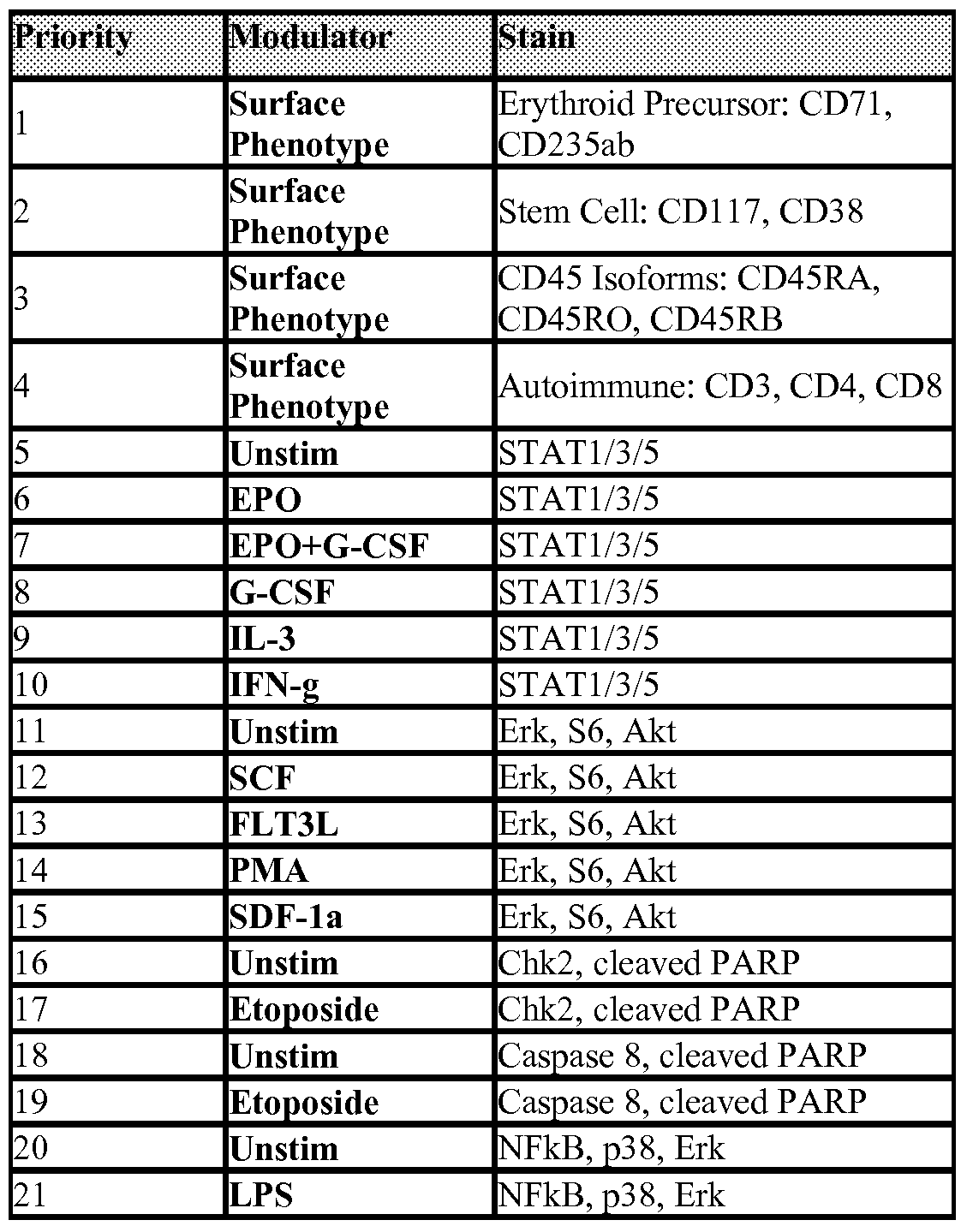

Claims
Priority Applications (1)
| Application Number | Priority Date | Filing Date | Title |
|---|---|---|---|
| GB1020176.2A GB2474146B (en) | 2008-04-29 | 2009-04-29 | Methods of determining the health status of an individual |
Applications Claiming Priority (2)
| Application Number | Priority Date | Filing Date | Title |
|---|---|---|---|
| US4888608P | 2008-04-29 | 2008-04-29 | |
| US61/048,886 | 2008-04-29 |
Publications (2)
| Publication Number | Publication Date |
|---|---|
| WO2009134944A2 true WO2009134944A2 (en) | 2009-11-05 |
| WO2009134944A3 WO2009134944A3 (en) | 2010-02-25 |
Family
ID=40856505
Family Applications (1)
| Application Number | Title | Priority Date | Filing Date |
|---|---|---|---|
| PCT/US2009/042187 Ceased WO2009134944A2 (en) | 2008-04-29 | 2009-04-29 | Methods of determining the health status of an individual |
Country Status (3)
| Country | Link |
|---|---|
| US (2) | US20090269773A1 (en) |
| GB (1) | GB2474146B (en) |
| WO (1) | WO2009134944A2 (en) |
Cited By (15)
| Publication number | Priority date | Publication date | Assignee | Title |
|---|---|---|---|---|
| EP2179037A4 (en) * | 2007-08-21 | 2010-12-22 | Nodality Inc | METHODS FOR DIAGNOSIS, PROGNOSIS AND METHODS OF TREATMENT |
| US7897356B2 (en) | 2008-11-12 | 2011-03-01 | Caris Life Sciences | Methods and systems of using exosomes for determining phenotypes |
| CN102353793A (en) * | 2011-07-07 | 2012-02-15 | 山东轻工业学院 | Dimensional flow liquid phase array detection method of fusion protein in leukemia cells |
| US8227202B2 (en) | 2008-07-10 | 2012-07-24 | Nodality, Inc. | Methods for diagnosis, prognosis and methods of treatment |
| US8273544B2 (en) | 2008-07-10 | 2012-09-25 | Nodality, Inc. | Methods for diagnosis, prognosis and methods of treatment |
| CN103018458A (en) * | 2012-12-06 | 2013-04-03 | 北京普赞生物技术有限公司 | Ultra-sensitive Aflatoxin B1 ELISA Kit |
| JP2013520208A (en) * | 2010-02-24 | 2013-06-06 | ザ・ボード・オブ・トラスティーズ・オブ・ザ・レランド・スタンフォード・ジュニア・ユニバーシティ | Diagnosis, prognosis and treatment of autoimmune diseases |
| EP2476053A4 (en) * | 2009-09-08 | 2014-03-12 | Nodality Inc | Analysis of cell networks |
| US8700335B2 (en) | 2006-05-18 | 2014-04-15 | Caris Mpi, Inc. | System and method for determining individualized medical intervention for a disease state |
| US20140134650A1 (en) * | 2009-09-08 | 2014-05-15 | Nodality, Inc. | Induced intercellular communication |
| US8768629B2 (en) | 2009-02-11 | 2014-07-01 | Caris Mpi, Inc. | Molecular profiling of tumors |
| US9128101B2 (en) | 2010-03-01 | 2015-09-08 | Caris Life Sciences Switzerland Holdings Gmbh | Biomarkers for theranostics |
| US9469876B2 (en) | 2010-04-06 | 2016-10-18 | Caris Life Sciences Switzerland Holdings Gmbh | Circulating biomarkers for metastatic prostate cancer |
| CN107158388A (en) * | 2017-07-11 | 2017-09-15 | 上海市第人民医院 | Application of the MIIP pS303 blocking agents in antineoplastic is prepared |
| US10119959B2 (en) | 2010-06-25 | 2018-11-06 | The Board Of Trustees Of The Leland Stanford Junior University | Method of assaying an individual for immune impairment |
Families Citing this family (36)
| Publication number | Priority date | Publication date | Assignee | Title |
|---|---|---|---|---|
| US7381535B2 (en) * | 2002-07-10 | 2008-06-03 | The Board Of Trustees Of The Leland Stanford Junior | Methods and compositions for detecting receptor-ligand interactions in single cells |
| WO2003067210A2 (en) | 2001-07-10 | 2003-08-14 | The Board Of Trustees Of The Leland Stanford Junior University | Methods and compositions for detecting the activation state of the multiple proteins in single cells |
| US7393656B2 (en) * | 2001-07-10 | 2008-07-01 | The Board Of Trustees Of The Leland Stanford Junior University | Methods and compositions for risk stratification |
| EP1842147A2 (en) * | 2005-01-24 | 2007-10-10 | The Board of Trustees of The Leland Stanford Junior University | Method for modeling cell signaling systems by means of bayesian networks |
| CA2673481A1 (en) * | 2006-12-19 | 2008-06-26 | University Of Virginia Patent Foundation | Combined effects of topiramate and ondansetron on alcohol consumption |
| WO2008095086A2 (en) * | 2007-01-31 | 2008-08-07 | University Of Virginia Patent Foundation | Topiramate plus naltrexone for the treatment of addictive disorders |
| US20090062237A1 (en) * | 2007-06-15 | 2009-03-05 | Mayo Foundation For Medical Education And Research | Evaluating immune competence |
| US20110065628A1 (en) * | 2007-08-27 | 2011-03-17 | University Of Virginia Patent Foundation | Medication Combinations for the Treatment of Alcoholism and Drug Addiction |
| CA2716498C (en) | 2008-02-28 | 2020-12-22 | University Of Virginia Patent Foundation | Serotonin transporter gene and treatment of alcoholism |
| US20100099109A1 (en) * | 2008-10-17 | 2010-04-22 | Nodality, Inc., A Delaware Corporation | Methods for Analyzing Drug Response |
| US9034257B2 (en) | 2008-10-27 | 2015-05-19 | Nodality, Inc. | High throughput flow cytometry system and method |
| US8309306B2 (en) * | 2008-11-12 | 2012-11-13 | Nodality, Inc. | Detection composition |
| US20100209929A1 (en) * | 2009-01-14 | 2010-08-19 | Nodality, Inc., A Delaware Corporation | Multiple mechanisms for modulation of jak/stat activity |
| US20100204973A1 (en) * | 2009-01-15 | 2010-08-12 | Nodality, Inc., A Delaware Corporation | Methods For Diagnosis, Prognosis And Treatment |
| US20100233733A1 (en) * | 2009-02-10 | 2010-09-16 | Nodality, Inc., A Delaware Corporation | Multiple mechanisms for modulation of the pi3 kinase pathway |
| US20100215644A1 (en) * | 2009-02-25 | 2010-08-26 | Nodality, Inc. A Delaware Corporation | Analysis of nodes in cellular pathways |
| US8242248B2 (en) * | 2009-03-23 | 2012-08-14 | Nodality, Inc. | Kits for multiparametric phospho analysis |
| WO2010135608A1 (en) * | 2009-05-20 | 2010-11-25 | Nodality, Inc. | Methods for diagnosis, prognosis and methods of treatment |
| GB2479058A (en) * | 2010-03-24 | 2011-09-28 | Nodality Inc | Modeling biological events |
| IL278149B2 (en) | 2010-07-02 | 2024-03-01 | Univ Virginia Patent Foundation | A molecular genetic approach to the treatment and diagnosis of alcohol and drug dependence |
| WO2012024546A2 (en) * | 2010-08-18 | 2012-02-23 | Nodality, Inc. | Incorporation of health measurments in analysis and interpretation of functional biological response data |
| WO2012090226A2 (en) | 2010-12-31 | 2012-07-05 | Tata Consultancy Services Limited | Computer implemented system and method for measuring individual wellness index |
| US9005909B2 (en) * | 2011-01-06 | 2015-04-14 | Rigel Pharmaceuticals, Inc. | Whole blood assay for measuring AMPK activation |
| WO2013036721A1 (en) | 2011-09-09 | 2013-03-14 | University Of Virginia Patent Foundation | Molecular genetic approach to treatment and diagnosis of alcohol and drug dependence |
| WO2014036183A2 (en) | 2012-08-28 | 2014-03-06 | Puget Sound Blood Center | Biochemical markers of red blood cell storage and toxicity |
| US10760113B2 (en) * | 2015-07-10 | 2020-09-01 | Regents Of The University Of Minnesota | Kinase activity detection methods |
| EP3346910A4 (en) * | 2015-09-09 | 2019-05-15 | Ubiome Inc. | METHOD AND SYSTEM FOR DIAGNOSES DERIVED FROM MICROBIOMA AND THERAPEUTIC AGENTS AGAINST ECZEMA |
| CN108348166B (en) * | 2015-09-09 | 2022-06-03 | 普梭梅根公司 | Microbiota-derived diagnostic and therapeutic methods and systems for infectious diseases and other health conditions associated with antibiotic use |
| WO2017190009A1 (en) | 2016-04-29 | 2017-11-02 | The Board Of Regents Of The University Of Texas System | Use of jumonji c demethylase inhibitors for the treatment and prevention of chemotherapy resistance and radioresistance in cancer |
| CN109165394B (en) * | 2018-06-25 | 2019-07-16 | 中国人民解放军陆军工程大学 | A lightweight implementation method of standardized BIM model |
| CN109544102A (en) * | 2018-10-26 | 2019-03-29 | 中交第航务工程局有限公司 | A kind of engineering ship construction management system and application method based on BIM model |
| CN111286507A (en) * | 2019-10-13 | 2020-06-16 | 华中农业大学 | Application of OsPP15 gene in improving rice yield |
| CN113241177B (en) * | 2021-05-19 | 2024-05-10 | 上海宝藤生物医药科技股份有限公司 | Method, device, equipment and storage medium for evaluating immunity level |
| CN114383958B (en) * | 2022-01-11 | 2024-05-24 | 沈阳工业大学 | Method and system for determining key-based near-field dynamics critical elongation of deep rock mass |
| EP4469788A4 (en) * | 2022-01-28 | 2025-12-24 | Univ Southern California | IDENTIFICATION OF NON-DISCOVERY PATIENTS USING A DISEASE-ASSOCIATED TEST AND ANALYSIS IN LIQUID BIOPSY |
| CN119868814B (en) * | 2024-12-26 | 2025-11-11 | 广州分享健康产业有限公司 | Intelligent control system and control method of cell activation instrument |
Family Cites Families (78)
| Publication number | Priority date | Publication date | Assignee | Title |
|---|---|---|---|---|
| ES2035317T5 (en) * | 1987-11-09 | 1998-03-16 | Becton Dickinson Co | METHOD FOR ANALYZING HEMATOPOYETIC CELLS IN A SAMPLE. |
| US5234816A (en) * | 1991-07-12 | 1993-08-10 | Becton, Dickinson And Company | Method for the classification and monitoring of leukemias |
| EP0640102B1 (en) * | 1992-04-10 | 2006-08-09 | Dana-Farber Cancer Institute, Inc. | Activation-state-specific phosphoprotein immunodetection |
| EP0610774B1 (en) * | 1993-02-09 | 2001-03-28 | Becton, Dickinson and Company | Automatic lineage assignment of acute leukemias by flow cytometry |
| US5968738A (en) * | 1995-12-06 | 1999-10-19 | The Board Of Trustees Of The Leland Stanford Junior University | Two-reporter FACS analysis of mammalian cells using green fluorescent proteins |
| US6232299B1 (en) * | 1996-05-01 | 2001-05-15 | Eli Lilly And Company | Use of protein kinase C inhibitors to enhance the clinical efficacy of oncolytic agents and radiation therapy |
| US6558916B2 (en) * | 1996-08-02 | 2003-05-06 | Axiom Biotechnologies, Inc. | Cell flow apparatus and method for real-time measurements of patient cellular responses |
| US6280967B1 (en) * | 1996-08-02 | 2001-08-28 | Axiom Biotechnologies, Inc. | Cell flow apparatus and method for real-time of cellular responses |
| US5804436A (en) * | 1996-08-02 | 1998-09-08 | Axiom Biotechnologies, Inc. | Apparatus and method for real-time measurement of cellular response |
| US6197531B1 (en) * | 1997-01-22 | 2001-03-06 | Center For Blood Research, Inc. | Method for determining the immunocompetence of a mammal and therapies related thereto |
| US6881548B2 (en) * | 1997-05-23 | 2005-04-19 | A&G Pharmaceutical, Inc. | Methods and kits for diagnosing tumorigenicity and determining resistance to the antineoplastic effects of antiestrogen therapy |
| US6821740B2 (en) * | 1998-02-25 | 2004-11-23 | Becton, Dickinson And Company | Flow cytometric methods for the concurrent detection of discrete functional conformations of PRB in single cells |
| US7236888B2 (en) * | 1998-03-06 | 2007-06-26 | The Regents Of The University Of California | Method to measure the activation state of signaling pathways in cells |
| US6200765B1 (en) * | 1998-05-04 | 2001-03-13 | Pacific Northwest Cancer Foundation | Non-invasive methods to detect prostate cancer |
| AU3897999A (en) * | 1998-05-14 | 1999-11-29 | Luminex Corporation | Multi-analyte diagnostic system and computer implemented process for same |
| US7001725B2 (en) * | 1999-04-30 | 2006-02-21 | Aclara Biosciences, Inc. | Kits employing generalized target-binding e-tag probes |
| EP1175238B1 (en) * | 1999-04-30 | 2006-01-25 | Children's Hospital Medical Center | Hemofiltration system |
| US6673554B1 (en) * | 1999-06-14 | 2004-01-06 | Trellie Bioinformatics, Inc. | Protein localization assays for toxicity and antidotes thereto |
| US6506551B1 (en) * | 1999-10-08 | 2003-01-14 | North Shore - Long Island Jewish Research Institute | CD38 as a prognostic indicator in B cell chronic lymphocytic leukemia |
| JP2004509835A (en) * | 2000-03-06 | 2004-04-02 | ユニヴァーシティ オブ ケンタッキー リサーチ ファンデーション | Methods of damaging hematologic cancer progenitor cells and related compounds |
| WO2001067103A1 (en) * | 2000-03-06 | 2001-09-13 | Bioseek, Inc. | Function homology screening |
| WO2001077684A2 (en) * | 2000-04-10 | 2001-10-18 | The Scripps Research Institute | Proteomic analysis using activity-based probe libraries |
| US7537938B2 (en) * | 2000-04-28 | 2009-05-26 | Monogram Biosciences, Inc. | Biomarker detection in circulating cells |
| WO2002024947A2 (en) * | 2000-09-20 | 2002-03-28 | Kinetek Pharmaceuticals, Inc. | Cancer associated protein kinases and their uses |
| AU2002228000A1 (en) * | 2000-12-07 | 2002-06-18 | Europroteome Ag | Expert system for classification and prediction of genetic diseases |
| EP1366146A4 (en) * | 2001-02-28 | 2005-01-19 | Merck & Co Inc | INSULATED NUCLEIC ACID MOLECULES ENCODING HUMAN PROTEINS ENCODED BY SIGNAL-TRANSDUCTING MAPKAP-2 KINASE, CELLS THUS PROCESSED AND USES THEREFOR |
| WO2002087431A1 (en) * | 2001-05-01 | 2002-11-07 | Structural Bioinformatics, Inc. | Diagnosing inapparent diseases from common clinical tests using bayesian analysis |
| WO2003067210A2 (en) * | 2001-07-10 | 2003-08-14 | The Board Of Trustees Of The Leland Stanford Junior University | Methods and compositions for detecting the activation state of the multiple proteins in single cells |
| US7695926B2 (en) * | 2001-07-10 | 2010-04-13 | The Board Of Trustees Of The Leland Stanford Junior University | Methods and compositions for detecting receptor-ligand interactions in single cells |
| US7393656B2 (en) * | 2001-07-10 | 2008-07-01 | The Board Of Trustees Of The Leland Stanford Junior University | Methods and compositions for risk stratification |
| US7381535B2 (en) * | 2002-07-10 | 2008-06-03 | The Board Of Trustees Of The Leland Stanford Junior | Methods and compositions for detecting receptor-ligand interactions in single cells |
| US7105642B2 (en) * | 2001-08-03 | 2006-09-12 | Cell Signalling Technology, Inc. | Monoclonal antibodies specific for phosphorylated estrogen receptor alpha (Ser118) and uses thereof |
| ATE530635T1 (en) * | 2001-08-21 | 2011-11-15 | Ventana Med Syst Inc | METHOD AND QUANTIFICATION ASSAY FOR DETERMINING C-KIT/SCF/PAKT STATUS |
| US20030148321A1 (en) * | 2001-08-24 | 2003-08-07 | Iris Pecker | Methods and kits for diagnosing and monitoring hematopoietic cancers |
| AU2002336504A1 (en) * | 2001-09-12 | 2003-03-24 | The State Of Oregon, Acting By And Through The State Board Of Higher Education On Behalf Of Oregon S | Method and system for classifying a scenario |
| US20070166704A1 (en) * | 2002-01-18 | 2007-07-19 | Fei Huang | Identification of polynucleotides and polypeptide for predicting activity of compounds that interact with protein tyrosine kinases and/or protein tyrosine kinase pathways |
| US7183385B2 (en) * | 2002-02-20 | 2007-02-27 | Cell Signaling Technology, Inc. | Phospho-specific antibodies to Flt3 and uses thereof |
| US20050216961A1 (en) * | 2002-03-28 | 2005-09-29 | Delaney Allen D | Cancer associated protein kinases and their uses |
| WO2003087760A2 (en) * | 2002-04-05 | 2003-10-23 | Cell Signaling Technology, Inc. | Methods for detecting bcr-abl signaling activity in tissues using phospho- specific antibodies |
| US20030190689A1 (en) * | 2002-04-05 | 2003-10-09 | Cell Signaling Technology,Inc. | Molecular profiling of disease and therapeutic response using phospho-specific antibodies |
| US7329502B2 (en) * | 2002-04-25 | 2008-02-12 | The United States Of America As Represented By The Department Of Health And Human Services | ZAP-70 expression as a marker for chronic lymphocytic leukemia/small lymphocytic lymphoma (CLL/SLL) |
| AU2003299466A1 (en) * | 2002-05-03 | 2004-06-07 | Molecular Probes, Inc. | Compositions and methods for detection and isolation of phosphorylated molecules |
| US7460960B2 (en) * | 2002-05-10 | 2008-12-02 | Epitome Biosystems, Inc. | Proteome epitope tags and methods of use thereof in protein modification analysis |
| JP4212827B2 (en) * | 2002-05-16 | 2009-01-21 | シスメックス株式会社 | Bone marrow nucleated cell automatic analysis method |
| EP1576173A4 (en) * | 2002-07-12 | 2007-02-28 | Rigel Pharmaceuticals Inc | Modulators of cellular proliferation |
| US7537891B2 (en) * | 2002-08-27 | 2009-05-26 | Bristol-Myers Squibb Company | Identification of polynucleotides for predicting activity of compounds that interact with and/or modulate protein tyrosine kinases and/or protein tyrosine kinase pathways in breast cells |
| US20040137539A1 (en) * | 2003-01-10 | 2004-07-15 | Bradford Sherry A. | Cancer comprehensive method for identifying cancer protein patterns and determination of cancer treatment strategies |
| US20050009112A1 (en) * | 2003-03-07 | 2005-01-13 | Fred Hutchinson Cancer Research Center, Office Of Technology Transfer | Methods for identifying Rheb effectors as lead compounds for drug development for diabetes and diseases associated with abnormal cell growth |
| EP1664716A4 (en) * | 2003-08-15 | 2008-08-13 | Smithkline Beecham Corp | BIOMARKERS AGAINST CANCER |
| US7402399B2 (en) * | 2003-10-14 | 2008-07-22 | Monogram Biosciences, Inc. | Receptor tyrosine kinase signaling pathway analysis for diagnosis and therapy |
| US7326577B2 (en) * | 2003-10-20 | 2008-02-05 | Esoterix, Inc. | Cell fixation and use in phospho-proteome screening |
| WO2006023004A1 (en) * | 2004-08-12 | 2006-03-02 | Transform Pharmaceuticals, Inc. | Methods for identifiying conditions affecting a cell state |
| US20060040338A1 (en) * | 2004-08-18 | 2006-02-23 | Odyssey Thera, Inc. | Pharmacological profiling of drugs with cell-based assays |
| US7803523B2 (en) * | 2004-08-27 | 2010-09-28 | University Health Network | Whole blood preparation for cytometric analysis of cell signaling pathways |
| EP1842147A2 (en) * | 2005-01-24 | 2007-10-10 | The Board of Trustees of The Leland Stanford Junior University | Method for modeling cell signaling systems by means of bayesian networks |
| US20070020670A1 (en) * | 2005-07-07 | 2007-01-25 | Hematologics, Inc. | Methods for detecting and confirming minimal disease |
| US20070105165A1 (en) * | 2005-11-04 | 2007-05-10 | Charles Goolsby | Composite profiles of cell antigens and target signal transduction proteins for analysis and clinical management of hematologic cancers |
| US20070172847A1 (en) * | 2005-11-15 | 2007-07-26 | The Regents Of The University Of California | Molecular signaling pathways triggered by rituximab: prognostic, diagnostic, and therapeutic uses |
| CA2547901A1 (en) * | 2006-06-14 | 2007-12-14 | University College Cardiff Consultants Limited | Chronic lymphocytic leukaemia |
| US8105796B2 (en) * | 2007-02-15 | 2012-01-31 | Universita' Degli Studi Di Torino | Regulation of expression of PI3Kβ protein in tumors |
| AU2008289442A1 (en) * | 2007-08-21 | 2009-02-26 | Nodality, Inc. | Methods for diagnosis, prognosis and methods of treatment |
| US20100030719A1 (en) * | 2008-07-10 | 2010-02-04 | Covey Todd M | Methods and apparatus related to bioinformatics data analysis |
| US8399206B2 (en) * | 2008-07-10 | 2013-03-19 | Nodality, Inc. | Methods for diagnosis, prognosis and methods of treatment |
| US8227202B2 (en) * | 2008-07-10 | 2012-07-24 | Nodality, Inc. | Methods for diagnosis, prognosis and methods of treatment |
| GB2474613A (en) * | 2008-07-10 | 2011-04-20 | Nodality Inc | Methods and apparatus related to management of experiments |
| US20100014741A1 (en) * | 2008-07-10 | 2010-01-21 | Banville Steven C | Methods and apparatus related to gate boundaries within a data space |
| ES2429315T3 (en) * | 2008-09-04 | 2013-11-14 | Beckman Coulter, Inc. | Pan-kinase activation and evaluation of signaling pathways |
| US20100099109A1 (en) * | 2008-10-17 | 2010-04-22 | Nodality, Inc., A Delaware Corporation | Methods for Analyzing Drug Response |
| US9034257B2 (en) * | 2008-10-27 | 2015-05-19 | Nodality, Inc. | High throughput flow cytometry system and method |
| US8309306B2 (en) * | 2008-11-12 | 2012-11-13 | Nodality, Inc. | Detection composition |
| US20100209929A1 (en) * | 2009-01-14 | 2010-08-19 | Nodality, Inc., A Delaware Corporation | Multiple mechanisms for modulation of jak/stat activity |
| US20100204973A1 (en) * | 2009-01-15 | 2010-08-12 | Nodality, Inc., A Delaware Corporation | Methods For Diagnosis, Prognosis And Treatment |
| US20100233733A1 (en) * | 2009-02-10 | 2010-09-16 | Nodality, Inc., A Delaware Corporation | Multiple mechanisms for modulation of the pi3 kinase pathway |
| US20100215644A1 (en) * | 2009-02-25 | 2010-08-26 | Nodality, Inc. A Delaware Corporation | Analysis of nodes in cellular pathways |
| US8242248B2 (en) * | 2009-03-23 | 2012-08-14 | Nodality, Inc. | Kits for multiparametric phospho analysis |
| GB0908589D0 (en) * | 2009-05-19 | 2009-06-24 | Univ Belfast | Assay method |
| WO2011031803A1 (en) * | 2009-09-08 | 2011-03-17 | Nodality, Inc. | Analysis of cell networks |
| EP2580352A4 (en) * | 2010-06-09 | 2014-03-19 | Nodality Inc | Pathways characterization of cells |
-
2009
- 2009-04-29 US US12/432,720 patent/US20090269773A1/en not_active Abandoned
- 2009-04-29 WO PCT/US2009/042187 patent/WO2009134944A2/en not_active Ceased
- 2009-04-29 GB GB1020176.2A patent/GB2474146B/en not_active Expired - Fee Related
-
2013
- 2013-10-03 US US14/045,548 patent/US20140134648A1/en not_active Abandoned
Cited By (23)
| Publication number | Priority date | Publication date | Assignee | Title |
|---|---|---|---|---|
| US8700335B2 (en) | 2006-05-18 | 2014-04-15 | Caris Mpi, Inc. | System and method for determining individualized medical intervention for a disease state |
| EP2179037A4 (en) * | 2007-08-21 | 2010-12-22 | Nodality Inc | METHODS FOR DIAGNOSIS, PROGNOSIS AND METHODS OF TREATMENT |
| US9182385B2 (en) | 2007-08-21 | 2015-11-10 | Nodality, Inc. | Methods for diagnosis, prognosis and methods of treatment |
| EP2860247A1 (en) * | 2007-08-21 | 2015-04-15 | Nodality, Inc. | Methods for diagnosis, prognosis and methods of treatment |
| US8399206B2 (en) | 2008-07-10 | 2013-03-19 | Nodality, Inc. | Methods for diagnosis, prognosis and methods of treatment |
| US8227202B2 (en) | 2008-07-10 | 2012-07-24 | Nodality, Inc. | Methods for diagnosis, prognosis and methods of treatment |
| US9500655B2 (en) | 2008-07-10 | 2016-11-22 | Nodality, Inc. | Methods for diagnosis, prognosis and methods of treatment |
| US8778620B2 (en) | 2008-07-10 | 2014-07-15 | Nodality, Inc. | Methods for diagnosis, prognosis and methods of treatment |
| US8273544B2 (en) | 2008-07-10 | 2012-09-25 | Nodality, Inc. | Methods for diagnosis, prognosis and methods of treatment |
| US7897356B2 (en) | 2008-11-12 | 2011-03-01 | Caris Life Sciences | Methods and systems of using exosomes for determining phenotypes |
| US8768629B2 (en) | 2009-02-11 | 2014-07-01 | Caris Mpi, Inc. | Molecular profiling of tumors |
| EP2476053A4 (en) * | 2009-09-08 | 2014-03-12 | Nodality Inc | Analysis of cell networks |
| US20140134650A1 (en) * | 2009-09-08 | 2014-05-15 | Nodality, Inc. | Induced intercellular communication |
| US9459246B2 (en) * | 2009-09-08 | 2016-10-04 | Nodality, Inc. | Induced intercellular communication |
| EP2539470A4 (en) * | 2010-02-24 | 2013-10-23 | Univ Leland Stanford Junior | METHODS FOR THE DIAGNOSIS, PROGNOSIS AND TREATMENT OF AUTOIMMUNE DISEASES |
| JP2013520208A (en) * | 2010-02-24 | 2013-06-06 | ザ・ボード・オブ・トラスティーズ・オブ・ザ・レランド・スタンフォード・ジュニア・ユニバーシティ | Diagnosis, prognosis and treatment of autoimmune diseases |
| US9128101B2 (en) | 2010-03-01 | 2015-09-08 | Caris Life Sciences Switzerland Holdings Gmbh | Biomarkers for theranostics |
| US9469876B2 (en) | 2010-04-06 | 2016-10-18 | Caris Life Sciences Switzerland Holdings Gmbh | Circulating biomarkers for metastatic prostate cancer |
| US10119959B2 (en) | 2010-06-25 | 2018-11-06 | The Board Of Trustees Of The Leland Stanford Junior University | Method of assaying an individual for immune impairment |
| CN102353793B (en) * | 2011-07-07 | 2014-01-29 | 山东轻工业学院 | Binary flow cytometry liquid array detection method for fusion protein in leukemia cells |
| CN102353793A (en) * | 2011-07-07 | 2012-02-15 | 山东轻工业学院 | Dimensional flow liquid phase array detection method of fusion protein in leukemia cells |
| CN103018458A (en) * | 2012-12-06 | 2013-04-03 | 北京普赞生物技术有限公司 | Ultra-sensitive Aflatoxin B1 ELISA Kit |
| CN107158388A (en) * | 2017-07-11 | 2017-09-15 | 上海市第人民医院 | Application of the MIIP pS303 blocking agents in antineoplastic is prepared |
Also Published As
| Publication number | Publication date |
|---|---|
| US20140134648A1 (en) | 2014-05-15 |
| GB2474146A (en) | 2011-04-06 |
| US20090269773A1 (en) | 2009-10-29 |
| GB201020176D0 (en) | 2011-01-12 |
| WO2009134944A3 (en) | 2010-02-25 |
| GB2474146B (en) | 2013-04-10 |
Similar Documents
| Publication | Publication Date | Title |
|---|---|---|
| US20140134648A1 (en) | Methods of determining the health status of an individual | |
| US9182385B2 (en) | Methods for diagnosis, prognosis and methods of treatment | |
| US20170285027A1 (en) | Methods for diagnosis, prognosis and methods of treatment | |
| US20090291458A1 (en) | Method for Determining the Status of an Individual | |
| US20170285008A1 (en) | Analysis of cell networks | |
| US20100233733A1 (en) | Multiple mechanisms for modulation of the pi3 kinase pathway | |
| US20140147857A1 (en) | Methods for diagnosis prognosis and methods of treatment | |
| US9459246B2 (en) | Induced intercellular communication | |
| US20120157340A1 (en) | Pathways characterization of cells | |
| US20170292946A1 (en) | Methods for diagnosis, prognosis and methods of treatment | |
| US20170184594A1 (en) | Pathway characterization of cells | |
| US20170184587A1 (en) | Compositions and methods for autoimmune disease | |
| WO2014074646A2 (en) | Induced intercellular communication | |
| ITTO20110233A1 (en) | IN VITRO METHOD OF DIAGNOSIS, PROGNOSIS AND TREATMENT OF HEMATOPOIETIC DISORDERS |
Legal Events
| Date | Code | Title | Description |
|---|---|---|---|
| 121 | Ep: the epo has been informed by wipo that ep was designated in this application |
Ref document number: 09739746 Country of ref document: EP Kind code of ref document: A2 |
|
| NENP | Non-entry into the national phase |
Ref country code: DE |
|
| ENP | Entry into the national phase |
Ref document number: 1020176 Country of ref document: GB Kind code of ref document: A Free format text: PCT FILING DATE = 20090429 |
|
| WWE | Wipo information: entry into national phase |
Ref document number: 1020176.2 Country of ref document: GB |
|
| 122 | Ep: pct application non-entry in european phase |
Ref document number: 09739746 Country of ref document: EP Kind code of ref document: A2 |