[go: up one dir, main page]

WO2009086343A2 - Microorganism-capturing compositions and methods - Google Patents

Microorganism-capturing compositions and methods Download PDF

Info

Publication number
WO2009086343A2
WO2009086343A2 PCT/US2008/088099 US2008088099W WO2009086343A2 WO 2009086343 A2 WO2009086343 A2 WO 2009086343A2 US 2008088099 W US2008088099 W US 2008088099W WO 2009086343 A2 WO2009086343 A2 WO 2009086343A2
Authority
WO
WIPO (PCT)
Prior art keywords
carbohydrate
solid support
protein
bacterial cells
group
Prior art date
Legal status (The legal status is an assumption and is not a legal conclusion. Google has not performed a legal analysis and makes no representation as to the accuracy of the status listed.)
Ceased
Application number
PCT/US2008/088099
Other languages
French (fr)
Other versions
WO2009086343A3 (en
Inventor
Sirdhar V. Dasaratha
G. Marco Bommarito
Tushar A. Kshirsagar
Current Assignee (The listed assignees may be inaccurate. Google has not performed a legal analysis and makes no representation or warranty as to the accuracy of the list.)
3M Innovative Properties Co
Original Assignee
3M Innovative Properties Co
Priority date (The priority date is an assumption and is not a legal conclusion. Google has not performed a legal analysis and makes no representation as to the accuracy of the date listed.)
Filing date
Publication date
Application filed by 3M Innovative Properties Co filed Critical 3M Innovative Properties Co
Priority to US12/811,142 priority Critical patent/US20110183398A1/en
Priority to CN2008801269554A priority patent/CN101952727A/en
Priority to JP2010541491A priority patent/JP2011509083A/en
Priority to EP08867539A priority patent/EP2240774A2/en
Publication of WO2009086343A2 publication Critical patent/WO2009086343A2/en
Publication of WO2009086343A3 publication Critical patent/WO2009086343A3/en
Anticipated expiration legal-status Critical
Ceased legal-status Critical Current

Links

Classifications

    • GPHYSICS
    • G01MEASURING; TESTING
    • G01NINVESTIGATING OR ANALYSING MATERIALS BY DETERMINING THEIR CHEMICAL OR PHYSICAL PROPERTIES
    • G01N33/00Investigating or analysing materials by specific methods not covered by groups G01N1/00 - G01N31/00
    • G01N33/48Biological material, e.g. blood, urine; Haemocytometers
    • G01N33/50Chemical analysis of biological material, e.g. blood, urine; Testing involving biospecific ligand binding methods; Immunological testing
    • G01N33/53Immunoassay; Biospecific binding assay; Materials therefor
    • G01N33/543Immunoassay; Biospecific binding assay; Materials therefor with an insoluble carrier for immobilising immunochemicals
    • G01N33/54353Immunoassay; Biospecific binding assay; Materials therefor with an insoluble carrier for immobilising immunochemicals with ligand attached to the carrier via a chemical coupling agent
    • GPHYSICS
    • G01MEASURING; TESTING
    • G01NINVESTIGATING OR ANALYSING MATERIALS BY DETERMINING THEIR CHEMICAL OR PHYSICAL PROPERTIES
    • G01N33/00Investigating or analysing materials by specific methods not covered by groups G01N1/00 - G01N31/00
    • G01N33/48Biological material, e.g. blood, urine; Haemocytometers
    • G01N33/50Chemical analysis of biological material, e.g. blood, urine; Testing involving biospecific ligand binding methods; Immunological testing
    • G01N33/53Immunoassay; Biospecific binding assay; Materials therefor
    • G01N33/543Immunoassay; Biospecific binding assay; Materials therefor with an insoluble carrier for immobilising immunochemicals
    • G01N33/54313Immunoassay; Biospecific binding assay; Materials therefor with an insoluble carrier for immobilising immunochemicals the carrier being characterised by its particulate form
    • G01N33/54326Magnetic particles
    • G01N33/54333Modification of conditions of immunological binding reaction, e.g. use of more than one type of particle, use of chemical agents to improve binding, choice of incubation time or application of magnetic field during binding reaction
    • GPHYSICS
    • G01MEASURING; TESTING
    • G01NINVESTIGATING OR ANALYSING MATERIALS BY DETERMINING THEIR CHEMICAL OR PHYSICAL PROPERTIES
    • G01N33/00Investigating or analysing materials by specific methods not covered by groups G01N1/00 - G01N31/00
    • G01N33/48Biological material, e.g. blood, urine; Haemocytometers
    • G01N33/50Chemical analysis of biological material, e.g. blood, urine; Testing involving biospecific ligand binding methods; Immunological testing
    • G01N33/53Immunoassay; Biospecific binding assay; Materials therefor
    • G01N33/543Immunoassay; Biospecific binding assay; Materials therefor with an insoluble carrier for immobilising immunochemicals
    • G01N33/544Immunoassay; Biospecific binding assay; Materials therefor with an insoluble carrier for immobilising immunochemicals the carrier being organic
    • G01N33/548Carbohydrates, e.g. dextran
    • GPHYSICS
    • G01MEASURING; TESTING
    • G01NINVESTIGATING OR ANALYSING MATERIALS BY DETERMINING THEIR CHEMICAL OR PHYSICAL PROPERTIES
    • G01N33/00Investigating or analysing materials by specific methods not covered by groups G01N1/00 - G01N31/00
    • G01N33/48Biological material, e.g. blood, urine; Haemocytometers
    • G01N33/50Chemical analysis of biological material, e.g. blood, urine; Testing involving biospecific ligand binding methods; Immunological testing
    • G01N33/53Immunoassay; Biospecific binding assay; Materials therefor
    • G01N33/569Immunoassay; Biospecific binding assay; Materials therefor for microorganisms, e.g. protozoa, bacteria, viruses
    • G01N33/56911Bacteria

Definitions

  • bacterial samples are cultured to assess viability and increase the number of bacteria in the sample to assure an adequate level for detection.
  • the culturing step requires substantial time, which delays obtaining the detection results.
  • detection can be carried out using a shorter or even no culturing step.
  • Methods have been developed to isolate, and thereby concentrate, specific bacterial strains by using antibodies specific to the strain.
  • concentration methods that are non-strain specific, which would allow a more general sampling of the microorganisms present, have been proposed.
  • a specific strain or specific strains within a mixture of strains can be identified and/or quantified using known detection methods, including, for example, nucleic acid amplification methods.
  • Non-specific isolation of microorganisms using carbohydrate and lectin protein interactions has been proposed.
  • Lectins present on the surface of bacteria were suggested as capture targets for certain carbohydrates attached to polyacrylamide.
  • Substances that serve as nutrients for microorganisms have also been proposed for use as ligands for non- specific capture of microorganisms.
  • Such nutrient ligands included carbohydrates, vitamins, iron-chelating compounds, and sidorophores.
  • Supported chitsan has also been proposed for concentrating microorganisms in a non-strain-specific manner, allowing the microorganisms to be more easily and rapidly assayed.
  • compositions with a carbohydrate on a solid support, but without the biotin-binding protein were found to be less effective for non-specifically binding bacterial cells.
  • compositions with a biotin-binding protein on the solid support, but without the carbohydrate were also found to be less effective for non-specifically binding bacterial cells.
  • the biotin-binding protein attached to the solid support via the carbohydrate is even more effective for non-specifically binding bacterial cells in the presence of an amphiphilic glycoside of a steroid or triterpene.
  • a composition comprise a solid support which has a surface comprising a combination of a carbohydrate and a biotin-binding protein, wherein the protein is covalently bonded to the carbohydrate, and wherein the protein is linked to the solid support via the carbohydrate; and at least one of 1) a plurality of bacterial cells non-specifically bound to the combination of the carbohydrate and the protein or 2) an amphiphilic glycoside of a steroid or triterpene.
  • composition comprising: a solid support which has a surface comprising a combination of a carbohydrate and a biotin-binding protein, wherein the protein is covalently bonded to the carbohydrate, and wherein the protein is linked to the solid support via the carbohydrate; and a plurality of bacterial cells non-specifically bound to the combination of the carbohydrate and the protein.
  • composition comprising: a solid support which has a surface comprising a combination of a carbohydrate and a biotin-binding protein, wherein the protein is covalently bonded to the carbohydrate, and wherein the protein is linked to the solid support via the carbohydrate; and an amphiphilic glycoside of a steroid or triterpene.
  • a method for isolating bacterial cells comprising: providing a solid support which has a surface comprising a combination of a carbohydrate and a biotin-binding protein, wherein the protein is covalently bonded to the carbohydrate, and wherein the protein is linked to the solid support via the carbohydrate; providing a sample suspected of having a plurality of bacterial cells; contacting the solid support which has the surface comprising the combination of the carbohydrate and the biotin-binding protein with the sample; wherein at least a portion of the plurality of bacterial cells from the sample become non-specifically bound to the surface of the solid support; and separating the solid support from the remainder of the sample after the at least a portion of the plurality of bacterial cells from the sample become non-specifically bound to the surface of the solid support.
  • a device for detecting bacterial cells comprising: a composition comprising: a solid support which has a surface comprising a combination of a carbohydrate and a biotin-binding protein, wherein the protein is covalently bonded to the carbohydrate, and wherein the protein is linked to the solid support via the carbohydrate; and a plurality of bacterial cells non-specifically bound to the combination of the carbohydrate and the protein; and a means for detecting the bacterial cells.
  • a device for detecting bacterial cells comprising: a composition comprising: a solid support which has a surface comprising a combination of a carbohydrate and a biotin-binding protein, wherein the protein is covalently bonded to the carbohydrate, and wherein the protein is linked to the solid support via the carbohydrate; and an amphiphilic glycoside of a steroid or triterpene; and a means for detecting the bacterial cells.
  • kits comprising: a solid support which has a surface comprising a combination of a carbohydrate and a biotin-binding protein, wherein the protein is covalently bonded to the carbohydrate, and wherein the protein is linked to the solid support via the carbohydrate; and an amphiphilic glycoside of a steroid or triterpene.
  • amphiphilic glycoside of a steroid or triterpene refers to a steroid or triterpene joined to a sugar by a glycosidic linkage, the resulting material having amphiphilic properties, that is, surfactant properties, the material being both hydrophilic and lipophilic.
  • biotin-binding protein refers to a protein which has a high affinity and selectivity for binding biotin. As used herein, the “biotin-binding protein” does not include bound biotin.
  • magnetic particles means particles, particle conglomerates, or beads comprised of ferromagnetic, paramagnetic, or superparamagnetic particles, including dispersions of said particles in a polymer bead.
  • a means particles, particle conglomerates, or beads comprised of ferromagnetic, paramagnetic, or superparamagnetic particles, including dispersions of said particles in a polymer bead.
  • Non-specifically binding bacterial cells means that the binding is not specific to any type of bacterial cells.
  • all bacteria in a sample can be isolated from other components in the sample rather than targeting, for example, one strain of bacteria. Both gram positive and gram negative bacteria can be bound. The resulting isolated bacteria can then be subjected to known detection methods, such as bacterial load detection.
  • composition comprising: a solid support which has a surface comprising a combination of a carbohydrate and a biotin-binding protein, wherein the protein is covalently bonded to the carbohydrate, and wherein the protein is linked to the solid support via the carbohydrate; and a plurality of bacterial cells non-specifically bound to the combination of the carbohydrate and the protein.
  • composition comprising: a solid support which has a surface comprising a combination of a carbohydrate and a biotin- binding protein, wherein the protein is covalently bonded to the carbohydrate, and wherein the protein is linked to the solid support via the carbohydrate; and an amphiphilic glycoside of a steroid or triterpene.
  • this composition further comprises a plurality of bacterial cells non-specifically bound to the combination of the carbohydrate and the biotin-binding protein.
  • compositions can be prepared by combining the solid support having the combination of carbohydrate and biotin-binding protein described above with at least one of a plurality of bacterial cells or an amphiphilic glycoside of a steroid or triterpene.
  • the solid support having the combination of carbohydrate and biotin-bind protein can be provided as described below.
  • Combining the solid support with the bacterial cells and/or amphiphilic glycoside can be conveniently carried out by suspending or immersing the solid support in a buffer and adding the bacterial cells, also suspended in a buffer, and/or the amphiphilic glycoside dissolved in a buffer.
  • the amphiphilic glycoside buffer solution preferably contains about 0.2 to about 10 weight/volume percent (% w/v) amphiphilic glycoside, more preferably about 0.5 to about 5 % w/v.
  • a method for isolating bacterial cells comprising: providing a solid support which has a surface comprising a combination of a carbohydrate and a biotin-binding protein, wherein the protein is covalently bonded to the carbohydrate, and wherein the protein is linked to the solid support via the carbohydrate; providing a sample suspected of having a plurality of bacterial cells; contacting the solid support which has the surface comprising the combination of the carbohydrate and the biotin-binding protein with the sample; wherein at least a portion of the plurality of bacterial cells from the sample become non-specifically bound to the surface of the solid support; and separating the solid support from the remainder of the sample after the at least a portion of the plurality of bacterial cells from the sample become non-specifically bound to the surface of the solid support.
  • the sample suspected of having a plurality of bacterial cells can be provided from a wide range of sources using known methods.
  • sources include physiological fluid, e.g., blood, saliva, ocular lens fluid, synovial fluid, cerebral spinal fluid, pus, sweat, exudate, urine, mucus, mucosal tissue (e.g., buccal, gingival, nasal, ocular, tracheal, bronchial, gastrointestinal, rectal, urethral, ureteral, vaginal, cervical, and uterine mucosal membranes), lactation milk, or the like.
  • physiological fluid e.g., blood, saliva, ocular lens fluid, synovial fluid, cerebral spinal fluid, pus, sweat, exudate, urine, mucus, mucosal tissue (e.g., buccal, gingival, nasal, ocular, tracheal, bronchial, gastrointestinal, rectal, urethral, ureteral, vaginal, cervical, and uterine mucos
  • sample sources include those obtained from a body site, e.g., wound, skin, nares, nasopharyngeal cavity, nasal cavities, anterior nasal vestibule, scalp, nails, outer ear, middle ear, mouth, rectum, or other site.
  • Additional examples of sample sources include process streams, water, food, soil, vegetation, air (e.g., contaminated), surfaces, such as food preparation surfaces, and the like.
  • Known sampling techniques can be used to gather the sample from the source. For example, a volume of a liquid sample can be drawn from the source, or a swab or wipe can be used to collect a sample from a physiological or environmental surface.
  • swabs or other sample collection devices are commercially available.
  • the sample can be provided for use in the above method directly as is from the collection device, for example, in the case of aqueous liquids.
  • the sample can be provided by eluting, releasing, or washing the sample from the collection device using, for example, water, physiological saline, pH buffered solutions, or any other solutions or combinations of solutions that can remove the sample from the collection device and bring the sample into suspension.
  • An example of an eluting buffer is phosphate buffered saline (PBS), which can be used in combination with a surfactant, such as polyoxyethylene sorbitan monolaurate or a poly(oxyethylene-co-oxypropylene) block copolymer.
  • PBS phosphate buffered saline
  • surfactant such as polyoxyethylene sorbitan monolaurate or a poly(oxyethylene-co-oxypropylene) block copolymer.
  • Other extraction solutions can function to maintain specimen stability during transport from a sample collection site to a sample analysis site. Examples of
  • the sample may be subjected to a treatment prior to contact with the solid support, such as dilution of viscous fluids, concentration, filtration, distillation, dialysis, dilution, inactivation of natural components, addition of reagents, chemical treatment, or the like.
  • a treatment prior to contact with the solid support
  • Contacting the solid support which has the surface comprising the combination of the carbohydrate and the biotin-binding protein with the sample suspected of having a plurality of bacterial cells can be conveniently carried out by suspending or immersing the solid support in a buffer and adding the sample, which may also be suspended in a buffer.
  • Suitable buffers include PBS and PBS with a nonionic surfactant.
  • the mixture can be agitated for a period of time sufficient to allow the bacterial cells to become bound to the combination of carbohydrate and biotin-binding protein on the solid support.
  • the binding can be conveniently carried out at room temperature.
  • Separating the solid support from the remainder of the sample can be carried out using well known methods.
  • the remainder of the sample can be drained, decanted, drawn, centrifuged, pipetted, and/or filtered off of the solid support.
  • the solid support is magnetic particles the solid support can be consolidated in one area of the container with a magnet for convenient removal of the remainder of the sample, for example, by decanting, pipetting, or forcing the supernate out of the container using a pressure differential or a g-force.
  • the solid support may be washed, for example, with a buffer, to remove any remaining unbound sample material.
  • the method for isolating bacterial cells further comprises detecting the at least a portion of the plurality of bacterial cells.
  • the detecting is carried out by a detection method selected from the group consisting of adenosine triphosphate (ATP) detection by bio luminescence, polydiacetylene (PDA) colorimetric detection, nucleic acid detection, immunological detection, growth based detection, surface acoustic wave detection, or the like.
  • a detection method selected from the group consisting of adenosine triphosphate (ATP) detection by bio luminescence, polydiacetylene (PDA) colorimetric detection, nucleic acid detection, immunological detection, growth based detection, surface acoustic wave detection, or the like.
  • ATP detection can be used as a nonspecific indicator of bacterial load. After separating the solid support with non-specifically bound bacterial cells from the remainder of the sample (which may contain interfering components such as extra-cellular ATP), the cells are lysed and contacted with luciferin and luciferase. The resulting bioluminescence, which is of an intensity proportional to the number of captured bacterial cells, is then measured, for example, using a luminometer.
  • PDA colorimetric detection can be used to detect specific bacteria or a spectrum of bacteria by contacting a colorimetric sensor with the bacteria.
  • the colorimetric sensor comprises a receptor and a polymerized composition which includes a diacetylene compound or a polydiacetylene.
  • Methods for detecting nucleic acids often include amplifying or hybridizing the nucleic acids.
  • the captured bacterial cells are lysed to make the cellular nucleic acids available for detection. Lysing can be carried out enzymatically, chemically, and/or mechanically. Enzymes used for lysis include, for example, lysostaphin, lysozyme, mutanolysin, or others.
  • Chemical lysis can be carried out using a surfactant, alkali, heat, or other means. When alkali is used for lysis, a neutralization reagent may be used to neutralize the solution or mixture after lysis.
  • the lysis reagent can include a surfactant or detergent such as sodium dodecylsulfate (SDS), lithium laurylsulfate (LLS), TRITON series, TWEEN series, BRIJ series, NP series, CHAPS, iV-methyl-iV-(l- oxododecyl)glycine, sodium salt, or the like, buffered as needed; a chaotrope such as guanidium hydrochloride, guanidium thiacyanate, sodium iodide, or the like; a lysis enzyme such as lysozyme, lysostaphin, mutanolysin, proteinases, pronases, cellulases, or any of the other commercially available lysis enzymes; an alkaline lysis reagent; solid particles such as beads,
  • amplification methods include polymerase chain reaction (PCR); target polynucleotide amplification methods such as self-sustained sequence replication (3SR) and strand-displacement amplification (SDA); methods based on amplification of a signal attached to the target polynucleotide, such as "branched chain” DNA amplification; methods based on amplification of probe DNA, such as ligase chain reaction (LCR) and QB replicase amplification (QBR); transcription-based methods, such as ligation activated transcription (LAT), nucleic acid sequence-based amplification (NASBA), amplification under the trade name INVADER, and transcription-mediated amplification (TMA); and various other amplification methods, such as repair chain reaction (RCR) and cycling probe reaction (CPR).
  • PCR polymerase chain reaction
  • target polynucleotide amplification methods such as self-sustained sequence replication (3SR) and strand-displacement amplification (SDA); methods based on
  • Primer directed nucleic acid amplification methods which include, for example, thermal cycling methods such as PCR, LCR, and SDA, may be used advantageously for detecting a spectrum of bacteria by choosing a primer which can hybridize to nucleic acids from the spectrum of bacteria.
  • Nucleic acid hybridization detection methods are also well known.
  • a single stranded nucleic acid probe is hybridized to a single stranded nucleic acid(s) from the bacterial cells to provide a double stranded nucleic acid which includes the probe strand.
  • Nucleic acid probes probe labels
  • a species specific probe or a combination of species specific probes may be used to detect a specific bacteria or a number of different bacteria.
  • Immunological detection includes detection of a biological molecule, such as a protein, proteoglycan, or other material with antigenic activity, acting as a marker on the surface of bacteria. Detection of the antigenic material is typically by an antibody, a polypeptide selected from a process such as phage display, or an aptamer from a screening process. Immunological detection methods are known, examples of which include immunoprecipitation and enzyme-linked immunosorbent assays (ELISA). Antibody binding can be detected in several ways, including by labeling either the primary or the secondary antibody with a fluorescent dye, quantum dot, or an enzyme that can produce chemiluminescence or a color change. Plate readers and lateral flow devices have been used for detecting and quantif ⁇ ying the binding event.
  • a biological molecule such as a protein, proteoglycan, or other material with antigenic activity, acting as a marker on the surface of bacteria. Detection of the antigenic material is typically by an antibody, a polypeptide selected from a process such as
  • Growth based detection methods are well known and generally include plating the bacteria, culturing the bacteria to increase the number of bacterial cells under specific conditions, and enumerating the bacterial cells.
  • PETRIFILM Aerobic Count Plates (3M Company, St. Paul, MN) can be used for this purpose.
  • acoustic wave detection is also known for detecting bacterial cells.
  • a bulk acoustic wave-impedance sensor has been used for detecting the growth and numbers of bacterial cells on the surface of a solid medium.
  • the concentration range of the bacteria that can be detected by this method was 3.4 x 10 2 to 6.7 x 10 6 cells/ml. See Le Deng et al, J. Microbiological Methods, Vol. 26, Iss. 10-2, 197-203 (1997).
  • contacting the solid support with the sample is carried out in the presence of an amphiphilic glycoside of a steriod or triterpene.
  • contacting the solid support with the sample may be carried out also in the presence of a buffer, which may provide a suitable media for effectively binding the bacterial cells to the solid support.
  • a buffer of appropriate charge, osmolality, or other characteristic may be added to the sample prior to, simultaneously with, or after contact with the solid support.
  • PBS and PBS-L64 buffers are examples of such cell binding buffers.
  • a device for detecting bacterial cells comprising: a composition comprising a solid support which has a surface comprising a combination of a carbohydrate and a biotin-binding protein, wherein the protein is covalently bonded to the carbohydrate, and wherein the protein is linked to the solid support via the carbohydrate, and a plurality of bacterial cells non-specif ⁇ cally bound to the combination of the carbohydrate and the protein; and a means for detecting the bacterial cells.
  • a device for detecting bacterial cells comprising: a composition comprising a solid support which has a surface comprising a combination of a carbohydrate and a biotin-binding protein, wherein the protein is covalently bonded to the carbohydrate, and wherein the protein is linked to the solid support via the carbohydrate, and an amphiphilic glycoside of a steroid or triterpene; and a means for detecting the bacterial cells.
  • the composition further comprises a plurality of bacterial cells non- specifically bound to the combination of the carbohydrate and the protein.
  • the device may include one or more structural features which contain the composition and the means for detecting the bacterial cells.
  • the device can provide a location or locations and conditions for capturing bacterial cells by the solid support, separating the remaining sample from the solid support, and detecting the bacterial cells.
  • the sample may be located in one or a plurality of locations or reservoirs.
  • the device may provide uniform and accurate temperature control of one or more of the locations or reservoirs.
  • the device may provide channels between locations or reservoirs, for example, such that bacterial cell binding may take place in one or more locations or reservoirs, and bacterial cell detection may take place in one or more other locations or reservoirs.
  • the device is a lateral flow device, a vertical flow device, or a combination thereof. Some examples of such devices are described in Assignee's co-pending U.S. Patent Application Serial No. 60/989291.
  • the device is a micro fluidic device. Some examples of micro fluidic devices are described in U.S. Publication Numbers 2002/0064885 (Bedingham et al);
  • the plurality of bacterial cells includes two or more different types of bacteria.
  • a type of bacteria or a type of bacterial cells refers to a strain, species, genus, family, order, or Part of the bacteria or bacterial cells.
  • the bacteria are selected from the group consisting of gram positive bacteria and gram negative bacteria.
  • the bacteria are selected from the group consisting of Bacillus, Bordetella, Borrelia, Campylobacter, Clostridium, Corynebacteria, Enterobacter, Enterococcus, Escherichia, Helicobacter, Legionella, Listeria, Mycobacterium, Neisseria, Pseudomonas, Salmonella, Shigella, Staphylococcus, Streptococcus, Vibrio, and Yersinia.
  • the bacteria are selected from the group consisting of S. aureus, P. aeruginosa, S. epidermidis, E. faecalis, Strep agalatiae, Strep dysgalatiae, E. coli, Salmonella, and Group B Strep.
  • kits comprising a solid support which has a surface comprising a combination of a carbohydrate and a biotin-binding protein, wherein the protein is covalently bonded to the carbohydrate, and wherein the protein is linked to the solid support via the carbohydrate; and an amphiphilic glycoside of a steroid or triterpene.
  • the kit further comprises a means for detecting bacterial cells.
  • the means for detecting the bacterial cells is selected from the group consisting of reagents for detecting adenosine triphosphate (ATP) by bioluminescence, a PDA colorimetric sensor, reagents for nucleic acid detection, reagents for immunological detection, media for plating and enumerating bacterial cells, a surface acoustic wave sensor, and the like.
  • Reagents for detecting ATP include luciferin, luciferase, and optionally a lysing agent.
  • Reagents for nucleic acid detection include, for example a primer, a probe, an enzyme for extending the primer, or a combination thereof.
  • Reagents for immunological detection include, for example, at least one antibody.
  • the amphiphilic glycoside of the steroid or triterpene is a saponin.
  • Saponins are 27 carbon atom steroids or 30 carbon atom triterpenes joined to a sugar by a glycosidic linkage.
  • the sugar is selected from the group consisting of hexoses, pentoses, saccharic acids, and a combination thereof. Saponin has been used as a mild detergent and for lysing red blood cells.
  • this class of materials can be present with the combination of the biotin-binding protein and the carbohydrate attached to the solid support via the carbohydrate without loss of effectiveness for non-specifically binding bacterial cells.
  • the combination of the biotin-binding protein and the carbohydrate attached to the solid support via the carbohydrate, in combination with the amphiphilic glycoside of a steroid or triterpene, or an embodiment thereof described above is even more effective for non-specifically binding bacterial cells.
  • the biotin-binding protein is a protein which binds four biotin molecules per protein molecule if biotin is present with the protein.
  • the embodiments of the present invention include a biotin-binding protein, the biotin-binding protein is included without biotin (or biotin attached to another material) bound to the biotin-binding protein.
  • the biotin-binding protein is selected from the group consisting of avidin, streptavidin, neutravidin, and selectively nitrated avidin.
  • Avidin is a glycoprotein with a mass of about 66 kDa and an isoelectric point of 10 to 10.5. About 10 percent of avidin' s total mass is carbohydrate. Avidin is commerically available, for example, from Sigma- Aldrich. Streptavidin is a tetrameric protein with a mass of about 60 kDa and an isoelectric point of about 5. Streptavidin, which lacks the carbohydrate component found in avidin, is commerically available, for example, from Pierce. Neutravidin is a deglycosylated avidin, with a mass of about 60 kDa and an isoelectric point of about 6.3. Neutravidin is commerically avialable from Pierce.
  • biotin-binding protein is streptavidin.
  • the carbohydrate is selected from the group consisting of monsaccharides, oligosaccharides, polysaccharides, and combinations thereof.
  • Suitable monosaccharides include, for example, mannose, galactose, glucose, fructose, fucose, iV-acetylglucosamine, //-acetylgalactosamine, rhamnose, galactosamine, glucosamine, galacturonic acid, glucuronic acid, JV-acetylneuraminic acid, methyl D- mannopyranoside, ⁇ -methylglucoside, galactoside, ribose, xylose, arabinose, saccharate, mannitol, sorbitol, inositol, glycerol, a derivative of any one of these monosaccharides, and a combination thereof.
  • Suitable oligosaccharides include those having 2 to 12 monosaccharide units, which may be the same or different. Examples include oligomannose having 2 to 6 units, maltose, sucrose, trehalose, cellobiose, salicin, a derivative of any one of these oligosaccharides, and a combination thereof. Suitable polysaccharides include those having more than 12 monosaccharide units, which may be the same or different.
  • Examples include polysaccharides such as gum arabic (acacia gum), believed to be a branched polymer of galactose, rhamnose, arabinose, and glucuronic acid as the calcium, magnesium, and potassium salts; galactomannan polysaccharide (locust bean gum), believed to be a straight chain polymer of mannose and one galactose branch on every fourth mannose; guar gum, believed to be a straight chain polymer of mannose and one galactose branch on every other unit; and gum karaya, believed to include a partially acetylated polymer of galactose, rhamnose, and glucuronic acid.
  • polysaccharides such as gum arabic (acacia gum), believed to be a branched polymer of galactose, rhamnose, arabinose, and glucuronic acid as the calcium, magnesium, and potassium salts
  • galactomannan polysaccharide locust bean gum
  • the carbohydrate includes at least one carboxy group. Any one of the above described monsaccharides, oligosaccharides, polysaccharides, and a combination thereof, which includes at least one carboxy group, may be used. Moreover, any one of the above described monsaccharides, oligosaccharides, polysaccharides, and a combination thereof, which is derivatized to include at least one carboxy group, may be used.
  • the carbohydrate is a polysaccharide.
  • the carbohydrate comprises arabic acid.
  • the biotin-binding protein can be covalently bonded to the carbohydrate by reaction(s) between functional groups on the biotin-binding protein and functional groups on the carbohydrate.
  • the carbohydrate includes at least one carboxy group
  • the biotin-binding protein is covalently bonded to the carbohydrate through a linking group, the linking group being the reaction product of the protein and the at least one carboxy group of the carbohydrate.
  • the biotin-binding protein becomes covalently bonded to the carbohydrate by well known interactions.
  • an amino group of the biotin-binding protein reacts with a carboxy group of the carbohydrate to provide the linking group, in this case, an amido group.
  • a hydroxy group of the biotin-binding protein reacts with a carboxy group of the carbohydrate to provide the linking group, in this case, an ester group.
  • Either or both of these linking groups can provide the bond between the protein and the carbohydrate, although other known bonding routes and linking groups may be used additionally or alternatively.
  • the solid support may be comprised partially or completely of the carbohydrate.
  • the solid support is structured with a carbohydrate at a surface of the solid support, such that the carbohydrate is available for reacting with a biotin-binding protein.
  • the biotin- binding protein is then bonded to the carbohydrate, which in turn is attached to the solid support or is the solid support.
  • the solid support may be any of the known supports or matrices which are currently used for separation or immobilization.
  • the solid support is selected from the group consisting of a bead, a gel, a film, a sheet, a particle, a filter, a membrane, a plate, a strip, a tube, a well, a fiber, a capillary, and a combination thereof.
  • the solid support may comprise a glass, silica, ceramic, metal, polymer, or combination thereof.
  • Suitable polymers include, for example, latex, cellulose, polysaccharides, polyacrylamide, polymethacrylates, polyolef ⁇ ns, such as polyethylene, polypropylene, and poly(4-methylbutene), polyolef ⁇ n copolymers, polyolef ⁇ n ionomers, polyolef ⁇ n blends, polystyrene, polyamides, such as a nylon, poly(vinylbutyrate), polyesters, such as poly(ethyleneterephthalate), polyvinylchloride, poly(vinyl alcohol), and polycarbonate.
  • the solid support comprises a polysaccharide.
  • the solid support may be coated with a carbohydrate such as one or more of those described above.
  • the coated carbohydrate may be bound to the solid support by known bonding methods and structures.
  • the solid support surface may first be functionalized with isocyanate groups and then coated with the carbohydrate. Hydroxy groups on the carbohydrate (and amino groups, if present) react with the isocyanate groups, bonding the carbohydrate to the solid support.
  • the solid support is magnetic particles.
  • magnetic particles are commercially available, including, for example, polymer particles based on poly(vinyl alcohol) in which a magnetic colloid has been encapsulated (Chemagen AG, Germany), polystyrene spheres including a dispersion of a mixture of maghemite (gamma-Fe 2 ⁇ 3 ) and magnetite (Fe S O 4 ), and a polystyrene shell (Dynal Biotech ASA, Oslo, Norway), magnetic particles with a polysaccharide matrix (Chemicell GmbH, Berlin, Germany), and magnetic particles with a polysaccharide core and coated with streptavidin (Chemicell GmbH).
  • Those magnetic beads or particles which do not include a carbohydrate available for bonding with a biotin-binding protein may be modified as, for example, described above to attach a carbohydrate, to which a biotin-binding protein can then be bonded.
  • the magnetic particles with the polysaccharide core or matrix can be reacted with a biotin-binding protein to provide the above described combination of carbohydrate and protein.
  • the magnetic particles with the polysaccharide core or matrix already coated with streptavidin may be used without modification.
  • the magnetic beads or particles have a diameter of about 0.02 to about 5 microns.
  • this diameter refers to at least one dimension of the bead or particle. This diameter range provides a suitable surface area for effective non-specific capture of bacterial cells.
  • a phosphate buffer saline (PBS) solution was prepared by diluting ten- fold a 1Ox PBS liquid concentrate (EMD Biosciences, San Diego CA). This resulted in a PBS buffer solution with the following salt composition: 10 mM sodium phosphate, 137 mM sodium chloride, 2.7 mM potassium chloride.
  • the PBS buffer solution had a pH of 7.5 at 25 0 C.
  • PLURONIC L64 surfactant in the amount of 0.2 % (w/v), was added to the PBS buffer solution to provide phosphate buffer saline with PLURONIC L64 (PBS-L64 buffer) with a pH of 7.5 at 25 °C.
  • Streptavidin-coated magnetic particles at a concentration of 2.5 milligram per milliliter (mg/ml) were mixed with biotinylated antibody preparations in 500 ⁇ l PBS L-64 buffer. The mass ratio of the antibody to the particles for conjugation was 40 ⁇ g antibody/mg of particles. The resulting mixture was incubated at 37 0 C for 1 hour (hr).
  • the particles were washed in PBS L-64 buffer to remove unbound antibody. After the final wash the particles were re-suspended to a particle concentration of 2.5 mg/ml.
  • a streptavidin stock was prepared by first dissolving 5 mg of ImmunoPure Streptavidin (Pierce, Rockford, IL) in 0.5 ml of water (final solution concentration of 10 mg/ml), and subsequently adding 75 ⁇ l of the result solution to 425 ⁇ l of MES (2-(N- morpholino)ethanesulfonic acid) buffer to obtain a final streptavidin solution concentration of 1.5 mg/ml.
  • a neutravidin stock was prepared by first dissolving 10 mg of neutravidin (Pierce, Rockford, IL) in 1.0 ml of water (final solution concentration of 10 mg/ml), and subsequently adding 75 ⁇ l of the resulting solution to 425 ⁇ l of MES buffer to obtain a final neutravidin solution concentration of 1.5 mg/ml.
  • EDC l-ethyl-3-(3-dimethylaminopropyl)-carbodiimide
  • An EDC (l-ethyl-3-(3-dimethylaminopropyl)-carbodiimide) stock solution was prepared just before functionalizing the beads by adding 20 mg EDC to 0.5 ml MES buffer to yield a final EDC solution concentration of 40 mg/ml.
  • FLUIDMAG ARA beads (lOOnm, from Chemicell GmbH, Berlin, Germany) were functionalized with streptavidin or neutravidin using the following procedure: 1. 100 ⁇ l of the stock bead solution was washed twice in 1 ml MES buffer, and the supernatant was remove.
  • the beads were re-suspended in 0.2 ml of the EDC solution as prepared above.
  • the beads were re-suspended in 0.2 ml MES buffer with the streptavidin or neutravidin stock as prepared above, and the resulting mixture was incubate for 2 hours under agitation at room temperature.
  • the resulting beads were wash three times in PBS L-64 buffer. 7. The beads were re-suspended in 1.2 ml PBS L-64 buffer to provide a final bead solution concentration of 2.5 mg/ml.
  • DYNAL Cl beads (from Invitrogen Inc., Carlsbad, CA).were functionalized with streptavidin or neutravidin using the same procedure outlined above but starting with 300 ⁇ l of the bead stock solution rather than 100 ⁇ l as above.
  • Bacterial suspensions were prepared using overnight cultures grown in tryptic soy broth at 37 0 C. The cultures were centrifuged to harvest the cells and the cell pellets were re-suspended in sterile phosphate buffered saline to a final concentration of approximately
  • the vial was manually rocked for 30 seconds (s) to mix the bacteria and the particles and then agitated on a rocking platform (Reciprocating BARNSTEAD/THERMOLYNE VARIMIX (Dubuque, Iowa)) set at approximately 0.3 cycle per second) for 15 minutes.
  • the particles were drawn to one side of the vial with a magnet for 5 minutes.
  • An optional wash step was then performed by removing the vial from the magnet, adding 500 ⁇ l sterile PBS L-64 buffer, and manually agitating the vial for 30 s to re-suspend the particles.
  • the vial was placed adjacent to the magnet for 5 minutes and the wash solution was aspirated and diluted 10-fold in PBS L-64 buffer.
  • the vial was removed from the magnet, 500 ⁇ l buffer was added, and the vial was manually agitated for 30 s to re-suspend the particles.
  • the number of viable bacteria in each of the respective solutions was determined by plating serial dilutions of each suspension on PETRIFILM Aerobic Count Plates (3M Company, St. Paul, MN).
  • FLUIDMAG particles (lOOnm, from Chemicell GmbH, Berlin, Germany) with a polysaccharide core and functionalized with streptavidin were used to non-specif ⁇ cally capture S. aureus 25923 using the above Capture and Detection Procedure. 10 ⁇ l of particles were combined with 490 ⁇ l of the bacteria at approximately 5 x 10 3 cfu/ml. A wash step was included in the experiment.
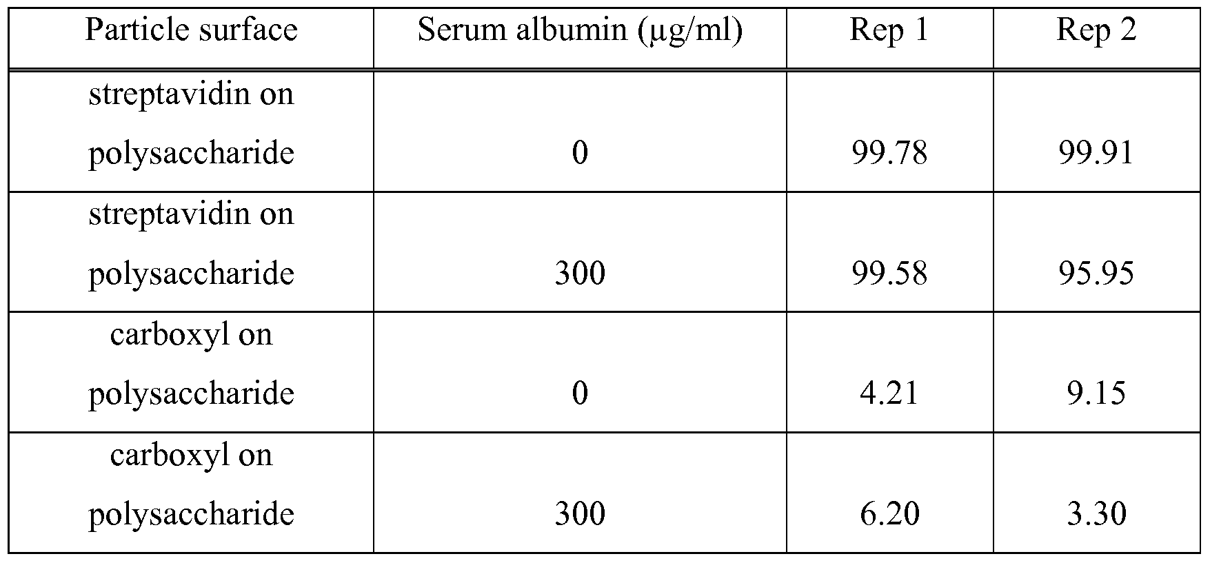
  • FLUIDMAG magnetic particles with a polysaccharide core and functionalized with streptavidin were used to non-specif ⁇ cally capture bacteria according to the above Capture and Detection Procedure.
  • FLUIDMAG ARA magnetic particles with a polysaccharide core and surface carboxyl surface groups were also tested.
  • the capture was performed for both P. aeruginosa (ATCCC 10662) (Table 2) and S. aureus (ATCCC 25923) (Table 3).
  • Capture was tested with and without the presence of human serum albumin 300 ⁇ g/ml.
  • the particles (30 ⁇ l) were combined with 470 ⁇ l of the bacteria (particle concentration of 0.6 mg/ml) at approximately 5 x 10 6 cfu/ml for all of the experiments. The results are shown in Tables 2 and 3.
  • the streptavidin beads showed capture of P. aeruginosa with a slightly lower capture in the presence of serum albumin.
  • the carboxyl beads showed significantly lower capture, especially in the presence of serum albumin.
  • both particles had the same polysaccharide core chemistry, the particles with streptavidin demonstrated much better capture as compared to the particles surface functionalized with carboxyl groups but without streptavidin.
  • Table 3 S. aureus (25923) Capture by Streptavidin on Polysaccharide Compared with Carboxyl on Polysaccharide Without Streptavidin
  • the streptavidin/polysaccharide beads showed excellent capture of S. aureus both in the presence and absence of serum albumin.
  • the carboxyl beads without streptavidin showed poor capture in both cases. Even though both particles had the same polysaccharide core, the particles with streptavidin on the surface performed significantly better.
  • FLUIDMAG magnetic particles 100 nm, Chemicell GmbH
  • a polysaccharide core and functionalized with streptavidin were used to non-specif ⁇ cally capture bacteria according to the above Capture and Detection Procedure. Capture was performed for both P. aeruginosa (ATCCC 10662) (Table 4) and S. aureus (ATCCC 25923) (Table 5). Capture was carried out in the presence of blood in the sample at varying concentrations (1 :1000, 1 :10000 and 1 :100000 dilutions of whole blood). A control sample with no blood was also tested.
  • aureus (ATCCC 25923) was captured according to the above Capture and Detection Procedure (32 ⁇ l of the stock bead solution as prepared in Preparative Example 3 was added to 468 ⁇ l of the bacteria stock sample to obtain a final bead solution concentration of 0.16mg/ml).
  • capture experiments were performed with the unmodified polystyrene-carboxyl (DYNAL Cl) and polysaccharide - carboxyl (FLUIDMAG ARA) beads. The results are shown in Table 6.
  • the capture efficiency of the streptavidin functionalized polystyrene beads was about 5 times lower than the unmodified polystyrene beads.
  • the capture efficiency was approximately ten times better than either the unmodified polysaccharide beads or the polystyrene beads functionalized with streptavidin.
  • Example 4 Compared with Capture by Streptavidin on Polystyrene-Carboxyl Beads Using P. aeruginosa (ATCCC 35032) instead of S. aureus, Example 4 was essentially repeated, and the results are summarized below in Table 7. Overall, the polysaccharide beads modified with streptavidin provided the best non-specific bacteria capture performance. Table 7. P. aeruginosa Capture by Streptavidin on Polysaccharide, Polysaccharide- Carboxyl, Streptavidin on Polystyrene-Carboxyl, and Polystyrene-Carboxyl Beads (Stock bacterial concentration was 12000 cfu/ml.)
  • Example 6 S. aureus (ATCCC 25923) Capture by Neutravidin on Polysaccharide Beads Compared with Capture by Neutravidin on Polystyrene-Carboxyl Beads
  • Neutravidin was attached to polystyrene-carboxyl (DYNAL Cl) and polysaccharide-carboxyl (FLUIDMAG ARA, Chemicell GmbH) beads as in Preparative Example 3, and S. aureus (ATCCC 25923) was captured according to the above Capture and Detection Procedure (32 ⁇ l of the stock bead solution as prepared in Preparative Example 3 was added to 468 ⁇ l of the bacteria stock sample to obtain a final bead solution concentration of 0.16mg/ml). The results are shown in Table 8.
  • P. aeruginosa (ATCCC 35032) Capture by Neutravidin on Polysaccharide Beads Compared with Capture by Neutravidin on Polystyrene-Carboxyl Beads
  • Example 6 Using P. aeruginosa (ATCCC 35032) instead of S. aureus, Example 6 was essentially repeated, and the results are summarized below in Table 9.
  • the polysaccharide beads with neutravidin provided significantly greater non-specific bacterial capture than the polystyrene beads with neutravidin.
  • Streptavidin was attached to polysaccharide-carboxyl beads (FLUIDMAG ARA, Chemicell GmbH) as in Preparative Example 3, and various bacteria strains (from clinical isolates) were captured according to the above Capture and Detection Procedure (32 ⁇ l of the stock bead solution as prepared in Preparative Example 3 was added to 468 ⁇ l of the bacteria stock sample to obtain a final bead solution concentration of 0.16 mg/ml). The results are summarized below in Table 10. Table 10. Non-specific Capture of Various Bacteria Using Polysaccharide Beads Functionalized with Streptavidin
  • E. coli, Salmonella, and Group B Streptococcus solutions were prepared by growing these bacteria over night on blood agar plates.
  • Bacteria suspensions were then prepared in PBS L-64 buffer by MacFarland turbidity standards to a concentration of approximately 1 x 10 8 cfu/mL.
  • the suspensions were diluted in PBS L-64 buffer to a working concentration of approximately 1 x 10 6 cfu/mL.
  • Capture experiments were performed by adding 32 ⁇ l of streptavidin functionalized polysaccharide-carboxyl beads (made using FLUIDMAG ARA beads in Preparative Example 3) (2.5 mg/mL) to 234 ⁇ l of the bacteria sample.
  • a 2 % stock solution of saponin (Product #47036, Sigma- Aldrich) in PBS buffer was prepared.
  • saponin Product #47036, Sigma- Aldrich
  • 234 ⁇ l of the stock saponin solution was added to the bead-bacteria mixture, resulting in a 0.9 % concentration of saponin during capture.
  • 234 ⁇ l of PBS L-64 buffer was added to the bead-bacteria mixture.
  • the bead concentration was 0.16 mg/ml during the capture experiments, which were carried out from this point forward as described in the above Capture and Detection Procedure.
  • the results shown in Table 12 indicate that the presence of saponin increased bacteria capture for all bacteria tested.
  • FLUIDMAG ARA beads 100 nm, from Chemicell GmbH, Berlin, Germany
  • a polysaccharide core and functionalized with streptavidin or neutravidin prepared as in Preparative Example 3 were used to non-specifically capture S. aureus (ATCC 25923) and E. coli (ATCC 25922) using the above Capture and Detection Procedure.
  • 10 ⁇ l of the bead suspension was combined with 490 ⁇ l of the bacteria suspensions at two concentrations: ⁇ lxl ⁇ 6 cfu/ml and ⁇ lxl ⁇ 5 cfu/ml, using polypropylene microcentrifuge tubes (available from VWR Scientific, West Chester, PA).
  • Samples with 0 cfu/ml and positive control samples were also prepared using the above Capture and Detection Procedure.
  • the beads were then separated and concentrated by placing the microcentrifuge tube in a DYNAL magnetic fixture (available from Invitrogen, Inc. Carlsbad, CA) for at least 5 minutes. The supernatant was discarded by micropipetting without disrupting the agglomerated beads.
  • DYNAL magnetic fixture available from Invitrogen, Inc. Carlsbad, CA
  • the beads were then washed by adding 0.5 mL PBS-L64 buffer to the tube and agitating using a rocking motion for 5 minutes. The beads were then again separated and concentrated by placing the microcentrifuge tube in the DYNAL magnetic fixture for at least 5 minutes. The wash solution was discarded by micropipetting without disrupting the agglomerated beads. This wash step was repeated a second time.
  • the beads were then again separated and concentrated by placing the microcentrifuge tube in the DYNAL magnetic fixture for at least 5 minutes. The supernatant was then micropipetted to the bottom chamber of a Biotrace AQUA-TRACE test device (available from Biotrace International BioProducts Inc., Bothell, WA) after removal of the swab and the foil sealed chamber containing lysing agents in pelletized form.
  • the bottom chamber of the Biotrace device contained all the necessary dry reagents to determine the presence of ATP via luciferase bio luminescence.
  • the sample was vortexed for 10 seconds and placed in a Biotrace luminometer (UNI-LITE NG, (available from Biotrace International BioProducts Inc., Bothell, WA)) within thirty seconds after adding the supernatant to the bottom chamber of a Biotrace AQUA- TRACE test device.
  • the bio luminescent response from each sample is reported in Table 13 in Relative Light Units (RLUs).
  • RLUs Relative Light Units
  • the average RLUs from three replicates is reported in Table 13.
  • Table 13 are the 1 sigma standard deviations on the reported RLUs values. Table 13. Detection of captured S. aureus and E. coli with ATP detection
  • Cl, C2, C3, and C4 are Comparative Examples 1, 2, 3, and 4. S. aureus was detected at both concentrations tested, whereas E. coli was detected only at the highest concentration tested.
  • positive control signal intensities C1-C4
  • Examples 23 - 27 Capture and ATP Detection of S. aureus and E. coli in the Presence of Lysed Blood Cells
  • the assay to detect bacteria was repeated exactly as described in Examples 11-22, except for an initial sample composition which contained: 234 ⁇ l of a 1 :10,000 diluted (using PBS buffer) and lysed (using 0.9% Saponin Product #47036, Sigma-Aldrich) human whole blood specimen, along with 234 ⁇ l of PBS buffer containing bacteria at either 0, IxIO 5 , or IxIO 6 cfu/ml, and 32 ⁇ l of the FLUIDMAG particles (100 nm, from Chemicell GmbH, Berlin, Germany) with a polysaccharide core and functionalized with streptavidin.
  • an initial sample composition which contained: 234 ⁇ l of a 1 :10,000 diluted (using PBS buffer) and lysed (using 0.9% Saponin Product #47036, Sigma-Aldrich) human whole blood specimen, along with 234
  • the bio luminescent response from each sample is reported in Table 14 in Relative Light Units (RLUs).
  • RLUs Relative Light Units
  • the average RLUs from three replicates is reported in Table 14.
  • Table 14 Also shown in Table 14 are the 1 sigma standard deviations on the reported RLUs values. The data shown in Table 14 demonstrates that detection of both bacteria was observed at both concentrations tested.

Landscapes

  • Health & Medical Sciences (AREA)
  • Life Sciences & Earth Sciences (AREA)
  • Immunology (AREA)
  • Engineering & Computer Science (AREA)
  • Molecular Biology (AREA)
  • Chemical & Material Sciences (AREA)
  • Biomedical Technology (AREA)
  • Hematology (AREA)
  • Urology & Nephrology (AREA)
  • Food Science & Technology (AREA)
  • General Physics & Mathematics (AREA)
  • Cell Biology (AREA)
  • Biotechnology (AREA)
  • Medicinal Chemistry (AREA)
  • Physics & Mathematics (AREA)
  • Analytical Chemistry (AREA)
  • Biochemistry (AREA)
  • General Health & Medical Sciences (AREA)
  • Microbiology (AREA)
  • Pathology (AREA)
  • Chemical Kinetics & Catalysis (AREA)
  • Tropical Medicine & Parasitology (AREA)
  • Virology (AREA)
  • Measuring Or Testing Involving Enzymes Or Micro-Organisms (AREA)
  • Apparatus Associated With Microorganisms And Enzymes (AREA)
  • Micro-Organisms Or Cultivation Processes Thereof (AREA)
  • Investigating Or Analysing Materials By The Use Of Chemical Reactions (AREA)

Abstract

The invention relates to compositions, methods, devices, and kits for non-specifically isolating bacterial cells. The compositions comprise a solid support which has a surface comprising a combination of a carbohydrate and a biotin-binding protein, wherein the protein is covalently bonded to the carbohydrate, and wherein the protein is linked to the solid support via the carbohydrate; and at least one of 1) a plurality of bacterial cells non-specifically bound to the combination of the carbohydrate and the protein or 2) an amphiphilic glycoside of a steroid or triterpene. The methods, devices, and kits include at least one of these compositions.

Description

MICROORGANISM-CAPTURING COMPOSITIONS AND METHODS
CROSS REFERENCE TO RELATED APPLICATIONS The present invention claims priority to U.S. Provisional Application Serial No.
61/018011, filed December 31, 2007, which is incorporated herein by reference.
GOVERNMENT RIGHTS
The U.S. Government may have certain rights to this invention under the terms of Contract Nos. DAAD-13-03-C-0047 (Program No. 2640) and W81XWH-07-01-0354 (Program No. 2750) granted by the Department of Defense.
BACKGROUND
Assays for determining the presence of microorganisms in a variety of samples, including food, clinical, environmental, and experimental samples, are of increasing importance. Such assays can provide an indication of microorganism load and/or identification of microorganisms that are present.
Current techniques for qualitatively and quantitatively determining the presence of microorganisms typically involve identification of a specific microorganism, such as a pathogen. Successful detection of the presence of a particular microorganism, such as a bacterium, in a sample depends upon the concentration of the bacterium in the sample.
Generally, bacterial samples are cultured to assess viability and increase the number of bacteria in the sample to assure an adequate level for detection. The culturing step requires substantial time, which delays obtaining the detection results. By concentrating bacteria in a sample, detection can be carried out using a shorter or even no culturing step. Methods have been developed to isolate, and thereby concentrate, specific bacterial strains by using antibodies specific to the strain.
Other concentration methods that are non-strain specific, which would allow a more general sampling of the microorganisms present, have been proposed. Once isolated, a specific strain or specific strains within a mixture of strains can be identified and/or quantified using known detection methods, including, for example, nucleic acid amplification methods. Non-specific isolation of microorganisms using carbohydrate and lectin protein interactions has been proposed. Lectins present on the surface of bacteria were suggested as capture targets for certain carbohydrates attached to polyacrylamide. Substances that serve as nutrients for microorganisms have also been proposed for use as ligands for non- specific capture of microorganisms. Such nutrient ligands included carbohydrates, vitamins, iron-chelating compounds, and sidorophores. Supported chitsan has also been proposed for concentrating microorganisms in a non-strain-specific manner, allowing the microorganisms to be more easily and rapidly assayed.
Although some methods for isolating microorganisms have been described, there continues to be an interest in and a need for improved materials and methods for isolating microorganisms .
SUMMARY
It has now been found that the combination of a biotin-binding protein attached to a solid support via a carbohydrate is effective for non-specifically binding bacterial cells.
Compositions with a carbohydrate on a solid support, but without the biotin-binding protein, were found to be less effective for non-specifically binding bacterial cells. Moreover, compositions with a biotin-binding protein on the solid support, but without the carbohydrate, were also found to be less effective for non-specifically binding bacterial cells. In some embodiments, the biotin-binding protein attached to the solid support via the carbohydrate is even more effective for non-specifically binding bacterial cells in the presence of an amphiphilic glycoside of a steroid or triterpene.
The present invention, therefore, provides new compositions for non-specifically binding bacterial cells. In one embodiment, there is provided a composition comprise a solid support which has a surface comprising a combination of a carbohydrate and a biotin-binding protein, wherein the protein is covalently bonded to the carbohydrate, and wherein the protein is linked to the solid support via the carbohydrate; and at least one of 1) a plurality of bacterial cells non-specifically bound to the combination of the carbohydrate and the protein or 2) an amphiphilic glycoside of a steroid or triterpene.
In another embodiment, there is provided a composition comprising: a solid support which has a surface comprising a combination of a carbohydrate and a biotin-binding protein, wherein the protein is covalently bonded to the carbohydrate, and wherein the protein is linked to the solid support via the carbohydrate; and a plurality of bacterial cells non-specifically bound to the combination of the carbohydrate and the protein.
In another embodiment, there is provided a composition comprising: a solid support which has a surface comprising a combination of a carbohydrate and a biotin-binding protein, wherein the protein is covalently bonded to the carbohydrate, and wherein the protein is linked to the solid support via the carbohydrate; and an amphiphilic glycoside of a steroid or triterpene.
In another aspect, there is provided a method for isolating bacterial cells comprising: providing a solid support which has a surface comprising a combination of a carbohydrate and a biotin-binding protein, wherein the protein is covalently bonded to the carbohydrate, and wherein the protein is linked to the solid support via the carbohydrate; providing a sample suspected of having a plurality of bacterial cells; contacting the solid support which has the surface comprising the combination of the carbohydrate and the biotin-binding protein with the sample; wherein at least a portion of the plurality of bacterial cells from the sample become non-specifically bound to the surface of the solid support; and separating the solid support from the remainder of the sample after the at least a portion of the plurality of bacterial cells from the sample become non-specifically bound to the surface of the solid support.
In another aspect, there is provided a device for detecting bacterial cells comprising: a composition comprising: a solid support which has a surface comprising a combination of a carbohydrate and a biotin-binding protein, wherein the protein is covalently bonded to the carbohydrate, and wherein the protein is linked to the solid support via the carbohydrate; and a plurality of bacterial cells non-specifically bound to the combination of the carbohydrate and the protein; and a means for detecting the bacterial cells.
In another embodiment, there is provided a device for detecting bacterial cells comprising: a composition comprising: a solid support which has a surface comprising a combination of a carbohydrate and a biotin-binding protein, wherein the protein is covalently bonded to the carbohydrate, and wherein the protein is linked to the solid support via the carbohydrate; and an amphiphilic glycoside of a steroid or triterpene; and a means for detecting the bacterial cells.
In another aspect, there is provided a kit comprising: a solid support which has a surface comprising a combination of a carbohydrate and a biotin-binding protein, wherein the protein is covalently bonded to the carbohydrate, and wherein the protein is linked to the solid support via the carbohydrate; and an amphiphilic glycoside of a steroid or triterpene.
DEFINITIONS
The term "amphiphilic glycoside of a steroid or triterpene" refers to a steroid or triterpene joined to a sugar by a glycosidic linkage, the resulting material having amphiphilic properties, that is, surfactant properties, the material being both hydrophilic and lipophilic.
The term "biotin-binding protein" refers to a protein which has a high affinity and selectivity for binding biotin. As used herein, the "biotin-binding protein" does not include bound biotin.
The term "magnetic particles" means particles, particle conglomerates, or beads comprised of ferromagnetic, paramagnetic, or superparamagnetic particles, including dispersions of said particles in a polymer bead. As used herein, "a", "an", "the", "at least one", and "one or more" are used interchangeably.
The terms "comprises" and variations thereof do not have a limiting meaning where these terms appear in the description and claims. The above summary of the present invention is not intended to describe each disclosed embodiment or every implementation of the present invention. The description that follows more particularly exemplifies illustrative embodiments. In several places throughout the description, guidance is provided through lists of examples, which examples can be used in various combinations. In each instance, the recited list serves only as a representative group and should not be interpreted as an exclusive list.
DETAILED DESCRIPTION OF ILLUSTRATIVE EMBODIMENTS OF THE
INVENTION
The present invention provides new compositions for non-specifically binding bacterial cells. "Non-specifically binding" bacterial cells means that the binding is not specific to any type of bacterial cells. Thus, all bacteria in a sample can be isolated from other components in the sample rather than targeting, for example, one strain of bacteria. Both gram positive and gram negative bacteria can be bound. The resulting isolated bacteria can then be subjected to known detection methods, such as bacterial load detection.
In one embodiment, there is provided a composition comprising: a solid support which has a surface comprising a combination of a carbohydrate and a biotin-binding protein, wherein the protein is covalently bonded to the carbohydrate, and wherein the protein is linked to the solid support via the carbohydrate; and a plurality of bacterial cells non-specifically bound to the combination of the carbohydrate and the protein.
In another embodiment, there is provided a composition comprising: a solid support which has a surface comprising a combination of a carbohydrate and a biotin- binding protein, wherein the protein is covalently bonded to the carbohydrate, and wherein the protein is linked to the solid support via the carbohydrate; and an amphiphilic glycoside of a steroid or triterpene. For certain embodiments, this composition further comprises a plurality of bacterial cells non-specifically bound to the combination of the carbohydrate and the biotin-binding protein. The compositions can be prepared by combining the solid support having the combination of carbohydrate and biotin-binding protein described above with at least one of a plurality of bacterial cells or an amphiphilic glycoside of a steroid or triterpene. The solid support having the combination of carbohydrate and biotin-bind protein can be provided as described below. Combining the solid support with the bacterial cells and/or amphiphilic glycoside can be conveniently carried out by suspending or immersing the solid support in a buffer and adding the bacterial cells, also suspended in a buffer, and/or the amphiphilic glycoside dissolved in a buffer. The amphiphilic glycoside buffer solution preferably contains about 0.2 to about 10 weight/volume percent (% w/v) amphiphilic glycoside, more preferably about 0.5 to about 5 % w/v.
In another embodiment, there is provided a method for isolating bacterial cells comprising: providing a solid support which has a surface comprising a combination of a carbohydrate and a biotin-binding protein, wherein the protein is covalently bonded to the carbohydrate, and wherein the protein is linked to the solid support via the carbohydrate; providing a sample suspected of having a plurality of bacterial cells; contacting the solid support which has the surface comprising the combination of the carbohydrate and the biotin-binding protein with the sample; wherein at least a portion of the plurality of bacterial cells from the sample become non-specifically bound to the surface of the solid support; and separating the solid support from the remainder of the sample after the at least a portion of the plurality of bacterial cells from the sample become non-specifically bound to the surface of the solid support.
The sample suspected of having a plurality of bacterial cells can be provided from a wide range of sources using known methods. Examples of sources include physiological fluid, e.g., blood, saliva, ocular lens fluid, synovial fluid, cerebral spinal fluid, pus, sweat, exudate, urine, mucus, mucosal tissue (e.g., buccal, gingival, nasal, ocular, tracheal, bronchial, gastrointestinal, rectal, urethral, ureteral, vaginal, cervical, and uterine mucosal membranes), lactation milk, or the like. Further examples of sample sources include those obtained from a body site, e.g., wound, skin, nares, nasopharyngeal cavity, nasal cavities, anterior nasal vestibule, scalp, nails, outer ear, middle ear, mouth, rectum, or other site. Additional examples of sample sources include process streams, water, food, soil, vegetation, air (e.g., contaminated), surfaces, such as food preparation surfaces, and the like. Known sampling techniques can be used to gather the sample from the source. For example, a volume of a liquid sample can be drawn from the source, or a swab or wipe can be used to collect a sample from a physiological or environmental surface. A wide variety of swabs or other sample collection devices are commercially available. The sample can be provided for use in the above method directly as is from the collection device, for example, in the case of aqueous liquids. Alternatively, the sample can be provided by eluting, releasing, or washing the sample from the collection device using, for example, water, physiological saline, pH buffered solutions, or any other solutions or combinations of solutions that can remove the sample from the collection device and bring the sample into suspension. An example of an eluting buffer is phosphate buffered saline (PBS), which can be used in combination with a surfactant, such as polyoxyethylene sorbitan monolaurate or a poly(oxyethylene-co-oxypropylene) block copolymer. Other extraction solutions can function to maintain specimen stability during transport from a sample collection site to a sample analysis site. Examples of these types of extraction solutions include Amies' and Stuart's transport media.
The sample may be subjected to a treatment prior to contact with the solid support, such as dilution of viscous fluids, concentration, filtration, distillation, dialysis, dilution, inactivation of natural components, addition of reagents, chemical treatment, or the like. Contacting the solid support which has the surface comprising the combination of the carbohydrate and the biotin-binding protein with the sample suspected of having a plurality of bacterial cells can be conveniently carried out by suspending or immersing the solid support in a buffer and adding the sample, which may also be suspended in a buffer. Suitable buffers include PBS and PBS with a nonionic surfactant. The mixture can be agitated for a period of time sufficient to allow the bacterial cells to become bound to the combination of carbohydrate and biotin-binding protein on the solid support. The binding can be conveniently carried out at room temperature.
Separating the solid support from the remainder of the sample can be carried out using well known methods. For example, the remainder of the sample can be drained, decanted, drawn, centrifuged, pipetted, and/or filtered off of the solid support. When the solid support is magnetic particles the solid support can be consolidated in one area of the container with a magnet for convenient removal of the remainder of the sample, for example, by decanting, pipetting, or forcing the supernate out of the container using a pressure differential or a g-force. Additionally, the solid support may be washed, for example, with a buffer, to remove any remaining unbound sample material.
For certain embodiments, the method for isolating bacterial cells further comprises detecting the at least a portion of the plurality of bacterial cells. For certain of these embodiments, the detecting is carried out by a detection method selected from the group consisting of adenosine triphosphate (ATP) detection by bio luminescence, polydiacetylene (PDA) colorimetric detection, nucleic acid detection, immunological detection, growth based detection, surface acoustic wave detection, or the like.
ATP detection can be used as a nonspecific indicator of bacterial load. After separating the solid support with non-specifically bound bacterial cells from the remainder of the sample (which may contain interfering components such as extra-cellular ATP), the cells are lysed and contacted with luciferin and luciferase. The resulting bioluminescence, which is of an intensity proportional to the number of captured bacterial cells, is then measured, for example, using a luminometer. PDA colorimetric detection can be used to detect specific bacteria or a spectrum of bacteria by contacting a colorimetric sensor with the bacteria. The colorimetric sensor comprises a receptor and a polymerized composition which includes a diacetylene compound or a polydiacetylene. When bacterial cells are bound by the receptor, resulting conformational changes to the sensor cause a measurable color change. The color change can be measured, for example, visually or using a colorimeter. Indirect detection of bacterial cells using probes which can bind to the receptor may also be used. PDA colorimetric detection using such colorimetric sensors is known and described, for example, in U.S. Patent Application Publication No. 2006/0134796A1, International Publication Nos. WO 2004/057331A1 and WO 2007/016633A1, and in Assignee's co- pending U.S. Patent Application Serial No. 60/989298.
Methods for detecting nucleic acids, including DNA and RNA, often include amplifying or hybridizing the nucleic acids. The captured bacterial cells are lysed to make the cellular nucleic acids available for detection. Lysing can be carried out enzymatically, chemically, and/or mechanically. Enzymes used for lysis include, for example, lysostaphin, lysozyme, mutanolysin, or others. Chemical lysis can be carried out using a surfactant, alkali, heat, or other means. When alkali is used for lysis, a neutralization reagent may be used to neutralize the solution or mixture after lysis. Mechanical lysis can be accomplished by mixing or shearing using solid particles or microparticles such as beads or microbeads. Sonication may also be used for lysis. The lysis reagent can include a surfactant or detergent such as sodium dodecylsulfate (SDS), lithium laurylsulfate (LLS), TRITON series, TWEEN series, BRIJ series, NP series, CHAPS, iV-methyl-iV-(l- oxododecyl)glycine, sodium salt, or the like, buffered as needed; a chaotrope such as guanidium hydrochloride, guanidium thiacyanate, sodium iodide, or the like; a lysis enzyme such as lysozyme, lysostaphin, mutanolysin, proteinases, pronases, cellulases, or any of the other commercially available lysis enzymes; an alkaline lysis reagent; solid particles such as beads, or a combination thereof. Examples of amplification methods include polymerase chain reaction (PCR); target polynucleotide amplification methods such as self-sustained sequence replication (3SR) and strand-displacement amplification (SDA); methods based on amplification of a signal attached to the target polynucleotide, such as "branched chain" DNA amplification; methods based on amplification of probe DNA, such as ligase chain reaction (LCR) and QB replicase amplification (QBR); transcription-based methods, such as ligation activated transcription (LAT), nucleic acid sequence-based amplification (NASBA), amplification under the trade name INVADER, and transcription-mediated amplification (TMA); and various other amplification methods, such as repair chain reaction (RCR) and cycling probe reaction (CPR). Primer directed nucleic acid amplification methods, which include, for example, thermal cycling methods such as PCR, LCR, and SDA, may be used advantageously for detecting a spectrum of bacteria by choosing a primer which can hybridize to nucleic acids from the spectrum of bacteria.
Nucleic acid hybridization detection methods are also well known. Here, a single stranded nucleic acid probe is hybridized to a single stranded nucleic acid(s) from the bacterial cells to provide a double stranded nucleic acid which includes the probe strand. Nucleic acid probes (probe labels) such as fluorescent, chemiluminescent, and radioactive labels which can then be quantified and detected are known. Moreover, a species specific probe or a combination of species specific probes may be used to detect a specific bacteria or a number of different bacteria.
Immunological detection includes detection of a biological molecule, such as a protein, proteoglycan, or other material with antigenic activity, acting as a marker on the surface of bacteria. Detection of the antigenic material is typically by an antibody, a polypeptide selected from a process such as phage display, or an aptamer from a screening process. Immunological detection methods are known, examples of which include immunoprecipitation and enzyme-linked immunosorbent assays (ELISA). Antibody binding can be detected in several ways, including by labeling either the primary or the secondary antibody with a fluorescent dye, quantum dot, or an enzyme that can produce chemiluminescence or a color change. Plate readers and lateral flow devices have been used for detecting and quantifϊying the binding event.
Growth based detection methods are well known and generally include plating the bacteria, culturing the bacteria to increase the number of bacterial cells under specific conditions, and enumerating the bacterial cells. PETRIFILM Aerobic Count Plates (3M Company, St. Paul, MN) can be used for this purpose.
Surface acoustic wave detection, described, for example, in International Publication No. WO 2005/071416, is also known for detecting bacterial cells. For example, a bulk acoustic wave-impedance sensor has been used for detecting the growth and numbers of bacterial cells on the surface of a solid medium. The concentration range of the bacteria that can be detected by this method was 3.4 x 10² to 6.7 x 10⁶ cells/ml. See Le Deng et al, J. Microbiological Methods, Vol. 26, Iss. 10-2, 197-203 (1997).
For certain embodiments, including any one of the above embodiments of the method for isolating bacterial cells, contacting the solid support with the sample is carried out in the presence of an amphiphilic glycoside of a steriod or triterpene.
As indicated above, contacting the solid support with the sample may be carried out also in the presence of a buffer, which may provide a suitable media for effectively binding the bacterial cells to the solid support. A buffer of appropriate charge, osmolality, or other characteristic may be added to the sample prior to, simultaneously with, or after contact with the solid support. PBS and PBS-L64 buffers are examples of such cell binding buffers.
In another embodiment, there is provided a device for detecting bacterial cells comprising: a composition comprising a solid support which has a surface comprising a combination of a carbohydrate and a biotin-binding protein, wherein the protein is covalently bonded to the carbohydrate, and wherein the protein is linked to the solid support via the carbohydrate, and a plurality of bacterial cells non-specifϊcally bound to the combination of the carbohydrate and the protein; and a means for detecting the bacterial cells.
In another embodiment, there is provided a device for detecting bacterial cells comprising: a composition comprising a solid support which has a surface comprising a combination of a carbohydrate and a biotin-binding protein, wherein the protein is covalently bonded to the carbohydrate, and wherein the protein is linked to the solid support via the carbohydrate, and an amphiphilic glycoside of a steroid or triterpene; and a means for detecting the bacterial cells. For certain embodiments, the composition further comprises a plurality of bacterial cells non- specifically bound to the combination of the carbohydrate and the protein.
The device may include one or more structural features which contain the composition and the means for detecting the bacterial cells. The device can provide a location or locations and conditions for capturing bacterial cells by the solid support, separating the remaining sample from the solid support, and detecting the bacterial cells. The sample may be located in one or a plurality of locations or reservoirs. The device may provide uniform and accurate temperature control of one or more of the locations or reservoirs. The device may provide channels between locations or reservoirs, for example, such that bacterial cell binding may take place in one or more locations or reservoirs, and bacterial cell detection may take place in one or more other locations or reservoirs. For certain embodiments, including any one of the above embodiments which include the device for detecting bacterial cells, the device is a lateral flow device, a vertical flow device, or a combination thereof. Some examples of such devices are described in Assignee's co-pending U.S. Patent Application Serial No. 60/989291. For certain embodiments, the device is a micro fluidic device. Some examples of micro fluidic devices are described in U.S. Publication Numbers 2002/0064885 (Bedingham et al);
US2002/0048533 (Bedingham et al.); US2002/0047003 (Bedingham et al.); and US2003/138779 (Parthasarathy et al.); U.S. Patent Nos. 6,627,159; 6,720,187; 6,734,401; 6,814,935; 6,987,253; 7,026,168, and 7,164,107; and International Publication No. WO 2005/061084 Al (Bedingham et al.). For certain embodiments, including any one of the above embodiments of a composition, a method, or a device, which includes a plurality of bacterial cells, the plurality of bacterial cells includes two or more different types of bacteria. A type of bacteria or a type of bacterial cells refers to a strain, species, genus, family, order, or Part of the bacteria or bacterial cells.
For certain embodiments, including any one of the above embodiments of a composition, a method, or a device, which includes a plurality of bacterial cells, the bacteria are selected from the group consisting of gram positive bacteria and gram negative bacteria. For certain of these embodiments, the bacteria are selected from the group consisting of Bacillus, Bordetella, Borrelia, Campylobacter, Clostridium, Corynebacteria, Enterobacter, Enterococcus, Escherichia, Helicobacter, Legionella, Listeria, Mycobacterium, Neisseria, Pseudomonas, Salmonella, Shigella, Staphylococcus, Streptococcus, Vibrio, and Yersinia. For certain embodiments, the bacteria are selected from the group consisting of S. aureus, P. aeruginosa, S. epidermidis, E. faecalis, Strep agalatiae, Strep dysgalatiae, E. coli, Salmonella, and Group B Strep.
In another embodiment, there is provided a kit comprising a solid support which has a surface comprising a combination of a carbohydrate and a biotin-binding protein, wherein the protein is covalently bonded to the carbohydrate, and wherein the protein is linked to the solid support via the carbohydrate; and an amphiphilic glycoside of a steroid or triterpene. For certain of these embodiments, the kit further comprises a means for detecting bacterial cells.
For certain embodiments, including any one of the above embodiments of a device or kit, the means for detecting the bacterial cells is selected from the group consisting of reagents for detecting adenosine triphosphate (ATP) by bioluminescence, a PDA colorimetric sensor, reagents for nucleic acid detection, reagents for immunological detection, media for plating and enumerating bacterial cells, a surface acoustic wave sensor, and the like. Reagents for detecting ATP include luciferin, luciferase, and optionally a lysing agent. Reagents for nucleic acid detection include, for example a primer, a probe, an enzyme for extending the primer, or a combination thereof. Reagents for immunological detection include, for example, at least one antibody.
For certain embodiments, including any one of the above embodiments of a composition, a method, a device, or a kit, which includes an amphiphilic glycoside of a steroid or triterpene, the amphiphilic glycoside of the steroid or triterpene is a saponin. Saponins are 27 carbon atom steroids or 30 carbon atom triterpenes joined to a sugar by a glycosidic linkage. For certain embodiments, the sugar is selected from the group consisting of hexoses, pentoses, saccharic acids, and a combination thereof. Saponin has been used as a mild detergent and for lysing red blood cells. However, unexpectedly it has now been found that this class of materials can be present with the combination of the biotin-binding protein and the carbohydrate attached to the solid support via the carbohydrate without loss of effectiveness for non-specifically binding bacterial cells. Moreover, for certain embodiments, the combination of the biotin-binding protein and the carbohydrate attached to the solid support via the carbohydrate, in combination with the amphiphilic glycoside of a steroid or triterpene, or an embodiment thereof described above, is even more effective for non-specifically binding bacterial cells.
For certain embodiments, including any one of the above embodiments of a composition, a method, a device, or a kit, the biotin-binding protein is a protein which binds four biotin molecules per protein molecule if biotin is present with the protein. Although the embodiments of the present invention include a biotin-binding protein, the biotin-binding protein is included without biotin (or biotin attached to another material) bound to the biotin-binding protein. For certain of these embodiments, the biotin-binding protein is selected from the group consisting of avidin, streptavidin, neutravidin, and selectively nitrated avidin. Avidin is a glycoprotein with a mass of about 66 kDa and an isoelectric point of 10 to 10.5. About 10 percent of avidin' s total mass is carbohydrate. Avidin is commerically available, for example, from Sigma- Aldrich. Streptavidin is a tetrameric protein with a mass of about 60 kDa and an isoelectric point of about 5. Streptavidin, which lacks the carbohydrate component found in avidin, is commerically available, for example, from Pierce. Neutravidin is a deglycosylated avidin, with a mass of about 60 kDa and an isoelectric point of about 6.3. Neutravidin is commerically avialable from Pierce. Selectively nitrated avidin, available from Molecular Probes, Inc. under the trade name CAPT A VIDIN, has tyrosine residues in the four biotin-binding sites of avidin nitrated. For certain of these embodiments, the biotin-binding protein is streptavidin.
For certain embodiments, including any one of the above embodiments of a composition, a method, a device, or a kit, the carbohydrate is selected from the group consisting of monsaccharides, oligosaccharides, polysaccharides, and combinations thereof. Suitable monosaccharides include, for example, mannose, galactose, glucose, fructose, fucose, iV-acetylglucosamine, //-acetylgalactosamine, rhamnose, galactosamine, glucosamine, galacturonic acid, glucuronic acid, JV-acetylneuraminic acid, methyl D- mannopyranoside, α-methylglucoside, galactoside, ribose, xylose, arabinose, saccharate, mannitol, sorbitol, inositol, glycerol, a derivative of any one of these monosaccharides, and a combination thereof. Suitable oligosaccharides include those having 2 to 12 monosaccharide units, which may be the same or different. Examples include oligomannose having 2 to 6 units, maltose, sucrose, trehalose, cellobiose, salicin, a derivative of any one of these oligosaccharides, and a combination thereof. Suitable polysaccharides include those having more than 12 monosaccharide units, which may be the same or different. Examples include polysaccharides such as gum arabic (acacia gum), believed to be a branched polymer of galactose, rhamnose, arabinose, and glucuronic acid as the calcium, magnesium, and potassium salts; galactomannan polysaccharide (locust bean gum), believed to be a straight chain polymer of mannose and one galactose branch on every fourth mannose; guar gum, believed to be a straight chain polymer of mannose and one galactose branch on every other unit; and gum karaya, believed to include a partially acetylated polymer of galactose, rhamnose, and glucuronic acid. For certain of these embodiments, the carbohydrate includes at least one carboxy group. Any one of the above described monsaccharides, oligosaccharides, polysaccharides, and a combination thereof, which includes at least one carboxy group, may be used. Moreover, any one of the above described monsaccharides, oligosaccharides, polysaccharides, and a combination thereof, which is derivatized to include at least one carboxy group, may be used. For certain of these embodiments, the carbohydrate is a polysaccharide. For certain of these embodiments, the carbohydrate comprises arabic acid.
The biotin-binding protein can be covalently bonded to the carbohydrate by reaction(s) between functional groups on the biotin-binding protein and functional groups on the carbohydrate. For certain embodiments, including any one of the above embodiments of a composition, a method, a device, or a kit, the carbohydrate includes at least one carboxy group, and the biotin-binding protein is covalently bonded to the carbohydrate through a linking group, the linking group being the reaction product of the protein and the at least one carboxy group of the carbohydrate. The biotin-binding protein becomes covalently bonded to the carbohydrate by well known interactions. For example, an amino group of the biotin-binding protein reacts with a carboxy group of the carbohydrate to provide the linking group, in this case, an amido group. In another example, a hydroxy group of the biotin-binding protein reacts with a carboxy group of the carbohydrate to provide the linking group, in this case, an ester group. Either or both of these linking groups can provide the bond between the protein and the carbohydrate, although other known bonding routes and linking groups may be used additionally or alternatively.
The solid support may be comprised partially or completely of the carbohydrate. The solid support is structured with a carbohydrate at a surface of the solid support, such that the carbohydrate is available for reacting with a biotin-binding protein. The biotin- binding protein is then bonded to the carbohydrate, which in turn is attached to the solid support or is the solid support.
The solid support may be any of the known supports or matrices which are currently used for separation or immobilization. For certain embodiments, including any one of the above embodiments of a composition, a method, a device, or a kit, the solid support is selected from the group consisting of a bead, a gel, a film, a sheet, a particle, a filter, a membrane, a plate, a strip, a tube, a well, a fiber, a capillary, and a combination thereof. The solid support may comprise a glass, silica, ceramic, metal, polymer, or combination thereof. Suitable polymers include, for example, latex, cellulose, polysaccharides, polyacrylamide, polymethacrylates, polyolefϊns, such as polyethylene, polypropylene, and poly(4-methylbutene), polyolefϊn copolymers, polyolefϊn ionomers, polyolefϊn blends, polystyrene, polyamides, such as a nylon, poly(vinylbutyrate), polyesters, such as poly(ethyleneterephthalate), polyvinylchloride, poly(vinyl alcohol), and polycarbonate. For certain embodiments, the solid support comprises a polysaccharide. The solid support may be coated with a carbohydrate such as one or more of those described above. The coated carbohydrate may be bound to the solid support by known bonding methods and structures. For example, the solid support surface may first be functionalized with isocyanate groups and then coated with the carbohydrate. Hydroxy groups on the carbohydrate (and amino groups, if present) react with the isocyanate groups, bonding the carbohydrate to the solid support.
For certain embodiments, including any one of the above embodiments of a composition, a method, a device, or a kit, the solid support is magnetic particles. A variety of magnetic particles are commercially available, including, for example, polymer particles based on poly(vinyl alcohol) in which a magnetic colloid has been encapsulated (Chemagen AG, Germany), polystyrene spheres including a dispersion of a mixture of maghemite (gamma-Fe2θ3) and magnetite (FeSO4), and a polystyrene shell (Dynal Biotech ASA, Oslo, Norway), magnetic particles with a polysaccharide matrix (Chemicell GmbH, Berlin, Germany), and magnetic particles with a polysaccharide core and coated with streptavidin (Chemicell GmbH). Those magnetic beads or particles which do not include a carbohydrate available for bonding with a biotin-binding protein may be modified as, for example, described above to attach a carbohydrate, to which a biotin-binding protein can then be bonded. The magnetic particles with the polysaccharide core or matrix can be reacted with a biotin-binding protein to provide the above described combination of carbohydrate and protein. The magnetic particles with the polysaccharide core or matrix already coated with streptavidin may be used without modification.
For certain embodiments, including any one of the above embodiments which includes magnetic beads or particles, the magnetic beads or particles have a diameter of about 0.02 to about 5 microns. For beads or particles which are not spherical, this diameter refers to at least one dimension of the bead or particle. This diameter range provides a suitable surface area for effective non-specific capture of bacterial cells.
Objects and advantages of this invention are further illustrated by the following examples, but the particular materials and amounts thereof recited in these examples, as well as other conditions and details, should not be construed to unduly limit this invention.
EXAMPLES All parts, percentages, ratios, etc. in the Examples and throughtout the specification are by mole unless indicated otherwise. All solvents and reagents without a named supplier were purchased from Aldrich Chemical; Milwaukee, WI. Water was purified by the use of a U-V Milli-Q water purifier with a resistivity of 18.2 Mohms/cm (Millipore, Bedford MA). Table of Abbreviations
Figure imgf000018_0001
Preparative Example 1 - Preparation of Phosphate Buffer Saline with PLURONIC L64 (PBS-L64 buffer)
A phosphate buffer saline (PBS) solution was prepared by diluting ten- fold a 1Ox PBS liquid concentrate (EMD Biosciences, San Diego CA). This resulted in a PBS buffer solution with the following salt composition: 10 mM sodium phosphate, 137 mM sodium chloride, 2.7 mM potassium chloride. The PBS buffer solution had a pH of 7.5 at 25 0C. PLURONIC L64 surfactant, in the amount of 0.2 % (w/v), was added to the PBS buffer solution to provide phosphate buffer saline with PLURONIC L64 (PBS-L64 buffer) with a pH of 7.5 at 25 °C.
Preparative Example 2 - Preparation of Antibody Functionalized Magnetic Beads
All antibody preparations were biotinylated with EZ-Link NHS-PEO4-Biotin (Product Number 21330 from Pierce, Rockford, IL) according to the manufacturer's directions. Streptavidin-coated magnetic particles (100 nm FLUIDMAG beads from Chemicell GmbH, Berlin, Germany) were used. All reactions and washes were performed in PBS L-64 buffer unless stated otherwise. Wash steps included three successive washes unless stated otherwise. The washing process consisted of placing a magnet adjacent to the tube to draw the particles to the side of the tube proximal to the magnet, removing the liquid from the tube with the adjacent magnet, and adding an equal volume of fresh buffer to replace the liquid that was removed. The magnet was removed to allow re-suspension and mixing of the particles.
Streptavidin-coated magnetic particles, at a concentration of 2.5 milligram per milliliter (mg/ml) were mixed with biotinylated antibody preparations in 500 μl PBS L-64 buffer. The mass ratio of the antibody to the particles for conjugation was 40 μg antibody/mg of particles. The resulting mixture was incubated at 37 0C for 1 hour (hr).
Subsequently, the particles were washed in PBS L-64 buffer to remove unbound antibody. After the final wash the particles were re-suspended to a particle concentration of 2.5 mg/ml.
Preparative Example 3 - Attachment of Streptavidin or Neutravidin to Polysaccharide or
Polystyrene Beads with Carboxyl Functionality on the Surface
A streptavidin stock was prepared by first dissolving 5 mg of ImmunoPure Streptavidin (Pierce, Rockford, IL) in 0.5 ml of water (final solution concentration of 10 mg/ml), and subsequently adding 75 μl of the result solution to 425 μl of MES (2-(N- morpholino)ethanesulfonic acid) buffer to obtain a final streptavidin solution concentration of 1.5 mg/ml.
A neutravidin stock was prepared by first dissolving 10 mg of neutravidin (Pierce, Rockford, IL) in 1.0 ml of water (final solution concentration of 10 mg/ml), and subsequently adding 75 μl of the resulting solution to 425 μl of MES buffer to obtain a final neutravidin solution concentration of 1.5 mg/ml.
An EDC (l-ethyl-3-(3-dimethylaminopropyl)-carbodiimide) stock solution was prepared just before functionalizing the beads by adding 20 mg EDC to 0.5 ml MES buffer to yield a final EDC solution concentration of 40 mg/ml. FLUIDMAG ARA beads (lOOnm, from Chemicell GmbH, Berlin, Germany) were functionalized with streptavidin or neutravidin using the following procedure: 1. 100 μl of the stock bead solution was washed twice in 1 ml MES buffer, and the supernatant was remove.
2. The beads were re-suspended in 0.2 ml of the EDC solution as prepared above.
3. The suspension was mixed for 10 minutes at room temperature. 4. The beads were washed twice with 1 ml MES buffer.
5. The beads were re-suspended in 0.2 ml MES buffer with the streptavidin or neutravidin stock as prepared above, and the resulting mixture was incubate for 2 hours under agitation at room temperature.
6. The resulting beads were wash three times in PBS L-64 buffer. 7. The beads were re-suspended in 1.2 ml PBS L-64 buffer to provide a final bead solution concentration of 2.5 mg/ml.
DYNAL Cl beads (from Invitrogen Inc., Carlsbad, CA).were functionalized with streptavidin or neutravidin using the same procedure outlined above but starting with 300 μl of the bead stock solution rather than 100 μl as above.
Capture and Detection Procedure for Staphylococcus aureus (S. aureus, ATCCC 25923) and Ps eudomonas aeruginosa (P. aeruginosa, ATCCC 10662) Cells
Bacterial suspensions were prepared using overnight cultures grown in tryptic soy broth at 37 0C. The cultures were centrifuged to harvest the cells and the cell pellets were re-suspended in sterile phosphate buffered saline to a final concentration of approximately
5 x \Φ cfu/mL. Prior to experimentation, the bacteria were washed in triplicate in PBS L- 64 buffer and diluted to approximately 5 x 10³ to 5 x 10⁶ cfu/mL. Optionally human serum albumin (HSA) was then added to the solution to achieve a target concentration of protein (typically 300 μg/ml). The desired amount of streptavidin coated particle suspensions (10 mg/mL) (FLUIDMAG beads from Chemicell GmbH, Berlin, Germany) were mixed with the bacteria suspension to a total volume of 500 microliter (μl) in a 2 ml polypropylene vial. The vial was manually rocked for 30 seconds (s) to mix the bacteria and the particles and then agitated on a rocking platform (Reciprocating BARNSTEAD/THERMOLYNE VARIMIX (Dubuque, Iowa)) set at approximately 0.3 cycle per second) for 15 minutes. The particles were drawn to one side of the vial with a magnet for 5 minutes. The supernatant, containing bacterial cells that were not adsorbed by the particles, was removed and diluted 10-fold in sterile PBS L-64 buffer. An optional wash step was then performed by removing the vial from the magnet, adding 500 μl sterile PBS L-64 buffer, and manually agitating the vial for 30 s to re-suspend the particles. The vial was placed adjacent to the magnet for 5 minutes and the wash solution was aspirated and diluted 10-fold in PBS L-64 buffer. The vial was removed from the magnet, 500 μl buffer was added, and the vial was manually agitated for 30 s to re-suspend the particles.
The number of viable bacteria in each of the respective solutions (supernatant, wash and re-suspended particles) was determined by plating serial dilutions of each suspension on PETRIFILM Aerobic Count Plates (3M Company, St. Paul, MN).
Example 1 S. aureus (ATCCC 25923) Capture
FLUIDMAG particles (lOOnm, from Chemicell GmbH, Berlin, Germany) with a polysaccharide core and functionalized with streptavidin were used to non-specifϊcally capture S. aureus 25923 using the above Capture and Detection Procedure. 10 μl of particles were combined with 490 μl of the bacteria at approximately 5 x 10³ cfu/ml. A wash step was included in the experiment.
The particles demonstrated high % capture of the bacteria. When the same particles were coated with an antibody (as prepared by Preparative Example 2) that was not specific to any antigens present on the cell surface, and the same experiment repeated, the capture was poor. Thus the presence of the antibody significantly reduced the nonspecific bacteria capture. The results are shown in Table 1.
Table 1. S. aureus Capture with 100 nm Beads with a Polysaccharide Core and Functionalized with Streptavidin
Figure imgf000021_0001
For PBP2A antibody, see Assignee's co-pending U.S. Patent Application Serial No. 60/867089. Example 2 Comparison Between Streptavidin Functionalized Beads and Carboxyl Functionalized
Beads Without Streptavidin
FLUIDMAG magnetic particles with a polysaccharide core and functionalized with streptavidin (100 nm, Chemicell GmbH) were used to non-specifϊcally capture bacteria according to the above Capture and Detection Procedure. In addition, FLUIDMAG ARA magnetic particles with a polysaccharide core and surface carboxyl surface groups (100 nm, Chemicell GmbH) were also tested. The capture was performed for both P. aeruginosa (ATCCC 10662) (Table 2) and S. aureus (ATCCC 25923) (Table 3). Capture was tested with and without the presence of human serum albumin 300 μg/ml. The particles (30 μl) were combined with 470 μl of the bacteria (particle concentration of 0.6 mg/ml) at approximately 5 x 10⁶ cfu/ml for all of the experiments. The results are shown in Tables 2 and 3.
Table 2. P. aeruginosa (10662) Capture by Streptavidin on Polysaccharide Compared with Carboxyl on Polysaccharide Without Streptavidin
Figure imgf000022_0001
As shown in Table 2, the streptavidin beads showed capture of P. aeruginosa with a slightly lower capture in the presence of serum albumin. However, the carboxyl beads showed significantly lower capture, especially in the presence of serum albumin. Even though both particles had the same polysaccharide core chemistry, the particles with streptavidin demonstrated much better capture as compared to the particles surface functionalized with carboxyl groups but without streptavidin. Table 3. S. aureus (25923) Capture by Streptavidin on Polysaccharide Compared with Carboxyl on Polysaccharide Without Streptavidin
Figure imgf000023_0001
As shown in Table 3, the streptavidin/polysaccharide beads showed excellent capture of S. aureus both in the presence and absence of serum albumin. However, the carboxyl beads without streptavidin showed poor capture in both cases. Even though both particles had the same polysaccharide core, the particles with streptavidin on the surface performed significantly better.
Example 3 Capture by Streptavidin on Polysaccharide in the Presence of Red Blood Cells and
Proteins
FLUIDMAG magnetic particles (100 nm, Chemicell GmbH) with a polysaccharide core and functionalized with streptavidin were used to non-specifϊcally capture bacteria according to the above Capture and Detection Procedure. Capture was performed for both P. aeruginosa (ATCCC 10662) (Table 4) and S. aureus (ATCCC 25923) (Table 5). Capture was carried out in the presence of blood in the sample at varying concentrations (1 :1000, 1 :10000 and 1 :100000 dilutions of whole blood). A control sample with no blood was also tested. The particles (30 μl) were combined with 470 μl of the bacteria (particle concentration of 0.6 mg/ml) at approximately 5 x 10⁶ cfu/ml for all of the experiments. The results are shown in Tables 4 and 5. Table 4. P. aeruginosa (10662) Capture by Streptavidin on Polysaccharide in the Presence and Absence of Blood Cells
Figure imgf000024_0001
Excellent capture of P. aeruginosa was seen in the control sample. Although capture decreased with increasing levels of blood, the capture levels were satisfactory even at the highest levels of blood tested (1 : 1000 dilution).
Table 5. S. aureus (25923) Capture by Streptavidin on Polysaccharide in the Presence and Absence of Blood Cells
Figure imgf000024_0002
For S. aureus, excellent capture was seen in the control sample. Although capture decreased with increasing levels of blood, the capture levels were satisfactory even at the highest levels of blood tested (1 : 1000 dilution).
Example 4
S. aureus (ATCCC 25923) Capture by Streptavidin on Polysaccharide Beads Compared with Capture by Streptavidin on Polystyrene-Carboxyl Beads Streptavidin was attached to polystyrene-carboxyl beads (DYNAL Cl) and polysaccharide-carboxyl beads (FLUIDMAG ARA, Chemicell GmbH, Berlin, Germany) as in Preparative Example 3, and S. aureus (ATCCC 25923) was captured according to the above Capture and Detection Procedure (32 μl of the stock bead solution as prepared in Preparative Example 3 was added to 468μl of the bacteria stock sample to obtain a final bead solution concentration of 0.16mg/ml). In addition, capture experiments were performed with the unmodified polystyrene-carboxyl (DYNAL Cl) and polysaccharide - carboxyl (FLUIDMAG ARA) beads. The results are shown in Table 6.
Table 6. S. aureus (ATCCC 25923) Capture by Streptavidin on Polysaccharide, Polysaccharide-Carboxyl, Streptavidin on Polystyrene-Carboxyl, and Polystyrene- Carboxyl Beads (Stock bacterial concentration was 44000 cfu/ml.)
Figure imgf000025_0001
Referring to Table 6, the capture efficiency of the streptavidin functionalized polystyrene beads was about 5 times lower than the unmodified polystyrene beads. On the other hand, for the polysaccharide beads modified with streptavidin, the capture efficiency was approximately ten times better than either the unmodified polysaccharide beads or the polystyrene beads functionalized with streptavidin.
Example 5
P. aeruginosa (ATCCC 35032) Capture by Streptavidin on Polysaccharide Beads
Compared with Capture by Streptavidin on Polystyrene-Carboxyl Beads Using P. aeruginosa (ATCCC 35032) instead of S. aureus, Example 4 was essentially repeated, and the results are summarized below in Table 7. Overall, the polysaccharide beads modified with streptavidin provided the best non-specific bacteria capture performance. Table 7. P. aeruginosa Capture by Streptavidin on Polysaccharide, Polysaccharide- Carboxyl, Streptavidin on Polystyrene-Carboxyl, and Polystyrene-Carboxyl Beads (Stock bacterial concentration was 12000 cfu/ml.)
Figure imgf000026_0001
Example 6 S. aureus (ATCCC 25923) Capture by Neutravidin on Polysaccharide Beads Compared with Capture by Neutravidin on Polystyrene-Carboxyl Beads Neutravidin was attached to polystyrene-carboxyl (DYNAL Cl) and polysaccharide-carboxyl (FLUIDMAG ARA, Chemicell GmbH) beads as in Preparative Example 3, and S. aureus (ATCCC 25923) was captured according to the above Capture and Detection Procedure (32 μl of the stock bead solution as prepared in Preparative Example 3 was added to 468 μl of the bacteria stock sample to obtain a final bead solution concentration of 0.16mg/ml). The results are shown in Table 8.
Table 8. S. aureus Capture by Neutravidin on Polysaccharide Beads Compared with Neutravidin on Polystyrene Beads (Stock bacterial concentration was 88000 cfu/ml.)
Figure imgf000026_0002
Referring to Table 8, the polysaccharide beads modified with neutravidin showed capture efficiencies ten times better than the polystyrene beads functionalized with neutravidin. Example 7
P. aeruginosa (ATCCC 35032) Capture by Neutravidin on Polysaccharide Beads Compared with Capture by Neutravidin on Polystyrene-Carboxyl Beads
Using P. aeruginosa (ATCCC 35032) instead of S. aureus, Example 6 was essentially repeated, and the results are summarized below in Table 9. The polysaccharide beads with neutravidin provided significantly greater non-specific bacterial capture than the polystyrene beads with neutravidin.
Table 9. P. aeruginosa Capture by Neutravidin on Polysaccharide Beads Compared with Neutravidin on Polystyrene Beads (Stock bacterial concentration was 18000 cfu/ml.)
Figure imgf000027_0001
Example 8 Non-specific Capture of Various Bacteria Using Polysaccharide Beads Functionalized with Streptavidin
Streptavidin was attached to polysaccharide-carboxyl beads (FLUIDMAG ARA, Chemicell GmbH) as in Preparative Example 3, and various bacteria strains (from clinical isolates) were captured according to the above Capture and Detection Procedure (32 μl of the stock bead solution as prepared in Preparative Example 3 was added to 468 μl of the bacteria stock sample to obtain a final bead solution concentration of 0.16 mg/ml). The results are summarized below in Table 10. Table 10. Non-specific Capture of Various Bacteria Using Polysaccharide Beads Functionalized with Streptavidin
Figure imgf000028_0001
The results in Table 10 show that the polysaccharide-streptavidin beads can be used to non-specifically capture a variety of microorganisms.
Example 9
Non-specific Capture of Streptococcus Bacteria Strains Using Streptavidin Functionalized
Polysaccharide Beads FLUIDMAG magnetic particles (lOOnm, Chemicell GmbH) with a polysaccharide core and functionalized with streptavidin were used to non-specifically capture Strep agalatiae 39B and Strep dysgalatiae IE (clinical isolates). The above Capture and Detection Procedure was followed, using a bead solution concentration of 0.6mg/ml. The results (shown in Table 11) demonstrate efficient capture of both Strep agalatiae and Strep dysgalatiae.
Table 11. Non-specific Capture of Streptococcus Bacteria Strains Using FLUIDMAG Beads (Chemicell GmbH)
Figure imgf000028_0002
Example 10
Effect of Saponin on Non-specific Capture of Bacteria Using Polysaccharide Beads
Functionalized with Streptavidin
E. coli, Salmonella, and Group B Streptococcus solutions were prepared by growing these bacteria over night on blood agar plates. Bacteria suspensions were then prepared in PBS L-64 buffer by MacFarland turbidity standards to a concentration of approximately 1 x 10⁸ cfu/mL. The suspensions were diluted in PBS L-64 buffer to a working concentration of approximately 1 x 10⁶ cfu/mL.
Capture experiments were performed by adding 32 μl of streptavidin functionalized polysaccharide-carboxyl beads (made using FLUIDMAG ARA beads in Preparative Example 3) (2.5 mg/mL) to 234 μl of the bacteria sample. A 2 % stock solution of saponin (Product #47036, Sigma- Aldrich) in PBS buffer was prepared. For capture experiments in the presence of saponin, 234 μl of the stock saponin solution was added to the bead-bacteria mixture, resulting in a 0.9 % concentration of saponin during capture. For capture experiments without saponin, 234 μl of PBS L-64 buffer was added to the bead-bacteria mixture. The bead concentration was 0.16 mg/ml during the capture experiments, which were carried out from this point forward as described in the above Capture and Detection Procedure. The results shown in Table 12 indicate that the presence of saponin increased bacteria capture for all bacteria tested.
Table 12. The Effect of Saponin on Non-specific Capture of E. coli, Salmonella, and Group B Streptococcus Using Streptavidin Functonalized Polysaccharide Beads
Figure imgf000029_0001
Examples 11 - 22 and Comparative Examples 1 - 4
Capture of Bacteria and ATP Detection
FLUIDMAG ARA beads (100 nm, from Chemicell GmbH, Berlin, Germany) with a polysaccharide core and functionalized with streptavidin or neutravidin (prepared as in Preparative Example 3) were used to non-specifically capture S. aureus (ATCC 25923) and E. coli (ATCC 25922) using the above Capture and Detection Procedure. For each example, 10 μl of the bead suspension was combined with 490 μl of the bacteria suspensions at two concentrations: ~lxlθ6 cfu/ml and ~lxlθ5 cfu/ml, using polypropylene microcentrifuge tubes (available from VWR Scientific, West Chester, PA). Samples with 0 cfu/ml and positive control samples (bacteria at either 1x10⁶ cfu/ml or 1x10⁵ cfu/ml without any capture beads) were also prepared using the above Capture and Detection Procedure.
The beads were then separated and concentrated by placing the microcentrifuge tube in a DYNAL magnetic fixture (available from Invitrogen, Inc. Carlsbad, CA) for at least 5 minutes. The supernatant was discarded by micropipetting without disrupting the agglomerated beads.
The beads were then washed by adding 0.5 mL PBS-L64 buffer to the tube and agitating using a rocking motion for 5 minutes. The beads were then again separated and concentrated by placing the microcentrifuge tube in the DYNAL magnetic fixture for at least 5 minutes. The wash solution was discarded by micropipetting without disrupting the agglomerated beads. This wash step was repeated a second time.
100 μl of DEPC (diethyl pyrocarbonate, available from Aldrich Chemicals, Milwaukee WI)) treated water and 100 μl of Extractant XM (available from Biotrace International BioProducts Inc., Bothell, WA) were added to the beads. The vial was removed from the magnetic fixture, and manually agitated to resuspend the particles. Vortexing was also used to make sure the particles were suspended without any visible aggregation. The suspended particles were incubated with the DEPC treated water and Extractant XM for a minimum of 60 seconds.
The beads were then again separated and concentrated by placing the microcentrifuge tube in the DYNAL magnetic fixture for at least 5 minutes. The supernatant was then micropipetted to the bottom chamber of a Biotrace AQUA-TRACE test device (available from Biotrace International BioProducts Inc., Bothell, WA) after removal of the swab and the foil sealed chamber containing lysing agents in pelletized form. The bottom chamber of the Biotrace device contained all the necessary dry reagents to determine the presence of ATP via luciferase bio luminescence. The sample was vortexed for 10 seconds and placed in a Biotrace luminometer (UNI-LITE NG, (available from Biotrace International BioProducts Inc., Bothell, WA)) within thirty seconds after adding the supernatant to the bottom chamber of a Biotrace AQUA- TRACE test device. The bio luminescent response from each sample is reported in Table 13 in Relative Light Units (RLUs). The average RLUs from three replicates is reported in Table 13. Also shown in Table 13 are the 1 sigma standard deviations on the reported RLUs values. Table 13. Detection of captured S. aureus and E. coli with ATP detection
Figure imgf000031_0001
Cl, C2, C3, and C4 are Comparative Examples 1, 2, 3, and 4. S. aureus was detected at both concentrations tested, whereas E. coli was detected only at the highest concentration tested. When comparing the positive control signal intensities (C1-C4) to that of samples captured using the paramagnetic beads, lower intensities found for the captured samples originated from less than 100 percent capture efficiency of the sample preparation but also the loss of sample ATP due to non-specific binding of the extracted ATP on the beads during the assay.
Examples 23 - 27 Capture and ATP Detection of S. aureus and E. coli in the Presence of Lysed Blood Cells The assay to detect bacteria was repeated exactly as described in Examples 11-22, except for an initial sample composition which contained: 234 μl of a 1 :10,000 diluted (using PBS buffer) and lysed (using 0.9% Saponin Product #47036, Sigma-Aldrich) human whole blood specimen, along with 234 μl of PBS buffer containing bacteria at either 0, IxIO5, or IxIO6 cfu/ml, and 32 μl of the FLUIDMAG particles (100 nm, from Chemicell GmbH, Berlin, Germany) with a polysaccharide core and functionalized with streptavidin. The bio luminescent response from each sample is reported in Table 14 in Relative Light Units (RLUs). The average RLUs from three replicates is reported in Table 14. Also shown in Table 14 are the 1 sigma standard deviations on the reported RLUs values. The data shown in Table 14 demonstrates that detection of both bacteria was observed at both concentrations tested.
Table 14. ATP detection if S. aureus and E. coli in the presence of lysed blood cells
Figure imgf000032_0001
The complete disclosures of the patents, patent documents, and publications cited herein are incorporated by reference in their entirety as if each were individually incorporated. Various modifications and alterations to this invention will become apparent to those skilled in the art without departing from the scope and spirit of this invention. It should be understood that this invention is not intended to be unduly limited by the illustrative embodiments and examples set forth herein and that such examples and embodiments are presented by way of example only with the scope of the invention intended to be limited only by the claims set forth herein as follows.

Claims

WHAT IS CLAIMED IS:
1. A composition comprising: a solid support which has a surface comprising a combination of a carbohydrate and a biotin-binding protein, wherein the protein is covalently bonded to the carbohydrate, and wherein the protein is linked to the solid support via the carbohydrate; and a plurality of bacterial cells non-specifically bound to the combination of the carbohydrate and the protein.
2. A composition comprising: a solid support which has a surface comprising a combination of a carbohydrate and a biotin-binding protein, wherein the protein is covalently bonded to the carbohydrate, and wherein the protein is linked to the solid support via the carbohydrate; and an amphiphilic glycoside of a steroid or triterpene.
3. The composition of claim 2, further comprising a plurality of bacterial cells non- specifϊcally bound to the combination of the carbohydrate and the protein.
4. The composition of claim 1 or claim 3, wherein the plurality of bacterial cells includes two or more different types of bacteria.
5. The composition of any one of claims 1, 3, and 4, wherein the bacteria are selected from the group consisting of Bacillus, Bordetella, Borrelia, Campylobacter, Clostridium, Cornyebacteria, Enterobacter, Enterococcus, Escherichia, Helicobacter, Legionella, Listeria, Mycobacterium, Neisseria, Pseudomonas, Salmonella, Shigella, Staphylococcus,
Streptococcus, Vibrio, and Yersinia.
6. The composition of any one of claims 2, 3, and 4 or 5 as dependent on claim 3, wherein the amphiphilic glycoside of a steroid or triterpene is saponin.
7. The composition of any one of claims 1 through 6, wherein the biotin-binding protein is selected from the group consisting of avidin, streptavidin, neutravidin, and selectively nitrated avidin.
8. The composition of any one of claims 1 through 7, wherein the carbohydrate is selected from the group consisting of monsaccharides, oligosaccharides, polysaccharides, and combinations thereof.
9. The composition of any one of claims 1 through 8, wherein the carbohydrate includes at least one carboxy group, and wherein the biotin-binding protein is covalently bonded to the carbohydrate through a linking group, the linking group being the reaction product of the protein and the at least one carboxy group of the carbohydrate.
10. The composition of any one of claims 1 through 9, wherein the solid support is selected from the group consisting of a bead, a gel, a film, a sheet, a particle, a filter, a membrane, a plate, a strip, a tube, a well, a fiber, a capillary, and a combination thereof.
11. The composition of claim 10, wherein the solid support is magnetic particles.
12. The composition of claim 11 , wherein the magnetic particles have a diameter of about 0.02 to about 5 microns.
13. A method for isolating bacterial cells comprising: providing a solid support which has a surface comprising a combination of a carbohydrate and a biotin-binding protein, wherein the protein is covalently bonded to the carbohydrate, and wherein the protein is linked to the solid support via the carbohydrate; providing a sample suspected of having a plurality of bacterial cells; contacting the solid support which has the surface comprising the combination of the carbohydrate and the biotin-binding protein with the sample; wherein at least a portion of the plurality of bacterial cells from the sample become non-specifically bound to the surface of the solid support; and separating the solid support from the remainder of the sample after the at least a portion of the plurality of bacterial cells from the sample become non- specifically bound to the surface of the solid support.
14. The method of claim 13, further comprising detecting the at least a portion of the plurality of bacterial cells.
15. The method of claim 14, wherein the detecting is carried out by a detection method selected from the group consisting of adenosine triphosphate (ATP) detection by bio luminescence, polydiacetylene (PDA) colorimetric detection, nucleic acid detection, immunological detection, growth based detection, and surface acoustic wave detection.
16. The method of any one of claims 13, 14, and 15, wherein contacting the solid support with the sample is carried out in the presence of an amphiphilic glycoside of a steroid or triterpene.
17. The method of claim 16, wherein the amphiphilic glycoside of a steroid or triterpene is saponin.
18. The method of any one of claims 13 through 17, wherein the biotin-binding protein is selected from the group consisting of avidin, streptavidin, neutravidin, and selectively nitrated avidin.
19. The method of any one of claims 13 through 18, wherein the carbohydrate is selected from the group consisting of monsaccharides, oligosaccharides, polysaccharides, and combinations thereof.
20. The method of any one of claims 13 through 19, wherein the carbohydrate includes at least one carboxy group, and wherein the biotin-binding protein is covalently bonded to the carbohydrate through a linking group, the linking group being the reaction product of the protein and the at least one carboxy group of the carbohydrate.
21. The method of any one of claims 13 through 20, wherein the solid support is selected from the group consisting of a bead, a gel, a film, a sheet, a particle, a filter, a membrane, a plate, a strip, a tube, a well, a fiber, a capillary, and a combination thereof.
22. The method of claim 21 , wherein the solid support is magnetic particles.
23. The method of claim 22, wherein the magnetic particles have a diameter of about 0.02 to about 5 microns.
24. The method of any one of claims 13 through 23, wherein the plurality of bacterial cells includes two or more different types of bacteria.
25. The method of any one of claims 13 through 24, wherein the bacteria are selected from the group consisting of Bacillus, Bordetella, Borrelia, Campylobacter, Clostridium, Cornyebacteria, Enterobacter, Enterococcus, Escherichia, Helicobacter, Legionella,
Listeria, Mycobacterium, Neisseria, Pseudomonas, Salmonella, Shigella, Staphylococcus, Sterptococcus, Vibrio, and Yersinia.
26. A device for detecting bacterial cells comprising: a composition comprising: a solid support which has a surface comprising a combination of a carbohydrate and a biotin-binding protein, wherein the protein is covalently bonded to the carbohydrate, and wherein the protein is linked to the solid support via the carbohydrate; and a plurality of bacterial cells non-specifically bound to the combination of the carbohydrate and the protein; and a means for detecting the bacterial cells.
27. A device for detecting bacterial cells comprising: a composition comprising: a solid support which has a surface comprising a combination of a carbohydrate and a biotin-binding protein, wherein the protein is covalently bonded to the carbohydrate, and wherein the protein is linked to the solid support via the carbohydrate; and an amphiphilic glycoside of a steroid or triterpene; and a means for detecting the bacterial cells.
28. The device of claim 27, wherein the composition further comprises a plurality of bacterial cells non- specifically bound to the combination of the carbohydrate and the protein.
29. The device of claim 26 or claim 28, wherein the plurality of bacterial cells includes two or more different types of bacteria.
30. The device of any one of claims 26, 28, and 29, wherein the bacteria are selected from the group consisting of Bacillus, Bordetella, Borrelia, Campylobacter, Clostridium, Cornyebacteria, Enterobacter, Enterococcus, Escherichia, Helicobacter, Legionella,
Listeria, Mycobacterium, Neisseria, Pseudomonas, Salmonella, Shigella, Staphylococcus, Streptococcus, Vibrio, and Yersinia.
31. The device of any one of claims 26 through 30, wherein the means for detecting the bacterial cells is selected from the group consisting of reagents for detecting adenosine triphosphate (ATP) by bioluminescence, a PDA colorimetric sensor, reagents for nucleic acid detection, reagents for immunological detection, media for plating and enumerating bacterial cells, and a surface acoustic wave sensor.
32. The device of any one of claims 27, 28, 29 as dependent on claim 28, 30 as dependent on claim 28 or 29 as dependent on claim 28, and 31 as dependent on claim 27, claim 28, claim 29 as dependent on claim 28, or claim 30 as dependent on claim 28 or 29 as dependent on claim 28, wherein the amphiphilic glycoside of a steroid or triterpene is saponin.
33. The device of any one of claims 26 through 32, wherein the biotin-binding protein is selected from the group consisting of avidin, streptavidin, neutravidin, and selectively nitrated avidin.
34. The device of any one of claims 26 through 33, wherein the carbohydrate is selected from the group consisting of monsaccharides, oligosaccharides, polysaccharides, and combinations thereof.
35. The device of any one of claims 26 through 34, wherein the carbohydrate includes at least one carboxy group, and wherein the biotin-binding protein is covalently bonded to the carbohydrate through a linking group, the linking group being the reaction product of the protein and the at least one carboxy group of the carbohydrate.
36. The device of any one of claims 26 through 35, wherein the solid support is selected from the group consisting of a bead, a gel, a film, a sheet, a particle, a filter, a membrane, a plate, a strip, a tube, a well, a fiber, a capillary, and a combination thereof.
37. The device of claim 36, wherein the solid support is magnetic particles.
38. The device of claim 37, wherein the magnetic particles have a diameter of about 0.02 to about 5 microns.
39. A kit comprising: a solid support which has a surface comprising a combination of a carbohydrate and a biotin-binding protein, wherein the protein is covalently bonded to the carbohydrate, and wherein the protein is linked to the solid support via the carbohydrate; and an amphiphilic glycoside of a steroid or triterpene.
40. The kit of claim 39, further comprising a means for detecting bacterial cells.
41. The kit of claim 40, wherein the means for detecting bacterial cells is selected from the group consisting of reagents for detecting adenosine triphosphate (ATP) by bioluminescence, a PDA colorimetric sensor, reagents for nucleic acid detection, reagents for immunological detection, media for plating and enumerating bacterial cells, and a surface acoustic wave sensor.
42. The kit of any one of claim 39, 40, and 41 , wherein the amphiphilic glycoside of a steroid or triterpene is saponin.
43. The kit of any one of claims 39 through 42, wherein the biotin-binding protein is selected from the group consisting of avidin, streptavidin, neutravidin, and selectively nitrated avidin.
44. The kit of any one of claims 39 through 43, wherein the carbohydrate is selected from the group consisting of monsaccharides, oligosaccharides, polysaccharides, and combinations thereof.
45. The kit of any one of claims 39 through 44, wherein the carbohydrate includes at least one carboxy group, and wherein the protein is covalently bonded to the carbohydrate through a linking group, the linking group being the reaction product of the protein and the at least one carboxy group of the carbohydrate.
46. The kit of any one of claims 39 through 45, wherein the solid support is selected from the group consisting of a bead, a gel, a film, a sheet, a particle, a filter, a membrane, a plate, a strip, a tube, a well, a fiber, a capillary, and a combination thereof.
47. The kit of claim 46, wherein the solid support is magnetic particles.
48. The kit of claim 47, wherein the magnetic particles have a diameter of about 0.02 to about 5 microns.
PCT/US2008/088099 2007-12-31 2008-12-23 Microorganism-capturing compositions and methods Ceased WO2009086343A2 (en)

Priority Applications (4)

Application Number Priority Date Filing Date Title
US12/811,142 US20110183398A1 (en) 2007-12-31 2008-12-23 Microorganism-capturing compositions and methods
CN2008801269554A CN101952727A (en) 2007-12-31 2008-12-23 Microorganism catches with composition and method
JP2010541491A JP2011509083A (en) 2007-12-31 2008-12-23 Composition for capturing microorganisms and method for capturing microorganisms
EP08867539A EP2240774A2 (en) 2007-12-31 2008-12-23 Microorganism-capturing compositions and methods

Applications Claiming Priority (2)

Application Number Priority Date Filing Date Title
US1801107P 2007-12-31 2007-12-31
US61/018,011 2007-12-31

Publications (2)

Publication Number Publication Date
WO2009086343A2 true WO2009086343A2 (en) 2009-07-09
WO2009086343A3 WO2009086343A3 (en) 2010-05-06

Family

ID=40751055

Family Applications (1)

Application Number Title Priority Date Filing Date
PCT/US2008/088099 Ceased WO2009086343A2 (en) 2007-12-31 2008-12-23 Microorganism-capturing compositions and methods

Country Status (5)

Country Link
US (1) US20110183398A1 (en)
EP (1) EP2240774A2 (en)
JP (1) JP2011509083A (en)
CN (1) CN101952727A (en)
WO (1) WO2009086343A2 (en)

Cited By (10)

* Cited by examiner, † Cited by third party
Publication number Priority date Publication date Assignee Title
WO2012001407A1 (en) 2010-07-02 2012-01-05 Microsens Medtech Limited Capture of micro-organisms
CN107109464A (en) * 2014-11-13 2017-08-29 3M创新有限公司 The kit for including ATP diphosphonic acid hydrolases for detecting bacterial ATP in sample
JP2018532113A (en) * 2013-11-08 2018-11-01 エクスセラ メディカル コーポレイション Method for diagnosing infectious diseases using adsorption media
US11065378B2 (en) 2005-12-13 2021-07-20 Exthera Medical Corporation Method for extracorporeal removal of a pathogenic microbe, an inflammatory cell or an inflammatory protein from blood
US11123466B2 (en) 2009-12-01 2021-09-21 Exthera Medical Corporation Methods for removing cytokines from blood with surface immobilized polysaccharides
US11266772B2 (en) 2012-06-13 2022-03-08 Exthera Medical Corporation Use of heparin and carbohydrates to treat cancer
JP2023526727A (en) * 2020-03-26 2023-06-23 クロス ランドルフ Systems and methods for in vivo testing of recent viable dental cell debris
US11844895B2 (en) 2014-04-24 2023-12-19 Exthera Medical Corporation Method for removing bacteria from blood using high flow rate
US11911551B2 (en) 2016-03-02 2024-02-27 Exthera Medical Corporation Method for treating drug intoxication
US12090261B2 (en) 2019-05-16 2024-09-17 Exthera Medical Corporation Method for modulating endothelial glycocalyx structure

Families Citing this family (25)

* Cited by examiner, † Cited by third party
Publication number Priority date Publication date Assignee Title
EP2359164A4 (en) 2008-12-10 2012-05-30 Abqmr Inc Nuclear magnetic resonance apparatus, methods and associated technology
AU2011207626B2 (en) 2010-01-19 2015-06-18 President And Fellows Of Harvard College Engineered opsonin for pathogen detection and treatment
US8841104B2 (en) 2010-04-21 2014-09-23 Nanomr, Inc. Methods for isolating a target analyte from a heterogeneous sample
US20110262989A1 (en) 2010-04-21 2011-10-27 Nanomr, Inc. Isolating a target analyte from a body fluid
US9428547B2 (en) 2010-04-21 2016-08-30 Dna Electronics, Inc. Compositions for isolating a target analyte from a heterogeneous sample
US9476812B2 (en) 2010-04-21 2016-10-25 Dna Electronics, Inc. Methods for isolating a target analyte from a heterogeneous sample
US9593160B2 (en) 2011-07-18 2017-03-14 President And Fellows Of Harvard College Engineered microbe-targeting molecules and uses thereof
EP2820147B1 (en) 2012-02-29 2018-08-08 President and Fellows of Harvard College Rapid antibiotic susceptibility testing
EP2897548A4 (en) 2012-09-21 2016-05-25 3M Innovative Properties Co Incision protection
US9995742B2 (en) 2012-12-19 2018-06-12 Dnae Group Holdings Limited Sample entry
US10000557B2 (en) 2012-12-19 2018-06-19 Dnae Group Holdings Limited Methods for raising antibodies
US9804069B2 (en) 2012-12-19 2017-10-31 Dnae Group Holdings Limited Methods for degrading nucleic acid
US9599610B2 (en) 2012-12-19 2017-03-21 Dnae Group Holdings Limited Target capture system
US9434940B2 (en) 2012-12-19 2016-09-06 Dna Electronics, Inc. Methods for universal target capture
US9551704B2 (en) 2012-12-19 2017-01-24 Dna Electronics, Inc. Target detection
WO2014144325A1 (en) 2013-03-15 2014-09-18 President And Fellows Of Harvard College Methods and compositions for improving detection and/or capture of a target entity
US9988617B2 (en) 2013-05-21 2018-06-05 President And Fellows Of Harvard College Engineered heme-binding compositions and uses thereof
WO2015095604A2 (en) 2013-12-18 2015-06-25 President And Fellows Of Harvard College Methods and assays relating to circulating tumor cells
EP3739062B1 (en) * 2014-10-20 2023-08-16 Gen-Probe Incorporated Red blood cell lysis solution
EP3763378A1 (en) 2015-08-06 2021-01-13 President and Fellows of Harvard College Improved microbe-binding molecules and uses thereof
JP7038676B2 (en) * 2016-03-18 2022-03-18 キューティー ホールディングス コーポレーション Compositions, Devices, and Methods for Cell Separation
DE202017007130U1 (en) 2016-04-27 2019-08-29 Gen-Probe Inc. Lysis reagent for blood cells
US11041855B2 (en) * 2018-03-05 2021-06-22 Board Of Trustees Of Michigan State University Non-specific, wireless detection of electrically or magnetically labeled bacteria and/or virus
GB201805479D0 (en) * 2018-04-03 2018-05-16 Momentum Bioscience Ltd Microorganism separation and detection
WO2021105843A1 (en) * 2019-11-25 2021-06-03 3M Innovative Properties Company Biotin-containing monomers and articles formed therefrom

Family Cites Families (14)

* Cited by examiner, † Cited by third party
Publication number Priority date Publication date Assignee Title
US5043267A (en) * 1984-05-18 1991-08-27 E. I. Du Pont De Nemours And Company Method for rapid detection of bacterial and fungal infection
US4994378A (en) * 1989-09-08 1991-02-19 Becton, Dickinson And Company Method for reducing blood carbon dioxide background in bacterial media by the addition of micelles of saponin and a phospholipid
AU679822B2 (en) * 1993-12-03 1997-07-10 Gordon L. Dorn Method for improving quantitative recovery of microorganisms from specimens containing blood components
US6627159B1 (en) * 2000-06-28 2003-09-30 3M Innovative Properties Company Centrifugal filling of sample processing devices
US6734401B2 (en) * 2000-06-28 2004-05-11 3M Innovative Properties Company Enhanced sample processing devices, systems and methods
US6720187B2 (en) * 2000-06-28 2004-04-13 3M Innovative Properties Company Multi-format sample processing devices
GB2372319A (en) * 2000-12-12 2002-08-21 R A Lab Ltd A solid phase immunoassay involving blood cells which assay excludes washing steps
DE10230147A1 (en) * 2001-10-09 2004-01-15 Profos Ag Process for non-specific enrichment of bacterial cells
US7192560B2 (en) * 2001-12-20 2007-03-20 3M Innovative Properties Company Methods and devices for removal of organic molecules from biological mixtures using anion exchange
US20040137430A1 (en) * 2002-10-15 2004-07-15 Regents Of The University Of Minnesota Assays to detect or quantify bacterial or viral pathogens and contaminants
US6963007B2 (en) * 2002-12-19 2005-11-08 3M Innovative Properties Company Diacetylenic materials for sensing applications
EP1649284B1 (en) * 2003-07-17 2007-12-05 Invitrogen Dynal AS Process for preparing coated magnetic particles
CA2590867A1 (en) * 2004-12-17 2006-07-13 3M Innovative Properties Company Colorimetric sensors constructed of diacetylene materials
US20070020649A1 (en) * 2005-03-11 2007-01-25 E.I. Du Pont De Nemours And Company Chitosan capture of microorganisms for detection

Non-Patent Citations (1)

* Cited by examiner, † Cited by third party
Title
None

Cited By (16)

* Cited by examiner, † Cited by third party
Publication number Priority date Publication date Assignee Title
US11065378B2 (en) 2005-12-13 2021-07-20 Exthera Medical Corporation Method for extracorporeal removal of a pathogenic microbe, an inflammatory cell or an inflammatory protein from blood
US11123466B2 (en) 2009-12-01 2021-09-21 Exthera Medical Corporation Methods for removing cytokines from blood with surface immobilized polysaccharides
RU2573921C2 (en) * 2010-07-02 2016-01-27 Майкросенс Дайагностикс Лимитед Method of binding mycobacteria
US8603771B2 (en) 2010-07-02 2013-12-10 Microsens Medtech Limited Capture of micro-organisms
WO2012001407A1 (en) 2010-07-02 2012-01-05 Microsens Medtech Limited Capture of micro-organisms
CN103189747B (en) * 2010-07-02 2015-03-11 米克罗森斯诊断有限公司 capture of microorganisms
CN103189747A (en) * 2010-07-02 2013-07-03 米克罗森斯医疗技术有限公司 Capture of micro-organisms
US11266772B2 (en) 2012-06-13 2022-03-08 Exthera Medical Corporation Use of heparin and carbohydrates to treat cancer
JP2018532113A (en) * 2013-11-08 2018-11-01 エクスセラ メディカル コーポレイション Method for diagnosing infectious diseases using adsorption media
US11306346B2 (en) 2013-11-08 2022-04-19 Exthera Medical Corporation Methods for diagnosing infectious diseases using adsorption media
US11844895B2 (en) 2014-04-24 2023-12-19 Exthera Medical Corporation Method for removing bacteria from blood using high flow rate
CN107109464B (en) * 2014-11-13 2021-03-30 3M创新有限公司 Kit comprising ATP-diphosphohydrolase for detecting bacterial ATP in a sample
CN107109464A (en) * 2014-11-13 2017-08-29 3M创新有限公司 The kit for including ATP diphosphonic acid hydrolases for detecting bacterial ATP in sample
US11911551B2 (en) 2016-03-02 2024-02-27 Exthera Medical Corporation Method for treating drug intoxication
US12090261B2 (en) 2019-05-16 2024-09-17 Exthera Medical Corporation Method for modulating endothelial glycocalyx structure
JP2023526727A (en) * 2020-03-26 2023-06-23 クロス ランドルフ Systems and methods for in vivo testing of recent viable dental cell debris

Also Published As

Publication number Publication date
EP2240774A2 (en) 2010-10-20
US20110183398A1 (en) 2011-07-28
CN101952727A (en) 2011-01-19
JP2011509083A (en) 2011-03-24
WO2009086343A3 (en) 2010-05-06

Similar Documents

Publication Publication Date Title
US20110183398A1 (en) Microorganism-capturing compositions and methods
JP2965131B2 (en) Magnetic carrier for nucleic acid binding and nucleic acid isolation method using the same
Bennett et al. The use of bacteriophage‐based systems for the separation and concentration of Salmonella
JP2019068818A (en) Method for specific isolation of nucleic acids of interest
US12221647B2 (en) Methods for isolating microbial cells from a blood sample
US9766237B2 (en) Method of capturing bacteria on polylysine-coated microspheres
EP2588863B1 (en) Capture of micro-organisms
JP2003520048A (en) Cell isolation method
US20070020649A1 (en) Chitosan capture of microorganisms for detection
WO2016024263A1 (en) Methods for isolating microbial dna from a blood sample
US9910037B2 (en) Conjugation of multiple vancomycin molecules on a polyvinyl alcohol backbone for the capture of microorganisms
US9777264B2 (en) Materials and method for immobilizing, isolating, and concentrating cells using carboxylated surfaces
US20120282623A1 (en) Rapid pathogen detection techniques and apparatus
EP1118676A2 (en) Cell isolation method
US20060166190A1 (en) Magnetism based nucleic acid amplification
US20120271043A1 (en) Process and device for collecting nucleic acids of microorganisms from a particulate sample
US20090186346A1 (en) Method for Detecting the Presence or Absence of a Target Cell in a Sample
EP2385979B1 (en) Sequence-specific large volume sample preparation method and assay
JP2009100688A (en) Pretreatment method for detecting acid-fast bacterial gene, pretreatment apparatus and method for detecting gene
WO2025154315A1 (en) Microorganism collecting reagent, microorganism collecting kit, microorganism collecting method, and microorganism collecting device

Legal Events

Date Code Title Description
WWE Wipo information: entry into national phase

Ref document number: 200880126955.4

Country of ref document: CN

121 Ep: the epo has been informed by wipo that ep was designated in this application

Ref document number: 08867539

Country of ref document: EP

Kind code of ref document: A2

WWE Wipo information: entry into national phase

Ref document number: 12811142

Country of ref document: US

WWE Wipo information: entry into national phase

Ref document number: 2010541491

Country of ref document: JP

NENP Non-entry into the national phase

Ref country code: DE

WWE Wipo information: entry into national phase

Ref document number: 4718/CHENP/2010

Country of ref document: IN

Ref document number: 2008867539

Country of ref document: EP