WO2007057644A2 - Method of measuring the affinity of biomolecules - Google Patents
Method of measuring the affinity of biomolecules Download PDFInfo
- Publication number
- WO2007057644A2 WO2007057644A2 PCT/GB2006/004208 GB2006004208W WO2007057644A2 WO 2007057644 A2 WO2007057644 A2 WO 2007057644A2 GB 2006004208 W GB2006004208 W GB 2006004208W WO 2007057644 A2 WO2007057644 A2 WO 2007057644A2
- Authority
- WO
- WIPO (PCT)
- Prior art keywords
- biomolecules
- tether
- biomolecule
- tethered
- protein
- Prior art date
- Legal status (The legal status is an assumption and is not a legal conclusion. Google has not performed a legal analysis and makes no representation as to the accuracy of the status listed.)
- Ceased
Links
Classifications
-
- G—PHYSICS
- G01—MEASURING; TESTING
- G01N—INVESTIGATING OR ANALYSING MATERIALS BY DETERMINING THEIR CHEMICAL OR PHYSICAL PROPERTIES
- G01N33/00—Investigating or analysing materials by specific methods not covered by groups G01N1/00 - G01N31/00
- G01N33/48—Biological material, e.g. blood, urine; Haemocytometers
- G01N33/50—Chemical analysis of biological material, e.g. blood, urine; Testing involving biospecific ligand binding methods; Immunological testing
- G01N33/53—Immunoassay; Biospecific binding assay; Materials therefor
- G01N33/557—Immunoassay; Biospecific binding assay; Materials therefor using kinetic measurement, i.e. time rate of progress of an antigen-antibody interaction
-
- B—PERFORMING OPERATIONS; TRANSPORTING
- B82—NANOTECHNOLOGY
- B82Y—SPECIFIC USES OR APPLICATIONS OF NANOSTRUCTURES; MEASUREMENT OR ANALYSIS OF NANOSTRUCTURES; MANUFACTURE OR TREATMENT OF NANOSTRUCTURES
- B82Y15/00—Nanotechnology for interacting, sensing or actuating, e.g. quantum dots as markers in protein assays or molecular motors
-
- B—PERFORMING OPERATIONS; TRANSPORTING
- B82—NANOTECHNOLOGY
- B82Y—SPECIFIC USES OR APPLICATIONS OF NANOSTRUCTURES; MEASUREMENT OR ANALYSIS OF NANOSTRUCTURES; MANUFACTURE OR TREATMENT OF NANOSTRUCTURES
- B82Y30/00—Nanotechnology for materials or surface science, e.g. nanocomposites
-
- C—CHEMISTRY; METALLURGY
- C12—BIOCHEMISTRY; BEER; SPIRITS; WINE; VINEGAR; MICROBIOLOGY; ENZYMOLOGY; MUTATION OR GENETIC ENGINEERING
- C12N—MICROORGANISMS OR ENZYMES; COMPOSITIONS THEREOF; PROPAGATING, PRESERVING, OR MAINTAINING MICROORGANISMS; MUTATION OR GENETIC ENGINEERING; CULTURE MEDIA
- C12N15/00—Mutation or genetic engineering; DNA or RNA concerning genetic engineering, vectors, e.g. plasmids, or their isolation, preparation or purification; Use of hosts therefor
- C12N15/09—Recombinant DNA-technology
- C12N15/10—Processes for the isolation, preparation or purification of DNA or RNA
- C12N15/1034—Isolating an individual clone by screening libraries
- C12N15/1055—Protein x Protein interaction, e.g. two hybrid selection
-
- G—PHYSICS
- G01—MEASURING; TESTING
- G01N—INVESTIGATING OR ANALYSING MATERIALS BY DETERMINING THEIR CHEMICAL OR PHYSICAL PROPERTIES
- G01N33/00—Investigating or analysing materials by specific methods not covered by groups G01N1/00 - G01N31/00
- G01N33/48—Biological material, e.g. blood, urine; Haemocytometers
- G01N33/50—Chemical analysis of biological material, e.g. blood, urine; Testing involving biospecific ligand binding methods; Immunological testing
- G01N33/53—Immunoassay; Biospecific binding assay; Materials therefor
- G01N33/5306—Improving reaction conditions, e.g. reduction of non-specific binding, promotion of specific binding
-
- G—PHYSICS
- G01—MEASURING; TESTING
- G01N—INVESTIGATING OR ANALYSING MATERIALS BY DETERMINING THEIR CHEMICAL OR PHYSICAL PROPERTIES
- G01N33/00—Investigating or analysing materials by specific methods not covered by groups G01N1/00 - G01N31/00
- G01N33/48—Biological material, e.g. blood, urine; Haemocytometers
- G01N33/50—Chemical analysis of biological material, e.g. blood, urine; Testing involving biospecific ligand binding methods; Immunological testing
- G01N33/53—Immunoassay; Biospecific binding assay; Materials therefor
- G01N33/543—Immunoassay; Biospecific binding assay; Materials therefor with an insoluble carrier for immobilising immunochemicals
- G01N33/54366—Apparatus specially adapted for solid-phase testing
- G01N33/54373—Apparatus specially adapted for solid-phase testing involving physiochemical end-point determination, e.g. wave-guides, FETS, gratings
Definitions
- This invention relates particularly, though not exclusively, to the field of analysis of biochemical pathways and to the interaction between biomolecules such as proteins and polypeptides.
- Standard 'test tube' biochemical techniques including calorimetry and fluorescence anisotropy, require the time-consuming production and purification of microgram to milligram quantities of soluble proteins and are therefore unlikely to be scaled-up for high throughput applications.
- a potentially more promising technique Surface Plasmon Resonance (Biacore ® ) requires somewhat less protein and can tolerate the presence of impurities (in some formats), but requires the careful timed flow of assay protein, followed by wash solutions over an immobilized binding partner and is therefore unlikely to be easily adapted for high throughput analyses.
- Typical protein requirements for Surface Plasmon Resonance and calorimetry are described below:
- Isothermal Titration Calorimetry Measures: IC d , Stoichiometry (n), ⁇ G, ⁇ N
- Requires 1 ml of lO ⁇ M solution; 20nMoles; lmg of 50KDa protein.
- K 0n , K 0 ⁇ , K d Requires 2mls of 10OnM solution; 200pMoles; lO ⁇ g of 50KDa protein*.
- a method of measuring the affinity of first and second biomolecules in which a first biomolecule is tethered by a first tether portion having a first tether portion length and a second biomolecule is tethered by a second tether portion having a second tether portion length, determining binding of adjacent first and second biomolecules to each other, varying at least one of the first and second tether portion lengths and determining binding of the first and second biomolecules.
- the method of the invention is advantageous in that it allows very accurate control of the concentration of amounts of biomolecules and also in that it needs only very small amounts of the biomolecules - it allows equilibrium-binding studies to be carried out in pico- to attolitre volumes.
- the low volumes allow a range of concentration-dependent biochemical assays to be performed with very low input amounts of each biomolecule.
- conventional calorimetry would require 20 nanomoles of protein or 1 mg of protein of a 50KDa protein; Surface Plasmon Resonance would require 200 picomoles of protein corresponding to 10 micrograms for a 50KDa protein.
- a comparable method in accordance with the invention would require only 10 attomoles and 50 picograms for a 50KDa protein.
- nano- to zeptolitre, preferably pico- to attolitre, volumes of first and/or second biomolecules are used.
- biomolecule includes both naturally-occurring molecules and synthetic molecules.
- the first and second biomolecules may be tethered to a solid support such as a glass slide or microbead. hi one such configuration, the first and second biomolecules are separately tethered to the same solid support.
- the first and second biomolecules may be tethered together in a "Y-shaped" arrangement.
- the first and second biomolecules may be joined by a tether but not tethered to a solid support, in a "linear molecule” arrangement.
- the first and second biomolecules may be tethered together and present in a solution.
- the biomolecules may be randomly arranged over the solid support.
- the biomolecules may be tethered to discrete portions of, or areas defined on, the solid support such that the first and second biomolecules can only interact if they "stretch" to span the gap between the discrete portions.
- the proportion of bound and free biomolecules may be altered allowing the determination of affinity described above.
- Discrete portions of the surface may be coupled to the first and second biomolecules using a range of techniques including photo, electron or ion beam lithography to sequentially deprotect portions for coupling.
- first and second biomolecules are tethered to a solid support
- the method may be arranged to be operated in a nano-scale reaction zone.
- the formation of the reaction zone may be achieved by anchoring the tethers near each other so that at least some of the first and second biomolecules are closely adjacent to each other, such that the substantially hemispherical swept volumes defined by the free ends of each biomolecule overlap, allowing the first and second biomolecules to bind to each other.
- the volume of each hemispherical volume may be of the order of 2 x 10 3 to 1 x 10 12 nm 3 .
- the stiffness of the tethers is considered in terms of the persistence length (P) which is an experimentally measured parameter that characterises the stiffness as a single bending parameter of a flexible rod.
- P persistence length
- the tether can be represented by a worm- like chain model that is characterised by, the persistence length (P). Molecules much longer than the persistence length (P equals approx 50-90nm for dsDNA) behave like random coils of a freely jointed chain with a segment length 2P and a Gaussian distribution of segment density.
- the inter-anchor distance can be altered to vary the overlap of the swept volumes. This method can be used to alter the stoichiometry of tethered molecules and to fully exploit the special case when long flexible tethers are used.
- both varying tether length and inter-anchor distance are proposed as methods for varying tethered biomolecule concentrations.
- the probability distribution may be the most effective method of calculating affinity.
- the first and second tether portion length may be varied to vary the biomolecule concentrations.
- FRET Forster resonance energy transfer
- the assay readout is the intensity of FRET between fluorophores coupled to the head oligonucleotides attached to the first and second biomolecules.
- a laser appropriate to the excitation maximum for a fluorophore attached to the first biomolecule, is used to excite that fluorophore.
- Emission at the wavelength maximum from a fluorophore attached to the second biomolecule is recorded to assess the level of FRET.
- Li practice for FRET to occur, an excited molecule of the first fluorophore has to be molecularly close ( ⁇ 10nm) to the second fluorophore for energy to be transferred, leading to emission at the characteristic wavelength of the second fluorophore.
- FRET fluorescence lifetime measurement
- FRET may be measured using a lens to focus the lasers on glass slides containing arrays of tethered biomolecules.
- purpose-built machines are arranged to overcome the limitations that confocal microscopes have due to their design for other purposes.
- the use of photomultipliers and cooled charge- coupled devices (CCDs) may enhance the sensitivity of detection of the low level FRET signals, potentially leading to the detection of FRET between tethered pairs of single first and second biomolecules (for example, see Walter et al., Biopolymers (Nucleic Acid Sciences), Vol. 61, 224-241 (2002)).
- Total Internal Reflection Fluorescence Microscopy may be used (Surface fluorescence microscopy with evanescent illumination., Axelrod, D., Light Microscopy in Biology, Lacey, A. (ed), Oxford University Press, New
- nanoscale spheres or “quantum dots”, nanocrystals which absorb light but quickly re-emit the light in a different colour may be tethered in place of the single fluorophores. These conjugates may offer higher FRET efficiencies due to the increased number of fluorescent molecules.
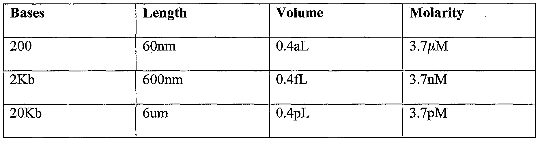
- the nanoscale spheres would allow fluorescence correlation spectroscopy to be performed using a high-resolution light confocal microscope. For tethers longer than 2Kb (0.6 ⁇ M), the formation of first biomolecule/second biomolecule complexes may be directly recorded due to the proportion of fluorescent dot pairs in proportion to those that show some separation.
- the first and/or second tethers may be formed from nucleotides.
- a tether is generated from double stranded DNA (dsDNA).
- a tether may be made from other polymers such as carbon nanotubes (D. H. Jung et al Covalent attachment and hybridization of DNA oligonucleotides on patterned single-walled carbon nanotube films. Langmuir. 2004 Sep 28;20(20):8886-91.), amyloid fibrils, or DNA crossover complexes for example DX hybrids, which include an even number (typically 4, 6, or 8) strands of DNA and are somewhat stiffer than dsDNA; J.
- the 'stiffness' or persistence length (P) and electrostatic charge of tethers such as dsDNA may be modulated by chemical modification, interchelation with molecules such as ethidium bromide or other suitable interchelating agents, which are typically organic compounds to allow insertion between the dsDNA bases or are positively charged and polymeric to complex with DNA based on affinity for the negatively charged phosphate backbone of DNA.
- the stiffness of DNA may also be altered by complexing with DNA binding proteins along the length of the tether.
- the stiffness of other tethers may be modulated by other means.
- the stiffness of tethers comprising DX hybrids may be modulated by varying the number of strands; for tethers comprising carbon nanotubes by increasing the number of concentric tubes forming each nanotube.
- variable-length dsDNA tether portions are ligated together to form a tether.
- a tether body portion may be linked to head and tail tether portions to produce the tether.
- the nucleotides of the body portion may be ligated to head and tail portions in solution.
- chemical cross-linking methods can be used to attach head and tail oligonucleotide linkers to the respective ends of the body portions of the tethers.
- the or each tether portion length may typically be of the order of 50 basepairs (bp) to 50Kb preferably, 200 base pairs to 20 kbase pairs, or 30 to 12 000 nm, preferably 60 to 6000nm, for other tethers.
- the tethers may be tethered to a surface by means of anchors.
- the anchors may be single- stranded amino-modified oligonucleotides.
- the tethers (head, body and tail) can then be hybridised to a solid support to which anchor oligonucleotides have been immobilised.
- the solid support may be a modified glass substrate. Standard techniques may be used to covalently couple an anchor oligonucleotide (for example, see: Chrisey, L.A., Lee, G.U., and O'Ferrall, E. (1996) Covalent attachment of synthetic DNA to self-assembled monolayer films Nucleic Acids Res. 24:3031-3039).
- a preferred method involves coupling amino- modified anchor oligonucleotides to a glass support treated with an agent such as amino silane and /»-phenylenel,4 diisothiocyanate (PDC).
- an agent such as amino silane and /»-phenylenel,4 diisothiocyanate (PDC).
- PDC polyphenylenel,4 diisothiocyanate
- Other substrates that can be modified to bind a tether to a surface are also contemplated, including agarose and sepharose.
- the format of the support is a glass slide onto which oligonucleotide anchors or tethers are printed in arrays of spots using commercially available split pin arraying machines such as those available from Genetix. More specialist solid supports, based on miniaturisation techniques derived from microelectronics, may be used in more sophisticated implementations that are designed to further miniaturise the analysis and to integrate better with readout systems.
- the support may be provided by microbeads that are coupled in formats that generate a unique relationship between a single bead and tether combination.
- Suitable microbeads may include polystyrene, coated ferrous/ferric particles, gold particles, sepharose, agarose, glass or carbon.
- amino-terminal oligonucleotide anchors for the first and second biomolecules may be covalently coupled to the modified glass substrate.
- a range of other approaches can be used to vary the inter-tether distance.
- the distance between the oligonucleotide anchors is increased by the use of a non-specific amino terminal oligonucleotide (which is designed not to bind to other tether components) that is titrated into the specific oligonucleotide mix. The greater the proportion of the non-specific oligonucleotide; the greater the resulting distance between specific oligonucleotide anchor.
- the proportion of modified silane molecules may be reduced prior to oligonucleotide coupling.
- Inter-anchor mean distances may be varied from distances greater than the tether length to the maximal oligonucleotide tether capacity.
- the maximal coupling density possible using published protocols e.g. Chrisey et al 1996 supra
- the maximal coupling density possible using published protocols is 20pmoles of bound DNA/cm 2 which equates to an mean inter-anchor spacing of 1.6nm; Chrisey, L. A., et al (1996) supra.
- This inter-anchor density massively exceeds that required for the most probable range of anchor densities which would normally range from about 5nm to about l ⁇ m.
- the non-specific amino-speciflc oligonucleotide functions to cap the reactive groups and will also make the surface of the support electrostatically negative, thereby minimizing the association of the negatively-charged DNA tether with the surface.
- hydrophobic lipid groups may be coupled to the glass surface to discourage DNA-surface association due to the incompatibility of hydrophobic - hydrophilic associations.
- suitable lipid groups may include phosphatidyl ethanolamine.
- the sequences present in adjacent anchor oligonucleotides are synthesized in series (i.e. as a single oligonucleotide). This effectively generates a common anchor for both the first and second tethers and ensures that the swept volumes entirely overlap. There may be advantages to this approach if the binding of very low numbers (as low as 1 pair) of biomolecules were to be studied. In a variant of this implementation, where the first and second biomolecules are not tethered to a separate solid surface, the first and second biomolecules may be tethered at the respective ends of a single tether and measurements could be made in solution.
- protein nucleic acid conjugates are produced according to the method described in: Jung, G. Y., and Stephanopoulos, G. (2004) A functional protein chip for pathway optimization and in vitro metabolic engineering Science 304, 428-431. This description is in turn based on the original method described in: Roberts, R. W., and Szostak, J. W. (1997) RNA-peptide fusions for the in vitro selection of peptides and proteins Proc. Natl. Acad. Sci. U.S. A 94, 12297-12302.
- the method involves the use of an in vitro translation reaction to covalently attach a nascent peptide by its C-terminus close to the 3' end of an mRNA-DNA conjugate.
- An additional class of methods for connecting protein biomolecules to nucleic acid tethers that can be used with equal preference to the method described above. These methods generically involve the synthesis of a fusion protein comprising the biomolecule of interest attached to a modified enzyme (shown schematically in the accompanying Figure 23; species A fused to species X).
- the protein- nucleic acid complex can be purified away from in vitro translation extract proteins, following annealing to the immobilised tethers and washing.
- the proteins produced in the in vitro translation extracts are in a large excess over that required to saturate a typical lOO ⁇ m diameter microarray spot of tethers ( ⁇ 8pg of a 50KDa protein in a spot containing 30nm inter-anchor distance with 1 x 10 7 molecules).
- Jung and Stephanoupoulos (2004) showed that the density of Oligonucleotide anchors' in their approach was the primary determinant of the levels of immobilized nucleic acid- protein complexes.
- the proportion of tethers used in a method in accordance with the invention will determine the proportions of tethered first and second nucleic acid- protein complexes.
- protein-nucleic acid conjugates may be used, including the direct chemical crosslinking of purified protein biomolecules to modified oligonucleotides.
- protein-nucleic acid complexes may be generated in situ by annealing the mRNA-DNA conjugate to the immobilized tether first and translating the messenger RNA, whilst bound to the tether, by adding in vitro translation extracts to the tethered messenger
- messenger RNA may be designed to generate protein fusions between the protein biomolecules of interest and a second protein domain X.
- the second domain X may be designed to have a very high affinity for an engineered component of the head oligonucleotide or the head end of the tether.
- the X domain is a high affinity specific DNA binding protein (e.g. lambda repressor)
- its cognate DNA site may be introduced into the head oligonucleotide complex to enable the nascent protein to associate with the tether via the DNA binding moiety.
- X could be a molecule such as streptavidin and a corresponding binding partner — in this case biotin - would be chemically coupled to the head oligonucleotide.
- Typical protein biomolecules include enzymes, antibodies and receptors.
- the first and/or second biomolecule may be a bioactive molecule other than a protein.
- the alternative biomolecule is capable of maintaining its functional activity whilst being coupled to a tether.
- Alternative molecules include peptides, peptide analogues, such as synthetic amino acids, combinatorial polymer libraries, small molecules (say ⁇ 1000 Daltons, for example chemically-synthesized drugs), polysaccharides, and catalytically active RNA species.
- nucleic acid protein conjugates are annealed through complimentary sequences, provided by either of the first and second biomolecules, close to the 3' end of the nucleic acid component to complementary sequences in a head oligonucleotide tether portion. This concentrates the nucleic acid conjugates from molarities typical of in vitro translations (e.g. 1OnM) to the experimental concentrations.
- 1OnM in vitro translations
- Simple well-characterised equilibrium binding equations can be used to derive molecular interaction parameters based on the concentrations of the first and second biomolecules and the proportion of first biomolecule/second biomolecule bound. For example, in a typical experiment to accurately determine the Kd of an interaction between first and second biomolecules which are tethered to a support, first and second biomolecules having a range of tether lengths and inter-anchor distances is set up as an array of spots using appropriate combinations of anchors and tethers for the first and second biomolecules. This generates a standard range of concentrations. These concentrations are first plotted against the proportion of bound first/second biomolecule complex and the concentration of the first biomolecule (or the second biomolecule) required for half maximal binding is determined (this concentration is the K d ).
- a method of determining the K d of an interaction between first and second biomolecules by determining the proportion of bound first and second biomolecules for a range of concentrations of the first and second biomolecules and the determining the concentration of the first or second biomolecule required for half maximal binding of the first and second biomolecules - the K d .
- the affinity of a first biomolecule to a library of second biomolecules may be determined.
- the library of biomolecules may comprise at least a significant portion of a transcriptome or proteome.
- Methods in accordance with the invention offer the potential of screening interactions between a single biomolecule A and a library of molecules Bl, B2, B3... B n .
- each spot is occupied by only biomolecule A and Bl or A and B2... A and B n .
- head tether portions recognising unique (for example coding) regions from the 3' end of messages Bl, B2, B3 ... B n are generated and coupled to the core tethers as described earlier.
- B n may be libraries of proteins potentially representing a transcriptome/ proteome.
- B n may be libraries of peptides used for defining interaction sites.
- B n may be tethered libraries of chemical compounds ranging from small molecule compounds to libraries of synthetic polymers.
- an anchor/tether for the second biomolecule that can be cleaved together with initial saturating concentrations of the first and second biomolecules, it is possible to determine K off .
- the rate of decay of first biomolecule/second biomolecule complex levels is monitored in real time following cleavage of the tether for the second biomolecule. This type of analysis is analogous to that used in surface plasmon resonance to determine the K off .
- the effect of a third, tethered or non-tethered biomolecule on the interaction between the tethered first and second biomolecules may also be studied.
- the K off value for an interaction between the first and second biomolecules is determined by providing initial saturating concentrations of the first and second biomolecules, cleaving a second biomolecule tether portion or anchor and monitoring any change in levels of bound first and second biomolecules.
- a method of determining the change in free energy (AG 0 ) value for an interaction between a first and second biomolecules comprising determining the proportion of bound biomolecules at a first temperature and determining the proportion of bound biomolecules at a second temperature and comparing the proportion of bound biomolecules at the respective first and second temperatures.
- the temperature of the biomolecules may be varied by altering the temperature of the experimental apparatus used to perform the method.
- apparatus for determining the affinity of first and second biomolecules comprising a first biomolecule tethered by a first tether having a first tether length, a second biomolecule tethered by a second tether having a second tether length, means for determining binding of adjacent first and second biomolecules to each other, and means for varying at least one of the first, and second tether lengths.
- At least one of the first and second biomolecules may be tethered to a surface of the apparatus.
- both the first and second biomolecules are tethered to a surface.
- the biomolecules may be tethered separately to the surface or together in, for example, a Y- shaped or linear arrangement.
- the biomolecules may be tethered together and 5 associated with the apparatus in the form of a solution.
- the surface may be provided by a solid support.
- the solid support is a glass slide.
- the solid support may be a micro bead.
- Figure 1 is a diagram showing a tethered biomolecule for use in a method of the invention
- Figure 2 is a diagram showing two tethered biomolecules for use in a method of the 20 invention
- Figure 3 A is a diagram showing the biomolecules of Figure 2 binding
- Fig. 3B illustrates the biomolecules of Fig. 3 A binding and illustrates the flexible nature of the tethers
- Figure 4A and B are diagrams showing an array of tethered biomolecules for use in a method of the invention at different inter-tether spacings;
- Figure 5 shows head tether portions for use in tethers in accordance with the invention
- Figure 6 shows a modified oligonucleotide for use in tethers in accordance with the invention
- Figure 7 shows the formation of tethers in accordance with the invention
- Figure 8 shows a further step in the formation of tethers in accordance with the invention.
- Figure 9 shows the production of biomolecule and tether conjugates
- FIG. 10 illustrates a method in accordance with the invention
- Figure 11 illustrates the use of a method in accordance with the invention to measure K 0 ⁇
- Figure 12 is a scheme showing operation of a linear molecule arrangement of biomolecules in accordance with the invention in which:
- A. is an illustration of spheres swept by the free ends of short and long flexible tethers
- B is an illustration of a possible conformation of free and bound variants of a linear molecule representing an intra-molecular interaction between biomolecules A and B;
- C. is an illustration of free and bound variants undergoing inter-molecular interactions between A and B;
- Figure 13 is a diagram showing 'head-set' oligonucleotides used for forming tethers in which:
- A. shows separate molecules of the form shown in Fig.12C;
- FIG. 12B shows a linear molecule with acceptor and donor head sets attached. This molecule takes the form shown in Fig. 12B;
- Figure 14 shows time dependent decay of donor fluorescence due to FRET
- Figure 15 is a graph illustrating an Acceptor Head-Set Titration
- Figure 16 illustrates experimental measures of linear molecule affinity for:
- Figure 17 illustrates a Y-shaped molecule in accordance with the invention
- Figure 18 illustrates a determination of Factor 'C using a method in accordance with the invention as described below;
- Figure 19 illustrates the design of biomolecules formed by oligonucleotides
- Figure 20 is a photograph of a gel analysis of biomolecules having various length tether portions
- Figure 21 shows results of FRET experiments using the oligonucleotides of Figure 19.
- Figure 22 is a graph illustrating the variation of the proportion of bond molecules with the length of DNA tethers.
- Figure 1 shows a single first biomolecule 10 tethered by a first tether 12 through anchor 14 to surface 16.
- the first biomolecule 10 is free to move on the tether 12 about anchor 14 in a substantially hemispherical volume 18.
- the volume of volume 18 is determined by the first tether length 19.
- FIG. 2 shows anchored first and second biomolecules 10 and 20.
- the second biomolecule 20 is tethered by a second tether 22 via an anchor 24 and is also free to move in a substantially hemispherical volume 26.
- the volume of volume 26 is determined by the second tether length 27.
- the hemispherical volumes 18 and 26 overlap to define a reaction zone 28.
- Figure 3 A shows the first and second biomolecules 10 and 20 binding in the reaction zone 28.
- the tethers are flexible and so the biomolecules occupy a volume rather than just a surface.
- Figure 4 shows varying the inter-tether spacing between tethered biomolecules.
- the biomolecules for example 30 and 32, are relatively spaced apart.
- the biomolecules for example 34 and 36, are relatively close together.
- An alternative to the random distribution of first and second biomolecules tethers as illustrated in Fig.4A and B is the targeting of first and second biomolecules to discrete portions on the surface of the substrate such that the first and second biomolecules can only interact if they stretch to span the gap between the surface patches. By controlling the distance between the discrete portions and/or the tether length, the proportion of bound and free biomolecules may be altered allowing the determination of affinity as described herein.
- FIG. 5 to 9 The preparation of one form of tethered biomolecules for use in a method in accordance with the invention is shown in Figures 5 to 9. This involves joining a variable length body tether to three "adaptor" oligonucleotides. The head, body, tail and anchor oligonucleotides are combined as described below to generate an immobilised tether. Arrays of spots containing immobilised tethers are produced with different proportions of first and second tether length tethers. As described later, nucleic acid-protein covalent complexes are then hybridised to the immobilised tethers. a) Production of tether head portions
- Tether body portions are generated from double stranded DNA (dsDNA) as shown particularly in Figure 6.
- a tether body portion 50 has a single-stranded upper portion comprising a restriction enzyme half site X, which is complimentary to the half-site X' of tether head portion 38 or 40.
- the lower region of the body tether portion includes a single stranded section, generally designated as Y in Figure 6.
- Tether tail portions are designed to anneal and ligate to the dsDNA tether body portion and also to anneal to specific anchor oligonucleotides which are described below.
- the tether tail portions 52 and 54 shown in Figure 6 each comprise upper respective and lower sections.
- the upper section, generally designated as Y', is complimentary to the single stranded portion Y of tether body portion 50 .
- the lower sections, generally designated as 1 and 2 are also single-stranded and are designed to anneal to the anchors described below.
- tether production reactions are set up to generate pools of first or second fluorophore or to quantum dot labelled tethers with different tether lengths.
- the tether head portions 38, 40, tether body portions 50 and tether tail portions 52, 54 are assembled by conventional conditions under suitable conditions in solution as shown in Figure 6 to form tethers 55 and 57. Typical conditions may be 5OmM NaCl, HEPES buffer pH7.5 (10mM),and room temperature.
- the assembled tethers 55, 57 can be anchored to a surface by means of anchors.
- the anchors are typically single-stranded amino-modified oligonucleotides.
- the solid support is a modified glass substrate prepared using standard techniques to covalently couple the anchor oligonucleotide. For example, see: Chrisey, L. A., Lee, GJJ., and O'Ferrall, E. (1996) Covalent attachment of synthetic DNA to self-assembled monolayer films Nucleic Acids Res. 24:3031-3039.
- the amino-modified anchor oligonucleotides are coupled to glass treated with amino silane and p-phenylenel,4 diisothiocyanate (PDC) (Fig. 7).
- FLIM Fluorescence Life-time Measurement
- the tethers 55 and 57 are then hybridised to a solid support 60 to which anchor oligonucleotides 56, 58, each having single-stranded sections, generally designated as 1 and 2 respectively, which are complimentary to corresponding sections 1 and 2 of the tether tail portions 52, 54, have been previously immobilised.
- anchor oligonucleotides 56, 58 each having single-stranded sections, generally designated as 1 and 2 respectively, which are complimentary to corresponding sections 1 and 2 of the tether tail portions 52, 54, have been previously immobilised.
- Protein biomolecule/nucleic acid conjugates which can hybidise to the tethers are produced according to the method described in: Jung, G. Y., and Stephanopoulos, G. (2004). supra by an in vitro translation reaction to covalently attach a nascent peptide by its C-terminus close to the 3' end of an mRNA-DNA conjugate. Tether protein complexes are then hybridised to the annealed arrays of tethers attached to their immobilised anchor oligonucleotides.
- FIG. 9 This is schematically illustrated in Figure 9 where a first biomolecule, indicated generally as Protein A, is hybridized to the head portion of tether 55 and a second biomolecule, indicated generally as Protein B is hybridized to the head portion of tether 57.
- a first biomolecule indicated generally as Protein A
- a second biomolecule indicated generally as Protein B
- Alternative methods of making protein biomolecule-nucleic acid conjugates may be used, including the direct chemical crosslinking of purified first or second biomolecules to modified oligonucleotides.
- protein nucleic acid complexes may be generated in situ by annealing the niRNA-DNA conjugate to the immobilized tether first and translating the messenger RNA whilst bound to the tether by adding in vitro translation extracts to the tethered messenger
- the messenger RNA is engineered to generate protein fusions between the protein biomolecule of interest and a second protein domain X.
- the domain X is designed to have a very high affinity for an engineered component of a tether head portion oligonucleotide or the head end of the tether.
- the X domain is a high affinity specific DNA binding protein (e.g. lambda repressor)
- its cognate DNA site is introduced into the head oligonucleotide complex to enable the nascent protein to associate with the tether via the DNA binding moiety.
- X is a molecule such as streptavidin and its binding partner - in this case biotin - is chemically coupled during synthesis to a tether head portion oligonucleotide.
- nucleic acid biomolecule protein conjugates are annealed through complimentary sequences (A or B) close to the 3' end of the nucleic acid component to complementary sequences in the head tether portion as shown in Figure 9.
- tethers need not be made from dsDNA but may be made from other molecules such as DNA DX hybrids.
- a and B are attached at opposite ends of a single flexible tether allowing both molecules to sweep out a shared spherical volume that varies as a cubic function of the tether length.
- the volume swept by A and B reduces and the effective concentration of A and B within the volume rises as a cubic function of the tether length.
- This scheme is formally analogous to the surface anchoring of tether biomolecules described above in that A and B can be regarded as being anchored to a surface that is exactly half the length of the joint tether such that the volumes swept by A and B exactly overlap.
- FLIM Fluorescence Life-time Measurement
- the details of the test system are illustrated in Figure 13.
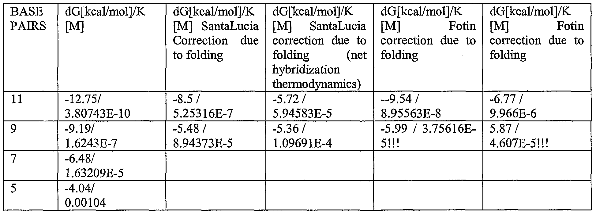
- the biomolecules whose affinity was measured were complementary strands of a DNA hybrid in which two 11 base pair overlaps recognise each other in a reversible reaction.
- the 11 base pair interacting regions are single-stranded DNA extensions of longer double stranded DNA molecules that contain fluorophores A (Acceptor) and D (Donor) incorporated into the bases indicated in bold (Fig.13A).
- the fluorophore used as donor was Alexa Fluor 488 and at the fluorophore used as acceptor was Alexa Fluor 555, both are manufactured by Molecular Probes.
- fluorophores were incorporated during oligonucleotide synthesis and the labelled oligonucleotides were subsequently annealed to form the structures shown in Fig. 6A.
- the fluorophore-tagged double-stranded oligonucleotides are referred to as a donor or acceptor 'head set' to denote the presence of both the annealing 1 lbp affinity region and the presence of the fluorescent dyes.
- the donor and acceptor head set oligonucleotides were ligated to variable length double stranded DNA regions by standard procedures. Briefly, the 'head set' oligonucleotides were cleaved with Bst Xl restriction enzyme and were ligated to variable length 'tether body' DNAs each of which contained a free BstXl and Xbal site. BstXl- BstXl and Xba-Xba ligations were used to generate the molecules as shown in Fig.5B. These were gel purified prior to analysis. The total lengths of the linear molecules incorporating both Donor and Acceptor head groups were: 515bp and 71 Obp.
- Head sets or dual-labelled linear DNA molecules were diluted to the concentrations described in a final concentration of 70 mM NaCl, 10 mM Tris pH 8.0. 6 ⁇ l of each solution was introduced into one of the wells of a 50 well slide produced using a multi-chambered coverslip (Stratech Scientific, UK) together with a 22 x 50 mm coverslip (Menzel-Glaser, Germany). Samples were analysed using a frequency-doubled Ti:Sa laser providing short optical pulses
- fluorescence light was filtered by the spectrometer around the emission maximum of the donor fluorophore (520 ⁇ 5nm) and detected by a single channel fast photomultiplier (200ps time resolution) connected to a time-correlated single photon counting module. Background contributions were measured from the buffer solution without fluorophores in the same excitation and detection conditions and properly subtracted to the data.
- the first and second tether portions for each biomolecule in a Y-shaped molecule are anchored to a single DNA strand such that the tethers are free to diffuse as for the linear molecule shown in Fig. 12B.
- the main advantage of this form of tethering compared with that of the single molecule is that the first and second tether portions are free to interact independent of the length of the intervening tether.
- the linear molecule is unable to fold back on itself at lengths shorter than the persistence length (P) which approximates to between 90 and 120bp.
- % maximal binding we first determined the proportion of bound and unbound donor (R) at different donor and acceptor concentrations using the following procedure. The ratio between the bound and unbound decay spectra for different acceptor concentrations was determined over time and plotted as shown in Fig.14 using free labelled head set oligonucleotides (The three curves shown represent 1. 50 nM acceptor: 5 OnM donor, 2. 200 nM acceptor: 50 nM donor, 3. 600 nM:50nM donor).
- the proportion of bound donor R/(l+R) was plotted against acceptor concentration as shown in Fig.15 (percentage normalised to the maximum effect observed above 4000 nM acceptor concentration).
- Fig.15 the experimental curve of free donor and acceptor head sets was determined for a range of acceptor head set and a single (5OnM) donor head set concentration. This allowed the determination of the binding affinity of the 1 lbp overlap head sets as 136nM. This matches closely to the theoretical determination of 176nM for the same sequence.
- preliminary data from two llbp overlap linear molecules (donor at one end, acceptor at the other; open circles) is displayed on the same scale.
- Figure 14 The rate of decay of the fluorescence signal is increased in the presence of increasing levels of fluorescently-labelled acceptor head sets showing increased decay rate of the donor fluorophore in the presence of the acceptor fluorophore that is a time-dependent characteristic of FRET.
- labelled head sets that did not contain a single-stranded overhang showed no FRET/FLEVI (data not shown), arguing that the decay observed was due to the inter-molecular hybridisation of the two head-sets.
- the characteristic decay curves from FRET-FLIM analyses of the kind shown in Figure 14 were transformed into relative FLIM values according to the method described above and were plotted in relation to the concentration of Acceptor Head-Set oligonucleotide (Fig. 15). Specifically in this figure the percentage maximum FLEVI (y-axis) for the 1 lbp overlap donor head set was plotted against the acceptor head-set concentration. The curve showed a classical saturation response with a half maximal binding (K d ) concentration of Acceptor head set being calculated (FIT) to be 136nM. Linear regression analysis was used to estimate a value of 136nM for the dissociation constant of the l lbp overlap in 7OmM NaCl. This value was very close to the theoretical value of 17OnM that was calculated for the sequence from nearest-neighbour thermodynamic predictions (see above). This indicates that the FRET-FLEVI method was able to accurately determine the proportion of bound fluorophores.
- the actual concentration of each molecule was 5nM and the nominal tethered concentration of each molecule was 778nM and 200OnM as determined by assuming each molecule has a volume whose spherical radius is the length of the tether.
- the measured FRET values were much higher than expected based on the absolute molecular concentration (5nM) and were higher for the shorter molecule (515bp) than for the longer molecule (710bp).
- This data is consistent with the tether enhancing the concentration of the free ends in proportion to the inverse of the length of the tether.
- concentration can be altered by varying the length of the tethers.
- Table 5 The percentage maximal FLIM was plotted against nominal tethered concentration in Fig. 16 showing that FRET/FLIM and therefore binding increased at shorter tether lengths.
- the percentage maximum FLIM for the 1 lbp overlap donor head set was (as in Figure 15) is shown a gain for reference, plotted against the acceptor head-set concentration.
- the data shows that the measured percentage FLIM for each length of tether was comparable to that generated by using free concentrations of the same ligand as shown in Fig. 15, suggesting that the tethers maintain their ends within a volume similar to that generated by a flexible linear molecule.
- the generation of the data shown involved the preparation of fluorophore-labelled linear DNA molecules and the measurement of time resolved FRET.
- the biomolecules whose affinity was measured are shown in Figure 19.
- the key points are an 1 lbp overlap between two pairs of oligonucleotides that constitutes the biological affinity to be measured, together with covalently-coupled fluorophores that are required for the measurement of free and bound molecules using time-resolved FRET.
- These molecules are essentially the same as described in Example 1 above (Fig 13A) which contain the same 1 lbp overlap single-stranded DNA overlap.
- the main difference between those sequences and the sequences of this example is the presence of a BstXl half site to allow ligation onto the Tether Body DNAs (Fig. 19C,D).
- the overlapping oligonucleotide pairs are called 'Head Sets' and they are distinguished by the attached fluorophore.
- the donor fluorophore (Alexafluor 488) and Acceptor (ATTO550) fluorophores were attached to the oligonucleotides during synthesis by commercial suppliers
- the 2 constituent oligonucleotides for the donor or acceptor Head Sets (25 ⁇ M final concentration) were annealed by cooling from 9O 0 C to room temperature over 1 hour in a thermal cycler machine in annealing buffer (7OmM NaCl 1OmM Tris pH 7.4).
- the DNAs that were analysed by FRET were 498bp, 692bp, 1052bp and 1752bp in length following addition of the Head Sets.
- the ligation overlap sequences were designed to be different in sequence and non-palindromic (Acceptor Headset 5'TCAC; Donor Headset 5'CACA). This was achieved by BstXl digestion of the Tether Body DNAs from plasmids that contained two BstXl sites flanking the region Tether Body region of DNA.
- the linear molecules were gel purified and quantified by comparison with known DNA standards.
- FRET analysis the samples were diluted to the concentrations indicated and 5 ⁇ l was added to the wells of a multiwell chambered coverslip (Grace Bio-labs;
- Samples were analysed using a frequency-doubled Ti:Sa laser providing short optical pulses (lOOfs duration) at 76Mhz repetition rate, with wavelength in the absorption band of the donor fluorophore ( ⁇ 470nm).
- the exciting light was weakly focused onto the sample allowing for a uniform illumination and collection over lmm well depth, to maximize the signal contribution over the fluorescence background of the coverslip.
- Low excitation intensities (0.05-1OmW over 0.4mm spot diameter) were maintained to avoid nonlinearities and photodamage.
- Fluorescent light collected from a microscope objective was spectrally analysed using a spectrometer and detected by a cooled CCD camera for time-integrated FRET spectra.
- fluorescence light was filtered by the spectrometer around the emission maximum of the donor fluorophore (520 ⁇ 5nm) and detected by a single channel fast photomultiplier (200ps time resolution) connected to a time-correlated single photon counting module. Background contributions were measured from the buffer solution without fluorophores in the same excitation and detection conditions and properly subtracted to the data.
- the time dependence of FRET can be seen in the Donor and Acceptor dynamics shown in Figure 21.
- the maximal fluorescence intensity of each trace was normalised to 1.
- the proximity of the Donor Fluorophore to the Acceptor Fluorophore resulted in a rapid decay of Donor fluorescence by comparison with unligated Donor Head Set oligonucleotides (Fig. 21 A solid curves).
- a corresponding enhancement of Acceptor fluorophore dynamics was observed by comparison with the unligated Acceptor Head Set.
- no energy transfer was observed in analogous experiments involving the Obp overlap linear molecules (Fig. 21B) 5 indicating that the 1 lbp overlap was required for the changes in fluorescence dynamics.
- the proportion of bound (circular conformation) to total number of molecules [Dbound/Dtot] is proportional to the probability that the molecules are in the circular conformation and was calculated as described above.
- tether length can be used as a direct way of controlling the concentration of biomolecules at the free ends of the tethers and that a high 'concentration' of tethered biomolecules can be obtained via an intra-molecular interaction between a pair of biomolecules at the end of the tether.
- the preferred method for tethering the biomolecules is to attach them to a solid surface
- the connection of two biomolecules via a single flexible tether is essentially a minor practical modification of the linear tether system described since the primary control over tethered biomolecule concentration will be attained by altering the tether length.
- the surface tethered method should also allow fine control of the overlap between swept volumes by altering the inter-anchor distances (see Figures 1 to 4).
- the linear molecule and other implementations of the nano-tether biochemistry approach such as the Y-shaped molecule ( Figure 17) may have distinct advantages in settings in which the detection molecules are introduced into containers containing factors that may alter the affinity of the two biomolecules.
- vesicle preparations containing the linear or Y-shaped molecules could be used to monitor the concentration of a metabolite that alters the affinity of a first biomolecule for a second biomolecule and is free to diffuse into the vesicle.
- Potential containers include test tubes, microwell plates, membrane-bound containers that allow diffusion of metabolites, but retain the linear molecule, cells (e.g. microinjection of molecules), and organisms (e.g. Zebra fish embryos).
- the format of the support is a glass slide onto which oligonucleotides have been printed in arrays of spots using a split pin arraying machine.
- the support is provided by microbeads that are coupled in formats that generate a unique relationship between a single bead and tether combination.
- This format enables the adaptation of the technology to microfluidic systems.
- ammo- terminal oligonucleotide anchors for the first and second biomolecules are covalently coupled to the modified glass substrate.
- a non-specific amino-terminal oligonucleotide (designed not to bind tether components) is titrated into the anchor oligonucleotide coupling mix to vary the inter-anchor coupling distance where appropriate.
- Inter-anchor mean distances are varied from lengths greater than the tether length to the maximal oligonucleotide tether capacity (maximal capacity is 20pmoles of bound DNA/cm 2 which equates to an mean inter-anchor spacing of l. ⁇ nm; Chrisey, L.
- the non-specific oligonucleotide functions to cap the reactive groups and will also make the surface electrostatically negative, thereby minimizing the association of the negatively- charged DNA tether with the surface.
- hydrophobic lipid groups may be coupled to the glass surface to discourage DNA-surface association due to the incompatibility of hydrophobic — hydrophilic associations.
- sequences present in the anchor oligonucleotide are synthesized in series (i.e. 5' sequence 1 — sequence 2-3' as a single oligonucleotide). This will effectively generate a common anchor for both tethers A and B and will ensure that the swept volumes entirely overlap. There may be advantages to this approach if very low numbers (as low as 1 pair of biomolecules) were to be studied as part of further developments of the technique.
- the assay readout is the intensity of Forster resonance energy transfer (FRET) between the different fluorophores 42, 43 coupled to the tether head portions 38 and 40 or elsewhere in the nucleic acid portion of the tether as shown in Figure 10.
- FRET Forster resonance energy transfer
- a laser appropriate to the excitation maximum ( ⁇ l) for fluorophore is used to excite that fluorophore.
- Emission at the wavelength maximum ( ⁇ 2) from the fluorophore 43 is recorded to assess the level of FRET ( Figure 10).
- fluorophore 43 may be selected to quench fluorescence from fluorophore 42 through FRET.
- an excited molecule of one fluorophore has to be molecularly close ( ⁇ 10nm) to another fluorophore for energy to be transferred leading to emission at the characteristic wavelength of the other fluorophore.
- the proportion of first and second biomolecule present within a tethered biomolecule spot is quantified by the intensity of FRET.
- Appropriate controls e.g. spots of the fluorophores 42 and 43 alone) are used to normalise signal levels.
- FRET is measured using a confocal microscope on glass slides containing arrays of tethered biomolecules.
- nanoscale solids such as spheres or "quantum dots" (Doty, R. C. et al Cell MoI. Life Sci. 61 (15) 1843-9), are tethered in place of the single fluorophores. These conjugates may offer higher FRET efficiencies due to the increased number of fluorescent molecules.
- the nanoscale solids would allow fluorescence correlation spectroscopy to be performed using a high-resolution light confocal microscope. For tethers longer than 2Kb (0.6 ⁇ m), the formation of first and second biomolecule complexes can be directly recorded due to the proportion of fluorescent dots pairs in proportion to those that show some separation.
- nano-tether biochemistry technique in all formats (linear, Y-shaped and attached) include:
- a range of tether lengths and inter-anchor distances are set up as an array of spots using appropriate combinations of anchors and tethers for the first and second biomolecules. This generates a standard range of concentrations. These concentrations are plotted against the proportion of bound first and second biomolecule complex and the concentration of the first biomolecule (or the second biomolecule) required for half maximal binding is determined (This concentration is the K d ).
- the method of invention allows the screening of interactions between a single molecule A and a library of molecules Bl, B2, B3... B n .
- each spot is occupied by only A and Bl or A and B2... A and B n .
- head tethers recognising unique (for example coding) regions from the 3' end of messages Bl, B2, B3 ... B n are generated and coupled to the core tethers as described earlier.
- B n can be a library of proteins potentially representing the transcriptome/ proteome.
- B n can be libraries of peptides used to defining interaction sites or tethered libraries of chemical compounds ranging from small molecule compounds to libraries of synthetic polymers.
- C By establishing binding constants between the first and second biomolecules that are close to the K d , it is possible to set up binding reactions at concentrations of the first and second biomolecules that are close to the K d and thus are particularly sensitive to screens for soluble modulators of their interaction.
- modulating molecules will collectively be called "C".
- Examples of C include: a purified interacting protein, a protein that modifies A and B (e.g. a kinase), a drag molecule or candidate, complex mixtures of proteins that contain one or more components that alter AB complex formation (e.g. cell extracts, blood serum, other biological fluids).
- C could be a solution of a single molecule or complex mixtures of compounds (e.g. biological extracts or bodily fluids).
- C may itself be tethered to a third tether or alternatively it may be non-tethered, for example, in solution according to the application. The presence of C can be tested in a number of formats as described below with reference to
- C may be a sequential series of test solutions or fractions from a separation (e.g. Chromatography column eluates in a combinatorial chemistry system, or protein fractions from a cellular extract).
Landscapes
- Health & Medical Sciences (AREA)
- Life Sciences & Earth Sciences (AREA)
- Engineering & Computer Science (AREA)
- Chemical & Material Sciences (AREA)
- Immunology (AREA)
- Biomedical Technology (AREA)
- Molecular Biology (AREA)
- Urology & Nephrology (AREA)
- Hematology (AREA)
- Biotechnology (AREA)
- Physics & Mathematics (AREA)
- General Health & Medical Sciences (AREA)
- Microbiology (AREA)
- Biochemistry (AREA)
- General Physics & Mathematics (AREA)
- Nanotechnology (AREA)
- Pathology (AREA)
- Analytical Chemistry (AREA)
- Medicinal Chemistry (AREA)
- Food Science & Technology (AREA)
- Cell Biology (AREA)
- Genetics & Genomics (AREA)
- Crystallography & Structural Chemistry (AREA)
- Organic Chemistry (AREA)
- General Engineering & Computer Science (AREA)
- Zoology (AREA)
- Wood Science & Technology (AREA)
- Bioinformatics & Cheminformatics (AREA)
- Chemical Kinetics & Catalysis (AREA)
- Biophysics (AREA)
- Plant Pathology (AREA)
- Bioinformatics & Computational Biology (AREA)
- Materials Engineering (AREA)
- Condensed Matter Physics & Semiconductors (AREA)
- Composite Materials (AREA)
- Measuring Or Testing Involving Enzymes Or Micro-Organisms (AREA)
- Apparatus Associated With Microorganisms And Enzymes (AREA)
- Peptides Or Proteins (AREA)
- Investigating Or Analysing Biological Materials (AREA)
Abstract
Description


Claims
Priority Applications (9)
| Application Number | Priority Date | Filing Date | Title |
|---|---|---|---|
| ES06808501.8T ES2509344T3 (en) | 2005-11-16 | 2006-11-10 | Method to measure the affinity of biomolecules |
| AU2006314272A AU2006314272B2 (en) | 2005-11-16 | 2006-11-10 | Method of measuring the affinity of biomolecules |
| CA2629931A CA2629931C (en) | 2005-11-16 | 2006-11-10 | Method |
| NZ568137A NZ568137A (en) | 2005-11-16 | 2006-11-10 | Method of measuring the affinity of biomolecules |
| EP06808501.8A EP1949104B1 (en) | 2005-11-16 | 2006-11-10 | Method of measuring the affinity of biomolecules |
| JP2008540677A JP5133895B2 (en) | 2005-11-16 | 2006-11-10 | Method for measuring affinity of biomolecules |
| US12/094,073 US8778697B2 (en) | 2005-11-16 | 2006-11-10 | Method of measuring the affinity of biomolecules |
| US14/222,112 US20140323328A1 (en) | 2005-11-16 | 2014-03-21 | Method |
| US15/644,078 US20170370927A1 (en) | 2005-11-16 | 2017-07-07 | Method of measuring the affinity of biomolecules |
Applications Claiming Priority (2)
| Application Number | Priority Date | Filing Date | Title |
|---|---|---|---|
| GB0523366.3 | 2005-11-16 | ||
| GBGB0523366.3A GB0523366D0 (en) | 2005-11-16 | 2005-11-16 | Method |
Related Child Applications (2)
| Application Number | Title | Priority Date | Filing Date |
|---|---|---|---|
| US12/094,073 A-371-Of-International US8778697B2 (en) | 2005-11-16 | 2006-11-10 | Method of measuring the affinity of biomolecules |
| US14/222,112 Division US20140323328A1 (en) | 2005-11-16 | 2014-03-21 | Method |
Publications (2)
| Publication Number | Publication Date |
|---|---|
| WO2007057644A2 true WO2007057644A2 (en) | 2007-05-24 |
| WO2007057644A3 WO2007057644A3 (en) | 2007-09-07 |
Family
ID=35580165
Family Applications (1)
| Application Number | Title | Priority Date | Filing Date |
|---|---|---|---|
| PCT/GB2006/004208 Ceased WO2007057644A2 (en) | 2005-11-16 | 2006-11-10 | Method of measuring the affinity of biomolecules |
Country Status (9)
| Country | Link |
|---|---|
| US (3) | US8778697B2 (en) |
| EP (1) | EP1949104B1 (en) |
| JP (1) | JP5133895B2 (en) |
| AU (1) | AU2006314272B2 (en) |
| CA (1) | CA2629931C (en) |
| ES (1) | ES2509344T3 (en) |
| GB (1) | GB0523366D0 (en) |
| NZ (1) | NZ568137A (en) |
| WO (1) | WO2007057644A2 (en) |
Families Citing this family (8)
| Publication number | Priority date | Publication date | Assignee | Title |
|---|---|---|---|---|
| JP2011067174A (en) * | 2009-09-28 | 2011-04-07 | Fujitsu Ltd | Target detector and method for producing the same |
| JP5714023B2 (en) * | 2009-11-30 | 2015-05-07 | ジーイー・ヘルスケア・バイオサイエンス・アクチボラグ | Method and system for analysis of binding behavior |
| US10545090B2 (en) * | 2009-11-30 | 2020-01-28 | Ge Healthcare Bio-Sciences Ab | Method and system for more reliable determination of interaction parameters for low affinity analytes |
| CN102947681B (en) * | 2010-04-20 | 2016-05-18 | 惠普发展公司,有限责任合伙企业 | Strengthen luminous automatic layout, luminous enhance device for surface |
| US9001324B2 (en) | 2010-07-30 | 2015-04-07 | Hewlett-Packard Development Company, L.P. | Optical fiber surface enhanced raman spectroscopy (SERS) probe |
| EP2594942A1 (en) * | 2011-11-16 | 2013-05-22 | Koninklijke Philips Electronics N.V. | Long rigid spacers to enhance binding kinetics in immunoassays |
| WO2016209869A1 (en) * | 2015-06-22 | 2016-12-29 | The Regents Of The University Of California | Quantitative fret-based interaction assay |
| WO2025159970A1 (en) * | 2024-01-22 | 2025-07-31 | Access Medical Systems, Ltd. | Method for determining dissociation constant of two binding molecules |
Citations (1)
| Publication number | Priority date | Publication date | Assignee | Title |
|---|---|---|---|---|
| WO2004038415A1 (en) | 2002-10-24 | 2004-05-06 | Biacore Ab | Assay with co-immobilized ligands |
Family Cites Families (2)
| Publication number | Priority date | Publication date | Assignee | Title |
|---|---|---|---|---|
| US6919178B2 (en) * | 2000-11-21 | 2005-07-19 | Sunesis Pharmaceuticals, Inc. | Extended tethering approach for rapid identification of ligands |
| NZ511705A (en) * | 2001-05-14 | 2004-03-26 | Horticulture & Food Res Inst | Methods and rapid immunoassay device for detecting progesterone and other steroids |
-
2005
- 2005-11-16 GB GBGB0523366.3A patent/GB0523366D0/en not_active Ceased
-
2006
- 2006-11-10 AU AU2006314272A patent/AU2006314272B2/en not_active Ceased
- 2006-11-10 US US12/094,073 patent/US8778697B2/en not_active Expired - Fee Related
- 2006-11-10 ES ES06808501.8T patent/ES2509344T3/en active Active
- 2006-11-10 WO PCT/GB2006/004208 patent/WO2007057644A2/en not_active Ceased
- 2006-11-10 EP EP06808501.8A patent/EP1949104B1/en not_active Not-in-force
- 2006-11-10 JP JP2008540677A patent/JP5133895B2/en not_active Expired - Fee Related
- 2006-11-10 NZ NZ568137A patent/NZ568137A/en not_active IP Right Cessation
- 2006-11-10 CA CA2629931A patent/CA2629931C/en not_active Expired - Fee Related
-
2014
- 2014-03-21 US US14/222,112 patent/US20140323328A1/en not_active Abandoned
-
2017
- 2017-07-07 US US15/644,078 patent/US20170370927A1/en not_active Abandoned
Patent Citations (1)
| Publication number | Priority date | Publication date | Assignee | Title |
|---|---|---|---|---|
| WO2004038415A1 (en) | 2002-10-24 | 2004-05-06 | Biacore Ab | Assay with co-immobilized ligands |
Non-Patent Citations (1)
| Title |
|---|
| GOLDSTEIN ET AL., JOURNAL OF MOLECULAR RECOGNITION, vol. 12, 1999, pages 293 - 299 |
Also Published As
| Publication number | Publication date |
|---|---|
| AU2006314272A1 (en) | 2007-05-24 |
| US8778697B2 (en) | 2014-07-15 |
| GB0523366D0 (en) | 2005-12-28 |
| WO2007057644A3 (en) | 2007-09-07 |
| US20170370927A1 (en) | 2017-12-28 |
| AU2006314272B2 (en) | 2012-11-01 |
| NZ568137A (en) | 2011-12-22 |
| JP5133895B2 (en) | 2013-01-30 |
| JP2009515543A (en) | 2009-04-16 |
| EP1949104B1 (en) | 2014-07-09 |
| US20090023227A1 (en) | 2009-01-22 |
| ES2509344T3 (en) | 2014-10-17 |
| EP1949104A2 (en) | 2008-07-30 |
| CA2629931A1 (en) | 2007-05-24 |
| CA2629931C (en) | 2015-03-24 |
| US20140323328A1 (en) | 2014-10-30 |
Similar Documents
| Publication | Publication Date | Title |
|---|---|---|
| US20170370927A1 (en) | Method of measuring the affinity of biomolecules | |
| Xing et al. | Highly shape-and size-tunable membrane nanopores made with DNA | |
| US20220373543A1 (en) | Surface-immobilized bistable polynucleotide devices for the sensing and quantification of molecular events | |
| CN112236527B (en) | Sandwich assay of colocalization by ligation | |
| EP1915466B1 (en) | Methods for preparing hybrid substrates comprising dna and antibodies and uses thereof | |
| US20100133102A1 (en) | Sensors employing combinatorial artificial receptors | |
| JP2004536290A (en) | Methods for producing stable and reproducible antibody arrays | |
| Morris et al. | Single-molecule kinetic investigation of cocaine-dependent split-aptamer assembly | |
| JP7423641B2 (en) | Surface-immobilized bistable polynucleotide devices for sensing and quantifying molecular events | |
| Christensen et al. | Single vesicle biochips for ultra-miniaturized nanoscale fluidics and single molecule bioscience | |
| US20250066841A1 (en) | Compositions and methods for detecting binding interactions under equilibrium or non-equilibrium conditions | |
| WO2024259350A2 (en) | Making and using structured nucleic acid particles | |
| Gai et al. | Visualizing chemical interactions in life sciences with wide-field fluorescence microscopy towards the single-molecule level | |
| Tang et al. | The fantastic single-molecule techniques | |
| US20250354988A1 (en) | Time-dependent profiling of binding interactions | |
| US20240426839A1 (en) | Compositions and methods for improving affinity reagent avidity | |
| US20250305034A1 (en) | Nanostructures for modulation of analyte conformation | |
| Bognár | Synthetic Receptors and Labels for Chemical Sensing | |
| Yang | DNA nanopore as a signal transducer for the detection of short oligonucleotides | |
| HK40126758A (en) | Colocalization-by-linkage sandwich assays | |
| Preininger | DNA and protein sensor arrays | |
| Lou et al. | Ultrasensitive DNA and protein analysis using molecular beacon probes |
Legal Events
| Date | Code | Title | Description |
|---|---|---|---|
| 121 | Ep: the epo has been informed by wipo that ep was designated in this application | ||
| WWE | Wipo information: entry into national phase |
Ref document number: 2006808501 Country of ref document: EP |
|
| WWE | Wipo information: entry into national phase |
Ref document number: 2006314272 Country of ref document: AU |
|
| WWE | Wipo information: entry into national phase |
Ref document number: 568137 Country of ref document: NZ |
|
| WWE | Wipo information: entry into national phase |
Ref document number: 2629931 Country of ref document: CA |
|
| ENP | Entry into the national phase |
Ref document number: 2008540677 Country of ref document: JP Kind code of ref document: A |
|
| NENP | Non-entry into the national phase |
Ref country code: DE |
|
| ENP | Entry into the national phase |
Ref document number: 2006314272 Country of ref document: AU Date of ref document: 20061110 Kind code of ref document: A |
|
| WWP | Wipo information: published in national office |
Ref document number: 2006314272 Country of ref document: AU |
|
| WWP | Wipo information: published in national office |
Ref document number: 2006808501 Country of ref document: EP |
|
| WWE | Wipo information: entry into national phase |
Ref document number: 12094073 Country of ref document: US |