WO2007024840A2 - Method of quantitating nucleic acids by flow cytometry microparticle-based array - Google Patents
Method of quantitating nucleic acids by flow cytometry microparticle-based array Download PDFInfo
- Publication number
- WO2007024840A2 WO2007024840A2 PCT/US2006/032735 US2006032735W WO2007024840A2 WO 2007024840 A2 WO2007024840 A2 WO 2007024840A2 US 2006032735 W US2006032735 W US 2006032735W WO 2007024840 A2 WO2007024840 A2 WO 2007024840A2
- Authority
- WO
- WIPO (PCT)
- Prior art keywords
- sequence
- seq
- standard
- nucleic acid
- subset
- Prior art date
- Legal status (The legal status is an assumption and is not a legal conclusion. Google has not performed a legal analysis and makes no representation as to the accuracy of the status listed.)
- Ceased
Links
Classifications
-
- C—CHEMISTRY; METALLURGY
- C12—BIOCHEMISTRY; BEER; SPIRITS; WINE; VINEGAR; MICROBIOLOGY; ENZYMOLOGY; MUTATION OR GENETIC ENGINEERING
- C12Q—MEASURING OR TESTING PROCESSES INVOLVING ENZYMES, NUCLEIC ACIDS OR MICROORGANISMS; COMPOSITIONS OR TEST PAPERS THEREFOR; PROCESSES OF PREPARING SUCH COMPOSITIONS; CONDITION-RESPONSIVE CONTROL IN MICROBIOLOGICAL OR ENZYMOLOGICAL PROCESSES
- C12Q1/00—Measuring or testing processes involving enzymes, nucleic acids or microorganisms; Compositions therefor; Processes of preparing such compositions
- C12Q1/68—Measuring or testing processes involving enzymes, nucleic acids or microorganisms; Compositions therefor; Processes of preparing such compositions involving nucleic acids
- C12Q1/6813—Hybridisation assays
- C12Q1/6816—Hybridisation assays characterised by the detection means
-
- C—CHEMISTRY; METALLURGY
- C12—BIOCHEMISTRY; BEER; SPIRITS; WINE; VINEGAR; MICROBIOLOGY; ENZYMOLOGY; MUTATION OR GENETIC ENGINEERING
- C12Q—MEASURING OR TESTING PROCESSES INVOLVING ENZYMES, NUCLEIC ACIDS OR MICROORGANISMS; COMPOSITIONS OR TEST PAPERS THEREFOR; PROCESSES OF PREPARING SUCH COMPOSITIONS; CONDITION-RESPONSIVE CONTROL IN MICROBIOLOGICAL OR ENZYMOLOGICAL PROCESSES
- C12Q1/00—Measuring or testing processes involving enzymes, nucleic acids or microorganisms; Compositions therefor; Processes of preparing such compositions
- C12Q1/68—Measuring or testing processes involving enzymes, nucleic acids or microorganisms; Compositions therefor; Processes of preparing such compositions involving nucleic acids
- C12Q1/6813—Hybridisation assays
- C12Q1/6834—Enzymatic or biochemical coupling of nucleic acids to a solid phase
Definitions
- Detection and analysis of nucleic acids in clinical samples is useful in characterizing diseases, susceptibility for disease and drug responsiveness.
- Southern blots, Northern blots, PCR, RT-PCR and microarrays are some commonly used methods of analyzing clinical and research samples. Using these methods, however, it is difficult to generate information from small amounts of nucleic acids and/or highly homologous sequences.
- Flow cytometry bead-based assays as described, for example, by Fuja et al., Journal of Biotechnology 108: 193-205 (2004), Camilla et al, Clinical and Diagnostic Laboratory Immunology 8(4): 776-784 and U.S. Pat. No.
- 5,981,180 can also be utilized for analyzing nucleic acids and often result in a more sensitive assay allowing detection of low copy number nucleic acids.
- Flow cytometry bead- based assays like microarrays, possess the additional advantage of being amenable to multiplexing, or simultaneously measuring or processing multiple analytes.
- Flow cytometery is a technique that detects analytes based on unique optical characteristics, and can thus distinguish between different bead sets based on several parameters, including size and fluorescence intensity. Oligonucleotides that are complementary to different nucleic acids of interest are coupled to different bead sets, and, after hybridization of a sample of interest with these oligonucleotide-coupled beads, multiple different nucleic acids can be rapidly detected.
- Flow cytometry bead- based assays like Northern blots, RT-PCR and microarrays, provide information about the relative level of nucleic acids but do not provide information regarding the quantity or concentration of nucleic acid in a sample.
- analyzing a sample that contains nucleic acids using almost any method is often a multi-step process (for example, the process may include the steps of preparing cDNA, amplifying the cDNA, hybridizing the cDNA with oligonucleotide-coupled beads and analyzing by flow cytometry) and variability is introduced at each step of the process.
- This variability and the nature of the information obtained using known techniques make it difficult to compare the results of each analysis between assays, between different genes and over time.
- nucleic acids can be quantitated by taking multiple known concentrations of a standard nucleic acid through the same multi-step process as a sample (e.g., a biological sample) to be analyzed, and using the data generated for the multiple known concentrations of standard nucleic acid to determine the quantity of a target nucleic acid in a sample (e.g., a biological sample).
- this multi-step process includes the steps of generating cDNA, amplifying cDNA, hybridizing the cDNA with oligonucleotide-coupled microparticles and analyzing by flow cytometry.
- the present invention is a method of measuring a nucleic acid species in a sample (e.g., a biological sample), a method of measuring a target RNA species in a sample (e.g., a biological sample), a method of screening for the presence of a genetic polymorphism or mutation, a method of monitoring a response to an agent, a method of predicting a patient's responsiveness to an agent and a method of screening for the presence of a pathogenic organism in a sample (e.g., a biological sample).
- a sample e.g., a biological sample
- a method of measuring a target RNA species in a sample e.g., a biological sample
- a method of screening for the presence of a genetic polymorphism or mutation e.g., a method of screening for the presence of a genetic polymorphism or mutation
- a method of monitoring a response to an agent e.g., a method of predicting a patient's responsiveness to an
- the invention is a method of measuring a target nucleic acid species in a sample (e.g., a biological sample) comprising: a) amplifying multiple known concentrations of a standard nucleic acid to prepare amplified standard nucleic acid samples, wherein the standard nucleic acid comprises the same sequence as the target nucleic acid or a fragment thereof; b) amplifying the target nucleic acid; c) hybridizing the amplified target nucleic acid with a population comprising a subset of microparticles to form a mixture, wherein the subset is distinguishable from other subsets, if any, in said population based on a detectable parameter and wherein the subset of microparticles is coupled to an oligonucleotide that is complementary to a portion of the target nucleic acid and standard nucleic acids; d) hybridizing each of the amplified standard nucleic acid samples with a population comprising said subset of microparticles to form mixtures; e) analyzing
- the invention is a method of measuring a target RNA species in a sample (e.g., a biological sample) comprising: a) preparing standard cDNA from a known concentration of standard RNA by preparing multiple known concentrations of the standard RNA, wherein the standard RNA comprises the same sequence as the target RNA or a fragment thereof, and generating standard cDNA samples for each concentration of standard RNA; b) generating target cDNA from the biological sample; c) amplifying the target cDNA and the standard cDNA samples to prepare an amplified target cDNA sample and amplified standard cDNA samples; d) hybridizing the amplified target cDNA sample with a population comprising a subset of microparticles to form a mixture, wherein the subset is distinguishable from other subsets, if any, in said population based on a detectable parameter, and wherein the subset of microparticles is coupled to an oligonucleotide that is complementary to a portion of said target RNA
- the invention is a method of screening for the presence of one or more polymorphisms or mutations in a nucleic acid in a sample (e.g., a biological sample).
- the method comprises analyzing the sample for the polymorphisms or mutations utilizing the steps described above for measuring a target nucleic acid species in a sample, and further comprises preparing multiple known concentrations of a standard nucleic acid sample for each polymorphism or mutation being screened.
- the method further comprises including a subset of microparticles, each independently coupled to an oligonucleotide that is complementary to a portion of a nucleotide sequence that corresponds to a particular polymorphism or mutation being screened.
- the invention is a method of screening for the presence of a polymorphism or mutation in a nucleic acid from a sample (e.g., a biological sample) comprising: a) for each of said polymorphisms or mutations being screened, preparing cDNA from known concentrations of standard RNA comprising the steps of:
- the invention is a method of monitoring a change in gene expression in response to an agent comprising obtaining a sample (e.g., a biological sample) after administration of the agent and determining the quantity of target nucleic acid in the sample comprising the steps described above for measuring a target nucleic acid species in a sample.
- the method comprises obtaining a sample (e.g., a biological sample) after administration of the agent and determining the quantity of a target RNA in the sample comprising: a) preparing cDNA from known concentrations of a standard RNA comprising the steps of:
- the method pertains to screening for the presence of a pathogenic organism in a sample (e.g., a biological sample) comprising analyzing the sample for the presence of a nucleic acid from the pathogenic organism, comprising the steps described above for measuring a target nucleic acid species.
- the invention is a method for screening for the presence of a pathogenic organism in a sample (e.g., a biological sample) comprising analyzing the sample for the presence of target RNA from said pathogenic organism comprising: a) preparing cDNA from known concentrations of a standard RNA comprising the steps of:
- RNA comprises the same sequence as the target RNA or a fragment thereof ;
- FIG. IA is diagram showing a method for preparing standard RNA for generation of a standard curve.
- FIG. IB is a diagram showing additional steps in the generation of a standard curve.
- FIG. 2A shows the portion of mouse beta actin (GenBank Ace. No. NM_007393) (SEQ ID NO: 1) that was cloned from the BlO cell line (McGiIl University, Canada). The positions of primers and/or oligonucleotides are underlined and labeled
- FIG. 2B shows the portion of mouse al ⁇ ha-7 (GenBank. Ace. No. AF225980) (SEQ ID NO: 2) that was cloned for preparation of the standard curve.
- the positions of primers and/or oligonucleotides are underlined and labeled.
- FIG. 2C shows the portion of human beta-actin (GenBank Ace. No. BC014861) (SEQ ID NO:3) that was cloned for preparation of the standard curve.
- the positions of primers and/or oligonucleotides are underlined and labeled.
- FIG. 2D shows a portion of human GAPDH (GenBank Ace. No. BT006893) (SEQ ID NO: 4) that was cloned for preparation of the standard curve.
- the positions of primers and/or oligonucleotides are underlined and labeled.
- FIG. 2E shows the sequence of human alpha-7 (GenBank Ace. No. NM_000746) (SEQ ID NO:5).
- the positions of primers and/or oligonucleotides referred to in Example 3 are underlined and labeled.
- FIG. 2F shows the sequence of human alpha-7 dup (GenBank Ace. No. NM_139230) (SEQ ID NO: 6). The positions of primers and/or oligonucleotides referred to in Example 3 are underlined and labeled.
- FIG. 3 is a graph of raw intensity units versus concentration (fg) for standard beta actin samples (known concentrations of beta actin) analyzed using the BioRad Bioplex System. The graph or standard curve was generated using Bio-Plex Manager 3.0 software with a logistic-5pl regression type and a log(x)-linear(y) axis transformation.
- FIG. 4 is a graph of raw intensity units versus concentration (fg) for standard alpha-7 samples prepared using the primers MuRta7s2 (SEQ ID NO: 15) and MuRta7asl (SEQ ID NO: 16) primers. The graph or standard curve was generated using Bio-Plex Manager 3.0 software with a linear (semi-log) regression type and a log(x)-linear(y) axis transformation.
- FIG. 5 is a graph of raw intensity units versus concentration (fg) for standard alpha-7 samples prepared using the primers MuRta7s2 (SEQ ID NO: 17) and MuRta7as2 (SEQ ID NO: 18).
- the graph or standard curve was generated using Bio- Plex Manager 3.0 software with a logistic- 5pl regression type and a log(x)-linear(y) axis transformation
- FIG. 6 is a bar graph showing the concentrations (fg) of beta actin RNA in different cell lines determined using the standard curve of FIG. 2.
- FIG. 7 is a bar graph showing the concentrations (fg) of alpha-7 RNA in different cell lines determined using the standard curves of FIGs. 3 and 4.
- FIG. 8 is a bar graph of raw intensity units versus different concentrations of beta actin generated using a multiplexed PCR reaction and separate PCR reactions.
- FIG. 1OA is a bar graph of raw intensity units versus alpha-7 from brain samples and negative controls amplified with different primers. Beginning on the left side of the graph, the first column corresponds to the results of three human brain samples which were amplified with primers Hua7Lxs2 (SEQ ID NO:27) and Hua7Lxasl (SEQ ID NO:28). The second column corresponds to four brain samples amplified with Hua7Lxs2 (SEQ ID NO:29) and Hua7Lxas2 (SEQ ID NO:30). The third column corresponds to three negative controls amplified with Hua7Lxs2 and Hua7Lxasl. The fourth column corresponds to four negative controls amplified with Hua7Lxs2 and Hua7Lxas2. Reactions were hybridized to Hua7LxPrl (SEQ ID NO:31).
- FIG. 1OB is a bar graph of raw intensity units versus alpha-7 dup from human peripheral blood mononuclear cells (PBMCs). Beginning on the left side of the graph, the first column corresponds to the results of three human PBMC samples which were amplified with primers Hua7dLxsl (SEQ ID NO: 32) and Hua7dLxasl (SEQ ID NO:33). The second column corresponds to three PBMC samples amplified with Hua7dLxs2 (SEQ ID NO:34) and Hua7dLxasl (SEQ ID NO:35). The third column corresponds to three negative controls amplified with Hua7dLxsl and Hua7dLxasl. The last column corresponds to three negative controls amplified with Hua7dLxs2 and Hua7dLxasl.
- PBMCs peripheral blood mononuclear cells
- FIG. 1 IA shows the portion of transforming growth factor beta-1 (TGFbI) (GenBank Ace. No. XO2812) (SEQ ID NO: 106) that was cloned for preparation of the standard curve.
- TGFbI transforming growth factor beta-1
- SEQ ID NO: 106 The positions of primers and/or oligonucleotides are underlined and labeled.
- FIG. 1 IB shows the portion of human RANTES (GenBank Ace. No. M21121) (SEQ ID NO: 107) that was cloned for preparation of the standard curve. The positions of primers and/or oligonucleotides are underlined and labeled.
- FIG. 11C shows the portion of human intracellular adhesion molecule- 1 (ICAMl) (GenBank Ace. No. BC015969.2) (SEQ ID NO: 108) that was cloned for preparation of the standard curve.
- ICMl human intracellular adhesion molecule- 1
- SEQ ID NO: 108 The positions of primers and/or oligonucleotides are underlined and labeled.
- FIG. 1 ID shows the portion of human vascular cell adhesion molecule- 1 (VCAM-I) (GenBank Ace. No. NM_001078) (SEQ ID NO: 109) that was cloned for preparation of the standard curve.
- VCAM-I human vascular cell adhesion molecule- 1
- SEQ ID NO: 109 The positions of primers and/or oligonucleotides are underlined and labeled.
- FIG. 1 IE shows the portion of mouse platelet-derived growth factor-inducible gene (MCP-I) (GenBank Ace. No. J04467) (SEQ ID NO: 110) that was cloned for preparation of the standard curve.
- MCP-I mouse platelet-derived growth factor-inducible gene
- SEQ ID NO: 110 mouse platelet-derived growth factor-inducible gene
- FIG. 1 IF shows the portion of mouse interleukin-4 (IL-4) (GenBank Ace. No. NM_021283) (SEQ ID NO: 111) that was cloned for preparation of the standard curve. The positions of primers and/or oligonucleotides are underlined and labeled.
- FIG. 1 IG shows the portion of human GAPDH (GenBank Ace. No.BC096440) (SEQ E) NO: 112) that was cloned for preparation of the standard curve. The positions of primers and/or oligonucleotides are underlined and labeled.
- FIG. 1 IH shows the portion of mouse interleukin-10 (IL-10) (GenBank Ace. No. NM_001548) (SEQ ED NO: 113) that was cloned for preparation of the standard curve.
- the positions of primers and/or oligonucleotides are underlined and labeled.
- FIG. 1 II shows the portion of human interleukin-8 (IL-8) (GenBank Ace. No. NM_000584.2) (SEQ ID NO: 114) that was cloned for preparation of the standard curve.
- the positions of primers and/or oligonucleotides are underlined and labeled.
- FIG. 1 IJ shows the portion of human interleukin-6 (IL-6) (GenBank Ace. No. NM_00600.1) (SEQ DD NO: 115) that was cloned for preparation of the standard curve.
- the positions of primers and/or oligonucleotides are underlined and labeled.
- FIG. 1 IK shows the portion of human platelet/endothelial cell adhesion molecule 1 (PECAM-I) (GenBank Ace. No. BC051822) (SEQ ID NO: 116) that was cloned for preparation of the standard curve. The positions of primers and/or oligonucleotides are underlined and labeled.
- PECAM-I human platelet/endothelial cell adhesion molecule 1
- FIG. 1 IL shows the portion of mouse heme oxygenase 1 (Hmoxl) (GenBank Ace. No. NM_010442) (SEQ DD NO: 117) that was cloned for preparation of the standard curve.
- the positions of primers and/or oligonucleotides are underlined and labeled.
- FIG. 1 IN shows the portion of human S0CS3 (GenBank Ace. No. NM_003955) (SEQ DD NO: 119) that was cloned for preparation of the standard curve.
- the positions of primers and/or oligonucleotides are underlined and labeled.
- FIG. 1 IO shows the portion of human Hmoxl (GenBank Ace. No. NM_002133.1) (SEQ DD NO: 120) that was cloned for preparation of the standard curve. The positions of primers and/or oligonucleotides are underlined and labeled.
- FIG. 1 IP shows the portion of human GAPDH (GenBank Ace. No.BC083511.1) (SEQ ID NO: 121) that was cloned for preparation of the standard curve. The positions of primers and/or oligonucleotides are underlined and labeled.
- FIG. HQ shows the portion of human B-cell CLL/lymphoma 2 (Bcl-2 (GenBank Ace. No. NM_000633.2) (SEQ ID NO: 122) that was cloned for preparation of the standard curve.
- Bcl-2 GenBank Ace. No. NM_000633.2
- SEQ ID NO: 122 The positions of primers and/or oligonucleotides are underlined and labeled.
- FIG. 1 IS shows the portion of mouse ICAM-I (GenBank Ace. No. NM_010493) (SEQ ID NO: 124) that was cloned for preparation of the standard curve. The positions of primers and/or oligonucleotides are underlined and labeled.
- FIG. 1 IU shows the portion of mouse BAX (GenBank Ace. No. NM_007527) (SEQ ID NO: 126) that was cloned for preparation of the standard curve. The positions of primers and/or oligonucleotides are underlined and labeled.
- FIG. 1 IV shows the portion of mouse interleukin-6 (IL-6) (GenBank Ace. No. J03783) (SEQ ID NO: 127) that was cloned for preparation of the standard curve.
- the positions of primers and/or oligonucleotides are underlined and labeled.
- FIG. 1 IW shows the portion of human tumor necrosis factor alpha (TNF- ⁇ ) (GenBank Ace. No. M10988) (SEQ ID NO: 128) that was cloned for preparation of the ' standard curve.
- the positions of primers and/or oligonucleotides are underlined and labeled.
- FIG. HX shows the portion of human MP Ib (GenBank Ace. No. AY766446) (SEQ ID NO: 129) that was cloned for preparation of the standard curve. The positions of primers and/or oligonucleotides are underlined and labeled.
- FIG. HY shows the portion of mouse TNF- ⁇ (GenBank Ace. No. M13049) (SEQ ID NO: 130) that was cloned for preparation of the standard curve. The positions of primers and/or oligonucleotides are underlined and labeled.
- FIG. 12 is a bar graph of raw intensity units versus different concentrations of human TGFb (fg) generated using the primers HuTGFbs4 (SEQ ID NO:41) and HuTGFbas4 (SEQ ID NO:42) and detected using the probe HuTGFb ⁇ r3 (SEQ ID NO:43).
- FIG. 13 is a bar graph of raw intensity units versus different concentrations of human RANTES (fg) generated using the primers HuRANTESs2 (SEQ ID NO:44) and HuRANTESas2 (SEQ ID NO:45) and detected using the probe HuRANTESprl (SEQ ID NO:46).
- FIG. 14 is a bar graph of raw intensity units versus different concentrations of human ICAMl (fg) generated using the primers HuICAMls3 (SEQ ID NO: 47) and HuICAMl as2 (SEQ ID NO:48) and detected using the probe HuICAMlpr2 (SEQ ID NO:49).
- FIG. 15 is a bar graph of raw intensity units versus different concentrations of human VCAMl (fg) generated using the primers HuVCAMl s3 (SEQ ID NO:50) and HuVCAMas4 (SEQ ID NO:51) and detected using the probe HuVCAMlpr3 (SEQ ID NO:52).
- FIG. 16 is a bar graph of raw intensity units versus different concentrations of mouse MCPl (fg) generated using the primers MuMCPls4 (SEQ ID NO: 53) and MuMCPlas4 (SEQ ID NO:54) and detected using the probe MuMCPl ⁇ r2 (SEQ ID NO:55).
- FIG. 17 is a bar graph of raw intensity units versus different concentrations of mouse IL4 (fg) generated using the primers MuIL4s2 (SEQ ID NO:56) and MuIL4asl (SEQ ID NO:57)and detected using the probe MuIL4prl (SEQ ID NO:58).
- FIG. 18 is a bar graph of raw intensity units versus different concentrations of mouse GAPDH (fg) generated using the primers MuGAPDHs ⁇ n (SEQ ID NO:59) and MuGAPDHas5n (SEQ ID NO:60) and detected using the probe MuGAPDHpr4n (SEQ ID NO:61).
- FIG. 19 is a bar graph of raw intensity units versus different concentrations of mouse ILlO (fg) generated using the primers MuIL10s4 (SEQ ID NO:62) and MuILl 0as3 (SEQ ID NO:63) and detected using the probe MuILl 0pr2 (SEQ TD NO:64).
- FIG. 20 is a bar graph of raw intensity units versus different concentrations of human IL8 (fg) generated using the primers HuIL8s2 (SEQ ID NO:65) and HuIL8asl (SEQ ID NO:66) and detected using the probe HuIL8pr2 (SEQ ID NO:67).
- FIG. 21 is a bar graph of raw intensity units versus different concentrations of human IL6 (fg) generated using the primers HuIL6s3 (SEQ ID NO:68) and huIL6as2 (SEQ ID NO:69) and detected using the probe huIL6 ⁇ r2 (SEQ ID NO:70).
- FIG. 22 is a bar graph of raw intensity units versus different concentrations of human PECAM (fg) generated using the primers HuPECAMl s3 (SEQ ID NO:71) and HuPECAMlas2 (SEQ ID NO:72) and detected using the probe HuPECAMlpr2 (SEQ ID NO:73).
- FIG. 23 is a bar graph of raw intensity units versus different concentrations of mouse Hmoxl (fg) generated using the primers MuHmoxls2 (SEQ ID NO:74) and MuHmoxasl (SEQ ID NO:75) and detected using the probe MuHmoxlpr2 (SEQ ID NO:76).
- FIG. 25 is a bar graph of raw intensity units versus different concentrations of human SOCS3 (fg) generated using the primers HuSOCS3s3 (SEQ ID NO:80) and HuSOCS3as2 (SEQ ID NO:81) and detected using the probe HuSOCSpr2 (SEQ ID NO:82).
- FIG. 26 is a bar graph of raw intensity units versus different concentrations of human Hmoxl (fg) generated using the primers HuHmoxls3 (SEQ ID NO:83) and HuHmoxlas ⁇ (SEQ ID NO: 84) and detected using the probe HuHmoxl ⁇ r3 (SEQ ID NO:85).
- FIG. 27 is a bar graph of raw intensity units versus different concentrations of human GAPDH (fg) generated using the primers HuGAPDHs3n (SEQ ID NO:86) and HuGAPDHas3n (SEQ ID NO: 87) and detected using the probe HuGAPDHpr2n (SEQ ID NO:88).
- FIG. 28 is a bar graph of raw intensity units versus different concentrations of human Bcl-2 (fg) generated using the primers HuBcl2s3 (SEQ ID NO: 103) and HuBcl2as2 (SEQ ID NO: 104) and detected using the probe HuBcl2pr2 (SEQ ID NO: 105).
- FIG. 29 is a bar graph of raw intensity units versus different concentrations of mouse Bcl-2 (fg) generated using the primers Mubcl2s3 (SEQ ID NO: 89) and Mubcl2as2 (SEQ ID NO:90) and detected using the probe Mubcl2pr2 (SEQ ID NO:91).
- FIG. 30 is a bar graph of raw intensity units versus different concentrations of mouse ICAMl (fg) generated using the primers MuICAMl s3 (SEQ ID NO: 107) and MuICAMl as2 (SEQ ID NO: 108) and detected using the probe MuICAMl ⁇ r2 (SEQ ID NO: 109).
- FIG. 31 is a bar graph of raw intensity units versus different concentrations of human BAX (fg) generated using the primers HuBAXs2 (SEQ ID NO: 110) and HuBAXas3 (SEQ ID NO: 111) and detected using the probe HuBAXpr2 (SEQ ID NO: 112).
- FIG. 32 is a bar graph of raw intensity units versus different concentrations of mouse BAX (fg) generated using the primers MuBAXs3 (SEQ ID NO: 92) and MuBAXas3 (SEQ ID NO:93) and detected using the probe MuBAXpr2 (SEQ ID NO:94).
- FIG. 34 is a bar graph of raw intensity units versus different concentrations of human TNF- ⁇ (fg) generated using the primers HuTNFs3 (SEQ ID NO:95) and HuTNFas3 (SEQ ID NO:96) and detected using the probe HuTNF ⁇ r2E (SEQ ID NO:97).
- FIG. 35 is a bar graph of raw intensity units versus different concentrations of mouse MIPlB (fg) generated using the primers MuMDPlbs2 (SEQ ID NO:98) and MuMIPlas3 (SEQ ID NO:99) and detected using the probe MuMIPlbpr2 (SEQ ID NO: 100).
- FIG. 36 is a bar graph of raw intensity units versus different concentrations of mouse TNF- ⁇ (fg) generated using the primers MuTNFs3 (SEQ ID NO: 101) and MuTNFas3 (SEQ ID NO: 102) and detected using the probe MuTNF ⁇ r2E (SEQ ID NO:103).
- the present invention is a method of measuring a target nucleic acid species in a biological sample.
- the invention comprises measuring a target RNA species in a biological sample, hi another embodiment, the invention comprises measuring a target DNA species in a biological sample.
- the methods described herein utilize flow cytometry for the analysis of the nucleic acids. Flow cytometric technology has been described extensively in the literature (for example, in U.S. Patent Nos. 5,736,330, 5,981,180, 6,499,562 and 6,649,414, the teachings of which are incorporated by reference herein in their entirety). Any flow cytometer that can analyze the samples (e.g., biological samples) and/or the standard and target nucleic acids can be utilized.
- Such flow cytometry technology includes, for example, Coulter Elite-ESP flow cytometer (available from Beckman-Coulter, Inc., Fullerton, California), EPCS-XL MCL flow cytometer (available from Beckman Coulter, Inc.), FACScan flow cytometer (available from Beckman Coulter), MOFLO flow cytometer (available from Cytomation, Inc., Fort Collins, Colorado), Luminex 100 xMAP (available from Luminex Corp., Austin, Texas), Luminex xMAP® technology (available from Luminex Corp.).
- the nucleic acids are analyzed using Luminex xMAP® technology.
- nucleic acid includes deoxyribonucleotides, ribonucleotides and polymers thereof in single- or double-stranded form, including, for example, DNA (e.g., genomic DNA, complementary DNA (cDNA), chromosomal DNA, plasmid DNA), RNA (e.g., mRNA, tRNA, rRNA, snRNA, snoRNA, microRNA) and DNA-RNA hybrids.
- DNA e.g., genomic DNA, complementary DNA (cDNA), chromosomal DNA, plasmid DNA
- RNA e.g., mRNA, tRNA, rRNA, snRNA, snoRNA, microRNA
- DNA-RNA hybrids e.g., mRNA, tRNA, rRNA, snRNA, snoRNA, microRNA
- nucleic acid can be used interchangeably with nucleotide, oligonucleotide and polynucleotide.
- nucleotide encompasses not only nucleotides (ribonucleotides and deoxyribonucleotides), but also encompasses related molecules including nucleosides (nucleotides lacking a 5'- phosphate) and phosphodiesters (nucleotides lacking a nitrogen-containing heterocyclic organic base).
- nucleosides nucleotides lacking a 5'- phosphate
- phosphodiesters nucleotides lacking a nitrogen-containing heterocyclic organic base.
- the terms also encompass chains of nucleosides which are linked by analogs of the phosphate linkages, e.g., phosphorotliioate, phosphoramidate, alkylphosphonate, alkylphosphonothioate, and the like, or combinations thereof.
- a "target nucleic acid species” is a nucleic acid that is being measured or detected using a method of the invention.
- the target nucleic acid species is a target DNA or a target RNA species.
- a "target RNA” or “target RNA species” is an RNA species that is being measured or detected using a method of the invention.
- a “standard nucleic acid” is a nucleic acid of a known concentration that comprises the same sequence as the target nucleic acid or a fragment thereof.
- sequence can include deoxyribonucleotide and/or ribonucleotide sequences.
- the "standard nucleic acid” consists essentially of the same sequence as the target nucleic acid or a fragment thereof.
- the standard nucleic acid consists of the same sequence as the target nucleic acid or a fragment thereof.
- the standard nucleic acid comprises a sequence that overlaps the target nucleic acid sequence.
- the standard nucleic acid comprises a sequence which has at least 80%, at least 85%, at least 90%, at least 95% or at least 99% sequence identity to the sequence of the target nucleic acid.
- a "standard RNA" is an RNA of a known concentration that comprises the same sequence as the target nucleic acid or a fragment thereof.
- the standard RNA comprises a sequence that overlaps the sequence of the target RNA.
- the standard RNA consists essentially of the same sequence as the target nucleic acid or a fragment thereof.
- the standard RNA consists of the same sequence as the target nucleic acid or a fragment thereof.
- the standard RNA comprises a sequence that has at least 80%, at least 85%, at least 90%, at least 95% or at least 99% sequence identity to the target RNA.
- a “standard cDNA” is a cDNA prepared from a standard RNA.
- a “target cDNA” is a cDNA prepared from a target RNA.
- the target nucleic acid is a sequence or portion of a sequence from a nucleic acid (e.g., a gene) that is associated with a disease, a sequence or portion of a sequence from a nucleic acid (e.g., a gene) whose expression is altered in diseased, as compared to normal, tissue, a sequence or portion of a sequence from a nucleic acid (e.g., a gene) whose expression is altered in cancerous tissue or in a tumor, a sequence or portion of a sequence from a nucleic acid (e.g., a gene) from a metabolic pathway, a sequence or portion of a sequence from a nucleic acid (e.g., a gene) of a pathogenic organism and/or a sequence or portion of a sequence from a nucleic acid
- two or more target nucleic acids are quantitated.
- standard nucleic acids corresponding to each of the target nucleic acids are utilized, wherein the standard nucleic acids each independently comprise the same sequence as each test nucleic acid or a fragment thereof.
- two or more target nucleic acids are quantitated, wherein at least two nucleic acids have substantial identity to one another.
- two nucleic acids (or regions of the nucleic acids) are substantially homologous or identical when the amino acid sequences are at least about 60%, 70%, 75%, 80%, 85%, 90% or 95% or more, homologous or identical.
- the actual comparison of the two sequences can be accomplished by well-known methods, for example, using a mathematical algorithm. A preferred, non-limiting example of such a mathematical algorithm is described in Karlin et ah, Proc. Natl. Acad. Sci. USA, 90:5873-5877 (1993).
- Such an algorithm is incorporated into the BLASTN and BLASTX programs (version 2.2) as described in Schaffer et al, Nucleic Acids Res., 29:2994-3005 (2001).
- BLASTN BLAST and Gapped BLAST programs
- the default parameters of the respective programs ⁇ e.g., BLASTN
- the database searched is a non-redundant (NR) database, and parameters for sequence comparison can be set at: no filters; Expect value of 10; Word Size of 3; the Matrix is BLOSUM62; and Gap Costs have an Existence of 11 and an Extension of 1.
- the percent identity between two amino acid sequences can be accomplished using the GAP program in the GCG software package (available at www.accelrys.com, as available on August 31, 2001) using either a Blossom 63 matrix or a PAM250 matrix, and a gap weight of 12, 10, 8, 6, or 4 and a length weight of 2, 3, or 4.
- the percent identity between two nucleic acid sequences can be accomplished using the GAP program in the GCG software package (available at www.cgc.com), using a gap weight of 50 and a length weight of 3.
- multiple known concentrations of a standard nucleic acid or “multiple known concentrations” of a standard RNA are prepared.
- Multiple known concentrations of a standard nucleic acid or RNA can be two or more concentrations of the standard nucleic acid or RNA.
- the multiple known concentrations can be prepared by dilution or serial dilution of a known concentration of the standard nucleic acid.
- each of the standard known concentrations of standard nucleic acid is added to samples comprising a heterogenous RNA population before amplification or before cDNA preparation in the case of standard RNA samples.
- a heterogenous nucleic acid population is any mixture comprising multiple different nucleic acids, but not including the target and/or standard nucleic acid.
- Examples of a heterogenous nucleic acid samples are total DNA or RNA samples from any cell line or tissue from any species (e.g., E. coli, human, Drosophila and Chinese hamster ovary cells).
- the step of adding standard nucleic acid samples to a heterogenous nucleic acid sample, followed by preparation of cDNA and/or amplification, can be employed in order to approximate the similar process for the target nucleic acid sample, which is amplified and/or reverse transcribed from a complex mixture of multiple different nucleic acids in the sample (e.g., biological sample).
- nucleic acid e.g., a nucleic acid concentration
- concentration e.g., a nucleic acid concentration
- level e.g., a quantity of nucleic acid
- the nucleic acids used in the present method can be amplified using any method now known or later discovered that results in an amplification or an exponential increase in the number of nucleic acid molecules. Such methods include amplification by polymerase chain reaction ("PCR"), using a vector that can transform a cell (e.g., a bacterial cell (e.g., E. coli)) which may then be grown to multiply the nucleic acid molecule(s), transcription-based amplification methods (described, for example, in U.S. Patent Nos.
- PCR polymerase chain reaction
- the target and standard nucleic acids are amplified by PCR.
- the target nucleic acid to be quantitated is a RNA species and the cDNA is prepared from the RNA and then amplified.
- cDNA may be prepared by reverse transcription or any other method by which cDNA can be prepared. Methods for the preparation of cDNA by reverse transcription are well-known in the art.
- the target nucleic acid is a DNA species.
- microparticle is a particle which can be detected and analyzed by flow cytometry.
- microparticle encompasses microspheres, beads, microbeads and other particles that are detectable by flow cytometry.
- the microparticle is labeled with one or more colored or fluorescent dyes.
- Micropartieles that may be used in flow cytometry, including those labeled with a colored or fluorescent dye, are known in the art and are commercially available.
- a microparticle labeled with a colored or fluorescent dye can be prepared by several methods including, but not limited to, methods described in U.S. Patent Nos. 4,267,234, 4,552,812, 5,194,300, 5,073,498, 5,981,180 and 6,599,331, the teachings of which are incorporated by reference herein in their entirety.
- Each subset of micropartieles in a population is distinguishable from other subsets, if any others are present, based on one or more detectable parameters.
- the detectable parameter is fluorescence intensity, size and/or shape of the microparticle.
- Microparticles may be made of any material or materials that can be utilized in flow cytometry.
- These materials include, but are not limited to, polystyrene, brominated polystyrene, polyacrylic acid, polyacrylonitrile, polyacrylamide, polyacrolein, polybutadiene, polydimethylsiloxane, polyisoprene, polyurethane, polyvinylacetate, polyvinylchloride, polyvinylpyridine, polyvinylbenzylchloride, polyvinyltoluene, polyvinylidene chloride, polydivinylbenzene, polymethylmethacrylate, latex, carbohydrate (e.g., carboxymethyl cellulose, hydroxyethyl cellulose), agar, gel, proteinaceous polymer, polypeptide, eukaryotic and prokaryotic cells, lipid, metal, resin, latex, rubber, silicone (e.g., polydimethyldiphenyl siloxane), glass, ceramic, charcoal, kaolinite, bentonite or combinations thereof.
- the microparticles may have additional surface functional groups to facilitate their attachment, adsorption and/or labeling. These groups may include, for example, carboxylates, esters, alcohols, carbamides, aldehydes, amines, sulfur oxides, nitrogen oxides, or halides. hi one embodiment, the microparticles are Luminex xMAP® beads.
- a subset of microparticles can be coupled to any agent that is capable of binding the target nucleic acid.
- Agents that can be coupled to microparticles include, but are not limited to, aptamers and oligonucleotides that are complementary to the target and/or standard nucleic acids, or a portion thereof.
- Oligonucleotides that are complementary to the target and/or standard nucleic acid, or a portion thereof include nucleic acid that are complementary to sequences from naturally-occurring nucleic acids (i.e., nucleic acids that are found in an organism, for example, genomic DNA, complementary DNA (cDNA), chromosomal DNA, plasmid DNA, rnRNA, tRNA, and/or rRNA).
- the oligonucleotide that is coupled to the microparticle is a modified oligonucleotide.
- “Modified” oligonucleotides are oligonucleotides that comprise modified nucleotides.
- a modified nucleotide is a nucleotide that has been structurally altered so that it differs from a naturally-occurring nucleotide.
- Such modified nucleotides include nucleotides which contains a modified sugar moiety, a modified phosphate moiety and/or a modified nucleobase.
- Modification of the sugar moiety includes, but is not limited to, replacement of the ribose ring with a hexose, cyclopentyl or cyclohexyl ring.
- the D- ribose ring of a naturally-occurring nucleic acid can be replaced with an L-ribose ring or the ⁇ -anomer of a naturally-occurring nucleic acid can be replaced with the ⁇ - anomer.
- Modified phosphate moieties include phosphorothioates, phosphorodithioates, methyl phosphonates, alkylphosphonates, alkylphosphonothioates, methyl phosphates, phosphoramidates, and the like, or combinations thereof.
- Oligonucleotides that comprise such modified phosphate linkages can have improved properties when compared to a corresponding oligonucleotide that comprises only phosphate diester linkages. For example, oligonucleotides comprising modified linkages can have increased resistance to degradation by nucleases which may be present in an organism.
- Modified nucleobases include 7-deazaguanine, 7-deaza-8-azaguanine, 5- propynylcytosine, 5-propynyluricil, 7-deazaadenine, 7-deaza-8-azaadenine, 7-deaza-6- oxopurine, 6-oxopurine, 3-deazaadenosine, 2-oxo-5-methylpyrirnidine, 2-oxo-4- methylthio-5-methylpyrimidine, 2-thiocarbonyl-4-oxo-5-methylpyrimidine, 4-oxo-5- methylpyrimidine, 2-amino-purine, 5-fluorouricil, 2,6-diaminopurine, 8-aminopurme, 4-triazolo-5-methylthymine, and 4-triazolo-5-methyluricil.
- Modified nucleobases can also include abasic moieties. Methods for generating oligonucleotides that comprise one or more modified sugar moieties, phosphate moieties and/or nucleobases are well known to those of skill in the art.
- Modified nucleotides also encompass conjugated nucleotides (e.g., nucleotides conjugated to a moiety).
- modified nucleotides include, but are not limited to, dideoxynucleotides, biotinylated nucleotides, amine-modified nucleotides, alkylated nucleotides, fluorophore-labeled nucleotides, radiolabeled nucleotides, phosphorothioates, phosphoramidites, phosphites, ring atom-modified derivatives and the like.
- a modified nucleotide can be produced by a chemical modification of a nucleotide, either prior to, during, or subsequent to incorporation into an oligonucleotide, for example, using methods that are well known in the art.
- a modified nucleotide can be produced by incorporating a modified nucleoside triphosphate into a nucleic acid polymer chain during an amplification reaction, for example, a polymerase chain reaction (PCR).
- Oligonucleotides containing multiple modified nucleotides and/or any combination of modified nucleotides are also encompassed by the invention.
- chimeric oligonucleotides for example, an oligonucleotide that contains both phosphodiester and phosphorothioate linkages.
- the microparticles are coupled to an oligonucleotide that is complementary to a portion of the target and standard nucleic acids.
- the same oligonucleotide is complementary to a portion of both the target and standard nucleic acids.
- An oligonucleotide coupled to a microparticle is of sufficient length to allow specific hybridization.
- the oligonucleotide maybe of a length from about 5 to about 1000 nucleotides, from about 5 to about 700 nucleotides, from about 5 to about 500 nucleotides, from about 5 to about 250 nucleotides, from about 5 to about 100 nucleotides, from about 5 to about 75 nucleotides, from about 10 to about 75 nucleotides, from about 10 to about 50 nucleotides, from about 15 to about 50 nucleotides, from about 15 to about 40 nucleotides or from about 15 to about 30 nucleotides.
- a saturating amount of the oligonucleotide is coupled to the microparticles.
- the oligonucleotides can be coupled to microparticles (e.g., a subset of microparticles) using any appropriate method including, but not limited to, attachment (for example, covalent and non- covalent attachment) or adsorption.
- the oligonucleotides can be modified with a functional group in order to facilitate attachment to a microparticle.
- Functional groups that can be used to modify an oligonucleotide include amino groups (for example, 5' amino modifiers, 3' amino modifiers, internal amino modifiers), thiol groups (for example, a 5' thiol modifier, a 3' thiol modifier) and AcryditeTM.
- a target and/or standard nucleic acid is amplified using PCR and the oligonucleotides are complementary to a non-primer portion of the amplified nucleic acid.
- a "non-primer portion” is a portion of the amplified nucleic acid that does not overlap with the primed sequences used for amplification.
- the subset of microparticles is then hybridized with a standard or target nucleic acid. During hybridization, a subset of microparticles can be in a population comprising other different subsets of microparticles.
- Hybridization may be conducted by any method using a temperature, time, buffer solution and other conditions that permit the binding of the target or standard nucleic acid to an oligonucleotide with a complementary sequence, hi one embodiment, the amplified nucleic acid is incubated at a temperature sufficient to denature the nucleic acid and then is incubated at a temperature and for a time sufficient for hybridization of the nucleic acid to a complementary oligonucleotide.
- the binding of the nucleic acid to microparticles is detected based on detection of a label.
- a label is any moiety that allows detection.
- a label may be a direct label that is directly detected or an indirect label that is indirectly detected, for example, using a binding partner.
- Suitable labels for use in the methods of the invention include, but are not limited to, fluorescent labels, chemiluminescent labels, radioisotopes, epitope labels (e.g., hemagglutinin (HA) or other epitopes), affinity labels (e.g., biotin, avidin), spin labels and enzyme labels.
- the label is biotin and a fluorescent conjugate of streptavidin is used as a reporter.
- a label may also be introduced after the target and/or standard nucleic acid has been hybridized with the oligonucleotide-coupled microparticles, for example, the label can be a fluorophore-conjugated dendrimer (as described, for example, in Lowe et al., "Multiplexed, particle-based detection of DNA using flow cytometry with 3DNA dendrimers for signal amplification". Cytometry A. 60A(2):135-44 (2004) and Fuja et al., "A multiplex microsphere bead assay for comparative RNA expression analysis using flow cytometry". Journal of Biotechnology 108: 193-205 (2004)).
- the mixture comprising a population comprising a subset of microparticles and standard and/or target nucleic acids can be analyzed by flow cytometry in a multi-well plate, such as a 96-well plate.
- the results of flow cytometric analysis for the multiple known concentrations of the standard nucleic acid can be utilized to determine the quantity of target nucleic acids in the biological sample.
- the results of flow cytometry for the concentrations of the standard nucleic acid can be plotted to generate a standard curve with the known quantity or concentration of the standard nucleic acid samples on one axis and the read-out measurement from the flow cytometer (e.g., fluorescence intensity or raw intensity units) on another axis.
- This standard curve can then be used to extrapolate the quantity of a target nucleic acid based on the read-out measurement from the flow cytometer for the target nucleic acid.
- the result of flow cytometry for the multiple known concentrations of the standard nucleic acid can also be used to determine the quantity of a target nucleic acid by calculating a ratio of the read-outs for the target nucleic acid and the standard nucleic acid and multiplying by a value that correlates to the known concentration of the standard nucleic acid.
- Other methods of determining the quantity of a target nucleic acid using the results from the multiple known concentration of the standard nucleic acid will be readily apparent to one of skill in the art and are encompassed herein.
- a sample may be any sample that comprises a nucleic acid.
- the sample is a biological sample.
- biological samples include, but are not limited to, samples that comprise one or more cells and samples from any organism, including, but not limited to, any animal, bacteria, plant or virus.
- Biological samples also include ex vivo and in vivo samples.
- Biological sample can, for example, include blood, synovial fluid, cerebrospinal fluid, semen and tissue samples.
- Tissue samples include, for example, samples from organs, tumors, lymph nodes and vascular tissue (e.g., arteries).
- the target nucleic acid is a sequence or a portion of the sequence encoding a cholinergic receptor of any species, including, but not limited to human, mouse and rat.
- the cholinergic receptor is a nicotinic acetylcholine receptor
- the nicotinic acetylcholine receptor is an alpha-7 nicotinic acetylcholine receptor.
- an alpha-7 nicotinic receptor is a receptor comprising an ⁇ 7 subunit.
- the receptor can comprise only the ⁇ 7 subunit; alternatively the receptor comprises ⁇ 7 subunit(s) and other nicotinic receptor subunit(s).
- the receptor is a homopentamer of ⁇ 7 subunits.
- the receptor is a heteropentamer of the ⁇ 7 subunit and other nicotinic receptor subunits.
- Different ⁇ 7 subunit isoforms and/or variants can also be measured using the methods described herein, including, but not limited to, the human ⁇ 7 nicotinic acetylcholine receptor (described, for example, in U.S. Patent No.
- two or more target nucleic acids are quantitated, wherein one nucleic acid is a sequence or a portion of a sequence from a human alpha-7 subunit and another nucleic acid is a sequence or portion of sequence that encodes for dup ⁇ 7 or a portion of said sequence.
- the methods described herein for measuring a target nucleic acid species can be employed using the specific primer pairs described below for amplification of the target and standard nucleic acids.
- the methods described herein can be employed using specific oligonucleotides described below coupled to a subset of microparticles.
- the methods described herein can be employed using the combination described below of specific primer pairs for amplification of the target and standard nucleic acid acids and a subset of microparticles coupled to a specific oligonucleotide, hi certain embodiments, the nucleic acid is a RNA species, hi other embodiments, the target and standard nucleic acids are amplified by PCR.
- the target nucleic acid is a sequence of a human ⁇ 7 subunit, wherein the standard and target nucleic acids are amplified with a forward primer which has the sequence of SEQ ID NO: 27 and a reverse primer which has the sequence of SEQ ID NO:28.
- the subset of microparticles is coupled to an oligonucleotide having the sequence of SEQ ID NO: 31.
- the standard and target nucleic acids are amplified with a forward primer which has the sequence of SEQ ID NO:27 and a reverse primer which the sequence of SEQ ID NO:28 and the subset of microparticles is coupled to an oligonucleotide having the sequence of SEQ ID NO:31.
- the target nucleic acid sequence is a sequence of a mouse oc7 subunit, wherein the standard and target nucleic acids are amplified with a forward primer which has the sequence of SEQ ID NO:9 and a reverse primer which has the sequence of SEQ ID NO: 10.
- the subset of microparticles is coupled to an oligonucleotide having the sequence of SEQ ID NO:11.
- the standard and target nucleic acids are amplified with a forward primer which has the sequence of SEQ ID NO:9 and a reverse primer which has the sequence of SEQ ID NO: 10 and the subset of microparticles is coupled to an oligonucleotide having the sequence of SEQ ID NO: 11.
- the target nucleic acid sequence is a sequence of a mouse ⁇ 7 subunit, wherein the standard and target nucleic acids are amplified with a forward primer which has the sequence of SEQ ID NO: 15 and a reverse primer which has the sequence of SEQ ID NO: 16.
- the standard and target nucleic acids are amplified with a forward primer which has the sequence of SEQ ID NO:15 and a reverse primer which has the sequence of SEQ ID NO:16 and the subset of microparticles is coupled to an oligonucleotide having the sequence of SEQ ID NO:11.
- the target nucleic acid sequence is a sequence of human ⁇ .7 duplicate nicotinic acetylcholine receptor, wherein the standard and target nucleic acids are amplified with a forward primer which has the sequence of SEQ ID NO: 32 and a reverse primer which has the sequence of SEQ ID NO:33.
- the subset of microparticles is coupled to an oligonucleotide having the sequence of SEQ ID NO: 36.
- the standard and target nucleic acids are amplified with a forward primer which has the sequence of SEQ ID NO: 32 and a reverse primer which has the sequence of SEQ ID NO:33 and the subset of microparticles is coupled to an oligonucleotide having the sequence of SEQ ID NO: 36.
- the target nucleic acid is a sequence or a portion of the sequence encoding a housekeeping gene from any species including, for example, human, mouse and rat.
- a housekeeping gene is one that can be used to normalize levels of gene expression because the genes are characterized by relatively invariable expression levels.
- Housekeeping genes include, but are not limited to, beta actin and GAPDH.
- the target nucleic acid is a sequence of a housekeeping gene.
- the target nucleic acid is a sequence is of beta actin.
- the target nucleic acid is a sequence of mouse beta actin, wherein the standard and target nucleic acids are amplified with a forward primer which has the sequence of SEQ TD NO:7 and a reverse primer which has the sequence of SEQ ID NO: 8.
- the subset of microparticles is coupled to an oligonucleotide having the sequence of SEQ ID NO: 12.
- the standard and target nucleic acids are amplified with a forward primer which has the sequence of SEQ ID NO:7 and a reverse primer which has the sequence of SEQ ID NO: 8 and the subset of microparticles is coupled to an oligonucleotide having the sequence of SEQ ID NO: 12.
- the target nucleic acid is a sequence of mouse beta actin, wherein the standard and target nucleic acids are amplified with a forward primer which has the sequence of SEQ ID NO: 13 and a reverse primer which has the sequence of SEQ ID NO: 14.
- the standard and target nucleic acids are amplified with a forward primer which has the sequence of SEQ ID NO: 13 and a reverse primer which has the sequence of SEQ ID NO: 14 and the subset of microparticles is coupled to an oligonucleotide having the sequence of SEQ ID NO: 12.
- the target nucleic acid is a sequence of human beta actin, wherein the standard and target nucleic acids are amplified with a forward primer which has the sequence of SEQ ID NO: 19 and a reverse primer which has the sequence of SEQ ID NO:20.
- the subset of microparticles is coupled to an oligonucleotide having the sequence of SEQ ID NO: 26.
- the standard and target nucleic acids are amplified with a forward primer which has the sequence of SEQ ID NO: 19 and a reverse primer which has the sequence of SEQ ID NO:20 and the subset of microparticles is coupled to an oligonucleotide having the sequence of SEQ ID NO:26.
- the target nucleic acid is a sequence of human beta actin, wherein the standard and target nucleic acids are amplified with a forward primer which has the sequence of SEQ ID NO:21 and a reverse primer which has the sequence of SEQ ID NO:22.
- the subset of microparticles is coupled to an oligonucleotide having the sequence of SEQ ID NO:26.
- the standard and target nucleic acids are amplified with a forward primer which has the sequence of SEQ TD NO:21 and a reverse primer which has the sequence of SEQ ID NO:22 and the subset of microparticles is coupled to an oligonucleotide having the sequence of SEQ ID NO:26.
- the target nucleic acid is a sequence of GAPDH.
- the target nucleic acid is a sequence of human GAPDH, wherein the standard and target nucleic acids are amplified with a forward primer which has the sequence of SEQ ID NO:23 and a reverse primer which has the sequence of SEQ ID NO:23
- the subset of microparticles is coupled to an oligonucleotide having the sequence of SEQ ID NO:25.
- the standard and target nucleic acids are amplified with a forward primer which has the sequence of SEQ ID NO: 19 and a reverse primer which has the sequence of SEQ ID NO:20 and the subset of microparticles is coupled to an oligonucleotide having the sequence of SEQ ID NO:25.
- the target nucleic acid is a sequence of human GAPDH, wherein the standard and target nucleic acids are amplified with a forward primer which has the sequence of SEQ ID NO:86 and a reverse primer which has the sequence of SEQ ID NO:87.
- the subset of microparticles is coupled to an oligonucleotide having the sequence of SEQ ID NO: 88.
- the standard and target nucleic acids are amplified with a forward primer which has the sequence of SEQ ID NO:86 and a reverse primer which has the sequence of SEQ ID NO: 87 and the subset of microparticles is coupled to an oligonucleotide having the sequence of SEQ ID NO:88.
- the target nucleic acid is a sequence of mouse GAPDH, wherein the standard and target nucleic acids are amplified with a forward primer which has the sequence of SEQ ID NO:59 and a reverse primer which has the sequence of SEQ ID NO:60.
- the subset of microparticles is coupled to an oligonucleotide having the sequence of SEQ ID NO:61.
- the standard and target nucleic acids are amplified with a forward primer which has the sequence of SEQ ID NO:59 and a reverse primer which has the sequence of SEQ ID NO:60 and the subset of microparticles is coupled to an oligonucleotide having the sequence of SEQ ID NO:61.
- the target nucleic acid is a sequence or a portion of the sequence encoding a gene (from any species including, for example, human, mouse and rat) involved in the regulation of inflammation.
- Genes involved in the regulation of inflammation include, but are not limited, TNF- ⁇ , IL-4, IL-6, IL-8, IL-IO, MIP-Ib, MCP-I, MIP-Ib and RANTES.
- the target nucleic acid is a sequence of a human TGF-b, wherein the standard and target nucleic acids are amplified with a forward primer which has the sequence of SEQ ID NO: 41 and a reverse primer which has the sequence of SEQ ID NO:42.
- the subset of microparticles is coupled to an oligonucleotide having the sequence of SEQ ID NO: 43.
- the standard and target nucleic acids are amplified with a forward primer which has the sequence of SEQ TD NO: 41 and a reverse primer which has the sequence of SEQ ID NO:42 and the subset of microparticles is coupled to an oligonucleotide having the sequence of SEQ E) NO: 43.
- the target nucleic acid is a sequence of a human RANTES, wherein the standard and target nucleic acids are amplified with a forward primer which has the sequence of SEQ ID NO: 44 and a reverse primer which has the sequence of SEQ TD NO:45.
- the subset of microparticles is coupled to an oligonucleotide having the sequence of SEQ E) NO: 46.
- the standard and target nucleic acids are amplified with a forward primer which has the sequence of SEQ E) NO: 44 and a reverse primer which has the sequence of SEQ E) NO:45 and the subset of microparticles is coupled to an oligonucleotide having the sequence of SEQ E) NO: 46.
- the target nucleic is a sequence of mouse MCP-I, wherein the standard and target nucleic acids are amplified with a forward primer which has the sequence of SEQ E) NO: 53 and a reverse primer which has the sequence of SEQ E) NO:54.
- the subset of microparticles is coupled to an oligonucleotide having the sequence of SEQ TD NO: 55.
- the standard and target nucleic acids are amplified with a forward primer which has the sequence of SEQ TD NO: 53 and a reverse primer which has the sequence of SEQ E) NO: 54 and the subset of microparticles is coupled to an oligonucleotide having the sequence of SEQ TD NO: 55.
- the target nucleic acid is a sequence of a mouse IL-4, wherein the standard and target nucleic acids are amplified with a forward primer which has the sequence of SEQ TD NO: 56 and a reverse primer which has the sequence of SEQ TD NO:57.
- the subset of microparticles is coupled to an oligonucleotide having the sequence of SEQ TD NO: 58.
- the standard and target nucleic acids are amplified with a forward primer which has the sequence of SEQ E) NO: 56 and a reverse primer which has the sequence of SEQ TD NO:57 and the subset of microparticles is coupled to an oligonucleotide having the sequence of SEQ ID NO: 58.
- the target nucleic acid is a sequence of mouse IL-IO, wherein the standard and target nucleic acids are amplified with a forward primer which has the sequence of SEQ ID NO:62 and a reverse primer which has the sequence of SEQ ID NO:63.
- the subset of microparticles is coupled to an oligonucleotide having the sequence of SEQ ID NO:64.
- the standard and target nucleic acids are amplified with a forward primer which has the sequence of SEQ ID NO:62 and a reverse primer which has the sequence of SEQ ID NO:63 and the subset of microparticles is coupled to an oligonucleotide having the sequence of SEQ TD NO:64.
- the target nucleic acid is a sequence of a human IL-8, wherein the standard and target nucleic acids are amplified with a forward primer which has the sequence of SEQ ID NO: 65 and a reverse primer which has the sequence of SEQ ID NO:66.
- the subset of microparticles is coupled to an oligonucleotide having the sequence of SEQ ID NO:67.
- the standard and target nucleic acids are amplified with a forward primer which has the sequence of SEQ ID NO:65 and a reverse primer which has the sequence of SEQ TD NO:66 and the subset of microparticles is coupled to an oligonucleotide having the sequence of SEQ ID NO:67.
- the target nucleic acid is a sequence of a human IL-6, wherein the standard and target nucleic acids are amplified with a forward primer which has the sequence of SEQ ID NO: 68 and a reverse primer which has the sequence of SEQ ID NO:69.
- the subset of microparticles is coupled to an oligonucleotide having the sequence of SEQ ID NO:70.
- the standard and target nucleic acids are amplified with a forward primer which has the sequence of SEQ TD NO:68 and a reverse primer which has the sequence of SEQ ID NO: 69 and the subset of microparticles is coupled to an oligonucleotide having the sequence of SEQ ID NO:70.
- the target nucleic acid is a sequence of human TNF- ⁇ , wherein the standard and target nucleic acids are amplified with a forward primer which has the sequence of SEQ ID NO: 95 and a reverse primer which has the sequence of SEQ DD NO:96.
- the subset of microparticles is coupled to an oligonucleotide having the sequence of SEQ ID NO:97.
- the standard and target nucleic acids are amplified with a forward primer which has the sequence of SEQ ID NO:95 and a reverse primer which has the sequence of SEQ ID NO.96 and the subset of microparticles is coupled to an oligonucleotide having the sequence of SEQ ID NO:97.
- the target nucleic acid is a sequence of mouse TNF- ⁇ , wherein the standard and target nucleic acids are amplified with a forward primer which has the sequence of SEQ ID NO: 101 and a reverse primer which has the sequence of SEQ ID NO: 102.
- the subset of microparticles is coupled to an oligonucleotide having the sequence of SEQ ID NO: 103.
- the standard and target nucleic acids are amplified with a forward primer which has the sequence of SEQ TD NO: 101 and a reverse primer which has the sequence of SEQ ID NO: 102 and the subset of microparticles is coupled to an oligonucleotide having the sequence of SEQ ID NO: 103.
- the target nucleic acid is a sequence of mouse MIPIb, wherein the standard and target nucleic acids are amplified with a forward primer which has the sequence of SEQ ID NO:98 and a reverse primer which has the sequence of SEQ ID NO:99.
- the subset of microparticles is coupled to an oligonucleotide having the sequence of SEQ ID NO: 100.
- the standard and target nucleic acids are amplified with a forward primer which has the sequence of SEQ ID NO: 98 and a reverse primer which has the sequence of SEQ ID NO:99 and the subset of microparticles is coupled to an oligonucleotide having the sequence of SEQ ID NO: 100.
- the target nucleic acid is a sequence of mouse IL-6, wherein the standard and target nucleic acids are amplified with a forward primer which has the sequence of SEQ ID NO: 113 and a reverse primer which has the sequence of SEQ ID NO: 114.
- the subset of microparticles is coupled to an oligonucleotide having the sequence of SEQ ID NO: 115.
- the standard and target nucleic acids are amplified with a forward primer which has the sequence of SEQ ID NO: 113 and a reverse primer which has the sequence of SEQ ID NO: 114 and the subset of microparticles is coupled to an oligonucleotide having the sequence of SEQ H) NO: 115.
- the target nucleic acid is a sequence or a portion of the sequence encoding a gene (from any species including, for example, human, mouse and rat) involved in the regulation of cell adhesion from any species. Genes involved in the regulation of cell adhesion include, but are not limited to, VCAM-I, ICAM-I and PECAM-I.
- the target nucleic acid is a sequence of human ICAM-I, wherein the standard and target nucleic acids are amplified with a forward primer which has the sequence of SEQ ID NO: 47 and a reverse primer which has the sequence of SEQ ID NO:48.
- the subset of microparticles is coupled to an oligonucleotide having the sequence of SEQ ID NO:49.
- the standard and target nucleic acids are amplified with a forward primer which has the sequence of SEQ ID NO: 47 and a reverse primer which has the sequence of SEQ ID NO:48 and the subset of microparticles is coupled to an oligonucleotide having the sequence of SEQ ID NO:49.
- the target nucleic acid is a sequence of a mouse VCAM-I, wherein the standard and target nucleic acids are amplified with a forward primer which has the sequence of SEQ ID NO: 50 and a reverse primer which has the sequence of SEQ ID NO:51.
- the subset of microparticles is coupled to an oligonucleotide having the sequence of SEQ ID NO:52.
- the standard and target nucleic acids are amplified with a forward primer which has the sequence of SEQ ID NO:50 and a reverse primer which has the sequence of SEQ ID NO:51 and the subset of microparticles is coupled to an oligonucleotide having the sequence of SEQ ID NO:52.
- the target nucleic acid is a sequence of a human PECAM-I, wherein the standard and target nucleic acids are amplified with a forward primer which has the sequence of SEQ ID NO:71 and a reverse primer which has the sequence of SEQ ID NO:72.
- the subset of microparticles is coupled to an oligonucleotide having the sequence of SEQ ID NO:73.
- the standard and target nucleic acids are amplified with a forward primer which has the sequence of SEQ ID NO:71 and a reverse primer which has the sequence of SEQ ID NO:72 and the subset of microparticles is coupled to an oligonucleotide having the sequence of SEQ ID NO:73.
- the target nucleic acid is a sequence of mouse ICAM-I, wherein the standard and target nucleic acids are amplified with a forward primer which has the sequence of SEQ ID NO: 107 and a reverse primer which has the sequence of SEQ ID NO:108.
- the subset of microparticles is coupled to an oligonucleotide having the sequence of SEQ ID NO: 109.
- the standard and target nucleic acids are amplified with a forward primer which has the sequence of SEQ ID NO: 107 and a reverse primer which has the sequence of SEQ ID NO: 108 and the subset of microparticles is coupled to an oligonucleotide having the sequence of SEQ ID NO: 109.
- the target nucleic acid is a sequence or a portion of the sequence encoding a gene (from any species including, for example, human, mouse or rat) involved in the regulation of apoptosis.
- Genes involved in the regulation of apoptosis include, but are not limited to, Bcl-2 and Bax.
- the target nucleic acid is a sequence of mouse Bcl-2, wherein the standard and target nucleic acids are amplified with a forward primer which has the sequence of SEQ ID NO:89 and a reverse primer which has the sequence of SEQ ID NO:90.
- the subset of microparticles is coupled to an oligonucleotide having the sequence of SEQ ID NO:91.
- the standard and target nucleic acids are amplified with a forward primer which has the sequence of SEQ ID NO:89 and a reverse primer which has the sequence of SEQ ID NO:90 and the subset of microparticles is coupled to an oligonucleotide having the sequence of SEQ ID NO:91.
- the target nucleic acid is a sequence of human Bcl-2, wherein the standard and target nucleic acids are amplified with a forward primer which has the sequence of SEQ ID NO: 104 and a reverse primer which has the sequence of SEQ ID NO: 105.
- the subset of microparticles is coupled to an oligonucleotide having the sequence of SEQ JD NO: 106.
- the standard and target nucleic acids are amplified with a forward primer which has the sequence of SEQ ID NO: 104 and a reverse primer which has the sequence of SEQ ID NO: 105 and the subset of microparticles is coupled to an oligonucleotide having the sequence of SEQ ID NO: 106.
- the target nucleic acid is a sequence of mouse Bax, wherein the standard and target nucleic acids are amplified with a forward primer which has the sequence of SEQ ID NO: 92 and a reverse primer which has the sequence of SEQ ID NO:93.
- the subset of microparticles is coupled to an oligonucleotide having the sequence of SEQ ID NO:94.
- the standard and target nucleic acids are amplified with a forward primer which has the sequence of SEQ ID NO:92 and a reverse primer which has the sequence of SEQ ID NO:93 and the subset of microparticles is coupled to an oligonucleotide having the sequence of SEQ ID NO:94.
- the target nucleic acid is a sequence of human Bax, wherein the standard and target nucleic acids are amplified with a forward primer which has the sequence of SEQ ID NO: 110 and a reverse primer which has the sequence of SEQ ID NO: 111.
- the subset of microparticles is coupled to an oligonucleotide having the sequence of SEQ ID NO:112.
- the standard and target nucleic acids are amplified with a forward primer which has the sequence of SEQ ID NO: 110 and a reverse primer which has the sequence of SEQ ID NO: 111 and the subset of microparticles is coupled to an oligonucleotide having the sequence of SEQ ID NO: 112.
- the target nucleic acid is a sequence or a portion of a Hmox-1 gene from any species including, for example, human, mouse, or rat.
- the target nucleic acid is a sequence of mouse Hmox-1, wherein the standard and target nucleic acids are amplified with a forward primer which has the sequence of SEQ ID NO:74 and a reverse primer which has the sequence of SEQ ID NO: 75.
- the subset of microparticles is coupled to an oligonucleotide having the sequence of SEQ ID NO:76.
- the standard and target nucleic acids are amplified with a forward primer which has the sequence of SEQ E) NO:74 and a reverse primer which has the sequence of SEQ E) NO: 75 and the subset of microparticles is coupled to an oligonucleotide having the sequence of SEQ E) NO:76.
- the target nucleic acid is a sequence of human Hmox-1, wherein the standard and target nucleic acids are amplified with a forward primer which has the sequence of SEQ E) NO: 83 and a reverse primer which has the sequence of SEQ E) NO:84.
- the subset of microparticles is coupled to an oligonucleotide having the sequence of SEQ E) NO:85.
- the standard and target nucleic acids are amplified with a forward primer which has the sequence of SEQ E) NO: 83 and a reverse primer which has the sequence of SEQ E) NO: 84 and the subset of microparticles is coupled to an oligonucleotide having the sequence of SEQ E) NO:85.
- the target nucleic acid is a sequence or a portion of a S0CS3 gene from any species including, for example, human, mouse, or rat.
- the target nucleic acid is a sequence of mouse S0CS3, wherein the standard and target nucleic acids are amplified with a forward primer which has the sequence of SEQ E) NO:77 and a reverse primer which has the sequence of SEQ E) NO:78.
- the subset of microparticles is coupled to an oligonucleotide having the sequence of SEQ E) NO:79.
- the standard and target nucleic acids are amplified with a forward primer which has the sequence of SEQ E ) NO: 77 and a reverse primer which has the sequence of SEQ E) NO:78 and the subset of microparticles is coupled to an oligonucleotide having the sequence of SEQ ID NO:79.
- the target nucleic acid is a sequence of human SOCS3, wherein the standard and target nucleic acids are amplified with a forward primer which has the sequence of SEQ ID NO:80 and a reverse primer which has the sequence of SEQ ID NO:81.
- the subset of microparticles is coupled to an oligonucleotide having the sequence of SEQ ID NO:82.
- the standard and target nucleic acids are amplified with a forward primer which has the sequence of SEQ ID NO: 80 and a reverse primer which has the sequence of SEQ ID NO:81 and the subset of microparticles is coupled to an oligonucleotide having the sequence of SEQ ID NO:82.
- kits for measuring a target nucleic acid described herein wherein said kit comprises the specific primer pairs described herein.
- the invention is a kit for measuring a target nucleic acid described herein comprising the specific primer pairs described herein and the specific oligonucleotide described herein.
- the present invention is a kit for measuring a target nucleic acid described herein comprising the specific pairs described below and a subset of microparticles coupled to the specific oligonucleotide described herein.
- the method of the invention pertains to screening for the presence of one or more polymorphisms or mutations in a nucleic acid (e.g., a gene) in a patient, hi another embodiment, the target nucleic acid is RNA. In a further embodiment, one or more of the polymorphisms or mutations is associated with a disease or susceptibility for a disease.
- a nucleic acid e.g., a gene
- the target nucleic acid is RNA.
- one or more of the polymorphisms or mutations is associated with a disease or susceptibility for a disease.
- biological samples can be screened for the presence of histocompatability alleles, mutations associated with genetic diseases, mutations associated with autoimmune diseases, mutations of tumor suppressor genes or oncogenes (e.g., tumor suppressor genes or oncogenes associated with neoplasia or the risk of neoplasia), mutations associated with metabolism diseases or disorders, mutations associated with muscle and/or bone diseases or disorders, mutations associated with nervous system diseases or disorders, mutations associated with signaling diseases or disorders, and mutations associated with transporter diseases or disorders.
- the one or more mutations that are detected are mutations in the hemoglobin beta chain.
- the invention is a method of monitoring a change in gene expression in response to an agent comprising obtaining a sample (e.g., a biological sample) after agent administration and determining the quantity of target nucleic acid in the sample, hi this method, the quantity of target nucleic acid is determined using the steps described above for measuring a target nucleic acid in a sample.
- the method pertains to monitoring gene expression after administration of an agent comprising obtaining a sample (e.g., a biological sample) after agent administration and determining the quantity of a target RNA in the sample.
- an agent is any pharmacological agent, chemical agent or biological agent. The response to an agent can be measured in any species including, but not limited to, animals and plants.
- the change in gene expression is measured in a human.
- the invention is a method of predicting a patient's responsiveness to an agent comprising obtaining a biological sample from the patient and determining the quantity of target nucleic acid in the biological sample, wherein the quantity of target nucleic acid is correlated with the responsiveness of the patient to the agent, hi another embodiment, the invention is a method of predicting a patient's responsiveness to an agent comprising obtaining a biological sample from the patient and determining the quantity of target RNA in the biological sample, wherein the quantity of target RNA is correlated with the responsiveness of the patient to the agent.
- a patient can be any human or non-human animal.
- the invention is a method of screening for the presence of a pathogenic organism in a sample (e.g., a biological sample) comprising analyzing the sample for the presence of an RNA from the pathogenic organism, hi this method, detection and determination of the target nucleic acid is accomplished using the steps described above for measuring a target nucleic acid (e.g., a target RNA species) in a sample.
- a pathogenic organism can be any pathogenic organism including but not limited to those of bacterial, viral, fungal, mycoplasmal, rickettsial, chlamydial or protozoal origin.
- pathogenic organisms include Acintobacter, Actinomyces, Aerococcus, Aeromonas, Alclaigenes, Bacillus, Bacteriodes, Bordetella, Branhamella, Bevibacterium, Campylobacter, Candida, Capnocytophagia, Chlamydia, Chromobacterium, Clostridium, Corynebacterium, Cryptococcus, Deinococcus, Enterococcus, Erysielothrix, Escherichia, Flavobacterium, Gemella, Gonorrhea, Haemophilus, Klebsiella, Lactobacillus, Lactococcus, Legionella, Leuconostoc, Listeria, Micrococcus, Mycobacterium, Neisseria, Nocardia, Oerskovia, Paracoccus, Pediococcus, Peptostreptococcus, Propionibacterium, Proteus, Psuedomonas, Rahnella, Rho
- Viruses that can be detected include, but are not limited to, the hepatitis viruses and human immunodeficiency viruses (HIV).
- the method pertains to screening for the presence of a disease mediated by a pathogenic organism in a patient comprising screening for the presence of a pathogenic organism in a biological sample from the patient.
- Example 1 Generation of standard curves to assess levels of alpha-7 and beta actin in mouse samples
- This experiment was designed to test two standard curves and concomitantly to assess levels of alpha-7 RNA (CHRNA7) and beta actin RNA from mouse samples.
- CHRNA7 alpha-7 RNA
- beta actin RNA from mouse samples.
- Standard RNA samples for generation of a standard curve were prepared as follows. Part of mouse beta actin (exons 1 to 3 as shown in FIG. 2A) was cloned from the BlO cell line (McGiIl University, Canada). In brief, 5ug of total RNA was randomly primed to cDNA using the Protoscript® First Strand cDNA Synthesis Kit (New England Biolabs, Inc., Massachusetts) as per the manufacturer's instructions. l/20 th of the reaction was utilized in a subsequent PCR reaction using 3OpM of the primers designated MuRtbAcsl/as3 in a lOOul total reaction volume.
- PCR reaction was carried out in an MJ PTC-100 Thermal Cycler (BioRad, California) using the following program: 95 0 C for 5 minutes followed by 30 cycles of 95 0 C for 30 sec, 58 0 C for 30 sec, 72 0 C for 45 sec; and a final extension at 72 0 C for five minutes.
- PCR products were run on a 1% ethidium bromide stained agarose gel and purified using a gel extraction kit (Qiagen, California). Excised bands were cloned using Topo TA cloning kit (Invitrogen; catalog no.
- the resulting reaction was then further purified to eliminate contaminating rNTP's and enzymes by column purification using the RNeasy miniprep kit (Qiagen) according to the manufacturer's instructions.
- the purified IVT beta actin gene fragment RNA was then quantitated using a spectrophotometer (BioRad). Readings were taken at various concentrations as the RNA was diluted to ensure accurate measurement of the RNA.
- RNA Dilutions of RNA corresponded to IOng/ul, lng/ul, lOOpg/ul, lOpg/ul, lpg/ul, lOOfg/ul, lOfg/ul, and lfg/ul and IuI of each dilution was added to 9 ul of solution containing a total of 500ng of E coli total RNA (Ambion).
- oligonucleotide-coupled microparticles In order to prepare oligonucleotide-coupled microparticles, aliquots of oligonucleotide designated MuRta7LxPrl and MubAcprb3 were diluted to O.lmM. Alpha-7 oligonucleotides were coupled to carboxylated microparticles corresponding to region 40 (the "region" corresponded to the color of the bead) (Luminex Catalog No. 140) and beta actin oligonucleotides were coupled to carboxylated xMAP beads corresponding to region 20 (Luminex Catalog No. 120). Coupling reactions were carried out according to the manufacturer's recommendations for nucleic acid coupling. Coupled beads were counted and diluted to 35,000 beads/ul using a hemocytometer.
- Target RNA for alpha-7 and beta actin was prepared by collecting MHS, AMJ2C11, BlO and LADMAC cells (all cells were obtained from American Type Culture Collection, Rockville, MD) using the RNeasy miniprep kit. Each cell line sample was split in three. 1 OuI of each sample for a total of 15 samples were used for cDNA generation on the same plate as the standard curves for both alpha-7 and beta actin. cDNA was generated as described above using Protoscript® First Strand cDNA Synthesis Kit and all cDNA wells were diluted to 100 ul with water.
- cDNA from each well was used in a PCR reaction for beta actin.
- a 96 well plate was set up in the following manner: Wells Al-Hl consisted of the beta actin standard cDNA lowest to highest concentration. Wells A3-C3 corresponded to triplicate LADMAC samples. Wells F3-H3 contained BlO 6/05, wells A5-C5 contained AMJ2C11, wells F5-H5 contained MHS, wells A7-C7 contained BlO 12/04, wells A9-H9 contained no template negative controls. The primers designated. HuBacts2/as2 were used in a 30 cycle, 55 0 C cycle reaction as described above.
- the alpha-7 plate was oriented in the same manner with the alpha-7 standard cDNA in place of the beta actin cDNA.
- Alpha-7 levels were assessed using primers designated MuRta7Lxs2/asl in a 30 cycle PCR reaction with an annealing temperature of 58 0 C, as described above.
- the primers designated MuRta7Lxs2/as2 were used to assess alpha-7 levels in a 30 cycle PCR with an annealing temperature of 55 0 C.
- the final plate was oriented in the same manner as described above. All reactions were stopped immediately after completion and placed on an ice/water bath.
- Beta actin PCR products were diluted 1 : 10 in water. 15ul of this diluted reaction was used in the following hybridization step. Hybridization was done following Luminex's Sample protocol for direct DNA hybridization. Briefly, 30ul of 1.5xTMAC containing 5000 oligonucleotide coupled beads (region 20) was added to 15 ul diluted PCR. Samples were mixed and then denatured for 5 minutes at 95 0 C in an MJ PTC-100 thermocycler. Samples were allowed to hybridize at 45 0 C for fifteen minutes followed by a three minute spin at 2400xg.
- Wells A3-C3 corresponded to triplicate LADMAC samples.
- Wells F3-H3 contained BlO 6/05
- wells A5-C5 contained AMJ2C11
- wells F5-H5 contained MHS
- wells A7-C7 contained BlO 12/04.
- Wells A9-D9 contained the four highest concentrations of the alpha 7 standard curve amplified with primers MuRta7Lxs2/as2.
- Wells E9-H9 contained the no template negative controls for the PCR.
- Wells Al 1 -Dl 1 contained the four highest concentrations of the alpha-7 standard curve amplified with primers designated MuRta7Lxs2/asl .
- Alpha 7 PCR products amplified with primers designated MuRta7Lxs2/asl were hybridized to alpha7 probe coupled beads (region 40) in a similar manner. No dilution of PCR products was performed and 15ul of PCR product was added for each sample.
- Wells Al-Hl corresponded to the standard curve dilutions, from IOng down to lfg. Cell line samples were in the same rows as above.
- Wells A9-D9 corresponded to the four highest concentrations of beta actin standard curve hybridized to alpha 7 beads.
- Wells E9-H9 corresponded to the no template negative control PCR products.
- the hybridization temperature in the thermocycler was 45 0 C.
- Alpha-7 PCR products amplified with the primers designated MuRta7Lxs2/as2 were hybridized to alpha 7 probe coupled beads (region 40) in a similar manner. No dilution of PCR products was performed and 15ul of straight PCR product was added for each sample. Wells Al-Hl corresponded to the standard curve dilutions from IOng down to lfg. Cell line samples were in the same rows as above. Well B9 corresponded to the no template negative control PCR sample. The hybridization in the thermocycler was performed at 5O 0 C. 200 beads from every sample were read by the BioRad Bioplex system, raw intensities were calculated and concentrations were determined by the machine based upon the performance of the standard curves.
- FIG. 3 shows the standard curve generated for the standard murine beta actin samples. This standard curve was utilized to determine the concentration of target beta actin (fg) in each sample.
- FIG. 4 shows the standard curve generated for murine alpha-7 amplified with the MuRtA7s2/asl primers. This standard curve was used to determine the concentration of alpha-7 (fg) for each sample amplified with MuRtA7s2/asl.
- FIG. 5 shows the standard curve generated for murine alpha-7 amplified with the MuRtA7s2/as2 primers. This standard curve was used to determine the concentration of alpha-7 (fg) for each sample amplified with MuRtA7s2/as2.
- FIG. 6 shows the results of determining concentrations of beta actin in each cell line and negative control samples based on the beta actin standard curve (shown in FIG. 1). All three controls were below the limits of detection for this assay and raw intensity units have been substituted in place of concentration units.
- FIG. 7 shows the results of determining concentrations of alpha-7 in each cell line and negative control sample based on the alpha 7 standard curves prepared using the MuRtA7s2/asl and MuRtA7s2/as2 sets of primers. Although the raw intensities were higher for alpha-7 samples amplified with MuRtA7s2/asl than with MuRtA7s2/as2, the concentrations determined for the target alpha-7 using each standard curve were relatively similar. The results in FIG.
- Beta actin concentration averages were divided into the cell line with the lowest concentration of beta actin shown in FIG .4 (LADMAC). These fractions were then multiplied with alpha-7 concentrations for the cell line data from either PCR and these values (scaled for the amount of starting material) are shown in FIG. 5.
- Example 2 Generation of standard curves to assess levels of GAPDH and beta actin transcript levels
- This experiment was performed in order to determine the concentration of human beta actin and human GAPDH transcript levels using standard curves generated according to the methods described herein and additionally, to multiplex analysis of these two transcripts.
- a portion of the human gene for beta actin (shown in FIG. 2C) was cloned from HL-60 total RNA (Ambion) using the primer designated HuBactsl to generate beta actin standard cDNA.
- a portion of the human gene GAPDH (shown in FIG. 2D) was cloned from HL-60 total RNA using primer designated HuGAPasl to generate GAPDH standard cDNA.
- the PCR reaction was carried out at 30 cycles with an annealing temperature of 58 0 C. Methods utilized for the preparation of cDNA, PCR, gel extraction, cloning, digestion, and IVT reactions are described above.
- IVT products were quantitated and diluted as described above into eight separate dilutions ranging from lOng down to lpg, all in 500ng E coli total RNA.
- the first strand synthesis of all of these samples was performed as described above and the cDNA was diluted to 60ul with water for each sample.
- Two PCR reactions were conducted using two 96-well plates, one was a multiplexed PCR reaction and the other was a separate (non-multiplexed) PCR reaction.
- 14ul of each standard cDNA for beta actin and GAPDH
- 14ul of each standard cDNA for beta actin and GAPDH
- Duplicates were set up in wells A2-H2 and wells A4-D4 were controls for the reaction with no template.
- Both sets of primers designated HuBacts2/as2 and HuGaps2/asl, were added to each well.
- wells Al-Hl and A5-H5 consisted of 14ul of the beta actin standard cDNA from highest to lowest dilution; these wells contained the primers designated HuBacts2/as2.
- A3-H3 and A7-H7 had 14ul of the GAPDH standard cDNA from highest to lowest dilution and primers HuGaps2/asl.
- A9-H9 contained the no template controls with the first four wells containing primers HuBacts2/as2 for beta actin and the second four wells containing primers HuGaps2/asl for GAPDH.
- 15ul of the non-multiplexed PCR product from the standard dilutions were added to the corresponding wells of a new plate to which 30ul of 1.5xTMAC containing 2500 beads conjugated to the oligonucleotide designated HuBactpr3.
- 15ul of the GAPDH standard cDNA (column 3) was mixed with 30ul 1.5xTMAC with HuBactpr3 beads.
- 15ul of beta actin standard cDNA dilutions (column 5) was mixed with 30ul 1.5xTMAC and HuGapprl conjugated bead.
- 15ul of the GAPDH standard cDNA (column 7) was mixed with 30ul 1.5xTMAC and HuGapprl conjugated beads.
- 15ul from of the no template control samples were mixed with 1.5xTMAC and HuBactpr3 beads.
- the denaturing, hybridization, staining, and flow cytometry were performed as described above.
- FIG. 8 shows a comparison of the raw intensity units for different concentrations of beta actin determined using multiplexed PCR and separate PCR.
- FIG. 9 shows a comparison of raw intensity units for different concentrations of GAPDH determined using multiplexed PCR and separate PCR. As shown in FIGs. 8 and 9, the determined raw intensity units for different concentrations of target beta actin and GAPDH conducted in multiplex and non-multiplex formats are very similar.
- Beta actin and GAPDH are two ubiquitously expressed genes frequently utilized to ensure that similar amounts of sample have been processed. To make conclusions about genes that fluctuate, it is important to be able to demonstrate that changes seen in an experiment are not a result of different amounts of input material processed.
- the results obtained from the control samples demonstrate that the primers used in PCR and the oligonucleotide coupled to the beads are specific. Furthermore, this experiment demonstrates that the generation of standard curves is not adversely affected by multiplexing.
- Example 3 Test of multiple primers and probe combinations specific for alpha-7 nicotinic acetylcholine receptor and alpha-7 dup
- Human PBMCs (from a donor) were used for testing alpha-7dup primers and probes. 500ng of human PBMC total RNA was placed into 6 wells, and cDNA was generated as previously described. One quarter of each reaction for 3 of the 6 samples was amplified in a PCR reaction with an annealing temperature of 55 0 C for 30 cycles (details of cycles, instrument, and reaction conditions are described above). Three additional wells, containing no template cDNA, were also analyzed. 3OpM of the primers designated Hua7dLxsl and Hua7dLxasl were added to each of these six reactions. Hua7dLxPrl was coupled to xMAP beads region 40 as described previously.
- FIG. 1OA shows that transcripts for alpha-7 were present in human brain RNA and that both primer sets show a clear signal for alpha-7 when compared to the negative controls.
- Figure 1OB shows that transcripts for alpha-7dup were shown to be present in human PBMC RJSfA and that both primer sets show a clear signal for alpha-7 in the brain samples when compared to the negative controls.
- RNA corresponding to lOng/ul, lng/ul, lOOpg/ul, lOpg/ul, lpg/ul, 100fg/ul, 10fg/ul, and lfg/ul were made and IuI of each dilution was added to 9 ul of solution containing a total of 500ng of E. coli total RNA (Ambion).
- cDNA was generated as previously described. It is routine to combine multiple standard curves into one cDNA reaction, by increasing the concentration of the E. coli RNA solution, which frees up volume for additional standard curves.
- One-fifth of each cDNA reaction was amplified in a PCR reaction with an annealing temperature of 58 0 C for 30 cycles (the cycles, instrument, and reaction conditions and sources are described above). 3OpM of the primers designated below were added to each PCR sample. The oligonucleotide probe designated below was coupled to xMAP beads. 15ul of each PCR reaction was then added to a corresponding well of a new 96-well plate containing 30ul of 1.5xTMAC with 5000 beads coupled to the probe described below. This mixture was mixed and denatured, hybridized, stained and read as described previously.
- FIGs. 12- 36 depict standard curves generated for the above-described genes.
- the results of these experiments demonstrate that the methods of quantitating nucleic acids described herein can be utilized to quantitate nucleic acids from a wide variety of genes.
Landscapes
- Chemical & Material Sciences (AREA)
- Organic Chemistry (AREA)
- Life Sciences & Earth Sciences (AREA)
- Zoology (AREA)
- Wood Science & Technology (AREA)
- Proteomics, Peptides & Aminoacids (AREA)
- Health & Medical Sciences (AREA)
- Engineering & Computer Science (AREA)
- Microbiology (AREA)
- Immunology (AREA)
- Physics & Mathematics (AREA)
- Molecular Biology (AREA)
- Biotechnology (AREA)
- Biophysics (AREA)
- Analytical Chemistry (AREA)
- Biochemistry (AREA)
- Bioinformatics & Cheminformatics (AREA)
- General Engineering & Computer Science (AREA)
- General Health & Medical Sciences (AREA)
- Genetics & Genomics (AREA)
- Measuring Or Testing Involving Enzymes Or Micro-Organisms (AREA)
Abstract
Disclosed is a method of measuring a target nucleic acid species in a sample (e.g., a biological sample) comprising amplifying multiple known concentrations of a standard nucleic acid, wherein the standard nucleic acid comprises the same sequence as the target nucleic acid or a fragment thereof; amplifying the target nucleic acid; hybridizing the amplified target nucleic acid with a population comprising a subset of microparticles to form a mixture, wherein each subset is distinguishable from other subsets based on a detectable parameter and wherein a subset of microparticles is coupled to an oligonucleotide that is complementary to a portion of the target and standard nucleic acids; hybridizing each of the amplified standard nucleic acid samples with a population comprising said subset of microparticles to form mixtures; analyzing the mixtures by flow cytometry, wherein the binding of a target nucleic acid and standard nucleic acid samples to a subset of microparticles is measured based on detection of a label; and determining the quantity of target nucleic acid in the biological sample.
Description
METHOD OF QUANTITATING NUCLEIC ACIDS
RELATED APPLICATIONS
This application claims the benefit of U.S. Provisional Application No. 60/710,558, filed on August 22, 2005, the entire teachings of which are incorporated herein by reference.
BACKGROUND OF THE INVENTION
Detection and analysis of nucleic acids in clinical samples is useful in characterizing diseases, susceptibility for disease and drug responsiveness. Southern blots, Northern blots, PCR, RT-PCR and microarrays are some commonly used methods of analyzing clinical and research samples. Using these methods, however, it is difficult to generate information from small amounts of nucleic acids and/or highly homologous sequences. Flow cytometry bead-based assays (as described, for example, by Fuja et al., Journal of Biotechnology 108: 193-205 (2004), Camilla et al, Clinical and Diagnostic Laboratory Immunology 8(4): 776-784 and U.S. Pat. No. 5,981,180) can also be utilized for analyzing nucleic acids and often result in a more sensitive assay allowing detection of low copy number nucleic acids. Flow cytometry bead- based assays, like microarrays, possess the additional advantage of being amenable to multiplexing, or simultaneously measuring or processing multiple analytes.
Flow cytometery is a technique that detects analytes based on unique optical characteristics, and can thus distinguish between different bead sets based on several parameters, including size and fluorescence intensity. Oligonucleotides that are complementary to different nucleic acids of interest are coupled to different bead sets, and, after hybridization of a sample of interest with these oligonucleotide-coupled beads, multiple different nucleic acids can be rapidly detected. Flow cytometry bead- based assays, like Northern blots, RT-PCR and microarrays, provide information about the relative level of nucleic acids but do not provide information regarding the quantity or concentration of nucleic acid in a sample. In addition, analyzing a sample that contains nucleic acids using almost any method is often a multi-step process (for example, the process may include the steps of preparing cDNA, amplifying the cDNA,
hybridizing the cDNA with oligonucleotide-coupled beads and analyzing by flow cytometry) and variability is introduced at each step of the process. This variability and the nature of the information obtained using known techniques make it difficult to compare the results of each analysis between assays, between different genes and over time.
SUMMARY OF THE INVENTION
It has now been discovered that nucleic acids can be quantitated by taking multiple known concentrations of a standard nucleic acid through the same multi-step process as a sample (e.g., a biological sample) to be analyzed, and using the data generated for the multiple known concentrations of standard nucleic acid to determine the quantity of a target nucleic acid in a sample (e.g., a biological sample). In some instances, this multi-step process includes the steps of generating cDNA, amplifying cDNA, hybridizing the cDNA with oligonucleotide-coupled microparticles and analyzing by flow cytometry.
In various embodiments, the present invention is a method of measuring a nucleic acid species in a sample (e.g., a biological sample), a method of measuring a target RNA species in a sample (e.g., a biological sample), a method of screening for the presence of a genetic polymorphism or mutation, a method of monitoring a response to an agent, a method of predicting a patient's responsiveness to an agent and a method of screening for the presence of a pathogenic organism in a sample (e.g., a biological sample).
In one embodiment, the invention is a method of measuring a target nucleic acid species in a sample (e.g., a biological sample) comprising: a) amplifying multiple known concentrations of a standard nucleic acid to prepare amplified standard nucleic acid samples, wherein the standard nucleic acid comprises the same sequence as the target nucleic acid or a fragment thereof; b) amplifying the target nucleic acid; c) hybridizing the amplified target nucleic acid with a population comprising a subset of microparticles to form a mixture, wherein the
subset is distinguishable from other subsets, if any, in said population based on a detectable parameter and wherein the subset of microparticles is coupled to an oligonucleotide that is complementary to a portion of the target nucleic acid and standard nucleic acids; d) hybridizing each of the amplified standard nucleic acid samples with a population comprising said subset of microparticles to form mixtures; e) analyzing the mixtures by flow cytometry, wherein the binding of a target nucleic acid and standard nucleic acid samples to a subset of microparticles is measured based on detection of a label; and f) determining the quantity of target nucleic acid in the sample based on analysis of said standard nucleic acid samples.
In another embodiment, the invention is a method of measuring a target RNA species in a sample (e.g., a biological sample) comprising: a) preparing standard cDNA from a known concentration of standard RNA by preparing multiple known concentrations of the standard RNA, wherein the standard RNA comprises the same sequence as the target RNA or a fragment thereof, and generating standard cDNA samples for each concentration of standard RNA; b) generating target cDNA from the biological sample; c) amplifying the target cDNA and the standard cDNA samples to prepare an amplified target cDNA sample and amplified standard cDNA samples; d) hybridizing the amplified target cDNA sample with a population comprising a subset of microparticles to form a mixture, wherein the subset is distinguishable from other subsets, if any, in said population based on a detectable parameter, and wherein the subset of microparticles is coupled to an oligonucleotide that is complementary to a portion of said target cDNA; e) hybridizing each of the amplified standard cDNA samples with a population comprising said subset of microparticles to form mixtures;
f) analyzing said mixtures by flow cytometry, wherein each subset of microparticles is identified based on said detectable parameter and wherein the binding of target cDNA and standard cDNA to each subset of microparticles is measured based on detection of a label, and g) determining the quantity of target RNA in the sample based on analysis of said standard cDNA samples.
In another embodiment, the invention is a method of screening for the presence of one or more polymorphisms or mutations in a nucleic acid in a sample (e.g., a biological sample). The method comprises analyzing the sample for the polymorphisms or mutations utilizing the steps described above for measuring a target nucleic acid species in a sample, and further comprises preparing multiple known concentrations of a standard nucleic acid sample for each polymorphism or mutation being screened. The method further comprises including a subset of microparticles, each independently coupled to an oligonucleotide that is complementary to a portion of a nucleotide sequence that corresponds to a particular polymorphism or mutation being screened. In one embodiment, the invention is a method of screening for the presence of a polymorphism or mutation in a nucleic acid from a sample (e.g., a biological sample) comprising: a) for each of said polymorphisms or mutations being screened, preparing cDNA from known concentrations of standard RNA comprising the steps of:
(i) preparing samples of multiple known concentrations of said standard RNA, wherein said standard RNA each independently comprises the same sequence as a nucleic acid corresponding to each of said polymorphisms or mutations;
(ii) generating standard cDNA samples for each concentration of 'standard RNA; b) generating target cDNA from said sample; c) amplifying said target cDNA and standard cDNA samples to prepare amplified target cDNA and amplified standard cDNA samples;
d) hybridizing the amplified target cDNA with a population comprising a subset of microparticles to form a mixture; wherein the subset is distinguishable from other subsets, if any, in said population based on a detectable parameter, wherein for each polymorphism or mutation being screened, there is a subset of microparticles coupled to an oligonucleotide complementary to portions of nucleotide sequences corresponding to each of said polymorphisms or mutations, wherein another subset of microparticles is coupled to an oligonucleotide that is complementary to a portion of a wild-type nucleotide sequence; e) hybridizing each of the amplified standard cDNA samples with populations comprising a subset of microparticles to form mixtures; f) analyzing said mixtures by flow cytometry, wherein each subset of microparticles is identified based on said detectable parameter and wherein the binding of amplified target cDNA to each subset of microparticles is measured based on detection of a label, wherein the binding of amplified standard cDNA samples to a subset of microspheres is measured based on detection of a label; and g) determining the presence of said polymorphism or mutation of a gene in said biological sample.
In a further embodiment, the invention is a method of monitoring a change in gene expression in response to an agent comprising obtaining a sample (e.g., a biological sample) after administration of the agent and determining the quantity of target nucleic acid in the sample comprising the steps described above for measuring a target nucleic acid species in a sample. In one embodiment, the method comprises obtaining a sample (e.g., a biological sample) after administration of the agent and determining the quantity of a target RNA in the sample comprising:
a) preparing cDNA from known concentrations of a standard RNA comprising the steps of:
(i) preparing samples of multiple known concentrations of said standard RNA, wherein said standard RNA comprises the same sequence as the target RNA or a fragment thereof;
(ii) generating standard cDNA samples for each concentration of standard RNA; b) generating target cDNA from said biological samples; c) amplifying said target cDNA and standard cDNA samples to prepare amplified target cDNA and amplified standard cDNA samples; d) hybridizing the amplified target cDNA with a population comprising a subset of microparticles to form a mixture; wherein the subset is distinguishable from other subsets, if any, in said population based on a detectable parameter, wherein a subset of microparticles is coupled to an oligonucleotide that is complementary to a portion of said target cDNA and standard cDNA; e) hybridizing each of the amplified standard cDNA samples with a population comprising the subset of microparticles to form mixtures; f) analyzing said mixtures by flow cytometry, wherein each subset of microparticles is identified based on said detectable parameter, wherein the binding of target cDNA and standard cDNA samples to each subset of microparticles is measured based on detection of a label, and g) determining the quantity of target RNA in the sample based on analysis of said standard cDNA samples.
In a further embodiment, the method pertains to screening for the presence of a pathogenic organism in a sample (e.g., a biological sample) comprising analyzing the sample for the presence of a nucleic acid from the pathogenic organism, comprising the steps described above for measuring a target nucleic acid species. In one embodiment,
the invention is a method for screening for the presence of a pathogenic organism in a sample (e.g., a biological sample) comprising analyzing the sample for the presence of target RNA from said pathogenic organism comprising: a) preparing cDNA from known concentrations of a standard RNA comprising the steps of:
(i) preparing samples of multiple known concentrations of said standard RNA, wherein said RNA comprises the same sequence as the target RNA or a fragment thereof ;
(ii) generating standard cDNA samples for each concentration of standard RNA; b) generating target cDNA from said biological sample; c) amplifying said target cDNA from said target cDNA standard DNA to prepare amplified target DNA and amplified standard DNA, respectively; d) hybridizing the amplified target DNA with a population comprising a subset of microparticles to form a mixture; wherein the subset is distinguishable from other subsets, if any, in said population based on a detectable parameter, wherein the subset of microparticles is coupled to an oligonucleotide that is complementary to a portion of a nucleotide sequence from said pathogenic organism; e) hybridizing each of the amplified standard DNA samples with a population comprising said subset of microparticles to form mixtures; f) analyzing said mixtures by flow cytometry, wherein each subset of microparticles is identified based on said detectable parameter, wherein the binding of target DNA and standard DNA samples to each subset of microparticles is measured based on detection of a label, and g) determining the presence of said pathogenic organism in said biological sample.
BRIEF DESCRIPTION OF THE DRAWINGS
FIG. IA is diagram showing a method for preparing standard RNA for generation of a standard curve.
FIG. IB is a diagram showing additional steps in the generation of a standard curve.
FIG. 2A shows the portion of mouse beta actin (GenBank Ace. No. NM_007393) (SEQ ID NO: 1) that was cloned from the BlO cell line (McGiIl University, Canada). The positions of primers and/or oligonucleotides are underlined and labeled
FIG. 2B shows the portion of mouse alρha-7 (GenBank. Ace. No. AF225980) (SEQ ID NO: 2) that was cloned for preparation of the standard curve. The positions of primers and/or oligonucleotides are underlined and labeled.
FIG. 2C shows the portion of human beta-actin (GenBank Ace. No. BC014861) (SEQ ID NO:3) that was cloned for preparation of the standard curve. The positions of primers and/or oligonucleotides are underlined and labeled.
FIG. 2D shows a portion of human GAPDH (GenBank Ace. No. BT006893) (SEQ ID NO: 4) that was cloned for preparation of the standard curve. The positions of primers and/or oligonucleotides are underlined and labeled.
FIG. 2E shows the sequence of human alpha-7 (GenBank Ace. No. NM_000746) (SEQ ID NO:5). The positions of primers and/or oligonucleotides referred to in Example 3 are underlined and labeled.
FIG. 2F shows the sequence of human alpha-7 dup (GenBank Ace. No. NM_139230) (SEQ ID NO: 6). The positions of primers and/or oligonucleotides referred to in Example 3 are underlined and labeled.
FIG. 3 is a graph of raw intensity units versus concentration (fg) for standard beta actin samples (known concentrations of beta actin) analyzed using the BioRad Bioplex System. The graph or standard curve was generated using Bio-Plex Manager 3.0 software with a logistic-5pl regression type and a log(x)-linear(y) axis transformation.
FIG. 4 is a graph of raw intensity units versus concentration (fg) for standard alpha-7 samples prepared using the primers MuRta7s2 (SEQ ID NO: 15) and MuRta7asl (SEQ ID NO: 16) primers. The graph or standard curve was generated using Bio-Plex Manager 3.0 software with a linear (semi-log) regression type and a log(x)-linear(y) axis transformation.
FIG. 5 is a graph of raw intensity units versus concentration (fg) for standard alpha-7 samples prepared using the primers MuRta7s2 (SEQ ID NO: 17) and MuRta7as2 (SEQ ID NO: 18). The graph or standard curve was generated using Bio- Plex Manager 3.0 software with a logistic- 5pl regression type and a log(x)-linear(y) axis transformation
FIG. 6 is a bar graph showing the concentrations (fg) of beta actin RNA in different cell lines determined using the standard curve of FIG. 2.
FIG. 7 is a bar graph showing the concentrations (fg) of alpha-7 RNA in different cell lines determined using the standard curves of FIGs. 3 and 4.
FIG. 8 is a bar graph of raw intensity units versus different concentrations of beta actin generated using a multiplexed PCR reaction and separate PCR reactions.
FIG. 9 is a bar graph of raw intensity units versus different concentration of GAPDH generated using multiplexed PCR and separate PCR reactions.
FIG. 1OA is a bar graph of raw intensity units versus alpha-7 from brain samples and negative controls amplified with different primers. Beginning on the left side of the graph, the first column corresponds to the results of three human brain samples which were amplified with primers Hua7Lxs2 (SEQ ID NO:27) and Hua7Lxasl (SEQ ID NO:28). The second column corresponds to four brain samples amplified with Hua7Lxs2 (SEQ ID NO:29) and Hua7Lxas2 (SEQ ID NO:30). The third column corresponds to three negative controls amplified with Hua7Lxs2 and Hua7Lxasl. The fourth column corresponds to four negative controls amplified with Hua7Lxs2 and Hua7Lxas2. Reactions were hybridized to Hua7LxPrl (SEQ ID NO:31).
FIG. 1OB is a bar graph of raw intensity units versus alpha-7 dup from human peripheral blood mononuclear cells (PBMCs). Beginning on the left side of the graph, the first column corresponds to the results of three human PBMC samples which were
amplified with primers Hua7dLxsl (SEQ ID NO: 32) and Hua7dLxasl (SEQ ID NO:33). The second column corresponds to three PBMC samples amplified with Hua7dLxs2 (SEQ ID NO:34) and Hua7dLxasl (SEQ ID NO:35). The third column corresponds to three negative controls amplified with Hua7dLxsl and Hua7dLxasl. The last column corresponds to three negative controls amplified with Hua7dLxs2 and Hua7dLxasl.
FIG. 1 IA shows the portion of transforming growth factor beta-1 (TGFbI) (GenBank Ace. No. XO2812) (SEQ ID NO: 106) that was cloned for preparation of the standard curve. The positions of primers and/or oligonucleotides are underlined and labeled.
FIG. 1 IB shows the portion of human RANTES (GenBank Ace. No. M21121) (SEQ ID NO: 107) that was cloned for preparation of the standard curve. The positions of primers and/or oligonucleotides are underlined and labeled.
FIG. 11C shows the portion of human intracellular adhesion molecule- 1 (ICAMl) (GenBank Ace. No. BC015969.2) (SEQ ID NO: 108) that was cloned for preparation of the standard curve. The positions of primers and/or oligonucleotides are underlined and labeled.
FIG. 1 ID shows the portion of human vascular cell adhesion molecule- 1 (VCAM-I) (GenBank Ace. No. NM_001078) (SEQ ID NO: 109) that was cloned for preparation of the standard curve. The positions of primers and/or oligonucleotides are underlined and labeled.
FIG. 1 IE shows the portion of mouse platelet-derived growth factor-inducible gene (MCP-I) (GenBank Ace. No. J04467) (SEQ ID NO: 110) that was cloned for preparation of the standard curve. The positions of primers and/or oligonucleotides are underlined and labeled.
FIG. 1 IF shows the portion of mouse interleukin-4 (IL-4) (GenBank Ace. No. NM_021283) (SEQ ID NO: 111) that was cloned for preparation of the standard curve. The positions of primers and/or oligonucleotides are underlined and labeled.
FIG. 1 IG shows the portion of human GAPDH (GenBank Ace. No.BC096440) (SEQ E) NO: 112) that was cloned for preparation of the standard curve. The positions of primers and/or oligonucleotides are underlined and labeled.
FIG. 1 IH shows the portion of mouse interleukin-10 (IL-10) (GenBank Ace. No. NM_001548) (SEQ ED NO: 113) that was cloned for preparation of the standard curve. The positions of primers and/or oligonucleotides are underlined and labeled.
FIG. 1 II shows the portion of human interleukin-8 (IL-8) (GenBank Ace. No. NM_000584.2) (SEQ ID NO: 114) that was cloned for preparation of the standard curve. The positions of primers and/or oligonucleotides are underlined and labeled.
FIG. 1 IJ shows the portion of human interleukin-6 (IL-6) (GenBank Ace. No. NM_00600.1) (SEQ DD NO: 115) that was cloned for preparation of the standard curve. The positions of primers and/or oligonucleotides are underlined and labeled.
FIG. 1 IK shows the portion of human platelet/endothelial cell adhesion molecule 1 (PECAM-I) (GenBank Ace. No. BC051822) (SEQ ID NO: 116) that was cloned for preparation of the standard curve. The positions of primers and/or oligonucleotides are underlined and labeled.
FIG. 1 IL shows the portion of mouse heme oxygenase 1 (Hmoxl) (GenBank Ace. No. NM_010442) (SEQ DD NO: 117) that was cloned for preparation of the standard curve. The positions of primers and/or oligonucleotides are underlined and labeled.
FIG. 1 IM shows the portion of mouse suppressor of cytokine signaling 3 (S0CS3) (GenBank Ace. No. NM_007707) (SEQ DD NO: 118) that was cloned for preparation of the standard curve. The positions of primers and/or oligonucleotides are underlined and labeled.
FIG. 1 IN shows the portion of human S0CS3 (GenBank Ace. No. NM_003955) (SEQ DD NO: 119) that was cloned for preparation of the standard curve. The positions of primers and/or oligonucleotides are underlined and labeled.
FIG. 1 IO shows the portion of human Hmoxl (GenBank Ace. No. NM_002133.1) (SEQ DD NO: 120) that was cloned for preparation of the standard curve. The positions of primers and/or oligonucleotides are underlined and labeled.
FIG. 1 IP shows the portion of human GAPDH (GenBank Ace. No.BC083511.1) (SEQ ID NO: 121) that was cloned for preparation of the standard curve. The positions of primers and/or oligonucleotides are underlined and labeled.
FIG. HQ shows the portion of human B-cell CLL/lymphoma 2 (Bcl-2 (GenBank Ace. No. NM_000633.2) (SEQ ID NO: 122) that was cloned for preparation of the standard curve. The positions of primers and/or oligonucleotides are underlined and labeled.
FIG. 1 IR shows the portion of mouse Bcl-2 (GenBank Ace. No. NM_177410) (SEQ ID NO: 123) that was cloned for preparation of the standard curve. The positions of primers and/or oligonucleotides are underlined and labeled.
FIG. 1 IS shows the portion of mouse ICAM-I (GenBank Ace. No. NM_010493) (SEQ ID NO: 124) that was cloned for preparation of the standard curve. The positions of primers and/or oligonucleotides are underlined and labeled.
FIG. 1 IT shows the portion of human Bcl2-associated X protein (BAX) (GenBank Ace. No. L22473) (SEQ ID NO: 125) that was cloned for preparation of the standard curve. The positions of primers and/or oligonucleotides are underlined and labeled.
FIG. 1 IU shows the portion of mouse BAX (GenBank Ace. No. NM_007527) (SEQ ID NO: 126) that was cloned for preparation of the standard curve. The positions of primers and/or oligonucleotides are underlined and labeled.
FIG. 1 IV shows the portion of mouse interleukin-6 (IL-6) (GenBank Ace. No. J03783) (SEQ ID NO: 127) that was cloned for preparation of the standard curve. The positions of primers and/or oligonucleotides are underlined and labeled.
FIG. 1 IW shows the portion of human tumor necrosis factor alpha (TNF-α) (GenBank Ace. No. M10988) (SEQ ID NO: 128) that was cloned for preparation of the ' standard curve. The positions of primers and/or oligonucleotides are underlined and labeled.
FIG. HX shows the portion of human MP Ib (GenBank Ace. No. AY766446) (SEQ ID NO: 129) that was cloned for preparation of the standard curve. The positions of primers and/or oligonucleotides are underlined and labeled.
FIG. HY shows the portion of mouse TNF-α (GenBank Ace. No. M13049) (SEQ ID NO: 130) that was cloned for preparation of the standard curve. The positions of primers and/or oligonucleotides are underlined and labeled.
FIG. 12 is a bar graph of raw intensity units versus different concentrations of human TGFb (fg) generated using the primers HuTGFbs4 (SEQ ID NO:41) and HuTGFbas4 (SEQ ID NO:42) and detected using the probe HuTGFbρr3 (SEQ ID NO:43).
FIG. 13 is a bar graph of raw intensity units versus different concentrations of human RANTES (fg) generated using the primers HuRANTESs2 (SEQ ID NO:44) and HuRANTESas2 (SEQ ID NO:45) and detected using the probe HuRANTESprl (SEQ ID NO:46).
FIG. 14 is a bar graph of raw intensity units versus different concentrations of human ICAMl (fg) generated using the primers HuICAMls3 (SEQ ID NO: 47) and HuICAMl as2 (SEQ ID NO:48) and detected using the probe HuICAMlpr2 (SEQ ID NO:49).
FIG. 15 is a bar graph of raw intensity units versus different concentrations of human VCAMl (fg) generated using the primers HuVCAMl s3 (SEQ ID NO:50) and HuVCAMas4 (SEQ ID NO:51) and detected using the probe HuVCAMlpr3 (SEQ ID NO:52).
FIG. 16 is a bar graph of raw intensity units versus different concentrations of mouse MCPl (fg) generated using the primers MuMCPls4 (SEQ ID NO: 53) and MuMCPlas4 (SEQ ID NO:54) and detected using the probe MuMCPlρr2 (SEQ ID NO:55).
FIG. 17 is a bar graph of raw intensity units versus different concentrations of mouse IL4 (fg) generated using the primers MuIL4s2 (SEQ ID NO:56) and MuIL4asl (SEQ ID NO:57)and detected using the probe MuIL4prl (SEQ ID NO:58).
FIG. 18 is a bar graph of raw intensity units versus different concentrations of mouse GAPDH (fg) generated using the primers MuGAPDHsόn (SEQ ID NO:59) and MuGAPDHas5n (SEQ ID NO:60) and detected using the probe MuGAPDHpr4n (SEQ ID NO:61).
FIG. 19 is a bar graph of raw intensity units versus different concentrations of mouse ILlO (fg) generated using the primers MuIL10s4 (SEQ ID NO:62) and MuILl 0as3 (SEQ ID NO:63) and detected using the probe MuILl 0pr2 (SEQ TD NO:64).
FIG. 20 is a bar graph of raw intensity units versus different concentrations of human IL8 (fg) generated using the primers HuIL8s2 (SEQ ID NO:65) and HuIL8asl (SEQ ID NO:66) and detected using the probe HuIL8pr2 (SEQ ID NO:67).
FIG. 21 is a bar graph of raw intensity units versus different concentrations of human IL6 (fg) generated using the primers HuIL6s3 (SEQ ID NO:68) and huIL6as2 (SEQ ID NO:69) and detected using the probe huIL6ρr2 (SEQ ID NO:70).
FIG. 22 is a bar graph of raw intensity units versus different concentrations of human PECAM (fg) generated using the primers HuPECAMl s3 (SEQ ID NO:71) and HuPECAMlas2 (SEQ ID NO:72) and detected using the probe HuPECAMlpr2 (SEQ ID NO:73).
FIG. 23 is a bar graph of raw intensity units versus different concentrations of mouse Hmoxl (fg) generated using the primers MuHmoxls2 (SEQ ID NO:74) and MuHmoxasl (SEQ ID NO:75) and detected using the probe MuHmoxlpr2 (SEQ ID NO:76).
FIG. 24 is a bar graph of raw intensity units versus different concentrations of mouse SOCS3 (fg) generated using the primers MuSOCS3s3 (SEQ ID NO:77) and MuSOCS3asl (SEQ ID NO:78) and detected using the probe MuSOCS3prl (SEQ ID NO:79).
FIG. 25 is a bar graph of raw intensity units versus different concentrations of human SOCS3 (fg) generated using the primers HuSOCS3s3 (SEQ ID NO:80) and HuSOCS3as2 (SEQ ID NO:81) and detected using the probe HuSOCSpr2 (SEQ ID NO:82).
FIG. 26 is a bar graph of raw intensity units versus different concentrations of human Hmoxl (fg) generated using the primers HuHmoxls3 (SEQ ID NO:83) and HuHmoxlasβ (SEQ ID NO: 84) and detected using the probe HuHmoxlρr3 (SEQ ID NO:85).
FIG. 27 is a bar graph of raw intensity units versus different concentrations of human GAPDH (fg) generated using the primers HuGAPDHs3n (SEQ ID NO:86) and HuGAPDHas3n (SEQ ID NO: 87) and detected using the probe HuGAPDHpr2n (SEQ ID NO:88).
FIG. 28 is a bar graph of raw intensity units versus different concentrations of human Bcl-2 (fg) generated using the primers HuBcl2s3 (SEQ ID NO: 103) and HuBcl2as2 (SEQ ID NO: 104) and detected using the probe HuBcl2pr2 (SEQ ID NO: 105).
FIG. 29 is a bar graph of raw intensity units versus different concentrations of mouse Bcl-2 (fg) generated using the primers Mubcl2s3 (SEQ ID NO: 89) and Mubcl2as2 (SEQ ID NO:90) and detected using the probe Mubcl2pr2 (SEQ ID NO:91).
FIG. 30 is a bar graph of raw intensity units versus different concentrations of mouse ICAMl (fg) generated using the primers MuICAMl s3 (SEQ ID NO: 107) and MuICAMl as2 (SEQ ID NO: 108) and detected using the probe MuICAMlρr2 (SEQ ID NO: 109).
FIG. 31 is a bar graph of raw intensity units versus different concentrations of human BAX (fg) generated using the primers HuBAXs2 (SEQ ID NO: 110) and HuBAXas3 (SEQ ID NO: 111) and detected using the probe HuBAXpr2 (SEQ ID NO: 112).
FIG. 32 is a bar graph of raw intensity units versus different concentrations of mouse BAX (fg) generated using the primers MuBAXs3 (SEQ ID NO: 92) and MuBAXas3 (SEQ ID NO:93) and detected using the probe MuBAXpr2 (SEQ ID NO:94).
FIG. 33 is a bar graph of raw intensity units versus different concentrations of mouse IL6 (fg) generated using the primers MuIL6s3 (SEQ ID NO: 113) and MuIL6as2 (SEQ ID NO: 114) and detected using the probe MuIL6pr2 (SEQ ID NO: 115).
FIG. 34 is a bar graph of raw intensity units versus different concentrations of human TNF-α (fg) generated using the primers HuTNFs3 (SEQ ID NO:95) and HuTNFas3 (SEQ ID NO:96) and detected using the probe HuTNFρr2E (SEQ ID NO:97).
FIG. 35 is a bar graph of raw intensity units versus different concentrations of mouse MIPlB (fg) generated using the primers MuMDPlbs2 (SEQ ID NO:98) and MuMIPlas3 (SEQ ID NO:99) and detected using the probe MuMIPlbpr2 (SEQ ID NO: 100).
FIG. 36 is a bar graph of raw intensity units versus different concentrations of mouse TNF-α (fg) generated using the primers MuTNFs3 (SEQ ID NO: 101) and MuTNFas3 (SEQ ID NO: 102) and detected using the probe MuTNFρr2E (SEQ ID NO:103).
DETAILED DESCRIPTION OF THE INVENTION
The practice of the present invention will employ, unless otherwise indicated, conventional techniques of cell culture, molecular biology, microbiology, cell biology, and immunology, which are well within the skill of the art. Such techniques are fully explained in the literature. See, e.g., Sambrook et al., 1989, "Molecular Cloning: A Laboratory Manual", Cold Spring Harbor Laboratory Press; Ausubel et al. (1995), "Short Protocols in Molecular Biology", "Molecular Cloning: A Laboratory Manual" by T. Maniatis, et al., Cold Spring Harbor Laboratory, Cold Spring Harbor, N.Y., 1982, John Wiley and Sons; Methods in Enzymology (several volumes); Methods in Cell Biology (several volumes), and Methods in Molecular Biology (several volumes).
As used herein, "a" or "an" are taken to mean one or more unless otherwise specified.
In one embodiment, the present invention is a method of measuring a target nucleic acid species in a biological sample. In another embodiment, the invention comprises measuring a target RNA species in a biological sample, hi another embodiment, the invention comprises measuring a target DNA species in a biological sample. In certain embodiments, the methods described herein utilize flow cytometry for the analysis of the nucleic acids. Flow cytometric technology has been described extensively in the literature (for example, in U.S. Patent Nos. 5,736,330, 5,981,180, 6,499,562 and 6,649,414, the teachings of which are incorporated by reference herein in their entirety). Any flow cytometer that can analyze the samples (e.g., biological
samples) and/or the standard and target nucleic acids can be utilized. Such flow cytometry technology includes, for example, Coulter Elite-ESP flow cytometer (available from Beckman-Coulter, Inc., Fullerton, California), EPCS-XL MCL flow cytometer (available from Beckman Coulter, Inc.), FACScan flow cytometer (available from Beckman Coulter), MOFLO flow cytometer (available from Cytomation, Inc., Fort Collins, Colorado), Luminex 100 xMAP (available from Luminex Corp., Austin, Texas), Luminex xMAP® technology (available from Luminex Corp.). In one embodiment, the nucleic acids are analyzed using Luminex xMAP® technology.
The term "nucleic acid" includes deoxyribonucleotides, ribonucleotides and polymers thereof in single- or double-stranded form, including, for example, DNA (e.g., genomic DNA, complementary DNA (cDNA), chromosomal DNA, plasmid DNA), RNA (e.g., mRNA, tRNA, rRNA, snRNA, snoRNA, microRNA) and DNA-RNA hybrids. The term "nucleic acid" can be used interchangeably with nucleotide, oligonucleotide and polynucleotide. As used herein, the term "nucleotide" encompasses not only nucleotides (ribonucleotides and deoxyribonucleotides), but also encompasses related molecules including nucleosides (nucleotides lacking a 5'- phosphate) and phosphodiesters (nucleotides lacking a nitrogen-containing heterocyclic organic base). The terms also encompass chains of nucleosides which are linked by analogs of the phosphate linkages, e.g., phosphorotliioate, phosphoramidate, alkylphosphonate, alkylphosphonothioate, and the like, or combinations thereof.
As used herein, a "target nucleic acid species" is a nucleic acid that is being measured or detected using a method of the invention. In one embodiment, the target nucleic acid species is a target DNA or a target RNA species. A "target RNA" or "target RNA species" is an RNA species that is being measured or detected using a method of the invention.
As used herein, a "standard nucleic acid" is a nucleic acid of a known concentration that comprises the same sequence as the target nucleic acid or a fragment thereof. The term "sequence" can include deoxyribonucleotide and/or ribonucleotide sequences. In one embodiment, the "standard nucleic acid" consists essentially of the same sequence as the target nucleic acid or a fragment thereof. In another embodiment,
the standard nucleic acid consists of the same sequence as the target nucleic acid or a fragment thereof. In another embodiment, the standard nucleic acid comprises a sequence that overlaps the target nucleic acid sequence. In another embodiment, the standard nucleic acid comprises a sequence which has at least 80%, at least 85%, at least 90%, at least 95% or at least 99% sequence identity to the sequence of the target nucleic acid. A "standard RNA" is an RNA of a known concentration that comprises the same sequence as the target nucleic acid or a fragment thereof. In one embodiment, the standard RNA comprises a sequence that overlaps the sequence of the target RNA. In another embodiment the standard RNA consists essentially of the same sequence as the target nucleic acid or a fragment thereof. In another embodiment, the standard RNA consists of the same sequence as the target nucleic acid or a fragment thereof. In another embodiment, the standard RNA comprises a sequence that has at least 80%, at least 85%, at least 90%, at least 95% or at least 99% sequence identity to the target RNA.
A "standard cDNA" is a cDNA prepared from a standard RNA. A "target cDNA" is a cDNA prepared from a target RNA. In one embodiment, the target nucleic acid is a sequence or portion of a sequence from a nucleic acid (e.g., a gene) that is associated with a disease, a sequence or portion of a sequence from a nucleic acid (e.g., a gene) whose expression is altered in diseased, as compared to normal, tissue, a sequence or portion of a sequence from a nucleic acid (e.g., a gene) whose expression is altered in cancerous tissue or in a tumor, a sequence or portion of a sequence from a nucleic acid (e.g., a gene) from a metabolic pathway, a sequence or portion of a sequence from a nucleic acid (e.g., a gene) of a pathogenic organism and/or a sequence or portion of a sequence from a nucleic acid (e.g., a gene) that is known, or believed, to change expression in response to an agent. In another embodiment, the target nucleic acid is a nucleic acid that is expressed at relatively low levels.
In one embodiment, two or more target nucleic acids are quantitated. When two or more target nucleic acids are quantitated, then standard nucleic acids corresponding to each of the target nucleic acids are utilized, wherein the standard nucleic acids each independently comprise the same sequence as each test nucleic acid or a fragment
thereof. In a further embodiment, two or more target nucleic acids are quantitated, wherein at least two nucleic acids have substantial identity to one another. As used herein, two nucleic acids (or regions of the nucleic acids) are substantially homologous or identical when the amino acid sequences are at least about 60%, 70%, 75%, 80%, 85%, 90% or 95% or more, homologous or identical.
The percent identity of two nucleic acid sequences can be determined by aligning the sequences for optimal comparison purposes (e.g., gaps can be introduced in the sequence of a first sequence). The nucleotides at corresponding positions are then compared, and the percent identity between the two sequences is a function of the number of identical positions shared by the sequences (i.e., % identity = # of identical positions/total # of positions x 100). The actual comparison of the two sequences can be accomplished by well-known methods, for example, using a mathematical algorithm. A preferred, non-limiting example of such a mathematical algorithm is described in Karlin et ah, Proc. Natl. Acad. Sci. USA, 90:5873-5877 (1993). Such an algorithm is incorporated into the BLASTN and BLASTX programs (version 2.2) as described in Schaffer et al, Nucleic Acids Res., 29:2994-3005 (2001). When utilizing BLAST and Gapped BLAST programs, the default parameters of the respective programs {e.g., BLASTN) can be used. See www.ncbi.nlm.nih.gov, as available on April 10, 2002. In one embodiment, the database searched is a non-redundant (NR) database, and parameters for sequence comparison can be set at: no filters; Expect value of 10; Word Size of 3; the Matrix is BLOSUM62; and Gap Costs have an Existence of 11 and an Extension of 1.
Another preferred, non-limiting example of a mathematical algorithm utilized for the comparison of sequences is the algorithm of Myers and Miller, CABIOS (1989). Such an algorithm is incorporated into the ALIGN program (version 2.0), which is part of the GCG (Accelrys) sequence alignment software package. When utilizing the ALIGN program for comparing amino acid sequences, a PAM 120 weight residue table, a gap length penalty of 12 , and a gap penalty of 4 can be used. Additional algorithms for sequence analysis are known in the art and include ADVANCE and ADAM as
described in Torellis and Robotti, Comput. Appl. Biosci., 10: 3-5 (1994); and FASTA described in Pearson and Lipman, Proc. Natl. Acad. Sci USA, 85: 2444-8 (1988).
In one embodiment, the percent identity between two amino acid sequences can be accomplished using the GAP program in the GCG software package (available at www.accelrys.com, as available on August 31, 2001) using either a Blossom 63 matrix or a PAM250 matrix, and a gap weight of 12, 10, 8, 6, or 4 and a length weight of 2, 3, or 4. In yet another embodiment, the percent identity between two nucleic acid sequences can be accomplished using the GAP program in the GCG software package (available at www.cgc.com), using a gap weight of 50 and a length weight of 3.
According to some aspects of the present invention, "multiple known concentrations" of a standard nucleic acid or "multiple known concentrations" of a standard RNA are prepared. Multiple known concentrations of a standard nucleic acid or RNA can be two or more concentrations of the standard nucleic acid or RNA. For example, the multiple known concentrations can be prepared by dilution or serial dilution of a known concentration of the standard nucleic acid. In one embodiment, each of the standard known concentrations of standard nucleic acid is added to samples comprising a heterogenous RNA population before amplification or before cDNA preparation in the case of standard RNA samples. A heterogenous nucleic acid population is any mixture comprising multiple different nucleic acids, but not including the target and/or standard nucleic acid. Examples of a heterogenous nucleic acid samples are total DNA or RNA samples from any cell line or tissue from any species (e.g., E. coli, human, Drosophila and Chinese hamster ovary cells). The step of adding standard nucleic acid samples to a heterogenous nucleic acid sample, followed by preparation of cDNA and/or amplification, can be employed in order to approximate the similar process for the target nucleic acid sample, which is amplified and/or reverse transcribed from a complex mixture of multiple different nucleic acids in the sample (e.g., biological sample). The methods described herein are utilized to determine or measure "a quantity" of nucleic acid (e.g., a nucleic acid concentration); the terms "quantity" and "concentration" and "level" are used interchangeably.
The nucleic acids used in the present method can be amplified using any method now known or later discovered that results in an amplification or an exponential increase in the number of nucleic acid molecules. Such methods include amplification by polymerase chain reaction ("PCR"), using a vector that can transform a cell (e.g., a bacterial cell (e.g., E. coli)) which may then be grown to multiply the nucleic acid molecule(s), transcription-based amplification methods (described, for example, in U.S. Patent Nos. 5,130,238 and 5,399,491), self-sustained sequence replication ("3SR"), Nucleic Acid Sequence Based Amplification (described, for example, in U.S. Patent No. 5,654,142), linear amplification (described, for example in U.S. Patent Nos. 6,916,633 and 6,027,923) and ligation amplification reaction (described, for example, by Wu et al. in Genomics 4:560 (1989)). In one embodiment of the invention, the target and standard nucleic acids are amplified by PCR. In some embodiments, the target nucleic acid to be quantitated is a RNA species and the cDNA is prepared from the RNA and then amplified. cDNA may be prepared by reverse transcription or any other method by which cDNA can be prepared. Methods for the preparation of cDNA by reverse transcription are well-known in the art. In other embodiments, the target nucleic acid is a DNA species.
The standard and target nucleic acids are hybridized with micropartieles (e.g., a population comprising a subset of micropartieles). As used herein, a microparticle is a particle which can be detected and analyzed by flow cytometry. The term "microparticle" encompasses microspheres, beads, microbeads and other particles that are detectable by flow cytometry. In one embodiment, the microparticle is labeled with one or more colored or fluorescent dyes. Micropartieles that may be used in flow cytometry, including those labeled with a colored or fluorescent dye, are known in the art and are commercially available. In addition, a microparticle labeled with a colored or fluorescent dye can be prepared by several methods including, but not limited to, methods described in U.S. Patent Nos. 4,267,234, 4,552,812, 5,194,300, 5,073,498, 5,981,180 and 6,599,331, the teachings of which are incorporated by reference herein in their entirety. Each subset of micropartieles in a population is distinguishable from other subsets, if any others are present, based on one or more detectable parameters. In
one embodiment, the detectable parameter is fluorescence intensity, size and/or shape of the microparticle. Microparticles may be made of any material or materials that can be utilized in flow cytometry. These materials include, but are not limited to, polystyrene, brominated polystyrene, polyacrylic acid, polyacrylonitrile, polyacrylamide, polyacrolein, polybutadiene, polydimethylsiloxane, polyisoprene, polyurethane, polyvinylacetate, polyvinylchloride, polyvinylpyridine, polyvinylbenzylchloride, polyvinyltoluene, polyvinylidene chloride, polydivinylbenzene, polymethylmethacrylate, latex, carbohydrate (e.g., carboxymethyl cellulose, hydroxyethyl cellulose), agar, gel, proteinaceous polymer, polypeptide, eukaryotic and prokaryotic cells, lipid, metal, resin, latex, rubber, silicone (e.g., polydimethyldiphenyl siloxane), glass, ceramic, charcoal, kaolinite, bentonite or combinations thereof. The microparticles may have additional surface functional groups to facilitate their attachment, adsorption and/or labeling. These groups may include, for example, carboxylates, esters, alcohols, carbamides, aldehydes, amines, sulfur oxides, nitrogen oxides, or halides. hi one embodiment, the microparticles are Luminex xMAP® beads.
According to the method, a subset of microparticles can be coupled to any agent that is capable of binding the target nucleic acid. Agents that can be coupled to microparticles include, but are not limited to, aptamers and oligonucleotides that are complementary to the target and/or standard nucleic acids, or a portion thereof. Oligonucleotides that are complementary to the target and/or standard nucleic acid, or a portion thereof, include nucleic acid that are complementary to sequences from naturally-occurring nucleic acids (i.e., nucleic acids that are found in an organism, for example, genomic DNA, complementary DNA (cDNA), chromosomal DNA, plasmid DNA, rnRNA, tRNA, and/or rRNA). In one embodiment, the oligonucleotide that is coupled to the microparticle is a modified oligonucleotide. "Modified" oligonucleotides are oligonucleotides that comprise modified nucleotides. As used herein, a modified nucleotide is a nucleotide that has been structurally altered so that it differs from a naturally-occurring nucleotide. Such modified nucleotides include
nucleotides which contains a modified sugar moiety, a modified phosphate moiety and/or a modified nucleobase.
Modification of the sugar moiety includes, but is not limited to, replacement of the ribose ring with a hexose, cyclopentyl or cyclohexyl ring. Alternatively, the D- ribose ring of a naturally-occurring nucleic acid can be replaced with an L-ribose ring or the β-anomer of a naturally-occurring nucleic acid can be replaced with the α- anomer.
Modified phosphate moieties include phosphorothioates, phosphorodithioates, methyl phosphonates, alkylphosphonates, alkylphosphonothioates, methyl phosphates, phosphoramidates, and the like, or combinations thereof. Oligonucleotides that comprise such modified phosphate linkages can have improved properties when compared to a corresponding oligonucleotide that comprises only phosphate diester linkages. For example, oligonucleotides comprising modified linkages can have increased resistance to degradation by nucleases which may be present in an organism.
Modified nucleobases include 7-deazaguanine, 7-deaza-8-azaguanine, 5- propynylcytosine, 5-propynyluricil, 7-deazaadenine, 7-deaza-8-azaadenine, 7-deaza-6- oxopurine, 6-oxopurine, 3-deazaadenosine, 2-oxo-5-methylpyrirnidine, 2-oxo-4- methylthio-5-methylpyrimidine, 2-thiocarbonyl-4-oxo-5-methylpyrimidine, 4-oxo-5- methylpyrimidine, 2-amino-purine, 5-fluorouricil, 2,6-diaminopurine, 8-aminopurme, 4-triazolo-5-methylthymine, and 4-triazolo-5-methyluricil. Modified nucleobases can also include abasic moieties. Methods for generating oligonucleotides that comprise one or more modified sugar moieties, phosphate moieties and/or nucleobases are well known to those of skill in the art.
Modified nucleotides also encompass conjugated nucleotides (e.g., nucleotides conjugated to a moiety). Examples of such modified nucleotides include, but are not limited to, dideoxynucleotides, biotinylated nucleotides, amine-modified nucleotides, alkylated nucleotides, fluorophore-labeled nucleotides, radiolabeled nucleotides, phosphorothioates, phosphoramidites, phosphites, ring atom-modified derivatives and the like. Oligonucleotides can further encompass oligonucleotide polymers that possess a modified backbone, such as protein-nucleic acids (PNAs) or PNA hybrids. Methods
for producing modified nucleotides and/or oligonucleotide polymers that possess a modified backbone, e.g., PNA, PNA hybrid, are well known to those of skill in the art.
A modified nucleotide can be produced by a chemical modification of a nucleotide, either prior to, during, or subsequent to incorporation into an oligonucleotide, for example, using methods that are well known in the art. Alternatively, a modified nucleotide can be produced by incorporating a modified nucleoside triphosphate into a nucleic acid polymer chain during an amplification reaction, for example, a polymerase chain reaction (PCR). Oligonucleotides containing multiple modified nucleotides and/or any combination of modified nucleotides are also encompassed by the invention. Also included in the invention are chimeric oligonucleotides, for example, an oligonucleotide that contains both phosphodiester and phosphorothioate linkages.
In one embodiment, the microparticles (e.g., a subset of microparticles) are coupled to an oligonucleotide that is complementary to a portion of the target and standard nucleic acids. In other words, the same oligonucleotide is complementary to a portion of both the target and standard nucleic acids. An oligonucleotide coupled to a microparticle is of sufficient length to allow specific hybridization. For example, the oligonucleotide maybe of a length from about 5 to about 1000 nucleotides, from about 5 to about 700 nucleotides, from about 5 to about 500 nucleotides, from about 5 to about 250 nucleotides, from about 5 to about 100 nucleotides, from about 5 to about 75 nucleotides, from about 10 to about 75 nucleotides, from about 10 to about 50 nucleotides, from about 15 to about 50 nucleotides, from about 15 to about 40 nucleotides or from about 15 to about 30 nucleotides. In one embodiment, a saturating amount of the oligonucleotide is coupled to the microparticles. The oligonucleotides can be coupled to microparticles (e.g., a subset of microparticles) using any appropriate method including, but not limited to, attachment (for example, covalent and non- covalent attachment) or adsorption. The oligonucleotides can be modified with a functional group in order to facilitate attachment to a microparticle. Functional groups that can be used to modify an oligonucleotide include amino groups (for example, 5' amino modifiers, 3' amino modifiers, internal amino modifiers), thiol groups (for
example, a 5' thiol modifier, a 3' thiol modifier) and Acrydite™. In one embodiment, a target and/or standard nucleic acid is amplified using PCR and the oligonucleotides are complementary to a non-primer portion of the amplified nucleic acid. A "non-primer portion" is a portion of the amplified nucleic acid that does not overlap with the primed sequences used for amplification. The subset of microparticles is then hybridized with a standard or target nucleic acid. During hybridization, a subset of microparticles can be in a population comprising other different subsets of microparticles. Hybridization may be conducted by any method using a temperature, time, buffer solution and other conditions that permit the binding of the target or standard nucleic acid to an oligonucleotide with a complementary sequence, hi one embodiment, the amplified nucleic acid is incubated at a temperature sufficient to denature the nucleic acid and then is incubated at a temperature and for a time sufficient for hybridization of the nucleic acid to a complementary oligonucleotide.
In one embodiment, the binding of the nucleic acid to microparticles (e.g., a subset of microparticles) is detected based on detection of a label. A label is any moiety that allows detection. A label may be a direct label that is directly detected or an indirect label that is indirectly detected, for example, using a binding partner. Suitable labels for use in the methods of the invention include, but are not limited to, fluorescent labels, chemiluminescent labels, radioisotopes, epitope labels (e.g., hemagglutinin (HA) or other epitopes), affinity labels (e.g., biotin, avidin), spin labels and enzyme labels. In particular embodiments, the label that is detected may be present on a microparticle, on the target and/or standard nucleic acid, or on the oligonucleotide coupled to the microparticle. The label may be introduced into the system before, during or after hybridization of the oligonucleotide-coupled microparticles with the target and/or standard nucleic acids, hi one embodiment, the label is present on the amplified target and/or standard nucleic acid. For example, the label may be incorporated into the target and/or standard nucleic acid using a labeled primer during PCR amplification, hi a particular embodiment, the label is a fluorophore. In another embodiment, the label is an affinity label (e.g., biotin, avidin). hi yet another embodiment, the label is biotin and a fluorescent conjugate of streptavidin is used as a
reporter. A label may also be introduced after the target and/or standard nucleic acid has been hybridized with the oligonucleotide-coupled microparticles, for example, the label can be a fluorophore-conjugated dendrimer (as described, for example, in Lowe et al., "Multiplexed, particle-based detection of DNA using flow cytometry with 3DNA dendrimers for signal amplification". Cytometry A. 60A(2):135-44 (2004) and Fuja et al., "A multiplex microsphere bead assay for comparative RNA expression analysis using flow cytometry". Journal of Biotechnology 108: 193-205 (2004)).
The mixture comprising a population comprising a subset of microparticles and standard and/or target nucleic acids can be analyzed by flow cytometry in a multi-well plate, such as a 96-well plate. The results of flow cytometric analysis for the multiple known concentrations of the standard nucleic acid can be utilized to determine the quantity of target nucleic acids in the biological sample. For example, the results of flow cytometry for the concentrations of the standard nucleic acid can be plotted to generate a standard curve with the known quantity or concentration of the standard nucleic acid samples on one axis and the read-out measurement from the flow cytometer (e.g., fluorescence intensity or raw intensity units) on another axis. This standard curve can then be used to extrapolate the quantity of a target nucleic acid based on the read-out measurement from the flow cytometer for the target nucleic acid. The result of flow cytometry for the multiple known concentrations of the standard nucleic acid can also be used to determine the quantity of a target nucleic acid by calculating a ratio of the read-outs for the target nucleic acid and the standard nucleic acid and multiplying by a value that correlates to the known concentration of the standard nucleic acid. Other methods of determining the quantity of a target nucleic acid using the results from the multiple known concentration of the standard nucleic acid will be readily apparent to one of skill in the art and are encompassed herein.
As used herein, a sample may be any sample that comprises a nucleic acid. In one embodiment, the sample is a biological sample. Such biological samples, include, but are not limited to, samples that comprise one or more cells and samples from any organism, including, but not limited to, any animal, bacteria, plant or virus. Biological samples also include ex vivo and in vivo samples. Biological sample can, for example,
include blood, synovial fluid, cerebrospinal fluid, semen and tissue samples. Tissue samples include, for example, samples from organs, tumors, lymph nodes and vascular tissue (e.g., arteries).
In some embodiments, the target nucleic acid is a sequence or a portion of the sequence encoding a cholinergic receptor of any species, including, but not limited to human, mouse and rat. In another embodiment the cholinergic receptor is a nicotinic acetylcholine receptor, hi a further embodiment, the nicotinic acetylcholine receptor is an alpha-7 nicotinic acetylcholine receptor. The terms "alpha-7 nicotinic acetylcholine receptor," "alpha-7 receptor," "α7 nAChR" and "CHNRA7" are used interchangeably. As used herein, an alpha-7 nicotinic receptor is a receptor comprising an α7 subunit. The receptor can comprise only the α7 subunit; alternatively the receptor comprises α7 subunit(s) and other nicotinic receptor subunit(s). In one embodiment, the receptor is a homopentamer of α7 subunits. In another embodiment, the receptor is a heteropentamer of the α7 subunit and other nicotinic receptor subunits. Different α7 subunit isoforms and/or variants can also be measured using the methods described herein, including, but not limited to, the human α7 nicotinic acetylcholine receptor (described, for example, in U.S. Patent No. 5,837,489), the α7 duplicate nicotinic acetylcholine receptor (described, for example, in Villiger et al., Journal of Immunology 126: 86-98 (2002) and Gault et al., Genomics 52:173-85 (1998)) (the terms "α7 duplicate nicotinic acetylcholine receptor," "dupα7," "alpha-7dup" and "alpha-7 dup" are used interchangeably), the splice variant α.7-2 (described, for example, in U.S. Publication No. 20040152160) and the promoter variant(s) of the α7 nicotinic receptor (described, for example, in U.S. Patent No. 6,875,606). In one . embodiment, two or more target nucleic acids are quantitated, wherein one nucleic acid is a sequence or a portion of a sequence from a human alpha-7 subunit and another nucleic acid is a sequence or portion of sequence that encodes for dupα7 or a portion of said sequence.
The methods described herein for measuring a target nucleic acid species can be employed using the specific primer pairs described below for amplification of the target and standard nucleic acids. In addition, the methods described herein can be employed
using specific oligonucleotides described below coupled to a subset of microparticles. Furthermore, the methods described herein can be employed using the combination described below of specific primer pairs for amplification of the target and standard nucleic acid acids and a subset of microparticles coupled to a specific oligonucleotide, hi certain embodiments, the nucleic acid is a RNA species, hi other embodiments, the target and standard nucleic acids are amplified by PCR. hi certain embodiments, the target nucleic acid is a sequence of a human α7 subunit, wherein the standard and target nucleic acids are amplified with a forward primer which has the sequence of SEQ ID NO: 27 and a reverse primer which has the sequence of SEQ ID NO:28. hi another embodiment, the subset of microparticles is coupled to an oligonucleotide having the sequence of SEQ ID NO: 31. hi a further embodiment, the standard and target nucleic acids are amplified with a forward primer which has the sequence of SEQ ID NO:27 and a reverse primer which the sequence of SEQ ID NO:28 and the subset of microparticles is coupled to an oligonucleotide having the sequence of SEQ ID NO:31. hi another embodiment, the target nucleic acid sequence is a sequence of a mouse oc7 subunit, wherein the standard and target nucleic acids are amplified with a forward primer which has the sequence of SEQ ID NO:9 and a reverse primer which has the sequence of SEQ ID NO: 10. hi a further embodiment, the subset of microparticles is coupled to an oligonucleotide having the sequence of SEQ ID NO:11. hi yet another embodiment, the standard and target nucleic acids are amplified with a forward primer which has the sequence of SEQ ID NO:9 and a reverse primer which has the sequence of SEQ ID NO: 10 and the subset of microparticles is coupled to an oligonucleotide having the sequence of SEQ ID NO: 11.
In another embodiment, the target nucleic acid sequence is a sequence of a mouse α7 subunit, wherein the standard and target nucleic acids are amplified with a forward primer which has the sequence of SEQ ID NO: 15 and a reverse primer which has the sequence of SEQ ID NO: 16. hi yet another embodiment, the standard and target nucleic acids are amplified with a forward primer which has the sequence of SEQ ID NO:15 and a reverse primer which has the sequence of SEQ ID NO:16 and the
subset of microparticles is coupled to an oligonucleotide having the sequence of SEQ ID NO:11.
In another embodiment, the target nucleic acid sequence is a sequence of human α.7 duplicate nicotinic acetylcholine receptor, wherein the standard and target nucleic acids are amplified with a forward primer which has the sequence of SEQ ID NO: 32 and a reverse primer which has the sequence of SEQ ID NO:33. hi an additional embodiment, the subset of microparticles is coupled to an oligonucleotide having the sequence of SEQ ID NO: 36. hi a further embodiment, the standard and target nucleic acids are amplified with a forward primer which has the sequence of SEQ ID NO: 32 and a reverse primer which has the sequence of SEQ ID NO:33 and the subset of microparticles is coupled to an oligonucleotide having the sequence of SEQ ID NO: 36.
In some embodiments, the target nucleic acid is a sequence or a portion of the sequence encoding a housekeeping gene from any species including, for example, human, mouse and rat. As used herein, a housekeeping gene is one that can be used to normalize levels of gene expression because the genes are characterized by relatively invariable expression levels. Housekeeping genes include, but are not limited to, beta actin and GAPDH. In one embodiment, the target nucleic acid is a sequence of a housekeeping gene. In yet another embodiment, the target nucleic acid is a sequence is of beta actin.
In one embodiment, the target nucleic acid is a sequence of mouse beta actin, wherein the standard and target nucleic acids are amplified with a forward primer which has the sequence of SEQ TD NO:7 and a reverse primer which has the sequence of SEQ ID NO: 8. In another embodiment, the subset of microparticles is coupled to an oligonucleotide having the sequence of SEQ ID NO: 12. In a further embodiment, the standard and target nucleic acids are amplified with a forward primer which has the sequence of SEQ ID NO:7 and a reverse primer which has the sequence of SEQ ID NO: 8 and the subset of microparticles is coupled to an oligonucleotide having the sequence of SEQ ID NO: 12.
In one embodiment, the target nucleic acid is a sequence of mouse beta actin, wherein the standard and target nucleic acids are amplified with a forward primer which
has the sequence of SEQ ID NO: 13 and a reverse primer which has the sequence of SEQ ID NO: 14. In a further embodiment, the standard and target nucleic acids are amplified with a forward primer which has the sequence of SEQ ID NO: 13 and a reverse primer which has the sequence of SEQ ID NO: 14 and the subset of microparticles is coupled to an oligonucleotide having the sequence of SEQ ID NO: 12. hi an additional embodiment, the target nucleic acid is a sequence of human beta actin, wherein the standard and target nucleic acids are amplified with a forward primer which has the sequence of SEQ ID NO: 19 and a reverse primer which has the sequence of SEQ ID NO:20. hi another embodiment, the subset of microparticles is coupled to an oligonucleotide having the sequence of SEQ ID NO: 26. hi an additional embodiment, the standard and target nucleic acids are amplified with a forward primer which has the sequence of SEQ ID NO: 19 and a reverse primer which has the sequence of SEQ ID NO:20 and the subset of microparticles is coupled to an oligonucleotide having the sequence of SEQ ID NO:26. hi another embodiment, the target nucleic acid is a sequence of human beta actin, wherein the standard and target nucleic acids are amplified with a forward primer which has the sequence of SEQ ID NO:21 and a reverse primer which has the sequence of SEQ ID NO:22. hi an additional embodiment, the subset of microparticles is coupled to an oligonucleotide having the sequence of SEQ ID NO:26. hi yet another embodiment, the standard and target nucleic acids are amplified with a forward primer which has the sequence of SEQ TD NO:21 and a reverse primer which has the sequence of SEQ ID NO:22 and the subset of microparticles is coupled to an oligonucleotide having the sequence of SEQ ID NO:26. hi a further embodiment, the target nucleic acid is a sequence of GAPDH. hi one embodiment, the target nucleic acid is a sequence of human GAPDH, wherein the standard and target nucleic acids are amplified with a forward primer which has the sequence of SEQ ID NO:23 and a reverse primer which has the sequence of SEQ ID
NO:24. hi another embodiment, the subset of microparticles is coupled to an oligonucleotide having the sequence of SEQ ID NO:25. hi yet another embodiment, the standard and target nucleic acids are amplified with a forward primer which has the
sequence of SEQ ID NO: 19 and a reverse primer which has the sequence of SEQ ID NO:20 and the subset of microparticles is coupled to an oligonucleotide having the sequence of SEQ ID NO:25.
In another embodiment, the target nucleic acid is a sequence of human GAPDH, wherein the standard and target nucleic acids are amplified with a forward primer which has the sequence of SEQ ID NO:86 and a reverse primer which has the sequence of SEQ ID NO:87. In another embodiment, the subset of microparticles is coupled to an oligonucleotide having the sequence of SEQ ID NO: 88. In another embodiment, the standard and target nucleic acids are amplified with a forward primer which has the sequence of SEQ ID NO:86 and a reverse primer which has the sequence of SEQ ID NO: 87 and the subset of microparticles is coupled to an oligonucleotide having the sequence of SEQ ID NO:88.
In another embodiment, the target nucleic acid is a sequence of mouse GAPDH, wherein the standard and target nucleic acids are amplified with a forward primer which has the sequence of SEQ ID NO:59 and a reverse primer which has the sequence of SEQ ID NO:60. hi an additional embodiment, the subset of microparticles is coupled to an oligonucleotide having the sequence of SEQ ID NO:61. In an additional embodiment, the standard and target nucleic acids are amplified with a forward primer which has the sequence of SEQ ID NO:59 and a reverse primer which has the sequence of SEQ ID NO:60 and the subset of microparticles is coupled to an oligonucleotide having the sequence of SEQ ID NO:61. hi certain embodiments, the target nucleic acid is a sequence or a portion of the sequence encoding a gene (from any species including, for example, human, mouse and rat) involved in the regulation of inflammation. Genes involved in the regulation of inflammation include, but are not limited, TNF-α, IL-4, IL-6, IL-8, IL-IO, MIP-Ib, MCP-I, MIP-Ib and RANTES.
In one embodiment, the target nucleic acid is a sequence of a human TGF-b, wherein the standard and target nucleic acids are amplified with a forward primer which has the sequence of SEQ ID NO: 41 and a reverse primer which has the sequence of SEQ ID NO:42. hi another embodiment, the subset of microparticles is coupled to an
oligonucleotide having the sequence of SEQ ID NO: 43. In yet another embodiment, the standard and target nucleic acids are amplified with a forward primer which has the sequence of SEQ TD NO: 41 and a reverse primer which has the sequence of SEQ ID NO:42 and the subset of microparticles is coupled to an oligonucleotide having the sequence of SEQ E) NO: 43.
In another embodiment, the target nucleic acid is a sequence of a human RANTES, wherein the standard and target nucleic acids are amplified with a forward primer which has the sequence of SEQ ID NO: 44 and a reverse primer which has the sequence of SEQ TD NO:45. In an additional embodiment, the subset of microparticles is coupled to an oligonucleotide having the sequence of SEQ E) NO: 46. hi another embodiment, the standard and target nucleic acids are amplified with a forward primer which has the sequence of SEQ E) NO: 44 and a reverse primer which has the sequence of SEQ E) NO:45 and the subset of microparticles is coupled to an oligonucleotide having the sequence of SEQ E) NO: 46.
In one embodiment, the target nucleic is a sequence of mouse MCP-I, wherein the standard and target nucleic acids are amplified with a forward primer which has the sequence of SEQ E) NO: 53 and a reverse primer which has the sequence of SEQ E) NO:54. hi another embodiment, the subset of microparticles is coupled to an oligonucleotide having the sequence of SEQ TD NO: 55. hi one embodiment, the standard and target nucleic acids are amplified with a forward primer which has the sequence of SEQ TD NO: 53 and a reverse primer which has the sequence of SEQ E) NO: 54 and the subset of microparticles is coupled to an oligonucleotide having the sequence of SEQ TD NO: 55. hi another embodiment, the target nucleic acid is a sequence of a mouse IL-4, wherein the standard and target nucleic acids are amplified with a forward primer which has the sequence of SEQ TD NO: 56 and a reverse primer which has the sequence of SEQ TD NO:57. hi another embodiment, the subset of microparticles is coupled to an oligonucleotide having the sequence of SEQ TD NO: 58. hi a further embodiment, the standard and target nucleic acids are amplified with a forward primer which has the sequence of SEQ E) NO: 56 and a reverse primer which has the sequence of SEQ TD
NO:57 and the subset of microparticles is coupled to an oligonucleotide having the sequence of SEQ ID NO: 58.
In another embodiment, the target nucleic acid is a sequence of mouse IL-IO, wherein the standard and target nucleic acids are amplified with a forward primer which has the sequence of SEQ ID NO:62 and a reverse primer which has the sequence of SEQ ID NO:63. hi an additional embodiment, the subset of microparticles is coupled to an oligonucleotide having the sequence of SEQ ID NO:64. hi yet another embodiment, the standard and target nucleic acids are amplified with a forward primer which has the sequence of SEQ ID NO:62 and a reverse primer which has the sequence of SEQ ID NO:63 and the subset of microparticles is coupled to an oligonucleotide having the sequence of SEQ TD NO:64. hi another embodiment, the target nucleic acid is a sequence of a human IL-8, wherein the standard and target nucleic acids are amplified with a forward primer which has the sequence of SEQ ID NO: 65 and a reverse primer which has the sequence of SEQ ID NO:66. hi an additional embodiment, the subset of microparticles is coupled to an oligonucleotide having the sequence of SEQ ID NO:67. In another embodiment, the standard and target nucleic acids are amplified with a forward primer which has the sequence of SEQ ID NO:65 and a reverse primer which has the sequence of SEQ TD NO:66 and the subset of microparticles is coupled to an oligonucleotide having the sequence of SEQ ID NO:67. hi another embodiment, the target nucleic acid is a sequence of a human IL-6, wherein the standard and target nucleic acids are amplified with a forward primer which has the sequence of SEQ ID NO: 68 and a reverse primer which has the sequence of SEQ ID NO:69. hi an additional embodiment, the subset of microparticles is coupled to an oligonucleotide having the sequence of SEQ ID NO:70. In yet another embodiment, the standard and target nucleic acids are amplified with a forward primer which has the sequence of SEQ TD NO:68 and a reverse primer which has the sequence of SEQ ID NO: 69 and the subset of microparticles is coupled to an oligonucleotide having the sequence of SEQ ID NO:70.
In another embodiment, the target nucleic acid is a sequence of human TNF-α, wherein the standard and target nucleic acids are amplified with a forward primer which has the sequence of SEQ ID NO: 95 and a reverse primer which has the sequence of SEQ DD NO:96. In one embodiment, the subset of microparticles is coupled to an oligonucleotide having the sequence of SEQ ID NO:97. In yet another embodiment, the standard and target nucleic acids are amplified with a forward primer which has the sequence of SEQ ID NO:95 and a reverse primer which has the sequence of SEQ ID NO.96 and the subset of microparticles is coupled to an oligonucleotide having the sequence of SEQ ID NO:97. hi another embodiment, the target nucleic acid is a sequence of mouse TNF-α, wherein the standard and target nucleic acids are amplified with a forward primer which has the sequence of SEQ ID NO: 101 and a reverse primer which has the sequence of SEQ ID NO: 102. hi an additional embodiment, the subset of microparticles is coupled to an oligonucleotide having the sequence of SEQ ID NO: 103. hi yet another embodiment, the standard and target nucleic acids are amplified with a forward primer which has the sequence of SEQ TD NO: 101 and a reverse primer which has the sequence of SEQ ID NO: 102 and the subset of microparticles is coupled to an oligonucleotide having the sequence of SEQ ID NO: 103. hi another embodiment, the target nucleic acid is a sequence of mouse MIPIb, wherein the standard and target nucleic acids are amplified with a forward primer which has the sequence of SEQ ID NO:98 and a reverse primer which has the sequence of SEQ ID NO:99. hi another embodiment, the subset of microparticles is coupled to an oligonucleotide having the sequence of SEQ ID NO: 100. hi yet another embodiment, the standard and target nucleic acids are amplified with a forward primer which has the sequence of SEQ ID NO: 98 and a reverse primer which has the sequence of SEQ ID NO:99 and the subset of microparticles is coupled to an oligonucleotide having the sequence of SEQ ID NO: 100. hi another embodiment, the target nucleic acid is a sequence of mouse IL-6, wherein the standard and target nucleic acids are amplified with a forward primer which has the sequence of SEQ ID NO: 113 and a reverse primer which has the sequence of
SEQ ID NO: 114. In an additional embodiment, the subset of microparticles is coupled to an oligonucleotide having the sequence of SEQ ID NO: 115. In another embodiment, the standard and target nucleic acids are amplified with a forward primer which has the sequence of SEQ ID NO: 113 and a reverse primer which has the sequence of SEQ ID NO: 114 and the subset of microparticles is coupled to an oligonucleotide having the sequence of SEQ H) NO: 115.
In certain other embodiments, the target nucleic acid is a sequence or a portion of the sequence encoding a gene (from any species including, for example, human, mouse and rat) involved in the regulation of cell adhesion from any species. Genes involved in the regulation of cell adhesion include, but are not limited to, VCAM-I, ICAM-I and PECAM-I. hi one embodiment, the target nucleic acid is a sequence of human ICAM-I, wherein the standard and target nucleic acids are amplified with a forward primer which has the sequence of SEQ ID NO: 47 and a reverse primer which has the sequence of SEQ ID NO:48. hi another embodiment, the subset of microparticles is coupled to an oligonucleotide having the sequence of SEQ ID NO:49. hi one embodiment the standard and target nucleic acids are amplified with a forward primer which has the sequence of SEQ ID NO: 47 and a reverse primer which has the sequence of SEQ ID NO:48 and the subset of microparticles is coupled to an oligonucleotide having the sequence of SEQ ID NO:49. hi another embodiment, the target nucleic acid is a sequence of a mouse VCAM-I, wherein the standard and target nucleic acids are amplified with a forward primer which has the sequence of SEQ ID NO: 50 and a reverse primer which has the sequence of SEQ ID NO:51. hi an additional embodiment, the subset of microparticles is coupled to an oligonucleotide having the sequence of SEQ ID NO:52. hi another embodiment, the standard and target nucleic acids are amplified with a forward primer which has the sequence of SEQ ID NO:50 and a reverse primer which has the sequence of SEQ ID NO:51 and the subset of microparticles is coupled to an oligonucleotide having the sequence of SEQ ID NO:52.
In one embodiment, the target nucleic acid is a sequence of a human PECAM-I, wherein the standard and target nucleic acids are amplified with a forward primer which has the sequence of SEQ ID NO:71 and a reverse primer which has the sequence of SEQ ID NO:72. In another embodiment, the subset of microparticles is coupled to an oligonucleotide having the sequence of SEQ ID NO:73. In yet another embodiment, the standard and target nucleic acids are amplified with a forward primer which has the sequence of SEQ ID NO:71 and a reverse primer which has the sequence of SEQ ID NO:72 and the subset of microparticles is coupled to an oligonucleotide having the sequence of SEQ ID NO:73. hi another embodiment, the target nucleic acid is a sequence of mouse ICAM-I, wherein the standard and target nucleic acids are amplified with a forward primer which has the sequence of SEQ ID NO: 107 and a reverse primer which has the sequence of SEQ ID NO:108. In an additional embodiment, the subset of microparticles is coupled to an oligonucleotide having the sequence of SEQ ID NO: 109. In yet another embodiment, the standard and target nucleic acids are amplified with a forward primer which has the sequence of SEQ ID NO: 107 and a reverse primer which has the sequence of SEQ ID NO: 108 and the subset of microparticles is coupled to an oligonucleotide having the sequence of SEQ ID NO: 109.
In further embodiments, the target nucleic acid is a sequence or a portion of the sequence encoding a gene (from any species including, for example, human, mouse or rat) involved in the regulation of apoptosis. Genes involved in the regulation of apoptosis include, but are not limited to, Bcl-2 and Bax.
In one embodiment, the target nucleic acid is a sequence of mouse Bcl-2, wherein the standard and target nucleic acids are amplified with a forward primer which has the sequence of SEQ ID NO:89 and a reverse primer which has the sequence of SEQ ID NO:90. hi yet another embodiment, the subset of microparticles is coupled to an oligonucleotide having the sequence of SEQ ID NO:91. hi one embodiment, the standard and target nucleic acids are amplified with a forward primer which has the sequence of SEQ ID NO:89 and a reverse primer which has the sequence of SEQ ID
NO:90 and the subset of microparticles is coupled to an oligonucleotide having the sequence of SEQ ID NO:91.
In another embodiment, the target nucleic acid is a sequence of human Bcl-2, wherein the standard and target nucleic acids are amplified with a forward primer which has the sequence of SEQ ID NO: 104 and a reverse primer which has the sequence of SEQ ID NO: 105. In another embodiment, the subset of microparticles is coupled to an oligonucleotide having the sequence of SEQ JD NO: 106. hi yet another embodiment, the standard and target nucleic acids are amplified with a forward primer which has the sequence of SEQ ID NO: 104 and a reverse primer which has the sequence of SEQ ID NO: 105 and the subset of microparticles is coupled to an oligonucleotide having the sequence of SEQ ID NO: 106.
In another embodiment, the target nucleic acid is a sequence of mouse Bax, wherein the standard and target nucleic acids are amplified with a forward primer which has the sequence of SEQ ID NO: 92 and a reverse primer which has the sequence of SEQ ID NO:93. In another embodiment, the subset of microparticles is coupled to an oligonucleotide having the sequence of SEQ ID NO:94. hi yet another embodiment, the standard and target nucleic acids are amplified with a forward primer which has the sequence of SEQ ID NO:92 and a reverse primer which has the sequence of SEQ ID NO:93 and the subset of microparticles is coupled to an oligonucleotide having the sequence of SEQ ID NO:94. hi another embodiment, the target nucleic acid is a sequence of human Bax, wherein the standard and target nucleic acids are amplified with a forward primer which has the sequence of SEQ ID NO: 110 and a reverse primer which has the sequence of SEQ ID NO: 111. hi an additional embodiment, the subset of microparticles is coupled to an oligonucleotide having the sequence of SEQ ID NO:112. hi another embodiment, the standard and target nucleic acids are amplified with a forward primer which has the sequence of SEQ ID NO: 110 and a reverse primer which has the sequence of SEQ ID NO: 111 and the subset of microparticles is coupled to an oligonucleotide having the sequence of SEQ ID NO: 112.
In another embodiment, the target nucleic acid is a sequence or a portion of a Hmox-1 gene from any species including, for example, human, mouse, or rat. In one embodiment, the target nucleic acid is a sequence of mouse Hmox-1, wherein the standard and target nucleic acids are amplified with a forward primer which has the sequence of SEQ ID NO:74 and a reverse primer which has the sequence of SEQ ID NO: 75. In another embodiment, the subset of microparticles is coupled to an oligonucleotide having the sequence of SEQ ID NO:76. In yet another embodiment, the standard and target nucleic acids are amplified with a forward primer which has the sequence of SEQ E) NO:74 and a reverse primer which has the sequence of SEQ E) NO: 75 and the subset of microparticles is coupled to an oligonucleotide having the sequence of SEQ E) NO:76.
In another embodiment, the target nucleic acid is a sequence of human Hmox-1, wherein the standard and target nucleic acids are amplified with a forward primer which has the sequence of SEQ E) NO: 83 and a reverse primer which has the sequence of SEQ E) NO:84. In another embodiment, the subset of microparticles is coupled to an oligonucleotide having the sequence of SEQ E) NO:85. hi yet another embodiment, the standard and target nucleic acids are amplified with a forward primer which has the sequence of SEQ E) NO: 83 and a reverse primer which has the sequence of SEQ E) NO: 84 and the subset of microparticles is coupled to an oligonucleotide having the sequence of SEQ E) NO:85. hi yet another embodiment, the target nucleic acid is a sequence or a portion of a S0CS3 gene from any species including, for example, human, mouse, or rat. In one embodiment, the target nucleic acid is a sequence of mouse S0CS3, wherein the standard and target nucleic acids are amplified with a forward primer which has the sequence of SEQ E) NO:77 and a reverse primer which has the sequence of SEQ E) NO:78. hi another embodiment, the subset of microparticles is coupled to an oligonucleotide having the sequence of SEQ E) NO:79. hi yet another embodiment, the standard and target nucleic acids are amplified with a forward primer which has the sequence of SEQ E) NO: 77 and a reverse primer which has the sequence of SEQ E)
NO:78 and the subset of microparticles is coupled to an oligonucleotide having the sequence of SEQ ID NO:79.
In another embodiment, the target nucleic acid is a sequence of human SOCS3, wherein the standard and target nucleic acids are amplified with a forward primer which has the sequence of SEQ ID NO:80 and a reverse primer which has the sequence of SEQ ID NO:81. In yet another embodiment, the subset of microparticles is coupled to an oligonucleotide having the sequence of SEQ ID NO:82. In another embodiment, the standard and target nucleic acids are amplified with a forward primer which has the sequence of SEQ ID NO: 80 and a reverse primer which has the sequence of SEQ ID NO:81 and the subset of microparticles is coupled to an oligonucleotide having the sequence of SEQ ID NO:82.
Also encompassed within the present invention is a kit for measuring a target nucleic acid described herein wherein said kit comprises the specific primer pairs described herein. In an additional embodiment, the invention is a kit for measuring a target nucleic acid described herein comprising the specific primer pairs described herein and the specific oligonucleotide described herein. In a further embodiment, the present invention is a kit for measuring a target nucleic acid described herein comprising the specific pairs described below and a subset of microparticles coupled to the specific oligonucleotide described herein.
In one embodiment, the method of the invention pertains to screening for the presence of one or more polymorphisms or mutations in a nucleic acid (e.g., a gene) in a patient, hi another embodiment, the target nucleic acid is RNA. In a further embodiment, one or more of the polymorphisms or mutations is associated with a disease or susceptibility for a disease. Utilizing the methods of the present invention, biological samples can be screened for the presence of histocompatability alleles, mutations associated with genetic diseases, mutations associated with autoimmune diseases, mutations of tumor suppressor genes or oncogenes (e.g., tumor suppressor genes or oncogenes associated with neoplasia or the risk of neoplasia), mutations associated with metabolism diseases or disorders, mutations associated with muscle and/or bone diseases or disorders, mutations associated with nervous system diseases or
disorders, mutations associated with signaling diseases or disorders, and mutations associated with transporter diseases or disorders. In one embodiment, the one or more mutations that are detected are mutations in the hemoglobin beta chain.
In one embodiment, the invention is a method of monitoring a change in gene expression in response to an agent comprising obtaining a sample (e.g., a biological sample) after agent administration and determining the quantity of target nucleic acid in the sample, hi this method, the quantity of target nucleic acid is determined using the steps described above for measuring a target nucleic acid in a sample. In another embodiment, the method pertains to monitoring gene expression after administration of an agent comprising obtaining a sample (e.g., a biological sample) after agent administration and determining the quantity of a target RNA in the sample. As used herein, an agent is any pharmacological agent, chemical agent or biological agent. The response to an agent can be measured in any species including, but not limited to, animals and plants. In one embodiment, the change in gene expression is measured in a human. hi an additional embodiment, the invention is a method of predicting a patient's responsiveness to an agent comprising obtaining a biological sample from the patient and determining the quantity of target nucleic acid in the biological sample, wherein the quantity of target nucleic acid is correlated with the responsiveness of the patient to the agent, hi another embodiment, the invention is a method of predicting a patient's responsiveness to an agent comprising obtaining a biological sample from the patient and determining the quantity of target RNA in the biological sample, wherein the quantity of target RNA is correlated with the responsiveness of the patient to the agent. A patient can be any human or non-human animal. hi yet another embodiment, the invention is a method of screening for the presence of a pathogenic organism in a sample (e.g., a biological sample) comprising analyzing the sample for the presence of an RNA from the pathogenic organism, hi this method, detection and determination of the target nucleic acid is accomplished using the steps described above for measuring a target nucleic acid (e.g., a target RNA species) in a sample. The pathogenic organism can be any pathogenic organism
including but not limited to those of bacterial, viral, fungal, mycoplasmal, rickettsial, chlamydial or protozoal origin. Some examples of pathogenic organisms include Acintobacter, Actinomyces, Aerococcus, Aeromonas, Alclaigenes, Bacillus, Bacteriodes, Bordetella, Branhamella, Bevibacterium, Campylobacter, Candida, Capnocytophagia, Chlamydia, Chromobacterium, Clostridium, Corynebacterium, Cryptococcus, Deinococcus, Enterococcus, Erysielothrix, Escherichia, Flavobacterium, Gemella, Gonorrhea, Haemophilus, Klebsiella, Lactobacillus, Lactococcus, Legionella, Leuconostoc, Listeria, Micrococcus, Mycobacterium, Neisseria, Nocardia, Oerskovia, Paracoccus, Pediococcus, Peptostreptococcus, Propionibacterium, Proteus, Psuedomonas, Rahnella, Rhodococcus, Rhodospirillium, Staphlococcus, Streptomyces, Streptococcus, Vibrio, and Yersinia. Viruses that can be detected include, but are not limited to, the hepatitis viruses and human immunodeficiency viruses (HIV). hi another embodiment, the method pertains to screening for the presence of a disease mediated by a pathogenic organism in a patient comprising screening for the presence of a pathogenic organism in a biological sample from the patient.
The invention is illustrated by the following examples which are not intended to be limiting in any way.
EXEMPLIFICATION
Example 1 : Generation of standard curves to assess levels of alpha-7 and beta actin in mouse samples
This experiment was designed to test two standard curves and concomitantly to assess levels of alpha-7 RNA (CHRNA7) and beta actin RNA from mouse samples.
Standard RNA samples for generation of a standard curve were prepared as follows. Part of mouse beta actin (exons 1 to 3 as shown in FIG. 2A) was cloned from the BlO cell line (McGiIl University, Canada). In brief, 5ug of total RNA was randomly primed to cDNA using the Protoscript® First Strand cDNA Synthesis Kit (New England Biolabs, Inc., Massachusetts) as per the manufacturer's instructions. l/20th of the reaction was utilized in a subsequent PCR reaction using 3OpM of the primers designated MuRtbAcsl/as3 in a lOOul total reaction volume. Reagents for PCR reactions, including Taq DNA polymerase, were from Invitrogen Corporation (San Diego, CA) and used according to the manufacturer's instructions. The PCR reaction was carried out in an MJ PTC-100 Thermal Cycler (BioRad, California) using the following program: 950C for 5 minutes followed by 30 cycles of 950C for 30 sec, 580C for 30 sec, 720C for 45 sec; and a final extension at 720C for five minutes. PCR products were run on a 1% ethidium bromide stained agarose gel and purified using a gel extraction kit (Qiagen, California). Excised bands were cloned using Topo TA cloning kit (Invitrogen; catalog no. K4510-20). Sequence verified clones were digested with BamHl (New England Biolabs, Inc.), which cuts downstream of the inserted gene, according to the manufacturer's recommendations. The digested plasmid was gel purified as described above. The MEGAscript T7 kit (1333) (Ambion, Inc., Texas) was used to produce the mouse beta actin RNA from the digested plasmid according to the manufacturer's instructions. Briefly, DNase (Ambion, Inc.) was used to digest the plasmid after the in vitro transcription ("IVT") was complete. The resulting reaction was then further purified to eliminate contaminating rNTP's and enzymes by column purification using the RNeasy miniprep kit (Qiagen) according to the manufacturer's instructions. The purified IVT beta actin gene fragment RNA was then quantitated using a spectrophotometer (BioRad). Readings were taken at various concentrations as
the RNA was diluted to ensure accurate measurement of the RNA. Dilutions of RNA corresponded to IOng/ul, lng/ul, lOOpg/ul, lOpg/ul, lpg/ul, lOOfg/ul, lOfg/ul, and lfg/ul and IuI of each dilution was added to 9 ul of solution containing a total of 500ng of E coli total RNA (Ambion).
A similar procedure was used to generate the standard curve for a portion (shown in FIG. 2B) of the murine alpha-7 gene cloned from MHS cells (American Type Culture Collection, Rockville, MD). PCR was carried out using the primers designated MuRta7Lxs5/as3 in a cycler reaction for 950C for 5 minutes followed by 40 cycles of 950C for 30 sec, 580C for 30 sec, 720C for 45 sec; and a final extension at 720C for five minutes.
In order to prepare oligonucleotide-coupled microparticles, aliquots of oligonucleotide designated MuRta7LxPrl and MubAcprb3 were diluted to O.lmM. Alpha-7 oligonucleotides were coupled to carboxylated microparticles corresponding to region 40 (the "region" corresponded to the color of the bead) (Luminex Catalog No. 140) and beta actin oligonucleotides were coupled to carboxylated xMAP beads corresponding to region 20 (Luminex Catalog No. 120). Coupling reactions were carried out according to the manufacturer's recommendations for nucleic acid coupling. Coupled beads were counted and diluted to 35,000 beads/ul using a hemocytometer.
Target RNA for alpha-7 and beta actin was prepared by collecting MHS, AMJ2C11, BlO and LADMAC cells (all cells were obtained from American Type Culture Collection, Rockville, MD) using the RNeasy miniprep kit. Each cell line sample was split in three. 1 OuI of each sample for a total of 15 samples were used for cDNA generation on the same plate as the standard curves for both alpha-7 and beta actin. cDNA was generated as described above using Protoscript® First Strand cDNA Synthesis Kit and all cDNA wells were diluted to 100 ul with water.
20ul cDNA from each well was used in a PCR reaction for beta actin. A 96 well plate was set up in the following manner: Wells Al-Hl consisted of the beta actin standard cDNA lowest to highest concentration. Wells A3-C3 corresponded to triplicate LADMAC samples. Wells F3-H3 contained BlO 6/05, wells A5-C5 contained AMJ2C11, wells F5-H5 contained MHS, wells A7-C7 contained BlO 12/04,
wells A9-H9 contained no template negative controls. The primers designated. HuBacts2/as2 were used in a 30 cycle, 550C cycle reaction as described above. The alpha-7 plate was oriented in the same manner with the alpha-7 standard cDNA in place of the beta actin cDNA. Alpha-7 levels were assessed using primers designated MuRta7Lxs2/asl in a 30 cycle PCR reaction with an annealing temperature of 580C, as described above. The primers designated MuRta7Lxs2/as2 were used to assess alpha-7 levels in a 30 cycle PCR with an annealing temperature of 550C. The final plate was oriented in the same manner as described above. All reactions were stopped immediately after completion and placed on an ice/water bath.
Beta actin PCR products were diluted 1 : 10 in water. 15ul of this diluted reaction was used in the following hybridization step. Hybridization was done following Luminex's Sample protocol for direct DNA hybridization. Briefly, 30ul of 1.5xTMAC containing 5000 oligonucleotide coupled beads (region 20) was added to 15 ul diluted PCR. Samples were mixed and then denatured for 5 minutes at 950C in an MJ PTC-100 thermocycler. Samples were allowed to hybridize at 450C for fifteen minutes followed by a three minute spin at 2400xg. Supernatants were removed and 75ul of IxTMAC containing 4ug/ml Molecular Probes SAPE (S866) was added, mixed, and returned to the thermocycler for 5 additional minutes at hybridization temperature. The samples were quickly transferred to a 96 well Nunc Apogent plate and 50ul IxTMAC was added to each sample and mixed. The plate was quickly transferred to a calibrated BioRad Bioplex System (Bio-Rad Laboratories, Hercules, CA) and read with the plate reader set at 450C. The beta actin plate was set up in the following manner: Al-Hl contained the beta actin standard cDNA eight dilutions from IOng down to lfg. Wells A3-C3 corresponded to triplicate LADMAC samples. Wells F3-H3 contained BlO 6/05, wells A5-C5 contained AMJ2C11, wells F5-H5 contained MHS, and wells A7-C7 contained BlO 12/04. Wells A9-D9 contained the four highest concentrations of the alpha 7 standard curve amplified with primers MuRta7Lxs2/as2. Wells E9-H9 contained the no template negative controls for the PCR. Wells Al 1 -Dl 1 contained the four highest concentrations of the alpha-7 standard curve amplified with primers designated MuRta7Lxs2/asl .
Alpha 7 PCR products amplified with primers designated MuRta7Lxs2/asl were hybridized to alpha7 probe coupled beads (region 40) in a similar manner. No dilution of PCR products was performed and 15ul of PCR product was added for each sample. Wells Al-Hl corresponded to the standard curve dilutions, from IOng down to lfg. Cell line samples were in the same rows as above. Wells A9-D9 corresponded to the four highest concentrations of beta actin standard curve hybridized to alpha 7 beads. Wells E9-H9 corresponded to the no template negative control PCR products. The hybridization temperature in the thermocycler was 450C.
Alpha-7 PCR products amplified with the primers designated MuRta7Lxs2/as2 were hybridized to alpha 7 probe coupled beads (region 40) in a similar manner. No dilution of PCR products was performed and 15ul of straight PCR product was added for each sample. Wells Al-Hl corresponded to the standard curve dilutions from IOng down to lfg. Cell line samples were in the same rows as above. Well B9 corresponded to the no template negative control PCR sample. The hybridization in the thermocycler was performed at 5O0C. 200 beads from every sample were read by the BioRad Bioplex system, raw intensities were calculated and concentrations were determined by the machine based upon the performance of the standard curves.
The results of these analyses are shown in FIGs. 3-7. FIG. 3 shows the standard curve generated for the standard murine beta actin samples. This standard curve was utilized to determine the concentration of target beta actin (fg) in each sample. FIG. 4 shows the standard curve generated for murine alpha-7 amplified with the MuRtA7s2/asl primers. This standard curve was used to determine the concentration of alpha-7 (fg) for each sample amplified with MuRtA7s2/asl. FIG. 5 shows the standard curve generated for murine alpha-7 amplified with the MuRtA7s2/as2 primers. This standard curve was used to determine the concentration of alpha-7 (fg) for each sample amplified with MuRtA7s2/as2. FIG. 6 shows the results of determining concentrations of beta actin in each cell line and negative control samples based on the beta actin standard curve (shown in FIG. 1). All three controls were below the limits of detection for this assay and raw intensity units have been substituted in place of concentration units. FIG. 7 shows the results of determining concentrations of alpha-7
in each cell line and negative control sample based on the alpha 7 standard curves prepared using the MuRtA7s2/asl and MuRtA7s2/as2 sets of primers. Although the raw intensities were higher for alpha-7 samples amplified with MuRtA7s2/asl than with MuRtA7s2/as2, the concentrations determined for the target alpha-7 using each standard curve were relatively similar. The results in FIG. 7 included an additional control based on the beta actin standard curve. Beta actin concentration averages were divided into the cell line with the lowest concentration of beta actin shown in FIG .4 (LADMAC). These fractions were then multiplied with alpha-7 concentrations for the cell line data from either PCR and these values (scaled for the amount of starting material) are shown in FIG. 5.
These experiments also demonstrate the specificity of the reactions as there was little to no signal when high concentrations of standard alpha-7 PCR products were hybridized with the beta actin oligonucleotide-coupled beads. Similarly, hybridization of beta actin standard PCR products to alpha-7 oligonucleotide-coupled beads resulted in little or no signal. Furthermore, nearly identical concentrations (fg) of alpha-7 from four murine cell lines were determined when the raw intensities from the two distinct primer sets were analyzed using the generated standard curves, thereby demonstrating the reproducibility of the method.
TABLE l Primers or oligonucleotides used to assess levels of alpha-7 and beta actin RNA


Example 2: Generation of standard curves to assess levels of GAPDH and beta actin transcript levels
This experiment was performed in order to determine the concentration of human beta actin and human GAPDH transcript levels using standard curves generated according to the methods described herein and additionally, to multiplex analysis of these two transcripts.
Briefly, a portion of the human gene for beta actin (shown in FIG. 2C) was cloned from HL-60 total RNA (Ambion) using the primer designated HuBactsl to generate beta actin standard cDNA. Similarly a portion of the human gene GAPDH (shown in FIG. 2D) was cloned from HL-60 total RNA using primer designated HuGAPasl to generate GAPDH standard cDNA. The PCR reaction was carried out at 30 cycles with an annealing temperature of 580C. Methods utilized for the preparation of cDNA, PCR, gel extraction, cloning, digestion, and IVT reactions are described above. IVT products were quantitated and diluted as described above into eight separate dilutions ranging from lOng down to lpg, all in 500ng E coli total RNA. The first strand synthesis of all of these samples was performed as described above and the cDNA was diluted to 60ul with water for each sample.
Two PCR reactions were conducted using two 96-well plates, one was a multiplexed PCR reaction and the other was a separate (non-multiplexed) PCR reaction. For the multiplexed PCR, 14ul of each standard cDNA (for beta actin and GAPDH) were added for a total volume of 28ul in wells Al-Hl from highest lowest to lowest
concentration. Duplicates were set up in wells A2-H2 and wells A4-D4 were controls for the reaction with no template. Both sets of primers, designated HuBacts2/as2 and HuGaps2/asl, were added to each well.
For the non-multiplexed PCR, wells Al-Hl and A5-H5 consisted of 14ul of the beta actin standard cDNA from highest to lowest dilution; these wells contained the primers designated HuBacts2/as2. A3-H3 and A7-H7 had 14ul of the GAPDH standard cDNA from highest to lowest dilution and primers HuGaps2/asl. A9-H9 contained the no template controls with the first four wells containing primers HuBacts2/as2 for beta actin and the second four wells containing primers HuGaps2/asl for GAPDH.
15ul of the standard DNA dilutions from the multiplexed PCR (columns 1 and 2 of the plate) were added to 30ul of 1.5xTMAC solution containing 2500 beads conjugated to the oligonucleotide designated HuGapprl and 2500 beads conjugated to the oligonucleotide designated HuBactpr3. Similarly, 15ul from the no template controls was mixed with this double bead mixture. The denaturing, hybridization, staining, and flow cytometry were performed as described above in Example 1.
15ul of the non-multiplexed PCR product from the standard dilutions (columns 1 and 2 of the plate) were added to the corresponding wells of a new plate to which 30ul of 1.5xTMAC containing 2500 beads conjugated to the oligonucleotide designated HuBactpr3. 15ul of the GAPDH standard cDNA (column 3) was mixed with 30ul 1.5xTMAC with HuBactpr3 beads. 15ul of beta actin standard cDNA dilutions (column 5) was mixed with 30ul 1.5xTMAC and HuGapprl conjugated bead. 15ul of the GAPDH standard cDNA (column 7) was mixed with 30ul 1.5xTMAC and HuGapprl conjugated beads. 15ul from of the no template control samples were mixed with 1.5xTMAC and HuBactpr3 beads. The denaturing, hybridization, staining, and flow cytometry were performed as described above.
The results of this experiment are presented in FIGs. 8 and 9. FIG. 8 shows a comparison of the raw intensity units for different concentrations of beta actin determined using multiplexed PCR and separate PCR. FIG. 9 shows a comparison of raw intensity units for different concentrations of GAPDH determined using multiplexed PCR and separate PCR. As shown in FIGs. 8 and 9, the determined raw
intensity units for different concentrations of target beta actin and GAPDH conducted in multiplex and non-multiplex formats are very similar.
Beta actin and GAPDH are two ubiquitously expressed genes frequently utilized to ensure that similar amounts of sample have been processed. To make conclusions about genes that fluctuate, it is important to be able to demonstrate that changes seen in an experiment are not a result of different amounts of input material processed. In addition, the results obtained from the control samples demonstrate that the primers used in PCR and the oligonucleotide coupled to the beads are specific. Furthermore, this experiment demonstrates that the generation of standard curves is not adversely affected by multiplexing.
TABLE 2 Primers or oligonucleotides used to generate standard curves for beta actin and GAPDH

Example 3 : Test of multiple primers and probe combinations specific for alpha-7 nicotinic acetylcholine receptor and alpha-7 dup
This experiment was performed in order to test multiple primers and probe combinations specific for two human genes, alpha-7 and alpha-7 dup. This was done to optimize an assay for quantitating transcript levels; primer sets were tested in order to find working combinations that produced a high signal over background. Here, we report two primer sets for each of these two genes that have showed consistently high signals over background.
Briefly, human brain total RNA (Ambion) was used for testing alpha-7 primers and probes. 500ng of total human brain RNA was placed into 7 wells and cDNA was generated as previously described. One quarter of each cDNA reaction for 3 of 7 samples was amplified in a PCR reaction with an annealing temperature of 580C for 30 cycles (the cycles, instrument, and reaction conditions and sources are described above). Three additional wells were used in which no template cDNA was placed into these wells (the no template controls for this experiment). 3OpM of the primers Hua7Lxs2 and Hua7Lxasl were added to each of these 6 wells. The oligonucleotide designated Hua7LxPrl was coupled to xMAP beads region 20 as described previously. 15ul of each PCR reaction was then added to a corresponding well of a new 96 well plate containing 30ul of 1.5xTMAC with 5000 beads coupled to Hua7LxPrl. This mixture was mixed and denatured, hybridized, stained and read as described previously. A quarter of the total volume of the remaining 4 samples of brain cDNA were amplified using 3OpM of the primers Hua7Lxs2 and Hua7Lxas2. The only difference for this reaction was that the annealing temperature of the PCR was conducted at 550C. Four no template controls were amplified with these primers as well. These eight reactions were then mixed with the above mentioned beads and analyzed in the same manner.
Human PBMCs (from a donor) were used for testing alpha-7dup primers and probes. 500ng of human PBMC total RNA was placed into 6 wells, and cDNA was generated as previously described. One quarter of each reaction for 3 of the 6 samples was amplified in a PCR reaction with an annealing temperature of 550C for 30 cycles (details of cycles, instrument, and reaction conditions are described above). Three additional wells, containing no template cDNA, were also analyzed. 3OpM of the primers designated Hua7dLxsl and Hua7dLxasl were added to each of these six reactions. Hua7dLxPrl was coupled to xMAP beads region 40 as described previously. 15ul of each PCR reaction was then added to a corresponding well of a new 96 well plate containing 30ul of 1.5xTMAC with 5000 beads coupled with Hua7dLxPrl . This mixture was mixed and denatured, hybridized, stained and read as described previously. A quarter of the total volume of the remaining 3 samples of human PBMC cDNA were amplified using 3OpM of the primers designated Hua7dLxs2 and Hua7dLxasl; three no
template controls were amplified using these primers as well. All reactions for primers Hua7dLxs2 and Hua7dLxasl were run under the exact same conditions as those amplified with Hua7dLxsl and Hua7dLxasl.
The results of these experiments are shown in FIGs. 1OA and 1OB. FIG. 1OA shows that transcripts for alpha-7 were present in human brain RNA and that both primer sets show a clear signal for alpha-7 when compared to the negative controls. Figure 1OB shows that transcripts for alpha-7dup were shown to be present in human PBMC RJSfA and that both primer sets show a clear signal for alpha-7 in the brain samples when compared to the negative controls.
TABLE 3
Primers or oligonucleotides used to generate standard curves for Alpha-7 and Alpha- 7dup

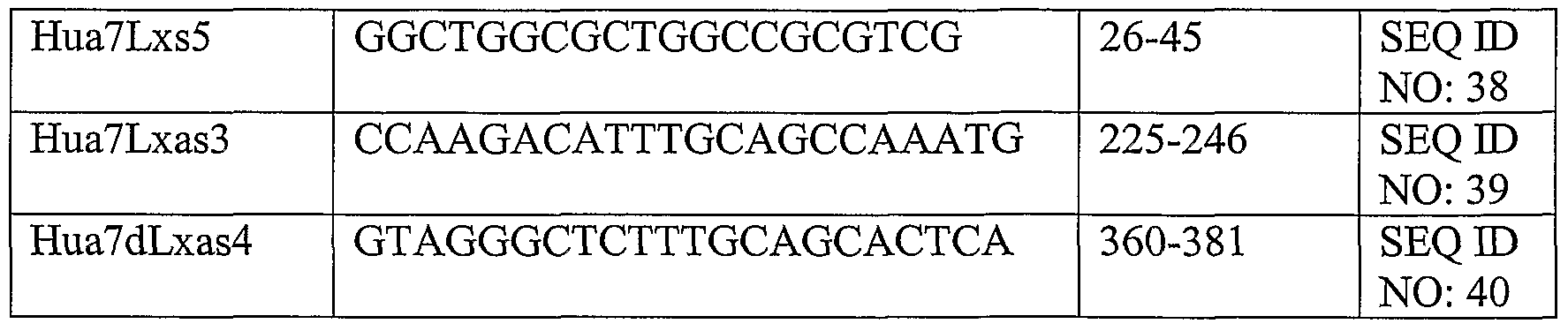
Examples 4-26: Generation of standard curves to assess levels of various genes
This experiment was performed in order to test primers and probe combinations and to generate standard curves from the following genes: human TGFb, human RANTES, human ICAMl, mouse ICAM-I, human VCAMl, mouse MCPl, mouse IL- 4, mouse GAPDH, mouse IL-10, human IL-8, human IL-6, human PECAM, mouse Hmoxl, mouse S0CS3, human S0CS3, human Hmoxl, human GAPDH, human Bcl-2, mouse Bcl-2, mouse ICAMl, human BAX, mouse BAX, mouse IL-6, human TNF-α, mouse MIPIv and mouse TNF-α.
A portion of the above mentioned genes was cloned using the techniques described above in Examples 1-3. The sequences used for cloning are also described above. The process by which a sequence verified clone is linearized depends upon its sequence and may use either BamHl or Kpnl or Xbal, all common restriction enzymes supplied by New England Biolabs, Inc. In vitro transcription reaction conditions and the subsequent purification and quantitation of RNA have also been described above. Dilutions of RNA corresponding to lOng/ul, lng/ul, lOOpg/ul, lOpg/ul, lpg/ul, 100fg/ul, 10fg/ul, and lfg/ul were made and IuI of each dilution was added to 9 ul of solution containing a total of 500ng of E. coli total RNA (Ambion). cDNA was generated as previously described. It is routine to combine multiple standard curves into one cDNA reaction, by increasing the concentration of the E. coli RNA solution, which frees up volume for additional standard curves. One-fifth of each cDNA reaction was amplified in a PCR reaction with an annealing temperature of 580C for 30 cycles (the cycles, instrument, and reaction conditions and sources are described above). 3OpM of the primers designated below were added to each PCR sample. The oligonucleotide probe designated below was coupled to xMAP beads. 15ul of each PCR reaction was then added to a corresponding well of a new 96-well plate containing 30ul of 1.5xTMAC
with 5000 beads coupled to the probe described below. This mixture was mixed and denatured, hybridized, stained and read as described previously.
The results of these experiments are shown in FIGs. 12- 36 which depict standard curves generated for the above-described genes. In part, the results of these experiments demonstrate that the methods of quantitating nucleic acids described herein can be utilized to quantitate nucleic acids from a wide variety of genes.
TABLE 4 Primers and oligonucleotide probes used to generate standard curves


TABLE 5 Sequences of primers or oligonucleotides used to generate standard curves


The relevant teachings of all the references, patents and patent applications cited herein are incorporated herein by reference in their entirety.
While this invention has been particularly shown and described with references to example embodiments thereof, it will be understood by those skilled in the art that various changes in form and details may be made therein without departing from the scope of the invention encompassed by the appended claims.
Claims
1. A method of measuring a target nucleic acid species in a sample comprising: a) amplifying multiple known concentrations of a standard nucleic acid to prepare amplified standard nucleic acid samples, wherein the standard nucleic acid comprises the same sequence as the target nucleic acid or a fragment thereof; b) amplifying the target nucleic acid; c) hybridizing the amplified target nucleic acid with a population comprising a subset of microparticles to form a mixture; wherein the subset is distinguishable from other subsets, if any, in said population based on a detectable parameter, and wherein the subset of microparticles is coupled to an oligonucleotide that is complementary to a portion of said target and standard nucleic acids; d) hybridizing each of the amplified standard nucleic acid samples with a population comprising said subset of microparticles to form mixtures; e) analyzing said mixtures by flow cytometry, wherein each subset of microparticles is identified based on said detectable parameter, wherein the binding of target nucleic and standard nucleic acid samples to each subset of microparticles is measured based on detection of a label, and f) determining the quantity of target nucleic acid in the sample based on analysis of said standard nucleic acid samples.
2. The method of claim 1 wherein said nucleic acid is DNA.
3. The method of claim 1 wherein said nucleic acid is RNA.
4. The method of claim 1 wherein said target nucleic acid and said standard nucleic acid are amplified by polymerase chain reaction ("PCR").
5. The method of claim 1 wherein a quantity of each of said known concentrations of standard nucleic acid is added to samples each comprising a heterogenous nucleic acid population before amplifying said multiple known concentrations of a standard nucleic acid.
6. The method of claim 1 wherein the binding of target nucleic acid to a subset of microparticles is measured based on detection of a label on said target nucleic acid.
7. The method of claim 1 wherein the binding of standard nucleic acid to a subset of microparticles is measured based on detection of a label on said standard nucleic acid.
8. The method of claim 7 wherein the binding of standard nucleic acid to a subset of microparticles is measured based on detection of a label on said standard nucleic acid samples.
9. The method of claim 6 wherein said label is placed on said target nucleic acid using labeled primer sequences.
10. The method of claim 6 wherein said label is biotin.
11. The method of claim 9 wherein said label is biotin.
12. The method of claim 1 wherein the binding of target nucleic acid to a subset of microparticles is measured based on detection of a label on said oligonucleotide.
13. The method of claim 12 wherein said label is a fluorophore.
14. The method of claim 1 wherein said detectable parameter is fluorescence intensity.
15. The method of claim 1 wherein said oligonucleotide that is complementary to a portion of said target and standard nucleic acids is complementary to a non- primer portion of said target nucleic acid.
16. ' The method of claim 1 wherein said sample is a biological sample selected from the group consisting of blood, synovial fluid, cerebrospinal fluid and a tissue sample.
17. The method of claim 1 wherein said analyzing step is conducted in a multi-well plate.
18. The method of claim 17, wherein said multi-well plate is a 96-well plate.
19. The method of claim 1, wherein at least two target nucleic acids are measured in the biological sample.
20. The method of claim 1 wherein said nucleic acid is a sequence of a human α7 subunit wherein said standard and target nucleic acids are amplified with a forward primer which has the sequence of SEQ ED NO:27 and a reverse primer which has the sequence of SEQ ID NO:28 and wherein said subset of microparticles is coupled to an oligonucleotide having the sequence of SEQ ID NO:31.
21. The method of claim 1 wherein said nucleic acid is a sequence of the mouse α7 subunit wherein said standard and target nucleic acids are amplified with a forward primer which has the sequence of SEQ ID NO:9 and a reverse primer which has the sequence of SEQ ID NO: 10 and wherein said subset of microparticles is coupled to an oligonucleotide having the sequence of SEQ ID NO:11.
22. The method of claim 1 wherein said nucleic acid is a sequence of the human α7 duplicate nicotinic acetylcholine receptor wherein said standard and target nucleic acids are amplified with a forward primer which has the sequence of SEQ ID NO:32 and a reverse primer which has the sequence of SEQ ID NO:33 and wherein said subset of microparticles is coupled to an oligonucleotide having the sequence of SEQ ID NO:36.
23. The method of claim 1 wherein said nucleic acid is a sequence of human beta actin wherein said standard and target nucleic acids are amplified with a forward primer which has the sequence of SEQ ID NO:21 and a reverse primer which has the sequence of SEQ ID NO:22 and wherein said subset of microparticles is coupled to an oligonucleotide having the sequence of SEQ ID NO:26.
24. The method of claim 1 wherein said nucleic acid is a sequence of human GAPDH wherein said standard and target nucleic acids are amplified with a forward primer which has the sequence of SEQ ID NO:23 and a reverse primer which has the sequence of SEQ ID NO:24 and wherein said subset of microparticles is coupled to an oligonucleotide having the sequence of SEQ ID NO:25.
25. The method of claim 1 wherein said nucleic acid is a sequence of human GAPDH wherein said standard and target nucleic acids are amplified with a forward primer which has the sequence of SEQ ID NO:86 and a reverse primer which has the sequence of SEQ ID NO: 87 and wherein said subset of microparticles is coupled to an oligonucleotide having the sequence of SEQ ID NO:88.
26. The method of claim 1 wherein said nucleic acid is a sequence of mouse IL-4 wherein said standard and target nucleic acids are amplified with a forward primer which has the sequence of SEQ ID NO:56 and a reverse primer which has the sequence of SEQ ID NO:57 and wherein said subset of microparticles is coupled to an oligonucleotide having the sequence of SEQ ID NO: 58.
27. The method of claim 1 wherein said nucleic acid is a sequence of mouse IL-10 wherein said standard and target nucleic acids are amplified with a forward primer which has the sequence of SEQ ID NO:62 and a reverse primer which has the sequence of SEQ TD NO: 63 and wherein said subset of microparticles is coupled to an oligonucleotide having the sequence of SEQ ID NO:64.
28. The method of claim 1 wherein said nucleic acid is a sequence of human IL-8 wherein said standard and target nucleic acids are amplified with a forward primer which has the sequence of SEQ ID NO: 65 and a reverse primer which has the sequence of SEQ TD NO: 66 and wherein said subset of microparticles is coupled to an oligonucleotide having the sequence of SEQ TD NO:67.
29. The method of claim 1 wherein said nucleic acid is a sequence of human IL-6 wherein said standard and target nucleic acids are amplified with a forward primer which has the sequence of SEQ E) NO:68 and a reverse primer which has the sequence of SEQ TD NO:69 and wherein said subset of microparticles is coupled to an oligonucleotide having the sequence of SEQ TD NO: 70.
30. The method of claim 1 wherein said nucleic acid is a sequence of human TNF-α wherein said standard and target nucleic acids are amplified with a forward primer which has the sequence of SEQ TD NO:95 and a reverse primer which has the sequence of SEQ ID NO:96 and wherein said subset of microparticles is coupled to an oligonucleotide having the sequence of SEQ ID NO:97.
31. The method of claim 1 wherein said nucleic acid is a sequence of mouse TNF- α wherein said standard and target nucleic acids are amplified with a forward primer which has the sequence of SEQ ID NO: 101 and a reverse primer which has the sequence of SEQ ID NO: 102 and wherein said subset of microparticles is coupled to an oligonucleotide having the sequence of SEQ ID NO: 103.
32. The method of claim 1 wherein said nucleic acid is a sequence of mouse IL-6 wherein said standard and target nucleic acids are amplified with a forward primer which has the sequence of SEQ ID NO: 113 and a reverse primer which has the sequence of SEQ ID NO: 114 and wherein said subset of microparticles is coupled to an oligonucleotide having the sequence of SEQ ID NO:115.
33. The method of claim 1 wherein said nucleic acid is a sequence of human ICAM- 1 wherein said standard and target nucleic acids are amplified with a forward primer which has the sequence of SEQ ID NO:47 and a reverse primer which has the sequence of SEQ ID NO:48 and wherein said subset of microparticles is coupled to an oligonucleotide having the sequence of SEQ ID NO:49.
34. The method of claim 1 wherein said nucleic acid is a sequence of mouse ICAM- 1 wherein said standard and target nucleic acids are amplified with a forward primer which has the sequence of SEQ ID NO: 107 and a reverse primer which has the sequence of SEQ ID NO: 108 and wherein said subset of microparticles is coupled to an oligonucleotide having the sequence of SEQ ID NO: 109.
35. The method of claim 1 wherein said nucleic acid is a sequence of mouse Bcl-2 wherein said standard and target nucleic acids are amplified with a forward primer which has the sequence of SEQ ID NO: 89 and a reverse primer which has the sequence of SEQ ID NO:90 and wherein said subset of microparticles is coupled to an oligonucleotide having the sequence of SEQ ID NO:91.
36. The method of claim 1 wherein said nucleic acid is a sequence of human Bcl-2 wherein said standard and target nucleic acids are amplified with a forward primer which has the sequence of SEQ ID NO: 104 and a reverse primer which has the sequence of SEQ ID NO: 105 and wherein said subset of microparticles is coupled to an oligonucleotide having the sequence of SEQ ID NO: 106.
37. The method of claim 1 wherein said nucleic acid is a sequence of mouse Bax wherein said standard and target nucleic acids are amplified with a forward primer which has the sequence of SEQ ID NO: 92 and a reverse primer which has the sequence of SEQ ID NO:93 and wherein said subset of microparticles is coupled to an oligonucleotide having the sequence of SEQ ID NO:94.
38. The method of claim 1 wherein said nucleic acid is a sequence of human Bax wherein said standard and target nucleic acids are amplified with a forward primer which has the sequence of SEQ ID NO: 110 and a reverse primer which has the sequence of SEQ ID NO: 111 and wherein said subset of microparticles is coupled to an oligonucleotide having the sequence of SEQ ID NO:112.
39. The method of claim 1 wherein said nucleic acid is a sequence of human TGF-b wherein said standard and target nucleic acids are amplified with a forward primer which has the sequence of SEQ ID NO:41 and a reverse primer which has the sequence of SEQ TD NO:42 and wherein said subset of microparticles is coupled to an oligonucleotide having the sequence of SEQ ID NO:43.
40. The method of claim 1 wherein said nucleic acid is a sequence of human RANTES wherein said standard and target nucleic acids are amplified with a forward primer which has the sequence of SEQ ID NO:44 and a reverse primer which has the sequence of SEQ ID NO:45 and wherein said subset of microparticles is coupled to an oligonucleotide having the sequence of SEQ E) NO:46.
41. The method of claim 1 wherein said nucleic acid is a sequence of mouse MIP-b wherein said standard and target nucleic acids are amplified with a forward primer which has the sequence of SEQ ID NO:98 and a reverse primer which has the sequence of SEQ ID NO:99 and wherein said subset of microparticles is coupled to an oligonucleotide having the sequence of SEQ ID NO: 100.
42. The method of claim 1 wherein said nucleic acid is a sequence of mouse VCAM-I wherein said standard and target nucleic acids are amplified with a forward primer which has the sequence of SEQ ID NO:50 and a reverse primer which has the sequence of SEQ ID NO:51 and wherein said subset of microparticles is coupled to an oligonucleotide having the sequence of SEQ ID NO:52.
43. The method of claim 1 wherein said nucleic acid is a sequence of human PECAM-I wherein said standard and target nucleic acids are amplified with a forward primer which has the sequence of SEQ ID NO:71 and a reverse primer which has the sequence of SEQ ID NO:72 and wherein said subset of microparticles is coupled to an oligonucleotide having the sequence of SEQ ID NO:73.
44. The method of claim 1 wherein said nucleic acid is a sequence of mouse beta actin wherein said standard and target nucleic acids are amplified with a forward primer which has the sequence of SEQ ID NO:7 and a reverse primer which has the sequence of SEQ ID NO: 8 and wherein said subset of microparticles is coupled to an oligonucleotide having the sequence of SEQ ID NO: 12.
45. The method of claim 1 wherein said nucleic acid is a sequence of human beta actin wherein said standard and target nucleic acids are amplified with a forward primer which has the sequence of SEQ ID NO: 19 and a reverse primer which has the sequence of SEQ ID NO:20 and wherein said subset of microparticles is coupled to an oligonucleotide having the sequence of SEQ ID NO:26.
46. The method of claim 1 wherein said nucleic acid is a sequence of human beta actin wherein said standard and target nucleic acids are amplified with a forward primer which has the sequence of SEQ ID NO:21 and a reverse primer which has the sequence of SEQ ID NO:22 and wherein said subset of microparticles is coupled to an oligonucleotide having the sequence of SEQ ID NO:26.
47. The method of claim 1 wherein said nucleic acid is a sequence of mouse Hmox- 1 wherein said standard and target nucleic acids are amplified with a forward primer which has the sequence of SEQ ID NO:74 and a reverse primer which has the sequence of SEQ ID NO: 75 and wherein said subset of microparticles is coupled to an oligonucleotide having the sequence of SEQ ID NO:76.
48. The method of claim 1 wherein said nucleic acid is a sequence of human Hmox- 1 wherein said standard and target nucleic acids are amplified with a forward primer which has the sequence of SEQ ID NO: 83 and a reverse primer which has the sequence of SEQ ID NO: 84 and wherein said subset of microparticles is coupled to an oligonucleotide having the sequence of SEQ ID NO:85.
49. The method of claim 1 wherein said nucleic acid is a sequence of mouse SOCS- 3 wherein said standard and target nucleic acids are amplified with a forward primer which has the sequence of SEQ ID NO: 77 and a reverse primer which has the sequence of SEQ ID NO:78 and wherein said subset of microparticles is coupled to an oligonucleotide having the sequence of SEQ ID NO:79.
50. The method of claim 1 wherein said nucleic acid is a sequence of human SOCS- 2 wherein said standard and target nucleic acids are amplified with a forward primer which has the sequence of SEQ ID NO:80 and a reverse primer which has the sequence of SEQ ID NO:81 and wherein said subset of microparticles is coupled to an oligonucleotide having the sequence of SEQ ID NO: 82.
51. The method of claim 1 wherein said nucleic acid is a sequence of mouse a7 wherein said standard and target nucleic acids are amplified with a forward primer which has the sequence of SEQ ID NO: 15 and a reverse primer which has the sequence of SEQ ID NO: 16 and wherein said subset of microparticles is coupled to an oligonucleotide having the sequence of SEQ ID NO: 11.
52. The method of claim 1 wherein said nucleic acid is a sequence of mouse beta actin wherein said standard and target nucleic acids are amplified with a forward primer which has the sequence of SEQ TD NO: 13 and a reverse primer which has the sequence of SEQ ID NO: 14 and wherein said subset of microparticles is coupled to an oligonucleotide having the sequence of SEQ ID NO: 12.
53. A method of measuring a target RNA species in a sample comprising: a) preparing cDNA from a known concentration of a standard RNA comprising the steps of:
(i) preparing samples of multiple known concentrations of said standard RNA, wherein said standard RNA comprises the same sequence as the target RNA or a fragment thereof, and
(ii) generating standard cDNA samples for each concentration of standard RNA; b) generating target cDNA from said biological sample; c) amplifying said target cDNA and said standard cDNA samples to prepare amplified target DNA and amplified standard DNA samples; d) hybridizing the amplified target DNA with a population comprising a subset of microparticles to form a mixture; wherein the subset is distinguishable from other subsets, if any, in said population based on a detectable parameter, and wherein a subset of microparticles is coupled to an oligonucleotide that is complementary to a portion of said target and standard DNA; e) hybridizing each of the standard cDNA samples with a population comprising said subset of microparticles to form mixtures; f) analyzing said mixtures by flow cytometry, wherein each subset of microparticles is identified based on said detectable parameter, wherein the binding of target cDNA and standard cDNA samples to each subset of microparticles is measured based on detection of a label, and g) determining the quantity of target RNA in the sample based on analysis of said standard DNA samples.
54. The method of claim 53 wherein said target cDNA and said standard cDNA are amplified by polymerase chain reaction ("PCR").
55. The method of claim 53 wherein a quantity of each of said known concentrations of standard RNA is added to samples each comprising a heterogenous RNA population before preparation of said standard cDNA samples.
56. The method of claim 53 wherein the binding of target cDNA to a subset of microparticles is measured based on detection of a label on said target DNA.
57. The method of claim 53 wherein the binding of standard DNA samples to a subset of microparticles is measured based on detection of a label on said standard DNA samples.
58. The method of claim 57 wherein the binding of standard DNA samples to a subset of microparticles is measured based on detection of a label on said standard DNA samples.
59. The method of claim 54 wherein said label is placed on said target cDNA and said standard cDNA samples using labeled primer sequences.
60. The method of claim 56 wherein said label is biotin.
61. The method of claim 57 wherein said label is biotin.
62. The method of claim 53 wherein the binding of target cDNA to a subset of microparticles is measured based on detection of a label on said oligonucleotide.
63. The method of claim 62 wherein said label is a fluorophore.
64. The method of claim 53 wherein said detectable parameter is fluorescence intensity.
65. The method of claim 53 wherein said oligonucleotide that is complementary to a portion of said target DNA is complementary to a non-primer portion of said target DNA.
66. The method of claim 65 wherein said oligonucleotide that is complementary to a portion of said standard DNA is complementary a non-primer portion of said standard DNA.
67. The method of claim 53 wherein said sample is a biological sample selected from the group consisting of blood, synovial fluid, cerebrospinal fluid and a tissue sample.
68. The method of claim 53 wherein said analyzing step is conducted in a multi- well plate.
69. The method of claim 68, wherein said multi-well plate is a 96-well plate.
70. A method of screening for the presence of one or more polymorphisms or mutations of a gene in a sample comprising analyzing said sample comprising: a) amplifying multiple known concentrations of a standard nucleic acid for each polymorphism or mutation to prepare amplified standard nucleic acid samples, wherein each standard nucleic acid comprises the same sequence as a nucleotide corresponding to each of the polymorphisms or mutations; b) amplifying the target nucleic acid; c) hybridizing the amplified target nucleic acid with a population comprising a subset of microparticles to form a mixture; ^ wherein the subset is distinguishable from other subsets, if any, in said population based on a detectable parameter, wherein for each polymorphism or mutation being screened, there is a subset of microparticles coupled to an oligonucleotide complementary to a portion of the nucleotide sequence corresponding to the polymorphism or mutation, wherein another subset of microparticles is coupled to an oligonucleotide that is complementary to a portion of a wild-type nucleotide sequence; d) hybridizing each of the amplified standard nucleic acid samples with a population comprising said subset of microparticles to form mixtures; e) analyzing said mixtures by flow cytometry, wherein each subset of microparticles is identified based on said detectable parameter, wherein the binding of target nucleic and standard nucleic acid samples to each subset of microparticles is measured based on detection of a label, and ( f) determining the presence of said polymorphism or mutation of a gene.
71. The method of claim 70 wherein said polymorphism or mutation is associated with a disease or susceptibility for a disease.
72. A method of screening for the presence of one or more polymorphisms or mutations of a gene in a sample comprising analyzing said sample for said polymorphisms or mutations comprising: a) for each of said polymorphisms or mutations being screened, preparing cDNA from known concentrations of a standard KNA comprising the steps of:
(i) preparing samples of multiple known concentrations of said standard RNA, wherein said standard RNA each independently comprise the same sequence as a nucleic acid corresponding to each of said polymorphisms or mutations,
(ii) generating standard cDNA samples for each concentration of standard RNA; b) generating target cDNA from said biological sample; c) amplifying said target cDNA and standard cDNA samples to prepare amplified target DNA and amplified standard DNA; d) hybridizing the amplified target DNA with a population comprising a subset of microparticles to form a mixture; wherein the subset is distinguishable from other subsets, if any, in said population based on a detectable parameter, wherein for each polymorphism or mutation being screened, there is a subset of microparticles coupled to an oligonucleotide complementary to a portion of the nucleotide sequence corresponding to the polymorphism or mutation, wherein another subset of microparticles is coupled to an oligonucleotide that is complementary to a portion of a wild-type nucleotide sequence; e) hybridizing each of the standard cDNA samples with populations comprising said subset of microparticles to form mixtures; f) analyzing said mixtures by flow cytometry, wherein each subset of microparticles is identified based on said detectable parameter, wherein the binding of target DNA to each subset of microparticles is, measured based detection of a label, wherein the binding of standard DNA samples to a subset of microspheres is measured based on detection of a label; and g) determining the presence of said polymorphism or mutation of a gene in said biological sample.
73. The method of claim 72 wherein said polymorphism or mutation is associated with a disease or susceptibility for a disease.
74. A method of monitoring a change in gene expression in response to an agent comprising obtaining a sample after agent administration and determining the quantity of a target nucleic acid in said sample comprising: a) amplifying multiple known concentrations of a standard nucleic acid to prepare amplified standard nucleic acid samples, wherein the standard nucleic acid comprises the same sequence as the target nucleic acid or a fragment thereof; b) amplifying the target nucleic acid; c) hybridizing the amplified target nucleic acid with a population comprising a subset of microparticles to form a mixture; wherein the subset is distinguishable from other subsets, if any, in said population based on a detectable parameter, and wherein a subset of microparticles is coupled to an oligonucleotide that is complementary to a portion of said target and standard nucleic acids; d) hybridizing each of the amplified standard nucleic acid samples with a population comprising said subset of microparticles to form mixtures; e) analyzing said mixtures by flow cytometry, wherein each subset of microparticles is identified based on said detectable parameter, wherein the binding of target nucleic and standard nucleic acid samples to each subset of microparticles is measured based on detection of a label, and f) determining the quantity of target nucleic acid in the sample based on analysis of said standard nucleic acid samples.
A method of monitoring a change in gene expression in response to an agent comprising obtaining a biological sample after agent administration and determining the quantity of a target RNA in said biological sample comprising: a) preparing cDNA from known concentrations of a standard RNA comprising the steps of: (i) preparing samples of multiple known concentrations of said standard RNA, wherein said standard RNA comprises the same sequence as the target RNA or a fragment thereof; (ii) generating standard cDNA samples for each concentration of standard RNA; b) generating target cDNA from said biological samples; c) amplifying said target cDNA standard cDNA samples to prepare amplified target DNA and amplified standard DNA samples; d) hybridizing the amplified target DNA with a population comprising a subset of microparticles to form a mixture; wherein said subset is distinguishable from other subsets, if any, in said population based on a detectable parameter, wherein a subset of microparticles is coupled to an oligonucleotide that is complementary to a portion of said target DNA and said standard DNA; e) hybridizing each of the standard cDNA samples with a population comprising a subset of microparticles to form a mixture; f) analyzing said mixtures by flow cytometry, wherein each subset of microparticles is identified based on said detectable parameter, wherein the binding of target cDNA and standard cDNA samples to each subset of microparticles is measured based on detection of a label, and g) determining the quantity of target RNA in the sample based on analysis of said standard DNA samples.
76. The method of claim 75 wherein a biological sample is also obtained before agent administration and the quantities of target RNA- in samples obtained before and after agent administration are compared.
77. The method of claim 75 wherein biological samples are obtained at more than one time point after agent administration.
78. The method of claim 75 wherein said response is measured in a patient.
79. A method of screening for the presence of a pathogenic organism in a biological sample comprising analyzing the sample for the presence of a target nucleic acid from said pathogenic organism comprising: a) amplifying multiple known concentrations of a standard nucleic acid to prepare amplified standard nucleic acid samples, wherein the standard nucleic acid comprises the same sequence as the nucleic acid from the pathogenic organism or a fragment thereof; b) amplifying the target nucleic acid; c) hybridizing the amplified target nucleic acid with a population comprising a subset of microparticles to form a mixture; wherein the subset is distinguishable from other subsets, if any, in said population based on a detectable parameter, and wherein the subset of microparticles is coupled to an oligonucleotide that is complementary to a portion of said target and standard nucleic acids; d) hybridizing each of the amplified standard nucleic acid samples with a population comprising said subset of microparticles to form mixtures; e) analyzing said mixtures by flow cytometry, wherein each subset of microparticles is identified based on said detectable parameter, wherein the binding of target nucleic and standard nucleic acid samples to each subset of microparticles is measured based on detection of a label, and f) determining the presence of said pathogenic organism in said biological sample. A method of screening for the presence of a pathogenic organism in a biological sample comprising analyzing said sample for the presence of a target RNA from said pathogenic organism comprising: a) preparing cDNA from known concentrations of a standard RNA comprising the steps of:
(i) preparing samples of multiple known concentrations of said standard RNA, wherein said standard RNA comprises the same sequence as the target RNA or a fragment thereof;
(ii) generating standard cDNA samples for each concentration of standard RNA; b) generating target cDNA from said biological sample; c) amplifying said target DNA from said target cDNA standard DNA to prepare amplified target DNA and amplified standard DNA; d) hybridizing the target cDNA with a population comprising a subset of microparticles to form a mixture; wherein the subset is distinguishable from other subsets, if any, in said population based on a detectable parameter, wherein the subset of microparticles is coupled to an oligonucleotide that is complementary to a portion of a nucleotide sequence from said pathogenic organism; e) hybridizing each of the standard cDNA samples with a population comprising said subset of microparticles to form a mixture; f) analyzing said mixtures by flow cytometry, wherein each subset of microparticles is identified based on said detectable parameter, wherein the binding of target cDNA and standard cDNA samples to each subset of microparticles is measured based on detection of a label, and g) determining the presence of said pathogenic organism in said biological sample. The method of claim 80 wherein said pathogenic organism is selected from the group consisting of a virus and a bacteria.
Applications Claiming Priority (2)
| Application Number | Priority Date | Filing Date | Title |
|---|---|---|---|
| US71055805P | 2005-08-22 | 2005-08-22 | |
| US60/710,558 | 2005-08-22 |
Publications (2)
| Publication Number | Publication Date |
|---|---|
| WO2007024840A2 true WO2007024840A2 (en) | 2007-03-01 |
| WO2007024840A3 WO2007024840A3 (en) | 2007-04-12 |
Family
ID=37529404
Family Applications (1)
| Application Number | Title | Priority Date | Filing Date |
|---|---|---|---|
| PCT/US2006/032735 Ceased WO2007024840A2 (en) | 2005-08-22 | 2006-08-22 | Method of quantitating nucleic acids by flow cytometry microparticle-based array |
Country Status (1)
| Country | Link |
|---|---|
| WO (1) | WO2007024840A2 (en) |
Cited By (53)
| Publication number | Priority date | Publication date | Assignee | Title |
|---|---|---|---|---|
| EP2291541A4 (en) * | 2008-03-05 | 2011-06-22 | Clean Earth Tech Llc | FLOW CYTOMETRY ASSAY WITH PNA MARKING BALLS AND MULTIPLEXED PCR COUPLING FOR THE SIMULTANEOUS DETECTION OF SEVERAL BIOLOGICAL AGENTS |
| US8748094B2 (en) | 2008-12-19 | 2014-06-10 | President And Fellows Of Harvard College | Particle-assisted nucleic acid sequencing |
| US9017948B2 (en) | 2007-03-07 | 2015-04-28 | President And Fellows Of Harvard College | Assays and other reactions involving droplets |
| US9056289B2 (en) | 2009-10-27 | 2015-06-16 | President And Fellows Of Harvard College | Droplet creation techniques |
| US9388465B2 (en) | 2013-02-08 | 2016-07-12 | 10X Genomics, Inc. | Polynucleotide barcode generation |
| US9410201B2 (en) | 2012-12-14 | 2016-08-09 | 10X Genomics, Inc. | Methods and systems for processing polynucleotides |
| US9663818B2 (en) | 2012-06-15 | 2017-05-30 | The University Of Chicago | Oligonucleotide-mediated quantitative multiplexed immunoassays |
| US9689024B2 (en) | 2012-08-14 | 2017-06-27 | 10X Genomics, Inc. | Methods for droplet-based sample preparation |
| US9694361B2 (en) | 2014-04-10 | 2017-07-04 | 10X Genomics, Inc. | Fluidic devices, systems, and methods for encapsulating and partitioning reagents, and applications of same |
| US9701998B2 (en) | 2012-12-14 | 2017-07-11 | 10X Genomics, Inc. | Methods and systems for processing polynucleotides |
| US9797010B2 (en) | 2007-12-21 | 2017-10-24 | President And Fellows Of Harvard College | Systems and methods for nucleic acid sequencing |
| US9824068B2 (en) | 2013-12-16 | 2017-11-21 | 10X Genomics, Inc. | Methods and apparatus for sorting data |
| US9951386B2 (en) | 2014-06-26 | 2018-04-24 | 10X Genomics, Inc. | Methods and systems for processing polynucleotides |
| US9975122B2 (en) | 2014-11-05 | 2018-05-22 | 10X Genomics, Inc. | Instrument systems for integrated sample processing |
| US10011872B1 (en) | 2016-12-22 | 2018-07-03 | 10X Genomics, Inc. | Methods and systems for processing polynucleotides |
| US10221436B2 (en) | 2015-01-12 | 2019-03-05 | 10X Genomics, Inc. | Processes and systems for preparation of nucleic acid sequencing libraries and libraries prepared using same |
| US10221442B2 (en) | 2012-08-14 | 2019-03-05 | 10X Genomics, Inc. | Compositions and methods for sample processing |
| US10273541B2 (en) | 2012-08-14 | 2019-04-30 | 10X Genomics, Inc. | Methods and systems for processing polynucleotides |
| US10287623B2 (en) | 2014-10-29 | 2019-05-14 | 10X Genomics, Inc. | Methods and compositions for targeted nucleic acid sequencing |
| US10323279B2 (en) | 2012-08-14 | 2019-06-18 | 10X Genomics, Inc. | Methods and systems for processing polynucleotides |
| US10395758B2 (en) | 2013-08-30 | 2019-08-27 | 10X Genomics, Inc. | Sequencing methods |
| US10400235B2 (en) | 2017-05-26 | 2019-09-03 | 10X Genomics, Inc. | Single cell analysis of transposase accessible chromatin |
| US10400280B2 (en) | 2012-08-14 | 2019-09-03 | 10X Genomics, Inc. | Methods and systems for processing polynucleotides |
| US10428326B2 (en) | 2017-01-30 | 2019-10-01 | 10X Genomics, Inc. | Methods and systems for droplet-based single cell barcoding |
| US10471016B2 (en) | 2013-11-08 | 2019-11-12 | President And Fellows Of Harvard College | Microparticles, methods for their preparation and use |
| US10481158B2 (en) | 2015-06-01 | 2019-11-19 | California Institute Of Technology | Compositions and methods for screening T cells with antigens for specific populations |
| US10533221B2 (en) | 2012-12-14 | 2020-01-14 | 10X Genomics, Inc. | Methods and systems for processing polynucleotides |
| US10550429B2 (en) | 2016-12-22 | 2020-02-04 | 10X Genomics, Inc. | Methods and systems for processing polynucleotides |
| US10650912B2 (en) | 2015-01-13 | 2020-05-12 | 10X Genomics, Inc. | Systems and methods for visualizing structural variation and phasing information |
| US10697000B2 (en) | 2015-02-24 | 2020-06-30 | 10X Genomics, Inc. | Partition processing methods and systems |
| US10745742B2 (en) | 2017-11-15 | 2020-08-18 | 10X Genomics, Inc. | Functionalized gel beads |
| US10752949B2 (en) | 2012-08-14 | 2020-08-25 | 10X Genomics, Inc. | Methods and systems for processing polynucleotides |
| US10774370B2 (en) | 2015-12-04 | 2020-09-15 | 10X Genomics, Inc. | Methods and compositions for nucleic acid analysis |
| US10815525B2 (en) | 2016-12-22 | 2020-10-27 | 10X Genomics, Inc. | Methods and systems for processing polynucleotides |
| US10829815B2 (en) | 2017-11-17 | 2020-11-10 | 10X Genomics, Inc. | Methods and systems for associating physical and genetic properties of biological particles |
| US10839939B2 (en) | 2014-06-26 | 2020-11-17 | 10X Genomics, Inc. | Processes and systems for nucleic acid sequence assembly |
| US10854315B2 (en) | 2015-02-09 | 2020-12-01 | 10X Genomics, Inc. | Systems and methods for determining structural variation and phasing using variant call data |
| US11081208B2 (en) | 2016-02-11 | 2021-08-03 | 10X Genomics, Inc. | Systems, methods, and media for de novo assembly of whole genome sequence data |
| US11084036B2 (en) | 2016-05-13 | 2021-08-10 | 10X Genomics, Inc. | Microfluidic systems and methods of use |
| US11123297B2 (en) | 2015-10-13 | 2021-09-21 | President And Fellows Of Harvard College | Systems and methods for making and using gel microspheres |
| US11155881B2 (en) | 2018-04-06 | 2021-10-26 | 10X Genomics, Inc. | Systems and methods for quality control in single cell processing |
| US11274343B2 (en) | 2015-02-24 | 2022-03-15 | 10X Genomics, Inc. | Methods and compositions for targeted nucleic acid sequence coverage |
| US11401550B2 (en) | 2008-09-19 | 2022-08-02 | President And Fellows Of Harvard College | Creation of libraries of droplets and related species |
| US11591637B2 (en) | 2012-08-14 | 2023-02-28 | 10X Genomics, Inc. | Compositions and methods for sample processing |
| US11629344B2 (en) | 2014-06-26 | 2023-04-18 | 10X Genomics, Inc. | Methods and systems for processing polynucleotides |
| US11773389B2 (en) | 2017-05-26 | 2023-10-03 | 10X Genomics, Inc. | Single cell analysis of transposase accessible chromatin |
| US11898206B2 (en) | 2017-05-19 | 2024-02-13 | 10X Genomics, Inc. | Systems and methods for clonotype screening |
| US12163191B2 (en) | 2014-06-26 | 2024-12-10 | 10X Genomics, Inc. | Analysis of nucleic acid sequences |
| US12258613B2 (en) | 2017-03-08 | 2025-03-25 | California Institute Of Technology | Pairing antigen specificity of a T cell with T cell receptor sequences |
| US12264411B2 (en) | 2017-01-30 | 2025-04-01 | 10X Genomics, Inc. | Methods and systems for analysis |
| US12312640B2 (en) | 2014-06-26 | 2025-05-27 | 10X Genomics, Inc. | Analysis of nucleic acid sequences |
| US12421558B2 (en) | 2020-02-13 | 2025-09-23 | 10X Genomics, Inc. | Systems and methods for joint interactive visualization of gene expression and DNA chromatin accessibility |
| US12427518B2 (en) | 2016-05-12 | 2025-09-30 | 10X Genomics, Inc. | Microfluidic on-chip filters |
Family Cites Families (5)
| Publication number | Priority date | Publication date | Assignee | Title |
|---|---|---|---|---|
| US5736330A (en) * | 1995-10-11 | 1998-04-07 | Luminex Corporation | Method and compositions for flow cytometric determination of DNA sequences |
| EP1248853A2 (en) * | 1999-08-20 | 2002-10-16 | Luminex Corporation | Liquid array technology |
| US20020137060A1 (en) * | 2000-08-09 | 2002-09-26 | Brown-Augsburger Patricia Lea | Combined hybridization-detection assays for determining nucleic acid concentrations in biological fluids |
| AT411174B (en) * | 2001-08-09 | 2003-10-27 | Lambda Labor Fuer Molekularbio | METHOD AND CHIP FOR ANALYZING NUCLEIC ACIDS |
| EP1548126A1 (en) * | 2003-12-22 | 2005-06-29 | Bio-Rad Pasteur | Solid support for control nucleic acid, and application thereof to nucleic acid detection |
-
2006
- 2006-08-22 WO PCT/US2006/032735 patent/WO2007024840A2/en not_active Ceased
Cited By (142)
| Publication number | Priority date | Publication date | Assignee | Title |
|---|---|---|---|---|
| US12416033B2 (en) | 2007-03-07 | 2025-09-16 | President And Fellows Of Harvard College | Assays and other reactions involving droplets |
| US10738337B2 (en) | 2007-03-07 | 2020-08-11 | President And Fellows Of Harvard College | Assays and other reactions involving droplets |
| US9017948B2 (en) | 2007-03-07 | 2015-04-28 | President And Fellows Of Harvard College | Assays and other reactions involving droplets |
| US9029085B2 (en) | 2007-03-07 | 2015-05-12 | President And Fellows Of Harvard College | Assays and other reactions involving droplets |
| US12331287B1 (en) | 2007-03-07 | 2025-06-17 | President And Fellows Of Harvard College | Assays and other reactions involving droplets |
| US9068210B2 (en) | 2007-03-07 | 2015-06-30 | President And Fellows Of Harvard College | Assay and other reactions involving droplets |
| US10683524B2 (en) | 2007-03-07 | 2020-06-16 | President And Fellows Of Harvard College | Assays and other reactions involving droplets |
| US10221437B2 (en) | 2007-03-07 | 2019-03-05 | President And Fellows Of Harvard College | Assays and other reactions involving droplets |
| US10508294B2 (en) | 2007-03-07 | 2019-12-17 | President And Fellows Of Harvard College | Assays and other reactions involving droplets |
| US9850526B2 (en) | 2007-03-07 | 2017-12-26 | President And Fellows Of Harvard College | Assays and other reactions involving droplets |
| US10941430B2 (en) | 2007-03-07 | 2021-03-09 | President And Fellows Of Harvard College | Assays and other reactions involving droplets |
| US9816121B2 (en) | 2007-03-07 | 2017-11-14 | President And Fellows Of Harvard College | Assays and other reactions involving droplets |
| US10633701B2 (en) | 2007-12-21 | 2020-04-28 | President And Fellows Of Harvard College | Systems and methods for nucleic acid sequencing |
| US9797010B2 (en) | 2007-12-21 | 2017-10-24 | President And Fellows Of Harvard College | Systems and methods for nucleic acid sequencing |
| EP2291541A4 (en) * | 2008-03-05 | 2011-06-22 | Clean Earth Tech Llc | FLOW CYTOMETRY ASSAY WITH PNA MARKING BALLS AND MULTIPLEXED PCR COUPLING FOR THE SIMULTANEOUS DETECTION OF SEVERAL BIOLOGICAL AGENTS |
| US11401550B2 (en) | 2008-09-19 | 2022-08-02 | President And Fellows Of Harvard College | Creation of libraries of droplets and related species |
| US12116631B2 (en) | 2008-09-19 | 2024-10-15 | President And Fellows Of Harvard College | Creation of libraries of droplets and related species |
| US8748094B2 (en) | 2008-12-19 | 2014-06-10 | President And Fellows Of Harvard College | Particle-assisted nucleic acid sequencing |
| US10457977B2 (en) | 2008-12-19 | 2019-10-29 | President And Fellows Of Harvard College | Particle-assisted nucleic acid sequencing |
| US11000849B2 (en) | 2009-10-27 | 2021-05-11 | President And Fellows Of Harvard College | Droplet creation techniques |
| US9839911B2 (en) | 2009-10-27 | 2017-12-12 | President And Fellows Of Harvard College | Droplet creation techniques |
| US12121898B2 (en) | 2009-10-27 | 2024-10-22 | President And Fellows Of Harvard College | Droplet creation techniques |
| US9056289B2 (en) | 2009-10-27 | 2015-06-16 | President And Fellows Of Harvard College | Droplet creation techniques |
| US9663818B2 (en) | 2012-06-15 | 2017-05-30 | The University Of Chicago | Oligonucleotide-mediated quantitative multiplexed immunoassays |
| US10550421B2 (en) | 2012-06-15 | 2020-02-04 | The University Of Chicago | Oligonucleotide-mediated quantitative multiplexed immunoassays |
| US11359239B2 (en) | 2012-08-14 | 2022-06-14 | 10X Genomics, Inc. | Methods and systems for processing polynucleotides |
| US10669583B2 (en) | 2012-08-14 | 2020-06-02 | 10X Genomics, Inc. | Method and systems for processing polynucleotides |
| US10626458B2 (en) | 2012-08-14 | 2020-04-21 | 10X Genomics, Inc. | Methods and systems for processing polynucleotides |
| US9689024B2 (en) | 2012-08-14 | 2017-06-27 | 10X Genomics, Inc. | Methods for droplet-based sample preparation |
| US10597718B2 (en) | 2012-08-14 | 2020-03-24 | 10X Genomics, Inc. | Methods and systems for sample processing polynucleotides |
| US10584381B2 (en) | 2012-08-14 | 2020-03-10 | 10X Genomics, Inc. | Methods and systems for processing polynucleotides |
| US10752950B2 (en) | 2012-08-14 | 2020-08-25 | 10X Genomics, Inc. | Methods and systems for processing polynucleotides |
| US11441179B2 (en) | 2012-08-14 | 2022-09-13 | 10X Genomics, Inc. | Methods and systems for processing polynucleotides |
| US10053723B2 (en) | 2012-08-14 | 2018-08-21 | 10X Genomics, Inc. | Capsule array devices and methods of use |
| US11591637B2 (en) | 2012-08-14 | 2023-02-28 | 10X Genomics, Inc. | Compositions and methods for sample processing |
| US10221442B2 (en) | 2012-08-14 | 2019-03-05 | 10X Genomics, Inc. | Compositions and methods for sample processing |
| US12037634B2 (en) | 2012-08-14 | 2024-07-16 | 10X Genomics, Inc. | Capsule array devices and methods of use |
| US11078522B2 (en) | 2012-08-14 | 2021-08-03 | 10X Genomics, Inc. | Capsule array devices and methods of use |
| US10752949B2 (en) | 2012-08-14 | 2020-08-25 | 10X Genomics, Inc. | Methods and systems for processing polynucleotides |
| US10273541B2 (en) | 2012-08-14 | 2019-04-30 | 10X Genomics, Inc. | Methods and systems for processing polynucleotides |
| US11035002B2 (en) | 2012-08-14 | 2021-06-15 | 10X Genomics, Inc. | Methods and systems for processing polynucleotides |
| US10323279B2 (en) | 2012-08-14 | 2019-06-18 | 10X Genomics, Inc. | Methods and systems for processing polynucleotides |
| US12098423B2 (en) | 2012-08-14 | 2024-09-24 | 10X Genomics, Inc. | Methods and systems for processing polynucleotides |
| US11021749B2 (en) | 2012-08-14 | 2021-06-01 | 10X Genomics, Inc. | Methods and systems for processing polynucleotides |
| US9695468B2 (en) | 2012-08-14 | 2017-07-04 | 10X Genomics, Inc. | Methods for droplet-based sample preparation |
| US10450607B2 (en) | 2012-08-14 | 2019-10-22 | 10X Genomics, Inc. | Methods and systems for processing polynucleotides |
| US10400280B2 (en) | 2012-08-14 | 2019-09-03 | 10X Genomics, Inc. | Methods and systems for processing polynucleotides |
| US10227648B2 (en) | 2012-12-14 | 2019-03-12 | 10X Genomics, Inc. | Methods and systems for processing polynucleotides |
| US11421274B2 (en) | 2012-12-14 | 2022-08-23 | 10X Genomics, Inc. | Methods and systems for processing polynucleotides |
| US9410201B2 (en) | 2012-12-14 | 2016-08-09 | 10X Genomics, Inc. | Methods and systems for processing polynucleotides |
| US9701998B2 (en) | 2012-12-14 | 2017-07-11 | 10X Genomics, Inc. | Methods and systems for processing polynucleotides |
| US10676789B2 (en) | 2012-12-14 | 2020-06-09 | 10X Genomics, Inc. | Methods and systems for processing polynucleotides |
| US9856530B2 (en) | 2012-12-14 | 2018-01-02 | 10X Genomics, Inc. | Methods and systems for processing polynucleotides |
| US10612090B2 (en) | 2012-12-14 | 2020-04-07 | 10X Genomics, Inc. | Methods and systems for processing polynucleotides |
| US9567631B2 (en) | 2012-12-14 | 2017-02-14 | 10X Genomics, Inc. | Methods and systems for processing polynucleotides |
| US10253364B2 (en) | 2012-12-14 | 2019-04-09 | 10X Genomics, Inc. | Method and systems for processing polynucleotides |
| US11473138B2 (en) | 2012-12-14 | 2022-10-18 | 10X Genomics, Inc. | Methods and systems for processing polynucleotides |
| US10533221B2 (en) | 2012-12-14 | 2020-01-14 | 10X Genomics, Inc. | Methods and systems for processing polynucleotides |
| US10150964B2 (en) | 2013-02-08 | 2018-12-11 | 10X Genomics, Inc. | Partitioning and processing of analytes and other species |
| US10150963B2 (en) | 2013-02-08 | 2018-12-11 | 10X Genomics, Inc. | Partitioning and processing of analytes and other species |
| US11193121B2 (en) | 2013-02-08 | 2021-12-07 | 10X Genomics, Inc. | Partitioning and processing of analytes and other species |
| US9644204B2 (en) | 2013-02-08 | 2017-05-09 | 10X Genomics, Inc. | Partitioning and processing of analytes and other species |
| US9388465B2 (en) | 2013-02-08 | 2016-07-12 | 10X Genomics, Inc. | Polynucleotide barcode generation |
| US12249402B2 (en) | 2013-08-30 | 2025-03-11 | 10X Genomics, Inc. | Sequencing methods |
| US12131805B2 (en) | 2013-08-30 | 2024-10-29 | 10X Genomics, Inc. | Sequencing methods |
| US10395758B2 (en) | 2013-08-30 | 2019-08-27 | 10X Genomics, Inc. | Sequencing methods |
| US10471016B2 (en) | 2013-11-08 | 2019-11-12 | President And Fellows Of Harvard College | Microparticles, methods for their preparation and use |
| US9824068B2 (en) | 2013-12-16 | 2017-11-21 | 10X Genomics, Inc. | Methods and apparatus for sorting data |
| US10343166B2 (en) | 2014-04-10 | 2019-07-09 | 10X Genomics, Inc. | Fluidic devices, systems, and methods for encapsulating and partitioning reagents, and applications of same |
| US12005454B2 (en) | 2014-04-10 | 2024-06-11 | 10X Genomics, Inc. | Fluidic devices, systems, and methods for encapsulating and partitioning reagents, and applications of same |
| US9694361B2 (en) | 2014-04-10 | 2017-07-04 | 10X Genomics, Inc. | Fluidic devices, systems, and methods for encapsulating and partitioning reagents, and applications of same |
| US10071377B2 (en) | 2014-04-10 | 2018-09-11 | 10X Genomics, Inc. | Fluidic devices, systems, and methods for encapsulating and partitioning reagents, and applications of same |
| US10137449B2 (en) | 2014-04-10 | 2018-11-27 | 10X Genomics, Inc. | Fluidic devices, systems, and methods for encapsulating and partitioning reagents, and applications of same |
| US10150117B2 (en) | 2014-04-10 | 2018-12-11 | 10X Genomics, Inc. | Fluidic devices, systems, and methods for encapsulating and partitioning reagents, and applications of same |
| US11713457B2 (en) | 2014-06-26 | 2023-08-01 | 10X Genomics, Inc. | Methods and systems for processing polynucleotides |
| US10337061B2 (en) | 2014-06-26 | 2019-07-02 | 10X Genomics, Inc. | Methods and systems for processing polynucleotides |
| US10760124B2 (en) | 2014-06-26 | 2020-09-01 | 10X Genomics, Inc. | Methods and systems for processing polynucleotides |
| US12312640B2 (en) | 2014-06-26 | 2025-05-27 | 10X Genomics, Inc. | Analysis of nucleic acid sequences |
| US10457986B2 (en) | 2014-06-26 | 2019-10-29 | 10X Genomics, Inc. | Methods and systems for processing polynucleotides |
| US10030267B2 (en) | 2014-06-26 | 2018-07-24 | 10X Genomics, Inc. | Methods and systems for processing polynucleotides |
| US9951386B2 (en) | 2014-06-26 | 2018-04-24 | 10X Genomics, Inc. | Methods and systems for processing polynucleotides |
| US10839939B2 (en) | 2014-06-26 | 2020-11-17 | 10X Genomics, Inc. | Processes and systems for nucleic acid sequence assembly |
| US12163191B2 (en) | 2014-06-26 | 2024-12-10 | 10X Genomics, Inc. | Analysis of nucleic acid sequences |
| US10208343B2 (en) | 2014-06-26 | 2019-02-19 | 10X Genomics, Inc. | Methods and systems for processing polynucleotides |
| US11133084B2 (en) | 2014-06-26 | 2021-09-28 | 10X Genomics, Inc. | Systems and methods for nucleic acid sequence assembly |
| US10041116B2 (en) | 2014-06-26 | 2018-08-07 | 10X Genomics, Inc. | Methods and systems for processing polynucleotides |
| US11629344B2 (en) | 2014-06-26 | 2023-04-18 | 10X Genomics, Inc. | Methods and systems for processing polynucleotides |
| US10344329B2 (en) | 2014-06-26 | 2019-07-09 | 10X Genomics, Inc. | Methods and systems for processing polynucleotides |
| US10480028B2 (en) | 2014-06-26 | 2019-11-19 | 10X Genomics, Inc. | Methods and systems for processing polynucleotides |
| US11739368B2 (en) | 2014-10-29 | 2023-08-29 | 10X Genomics, Inc. | Methods and compositions for targeted nucleic acid sequencing |
| US10287623B2 (en) | 2014-10-29 | 2019-05-14 | 10X Genomics, Inc. | Methods and compositions for targeted nucleic acid sequencing |
| US9975122B2 (en) | 2014-11-05 | 2018-05-22 | 10X Genomics, Inc. | Instrument systems for integrated sample processing |
| US10245587B2 (en) | 2014-11-05 | 2019-04-02 | 10X Genomics, Inc. | Instrument systems for integrated sample processing |
| US11135584B2 (en) | 2014-11-05 | 2021-10-05 | 10X Genomics, Inc. | Instrument systems for integrated sample processing |
| US10221436B2 (en) | 2015-01-12 | 2019-03-05 | 10X Genomics, Inc. | Processes and systems for preparation of nucleic acid sequencing libraries and libraries prepared using same |
| US11414688B2 (en) | 2015-01-12 | 2022-08-16 | 10X Genomics, Inc. | Processes and systems for preparation of nucleic acid sequencing libraries and libraries prepared using same |
| US10557158B2 (en) | 2015-01-12 | 2020-02-11 | 10X Genomics, Inc. | Processes and systems for preparation of nucleic acid sequencing libraries and libraries prepared using same |
| US10650912B2 (en) | 2015-01-13 | 2020-05-12 | 10X Genomics, Inc. | Systems and methods for visualizing structural variation and phasing information |
| US12387821B2 (en) | 2015-01-13 | 2025-08-12 | 10X Genomics, Inc. | Systems and methods for visualizing structural variation and phasing information |
| US10854315B2 (en) | 2015-02-09 | 2020-12-01 | 10X Genomics, Inc. | Systems and methods for determining structural variation and phasing using variant call data |
| US11603554B2 (en) | 2015-02-24 | 2023-03-14 | 10X Genomics, Inc. | Partition processing methods and systems |
| US11274343B2 (en) | 2015-02-24 | 2022-03-15 | 10X Genomics, Inc. | Methods and compositions for targeted nucleic acid sequence coverage |
| US10697000B2 (en) | 2015-02-24 | 2020-06-30 | 10X Genomics, Inc. | Partition processing methods and systems |
| US10481158B2 (en) | 2015-06-01 | 2019-11-19 | California Institute Of Technology | Compositions and methods for screening T cells with antigens for specific populations |
| US11123297B2 (en) | 2015-10-13 | 2021-09-21 | President And Fellows Of Harvard College | Systems and methods for making and using gel microspheres |
| US12421539B2 (en) | 2015-12-04 | 2025-09-23 | 10X Genomics, Inc. | Methods and compositions for nucleic acid analysis |
| US10774370B2 (en) | 2015-12-04 | 2020-09-15 | 10X Genomics, Inc. | Methods and compositions for nucleic acid analysis |
| US11873528B2 (en) | 2015-12-04 | 2024-01-16 | 10X Genomics, Inc. | Methods and compositions for nucleic acid analysis |
| US11473125B2 (en) | 2015-12-04 | 2022-10-18 | 10X Genomics, Inc. | Methods and compositions for nucleic acid analysis |
| US11624085B2 (en) | 2015-12-04 | 2023-04-11 | 10X Genomics, Inc. | Methods and compositions for nucleic acid analysis |
| US11081208B2 (en) | 2016-02-11 | 2021-08-03 | 10X Genomics, Inc. | Systems, methods, and media for de novo assembly of whole genome sequence data |
| US12427518B2 (en) | 2016-05-12 | 2025-09-30 | 10X Genomics, Inc. | Microfluidic on-chip filters |
| US12138628B2 (en) | 2016-05-13 | 2024-11-12 | 10X Genomics, Inc. | Microfluidic systems and methods of use |
| US11084036B2 (en) | 2016-05-13 | 2021-08-10 | 10X Genomics, Inc. | Microfluidic systems and methods of use |
| US10858702B2 (en) | 2016-12-22 | 2020-12-08 | 10X Genomics, Inc. | Methods and systems for processing polynucleotides |
| US10815525B2 (en) | 2016-12-22 | 2020-10-27 | 10X Genomics, Inc. | Methods and systems for processing polynucleotides |
| US12534760B2 (en) | 2016-12-22 | 2026-01-27 | 10X Genomics, Inc. | Methods and systems for processing polynucleotides |
| US10550429B2 (en) | 2016-12-22 | 2020-02-04 | 10X Genomics, Inc. | Methods and systems for processing polynucleotides |
| US11180805B2 (en) | 2016-12-22 | 2021-11-23 | 10X Genomics, Inc | Methods and systems for processing polynucleotides |
| US10480029B2 (en) | 2016-12-22 | 2019-11-19 | 10X Genomics, Inc. | Methods and systems for processing polynucleotides |
| US10793905B2 (en) | 2016-12-22 | 2020-10-06 | 10X Genomics, Inc. | Methods and systems for processing polynucleotides |
| US10323278B2 (en) | 2016-12-22 | 2019-06-18 | 10X Genomics, Inc. | Methods and systems for processing polynucleotides |
| US10011872B1 (en) | 2016-12-22 | 2018-07-03 | 10X Genomics, Inc. | Methods and systems for processing polynucleotides |
| US12084716B2 (en) | 2016-12-22 | 2024-09-10 | 10X Genomics, Inc. | Methods and systems for processing polynucleotides |
| US12264411B2 (en) | 2017-01-30 | 2025-04-01 | 10X Genomics, Inc. | Methods and systems for analysis |
| US12264316B2 (en) | 2017-01-30 | 2025-04-01 | 10X Genomics, Inc. | Methods and systems for droplet-based single cell barcoding |
| US11193122B2 (en) | 2017-01-30 | 2021-12-07 | 10X Genomics, Inc. | Methods and systems for droplet-based single cell barcoding |
| US10428326B2 (en) | 2017-01-30 | 2019-10-01 | 10X Genomics, Inc. | Methods and systems for droplet-based single cell barcoding |
| US12258613B2 (en) | 2017-03-08 | 2025-03-25 | California Institute Of Technology | Pairing antigen specificity of a T cell with T cell receptor sequences |
| US11898206B2 (en) | 2017-05-19 | 2024-02-13 | 10X Genomics, Inc. | Systems and methods for clonotype screening |
| US11773389B2 (en) | 2017-05-26 | 2023-10-03 | 10X Genomics, Inc. | Single cell analysis of transposase accessible chromatin |
| US11198866B2 (en) | 2017-05-26 | 2021-12-14 | 10X Genomics, Inc. | Single cell analysis of transposase accessible chromatin |
| US10844372B2 (en) | 2017-05-26 | 2020-11-24 | 10X Genomics, Inc. | Single cell analysis of transposase accessible chromatin |
| US10400235B2 (en) | 2017-05-26 | 2019-09-03 | 10X Genomics, Inc. | Single cell analysis of transposase accessible chromatin |
| US10927370B2 (en) | 2017-05-26 | 2021-02-23 | 10X Genomics, Inc. | Single cell analysis of transposase accessible chromatin |
| US11155810B2 (en) | 2017-05-26 | 2021-10-26 | 10X Genomics, Inc. | Single cell analysis of transposase accessible chromatin |
| US11884962B2 (en) | 2017-11-15 | 2024-01-30 | 10X Genomics, Inc. | Functionalized gel beads |
| US10745742B2 (en) | 2017-11-15 | 2020-08-18 | 10X Genomics, Inc. | Functionalized gel beads |
| US10876147B2 (en) | 2017-11-15 | 2020-12-29 | 10X Genomics, Inc. | Functionalized gel beads |
| US10829815B2 (en) | 2017-11-17 | 2020-11-10 | 10X Genomics, Inc. | Methods and systems for associating physical and genetic properties of biological particles |
| US11155881B2 (en) | 2018-04-06 | 2021-10-26 | 10X Genomics, Inc. | Systems and methods for quality control in single cell processing |
| US12421558B2 (en) | 2020-02-13 | 2025-09-23 | 10X Genomics, Inc. | Systems and methods for joint interactive visualization of gene expression and DNA chromatin accessibility |
Also Published As
| Publication number | Publication date |
|---|---|
| WO2007024840A3 (en) | 2007-04-12 |
Similar Documents
| Publication | Publication Date | Title |
|---|---|---|
| WO2007024840A2 (en) | Method of quantitating nucleic acids by flow cytometry microparticle-based array | |
| US20230227889A1 (en) | Multiplex Preparation of Barcoded Gene Specific DNA Fragments | |
| US8323894B2 (en) | Methods of analysis of allelic imbalance | |
| US20090253583A1 (en) | Hematological Cancer Profiling System | |
| AU2019200289A1 (en) | Transposition into native chromatin for personal epigenomics | |
| US20090081655A1 (en) | Multiplex Real-time Quantitative PCR | |
| WO2010088668A2 (en) | Methods of detecting sepsis | |
| WO2004108899A2 (en) | Pni microarray and uses | |
| JP2008067709A (en) | Methods and compositions for amplification of RNA sequences | |
| JP2005508135A (en) | Methods and compositions for amplification of RNA sequences | |
| WO2021194699A1 (en) | Single cell genetic analysis | |
| EP3947674A1 (en) | Methods, systems, and aparatus for nucleic acid detection | |
| WO2003076566A2 (en) | Enzymatic ligation-based identification of nucleotide sequences | |
| WO2021003255A1 (en) | Method and apparatus to normalize quantitative readouts in single-cell experiments | |
| AU2008288256A1 (en) | A 3' -based sequencing approach for microarray manufacture | |
| KR20080073321A (en) | Mitigation of the Coupt-1 DNA Denial in Nucleic Acid Hybridization | |
| WO2025026366A1 (en) | Compositions and methods for nucleic acid detection | |
| US20030170695A1 (en) | Enzymatic ligation-based identification of nucleotide sequences | |
| AU2010303902A1 (en) | Methods of characterizing small nucleic acid molecules | |
| WO2013052663A1 (en) | Methods and compositions for detecting a target dna in a mixed nucleic acid sample |
Legal Events
| Date | Code | Title | Description |
|---|---|---|---|
| 121 | Ep: the epo has been informed by wipo that ep was designated in this application | ||
| NENP | Non-entry into the national phase |
Ref country code: DE |
|
| 122 | Ep: pct application non-entry in european phase |
Ref document number: 06802060 Country of ref document: EP Kind code of ref document: A2 |