WO2005094338A2 - Treatment of type 1 diabetes with inhibitors of macrophage migration inhibitory factor - Google Patents
Treatment of type 1 diabetes with inhibitors of macrophage migration inhibitory factor Download PDFInfo
- Publication number
- WO2005094338A2 WO2005094338A2 PCT/US2005/010521 US2005010521W WO2005094338A2 WO 2005094338 A2 WO2005094338 A2 WO 2005094338A2 US 2005010521 W US2005010521 W US 2005010521W WO 2005094338 A2 WO2005094338 A2 WO 2005094338A2
- Authority
- WO
- WIPO (PCT)
- Prior art keywords
- mif
- diabetes
- mammal
- type
- agent
- Prior art date
- Legal status (The legal status is an assumption and is not a legal conclusion. Google has not performed a legal analysis and makes no representation as to the accuracy of the status listed.)
- Ceased
Links
Classifications
-
- C—CHEMISTRY; METALLURGY
- C07—ORGANIC CHEMISTRY
- C07K—PEPTIDES
- C07K16/00—Immunoglobulins [IGs], e.g. monoclonal or polyclonal antibodies
- C07K16/18—Immunoglobulins [IGs], e.g. monoclonal or polyclonal antibodies against material from animals or humans
- C07K16/24—Immunoglobulins [IGs], e.g. monoclonal or polyclonal antibodies against material from animals or humans against cytokines, lymphokines or interferons
-
- A—HUMAN NECESSITIES
- A61—MEDICAL OR VETERINARY SCIENCE; HYGIENE
- A61K—PREPARATIONS FOR MEDICAL, DENTAL OR TOILETRY PURPOSES
- A61K31/00—Medicinal preparations containing organic active ingredients
- A61K31/33—Heterocyclic compounds
- A61K31/395—Heterocyclic compounds having nitrogen as a ring hetero atom, e.g. guanethidine or rifamycins
- A61K31/41—Heterocyclic compounds having nitrogen as a ring hetero atom, e.g. guanethidine or rifamycins having five-membered rings with two or more ring hetero atoms, at least one of which being nitrogen, e.g. tetrazole
- A61K31/42—Oxazoles
-
- A—HUMAN NECESSITIES
- A61—MEDICAL OR VETERINARY SCIENCE; HYGIENE
- A61P—SPECIFIC THERAPEUTIC ACTIVITY OF CHEMICAL COMPOUNDS OR MEDICINAL PREPARATIONS
- A61P3/00—Drugs for disorders of the metabolism
- A61P3/08—Drugs for disorders of the metabolism for glucose homeostasis
- A61P3/10—Drugs for disorders of the metabolism for glucose homeostasis for hyperglycaemia, e.g. antidiabetics
-
- A—HUMAN NECESSITIES
- A61—MEDICAL OR VETERINARY SCIENCE; HYGIENE
- A61P—SPECIFIC THERAPEUTIC ACTIVITY OF CHEMICAL COMPOUNDS OR MEDICINAL PREPARATIONS
- A61P43/00—Drugs for specific purposes, not provided for in groups A61P1/00-A61P41/00
-
- A—HUMAN NECESSITIES
- A61—MEDICAL OR VETERINARY SCIENCE; HYGIENE
- A61K—PREPARATIONS FOR MEDICAL, DENTAL OR TOILETRY PURPOSES
- A61K39/00—Medicinal preparations containing antigens or antibodies
- A61K2039/505—Medicinal preparations containing antigens or antibodies comprising antibodies
-
- C—CHEMISTRY; METALLURGY
- C07—ORGANIC CHEMISTRY
- C07K—PEPTIDES
- C07K2317/00—Immunoglobulins specific features
- C07K2317/70—Immunoglobulins specific features characterized by effect upon binding to a cell or to an antigen
- C07K2317/73—Inducing cell death, e.g. apoptosis, necrosis or inhibition of cell proliferation
-
- C—CHEMISTRY; METALLURGY
- C07—ORGANIC CHEMISTRY
- C07K—PEPTIDES
- C07K2317/00—Immunoglobulins specific features
- C07K2317/70—Immunoglobulins specific features characterized by effect upon binding to a cell or to an antigen
- C07K2317/76—Antagonist effect on antigen, e.g. neutralization or inhibition of binding
Definitions
- the present invention generally relates to diabetes treatment. More specifically, the invention is directed to the use of inhibitors of macrophage migration inhibitory factor for treatment or prevention of type 1 diabetes.
- Dahlquist G The aetiology of type 1 diabetes: an epidemiological perspective. Ada Paediatr (Suppl. Oct) 425:5-10, 1998
- Denkinger CM Denkinger M, Kort JJ, Metz C, Forsthuber TG: In vivo blockade of macrophage migration inhibitory factor ameliorates acute experimental autoimmune encephalomyelitis by impairing the homing of encephalitogenic T cells to the central nervous system. J Immunol 170:1274-1282, 2003 18. Bojunga J, Kusterer K, Bacher M, Kurek R, Usadel K-H, Renneberg H: Macrophage migration inhibitory factor and development of type-1 diabetes in non-obese diabetic mice. Cytokine 21: 179-186, 2003
- Type 1 diabetes mellitus (type 1 DM) is a multifactorial syndrome caused by the lack of endogenous insulin, thought to be due to an immune attack mediated by autoreactive T cells and macrophages against pancreatic ⁇ -cells.
- the disease afflicts approximately 4 million people in North America and epidemiological data concur that the incidence and thus the prevalence of the disease is increasing worldwide (1).
- Extensive research efforts have greatly expanded understanding of disease pathogenesis, and have revealed a critical role for several pro-inflammatory mediators.
- cytokines including interleukin (IL)-l ⁇ , interferon (IFN)- ⁇ , tumor necrosis factor (TNF)- ⁇ and IL-18 play important roles in the development of streptozotocin-induced diabetes (4-7).
- IL-l ⁇ interleukin
- IFN interferon
- TNF tumor necrosis factor
- IL-18 IL-18
- administration of either recombinant IL-l ⁇ , IFN- ⁇ , or TNF- ⁇ , or specific inhibitors of their activity have complex and often contradictory effects on disease development and/or course, depending on animal model used, as well as on timing of administration (8-11).
- Macrophage migration inhibitory factor is a critical cytokine in local and systemic inflammation, but its role in diabetes has not been explored thoroughly.
- MJP is a pleiotropic cytokine produced during immune responses by activated T cells, macrophages and a variety of nonimmune cells (13,14). It acts as a critical mediator of host defense, and is being explored as a therapeutic target in septic shock as well as chronic inflammatory and autoimmune diseases (15-17).
- Elevated MIF gene expression has been detected in spontaneously non-obese diabetic (NOD) mice (18), but its importance in the pathogenesis of type 1 DM is unclear. There is thus a need for further investigation into the precise role and interactions of cytokines, in particular MIF, in type 1 diabetes.
- the present invention addresses that need.
- the invention is directed to methods of treating a mammal having type 1 diabetes or at risk for type 1 diabetes.
- the methods comprise administering to the mammal a pharmaceutical composition comprising an agent that inhibits a macrophage migration inhibitory factor (MIF) in the mammal.
- the agent is a polypeptide or a polynucleotide.
- the present invention is also directed to other methods of treating a mammal having type 1 diabetes or at risk for type 1 diabetes. These methods also comprise administering to the mammal a pharmaceutical composition comprising an agent that inhibits a macrophage migration inhibitory factor (MIF) in the mammal.
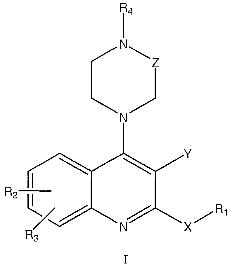
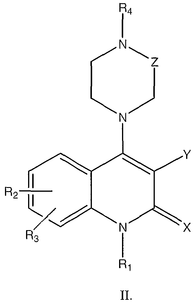
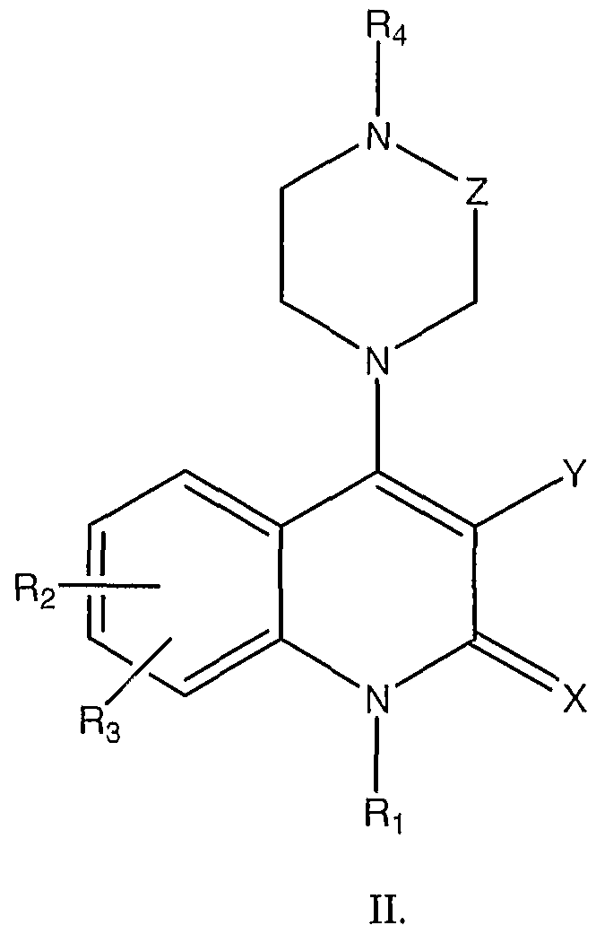
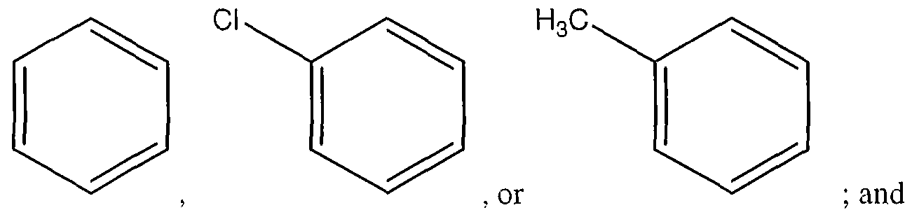
- the agent is an organic molecule comprising the following structure I or II
- the invention is additionally directed to methods of evaluating whether a compound is useful for preventing or treating type 1 diabetes.
- the methods comprise, (a) determining whether the compound inhibits a macrophage migration inhibitory factor (MIF) in a mammal, then, if the compound inhibits the MIF, (b) determining whether the compound inhibits development of type 1 diabetes.
- the invention is directed to kits comprising (a) a pharmaceutical composition comprising an agent that inhibits a macrophage migration inhibitory factor (MIF) in the mammal, where the agent is a polypeptide or a polynucleotide, and (b) instructions for administering the composition to the mammal.
- the mammal has type 1 diabetes or is at risk for type 1 diabetes.
- kits comprising (a) a pharmaceutical composition comprising an agent that inhibits a macrophage migration inhibitory factor (MIF) in the mammal, and (b) instractions for administering the composition to the mammal.
- MIF macrophage migration inhibitory factor
- the mammal has type 1 diabetes or is at risk for type 1 diabetes.
- the pharmaceutical composition of these embodiments is an organic molecule comprising the following structure I or II
- the invention is also directed to the use of an agent that inhibits a macrophage migration inhibitory factor (MIF) in the mammal, where the agent is a polypeptide or a polynucleotide, for the manufacture of a medicament for the treatment of a mammal having type 1 diabetes or at risk for type 1 diabetes. Additionally, the invention is directed to the use of an agent that inhibits a macrophage migration inhibitory factor (MIF) in the mammal for the manufacture of a medicament for the treatment of a mammal having type 1 diabetes or at risk for type 1 diabetes.
- the agent is an organic molecule comprising the following structure I or II
- FIG. 1 is micrographs and graphs demonstrating elevated MIF protein expression in pancreas and peritoneal cells of diabetic mice.
- Panels A-C are light immunomicrographs of pancreata.
- MIF is weakly expressed by islet cells of non-diabetic control mice (Panel A); in pancreatic islets of day 10 diabetic mice, there is massive mononuclear cell (MNC) infiltration and MIF expression is markedly up-regulated (Panel B); negative staining in a serial section of diabetic mice islet (Panel C).
- Panel E is a graph of quantitative analysis of intracellular expression of MEF protein in peritoneal cells of non-diabetic mice (Control), MLD-STZ diabetic mice (STZ), and MLD-STZ diabetic mice treated with anti-MIF antibody (STZ- ⁇ MIF), measured by cell-based ELISA performed with MIF-specific antibodies, as described in Materials and Methods of the Example. The results are presented as fold increase of control absorbance value (OD 492 nm 0.687 ⁇ 0.013). *p ⁇ 0.05 refers to otherwise untreated MLD-STZ diabetic animals.
- FIG. 2 is graphs of experimental results demonstrating that MIF blockade with anti-
- MIF antibody and ISO-1 suppress the development of hyperglycemia and insulitis.
- Blood glucose levels were determined in C57B1/6 mice (Panel A) and CBA/H mice (Panel B) starting at the beginning of the STZ treatment and continued through weekly measurements. Animals received MLD-STZ injections and were treated with either vehicle (STZ), non-immune rabbit IgG (STZ-IgG), anti-MIF IgG (STZ- ⁇ MIF), or ISO-1 (STZ-ISO).
- STZ non-immune rabbit IgG
- STZ- ⁇ MIF anti-MIF IgG
- ISO-1 ISO-1
- FIG. 3 is graphs of experimental results demonstrating that neutralization of MIF activity reduces splenocyte proliferation and adherence.
- Panel A shows 3 H-thymidine incorporation, as a measure of cell proliferation, as determined in SMNC isolated from mice untreated with STZ (control), treated with STZ and vehicle (STZ), with STZ and non-immune rabbit IgG (STZ-IgG), STZ and anti-MIF IgG (STZ- ⁇ MIF), or STZ and ISO-1 (STZ-ISO).
- Panel B shows adhesion to plastic surface, or L929 fibroblast, as determined for SMNC isolated from the same groups of mice.
- FIG. 4 is graphs of experimental results demonstrating that neutralization of MIF activity reduces the production of TNF- ⁇ .
- Spleen MNC Spleen MNC
- PC peritoneal cells
- pancreatic islets were isolated from control untreated mice (control), mice treated with STZ and non-immune rabbit IgG (STZ-IgG), STZ and anti-MIF IgG (STZ- ⁇ MIF), STZ and vehicle (STZ), and STZ and ISO-1 (STZ-ISO).
- TNF production was measured in the cell culture supernatants as described in Materials and Methods of the Example. Results are representative of three independent experiments with similar results.
- *p ⁇ 0.05 refers to corresponding STZ- IgG (Panel A) or STZ (Panel B) animals.
- FIG. 5 is graphs of experimental results demonstrating that neutralization of MIF activity down-regulates the expression of iNOS and NO production.
- Peritoneal cells (PC) and pancreatic islets were isolated from mice treated as described in FIG. 3.
- iNOS expression was determined by cell-based ELISA, and presented as fold increase compared to control value (O.D. 492 nm 0.445 ⁇ 0.027).
- Panel B after isolation from mice, PC were cultivated in medium for 48 hours, and pancreatic islets in the presence of 250 U/ml IFN- ⁇ + D -l ⁇ for 72 hours. Subsequently, nitrite accumulation in cell culture supernatants was determined. Results are representative of three independent experiments with similar results.
- FIG. 6 is graphs of experimental results demonstrating that neutralization of MIF activity does not affect the expression of IFN- ⁇ and MHC class H Spleen MNC (SMNC) and peritoneal cells (PC) were isolated from mice treated as described in FIG. 3.
- SMNC IFN- ⁇ and MHC class H Spleen MNC
- PC peritoneal cells
- Panel A IFN- ⁇ expression in SMNC was determined by cell-based ELISA, and presented as fold increase compared to control value (O.D. 492 nm 0.070 ⁇ 0.014).
- Panel B MHC ⁇ molecules expression in PC and SMNC was determined by cell-based ELISA, and presented as fold increase compared to control value (O.D.
- FIG. 7 is a graph of experimental results showing inhibition of diabetes in streptozotocin-treated mice that were treated with an MIF inhibitor.
- FIG. 8 is a graph of experimental results showing the effect of timing of MIF inhibitor ISO-1 and anti-MIF antibody on control of diabetes in streptozotocin-treated mice.
- FIG. 9 is a graph of experimental results showing that MJJF-null mice do not acquire diabetes after treatment with streptozotocin.
- MIF macrophage migration inhibitory factor
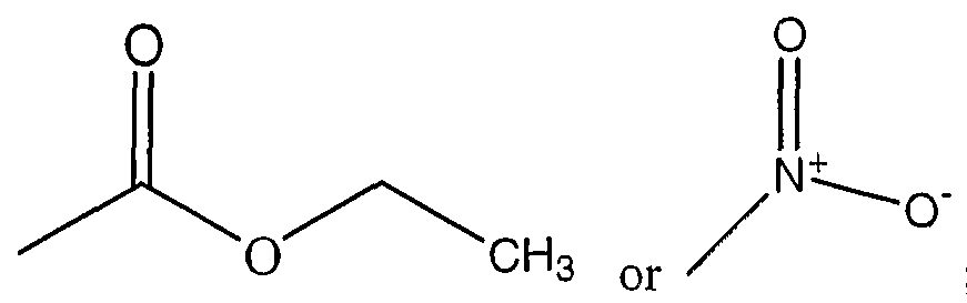
- ISO-1 (S,R)-3-(4- hydroxyphenyl)-4,5-dihydro-5-isoxazole acetic acid methyl ester
- iNOS inducible nitric oxide synthase
- STZ streptozotocin
- MLD-STZ multiple low doses of streptozotocin
- SMNC spleen mononuclear cells
- PC peritoneal cells
- IFN- ⁇ ⁇ -interferon
- TNF- ⁇ tumor necrosis factor- ⁇
- IL-l ⁇ interleukin-l ⁇ .
- the present invention is based on the discovery that MJF is a critical factor in the pathogenesis of type 1 diabetes. Therefore, inhibition of MIF prevents or attenuates the development of type 1 diabetes. See Examples.
- the invention is directed to methods of treating a mammal having type 1 diabetes or at risk for type 1 diabetes.
- the methods comprise administering to the mammal a pharmaceutical composition comprising an agent that inhibits a macrophage migration inhibitory factor (MIF) in the mammal.
- MIF macrophage migration inhibitory factor
- the agent is a polypeptide or a polynucleotide. In some of these methods, the agent inhibits activity of the MIF.
- an agent comprises an antibody binding site that binds specifically to the MIF, for example an antibody, an Fab fragment or an F(ab)2 fragment of an antibody.
- Such agents can be produced by well-known methods. Non-limiting methods include: immunization of animals with MIF, followed by isolation of anti-MIF antibodies from serum or production of anti-MIF monoclonal antibodies from hybridomas made by fusion of spenocytes with myeloma cells; or phage display or other recombinant methods.
- the monoclonal antibody is chosen or adapted to match the species to be treated.
- the anti-MIF antibody or antigen-binding fragment thereof
- the anti-MIF antibody will be a human antibody or a humanized antibody.
- Such antigen-specific human or humanized monoclonal antibodies may be developed by a variety of methods well known in the art.
- Another agent useful for these methods that inhibits activity of the MIF is an aptamer that binds specifically to the MJF.
- Aptamers are single stranded oligonucleotides or oligonucleotide analogs that bind to a particular target molecule, such as MIF.
- aptamers are the oligonucleotide analogy to antibodies.
- aptamers are smaller than antibodies, generally in the range of 50-100 nt. Their binding is highly dependent on the secondary structure formed by the aptamer oligonucleotide.
- RNA and single stranded DNA are known. See, e.g., 5,773,598; 5,496,938; 5,580,737; 5,654,151; 5,726,017; 5,786,462; 5,503,978; 6,028,186; 6,1 10,900; 6,124,449; 6,127,119; 6,140,490; 6,147,204; 6,168,778; and 6,171,795. Aptamers can also be expressed from a transfected vector (Joshi et al., 2002, J. Virol. 76,6545).
- SELEX Systematic Evolution of Ligands by Exponential enrichment
- the agent inhibits expression of the MEF.
- Preferred examples include inhibitory oligonucleotides in the form of antisense nucleic acids, ribozymes and small inhibitory nucleic acids (e.g., siRNA) specific for MDF mRNA in the mammal. Each of these inhibitory oligonucleotides requires knowledge of the sequence of MIF mRNA.
- MIF mRNA sequences for many mammalian species are known. See, e.g., SEQ ID NO: 1-3, providing MIF cDNA sequences for human, mouse and rat, respectively.
- the MJE cDNA sequence for any mammal could be determined without undue experimentation, e.g., by amplifying the sequence from a cDNA preparation of the mammal, using primers designed from a known mammalian MIF cDNA sequence.
- the inhibitory oligonucleotides of the present invention are antisense nucleic acids or mimetics. Antisense nucleic acid molecules act to directly block the translation of mRNA by hybridizing to targeted mRNA and preventing protein translation.
- Antisense approaches involve the design of oligonucleotides that are complementary to a portion of an MIF mRNA.
- the antisense oligonucleotides will bind to the complementary protective sequence mRNA transcripts and prevent translation. Absolute complementarity, although preferred, is not required.
- Ribozyme molecules designed to catalytically cleave MIF mRNA transcripts can also be used to prevent translation of MIF mRNA and, therefore, expression of the MIF protein. See, e.g., PCT Publication WO 90/11364; Sarver, et al., 1990.
- Ribozymes are enzymatic RNA molecules capable of catalyzing the specific cleavage of RNA. For a review, see Rossi, 1994.
- the mechanism of ribozyme action involves sequence specific hybridization of the ribozyme molecule to complementary target RNA, followed by an endonucleolytic cleavage event.
- the composition of ribozyme molecules must include one or more sequences complementary to the MJE mRNA, and must include the well known catalytic sequence responsible for mRNA cleavage. For this sequence, see, e.g., U.S. Pat. No. 5,093,246.
- Preferred types of ribozymes for the present invention are hammerhead ribozymes. In these embodiments the hammerhead ribozymes cleave MIF mRNA at locations dictated by flanking regions that form complementary base pairs with the mRNA.
- hammerhead ribozyme The sole requirement of the hammerhead ribozyme is that the mRNA have the two base sequence 5 -UG-3', which occurs numerous times in the MIF gene (see SEQ ID NO: 1-3).
- the constmction and production of hammerhead ribozymes is well known in the art and is described more fully in Myers, 1995, Molecular Biology and Biotechnology: A Comprehensive Desk Reference, VCH Publishers, New York, (see especially FIG. 4, page 833) and in Haseloff and Gerlach, 1988.
- the ribozymes of the present invention also include RNA endoribonucleases
- Cech-type ribozymes such as the one which occurs naturally in Tetrahvmena thermophila (known as the INS, or L-19 JNS R ⁇ A) and which has been extensively described by Thomas Cech and collaborators (Been and Cech, 1986; Zaug, et al, 1984; Zaug and Cech, 1986; Zaug, et al distract 1986; WO 88/04300, Cell, 47:207-216).
- the Cech-type ribozymes have an eight base pair active site that hybridizes to the MIF mR ⁇ A sequence wherever cleavage of the MIF R ⁇ A is desired.
- the invention encompasses those Cech-type ribozymes that target eight base-pair sequences that are present in the MIF mR ⁇ A.
- the ribozymes can be composed of modified oligonucleotides (e.g., for improved stability, targeting, etc.) and should be delivered to cells that make MIF in vivo, preferably activated T cells and/or macrophages.
- a preferred method of delivery involves using a D ⁇ A constract "encoding" the ribozyme under the control of a strong constitutive pol III or pol II promoter, so that transfected cells will produce sufficient quantities of the ribozyme to destroy endogenous MIF gene messages and inhibit translation.
- ribozymes unlike antisense molecules, are catalytic, a lower intracellular concentration is required for efficiency.
- endogenous target gene expression can be reduced by targeting deoxyribonucleotide sequences complementary to the regulatory region of the MIF gene (i.e., the MIF gene promoter and/or enhancers) to form triple helical structures that prevent transcription of the MIF gene in target cells in the body.
- deoxyribonucleotide sequences complementary to the regulatory region of the MIF gene i.e., the MIF gene promoter and/or enhancers
- Nucleic acid molecules to be used in triple helix formation for the inhibition of transcription should be single stranded and composed of deoxynucleotides.
- the base composition of these oligonucleotides must be designed to promote triple helix formation via Hoogsteen base pairing rules, which generally require sizable stretches of either purines or pyrimidines to be present on one strand of a duplex.
- Nucleic acids may be pyrimidine-based, which will result in TAT and CGC+ triplets across the three associated strands of the resulting triple helix.
- the pyrimidine-rich molecules provide base complementarity to a purine-rich region of a single strand of the duplex in a parallel orientation to that strand.
- nucleic acid molecules may be chosen that are purine-rich, for example, contain a stretch of G residues.
- These molecules will form a triple helix with a DNA duplex that is rich in GC pairs, in which the majority of the purine residues are located on a single strand of the targeted duplex, resulting in GGC triplets across the three strands in the triplex.
- GC-rich areas are available for targeting in the MIF gene (SEQ ID NO: 1-3).
- the potential sequences that can be targeted for triple helix formation may be increased by creating a so-called "switchback" nucleic acid molecule.
- Switchback molecules are synthesized in an alternating 5'-3', 3'-5' manner, such that they base pair with first one strand of a duplex and then the other, eliminating the necessity for a sizable stretch of either purines or pyrimidines to be present on one strand of a duplex.
- the oligonucleotide can be a small interfering RNA (siRNA), known in the art to be double stranded RNAs, complementary to the target mRNA (here MIF), that interacts with cellular factors to bind to the target sequence, which is then degraded.
- the siRNA sequence can be complementary to any portion of the MIF mRNA.
- the siRNA is preferably 21-23 nt long, although longer sequences will be processed to that length.
- References include Caplen et al, 2001 ; Elbashir et al., 2001; Jarvis and Ford, 2002; and Sussman and Peirce, 2002.
- Antisense RNA and DNA, ribozyme, triple helix, and siRNA molecules of the invention may be prepared by any method known in the art for the synthesis of DNA and RNA molecules, as discussed above. These include techniques for chemically synthesizing oligodeoxyribonucleotides and oligoribonucleotides well known in the art such as for example solid-phase phosphoramidite chemical synthesis.
- RNA molecules may be generated by in vitro or in vivo transcription of DNA sequences encoding the antisense RNA molecule.
- DNA sequences may be incorporated into a wide variety of vectors that incorporate suitable RNA polymerase promoters such as the T7 or SP6 polymerase promoters.
- antisense cDNA constructs that synthesize antisense RNA constitutively or inducibly, depending on the promoter used, can be introduced stably into cell lines, or into target cells in the mammal by known gene therapy methods.
- oligonucleotide refers to an oligomer or polymer of ribonucleic acid (RNA) or deoxyribonucleic acid (DNA) or mimetics thereof. This term includes oligonucleotides composed of naturally occurring nucleobases, sugars and covalent internucleoside (backbone) linkages as well as oligonucleotides having non-naturally-occurring portions which function similarly. Such oligonucleotide mimetics are often preferred over native forms because of desirable properties such as, for example, enhanced cellular uptake, enhanced affinity for nucleic acid target and increased stability in the presence of nucleases.
- oligonucleotides containing modified backbones or non-natural internucleoside linkages include those that retain a phosphorus atom in the backbone and those that do not have a phosphorus atom in the backbone.
- Preferred modified oligonucleotide backbones include, for example, phosphorothioates, chiral phosphorothioates, phosphorodithioates, phosphotriesters, aminoalkylphosphotriesters, methyl and other al yl phosphonates including 3'-alkylene phosphonates, 5 -alkylene phosphonates and chiral phosphonates, phosphinates, phosphoramidates including 3'-amino phosphoramidate and aminoalkylphosphoramidates, thionophosphoramidates, thionoalkylphosphonates, thionoalkylphosphotriesters, selenophosphates and borano-phosphates having normal 3'-5' linkages, 2'-5' linked analogs of these, and those having inverted polarity wherein one or more intemucleotide linkages is a 3' to 3', 5' to 5' or 2' to 2' linkage.
- Preferred oligonucleotides having inverted polarity comprise a single 3' to 3' linkage at the 3'-most intemucleotide linkage i.e. a single inverted nucleoside residue which may be abasic (the nucleobase is missing or has a hydroxyl group in place thereof).
- Various salts, mixed salts and free acid forms are also included.
- Representative United States patents that teach the preparation of the above phosphorus-containing linkages include, but are not limited to, U.S. Pat. Nos.
- Preferred modified oligonucleotide backbones that do not include a phosphorus atom therein have backbones that are formed by short chain alkyl or cycloalkyl internucleoside linkages, mixed heteroatom and alkyl or cycloalkyl internucleoside linkages, or one or more short chain heteroatomic or heterocyclic internucleoside linkages.
- morpholino linkages formed in part from the sugar portion of a nucleoside
- siloxane backbones sulfide, sulfoxide and sulfone backbones
- formacetyl and thioformacetyl backbones methylene formacetyl and thioformacetyl backbones
- riboacetyl backbones alkene containing backbones; sulfamate backbones; methyleneimino and methylenehydrazino backbones; sulfonate and sulfonamide backbones; amide backbones; and others having mixed N, O, S and CH 2 component parts.
- both the sugar and the internucleoside linkage, i.e., the backbone, of the nucleotide units are replaced with novel groups.
- the base units are maintained for hybridization with an appropriate nucleic acid target compound.
- an oligomeric compound an oligonucleotide mimetic that has been shown to have excellent hybridization properties, is referred to as a peptide nucleic acid (PNA).
- PNA peptide nucleic acid
- the sugar-backbone of an oligonucleotide is replaced with an amide containing backbone, in particular an aminoethylglycine backbone.
- nucleobases are retained and are bound directly or indirectly to aza nitrogen atoms of the amide portion of the backbone.
- Representative United States patents that teach the preparation of PNA compounds include, but are not limited to, U.S. Pat. Nos. 5,539,082; 5,714,331; and 5,719,262. Further teaching of PNA compounds can be found in Nielsen et al., Science, 1991, 254, 1497-1500. Modified oligonucleotides may also contain one or more substituted sugar moieties.
- Preferred oligonucleotides comprise one of the following at the 2' position: OH; F; 0-, S-, or N-alkyl; O-, S-, or N-alkenyl; 0-, S- or N-alkynyl; or 0-alkyI-O-alkyl, wherein the alkyl, alkenyl and alkynyl may be substituted or unsubstituted Cl to CIO alkyl or C2 to CIO alkenyl and alkynyl.
- n and m are from 1 to about 10.
- oligonucleotides comprise one of the following at the 2' position: Cl to CIO lower alkyl, substituted lower alkyl, alkenyl, alkynyl, alkaryl, aralkyl, O-alkaryl or O- aralkyl, SH, SCH 3 , OCN, Cl, Br, CN, CF 3 , OCF 3 , SOCH 3 , S0 2 CH 3 , ON 2 , N0 2 , N 3 , NH 2 , heterocycloalkyl, heterocycloalkaryl, aminoalkylamino, polyalkylamino, substituted silyl, an RNA cleaving group, a reporter group, an intercalator, a group for improving the pharmacokinetic properties of an oligonucleotide, or a group for improving the pharmacodynamic properties of an oligonucleotide, and other substituents having similar properties.
- a preferred modification includes 2'-methoxyethoxy (2'-OCH 2 CH 2 OCH 3 , also known as 2'-0-(2-methoxyethyl) or 2'-MOE) (Martin et al., Helv. Chim. Acta, 1995, 78, 486- 504) i.e., an alkoxyalkoxy group.
- a further preferred modification includes 2'- dimethylaminooxyethoxy, i.e., a 0(CH 2 ) 2 ⁇ N(CH 3 ) 2 group, also known as 2 -DMAOE, as described in examples hereinbelow, and 2'-dimethylaminoethoxyethoxy (also known in the art as 2'-0-dimethylaminoethoxyethyl or 2'-DMAEOE), i.e., 2'-OCH 2 OCH 2 N(CH 2 ) 2 , also described in examples hereinbelow.
- a further preferred modification includes Locked Nucleic Acids (LNAs) in which the 2'-hydroxyl group is linked to the 3' or 4' carbon atom of the sugar ring thereby forming a bicyclic sugar moiety.
- the linkage is preferably a methelyne (CH 2 ) n group bridging the 2' oxygen atom and the 3' or 4' carbon atom wherein n is 1 or 2.
- LNAs and preparation thereof are described in WO 98/39352 and WO 99/14226.
- the 2'-modification may be in the arabino (up) position or ribo (down) position.
- a preferred 2'-arabino modification is 2'-F.
- oligonucleotide Similar modifications may also be made at other positions on the oligonucleotide, particularly the 3' position of the sugar on the 3' terminal nucleotide or in 2'-5' linked oligonucleotides and the 5' position of 5' terminal nucleotide. Oligonucleotides may also have sugar mimetics such as cyclobutyl moieties in place of the pentofuranosyl sugar. Representative United States patents that teach the preparation of such modified sugar stmctures include, but are not limited to, U.S. Pat. Nos.
- Oligonucleotides may also include nucleobase (often referred to in the art simply as
- nucleobases include the purine bases adenine (A) and guanine (G), and the pyrimidine bases thymine (T), cytosine (C) and uracil (U).
- Modified nucleobases include other synthetic and natural nucleobases such as 5-methylcytosine (5-me-C), 5-hydroxymethyl cytosine, xanthine, hypoxanthine, 2-aminoadenine, 6-methyl and other alkyl derivatives of adenine and guanine, 2- propyl and other alkyl derivatives of adenine and guanine, 2-thiouracil, 2-thiothymine and 2- thiocytosine, 5-halouracil and cytosine, 5-propynyl, uracil and cytosine and other alkynyl derivatives of pyrimidine bases, 6-azo uracil, cytosine and thymine, 5-uracil (pseudouracil), 4- thiouracil, 8-halo, 8-amino, 8-thiol, 8-thioalkyl, 8-hydroxyl and other 8-substituted adenines and guanines, 5-halo particularly
- nucleobases include tricyclic pyrimidines such as phenoxazine cytidine, phenothiazine cytidine, G-clamps such as a substituted phenoxazine cytidine, carbazole cytidine, and pyridoindole cytidine. Modified nucleobases may also include those in which the purine or pyrimidine base is replaced with other heterocycles, for example 7-deaza-adenine, 7-deazaguanosine, 2-aminopyridine and 2-pyridone. Further nucleobases include those disclosed in U.S. Pat. No.
- 5-substituted pyrimidines include 5-substituted pyrimidines, 6-azapyrimidines and N-2, N-6 and 0-6 substituted purines, including 2-aminopropyladenine, 5-propynyluracil and 5-propynylcytosine.
- 5-methylcytosine substitutions have been shown to increase nucleic acid duplex stability by 0.6-1.2°C. (Sanghvi, Y. S., Crooke, S. T. and Lebleu, B., eds., Antisense Research and Applications, CRC Press, Boca Raton, 1993, pp. 276-278) and are presently preferred base substitutions, even more particularly when combined with 2'-0-methoxyethyl sugar modifications.
- oligonucleotides of the invention involves chemically linking to the oligonucleotide one or more moieties or conjugates that enhance the activity, cellular distribution or cellular uptake of the oligonucleotide.
- the compounds of the invention can include conjugate groups covalently bound to functional groups such as primary or secondary hydroxyl groups.
- Conjugate groups of the invention include intercalators, reporter molecules, polya ines, polyamides, polyethylene glycols, polyethers, groups that enhance the pharmacodynamic properties of oligomers, and groups that enhance the pharmacokinetic properties of oligomers.
- Typical conjugates groups include cholesterols, lipids, phospholipids, biotin, phenazine, folate, phenanthridine, anthraquinone, acridine, fluoresceins, rhodamines, coumarins, and dyes.
- Groups that enhance the pharmacodynamic properties include groups that improve oligomer uptake, enhance oligomer resistance to degradation, and/or strengthen sequence-specific hybridization with RNA.
- Groups that enhance the pharmacokinetic properties include groups that improve oligomer uptake, distribution, metabolism or excretion. Representative conjugate groups are disclosed in international Patent Application PCT/US92/09196.
- Conjugate moieties include but are not limited to lipid moieties such as a cholesterol moiety (Letsinger et al., Proc. Natl. Acad. Sci. USA, 1989, 86, 6553-6556), cholic acid (Manoharan et al., Bioorg. Med. Chem. Let., 1994, 4, 1053-1060), a thioether, e.g., hexyl-S-tritylthiol (Manoharan et al., Ann. N.Y. Acad. Sci., 1992, 660, 306-309; Manoharan et al, Bioorg. Med. Chem.
- lipid moieties such as a cholesterol moiety (Letsinger et al., Proc. Natl. Acad. Sci. USA, 1989, 86, 6553-6556), cholic acid (Manoharan et al., Bioorg. Med. Chem. Let., 1994, 4, 1053-10
- Acids Res., 1990, 18, 3777-3783 a polyamine or a polyethylene glycol chain (Manoharan et al., Nucleosides & Nucleotides, 1995, 14, 969- 973), or adamantane acetic acid (Manoharan et al., Tetrahedron Lett., 1995, 36, 3651-3654), a palmityl moiety (Mishra et al., Biochim. Biophys. Acta, 1995, 1264, 229-237), or an octadecylamine or hexylamino-carbonyl-oxycholesterol moiety (Crooke et al., J. Pharmacol. Exp.
- Oligonucleotides of the invention may also be conjugated to active drag substances, for example, aspirin, warfarin, phenylbutazone, ibuprofen, suprofen, fenbufen, ketoprofen, (S)-(+)-pranoprofen, carprofen, dansylsarcosine, 2,3,5-triiodobenzoic acid, flufenamic acid, folinic acid, a benzothiadiazide, chlorothiazide, a diazepine, indomethicin, a barbiturate, a cephalosporin, a sulfa drag, an antidiabetic, an antibacterial or an antibiotic.
- active drag substances for example, aspirin, warfarin, phenylbutazone, ibuprofen, suprofen, fenbufen, ketoprofen, (S)-(+)-pranoprofen, carprofen
- the present invention also includes inhibitory oligonucleotide compounds that are chimeric compounds.
- "Chimeric" inhibitory oligonucleotide compounds or “chimeras,” in the context of this invention, are inhibitory oligonucleotides that contain two or more chemically distinct regions, each made up of at least one monomer unit, i.e., a nucleotide in the case of an oligonucleotide compound.
- oligonucleotides typically contain at least one region wherein the oligonucleotide is modified so as to confer upon the oligonucleotide increased resistance to nuclease degradation, increased cellular uptake, and/or increased binding affinity for the target nucleic acid.
- An additional region of the oligonucleotide may serve as a substrate for enzymes capable of cleaving RNA:DNA or RNA:RNA hybrids.
- RNase H is a cellular endonuclease which cleaves the RNA strand of an RNA:DNA duplex.
- RNA target Activation of RNase H, therefore, results in cleavage of the RNA target, thereby greatly enhancing the efficiency of oligonucleotide inhibition of gene expression. Consequently, comparable results can often be obtained with shorter oligonucleotides when chimeric oligonucleotides are used, compared to phosphorothioate deoxyoligonucleotides hybridizing to the same target region.
- Cleavage of the RNA target can be routinely detected by gel electrophoresis and, if necessary, associated nucleic acid hybridization techniques known in the art.
- Chimeric inhibitory oligonucleotide compounds of the invention may be formed as composite stmctures of two or more oligonucleotides, modified oligonucleotides, oligonucleosides and/or oligonucleotide mimetics as described above. Such compounds have also been referred to in the art as hybrids or gapmers. Representative LTnited States patents that teach the preparation of such hybrid stmctures include, but are not limited to, U.S. Pat.
- the inhibitory oligonucleotides useful for the invention methods may be synthesized in vitro.
- the inhibitory oligonucleotides of the invention may also be admixed, encapsulated, conjugated or otherwise associated with other molecules, molecule stmcUires or mixtures of compounds, as for example, liposomes, receptor targeted molecules, oral, rectal, topical or other formulations, for assisting in uptake, distribution and/or absorption.
- Representative United States patents that teach the preparation of such uptake, distribution and/or absorption assisting formulations include, but are not limited to, U.S. Pat. Nos. 5,108,921; 5,354,844;
- prodrug indicates a therapeutic agent that is prepared in an inactive form that is converted to an active form (i.e., drug) within the body or cells thereof by the action of endogenous enzymes or other chemicals and/or conditions.
- prodrug versions of the oligonucleotides of the invention are prepared as SATE [(S-acetyl-2-thioethyl) phosphate] derivatives according to the methods disclosed in WO 93/24510 to Gosselin et al., published
- the oligonucleotides can also comprise a non-nucleotide moiety, such as a hapten, a fluorescent molecule, or a radioactive moiety, useful, e.g., to detect or quantify the amount of oligonucleotide that has entered a cell.
- the mammal has or is at risk for having diabetes, impaired glucose intolerance, stress hyperglycemia, metabolic syndrome, and/or insulin resistance. These methods can be used with any mammal.
- the mammal is a rodent
- the mammal is a human.
- the invention is also directed to methods of treating a mammal having type 1 diabetes or at risk for type 1 diabetes. These methods comprise administering to the mammal a pharmaceutical composition comprising an agent that inhibits a macrophage migration inhibitory factor (MIF) in the mammal.
- MIF macrophage migration inhibitory factor
- the agent is an organic molecule comprising the following structure I or II
- the organic molecule comprises stracture II, where
- compositions can be formulated without undue experimentation for administration to a mammal, including humans, as appropriate for the particular application. Additionally, proper dosages of the compositions can be determined without undue experimentation using standard dose-response protocols. Accordingly, the compositions designed for oral, lingual, sublingual, buccal and intrabuccal administration can be made without undue experimentation by means well known in the art, for example with an inert diluent or with an edible carrier. The compositions may be enclosed in gelatin capsules or compressed into tablets.
- the pharmaceutical compositions of the present invention may be incorporated with excipients and used in the form of tablets, troches, capsules, elixirs, suspensions, syrups, wafers, chewing gums and the like.
- Tablets, pills, capsules, troches and the like may also contain binders, recipients, disintegrating agent, lubricants, sweetening agents, and flavoring agents.
- binders include microcrystalline cellulose, gum tragacanth or gelatin.
- excipients include starch or lactose.
- disintegrating agents include alginic acid, corn starch and the like.
- lubricants include magnesium stearate or potassium stearate.
- compositions useful for the present invention can easily be administered parenterally such as for example, by intravenous, intramuscular, intrathecal or subcutaneous injection. Parenteral administration can be accomplished by incorporating the compositions of the present invention into a solution or suspension.
- Such solutions or suspensions may also include sterile diluents such as water for injection, saline solution, fixed oils, polyethylene glycols, glycerine, propylene glycol or other synthetic solvents.
- Parenteral formulations may also include antibacterial agents such as for example, benzyl alcohol or methyl parabens, antioxidants such as for example, ascorbic acid or sodium bisulfite and chelating agents such as EDTA. Buffers such as acetates, citrates or phosphates and agents for the adjustment of tonicity such as sodium chloride or dextrose may also be added.
- the parenteral preparation can be enclosed in ampules, disposable syringes or multiple dose vials made of glass or plastic.
- Rectal administration includes administering the pharmaceutical compositions into the rectum or large intestine. This can be accomplished using suppositories or enemas.
- Suppository formulations can easily be made by methods known in the art. For example, suppository formulations can be prepared by heating glycerin to about 120° C, dissolving the composition in the glycerin, mixing the heated glycerin after which purified water may be added, and pouring the hot mixture into a suppository mold.
- Transdermal administration includes percutaneous absorption of the composition through the skin. Transdermal formulations include patches (such as the well-known nicotine patch), ointments, creams, gels, salves and the like.
- nasally administering or nasal administration includes administering the composition to the mucous membranes of the nasal passage or nasal cavity of the patient.
- pharmaceutical compositions for nasal administration of a composition include therapeutically effective amounts of the composition prepared by well-known methods to be administered, for example, as a nasal spray, nasal drop, suspension, gel, ointment, cream or powder. Administration of the composition may also take place using a nasal tampon or nasal sponge.
- the mammal has or is at risk for having diabetes or conditions associated with diabetes or a risk of diabetes, for example impaired glucose intolerance, stress hyperglycemia, metabolic syndrome, and/or insulin resistance.
- the present invention can be utilized for any mammal, for example rodents, generally used as experimental models for diabetes (see Example), or humans.
- the discovery that compounds that inhibit MIF activity or expression are useful for treatment of type 1 diabetes indicates that screens of compounds for MIF-inhibiting activity can be used to identify compounds for the ability to prevent or treat type 1 diabetes.
- the invention is directed to methods of evaluating whether a compound is useful for preventing or treating type 1 diabetes.
- the methods comprise the following two steps: (a) determining whether the compound inhibits a macrophage migration inhibitory factor (MIF) in a mammal, then, if the compound inhibits the MEF, (b) determining whether the compound inhibits development of type 1 diabetes.
- MIF macrophage migration inhibitory factor
- the ability of the tested compound to inhibit MIF activity can be determined by any known procedure. Non-limiting examples include those methods described in U.S. Patent Application Publications 2003/0008908 and 2003/0195194. Similarly, any known procedure for evaluating the effect of a compound on type 1 diabetes can be utilized.
- the effect of the compound on type 1 diabetes is determined by evaluating the effect of the compound on development of diabetes in animal models utilizing multiple low dose streptozotocin administration or in animal models genetically susceptible to development of type 1 diabetes, such as the NOD mouse.
- the effect of a test compound or formulation on the development of diabetes in such models may be assessed by a variety of convenient methods, for instance by monitoring circulating glucose or proliferation of splenic lymphocytes in the mammal (see also Example). These methods are not limited to evaluation of any particular type of compound.
- kits comprising (a) a pharmaceutical composition comprising an agent that inhibits a macrophage migration inhibitory factor (MIF) in the mammal, where the agent is a polypeptide or a polynucleotide, and (b) instructions for administering the composition to the mammal.
- MIF macrophage migration inhibitory factor
- the mammal has type 1 diabetes or is at risk for type 1 diabetes.
- the invention is directed to other kits comprising (a) a pharmaceutical composition comprising an agent that inhibits a macrophage migration inhibitory factor (MIF) in the mammal, and (b) instractions for administering the composition to the mammal.
- MIF macrophage migration inhibitory factor
- the mammal has type 1 diabetes or is at risk for type 1 diabetes.
- the pharmaceutical composition of these embodiments is an organic molecule comprising the following stracture I or II
- the invention is also directed to the use of an agent that inhibits a macrophage migration inhibitory factor (MIF) in the mammal, where the agent is a polypeptide or a polynucleotide, for the manufacture of a medicament for the treatment of a mammal having type 1 diabetes or at risk for type 1 diabetes. Additionally, the invention is directed to the use of an agent that inhibits a macrophage migration inhibitory factor (MIF) in the mammal for the manufacture of a medicament for the treatment of a mammal having type 1 diabetes or at risk for type 1 diabetes.
- the agent is an organic molecule comprising the following stracture I or II
- Example 1 Neutralization of Macrophage Migration Inhibitory Factor (MIF) Activity Attenuates Experimental Autoimmune Diabetes
- MIF Macrophage Migration Inhibitory Factor
- MIF mRNA expression is upregulated in non-obese diabetic mice, yet little is known about its potential as a regulator of type 1 diabetes.
- MIF protein is significantly elevated in islet cells during the development of experimental diabetes induced in mice by multiple low doses of streptozotocin.
- Attenuation of MIF activity with exemplary MIF inhibitors such as neutralizing antibodies against MIF, or the pharmacological MIF inhibitor (S,R)-3-(4- hydroxyphenyl)-4,5-dihydro-5-isoxazole acetic acid methyl ester (ISO-1), markedly reduces histopathological changes in the islets of pancreas and suppresses the development of hyperglycaemia, showing the general utility of this method of treating mammals with, or at risk for, type 1 diabetes.
- MIF inhibitors such as neutralizing antibodies against MIF, or the pharmacological MIF inhibitor (S,R)-3-(4- hydroxyphenyl)-4,5-dihydro-5-isoxazole acetic acid methyl ester (ISO-1)
- MDF also stimulates the synthesis of other proinflammatory cytokines and soluble mediators involved in the pathogenesis of type 1 DM such as TNF- ⁇ , IL-l ⁇ and nitric oxide (NO) (19) raised the possibility that inhibition of MIF activity may modulate the development of disease.
- TNF- ⁇ proinflammatory cytokines
- IL-l ⁇ soluble mediators involved in the pathogenesis of type 1 DM
- NO nitric oxide
- an anti-MIF polyclonal antibody as well as (S,R)-3-(4-hydroxyphenyl)-4,5-dihydro-5-isoxazole acetic acid methyl ester (ISO-1), a pharmacological inhibitor of MIF (20), were given to mice as prophylactic treatment prior to the challenge with STZ.
- Various immunological parameters relevant for DM pathogenesis were determined, including autoreactive cell proliferation and adhesion, as well as the production of proinflammatory mediators. To our knowledge, this is the first attempt to interfere in autoimmune diabetes by in vivo neutralization of MIF. Materials and Methods Mice. Inbred CBA/H mice were obtained from our own breeding colony at the Institute for Biological Research, Belgrade.
- mice Inbred C57 Bl/6 mice were originally purchased from Charles River (Calco, Italy) and then bred by brother per sister mating for up to 4 generations. Mice were kept under standard laboratory conditions with free access to food and water. The handling of the mice and the study protocol were approved by the local Institutional Animal Care and Use Committee. Reagents. Streptozotocin (STZ, S-0130), sulfanilamide, naphthylenediamine dichidrochloride and irrelevant rabbit IgG were purchased from Sigma (St. Louis, MO). Anti- murine MIF IgG was prepared from rabbit serum raised against murine MIF and purified by protein A affinity chromatography following the manufacturer's instractions (Pierce, Rockford, IL).
- ISO-1 [(S,R)-3-(4-hydroxyphenil)-4,5-dihydro-5-isoxasole acetic acid methyl ester] was synthesized as previously described (20). Diabetes induction and in vivo treatments. Diabetes was induced in adult male mice with multiple subtoxic doses of streptozotocin (MLD-STZ, 40 mg/kg body wt/day i.p. for five consecutive days) as described (21). The impact of polyclonal antibody against MJP was studied by i.p. injection of mice (5-10 per treatment group) with 5 mg/kg of rabbit IgG antibody against mouse MJP on days -3, -1, 2 and 5 in relation to the first STZ dose. The effect of ISO-1 was studied by i.p.
- PC Resident peritoneal cells
- SMNC spleen mononuclear cells
- pancreatic islets were isolated from individual anti-MIF IgG-treated, ISO-1 -treated or control diabetic mice on day 15 after the first injection of STZ, as well as from normal untreated animals.
- SMNC SMNC (5xl0°/well) prepared by Ficoll gradient centrifugation, resident PC (2.5xl0 5 /well) collected by peritoneal lavage with cold PBS, or pancreatic islets (150-200 islets/well) prepared by collagenase digestion and density gradient purification as described (22), were incubated in 24-well Limbro culture plates in 1 ml of RPMI-1640 culture medium containing 5% fetal bovine serum (FBS) and cell supernatants were collected after 48 h. Concentration of bioactive TNF- ⁇ in culture supernatant was determined as previously described (22) using a cytolytic bioassay with actinomycin D-treated fibrosarcoma cell line L929.
- Nitrite accumulation an indicator of NO production, was determined in cell culture supernatant using the Griess reaction (22).
- the expression of intracellular MIF, IFN- ⁇ , MHC II, or iNOS was determined by slight modification of cell-based ELISA protocol (23).
- Spleen MNC (5 l ⁇ 7weII) or PC (2.5xl0 5 /well), were allowed to adhere to poly-L-lysine-precoated 96-well microplates.
- Ig(H+L)-HRP or goat anti-mouse Ig(F(ab') 2 -specific)-HRP washed again and incubated for 15 min at room temperauire in dark with 50 ⁇ l of a solution containing 0.4 mg/ml OPD (Sigma), 11.8 mg/ml Na 2 HP0 4 x2H 2 0, 7.3 mg/ml citric acid and 0.015% H 2 0 2 .
- the reaction was stopped with 3N HC1, and the absorbance was measured in a microplate reader at 492 nm in a Titertek microplate reader (Flow). Ex vivo Iymphoproliferative response and adhesion assay.
- Spontaneous proliferation of SMNC was determined by incubation of cells (5xl0 /well) from each individual animal in 96-well microplates with 1 ⁇ Ci 3 H-thymidine ( ⁇ -TdR, ICN). Incorporated radioactivity was measured after 24 h in a Beckman liquid scintillation counter.
- the analysis of spontaneous adhesion of SMNC (2.5xl0 5 /well) to a monolayer of L929 fibroblasts or plastic was performed by using crystal violet assay as previously described (21). The absorbance corresponding to the number of adherent cells, was measured at 570 nm. Antibodies and flow cvtometrv.
- Spleen MNC (lxlO 6 ) were incubated with the rat anti- mouse monoclonal antibodies (mAbs): anti-CDl lb (MAC-l)-phycoerythrin (PE), or anti- CD25 (IL-2 Receptor ⁇ chain, p55)-biotin (PharMingen, San Diego, CA), followed by
- Streptavidin-PE Streptavidin-PE (PharMingen). Each cell suspension of SMNC was a pool from three to five animals obtained from the same experimental group 15 days after diabetes induction, as well as from normal untreated mice. Cell surface marker expression was analyzed with a flow cytometer (FACScalibur, Becton Dickinson, Heidelberg, Germany) and Win MDI 2.8 software (Joseph Trotter). Histological and immunohistochemical examination of pancreas, Pancreata were fixed in neutral buffered formalin and then embedded in paraffin. The fixed blocks were sectioned (7 ⁇ m thick) and haematoxylin and eosin staining was performed to assess the incidence and degree of inflammatory changes.
- Insulitis scoring was performed as previously described (10, 11) by examining at least 15 islets per mouse and graded in a blinded fashion as follows: 0, no infiltrate; 1, peri-ductal infiltrate; 2, peri-islet infiltrate; 3, intraislet infiltrate; and 4, intraislet infiltrate associated with ⁇ -cell destruction. At least 15 islets were counted for each mouse. A mean score for each pancreas was calculated by dividing the total score by the number of islets examined. Insulitis scores (IS) are expressed as mean values ⁇ SD. Immunohistochemistry was performed on paraffin-embedded sections of formalin-fixed tissue using a previously described microwave-based method (24).
- MIF immunostaining a polyclonal rabbit anti-MIF IgG and control rabbit IgG were used. The examined area of the islet was outlined, and the percentage of MIF-positive islet cells was measured using quantitative Image Analysis System (Optima 6.5, Media Cybernatics, Silver Springs, MD). Statistical analysis. The blood glucose values are shown as mean values ⁇ SE.
- Anti-MIF prophylaxis suppresses clinical and histological parameters of MLD-STZ- induced diabetes.
- MIF activity modulates disease in MLD- STZ-exposed mice.
- C57B1/6 and CBA H control mice treated with PBS or non-immune IgG developed sustained hyperglycemia over a 2- week period following MLD-STZ injections.
- MLD-STZ induced different degrees of hyperglycemia in the two mouse strains, treatment of either C57B1/6 mice (FIG. 2A), or
- mice CBA/H mice (FIG. 2B) with anti-MIF Ab from day -3 to day +5 significantly inhibited MLD- STZ-induced hyperglycemia.
- ISO-1 pharmacological MIF antagonist
- mice prophylactically over a period of 14 days with ISO-1, starting 3 days before the first of five injections of STZ.
- mice remained euglycemic throughout the eight- week experimental period (FIG. 2B).
- the anti-diabetogenic effect of either immunological or pharmacological neutralization of MIF was long-lasting, with limited variations throughout the entire 56 day follow-up period (FIG. 2A and B).
- MIF blockade by ISO-1 attenuated inflammation of the islets (FIG. 2D).
- spleen mononuclear cells SMNC
- SMNC spleen mononuclear cells
- Previous studies have shown that isolated autoreactive lymphocytes from diabetic mice exhibit significantly increased ex vivo spontaneous proliferation in comparison to cells isolated from healthy animals, suggesting that this cell type may contribute to disease progression (21, 22).
- MIF inhibitors modulate splenocyte proliferative responses ex vivo
- diabetic CBA/H mice were treated with either anti-MIF antibodies or ISO-1, euthanized at day 15 after MLD-STZ exposure, and splenocy tes harvested for ex vivo analyses.
- MIF antagonists down-regulate the expression of CDl lb and CD25 of splenic mononuclear cells
- Treatment groups CDl lb" CD25 * % MFI % MFI Exp. 1
- lymphocytes were isolated from the spleens of control or anti-MJJF-treated mice 15 days post-MLD-STZ exposure and labeled with fluorescently tagged antibodies directed against IL-2R (CD25). Staining the cells for EL-2R expression (Table 1) revealed significant increase in both the percentage of 1L-2R "1" lymphocytes and antigen density in control diabetic mice, when comparing to normal untreated animals, while the in vivo treatment with anti-MIF IgG, or with ISO-1 reduced these parameters. Production of pro-inflammatory mediators.
- MIF significantly contributes to the immunopathology associated with excessive inflammation and autoimmunity, and neutralizing endogenous MIF with neutralizing anti-MIF antibodies, or targeted disruption of the MIF gene, inhibits the development of several rodent models of inflammatory disease, including immunologically induced kidney disease (27), leishmaniasis (28), Gram-negative and Gram-positive sepsis (15, 29, 30), antigen-induced arthritis (31), colitis (16), and experimental autoimmune encephalomyelitis (EAE) (17).
- MIF myeloma
- pancreatic ⁇ -cells a potential role for MDF in the development and pathogenesis of autoimmune- mediated DM
- MIF is constitutively expressed and secreted together with insulin from pancreatic ⁇ -cells, and acts as an autocrine factor to stimulate insulin release (32).
- induction of insulin secretion is thought to contribute to immunoinflammatory diabetogenic pathways by favoring the expression on the ⁇ - cells and the presentation to the immune cells of antigens that are up-regulated when the functional activity is augmented (12), this "hormonal" property could represent an additional important factor involving endogenous MIF in the initial events of ⁇ -cell dysfunction and destruction.
- Targeting endogenous MJP may therefore be a suitable approach for unraveling the role of this cytokine in the pathogenesis of type 1 DM and for therapeutic and/or prophylactic treatment of the condition.
- endogenous MIF plays a key role in the development of murine autoimmune diabetes. Progression of MLD-STZ-induced diabetes was accompanied by up-regulated MJP protein expression both in pancreatic islets and peripheral cells.
- Immunoneutralization of MIF by anti-MIF IgG, or pharmacological inhibition of MIF activity with ISO-1 markedly attenuated the clinical and histological manifestations of the disease. It inhibited the inflammatory responses as well as splenocyte proliferation and adherence ex vivo.
- MIF activity also has been shown to down-regulate adhesion molecule-dependent target tissue pathology in EAE (17).
- Another important finding of this study is that anti-MIF treatment affects the production of pro-inflammatory and cytotoxic mediators.
- the lower local production of TNF- ⁇ and NO may result from the reduction of inflammatory cell influx into pancreas.
- MIF blockade might also directly influence macrophage and T cell effector function, as suggested by down-regulation of TNF- ⁇ , iNOS and NO by peritoneal and/or spleen cells. This is consistent with the in vitro ability of anti-MIF antibodies to interfere with production of these mediators (28).
- MIF up-regulates in vitro production of TNF- ⁇ , as well as of reactive oxygen species and nitrogen metabolites (35). It therefore is conceivable that direct blockade of MIF-mediated iNOS expression and TNF- ⁇ synthesis may contribute to down-regulation of tissue-damaging pro-inflammatory mediators in type 1 DM. Thus, we hypothesize that the efficacy of MIF blockade in the treatment of MLD- STZ DM is due to inhibitory effects on the autoimmune/inflammatory response of T cells and macrophages, as well as islet cells.
- IFN- ⁇ may possess dicothomic effects on inflammation, exerting proinflammatory effects when produced at the level of the organ targeted by the immune response (e.g. the islet microenvironment), while activating corticosteroid-dependent and independent anti-inflammatory pathways at the systemic level (8).
- MIF is a critical mediator of inflammatory and autoimmune diseases, because neutralizing endogenous MIF activity with anti-MIF antibodies has been effective in animal models of septic shock, colitis, encephalomyelitis, and leishmania infection (15-17, 28), and may be a promising approach for therapy of various human diseases.
- treatment approaches that rely on exogenously administered proteins, including humanized antibodies may face several challenges, including potential immunogenicity, that suggest the desirability of more preferred embodiments.
- Anti-cytokine antibodies can form small inflammatory complexes with cytokines, and thereby exacerbate inflammatory responses (39). For these reasons, alternatives to the therapeutic use of anti-MEF Ig preparations should also be considered for pharmaceutical development. Thus, pharmacological intervention with ISO-1, as a small drag-like molecule that inhibits MIF tautomerase and biological activity (20), may be a more preferred approach for the treatment of MIF-related diseases. Future studies will address the potential use of MIF inhibitors as a treatment, rather than a prophylactic, for regulation of diabetes pathogenesis and presentation.
- Example 2 Delayed MIF inhibition after induction of diabetes. Using methods as described in Example 1, diabetes was induced in adult male mice with multiple subtoxic doses of streptozotocin (MLD-STZ, 40 mg/kg body wt/day i.p. for five consecutive days) as described (21). The effect of MIF inhibition after induction of diabetes was studied by i.p. injection of (S,R)-3-(4-hydroxyphenyl)-4,5-dihydro-5-isoxazole acetic acid methyl ester (ISO-1), a pharmacological inhibitor of MIF (20) at a dose of 1 mg/mouse/day, for 14 consecutive days starting at day 7 after initial streptozotocin administration.
- MIF streptozotocin
- Peripheral blood glucose was monitored weekly in these mice and in control mice that were treated identically, except where the ISO-1 treatment was replaced by ISO-1 diluent. As shown in FIG. 7 and 8, inhibition of MJP after induction of diabetes provided considerable benefit to the streptozotocin-treated mice, by inhibiting the progression of diabetes.
- MIF-null Mice are Resistant to the Induction of Type 1 Diabetes by Streptozotocin
- MIF-/- C57B1/6 mice 40 were treated with streptozotocin and compared with MIF 1" + mice in progression of diabetes.
- the MIF-/- mice did not develop diabetes, while the MEF+/+ mice did develop diabetes (FIG. 9).
- methods directed to inhibiting expression of MIF e.g., with ribozymes, antisense, siRNA, etc. would be expected to be useful for type 1 diabetes treatment.
Landscapes
- Health & Medical Sciences (AREA)
- Chemical & Material Sciences (AREA)
- Medicinal Chemistry (AREA)
- General Health & Medical Sciences (AREA)
- Organic Chemistry (AREA)
- Life Sciences & Earth Sciences (AREA)
- Animal Behavior & Ethology (AREA)
- Veterinary Medicine (AREA)
- Public Health (AREA)
- Pharmacology & Pharmacy (AREA)
- Diabetes (AREA)
- Biochemistry (AREA)
- Bioinformatics & Cheminformatics (AREA)
- General Chemical & Material Sciences (AREA)
- Chemical Kinetics & Catalysis (AREA)
- Immunology (AREA)
- Engineering & Computer Science (AREA)
- Biophysics (AREA)
- Genetics & Genomics (AREA)
- Molecular Biology (AREA)
- Proteomics, Peptides & Aminoacids (AREA)
- Epidemiology (AREA)
- Nuclear Medicine, Radiotherapy & Molecular Imaging (AREA)
- Emergency Medicine (AREA)
- Endocrinology (AREA)
- Hematology (AREA)
- Obesity (AREA)
- Pharmaceuticals Containing Other Organic And Inorganic Compounds (AREA)
- Medicines That Contain Protein Lipid Enzymes And Other Medicines (AREA)
- Investigating Or Analysing Biological Materials (AREA)
- Measuring Or Testing Involving Enzymes Or Micro-Organisms (AREA)
- Medicines Containing Antibodies Or Antigens For Use As Internal Diagnostic Agents (AREA)
Abstract
Description


Claims


Priority Applications (5)
| Application Number | Priority Date | Filing Date | Title |
|---|---|---|---|
| CA2563117A CA2563117C (en) | 2004-03-29 | 2005-03-29 | Treatment of type 1 diabetes with inhibitors of macrophage migration inhibitory factor |
| AU2005228426A AU2005228426B2 (en) | 2004-03-29 | 2005-03-29 | Treatment of type 1 diabetes with inhibitors of macrophage migration inhibitory factor |
| JP2007506473A JP5198848B2 (en) | 2004-03-29 | 2005-03-29 | Treatment of type 1 diabetes using macrophage migration inhibitory factor inhibitor |
| US10/594,641 US20080305118A1 (en) | 2004-03-29 | 2005-03-29 | Treatment Of Type 1 Diabetes With Inhibitors Of Macrophage Migration Inhibitory Factor |
| EP05746462.0A EP1740167B1 (en) | 2004-03-29 | 2005-03-29 | Treatment of type 1 diabetes with inhibitors of macrophage migration inhibitory factor |
Applications Claiming Priority (2)
| Application Number | Priority Date | Filing Date | Title |
|---|---|---|---|
| US55716904P | 2004-03-29 | 2004-03-29 | |
| US60/557,169 | 2004-03-29 |
Publications (2)
| Publication Number | Publication Date |
|---|---|
| WO2005094338A2 true WO2005094338A2 (en) | 2005-10-13 |
| WO2005094338A3 WO2005094338A3 (en) | 2007-05-31 |
Family
ID=35064302
Family Applications (1)
| Application Number | Title | Priority Date | Filing Date |
|---|---|---|---|
| PCT/US2005/010521 Ceased WO2005094338A2 (en) | 2004-03-29 | 2005-03-29 | Treatment of type 1 diabetes with inhibitors of macrophage migration inhibitory factor |
Country Status (6)
| Country | Link |
|---|---|
| US (1) | US20080305118A1 (en) |
| EP (1) | EP1740167B1 (en) |
| JP (1) | JP5198848B2 (en) |
| AU (1) | AU2005228426B2 (en) |
| CA (1) | CA2563117C (en) |
| WO (1) | WO2005094338A2 (en) |
Cited By (3)
| Publication number | Priority date | Publication date | Assignee | Title |
|---|---|---|---|---|
| WO2013010955A1 (en) | 2011-07-15 | 2013-01-24 | Morphosys Ag | Antibodies that are cross-reactive for macrophage migration inhibitory factor (mif) and d-dopachrome tautomerase (d-dt) |
| US9540322B2 (en) | 2008-08-18 | 2017-01-10 | Yale University | MIF modulators |
| US9643922B2 (en) | 2008-08-18 | 2017-05-09 | Yale University | MIF modulators |
Families Citing this family (2)
| Publication number | Priority date | Publication date | Assignee | Title |
|---|---|---|---|---|
| CA2647163C (en) * | 2006-03-24 | 2016-06-21 | The Feinstein Institute For Medical Research | Modified macrophage migration inhibitory factor inhibitors |
| US9567306B2 (en) | 2014-06-17 | 2017-02-14 | The Feinstein Institute For Medical Research | Inhibition of macrophage migration inhibitory factor in melanoma and colon cancer |
Family Cites Families (10)
| Publication number | Priority date | Publication date | Assignee | Title |
|---|---|---|---|---|
| US5530101A (en) * | 1988-12-28 | 1996-06-25 | Protein Design Labs, Inc. | Humanized immunoglobulins |
| US6080407A (en) * | 1993-05-17 | 2000-06-27 | The Picower Institute For Medical Research | Diagnostic assays for MIF |
| WO2001010384A2 (en) * | 1999-08-10 | 2001-02-15 | Joslin Diabetes Center, Inc. | Method for identifying compounds for treatment of insulin resistance |
| CA2389229A1 (en) * | 1999-10-29 | 2001-05-10 | The Picower Institute For Medical Research | Compounds having mif antagonist activity |
| US6268151B1 (en) * | 2000-01-20 | 2001-07-31 | Isis Pharmaceuticals, Inc. | Antisense modulation of macrophage migration inhibitory factor expression |
| WO2001064749A2 (en) * | 2000-02-28 | 2001-09-07 | Idec Pharmaceuticals Corporation | Method for preparing anti-mif antibodies |
| WO2002020758A2 (en) * | 2000-09-05 | 2002-03-14 | Curagen Corporation | Novel proteins and nucleic acids encoding same |
| UY27304A1 (en) * | 2001-05-24 | 2002-12-31 | Avanir Pharmaceuticals | INHIBITORS OF THE INHIBITOR FACTOR OF MIGRATION OF MACROPHAGES AND METHODS FOR IDENTIFICATION |
| EP1411930B1 (en) * | 2001-06-08 | 2013-01-16 | Cytokine Pharmasciences, Inc. | Isoxazoline compounds having mif antagonist activity |
| WO2003104203A1 (en) * | 2002-06-07 | 2003-12-18 | Cortical Pty Ltd | Therapeutic molecules and methods-1 |
-
2005
- 2005-03-29 CA CA2563117A patent/CA2563117C/en not_active Expired - Fee Related
- 2005-03-29 AU AU2005228426A patent/AU2005228426B2/en not_active Ceased
- 2005-03-29 WO PCT/US2005/010521 patent/WO2005094338A2/en not_active Ceased
- 2005-03-29 EP EP05746462.0A patent/EP1740167B1/en not_active Expired - Lifetime
- 2005-03-29 JP JP2007506473A patent/JP5198848B2/en not_active Expired - Fee Related
- 2005-03-29 US US10/594,641 patent/US20080305118A1/en not_active Abandoned
Non-Patent Citations (1)
| Title |
|---|
| See references of EP1740167A4 * |
Cited By (6)
| Publication number | Priority date | Publication date | Assignee | Title |
|---|---|---|---|---|
| US9540322B2 (en) | 2008-08-18 | 2017-01-10 | Yale University | MIF modulators |
| US9643922B2 (en) | 2008-08-18 | 2017-05-09 | Yale University | MIF modulators |
| US10202343B2 (en) | 2008-08-18 | 2019-02-12 | Yale University | MIF modulators |
| US11584717B2 (en) | 2008-08-18 | 2023-02-21 | Yale University | MIF modulators |
| WO2013010955A1 (en) | 2011-07-15 | 2013-01-24 | Morphosys Ag | Antibodies that are cross-reactive for macrophage migration inhibitory factor (mif) and d-dopachrome tautomerase (d-dt) |
| US9238689B2 (en) | 2011-07-15 | 2016-01-19 | Morpho Sys AG | Antibodies that are cross-reactive for macrophage migration inhibitory factor (MIF) and D-dopachrome tautomerase (D-DT) |
Also Published As
| Publication number | Publication date |
|---|---|
| CA2563117A1 (en) | 2005-10-13 |
| AU2005228426B2 (en) | 2010-07-15 |
| EP1740167B1 (en) | 2013-08-28 |
| EP1740167A4 (en) | 2008-12-10 |
| CA2563117C (en) | 2014-01-07 |
| JP5198848B2 (en) | 2013-05-15 |
| WO2005094338A3 (en) | 2007-05-31 |
| EP1740167A2 (en) | 2007-01-10 |
| US20080305118A1 (en) | 2008-12-11 |
| JP2007534672A (en) | 2007-11-29 |
| AU2005228426A1 (en) | 2005-10-13 |
Similar Documents
| Publication | Publication Date | Title |
|---|---|---|
| EP3210611B1 (en) | Methods of treating vascular inflammatory disorders | |
| US20080114070A1 (en) | Compositions and methods for restoring sensitivity of tumor cells to antitumor therapy and inducing apoptosis | |
| WO2014003742A1 (en) | Anti-cxcl9, anti-cxcl10, anti-cxcl11, anti-cxcl13, anti-cxcr3 and anti-cxcr5 agents for inhibition of inflammation | |
| Grossman et al. | Inhibition of the alternative complement pathway by antisense oligonucleotides targeting complement factor B improves lupus nephritis in mice | |
| US20250283089A1 (en) | Treatment of a non-alcoholic fatty liver disease | |
| AU2005228426B2 (en) | Treatment of type 1 diabetes with inhibitors of macrophage migration inhibitory factor | |
| JP2022543136A (en) | Methods for treatment of APOC3-related diseases and disorders | |
| WO2010042697A1 (en) | Regulation of lymphocytes and uses therefor | |
| US7951785B2 (en) | NFIA in glial fate determination, glioma therapy and astrocytoma treatment | |
| US20110287008A1 (en) | Inhibition of emmprin to treat multiple sclerosis | |
| EP4183416A1 (en) | Agent for preventing or treating muscular disease | |
| WO2025074157A1 (en) | Treatment of a non-alcoholic fatty liver disease | |
| EP4326871A1 (en) | Use of splice switching oligonucleotides for exon skipping-mediated knockdown of pim2 | |
| EP1849480A2 (en) | Compositions and methods for restoring sensitivity of tumor cells to antitumor therapy and inducing apoptosis | |
| CN121532422A (en) | Fusion protein for use in the treatment of thyroid eye disease | |
| WO2019208398A1 (en) | Intractable asthma prophylactic/therapeutic agent screening method, and intractable asthma prophylactic/therapeutic agent | |
| HK1109731A (en) | Compositions and methods for restoring sensitivity of tumor cells to antitumor therapy and inducing apoptosis |
Legal Events
| Date | Code | Title | Description |
|---|---|---|---|
| AK | Designated states |
Kind code of ref document: A2 Designated state(s): AE AG AL AM AT AU AZ BA BB BG BR BW BY BZ CA CH CN CO CR CU CZ DE DK DM DZ EC EE EG ES FI GB GD GE GH GM HR HU ID IL IN IS JP KE KG KP KR KZ LC LK LR LS LT LU LV MA MD MG MK MN MW MX MZ NA NI NO NZ OM PG PH PL PT RO RU SC SD SE SG SK SL SM SY TJ TM TN TR TT TZ UA UG US UZ VC VN YU ZA ZM ZW |
|
| AL | Designated countries for regional patents |
Kind code of ref document: A2 Designated state(s): GM KE LS MW MZ NA SD SL SZ TZ UG ZM ZW AM AZ BY KG KZ MD RU TJ TM AT BE BG CH CY CZ DE DK EE ES FI FR GB GR HU IE IS IT LT LU MC NL PL PT RO SE SI SK TR BF BJ CF CG CI CM GA GN GQ GW ML MR NE SN TD TG |
|
| WWE | Wipo information: entry into national phase |
Ref document number: 2563117 Country of ref document: CA Ref document number: 2007506473 Country of ref document: JP |
|
| NENP | Non-entry into the national phase |
Ref country code: DE |
|
| WWW | Wipo information: withdrawn in national office |
Country of ref document: DE |
|
| WWE | Wipo information: entry into national phase |
Ref document number: 2005228426 Country of ref document: AU |
|
| WWE | Wipo information: entry into national phase |
Ref document number: 2005746462 Country of ref document: EP |
|
| ENP | Entry into the national phase |
Ref document number: 2005228426 Country of ref document: AU Date of ref document: 20050329 Kind code of ref document: A |
|
| WWP | Wipo information: published in national office |
Ref document number: 2005228426 Country of ref document: AU |
|
| 121 | Ep: the epo has been informed by wipo that ep was designated in this application | ||
| WWP | Wipo information: published in national office |
Ref document number: 2005746462 Country of ref document: EP |
|
| WWE | Wipo information: entry into national phase |
Ref document number: 10594641 Country of ref document: US |