WO2004065422A2 - Cancerous disease modifying antibodies - Google Patents
Cancerous disease modifying antibodies Download PDFInfo
- Publication number
- WO2004065422A2 WO2004065422A2 PCT/CA2004/000059 CA2004000059W WO2004065422A2 WO 2004065422 A2 WO2004065422 A2 WO 2004065422A2 CA 2004000059 W CA2004000059 W CA 2004000059W WO 2004065422 A2 WO2004065422 A2 WO 2004065422A2
- Authority
- WO
- WIPO (PCT)
- Prior art keywords
- antibody
- antigen binding
- isolated
- tissue sample
- tumor
- Prior art date
- Legal status (The legal status is an assumption and is not a legal conclusion. Google has not performed a legal analysis and makes no representation as to the accuracy of the status listed.)
- Ceased
Links
Classifications
-
- C—CHEMISTRY; METALLURGY
- C07—ORGANIC CHEMISTRY
- C07K—PEPTIDES
- C07K16/00—Immunoglobulins [IGs], e.g. monoclonal or polyclonal antibodies
- C07K16/18—Immunoglobulins [IGs], e.g. monoclonal or polyclonal antibodies against material from animals or humans
- C07K16/28—Immunoglobulins [IGs], e.g. monoclonal or polyclonal antibodies against material from animals or humans against receptors, cell surface antigens or cell surface determinants
- C07K16/30—Immunoglobulins [IGs], e.g. monoclonal or polyclonal antibodies against material from animals or humans against receptors, cell surface antigens or cell surface determinants from tumour cells
-
- A—HUMAN NECESSITIES
- A61—MEDICAL OR VETERINARY SCIENCE; HYGIENE
- A61K—PREPARATIONS FOR MEDICAL, DENTAL OR TOILETRY PURPOSES
- A61K51/00—Preparations containing radioactive substances for use in therapy or testing in vivo
- A61K51/02—Preparations containing radioactive substances for use in therapy or testing in vivo characterised by the carrier, i.e. characterised by the agent or material covalently linked or complexing the radioactive nucleus
- A61K51/04—Organic compounds
- A61K51/08—Peptides, e.g. proteins, carriers being peptides, polyamino acids, proteins
- A61K51/10—Antibodies or immunoglobulins; Fragments thereof, the carrier being an antibody, an immunoglobulin or a fragment thereof, e.g. a camelised human single domain antibody or the Fc fragment of an antibody
- A61K51/1045—Antibodies or immunoglobulins; Fragments thereof, the carrier being an antibody, an immunoglobulin or a fragment thereof, e.g. a camelised human single domain antibody or the Fc fragment of an antibody against animal or human tumor cells or tumor cell determinants
- A61K51/1051—Antibodies or immunoglobulins; Fragments thereof, the carrier being an antibody, an immunoglobulin or a fragment thereof, e.g. a camelised human single domain antibody or the Fc fragment of an antibody against animal or human tumor cells or tumor cell determinants the tumor cell being from breast, e.g. the antibody being herceptin
-
- A—HUMAN NECESSITIES
- A61—MEDICAL OR VETERINARY SCIENCE; HYGIENE
- A61K—PREPARATIONS FOR MEDICAL, DENTAL OR TOILETRY PURPOSES
- A61K51/00—Preparations containing radioactive substances for use in therapy or testing in vivo
- A61K51/02—Preparations containing radioactive substances for use in therapy or testing in vivo characterised by the carrier, i.e. characterised by the agent or material covalently linked or complexing the radioactive nucleus
- A61K51/04—Organic compounds
- A61K51/08—Peptides, e.g. proteins, carriers being peptides, polyamino acids, proteins
- A61K51/10—Antibodies or immunoglobulins; Fragments thereof, the carrier being an antibody, an immunoglobulin or a fragment thereof, e.g. a camelised human single domain antibody or the Fc fragment of an antibody
- A61K51/1093—Antibodies or immunoglobulins; Fragments thereof, the carrier being an antibody, an immunoglobulin or a fragment thereof, e.g. a camelised human single domain antibody or the Fc fragment of an antibody conjugates with carriers being antibodies
- A61K51/1096—Antibodies or immunoglobulins; Fragments thereof, the carrier being an antibody, an immunoglobulin or a fragment thereof, e.g. a camelised human single domain antibody or the Fc fragment of an antibody conjugates with carriers being antibodies radioimmunotoxins, i.e. conjugates being structurally as defined in A61K51/1093, and including a radioactive nucleus for use in radiotherapeutic applications
-
- A—HUMAN NECESSITIES
- A61—MEDICAL OR VETERINARY SCIENCE; HYGIENE
- A61P—SPECIFIC THERAPEUTIC ACTIVITY OF CHEMICAL COMPOUNDS OR MEDICINAL PREPARATIONS
- A61P35/00—Antineoplastic agents
-
- C—CHEMISTRY; METALLURGY
- C07—ORGANIC CHEMISTRY
- C07K—PEPTIDES
- C07K16/00—Immunoglobulins [IGs], e.g. monoclonal or polyclonal antibodies
-
- C—CHEMISTRY; METALLURGY
- C07—ORGANIC CHEMISTRY
- C07K—PEPTIDES
- C07K16/00—Immunoglobulins [IGs], e.g. monoclonal or polyclonal antibodies
- C07K16/18—Immunoglobulins [IGs], e.g. monoclonal or polyclonal antibodies against material from animals or humans
- C07K16/28—Immunoglobulins [IGs], e.g. monoclonal or polyclonal antibodies against material from animals or humans against receptors, cell surface antigens or cell surface determinants
- C07K16/30—Immunoglobulins [IGs], e.g. monoclonal or polyclonal antibodies against material from animals or humans against receptors, cell surface antigens or cell surface determinants from tumour cells
- C07K16/3015—Breast
-
- C—CHEMISTRY; METALLURGY
- C07—ORGANIC CHEMISTRY
- C07K—PEPTIDES
- C07K16/00—Immunoglobulins [IGs], e.g. monoclonal or polyclonal antibodies
- C07K16/18—Immunoglobulins [IGs], e.g. monoclonal or polyclonal antibodies against material from animals or humans
- C07K16/28—Immunoglobulins [IGs], e.g. monoclonal or polyclonal antibodies against material from animals or humans against receptors, cell surface antigens or cell surface determinants
- C07K16/30—Immunoglobulins [IGs], e.g. monoclonal or polyclonal antibodies against material from animals or humans against receptors, cell surface antigens or cell surface determinants from tumour cells
- C07K16/3023—Lung
-
- C—CHEMISTRY; METALLURGY
- C07—ORGANIC CHEMISTRY
- C07K—PEPTIDES
- C07K16/00—Immunoglobulins [IGs], e.g. monoclonal or polyclonal antibodies
- C07K16/18—Immunoglobulins [IGs], e.g. monoclonal or polyclonal antibodies against material from animals or humans
- C07K16/28—Immunoglobulins [IGs], e.g. monoclonal or polyclonal antibodies against material from animals or humans against receptors, cell surface antigens or cell surface determinants
- C07K16/30—Immunoglobulins [IGs], e.g. monoclonal or polyclonal antibodies against material from animals or humans against receptors, cell surface antigens or cell surface determinants from tumour cells
- C07K16/3046—Stomach, Intestines
-
- C—CHEMISTRY; METALLURGY
- C07—ORGANIC CHEMISTRY
- C07K—PEPTIDES
- C07K16/00—Immunoglobulins [IGs], e.g. monoclonal or polyclonal antibodies
- C07K16/18—Immunoglobulins [IGs], e.g. monoclonal or polyclonal antibodies against material from animals or humans
- C07K16/28—Immunoglobulins [IGs], e.g. monoclonal or polyclonal antibodies against material from animals or humans against receptors, cell surface antigens or cell surface determinants
- C07K16/30—Immunoglobulins [IGs], e.g. monoclonal or polyclonal antibodies against material from animals or humans against receptors, cell surface antigens or cell surface determinants from tumour cells
- C07K16/3069—Reproductive system, e.g. ovaria, uterus, testes, prostate
-
- G01N33/5758—
-
- A—HUMAN NECESSITIES
- A61—MEDICAL OR VETERINARY SCIENCE; HYGIENE
- A61K—PREPARATIONS FOR MEDICAL, DENTAL OR TOILETRY PURPOSES
- A61K39/00—Medicinal preparations containing antigens or antibodies
- A61K2039/505—Medicinal preparations containing antigens or antibodies comprising antibodies
Definitions
- This invention relates to the isolation and production of cancerous disease modifying antibodies (CDMAB) and to the use of these CDMAB in therapeutic and diagnostic processes, optionally in combination with one or more chemotherapeutic agents.
- the invention further relates to binding assays which utilize the CDMABs of the instant invention.
- CDMAB cancerous disease modifying antibodies
- the cancer patient usually has few options of treatment.
- the regimented approach to cancer therapy has produced improvements in global survival and morbidity rates.
- these improved statistics do not necessarily correlate with an improvement in their personal situation.
- a methodology was put forth which enabled the practitioner to treat each tumor independently of other patients in the same cohort, this would permit the unique approach of tailoring therapy to just that one person.
- Such a course of therapy would, ideally, increase the rate of cures, and produce better outcomes, thereby satisfying a long-felt need.
- the use of polyclonal antibodies has been used with limited success in the treatment of human cancers. Lymphomas and leukemias have been treated with human plasma, but there were few prolonged remission or responses.
- Solid tumors such as breast cancers, melanomas and renal cell carcinomas have also been treated with human blood, chimpanzee serum, human plasma and horse serum with correspondingly unpredictable and ineffective results.
- U.S. Patent No. 5,750,102 discloses a process wherein cells from a patient's tumor are transfected with MHC genes which may be cloned from cells or tissue from the patient. These transfected cells are then used to vaccinate the patient.
- U.S. Patent No. 4,861,581 discloses a process comprising the steps of obtaining monoclonal antibodies that are specific to an internal cellular component of neoplastic and normal cells of the mammal but not to external components, labeling the monoclonal antibody, contacting the labeled antibody with tissue of a mammal that has received therapy to kill neoplastic cells, and determining the effectiveness of therapy by measuring the binding of the labeled antibody to the internal cellular component of the degenerating neoplastic cells.
- the patentee recognizes that malignant cells represent a convenient source of such antigens.
- U.S. Patent No. 5,171,665 provides a novel antibody and method for its production. Specifically, the patent teaches formation of a monoclonal antibody which has the property of binding strongly to a protein antigen associated with human tumors, e.g. those of the colon and lung, while binding to normal cells to a much lesser degree.
- U.S. Patent No. 5,484,596 provides a method of cancer therapy comprising surgically removing tumor tissue from a human cancer patient, treating the tumor tissue to obtain tumor cells, irradiating the tumor cells to be viable but non-tumorigenic, and using these cells to prepare a vaccine for the patient capable of inhibiting recurrence of the primary tumor while simultaneously inhibiting metastases.
- the patent teaches the development of monoclonal antibodies which are reactive with surface antigens of tumor cells. As set forth at col. 4, lines 45 et seq., the patentees utilize autochthonous tumor cells in the development of monoclonal antibodies expressing active specific immunotherapy in human neoplasia.
- U.S. Patent No. 5,693,763 teaches a glycoprotein antigen characteristic of human carcinomas and not dependent upon the epithelial tissue of origin.
- U.S. Patent No. 5,783,186 is drawn to Anti-Her2 antibodies which induce apoptosis in Her2 expressing cells, hybridoma cell lines producing the antibodies, methods of treating cancer using the antibodies and pharmaceutical compositions including said antibodies.
- U.S. Patent No. 5,849,876 describes new hybridoma cell lines for the production of monoclonal antibodies to mucin antigens purified from tumor and non- tumor tissue sources.
- U.S. Patent No. 5,869,268 is drawn to a method for generating a human lymphocyte producing an antibody specific to a desired antigen, a method for producing a monoclonal antibody, as well as monoclonal antibodies produced by the method.
- the patent is particularly drawn to the production of an anti-HD human monoclonal antibody useful for the diagnosis and treatment of cancers.
- U.S. Patent No. 5,869,045 relates to antibodies, antibody fragments, antibody conjugates and single chain immunotoxins reactive with human carcinoma cells.
- the mechanism by which these antibodies function is two-fold, in that the molecules are reactive with cell membrane antigens present on the surface of human carcinomas, and further in that the antibodies have the ability to internalize within the carcinoma cells, subsequent to binding, making them especially useful for forming antibody-drug and antibody-toxin conjugates. In their unmodified form the antibodies also manifest cytotoxic properties at specific concentrations.
- U.S. Patent No. 5,780,033 discloses the use of autoantibodies for tumor therapy and prophylaxis. However, this antibody is an antinuclear autoantibody from an aged mammal.
- the autoantibody is said to be one type of natural antibody found in the immune system. Because the autoantibody comes from "an aged mammal", there is no requirement that the autoantibody actually comes from the patient being treated.
- the patent discloses natural and monoclonal antinuclear autoantibody from an aged mammal, and a hybridoma cell line producing a monoclonal antinuclear autoantibody.
- immunoglobulin fragments and recombinant proteins derived from immunoglobulins such as chimeric and humanized immunoglobulins, F(ab') and F(ab') 2 fragments, single-chain antibodies, recombinant immunoglobulin variable regions (Fv)s, fusion proteins etc.
- immunoglobulins such as chimeric and humanized immunoglobulins, F(ab') and F(ab') 2 fragments, single-chain antibodies, recombinant immunoglobulin variable regions (Fv)s, fusion proteins etc.
- Fv immunoglobulin variable regions
- CDMAB can also be conjugated to toxins, cytotoxic moieties or enzymes e.g. biotin conjugated enzymes, or hematogenous cells.
- This application utilizes the method for producing patient specific anti-cancer antibodies as taught in the '357 patent for isolating hybridoma cell lines which encode for cancerous disease modifying monoclonal antibodies. These antibodies can be made specifically for one tumor and thus make possible the customization of cancer therapy.
- a likely clinical scenario is that a tumor sample is obtained at the time of presentation, and banked. From this sample, the tumor can be typed from a panel of pre-existing cancerous disease modifying antibodies.
- the patient will be conventionally staged but the available antibodies can be of use in further staging the patient.
- the patient can be treated immediately with the existing antibodies, and a panel of antibodies specific to the tumor can be produced either using the methods outlined herein or through the use of phage display libraries in conjunction with the screening methods herein disclosed. All the antibodies generated will be added to the library of anti-cancer antibodies since there is a possibility that other tumors can bear some of the same epitopes as the one that is being treated.
- the antibodies produced according to this method may be useful to treat cancerous disease in any number of patients who have cancers that bind to these antibodies.
- the mouse monoclonal antibodies 7BD-33-11 A, 1 A245.6 and 11BD-2E11-2 were obtained following immunization of mice with cells from a patient's breast tumor biopsy.
- anti-cancer antibodies having either cell-killing (cytotoxic) or cell-growth inhibiting (cytostatic) properties will hereafter be referred to as cytotoxic.
- cytotoxic cell-killing
- cytostatic cell-growth inhibiting
- the 7BD-33-11A, 1A245.6 and 11BD-2E11-2 antigens were expressed on the cell surface of a broad range of human cell lines from different tissue origins.
- the breast cancer cell line MCF-7 and prostate cancer cell line PC-3 were the only 2 cancer cell lines tested that were susceptible to the cytotoxic effects of either 7BD-33-11A or 1A245.6.
- the breast cancer cell line MCF-7 and ovarian cancer cell line OVCAR-3 were the only two cancer cell lines tested that were susceptible to the cytotoxic effects of 11 BD-2E11-2.
- the patient can elect to receive the currently recommended therapies as part of a multi-modal regimen of treatment.
- the antibodies isolated via the present methodology are relatively non-toxic to non-cancerous cells allows for combinations of antibodies at high doses to be used, either alone, or in conjunction with conventional therapy.
- the high therapeutic index will also permit re-treatment on a short time scale that should decrease the likelihood of emergence of treatment resistant cells.
- the mammary cancer cell line MCF-7 and the ovarian cancer cell line OVCAR-3 have been evaluated in SCID mice.
- the successful engraftment of both the MCF-7 and OVCAR-3 tumors and the sensitivity of the tumors to standard chemotherapeutic agents have characterized them as suitable models of human cancer for drug testing.
- the MCF-7 parental cell line and its variants and the OVCAR-3 cell line have been used successfully in xenograft tumor models to evaluate a wide range of therapeutic agents that have been used as clinical chemotherapeutic agents.
- 11BD-2E11-2 prevented tumor growth and reduced tumor burden in a preventative in vivo model of human breast cancer. Monitoring continued past 280 days post-treatment.
- 11BD- 2E11-2 treatment also had anti-tumor activity against OVCAR-3 cells in an ovarian cancer model.
- Body weight was used a surrogate measure of tumor progression in this model.
- 11BD-2E11-2 treatment was efficacious as it delayed tumor progression compared to the buffer control treated group in a well-established model of human ovarian cancer.
- the anti-tumor activities of 1 1BD-2E11-2 in several different cancer models, make it an attractive anti-cancer therapeutic agent.
- This invention teaches the use of the 11BD-2E11-2 antigen as a target for a therapeutic agent, that when administered can reduce the tumor burden of a cancer expressing the antigen in a mammal, and can also lead to a prolonged survival of the treated mammal.
- This invention also teaches the use of CDMAB (11BD-2E11-2), and its derivatives, to target its antigen to reduce the tumor burden of a cancer expressing the antigen in a mammal, and to prolong the survival of a mammal bearing tumors that express this antigen.
- the mammary tumor cell line MB-231 and the prostate tumor cell line PC-3 have been evaluated as an in vivo xenograft model in immunodeficient mice.
- the successful engraftment or 'take-rate' of both the MB-231 and PC-3 tumors and the sensitivity of the tumors to standard chemotherapeutic agents have characterized them as suitable models.
- the MB-231 parental cell line and variants of the cell line and the PC-3 androgen-independent cell line have been used successfully in xenograft tumor models to evaluate a wide range of therapeutic agents that used as clinical chemotherapeutic agents.
- 7BD-33-11A and 1 A245.6 prevented tumor growth and reduced tumor burden in a preventative in vivo model of human breast cancer. Monitoring continued past 150 days post-treatment. 7BD-33-11A never developed tumors and 87.5 percent of the 7BD-33-11A treatment group was still alive at over 6 months post-implantation. Conversely, the isotype control group had 100 percent mortality by day 72 (23 days post-treatment). 1A245.6 treated mice reached 100 percent mortality by day 151 post- treatment, which is greater than 6 times longer than the isotype control treatment group. Therefore 1 A245.6, and to a greater extent 7BD-33-11 A, enhanced survival and decreased the tumor burden in a breast cancer model.
- both 7BD-33-11 A and 1A245.6 significantly suppressed tumor growth and decreased tumor burden in an established in vivo model of human breast cancer.
- 7BD-33-11 A and 1 A245.6 treatment also had anti-tumor activity against PC-3 cells in a preventative in vivo prostate cancer model.
- 7BD-33-11 A and 1 A245.6 were given separately to mice 1 day prior to implantation of tumor cells followed by weekly injections for 7 weeks.
- mice given 7BD-33-11A or 1 A245.6 had tumors that grew to only 31 and 50 percent of the isotype control group respectively.
- body weight can be used as a surrogate indicator of disease progression.
- Mice were monitored for survival post-treatment. At 11 days post-treatment, isotype and buffer control mice had reached 100 percent mortality. Conversely, 7BD-33-1 1A and 1 A245.6 reached 100 percent mortality at day 38 post-treatment, 3 times longer than the control groups.
- 7BD-33-11 A and 1A245.6 treatment was efficacious as it both delayed tumor growth, prevented body weight loss and extended survival compared to the isotype control treated group in a well-established model of human prostate cancer.
- 7BD-33- 11 A demonstrated anti-tumor activity against PC-3 cells in an established in vivo tumor model.
- PC-3 prostate cancer cells were transplanted subcutaneously into SCID mice such that the tumor reached a certain size before antibody treatment.
- Treatment with 7BD-33-11 A was again compared to isotype control. It was shown that the 7BD-33-11A treatment group had significantly (p ⁇ 0.024) smaller mean tumor volumes compared with the isotype control treated group immediately following treatment. 7BD-33-11 A treatment mediated tumor suppression by 36 percent compared to the isotype control group.
- the anti-tumor activities of 7BD-33-11A in several different cancer models, make it an attractive anti- cancer therapeutic agent.
- 7BD-33-11A and 1A245.6 towards normal human tissues was determined.
- IHC staining the majority of the tissues failed to express the 7BD-33- 11 A antigen, including the vital organs, such as the kidney, heart, and lung.
- 7BD-33- 11 A stained the salivary gland, liver, pancreas, stomach, prostate and duodendum, and strongly stained the tonsil. Results from tissue staining indicated that 7BD-33-1 1A showed restricted binding to various cell types but had binding to infiltrating macrophages, lymphocytes, and fibroblasts. For 1 A245.6, a wider range of tissues was positively stained.
- 7BD-33- 11 A expressed within patient samples as staining was restricted to malignant cells.
- 7BD-33-11 A stained 0 of 10 samples of normal tissue from breast cancer patients.
- 1A245.6 stained 98 percent of breast cancer tissue samples.
- 1A245.6 also stained 8 out of 10 samples of normal tissue from breast cancer patients.
- this staining was much weaker than that observed with the breast cancer tissue samples and was generally restricted to infiltrating fibroblasts.
- 7BD-33-11 A and 1 A245.6 expression was further evaluated based on breast tumor expression of the receptors for the ho ⁇ nones estrogen and progesterone, which play an important role in the development, treatment, and prognosis of breast tumors.
- this data demonstrates that both the 7BD-33-11 A and 1 A245.6 antigen is a cancer associated antigen and is expressed in humans, and is a pathologically relevant cancer target. Further, this data also demonstrates the binding of 7BD-33-1 1 A and 1 A245.6 antibody to human cancer tissues, and can be used appropriately for assays that can be diagnostic, predictive of therapy, or prognostic. In addition, the cell membrane localization of this antigen permits the use of this antigen, its gene or derivatives, its protein or its variants to be used for assays that can be diagnostic, predictive of therapy, or prognostic.
- this invention teaches the use of the 7BD-33-11A or 1A245.6 antigen as a target for a therapeutic agent, that when administered can reduce the tumor burden of a cancer expressing the antigen in a mammal, and can also lead to a prolonged survival of the treated mammal.
- This invention also teaches the use of CDMAB (7BD-33- 11A/1A245.6), and its derivatives, to target its antigen to reduce the tumor burden of a cancer expressing the antigen in a mammal, and to prolong the survival of a mammal bearing tumors that express this antigen.
- Furthe ⁇ nore this invention also teaches the use of detecting the 7BD-33-1 1A or 1A245.6 antigen in cancerous cells that can be useful for the diagnosis, prediction of therapy, and prognosis of mammals bearing tumors that express this antigen.
- the process of generating specific antibodies to the tumor can be repeated for re- treatment.
- the anti-cancer antibodies can be conjugated to red blood cells obtained from that patient and re-infused for treatment of metastases.
- metastatic cancers There have been few effective treatments for metastatic cancer and metastases usually portend a poor outcome resulting in death.
- metastatic cancers are usually well vascularized and the delivery of anti-cancer antibodies by red blood cells can have the effect of concentrating the antibodies at the site of the tumor.
- Even prior to metastases most cancer cells are dependent on the host's blood supply for their survival and anti-cancer antibody conjugated to red blood cells can be effective against in situ tumors as well.
- the antibodies may be conjugated to other hematogenous cells, e.g. lymphocytes, macrophages, monocytes, natural killer cells, etc.
- hematogenous cells e.g. lymphocytes, macrophages, monocytes, natural killer cells, etc.
- lymphocytes e.g. lymphocytes, macrophages, monocytes, natural killer cells, etc.
- murine IgM and IgG2a antibodies can activate human complement by binding the C-l component of the complement system thereby activating the classical pathway of complement activation which can lead to tumor lysis.
- human antibodies the most effective complement activating antibodies are generally IgM and IgGl.
- Murine antibodies of the IgG2a and IgG3 isotype are effective at recruiting cytotoxic cells that have Fc receptors which will lead to cell killing by monocytes, macrophages, granulocytes and certain lymphocytes.
- Human antibodies of both the IgGl and IgG3 isotype mediate ADCC.
- Another possible mechanism of antibody mediated cancer killing may be through the use of antibodies that function to catalyze the hydrolysis of various chemical bonds in the cell membrane and its associated glycoproteins or glycolipids, so-called catalytic antibodies.
- the first is the use of antibodies as a vaccine to induce the body to produce an immune response against the putative cancer antigen that resides on the tumor cell.
- the second is the use of antibodies to target growth receptors and interfere with their function or to down regulate that receptor so that effectively its function is lost.
- RECIST criteria Clinical criteria for such evaluation have been promulgated by Response Evaluation Criteria in Solid Tumors Working Group, a group of international experts in cancer. Drugs with a demonstrated effect on tumor burden, as shown by objective responses according to RECIST criteria, in comparison to the appropriate control group tend to, ultimately, produce direct patient benefit.
- tumor burden is generally more straightforward to assess and document.
- pre-clinical studies can be translated to the clinical setting, drugs that produce prolonged survival in pre-clinical models have the greatest anticipated clinical utility. Analogous to producing positive responses to clinical treatment, drugs that reduce tumor burden in the pre-clinical setting may also have significant direct impact on the disease.
- a still further objective of the instant invention is to produce cancerous disease modifying antibodies whose cytotoxicity is a function of their ability to catalyze hydrolysis of cellular chemical bonds.
- a still further objective of the instant invention is to produce cancerous disease modifying antibodies which are useful for in a binding assay for diagnosis, prognosis, and monitoring of cancer.
- Figure 1 includes representative FACS histograms of 1A245.6 antibodies, isotype control antibodies for both antibodies, anti-EGFR antibodies directed against several cancer cell lines and non-cancer cells.
- Figure 2 includes representative FACS histograms of 7BD-33-11A antibodies, isotype control antibodies for 1A245.6, anti-EGFR antibodies, isotype control antibodies for anti-EGFR directed against several cancer cell lines and non-cancer cells.
- Figure 3 includes representative FACS histograms of 11BD-2E11-2 antibodies, isotype control antibodies for both antibodies, anti-EGFR antibodies directed against several cancer cell lines and non-cancer cells.
- Figure 4 is a graphical analysis of tumor volume over time with respect to particular antibody treatment.
- Figure 5 is a graphical analysis of antibody effect on MB231 Human Breast Cancer tumor volume over time.
- Figure 6 is a graphical analysis quantifying percent survival over time relative to antibody therapy.
- Figure 7 Effect of 11BD-2E11-2 on tumor growth in a preventative MCF-7 breast cancer model.
- the dashed line indicates the period during which the antibody was administered. Data points represent the mean +/- SEM.
- Figure 8. Survival of tumor-bearing mice after treatment with 11BD-2E11-2 or isotype control antibody in a preventative MCF-7 xenograft study. Mice were monitored for survival for longer than 230 days post-treatment.
- Figure 9 Effect of 11BD-2E11-2 on mean body weight in a preventative OVCAR-3 ovarian cancer model.
- the solid line indicates the period during which the antibody was administered.
- Data points represent the mean +/- SEM.
- FIG. 10 Survival of tumor-bearing mice after treatment with 11 BD-2E11 -2 or buffer control antibody in a preventative OVCAR-3 study. Mice were monitored for survival for approximately 60 days post-treatment.
- FIG. 11 Survival of tumor-bearing mice after treatment with 7BD-33-11 A, 1 A245.6 or isotype control antibody in a preventative MB-231 xenograft study. Mice were monitored for survival for 200 days post-treatment.
- Figure 12 Effect of 7BD-33-11A and 1A245.6 on tumor growth in a preventative MB- 231 breast cancer model. The dashed line indicates the period during which the antibody was administered. Data points represent the mean +/- SEM.
- Figure 13 Survival of tumor-bearing mice after treatment with 7BD-33-11A, 1A245.6 or isotype control antibody in an established MB-231 xenograft study. Mice were monitored for survival for 130 days post-treatment.
- Figure 14 Effect of 7BD-33-11A and 1A245.6 on tumor growth in a preventative PC- 3 prostate cancer model.
- the dashed line indicates the period during which the antibody was administered.
- Data points represent the mean +/- SEM.
- Figure 15. Histogram showing mean body weight of the different treatment groups over the duration of the preventative PC-3 xenograft study. Data are presented as the mean +/- SEM for each group at each time point.
- FIG. 16 Survival of tumor-bearing mice after treatment with 7BD-33-11A, 1A245.6, isotype or buffer control antibody in a preventative PC-3 xenograft study. Mice were monitored for survival for 38 days post-treatment.
- Figures 19A-C Normal Human Brain A. 7BD-33-11 A. B. 1A245.6.
- Figures 20A-C Normal Human Heart A. 7BD-33-11 A. B. 1 A245.6 (arrows indicate positive staining).
- Figures 21 A-C Normal Human Stomach Antrum.
- A. 7BD-33-11 A arrows indicate positive staining of gastric gland epithelium.
- B. 1A245.6 arrows indicate positive staining of gastric gland epithelium).
- Figures 22A-B Representative micrograph of 7BD-33-11 A binding to human breast cancer tumor (infiltrating duct carcinoma; Panel A; black arrows: sheets of tumor cells, yellow arrow: tumor stroma) and human normal breast (Panel B). Magnification is 200X.
- Figures 23A-B Representative micrograph of 1 A245.6 binding to human breast cancer tumor (infiltrating duct carcinoma; Panel A; black arrows: sheets of tumor cells, yellow arrow: tumor stroma) and human normal breast (Panel B; black arrows: fibroblasts). Magnification is 200X.
- Figures 24A-C Renal Cell Carcinoma.
- A. 7BD-33-11A (arrows indicate positive staining in sheets of tumor cells).
- B. 1A245.6 (arrows indicate positive staining in sheets of tumor cells).
- C Negative isotype control. Magnification is 200X.
- hybridoma cell lines 7BD-33-11A and 1A245.6 were deposited, in accordance with the Budapest Treaty, with the American Type Culture Collection, 10801 University Boulevard., Manassas, VA 20110-2209 on January 8, 2003, under
- a single cell suspensions of the antigen i.e. human breast cancer cells, were prepared in cold PBS. Eight to nine weeks old BALB/c mice were immunized by injecting 100 microliters of the antigen-adjuvant containing between 0.2 million and 2.5 million cells in divided doses both subcutaneously and intraperitoneally with Freund's Complete Adjuvant. Freshly prepared antigen-adjuvant was used to boost the immunized mice at between 0.2 million and 2.5 million cells in the same fashion three weeks after the initial immunization, and two weeks after the last boost. A spleen was used for fusion at least two days after the last immunization.
- the hybridomas were prepared by fusing the isolated splenocytes with Sp2/0 myeloma partners. The supematants from the fusions were tested for subcloning of the hybridomas.
- A245.6 single cell suspensions of the antigen i.e. human breast cancer cells, were prepared in cold PBS. Eight to nine weeks old BALB/c mice were immunized by injecting 100 microliters of the antigen-adjuvant containing 2.5 million cells in divided doses both subcutaneously and intraperitoneally with Freund' s Complete Adjuvant.
- Freshly prepared antigen-adjuvant was used to boost the immunized mice at 2.5 million cells in the same fashion three weeks after the initial immunization, two weeks later, five weeks later and three weeks after the last boost.
- a spleen was used for fusion at least three days after the last immunization.
- the hybridomas were prepared by fusing the isolated splenocytes with NSO-1 myeloma partners. The supematants from the fusions were tested for subcloning of the hybridomas.
- the hybridoma that produce the anti-cancer antibody 11BD-2E11-2 single cell suspensions of the antigen i.e. human breast cancer cells
- the antigen i.e. human breast cancer cells
- Eight to nine weeks old BALB/c mice were immunized by injecting 100 microliters of the antigen-adjuvant containing between 0.2 million and 2.5 million cells in divided doses both subcutaneously and intraperitoneally with Freund's Complete Adjuvant.
- Freshly prepared antigen-adjuvant was used to boost the immunized mice at between 0.2 million and 2.5 million cells in the same fashion two to three weeks after the initial immunization, and two weeks after the last boost.
- a spleen was used for fusion at least two days after the last immunization.
- the hybridomas were prepared by fusing the isolated splenocytes with NSO-1 myeloma partners. The supematants from the fusions were tested for subcloning of the hybridomas.
- an ELISA assay was employed. 100 microliters/well of goat anti-mouse IgG + IgM (H+L) at a concentration of 2.4 icrograms/mL in coating buffer (0.1M carbonate/bicarbonate buffer, pH 9.2-9.6) at4°C was added to the ELISA plates overnight. The plates were washed thrice in washing buffer (PBS + 0.05% Tween). 100 microliters/well blocking buffer (5% milk in wash buffer) was added to the plate for 1 hr. at room temperature and then washed thrice in washing buffer.
- coating buffer 0.1M carbonate/bicarbonate buffer, pH 9.2-9.6
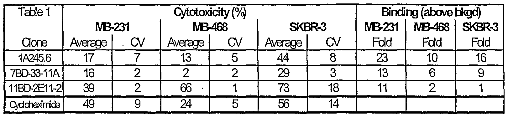
- the color reaction was tenninated by adding 100 microliters/well 2M H 2 S0 and the plate was read at 450 nm with a Perkin-Elmer HTS7000 plate reader. As indicated in Table 1 the 7BD-33-11A, 1A245.6, 11BD- 2E11-2 hybridomas secreted primarily antibodies of the IgG isotype.
- MDA-MB-231 also referred to as MB-231
- MDA-MB-468 also referred to as MB-468
- SKBR-3 The plated cells were fixed prior to use. The plates were washed thrice with PBS containing MgCl 2 and CaCl 2 at room temperature. 100 microliters of 2% paraformaldehyde diluted in PBS was added to each well for ten minutes at room temperature and then discarded. The plates were again washed with PBS containing MgCl 2 and CaCl 2 three times at room temperature.
- Blocking was done with 100 microliters/well of 5% milk in wash buffer (PBS + 0.05% Tween) for 1 hr at room temperature. The plates were washed thrice with wash buffer and the hybridoma supernatant was added at 100 microliters/well for 1 hr at room temperature. The plates were washed three times with wash buffer and 100 microliters/well of 1/5000 dilution of goat anti-mouse IgG or IgM antibody conjugated to horseradish peroxidase (diluted in PBS containing 1% bovine serum albumin) was added.
- wash buffer PBS + 0.05% Tween
- the antibodies from the 11BD-2E11-2 hybridoma cell line also bound most strongly to the MDA-MB-231 cell line, but did not demonstrate binding on the other 2 cell lines greater than background. These results suggest that the epitope recognized by this antibody is not present on MDA-MB-468 or SKBR-3 cells, and is distinct from the epitopes recognized by 7BD-33-11A and 1A245.6.
- the cytotoxic effect of the hybridoma supematants were tested in the same breast cancer cell lines: MDA-MB- 231 , MDA-MB-468 and SKBR-3.
- the Live/Dead cytotoxicity assay was obtained from Molecular Probes (Eu,OR). The assays were performed according to the manufacturer's instructions with the changes outlined below. Cells were plated before the assay at the predetermined appropriate density. After 2 days, 100 microliters of supernatant from the hybridoma microtitre plates were transferred to the cell plates and incubated in a 5% CO 2 incubator for 5 days.
- the wells that served as the positive controls were aspirated until empty and 100 microliters of sodium azide and/or cycloheximide was added.
- 3BD-27 monoclonal antibody was also added as an isotype control since it was known not to bind to the three breast cancer cell lines being tested.
- An anti-EGFR antibody (C225) was also used in the assay for comparison. After 5 days of treatment, the plate was then emptied by inverting and blotted dry. Room temperature DPBS containing MgCl 2 and CaCl 2 was dispensed into each well from a multichannel squeeze bottle, tapped three times, emptied by inversion and then blotted dry.
- the antibody derived form the hybridoma cell can produce cytotoxicity in cancer cells.
- There were several exceptions to this trend such as the amount of cytotoxicity produced by 11BD-2E11-2 in MB-468 cancer cells, and SKBR-3 cancers despite a paucity of binding.
- the assay was not sensitive enough to detect the binding that was sufficient to mediate cytotoxicity in this particular situation.
- the other exception was the relative paucity of cytotoxicity of 7BD-33-11 A towards MB- 468 cells despite a 6 fold increase in binding over the background in comparison to an isotype control. This pointed to the possibility that binding was not necessarily predictive of the outcome of antibody ligation of its cognate antigen.
- the known nonspecific cytotoxic agents cycloheximide produced cytotoxicity as expected.
- Monoclonal antibodies were produced by culturing the hybridomas, 7BD-33- 11A, 1A245.6, 11BD-2E11-2, in CL-1000 flasks (BD Biosciences, Oakville, ON) with collections and reseeding occurring twice/week and standard antibody purification procedures with Protein G Sepharose 4 Fast Flow (Amersham Biosciences, Baie d'Urfe, QC). It is within the scope of this invention to utilize monoclonal antibodies which are humanized, chimerized or murine antibodies.
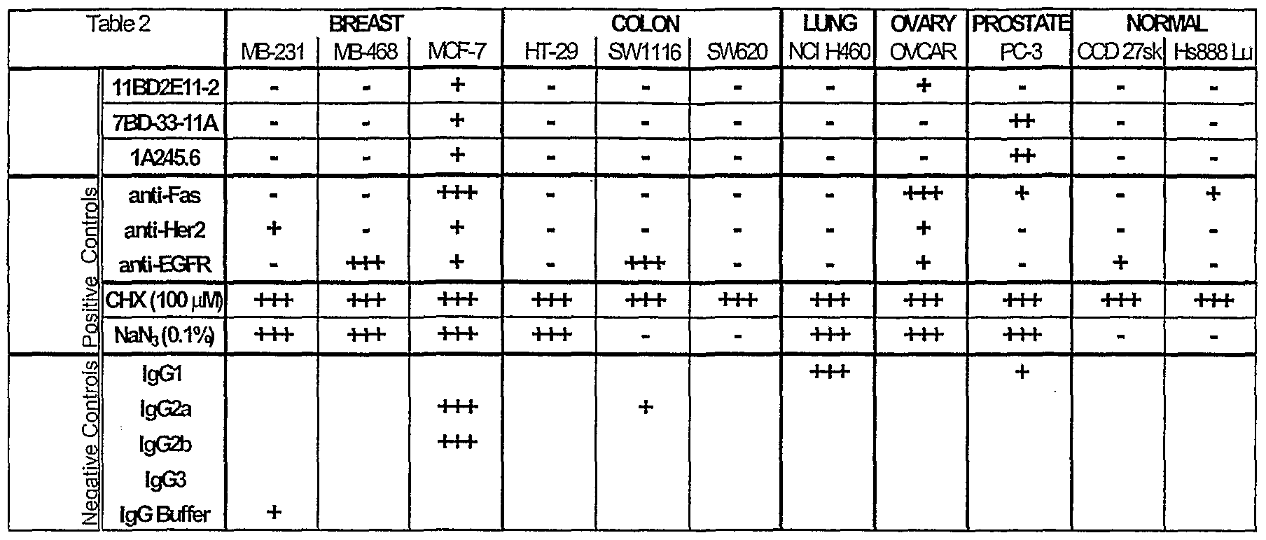
- MB-231, MB-468, MCF-7 colon cancer (HT-29, SW1116, SW620), lung cancer (NCI H460), ovarian cancer (OVCAR), prostate cancer (PC-3), and non- cancer (CCD 27sk, Hs888 Lu) cell lines were tested (all from the ATCC, Manassas, VA).
- the Live/Dead cytotoxicity assay was obtained from Molecular Probes (Eugene,OR). The assays were performed according to the manufacturer's instructions with the changes outlined below. Cells were plated before the assay at the predetermined appropriate density.
- the plates were read in a Perkin-Elmer HTS7000 fluorescence plate reader and the data was analyzed in Microsoft Excel and the results were tabulated in Table 2.
- the data represented an average of four experiments tested in triplicate and presented qualitatively in the following fashion: 4/4 experiments greater than threshold cytotoxicity (+++), 3/4 experiments greater than threshold cytotoxicity (++), 2/4 experiments greater than threshold cytotoxicity (+).
- Unmarked cells in Table 2 represented inconsistent or effects less than the threshold cytotoxicity.
- the 7BD-33- 11A and 1 A245.6 antibodies demonstrated cytotoxicity in breast and prostate tumor cell lines selectively, while having no effect on non-transformed normal cells. Both demonstrated a 25-50% greater killing than the positive control anti-Fas antibody.
- 11BD-2E11-2 was specifically cytotoxic in breast and ovarian cancer cells, and did not affect normal cells.
- the chemical cytotoxic agents induced their expected cytotoxicity while a number of other antibodies which were included for comparison also performed as expected given the limitations of biological cell assays. In total, it was shown that the three antibodies have cytotoxic activity against a number of cancer cell types.
- the antibodies were selective in their activity since not all cancer cell types were susceptible. Furthermore, the antibodies demonstrated functional specificity since they did not produce cytotoxicity against non-cancer cell types, which is an important factor in a therapeutic situation.
- Cells were prepared for FACS by initially washing the cell monolayer with
- DPBS (without Ca ++ and Mg ++ ).
- Cell dissociation buffer (INVITROGEN) was then used to dislodge the cells from their cell culture plates at 37°C. After centrifugation and collection the cells were resuspended in Dulbecco's phosphate buffered saline containing MgCl 2> CaCl 2 and 25% fetal bovine serum at 4°C (wash media) and counted, aliquoted to appropriate cell density, spun down to pellet the cells and resuspended in staining media (DPBS containing MgCl 2 and CaCl 2 ) containing 7BD-33-11A, 1A245.6, 11BD-2E11-2 or control antibodies (isotype control or anti-EGF-R) at 20 micrograms/mL on ice for 30 minutes.
- staining media DPBS containing MgCl 2 and CaCl 2
- the cells Prior to the addition of Alexa Fluor 488- conjugated secondary antibody the cells were washed once with wash media. The Alexa Fluor 488-conjugated antibody in staining media was then added for 20 minutes. The cells were then washed for the final time and resuspended in staining media containing 1 microgram/mL propidium iodide. Flow cytometric acquisition of the cells was assessed by running samples on a FACScan using the CellQuest software (BD Biosciences). The forward (FSC) and side scatter (SSC) of the cells were set by adjusting the voltage and amplitude gains on the FSC and SSC detectors.
- FSC forward
- SSC side scatter
- the detectors for the three fluorescence channels were adjusted by running cells stained with purified isotype control antibody followed by Alexa Fluor 488-conjugated secondary antibody such that cells had a uniform peak with a median fluorescent intensity of approximately 1-5 units.
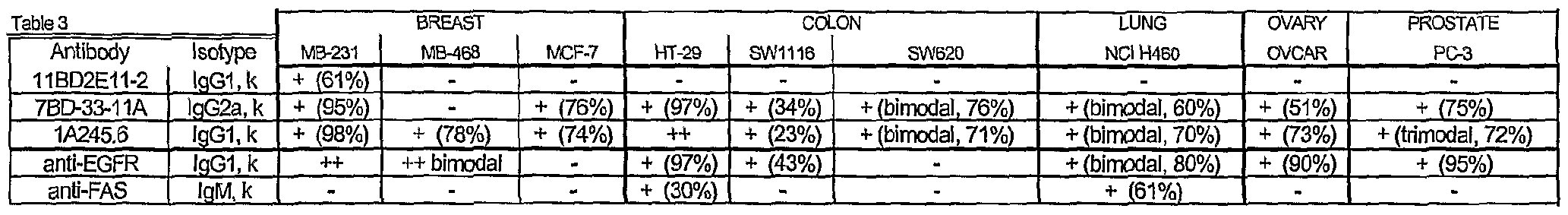
- Live cells were acquired by gating for FSC and propidium iodide exclusion. For each sample, approximately 10,000 live cells were acquired for analysis and the resulted presented in Table 3.
- Table 3 tabulated the mean fluorescence intensity fold increase above isotype control and is presented qualitatively as: less than 5 (-); 5 to 50 (+); 50 to 100 (++); above 100 (+++) and in parenthesis, the percentage of cells stained.
- FIG. 10 1 Representative histograms of 7BD-33-11A antibodies were compiled for Figure 10 1 , 1 A245.6 antibodies were compiled for Figure 2, 11BD-2E11-2 were compiled for Figure 3 and evidence the binding characteristics, inclusive of illustrated bimodal peaks, in some cases.
- 11BD-2E11-2 displayed specific tumor binding to the breast tumor cell line MDA-MB-231.
- Both 7BD-33-11 and 1A245.6 displayed similar binding to cancer lines of breast (MDA-MB-231 and MCF-7), colon, lung, ovary, and 15 prostate origin and differential binding to one of the breast cancer cell lines (MDA- MB-468). There was binding of all three antibodies to non-cancer cells, however that binding did not produce cytotoxicity.
- mice were implanted with 5 million MDA-MB-231 human breast cancer cells in one hundred microliters injected subcutaneously in the scruff of the neck. The mice were randomly divided into four treatment groups often.
- 3BD-27 isotype control antibody (known not to bind MDA-MB-231 cells) were administered intrapertioneally at a volume of 300 microliters after dilution from the stock concentration with a diluent that contained 2.7 mM KC1, 1 mM KH P0 , 137 mM NaCl, 20 mM Na 2 HP0 4 .
- the antibodies were then administered once per week for a period of 7 weeks in the same fashion.
- Tumor growth was measured about every seventh day with calipers for up to ten weeks or until individual animals reached the Canadian Council for Animal Care (CCAC) end-points. Body weights of the animals were recorded for the duration of the study. At the end of the study all animals were euthanised according to CCAC guidelines. There were no clinical signs of toxicity throughout the study. Body weight measured at weekly intervals was a surrogate for well-being and failure to thrive. There was a minimal difference in weight for the groups treated with the isotype control, 3BD-27, and 7BD-33-11A, 1 A245.6, or 11BD-2E11-2.
- Those mice bearing cancer treated with 7BD-33-11A antibody were disease free and had no tumor burden.
- This also demonstrated a lesser tumor burden with cytotoxic antibody treatment in comparison to a control antibody.
- survival benefits (Fig. 6) from treatment with 7BD-33-11 A, 1 A245.6, and 11BD-2E11-2 cytotoxic antibodies.
- the control group treated with 3BD-27 antibody reached 100% mortality by day 74 post-implantation.
- groups treated with 7BD-33-11A were disease free and 1 A245.6 treated animal displayed 100% survival and the group treated with 11BD-2E1 1-2 had 24% survival.
- mice Five to six week old, female SCID mice were implanted with 5 million MDA- MB-231 breast cancer cells in one hundred microliters injected subcutaneously in the scruff of the neck. Tumor growth was measured with calipers every week. When the majority of the cohort reached a tumor volume of 100 mm 3 (range 50-200 mm 3 ) at 34 days post implantation 8-10 mice were randomly assigned into each of three treatment groups.
- mice As outlined in S.N. 10/348,284, and with reference to Figure 11, 4 to 8 week old female SCID mice were implanted with 5 million MB-231 human breast cancer cells in 100 microlitres saline injected subcutaneously in the scruff of the neck. The mice were randomly divided into 3 treatment groups of 10.
- mice 5 to 6 week old female SCID mice were implanted with 5 million MB-231 human breast cancer cells in 100 microlitres saline injected subcutaneously in the scruff of the neck. Tumor growth was measured with calipers every week. When the majority of the cohort reached a tumor volume of 100 mm 3 (range 50-200 mm 3 ) at 34 days post- implantation 8-10 mice were randomly assigned into each of 3 treatment groups.
- 7BD- 33-11 A 1A245.6 test antibodies or isotype control antibody was administered intraperitoneally with 15 mg/kg of antibodies at a volume of 150 microliters after dilution from the stock concentration with a diluent that contained 2.7 mM KC1, 1 mM KH 2 P0 4 , 137 mM NaCl and 20 mM Na 2 HP0 4 .
- the antibodies were then administered 3 times per week for 10 doses in total in the same fashion until day 56 post- implantation. Tumor growth was measured about every seventh day with calipers until day 59 post-implantation or until individual animals reached the CCAC end-points.
- mice 4 to 8 week old, male SCID mice were implanted with 1 million PC-3 human prostate cancer cells in 100 microliters saline injected subcutaneously in the scruff of the neck. The mice were randomly divided into 4 treatment groups of 8. On the day prior to implantation 20 mg/kg of 7BD-33-11A or 1A245.6 test antibody or isotype control antibody or buffer control was administered intraperitoneally at a volume of 300 microliters after dilution from the stock concentration with a diluent that contained 2.7 mM KC1, 1 mM KH 2 P0 4 , 137 mM NaCl and 20 mM Na 2 HP0 .
- the antibodies and buffer control were then administered once per week for a period of 7 weeks in the same fashion. Tumor growth was measured about every 7th day with calipers for up to 10 weeks or until individual animals reached the CCAC end-points or day 52. Body weights of the animals were recorded for the duration of the study. At the end of the study all animals were euthanised according to CCAC guidelines.
- 7BD-33-11 A and 1 A245.6 antibody treatment reduced tumor burden, delayed disease progression and extended survival in comparison to an isotype control antibody and a buffer control in a well-recognized model of human prostate cancer.
- mice Male SCID mice, 4 to 8 weeks old, were implanted with 1 million PC-3 prostate cancer cells in 100 microliters saline injected subcutaneously in the scruff of the neck. Tumor growth was measured with calipers every week. When the majority of the cohort reached a tumor volume of 275 mm (range 144-406 mm ) at 21 days post implantation, 9-10 mice were randomized into each of 4 treatment groups.
- 7BD-33- 11 A or 1A245.6 or isotype control antibody was administered intraperitoneally with 20 mg/kg/dose at a volume of 300 microliters after dilution from the stock concentration with a diluent that contained 2.7 mM KC1, 1 mM KH 2 P0 4 , 137 mM NaCl and 20 mM Na 2 HP0 .
- the antibodies were then administered 3 times per week for a total of 10 doses in the same fashion until day 43 post-implantation. Tumor growth was measured about every seventh day with calipers for the duration of the study or until individual animals reached CCAC end-points. Body weights of the animals were recorded for the duration of the study. At the end of the study all animals were euthanised according to CCAC guidelines.
- 7BD-33-11 A is significantly more effective than the isotype control antibody in suppressing tumor growth in an established tumor xenograft model of prostate cancer in SCID mice. Therefore treatment with 7BD-33-11 A significantly decreased the tumor burden of established tumors in two well-recognized models of human cancer disease (breast and prostate) suggesting pharmacologic and pharmaceutical benefits of this antibody for therapy in other mammals, including man.
- Tissue sections were deparaffinized by drying in an oven at 58°C for 1 hour and dewaxed by immersing in xylene 5 times for 4 minutes each in Coplin jars. Following treatment through a series of graded ethanol washes (100%-75%) the sections were re- hydrated in water. The slides were immersed in 10 mM citrate buffer at pH 6 (Dako, Toronto, Ontario) then microwaved at high, medium, and low power settings for 5 minutes each and finally immersed in cold PBS. Slides were then immersed in 3% hydrogen peroxide solution for 6 minutes, washed with PBS three times for 5 minutes each, dried, incubated with Universal blocking solution (Dako, Toronto, Ontario) for 5 minutes at room temperature.
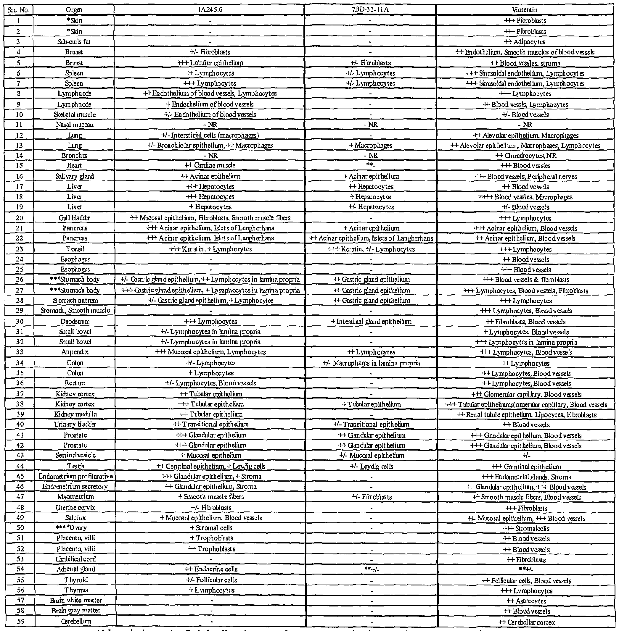
- Binding of antibodies to 59 normal human tissues was performed using a human, normal organ tissue array (Imgenex, San Diego, CA). Table 4 presents a summary of the results of 7BD-33-11A and 1A245.6 staining of an array of normal human tissues. From the table, there are 3 categories of tissue staining. A group of tissues was completely negative. These tissues included normal skin, brain ( Figure 19A), ovary, thymus, thyroid, small bowel, esophaguas, heart ( Figure 20A), gall bladder and lymph node for 7BD-33-11A. For 1A245.6, the completely negative tissues comprised of skin, sub-cutis fat, esophagus and brain ( Figure 19B). A second group of tissues comprised tissues that demonstrated positive staining.
- liver and pancreas for 7BD-33-11 A included the liver and pancreas for 7BD-33-11 A.
- the tonsil had the strongest staining with this antibody.
- positive staining occurred in the liver, heart ( Figure 20B), testis, thyroid, adrenal gland and myometrium.
- 1A245.6 stained the tonsil the strongest.
- a third group of tissues included tissues in which staining was positive in the tissue section, but was limited to infiltrating macrophages, lymphocytes, fibroblasts or the epithelium, for example the stomach for both 7BD-33-11 A and 1 A245.6 ( Figure
- 7BD-33-11 A antigen is not present on cells of several of the vital organs, including kidney, heart ( Figure 20 A) and lung. Overall, 7BD-33-11 A binds to a smaller subset of normal human tissues compared to 1 A245.6 with weak to moderate binding in the tissues that are positive. 1A245.6 staining, albeit more extensive, is also generally weak to moderate in intensity and in the majority of cases is limited to the epithelium of the stained tissue. These results suggest that the antigen for 7BD-33-11 A is not widely expressed on normal tissues, and that the antibody would bind specifically to a limited number of tissues in humans. In addition, the antigen for 1 A245.6, besides being present in the heart and liver, is limited to epithelium and infiltrating lymphocytes, macrophages and fibroblasts.
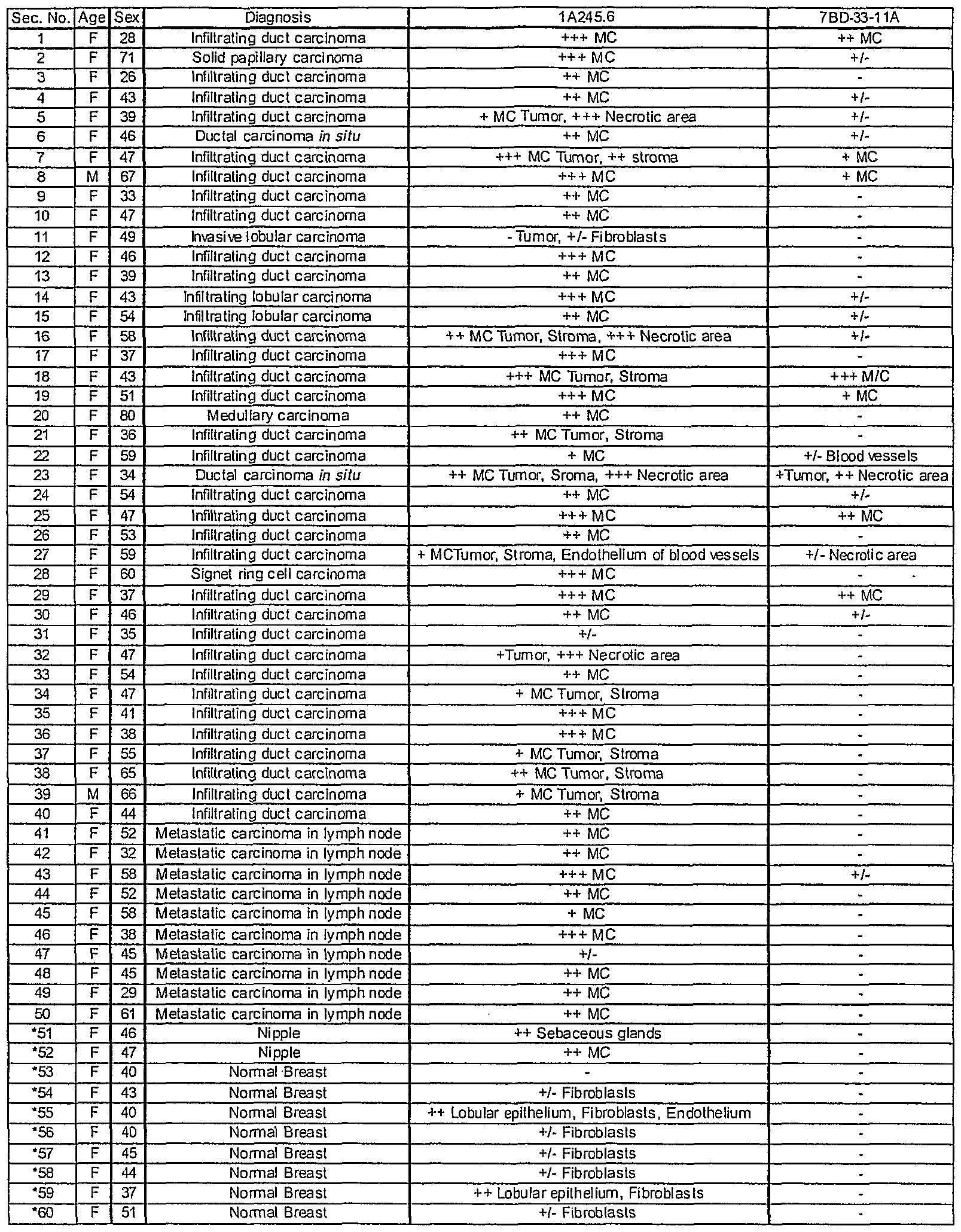
- Table 5 provides a binding summary of 7BD-33-1 1A and 1A245.6 antibody staining of a breast cancer tissue array. Each array contained tumor samples from 50 individual patients. Overall, 36 percent of the 50 patients tested were positive for the 7BD-33-11 A antigen (Figure 22A) compared to 98 percent for 1A245.6 ( Figure 23A). For 7BD-33-11 A , 0 out of 10 normal breast tissue samples from breast cancer patients were positive ( Figure 22B). Conversely, 9 out of 10 normal breast tissue samples were positive for 1 A245.6. However, staining was due to infiltrating fibroblasts in the majority of cases ( Figure 23B). No correlation between estrogen and progesterone receptor status was evident for 1 A245.6 (Table 6).
- PS the section is partially detached
- * Non-neoplastic breast tissue in breast cancer patient.
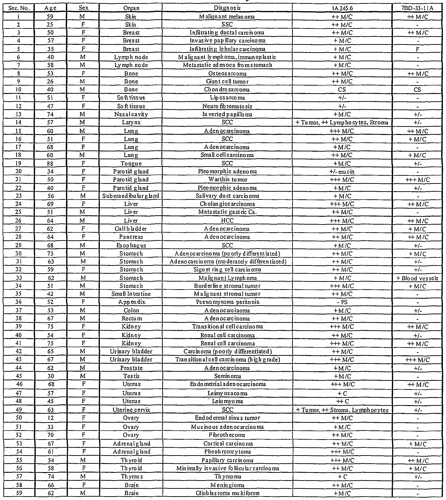
- both antibodies were individually tested on a multiple human tumor tissue array (Imgenex, San Diego, CA). The following information was provided for each patient: age, sex, organ and diagnosis. The staining procedure used was the same as the one outlined in Example 9. Vimentin was used as a positive control antibody and the same negative control antibody was used as described for the human breast tumor tissue array. All antibodies were used at a working concentration of 5 ⁇ g/mL.
- 7BD-33-11 A stained a number of various human cancers besides breast.
- the following tumor types were positive for 7BD-33-11 A: skin (1/2), lung (3/4), liver (2/3), stomach (4/5), thyroid (2/2), prostate (1/1), uterus (4/4) and kidney (3/3) ( Figure 24A).
- Other tumor tissues were negative for 7BD-33-11 A expression; ovary (0/3), testis (0/1), brain (0/2) and lymph node (0/2).
- some of the strongest staining was seen on malignant cells of the skin, lung, liver, uterus, kidney ( Figure 24B), stomach and bladder.
- 7BD-33-11 A and 1 A245.6 staining was localized on the membrane and within the cytoplasm of cancerous cells.
- mice 4 to 8 week old female SCID mice were implanted with 5 million MCF-7 human breast cancer cells in 100 microlitres saline injected subcutaneously in the scruff of the neck. The mice were randomly divided into 2 treatment groups of 11-13 mice. On the day after implantation, 20 mg/kg of either 11BD-2E11-2 test antibody or isotype control antibody (known not to bind MCF-7 or OVCAR-3 cells) was administered intraperitoneally at a volume of 300 microliters after dilution from the stock concentration with a diluent that contained 2.7 mM KC1, 1 mM KH 2 PO 4j 137 mM NaCl and 20 mM Na 2 HP0 .
- 11BD-2E11-2 test antibody or isotype control antibody known not to bind MCF-7 or OVCAR-3 cells
- the antibodies were then administered once per week for a period of 7 weeks in the same fashion. Tumor growth was measured about every seventh day with calipers for up to 8 weeks or until individual animals reached the Canadian Council for Animal Care (CCAC) end-points. Body weights of the animals were recorded for the duration of the study. At the end of the study all animals were euthanised according to CCAC guidelines.
- CCAC Canadian Council for Animal Care
- mice Female SCID mice were implanted with 5 million OVCAR-3 human ovarian cancer cells in 1000 microliters saline injected intraperitoneally.
- the mice were randomly divided into 2 treatment groups of 10.
- 20 mg/kg of 11BD-2E1 1-2 test antibody or buffer control antibody was administered intraperitoneally at a volume of 300 microliters after dilution from the stock concentration with a diluent that contained 2.7 mM KC1, 1 mM KH 2 P0 4 , 137 mM NaCl and 20 mM Na 2 HP0 4 .
- the antibodies were then administered once per week for a period of 9 weeks in the same fashion. Body weights of the animals were recorded for the duration of the study. At the end of the study all animals were euthanised according to CCAC guidelines.
- 11BD- 2E11-2 antibody treatment reduced tumor burden, delayed disease progression and enhanced survival in comparison to a buffer control antibody in a well-recognized model of human ovarian cancer. Therefore treatment with 11BD-2E11-2 significantly decreased the tumor burden of established tumors in two well-recognized models of human cancer disease (breast and ovarian cancers) suggesting pharmacologic and pharmaceutical benefits of this antibody for therapy in other mammals, including man (Smith et al. The Prostate 48:47-53 2001; Olson et al. International Journal of Cancer 98:923-929 2002; Guilbaud et al. Clinical Cancer Research 7:2573-2580 2001 ; Von Gruenigen et al. International Journal of Gynecologic Cancer 9:365-372 1999; Guichard et al. Clinical Cancer Research 7:3222-3228 2001; Xiao et al. Protein Expression and Purification 19: 12-21 2000).
Landscapes
- Health & Medical Sciences (AREA)
- Life Sciences & Earth Sciences (AREA)
- Chemical & Material Sciences (AREA)
- Immunology (AREA)
- Organic Chemistry (AREA)
- Proteomics, Peptides & Aminoacids (AREA)
- Medicinal Chemistry (AREA)
- General Health & Medical Sciences (AREA)
- Molecular Biology (AREA)
- Biophysics (AREA)
- Genetics & Genomics (AREA)
- Biochemistry (AREA)
- Cell Biology (AREA)
- Pharmacology & Pharmacy (AREA)
- Veterinary Medicine (AREA)
- Public Health (AREA)
- Animal Behavior & Ethology (AREA)
- Epidemiology (AREA)
- Oncology (AREA)
- Physics & Mathematics (AREA)
- Optics & Photonics (AREA)
- Gynecology & Obstetrics (AREA)
- Pregnancy & Childbirth (AREA)
- Reproductive Health (AREA)
- General Chemical & Material Sciences (AREA)
- Chemical Kinetics & Catalysis (AREA)
- Nuclear Medicine, Radiotherapy & Molecular Imaging (AREA)
- Medicines Containing Antibodies Or Antigens For Use As Internal Diagnostic Agents (AREA)
- Peptides Or Proteins (AREA)
Abstract
Description

Claims
Priority Applications (5)
| Application Number | Priority Date | Filing Date | Title |
|---|---|---|---|
| AU2004205435A AU2004205435B2 (en) | 2003-01-21 | 2004-01-21 | Cancerous disease modifying antibodies |
| CA002513772A CA2513772A1 (en) | 2003-01-21 | 2004-01-21 | Cancerous disease modifying antibodies |
| JP2006500434A JP4726777B2 (en) | 2003-01-21 | 2004-01-21 | Cancer disease modifying antibodies |
| EP04703738A EP1587836A2 (en) | 2003-01-21 | 2004-01-21 | Cancerous disease modifying antibodies |
| NZ541366A NZ541366A (en) | 2003-01-21 | 2004-01-21 | Monoclonal antibodies expressed by ATCC PTA-5643 for the manufacture of a medicament for the treatment of cancer |
Applications Claiming Priority (8)
| Application Number | Priority Date | Filing Date | Title |
|---|---|---|---|
| US10/348,231 | 2003-01-21 | ||
| US10/348,231 US7009040B2 (en) | 2003-01-21 | 2003-01-21 | Cancerous disease modifying antibodies |
| US10/603,006 | 2003-06-23 | ||
| US10/603,006 US20040105816A1 (en) | 1999-10-08 | 2003-06-23 | Cancerous disease modifying antibodies |
| US10/743,451 US20040141979A1 (en) | 2003-01-21 | 2003-12-19 | Cancerous disease modifying antibodies |
| US10/743,451 | 2003-12-19 | ||
| US10/762,129 US7361342B2 (en) | 2003-01-21 | 2004-01-20 | Cancerous disease modifying antibodies |
| US10/762,129 | 2004-01-20 |
Publications (2)
| Publication Number | Publication Date |
|---|---|
| WO2004065422A2 true WO2004065422A2 (en) | 2004-08-05 |
| WO2004065422A3 WO2004065422A3 (en) | 2004-11-11 |
Family
ID=32777212
Family Applications (1)
| Application Number | Title | Priority Date | Filing Date |
|---|---|---|---|
| PCT/CA2004/000059 Ceased WO2004065422A2 (en) | 2003-01-21 | 2004-01-21 | Cancerous disease modifying antibodies |
Country Status (5)
| Country | Link |
|---|---|
| US (1) | US7361342B2 (en) |
| EP (1) | EP1587836A2 (en) |
| AU (1) | AU2004205435B2 (en) |
| CA (1) | CA2513772A1 (en) |
| WO (1) | WO2004065422A2 (en) |
Cited By (12)
| Publication number | Priority date | Publication date | Assignee | Title |
|---|---|---|---|---|
| JP2007530457A (en) * | 2004-03-26 | 2007-11-01 | アリアス リサーチ、インコーポレイテッド | Cytotoxic mediation of cells demonstrating surface expression of MCSP |
| WO2008011710A1 (en) * | 2006-07-26 | 2008-01-31 | Arius Research Inc. | Cytotoxicity mediation of cells evidencing surface expression of cd63 |
| EP1732603A4 (en) * | 2004-03-26 | 2008-04-30 | Arius Res Inc | HIGHLIGHTING THE SURFACIAL EXPRESSION OF CD63 THROUGH CELL CYTOTOXICITY |
| JP2008513361A (en) * | 2004-09-15 | 2008-05-01 | アリアス リサーチ、インコーポレイテッド | Cancerous disease modifying antibodies |
| JP2008514550A (en) * | 2004-09-24 | 2008-05-08 | アリアス リサーチ、インコーポレイテッド | Cytotoxic mediation of cells demonstrating surface expression of MCSP |
| JP2008545615A (en) * | 2005-01-03 | 2008-12-18 | アリアス リサーチ、インコーポレイテッド | Cytotoxic mediation of cells demonstrating surface expression of CD63 |
| JP2009529010A (en) * | 2006-03-07 | 2009-08-13 | アリアス リサーチ、インコーポレイテッド | Cancerous disease modifying antibodies |
| EP1996623A4 (en) * | 2006-03-07 | 2009-09-16 | Hoffmann La Roche | ANTIBODIES MODIFYING CANCER DISEASE FOR DIAGNOSTIC AND THERAPEUTIC USE IN BREAST CANCER AND PROSTATE |
| JP2009545533A (en) * | 2006-08-03 | 2009-12-24 | エフ.ホフマン−ラ ロシュ アーゲー | Cancer-like disease-modifying antibody |
| EP2163564A1 (en) | 2003-01-21 | 2010-03-17 | F. Hoffmann-La Roche AG | Cancerous disease modifying antibodies |
| EP2044119A4 (en) * | 2006-07-26 | 2010-09-01 | Arius Res Inc | ANTIBODIES MODIFYING CANCER DISEASES |
| WO2022214499A3 (en) * | 2021-04-05 | 2022-11-17 | Altheia Science S.R.L. | Diagnosis and treatment of myeloid disorders and acute leukemias using novel tumor specific antigens |
Families Citing this family (2)
| Publication number | Priority date | Publication date | Assignee | Title |
|---|---|---|---|---|
| JP4462964B2 (en) * | 2003-03-10 | 2010-05-12 | 第一三共株式会社 | Antibodies targeting cancer-specific antigens |
| EP3074025A1 (en) | 2013-11-27 | 2016-10-05 | Baylor College Of Medicine | Csgp4-specific chimeric antigen receptor for cancer |
Family Cites Families (15)
| Publication number | Priority date | Publication date | Assignee | Title |
|---|---|---|---|---|
| US4828991A (en) * | 1984-01-31 | 1989-05-09 | Akzo N.V. | Tumor specific monoclonal antibodies |
| AU613590B2 (en) * | 1986-11-19 | 1991-08-08 | Bristol-Myers Squibb Company | Hybridomas producing monoclonal antibodies to new mucin epitopes |
| US4861581A (en) * | 1986-12-05 | 1989-08-29 | Cancer Biologics, Inc. | Detection of necrotic malignant tissue and associated therapy |
| US5171665A (en) * | 1989-04-17 | 1992-12-15 | Oncogen | Monoclonal antibody to novel antigen associated with human tumors |
| US6020145A (en) * | 1989-06-30 | 2000-02-01 | Bristol-Myers Squibb Company | Methods for determining the presence of carcinoma using the antigen binding region of monoclonal antibody BR96 |
| EP0539970B1 (en) * | 1991-10-30 | 1999-05-26 | Idemitsu Kosan Company Limited | Methods for producing human lymphocytes and human monoclonal antibodies, and human monoclonal antibodies produced thereby |
| IL105008A0 (en) * | 1992-03-13 | 1993-07-08 | Yeda Res & Dev | Double transfectants of mhc genes as cellular vaccines for immunoprevention of tumor metastasis |
| US5545532A (en) * | 1993-02-05 | 1996-08-13 | Epigen, Inc. | Human carcinoma antigen (HCA), HCA antibodies, HCA immunoassays and methods of imaging |
| WO1996000084A1 (en) * | 1994-06-24 | 1996-01-04 | Torchilin Vladimir P | Use of autoantibodies for tumor therapy and prophylaxis |
| US5783186A (en) * | 1995-12-05 | 1998-07-21 | Amgen Inc. | Antibody-induced apoptosis |
| US6180357B1 (en) * | 1999-10-08 | 2001-01-30 | Arius Research, Inc. | Individualized patient-specific anti-cancer antibodies |
| AU2001226985B8 (en) | 2000-11-08 | 2008-03-13 | F. Hoffmann-La Roche Ag | Individualized anti-cancer antibodies |
| US7009040B2 (en) * | 2003-01-21 | 2006-03-07 | Arius Research, Inc. | Cancerous disease modifying antibodies |
| EP1455819A1 (en) | 2001-12-21 | 2004-09-15 | Arius Research, Inc. | Individualized anti-cancer antibodies |
| WO2003086456A2 (en) | 2002-04-05 | 2003-10-23 | Arius Research, Inc. | Anti-ck18 monoclonal antibody and therapeutic and diagnostic uses thereof in cancer |
-
2004
- 2004-01-20 US US10/762,129 patent/US7361342B2/en not_active Expired - Lifetime
- 2004-01-21 CA CA002513772A patent/CA2513772A1/en not_active Abandoned
- 2004-01-21 AU AU2004205435A patent/AU2004205435B2/en not_active Ceased
- 2004-01-21 EP EP04703738A patent/EP1587836A2/en not_active Withdrawn
- 2004-01-21 WO PCT/CA2004/000059 patent/WO2004065422A2/en not_active Ceased
Cited By (17)
| Publication number | Priority date | Publication date | Assignee | Title |
|---|---|---|---|---|
| EP2163564A1 (en) | 2003-01-21 | 2010-03-17 | F. Hoffmann-La Roche AG | Cancerous disease modifying antibodies |
| EP1732602A4 (en) * | 2004-03-26 | 2008-05-07 | Arius Res Inc | CYTOTOXIC MEDIATION OF CELLS WITH SUPERFICIAL EXPRESSION OF MCSP |
| JP2007530457A (en) * | 2004-03-26 | 2007-11-01 | アリアス リサーチ、インコーポレイテッド | Cytotoxic mediation of cells demonstrating surface expression of MCSP |
| EP1732603A4 (en) * | 2004-03-26 | 2008-04-30 | Arius Res Inc | HIGHLIGHTING THE SURFACIAL EXPRESSION OF CD63 THROUGH CELL CYTOTOXICITY |
| EP1796721A4 (en) * | 2004-09-15 | 2009-03-04 | Arius Res Inc | MODIFYING ANTIBODIES OF CANCER DISEASE |
| JP2008513361A (en) * | 2004-09-15 | 2008-05-01 | アリアス リサーチ、インコーポレイテッド | Cancerous disease modifying antibodies |
| JP2008514550A (en) * | 2004-09-24 | 2008-05-08 | アリアス リサーチ、インコーポレイテッド | Cytotoxic mediation of cells demonstrating surface expression of MCSP |
| JP2008545615A (en) * | 2005-01-03 | 2008-12-18 | アリアス リサーチ、インコーポレイテッド | Cytotoxic mediation of cells demonstrating surface expression of CD63 |
| EP1996624A4 (en) * | 2006-03-07 | 2009-09-16 | Hoffmann La Roche | ANTIBODIES MODIFYING CANCER DISEASE FOR DIAGNOSTIC AND THERAPEUTIC USE IN BREAST AND OVARY CANCER |
| EP1996623A4 (en) * | 2006-03-07 | 2009-09-16 | Hoffmann La Roche | ANTIBODIES MODIFYING CANCER DISEASE FOR DIAGNOSTIC AND THERAPEUTIC USE IN BREAST CANCER AND PROSTATE |
| JP2009529010A (en) * | 2006-03-07 | 2009-08-13 | アリアス リサーチ、インコーポレイテッド | Cancerous disease modifying antibodies |
| WO2008011710A1 (en) * | 2006-07-26 | 2008-01-31 | Arius Research Inc. | Cytotoxicity mediation of cells evidencing surface expression of cd63 |
| EP2073851A4 (en) * | 2006-07-26 | 2010-04-21 | Hoffmann La Roche | CYTOTOXICITY MEDIATION OF CELLS ATTAINING SURFACE EXPRESSION OF CD63 |
| EP2044119A4 (en) * | 2006-07-26 | 2010-09-01 | Arius Res Inc | ANTIBODIES MODIFYING CANCER DISEASES |
| JP2009545533A (en) * | 2006-08-03 | 2009-12-24 | エフ.ホフマン−ラ ロシュ アーゲー | Cancer-like disease-modifying antibody |
| EP2046381A4 (en) * | 2006-08-03 | 2010-07-21 | Hoffmann La Roche | ANTIBODIES MODIFYING CANCER DISEASE |
| WO2022214499A3 (en) * | 2021-04-05 | 2022-11-17 | Altheia Science S.R.L. | Diagnosis and treatment of myeloid disorders and acute leukemias using novel tumor specific antigens |
Also Published As
| Publication number | Publication date |
|---|---|
| WO2004065422A3 (en) | 2004-11-11 |
| US20040151665A1 (en) | 2004-08-05 |
| EP1587836A2 (en) | 2005-10-26 |
| CA2513772A1 (en) | 2004-08-05 |
| US7361342B2 (en) | 2008-04-22 |
| AU2004205435B2 (en) | 2011-02-03 |
| AU2004205435A1 (en) | 2004-08-05 |
Similar Documents
| Publication | Publication Date | Title |
|---|---|---|
| EP2163564A1 (en) | Cancerous disease modifying antibodies | |
| US7442776B2 (en) | Cancerous disease modifying antibodies | |
| AU2004248865B2 (en) | Cancerous disease modifying antibodies | |
| US20090074659A1 (en) | Cancerous disease modifying antibodies | |
| US7399835B2 (en) | Cancerous disease modifying antibodies | |
| AU2004205435B2 (en) | Cancerous disease modifying antibodies | |
| US20050027106A1 (en) | Cancerous disease modifying antibodies | |
| US20040105816A1 (en) | Cancerous disease modifying antibodies | |
| US20050255041A1 (en) | Cancerous disease modifying antibodies | |
| US20040141914A1 (en) | Cancerous disease modifying antibodies |
Legal Events
| Date | Code | Title | Description |
|---|---|---|---|
| AK | Designated states |
Kind code of ref document: A2 Designated state(s): AE AG AL AM AT AU AZ BA BB BG BR BW BY BZ CA CH CN CO CR CU CZ DE DK DM DZ EC EE EG ES FI GB GD GE GH GM HR HU ID IL IN IS JP KE KG KP KR KZ LC LK LR LS LT LU LV MA MD MG MK MN MW MX MZ NA NI NO NZ OM PG PH PL PT RO RU SC SD SE SG SK SL SY TJ TM TN TR TT TZ UA UG US UZ VC VN YU ZA ZM ZW |
|
| AL | Designated countries for regional patents |
Kind code of ref document: A2 Designated state(s): BW GH GM KE LS MW MZ SD SL SZ TZ UG ZM ZW AM AZ BY KG KZ MD RU TJ TM AT BE BG CH CY CZ DE DK EE ES FI FR GB GR HU IE IT LU MC NL PT RO SE SI SK TR BF BJ CF CG CI CM GA GN GQ GW ML MR NE SN TD TG |
|
| 121 | Ep: the epo has been informed by wipo that ep was designated in this application | ||
| WWE | Wipo information: entry into national phase |
Ref document number: 2006500434 Country of ref document: JP |
|
| WWE | Wipo information: entry into national phase |
Ref document number: 2513772 Country of ref document: CA Ref document number: 541366 Country of ref document: NZ |
|
| WWE | Wipo information: entry into national phase |
Ref document number: 2004703738 Country of ref document: EP Ref document number: 2004205435 Country of ref document: AU |
|
| ENP | Entry into the national phase |
Ref document number: 2004205435 Country of ref document: AU Date of ref document: 20040121 Kind code of ref document: A |
|
| WWP | Wipo information: published in national office |
Ref document number: 2004205435 Country of ref document: AU |
|
| WWE | Wipo information: entry into national phase |
Ref document number: 20048075394 Country of ref document: CN |
|
| WWP | Wipo information: published in national office |
Ref document number: 2004703738 Country of ref document: EP |