WO2004022156A2 - Methods for treating central nervous system damage - Google Patents
Methods for treating central nervous system damage Download PDFInfo
- Publication number
- WO2004022156A2 WO2004022156A2 PCT/US2003/027819 US0327819W WO2004022156A2 WO 2004022156 A2 WO2004022156 A2 WO 2004022156A2 US 0327819 W US0327819 W US 0327819W WO 2004022156 A2 WO2004022156 A2 WO 2004022156A2
- Authority
- WO
- WIPO (PCT)
- Prior art keywords
- subject
- fes
- neural
- spinal cord
- patterned movement
- Prior art date
- Legal status (The legal status is an assumption and is not a legal conclusion. Google has not performed a legal analysis and makes no representation as to the accuracy of the status listed.)
- Ceased
Links
Classifications
-
- A—HUMAN NECESSITIES
- A61—MEDICAL OR VETERINARY SCIENCE; HYGIENE
- A61H—PHYSICAL THERAPY APPARATUS, e.g. DEVICES FOR LOCATING OR STIMULATING REFLEX POINTS IN THE BODY; ARTIFICIAL RESPIRATION; MASSAGE; BATHING DEVICES FOR SPECIAL THERAPEUTIC OR HYGIENIC PURPOSES OR SPECIFIC PARTS OF THE BODY
- A61H39/00—Devices for locating or stimulating specific reflex points of the body for physical therapy, e.g. acupuncture
- A61H39/002—Using electric currents
-
- A—HUMAN NECESSITIES
- A61—MEDICAL OR VETERINARY SCIENCE; HYGIENE
- A61N—ELECTROTHERAPY; MAGNETOTHERAPY; RADIATION THERAPY; ULTRASOUND THERAPY
- A61N1/00—Electrotherapy; Circuits therefor
- A61N1/18—Applying electric currents by contact electrodes
- A61N1/32—Applying electric currents by contact electrodes alternating or intermittent currents
- A61N1/326—Applying electric currents by contact electrodes alternating or intermittent currents for promoting growth of cells, e.g. bone cells
-
- A—HUMAN NECESSITIES
- A61—MEDICAL OR VETERINARY SCIENCE; HYGIENE
- A61N—ELECTROTHERAPY; MAGNETOTHERAPY; RADIATION THERAPY; ULTRASOUND THERAPY
- A61N1/00—Electrotherapy; Circuits therefor
- A61N1/18—Applying electric currents by contact electrodes
- A61N1/32—Applying electric currents by contact electrodes alternating or intermittent currents
- A61N1/36—Applying electric currents by contact electrodes alternating or intermittent currents for stimulation
- A61N1/36003—Applying electric currents by contact electrodes alternating or intermittent currents for stimulation of motor muscles, e.g. for walking assistance
Definitions
- This invention relates in general to the field of medicine and to therapeutic methods for treating disruption of neural function resulting from damage to the central nervous system (CNS), and more particularly to methods for restoring motor and sensory function in subjects suffering from CNS damage.
- CNS central nervous system
- CNS damage can result, for example, from spinal cord injury ("SCI"), which typically involves a complete or partial severance of a region of the spinal cord in an auto or other accident, or from a stroke-induced lesion.
- SCI spinal cord injury
- SCI can also be the result of a chronic disease process such as multiple sclerosis or a cancerous tumor.
- Acute trauma and chronic disease processes such as multiple sclerosis can also produce lesions elsewhere in the CNS that result in dramatic impairment of sensory or motor function, or both.
- ASIA American Spinal Injury Association
- SCI spinal cord injury
- Table 1 presents ASIA'S five grades of spinal cord function, A to E, where E is normal.
- the ASIA Grade A describes individuals with the least remaining function. Such patients have little hope for recovery. Less than 1 % of those with no muscle activity in the lower extremities 1 month after injury learn to walk again, and only 10% recover enough function to be reclassified as ASIA B or better. Approximately 90% of patients remain classified as ASIA Grade A.
- Table 1 ASIA'S international standards for classifying SCI
- FES Functional electrical Stimulation
- Examples include cochlear implants for restoration of hearing, stimulation of lower motor neurons to restore motor function including hand grasp and release, standing and stepping, breathing, and bladder emptying, and deep brain stimulation to treat the motor symptoms of Parkinson's disease (Grill and Kirsch, 1999;McDonald and Sadowsky, 2002).
- FES has not been applied to reversing or altering disease progression or to promoting tissue regeneration or repair.
- Locomotor training such as on a treadmill, has been applied to motor incomplete patients (i.e., ASIA grade B or better) to enhance recovery of walking. Locomotor training can help such patients improve their walking speed and posture, and to relearn and maintain balance while walking. These improvements correlate well with corresponding improvements in muscle strength and in cardiovascular fitness, and are also believed to involve improved recruitment of existing, spared motor neurons.
- Locomotor therapy has been combined with partial body weight support (BWS) of up to 40%, to reduce the load borne by lower limbs during training, straighten walking posture, and assist certain aspects of gait such as swing and balance. BWS of greater than about 30-40% is generally thought to substantially limit any benefit of locomotor training to the patient.
- BWS body weight support
- FES therapy has been used in combination with locomotor or cycling training for motor incomplete patients.
- Field-Fote describes the combined use of FES with partial BWS and treadmill training, to improve over-ground walking ability in patients of ASIA grade C (Edelle C. Field-Fote, Arch. Phys. Med. Rehabil. 82: 818-24, (June 2001)). Patients were able to support at least 70% of their own body weight and were able to walk on a treadmill. Patients treated accordingly showed improved over-ground walking speed in the absence of BWS and FES, due to training effects on the musculoskeletal and cardiovascular systems. FES cycling has been described, for promoting recovery of leg strength and endurance in a motor incomplete patient (N.
- the present invention provides a novel approach to restoring motor and sensory function in individuals suffering from CNS damage including those individuals with such extensive CNS damage that recovery of any function requiring the activity of the lost or damaged neurons was previously thought impossible.
- the methods are based in part on surprising findings in a motor complete human subject with a high cervical (C2) traumatic injury.
- C2 cervical
- the patient showed only spotty sensation in the left hemibody over the five years following the injury.
- FES functional electrical stimulation
- a method for treating central nervous system (CNS) damage in a mammalian subject in need of such treatment includes exposing the mammalian subject to a therapeutically effective amount of functional electrical stimulation(FES)- evoked patterned movement.
- exposing the mammalian subject to a therapeutically effective amount of FES-evoked patterned movement includes exposure for a period of time sufficient to regenerate neural cells in the CNS of the subject.
- the methods encompass a method for regenerating neural cells in a subject suffering from CNS damage includes applying functional electronic stimulation (FES) to muscles of the subject that are in communication with peripheral nervous system nerves of the subject, the FES being sufficient to evoke patterned movement of limbs of the subject, so that the patterned movement in conjunction with the FES regenerates neural cells in the subject by promoting neural cell birth and survival in the CNS of the subject.
- FES functional electronic stimulation
- the methods encompass a method for repairing CNS damage in a subject in need of such treatment includes regenerating neural cells in or around a site of the CNS damage using functional electronic stimulation-evoked patterned movement.
- the methods encompass a method for at least partially restoring sensory or motor function in a subject suffering from CNS damage, the method including inducing in the subject an amount of FES-evoked patterned movement sufficient to at least partially restore motor or sensory function previously unattainable for the patient due to the CNS damage.
- the methods encompass a method for at least partially restoring motor function in a motor complete subject including inducing in the subject a therapeutically effective amount of FES-evoked patterned movement.
- the methods encompass a method for at least partially restoring sensory function in a sensory complete subject including inducing in the subject a therapeutically effective amount of FES-evoked patterned movement.
- the methods encompass a method for at least partially restoring motor and sensory function in a subject who is both motor and sensory complete, including inducing in the subject a therapeutically effective amount of FES-evoked patterned movement.
- the methods encompass a method for treating spinal cord injury including identifying a motor complete or sensory complete subject in need of such treatment, regenerating neural cells in the spinal cord of the subject using functional electronic stimulation-evoked patterned movement, and monitoring of the subject during and after the course of said treatment to assess the effectiveness of the neural cell regeneration.
- the methods encompass a method for late recovery of sensory or motor function in a subject suffering from CNS damage, including inducing in the subject a therapeutically effective amount of FES-evoked patterned movement to muscles the control of which has been affected by the CNS damage wherein the induction of FES-evoked patterned movement is begun more than six months after the CNS damage occurs.
- the methods encompass a method for regenerating neural cells in a subject suffering from CNS damage that includes applying functional electronic stimulation (FES) to muscles of the subject that are in communication with peripheral nervous system nerves of the subject, the FES being sufficient to stimulate a central pattern generator coordinating patterned movement of limbs of the subject, so that the stimulation of the central pattern generator regenerates neural cells in the subject by promoting neural cell birth and survival in the CNS of the subject.
- FES functional electronic stimulation
- the methods encompass a method for repairing CNS damage in a subject in need of such treatment includes regenerating neural cells in or around a site of the CNS damage FES stimulation of a central pattern generator.
- the methods encompass a method for at least partially restoring sensory or motor function in a subject suffering from CNS damage, the method including stimulating a central pattern generator in the subject using FES configured to evoke a patterned movement in the subject.
- Figure 1 shows x-ray films revealing an open mouth view (A) and a lateral view (B) of the cervical spine following internal stabilization in a human subject with SCI;
- FIG. 2 shows schematic drawings of an FES bicycle
- Figure 3 shows serial MR images of the cervical spinal cord demonstrating a posttraumatic cyst at C-2 level and severe encephalomalacia in the subject, a human patient who suffered a traumatic SCI 5 years earlier;
- Figure 4 is a schematic illustration of the timeline of injury and complications of the human patient of Figure 3;
- Figure 5 displays graphs describing marked recovery of the human patient from SCI
- Figure 6 is a schematic drawing comparing the human patient's ASIA grades from 1995 to 2002;
- Figure 7 (a) schematically illustrates the methodology used in a rat model of spinal cord injury to evaluate the effects of FES-evoked patterned movement
- Figure 7(b) is a time line of the experimental design for the rat model of spinal cord injury
- Figure 8 shows new cell birth in the adult spinal cord after chronic SCI
- Figure 9 (a) shows results of quantitative counts of BrdU labeled cells showing FES-induced selective increase in new cell birth reserved to lower lumbar segments;
- Figure 9 (b) shows that the effect demonstrated in Figure 8 (a) persisted in the cell survival group 7 days later;
- FIG 10 shows confocal microscopic images of anti-BrdU labelled and phenotype labelled cells identifying new-born cells as tripotential progenitor cells, glial progenitor cells, astrocytes and oligodendrocytes, from an FES-treated animal at lumbar level L5, two hours after the last BrdU injection;
- Figure 11 shows quantification of NG2 (glial progenitor cell marker) colocalization with BrdU following spinal cord injury at 2 hours after last BrdU injection;
- Figure 12 shows quantification of GFAP (astrocyte marker) colocalization with BrdU in the injured spinal cord at 2 hours after last BrdU injection;
- Figures 13, (a)-(e) shows quantification of Nestin (tripotential progenitor cell marker) colocalization with BrdU in the injured spinal cord at 2 hours after last BrdU injection;
- Figure 14 show multiple views of a T1-weighted MRI of cervical SCI in the SCI subject of Figures 1-6;
- Figure 15 shows 3D and 2D flattened views of an atlas brain with projected BOLD responses for visuomotor tracking task
- Figure 16 shows 3D and 2D flattened views of the atlas brain in which BOLD responses for sensory vibrotactile stimuli have been projected
- Figure 17 is a comparison of functional topography in primary somatosensory cortex (SI), between the human subject with SCI and control subjects.
- SI primary somatosensory cortex
- the term "therapeutically effective” refers to a characteristic of an amount of FES-evoked patterned movement, wherein the amount is sufficient to at least partially restore previously lost motor or sensory function or previously lost motor and sensory function, together with regeneration of neural cells in the subject.
- regeneration of neural cells is deemed to occur when at least partial restoration of previously lost function is observed.
- spared neurons may partially contribute to recovery of function, regeneration of neural cells can be evaluated using fMRI and other imaging techniques, or other methods of tissue evaluation as known in the art.
- the terms “regenerate” and “regeneration” refer to the enhanced birth and survival of neuroepithelial-derived cells of the central nervous system, including particularly cells of the spinal cord, within or surrounding a site of cell damage. Neural regeneration shall be understood to at least partially restore or enhance neuronal signaling through or around the site of cell damage.
- neural cells encompasses tripotential progenitor cells, glial progenitor cells, astrocytes and oligodendrocytes, or any combination thereof, as well as any other cells that may derive developmentally from the neuroepithelium.
- central nervous system damage and “CNS damage” refer to the result of a disease process or injury that is characterized by destruction of, or harm to, cells of the brain or the spinal cord, such that the normal motor and sensory control function of the brain or spinal cord is disrupted.
- CNS damage shall be understood to encompass, for example, the result of an acute traumatic break or injury of the spine that completely or partially severs the spinal cord, the result of a stroke, the result of chronic disease such as multiple sclerosis, Huntington's Disease, Alzheimer's disease, amyotrophic lateral sclerosis (ALS) and neurodegeneration of aging, and the result of cancerous tumors forming within the central nervous system.
- a subject suffering from CNS damage is deemed to also be suffering from at least a partial disruption of motor or of sensory function, or of both motor and sensory function as a result of the CNS damage.
- spinal cord injury or “SCI” refer to a specific instance of CNS damage characterized by complete or partial destruction of the spinal cord at one or more sites, which may result from acute trauma to the spine or from a disease process.
- the terms “functional electrical stimulation” and “FES” refer to a form of electrotherapy in which external electric current is applied to a muscle or muscle group to thereby evoke a bodily movement that normally would be subject to voluntary or involuntary control by the CNS.
- central pattern generator and “spinal pattern generator” refer to a group of nerve cells in the central nervous system, particularly in the spinal cord, that interact in a circuit to produce a coordinated pattern of signals that stimulate muscles to contract in sequence.
- An example is the alternating movements of leg muscles during walking.
- all the nerve cells in the central pattern generator may be located in the spinal cord, the activity of the circuit is normally modulated by signals from the brain, so that the activity is normally under voluntary control.
- patterned movement refers to motor activity of a subject, in which a body part or parts, typically a limb or limbs such as the legs, follow a coherent and repetitive or cyclical trajectory. When more than one body part is involved in a patterned movement, the body parts maintain a discernible coherent interrelationship during the motor activity. Examples of patterned movement include walking, breathing, biking, punching, kicking, swimming, fist clinching, toe pointing, knee bending, hip flexing, sitting, standing, and jumping.
- FES-evoked is used to described motor activity of a subject that is partially or completely elicited by administration of electrical stimulation to muscles of the body parts involved in the motor activity.
- reciprocal movement refers to a type of patterned movement in which at least two limbs, such as a pair of legs, move through at least two alternating positions in inverse relation to one another. Examples of reciprocal movement include the activity of two legs in walking or in biking motion.
- neural signal refers to an electrochemical depolarization or hyperpolarization, or a combination thereof, of a neural cell, that transmits information between neural cells.
- a neural signal may be an action potential, consisting of a stereotyped sequence of depolarization and repolarization of the cell membrane that transmits information through the cell from the input of the cell to its output.
- a neural signal alternatively be a local hyperpolarization or depolarization of the cell membrane that renders the cell either more or less likely to fire and action potential.
- the term "subject” refers to a mammal, including humans and nonhuman primates, dogs, cats, horses, cows, sheep, rabbits, mice, rats, and guinea pigs.
- the present invention is based in the surprising discovery that a treatment regimen including induction of FES sufficient to evoke patterned movement in a subject suffering from CNS damage, such as a motor complete subject, at least partially restores previously lost CNS function and regenerates neural cells in or around a site of CNS damage.
- the present invention provides a novel approach to at least partially restoring motor and sensory function in individuals suffering from CNS damage, and particularly those individuals with such extensive CNS damage that recovery of any function requiring the activity of the lost or damaged neurons was previously thought impossible.
- the methods are based in part on surprising findings in a human subject with a high cervical (C2) traumatic injury, clinically assessed as motor complete.
- C2 cervical
- the patient showed only spotty sensation in the left hemibody over the five years following the injury.
- FES functional electrical stimulation
- the methods are also based in part on complementary experimental findings in a rat model of spinal cord injury which, consistent with the human findings, demonstrate the surprising effects of the current methods on neural cell regeneration and on motor function in rat.
- retraining is meant application of a therapeutic regimen such as locomotor training that may result in improved function through recruitment of existing, undamaged motor neurons and from the induction of synaptic changes among existing neurons and between neuron and muscle fibers, such that the existing, undamaged cells are more efficiently used.
- Applicants have discovered that inducing FES-evoked patterned body movements regenerates neural cells such that CNS damage previously thought beyond repair is repaired, and function previously thought permanently lost is at least partially restored.
- the FES-evoked patterned movements are thought to stimulate neural regeneration by stimulating neural activity in a central pattern generator.
- Physiologic and metabolic demands placed on cells comprising the spinal circuit may activate cellular processes that promote new neural cell birth and survival.
- the methods of the present invention encompass a method for treating CNS damage in a mammalian subject in need of such treatment, including inducing in the mammalian subject to a therapeutically effective amount of FES- evoked patterned movement.
- the therapeutically effective amount of FES-evoked patterned movement involves, in part, exposing the subject to the FES-evoked patterned movement for a period of time sufficient to regenerate neural cells, including the neural progenitor cells known as tripotential progenitor cells, as well as glial progenitors, astrocytes and oligodendrocytes.
- FES-evoked patterned movement at least partially restores function by enhancing neural signal transmission capacity in or around a site of CNS damage, such as in or around a spinal cord lesion.
- the resultant at least partial restoration of function is also consistent with synaptic changes in both new and pre-existing neural cells form new synaptic connections among the cells in or around a site of CNS damage.
- the FES-evoked patterned movement is biking movement of the legs of a human subject, and the FES is applied to at least one leg muscle or muscle group.
- the muscle or muscle groups selected for induction of FES will be selected as appropriate from, for example, the gluteals, paraspinals, abdominals, wrist extensors, wrist flexors, deltoids, biceps, triceps, hamstrings, and quadriceps.
- the methods contemplate application of FES sufficient to evoke a patterned movement of limbs that results from a cycle of neural activity that is initiated in the peripheral nerves, propagates to the spinal cord, is followed by neural inactivity first in the peripheral nerves and then in the spinal cord, and then the cycle of neural activity is repeated.
- FES sufficient to stimulate a central pattern generator that coordinates the patterned movement, will produce the same effect.
- the methods also contemplate patterned electrical activation of a central pattern generator, regardless of whether movement is actually effected, to regenerate neural cells and at least partially restore function.
- This embodiment is especially suitable for treating severely compromised patients, such as the human subject of Example 1 , whose muscle strength has been severely compromised by atrophy over months and years post-injury.
- severely compromised patients such as the human subject of Example 1
- muscle strength has been severely compromised by atrophy over months and years post-injury.
- partial restoration of function is obtained by regenerating neural cells using FES induced activation of a central pattern generator.
- improvement of function permits, continued and further improvements are obtained by the FES-induced activation of the central pattern generator which also effects actual movement.
- Stimulation protocols are generally known in the arts of physical therapy and rehabilitation and are described for example in G.M. Yarkony et al., Arch. Phys. Med. Rehabil. 73:78-86 (1992).
- a therapeutically effective amount of FES-evoked patterned movement is attained by inducing FES sufficient to evoke patterned movement for at least about one hour per day and at least three times per week.
- An example of an effective level of electronic stimulation within the meaning of the present invention is electronic stimulation delivered at 3 V, and using 200 ⁇ s monophasic pulses delivered at 20 Hz, as described in Examples 1 and 2.
- the FES may be induced through externally placed electrodes and connectors, i.e.
- FES FES-induced neural regeneration
- ASIA grade ASIA grade
- other clinical criteria and imaging techniques as known in the art that indicate regeneration of neural cells in or around the site of CNS damage.
- partial recovery of function that corresponds to an improvement of at least one ASIA grade, with respect to motor or to sensory function, is indicative that regeneration has occurred.
- the effects of FES are assessed using a combination of clinical criteria including improvement of at least one ASIA grade, time elapsed from the original injury, and as needed, imaging techniques such as fMRI that reveal neural regeneration.
- FES is deemed to have regenerated neural cells in a patient clinically assessed as motor incomplete who has previously shown no significant improvement for about 6 months post-injury, but then shows substantial improvement of function corresponding to at least one ASIA grade after being exposed to FES in accordance with the methods described.
- FES is deemed to have regenerated neural cells in a motor incomplete patient or a sensory incomplete patient in whom the previously lost motor or sensory function is at least partially restored to a level corresponding to an improvement of at least one ASIA grade, and wherein the restored function was previously unattainable solely through retraining existing, undamaged (i.e. "spared") neural cells.
- the assessment of FES effects can be supplemented with the use of fMRI or other imaging techniques such as those relying on paramagnetic tracers to further reveal and characterize the neural regeneration.
- the methods encompass a method for regenerating neural cells in a subject suffering from CNS damage, the method including applying FES to muscles of the subject that are in communication with peripheral nervous system nerves of the subject, the FES being sufficient to evoke patterned movement of limbs of the subject, wherein the patterned movement in conjunction with the FES of the peripheral nervous system nerves regenerates neural cells in the subject by promoting neural cell birth and survival in the CNS of the subject.
- the methods encompass treating a subject who is either motor complete, or sensory complete, or both.
- the methods also encompass treating a subject who is motor incomplete or sensory incomplete or both wherein such a patient is characterized by CNS damage that cannot be recovered by retraining of existing, undamaged neurons.
- the regeneration of neural cells and resulting at least partial restoration of motor function or sensory function or of both motor and sensory function involves restored transmission of a neural signal.
- a partial restoration of function involves an at least partially restored neural signal that can cross the site of spinal cord injury.
- the at least partially restored neural signal can then be used by the subject to initiate voluntary control, including voluntary motor control for example, of a muscle previously denervated as a result of spinal cord lesion or as a result of other CNS damage.
- the methods contemplate a method for treating CNS damage in a subject in need of such treatment, the methods including regenerating neural cells in or around a site of CNS damage using functional electronic stimulation-evoked movement.
- the methods encompasses at least partially restoring sensory or motor function in a subject suffering from CNS damage by inducing in the subject an amount of FES-evoked patterned movement sufficient to at least partially restore motor or sensory function previously unattainable for the patient due to the CNS damage.
- unattainable is meant CNS function that cannot be recovered solely through retraining existing, undamaged (i.e. "spared") neural cells.
- Another embodiment contemplates a method for treating spinal cord injury including identifying a motor complete or sensory complete subject in need of such treatment, regenerating neural cells in the spinal cord of the subject using functional electronic stimulation-evoked patterned movement, and monitoring the subject during and after the course of said treatment to assess the effectiveness of the neural cell regeneration.
- Example 1 - Neural Regeneration in Humans Spinal cord injury is a well-known cause of CNS damage resulting in substantial or total loss of CNS function, including loss of sensory or of motor function, or both depending on the precise nature of the injury.
- Use of the inventive therapy for late recovery from SCI injury may result in substantial if not complete restoration of motor and sensory function, even with a stable ASIA Grade A injury.
- Such partial restoration of function is possible even when the late recovery is well beyond the 6- months-from-injury time frame previously accepted as the period within which large improvements are possible for ASIA Grade A patients.
- late recovery of function is possible even several years after injury.
- Figure 1 shows x-ray films revealing an open mouth view (A) and a lateral view (B) of the cervical spine following internal stabilization.
- the type of injury incurred by this patient leads to bone dissociation of the head and spine.
- Reconstruction required fusion of the occiput to C-2 with titanium rods, wire, and bone graft.
- Magnetic resonance imaging revealed severe injury at the C-2 level that had left a central fluid-filled cyst surrounded by a narrow donut-like rim of white matter.
- Five years after the injury a program known as "activity-based recovery” was instituted. The hypothesis was that patterned neural activity might stimulate the central nervous system to become more functional, as it does during development. Specifically, FES-evoked bicycling movement was induced in the subject, three times weekly, for about one hour per session.
- FIG. 2 shows schematic drawings of the FES bicycle.
- the FES bicycle uses computer-controlled electrodes to stimulate the legs muscles in specific patterns. A paralyzed individual can therefore rotate the bicycle wheels even though he is unable to control his leg muscles voluntarily.
- three muscle groups (red) were stimulated bilaterally: the gluteal, quadriceps, and hamstring muscles. Electrodes (blue) went to pads attached to the skin over each muscle: two pads for each quadriceps (1) and one for each hamstring (2) and gluteal muscle (3). Over a 3-year period (5-8 years after injury), the patient's condition improved from ASIA Grade A to ASIA Grade C, an improvement of two ASIA grades.
- Figure 3 depicts serial T2-weighted MR images obtained in the patient's cervical spinal cord 5 years after the C-2 SCI.
- Figure 3 shows a posttraumatic cyst at C-2 level and severe encephalomalacia in the patient who had suffered a traumatic SCI 5 years earlier.
- a sagittal MR image is shown on the left (A) and the corresponding coronal sections are shown the right (B-E).
- the perimeter of the cord is circled in black and the internal cyst is white in panels C and E, which are duplicate images of panels B and D.
- FIG. 4 is a schematic illustration of the timeline of injury and complications of the human patient.
- the injury epicenter contains a central cyst on level with the lower part of the C-2 vertebral body. For ease of reference, the central cyst is shown in white in insets C and E. In the same insets, black lines outline the cord.
- cystic area is confined to the C-2 level rather than extending one level above and below, as is more typical with traumatic SCI.
- the likely explanation is that the cervical canal is wider at C-2, which protects the cord more than the canal at other levels.
- Cervical traction was instituted. He received methylprednisolone (30 mg/kg bolus, followed in 1 hour by 5.4 mg/kg) after the injury. Cervical spine x-ray films demonstrated a Type II odontoid fracture with fracture of the occipital condyle and displacement of the occiput anterior to C-1 , suggesting occipitoatlantal dislocation.
- the MR imaging analysis of the lesion indicated residual sparing of approximately 25 mm 2 of tissue in an outer donut-like rim of the cord at the C-2 level.
- the analysis would tend to err on the side of inflated area measurements, as the demarcation of tissue and cerebrospinal fluid in the central cyst was not always a clean border. Nonetheless, the lesion epicenter can be identified in Fig. 3 by a simultaneous drop in signal intensity and minimal residual area.
- Previous work in normal volunteers suggests the area of C2-4 spinal cord cross-sections ranges from 67 to 101 mm 2 , with study means ranging from 78.1 to 84.7 mm 2 .
- the ASIA Examinations The ASIA examinations spanning the period prior to recovery (1999) through recovery (end 1999-2002) were performed in accordance with ASIA'S International Standards by a single investigator (J.W.M.) trained in the measurements and in the specialty of SCI medicine. The results of early examinations were obtained from ASIA sheets in the original hospital charts (1995-1999). The individuals who performed these examinations were also experts in SCI care and well versed in the clinical ASIA standard examination. Initial examinations by the serial examiner (J.W.M.) confirmed these earlier records (1999). All components of the examination were performed in accordance with ASIA standards while the individual was lying in bed. The ASIA International Standard test for patients with complete tetraplegia has good intra- and interrater reliability.
- Electromyography Evaluation The Advantage/Clarke-Davis (diaphragm) and Dantec/Keypoint (upper and lower extremity muscles and external sphincter muscle) instruments and a Medtronic disposable monopolar needle DMN 50 were used for electromyography.
- Amplifier input impedance was set at 5 kOhm, with a high-pass filter of 2 Hz, a low-pass filter of 10 kHz, a sweep of 10 to 200 msec/D and a sensitivity of 0.1 to 0.2 mV/D.
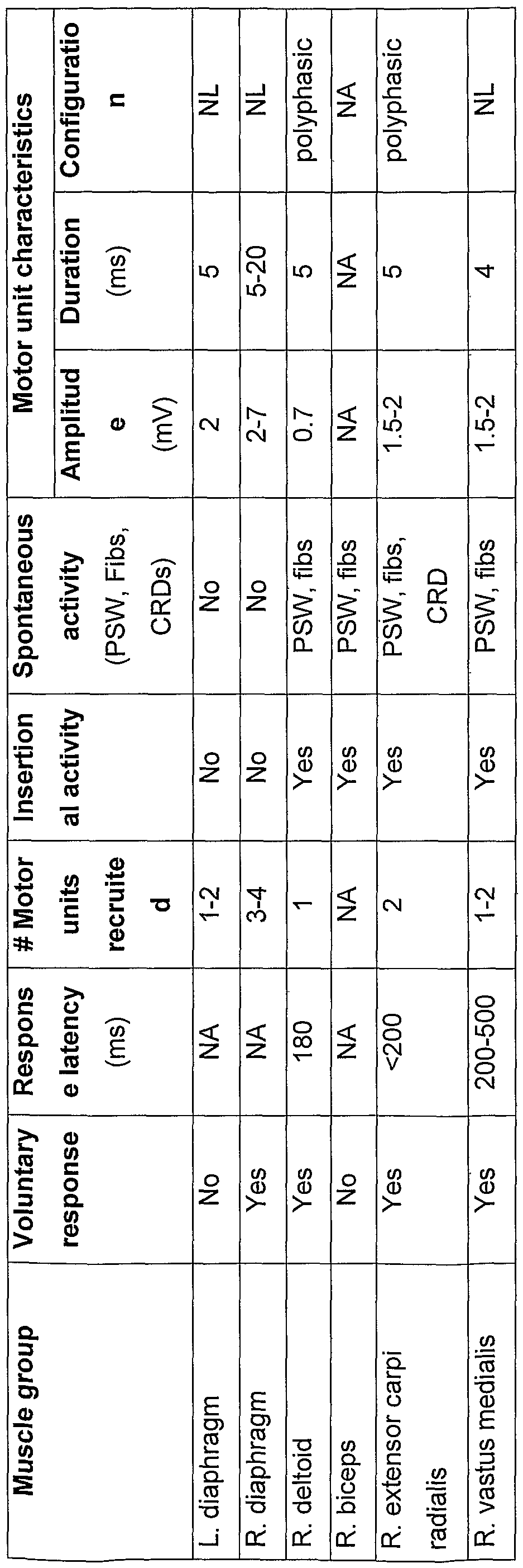
- the diaphragm was evaluated while the patient was seated, and the following muscles were assessed while the patient was lying down: right deltoid, biceps, extensor carpi radialis, and vastus medialis.
- Bone Density was performed at Washington University School of Medicine's Bone and Mineral Diseases laboratories. Dual- energy x-ray absorptiometry measurements were compared using national standards for white males (based on age, weight, and height). Data are presented as standard deviations using t-scores (sex-matched normal young adult reference).
- Magnetic Resonance Imaging Safety testing Before placing the patient in the MRI scanner, duplicate copies of the orthopedic instrumentation were acquired for testing of MR compatibility. To simulate the titanium loop instrumentation in the patient's cervical region (Danek Inc., Minneapolis, MN), two stainless-steel Harrington rods and stainless steel circlage wires were obtained for testing. A neurosurgeon bent the rods using a standard bending apparatus to approximate the bend of the straight segments of the patient's Danek loop, using the patient's plain-film radiographs. This bending was performed because stainless steel could become slightly magnetic when bent.
- the rods and wires were manually passed in and out of the MRI scanner. No torques were felt on the metal that would indicate any force due to magnetic properties. A strong hand-held magnet was also moved around the metal, and no attractive forces were felt. The rods and wires were then arranged to match the configuration on the radiographs.
- the hardware was taped to a spherical phantom containing 0.9% NaCI (saline) and doped with 0.15 mM Gd[DTPA-BMEA] (Optimark, Mallinckrodt Inc., St. Louis, Missouri) to shorten the T1 relaxation time to the physiological range.
- the phantom was placed in the MRI scanner, and several scans were performed using various pulse sequences that were planned to use in subsequent patient imaging sessions (MP-RAGE, turbo spin echo, echo-planar imaging, diffusion tensor imaging, etc.).
- the investigator held his hand over the metal during the imaging, and did not feel any RF heating or movement of the metal.
- Patient setup The patient was transported to the MRI suite by van, and all external metal was removed from his pockets. During transport and MRI the patient was ventilated using his personal ventilator connected to a tracheostomy tube for positive ventilator assistance.
- the patient and his ventilator were moved onto a lightweight aluminum stretcher (Model 30NM, Ferno-Washington Inc., Wilmington, Ohio) tested for MR-compatibility, and then the patient was transported into the MRI room.
- a pulse oximeter probe was placed on his right index finger to monitor heart rate and oxygen saturation (using a rubber grip sensor, Invivo Research, Inc., Orlando, Florida).
- a non-invasive blood pressure cuff was placed over the left arm and a tubing was connected to a side-port of the ventilator connector to monitor blood pressure, respiratory rate, end-tidal CO 2 , and inspired 02 (3150 Magnitude/3155A Millenium anesthesia monitoring system, Invivo Research, Inc., Orland, Florida).
- the ventilator tubing was temporarily disconnected and the patient was lifted onto the MRI table.
- the ventilator tubing was immediately reconnected, and a wall O 2 line was connected to the ventilator (medical grade O 2 , Airgas, St. Louis, Missouri).
- the oxygen flow was adjusted to 2-3 L/minute to maintain SpO2 to > 95% in the supine position. (In the upright position the patient typically has an SpO 2 of 97-98% without additional O 2 .)
- the patient was positioned so that the C2-C3 region of the cervical spin was centered over the active element of the neck phased array coil (see below). Padding was placed under the elbows, knees, and back.
- the ventilator and O 2 line were temporarily disconnected and moved to inside a non-magnetic RF-shielded box (Shielding Resources Group, Tulsa, Oklahoma) placed next to the MRI scanner.
- the RF box contained a shielded AC/DC power source, shielded electronic connectors, and waveguides (copper conduits) built into the walls of the box.
- the ventilator and O 2 tubings were fed through a waveguide and reconnected to the ventilator inside the RF box. This RF box was tested at > 80 dB attenuation to completely screen RF emissions from the ventilator and remove severe RF interference in the MRI.
- Headphones were placed on the patient for acoustic shielding and listening to music, and tape was placed across the forehead to minimize movement of the head and neck.
- the patient was then advanced into the scanner by moving the patient table, positioning the C2-C3 region and active coil element at the isocenter of the magnet and gradients.
- the display from the Invivo monitor was then connected from a custom video port of the Millenium 3155A anesthesia unit through a custom RF penetration panel in the MRI room wall (Shielding Resources Group, Tulsa, Oklahoma) and then to the back of the satellite MR console monitor for display of physiologic data in the control room, where a neurointensivist monitored all physiologic parameters during MRI.
- the heart rate was 58-86 beats per minute
- SpO 2 was 95-97%
- blood pressure was 126-139 mm / 72-86 mm
- the respiratory rate was 18 respirations per minute (equal to the ventilator setting)
- the end-tidal CO 2 was 29-34 mm Hg
- the inspired O 2 (FiO2) was 28%.
- the patient did not experience any adverse symptoms during the imaging sessions.
- MRI Acquisitions A 1.5-Tesla Siemens Magnetom Vision MRI scanner and Siemens phased-array spine coil were used for all imaging (Siemens, Er Weg, Germany). After an initial scout to confirm positioning at the isocenter, the external magnetic field was shimmed manually using the vendor MAP-shim procedure. For this purpose, only the linear shim channels were used because of instability in the higher-order shim channels due to the adjacent metal instrumentation. The frequency and transmitter were then tuned to the on-resonant and 180° conditions, and the scout scan was repeated. A T1 -weighted MP-RAGE scan was acquired next in the transverse plane, tilted ⁇ 20° sagittally and coronally to be perpendicular to the cord.
- the acquisition time was 9 min 29 sec with no signal averaging.
- the MP-RAGE data was used for cord cross-sectional area measurements (see below). Additional anatomical scans such as T2-weighted turbo spin echo and fluid-attenuated turbo inversion recovery were also acquired. Data analysis: MP-RAGE data were resliced to be perpendicular to the cervical cord.
- Region-of-interest (ROI) tracings were made around the border of the cord on every other transverse image using the "auto trace" function of Analyze AVW 4.0 (Mayo Foundation, Rochester, Minnesota). To choose the intensity level for the auto-trace, the lower level was determined in which the trace expanded out into the surrounding background noise, the higher level was determined in which the trace collapsed into the center of the cord. Then, the mean of the two levels was calculated and chosen for the auto-trace of the cord area. By this procedure, partial volume effects were taken into account in which the cord intensity transitions from high T1 -weighted intensity to the low signal surrounding the cord (see Figure 3).
- the ROI generated by the auto-trace procedure had to be edited to prevent the region from extending into adjacent tissues (e.g., due to cord adhesions).
- the two-dimensional areas of the cord were measured by sampling the ROIs, and results were graphed as a function of length along the cord. 7.
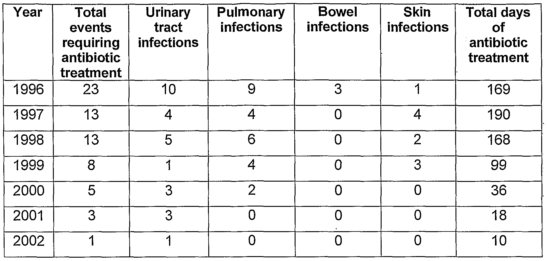
- Quantification of Infectious Complications The personal 24-hour nursing records for the patient were exceptionally detailed and this allowed us to track accurately the number and types of infections requiring antibiotic treatments each year. In addition, the total duration of each treatment was always recorded. The prescription records provided by the local medical doctor verified these values. In most cases, cultures were also obtained to further verify the infection.
- the activity-based recovery program consisted primarily of training on a FES bicycle.
- the customized recumbent bike system designed for use with paralyzed individuals, integrated computer-assisted FES-induced cycling.
- the goal was 1 hour of activity (up to 3000 revolutions) per day three times per week.
- the FES bicycle modulates the intensity of stimulation to obtain a consistent rotation speed.
- Surface electrodes stimulate three muscle groups in each leg (Fig. 2): one electrode is placed at the superior edge of the gluteal muscle, another over the hamstring group midway between the knee and hip, and two over the quadriceps (one over the superior portion and the other over the inferior third of the quadriceps).
- the legs were balanced in three ways.
- the seated buttocks and boots anchored the legs at the upper and lower positions. Belts that attached to the upper leg with Velcro balanced the mid-leg. A weighted fly-wheel ensured a smooth rotation by carrying momentum. The goal was to achieve the greatest number of revolutions (3000/hour).
- the FES bicycle therapy was supplemented with surface electrical stimulation to activate the following muscle groups; paraspinals, abdominals, wrist extensors, wrist flexors, deltoids, biceps, and triceps.
- the therapies were rotated daily, usually in a 3-day sequence. Each muscle group was activated for half an hour using intermittent 1 second on, 1 second off AC cycles.
- aquatherapy was incorporated into the program, with a goal of one 1-hour session per week. The aquatherapy focused on muscle groups in which voluntary control was recovered while participating in the activity based recovery program.
- a left lateral malleolus skin ulceration (Grade IV) was complicated by slow healing and osteomyelitis, threatening amputation. Aggressive treatment and healing by secondary intention took more than 1 year. 9) Sacral skin ulcer. Pilonidal cyst removal and suture closure was complicated by dehiscence and development of a sacral wound that required aggressive treatment and healing by secondary intention (vertical green bar indicates healing time). 10) Pathological fracture. The left femur fractured while the patient was attempting weight-supported standing. Treatment required hospitalization and surgical internal fixation. Additional treatment of severe osteoporosis included vitamin D, calcium supplements, and pharmacological treatment to limit bone resorption.
- Pinprick assessment which requires discrimination and is therefore more difficult, did not improve until the last year of the 3-year program, which is ongoing to date. Sensation was appreciable throughout most dermatomes of the body (Fig. 6), including the sacral region (S-3, S4-5). In addition to recovery of pinprick and light touch, recovery of additional sensory modalities occurred and included vibration, proprioception, and ability to differentiate heat and cold. The most notable change was an improvement in motor function of up to 20% (20/100). Voluntary control of the external anal sphincter is possible (S4-5). The conversion of the patient's condition to ASIA Grade C thus occurred in July 2001. Motor recovery was first evident in the left fingers, then the right hand, and then the legs.
- FIG. 6 is a schematic drawing showing a comparison of the patient's ASIA grades from 1995 to 2002. Values in blue indicate scores from 1999; those in red indicate 2002 scores. Note that all the 1995 motor scores and the sensory scores obtained below T-4 were zero. The 1995 scores were taken from the July 1995 ASIA sheet. The following comparison puts this motor recovery into perspective.
- the National Acute Spinal Cord Injury Study II and III trials documented an average 4.8- point improvement in the motor scores of patients receiving methylprednisolone compared with those receiving placebo if methylprednisolone was given within 8 hours of injury.
- Electromyography Results The EMG analysis of volitional movements was completed in the winter of 2001 (Table 4), and the results were compared with those from phrenic nerve testing performed on June 21 , 1995, shortly after the injury. At that time, there was evidence of intact anterior horn cells. Latencies were less than 10 msec, and right and left amplitudes were 0.9 mV and 0.5 mV, respectively. Diaphragmatic movement, albeit small, was noted on fluoroscopic examination. In contrast, amplitudes were greater in 2001 (2-7 mV), but there was further evidence of denervation (Table 4).
- EMG characteristics of key muscles in response to commands to voluntarily move the muscle group Columns indicate muscle groups recorded, presence of identifiable EMG response to activation request (voluntary response), delay in EMG activation compared to voluntary activation request (Response latency), number of motor units recruited with increasing effort to move (# Motor units recruited), presence of activity with insertion of the EMG needle (Insertional activity), presence of spontaneous resting activity (Spontaneous activity), and characteristics of motor units (Motor unit characteristics: Amplitude, Duration, and Configuration). Abbreviations: positive sharp waves (PSW); fibrillations (fibs); complex repetitive discharge (CRD); normal (NL); not assessed (NA); millivolts (mV); millisecond (ms). 5.
- FIG. 7(a) shows a schematic drawing of the experimental rat model showing the relationship between the injury and stimulation level.
- Figure 7(b) shows the 43-day time line for the experiment in which twelve Long Evans adult female rats received a complete transection of the spinal cord by suction ablation of two levels (T8-9).
- T8-9 two levels
- Stimulator and electrode implantation Three weeks following SCI the rats were re-anesthetized, a midline incision was made over the lower back and a 2- channel battery powered electrical stimulator (Jarvis, University of Liverpool) and current return electrode were implanted in a subcutaneous pocket. A 0.5 cm incision was made on the lateral aspect of both shanks and stainless steel wire electrodes were tunneled bilaterally underneath the skin from the stimulator site, and sutured into the tibialis anterior muscle adjacent to the common peroneal nerve. Intra- operative test stimulation ensured proper peroneal nerve activation.
- FES devices were activated daily for 19 days between 24 and 43 days after injury (starting at 3 days following implantation), for three 1 h sessions during each 9 h workday (Figure 11B).
- An additional group of 6 rats received the identical FES implant and activation pattern but were not injured.
- the pattern of stimulation was 1 s stimulation of one common peroneal nerve, followed by 1 s of rest, then the other common peroneal nerve was stimulated for 1 s, followed by 1 s of rest and the cycle repeated.
- Stimuli were 3 V 200 ⁇ s monophasic pulses delivered at 20 Hz between the electrode over the nerve and the return electrode in the lower back and were expected to activate large myelinated fibers within the common peroneal nerve, but not small unmyelinated nociceptive fibers.
- the nerve stimulation produced alternating flexion of the hindlimbs that crudely approximated bilateral stepping-like hindlimb movements.
- Bromodeoxyuridine injection paradigm Newborn cells were identified by daily injections, beginning day 31-36 after injury, with BrdU (50 mg/kg i.p.), a brominated DNA building block that is selectively incorporated into replicating DNA during the S phase of the cell cycle.
- BrdU 50 mg/kg i.p.
- Immunohistochemistry Every 6th section (40 ⁇ m) from spinal cord levels C2, T1 , T7, T11 , L1 , and L5 was selected for anti-BrdU immunohistochemistry. Sections were incubated in 2 N HCI for 60 min at 37°C, transferred to 0.1 M Borate buffer (pH 8.5) for 20 min, and rinsed with PBS. Non-specific labeling was blocked with 0.1 % BSA in 0.1% Triton X-100/PBS for 60 min. A mouse monoclonal anti-BrdU antibody (1:600; Roche, Mannheim, Germany) was incubated with the tissue overnight at 4°C. Then tissue was treated with a CY3-conjugated secondary antibody (1:2000; Jackson, West Grove, PA) in 2% normal goat serum (NGS) for 60 min.
- BrdU positive cells The number of BrdU-labeled cells as a function of spinal cord level was measured in a blinded fashion. Images were acquired on an Olympus IX70 Microscope equipped with a Magnafire® camera and quantitative counts were performed using a computer assisted software package (StereoinvestigatorTM, Microbrightfield Inc., VT). For unbiased stereological counts of proliferating cells the indicator fractionator method was used. The estimate of BrdU positive nuclei was based on counts of nuclei in a known fraction of the volume of the region under consideration.
- the software calculated the total number of BrdU positive nuclei.
- cells were evaluated for colabeling with each phenotypic marker.
- the complete cell nucleus was followed through the z-axis, and only cells with a well- circumscribed immunopositive cell body were considered positive for a particular phenotype.
- FES produced a substantial increase in the density of new cell birth/survival at the lumbar levels of the spinal cord, consistent with a robust projection of the peroneal nerve to the lumbar spinal cord.
- the rostral-caudal selectivity of the effects of FES-induced hindlimb locomotion on cell birth/survival helps exclude possible global mechanisms such as metabolism, blood borne and other systemic factors, and strengthens support for local mechanisms associated with increased neural activity. Electrical fields in the range of 300 V/m can induce changes in neural cell function. Based upon the 100X larger magnitude of electric field required to produce direct effects in other studies, compared to the very small electric field experience by the spinal cord in these studies (about 5 V/m), the neuronal activity rather than direct electric field effects is demonstrated to be responsible for the observed effects.
- the rat spinal cord was completely transected by suction ablation at T8/T9 to minimize intra-animal variability that might occur with less complete lesions, but this model limited analysis of functional recovery.
- Previous studies indicate that thoracic separation of the spinal cord produces reduced neural activity below the lesion level and that peripheral nerve stimulation or gait activity enhances activity at the respective cord level.
- a chronic injury was chosen because it is most relevant to human regeneration, the blood brain barrier is patent, and injury-induced cellular proliferation is normalizing.
- a stimulation protocol of three one hour periods of phasic activity per day was based on previous findings that at least one hour of gait training per day was sufficient to double the number of progenitor derived neurons in the hippocampus of normal mice. Alternating stimulation of the common peroneal nerve stimulation produced alternating flexion of the hindlimbs that crudely approximated bilateral stepping-like movements of the hindlimbs.
- the newborn cells mainly expressed markers of tripotential and glial progenitor cells, as well as astrocytes and oligodendrocytes.
- the number of BrdU labeled microglia, macrophages or endothelial cells was small and did not substantially contribute to the increased cell birth/survival at L1 and L5, levels distant from the injury site (T8/9). This conclusion is consistent with the very low contribution (fewer than 1.5 %) of microglia and endothelial cells to total BrdU labeled cells in the normal spinal cord.
- a five-day interval of BrdU labeling will primarily label proliferating cells that do not have time to differentiate into mature cells, especially neurons. No evidence of new neuron birth was present in the spinal cord at day 0 or 7 after BrdU injection and this is consistent with previous data demonstrating the absence of new neuron birth in the adult spinal cord.
- Figure 8 shows new cell birth in the adult spinal cord after chronic SCI.
- Panels A & B show 40 ⁇ m coronal sections of the spinal cord at the C2 (A) and L1 (B) levels, which were immunolabeled with mouse anti-BrdU antibody. Sections were reconstructed using Stereoinvestigator® from individual images at 20x magnification. Zones where the majority of BrdU positive cells were found are indicated with arrows. Square lettered inserts in panels A & B correspond to magnified images in panels C-F. Although the greatest number of BrdU positive cells was found in the white matter (C), BrdU positive cells were also present in grey matter (D-E) and around the central canal (F). Scale bar in panels A-B is 1 mm, and scale bar in panels C-F is 50 ⁇ m.
- Figures 9 show that FES promoted new cell birth/survival in the damaged spinal cord. Quantitative counts of BrdU labeled cells were performed using stereological methods in coronal spinal cord sections (C2-L5). Two-way ANOVA demonstrated the effects of treatment and spinal level (within groups, p ⁇ 0.05).
- A FES induced a robust and selective increase in new cell birth reserved to lower lumbar segments that predictably experienced increased activity from the patterned FES induced stepping-like limb movements.
- This effect persisted in the cell survival group 7 days later (*p ⁇ 0.05; **p ⁇ 0.001 , FES vs. control). .
- FIG. 10 panels (a)-(l) shows BrdU colocalization with cell specific markers. Confocal microscopic images from an FES treated animal at lumbar level L5, two hours after the last BrdU injection.
- A-C Nestin immunoreactivity (blue) was found predominantly in white matter and labeled cells exhibited bipolar and multipolar morphology. Some of the Nestin ⁇ cells colabeled with GFAP (green, arrowhead), which may represent reactive astrocytes, however, fewer than 2 % double labeled with BrdU (red). Thus, BrdU+/Nestin+ cells are proliferating tripotential progenitors rather than reactive astrocytes.
- FIG 11 panels (a)-(e) shows quantification of NG2 colocalization with BrdU following SCI at 2 hours after last BrdU injection.
- NG2 immunoreactivity was used to classify BrdU+ cells as glial progenitor cells rostral (A) and caudal (B) to the lesion site.
- NG2+ cells displayed bipolar and multipolar cell morphologies predominantly in the white matter (arrows indicate individual BrdU+/NG2+ cells).
- Panels C-E depict single confocal sections of each marker for such an individual cell. The distribution pattern of Brdu+/NG2+ cells did not vary amongst groups.
- the present invention represents the first demonstration that FES can enhance cellular generation in the injured adult CNS.
- electrical stimulation is used clinically to attempt to enhance bone fusion and stimulate peripheral nerve growth, it has not been applied to CNS regeneration.
- the magnitude of the cell birth/survival response to FES in the present study was surprisingly large (61 % to 77%).
- Based on the volume of the entire adult rat spinal cord (estimated by volume displacement; 506 + 7 mm 3 , density 1.21 g/cm 3 ) and the estimates of FES induced cell birth, at level C2 approximately two new cells are born per second in the current studies. Therefore, a million new cells were born in just under one week, a number equivalent to numbers commonly transplanted in the injured spinal cord for repair purposes. Harnessing the potential of these endogenously born cells therefore represents a rational approach to self-repair of the damaged CNS.
- Experiments found in the Examples section of the present invention were designed to optimize the experimental analysis of one index of regeneration and new cell birth.
- FES may enhance recovery after chronic spinal cord injury.
- FES dramatically increases cell birth/survival in the injured spinal cord may be one factor that could contribute to functional recovery.
- Activity-based recovery applications may be an important long-term therapeutic target for individuals with SCI.
- Figure 11 illustrates the experimental outline and the topographic relationship between the injury level and the FES stimulation level.
- Bladder infections were defined by onset of bloody or cloudy urine coinciding with presence of red and white blood cells, struvite crystals, and epithelial cells in the urine sediment. Neither group showed any signs of autophagia, pain, or inflammation.
- Anti-BrdU labeled cells indicative of new cell birth occurred in the spinal cords of both control and stimulated rats.
- BrdU may integrate into apoptotic cells as a result of DNA damage repair
- BrdU does not integrate in detectable levels into adult cells induced to undergo necrotic or apoptotic death.
- Anti-BrdU labeled cells expressed markers of tripotential progenitors (Nestin), glial progenitors (NG2), astrocytes (GFAP) and oligodendrocytes (APC-CC1). There was no quantitative difference in NG2 (49 ⁇ 3% at T1 ; 36+3% at L5), GFAP (45+3% at T1 ; 40+3% at L5), or APC-CC1 (fewer than 5%) expression between groups ( Figure 14-16).
- Microglia, Macrophages, and Endothelial Cells Minimally Contributed to Increased Cell Birth/Survival Surrounding the Lesion Site
- ED- 1 and OX-42 labeled macrophages and microglia surrounding the injury site and their numbers rapidly tapered as a function of distance from the injury site. Macrophages could also be easily distinguished by their characteristic cell body shape and eccentric nuclei and their bifringent inclusions. Double labeling with anti- BrdU revealed that a very small number of these cells were BrdU positive (fewer than 2%).
- Anti-BrdU labeling was also occasionally observed in endothelial cells or pericytes of blood vessels (identified by characteristic morphology, location and immunoreactivity to specific markers (anti-CD31 , anti-Glut-1)), but the total number of cells was small (fewer than 1.5%) and mostly restricted to the levels surrounding the injury site (T8/T11). Thus, microglia, macrophages, and endothelial cells did not contribute substantially to the number of BrdU labeled cells measured at levels distant from the injury (C2, T1 , L1 , and L5).
- Severe sensory deprivation due to amputation or peripheral nerve damage profoundly alters responsiveness and topographical organization of primary somatosensory cortex (SI). Less well known are the alterations in cortical responses after spinal cord injury (SCI). Individuals with SCI, as opposed to those with amputations, retain a normal body, which may influence cortical reorganization significantly, especially in individuals with partial SCI who have surviving fibers and potentially some functional connections across the level of damage. It will become an important practical issue to assess cortical responsiveness immediately after damage and in the course of recovery if ongoing efforts for restoring function by transplantation or other means such as FES-evoked patterned movement, are successful.
- Functional MRI fMRI
- BOLD blood oxygenation level-dependent
- fMRI results provide mapping of cortical somatosensory-motor areas in the human subject. Given the unique medical history of the subject in including late partial recovery of function after being clinically assessed as motor complete and nearly sensory complete, the fMRI study sought to determine the topographical normality of the subject's somatosensory and motor cortical responses above and below the level of damage, and to investigate possible cortical reorganization subsequent to a late recovery from SCI.
- Example 1 describes in detail the clinical history of the 50-year-old right-handed male who sustained a displaced C2 type II odontoid fracture from an equestrian accident in 1995 at age 42. No other permanent injuries, particularly a head injury, complicated the SCI.
- motor or somatosensory functions were absent below the lesion level for 5 years except for spotty sensation in the left hemitorso. He is ventilator dependent with hypophonic vocalization due to impaired function of the chest diaphragm and muscles of vocalization.
- the control subject was a 23-year-old male who has a normal neurological and psychiatric history. Visuomotor Tracking.
- Vibrotactile Stimulation A massage vibrator delivered suprathreshold tactile stimulation.
- the device previously used in positron-emission tomography studies, was made magnetic resonance-compatible by replacing the electric motor with a pneumatic drive that was connected to a remote air compressor.
- the vibrator delivered approximately 2-mm displacement vibrations centered on a base frequency of approximately 100 Hz.
- the vibrator head was manually held against the left fingers and palm or sole of the left foot throughout stimulation and rest periods. Precise skin displacements were unknown, although stimulus magnitudes probably activated most skin mechanoreceptors, adjoining deeper tissues, and proprioceptors throughout distal parts of the stimulated limb.
- MRI Acquisitions During MRI the SCI subject was ventilated, and physiologic parameters were monitored continuously (Magnitude/Millenium anesthesia monitoring, In Vivo Research, Orlando, FL) in a custom magnetic resonance-compatible setup (Shielding Resources Group, Tulsa, OK). All MRI used a 1.5-Tesla Magnetom Vision scanner and circularly polarized head coil (Siemens, Er Weg, Germany). Structural 3D T1 -weighted magnetization-prepared_rapid gradient echo MRI was acquired.
- 128 sets of 20 contiguous, 6-mmthick slices were acquired parallel to the anterior commissure- posterior commissure plane (3.75 x 3.75 mm in-plane voxel size), allowing complete brain coverage.
- This protocol yielded images of the SCI subject without significant signal loss or distortions in the brain despite surgical metal in the neck.
- Sensory and motor fMRI were acquired in different imaging sessions by using 8-10 fMRI runs per session and 128 frames (346.75 s) per run.
- the cord was segmented from structural MRI for area measurement and 3D-rendered display by using ANALYZE AVW 4.0 (Mayo Foundation, Rochester, MN) and a Sun Fire V880 computer (Sun Microsystems, Santa Clara, CA).
- fMRI data were analyzed as described in Burton et al., J. Neurophysiol. 87: 589-607 (2002), and Corbetta et al., Neuron 21: 761-73 (1998).
- General linear models estimated the BOLD responses in each subject and each task (e.g., tongue movement) without assuming a hemodynamic response shape.
- BOLD time courses were estimated in each voxel, over eight frames (21.67 s) for somatosensory tasks and 10 frames (27.09 s) for motor tasks.
- fMRI data were smoothed with a 3D 2-voxel Gaussian kernel and transformed to the Talairach atlas before statistical analysis.
- Statistical maps were based on cross-correlation between estimated BOLD time course and a reference hemodynamic response function that was obtained by convolving a delayed gamma function with a rectangular function representing task and control periods.
- the derived t statistics per voxel were converted to normally distributed z scores and corrected for multiple comparisons across the entire brain by using distributions obtained from Monte Carlo simulations (based on methods described in S.D.
- Figure 14 shows a T1 -weighted MRI of cervical SCI in SCI subject.
- the top panel shows longitudinal 3D-rendered image (view from behind) of lower-brainstem and cervical spinal cord segments from the tip of the C2 odontoid process through the bottom of the C2 vertebral body.
- the middle panel shows selected low- magnification images through the zone of injury show the small size of the cord relative to the spinal canal (SC). Images are transverse to the cord's long axis and at levels 40, 46, and 51 mm below the cerebellar tonsils (Left, Center, and Right, respectively).
- the bottom panel shows higher-magnification view of the same three transverse images show focal regions of low T1 -weighted signal that are consistent with chronic tissue damage (myelomalacia) or scarring.
- the location and shape of these sites vary and include a central oval (red arrow), a cleft (blue arrow), and several peripheral lesions (yellow arrows). L indicates left, R, indicates right.
- Figure 15 shows BOLD responses to the visually guided motor tasks on 3D and flattened representations of a standard brain. 3D and 2D flattened views of an atlas brain are shown with projected BOLD responses for visuomotor tracking task; the color scale indicates z scores.
- Figure 15A shows a control subject, .while Figure 15B shows the subject with SCI.
- Tongue movements in the control subject activated a ventral segment of precentral gyrus, central sulcus, and postcentral gyrus bilaterally (Fig. 15A, SMfc). These regions likely correspond to the primary sensory and motor cortex face area. Recruitment of SI in a visuomotor tracking task probably reflects tactile and proprioceptive sensory feedback signals associated with normal movements.
- Tongue movements activated the face area of SI/M1 in ventral precentral gyrus, central sulcus, and postcentral gyrus (primary somatosensory-motor cortex, SMfc). Activity spread dorsally into the hand area and more extensively into adjacent regions such as the second somatosensory cortex (Sll) and frontal operculum.
- Sll second somatosensory cortex
- Performance was less accurate and less sustained, and the subject reported more effort, compared with visuomotor tracking.
- BOLD responses (movement vs. rest) were weaker and less localized in M1 , and there was no recruitment of premotor or higher-order regions (not shown).
- FIG. 16 shows BOLD responses to vibratory stimulation of the left hand or left foot. 3D and 2D flattened views of the atlas brain are shown, on which BOLD responses for sensory vibrotactile stimuli have been projected; the color scale indicates z scores.
- Figure 16A shows results for a control subject, and figure 16B shows those for the Subject with SCI .
- left hand and fingers vibration activated the contralateral (right) SI/M1 cortex.
- Responses occupied the dorsal segment of the postcentral gyrus extending into the central sulcus and the precentral gyrus, thus identifying the normal location of the hand area (Fig. 16A, SMh).
- Sll left foot vibration evoked activity in the right medial frontal gyrus approximately 1.2 cm dorsal to the hand area (Fig. 16A Right).
- This response likely involved the SI/M1 foot area (SMft).
- Sll was activated bilaterally.
- the subject with SCI reported a better sensation of vibration in the left foot than in the left hand.
- Left hand vibration failed to activate expected hand areas of SI/M1 in the right postcentral gyrus, central sulcus, and precentral gyrus but produced bilateral responses in Sll (Fig. 16S, Left).
- FIG. 17A contrasts activation maps for left hand vibration and tracking tongue movements in the control subject.
- Figure 17 ⁇ shows the absence of a normal hand area in the SCI subject.
- Figure 17C shows that the face area is similarly active in both subjects. However, the active region expands dorsally in the SCI subject where it encroaches onto the normal hand area (Fig. 17D).
- the S1 hand area responded to sensory stimulation of the face but not the hand, which possibly reflects neural plasticity based on competitive interaction from a normally innervated face.
- This response of the (deafferented) hand area to (invading) face inputs might correlate with an enhanced resolution of tactile sensation in the face, as demonstrated on a much smaller spatial scale within the digit representation of monkey area 3b.
- the reorganization possibly involved subcortical transneuronal changes, which are progressive and delayed. Cortical activity is likely to reflect this mechanism by initial silence followed by late recovery of responses. Similar to prior results in monkeys with long-term dorsal rhizotomies, expansion in the SCI subject involved SI hand area responses to stimulation in the lower face, which borders the thalamic hand area. A conjunction of mechanisms is likely to be responsible for the SI foot- area responses and the reported better sensations from the foot. Absent was competition from intact representations because all cortical and subcortical areas adjacent to the foot were severely deprived. This likely left the SI foot area accessible to any input that was conveyed through surviving fibers at the level of the injury (Fig. 14).
- the secondary motor areas were activated more in the SCI vs. control subject. Additionally, many higher-order regions (e.g., posterior parietal, temporal, and dorsolateral prefrontal cortex) were recruited in the SCI subject, more for finger than tongue movements. Although tongue movements use neurons above the lesion, it is not surprising that an abnormally widespread network of activity was evoked, because these movements require coordination of several muscle groups (e.g., diaphragm or accessory respiratory muscles) that have C2-C5 myotomes at or below the lesion.
- muscle groups e.g., diaphragm or accessory respiratory muscles
Landscapes
- Health & Medical Sciences (AREA)
- Life Sciences & Earth Sciences (AREA)
- Animal Behavior & Ethology (AREA)
- Veterinary Medicine (AREA)
- Public Health (AREA)
- General Health & Medical Sciences (AREA)
- Physical Education & Sports Medicine (AREA)
- Rehabilitation Therapy (AREA)
- Engineering & Computer Science (AREA)
- Biomedical Technology (AREA)
- Nuclear Medicine, Radiotherapy & Molecular Imaging (AREA)
- Radiology & Medical Imaging (AREA)
- Pain & Pain Management (AREA)
- Epidemiology (AREA)
- Cell Biology (AREA)
- Orthopedic Medicine & Surgery (AREA)
- Medicines That Contain Protein Lipid Enzymes And Other Medicines (AREA)
- Electrotherapy Devices (AREA)
Abstract
Description


Claims
Priority Applications (4)
| Application Number | Priority Date | Filing Date | Title |
|---|---|---|---|
| AU2003272273A AU2003272273B2 (en) | 2002-09-04 | 2003-09-04 | Methods for treating central nervous system damage |
| EP03754449A EP1545700A4 (en) | 2002-09-04 | 2003-09-04 | METHOD OF TREATING CENTRAL NERVOUS SYSTEM DAMAGE |
| JP2004534624A JP2006508707A (en) | 2002-09-04 | 2003-09-04 | Treatment of central nervous system injury |
| CA002497674A CA2497674A1 (en) | 2002-09-04 | 2003-09-04 | Methods for treating central nervous system damage |
Applications Claiming Priority (2)
| Application Number | Priority Date | Filing Date | Title |
|---|---|---|---|
| US40821402P | 2002-09-04 | 2002-09-04 | |
| US60/408,214 | 2002-09-04 |
Publications (2)
| Publication Number | Publication Date |
|---|---|
| WO2004022156A2 true WO2004022156A2 (en) | 2004-03-18 |
| WO2004022156A3 WO2004022156A3 (en) | 2004-08-05 |
Family
ID=31978579
Family Applications (1)
| Application Number | Title | Priority Date | Filing Date |
|---|---|---|---|
| PCT/US2003/027819 Ceased WO2004022156A2 (en) | 2002-09-04 | 2003-09-04 | Methods for treating central nervous system damage |
Country Status (6)
| Country | Link |
|---|---|
| US (2) | US7610096B2 (en) |
| EP (1) | EP1545700A4 (en) |
| JP (1) | JP2006508707A (en) |
| AU (1) | AU2003272273B2 (en) |
| CA (1) | CA2497674A1 (en) |
| WO (1) | WO2004022156A2 (en) |
Cited By (2)
| Publication number | Priority date | Publication date | Assignee | Title |
|---|---|---|---|---|
| WO2006134999A1 (en) * | 2005-06-15 | 2006-12-21 | Kagoshima University | Vibration stimulation therapy apparatus, its use method, and computer program |
| US12290335B2 (en) | 2019-09-30 | 2025-05-06 | Rainbow Inc. | Method, program, and device for evaluating state of motor function |
Families Citing this family (82)
| Publication number | Priority date | Publication date | Assignee | Title |
|---|---|---|---|---|
| US20060247095A1 (en) * | 2001-09-21 | 2006-11-02 | Rummerfield Patrick D | Method and apparatus for promoting nerve regeneration in paralyzed patients |
| AU2003901696A0 (en) | 2003-04-09 | 2003-05-01 | Cochlear Limited | Implant magnet system |
| US7840270B2 (en) | 2003-07-23 | 2010-11-23 | Synapse Biomedical, Inc. | System and method for conditioning a diaphragm of a patient |
| US20090312817A1 (en) * | 2003-11-26 | 2009-12-17 | Wicab, Inc. | Systems and methods for altering brain and body functions and for treating conditions and diseases of the same |
| JP2007518469A (en) * | 2003-11-26 | 2007-07-12 | タイラー ミッシェル ユージン | System and method for modifying vestibular biomechanics |
| US10589087B2 (en) | 2003-11-26 | 2020-03-17 | Wicab, Inc. | Systems and methods for altering brain and body functions and for treating conditions and diseases of the same |
| US20080009772A1 (en) * | 2003-11-26 | 2008-01-10 | Wicab, Inc. | Systems and methods for altering brain and body functions and for treating conditions and diseases of the same |
| US20060161218A1 (en) * | 2003-11-26 | 2006-07-20 | Wicab, Inc. | Systems and methods for treating traumatic brain injury |
| US7818131B2 (en) * | 2005-06-17 | 2010-10-19 | Venture Gain, L.L.C. | Non-parametric modeling apparatus and method for classification, especially of activity state |
| US8249714B1 (en) | 2005-07-08 | 2012-08-21 | Customkynetics, Inc. | Lower extremity exercise device with stimulation and related methods |
| US9050005B2 (en) * | 2005-08-25 | 2015-06-09 | Synapse Biomedical, Inc. | Method and apparatus for transgastric neurostimulation |
| US7756585B2 (en) * | 2006-01-31 | 2010-07-13 | Good Samaritan Children's Therapy Unit | Muscle stimulation method and system to improve walking |
| US20070208392A1 (en) * | 2006-02-17 | 2007-09-06 | Alfred E. Mann Foundation For Scientific Research | System for functional electrical stimulation |
| EP2081636A4 (en) * | 2006-10-26 | 2010-12-22 | Wicab Inc | Systems and methods for altering brain and body functions and for treating conditions and diseases |
| US20080234791A1 (en) * | 2007-01-17 | 2008-09-25 | Jeffrey Edward Arle | Spinal cord implant systems and methods |
| WO2008098001A2 (en) * | 2007-02-05 | 2008-08-14 | Synapse Biomedical, Inc. | Removable intramuscular electrode |
| US9820671B2 (en) * | 2007-05-17 | 2017-11-21 | Synapse Biomedical, Inc. | Devices and methods for assessing motor point electromyogram as a biomarker |
| SE531177C2 (en) | 2007-05-24 | 2009-01-13 | Cochlear Ltd | Distance for implants |
| US7722504B2 (en) * | 2007-09-04 | 2010-05-25 | Younger J Kevin | Method for measuring physical fitness and creating athletic training regimens for particular sports |
| US7996080B1 (en) | 2007-10-16 | 2011-08-09 | Customkynetics, Inc. | Recumbent stepping exercise device with stimulation and related methods |
| US8428726B2 (en) | 2007-10-30 | 2013-04-23 | Synapse Biomedical, Inc. | Device and method of neuromodulation to effect a functionally restorative adaption of the neuromuscular system |
| JP2011509216A (en) * | 2008-01-11 | 2011-03-24 | ザ リージェンツ オブ ザ ユニバーシティー オブ ミシガン | Insect flight control system |
| WO2009115129A1 (en) * | 2008-03-20 | 2009-09-24 | Otto-Von-Guericke-Universität Magdeburg | An apparatus and a method for automatic treatment adjustment after nervous system dysfunction |
| US8260424B2 (en) * | 2008-10-24 | 2012-09-04 | Boston Scientific Neuromodulation Corporation | Systems and methods for detecting a loss of electrical connectivity between components of implantable medical lead systems |
| US8688217B2 (en) * | 2008-10-24 | 2014-04-01 | Boston Scientific Neuromodulation Corporation | Method to detect proper lead connection in an implantable stimulation system |
| FR2938233B1 (en) * | 2008-11-07 | 2011-08-26 | Bruno Garin | METHOD FOR MANAGING THE EFFORT OF A USER OF A HUMAN PROPULSION VEHICLE AND VEHICLE ADAPTED THEREFOR |
| US8612010B2 (en) * | 2008-12-23 | 2013-12-17 | Robotic Integrated Technology Development Corporation | Upper extremity muscle therapy system |
| US8615301B2 (en) * | 2008-12-23 | 2013-12-24 | Robotic Integrated Technology Development Corporation | Muscle therapy system |
| WO2011003709A1 (en) * | 2009-07-10 | 2011-01-13 | University College Dublin, National University Of Ireland, Dublin | Method and apparatus for stimulating the lower back and abdominal muscles |
| SE1050420A1 (en) * | 2010-04-27 | 2011-07-26 | Inerventions Ab | System and clothing for relaxing a spastic muscle |
| WO2012003451A2 (en) | 2010-07-01 | 2012-01-05 | Stimdesigns Llc | Universal closed-loop electrical stimulation system |
| US8923978B1 (en) | 2011-04-19 | 2014-12-30 | Customkynetics, Inc. | Exercise device with stimulation based on volitional contribution and related methods |
| USD683464S1 (en) | 2011-06-17 | 2013-05-28 | Customkynetics, Inc. | Leg brace |
| RU2466756C1 (en) * | 2011-07-28 | 2012-11-20 | Институт физиологии им. И.П. Павлова Российской Академии Наук | Method for simulating treating patients with chronic spinal injury in laboratory animals |
| WO2013028960A1 (en) * | 2011-08-25 | 2013-02-28 | Insomnisolv, Llc | System and method for the treatment of insomnia |
| US9271864B2 (en) * | 2011-10-04 | 2016-03-01 | Feinstein Patents Llc | Orthosis for range of motion, muscular and neurologic rehabilitation of the lower extremities |
| AU2012334926B2 (en) | 2011-11-11 | 2017-07-13 | The Regents Of The University Of California | Transcutaneous spinal cord stimulation: noninvasive tool for activation of locomotor circuitry |
| EP2592431A1 (en) * | 2011-11-11 | 2013-05-15 | Max-Planck-Gesellschaft zur Förderung der Wissenschaften e.V. | Method and apparatus for visualization of tissue perfusion by means of assessing BOLD signal fluctuations |
| EP2812697B1 (en) * | 2012-02-06 | 2017-03-22 | University of Miami | Innate immune proteins as biomarkers for cns injury |
| US9510756B2 (en) * | 2012-03-05 | 2016-12-06 | Siemens Healthcare Gmbh | Method and system for diagnosis of attention deficit hyperactivity disorder from magnetic resonance images |
| EP2869890B1 (en) * | 2012-07-03 | 2017-09-06 | Med-El Elektromedizinische Geraete GmbH | Mri-safe implant magnet with angular magnetization |
| WO2014144785A1 (en) | 2013-03-15 | 2014-09-18 | The Regents Of The University Of California | Multi-site transcutaneous electrical stimulation of the spinal cord for facilitation of locomotion |
| EP3049148B1 (en) | 2013-09-27 | 2020-05-20 | The Regents Of The University Of California | Engaging the cervical spinal cord circuitry to re-enable volitional control of hand function in tetraplegic subjects |
| US10091594B2 (en) | 2014-07-29 | 2018-10-02 | Cochlear Limited | Bone conduction magnetic retention system |
| EP3180069B1 (en) | 2014-08-17 | 2020-05-13 | Nine Continents Medical, Inc. | Miniature implatable neurostimulator system for sciatic nerves and their branches |
| US12053630B2 (en) | 2014-08-17 | 2024-08-06 | Coloplast A/S | Implantable pulse generator with automatic jump-start |
| AU2015305237B2 (en) | 2014-08-21 | 2020-06-18 | The Regents Of The University Of California | Regulation of autonomic control of bladder voiding after a complete spinal cord injury |
| US10773074B2 (en) | 2014-08-27 | 2020-09-15 | The Regents Of The University Of California | Multi-electrode array for spinal cord epidural stimulation |
| US10130807B2 (en) | 2015-06-12 | 2018-11-20 | Cochlear Limited | Magnet management MRI compatibility |
| US20160381473A1 (en) | 2015-06-26 | 2016-12-29 | Johan Gustafsson | Magnetic retention device |
| WO2017035512A1 (en) | 2015-08-26 | 2017-03-02 | The Regents Of The University Of California | Concerted use of noninvasive neuromodulation device with exoskeleton to enable voluntary movement and greater muscle activation when stepping in a chronically paralyzed subject |
| US10917730B2 (en) | 2015-09-14 | 2021-02-09 | Cochlear Limited | Retention magnet system for medical device |
| US9782321B1 (en) * | 2015-09-15 | 2017-10-10 | Thomas Semmens | Lower body electrical muscle stimulation exercise systems |
| GB201518263D0 (en) * | 2015-10-15 | 2015-12-02 | Oxford Bioelectronics Ltd | Method |
| US11318310B1 (en) | 2015-10-26 | 2022-05-03 | Nevro Corp. | Neuromodulation for altering autonomic functions, and associated systems and methods |
| US11097122B2 (en) | 2015-11-04 | 2021-08-24 | The Regents Of The University Of California | Magnetic stimulation of the spinal cord to restore control of bladder and/or bowel |
| US10576276B2 (en) | 2016-04-29 | 2020-03-03 | Cochlear Limited | Implanted magnet management in the face of external magnetic fields |
| JP6319730B2 (en) * | 2016-08-30 | 2018-05-09 | パナソニックIpマネジメント株式会社 | Control device for electrical stimulation device, electrical stimulation device, and pedaling exercise system |
| US11595768B2 (en) | 2016-12-02 | 2023-02-28 | Cochlear Limited | Retention force increasing components |
| US11235154B2 (en) | 2017-02-17 | 2022-02-01 | The University Of British Columbia | Apparatus and methods for maintaining physiological functions |
| JP7236086B2 (en) * | 2017-03-10 | 2023-03-09 | 医療法人和康会 | training equipment |
| EP4252653A3 (en) | 2017-03-28 | 2023-12-06 | Ecole Polytechnique Fédérale de Lausanne (EPFL) EPFL-TTO | A neurostimulation system for central nervous stimulation (cns) and peripheral nervous stimulation (pns) |
| US12434068B2 (en) | 2017-05-23 | 2025-10-07 | The Regents Of The University Of California | Accessing spinal networks to address sexual dysfunction |
| DE20168827T1 (en) | 2017-06-30 | 2021-01-21 | Gtx Medical B.V. | NEUROMODULATION SYSTEM |
| US11723579B2 (en) | 2017-09-19 | 2023-08-15 | Neuroenhancement Lab, LLC | Method and apparatus for neuroenhancement |
| US11717686B2 (en) | 2017-12-04 | 2023-08-08 | Neuroenhancement Lab, LLC | Method and apparatus for neuroenhancement to facilitate learning and performance |
| WO2019110400A1 (en) | 2017-12-05 | 2019-06-13 | Ecole Polytechnique Federale De Lausanne (Epfl) | A system for planning and/or providing neuromodulation |
| US12357828B2 (en) | 2017-12-05 | 2025-07-15 | Ecole Polytechnique Federale De Lausanne (Epfl) | System for planning and/or providing neuromodulation |
| US11318277B2 (en) | 2017-12-31 | 2022-05-03 | Neuroenhancement Lab, LLC | Method and apparatus for neuroenhancement to enhance emotional response |
| US12280219B2 (en) | 2017-12-31 | 2025-04-22 | NeuroLight, Inc. | Method and apparatus for neuroenhancement to enhance emotional response |
| US11364361B2 (en) | 2018-04-20 | 2022-06-21 | Neuroenhancement Lab, LLC | System and method for inducing sleep by transplanting mental states |
| JP2021534877A (en) | 2018-08-23 | 2021-12-16 | ザ リージェンツ オブ ザ ユニバーシティ オブ カリフォルニアThe Regents Of The University Of California | Non-invasive spinal cord stimulation for radiculopathy, cauda equina syndrome, and recovery of upper limb function |
| WO2020056418A1 (en) | 2018-09-14 | 2020-03-19 | Neuroenhancement Lab, LLC | System and method of improving sleep |
| EP3653256B1 (en) | 2018-11-13 | 2022-03-30 | ONWARD Medical N.V. | Control system for movement reconstruction and/or restoration for a patient |
| US11590352B2 (en) | 2019-01-29 | 2023-02-28 | Nevro Corp. | Ramped therapeutic signals for modulating inhibitory interneurons, and associated systems and methods |
| US11471683B2 (en) | 2019-01-29 | 2022-10-18 | Synapse Biomedical, Inc. | Systems and methods for treating sleep apnea using neuromodulation |
| EP3695878B1 (en) | 2019-02-12 | 2023-04-19 | ONWARD Medical N.V. | A system for neuromodulation |
| EP4215240A1 (en) | 2019-09-27 | 2023-07-26 | Cochlear Limited | Multipole magnet for medical implant system |
| EP3799784A1 (en) * | 2019-10-03 | 2021-04-07 | Koninklijke Philips N.V. | Imaging-based reflex measurements for sedation depth monitoring |
| EP3827871A1 (en) | 2019-11-27 | 2021-06-02 | ONWARD Medical B.V. | Neuromodulation system |
| EP3827875B1 (en) | 2019-11-27 | 2023-07-05 | ONWARD Medical N.V. | Neuromodulation system |
| CN112426126B (en) * | 2020-09-29 | 2023-02-24 | 广西美澳妇产医院有限公司 | Spinal cord injury assessment body surface projection positioning system |
Family Cites Families (20)
| Publication number | Priority date | Publication date | Assignee | Title |
|---|---|---|---|---|
| US4558704A (en) * | 1983-12-15 | 1985-12-17 | Wright State University | Hand control system |
| US4602638A (en) * | 1984-10-03 | 1986-07-29 | Eddie Adams | Apparatus and method for invasive electrical stimulation of bone fractures |
| HU196318B (en) * | 1985-07-30 | 1988-11-28 | Istvan Kocsi | Instrument for functional electric stimulating paralysed muscles |
| GB8520473D0 (en) * | 1985-08-15 | 1985-09-18 | Grenfell H W | Electrostimulation of muscles |
| GB2186191B (en) * | 1985-11-06 | 1990-01-10 | Univ Strathclyde | Hybrid orthosis |
| US4719922A (en) * | 1986-03-03 | 1988-01-19 | 147638 Canada Inc. | Stimulator apparatus |
| US4919140A (en) * | 1988-10-14 | 1990-04-24 | Purdue Research Foundation | Method and apparatus for regenerating nerves |
| US5014705A (en) | 1989-04-07 | 1991-05-14 | Sigmedics, Inc. Of Delaware | Microprocessor-controlled multiplexed functional electrical stimulator for surface stimulation in paralyzed patients |
| US5002053A (en) * | 1989-04-21 | 1991-03-26 | University Of Arkansas | Method of and device for inducing locomotion by electrical stimulation of the spinal cord |
| US5269303A (en) * | 1991-02-22 | 1993-12-14 | Cyberonics, Inc. | Treatment of dementia by nerve stimulation |
| US5400784A (en) * | 1993-10-15 | 1995-03-28 | Case Western Reserve University | Slowly penetrating inter-fascicular nerve cuff electrode and method of using |
| US5769875A (en) * | 1994-09-06 | 1998-06-23 | Case Western Reserve University | Functional neuromusclar stimulation system |
| US5843142A (en) * | 1997-03-27 | 1998-12-01 | Sultan; Hashem | Voice activated loco motor device and method of use for spinal cord injuries |
| WO1999000148A2 (en) * | 1997-06-30 | 1999-01-07 | The Regents Of The University Of California | Method for treatment of spinal cord injuries in a mammal |
| US5974342A (en) * | 1997-10-16 | 1999-10-26 | Electrologic Of America, Inc. | Electrical stimulation therapy method and apparatus |
| US5968921A (en) * | 1997-10-24 | 1999-10-19 | Orgegon Health Sciences University | Compositions and methods for promoting nerve regeneration |
| JP2000116800A (en) * | 1998-10-14 | 2000-04-25 | Shinnosuke Hirano | System for motion function restoration after vertebra damage |
| US6988004B2 (en) * | 2000-05-16 | 2006-01-17 | Bioheart, Inc. | Method for inducing angiogenesis by electrical stimulation of muscles |
| JP2004505709A (en) * | 2000-08-14 | 2004-02-26 | ネオプラクシス プロプライエタリイ リミテッド | Muscle fatigue meter |
| US8065014B2 (en) * | 2001-06-21 | 2011-11-22 | Vanderbilt University | Method for promoting selective reinnervation of denervated tissue |
-
2003
- 2003-09-04 WO PCT/US2003/027819 patent/WO2004022156A2/en not_active Ceased
- 2003-09-04 AU AU2003272273A patent/AU2003272273B2/en not_active Ceased
- 2003-09-04 JP JP2004534624A patent/JP2006508707A/en active Pending
- 2003-09-04 US US10/654,832 patent/US7610096B2/en not_active Expired - Fee Related
- 2003-09-04 EP EP03754449A patent/EP1545700A4/en not_active Withdrawn
- 2003-09-04 CA CA002497674A patent/CA2497674A1/en not_active Abandoned
-
2009
- 2009-09-11 US US12/557,634 patent/US7991477B2/en not_active Expired - Fee Related
Cited By (2)
| Publication number | Priority date | Publication date | Assignee | Title |
|---|---|---|---|---|
| WO2006134999A1 (en) * | 2005-06-15 | 2006-12-21 | Kagoshima University | Vibration stimulation therapy apparatus, its use method, and computer program |
| US12290335B2 (en) | 2019-09-30 | 2025-05-06 | Rainbow Inc. | Method, program, and device for evaluating state of motor function |
Also Published As
| Publication number | Publication date |
|---|---|
| WO2004022156A3 (en) | 2004-08-05 |
| US20100114239A1 (en) | 2010-05-06 |
| CA2497674A1 (en) | 2004-03-18 |
| US20040127954A1 (en) | 2004-07-01 |
| AU2003272273A1 (en) | 2004-03-29 |
| JP2006508707A (en) | 2006-03-16 |
| EP1545700A4 (en) | 2006-09-27 |
| US7991477B2 (en) | 2011-08-02 |
| US7610096B2 (en) | 2009-10-27 |
| AU2003272273B2 (en) | 2008-10-02 |
| EP1545700A2 (en) | 2005-06-29 |
Similar Documents
| Publication | Publication Date | Title |
|---|---|---|
| US7610096B2 (en) | Methods for treating central nervous system damage | |
| Luo et al. | A review of functional electrical stimulation treatment in spinal cord injury | |
| McDonald et al. | Late recovery following spinal cord injury: case report and review of the literature | |
| Militskova et al. | Supraspinal and afferent signaling facilitate spinal sensorimotor network excitability after discomplete spinal cord injury: a case report | |
| Baek et al. | Effects of repetitive peripheral magnetic stimulation over vastus lateralis in patients after hip replacement surgery | |
| RU2344847C1 (en) | Medical rehabilitation method for dorsopathy patients with concomitant osteoarthrosis | |
| Knox et al. | Improved compensatory postural adjustments of the deep abdominals following exercise in people with chronic low back pain | |
| Sindou et al. | Neurosurgery for spasticity: a multidisciplinary approach | |
| WO2007071295A1 (en) | Method of activating lost motion functions and defining recovery efficiency performance related to central nervous system injuries | |
| Xiaojun et al. | Cerebral theta-burst stimulation combined with physiotherapy in patients with incomplete spinal cord injury: a pilot randomized controlled trial | |
| RU2122443C1 (en) | Method of correcting neuro-mental diseases accompanied with locomotive disturbances | |
| Küker et al. | MRI demonstration of reversible impairment of the blood-CNS barrier function in subacute combined degeneration of the spinal cord | |
| Zhang et al. | Functional magnetic stimulation of inspiratory and expiratory muscles in subjects with tetraplegia | |
| Laufer et al. | Response of male and female subjects after total knee arthroplasty to repeated neuromuscular electrical stimulation of the quadriceps femoris muscle | |
| Hofer et al. | Cuneiform nucleus stimulation can assist gait training to promote locomotor recovery in individuals with incomplete tetraplegia | |
| Shaheen et al. | Physiotherapy management of frozen shoulder associated with | |
| US20260043028A1 (en) | Treatment of spinal muscular atrophy | |
| RU2841934C1 (en) | Method for rehabilitation of patients in acute period of cerebral infarction | |
| Shalaby Khalaf Mahran et al. | Effect of Physiotherapy with A Digital Electronic Acupunctoscope on Preventing The Intensive Care Unit-Acquired Muscle Weakness | |
| Wang et al. | The effect of SSP therapy on elderly nursing home residents’ chronic constipation | |
| Li et al. | Case report: Ultrasound-guided | |
| Ghias et al. | Effectiveness of PNF Pattern in Regular Physical Therapy Sessions on Functional Mobility in Frozen Shoulder: PNF Pattern in Frozen Shoulder Therapy | |
| Kwong | Effects of bilateral cutaneous electrical stimulation in improving lower limb motor functions and level of community integration in people with stroke | |
| WO2024226406A1 (en) | Treatment of spinal muscular atrophy | |
| Smith | Descending Motor Pathways and Brainstem Reflexes in Cerebral Palsy |
Legal Events
| Date | Code | Title | Description |
|---|---|---|---|
| AK | Designated states |
Kind code of ref document: A2 Designated state(s): AE AG AL AM AT AU AZ BA BB BG BR BY BZ CA CH CN CO CR CU CZ DE DK DM DZ EC EE ES FI GB GD GE GH GM HR HU ID IL IN IS JP KE KG KP KR KZ LC LK LR LS LT LU LV MA MD MG MK MN MW MX MZ NI NO NZ OM PG PH PL PT RO RU SC SD SE SG SK SL SY TJ TM TN TR TT TZ UA UG US UZ VC VN YU ZA ZM ZW |
|
| AL | Designated countries for regional patents |
Kind code of ref document: A2 Designated state(s): GH GM KE LS MW MZ SD SL SZ TZ UG ZM ZW AM AZ BY KG KZ MD RU TJ TM AT BE BG CH CY CZ DE DK EE ES FI FR GB GR HU IE IT LU MC NL PT RO SE SI SK TR BF BJ CF CG CI CM GA GN GQ GW ML MR NE SN TD TG |
|
| 121 | Ep: the epo has been informed by wipo that ep was designated in this application | ||
| DFPE | Request for preliminary examination filed prior to expiration of 19th month from priority date (pct application filed before 20040101) | ||
| WWE | Wipo information: entry into national phase |
Ref document number: 2497674 Country of ref document: CA |
|
| WWE | Wipo information: entry into national phase |
Ref document number: 2004534624 Country of ref document: JP Ref document number: 2003272273 Country of ref document: AU |
|
| WWE | Wipo information: entry into national phase |
Ref document number: 2003754449 Country of ref document: EP |
|
| WWP | Wipo information: published in national office |
Ref document number: 2003754449 Country of ref document: EP |