WO2003091442A1 - Improved chimeric glycoproteins and pseudotyped lentiviral vectors - Google Patents
Improved chimeric glycoproteins and pseudotyped lentiviral vectors Download PDFInfo
- Publication number
- WO2003091442A1 WO2003091442A1 PCT/IB2003/001597 IB0301597W WO03091442A1 WO 2003091442 A1 WO2003091442 A1 WO 2003091442A1 IB 0301597 W IB0301597 W IB 0301597W WO 03091442 A1 WO03091442 A1 WO 03091442A1
- Authority
- WO
- WIPO (PCT)
- Prior art keywords
- cell
- cells
- vector
- vectors
- human
- Prior art date
- Legal status (The legal status is an assumption and is not a legal conclusion. Google has not performed a legal analysis and makes no representation as to the accuracy of the status listed.)
- Ceased
Links
Classifications
-
- C—CHEMISTRY; METALLURGY
- C07—ORGANIC CHEMISTRY
- C07K—PEPTIDES
- C07K19/00—Hybrid peptides, i.e. peptides covalently bound to nucleic acids, or non-covalently bound protein-protein complexes
-
- C—CHEMISTRY; METALLURGY
- C12—BIOCHEMISTRY; BEER; SPIRITS; WINE; VINEGAR; MICROBIOLOGY; ENZYMOLOGY; MUTATION OR GENETIC ENGINEERING
- C12N—MICROORGANISMS OR ENZYMES; COMPOSITIONS THEREOF; PROPAGATING, PRESERVING, OR MAINTAINING MICROORGANISMS; MUTATION OR GENETIC ENGINEERING; CULTURE MEDIA
- C12N15/00—Mutation or genetic engineering; DNA or RNA concerning genetic engineering, vectors, e.g. plasmids, or their isolation, preparation or purification; Use of hosts therefor
- C12N15/09—Recombinant DNA-technology
- C12N15/63—Introduction of foreign genetic material using vectors; Vectors; Use of hosts therefor; Regulation of expression
- C12N15/79—Vectors or expression systems specially adapted for eukaryotic hosts
- C12N15/85—Vectors or expression systems specially adapted for eukaryotic hosts for animal cells
- C12N15/86—Viral vectors
-
- A—HUMAN NECESSITIES
- A61—MEDICAL OR VETERINARY SCIENCE; HYGIENE
- A61P—SPECIFIC THERAPEUTIC ACTIVITY OF CHEMICAL COMPOUNDS OR MEDICINAL PREPARATIONS
- A61P35/00—Antineoplastic agents
-
- A—HUMAN NECESSITIES
- A61—MEDICAL OR VETERINARY SCIENCE; HYGIENE
- A61P—SPECIFIC THERAPEUTIC ACTIVITY OF CHEMICAL COMPOUNDS OR MEDICINAL PREPARATIONS
- A61P43/00—Drugs for specific purposes, not provided for in groups A61P1/00-A61P41/00
-
- A—HUMAN NECESSITIES
- A61—MEDICAL OR VETERINARY SCIENCE; HYGIENE
- A61P—SPECIFIC THERAPEUTIC ACTIVITY OF CHEMICAL COMPOUNDS OR MEDICINAL PREPARATIONS
- A61P7/00—Drugs for disorders of the blood or the extracellular fluid
-
- C—CHEMISTRY; METALLURGY
- C07—ORGANIC CHEMISTRY
- C07K—PEPTIDES
- C07K14/00—Peptides having more than 20 amino acids; Gastrins; Somatostatins; Melanotropins; Derivatives thereof
- C07K14/005—Peptides having more than 20 amino acids; Gastrins; Somatostatins; Melanotropins; Derivatives thereof from viruses
- C07K14/08—RNA viruses
- C07K14/15—Retroviridae, e.g. bovine leukaemia virus, feline leukaemia virus human T-cell leukaemia-lymphoma virus
-
- C—CHEMISTRY; METALLURGY
- C12—BIOCHEMISTRY; BEER; SPIRITS; WINE; VINEGAR; MICROBIOLOGY; ENZYMOLOGY; MUTATION OR GENETIC ENGINEERING
- C12N—MICROORGANISMS OR ENZYMES; COMPOSITIONS THEREOF; PROPAGATING, PRESERVING, OR MAINTAINING MICROORGANISMS; MUTATION OR GENETIC ENGINEERING; CULTURE MEDIA
- C12N15/00—Mutation or genetic engineering; DNA or RNA concerning genetic engineering, vectors, e.g. plasmids, or their isolation, preparation or purification; Use of hosts therefor
- C12N15/09—Recombinant DNA-technology
- C12N15/11—DNA or RNA fragments; Modified forms thereof; Non-coding nucleic acids having a biological activity
- C12N15/62—DNA sequences coding for fusion proteins
-
- A—HUMAN NECESSITIES
- A61—MEDICAL OR VETERINARY SCIENCE; HYGIENE
- A61K—PREPARATIONS FOR MEDICAL, DENTAL OR TOILETRY PURPOSES
- A61K48/00—Medicinal preparations containing genetic material which is inserted into cells of the living body to treat genetic diseases; Gene therapy
-
- C—CHEMISTRY; METALLURGY
- C07—ORGANIC CHEMISTRY
- C07K—PEPTIDES
- C07K2319/00—Fusion polypeptide
-
- C—CHEMISTRY; METALLURGY
- C07—ORGANIC CHEMISTRY
- C07K—PEPTIDES
- C07K2319/00—Fusion polypeptide
- C07K2319/01—Fusion polypeptide containing a localisation/targetting motif
- C07K2319/03—Fusion polypeptide containing a localisation/targetting motif containing a transmembrane segment
-
- C—CHEMISTRY; METALLURGY
- C12—BIOCHEMISTRY; BEER; SPIRITS; WINE; VINEGAR; MICROBIOLOGY; ENZYMOLOGY; MUTATION OR GENETIC ENGINEERING
- C12N—MICROORGANISMS OR ENZYMES; COMPOSITIONS THEREOF; PROPAGATING, PRESERVING, OR MAINTAINING MICROORGANISMS; MUTATION OR GENETIC ENGINEERING; CULTURE MEDIA
- C12N2510/00—Genetically modified cells
- C12N2510/02—Cells for production
-
- C—CHEMISTRY; METALLURGY
- C12—BIOCHEMISTRY; BEER; SPIRITS; WINE; VINEGAR; MICROBIOLOGY; ENZYMOLOGY; MUTATION OR GENETIC ENGINEERING
- C12N—MICROORGANISMS OR ENZYMES; COMPOSITIONS THEREOF; PROPAGATING, PRESERVING, OR MAINTAINING MICROORGANISMS; MUTATION OR GENETIC ENGINEERING; CULTURE MEDIA
- C12N2740/00—Reverse transcribing RNA viruses
- C12N2740/00011—Details
- C12N2740/10011—Retroviridae
- C12N2740/13011—Gammaretrovirus, e.g. murine leukeamia virus
- C12N2740/13022—New viral proteins or individual genes, new structural or functional aspects of known viral proteins or genes
-
- C—CHEMISTRY; METALLURGY
- C12—BIOCHEMISTRY; BEER; SPIRITS; WINE; VINEGAR; MICROBIOLOGY; ENZYMOLOGY; MUTATION OR GENETIC ENGINEERING
- C12N—MICROORGANISMS OR ENZYMES; COMPOSITIONS THEREOF; PROPAGATING, PRESERVING, OR MAINTAINING MICROORGANISMS; MUTATION OR GENETIC ENGINEERING; CULTURE MEDIA
- C12N2740/00—Reverse transcribing RNA viruses
- C12N2740/00011—Details
- C12N2740/10011—Retroviridae
- C12N2740/13011—Gammaretrovirus, e.g. murine leukeamia virus
- C12N2740/13041—Use of virus, viral particle or viral elements as a vector
- C12N2740/13043—Use of virus, viral particle or viral elements as a vector viral genome or elements thereof as genetic vector
-
- C—CHEMISTRY; METALLURGY
- C12—BIOCHEMISTRY; BEER; SPIRITS; WINE; VINEGAR; MICROBIOLOGY; ENZYMOLOGY; MUTATION OR GENETIC ENGINEERING
- C12N—MICROORGANISMS OR ENZYMES; COMPOSITIONS THEREOF; PROPAGATING, PRESERVING, OR MAINTAINING MICROORGANISMS; MUTATION OR GENETIC ENGINEERING; CULTURE MEDIA
- C12N2740/00—Reverse transcribing RNA viruses
- C12N2740/00011—Details
- C12N2740/10011—Retroviridae
- C12N2740/15011—Lentivirus, not HIV, e.g. FIV, SIV
- C12N2740/15041—Use of virus, viral particle or viral elements as a vector
- C12N2740/15043—Use of virus, viral particle or viral elements as a vector viral genome or elements thereof as genetic vector
-
- C—CHEMISTRY; METALLURGY
- C12—BIOCHEMISTRY; BEER; SPIRITS; WINE; VINEGAR; MICROBIOLOGY; ENZYMOLOGY; MUTATION OR GENETIC ENGINEERING
- C12N—MICROORGANISMS OR ENZYMES; COMPOSITIONS THEREOF; PROPAGATING, PRESERVING, OR MAINTAINING MICROORGANISMS; MUTATION OR GENETIC ENGINEERING; CULTURE MEDIA
- C12N2740/00—Reverse transcribing RNA viruses
- C12N2740/00011—Details
- C12N2740/10011—Retroviridae
- C12N2740/15011—Lentivirus, not HIV, e.g. FIV, SIV
- C12N2740/15041—Use of virus, viral particle or viral elements as a vector
- C12N2740/15045—Special targeting system for viral vectors
-
- C—CHEMISTRY; METALLURGY
- C12—BIOCHEMISTRY; BEER; SPIRITS; WINE; VINEGAR; MICROBIOLOGY; ENZYMOLOGY; MUTATION OR GENETIC ENGINEERING
- C12N—MICROORGANISMS OR ENZYMES; COMPOSITIONS THEREOF; PROPAGATING, PRESERVING, OR MAINTAINING MICROORGANISMS; MUTATION OR GENETIC ENGINEERING; CULTURE MEDIA
- C12N2810/00—Vectors comprising a targeting moiety
- C12N2810/50—Vectors comprising as targeting moiety peptide derived from defined protein
- C12N2810/60—Vectors comprising as targeting moiety peptide derived from defined protein from viruses
-
- C—CHEMISTRY; METALLURGY
- C12—BIOCHEMISTRY; BEER; SPIRITS; WINE; VINEGAR; MICROBIOLOGY; ENZYMOLOGY; MUTATION OR GENETIC ENGINEERING
- C12N—MICROORGANISMS OR ENZYMES; COMPOSITIONS THEREOF; PROPAGATING, PRESERVING, OR MAINTAINING MICROORGANISMS; MUTATION OR GENETIC ENGINEERING; CULTURE MEDIA
- C12N2830/00—Vector systems having a special element relevant for transcription
- C12N2830/50—Vector systems having a special element relevant for transcription regulating RNA stability, not being an intron, e.g. poly A signal
-
- C—CHEMISTRY; METALLURGY
- C12—BIOCHEMISTRY; BEER; SPIRITS; WINE; VINEGAR; MICROBIOLOGY; ENZYMOLOGY; MUTATION OR GENETIC ENGINEERING
- C12N—MICROORGANISMS OR ENZYMES; COMPOSITIONS THEREOF; PROPAGATING, PRESERVING, OR MAINTAINING MICROORGANISMS; MUTATION OR GENETIC ENGINEERING; CULTURE MEDIA
- C12N7/00—Viruses; Bacteriophages; Compositions thereof; Preparation or purification thereof
- C12N7/04—Inactivation or attenuation; Producing viral sub-units
- C12N7/045—Pseudoviral particles; Non infectious pseudovirions, e.g. genetically engineered
Definitions
- the present invention relates to chimeric glycoproteins and improved lentiviral vectors pseudotyped with those glycoproteins, methods and compositions for making such glycoproteins and vectors, and methods of in vitro and in vivo transduction of cells with such vectors.
- the improved compositions and vectors are of particular utility for in vivo gene transfer applications..
- Vectors derived from retroviruses offer particularly flexible properties in gene transfer applications given the numerous possible associations of various viral surface glycoproteins (determining cell tropism) with different types of viral cores (determining genome replication and integration) 1 .
- association ofthe VSN-G glycoprotein with viral cores derived from lentiviruses results in vector pseudotypes that have broad tropism and can integrate into non-prohferatmg target cells . They have proved useful for the transduction of several cell types ex vivo and in vivo '1 .
- NSV-G-pseudotyped vectors are rapidly inactivated by human serum 24 and this might impose a limitation on the use of NSN-G as a glycoprotein to pseudotype vectors for systemic gene delivery.
- Lentiviral vectors derived from simian immunodeficiency virus (SIN) have been generated in several laboratories 1 , including our own 25 . Characterization of these vectors has indicated that they are similar to those derived from human immunodeficiency virus (HIN-1) with respect to the insertion of trans genes in non-proliferating cells, although SIV vectors perform better than HTV-1 vectors in simian cells .
- the present invention is directed to chimeric and mutant glycoproteins for use in making pseudotyped viral vector particles.
- the chimeric glycoprotein comprises a cytoplasmic tail domain derived from MLV-A and a transmembrane and extracellular domain derived from feline endogenous virus RD114.
- the glycoproteins incorporate minimal modifications that allow efficient pseudotype formation with lentivirus-based vectors.
- Specific embodiments include glycoproteins comprising cleavage sites within the cytoplasmic tail domain compatible with the retroviral core protease ofthe retroviral vector that is to be pseudotyped with the altered glycoprotein.
- modifications are introduced into a stretch of 8 amino- acids, which encompass a stubstrate for the viral core protease and whose cleavage is critical for the fusogenicity ofthe viral glycoprotein. These modifications allow pseudotyping with either oncoretro viral or with different lentiviral cores.
- a further embodiment ofthe invention is a method for matching the amino acid sequence ofthe cytoplasmic tail of chimeric and mutant glycoproteins with the proteases of retroviral cores, resulting in dramatically improved glycoprotein assembly on those cores.
- the invention also encompasses nucleic acid constructs encoding such glycoproteins.
- the nucleic acid comprises the sequence of SEQ ID NO: 1.
- the construct is an expression construct suitable for expression the glycoproteins such that they are incorporated into recombinant viral vector particles.
- the invention comprises a cell transfected with such nucleic acid constructs.
- One embodiment ofthe invention comprises a vector particle comprising a chimeric glycoprotein wherein the chimeric glycoprotein comprises a cytoplasmic tail domain derived from MLV-A and a transmembrane and extracellular domain derived from feline endogenous virus RD114.
- the vector particle is a pseudotyped vector particle.
- the vector particle further comprises a recombinant viral vector construct.
- the vector particle comprises a vector construct wherein the vector construct is derived from a retrovirus or lentivirus.
- the vector construct is derived from SIN or HIN.
- the vector particle comprises a vector construct, which further comprises a transgene.
- the transgene is a marker or reporter gene.
- the transgene is a green fluorescent protein (GFP).
- GFP green fluorescent protein
- the transgene is a therapeutic gene.
- the transgene is an oncogene or a proto- oncogene.
- the transgene is a drug susceptibilty gene.
- the method further comprises the step of providing retronectin in an amount sufficient to enhance transduction.
- the cells are transduced in vitro.
- the cells are transduced in vivo.
- the cells are vertebrate cells, primate cells, or human cells.
- the cells are also contemplated to be CD34+ or PBL cells.
- Another embodiment ofthe method encompasses a cell transduced by the method.
- Another embodiment comprises contacting the cell with a vector particle made in accordance with the methods ofthe invention and under conditions to effect the transduction of the cell by the recombinant vector.
- the cell is specifically contemplated to be a human cell, which includes a hematopoietic stem cell or a human CD34+ cell.
- the cell is treated to stimulate cell proliferation without substantial loss of stem cell pluripotency.
- the cell is transduced in vivo or in vitro.
- the transduced cell is introduced into an animal subject.
- the animal subject is a human subject in a preferred embodiment.
- a typical example of ex vivo gene therapy encompassed by the invention is a patient suffering from chronic granulatous disease (CGD), whose CD34 + cells can be isolated from the bone marrow or the peripheral blood and transduced ex vivo with a lentivector expressing the gp91phox gene before reimplantation.
- CCD chronic granulatous disease
- SCID severe combined immunodeficiency
- the inventors contemplate a similar approach, using vector constructs ofthe invention expressing the gene defective in the patient, for example, the gene encoding the common gamma chain of the Interleukin receptor.
- the present inventors contemplate intracellular immunization, wherein cells are rendered resistant to the HIV virus through the introduction of antiviral genes.
- targets ofthe vectors ofthe invention include hematopoietic progenitors, peripheral blood CD4 + T cells, and monocytes.
- similar intracellular immumzation methods can be used for other viral infections as well.
- tumor cells or antigen presenting cells such as dendritic cells will be genetically engineered with the lentivectors of the invention.
- some transgenes that may be used in the lentivector constructs of the invention are those that can inhibit, and/or kill, and/or prevent the proliferation, and/or mediate the apoptosis of, the cancer/tumor cell and/or genes such as TNF.
- the vector particles described herein may also be used in vivo, by direct injection into the blood or into a specific organ.
- intracerebral injection of lentivectors expressing the Glial Cell Derived Nerve Growth Factor (GDNF) can be used for the treatment of Parkinson's disease.
- intraportal injection of a lentivector expressing coagulation factor VIII for the correction of hemophilia A is envisioned.
- intravenous or intramuscular injection of a lentivector ofthe present invention expressing the dystrophin gene for the treatment of Duchenne Muscular Dystrophy is envisioned.
- FIG. 1A Generation of SIVmac251-derived vectors.
- FIG. IB SIVmac251 was used to derive constructs encoding the packaging functions and constructs carrying the transfer vector. Expression constructs expressing various viral glycoproteins (GP) were also designed. The filled boxes represent the viral genes. The open boxes show the czs-acting sequences.
- LTR long terminal repeat
- CMV human cytomegalovirus immediate-early promoter
- PBS primer binding site
- MSD major splice donor site
- ⁇ packaging sequence
- RRE Rev-responsive element
- P HMG 3-hydroxy-3-methylglutaryl coenzyme A reductase (HMG) promoter
- polyA polyadenylation site
- SD splice donor site
- SA splice acceptor site
- SV40 simian virus 40 early promoter.
- Vector particles were produced by co-transfection of plasmids harboring the packaging functions, the viral glycoproteins and the transfer vector into 293T cells. The supematants of transfected cells were collected during transient expression, concentrated by ultracentrifugation, and used for target cell transduction.
- FIG. 2A Infectious titers of SIVmac-derived vectors pseudotyped with different viral glycoproteins.
- Vectors carrying the GFP marker gene were generated with the indicated GPs of retroviral or non-retroviral (stars) origins.
- EboV Ebola virus
- FPV-HA hemagglutinin of fowl plague virus
- GALV gibbon ape leukemia virus
- MLV-A amphotropic murine leukemia virus
- LCMV lymphocytic choriomeningitis virus
- VSV vesicular stomatitis virus.
- TE671 target cells were infected with dilutions of non-concentrated vector preparations and the percentage of GFP-positive cells was determined 3 days post-infection. Infectious titers were calculated as GFP i.u./ml.
- vector producer cells expressing the FPV-HA were treated with 2U of Clostridium perfringens neuraminidase (Sigma- Aldrich, France) for 24 hrs to induce the release of HA-pseudotyped particles from the surface of producer cells (FPV-HA + NA).
- FIG. 2B Schematic representation of the RD114/TR chimeric GP in which the cytoplasmic domain ofthe RD114 glycoprotein was replaced with that ofthe MLV-A GP. The sequences ofthe three topological domains, ectodomain, transmembrane and cytoplasmic tail, are shown. The GALV/TR chimeric GP was modified in a similar manner.
- FIG. 2C Incorporation of RD114 and RDl 14/TR GPs in virions was assessed in immunoblots of SIV vector particles pelleted through 20% sucrose cushions, using anti-RDl 14 SU and anti-CA antibodies. The position ofthe molecular weight markers is shown (kDa)
- FIG. 3A Characterization of pseudotyped SIV-based vector stocks. Infectious titers of SrVmac-based vector stocks pseudotyped with the indicated GPs and concentrated by ultracentrifugation. The mean titers ⁇ SD from nine individual experiments performed on TE671 target cells are shown.
- FIG. 3B (B) Detection of physical particles was performed by immunoblotting of representative purified vector stocks using anti-SIV-CA (capsid) antibodies.
- FIG. 4A Stability of pseudotyped SIV-vector virions in human sera.
- Infectious pseudotyped SIV-vector particles (50,000 GFP i.u. in 50 ⁇ l of suspension buffer) were mixed with 50 ⁇ l of fresh (dashed bars) or heat-inactivated (black bars) human sera.
- virions were mixed with 50 ⁇ l of heat-inactivated fetal calf serum (FCS).
- FCS heat-inactivated fetal calf serum
- Virion/sera mixtures were incubated at 37°C for one hr and then used to transduce TE671 target cells. Values show the titers of primate sera-incubated virions relative to the titers ofthe same virions incubated in FCS (%).
- the results of experiments performed with sera of three different individual donors are shown.
- the experiments with human serum #659 were performed in triplicate and are displayed as mean values ⁇ SD.
- FIG. 4B Stability of pseudotyped SIV-vector virions in macaque sera.
- Infectious pseudotyped SIV-vector particles (50,000 GFP i.u. in 50 ⁇ l of suspension buffer) were mixed with 50 ⁇ l of fresh (dashed bars) or heat-inactivated (black bars) macaque sera.
- virions were mixed with 50 ⁇ l of heat-inactivated fetal calf serum (FCS).
- FCS heat-inactivated fetal calf serum
- Virion/sera mixtures were incubated at 37°C for one hr and then used to transduce TE671 target cells. Values show the titers of primate sera-incubated virions relative to the titers ofthe same virions incubated in FCS (%).
- the results of experiments performed with sera of three different individual donors are shown.
- the experiments with human serum #659 were performed in triplicate and are displayed as mean values ⁇ SD.
- FIG. 5A Transduction of human and macaque CD34 + cells.
- CD34 + cells derived from human mobilized blood were pre-stimulated by overnight incubation with TPO and were transduced for 16 hrs at different multiplicities of infection (MOIs) with SIV-vectors pseudotyped with VSV-G (triangles), MLV-A GP (closed circles), GALV/TR GP (open circles) or RD114/TR GP (closed squares).
- MOIs multiplicities of infection
- transductions were performed in duplicate: in the absence or in the presence of CH-296 retronectin polypeptides coated on the plates.
- FIG. 5B Transduction of human and macaque CD34 + cells.
- CD34 + cells derived from cynomolgus macaque bone marrow were pre-stimulated by overnight incubation with TPO and were transduced for 16 hrs at different multiplicities of infection (MOIs) with SIV-vectors pseudotyped with VSV-G (triangles), MLV-A GP (closed circles), GALV/TR GP (open circles) or RD114/TR GP (closed squares).
- MOIs multiplicities of infection
- SIV-vectors pseudotyped with VSV-G triangles
- MLV-A GP closed circles
- GALV/TR GP open circles
- RD114/TR GP closed squares
- FIG. 6A Transduction of human peripheral blood lymphocytes.
- Peripheral blood lymphocytes (PBLs) of human origins were transduced with the indicated SIV-vector pseudotypes at different multiplicities of infection (MOIs).
- Human PBLs were activated with soluble anti-CD3 and anti-CD28 antibodies for 24 hours.
- Macaque PBLs were activated with concanavalin A and rhIL2 for 2 days prior to infection.
- Activated PBLs were infected for 4 hrs with STV vectors pseudotyped with VSV-G (triangles), MLV-A GP (closed circles), GALV/TR GP (open circles) or RDl 14/TR GP (closed squares).
- Infected cells were washed in PBS, grown in PBL culture medium and transduction efficiency was assessed five days post-infection.
- the results of experiments performed with PBLs from different donors are shown, as well as the statistical analyses ofthe maximal transduction efficiencies of at least four experiments performed with PBLs derived from different donors and stocks of pseudotyped vectors.
- FIG. 6B Transduction of macaque peripheral blood lymphocytes.
- Peripheral blood lymphocytes (PBLs) of cynomolgus macaque origins were transduced with the indicated SIN- vector pseudotypes at different multiplicities of infection (MOIs).
- Human PBLs were activated with soluble anti-CD3 and anti-CD28 antibodies for 24 hours.
- Macaque PBLs were activated with concanavalin A and rhIL2 for 2 days prior to infection.
- Activated PBLs were infected for 4 hrs with SIN vectors pseudotyped with NSN-G (triangles), MLN-A GP (closed circles), GALN/TR GP (open circles) or RDl 14/TR GP (closed squares). Infected cells were washed in PBS, grown in PBL culture medium and transduction efficiency was assessed five days post- infection. The results of experiments performed with PBLs from different donors are shown, as well as the statistical analyses ofthe maximal transduction efficiencies of at least four experiments performed with PBLs derived from different donors and stocks of pseudotyped vectors.
- FIG. 7A Representation of RDl 14 GP cytoplasmic-tail mutants. Alignment ofthe TM subunits ofthe RDl 14 GP with the TMs of type C (MLN-A) or type D (Mason-Pfizer monkey virus - MPMN) mammalian retrovirus Env glycoproteins. (:) shows the identical amino- acids in the TMs of MLN-A and MPMN relative to that of RDl 14. conserveed amino-acids such as I, L or N for aliphatic residues; K or R for positively charged residues and D or E for negatively charged residues are highlighted. The transmembrane domain (M) ofthe different GPs is boxed.
- M transmembrane domain
- the cytoplasmic tail is formed of two segments: the tail (T) ofthe mature GP found on virions after removal ofthe GP carboxy-terminal end (R) by the viral core protease.
- the protease cleavage sites (boxed) and the YXXL endocytosis motif (underlined) are shown in the different GPs.
- FIG. 7B Sequences ofthe carboxy-terminal ends that were modified in RDl 14 GP are underlined for each mutant. Only the transmembrane domains ofthe different chimeric GPs is boxed. (*) shows the position ofthe premature stop codon inserted in the RDRless chimeric GP. (') represents the position of cleavage mediated by the viral core protease.
- FIG. 8 Results of syncytia assays.
- Cell-cell fusogenicity of the GP chimeras determined by counting the number of syncytia in transfected cells seeded in 2 cm 2 wells. Mock- transfected cells (no GP) were used to determine the background number of syncytia (substracted here). The data represent the results of three independent experiments.
- FIG. 9 A Infectivity of vector pseudotypes. Infectivity of MLV vectors pseudotyped with the indicated GP mutants as GFP i.u. ml.
- the graph shows the mean ⁇ SD of four independent experiments. Results obtained with vectors pseudotyped with either the MLV-A GP or with VSV-G are also shown, for comparison.
- FIG. 9B Infectivity of vector pseudotypes. Infectivity of SIV vectors pseudotyped with the indicated GP mutants as GFP i.uJml. The graph shows the mean ⁇ SD of four independent experiments. Results obtained with vectors pseudotyped with either the MLV-A GP or with VSV-G are also shown, for comparison.
- RD114/TR-pseudotyped vectors show augmented transduction of human and macaque primary blood lymphocytes and CD34 + cells.
- RDl 14 GP mutants that bear alterations in their transmembrane domains and/or cytoplasmic tails can modulate pseudotype formation either with MLV, SIV or HTV-l viral core particles.
- a cleavage site compatible with the retroviral core protease must be present in the cytoplasmic tail ofthe RDl 14 GP to enable efficient pseudotyping. While incompatibility ofthe cleavage site with the MLV protease alters infectivity of pseudotyped virions but not GP incorporation on MLV cores, compatibility ofthe cleavage site with the lentiviral protease conditions both GP incorporation and infectivity of pseudotyped lentiviral cores.
- the cytoplasmic tail of the RDl 14 GP controls cell-cell and virus-cell fusogenicity.
- the cytoplasmic tail is of central importance in the processes that regulate both viral assembly and fusogenicity.
- the cytoplasmic tail of these GPs is a structural motif that contains a carboxy-terminal peptide, named R, which harbours a tyrosine endocytosis signal - YXXL - (5), and whose cleavage by the viral protease strongly modulates the properties ofthe GP.
- the R peptide is thought to interact i) with the adaptin complex ofthe cellular endocytosis machinery, ii) with the carboxy-terminal end ofthe mature GP found in virions (domain T ofthe cytoplasmic tail in Fig. 1A) and iii) with virion internal proteins (1, 8, 13, 14, 17, 19, 43). Characterisation of mutants ofthe RDl 14 glycoprotein (this report) indicates the existence of related signals carried by its cytoplasmic tail.
- the RD ⁇ YXXL mutant GPs in which the YXXL motif was disrupted by point mutagenesis, leads to increased cell-cell fusion most likely via increased cell surface expression, as suggested by studies of others using distinct onco-retroviral and lentiviral glycoproteins (1, 8, 13).
- mutations that increased cell-cell fusogenicity and/or cell-surface expression may not necessarily enhance viral incorporation and/or viral infectivity, as inferred by the characterisation ofthe RDRless, RDPrMLV, RDP ⁇ S ⁇ V RQAG , RDPrHIV and RD ⁇ YXXL mutant GPs.
- our data indicate that the infectivity of pseudotyped vectors depends on the compatibility between the - modified - cytoplasmic tails of RDl 14 GP and the type of viral core.
- RDl 14 GP mutants that bear several mutations in the cytoplasmic tail, e.g., RDPrMLV, RDP ⁇ SIV R QA G and RDPrHIN mutants may have enhanced cytotoxicity because ofthe loss of fusion inhibitory control by their mutated cytoplasmic tails.
- cytoplasmic tail ofthe RDl 14 glycoprotein resultsed in up to 10 fold increased viral incorporation (Fig. 4), demonstrating that the cytoplasmic tail ofthe RDl 14 GP contains determinants of incompatibility with the lentiviral core.
- compatibility could be restored by introducing changes in the specificity ofthe cleavage site of its cytoplasmic tail.
- changes may have induced structural modifications ofthe cytoplasmic tail that resulted in optimised interactions with SIN core, for example, by reducing steric incompatibilities with the SIV matrix proteins.
- the presence of few active lentiviral protease in the vicinity ofthe viral assembly site may then cleave the cytoplasmic tails of these GP in a manner dependent on their compatibility with the cleavage site harboured by their CT. This would result in removal ofthe R peptide and thus in elimination of incompatibility determinants that prevented incorporation on the lentiviral cores.
- the tailored GPs may then be incorporated on lentiviral particles possibly following a passive mechanism of incorporation.
- Viruses of many types have formed the basis for vectors. Virus infection involves the introduction of the viral genome into the host cell. That property is co-opted for use as a gene delivery vehicle in viral based vectors.
- the viruses used are often derived from pathogenic viral species that already have many of the necessary traits and abilities to transfect cells. However, not all viruses will successfully transfect all cell types at all stages ofthe cell cycle.
- viral genomes are often modified to enhance their utility and effectiveness for introducing foreign gene constructs (transgenes) or other nucleic acids. At the same time, modifications may be introduced that reduce or eliminate their ability to cause disease.
- Lentiviruses are a subgroup of retroviruses that can infect nondividing cells owing to the karyophilic properties of their preintegration complex, which allow for its active import through the nucleopore.
- lentiviral vectors derived from human immunodeficiency virus type 1 can mediate the efficient delivery, integration and long-term expression of transgenes into non-mitotic cells both in vitro and in vivo (Naldini et al, 1996a; Naldini et al, 1996b; Blomer et al, 1997).
- HIV-based vectors can efficiently transduce human CD34 + hematopoietic cells in the absence of cytokine stimulation (Akkina et al, 1996; Sutton et al, 1998; Uchida et al, 1998; Miyoshi et al, 1999; Case etal, 1999), and these cells are capable of long-term engraftment in NOD/SCID mice (Miyoshi et al, 1999).
- bone marrow from these primary recipients can repopulate secondary mice with transduced cells, confirming the lentivector-mediated genetic modification of very primitive hematopoietic precursors, most probably bonafide stem cells.
- lentiviral vectors provide a previously unexplored basis for the study of hematopoiesis and similar phenomena, and for the gene therapy of inherited and acquired disorders via the genetic modification of human stem cells (HCLs).
- RNA molecule that also contains all the necessary cis- acting elements carries all the coding sequences. Biosafety of a vector production system is therefore best achieved by distributing the sequences encoding its various components over as many independent units as possible, to maximize the number of crossovers that would be required to re-create an RCR.
- Lentivector particles are generated by co-expressing the virion packaging elements and the vector genome in host producer cells, e.g. 293 human embryonic kidney cells.
- host producer cells e.g. 293 human embryonic kidney cells.
- the core and enzymatic components of the virion come from HIV-1, while the envelope protein is derived from a heterologous virus, most often VSV.
- the genomic complexity of HIV where a whole set of genes encodes virulence factors essential for pathogenesis but dispensable for transferring the virus genetic cargo, substantially aids the development of clinically acceptable vector systems.
- Multiply attentuated packaging systems typically now comprise only three of the nine genes of HIV-1 : gag, encoding the virion main structural proteins, pol, responsible for the retrovirus-specific enzymes, and rev, which encodes a post-transcriptional regulator necessary for efficient gag and pol expression (Dull, et al, 1998). From such an extensively deleted packaging system, the parental virus cannot be reconstituted, since some 60% of its genome has been completely eliminated. In one version of an HIV-based packaging system, Gag/Pol, Rev, VSV G and the vector are produced from four separate DNA units. Also, the overlap between vector and helper sequences has been reduced to a few tens of nucleotides so that opportunities for homologous recombination are minimized.
- HIV type 1 (HIV-1) based vector particles may be generated by co-expressing the virion packaging elements and the vector genome in a so-called producer cell, e.g. 293T human enbryonic kidney cells. These cells may be transiently transfected with a number of plasmids. Typically from three to four plasmids are employed, but the number may be greater depending upon the degree to which the lentiviral components are broken up into separate units. Generally, one plasmid encodes the core and enzymatic components of the virion, derived from HIV-1. This plasmid is termed the packaging plasmid.
- Another plasmid encodes the envelope protein(s), most commonly the G protein of vesicular stomatitis virus (VSV G) because of its high stability and broad tropism.
- This plasmid may be termed the envelope expression plasmid.
- Yet another plasmid encodes the genome to be transferred to the target cell, that is, the vector itself, and is called the transfer vector. Recombinant viruses with titers of several millions of transducing units per milliliter (TU/ml) can be generated by this technique and variants thereof. After ultracentrifugation concentrated stocks of approximately 10 9 TU/ml can be obtained.
- the vector itself is the only genetic material transferred to the target cells. It typically comprises the transgene cassette flanked by cis-acting elements necessary for its encapsidation, reverse transcription, nuclear import and integration.
- lentiviral vectors have been made that are "self-inactivating" in that they lose the transcriptional capacity ofthe viral long terminal repeat (LTR) once transferred to target cells (Zufferey, et al. 1998). This modification further reduces the risk of emergence of replication competent recombinants (RCR) and avoids problems linked to promoter interference.
- Protein incorporation on retroviruses is not specific to the homologous viral glycoproteins.
- Over 40 different host cell-derived proteins have been identified on the exterior of HIN-1 viral particles, including major histocompatibility complex class I (MHC-I) and MHC-II molecules, adhesion molecules, co-stimulation molecules and complement control proteins 48 .
- MHC-I major histocompatibility complex class I
- MHC-II MHC-II molecules
- adhesion molecules adhesion molecules
- co-stimulation molecules and complement control proteins 48 proteins that can be incorporated into retrovirus particles and mediate infectivity 49 .
- This process known as pseudotyping, allows retroviral vectors to transduce a broader range of cells and tissues.
- Engineering of lentiviral vectors with the NSN-G glycoprotein exemplifies the ability of a heterologous glycoprotein to extend the tropism of a vector 2 .
- the env gene can be derived from any virus, including retroviruses.
- retro viral-derived env genes include, but are not limited to: Moloney murine leukemia virus (MoMuLN or MMLN), Harvey murine sarcoma virus (HaMuSN or HSN), murine mammary tumor virus (MuMTN or MMTN), gibbon ape leukemia virus (GaLN or GALN), human immunodeficiency virus (HIN) and Rous sarcoma virus (RSN).
- Other env genes such as Vesicular stomatitis virus (VSV) protein G (VSV G), that of hepatitis viruses and of influenza also can be used.
- VSV Vesicular stomatitis virus
- VSV G Vesicular stomatitis virus
- VSV G protein is a desirable env gene because VSV G confers broad host range on the recombinant virus, VSV G can be deleterious to the host cell, e.g. the packaging cell.
- an inducible promoter system may be employed so that VSV G expression can be regulated to minimize host toxicity when VSV G is expression is not required.
- the tetracycline-regulated gene expression system of Gossen & Bujard, (1992) can be employed to provide for inducible expression of VSV G when tetracycline is withdrawn from the transferred cell.
- the tet/NP16 transactivator is present on a first vector and the NSN G coding sequence is cloned downstream from a promoter controlled by tet operator sequences on another vector.
- the vector providing the viral env nucleic acid sequence is associated operably with regulatory sequences, e.g., a promoter or enhancer.
- the regulatory sequence can be any eukaryotic promoter or enhancer, including for example, EFl ⁇ , PGK, the Moloney murine leukemia virus promoter-enhancer element, the human cytomegalovirus enhancer, the vaccinia P7.5 promoter or the like (also see examples listed in Tables 1 and 2 below).
- the promoter-enhancer elements are located within or adjacent to the LTR sequences.
- the regulatory sequence is one which is not endogenous to the lentivirus from which the vector is being constructed.
- the SIV regulatory sequence found in the SIV LTR would be replaced by a regulatory element which does not originate from SIN.
- an antibody or a particular ligand for targeting to a receptor of a particular cell-type.
- the vector By inserting a sequence (including a regulatory region) of interest into the viral vector, along with another gene which encodes the ligand for a receptor on a specific target cell, for example, the vector is now target-specific.
- Retroviral vectors can be made target-specific by inserting, for example, a glycolipid or a protein. Targeting often is accomplished by using an antigen-binding portion of an antibody or a recombinant antibody-type molecule, such as a single chain antibody, to target the retroviral vector.
- the passive model of GP incorporation implies non-obligatory interactions between the pseudotyping glycoprotein and the viral core, provided that the former is sufficiently abundant at the site of virus budding 51 and that its cytoplasmic tail does not bear determinants that are sterically incompatible with viral assembly or virion morphology 49 , hi this respect, heterologous GPs harboring short cytoplasmic tails such as those of FPV, LCMV and VSV (Fig. 2) are likely to be incorporated on lentiviral particles via a passive mechanism.
- pseudotyping of lentiviral core particles with the glycoproteins of type C and D mammalian retroviruses involves an alternative pathway of assembly 14 .
- the GPs of some of these retroviruses like the GALV and the RDl 14 viruses, have been shown to harbor in their cytoplasmic tail a determinant that restricts incorporation on lentiviral cores 8,14 .
- the relatively short cytoplasmic tails of type C/D mammalian retrovirus GPs of about 30-40 amino-acid-long, harbor a 15-20 amino-acid-long carboxy-terminal peptide, named R for MLVs, whose cleavage by the homologous viral core protease is required to activate the fusion potential ofthe glycoprotein " .
- R for MLVs a 15-20 amino-acid-long carboxy-terminal peptide
- VSV-G-pseudotyped lentiviral vectors have proved useful to transduce several cell types in vivo or in vitro 3'7 . Yet their high sensitivity to human 24 and non-human primate (Fig. 4) complement may preclude their utility for in vivo systemic administration. In contrast to VSV-G pseudotypes, vectors generated with retroviral glycoproteins were stable in human and macaque sera, with RD114/TR-pseudotyped SIV vectors being constantly resistant to human sera, suggesting that the latter vectors could be particularly suitable for systemic gene delivery (Fig. 4).
- Retroviruses produced by human cells are usually resistant in human serum 34 ' 36 , with the exception of VSV-G-pseudotyped vectors 24 .
- onco-retroviral vectors coated with MLV GPs and produced by human cells were differentially sensitive to complement inactivation in sera from non-human Old World primates in a manner that correlated with increasing evolutionary distance from humans .
- VSV-G-pseudotyped lentiviral vectors may not be suitable for particular gene transfer applications where cell type-specific gene delivery would be required. More selective tropisms could be achieved by taking advantage ofthe natural tropisms of glycoproteins derived from some membrane-enveloped viruses or, alternatively, by engineering the host-range of incorporation-competent GPs (e.g., MLV, GALV/TR or FPV-HA) 65 ' 66 .
- MLV, GALV/TR or FPV-HA incorporation-competent GPs
- surface glycoproteins derived from viruses that cause lung infection and infect via the airway epithelia like Ebola virus or Influenza virus, may prove useful for gene therapy ofthe human airway 10 .
- lentiviral vector pseudotypes might not always retain the host range ofthe parental viruses from which the pseudotyping glycoproteins were derived.
- the glycoprotein ofthe Mokola virus, a neurotropic lyssavirus efficiently pseudotypes HIV-1 vectors 12
- the pseudotyped vectors do not reproduce the specific neurotropism ofthe parental virus 9 .
- CH-296 retronectin fragment enhances infection may involve the co-localization of retroviral particles and target cells 43 , owing to the property of CH-296 to bind both the cell surface, through its attachment to ⁇ 4/5 ⁇ ⁇ integrins, and the viral glycoprotein, through a high-affinity hep arm II domain 42 .
- alternative explanations, involving inhibition of apoptosis and stimulation of cell division, have been proposed 67 , our results are in favor ofthe former mechanism since differential effects of CH-296 were detected according to the type of glycoprotein used to pseudotype the lentiviral core particles.
- Proteins ofthe extra-cellular matrix such as heparan sulfate proteoglycans, play a major role in the initial steps of infection and perhaps are more important to mediate viral/cell attachment than the viral receptors themselves, that primarily serve to trigger membrane fusion 69 ' 70 .
- Motifs that differentially influence binding to extra-cellular matrix proteins have been identified in glycoproteins of several enveloped viruses 71 ' 72 . They may be particularly efficient in the RDl 14 glycoprotein and stimulate CH-296-mediated attachment to cells.
- RDl 14/TR-pseudotyped SIV vectors very efficiently transduced human and macaque PBLs (Fig. 6), in the absence of retronectin. Indeed, in these cells, there was a striking difference in the transduction efficiencies observed with vectors pseudotyped with either VSV-G or MLV- A GP and those coated with RDl 14/TR GP. The reasons for this discrepancy may lie in difference in expression ofthe receptors for these GPs. Alternatively these results may not necessarily involve differences in receptor density and/or initial virus/receptor interaction parameters.
- transduction efficiency does not correlate with the level of receptor expression 17,73 but rather establish the importance of post-binding events such as receptor clustering, membrane fusion mechanism, site of fusion, uncoating and migration of the viral particle from the site of uncoating and the nucleus 74,75 . It can therefore be surmised that, for transduction of PBLs with SIV vectors, the RDl 14 receptor modulates post-binding events in a more efficient fashion than the VSV-G or MLV-A receptors.
- Cells may be transduced in vivo or in vitro, depending on the ultimate application. Even in the context of human gene therapy, such as gene therapy of human stem cells, one may transduce the stem cell in vivo or, alternatively, transduce in vitro followed by infusion of the transduced stem cell into a human subject.
- the human stem cell can be removed from a human, e.g., a human patient, using methods well known to those of skill in the art and transduced as noted above. The transduced stem cells are then reintroduced into the same or a different human.
- the treatment is typically carried out by intravenous administration ofthe vector.
- the vector particles are incubated with the cells using a dose generally in the order of between 1 to 50 multiplicities of infection (MOI) which also corresponds to lxl 0 5 to 50xl0 5 transducing units ofthe viral vector per 10 5 cells.
- MOI multiplicities of infection
- This of course includes amount of vector corresponding to 1, 2, 3, 4, 5, 6, 7, 8 , 9, 10, 15, 20, 25, 30, 35, 40, 45, and 50 MOI.
- the amount of vector may be expressed in terms of HeLa transducing units (TU).
- Other routes for vector administration include intrarterially, endoscopically, intralesionally, percutaneously, subcutaneously, intramuscular, intrathecally, intraorbitally, intradermally, intraperitoneally, transtracheally, subcuticularly, by intrastemal injection, by inhalation or intranasal spraying, by endotracheal route and the like.
- the expression vector can be delivered by direct injection into the tumor or into the tumor vasculature.
- the terms “cell,” “cell line,” and “cell culture” may be used interchangeably. All of these terms also include their progeny, which is any and all subsequent generations. It is understood that all progeny may not be identical due to deliberate or inadvertent mutations.
- "host cell” refers to a prokaryotic or eukaryotic cell, and it includes any transformable organisms that is capable of replicating a vector and/or expressing a heterologous nucleic acid encoded by the vectors of this invention.
- a host cell can, and has been, used as a recipient for vectors.
- a host cell may be "transfected” or “transformed,” which refers to a process by which exogenous nucleic acid is transferred or introduced into the host cell.
- a transformed cell includes the primary subject cell and its progeny.
- the terms “engineered” and “recombinant” cells or host cells are intended to refer to a cell into which an exogenous nucleic acid sequence, such as, for example, a lentivector ofthe invention bearing a therapeutic gene construct, has been introduced. Therefore, recombinant cells are distinguishable from naturally occurring cells which do not contain a recombinantly introduced nucleic acid.
- RNAs or proteinaceous sequences may be co-expressed with other selected RNAs or proteinaceous sequences in the same host cell. Co-expression may be achieved by co-transfecting the host cell with two or more distinct recombinant vectors. Alternatively, a single recombinant vector may be constructed to include multiple distinct coding regions for RNAs, which could then be expressed in host cells transfected with the single vector.
- Host cells may be derived from prokaryotes or eukaryotes, depending upon whether the desired result is replication of the vector or expression of part or all of the vector-encoded nucleic acid sequences. Numerous cell lines and cultures are available for use as a host cell, and they can be obtained through the American Type Culture Collection (ATCC), which is an organization that serves as an archive for living cultures and genetic materials. Some examples of host cells used in this invention include but are not limited to virus packaging cells, virus producer cells, 293T cells, human hematopoietic progenitor cells, human hematopoietic stem cells, CD34 + cells CD4 + cells, and the like.
- A. Tissues and Cells include but are not limited to virus packaging cells, virus producer cells, 293T cells, human hematopoietic progenitor cells, human hematopoietic stem cells, CD34 + cells CD4 + cells, and the like.
- the invention is not limited to any one particular cell type and that one may use the lentiviral vectors and methods of the invention for the expression of transgenes in many cell types.
- cell types contemplated include terminally differentiated cells such as neurons, lung cells, muscle cells, liver cells, pancreatic cells, endothelial cells, cardiac cells, skin cells, bone marrow stromal cells, ear and eye cells.
- stem cells and progenitor cells such as pancreatic ductal cells, neural precursors, and mesodermal stem cells are also contemplated.
- the more preferred lentivectors ofthe present invention have highly desirable features that permit the high level expression of transgenes in human progenitor cells while meeting human biosafety requirements.
- virus particles For the production of virus particles, one may employ any cell that is compatible with the expression of lentiviral Gag and Pol genes, or any cell that can be engineered to support such expression.
- producer cells such as 293T cells, TE 671 and HT1080 cells may be used.
- the lentivectors of the invention will be particularly useful in the transduction of human hematopoietic progenitor cell or a hematopoietic stem cell, obtained either from the bone marrow, the peripheral blood or the umbilical cord blood, as well as in the tranduction of a CD4 + T cell, a peripheral blood B or T lymphocyte cell, a peripheral blood mononuclear cell, a dendritic cell, and a monocytic cell.
- Particularly preferred targets are CD34 cells.
- a tissue may comprise a host cell or cells to be transformed or contacted with a nucleic acid delivery composition and/or an additional agent.
- the tissue may be part or separated from an organism.
- a tissue and its constituent cells may comprise, but is not limited to, blood (e.g., hematopoietic cells (such as human hematopoietic progenitor cells, human hematopoietic stem cells, CD34 + cells CD4 + cells), lymphocytes and other blood lineage cells), bone marrow, brain, stem cells, blood vessel, liver, lung, bone, breast, cartilage, cervix, colon, cornea, embryonic, endometrium, endothelial, epithelial, esophagus, facia, fibroblast, follicular, ganglion cells, glial cells, goblet cells, kidney, lymph node, muscle, neuron, ovaries, pancreas, peripheral blood, prostate, skin, skin, small intestine, spleen, stomach, teste
- the host cell or tissue may be comprised in at least one organism.
- the organism may be, human, primate or murine.
- the organism may be any eukaryote or even a prokayote (e.g., a eubacteria, an archaea), as would be understood by one of ordinary skill in the art.
- Some lentivectors ofthe invention may employ control sequences that allow them to be replicated and/or expressed in both prokaryotic and eukaryotic cells.
- One of skill in the art would further understand the conditions under which to incubate all of the above described host cells to maintain them and to permit replication of a vector.
- a cell in need thereof with a lentiviral vector comprising a therapeutic gene.
- the cell will further be in an organism such as a human in need of the gene therapy.
- the routes of administration will vary, naturally, with the location and nature of the disease, and include, e.g., intravenous, intrarterial, intradermal, transdermal, intramuscular, intranasal, subcutaneous, percutaneous, intratracheal, intraperitoneal, intratumoral, perfusion and lavage.
- the cells will also sometimes be isolated from the organisms, exposed to the lentivector ex vivo, and reimplanted afterwards.
- Injection of lentiviral nucleic acid constructs of the invention may be delivered by syringe or any other method used for injection of a solution, as long as the expression construct can pass through the particular gauge of needle required for injection.
- a novel needleless injection system has recently been described (U.S. Patent 5,846,233) having a nozzle defining an ampule chamber for holding the solution and an energy device for pushing the solution out ofthe nozzle to the site of delivery.
- a syringe system has also been described for use in gene therapy that permits multiple injections of predetermined quantities of a solution precisely at any depth (U.S. Patent 5,846,225).
- Solutions of the nucleic acids as free base or pharmacologically acceptable salts may be prepared in water suitably mixed with a surfactant, such as hydroxypropylcellulose. Dispersions may also be prepared in glycerol, liquid polyethylene glycols, and mixtures thereof and in oils. Under ordinary conditions of storage and use, these preparations contain a preservative to prevent the growth of microorganisms.
- the pharmaceutical forms suitable for injectable use include sterile aqueous solutions or dispersions and sterile powders for the extemporaneous preparation of sterile injectable solutions or dispersions (U.S. Patent 5,466,468). h all cases the form must be sterile and must be fluid to the extent that easy syringability exists.
- the carrier can be a solvent or dispersion medium containing, for example, water, ethanol, polyol (e.g., glycerol, propylene glycol, and liquid polyethylene glycol, and the like), suitable mixtures thereof, and/or vegetable oils.
- polyol e.g., glycerol, propylene glycol, and liquid polyethylene glycol, and the like
- suitable mixtures thereof e.g., glycerol, propylene glycol, and liquid polyethylene glycol, and the like
- vegetable oils e.g., glycerol, propylene glycol, and liquid polyethylene glycol, and the like
- Proper fluidity may be maintained, for example, by the use of a coating, such as lecithin, by the maintenance of the required particle size in the case of dispersion and by the use of surfactants.
- the prevention of the action of microorganisms can be brought about by various antibacterial and antifungal agents, for example, parabens, chlorobutanol, phenol, sorbic acid, thimerosal, and the like.
- isotonic agents for example, sugars or sodium chloride.
- Prolonged absorption ofthe injectable compositions can be brought about by the use in the compositions of agents delaying absorption, for example, aluminum monostearate and gelatin.
- aqueous solutions For parenteral administration in an aqueous solution, for example, the solution should be suitably buffered if necessary and the liquid diluent first rendered isotonic with sufficient saline or glucose.
- aqueous solutions are especially suitable for intravenous, intraarterial, intramuscular, subcutaneous, intratumoral and intraperitoneal administration, hi this connection, sterile aqueous media that can be employed will be known to those of skill in the art in light of the present disclosure.
- one dosage may be dissolved in 1 ml of isotonic NaCI solution and either added to 1000 ml of hypodermoclysis fluid or injected at the proposed site of infusion, (see for example, "Remington's Pharmaceutical Sciences” 15th Edition, pages 1035-1038 and 1570-1580).
- Some variation in dosage will necessarily occur depending on the condition of the subject being treated.
- the person responsible for administration will, in any event, determine the appropriate dose for the individual subject.
- preparations should meet sterility, pyrogenicity, general safety and purity standards as required by FDA Office of Biologies standards.
- Sterile injectable solutions are prepared by incorporating the active compounds in the required amount in the appropriate solvent with various of the other ingredients enumerated above, as required, followed by filtered sterilization.
- dispersions are prepared by incorporating the various sterilized active ingredients into a sterile vehicle which contains the basic dispersion medium and the required other ingredients from those enumerated above.
- the preferred methods of preparation are vacuum-drying and freeze-drying techniques which yield a powder of the active ingredient plus any additional desired ingredient from a previously sterile-filtered solution thereof.
- compositions disclosed herein may be formulated in a neutral or salt form.
- Pharmaceutically-acceptable salts include the acid addition salts and which are formed with inorganic acids such as, for example, hydrochloric or phosphoric acids, or such organic acids as acetic, oxalic, tartaric, mandelic, and the like. Salts formed with the free carboxyl groups can also be derived from inorganic bases such as, for example, sodium, potassium, ammonium, calcium, or ferric hydroxides, and such organic bases as isopropylamine, trimethylamine, histidine, procaine and the like.
- solutions Upon formulation, solutions will be administered in a manner compatible with the dosage formulation and in such amount as is therapeutically effective.
- the formulations are easily administered in a variety of dosage forms such as mjectable solutions, drug release capsules and the like.
- carrier includes any and all solvents, dispersion media, vehicles, coatings, diluents, antibacterial and antifungal agents, isotonic and absorption delaying agents, buffers, carrier solutions, suspensions, colloids, and the like.
- carrier includes any and all solvents, dispersion media, vehicles, coatings, diluents, antibacterial and antifungal agents, isotonic and absorption delaying agents, buffers, carrier solutions, suspensions, colloids, and the like.
- the use of such media and agents for pharmaceutical active substances is well known in the art. Except insofar as any conventional media or agent is incompatible with the active ingredient, its use in the therapeutic compositions is contemplated. Supplementary active ingredients can also be incorporated into the compositions.
- phrases "pharmaceutically-acceptable” or “pharmacologically-acceptable” refers to molecular entities and compositions that do not produce an allergic or similar untoward reaction when administered to a human.
- the preparation of an aqueous composition that contains a protein as an active ingredient is well understood in the art.
- such compositions are prepared as injectables, either as liquid solutions or suspensions; solid forms suitable for solution in, or suspension in, liquid prior to injection can also be prepared.
- the terms "contacted” and “exposed,” when applied to a cell, are used herein to describe the process by which a therapeutic lentiviral vector is delivered to a target cell.
- the volume to be administered will be about 4-10 ml (preferably 10 ml), while for tumors of ⁇ 4 cm, a volume of about 1-3 ml will be used (preferably 3 ml).
- Multiple injections delivered as single dose comprise about 0.1 to about 0.5 ml volumes.
- the viral particles may advantageously be contacted by administering multiple injections to the tumor, spaced at approximately 1 cm intervals. Systemic administration is preferred for conditions such as hematological malignancies.
- Continuous administration also may be applied where appropriate. Delivery via syringe or catherization is preferred. Such continuous perfusion may take place for a period from about 1-2 hours, to about 2-6 hours, to about 6-12 hours, to about 12-24 hours, to about 1-2 days, to about 1-2 wk or longer following the initiation of treatment. Generally, the dose of the therapeutic composition via continuous perfusion will be equivalent to that given by a single or multiple injections, adjusted over a period of time during which the perfusion occurs.
- Treatment regimens may vary as well, and often depend on type of disease and location of diseased tissue, and factors such as the health and the age ofthe patient.
- the clinician will be best suited to make such decisions based on the known efficacy and toxicity (if any) of the therapeutic formulations based on lentiviral vectors ofthe present invention.
- a unit dose is defined as containing a predetermined-quantity of the therapeutic composition comprising a lentiviral vector of the present invention.
- the quantity to be administered, and the particular route and formulation, are within the skill of those in the clinical arts.
- a unit dose need not be administered as a single injection but may comprise continuous infusion over a set period of time.
- Unit dose of the present invention may conveniently be described in terms of transducing units (T.U.) of lentivector, as defined by tittering the vector on a cell line such as HeLa or 293.
- Unit doses range from 10 3 , 10 4 , 10 5 , 10 6 , 10 7 , 10 s , 10 9 , 10 10 , 10 11 , 10 12 , 10 13 T.U. and higher. 8.
- One embodiment of the present invention is to transfer nucleic acids encoding a therapeutic gene, especially a gene that provides therapy for hematopoietic and lympho- hematopoietic disorders, such as the inherited or acquired disorders described above.
- the nucleic acids encode a full-length, substantially full-length, or functional equivalent form of such a gene. These genes may be known as transgenes.
- the lentivectors of the present invention may be employed to deliver any transgene that one desires, depending on the application.
- a transgene that will confer a desirable function on such cells including, for example, globin genes, hematopoietic growth factors, which include erythropoietin (EPO), the interleukins (such as h ⁇ terleukin-1 (IL-1), h ⁇ terleukin-2 (IL-2), Interleukin-3 (IL-3), Interleukin-6 (IL-6), Interleukin-12 (IL-12), etc.) and the colony- stimulating factors (such as granulocyte colony-stimulating factor, granulocyte/macrophage colony-stimulating factor, or stem-cell colony-stimulating factor), the platelet-specific integrin ⁇ llb ⁇ , multidrug resistance genes, the gp91 or gp 47 genes that are defective in
- a principal application ofthe present invention will be to provide for vectors that deliver desired transgenes to hematopoietic cells for a number of possible reasons. This might include, but of course not be limited to, the treatment of myelosupression and neutropenias which may be caused as a result of chemotherapy or immunosupressive therapy or infections such as AIDS, genetic disorders, cancers and the like.
- Exemplary genetic disorders of hematopoietic cells include sickle cell anemia, thalassemias, hemaglobinopathies, Glanzmann thrombasthenia, rysosomal storage disorders (such as Fabry disease, Gaucher disease, Niemann-Pick disease, and Wiskott-Aldrich syndrome), severe combined immunodeficiency syndromes (SCID), as well as diseases resulting from the lack of systemic production of a secreted protein, for example, coagulation factor VIII and/or IX.
- rysosomal storage disorders such as Fabry disease, Gaucher disease, Niemann-Pick disease, and Wiskott-Aldrich syndrome
- SCID severe combined immunodeficiency syndromes
- transgenes such as globin genes, hematopoietic growth factors, which include erythropoietin (EPO), the interleukins (especially Interleukin-1, Interleukin-2, h ⁇ terleukin-3, Interleukin-6, Interleukin-12, etc.) and the colony- stimulating factors (such as granulocyte colony-stimulating factor, granulocyte/macrophage colony-stimulating factor, or stem-cell colony-stimulating factor), the platelet-specific integrin llb ⁇ , multidrug resistance genes, the gp91 or gp 47 genes which are defective in patients with chronic granulomatous disease (CGD), antiviral genes rendering cells resistant to infections with pathogens such as human immunodeficiency virus, genes coding for blood coagulation factors VIII or IX which are mutated in hemophiliacs, ligands involved in T cell-mediated immune responses such as T cell antigen receptor
- EPO erythrop
- Exemplary cancers are those of hematopoietic origin, for example, arising from myeloid, lymphoid or erythroid lineages, or precursor cells thereof.
- Exemplary myeloid disorders include, but are not limited to, acute promyeloid leukemia (APML), acute myelogenous leukemia (AML) and chronic myelogenous leukemia (CML).
- APML acute promyeloid leukemia
- AML acute myelogenous leukemia
- CML chronic myelogenous leukemia
- Lymphoid malignancies which may be treated utilizing the lentivectors of the present invention include, but are not limited to acute lymphoblastic leukemia (ALL) which includes B-lineage ALL and T-lineage ALL, chronic lymphocytic leukemia (CLL), prolymphocytic leukemia (PLL), hairy cell leukemia (HLL) and Waldenstrom's macroglobulinemia (WM).
- ALL acute lymphoblastic leukemia
- CLL chronic lymphocytic leukemia
- PLL prolymphocytic leukemia
- HLL hairy cell leukemia
- WM Waldenstrom's macroglobulinemia
- malignant lymphomas contemplated as candidates for treatment utilizing the lentiviral vectors of the present invention include, but are not limited to non-Hodgkin lymphoma and variants thereof, peripheral T-cell lymphomas, adult T-cell leukemia/lymphoma (ATL), cutaneous T-cell lymphoma (CTCL), large granular lymphocytic leukemia (LGF) and Hodgkin's disease.
- the treatment of a hematopoietic and lympho-hematopoietic disorder involves the administration of a lentiviral vector of the invention comprising a therapeutic nucleic acid expression construct to a cell of hematopoietic origin.
- a lentiviral vector of the invention comprising a therapeutic nucleic acid expression construct for the manufacture of a medicament intended for the treatment of a hematopoietic and lympho-hematopoietic disorder is also within the scope of the invention. It is contemplated that the hematopoietic cells take up the construct and express the therapeutic polypeptide encoded by nucleic acid, thereby restoring the cells normal phenotype.
- a nucleic acid may be made by any technique known to one of ordinary skill in the art.
- Non-limiting examples of synthetic nucleic acid, particularly a synthetic oligonucleotide include a nucleic acid made by in vitro chemical synthesis using phosphotriester, phosphite or phosphoramidite chemistry and solid phase teclmiques such as described in EP 266,032, or via deoxynucleoside H-phosphonate intermediates as described by Froehler et al, 1986, and U.S. Patent Serial No. 5,705,629.
- a non-limiting example of enzymatically produced nucleic acid include one produced by enzymes in amplification reactions such as PCRTM (see for example, U.S.
- a non-limiting example of a biologically produced nucleic acid includes recombinant nucleic acid production in living cells (see for example, Sambrook et al. 2000).
- a nucleic acid may be purified on polyacrylamide gels, cesium chloride centrifugation gradients, or by any other means known to one of ordinary skill in the art (see for example, Sambrook et al. 2000).
- nucleic acid will generally refer to at least one molecule or strand of DNA
- RNA or a derivative or mimic thereof comprising at least one nucleobase, such as, for example, a naturally occurring purine or pyrimidine base found in DNA (e.g., adenine "A,” guanine “G,” thymine “T,” and cytosine “C”) or RNA (e.g. A, G, uracil “U,” and C).
- nucleobase such as, for example, a naturally occurring purine or pyrimidine base found in DNA (e.g., adenine "A,” guanine “G,” thymine “T,” and cytosine “C”) or RNA (e.g. A, G, uracil “U,” and C).
- nucleic acid encompasses the terms “oligonucleotide” and “polynucleotide.”
- oligonucleotide refers to at least one molecule of between about 3 and about 100 nucleobases in length.
- polynucleotide refers to at least one molecule of greater than about 100 nucleobases in length. These definitions generally refer to at least one single-stranded molecule, but in specific embodiments will also encompass at least one additional strand that is partially, substantially or fully complementary to the at least one single-stranded molecule.
- a nucleic acid may encompass at least one double-stranded molecule or at least one triple-stranded molecule that comprises one or more complementary strand(s) or "complement(s)" of a particular sequence comprising a strand ofthe molecule.
- a "gene” refers to a nucleic acid that is transcribed.
- a "gene segment” is a nucleic acid segment of a gene.
- the gene includes regulatory sequences involved in transcription, or message production or composition.
- the gene comprises transcribed sequences that encode for a protein, polypeptide or peptide.
- the gene comprises a nucleic acid, and/or encodes a polypeptide or peptide-coding sequences of a gene that is defective or mutated in a hematopoietic and lympho-hematopoietic disorder.
- an "isolated gene” may comprise transcribed nucleic acid(s), regulatory sequences, coding sequences, or the like, isolated substantially away from other such sequences, such as other naturally occurring genes, regulatory sequences, polypeptide or peptide encoding sequences, etc.
- the term “gene” is used for simplicity to refer to a nucleic acid comprising a nucleotide sequence that is transcribed, and the complement thereof.
- the transcribed nucleotide sequence comprises at least one functional protein, polypeptide and/or peptide encoding unit.
- this functional term "gene” includes both genomic sequences, RNA or cDNA sequences, or smaller engineered nucleic acid segments, including nucleic acid segments of a non-transcribed part of a gene, including but not limited to the non-transcribed promoter or enhancer regions of a gene. Smaller engineered gene nucleic acid segments may express, or may be adapted to express using nucleic acid manipulation technology, proteins, polypeptides, domains, peptides, fusion proteins, mutants and/or such like. Thus, a "truncated gene” refers to a nucleic acid sequence that is missing a stretch of contiguous nucleic acid residues.
- nucleic acid segments may be designed based on a particular nucleic acid sequence, and maybe of any length. By assigning numeric values to a sequence, for example, the first residue is 1, the second residue is 2, etc., an algorithm defining all nucleic acid segments can be created:
- n is an integer from 1 to the last number of the sequence and y is the length of the nucleic acid segment minus one, where n + y does not exceed the last number of the sequence.
- the nucleic acid segments correspond to bases 1 to 10, 2 to 11, 3 to 12 ... and/or so on.
- the nucleic acid segments correspond to bases 1 to 15, 2 to 16, 3 to 17 ... and/or so on.
- the nucleic segments correspond to bases 1 to 20, 2 to 21, 3 to 22 ... and/or so on.
- nucleic acid(s) of the present invention may be combined with other nucleic acid sequences, including but not limited to, promoters, enhancers, polyadenylation signals, restriction enzyme sites, multiple cloning sites, coding segments, and the like, to create one or more nucleic acid construct(s).
- the overall length may vary considerably between nucleic acid constructs.
- a nucleic acid segment of almost any length may be employed, with the total length preferably being limited by the ease of preparation or use in the intended recombinant nucleic acid protocol.
- vector is used to refer to a carrier nucleic acid molecule into which a nucleic acid sequence can be inserted for introduction into a cell where it can be replicated.
- Vectors of the present invention are lentivirus based as described above and in other parts of the specification.
- the nucleic acid molecules carried by the vectors of the invention encode therapeutic genes and will be used for carrying out gene-therapies.
- One of skill in the art would be well equipped to construct such a therapeutic vector through standard recombinant techniques (see, for example, Maniatis et al, 1988 and Ausubel et al, 1994).
- expression vector refers to any type of genetic construct comprising a nucleic acid coding for a RNA capable of being transcribed. In some cases, RNA molecules are then translated into a protein, polypeptide, or peptide. In other cases, these sequences are not translated, for example, in the production of antisense molecules or ribozymes.
- Expression vectors can contain a variety of "control sequences,” which refer to nucleic acid sequences necessary for the transcription and possibly translation of an operably linked coding sequence in a particular host cell. In addition to control sequences that govern transcription and translation, vectors and expression vectors may contain nucleic acid sequences that serve other functions as well and are described below.
- Vectors of the present invention can include a multiple cloning site (MCS), which is a nucleic acid region that contains multiple restriction enzyme sites, any of which can be used in conjunction with standard recombinant technology to digest the vector (see, for example, Carbonelli et al, 1999, Levenson et al, 1998, and Cocea, 1997) "Restriction enzyme digestion” refers to catalytic cleavage of a nucleic acid molecule with an enzyme that functions only at specific locations in a nucleic acid molecule. Many of these restriction enzymes are commercially available. Use of such enzymes is widely understood by those of skill in the art.
- MCS multiple cloning site
- a vector is linearized or fragmented using a restriction enzyme that cuts within the MCS to enable exogenous sequences to be ligated to the vector.
- "Ligation” refers to the process of forming phosphodiester bonds between two nucleic acid fragments, which may or may not be contiguous with each other. Techniques involving restriction enzymes and ligation reactions are well known to those of skill in the art of recombinant technology.
- RNA molecules will undergo RNA splicing to remove introns from the primary transcripts.
- Vectors containing genomic eukaryotic sequences may require donor and/or acceptor splicing sites to ensure proper processing of the transcript for protein expression (see, for example, Chandler et al, 1997)
- the vectors or constructs of the present invention will generally comprise at least one termination signal.
- a “termination signal” or “terminator” is comprised of the DNA sequences involved in specific termination of an RNA transcript by an RNA polymerase. Thus, in certain embodiments a termination signal that ends the production of an RNA transcript is contemplated. A terminator may be necessary in vivo to achieve desirable message levels.
- the terminator region may also comprise specific DNA sequences that permit site-specific cleavage of the new transcript so as to expose a polyadenylation site. This signals a specialized endogenous polymerase to add a stretch of about 200 A residues (poly A) to the 3' end of the transcript. RNA molecules modified with this polyA tail appear to more stable and are translated more efficiently.
- that terminator comprises a signal for the cleavage of the RNA, and it is more preferred that the terminator signal promotes polyadenylation ofthe message.
- the terminator and/or polyadenylation site elements can serve to enhance message levels and to minimize read through from the cassette into other sequences.
- Terminators contemplated for use in the invention include any known terminator of transcription described herein or known to one of ordinary skill in the art, including but not limited to, for example, the termination sequences of genes, such as for example the bovine growth hormone terminator or viral termination sequences, such as for example the SV40 terminator.
- the termination signal may be a lack of franscribable or translatable sequence, such as due to a sequence truncation.
- polyadenylation signal In eukaryotic gene expression, one will typically include a polyadenylation signal to effect proper polyadenylation of the transcript.
- the nature of the polyadenylation signal is not believed to be crucial to the successful practice of the invention, and any such sequence may be employed.
- Some examples include the SV40 polyadenylation signal or the bovine growth hormone polyadenylation signal, convenient and known to function well in various target cells. Polyadenylation may increase the stability of the transcript or may facilitate cytoplasmic transport.
- a vector of the invention may contain one or more origins of replication sites (often termed "ori"), which is a specific nucleic acid sequence at which replication is initiated.
- ori origins of replication sites
- ARS autonomously replicating sequence
- cells transduced with the lentivectors of the present invention may be identified in vitro or in vivo by including a marker in the expression vector.
- markers would confer an identifiable change to the transduced cell permitting easy identification of cells containing the expression vector.
- a selectable marker is one that confers a property that allows for selection.
- a positive selectable marker is one in which the presence of the marker allows for its selection, while a negative selectable marker is one in which its presence prevents its selection.
- An example of a positive selectable marker is a drug resistance marker.
- a drug selection marker aids in the cloning and identification of transformants
- genetic constructs that confer resistance to neomycin, puromycin, hygromycin, DHFR, GPT, zeocin and histidinol are useful selectable markers.
- other types of markers including screenable markers such as GFP, whose basis is colorimetric analysis, are also contemplated.
- screenable enzymes such as herpes simplex virus thymidine kinase (tk) or chloramphenicol acetyltransferase (CAT) may be utilized.
- a “promoter” is a control sequence that is a region of a nucleic acid sequence at which initiation and rate of transcription are controlled. It may contain genetic elements at which regulatory proteins and molecules may bind, such as RNA polymerase and other transcription factors, to initiate the specific transcription a nucleic acid sequence.
- the phrases "operatively positioned,” “operatively linked,” “under control,” and “under transcriptional control” mean that a promoter is in a correct functional location and/or orientation in relation to a nucleic acid sequence to control transcriptional initiation and/or expression of that sequence.
- a promoter generally comprises a sequence that functions to position the start site for RNA synthesis.
- the best known example of this is the TATA box, but in some promoters lacking a TATA box, such as, for example, the promoter for the mammalian terminal deoxynucleotidyl transferase gene and the promoter for the SV40 late genes, a discrete element overlying the start site itself helps to fix the place of initiation. Additional promoter elements regulate the frequency of transcriptional initiation. Typically, these are located in the region 30-110 bp upstream ofthe start site, although a number of promoters have been shown to contain functional elements downstream ofthe start site as well.
- a coding sequence "under the control of a promoter, one positions the 5' end of the transcription initiation site of the transcriptional reading frame "downstream" of (i.e., 3' of) the chosen promoter.
- the "upstream” promoter stimulates transcription ofthe DNA and promotes expression ofthe encoded RNA.
- promoter elements frequently are flexible, so that promoter function is preserved when elements are inverted or moved relative to one another.
- the spacing between promoter elements can be increased to 50 bp apart before activity begins to decline.
- individual elements can function either cooperatively or independently to activate transcription.
- a promoter may or may not be used in conjunction with an "enhancer,” which refers to a cz ' s-acting regulatory sequence involved in the transcriptional activation of a nucleic acid sequence.
- a promoter may be one naturally associated with a nucleic acid sequence, as may be obtained by isolating the 5' non-coding sequences located upstream ofthe coding segment and/or exon. Such a promoter can be referred to as "endogenous.”
- an enhancer may be one naturally associated with a nucleic acid sequence, located either downstream or upstream of that sequence.
- certain advantages will be gained by positioning the coding nucleic acid segment under the control of a recombinant or heterologous promoter, which refers to a promoter that is not normally associated with a nucleic acid sequence in its natural environment.
- a recombinant or heterologous enhancer refers also to an enhancer not normally associated with a nucleic acid sequence in its natural environment.
- promoters or enhancers may include promoters or enhancers of other genes, and promoters or enhancers isolated from any other virus, or prokaryotic or eukaryotic cell, and promoters or enhancers not "naturally occurring," i.e., containing different elements of different transcriptional regulatory regions, and/or mutations that alter expression.
- promoters that are most commonly used in recombinant DNA construction include the ⁇ -lactamase (penicillinase), lactose and tryptophan (trp) promoter systems.
- sequences may be produced using recombinant cloning and/or nucleic acid amplification technology, including PCRTM, in connection with the compositions disclosed herein (see U.S. Patents 4,683,202 and 5,928,906,).
- control sequences that direct transcription and/or expression of sequences within non-nuclear organelles such as mitochondria, chloroplasts, and the like, can be employed as well.
- Control sequences comprising promoters, enhancers and other locus or transcription controlling/modulating elements are also referred to as "transcriptional cassettes".
- promoter and/or enhancer that effectively directs the expression of the DNA segment in the organelle, cell type, tissue, organ, or organism chosen for expression.
- Those of skill in the art of molecular biology generally know the use of promoters, enhancers, and cell type combinations for protein expression, (see, for example Sambrook et al, 2000).
- the promoters employed may be constitutive, tissue-specific, inducible, and/or useful under the appropriate conditions to direct high level expression of the introduced DNA segment, such as is advantageous for gene therapy or for applications such as the large- scale production of recombinant proteins and/or peptides.
- the promoter may be heterologous or endogenous.
- Eukaryotic cells can support cytoplasmic transcription from certain bacterial promoters if the appropriate bacterial polymerase is provided, either as part of the delivery complex or as an additional genetic expression construct.
- Tables 1 lists non-limiting examples of elements/promoters that may be employed, in the context of the present invention, to regulate the expression of a RNA.
- Table 2 provides non- limiting examples of inducible elements, which are regions of a nucleic acid sequence that can be activated in response to a specific stimulus.
- tissue-specific promoters or elements as well as assays to characterize their activity, is well known to those of skill in the art.
- Non-limiting examples of such regions include the human LIMK2 gene (Nomoto et al, 1999), the somatostatin receptor 2 gene (Kraus et al, 1998), murine epididymal retinoic acid-binding gene (Lareyre et al, 1999), human CD4 (Zhao-Emonet et al, 1998), mouse alpha2 (XI) collagen (Tsumaki, et al, 1998), D1A dopamine receptor gene (Lee, et al, 1997), insulin-like growth factor II (Wu t al, 1997), and human platelet endothelial cell adhesion molecule-1 (Almendro et al, 1996).
- the lentiviral vectors of the present invention are designed, primarily, to transform cells with a therapeutic gene under the control of regulated eukaryotic promoters.
- a promoter/enhancer combination as per the Eukaryotic Promoter Data Base EPDB
- a tissue-specific promoter for cancer gene therapy or the targeting of tumors may be employed with the lentiviral vectors of the present invention for treatment of cancers, especially hematological cancers.
- promoters and enhancers that control the transcription of protein encoding genes in eukaryotic cells are composed of multiple genetic elements.
- the cellular machinery is able to gather and integrate the regulatory information conveyed by each element, allowing different genes to evolve distinct, often complex patterns of transcriptional regulation.
- Activation or repression of the promoter and enhancer elements may be had through contacting those elements with the appropriate transcriptional activators or repressors, such as those described in FIG. IB for the gp91-phox promoter and disclosed in Luo and Skalnik (1996) J. Biol. Chem. 271:18203-210, and Luo and Skalnik (1996) J. Biol. Chem. 271:23445-23451.
- the activity of Ihterferon-gamma in modulating the transcription and expression of the expression cassette is an example of how such promoter or enhancer elements and the factors that interact with them may be employed in the practice ofthe present invention.
- Enhancers were originally detected as genetic elements that increased transcription from a promoter located at a distant position on the same molecule of DNA. This ability to act over a large distance had little precedent in classic studies of prokaryotic transcriptional regulation. Subsequent work showed that regions of DNA with enhancer activity are organized much like promoters. That is, they are composed of many individual elements, each of which binds to one or more transcriptional proteins. See, for example, the model for the regulation ofthe gp91-phox promoter presented in FIG. IB.
- Exemplary enhancers contemplated in the present invention are the DNAase HyperSensitive elements and their homologs described by Lien LL, Lee Y, Orkin SH, (1997) "Regulation of the myeloid-cell-expressed human gp91-phox gene as studied by transfer of yeast artificial chromosome clones into embryonic stem cells: suppression of a variegated cellular pattern of expression requires a full complement of distant cis elements," Mol Cell Biol. 17(4):2279-90. Under the influence of these enhancer elements, gene expression may be higher (due to enhancer activity HS) and less variegated (due to silencer activity of HS).
- Analogs ofthe HS elements of gp91-phox are active in other promoter-enhancer systems. See, for example, May C, Rivella S, Callegari J, Heller G, Gaensler KM, Luzzatto L, Sadelain M, (2000) Therapeutic haemoglobin synthesis in beta-thalassaemic mice expressing lentivirus- encoded human beta-globin. Nature 406(6791):82-6, where analogous beta-globin HS elements were included into lentivector upstream of beta-globin promoter to drive expression of beta- globin cDNA.
- Promoters and enhancers have the same general function of activating transcription in the cell. They are often overlapping and contiguous, often seeming to have a very similar modular organization. Taken together, these considerations suggest that enhancers and promoters are homologous entities and that the transcriptional activator proteins bound to these sequences may interact with the cellular transcriptional machinery in fundamentally the same way.
- the basic distinction between enhancers and promoters is operational. An enhancer region as a whole must be able to stimulate transcription at a distance; this need not be true of a promoter region or its component elements. On the other hand, a promoter must have one or more elements that direct initiation of RNA synthesis at a particular site and in a particular orientation, whereas enhancers lack these specificities. Aside from this operational distinction, enhancers and promoters are very similar entities. Constructs of elements that control transcription and expression may therefore be comprised of various elements arranged so as to provide means of control of enhanced utility and operation.
- a signal that may prove useful is a polyadenylation signal (hGH, BGH, SV40).
- IRES internal ribosome binding sites
- IRES elements are used to create multigene, or polycistronic, messages. IRES elements are able to bypass the ribosome scanning model of 5 '-methylated cap- dependent translation and begin translation at internal sites (Pelletier and Sonenberg, 1988). IRES elements from two members of the picornavirus family (polio and encephalomyocarditis) have been described (Pelletier and Sonenberg, 1988), as well as an IRES from a mammalian message (Macejak and Sarnow, 1991). IRES elements can be linked to heterologous open reading frames.
- each open reading frame can be transcribed together, each separated by an IRES, creating polycistronic messages.
- IRES element By virtue ofthe IRES element, each open reading frame is accessible to ribosomes for efficient translation. Multiple genes can be efficiently expressed using a single promoter/enhancer to transcribe a single message.
- promoters are DNA elements that when positioned functionally upstream of a gene leads to the expression of that gene.
- Most transgenes that will be transformed using the lentiviral vectors of the present invention are functionally positioned downstream of a promoter element.
- a specific initiation signal also may be required for efficient translation of coding sequences. These signals include the ATG initiation codon or adjacent sequences. Exogenous translational control signals, including the ATG initiation codon, may need to be provided. One of ordinary skill in the art would readily be capable of determining this and providing the necessary signals. It is well known that the initiation codon must be "in-frame" with the reading frame of the desired coding sequence to ensure translation of the entire insert. The exogenous translational control signals and initiation codons can be either natural or synthetic. The efficiency of expression may be enhanced by the inclusion of appropriate transcription enhancer elements.
- SEQ ID NO: 1 provides the nucleotide sequence of []
- SEQ ID NO:2 provides the amino acid sequence of []
- SEQ ID NO:3 provides the nucleotide sequence of []
- SEQ ID NO:4 provides the amino acid sequence of [].
- the 293T human embryo kidney cell line (ATCC CRL-1573) and the TE671 human rhabdomyosarcoma cell line (ATCC CRL-8805) were grown in DMEM (Life Technologies, France) supplemented with 10% fetal calf serum (FCS).
- CD34 + cells Human and cynomolgus macaque (Macaca fascicularis) CD34 + cells were obtained from mobilized blood and bone marrow samples, respectively, as described previously 26"28 . CD34 + cells were recovered after Ficoll-Paque (Pharmacia, Sweden) gradient centrifugation and were purified with anti-CD34 M450 Dynabeads (Dynal, Norway). CD34 + cell purity was over 95%.
- PBMCs peripheral blood mononuclear cells
- PBLs Peripheral blood lymphocytes
- Anti-RDl 14 GP was a goat antiserum raised against the RDl 14 gp70 envelope glycoprotein (SU), used diluted to 1/5,000 for Western Blots.
- Anti-SIV CA NIH AIDS Research and Reference Reagent Program, USA
- Anti-MLV CA (ViroMed Biosafety Labs, USA) was a goat antiserum raised against the Rausher leukemia virus (RLV) p30 capsid protein (CA), used diluted to 1/10,000 for Western Blots.
- the pSIN-12 packaging plasmid (Fig. 1) is a derivative of pSIN8 25 and expresses the SINmac251 gag-pol genes under control of the hCMV promoter and an HIV-1 rev gene expression unit into which the two exons of rev have been fused and placed under control ofthe 3-hydroxy-3-methylglutaryl co enzyme A reductase (HMG) promoter, HMG intron I and the SV40 polyadenylation sequences.
- the pSIV-Tl + plasmid 30 encodes a packaging-competent SJVmac251 -based vector that expresses the enhanced green fluorescent protein (GFP) marker gene under control ofthe CMV promoter (Fig. 1).
- GFP enhanced green fluorescent protein
- the pSIV-Tl + plasmid encodes a packaging-competent SINmac251 -based vector that expresses the enhanced green fluorescent protein (GFP) marker gene under control of the CMN promoter.
- GFP green fluorescent protein
- VSV vesicular stomatitis virus
- EboV the glycoprotein of the Zaire strain of Ebola Virus
- FPV fowl plague virus
- GLV gibbon ape leukemia virus
- All glycoproteins were expressed under control of the same exacting signals: CMV promoter, rabbit ⁇ -globin intron II and polyadenylation sequences (Fig. 1).
- phCMV-G was used as a backbone to express the glycoproteins derived from the feline endogenous virus RDl 14 (Genbank X87829 34 ) and the 4070A strain of amphotropic MLN (MLN-A 35 ).
- the phCMN-RD114 expression vector, expressing the RDl 14 virus envelope glycoprotein (RDl 14 GP), and the phCMN-GALN construct were further modified to express the RDl 14/TR (Fig. 2B) and GALV/TR 8,13 ' 15 chimeric glycoproteins carrying the MLV-A GP cytoplasmic tail.
- the phCMV-RD expression vector expressing the RDl 14 virus envelope glycoprotein (RDl 14 GP), was further modified to generate a series of mutants that harbour modifications in the RDl 14 GP transmembrane domain (TMD) and/or cytoplasmic tail (CT). All subsequent constructs were generated by PCR-mediated and oligonucleotide site-directed mutagenesis (details and sequences are available upon request) and cloned in the phCMN-RD plasmid. The amino-acid sequences of the carboxy-terminal portions of the mutant RDl 14 GPs are shown in Fig. 7.
- the HeLa cells used for the fusion assay were stable transfectants of either a ⁇ - galactosidase gene (LacZ) under the control of the HIN-1 long terminal repeat (LTR), whose expression is Tat-dependent (HeLaCD4LTRLacZ cells), or were constitutively expressing the Tat protein of HIN-1 (Hela-Tat cells), as described previously (9). Envelope-mediated fusion was quantified essentially as described previously (9, 16). In this assay, the HIV-1 LTR-driven expression of ⁇ -galactosidase is transactivated by the Tat protein upon fusion of envelope- expressing cells with receptor-bearing indicator cells.
- LTR long terminal repeat
- HeLaCD4LTRLacZ cells constitutively expressing the Tat protein of HIN-1
- Pseudotyped SIN-derived vectors were generated as previously described 25 by transient transfection of 293T cells.
- the pSIN-Tl + vector construct (8.1 ⁇ g), the pSIV-12 packaging construct (8.1 ⁇ g), and the viral glycoprotein-expression construct (2.7 ⁇ g) were used to co- transfect 293T cells seeded the day before in 10 cm plates.
- the medium (12 ml plate) was replaced 16 hrs after transfection, and supernatant was harvested 24 hrs later.
- Concentration of the vector particles was performed by pelleting the virions in 26-ml ultracentrifugation tubes, which were spun for one hr at 32,000 rpm at 4°C in a 70Ti Beckman rotor. Viral pellets were resuspended in serum-free DMEM supplemented with 1% bovine serum albumin (BSA) in 1/100 ofthe initial volume ofthe viral supernatant, aliquoted and stored at -80°C.
- Pseudotyped MLV-derived vectors were generated in a similar manner by transient transfection of the pTG5349 MLV packaging construct (8.1 ⁇ g), of the pTG13077 MLV vector construct (8.1 ⁇ g) and of the glycoprotein-expressing construct (2.7 ⁇ g). Plasmid DNAs were transfected into 2.5xl0 6 293T cells seeded the day before in 10 cm plates using a calcium- phosphate transfection protocol (Clontech, France) according to the manufacturer's recommendations. The medium (8 ml/plate) was replaced 16 hrs after transfection, and supernatant was harvested 24 hrs later and filtered through 0.45 ⁇ m-pore-sized membranes.
- Virus producer cells were lysed in a 20 mM Tris-HCl buffer (pH 6.5) containing 1% Triton-XlOO, 0.05% SDS (sodium dodecyl-sulfate), 5 mg/ml sodium deoxycholate, 150 mM NaCI, and 1 mM PMSF. Ly sates were incubated for 10 min at 4°C and were centrifuged for 5 min at 13,000xg to pellet the nuclei. Supematants were then frozen at -80°C until further analysis.
- Tris-HCl buffer pH 6.5
- SDS sodium dodecyl-sulfate
- 5 mg/ml sodium deoxycholate 150 mM NaCI
- PMSF 1 mM PMSF
- Purified virus samples were obtained by ultracentrifugation of viral supematants (8 ml) through a 1.5-ml 20% sucrose cushion in a SW41 Beckman Rotor (25,000 rpm, 2.5 hrs, 4°C). Viral pellets were suspended in 100 ⁇ l of PBS, and frozen at -80°C.
- Samples (30 ⁇ g for cell lysates, or 20 ⁇ l for purified viruses) were mixed 5:1 (vohvol) in a 375 mM Tris-HCl (pH 6.8) buffer containing 6% SDS, 30% ⁇ -mercapto-ethanol, 10% glycerol, and 0.06% bromophenol blue, boiled for 5 min, then run on 9% SDS-PAGE. After protein transfer onto nitrocellulose filters, immunostaining was performed in TBS (Tris-base saline, pH 7.4) with 10% milk powder and 0.1% TWEEN.
- TBS Tris-base saline, pH 7.4
- the blots were probed with the relevant antibody and developed using HRPO-conjugated Ig (immunoglobulins) raised against the species of each primary antibody (DAKO, UK) and an enhanced chemiluminescence kit (Amersham Life Science).
- virus producer cells were starved in cysteine- and methionine-free culture medium for 1 h and labeled at 37°C for 16 hrs in 3 ml of cysteine- and methionine-free DMEM containing 100 ⁇ Ci of 35 S-cysteine and 35 S-methionine (ICN) per ml and 2% dialysed fetal calf serum. Cells were lysed and immunoprecipitated as previously described (9) with a goat anti-RDl 14 SU serum.
- the supernatant of virus producer cells were harvested and filtered through a 0.45-mm-pore-size filter. Supematants were ultracentrifuged on a 2-ml 20% sucrose cushion for 2 hrs at 30,000 rpm in an SW41 rotor (Beckman). The pellets were lysed by adding 150 ml of lysis buffer (50 mM Tris HCI (pH 7.5), 15 mM NaCI, 5 mM MgC12, 5 mM KCI, 1% Triton X-100, 0.5% sodium deoxycholate).
- lysis buffer 50 mM Tris HCI (pH 7.5), 15 mM NaCI, 5 mM MgC12, 5 mM KCI, 1% Triton X-100, 0.5% sodium deoxycholate.
- lysate was preserved and electrophoresed as such for a crude analysis of the virus protein content by western-blotting, whereas the remaining lysate was submitted to immunoprecipitation with anti-RDl 14 SU antibodies, hnmunoprecipitates were electrophoresed in sodium dodecyl sulfate (SDS)-12% polyacrylamide gels under reducing conditions to dissociate the co-immunoprocipitated TM GP subunit from the SU.
- SDS sodium dodecyl sulfate
- TE671 target cells were seeded at a density of 3x10 5 cells per well in 6-well plates one day prior transduction. Serial dilutions of vector preparations were added to the cells in the presence of 6 ⁇ g/ml of polybrene, and the cultures were incubated for 4 hrs at 37°C. The vector- containing medium was then replaced with normal culture medium and the cells were incubated for 72 hrs at 37°C. The transduction efficiency, determined as the percentage of GFP-positive cells, was measured by FACS analysis following individualisation of the transduced cells in trypsine and their re-suspension in PBS.
- CD34 + cells were incubated overnight in 12-well plates at 2x10 6 cells/well in 2 ml of StemSpan SFEM medium supplemented with antibiotics (StemCell Technologies, Meylan, France) and with 10 ng/ml of thrombopoietin (TPO; Peprotech Inc, London, UK).
- Pre-activated CD34 + cells were then seeded in 96-well plates (10 4 /well) and were transduced with the pseudotyped vectors in a total volume of 200 ⁇ l StemSpan medium containing TPO and 6 ⁇ g/ml of polybrene.
- Variable multiplicities of infection determined using TE671 target cells, were applied to the target cells and were in the range of 0.5 to 60 infectious particles/target cell.
- Transduction in retronectin-coated wells CH-296; Takara Shuzo, Japan was performed using the same protocol in 96-well plates pre-coated for 2 hrs with 8 ⁇ g retronectin/well.
- CD34 + cells were washed, suspended in 400 ⁇ l of StemSpan medium supplemented with 10% fetal calf serum (Life Technologies, France), with antibiotics, and with 10 ng/ml of Flt3-L, TPO, and stem cell factor (SCF) for 3 days.
- GFP expression was analyzed by FACS analysis 5 days post-infection.
- Human and macaque PBLs were pre-activated for 24 hr before infection as described previously by adding 1 ⁇ g of anti-CD3 (HIT3a, Pharmingen) and anti-CD28 (CD28.2, Pharmingen) antibodies to 1 ml of medium containing 2xl0 6 human PBLs 29 or by adding 5 ng/ml of concanavalin A and 10 ng/ml of IL2 to 2xl0 6 macaque PBLs 37 .
- 10 5 activated PBLs were mixed with the pseudotyped vectors in a total volume of 1 ml of PBL medium supplemented with 6 ⁇ g/ml of polybrene, for 4 hrs at 37°C. After infection, cells were washed in PBS and incubated at 37°C for 5 days in RPMI-1640 (Life Technologies, France) supplemented with IL2 until transduction efficiency was determined by FACS analysis.
- Example 1 Ability of different viral glycoproteins to pseudotype an SIV vector.
- glycoproteins were derived from type C mammalian retroviruses, such as the Env GPs of the feline endogenous retrovirus RDl 14, the amphotropic murine leukemia virus (MLV-A), the MLV-10A1 and the gibbon ape leukemia virus (GALV), or from membrane-enveloped viruses, such as the fowl plague virus (FPV hemagglutinin - FPV-HA), the lymphocytic choriomeningitis virus (LCMV), Ebola virus (EboV), and vesicular stomatitis virus (VSV) GPs.
- type C mammalian retroviruses such as the Env GPs of the feline endogenous retrovirus RDl 14, the amphotropic murine leukemia virus (MLV-A), the MLV-10A1 and the gibbon ape leukemia virus (GALV), or from membrane-enveloped viruses, such as the fowl plague virus (FPV
- Pseudotyped SIN vectors were generated by transient expression in 293 T cells transfected with three plasmids (Fig. 1) encoding the SIN viral core proteins, an SIN-based transfer vector harboring the GFP marker gene, and the different GPs.
- Infection assays on TE671 human rhabdomyosarcoma cells indicated that titers higher than 10 5 i.u./ml were obtained for vectors generated with the GPs of VSV, LCMV, MLV-A and MLV-10A1 (Fig. 2A).
- vectors generated with the GPs of EboV and FPV had low titers, of less than 5xl0 3 i.u./ml.
- SJN vectors generated with the GPs of GALV and RDl 14 had intermediate titers, between 10 4 and 5xl0 4 i.u./ml. These relative differences in infectivity of the pseudotyped vectors were reproduced on other target cells such as 293T cells (data not shown), suggesting that determination of the infectious titers on TE671 cells reflected the capacity of the different GPs to pseudotype SIN cores.
- the infectious titers obtained with SIN vectors generated with the GPs of FPN, GALN and RDl 14 were surprisingly low in comparison to those achieved with MLN vectors pseudotyped with the same glycoproteins ' ' . Since budding of lentiviral core particles is not dependent on the expression of viral glycoproteins , this suggested that the virions could not efficiently incorporate these GPs or, alternatively, that they could not egress from producer cells after GP assembly. Indeed, when vector-producer cells expressing the FPN-HA were treated with neuraminidase, infectivity of HA-pseudotyped vectors was strongly increased by up to 100 fold (Fig. 2A).
- cytoplasmic tail of mammalian type C retroviruses bears elements that control the formation and/or infectivity of pseudotypes with primate lentiviruses 8,13"15 . Since the MLN-A GP efficiently pseudotypes lentiviral vectors (Fig. 2A), we hypothesized that its cytoplasmic tail should contain all the elements required for optimal GP incorporation on lentiviral particles. Indeed, replacement of the cytoplasmic tail of RDl 14 (Fig. 2B) and GALN GPs with that of MLN-A GP resulted in strongly increased incorporation of either glycoprotein on lentiviral cores, as shown in Fig.
- GALN/TR and RDl 14/TR preserved the host-range of the initial glycoproteins, as assessed on receptor-interference assays (data not shown), and conferred 25 fold increased titers to the SIN vectors (Fig. 2A).
- Example 2 Characterization of pseudotyped SIV-based vector stocks.
- vectors pseudotyped with FPN-HA, VSV-G or with the GALV/TR, RDl 14/TR, MLV-A and LCMV glycoproteins were very efficiently concentrated, allowing recovery of more than 80%, on average, of the infectious particles after a 100-fold concentration of the physical particles (data not shown).
- vectors pseudotyped with the FPV-HA and LCMV glycoproteins failed to transduce the primary hematopoietic cells tested here (i.e., PBLs and CD34 + cells; data not shown), they were not analyzed further. Infectious titers of the concentrated stocks of vectors pseudotyped with the remaining glycoproteins (i.e., MLV-A, GALV/TR, RDl 14/TR and VSV-
- Example 3 Stability of vector pseudotypes in primate sera.
- Vectors suitable for in vivo gene delivery should be stable at 37°C and should retain high infectivity in primate sera.
- the stability of the vector pseudotypes was therefore determined by comparing titers of viral particles incubated for one hour at 37°C versus 4°C.
- Lentiviral vectors pseudotyped with RDl 14/TR GP or VSV-G were stable at 37°C, with more than 85% of the vector particles remaining infectious after incubation at 37°C (data not shown).
- vectors pseudotyped with MLV-A and GALV/TR GPs lost more than 75% of infectivity following incubation at 37°C (data not shown), suggesting that the latter GPs incorporated into lentiviral core particles were temperature-sensitive.
- the stability of the pseudotyped vectors in human and cynomolgus macaque sera was evaluated.
- the same quantities of pseudotyped infectious particles were mixed with fresh primate sera at a ratio of 50/50 (v/v) and incubated for one hr at 37°C.
- Heat-inactivated primate sera as well as fetal calf serum (FCS) were used as controls.
- FCS fetal calf serum
- the results represented as the percentages of residual infectivity after incubation in fresh or heat-inactivated primate sera relative to the infectivity of FCS-incubated virions (100%), are shown in Fig. 4.
- the VSV-G- pseudotyped vectors were inactivated by both human and macaque sera, resulting in more than 90% degradation of viral particles.
- Vectors pseudotyped with the retroviral glycoproteins were significantly more resistant in human sera, although their levels of resistance were variable according to the serum sample tested and the type of retroviral GP.
- Vectors pseudotyped with MLV-A glycoproteins were stable in human serum but were relatively sensitive to inactivation by macaque serum.
- Vectors coated with GALV/TR GP displayed variable levels of stability in human and macaque sera.
- lentiviral vectors pseudotyped with the RDl 14/TR GP exhibited complete stability in all human sera tested (Fig. 4A) and presented good stability in macaque sera (Fig. 4B).
- Example 4 Transduction of human and macaque primary hematopoietic cells.
- the retronectin also enhanced the transduction efficiency of vectors pseudotyped with GALV/TR and MLV-A GPs, yet with a much lower magnitude compared to vectors pseudotyped with RD114/TR GP (Fig. 5A).
- the percentages of GFP + cells steadily increased for the different vector pseudotypes until reaching plateaus.
- the MOIs required for reaching these plateaus varied with the vector pseudotype.
- the plateaus of transduction were quickly reached at MOIs of less than 5 infectious particles per cell for lentiviral vectors pseudotyped with VSV-G, with MLV-A GP or with GALV/TR GP (Fig. 6A).
- the threshold MOI necessary to reach a plateau with RDl 14/TR GP-pseudotyped virions was of about 5-10 infectious particles per cell (Fig. 6A).
- the maximal transduction levels also varied with the vector pseudotype tested.
- This somewhat low level of transduction is in agreement with our previous results 26 obtained with a VSV-G-pseudotyped HIV-1 -derived vector of the same generation and design as the SIV-T1 + vector used in this report.
- much higher levels of transduction, reaching 50-75%, were achieved with vectors pseudotyped with the RDl 14/TR chimeric GP (55.04% ⁇ 11,74%; n 4).
- RDl 14/TR glycoprotein was particularly potent to allow transduction of primate CD34 + cells and PBLs with pseudotyped SJN vectors, although the RDl 14/TR GP-pseudotyped SIN vectors required the retronectin CH-296 fragment for optimal transduction of short-term stimulated CD34 + cells.
- Example 5 Design of RDl 14 GP cytoplasmic-tail mutants.
- the feline endogenous virus RDl 14 is a type C mammalian retrovirus (32).
- its GP is typical of that of simian type D retroviruses (28), with which it shares the same cell surface receptor, RDR (31, 38), and bears significant homology with the GP of MPMN (Mason-Pfizer monkey virus), in the TM subunit, in particular (Fig. 7).
- MPMN Mel-Pfizer monkey virus
- transmembrane domain and/or cytoplasmic tail of mammalian type C retroviruses bear elements that control formation and/or infectivity of pseudotypes with HIN-1 vectors (4, 34, 36).
- Such elements might possibly influence the infectivity of viral particles at different levels: i) cellular co-localisation ofthe GP and viral cores necessary for viral assembly, ii) GP interactions with viral core proteins that condition GP incorporation, and iii) activation of GP fusogenicity through cleavage of its cytoplasmic tail by the retroviral protease during or shortly after virion budding.
- the RDRless GP was a truncated version of RDl 14 GP and was generated by insertion of a stop codon at a position corresponding to a putative cleavage site of its CT (see below).
- the cytoplasmic tails of the other mutants, RDP ⁇ SIVAR L M, RDPrSINRQAG and RDPrHTV contained substrates for the SlVmac or HIV-1 core proteases, which were respectively derived from cleavage sites found in the Gag proteins of SINmac251 or of HIN-1.
- mutant RD ⁇ YXXL was designed to evaluate the importance of a putative tyrosine endocytosis motif carried by the RDl 14 CT, which might influence GP localisation, cell-surface expression and/or fusogenicity.
- the influence of this motif has been well characterised for other retroviral GPs (1, 13, 19).
- Production of all GP mutants was achieved using identical expression-vectors under control of a CMV promoter.
- 293 producer cells were co-transfected with plasmids encoding the components of vector particles, i.e., the viral core proteins, the transfer vector and either of the different GPs. o variations in the quantities of either core proteins or GPs were detected, as monitored by immuno-blotting of cell lysates using anti-RDl 14 SU antibodies (data not shown).
- Example 6 Modifications of the RDl 14 GP cytoplasmic tail alter cell-cell fusogenicity.
- the other RDl 14 chimeric GPs induced variable levels of syncytia. Maximal cytopathic effects were noticed for the RDPrHIV and RDRless mutants. With the exception of mutant RDPrSINARL M which was as fusogenic than the unmodified RDl 14 GP, the other chimeras (RD ⁇ YXXL, RDPrMLV, and RDP ⁇ SIV R Q AG ) induced the formation of syncytia at levels higher than those obtained with the wild-type RDl 14 GP (Fig. 8). Altered cell-cell fusogenicity was intrinsically linked to modifications ofthe RDl 14 GP and was not influenced by interaction with the other viral components present in the transfected cells.
- Example 7 Modifications of the RD114 GP cytoplasmic tail modulate pseudotyping of lentiviral or onco-retroviral vectors.
- the capacity ofthe RDl 14 GP mutants to pseudotype either SINmac251- or MLN-based vectors was determined comparatively to wild-type RDl 14 GP, MLN-A GP and NSV-G.
- the infectivity of vectors generated in the presence of the different GPs was assessed using TE671 target cells (Fig. 9). Consistent with our previous results, the unmodified RDl 14 GP could well pseudotype MLV-based vectors (6), but not efficiently lentiviral-based vectors (35).
- Both SIV and MLV vectors generated with the RD ⁇ YXXL GP mutant had 3 to 4 fold lower titres than vectors pseudotyped with unmodified RDl 14 GP (Fig. 9).
- the RD/TR and RD/MTR chimeric GPs efficiently pseudotyped the SlV-based vectors, hi the case ofthe RD/TR mutant, this resulted in infectious titres higher than 10 6 i.uJml; i.e., up to 25 fold higher than those obtained with the wild-type RDl 14 GPs, and in the same range, if not higher, than infectious titres obtained with SJN vectors pseudotyped with MLV-A GP or VSV-G (Fig. 9B).
- MLV-derived vectors generated with the same chimeric GPs displayed only up to a 2- fold increase of infectivity compared to vectors pseudotyped with wild-type RDl 14 GP, whose titres were aheady high (Fig. 9A). Since the cytoplasmic tail was the minimal domain modified in these RDl 14 GP chimeras that led to increased infectivity of pseudotyped lentiviral vectors, these data therefore suggested that the CT of MLV-A, rather than its TMD, harboured a component that facilitated pseudotype formation with SIV vectors. Investigation of further RDl 14 GP chimeras (Fig. 7B) established that this component was located in a region that encompassed a putative cleavage site in its cytoplasmic tail.
- truncation of the RDl 14 GP by insertion of a premature stop codon resulted in high cell-cell fusogenic activity (Fig. 8), a phenotype similar to that of the R-truncated GP mutants of either type C or D mammalian retroviruses (2, 10, 30, 33).
- sequence alignement of the cytoplasmic tail of RDl 14 GP with the CTs of several type C and type D mammalian retroviral GPs allowed to predict the position of a 8 amino-acid-long sequence - VHAMVLAQ - in the cytoplasmic tail of the RDl 14 GP that most likely formed a retroviral protease cleavage site (Fig.
- Example 8 Modifications of the RD114 GP cytoplasmic tail alter viral incorporation.
- Viral particles carrying either SIN or MLN cores were generated with the RDl 14 GP chimeras and were purified by ultracentrifugation through 20% sucrose cushions. Detection and quantification of both viral core proteins and glycoproteins were performed by immunoblotting ofthe purified virions using anti-CA or anti-RDl 14 GP antibodies and allowed to measure the densities of RDl 14 GP chimeras on the viral particles (Fig. 4). No GP could be detected in the pellets of ultracentrifuged supematants from producer cells that only expressed the glycoproteins in the absence of viral cores (Fig. 4), thus demonstrating the specificity of the signals obtained with the purified virions.
- Lysates of virion-producer cells revealed similar quantities of both SU and TM glycoproteins, as expected. All mutant TM glycoproteins had the same electrophoretic mobility on gels (Fig. 5A and B), indicating that the majority ofthe CTs of the different TM species were not cleaved inside the producer cells. These results also confirmed that the modulation of the cell-cell fusogenicity of the mutant RDl 14 GPs was not dependent on processing of their TM proteins (Fig. 8) but, rather, was intrinsically linked to alteration ofthe structure or conformation of their cytoplasmic tails.
- RDl 14 GP chimeras had similar intensities on the purified MLV viral cores, for both SU and TM subunits (Fig. 5 A). This result confirmed that modification of their cytoplasmic tails did not affect their incorporation on onco-retroviral cores (Fig. 4). However, processing of the incorporated TM glycoproteins was detected in a manner dependent on the type of RDl 14 GP chimera. Indeed, compared to the size of their TM proteins in cell lysates, the TM subunits of the unmodified RDl 14 GP and of the chimeric RDPrMLV and RD/TR chimeras were approximately 2 kDa shorter (Fig. 5).
- Example 10 Modifications of the RD114 GP cytoplasmic tail do not affect GP localisation in lipid rafts.
- Membranes of cells transfected with the different GP mutants were fractionated under these conditions and the GP content of the membrane fractions was analysed by westem-blot analysis (Fig. 6). Wild-type RDl 14 GP as well as cytoplasmic tail mutants appeared in the detergent-resistant fractions of the lysates of transfected cells, indicating that all GPs were targeted to rafts.
- compositions and/or methods disclosed and claimed herein can be made and executed without undue experimentation in light of the present disclosure. While the compositions and methods of this invention have been described in terms of preferred embodiments, it will be apparent to those of skill in the art that variations may be applied to the compositions and/or methods and in the steps or in the sequence of steps ofthe method described herein without departing from the concept, spirit and scope of the invention. More specifically, it will be apparent that certain agents which are both chemically and physiologically related may be substituted for the agents described herein while the same or similar results would be achieved. All such similar substitutes and modifications apparent to those skilled in the art are deemed to be within the spirit, scope and concept of the invention as defined by the appended claims.
- Lentiviral vectors pseudotyped with envelope glycoproteins derived from gibbon ape leukemia vims and murine leukemia vims 10A1. Virology. 2000;273:16-20
- Lemoine FM High-level gene transfer to cord blood progenitors using gibbon ape leukemia virus pseudotyped retroviral vectors and an improved clinically applicable protocol.
- Combe C Mehtali M, Moullier P, Darlix J-L, Cosset F-L. Characterization of novel safe lentiviral vectors derived from simian immunodeficiency virus (SINmac251) that efficiently transduce mature human dendritic cells. Gene Ther. 2000;7:1613-1623 26. Salmon P, Kindler N, Ducrey O, Chapuis B, Zubler RH, Trono D. High-level transgene expression in human hematopoietic progenitors and differentiated blood lineages after transduction with improved lentiviral vectors. Blood. 2000;96:3392-3398
- Cosset F-L Takeuchi Y, Battini J, Weiss R, Collins M. High titer packaging cells producing recombinant retroviruses resistant to human serum. J. Virol. 1995;69:7430-7436
- Fibronectin improves transduction of reconstituting hematopoietic stem cells by retroviral vectors: evidence of direct viral binding to chymotryptic carboxy-terminal fragments. Blood. 1996;88:855-862
- CD34+ cells on fibronectin fragment CH-296 is inhibited by high concentrations of vector containing medium. J Gene Med. 2001;3:207-218
- Retroviral pseudotypes produced by rescue of moloney murine leukemia vims vector by C-type, but not D-type, retroviruses. Virology. 1992a; 186:792-794
- HIV-1 gag proteins diverse functions in the vims life cycle Virology. 251:1-15.
- the RD114/simian type D retrovims receptor is a neutral amino acid transporter Proc Natl Acad Sci U S A. 96:2129-2134.
- Lentiviral vectors pseudotyped with a modified RDl 14 envelope glycoprotein show increased stability in sera and augmented transduction of primary lymphocytes and CD34+ cells derived from human and non-human primates Blood, in press.
- Type C retrovirus inactivation by human complement is determined by both the viral genome and producer cell J. Virol. 68:8001-8007.
- Feline leukemia vims subgroup B uses the same cell surface receptor as gibbon ape leukemia vims J Virol. 66:1219-1222.
- Pfizer monkey vims genome makes human immunodeficiency vims type 1 expression and replication Rev-independent," Proc. Natl. Acad. Sci. 91:1256-60, 1994. Brown, Tiley, Cullen, "Efficient polyadenylation within the human immunodeficiency vims type
- Thyroid Hormone Alpha Gene by Thyroid Hormone Receptor Interaction Adjacent to the TATA
- Prealbumin Gene Binds a Common Factor at One Site and a Liver-Specific Factor(s) at
- Vims- 16 Contains an E2 Protein-Independent Enhancer Which is Specific for Cervical
- HJV-1 HJV-1
- HJN-2 HJN-2 mR ⁇ A Initiation Sites Mediates Transcriptional Activation by an HIN Trans- Activator
- Jameel and Siddiqui "The Human Hepatitis B Virus Enhancer Requires Transacting Cellular
- HIV-1 gag-pol gene J. Virol. 74:4839-52, 2000.
- Kraus et al "Alternative promoter usage and tissue specific expression of the mouse somatostatin receptor 2 gene," FEBSLett., 428(3):165-170, 1998.
- Kriegler and Botchan "A retrovirus LTR contains a new type of eukaryotic regulatory element,"
Landscapes
- Health & Medical Sciences (AREA)
- Life Sciences & Earth Sciences (AREA)
- Chemical & Material Sciences (AREA)
- Genetics & Genomics (AREA)
- Organic Chemistry (AREA)
- Engineering & Computer Science (AREA)
- General Health & Medical Sciences (AREA)
- Bioinformatics & Cheminformatics (AREA)
- Biomedical Technology (AREA)
- Biochemistry (AREA)
- Molecular Biology (AREA)
- General Engineering & Computer Science (AREA)
- Wood Science & Technology (AREA)
- Zoology (AREA)
- Biotechnology (AREA)
- Biophysics (AREA)
- Virology (AREA)
- Medicinal Chemistry (AREA)
- Plant Pathology (AREA)
- Microbiology (AREA)
- Physics & Mathematics (AREA)
- Proteomics, Peptides & Aminoacids (AREA)
- Gastroenterology & Hepatology (AREA)
- Pharmacology & Pharmacy (AREA)
- Animal Behavior & Ethology (AREA)
- Public Health (AREA)
- Veterinary Medicine (AREA)
- General Chemical & Material Sciences (AREA)
- Chemical Kinetics & Catalysis (AREA)
- Nuclear Medicine, Radiotherapy & Molecular Imaging (AREA)
- Hematology (AREA)
- Diabetes (AREA)
- Immunology (AREA)
- Oncology (AREA)
- Micro-Organisms Or Cultivation Processes Thereof (AREA)
- Peptides Or Proteins (AREA)
- Medicines That Contain Protein Lipid Enzymes And Other Medicines (AREA)
- Medicines Containing Material From Animals Or Micro-Organisms (AREA)
- Preparation Of Compounds By Using Micro-Organisms (AREA)
Abstract
Description
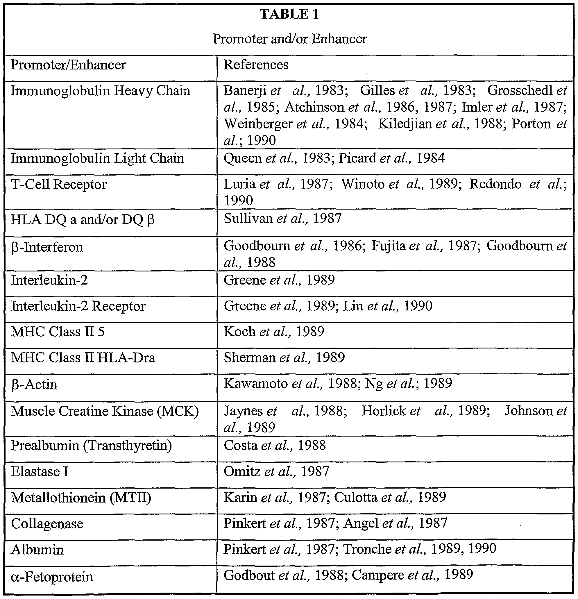
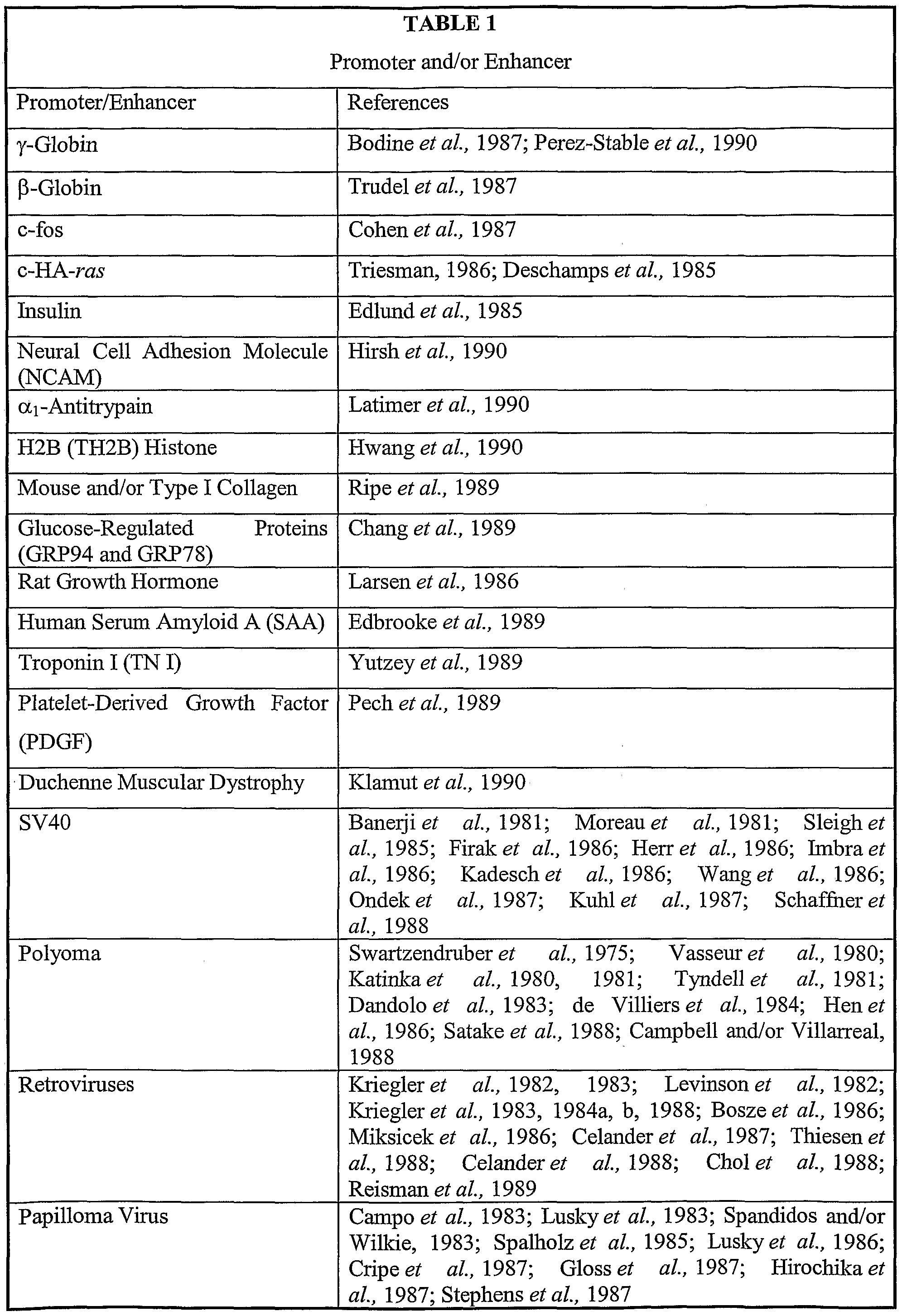
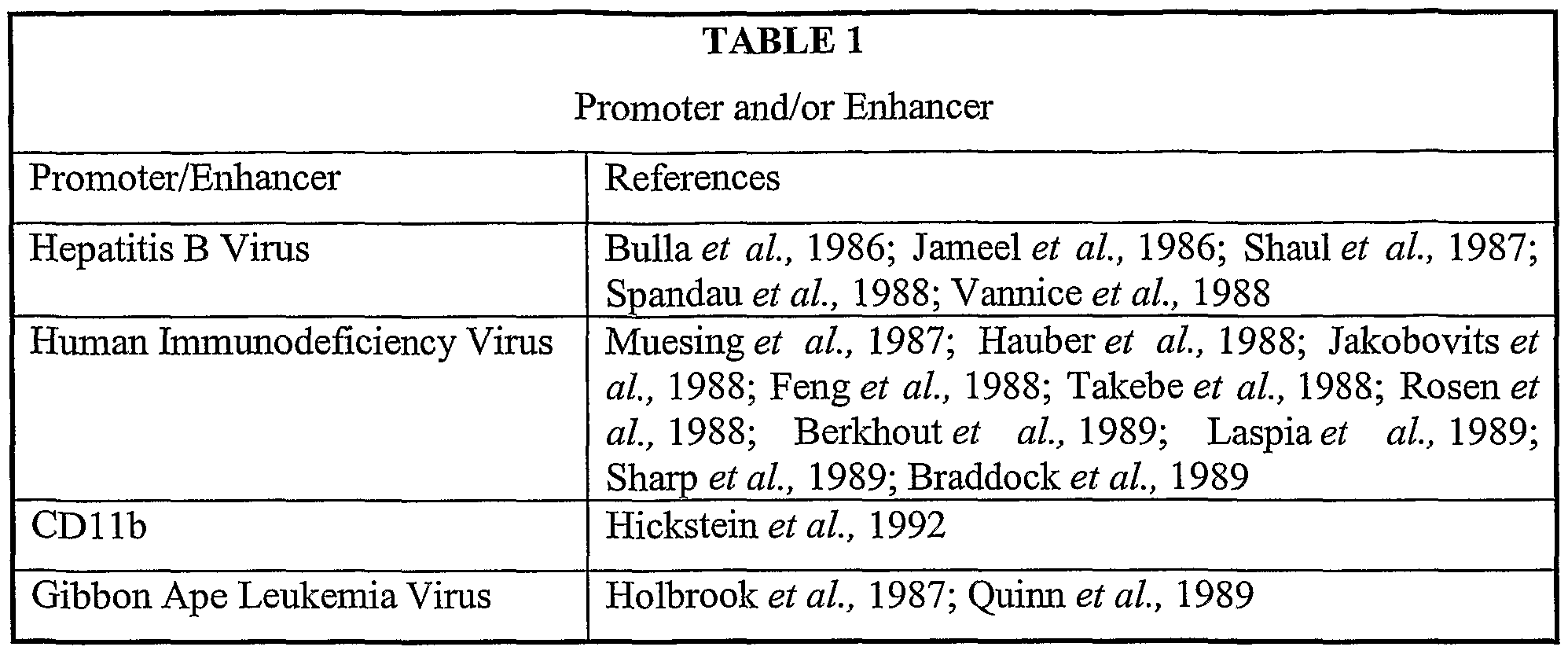

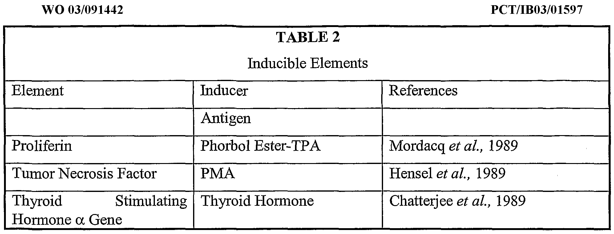
Claims
Priority Applications (10)
| Application Number | Priority Date | Filing Date | Title |
|---|---|---|---|
| HK05106240.0A HK1073479B (en) | 2002-04-26 | 2003-04-25 | Improved chimeric glycoproteins and pseudotyped lentiviral vectors |
| AU2003226598A AU2003226598A1 (en) | 2002-04-26 | 2003-04-25 | Improved chimeric glycoproteins and pseudotyped lentiviral vectors |
| KR1020047017296A KR101002955B1 (en) | 2002-04-26 | 2003-04-25 | Improved chimeric glycoproteins and pseudotype lentiviral vectors |
| CA2484166A CA2484166C (en) | 2002-04-26 | 2003-04-25 | Improved chimeric glycoproteins and pseudotyped lentiviral vectors |
| US10/512,474 US8338168B2 (en) | 2002-04-26 | 2003-04-25 | Chimeric glycoproteins and pseudotyped lentiviral vectors |
| JP2003587970A JP4343708B2 (en) | 2002-04-26 | 2003-04-25 | Improved chimeric glycoproteins and typified lentiviruses |
| DE60326444T DE60326444D1 (en) | 2002-04-26 | 2003-04-25 | IMPROVED CHIMERIC GLYCOPROTEINS AND PSEUDOTYPIZED LENTIVIRAL VECTORS |
| EP03747185A EP1499736B1 (en) | 2002-04-26 | 2003-04-25 | Improved chimeric glycoproteins and pseudotyped lentiviral vectors |
| DK03747185T DK1499736T3 (en) | 2002-04-26 | 2003-04-25 | Enhanced chimeric glycoproteins and pseudotyped lentoviral vectors |
| US13/676,852 US9090908B2 (en) | 2002-04-26 | 2012-11-14 | Chimeric glycoproteins and pseudotyped lentiviral vectors |
Applications Claiming Priority (2)
| Application Number | Priority Date | Filing Date | Title |
|---|---|---|---|
| US37565402P | 2002-04-26 | 2002-04-26 | |
| US60/375,654 | 2002-04-26 |
Related Child Applications (2)
| Application Number | Title | Priority Date | Filing Date |
|---|---|---|---|
| US10/512,474 A-371-Of-International US8338168B2 (en) | 2002-04-26 | 2003-04-25 | Chimeric glycoproteins and pseudotyped lentiviral vectors |
| US13/676,852 Division US9090908B2 (en) | 2002-04-26 | 2012-11-14 | Chimeric glycoproteins and pseudotyped lentiviral vectors |
Publications (1)
| Publication Number | Publication Date |
|---|---|
| WO2003091442A1 true WO2003091442A1 (en) | 2003-11-06 |
Family
ID=29270681
Family Applications (1)
| Application Number | Title | Priority Date | Filing Date |
|---|---|---|---|
| PCT/IB2003/001597 Ceased WO2003091442A1 (en) | 2002-04-26 | 2003-04-25 | Improved chimeric glycoproteins and pseudotyped lentiviral vectors |
Country Status (12)
| Country | Link |
|---|---|
| US (2) | US8338168B2 (en) |
| EP (1) | EP1499736B1 (en) |
| JP (1) | JP4343708B2 (en) |
| KR (1) | KR101002955B1 (en) |
| CN (1) | CN100340668C (en) |
| AT (1) | ATE424464T1 (en) |
| AU (1) | AU2003226598A1 (en) |
| CA (1) | CA2484166C (en) |
| DE (1) | DE60326444D1 (en) |
| DK (1) | DK1499736T3 (en) |
| ES (1) | ES2326217T3 (en) |
| WO (1) | WO2003091442A1 (en) |
Cited By (5)
| Publication number | Priority date | Publication date | Assignee | Title |
|---|---|---|---|---|
| EP2019144A1 (en) * | 2007-07-23 | 2009-01-28 | Institut National De La Sante Et De La Recherche Medicale (Inserm) | Vector particles for targeting CD34+ cells |
| WO2013045639A1 (en) | 2011-09-29 | 2013-04-04 | Institut National De La Sante Et De La Recherche Medicale (Inserm) | LENTIVIRAL VECTORS PSEUDOTYPED WITH MUTANT BaEV GLYCOPROTEINS |
| WO2020234810A1 (en) * | 2019-05-20 | 2020-11-26 | Instituto De Biologia Experimental E Tecnológica (Ibet) | Modified envelope glycoproteins for retroviridae viral vector pseudotyping and process for obtaining it |
| CN117003834A (en) * | 2023-06-09 | 2023-11-07 | 深圳市先康达生命科学有限公司 | Envelope glycoproteins used to transduce NK cell pseudotyped lentiviral vectors and their applications |
| US12084681B2 (en) | 2013-12-24 | 2024-09-10 | Kling Biotherapeutics B.V. | Ex vivo antibody production |
Families Citing this family (8)
| Publication number | Priority date | Publication date | Assignee | Title |
|---|---|---|---|---|
| SI2770061T1 (en) * | 2009-07-24 | 2019-02-28 | Immune Design Corp. | Non-integrating lentiviral vectors |
| CN105709221B (en) | 2011-04-06 | 2020-11-17 | 拜欧瓦克西姆有限公司 | Pharmaceutical composition for the prevention and/or treatment of HIV diseases in humans |
| US20140349403A1 (en) | 2011-12-12 | 2014-11-27 | The Children's Hospital Of Philadelphia | Large commercial scale lentiviral vector production system and vectors produced thereby |
| US20150203540A1 (en) * | 2012-07-27 | 2015-07-23 | The Trustees Of The University Of Pennsylvania | Cytoplasmic Tail Modifications to Boost Surface Expression and Immunogenicity of Envelope Glycoproteins |
| LT3194602T (en) * | 2014-09-18 | 2020-05-11 | Université de Montréal | Compounds and methods for enhancing viral gene transfer to human hematopoietic cells |
| EP4086352A1 (en) | 2021-05-07 | 2022-11-09 | Medizinische Hochschule Hannover | Glycoproteins for pseudotyping retroviral vector particles |
| CN121001739A (en) * | 2023-04-13 | 2025-11-21 | 深圳市济因生物科技有限公司 | A carrier and its application |
| CN116554353B (en) * | 2023-04-28 | 2024-05-03 | 北京奇迈永华生物科技有限公司 | A method for efficiently infecting human NK cells and other immune cells with pseudovirus |
Family Cites Families (18)
| Publication number | Priority date | Publication date | Assignee | Title |
|---|---|---|---|---|
| US4683202A (en) * | 1985-03-28 | 1987-07-28 | Cetus Corporation | Process for amplifying nucleic acid sequences |
| US4682195A (en) * | 1985-09-30 | 1987-07-21 | General Electric Company | Insulated gate device with configured emitter contact pad |
| EP0266032A1 (en) | 1986-08-29 | 1988-05-04 | Beecham Group Plc | Modified fibrinolytic enzyme |
| US5466468A (en) * | 1990-04-03 | 1995-11-14 | Ciba-Geigy Corporation | Parenterally administrable liposome formulation comprising synthetic lipids |
| DE4204650C1 (en) * | 1992-02-15 | 1993-07-08 | Hoffmeister, Helmut, Dr., 4400 Muenster, De | |
| DE4228457A1 (en) * | 1992-08-27 | 1994-04-28 | Beiersdorf Ag | Production of heterodimeric PDGF-AB using a bicistronic vector system in mammalian cells |
| US5834256A (en) * | 1993-06-11 | 1998-11-10 | Cell Genesys, Inc. | Method for production of high titer virus and high efficiency retroviral mediated transduction of mammalian cells |
| FR2722208B1 (en) * | 1994-07-05 | 1996-10-04 | Inst Nat Sante Rech Med | NEW INTERNAL RIBOSOME ENTRY SITE, VECTOR CONTAINING SAME AND THERAPEUTIC USE |
| US5599302A (en) * | 1995-01-09 | 1997-02-04 | Medi-Ject Corporation | Medical injection system and method, gas spring thereof and launching device using gas spring |
| US6013516A (en) | 1995-10-06 | 2000-01-11 | The Salk Institute For Biological Studies | Vector and method of use for nucleic acid delivery to non-dividing cells |
| US5705629A (en) * | 1995-10-20 | 1998-01-06 | Hybridon, Inc. | Methods for H-phosphonate synthesis of mono- and oligonucleotides |
| US5928906A (en) * | 1996-05-09 | 1999-07-27 | Sequenom, Inc. | Process for direct sequencing during template amplification |
| GB9621680D0 (en) * | 1996-10-17 | 1996-12-11 | Oxford Biomedica Ltd | Lentiviral vectors |
| US5846225A (en) * | 1997-02-19 | 1998-12-08 | Cornell Research Foundation, Inc. | Gene transfer therapy delivery device and method |
| WO1998051810A1 (en) * | 1997-05-13 | 1998-11-19 | University Of North Carolina At Chapel Hill | Lentivirus-based gene transfer vectors |
| US6136597A (en) | 1997-09-18 | 2000-10-24 | The Salk Institute For Biological Studies | RNA export element |
| US5994136A (en) | 1997-12-12 | 1999-11-30 | Cell Genesys, Inc. | Method and means for producing high titer, safe, recombinant lentivirus vectors |
| US6849454B2 (en) * | 2000-03-07 | 2005-02-01 | St. Jude Children's Research Hospital | Highly efficient gene transfer into human repopulating stem cells by RD114 pseudotyped retroviral vector particles |
-
2003
- 2003-04-25 WO PCT/IB2003/001597 patent/WO2003091442A1/en not_active Ceased
- 2003-04-25 US US10/512,474 patent/US8338168B2/en not_active Expired - Fee Related
- 2003-04-25 JP JP2003587970A patent/JP4343708B2/en not_active Expired - Fee Related
- 2003-04-25 EP EP03747185A patent/EP1499736B1/en not_active Expired - Lifetime
- 2003-04-25 CN CNB038135329A patent/CN100340668C/en not_active Expired - Fee Related
- 2003-04-25 AU AU2003226598A patent/AU2003226598A1/en not_active Abandoned
- 2003-04-25 CA CA2484166A patent/CA2484166C/en not_active Expired - Fee Related
- 2003-04-25 AT AT03747185T patent/ATE424464T1/en active
- 2003-04-25 ES ES03747185T patent/ES2326217T3/en not_active Expired - Lifetime
- 2003-04-25 DE DE60326444T patent/DE60326444D1/en not_active Expired - Lifetime
- 2003-04-25 DK DK03747185T patent/DK1499736T3/en active
- 2003-04-25 KR KR1020047017296A patent/KR101002955B1/en not_active Expired - Fee Related
-
2012
- 2012-11-14 US US13/676,852 patent/US9090908B2/en not_active Expired - Fee Related
Non-Patent Citations (8)
| Title |
|---|
| CHRISTODOULOPOULOS I ET AL: "SEQUENCES IN THE CYTOPLASMIC TAIL OF THE GIBBON APE LEUKEMIA VIRUS ENVELOPE PROTEIN THAT PREVENT ITS INCORPORATION INTO LENTIVIRUS VECTORS", JOURNAL OF VIROLOGY, THE AMERICAN SOCIETY FOR MICROBIOLOGY, US, vol. 75, no. 9, May 2001 (2001-05-01), pages 4129 - 4138, XP001034070, ISSN: 0022-538X * |
| KELLY PATRICK F ET AL: "RD114-Pseudotyped oncoretroviral vectors: Biological and physical properties.", ANNALS OF THE NEW YORK ACADEMY OF SCIENCES, vol. 938, June 2001 (2001-06-01), Conference on Hematopoietic Stem Cells: Genetics and Medicine;Tubingen, Germany; September 14-16, 2000, 2000: Basic and clinical sciences: Third International Conference. June, 2001 New York Academy o, pages 262 - 277, XP001153583, ISBN: 1-57331-295-9 * |
| NÈGRE D ET AL: "Characterization of novel safe lentiviral vectors derived from simian immunodeficiency virus (SIVmac251) that efficiently transduce mature human dendritic cells.", GENE THERAPY. ENGLAND OCT 2000, vol. 7, no. 19, October 2000 (2000-10-01), pages 1613 - 1623, XP002248522, ISSN: 0969-7128 * |
| NEGRE D ET AL: "Lentiviral vectors derived from simian immunodeficiency virus.", CURRENT TOPICS IN MICROBIOLOGY AND IMMUNOLOGY, vol. 261, September 2001 (2001-09-01), 2002 Springer-Verlag GmbH & Co. KG;Springer-Verlag New York Inc. Heidelberger Platz 3, D-14197, Berlin, Germany; 175 Fifth Avenue, New York, NY, 10010-7858, USA, pages 53 - 74, XP009014016, ISBN: 3-540-42190-4 * |
| SALMON ET AL., JOURNAL OF GENE MEDICINE, 2000 |
| SALMON P ET AL.: "A chimeric galv-derived envelop glycoprotein harboring the cytoplasmic tail of MLV envelope efficiently pseudotypes HIV-1 vectors", JOURNAL OF GENE MEDICINE, vol. 2sup, 2000, pages 23, XP009014038 * |
| SANDRIN VIRGINIE ET AL: "Lentiviral vectors pseudotyped with a modified RD114 envelope glycoprotein show increased stability in sera and augmented transduction of primary lymphocytes and CD34+ cells derived from human and nonhuman primates.", BLOOD. UNITED STATES 1 AUG 2002, vol. 100, no. 3, 1 August 2002 (2002-08-01), pages 823 - 832, XP002248520, ISSN: 0006-4971 * |
| STITZ J ET AL: "Lentiviral Vectors Pseudotyped with Envelope Glycoproteins Derived from Gibbon Ape Leukemia Virus and Murine Leukemia Virus 10A1", VIROLOGY, ACADEMIC PRESS,ORLANDO, US, vol. 273, no. 1, 20 July 2000 (2000-07-20), pages 16 - 20, XP004435973, ISSN: 0042-6822 * |
Cited By (11)
| Publication number | Priority date | Publication date | Assignee | Title |
|---|---|---|---|---|
| EP2019144A1 (en) * | 2007-07-23 | 2009-01-28 | Institut National De La Sante Et De La Recherche Medicale (Inserm) | Vector particles for targeting CD34+ cells |
| WO2009013324A1 (en) * | 2007-07-23 | 2009-01-29 | Institut National De La Sante Et De La Recherche Medicale (Inserm) | Vector particles for targeting cd34+ cells |
| WO2013045639A1 (en) | 2011-09-29 | 2013-04-04 | Institut National De La Sante Et De La Recherche Medicale (Inserm) | LENTIVIRAL VECTORS PSEUDOTYPED WITH MUTANT BaEV GLYCOPROTEINS |
| US9249426B2 (en) | 2011-09-29 | 2016-02-02 | Institut National De La Sante Et De La Recherche Medicale (Inserm) | Lentiviral vectors pseudotyped with mutant BaEV glycoproteins |
| RU2618864C2 (en) * | 2011-09-29 | 2017-05-11 | Энститю Насьональ Де Ля Сантэ Э Дэ Ля Решерш Медикаль (Инсерм) | LENTIVIRAL VECTORS PSEUDOTYPED BY MUTANT BaEV GLYCOPROTEINS |
| US12084681B2 (en) | 2013-12-24 | 2024-09-10 | Kling Biotherapeutics B.V. | Ex vivo antibody production |
| WO2020234810A1 (en) * | 2019-05-20 | 2020-11-26 | Instituto De Biologia Experimental E Tecnológica (Ibet) | Modified envelope glycoproteins for retroviridae viral vector pseudotyping and process for obtaining it |
| CN114072418A (en) * | 2019-05-20 | 2022-02-18 | 实验与技术生物学研究所(Ibet) | Modified envelope glycoprotein pseudotyped by retroviral vector and method for obtaining same |
| US20220204946A1 (en) * | 2019-05-20 | 2022-06-30 | Instituto De Biologia Experimental E Technológica (Ibet) | Modified envelope glycoproteins for retroviridae viral vector pseudotyping and process for obtaining it |
| CN117003834A (en) * | 2023-06-09 | 2023-11-07 | 深圳市先康达生命科学有限公司 | Envelope glycoproteins used to transduce NK cell pseudotyped lentiviral vectors and their applications |
| WO2024250481A1 (en) * | 2023-06-09 | 2024-12-12 | 深圳市先康达生命科学有限公司 | Envelope glycoprotein for pseudotyped lentiviral vector for transduction of nk cells and use of envelope glycoprotein |
Also Published As
| Publication number | Publication date |
|---|---|
| US20140004089A1 (en) | 2014-01-02 |
| EP1499736B1 (en) | 2009-03-04 |
| AU2003226598A8 (en) | 2003-11-10 |
| US9090908B2 (en) | 2015-07-28 |
| CN100340668C (en) | 2007-10-03 |
| US8338168B2 (en) | 2012-12-25 |
| ES2326217T3 (en) | 2009-10-05 |
| CA2484166A1 (en) | 2003-11-06 |
| DK1499736T3 (en) | 2009-06-08 |
| KR20050008697A (en) | 2005-01-21 |
| EP1499736A1 (en) | 2005-01-26 |
| CN1659284A (en) | 2005-08-24 |
| JP4343708B2 (en) | 2009-10-14 |
| DE60326444D1 (en) | 2009-04-16 |
| AU2003226598A1 (en) | 2003-11-10 |
| ATE424464T1 (en) | 2009-03-15 |
| KR101002955B1 (en) | 2010-12-21 |
| CA2484166C (en) | 2011-11-29 |
| HK1073479A1 (en) | 2005-10-07 |
| JP2005523706A (en) | 2005-08-11 |
| US20060257366A1 (en) | 2006-11-16 |
Similar Documents
| Publication | Publication Date | Title |
|---|---|---|
| US9090908B2 (en) | Chimeric glycoproteins and pseudotyped lentiviral vectors | |
| CA2456169C (en) | Methods and compositions relating to improved lentiviral vector production systems | |
| US8748169B2 (en) | Methods and compositions relating to restricted expression lentiviral vectors and their applications | |
| US10226538B2 (en) | Methods and compositions relating to improved lentiviral vectors and their applications | |
| AU2002343458A1 (en) | Methods and compositions relating to restricted expression lentiviral vectors and their applications | |
| HK1073479B (en) | Improved chimeric glycoproteins and pseudotyped lentiviral vectors |
Legal Events
| Date | Code | Title | Description |
|---|---|---|---|
| AK | Designated states |
Kind code of ref document: A1 Designated state(s): AE AG AL AM AT AU AZ BA BB BG BR BY BZ CA CH CN CO CR CU CZ DE DK DM DZ EC EE ES FI GB GD GE GH GM HR HU ID IL IN IS JP KE KG KP KR KZ LC LK LR LS LT LU LV MA MD MG MK MN MW MX MZ NI NO NZ OM PH PL PT RO RU SC SD SE SG SK SL TJ TM TN TR TT TZ UA UG US UZ VC VN YU ZA ZM ZW |
|
| AL | Designated countries for regional patents |
Kind code of ref document: A1 Designated state(s): GH GM KE LS MW MZ SD SL SZ TZ UG ZM ZW AM AZ BY KG KZ MD RU TJ TM AT BE BG CH CY CZ DE DK EE ES FI FR GB GR HU IE IT LU MC NL PT RO SE SI SK TR BF BJ CF CG CI CM GA GN GQ GW ML MR NE SN TD TG |
|
| DFPE | Request for preliminary examination filed prior to expiration of 19th month from priority date (pct application filed before 20040101) | ||
| 121 | Ep: the epo has been informed by wipo that ep was designated in this application | ||
| WWE | Wipo information: entry into national phase |
Ref document number: 2003747185 Country of ref document: EP |
|
| WWE | Wipo information: entry into national phase |
Ref document number: 2484166 Country of ref document: CA Ref document number: 2003587970 Country of ref document: JP |
|
| WWE | Wipo information: entry into national phase |
Ref document number: 1020047017296 Country of ref document: KR |
|
| WWE | Wipo information: entry into national phase |
Ref document number: 20038135329 Country of ref document: CN |
|
| WWP | Wipo information: published in national office |
Ref document number: 1020047017296 Country of ref document: KR |
|
| WWP | Wipo information: published in national office |
Ref document number: 2003747185 Country of ref document: EP |
|
| WWE | Wipo information: entry into national phase |
Ref document number: 2006257366 Country of ref document: US Ref document number: 10512474 Country of ref document: US |
|
| WWP | Wipo information: published in national office |
Ref document number: 10512474 Country of ref document: US |