WO2002036146A2 - Epitope-beta microglobulin polynucleotide for anti-cancer immunotherapy - Google Patents
Epitope-beta microglobulin polynucleotide for anti-cancer immunotherapy Download PDFInfo
- Publication number
- WO2002036146A2 WO2002036146A2 PCT/GB2001/004844 GB0104844W WO0236146A2 WO 2002036146 A2 WO2002036146 A2 WO 2002036146A2 GB 0104844 W GB0104844 W GB 0104844W WO 0236146 A2 WO0236146 A2 WO 0236146A2
- Authority
- WO
- WIPO (PCT)
- Prior art keywords
- epitope
- polynucleotide
- fusion protein
- cells
- hla
- Prior art date
- Legal status (The legal status is an assumption and is not a legal conclusion. Google has not performed a legal analysis and makes no representation as to the accuracy of the status listed.)
- Ceased
Links
Classifications
-
- C—CHEMISTRY; METALLURGY
- C07—ORGANIC CHEMISTRY
- C07K—PEPTIDES
- C07K14/00—Peptides having more than 20 amino acids; Gastrins; Somatostatins; Melanotropins; Derivatives thereof
- C07K14/005—Peptides having more than 20 amino acids; Gastrins; Somatostatins; Melanotropins; Derivatives thereof from viruses
-
- A—HUMAN NECESSITIES
- A61—MEDICAL OR VETERINARY SCIENCE; HYGIENE
- A61K—PREPARATIONS FOR MEDICAL, DENTAL OR TOILETRY PURPOSES
- A61K39/00—Medicinal preparations containing antigens or antibodies
- A61K39/0005—Vertebrate antigens
- A61K39/0011—Cancer antigens
-
- A—HUMAN NECESSITIES
- A61—MEDICAL OR VETERINARY SCIENCE; HYGIENE
- A61K—PREPARATIONS FOR MEDICAL, DENTAL OR TOILETRY PURPOSES
- A61K39/00—Medicinal preparations containing antigens or antibodies
- A61K39/12—Viral antigens
-
- A—HUMAN NECESSITIES
- A61—MEDICAL OR VETERINARY SCIENCE; HYGIENE
- A61K—PREPARATIONS FOR MEDICAL, DENTAL OR TOILETRY PURPOSES
- A61K39/00—Medicinal preparations containing antigens or antibodies
- A61K39/12—Viral antigens
- A61K39/145—Orthomyxoviridae, e.g. influenza virus
-
- A—HUMAN NECESSITIES
- A61—MEDICAL OR VETERINARY SCIENCE; HYGIENE
- A61P—SPECIFIC THERAPEUTIC ACTIVITY OF CHEMICAL COMPOUNDS OR MEDICINAL PREPARATIONS
- A61P31/00—Antiinfectives, i.e. antibiotics, antiseptics, chemotherapeutics
- A61P31/12—Antivirals
-
- A—HUMAN NECESSITIES
- A61—MEDICAL OR VETERINARY SCIENCE; HYGIENE
- A61P—SPECIFIC THERAPEUTIC ACTIVITY OF CHEMICAL COMPOUNDS OR MEDICINAL PREPARATIONS
- A61P35/00—Antineoplastic agents
-
- C—CHEMISTRY; METALLURGY
- C07—ORGANIC CHEMISTRY
- C07K—PEPTIDES
- C07K14/00—Peptides having more than 20 amino acids; Gastrins; Somatostatins; Melanotropins; Derivatives thereof
- C07K14/435—Peptides having more than 20 amino acids; Gastrins; Somatostatins; Melanotropins; Derivatives thereof from animals; from humans
- C07K14/705—Receptors; Cell surface antigens; Cell surface determinants
- C07K14/70503—Immunoglobulin superfamily
- C07K14/70539—MHC-molecules, e.g. HLA-molecules
-
- A—HUMAN NECESSITIES
- A61—MEDICAL OR VETERINARY SCIENCE; HYGIENE
- A61K—PREPARATIONS FOR MEDICAL, DENTAL OR TOILETRY PURPOSES
- A61K39/00—Medicinal preparations containing antigens or antibodies
- A61K2039/51—Medicinal preparations containing antigens or antibodies comprising whole cells, viruses or DNA/RNA
- A61K2039/53—DNA (RNA) vaccination
-
- A—HUMAN NECESSITIES
- A61—MEDICAL OR VETERINARY SCIENCE; HYGIENE
- A61K—PREPARATIONS FOR MEDICAL, DENTAL OR TOILETRY PURPOSES
- A61K39/00—Medicinal preparations containing antigens or antibodies
- A61K2039/58—Medicinal preparations containing antigens or antibodies raising an immune response against a target which is not the antigen used for immunisation
- A61K2039/585—Medicinal preparations containing antigens or antibodies raising an immune response against a target which is not the antigen used for immunisation wherein the target is cancer
-
- A—HUMAN NECESSITIES
- A61—MEDICAL OR VETERINARY SCIENCE; HYGIENE
- A61K—PREPARATIONS FOR MEDICAL, DENTAL OR TOILETRY PURPOSES
- A61K39/00—Medicinal preparations containing antigens or antibodies
- A61K2039/60—Medicinal preparations containing antigens or antibodies characteristics by the carrier linked to the antigen
- A61K2039/6031—Proteins
-
- A—HUMAN NECESSITIES
- A61—MEDICAL OR VETERINARY SCIENCE; HYGIENE
- A61K—PREPARATIONS FOR MEDICAL, DENTAL OR TOILETRY PURPOSES
- A61K39/00—Medicinal preparations containing antigens or antibodies
- A61K2039/62—Medicinal preparations containing antigens or antibodies characterised by the link between antigen and carrier
- A61K2039/627—Medicinal preparations containing antigens or antibodies characterised by the link between antigen and carrier characterised by the linker
-
- A—HUMAN NECESSITIES
- A61—MEDICAL OR VETERINARY SCIENCE; HYGIENE
- A61K—PREPARATIONS FOR MEDICAL, DENTAL OR TOILETRY PURPOSES
- A61K38/00—Medicinal preparations containing peptides
-
- C—CHEMISTRY; METALLURGY
- C07—ORGANIC CHEMISTRY
- C07K—PEPTIDES
- C07K2319/00—Fusion polypeptide
-
- C—CHEMISTRY; METALLURGY
- C12—BIOCHEMISTRY; BEER; SPIRITS; WINE; VINEGAR; MICROBIOLOGY; ENZYMOLOGY; MUTATION OR GENETIC ENGINEERING
- C12N—MICROORGANISMS OR ENZYMES; COMPOSITIONS THEREOF; PROPAGATING, PRESERVING, OR MAINTAINING MICROORGANISMS; MUTATION OR GENETIC ENGINEERING; CULTURE MEDIA
- C12N2760/00—MICROORGANISMS OR ENZYMES; COMPOSITIONS THEREOF; PROPAGATING, PRESERVING, OR MAINTAINING MICROORGANISMS; MUTATION OR GENETIC ENGINEERING; CULTURE MEDIA ssRNA viruses negative-sense
- C12N2760/00011—Details
- C12N2760/16011—Orthomyxoviridae
- C12N2760/16022—New viral proteins or individual genes, new structural or functional aspects of known viral proteins or genes
-
- C—CHEMISTRY; METALLURGY
- C12—BIOCHEMISTRY; BEER; SPIRITS; WINE; VINEGAR; MICROBIOLOGY; ENZYMOLOGY; MUTATION OR GENETIC ENGINEERING
- C12N—MICROORGANISMS OR ENZYMES; COMPOSITIONS THEREOF; PROPAGATING, PRESERVING, OR MAINTAINING MICROORGANISMS; MUTATION OR GENETIC ENGINEERING; CULTURE MEDIA
- C12N2760/00—MICROORGANISMS OR ENZYMES; COMPOSITIONS THEREOF; PROPAGATING, PRESERVING, OR MAINTAINING MICROORGANISMS; MUTATION OR GENETIC ENGINEERING; CULTURE MEDIA ssRNA viruses negative-sense
- C12N2760/00011—Details
- C12N2760/16011—Orthomyxoviridae
- C12N2760/16111—Influenzavirus A, i.e. influenza A virus
- C12N2760/16134—Use of virus or viral component as vaccine, e.g. live-attenuated or inactivated virus, VLP, viral protein
Definitions
- the invention relates to a polynucleotide for use in cancer therapy.
- Tumour cells are attacked by cytotoxic T lymphocytes (CTL) that recognise peptides presented by HLA class I molecules on the surface of the tumour cell.
- CTL cytotoxic T lymphocytes
- the inventors have now shown that delivery of ⁇ 2 m covalently linked to a peptide epitope restores surface class I molecule expression in a cell line. They have shown that expression in a tumour cell of the ⁇ 2 m fusion protein from a polynucleotide allows correct assembly of the class I molecule, its expression on the surface, and that the tumour cell becomes susceptible to attack by CTL.
- the ⁇ 2 m fusion protein may comprise a non-cancer epitope causing the tumour to become susceptible to attack by CTL which recognise non-cancer epitopes.
- the invention provides a polynucleotide capable of expressing an epitope- ⁇ 2 m fusion protein; for use in the generation of cytotoxic T lymphocyte (CTL) responses against a tumour. Also provided is a polynucleotide capable of expressing an epitope- ⁇ 2 m fusion protein; for use in a method of restoring antigen presentation in the tumour of a host.
- CTL cytotoxic T lymphocyte
- the invention relates to the treatment of cancer by expressing in tumour cells a peptide epitope- ⁇ 2 m fusion protein.
- Expression of the fusion protein in tumour cells may lead to an increased cytotoxic T lymphocyte (CTL) response against the cell, and may restore or improve antigen presentation in the cell.
- CTL cytotoxic T lymphocyte
- the tumour is a malignant tumour, for example, a solid tumour such as a gastrointestinal tumour, like colorectal cancer, gastro-esophageal cancer, cancer of liver and biliary tract, pancreatic cancer; prostatic cancer, testicular cancer, lung cancer, breast cancer, malignant melanoma, mesothelioma, brain tumours (such as glioma and astro cytomas), ovarian cancer, uterine cancer including cervical cancer, cancer of the head and neck (e.g.
- bladder cancer bladder cancer
- Kaposi sarcoma including AJDS-related Kaposi sarcoma, sarcomas and osteosarcoma
- renal carcinoma renal carcinoma
- hematopoietic malignant tumours such as leukemia and lymphoma, including AJDS-related lymphomas.
- a CTL response may be generated against a tumour by expression in the tumour cells of a peptide epitope- ⁇ 2 m fusion protein.
- the CTL response is sufficient to induce antigen specific killing of the tumo ⁇ r cells which express the fusion protein.
- the effectiveness of a CTL response may be measured in vitro.
- a cytotoxic T cell assay may be carried out by CD8+ T cell-restricted IFN- ⁇ ELISPOT and functional assays which assess antigen-specific CD8+ T cell killing by release of a measurable label, (e.g. Europium or Chromium) from killed target cells.
- An increase in the number (ELISPOT) or killing reflects an increase in cytotoxic T lymphocytes.
- the present invention provides a means of treating a tumour by expressing in the. tumour cells an epitope- ⁇ 2 m fusion protein.
- Expression of the fusion protein in a tumour cell may restore surface class I antigen presentation.
- Antigen presentation may be reduced in a tumour cell due to impairment of one or more components of the class I antigen presentation pathway.
- the impairment is due to a mutation which causes a reduced amount of the component to be expressed or causes an altered form of the component (typically differently glycosylated or mutated, e.g. truncated) to be expressed which has reduced or no activity.
- the impairment may be in the transport of the component.
- the component is expressed from a gene at reduced levels due to changes in methylation or chromatin structure, e.g. MHC class I genes.
- the component is typically involved in (and therefore the impairment typically affects) processing (proteolysis) of proteins to form peptides, transport of the peptides to the endoplasmic reticulum or formation or transport of the class ! molecule/peptide complex.
- the component which is impaired is the class I heavy chain, TAP or ⁇ 2 m.
- Restoration of antigen presentation may be seen as an increase or improvement in antigen presentation in the tumour cell.
- Antigen presentation may be restored to a level which is substantially the same as, or greater than, that expected in an equivalent non-tumour cell.
- Antigen presentation may be restored to a level which is lower than that expected in an equivalent non-tumour cell, but greater than that present in the cell before expression of the fusion protein.
- a fusion protein in a tumour cell may lead to correct assembly of class I MHC within the cell, expression on the surface of the cell and presentation of the epitope at the surface of the cell, therefore rendering the cell susceptible to attack by cytotoxic T lymphocytes (CTL).
- CTL cytotoxic T lymphocytes
- the fusion protein is capable of binding a class I heavy chain to form a class I molecule.
- the class I molecule has a conformation substantially similar to the class I molecule formed by that heavy chain and the ⁇ 2 m of the host.
- a class I molecule containing the fusion protein will bind a conformation dependent antibody (e.g. W6/32) and/or can present epitope to the CTL of the host.
- the fusion protein is capable of restoring surface class I molecule expression in T2 cells (e.g. detectable by use of W6/32 antibody).
- the epitope of the fusion protein is capable of binding a class I molecule, such as a HLA-A, -B or -C molecule.
- Typical class I molecules which the epitope can bind include HLA-A1, -A2, -A3, -A24, -A31, -A68, -B7, -B8, -B44, -Cw6 and -Cwl6.
- the epitope generally has a length of 8, 9, 10, 11 or 12 amino acids.
- the epitope may be a cancer epitope, i.e. an epitope only presented by tumour cells, typically an epitope which is recognised by tumour specific CTL in cancer patients.
- the epitope may be a neoantigen or from a protein which is expressed in increased amounts in a tumour cell.
- the epitope may be from any of the proteins which are discussed below that are expressed in a different form or in an increased amount in tumour cells.
- the epitope is a non-cancer epitope.
- Such an epitope is typically one which does not occur in a host protein, and is preferably an epitope which occurs in an intracellular pathogen of the host, such as a virus (e.g. EBN, CMN or influenza virus.
- a virus e.g. EBN, CMN or influenza virus.
- the epitope is from any one of the proteins mentioned in Table 1, such as in, or represented by, any of the specific epitopes listed in Table 1.
- the epitope is connected to the ⁇ 2 m by a protein linker.
- the linker comprises amino acids that do not have bulky side groups and therefore do not obstruct the folding of the protein.
- the linker generally allows the epitope to bind to the groove of class I molecule formed after the fusion protein binds to the heavy chain.
- the linker typically has a length of 5 to 20 amino acids, such as 10 to 18 or 14 to 16, preferably 15 amino acids. Typically at least 50%, 70% or 90% of the amino acids in the linker are serine or glycine.
- the linker is generally connected to the ⁇ terminus of the ⁇ 2 m in the form of a fusion protein).
- the linker is or comprises GGSGGGGSGGSGGSG.
- ⁇ 2 m encompasses any beta-2 microglobulin protein, for example, a mammalian beta-2 microglobulin protein such as human or murine beta-2 microglobulin.
- the ⁇ 2 m sequence is preferably the same or homologous to the ⁇ 2 m of the host, or a fragment thereof.
- typically the ⁇ 2 m has a length of at least 30, 40, 50 or 80 amino acids.
- Table 5 shows the polynucleotide sequence of human ⁇ 2 m and the amino acid sequence which it encodes.
- the ⁇ 2 m is a full length ⁇ 2 m protein such as the human beta-2 microglobulin shown in Table 5.
- the ⁇ 2 m may be naturally occurring or modified.
- a naturally occurring ⁇ 2 m is a ⁇ 2 m protein having the amino acid sequence of beta-2 microglobulin isolated from the particular species in question, or a fragment thereof.
- a polynucleotide sequence encoding a naturally occurring ⁇ 2 m may be the polynucleotide sequence naturally occurring in the organism or may be any degenerate polynucleotide sequence which encodes the same amino acid sequence.
- the ⁇ 2 m may be a variant ⁇ 2 m, such as an allelic variant of ⁇ 2 m.
- Modified ⁇ 2 m refers to a ⁇ 2 m having an amino acid sequence that has been modified from a wild-type or naturally occurring beta-2 microglobulin amino acid sequence, or a fragment thereof.
- a wild-type or naturally occurring polynucleotide sequence encoding beta-2 microglobulin may be modified by nucleotide substitutions, for example from 1, 2 or 3 to 10, 25 or 50 substitutions.
- the ⁇ 2 m sequence may alternatively or additionally be modified by one or more insertions and/or deletions and/or by an extension at either or both ends.
- a modified ⁇ 2 m sequence will generally have at least 70%, at least 80%, at least 90%, at least 95%, at least 98% or at least 99% sequence identity to the coding sequence of a wild-type or naturally occurring ⁇ 2 m sequence (such as that shown in Table 5) over a region of at least 20, preferably at least 30, for instance at least 40, at least 60, more preferably at least 100 contiguous nucleotides or most . preferably over the full length of the wild-type or naturally occurring ⁇ 2 m sequence.
- Methods of measuring nucleic acid and protein homology are well known in the art. For example the U GCG Package provides the BESTFIT program which can be used to calculate homology (Devereux et al 1984). Similarly the PILEUP and BLAST algorithms can be used to line up sequences (for example are described in Altschul 1993, and Altschul et al 1990). Many different settings are possible for such programs. In accordance with the invention, the default settings may be used.
- any combination of the above mentioned degrees of sequence identity and minimum sizes may be used to define polynucleotides of the invention, with the more stringent combinations (i.e. higher sequence identity over longer lengths) being preferred.
- a polynucleotide which has at least 90% sequence identity over 25, preferably over 30 nucleotides may be used in the invention, or a polynucleotide which has at least 95% sequence identity over 40 nucleotides.
- a suitable variant or fragment of a wild-type or naturally occurring ⁇ 2 m sequence maintains the characteristics of. wild-type ⁇ 2 m.
- the variant or fragment should maintain the ability to assemble with an MHC I heavy chain to form a class I MHC.
- the class I MHC comprising the variant or fragment ⁇ 2 m should be expressed on the surface of a cell.
- the assembled MHC comprising the variant or fragment ⁇ 2 m should retain the ability to present the epitope of the fusion protein at the cell surface.
- All references to a ⁇ 2 m sequence below refer to a ⁇ 2 m sequence or a variant thereof as defined above.
- the polynucleotide which is capable of expressing the fusion protein comprises a sequence which encodes the fusion protein, or a precursor thereof, and control sequences which cause expression of the coding sequence.
- the coding sequence may comprise one or more introns.
- the encoded protein is a precursor of the mature form of fusion protein
- the precursor requires post- translational modification to produce the mature form, such as cleavage (proteolysis) of the precursor.
- the precursor comprises a signal peptide which directs it to the endoplasmic reticulum.
- control sequences are operably linked to the coding sequence of the polynucleotide enabling them to cause expression of the coding sequence in the cells of the host.
- the control sequences are typically the same as, or substantially similar to, any of the control sequences in the gene of the host or of a virus capable of infecting the host.
- the control sequences typically comprise a promoter (generally 5 1 to the coding sequence) and/or a terminator and/or translation initiation sequence (e.g. GCCACCATGG or GCCCCCATGG) and/or a translational stop codon (e.g. TAA, TAG or TGA) and/or a polyadenylation signal and/or one or more enhancer sequences.
- the control sequences may enhance the transcription or translation of the polynucleotide.
- the control sequences may cause constitutive expression.
- the control sequences cause tumour-specific or tissue-specific expression (where the tissue is the same tissue as the tumour). In one embodiment this is achieved by use of control sequences whose activity is affected (typically increased) by an agent which is administered to the host.
- an agent may for example be targeted or may localise preferentially to tumour cells or to a particular tissue, and may thus cause the control sequences to only cause expression in tumour cells or in particular tissues.
- the polynucleotide may be transiently or stably expressed in the target cell.
- the polynucleotide may comprise other sequence(s) which aid the transport or activity of the polynucleotide, or which increase the stability of the polynucleotide in the cell.
- a sequence may aid delivery of the polynucleotide to the target cell, for example by causing the polynucleotide to adopt a more compact form or by aiding its association with a targeting or carrier agent.
- the sequence may aid the integration of the polynucleotide into the genome of the cell or may increase the stability of the polynucleotide in episomal form.
- the sequence causes replication of the polynucleotide in the cell.
- the polynucleotide is also capable of expressing a sequence which encodes a protein that enhances the ability of the fusion protein to restore antigen presentation or which stimulates a CTL response against the epitope of the fusion protein (such as any such protein mentioned herein).
- the delivery of the polynucleotide may be targeted, typically to the tumour cells, for example by targeting to tissues of the same type as the tumour cell.
- the targeting may be achieved by administering the polynucleotide in, or in the vicinity of, the tumour or at a location which ensures in vivo transport of the polynucleotide to the tumour.
- the polynucleotides may be associated with a moiety that targets delivery of the polynucleotide to a tumour cell. If the polynucleotide is in the form of a vector or associated with a carrier, then such a vector or carrier may act as a targeting moiety.
- the moiety which causes the targeting of the polynucleotide is generally able to bind a molecule expressed on surface of the tumour (and which is preferably expressed on the surface of only a small number of or no other cells).
- the moiety may be natural the ligand/receptor (or fragment and/or homologue thereof) of a ligand/receptor expressed on the tumour cells. Suitable ligands/receptors include EGF receptor (e.g. EGFRvffl).
- the moiety may be an antibody, such as a single- chain antibody.
- the ligand /receptor which is targeted is expressed in a different form and/or in increased amounts on the tumour cell, for example in a mutated form.
- Such proteins include:
- the polynucleotides of the invention may be administered directly as a naked nucleic acid construct to achieve expression of the fusion protein of the invention.
- the polynucleotide may in the form of a viral vector, typically based on a virus which is able to infect the host (preferably also cells of the same tissue as the tumour).
- the vector may be, or may be derived from, an alphavirus, adenovirus, adeno-associated virus, vaccinia virus, herpes simplex virus, retrovirus (e.g. lentivirus) vector, such as amphotropic or xenotrophic retroviruses derived from Moloney leukemia virus, spleen necrosis virus.
- the virus vector is typically attenuated, for example replication defective.
- the carrier which the polynucleotide may be associated with is typically one which aids the passage of the polynucleotide across the cell membrane.
- the carrier may comprise a transfection agent, a cationic agent, for example a cationic lipid, calcium phosphate, DEAE dextran, polyethylenimine (PEI), dendrimers, and lipofectants, for example lipofectam and transfectam, polylysine, a lipid or a precipitating agent (e.g. a calcium salt).
- the polynucleotide and carrier may be in the form of liposomes or particles.
- the particle typically has a diameter of 10 to 10 "3 ⁇ m, for example 1 to 10" 2 ⁇ m.
- the carrier may cause the polynucleotide to adopt a more compact form, e.g. a histone.
- the polynucleotide may be in association with spermidine.
- the carrier may be one which can be used to deliver the polynucleotide into the cell, or even into the nucleus, using ballistics techniques.
- a carrier is typically a metal particle, such as a gold particle.
- the polynucleotide is generally DNA or RNA, and is typically single or double stranded.
- the polynucleotide may be chemically modified, typically to enhance resistance to nucleases or to enhance its ability to enter cells.
- phosphorothioate nucleotides may be used.
- Other deoxynucleotide analogs include methylphosphonates, phosphoramidates, phosphorodithioates, N3'P5'- phosphoramidates and oligoribonucleotide phosphorothioates and their 2'-O-alkyl analogs and 2'-O-methylribonucieotide methyiphosphonates.
- MBOs Mixed backbone oligonucleotides
- MBOs contain segments of phosphothioate oligodeoxynucleotides and appropriately placed segments of modified oligodeoxy- or oligoribonucleotides.
- MBOs have segments of phosphorothioate linkages and other segments of other modified oligonucleotides, such as methylphosphonate, which is non-ionic, and very resistant to nucleases or 2'- O-alkyloligoribonucleotides .
- the polynucleotide may be in substantially isolated form, or it may be in substantially purified form, in which case it will generally comprise at least 80%, 90%, 95%), 98%) or 99% of the polynucleotide or dry mass in the preparation. Thus the polynucleotide may be in the form of 'naked DNA' .
- the host may have a CTL response against the peptide epitope of the fusion protein at the time when the fusion protein is administered (due to a previous stimulation) and/or such a response may be stimulated simultaneously, before or after administration of the polynucleotide.
- the stimulation may be one which is due to a natural infection (bacterial or viral, e.g. influenza virus infection).
- the stimulation may be due to the administration of an agent that comprises the epitope and optionally also an adjuvant that enhances a CTL response against the epitope.
- the present invention further provides a method for selecting a suitable epitope- ⁇ 2 m fusion protein for use in treating a tumour. The method comprises determining whether the patient has an existing CTL response to an epitope.
- a CTL response against an epitope indicates that a fusion protein comprising that epitope may be suitable for the treatment of a tumour in that patient, without the need for vaccination with the epitope itself.
- the CTL response of the patient may still be enhanced by vaccination and/or inclusion of an adjuvant.
- the polynucleotide encoding the fusion protein may be administered with or without a vaccination agent.
- the polynucleotide may be administered with or without first screening for a suitable epitope which the patient is known to produce a CTL response against.
- Expression of the fusion protein in a tumour, and consequent presentation of the epitope by MHC at the surface of the tumour cells may stimulate a CTL response against those cells.
- a vaccination agent and/or an adjuvant may also be administered.
- expression of the fusion protein may stimulate a CTL response against the epitope which may also act against tumour cells of the patient which express that epitope.
- the invention also provides a product containing a polynucleotide capable of expressing the epitope- ⁇ 2 m fusion protein and a vaccination agent that stimulates a CTL response against the epitope of the fusion protein for simultaneous, separate or sequential use in the treatment of cancer, for example in the generation of CTL responses against a tumour.
- the invention provides a composition comprising a polynucleotide capable of expressing the epitope- ⁇ 2 m fusion protein and a vaccination agent that stimulates a CTL response against the epitope of the fusion protein.
- the CTL adjuvant may be capable of causing or augmenting a MHC class II restricted T cell (typically CD4) response which is favourable to the production of a CTL response, such as a Thl response.
- the adjuvant may comprise a MHC class ⁇ restricted T cell epitope (or a precursor which can be processed in vivo to provide such an epitope).
- the adjuvant may be a cytokine, such as a cytokine which stimulates a MHC class I restricted T cell response or favourable MHC class II restricted T cell response (e.g. IL-2, IL-7, IL-12 or IFN- ⁇ ).
- the adjuvant may be, for example, CFA (Golding and Scott (1995) Ann. N.Y. Acad. Sci.
- a muramyl dipeptide e.g. of a mycobacterial cell wall
- monophosphoryl lipid A lipopolysaccharide (e.g. from B. abortus), liposomes, SAF-1 (Golding and Scott, supra)
- a saponin e.g. Quil A
- keyhole limpet hemocyanin yeast TY particle
- beta 2-microglobulin mannan (e.g. oxidised mannan), PRONAX (IDEC Ph. Corporation), immunostimulatory D ⁇ A sequences (ISS), high molecular weight nonionic block polymers or E. coli heat labile toxin (LT).
- the particular route of administration used may aid the stimulating of a CTL response, and thus the polynucleotide or composition may be provided in a form suitable for administering by such a route.
- the polynucleotide or composition is typically administered by any standard technique. It may be delivered directly, for example,, by injection, such as intradermally, subcutaneously or intramuscularly. Intraperitoneal or intravenous routes are preferred.
- the polynucleotide or compositions may be delivered topically, orally or intranasally or by aerosol or, for example, using a particle bombardment or patch transdermal delivery device. In one embodiment administration is by ballistic means.
- a low dose of antigen favours the development of a CTL response.
- a suitable low dose can be given.
- the polynucleotide or composition may be provided in an amount and concentration that is suitable for administering to provide an appropriate low dose.
- the vaccination agent may be administered before the polynucleotide of the invention such that the patient develops a CTL response against the epitope of the fusion protein and is capable of producing a CTL response against cells expressing the fusion protein.
- the time between administration of the vaccination agent and the polynucleotide of the invention should therefore be sufficient to allow the individual to develop an immune response against the epitope of the fusion protein.
- the vaccination agent may be administered one, two, four, six, eight, twelve or more weeks prior to administration of the polynucleotide encoding the fusion protein.
- the vaccine may be given in a single dose schedule or in a multiple dose schedule.
- a multiple dose schedule is one in which a primary course of vaccination may be with 1-10 separate doses, followed by other doses given at subsequent time intervals required to maintain or reinforce the immune response, for example at 1-4 months for a second dose, and if needed a subsequent dose after several months.
- the dosage regime will also at least in part be determined by the need of the individual and b e dep endent upon the judgement of the practitioner.
- An effective non-toxic amount of the polynucleotide (including in the form of the composition of the invention) and optionally also a vaccination agent that stimulates a CTL response against the peptide epitope of the fusion protein, may be given to a human or non-human patient in need thereof, such as a patient with a cancer or suspected of having cancer.
- a human or non-human patient in need thereof, such as a patient with a cancer or suspected of having cancer.
- the condition of a patient suffering from a cancer can therefore be improved by such an administration.
- the polynucleotide (and optionally also the vaccination agent) is provided for use in a method of treating the human or animal body by therapy.
- the invention also provides use of the polynucleotide in the manufacture of a medicament for treating cancer for example in the generation of CTL responses against a tumour.
- the polynucleotide (and generally also the vaccination agent) may be used in a method of treating a host comprising administering the polynucleotide (and generally also the vaccination agent) to the host.
- the polynucleotide may be administered in a single dose schedule or in a multiple dose schedule.
- the subject is given 1, 2, 3 or more separate administrations, each of which is separated by at least 12 hours, 1 day, 2, days, 7 days, 14 days, 1 month or more.
- the dose of polynucleotide and/or vaccination may be determined according to various parameters, especially according to the form of the polynucleotide used; the age, weight and condition of the patient to, be treated; the capacity of the patient's immune system to produce an immune response; the route of administration; and the required regimen. A physician will be able to determine the required route of administration and dosage for any particular patient.
- a suitable dose may however be from 1 pg to 10 g, for example from 100 ⁇ g to 1 g of the polynucleotide. These values may. represent the total amount administered in the complete treatment regimen or may represent each separate administration in the regimen.
- the quantity of nucleic acid administered per dose is preferably 10 ⁇ g or less, such as 1 pg to 10 ⁇ g for particle mediated delivery, e.g. ballistics methods, and preferably 1 ⁇ g to 10 mg, more preferably 100 ⁇ g to 1 mg for other routes, e.g. direct injection.
- the amount of virus administered is in the range of from 10 to 10 pfu, preferably from 10 7 to 10 10 pfu.
- injected typically 1-2 ml of virus, in a pharmaceutically acceptable suitable carrier or diluent is administered.
- the polynucleotide may be in the form of a pharmaceutical composition which comprises the polynucleotide and a pharmaceutically acceptable carrier or diluent.
- Suitable carriers and diluents include isotonic saline solutions, for example phosphate-buffered saline.
- the composition is formulated for parenteraL intravenous, intramuscular, subcutaneous, transdermal, intradermal, oral, intranasal, intravaginal, or intrarectal administration.
- T2 is a lymphoblastoid cell line lacking class I expression due to a defect in the TAP transporter system (1).
- JY is human B lymphoblastoid cell line (2).
- DLD-1 is a colorectal aden ⁇ carcinoma cell line that is defective in ⁇ 2 m expression (3).
- Anti h ⁇ 2 m antibody BBM.1 (4) and the anti class I antibody W6/32 (5) were used.
- the xenotropic cell line PG13 (American Type Culture Collection) was used for the production of recombinant viruses using the retroviral construct pLXSN (Clontech).
- the bacterial strain used for recombinant proteins expression was BL21(DE3)physS (Novagen). Purified human ⁇ 2 m was obtained from Sigma.
- the HLA-A2 restricted influenza matrix epitope (MA 58-66) was linked to human ⁇ 2 m by a 15 amino acid spacer.
- An appropriate linker length for the fusion protein was determined by inspection of the HLA-A2/MA peptide crystal structure (PDB code IHHI) (6).
- Glycine residues were used for the N terminal portion of the linker to facilitate the continuation of the peptide with minimal disruption to the binding groove as observed in aHLA-A2 crystal structure (PDB code 2CLR) (7).
- a methionine residue was added to the N-terminus of the protein.
- the PCR was performed using 5'-primer OX297: 5'-GGG GGG CAT ATG GGT ATT TTA GGA TTT GTT TTTACATTAGGAGGTGGGGGAGGCGGATCAGGAGGCTCAGGTGGGTCAGGA GGCATC CAG CGTACT CCAAAGATT CAG G-3' and 3 '-primer OX298: 5'-GATA GTT AAG TGG GAT CGA GAC ATG TAA GCT TCC CCC-3'.
- the amplified fragment (Ndel- Hind ⁇ i) was cloned into the expression vector pGMT7, sequenced and used for expression.
- the retroviral vector MA- ⁇ 2 m-pLXSN was generated in a three step cloning strategy.
- the primers 5' AAT TCG CCA CCA TGT CTC GCT CCG TGG CCT TAG CTG TGC TCG CGC TAC TCT CTC TTT CTG GCC TCG AGA CTA CTG-3' and 3'-GAT CCA GTA GTC TCG AGG CCA GAA AGA GAG AGT AGC GCG AGC ACA GCT AAG GCC ACG GAG CGA GAC ATG GTG GCG-5' were annealed to originate the signal sequence and cloned as a EcoRI- BamHI fragment into the pLXSN construct.
- a Xho I site was present in the oligo at the 3' end of the signal sequence.
- a plasmid expressing ⁇ 2 m was used as a template for a PCR reaction using the primer 5'-ATT ATT CTC GAG ATC ATC ATG GAT CCG GCG GAG GCG-3' encoding the 3' end of the linker and including the restriction sites for Xho I and BamHI and the primer OX298 described above including a Bgl II site.
- This fragment was cloned into the Xho-Bgl II sites of the retroviral vector pLXSN and contained at its 5' end the two sites Xho I and BamHI where the oligos encoding the epitope and part of the linker were cloned into.
- the two primers constituting this last fragment were 5'-TCG AGG GCG GCA TCC TGG GCT TCG TGT TCA CCC TGG GCG GCG-3' and 5'-ATT TCG CCA CCA TGT CTC GCT CCT TGG CCT TAG CTG TGC TCG CGC TAC TCT CTC TTT CTG GCC TCG AGA CTA CTG-3'.
- the construct containing the HLA-A2 heavy chain and the biotinylation tag was constructed as in (8)
- Bacteria were harvested after 5h and 10 ⁇ l lysate analysed on a 4-20% PAGE. Soluble HLA-peptide tetramers and fusamers were generated as in (8).
- Refolding of the binary (epitope- ⁇ 2 m fusion protein/HLA) and ternary (epitope/ ⁇ 2 m/HLA) complex was done in the presence of soluble MHC class I heavy chain and MA epitope (GTLGFVFTL).
- Recombinant h ⁇ 2 m was used for the ternary complex while the epitope- ⁇ 2 m fusion protein was used for the binary complex.
- the refold was concentrated, biotinylated and purified by gel filtration (Superdex 75 column) followed by ion-exchange chromatography. The biotinylation of the complex was tested by ELISA using Extravidin peroxidase conjugate (Sigma) and a TMB substrate for detection (Sigma).
- the generation of tetrameric class I molecule complexes was achieved by slow addition of extravidin-PE conjugated (Sigma, UK) at a molar ratio of 0.3 : 1 (extravidimbiotinylated protein) (8)
- Retroviral production and cell transduction The retroviral constructs MA- ⁇ 2 m-pLXSN and pLXSN were introduced, via calcium phosphate co-precipitation 1 (9) into the xenotropic packaging cell line PG13. After two weeks of selection in G418 [800 ⁇ g/ml], supernatant from producing cells was harvested and used for transduction of target cells. The T2 cell line was subsequently transduced with both the vectors while DLD-1 and JY cells were transduced with the retroviral vector MA- ⁇ 2 m-pLXSN.
- 5x10 6 cells were transduced using 5 ml of viral supernatant in presence of protamine 8 ⁇ g/ml for 16 hours and subsequently grown in selective medium containing G418 (400, 1600 and 500 ⁇ g/ml respectively) for 2 weeks.
- ⁇ 2 m in retrovirally transduced cells 10 6 cells were washed in PBS/0.1% BSA 1% human serum and incubated with 1 ⁇ g of anti- ⁇ 2 m (BBM.l) antibody for 30 min on ice. The cells were washed twice in PBS/0.1% BSA and incubated with a phycoerythrin conjugated secondary antibody for 30 min on ice. Cells were washed twice and analysed by flow cytometry. For the detection of HLA class I, 1 ⁇ g of anti HLA class I antibody (W6/32) was used and the cells were stained as described.
- HLA-A2-restricted CTL clones specific for flu matrix protein (clone C3 (10)) or the melanoma antigen melan-A (clone 3G3 (11)) were stained with equivalent doses (0.5 ⁇ g) of the fusamer or the tetramer for 15 min at 37 °C. After the staining, the cells were washed twice and resuspended in FACS buffer and analysed by flow cytometry.
- the transduced cell lines were assayed as target cells in a chromium release assay.
- the target cells were labeled for 1 hour at 37°C with 100 mCi per 10 6 cells Na 51 Cr and washed repeatedly.
- the cells were distributed in round bottom 96-well microtiter plates (5x10 3 cells per well) and pulsed with 5mM of the synthetic peptide for one hour before the addition of a HLA-A2 restricted flu-matrix CTL clone.
- the effector cells were added at different effector/target ratios and the plates were incubated for 4 hours at 37 °C. Supernatants were harvested and 51 Cr-release was measured in a gamma counter.
- the specific lysis was calculated according to the formula 100 x ([experimental cpm-background cpm]/[maximum cpm- background cpm]). Background cpm values were determined by incubating target cells alone and maximum values were determined lysing the target cells in presence of Triton 5% x-100. Non transduced cells were used as controls in the same conditions. AH the experiments were performed in triplicate.
- the linker was designed by molecular modelling and comprised a Gly-Gly-Ser motif, with the first five amino acids glycines to avoid interference of bulky side chains with the alpha helixes of the binding groove.
- a linker of 15 amino acids could span the gap between the C-terminus of the epitope and the ⁇ -terminus of ⁇ 2 m.
- the fusion protein- had a molecular mass increase of ca 2.6 kD consistant with the addition of 24 amino acids to ⁇ 2 m.
- the fusion molecule was folded together with soluble HLA-A2 heavy chain with a biotinylation tag on the C-terminus.
- the binary complex was biotinylated and multimerized with Streptavidin-PE as for the classic HLA/epitope tetrameric complexes (ternary complexes).
- the multivalent complexes of these binary and ternary MHC molecules are referred to as fusamers and tetramers.
- An irrelevant melanoma CTL clone, also restricted by HLA-A2 did not stain with this reagent. Stability of the fusamer was compared with stability of the tetramer by incubating both reagents at 37°C for 15 minutes, before staining CTL.
- the retroviral vector MA- ⁇ 2 m-pLXSN was constructed to express the epitope- ⁇ 2 m fusion protein where the immunodominant HLA-A2 restricted influenza peptide (58-66) is linked via a 15-amino acid linker to the amino-terminus of the human ⁇ 2 m.
- the ⁇ 2 m signal sequence was placed in front of the epitope to target the whole molecule to the endoplasmic reticulum.
- the virus was produced by transfection of the packaging cell line PA317 and the supernatant was harvested and used to transduce target cells.
- TAP-deficient HLA-A2 T2 cells were transduced with the retroviral vector MA- ⁇ 2 m-pLXSN, stained with W6/32, the conformation-dependent anti-MHC class I antibody, and analysed by flow cytometry. An increase in MHC class I complex on the cell surface was observed (Table 3 A), indicating that MA- ⁇ 2 m had complexed with the MHC class I heavy chain in a conformationally correct manner, independent of the TAP transporter.
- transduced DLD- 1 cells were surface-labeled with biotin and the epitope- ⁇ 2 m fusion protein was immunoprecipitated.
- the results showed that the immunoprecipitated ⁇ 2 m from the transduced DLD-1 cell line was bigger than the endogenous ⁇ 2 m immunoprecipitated from Jurkat cells used as control.
- the retrovirally transduced protein corresponded to a 14 kD protein. This result demonstrated that the protein was correctly transported to the cell surface and not degraded by membrane proteases.
- a quantitative analysis based on the intensity of the bands showed that the amount of recombinant ⁇ 2 m expressed was the same as. in the control cell line.
- the presence of the intact protein on the cell surface was also demonstrated for the fusion protein NP- ⁇ 2 m (12) and it may be relevant in case of in vivo delivery, where the epitope should not be presented by cells other than the targeted ones. Moreover the fusion protein itself can not be shuffled from one cell to another. When we co-cultured epitope- ⁇ 2 m fusion expressing cells and non-transduced cells, no fusion protein was detected on the surface of the non-transduced cells analysed by flow cytometry.
- a chromium release assay was performed using the transduced T2 cells expressing the epitope- ⁇ 2 m fusion protein. The assay was performed as described above, using a flu-matrix specific CTL clone. The specific killing in absence of epitope was very high for the cells expressing the fusion protein (72%) comparable to the level of lysis obtained for the non transduced cells pulsed with the epitope (70- 80%o) (Table 4A). The addition of epitope to the transduced cells did not alter significantly the percentage of specific lysis (70-80%).
- the retrovirally transduced DLD-1 cells were used as targets in a chromium release assay to assess the ability of the fusion protein to form a functional class I complex in a ⁇ 2 m negative cell line.
- the results (Table 4B) showed a high specific lysis of the cells (78%) expressing the epitope- ⁇ 2 m fusion protein both in the absence and in presence of the epitope.
- the non transduced cells showed only background lysis.
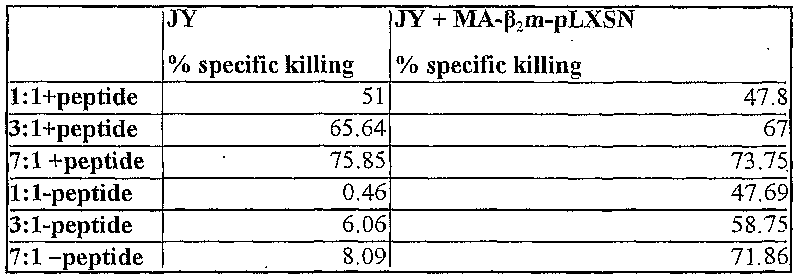
- a lymphoblastoid cell line JY was transduced with the retroviral vector MA- ⁇ 2 m-pLXSN and used in the assay as target cells (Table 4C).
- the specific killing activity using the transduced cells was as high as the normal control (non transduced JY cells) where the epitope was added.
- the specific lysis in the absence of the epitope was around background levels for the non transduced cell line, while the transduced line showed values comparable with those obtained with the positive controls.
- the fusion protein was able to form CTL recognition structures even in the presence of an endogenous HLA class I complex.
- peptide- ⁇ 2 m fusions to elicit a T cell immune response in vivo was tested by in 6 non-humanised mice, by testing the ability of cells transfected with influenza-derived nucleoprotein (NP)- ⁇ 2 m fusion to prime NP-specific CTL responses in vivo.
- NP influenza-derived nucleoprotein
- the T cell responses were measured using NP specific T cell assays and monitoring the vaccinia titre in the mice ovaries.
- mice of strain C57BL/6 wildtype laboratory mouse strain
- Virus Recombinant vaccinia viruses (W-NP) expressing the MHC class I- restricted (Db) peptide epitope derived from influenza NP (NP366-374), sequence in one letter code ASNENMET, had been generated previously (Beauverger et al, Virology 219: 133-139 (1996)).
- Target cells MC57 antigen presenting cells (Leist et al., 1987) express mouse MHC molecules Kb and Db. These were transduced with the NP- ⁇ 2 m-pLXSN retroviral vector (Uger et al J. Immunol. 160 1598-1605 (1998)). Expression of the NP- ⁇ 2 m fusions by the transfected MC57 cells was confirmed by western blot and FACS analysis. Experirnental Details:
- mice 1-4 (M1-M4) were injected, subcutaneously, at Day 0 with 4x 10 6 irradiated MC57 cells transfected the NP- ⁇ 2 m fusion. These mice were then injected intraperitoneally, at Day 8 with 2 xlO 6 pfu (plaque forming units) Vaccinia virus strain W-NP expressing the NP protein in 200 ⁇ l phosphate buffered saline (PBS, standard physiological saline buffer).
- PBS phosphate buffered saline
- mice 5 & 6 (M5-M6) were injected, subcutaneously, at Day 0 with 4 x 10 6 irradiated, untransfected MC57 cells. These mice were then injected intraperitoneally, at Day 8 with 2 x 10 6 pfu (plaque forming units) Vaccinia virus strain expressing the NP protein plus 200 ⁇ l phosphate buffered saline (PBS, standard physiological saline buffer).
- PBS phosphate buffered saline
- Chromium51 -labelled MC57 cells were prepulsed with NP peptide for 1 hour, then mixed with the cultured mouse lymphocytes at the indicated effector to target ratios. Chromium51 counts from wells of MC57 cells lysed by addition of detergent were set at 100% lysis.
- mice vaccinia titre in the mice ovaries were measured using a standard plaque-based assay Results
- mice Vaccinia clearance from mice ovaries Vaccinia clearance from mice ovaries
- mice contained approximately 10 5 - 10 6 less vaccinia W-NP pfu's than the untransfected M5-M6 mice.
- results generated demonstrate that spleen of one of the NP- ⁇ 2 m fusion transfected Ml -4 mice contained approximately 5 times higher levels of NP specific CTLs than untransfected M5-M6 mice, as measured by HLA specific tetramer staining.
- results presented in this example demonstrate the ability of tumour cells transfected with NP- ⁇ 2 m fusion to elicit an NP specific CTL response.
- NLVPMVATV (495-503) lower matrix protein pp65 HLA-A20201 TPRVTGGGAM (417-426) lower matrix protein pp65 HLA-B0702
- Beta 2-microglobulin gene mutations a study of established colorectal cell lines and fresh tumors. Proc NatlAcadSci USA 91:4751.
- Arginine 45 is a major part of the antigenic determinant of human beta 2- microglobulin recognized by mouse monoclonal antibody BBM.1. JBiol
Landscapes
- Health & Medical Sciences (AREA)
- Life Sciences & Earth Sciences (AREA)
- Chemical & Material Sciences (AREA)
- General Health & Medical Sciences (AREA)
- Medicinal Chemistry (AREA)
- Immunology (AREA)
- Organic Chemistry (AREA)
- Virology (AREA)
- Veterinary Medicine (AREA)
- Pharmacology & Pharmacy (AREA)
- Animal Behavior & Ethology (AREA)
- Public Health (AREA)
- Mycology (AREA)
- Microbiology (AREA)
- Epidemiology (AREA)
- Genetics & Genomics (AREA)
- Biochemistry (AREA)
- Proteomics, Peptides & Aminoacids (AREA)
- Molecular Biology (AREA)
- Biophysics (AREA)
- Gastroenterology & Hepatology (AREA)
- Oncology (AREA)
- Toxicology (AREA)
- Chemical Kinetics & Catalysis (AREA)
- General Chemical & Material Sciences (AREA)
- Nuclear Medicine, Radiotherapy & Molecular Imaging (AREA)
- Cell Biology (AREA)
- Pulmonology (AREA)
- Zoology (AREA)
- Communicable Diseases (AREA)
- Peptides Or Proteins (AREA)
- Medicines That Contain Protein Lipid Enzymes And Other Medicines (AREA)
- Medicines Containing Antibodies Or Antigens For Use As Internal Diagnostic Agents (AREA)
- Micro-Organisms Or Cultivation Processes Thereof (AREA)
- Pharmaceuticals Containing Other Organic And Inorganic Compounds (AREA)
Abstract
Description


Claims
Priority Applications (4)
| Application Number | Priority Date | Filing Date | Title |
|---|---|---|---|
| US10/415,841 US20040131598A1 (en) | 2000-11-02 | 2001-11-01 | Cancer therapy |
| EP01980679A EP1330259A2 (en) | 2000-11-02 | 2001-11-01 | Epitope-beta microglobulin polynucleotide for anti-cancer immunotherapy |
| CA002463623A CA2463623A1 (en) | 2000-11-02 | 2001-11-01 | Epitope-beta microglobulin polynucleotide for anti-cancer immunotherapy |
| AU2002212472A AU2002212472A1 (en) | 2000-11-02 | 2001-11-01 | Epitope-beta microglobulin polynucleotide for anti-cancer immunotherapy |
Applications Claiming Priority (2)
| Application Number | Priority Date | Filing Date | Title |
|---|---|---|---|
| GBGB0026812.8A GB0026812D0 (en) | 2000-11-02 | 2000-11-02 | Cancer therapy |
| GB0026812.8 | 2000-11-02 |
Publications (2)
| Publication Number | Publication Date |
|---|---|
| WO2002036146A2 true WO2002036146A2 (en) | 2002-05-10 |
| WO2002036146A3 WO2002036146A3 (en) | 2002-10-17 |
Family
ID=9902443
Family Applications (1)
| Application Number | Title | Priority Date | Filing Date |
|---|---|---|---|
| PCT/GB2001/004844 Ceased WO2002036146A2 (en) | 2000-11-02 | 2001-11-01 | Epitope-beta microglobulin polynucleotide for anti-cancer immunotherapy |
Country Status (6)
| Country | Link |
|---|---|
| US (1) | US20040131598A1 (en) |
| EP (1) | EP1330259A2 (en) |
| AU (1) | AU2002212472A1 (en) |
| CA (1) | CA2463623A1 (en) |
| GB (1) | GB0026812D0 (en) |
| WO (1) | WO2002036146A2 (en) |
Cited By (5)
| Publication number | Priority date | Publication date | Assignee | Title |
|---|---|---|---|---|
| EP1447451A4 (en) * | 2001-09-28 | 2006-06-07 | Dnavec Research Inc | MAMMALIAN CELL-INFECTING VIRUS VECTOR ENCODING EPITOPE-BOUND-BETA2m AND UTILIZATION THEREOF |
| EP1409547A4 (en) * | 2001-06-19 | 2007-05-02 | Technion Res And Dev Of Founda | Methods and pharmaceutical compositions for immune deception, particularly useful in the treatment of cancer |
| US7977457B2 (en) | 2006-05-19 | 2011-07-12 | Teva Pharmaceutical Industries Ltd. | Fusion proteins, uses thereof and processes for producing same |
| US8022190B2 (en) | 2001-06-19 | 2011-09-20 | Technion Research & Development Foundation Ltd. | Immuno-molecules containing viral proteins, compositions thereof and methods of using |
| US9616112B2 (en) | 2003-03-26 | 2017-04-11 | Technion Research & Development Foundation Limited | Compositions capable of specifically binding particular human antigen presenting molecule/pathogen-derived antigen complexes and uses thereof |
Family Cites Families (3)
| Publication number | Priority date | Publication date | Assignee | Title |
|---|---|---|---|---|
| US6011146A (en) * | 1991-11-15 | 2000-01-04 | Institut Pasteur | Altered major histocompatibility complex (MHC) determinant and methods of using the determinant |
| US5951975A (en) * | 1996-06-28 | 1999-09-14 | University Of Pittsburgh | Induction of CTLs specific for natural antigens by cross priming immunization |
| EP1086224B1 (en) * | 1998-06-10 | 2006-03-29 | THE GOVERNMENT OF THE UNITED STATES OF AMERICA, as represented by THE SECRETARY, DEPARTMENT OF HEALTH AND HUMAN SERVICES | B2 microglobulin fusion proteins and high affinity variants |
-
2000
- 2000-11-02 GB GBGB0026812.8A patent/GB0026812D0/en not_active Ceased
-
2001
- 2001-11-01 US US10/415,841 patent/US20040131598A1/en not_active Abandoned
- 2001-11-01 AU AU2002212472A patent/AU2002212472A1/en not_active Abandoned
- 2001-11-01 WO PCT/GB2001/004844 patent/WO2002036146A2/en not_active Ceased
- 2001-11-01 EP EP01980679A patent/EP1330259A2/en not_active Withdrawn
- 2001-11-01 CA CA002463623A patent/CA2463623A1/en not_active Abandoned
Cited By (6)
| Publication number | Priority date | Publication date | Assignee | Title |
|---|---|---|---|---|
| EP1409547A4 (en) * | 2001-06-19 | 2007-05-02 | Technion Res And Dev Of Founda | Methods and pharmaceutical compositions for immune deception, particularly useful in the treatment of cancer |
| US8022190B2 (en) | 2001-06-19 | 2011-09-20 | Technion Research & Development Foundation Ltd. | Immuno-molecules containing viral proteins, compositions thereof and methods of using |
| US8449889B2 (en) | 2001-06-19 | 2013-05-28 | Technion Research & Development Foundation Limited | Immuno-molecules containing viral proteins, compositions thereof and methods of using |
| EP1447451A4 (en) * | 2001-09-28 | 2006-06-07 | Dnavec Research Inc | MAMMALIAN CELL-INFECTING VIRUS VECTOR ENCODING EPITOPE-BOUND-BETA2m AND UTILIZATION THEREOF |
| US9616112B2 (en) | 2003-03-26 | 2017-04-11 | Technion Research & Development Foundation Limited | Compositions capable of specifically binding particular human antigen presenting molecule/pathogen-derived antigen complexes and uses thereof |
| US7977457B2 (en) | 2006-05-19 | 2011-07-12 | Teva Pharmaceutical Industries Ltd. | Fusion proteins, uses thereof and processes for producing same |
Also Published As
| Publication number | Publication date |
|---|---|
| EP1330259A2 (en) | 2003-07-30 |
| US20040131598A1 (en) | 2004-07-08 |
| WO2002036146A3 (en) | 2002-10-17 |
| GB0026812D0 (en) | 2000-12-20 |
| AU2002212472A1 (en) | 2002-05-15 |
| CA2463623A1 (en) | 2002-05-10 |
Similar Documents
| Publication | Publication Date | Title |
|---|---|---|
| JP4059920B2 (en) | Production of cancer self-associated antigen-specific human cytotoxic T cells and use thereof | |
| EP0680513B1 (en) | Lysosomal targeting of immunogens | |
| Boyle et al. | Influence of cellular location of expressed antigen on the efficacy of DNA vaccination: cytotoxic T lymphocyte and antibody responses are suboptimal when antigen is cytoplasmic after intramuscular DNA immunization. | |
| EP0689551B1 (en) | Immunogenic chimeras comprising nucleic acid sequences encoding endoplasmic reticulum signal sequence peptides and at least one other peptide, and their uses in vaccines and disease treatments | |
| AU2001258102B2 (en) | Immunogenic polypeptides encoded by mage minigenes and uses thereof | |
| JP4907767B2 (en) | Polypeptide | |
| US8282935B2 (en) | Materials and methods relating to improved vaccination strategies | |
| JP4900884B2 (en) | Tumor antigen | |
| US20210283242A1 (en) | Immune-mediated coronavirus treatments | |
| JP2006503878A (en) | T cells transduced with an antigen used as an antigen delivery system | |
| EP1085892A2 (en) | Vaccination strategy to prevent and treat cancers | |
| KR101195400B1 (en) | Carcinoembryonic antigen fusions and uses thereof | |
| WO1998046769A1 (en) | Composition and method for inducing an immune response against tumour-related antigens | |
| TW202246514A (en) | Viral constructs for use in enhancing t-cell priming during vaccination | |
| US20070048860A1 (en) | Carcinoembryonic antigen (CEA) peptides | |
| Tafuro et al. | Reconstitution of antigen presentation in HLA class I‐negative cancer cells with peptide‐β2m fusion molecules | |
| AU2003207321B2 (en) | MHC class I peptide epitopes from the human 5T4 tumor-associated antigen | |
| US20040131598A1 (en) | Cancer therapy | |
| Chen et al. | Identification of SARS-COV spike protein-derived and HLA-A2-restricted human CTL epitopes by using a new muramyl dipeptide-derivative adjuvant | |
| Hunter | Tafuro et al.(43) Pub. Date: Jul. 8, 2004 | |
| AU2004267117B2 (en) | Poxvirus vector encoding prostate specific antigens for treatment of prostate cancer | |
| EP1561817A2 (en) | Modified CEA and uses thereof | |
| CN118660718A (en) | Coronavirus antigenic variants | |
| KR20230132816A (en) | Replication-competent adenovirus type 4 SARS-COV-2 vaccine and uses thereof | |
| Mažeikė | Generation of anticancer vaccine based on virus-like particles |
Legal Events
| Date | Code | Title | Description |
|---|---|---|---|
| AK | Designated states |
Kind code of ref document: A3 Designated state(s): AE AG AL AM AT AU AZ BA BB BG BR BY BZ CA CH CN CO CR CU CZ DE DK DM DZ EC EE ES FI GB GD GE GH GM HR HU ID IL IN IS JP KE KG KP KR KZ LC LK LR LS LT LU LV MA MD MG MK MN MW MX MZ NO NZ OM PH PL PT RO RU SD SE SG SI SK SL TJ TM TR TT TZ UA UG US UZ VN YU ZA ZW |
|
| AL | Designated countries for regional patents |
Kind code of ref document: A3 Designated state(s): GH GM KE LS MW MZ SD SL SZ TZ UG ZW AM AZ BY KG KZ MD RU TJ TM AT BE CH CY DE DK ES FI FR GB GR IE IT LU MC NL PT SE TR BF BJ CF CG CI CM GA GN GQ GW ML MR NE SN TD TG |
|
| 121 | Ep: the epo has been informed by wipo that ep was designated in this application | ||
| DFPE | Request for preliminary examination filed prior to expiration of 19th month from priority date (pct application filed before 20040101) | ||
| WWE | Wipo information: entry into national phase |
Ref document number: 2001980679 Country of ref document: EP |
|
| WWE | Wipo information: entry into national phase |
Ref document number: 10415841 Country of ref document: US |
|
| WWP | Wipo information: published in national office |
Ref document number: 2001980679 Country of ref document: EP |
|
| REG | Reference to national code |
Ref country code: DE Ref legal event code: 8642 |
|
| WWE | Wipo information: entry into national phase |
Ref document number: 2463623 Country of ref document: CA |
|
| NENP | Non-entry into the national phase |
Ref country code: JP |
|
| WWW | Wipo information: withdrawn in national office |
Country of ref document: JP |
|
| WWW | Wipo information: withdrawn in national office |
Ref document number: 2001980679 Country of ref document: EP |