WO1991016335A1 - Glycosylated pdgf - Google Patents
Glycosylated pdgf Download PDFInfo
- Publication number
- WO1991016335A1 WO1991016335A1 PCT/US1991/002766 US9102766W WO9116335A1 WO 1991016335 A1 WO1991016335 A1 WO 1991016335A1 US 9102766 W US9102766 W US 9102766W WO 9116335 A1 WO9116335 A1 WO 9116335A1
- Authority
- WO
- WIPO (PCT)
- Prior art keywords
- pdgf
- hyperglycosylated
- chain
- yeast
- expression
- Prior art date
- Legal status (The legal status is an assumption and is not a legal conclusion. Google has not performed a legal analysis and makes no representation as to the accuracy of the status listed.)
- Ceased
Links
Classifications
-
- C—CHEMISTRY; METALLURGY
- C07—ORGANIC CHEMISTRY
- C07K—PEPTIDES
- C07K14/00—Peptides having more than 20 amino acids; Gastrins; Somatostatins; Melanotropins; Derivatives thereof
- C07K14/435—Peptides having more than 20 amino acids; Gastrins; Somatostatins; Melanotropins; Derivatives thereof from animals; from humans
- C07K14/475—Growth factors; Growth regulators
- C07K14/49—Platelet-derived growth factor [PDGF]
-
- A—HUMAN NECESSITIES
- A61—MEDICAL OR VETERINARY SCIENCE; HYGIENE
- A61K—PREPARATIONS FOR MEDICAL, DENTAL OR TOILETRY PURPOSES
- A61K38/00—Medicinal preparations containing peptides
Definitions
- the present invention relates to a hyperglycosylated PDGF with increased biological activity, and methods of making and isolating the same.
- Platelet Derived Growth Factor is the major mitogen in serum for mesenchymal-derived cells. PDGF promotes cell division of a variety of cells including smooth muscle cells, fibroblasts, and glial cells. PDGF is stored in platelet alpha-granules and is released locally during platelet activation when blood vessels are injured. It is also a potent chemoattractant for monocytes, neutrophils, fibroblasts, and smooth muscle cells. These activities make PDGF an important component in tissue repair processes.
- Native PDGF consists of dimers of homologous polypeptide chains, denoted A and B. Johnsson et al., Biochem Biophys Res Commun (1982) 104;66-74. The mature A and B monomers exhibit nearly 60% homology. Whether native PDGF is a heterodimer or a mixture of homodimers is not known, however, the dimeric structure is functionally important, since reduction of the disulfide bonds irreversibly destroys the biological activity of PDGF. The complete primary structure of the PDGF A-chain precursor has been deduced from its complementary DNA sequence. Betsholtz et al., Nature (1986) 320:695- 699.
- ODGF osteosarcoma-derived growth factor
- the SSV v-sis gene encodes a protein called p28sis, with a molecular weight under nonreducing conditions of about 24 kDa (Robbins et al., Nature (1983) 305:605-608).
- p28sis is highly homologous (approximately 96%) to the PDGF B-chain. Mitogenic activity of p28sis and the B-chain homologous region has been demonstrated in yeast expression products. Kelly et al., EMBO J (1985) 4.:3399- 3405.
- PDGF and analogs thereof have been expressed in mammalian cell systems. See, e.g.. King et al., Proc Natl Acad Sci USA (1985) 82:5295-5299; Clarke et al. , Nature (1984) 308:464-467; Gazit et al., Cell (1984) 29:89-97; and Josephs et al. , Science (1984) 225:636- 639. Additionally, Kelly et al., EMBO J (1985) 4.:3399- 3405, and U.S. patent nos.
- 4,766,073, 4,769,328 and 4,889,919 disclose methods for expressing PDGF A- and B- chain analogs in yeast, and homodimers and heterodimers thereof.
- European Patent Application 88302116.4 describes methods for purification of recombinant PDGF B-chain and U.S. patent no. 4,479,896 describes the recovery of PDGF peptides from human platelets. Deule et al., J Biol Chem (1981) 256:8896- 8899, found that PDGF A and B, isolated from human platelets, are glycoproteins.
- Glycoproteins produced in eucaryots contain carbohydrate chains linked to Asn amino acid residues, in the case of N-linked oligosaccharides, and Ser or Thr residues, in the case of O-linked sugars. Additionally, the carbohydrate chains found on glycoproteins produced in yeast consist primarily of mannose moieties. Certain yeast species also add terminal galactose and N- acetylglucosamine residues to the sugar molecule.
- PDGF A-chain contains both putative N- and O-glycosylation sites while the PDGF B-chain includes several possible O-linked glycosylation sites.
- the present invention is based on the unexpected discovery of a hyperglycosylated PDGF B homodimer which, when isolated from a heterogenous mixture, displays dramatically increased biological activity. Accordingly, in one embodiment, the instant invention is directed to hyperglycosylated PDGF.
- the invention is directed to a method of producing hyperglycosylated PDGF comprising:
- the hyperglycosylated PDGF is PDGF B-B homodimer having at least 17 moles of mannose per mole of dimer and the hyperglycosylated B-B homodimer is isolated from the less glycosylated fraction using lectin affinity chromatography.
- Figure 1 shows the nucleotide sequence and corresponding amino acid sequence for one form of the PDGF A-chain polypeptide (designated 13-1) .
- Figure 2 shows the nucleotide sequence and corresponding amino acid sequence for a second form of the PDGF A-chain polypeptide (designated Dl) .
- Figure 3 depicts the nucleotide sequence and corresponding amino acid sequence of a PDGF B-chain polypeptide.
- Figure 4 shows the nucleotide sequence and deduced amino acid sequence of the 13-1 A-chain precursor.
- the boxed portion designates the mature polypeptide.
- Figure 5 depicts the nucleotide sequence and deduced amino acid sequence of the Dl A-chain precursor. The boxed portion designates the mature polypeptide.
- Figure 6 is a diagram of plasmid pYAGL7PB, described in the examples.
- Figure 7 is a diagram of plasmid pYpA6, described in the examples.
- Figure 8 is a diagram of plasmid pYpA134, described in the examples.
- Figure 9 is a diagram of plasmid pAB24, described in the examples.
- Figure 10 is a graph showing the effect of the degree of glycosylation of PDGF B-B homodimer (determined by ConA affinity chromatography) on mitogenic activity.
- Figure 11 is a typical chromatogram obtained when hyperglycosylated PDGF and less glycosylated PDGF are separated using ConA affinity chromatography.
- Figure 12 shows the .in vivo incorporation of 3 H-mannose into PDGF B-produ ⁇ ing cells.
- Figure 13 is the nucleotide sequence of the
- SV40 region used to make plasmid pSV7d, described in the examples.
- Figure 14 is a map of the plasmid pSV7d, described in the examples.
- the term "PDGF” refers to any active form of PDGF (as determined by the assays described in the Experimental section) .
- the PDGF molecule may be either an A-A, B-B, or A-B dimer, or fusion proteins thereof, the individual chains having a primary sequence as depicted in Figures 1, 2, or 3 and analogs of these amino acid sequences which are substantially homologous and functionally equivalent thereto.
- substantially homologous intends that the number of amino acid variations (including substitutions, additions and/or deletions) in the sequence be less than about 10, preferably less than about 3.
- the term "functionally equivalent” refers to sequences of an analog which define a chain that will produce a protein having the biological activity of PDGF (as measured by the assay described in Example 5) .
- the A- and B-chain amino acid sequences need not be identical to the depicted sequences.
- a composition of hyperglycosylated” or “highly glycosylated” PDGF as used herein refers to an active composition c m ising at least an average of 10 moles of carbohydrate per mole of dimer, more preferably an average of 12-15 moles of carbohydrate per mole of dimer, and most preferably an average of 17 moles of carbohydrate per mole of dimer.
- the composition can include either nonglycosylated or less glycosylated PDGF forms so long as the average amount of carbohydrate per mole of dimer is as stated above.
- the carbohydrate present on the hyperglycosylated PDGF can be any carbohydrate that will bind the molecule and that does not destroy the biological activity of the hyperglycosylated PDGF composition.
- sugar moieties useful in the instant invention are described more fully below. These sugars can be added as monomeric units or as oligo ers, as described below.
- "Less glycosylated" PDGF is a PDGF molecule having less carbo- hydrate moieties bound thereto than the hyperglycosylated PDGF described above.
- a composition containing A is "substantially free of" B when at least 85% by weight of the total A+B in the composition is A.
- A comprises at least about 90% by weight of the total of A+B in the composition, more preferably at least about 95% or even 99% by weight.
- A- or B-chain polypeptides intends DNA of genomic, cDNA, semisynthetic, or synthetic origin which, by virtue of its origin or manipulation is either a DNA sequence not occurring in nature or a DNA sequence linked to DNA other than that to which it is linked in nature.
- a "replicon” is any genetic element (e.g., a plasmid, a chromosome, a virus) that behaves as an autonomous unit of polynucleotide replication within a cell; i.e., capable of replication under its own control.
- a “vector” is a replicon in which another polynucleotide segment is attached, so as to bring about the replication and/or expression of the attached segment.
- An “expression vector” refers to a vector capable of autonomous replication or integration and contains control sequences which direct the transcription and translation of the PDGF A- or B-chain DNA in an appropriate host.
- a "coding sequence” is a polynucleotide sequence which is transcribed and/or translated into a polypeptide.
- a “promoter sequence” is a DNA regulatory region capable of binding RNA polymerase and initiating transcription of a downstream (i.e., in the 3' direction) coding sequence.
- a coding sequence is "under the control" of the promoter sequence in a cell when transcription of the coding sequence results from the binding of RNA polymerase to the promoter sequence; translation of the resulting mRNA then results in the polypeptide encoded within the coding sequence.
- "Operably linked” refers to a juxtaposition wherein the components are configured so as to perform their usual function. Thus, control sequences operably linked to a coding sequence are capable of effecting the expression of the coding sequence.
- Control sequences refers to those sequences which control the transcription and/or translation of the coding sequence(s); these may include, but are not limited to, promoter sequences, transcriptional initiation and termination sequences, and translational initiation and termination sequences, and translational initiation and termination sequences.
- control sequences refers to sequences which control the processing of the polypeptide encoded within the coding sequence; these may include, but are not limited to sequences controlling secretion, protease cleavage, and glycosylation of the polypeptide.
- a "signal sequence” can be included before the coding sequence. This sequence encodes a signal peptide N-terminal to the polypeptide, that communicates to the host cell to direct the polypeptide to the cell surface or secrete the polypeptide into the media, and this signal peptide is clipped off by the host cell before th protein leaves the cell.
- Signal sequences can be found associated with a variety of proteins native to procaryots and eucary ⁇ ts. For instance, alpha-factor, a native yeast protein, is secreted from yeast, and its signal sequence can be attached to heterologous proteins to be secreted into the media (See U.S. Patent 4,546,082 EPO 0 116 201, publication date 12 January 1983; U.S. Patent Application Ser.
- Transformation is the insertion of an exogenous polynucleotide into a host cell.
- the exogenou polynucleotide may be maintained as a plasmid, or alternatively, may be integrated within the host genome.
- PDGF A-A, B-B or A-B dimers can be isolated or produced recombinantly in suitable cell expression systems, under conditions favoring glycosylation, according to the protocols described herein. Methods for producing recombinant PDGF are also explained in commonly owned, copending U.S. Patent Application ser. no. 041,299, filed 4 April 1987, the disclosure of which is incorporated herein by reference in its entirety. Highly glycosylated fractions are separated from less glycosylated proteins by the method described below to yield a hyperglcosylated PDGF fraction.
- PDGF dimers can be synthetically produced, using protein synthesis techniques well known in the art and explained in more detail below.
- the recombinant PDGF of the invention consists of highly glycosylated A-A, B-B, or A-B dimers.
- Two amino acid sequences for the PDGF A-chain are shown in Figures 1 and 2, respectively.
- Figure 3 depicts an amino acid sequence for the PDGF B-chain.
- the invention also encompasses analogs of these amino acid sequences which are substantially homologous and functionally equivalent thereto, as defined above.
- DNA sequences for use in eucaryotic expression systems, encoding the PDGF A-chain are shown in Figures 1 and 2 , respectively, while that for the PDGF B-chain is shown in Figure 3.
- the recombinant PDGF A- or B-chain DNA may be genomic, cDNA or synthetic DNA.
- DNA encoding the PDGF A- or B-chain can be obtained from a cDNA library prepared from mRNA of a PDGF-producing cell line.
- the library can be probed with oligonucleotide sequences based on the sequences disclosed in the figures. Clones that hybridize with the probes can be used to probe human genomic libraries to obtain analogous genomic DNA encoding the PDGF polypeptides.
- synthetic genes encoding the PDGF A- and B-chain may be prepared in vitro by synthesizing individual overlapping complementary oligonucleotides and filling in single stranded nonoverlapping portions using DNA polymerase in the presence of the deoxyribonucleotide triphosphates.
- the PDGF A- and B-chain DNA can be cloned into any suitable replicon to create a vector, and thereby be maintained in a composition which is substantially free of vectors that do not contain the PDGF gene of interest (e.g., other clones derived from the library).
- suitable replicon e.g., other clones derived from the library.
- Numerous cloning vectors are known to those of skill in the art, and the selection of an appropriate cloning vector is a matter of choice. However, preferred are those vectors for expression in eucaryotic cells, most preferably yeast cells, since these systems provide glycosylated products.
- the PDGF products of the instant invention can also be produced in bacteria and later glycosylated with any suitable sugar moieties, using techniques known in the art, and described in more detail below.
- vectors for cloning and host cells which they transform (in parenthesis) include the bacteriophage lambda (E. coli) ; pBR322 (E. col_i) ; pACYC 177 (E. coli); pKT 230, pGV1106, and pLAFRl (all useful in gram-negative bacteria) ; pME290 (non-E. coli gram-negative bacteria) , pHV 14 (E.
- coli and Bacillus subtilis coli and Bacillus subtilis
- pBD9 Bacillus subtilis
- pIJ61 Streptomyces
- pUC6 Streptomyces
- actinophage C31 Streptomyces
- YIp5 Sacharomyces
- YCpl9 Sacharomyces
- bovine papilloma virus bovine cells
- the polynucleotide sequence encoding the PDGF A- or B-chain polypeptides are expressed by inserting the sequence into an appropriate replicon thereby creating an expression vector, and introducing the resulting expression vector into a compatible host.
- the sequence encoding the PDGF A- or B-chain polypeptide is located in the vector with the appropriate control sequences.
- the positioning and orientation of the coding sequence with respect to the control sequences is such that the coding sequence is transcribed under the control of the control sequences: i.e., the promoter will control the transcription of the mRNA derived from the coding sequence; and the ribosomes will bind at the ribosomal binding site to begin the translational process; and the stop codon used to terminate translation will be upstream from the transcriptional termination codon.
- procaryotic control sequences include such commonly used promoters as the B-lactamase (penicillinase) and lactose (lac) promoter systems (Chang et al., Nature (1977) 198:1056) and the tryptophan (trp) promoter system (Goeddel et al., Nucleic Acids Res (1980) j$:4057) and the lambda-derived P L promoter and N-gene riboso e binding site (Shimatake et al., Nature (1981) 292:128) .
- promoters as the B-lactamase (penicillinase) and lactose (lac) promoter systems (Chang et al., Nature (1977) 198:1056) and the tryptophan (trp) promoter system (Goeddel et al., Nucleic Acids Res (1980) j$:4057) and the lambda-derived P L promoter and N-gene
- Control sequences for yeast vectors include promoters for the synthesis of glycolytic enzymes (Hess et al., J Adv Enzyme Reg (1968) 2:149; Holland et al.. Biochemistry (1978) .12:4900). Additional promoters known in the art include the promoter for 3-phosphoglycerate kinase (Hitzeman et al., J Biol Chem (1980) 255:2073) .
- promoters which have the additional advantage of transcription controlled by growth conditions and/or genetic background are the promoter regions for alcohol dehydrogenase 2 (ADH2) , isocytochro e C, acid phosphatase, degradative enzymes associated with nitrogen metabolism, the ⁇ -factor system and enzymes responsible for maltose and galactose utilization. It is also believed that terminator sequences are desirable at the 3' end of the coding sequences. Such terminators are found in the 3' untranslated region following the coding sequences in yeast-derived genes.
- Expression vectors for mammalian cells such as VERO, HeLa or CHO cells ordinarily include promoters and control sequences compatible with such cells as, for example, the commonly used early and late promoters from Simian Virus 40 (SV40) (Fiers et al., Nature (1978) .222:113), or other viral promoters such as those derived from polyoma, Adenovirus 2, bovine papilloma virus, or avian sarcoma viruses.
- the controllable promoter, hMTII Kerin, M. , et al., Nature (1982) 299:797-802
- hMTII Kerin, M. , et al., Nature (1982) 299:797-802
- regulatory sequences which allow for regulation of the expression of the PDGF A- or B-chain gene relative to the growth of the host cell.
- regulatory systems are those which cause the expression of a gene to be turned on or off in response to a chemical or physical stimulus, including the presence of a regulatory compound.
- procaryotic systems these would include the lac and trp operator systems.
- eucaryotic systems induction can occur in metallothionein genes with heavy metals and the mouse mammary tumor virus (MMTV) system with steroids. In these cases, the sequence encoding the PDGF A-chain polypeptides would be placed in tandem with the regulatory element.
- eucaryotic systems include the dihydrofolate reductase gene (dhfr) which is amplified in the presence of methotrexate, and adenosine deaminase (ADA) in the presence of deoxycorfomycin.
- dhfr dihydrofolate reductase gene
- ADA adenosine deaminase
- uracil gene ura
- leu leucine gene
- copper and tunicamycin resistance genes can be included in the vector and amplification can be achieved by the inclusion of copper and tunicamycin in the expression medium.
- sequence encoding the PDGF A- or B-chain polypeptides may either be present on the same plasmid or merely be cotransfected together with the selectable element to allow for integration within the host cell genome near each other.
- regulatory elements may also be present in the vector, i.e., those which are not necessarily in tandem with the sequence encoding PDGF A- or B-chain.
- An example is the SV40 enhancer sequence, which, by its mere presence, causes an enhancement of expression of genes distal to it.
- Modification of the sequence encoding PDGF A- or B-chain, prior to its insertion into the replicon, may be desirable or necessary, depending upon the expression system chosen, For example, in some cases, it may be necessary to modify the sequence so that it may be attached to the control sequences with the appropriate orientation, i.e., to maintain the reading frame. It may also be desirable to add sequences which cause the secretion of the polypeptide from the host organism, wit subsequent cleavage of the secretory signal.
- the techniques for modifying nucleotide sequences utilizing cloning are well known to those skilled in the art.
- restriction enzymes of enzymes such as Bal31, to remove excess nucleotides, and of chemically synthesized oligonucleotides for use as adapters, to replace lost nucleotides, and in site- directed mutagenesis.
- the appropriately modified sequence encoding the PDGF A- or B-chain polypeptide may be ligated to the control sequences prior to insertion into a vector.
- the coding sequence can be cloned directly into an expression vector which already contains the control sequences and an appropriate restriction site.
- the control sequences will necessarily be heterologous to the coding sequence.
- the control sequences may be either heterologous or homologous, depending upon the particular cell line.
- transformation is done using standard techniques appropriate to such cells.
- Transformations into yeast may be carried out according to the method of Beggs, J.D., Nature (1978) 275:104-109, or of Hinnen, A., et al., Proc Natl Acad Sci (USA) (1978) 75:1929.
- eucaryotic or viral genomes include insect cells and vectors suitable for use in these cells. These systems are known in the art, and include, for example, insect expression transfer vectors derived from the baculovirus Autographa californica nuclear polyhedrosis virus (AcNPV) , which is a helper-independent, viral expression vector.
- AcNPV Autographa californica nuclear polyhedrosis virus
- Expression vectors derived from this system usually use the strong viral polyhedrin gene promoter to drive expression of heterologous genes. Methods for the introduction of heterologous DNA into the desired site in the baculovirus are known in the art. (See Summer and
- the insertion can be into a gene such as the polyhedrin gene, by homologous recombination; insertion can also be into a restriction enzyme site engineered into the desired baculovirus gene. Sequences encoding signal peptides can also be used in these expression systems since these peptides are recognized by insect cells and will cause the secretion of the expressed product into the expression medium.
- Transformed cells are then grown under conditions which permit expression of the PDGF A- or B-chain gene and assembly of the expression product into a biologically active PDGF (i.e., into a dimeric form).
- the present invention permits production of heterodimers of PDGF A-chain and PDGF B- ⁇ hain by co-expressing the genes for both PDGF A-chain and PDGF B-chain through use of separate vectors or a single vector than contains an A-chain gene and the B-chain gene.
- the recombinant PDGF protein thus synthesized is then isolated from the host cells and purified.
- the expression system secretes the PDGF into the growth media, the PDGF is isolated directly from the media. If the recombinant PDGF is not secreted, it is isolated from cell lysates. The selection of the appropriate growth conditions and recovery methods are within the skill of the art. With regard to purification, see for instance EPA Publication No. 0177957 and Nature (1986) 219:511-514.
- PDGF isolated from yeast expression systems exists in a heterogenous state. That is, both a hyperglycosylated fraction and a less glycosy- lated fraction were found in expression products from yeast. These fractions exhibit differing biological activity, as determined by the mitogenic assay described herein—the more highly glycosylated fraction and mixtures containing a higher percentage of the highly glycosylated fraction, being more active. The two fractions can be further separated, as described below, to yield a purer form of hyperglycosylated PDGF.
- the proteins can be glycosylated by the addition of oligosac- charide units to appropriate glycosylation sites. It is known that O-gly ⁇ oproteins are formed by linkages between Ser and Thr with a variety of sugars including galactose, xylose, mannose, and fructose, among others. Montreuil, J. , Advances in Carbohydrate Chemistry and Biochemistry. Vol. 37, pp. 157-223 (Academic Press 1980) .
- ketoses such as glyceral- dehyde, erythrose, ribose, allose, altrose, arabinose, glucose, threose, gulose, idose, lyxose, talose, and derivatives thereof such as glycosides, glycosylamines, O-acyl derivatives, O-methyl derivatives, amino sugars, among others.
- ketoses such as
- N-linked glycosylation typically occurs between a sugar and an Asn residue at locations in a peptide chain having the amino acid sequence Asn-X-Ser(or Thr) , where X can be any of the 20 amino acids.
- Sugars typically added in N-linked glycosylation include fucose, galactosamine, glucosamine, galactose, glucose, and mannose, as well as those listed above, with galactosamine being the usual linking sugar.
- potential O-linked glycosylation sites of the PDGF A-chain can be found at positions Ser- l, Thr-12, Thr-14, Ser-22, Thr-27, Ser-28, Thr-44, Thr- 49, Ser-50, Ser-51, Ser-57, Ser-63, Thr-95, Thr-96, Ser- 97, Thr-107 of Figure l and, additionally at Ser-113 and Thr-125 of Figure 2.
- Possible N-glycosylation sites for the A-chain occur at Asn-48 of Figures 1 and 2.
- the PDGF B-chain contains a number of putative O-glycosylation sites found at amino acid residues Ser-1, Ser-4, Thr-6, Thr-18, Thr-20, Ser-26, Thr-33, Ser-50, Thr-63, Thr-88, Thr-90, Thr-101, and Thr-109, as depicted in Figure 3.
- sugars can be linked to any of these positions which are sterically accessible and in such a way so that the biological activity of the molecule is enhanced.
- carbohydrate moiety added to the PDGF molecule can consist of a single sugar or an oligosaccharide chain composed of either the same monomeric unit or a variety of sugars.
- Oligosaccharides for use with the instant invention can be synthesized using techniques known in the art. See, e.g., Hubbard et al., Ann Rev Biochem (1981) j50:555-583 and Solid-Phase Synthesis (E.C. Blossey and D.C. Neckers eds., 1975), the disclosures of which are incorporated herein by reference. Specifically, for solid-phase oligosaccharide synthesis, an activated monomer unit, protected by a temporary blocking group such a p-nitrobenzoate at one hydroxyl, and a persistent blocking group such as a benzyl at the remaining hydroxyls, is coupled to a suitably functionalized allylic alcohol resin.
- a temporary blocking group such as a p-nitrobenzoate at one hydroxyl
- a persistent blocking group such as a benzyl at the remaining hydroxyls
- the temporary blocking group is removed under mild conditions which leave the persistent blocking group attached.
- a simple alcoholysis reaction is then performed to attach a new monomer unit to the reactive end of the unit previously attached to the resin. Further steps include the sequential deblocking and coupling until an oligomer of the desired length is obtained. The oligomer is then cleaved from the support by oxidation and persistent blocking groups removed from the soluble derivative.
- the synthesized oligosaccharide or monomeric sugar can be added to the PDGF polypeptide by the addition of the sugar along with appropriate enzymes, according to methods well known in the art. See, e.g., Ann Rev Biochem (1987) 56:915-944 and FEBS 0620 (July 1983) 158(2) :335-338. Any sugar or combination of sugars, shown to increase PDGF activity as determined herein, in an amount equivalent to the increase seen when at least 10 moles of carbohydrate are present per mole of PDGF dimer, will find use with the instant invention. Methods for quantitating as well as well as identifying the sugar moieties of glycoproteins are known in the art.
- heterogenous mixture can be further purified by separating the two PDGF forms, using agents known to react with carbohydrate-containing molecules.
- the heterogenous expression product can be subjected to affinity chromatography or affinity electrophoresis, using ligands, such as lectins, useful for separating glycoproteins.
- ligands such as lectins
- Particularly useful is a column packed with a lectin-agarose resin, such as concanavalin A (ConA) sepharose.
- ConA concanavalin A
- the sugar used will depend on the carbohydrate moieties present on the PDGF.
- the hyperglycosylated yeast expression product was found to contain mannose moieties and therefore, a mannose sugar can be added to the elution buffer.
- the hyperglycosylated PDGF can also be recovered from the column by using an enzyme that will cleave the linkage formed between the sugar moieties of PDGF and the lectin. Enzymes useful with particular sugar moieties are well known in the art.
- PDGF dimers can be produced synthetically, based on the amino acid sequences shown in Figures 1, 2 and 3, using protein synthesis techniques well known in the art, such as solid or solution phase peptide synthesis. Summaries of common synthetic techniques can be found in Stewart, J.M. , and Young, J.D., Solid Phase Peptide Synthesis, 2nd ed. (Pierce Chemical Co. 1984) and Barany, G. , and Merrifield, R.B., The Peptides: Analysis, Synthesis, Biology, Vols. 1 and 2 (E. Gross and J. Meienhofer eds.. Academic Press 1980), and Bodansky, M. , Principles of Peptide Synthesis, (Springer-Verlag 1984) , the disclosures of which are incorporated herein by reference in their entirety.
- these methods consist of the sequential addition of individual amino acids to a growing chain.
- the amino or carboxyl group of the first amino acid is typically protected or derivatized and this group is either attached to an inert solid support or used directly in solution.
- the next amino acid in the sequence having the complementary (amino or carboxyl) group suitably protected, is then added under conditions favoring amide linkage formation.
- the protecting group is then removed and the next protected amino acid residue added. The cycle is repeated until the desired protein has been generated.
- the synthetically produced PDGF molecule will have a primary structure substantially homologous and functionally equivalent to those amino acid sequences depicted in Figures 1, 2, or 3.
- the PDGF molecule can be glycosylated by the addition of oligosaccharide units to appropriate glycosylation sites in a reaction as described above.
- Arg, Ser or Thr amino acid residues already derivatized with the appropriate sugar, can be used directly in the synthesis of the PDGF molecule.
- Hyperglycosylated PDGF prepared according to the invention is generally applied topically to wounds such as cutaneous, dermal, mucosal, or epithelial wounds in vertebrates, particularly mammals including man, domestic and farm animals, sports animals and pets. It may be used to treat any type of full or partial thickness wounds (burns) , radiation wounds, and ulcers such as decubiti and cutaneous ulcers caused by vascular, hematologic and metabolic diseases, infections, or neoplasms.
- the PDGF may be formulated using available excipients and carriers in the form of a lotion, spray, gel, ointment or as a controlled- or sustained-release dosage form.
- Additional ingredients such as other growth factors (FGF, CTAP-III, EGF, IGF-1, IGF-2, TGF- ⁇ , TGF- ⁇ ) , buffers, local anesthetics, antibiotics, gelling agents, and the like may be included in the formulation.
- FGF growth factors
- CTAP-III EGF
- IGF-1 IGF-1
- IGF-2 IGF-2
- TGF- ⁇ TGF- ⁇
- Controlled- or sustained-release formulations of hyperglycosylated PDGF are made by incorporating the PDGF in carriers or vehicles such as liposomes, nonresorbable impermeable polymers such as ethylene- vinyl acetate copolymers and Hytrel* copolymers, swellable polymers such as hydrogels, or resorbable polymers such as collagen and certain polyacids or polyesters such as those used to make resorbable sutures to provide for sustained release of the PDGF to the wound site over an extended time period, typically from one day to one week. Such incorporation may be particularly desirable when the PDGF is incorporated into a wound dressing.
- carriers or vehicles such as liposomes, nonresorbable impermeable polymers such as ethylene- vinyl acetate copolymers and Hytrel* copolymers, swellable polymers such as hydrogels, or resorbable polymers such as collagen and certain polyacids or polyesters such as those used to make resorbable sutures to provide
- the mechanism of PDGF release from the formulations may be diffusion, osmosis, leaching, dissolution, erosion, or combinations thereof.
- the PDGF dissolves in and diffuses through the carrier or vehicle on which it is encapsulated/dispersed.
- leaching or dissolution formulations the PDGF is leached from the carrier by body fluids.
- the concentration of polypeptide in the sustained-release formulation will normally be at least 1 ⁇ g/ml, usually between 10 ⁇ g/ml and 10 mg/ml. In some instances it may be desirable to continually maintain the treatment composition at the affected area or wound site during the healing period.
- This may be achieved via a multiplicity of intermittent applications of the treatment composition, or by administering the PDGF via a sustained-release dosage form such as those described above.
- the term "continually” denotes true continuous administration such as is achieved by such sustained-release dosage forms or that achieved by such repeated applications that provide a pharmacokinetic pattern that mimics that achieved by true continuous administration.
- the following examples further describe the isolation of DNA encoding PDGF A-chain and B-chain polypeptides, the expression of that DNA in various hosts to produce biologically active PDGF, and the isolation of hyperglycosylated PDGF.
- restriction endonucleases refers to the enzymatic cleavage of DNA by restriction endonucleases.
- Restriction endonucleases commonly referred to as restriction enzymes, are well characterized and commer ⁇ cially available and were used in accordance with the manufacturer's specifications. Digestion with restriction enzymes is frequently followed by treatment with alkaline phosphatase according to the manufacturer's specifications to remove the terminal 5' phosphates, thus preventing self-ligation of a vector having two compatible ends.
- “Fill in” refers to the enzymatic process of creating blunt ends by repairing overhanging ends generated by certain restriction enzymes. The repair is a function of DNA polymerase I large fragment (Klenow) and deoxynucleotide triphosphates and is used according to manufacturer's specifications.
- Gel isolation of a DNA restriction fragment refers to the recovery of a specific fragment, electro- phoretically separated on either an agarose gel or a polyacrylamide gel (depending on size of fragment) , by either electroelution or melting and extraction of the gel slice.
- a lambda-gtlO cDNA library was constructed from poly (A) + RNA from the human clonal glioma cell line U-343 (MGaC12:6 using the LiCl/urea method modified as described by Betsholtz, C. , et al.. Cell (1984) 39:447- 457. Oligo(dT)-primed synthesis of ds cDNA was performed according to Gubler, U. , and Hoffman, B.J., Gene (1983) 25:263-299. The resulting cDNA was treated with T4 DNA polymerase and subcloned into EcoRI-cleaved lambda-gtio using EcoRI linkers. The recombinant phage were plated in E. coli C600hfl.
- oligonucleotide probes designated PDGF A-l and PDGF A-2, were synthesized based on the known partial amino acid sequence of PDGF A-chain. Both were made using solid-phase phosphoramidite methodology.
- the double-stranded probe PDGF A-l was synthesized as two overlapping 50-bp oligonucleotides and radiolabeled using [ ⁇ - 32 P]-deoxynucleoside triphosphates and Klenow fragment of DNA polymerase I.
- PDGF A-2 was synthesized as a 37- base template and a 12-base complementary primer and was radiolabeled as PDGF A-l. The nucleotide sequences (single strand) of the two probes are given below.
- probes were used to screen the library (2 x 10 6 clones) .
- Duplicate nitrocellulose filter lifts were hybridized with the probes at 42°C in 20% formamide, 5 x SSC, 50 ⁇ H sodium phosphate pH 7.0, 5 x Denhardt's, 0.10% SDS, 200 ⁇ g/ml sonicated salmon sperm DNA and washed in 0.5 x SSC, 0.1% SDS at 42 ⁇ C.
- Clones 13-1 and Dl were selected from among those that hybridized to both probes and sequenced by dideoxy nucleotide chain termination after subcloning into M13 phage derivatives. Partial nucleotide sequences of 13-1 and Dl are shown in Figures 4 and 5, respectively.
- the longest open reading frame of Dl predicts a PDGF A-chain precursor of 211 amino acids (shown in Figure 5) ; the boxed portion designates the 125-amino acid PDGF A-chain polypeptide.
- the deduced amino acid sequence of Figure 5 matches the reported partial sequence of the PDGF A-chain obtained by amino acid sequencing except at amino acids 119, 141, 143, found to be lie, Gin, and S ⁇ - ⁇ ⁇ ⁇ f respectively, instead of the previously assigned Val, Arg, and Thr.
- the ATG codon at amino acid position 1 precedes a basic amino acid (Arg) followed by 18 hydrophobic residues. This is characteristic of a signal peptide sequence and is consistent with the observation that PDGF A-chain homodimers produced by human osteosarcoma cells are secreted. Comparison with preferred signal peptidase cleavage sites suggests that processing may occur between amino acids Ala20 and Glu21.
- the N-terminal sequence of platelet PDGF A-chain is found at amino acid 87, indicating that a propeptide of 66 amino acids (44% charged residue) is cleaved from the precursor to generate a 125-amino acid A-chain protein. This cleavage occurs after a run of four basic amino acids, Arg-Arg-Lys-Arg. Additional proteolytic processing may occur in the C-terminal region.
- Lys-Arg is the processing site used by the natural prepro-PDGF, as well as the prepro- ⁇ -factor.
- PDGF B-chain protein and the two forms of the A-chain protein, Dl and 13-1, were produced and secreted by yeast strain Saccharomyces cerevisiae AB110 (Mata, ura 3-52. leu 2-04. or both leu 2-3 and leu 2-112. pep 4-3. his 4 f 580. cir°) transformed with yeast expression plasmids pYpA6 and pYpA134, respectively. Additionally, PDGF B-chain protein was produced in S. cerevisiae MB2-1 (Mata, uradelta3. leu 2-3. leu 2-112, his 3-11. his 3- 15. pep 4delta, cir°) transformed with yeast expression plasmid pYAGL7PB.
- the plasmids contain the sequence coding for their respective mature PDGF protein along with pBR322 sequences including the origin of replication and the ampicillin resistance gene, as well as yeast sequences including the 2-micron and selectable markers leu and ura genes. Expression of the mature PDGF genes is inducible and under the control of the regulatable promoter ADH2-glyceraldehyde-3-phosphate-dehydrogenase (GAPDH) and ⁇ -factor terminator (see section 2.1.4). The individual plasmids are described in more detail below.
- Plasmid pYAGL7PB is a yeast expression vector which contains an "expression cassette" for PDGF B-chain cloned into the BamHI site of vector pAB24 (described more fully below) .
- Vector pAB24 contains PBR322 and yeast 2 micron sequences including the E. coli a ⁇ np R and yeast leu2 and ura3 genes. -29-
- the "expression cassette” consists of the following sequences fused together in this order (5' to 3'): ADH2 regulatory sequences, GAP promoter, truncated ⁇ -factor leader (EPA no. 324274) , PDGF B synthetic gene, and ⁇ -factor terminator.
- the map of plasmid pYAGL7PB is shown in Figure 6.
- the PDGF B gene cloned into the expression cassette was chemically synthesized employing yeast preferred codons using the phosphoramidite procedure as described by Urdea, et al., (Proc Natl Acad Sci USA (1983) .80:7461) and according to the Dayhoff protein data bank amino acid sequences for the B-chain o PDGF.
- the PDGF B gene comprises a 327 bp DNA fragment coding for 109 amino acids. The sequence is shown in Figure 3.
- In vitro mutagenesis was used to generate Xbal and Sail sites at the end of the two different mature PDGF A-chain genes in order to clone the genes into pAG (see section 2.1.4). In vitro mutagenesis was performed according to the procedure of Zoller and Smith, DNA (1984) 2(6) :479-488. A Sacl-Hindlll fragment, containin the entire coding region for the mature polypeptide, was cloned from each of the PDGF A-chain genes into M13mpl9 in order to generate single-stranded template. The following synthetic oligonucleotides were used as mutagenic primers.
- Mutagenesis of clone Dl was carried out using primers 1 and 2; primers 1 and 3 were used for clone 13-1.
- An approximately 400-bp Xbal-Sail (partial) fragment from clone Dl and an approximately 360-bp Xbal- Sall (partial) fragment from clone 13-1 were each isolated and cloned into pAG, which was digested with the Xbal and Sail and gel isolated, to give pAG-ADl and pAG- A13.1, respectively.
- the expression cassettes containing the two forms of the mature PDGF A-chain gene were cut out as BamHI fragments and each cloned into pAB24, previously digested with BamHI and treated with alkaline phosphatase, to give plasmids pYpA6 and pYpA134 f depicted in Figures 7 and 8, respectively.
- Yeast expression plasmids pYpA6 and pYpA134 were transformed into yeast strain S. cerevisiae AB110 (Mata, ura 3-52. leu 2-04. or both leu 2-3 and leu 2- 112. pep 4-3. his 4-580. cir°) , as described by Hinnen et al. fProc Natl Acad Sci USA (1978) 25:1929-1933).
- Yeast expression plasmid pYAGL7PB was transformed into yeast strain S. cerevisiae MB2-1 (Mata, Ura 3delta, leu 2-3. leu 2-112, his 3-11. his-315. pep 4delta r cir°) .
- Yeast was plated on ura " , 8% glucose, sorbitol plates. Transformants were grown in leu “ , 8% glucose liquid medium for 24 hr and then plated onto leu " , 8% glucose sorbitol plates to get individual colonies. Individual colonies were picked and grown in 3 ml of leu", 8% glucose medium for 24 hr at 30°C, and then inoculated (1:50) into 1 liter of ura " , 1% glucose media and grown for 75 hr at 30°C. Yeast culture medium was assayed for PDGF activity by the human foreskin fibroblast mitogen assay (see section 5) .
- the yeast transformant pYpA6-NTl secretes PDGF A-chain (Dl form) into the medium at a level of 750 ng/ml and transformant pYpA134-NTl secretes PDGF A-chain (13-1 form) into the medium at a level of 325 ng/ml.
- S. cerevisiae MB2-1 (pYAGL7PB) was first streaked onto ura- selective plates and then single transformants or a frozen glycerol stock was individually inoculated into leu- selective medium. After 48 hours of growth, at 30 degrees C, a portion of this culture was frozen in a final volume of about 19% glycerol ana frozen in a dry ice/ ethanol bath and stored at -70°C or colder. The other portion, used for expression testing, was grown for about 72 hours in a medium similar to ura- selective medium-2 (see below) , with the exception that the glucose level in the medium was 1% instead 8%. This was agitated for 72 hours, at 30 degrees C. The cultures were harvested, and cell free supernatants were prepared and assayed for PDGF B expression levels. Aliquots from the culture exhibiting the highest expression level were selected as the seed stock. 2.1.3.2. Inoculum 1
- Each 500 ml culture of inoculum 1 was used to inoculate ura- selective medium-2 in a 16 L seed fermentor.
- the inoculum 2 was grown for about 18 hours, at 30 degrees C, at an airflow rate of 10 LPM and an agitation rate of 400 RPM, back pressure was one to three psi. This was used to inoculate production medium in a large scale fermentor. Inoculation ratios varied depending on scale of production fermentor used (sizes varied from 3,400 L to 10,000 L) .
- One to three 16 L seed fermentors were typically used to set a production fermentor.
- the complete media contained salts, vitamins, trace elements, casamino acids and glucose.
- the inoculum 2 was used to inoculate complete production medium in a large scale production fermentor of 3,400 L, 4,000 L, and/or 10,000 L capacity. This culture was incubated with controlled temperature, aeration, and agitation for about 72 hours. Harvest time was determined by fermentation time alone. 2.1.3.5. Media Used in Large Scale Production
- Ura- Selective Medium-1 Basal Salts (10X) w/o Amino Acids 50% Glucose 20% Casamino Acids (Sheffield Co. or Marcor) 1% Adenine 1% Tryptophan
- Pantothenate/lnositol Solution q.s.t o 1 L using MILLI-Q water and filter through 0.2 ⁇ m filter units
- pAG- is a general yeast expression cassette vector derived from pAG-TNF where pAG-TNF is only used for convenience of cloning with the pAG- construct being relevant to this application.
- the expression cassette vector, pAG-TNF contains the regulatable ADH2-GAPDH promoter, the ⁇ - factor leader, the synthetic TNF gene, and the ⁇ -factor terminator cloned in pBRdeltaRI-Sal.
- the .ADH2-GAPDH promoter was isolated as a 1.0-Kbp BamHI-Ncol fragment from pAGdeltaXbaGAPl.
- the 5' end of the ⁇ -factor leader was supplied as a synthetic adaptor for the following sequence and having Ncol- and Pstl-compatible overhangs:
- pBRdeltaRI-Sal The 3' end of the ⁇ -factor leader, the synthetic TNF gene and the ⁇ -factor terminator was isolated as a Pstl-BamHI fragment from pHGloO-TNF. The three fragments were ligated together and cloned into pBRdeltaRI-Sal which had been previously digested with Ba ⁇ iHI and treated with alkaline phosphatase.
- pBRdeltaRI-Sal was constructed by digesting pBR322 with EcoRI and Sail, filling in the overhangs with Klenow fragment, and ligating on BamHI linkers. The vector and linkers were digested with BamHI and the BamHI-BamHI 3.8 kb vector was gel isolated and recircularized by self-ligation. The resulting plasmid was designated pBRdeltaRI-Sal.
- the plasmid pAGdeltaXbaGAPl contains the ADH2- GAPDH hybrid promoter and the GAPDH terminator cloned into pBRdeltaRI-Sal.
- the ADH2-GAPDH promoter (the only sequence pertinent to this application) was isolated as a 1.1-kb BamHI-Ncol fragment from pJS103 (described below).
- an approximately 90-bp Xbal-Xbal deletion was introduced into the 5' end of the promoter fragment by cutting the plasmid with Xbal. filling in the overhang ends with Klenow fragment, and dNTPs and recircularizing the plasmid, thus giving pAGdeltaXbaGAPl.
- pHGlOO-TNF contains the ⁇ -factor promoter, leader, and terminator with the synthetic gene coding for TNF inserted in frame at the 3' end of the leader.
- the 1.0 kb Pst-BamHI fragment isolated from this plasmid contains 240 bp of the 3' end of the ⁇ -factor leader, the 494-bp synthetic TNF gene (as an Xbal-Sall fragment) and the 272-bp ⁇ -factor terminator.
- the ⁇ -factor sequences which are the only sequences relevant to this application are derived from pAB114.
- pAB114 is described in EPO 0 116 201, pages 14-18, and Brake, A.J., et al., Proc Natl Acad Sci USA (1984), 81:4642-4646. The only difference is that a silent mutation was introduced by M13 mutagenesis to create an Xbal site at the 3' end of the leader to facilitate cloning of heterologous genes.
- the comparison of the 3' end of the ⁇ -factor leader from the wild type (pAB114) versus the altered ⁇ -factor (pHGlOO) illustrated below shows that a silent mutation was incorporated to code for an Xbal site just 5' to tt. processing site (Lys-Arg). This allows for msert i on of heterologous genes without the "spacer" codons (must provide the Lys-Arg processing site and ma i ntain reading frame) .
- Plasmid pJS103 contains the inducible hybrid ADH2-GAPDH promoter.
- the hybrid promoter is made up of the transcriptional and translational initiation region from the GAPDH promoter and is under the regulatory con ⁇ trol of the ADH2 transcriptional regulatory region.
- the ADH2 transcriptional regulatory region is derepressed in the absence of a readily available source such as glucose (without exogenous inducer) . By allowing for glucose exhaustion after the yeast culture is grown to high density, the transcriptional control region will be derepressed and expression of the desired peptide will occur.
- Plasmid pJS103 was constructed as follows. The ADH2 portion of the promoter was constructed by cutting a plasmid containing the wild-type ADH2 gene from plasmid pADR2 (Beier et al., Nature (1982) 300:724-728) with restriction enzyme Eco_RV, which acts at position +66 relative to the ATG start codon, as well as in two other sites in pADR2, outside of the ADH2 region. The resulting mixture of a vector fragment and two smaller fragments was resected with Bal31 exonuclease to remove about 300 bp. Synthetic Xhol linkers were ligated onto the Bal31-treated DNA.
- the resulting DNA linker vector fragment (about 5 kb) was separated from the linkers by column chromatography, cut with restriction enzyme Xhol. religated, and used to transform E. coli to ampicillin resistance.
- the positions of the Xhol linker were determined by DNA sequencing.
- One plasmid which contained an Xhol linker within the 5' nontranscribed region of the ADH2 gene (position -232 from ATG) was cut with the restriction enzyme Xhol. treated with nuclease SI, and subsequently treated with the restriction enzyme EcoRI to create a linear vector molecule having e blunt end at the site of the Xhol linker and an EcoRI end.
- GAP portion of the promoter was constructed by cutting plasmid pPGAPl with the enzymes BamHI and EcoRI, followed by the isolation of the 0.4 Kbp DNA fragment. This purified fragment was then completely digested with the enzyme Alul and an approximately 200 bp fragment was isolated.
- This GAP promoter fragment was ligated to the ADH2 fragment present on the linear vector described above to give plasmid pJS103.
- PPGAPl pPGAPl is a yeast expression cassette vector which has a polyrestriction site linker between the GAPDH terminator and a truncated GAPDH promoter region.
- the polyrestriction site contains the recognition sites for Ncol, Ecol, and Sail, and the cassette is excisable as a BamHI fragment.
- the preparation of pPGAPl is described in EPO 164556 and Travis, J. , et al., J Biol Chem (1985) 260(7) :4384-4389. In both references pPGAPl is referred to pPGAP.
- Plasmid pAB24 (Fig. 9) is a yeast shuttle vector which contains the complete 2 ⁇ sequence (Broach, Molecular Biology of the Yeast Saccharomyces (1981) Vol. 1, p. 445, Cold Spring Harbor Press) and pBR322 sequences. It also contains the yeast Ura 3 gene derived from plasmid YEp24 (Bostein et al.. Gene (1979) 8.:17) and the yeast LEU 2d gene derived from plasmid pCl/1. EPO Publ. No. 116,201. Plasmid pAB24 was constructed by digesting YEp24 with EcoRI and religating the vector to remove the partial 2 ⁇ sequences.
- the resulting plasmid, YEp24deltaRI was linearized by digestion with Clal and ligated with the complete 2 ⁇ plasmid which had been linearized with Clal.
- the resulting plasmid, pCBou was then digested with Xbal and the 8605 bp vector fragment was gel isolated. This isolated Xbal fragment containing the LEU 2 d gene isolated from pCl/1; the orientation of the LEU 2 d gene is in the same direction as the URA3 gene. Insertion of the expression cassette was in the unique BamHI site of the pBR322 sequences, this interrupting the gene for bacterial resistance to tetracycline.
- Mammalian Cell Expression In order to establish a permanent cell line producing PDGF, the entire cDNA was cloned into a mammalian cell expression vector which contains a transcriptional regulatory element, a polyadenylation site, and a transcriptional terminator signal. The 5 resulting plasmid along with a selectable marker was introduced into Chinese hamster ovary cells (CHO) .
- CHO Chinese hamster ovary cells
- pSV7d-PDGF-A102 pSV7d-PDGF-A103.
- pSV7d-PDGF-Bl Three separate mammalian cell expression vectors were constructed by isolating EcoRI fragments from each of the three cDNA clones and ligating them into pSV7d (see ⁇ 3.4 below) previously digested with EcoRI and treated with alkaline phosphatase.
- the resulting clones 5 pSV7d-PDGF-A103 (DdDDDl) , pSV7d-PDGF-A102 (13-1) , and pSV7d-PDGF-Bl (B chain) were isolated and characterized by restriction digests.
- Plasmids were used to produce the chimeric plasmids pSV7d-PDGF-A102-Bl and pSV7d-PDGF-A103-bl for coexpression of B chain and A 0 chain. Large-scale plasmid preparations were carried out for all of the constructions described. The DNA was used to transfect CHO cells.
- Transfections were performed as follows: CHO dhfr " cells (Urlaub and Chasin, Proc Natl Acad Sci (USA) (1980) 72:4216) were plated at a density of 5 x 10 5 to 10 6 cells per 10-cm dish prior to transfection in nutrient medium (F12 supplemented with 1.18 mg/ml Na 2 C0 3 , 292 ⁇ g/ml glutamine, 110 ⁇ g/ml sodium pyruvate, 100 U/ml penicillin, 100 U/ml streptomycin, 200 ⁇ g/ml proline, and 10% FCS) .
- nutrient medium F12 supplemented with 1.18 mg/ml Na 2 C0 3 , 292 ⁇ g/ml glutamine, 110 ⁇ g/ml sodium pyruvate, 100 U/ml penicillin, 100 U/ml streptomycin, 200 ⁇ g/ml proline, and 10% FCS
- the CHO cells were transfected with each of the pSV7d-PDGF expression plasmids that were mixed with plasmid pAD-dhfr, which bears a selectable marker (a dhfr gene driven by the adenovirus major late promoter, see below) , using a modification of the procedure described by Graham and van der Eb, Virology (1973) 52:456-467.
- the samples containing a total of 10 ⁇ g of plasmid DNA, were added to the dishes and allowed to settle onto the cells in a carbon dioxide incubator (37°C) .
- the supernatants were aspirated, the cells rinsed gently with Ca- and Mg-free phosphate-buffered saline (PBS-CMF) , and the dishes exposed to 15% glycerol as an adjuvant for 3.5-4 min. The cells were then rinsed gently and fed with the above-described medium.
- PBS-CMF Ca- and Mg-free phosphate-buffered saline
- the cells Forty-eight hours after the addition of DNA to the cells, the cells were split 1:20 into selective medium (DMEM supplemented with a 1:1 mixture of fetal calf serum and dialyzed fetal calf serum in addition to the components described above) . After growth in selective medium or 1-2 weeks, colonies appeared and were isolated and grown individually. Assays for PDGF were performed on each of the clones (as described below) .
- Transfections in which pSV7d-PDGF-A103 plus pSV7d-PDGF-Bl and pSV7d-PDGF-A102 plus pSV7d-PDGF-Bl are coprecipitated were also performed in addition to transfections with the chimeric plasmids described in ⁇ 3.1 in order to establish cell lines that are producing PDGF as a heterodimer of A-chain and B-chain.
- the plasmid pAD-dhfr, bearing the mouse dihydrofolate reductase (dhfr) gene was constructed by fusing the major late promoter from adenovirus-2 (Ad- MLP, map units 16-17.3) to the mouse dhfr cDNA (Subramani et al., J Mol Cell Biol (1982) 1:584-864) at the 5' end.
- DNA coding for the intron for SV40 small t antigen and the SV40 early region polyadenylation site was obtained from pSV2-neo (Southern and Berg, J Mol APPI Genet (1982) 1:327-341), and fused to the 3' end of the dhfr cDNA.
- Amplified cell lines methotrexate level 0.1 ⁇ M pSV7d-PDGF-A102 50-100 ng/ml pSV7d-PDGF-Bl 100-150 ng/ml
- Methotrexate level 1 ⁇ M pSV7d-PDGF-A102 50-100 ng/ml pSV7d-PDGF-Bl 100-150 ng/ml
- the mammalian cell shuttle vector plasmid pSV7d contains the SV40 origin of replication and early promoter (315 bp, PvuII pos 272-StuI pos 5193 with an 8 bp deletion between nucleotides 173 and 182) , a polylinker, and the SV40 poly A addition site (217 bp. Bell pos 2775-pos 2558).
- the sequence of the SV40 region is shown in Figure 13.
- the SV40 sequences were cloned into the pBR322 derivative pML (Lusky and Botchan, Cell (1984) 26:391) between nucleotide 4210 and Nrul pos 973. Maniatis, T. , et al., "Nucleotide Sequence of pBR322," (1983) Molecular Cloning: A Laboratory Manual.
- the SV40 sequences are positioned such that the direction of transcription from the early promoter is in the same direction as the ampicillin gene of the vector. A map of the plasmid is shown in Figure 14.
- Hyperglycosylated PDGF was recovered from S. cerevisiae MB2-1 as follows. Cells were separated from the supernatant using a Westphalia continuous flow through centrifuge. The cells were treated and discarded and the supernatant was recovered and stored chilled to await filtration. The supernatant was then passed through a 100k ultrafilter. The permeate was collected and stored chilled. Ion-exchange chromatography was performed to recover and concentrate PDGF from the filtered supernatant. The supernatant was adsorbed to an AMF Zeta Prep SP 3000 or 32L ion-exchange cartridge under conditions where PDGF binds to the resin. The cartridge was washed with buffer and equilibrated with 0.02M acetic acid.
- the equilibrated cartridge was loaded with the 100K permeate.
- the cartridge containing the bound PDGF may be stored up to 72 hours at 2-8°C after loading.
- the cartridge was first washed with 0.05M acetic acid/0.5M sodium chloride to remove contaminant proteins. The optical density was monitored at 254 nm.
- Elution of PDGF B-B occurs with 0.05M sodium phosphate/lM sodium chloride, pH 7.5. All major peaks as determined by monitoring OD 254 were collected and analyzed for PDGF B content by analytical Reverse Phase HPLC (RP HPLC) . All eluate peaks containing substantial amounts of PDGF B were pooled. The purity of the eluted fractions at this step ranged from 40% to 80%.
- the pooled eluates may be stored up to 72 hours at 2-8°C before further processing.
- the pooled SP cartridge eluate was titrated to pH 8 with 6N hydrochloric acid or 6N sodium hydroxide as needed.
- a copper chelate sepharose column was charged with Cu ++ by the application of 0.1 M copper suifate- The column was then washed thoroughly with 10% acetic acid/lM sodium chloride.
- the prepared load was pumped onto the column which had been equilibrated with 0.05M sodium acetate/1M sodium chloride, pH 8.
- the eluate was monitored at 254 nm.
- the loaded column underwent a series of washes beginning with 0.05M sodium acetate/1M sodium chloride, pH 8.
- the next wash consisted of 0.05M sodium acetate/ 0.05M sodium chloride, pH 8.
- the third wash titrated the column to pH 5 with 0.05M sodium acetate/0.05M sodium chloride, pH 5.
- the elution buffer was 0.05M sodium acetate/0.5M sodium chloride, pH 5.
- Fractions of the eluate peak were collected and analyzed for PDGF B content and purity by RP HPLC. Purity of the pooled fractions at this step ranged from 54% to 90%. The pool may be stored up to 72 hours at 2-8°C prior to further processing.
- the pooled copper chelate eluate was concentrated and diafiltered using a 10K ultrafilter to eliminate excess salt and Cu ++ .
- the retentate was diafiltered with purified water until the conductivity level measured in the permeate was less than 0.1 S/cm (10 to 20 retentate volumes) .
- the PDGF B fraction from above was then subjected to sulfopropyl HPLC.
- the SP column was equilibrated with 0.05M sodium phosphate, monobasic. The starting material was titrated to pH 4.2 using phosphoric acid. After loading, the column was washed with the equilibration buffer. The column was then washed with
- the PDGF B was eluted with a linear gradient of 0 to 1M sodium chloride in 0.05 sodium phosphate over 15 column volumes. Fractions were collected and analyzed by PP HPLC. The pooled SP HPLC fractions may be stored up to 48 hours at 2-8°C before further processing.
- the final product was concentrated and diafil ⁇ tered with purified water (P30) on a 10K AG Technology hollow fiber ultrafilter.
- the finished bulk product had a concentration of 2 to 5 mg/ml and was free of excess salt.
- the ultrafiltration circuit consisted of the same components specified in the first ultrafiltration step.
- the retentate was again diafiltered until the permeate conductivity was less then 0.1 mS/cm.
- the retentate was then passed through a 0.2 micron filter.
- the product may be stored lyophilized.
- the less or nonglycosylated fractions were consistently, significantly lower in biological activity than the highly glycosylated fraction, the glycosylated fraction exhibiting as much as three times the activity as the less glycosylated fraction.
- Figure 10 also shows that as percent glycosylation increases, so does activity.
- ConA affinity chromatography was used to separate the glycosylated and less glycosylated fractions from one another as follows.
- ConA-Sepharose 4B (Pharmacia) was packed into a 15 cm x 1 cm column to a final volume of 13 mi.
- the lyophilized products from ⁇ 4.1 were diluted in water to a concentration of approximately 1 mg/ l and loaded onto the column.
- the column was washed with equilibration buffer to eliminate any protein which did not bind to the resin.
- the glycosylated protein was then eluted off the column with 0.4M ⁇ -methyl-D- mannopyranoside (sigma) in equilibration buffer.
- the uptake of 3 H-mannose by PDGF B-producing cells was measured. Specifically, these cells were grown at 30°C in 25 ml of Leu minus media containing 8% glucose for 48 hours. Two ml of this growth was then transferred to 100-500 ml of Ura minus media with 1% glucose. At 43 hours, 150-840 ⁇ Ci of D- [2,6- 3 H]-mannose (Amersham) was added to the media and the growth was continued. The cells were harvested at 70 hours by centrifugation at 5000 g for 20 minutes at 4°C. The supernatant was collected and acidified by the addition of glacial acetic acid to a final concentration of 50 mM.
- deglycosylation was carried out by treating PDGF with ⁇ -mannosidase (Boehringer Mannheim) in a ratio of 10:1.
- the reaction was carried out in 50 mM Na Acetate/10 mM ZnCl 2 pH 4.0 at 37°C.
- the reaction was allowed to take place over a period of five days, and was stopped by boiling the solution for 3 minutes.
- the boiled solution was then passed through a Bio Gel P-10 (BioRad) gel filtration column, equilibrated with 50 mM acetic acid.
- the PDGF-containing peak was pooled and dialyzed against dH 2 0 to insure elimination of all free mannose residues.
- the final product was subjected to analysis by SDS-PAGE, mitogenic assay, and mannose determination by periodate (as explained below) .
- PDGF B has a number of potential O-linked glycosylation sites
- deglycosylation was also attempted by ⁇ -elimination. This was done by suspending PDGF in 0.1 N NaOH and incubating at 37°c for 48 hours. Following incubation, the protein was passed through a Bio Gel P-10 column, equilibrated with 50 mM acetic acid, to desalt and remove any free mannose residues. The PDGF peak was pooled, dialyzed against dH 2 0, and tested for biological activity as well as for the presence of mannose by the periodate reaction. The elimination reaction resulted in the removal of counts from the protein, indicating that radioactive mannose had been removed.
- the periodate method was used to determine the number of moles of mannose per mole of PDGF in the highly and less glycosylated fractions from Experiment 4.1 as well as in the deglycosylated products above.
- This method well known in the art, calls for the oxidation of the sugar by periodate, followed by a colorimetric determination of the residual periodate not utilized during the oxidation process (see Avigad, G. , Carbohydrate Research (1969) 11:119-123, the disclosure of which is incorporated herein by reference) .
- Analysis by the periodate reaction showed that the fraction that did not bind to the ConA resin was indeed less glycosylated than the protein which did bind.
- the less glycosylated fraction contains from 2- 5 moles of mannose/mole of PDGF.
- the more glycosylated protein had a higher apparent molecular weight, as determined by SDS-PAGE.
- CHO transformants were assayed by adding 10 ⁇ l of a 24-hr supernatant harvest in 5% PDHS (and necessary dilutions, usually serial twofold and threefold) to the well of a 96-well plates of the assay.
- HFF Human Foreskin Fibroblast
- HFF stocks were stored frozen; freezing was at passage 13. Prior to use, HFF were thawed, and grown in T75 flasks until confluent, which usually occurred at 5-7 days. Growth medium contained Dulbecco's Modified Eagles
- DMEM fetal bovine serum
- FBS fetal bovine serum
- 1 mM sodium pyruvate 300 ⁇ g/ml L-glutamine, lOOU/ml penicillin
- HFFs were plated as follows.
- the cells were rinsed and dissociated with trypsin as above.
- the trypsinized cells were pelleted, and resus- pended to a concentration of 1 x 10 5 cells/ml in medium similar to Growth Medium, except that 5% FBS replaced 20%
- PDGF in the sample was determined by monitoring 3 H-thymidine incorporation into HFF DNA stimulated by
- Control samples were treated as the samples described above, and were prepared as follows. For positive controls used in activity tests of PDGF produced __
Landscapes
- Chemical & Material Sciences (AREA)
- Health & Medical Sciences (AREA)
- Life Sciences & Earth Sciences (AREA)
- Organic Chemistry (AREA)
- Biochemistry (AREA)
- Gastroenterology & Hepatology (AREA)
- Zoology (AREA)
- Biophysics (AREA)
- General Health & Medical Sciences (AREA)
- Genetics & Genomics (AREA)
- Medicinal Chemistry (AREA)
- Molecular Biology (AREA)
- Proteomics, Peptides & Aminoacids (AREA)
- Toxicology (AREA)
- Preparation Of Compounds By Using Micro-Organisms (AREA)
Abstract
Hyperglycosylated PDGF and methods of producing and isolating the same are disclosed. The hyperglycosylated PDGF displays markedly increased activity when compared to a less or nonglycosylated counterpart. The hyperglycosylated PDGF can be produced recombinantly in yeast and isolated from the less glycosylated fraction using lectin affinity chromatography.
Description
GLYCOSYLATED PDGF
Description
Technical Field The instant invention relates generally to
PDGF. More particularly, the present invention relates to a hyperglycosylated PDGF with increased biological activity, and methods of making and isolating the same.
Background
Platelet Derived Growth Factor (PDGF) is the major mitogen in serum for mesenchymal-derived cells. PDGF promotes cell division of a variety of cells including smooth muscle cells, fibroblasts, and glial cells. PDGF is stored in platelet alpha-granules and is released locally during platelet activation when blood vessels are injured. It is also a potent chemoattractant for monocytes, neutrophils, fibroblasts, and smooth muscle cells. These activities make PDGF an important component in tissue repair processes.
Native PDGF consists of dimers of homologous polypeptide chains, denoted A and B. Johnsson et al., Biochem Biophys Res Commun (1982) 104;66-74. The mature A and B monomers exhibit nearly 60% homology. Whether native PDGF is a heterodimer or a mixture of homodimers is not known, however, the dimeric structure is functionally important, since reduction of the disulfide bonds irreversibly destroys the biological activity of PDGF.
The complete primary structure of the PDGF A-chain precursor has been deduced from its complementary DNA sequence. Betsholtz et al., Nature (1986) 320:695- 699. Furthermore, Heldin et al., Nature (1986), describes an osteosarcoma-derived growth factor (ODGF) that is structurally related to putative PDGF A-chain homodimer and studies on ODGF suggest that the PDGF A- chain homodimer would exhibit biological activity. Similarly, the amino acid sequence of the B-chain precursor has been described. Collins et al.. Nature (1985) 316:748-750. The B-chain is derived by proteolytic processing of this precursor which is encoded by the c-sis gene, the cellular counterpart to the transforming gene v-sis of simian sarcoma virus (SSV) . The SSV v-sis gene encodes a protein called p28sis, with a molecular weight under nonreducing conditions of about 24 kDa (Robbins et al., Nature (1983) 305:605-608). p28sis is highly homologous (approximately 96%) to the PDGF B-chain. Mitogenic activity of p28sis and the B-chain homologous region has been demonstrated in yeast expression products. Kelly et al., EMBO J (1985) 4.:3399- 3405.
PDGF and analogs thereof have been expressed in mammalian cell systems. See, e.g.. King et al., Proc Natl Acad Sci USA (1985) 82:5295-5299; Clarke et al. , Nature (1984) 308:464-467; Gazit et al., Cell (1984) 29:89-97; and Josephs et al. , Science (1984) 225:636- 639. Additionally, Kelly et al., EMBO J (1985) 4.:3399- 3405, and U.S. patent nos. 4,766,073, 4,769,328 and 4,889,919 disclose methods for expressing PDGF A- and B- chain analogs in yeast, and homodimers and heterodimers thereof. European Patent Application 88302116.4 describes methods for purification of recombinant PDGF B-chain and U.S. patent no. 4,479,896 describes the recovery of PDGF peptides from human platelets.
Deule et al., J Biol Chem (1981) 256:8896- 8899, found that PDGF A and B, isolated from human platelets, are glycoproteins. Hannick et al., Mol Cell Biol (1986) j5:1343-1348, localized the v-sis gene product to the endoplasmic reticulum-Golgi compartment and postulated that the protein was glycosylated, based on a reduction of molecular weight thereof after treatment with a glycosylation inhibitor or a deglycosylating agent. Klein et al.. Virology (1988) .64:403-410, found that cells transformed with SSV release a highly glycosylated molecule termed gp200sis, in addition to p28sis, which is recognized by anti-PDGF serum and appears to act as a PDGF-like growth factor. However, none of the above-described references disclose the isolation of a hyperglycosylated PDGF molecule displaying increased biological activity.
Glycoproteins produced in eucaryots contain carbohydrate chains linked to Asn amino acid residues, in the case of N-linked oligosaccharides, and Ser or Thr residues, in the case of O-linked sugars. Additionally, the carbohydrate chains found on glycoproteins produced in yeast consist primarily of mannose moieties. Certain yeast species also add terminal galactose and N- acetylglucosamine residues to the sugar molecule. PDGF A-chain contains both putative N- and O-glycosylation sites while the PDGF B-chain includes several possible O-linked glycosylation sites.
Disclosure of the Invention The present invention is based on the unexpected discovery of a hyperglycosylated PDGF B homodimer which, when isolated from a heterogenous mixture, displays dramatically increased biological activity.
Accordingly, in one embodiment, the instant invention is directed to hyperglycosylated PDGF.
In another embodiment, the invention is directed to a method of producing hyperglycosylated PDGF comprising:
(a) providing a mixture containing a hypergly¬ cosylated PDGF fraction and a less glycosylated PDGF fraction; and
(b) isolating the hyperglycosylated PDGF from the mixture.
In particularly preferred embodiments, the hyperglycosylated PDGF is PDGF B-B homodimer having at least 17 moles of mannose per mole of dimer and the hyperglycosylated B-B homodimer is isolated from the less glycosylated fraction using lectin affinity chromatography.
These and other embodiments of the present invention will readily occur to those of ordinary skill in the art in view of the disclosure herein.
Brief Description of the Figures
Figure 1 shows the nucleotide sequence and corresponding amino acid sequence for one form of the PDGF A-chain polypeptide (designated 13-1) . Figure 2 shows the nucleotide sequence and corresponding amino acid sequence for a second form of the PDGF A-chain polypeptide (designated Dl) .
Figure 3 depicts the nucleotide sequence and corresponding amino acid sequence of a PDGF B-chain polypeptide.
Figure 4 shows the nucleotide sequence and deduced amino acid sequence of the 13-1 A-chain precursor. The boxed portion designates the mature polypeptide.
Figure 5 depicts the nucleotide sequence and deduced amino acid sequence of the Dl A-chain precursor. The boxed portion designates the mature polypeptide.
Figure 6 is a diagram of plasmid pYAGL7PB, described in the examples.
Figure 7 is a diagram of plasmid pYpA6, described in the examples.
Figure 8 is a diagram of plasmid pYpA134, described in the examples. Figure 9 is a diagram of plasmid pAB24, described in the examples.
Figure 10 is a graph showing the effect of the degree of glycosylation of PDGF B-B homodimer (determined by ConA affinity chromatography) on mitogenic activity. Figure 11 is a typical chromatogram obtained when hyperglycosylated PDGF and less glycosylated PDGF are separated using ConA affinity chromatography.
Figure 12 shows the .in vivo incorporation of 3H-mannose into PDGF B-produσing cells. Figure 13 is the nucleotide sequence of the
SV40 region used to make plasmid pSV7d, described in the examples.
Figure 14 is a map of the plasmid pSV7d, described in the examples.
Detailed Description
The practice of the present invention will employ, unless otherwise indicated, conventional techniques of protein chemistry, molecular biology, microbiology and recombinant DNA technology, which are within the skill of the art. Such techniques are explained fully in the literature. See, e.g.. Scopes, R.K., Protein Purification Principles and Practice . 2nd ed. (Springer-Verlag 1987); Methods in Enzvmologv (S. colowick and N. Kaplan eds., Academic Press, Inc.);
Maniatis, Fritsch & Sambrook, Molecular Cloning: A Laboratory Manual (1982) ; Oligonucleotide Synthesis (M.J. Gait ed. 1984) ; and Handbook of Experimental Immunology. Vols. I-IV (D.M. Weir and C.C. Blackwell eds., 1986, Blackwell Scientific Publications) .
All patents, patent applications, and publi¬ cations mentioned herein, whether supra or infra, are hereby incorporated by reference.
A. Definitions
In describing the present invention, the following terms will be employed, and are intended to be defined as indicated below.
As used herein, the term "PDGF" refers to any active form of PDGF (as determined by the assays described in the Experimental section) . Thus, the PDGF molecule may be either an A-A, B-B, or A-B dimer, or fusion proteins thereof, the individual chains having a primary sequence as depicted in Figures 1, 2, or 3 and analogs of these amino acid sequences which are substantially homologous and functionally equivalent thereto. The term "substantially homologous" intends that the number of amino acid variations (including substitutions, additions and/or deletions) in the sequence be less than about 10, preferably less than about 3. The term "functionally equivalent" refers to sequences of an analog which define a chain that will produce a protein having the biological activity of PDGF (as measured by the assay described in Example 5) . Thus, the A- and B-chain amino acid sequences need not be identical to the depicted sequences.
"A composition of hyperglycosylated" or "highly glycosylated" PDGF as used herein refers to an active composition c m ising at least an average of 10 moles of carbohydrate per mole of dimer, more preferably an
average of 12-15 moles of carbohydrate per mole of dimer, and most preferably an average of 17 moles of carbohydrate per mole of dimer. The composition can include either nonglycosylated or less glycosylated PDGF forms so long as the average amount of carbohydrate per mole of dimer is as stated above. The carbohydrate present on the hyperglycosylated PDGF can be any carbohydrate that will bind the molecule and that does not destroy the biological activity of the hyperglycosylated PDGF composition. Examples of sugar moieties useful in the instant invention are described more fully below. These sugars can be added as monomeric units or as oligo ers, as described below. "Less glycosylated" PDGF is a PDGF molecule having less carbo- hydrate moieties bound thereto than the hyperglycosylated PDGF described above.
A composition containing A is "substantially free of" B when at least 85% by weight of the total A+B in the composition is A. Preferably, A comprises at least about 90% by weight of the total of A+B in the composition, more preferably at least about 95% or even 99% by weight.
The term "recombinant" as used herein to characterize DNA encoding PDGF A- or B-chain polypeptides intends DNA of genomic, cDNA, semisynthetic, or synthetic origin which, by virtue of its origin or manipulation is either a DNA sequence not occurring in nature or a DNA sequence linked to DNA other than that to which it is linked in nature. A "replicon" is any genetic element (e.g., a plasmid, a chromosome, a virus) that behaves as an autonomous unit of polynucleotide replication within a cell; i.e., capable of replication under its own control. A "vector" is a replicon in which another polynucleotide segment is attached, so as to bring about
the replication and/or expression of the attached segment. An "expression vector" refers to a vector capable of autonomous replication or integration and contains control sequences which direct the transcription and translation of the PDGF A- or B-chain DNA in an appropriate host.
A "coding sequence" is a polynucleotide sequence which is transcribed and/or translated into a polypeptide. A "promoter sequence" is a DNA regulatory region capable of binding RNA polymerase and initiating transcription of a downstream (i.e., in the 3' direction) coding sequence.
A coding sequence is "under the control" of the promoter sequence in a cell when transcription of the coding sequence results from the binding of RNA polymerase to the promoter sequence; translation of the resulting mRNA then results in the polypeptide encoded within the coding sequence. "Operably linked" refers to a juxtaposition wherein the components are configured so as to perform their usual function. Thus, control sequences operably linked to a coding sequence are capable of effecting the expression of the coding sequence. "Control sequences" refers to those sequences which control the transcription and/or translation of the coding sequence(s); these may include, but are not limited to, promoter sequences, transcriptional initiation and termination sequences, and translational initiation and termination sequences, and translational initiation and termination sequences. In addition, "control sequences" refers to sequences which control the processing of the polypeptide encoded within the coding sequence; these may include, but are not limited to
sequences controlling secretion, protease cleavage, and glycosylation of the polypeptide.
A "signal sequence" can be included before the coding sequence. This sequence encodes a signal peptide N-terminal to the polypeptide, that communicates to the host cell to direct the polypeptide to the cell surface or secrete the polypeptide into the media, and this signal peptide is clipped off by the host cell before th protein leaves the cell. Signal sequences can be found associated with a variety of proteins native to procaryots and eucaryσts. For instance, alpha-factor, a native yeast protein, is secreted from yeast, and its signal sequence can be attached to heterologous proteins to be secreted into the media (See U.S. Patent 4,546,082 EPO 0 116 201, publication date 12 January 1983; U.S. Patent Application Ser. No. 522,909, filed 12 August 1983) . Further, the alpha-factor and its analogs have been found to secrete heterologous proteins from a variety of yeast, such as Saccharomyces and Kluyveromyces, (EPO 88312306.9 filed 23 December 1988; U.S. Patent Application Ser. No. 139,682, filed 30 December 1987, and EPO Pub. No. 0 301 669, publication date 1 February 1989) .
"Transformation" is the insertion of an exogenous polynucleotide into a host cell. The exogenou polynucleotide may be maintained as a plasmid, or alternatively, may be integrated within the host genome.
B. General Methods Central to the instant invention is the discovery that PDGF, isolated from eucaryotic cells, exists as a heterogenous mixture of hyperglycosylated an less glycosylated forms, as defined above. These glycoproteins can be separated and the resulting hyperglycosylated population exhibits surprisingly
greater mitogenic activity than the less glycosylated PDGF.
PDGF A-A, B-B or A-B dimers can be isolated or produced recombinantly in suitable cell expression systems, under conditions favoring glycosylation, according to the protocols described herein. Methods for producing recombinant PDGF are also explained in commonly owned, copending U.S. Patent Application ser. no. 041,299, filed 4 April 1987, the disclosure of which is incorporated herein by reference in its entirety. Highly glycosylated fractions are separated from less glycosylated proteins by the method described below to yield a hyperglcosylated PDGF fraction. Alternatively, PDGF dimers can be synthetically produced, using protein synthesis techniques well known in the art and explained in more detail below.
Production of Recombinant Hyperglycosylated PDGF
1. Recombinant PDGF DNA
The recombinant PDGF of the invention consists of highly glycosylated A-A, B-B, or A-B dimers. Two amino acid sequences for the PDGF A-chain are shown in Figures 1 and 2, respectively. Figure 3 depicts an amino acid sequence for the PDGF B-chain. The invention also encompasses analogs of these amino acid sequences which are substantially homologous and functionally equivalent thereto, as defined above. DNA sequences for use in eucaryotic expression systems, encoding the PDGF A-chain, are shown in Figures 1 and 2 , respectively, while that for the PDGF B-chain is shown in Figure 3.
The recombinant PDGF A- or B-chain DNA may be genomic, cDNA or synthetic DNA. By way of example, DNA encoding the PDGF A- or B-chain can be obtained from a cDNA library prepared from mRNA of a PDGF-producing cell
line. The library can be probed with oligonucleotide sequences based on the sequences disclosed in the figures. Clones that hybridize with the probes can be used to probe human genomic libraries to obtain analogous genomic DNA encoding the PDGF polypeptides. Based on the amino acid sequences illustrated, synthetic genes encoding the PDGF A- and B-chain may be prepared in vitro by synthesizing individual overlapping complementary oligonucleotides and filling in single stranded nonoverlapping portions using DNA polymerase in the presence of the deoxyribonucleotide triphosphates.
2. Cloning PDGF DNA
The PDGF A- and B-chain DNA can be cloned into any suitable replicon to create a vector, and thereby be maintained in a composition which is substantially free of vectors that do not contain the PDGF gene of interest (e.g., other clones derived from the library). Numerous cloning vectors are known to those of skill in the art, and the selection of an appropriate cloning vector is a matter of choice. However, preferred are those vectors for expression in eucaryotic cells, most preferably yeast cells, since these systems provide glycosylated products. The PDGF products of the instant invention can also be produced in bacteria and later glycosylated with any suitable sugar moieties, using techniques known in the art, and described in more detail below.
Numerous cloning vectors are known to those of skill in the art and the selection of an appropriate cloning vector and host cells for use therewith is a matter of choice. Examples of vectors for cloning and host cells which they transform (in parenthesis) include the bacteriophage lambda (E. coli) ; pBR322 (E. col_i) ; pACYC 177 (E. coli); pKT 230, pGV1106, and pLAFRl (all useful in gram-negative bacteria) ; pME290 (non-E. coli
gram-negative bacteria) , pHV 14 (E. coli and Bacillus subtilis) , pBD9 (Bacillus) , pIJ61 (Streptomyces ) , pUC6 (Streptomyces) , actinophage C31 (Streptomyces) , YIp5 (Saccharomyces) , YCpl9 (Saccharomyces) , and bovine papilloma virus (mammalian cells) .
3. Expression of PDGF DNA
The polynucleotide sequence encoding the PDGF A- or B-chain polypeptides are expressed by inserting the sequence into an appropriate replicon thereby creating an expression vector, and introducing the resulting expression vector into a compatible host.
In creating an expression vector the sequence encoding the PDGF A- or B-chain polypeptide is located in the vector with the appropriate control sequences. The positioning and orientation of the coding sequence with respect to the control sequences is such that the coding sequence is transcribed under the control of the control sequences: i.e., the promoter will control the transcription of the mRNA derived from the coding sequence; and the ribosomes will bind at the ribosomal binding site to begin the translational process; and the stop codon used to terminate translation will be upstream from the transcriptional termination codon. Commonly used procaryotic control sequences include such commonly used promoters as the B-lactamase (penicillinase) and lactose (lac) promoter systems (Chang et al., Nature (1977) 198:1056) and the tryptophan (trp) promoter system (Goeddel et al., Nucleic Acids Res (1980) j$:4057) and the lambda-derived PL promoter and N-gene riboso e binding site (Shimatake et al., Nature (1981) 292:128) . Control sequences for yeast vectors include promoters for the synthesis of glycolytic enzymes (Hess et al., J Adv Enzyme Reg (1968) 2:149; Holland et al.. Biochemistry (1978) .12:4900). Additional promoters known in the art
include the promoter for 3-phosphoglycerate kinase (Hitzeman et al., J Biol Chem (1980) 255:2073) . Other promoters, which have the additional advantage of transcription controlled by growth conditions and/or genetic background are the promoter regions for alcohol dehydrogenase 2 (ADH2) , isocytochro e C, acid phosphatase, degradative enzymes associated with nitrogen metabolism, the α-factor system and enzymes responsible for maltose and galactose utilization. It is also believed that terminator sequences are desirable at the 3' end of the coding sequences. Such terminators are found in the 3' untranslated region following the coding sequences in yeast-derived genes. Expression vectors for mammalian cells such as VERO, HeLa or CHO cells ordinarily include promoters and control sequences compatible with such cells as, for example, the commonly used early and late promoters from Simian Virus 40 (SV40) (Fiers et al., Nature (1978) .222:113), or other viral promoters such as those derived from polyoma, Adenovirus 2, bovine papilloma virus, or avian sarcoma viruses. The controllable promoter, hMTII (Karin, M. , et al., Nature (1982) 299:797-802) may also be used.
In addition to control sequences, it may be desirable to add regulatory sequences which allow for regulation of the expression of the PDGF A- or B-chain gene relative to the growth of the host cell. Examples of regulatory systems are those which cause the expression of a gene to be turned on or off in response to a chemical or physical stimulus, including the presence of a regulatory compound. In procaryotic systems these would include the lac and trp operator systems. In eucaryotic systems induction can occur in metallothionein genes with heavy metals and the mouse mammary tumor virus (MMTV) system with steroids. In these cases, the sequence encoding the PDGF A-chain
polypeptides would be placed in tandem with the regulatory element.
There are also selective elements which give rise to DNA amplification which in turn can result in higher levels of specific protein production. In higher eucaryotic systems these include the dihydrofolate reductase gene (dhfr) which is amplified in the presence of methotrexate, and adenosine deaminase (ADA) in the presence of deoxycorfomycin. In other eucaryotic systems, such as yeast, the uracil gene (ura) and/or the leucine gene (leu) can be used as selectable markers. For amplification in such systems, copper and tunicamycin resistance genes can be included in the vector and amplification can be achieved by the inclusion of copper and tunicamycin in the expression medium. In these cases the sequence encoding the PDGF A- or B-chain polypeptides may either be present on the same plasmid or merely be cotransfected together with the selectable element to allow for integration within the host cell genome near each other.
Other types of regulatory elements may also be present in the vector, i.e., those which are not necessarily in tandem with the sequence encoding PDGF A- or B-chain. An example is the SV40 enhancer sequence, which, by its mere presence, causes an enhancement of expression of genes distal to it.
Modification of the sequence encoding PDGF A- or B-chain, prior to its insertion into the replicon, may be desirable or necessary, depending upon the expression system chosen, For example, in some cases, it may be necessary to modify the sequence so that it may be attached to the control sequences with the appropriate orientation, i.e., to maintain the reading frame. It may also be desirable to add sequences which cause the secretion of the polypeptide from the host organism, wit
subsequent cleavage of the secretory signal. The techniques for modifying nucleotide sequences utilizing cloning are well known to those skilled in the art. They include, e.g., the use of restriction enzymes, of enzymes such as Bal31, to remove excess nucleotides, and of chemically synthesized oligonucleotides for use as adapters, to replace lost nucleotides, and in site- directed mutagenesis.
The appropriately modified sequence encoding the PDGF A- or B-chain polypeptide may be ligated to the control sequences prior to insertion into a vector. Alternatively, the coding sequence can be cloned directly into an expression vector which already contains the control sequences and an appropriate restriction site. For expression of the PDGF A- or B-chain polypeptide in procaryots and in yeast, the control sequences will necessarily be heterologous to the coding sequence. In cases where the PDGF A- or B-chain gene is to be expressed in cell lines derived from vertebrates, the control sequences may be either heterologous or homologous, depending upon the particular cell line.
Depending on the host cell used, transformation is done using standard techniques appropriate to such cells. The calcium treatment employing calcium chloride, as described by Cohen, S.N., Proc Natl Acad Sci (USA)
(1972) .69:2110, or the RbCl2 method described in Maniatis et al.. Molecular Cloning: A Laboratory Manual (1982) Cold Spring Harbor Press, p. 254, and Hanahan, D. , J Mol Biol (1983) 166:557-580, may be used for procaryots or other cells which contain substantial cell wall barriers. For mammalian cells without such cell walls, the calcium phosphate precipitation method of Graham and van der Eb, Virology (1978) 5_2.:546, optionally as modified by Wigler, M., et al.. Cell (1979) 16_:777-785, may be used. Transformations into yeast may be carried out according
to the method of Beggs, J.D., Nature (1978) 275:104-109, or of Hinnen, A., et al., Proc Natl Acad Sci (USA) (1978) 75:1929.
Other systems for expression of eucaryotic or viral genomes include insect cells and vectors suitable for use in these cells. These systems are known in the art, and include, for example, insect expression transfer vectors derived from the baculovirus Autographa californica nuclear polyhedrosis virus (AcNPV) , which is a helper-independent, viral expression vector.
Expression vectors derived from this system usually use the strong viral polyhedrin gene promoter to drive expression of heterologous genes. Methods for the introduction of heterologous DNA into the desired site in the baculovirus are known in the art. (See Summer and
Smith, Texas Agricultural Experiment Station Bulletin No. 1555; Smith et al., Mol and Cell Biol (1983) 2:2156- 2165; and Luckow and Sumners, Virology (1989) 17:31). For example, the insertion can be into a gene such as the polyhedrin gene, by homologous recombination; insertion can also be into a restriction enzyme site engineered into the desired baculovirus gene. Sequences encoding signal peptides can also be used in these expression systems since these peptides are recognized by insect cells and will cause the secretion of the expressed product into the expression medium.
Transformed cells are then grown under conditions which permit expression of the PDGF A- or B-chain gene and assembly of the expression product into a biologically active PDGF (i.e., into a dimeric form). In addition to producing recombinant PDGF A-chain or B-chain homodimers, the present invention permits production of heterodimers of PDGF A-chain and PDGF B-πhain by co-expressing the genes for both PDGF A-chain and PDGF B-chain through use of separate vectors or a
single vector than contains an A-chain gene and the B-chain gene. The recombinant PDGF protein thus synthesized is then isolated from the host cells and purified. If the expression system secretes the PDGF into the growth media, the PDGF is isolated directly from the media. If the recombinant PDGF is not secreted, it is isolated from cell lysates. The selection of the appropriate growth conditions and recovery methods are within the skill of the art. With regard to purification, see for instance EPA Publication No. 0177957 and Nature (1986) 219:511-514.
It has been found that PDGF isolated from yeast expression systems exists in a heterogenous state. That is, both a hyperglycosylated fraction and a less glycosy- lated fraction were found in expression products from yeast. These fractions exhibit differing biological activity, as determined by the mitogenic assay described herein—the more highly glycosylated fraction and mixtures containing a higher percentage of the highly glycosylated fraction, being more active. The two fractions can be further separated, as described below, to yield a purer form of hyperglycosylated PDGF.
If the PDGF has been expressed in systems which produce nonglycosylated products, i.e. in bacteria, the proteins can be glycosylated by the addition of oligosac- charide units to appropriate glycosylation sites. It is known that O-glyσoproteins are formed by linkages between Ser and Thr with a variety of sugars including galactose, xylose, mannose, and fructose, among others. Montreuil, J. , Advances in Carbohydrate Chemistry and Biochemistry. Vol. 37, pp. 157-223 (Academic Press 1980) . Other sugars that will find use with the instant invention include but are not limited to aldoses and ketoses such as glyceral- dehyde, erythrose, ribose, allose, altrose, arabinose, glucose, threose, gulose, idose, lyxose, talose, and
derivatives thereof such as glycosides, glycosylamines, O-acyl derivatives, O-methyl derivatives, amino sugars, among others. These sugars can be present as mono-, di-, or polysaccharides. N-linked glycosylation typically occurs between a sugar and an Asn residue at locations in a peptide chain having the amino acid sequence Asn-X-Ser(or Thr) , where X can be any of the 20 amino acids. Sugars typically added in N-linked glycosylation include fucose, galactosamine, glucosamine, galactose, glucose, and mannose, as well as those listed above, with galactosamine being the usual linking sugar.
Therefore, potential O-linked glycosylation sites of the PDGF A-chain can be found at positions Ser- l, Thr-12, Thr-14, Ser-22, Thr-27, Ser-28, Thr-44, Thr- 49, Ser-50, Ser-51, Ser-57, Ser-63, Thr-95, Thr-96, Ser- 97, Thr-107 of Figure l and, additionally at Ser-113 and Thr-125 of Figure 2. Possible N-glycosylation sites for the A-chain occur at Asn-48 of Figures 1 and 2. The PDGF B-chain contains a number of putative O-glycosylation sites found at amino acid residues Ser-1, Ser-4, Thr-6, Thr-18, Thr-20, Ser-26, Thr-33, Ser-50, Thr-63, Thr-88, Thr-90, Thr-101, and Thr-109, as depicted in Figure 3. However, there appear to be no N-glycosylation sites in the B peptide. Thus, sugars can be linked to any of these positions which are sterically accessible and in such a way so that the biological activity of the molecule is enhanced.
Furthermore, the carbohydrate moiety added to the PDGF molecule can consist of a single sugar or an oligosaccharide chain composed of either the same monomeric unit or a variety of sugars.
Oligosaccharides for use with the instant invention can be synthesized using techniques known in the art. See, e.g., Hubbard et al., Ann Rev Biochem
(1981) j50:555-583 and Solid-Phase Synthesis (E.C. Blossey and D.C. Neckers eds., 1975), the disclosures of which are incorporated herein by reference. Specifically, for solid-phase oligosaccharide synthesis, an activated monomer unit, protected by a temporary blocking group such a p-nitrobenzoate at one hydroxyl, and a persistent blocking group such as a benzyl at the remaining hydroxyls, is coupled to a suitably functionalized allylic alcohol resin. The temporary blocking group is removed under mild conditions which leave the persistent blocking group attached. A simple alcoholysis reaction is then performed to attach a new monomer unit to the reactive end of the unit previously attached to the resin. Further steps include the sequential deblocking and coupling until an oligomer of the desired length is obtained. The oligomer is then cleaved from the support by oxidation and persistent blocking groups removed from the soluble derivative.
The synthesized oligosaccharide or monomeric sugar can be added to the PDGF polypeptide by the addition of the sugar along with appropriate enzymes, according to methods well known in the art. See, e.g., Ann Rev Biochem (1987) 56:915-944 and FEBS 0620 (July 1983) 158(2) :335-338. Any sugar or combination of sugars, shown to increase PDGF activity as determined herein, in an amount equivalent to the increase seen when at least 10 moles of carbohydrate are present per mole of PDGF dimer, will find use with the instant invention. Methods for quantitating as well as well as identifying the sugar moieties of glycoproteins are known in the art. Such a determination can be made using a Dio LC GlycoProtein Instrument (Dionex, Sunnyvale, CA.), as well as by chemical determination using periodate, classical paper chromatography on silica plates, and incorporation of radioactive sugars into the polypeptide chain.
4. Purification of Hyperglycosylated PDGF
As explained above, isolated, recombinantly produced PDGF has been found to exist in a heterogenous state with respect to glycosylation. The heterogenous mixture can be further purified by separating the two PDGF forms, using agents known to react with carbohydrate-containing molecules. For example, the heterogenous expression product can be subjected to affinity chromatography or affinity electrophoresis, using ligands, such as lectins, useful for separating glycoproteins. Particularly useful is a column packed with a lectin-agarose resin, such as concanavalin A (ConA) sepharose. The more highly glycosylated PDGF will bind the column and can be competed off using an appropriate sugar. The sugar used will depend on the carbohydrate moieties present on the PDGF. For example, as explained in more detail below, the hyperglycosylated yeast expression product was found to contain mannose moieties and therefore, a mannose sugar can be added to the elution buffer. The hyperglycosylated PDGF can also be recovered from the column by using an enzyme that will cleave the linkage formed between the sugar moieties of PDGF and the lectin. Enzymes useful with particular sugar moieties are well known in the art.
5. Synthetic Production of Glycosylated PDGF
PDGF dimers can be produced synthetically, based on the amino acid sequences shown in Figures 1, 2 and 3, using protein synthesis techniques well known in the art, such as solid or solution phase peptide synthesis. Summaries of common synthetic techniques can be found in Stewart, J.M. , and Young, J.D., Solid Phase Peptide Synthesis, 2nd ed. (Pierce Chemical Co. 1984) and Barany, G. , and Merrifield, R.B., The Peptides: Analysis,
Synthesis, Biology, Vols. 1 and 2 (E. Gross and J. Meienhofer eds.. Academic Press 1980), and Bodansky, M. , Principles of Peptide Synthesis, (Springer-Verlag 1984) , the disclosures of which are incorporated herein by reference in their entirety.
In general, these methods consist of the sequential addition of individual amino acids to a growing chain. The amino or carboxyl group of the first amino acid is typically protected or derivatized and this group is either attached to an inert solid support or used directly in solution. The next amino acid in the sequence having the complementary (amino or carboxyl) group suitably protected, is then added under conditions favoring amide linkage formation. The protecting group is then removed and the next protected amino acid residue added. The cycle is repeated until the desired protein has been generated.
The synthetically produced PDGF molecule will have a primary structure substantially homologous and functionally equivalent to those amino acid sequences depicted in Figures 1, 2, or 3. Once synthesized, the PDGF molecule can be glycosylated by the addition of oligosaccharide units to appropriate glycosylation sites in a reaction as described above. Alternatively, Arg, Ser or Thr amino acid residues, already derivatized with the appropriate sugar, can be used directly in the synthesis of the PDGF molecule.
6. Use and Administration of Hyperglycosylated PDGF Hyperglycosylated PDGF prepared according to the invention is generally applied topically to wounds such as cutaneous, dermal, mucosal, or epithelial wounds in vertebrates, particularly mammals including man, domestic and farm animals, sports animals and pets. It may be used to treat any type of full or partial
thickness wounds (burns) , radiation wounds, and ulcers such as decubiti and cutaneous ulcers caused by vascular, hematologic and metabolic diseases, infections, or neoplasms. The PDGF may be formulated using available excipients and carriers in the form of a lotion, spray, gel, ointment or as a controlled- or sustained-release dosage form. Additional ingredients such as other growth factors (FGF, CTAP-III, EGF, IGF-1, IGF-2, TGF-β, TGF- α) , buffers, local anesthetics, antibiotics, gelling agents, and the like may be included in the formulation. For topical administration, which is the most appropriate with regard to cutaneous lesions, standard topical formulations are employed using, for example, 0.01-10% concentrations of PDGF. The concentration of formulation depends,of course, on the severity of the wound and nature of the subject. In some treatment regimens, the dose is lowered with time to lessen likelihood of scarring. Controlled- or sustained-release formulations of hyperglycosylated PDGF are made by incorporating the PDGF in carriers or vehicles such as liposomes, nonresorbable impermeable polymers such as ethylene- vinyl acetate copolymers and Hytrel* copolymers, swellable polymers such as hydrogels, or resorbable polymers such as collagen and certain polyacids or polyesters such as those used to make resorbable sutures to provide for sustained release of the PDGF to the wound site over an extended time period, typically from one day to one week. Such incorporation may be particularly desirable when the PDGF is incorporated into a wound dressing. The mechanism of PDGF release from the formulations may be diffusion, osmosis, leaching, dissolution, erosion, or combinations thereof. In diffusional sustained-release formulations, the PDGF
dissolves in and diffuses through the carrier or vehicle on which it is encapsulated/dispersed. In leaching or dissolution formulations, the PDGF is leached from the carrier by body fluids. The concentration of polypeptide in the sustained-release formulation will normally be at least 1 μg/ml, usually between 10 μg/ml and 10 mg/ml. In some instances it may be desirable to continually maintain the treatment composition at the affected area or wound site during the healing period. This may be achieved via a multiplicity of intermittent applications of the treatment composition, or by administering the PDGF via a sustained-release dosage form such as those described above. In this regard, the term "continually" denotes true continuous administration such as is achieved by such sustained-release dosage forms or that achieved by such repeated applications that provide a pharmacokinetic pattern that mimics that achieved by true continuous administration.
The above disclosure generally describes the present invention. A more complete understanding can be obtained by reference to the following specific examples, which are offered for illustrative purposes only, and are not intended to limit the scope of the present invention in any way.
Deposits of Strains Useful in Practicing the Invention
A deposit of biologically pure cultures of the following strains were made with the American Type Culture Collection, 12301 Parklawn Drive, Rockville, Maryland. The accession number indicated was assigned after successful viability testing, and the requisite fees were paid. Access to said cultures will be available during pendency of the patent application to one determined by the Commissioner to be entitled thereto under 37 CFR 1.14 and 35 USC 122. All restriction on
availability of said cultures to the public will be irrevocably removed upon the granting of a patent based upon the application. Moreover, the designated deposits will be maintained for a period of thirty (30) years from the date of deposit, or for five (5) years after the last request for the deposit; or for the enforceable life of the U.S. patent, whichever is longer. Should a culture become nonviable or be inadvertently destroyed, or, in the case of plasmid-containing strains, loose its plasmid, it will be replaced with a viable culture(s) of the same taxonomic description.
Strain Deposit Date ATCC No.
MB2-1 (pYAGL7PB) April 19, 1990 20911 pSV7D-PDGF A102-B1 April 22, 1987 40320 pSV7D-PDGF A103-B1 April 22, 1987 40321
C. Experimental
The following examples further describe the isolation of DNA encoding PDGF A-chain and B-chain polypeptides, the expression of that DNA in various hosts to produce biologically active PDGF, and the isolation of hyperglycosylated PDGF.
In the following, "digestion" refers to the enzymatic cleavage of DNA by restriction endonucleases. Restriction endonucleases, commonly referred to as restriction enzymes, are well characterized and commer¬ cially available and were used in accordance with the manufacturer's specifications. Digestion with restriction enzymes is frequently followed by treatment with alkaline phosphatase according to the manufacturer's specifications to remove the terminal 5' phosphates, thus preventing self-ligation of a vector having two compatible ends.
"Fill in" refers to the enzymatic process of creating blunt ends by repairing overhanging ends generated by certain restriction enzymes. The repair is a function of DNA polymerase I large fragment (Klenow) and deoxynucleotide triphosphates and is used according to manufacturer's specifications.
Gel isolation of a DNA restriction fragment refers to the recovery of a specific fragment, electro- phoretically separated on either an agarose gel or a polyacrylamide gel (depending on size of fragment) , by either electroelution or melting and extraction of the gel slice.
All DNA manipulations are done according to standard procedures. See Maniatis et al.. Molecular Cloning. Cold Spring Harbor Laboratories, 1982. All enzymes used are obtained from commercial sources and used according to the manufacturer's specifications.
1. Isolation and Characterization of PDGF A- and B-Chain cDNA
A lambda-gtlO cDNA library was constructed from poly (A)+ RNA from the human clonal glioma cell line U-343 (MGaC12:6 using the LiCl/urea method modified as described by Betsholtz, C. , et al.. Cell (1984) 39:447- 457. Oligo(dT)-primed synthesis of ds cDNA was performed according to Gubler, U. , and Hoffman, B.J., Gene (1983) 25:263-299. The resulting cDNA was treated with T4 DNA polymerase and subcloned into EcoRI-cleaved lambda-gtio using EcoRI linkers. The recombinant phage were plated in E. coli C600hfl.
Two oligonucleotide probes, designated PDGF A-l and PDGF A-2, were synthesized based on the known partial amino acid sequence of PDGF A-chain. Both were made using solid-phase phosphoramidite methodology. The double-stranded probe PDGF A-l was synthesized as two
overlapping 50-bp oligonucleotides and radiolabeled using [α-32P]-deoxynucleoside triphosphates and Klenow fragment of DNA polymerase I. PDGF A-2 was synthesized as a 37- base template and a 12-base complementary primer and was radiolabeled as PDGF A-l. The nucleotide sequences (single strand) of the two probes are given below.
PDGF A-l (86-mer)
CCATCGAGGAGGCCGTGCCTGCAGTGTGC AAGAACCCGCACCGTGATCTATGAAGATCCCCCGCTCC
CAGGTCGACCCCACCTCCGCC
PDGF A-2 (37-mer)
AAGCGCTGCACCGGCTGCTGCAACACCAGCAGCGTGA
These probes were used to screen the library (2 x 10⁶ clones) . Duplicate nitrocellulose filter lifts were hybridized with the probes at 42°C in 20% formamide, 5 x SSC, 50 πH sodium phosphate pH 7.0, 5 x Denhardt's, 0.10% SDS, 200 μg/ml sonicated salmon sperm DNA and washed in 0.5 x SSC, 0.1% SDS at 42βC. Clones 13-1 and Dl were selected from among those that hybridized to both probes and sequenced by dideoxy nucleotide chain termination after subcloning into M13 phage derivatives. Partial nucleotide sequences of 13-1 and Dl are shown in Figures 4 and 5, respectively.
The longest open reading frame of Dl predicts a PDGF A-chain precursor of 211 amino acids (shown in Figure 5) ; the boxed portion designates the 125-amino acid PDGF A-chain polypeptide.
The deduced amino acid sequence of Figure 5 matches the reported partial sequence of the PDGF A-chain obtained by amino acid sequencing except at amino acids 119, 141, 143, found to be lie, Gin, and S<-~~~ f
respectively, instead of the previously assigned Val, Arg, and Thr. The ATG codon at amino acid position 1 precedes a basic amino acid (Arg) followed by 18 hydrophobic residues. This is characteristic of a signal peptide sequence and is consistent with the observation that PDGF A-chain homodimers produced by human osteosarcoma cells are secreted. Comparison with preferred signal peptidase cleavage sites suggests that processing may occur between amino acids Ala20 and Glu21. The N-terminal sequence of platelet PDGF A-chain is found at amino acid 87, indicating that a propeptide of 66 amino acids (44% charged residue) is cleaved from the precursor to generate a 125-amino acid A-chain protein. This cleavage occurs after a run of four basic amino acids, Arg-Arg-Lys-Arg. Additional proteolytic processing may occur in the C-terminal region.
The corresponding open reading frame of 13-1 (Figure 4) predicts a PDGF A-chain precursor of 196 amino acids identical in sequence to the precursor of Dl but lacking 15 C-terminal residues. Again, the mature polypeptide is boxed in Figure 4. cDNA for PDGF B-chain was isolated from the same cDNA library for use in the following experiments in which Dl, 13-1, and B-chain cDNA were cloned in an analogous manner, except where indicated.
2. Yeast Expression
Due to the ability of yeast to secrete and process proteins, the genes for the mature PDGF A-chains and B-chain were fused with the sequence of the α-factor leader, a yeast secretory signal sequence which would allow for secretion of PDGF. Yeast transformed with these plasmids would be expected to synthesize a protein containing an NH»-terminal α-factor leader and C00H- terminal PDGF chain separated by Lys-Arg. Since this
molecule is targeted for secretion, cleavage after the processing site Lys-Arg by the yeast should result in secretion of the mature growth factor. Lys-Arg is the processing site used by the natural prepro-PDGF, as well as the prepro-α-factor.
2.1. Regulatable Secretion in Yeast
PDGF B-chain protein, and the two forms of the A-chain protein, Dl and 13-1, were produced and secreted by yeast strain Saccharomyces cerevisiae AB110 (Mata, ura 3-52. leu 2-04. or both leu 2-3 and leu 2-112. pep 4-3. his 4f580. cir°) transformed with yeast expression plasmids pYpA6 and pYpA134, respectively. Additionally, PDGF B-chain protein was produced in S. cerevisiae MB2-1 (Mata, uradelta3. leu 2-3. leu 2-112, his 3-11. his 3- 15. pep 4delta, cir°) transformed with yeast expression plasmid pYAGL7PB. The plasmids contain the sequence coding for their respective mature PDGF protein along with pBR322 sequences including the origin of replication and the ampicillin resistance gene, as well as yeast sequences including the 2-micron and selectable markers leu and ura genes. Expression of the mature PDGF genes is inducible and under the control of the regulatable promoter ADH2-glyceraldehyde-3-phosphate-dehydrogenase (GAPDH) and α-factor terminator (see section 2.1.4). The individual plasmids are described in more detail below.
2.1.1. Construction of pYAGL7PB: A Yeast Expression Vector for Hyperglycosylated PDGF B-Chain Plasmid pYAGL7PB is a yeast expression vector which contains an "expression cassette" for PDGF B-chain cloned into the BamHI site of vector pAB24 (described more fully below) . Vector pAB24 contains PBR322 and yeast 2 micron sequences including the E. coli aτnpR and yeast leu2 and ura3 genes.
-29-
The "expression cassette" consists of the following sequences fused together in this order (5' to 3'): ADH2 regulatory sequences, GAP promoter, truncated α-factor leader (EPA no. 324274) , PDGF B synthetic gene, and α-factor terminator. The map of plasmid pYAGL7PB is shown in Figure 6. The PDGF B gene cloned into the expression cassette was chemically synthesized employing yeast preferred codons using the phosphoramidite procedure as described by Urdea, et al., (Proc Natl Acad Sci USA (1983) .80:7461) and according to the Dayhoff protein data bank amino acid sequences for the B-chain o PDGF. The PDGF B gene comprises a 327 bp DNA fragment coding for 109 amino acids. The sequence is shown in Figure 3.
2.1.2. Construction of pYpA6 and pYpA134: Yeast Expression Vectors for PDGF A-Chain
In vitro mutagenesis was used to generate Xbal and Sail sites at the end of the two different mature PDGF A-chain genes in order to clone the genes into pAG (see section 2.1.4). In vitro mutagenesis was performed according to the procedure of Zoller and Smith, DNA (1984) 2(6) :479-488. A Sacl-Hindlll fragment, containin the entire coding region for the mature polypeptide, was cloned from each of the PDGF A-chain genes into M13mpl9 in order to generate single-stranded template. The following synthetic oligonucleotides were used as mutagenic primers.
Mutation' Sequence of Primer
1. Xbal site at 5' end CTGCCCATTCTAGATAAGAGAAGCATC of 13-1 and Dl
2. Sail site at 3' end CCCACCTAAAGTCGACTTCCAGATGTGAGG of clone Dl
3. Sail site at 3' end GATGACCCGTCGACCAATCCTGGGA of clone 13-1
Mutagenesis of clone Dl was carried out using primers 1 and 2; primers 1 and 3 were used for clone 13-1. An approximately 400-bp Xbal-Sail (partial) fragment from clone Dl and an approximately 360-bp Xbal- Sall (partial) fragment from clone 13-1 were each isolated and cloned into pAG, which was digested with the Xbal and Sail and gel isolated, to give pAG-ADl and pAG- A13.1, respectively. The expression cassettes containing the two forms of the mature PDGF A-chain gene were cut out as BamHI fragments and each cloned into pAB24, previously digested with BamHI and treated with alkaline phosphatase, to give plasmids pYpA6 and pYpA134f depicted in Figures 7 and 8, respectively.
2.1.3. Yeast Transformation and Expression
Yeast expression plasmids pYpA6 and pYpA134 were transformed into yeast strain S. cerevisiae AB110 (Mata, ura 3-52. leu 2-04. or both leu 2-3 and leu 2- 112. pep 4-3. his 4-580. cir°) , as described by Hinnen et al. fProc Natl Acad Sci USA (1978) 25:1929-1933). Yeast expression plasmid pYAGL7PB was transformed into yeast strain S. cerevisiae MB2-1 (Mata, Ura 3delta, leu 2-3. leu 2-112, his 3-11. his-315. pep 4deltar cir°) . Yeast was plated on ura", 8% glucose, sorbitol plates. Transformants were grown in leu", 8% glucose liquid medium for 24 hr and then plated onto leu", 8% glucose
sorbitol plates to get individual colonies. Individual colonies were picked and grown in 3 ml of leu", 8% glucose medium for 24 hr at 30°C, and then inoculated (1:50) into 1 liter of ura", 1% glucose media and grown for 75 hr at 30°C. Yeast culture medium was assayed for PDGF activity by the human foreskin fibroblast mitogen assay (see section 5) . The yeast transformant pYpA6-NTl secretes PDGF A-chain (Dl form) into the medium at a level of 750 ng/ml and transformant pYpA134-NTl secretes PDGF A-chain (13-1 form) into the medium at a level of 325 ng/ml.
For large scale production of hyperglycosylated PDGF B-B homodimer, the procedure was as follows.
2.1.3.1. Preparation of Master/Working Seed Stock
S. cerevisiae MB2-1 (pYAGL7PB) was first streaked onto ura- selective plates and then single transformants or a frozen glycerol stock was individually inoculated into leu- selective medium. After 48 hours of growth, at 30 degrees C, a portion of this culture was frozen in a final volume of about 19% glycerol ana frozen in a dry ice/ ethanol bath and stored at -70°C or colder. The other portion, used for expression testing, was grown for about 72 hours in a medium similar to ura- selective medium-2 (see below) , with the exception that the glucose level in the medium was 1% instead 8%. This was agitated for 72 hours, at 30 degrees C. The cultures were harvested, and cell free supernatants were prepared and assayed for PDGF B expression levels. Aliquots from the culture exhibiting the highest expression level were selected as the seed stock.
2.1.3.2. Inoculum 1
A seed stock aliquot, prepared in section 2.1.3.1., was thawed and one aliquot used to inoculate 500 ml of ura- selective medium-1 in a shake flask. This culture was incubated for 1 to 2 days at a controlled temperature with agitation. Inoculum 1 may consist of one to three 500 ml cultures. The inoculum was used immediately to inoculate complete media in 16 L seed fermentors. Alternatively, the inoculum was stored at 2-8°C for up to seven days, prior to use.
2.1.3.3. Inoculum 2
Each 500 ml culture of inoculum 1 was used to inoculate ura- selective medium-2 in a 16 L seed fermentor. The inoculum 2 was grown for about 18 hours, at 30 degrees C, at an airflow rate of 10 LPM and an agitation rate of 400 RPM, back pressure was one to three psi. This was used to inoculate production medium in a large scale fermentor. Inoculation ratios varied depending on scale of production fermentor used (sizes varied from 3,400 L to 10,000 L) . One to three 16 L seed fermentors were typically used to set a production fermentor. The complete media contained salts, vitamins, trace elements, casamino acids and glucose.
2.1.3.4. Fermentor Culture
The inoculum 2 was used to inoculate complete production medium in a large scale production fermentor of 3,400 L, 4,000 L, and/or 10,000 L capacity. This culture was incubated with controlled temperature, aeration, and agitation for about 72 hours. Harvest time was determined by fermentation time alone.
2.1.3.5. Media Used in Large Scale Production
The components of the various media used above were as follows.
Ura- Selective Medium
Ura-/Sorbitol media 485 ml
20% Casamino Acids 12.5 ml
1% Tryptophan 2.5 ml
1% Adenine 2.5 ml 50% Glucose 80 ml
Ura-/Sorbitol Medium
Agar 80 g
Yeast Nitrogen Base w/o Amino Acids 26.8 g Sorbitol 728 g
Milli-Q Water 3.6 L
Leu- Selective Medium
10X Basal Salts w/o Amino Acids 100 ml 50% Glucose 160 ml
Leu- Supplements 100 ml
5% Thronine 4 ml
Pantothenate/Inositol Solution 2.5 ml q.s. with Milli-Q Water to 1L
- S eme s

Phsnylalanine
10X Basal Salts
Yeast Nitrogen Base w/o Amino Acids 267.2 g
Succinic Acid 400 g
Sodium Hydroxide 240 g g.s. with Milli-Q water to 4L
Pantothenate/lnositol Solution
D-Pantothenic Acid Hemicalcium Salt (1.2%) 12.0 g
Myoinositol (1.2%) 12.0 g q.s. with Milli-Q water to 4 L
Ura- Selective Medium-1 Basal Salts (10X) w/o Amino Acids 50% Glucose 20% Casamino Acids (Sheffield Co. or Marcor) 1% Adenine 1% Tryptophan
Pantothenate/lnositol Solution
 q.s.t o 1 L using MILLI-Q water and filter through 0.2 μ m filter units
Ura- Selective Medium-2 Yeast Nitrogen Base w/o Amino Acids 100 g Casamino Acids 200 g
Ca D-Pantothenate 500 mg
Myo-Inositol 500 mg
Glucose, 50% 1600 ml
Antifoam (SAG-471) 1.0 ml
Milli-Q water 9 L
Inoculum 1 480 ml
PDGF B pH to 5.6-5.8 using 75% Phosphoric Acid or 50% NaOH.
Complete Production Medium Casamino Acids 70 kg Ammonium Sulfate 17.5 kg (NH4)2S04
Monopotassium Phosphate 3.5 kg KH2P04
Magnesium Sulfate (Epsom salts) 1.75 kg MgS047H20
Sodium Chloride, NaCl 350 g Calcium Chloride, Anhydrous, CaCl2 350 g 3400 L trace elements powder (T-24) 1 container Antifoam (SAG 471) 28 fl. oz. Dextrose (Cerelose 2001) (batch) 123 kg (batch) 3400 L vitamin powder (T-24) 1 container P-10 water (Manteca) (batch) 815 gallons Inoculum 2 22 L pH to 6.0.
3400 L Trace Elements
Boric Acid 5.0 g
Cupric Sulfate 0.4 g
Potassium Iodide 1.0 g
Ferric Chloride 2.0 g
Manganous Sulfate Monohydrate 3.9 g
Sodium Molybdate 2.0 g
Zinc Sulfate 3.9 g
3400 L Vitamin Powder
Thiamine 9.5 g
Pyridoxine 9.5 g
Biotin 0.6 g p-aminobenzoic acid 6.3 g
Riboflavin 6.3 g
Folic Acid 0.6 g
Niacin 9.5 g
Myo-Inositol 115 g Ca Pantothenate 115 g
2.1.4. Plasmids Used for the Preparation of Yeast Expression Vectors pAG- is a general yeast expression cassette vector derived from pAG-TNF where pAG-TNF is only used for convenience of cloning with the pAG- construct being relevant to this application.
The expression cassette vector, pAG-TNF, contains the regulatable ADH2-GAPDH promoter, the α- factor leader, the synthetic TNF gene, and the α-factor terminator cloned in pBRdeltaRI-Sal. The .ADH2-GAPDH promoter was isolated as a 1.0-Kbp BamHI-Ncol fragment from pAGdeltaXbaGAPl. The 5' end of the α-factor leader was supplied as a synthetic adaptor for the following sequence and having Ncol- and Pstl-compatible overhangs:
CATGAGATTCCTTCAATTTTTACTGCA TCTAAGGAAGTTAAAAATG
The 3' end of the α-factor leader, the synthetic TNF gene and the α-factor terminator was isolated as a Pstl-BamHI fragment from pHGloO-TNF. The three fragments were ligated together and cloned into pBRdeltaRI-Sal which had been previously digested with BaπiHI and treated with alkaline phosphatase.
pBRdeltaRI-Sal was constructed by digesting pBR322 with EcoRI and Sail, filling in the overhangs with Klenow fragment, and ligating on BamHI linkers. The vector and linkers were digested with BamHI and the BamHI-BamHI 3.8 kb vector was gel isolated and recircularized by self-ligation. The resulting plasmid was designated pBRdeltaRI-Sal.
The plasmid pAGdeltaXbaGAPl contains the ADH2- GAPDH hybrid promoter and the GAPDH terminator cloned into pBRdeltaRI-Sal. The ADH2-GAPDH promoter (the only sequence pertinent to this application) was isolated as a 1.1-kb BamHI-Ncol fragment from pJS103 (described below). In addition, an approximately 90-bp Xbal-Xbal deletion was introduced into the 5' end of the promoter fragment by cutting the plasmid with Xbal. filling in the overhang ends with Klenow fragment, and dNTPs and recircularizing the plasmid, thus giving pAGdeltaXbaGAPl. pHGlOO-TNF contains the α-factor promoter, leader, and terminator with the synthetic gene coding for TNF inserted in frame at the 3' end of the leader. The 1.0 kb Pst-BamHI fragment isolated from this plasmid contains 240 bp of the 3' end of the α-factor leader, the 494-bp synthetic TNF gene (as an Xbal-Sall fragment) and the 272-bp α-factor terminator. The α-factor sequences which are the only sequences relevant to this application are derived from pAB114. pAB114 is described in EPO 0 116 201, pages 14-18, and Brake, A.J., et al., Proc Natl Acad Sci USA (1984), 81:4642-4646. The only difference is that a silent mutation was introduced by M13 mutagenesis to create an Xbal site at the 3' end of the leader to facilitate cloning of heterologous genes. The comparison of the 3' end of the α-factor leader from the wild type (pAB114) versus the altered α-factor (pHGlOO) illustrated below shows that a silent mutation was incorporated to code for an Xbal site just
5' to tt. processing site (Lys-Arg). This allows for msertion of heterologous genes without the "spacer" codons (must provide the Lys-Arg processing site and maintain reading frame) .
α-factor leader processing site spacer α-factor gene
IleAlaAlalysGluGluGlyValSerLeuAspLysArgGluAlaGluAlaTrpHisLeuGlnLeuGly pAB114 ATTGCTGCTAAAGAAGAAGGGGTATCTTTGGATAAAAGAGAGGCTGAAGCTTGGCATTGGTTGCAACTGGGG
Xbal Hindlll pHGl00 ATTGCTGCTAAAGAAGAAGGGGTATCTCTAGATAAAAGAGCTCCAACCTCTTCCTCTACCAAGAAGACCCAG
IleAlaAlaLysGluGluGlyValSerLeuAspLysArgAlaProThrSerSerSerThrLyslysThrGln α-factor leader processing site gene for IL-2
pJS103
Plasmid pJS103 contains the inducible hybrid ADH2-GAPDH promoter. The hybrid promoter is made up of the transcriptional and translational initiation region from the GAPDH promoter and is under the regulatory con¬ trol of the ADH2 transcriptional regulatory region. The ADH2 transcriptional regulatory region is derepressed in the absence of a readily available source such as glucose (without exogenous inducer) . By allowing for glucose exhaustion after the yeast culture is grown to high density, the transcriptional control region will be derepressed and expression of the desired peptide will occur.
Plasmid pJS103 was constructed as follows. The ADH2 portion of the promoter was constructed by cutting a plasmid containing the wild-type ADH2 gene from plasmid pADR2 (Beier et al., Nature (1982) 300:724-728) with restriction enzyme Eco_RV, which acts at position +66 relative to the ATG start codon, as well as in two other sites in pADR2, outside of the ADH2 region. The resulting mixture of a vector fragment and two smaller fragments was resected with Bal31 exonuclease to remove about 300 bp. Synthetic Xhol linkers were ligated onto the Bal31-treated DNA. The resulting DNA linker vector fragment (about 5 kb) was separated from the linkers by column chromatography, cut with restriction enzyme Xhol. religated, and used to transform E. coli to ampicillin resistance. The positions of the Xhol linker were determined by DNA sequencing. One plasmid which contained an Xhol linker within the 5' nontranscribed region of the ADH2 gene (position -232 from ATG) was cut with the restriction enzyme Xhol. treated with nuclease SI, and subsequently treated with the restriction enzyme EcoRI to create a linear vector molecule having e blunt end at the site of the Xhol linker and an EcoRI end. The
-41-
GAP portion of the promoter was constructed by cutting plasmid pPGAPl with the enzymes BamHI and EcoRI, followed by the isolation of the 0.4 Kbp DNA fragment. This purified fragment was then completely digested with the enzyme Alul and an approximately 200 bp fragment was isolated.
This GAP promoter fragment was ligated to the ADH2 fragment present on the linear vector described above to give plasmid pJS103.
PPGAPl pPGAPl is a yeast expression cassette vector which has a polyrestriction site linker between the GAPDH terminator and a truncated GAPDH promoter region. The polyrestriction site contains the recognition sites for Ncol, Ecol, and Sail, and the cassette is excisable as a BamHI fragment. The preparation of pPGAPl is described in EPO 164556 and Travis, J. , et al., J Biol Chem (1985) 260(7) :4384-4389. In both references pPGAPl is referred to pPGAP.
PAB24
Plasmid pAB24 (Fig. 9) is a yeast shuttle vector which contains the complete 2μ sequence (Broach, Molecular Biology of the Yeast Saccharomyces (1981) Vol. 1, p. 445, Cold Spring Harbor Press) and pBR322 sequences. It also contains the yeast Ura 3 gene derived from plasmid YEp24 (Bostein et al.. Gene (1979) 8.:17) and the yeast LEU2d gene derived from plasmid pCl/1. EPO Publ. No. 116,201. Plasmid pAB24 was constructed by digesting YEp24 with EcoRI and religating the vector to remove the partial 2μ sequences. The resulting plasmid, YEp24deltaRI, was linearized by digestion with Clal and ligated with the complete 2μ plasmid which had been linearized with Clal. The resulting plasmid, pCBou, was
then digested with Xbal and the 8605 bp vector fragment was gel isolated. This isolated Xbal fragment containing the LEU2d gene isolated from pCl/1; the orientation of the LEU2d gene is in the same direction as the URA3 gene. Insertion of the expression cassette was in the unique BamHI site of the pBR322 sequences, this interrupting the gene for bacterial resistance to tetracycline.
3. Mammalian Cell Expression ° In order to establish a permanent cell line producing PDGF, the entire cDNA was cloned into a mammalian cell expression vector which contains a transcriptional regulatory element, a polyadenylation site, and a transcriptional terminator signal. The 5 resulting plasmid along with a selectable marker was introduced into Chinese hamster ovary cells (CHO) .
3.1 Constructions of Mammalian Cell Expression Vectors pSV7d-PDGF-A102. pSV7d-PDGF-A103. and pSV7d-PDGF-Bl 0 Three separate mammalian cell expression vectors were constructed by isolating EcoRI fragments from each of the three cDNA clones and ligating them into pSV7d (see §3.4 below) previously digested with EcoRI and treated with alkaline phosphatase. The resulting clones 5 pSV7d-PDGF-A103 (DdDDDl) , pSV7d-PDGF-A102 (13-1) , and pSV7d-PDGF-Bl (B chain) were isolated and characterized by restriction digests. These plasmids were used to produce the chimeric plasmids pSV7d-PDGF-A102-Bl and pSV7d-PDGF-A103-bl for coexpression of B chain and A 0 chain. Large-scale plasmid preparations were carried out for all of the constructions described. The DNA was used to transfect CHO cells.
5
3.2 CHO Cell Transfections
Transfections were performed as follows: CHO dhfr" cells (Urlaub and Chasin, Proc Natl Acad Sci (USA) (1980) 72:4216) were plated at a density of 5 x 10⁵ to 106 cells per 10-cm dish prior to transfection in nutrient medium (F12 supplemented with 1.18 mg/ml Na2C03, 292 μg/ml glutamine, 110 μg/ml sodium pyruvate, 100 U/ml penicillin, 100 U/ml streptomycin, 200 μg/ml proline, and 10% FCS) . The CHO cells were transfected with each of the pSV7d-PDGF expression plasmids that were mixed with plasmid pAD-dhfr, which bears a selectable marker (a dhfr gene driven by the adenovirus major late promoter, see below) , using a modification of the procedure described by Graham and van der Eb, Virology (1973) 52:456-467. The samples, containing a total of 10 μg of plasmid DNA, were added to the dishes and allowed to settle onto the cells in a carbon dioxide incubator (37°C) . Six hours later, the supernatants were aspirated, the cells rinsed gently with Ca- and Mg-free phosphate-buffered saline (PBS-CMF) , and the dishes exposed to 15% glycerol as an adjuvant for 3.5-4 min. The cells were then rinsed gently and fed with the above-described medium.
Forty-eight hours after the addition of DNA to the cells, the cells were split 1:20 into selective medium (DMEM supplemented with a 1:1 mixture of fetal calf serum and dialyzed fetal calf serum in addition to the components described above) . After growth in selective medium or 1-2 weeks, colonies appeared and were isolated and grown individually. Assays for PDGF were performed on each of the clones (as described below) . Transfections in which pSV7d-PDGF-A103 plus pSV7d-PDGF-Bl and pSV7d-PDGF-A102 plus pSV7d-PDGF-Bl are coprecipitated were also performed in addition to
transfections with the chimeric plasmids described in §3.1 in order to establish cell lines that are producing PDGF as a heterodimer of A-chain and B-chain.
The plasmid pAD-dhfr, bearing the mouse dihydrofolate reductase (dhfr) gene was constructed by fusing the major late promoter from adenovirus-2 (Ad- MLP, map units 16-17.3) to the mouse dhfr cDNA (Subramani et al., J Mol Cell Biol (1982) 1:584-864) at the 5' end. DNA coding for the intron for SV40 small t antigen and the SV40 early region polyadenylation site was obtained from pSV2-neo (Southern and Berg, J Mol APPI Genet (1982) 1:327-341), and fused to the 3' end of the dhfr cDNA. These three segments were subclones into pBR322 to obtain the plasmid pAD-dhfr. Several of the primary CHO transfected cell lines secreted PDGF into the medium at levels of 1-2 ng/ml/24hr as determined by the mitogen assay described in §5.
Several primary clones from each of the transfected lines were selected for amplification in increasing amounts of methotrexate, 0.05 and 0.1, and 1.0 μM concentrations. Amplification and selection of methotrexate-resistant colonies were performed according to Kaufman, R.S., and Sharp, P.A., J Mol Biol (1982) 159.:601-621.
3.3 Results of PDGF Expression
Results of PDGF expression from CHO cells are summarized in Table 1 below. Media from CHO cell lines transfected with PDGF expression plasmids were assayed by the PDGF mitogen assay described in §5. The results of these experiments indicate that the PDGF A-chain homodimer is active.
Table 1
CHO Cells Transfected with PDGF Expression Plasmids: Level of Active PDGF Secreted into Media
EXPERIMENT 1
Cell Lines Plasmid Level of PDGF/24 hr Primary Cell Lines pSV7d-PDGF-A102 1-2 ng/ml pSV7d-PDGF-A103 no cells survived pSV7d-PDGF-Bl 1-2 ng/ml
Amplified cell lines methotrexate level: 0.1 μM pSV7d-PDGF-A102 50-100 ng/ml pSV7d-PDGF-Bl 100-150 ng/ml
Methotrexate level: 1 μM pSV7d-PDGF-A102 50-100 ng/ml pSV7d-PDGF-Bl 100-150 ng/ml
EXPERIMENT 2
Primary Cell Lines pSV7d-PDGF-A102 10-20 ng/ml pSV7d-PDGF-A103 1-2 ng/ml pSV7d-PDGF-Bl 10-20 ng/ml pSV7d-PDGF-A102 1-2 ng/ml
+ pSV7d-PDGF-Bl pSV7D-PDGF-A103 1-2 ng/ml
+ pSV7d-PDGF-Bl
Amplified Cell lines methotrexate level:
1 μM & 2 μM No increase in level of PDGF secreted into media.
EXPERIMENT 3
Primary Cell Lines pSV7D-PDGF 40 ng/ml
A102-B1 pSV7D-PDGF no secretion A103-B1 detected
3.4 Construction of pSV7d
The mammalian cell shuttle vector plasmid pSV7d contains the SV40 origin of replication and early promoter (315 bp, PvuII pos 272-StuI pos 5193 with an 8 bp deletion between nucleotides 173 and 182) , a polylinker, and the SV40 poly A addition site (217 bp. Bell pos 2775-pos 2558). Buchman, L. , et al., "The SV40 Nucleotide Sequence," pp. 7799-841, DNA TUMOR VIRUSES, Second Edition, edited by Tooze, J. The sequence of the SV40 region is shown in Figure 13. The SV40 sequences were cloned into the pBR322 derivative pML (Lusky and Botchan, Cell (1984) 26:391) between nucleotide 4210 and Nrul pos 973. Maniatis, T. , et al., "Nucleotide Sequence of pBR322," (1983) Molecular Cloning: A Laboratory Manual. The SV40 sequences are positioned such that the direction of transcription from the early promoter is in the same direction as the ampicillin gene of the vector. A map of the plasmid is shown in Figure 14.
4. Isolation of Hyperglycosylated PDGF Expressed in Yeast
PDGF B-B homodimer produced in Σ3. cerevisiae MB2-1, as described above, exists in a heterogenous state with respect to glycosylation. That is, upon further separation, at least two PDGF forms can be distinguished—one being highly glycosylated and the other form having little or no carbohydrate moieties attached. The more highly glycosylated fraction is dramatically more active as determined by the mitogenic assay described in §5.2, below. PDGF B-B homodimer was purified and the hyperglycosylated fraction isolated as follows.
4.1. Purification of PDGF B-B from S. cerevisiae MB2-1
Hyperglycosylated PDGF was recovered from S. cerevisiae MB2-1 as follows. Cells were separated from the supernatant using a Westphalia continuous flow through centrifuge. The cells were treated and discarded and the supernatant was recovered and stored chilled to await filtration. The supernatant was then passed through a 100k ultrafilter. The permeate was collected and stored chilled. Ion-exchange chromatography was performed to recover and concentrate PDGF from the filtered supernatant. The supernatant was adsorbed to an AMF Zeta Prep SP 3000 or 32L ion-exchange cartridge under conditions where PDGF binds to the resin. The cartridge was washed with buffer and equilibrated with 0.02M acetic acid. The equilibrated cartridge was loaded with the 100K permeate. The cartridge containing the bound PDGF may be stored up to 72 hours at 2-8°C after loading. To elute the bound PDGF, the cartridge was first washed with 0.05M acetic acid/0.5M sodium chloride to remove contaminant proteins. The optical density was monitored at 254 nm. Elution of PDGF B-B occurs with 0.05M sodium phosphate/lM sodium chloride, pH 7.5. All major peaks as determined by monitoring OD254 were collected and analyzed for PDGF B content by analytical Reverse Phase HPLC (RP HPLC) . All eluate peaks containing substantial amounts of PDGF B were pooled. The purity of the eluted fractions at this step ranged from 40% to 80%. The pooled eluates may be stored up to 72 hours at 2-8°C before further processing.
The pooled SP cartridge eluate was titrated to pH 8 with 6N hydrochloric acid or 6N sodium hydroxide as needed. A copper chelate sepharose column was charged with Cu++ by the application of 0.1 M copper suifate-
The column was then washed thoroughly with 10% acetic acid/lM sodium chloride. The prepared load was pumped onto the column which had been equilibrated with 0.05M sodium acetate/1M sodium chloride, pH 8. The eluate was monitored at 254 nm. The loaded column underwent a series of washes beginning with 0.05M sodium acetate/1M sodium chloride, pH 8. The next wash consisted of 0.05M sodium acetate/ 0.05M sodium chloride, pH 8. The third wash titrated the column to pH 5 with 0.05M sodium acetate/0.05M sodium chloride, pH 5. The elution buffer was 0.05M sodium acetate/0.5M sodium chloride, pH 5. Fractions of the eluate peak were collected and analyzed for PDGF B content and purity by RP HPLC. Purity of the pooled fractions at this step ranged from 54% to 90%. The pool may be stored up to 72 hours at 2-8°C prior to further processing.
The pooled copper chelate eluate was concentrated and diafiltered using a 10K ultrafilter to eliminate excess salt and Cu++. Once the volume had been reduced and the PDGF B concentration was between 2 to 5 mg/ml, the retentate was diafiltered with purified water until the conductivity level measured in the permeate was less than 0.1 S/cm (10 to 20 retentate volumes) . The PDGF B fraction from above was then subjected to sulfopropyl HPLC. The SP column was equilibrated with 0.05M sodium phosphate, monobasic. The starting material was titrated to pH 4.2 using phosphoric acid. After loading, the column was washed with the equilibration buffer. The column was then washed with
0.05M sodium phosphate, pH 7. The PDGF B was eluted with a linear gradient of 0 to 1M sodium chloride in 0.05 sodium phosphate over 15 column volumes. Fractions were collected and analyzed by PP HPLC. The pooled SP HPLC
fractions may be stored up to 48 hours at 2-8°C before further processing.
The final product was concentrated and diafil¬ tered with purified water (P30) on a 10K AG Technology hollow fiber ultrafilter. The finished bulk product had a concentration of 2 to 5 mg/ml and was free of excess salt. The ultrafiltration circuit consisted of the same components specified in the first ultrafiltration step. The retentate was again diafiltered until the permeate conductivity was less then 0.1 mS/cm. The retentate was then passed through a 0.2 micron filter. The product may be stored lyophilized.
4.2. Isolation of Hyperglycosylated PDGF B-B Homodimer As explained above, the purified PDGF B product from above exists in a heterogenous state with respect to degree of glycosylation. Lots tested contained from about 50% highly glycosylated PDGF to about 75% highly glycosylated PDGF, as determined by ConA affinity chromatography. Table 2 shows the composition of the heterogenous mixture recovered in Example 4.1, including the percentage of highly glycosylated and less or nonglycosylated PDGF, as well as the activity of these fractions.
Table 2
Composition of Purified PDGF B From Example 3.1
Percent Activity1
% Highly % less or Highly Less or
Lot # glyco. nonglyco. Mixture glyco. nonglyco.
PDGF' PDGF
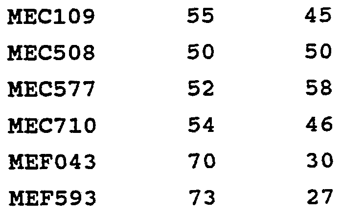
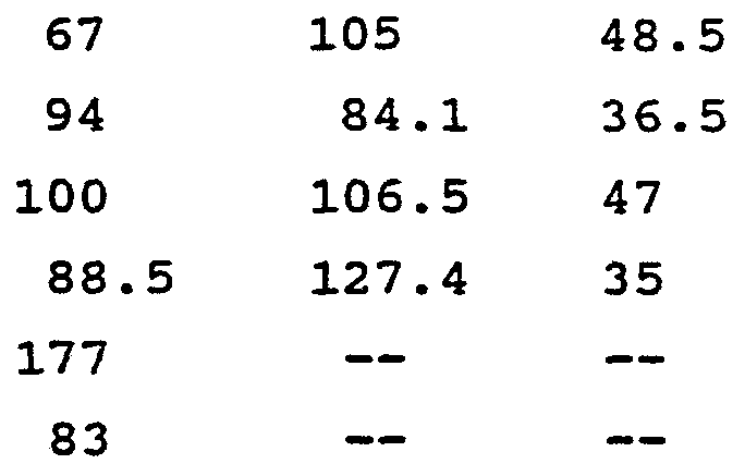
1 Activity was determined using the HFF mitogen assay described herein. Numbers were generated based on a PDGF B standard that was assigned 100% activity.
2 Fractions were separated using ConA affinity chromatography as described herein.
As can be seen, the less or nonglycosylated fractions were consistently, significantly lower in biological activity than the highly glycosylated fraction, the glycosylated fraction exhibiting as much as three times the activity as the less glycosylated fraction. Figure 10 also shows that as percent glycosylation increases, so does activity.
ConA affinity chromatography was used to separate the glycosylated and less glycosylated fractions from one another as follows. ConA-Sepharose 4B (Pharmacia) was packed into a 15 cm x 1 cm column to a final volume of 13 mi. The lyophilized products from
§4.1 were diluted in water to a concentration of approximately 1 mg/ l and loaded onto the column. After the protein was loaded, the column was washed with equilibration buffer to eliminate any protein which did not bind to the resin. The glycosylated protein was then eluted off the column with 0.4M α-methyl-D- mannopyranoside (sigma) in equilibration buffer. Affinity chromatography with ConA-Sepharose yielded two protein fractions. One did not bind to the resin, and the other was eluted off the column only after treatment with 0.4M α-methyl-D-mannopyranoside. A typical chromatogram is shown in Figure 11.
To determine whether mannose was the carbohydrate present, the uptake of 3H-mannose by PDGF B-producing cells was measured. Specifically, these cells were grown at 30°C in 25 ml of Leu minus media containing 8% glucose for 48 hours. Two ml of this growth was then transferred to 100-500 ml of Ura minus media with 1% glucose. At 43 hours, 150-840 μCi of D- [2,6-3H]-mannose (Amersham) was added to the media and the growth was continued. The cells were harvested at 70 hours by centrifugation at 5000 g for 20 minutes at 4°C. The supernatant was collected and acidified by the addition of glacial acetic acid to a final concentration of 50 mM. Seventy-five μg of nonradioactive PDGF was then added to the supernatant as a carrier. The supernatant was loaded onto a 7 ml S-Sepharose fast flow (Pharmacia) column which had been equilibrated with 50 mM acetic acid/0.12 M NaCl. Following the loading, the column was washed with equilibration buffer followed by a wash with 50 mM acetic acid/0.5 M NaCl. The PDGF was then eluted off the column with 50 mM acetic acid/1.0 M NaCl. Fractions collected during the second wash and the elution were counted to determine if the in vivo
incorporation was successful. A control experiment was also carried out in which the 3H-mannose was added to the supernatant after centrifugation to determine if there was any nonspecific binding to PDGF. The results indicate that no mannose is associated with PDGF unless it is covalently bound to the molecule.
The in vivo incorporation of D-[2,6-3H]-mannose was successful. The results of this experiment, are illustrated in Figure 12. As can be seen, mannose appears to be the carbohydrate which is involved in the glycosylation of PDGF expressed in yeast.
To confirm this, deglycosylation was carried out by treating PDGF with α-mannosidase (Boehringer Mannheim) in a ratio of 10:1. The reaction was carried out in 50 mM Na Acetate/10 mM ZnCl2 pH 4.0 at 37°C. The reaction was allowed to take place over a period of five days, and was stopped by boiling the solution for 3 minutes. The boiled solution was then passed through a Bio Gel P-10 (BioRad) gel filtration column, equilibrated with 50 mM acetic acid. The PDGF-containing peak was pooled and dialyzed against dH20 to insure elimination of all free mannose residues. The final product was subjected to analysis by SDS-PAGE, mitogenic assay, and mannose determination by periodate (as explained below) .
Since PDGF B has a number of potential O-linked glycosylation sites, deglycosylation was also attempted by β-elimination. This was done by suspending PDGF in 0.1 N NaOH and incubating at 37°c for 48 hours. Following incubation, the protein was passed through a Bio Gel P-10 column, equilibrated with 50 mM acetic acid, to desalt and remove any free mannose residues. The PDGF peak was pooled, dialyzed against dH20, and tested for biological activity as well as for the presence of mannose by the periodate reaction. The elimination
reaction resulted in the removal of counts from the protein, indicating that radioactive mannose had been removed.
The periodate method was used to determine the number of moles of mannose per mole of PDGF in the highly and less glycosylated fractions from Experiment 4.1 as well as in the deglycosylated products above. This method, well known in the art, calls for the oxidation of the sugar by periodate, followed by a colorimetric determination of the residual periodate not utilized during the oxidation process (see Avigad, G. , Carbohydrate Research (1969) 11:119-123, the disclosure of which is incorporated herein by reference) . Analysis by the periodate reaction showed that the fraction that did not bind to the ConA resin was indeed less glycosylated than the protein which did bind. On the average, the less glycosylated fraction contains from 2- 5 moles of mannose/mole of PDGF. The more glycosylated protein had a higher apparent molecular weight, as determined by SDS-PAGE.
Furthermore, deglycosylation with α-mannosidase gave both a decrease in the number of moles of mannose/mole of PDGF as well as in the mitogenic activity. Before treatment with α-mannosidase there were 9 moles of mannose/ mole of PDGF as determined by the periodate method. After the deglycosylation reaction, there were only 6.5 moles of mannose/mole of PDGF. This figure would not be expected to go to zero since the α- mannosidase only cleaves the external mannose residues and not the internal ones. After the reaction, the biological activity went down to approximately 60% of the original activity. These results support the theory that glycosylation plays a role in the biological activity of PDGF. The SDS-PAGE results also show a lower molecular
weight protein following the deglycosylation reaction with α-mannosidase.
After β-elimination, the periodate reaction showed a decrease in moles of mannose from 9 moles to .8-6 moles. The biological activity, however, was destroyed under these harsh conditions.
To determine which amino acid residues are glycosylated when PDGF B is produced in yeast, a heterogenous mixture containing approximately 70% highly glycosylated material was subjected to tryptic digestion and the tryptic fragments weighed and sequenced. Several fragments were heavier than the theoretical molecular weight of the amino acid residues contained therein, the increase in weight being in exact increments of the weight of mannose. Results varied, and it was sometimes difficult to determine which residue was indeed glycosylated since some tryptic fragments contained more than one glycosylation site. However, preliminary experiments indicate that the highly glycosylated PDGF appears to have from 0-2 mannoses on Thr-6, 0-1 on Thr- 20, 0-1 on Ser-26, 0-2 on Thr-63, 0-3 on Thr-88, 0-3 on Thr-90, and 0-1 on Thr-101.
This determination was also carried out on a PDGF B heterogeneous mixture having a higher percentage of less glycosylated material than hyperglycosylated
PDGF. No sugar groups were found at Thr-20, Ser-26 or Thr-101. Therefore, it is hypothesized that a purified sample of the less glycosylated PDGF B would lack sugars on these residues.
5. Bioassay for PDGF Activity
5.1. PDGF Produced in Mammalian Cells
Chinese hamster ovary cells (CHO) used in the process were normally grown in medium supplemented with 10% fetal calf serum (FCS) . This presented a problem in assaying for PDGF due to the background contributed by the native bovine PDGF in FCS. Thus, it was necessary to devise culture conditions that support production of recombinant products while reducing the background.
Using CHO cells transfected with the human β-interferon gene, it was found that expression levels reached approximately 50% of those observed in 10% FCS with culture medium supplemented by 5% platelet-deficient horse plasma (PDHS) . This medium, when added to either the cell growth or mitogen assays, gave no background.
CHO transformants were assayed by adding 10 μl of a 24-hr supernatant harvest in 5% PDHS (and necessary dilutions, usually serial twofold and threefold) to the well of a 96-well plates of the assay.
5.2. PDGF Produced in Yeast
Samples of supernatants of yeast which had expressed PDGF were appropriately diluted, depending upon expected activity, and then serially diluted in DMEM containing 1% bovine serum albumin (BSA) . Aliquots (10 μl) of each dilution were placed in the wells of the assay plates.
5.3 Human Foreskin Fibroblast (HFF) Mitogen Assay for PDGF
HFF stocks were stored frozen; freezing was at passage 13. Prior to use, HFF were thawed, and grown in T75 flasks until confluent, which usually occurred at 5-7
days. Growth medium contained Dulbecco's Modified Eagles
Medium (DMEM) , 20% fetal bovine serum (FBS) , 1 mM sodium pyruvate, 300 μg/ml L-glutamine, lOOU/ml penicillin, and
100 μg/ml streptomycin. Cells were incubated at 37°C in humidified 7% C02, 93% air atmosphere. At confluency, cells were passaged by rinsing the monolayer with phosphate buffered saline (PBS) lacking Ca++ and Mg"*"4", dissociating them in trypsin containing EDTA, and diluting them with Growth Medium. Cells were passaged no more than 8 times after thawing.
To assay for PDGF, HFFs were plated as follows.
The cells were rinsed and dissociated with trypsin as above. The trypsinized cells were pelleted, and resus- pended to a concentration of 1 x 10⁵ cells/ml in medium similar to Growth Medium, except that 5% FBS replaced 20%
FBS; 100 μl of suspension was dispensed into each well of a 96 well microtiter plate, and the cells were incubated
5-6 days under the above described conditions.
PDGF in the sample was determined by monitoring 3H-thymidine incorporation into HFF DNA stimulated by
PDGF. Samples were added to the wells containing HFF monolayers, and the assay plates incubated as above for
18 hours. The HFF cultures were then pulsed with
[Methyl-3H] thymidine (10 μC/ml final concentration,
1 μC/well) at 37°C under the above described incubation conditions for 8 hours. After incubation, the cells were rinsed with PBS and fixed. Fixing was by incubation with
5% trichloracetic acid (TCA) and then 100% methanol for
15 minutes, followed by drying in air. The cells were then solubilized with 0.3N NaOH, and counted in a liquid scintillation counter.
Control samples were treated as the samples described above, and were prepared as follows. For positive controls used in activity tests of PDGF produced
__
—57 —
in S. cerevisiae MB2-1, PDGF B-B homodimer from S. cerevisiae MB2-1, transformed with pYAGL7PB, was dissolved to a final concentration of 56 ng/ml in DMEM containing 1% BSA. A standard curve was prepared; the first point was 5.6 ng/ml, the remaining points were 2-fold serial dilutions. For positive controls in all other tests, PDGF, purchased from PDGF, Inc. , was dissolved to a final concentration of 100 ng/ml in DMEM containing 10 mg/ml BSA. A standard curve was prepared; the first point was 10 ng/ml, the remaining points were 2-fold serial dilutions.
Each dilution was tested in triplicate. Negative controls, in 1:10 dilution of 5% FCS, which lacked both sample and control PDGF, were also run. As can be seen in Table 2, and explained above, hyperglycosylated PDGF exhibited far more biological activity than its less or nonglycosylated counterpart. Thus, hyperglycosylated PDGF and methods for isolating the same have been disclosed. Although preferred embodiments of the subject invention have been described in some detail, it is understood that obvious variations can be made without departing from the spirit and the scope of the invention as defined by the appended claims.
Claims
1. A composition of hyperglycosylated PDGF.
2. The composition of claim 1 wherein said hyperglycosylated PDGF includes at least an average of 10 moles of carbohydrate per mole of dimer.
3. The composition of claim 1 wherein said hyperglycosylated PDGF includes at least an average of 17 moles of carbohydrate per mole of dimer.
4. The composition of claim 2 wherein said hyperglycosylated PDGF comprises PDGF B-B homodimer.
5. The composition of claim 3 wherein said hyperglycosylated PDGF comprises PDGF B-B homodimer.
6. The composition of claim 4 wherein said carbohydrate comprises mannose.
7. The composition of claim 5 wherein said carbohydrate comprises mannose.
8. The composition of claim 1 wherein said hyperglycosylated PDGF is produced in yeast.
9. A PDGF B-B homodimer having at least 17 moles of mannose per mole of dimer.
10. The PDGF of claim 9 wherein said PDGF is produced in yeast.
11. A method of producing hyperglycosylated PDGF comprising: (a) providing a mixture containing a hyperglycosylated PDGF fraction and a less glycosylated PDGF fraction; and
(b) isolating said hyperglycosylated PDGF from said mixture.
12. The method of claim 11 wherein said hyperglycosylated fraction is isolated using lectin affinity chromatography.
13. The method of claim 11 wherein said hyperglycosylated PDGF includes at least an average of 10 moles of carbohydrate per mole of dimer.
14. The method of claim 11 wherein said hyperglycosylated PDGF includes at least an average of 17 moles of carbohydrate per mole of dimer.
15. The method of claim 13 wherein said hyperglycosylated PDGF comprises PDGF B-B homodimer.
16. The method of claim 14 wherein said hyperglycosylated PDGF comprises PDGF B-B homodimer.
17. The method of claim 15 wherein said carbohydrate comprises mannose.
18. The method of claim 16 wherein said carbohydrate comprises mannose.
19. The method of claim 11 wherein said mixture is derived from yeast cells expressing PDGF.
20. A method of producing PDGF B-B homodimer having at least 17 moles of mannose per mole of dimer substantially free of less glycosylated PDGF comprising:
(a) providing a mixture containing said PDGF B- B homodimer and less glycosylated PDGF; and
(b) isolating said PDGF B-B homodimer using lectin affinity chromatography.
21. The method of claim 20 wherein said PDGF is produced in yeast.
22. In a method of purifying recombinant PDGF from an expression media which comprises a hyperglycosylated PDGF fraction and a less glycosylated PDGF fraction, the improvement comprising isolating said hyperglycosylated PDGF fraction from said expression media.
23. The method of claim 22 wherein said hyperglycosylated PDGF is isolated using lectin affinity chromatography.
24. The method of claim 22 wherein said recombinant PDGF is purified from yeast expression media.
Applications Claiming Priority (2)
| Application Number | Priority Date | Filing Date | Title |
|---|---|---|---|
| US51547490A | 1990-04-26 | 1990-04-26 | |
| US515,474 | 1990-04-26 |
Publications (1)
| Publication Number | Publication Date |
|---|---|
| WO1991016335A1 true WO1991016335A1 (en) | 1991-10-31 |
Family
ID=24051507
Family Applications (1)
| Application Number | Title | Priority Date | Filing Date |
|---|---|---|---|
| PCT/US1991/002766 Ceased WO1991016335A1 (en) | 1990-04-26 | 1991-04-25 | Glycosylated pdgf |
Country Status (1)
| Country | Link |
|---|---|
| WO (1) | WO1991016335A1 (en) |
Cited By (4)
| Publication number | Priority date | Publication date | Assignee | Title |
|---|---|---|---|---|
| US5281582A (en) * | 1992-02-27 | 1994-01-25 | Alliance Pharmaceuticals, Corp. | Serum growth factor |
| JP3318602B2 (en) | 1997-11-10 | 2002-08-26 | 独立行政法人産業技術総合研究所 | Glycosylated heparin-binding protein, method for producing the same, and pharmaceutical composition containing the same |
| WO2006079155A1 (en) * | 2005-01-25 | 2006-08-03 | Apollo Life Sciences Limited | Molecules and chimeric molecules thereof |
| JP2018525967A (en) * | 2014-05-16 | 2018-09-13 | インスティテュート オブ バイオテクノロジー アカデミー オブ ミリタリー メディカル サイエンシズ ピー.エル.エー.チャイナ | Platelet-derived growth factor B mutant, production method thereof and use thereof |
Citations (2)
| Publication number | Priority date | Publication date | Assignee | Title |
|---|---|---|---|---|
| US4766073A (en) * | 1985-02-25 | 1988-08-23 | Zymogenetics Inc. | Expression of biologically active PDGF analogs in eucaryotic cells |
| US4849407A (en) * | 1986-08-13 | 1989-07-18 | Zymogenetics, Inc. | Biologically active mosaic proteins |
-
1991
- 1991-04-25 WO PCT/US1991/002766 patent/WO1991016335A1/en not_active Ceased
Patent Citations (3)
| Publication number | Priority date | Publication date | Assignee | Title |
|---|---|---|---|---|
| US4766073A (en) * | 1985-02-25 | 1988-08-23 | Zymogenetics Inc. | Expression of biologically active PDGF analogs in eucaryotic cells |
| US4845075A (en) * | 1985-02-25 | 1989-07-04 | Zymogenetics, Inc. | Biologically active B-chain homodimers |
| US4849407A (en) * | 1986-08-13 | 1989-07-18 | Zymogenetics, Inc. | Biologically active mosaic proteins |
Non-Patent Citations (1)
| Title |
|---|
| VIROLOGY, Volume 164, issued 1988, KLEIN et al., "Highly Glycosylated PDGF-Like Molecule Secreted by Simian Sarcoma Virus-Transformed Cell", pages 403-410. * |
Cited By (9)
| Publication number | Priority date | Publication date | Assignee | Title |
|---|---|---|---|---|
| US5281582A (en) * | 1992-02-27 | 1994-01-25 | Alliance Pharmaceuticals, Corp. | Serum growth factor |
| JP3318602B2 (en) | 1997-11-10 | 2002-08-26 | 独立行政法人産業技術総合研究所 | Glycosylated heparin-binding protein, method for producing the same, and pharmaceutical composition containing the same |
| US7005415B1 (en) | 1997-11-10 | 2006-02-28 | Director-General Agency Of Industrial Science And Technology | Heparin-binding proteins modified with sugar chains, method of producing the same and pharmaceutical compositions containing the same |
| US7282481B2 (en) | 1997-11-10 | 2007-10-16 | Director-General Of Agency Of Industrial Science And Technology | Heparin-binding proteins modified with sugar chains, method of producing the same and pharmaceutical compositions containing same |
| WO2006079155A1 (en) * | 2005-01-25 | 2006-08-03 | Apollo Life Sciences Limited | Molecules and chimeric molecules thereof |
| JP2018525967A (en) * | 2014-05-16 | 2018-09-13 | インスティテュート オブ バイオテクノロジー アカデミー オブ ミリタリー メディカル サイエンシズ ピー.エル.エー.チャイナ | Platelet-derived growth factor B mutant, production method thereof and use thereof |
| KR20190101392A (en) * | 2014-05-16 | 2019-08-30 | 인스티튜트 오브 바이오테크놀로지, 아카데미 오브 밀리터리 메디칼 싸이언시스, 피엘에이. | Platelet-derived Growth Factor B Mutant, Preparation Method Therefor, and Use Thereof |
| US10450358B2 (en) * | 2014-05-16 | 2019-10-22 | Institute Of Biotechnology Academy Of Military Medical Sciences P.L.A. China | Platelet-derived growth factor B mutant, preparation method therefor and use thereof |
| KR102155903B1 (en) * | 2014-05-16 | 2020-09-16 | 인스티튜트 오브 바이오테크놀로지, 아카데미 오브 밀리터리 메디칼 싸이언시스, 피엘에이. | Platelet-derived Growth Factor B Mutant, Preparation Method Therefor, and Use Thereof |
Similar Documents
| Publication | Publication Date | Title |
|---|---|---|
| JP3115318B2 (en) | Fusion protein containing GM-CSF and IL-3 | |
| US6130071A (en) | Vascular endothelial growth factor C (VEGF-C) ΔCys156 protein and gene, and uses thereof | |
| US5128321A (en) | PDGF analogs and methods of use | |
| US4954437A (en) | Cell encoding recombinant human erythropoietin | |
| US5219759A (en) | Recombinant DNA encoding PDGF A-chain polypeptide and expression vectors | |
| AU646014B2 (en) | Novel platelet-derived growth factor B chain analogs and method for homogeneous production thereof | |
| US5965122A (en) | Use of interleukin-7 to stimulate proliferation of hematopoietic cell precursors | |
| EP0682110A1 (en) | Recombinant DNA encoding PDGF A-chain polypeptides | |
| US20030036511A1 (en) | Analysis and separation of platelet-derived growth factor proteins | |
| JPH08269096A (en) | Erythropoietin relative | |
| JPH07138292A (en) | Mouse leukemia inhibitor | |
| CA2084514A1 (en) | O-glycosylated ifn-alpha | |
| HU220334B (en) | Erythropoietin analogues | |
| EP0548267A1 (en) | Production of insulin-like growth factor-1 in methylotrophic yeast cells | |
| DE69434519T2 (en) | PROHORMONE CONVERTASE EXPRESSIVE POLYPEPTIDE SYNTHESIS | |
| JPH06505631A (en) | Megakaryocyte stimulating factor | |
| EP0802983B1 (en) | Expression of urokinase plasminogen activator inhibitors | |
| JP2823690B2 (en) | Protease resistant PDGF and methods of use | |
| NZ226679A (en) | Purified il-7 (interleukin-7) and pharmaceutical compositions | |
| AU743833B2 (en) | Recombinant human erythropoietin with advantageous glycosylation profile | |
| US5359033A (en) | Cleaved dimers of mullerian inhibiting substance-like polypeptides | |
| DE3687948T2 (en) | METHOD AND NUCLEIC ACID FOR PRODUCING LECITHIN: CHOLESTRINACYLTRANSFERASE. | |
| WO1991016335A1 (en) | Glycosylated pdgf | |
| CZ285440B6 (en) | Fusion protein, process of its preparation, gene structure for its preparation, vector containing such gene structure, streptomycete cell in which this vector is comprised and use of the fusion protein | |
| US5474982A (en) | PDGF analogs and methods of use |
Legal Events
| Date | Code | Title | Description |
|---|---|---|---|
| AK | Designated states |
Kind code of ref document: A1 Designated state(s): CA JP |
|
| AL | Designated countries for regional patents |
Kind code of ref document: A1 Designated state(s): AT BE CH DE DK ES FR GB GR IT LU NL SE |
|
| NENP | Non-entry into the national phase |
Ref country code: CA |
|
| 122 | Ep: pct application non-entry in european phase |