US6912412B2 - System and methods of fluorescence, reflectance and light scattering spectroscopy for measuring tissue characteristics - Google Patents
System and methods of fluorescence, reflectance and light scattering spectroscopy for measuring tissue characteristics Download PDFInfo
- Publication number
- US6912412B2 US6912412B2 US10/052,583 US5258302A US6912412B2 US 6912412 B2 US6912412 B2 US 6912412B2 US 5258302 A US5258302 A US 5258302A US 6912412 B2 US6912412 B2 US 6912412B2
- Authority
- US
- United States
- Prior art keywords
- tissue
- fluorescence
- light
- spectrum
- reflectance
- Prior art date
- Legal status (The legal status is an assumption and is not a legal conclusion. Google has not performed a legal analysis and makes no representation as to the accuracy of the status listed.)
- Expired - Lifetime, expires
Links
- 238000000034 method Methods 0.000 title claims abstract description 96
- 238000000149 argon plasma sintering Methods 0.000 title claims description 46
- 238000001506 fluorescence spectroscopy Methods 0.000 title claims description 15
- 238000001055 reflectance spectroscopy Methods 0.000 title description 9
- 238000001228 spectrum Methods 0.000 claims abstract description 45
- 238000003745 diagnosis Methods 0.000 claims abstract description 24
- 238000004458 analytical method Methods 0.000 claims description 56
- 238000002189 fluorescence spectrum Methods 0.000 claims description 52
- 238000000985 reflectance spectrum Methods 0.000 claims description 40
- 239000000523 sample Substances 0.000 claims description 22
- 238000012545 processing Methods 0.000 claims description 21
- 230000003595 spectral effect Effects 0.000 claims description 17
- 239000000835 fiber Substances 0.000 claims description 12
- 210000002919 epithelial cell Anatomy 0.000 claims description 11
- 230000003287 optical effect Effects 0.000 claims description 9
- 230000000737 periodic effect Effects 0.000 claims description 8
- 239000011159 matrix material Substances 0.000 claims description 6
- 230000005855 radiation Effects 0.000 claims 3
- 230000001131 transforming effect Effects 0.000 claims 2
- 238000001514 detection method Methods 0.000 abstract description 12
- 201000010099 disease Diseases 0.000 abstract description 8
- 208000037265 diseases, disorders, signs and symptoms Diseases 0.000 abstract description 8
- 230000000877 morphologic effect Effects 0.000 abstract description 7
- 238000001727 in vivo Methods 0.000 abstract description 6
- 201000009030 Carcinoma Diseases 0.000 abstract description 3
- 238000000338 in vitro Methods 0.000 abstract 1
- 210000001519 tissue Anatomy 0.000 description 116
- 206010058314 Dysplasia Diseases 0.000 description 46
- 208000023514 Barrett esophagus Diseases 0.000 description 45
- 208000023665 Barrett oesophagus Diseases 0.000 description 45
- 230000005284 excitation Effects 0.000 description 30
- 238000010521 absorption reaction Methods 0.000 description 26
- 230000035945 sensitivity Effects 0.000 description 23
- 210000004940 nucleus Anatomy 0.000 description 21
- 230000008569 process Effects 0.000 description 21
- 210000000981 epithelium Anatomy 0.000 description 18
- 230000000875 corresponding effect Effects 0.000 description 16
- 208000032124 Squamous Intraepithelial Lesions Diseases 0.000 description 14
- 108010054147 Hemoglobins Proteins 0.000 description 13
- 102000001554 Hemoglobins Human genes 0.000 description 13
- 238000001574 biopsy Methods 0.000 description 12
- 238000005259 measurement Methods 0.000 description 12
- 238000007477 logistic regression Methods 0.000 description 11
- 238000002790 cross-validation Methods 0.000 description 9
- 230000006870 function Effects 0.000 description 9
- 210000000277 pancreatic duct Anatomy 0.000 description 9
- 238000004611 spectroscopical analysis Methods 0.000 description 9
- 238000012384 transportation and delivery Methods 0.000 description 9
- 102000008186 Collagen Human genes 0.000 description 8
- 108010035532 Collagen Proteins 0.000 description 8
- 229920001436 collagen Polymers 0.000 description 8
- 238000000354 decomposition reaction Methods 0.000 description 7
- 229930027945 nicotinamide-adenine dinucleotide Natural products 0.000 description 7
- BOPGDPNILDQYTO-NNYOXOHSSA-N nicotinamide-adenine dinucleotide Chemical compound C1=CCC(C(=O)N)=CN1[C@H]1[C@H](O)[C@H](O)[C@@H](COP(O)(=O)OP(O)(=O)OC[C@@H]2[C@H]([C@@H](O)[C@@H](O2)N2C3=NC=NC(N)=C3N=C2)O)O1 BOPGDPNILDQYTO-NNYOXOHSSA-N 0.000 description 7
- 230000003902 lesion Effects 0.000 description 6
- 238000012306 spectroscopic technique Methods 0.000 description 6
- 238000011161 development Methods 0.000 description 5
- 230000000694 effects Effects 0.000 description 5
- 210000000496 pancreas Anatomy 0.000 description 5
- 238000000513 principal component analysis Methods 0.000 description 5
- 239000006096 absorbing agent Substances 0.000 description 4
- 238000002573 colposcopy Methods 0.000 description 4
- 210000002808 connective tissue Anatomy 0.000 description 4
- 230000007423 decrease Effects 0.000 description 4
- 230000002496 gastric effect Effects 0.000 description 4
- 239000013307 optical fiber Substances 0.000 description 4
- 238000012360 testing method Methods 0.000 description 4
- 230000009466 transformation Effects 0.000 description 4
- 206010008263 Cervical dysplasia Diseases 0.000 description 3
- 206010054949 Metaplasia Diseases 0.000 description 3
- 206010028980 Neoplasm Diseases 0.000 description 3
- 206010033645 Pancreatitis Diseases 0.000 description 3
- 206010033647 Pancreatitis acute Diseases 0.000 description 3
- 230000002159 abnormal effect Effects 0.000 description 3
- 201000003229 acute pancreatitis Diseases 0.000 description 3
- 208000009956 adenocarcinoma Diseases 0.000 description 3
- 210000003679 cervix uteri Anatomy 0.000 description 3
- 230000008859 change Effects 0.000 description 3
- 230000001419 dependent effect Effects 0.000 description 3
- 239000010432 diamond Substances 0.000 description 3
- 238000007459 endoscopic retrograde cholangiopancreatography Methods 0.000 description 3
- 238000011156 evaluation Methods 0.000 description 3
- 230000002962 histologic effect Effects 0.000 description 3
- 230000006872 improvement Effects 0.000 description 3
- 230000015689 metaplastic ossification Effects 0.000 description 3
- 208000008443 pancreatic carcinoma Diseases 0.000 description 3
- 238000009595 pap smear Methods 0.000 description 3
- 239000002245 particle Substances 0.000 description 3
- 150000004032 porphyrins Chemical class 0.000 description 3
- 238000011282 treatment Methods 0.000 description 3
- IJGRMHOSHXDMSA-UHFFFAOYSA-N Atomic nitrogen Chemical compound N#N IJGRMHOSHXDMSA-UHFFFAOYSA-N 0.000 description 2
- 102000004190 Enzymes Human genes 0.000 description 2
- 108090000790 Enzymes Proteins 0.000 description 2
- WSFSSNUMVMOOMR-UHFFFAOYSA-N Formaldehyde Chemical compound O=C WSFSSNUMVMOOMR-UHFFFAOYSA-N 0.000 description 2
- 102000002274 Matrix Metalloproteinases Human genes 0.000 description 2
- 108010000684 Matrix Metalloproteinases Proteins 0.000 description 2
- 206010061902 Pancreatic neoplasm Diseases 0.000 description 2
- 238000013459 approach Methods 0.000 description 2
- 210000004027 cell Anatomy 0.000 description 2
- 238000004891 communication Methods 0.000 description 2
- 238000012937 correction Methods 0.000 description 2
- 238000009792 diffusion process Methods 0.000 description 2
- 238000001839 endoscopy Methods 0.000 description 2
- 208000021045 exocrine pancreatic carcinoma Diseases 0.000 description 2
- 238000000605 extraction Methods 0.000 description 2
- 210000001035 gastrointestinal tract Anatomy 0.000 description 2
- 238000003384 imaging method Methods 0.000 description 2
- 230000003993 interaction Effects 0.000 description 2
- 208000015486 malignant pancreatic neoplasm Diseases 0.000 description 2
- 238000013508 migration Methods 0.000 description 2
- 230000004660 morphological change Effects 0.000 description 2
- 210000000214 mouth Anatomy 0.000 description 2
- 210000004400 mucous membrane Anatomy 0.000 description 2
- 201000002528 pancreatic cancer Diseases 0.000 description 2
- 238000002271 resection Methods 0.000 description 2
- 230000004044 response Effects 0.000 description 2
- 238000010183 spectrum analysis Methods 0.000 description 2
- 238000007619 statistical method Methods 0.000 description 2
- 210000004876 tela submucosa Anatomy 0.000 description 2
- RNAMYOYQYRYFQY-UHFFFAOYSA-N 2-(4,4-difluoropiperidin-1-yl)-6-methoxy-n-(1-propan-2-ylpiperidin-4-yl)-7-(3-pyrrolidin-1-ylpropoxy)quinazolin-4-amine Chemical compound N1=C(N2CCC(F)(F)CC2)N=C2C=C(OCCCN3CCCC3)C(OC)=CC2=C1NC1CCN(C(C)C)CC1 RNAMYOYQYRYFQY-UHFFFAOYSA-N 0.000 description 1
- 102100026802 72 kDa type IV collagenase Human genes 0.000 description 1
- 101710151806 72 kDa type IV collagenase Proteins 0.000 description 1
- ZGXJTSGNIOSYLO-UHFFFAOYSA-N 88755TAZ87 Chemical compound NCC(=O)CCC(O)=O ZGXJTSGNIOSYLO-UHFFFAOYSA-N 0.000 description 1
- KSFOVUSSGSKXFI-GAQDCDSVSA-N CC1=C/2NC(\C=C3/N=C(/C=C4\N\C(=C/C5=N/C(=C\2)/C(C=C)=C5C)C(C=C)=C4C)C(C)=C3CCC(O)=O)=C1CCC(O)=O Chemical compound CC1=C/2NC(\C=C3/N=C(/C=C4\N\C(=C/C5=N/C(=C\2)/C(C=C)=C5C)C(C=C)=C4C)C(C)=C3CCC(O)=O)=C1CCC(O)=O KSFOVUSSGSKXFI-GAQDCDSVSA-N 0.000 description 1
- 208000000668 Chronic Pancreatitis Diseases 0.000 description 1
- 102000005927 Cysteine Proteases Human genes 0.000 description 1
- 108010005843 Cysteine Proteases Proteins 0.000 description 1
- 102000016942 Elastin Human genes 0.000 description 1
- 108010014258 Elastin Proteins 0.000 description 1
- 101000934858 Homo sapiens Breast cancer type 2 susceptibility protein Proteins 0.000 description 1
- 101000891683 Homo sapiens Fanconi anemia group D2 protein Proteins 0.000 description 1
- 101000617536 Homo sapiens Presenilin-1 Proteins 0.000 description 1
- 206010061218 Inflammation Diseases 0.000 description 1
- XUJNEKJLAYXESH-REOHCLBHSA-N L-Cysteine Chemical compound SC[C@H](N)C(O)=O XUJNEKJLAYXESH-REOHCLBHSA-N 0.000 description 1
- 206010061876 Obstruction Diseases 0.000 description 1
- 108010064719 Oxyhemoglobins Proteins 0.000 description 1
- 206010033649 Pancreatitis chronic Diseases 0.000 description 1
- 208000031481 Pathologic Constriction Diseases 0.000 description 1
- 208000006994 Precancerous Conditions Diseases 0.000 description 1
- 102100022033 Presenilin-1 Human genes 0.000 description 1
- 102000012479 Serine Proteases Human genes 0.000 description 1
- 108010022999 Serine Proteases Proteins 0.000 description 1
- 229920000995 Spectralon Polymers 0.000 description 1
- 230000005856 abnormality Effects 0.000 description 1
- 238000009825 accumulation Methods 0.000 description 1
- 230000001154 acute effect Effects 0.000 description 1
- 230000004075 alteration Effects 0.000 description 1
- 229960002749 aminolevulinic acid Drugs 0.000 description 1
- QVGXLLKOCUKJST-UHFFFAOYSA-N atomic oxygen Chemical compound [O] QVGXLLKOCUKJST-UHFFFAOYSA-N 0.000 description 1
- 230000008901 benefit Effects 0.000 description 1
- 238000012742 biochemical analysis Methods 0.000 description 1
- 230000005540 biological transmission Effects 0.000 description 1
- 230000015572 biosynthetic process Effects 0.000 description 1
- 210000000481 breast Anatomy 0.000 description 1
- 238000004364 calculation method Methods 0.000 description 1
- 201000011510 cancer Diseases 0.000 description 1
- 210000003855 cell nucleus Anatomy 0.000 description 1
- 238000012512 characterization method Methods 0.000 description 1
- 230000011382 collagen catabolic process Effects 0.000 description 1
- 210000001072 colon Anatomy 0.000 description 1
- 230000000295 complement effect Effects 0.000 description 1
- 238000004590 computer program Methods 0.000 description 1
- 230000002596 correlated effect Effects 0.000 description 1
- 238000009109 curative therapy Methods 0.000 description 1
- XUJNEKJLAYXESH-UHFFFAOYSA-N cysteine Natural products SCC(N)C(O)=O XUJNEKJLAYXESH-UHFFFAOYSA-N 0.000 description 1
- 235000018417 cysteine Nutrition 0.000 description 1
- 238000007405 data analysis Methods 0.000 description 1
- 238000013480 data collection Methods 0.000 description 1
- 230000003247 decreasing effect Effects 0.000 description 1
- 238000009795 derivation Methods 0.000 description 1
- 238000002405 diagnostic procedure Methods 0.000 description 1
- 230000004069 differentiation Effects 0.000 description 1
- 230000010339 dilation Effects 0.000 description 1
- 229940079593 drug Drugs 0.000 description 1
- 239000003814 drug Substances 0.000 description 1
- 229920002549 elastin Polymers 0.000 description 1
- 238000005516 engineering process Methods 0.000 description 1
- 210000005081 epithelial layer Anatomy 0.000 description 1
- 210000003238 esophagus Anatomy 0.000 description 1
- 238000001914 filtration Methods 0.000 description 1
- 238000009499 grossing Methods 0.000 description 1
- 230000003118 histopathologic effect Effects 0.000 description 1
- 238000012333 histopathological diagnosis Methods 0.000 description 1
- 230000004054 inflammatory process Effects 0.000 description 1
- 230000009545 invasion Effects 0.000 description 1
- 238000012804 iterative process Methods 0.000 description 1
- 210000004072 lung Anatomy 0.000 description 1
- 238000012423 maintenance Methods 0.000 description 1
- 238000007726 management method Methods 0.000 description 1
- 238000013507 mapping Methods 0.000 description 1
- 239000003550 marker Substances 0.000 description 1
- 230000001404 mediated effect Effects 0.000 description 1
- 238000013160 medical therapy Methods 0.000 description 1
- 230000002503 metabolic effect Effects 0.000 description 1
- 238000012544 monitoring process Methods 0.000 description 1
- 208000022669 mucinous neoplasm Diseases 0.000 description 1
- 229910052757 nitrogen Inorganic materials 0.000 description 1
- 230000005693 optoelectronics Effects 0.000 description 1
- 210000000056 organ Anatomy 0.000 description 1
- 230000010355 oscillation Effects 0.000 description 1
- 239000001301 oxygen Substances 0.000 description 1
- 229910052760 oxygen Inorganic materials 0.000 description 1
- 201000008129 pancreatic ductal adenocarcinoma Diseases 0.000 description 1
- 230000007170 pathology Effects 0.000 description 1
- 229940109328 photofrin Drugs 0.000 description 1
- 238000010837 poor prognosis Methods 0.000 description 1
- 238000004393 prognosis Methods 0.000 description 1
- 230000001681 protective effect Effects 0.000 description 1
- 229950003776 protoporphyrin Drugs 0.000 description 1
- 238000005086 pumping Methods 0.000 description 1
- 238000006862 quantum yield reaction Methods 0.000 description 1
- 238000010223 real-time analysis Methods 0.000 description 1
- 238000011084 recovery Methods 0.000 description 1
- 230000009467 reduction Effects 0.000 description 1
- PYWVYCXTNDRMGF-UHFFFAOYSA-N rhodamine B Chemical compound [Cl-].C=12C=CC(=[N+](CC)CC)C=C2OC2=CC(N(CC)CC)=CC=C2C=1C1=CC=CC=C1C(O)=O PYWVYCXTNDRMGF-UHFFFAOYSA-N 0.000 description 1
- 238000012216 screening Methods 0.000 description 1
- 238000001356 surgical procedure Methods 0.000 description 1
- 208000024891 symptom Diseases 0.000 description 1
- 230000001360 synchronised effect Effects 0.000 description 1
- 238000001429 visible spectrum Methods 0.000 description 1
- 238000012800 visualization Methods 0.000 description 1
- 239000013585 weight reducing agent Substances 0.000 description 1
- OENHQHLEOONYIE-JLTXGRSLSA-N β-Carotene Chemical compound CC=1CCCC(C)(C)C=1\C=C\C(\C)=C\C=C\C(\C)=C\C=C\C=C(/C)\C=C\C=C(/C)\C=C\C1=C(C)CCCC1(C)C OENHQHLEOONYIE-JLTXGRSLSA-N 0.000 description 1
Images


Classifications
-
- G—PHYSICS
- G01—MEASURING; TESTING
- G01N—INVESTIGATING OR ANALYSING MATERIALS BY DETERMINING THEIR CHEMICAL OR PHYSICAL PROPERTIES
- G01N21/00—Investigating or analysing materials by the use of optical means, i.e. using sub-millimetre waves, infrared, visible or ultraviolet light
- G01N21/62—Systems in which the material investigated is excited whereby it emits light or causes a change in wavelength of the incident light
- G01N21/63—Systems in which the material investigated is excited whereby it emits light or causes a change in wavelength of the incident light optically excited
- G01N21/64—Fluorescence; Phosphorescence
- G01N21/6486—Measuring fluorescence of biological material, e.g. DNA, RNA, cells
-
- A—HUMAN NECESSITIES
- A61—MEDICAL OR VETERINARY SCIENCE; HYGIENE
- A61B—DIAGNOSIS; SURGERY; IDENTIFICATION
- A61B5/00—Measuring for diagnostic purposes; Identification of persons
- A61B5/0059—Measuring for diagnostic purposes; Identification of persons using light, e.g. diagnosis by transillumination, diascopy, fluorescence
- A61B5/0071—Measuring for diagnostic purposes; Identification of persons using light, e.g. diagnosis by transillumination, diascopy, fluorescence by measuring fluorescence emission
-
- A—HUMAN NECESSITIES
- A61—MEDICAL OR VETERINARY SCIENCE; HYGIENE
- A61B—DIAGNOSIS; SURGERY; IDENTIFICATION
- A61B5/00—Measuring for diagnostic purposes; Identification of persons
- A61B5/0059—Measuring for diagnostic purposes; Identification of persons using light, e.g. diagnosis by transillumination, diascopy, fluorescence
- A61B5/0075—Measuring for diagnostic purposes; Identification of persons using light, e.g. diagnosis by transillumination, diascopy, fluorescence by spectroscopy, i.e. measuring spectra, e.g. Raman spectroscopy, infrared absorption spectroscopy
-
- A—HUMAN NECESSITIES
- A61—MEDICAL OR VETERINARY SCIENCE; HYGIENE
- A61B—DIAGNOSIS; SURGERY; IDENTIFICATION
- A61B5/00—Measuring for diagnostic purposes; Identification of persons
- A61B5/0059—Measuring for diagnostic purposes; Identification of persons using light, e.g. diagnosis by transillumination, diascopy, fluorescence
- A61B5/0082—Measuring for diagnostic purposes; Identification of persons using light, e.g. diagnosis by transillumination, diascopy, fluorescence adapted for particular medical purposes
- A61B5/0084—Measuring for diagnostic purposes; Identification of persons using light, e.g. diagnosis by transillumination, diascopy, fluorescence adapted for particular medical purposes for introduction into the body, e.g. by catheters
-
- A—HUMAN NECESSITIES
- A61—MEDICAL OR VETERINARY SCIENCE; HYGIENE
- A61B—DIAGNOSIS; SURGERY; IDENTIFICATION
- A61B5/00—Measuring for diagnostic purposes; Identification of persons
- A61B5/145—Measuring characteristics of blood in vivo, e.g. gas concentration or pH-value ; Measuring characteristics of body fluids or tissues, e.g. interstitial fluid or cerebral tissue
- A61B5/1455—Measuring characteristics of blood in vivo, e.g. gas concentration or pH-value ; Measuring characteristics of body fluids or tissues, e.g. interstitial fluid or cerebral tissue using optical sensors, e.g. spectral photometrical oximeters
- A61B5/1459—Measuring characteristics of blood in vivo, e.g. gas concentration or pH-value ; Measuring characteristics of body fluids or tissues, e.g. interstitial fluid or cerebral tissue using optical sensors, e.g. spectral photometrical oximeters invasive, e.g. introduced into the body by a catheter
-
- A—HUMAN NECESSITIES
- A61—MEDICAL OR VETERINARY SCIENCE; HYGIENE
- A61B—DIAGNOSIS; SURGERY; IDENTIFICATION
- A61B5/00—Measuring for diagnostic purposes; Identification of persons
- A61B5/43—Detecting, measuring or recording for evaluating the reproductive systems
- A61B5/4306—Detecting, measuring or recording for evaluating the reproductive systems for evaluating the female reproductive systems, e.g. gynaecological evaluations
- A61B5/4318—Evaluation of the lower reproductive system
- A61B5/4331—Evaluation of the lower reproductive system of the cervix
-
- G—PHYSICS
- G01—MEASURING; TESTING
- G01N—INVESTIGATING OR ANALYSING MATERIALS BY DETERMINING THEIR CHEMICAL OR PHYSICAL PROPERTIES
- G01N21/00—Investigating or analysing materials by the use of optical means, i.e. using sub-millimetre waves, infrared, visible or ultraviolet light
- G01N21/17—Systems in which incident light is modified in accordance with the properties of the material investigated
- G01N21/47—Scattering, i.e. diffuse reflection
-
- G—PHYSICS
- G01—MEASURING; TESTING
- G01N—INVESTIGATING OR ANALYSING MATERIALS BY DETERMINING THEIR CHEMICAL OR PHYSICAL PROPERTIES
- G01N21/00—Investigating or analysing materials by the use of optical means, i.e. using sub-millimetre waves, infrared, visible or ultraviolet light
- G01N21/62—Systems in which the material investigated is excited whereby it emits light or causes a change in wavelength of the incident light
- G01N21/63—Systems in which the material investigated is excited whereby it emits light or causes a change in wavelength of the incident light optically excited
- G01N21/64—Fluorescence; Phosphorescence
Definitions
- Adenocarcinoma of the lower esophagus develops almost exclusively in patients with Barrett's esophagus (BE), a condition characterized by the presence of metaplastic columnar epithelium. While the prognosis of patients diagnosed with adenocarcinoma is poor, the chances of successful treatment increase significantly if the disease is detected at the dysplastic stage.
- BE Barrett's esophagus
- the surveillance of patients with BE for dysplasia is challenging in two respects. First, dysplasia is not visible during routine endoscopy. Thus, numerous random biopsies are required. Second, the histopathologic diagnosis of dysplasia is problematic, as there is poor inter-observer agreement on the classification of a particular specimen, even among expert gastrointestinal pathologists.
- Optical techniques may significantly enhance the endoscopist's ability to detect these early dysplastic changes in BE.
- fluorescence spectroscopy studies using exogenous fluorophores such as Photofrin® and aminolevulinic-acid induced protoporphyrin IX, show that there is a significant difference between the measured red fluorescence of the carcinomatous and non-dysplastic tissue as a result of the preferential accumulation of the drug.
- Initial autofluorescence spectroscopy studies performed at 410 nm excitation report promising results for detecting high-grade dysplasia. However, focal high-grade and low-grade lesions could not be detected reliably. Thus a continuing need exists for further improvements in the optical measurements used to detect early stage carcinomas.
- the present invention relates to a combination of spectroscopic systems that can improve the sensitivity and accuracy of measuring the state of tissue.
- a plurality of different spectroscopic systems can be used to determine whether a region of tissue is normal, pre-cancerous or cancerous.
- dysplasia detection in patients with Barrett's esophagus (BE) can be performed.
- fluorescence, reflectance and light scattering spectroscopies provide complementary information about the biochemical, architectural and morphological state of tissue and the corresponding changes that occur during the progression of disease, and in particular, of dysplasia.
- the system has been developed for combining these three methods to provide for detection, mapping, and/or imaging of tissue.
- methods of processing the spectral or image data with a data processor programmed to perform an automated diagnostic routine is useful to provide the results of a plurality of different measurement modalities including fluorescence and light scattering spectroscopy.
- a further preferred embodiment of the invention utilizes a tri-modal system to guide a biopsy procedure.
- the collection of light includes the collection of a measured fluorescence and reflectance spectra.
- the detected reflectance spectrum is processed to remove a diffusive background component. Once this is accomplished, then a component of the light that is periodic in wavelength is measured. This component arises from the light that is Mie-scattered by surface epithelial cell nuclei, for example. By analyzing the amplitude and frequency of the periodic structure, the density and size distribution of these nuclei can be extracted.
- the apparatus delivers both excitation components to the region of interest through the same distal surface of the probe, preferably through the same optical fiber or collection of fibers.
- the biopsy channel of an endoscope can be used to insert the fiber optic light delivery and collection system used to obtain measurements.
- a small diameter endoscope 5 mm or less in diameter, for example, can include the light delivery and collection system suitable for many applications.
- a preferred embodiment of the system can use a single fiber system for delivery and collection, or alternatively, can employ a central delivery fiber and six collection fibers concentrically arranged around the delivery fiber.
- the proximal end of the light delivery and collection probe is optically coupled to both a broadband flash lamp and a monochromatic source such as a laser.
- a rotating filter or dye wheel can be used to rapidly switch the excitation wavelength over a selected range.
- the need for using reflected light arises from the need to correct for the effects of hemoglobin absorption on the measured integrated tissue fluorescence intensity.
- the combination of fluorescence and reflectance spectroscopies can be applied to remove distortions introduced by scattering and absorption into the entire measured tissue fluorescence spectrum.
- the undistorted or modified fluorescence spectrum can serve as a sensitive indicator of tissue biochemistry, while reflectance and light scattering representations provide morphological information on tissue architecture and epithelial cell nuclei.
- the present invention can include the simultaneous use of all three spectroscopic methods for characterizing tissue and diagnosing disease. The following demonstrates that the combined use of all three techniques provides improved results as compared to the results of each technique individually, in terms of detecting not only high-grade, but also low-grade dysplastic changes in BE, for example.
- a method of analyzing spectral data to measure a structure in a layer of tissue includes providing a light collection system to collect fluorescent and reflected light from the tissue at a plurality of wavelengths and detect the collected light. The method further includes forming a fluorescence representation and a scattered light representation as a function of wavelength from the detected light. The method includes determining a characteristic of the tissue layer with the fluorescent representation and the scattered light representation. The method includes using the scattered light representation to determine a size of a structure, for example, the nuclei of cells, within the tissue layer. A diffuse reflectance spectrum can be analyzed in a preferred embodiment. The method, in the preferred embodiment includes generating a lookup table with different sizes of scatterers. The fluorescence representation can be corrected to obtain an intrinsic fluorescence spectrum.
- a method for analyzing tissue spectra includes acquiring a fluorescence spectrum, acquiring a reflectance spectrum and then processing the two to provide an intrinsic fluorescence spectrum, a diffuse reflectance spectrum and a light-scattering spectrum.
- the method includes determining a biophysical tissue characteristic from the intrinsic fluorescence spectrum, the diffuse reflectance spectrum and the light scattering spectrum.
- the method may include performing a discrimination analysis using the diagnostic parameters from the diffuse reflectance spectrum, the diagnostic parameters from the intrinsic fluorescence spectrum and the diagnostic parameters from the light scattered spectrum and then providing a diagnosis.
- FIG. 1 schematically illustrates a preferred embodiment of a system for performing measurements in accordance with the invention.
- FIG. 2A illustrates graphically fluorescence from a non-dysplastic Barrett's esophagus site, 337 nm excitation wherein the measured spectrum is the solid line; extracted intrinsic fluorescence is the dashed line.
- FIG. 2B illustrates graphically the corresponding reflectance spectrum in accordance with the fluorescence spectrum illustrated in FIG. 2 A.
- FIGS. 3A-3D illustrate the fluorescence spectra from non-dysplastic (solid lines), low-grade (dashed-lines) and high-grade dysplastic (dotted lines) Barrett's esophagus (BE) sites.
- the measured and corresponding extracted intrinsic fluorescence for excitation at 337 nm ((A) and (B)) and 397 nm ((C and (D)) are shown.
- Spectra are normalized to their peak intensities. Note the significant lineshape changes.
- the mean ⁇ standard deviation values are displayed for each category.
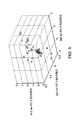
- FIG. 4 illustrates the scores of three principal components extracted from the decomposition of intrinsic fluorescence spectra at 337, 397 and 412 nm excitation used to distinguish high-grade dysplasia (diamonds) from non-dysplastic and low-grade dysplasia (squares) Barrett's esophagus (BE) sites.
- decomposition was performed in the 460 to 520 nm region of the intrinsic fluorescence spectra, as this is the wavelength range within which spectral differences are most pronounced.
- principal components were extracted from the intrinsic fluorescence spectra between 600 and 650 nm.
- PC 1 indicates the first principal component
- PC 3 the third.
- FIG. 5 illustrates the scores of three principal components extracted from the decomposition of the entire intrinsic fluorescence spectra at 337, 397 and 412 nm excitation used to distinguish dysplastic (low- and high-grade; diamonds) from non-dysplastic (squares) Barrett's esophagus (BE) sites.
- PC 2 indicates the second principal component.
- FIG. 6 is a reflectance spectrum of a non-dysplastic Barrett's esophagus site.
- the solid line represents the measured data and the dashed line represents the projected properties of the tissue based on known scattering and absorption properties of the tissue.
- FIG. 7 illustrates graphically the slopes and intercepts of wavelength dependent tissue reduced scattering coefficient, ⁇ s ′( ⁇ ), for non-dysplastic (squares), low-grade (diamonds) and high-grade (triangles) dysplastic Barrett's esophagus (BE) sites.
- ⁇ s ′( ⁇ ) wavelength dependent tissue reduced scattering coefficient
- BE Barrett's esophagus
- FIG. 8 illustrates the total number of nuclei per mm 2 plotted as a function of percentage of enlarged nuclei (diameter>10 ⁇ m), as determined from the light-scattering model analysis.
- Non-dysplastic Barrett's are represented by squares
- low-grade dysplasia are represented by circles
- high-grade dysplasia are represented by triangles.
- FIGS. 9A-9D illustrate graphically the measured and intrinsic fluorescence spectra of normal squamous epithelium (solid line), benign biopsied sites (dashed lines) and high-grade SILs (dotted lines).
- Curves A and B show differences in the lineshape and intensity observed at 337 nm excitation.
- Curves C and D show intensity differences observed in the intrinsic fluorescence excited at 358 nm.
- FIG. 10 illustrates relative NADH fluorescence levels as a function of relative collagen fluorescence levels plotted for normal squamous epithelium (squares), benign biopsies (circles) and HSILs (triangles). The three arrows point to the benign biopsies that were classified as “mature squamous epithelium.” The remaining benign biopsies were classified as “squamous metaplasia.”
- FIG. 11A shows reduced scattering coefficient at 400 nm for benign biopsied and high-grade dysplatic sites.
- FIG. 11B shows nuclear crowding or total number of nuclei per mm 2 plotted as a function of the standard derivation of the nuclear size population for a particular site.
- the line is determined by logistic regression.
- FIG. 12 illustrates a process sequence in accordance with a preferred embodiment of the present invention.
- FIG. 13 illustrates a flow chart of a preferred embodiment for measuring a structure in a layer of tissue in accordance with the present invention.
- FIG. 14 illustrates the details of the step of calibration referred to in FIG. 13 in accordance with a preferred embodiment of the present invention.
- FIG. 15 is a flow chart of a preferred embodiment of the present invention detailing the analysis of the diffuse reflectance spectrum (DRS) in accordance with the present invention.
- DRS diffuse reflectance spectrum
- FIG. 16 is a flow chart detailing the analysis of the intrinsic fluorescence spectrum (IFS) in accordance with a preferred embodiment of the present invention.
- IFS intrinsic fluorescence spectrum
- FIGS. 17A and 17B illustrate a flow chart detailing the analysis of the light scattering spectrum (LSS) in accordance with a preferred embodiment of the present invention.
- LSS light scattering spectrum
- FIG. 18 illustrates the tri-modal system analysis in accordance with a preferred embodiment of the present invention.
- FIG. 19 illustrates an alternate preferred embodiment of a tri-modal system analysis in accordance with the present invention.
- the excication light source of this fast-EEM system can include a nitrogen laser 12 emitting at 377 nm (Laser Science, Inc., Franklin, Mass.; Model: VSL-337MD) pumping 10 dye cuvettes precisely mounted on a rapidly rotating wheel 16 .
- eleven different excitation wavelengths are obtained between 337 and 620 nm and coupled using optical system 18 into the delivery fiber of a 1 mm diameter optical fiber probe 20 .
- white light 350-700 nm
- Xe flash lamp 14 Perkin Elmer Optoelectronics, Salem, Mass.
- a XeCI excimer laser emitting at 308 nm can be used.
- the probe is composed of six collection fibers 28 surrounding the central light delivelly fiber 26 , and it is covered with a protective, transparent optical shield at the distal end 22 as shown in FIG. 1 .
- the probe is inserted into the accessory channel of the endoscope and brought into gentle contact with the tissue, thus providing a fixed delivery-collection geometry.
- the reflected and fluorescence light is collected by the probe and coupled to a spectrograph 30 and detector 22 .
- a second synchronized wheel 36 is used to block the fluorescence excitation wavelength.
- the average of three sets of spectra from each site is used for analysis using a data processing system 34 .
- the probe is removed and a slight demarcation remains on the tissue for 30 to 60 seconds as a result of the probe contact.
- This endoscopically apparent marker is used as a guide for taking a biopsy from the same site at which spectra were acquired.
- the biopsy specimen is interpreted and classified by a gastrointestinal pathologist. If a dysplastic lesion is suspected, the specimen is reviewed and the diagnosis confirmed by a second gastrointestinal pathologist, in accordance with the standard of care. Data has been analyzed from 26 non-dysplastic Barrett's esophagus sites (9 patients), 7 low-grade (4 patients) and 7 high-grade (5 patients) dysplastic sites.
- Fluorescence spectra at eleven different excitation wavelengths, reflectance spectra and light scattering spectra are obtained.
- Each type of spectrum can be analyzed in a manner that provides information about biochemical and morphological changes that occur during dysplastic transformation. Fluorescence spectroscopy can provide valuable information about changes that take place in tissue biochemistry during the development of dysplasia.
- the measured tissue fluorescence spectra can be distorted significantly by unrelated scattering and absorption events. To remove these distortions, the fluorescence spectra are analyzed in combination with information from the corresponding reflectance spectra.
- Principal component analysis and logistic regression is employed to determine the correlation between spectral features of the intrinsic fluorescence and histopathological diagnosis.
- “leave-one-out” cross-validation can be used. Specifically, the principal components of the intrinsic fluorescence spectra that described the spectral features that change during the progression of dysplasia are selected.
- the corresponding scores can be used to determine the ability of the system to distinguish (a) high-grade dysplasia from low-grade dysplastic and non-dysplastic Barrett's esophagus (BE), and (b) dysplastic (low- and high-grade) from non-dysplastic Barrett's esophagus (BE).
- BE non-dysplastic Barrett's esophagus
- the following procedure can be performed.
- the scores from a particular site are eliminated, and logistic regression is used to form a decision surface that classifies the remaining sites in a manner that optimizes agreement with the histopathological classification.
- the resulting decision surface may be then used to classify the excluded site. This process can be repeated for each of the sites.
- This method provides use of a relatively small data set to validate the performance of a decision surface without bias.
- the decision surface varied minimally during this procedure, indicating the reliability of the technique.
- Sensitivity and specificity values can be determined by comparing the spectroscopic classification with histopathology.
- Statistical analysis may be performed using Matlab statistic software (The Math Works, Inc., Natick, Mass.) in accordance with a preferred embodiment.
- the measured reflectance spectra can be analyzed using a representation based on diffusion theory which expresses the reflected light as a function of the absorption ( ⁇ a ( ⁇ )) and reduced scattering ( ⁇ s ′( ⁇ )) coefficients of tissue.
- This analysis provides information about the architecture and morphology of mainly the connective tissue, i.e. the lamina intestinal and the submucosa, as the collected light originates within 500-700 ⁇ m from the tissue surface.
- the diagnostic value of the resulting tissue scattering coefficient values can be determined by correlating the results of logistic regression and cross-validation with histopathological classification, as in the intrinsic fluorescence case.
- a small fraction (2-5%) of the detected reflected light originates from light collected by the probe after being scattered only once by the tissue.
- This method described generally herein as light scattering spectroscopy is described in greater detail in U.S. Pat. No. 6,091,984, issued on Jul. 18, 2000, the entire contents of which is incorporated herein by reference. Additional methods for measuring tissue structure are described in International Application No. PCT/US98/21450, filed on Oct. 9, 1998, now W099/18845, published on Apr. 22, 1999, designating the United States, the entire contents of which is also incorporated herein by reference.
- the Intensity of this singly-scattered light contains a component which is periodic in inverse wavelength, the magnitude and frequency of which depends on the number and size of the nuclei in the epithelial cell layer.
- This periodic signal is analyzed to determine the number and size of epithelial cell nuclei corresponding to a particular site. Logistic regression and cross-validation are then used to compare the spectroscopic classification with that of histopathology.
- the posterior probability threshold for separating high-grade dysplasia from non-high-grade dysplasia sites is set to 0.3 in one preferred embodiment.
- results from all three spectroscopic techniques are combined to determine whether the number of correctly classified sites can be improved. Specifically, a site is assigned a classification that is consistent with results from at least two of the three analysis methods, and this classification is compared to histopathology.
- FIG. 2A shows a typical fluorescence spectrum excited with 337 nm light from a non-dysplastic Barrett's esophagus (BE) site (solid line).
- BE Barrett's esophagus
- sites with high-grade dysplasia can be differentiated from low-grade and non-dysplastic sites with high sensitivity and specificity (Table 1). Additionally, dysplastic and non-dysplastic epithelia can be distinguished with very high sensitivity and specificity.
- Table 1 illustrates the accuracy of spectroscopic classification of non-dysplastic (NDB), low-grade (LGD) and high-grade dysplastic (HGD) tissue in Barrett's esophagus.
- Reflectance spectra can be analyzed using a mathematical representation to obtain detailed information about the scattering and absorption properties of the bulk tissue.
- a typical reflectance spectrum with the corresponding fit obtained using this representation is shown in FIG. 6 .
- the reflectance spectra are further processed in a manner that allows extraction and analysis of the backscattered light from the epithelial cell nuclei.
- the results of this analysis are displayed in FIG. 8 .
- the ordinate of FIG. 8 represents the number of nuclei per square mm, indicative of the degree of nuclear crowding, and the abscissa represents the percentage of enlarged nuclei, defined as nuclei having diameter greater than 10 microns.
- the non-dysplastic samples are concentrated in the lower left-hand corner, indicating cell nuclei that are small and free of crowding.
- the data points move to the upper right, indicating nuclear enlargement and crowding, in agreement with the findings of histopathology. This technique is superior in terms of separating the dysplastic (low- and high-grade) from the non-dysplastic BE sites (Table 1).
- the ability to characterize dysplastic and non-dysplastic tissue in BE is improved by combining the information provided by each one of the spectroscopic techniques, obtained simultaneously with our system.
- a spectroscopic classification is consistent with at least two of the three analysis methods, high-grade dysplasia is identified with perfect sensitivity and specificity, and dysplastic tissue is distinguished from non-dysplastic tissue with perfect specificity, while maintaining very high sensitivity (Table 1).
- Spectroscopic techniques use information contained in light signals to assess the state of biological tissue.
- Optical fiber technology allows spectroscopic methods to be applied as a diagnostic tool for a wide range of tissues that are accessible endoscopically.
- the use of spectroscopy as described herein can be used for improving the physician's ability to detect pre-cancerous (dysplastic) and early cancerous lesions in many organs, such as the oral cavity, the cervix, the lung, the breast and the gastrointestinal tract.
- specific information can be acquired about tissue biochemical, architectural and morphological features. Microscopic changes in these features that occur during the progression of dysplasia may be detectable spectroscopically before the manifestation of macroscopic changes that are visible endoscopically.
- spectroscopic techniques are non-invasive, allowing study of the tissue in its native state, free of artifacts introduced by cutting and chemically processing the tissue.
- spectroscopic signals can be analyzed in real-time, thus guiding the physician to biopsy areas that are likely to yield significant pathology or possibly allowing her to make an immediate decision on the type of intervention that is required for successful treatment of the patient.
- the spectroscopic signals carried by light can be used as objective guides for assessing a particular tissue site, especially in areas in which the intra- and inter-observer agreement on the classification of disease is not very good.
- the targets of fluorescence spectroscopy include tissue biochemicals such as NADH, FAD, collagen, elastin and porphyrins. Exogenous or exogenously-induced chromophores that have been shown to accumulate preferentially in the diseased areas can also be used. The detection of high-grade dysplasia using tissue autofluorescence excited at 410 nm have been conducted. The difference between the measured integrated intensity-normalized fluorescence and the mean normalized fluorescence from normal esophageal tissue was used for the diagnostic procedure.
- the main spectral features that resulted in good differentiation between high-grade dysplastic and non-dysplastic tissues were the presence of decreased fluorescence around 470-480 nm and increased fluorescence in the red region of the spectrum for the high-grade dysplastic tissues. However, this process does not classify correctly sites with low-grade or focal high-grade dysplasia.
- EEMs can be used to identify the excitation wavelengths at which tissue classification is optimized. Additionally, EEMs can assist in identifying the origins of the measured fluorescence signals in a more reliable manner. Nevertheless, as shown in FIGS. 2 and 3 , these measured EEMs can be distorted significantly by tissue scattering and absorption. To eliminate artifacts introduced by changes in scattering or absorption, rather than by tissue biochemistry, corresponding reflectance spectra can be used which are affected in a similar manner by scattering and absorption events.
- tissue fluorescence excited at 337 nm broadens and shifts to longer wavelengths in a very consistent manner as the tissue progresses from non-dysplastic to low-grade to high-grade dysplasia (FIG. 3 ). These spectral changes are consistent with the presence of increased NADH levels in dysplastic tissue.
- Our findings at 397 and 412 nm excitation are attributed to endogenous porphyrins.
- the spectra corresponding to the high-grade dysplasia sites appear slightly distorted around 470 nm, even after correcting for the effects of scattering and absorption. This suggests that this difference arises as a result of biochemical changes rather than absorption changes.
- the scores of one of the first three principal components which described over 99% of the variance observed in the intrinsic fluorescence spectra excited at 337, 397 and 412 nm can be used.
- Reflectance spectroscopy can be used not only to remove the distortions observed in the measured tissue fluorescence spectra, but also to provide very detailed and potentially useful information about morphological and architectural features of the tissue.
- the observed tissue reflectance spectra can be used in terms of two parameters that are determined by tissue scattering and absorption. For example, changes in the concentration or the oxygen saturation of hemoglobin, the main absorber in the visible spectrum for this tissue type, result in concomitant changes in the absorption coefficient of tissue. Alterations in the architecture of the connective tissue collagen fibers, one of the main contributors of tissue scattering, lead to a modified tissue scattering coefficient.
- Light scattering spectroscopy is a procedure that can be used to obtain information about the number and the size of nuclei of the epithelial cell layer.
- Epithelial cell nuclei are the primary targets of reflected light that is singly scattered before it is collected by a probe. The intensity and oscillations of this singly-backscattered light are characteristic of the number and size of its target nuclei. This technique is used to characterize pre-cancerous and early cancerous changes in the colon, the oral cavity, the bladder and Barrett's esophagus (BE).
- Results of this technique are included for the data set of a preferred embodiment to illustrate the information that can be acquired and combined with fluorescence and reflectance spectroscopies.
- Light scattering spectroscopy outperforms the other two methods in terms of its ability to separate the dysplastic from non-dysplastic BE sites.
- the combination of all three techniques provides an extremely sensitive and specific tool for the detection of dysplasia in Barrett's esophagus (BE).
- BE Barrett's esophagus
- agreement with histopathology is achieved in terms of separating high-grade dysplasia from non-dysplastic and low-grade dysplasia sites.
- all sites are classified correctly as dysplastic or non-dysplastic, with the exception of one site.
- the observed improvement is expected, since each one of the techniques examines different features of tissue biochemistry and morphology that can be altered during the development of dysplastic changes.
- Pancreatic carcinoma is one of the first five leading causes of death in Western countries and has a very poor prognosis after initial diagnosis. This is due to late presentation of symptoms and the fact that only about 5-20% of all patients are eligible for resection. Adenocarcinoma of the pancreas arises from the ductal epithelium. In the precancerous stages ductal epithelial cells undergo morphological changes similar to those of Barrett's esophagus (BE) including increasing nuclear size and crowding. Several clinical conditions exist which may allow detection of precancerous or early cancerous changes.
- BE Barrett's esophagus
- pancreatitis due to focal obstruction of the pancreatic duct
- IPMT intraductal papillary mucinous tumor of the pancreas
- Detection of dysplasia in this setting is currently based on the detection of stricturing or dilation of the pancreatic duct, and exfoliative cytology.
- these methods cannot reliably distinguish dysplasia and inflammation and have an overall poor sensitivity of 44-70%.
- the present invention includes methods for performing tri-modal spectroscopic analysis within the main pancreatic duct using the technique of endoscopic retrograde cholangio-pancreatography (ERCP), and obtained spectra from the pancreatic duct epithelium.
- ERCP endoscopic retrograde cholangio-pancreatography
- the present invention can be directed to the use of tri-modal spectroscopy for the detection of dysplasia an invasive cancer in the pancreatic duct.
- the present invention can evaluate ex vivo or in vivo specimens of patients to detect pancreatic cancer of dysplasia.
- Spectra can be collected from the MPD at 1 cm intervals within 3 hours of resection and before formalin fixation.
- Tri-modal spectral analysis can be performed to evaluate for components which accurately discriminate histological categories.
- These spectral algorithms can be applied in vivo to patients undergoing endoscopic retrograde cholangiopancreatography for evaluation of the 3 conditions mentioned above.
- Spectra can be collected in vivo at 1 cm intervals along with entire length of the duct.
- Spectral signals can be compared to histology among the patients whose clinical condition dictates surgical removal of the pancreas.
- Anatomic locations of the spectral signals can be matched according to the distance from MPD orifice.
- the present invention can be employed for the detection of pre-invasive disease of the cervix either alone or at the time of colposcopy following an abnormal Pap smear.
- the present invention can also be used for a non-invasive method of monitoring the progress of medical therapies for pre-invasive disease.
- the Pap smear is a screening tool for cervical tissue abnormalities. Abnormal Pap smears are routinely followed up by colposcopy. This process uses a low-power binocular microscope for the identification of abnormal cervical epithelium, which is subsequently biopsied and examined histopathologically. It is estimated that in expert hands the sensitivity and specificity of colposcopy are 94% and 48%, respectively.
- a preferred embodiment of the present invention include spectroscopic techniques to evaluate cervical epithelium in vivo. Tissue fluorescence spectra excited at 337, 380 and 460 nm were acquired during colposcopy from normal and suspicious sites within the ectocervical and endocervical regions. Suspicious sites were biopsied and classified histopathologically. An initial set of spectra was analyzed and statistical methods were developed to optimize the agreement between spectroscopic and histopathological classification. When these methods were used prospectively to classify a second set of data, it was found that squamous intraepithelial lesions (SILs) can be identified spectroscopically with 82% sensitivity and 68% specificity when compared to histopathology.
- SILs squamous intraepithelial lesions
- the present invention improves upon the sensitivity and specificity of spectral analysis of cervical epithelium in a real-time in vivo approach.
- the method employs tri-modal spectroscopy (TMS), the combined use of intrinsic fluorescence spectroscopy (IFS), diffuse reflectance spectroscopy (DRS), and light scattering spectroscopy (LSS).
- TMS tri-modal spectroscopy
- IFS intrinsic fluorescence spectroscopy
- DRS diffuse reflectance spectroscopy
- LSS light scattering spectroscopy
- the method can compare the spectra obtained using tri-modal processing with the histologic diagnosis of the area of epithelium biopsied. This process provides patterns that are predictive of histologic dysplasia in a prospective fashion, thus allowing the clinician to increase the positive histologic lesions of the cervical epithelium prospectively. This can be of immense value in following patients on clinical trials in order to determine the response to medical treatments of cervical dysplasia.
- Spectra can be acquired from the normal squamous ectocervix and suspicious sites within the transformation zone. The latter may be biopsied immediately following spectral acquisition. Data has been collected from 34 patients, 42 normal ectocervical sites (not biopsied), 15 benign biopsied sites (12 classified as squamous metaplasia and 3 as mature squamous epithelium) and 10 high-grade squamous intraepithelial lesions (HSILs).
- HSILs high-grade squamous intraepithelial lesions
- the spectra may be decomposed as a linear combination of the NADH and collagen EEMs extracted from the measurements performed during variceal ligation.
- MMPs matrix metalloproteinases
- Differences in the levels and/or patterns of expression of MMP-2 have been reported between normal squamous epithelium, squamous metaplasia and SILs.
- an increase in the NADH fluorescence is noted for the HSILs compared to that of benign biopsied sites as seen in FIG. 11 A. This increase can be the result of an increase in the number of epithelial cells and/or their metabolic activity ( 52 ).
- HSILs can be distinguished from the benign biopsied sites with a sensitivity of 80% and a specificity of 67%.
- HSILs may be separated from the benign biopsied sites with 90% sensitivity and 67% specificity as seen in FIG. 11 B.
- the three techniques, IFS, DRS and LSS are combined in a preferred embodiment in a manner that classifies a particular site according to the diagnosis that is consistent with at least two of the three methods of analysis.
- This approach leads to a high level of sensitivity and specificity (100% and 80%, respectively) for the detection of HSILs from non-HSIL sites biopsied within the transformation zone when compared to any one of the techniques alone as seen in Table 2.
- IFS Intrinsic Fluorescence Spectroscopy
- Diffuse Reflectance Spectroscopy Diffuse Reflectance Spectroscopy
- LSS Light Scattering Spectroscopy
- TMS Tri-Modal Spectroscopy
- FIG. 12 illustrates a process sequence 100 of a preferred embodiment of the invention in which a region of interest within a lumen of a patient is illuminated 102 and the fluorescent and reflected light is detected 104 .
- the three components 106 , 108 and 110 are used to generate structural and biochemical information 112 .
- This data can be analyzed using discriminant analysis 114 , the components weighted 116 , and a diagnosis performed 118 in real-time.
- These measurements demonstrate the ability of spectroscopic techniques to provide useful information for disease classification in a non-invasive manner. While each of the techniques can be used for detecting dysplasia in Barrett's esophagus, their combination allows the formation of a comprehensive picture of the biochemical and morphological state of tissue.
- decomposition of the intrinsic tissue fluorescence EEMs into EEMs of biochemicals such as NADH and collagen can provide details about tissue biochemistry.
- Reflectance and light scattering spectroscopy yield morphological information related to the connective tissue and the epithelial cell nuclei. As this information is free from artifacts introduced by tissue excision and processing, it can help advance the understanding of the processes that lead to the progression of dysplasia.
- Software for performing data analysis in real-time enables the applicability of these techniques as a guide to biopsy. Methods to image regions of interest using this procedure enables large tissue areas to be studied rapidly.
- An operating environment for the system includes a processing system such as data processing system 34 , with at least one high speed processing unit and a memory system.
- a processing system such as data processing system 34
- at least one high speed processing unit and a memory system.
- the present invention is described with reference to acts and symbolic representations of operations or instructions that are performed by the processing system, unless indicated otherwise. Such acts and operations or instructions are sometimes referred to as being “computer-executed,” or “processing unit executed.”
- the acts and symbolically represented operations or instructions include the manipulation of electrical signals by the processing unit.
- An electrical system with data bits causes a resulting transformation or reduction of the electrical signal representation, and the maintenance of data bits at memory locations in the memory system to thereby reconfigure or otherwise alter the processing unit's operation, as well as other processing of signals.
- the memory locations were data bits are maintained are physical locations that have particular electrical, magnetic, optical, or organic properties corresponding to the data bits.
- the data bits may also be maintained on a computer readable medium including magnetic disks, optical disks, organic disks, and any other volatile or non-volatile mass storage system readable by the processing using.
- the computer readable medium includes cooperating or interconnected computer readable media, which exist exclusively on the processing system or is distributed among multiple interconnected processing systems that may be local or remote to the processing system.
- FIG. 13 is a flowchart of a preferred embodiment of measuring a structure in a layer of tissue by analyzing data in real-time in accordance with the present invention.
- the real-time analysis may be used to diagnose a pre-cancerous condition in, for example, epithelial tissue in the cervix and GI tract.
- the measurement process begins with the step 200 of calibration.
- the diffuse reflectance spectrum (DRS) is then analyzed per step 300 .
- the intrinsic fluorescence spectrum (IFS) is analyzed.
- the next step in the process includes the step 500 of analyzing the light scattering spectrum (LSS).
- the method then proceeds to step 600 , which represents the combined analysis for the previous modes of this tri-modal system analysis.
- fluorescence spectra at a plurality of wavelengths for example, at eleven laser excitation wavelengths between 337 and 620 nm and one white light (350-750 nm) reflectance spectrum is acquired in less than one second.
- Light delivery and collection is mediated through an optical fiber probe.
- the acquired spectra contain information about the uppermost tissue layers, where almost 90% of cancers begin.
- the IFS refers to the recovery of tissue fluorescence spectra that are free of distortions introduced by tissue scattering and absorption. To remove these distortions, the measured fluorescence and reflectance spectra are combined using, for example, a photon-migration-based method.
- the extracted IFS is decomposed to provide quantitative information on the biochemical tissue composition and the changes that take place in pre-cancerous tissues.
- the measured reflectance spectra consists mainly of photons that are scattered many times before being detected.
- a model that is based on diffusion theory is used to describe the diffusely reflected light and thus to extract information about the absorption and the reduced scattering coefficients of tissue.
- the reduced scattering coefficient depends mainly on the morphology of connective tissue, which provides structural support for the epithelium, the most superficial tissue layer.
- the TMS analysis may be performed in 2-8 seconds from time of data collection. This TMS analysis can provide a real-time guide to biopsies.
- FIG. 14 illustrates the details of the step 200 of calibration in accordance with a preferred embodiment of the present invention.
- the process of calibration to remove variations in spectra associated with, for example, instruments begins with step 210 of calibrating any data files in the system that are used to diagnose diseased tissue.
- the next step in the calibration sequence includes the step 220 of calibrating the system with a reflectance standard. This standard may include but is not limited to, for example, the use of a white light standard such as Spectralon calibration of reflectance.
- the calibration method 200 includes the step 230 of calibrating the system with a fluorescence standard.
- the fluorescence standard may be, for example, and without limitation, Rhodamine.
- the next step in the calibration method 200 includes the step 240 of removing the background fluorescence such as, for example, by subtracting any background fluorescence.
- the spectral response of the instrument is calibrated per step 250 .
- FIG. 15 is a flow chart of a preferred embodiment of the present invention detailing the analysis of the DRS.
- the process includes the generation of a look-up table which describes the Mie scattering parameter for different diameters of scattered particles per step 310 .
- the creation of the look-up table describing Mie scattering reduces processing time for Mie scattering calculations for each iteration.
- Step 320 includes dividing the reduced scattering coefficient values ⁇ s ′( ⁇ ) into a plurality of different regions. In a preferred embodiment, there are five regions. This division into different regions accounts for the lack of one optimum or universal fit for all ⁇ s ′( ⁇ ) values, as there are different values for ⁇ s ′( ⁇ ) in different regions.
- the ⁇ s ′( ⁇ ) space is separated into a plurality of regions and a minimization process is performed for each region separately.
- the calibrated reflectance spectrum is analyzed using a model, for example, the model described in previously referenced U.S. Pat. No. 6,091,984 and referenced herein after as the Zonios model in each of the different regions.
- the Zonios model accounts for one layer tissue architecture and represents an iterative process to get a best fit between the results of the model and values from an actual recording.
- a preferred embodiment of the present invention includes accounting for a multi-layer tissue architecture such as a two-layer model.
- light is provided onto a surface of a tissue by a light source.
- the light reflected back from the tissue is a function of wavelength and depends on several parameters such as the absorption coefficient of tissue, the reduced scattering coefficient and the probe.
- the reflectance spectrum R( ⁇ ) F( ⁇ a ( ⁇ ), ⁇ s ′( ⁇ ),r c ) wherein ⁇ a ( ⁇ ) is the absorption coefficient of tissue indicative of hemoglobin or betacarotine or more generally a biochemical that absorbs light, ⁇ s ′( ⁇ ) is the reduced scattering coefficient and r c is the effective light collection area of the probe.
- a best fit is then selected between the model and data per step 340 . The best fit is determined by optimizing values of parameters, for example, a scaling constant, particle size and density and hemoglobin concentration and saturation within a predetermined space.
- a correction is calculated and applied for high hemoglobin concentrations and/or absorption if necessary.
- the fit is not good when the concentration of hemoglobin is high.
- the hemoglobin concentration fit exceeds a predetermined threshold value, the fit is only performed for a region, for example, 450-700 nm. Further, a correction may also be calculated and applied for a poor fit as indicated by the shape of the reflectance curve if necessary per step 360 .
- FIG. 16 is a flow chart detailing the analysis of the IFS in accordance with a preferred embodiment of the present invention.
- biological tissues are turbid media in which the predominance of light scattering unavoidably entangles the effects of absorption and fluorescence.
- the interplay of absorption and scattering can substantially distort the measured fluorescence. Therefore, it is important to extract the intrinsic fluorescence from the measured tissue fluorescence.
- the intrinsic fluorescence is defined as the fluorescence that is due only to fluorophores, without interference from the absorbers and scatterers that are present in the tissue.
- the greatest distortion of fluorescence spectra occurs for emission near 420 nm where hemoglobin absorbs strongly. Removal of such distortion is particularly important for ultraviolet excitation wavelengths, which have been shown to contain important diagnostic information.
- step 410 the input data is analyzed using the information from the reflectance fit provided by the Zonios model.
- the inputs include ⁇ ′ s ( ⁇ ), ⁇ a ( ⁇ ) fluorescence F( ⁇ ), and R obs ; wherein R obs is the observed white light reflectance.
- Data is also analyzed using a method based on photon-migration theory referred to hereinafter as the Zhang model.
- the Zhang model is an improvement over the model described in U.S. Pat. No. 5,452,723 entitled “Calibrated Spectrographic Imaging” issued on Sep. 26, 1995, the entire contents of which being incorporated herein by reference.
- the Zhang model is described in an article entitled, “Turbidity-free fluorescence spectroscopy of biological tissue,” by Zhang et al., and published in Optics Letter, Vol. 25, No. 19, Oct. 1, 2000, the entire contents of which being incorporated herein by reference.
- the Zhang model is based on the fact that when reflectance and fluorescence are collected from a given tissue site with the same probe, absorption and scattering distort fluorescence and reflectance spectra similarly, because the diffusely reflected and the fluorescence photons traverse similar paths.
- I x denotes the excitation intensity
- ⁇ xm is the fluorescence quantum yield, in which the subscripts x and m denote evaluation of the corresponding quantities at excitation frequency ⁇ x and emission frequency ⁇ m
- ⁇ ni denotes the fluorescence escape probability for the fluorescence rates
- ⁇ is ⁇ s /( ⁇ a + ⁇ s ) and represents the fraction of photons not absorbed during each interaction event and can be considered the photon weight reduction factor for that event.
- brackets has the meaning of fluorescence photons per excitation photon.
- the photon transverses the first i absorption—scattering nodes and experiences propagation loss, its weight becomes ⁇ x i for each incident excitation photon.
- ⁇ fx /( ⁇ ax + ⁇ sx ) photons are absorbed by the fluorophore.
- ⁇ xm fluorescence photons are generated.
- the propagation loss of the fluorescence photons is accounted for by the factor ⁇ m n ⁇ i ⁇ l .
- Equation 4 holds for a probe of arbitrary delivery-collection geometry, as long as the fluorescence and reflectance are measured with the same probe: note that R o is a function of wavelength. Note that Equation 4 can be used to extract an intrinsic fluorescence excitation-emission matrix (EEM) from an experimentally measured fluorescence EEM. When the medium contains multiple fluorophores, one need only replace every occurrence of ⁇ fx ⁇ xm in equations 1 and 2 with a summation of this quantity over all fluorophores. Doing so results in the same expression as in equation 4.
- EEM intrinsic fluorescence excitation-emission matrix
- step 420 the IFS (R obs fit( ⁇ )) is obtained.
- a biochemical decomposition process may then be performed per step 430 .
- the biochemical decomposition process may be a linear decomposition process based on the IFS and provides a quantitative measure of the spectra. Further a principal component analysis (PCA) analysis may also be performed per step 440 .
- PCA principal component analysis
- FIGS. 17A and 17B are flow charts detailing the analysis of LSS in accordance with a preferred embodiment of the present invention.
- the analysis begins with step 510 wherein the observed white light reflectance curve (R obs ( ⁇ )) and the reflectance fit curve (R fit ( ⁇ )) are smoothed.
- a moving window average smoothing process may be used in a preferred embodiment.
- the LSS spectrum is wavelength dependent.
- the data is then transferred from wavelength ( ⁇ ) space to wavenumber ( ⁇ ) space in step 530 .
- a first test is then performed per step 540 wherein it is determined if there is a well defined peak in the spectrum in Fourier space. Only if there is a well defined peak a Fourier transform is estimated, otherwise a reliable diagnosis cannot be provided.
- step 550 a Fourier transform (FT) is generated. However, if there is a lack of a well defined peak, the process returns to step 310 . The process then enters step 560 which functions as a second test or checkpoint wherein the hemoglobin absorption features are accounted for. The second order polynomial is fit to the R obs ( ⁇ ) data. Steps 510 , 520 and 530 are then repeated if the fit is not accurate. A Fourier transform is then performed to compare the resulting FT's from step 550 and step 560 . It is then determined if the FT's are the same. If they are different, the process proceeds to step 570 . If the FT's are the same however, another hemoglobin or a two layer model, without limitation, is used such as described with respect to the DRS analysis in FIG. 15 .
- FT Fourier transform
- a third test or checkpoint is performed by analyzing the FT from step 550 and ascertaining the presence or absence of a dominant peak. If no dominant peak is present then the process returns to step 310 in the DRS analysis as the hemoglobin absorption parameter is then probably an artifact. If a dominant peak exists then spectral components other than the dominant peak are removed such as by filtering high frequency components per step 574 .
- the data is converted to an appropriate scale per step 576 by, for example, multiplying by an overall scaling constant to normalize the result.
- the nuclear size distribution is determined in step 580 .
- the determination may include the determination of the percent enlargement of the nuclear size per step 582 .
- the determination of the nuclear size distribution may also include the determination of the width of the nuclear size distribution per step 584 . Further, determination of the nuclear size distribution may include the determination of the number density of the nuclei per step 586 .
- FIG. 18 illustrates a preferred embodiment of a tri-modal system (TMS) analysis in accordance with the present invention.
- the TMS analysis includes step 610 of at least one of taking values of diagnostic parameters output from the DRS analysis ( ⁇ a ( ⁇ ) and ⁇ s ′( ⁇ )), values of diagnostic parameters output from IFS analysis, for example, but not limited to, bio-chemical analysis per step 620 and values of diagnostic parameters output from the LSS analysis, for example, without limitation, nuclear size distribution, size variation, standard deviation and number density per step 630 and performing a discrimination analysis in step 640 .
- the discrimination analysis may include, for example, but without limitation, Fisher discrimination analysis which includes assigning weights to each parameter, summing the weighted parameters and comparing the sum to a threshold value.
- the results of the TMS analysis are provided as a TMS diagnosis in step 650 .
- the TMS diagnosis includes the assessment about the presence or absence of non-dysplastic tissue, high-grade dysplastic tissue and dysplastic tissue.
- FIG. 19 is an alternate preferred embodiment of a TMS analysis in accordance with the present invention.
- a diagnosis from each modality of spectroscopy is provided individually and compared before providing an overall diagnosis.
- the values of the diagnostic parameters output from the DRS analysis, for example, ⁇ s ′( ⁇ ), in step 662 are used to provide the diagnosis from the DRS analysis per step 664 .
- the slope and intercept data values are determined and compared to threshold values to determine the tissue characteristics.
- the values of the diagnostic parameters output from IFS analysis in step 670 are used to provide a diagnosis commensurate with the IFS analysis in step 672 .
- the contribution of, but not limited to, NADH and/or collagen, and/or porphyrin, to the IFS spectrum are compared to threshold values to provide the diagnosis from the IFS analysis.
- the values of the diagnostic parameters output from the LSS analysis in step 680 are in turn used to provide a diagnosis from the LSS analysis in step 682 .
- the diagnosis from the LSS analysis includes, for example, the comparison of at least one nuclear size characteristic, such as, nuclear size expressed as a percent enlargement, number density, size variation, width of distribution versus crowding to threshold values. Depending upon the tissue and its location, a combination of two or more nuclear size characteristics may be compared to corresponding threshold values to provide a diagnosis from the LSS analysis.
- the three diagnoses are compared in step 690 .
- An overall diagnosis is provided from this comparison in step 692 .
- the overall diagnosis may be derived from two similar individual diagnoses, for example, a similar diagnosis from the IFS and LSS analyses. Different permutations and combinations of similar diagnoses may be used in preferred embodiments.
- a computer usable medium can include a readable memory device, such as a hard drive device, CD-ROM, a DVD-ROM, or a computer diskette, having computer readable program code segments stored thereon.
- the computer readable medium can also include a communications or transmission medium, such as, a bus or a communication link, either optical, wired or wireless having program code segments carried thereon as digital or analog data signals.
Landscapes
- Health & Medical Sciences (AREA)
- Life Sciences & Earth Sciences (AREA)
- Physics & Mathematics (AREA)
- General Health & Medical Sciences (AREA)
- Pathology (AREA)
- Biomedical Technology (AREA)
- Molecular Biology (AREA)
- Engineering & Computer Science (AREA)
- Surgery (AREA)
- Public Health (AREA)
- Medical Informatics (AREA)
- Biophysics (AREA)
- Heart & Thoracic Surgery (AREA)
- Animal Behavior & Ethology (AREA)
- Veterinary Medicine (AREA)
- General Physics & Mathematics (AREA)
- Chemical & Material Sciences (AREA)
- Analytical Chemistry (AREA)
- Biochemistry (AREA)
- Immunology (AREA)
- Nuclear Medicine, Radiotherapy & Molecular Imaging (AREA)
- Spectroscopy & Molecular Physics (AREA)
- Gynecology & Obstetrics (AREA)
- Reproductive Health (AREA)
- Optics & Photonics (AREA)
- Investigating, Analyzing Materials By Fluorescence Or Luminescence (AREA)
- Investigating Or Analysing Materials By Optical Means (AREA)
Abstract
Description
| TABLE 1 | |||
| HGD vs. (LGD and | (LGD and | ||
| NDB) | HGD) vs NDB | ||
| Sensitivity | Specificity | | Specificity | ||
| Intrinsic |
| 100% | 98% | 71% | 92% | |
| fluorescence (IF) | ||||
| Reflectance (R) | 86% | 100% | 79% | 88% |
| Light Scattering (LS) | 100% | 91% | 93% | 96% |
| Combination of IF, |
100% | 100% | 93% | 100% |
| and LS | ||||
| TABLE 2 | ||
| benign biopsies vs HSIL | ||
| Sensitivity | Specificity | ||
| Intrinsic Fluorescence Spectroscopy (IFS) | 80% | 67% |
| Diffuse Reflectance Spectroscopy (DRS) | 70% | 60% |
| Light Scattering Spectroscopy (LSS) | 90% | 67% |
| Tri-Modal Spectroscopy (TMS) | 100% | 80% |
where Ix denotes the excitation intensity, φxm is the fluorescence quantum yield, in which the subscripts x and m denote evaluation of the corresponding quantities at excitation frequency νx and emission frequency νm, ρni denotes the fluorescence escape probability for the fluorescence rates, α is μs/(μa+μs) and represents the fraction of photons not absorbed during each interaction event and can be considered the photon weight reduction factor for that event. Consequently, the expression in brackets has the meaning of fluorescence photons per excitation photon. After the photon transverses the first i absorption—scattering nodes and experiences propagation loss, its weight becomes αx i for each incident excitation photon. For each excitation photon arriving at the (i+1)th node, μfx/(μax+μsx) photons are absorbed by the fluorophore. For each photon absorbed by the fluorophore, φxm fluorescence photons are generated. The propagation loss of the fluorescence photons is accounted for by the factor αm n−i−l.
f xm=(I x /hν x)μfx lφ xm hν m (2)
where R is the reflectance of the homogeneous medium of scatterers and absorbers. The result can be combined with
wherein Ro is the reflectance in the absence of absorption and ∈=exp β−1 where β is a parameter independent of the number of steps n but dependent on the scattering properties of the medium, and μs are the scattering coefficients of the tissue.
Claims (32)
Priority Applications (2)
| Application Number | Priority Date | Filing Date | Title |
|---|---|---|---|
| US10/052,583 US6912412B2 (en) | 2001-01-19 | 2002-01-18 | System and methods of fluorescence, reflectance and light scattering spectroscopy for measuring tissue characteristics |
| US11/166,798 US8380268B2 (en) | 2001-01-19 | 2005-06-24 | System and methods of fluorescence, reflectance and light scattering spectroscopy for measuring tissue characteristics |
Applications Claiming Priority (2)
| Application Number | Priority Date | Filing Date | Title |
|---|---|---|---|
| US09/766,879 US6697652B2 (en) | 2001-01-19 | 2001-01-19 | Fluorescence, reflectance and light scattering spectroscopy for measuring tissue |
| US10/052,583 US6912412B2 (en) | 2001-01-19 | 2002-01-18 | System and methods of fluorescence, reflectance and light scattering spectroscopy for measuring tissue characteristics |
Related Parent Applications (1)
| Application Number | Title | Priority Date | Filing Date |
|---|---|---|---|
| US09/766,879 Continuation-In-Part US6697652B2 (en) | 2001-01-19 | 2001-01-19 | Fluorescence, reflectance and light scattering spectroscopy for measuring tissue |
Related Child Applications (1)
| Application Number | Title | Priority Date | Filing Date |
|---|---|---|---|
| US11/166,798 Continuation US8380268B2 (en) | 2001-01-19 | 2005-06-24 | System and methods of fluorescence, reflectance and light scattering spectroscopy for measuring tissue characteristics |
Publications (2)
| Publication Number | Publication Date |
|---|---|
| US20030013973A1 US20030013973A1 (en) | 2003-01-16 |
| US6912412B2 true US6912412B2 (en) | 2005-06-28 |
Family
ID=25077802
Family Applications (3)
| Application Number | Title | Priority Date | Filing Date |
|---|---|---|---|
| US09/766,879 Expired - Lifetime US6697652B2 (en) | 2001-01-19 | 2001-01-19 | Fluorescence, reflectance and light scattering spectroscopy for measuring tissue |
| US10/052,583 Expired - Lifetime US6912412B2 (en) | 2001-01-19 | 2002-01-18 | System and methods of fluorescence, reflectance and light scattering spectroscopy for measuring tissue characteristics |
| US11/166,798 Expired - Fee Related US8380268B2 (en) | 2001-01-19 | 2005-06-24 | System and methods of fluorescence, reflectance and light scattering spectroscopy for measuring tissue characteristics |
Family Applications Before (1)
| Application Number | Title | Priority Date | Filing Date |
|---|---|---|---|
| US09/766,879 Expired - Lifetime US6697652B2 (en) | 2001-01-19 | 2001-01-19 | Fluorescence, reflectance and light scattering spectroscopy for measuring tissue |
Family Applications After (1)
| Application Number | Title | Priority Date | Filing Date |
|---|---|---|---|
| US11/166,798 Expired - Fee Related US8380268B2 (en) | 2001-01-19 | 2005-06-24 | System and methods of fluorescence, reflectance and light scattering spectroscopy for measuring tissue characteristics |
Country Status (6)
| Country | Link |
|---|---|
| US (3) | US6697652B2 (en) |
| EP (2) | EP1942335A1 (en) |
| JP (1) | JP2004518124A (en) |
| AU (1) | AU2002253891A1 (en) |
| CA (1) | CA2431769A1 (en) |
| WO (1) | WO2002057757A2 (en) |
Cited By (35)
| Publication number | Priority date | Publication date | Assignee | Title |
|---|---|---|---|---|
| US20050288593A1 (en) * | 2001-01-19 | 2005-12-29 | Massachusetts Institute Of Technology | System and methods of fluorescence, reflectance and light scattering spectroscopy for measuring tissue characteristics |
| US20050288594A1 (en) * | 2002-11-29 | 2005-12-29 | Shlomo Lewkowicz | Methods, device and system for in vivo diagnosis |
| US20060155178A1 (en) * | 2004-03-26 | 2006-07-13 | Vadim Backman | Multi-dimensional elastic light scattering |
| US20060241349A1 (en) * | 2002-12-12 | 2006-10-26 | Kazuhiro Gono | Imaging apparatus |
| US20060241499A1 (en) * | 2005-02-24 | 2006-10-26 | Irion Klaus M | Multifunctional fluorescence diagnosis system |
| US20070019199A1 (en) * | 2005-07-25 | 2007-01-25 | The Wisconsin Alumni Research Foundation | Methods, systems, and computer program products for optimization of probes for spectroscopic measurement in turbid media |
| US20070129615A1 (en) * | 2005-10-27 | 2007-06-07 | Northwestern University | Apparatus for recognizing abnormal tissue using the detection of early increase in microvascular blood content |
| US20070173736A1 (en) * | 2005-10-07 | 2007-07-26 | Femspec Llc | Apparatus and methods for endometrial biopsies |
| US20070179368A1 (en) * | 2005-10-27 | 2007-08-02 | Northwestern University | Method of recognizing abnormal tissue using the detection of early increase in microvascular blood content |
| US20070232932A1 (en) * | 2006-03-17 | 2007-10-04 | Duke University | Monte Carlo based model of fluorescence in turbid media and methods and systems for using same to determine intrinsic fluorescence of turbid media |
| WO2007136880A3 (en) * | 2006-05-19 | 2008-04-24 | Evanston Northwestern Healthca | Method & apparatus for recognizing abnormal tissue using the detection of early increase in microvascular blood content |
| US20080270091A1 (en) * | 2007-02-23 | 2008-10-30 | Nirmala Ramanujam | Scaling method for fast monte carlo simulation of diffuse reflectance spectra from multi-layered turbid media and methods and systems for using same to determine optical properties of multi-layered turbid medium from measured diffuse reflectance |
| US20090015826A1 (en) * | 2006-03-30 | 2009-01-15 | Duke University | Optical assay system for intraoperative assessment of tumor margins |
| US20090204009A1 (en) * | 2008-02-07 | 2009-08-13 | Los Alamos National Security | Medical device system and related methods for diagnosing abnormal medical conditions based on in-vivo optical properties of tissue |
| US20090203977A1 (en) * | 2005-10-27 | 2009-08-13 | Vadim Backman | Method of screening for cancer using parameters obtained by the detection of early increase in microvascular blood content |
| US20100198080A1 (en) * | 2007-05-21 | 2010-08-05 | Board Of Regents, The University Of Texas System | Optical Methods to Intraoperatively Detect Positive Prostate and Kidney Cancer Margins |
| US20100249607A1 (en) * | 2008-09-26 | 2010-09-30 | Massachusetts Institute Of Technology | Quantitative spectroscopic imaging |
| US20100262020A1 (en) * | 2009-01-08 | 2010-10-14 | American Biooptics Llc | Probe apparatus for recognizing abnormal tissue |
| US20110059016A1 (en) * | 2007-09-27 | 2011-03-10 | Nirmala Ramanujam | Optical assay system with a multi-probe imaging array |
| US20110101775A1 (en) * | 2008-07-03 | 2011-05-05 | Fujitsu Technology Solutions Intellectual Property Gmbh | Circuit arrangement with a power input and an operating method for controlling a power input circuit |
| US20110105865A1 (en) * | 2008-04-24 | 2011-05-05 | Duke University | Diffuse reflectance spectroscopy device for quantifying tissue absorption and scattering |
| US20110112435A1 (en) * | 2007-09-28 | 2011-05-12 | Nirmala Ramanujam | Systems and methods for spectral analysis of a tissue mass using an instrument, an optical probe, and a monte carlo or a diffusion algorithm |
| US20120209092A1 (en) * | 2011-02-16 | 2012-08-16 | Southern Taiwan University | Non-invasive apparatus and method for measuring human metabolic conditions |
| US8659755B2 (en) * | 2004-06-30 | 2014-02-25 | Applied Biosystems, Llc | Luminescence reference standards |
| US9042967B2 (en) | 2008-05-20 | 2015-05-26 | University Health Network | Device and method for wound imaging and monitoring |
| US9091637B2 (en) | 2009-12-04 | 2015-07-28 | Duke University | Smart fiber optic sensors systems and methods for quantitative optical spectroscopy |
| US9314164B2 (en) | 2005-10-27 | 2016-04-19 | Northwestern University | Method of using the detection of early increase in microvascular blood content to distinguish between adenomatous and hyperplastic polyps |
| US9404870B2 (en) | 2013-03-15 | 2016-08-02 | Cedars-Sinai Medical Center | Time-resolved laser-induced fluorescence spectroscopy systems and uses thereof |
| US9885834B2 (en) | 2009-01-08 | 2018-02-06 | Northwestern University | Probe apparatus for measuring depth-limited properties with low-coherence enhanced backscattering |
| US10438356B2 (en) | 2014-07-24 | 2019-10-08 | University Health Network | Collection and analysis of data for diagnostic purposes |
| US10582895B2 (en) | 2011-01-20 | 2020-03-10 | Tel Hashomer Medical Research Infrastructure And Services Ltd. | Methods and devices for providing information useful in the diagnosis of abnormalities of the gastrointestinal tract |
| US10656089B2 (en) | 2016-04-01 | 2020-05-19 | Black Light Surgical, Inc. | Systems, devices, and methods for time-resolved fluorescent spectroscopy |
| US11172826B2 (en) | 2016-03-08 | 2021-11-16 | Enspectra Health, Inc. | Non-invasive detection of skin disease |
| US11633149B2 (en) | 2017-04-28 | 2023-04-25 | Enspectra Health, Inc. | Systems and methods for imaging and measurement of sarcomeres |
| US11911138B2 (en) | 2011-02-09 | 2024-02-27 | Tel Hashomer Medical Research Infrastructure And Services Ltd. | Methods for in vivo fundus monochromotic angiography imaging blood-containing tissue by utilizing two wavelength from 400nm to 620 nm and 620 nm to 800 nm |
Families Citing this family (207)
| Publication number | Priority date | Publication date | Assignee | Title |
|---|---|---|---|---|
| US20020091324A1 (en) * | 1998-04-06 | 2002-07-11 | Nikiforos Kollias | Non-invasive tissue glucose level monitoring |
| US7899518B2 (en) | 1998-04-06 | 2011-03-01 | Masimo Laboratories, Inc. | Non-invasive tissue glucose level monitoring |
| US6721582B2 (en) | 1999-04-06 | 2004-04-13 | Argose, Inc. | Non-invasive tissue glucose level monitoring |
| US6728560B2 (en) | 1998-04-06 | 2004-04-27 | The General Hospital Corporation | Non-invasive tissue glucose level monitoring |
| JP4241038B2 (en) | 2000-10-30 | 2009-03-18 | ザ ジェネラル ホスピタル コーポレーション | Optical method and system for tissue analysis |
| US9295391B1 (en) | 2000-11-10 | 2016-03-29 | The General Hospital Corporation | Spectrally encoded miniature endoscopic imaging probe |
| US7139598B2 (en) * | 2002-04-04 | 2006-11-21 | Veralight, Inc. | Determination of a measure of a glycation end-product or disease state using tissue fluorescence |
| US20070276199A1 (en) * | 2002-04-04 | 2007-11-29 | Ediger Marwood N | Determination of a Measure of a Glycation End-Product or Disease State Using Tissue Fluorescence |
| US7139602B2 (en) * | 2001-04-26 | 2006-11-21 | University Of Louisville Research Foundation, Inc. | Shape analysis of surfaces |
| WO2002088705A2 (en) | 2001-05-01 | 2002-11-07 | The General Hospital Corporation | Method and apparatus for determination of atherosclerotic plaque type by measurement of tissue optical properties |
| US6577754B2 (en) * | 2001-05-29 | 2003-06-10 | Tissueinformatics, Inc. | Robust stain detection and quantification for histological specimens based on a physical model for stain absorption |
| US7428434B2 (en) * | 2001-07-27 | 2008-09-23 | The Regents Of The Univeristy Of California | Quantitative broadband absorption and scattering spectroscopy in turbid media by combined frequency-domain and steady state methodologies |
| JP5259033B2 (en) * | 2001-08-03 | 2013-08-07 | オリンパス株式会社 | Endoscope system |
| US7355716B2 (en) * | 2002-01-24 | 2008-04-08 | The General Hospital Corporation | Apparatus and method for ranging and noise reduction of low coherence interferometry LCI and optical coherence tomography OCT signals by parallel detection of spectral bands |
| US20040010204A1 (en) * | 2002-03-28 | 2004-01-15 | Pearl Technology Holdings, Llc | Electronic/fiberoptic tissue differentiation instrumentation |
| US8131332B2 (en) * | 2002-04-04 | 2012-03-06 | Veralight, Inc. | Determination of a measure of a glycation end-product or disease state using tissue fluorescence of various sites |
| US8140147B2 (en) * | 2002-04-04 | 2012-03-20 | Veralight, Inc. | Determination of a measure of a glycation end-product or disease state using a flexible probe to determine tissue fluorescence of various sites |
| US20120078075A1 (en) * | 2002-04-04 | 2012-03-29 | Maynard John D | Determination of a measure of a glycation end-product or disease state using tissue fluorescence in combination with one or more other tests |
| US20050119548A1 (en) * | 2002-07-05 | 2005-06-02 | Vanderbilt University | Method and apparatus for optical spectroscopic detection of cell and tissue death |
| US7433540B1 (en) * | 2002-10-25 | 2008-10-07 | Adobe Systems Incorporated | Decomposing natural image sequences |
| JP2006509573A (en) * | 2002-12-13 | 2006-03-23 | イエトメド リミテッド | Optical inspection method and apparatus particularly useful for distinguishing between tumor and normal tissue in real time during surgery |
| US7053783B2 (en) | 2002-12-18 | 2006-05-30 | Biovigilant Systems, Inc. | Pathogen detector system and method |
| US8054468B2 (en) * | 2003-01-24 | 2011-11-08 | The General Hospital Corporation | Apparatus and method for ranging and noise reduction of low coherence interferometry LCI and optical coherence tomography OCT signals by parallel detection of spectral bands |
| WO2004066824A2 (en) * | 2003-01-24 | 2004-08-12 | The General Hospital Corporation | System and method for identifying tissue using low-coherence interferometry |
| WO2004088361A2 (en) | 2003-03-31 | 2004-10-14 | The General Hospital Corporation | Speckle reduction in optical coherence tomography by path length encoded angular compounding |
| DE10305599A1 (en) * | 2003-02-11 | 2004-08-26 | Richard Wolf Gmbh | Tissue diagnostic imaging device |
| US7102758B2 (en) | 2003-05-06 | 2006-09-05 | Duke University | Fourier domain low-coherence interferometry for light scattering spectroscopy apparatus and method |
| AU2004252482B2 (en) | 2003-06-06 | 2011-05-26 | The General Hospital Corporation | Process and apparatus for a wavelength tuning source |
| US20050008253A1 (en) * | 2003-07-10 | 2005-01-13 | Joseph Rozgonyi | Method and apparatus for eliminating irrelevant luminescent signals |
| ES2280053T3 (en) * | 2003-09-23 | 2007-09-01 | Stichting Voor De Technische Wetenschappen | PROCESS AND DEVICE FOR THE RETRODISPERSION SPECTROSCOPY. |
| US20080077005A1 (en) * | 2004-08-12 | 2008-03-27 | Piron Cameron A | System and Method for Multimodality Breast Imaging |
| US7379769B2 (en) * | 2003-09-30 | 2008-05-27 | Sunnybrook Health Sciences Center | Hybrid imaging method to monitor medical device delivery and patient support for use in the method |
| US7908690B2 (en) * | 2003-09-30 | 2011-03-22 | Sentinelle Medical, Inc. | Supine patient support for medical imaging |
| US7970452B2 (en) * | 2003-09-30 | 2011-06-28 | Hologic, Inc. | Open architecture imaging apparatus and coil system for magnetic resonance imaging |
| EP1685366B1 (en) | 2003-10-27 | 2011-06-15 | The General Hospital Corporation | Method and apparatus for performing optical imaging using frequency-domain interferometry |
| US8326404B2 (en) * | 2003-11-28 | 2012-12-04 | British Columbia Cancer Agency Branch | Multimodal detection of tissue abnormalities based on raman and background fluorescence spectroscopy |
| US8018598B2 (en) * | 2004-05-29 | 2011-09-13 | The General Hospital Corporation | Process, system and software arrangement for a chromatic dispersion compensation using reflective layers in optical coherence tomography (OCT) imaging |
| EP2085116A1 (en) * | 2004-06-30 | 2009-08-05 | University of Rochester | Photodynamic therapy with spatially resolved dual spectroscopic monitoring |
| AU2005270037B2 (en) * | 2004-07-02 | 2012-02-09 | The General Hospital Corporation | Endoscopic imaging probe comprising dual clad fibre |
| JP4871868B2 (en) | 2004-07-30 | 2012-02-08 | バイオヴィジラント システムズ インコーポレイテッド | Pathogen and particulate detection system and detection method |
| EP1782020B1 (en) | 2004-08-06 | 2012-10-03 | The General Hospital Corporation | Process, system and software arrangement for determining at least one location in a sample using an optical coherence tomography |
| WO2006024014A2 (en) * | 2004-08-24 | 2006-03-02 | The General Hospital Corporation | Process, system and software arrangement for measuring a mechanical strain and elastic properties of a sample |
| JP5324095B2 (en) * | 2004-08-24 | 2013-10-23 | ザ ジェネラル ホスピタル コーポレイション | Method and apparatus for imaging blood vessel segments |
| US7365859B2 (en) | 2004-09-10 | 2008-04-29 | The General Hospital Corporation | System and method for optical coherence imaging |
| KR101257100B1 (en) | 2004-09-29 | 2013-04-22 | 더 제너럴 하스피탈 코포레이션 | System and Method for Optical Coherence Imaging |
| EP2278267A3 (en) * | 2004-11-24 | 2011-06-29 | The General Hospital Corporation | Common-Path Interferometer for Endoscopic OCT |
| US8922781B2 (en) | 2004-11-29 | 2014-12-30 | The General Hospital Corporation | Arrangements, devices, endoscopes, catheters and methods for performing optical imaging by simultaneously illuminating and detecting multiple points on a sample |
| US7854705B2 (en) * | 2004-12-16 | 2010-12-21 | Olga Pawluczyk | Ex vivo verification of biopsy tissue samples |
| JP4668624B2 (en) * | 2005-01-07 | 2011-04-13 | オリンパス株式会社 | Image processing device for esophageal mucosa |
| US7428048B1 (en) | 2004-12-30 | 2008-09-23 | Spectral Molecular Imaging Inc. | Imaging elastic scattering spectroscopy |
| KR20080013919A (en) * | 2005-04-22 | 2008-02-13 | 더 제너럴 하스피탈 코포레이션 | Apparatus, Systems and Methods for Providing Spectral Domain Polarization Sensitive Optical Coherence Tomography |
| ES2337497T3 (en) | 2005-04-28 | 2010-04-26 | The General Hospital Corporation | EVALUATION OF CHARACTERISTICS OF THE IMAGE OF AN ANATOMICAL STRUCTURE IN IMAGES OF TOMOGRAPHY OF OPTICAL COHERENCE. |
| US8406858B2 (en) | 2005-04-29 | 2013-03-26 | The Regents Of The University Of Colorado, A Body Corporate | Multi-excitation diagnostic system and methods for classification of tissue |
| EP1889037A2 (en) * | 2005-06-01 | 2008-02-20 | The General Hospital Corporation | Apparatus, method and system for performing phase-resolved optical frequency domain imaging |
| JP5112312B2 (en) | 2005-07-15 | 2013-01-09 | バイオヴィジラント システムズ インコーポレイテッド | Pathogen and particulate detection system and detection method |
| US20070167836A1 (en) * | 2005-07-25 | 2007-07-19 | Massachusetts Institute Of Technology | Multi modal spectroscopy |
| US20070167835A1 (en) * | 2005-07-25 | 2007-07-19 | Massachusetts Institute Of Technology | Tri modal spectroscopic imaging |
| US20120302892A1 (en) * | 2005-07-25 | 2012-11-29 | Niyom Lue | Portable optical fiber probe-based spectroscopic scanner for rapid cancer diagnosis |
| ATE484727T1 (en) | 2005-08-09 | 2010-10-15 | Gen Hospital Corp | DEVICE AND METHOD FOR PERFORMING POLARIZATION-BASED QUADRATURE DEMODULATION IN OPTICAL COHERENCE TOMOGRAPHY |
| WO2007022220A2 (en) * | 2005-08-16 | 2007-02-22 | The General Hospital Corporation | Arrangements and methods for imaging in vessels |
| US7872759B2 (en) * | 2005-09-29 | 2011-01-18 | The General Hospital Corporation | Arrangements and methods for providing multimodality microscopic imaging of one or more biological structures |
| US8537366B2 (en) | 2005-10-11 | 2013-09-17 | Duke University | Systems and methods for endoscopic angle-resolved low coherence interferometry |
| PT2444783E (en) | 2005-10-11 | 2015-06-17 | Univ Duke | Systems and method for fiber-based endoscopic angle-resolved low coherence interferometry |
| US7889348B2 (en) * | 2005-10-14 | 2011-02-15 | The General Hospital Corporation | Arrangements and methods for facilitating photoluminescence imaging |
| JP5680826B2 (en) * | 2006-01-10 | 2015-03-04 | ザ ジェネラル ホスピタル コーポレイション | Data generation system using endoscopic technology for encoding one or more spectra |
| JP2009523574A (en) * | 2006-01-18 | 2009-06-25 | ザ ジェネラル ホスピタル コーポレイション | System and method for generating data using one or more endoscopic microscopy methods |
| US8145018B2 (en) | 2006-01-19 | 2012-03-27 | The General Hospital Corporation | Apparatus for obtaining information for a structure using spectrally-encoded endoscopy techniques and methods for producing one or more optical arrangements |
| ES2847854T3 (en) | 2006-01-19 | 2021-08-04 | Massachusetts Gen Hospital | Balloon Imaging Catheter |
| WO2007084933A2 (en) * | 2006-01-20 | 2007-07-26 | The General Hospital Corporation | Systems and processes for providing endogenous molecular imaging with mid-infared light |
| JP2009524066A (en) * | 2006-01-20 | 2009-06-25 | ザ ジェネラル ホスピタル コーポレイション | Optical coherence tomography systems, configurations, and processes that provide speckle reduction using wavefront modulation |
| JP5680829B2 (en) | 2006-02-01 | 2015-03-04 | ザ ジェネラル ホスピタル コーポレイション | A device that irradiates a sample with multiple electromagnetic radiations |
| US10426548B2 (en) | 2006-02-01 | 2019-10-01 | The General Hosppital Corporation | Methods and systems for providing electromagnetic radiation to at least one portion of a sample using conformal laser therapy procedures |
| JP5519152B2 (en) * | 2006-02-08 | 2014-06-11 | ザ ジェネラル ホスピタル コーポレイション | Device for acquiring information about anatomical samples using optical microscopy |
| EP2982929A1 (en) | 2006-02-24 | 2016-02-10 | The General Hospital Corporation | Methods and systems for performing angle-resolved fourier-domain optical coherence tomography |
| US20070282404A1 (en) * | 2006-04-10 | 2007-12-06 | University Of Rochester | Side-firing linear optic array for interstitial optical therapy and monitoring using compact helical geometry |
| FR2900043B1 (en) * | 2006-04-24 | 2008-07-04 | Commissariat Energie Atomique | METHOD FOR OPTICALLY IMAGING FLUORESCENCE OF BIOLOGICAL TISSUES, IN PARTICULAR TO DELIMIT REGIONS OF INTEREST FROM TISSUES TO BE ANALYZED BY TOMOGRAPHY |
| WO2007133961A2 (en) | 2006-05-10 | 2007-11-22 | The General Hospital Corporation | Processes, arrangements and systems for providing frequency domain imaging of a sample |
| WO2008011580A2 (en) * | 2006-07-21 | 2008-01-24 | Oncoscope, Inc. | Protective probe tip, particularly for use on a fiber-optic probe used in an endoscopic application |
| WO2008016927A2 (en) * | 2006-08-01 | 2008-02-07 | The General Hospital Corporation | Systems and methods for receiving and/or analyzing information associated with electro-magnetic radiation |
| CN101589301B (en) * | 2006-08-25 | 2012-11-07 | 通用医疗公司 | Apparatus and methods for enhancing optical coherence tomography imaging using volumetric filtering techniques |
| US8180419B2 (en) * | 2006-09-27 | 2012-05-15 | Nellcor Puritan Bennett Llc | Tissue hydration estimation by spectral absorption bandwidth measurement |
| WO2008049118A2 (en) * | 2006-10-19 | 2008-04-24 | The General Hospital Corporation | Apparatus and method for obtaining and providing imaging information associated with at least one portion of a sample and effecting such portion(s) |
| WO2008050712A1 (en) * | 2006-10-23 | 2008-05-02 | Olympus Corporation | Spectral endoscope and its wavelength calibration method |
| DE102006050886B4 (en) * | 2006-10-27 | 2016-12-22 | Siemens Healthcare Gmbh | Medical instrument and device for generating tissue sections |
| DE102006054556A1 (en) * | 2006-11-20 | 2008-05-21 | Zimmer Medizinsysteme Gmbh | Apparatus and method for non-invasive, optical detection of chemical and physical blood values and body constituents |
| WO2008066911A2 (en) * | 2006-11-30 | 2008-06-05 | Newton Laboratories, Inc. | Spectroscopically enhanced imaging |
| US8498695B2 (en) * | 2006-12-22 | 2013-07-30 | Novadaq Technologies Inc. | Imaging system with a single color image sensor for simultaneous fluorescence and color video endoscopy |
| EP2662673A3 (en) * | 2007-01-19 | 2014-06-18 | The General Hospital Corporation | Rotating disk reflection for fast wavelength scanning of dispersed broadband light |
| JP5507258B2 (en) * | 2007-01-19 | 2014-05-28 | ザ ジェネラル ホスピタル コーポレイション | Apparatus and method for controlling measurement depth in optical frequency domain imaging |
| EP2131741A4 (en) * | 2007-03-02 | 2010-05-05 | Wisconsin Alumni Res Found | USE OF ENDOGENOUS FLUORESCENCE TO IDENTIFY INVASIVE METASTATIC BREAST TUMER CELLS |
| US8374685B2 (en) * | 2007-03-12 | 2013-02-12 | Koninklijke Philips Electronics N.V. | System, computer-readable medium, method, and use for combined epithelial early cancer diagnosis and staging |
| US20080234567A1 (en) * | 2007-03-19 | 2008-09-25 | The General Hospital Corporation | Apparatus and method for providing a noninvasive diagnosis of internal bleeding |
| JP5558839B2 (en) * | 2007-03-23 | 2014-07-23 | ザ ジェネラル ホスピタル コーポレイション | Method, arrangement and apparatus for utilizing a wavelength swept laser using angular scanning and dispersion procedures |
| US10534129B2 (en) | 2007-03-30 | 2020-01-14 | The General Hospital Corporation | System and method providing intracoronary laser speckle imaging for the detection of vulnerable plaque |
| WO2008131082A1 (en) * | 2007-04-17 | 2008-10-30 | The General Hospital Corporation | Apparatus and methods for measuring vibrations using spectrally-encoded endoscopy techniques |
| US20100331927A1 (en) * | 2007-05-02 | 2010-12-30 | Cottrell William J | Feedback-controlled method for delivering photodynamic therapy and related instrumentation |
| WO2009018456A2 (en) * | 2007-07-31 | 2009-02-05 | The General Hospital Corporation | Systems and methods for providing beam scan patterns for high speed doppler optical frequency domain imaging |
| US20090062662A1 (en) | 2007-08-27 | 2009-03-05 | Remicalm, Llc | Optical spectroscopic device for the identification of cervical cancer |
| WO2009036418A1 (en) * | 2007-09-13 | 2009-03-19 | Duke University | Apparatuses, systems, and methods for low-coherence interferometry (lci) |
| WO2009036453A1 (en) * | 2007-09-15 | 2009-03-19 | The General Hospital Corporation | Apparatus, computer-accessible medium and method for measuring chemical and/or molecular compositions of coronary atherosclerotic plaques in anatomical structures |
| EP2042082A1 (en) * | 2007-09-28 | 2009-04-01 | Stichting Voor De Technische Wetenschappen | Method and apparatus for measuring fluorescence |
| JP2011500173A (en) * | 2007-10-12 | 2011-01-06 | ザ ジェネラル ホスピタル コーポレイション | System and process for optical imaging of luminal anatomical structures |
| US20090099460A1 (en) * | 2007-10-16 | 2009-04-16 | Remicalm Llc | Method and device for the optical spectroscopic identification of cervical cancer |
| WO2009059034A1 (en) * | 2007-10-30 | 2009-05-07 | The General Hospital Corporation | System and method for cladding mode detection |
| EP2057941A1 (en) * | 2007-11-07 | 2009-05-13 | Koninklijke Philips Electronics N.V. | Method and device for imaging an interior of an optically turbid medium |
| US7940047B2 (en) * | 2007-11-23 | 2011-05-10 | Sentinelle Medical, Inc. | Microcontroller system for identifying RF coils in the bore of a magnetic resonance imaging system |
| US8628976B2 (en) * | 2007-12-03 | 2014-01-14 | Azbil BioVigilant, Inc. | Method for the detection of biologic particle contamination |
| US8280496B2 (en) * | 2007-12-13 | 2012-10-02 | Boston Scientific Scimed, Inc. | Extended spectral sensitivity endoscope system and method of using the same |
| WO2009077947A2 (en) * | 2007-12-17 | 2009-06-25 | Koninklijke Philips Electronics N.V. | Method for detecting the presence of inhomogeneities in an interior of a turbid medium and device for imaging the interior of turbid media |
| WO2009089344A1 (en) * | 2008-01-08 | 2009-07-16 | Oncoscope, Inc. | Systems and methods for tissue examination, diagnostic, treatment, and/or monitoring |
| US20090225324A1 (en) * | 2008-01-17 | 2009-09-10 | The General Hospital Corporation | Apparatus for providing endoscopic high-speed optical coherence tomography |
| JP5245424B2 (en) * | 2008-01-25 | 2013-07-24 | 日本電気株式会社 | Pathological tissue imaging system, pathological tissue imaging method, and pathological tissue imaging program |
| WO2009105537A2 (en) * | 2008-02-19 | 2009-08-27 | Trustees Of Tufts College | Non-invasive optical characterization of biomaterial mineralization |
| GB0804190D0 (en) * | 2008-03-06 | 2008-04-16 | Ge Healthcare Ltd | Ester imaging agents |
| US9173554B2 (en) | 2008-03-18 | 2015-11-03 | Novadaq Technologies, Inc. | Imaging system for combined full-color reflectance and near-infrared imaging |
| US7898656B2 (en) | 2008-04-30 | 2011-03-01 | The General Hospital Corporation | Apparatus and method for cross axis parallel spectroscopy |
| EP2274572A4 (en) | 2008-05-07 | 2013-08-28 | Gen Hospital Corp | System, method and computer-accessible medium for tracking vessel motion during three-dimensional coronary artery microscopy |
| US8107710B2 (en) * | 2008-05-23 | 2012-01-31 | University Of Rochester | Automated placental measurement |
| US8861910B2 (en) * | 2008-06-20 | 2014-10-14 | The General Hospital Corporation | Fused fiber optic coupler arrangement and method for use thereof |
| US9254089B2 (en) | 2008-07-14 | 2016-02-09 | The General Hospital Corporation | Apparatus and methods for facilitating at least partial overlap of dispersed ration on at least one sample |
| WO2010009412A2 (en) | 2008-07-18 | 2010-01-21 | University Of Rochester Medical Center | Low-cost device for c-scan photoacoustic imaging |
| US20100081928A1 (en) * | 2008-09-29 | 2010-04-01 | Searete Llc, A Limited Liability Corporation Of The State Of Delaware | Histological Facilitation systems and methods |
| US20100081927A1 (en) * | 2008-09-29 | 2010-04-01 | Searete Llc, A Limited Liability Corporation Of The State Of Delaware | Histological facilitation systems and methods |
| US20100081926A1 (en) * | 2008-09-29 | 2010-04-01 | Searete Llc, A Limited Liability Corporation Of The State Of Delaware | Histological facilitation systems and methods |
| US20100081916A1 (en) * | 2008-09-29 | 2010-04-01 | Searete Llc, A Limited Liability Corporation Of The State Of Delaware. | Histological facilitation systems and methods |
| US20100081915A1 (en) * | 2008-09-29 | 2010-04-01 | Searete Llc, Alimited Liability Corporation Of The State Of Delaware | Histological facilitation systems and methods |
| US20100081924A1 (en) * | 2008-09-29 | 2010-04-01 | Searete Llc, A Limited Liability Corporation Of The State Of Delaware | Histological facilitation systems and methods |
| US20100087741A1 (en) * | 2008-10-02 | 2010-04-08 | Novadaq Technologies Inc. | Method for identifying malignancies in barrett's esophagus using white light endoscopy |
| US20100298688A1 (en) * | 2008-10-15 | 2010-11-25 | Dogra Vikram S | Photoacoustic imaging using a versatile acoustic lens |
| ES2957932T3 (en) * | 2008-12-10 | 2024-01-30 | Massachusetts Gen Hospital | Systems, apparatus and procedures for extending the image depth range of optical coherence tomography using optical subsampling |
| JP2012515576A (en) * | 2009-01-20 | 2012-07-12 | ザ ジェネラル ホスピタル コーポレイション | Endoscopic biopsy device, system, and method |
| JP2012515930A (en) * | 2009-01-26 | 2012-07-12 | ザ ジェネラル ホスピタル コーポレーション | System, method and computer-accessible medium for providing a wide-field super-resolution microscope |
| WO2010091190A2 (en) * | 2009-02-04 | 2010-08-12 | The General Hospital Corporation | Apparatus and method for utilization of a high-speed optical wavelength tuning source |
| JP5311655B2 (en) * | 2009-05-15 | 2013-10-09 | 独立行政法人農業・食品産業技術総合研究機構 | Component distribution analysis method and component distribution analyzer |
| WO2010148503A1 (en) | 2009-06-23 | 2010-12-29 | Sentinelle Medical Inc. | Variable angle guide holder for a biopsy guide plug |
| BR112012001042A2 (en) * | 2009-07-14 | 2016-11-22 | Gen Hospital Corp | fluid flow measurement equipment and method within anatomical structure. |
| JP5856061B2 (en) * | 2009-10-06 | 2016-02-09 | ザ ジェネラル ホスピタル コーポレイション | Apparatus and method for imaging specific cells using spectrally encoded confocal microscopy |
| KR20120130164A (en) * | 2009-11-17 | 2012-11-29 | 바치야 콘웨이 | Method and apparatus to detect coronary artery calcification or disease |
| WO2011072055A2 (en) * | 2009-12-08 | 2011-06-16 | The General Hospital Corporation | Methods and arrangements for analysis, diagnosis, and treatment monitoring of vocal folds by optical coherence tomography |
| JP2013518256A (en) | 2010-01-22 | 2013-05-20 | デユーク・ユニバーシテイ | Multiple windowing scheme for spectroscopic coherence tomography (OCT) and Fourier domain low coherence interferometry |
| US9823127B2 (en) | 2010-01-22 | 2017-11-21 | Duke University | Systems and methods for deep spectroscopic imaging of biological samples with use of an interferometer and spectrometer |
| SMT202000587T1 (en) | 2010-03-05 | 2021-01-05 | Massachusetts Gen Hospital | Systems which provide microscopic images of at least one anatomical structure at a particular resolution |
| CN102893137B (en) * | 2010-03-17 | 2017-01-11 | 曾海山 | Rapid multispectral imaging method and apparatus for cancer detection and localization and applications |
| US9069130B2 (en) | 2010-05-03 | 2015-06-30 | The General Hospital Corporation | Apparatus, method and system for generating optical radiation from biological gain media |
| US7884933B1 (en) | 2010-05-05 | 2011-02-08 | Revolutionary Business Concepts, Inc. | Apparatus and method for determining analyte concentrations |
| JP5778762B2 (en) | 2010-05-25 | 2015-09-16 | ザ ジェネラル ホスピタル コーポレイション | Apparatus and method for spectral analysis of optical coherence tomography images |
| EP2575597B1 (en) | 2010-05-25 | 2022-05-04 | The General Hospital Corporation | Apparatus for providing optical imaging of structures and compositions |
| US10285568B2 (en) | 2010-06-03 | 2019-05-14 | The General Hospital Corporation | Apparatus and method for devices for imaging structures in or at one or more luminal organs |
| WO2012058381A2 (en) | 2010-10-27 | 2012-05-03 | The General Hospital Corporation | Apparatus, systems and methods for measuring blood pressure within at least one vessel |
| US9913596B2 (en) | 2010-11-25 | 2018-03-13 | Invivo Corporation | Systems and methods for MRI guided trans-orifice and transperineal intervention apparatus with adjustable biopsy needle insertion |
| WO2012096037A1 (en) * | 2011-01-12 | 2012-07-19 | コニカミノルタオプト株式会社 | Method for detecting intermolecular interaction, and kit for use therein |
| WO2012120380A1 (en) | 2011-03-08 | 2012-09-13 | Novadaq Technologies Inc. | Full spectrum led illuminator |
| WO2012128614A1 (en) * | 2011-03-24 | 2012-09-27 | Erasmus University Medical Center Rotterdam | Method to determine the absorption coefficient in turbid media |
| JP5315376B2 (en) * | 2011-04-11 | 2013-10-16 | オリンパス株式会社 | Fence-like blood vessel detection device and method of operating the fence-like blood vessel detection device |
| US20120302862A1 (en) * | 2011-04-29 | 2012-11-29 | The General Hospital Corporation | Methods and arrangements for obtaining information and providing analysis for biological tissues |
| US8780362B2 (en) | 2011-05-19 | 2014-07-15 | Covidien Lp | Methods utilizing triangulation in metrology systems for in-situ surgical applications |
| JP6093761B2 (en) | 2011-06-28 | 2017-03-08 | コーニンクレッカ フィリップス エヌ ヴェKoninklijke Philips N.V. | Apparatus for optical analysis of relevant tissue samples |
| JP2014523536A (en) | 2011-07-19 | 2014-09-11 | ザ ジェネラル ホスピタル コーポレイション | System, method, apparatus and computer-accessible medium for providing polarization mode dispersion compensation in optical coherence tomography |
| WO2013029047A1 (en) | 2011-08-25 | 2013-02-28 | The General Hospital Corporation | Methods, systems, arrangements and computer-accessible medium for providing micro-optical coherence tomography procedures |
| US9341783B2 (en) | 2011-10-18 | 2016-05-17 | The General Hospital Corporation | Apparatus and methods for producing and/or providing recirculating optical delay(s) |
| EP2811890B1 (en) | 2012-02-08 | 2022-12-21 | Biop Medical, Ltd. | Method and apparatus for tissue disease diagnosis |
| EP2833776A4 (en) | 2012-03-30 | 2015-12-09 | Gen Hospital Corp | Imaging system, method and distal attachment for multidirectional field of view endoscopy |
| EP3574835A1 (en) | 2012-05-21 | 2019-12-04 | The Regents of the University of Colorado, a body corporate | Three-dimensional mapping of prostate cancer |
| US11490797B2 (en) | 2012-05-21 | 2022-11-08 | The General Hospital Corporation | Apparatus, device and method for capsule microscopy |
| US9814448B2 (en) | 2012-05-21 | 2017-11-14 | Precision Biopsy, Inc. | Three-dimensional optical imaging and therapy of prostate cancer |
| JP6103824B2 (en) * | 2012-06-04 | 2017-03-29 | オリンパス株式会社 | Fluorescence endoscope device |
| US20150150460A1 (en) * | 2012-06-07 | 2015-06-04 | The Trustees Of Dartmouth College | Methods And Systems For Intraoperative Tumor Margin Assessment In Surgical Cavities And Resected Tissue Specimens |
| US9351671B2 (en) | 2012-07-16 | 2016-05-31 | Timothy Ruchti | Multiplexed pathlength resolved noninvasive analyzer apparatus and method of use thereof |
| US9351672B2 (en) | 2012-07-16 | 2016-05-31 | Timothy Ruchti | Multiplexed pathlength resolved noninvasive analyzer apparatus with stacked filters and method of use thereof |
| US9585604B2 (en) * | 2012-07-16 | 2017-03-07 | Zyomed Corp. | Multiplexed pathlength resolved noninvasive analyzer apparatus with dynamic optical paths and method of use thereof |
| US9766126B2 (en) | 2013-07-12 | 2017-09-19 | Zyomed Corp. | Dynamic radially controlled light input to a noninvasive analyzer apparatus and method of use thereof |
| EP2888616A4 (en) | 2012-08-22 | 2016-04-27 | Gen Hospital Corp | SYSTEM, METHOD AND COMPUTER-ACCESSIBLE MEDIA FOR MANUFACTURING MINIATURE ENDOSCOPES USING SOFT LITHOGRAPHY |
| JP5344073B2 (en) * | 2012-09-14 | 2013-11-20 | 日本電気株式会社 | Pathological tissue imaging system, pathological tissue imaging method, and pathological tissue imaging program |
| JP6560126B2 (en) | 2013-01-28 | 2019-08-14 | ザ ジェネラル ホスピタル コーポレイション | Apparatus and method for providing diffusion spectroscopy superimposed on optical frequency domain imaging |
| WO2014120791A1 (en) | 2013-01-29 | 2014-08-07 | The General Hospital Corporation | Apparatus, systems and methods for providing information regarding the aortic valve |
| US11179028B2 (en) | 2013-02-01 | 2021-11-23 | The General Hospital Corporation | Objective lens arrangement for confocal endomicroscopy |
| EP2973217B1 (en) | 2013-03-15 | 2023-10-25 | Hologic, Inc. | System and method for reviewing and analyzing cytological specimens |
| US10478072B2 (en) | 2013-03-15 | 2019-11-19 | The General Hospital Corporation | Methods and system for characterizing an object |
| JP2014217438A (en) * | 2013-05-01 | 2014-11-20 | 独立行政法人物質・材料研究機構 | Probe detector |
| EP2997354A4 (en) | 2013-05-13 | 2017-01-18 | The General Hospital Corporation | Detecting self-interefering fluorescence phase and amplitude |
| EP3021734B1 (en) | 2013-07-19 | 2020-04-08 | The General Hospital Corporation | Imaging apparatus which utilizes multidirectional field of view endoscopy |
| WO2015010133A1 (en) | 2013-07-19 | 2015-01-22 | The General Hospital Corporation | Determining eye motion by imaging retina. with feedback |
| US9668652B2 (en) | 2013-07-26 | 2017-06-06 | The General Hospital Corporation | System, apparatus and method for utilizing optical dispersion for fourier-domain optical coherence tomography |
| WO2015054684A1 (en) * | 2013-10-11 | 2015-04-16 | The Trustees Of Columbia University In The City Of New York | System, method and computer-accessible medium for characterization of tissue |
| EP2887050A1 (en) * | 2013-12-23 | 2015-06-24 | Hochschule Reutlingen | Method for marker-free demarcation of tissues |
| WO2015105870A1 (en) | 2014-01-08 | 2015-07-16 | The General Hospital Corporation | Method and apparatus for microscopic imaging |
| WO2015116986A2 (en) | 2014-01-31 | 2015-08-06 | The General Hospital Corporation | System and method for facilitating manual and/or automatic volumetric imaging with real-time tension or force feedback using a tethered imaging device |
| US10228556B2 (en) | 2014-04-04 | 2019-03-12 | The General Hospital Corporation | Apparatus and method for controlling propagation and/or transmission of electromagnetic radiation in flexible waveguide(s) |
| EP3136950A1 (en) * | 2014-04-28 | 2017-03-08 | Northwestern University | Devices, methods, and systems of functional optical coherence tomography |
| CN106659868B (en) * | 2014-06-24 | 2019-11-29 | 普罗赛普特生物机器人公司 | Sample of tissue and cancer treatment facilities |
| KR102513779B1 (en) | 2014-07-25 | 2023-03-24 | 더 제너럴 하스피탈 코포레이션 | Apparatus, devices and methods for in vivo imaging and diagnosis |
| WO2016054079A1 (en) | 2014-09-29 | 2016-04-07 | Zyomed Corp. | Systems and methods for blood glucose and other analyte detection and measurement using collision computing |
| EP4442222A3 (en) | 2015-11-13 | 2024-12-18 | Stryker Corporation | Systems and methods for illumination and imaging of a target |
| JP6671653B2 (en) * | 2015-12-11 | 2020-03-25 | 大学共同利用機関法人情報・システム研究機構 | Image processing apparatus, image processing method, and program |
| CA3009419A1 (en) | 2016-01-26 | 2017-08-03 | Novadaq Technologies ULC | Configurable platform |
| CN109068969B (en) | 2016-03-10 | 2021-08-27 | 比奥普-医疗有限公司 | Device for diagnosing tissue |
| US9554738B1 (en) | 2016-03-30 | 2017-01-31 | Zyomed Corp. | Spectroscopic tomography systems and methods for noninvasive detection and measurement of analytes using collision computing |
| USD916294S1 (en) | 2016-04-28 | 2021-04-13 | Stryker European Operations Limited | Illumination and imaging device |
| WO2017214730A1 (en) | 2016-06-14 | 2017-12-21 | Novadaq Technologies Inc. | Methods and systems for adaptive imaging for low light signal enhancement in medical visualization |
| CN110366678B (en) * | 2016-12-27 | 2023-02-17 | 密歇根大学董事会 | Microscope based on laser emission |
| CA3051857A1 (en) * | 2017-01-30 | 2018-08-02 | Medibeacon Inc. | Method for non-invasive monitoring of fluorescent tracer agent with background separation corrections |
| CN115120241A (en) * | 2017-01-30 | 2022-09-30 | 麦迪贝肯有限公司 | A Non-Invasive Monitoring Method Using Diffuse-corrected Fluorescent Tracers |
| EP3580609B1 (en) | 2017-02-10 | 2023-05-24 | Stryker European Operations Limited | Open-field handheld fluorescence imaging systems and methods |
| WO2019030749A1 (en) | 2017-08-06 | 2019-02-14 | Biop - Medical Ltd | Optical probe for cervical examination |
| US10952616B2 (en) | 2018-03-30 | 2021-03-23 | Canon U.S.A., Inc. | Fluorescence imaging apparatus |
| US10743749B2 (en) | 2018-09-14 | 2020-08-18 | Canon U.S.A., Inc. | System and method for detecting optical probe connection |
| US11446055B1 (en) | 2018-10-18 | 2022-09-20 | Lumoptik, Inc. | Light assisted needle placement system and method |
Citations (17)
| Publication number | Priority date | Publication date | Assignee | Title |
|---|---|---|---|---|
| US4768513A (en) | 1986-04-21 | 1988-09-06 | Agency Of Industrial Science And Technology | Method and device for measuring and processing light |
| US4773097A (en) | 1984-05-31 | 1988-09-20 | Omron Tateisi Electronics Co. | Image analyzing apparatus |
| EP0449883A1 (en) | 1988-12-21 | 1991-10-09 | Massachusetts Inst Technology | A method for laser induced fluorescence of tissue. |
| US5201318A (en) | 1989-04-24 | 1993-04-13 | Rava Richard P | Contour mapping of spectral diagnostics |
| US5280788A (en) | 1991-02-26 | 1994-01-25 | Massachusetts Institute Of Technology | Devices and methods for optical diagnosis of tissue |
| US5304173A (en) * | 1985-03-22 | 1994-04-19 | Massachusetts Institute Of Technology | Spectral diagonostic and treatment system |
| US5421337A (en) | 1989-04-14 | 1995-06-06 | Massachusetts Institute Of Technology | Spectral diagnosis of diseased tissue |
| US5452723A (en) | 1992-07-24 | 1995-09-26 | Massachusetts Institute Of Technology | Calibrated spectrographic imaging |
| US5582168A (en) | 1991-07-17 | 1996-12-10 | Georgia Tech Research Corp. | Apparatus and methods for measuring characteristics of biological tissues and similar materials |
| US5697373A (en) | 1995-03-14 | 1997-12-16 | Board Of Regents, The University Of Texas System | Optical method and apparatus for the diagnosis of cervical precancers using raman and fluorescence spectroscopies |
| WO1999018845A1 (en) | 1997-10-10 | 1999-04-22 | Massachusetts Institute Of Technology | Method for measuring tissue morphology |
| US6008889A (en) | 1997-04-16 | 1999-12-28 | Zeng; Haishan | Spectrometer system for diagnosis of skin disease |
| WO2000042912A1 (en) | 1999-01-25 | 2000-07-27 | Massachusetts Institute Of Technology | Polarized light scattering spectroscopy of tissue |
| WO2001034031A1 (en) | 1999-11-05 | 2001-05-17 | Spectrx, Inc. | Multi-modal optical tissue diagnostic system |
| US6321111B1 (en) * | 1995-02-21 | 2001-11-20 | Massachusetts Institute Of Technology | Optical imaging using time gated scattered light |
| JP2002095624A (en) | 2000-09-26 | 2002-04-02 | Fuji Photo Film Co Ltd | Fluorescence endoscope system |
| US20020093563A1 (en) | 1998-04-20 | 2002-07-18 | Xillix Technologies Corp. | Imaging system with automatic gain control for reflectance and fluorescence endoscopy |
Family Cites Families (28)
| Publication number | Priority date | Publication date | Assignee | Title |
|---|---|---|---|---|
| US5199431A (en) * | 1985-03-22 | 1993-04-06 | Massachusetts Institute Of Technology | Optical needle for spectroscopic diagnosis |
| US5441053A (en) * | 1991-05-03 | 1995-08-15 | University Of Kentucky Research Foundation | Apparatus and method for multiple wavelength of tissue |
| US5419312A (en) * | 1993-04-20 | 1995-05-30 | Wildflower Communications, Inc. | Multi-function endoscope apparatus |
| US5421339A (en) * | 1993-05-12 | 1995-06-06 | Board Of Regents, The University Of Texas System | Diagnosis of dysplasia using laser induced fluoroescence |
| US5349954A (en) * | 1993-07-23 | 1994-09-27 | General Electric Company | Tumor tissue characterization apparatus and method |
| WO1995011624A2 (en) * | 1993-10-29 | 1995-05-04 | Feld Michael S | A raman endoscope |
| US5822072A (en) | 1994-09-30 | 1998-10-13 | Lockheed Martin Energy Systems, Inc. | Fiberoptic probe and system for spectral measurements |
| US5833612A (en) * | 1996-02-09 | 1998-11-10 | Esc Medical Systems, Ltd. | Method and apparatus for diagnosis skin lesions |
| US5842995A (en) * | 1996-06-28 | 1998-12-01 | Board Of Regents, The Univerisity Of Texas System | Spectroscopic probe for in vivo measurement of raman signals |
| EP0912169A2 (en) | 1996-07-16 | 1999-05-06 | Gist-Brocades B.V. | $g(b)-LACTAM GRANULES FREE OF ORGANIC SOLVENTS |
| US6135965A (en) * | 1996-12-02 | 2000-10-24 | Board Of Regents, The University Of Texas System | Spectroscopic detection of cervical pre-cancer using radial basis function networks |
| CA2192036A1 (en) | 1996-12-04 | 1998-06-04 | Harvey Lui | Fluorescence scope system for dermatologic diagnosis |
| US6066245A (en) * | 1996-12-27 | 2000-05-23 | Genetic Biosystems, Inc. | Method and apparatus for scanning fluorescently labeled particles |
| AU6604998A (en) | 1997-03-13 | 1998-09-29 | Biomax Technologies, Inc. | Methods and apparatus for detecting the rejection of transplanted tissue |
| US5920399A (en) * | 1997-03-18 | 1999-07-06 | Sandia Corporation | Multispectral imaging method and apparatus |
| US6128525A (en) | 1997-07-29 | 2000-10-03 | Zeng; Haishan | Apparatus and method to monitor photodynamic therapy (PDT) |
| DE69938493T2 (en) | 1998-01-26 | 2009-05-20 | Massachusetts Institute Of Technology, Cambridge | ENDOSCOPE FOR DETECTING FLUORESCENCE IMAGES |
| AU3970799A (en) * | 1998-05-04 | 1999-11-23 | Board Of Regents | Combined fluorescence and reflectance spectroscopy |
| US6059689A (en) * | 1998-06-04 | 2000-05-09 | Honda Giken Kogyo Kabushiki Kaisha | Control system for automatic vehicle transmission |
| US6353226B1 (en) * | 1998-11-23 | 2002-03-05 | Abbott Laboratories | Non-invasive sensor capable of determining optical parameters in a sample having multiple layers |
| US6546272B1 (en) | 1999-06-24 | 2003-04-08 | Mackinnon Nicholas B. | Apparatus for in vivo imaging of the respiratory tract and other internal organs |
| US20040044287A1 (en) * | 2000-03-31 | 2004-03-04 | Wei-Chiang Lin | Identification of human tissue using optical spectroscopy |
| US6377841B1 (en) * | 2000-03-31 | 2002-04-23 | Vanderbilt University | Tumor demarcation using optical spectroscopy |
| US6826424B1 (en) | 2000-12-19 | 2004-11-30 | Haishan Zeng | Methods and apparatus for fluorescence and reflectance imaging and spectroscopy and for contemporaneous measurements of electromagnetic radiation with multiple measuring devices |
| US6697652B2 (en) | 2001-01-19 | 2004-02-24 | Massachusetts Institute Of Technology | Fluorescence, reflectance and light scattering spectroscopy for measuring tissue |
| JP2005515472A (en) * | 2002-01-18 | 2005-05-26 | ニユートン・ラボラトリーズ・インコーポレーテツド | Spectroscopic diagnosis method and system |
| US6903813B2 (en) * | 2002-02-21 | 2005-06-07 | Jjl Technologies Llc | Miniaturized system and method for measuring optical characteristics |
| US7620212B1 (en) * | 2002-08-13 | 2009-11-17 | Lumidigm, Inc. | Electro-optical sensor |
-
2001
- 2001-01-19 US US09/766,879 patent/US6697652B2/en not_active Expired - Lifetime
-
2002
- 2002-01-18 JP JP2002557989A patent/JP2004518124A/en active Pending
- 2002-01-18 CA CA002431769A patent/CA2431769A1/en not_active Abandoned
- 2002-01-18 AU AU2002253891A patent/AU2002253891A1/en not_active Abandoned
- 2002-01-18 EP EP08153977A patent/EP1942335A1/en not_active Withdrawn
- 2002-01-18 US US10/052,583 patent/US6912412B2/en not_active Expired - Lifetime
- 2002-01-18 EP EP02723087A patent/EP1352231A2/en not_active Withdrawn
- 2002-01-18 WO PCT/US2002/003094 patent/WO2002057757A2/en active Application Filing
-
2005
- 2005-06-24 US US11/166,798 patent/US8380268B2/en not_active Expired - Fee Related
Patent Citations (23)
| Publication number | Priority date | Publication date | Assignee | Title |
|---|---|---|---|---|
| US4773097A (en) | 1984-05-31 | 1988-09-20 | Omron Tateisi Electronics Co. | Image analyzing apparatus |
| US5304173A (en) * | 1985-03-22 | 1994-04-19 | Massachusetts Institute Of Technology | Spectral diagonostic and treatment system |
| US5318024A (en) * | 1985-03-22 | 1994-06-07 | Massachusetts Institute Of Technology | Laser endoscope for spectroscopic imaging |
| US4768513A (en) | 1986-04-21 | 1988-09-06 | Agency Of Industrial Science And Technology | Method and device for measuring and processing light |
| EP0449883A1 (en) | 1988-12-21 | 1991-10-09 | Massachusetts Inst Technology | A method for laser induced fluorescence of tissue. |
| US5419323A (en) * | 1988-12-21 | 1995-05-30 | Massachusetts Institute Of Technology | Method for laser induced fluorescence of tissue |
| US5562100A (en) * | 1988-12-21 | 1996-10-08 | Massachusetts Institute Of Technology | Method for laser induced fluorescence of tissue |
| US5421337A (en) | 1989-04-14 | 1995-06-06 | Massachusetts Institute Of Technology | Spectral diagnosis of diseased tissue |
| US5201318A (en) | 1989-04-24 | 1993-04-13 | Rava Richard P | Contour mapping of spectral diagnostics |
| US5345941A (en) * | 1989-04-24 | 1994-09-13 | Massachusetts Institute Of Technology | Contour mapping of spectral diagnostics |
| US5280788A (en) | 1991-02-26 | 1994-01-25 | Massachusetts Institute Of Technology | Devices and methods for optical diagnosis of tissue |
| US5582168A (en) | 1991-07-17 | 1996-12-10 | Georgia Tech Research Corp. | Apparatus and methods for measuring characteristics of biological tissues and similar materials |
| US5452723A (en) | 1992-07-24 | 1995-09-26 | Massachusetts Institute Of Technology | Calibrated spectrographic imaging |
| US6321111B1 (en) * | 1995-02-21 | 2001-11-20 | Massachusetts Institute Of Technology | Optical imaging using time gated scattered light |
| US5697373A (en) | 1995-03-14 | 1997-12-16 | Board Of Regents, The University Of Texas System | Optical method and apparatus for the diagnosis of cervical precancers using raman and fluorescence spectroscopies |
| US6008889A (en) | 1997-04-16 | 1999-12-28 | Zeng; Haishan | Spectrometer system for diagnosis of skin disease |
| WO1999018845A1 (en) | 1997-10-10 | 1999-04-22 | Massachusetts Institute Of Technology | Method for measuring tissue morphology |
| US6091984A (en) | 1997-10-10 | 2000-07-18 | Massachusetts Institute Of Technology | Measuring tissue morphology |
| US20020093563A1 (en) | 1998-04-20 | 2002-07-18 | Xillix Technologies Corp. | Imaging system with automatic gain control for reflectance and fluorescence endoscopy |
| WO2000042912A1 (en) | 1999-01-25 | 2000-07-27 | Massachusetts Institute Of Technology | Polarized light scattering spectroscopy of tissue |
| US6404497B1 (en) * | 1999-01-25 | 2002-06-11 | Massachusetts Institute Of Technology | Polarized light scattering spectroscopy of tissue |
| WO2001034031A1 (en) | 1999-11-05 | 2001-05-17 | Spectrx, Inc. | Multi-modal optical tissue diagnostic system |
| JP2002095624A (en) | 2000-09-26 | 2002-04-02 | Fuji Photo Film Co Ltd | Fluorescence endoscope system |
Non-Patent Citations (12)
Cited By (62)
| Publication number | Priority date | Publication date | Assignee | Title |
|---|---|---|---|---|
| US8380268B2 (en) * | 2001-01-19 | 2013-02-19 | Massachusetts Institute Of Technology | System and methods of fluorescence, reflectance and light scattering spectroscopy for measuring tissue characteristics |
| US20050288593A1 (en) * | 2001-01-19 | 2005-12-29 | Massachusetts Institute Of Technology | System and methods of fluorescence, reflectance and light scattering spectroscopy for measuring tissue characteristics |
| US20050288594A1 (en) * | 2002-11-29 | 2005-12-29 | Shlomo Lewkowicz | Methods, device and system for in vivo diagnosis |
| US20060241349A1 (en) * | 2002-12-12 | 2006-10-26 | Kazuhiro Gono | Imaging apparatus |
| US8000776B2 (en) * | 2002-12-12 | 2011-08-16 | Olympus Corporation | Imaging apparatus |
| US20060155178A1 (en) * | 2004-03-26 | 2006-07-13 | Vadim Backman | Multi-dimensional elastic light scattering |
| US8659755B2 (en) * | 2004-06-30 | 2014-02-25 | Applied Biosystems, Llc | Luminescence reference standards |
| US9585612B2 (en) * | 2005-02-24 | 2017-03-07 | Karl Storz Gmbh & Co. Kg | Multifunctional fluorescence diagnosis system |
| US20060241499A1 (en) * | 2005-02-24 | 2006-10-26 | Irion Klaus M | Multifunctional fluorescence diagnosis system |
| US20070019199A1 (en) * | 2005-07-25 | 2007-01-25 | The Wisconsin Alumni Research Foundation | Methods, systems, and computer program products for optimization of probes for spectroscopic measurement in turbid media |
| US7835786B2 (en) * | 2005-07-25 | 2010-11-16 | Wisconsin Alumni Research Foundation | Methods, systems, and computer program products for optimization of probes for spectroscopic measurement in turbid media |
| US20070173736A1 (en) * | 2005-10-07 | 2007-07-26 | Femspec Llc | Apparatus and methods for endometrial biopsies |
| US20070129615A1 (en) * | 2005-10-27 | 2007-06-07 | Northwestern University | Apparatus for recognizing abnormal tissue using the detection of early increase in microvascular blood content |
| US20090203977A1 (en) * | 2005-10-27 | 2009-08-13 | Vadim Backman | Method of screening for cancer using parameters obtained by the detection of early increase in microvascular blood content |
| US9314164B2 (en) | 2005-10-27 | 2016-04-19 | Northwestern University | Method of using the detection of early increase in microvascular blood content to distinguish between adenomatous and hyperplastic polyps |
| US20070179368A1 (en) * | 2005-10-27 | 2007-08-02 | Northwestern University | Method of recognizing abnormal tissue using the detection of early increase in microvascular blood content |
| US7818154B2 (en) | 2006-03-17 | 2010-10-19 | Duke University | Monte Carlo based model of fluorescence in turbid media and methods and systems for using same to determine intrinsic fluorescence of turbid media |
| US20070232932A1 (en) * | 2006-03-17 | 2007-10-04 | Duke University | Monte Carlo based model of fluorescence in turbid media and methods and systems for using same to determine intrinsic fluorescence of turbid media |
| US20090015826A1 (en) * | 2006-03-30 | 2009-01-15 | Duke University | Optical assay system for intraoperative assessment of tumor margins |
| US7751039B2 (en) * | 2006-03-30 | 2010-07-06 | Duke University | Optical assay system for intraoperative assessment of tumor margins |
| US20100301229A1 (en) * | 2006-03-30 | 2010-12-02 | Nirmala Ramanujam | Optical assay system for intraoperative assessment of tumor margins |
| US7952704B2 (en) | 2006-03-30 | 2011-05-31 | Duke University | Optical assay system for intraoperative assessment of tumor margins |
| WO2007136880A3 (en) * | 2006-05-19 | 2008-04-24 | Evanston Northwestern Healthca | Method & apparatus for recognizing abnormal tissue using the detection of early increase in microvascular blood content |
| US20080270091A1 (en) * | 2007-02-23 | 2008-10-30 | Nirmala Ramanujam | Scaling method for fast monte carlo simulation of diffuse reflectance spectra from multi-layered turbid media and methods and systems for using same to determine optical properties of multi-layered turbid medium from measured diffuse reflectance |
| US8649849B2 (en) * | 2007-05-21 | 2014-02-11 | Board Of Regents, The University Of Texas System | Optical methods to intraoperatively detect positive prostate and kidney cancer margins |
| US20100198080A1 (en) * | 2007-05-21 | 2010-08-05 | Board Of Regents, The University Of Texas System | Optical Methods to Intraoperatively Detect Positive Prostate and Kidney Cancer Margins |
| US20110059016A1 (en) * | 2007-09-27 | 2011-03-10 | Nirmala Ramanujam | Optical assay system with a multi-probe imaging array |
| US20110112435A1 (en) * | 2007-09-28 | 2011-05-12 | Nirmala Ramanujam | Systems and methods for spectral analysis of a tissue mass using an instrument, an optical probe, and a monte carlo or a diffusion algorithm |
| US9820655B2 (en) | 2007-09-28 | 2017-11-21 | Duke University | Systems and methods for spectral analysis of a tissue mass using an instrument, an optical probe, and a Monte Carlo or a diffusion algorithm |
| US20090204009A1 (en) * | 2008-02-07 | 2009-08-13 | Los Alamos National Security | Medical device system and related methods for diagnosing abnormal medical conditions based on in-vivo optical properties of tissue |
| US20110105865A1 (en) * | 2008-04-24 | 2011-05-05 | Duke University | Diffuse reflectance spectroscopy device for quantifying tissue absorption and scattering |
| US11284800B2 (en) | 2008-05-20 | 2022-03-29 | University Health Network | Devices, methods, and systems for fluorescence-based endoscopic imaging and collection of data with optical filters with corresponding discrete spectral bandwidth |
| US9042967B2 (en) | 2008-05-20 | 2015-05-26 | University Health Network | Device and method for wound imaging and monitoring |
| US11154198B2 (en) | 2008-05-20 | 2021-10-26 | University Health Network | Method and system for imaging and collection of data for diagnostic purposes |
| US11375898B2 (en) | 2008-05-20 | 2022-07-05 | University Health Network | Method and system with spectral filtering and thermal mapping for imaging and collection of data for diagnostic purposes from bacteria |
| US8653700B2 (en) | 2008-07-03 | 2014-02-18 | Fujitsu Technology Solutions Intellectual Property Gmbh | Circuit arrangement with a power input and an operating method for controlling a power input circuit |
| US20110101775A1 (en) * | 2008-07-03 | 2011-05-05 | Fujitsu Technology Solutions Intellectual Property Gmbh | Circuit arrangement with a power input and an operating method for controlling a power input circuit |
| US20100249607A1 (en) * | 2008-09-26 | 2010-09-30 | Massachusetts Institute Of Technology | Quantitative spectroscopic imaging |
| US20100262020A1 (en) * | 2009-01-08 | 2010-10-14 | American Biooptics Llc | Probe apparatus for recognizing abnormal tissue |
| US9885834B2 (en) | 2009-01-08 | 2018-02-06 | Northwestern University | Probe apparatus for measuring depth-limited properties with low-coherence enhanced backscattering |
| US10684417B2 (en) | 2009-01-08 | 2020-06-16 | Northwestern University | Probe apparatus for measuring depth-limited properties with low-coherence enhanced backscattering |
| US9091637B2 (en) | 2009-12-04 | 2015-07-28 | Duke University | Smart fiber optic sensors systems and methods for quantitative optical spectroscopy |
| US10582895B2 (en) | 2011-01-20 | 2020-03-10 | Tel Hashomer Medical Research Infrastructure And Services Ltd. | Methods and devices for providing information useful in the diagnosis of abnormalities of the gastrointestinal tract |
| US11911138B2 (en) | 2011-02-09 | 2024-02-27 | Tel Hashomer Medical Research Infrastructure And Services Ltd. | Methods for in vivo fundus monochromotic angiography imaging blood-containing tissue by utilizing two wavelength from 400nm to 620 nm and 620 nm to 800 nm |
| US8855732B2 (en) * | 2011-02-16 | 2014-10-07 | Southern Taiwan University | Non-invasive apparatus and method for measuring human metabolic conditions |
| US20120209092A1 (en) * | 2011-02-16 | 2012-08-16 | Southern Taiwan University | Non-invasive apparatus and method for measuring human metabolic conditions |
| US11428636B2 (en) | 2013-03-15 | 2022-08-30 | Cedars-Sinai Medical Center | Time-resolved laser-induced fluorescence spectroscopy systems and uses thereof |
| US10983060B2 (en) | 2013-03-15 | 2021-04-20 | Cedars-Sinai Medical Center | Time-resolved laser-induced fluorescence spectroscopy systems and uses thereof |
| US10288567B2 (en) | 2013-03-15 | 2019-05-14 | Cedars-Sinai Medical Center | Time-resolved laser-induced fluorescence spectroscopy systems and uses thereof |
| US12152991B2 (en) | 2013-03-15 | 2024-11-26 | Cedars-Sinai Medical Center | Time-resolved laser-induced fluorescence spectroscopy systems and uses thereof |
| US9404870B2 (en) | 2013-03-15 | 2016-08-02 | Cedars-Sinai Medical Center | Time-resolved laser-induced fluorescence spectroscopy systems and uses thereof |
| US11961236B2 (en) | 2014-07-24 | 2024-04-16 | University Health Network | Collection and analysis of data for diagnostic purposes |
| US10438356B2 (en) | 2014-07-24 | 2019-10-08 | University Health Network | Collection and analysis of data for diagnostic purposes |
| US12169935B2 (en) | 2014-07-24 | 2024-12-17 | University Health Network | Systems, devices, and methods for visualization of tissue and collection and analysis of data regarding same |
| US11676276B2 (en) | 2014-07-24 | 2023-06-13 | University Health Network | Collection and analysis of data for diagnostic purposes |
| US11954861B2 (en) | 2014-07-24 | 2024-04-09 | University Health Network | Systems, devices, and methods for visualization of tissue and collection and analysis of data regarding same |
| US11172826B2 (en) | 2016-03-08 | 2021-11-16 | Enspectra Health, Inc. | Non-invasive detection of skin disease |
| US11877826B2 (en) | 2016-03-08 | 2024-01-23 | Enspectra Health, Inc. | Non-invasive detection of skin disease |
| US10656089B2 (en) | 2016-04-01 | 2020-05-19 | Black Light Surgical, Inc. | Systems, devices, and methods for time-resolved fluorescent spectroscopy |
| US12025557B2 (en) | 2016-04-01 | 2024-07-02 | Black Light Surgical, Inc. | Systems, devices, and methods for time-resolved fluorescent spectroscopy |
| US11630061B2 (en) | 2016-04-01 | 2023-04-18 | Black Light Surgical, Inc. | Systems, devices, and methods for time-resolved fluorescent spectroscopy |
| US11633149B2 (en) | 2017-04-28 | 2023-04-25 | Enspectra Health, Inc. | Systems and methods for imaging and measurement of sarcomeres |
Also Published As
| Publication number | Publication date |
|---|---|
| AU2002253891A1 (en) | 2002-07-30 |
| US20020143243A1 (en) | 2002-10-03 |
| US20030013973A1 (en) | 2003-01-16 |
| CA2431769A1 (en) | 2002-07-25 |
| WO2002057757A3 (en) | 2003-02-27 |
| EP1942335A1 (en) | 2008-07-09 |
| US6697652B2 (en) | 2004-02-24 |
| US8380268B2 (en) | 2013-02-19 |
| US20050288593A1 (en) | 2005-12-29 |
| JP2004518124A (en) | 2004-06-17 |
| WO2002057757A2 (en) | 2002-07-25 |
| EP1352231A2 (en) | 2003-10-15 |
Similar Documents
| Publication | Publication Date | Title |
|---|---|---|
| US6912412B2 (en) | System and methods of fluorescence, reflectance and light scattering spectroscopy for measuring tissue characteristics | |
| Georgakoudi et al. | Fluorescence, reflectance, and light-scattering spectroscopy for evaluating dysplasia in patients with Barrett's esophagus | |
| US6975899B2 (en) | Multi-modal optical tissue diagnostic system | |
| US8311607B2 (en) | Fluorescent fiberoptic probe for tissue health discrimination and method of use thereof | |
| CN101137322B (en) | Method and apparatus for measuring cancerous changes from reflectance spectral measurements obtained during endoscopic imaging | |
| Volynskaya et al. | Diagnosing breast cancer using diffuse reflectance spectroscopy and intrinsic fluorescence spectroscopy | |
| Panjehpour et al. | Spectroscopic diagnosis of esophageal cancer: new classification model, improved measurement system | |
| US8694266B2 (en) | Multimodal spectroscopic systems and methods for classifying biological tissue | |
| US20130231573A1 (en) | Apparatus and methods for characterization of lung tissue by raman spectroscopy | |
| US20030135122A1 (en) | Multi-modal optical tissue diagnostic system | |
| US10716613B2 (en) | Photonic probe apparatus with integrated tissue marking facility | |
| CN1302210A (en) | Acetic acid as a signal enhancing contrast agent in fluorescence spectroscopy | |
| JP2006517417A (en) | Improved diagnostic fluorescence and reflection | |
| Grillone et al. | The color of cancer: margin guidance for oral cancer resection using elastic scattering spectroscopy | |
| Jayanthi et al. | Discriminant analysis of autofluorescence spectra for classification of oral lesions in vivo | |
| US8234078B2 (en) | Multimodal spectroscopic systems and methods for classifying biological tissue | |
| Ebenezar et al. | Diagnostic potential of stokes shift spectroscopy of breast and prostate tissues—a preliminary pilot study | |
| Georgakoudi et al. | Tri-modal spectroscopy as a tool for detecting cervical squamous intraeptihelial lesions in vivo | |
| Katz et al. | Investigation of Barrett's esophagus by ultraviolet and visible fluorescence emission and excitation spectroscopy | |
| Kim | Development of a real time trimodal spectroscopy diagnostic tool for Barrett's esophagus | |
| Mirkovic et al. | Sasha McGee | |
| Ganguly | Anupam Dattamajumdar, D. Wells, Julie Parnell, Dipankur | |
| MXPA01002554A (en) | Multi-modal optical tissue diagnostic system |
Legal Events
| Date | Code | Title | Description |
|---|---|---|---|
| AS | Assignment |
Owner name: MASSACHUSETTS INSTITUTE OF TECHNOLOGY, MASSACHUSET Free format text: ASSIGNMENT OF ASSIGNORS INTEREST;ASSIGNORS:GEORGAKOUDI, IRENE;FELD, MICHAEL S.;ZHANG, QINGGUO;AND OTHERS;REEL/FRAME:012910/0581;SIGNING DATES FROM 20020327 TO 20020430 |
|
| STCF | Information on status: patent grant |
Free format text: PATENTED CASE |
|
| FPAY | Fee payment |
Year of fee payment: 4 |
|
| FEPP | Fee payment procedure |
Free format text: PAYOR NUMBER ASSIGNED (ORIGINAL EVENT CODE: ASPN); ENTITY STATUS OF PATENT OWNER: LARGE ENTITY |
|
| CC | Certificate of correction | ||
| AS | Assignment |
Owner name: NATIONAL INSTITUTES OF HEALTH (NIH), U.S. DEPT. OF Free format text: CONFIRMATORY LICENSE;ASSIGNOR:MASSACHUSETTS INSTITUTE OF TECHNOLOGY;REEL/FRAME:027553/0423 Effective date: 20120112 |
|
| FPAY | Fee payment |
Year of fee payment: 8 |
|
| FEPP | Fee payment procedure |
Free format text: PATENT HOLDER CLAIMS MICRO ENTITY STATUS, ENTITY STATUS SET TO MICRO (ORIGINAL EVENT CODE: STOM); ENTITY STATUS OF PATENT OWNER: LARGE ENTITY |
|
| FPAY | Fee payment |
Year of fee payment: 12 |
|
| FEPP | Fee payment procedure |
Free format text: ENTITY STATUS SET TO UNDISCOUNTED (ORIGINAL EVENT CODE: BIG.); ENTITY STATUS OF PATENT OWNER: LARGE ENTITY |