US5967991A - Drive apparatus for an interventional medical device used in an ultrasonic imaging system - Google Patents
Drive apparatus for an interventional medical device used in an ultrasonic imaging system Download PDFInfo
- Publication number
- US5967991A US5967991A US08/759,762 US75976296A US5967991A US 5967991 A US5967991 A US 5967991A US 75976296 A US75976296 A US 75976296A US 5967991 A US5967991 A US 5967991A
- Authority
- US
- United States
- Prior art keywords
- shaft
- needle
- arm
- biopsy needle
- disposable
- Prior art date
- Legal status (The legal status is an assumption and is not a legal conclusion. Google has not performed a legal analysis and makes no representation as to the accuracy of the status listed.)
- Expired - Fee Related
Links
- 238000003384 imaging method Methods 0.000 title claims abstract description 47
- 238000001574 biopsy Methods 0.000 claims abstract description 62
- 230000033001 locomotion Effects 0.000 claims abstract description 45
- 238000011835 investigation Methods 0.000 claims abstract description 14
- 230000010355 oscillation Effects 0.000 claims abstract description 10
- 238000003780 insertion Methods 0.000 claims abstract description 9
- 230000037431 insertion Effects 0.000 claims abstract description 9
- 239000002184 metal Substances 0.000 claims description 8
- 229910052751 metal Inorganic materials 0.000 claims description 8
- 239000000919 ceramic Substances 0.000 claims description 7
- 238000013016 damping Methods 0.000 claims description 3
- 238000005452 bending Methods 0.000 claims 2
- 230000008878 coupling Effects 0.000 claims 2
- 238000010168 coupling process Methods 0.000 claims 2
- 238000005859 coupling reaction Methods 0.000 claims 2
- 230000003213 activating effect Effects 0.000 claims 1
- 238000000034 method Methods 0.000 description 10
- 206010028980 Neoplasm Diseases 0.000 description 4
- 239000000463 material Substances 0.000 description 4
- 239000007788 liquid Substances 0.000 description 3
- 238000004519 manufacturing process Methods 0.000 description 3
- 238000001356 surgical procedure Methods 0.000 description 3
- 238000002604 ultrasonography Methods 0.000 description 3
- 230000026058 directional locomotion Effects 0.000 description 2
- 239000012530 fluid Substances 0.000 description 2
- 239000000203 mixture Substances 0.000 description 2
- 238000012986 modification Methods 0.000 description 2
- 230000004048 modification Effects 0.000 description 2
- 210000000056 organ Anatomy 0.000 description 2
- 229910001220 stainless steel Inorganic materials 0.000 description 2
- 239000010935 stainless steel Substances 0.000 description 2
- 238000012285 ultrasound imaging Methods 0.000 description 2
- 230000000007 visual effect Effects 0.000 description 2
- 229910001369 Brass Inorganic materials 0.000 description 1
- 238000013459 approach Methods 0.000 description 1
- JRPBQTZRNDNNOP-UHFFFAOYSA-N barium titanate Chemical class [Ba+2].[Ba+2].[O-][Ti]([O-])([O-])[O-] JRPBQTZRNDNNOP-UHFFFAOYSA-N 0.000 description 1
- 230000035559 beat frequency Effects 0.000 description 1
- 230000005540 biological transmission Effects 0.000 description 1
- 239000010951 brass Substances 0.000 description 1
- 201000011510 cancer Diseases 0.000 description 1
- 238000010276 construction Methods 0.000 description 1
- 230000001419 dependent effect Effects 0.000 description 1
- 238000006073 displacement reaction Methods 0.000 description 1
- 230000000694 effects Effects 0.000 description 1
- 239000013013 elastic material Substances 0.000 description 1
- 239000003292 glue Substances 0.000 description 1
- 230000003211 malignant effect Effects 0.000 description 1
- 239000003550 marker Substances 0.000 description 1
- 230000001338 necrotic effect Effects 0.000 description 1
- 230000035515 penetration Effects 0.000 description 1
- 230000000737 periodic effect Effects 0.000 description 1
- 230000001902 propagating effect Effects 0.000 description 1
- 238000005070 sampling Methods 0.000 description 1
- 239000007787 solid Substances 0.000 description 1
- 230000002459 sustained effect Effects 0.000 description 1
- 230000001360 synchronised effect Effects 0.000 description 1
- 238000012800 visualization Methods 0.000 description 1
Images


Classifications
-
- A—HUMAN NECESSITIES
- A61—MEDICAL OR VETERINARY SCIENCE; HYGIENE
- A61B—DIAGNOSIS; SURGERY; IDENTIFICATION
- A61B10/00—Instruments for taking body samples for diagnostic purposes; Other methods or instruments for diagnosis, e.g. for vaccination diagnosis, sex determination or ovulation-period determination; Throat striking implements
- A61B10/02—Instruments for taking cell samples or for biopsy
- A61B10/0233—Pointed or sharp biopsy instruments
-
- A—HUMAN NECESSITIES
- A61—MEDICAL OR VETERINARY SCIENCE; HYGIENE
- A61B—DIAGNOSIS; SURGERY; IDENTIFICATION
- A61B8/00—Diagnosis using ultrasonic, sonic or infrasonic waves
- A61B8/08—Clinical applications
- A61B8/0833—Clinical applications involving detecting or locating foreign bodies or organic structures
- A61B8/0841—Clinical applications involving detecting or locating foreign bodies or organic structures for locating instruments
-
- A—HUMAN NECESSITIES
- A61—MEDICAL OR VETERINARY SCIENCE; HYGIENE
- A61B—DIAGNOSIS; SURGERY; IDENTIFICATION
- A61B17/00—Surgical instruments, devices or methods
- A61B2017/0023—Surgical instruments, devices or methods disposable
-
- A—HUMAN NECESSITIES
- A61—MEDICAL OR VETERINARY SCIENCE; HYGIENE
- A61B—DIAGNOSIS; SURGERY; IDENTIFICATION
- A61B17/00—Surgical instruments, devices or methods
- A61B17/22—Implements for squeezing-off ulcers or the like on inner organs of the body; Implements for scraping-out cavities of body organs, e.g. bones; for invasive removal or destruction of calculus using mechanical vibrations; for removing obstructions in blood vessels, not otherwise provided for
- A61B17/22004—Implements for squeezing-off ulcers or the like on inner organs of the body; Implements for scraping-out cavities of body organs, e.g. bones; for invasive removal or destruction of calculus using mechanical vibrations; for removing obstructions in blood vessels, not otherwise provided for using mechanical vibrations, e.g. ultrasonic shock waves
- A61B17/22012—Implements for squeezing-off ulcers or the like on inner organs of the body; Implements for scraping-out cavities of body organs, e.g. bones; for invasive removal or destruction of calculus using mechanical vibrations; for removing obstructions in blood vessels, not otherwise provided for using mechanical vibrations, e.g. ultrasonic shock waves in direct contact with, or very close to, the obstruction or concrement
- A61B2017/22014—Implements for squeezing-off ulcers or the like on inner organs of the body; Implements for scraping-out cavities of body organs, e.g. bones; for invasive removal or destruction of calculus using mechanical vibrations; for removing obstructions in blood vessels, not otherwise provided for using mechanical vibrations, e.g. ultrasonic shock waves in direct contact with, or very close to, the obstruction or concrement the ultrasound transducer being outside patient's body; with an ultrasound transmission member; with a wave guide; with a vibrated guide wire
- A61B2017/22015—Implements for squeezing-off ulcers or the like on inner organs of the body; Implements for scraping-out cavities of body organs, e.g. bones; for invasive removal or destruction of calculus using mechanical vibrations; for removing obstructions in blood vessels, not otherwise provided for using mechanical vibrations, e.g. ultrasonic shock waves in direct contact with, or very close to, the obstruction or concrement the ultrasound transducer being outside patient's body; with an ultrasound transmission member; with a wave guide; with a vibrated guide wire with details of the transmission member
-
- A—HUMAN NECESSITIES
- A61—MEDICAL OR VETERINARY SCIENCE; HYGIENE
- A61B—DIAGNOSIS; SURGERY; IDENTIFICATION
- A61B17/00—Surgical instruments, devices or methods
- A61B17/34—Trocars; Puncturing needles
- A61B17/3403—Needle locating or guiding means
- A61B2017/3413—Needle locating or guiding means guided by ultrasound
-
- A—HUMAN NECESSITIES
- A61—MEDICAL OR VETERINARY SCIENCE; HYGIENE
- A61B—DIAGNOSIS; SURGERY; IDENTIFICATION
- A61B90/00—Instruments, implements or accessories specially adapted for surgery or diagnosis and not covered by any of the groups A61B1/00 - A61B50/00, e.g. for luxation treatment or for protecting wound edges
- A61B90/39—Markers, e.g. radio-opaque or breast lesions markers
- A61B2090/3925—Markers, e.g. radio-opaque or breast lesions markers ultrasonic
- A61B2090/3929—Active markers
Definitions
- the present invention relates to ultrasound imaging systems and more particularly, to an interventional medical device having a built-in vibrating driver assembly for use with an ultrasonic imaging system.
- ultrasonic imaging techniques are presently used by physicians for viewing internal regions of the human body.
- ultrasonic imaging techniques are used for detecting potentially malignant tumors.
- tissue sample is extracted in a minor surgical procedure which involves the insertion of a biopsy needle into the body of the patient and directing the needle toward the tumor.
- the physician inserts a biopsy needle into the body of a patient and guides it toward the tissue to be biopsied by watching an image of the internal region of interest in the patient's body produced by the ultrasonic imaging system.
- the physician must be able to clearly visualize the needle in order to accurately guide it through the patient's body toward the area of tissue to be removed by the biopsy needle.
- the needle In order to monitor the biopsy needle within the body using ultrasonic imaging techniques, the needle must be coupled to an ultrasonic transducer which causes the needle to transmit and/or receive ultrasonic waves in cooperation with an imaging scanhead.
- U.S. Pat. No. 3,556,079 issued to Omizo discloses a method whereby Doppler interrogating waves are directed forward from the tip of a biopsy needle. As the needle penetrates the body, backscatter waves from moving fluids within a vessel or organ are received and a conventional Doppler beat frequency is detected. The reception of the Doppler tone provides an indication that the needle is aimed at the vessel or organ containing the fluid.
- the Omizo method is a highly directional method and consequently, if the needle becomes misdirected, no backscatter waves will be returned thereby causing the Doppler tone to cease.
- a system which includes an omnidirectional transducer located at the needle tip.
- the transducer is used as a transponder to send signals back through the body to a transmitter when a signal is detected.
- the omnidirectional transducer exchanges ultrasonic waves with the imaging transducer irrespective of the orientation of the omnidirectional transducer, thus enabling the system to continually provide a visual marker in the ultrasonic image which indicates the needle tip location.
- An ultrasonic imaging transducer provides a two-dimensional image as it scans a relatively planar portion of the patient's body. Consequently, the needle tip can only be visualized when it is located within the scan plane of the imaging transducer.
- the '539 system cannot visualize the needle tip when the physician first enters the body if the plane of penetration is outside of the scan plane of the imaging transducer.
- the physician is required to focus his attention on the insertion and guidance of the biopsy needle and at the same time manipulate the imaging transducer and watch the imaging monitor to simultaneously orient the transducer and the needle so that the tissue structure to be biopsied and the needle tip are in the image or scan plane.
- U.S. Pat. No. 5,095,910 issued to Powers a biopsy needle with a reciprocating tip that produces a highly directional motion is described.
- the highly directional motion produces a Doppler shift which is detected and displayed by a color ultrasonic imaging system.
- the needle tip can be monitored as it is guided toward the tissue to be biopsied.
- the biopsy needle in the '910 patent comprises an inner solid element or stylet which reciprocates longitudinally within a hollow tube or cannula.
- the only motion that appears in the ultrasonic colorflow image as a visual Doppler response is the motion of the tip of the stylet at the open end of the cannula. This motion is shown as color.
- the system will not show the tip of the stylet if the tissue is liquid in nature, such as in the necrotic center of tumors, or when the stylet tip is at right angles to the ultrasound beam.
- the reciprocating motion is provided by a driver in the hub of the needle. As such, this arrangement is limited to specially prepared needles or other such devices for use in this system.
- U.S. Pat. No. 5,329,927 issued to Gardineer et al., a color ultrasonic imaging system for visualizing the tip of an interventional medical device, such as a biopsy needle, in the body of a patient is described.
- the patent describes an apparatus and method for causing a periodic or oscillating mechanical motion in the form of flexural waves in the X, Y, and Z axes in the needle which results in a significant Doppler shift effect that enables the needle to be detected by the color ultrasonic imaging system.
- the needle is made to oscillate by a mechanical motion mechanism or VIBER coupled thereto.
- the needle is coupled and secured to the mechanical motion mechanism using a flexible clip-like element formed from any suitable metal or plastic material.
- the flexible clip-like element is designed to accommodate and secure needles of different gages to the mechanical motion mechanism. Problems, however, having to do with assured fixation of the needle to the mechanical motion mechanism still arise due to the fixed diameter of the flexible clip-like element which works well only with a narrow range of needle gages. Moreover, the mechanical motion mechanism is generally expensive to manufacture.
- a disposable interventional medical device assembly for use with a color or black and white ultrasonic imaging system, comprising an interventional medical device having an elongated member for insertion into an interior region of a body under investigation, and vibrator means coupled to the member of the interventional medical device.
- the vibrator means produces a vibratory oscillation which causes the member to exhibit a flexural motion in response thereto, the flexural motion having a zero amplitude point and a maximum amplitude point, wherein the vibrator means is coupled to the member at a point located between the zero amplitude point and the maximum amplitude point of the member's flexural motion.
- the interventional medical device comprises a biopsy needle wherein the elongated member comprises a shaft of the biopsy needle.
- an ultrasonic imaging system comprising the earlier described disposable interventional medical device assembly.
- the system also comprises scanning means for detecting the flexural motion of the member of the disposable interventional medical device when the member is inserted into an interior region of a body under investigation and ultrasonic imaging means for generating an image of the interior region of the body under investigation in which the flexural motion is locatively represented.
- FIG. 1A is a top plan view of a biopsy needle assembly made in according to the present invention with the housing cover removed;
- FIG. 1B is a cross-section side view through line 1B--1B of the housing of the biopsy needle assembly of FIG. 1A;
- FIG. 2A is a cross-sectional side view of the piezo driver assembly of the biopsy needle assembly coupled to a spring-like coupler bracket;
- FIG. 2B is a front elevational view of the piezo driver assembly of FIG. 2A;
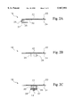
- FIG. 2C is a rear elevational view of the piezo driver assembly of FIG. 2B coupled to the shaft of the biopsy needle;
- FIG. 3A is a top plan view of the biopsy needle assembly made according to the present invention without the housing and the housing cover;
- FIG. 3B is side elevational view of the biopsy needle assembly of FIG. 3A;
- FIG. 4 is a graph which depicts tip deflection of the biopsy needle assembly made according to the present invention, due to the piezo driver.
- FIG. 5 is a pictorial representation of an ultrasound imaging system for transmitting the flexural motion of the biopsy needle assembly made according to the present invention.
- biopsy needles which are commonly used in surgical procedures that require visualization via conventional color ultrasound systems, be manufactured in various diameters, lengths, and cutting tip designs. It is extremely desirable from an economic standpoint, to be able to construct and manufacture these biopsy needles with their own vibrating driver.
- biopsy needles typically range in gages from 16 (1.6 mm diameter) to 25 (0.4 mm diameter), come in lengths from 0.5 inches to 10 inches, and have straight or various angled slant-cut cutting tips.
- the vibrating driver assembly of the present invention will be described in application to biopsy needles. It should be understood, however, that the vibrating driver apparatus of the present invention can be applied to other beam-like interventional medical devices, such as catheters and the like, which are commonly monitored in surgical procedures using color ultrasonic imaging systems.
- the biopsy needle assembly 10 generally comprises a biopsy needle 12 powered by a vibrating piezo driver assembly 18.
- the biopsy needle 12 has a shaft 14 which is fixed at one end in a hub 16.
- the hub 16 of the biopsy needle 12 contains a conical opening or luer connection 17 which allows the needle 12 to be connected to other medical apparatus such as tube and plunger devices used to propel liquids or extract liquid or tissues from the site of the needle insertion.
- the portion of the biopsy needle assembly 10 that vibrates and forms a standing wave is the thin, hollow tube or cannula, which forms the shaft 14.
- the piezo driver assembly 18 is a piezoelectric acoustic transducer (unimorph) comprising a single disc-shaped transducer 22 of piezoelectric ceramic bonded to a circular-shaped metal plate 24 composed of brass, stainless steel or any other suitable metal.
- the piezoelectric ceramic typically comprises some type of modified lead zirconate (PZT) composition or to a lesser extent, some type of modified barium titanate composition. Piezoelectric acoustic transducers made from such materials are well known in art and offer a high quality, low cost drive for the needle.
- the driver assembly 18 can comprise magnetic transducers and the like.
- the piezo driver assembly 18 is preferably coupled to the shaft 14 of the needle 10 via high rate a spring-like coupler bracket 26 (shown with broken lines) as will be explained later on in greater detail.
- the piezo driver assembly 18 is enclosed in a piezo driver housing 34 that is coupled to the hub 1 of the needle 12.
- the housing 34 includes a needle restrictor ring portion 36 that defines an aperture 38 through which the shaft 14 of the needle 12 extends out through the housing 34.
- a cover 42 for the housing 34 is provided and may be aligned to the housing 34 by pins, edges, depressions or other methods commonly employed.
- the housing 34 may also be fabricated in such a manner as to include the hub portion 16 where the luer connection 17 is constructed.
- a pair of leads 50, 52 attach the piezo driver assembly 18 to a pair of connectors 54, 56 mounted on the housing 22.
- the connectors 54, 56 enable the piezo driver assembly 18 contained within the housing 34 to be electrically coupled to an oscillator circuit 74 which activates the piezo driver assembly (FIG. 5).
- an oscillator circuit is described in U.S. Pat. No. 5,329,927, the entire disclosure of which is incorporated herein by reference.
- the leads 50, 52 are coupled to the piezo driver assembly 18 via an electrode 46 located on the metal plate 24 and an electrode 48 located on the surface of the ceramic transducer 22.
- the leads 50, 52 be of the solderless, detachable type so that they can be easily removed from both the piezo driver assembly 18 and the connectors 54, 56 mounted on the housing 34 without unsoldering.
- the electrodes 46 and 48 of the piezo driver assembly are best seen in FIG. 3A which depicts a top view of needle assembly 10 without the housing 34 and housing cover 42.
- the wire leads from the driver assembly may also be introduced into the housing directly and the leads soldered to points 46 and 48
- FIGS. 1B, 2A and 2B depicts the spring-like coupler bracket 26 which couples the piezo driver assembly 18 to the needle 12 in the preferred embodiment of the invention.
- the coupler bracket 26 has an asymmetrically-shaped configuration consisting of an elongated support member 28 with a first arm 30 hingedly attached to one end of the member 28 and second arm 32 hingedly attached to the other end of the member 28.
- the first arm 30 has a substantially greater length than the second arm 32.
- the coupler bracket 26 is preferably made from stainless steel although any other suitable material can be used.
- the piezo driver assembly 18 is adhesively bonded to the first arm 30 of the coupler bracket 26.
- the second arm 32 of the coupler bracket 26 is adhesively bonded or welded to the shaft 14 of the needle 12.
- a separate clip-like fastening element 58 is used to fasten the second arm 32 to the shaft 14 of the needle 12.
- the second arm 32 of the coupler bracket 26 can be configured to clip over the shaft 14 of the needle 12.
- glue can be optionally used to prevent detachment of the coupler bracket 26 from the needle 12.
- the coupler bracket 26 operates as a lever to impart a torque on the shaft 14 of the needle 12 as will be further explained below. Further, the coupler bracket 26 also prevents the piezo driver assembly 18 from breaking away from the needle 12 in the event that the shaft 14 of the needle 12 is bent during use.
- the piezo driver assembly 18 can be bonded directly to the shaft 12 of the needle 10 without the coupler bracket 26 if desired.
- FIG. 3B there is depicted a side view of the needle assembly 10 without the housing 34 and housing cover 42.
- the needle restrictor ring portion 36 (shown in phantom with broken lines) limits how much the shaft 14 of the needle 12 can bend as the shaft 14 of the needle 12 is inserted and guided through a patient's body by a physician. For example, if the shaft 14 of the needle 12 is approximately 0.027 of an inch, the diameter of the aperture 38 provided by the needle restrictor ring portion 36 would be approximately 0.080 of an inch.
- the housing 34 is configured so that the restrictor ring portion 36 surrounds the shaft 14 of the needle 12 at a point which provides a satisfactory needle working length.
- the working length of the needle 12 is the distance D from the restrictor ring portion 36 to the tip of the shaft 14. Typical working lengths range between 7.5 cm to 3 inches.
- the location of the restrictor ring portion 36 must also approximately coincide with a vibrational null point NP of an exaggerated wave 60 which represents the flexure of the needle 12 at its natural resonance frequency. Placing the restrictor ring portion 36 close to the null point NP operates to reduce the amount of damping to the needle 12 when the needle 12 is bent and makes contact with the needle restrictor ring portion 36. Since the natural resonance frequency of the needle 12 is dependent upon the length of the needle, which is controlled by the desired working length, the location of the restriction ring portion 36 from the hub 16 of the needle 12 can be determined.
- the piezo driver assembly 18 used in the present invention has a natural resonance frequency on the order of approximately 5000 Hz.
- the biopsy needles 12 used in the present invention have a preferred natural resonance frequency of 2700 Hz and a preferred resonance frequency range of between 2100 and 2700 Hz depending on the length of the needle's shaft 14. Accordingly, the piezo driver assembly 18 is capable of driving the needle 12 at any desired frequency without interference from the undesirable natural frequencies of the piezo driver assembly 18.
- the piezo driver assembly 18 is shown attached to the shaft 14 of the needle 12 via the spring coupler bracket 26 at a location which is selected to be at a point P which is approximately midway between the vibrational null point NP and a vibrational maximum point MP of the needle 12 depicted by the wave 60.
- the exact location of the piezo driver assembly 18 on the needle 12 is not as critical in terms of the needle assembly 10 producing a desired output (amplitude of needle vibration) compared with the arrangement described in U.S. Pat. No. 5,329,927 where the driver assembly must be mounted almost exactly at the vibrational maximum point of the needle.
- flexural vibrations which is used to describe the motion of the needle 12, denotes the curving or “flexure” of the needle's shaft 14 which is generated by the wave-like characteristics of the mechanical energy transmission generated by the piezo driver assembly 18 as depicted by the wave 60. Flexure occurs in an elastic material when a deflection is suitable to set-up stresses in the material. The vibratory-induced flexural waves operate and exhibit characteristics similar to standing waves or as propagating waves. Since the piezo driver assembly 18 also vibrates in the Y and Z planes, the shaft 14 of the needle 12 also exhibits flexural vibrations in the Y and Z planes.
- the flexural waves provided by the piezo driver assembly 18 are transmitted about or along the X,Y and/or Z axes at synchronized, but non-harmonically related frequencies that correspond to resonant frequencies of the needle 12.
- a more detailed discussion of biopsy needles and other interventional devices which are made to exhibit flexural waves is provided in the earlier mentioned U.S. Pat. No. 5,329,927.
- FIG. 4 depicts the tip deflection of the needle 12 when driven by a piezo driver assembly 18 attached to the shaft 14 thereof with and without the coupler bracket 26 at frequencies up to approximately 5000 Hz.
- the curve A corresponds to the instrumented output of the piezo driver assembly 18 attached via a coupler bracket 26.
- the curve B corresponds to the the frequencies calculated from a 3-d model constructed in software available from SDRC called I-DEAS, and analyzed by SDRC's finite element analysis software. Note that both the device and the model exhibit approximately 4 different frequency bands of resonance, the fourth frequency band FB occurring between approximately 2300 Hz and 3300 Hz and being the preferred resonance frequency range for operating the needle assembly 10.
- a conventional color ultrasonic imaging system comprising an ultrasonic imaging device 64, a scanner 66 electrically coupled to the imaging device 64 and the biopsy needle assembly 10 made according to the present invention.
- the ultrasonic imaging device via the scanner 66 can detect Doppler movements resulting from the flexural waves 60 exhibited by the needle assembly 10 of the present invention in any of the X, Y and Z planes thereby providing a precise indication on a display 68 of the system 64 as to the location of the needle assembly 10 within a patient.
- the scanner 66 impresses an imaging pulse 70 on a point 72 of the biopsy needle assembly 10.
- the point 72 may be closest to the scan head 66 when the first imaging pulse 70 occurs and furthest when a second imaging pulse 70 occurs.
- Known imaging systems 64 are able to detect and display the velocity of a moving element in the 0.2-100 centimeters per second or more (cm/sec) range.
- the first and second imaging pulses 70 are produced typically every 80 to 330 microseconds (usec) depending on the scale of the velocity to be detected. If a 5 cm/sec velocity is detected with a sampling interval of the imaging pulses set at 118 microseconds, the detected displacement of the point 72 is 5 centimeters/second times 118 microseconds, or approximately 6 microns.
- the driving method of the present invention provides a simple, easy to manufacture, low cost approach for driving an interventional medical device, such as a biopsy needle, into sustained oscillation. Accordingly, the driving method of the present invention provides inexpensive, throw away beam-like interventional medical devices such is biopsy needles with excellent visibility under color ultrasound scans.
Landscapes
- Health & Medical Sciences (AREA)
- Life Sciences & Earth Sciences (AREA)
- Surgery (AREA)
- Animal Behavior & Ethology (AREA)
- Biomedical Technology (AREA)
- Heart & Thoracic Surgery (AREA)
- Medical Informatics (AREA)
- Molecular Biology (AREA)
- Pathology (AREA)
- Engineering & Computer Science (AREA)
- General Health & Medical Sciences (AREA)
- Public Health (AREA)
- Veterinary Medicine (AREA)
- Physics & Mathematics (AREA)
- Biophysics (AREA)
- Nuclear Medicine, Radiotherapy & Molecular Imaging (AREA)
- Radiology & Medical Imaging (AREA)
- Ultra Sonic Daignosis Equipment (AREA)
Abstract
Description
Claims (27)
Priority Applications (1)
| Application Number | Priority Date | Filing Date | Title |
|---|---|---|---|
| US08/759,762 US5967991A (en) | 1996-12-03 | 1996-12-03 | Drive apparatus for an interventional medical device used in an ultrasonic imaging system |
Applications Claiming Priority (1)
| Application Number | Priority Date | Filing Date | Title |
|---|---|---|---|
| US08/759,762 US5967991A (en) | 1996-12-03 | 1996-12-03 | Drive apparatus for an interventional medical device used in an ultrasonic imaging system |
Publications (1)
| Publication Number | Publication Date |
|---|---|
| US5967991A true US5967991A (en) | 1999-10-19 |
Family
ID=25056857
Family Applications (1)
| Application Number | Title | Priority Date | Filing Date |
|---|---|---|---|
| US08/759,762 Expired - Fee Related US5967991A (en) | 1996-12-03 | 1996-12-03 | Drive apparatus for an interventional medical device used in an ultrasonic imaging system |
Country Status (1)
| Country | Link |
|---|---|
| US (1) | US5967991A (en) |
Cited By (68)
| Publication number | Priority date | Publication date | Assignee | Title |
|---|---|---|---|---|
| GB2367895A (en) * | 2000-06-05 | 2002-04-17 | Richard Miles | Needle location system for use with ultrasound imager |
| EP1208799A1 (en) * | 2000-11-16 | 2002-05-29 | Kretztechnik Aktiengesellschaft | Method for determining the insertion direction of a biopsy needle and for controlling its trajectory |
| US20030078671A1 (en) * | 2001-04-27 | 2003-04-24 | Lesniak Jeanne M. | Prevention of myocardial infarction induced ventricular expansion and remodeling |
| US20040074446A1 (en) * | 2002-10-22 | 2004-04-22 | Raymond Hawn | Quality assurance program and method for meat production |
| US20040230111A1 (en) * | 2003-02-12 | 2004-11-18 | Smith Stephen W. | Methods, devices, systems and computer program products for oscillating shafts using real time 3D ultrasound |
| US20050080402A1 (en) * | 2001-04-27 | 2005-04-14 | Myomend, Inc. | Prevention of myocardial infarction induced ventricular expansion and remodeling |
| WO2008067053A2 (en) * | 2006-10-12 | 2008-06-05 | Lawrence Group Medical Device Trust | Novel needle driver for magnetic resonance elastography |
| US20080300571A1 (en) * | 2007-05-30 | 2008-12-04 | Lepivert Patrick | Process and device for selectively treating interstitial tissue |
| US20090292204A1 (en) * | 2008-05-23 | 2009-11-26 | Oscillon Ltd. | Method and device for recognizing tissue structure using doppler effect |
| WO2010018512A1 (en) * | 2008-08-12 | 2010-02-18 | Koninklijke Philips Electronics N.V. | Ultrasound imaging |
| WO2010047734A1 (en) * | 2008-10-20 | 2010-04-29 | Critical Care Innovations, Inc. | Ultrasound detectable interventional medical device |
| US7794407B2 (en) | 2006-10-23 | 2010-09-14 | Bard Access Systems, Inc. | Method of locating the tip of a central venous catheter |
| KR101143663B1 (en) * | 2009-11-17 | 2012-05-09 | 삼성메디슨 주식회사 | Medical system and methdo for providing optimal ultrasound image for interventional treatment |
| US8388541B2 (en) | 2007-11-26 | 2013-03-05 | C. R. Bard, Inc. | Integrated system for intravascular placement of a catheter |
| US8388546B2 (en) | 2006-10-23 | 2013-03-05 | Bard Access Systems, Inc. | Method of locating the tip of a central venous catheter |
| US8437833B2 (en) | 2008-10-07 | 2013-05-07 | Bard Access Systems, Inc. | Percutaneous magnetic gastrostomy |
| US8478382B2 (en) | 2008-02-11 | 2013-07-02 | C. R. Bard, Inc. | Systems and methods for positioning a catheter |
| US20130261437A1 (en) * | 2006-11-02 | 2013-10-03 | Cooltouch Incorporated | Sonic Endovenous Catheter |
| USD699359S1 (en) | 2011-08-09 | 2014-02-11 | C. R. Bard, Inc. | Ultrasound probe head |
| US8663110B2 (en) | 2009-11-17 | 2014-03-04 | Samsung Medison Co., Ltd. | Providing an optimal ultrasound image for interventional treatment in a medical system |
| US8781555B2 (en) | 2007-11-26 | 2014-07-15 | C. R. Bard, Inc. | System for placement of a catheter including a signal-generating stylet |
| US8784336B2 (en) | 2005-08-24 | 2014-07-22 | C. R. Bard, Inc. | Stylet apparatuses and methods of manufacture |
| WO2014118376A1 (en) * | 2013-02-01 | 2014-08-07 | Olberon Medical Innovation Sas | Needle location device |
| US8801693B2 (en) | 2010-10-29 | 2014-08-12 | C. R. Bard, Inc. | Bioimpedance-assisted placement of a medical device |
| WO2014140556A1 (en) * | 2013-03-15 | 2014-09-18 | University Court Of The University Of Dundee | Medical apparatus and its visualisation |
| US8849382B2 (en) | 2007-11-26 | 2014-09-30 | C. R. Bard, Inc. | Apparatus and display methods relating to intravascular placement of a catheter |
| CN104068923A (en) * | 2014-07-08 | 2014-10-01 | 中国人民解放军第三军医大学第一附属医院 | Pulse wave simulating ultrasonic puncture kit |
| USD724745S1 (en) | 2011-08-09 | 2015-03-17 | C. R. Bard, Inc. | Cap for an ultrasound probe |
| US9125578B2 (en) | 2009-06-12 | 2015-09-08 | Bard Access Systems, Inc. | Apparatus and method for catheter navigation and tip location |
| US9169517B2 (en) | 1997-08-15 | 2015-10-27 | Institut Pasteur | Mutation within the connexin 26 gene responsible for prelingual non-syndromic deafness and method of detection |
| US9173589B2 (en) | 2011-03-30 | 2015-11-03 | Mayo Foundation For Medical Education And Research | System and method for inertial magnetic resonance elastography driver for use with interventional medical device |
| US9211107B2 (en) | 2011-11-07 | 2015-12-15 | C. R. Bard, Inc. | Ruggedized ultrasound hydrogel insert |
| US9339206B2 (en) | 2009-06-12 | 2016-05-17 | Bard Access Systems, Inc. | Adaptor for endovascular electrocardiography |
| US9445734B2 (en) | 2009-06-12 | 2016-09-20 | Bard Access Systems, Inc. | Devices and methods for endovascular electrography |
| US9456766B2 (en) | 2007-11-26 | 2016-10-04 | C. R. Bard, Inc. | Apparatus for use with needle insertion guidance system |
| US9492097B2 (en) | 2007-11-26 | 2016-11-15 | C. R. Bard, Inc. | Needle length determination and calibration for insertion guidance system |
| US9521961B2 (en) | 2007-11-26 | 2016-12-20 | C. R. Bard, Inc. | Systems and methods for guiding a medical instrument |
| US9532724B2 (en) | 2009-06-12 | 2017-01-03 | Bard Access Systems, Inc. | Apparatus and method for catheter navigation using endovascular energy mapping |
| US9554716B2 (en) | 2007-11-26 | 2017-01-31 | C. R. Bard, Inc. | Insertion guidance system for needles and medical components |
| US9636031B2 (en) | 2007-11-26 | 2017-05-02 | C.R. Bard, Inc. | Stylets for use with apparatus for intravascular placement of a catheter |
| US9649048B2 (en) | 2007-11-26 | 2017-05-16 | C. R. Bard, Inc. | Systems and methods for breaching a sterile field for intravascular placement of a catheter |
| US9839372B2 (en) | 2014-02-06 | 2017-12-12 | C. R. Bard, Inc. | Systems and methods for guidance and placement of an intravascular device |
| US9901714B2 (en) | 2008-08-22 | 2018-02-27 | C. R. Bard, Inc. | Catheter assembly including ECG sensor and magnetic assemblies |
| US10046139B2 (en) | 2010-08-20 | 2018-08-14 | C. R. Bard, Inc. | Reconfirmation of ECG-assisted catheter tip placement |
| US10127629B2 (en) | 2006-08-02 | 2018-11-13 | Inneroptic Technology, Inc. | System and method of providing real-time dynamic imagery of a medical procedure site using multiple modalities |
| US10136951B2 (en) | 2009-02-17 | 2018-11-27 | Inneroptic Technology, Inc. | Systems, methods, apparatuses, and computer-readable media for image guided surgery |
| US10188467B2 (en) | 2014-12-12 | 2019-01-29 | Inneroptic Technology, Inc. | Surgical guidance intersection display |
| US10278778B2 (en) | 2016-10-27 | 2019-05-07 | Inneroptic Technology, Inc. | Medical device navigation using a virtual 3D space |
| US10314559B2 (en) | 2013-03-14 | 2019-06-11 | Inneroptic Technology, Inc. | Medical device guidance |
| US10349890B2 (en) | 2015-06-26 | 2019-07-16 | C. R. Bard, Inc. | Connector interface for ECG-based catheter positioning system |
| US10398513B2 (en) | 2009-02-17 | 2019-09-03 | Inneroptic Technology, Inc. | Systems, methods, apparatuses, and computer-readable media for image management in image-guided medical procedures |
| US10433814B2 (en) | 2016-02-17 | 2019-10-08 | Inneroptic Technology, Inc. | Loupe display |
| US10449330B2 (en) | 2007-11-26 | 2019-10-22 | C. R. Bard, Inc. | Magnetic element-equipped needle assemblies |
| US10524691B2 (en) | 2007-11-26 | 2020-01-07 | C. R. Bard, Inc. | Needle assembly including an aligned magnetic element |
| US10639008B2 (en) | 2009-10-08 | 2020-05-05 | C. R. Bard, Inc. | Support and cover structures for an ultrasound probe head |
| CN111511290A (en) * | 2017-09-18 | 2020-08-07 | 活动针技术有限公司 | Medical device |
| US10751509B2 (en) | 2007-11-26 | 2020-08-25 | C. R. Bard, Inc. | Iconic representations for guidance of an indwelling medical device |
| US10820885B2 (en) | 2012-06-15 | 2020-11-03 | C. R. Bard, Inc. | Apparatus and methods for detection of a removable cap on an ultrasound probe |
| US10820944B2 (en) | 2014-10-02 | 2020-11-03 | Inneroptic Technology, Inc. | Affected region display based on a variance parameter associated with a medical device |
| US10973584B2 (en) | 2015-01-19 | 2021-04-13 | Bard Access Systems, Inc. | Device and method for vascular access |
| US10992079B2 (en) | 2018-10-16 | 2021-04-27 | Bard Access Systems, Inc. | Safety-equipped connection systems and methods thereof for establishing electrical connections |
| US11000207B2 (en) | 2016-01-29 | 2021-05-11 | C. R. Bard, Inc. | Multiple coil system for tracking a medical device |
| US11103200B2 (en) | 2015-07-22 | 2021-08-31 | Inneroptic Technology, Inc. | Medical device approaches |
| US11103213B2 (en) | 2009-10-08 | 2021-08-31 | C. R. Bard, Inc. | Spacers for use with an ultrasound probe |
| US11259879B2 (en) | 2017-08-01 | 2022-03-01 | Inneroptic Technology, Inc. | Selective transparency to assist medical device navigation |
| US11369296B2 (en) | 2011-02-01 | 2022-06-28 | Olberon Medical Innovation Sas | Needle holder |
| US11464578B2 (en) | 2009-02-17 | 2022-10-11 | Inneroptic Technology, Inc. | Systems, methods, apparatuses, and computer-readable media for image management in image-guided medical procedures |
| US11484365B2 (en) | 2018-01-23 | 2022-11-01 | Inneroptic Technology, Inc. | Medical image guidance |
Citations (8)
| Publication number | Priority date | Publication date | Assignee | Title |
|---|---|---|---|---|
| US3990452A (en) * | 1975-06-13 | 1976-11-09 | Fibra-Sonics, Inc. | Medical machine for performing surgery and treating using ultrasonic energy |
| US4425115A (en) * | 1977-12-19 | 1984-01-10 | Wuchinich David G | Ultrasonic resonant vibrator |
| US4431006A (en) * | 1982-01-07 | 1984-02-14 | Technicare Corporation | Passive ultrasound needle probe locator |
| US4961424A (en) * | 1987-08-05 | 1990-10-09 | Olympus Optical Co., Ltd. | Ultrasonic treatment device |
| US5095910A (en) * | 1990-04-18 | 1992-03-17 | Advanced Technology Laboratories, Inc. | Ultrasonic imaging of biopsy needle |
| US5329927A (en) * | 1993-02-25 | 1994-07-19 | Echo Cath, Inc. | Apparatus and method for locating an interventional medical device with a ultrasound color imaging system |
| US5421336A (en) * | 1994-04-04 | 1995-06-06 | Echo Cath, Inc. | Method for attaching an interventional medical device to a vibratory member associated with visualization by an ultrasound imaging system |
| US5549112A (en) * | 1994-03-12 | 1996-08-27 | Cockburn; John F. | Medical needle for use in ultrasound imaging and method of enhancing the visibility of such a needle to ultrasound |
-
1996
- 1996-12-03 US US08/759,762 patent/US5967991A/en not_active Expired - Fee Related
Patent Citations (8)
| Publication number | Priority date | Publication date | Assignee | Title |
|---|---|---|---|---|
| US3990452A (en) * | 1975-06-13 | 1976-11-09 | Fibra-Sonics, Inc. | Medical machine for performing surgery and treating using ultrasonic energy |
| US4425115A (en) * | 1977-12-19 | 1984-01-10 | Wuchinich David G | Ultrasonic resonant vibrator |
| US4431006A (en) * | 1982-01-07 | 1984-02-14 | Technicare Corporation | Passive ultrasound needle probe locator |
| US4961424A (en) * | 1987-08-05 | 1990-10-09 | Olympus Optical Co., Ltd. | Ultrasonic treatment device |
| US5095910A (en) * | 1990-04-18 | 1992-03-17 | Advanced Technology Laboratories, Inc. | Ultrasonic imaging of biopsy needle |
| US5329927A (en) * | 1993-02-25 | 1994-07-19 | Echo Cath, Inc. | Apparatus and method for locating an interventional medical device with a ultrasound color imaging system |
| US5549112A (en) * | 1994-03-12 | 1996-08-27 | Cockburn; John F. | Medical needle for use in ultrasound imaging and method of enhancing the visibility of such a needle to ultrasound |
| US5421336A (en) * | 1994-04-04 | 1995-06-06 | Echo Cath, Inc. | Method for attaching an interventional medical device to a vibratory member associated with visualization by an ultrasound imaging system |
Cited By (136)
| Publication number | Priority date | Publication date | Assignee | Title |
|---|---|---|---|---|
| US9868989B2 (en) | 1997-08-15 | 2018-01-16 | Institut Pasteur | Mutation within the connexin 26 gene responsible for prelingual non-syndromic deafness and method of detection |
| US9169517B2 (en) | 1997-08-15 | 2015-10-27 | Institut Pasteur | Mutation within the connexin 26 gene responsible for prelingual non-syndromic deafness and method of detection |
| GB2367895A (en) * | 2000-06-05 | 2002-04-17 | Richard Miles | Needle location system for use with ultrasound imager |
| EP1208799A1 (en) * | 2000-11-16 | 2002-05-29 | Kretztechnik Aktiengesellschaft | Method for determining the insertion direction of a biopsy needle and for controlling its trajectory |
| US6616610B2 (en) | 2000-11-16 | 2003-09-09 | Ge Medical Systems Kretztechnik Gmbh & Co. Ohg | Method for determination of the direction of introduction and for controlling the introduction path of biopsy needles |
| US20030078671A1 (en) * | 2001-04-27 | 2003-04-24 | Lesniak Jeanne M. | Prevention of myocardial infarction induced ventricular expansion and remodeling |
| US20050080402A1 (en) * | 2001-04-27 | 2005-04-14 | Myomend, Inc. | Prevention of myocardial infarction induced ventricular expansion and remodeling |
| US7311731B2 (en) | 2001-04-27 | 2007-12-25 | Richard C. Satterfield | Prevention of myocardial infarction induced ventricular expansion and remodeling |
| US7988727B2 (en) | 2001-04-27 | 2011-08-02 | Cormend Technologies, Llc | Prevention of myocardial infarction induced ventricular expansion and remodeling |
| US20040074446A1 (en) * | 2002-10-22 | 2004-04-22 | Raymond Hawn | Quality assurance program and method for meat production |
| US7329225B2 (en) | 2003-02-12 | 2008-02-12 | Duke University | Methods, devices, systems and computer program products for oscillating shafts using real time 3D ultrasound |
| US20040230111A1 (en) * | 2003-02-12 | 2004-11-18 | Smith Stephen W. | Methods, devices, systems and computer program products for oscillating shafts using real time 3D ultrasound |
| US10004875B2 (en) | 2005-08-24 | 2018-06-26 | C. R. Bard, Inc. | Stylet apparatuses and methods of manufacture |
| US8784336B2 (en) | 2005-08-24 | 2014-07-22 | C. R. Bard, Inc. | Stylet apparatuses and methods of manufacture |
| US11207496B2 (en) | 2005-08-24 | 2021-12-28 | C. R. Bard, Inc. | Stylet apparatuses and methods of manufacture |
| US10127629B2 (en) | 2006-08-02 | 2018-11-13 | Inneroptic Technology, Inc. | System and method of providing real-time dynamic imagery of a medical procedure site using multiple modalities |
| US10733700B2 (en) | 2006-08-02 | 2020-08-04 | Inneroptic Technology, Inc. | System and method of providing real-time dynamic imagery of a medical procedure site using multiple modalities |
| US11481868B2 (en) | 2006-08-02 | 2022-10-25 | Inneroptic Technology, Inc. | System and method of providing real-time dynamic imagery of a medical procedure she using multiple modalities |
| WO2008067053A3 (en) * | 2006-10-12 | 2009-01-22 | Lawrence Group Medical Device | Novel needle driver for magnetic resonance elastography |
| US20080255444A1 (en) * | 2006-10-12 | 2008-10-16 | Geng Li | Novel needle driver for magnetic resonance elastography |
| US7979109B2 (en) * | 2006-10-12 | 2011-07-12 | Lawrence Group Medical Device Trust | Needle driver for magnetic resonance elastography |
| US20110178390A1 (en) * | 2006-10-12 | 2011-07-21 | Geng Li | Novel needle driver for magnetic resonance elastography |
| WO2008067053A2 (en) * | 2006-10-12 | 2008-06-05 | Lawrence Group Medical Device Trust | Novel needle driver for magnetic resonance elastography |
| US20140031670A1 (en) * | 2006-10-12 | 2014-01-30 | Lawrence Group Medical Device Trust | Novel Needle Driver For Magnetic Resonance Elastography |
| US9833169B2 (en) | 2006-10-23 | 2017-12-05 | Bard Access Systems, Inc. | Method of locating the tip of a central venous catheter |
| US8388546B2 (en) | 2006-10-23 | 2013-03-05 | Bard Access Systems, Inc. | Method of locating the tip of a central venous catheter |
| US9345422B2 (en) | 2006-10-23 | 2016-05-24 | Bard Acess Systems, Inc. | Method of locating the tip of a central venous catheter |
| US9265443B2 (en) | 2006-10-23 | 2016-02-23 | Bard Access Systems, Inc. | Method of locating the tip of a central venous catheter |
| US8512256B2 (en) | 2006-10-23 | 2013-08-20 | Bard Access Systems, Inc. | Method of locating the tip of a central venous catheter |
| US7794407B2 (en) | 2006-10-23 | 2010-09-14 | Bard Access Systems, Inc. | Method of locating the tip of a central venous catheter |
| US8858455B2 (en) | 2006-10-23 | 2014-10-14 | Bard Access Systems, Inc. | Method of locating the tip of a central venous catheter |
| US8774907B2 (en) | 2006-10-23 | 2014-07-08 | Bard Access Systems, Inc. | Method of locating the tip of a central venous catheter |
| US20130261437A1 (en) * | 2006-11-02 | 2013-10-03 | Cooltouch Incorporated | Sonic Endovenous Catheter |
| US20130261436A1 (en) * | 2006-11-02 | 2013-10-03 | Cooltouch Incorporated | Sonic Endovenous Catheter |
| US20080300571A1 (en) * | 2007-05-30 | 2008-12-04 | Lepivert Patrick | Process and device for selectively treating interstitial tissue |
| US9681823B2 (en) | 2007-11-26 | 2017-06-20 | C. R. Bard, Inc. | Integrated system for intravascular placement of a catheter |
| US9999371B2 (en) | 2007-11-26 | 2018-06-19 | C. R. Bard, Inc. | Integrated system for intravascular placement of a catheter |
| US10966630B2 (en) | 2007-11-26 | 2021-04-06 | C. R. Bard, Inc. | Integrated system for intravascular placement of a catheter |
| US10849695B2 (en) | 2007-11-26 | 2020-12-01 | C. R. Bard, Inc. | Systems and methods for breaching a sterile field for intravascular placement of a catheter |
| US11123099B2 (en) | 2007-11-26 | 2021-09-21 | C. R. Bard, Inc. | Apparatus for use with needle insertion guidance system |
| US8849382B2 (en) | 2007-11-26 | 2014-09-30 | C. R. Bard, Inc. | Apparatus and display methods relating to intravascular placement of a catheter |
| US11779240B2 (en) | 2007-11-26 | 2023-10-10 | C. R. Bard, Inc. | Systems and methods for breaching a sterile field for intravascular placement of a catheter |
| US11134915B2 (en) | 2007-11-26 | 2021-10-05 | C. R. Bard, Inc. | System for placement of a catheter including a signal-generating stylet |
| US9649048B2 (en) | 2007-11-26 | 2017-05-16 | C. R. Bard, Inc. | Systems and methods for breaching a sterile field for intravascular placement of a catheter |
| US10751509B2 (en) | 2007-11-26 | 2020-08-25 | C. R. Bard, Inc. | Iconic representations for guidance of an indwelling medical device |
| US10602958B2 (en) | 2007-11-26 | 2020-03-31 | C. R. Bard, Inc. | Systems and methods for guiding a medical instrument |
| US10524691B2 (en) | 2007-11-26 | 2020-01-07 | C. R. Bard, Inc. | Needle assembly including an aligned magnetic element |
| US10449330B2 (en) | 2007-11-26 | 2019-10-22 | C. R. Bard, Inc. | Magnetic element-equipped needle assemblies |
| US10342575B2 (en) | 2007-11-26 | 2019-07-09 | C. R. Bard, Inc. | Apparatus for use with needle insertion guidance system |
| US8781555B2 (en) | 2007-11-26 | 2014-07-15 | C. R. Bard, Inc. | System for placement of a catheter including a signal-generating stylet |
| US10238418B2 (en) | 2007-11-26 | 2019-03-26 | C. R. Bard, Inc. | Apparatus for use with needle insertion guidance system |
| US9636031B2 (en) | 2007-11-26 | 2017-05-02 | C.R. Bard, Inc. | Stylets for use with apparatus for intravascular placement of a catheter |
| US11707205B2 (en) | 2007-11-26 | 2023-07-25 | C. R. Bard, Inc. | Integrated system for intravascular placement of a catheter |
| US10231753B2 (en) | 2007-11-26 | 2019-03-19 | C. R. Bard, Inc. | Insertion guidance system for needles and medical components |
| US9554716B2 (en) | 2007-11-26 | 2017-01-31 | C. R. Bard, Inc. | Insertion guidance system for needles and medical components |
| US10165962B2 (en) | 2007-11-26 | 2019-01-01 | C. R. Bard, Inc. | Integrated systems for intravascular placement of a catheter |
| US10105121B2 (en) | 2007-11-26 | 2018-10-23 | C. R. Bard, Inc. | System for placement of a catheter including a signal-generating stylet |
| US8388541B2 (en) | 2007-11-26 | 2013-03-05 | C. R. Bard, Inc. | Integrated system for intravascular placement of a catheter |
| US9456766B2 (en) | 2007-11-26 | 2016-10-04 | C. R. Bard, Inc. | Apparatus for use with needle insertion guidance system |
| US9492097B2 (en) | 2007-11-26 | 2016-11-15 | C. R. Bard, Inc. | Needle length determination and calibration for insertion guidance system |
| US9521961B2 (en) | 2007-11-26 | 2016-12-20 | C. R. Bard, Inc. | Systems and methods for guiding a medical instrument |
| US9526440B2 (en) | 2007-11-26 | 2016-12-27 | C.R. Bard, Inc. | System for placement of a catheter including a signal-generating stylet |
| US11529070B2 (en) | 2007-11-26 | 2022-12-20 | C. R. Bard, Inc. | System and methods for guiding a medical instrument |
| US9549685B2 (en) | 2007-11-26 | 2017-01-24 | C. R. Bard, Inc. | Apparatus and display methods relating to intravascular placement of a catheter |
| US8478382B2 (en) | 2008-02-11 | 2013-07-02 | C. R. Bard, Inc. | Systems and methods for positioning a catheter |
| US8971994B2 (en) | 2008-02-11 | 2015-03-03 | C. R. Bard, Inc. | Systems and methods for positioning a catheter |
| US20090292204A1 (en) * | 2008-05-23 | 2009-11-26 | Oscillon Ltd. | Method and device for recognizing tissue structure using doppler effect |
| CN102119002A (en) * | 2008-08-12 | 2011-07-06 | 皇家飞利浦电子股份有限公司 | Ultrasound imaging |
| US20110137165A1 (en) * | 2008-08-12 | 2011-06-09 | Koninklijke Philips Electronics N.V. | Ultrasound imaging |
| WO2010018512A1 (en) * | 2008-08-12 | 2010-02-18 | Koninklijke Philips Electronics N.V. | Ultrasound imaging |
| US11027101B2 (en) | 2008-08-22 | 2021-06-08 | C. R. Bard, Inc. | Catheter assembly including ECG sensor and magnetic assemblies |
| US9901714B2 (en) | 2008-08-22 | 2018-02-27 | C. R. Bard, Inc. | Catheter assembly including ECG sensor and magnetic assemblies |
| US9907513B2 (en) | 2008-10-07 | 2018-03-06 | Bard Access Systems, Inc. | Percutaneous magnetic gastrostomy |
| US8437833B2 (en) | 2008-10-07 | 2013-05-07 | Bard Access Systems, Inc. | Percutaneous magnetic gastrostomy |
| WO2010047734A1 (en) * | 2008-10-20 | 2010-04-29 | Critical Care Innovations, Inc. | Ultrasound detectable interventional medical device |
| US9138286B2 (en) | 2008-10-20 | 2015-09-22 | Nuvue Therapeutics, Inc. | Ultrasound detectable interventional medical device |
| US20100286528A1 (en) * | 2008-10-20 | 2010-11-11 | Critical Care Innovations, Inc. | Ultrasound detectable interventional medical device |
| US11464575B2 (en) | 2009-02-17 | 2022-10-11 | Inneroptic Technology, Inc. | Systems, methods, apparatuses, and computer-readable media for image guided surgery |
| US10136951B2 (en) | 2009-02-17 | 2018-11-27 | Inneroptic Technology, Inc. | Systems, methods, apparatuses, and computer-readable media for image guided surgery |
| US10398513B2 (en) | 2009-02-17 | 2019-09-03 | Inneroptic Technology, Inc. | Systems, methods, apparatuses, and computer-readable media for image management in image-guided medical procedures |
| US11464578B2 (en) | 2009-02-17 | 2022-10-11 | Inneroptic Technology, Inc. | Systems, methods, apparatuses, and computer-readable media for image management in image-guided medical procedures |
| US9339206B2 (en) | 2009-06-12 | 2016-05-17 | Bard Access Systems, Inc. | Adaptor for endovascular electrocardiography |
| US10912488B2 (en) | 2009-06-12 | 2021-02-09 | Bard Access Systems, Inc. | Apparatus and method for catheter navigation and tip location |
| US9532724B2 (en) | 2009-06-12 | 2017-01-03 | Bard Access Systems, Inc. | Apparatus and method for catheter navigation using endovascular energy mapping |
| US10271762B2 (en) | 2009-06-12 | 2019-04-30 | Bard Access Systems, Inc. | Apparatus and method for catheter navigation using endovascular energy mapping |
| US10231643B2 (en) | 2009-06-12 | 2019-03-19 | Bard Access Systems, Inc. | Apparatus and method for catheter navigation and tip location |
| US9125578B2 (en) | 2009-06-12 | 2015-09-08 | Bard Access Systems, Inc. | Apparatus and method for catheter navigation and tip location |
| US11419517B2 (en) | 2009-06-12 | 2022-08-23 | Bard Access Systems, Inc. | Apparatus and method for catheter navigation using endovascular energy mapping |
| US9445734B2 (en) | 2009-06-12 | 2016-09-20 | Bard Access Systems, Inc. | Devices and methods for endovascular electrography |
| US11998386B2 (en) | 2009-10-08 | 2024-06-04 | C. R. Bard, Inc. | Support and cover structures for an ultrasound probe head |
| US10639008B2 (en) | 2009-10-08 | 2020-05-05 | C. R. Bard, Inc. | Support and cover structures for an ultrasound probe head |
| US11103213B2 (en) | 2009-10-08 | 2021-08-31 | C. R. Bard, Inc. | Spacers for use with an ultrasound probe |
| US8663110B2 (en) | 2009-11-17 | 2014-03-04 | Samsung Medison Co., Ltd. | Providing an optimal ultrasound image for interventional treatment in a medical system |
| KR101143663B1 (en) * | 2009-11-17 | 2012-05-09 | 삼성메디슨 주식회사 | Medical system and methdo for providing optimal ultrasound image for interventional treatment |
| US10046139B2 (en) | 2010-08-20 | 2018-08-14 | C. R. Bard, Inc. | Reconfirmation of ECG-assisted catheter tip placement |
| US8801693B2 (en) | 2010-10-29 | 2014-08-12 | C. R. Bard, Inc. | Bioimpedance-assisted placement of a medical device |
| US9415188B2 (en) | 2010-10-29 | 2016-08-16 | C. R. Bard, Inc. | Bioimpedance-assisted placement of a medical device |
| US11369296B2 (en) | 2011-02-01 | 2022-06-28 | Olberon Medical Innovation Sas | Needle holder |
| US9173589B2 (en) | 2011-03-30 | 2015-11-03 | Mayo Foundation For Medical Education And Research | System and method for inertial magnetic resonance elastography driver for use with interventional medical device |
| USD724745S1 (en) | 2011-08-09 | 2015-03-17 | C. R. Bard, Inc. | Cap for an ultrasound probe |
| USD699359S1 (en) | 2011-08-09 | 2014-02-11 | C. R. Bard, Inc. | Ultrasound probe head |
| USD754357S1 (en) | 2011-08-09 | 2016-04-19 | C. R. Bard, Inc. | Ultrasound probe head |
| US9211107B2 (en) | 2011-11-07 | 2015-12-15 | C. R. Bard, Inc. | Ruggedized ultrasound hydrogel insert |
| US10820885B2 (en) | 2012-06-15 | 2020-11-03 | C. R. Bard, Inc. | Apparatus and methods for detection of a removable cap on an ultrasound probe |
| US10993738B2 (en) | 2013-02-01 | 2021-05-04 | Olberon Limited | Needle location device |
| WO2014118376A1 (en) * | 2013-02-01 | 2014-08-07 | Olberon Medical Innovation Sas | Needle location device |
| CN105228533A (en) * | 2013-02-01 | 2016-01-06 | 欧伯伦医疗创新公司 | Pin positioner |
| US10314559B2 (en) | 2013-03-14 | 2019-06-11 | Inneroptic Technology, Inc. | Medical device guidance |
| US11123100B2 (en) | 2013-03-15 | 2021-09-21 | University Court Of The University Of Dundee | Medical apparatus and its visualisation |
| CN105377144A (en) * | 2013-03-15 | 2016-03-02 | 敦提大学校董事会 | Medical apparatus and its visualisation |
| WO2014140556A1 (en) * | 2013-03-15 | 2014-09-18 | University Court Of The University Of Dundee | Medical apparatus and its visualisation |
| US9839372B2 (en) | 2014-02-06 | 2017-12-12 | C. R. Bard, Inc. | Systems and methods for guidance and placement of an intravascular device |
| US10863920B2 (en) | 2014-02-06 | 2020-12-15 | C. R. Bard, Inc. | Systems and methods for guidance and placement of an intravascular device |
| CN104068923A (en) * | 2014-07-08 | 2014-10-01 | 中国人民解放军第三军医大学第一附属医院 | Pulse wave simulating ultrasonic puncture kit |
| US10820944B2 (en) | 2014-10-02 | 2020-11-03 | Inneroptic Technology, Inc. | Affected region display based on a variance parameter associated with a medical device |
| US11684429B2 (en) | 2014-10-02 | 2023-06-27 | Inneroptic Technology, Inc. | Affected region display associated with a medical device |
| US10820946B2 (en) | 2014-12-12 | 2020-11-03 | Inneroptic Technology, Inc. | Surgical guidance intersection display |
| US10188467B2 (en) | 2014-12-12 | 2019-01-29 | Inneroptic Technology, Inc. | Surgical guidance intersection display |
| US11931117B2 (en) | 2014-12-12 | 2024-03-19 | Inneroptic Technology, Inc. | Surgical guidance intersection display |
| US11534245B2 (en) | 2014-12-12 | 2022-12-27 | Inneroptic Technology, Inc. | Surgical guidance intersection display |
| US10973584B2 (en) | 2015-01-19 | 2021-04-13 | Bard Access Systems, Inc. | Device and method for vascular access |
| US11026630B2 (en) | 2015-06-26 | 2021-06-08 | C. R. Bard, Inc. | Connector interface for ECG-based catheter positioning system |
| US10349890B2 (en) | 2015-06-26 | 2019-07-16 | C. R. Bard, Inc. | Connector interface for ECG-based catheter positioning system |
| US11103200B2 (en) | 2015-07-22 | 2021-08-31 | Inneroptic Technology, Inc. | Medical device approaches |
| US11000207B2 (en) | 2016-01-29 | 2021-05-11 | C. R. Bard, Inc. | Multiple coil system for tracking a medical device |
| US10433814B2 (en) | 2016-02-17 | 2019-10-08 | Inneroptic Technology, Inc. | Loupe display |
| US11179136B2 (en) | 2016-02-17 | 2021-11-23 | Inneroptic Technology, Inc. | Loupe display |
| US10278778B2 (en) | 2016-10-27 | 2019-05-07 | Inneroptic Technology, Inc. | Medical device navigation using a virtual 3D space |
| US11369439B2 (en) | 2016-10-27 | 2022-06-28 | Inneroptic Technology, Inc. | Medical device navigation using a virtual 3D space |
| US10772686B2 (en) | 2016-10-27 | 2020-09-15 | Inneroptic Technology, Inc. | Medical device navigation using a virtual 3D space |
| US11259879B2 (en) | 2017-08-01 | 2022-03-01 | Inneroptic Technology, Inc. | Selective transparency to assist medical device navigation |
| CN111511290A (en) * | 2017-09-18 | 2020-08-07 | 活动针技术有限公司 | Medical device |
| CN111511290B (en) * | 2017-09-18 | 2023-11-03 | 活动针技术有限公司 | medical device |
| US11484365B2 (en) | 2018-01-23 | 2022-11-01 | Inneroptic Technology, Inc. | Medical image guidance |
| US10992079B2 (en) | 2018-10-16 | 2021-04-27 | Bard Access Systems, Inc. | Safety-equipped connection systems and methods thereof for establishing electrical connections |
| US11621518B2 (en) | 2018-10-16 | 2023-04-04 | Bard Access Systems, Inc. | Safety-equipped connection systems and methods thereof for establishing electrical connections |
Similar Documents
| Publication | Publication Date | Title |
|---|---|---|
| US5967991A (en) | Drive apparatus for an interventional medical device used in an ultrasonic imaging system | |
| US5343865A (en) | Apparatus and method for locating an interventional medical device with a ultrasound color imaging system | |
| US6068599A (en) | Blood vessel puncturing device using ultrasound | |
| JP3146018B2 (en) | Ultrasonic imaging method and device for biopsy needle | |
| EP0755223B1 (en) | Apparatus for locating vibrating medical devices | |
| US6964640B2 (en) | System and method for detection of motion | |
| US4363326A (en) | Ultrasonic apparatus for needle insertion | |
| US4546771A (en) | Acoustic microscope | |
| JP3594278B2 (en) | Intracavity ultrasonic probe device | |
| JP5443732B2 (en) | Ultrasonic probe and ultrasonic diagnostic apparatus using the ultrasonic probe | |
| EP0672384B1 (en) | Medical needle for use in ultrasound imaging | |
| US5421336A (en) | Method for attaching an interventional medical device to a vibratory member associated with visualization by an ultrasound imaging system | |
| JPH06189958A (en) | Ultrasonic wave type cavity diagnostic device and method thereof | |
| JP2004504878A (en) | Ultrasound imaging system and method for implantable invasive devices | |
| EP2337501B1 (en) | Ultrasound detectable interventional medical device | |
| JPH1099329A (en) | Ultrasonic probe and ultrasonic diagnostic equipment | |
| JPH11137555A (en) | Ultrasonic diagnostic apparatus | |
| JPS6090542A (en) | Sensor for ultrasonic photographing apparatus | |
| GB2367895A (en) | Needle location system for use with ultrasound imager | |
| GB2173593A (en) | Ultrasonic location of needles or catheters | |
| JP2755964B2 (en) | Ultrasonic probe | |
| JPS60259254A (en) | Ultrasonic crushing apparatus | |
| JP2658689B2 (en) | Ultrasonic probe | |
| JPH0127765Y2 (en) | ||
| WO2020079808A1 (en) | Ultrasonic probe |
Legal Events
| Date | Code | Title | Description |
|---|---|---|---|
| AS | Assignment |
Owner name: PRISCHAK, JOSEPH J., PENNSYLVANIA Free format text: SECURITY INTEREST;ASSIGNOR:ECHOCATH, INC.;REEL/FRAME:011911/0816 Effective date: 20010529 |
|
| AS | Assignment |
Owner name: CRITICAL CARE INNOVATIONS, INC., VIRGINIA Free format text: RELEASE AND TERMINATION OF SECURITY INTEREST;ASSIGNOR:ECHOCATH, INC.;REEL/FRAME:013447/0140 Effective date: 20020917 |
|
| FEPP | Fee payment procedure |
Free format text: PAYOR NUMBER ASSIGNED (ORIGINAL EVENT CODE: ASPN); ENTITY STATUS OF PATENT OWNER: SMALL ENTITY |
|
| FPAY | Fee payment |
Year of fee payment: 4 |
|
| FPAY | Fee payment |
Year of fee payment: 8 |
|
| REMI | Maintenance fee reminder mailed | ||
| LAPS | Lapse for failure to pay maintenance fees | ||
| STCH | Information on status: patent discontinuation |
Free format text: PATENT EXPIRED DUE TO NONPAYMENT OF MAINTENANCE FEES UNDER 37 CFR 1.362 |
|
| FP | Lapsed due to failure to pay maintenance fee |
Effective date: 20111019 |