US5843685A - Production of chimeric mouse-human antibodies with specificity to human tumor antigens - Google Patents
Production of chimeric mouse-human antibodies with specificity to human tumor antigens Download PDFInfo
- Publication number
- US5843685A US5843685A US08/466,034 US46603495A US5843685A US 5843685 A US5843685 A US 5843685A US 46603495 A US46603495 A US 46603495A US 5843685 A US5843685 A US 5843685A
- Authority
- US
- United States
- Prior art keywords
- antibody
- chimeric
- chain
- ing
- human
- Prior art date
- Legal status (The legal status is an assumption and is not a legal conclusion. Google has not performed a legal analysis and makes no representation as to the accuracy of the status listed.)
- Expired - Fee Related
Links
- 0 CC(*)C1CCC(C)CC1 Chemical compound CC(*)C1CCC(C)CC1 0.000 description 1
- GDOPTJXRTPNYNR-UHFFFAOYSA-N CC1CCCC1 Chemical compound CC1CCCC1 GDOPTJXRTPNYNR-UHFFFAOYSA-N 0.000 description 1
Images


Classifications
-
- C—CHEMISTRY; METALLURGY
- C07—ORGANIC CHEMISTRY
- C07K—PEPTIDES
- C07K16/00—Immunoglobulins [IGs], e.g. monoclonal or polyclonal antibodies
- C07K16/46—Hybrid immunoglobulins
- C07K16/461—Igs containing Ig-regions, -domains or -residues form different species
- C07K16/462—Igs containing a variable region (Fv) from one specie and a constant region (Fc) from another
-
- G—PHYSICS
- G01—MEASURING; TESTING
- G01N—INVESTIGATING OR ANALYSING MATERIALS BY DETERMINING THEIR CHEMICAL OR PHYSICAL PROPERTIES
- G01N33/00—Investigating or analysing materials by specific methods not covered by groups G01N1/00 - G01N31/00
- G01N33/48—Biological material, e.g. blood, urine; Haemocytometers
- G01N33/50—Chemical analysis of biological material, e.g. blood, urine; Testing involving biospecific ligand binding methods; Immunological testing
- G01N33/53—Immunoassay; Biospecific binding assay; Materials therefor
- G01N33/531—Production of immunochemical test materials
-
- G—PHYSICS
- G01—MEASURING; TESTING
- G01N—INVESTIGATING OR ANALYSING MATERIALS BY DETERMINING THEIR CHEMICAL OR PHYSICAL PROPERTIES
- G01N33/00—Investigating or analysing materials by specific methods not covered by groups G01N1/00 - G01N31/00
- G01N33/48—Biological material, e.g. blood, urine; Haemocytometers
- G01N33/50—Chemical analysis of biological material, e.g. blood, urine; Testing involving biospecific ligand binding methods; Immunological testing
- G01N33/53—Immunoassay; Biospecific binding assay; Materials therefor
- G01N33/574—Immunoassay; Biospecific binding assay; Materials therefor for cancer
- G01N33/57484—Immunoassay; Biospecific binding assay; Materials therefor for cancer involving compounds serving as markers for tumor, cancer, neoplasia, e.g. cellular determinants, receptors, heat shock/stress proteins, A-protein, oligosaccharides, metabolites
- G01N33/57492—Immunoassay; Biospecific binding assay; Materials therefor for cancer involving compounds serving as markers for tumor, cancer, neoplasia, e.g. cellular determinants, receptors, heat shock/stress proteins, A-protein, oligosaccharides, metabolites involving compounds localized on the membrane of tumor or cancer cells
-
- A—HUMAN NECESSITIES
- A61—MEDICAL OR VETERINARY SCIENCE; HYGIENE
- A61K—PREPARATIONS FOR MEDICAL, DENTAL OR TOILETRY PURPOSES
- A61K39/00—Medicinal preparations containing antigens or antibodies
- A61K2039/505—Medicinal preparations containing antigens or antibodies comprising antibodies
-
- A—HUMAN NECESSITIES
- A61—MEDICAL OR VETERINARY SCIENCE; HYGIENE
- A61K—PREPARATIONS FOR MEDICAL, DENTAL OR TOILETRY PURPOSES
- A61K38/00—Medicinal preparations containing peptides
-
- C—CHEMISTRY; METALLURGY
- C07—ORGANIC CHEMISTRY
- C07K—PEPTIDES
- C07K2317/00—Immunoglobulins specific features
- C07K2317/20—Immunoglobulins specific features characterized by taxonomic origin
- C07K2317/24—Immunoglobulins specific features characterized by taxonomic origin containing regions, domains or residues from different species, e.g. chimeric, humanized or veneered
-
- C—CHEMISTRY; METALLURGY
- C07—ORGANIC CHEMISTRY
- C07K—PEPTIDES
- C07K2317/00—Immunoglobulins specific features
- C07K2317/50—Immunoglobulins specific features characterized by immunoglobulin fragments
- C07K2317/55—Fab or Fab'
-
- C—CHEMISTRY; METALLURGY
- C07—ORGANIC CHEMISTRY
- C07K—PEPTIDES
- C07K2317/00—Immunoglobulins specific features
- C07K2317/70—Immunoglobulins specific features characterized by effect upon binding to a cell or to an antigen
- C07K2317/73—Inducing cell death, e.g. apoptosis, necrosis or inhibition of cell proliferation
- C07K2317/732—Antibody-dependent cellular cytotoxicity [ADCC]
-
- C—CHEMISTRY; METALLURGY
- C07—ORGANIC CHEMISTRY
- C07K—PEPTIDES
- C07K2317/00—Immunoglobulins specific features
- C07K2317/70—Immunoglobulins specific features characterized by effect upon binding to a cell or to an antigen
- C07K2317/73—Inducing cell death, e.g. apoptosis, necrosis or inhibition of cell proliferation
- C07K2317/734—Complement-dependent cytotoxicity [CDC]
-
- C—CHEMISTRY; METALLURGY
- C07—ORGANIC CHEMISTRY
- C07K—PEPTIDES
- C07K2319/00—Fusion polypeptide
-
- Y—GENERAL TAGGING OF NEW TECHNOLOGICAL DEVELOPMENTS; GENERAL TAGGING OF CROSS-SECTIONAL TECHNOLOGIES SPANNING OVER SEVERAL SECTIONS OF THE IPC; TECHNICAL SUBJECTS COVERED BY FORMER USPC CROSS-REFERENCE ART COLLECTIONS [XRACs] AND DIGESTS
- Y10—TECHNICAL SUBJECTS COVERED BY FORMER USPC
- Y10S—TECHNICAL SUBJECTS COVERED BY FORMER USPC CROSS-REFERENCE ART COLLECTIONS [XRACs] AND DIGESTS
- Y10S530/00—Chemistry: natural resins or derivatives; peptides or proteins; lignins or reaction products thereof
- Y10S530/808—Materials and products related to genetic engineering or hybrid or fused cell technology, e.g. hybridoma, monoclonal products
Definitions
- This invention relates to DNA regions and their combinations which are particularly useful for inclusion in recombinant DNA vectors for the expression of inserted genes, especially genes encoding the light (L) and heavy (H) chains of an antibody molecule.
- the invention further relates to chimeric antibodies with human tumor cell specificity and their derivatives, nucleotide and protein sequences coding therefor, as well as methods of obtaining and manipulating such sequences.
- mammalian cells provide materials useful for the diagnosis and treatment of human and veterinary diseases and disorders.
- tissue plasminogen activator erythropoietin, hepatitis B surface antigen
- genetically engineered antibodies Mammalian cells, such as chinese hamster ovary or hybridoma cells, provide convenient hosts for the production of many such proteins because of their ability to properly glycosylate, assemble, fold, and secrete the engineered protein. These qualities make mammalian cells particularly useful for the production of antibody molecules, which are glycosylated multimeric proteins consisting of two identical H chains combined with two identical L chains in a specific three-dimensional molecular arrangement.
- gene expression systems for the production of genetically engineered proteins from mammalian cells have been developed. These systems include vectors designed for either the transient or permanent expression of the desired gene when introduced into the host cell. Many of these vehicles include DNA regions or elements which provide various gene expression functions, such as promotion of transcription initiation, transcription promoter enhancement, mRNA splicing, mRNA polyadenylation, and transcription termination.
- This invention describes specific gene expression elements and recombinant DNA expression vectors that are particularly useful for the production of genetically engineered antibodies from mammalian cells.
- a chimeric mouse-human antibody will typically be synthesized from genes driven by the chromosomal gene promoters native to the mouse H and L chain variable (V) regions used in the constructs; splicing usually occurs between the splice donor site in the mouse J region and the splice acceptor site preceding the human constant (C) region and also at the splice regions that occur within the human H chain C region; polyadenylation and transcription termination occur at native chromosomal sites downstream of the human coding regions.
- cDNA requires that gene expression elements appropriate for the host cell be combined with the gene in order to achieve synthesis of the desired protein. This property could help overcome the unpredictability of recombinant antibody synthesis through the use of specific gene expression elements, such as viral transcriptional promoter sequences, to uniformly achieve efficient antibody synthesis.
- specific gene expression elements such as viral transcriptional promoter sequences
- Gene expression elements that have been used for the expression of cDNA genes include:
- Viral transcription promoters and their enhancer elements such as the SV40 early promoter (Okayama, H. and Berg, P., Mol. Cell. Biol. 3:280 (1983)), Rous sarcoma virus LTR (Gorman, C. et al., Proc. Natl. Acad. Sci., USA 79:6777 (1982)), and Moloney murine leukemia virus LTR (Grosschedl, R., and Baltimore, D., Cell 41:885 (1985))
- SV40 early promoter Okayama, H. and Berg, P., Mol. Cell. Biol. 3:280 (1983)
- Rous sarcoma virus LTR Rous sarcoma virus LTR
- Moloney murine leukemia virus LTR Grosschedl, R., and Baltimore, D., Cell 41:885 (1985)
- Immunoglobulin cDNA genes have been expressed as described by Liu et al., supra, and Weidle et al., Gene 51:21 (1987).
- the expression elements used for immunoglobulin cDNA gene expression were the SV40 early promoter and its enhancer, the mouse immunoglobulin H chain promoter enhancers, SV40 late region mRNA splicing, rabbit ⁇ -globin intervening sequence, immunoglobulin and rabbit ⁇ -globin polyadenylation sites, SV40 polyadenylation elements.
- immunoglobulin genes comprised of part cDNA, part chromosomal gene (Whittle et al., Protein Engineering 1:499 (1987)), the transcriptional promoter is human cytomegalovirus, the promoter enhancers are cytomegalovirus and mouse/human immunoglobulin, and mRNA splicing and polyadenylation regions are from the native chromosomal immunoglobulin sequences.
- Host cells used for immunoglobulin cDNA expression include mouse hybridoma (Sp2/0), monkey COS cells, and Chinese Hamster Ovary (CHO) cells.
- mAb Monoclonal antibody
- mAb Monoclonal antibody
- MAbs produced from hybridomas are already widely used in clinical studies and basic research, testing their efficacy in the treatment of human diseases including cancer, viral and microbial infections, and other diseases and disorders of the immune system.
- mouse mAbs Although they display extraordinarily specificity and can influence the progression of human disease, mouse mAbs, by their very nature, have limitations in their applicability to human medicine. Most obviously, since they are derived from mouse cells, they are recognized as foreign protein when introduced into humans and elicit immune responses. Similarly, since they are distinguished from human proteins, they are cleared rapidly from circulation.
- Chimeric antibody technology such as that used for the antibodies described in this invention, bridges both the hybridoma and genetic engineering technologies to provide reagents, as well as products derived therefrom, for the treatment and diagnosis of human cancer.
- the chimeric antibodies of the present invention embody a combination of the advantageous characteristics of mAbs. Like mouse mAbs, they can recognize and bind to a tumor antigen present in cancer tissue; however, unlike mouse mAbs, the "human-specific" properties of the chimeric antibodies lower the likelihood of an immune response to the antibodies, and result in prolonged survival in the circulation through reduced clearance. Moreover, using the methods-disclosed in the present invention, any desired antibody isotype can be combined with any particular antigen combining site.
- B38.1 mouse mAb (described in U.S. Pat. No. 4,612,282) was obtained from a mouse which had been immunized with cells from a human breast carcinoma, after which spleen cells were hybridized with NS-1 mouse myeloma cells.
- the antibody binds to an antigen which is expressed on the surface of cells from many human carcinomas, including lung carcinomas (adeno, squamous), breast carcinomas, colon carcinomas and ovarian carcinomas, but is not detectable in the majority of normal adult tissues tested.
- B38.1 is of the IgG1 isotype and does not mediate detectable antibody-dependent cellular cytotoxicity (ADCC) of antigen-positive tumor cells by human peripheral blood leukocyte effector cells.
- ADCC antibody-dependent cellular cytotoxicity
- the Br-3 mouse mAb (Liao, S. K., et al., Proc. Am. Assoc. Cancer Res. 28:362 (1987) (where it was designated as BTMA8); Cancer Immunol.Immunother. 28:77-86 (1989)) was obtained from mice which had been immunized with cells from a human breast carcinoma, after which spleen cells were hybridized with NS-1 mouse myeloma cells.
- the antibody binds to an antigen which is expressed on the surface of cells from many human carcinomas, including lung carcinomas (adeno, squamous), breast carcinomas, colon carcinomas and ovarian carcinomas, but is not detectable in the majority of normal adult tissues tested.
- Br-3 is of the IgG1 isotype and mediates low level ADCC of antigen-positive tumor cells.
- the antibody binds to an antigen which is expressed on the surface of cells from many human carcinomas, including lung carcinomas (adeno, squamous), breast carcinomas, colon carcinomas and ovarian carcinomas, but is not detectable in the majority of normal adult tissues tested.
- Co-1 is of the IgG3 isotype and mediates ADCC of antigen-positive tumor cells.
- the ME4 mouse mAb (Liao, S. K., et al., J. Natl. Cancer Inst. 74:1047-1058 (1985)) was obtained from a mouse which had been immunized with cells from a human melanoma.
- the antibody binds to an antigen which is expressed at the surface of cells from many human melanomas and carcinomas (including lung carcinomas breast carcinomas, colon carcinomas, and ovarian carcinomas), but is not detectable in the majority of normal adult tissues tested.
- ME4 is of the IgG1 isotype and does not mediate ADCC of antigen-positive tumor cells.
- the KM10 mouse mAb Japanese Patent application No. 60-8129
- Japanese Patent application No. 60-8129 Japanese Patent application No. 60-8129
- KM10 is of the IgG1 isotype and binds to an antigen which is expressed on the surface of cells from many human carcinomas, including colon, stomach, pancreas and esophagus, but is not detectable in the majority of normal adult tissues tested.
- the hybridoma producing mAb KM10 was deposited at the Institute for Fermentation, Osaka (IFO) in Osaka, Japan on Mar. 24, 1989 under accession number IFO 50187.
- the invention is directed to a combination of gene expression elements, and recombinant DNA vectors containing these elements, useful for the expression of immunoglobulin light chain and heavy chain cDNA genes in a desired host mammalian cell.
- the transcriptional promoter is a viral LTR sequence
- the transcriptional promoter enhancers are either or both the mouse immunoglobulin heavy chain enhancer and the viral LTR enhancer
- the splice region contains an intron of greater than 31 bp
- the polyadenylation and transcription termination regions are derived from the native chromosomal sequence corresponding to the immunoglobulin chain being synthesized.
- cDNA sequences encoding other proteins are combined with the above-recited expression elements to achieve expression of the proteins in mammalian cells.
- the invention can be used to construct recombinant DNA expression vehicles to achieve efficient synthesis of antibodies in transfected host cells.
- a vehicle is constructed by the ligation of a gene expression module, containing the elements recited above, to antibody coding cDNA sequences to form a recombinant DNA molecule.
- Hosts such as Sp2/0 hybridoma or Chinese Hamster Ovary cells, are then transfected with this recombinant DNA.
- the invention provides engineered chimeric antibodies of desired V region specificity and C region properties, produced after gene cloning and expression of L and H chains.
- the chimeric antibody and its derivatives have applicability in the treatment and diagnosis of human cancer.
- the cloned immunoglobulin gene products and their derivatives can be produced in mammalian or microbial cells.
- the invention provides cDNA sequences coding for immunoglobulin chains comprising a human C region and a non-human, V region.
- the immunoglobulin chains are both H and L.
- the invention provides sequences as above, present in recombinant DNA molecules in vehicles such as plasmid vectors, capable of expression in desired prokaryotic or eukaryotic hosts.
- the invention provides host cells capable of producing the chimeric antibodies in culture and methods of using these host cells.
- the invention also provides individual chimeric immunoglobulin chains, as well as complete assembled molecules having human C regions and mouse V regions with specificity for human tumor cell antigens, wherein both V regions have the same binding specificity.
- immunoglobulin chains and/or molecules provided by the invention are:
- An antibody with monovalent specificity for a tumor cell antigen i.e., a complete, functional immunoglobulin molecule comprising:
- Antibody fragments such as Fab, Fab', and F(ab') 2 .
- the invention also provides for a genetic sequence, especially a cDNA sequence, coding for the V region of desired specificity of an antibody molecule H and/or L chain, linked to a sequence coding for a polypeptide different than an immunoglobulin chain (e.g., an enzyme).
- a genetic sequence especially a cDNA sequence, coding for the V region of desired specificity of an antibody molecule H and/or L chain, linked to a sequence coding for a polypeptide different than an immunoglobulin chain (e.g., an enzyme).
- cDNA sequences are particularly advantageous over genomic sequences (which contain introns), in that cDNA sequences can be expressed in bacteria or other hosts which lack appropriate RNA splicing systems.
- FIG. 1 Construction scheme for the promoter cassette expression vector pING2122.
- Promoter DNA cassettes can be placed in the region located between EcoRI and SalI sites to express chimeric L6 ⁇ chains. Not drawn to scale. Restriction enzyme code: R, EcoRI; Xb, XbaI; Bg, BglII; H, HindIII; B, BamHI; S, SalI; X, XhoI; Ss, SStI; K, KpnI.
- FIG. 2(A and B) Construction scheme for ⁇ expression vectors pING2126 (FIG. 2A), pING2133 (FIG. 2A), and pING1712 (FIG. 2B). Not drawn to scale. Restriction enzyme code is the same as for FIG. 1.
- FIG. 3 Construction scheme for the ⁇ chain C-region cassette. Plasmid pING1462 contains the entire C.sub. ⁇ domain and contains an AvrII restriction site at the J-C.sub. ⁇ . junction. Not drawn to scale.
- the V region from the cDNA clone pR3L-11 was engineered to be compatible with the mammalian expression plasmid pING1712.
- Plasmid pING2203 contains the following gene expression elements useful in mammalian cells: 1) the IgH enhancer element, 2) the Abelson LTR promoter, 3) the SV40 19S/16S splice module, and 4) a human ⁇ polyadenylation region. It also contains the entire human C.sub. ⁇ region, and the GPT gene which allows for mycophenolic acid resistance in transfected cells. Not drawn to scale.
- the V region for the cDNA clone pR3G-11 was engineered to be compatible with the mammalian expression plasmid pING1714.
- Plasmid pING2227 contains the following gene expression elements useful in mammalian cells: 1) an IgH enhancer element, 2) an Abelson LTR promoter, 3) the SV40 19S/16S splice module, and 4) the genomic IgG1 polyadenylation signal sequence. It also contains the entire human IgG1 C region from pGMH-6 (Liu, A Y. et al., (1987) supra).
- pING1714 contains the neomycin phosphotransferase gene which allows for G418 selection in transfected cells. Not drawn to scale.
- FIG. 6(A and B) Construction scheme for the chimeric H chain expression vector pING1714. Not drawn to scale.
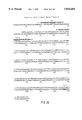
- FIG. 7 Nucleotide sequence of the coding strand for the B38.1 mouse V.sub. ⁇ region. Shown is the nucleotide sequence from the end of the oligo-dC tail to the J.sub. ⁇ l-C.sub. ⁇ junction. Also shown is the amino acid sequence deduced from the nucleotide sequence. Shown in bold are the oligonucleotides used for site-directed mutagenesis and the sites at which restriction site modifications were made.
- FIG. 8 Nucleotide sequence of the coding strand for the B38.1 H chain mouse V region. Shown is the nucleotide sequence from the end of the oligo-dC tail to the J H 4-C H 1 junction. Also shown is the amino acid sequence deduced from the nucleotide sequence. Shown in bold are the oligonucleotides used for site-directed mutagenesis and the sites at which restriction site modifications were made.
- the V region for the cDNA clone pR1G-8 was engineered to be compatible with the eukaryotic expression plasmid pING1714.
- Plasmid pING1714 contains the following gene regulatory elements useful for expression in mammalian cells: 1) an Abelson LTR promoter, 2) the SV40 19S/16S splice module, and 3) the SV40 polyadenylation signal sequence. It also contains the entire human IgG1 C region from pGMH-6 (Liu, A. Y., et al., (1987), supra).
- pING1714 contains the neomycin phosphotransferase gene which allows for G418 selection in transfected cells. Not drawn to scale.
- FIG. 10(A and B) Construction scheme for the chimeric mouse-human ING-1 L chain mammalian expression plasmid pING2207.
- the V region from the cDNA clone pRIK-7 was engineered to be compatible with the eukaryotic expression plasmid pING1712. See FIG. 2(A and B) for construction of plasmid pING1712. Not drawn to scale.
- FIG. 11 Construction scheme for yeast expression plasmid containing ING-1 chimeric L chain gene fused to the yeast PGK promoter (P), invertase signal sequence (S) and PGK polyadenylation signal (T). Not drawn to scale.
- FIG. 12 Construction for yeast expression plasmid containing ING-1 chimeric Fd chain gene fused to the yeast PGK promoter (P), invertase signal sequence (S), and PGK polyadenylation signal (T). Not drawn to scale.
- Plasmid pING3107 contains the following elements useful for expression in E. coli: 1) the araC gene, 2) the inducible araB promoter, 3) the dicistronic chimeric Fd and chimeric ⁇ ING-1 genes fused to the pelB leader sequence, 4) the trpA transcription termination sequence, and 5) the tet R gene, useful for selection in E. coli. Not drawn to scale.
- FIG. 14 Nucleotide sequence of the coding strand for the Br-3 mouse V.sub. ⁇ region. Shown is the nucleotide sequence from the end of the oligo-dC tail to the J.sub. ⁇ -C.sub. ⁇ junction. Also shown is the amino acid sequence deduced from the nucleotide sequence. Shown in bold is the oligonucleotide used for site-directed mutagenesis to introduce an ApaI site and the position of the AvrII site at the J-C.sub. ⁇ junction.
- FIG. 15 Nucleotide sequence of the coding strand for the Br-3 H chain mouse V region. Shown is the nucleotide sequence from the end of the oligo-dC tail to the J H 3-C H 1 junction. Also shown is the amino acid sequence deduced from the nucleotide sequence. Shown in bold are the oligonucleotides used for site-directed mutagenesis and the sites at which restriction site modifications were made. Also shown is the position of the PstI site near the J-C H 1 junction.
- FIG. 17 Construction scheme for plasmid pING1485, containing the ING-2 chimeric Fd chain gene with an AatII site at the gene sequence encoding the signal sequence processing site. V', fragment of V region gene sequences. Not drawn to scale.
- FIG. 18 Construction scheme for yeast expression plasmid containing ING-2 chimeric L chain gene fused to the yeast PGK promoter (P), invertase signal sequence and PGK polyadenylation signal (T). Not drawn to scale.
- FIG. 19 Construction scheme for yeast expression plasmid containing ING-2 chimeric Fd chain gene fused to the yeast PGK promoter (P), invertase signal sequence (S), and PGK polyadenylation signal (T).
- FIG. 21 Nucleotide sequence of the coding strand for the Co-1 ⁇ mouse V region. Shown is the nucleotide sequence from the end of the oligo-dC tail to the J.sub. ⁇ 5-C.sub. ⁇ junction. Also shown is the amino acid sequence deduced from the nucleotide sequence. Shown in bold are the oligonucleotides used for site directed mutagenesis and the sites at which restriction site modifications were made. Also in bold is the site of the MstII site useful for introduction of a SalI restriction site.
- FIG. 22 Nucleotide sequence of the coding strand for the Co-1 H chain mouse V region. Shown is the nucleotide sequence from the end of the oligo-dC tail to the J H 4-C H 1 junction. Also shown is the amino acid sequence deduced from the nucleotide sequence. In bold are the sites of the NcoI, BstEII and PstI sites useful for gene manipulation.
- the V region for the cDNA clone p01G-11 was engineered to be compatible with the eukaryotic expression plasmid pING2227. See FIG. 5 for construction of plasmid pING2227. Not drawn to scale.
- the V region from the cDNA clone p01K-8 was engineered to be compatible with the eukaryotic expression plasmid pING1712. See FIG. 2 for construction of plasmid pING1712. Not drawn to scale.
- FIG. 25 Construction scheme for yeast expression plasmid containing ING-3 chimeric L chain gene fused to the yeast PGK promoter, invertase signal sequence and PGK polyadenylation signal. Not drawn to scale.
- FIG. 26 Construction scheme for ING-3 chimeric Fd chain gene containing a PstI site at the gene sequence encoding the signal sequence processing site. Not drawn to scale.
- FIG. 27 Construction scheme for yeast expression plasmid containing ING-3 chimeric Fd chain gene fused to the yeast PGK promoter, invertase signal sequence and PGK polyadenylation signal. Not drawn to scale.
- Plasmid pING3307 contains the following elements useful for expression in E. coli: 1) the araC gene, 2) the inducible araB promoter, 3) the dicistronic Fd and ⁇ ING-3 genes fused to the pelB leader sequence, 4) the trpA transcription termination sequence, and 5) the tet R gene, useful for selection in E. coli. Not drawn to scale.
- FIG. 29 Nucleotide sequence of the coding strand for the ME4 mouse V.sub. ⁇ region. Shown is the nucleotide sequence from the end of the oligo-dC tail to the J.sub. ⁇ 1-C.sub. ⁇ junction. Also shown is the amino acid sequence deduced from the nucleotide sequence. Shown in bold are the oligonucleotides used for site-directed mutagenesis and the sites at which restriction site modifications were made.
- FIG. 30 Nucleotide sequence of the coding strand for the ME4 H chain mouse V region. Shown is the nucleotide sequence from the end of the oligo-dC tail to the J H 4-C H 1 junction. Also shown is the amino acid sequence deduced from the nucleotide sequence. Shown in bold are the oligonucleotides used for site-directed mutagenesis and the sites at which restriction site modifications were made.
- the V region for the cDNA clone pE4G-21 was engineered to be compatible with the eukaryotic expression plasmid pING2227.
- Plasmid pING2232 contains the following gene regulatory elements useful for expression in mammalian cells: 1) in IgH Enhancer element, 2) an Abelson LTR promoter, 3) the SV40 19S/16S splice module, and 4) the genomic human IgG1 polyadenylation signal sequence. It also contains the entire human IgG1 C region from pGMH-6 (Liu, A. Y. et al., supra). Not drawn to scale.
- the V region from the cDNA clone pE4K-15 was engineered to be compatible with the eukaryotic expression plasmid pING1712.
- Plasmid pING2216 contains the following gene regulatory elements useful for expression in mammalian cells: 1) the IgH enhancer element, 2) the Abelson LTR promoter, 3) the SV40 19S/16S splice module, and 4) a human ⁇ polyadenylation signal sequence. It also contains the entire human C.sub. ⁇ region (Liu A. Y., et al. supra) and the GPT gene which allows for mycophenolic acid resistance in transfected cells. Not drawn to scale.
- FIG. 33(A and B) Construction scheme for the fusion of the mature form of the ING-4 chimeric L chain gene to the yeast invertase signal sequence(s) under control of the yeast PGK promoter. Not drawn to scale.
- FIG. 34(A and B) Construction scheme for a yeast expression plasmid containing the ING-4 chimeric L chain and Fd genes fused to the yeast PGK promoter (P), invertase signal sequence(s) and PGK polyadenylation signal (T). Not drawn to scale.
- Plasmid pME4-B3 contains the following elements useful for expression in E. coli: 1) the araC gene, 2) the inducible araB promoter, 3) the dicistronic Fd and K ING-4 genes fused to the pelB leader sequence, 4) the trpA transcription termination sequence, and 5) the tet R gene, useful for selection in E. coli. Not drawn to scale.
- FIG. 36 Nucleotide sequence of the coding strand for the KM10 H chain mouse V region. Shown is the nucleotide sequence from the end of the oligo-dC tail to the J H 4-C H 1 junction. Also shown is the amino acid sequence deduced from the nucleotide sequence. Shown in bold are the oligonucleotides used for site directed mutagenesis and the sites at which restriction site modifications were made.
- FIG. 37 Nucleotide sequence of the coding strand for the KM10 mouse V.sub. ⁇ region. Shown is the nucleotide sequence from the end of the oligo-dC tail to the J.sub. ⁇ 5-C.sub. ⁇ junction. Also shown is the amino acid sequence deduced from the nucleotide sequence. Shown in bold are the oligonucleotides used for site directed mutagenesis and the sites at which restriction site modifications were made.
- the V region for the cDNA clone pM10G-2 was engineered to be compatible with the eukaryotic expression plasmid pING2227. See FIG. 5 for construction of plasmid pING2227. Not drawn to scale.
- the V region from the cDNA clone pM10K-16 was engineered to be compatible with the eukaryotic expression plasmid pING1712. See FIG. 2 for construction of plasmid pING1712. Not drawn to scale.
- FIG. 40 Yeast expression plasmids for Fab expression. Shown are: (a) the yeast expression plasmid containing KM10 chimeric L chain gene fused to the yeast PGK promoter, invertase signal sequence and PGK polyadenylation signal; (b) the similar yeast plasmid containing the Fd gene; (c) the yeast expression plasmid containing the L chain promoter/leader fusion with PGK transcription termination signal; (d) similar yeast plasmid containing the Fd gene; and (e) the final 2 gene yeast expression plasmid pING3200. Not drawn to scale.
- Plasmid pING3202 contains the following elements useful for expression in E. coli: 1) the araC gene, 2) the inducible araB promoter, 3) the dicistronic Fd and ⁇ KM10 genes fused to the pelB leader sequence, 4) the trpA transcription termination sequence, and 5) the tet R gene, useful for selection in E. coli. Not drawn to scale.
- the invention provides antibodies that are useful for the treatment and diagnosis of human cancer, either alone or in combination with other reagents.
- the tumor antigens to which such antibodies may be directed include those defined by the mAbs B38.1, Br-3, Co-1, ME4, and KM10.
- mRNA messenger RNA
- H and L chain J regions have different, but highly homologous (>80%) sequences, among each group, especially near the C region. This homology is exploited in this invention by using consensus sequences of L and H chain J regions to design oligonucleotides for use as primers or probes for introducing useful restriction sites into the J region for subsequent linkage of V region segments to human C region segments.
- C region cDNA module vectors prepared from human cells and modified by site-directed mutagenesis to place a restriction site at the analogous position in the human sequence were used.
- the complete human C.sub. ⁇ region and the complete human C.sub. ⁇ 1 region can be cloned.
- An alternative method utilizing genomic C H region clones as the source for C H region module vectors would not allow these genes to be expressed in systems such as bacteria where enzymes needed to remove intervening sequences are absent.
- V region segments are excised and ligated to L or H chain C region module vectors.
- human gamma 1 region can be modified by introducing a termination codon thereby generating a gene sequence which encodes the H chain portion of an Fab molecule.
- the coding sequences with operably linked V and C regions are then transferred into appropriate expression vehicles for expression in appropriate prokaryotic or eukaryotic hosts.
- Two coding DNA sequences are said to be "operably linked” if the linkage results in a continuously translatable sequence without alteration or interruption of the triplet reading frame.
- a DNA coding sequence is operably linked to a gene expression element if the linkage results in the proper function of that gene expression element to result in expression of the coding sequence.
- Expression vehicles include plasmids or other vectors. Preferred among these are vehicles carrying a functionally complete human C heavy (C H ) or C light (C L ) chain sequence having appropriate restriction sites engineered so that any variable H (V H ) or variable L (V L ) chain sequence with appropriate cohesive ends can be easily inserted thereinto.
- Human C H or C L chain sequence-containing vehicles are thus an important embodiment of the invention. These vehicles can be used as intermediates for the expression of any desired complete H or L chain in any appropriate host.
- yeast provides substantial advantages for the production of immunoglobulin L and H chains. Yeast cells carry out post-translational peptide modifications including glycosylation. A number of recombinant DNA strategies now exist which utilize strong promoter sequences and high copy number plasmids which can be used for production of the desired proteins in yeast. Yeast recognizes leader sequences of cloned mammalian gene products and secretes peptides bearing leader sequences (i.e., prepeptides) (Hitzman et al., 11th International Conference on Yeast, Genetics and Molecular Biology, adjoin, France, Sep. 13-17, 1982).
- Yeast gene expression systems can be routinely evaluated for the levels of production, secretion, and the stability of chimeric H and L chain proteins and assembled chimeric antibodies. Any of a series of yeast gene expression systems incorporating promoter and termination elements from the actively expressed genes coding for glycolytic enzymes produced in large quantities when yeasts are grown in media rich in glucose can be utilized. Known glycolytic genes can also provide very efficient transcription control signals. For example, the promoter and terminator signals of the iso-1-cytochrome C (CYC-1) gene can be utilized. A number of approaches may be taken for evaluating optimal expression plasmids for the expression of cloned immunoglobulin cDNAs in yeast.
- CYC-1-cytochrome C CYC-1-cytochrome C
- Bacterial strains may also be utilized as transformation hosts for the production of antibody molecules or antibody fragments described by this invention.
- E. coli K12 strains such as E. coli W3110 (ATCC 27325) and other enterobacteria such as Salmonella typhimurium or Serratia marcescens, and various Pseudomonas species may be used.
- Plasmid vectors containing replicon and control sequences which are derived from species compatible with a host cell are used in connection with these bacterial hosts.
- the vector carries a replication site, as well as specific genes which are capable of providing phenotypic selection in transformed cells.
- a number of approaches may be taken for evaluating the expression plasmids for the production of chimeric antibodies or antibody chains encoded by the cloned immunoglobulin cDNAs in bacteria.
- Mammalian cells are grown in vitro or in vivo.
- Mammalian cells provide post-translational modifications to immunoglobulin protein molecules including leader peptide removal, folding and assembly of H and L chains, glycosylation of the antibody molecules, and secretion of functional antibody protein.
- Mammalian cells which may be useful as hosts for the production of antibody proteins include cells of lymphoid origin, such as the hybridoma Sp2/0-Ag14 (ATCC CRL 1581) or the myeloma P3C63Ag8 (ATCC TIB 9), and its derivatives. Others include cells of fibroblast origin, such as Vero (ATCC CRL 81) or CHO-K1 (ATCC CRL 61).
- H and L chain genes are available for the expression of cloned H and L chain genes in mammalian cells. Different approaches can be followed to obtain complete H 2 L 2 antibodies. It is possible to co-express L and H chains in the same cells to achieve intracellular association and linkage of H and L chains into complete tetrameric H 2 L 2 antibodies. The co-expression can occur by using either the same or different plasmids in the same host. Genes for both H and L chains can be placed into the same plasmid, which is then transfected into cells, thereby selecting directly for cells that express both chains.
- cells may be transfected first with a plasmid encoding one chain, for example L chain, followed by transfection of the resulting cell line with a H chain plasmid containing a second selectable marker.
- a plasmid encoding one chain for example L chain
- H chain plasmid containing a second selectable marker for example L chain
- Cell lines producing H 2 L 2 molecules via either route could be transfected with plasmids encoding additional copies of L, H, or L plus H chains in conjunction with additional selectable markers to generate cell lines with enhanced properties, such as higher production of assembled (H 2 L 2 ) antibody molecules or enhanced stability of the transfected cell lines.
- the invention is also directed to combinations of gene expression elements, and recombinant DNA vectors containing these elements, useful for the expression of immunoglobulin L chain and H chain cDNA genes in a desired host mammalian cell.
- the transcriptional promoter is a viral LTR sequence
- the transcriptional promoter enhancer(s) are either or both the mouse immunoglobulin H chain enhancer and the viral LTR enhancer
- the splice region contains an intron of greater than 31 bp
- the polyadenylation and transcription termination regions are derived from a native chromosomal sequence corresponding to the immunoglobulin chain being synthesized.
- the invention can be used to construct recombinant DNA expression vehicles to achieve efficient synthesis of antibodies from transfected host cells.
- a vehicle would be constructed by the ligation of gene expression modules to antibody coding sequences to form a recombinant DNA molecule.
- This recombinant DNA can then be used to transfect mammalian hosts such as Sp2/0 hybridoma or chinese hamster ovary cells.
- a recombinant DNA gene expression unit for L chain synthesis can be constructed by the ligation of the following gene expression elements:
- H chain immunoglobulin transcription enhancer sequence such as the 0.7-kb XbaI to EcoRI DNA fragment from the mouse genomic H chain immunoglobulin DNA sequence
- a retroviral LTR transcription promoter sequence such as Abelson murine leukemia virus LTR;
- a 3' untranslated sequence including a polyadenylation signal sequence (AATAAA), such as that from human immunoglobulin n cDNA, and
- the order of the promoter (ii), coding sequence (iv), polyadenylation signal sequence (v), and polyadenylation and transcription termination (vi) elements is fixed.
- the splice region (iii) may be located at any position after the promoter (ii), but before the polyadenylation signal sequence (v).
- the splice region may be located within other elements such as the cDNA coding sequence.
- the enhancer (i) may be located anywhere in or near the unit, and may be located within some elements, such as the intervening sequence between splice donor and splice acceptor.
- the L chain gene expression unit can be ligated to other useful DNA sequences, such as selectable marker genes from pSV-2neo or pSV-2gpt, prior to transfection of host cells.
- a recombinant DNA gene expression unit for H chain synthesis can be constructed by the ligation of the following gene expression elements:
- H chain immunoglobulin transcription enhancer sequence such as the 0.7-kb XbaI to EcoRI DNA fragment from mouse genomic DNA sequences
- a retroviral LTR transcription promoter sequence such as Abelson murine leukemia virus LTR;
- a DNA sequence containing splice donor and splice acceptor sites such as the SV40 19S/16S splice donor and the 16S splice acceptor, separated by greater than 31 bp of intervening sequence;
- the order of the H chain gene expression elements has the same limitations as the L chain gene expression modules.
- the assembled H chain gene expression unit can be ligated to other useful DNA sequences, such as selectable marker genes, prior to the transfection of host cells.
- the assembled H chain expression vehicle may contain a different selectable marker than that chosen for the L chain gene expression vehicle.
- H and L chain gene expression vehicles can be transfected together (co-transfection) or in separate steps (sequential transfection). Both L and H chain gene expression units may be assembled in the same expression vehicle, in which only a single selectable marker may be used.
- the invention provides "chimeric" immunoglobulin chains, either H or L with specificity to human tumor antigens.
- a chimeric chain contains a C region substantially similar to that present in a natural human immunoglobulin, and a V region having the desired anti-tumor specificity of the invention.
- the invention also provides immunoglobulin molecules having H and L chains associated so that the overall molecule exhibits the desired binding and recognition properties.
- Various types of immunoglobulin molecules are provided: monovalent, divalent, or molecules with the specificity-determining V binding domains attached to moieties carrying desired functions.
- This invention also provides for fragments of chimeric immunoglobulin molecules such as Fab, Fab', or F(ab') 2 molecules or those proteins coded by truncated genes to yield molecular species functionally resembling these fragments.
- Antibodies having chimeric H chains and L chains of the same or different V region binding specificity can be prepared by appropriate association of the desired polypeptide chains. These chains are individually prepared by the modular assembly methods of the invention.
- the antibodies of this invention can be used for therapeutic purposes by themselves, for example, acting via complement-mediated lysis and antibody-dependent cellular cytotoxicity, or coupled to toxins or therapeutic moieties, such as ricin, radionuclides, drugs, etc., in the treatment of human cancer.
- the antibodies may be advantageously utilized in combination with factors, such as lymphokines, colony-stimulating factors, and the like, which increase the number or activity of antibody-dependent effector cells.
- the invention is also directed to a method of killing cells carrying an antigen thereon, such method comprising contacting the cells with a chimeric antibody molecule that contains two light chains and two heavy chains, each of said chains containing a constant human region and a variable region having specificity to an antigen bound by a murine monoclonal antibody selected from the group consisting of B38.1, Br-3, Co-1, ME4 and KM10.
- the antibody may be in detectably labeled form.
- the antibodies of the invention having human C region can be utilized for passive immunization, especially in humans, with reduced negative immune reactions such as serum sickness or anaphylacticshock, as compared to whole mouse antibodies.
- the antibodies can also be utilized in prior art immunodiagnostic assays and kits in detectably labeled form (e.g., enzymes, 125 I, 14 C, fluorescent labels, etc.), or in immobilized form (on polymeric tubes, beads, etc.). They may also be utilized in labeled form for in vivo imaging, wherein the label can be a radioactive emitter, or a nuclear magnetic resonance contrasting agent such as a heavy metal nucleus, or an X-ray contrasting agent, such as a heavy metal.
- the antibodies can also be used for in vitro localization of the recognized tumor cell antigen by appropriate labeling.
- Mixed antibody-enzyme molecules can be used for immunodiagnostic methods, such as ELISA.
- Mixed antibody-peptide effector conjugates can be used for targeted delivery of the effector moiety with a high degree of efficacy and specificity.
- the chimeric antibodies of this invention can be used for any and all uses in which the original murine mAbs can be used, with the obvious advantage that the chimeric ones are more compatible with the human body.
- a series of recombinant DNA vectors were constructed to test different gene expression elements in order to optimize the expression of chimeric mouse-human immunoglobulin L chain.
- a vector to test different promoters and splice regions was first made (pING2122, FIG. 1). This vector is derived from a chimeric L chain cDNA expression plasmid, pING2121b.
- the mouse immunoglobulin H chain enhancer 0.7-kb XbaI to EcoRI fragment from M13 M8 ⁇ RX12 Robotson, R. R. et al., PCT US86/02269 was inserted into XbaI plus EcoRI cut M13mp19.
- the enhancer-containing HindIII to BglII fragment was inserted into the BglII to HindIII region of pSH6, an E. coli recombinant plasmid DNA that contains unique XhoI, BglII, and HindIII sites, with the BglII between the XhoI and HindIII sites.
- the enhancer-containing XbaI to XhoI fragment was then inserted into the enhancer XbaI to XhoI region of pING2121b, an expression plasmid identical to pING2108b (Liu, A. Y. et al., J.
- pING2122 (FIG. 1) and contained a region of DNA between unique EcoRI and XhoI sites for the insertion of various promoters to be tested.
- pING2122 has an SV40 19S splice region between the XhoI and unique SalI sites, allowing the insertion (or deletion) of alternate splice regions.
- promoters were obtained and introduced as EcoRI to SalI regions into the pING2122 vector to compare their transcriptional strength to the SV40 viral promoter in the reference expression plasmid pING2121b.
- the promoters chosen were the H chain immunoglobulin V1 (V1(Igh)), mouse metallothionein (MMT), Abelson virus LTR (Abl), and Rous sarcoma virus LTR (RSV) promoters.
- the mouse H chain V1(Igh) promoter was obtained as a 600-bp BamHI DNA fragment derived from the V1 gene promoter (Clarke, C. et al., Nucleic Acids Research 10:7731-7749 (1982)).
- the DNA fragment was inserted into the BamHI site of pUC19.
- the VI promoter was excised as an EcoRI to SalI fragment and first ligated to the large fragment of pING2122 cut with EcoRI and XhoI to form pING2123 (V1 promoter plus SV40 19S splice).
- the V1 promoter DNA fragment was next ligated to pING2122 cut with EcoRI and SalI to form pING2124 (V1 promoter with no splice).
- the Abelson LTR promoter was obtained from pelin2 (provided by Dr. Owen Witte, UCLA); pelin2 contains the p120 viral 3' LTR (Reddy, E. P. et al., Proc. Natl. Acad. Sci. USA 80:3623 (1983)), except that the BglII site at viral position 4623 has been modified by insertion of the EcoRI oligonucleotide linker GGAATTCC.
- the 0.8-kb EcoRI to KpnI fragment of pelin2 containing the p120 3' LTR promoter was inserted into KpnI plus EcoRI cut pUC18.
- the LTR was excised as an EcoRI to SalI fragment and ligated to EcoRI plus XhoI cut pING2122, placing the LTR promoter adjacent to the L6 L chain gene to form pING2125 (Abl promoter with SV40 19S splice).
- the Abl LTR fragment was similarly ligated to EcoRI plus SalI cut pING2122 to form plasmid pING2126 (Abl promoter with no splice).
- the metallothionein MMT promoter was obtained from the plasmid pBPV-MMT neo (American Type Culture Collection #37224) and was excised as an EcoRI to BglII fragment and ligated to EcoRI plus BamHI cut pUC19.
- the EcoRI to SalI DNA fragment of the resulting plasmid was excised and ligated to EcoRI plus XhoI cut pING2122 to form pING2131 (MMT promoter plus SV40 19S splice).
- the RSV promoter was obtained from pRSVcat (American Type Culture Collection #37152) and was excised as a 560-bp HindIII to SfaNI DNA fragment. After T4 DNA ploymerase treatment, the fragment was ligated to SmaI cut M13mp19 and the resultant recombinant phage were selected for the orientation where the insert EcoRI site was closest to the vector SalI site. The RSV promoter DNA was then excised by partial digestion with EcoRI followed by complete SalI digestion and ligated to the large DNA fragment from EcoRI plus XhoI cut pING2122 to form pING2132 (RSV promoter plus SV40 19S splice).
- the reference expression plasmid pING2121b (SV40 promoter plus SV40 19S splice) was modified to incorporate different splice regions.
- pING2121b was digested with XhoI and SalI and self-ligated to delete the splice region, forming pING2128 (SV40 promoter, no splice).
- the SV40 19S/16S splice region (SV40 19S/16S splice donor and splice acceptor) was excised as a 174 bp XhoI to SalI DNA fragment from pUC12/pL1 (Robinson, R. R.
- the expression vectors were modified to include L chain genomic polyadenylation and transcription termination regions.
- the first step was the HindIII digestion and religation of plasmid pING2121a, which is identical to pING2108a described by Liu, A. Y. et al., J. Immunology 139:3521 (1987), with one exception. It differed in that the V L region used in its construction was from the L6 mAb (Liu, A. Y. et al., Proc. Natl. Acad. Sci., USA 84:3439 (1987)) rather than the 2H7 mAb.
- This modified pING2121a was termed pING2121a-deltaH.
- the 1.1-kb BglII to BamHI fragment of mouse genomic DNA distal to the polyadenylation site was isolated from pS107A (provided by Dr. Randolph Wall, UCLA) and inserted into the BamHI site of pING2121a-deltaH, screening for the orientation homologous to the native gene.
- the 3.3 kb BglII to SstI fragment containing this modified 3' region was ligated to the 5.2 kb BglII to SstI fragment of pING2121b to form pING1703 (SV40 promoter, mouse ⁇ genomic 3' region).
- FIG. 2 shows the construction of pING1712.
- the plasmid expression vector DNAs described above were used to transfect mouse hybridoma Sp2/0 cells to test the efficacy of the various promoters and splice regions. Each plasmid DNA was linearized by PvuI digestion and transfected into Sp2/0 cells by electroporation (Potter, H. et al., Proc. Natl. Acad. Sci. USA 81:7161 (1984)).
- the electroporation conditions were: 10 to 20 ⁇ g of linearized DNA mixed with 10 ⁇ 10 6 Sp2/0 cells in 0.5 ml of phosphate buffered saline (IRVINE Scientific #9236), using a 5-msec pulse of 300 V from a BTX-T100 transfector and 471 cuvette electrode (Biotechnologies and Experimental Research, San Diego, Calif.).
- DMEM growth medium Dulbecco's modified Eagle medium plus 10% fetal bovine serum, GIBCO
- GIBCO fetal bovine serum
- DMEM growth medium supplemented with 0.006 mg/ml mycophenolic acid (Calbiochem) and 0.25 mg/ml xanthine (SIGMA)
- DMEM growth medium supplemented with 0.006 mg/ml mycophenolic acid (Calbiochem) and 0.25 mg/ml xanthine (SIGMA)
- Individual wells positive for cell growth were expanded in mycophenolic acid growth medium prior to assay of secreted or intracellular L chain by enzymelinked immunosorbent assay (ELISA).
- ELISA enzymelinked immunosorbent assay
- Experiment 7 compares the different polyadenylation and transcription termination regions.
- the use of either mouse or human genomic polyadenylation and transcription termination regions appears to result in more efficient synthesis than does the use of the SV40 viral polyadenylation region.
- the combination of the most efficient promoter, splice region, and polyadenylation and transcription termination region (pING1712) is tested in experiments 8 and 9. These experiments show that the combination of gene expression elements in pING1712 are far more efficient than the starting plasmid pING2121b, giving a 6 to 17-fold increase in ⁇ expression level.
- An approach to improve immunoglobulin H chain synthesis involves creating an L chain producing cell line, and then transfecting that cell line with H chain expression plasmids containing various gene expression elements.
- a chimeric mouse-human ⁇ plasmid expression vector, pING2203 was constructed and then used to transfect murine Sp2/0 cells. The construction required the combination of a human C.sub. ⁇ region gene module with a mouse V.sub. ⁇ region.
- a human C.sub. ⁇ gene cassette was constructed from the genomic human C.sub. ⁇ region of plasmid pHu.sub. ⁇ 1 (Hieter, P. et al., Nature 294:536-540 (1981)). Initially, two BamHI to SstI fragments were subcloned into M13mp19 to generate pLJ6 and pLJ8. Plasmid pLJ6 contains the DNA sequence coding the 3' end of the human J ⁇ -C ⁇ intron and the 5' end of the human C.sub. ⁇ exon. The nucleotide sequence of this region was determined, and site-directed mutagenesis was used to insert an AvrII site at the intron/exon junction.
- This restriction site was chosen so that an in-frame fusion could be made between the conserved AvrII site at the mouse J-C junction from any mouse V.sub. ⁇ cDNA clone and the human C.sub. ⁇ module.
- the oligonucleotide used for introduction of an AvrII site was:. 5'-CCTTGGGCTGACCTAGGTGGA-3'.
- the derivative of pLJ6 containing an AvrII site was called pING1459.
- pBR322NA is pBR322 containing a deletion between NdeI and AvaI
- pING1462 was further modified to include the human ⁇ polyadenylation region by ligation of the C.sub. ⁇ gene module to the contiguous 3' region (FIG. 4) of genomic DNA from pHu ⁇ 1, forming the human C.sub. ⁇ region vector pING2201.
- RNA isolated from a hybridoma cell line secreting the Br-3 mouse mAb (IgG1, ⁇ ) (see Background) was used to generate a plasmid cDNA library by the method of Gubler and Hoffman (Gene 25:263 (1983)). From this library, a cDNA clone encoding the entire Br-3 ⁇ chain was isolated.
- This cDNA clone, pR3L-11 was modified for expression in mammalian cells as follows.
- the oligo-dC sequence 5' to the V ⁇ module in pR3L-11 was removed in a number of steps that resulted in the positioning of a SalI restriction site upstream of the coding sequence for Br-3 ⁇ .
- a PstI to SstI subclone of pR3L-11 was made in M13mp19 to generate pL5.
- This plasmid was cut with HindIII and treated with Bal31 nuclease to remove the oligo-dC tail, followed by T4-polymerase treatment, EcoRI digestion and subcloning into SmaI and EcoRI digested pUC18.
- This located a SalI site upstream of the V ⁇ , region (plasmid pWW9).
- a BamHI site was deleted between the SalI site and the lambda initiation codon by digestion with BamHI followed by mung bean nuclease digestion and religation to generate pWW9-2.
- the nucleotide sequence around the SalI site was determined to be GTCGACTCCCCGAAAAGAATAGACCTGGTTTGTGAATTATG, where the SalI site and initiation codon ATG are underlined.
- the chimeric Br-3 ⁇ gene was constructed in a three-piece ligation from the vector AvrII to SalI fragment from pING2201, the mouse V.sub. ⁇ region SalI to SstI fragment from pWW9-2, and the SstI to AvrII fragment from pR3L-11, generating pING2202 (FIG. 4).
- the final chimeric L chain expression vector, pING2203 was constructed in a three-piece ligation from pING1712 (BglII to SalI fragment containing the IgH enhancer, Abelson LTR promoter, and SV40 16S splice), pING2003Egpt (Robinson, R. R.
- pING2203 is the chimeric Br-3 ⁇ equivalent of the chimeric L6 ⁇ producer, pING1712 (Example 1).
- a H chain cDNA clone was isolated which contained the entire coding region of the Br-3 immunoglobulin H chain.
- This cDNA clone, pR3G-11 was adapted for expression in mammalian cells as outlined in FIG. 5.
- the Br-3 J region is J H 3 and contains a natural Pst1 site near the V-C H 1 junction that can be used to link the mouse V and human C modules.
- a homologous PstI site was therefore inserted into a human C.sub. ⁇ 1 cDNA gene module.
- a portion of the human C.sub. ⁇ 1 region fused to the L6 specificity (Liu, A. Y. et al., Proc. Natl. Acad.
- the modified chimeric mouse V-human C H 1 plasmid was used in a three-way ligation to form the expression vector, pING2206 (see FIG. 5).
- the vector sequences containing the Abl LTR promoter were from pING1714, which was constructed as shown in FIG. 6.
- the SV40 expression plasmid pING111 Robotson et al., PCT US86/02269 was modified by the insertion of an AatII oligonucleotide linker at the XbaI site, followed by AatII cleavage and religation to form pING1707.
- the AatII to SalI fragment containing the Abelson LTR promoter was excised from pING2133 and ligated to the large AatII to SalI fragment of pING1707 to form pING1711.
- the H chain enhancer was deleted from pING1711 by EcoRI digestion, T4 polymerase treatment, ligation to AatII oligonucleotide linker, and cleavage and religation with AatII to form the 7.7 kb expression vector pING1714 shown in FIGS. 5 and 6.
- the reconstructed chimeric H chain expression plasmid pING2206 was modified in the promoter-enhancer region in two ways. First, a H chain IgH enhancer was placed upstream of the Abl promoter to form pING2217. Second, substitution of the AatII to SalI region of pING2217 (H chain enhancer, Abl promoter, 16S splice region) with the homologous region of pING2127 generated pING2218 (H chain enhancer, SV40 promoter, 16S splice region).
- pING2206 was also modified at the 3' end in two ways.
- the poly-A stretch from the human IgG1 cDNA was deleted between the BamHI site immediately downstream of the poly-A and the XmaIII site immediately 3' to the IgG1 stop codon, bringing the SV40 polyadenylation region closer to the IgG1 gene and generating pING2220.
- the 1300 nucleotide XmaIII fragment from human IgG1 genomic DNA (Ellison et al., Nucleic Acid Res. 10:4071 (1982)) was added at the XmaIII site 3' to the stop codon, generating pING2219.
- pING2219 thus contains the genomic human IgG1 polyadenylation signal and site.
- a L chain producing cell line was first made by transfection of Sp2/0 cells with pING2203 by the electroporation method in Example 1 and subcloning to generate cell line 22031B5.14.
- the 22031B5.14 cell line was subsequently transfected by electroporation with the various chimeric Br-3 H chain expression plasmid DNAs.
- Cells expressing the associated selectable neo r gene were selected by growth in complete DMEM medium supplemented with 0.8 mg/ml G418 (GIBC0).
- Cultures from 96-well plates containing G418-resistant cells were expanded to 48-well plates and the secreted chimeric mouse-human H chain of terminal cultures was measured by ELISA specific for human gamma chains.
- the transfection results are summarized in Table 2.
- the results show that the Abl viral LTR promoter is more efficient than the SV40 promoter, and that the inclusion of an immunoglobulin H chain enhancer is advantageous for the Abl LTR promoter driving a H chain cDNA gene.
- the second experiment compares various 3' configurations of the chimeric Br-3 expression unit.
- the construct containing the IgG1 H chain genomic polyadenylation region (pING2219) results in higher H chain expression than those which contain only the SV40 polyadenylation region (pING2220), or the cDNA poly-A stretch and SV40 polyadenylation region (pING2206).
- an improved vector for the expression of chimeric Br-3 H chain would combine the immunoglobulin H chain enhancer, Abl LTR promoter, and the human genomic polyadenylation region. Accordingly, the DNA fragment containing the improved gene expression elements from pING2217 (H chain enhancer, Abl LTR promoter, 16S splice) was ligated to that from pING2219 (genomic polyadenylation region) to generate the improved chimeric Br-3 H chain gene expression vector, pING2227. This vector was used for the transfection of chimeric Br-3 L chain producing cells to achieve efficient synthesis of chimeric Br-3 H chain and the resulting fully-assembled chimeric antibody.
- gene expression elements are also useful for expression of immunoglobulins in cells other than mouse Sp2/0 hybridoma cells.
- CHO cells were transfected with pING1712+pING1714 to yield efficient synthesis of fully assembled chimeric antibodies.
- the plasmids pUC18, pUC19, and dG-tailed pBR322, pSV2-neo and pSV2-gpt were purchased from BRL (Gaithersburg, Md.) as were M13mp18 and M13mp19.
- DNA manipulations involving purification of plasmid DNA by bouyant density centrifugation, restriction endonuclease digestion, purification of DNA fragments by agarose gel electrophoresis, ligation and transformation of E. coli were as described by Maniatis, T., et al., Molecular Cloning: A Laboratorv Manual (1982), or other standard procedures. Restriction endonucleases and other DNA/RNA modifying enzymes were purchased from Boehringer-Mannheim (Indianapolis, Ind.), BRL, and New England Biolabs (Beverly, Mass.).
- B38.1 hybridoma cells at approximately 1 ⁇ 10 6 cells/ml were collected by centrifugation and washed in 100 ml of PBS (8 g NaCl, 0.2 g KH 2 P0 4 , 1.15 g Na 2 HP0 4 , and 0.2 g KCl per liter). The cells were centrifuged again and the cell pellet was suspended in a solution of guanidine thiocyanate, and total cellular RNA and poly(A) + RNA were prepared from tissue culture cells as described in Maniatis, T., et al., supra.
- Oligo-dT primed cDNA libraries were prepared from poly(A) + RNA by the methods of Gubler, V., and Hoffman, B. J., Gene 25:263 (1983). The cDNA was dC-tailed with terminal deoxynucleotide transferase and annealed to dG-tailed pBR322. cDNA libraries were screened by hybridization (Maniatis, T. et al., supra) with 32 P-labeled, nick-translated DNA fragments, i.e., for ⁇ clones with a mouse C.sub. ⁇ region probe and for H chain clones with an IgG1 C region probe.
- the nucleotide sequences of the cDNA clones show that they are immunoglobulin V region clones as they contain amino acid residues diagnostic of V domains (Kabat et al., Sequences of Proteins of Immunological Interest; U.S. Dept. of HHS, 1983).
- the B38.1 VH belongs to subgroup II.
- the B38.1 V H has the J H 4 sequence.
- B38.1 V.sub. ⁇ has the J.sub. ⁇ 1 sequence.
- Oligo-dT primed cDNA libraries were prepared from poly(A) + RNA by the methods of Gubler, V., and Hoffman, B. J., supra. The cDNA was dC-tailed with terminal deoxynucleotide transferase and annealed to dG-tailed pBR322. cDNA libraries were screened by hybridization (Maniatis, T., et al., supra) with 32 P-labeled,. nick-translated DNA fragments, i.e., for ⁇ clones with a mouse C.sub. ⁇ region probe and for H chain clones with an IgG1 C region probe.
- the nucleotide sequences of the cDNA clones show that they are immunoglobulin V region clones as they contain amino acid residues diagnostic of V domains (Kabat et al., supra.)
- the Br-3 V H belongs to subgroup IIIC.
- the Br-3 V H has the J H 3 sequence and the Br-3 V L has the J.sub. ⁇ sequence.
- Co-1 hybridoma cells at approximately 1 ⁇ 10 6 cells/ml were collected by centrifugation and washed in 100 ml of PBS (8 g NaCl, 0.2 g KH2P04, 1.15 g Na2HP04,and 0.2 g KCl per liter). The cells were centrifuged again and the cell pellet was suspended in a solution of guanidine thiocyanate, and total cellular RNA and poly(A) + RNA were prepared from tissue culture cells by methods described in Maniatis, T., et al., supra.
- Oligo-dT primed cDNA libraries were prepared from poly(A) + RNA by the methods of Gubler, V. and Hoffman, B. J., supra. The cDNA was dC-tailed with terminal deoxynucleotide transferase and annealed to dG-tailed pBR322. cDNA libraries were screened by hybridization (Maniatis, T., supra) with 32 P-labelled, nick translated DNA fragments, ie., for ⁇ clones with a mouse C.sub. ⁇ region probe and for H chain clones with a mouse IgG1 C region probe.
- the nucleotide sequences of the cDNA clones show that they are immunoglobulin V region clones as they contain amino acid residues diagnostic of V domains (Kabat et al., supra).
- the Co-1 V H belongs to subgroup II.
- the Co-1 V H has the J H 4 sequence and the Co-1 V.sub. ⁇ has the J.sub. ⁇ 5 sequence.
- ME4 hybridoma cells at approximately 1 ⁇ 10 6 cells/ml were collected by centrifugation and washed in 100 ml of PBS (8 g NaCl, 0.2 g KH 2 P0 4 , 1.15 g Na 2 HP0 4 , and 0.2 g KCl per liter). The cells were centrifuged again and the cell pellet was suspended in a solution of guanidine thiocyanate, and total cellular RNA and poly(A) + RNA were prepared from tissue culture cells by methods described in Maniatis, T., et al., supra.
- Oligo-dT primed cDNA libraries were prepared from poly(A) + RNA by the methods of Gubler, V., and Hoffman, B. J., supra. The cDNA was dC-tailed with terminal deoxynucleotide transferase and annealed to dG-tailed pBR322. cDNA libraries were screened by hybridization (Maniatis, T., supra) with 32 P-labeled, nick-translated DNA fragments, i.e., for ⁇ clones with a mouse C.sub. ⁇ region probe and for H chain clones with an IgG1 C region probe.
- the nucleotide sequences of the cDNA clones show that they are immunoglobulin V region clones as they contain amino acid residues diagnostic of V domains (Kabat et al., supra.)
- the ME4 V H belongs to subgroup II.
- the ME4 V H has the J H 4 sequence and the ME4 V.sub. ⁇ has the J.sub. ⁇ 1 sequence.
- KM10 hybridoma cells at approximately 1 ⁇ 10 6 cells/ml were collected by centrifugation and washed in 100 ml of PBS (8 g NaCl, 0.2 g KH 2 P0 4 , 1.15 g Na 2 HP0 4 ,and 0.2 g KCl per liter). The cells were centrifuged again and the cell pellet was suspended in a solution of guanidine thiocyanate, and total cellular RNA and poly(A) + RNA were prepared from tissue culture cells by methods described in Maniatis, T., et al., supra.
- Oligo-dT primed cDNA libraries were prepared from poly(A) + RNA as above.
- the cDNA was dC-tailed with terminal deoxynucleotide transferase and annealed to dG-tailed pBR322.
- cDNA libraries were screened by hybridization (Maniatis, T., supra) with 32 P-labelled, nick translated DNA fragments, i.e., for ⁇ clones with a mouse C.sub. ⁇ region probe and for H chain clones with a mouse IgG1 C region probe.
- the nucleotide sequences of the cDNA clones show that they are immunoglobulin V region clones as they contain amino acid residues diagnostic of V domains (Kabat et al., supra).
- the KM10 V H belongs to subgroup II.
- the KM10 V H has the J H 4 sequence and the KM10 V.sub. ⁇ has the J.sub. ⁇ 5 sequence.
- M13 subcloned DNA fragments were subjected to site-directed mutagenesis as described by Kramer, W., et al., Nucl. Acids Res. 12:9441. Oligonucleotides were purchased from Synthetic Genetics, San Diego, Calif., in their purified form.
- the J-region mutagenesis primer V.sub. ⁇ HindIII, 5'-GTTTGATTTCAAGCTTGGTGC-3' was utilized for the B38.1 V.sub. ⁇ .
- the human C.sub. ⁇ , module derived from a cDNA clone was previously mutagenized to contain a HindIII site in the human J region. Liu, A. Y., et al., Proc. Natl. Acad. Sci. USA 84:3439-3443 (1987).
- a SalI restriction site was introduced 5' to the ATG initiation codon by site-directed mutagenesis with the primer 5'-ATGGTGAGTCGACAGTGACCCCC-3.
- the cDNA clone containing the B38.1 H chain was likewise adapted for expression by introducing a BstEII site into the J H 4 for linkage to the human C domain, and a SalI site was introduced 5' of the ATG initiation codon by site-directed mutagenesis.
- the oligonucleotide used for mutagenesis to introduce a BstEII site was 5'-GAGACGGTGACCGAGGTTCC-3' and that for the SalI site was 5' -GAAGTGGTGCCTGTCGACTAACTGGTC-3'.
- M13 subcloned DNA fragments were subjected to site-directed mutagenesis as described by Kramer, W., et al., Nucl. Acids Res. 12:9441. Oligonucleotides were purchased from Synthetic Genetics, San Diego, Calif., in their purified form.
- the J-region mutagenesis primer J.sub. ⁇ 1HindIII, 5'-GTTTGATTTCAAGCTTGGTGC-3' was utilized for the ME4 V.sub. ⁇ .
- the human C.sub. ⁇ module derived from a cDNA clone was previously mutagenized to contain a HindIII site in the human J region. Liu, A. Y., et al. (1987) supra.
- a SalI restriction site was introduced 5' to the ATG initiation codon by site-directed mutagenesis with the primer 5'-GGACATCATGTCGACGGATACGAGC-3'.
- the cDNA clone containing the ME4 H chain was likewise adapted for expression by introducing a BstEII site into the J H 4 for linkage to the human C domain, and a SalI site was introduced 5' of the ATG initiation codon by site-directed mutagenesis.
- the oligonucleotide used for mutagenesis to introduce a BstEII site was 5'-GAGACGGTGACCGAGGTTCC-3'.
- the chimeric H chain expression plasmids were derived from the replacement of the V H module in pING1714 with the V H modules of pING1604, as a SalI to BstEII fragment as outlined in FIG. 9.
- This plasmid, pING2225 directs the synthesis of chimeric ING-1 H chain when transfected into mammalian cells.
- the SalI to HindIII fragment of the mouse V K module from pING1474 was joined to the human CK module in pING1712 by the procedure outlined in FIG. 10, forming pING2207.
- a derivative of the human C gene cDNA clone pGMH-6, Liu, A. Y., et al., Proc. Natl. Acad. Sci. USA 84:3439-3443 (1987), found in pING1714 contains a BstEII site to which the Co-1 H chain V-region can be ligated.
- the final H chain expression plasmid is pING2234.
- the SalI to HindIII fragment of the mouse V.sub. ⁇ module from pING1471 was joined to the human C.sub. ⁇ module in pING1712 by the procedure outlined in FIG. 24, forming pING2204.
- the chimeric H chain expression plasmid was derived from the replacement of the V H module in pING2227 with the ME4 V H -J-C H module assembled in pING2222 (FIG. 31).
- Two restriction fragments from pING2222 containing H chain sequences-(SalI to ApaI and ApaI to SstII) were ligated to pING2227 cut with SalI and SstII (FIG. 31D, E) to generate pING2232.
- This plasmid directs the synthesis of chimeric ING-4 H chain when transfected into mammalian cells.
- FIG. 31 details the cloning steps required to manipulate the cDNA V H region into pING2232.
- the SalI to HindIII fragment of the mouse V.sub. ⁇ module from pING1488 was joined to the human C K module in pING1712 by the procedure outlined in FIG. 32, forming pING2216.
- the cDNA clone containing the KM10 H chain, pK10G was adapted for mammalian expression by introducing convenient restriction endonucleases sites by site directed mutagenesis (Kramer, W., et al., (1984), supra) into appropriate M13 subclones (FIG. 39). Oligonucleotides were synthesized on a Cyclone DNA synthesizer, New Brunswick Scientific Co., and purified by acrylamide gel electrophoresis.
- the J-region mutagenesis primer 5' -GAGACGGTGACCGAGGTTCC-3' was used to insert a BstEII site into the M13 subclone p4G2, and the oligonucleotide 5'-ATCCATGATGTCGACGACCTTGGGC-3' was used to insert a SalI restriction site into pR6C upstream of the initiation codon ATG.
- the restriction fragment containing the KM10 H chain V-region bounded by SalI and BstEII was then cloned into the expression vector pING2227.
- the J-region mutagenesis primer 5'-CAGCTCAAGCTTGGTCCC-3' was used to insert a HindIII site into the M13 subclone p4K14, and the oligonucleotide 5'-GGATTTTGGTCGACGGCTAATTAGTG-3' was used to insert a SalI restriction site into p4BD upstream of the initiation codon ATG.
- the restriction fragment containing the KM10 L chain V-region bounded by SalI and HindIII was then cloned into the expression vector pING1712.
- the cell line Sp2/0 (American Type Culture Collection # CRL1581) was grown in Dulbecco's Modified Eagle Medium plus 4.5 g/l glucose (DMEM, Gibco) plus 10% fetal bovine serum. Media were supplemented with glutamine/penicillin/streptomycin (Irvine Scientific, Irvine, Calif.).
- the electroporation method of Potter, H., et al. (Proc. Natl. Acad. Sci. USA 81:7161 (1984)) was used. After transfection, cells were allowed to recover in complete DMEM for 24 hours, and then seeded at 10,000 to 50,000 cells per well in 96-well culture plates in the presence of selective medium. G418 (GIBCO) selection was at 0.8 mg/ml, and mycophenolic acid (Calbiochem) was at 6 pg/ml plus 0.25 mg/ml xanthine. The electroporation technique gave a transfection frequency of 1-10 ⁇ 10 -5 for the Sp2/0 cells.
- the chimeric ING-1 L chain expression plasmid pING2207 was linearized by digestion with PvuI restriction endonuclease and transfected into Sp2/0 cells, giving mycophenolic acid resistant clones which were screened for L chain synthesis. The best producer after outgrowth and subsequent subcloning, was transfected with pING2225, the expression plasmid containing the chimeric ING-1 H chain gene. After selection with G418, the clone producing the most L plus H chain was subcloned (cell line C499) and secreted antibody at approximately 10-15 ⁇ g/ ⁇ l.
- the chimeric ING-2 L chain expression plasmid pING2203 was linearized by digestion with PvuI restriction endonuclease and transfected into Sp2/0 cells, giving mycophenolic acid resistant clones which were screened for L chain synthesis. The best producer after outgrowth and subsequent subcloning, was transfected with pING2227, the expression plasmid containing the chimeric ING-2 H chain gene. After selection with G418, the clone producing the most L plus H chain was subcloned (cell line C534) and secreted antibody at approximately 10-15 ⁇ g/ml.
- the chimeric ING-3 L chain expression plasmid pING2204 was linearized by digestion with PvuI restriction endonuclease and transfected into Sp2/0 cells, giving mycophenolic acid resistant clones which were screened for L chain synthesis. The best producer after outgrowth and subsequent subcloning was transfected with pING2234, the expression plasmid containing the chimeric ING-3 H chain gene. After selection with G418, the clone producing the most L plus H chain was subcloned (cell line C542) and secreted antibody at approximately 5 ⁇ g/ml.
- the chimeric ING-4 L chain expression plasmid pING2216 was linearized by digestion with PvuI restriction endonuclease and transfected into Sp2/0 cells, giving mycophenolic acid resistant clones which were screened for L chain synthesis. The best producer after outgrowth and subsequent subcloning, was transfected with pING2232, the expression plasmid containing the chimeric ING-4 H chain gene. After selection with G418, the clone producing the most L plus H chain was subcloned (cell line C489) and secreted antibody at approximately 10 ⁇ g/ml.
- the chimeric KM10 L chain expression plasmid pING2242 was linearized by digestion with PvuI restriction endonuclease and transfected into Sp2/0 cells, giving mycophenolic acid resistant clones which were screened for L chain synthesis. The best producer after outgrowth and subsequent subcloning, was transfected with PvuI-linearized pING2240, the expression plasmid containing the chimeric KM10 H chain gene. After selection with G418, the clone producing the most L plus H chain, Sp2/0-22426G2-22401C4 (ATCC Accession #HB 10131), secreted antibody at approximately 21 ⁇ g/ml.
- Sp2/0.pING222071C5.B7-pING22253F2.G6 (C499) cells were grown in culture medium HB101 (Hana Biologics), supplemented with 10 mM HEPES, 1x Glutamine-Pen-Strep (Irvine Scientific #9316). The spent medium was centrifuged at about 14,000 xg for 20 minutes and the supernatant was filtered through a .45 ⁇ millipore nitrocellulose membrane filter and stored frozen. The antibody level was measured by ELISA. Approximately 20 L of cell culture supernatant was concentrated 10-fold using a S10Y30 cartridge and DC-10 concentrator (Amicon Corp.).
- Sp2/0.pING22031B5.14-pING22271D3.F11 (C534) cells were grown in culture medium HB101 (Hana Biologics), supplemented with 10 mM HEPES, 1x Glutamine-Pen-Strep (Irvine Scientific #9316). The spent medium was centrifuged at about 14,000 xg for 20 minutes and the supernatant was filtered through a .45u millipore nitrocellulose membrane filter and stored frozen. The antibody level was measured by ELISA. Approximately 20 L of cell culture supernatant was concentrated 10-fold using a S10Y30 cartridge and DC-10 concentrator (Amicon Corp.).
- Sp2/0.pING2204587.F9-pING22342G11.C11 (cell line C542) cells were grown in culture medium HB101 (Hana Biologics), supplemented with 10 mM HEPES, 1x Glutamine-Pen-Strep (Irvine Scientific #9316). The spent medium was centrifuged at about 14,000 xg for 20 minutes and the supernatant was filtered through a .45 micron millipore nitrocellulose membrane filter and stored frozen. The antibody level was measured by ELISA. Approximately 20 L of cell culture supernatant was concentrated 10-fold over a S10Y30 cartridge using DC-10 concentrator (Amicon corp).
- Sp2/0.pING22162C2.1C7-pING22321B5.F5 (C489) cells were grown in culture medium HB101 (Hana Biologics), supplemented with 10 mM HEPES, 1x Glutamine-Pen-Strep (Irvine Scientific #9316). The spent medium was centrifuged at about 14,000 xg for 20 minutes and the supernatant was filtered through a .45u Millipore nitrocellulose membrane filter and stored frozen. The antibody level was measured by ELISA.
- Sp2/0-22426G2-22401C4 cells were grown in culture medium HB101 (Hana Biologics)+1% Fetal Bovine Serum, supplemented with 10 mM HEPES, 1x Glutamine-Pen-Strep (Irvine Scientific #9316).
- the spent medium was centrifuged at about 14,000 x g for 20 minutes and the supernatant was filtered through a 0.45 ⁇ m Millipore nitrocellulose membrane filter and stored frozen.
- the antibody content was measured by ELISA. Approximately 15.5 L of cell culture supernatant were concentrated 10-fold over a S10Y30 cartridge using DC-10 concentrator (Amicon Corp.).
- mouse B38.1 and chimeric ING-1 antibodies were compared in a binding inhibition assay. Such inhibition assays are used to establish the identity of recognition of antigen.
- Mouse B38.1 mAb was labeled with 125 I; purified unlabeled chimeric ING-1 and mouse B38.1 antibodies were examined for their ability to inhibit the binding of radiolabeled B38.1 antibody to target cells (HT29 colon tumor).
- the chimeric ING-1 and mouse B38.1 antibodies were identical in inhibition of the binding of labeled B38.1 antibody to HT29 tumor cells (Table 3).
- mice Br-3 and chimeric ING-2 antibodies were compared in a binding inhibition assay. Such inhibition assays are used to establish the identity of recognition of antigen.
- Mouse Br-3 mAb was labeled with 125 I; purified unlabeled chimeric ING-2 and mouse Br-3 antibodies were examined for their ability to inhibit the binding of radiolabeled Br-3 antibody to target cells (BT20 breast tumor).
- the chimeric ING-2 and mouse Br-3 antibodies were identical in inhibition of the binding of labelled Br-3 antibody to BT20 tumor cells (Table 6).
- mouse Co-1 and chimeric ING-3 antibodies were compared in a binding inhibition assay. Such inhibition assays are used to establish the identity of recognition of antigen.
- Mouse Co-1 mAb was labeled with 125 I; purified unlabeled chimeric ING-3 and mouse Co-1 antibodies were examined for their ability to inhibit the binding of radio labeled Co-1 antibody to target cells (HT29 colon tumor).
- the chimeric ING-3 and mouse Co-1 antibodies both inhibited the binding of labelled Co-1 antibody to HT29 tumor cells (Table 9), indicating that the ING-3 and Co-1 antibodies bind to the same target antigen.
- chimeric ING-3 A comparison was made between the ability of the chimeric ING-3 and the mouse Co-1 antibodies to lyse human tumor cells in the presence of human peripheral blood leukocytes as effector cells (mediating antibody-dependent cellular cytotoxicity, ADCC), or human serum as complement (mediating complement-dependent cytolysis, CDC).
- Table 10 shows that the chimeric ING-3 antibody is active at mediating ADCC of the human colon carcinoma cell line HT29, as is mouse Co-1 antibody whose H chain is of isotype IgG3.
- Table 11 shows that the chimeric ING-3 antibody mediates up to 7.6% lysis of human colon carcinoma HT-29 cells by CDC, as compared to 19.6% by mouse Co-1 antibody at 10 ⁇ g/ml.
- the mouse ME4 and chimeric ING-4 antibodies were compared in a binding inhibition assay. Such inhibition assays are used to establish the identity of recognition of antigen.
- Chimeric ING-4 Fab from yeast was biotinylated; purified chimeric ING-4 and mouse ME4 antibodies were examined for their ability to inhibit the binding of biotinylated yeast chimeric ING-4 Fab to target cells (HT29 colon tumor).
- the chimeric ING-4 and mouse ME4 antibodies were capable of inhibiting the binding of biotinylated yeast chimeric ING-4 Fab to HT29 tumor cells (Table 12).
- mouse KM10 mAb and chimeric KM10 antibodies were compared in a binding inhibition assay. Such inhibition assays are used to establish the identity of recognition of antigen.
- Mouse KM10 mAb was labeled with 125 I; purified unlabeled chimeric KM10 and mouse KM10 antibodies were examined for their ability to inhibit the binding of radio-labeled KM10 antibody to target cells (LS174T colon tumor).
- the chimeric KM10 and mouse KM10 antibodies were identical in inhibition of the binding of labeled KM10 antibody to LS174T tumor cells (Table 11).
- yeast cells are capable of expressing and secreting foreign proteins.
- yeast serve as a host for the production of mouse-human chimeric Fab.
- This reagent may prove useful for the diagnosis of human cancer via in vivo imaging of appropriately labeled product, or therapy of cancer by its administration as a drug, radionuclide or toxin immunoconjugate.
- Saccharomyces cerevisiae strain PS6 (ura3 leu2 MATa) was developed at INGENE and used as a host for yeast transformations performed as described by Ito et al., J. Bacteriol. 153:163-168 (1983). Yeast transformants were selected on SD agar (2% glucose, 0.67% yeast nitrogen base, 2% agar) and grown in SD broth buffered with 50 mM sodium succinate, pH5.5.
- the DNA sequence of 40 bp of the 3' untranslated region and approximately 150 bp of the coding sequence for the human C ⁇ region was determined. Based on this information, the C ⁇ region was confirmed to be of the ⁇ -1 (Mcg) allotype.
- An XhoI site was next placed 4 bp downstream of the stop codon of the human C ⁇ C region with the oligonucleotide primer 5' GTGGGGTTAGACTCGAGAACCTATGAAC 3' to generate the chimeric ING-2 ⁇ chain plasmid, pING1602 (FIG. 16G).
- An AatII restriction site was introduced into the Br-3 H chain cDNA sequence at the junction of the leader peptide and mature coding region with the oligonucleotide primer 5' AAGCTTCACTTGACGTCGGACACCTTTTA-3' to generate the plasmid, pING1480 (FIG. 17A).
- the codon for the N-terminal amino acid of the H chain was changed by this construction from the naturally occurring GAA (glutamate) to CAA (glutamine).
- Site directed in vitro mutagenesis was performed as described by Kramer et al., supra, to place restriction sites at the mammalian signal sequence processing sites restriction site was introduced into the ME4 K L chain cDNA sequence (FIG. 29) at the junction of the leader peptide and mature coding region with the oligonucleotide primer 5'-CATCTGGATATCTGCAGTGUTACCTTGAA-3'.
- an SstI restriction site was introduced into the ME4 H chain cDNA sequence (FIG. 30) at the junction of the leader peptide and mature coding region with the oligonucleotide primer 5'-CAACTGGACCTGAGCTCGAACACCTGCAG-3'.
- Site directed in vitro mutagenesis was performed as described by Kramer et al., supra, to introduce a BsmI restriction site into the KM10 ⁇ L chain cDNA sequence (FIG. 36) at the junction of the leader peptide and mature coding region with the oligonucleotide primer 5'-GAGCACAATTTCTGCATTCGACACTGTGAC-3'.
- An SstI site was similarly introduced at the junction of the leader peptide and mature coding region of the KM10 H chain with the oligonucleotide primer 5'-CAACTGGATCTGAGCTCGGGCACTTTG-3' (FIG. 37).
- the gene sequences encoding the mature form of the L chain V region of B38.1 and containing a HindIII site in the J region (as described in Example 3) and a ApaI site introduced at the signal sequence processing site was fused to the human C.sub. ⁇ region by cloning a SalI-HindII fragment containing V into a vector containing the gene sequence encoding human C.sub. ⁇ (pING 1460), generating the ING-1 chimeric L chain plasmid pING1483.
- the mature chimeric ING-1 L chain gene from pING1483 was next fused to the gene sequence encoding the yeast invertase signal sequence, Taussig, R. and M. Carlson, Nucl. Acids Res. 11:1943-1954 (1983) under control of the yeast PGK promoter, Hitzeman, R.A. et al., Nucl. Acids Res. 10:7791-7807 (1982) as follows: The plasmid pING1483 was digested with ApaI, treated with T-4 DNA polymerase and then digested with XhoI and a restriction fragment containing V+C.sub. ⁇ was purified.
- This fragment was ligated to a similarly prepared restriction fragment from the plasmid, pING1149 which contains the PGK promoter (P) fused to the invertase signal sequence (S) to generate pING1491 (FIG. 11).
- gene sequence encoding the mature form of the ING-1 chimeric L chain was fused in-frame to the gene sequence encoding the yeast invertase signal sequence (S).
- the codon for the N-terminal amino acid of the L chain was changed by this construction from the naturally-occurring GAT (aspartate) to CAA (glutamine).
- the PGK promoter--invertase signal sequence--chimeric L chain (V,C.sub. ⁇ ) fusion was cloned into a complete 2 micron circle (2u), leu2 yeast expression vector containing the PGK polyadenylation signal (T) to generate pING1496 (FIG. 11).
- a SstI-BstEII restriction fragment containing V H was ligated with a BstEII restriction fragment containing the human J region and a portion of human CH 1 and a SstI-BstEII restriction fragment containing the remaining 3' portion of human CH 1 to generate the ING-1 chimeric Fd chain plasmid pING1606.
- the chimeric ING-1 Fd chain gene in pING1606 was next fused to the yeast invertase signal sequence under control of the PGK promoter using the approach taken for the L chain to generate pING1613 (FIG. 12). This fusion was then cloned as a BamHI-XhoI fragment into a partial 2 micron circle (Oriy, REP3), ura3 yeast expression vector containing the PGK polyadenylation signal (T) to generate pING1616 (FIG. 12).
- the SalI-BamHI chimeric L chain fragment from pING1602 was cloned into pBR322NA (see FIG. 3) to generate pING1609.
- the mature chimeric ING-2 L chain gene from pING1609 was next fused to the gene sequence encoding the yeast invertase signal sequence (Taussig et al., supra) under control of the yeast PGK promoter (Hitzeman, R. A., et al., supra) as follows: the plasmid pING1609 was digested with ApaI, treated with T-4 DNA polymerase and then digested with XhoI and a restriction fragment containing V+C.sub. ⁇ was purified.
- This fragment was ligated to a similarly prepared restriction fragment from the plasmid, pING1149 which contains the PGK promoter (P) fused to the invertase signal sequence (S) to generate pING1678 (FIG. 18A).
- the gene sequence encoding the mature form of the ING-2 chimeric L chain was fused sequence (S).
- the PGK promoter--invertase signal sequence--chimeric L chain (V,C.sub. ⁇ ) fusion was cloned into a complete 2-micron circle (2u), leu2 yeast expression vector containing the PGK polyadenylation signal (T) to generate pING1692 (FIG. 18C).
- the gene sequence encoding the 5' end of the mature form of the H chain V region of Br-3 and containing an AatII site introduced at the signal sequence processing site was fused to the gene sequence encoding the 3' end of the Br-3 H chain V region containing a PstI site in the J region and the human CH 1 region (which had been previously generated by introducing a stop codon in hinge, Robinson, R. R., et al., PCT US86/02269) as shown in FIG. 17 to generate the ING-2 chimeric Fd chain plasmid pING1485 (FIG. 17D).
- the chimeric ING-2 Fd chain gene in pING1485 was next fused to the yeast invertase signal sequence (s) under control of the PGK (p) promoter using the approach taken for the L chain to generate pING1494 (FIG. 19A). This fusion was then cloned as a BglII-XhoI fragment into a partial 2-micron circle (Oriy, REP3), ura3 yeast expression vector containing the PGK polyadenylation signal (T) to generate pING1610 (FIG. 19C).
- the gene sequences encoding the mature form of the L chain V region of Co-1 and containing a HindIII site in the J region (as described in Example 3) and an AatII site introduced at the signal sequence processing site was fused to the human C.sub. ⁇ region by cloning a SalI-HindII fragment containing V into a vector containing the gene sequences encoding human C.sub. ⁇ (pING1460), generating the ING-3 chimeric L chain plasmid pING1682.
- the mature chimeric ING-3 L chain gene from pING1682 was next fused to the gene sequence encoding the yeast invertase signal sequence (Taussig, R. and Carlson, M., supra) under control of the yeast PGK promoter (Hitzeman, R. A., et al., supra) as follows:
- the plasmid pING1682 was digested with AatII, treated with T-4 DNA polymerase and then digested with XhoI and a restriction fragment containing V+C.sub. ⁇ was purified.
- This fragment was ligated to a similarly prepared restriction fragment from the plasmid, pING1149 which contains the PGK promoter (P) fused to the invertase signal sequence (S) to generate pING1686 (FIG. 25A).
- pING1149 which contains the PGK promoter (P) fused to the invertase signal sequence (S) to generate pING1686 (FIG. 25A).
- P PGK promoter
- S invertase signal sequence
- FIG. 25A the gene sequence encoding the mature form of the ING-3 chimeric L chain was fused in frame to the gene sequence encoding the yeast invertase signal sequence (S).
- the codon for the N-terminal amino acid of the L chain was changed by this construction from the naturally-occurring GAA (glutamate) to CAA (glutamine).
- the PGK promoter--invertase signal sequence--chimeric L chain (V,C.sub. ⁇ ) fusion was cloned into a complete 2 micron circle (2u), leu2 yeast expression vector containing the PGK polyadenylation signal (T) to generate pING1694 (FIG. 25C).
- the gene sequence encoding the mature form of the H chain V region of Co-1 and containing a BstEII site in the J-region was fused to the gene sequence encoding the human CH1 region (which had been previously generated by introducing a stop codon in the hinge (Robinson, R. R., et al., supra) as shown in FIG. 26 to generate the ING-3 chimeric Fd chain plasmid pING1632.
- a PstI restriction site was introduced into the Co-1 H chain cDNA sequence at the junction of the leader peptide and mature coding region by ligating the double stranded oligonucleotide ##STR2## into pING1632 to generate pING1647 (FIG. 26E).
- the chimeric ING-3 Fd chain gene in pING1647 was next fused to the yeast invertase signal sequence under control of the PGK promoter using the approach taken for the L chain to generate pING1656 (FIG. 27A). This fusion was then cloned as a BamHI-XhoI fragment into a partial 2 micron circle (Oriy, REP3), ura3 yeast expression vector containing the PGK polyadenylation signal (T) to generate pING1663 (FIG. 27C).
- the gene sequences encoding the mature form of the L chain V region of ME4 and containing a HindIII site in the J region (as described in Example 3) and a PstI site introduced at the signal sequence processing site (pING1489, FIG. 33B) was fused to the human C k region (pING1460, FIG. 33A) and the gene sequence encoding the yeast invertase signal sequence (S) (Taussig, R. and M. Carlson, supra) under control of the yeast PGK promoter (P) (Hitzeman, R. A. et al., supra) (pING1149, FIG. 33C) to generate pING1497 (FIG. 33D).
- S yeast invertase signal sequence
- P yeast PGK promoter
- the plasmid pING1497 was next digested with PstI, treated with T-4 DNA polymerase and allowed to self-close, generating pING1600 (FIG. 33E).
- the gene sequence encoding the mature form of the ING-4 chimeric L chain was placed in frame with the gene sequence encoding the yeast invertase signal sequence.
- the PGK promoter (P)--invertase signal sequence (S)--chimeric L chain (V,C k ) fusion in pING1600 was next cloned as a BamHI-Xho fragment into pING1152 (FIG. 34B) which is a partial 2 micron circle (oriY,REP3) ura3 yeast expression vector containing the PGK polyadenylation signal (T) to generate pING1641 (FIG. 34E).
- a SalI-BstEII restriction fragment containing V H with the SstI site at the signal sequence processing site was ligated with a BstEII restriction fragment containing the human J region and a portion of human CH 1 and a SalI-BstEII restriction fragment containing the remaining 3'portion of human CH 1 to generate the ING-4 chimeric Fd chain plasmid pING1611.
- the chimeric ING-4 Fd chain gene in pING1611 was next fused in frame to the yeast invertase signal sequence under control of the PGK promoter as follows:
- the plasmid pING1611 was digested with SstI, treated with T4 DNA polymerase and then digested with XhoI and a restriction fragment containing V+CH 1 was purified. This fragment was ligated to a similarly prepared restriction fragment from the plasmid, pING1149 (FIG. 33C), which contains the yeast PGK promoter fused to the invertase signal sequence to generate pING1614 (FIG. 34D).
- the PGK promoter (P)-invertase signal sequence, (S)-chimeric Fd chain (V 1 CH 1 ) in pING1614 was then cloned as a BamHI-XhoI fragment into a partial 2 micron circle (Oriy, REP3), ura3 yeast expression vector containing the PGK polyadenylation signal (T) to generate pING1620 (FIG. 34F).
- the chimeric L and Fd chain genes each fused to the yeast invertase signal sequence and PGK promoter and polyadenylation signal were placed on the same plasmid to generate pING1667 (FIG. 34H).
- the gene sequences encoding the mature form of the L chain V region of KM10 and containing a HindIII site in the J region (as described in Example 3) and a BsmI site introduced at the signal sequence processing site was fused to the human C.sub. ⁇ region by cloning a SalI-HindII fragment containing V into a vector containing the gene sequences encoding human C.sub. ⁇ (pING1460), generating the KM10 chimeric L chain plasmid pM1D (see FIG. 41).
- the mature chimeric KM10 L chain gene from pM1D was next fused to the gene sequence encoding the yeast invertase signal sequence (Taussig, R. and M. Carlson, supra) under control of the yeast PGK promoter (Hitzeman, R. A., et al., supra) as follows:
- the plasmid pM1D was digested with BsmI, treated with T4 DNA polymerase and then digested with XhoI and a restriction fragment containing V+C.sub. ⁇ was purified.
- This fragment was ligated to a similarly prepared restriction fragment from the plasmid, pING1149 which contains the PGK promoter (P) fused to the invertase signal sequence (S) to generate pR9D (FIG. 40A).
- pING1149 which contains the PGK promoter (P) fused to the invertase signal sequence (S) to generate pR9D (FIG. 40A).
- P PGK promoter
- S invertase signal sequence
- the PGK promoter-invertase signal sequence-chimeric L chain (V,C.sub. ⁇ ) fusion was cloned into a partial 2 micron circle (2 ⁇ ), ura3 yeast expression vector containing the PGK polyadenylation signal (Tm) to generate pX1D (FIG. 40C).
- the gene sequence encoding the mature form of the H chain V region of KM10 and containing a BstEII site in the J region (as described in Example 3) and a Sstl site introduced at the signal sequence processing site was fused to the human C H 1 region (which had been previously generated by introducing a stop codon in hinge, (Robinson, R. R., et al., supra) in pING1453 to generate the KM10 Fd chain plasmid pF3D (see FIG. 41).
- the mature chimeric KM10 Fd gene from pF3D was next fused to the yeast invertase signal under the control of the yeast PGK promoter in a similar manner to that described for light chain generating pP12D (FIG. 40B).
- the PGK promoter-invertase signal sequence-chimeric Fd chain (V,C H 1) fusion was cloned into a partial 2 micron circle (2 ⁇ ) expression vector containing the PGK polyadenylation signal (TM) to generate pW6D (FIG. 40D).
- a single yeast expression vector containing both the chimeric light chain and Fd chain genes and their respective expression signals was constructed from pXID and pW6D.
- the plasmids pING1496 and pING1616 were co-transformed into S. cerevisiae PS6 and the transformants were grown in broth under selective conditions as described above.
- the culture supernatants were assayed by ELISA and contained Fab levels of 30 ng/ml.
- the yeast strain that secreted detectable Fab protein was grown in 10 liters of SD broth for 60 hours and Fab protein was purified from the culture supernatant.
- the plasmids pING1692 and pING1610 were co-transformed into S. cerevisiae PS6 and the transformants were grown in broth under selective conditions as described above.
- the culture supernatants were assayed by ELISA and contained average Fab levels of approximately 200 ng/ml.
- the yeast strain that secreted detectable Fab protein was grown in 10 liters of SD broth for 60 hours and Fab protein was purified from the culture supernatant.
- the plasmids pING1694 and pING1663 were co-transformed into S. cerevisiae PS6 and the transformants were grown in broth under selective conditions as described above.
- the culture supernatants were assayed by ELISA and contained Fab levels of approximately 200 ng/ml.
- the yeast strain that secreted 200 ng/ml Fab protein was grown in 10 liters of SD broth for 60 hours and Fab protein was purified from the culture supernatant.
- the plasmid pING1667 was transformed into S. cerevisiae PS6 by selection for ura + leu + colonies on SD agar. The transformants were grown in SD broth lacking both uracil and leucine. The culture supernatant of one isolate (no. 714) was found by ELISA to contain Fab levels of 4 ⁇ g/ml. The yeast strain that secreted 4 ⁇ g/ml Fab protein was grown in 10 liters of SD broth for 60 hours and Fab protein was purified from the culture supernatant.
- the plasmid pING3200 was transformed into S. cerevisiae PS6 and the transformants were grown in broth under selective conditions as described above.
- the culture supernatants were assayed by ELISA and contained Fab levels of approximately 100 ng/ml.
- the yeast strain that secreted 100 ng/ml Fab protein was grown in 50L of SD broth for 60 hr and Fab protein was purified from the culture supernatant.
- Fab protein was partially purified from 10 liters of culture supernatant by concentrating over an DC10 concentrator (Amicon) using a S10Y30 cartridge, washing with 20 liters of distilled water and re-concentrating to 1.5 liters.
- the pH of the supernatant was adjusted to 5.5, and it was loaded onto an SP disc which was previously equilibratd with 10 mM MES, pH 5.5.
- the disc was eluted with 6 step gradients of 30, 60, 100, 150, 200, and 300 mM NaCl in 10 mM MES, pH 5.5.
- Fab was purified from 10 liters culture supernatant.
- the culture supernatant was first concentrated by a DC10 unit over S10Y30 cartridge (Amicon), washing with 20 liters of distilled water, reconcentrating, and then washing with 10 mM sodium phosphate buffer at pH 8.0, and concentrating it again.
- the concentrate was then loaded on a DE52 (Whatman) column pre-equilibrated with 10 mM sodium phosphate buffer at pH 8.0. Sufficient 0.2M monosodium phosphate was added to the flow through of DE52, adjusted pH to 7.2, and the sample was concentrated over a YM10 membrane (Stirred Cell 2000, Amicon).
- the sample was then diluted with sufficient water and reconcentrated to 200 ml to give a conductivity of 1.4 mS/cm.
- the total amount of protein was estimated by a calorimetric assay, and the sample was loaded onto a CM52 (Whatman) column at a ratio of 10 mg total protein per g CM52 (pre-equilibrated with 10 mM sodium phosphate buffer, pH 7.2).
- the CM52 column was eluted with sequential steps of 20 column volumes each of 2, 5, 10, 15, 20, 50, 100, 200, and 500 mM NaCl in 10 mM sodium phosphate buffer, pH. 7.2.
- the fractions containing Fab as assessed by enzyme immunoassay were combined and concentrated over a YM10 membrane to an Fab concentration of about 1 mg/ml, and stored frozen.
- the pooled fraction was further analyzed by SDS-PAGE and Western. They both revealed a single 46kd band consistent with the predicted molecular weight, based on nucleotide sequence.
- Fab was purified from 10 liters culture supernatant.
- the culture supernatant was first concentrated by a DC10 unit over S10Y30 cartridge (Amicon), washing with 20 liters of distilled water, reconcentrating, and then washing with 10 mM sodium phosphate buffer at pH 8.0, and concentrating it again.
- the concentrate was then loaded on a DE52 (Whatman) column pre-equilibrated with 10 mM sodium phosphate buffer at pH 8.0. Sufficient 0.2M monosodium phosphate was added to the flow-through of the DE52 column to adjust pH to 7.2, and the sample was concentrated over a YM10 membrane (Stirred Cell 2000, Amicon).
- the sample was then diluted with sufficient water and reconcentrated to 200 ml to give a conductivity of 1.4 mS/cm.
- the total amount of protein was estimated by a colorimetric assay, and the sample was loaded onto a CM52 (Whatman) column at a ratio of 10 mg total protein per g CM52 (pre-equilibrated with 10 mM sodium phosphate buffer, pH 7.2).
- the CM52 column was eluted with sequential steps of 20 column volumes each of 2, 5, 10, 15, 20, 50, 100, 200, and 500 mM NaCl in 10 mM sodium phosphate buffer, pH 7.2.
- the fractions containing Fab as assessed by enzyme immunoassay were combined and concentrated over a YM10 membrane to an Fab concentration of about 1 mg/ml, and stored frozen.
- the pooled fractions were further analyzed by SDS-PAGE and Western. They both revealed a single 46kd band consistent with the predicted molecular weight, based on nucleotide sequence.
- Fab was purified from 10 liters culture supernatant.
- the culture supernatant was first concentrated by a DC10 unit over S10y30 cartridge (Amicon), washing with 20 liters of distilled water, reconcentrating, and then washing with 10 mM sodium phosphate buffer at pH 8.0, and concentrating it again.
- the concentrate is then loaded on a DE52 (Whatman) column pre-equilibrated with 10 mM sodium phosphate buffer at pH 8.0. Sufficient 0.2M monosodium phosphate is adde4d to the flow through of DE52, adjusted pH to 7.2, and the sample was concentrated over a YM10 membrane (Stirred Cell 2000, Amicon).
- the sample was then diluted with sufficient water and reconcentrated to 200 ml to give a conductivity of 1.4 ms/cm.
- the total amount of protein was estimated by a colorimetric assay, and the sample was loaded onto a CM52 (Whatman) column at a ratio of 10 mg total protein per g CM52 (pre-equilibrated with 10 mM sodium phosphate buffer.
- the CM52 column was eluted with sequential steps of 20 column volumes each of 2, 5, 10, 15, 20, 50, 100, 200, and 500 mM NaCl in 10 mM sodium phosphate buffer, pH 6.8.
- the fractions containing Fab as assessed by enzyme immunoassay were combined and concentrated over a YM10 membrane to an Fab concentration of about 1 mg/ml, and stored frozen.
- the pooled fraction was further analyzed by SDS-PAGE and Western, they both reveal a single 46kd band and consistent with an predicated molecular weight, based on nucleotide sequence.
- Fab was purified from 43 L of culture supernatant.
- the culture supernatant was first concentrated by a DC10 unit over S10Y10 cartridge (Amicon), washing with 20 L of distilled water, reconcentrating, and then washing with 10 mM sodium phosphate buffer at pH 8.0, and concentrating it again.
- the concentrate was then loaded onto a DE52 (Whatman) column pre-equilibrated with 10 mM sodium phosphate buffer at pH 8.0. Sufficient 0.2M monosodium phosphate was added to the flow through of DE52 to adjust pH to 7.3, and the sample was concentrated over a YM10 membrane (Stirred Cell 2000, Amicon).
- the sample was then diluted with sufficient water and reconcentrated to 200 ml to give a conductivity of 1.6 mS/cm.
- the total amount of protein was estimated by a colorimetric assay, and the sample was loaded onto a CM52 (Whatman) column at a ratio of 10 mg total protein per g CM52 (pre-equilibrated with 10 mM sodium phosphate buffer, pH 7.3).
- the CM52 column was eluted with sequential steps of 20 column volumes each of 2, 5, 10, 15, 20, 50, 100, 200, and 500 mM NaCl in 10 mM sodium phosphate buffer, pH 7.3.
- the fractions containing Fab as assessed by ELISA were combined and concentrated over a YM10 membrane to an Fab concentration of about 1 mg/ml, and stored frozen.
- the pooled fraction was further analyzed by SDS-PAGE and Western blotting. They both revealed a single 46 kD band consistent with the predicted molecular weight, based on nucleotide sequence.
- yeast secrete correctly folded, functional molecules The purification from yeast culture supernatants of protein of the expected size of Fab suggests that yeast secrete correctly folded, functional molecules. This hypothesis was confirmed by performing direct and competition binding assays with a human carcinoma cell line. In the direct binding assay, Fab from yeast bound to the same target cancer cells as did mouse ME4 antibody, but not to a control cell line which lacks the antigen. In the competition assay using biotinylated chimeric ING-4 Fab from yeast, both the yeast-derived chimeric ING-4 Fab and mouse ME4 antibody inhibited binding of biotinylated chimeric Fab to human HT-29 tumor cells. Fifty percent inhibition of the yeast-derived ING-4 Fab was approximately 0.3 ⁇ g/ml (Table 21).
- yeast secrete correctly folded, functional molecules This was confirmed by performing direct and competition binding assays with the human carcinoma cell line LS174T.
- direct binding assay Fab from yeast bound to the same target cancer cells as did mouse KM10 antibody, but not to a cell line which lacks the antigen.
- competition assay using 125 I-labeled mouse KM10 antibody the yeast-derived chimeric KM10 Fab inhibited binding of radio-labeled mouse KM10 antibody to human tumor cells (LS174T).
- Yeast-derived Fab caused a 50% inhibition of binding of mouse KM10 antibody at approximately 3.7 ⁇ g/ml (Table 22), similar to the inhibitory potency of KM10 mouse antibody.
- Yeast derived KM10 Fab inhibited binding of both intact mouse KM10 antibody and Fab fragments (prepared by papain indigestion of mouse KM10 Fab prepared by papain digestion of mouse antibody.
- E. coli Bacteria are suited for production of chimeric antibodies expressed from mammalian cDNA since entire coding sequences can be expressed from well characterized promoters.
- Escherichia coli is one of many useful bacterial species for production of foreign proteins (Holland et al., BioTechnology 4:427 (1986)) since a wealth of genetic information is available for optimization of its gene expression.
- E. coli can be used for production of foreign proteins internally or for secretion of proteins out of the cytoplasm, where they most often accumulate in the periplasmic space (Gray et al., Gene 39:247 (1985); Oka et al., Proc. Natl. Acad. Sci. USA 82:7212 (1985)). Secretion from the E.
- An Fab molecule consists of two nonidentical protein chains linked by a single disulfide bridge. These two chains are the intact antibody L chain and the V, J, and C H 1 portions of the antibody H chain Fd.
- the proper cDNA clones for the ING-2 chimeric L and Fd genes have already been identified. In this example, these cDNA clones were organized into a single bacterial operon (a dicistronic message) as gene fusions to the pectate lyase (pelB) gene leader sequence from Erwinia caratovora (Lei et al., J. Bacteriol. 169:4379 (1987)) and expressed from a strong regulated promoter. The result is a system for the simultaneous expression of two protein chains in E. coli, and the secretion of immunologically active, properly assembled Fab of chimeric antibodies.
- Erwinia caratovora codes for several pectate lyases (polygalacturonic acid trans-eliminase) (Lei et al., Gene 35:63 (1985)). Three pectate lyase genes have been cloned, and the DNA sequence of these genes has been determined. When cloned into E. coli under the control of a strong promoter, the pelB gene is expressed and large quantities of pectate lyase accumulate in the periplasmic space and culture supernatant. The pelB signal sequence functions efficiently in E. coli and was used as a secretion signal for antibody genes in this example. (Other signal sequences would also be useful for this application.) The nucleotide sequence surrounding the signal sequence of the pelB gene is published (Lei et al., J. Bacteriol. 169:4379-4383 (1987)).
- the pelB signal sequence contains a HaeIII restriction site at amino acid 22, adjacent to the signal peptidase cleavage site: ala-ala.
- Plasmid pSS1004 (Lei et al., (1987), supra) containing the pelB gene in pUC8 (Vierra and Messing, Gene 19:259 (1982)) was digested with HaeIII and EcoR1. This DNA was ligated with an eight base pair SstI linker to SspI and EcoR1 cut pBR322.
- the resulting plasmid contained a 300 bp fragment which included the 22 amino acid leader sequence of pelB and about 230 bp of upstream E. caratovora DNA.
- This plasmid, pING173, contains an insert that upon digestion with Sst1 and treatment with T4 DNA polymerase can be ligated directly to a DNA fragment flanked by the first amino acid of a mature coding sequence for any gene to generate a protein fusion containing a functional bacterial leader sequence in frame with the incoming gene.
- the Sst1 to EcoR1 restriction fragment in pING173 was cloned into pUC18 (Yanich-Perron et al., Gene 33:103 (1985)) to generate pRR175, which contains the pelB leader and adjacent upstream non-coding sequence (including a ribosome binding site) downstream of the lac promoter.
- Plasmid pING1500, derived from pRR175, contains only the region from the -48 of the pelB gene to an XhoI site downstream of the pelB leader, and includes the SstI site at the junction.
- the intact ING-1 chimeric L chain gene containing an ApaI restriction site at the signal sequence processing site and a unique XhoI site downstream of the gene in pING1483 served as the starting point for bacterial expression.
- the plasmid pING1483 was cut with ApaI, treated with T4 polymerase, and digested with XhoI.
- the approximately 800 bp fragment containing the L chain gene was purified and ligated to pING1500 that was cut with SstI, treated with T4 polymerase, and cut with XhoI (FIG. 13A,B).
- the resulting plasmid that contained a pelB::B38.1 L chain fusion was sequenced to determine that the proper in-frame fusion was formed. This plasmid was called pING3102.
- the intact ING-2 chimeric L chain gene containing an ApaI restriction site at the signal sequence processing site and a BamHI site downstream of the gene in pING1481 served as the starting point for bacterial expression.
- the plasmid pING1481 was cut with ApaI, treated with T4 polymerase, and digested with BamHI.
- the approximately 800 bp fragment containing the L chain gene was purified and ligated to pING1500 that was cut with SstI, treated with T4 polymerase, and cut with BamHI (FIG. 20A, B). This plasmid was called p3P9.
- the intact Co-1 chimeric L chain gene containing an AatII restriction site at the signal sequence processing site and a unique XhoI site downstream of the gene in pING1682 served as the starting point for bacterial expression.
- the plasmid pING1682 was cut with AatII, treated with T4 polymerase, and digested with XhoI.
- the approximately 800 bp fragment containing the L chain gene was purified and ligated to pING1500 that was cut with SstI, treated with T4 polymerase, and cut with XhoI (FIG. 28A, B).
- the resulting plasmid that contained a pelB::Co-1 L chain fusion was sequenced to determine that the proper in-frame fusion was formed. This plasmid was called pBAK026.
- the intact ME4 chimeric L chain gene containing an PstI restriction site at the signal sequence processing site and a unique XhoI site downstream of the gene in pING1497 served as the starting point for bacterial expression.
- the plasmid pING1497 was cut with PstI, treated with T4 polymerase, and digested with XhoI.
- the approximately 800 bp fragment containing the L chain gene was purified and ligated to pING1500 that was cut with SstI, treated with T4 polymerase, and cut with XhoI (FIG. 35A,B).
- the resulting plasmid that contained a pelB::ME4 L chain fusion was sequenced to determine that the proper in-frame fusion was formed. This plasmid was called 3Q2.
- the intact KM10 chimeric L chain gene containing a BsmI restriction site at the signal sequence processing site and a unique XhoI site downstream of the gene in pM1D served as the starting point for bacterial expression.
- the plasmid pM1D was cut with BsmI, treated with T4 polymerase, and digested with XhoI.
- the approximately 800 bp fragment containing the L chain gene was purified and ligated to pING1500 that was cut with SstI, treated with T4 polymerase, and cut with XhoI (FIG. 41A, B).
- the resulting plasmid that contained a pelB::KM10 L chain fusion was sequenced to determine that the proper in-frame fusion was formed. This plasmid was called pS2D.
- the intact ING-1 chimeric Fd gene containing an SstI restriction site at the signal sequence processing site and a XhoI restriction site downstream of the gene in pING1606 served as the starting point for bacterial expression.
- the plasmid pING1606 was cut with SstI, treated with T4 polymerase, and digested with XhoI.
- the approximately 800 bp fragment containing the L chain gene was purified and ligated to pING1500 that was cut with SstI, treated with T4 polymerase, and cut with XhoI (FIG. 13B, C).
- the resulting plasmid that contained a pelB::B38.1 Fd fusion was sequenced to determine that the proper in-frame fusion was formed. This plasmid was called pING3101.
- the intact ING-2 chimeric Fd gene containing an AatII restriction site at the signal sequence processing site and a XhoI restriction site downstream of the gene in pING1485 served as the starting point for bacterial expression.
- the plasmid pING1485 was cut with AatII, treated with T4 polymerase, and digested with XhoI.
- the approximately 800 bp fragment containing the H chain gene was purified and ligated to pING1500 that was cut with SstI, treated with T4 polymerase, and cut with XhoI (FIG. 20B, C).
- the resulting plasmid that contained a pelB::Br-3 Fd fusion was sequenced to determine that the proper in-frame fusion was formed. This plasmid was called p3M2.
- the intact ING-3 chimeric Fd gene containing a PstI restriction site at the signal sequence processing site and a XhoI restriction site downstream of the gene in pING1647 served as the starting point for bacterial expression.
- the plasmid pING1647 was cut with PstI, treated with T4 polymerase, and digested with XhoI.
- the approximately 800 bp fragment containing the Fd gene was purified and ligated to pING1500 that was cut with SstI, treated with T4 polymerase, and cut with XhoI (FIG. 28B,C).
- the resulting plasmid was called pBAK030.
- the intact ING-4 chimeric Fd gene containing an SstI restriction site at the signal sequence processing site and a XhoI restriction site downstream of the gene in pING1611 served as the starting point for bacterial expression.
- the plasmid pING1611 was cut with SstI, treated with T4 polymerase, and digested with XhoI.
- the approximately 800 bp fragment containing the L chain gene was purified and ligated to pING1500 that was cut with SstI, treated with T4 polymerase, and cut with XhoI (FIG. 35B,C).
- the resulting plasmid that contained a pelB::ME4 Fd fusion was sequenced to determine that the proper in-frame fusion was formed. This plasmid was called p3U8.
- the intact KM10 chimeric Fd gene containing a Sst1 restriction site at the signal sequence processing site and a XhoI restriction site downstream of the gene in pF3D served as the starting point for bacterial expression.
- the plasmid pF3D was cut with Sst1, treated with T4 polymerase, and digested with XhoI.
- the approximately 800 bp fragment containing the Fd gene was purified and ligated to pING1500 that was cut with SstI, treated with T4 polymerase, and cut with XhoI (FIG. 41B,C).
- the resulting plasmid that contained a pelB::KM10 Fd fusion was sequenced to determine that the proper in-frame fusion was formed. This plasmid was called pQ16D.
- both L chain and Fd need to be produced simultaneously within the cell.
- a series of expression plasmids were constructed that contain both genes aligned so that transcription from a single promoter will specify both genes This was done in a way that minimized the noncoding DNA between the two genes.
- Each gene has a ribosome binding site needed for translation initiation and the identical DNA sequence from -48 to the pelB leader::antibody gene junction.
- Plasmid pING3102 was cut with SphI, treated with T4 polyerase, cut with EcoRI, and the vector fragment was purified (FIG. 13D).
- pING3101 was cut with XhoI, treated with T4 polymerase, cut with EcoRI and the fragment containing the Fd gene was purified (FIG. 13E).
- pING3103 which contained the two ING-1 chimeric gene fusions linked in close proximity.
- the two gene cistron was placed under the control of the araB promoter in pIT206TXR.
- pING3103 was cut with EcoRI and XhoI and the fragment containing the two genes was purified (FIG. 13G).
- pIT206TXR was cut with EcoRI and XhoI and ligated to the purified gene fragment in a three piece ligation along with the annealed oligonucleotides (5'TCGAGAGCCCGCCTAATGAGCGGGCTTTTTTTT-3' and 5' TCGAAAAAAAAGCCCGCTCATTAGGCGGGCTC-3) containing the trpA transcriptional terminator (FIG. 13F).
- pING3104 an extra 30 bp fragment of pBR322 DNA is located between the araB promoter and the Fd initiation codon. This fragment was removed to make the final 2 gene expression plasmid, pING3107 (FIG. 13H).
- This last vector contains all of the necessary features for expression of ING-1 chimeric Fab in E. coli.
- Plasmid p3P9 was cut with SphI, treated with T4 polymerase, cut with EcoRI, and the vector fragment was purified (FIG. 20D).
- p3M2 was cut with XhoI, treated with T4 polymerase, cut with EcoRI and the fragment containing the Fd gene was purified (FIG. 20E).
- p3T1 which contained the two ING-2 chimeric gene fusions linked in close proximity.
- the two gene cistron was placed under the control of the arab promoter in pIT206T.
- Plasmid p3T1 was cut with SalI and SohI, and the two gene module was cloned into pUC18 with SalI and SphI, generating pUC3T1-6 (FIG. 20F). Plasmid pUC3T1-6 was cut with SphI, treated with T4-polymerase, cut with EcoRI (FIG. 20G), and the two gene module was cloned into pIT206T cut with PstI, treated with T4 polymerase, and cut with EcoRI (FIG. 20H). Plasmid pBR3-1 failed to produce ⁇ chains, and upon inspection was found to have a 1 bp deletion at the junction of pelB and the Br-3 ⁇ chain.
- Site-directed mutagenesis was used to insert one bp with the primer 5' -CAACAGCCTGCGCCATCGCTG-3' using an appropriate M13 subclone from pBR3-1. Reassembly of the product from oligonucleotide mutagenesis back into pBR3-1 resulted in pBR3-2 (FIG. 201).
- Plasmid pBAK026 was cut with SphI, treated with T4 polymerase, cut with EcoRI, and the vector fragment was purified (FIG. 28D).
- pBAK030 was cut with XhoI, treated with T4 polymerase, cut with EcoRI and the fragment containing the Fd gene was purified (FIG. 28E). These two purified DNA fragments were ligated to produce pBAK059, which contained the two ING-3 chimeric gene fusions linked in close proximity. The two gene cistron was placed under the control of the araBAD promoter in pING3104.
- Plasmid pBAK059 was cut with SphI, treated with T4 polymerase, cut with XhoI, and the fragment containing the Fd and ⁇ genes was purified (FIG. 28G). This DNA fragment was ligated to the vector fragment from pING3104 that had been cut with EcoRI, treated with T4 polymerase, and cut with XhoI (FIG. 28F), generating pC01-B1.
- the pC01-B1 expression plasmid was found to have an incorrect nucleotide sequence at the joint between the pelB leader peptide and the V H peptide sequence. A single nucleotide base pair was missing at the joint, causing an incorrect translational reading frame for the Fd protein. To correct this, a DNA fragment containing this region was excised and ligated to an M13 phage vector for site-directed mutagenesis by the method of Kramer et al., supra (FIG. 28H). The incorrect nucleotide sequence was 5'-CAGCGATGGCGAGGTCCAGTTG-3'. The oligonucleotide used for mutagenesis was 5'-CAGCGATGGCGGAGGTCCAGTTG-3'.
- Plasmid pING3307 contains all the necessary features for expression of ING-3 Fab in E. coli.
- Plasmid p3Q2 was cut with SphI, treated with T4 polymerase, cut with EcoRI, and the vector fragment was purified (FIG. 35D).
- p3U8 was cut with XhoI, treated with T4 polymerase, cut with EcoRI and the fragment containing the Fd gene was purified (FIG. 35E).
- pME4-B which contained the two ING-4 chimeric gene fusions linked in close proximity.
- the SphI to SalI fragment containing the Fd and K genes from pME4B was subcloned (FIG. 35F) into pUC19 cut with SphI and SalI, generating pME4-B1.
- Plasmid pME4-B1 was cut with SphI, treated with T4 polymerase, cut with EcoR1, and the fragment containing the two genes was purified (FIG. 35G).
- pIT206T was cut with Pst1, treated with T4 polymerase, cut with EcoR1 (FIG. 35H), and ligated to the purified K and Fd gene fragment, generating pING4-B2.
- the final ING-4 Fab expression vector, pME4-B3, was constructed by cutting pME4-B2 with SalI and ligating it with the annealed oligonucleotides (5'TCGAGAGCCCGCCTAATGAGCGGGCTTTTTTTT-3' and 5'TCGAAAAAAAAAGCCCGCTCATTAGGCGGGCTC-3') containing the trpA transcriptional terminator (FIG. 35I).
- This last vector contains all of the necessary features for expression of ING-4 chimeric Fab in E. coli.
- Plasmid pS2D was cut with SphI, treated with T4 polymerase, cut with EcoRI, and the vector fragment was purified (FIG. 41D).
- pQ16D was cut with XhoI, treated with T4 polymerase, cut with EcoRI and the fragment containing the Fd gene was purified (FIG. 41E).
- pB7E which contained the two KM10 chimeric gene fusions linked in close proximity.
- the two gene cistron was placed under the control of the araB promoter in pING3104.
- Plasmid pB7E was cut with SphI, treated with T4 polymerase, cut with XhoI, and the fragment containing the Fd and ⁇ genes was purified (FIG. 41G). This DNA fragment was ligated to the vector fragment from pING3104 that had been cut with EcoRI, treated with T4 polymerase, and cut with XhoI (FIG. 41F), generating pING3202. This vector contains all the necessary features for expression of KM10 chimeric Fab in E. coli.
- ING-1 chimeric Fab from pING3107 in E. coli is under the inducible control of the arab promoter. Upon arabinose induction, Fab secreted into the growth medium increases more than 10 fold. Uninduced bacterial colonies harboring pING3107 are phenotypically indistinguishable from E. coli harboring pIT206TXR. The strain harboring pING3107 is cultured in 10 L of minimal medium, supplemented with 0.7% glycerol as the carbon source, and induced with 0.2% arabinose for greater than 12 hours.
- CM-52 carboxymethylcellulose
- the CM-52 column is eluted with sequential steps of increasing NaCl concentration in a phosphate buffer.
- the fractions containing Fab as assessed by enzyme immunoassay are combined and concentrated over a YM10 membrane to an Fab concentration of about 1 mg/ml, and stored frozen.
- the ING-1 Fab purified from E. coli has identical molecular weight properties as ING-1 Fab purified from yeast (Example 4), as assessed by SDS gel electrophoresis.
- the bacterially-produced ING-1 Fab is correctly assembled as a ⁇ plus Fd chain dimer because of its positive reaction in the enzyme immunoassays detecting molecules with both ⁇ and Fd determinants, and because it competes the binding of labeled B38.1 mouse antibody to human tumor cells.
- ING-2 chimeric Fab from pBR3-3 in E. coli is under the inducible control of the arab promoter. Upon arabinose induction, Fab secreted into the growth medium increases more than 10 fold. Uninduced bacterial colonies harboring pBR3-3 are phenotypically indistinguishable from E. coli harboring pIT206T. The strain harboring pBR3-3 is cultured in 10 L of minimal medium, supplemented with 0.7% glycerol as the carbon source, and induced with 0.2% arabinose for greater than 12 hours.
- the total amount of protein is estimated by a calorimetric assay, and the sample is loaded onto a carboxymethylcellulose (CM52, Whatman) column at a ratio of 10 mg total protein per 1 g CM cellulose preequilibrated with a 10 mM sodium phosphate buffer.
- the CM-cellulose column is eluted with sequential steps of increasing NaCl concentration in a phosphate buffer.
- the fractions containing Fab as assessed by enzyme immunoassay are combined and concentrated over a YM10 membrane to an Fab concentration of about 1 mg/ml, and stored frozen.
- the ING-2 Fab purified from E. coli has identical molecular weight properties as ING-2 Fab purified from yeast (Example 4), as assessed by SDS gel electrophoresis.
- the bacterially-produced ING-2 Fab is correctly folded as a ⁇ plus Fd chain dimer because of its positive reaction in the enzyme immunoassays detecting molecules with both ⁇ and Fd determinants, and because it competes the binding of labeled Br-3 mouse antibody to human tumor cells.
- ING-3 chimeric Fab from pING3307 in E. coli is under the inducible control of the araBAD promoter. Upon arabinose induction, Fab secreted into the growth medium increased more than 10 fold. Uninduced bacterial colonies harboring pING3307 were phenotypically indistinguishable from E. coli harboring pING3104. The strain harboring pING3307 was cultured in 10 L of minimal medium, supplemented with 0.7% glycerol as the carbon source, and induced with 0.2% arabinose for greater than 12 hours.
- the CM-52 Cellulose column was eluted with sequential steps of increasing NaCl concentration in the same phosphate buffer.
- the fractions containing Fab as assessed by enzyme immunoassay were combined, and the buffer was exchanged by concentration over a YM10 memebrane and dilution with 10 mM sodium phosphate, pH 6.5.
- the Fab was bound to a Bakerbond carboxyethyl resin (J. T. Baker) column pre-equilibrated with 10 nm sodium phosphate, pH 6.5.
- the Fab was eluted with a linear NaCl gradient, and fractions containing Fab were pooled, the buffer was exchanged to 10 mM sodium phosphate, pH 7.2, and concentrated over a YM10 membrane to a Fab concentration of about 5 mg/ml, and stored frozen.
- the ING-3 Fab purified from E. coli had identical molecular weight properties as ING-3 Fab purified from yeast (Example 4), as assessed by SDS gel electrophoresis.
- the bacterially-produced ING-3 Fab was correctly folded as a ⁇ plus Fd chain dimer because: (1) it reacted positively in the enzyme immunoassays which detect molecules with both ⁇ and Fd determinants, and (2) it bound specifically to human tumor cells, as shown in competition assays using Co-1 mouse antibody (see Table 23).
- ING-4 chimeric Fab from pME4-B3 in E. coli is under the inducible control of the araBAD promoter. Upon arabinose induction, Fab secreted into the growth medium increases more than 10 fold. Uninduced bacterial colonies harboring pME4-B3 are phenotypically indistinguishable from E. coli harboring pIT206TXR. The strain harboring pME4-B3 is cultured in 10 L of minimal medium, supplemented with 0.7% glycerol as the carbon source, and induced with 0.2% arabinose for greater than 12 hours.
- the total amount of protein is estimated by a colorimetric assay, and the sample is loaded onto a carboxymethylcellulose type (CM52, Whatman) column at a ratio of 10 mg total protein per 1 g CM52 (preequilibrated with 10 mM sodium phosphate buffer at pH 6.8).
- the CM52 column is eluted with a linear gradient of increasing NaCl concentration (0-0.1N) in the same phosphate buffer.
- the fractions containing Fab as assessed by enzyme immunoassay are further analyzed by SDS-PAGE and the pooled.
- the combined Fab fractions are concentrated over a YM10 membrane to an Fab concentration of about 1 mg/ml, and stored frozen.
- the ING-4 Fab purified from E. coli has identical molecular weight properties as ING-4 Fab purified from yeast (Example 4), as assessed by SDS gel electrophoresis.
- the bacterially-produced ING-4 Fab is correctly assembled as a ⁇ plus Fd chain dimer because of its positive reaction in the enzyme immunoassays detecting molecules with both ⁇ and Fd determinants, and because it competes the binding of labeled mouse antibody to human tumor cells.
- KM10 chimeric Fab from pING3202 in E. coli is under the inducible control of the araB promoter. Upon arabinose induction, Fab secreted into the growth medium increased more than 10 fold. Uninduced bacterial colonies harboring pING3202 were phenotypically indistinguishable from E. coli harboring pING3104. The strain harboring pING3202 was cultured in 10 L of minimal medium, supplemented with 0.7% glycerol as the carbon source, and induced with 0.2% arabinose for over 12 hr. Fab was detected in the fermentation broth by ELISA. The Fab can be purified from this fermentation broth and has properties identical to those of the chimeric Fab described above. KM10 Fab produced in bacteria binds to LS174T cells.
- the chimeric antibodies are significant because they bind to the surface of human tumor cells but do not bind detectably to normal cells such as fibroblasts, endothelial cells, or epithelial cells in the major organs.
- normal cells such as fibroblasts, endothelial cells, or epithelial cells in the major organs.
- the five chimeric mAbs described above define antigens useful for distinguishing human tumor cells from normal cells.
- the chimeric antibodies of the present invention can initiate efficient killing of target cells by cellular components (ADCC) or enzymatic components (CDC) of the blood, which makes these chimeric antibodies prime candidates for tumor immunotherapy.
- ADCC cellular components
- CDC enzymatic components
- the presence of human rather than murine antigenic determinants on the chimeric antibodies increases their resistance to rapid clearance from the body relative to the original murine mAbs.
- this resistance to clearance enhances the potential utility of such chimeric antibodies, as well as their their derivatives, in tumor diagnosis and therapy, through their use as immunoconjugates with drugs, toxins, immunomodulators, radionuclides, etc.
- Uses of immunoconjugates and methods for their formation are known to those skilled in the art and can be employed to modify the chimeric antibodies within the scope of the present invention.
- Yeast strain PS6 (pING3200) (G267) (ATCC accession #20945, deposited May 5, 1989).
Landscapes
- Health & Medical Sciences (AREA)
- Life Sciences & Earth Sciences (AREA)
- Immunology (AREA)
- Chemical & Material Sciences (AREA)
- Engineering & Computer Science (AREA)
- Molecular Biology (AREA)
- Hematology (AREA)
- Urology & Nephrology (AREA)
- Biomedical Technology (AREA)
- Cell Biology (AREA)
- Biochemistry (AREA)
- Medicinal Chemistry (AREA)
- General Health & Medical Sciences (AREA)
- Physics & Mathematics (AREA)
- Oncology (AREA)
- Analytical Chemistry (AREA)
- Microbiology (AREA)
- Biotechnology (AREA)
- General Physics & Mathematics (AREA)
- Pathology (AREA)
- Food Science & Technology (AREA)
- Hospice & Palliative Care (AREA)
- Organic Chemistry (AREA)
- Biophysics (AREA)
- Genetics & Genomics (AREA)
- Proteomics, Peptides & Aminoacids (AREA)
- Preparation Of Compounds By Using Micro-Organisms (AREA)
- Micro-Organisms Or Cultivation Processes Thereof (AREA)
Abstract
Description
                                  TABLE 1
__________________________________________________________________________
Chimeric Light Chain Plasmid Transfection Results
              Gene Expression
                        No. of
                             Intracellular
                                     Secreted
                                           Relative
              Elements:.sup.(a)
                        Wells
                             Kappa.sup.(b)
                                     Kappa.sup.(c)
                                           Kappa
Experiment
       pING#  Promoter/Splice/3'
                        Tested
                             (ng/105 cells)
                                     (ng/ml)
                                           Expression.sup.(d)
__________________________________________________________________________
1      2121b  SV/19S/SV 24   .18 ± .10
                                     ND.sup.(e)
                                           1.0
       2123   V1/19S/SV 24   .12 ± .08
                                     ND    0.7
       2124   V1/φ/SV
                        24   .09 ± .11
                                     ND    0.5
       2126   Ab1/φ/SV
                        24   .44 ± .21
                                     ND    2.4
2      2121b  SV/19S/SV 7    .22 ± .11
                                     ND    1.0
       2126   Ab1/φ/SV
                        9    .76 ± .75
                                     ND    3.5
       2125   Ab1/19S/SV
                        12   .28 ± .41
                                     ND    1.3
       2123   V1/19S/SV 24   .10 ± .07
                                     ND    0.5
3      2121b  SV/19S/SV 24   .22 ± .14
                                     ND    1.0
       2127   SV/16S/SV 24   .37 ± .41
                                     ND    1.7
       2128   SV/φ/SV
                        24   .16 ± .07
                                     ND    0.7
4      2121b  SV/19S/SV 24   .49 ± .32
                                     ND    1.0
       2126   Ab1/φ/SV
                        21   .41 ± .20
                                     ND    0.8
       2125   Ab1/19S/SV
                        13   .64 ± .40
                                     ND    1.3
       2131+Cd.sup.(f)
              MMT/19S/SV
                        18   .48 ± .18
                                     ND    1.0
       2131-Cd
              MMT/19S/SV
                        22   .32 ± .15
                                     ND    0.7
5      2121b  SV/19S/SV 20   .34 ± .11
                                     ND    1.0
       2132   RSV/19S/SV
                        8    .50 ± .53
                                     ND    1.5
6      2121b  SV/19S/SV 22   ND      48 ± 30
                                           1.0
       2125   Ab1/19S/SV
                        14   ND      51 ± 30
                                           1.1
       2126   Ab1/φ/SV
                        23   ND      79 ± 50
                                           1.6
       2133   Ab1/16S/SV
                        22   ND      110 ± 80
                                           2.3
7      2121b  SV/19S/SV 22   ND      58 ± 40
                                           1.0
       1703   SV/19S/mok
                        24   ND      107 ± 130
                                           1.8
       1704   SV/19S/huk
                        19   ND      112 ± 170
                                           1.9
8      2121b  SV/19S/SV 23   ND      21 ± 14
                                           1.0
       1712   Ab1/16S/mok
                        6    ND      360 ± 210
                                           17.2
9      2121b  SV/19S/SV 24   ND      25 ± 21
                                           1.0
       1712   Ab1/16S/mok
                        48   ND      153 ± 125
                                           6.1
__________________________________________________________________________
 LEGEND TO TABLE 1
 .sup.(a) Promoter abbreviations:
 SV SV40 early promoter
 V1 immunoglobulin heavy chain V1 promoter
 Ab1 Abelson virus LTR promoter
 MMT mouse metallothionein promoter
 RSV Rous sarcoma virus LTR promoter
 Splice region abbreviations:
 φ no splice region
 16S 19S/16S splice region
 3' (polyadenylation and transcription termination) abbreviations:
 SV40 SV40 late region
 mok mouse kappa genomic region
 huk human kappa genomic region
 .sup.(b) Intracellular kappa chain was measured by ELISA solubilized
 protein from healthy cells. Results are as mean (± SD) kappa chain
 level.
 .sup.(c) Secreted kappa chain was measured by ELISA of a culture
 supernatant. Results are reported as mea kappa chain level.
 .sup.(d) "Relative Kappa Expression" was determined by div mean expressio
 by the mean expression of pING2121b.
 .sup.(e) ND: no data
 .sup.(f) +Cd: cadmium sulfate added to culture
 -Cd: no cadmium sulfate added to culture
                                  TABLE 2
__________________________________________________________________________
Chimeric Heavy Chain Plasmid Transfection Results
                          No. of
                               Secreted
                                     Relative
                                            Highest
            Gene Expression Elements:.sup.(a)
                          Wells
                               Gamma.sup.(b)
                                     Gamma  Producer
Experiment
       pING#
            Enhancer/Promoter/3'
                          Tested
                               (μg/ml)
                                     Expression.sup.(c)
                                            (μg/ml)
__________________________________________________________________________
1      2206 -/Ab1/poly A  24   .61 ± .38
                                     1.0    1.3
       2217 +/Ab1/poly A  23   .83 ± .69
                                     1.2    2.3
       2218 +/SV40/poly A 22   .52 ± .24
                                     .9     1.2
2      2206 -/Ab1/poly A  23   .38 ± .29
                                     1.0    1.1
       2220 -/Ab1/SV40    24   .33 ± .23
                                     .9     0.8
       2219 -/Ab1/hu gamma
                          23   .67 ± .29
                                     1.8    1.2
__________________________________________________________________________
 .sup.(a) Enhancer abbreviations:
 + presence of immunoglobulin heavy chain enhancer
 - absence of immunoglobulin enhancers
 Promoter abbreviations:
 Ab1 Abelson virus LTR
 SV40 SV40 early
 3' (polyadenylation) abbreviations:
 poly A human gamma1 cDNA, polyadenylation signal, and SV40 polyadenylatio
 region
 SV40 SV40 polyadenylation region only
 hu gamma human gamma1 immunoglobulin genomic DNA containing the
 chromosomal polyadenylation region
 .sup.(b) Secreted gamma chain was measured by ELISA of a terminal culture
 supernatant.
 .sup.(c) "Relative Gamma Expression" is determined by dividing the mean
 expression by the mean expression of pING2206.
              TABLE 3
______________________________________
Inhibition of Binding of B38.1 Antibody to Tumor Cells
            % Inhibition by
Antibody    Competing Antibody:.sup.a
Concentration
            Chimeric     Mouse   Human
μg/ml    ING-1        B38.1   IgG.sup.b
______________________________________
0.033       17           22      27
0.10        46           52      -3
0.30        84           75      0
0.89        99           93      12
2.67        100          100     -10
8.0         100          100     5
______________________________________
 .sup.a 125 Ilabeled B38.1 antibody was incubated with HT29 tumor cells in
 the presence of the competing antibody at 4° C. Cells were washed
 free of unbound antibody, and cellbound radioactivity was used to
 determine the % inhibition of binding.
 .sup.b Human IgG is used as a nonspecific antibody control.
              TABLE 4
______________________________________
Antibody-Dependent Cellular Cytotoxicity and Complement
Dependent Cytotoxicity Mediated by Chimeric ING-1 Antibody.sup.a
         % Cytolysis:
Antibody   - Serum            + Serum
Concentration
           Chimeric Mouse     Chimeric
                                     Mouse
μg/ml   ING-1    B38.1     ING-1  B38.1
______________________________________
.00001     58       37        23     20
.0001      56       32        33     24
.001       78       32        61     21
.01        87       30        85     22
0.1        88       28        95     21
1.0        96       29        97     21
10.0       92       25        85     20
______________________________________
 .sup.a BT-20 tumor cells were labeled with .sup.51 Cr, washed, and
 incubated with freshly isolated peripheral blood leukocytes at a ratio of
 50 leukocytes per tumor cell for 4 hours. The amount of .sup.51 Cr
 released into the medium was used to calculate the % cytolysis as compare
 to cells lysed by the addition of 1% NP40. This assay was done with and
 without 17% human serum as a source of complement. The basal level of
 leukocyte killing was 17% for leukocytes alone, and 21% for leukocytes
 plus serum.
              TABLE 5
______________________________________
Complement Dependent Cytolysis Mediated by Chimeric ING-1
Antibody.sup.a
Antibody       % Cytolysis Mediated by:
Concentration  Chimeric  Mouse
μg/ml       ING-1     B38.1
______________________________________
.003           1.4       0.1
.016           1.6       0.4
.08            8.1       0.5
0.4            16.6      0.2
2.0            12.7      0.4
10.0           11.9      0.0
50.0           11.5      0.6
______________________________________
 .sup.a HT-29 tumor cells were labeled with Cr.sup.51, washed, and
 incubated with 17% human serum and antibody at the indicated
 concentrations for 4 hours at 37° C. The amount of .sup.51 Cr
 released into the medium was used to calculate the % cytolysis as compare
 to cells lysed by the addition of 1% NP40.
              TABLE 6
______________________________________
Inhibition of Binding of Br-3 Antibody to Tumor Cells.sup.a
            % Inhibition by
Antibody    Competing Antibody:
Concentration
            Chimeric     Mouse   Human
μg/ml    ING-2        Br-3    IgG.sup.b
______________________________________
0.07        -19          -8      24
0.20        -1           2       11
0.60        27           17      -50
1.8         48           52      -11
5.3         81           75      -8
16.0        98           96      -29
______________________________________
 .sup.a 125 Ilabeled Br3 antibody was incubated with BT20 tumor cells in
 the presence of the competing antibody at 4° C. Cells were washed
 free of unbound antibody, and cellbound radioactivity was used to
 determine the % inhibition of binding.
 .sup.b Human IgG is used as a nonspecific antibody control.
              TABLE 7
______________________________________
Antibody-Dependent Cellular Cytotoxicity Mediated by
Chimeric ING-2 Antibody.sup.a
Antibody       % Cytolysis Mediated by:
Concentration  Chimeric  Mouse
μg/ml       ING-2     Br-3
______________________________________
0.003          7         11
0.016          8         11
0.08           11        12
0.4            19        14
2.0            34        15
10.0           38        19
50.0           36        19
______________________________________
 .sup.a BT-20 tumor cells were labeled with .sup.51 Cr, washed, and
 incubated with freshly isolated peripheral blood leukocytes at a ratio of
 50 leukocytes per tumor cell for 4 hours at 37° C. The amount of
 .sup.51 Cr released into the medium was used to calculate the % cytolysis
 as compared to cells lysed by the addition of 1% NP40.
              TABLE 8
______________________________________
Complement Dependent Cytolysis Mediated by Chimeric ING-2
Antibody.sup.a
Antibody       % Cytolysis Mediated by:
Concentration  Chimeric  Mouse
μg/ml       ING-2     Br-3
______________________________________
.016           0.14      0.09
.08            0.21      0.73
0.4            -0.40     0.37
2.0            0.77      0.69
10.0           -0.25     0.92
50.0           0.84      0.46
______________________________________
 .sup.a MCF-7 tumor cells were labeled with .sup.51 Cr, washed, and
 incubated with 17% human serum and antibody at the indicated
 concentrations for 4 hours at 37° C. The amount of .sup.51 Cr
 released into the medium was used to calculate the % cytolysis as compare
 to cells lysed by the addition of 1% NP40.
              TABLE 9
______________________________________
Inhibition of Binding of Co-1 Antibody to HT-29 Tumor Cells
            % Inhibition by
Antibody    Competing Antibody:.sup.a
Concentration
            Chimeric     Mouse   Human
μg/ml    ING-3        Co-1    IgG.sup.b
______________________________________
0.14        11           3       -1
0.41        6            -4      5
1.23        7            3       -2
3.70        12           33      0
11.1        28           55      2
33.3        46           85      0
100         60           93      0
300         86           94      -5
______________________________________
 .sup.a - .sup.125 Ilabeled Co1 antibody was incubated with HT29 tumor
 cells in the presence of the competing antibody at 4° C. Cells wer
 washed free of unbound antibody, and cellbound radioactivity was used to
 determine the % inhibition of binding.
 .sup.b - Human IgG is used as a nonspecific antibody control.
              TABLE 10
______________________________________
Antibody-Dependent Cellular Cytotoxicity Mediated by
Chimeric ING-3 Antibody.sup.a
Antibody       % Cytolysis Mediated by:
Concentration  Chimeric  Mouse
μg/ml       ING-3     Co-1
______________________________________
10.            67        71
2.             58        64
0.4            53        57
0.08           43        50
0.016          37        45
0.0032         29        40
0.00064        28        34
0.             19        19
______________________________________
 .sup.a - HT29 tumor cells were labeled with .sup.51 Cr, washed, and
 incubated with freshly isolated peripheral blood leukocytes at a ratio of
 60 leukocytes per tumor cell for 4 hours at 37° C. The amount of
 .sup.51 Cr released into the medium was used to calculate the % cytolysis
 as compared to cells lysed by the addition of 1% NP40.
              TABLE 11
______________________________________
Complement Dependent Cytolysis Mediated by Chimeric ING-3
Antibody.sup.a
Antibody       % Cytolysis Mediated by:
Concentration  Chimeric  Mouse
μg/ml       ING-3     Co-1
______________________________________
10.            7.6       19.6
2.             2.5       7.9
0.4            0.4       1.7
0.08           0.8       1.2
0.016          0.7       0.8
0.032          0.3       1.0
0.             0.8       0.9
______________________________________
 .sup.a - HT29 tumor cells were labeled with Cr.sup.51 washed, and
 incubated with 17% human serum and antibody at the indicated
 concentrations for 4 hours at 37° C. The amount of .sup.51 Cr
 released into the medium was used to calculate the % cytolysis as compare
 to cells lysed by the addition of 1% NP40.
              TABLE 12
______________________________________
Inhibition of Binding of ME4 Antibody to Tumor Cells.sup.a
           % Inhibition by
Antibody   Competing Antibody:
Concentration.sup.b
           Chimeric     Mouse   Human
μg/ml   ING-4        ME4     IgG.sup.c
______________________________________
.033       11           7       3
0.10       33           2       4
0.31       82           4       9
0.93       93           15      -3
2.77       95           22      -7
8.33       98           50      4
25.00      99           84      -2
______________________________________
 .sup.a Biotinylated yeast chimeric Fab is incubated with HT29 tumor cells
 in the presence of the competing antibody at 4° C. Cells are washe
 and further incubated with avidinperoxidase at room temperature. The
 cellbound peroxidase is visualized with OPD reagent and its OD.sub.490 is
 used to determine the % inhibition by the following equation:
 ##STR1##
 .sup.b The content of mouse ME4 antibody is estimated to be 10% of total
 IgG in the preparation.
 .sup.c Human IgG is used as a nonspecific antibody control.
              TABLE 13
______________________________________
Antibody-Dependent Cellular Cytotoxicity Mediated by
Chimeric ING-4 Antibody.sup.a
Antibody       % Cytolysis Mediated by:
Concentration.sup.b
               Chimeric  Mouse
μg/ml       ING-4     ME4
______________________________________
0              33        29
0.016          34        27
0.08           38        29
0.4            41        39
2.0            50        41
10.0           44        48
50.0           50        57
______________________________________
 .sup.a HT-29 tumor cells were labeled with .sup.51 Cr, washed, and
 incubated with freshly isolated peripheral blood leukocytes at a ratio of
 50 leukocytes per tumor cell for 4 hours at 37° C. The amount of
 .sup.51 Cr released into the medium was used to calculate the % cytolysis
 as compared to cells lysed by the addition of 1% NP40.
 .sup.b Mouse ME4 Concentration was corrected to its estimated level which
 is 10% of total IgG in the mouse ME4 antibody preparation.
              TABLE 14
______________________________________
Antibody-Dependent Cellular Cytotoxicity Plus Complement
Dependent Cytotoxicity Mediated by Chimeric ING-4 Antibody.sup.a
Antibody       % Cytolysis Mediated by:
Concentration.sup.b
               Chimeric  Mouse
μg/ml       ING-4     ME4
______________________________________
.00005         32        32
.0005          32        30
.005           35        29
.05            42        32
0.5            42        33
5.0            44        37
50.0           51        34
______________________________________
 .sup.a HT-29 tumor cells were labeled with .sup.51 Cr, washed, and
 incubated with freshly isolated peripheral blood leukocytes at a ratio of
 50 leukocytes per tumor cell for 4 hours at 37° C. in the presence
 of 17% human serum. The amount of .sup.51 Cr released into the medium was
 used to calculate the % cytolysis as compared to cells lysed by the
 addition of 1% NP40. This assay was done with 17% human serum as a source
 of complement. The basal level of leukocyte killing was 30% for leukocyte
 plus serum.
 .sup.b The content of mouse ME4 antibody was estimated to be 10% of total
 IgG in the preparation.
              TABLE 15
______________________________________
Complement Dependent Cytotoxicity Mediated by Chimeric ING-4
Antibody.sup.a
Antibody       % Cytolysis Mediated by:
Concentration.sup.b
               Chimeric  Mouse
μg/ml       ING-4     ME4
______________________________________
0.005          -0.1      -1.1
0.05           1.2       1.2
0.5            1.8       1.5
5.0            3.2       2.5
50.0           3.3       2.8
______________________________________
 .sup.a HT-29 tumor cells were labeled with Cr.sup.51, washed, and
 incubated with 17% human serum and antibody at the indicated
 concentrations for 4 hours at 37° C. The amount of .sup.51 Cr
 released into the medium was used to calculate the % cytolysis as compare
 to cells lysed by the addition of 1% NP40.
 .sup.b The content of mouse ME4 antibody was estimated to be 10% of total
 IgG in the preparation.
              TABLE 16
______________________________________
Inhibition of Binding of KM10 Antibody to Tumor Cells
            % Inhibition by
Antibody    Competing Antibody:.sup.a
Concentration
            Chimeric     Mouse   Human
μg/ml    KM10         KM10    IgG.sup.b
______________________________________
0.15        2            2       -8
0.45        9            14      2
1.35        9            32      18
4.04        42           46      6
12.1        63           59      14
36.4        74           80      -7
109         75           72      -22
______________________________________
 .sup.a 125 Ilabeled KM10 antibody was incubated with LS174T tumor cells i
 the presence of the competing antibody at 4° C. Cells were washed
 free of unbound antibody, and cellbound radioactivity was used to
 determine the % inhibition of binding.
 .sup.b Human IgG is used as a nonspecific antibody control.
              TABLE 17
______________________________________
Antibody-Dependent Cellular Cytotoxicity Mediated
by Chimeric KM10 Antibody.sup.a
Antibody       % Cytolysis Mediated by:
Concentration  Chimeric  Mouse
μg/ml       KM10      KM10
______________________________________
50.            80        26
5.             61        20
.5             39        22
.05            27        21
.005           23        21
.0005          23        21
0              24        22
______________________________________
 .sup.a - LS174T tumor cells were labeled with .sup.51 Cr, washed, and
 incubated with freshly isolated peripheral blood leukocytes in the
 presence of 17% human serum at a ratio of 50 leukocytes per tumor cell fo
 4 hours at 37° C. The amount of .sup.51 Cr released into the mediu
 was used to calculate the % cytolysis as compared to cells lysed by the
 addition of 1% NP40.
              TABLE 18
______________________________________
Inhibition of Binding of B38.1 Antibody to Tumor Cells.sup.a
Antibody
Concentration
         % Inhibition by Competing Antibody
μg/ml Mouse B38.1 Chimeric ING-1 Fab
                                  Human Fab.sup.b
______________________________________
80       --          97           4.7
40       --          93           4.4
20       --          88           5.3
10       95          79           7.6
5        89          66
2.5      81          49
1.25     63          36
0.625    30          18
0.156    21          6.3
______________________________________
 .sup.a 125 Ilabeled B38.1 antibody was incubated with MCF7 tumor cells in
 the presence of the competing antibody at 4° C. Cells were washed
 free of unbound antibody, and cellbound radioactivity was used to
 determine the % inhibition of binding.
 .sup.b Chimeric L6 Fab was used as a nonspecific antibody control.
              TABLE 19
______________________________________
Inhibition of Binding of Br-3 Antibody to Tumor Cells.sup.a
Antibody
Concentration
         % Inhibition by Competing Ig
μg/ml Mouse Br-3  Chimeric ING-2 Fab
                                  Human IgG
______________________________________
48       --          92           --
16       94          72           7
5.33     81          47           23
1.78     73          27           10
0.59     29          09           13
0.20     19          15           6
0.07     9           --           17
______________________________________
 .sup.a 125 Ilabeled Br3 antibody was incubated with BT20 tumor cells in
 the presence of the competing Ig at 4° C. Cells were washed free o
 unbound antibody, and cellbound radioactivity was used to determine the %
 inhibition of binding.
 .sup.b Human IgG is used as a nonspecific antibody control.
              TABLE 20
______________________________________
Inhibition of Binding of Co-1 Antibody to Tumor Cells
Antibody
Concentration
         % Inhibition by Competing Antibody.sup.a
μg/ml Mouse Co-1  Chimeric ING-3 Fab
                                  Human Fab.sup.b
______________________________________
900      ND.sup.c    78           32
300      94          56           -5
100      93          29           0
33.3     85          19           0
11.1     55          7            2
3.7      33          0            0
1.23     3           3            -2
.41      0           0            5
.14      3           0            -1
______________________________________
 .sup.a  .sup.125labeled Co1 antibody was incubated with HT29 tumor cells
 in the presence of the competing antibody at 4° C. Cells were
 washed free of unbound antibody, and cellbound radioactivity was used to
 determine the % inhibition of binding.
 .sup.b  Human IgG was used as a nonspecific antibody control.
 .sup.c  ND, not determined.
              TABLE 21
______________________________________
Inhibition of Binding of ING-4 Fab to Tumor Cells
           % Inhibition by
Antibody   Competing Antibody:
Concentration.sup.c
           Chimeric     Mouse   Human
μg/ml   ING-4        ME4     IgG.sup.b
______________________________________
.033       6            7       3
0.10       21           2       4
0.31       51           40      9
0.93       76           15      -3
2.77       88           22      -7
8.33       94           50      4
25.0       99           84      -2
______________________________________
 .sup.a Biotinylated yeast chimeric Fab is incubated with HT29 tumor cells
 in the presence of the competing antibody at 4° C. Cells are washe
 and further incubated with avidinperoxidase at room temperature. The
 cellbound peroxidase is visualized with OPD reagent and its OD.sub.490 is
 used to determine the % inhibition by the following equation:
 ##STR3##
 .sup.b Human IgG is used as a nonspecific antibody control.
 .sup.c The content of mouse ME4 antibody is estimated to be 10% of total
 IgG in the preparation.
              TABLE 22
______________________________________
Inhibition of Binding of KM10 Antibody to Tumor Cells
Antibody % Inhibition by Competing Antibody.sup.a
Concentration
         Mouse   Chimeric  Mouse KM10
μg/ml KM10    KM10 Fab  Fab by papain
                                    HumanIgG
______________________________________
100      94      94        89       38
33.3     82      90        91       39
11.1     82      76        87       30
3.70     56      53        69       43
1.235    51      29        56       42
0.412    40      7         39       34
0.137    41      3
______________________________________
 .sup.a -.sup.125 I-labeled KM10 antibody was incubated with LS174T tumor
 cells in the presence of the competing antibody at 4° C. Cells wer
 washed free of unbound antibody, and cellbound radioactivity was used to
 determine the % inhibition of binding.
 .sup.b -HumanIgG was used as a nonspecific antibody control.
              TABLE 23
______________________________________
Inhibition of Binding ING-3 Fab to Tumor Cells
           Binding Activity (A.sub.490) in the
ING-3 Fab  Presence of Competing Co-1 Antibody:.sup.a
Concentration
           No Co-1     Ten-fold  Thirty-fold
μg/ml   Competitor  Co-1 Excess
                                 Co-1 Excess
______________________________________
30         1.30        0.36      0.51
10         1.36        0.34      0.39
3.3        1.11        0.39      0.36
1.1        0.74        0.35      0.33
0.37       0.41        0.34      0.34
0.12       0.22        0.27      0.30
0.04       0.21        0.28      0.30
0.0        0.21        0.22      0.31
______________________________________
 .sup.a -LS174T cells were incubated with the indicated concentration of
 ING3 Fab and with either (1) no competing Co1 antibody, (2) a tenfold mas
 excess of mouse Co1 antibody, or (3) a thirtyfold mass excess of mouse CO
 antibody. Cellbound Fab was detected colorimetrically following incubatio
 with peroxidaseconjugated goat antihuman κ chain antibody, followed
 by incubation with ophenylene diamine substrate in the presence of
 hydrogen peroxide. Results are reported as  absorbance values at 490 nm
 (A.sub.490).
Claims (19)
Priority Applications (1)
| Application Number | Priority Date | Filing Date | Title |
|---|---|---|---|
| US08/466,034 US5843685A (en) | 1988-09-06 | 1995-06-06 | Production of chimeric mouse-human antibodies with specificity to human tumor antigens |
Applications Claiming Priority (10)
| Application Number | Priority Date | Filing Date | Title |
|---|---|---|---|
| US24062488A | 1988-09-06 | 1988-09-06 | |
| US24174488A | 1988-09-08 | 1988-09-08 | |
| US24373988A | 1988-09-13 | 1988-09-13 | |
| US25300288A | 1988-10-04 | 1988-10-04 | |
| US36764189A | 1989-06-19 | 1989-06-19 | |
| US38276889A | 1989-07-21 | 1989-07-21 | |
| PCT/US1989/003852 WO1990002569A1 (en) | 1988-09-06 | 1989-09-06 | Production of chimeric mouse-human antibodies with specificity to human tumor antigens |
| US65940191A | 1991-07-29 | 1991-07-29 | |
| US08/364,001 US5576184A (en) | 1988-09-06 | 1994-12-27 | Production of chimeric mouse-human antibodies with specificity to human tumor antigens |
| US08/466,034 US5843685A (en) | 1988-09-06 | 1995-06-06 | Production of chimeric mouse-human antibodies with specificity to human tumor antigens |
Related Parent Applications (1)
| Application Number | Title | Priority Date | Filing Date |
|---|---|---|---|
| US08/364,001 Continuation US5576184A (en) | 1988-09-06 | 1994-12-27 | Production of chimeric mouse-human antibodies with specificity to human tumor antigens |
Publications (1)
| Publication Number | Publication Date |
|---|---|
| US5843685A true US5843685A (en) | 1998-12-01 |
Family
ID=27569432
Family Applications (2)
| Application Number | Title | Priority Date | Filing Date |
|---|---|---|---|
| US08/364,001 Expired - Lifetime US5576184A (en) | 1988-09-06 | 1994-12-27 | Production of chimeric mouse-human antibodies with specificity to human tumor antigens |
| US08/466,034 Expired - Fee Related US5843685A (en) | 1988-09-06 | 1995-06-06 | Production of chimeric mouse-human antibodies with specificity to human tumor antigens |
Family Applications Before (1)
| Application Number | Title | Priority Date | Filing Date |
|---|---|---|---|
| US08/364,001 Expired - Lifetime US5576184A (en) | 1988-09-06 | 1994-12-27 | Production of chimeric mouse-human antibodies with specificity to human tumor antigens |
Country Status (1)
| Country | Link |
|---|---|
| US (2) | US5576184A (en) |
Cited By (12)
| Publication number | Priority date | Publication date | Assignee | Title |
|---|---|---|---|---|
| US6461824B1 (en) | 1988-09-06 | 2002-10-08 | Xoma Technology Ltd. | Production of chimeric antibodies with specificity to human tumor antigens |
| US20030108553A1 (en) * | 2001-09-19 | 2003-06-12 | The Regents Of The University Of Michigan | Detection and treatment of cancers of the liver |
| US20030157641A1 (en) * | 2001-11-16 | 2003-08-21 | Idec Pharmaceuticals Corporation | Polycistronic expression of antibodies |
| WO2003048306A3 (en) * | 2001-11-16 | 2003-12-11 | Idec Pharma Corp | Polycistronic expression of antibodies |
| US20050181479A1 (en) * | 2005-03-04 | 2005-08-18 | Medimmune, Inc. | Periplasmic expression of antibodies using a single signal sequence |
| US20050196400A1 (en) * | 1991-05-06 | 2005-09-08 | Xoma Technology Ltd. | Production of chimeric mouse-human antibodies with specificity to human tumor antigens |
| US20070003972A1 (en) * | 2001-09-19 | 2007-01-04 | Hanash Samir M | Detection and treatment of cancers of the lung |
| US20070020685A1 (en) * | 2005-07-22 | 2007-01-25 | Kalobios Pharmaceuticals, Inc. | Secretion of antibodies without signal peptides from bacteria |
| US20080089885A1 (en) * | 2006-10-10 | 2008-04-17 | Vaccinex, Inc. | Anti-cd20 antibodies and methods of use |
| EP1935431A2 (en) | 2000-05-15 | 2008-06-25 | Health Research, Inc. | Cancer treatments by using a combination of an antibody against her2 and interleukin-2 |
| US20080241126A1 (en) * | 2003-03-31 | 2008-10-02 | Xoma Technology Ltd. | Nucleic acids encoding Human Engineered heavy and light chain variable region sequences to Ep-CAM |
| EP2236172A1 (en) | 2003-11-04 | 2010-10-06 | Novartis Vaccines and Diagnostics, Inc. | Combination therapy comprising anti-CD20 and anti-CD40 antibodies for the treatment of B cell-related cancers |
Families Citing this family (119)
| Publication number | Priority date | Publication date | Assignee | Title |
|---|---|---|---|---|
| US20020025551A1 (en) * | 1998-04-09 | 2002-02-28 | Millennium Pharmaceuticals, Inc., A Delaware Corporation | Novel molecules of the t129-related protein family and uses thereof |
| TWI239847B (en) | 1997-12-02 | 2005-09-21 | Elan Pharm Inc | N-terminal fragment of Abeta peptide and an adjuvant for preventing and treating amyloidogenic disease |
| US20080050367A1 (en) | 1998-04-07 | 2008-02-28 | Guriq Basi | Humanized antibodies that recognize beta amyloid peptide |
| US7790856B2 (en) | 1998-04-07 | 2010-09-07 | Janssen Alzheimer Immunotherapy | Humanized antibodies that recognize beta amyloid peptide |
| US7179892B2 (en) | 2000-12-06 | 2007-02-20 | Neuralab Limited | Humanized antibodies that recognize beta amyloid peptide |
| US7964192B1 (en) * | 1997-12-02 | 2011-06-21 | Janssen Alzheimer Immunotherapy | Prevention and treatment of amyloidgenic disease |
| US6761888B1 (en) | 2000-05-26 | 2004-07-13 | Neuralab Limited | Passive immunization treatment of Alzheimer's disease |
| US7588766B1 (en) | 2000-05-26 | 2009-09-15 | Elan Pharma International Limited | Treatment of amyloidogenic disease |
| US6197582B1 (en) * | 1998-03-18 | 2001-03-06 | The Trustees Of Columbia University In The City Of New York | Development of human monoclonal antibodies and uses thereof |
| US6194551B1 (en) * | 1998-04-02 | 2001-02-27 | Genentech, Inc. | Polypeptide variants |
| US6528624B1 (en) * | 1998-04-02 | 2003-03-04 | Genentech, Inc. | Polypeptide variants |
| US20050059802A1 (en) * | 1998-04-07 | 2005-03-17 | Neuralab Ltd | Prevention and treatment of amyloidogenic disease |
| US7183387B1 (en) | 1999-01-15 | 2007-02-27 | Genentech, Inc. | Polypeptide variants with altered effector function |
| KR100940380B1 (en) * | 1999-01-15 | 2010-02-02 | 제넨테크, 인크. | Polypeptide Variants with Altered Effector Function |
| US6737056B1 (en) * | 1999-01-15 | 2004-05-18 | Genentech, Inc. | Polypeptide variants with altered effector function |
| IL129299A0 (en) * | 1999-03-31 | 2000-02-17 | Mor Research Applic Ltd | Monoclonal antibodies antigens and diagnosis of malignant diseases |
| BR0010097A (en) | 1999-04-28 | 2002-02-13 | Genetics Inst | Isolated polynucleotide; host cell; process for the production of a protein encoded by the polynucleotide; protein; composition; and; antibody |
| US20040146521A1 (en) * | 1999-06-01 | 2004-07-29 | Schenk Dale B. | Prevention and treatment of synucleinopathic disease |
| US6423499B1 (en) * | 1999-09-10 | 2002-07-23 | The United States Of America, As Represented By The Secretary Of Agriculture | PCR primers for detection and identification of plant pathogenic species, subspecies, and strains of acidovorax |
| US7060802B1 (en) * | 2000-09-18 | 2006-06-13 | The Trustees Of Columbia University In The City Of New York | Tumor-associated marker |
| PE20020574A1 (en) | 2000-12-06 | 2002-07-02 | Wyeth Corp | HUMANIZED ANTIBODIES THAT RECOGNIZE THE AMYLOID PEPTIDE BETA |
| US7700751B2 (en) | 2000-12-06 | 2010-04-20 | Janssen Alzheimer Immunotherapy | Humanized antibodies that recognize β-amyloid peptide |
| US20040132101A1 (en) | 2002-09-27 | 2004-07-08 | Xencor | Optimized Fc variants and methods for their generation |
| US8188231B2 (en) | 2002-09-27 | 2012-05-29 | Xencor, Inc. | Optimized FC variants |
| US7662925B2 (en) * | 2002-03-01 | 2010-02-16 | Xencor, Inc. | Optimized Fc variants and methods for their generation |
| US20080254027A1 (en) * | 2002-03-01 | 2008-10-16 | Bernett Matthew J | Optimized CD5 antibodies and methods of using the same |
| US20070148171A1 (en) * | 2002-09-27 | 2007-06-28 | Xencor, Inc. | Optimized anti-CD30 antibodies |
| US7317091B2 (en) | 2002-03-01 | 2008-01-08 | Xencor, Inc. | Optimized Fc variants |
| US20100311954A1 (en) * | 2002-03-01 | 2010-12-09 | Xencor, Inc. | Optimized Proteins that Target Ep-CAM |
| MY139983A (en) | 2002-03-12 | 2009-11-30 | Janssen Alzheimer Immunotherap | Humanized antibodies that recognize beta amyloid peptide |
| AU2003300588B2 (en) | 2002-03-29 | 2008-12-04 | Xoma Technology Ltd. | Methods vectors plasmids and methods for increasing expression of recombinant polypeptides |
| US8968730B2 (en) | 2002-08-14 | 2015-03-03 | Macrogenics Inc. | FcγRIIB specific antibodies and methods of use thereof |
| US8530627B2 (en) * | 2002-08-14 | 2013-09-10 | Macrogenics, Inc. | FcγRIIB specific antibodies and methods of use thereof |
| US8187593B2 (en) * | 2002-08-14 | 2012-05-29 | Macrogenics, Inc. | FcγRIIB specific antibodies and methods of use thereof |
| US8044180B2 (en) * | 2002-08-14 | 2011-10-25 | Macrogenics, Inc. | FcγRIIB specific antibodies and methods of use thereof |
| US8946387B2 (en) * | 2002-08-14 | 2015-02-03 | Macrogenics, Inc. | FcγRIIB specific antibodies and methods of use thereof |
| US8193318B2 (en) * | 2002-08-14 | 2012-06-05 | Macrogenics, Inc. | FcγRIIB specific antibodies and methods of use thereof |
| US20060235208A1 (en) * | 2002-09-27 | 2006-10-19 | Xencor, Inc. | Fc variants with optimized properties |
| TW200509968A (en) * | 2002-11-01 | 2005-03-16 | Elan Pharm Inc | Prevention and treatment of synucleinopathic disease |
| US20080014194A1 (en) * | 2003-10-31 | 2008-01-17 | Elan Pharmaceuticals, Inc. | Prevention and Treatment of Synucleinopathic and Amyloidogenic Disease |
| US8506959B2 (en) * | 2002-11-01 | 2013-08-13 | Neotope Biosciences Limited | Prevention and treatment of synucleinopathic and amyloidogenic disease |
| US9034337B2 (en) | 2003-10-31 | 2015-05-19 | Prothena Biosciences Limited | Treatment and delay of outset of synucleinopathic and amyloidogenic disease |
| US8697082B2 (en) * | 2002-11-01 | 2014-04-15 | Neotope Biosciences Limited | Prevention and treatment of synucleinopathic and amyloidogenic disease |
| US7960512B2 (en) | 2003-01-09 | 2011-06-14 | Macrogenics, Inc. | Identification and engineering of antibodies with variant Fc regions and methods of using same |
| WO2004063351A2 (en) * | 2003-01-09 | 2004-07-29 | Macrogenics, Inc. | IDENTIFICATION AND ENGINEERING OF ANTIBODIES WITH VARIANT Fc REGIONS AND METHODS OF USING SAME |
| US8084582B2 (en) | 2003-03-03 | 2011-12-27 | Xencor, Inc. | Optimized anti-CD20 monoclonal antibodies having Fc variants |
| US20070275460A1 (en) * | 2003-03-03 | 2007-11-29 | Xencor.Inc. | Fc Variants With Optimized Fc Receptor Binding Properties |
| US8388955B2 (en) * | 2003-03-03 | 2013-03-05 | Xencor, Inc. | Fc variants |
| US20090010920A1 (en) | 2003-03-03 | 2009-01-08 | Xencor, Inc. | Fc Variants Having Decreased Affinity for FcyRIIb |
| US9051373B2 (en) | 2003-05-02 | 2015-06-09 | Xencor, Inc. | Optimized Fc variants |
| US7358331B2 (en) * | 2003-05-19 | 2008-04-15 | Elan Pharmaceuticals, Inc. | Truncated fragments of alpha-synuclein in Lewy body disease |
| PE20050627A1 (en) | 2003-05-30 | 2005-08-10 | Wyeth Corp | HUMANIZED ANTIBODIES THAT RECOGNIZE THE BETA AMYLOID PEPTIDE |
| US9714282B2 (en) | 2003-09-26 | 2017-07-25 | Xencor, Inc. | Optimized Fc variants and methods for their generation |
| US8101720B2 (en) | 2004-10-21 | 2012-01-24 | Xencor, Inc. | Immunoglobulin insertions, deletions and substitutions |
| WO2005047860A2 (en) | 2003-11-08 | 2005-05-26 | Elan Pharmaceuticals, Inc. | Antibodies to alpha-synuclein |
| WO2005077981A2 (en) * | 2003-12-22 | 2005-08-25 | Xencor, Inc. | Fc POLYPEPTIDES WITH NOVEL Fc LIGAND BINDING SITES |
| AU2005227326B2 (en) * | 2004-03-24 | 2009-12-03 | Xencor, Inc. | Immunoglobulin variants outside the Fc region |
| US7521542B2 (en) * | 2004-05-10 | 2009-04-21 | Macrogenics, Inc. | Humanized FcγRIIB-specific antibodies and methods of use thereof |
| US20150010550A1 (en) | 2004-07-15 | 2015-01-08 | Xencor, Inc. | OPTIMIZED Fc VARIANTS |
| CN101052654A (en) * | 2004-08-19 | 2007-10-10 | 健泰科生物技术公司 | Polypeptide variants with altered effector function |
| US20060074225A1 (en) * | 2004-09-14 | 2006-04-06 | Xencor, Inc. | Monomeric immunoglobulin Fc domains |
| EP1810035A4 (en) | 2004-11-10 | 2010-03-17 | Macrogenics Inc | Engineering fc antibody regions to confer effector function |
| EP1817340B1 (en) * | 2004-11-12 | 2012-05-16 | Xencor, Inc. | Fc variants with altered binding to fcrn |
| US8546543B2 (en) | 2004-11-12 | 2013-10-01 | Xencor, Inc. | Fc variants that extend antibody half-life |
| US8367805B2 (en) | 2004-11-12 | 2013-02-05 | Xencor, Inc. | Fc variants with altered binding to FcRn |
| US8802820B2 (en) | 2004-11-12 | 2014-08-12 | Xencor, Inc. | Fc variants with altered binding to FcRn |
| TW200635607A (en) | 2004-12-15 | 2006-10-16 | Elan Pharm Inc | Humanized Aβ antibodies for use in improving cognition |
| PE20061152A1 (en) | 2004-12-15 | 2006-10-13 | Neuralab Ltd | HUMANIZED ANTIBODIES THAT RECOGNIZE THE BETA AMYLOID PEPTIDE |
| EP1674479A1 (en) * | 2004-12-22 | 2006-06-28 | Memorial Sloan-Kettering Cancer Center | Modulation of Fc Gamma receptors for optimizing immunotherapy |
| WO2006076594A2 (en) * | 2005-01-12 | 2006-07-20 | Xencor, Inc. | Antibodies and fc fusion proteins with altered immunogenicity |
| JP5838021B2 (en) | 2005-04-15 | 2015-12-24 | マクロジェニクス,インコーポレーテッド | Covalently bonded diabody and its use |
| US9284375B2 (en) | 2005-04-15 | 2016-03-15 | Macrogenics, Inc. | Covalent diabodies and uses thereof |
| US9963510B2 (en) * | 2005-04-15 | 2018-05-08 | Macrogenics, Inc. | Covalent diabodies and uses thereof |
| US11254748B2 (en) | 2005-04-15 | 2022-02-22 | Macrogenics, Inc. | Covalent diabodies and uses thereof |
| DK2573114T3 (en) * | 2005-08-10 | 2016-07-04 | Macrogenics Inc | The identification and production of antibodies with variant Fc regions, and methods of using same |
| EP1926751A1 (en) * | 2005-09-20 | 2008-06-04 | Amgen Inc. | Method for generating f(ab')2 antibody fragments |
| WO2007041635A2 (en) * | 2005-10-03 | 2007-04-12 | Xencor, Inc. | Fc variants with optimized fc receptor binding properties |
| CA2625998C (en) | 2005-10-06 | 2015-12-01 | Xencor, Inc. | Optimized anti-cd30 antibodies |
| AU2007226752A1 (en) * | 2006-03-10 | 2007-09-20 | Macrogenics, Inc. | Identification and engineering of antibodies with variant heavy chains and methods of using same |
| US8784810B2 (en) | 2006-04-18 | 2014-07-22 | Janssen Alzheimer Immunotherapy | Treatment of amyloidogenic diseases |
| CA2660592C (en) * | 2006-05-26 | 2016-07-12 | Macrogenics, Inc. | Humanized fc.gamma.riib-specific antibodies and methods of use thereof |
| WO2008002933A2 (en) | 2006-06-26 | 2008-01-03 | Macrogenics, Inc. | Combination of fcgammariib antibodies and cd20-specific antibodies and methods of use thereof |
| DK2029173T3 (en) | 2006-06-26 | 2016-11-07 | Macrogenics Inc | FC-RIIB-specific antibodies and methods of use thereof |
| WO2008022152A2 (en) | 2006-08-14 | 2008-02-21 | Xencor, Inc. | Optimized antibodies that target cd19 |
| AU2007299843B2 (en) | 2006-09-18 | 2012-03-08 | Xencor, Inc | Optimized antibodies that target HM1.24 |
| US20080112961A1 (en) * | 2006-10-09 | 2008-05-15 | Macrogenics, Inc. | Identification and Engineering of Antibodies with Variant Fc Regions and Methods of Using Same |
| US8652466B2 (en) * | 2006-12-08 | 2014-02-18 | Macrogenics, Inc. | Methods for the treatment of disease using immunoglobulins having Fc regions with altered affinities for FcγRactivating and FcγRinhibiting |
| US8147833B2 (en) * | 2007-02-23 | 2012-04-03 | Neotope Biosciences Limited | Prevention and treatment of synucleinopathic and amyloidogenic disease |
| EP2118300B1 (en) * | 2007-02-23 | 2015-05-27 | Prothena Biosciences Limited | Prevention and treatment of synucleinopathic and amyloidogenic disease |
| US8003097B2 (en) | 2007-04-18 | 2011-08-23 | Janssen Alzheimer Immunotherapy | Treatment of cerebral amyloid angiopathy |
| US7580304B2 (en) * | 2007-06-15 | 2009-08-25 | United Memories, Inc. | Multiple bus charge sharing |
| PT2182983E (en) | 2007-07-27 | 2014-09-01 | Janssen Alzheimer Immunotherap | Treatment of amyloidogenic diseases with humanised anti-abeta antibodies |
| JO3076B1 (en) | 2007-10-17 | 2017-03-15 | Janssen Alzheimer Immunotherap | Immunotherapy regimes dependent on apoe status |
| US8795667B2 (en) * | 2007-12-19 | 2014-08-05 | Macrogenics, Inc. | Compositions for the prevention and treatment of smallpox |
| EP2235059B1 (en) | 2007-12-26 | 2015-02-18 | Xencor, Inc. | Fc variants with altered binding to fcrn |
| WO2009088928A2 (en) * | 2007-12-31 | 2009-07-16 | Xoma Technology Ltd. | Libraries, arrays and their uses for targeted affinity enhancement |
| DE102008011444A1 (en) * | 2008-02-27 | 2009-09-10 | Fraunhofer-Gesellschaft zur Förderung der angewandten Forschung e.V. | Thermochrome coated substrates and process for their preparation |
| PT2247304T (en) | 2008-04-02 | 2016-08-29 | Macrogenics Inc | Her2/neu-specific antibodies and methods of using same |
| CN102046655B (en) | 2008-04-02 | 2016-09-14 | 宏观基因有限公司 | BCR-complex-specific antibody and its using method |
| WO2010033279A2 (en) * | 2008-06-04 | 2010-03-25 | Macrogenics, Inc. | Antibodies with altered binding to fcrn and methods of using same |
| US9067981B1 (en) | 2008-10-30 | 2015-06-30 | Janssen Sciences Ireland Uc | Hybrid amyloid-beta antibodies |
| WO2011028952A1 (en) | 2009-09-02 | 2011-03-10 | Xencor, Inc. | Compositions and methods for simultaneous bivalent and monovalent co-engagement of antigens |
| TR201804897T4 (en) | 2009-10-07 | 2018-06-21 | Macrogenics Inc | POLYPEPTIDES CONTAINING FC REGION WITH ADVANCED EFFECTOR FUNCTION DUE TO CHANGES OF FUCOSILATION SIZE AND METHODS FOR THEIR USE |
| WO2011091078A2 (en) | 2010-01-19 | 2011-07-28 | Xencor, Inc. | Antibody fc variants with enhanced complement activity |
| ES2895480T3 (en) | 2010-03-04 | 2022-02-21 | Macrogenics Inc | Antibodies reactive with B7-H3, immunologically active fragments thereof and uses thereof |
| PH12012501751A1 (en) | 2010-03-04 | 2012-11-12 | Macrogenics Inc | Antibodies reactive with b7-h3, immunologically active fragments thereof and uses thereof |
| AU2011286024B2 (en) | 2010-08-02 | 2014-08-07 | Macrogenics, Inc. | Covalent diabodies and uses thereof |
| DK2691417T4 (en) | 2011-03-29 | 2025-01-02 | Roche Glycart Ag | Antistof fc-varianter |
| UA111612C2 (en) | 2011-05-21 | 2016-05-25 | Макродженікс, Інк. | DOMAINS WHICH ARE CONNECTED TO DEIMMUNIZED SERUM |
| DK2872534T3 (en) | 2012-07-13 | 2018-10-22 | Roche Glycart Ag | Bispecific anti-VEGF / anti-ANG-2 antibodies and their use in the treatment of ocular vascular diseases |
| US9487587B2 (en) | 2013-03-05 | 2016-11-08 | Macrogenics, Inc. | Bispecific molecules that are immunoreactive with immune effector cells of a companion animal that express an activating receptor and cells that express B7-H3 and uses thereof |
| PT2968520T (en) | 2013-03-14 | 2021-08-19 | Macrogenics Inc | BIESPECIFIC MOLECULES THAT ARE IMMUNORREACTIVE WITH IMMUNITY EFFECTOR CELLS EXPRESSING AN ACTIVATION RECEIVER |
| UA116479C2 (en) | 2013-08-09 | 2018-03-26 | Макродженікс, Інк. | Bi-specific monovalent fc diabodies that are capable of binding cd32b and cd79b and uses thereof |
| US11384149B2 (en) | 2013-08-09 | 2022-07-12 | Macrogenics, Inc. | Bi-specific monovalent Fc diabodies that are capable of binding CD32B and CD79b and uses thereof |
| EP2840091A1 (en) | 2013-08-23 | 2015-02-25 | MacroGenics, Inc. | Bi-specific diabodies that are capable of binding gpA33 and CD3 and uses thereof |
| EP2839842A1 (en) | 2013-08-23 | 2015-02-25 | MacroGenics, Inc. | Bi-specific monovalent diabodies that are capable of binding CD123 and CD3 and uses thereof |
| EP3201227A4 (en) | 2014-09-29 | 2018-04-18 | Duke University | Bispecific molecules comprising an hiv-1 envelope targeting arm |
| GEP20227398B (en) | 2016-04-15 | 2022-07-25 | Macrogenics Inc | Novel b7-h3 binding molecules, antibody drug conjugates thereof and usage thereof |
| WO2019028182A2 (en) * | 2017-08-01 | 2019-02-07 | Remd Biotherapeutics, Inc. | Cancer treatment using antibodies that bind human cd134 (ox40) receptor |
Citations (11)
| Publication number | Priority date | Publication date | Assignee | Title |
|---|---|---|---|---|
| US4354847A (en) * | 1980-07-09 | 1982-10-19 | Outboard Marine Corporation | High tilt pivot mounting arrangement for an outboard motor |
| EP0162319A2 (en) * | 1984-04-24 | 1985-11-27 | Tasuku Honjo | Method of joining heterologous genes |
| EP0173494A2 (en) * | 1984-08-27 | 1986-03-05 | The Board Of Trustees Of The Leland Stanford Junior University | Chimeric receptors by DNA splicing and expression |
| US4612282A (en) * | 1981-12-15 | 1986-09-16 | The United States Of America As Represented By The Secretary Of The Department Of Health And Human Services | Monoclonal antibodies reactive with human breast cancer |
| WO1987002671A1 (en) * | 1985-11-01 | 1987-05-07 | International Genetic Engineering, Inc. | Modular assembly of antibody genes, antibodies prepared thereby and use |
| EP0260148A2 (en) * | 1986-09-12 | 1988-03-16 | Genentech, Inc. | Improved recombinant expression method, vector and transformed cells |
| WO1988003145A1 (en) * | 1986-10-27 | 1988-05-05 | International Genetic Engineering, Inc. | Chimeric antibody with specificity to human tumor antigen |
| WO1988004936A1 (en) * | 1987-01-08 | 1988-07-14 | International Genetic Engineering, Inc. | Chimeric antibody with specificity to human b cell surface antigen |
| US4816567A (en) * | 1983-04-08 | 1989-03-28 | Genentech, Inc. | Recombinant immunoglobin preparations |
| US4818709A (en) * | 1983-01-21 | 1989-04-04 | Primus Frederick J | CEA-family antigens, Anti-CEA antibodies and CEA immunoassay |
| EP0364096A2 (en) * | 1988-09-06 | 1990-04-18 | International Genetic Engineering, Inc. (Ingene) | Gene expression elements and the production of chimeric mouse-human antibodies |
-
1994
- 1994-12-27 US US08/364,001 patent/US5576184A/en not_active Expired - Lifetime
-
1995
- 1995-06-06 US US08/466,034 patent/US5843685A/en not_active Expired - Fee Related
Patent Citations (12)
| Publication number | Priority date | Publication date | Assignee | Title |
|---|---|---|---|---|
| US4354847A (en) * | 1980-07-09 | 1982-10-19 | Outboard Marine Corporation | High tilt pivot mounting arrangement for an outboard motor |
| US4612282A (en) * | 1981-12-15 | 1986-09-16 | The United States Of America As Represented By The Secretary Of The Department Of Health And Human Services | Monoclonal antibodies reactive with human breast cancer |
| US4818709A (en) * | 1983-01-21 | 1989-04-04 | Primus Frederick J | CEA-family antigens, Anti-CEA antibodies and CEA immunoassay |
| US4816567A (en) * | 1983-04-08 | 1989-03-28 | Genentech, Inc. | Recombinant immunoglobin preparations |
| EP0162319A2 (en) * | 1984-04-24 | 1985-11-27 | Tasuku Honjo | Method of joining heterologous genes |
| EP0173494A2 (en) * | 1984-08-27 | 1986-03-05 | The Board Of Trustees Of The Leland Stanford Junior University | Chimeric receptors by DNA splicing and expression |
| WO1987002671A1 (en) * | 1985-11-01 | 1987-05-07 | International Genetic Engineering, Inc. | Modular assembly of antibody genes, antibodies prepared thereby and use |
| EP0260148A2 (en) * | 1986-09-12 | 1988-03-16 | Genentech, Inc. | Improved recombinant expression method, vector and transformed cells |
| WO1988003145A1 (en) * | 1986-10-27 | 1988-05-05 | International Genetic Engineering, Inc. | Chimeric antibody with specificity to human tumor antigen |
| WO1988004936A1 (en) * | 1987-01-08 | 1988-07-14 | International Genetic Engineering, Inc. | Chimeric antibody with specificity to human b cell surface antigen |
| US5500362A (en) * | 1987-01-08 | 1996-03-19 | Xoma Corporation | Chimeric antibody with specificity to human B cell surface antigen |
| EP0364096A2 (en) * | 1988-09-06 | 1990-04-18 | International Genetic Engineering, Inc. (Ingene) | Gene expression elements and the production of chimeric mouse-human antibodies |
Non-Patent Citations (41)
| Title |
|---|
| Barr, Ian G. Ph.D. et al., "Retargeting of Cytolytic T Lymphocytes by Heteroaggregated (Bispecific) Antibodies," Cancer Detection and Prevention. Prev. 12:439-450 (1988). |
| Barr, Ian G. Ph.D. et al., Retargeting of Cytolytic T Lymphocytes by Heteroaggregated (Bispecific) Antibodies, Cancer Detection and Prevention. Prev. 12:439 450 (1988). * |
| Choo, K.H. et al., Vectors for Expession and Amplification of cDNA in Mammalian Cells: Expression of Rat Phenylalanine Hydroxylase,K DNA 5(6) :529537 (1986). * |
| Dillman, R.O., "Monoclonal Antibodies for Treating Cancer," Annals Internal Medicine 111(7):592-603 (Oct. 1989). |
| Dillman, R.O., Monoclonal Antibodies for Treating Cancer, Annals Internal Medicine 111(7):592 603 (Oct. 1989). * |
| Gillies, Stephen D. et al., "Expression of Genetically Engineered Immunoconjugates of Lymphotoxin and a Chimeric Anti-Ganglioside GD2 Antibody," Hybridoma 10(3):347-356, Jan. 7, 1991. |
| Gillies, Stephen D. et al., Expression of Genetically Engineered Immunoconjugates of Lymphotoxin and a Chimeric Anti Ganglioside GD2 Antibody, Hybridoma 10(3):347 356, Jan. 7, 1991. * |
| Harris, W.J. et al., "Therapeutic antibodies-the coming of age," Trends in Biotech. 11:42-44 (Feb. 1993). |
| Harris, W.J. et al., Therapeutic antibodies the coming of age, Trends in Biotech. 11:42 44 (Feb. 1993). * |
| Hay, R. et al., eds., in: American Type Culture Collection Catalogue of Cell Lines and Hybridomas Seventh Edition , 1992, p. 381: ATCC Hybridoma 8110 (Jan. 1992). * |
| Hay, R. et al., eds., in: American Type Culture Collection Catalogue of Cell Lines and Hybridomas Seventh Edition, 1992, p. 381: ATCC Hybridoma 8110 (Jan. 1992). |
| Hird, V. et al., "Immunotherapy with Monoclonal Antibodies," in: Genes and Cancer, Carney, D and Sikora, K., eds., John Wiley & Sons Ltd. pp. 183-189 (Oct. 1990). |
| Hird, V. et al., Immunotherapy with Monoclonal Antibodies, in: Genes and Cancer , Carney, D and Sikora, K., eds., John Wiley & Sons Ltd. pp. 183 189 (Oct. 1990). * |
| Liu, A.Y. et al., "Chimeric mouse-human IgG1 antibody that can mediate lysis of cancer cells," Proc. Natl. Acad. Sci. USA 84:3439-3443 (May 1987). |
| Liu, A.Y. et al., "Production Of a Mouse-Human Chimeric Monoclonal Antibody To CD20 With Potent Fc-Dependent Biologic Activity," The Journal of Immunology 139(10):3521-3526 (Nov. 1987). |
| Liu, A.Y. et al., Chimeric mouse human IgG1 antibody that can mediate lysis of cancer cells, Proc. Natl. Acad. Sci. USA 84:3439 3443 (May 1987). * |
| Liu, A.Y. et al., Production Of a Mouse Human Chimeric Monoclonal Antibody To CD20 With Potent Fc Dependent Biologic Activity, The Journal of Immunology 139(10):3521 3526 (Nov. 1987). * |
| Mujoo, Kalpana et al., "A potent and specific immunotoxin for tumor cells expressing disialoganglioside GD2," Cancer Immunology Immunotherapy 34:198-204, Jul. 23, 1991. |
| Mujoo, Kalpana et al., A potent and specific immunotoxin for tumor cells expressing disialoganglioside GD 2 , Cancer Immunology Immunotherapy 34:198 204, Jul. 23, 1991. * |
| Nose, M. and H. Wigzell, "Biological significance of carbohydrate chains on monoclonal antibodies," Proc. Natl. Acad. Sci. USA 80:6632-6636 (1983). |
| Nose, M. and H. Wigzell, Biological significance of carbohydrate chains on monoclonal antibodies, Proc. Natl. Acad. Sci. USA 80:6632 6636 (1983). * |
| Okayama, H. et al., "A cDNA Cloning Vector That Permits Expression of cDNA Inserts in Mammalian Cells," Molecular and Cellular Biology 3(2) :280-289 (1983). |
| Okayama, H. et al., A cDNA Cloning Vector That Permits Expression of cDNA Inserts in Mammalian Cells, Molecular and Cellular Biology 3(2) :280 289 (1983). * |
| Oldham, R.K. et al., "Adriamycin custom-tailored immunoconjugates in the treat of human maliganancies," Mol. Biother. 1(2):103-113 (Jun. 1988). |
| Oldham, R.K. et al., Adriamycin custom tailored immunoconjugates in the treat of human maliganancies, Mol. Biother. 1(2):103 113 (Jun. 1988). * |
| Robinson, R.R. et al., "Chimeric mouse-human anti-carcinoma antibodies that mediate different anti-tumor cell biological activities," Hum. Antibod. Hybridomas 2:84-93 (Apr. 1991). |
| Robinson, R.R. et al., Chimeric mouse human anti carcinoma antibodies that mediate different anti tumor cell biological activities, Hum. Antibod. Hybridomas 2:84 93 (Apr. 1991). * |
| S. K. Liao et al, JNCI, 74, 1047 1057, 1985. * |
| S.-K. Liao et al, JNCI, 74, 1047-1057, 1985. |
| Sasada, R. et al., "The Establishment of IL-2 Producing Cells by Genetic Engineering," Cell Structure and Function 12:205-217 (Apr. 1987). |
| Sasada, R. et al., The Establishment of IL 2 Producing Cells by Genetic Engineering, Cell Structure and Function 12:205 217 (Apr. 1987). * |
| Shaw, D.R. et al., "Mouse/Human Chimeric Antibodies to a Tumor-Associated Antigen: Biologic Activity of the Four Human IgG Subclasses," Journal of the National Cancer Institute 80(19):1553-1559 (Dec. 1988). |
| Shaw, D.R. et al., Mouse/Human Chimeric Antibodies to a Tumor Associated Antigen: Biologic Activity of the Four Human IgG Subclasses, Journal of the National Cancer Institute 80(19):1553 1559 (Dec. 1988). * |
| Steplewski, Z. et al., "Biological activity of human-mouse IgG1, IgG2, IgG3, and IgG4 chimeric monoclonal antibodies with antitumor specificity," Proc. Natl. Acad. Sci. USA 85:4852-4856 (Jul. 1988). |
| Steplewski, Z. et al., Biological activity of human mouse IgG1, IgG2, IgG3, and IgG4 chimeric monoclonal antibodies with antitumor specificity, Proc. Natl. Acad. Sci. USA 85:4852 4856 (Jul. 1988). * |
| Wawrzynczak, E.J. et al., "Strategies in antibody therapy of cancer," Clin. exp. Immunol. 82:189-193 (Oct. 1990). |
| Wawrzynczak, E.J. et al., Strategies in antibody therapy of cancer, Clin. exp. Immunol. 82:189 193 (Oct. 1990). * |
| Whittle, N. et al., "Expression in COS cells of a mouse-human chimaeric B72.3 antibody," Protein Engineering 1(6):499-505 (Dec. 1987). |
| Whittle, N. et al., Expression in COS cells of a mouse human chimaeric B72.3 antibody, Protein Engineering 1(6):499 505 (Dec. 1987). * |
| Y. Nishimura et al, Cancer Res. 47, 999 1005, 1987. * |
| Y. Nishimura et al, Cancer Res. 47, 999-1005, 1987. |
Cited By (28)
| Publication number | Priority date | Publication date | Assignee | Title |
|---|---|---|---|---|
| US6461824B1 (en) | 1988-09-06 | 2002-10-08 | Xoma Technology Ltd. | Production of chimeric antibodies with specificity to human tumor antigens |
| US20050196400A1 (en) * | 1991-05-06 | 2005-09-08 | Xoma Technology Ltd. | Production of chimeric mouse-human antibodies with specificity to human tumor antigens |
| EP1935431A2 (en) | 2000-05-15 | 2008-06-25 | Health Research, Inc. | Cancer treatments by using a combination of an antibody against her2 and interleukin-2 |
| US7202045B2 (en) | 2001-09-19 | 2007-04-10 | Regents Of The University Of Michigan | Detection and treatment of cancers of the lung |
| US20070003972A1 (en) * | 2001-09-19 | 2007-01-04 | Hanash Samir M | Detection and treatment of cancers of the lung |
| US7387881B2 (en) | 2001-09-19 | 2008-06-17 | The Regents Of The University Of Michigan | Detection and treatment of cancers of the liver |
| US20030108553A1 (en) * | 2001-09-19 | 2003-06-12 | The Regents Of The University Of Michigan | Detection and treatment of cancers of the liver |
| WO2003048306A3 (en) * | 2001-11-16 | 2003-12-11 | Idec Pharma Corp | Polycistronic expression of antibodies |
| US20030157641A1 (en) * | 2001-11-16 | 2003-08-21 | Idec Pharmaceuticals Corporation | Polycistronic expression of antibodies |
| EA007905B1 (en) * | 2001-11-16 | 2007-02-27 | Байоджен Айдек Инк. | Polycistronic expression of antibodies |
| AU2002364956C1 (en) * | 2001-11-16 | 2009-05-28 | Biogen Idec Inc. | Polycistronic expression of antibodies |
| AU2002364956B2 (en) * | 2001-11-16 | 2008-09-11 | Biogen Idec Inc. | Polycistronic expression of antibodies |
| US20080241126A1 (en) * | 2003-03-31 | 2008-10-02 | Xoma Technology Ltd. | Nucleic acids encoding Human Engineered heavy and light chain variable region sequences to Ep-CAM |
| EP2236172A1 (en) | 2003-11-04 | 2010-10-06 | Novartis Vaccines and Diagnostics, Inc. | Combination therapy comprising anti-CD20 and anti-CD40 antibodies for the treatment of B cell-related cancers |
| US20050181479A1 (en) * | 2005-03-04 | 2005-08-18 | Medimmune, Inc. | Periplasmic expression of antibodies using a single signal sequence |
| US20070020685A1 (en) * | 2005-07-22 | 2007-01-25 | Kalobios Pharmaceuticals, Inc. | Secretion of antibodies without signal peptides from bacteria |
| WO2007018853A3 (en) * | 2005-07-22 | 2009-04-16 | Kalobios Pharmaceuticals Inc | Secretion of antibodies without signal peptides from bacteria |
| AU2006276823B2 (en) * | 2005-07-22 | 2010-04-22 | Kalobios Pharmaceuticals, Inc. | Secretion of antibodies without signal peptides from bacteria |
| KR101014891B1 (en) * | 2005-07-22 | 2011-02-15 | 칼로바이오스 파마슈티컬스, 아이엔씨. | Secretion of antibodies from bacteria without signal peptide |
| US8211648B2 (en) | 2005-07-22 | 2012-07-03 | Kalobios Pharmaceuticals, Inc. | Secretion of antibodies without signal peptides from bacteria |
| CN101568647B (en) * | 2005-07-22 | 2012-10-03 | 卡罗生物制药公司 | Secretion of antibodies without signal peptides from bacteria |
| US20120309055A1 (en) * | 2005-07-22 | 2012-12-06 | Kalobios Pharmaceuticals Inc. | Secretion of antibodies without signal peptides from bacteria |
| US9127046B2 (en) * | 2005-07-22 | 2015-09-08 | Kalobios Pharmaceuticals, Inc. | Secretion of antibodies without signal peptides from bacteria |
| US20090130089A9 (en) * | 2006-10-10 | 2009-05-21 | Vaccinex, Inc. | Anti-cd20 antibodies and methods of use |
| WO2008063771A2 (en) | 2006-10-10 | 2008-05-29 | Vaccinex, Inc. | Anti-cd20 antibodies and methods of use |
| US20080089885A1 (en) * | 2006-10-10 | 2008-04-17 | Vaccinex, Inc. | Anti-cd20 antibodies and methods of use |
| US9382327B2 (en) | 2006-10-10 | 2016-07-05 | Vaccinex, Inc. | Anti-CD20 antibodies and methods of use |
| US10301393B2 (en) | 2006-10-10 | 2019-05-28 | Vaccinex, Inc. | Methods of treating multiple sclerosis using anti-CD20 antibodies |
Also Published As
| Publication number | Publication date |
|---|---|
| US5576184A (en) | 1996-11-19 |
Similar Documents
| Publication | Publication Date | Title |
|---|---|---|
| US5843685A (en) | Production of chimeric mouse-human antibodies with specificity to human tumor antigens | |
| US6461824B1 (en) | Production of chimeric antibodies with specificity to human tumor antigens | |
| AU634314B2 (en) | Chimeric mouse-human a10 antibody with specificity to a human tumor cell antigen | |
| US5721108A (en) | Chimeric antibody with specificity to human B cell surface antigen | |
| EP0371998B1 (en) | Modular assembly of antibody genes, antibodies prepared thereby and use | |
| US5698417A (en) | Modular assembly of antibody genes, antibodies prepared thereby and use | |
| AU626254B2 (en) | Chimeric antibody with specificity to human tumor antigen | |
| US20050163708A1 (en) | Chimeric antibody with specificity to human B cell surface antigen | |
| US20030096343A1 (en) | Modular assembly of antibody genes, antibodies prepared thereby and use | |
| WO1987002671A1 (en) | Modular assembly of antibody genes, antibodies prepared thereby and use | |
| EP0436016A1 (en) | CARCINOEMBRYONIC ANTI-ANTIGEN CHEMICAL ANTIBODIES. | |
| US5075431A (en) | Chimeric anti-CEA antibody | |
| CA2019323A1 (en) | Chimeric mouse-human km10 antibody with specificity to a human tumor cell antigen | |
| US20050196400A1 (en) | Production of chimeric mouse-human antibodies with specificity to human tumor antigens | |
| AU606653C (en) | Chimeric antibody with specificity to human B cell surface antigen |
Legal Events
| Date | Code | Title | Description |
|---|---|---|---|
| FPAY | Fee payment |
Year of fee payment: 4 |
|
| AS | Assignment |
Owner name: XOMA LTD., BERMUDA Free format text: MERGER AND CHANGE OF NAME;ASSIGNORS:XOMA CORPORATION (MERGED INTO);XOMA ARIZONA, INC. (CHANGED TO);XOMA CORPORATION (CHANGED TO);REEL/FRAME:013169/0466;SIGNING DATES FROM 19981212 TO 19981229 Owner name: XOMA TECHNOLOGY LTD., BERMUDA Free format text: ASSIGNMENT OF ASSIGNORS INTEREST;ASSIGNOR:XOMA LTD.;REEL/FRAME:013174/0027 Effective date: 19991022 |
|
| AS | Assignment |
Owner name: XOMA CORPORATION, CALIFORNIA Free format text: ASSIGNMENT OF ASSIGNORS INTEREST;ASSIGNORS:BETTER, MARC D.;HORWITZ, ARNOLD H.;ROBINSON, RANDY R.;AND OTHERS;REEL/FRAME:013372/0715 Effective date: 19910426 |
|
| FPAY | Fee payment |
Year of fee payment: 8 |
|
| FEPP | Fee payment procedure |
Free format text: PAYER NUMBER DE-ASSIGNED (ORIGINAL EVENT CODE: RMPN); ENTITY STATUS OF PATENT OWNER: SMALL ENTITY Free format text: PAYOR NUMBER ASSIGNED (ORIGINAL EVENT CODE: ASPN); ENTITY STATUS OF PATENT OWNER: SMALL ENTITY |
|
| REMI | Maintenance fee reminder mailed | ||
| LAPS | Lapse for failure to pay maintenance fees | ||
| STCH | Information on status: patent discontinuation |
Free format text: PATENT EXPIRED DUE TO NONPAYMENT OF MAINTENANCE FEES UNDER 37 CFR 1.362 |
|
| FP | Lapsed due to failure to pay maintenance fee |
Effective date: 20101201 |
|
| AS | Assignment |
Owner name: SILICON VALLEY BANK, CALIFORNIA Free format text: SECURITY INTEREST;ASSIGNOR:XOMA TECHNOLOGY LTD.;REEL/FRAME:046115/0459 Effective date: 20180507 |