US5197482A - Helical-tipped lesion localization needle device and method of using the same - Google Patents
Helical-tipped lesion localization needle device and method of using the same Download PDFInfo
- Publication number
- US5197482A US5197482A US07/679,464 US67946491A US5197482A US 5197482 A US5197482 A US 5197482A US 67946491 A US67946491 A US 67946491A US 5197482 A US5197482 A US 5197482A
- Authority
- US
- United States
- Prior art keywords
- lesion
- wire
- cannula
- marking
- shaft
- Prior art date
- Legal status (The legal status is an assumption and is not a legal conclusion. Google has not performed a legal analysis and makes no representation as to the accuracy of the status listed.)
- Expired - Fee Related
Links
- 230000003902 lesion Effects 0.000 title claims abstract description 112
- 230000004807 localization Effects 0.000 title abstract description 26
- 238000000034 method Methods 0.000 title description 23
- 210000003092 coiled body Anatomy 0.000 claims 1
- 239000003550 marker Substances 0.000 abstract description 32
- 210000000481 breast Anatomy 0.000 abstract description 23
- 238000001574 biopsy Methods 0.000 abstract description 17
- 238000001356 surgical procedure Methods 0.000 abstract description 5
- 238000004873 anchoring Methods 0.000 abstract description 4
- 206010028980 Neoplasm Diseases 0.000 abstract description 3
- 206010052428 Wound Diseases 0.000 description 18
- 208000027418 Wounds and injury Diseases 0.000 description 18
- 238000003384 imaging method Methods 0.000 description 10
- 238000005520 cutting process Methods 0.000 description 7
- 229910001220 stainless steel Inorganic materials 0.000 description 7
- 238000004519 manufacturing process Methods 0.000 description 6
- 239000000463 material Substances 0.000 description 6
- 239000010935 stainless steel Substances 0.000 description 6
- 230000006835 compression Effects 0.000 description 4
- 238000007906 compression Methods 0.000 description 4
- 238000005476 soldering Methods 0.000 description 4
- 230000001419 dependent effect Effects 0.000 description 3
- 238000003780 insertion Methods 0.000 description 3
- 230000037431 insertion Effects 0.000 description 3
- 239000000523 sample Substances 0.000 description 3
- 230000006378 damage Effects 0.000 description 2
- 230000003247 decreasing effect Effects 0.000 description 2
- 238000011156 evaluation Methods 0.000 description 2
- 230000005012 migration Effects 0.000 description 2
- 238000013508 migration Methods 0.000 description 2
- 230000036407 pain Effects 0.000 description 2
- 229920001169 thermoplastic Polymers 0.000 description 2
- 239000004416 thermosoftening plastic Substances 0.000 description 2
- 230000000451 tissue damage Effects 0.000 description 2
- 231100000827 tissue damage Toxicity 0.000 description 2
- 229910000838 Al alloy Inorganic materials 0.000 description 1
- 206010006272 Breast mass Diseases 0.000 description 1
- 239000004593 Epoxy Substances 0.000 description 1
- CWYNVVGOOAEACU-UHFFFAOYSA-N Fe2+ Chemical compound [Fe+2] CWYNVVGOOAEACU-UHFFFAOYSA-N 0.000 description 1
- 239000004677 Nylon Substances 0.000 description 1
- 208000002847 Surgical Wound Diseases 0.000 description 1
- 229910001069 Ti alloy Inorganic materials 0.000 description 1
- ATJFFYVFTNAWJD-UHFFFAOYSA-N Tin Chemical compound [Sn] ATJFFYVFTNAWJD-UHFFFAOYSA-N 0.000 description 1
- 239000000853 adhesive Substances 0.000 description 1
- 230000001070 adhesive effect Effects 0.000 description 1
- 229910045601 alloy Inorganic materials 0.000 description 1
- 239000000956 alloy Substances 0.000 description 1
- -1 and the like Substances 0.000 description 1
- 230000000712 assembly Effects 0.000 description 1
- 238000000429 assembly Methods 0.000 description 1
- 239000010836 blood and blood product Substances 0.000 description 1
- 229940125691 blood product Drugs 0.000 description 1
- 239000000919 ceramic Substances 0.000 description 1
- 238000010276 construction Methods 0.000 description 1
- 238000001514 detection method Methods 0.000 description 1
- 230000000694 effects Effects 0.000 description 1
- 229920006332 epoxy adhesive Polymers 0.000 description 1
- 230000006870 function Effects 0.000 description 1
- 230000036541 health Effects 0.000 description 1
- 230000003993 interaction Effects 0.000 description 1
- 238000012977 invasive surgical procedure Methods 0.000 description 1
- 238000002690 local anesthesia Methods 0.000 description 1
- 238000003754 machining Methods 0.000 description 1
- 238000009607 mammography Methods 0.000 description 1
- 229910001092 metal group alloy Inorganic materials 0.000 description 1
- 238000012986 modification Methods 0.000 description 1
- 230000004048 modification Effects 0.000 description 1
- 229920001778 nylon Polymers 0.000 description 1
- 210000000056 organ Anatomy 0.000 description 1
- 238000004806 packaging method and process Methods 0.000 description 1
- 230000008447 perception Effects 0.000 description 1
- 239000004033 plastic Substances 0.000 description 1
- 230000005855 radiation Effects 0.000 description 1
- 229910000679 solder Inorganic materials 0.000 description 1
- 230000006641 stabilisation Effects 0.000 description 1
- 238000011105 stabilization Methods 0.000 description 1
- 238000012414 sterilization procedure Methods 0.000 description 1
- 239000012815 thermoplastic material Substances 0.000 description 1
- 210000000779 thoracic wall Anatomy 0.000 description 1
- 238000012800 visualization Methods 0.000 description 1
Images


Classifications
-
- A—HUMAN NECESSITIES
- A61—MEDICAL OR VETERINARY SCIENCE; HYGIENE
- A61B—DIAGNOSIS; SURGERY; IDENTIFICATION
- A61B17/00—Surgical instruments, devices or methods
- A61B17/34—Trocars; Puncturing needles
- A61B17/3403—Needle locating or guiding means
-
- A—HUMAN NECESSITIES
- A61—MEDICAL OR VETERINARY SCIENCE; HYGIENE
- A61B—DIAGNOSIS; SURGERY; IDENTIFICATION
- A61B90/00—Instruments, implements or accessories specially adapted for surgery or diagnosis and not covered by any of the groups A61B1/00 - A61B50/00, e.g. for luxation treatment or for protecting wound edges
- A61B90/39—Markers, e.g. radio-opaque or breast lesions markers
-
- A—HUMAN NECESSITIES
- A61—MEDICAL OR VETERINARY SCIENCE; HYGIENE
- A61B—DIAGNOSIS; SURGERY; IDENTIFICATION
- A61B17/00—Surgical instruments, devices or methods
- A61B17/04—Surgical instruments, devices or methods for suturing wounds; Holders or packages for needles or suture materials
- A61B17/06—Needles ; Sutures; Needle-suture combinations; Holders or packages for needles or suture materials
- A61B17/06066—Needles, e.g. needle tip configurations
- A61B2017/06076—Needles, e.g. needle tip configurations helically or spirally coiled
-
- A—HUMAN NECESSITIES
- A61—MEDICAL OR VETERINARY SCIENCE; HYGIENE
- A61B—DIAGNOSIS; SURGERY; IDENTIFICATION
- A61B90/00—Instruments, implements or accessories specially adapted for surgery or diagnosis and not covered by any of the groups A61B1/00 - A61B50/00, e.g. for luxation treatment or for protecting wound edges
- A61B90/39—Markers, e.g. radio-opaque or breast lesions markers
- A61B2090/3904—Markers, e.g. radio-opaque or breast lesions markers specially adapted for marking specified tissue
- A61B2090/3908—Soft tissue, e.g. breast tissue
-
- A—HUMAN NECESSITIES
- A61—MEDICAL OR VETERINARY SCIENCE; HYGIENE
- A61B—DIAGNOSIS; SURGERY; IDENTIFICATION
- A61B90/00—Instruments, implements or accessories specially adapted for surgery or diagnosis and not covered by any of the groups A61B1/00 - A61B50/00, e.g. for luxation treatment or for protecting wound edges
- A61B90/39—Markers, e.g. radio-opaque or breast lesions markers
- A61B2090/3987—Applicators for implanting markers
Definitions
- the present invention relates to lesion localization needles and devices, for use in localizing or marking non-palpable lesions and tumors within the body, and more particularly, the present invention relates to a needle assembly which includes a wire marker having a helically wound wire tip for rotatingly anchoring a marker to a lesion within a human breast.
- the devices generally comprise a hypodermic needle or cannula which is inserted into the body under local anesthesia to a position adjacent and in contact with the lesion.
- the wire marker is then passed through the cannula and is anchored into the lesion so that the lesion is marked for subsequent surgical procedures such as excision or biopsy.
- the cannula is usually removed from the body, leaving the wire in place and extending from the body.
- these markers tend to dislodge and migrate during transport of the patient for the surgical biopsy procedure.
- ultrasonic imaging is being used as a preferred ancillary or adjunctive imaging method to evaluate breast masses which may be associated with positive or negative mammographic findings.
- Currently available localization and marking devices image poorly, if at all, ultrasonically, making it difficult to accurately pinpoint the tip of the localization wire with respect to the lesion Consequently, a subsequent surgical biopsy procedure may result in an inaccurate incision causing unnecessary tissue damage, and may necessitate a second surgical procedure to properly biopsy the lesion, causing the patient unnecessary pain, suffering, and expense.
- a localization needle assembly In the prior art, several types of lesion localization devices and lesion markers are disclosed.
- These needle assemblies generally comprise a hypodermic needle or cannula which is inserted into the body to an area adjacent to and in contact with the lesion A marking wire is then inserted through the cannula into the lesion and anchored in place so that the cannula may be removed.
- Ultrasonic imaging is increasingly being used as the preferred method of detection and evaluation of lesions and masses within the body due to its accuracy, and in view of the fact that the patient is not exposed to potentially harmful radiation for extended periods of time
- the prior art marking devices generally image very poorly ultrasonically, as the tip of the previous marker shows up as a small, hard to locate dot or spot on the viewing screen. Depth perception is very limited, and consequently, accurate, reliable placement of the previous marking device is not guaranteed.
- Nicholson, et al., U.S. Pat. No. 4,616,656, discloses a probe wire and sheath assembly in which the wire has a J-type memory hook for marking lesions.
- the wire probe has a soft flexibility so that when it is enclosed within the sheath it has a straight configuration.
- the sheath, or needle is inserted into the body, for instance into the breast of a female patient, and positioned proximate to a lesion
- the wire probe is then pushed further into the lesion so that the memory hook is reformed and anchors itself within the lesion.
- the sheath is then removed leaving the hook embedded in the lesion as a marker.
- Hawkins, Jr. U.S. Pat. No. 4,230,123.
- Hawkins, Jr. discloses a needle sheath assembly which consists of a small gauge needle in which a stylus or wire is positioned within a cannula. A shorter outer sheath is slidably located over the cannula which is removable after insertion of the needle into the patient's body. The wire has a J-type hook which is passed through the cannula to stabilize the tip of the cannula during biopsy.
- Nicholson, et al. and Hawkins, Jr. are subject to several disadvantages which effect the accuracy and performance of the device Devices such as those disclosed in these references image very poorly and are inconsistently visualized ultrasonically, and consequently may not be accurately placed
- the breast is compressed during the mammographic localization procedure so that after the needle is in place and compression discontinued, the needle marker may inadvertently dislodge or migrate to a different position than that set during the localization procedure
- the needle may also deflect away from the lesion, or if the strength and resiliency of the wire is less than that required to penetrate the lesion, the hook may not reform, allowing the marker to migrate or dislodge.
- Simon, U.S Pat. No. 4,790,329 discloses a biopsy localization device having a sheath or cannula through which a barbed rod passes.
- the cannula is provided with an open side port through which the barb extends upon positioning within a lesion
- the barb is compressed within the lumen of the cannula and the pointed end of the rod extends from the cannula
- the rod is rotated 180° so that the end of the barb may pass through the open side port of the cannula.
- the rod is then drawn back so that barb and cannula anchor into the lesion to prevent removal.
- Nemstrom Screw Diagnostic Instrument An additional type of prior art lesion localization and biopsy device is commonly referred to as the "Nordenstrom Screw Diagnostic Instrument", which was developed by Bjorn Nordenstrom (Radiology, Nov. 1975, Volume 117, Page 474).
- the Nordenstrom screw is generally a biopsy device and not a lesion localization and marking device.
- a cannula is provided which is inserted into the body, having a screw-tipped rod within the lumen of the cannula.
- the rod When the cannula is positioned proximate a lesion, the rod is rotated to screw the tip into the lesion
- the screw tip is integral with the rod itself, and is a finely machined device in which the screw threads define grooves which taper to the tip of the device
- the cannula is then rotated in an opposite direction using slight forward pressure to a position over the screw threads Tissue from the lesion is captured in the grooves of the screw tip and the entire device is withdrawn so that the tissue may be examined
- the Nordenstrom screw device as stated above, is not a marking device, but instead allows the physician to immediately biopsy the lesion in question.
- Hawkins, et al. U.S. Pat. No. 4,799,495.
- the cannula may be provided with a tapering screw tip to anchor the cannula in the tissue while the needle marker penetrates the lesion
- the cannula and wire are used to mark the lesion
- Hawkins et al also discloses the use of the cannula alone for marking the lesion.
- Hawkins et al. discusses a helical screw needle marker, similar to the Nordenstrom screw device, which may be inserted through the cannula to mark the lesion
- the novel, disposable lesion localization and marking device of the present invention obviates the problems associated with the prior art lesion localization devices by providing an inexpensive, simple to manufacture lesion marking device having a helically wound marking wire attached to a wire shaft which passes through a hypodermic needle comprising a cannula.
- the helically wound marking wire extends concentrically outward from the shaft and maintains a substantially uniform diameter so that once the wire is rotated or screwed into a lesion, it remains anchored in the tissue without the possibility of backing off and dislodging.
- a second helically wound wire is provided on the shaft remote from the first helically wound wire at the tip which, in conjunction with a wire guide provided on a gripping knob of the cannula, assists in the forward advancement of the shaft so that excessive forward pressure is not required, and the second helix also acts as a depth guide to provide an accurate indication of the depth to which the first helix is embedded in a lesion.
- the helically wound wires are secured to the shaft by means such as soldering, or may be wound as part of the shaft itself, so that the entire device is simple to manufacture and relatively inexpensive, thereby making the device disposable following the biopsy procedure.
- the present invention eliminates or substantially ameliorates the disadvantages encountered in the prior art through the provision of a lesion localization and marking device having a helically wound wire tip attached to a shaft which is inserted within the lumen of a cannula into the body and then rotated into a lesion to anchor the marker within the lesion tissue.
- the device is simple to manufacture and inexpensive thereby making it a disposable unit, which may be packaged in a sterile packaging unit for one time use.
- the lesion localization and marking device of the present invention consists of a marker having a shaft constructed of stainless steel or other biocompatable material which has secured to its distal end, or formed integrally thereon, a stainless steel wire which is helically. wound about the end of the shaft.
- the helically wound wire extends outwardly in a concentric manner from the end of the shaft and overhangs the shaft a predetermined distance.
- the end of the helix is sharpened to facilitate insertion into a lesion within the body.
- the helical wire is secured to the shaft by conventional means such as soldering.
- the marking device when used in conjunction with the needle assembly of the present invention, may be provided with a second helically wound wire which is secured to the shaft of the marker remote from the end having the first helically wound wire.
- the second helically wound wire is secured to the shaft by soldering, or integrally formed as part of the shaft, and is dimensioned to have the same number of turns per centimeter as the first helically wound wire, thus having the same pitch or angle for each turn of coil.
- the marking device is positioned within a hypodermic needle or cannula which essentially comprises a stainless steel tube having a cutting edge at one end and a thermoplastic gripping knob at its other end.
- the gripping knob has a hole bored through the center which preferably aligns with the lumen of the cannula, and a second hole is bored through the knob parallel to the first hole and offset from the center of the lumen. Through the second hole is positioned a wire guide which is bent perpendicular to the hole and placed to partially cover the first hole, leaving an opening which is substantially equal to the diameter of the shaft of the wire marker plus the diameter of the wire which forms the helix.
- the needle assembly is inserted into the body, such as into the breast of a female patient, until the tip of the cannula is proximate to a lesion which has been discovered during a mammographic or ultrasonic imaging procedure.
- the marking device is positioned within the cannula so that the sharpened tip of the first helical wire is adjacent to the cutting edge of the cannula, and the second helical wire is positioned a predetermined distance such that the end of the second helical wire closest to the first helical wire is adjacent to and engages the wire guide of the thermoplastic knob of the cannula.
- the second helical wire is guided along the wire guide so as to stabilize the shaft while drawing the marker into the cannula due to the interaction of the second helix and the wire guide during rotation, and the first helical wire is rotated into the lesion.
- the wire guide assists the forward advancement of the marker during rotation.
- the length of the second helical wire is identical to the length of the first helical wire from the end of the shaft to the sharpened tip, and both helical wires have an identical number of turns per centimeter.
- the marker used in the localization procedure provide consistent visualization and clean imaging with a recognizable acoustic pattern.
- Prior art markers do not provide adequate ultrasonic imaging and consequently do not contribute to accurate localization of a lesion
- the present invention due to the helical tip, provides excellent imaging characteristics compared to prior art markers, such that each turn of the helix images distinctly, as opposed to the single spot or dot appearing from the prior art markers
- the present marker provides an unambiguous ultrasonic image allowing for accurate marking of the discovered lesion under the same conditions as mammography, thus reducing the patient's exposure to X-rays as well as decreasing the number of repositions required to accurately mark the lesion.
- a still further object of the present invention is to provide a lesion localization device which presents an unambiguous echo when exposed to ultrasonic sound waves, allowing placement of the device to be carried out without the need for the use of X-ray imaging.
- a still further object of the present invention is to provide a lesion localization device in which the depth to which the lesion marker is placed with respect to a lesion is readily and accurately determined.
- a still further object of the present invention is to provide a marking device for localizing non-palpable breast lesions which can be firmly anchored in the lesion and will not be dislodged regardless of the positioning or stability of the breast tissue.
- Yet another object of the present invention is to provide an efficient and accurate method for marking non-palpable lesions within the body, particularly within the breast.
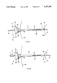
- FIG. 1 illustrates a side elevational view of a marking device pursuant to the present invention
- FIG. 2 illustrates a side elevational view of a hypodermic needle or cannula pursuant to the present invention
- FIG. 3a illustrates an elevational end view of the gripping knob of the hypodermic needle of FIG. 2 along lines 3a-3a;
- FIG. 3b illustrates an elevational end view of the cannula of the hypodermic needle of FIG. 2 along lines 3b-3b;
- FIG. 4 illustrates a perspective, partially sectional view of the lesion localization needle assembly pursuant to the present invention after insertion into the body but prior to marking a lesion;
- FIG. 5 illustrates a perspective, partially sectional view of the needle assembly of FIG. 4 during rotation of the marking device within the cannula and into a lesion;
- FIG. 6 illustrates the needle assembly of FIG. 4 after rotation of the marking device into the cannula with the wire marker being fully embedded within a lesion
- FIG. 7 illustrates a side elevational view of an alternate embodiment of a marking device pursuant to the present invention.
- FIG. 8 illustrates a side elevational view of an alternate embodiment of a needle or cannula pursuant to the present invention.
- FIG. 1 shows marking device 10 according to the present invention.
- Marking device 10 is constructed of a biocompatable material, and is preferably constructed of stainless steel, although many metal alloys such as aluminum alloy, titanium alloy, ferrous alloy, and the like, as well as materials such as plastic and ceramic, may be employed
- Marking device 10 essentially consists of a shaft 12 which is preferably type 18-8 stainless steel having a thickness of between 0.011 and 0.20 inches diameter, and is preferably 0.016 inches diameter.
- Marking device 10 is provided at one end with helical marking wire 14 which is helically wound about the end of shaft 12 and secured to the shaft as illustrated at 20.
- helical marking wire 14 is constructed of the same material as shaft 12, and is secured to the shaft by soldering, preferably of a 98% tin and 2% silver solder Helical marking wire 14 is wound about shaft 12 and extends outwardly away from the shaft to terminate in a sharpened tip 16.
- the diameter of the coil formed by helix 14 remains constant along its length Helix 14 extends from the end of shaft 12 a distance of between 0.5 centimeters and 2 centimeters, and preferably extends 1 centimeter from the end of shaft 12.
- the pitch of the coil is determined by the number of turns per centimeter, which along with the length of helix 14, is dependent upon the application for which the marker is to be used.
- helix 14 generally is provided with between 6 and 15 turns per centimeter, and preferably it is provided with 8 turns per centimeter for marking breast lesions.
- helical guide wire 18 which is also wound about shaft 12.
- Helix 18 is constructed of the same material as helix 14 and shaft 12, and helical wires 14 and 18 are the same gauge wire, preferably having a diameter of between 0.009 and 0.015 inches (0.02 and 0.04 cm). The preferred diameter for helical wires 14 and 18 is 0.011 inches (0.027 cm) for marking breast lesions.
- Helix 18 is secured to shaft 12 in a manner similar to helix 14. Helix 18 is of the same length as the length that helix 14 extends from the end of shaft 12 to sharpened tip 16, and also has the identical amount of turns per centimeter as helix 14, and thus the same pitch to the coil formed by helix 18.
- Distance “d” is dependent upon the length of the hypodermic needle or cannula with which marking device 10 is to be used. This will be described in greater detail below.
- hypodermic needle 30 comprises a cannula 32 having a sharpened cutting tip 34 and a gripping knob 36.
- Cannula 32 like marking device 10, is constructed of biocompatable material, and is preferably stainless steel.
- the cannula is 18-gauge thin wall stainless steel type 504, and has a length from tip 34 to knob end 36 of between 3 and 15 centimeters, depending upon the type and location of the lesion to be marked.
- Knob 36 is preferably constructed of thermoplastic material such as nylon and is secured to cannula 32 at end 38 by conventional means such as epoxy adhesives, and the like.
- Knob 36 may have a ridged gripping surface 44 which aids the physician in handling the needle 30.
- Cannula 32 is of course hollow and defines a lumen 33, as best seen in FIG. 3B.
- Gripping knob 36 has a hole 46 bored through the knob, which in the preferred embodiment aligns with lumen 33 of cannula 32 so that the cannula can extend through the hole to face 37 of knob 36.
- a second hole 47 is bored through knob 36, which is offset and parallel to hole 46.
- a wire guide 40 passes through hole 47 and may be secured within the hole by conventional means such as epoxy, adhesives, and the like.
- Wire guide 40 passes through hole 47 and is bent at 41 along face 37 of knob 36 to form guide bar 42.
- Wire guide 40 may also loosely and pivotably rest within hole 47 so that guide bar 42 may be moved into and out of engagement with shaft 12 of marker 10.
- guide bar 42 partially covers hole 46 in knob 36 so as to reduce the opening of hole 46. The reason for this will be explained in greater detail below.
- FIGS. 4, 5 and 6 show needle and marker assembly 50 in various positions during use of assembly 50 in marking a lesion within the body.
- Assembly 50 comprises marking device 10 as shown in FIG. 1 positioned within the lumen 33 of needle 30 as shown in FIG. 2.
- the location of the lesion within the body is determined radiologically or ultrasonically in a non-invasive procedure. In order to biopsy the lesion or remove it, the surgeon must have an accurate location of the lesion prior to performing the surgical procedure to minimize damage to tissue. The accuracy of the location of the marker will obviate any need for additional incisions, as well as avoid unnecessary tissue removal, which benefits the patient both physically and cosmetically
- the use of a marking device such as in the present invention is illustrated in FIGS. 4, 5 and 6.
- the needle and marker assembly is inserted into the body through the skin surface 52 until cutting tip 34 of cannula 32 is positioned proximate a lesion or tumor 54.
- Marking device 10 is positioned within needle 30 such that sharpened tip 16 of helical marking wire is positioned adjacent to cutting tip 34 of needle 30.
- the length of needle 30, as well as the length of shaft 12 and distance "d" between marking wire 14 and guide wire 18 is determined by the depth or distance lesion 54 is from the surface of the skin 52. Distance "d” is determined such that when marking device 10 is within the lumen 33 of needle 30, forward end 19 of helical guide wire 18 engages and rests against guide bar 42, resulting in sharpened tip 16 being adjacent to cutting tip 34.
- marking device 10 is rotated about shaft 12 to advance helical marking wire 14 into lesion 54. Sharpened tip 16 enters lesion 54 and the rotation about shaft 12 further advances marking wire 14 into the lesion to firmly anchor it in place The depth to which helical marking wire 14 enters lesion 54 is determined by the distance helical guide wire 18 travels through hole 46 into cannula 32.
- guide bar 42 of wire guide 40 engages the shaft and helix 18 at end 19 of helix 18 and guides shaft 12 while allowing helical guide wire 18 to rotate into hole 46 in a screw-like fashion
- Guide bar 42 is positioned between the individual coils of helical guide wire 18 to prevent slipping or pulling on the shaft.
- Wire guide 40 may be secured in hole 47 or may be pivotably secured so that guide bar 42 may rotated away from shaft 12 to disengage guide bar 42 from helix 18.
- marking wire 14 When the surgeon determines that the marking wire 14 in proper position, such as when it is completely embedded in the lesion, as evidenced by trailing end 21 of helix 18 turning into knob 36, the surgeon may then remove needle 30 from the body leaving marking device 10 firmly embedded in the lesion Alternatively, when it is determined that the helix 14 is in a desired position with respect to lesion 54 without helix 18 being completely within cannula 32, such as when a lesion is located proximate the chest wall as determined by ultrasonic imaging, wire guide 40 may be pivoted to rotate guide bar 42 away from helix 18 to allow for removal of needle 30 without disturbing the position of helix 14. Marking device 10 remains firmly anchored due to the concentric nature of the coils of marking wire 14 and eliminates the possibility of inadvertent dislodgement due to relaxation of the tissues of the breast upon discontinuing the compression placed on the breast during the procedure.
- FIG. 7 illustrates an alternate embodiment of the present invention showing marking device 10a, in which helix 14a and helix 18a are integrally wound as part of shaft 12a.
- FIG. 7 is identical to FIG. 1 in operation and function except that additional helical wires are not needed, since marking device 10a is of unitary construction in that shaft 12a and helixes 14a and 18a are constructed as a single unit.
- marking device 10a is of unitary construction in that shaft 12a and helixes 14a and 18a are constructed as a single unit.
- helix 14 and helix 18 may be joined so that the entire shaft 12 is in a helical coil.
- cannula 32 may be provided with a notched portion 60, and knob 36 may be eliminated, as seen in FIG. 8 In this case, notch 60 engages helix 18, or alternately helix 14, dependent upon location of notch 60. Notch 60 will then guide marking device 10 in the same manner as wire guide 40 and guide bar 42.
Landscapes
- Health & Medical Sciences (AREA)
- Surgery (AREA)
- Life Sciences & Earth Sciences (AREA)
- Medical Informatics (AREA)
- Nuclear Medicine, Radiotherapy & Molecular Imaging (AREA)
- Engineering & Computer Science (AREA)
- Biomedical Technology (AREA)
- Heart & Thoracic Surgery (AREA)
- Pathology (AREA)
- Molecular Biology (AREA)
- Animal Behavior & Ethology (AREA)
- General Health & Medical Sciences (AREA)
- Public Health (AREA)
- Veterinary Medicine (AREA)
- Oral & Maxillofacial Surgery (AREA)
- Media Introduction/Drainage Providing Device (AREA)
Abstract
A lesion localization and marking wire and needle assembly for marking non-palpable lesions within the body. A marking device having a helically wound coil of wire attached to an end of the shaft which is insertable into the body through a needle or cannula for rotatingly anchoring the marking device into a lesion or tumor is provided. The needle or cannula is inserted into the body with the marking device positioned therein so that when the cannula is positioned proximate to a lesion the shaft of the marker is rotated to advance the marker into the lesion to mark it for subsequent surgical procedures. A second helical wire may be provided on the shaft which cooperates with a wire guide device attached to the needle to enable the physician to determine the depth of the marking device as it anchors into the lesion. In particular, the device is provided for marking for biopsy lesions of the breast.
Description
This is a continuation of copending application Ser. No. 367,405, filed on Jun. 15, 1989, now U.S. Pat. No. 5,018,530.
1. Field of the Invention
The present invention relates to lesion localization needles and devices, for use in localizing or marking non-palpable lesions and tumors within the body, and more particularly, the present invention relates to a needle assembly which includes a wire marker having a helically wound wire tip for rotatingly anchoring a marker to a lesion within a human breast.
Localization or marking of lesions within the body, such as non-palpable lesions discovered within the body, and devices such as needles and wires for marking these lesions, are well known in the art. The devices generally comprise a hypodermic needle or cannula which is inserted into the body under local anesthesia to a position adjacent and in contact with the lesion. The wire marker is then passed through the cannula and is anchored into the lesion so that the lesion is marked for subsequent surgical procedures such as excision or biopsy. After marking the lesion with the wire marker, the cannula is usually removed from the body, leaving the wire in place and extending from the body. However, these markers tend to dislodge and migrate during transport of the patient for the surgical biopsy procedure.
Increasingly, ultrasonic imaging is being used as a preferred ancillary or adjunctive imaging method to evaluate breast masses which may be associated with positive or negative mammographic findings. Currently available localization and marking devices image poorly, if at all, ultrasonically, making it difficult to accurately pinpoint the tip of the localization wire with respect to the lesion Consequently, a subsequent surgical biopsy procedure may result in an inaccurate incision causing unnecessary tissue damage, and may necessitate a second surgical procedure to properly biopsy the lesion, causing the patient unnecessary pain, suffering, and expense.
2. Discussion of the Prior Art
In the prior art, several types of lesion localization devices and lesion markers are disclosed. Currently, the method of detecting and performing a biopsy on a non-palpable occult lesion within the body, such as non-palpable breast lesions, has been to radiologically or ultrasonically locate the lesion and to mark the lesion using a localization needle assembly, prior to a biopsy procedure. These needle assemblies generally comprise a hypodermic needle or cannula which is inserted into the body to an area adjacent to and in contact with the lesion A marking wire is then inserted through the cannula into the lesion and anchored in place so that the cannula may be removed.
Ultrasonic imaging is increasingly being used as the preferred method of detection and evaluation of lesions and masses within the body due to its accuracy, and in view of the fact that the patient is not exposed to potentially harmful radiation for extended periods of time The prior art marking devices generally image very poorly ultrasonically, as the tip of the previous marker shows up as a small, hard to locate dot or spot on the viewing screen. Depth perception is very limited, and consequently, accurate, reliable placement of the previous marking device is not guaranteed.
Nicholson, et al., U.S. Pat. No. 4,616,656, discloses a probe wire and sheath assembly in which the wire has a J-type memory hook for marking lesions. The wire probe has a soft flexibility so that when it is enclosed within the sheath it has a straight configuration. The sheath, or needle, is inserted into the body, for instance into the breast of a female patient, and positioned proximate to a lesion The wire probe is then pushed further into the lesion so that the memory hook is reformed and anchors itself within the lesion. The sheath is then removed leaving the hook embedded in the lesion as a marker.
A similar device is disclosed in Hawkins, Jr., U.S. Pat. No. 4,230,123. Hawkins, Jr. discloses a needle sheath assembly which consists of a small gauge needle in which a stylus or wire is positioned within a cannula. A shorter outer sheath is slidably located over the cannula which is removable after insertion of the needle into the patient's body. The wire has a J-type hook which is passed through the cannula to stabilize the tip of the cannula during biopsy.
Nicholson, et al. and Hawkins, Jr. are subject to several disadvantages which effect the accuracy and performance of the device Devices such as those disclosed in these references image very poorly and are inconsistently visualized ultrasonically, and consequently may not be accurately placed Furthermore, in procedures involving lesions of the breast, the breast is compressed during the mammographic localization procedure so that after the needle is in place and compression discontinued, the needle marker may inadvertently dislodge or migrate to a different position than that set during the localization procedure The needle may also deflect away from the lesion, or if the strength and resiliency of the wire is less than that required to penetrate the lesion, the hook may not reform, allowing the marker to migrate or dislodge. This can result in damaging the tissues of the breast, as well as an inaccurate surgical incision during the biopsy procedure, usually requiring a second surgical procedure to properly biopsy the lesion, causing the patient unnecessary pain, suffering and expense Devices of this type also generally require that the breast be stabilized during transport of the patient from the radiology section of a hospital to the surgical section for the biopsy procedure in order to prevent dislodgement of the marker.
Simon, U.S Pat. No. 4,790,329, discloses a biopsy localization device having a sheath or cannula through which a barbed rod passes. The cannula is provided with an open side port through which the barb extends upon positioning within a lesion In use, the barb is compressed within the lumen of the cannula and the pointed end of the rod extends from the cannula As the device penetrates the patient's body, and into a lesion, the rod is rotated 180° so that the end of the barb may pass through the open side port of the cannula. The rod is then drawn back so that barb and cannula anchor into the lesion to prevent removal. While the device is relocatable, such as by drawing back the cannula to enclose the barbed rod after anchoring, it is apparent that some tissue damage will result due to the barb puncturing the tissue once it is anchored In addition, the cannula remains in place while the lesion is marked by the barb, which results in excessive weight applied to the tissue The entire device must be stabilized in order to prevent tearing of tissue and dislodgement of the marker As related to breast lesions, as discussed above, compression of the breast during the procedure provides accurate anchoring of the barb; however, during transport of the patient, the additional weight of the cannula as well as the barbed rod will require stabilization of the breast to prevent migration and dislodgement of the device. A similar device, facing the same disadvantages, is disclosed in Hawkins, et al , U.S Pat. No. 4,799,495.
An additional type of prior art lesion localization and biopsy device is commonly referred to as the "Nordenstrom Screw Diagnostic Instrument", which was developed by Bjorn Nordenstrom (Radiology, Nov. 1975, Volume 117, Page 474). The Nordenstrom screw is generally a biopsy device and not a lesion localization and marking device. A cannula is provided which is inserted into the body, having a screw-tipped rod within the lumen of the cannula. When the cannula is positioned proximate a lesion, the rod is rotated to screw the tip into the lesion The screw tip is integral with the rod itself, and is a finely machined device in which the screw threads define grooves which taper to the tip of the device After the screw tip is rotated into the lesion, the cannula is then rotated in an opposite direction using slight forward pressure to a position over the screw threads Tissue from the lesion is captured in the grooves of the screw tip and the entire device is withdrawn so that the tissue may be examined The Nordenstrom screw device, as stated above, is not a marking device, but instead allows the physician to immediately biopsy the lesion in question.
An additional marking device using a screw tip is disclosed in Hawkins, et al., U.S. Pat. No. 4,799,495. In this device, the cannula may be provided with a tapering screw tip to anchor the cannula in the tissue while the needle marker penetrates the lesion The cannula and wire are used to mark the lesion, and Hawkins et al also discloses the use of the cannula alone for marking the lesion. Furthermore, Hawkins et al. discusses a helical screw needle marker, similar to the Nordenstrom screw device, which may be inserted through the cannula to mark the lesion However, the tapering screw tip of Hawkins et al. is a finely machined device which is quite expensive to manufacture, and which also is subject to the disadvantage that the tapered end may result in the loosening or "backing off" of the screw tip which will dislodge the marker during transport of the patient, or upon discontinuation of compression of the breast during the marking procedure. Furthermore, the precise machining of the tip of this device, and in particular a hollow screw-tipped cannula, would be a difficult and very expensive procedure from a manufacturing standpoint, and would necessitate that the device be reusable due to these cost considerations. In view of this, and in light of current health risks and concerns for patient safety as related to blood products and invasive surgical procedures, sterilization procedures would be required prior to and after each use, thereby making the procedure more elaborate and expensive then normally necessary.
The novel, disposable lesion localization and marking device of the present invention obviates the problems associated with the prior art lesion localization devices by providing an inexpensive, simple to manufacture lesion marking device having a helically wound marking wire attached to a wire shaft which passes through a hypodermic needle comprising a cannula. The helically wound marking wire extends concentrically outward from the shaft and maintains a substantially uniform diameter so that once the wire is rotated or screwed into a lesion, it remains anchored in the tissue without the possibility of backing off and dislodging.
In a preferred embodiment, a second helically wound wire is provided on the shaft remote from the first helically wound wire at the tip which, in conjunction with a wire guide provided on a gripping knob of the cannula, assists in the forward advancement of the shaft so that excessive forward pressure is not required, and the second helix also acts as a depth guide to provide an accurate indication of the depth to which the first helix is embedded in a lesion. The helically wound wires are secured to the shaft by means such as soldering, or may be wound as part of the shaft itself, so that the entire device is simple to manufacture and relatively inexpensive, thereby making the device disposable following the biopsy procedure.
The present invention eliminates or substantially ameliorates the disadvantages encountered in the prior art through the provision of a lesion localization and marking device having a helically wound wire tip attached to a shaft which is inserted within the lumen of a cannula into the body and then rotated into a lesion to anchor the marker within the lesion tissue. The device is simple to manufacture and inexpensive thereby making it a disposable unit, which may be packaged in a sterile packaging unit for one time use.
The lesion localization and marking device of the present invention consists of a marker having a shaft constructed of stainless steel or other biocompatable material which has secured to its distal end, or formed integrally thereon, a stainless steel wire which is helically. wound about the end of the shaft. The helically wound wire extends outwardly in a concentric manner from the end of the shaft and overhangs the shaft a predetermined distance. The end of the helix is sharpened to facilitate insertion into a lesion within the body. The helical wire is secured to the shaft by conventional means such as soldering.
The marking device, when used in conjunction with the needle assembly of the present invention, may be provided with a second helically wound wire which is secured to the shaft of the marker remote from the end having the first helically wound wire. The second helically wound wire is secured to the shaft by soldering, or integrally formed as part of the shaft, and is dimensioned to have the same number of turns per centimeter as the first helically wound wire, thus having the same pitch or angle for each turn of coil. The marking device is positioned within a hypodermic needle or cannula which essentially comprises a stainless steel tube having a cutting edge at one end and a thermoplastic gripping knob at its other end. The gripping knob has a hole bored through the center which preferably aligns with the lumen of the cannula, and a second hole is bored through the knob parallel to the first hole and offset from the center of the lumen. Through the second hole is positioned a wire guide which is bent perpendicular to the hole and placed to partially cover the first hole, leaving an opening which is substantially equal to the diameter of the shaft of the wire marker plus the diameter of the wire which forms the helix.
In use, the needle assembly is inserted into the body, such as into the breast of a female patient, until the tip of the cannula is proximate to a lesion which has been discovered during a mammographic or ultrasonic imaging procedure. The marking device is positioned within the cannula so that the sharpened tip of the first helical wire is adjacent to the cutting edge of the cannula, and the second helical wire is positioned a predetermined distance such that the end of the second helical wire closest to the first helical wire is adjacent to and engages the wire guide of the thermoplastic knob of the cannula. As the marking device is rotated, the second helical wire is guided along the wire guide so as to stabilize the shaft while drawing the marker into the cannula due to the interaction of the second helix and the wire guide during rotation, and the first helical wire is rotated into the lesion. The wire guide assists the forward advancement of the marker during rotation.
The length of the second helical wire is identical to the length of the first helical wire from the end of the shaft to the sharpened tip, and both helical wires have an identical number of turns per centimeter. As the first helical wire is embedded into the lesion, one physician can accurately gauge the depth to which the first wire enters the lesion by the distance the second helical wire extends outwardly from the gripping knob of the cannula When the second helical wire is fully rotated within the cannula the physician will know that the first wire is fully extended outside the cannula and is in position with respect to the lesion. The cannula is then removed from the body leaving the marking device in place.
As ultrasonic imaging is increasingly being used as the preferred method of evaluation of breast lesions in localization procedures, it is very important the marker used in the localization procedure provide consistent visualization and clean imaging with a recognizable acoustic pattern. Prior art markers do not provide adequate ultrasonic imaging and consequently do not contribute to accurate localization of a lesion The present invention, however, due to the helical tip, provides excellent imaging characteristics compared to prior art markers, such that each turn of the helix images distinctly, as opposed to the single spot or dot appearing from the prior art markers As a result, the present marker provides an unambiguous ultrasonic image allowing for accurate marking of the discovered lesion under the same conditions as mammography, thus reducing the patient's exposure to X-rays as well as decreasing the number of repositions required to accurately mark the lesion.
Accordingly, it is a primary object of the present invention to provide an inexpensive, simple to manufacture, and disposable marking device for localizing lesions within the body, particularly breast lesions.
It is a further object of the present invention to provide a lesion localization device which substantially eliminates the possibility of dislodgement or migration of the needle marker after placement.
It is yet another object of the present invention to provide a lesion localization device which may be relocated or repositioned within the body which minimizes or substantially eliminates damage to tissue during repositioning.
A still further object of the present invention is to provide a lesion localization device which presents an unambiguous echo when exposed to ultrasonic sound waves, allowing placement of the device to be carried out without the need for the use of X-ray imaging.
A still further object of the present invention is to provide a lesion localization device in which the depth to which the lesion marker is placed with respect to a lesion is readily and accurately determined.
A still further object of the present invention is to provide a marking device for localizing non-palpable breast lesions which can be firmly anchored in the lesion and will not be dislodged regardless of the positioning or stability of the breast tissue.
Yet another object of the present invention is to provide an efficient and accurate method for marking non-palpable lesions within the body, particularly within the breast.
The foregoing objects and other features of the invention will become more readily apparent and may be understood by referring to the following detailed description of an illustrative embodiment of the lesion localization and marking device having a helically wound wire tip, taken in conjunction with the accompanying drawings; in which:
FIG. 1 illustrates a side elevational view of a marking device pursuant to the present invention;
FIG. 2 illustrates a side elevational view of a hypodermic needle or cannula pursuant to the present invention;
FIG. 3a illustrates an elevational end view of the gripping knob of the hypodermic needle of FIG. 2 along lines 3a-3a;
FIG. 3b illustrates an elevational end view of the cannula of the hypodermic needle of FIG. 2 along lines 3b-3b;
FIG. 4 illustrates a perspective, partially sectional view of the lesion localization needle assembly pursuant to the present invention after insertion into the body but prior to marking a lesion;
FIG. 5 illustrates a perspective, partially sectional view of the needle assembly of FIG. 4 during rotation of the marking device within the cannula and into a lesion;
FIG. 6 illustrates the needle assembly of FIG. 4 after rotation of the marking device into the cannula with the wire marker being fully embedded within a lesion;
FIG. 7 illustrates a side elevational view of an alternate embodiment of a marking device pursuant to the present invention; and
FIG. 8 illustrates a side elevational view of an alternate embodiment of a needle or cannula pursuant to the present invention.
Referring now in specific detail to the drawings, in which identical reference numerals identify similar or identical elements throughout the several views, FIG. 1 shows marking device 10 according to the present invention. Marking device 10 is constructed of a biocompatable material, and is preferably constructed of stainless steel, although many metal alloys such as aluminum alloy, titanium alloy, ferrous alloy, and the like, as well as materials such as plastic and ceramic, may be employed Marking device 10 essentially consists of a shaft 12 which is preferably type 18-8 stainless steel having a thickness of between 0.011 and 0.20 inches diameter, and is preferably 0.016 inches diameter. Marking device 10 is provided at one end with helical marking wire 14 which is helically wound about the end of shaft 12 and secured to the shaft as illustrated at 20. Preferably, helical marking wire 14 is constructed of the same material as shaft 12, and is secured to the shaft by soldering, preferably of a 98% tin and 2% silver solder Helical marking wire 14 is wound about shaft 12 and extends outwardly away from the shaft to terminate in a sharpened tip 16. The diameter of the coil formed by helix 14 remains constant along its length Helix 14 extends from the end of shaft 12 a distance of between 0.5 centimeters and 2 centimeters, and preferably extends 1 centimeter from the end of shaft 12. The pitch of the coil is determined by the number of turns per centimeter, which along with the length of helix 14, is dependent upon the application for which the marker is to be used. Different tissues within the body have different degrees of strength and resiliency, some requiring more force to anchor the marker 10 in place, and thus some tissues require a device having more turns per centimeter than other tissues Accordingly, helix 14 generally is provided with between 6 and 15 turns per centimeter, and preferably it is provided with 8 turns per centimeter for marking breast lesions.
Separated a distance "d" along shaft 12 from helical marking wire 14 is helical guide wire 18 which is also wound about shaft 12. Helix 18 is constructed of the same material as helix 14 and shaft 12, and helical wires 14 and 18 are the same gauge wire, preferably having a diameter of between 0.009 and 0.015 inches (0.02 and 0.04 cm). The preferred diameter for helical wires 14 and 18 is 0.011 inches (0.027 cm) for marking breast lesions. Helix 18 is secured to shaft 12 in a manner similar to helix 14. Helix 18 is of the same length as the length that helix 14 extends from the end of shaft 12 to sharpened tip 16, and also has the identical amount of turns per centimeter as helix 14, and thus the same pitch to the coil formed by helix 18.
Distance "d" is dependent upon the length of the hypodermic needle or cannula with which marking device 10 is to be used. This will be described in greater detail below.
As can be seen in FIG. 2, hypodermic needle 30 comprises a cannula 32 having a sharpened cutting tip 34 and a gripping knob 36. Cannula 32, like marking device 10, is constructed of biocompatable material, and is preferably stainless steel. In a preferred embodiment, the cannula is 18-gauge thin wall stainless steel type 504, and has a length from tip 34 to knob end 36 of between 3 and 15 centimeters, depending upon the type and location of the lesion to be marked. Knob 36 is preferably constructed of thermoplastic material such as nylon and is secured to cannula 32 at end 38 by conventional means such as epoxy adhesives, and the like. Knob 36 may have a ridged gripping surface 44 which aids the physician in handling the needle 30. Cannula 32 is of course hollow and defines a lumen 33, as best seen in FIG. 3B.
Gripping knob 36 has a hole 46 bored through the knob, which in the preferred embodiment aligns with lumen 33 of cannula 32 so that the cannula can extend through the hole to face 37 of knob 36. In addition to hole 46, a second hole 47 is bored through knob 36, which is offset and parallel to hole 46. A wire guide 40 passes through hole 47 and may be secured within the hole by conventional means such as epoxy, adhesives, and the like. Wire guide 40 passes through hole 47 and is bent at 41 along face 37 of knob 36 to form guide bar 42. Wire guide 40 may also loosely and pivotably rest within hole 47 so that guide bar 42 may be moved into and out of engagement with shaft 12 of marker 10. As seen in FIG. 3a, guide bar 42 partially covers hole 46 in knob 36 so as to reduce the opening of hole 46. The reason for this will be explained in greater detail below.
FIGS. 4, 5 and 6 show needle and marker assembly 50 in various positions during use of assembly 50 in marking a lesion within the body. Assembly 50 comprises marking device 10 as shown in FIG. 1 positioned within the lumen 33 of needle 30 as shown in FIG. 2. The location of the lesion within the body, such as non-palpable lesions found in the breast or organs deep within the body, is determined radiologically or ultrasonically in a non-invasive procedure. In order to biopsy the lesion or remove it, the surgeon must have an accurate location of the lesion prior to performing the surgical procedure to minimize damage to tissue. The accuracy of the location of the marker will obviate any need for additional incisions, as well as avoid unnecessary tissue removal, which benefits the patient both physically and cosmetically The use of a marking device such as in the present invention is illustrated in FIGS. 4, 5 and 6.
As seen in FIG. 4, the needle and marker assembly is inserted into the body through the skin surface 52 until cutting tip 34 of cannula 32 is positioned proximate a lesion or tumor 54. Marking device 10 is positioned within needle 30 such that sharpened tip 16 of helical marking wire is positioned adjacent to cutting tip 34 of needle 30. The length of needle 30, as well as the length of shaft 12 and distance "d" between marking wire 14 and guide wire 18 is determined by the depth or distance lesion 54 is from the surface of the skin 52. Distance "d" is determined such that when marking device 10 is within the lumen 33 of needle 30, forward end 19 of helical guide wire 18 engages and rests against guide bar 42, resulting in sharpened tip 16 being adjacent to cutting tip 34.
Turning now to FIG. 5, after cutting tip 34 is positioned proximate to lesion 54, marking device 10 is rotated about shaft 12 to advance helical marking wire 14 into lesion 54. Sharpened tip 16 enters lesion 54 and the rotation about shaft 12 further advances marking wire 14 into the lesion to firmly anchor it in place The depth to which helical marking wire 14 enters lesion 54 is determined by the distance helical guide wire 18 travels through hole 46 into cannula 32. As shaft 12 is rotated, guide bar 42 of wire guide 40 engages the shaft and helix 18 at end 19 of helix 18 and guides shaft 12 while allowing helical guide wire 18 to rotate into hole 46 in a screw-like fashion Guide bar 42 is positioned between the individual coils of helical guide wire 18 to prevent slipping or pulling on the shaft. Wire guide 40 may be secured in hole 47 or may be pivotably secured so that guide bar 42 may rotated away from shaft 12 to disengage guide bar 42 from helix 18.
When helical marking wire 14 is embedded and anchored in lesion 54, that is when the end 23 of shaft 12 is proximate to the lesion 54, the rotation is ceased This is best seen in FIG. 6. The surgeon may determine when marking wire 14 is in its desired position with respect to lesion 54 when guide wire 18 completely disappears into knob 36 past guide bar 42. The trailing end 21 of guide wire 18 is the same distance from the end 23 of shaft 20 as the distance between forward end 19 of guide wire 18 and sharpened tip 16 of marking wire 14. When the surgeon determines that the marking wire 14 in proper position, such as when it is completely embedded in the lesion, as evidenced by trailing end 21 of helix 18 turning into knob 36, the surgeon may then remove needle 30 from the body leaving marking device 10 firmly embedded in the lesion Alternatively, when it is determined that the helix 14 is in a desired position with respect to lesion 54 without helix 18 being completely within cannula 32, such as when a lesion is located proximate the chest wall as determined by ultrasonic imaging, wire guide 40 may be pivoted to rotate guide bar 42 away from helix 18 to allow for removal of needle 30 without disturbing the position of helix 14. Marking device 10 remains firmly anchored due to the concentric nature of the coils of marking wire 14 and eliminates the possibility of inadvertent dislodgement due to relaxation of the tissues of the breast upon discontinuing the compression placed on the breast during the procedure.
FIG. 7 illustrates an alternate embodiment of the present invention showing marking device 10a, in which helix 14a and helix 18a are integrally wound as part of shaft 12a. FIG. 7 is identical to FIG. 1 in operation and function except that additional helical wires are not needed, since marking device 10a is of unitary construction in that shaft 12a and helixes 14a and 18a are constructed as a single unit. In a further embodiment, helix 14 and helix 18 may be joined so that the entire shaft 12 is in a helical coil.
In a further embodiment, cannula 32 may be provided with a notched portion 60, and knob 36 may be eliminated, as seen in FIG. 8 In this case, notch 60 engages helix 18, or alternately helix 14, dependent upon location of notch 60. Notch 60 will then guide marking device 10 in the same manner as wire guide 40 and guide bar 42.
While the invention has been particularly shown and described with reference to the preferred embodiments, it will be understood by those skilled in the art the various changes in form and detail may be made therein without departing from the spirit and scope of the invention. Accordingly, modifications and/or changes such as removing guide wire 18 or providing a longer or shorter marking wire, as well as increasing or decreasing the pitch of the coils as related to the number turns per centimeter, may be provided as desired, and are considered to be within the scope of the invention.
Claims (1)
1. A marking device for marking lesions within the body comprising shaft having a helically coiled end portion, a first straight section and a helically coiled body portion remote from said end portion separating a second straight section from said first straight section, said end portion and said body portion being coiled in equal diameter helixes, said helixes each having a constant diameter along their lengths, said constant diameters and lengths being equal such that each helix has an equal number of turns per centimeter, said end portion further having a sharpened tip remote from said first straight section.
Priority Applications (2)
| Application Number | Priority Date | Filing Date | Title |
|---|---|---|---|
| US07/679,464 US5197482A (en) | 1989-06-15 | 1991-04-02 | Helical-tipped lesion localization needle device and method of using the same |
| US07/988,616 US5234426A (en) | 1989-06-15 | 1992-12-10 | Helical-tipped lesion localization needle device and method of using the same |
Applications Claiming Priority (2)
| Application Number | Priority Date | Filing Date | Title |
|---|---|---|---|
| US07/367,405 US5018530A (en) | 1989-06-15 | 1989-06-15 | Helical-tipped lesion localization needle device and method of using the same |
| US07/679,464 US5197482A (en) | 1989-06-15 | 1991-04-02 | Helical-tipped lesion localization needle device and method of using the same |
Related Parent Applications (1)
| Application Number | Title | Priority Date | Filing Date |
|---|---|---|---|
| US07/367,405 Continuation US5018530A (en) | 1989-06-15 | 1989-06-15 | Helical-tipped lesion localization needle device and method of using the same |
Related Child Applications (1)
| Application Number | Title | Priority Date | Filing Date |
|---|---|---|---|
| US07/988,616 Division US5234426A (en) | 1989-06-15 | 1992-12-10 | Helical-tipped lesion localization needle device and method of using the same |
Publications (1)
| Publication Number | Publication Date |
|---|---|
| US5197482A true US5197482A (en) | 1993-03-30 |
Family
ID=27003780
Family Applications (1)
| Application Number | Title | Priority Date | Filing Date |
|---|---|---|---|
| US07/679,464 Expired - Fee Related US5197482A (en) | 1989-06-15 | 1991-04-02 | Helical-tipped lesion localization needle device and method of using the same |
Country Status (1)
| Country | Link |
|---|---|
| US (1) | US5197482A (en) |
Cited By (100)
| Publication number | Priority date | Publication date | Assignee | Title |
|---|---|---|---|---|
| US5269791A (en) * | 1992-10-09 | 1993-12-14 | Ilya Mayzels | Surgical knot pushing appliance |
| US5499989A (en) * | 1994-12-22 | 1996-03-19 | Labash; Stephen S. | Breast biopsy apparatus and method of use |
| WO1997029709A1 (en) | 1996-02-15 | 1997-08-21 | Biosense, Inc. | Medical procedures and apparatus using intrabody probes |
| DE19647873A1 (en) * | 1996-11-19 | 1998-05-20 | Daum Gmbh | Insert for indicating position of tumor or other diseased structures |
| US5800445A (en) * | 1995-10-20 | 1998-09-01 | United States Surgical Corporation | Tissue tagging device |
| US5879357A (en) * | 1995-10-20 | 1999-03-09 | United States Surgical Corporation | Apparatus for marking tissue location |
| US5902310A (en) * | 1996-08-12 | 1999-05-11 | Ethicon Endo-Surgery, Inc. | Apparatus and method for marking tissue |
| US6007495A (en) * | 1998-01-22 | 1999-12-28 | United States Surgical Corporation | Biopsy apparatus and method |
| US6053925A (en) * | 1998-02-27 | 2000-04-25 | Barnhart; William H. | Lesion localization device and method |
| US6056700A (en) * | 1998-10-13 | 2000-05-02 | Emx, Inc. | Biopsy marker assembly and method of use |
| US6203493B1 (en) | 1996-02-15 | 2001-03-20 | Biosense, Inc. | Attachment with one or more sensors for precise position determination of endoscopes |
| US6211666B1 (en) | 1996-02-27 | 2001-04-03 | Biosense, Inc. | Object location system and method using field actuation sequences having different field strengths |
| US6228055B1 (en) | 1994-09-16 | 2001-05-08 | Ethicon Endo-Surgery, Inc. | Devices for marking and defining particular locations in body tissue |
| US6253770B1 (en) | 1996-02-15 | 2001-07-03 | Biosense, Inc. | Catheter with lumen |
| US6261315B1 (en) * | 1997-10-28 | 2001-07-17 | St. Jude Medical Cardiovascular Group, Inc. | Tubular body structure marking methods and apparatus |
| US6266551B1 (en) | 1996-02-15 | 2001-07-24 | Biosense, Inc. | Catheter calibration and usage monitoring system |
| US6270464B1 (en) | 1998-06-22 | 2001-08-07 | Artemis Medical, Inc. | Biopsy localization method and device |
| US6321109B2 (en) | 1996-02-15 | 2001-11-20 | Biosense, Inc. | Catheter based surgery |
| US6336904B1 (en) | 1998-04-07 | 2002-01-08 | Pro Duct Health, Inc. | Methods and devices for the localization of lesions in solid tissue |
| US6366799B1 (en) | 1996-02-15 | 2002-04-02 | Biosense, Inc. | Movable transmit or receive coils for location system |
| US6398794B1 (en) | 2000-11-17 | 2002-06-04 | Eldon J. Hinshaw | Splinter removal device |
| US6405733B1 (en) | 2000-02-18 | 2002-06-18 | Thomas J. Fogarty | Device for accurately marking tissue |
| US6453190B1 (en) | 1996-02-15 | 2002-09-17 | Biosense, Inc. | Medical probes with field transducers |
| US20030065266A1 (en) * | 2001-10-02 | 2003-04-03 | Russell Jeremy Colin | Medico-surgical devices |
| US6544269B2 (en) | 2000-08-10 | 2003-04-08 | Cook Incorporated | Localizer needle |
| US6618612B1 (en) | 1996-02-15 | 2003-09-09 | Biosense, Inc. | Independently positionable transducers for location system |
| US20030195499A1 (en) * | 2002-04-16 | 2003-10-16 | Mani Prakash | Microwave antenna having a curved configuration |
| US20030195500A1 (en) * | 1999-06-17 | 2003-10-16 | Moorman Jack W. | Needle kit and method for microwave ablation, track coagulation, and biopsy |
| US6722371B1 (en) | 2000-02-18 | 2004-04-20 | Thomas J. Fogarty | Device for accurately marking tissue |
| US6752767B2 (en) | 2002-04-16 | 2004-06-22 | Vivant Medical, Inc. | Localization element with energized tip |
| US6752154B2 (en) | 2000-02-18 | 2004-06-22 | Thomas J. Fogarty | Device for accurately marking tissue |
| US20040228650A1 (en) * | 2003-05-09 | 2004-11-18 | Takashi Saito | Image forming apparatus |
| US20040254572A1 (en) * | 2003-04-25 | 2004-12-16 | Mcintyre Jon T. | Self anchoring radio frequency ablation array |
| US20040267155A1 (en) * | 1998-06-22 | 2004-12-30 | Fulton Richard Eustis | Biopsy localization method and device |
| US20050015081A1 (en) * | 2003-07-18 | 2005-01-20 | Roman Turovskiy | Devices and methods for cooling microwave antennas |
| US20050059888A1 (en) * | 1998-12-24 | 2005-03-17 | Sirimanne D. Laksen | Biopsy cavity marking device and method |
| US20060173280A1 (en) * | 2003-11-17 | 2006-08-03 | Inrad, Inc. | Multi Mode Imaging Marker |
| US20060241411A1 (en) * | 2005-04-20 | 2006-10-26 | Inrad, Inc. | Marking device with retracable cannula |
| US20060253153A1 (en) * | 2005-04-22 | 2006-11-09 | Helmut Schreiber | Surgical marker/connector |
| US20070185507A1 (en) * | 2005-04-22 | 2007-08-09 | Helmut Schreiber | Surgical marker/connector and method of installation |
| US7318824B2 (en) | 2001-11-02 | 2008-01-15 | Vivant Medical, Inc. | High-strength microwave antenna assemblies |
| US20080082093A1 (en) * | 2006-09-29 | 2008-04-03 | Prakash Mani N | Microwave antenna assembly and method of using the same |
| US20080146964A1 (en) * | 2005-02-03 | 2008-06-19 | Lts Lohmann Therapie-System Ag | Biopsy Needle For The Histological Examination Of Body Tissue |
| US20080281190A1 (en) * | 2006-01-25 | 2008-11-13 | Health Beacons, Inc. | Surgical procedures |
| US20080294039A1 (en) * | 2006-08-04 | 2008-11-27 | Senorx, Inc. | Assembly with hemostatic and radiographically detectable pellets |
| US20090005645A1 (en) * | 2005-05-04 | 2009-01-01 | Frassica James J | Rotate-to- advance catheterization system |
| US20090138005A1 (en) * | 2007-11-27 | 2009-05-28 | Vivant Medical, Inc. | Targeted Cooling of Deployable Microwave Antenna |
| US20090171198A1 (en) * | 2006-08-04 | 2009-07-02 | Jones Michael L | Powdered marker |
| US20100010341A1 (en) * | 2006-12-18 | 2010-01-14 | Talpade Dnyanesh A | Biopsy Marker with In Situ-Generated Imaging Properties |
| US20100030072A1 (en) * | 2006-12-12 | 2010-02-04 | Casanova R Michael | Multiple Imaging Mode Tissue Marker |
| US20100069718A1 (en) * | 2005-05-04 | 2010-03-18 | Frassica James J | Rotate-to-advance catheterization system |
| US20100076264A1 (en) * | 2005-05-04 | 2010-03-25 | Stephen Tallarida | Rotate-to-advance catheterization system |
| US20100081879A1 (en) * | 2005-05-04 | 2010-04-01 | Frassica James J | Rotate-to-advance catheterization system |
| US20100113972A1 (en) * | 2008-10-31 | 2010-05-06 | Victor Hugo Alvarado | Biopsy needle device and method for using same |
| US20100204570A1 (en) * | 2009-02-06 | 2010-08-12 | Paul Lubock | Anchor markers |
| US20100207765A1 (en) * | 2009-02-13 | 2010-08-19 | Christopher Brander | Method and apparatus for locating passive integrated transponder tags |
| US20100298696A1 (en) * | 2003-11-17 | 2010-11-25 | Bard Peripheral Vascular, Inc. | Self-contained, self-piercing, side-expelling marking apparatus |
| US20100298698A1 (en) * | 2000-11-20 | 2010-11-25 | Senorx, Inc. | Tissue site markers for in vivo imaging |
| US20100324416A1 (en) * | 1999-02-02 | 2010-12-23 | Senorx, Inc. | Cavity-filling biopsy site markers |
| US20100331668A1 (en) * | 2008-01-31 | 2010-12-30 | Ranpura Himanshu M | Biopsy Tissue Marker |
| US20110028836A1 (en) * | 2008-12-30 | 2011-02-03 | Himanshu Ranpura | Marker delivery device for tissue marker placement |
| US20110092815A1 (en) * | 2003-05-23 | 2011-04-21 | Senorx, Inc. | Marker or filler forming fluid |
| US20110166448A1 (en) * | 1999-02-02 | 2011-07-07 | Jones Michael L | Marker delivery device with releasable plug |
| US20110178370A1 (en) * | 1997-02-10 | 2011-07-21 | Frassica James J | Rotate to advance catheterization system |
| US20110184449A1 (en) * | 2006-08-04 | 2011-07-28 | Senorx, Inc. | Marker delivery device with obturator |
| US20110184280A1 (en) * | 1999-02-02 | 2011-07-28 | Jones Michael L | Intracorporeal marker and marker delivery device |
| US20110301580A1 (en) * | 2009-03-25 | 2011-12-08 | University Of Iowa Research Foundation | Methods and devices for arytenoid repositioning |
| US8317678B2 (en) | 2005-05-04 | 2012-11-27 | Olympus Endo Technology America Inc. | Rotate-to-advance catheterization system |
| US20120302915A1 (en) * | 2011-05-27 | 2012-11-29 | Lee Jonathon T | Spiral biopsy needle |
| US8366674B2 (en) | 2005-05-04 | 2013-02-05 | Olympus Endo Technology America Inc. | Rotate-to-advance catheterization system |
| US8377041B2 (en) | 2005-02-28 | 2013-02-19 | Olympus Endo Technology America Inc. | Rotate-to-advance catheterization system |
| US8435229B2 (en) | 2006-02-28 | 2013-05-07 | Olympus Endo Technology America Inc. | Rotate-to-advance catheterization system |
| US8437834B2 (en) | 2006-10-23 | 2013-05-07 | C. R. Bard, Inc. | Breast marker |
| US8444573B2 (en) | 2010-03-30 | 2013-05-21 | Siteselect Medical Technologies, Inc. | Tissue excision device |
| US8486028B2 (en) | 2005-10-07 | 2013-07-16 | Bard Peripheral Vascular, Inc. | Tissue marking apparatus having drug-eluting tissue marker |
| US8574220B2 (en) | 2006-02-28 | 2013-11-05 | Olympus Endo Technology America Inc. | Rotate-to-advance catheterization system |
| US8579931B2 (en) | 1999-06-17 | 2013-11-12 | Bard Peripheral Vascular, Inc. | Apparatus for the percutaneous marking of a lesion |
| US8626269B2 (en) | 2003-05-23 | 2014-01-07 | Senorx, Inc. | Fibrous marker and intracorporeal delivery thereof |
| US8668737B2 (en) | 1997-10-10 | 2014-03-11 | Senorx, Inc. | Tissue marking implant |
| US20140107693A1 (en) * | 2012-10-12 | 2014-04-17 | Cook Medical Technologies Llc | Device and method for removing tissue inside a body vessel |
| US8777841B2 (en) | 2007-05-18 | 2014-07-15 | Olympus Endo Technology America Inc. | Rotate-to-advance catheterization system |
| US8784433B2 (en) | 2002-06-17 | 2014-07-22 | Senorx, Inc. | Plugged tip delivery tube for marker placement |
| USD715442S1 (en) | 2013-09-24 | 2014-10-14 | C. R. Bard, Inc. | Tissue marker for intracorporeal site identification |
| USD715942S1 (en) | 2013-09-24 | 2014-10-21 | C. R. Bard, Inc. | Tissue marker for intracorporeal site identification |
| USD716450S1 (en) | 2013-09-24 | 2014-10-28 | C. R. Bard, Inc. | Tissue marker for intracorporeal site identification |
| USD716451S1 (en) | 2013-09-24 | 2014-10-28 | C. R. Bard, Inc. | Tissue marker for intracorporeal site identification |
| US8939153B1 (en) | 2013-03-15 | 2015-01-27 | Health Beacons, Inc. | Transponder strings |
| US9119712B2 (en) | 2012-08-24 | 2015-09-01 | Cook Medical Technologies Llc | Medical devices, systems, and kits for the medialization of a vocal cord |
| US9149341B2 (en) | 1999-02-02 | 2015-10-06 | Senorx, Inc | Deployment of polysaccharide markers for treating a site within a patient |
| US9220395B2 (en) | 1999-09-27 | 2015-12-29 | James J. Frassica | Rotate-to-advance catheterization system |
| US9433499B2 (en) | 2013-05-07 | 2016-09-06 | Cook Medical Technologies Llc | Vocal cord medialization |
| US9492570B2 (en) | 1998-12-24 | 2016-11-15 | Devicor Medical Products, Inc. | Device and method for safe location and marking of a biopsy cavity |
| US9669113B1 (en) | 1998-12-24 | 2017-06-06 | Devicor Medical Products, Inc. | Device and method for safe location and marking of a biopsy cavity |
| US9820824B2 (en) | 1999-02-02 | 2017-11-21 | Senorx, Inc. | Deployment of polysaccharide markers for treating a site within a patent |
| US10188310B2 (en) | 2014-08-24 | 2019-01-29 | Health Beacons, Inc. | Probe for determining magnetic marker locations |
| US10786604B2 (en) | 2008-09-23 | 2020-09-29 | Senorx, Inc. | Porous bioabsorbable implant |
| US20210177386A1 (en) * | 2019-12-11 | 2021-06-17 | Merit Medical Systems, Inc. | Bone biopsy device and related methods |
| US11266481B2 (en) | 2018-10-12 | 2022-03-08 | Hologic, Inc. | Tissue localization marker with D-shaped cross-section |
| RU2811268C1 (en) * | 2023-06-20 | 2024-01-11 | Федеральное государственное бюджетное военное образовательное учреждение высшего образования "Военно-медицинская академия имени С.М. Кирова" Министерства обороны Российской Федерации (ВМедА) | Method of applying anchor marks for preoperative marking of small diameter intrapulmonary peripheral new formations |
| US12295556B2 (en) | 2019-09-27 | 2025-05-13 | Merit Medical Systems, Inc. | Rotation biopsy system and handle |
Citations (8)
| Publication number | Priority date | Publication date | Assignee | Title |
|---|---|---|---|---|
| US172868A (en) * | 1876-02-01 | Improvement in corkscrews | ||
| US832565A (en) * | 1905-12-29 | 1906-10-02 | Henry Levi | Portable fence-post. |
| US3683891A (en) * | 1970-06-26 | 1972-08-15 | Marshall Eskridge | Tissue auger |
| US3749085A (en) * | 1970-06-26 | 1973-07-31 | J Willson | Vascular tissue removing device |
| US4653496A (en) * | 1985-02-01 | 1987-03-31 | Bundy Mark A | Transluminal lysing system |
| US4682606A (en) * | 1986-02-03 | 1987-07-28 | Decaprio Vincent H | Localizing biopsy apparatus |
| US4935025A (en) * | 1989-01-30 | 1990-06-19 | Bundy Mark A | Transluminal lysing device |
| US5059197A (en) * | 1989-04-15 | 1991-10-22 | Urie Robert G | Lesion location device |
-
1991
- 1991-04-02 US US07/679,464 patent/US5197482A/en not_active Expired - Fee Related
Patent Citations (8)
| Publication number | Priority date | Publication date | Assignee | Title |
|---|---|---|---|---|
| US172868A (en) * | 1876-02-01 | Improvement in corkscrews | ||
| US832565A (en) * | 1905-12-29 | 1906-10-02 | Henry Levi | Portable fence-post. |
| US3683891A (en) * | 1970-06-26 | 1972-08-15 | Marshall Eskridge | Tissue auger |
| US3749085A (en) * | 1970-06-26 | 1973-07-31 | J Willson | Vascular tissue removing device |
| US4653496A (en) * | 1985-02-01 | 1987-03-31 | Bundy Mark A | Transluminal lysing system |
| US4682606A (en) * | 1986-02-03 | 1987-07-28 | Decaprio Vincent H | Localizing biopsy apparatus |
| US4935025A (en) * | 1989-01-30 | 1990-06-19 | Bundy Mark A | Transluminal lysing device |
| US5059197A (en) * | 1989-04-15 | 1991-10-22 | Urie Robert G | Lesion location device |
Cited By (219)
| Publication number | Priority date | Publication date | Assignee | Title |
|---|---|---|---|---|
| US5269791A (en) * | 1992-10-09 | 1993-12-14 | Ilya Mayzels | Surgical knot pushing appliance |
| US7044957B2 (en) | 1994-09-16 | 2006-05-16 | Ethicon Endo-Surgery, Inc. | Devices for defining and marking tissue |
| US20050165305A1 (en) * | 1994-09-16 | 2005-07-28 | Foerster Seth A. | Methods and devices for defining and marking tissue |
| US20050049489A1 (en) * | 1994-09-16 | 2005-03-03 | Foerster Seth A. | Methods for marking a biopsy site |
| US20010034528A1 (en) * | 1994-09-16 | 2001-10-25 | Foerster Seth A. | Methods and devices for defining and marking tissue |
| US7229417B2 (en) | 1994-09-16 | 2007-06-12 | Ethicon Endo-Surgery, Inc. | Methods for marking a biopsy site |
| US7625397B2 (en) | 1994-09-16 | 2009-12-01 | Ethicon Endo-Surgery, Inc. | Methods for defining and marking tissue |
| US20040024304A1 (en) * | 1994-09-16 | 2004-02-05 | Foerster Seth A. | Methods and devices for defining and marking tissue |
| US8277391B2 (en) | 1994-09-16 | 2012-10-02 | Devicor Medical Products, Inc. | Methods and devices for defining and marking tissue |
| US6228055B1 (en) | 1994-09-16 | 2001-05-08 | Ethicon Endo-Surgery, Inc. | Devices for marking and defining particular locations in body tissue |
| US20060074443A1 (en) * | 1994-09-16 | 2006-04-06 | Foerster Seth A | Devices and methods for marking a biopsy site |
| US5499989A (en) * | 1994-12-22 | 1996-03-19 | Labash; Stephen S. | Breast biopsy apparatus and method of use |
| US5800445A (en) * | 1995-10-20 | 1998-09-01 | United States Surgical Corporation | Tissue tagging device |
| US5879357A (en) * | 1995-10-20 | 1999-03-09 | United States Surgical Corporation | Apparatus for marking tissue location |
| US6253770B1 (en) | 1996-02-15 | 2001-07-03 | Biosense, Inc. | Catheter with lumen |
| US6203493B1 (en) | 1996-02-15 | 2001-03-20 | Biosense, Inc. | Attachment with one or more sensors for precise position determination of endoscopes |
| US6266551B1 (en) | 1996-02-15 | 2001-07-24 | Biosense, Inc. | Catheter calibration and usage monitoring system |
| WO1997029709A1 (en) | 1996-02-15 | 1997-08-21 | Biosense, Inc. | Medical procedures and apparatus using intrabody probes |
| US6453190B1 (en) | 1996-02-15 | 2002-09-17 | Biosense, Inc. | Medical probes with field transducers |
| US6321109B2 (en) | 1996-02-15 | 2001-11-20 | Biosense, Inc. | Catheter based surgery |
| US6332089B1 (en) | 1996-02-15 | 2001-12-18 | Biosense, Inc. | Medical procedures and apparatus using intrabody probes |
| US6618612B1 (en) | 1996-02-15 | 2003-09-09 | Biosense, Inc. | Independently positionable transducers for location system |
| US6366799B1 (en) | 1996-02-15 | 2002-04-02 | Biosense, Inc. | Movable transmit or receive coils for location system |
| US6211666B1 (en) | 1996-02-27 | 2001-04-03 | Biosense, Inc. | Object location system and method using field actuation sequences having different field strengths |
| US5902310A (en) * | 1996-08-12 | 1999-05-11 | Ethicon Endo-Surgery, Inc. | Apparatus and method for marking tissue |
| DE19647873A1 (en) * | 1996-11-19 | 1998-05-20 | Daum Gmbh | Insert for indicating position of tumor or other diseased structures |
| US20110178370A1 (en) * | 1997-02-10 | 2011-07-21 | Frassica James J | Rotate to advance catheterization system |
| US8764631B2 (en) | 1997-02-10 | 2014-07-01 | Olympus Endo Technology America Inc. | Rotate to advance catheterization system |
| US8668737B2 (en) | 1997-10-10 | 2014-03-11 | Senorx, Inc. | Tissue marking implant |
| US9039763B2 (en) | 1997-10-10 | 2015-05-26 | Senorx, Inc. | Tissue marking implant |
| US6261315B1 (en) * | 1997-10-28 | 2001-07-17 | St. Jude Medical Cardiovascular Group, Inc. | Tubular body structure marking methods and apparatus |
| US6007495A (en) * | 1998-01-22 | 1999-12-28 | United States Surgical Corporation | Biopsy apparatus and method |
| US6053925A (en) * | 1998-02-27 | 2000-04-25 | Barnhart; William H. | Lesion localization device and method |
| US6336904B1 (en) | 1998-04-07 | 2002-01-08 | Pro Duct Health, Inc. | Methods and devices for the localization of lesions in solid tissue |
| US20050033195A1 (en) * | 1998-06-22 | 2005-02-10 | Fulton Richard E. | Biopsy localization method and device |
| US20060079829A1 (en) * | 1998-06-22 | 2006-04-13 | Fulton Richard E | Biopsy localization method and device |
| US10010380B2 (en) | 1998-06-22 | 2018-07-03 | Devicor Medical Products, Inc. | Biopsy localization method and device |
| US6270464B1 (en) | 1998-06-22 | 2001-08-07 | Artemis Medical, Inc. | Biopsy localization method and device |
| US20040204660A1 (en) * | 1998-06-22 | 2004-10-14 | Artemis Medical, Inc. | Biopsy localization method and device |
| US20040210160A1 (en) * | 1998-06-22 | 2004-10-21 | Fulton Richard E. | Biopsy localization method and device |
| US20050045192A1 (en) * | 1998-06-22 | 2005-03-03 | Artemis Medical, Inc. | Biopsy localization method and device |
| US6699205B2 (en) | 1998-06-22 | 2004-03-02 | Artemis Medical, Inc. | Biopsy localization method and device |
| US20040267155A1 (en) * | 1998-06-22 | 2004-12-30 | Fulton Richard Eustis | Biopsy localization method and device |
| US8292822B2 (en) | 1998-06-22 | 2012-10-23 | Devicor Medical Products, Inc. | Biopsy localization method and device |
| US6056700A (en) * | 1998-10-13 | 2000-05-02 | Emx, Inc. | Biopsy marker assembly and method of use |
| US6261243B1 (en) | 1998-10-13 | 2001-07-17 | Emx, Inc. | Biopsy marker assembly and method of use |
| US9986974B2 (en) | 1998-12-24 | 2018-06-05 | Devicor Medical Products, Inc. | Biopsy cavity marking device |
| US9669113B1 (en) | 1998-12-24 | 2017-06-06 | Devicor Medical Products, Inc. | Device and method for safe location and marking of a biopsy cavity |
| US20050059888A1 (en) * | 1998-12-24 | 2005-03-17 | Sirimanne D. Laksen | Biopsy cavity marking device and method |
| US9492570B2 (en) | 1998-12-24 | 2016-11-15 | Devicor Medical Products, Inc. | Device and method for safe location and marking of a biopsy cavity |
| US8626270B2 (en) | 1999-02-02 | 2014-01-07 | Senorx, Inc. | Cavity-filling biopsy site markers |
| US9237937B2 (en) | 1999-02-02 | 2016-01-19 | Senorx, Inc. | Cavity-filling biopsy site markers |
| US8361082B2 (en) | 1999-02-02 | 2013-01-29 | Senorx, Inc. | Marker delivery device with releasable plug |
| US9044162B2 (en) | 1999-02-02 | 2015-06-02 | Senorx, Inc. | Marker delivery device with releasable plug |
| US8965486B2 (en) | 1999-02-02 | 2015-02-24 | Senorx, Inc. | Cavity filling biopsy site markers |
| US9149341B2 (en) | 1999-02-02 | 2015-10-06 | Senorx, Inc | Deployment of polysaccharide markers for treating a site within a patient |
| US9820824B2 (en) | 1999-02-02 | 2017-11-21 | Senorx, Inc. | Deployment of polysaccharide markers for treating a site within a patent |
| US10172674B2 (en) | 1999-02-02 | 2019-01-08 | Senorx, Inc. | Intracorporeal marker and marker delivery device |
| US8219182B2 (en) | 1999-02-02 | 2012-07-10 | Senorx, Inc. | Cavity-filling biopsy site markers |
| US9861294B2 (en) | 1999-02-02 | 2018-01-09 | Senorx, Inc. | Marker delivery device with releasable plug |
| US20110184280A1 (en) * | 1999-02-02 | 2011-07-28 | Jones Michael L | Intracorporeal marker and marker delivery device |
| US8498693B2 (en) | 1999-02-02 | 2013-07-30 | Senorx, Inc. | Intracorporeal marker and marker delivery device |
| US20110166448A1 (en) * | 1999-02-02 | 2011-07-07 | Jones Michael L | Marker delivery device with releasable plug |
| US20100324416A1 (en) * | 1999-02-02 | 2010-12-23 | Senorx, Inc. | Cavity-filling biopsy site markers |
| US9649093B2 (en) | 1999-02-02 | 2017-05-16 | Senorx, Inc. | Cavity-filling biopsy site markers |
| US10463446B2 (en) | 1999-06-17 | 2019-11-05 | Bard Peripheral Vascular, Inc. | Apparatus for the percutaneous marking of a lesion |
| US8579931B2 (en) | 1999-06-17 | 2013-11-12 | Bard Peripheral Vascular, Inc. | Apparatus for the percutaneous marking of a lesion |
| US20070161977A1 (en) * | 1999-06-17 | 2007-07-12 | Moorman Jack W | Needle kit and method for microwave ablation, track coagulation, and biopsy |
| US7160292B2 (en) | 1999-06-17 | 2007-01-09 | Vivant Medical, Inc. | Needle kit and method for microwave ablation, track coagulation, and biopsy |
| US9579159B2 (en) | 1999-06-17 | 2017-02-28 | Bard Peripheral Vascular, Inc. | Apparatus for the percutaneous marking of a lesion |
| US20030195500A1 (en) * | 1999-06-17 | 2003-10-16 | Moorman Jack W. | Needle kit and method for microwave ablation, track coagulation, and biopsy |
| US8690868B2 (en) | 1999-06-17 | 2014-04-08 | Covidien Lp | Needle kit and method for microwave ablation, track coagulation, and biopsy |
| US9220395B2 (en) | 1999-09-27 | 2015-12-29 | James J. Frassica | Rotate-to-advance catheterization system |
| US7322360B2 (en) | 2000-02-18 | 2008-01-29 | Thomas J. Fogarty | Device for accurately marking tissue |
| US6564806B1 (en) | 2000-02-18 | 2003-05-20 | Thomas J. Fogarty | Device for accurately marking tissue |
| US6405733B1 (en) | 2000-02-18 | 2002-06-18 | Thomas J. Fogarty | Device for accurately marking tissue |
| EP2077090A1 (en) | 2000-02-18 | 2009-07-08 | Thomas J. Fogarty | Improved device for accurately marking tissue |
| US6722371B1 (en) | 2000-02-18 | 2004-04-20 | Thomas J. Fogarty | Device for accurately marking tissue |
| EP2932891A1 (en) | 2000-02-18 | 2015-10-21 | Focal Therapeutics, Inc. | Improved device for accurately marking tissue |
| US6752154B2 (en) | 2000-02-18 | 2004-06-22 | Thomas J. Fogarty | Device for accurately marking tissue |
| EP2298154A2 (en) | 2000-02-18 | 2011-03-23 | Fogarty, Thomas J. | Improved device for accurately marking tissue |
| US20040168692A1 (en) * | 2000-02-18 | 2004-09-02 | Thomas Fogarty | Device for accurately marking tissue |
| US6544269B2 (en) | 2000-08-10 | 2003-04-08 | Cook Incorporated | Localizer needle |
| US6398794B1 (en) | 2000-11-17 | 2002-06-04 | Eldon J. Hinshaw | Splinter removal device |
| US8718745B2 (en) * | 2000-11-20 | 2014-05-06 | Senorx, Inc. | Tissue site markers for in vivo imaging |
| US20100298698A1 (en) * | 2000-11-20 | 2010-11-25 | Senorx, Inc. | Tissue site markers for in vivo imaging |
| US7258669B2 (en) * | 2001-10-02 | 2007-08-21 | Smiths Group Plc | Medico-surgical devices |
| US20030065266A1 (en) * | 2001-10-02 | 2003-04-03 | Russell Jeremy Colin | Medico-surgical devices |
| US7318824B2 (en) | 2001-11-02 | 2008-01-15 | Vivant Medical, Inc. | High-strength microwave antenna assemblies |
| US10154880B2 (en) | 2001-11-02 | 2018-12-18 | Covidien Lp | High-strength microwave antenna assemblies |
| US6752767B2 (en) | 2002-04-16 | 2004-06-22 | Vivant Medical, Inc. | Localization element with energized tip |
| US20030195499A1 (en) * | 2002-04-16 | 2003-10-16 | Mani Prakash | Microwave antenna having a curved configuration |
| US8808282B2 (en) | 2002-04-16 | 2014-08-19 | Covidien Lp | Microwave antenna having a curved configuration |
| US20090149850A1 (en) * | 2002-04-16 | 2009-06-11 | Vivant Medical, Inc. | Localization Element with Energized Tip |
| US7197363B2 (en) | 2002-04-16 | 2007-03-27 | Vivant Medical, Inc. | Microwave antenna having a curved configuration |
| US10039602B2 (en) | 2002-04-16 | 2018-08-07 | Covidien Lp | Electrosurgical energy channel splitters and systems for delivering electrosurgical energy |
| US11045253B2 (en) | 2002-04-16 | 2021-06-29 | Covidien Lp | Electrosurgical energy channel splitters and systems for delivering electrosurgical energy |
| US7846108B2 (en) | 2002-04-16 | 2010-12-07 | Vivant Medical, Inc. | Localization element with energized tip |
| US20040267156A1 (en) * | 2002-04-16 | 2004-12-30 | Vivant Medical, Inc. | Localization element with energized tip |
| US10143520B2 (en) | 2002-04-16 | 2018-12-04 | Covidien Lp | Microwave antenna guide assembly |
| US7468042B2 (en) | 2002-04-16 | 2008-12-23 | Vivant Medical, Inc. | Localization element with energized tip |
| US20070198006A1 (en) * | 2002-04-16 | 2007-08-23 | Mani Prakash | Microwave antenna having a curved configuration |
| US10363097B2 (en) | 2002-04-16 | 2019-07-30 | Coviden Lp | Ablation system having multiple energy sources |
| US8784433B2 (en) | 2002-06-17 | 2014-07-22 | Senorx, Inc. | Plugged tip delivery tube for marker placement |
| US10813716B2 (en) | 2002-11-18 | 2020-10-27 | Bard Peripheral Vascular, Inc. | Self-contained, self-piercing, side-expelling marking apparatus |
| US9848956B2 (en) | 2002-11-18 | 2017-12-26 | Bard Peripheral Vascular, Inc. | Self-contained, self-piercing, side-expelling marking apparatus |
| US20040254572A1 (en) * | 2003-04-25 | 2004-12-16 | Mcintyre Jon T. | Self anchoring radio frequency ablation array |
| US20040228650A1 (en) * | 2003-05-09 | 2004-11-18 | Takashi Saito | Image forming apparatus |
| US8447386B2 (en) | 2003-05-23 | 2013-05-21 | Senorx, Inc. | Marker or filler forming fluid |
| US8880154B2 (en) | 2003-05-23 | 2014-11-04 | Senorx, Inc. | Fibrous marker and intracorporeal delivery thereof |
| US20110092815A1 (en) * | 2003-05-23 | 2011-04-21 | Senorx, Inc. | Marker or filler forming fluid |
| US10299881B2 (en) | 2003-05-23 | 2019-05-28 | Senorx, Inc. | Marker or filler forming fluid |
| US10045832B2 (en) | 2003-05-23 | 2018-08-14 | Senorx, Inc. | Marker or filler forming fluid |
| US9801688B2 (en) | 2003-05-23 | 2017-10-31 | Senorx, Inc. | Fibrous marker and intracorporeal delivery thereof |
| US8639315B2 (en) | 2003-05-23 | 2014-01-28 | Senorx, Inc. | Marker or filler forming fluid |
| US8626269B2 (en) | 2003-05-23 | 2014-01-07 | Senorx, Inc. | Fibrous marker and intracorporeal delivery thereof |
| US7311703B2 (en) | 2003-07-18 | 2007-12-25 | Vivant Medical, Inc. | Devices and methods for cooling microwave antennas |
| US10405921B2 (en) | 2003-07-18 | 2019-09-10 | Covidien Lp | Devices and methods for cooling microwave antennas |
| US20050015081A1 (en) * | 2003-07-18 | 2005-01-20 | Roman Turovskiy | Devices and methods for cooling microwave antennas |
| US8634899B2 (en) | 2003-11-17 | 2014-01-21 | Bard Peripheral Vascular, Inc. | Multi mode imaging marker |
| US20100298696A1 (en) * | 2003-11-17 | 2010-11-25 | Bard Peripheral Vascular, Inc. | Self-contained, self-piercing, side-expelling marking apparatus |
| US20060173280A1 (en) * | 2003-11-17 | 2006-08-03 | Inrad, Inc. | Multi Mode Imaging Marker |
| US7988643B2 (en) * | 2005-02-03 | 2011-08-02 | Lts Lohmann Therapie-Systeme Ag | Biopsy needle for the histological examination of body tissue |
| US20080146964A1 (en) * | 2005-02-03 | 2008-06-19 | Lts Lohmann Therapie-System Ag | Biopsy Needle For The Histological Examination Of Body Tissue |
| US8377041B2 (en) | 2005-02-28 | 2013-02-19 | Olympus Endo Technology America Inc. | Rotate-to-advance catheterization system |
| US11278370B2 (en) | 2005-04-20 | 2022-03-22 | Bard Peripheral Vascular, Inc. | Marking device with retractable cannula |
| US20060241411A1 (en) * | 2005-04-20 | 2006-10-26 | Inrad, Inc. | Marking device with retracable cannula |
| US10357328B2 (en) | 2005-04-20 | 2019-07-23 | Bard Peripheral Vascular, Inc. and Bard Shannon Limited | Marking device with retractable cannula |
| US10342635B2 (en) | 2005-04-20 | 2019-07-09 | Bard Peripheral Vascular, Inc. | Marking device with retractable cannula |
| US7452364B2 (en) | 2005-04-22 | 2008-11-18 | Helmut Schreiber | Surgical marker/connector |
| US7931662B2 (en) | 2005-04-22 | 2011-04-26 | Helmut Schreiber | Surgical marker/connector and method of installation |
| US20080097490A1 (en) * | 2005-04-22 | 2008-04-24 | Helmut Schreiber | Method for Marking and Connecting Tissue |
| US8142453B2 (en) | 2005-04-22 | 2012-03-27 | Helmut Schreiber | Method for marking and connecting tissue |
| US20070185507A1 (en) * | 2005-04-22 | 2007-08-09 | Helmut Schreiber | Surgical marker/connector and method of installation |
| US20060253153A1 (en) * | 2005-04-22 | 2006-11-09 | Helmut Schreiber | Surgical marker/connector |
| US20100076264A1 (en) * | 2005-05-04 | 2010-03-25 | Stephen Tallarida | Rotate-to-advance catheterization system |
| US20100069718A1 (en) * | 2005-05-04 | 2010-03-18 | Frassica James J | Rotate-to-advance catheterization system |
| US20100081879A1 (en) * | 2005-05-04 | 2010-04-01 | Frassica James J | Rotate-to-advance catheterization system |
| US8317678B2 (en) | 2005-05-04 | 2012-11-27 | Olympus Endo Technology America Inc. | Rotate-to-advance catheterization system |
| US8343040B2 (en) | 2005-05-04 | 2013-01-01 | Olympus Endo Technology America Inc. | Rotate-to-advance catheterization system |
| US8366674B2 (en) | 2005-05-04 | 2013-02-05 | Olympus Endo Technology America Inc. | Rotate-to-advance catheterization system |
| US8414477B2 (en) | 2005-05-04 | 2013-04-09 | Olympus Endo Technology America Inc. | Rotate-to-advance catheterization system |
| US8747300B2 (en) | 2005-05-04 | 2014-06-10 | Olympus Endo Technology America Inc. | Rotate-to-advance catheterization system |
| US8235942B2 (en) | 2005-05-04 | 2012-08-07 | Olympus Endo Technology America Inc. | Rotate-to-advance catheterization system |
| US20090005645A1 (en) * | 2005-05-04 | 2009-01-01 | Frassica James J | Rotate-to- advance catheterization system |
| US8486028B2 (en) | 2005-10-07 | 2013-07-16 | Bard Peripheral Vascular, Inc. | Tissue marking apparatus having drug-eluting tissue marker |
| US20080281190A1 (en) * | 2006-01-25 | 2008-11-13 | Health Beacons, Inc. | Surgical procedures |
| US8574220B2 (en) | 2006-02-28 | 2013-11-05 | Olympus Endo Technology America Inc. | Rotate-to-advance catheterization system |
| US8435229B2 (en) | 2006-02-28 | 2013-05-07 | Olympus Endo Technology America Inc. | Rotate-to-advance catheterization system |
| US20090171198A1 (en) * | 2006-08-04 | 2009-07-02 | Jones Michael L | Powdered marker |
| US20110184449A1 (en) * | 2006-08-04 | 2011-07-28 | Senorx, Inc. | Marker delivery device with obturator |
| US20080294039A1 (en) * | 2006-08-04 | 2008-11-27 | Senorx, Inc. | Assembly with hemostatic and radiographically detectable pellets |
| US8068921B2 (en) | 2006-09-29 | 2011-11-29 | Vivant Medical, Inc. | Microwave antenna assembly and method of using the same |
| US9333032B2 (en) | 2006-09-29 | 2016-05-10 | Covidien Lp | Microwave antenna assembly and method of using the same |
| US20080082093A1 (en) * | 2006-09-29 | 2008-04-03 | Prakash Mani N | Microwave antenna assembly and method of using the same |
| US8437834B2 (en) | 2006-10-23 | 2013-05-07 | C. R. Bard, Inc. | Breast marker |
| US9579077B2 (en) | 2006-12-12 | 2017-02-28 | C.R. Bard, Inc. | Multiple imaging mode tissue marker |
| US11471244B2 (en) | 2006-12-12 | 2022-10-18 | C.R. Bard, Inc. | Multiple imaging mode tissue marker |
| US9901415B2 (en) | 2006-12-12 | 2018-02-27 | C. R. Bard, Inc. | Multiple imaging mode tissue marker |
| US20100030072A1 (en) * | 2006-12-12 | 2010-02-04 | Casanova R Michael | Multiple Imaging Mode Tissue Marker |
| US10682200B2 (en) | 2006-12-12 | 2020-06-16 | C. R. Bard, Inc. | Multiple imaging mode tissue marker |
| US9042965B2 (en) | 2006-12-18 | 2015-05-26 | C. R. Bard, Inc. | Biopsy marker with in situ-generated imaging properties |
| US20100010341A1 (en) * | 2006-12-18 | 2010-01-14 | Talpade Dnyanesh A | Biopsy Marker with In Situ-Generated Imaging Properties |
| US8401622B2 (en) | 2006-12-18 | 2013-03-19 | C. R. Bard, Inc. | Biopsy marker with in situ-generated imaging properties |
| US8777841B2 (en) | 2007-05-18 | 2014-07-15 | Olympus Endo Technology America Inc. | Rotate-to-advance catheterization system |
| US8870755B2 (en) | 2007-05-18 | 2014-10-28 | Olympus Endo Technology America Inc. | Rotate-to-advance catheterization system |
| US8292880B2 (en) | 2007-11-27 | 2012-10-23 | Vivant Medical, Inc. | Targeted cooling of deployable microwave antenna |
| US20090138005A1 (en) * | 2007-11-27 | 2009-05-28 | Vivant Medical, Inc. | Targeted Cooling of Deployable Microwave Antenna |
| US8311610B2 (en) | 2008-01-31 | 2012-11-13 | C. R. Bard, Inc. | Biopsy tissue marker |
| US20100331668A1 (en) * | 2008-01-31 | 2010-12-30 | Ranpura Himanshu M | Biopsy Tissue Marker |
| US11833275B2 (en) | 2008-09-23 | 2023-12-05 | Senorx, Inc. | Porous bioabsorbable implant |
| US10786604B2 (en) | 2008-09-23 | 2020-09-29 | Senorx, Inc. | Porous bioabsorbable implant |
| US20100113972A1 (en) * | 2008-10-31 | 2010-05-06 | Victor Hugo Alvarado | Biopsy needle device and method for using same |
| US11779431B2 (en) | 2008-12-30 | 2023-10-10 | C. R. Bard, Inc. | Marker delivery device for tissue marker placement |
| US20110028836A1 (en) * | 2008-12-30 | 2011-02-03 | Himanshu Ranpura | Marker delivery device for tissue marker placement |
| US8670818B2 (en) | 2008-12-30 | 2014-03-11 | C. R. Bard, Inc. | Marker delivery device for tissue marker placement |
| US10258428B2 (en) | 2008-12-30 | 2019-04-16 | C. R. Bard, Inc. | Marker delivery device for tissue marker placement |
| US20100204570A1 (en) * | 2009-02-06 | 2010-08-12 | Paul Lubock | Anchor markers |
| US8973584B2 (en) | 2009-02-13 | 2015-03-10 | Health Beacons, Inc. | Method and apparatus for locating passive integrated transponder tags |
| US20100207765A1 (en) * | 2009-02-13 | 2010-08-19 | Christopher Brander | Method and apparatus for locating passive integrated transponder tags |
| US10849529B2 (en) | 2009-02-13 | 2020-12-01 | Health Beacons, Inc. | Method and apparatus for locating passive integrated transponder tags |
| US12048522B2 (en) | 2009-02-13 | 2024-07-30 | Health Beacons, Inc. | Method and apparatus for locating passive integrated transponder tags |
| US9867550B2 (en) | 2009-02-13 | 2018-01-16 | Health Beacons, Inc. | Method and apparatus for locating passive integrated transponder tags |
| US20110301580A1 (en) * | 2009-03-25 | 2011-12-08 | University Of Iowa Research Foundation | Methods and devices for arytenoid repositioning |
| US9056005B2 (en) * | 2009-03-25 | 2015-06-16 | University Of Iowa Research Foundation | Methods and devices for arytenoid repositioning |
| US8740809B2 (en) | 2010-03-30 | 2014-06-03 | Siteselect Medical Technologies, Inc. | Tissue excision device with a retractable backhook |
| US8597202B2 (en) | 2010-03-30 | 2013-12-03 | Siteselect Medical Technologies, Inc. | Tissue excision device with a modified cutting edge |
| US8529467B2 (en) | 2010-03-30 | 2013-09-10 | Siteselect Medical Technologies, Inc. | Tissue excision device with a collapsible stylet |
| US8529466B2 (en) | 2010-03-30 | 2013-09-10 | Siteselect Medical Technologies, Inc. | Tissue excision device with rotating stylet blades |
| US8535240B2 (en) | 2010-03-30 | 2013-09-17 | Siteselect Medical Technologies, Inc. | Tissue excision device with a retracting stylet blade |
| US8485988B2 (en) | 2010-03-30 | 2013-07-16 | Siteselect Medical Technologies, Inc. | Tissue excision device |
| US8444573B2 (en) | 2010-03-30 | 2013-05-21 | Siteselect Medical Technologies, Inc. | Tissue excision device |
| US8597204B2 (en) | 2010-03-30 | 2013-12-03 | Siteselect Medical Technologies, Inc. | Tissue excision device with an independent needle |
| US8597203B2 (en) | 2010-03-30 | 2013-12-03 | Siteselect Medical Technologies, Inc. | Tissue excision device with a reduced diameter cannula |
| US9226733B2 (en) | 2010-03-30 | 2016-01-05 | Siteselect Medical Technologies, Inc. | Tissue excision device with an independent needle |
| US8597200B2 (en) | 2010-03-30 | 2013-12-03 | Siteselect Medial Technologies, Inc. | Tissue excision device |
| US8597201B2 (en) | 2010-03-30 | 2013-12-03 | Siteselect Medical Technologies, Inc. | Tissue excision device with a flexible transection blade |
| US20120302915A1 (en) * | 2011-05-27 | 2012-11-29 | Lee Jonathon T | Spiral biopsy needle |
| US10231816B2 (en) | 2012-08-24 | 2019-03-19 | Cook Medical Technologies Llc | Medical devices, systems, and kits for the medialization of a vocal cord |
| US9615918B2 (en) | 2012-08-24 | 2017-04-11 | Cook Medical Technologies Llc | Medical devices, systems, and kits for the medialization of a vocal cord |
| US9119712B2 (en) | 2012-08-24 | 2015-09-01 | Cook Medical Technologies Llc | Medical devices, systems, and kits for the medialization of a vocal cord |
| US8920451B2 (en) * | 2012-10-12 | 2014-12-30 | Cook Medical Technologies Llc | Device and method for removing tissue inside a body vessel |
| US20140107693A1 (en) * | 2012-10-12 | 2014-04-17 | Cook Medical Technologies Llc | Device and method for removing tissue inside a body vessel |
| US9198654B1 (en) | 2013-03-15 | 2015-12-01 | Health Beacons, Inc. | Transponder strings |
| US8939153B1 (en) | 2013-03-15 | 2015-01-27 | Health Beacons, Inc. | Transponder strings |
| US9433499B2 (en) | 2013-05-07 | 2016-09-06 | Cook Medical Technologies Llc | Vocal cord medialization |
| US10105216B2 (en) | 2013-05-07 | 2018-10-23 | Cook Medical Technologies Llc | Vocal cord medialization |
| USD715442S1 (en) | 2013-09-24 | 2014-10-14 | C. R. Bard, Inc. | Tissue marker for intracorporeal site identification |
| USD716451S1 (en) | 2013-09-24 | 2014-10-28 | C. R. Bard, Inc. | Tissue marker for intracorporeal site identification |
| USD715942S1 (en) | 2013-09-24 | 2014-10-21 | C. R. Bard, Inc. | Tissue marker for intracorporeal site identification |
| USD716450S1 (en) | 2013-09-24 | 2014-10-28 | C. R. Bard, Inc. | Tissue marker for intracorporeal site identification |
| US10188310B2 (en) | 2014-08-24 | 2019-01-29 | Health Beacons, Inc. | Probe for determining magnetic marker locations |
| US11998306B2 (en) | 2014-08-24 | 2024-06-04 | Health Beacons, Inc. | Probe for determining magnetic marker locations |
| US11266481B2 (en) | 2018-10-12 | 2022-03-08 | Hologic, Inc. | Tissue localization marker with D-shaped cross-section |
| US12023209B2 (en) | 2018-10-12 | 2024-07-02 | Hologic, Inc. | Tissue localization marker with biconvex-shaped cross-section and system for localizing tissue |
| US12295556B2 (en) | 2019-09-27 | 2025-05-13 | Merit Medical Systems, Inc. | Rotation biopsy system and handle |
| US20210177386A1 (en) * | 2019-12-11 | 2021-06-17 | Merit Medical Systems, Inc. | Bone biopsy device and related methods |
| US12150627B2 (en) * | 2019-12-11 | 2024-11-26 | Merit Medical Systems, Inc. | Bone biopsy device and related methods |
| RU2811268C1 (en) * | 2023-06-20 | 2024-01-11 | Федеральное государственное бюджетное военное образовательное учреждение высшего образования "Военно-медицинская академия имени С.М. Кирова" Министерства обороны Российской Федерации (ВМедА) | Method of applying anchor marks for preoperative marking of small diameter intrapulmonary peripheral new formations |
Similar Documents
| Publication | Publication Date | Title |
|---|---|---|
| US5197482A (en) | Helical-tipped lesion localization needle device and method of using the same | |
| US5234426A (en) | Helical-tipped lesion localization needle device and method of using the same | |
| US5018530A (en) | Helical-tipped lesion localization needle device and method of using the same | |
| US4790329A (en) | Adjustable biopsy localization device | |
| US5195540A (en) | Lesion marking process | |
| US5490521A (en) | Ultrasound biopsy needle | |
| AU599908B2 (en) | Self-actuating breast lesion probe and method | |
| US6752154B2 (en) | Device for accurately marking tissue | |
| US6175760B1 (en) | Lesion localizer for nuclear medicine | |
| US8177792B2 (en) | Plugged tip delivery tube for marker placement | |
| EP0308482B1 (en) | Localization needle assembly | |
| US5853366A (en) | Marker element for interstitial treatment and localizing device and method using same | |
| EP2620111A2 (en) | Echogenic medical device | |
| CA2030488A1 (en) | Stabilized aspiration bipsy needle assembly | |
| CA2506285A1 (en) | Tissue localizing and marking device and method of using same | |
| WO1998001068A9 (en) | Marker element for interstitial treatment and localizing device and method using same | |
| WO2000024332A1 (en) | Marker for indicating the location of identified tissue | |
| CA2052999A1 (en) | Guide for localizing a nonpalpable breast lesion | |
| JPH11501236A (en) | Device for locating suspected breast lesions and mounting device for said device | |
| WO2004032705A2 (en) | Imaging catheter with indicia and methods of use | |
| Parker et al. | Response to" Caution on core" | |
| CN222549305U (en) | A pulmonary nodule positioning component and a pulmonary nodule puncture positioning system | |
| WO1996010953A1 (en) | Specification of amendment a percutaneous multicannula guide for biopsies and other percutaneous interventions | |
| KR100374709B1 (en) | The affected part indicater of the intestine and utility device thereof | |
| CN120899344A (en) | Pulmonary nodule puncture positioning system and positioning method |
Legal Events
| Date | Code | Title | Description |
|---|---|---|---|
| REMI | Maintenance fee reminder mailed | ||
| LAPS | Lapse for failure to pay maintenance fees | ||
| FP | Lapsed due to failure to pay maintenance fee |
Effective date: 19970402 |
|
| STCH | Information on status: patent discontinuation |
Free format text: PATENT EXPIRED DUE TO NONPAYMENT OF MAINTENANCE FEES UNDER 37 CFR 1.362 |