US5024940A - Nucleic acids encoding the delta chain of the T cell antigen receptor - Google Patents
Nucleic acids encoding the delta chain of the T cell antigen receptor Download PDFInfo
- Publication number
- US5024940A US5024940A US07/115,256 US11525687A US5024940A US 5024940 A US5024940 A US 5024940A US 11525687 A US11525687 A US 11525687A US 5024940 A US5024940 A US 5024940A
- Authority
- US
- United States
- Prior art keywords
- nucleotide sequence
- antigen receptor
- cell antigen
- tcr
- cells
- Prior art date
- Legal status (The legal status is an assumption and is not a legal conclusion. Google has not performed a legal analysis and makes no representation as to the accuracy of the status listed.)
- Expired - Fee Related
Links
- 102000016266 T-Cell Antigen Receptors Human genes 0.000 title claims description 533
- 108010092262 T-Cell Antigen Receptors Proteins 0.000 title claims description 360
- 150000007523 nucleic acids Chemical class 0.000 title description 6
- 108020004707 nucleic acids Proteins 0.000 title description 4
- 102000039446 nucleic acids Human genes 0.000 title description 4
- 210000004027 cell Anatomy 0.000 claims abstract description 294
- 108090000765 processed proteins & peptides Proteins 0.000 claims abstract description 273
- 102000004196 processed proteins & peptides Human genes 0.000 claims abstract description 233
- 229920001184 polypeptide Polymers 0.000 claims abstract description 218
- 210000001744 T-lymphocyte Anatomy 0.000 claims abstract description 182
- 238000000034 method Methods 0.000 claims abstract description 81
- 239000002773 nucleotide Substances 0.000 claims description 72
- 125000003729 nucleotide group Chemical group 0.000 claims description 72
- 241000282414 Homo sapiens Species 0.000 claims description 53
- 108090000623 proteins and genes Proteins 0.000 claims description 48
- 102000004169 proteins and genes Human genes 0.000 claims description 32
- 108091028043 Nucleic acid sequence Proteins 0.000 claims description 28
- 210000004408 hybridoma Anatomy 0.000 claims description 22
- 125000003275 alpha amino acid group Chemical group 0.000 claims description 19
- 239000012634 fragment Substances 0.000 claims description 19
- 239000002131 composite material Substances 0.000 claims description 14
- 239000000427 antigen Substances 0.000 claims description 13
- 108091007433 antigens Proteins 0.000 claims description 13
- 102000036639 antigens Human genes 0.000 claims description 13
- 238000002264 polyacrylamide gel electrophoresis Methods 0.000 claims description 8
- 238000005304 joining Methods 0.000 claims description 4
- 230000000890 antigenic effect Effects 0.000 claims 2
- 230000005856 abnormality Effects 0.000 abstract description 75
- 210000000987 immune system Anatomy 0.000 abstract description 75
- 239000000126 substance Substances 0.000 abstract description 46
- 108091008874 T cell receptors Proteins 0.000 description 177
- 239000000523 sample Substances 0.000 description 110
- 210000005105 peripheral blood lymphocyte Anatomy 0.000 description 101
- 238000004458 analytical method Methods 0.000 description 56
- 238000001114 immunoprecipitation Methods 0.000 description 55
- 102000017420 CD3 protein, epsilon/gamma/delta subunit Human genes 0.000 description 40
- 239000002299 complementary DNA Substances 0.000 description 39
- 210000004698 lymphocyte Anatomy 0.000 description 38
- 238000002415 sodium dodecyl sulfate polyacrylamide gel electrophoresis Methods 0.000 description 38
- 210000002966 serum Anatomy 0.000 description 36
- 239000012133 immunoprecipitate Substances 0.000 description 35
- 229920004890 Triton X-100 Polymers 0.000 description 33
- 239000013504 Triton X-100 Substances 0.000 description 31
- 230000008707 rearrangement Effects 0.000 description 30
- 239000006166 lysate Substances 0.000 description 28
- 241000894007 species Species 0.000 description 27
- 235000018102 proteins Nutrition 0.000 description 26
- 108091032973 (ribonucleotides)n+m Proteins 0.000 description 25
- 108020004414 DNA Proteins 0.000 description 24
- 241000699666 Mus <mouse, genus> Species 0.000 description 20
- DBMJMQXJHONAFJ-UHFFFAOYSA-M Sodium laurylsulphate Chemical compound [Na+].CCCCCCCCCCCCOS([O-])(=O)=O DBMJMQXJHONAFJ-UHFFFAOYSA-M 0.000 description 19
- 108020004999 messenger RNA Proteins 0.000 description 19
- 230000001413 cellular effect Effects 0.000 description 18
- 108020003175 receptors Proteins 0.000 description 17
- 102000005962 receptors Human genes 0.000 description 17
- 108700042082 T-Cell Receptor gamma Genes Proteins 0.000 description 16
- 235000001014 amino acid Nutrition 0.000 description 16
- 229940024606 amino acid Drugs 0.000 description 16
- 150000001413 amino acids Chemical class 0.000 description 16
- 238000000636 Northern blotting Methods 0.000 description 15
- 230000009089 cytolysis Effects 0.000 description 15
- 239000000833 heterodimer Substances 0.000 description 15
- 230000037230 mobility Effects 0.000 description 15
- 108010047620 Phytohemagglutinins Proteins 0.000 description 14
- 230000001885 phytohemagglutinin Effects 0.000 description 14
- 108010054576 Deoxyribonuclease EcoRI Proteins 0.000 description 13
- 241001529936 Murinae Species 0.000 description 13
- 241000283973 Oryctolagus cuniculus Species 0.000 description 13
- 238000000338 in vitro Methods 0.000 description 13
- 206010028980 Neoplasm Diseases 0.000 description 12
- 229910001424 calcium ion Inorganic materials 0.000 description 12
- 238000002474 experimental method Methods 0.000 description 12
- 239000003656 tris buffered saline Substances 0.000 description 12
- BHPQYMZQTOCNFJ-UHFFFAOYSA-N Calcium cation Chemical compound [Ca+2] BHPQYMZQTOCNFJ-UHFFFAOYSA-N 0.000 description 11
- 239000000463 material Substances 0.000 description 11
- 230000009257 reactivity Effects 0.000 description 11
- LOKCTEFSRHRXRJ-UHFFFAOYSA-I dipotassium trisodium dihydrogen phosphate hydrogen phosphate dichloride Chemical compound P(=O)(O)(O)[O-].[K+].P(=O)(O)([O-])[O-].[Na+].[Na+].[Cl-].[K+].[Cl-].[Na+] LOKCTEFSRHRXRJ-UHFFFAOYSA-I 0.000 description 10
- 239000002953 phosphate buffered saline Substances 0.000 description 10
- 238000013519 translation Methods 0.000 description 10
- 210000003719 b-lymphocyte Anatomy 0.000 description 9
- 210000005259 peripheral blood Anatomy 0.000 description 9
- 239000011886 peripheral blood Substances 0.000 description 9
- 239000000047 product Substances 0.000 description 9
- BWGNESOTFCXPMA-UHFFFAOYSA-N Dihydrogen disulfide Chemical compound SS BWGNESOTFCXPMA-UHFFFAOYSA-N 0.000 description 8
- 108090000288 Glycoproteins Proteins 0.000 description 8
- 102000003886 Glycoproteins Human genes 0.000 description 8
- 238000002105 Southern blotting Methods 0.000 description 8
- 101000895926 Streptomyces plicatus Endo-beta-N-acetylglucosaminidase H Proteins 0.000 description 8
- 238000007792 addition Methods 0.000 description 8
- 238000000376 autoradiography Methods 0.000 description 8
- PGLTVOMIXTUURA-UHFFFAOYSA-N iodoacetamide Chemical compound NC(=O)CI PGLTVOMIXTUURA-UHFFFAOYSA-N 0.000 description 8
- 239000003550 marker Substances 0.000 description 8
- 210000003819 peripheral blood mononuclear cell Anatomy 0.000 description 8
- 108700018351 Major Histocompatibility Complex Proteins 0.000 description 7
- 239000000020 Nitrocellulose Substances 0.000 description 7
- 229940127174 UCHT1 Drugs 0.000 description 7
- 201000011510 cancer Diseases 0.000 description 7
- 239000003599 detergent Substances 0.000 description 7
- 238000010790 dilution Methods 0.000 description 7
- 239000012895 dilution Substances 0.000 description 7
- 230000000694 effects Effects 0.000 description 7
- 239000000499 gel Substances 0.000 description 7
- 210000004602 germ cell Anatomy 0.000 description 7
- 229920001220 nitrocellulos Polymers 0.000 description 7
- 238000010186 staining Methods 0.000 description 7
- 230000020382 suppression by virus of host antigen processing and presentation of peptide antigen via MHC class I Effects 0.000 description 7
- 210000001541 thymus gland Anatomy 0.000 description 7
- 108020004705 Codon Proteins 0.000 description 6
- 230000004988 N-glycosylation Effects 0.000 description 6
- 235000018417 cysteine Nutrition 0.000 description 6
- XUJNEKJLAYXESH-UHFFFAOYSA-N cysteine Natural products SCC(N)C(O)=O XUJNEKJLAYXESH-UHFFFAOYSA-N 0.000 description 6
- 239000012636 effector Substances 0.000 description 6
- 238000000684 flow cytometry Methods 0.000 description 6
- MHMNJMPURVTYEJ-UHFFFAOYSA-N fluorescein-5-isothiocyanate Chemical compound O1C(=O)C2=CC(N=C=S)=CC=C2C21C1=CC=C(O)C=C1OC1=CC(O)=CC=C21 MHMNJMPURVTYEJ-UHFFFAOYSA-N 0.000 description 6
- 238000001943 fluorescence-activated cell sorting Methods 0.000 description 6
- 230000004927 fusion Effects 0.000 description 6
- 230000003053 immunization Effects 0.000 description 6
- 238000011534 incubation Methods 0.000 description 6
- 238000001155 isoelectric focusing Methods 0.000 description 6
- 238000002372 labelling Methods 0.000 description 6
- 230000014725 late viral mRNA transcription Effects 0.000 description 6
- 238000000926 separation method Methods 0.000 description 6
- 238000005406 washing Methods 0.000 description 6
- 238000011725 BALB/c mouse Methods 0.000 description 5
- 108091003079 Bovine Serum Albumin Proteins 0.000 description 5
- 101800001148 Delta-peptide Proteins 0.000 description 5
- 206010061598 Immunodeficiency Diseases 0.000 description 5
- 208000029462 Immunodeficiency disease Diseases 0.000 description 5
- 102000000588 Interleukin-2 Human genes 0.000 description 5
- 108010002350 Interleukin-2 Proteins 0.000 description 5
- FFEARJCKVFRZRR-BYPYZUCNSA-N L-methionine Chemical compound CSCC[C@H](N)C(O)=O FFEARJCKVFRZRR-BYPYZUCNSA-N 0.000 description 5
- 108010023244 Lactoperoxidase Proteins 0.000 description 5
- 102000045576 Lactoperoxidases Human genes 0.000 description 5
- 102000043129 MHC class I family Human genes 0.000 description 5
- 108091054437 MHC class I family Proteins 0.000 description 5
- 102000000447 Peptide-N4-(N-acetyl-beta-glucosaminyl) Asparagine Amidase Human genes 0.000 description 5
- 108010055817 Peptide-N4-(N-acetyl-beta-glucosaminyl) Asparagine Amidase Proteins 0.000 description 5
- 108010004729 Phycoerythrin Proteins 0.000 description 5
- 206010035226 Plasma cell myeloma Diseases 0.000 description 5
- 230000027455 binding Effects 0.000 description 5
- 230000015572 biosynthetic process Effects 0.000 description 5
- 239000012228 culture supernatant Substances 0.000 description 5
- 230000029087 digestion Effects 0.000 description 5
- 239000000539 dimer Substances 0.000 description 5
- 230000001605 fetal effect Effects 0.000 description 5
- 238000009396 hybridization Methods 0.000 description 5
- 230000007813 immunodeficiency Effects 0.000 description 5
- 229940057428 lactoperoxidase Drugs 0.000 description 5
- 208000032839 leukemia Diseases 0.000 description 5
- 239000012528 membrane Substances 0.000 description 5
- 210000004379 membrane Anatomy 0.000 description 5
- 229960004452 methionine Drugs 0.000 description 5
- 201000000050 myeloid neoplasm Diseases 0.000 description 5
- 230000036961 partial effect Effects 0.000 description 5
- 230000008488 polyadenylation Effects 0.000 description 5
- 230000002829 reductive effect Effects 0.000 description 5
- 210000001519 tissue Anatomy 0.000 description 5
- 238000000539 two dimensional gel electrophoresis Methods 0.000 description 5
- 208000030507 AIDS Diseases 0.000 description 4
- 229920000936 Agarose Polymers 0.000 description 4
- 241000283707 Capra Species 0.000 description 4
- LFQSCWFLJHTTHZ-UHFFFAOYSA-N Ethanol Chemical compound CCO LFQSCWFLJHTTHZ-UHFFFAOYSA-N 0.000 description 4
- 238000012413 Fluorescence activated cell sorting analysis Methods 0.000 description 4
- 206010021450 Immunodeficiency congenital Diseases 0.000 description 4
- 241001465754 Metazoa Species 0.000 description 4
- 241000699670 Mus sp. Species 0.000 description 4
- 239000012980 RPMI-1640 medium Substances 0.000 description 4
- 230000002378 acidificating effect Effects 0.000 description 4
- 150000001720 carbohydrates Chemical class 0.000 description 4
- 235000014633 carbohydrates Nutrition 0.000 description 4
- UMCMPZBLKLEWAF-UHFFFAOYSA-N chaps detergent Chemical compound OC1CC2CC(O)CCC2(C)C2C1C1CCC(C(CCC(=O)NCCC[N+](C)(C)CCCS([O-])(=O)=O)C)C1(C)C(O)C2 UMCMPZBLKLEWAF-UHFFFAOYSA-N 0.000 description 4
- 238000010382 chemical cross-linking Methods 0.000 description 4
- 239000003636 conditioned culture medium Substances 0.000 description 4
- 210000001151 cytotoxic T lymphocyte Anatomy 0.000 description 4
- 238000012217 deletion Methods 0.000 description 4
- 230000037430 deletion Effects 0.000 description 4
- VHJLVAABSRFDPM-QWWZWVQMSA-N dithiothreitol Chemical compound SC[C@@H](O)[C@H](O)CS VHJLVAABSRFDPM-QWWZWVQMSA-N 0.000 description 4
- 230000006870 function Effects 0.000 description 4
- 230000013595 glycosylation Effects 0.000 description 4
- 238000006206 glycosylation reaction Methods 0.000 description 4
- 230000000984 immunochemical effect Effects 0.000 description 4
- 108010045069 keyhole-limpet hemocyanin Proteins 0.000 description 4
- 210000000066 myeloid cell Anatomy 0.000 description 4
- 239000002243 precursor Substances 0.000 description 4
- 238000002360 preparation method Methods 0.000 description 4
- 238000012545 processing Methods 0.000 description 4
- 239000007787 solid Substances 0.000 description 4
- 238000005063 solubilization Methods 0.000 description 4
- 230000007928 solubilization Effects 0.000 description 4
- 210000004989 spleen cell Anatomy 0.000 description 4
- 238000010561 standard procedure Methods 0.000 description 4
- 208000011580 syndromic disease Diseases 0.000 description 4
- DGVVWUTYPXICAM-UHFFFAOYSA-N β‐Mercaptoethanol Chemical compound OCCS DGVVWUTYPXICAM-UHFFFAOYSA-N 0.000 description 4
- 230000006269 (delayed) early viral mRNA transcription Effects 0.000 description 3
- 206010003445 Ascites Diseases 0.000 description 3
- 208000023275 Autoimmune disease Diseases 0.000 description 3
- 101000914947 Bungarus multicinctus Long neurotoxin homolog TA-bm16 Proteins 0.000 description 3
- 206010057248 Cell death Diseases 0.000 description 3
- IAZDPXIOMUYVGZ-UHFFFAOYSA-N Dimethylsulphoxide Chemical compound CS(C)=O IAZDPXIOMUYVGZ-UHFFFAOYSA-N 0.000 description 3
- WSFSSNUMVMOOMR-UHFFFAOYSA-N Formaldehyde Chemical compound O=C WSFSSNUMVMOOMR-UHFFFAOYSA-N 0.000 description 3
- 239000012981 Hank's balanced salt solution Substances 0.000 description 3
- 206010025323 Lymphomas Diseases 0.000 description 3
- 102000018697 Membrane Proteins Human genes 0.000 description 3
- 108010052285 Membrane Proteins Proteins 0.000 description 3
- 108700026244 Open Reading Frames Proteins 0.000 description 3
- 238000012300 Sequence Analysis Methods 0.000 description 3
- 108700042074 T-Cell Receptor delta Genes Proteins 0.000 description 3
- 230000004913 activation Effects 0.000 description 3
- 239000011543 agarose gel Substances 0.000 description 3
- 230000000961 alloantigen Effects 0.000 description 3
- 238000003556 assay Methods 0.000 description 3
- 210000004369 blood Anatomy 0.000 description 3
- 239000008280 blood Substances 0.000 description 3
- 239000013592 cell lysate Substances 0.000 description 3
- 238000005119 centrifugation Methods 0.000 description 3
- 238000010367 cloning Methods 0.000 description 3
- 238000011109 contamination Methods 0.000 description 3
- 125000000151 cysteine group Chemical group N[C@@H](CS)C(=O)* 0.000 description 3
- 230000001461 cytolytic effect Effects 0.000 description 3
- 210000000805 cytoplasm Anatomy 0.000 description 3
- 201000010099 disease Diseases 0.000 description 3
- 208000037265 diseases, disorders, signs and symptoms Diseases 0.000 description 3
- 238000009826 distribution Methods 0.000 description 3
- 239000012091 fetal bovine serum Substances 0.000 description 3
- GNBHRKFJIUUOQI-UHFFFAOYSA-N fluorescein Chemical compound O1C(=O)C2=CC=CC=C2C21C1=CC=C(O)C=C1OC1=CC(O)=CC=C21 GNBHRKFJIUUOQI-UHFFFAOYSA-N 0.000 description 3
- 238000002649 immunization Methods 0.000 description 3
- 238000002347 injection Methods 0.000 description 3
- 239000007924 injection Substances 0.000 description 3
- 238000010253 intravenous injection Methods 0.000 description 3
- 238000002955 isolation Methods 0.000 description 3
- 238000004519 manufacturing process Methods 0.000 description 3
- 238000005259 measurement Methods 0.000 description 3
- 239000002609 medium Substances 0.000 description 3
- 239000013642 negative control Substances 0.000 description 3
- 125000002924 primary amino group Chemical group [H]N([H])* 0.000 description 3
- 230000008569 process Effects 0.000 description 3
- 230000000644 propagated effect Effects 0.000 description 3
- 230000002269 spontaneous effect Effects 0.000 description 3
- 239000006228 supernatant Substances 0.000 description 3
- 238000003786 synthesis reaction Methods 0.000 description 3
- 239000013598 vector Substances 0.000 description 3
- UMCMPZBLKLEWAF-BCTGSCMUSA-N 3-[(3-cholamidopropyl)dimethylammonio]propane-1-sulfonate Chemical compound C([C@H]1C[C@H]2O)[C@H](O)CC[C@]1(C)[C@@H]1[C@@H]2[C@@H]2CC[C@H]([C@@H](CCC(=O)NCCC[N+](C)(C)CCCS([O-])(=O)=O)C)[C@@]2(C)[C@@H](O)C1 UMCMPZBLKLEWAF-BCTGSCMUSA-N 0.000 description 2
- TVZGACDUOSZQKY-LBPRGKRZSA-N 4-aminofolic acid Chemical compound C1=NC2=NC(N)=NC(N)=C2N=C1CNC1=CC=C(C(=O)N[C@@H](CCC(O)=O)C(O)=O)C=C1 TVZGACDUOSZQKY-LBPRGKRZSA-N 0.000 description 2
- 102000007469 Actins Human genes 0.000 description 2
- 108010085238 Actins Proteins 0.000 description 2
- 241000857945 Anita Species 0.000 description 2
- DCXYFEDJOCDNAF-UHFFFAOYSA-N Asparagine Natural products OC(=O)C(N)CC(N)=O DCXYFEDJOCDNAF-UHFFFAOYSA-N 0.000 description 2
- 108090001008 Avidin Proteins 0.000 description 2
- 101150111062 C gene Proteins 0.000 description 2
- 108091026890 Coding region Proteins 0.000 description 2
- 102000053602 DNA Human genes 0.000 description 2
- 102000004190 Enzymes Human genes 0.000 description 2
- 108090000790 Enzymes Proteins 0.000 description 2
- 108010087819 Fc receptors Proteins 0.000 description 2
- 102000009109 Fc receptors Human genes 0.000 description 2
- 108010010803 Gelatin Proteins 0.000 description 2
- 108700005091 Immunoglobulin Genes Proteins 0.000 description 2
- DCXYFEDJOCDNAF-REOHCLBHSA-N L-asparagine Chemical compound OC(=O)[C@@H](N)CC(N)=O DCXYFEDJOCDNAF-REOHCLBHSA-N 0.000 description 2
- 102000043131 MHC class II family Human genes 0.000 description 2
- 108091054438 MHC class II family Proteins 0.000 description 2
- 108010085220 Multiprotein Complexes Proteins 0.000 description 2
- 102000007474 Multiprotein Complexes Human genes 0.000 description 2
- 108010001267 Protein Subunits Proteins 0.000 description 2
- 102000002067 Protein Subunits Human genes 0.000 description 2
- 239000013614 RNA sample Substances 0.000 description 2
- 241000191967 Staphylococcus aureus Species 0.000 description 2
- 108700042077 T-Cell Receptor beta Genes Proteins 0.000 description 2
- IQFYYKKMVGJFEH-XLPZGREQSA-N Thymidine Chemical compound O=C1NC(=O)C(C)=CN1[C@@H]1O[C@H](CO)[C@@H](O)C1 IQFYYKKMVGJFEH-XLPZGREQSA-N 0.000 description 2
- 101150117115 V gene Proteins 0.000 description 2
- 230000000735 allogeneic effect Effects 0.000 description 2
- 229960003896 aminopterin Drugs 0.000 description 2
- 238000013459 approach Methods 0.000 description 2
- 229960001230 asparagine Drugs 0.000 description 2
- 235000009582 asparagine Nutrition 0.000 description 2
- 230000000903 blocking effect Effects 0.000 description 2
- 238000009835 boiling Methods 0.000 description 2
- 229940098773 bovine serum albumin Drugs 0.000 description 2
- 239000003153 chemical reaction reagent Substances 0.000 description 2
- 238000000749 co-immunoprecipitation Methods 0.000 description 2
- 238000001816 cooling Methods 0.000 description 2
- 238000004132 cross linking Methods 0.000 description 2
- 230000001086 cytosolic effect Effects 0.000 description 2
- 238000012137 double-staining Methods 0.000 description 2
- 238000010828 elution Methods 0.000 description 2
- 229940088598 enzyme Drugs 0.000 description 2
- 210000003754 fetus Anatomy 0.000 description 2
- 239000008273 gelatin Substances 0.000 description 2
- 229920000159 gelatin Polymers 0.000 description 2
- 235000019322 gelatine Nutrition 0.000 description 2
- 235000011852 gelatine desserts Nutrition 0.000 description 2
- 238000010438 heat treatment Methods 0.000 description 2
- 210000005104 human peripheral blood lymphocyte Anatomy 0.000 description 2
- FDGQSTZJBFJUBT-UHFFFAOYSA-N hypoxanthine Chemical compound O=C1NC=NC2=C1NC=N2 FDGQSTZJBFJUBT-UHFFFAOYSA-N 0.000 description 2
- 230000006303 immediate early viral mRNA transcription Effects 0.000 description 2
- 230000028993 immune response Effects 0.000 description 2
- 230000002163 immunogen Effects 0.000 description 2
- 230000002621 immunoprecipitating effect Effects 0.000 description 2
- 230000003993 interaction Effects 0.000 description 2
- 230000003834 intracellular effect Effects 0.000 description 2
- 239000007928 intraperitoneal injection Substances 0.000 description 2
- 230000026045 iodination Effects 0.000 description 2
- 238000006192 iodination reaction Methods 0.000 description 2
- 230000002147 killing effect Effects 0.000 description 2
- 230000000670 limiting effect Effects 0.000 description 2
- 230000007246 mechanism Effects 0.000 description 2
- 230000001404 mediated effect Effects 0.000 description 2
- 239000003226 mitogen Substances 0.000 description 2
- 239000000203 mixture Substances 0.000 description 2
- 230000005868 ontogenesis Effects 0.000 description 2
- 230000008520 organization Effects 0.000 description 2
- 239000008188 pellet Substances 0.000 description 2
- YBYRMVIVWMBXKQ-UHFFFAOYSA-N phenylmethanesulfonyl fluoride Chemical compound FS(=O)(=O)CC1=CC=CC=C1 YBYRMVIVWMBXKQ-UHFFFAOYSA-N 0.000 description 2
- 239000013612 plasmid Substances 0.000 description 2
- 229940093430 polyethylene glycol 1500 Drugs 0.000 description 2
- 230000037452 priming Effects 0.000 description 2
- 210000001995 reticulocyte Anatomy 0.000 description 2
- 238000012216 screening Methods 0.000 description 2
- 238000011451 sequencing strategy Methods 0.000 description 2
- 230000000405 serological effect Effects 0.000 description 2
- 239000000243 solution Substances 0.000 description 2
- 210000002325 somatostatin-secreting cell Anatomy 0.000 description 2
- 230000002992 thymic effect Effects 0.000 description 2
- 238000011282 treatment Methods 0.000 description 2
- 238000012800 visualization Methods 0.000 description 2
- DQJCDTNMLBYVAY-ZXXIYAEKSA-N (2S,5R,10R,13R)-16-{[(2R,3S,4R,5R)-3-{[(2S,3R,4R,5S,6R)-3-acetamido-4,5-dihydroxy-6-(hydroxymethyl)oxan-2-yl]oxy}-5-(ethylamino)-6-hydroxy-2-(hydroxymethyl)oxan-4-yl]oxy}-5-(4-aminobutyl)-10-carbamoyl-2,13-dimethyl-4,7,12,15-tetraoxo-3,6,11,14-tetraazaheptadecan-1-oic acid Chemical compound NCCCC[C@H](C(=O)N[C@@H](C)C(O)=O)NC(=O)CC[C@H](C(N)=O)NC(=O)[C@@H](C)NC(=O)C(C)O[C@@H]1[C@@H](NCC)C(O)O[C@H](CO)[C@H]1O[C@H]1[C@H](NC(C)=O)[C@@H](O)[C@H](O)[C@@H](CO)O1 DQJCDTNMLBYVAY-ZXXIYAEKSA-N 0.000 description 1
- NHBKXEKEPDILRR-UHFFFAOYSA-N 2,3-bis(butanoylsulfanyl)propyl butanoate Chemical compound CCCC(=O)OCC(SC(=O)CCC)CSC(=O)CCC NHBKXEKEPDILRR-UHFFFAOYSA-N 0.000 description 1
- BHNQPLPANNDEGL-UHFFFAOYSA-N 2-(4-octylphenoxy)ethanol Chemical compound CCCCCCCCC1=CC=C(OCCO)C=C1 BHNQPLPANNDEGL-UHFFFAOYSA-N 0.000 description 1
- HRPVXLWXLXDGHG-UHFFFAOYSA-N Acrylamide Chemical compound NC(=O)C=C HRPVXLWXLXDGHG-UHFFFAOYSA-N 0.000 description 1
- 208000035285 Allergic Seasonal Rhinitis Diseases 0.000 description 1
- 241000894006 Bacteria Species 0.000 description 1
- DWRXFEITVBNRMK-UHFFFAOYSA-N Beta-D-1-Arabinofuranosylthymine Natural products O=C1NC(=O)C(C)=CN1C1C(O)C(O)C(CO)O1 DWRXFEITVBNRMK-UHFFFAOYSA-N 0.000 description 1
- 101100263837 Bovine ephemeral fever virus (strain BB7721) beta gene Proteins 0.000 description 1
- 208000011691 Burkitt lymphomas Diseases 0.000 description 1
- 108091035707 Consensus sequence Proteins 0.000 description 1
- WQZGKKKJIJFFOK-QTVWNMPRSA-N D-mannopyranose Chemical compound OC[C@H]1OC(O)[C@@H](O)[C@@H](O)[C@@H]1O WQZGKKKJIJFFOK-QTVWNMPRSA-N 0.000 description 1
- 101150014361 Delta gene Proteins 0.000 description 1
- 108010053770 Deoxyribonucleases Proteins 0.000 description 1
- 102000016911 Deoxyribonucleases Human genes 0.000 description 1
- 101100316840 Enterobacteria phage P4 Beta gene Proteins 0.000 description 1
- 241000701959 Escherichia virus Lambda Species 0.000 description 1
- 241000287828 Gallus gallus Species 0.000 description 1
- WQZGKKKJIJFFOK-GASJEMHNSA-N Glucose Natural products OC[C@H]1OC(O)[C@H](O)[C@@H](O)[C@@H]1O WQZGKKKJIJFFOK-GASJEMHNSA-N 0.000 description 1
- 102000002068 Glycopeptides Human genes 0.000 description 1
- 108010015899 Glycopeptides Proteins 0.000 description 1
- 241000282412 Homo Species 0.000 description 1
- 101000606748 Homo sapiens Pepsin A-5 Proteins 0.000 description 1
- 241000714260 Human T-lymphotropic virus 1 Species 0.000 description 1
- 206010020751 Hypersensitivity Diseases 0.000 description 1
- UGQMRVRMYYASKQ-UHFFFAOYSA-N Hypoxanthine nucleoside Natural products OC1C(O)C(CO)OC1N1C(NC=NC2=O)=C2N=C1 UGQMRVRMYYASKQ-UHFFFAOYSA-N 0.000 description 1
- 108060003951 Immunoglobulin Proteins 0.000 description 1
- 102100034343 Integrase Human genes 0.000 description 1
- 101150008942 J gene Proteins 0.000 description 1
- ZDXPYRJPNDTMRX-VKHMYHEASA-N L-glutamine Chemical compound OC(=O)[C@@H](N)CCC(N)=O ZDXPYRJPNDTMRX-VKHMYHEASA-N 0.000 description 1
- 229930182816 L-glutamine Natural products 0.000 description 1
- 239000000232 Lipid Bilayer Substances 0.000 description 1
- 229910020101 MgC2 Inorganic materials 0.000 description 1
- 101100096028 Mus musculus Smok1 gene Proteins 0.000 description 1
- 102000005348 Neuraminidase Human genes 0.000 description 1
- 108010006232 Neuraminidase Proteins 0.000 description 1
- 108091034117 Oligonucleotide Proteins 0.000 description 1
- 229930040373 Paraformaldehyde Natural products 0.000 description 1
- 102100039652 Pepsin A-5 Human genes 0.000 description 1
- 108010092799 RNA-directed DNA polymerase Proteins 0.000 description 1
- 102000006382 Ribonucleases Human genes 0.000 description 1
- 108010083644 Ribonucleases Proteins 0.000 description 1
- 108091006629 SLC13A2 Proteins 0.000 description 1
- 229920002684 Sepharose Polymers 0.000 description 1
- 241000191940 Staphylococcus Species 0.000 description 1
- 108091081024 Start codon Proteins 0.000 description 1
- 108091036066 Three prime untranslated region Proteins 0.000 description 1
- 239000007983 Tris buffer Substances 0.000 description 1
- JLCPHMBAVCMARE-UHFFFAOYSA-N [3-[[3-[[3-[[3-[[3-[[3-[[3-[[3-[[3-[[3-[[3-[[5-(2-amino-6-oxo-1H-purin-9-yl)-3-[[3-[[3-[[3-[[3-[[3-[[5-(2-amino-6-oxo-1H-purin-9-yl)-3-[[5-(2-amino-6-oxo-1H-purin-9-yl)-3-hydroxyoxolan-2-yl]methoxy-hydroxyphosphoryl]oxyoxolan-2-yl]methoxy-hydroxyphosphoryl]oxy-5-(5-methyl-2,4-dioxopyrimidin-1-yl)oxolan-2-yl]methoxy-hydroxyphosphoryl]oxy-5-(6-aminopurin-9-yl)oxolan-2-yl]methoxy-hydroxyphosphoryl]oxy-5-(6-aminopurin-9-yl)oxolan-2-yl]methoxy-hydroxyphosphoryl]oxy-5-(6-aminopurin-9-yl)oxolan-2-yl]methoxy-hydroxyphosphoryl]oxy-5-(6-aminopurin-9-yl)oxolan-2-yl]methoxy-hydroxyphosphoryl]oxyoxolan-2-yl]methoxy-hydroxyphosphoryl]oxy-5-(5-methyl-2,4-dioxopyrimidin-1-yl)oxolan-2-yl]methoxy-hydroxyphosphoryl]oxy-5-(4-amino-2-oxopyrimidin-1-yl)oxolan-2-yl]methoxy-hydroxyphosphoryl]oxy-5-(5-methyl-2,4-dioxopyrimidin-1-yl)oxolan-2-yl]methoxy-hydroxyphosphoryl]oxy-5-(5-methyl-2,4-dioxopyrimidin-1-yl)oxolan-2-yl]methoxy-hydroxyphosphoryl]oxy-5-(6-aminopurin-9-yl)oxolan-2-yl]methoxy-hydroxyphosphoryl]oxy-5-(6-aminopurin-9-yl)oxolan-2-yl]methoxy-hydroxyphosphoryl]oxy-5-(4-amino-2-oxopyrimidin-1-yl)oxolan-2-yl]methoxy-hydroxyphosphoryl]oxy-5-(4-amino-2-oxopyrimidin-1-yl)oxolan-2-yl]methoxy-hydroxyphosphoryl]oxy-5-(4-amino-2-oxopyrimidin-1-yl)oxolan-2-yl]methoxy-hydroxyphosphoryl]oxy-5-(6-aminopurin-9-yl)oxolan-2-yl]methoxy-hydroxyphosphoryl]oxy-5-(4-amino-2-oxopyrimidin-1-yl)oxolan-2-yl]methyl [5-(6-aminopurin-9-yl)-2-(hydroxymethyl)oxolan-3-yl] hydrogen phosphate Polymers Cc1cn(C2CC(OP(O)(=O)OCC3OC(CC3OP(O)(=O)OCC3OC(CC3O)n3cnc4c3nc(N)[nH]c4=O)n3cnc4c3nc(N)[nH]c4=O)C(COP(O)(=O)OC3CC(OC3COP(O)(=O)OC3CC(OC3COP(O)(=O)OC3CC(OC3COP(O)(=O)OC3CC(OC3COP(O)(=O)OC3CC(OC3COP(O)(=O)OC3CC(OC3COP(O)(=O)OC3CC(OC3COP(O)(=O)OC3CC(OC3COP(O)(=O)OC3CC(OC3COP(O)(=O)OC3CC(OC3COP(O)(=O)OC3CC(OC3COP(O)(=O)OC3CC(OC3COP(O)(=O)OC3CC(OC3COP(O)(=O)OC3CC(OC3COP(O)(=O)OC3CC(OC3COP(O)(=O)OC3CC(OC3COP(O)(=O)OC3CC(OC3CO)n3cnc4c(N)ncnc34)n3ccc(N)nc3=O)n3cnc4c(N)ncnc34)n3ccc(N)nc3=O)n3ccc(N)nc3=O)n3ccc(N)nc3=O)n3cnc4c(N)ncnc34)n3cnc4c(N)ncnc34)n3cc(C)c(=O)[nH]c3=O)n3cc(C)c(=O)[nH]c3=O)n3ccc(N)nc3=O)n3cc(C)c(=O)[nH]c3=O)n3cnc4c3nc(N)[nH]c4=O)n3cnc4c(N)ncnc34)n3cnc4c(N)ncnc34)n3cnc4c(N)ncnc34)n3cnc4c(N)ncnc34)O2)c(=O)[nH]c1=O JLCPHMBAVCMARE-UHFFFAOYSA-N 0.000 description 1
- 230000002009 allergenic effect Effects 0.000 description 1
- 230000007815 allergy Effects 0.000 description 1
- 125000000539 amino acid group Chemical group 0.000 description 1
- 230000003416 augmentation Effects 0.000 description 1
- 230000003190 augmentative effect Effects 0.000 description 1
- IQFYYKKMVGJFEH-UHFFFAOYSA-N beta-L-thymidine Natural products O=C1NC(=O)C(C)=CN1C1OC(CO)C(O)C1 IQFYYKKMVGJFEH-UHFFFAOYSA-N 0.000 description 1
- 230000001588 bifunctional effect Effects 0.000 description 1
- 230000003185 calcium uptake Effects 0.000 description 1
- 238000004364 calculation method Methods 0.000 description 1
- 238000004113 cell culture Methods 0.000 description 1
- 230000006037 cell lysis Effects 0.000 description 1
- 210000000170 cell membrane Anatomy 0.000 description 1
- 230000005859 cell recognition Effects 0.000 description 1
- 230000015861 cell surface binding Effects 0.000 description 1
- 239000006285 cell suspension Substances 0.000 description 1
- 230000008859 change Effects 0.000 description 1
- 238000012512 characterization method Methods 0.000 description 1
- 238000006243 chemical reaction Methods 0.000 description 1
- 210000000349 chromosome Anatomy 0.000 description 1
- 238000003776 cleavage reaction Methods 0.000 description 1
- 230000000052 comparative effect Effects 0.000 description 1
- 230000000295 complement effect Effects 0.000 description 1
- 230000009260 cross reactivity Effects 0.000 description 1
- 238000012136 culture method Methods 0.000 description 1
- 150000001945 cysteines Chemical class 0.000 description 1
- 238000011266 cytolytic assay Methods 0.000 description 1
- 231100000433 cytotoxic Toxicity 0.000 description 1
- 230000001472 cytotoxic effect Effects 0.000 description 1
- 230000003247 decreasing effect Effects 0.000 description 1
- 230000002950 deficient Effects 0.000 description 1
- 238000000432 density-gradient centrifugation Methods 0.000 description 1
- 229940009976 deoxycholate Drugs 0.000 description 1
- KXGVEGMKQFWNSR-LLQZFEROSA-N deoxycholic acid Chemical compound C([C@H]1CC2)[C@H](O)CC[C@]1(C)[C@@H]1[C@@H]2[C@@H]2CC[C@H]([C@@H](CCC(O)=O)C)[C@@]2(C)[C@@H](O)C1 KXGVEGMKQFWNSR-LLQZFEROSA-N 0.000 description 1
- 230000001419 dependent effect Effects 0.000 description 1
- 239000010432 diamond Substances 0.000 description 1
- 229910003460 diamond Inorganic materials 0.000 description 1
- 238000003113 dilution method Methods 0.000 description 1
- 230000003292 diminished effect Effects 0.000 description 1
- 208000031068 ectodermal dysplasia syndrome Diseases 0.000 description 1
- 238000001962 electrophoresis Methods 0.000 description 1
- 230000000925 erythroid effect Effects 0.000 description 1
- 230000005284 excitation Effects 0.000 description 1
- 239000013604 expression vector Substances 0.000 description 1
- 239000012894 fetal calf serum Substances 0.000 description 1
- YFHXZQPUBCBNIP-UHFFFAOYSA-N fura-2 Chemical compound CC1=CC=C(N(CC(O)=O)CC(O)=O)C(OCCOC=2C(=CC=3OC(=CC=3C=2)C=2OC(=CN=2)C(O)=O)N(CC(O)=O)CC(O)=O)=C1 YFHXZQPUBCBNIP-UHFFFAOYSA-N 0.000 description 1
- 101150034785 gamma gene Proteins 0.000 description 1
- 102000011778 gamma-delta T-Cell Antigen Receptors Human genes 0.000 description 1
- 108010062214 gamma-delta T-Cell Antigen Receptors Proteins 0.000 description 1
- 238000001502 gel electrophoresis Methods 0.000 description 1
- 230000002068 genetic effect Effects 0.000 description 1
- 239000008103 glucose Substances 0.000 description 1
- 239000001963 growth medium Substances 0.000 description 1
- 238000003306 harvesting Methods 0.000 description 1
- 238000004128 high performance liquid chromatography Methods 0.000 description 1
- 239000000710 homodimer Substances 0.000 description 1
- 210000005260 human cell Anatomy 0.000 description 1
- 230000002209 hydrophobic effect Effects 0.000 description 1
- 238000012872 hydroxylapatite chromatography Methods 0.000 description 1
- 230000000521 hyperimmunizing effect Effects 0.000 description 1
- 230000001900 immune effect Effects 0.000 description 1
- 230000037451 immune surveillance Effects 0.000 description 1
- 238000003018 immunoassay Methods 0.000 description 1
- 102000018358 immunoglobulin Human genes 0.000 description 1
- 230000005764 inhibitory process Effects 0.000 description 1
- 230000000977 initiatory effect Effects 0.000 description 1
- 238000001990 intravenous administration Methods 0.000 description 1
- 210000000265 leukocyte Anatomy 0.000 description 1
- 210000004185 liver Anatomy 0.000 description 1
- 230000002934 lysing effect Effects 0.000 description 1
- 238000012423 maintenance Methods 0.000 description 1
- 230000036210 malignancy Effects 0.000 description 1
- 230000002503 metabolic effect Effects 0.000 description 1
- WSFSSNUMVMOOMR-NJFSPNSNSA-N methanone Chemical compound O=[14CH2] WSFSSNUMVMOOMR-NJFSPNSNSA-N 0.000 description 1
- 230000011987 methylation Effects 0.000 description 1
- 238000007069 methylation reaction Methods 0.000 description 1
- 238000013508 migration Methods 0.000 description 1
- 230000005012 migration Effects 0.000 description 1
- 230000002297 mitogenic effect Effects 0.000 description 1
- 239000003068 molecular probe Substances 0.000 description 1
- 238000012544 monitoring process Methods 0.000 description 1
- 210000001616 monocyte Anatomy 0.000 description 1
- 210000000822 natural killer cell Anatomy 0.000 description 1
- 230000000803 paradoxical effect Effects 0.000 description 1
- 229920002866 paraformaldehyde Polymers 0.000 description 1
- 238000012510 peptide mapping method Methods 0.000 description 1
- 210000004976 peripheral blood cell Anatomy 0.000 description 1
- 108010086652 phytohemagglutinin-P Proteins 0.000 description 1
- 238000007747 plating Methods 0.000 description 1
- 210000004896 polypeptide structure Anatomy 0.000 description 1
- 239000002244 precipitate Substances 0.000 description 1
- 238000001556 precipitation Methods 0.000 description 1
- 230000035755 proliferation Effects 0.000 description 1
- 230000009696 proliferative response Effects 0.000 description 1
- KCXFHTAICRTXLI-UHFFFAOYSA-M propane-1-sulfonate Chemical compound CCCS([O-])(=O)=O KCXFHTAICRTXLI-UHFFFAOYSA-M 0.000 description 1
- 238000011002 quantification Methods 0.000 description 1
- 239000010453 quartz Substances 0.000 description 1
- 230000002285 radioactive effect Effects 0.000 description 1
- 239000011541 reaction mixture Substances 0.000 description 1
- 238000004153 renaturation Methods 0.000 description 1
- 230000004044 response Effects 0.000 description 1
- 230000004043 responsiveness Effects 0.000 description 1
- 230000000284 resting effect Effects 0.000 description 1
- 108091008146 restriction endonucleases Proteins 0.000 description 1
- 230000007017 scission Effects 0.000 description 1
- 230000003248 secreting effect Effects 0.000 description 1
- 238000012163 sequencing technique Methods 0.000 description 1
- 208000002491 severe combined immunodeficiency Diseases 0.000 description 1
- VYPSYNLAJGMNEJ-UHFFFAOYSA-N silicon dioxide Inorganic materials O=[Si]=O VYPSYNLAJGMNEJ-UHFFFAOYSA-N 0.000 description 1
- 239000012064 sodium phosphate buffer Substances 0.000 description 1
- 230000000392 somatic effect Effects 0.000 description 1
- 210000000952 spleen Anatomy 0.000 description 1
- 210000004988 splenocyte Anatomy 0.000 description 1
- 230000000638 stimulation Effects 0.000 description 1
- 229960005322 streptomycin Drugs 0.000 description 1
- 230000002194 synthesizing effect Effects 0.000 description 1
- 229940104230 thymidine Drugs 0.000 description 1
- 238000013518 transcription Methods 0.000 description 1
- 230000035897 transcription Effects 0.000 description 1
- 238000012546 transfer Methods 0.000 description 1
- 230000001960 triggered effect Effects 0.000 description 1
- LENZDBCJOHFCAS-UHFFFAOYSA-N tris Chemical compound OCC(N)(CO)CO LENZDBCJOHFCAS-UHFFFAOYSA-N 0.000 description 1
- 230000003442 weekly effect Effects 0.000 description 1
Images


Classifications
-
- G—PHYSICS
- G01—MEASURING; TESTING
- G01N—INVESTIGATING OR ANALYSING MATERIALS BY DETERMINING THEIR CHEMICAL OR PHYSICAL PROPERTIES
- G01N33/00—Investigating or analysing materials by specific methods not covered by groups G01N1/00 - G01N31/00
- G01N33/48—Biological material, e.g. blood, urine; Haemocytometers
- G01N33/50—Chemical analysis of biological material, e.g. blood, urine; Testing involving biospecific ligand binding methods; Immunological testing
- G01N33/53—Immunoassay; Biospecific binding assay; Materials therefor
- G01N33/569—Immunoassay; Biospecific binding assay; Materials therefor for microorganisms, e.g. protozoa, bacteria, viruses
- G01N33/56966—Animal cells
- G01N33/56972—White blood cells
-
- C—CHEMISTRY; METALLURGY
- C07—ORGANIC CHEMISTRY
- C07K—PEPTIDES
- C07K14/00—Peptides having more than 20 amino acids; Gastrins; Somatostatins; Melanotropins; Derivatives thereof
- C07K14/435—Peptides having more than 20 amino acids; Gastrins; Somatostatins; Melanotropins; Derivatives thereof from animals; from humans
- C07K14/705—Receptors; Cell surface antigens; Cell surface determinants
- C07K14/70503—Immunoglobulin superfamily
- C07K14/7051—T-cell receptor (TcR)-CD3 complex
-
- C—CHEMISTRY; METALLURGY
- C07—ORGANIC CHEMISTRY
- C07K—PEPTIDES
- C07K16/00—Immunoglobulins [IGs], e.g. monoclonal or polyclonal antibodies
- C07K16/18—Immunoglobulins [IGs], e.g. monoclonal or polyclonal antibodies against material from animals or humans
- C07K16/28—Immunoglobulins [IGs], e.g. monoclonal or polyclonal antibodies against material from animals or humans against receptors, cell surface antigens or cell surface determinants
- C07K16/2803—Immunoglobulins [IGs], e.g. monoclonal or polyclonal antibodies against material from animals or humans against receptors, cell surface antigens or cell surface determinants against the immunoglobulin superfamily
- C07K16/2809—Immunoglobulins [IGs], e.g. monoclonal or polyclonal antibodies against material from animals or humans against receptors, cell surface antigens or cell surface determinants against the immunoglobulin superfamily against the T-cell receptor (TcR)-CD3 complex
Definitions
- T cell antigen receptors TCR
- MHC major histocompatibility complex
- TCR ⁇ , ⁇ A unique feature of the human TCR ⁇ , ⁇ was the observed comodulation (2), coimmunoprecipitation (9, 10) and required coexpression (11) of the TCR ⁇ , ⁇ molecules with the T3 glycoprotein, which suggested that these two structures were related. Subsequently, the direct physical association of the two protein complexes was demonstrated by chemically cross-linking the TCR ⁇ , ⁇ molecules to the T3 glycoprotein and identifying the components of the cross-linked complex as the TCR subunit and the T3 glycoprotein (M r 28,000) subunit (12). A T3 counterpart is similarly associated with murine TCR ⁇ , ⁇ (13, 14).
- TCR ⁇ A third gene that rearranges in T cells, designated TCR ⁇ , has been identified in mouse (15, 16, 17) and in man (18, 19).
- TCR ⁇ A third gene that rearranges in T cells, designated TCR ⁇ , has been identified in mouse (15, 16, 17) and in man (18, 19).
- the human and mouse TCR ⁇ gene in terms of its genetic structure; for example, the cDNA of the human TCR ⁇ gene indicates five potential sites for N-linked glycosylation in the TCR ⁇ gene product, which contrasts with the notable absence of such sites in the murine TCR ⁇ gene.
- the human TCR ⁇ gene product will have a high molecular weight which is not predictable from its genetic sequence.
- TCR ⁇ gene rearrangements occur in lymphocytes with suppressor-cytotoxic as well as helper phenotypes and may produce a large number of TCR ⁇ chains (18, 19, 20, 21, 22, 23).
- the function of the TCR ⁇ gene is unknown.
- neither the protein encoded by the TCR ⁇ gene nor its possible association with other structures have been defined.
- the multiple glycosylation sites render it impossible to predict with accuracy the nature and size of the TCR ⁇ polypeptide structure.
- published literature does not teach or suggest the utility of TCR ⁇ with regard to diagnosing, monitoring or staging human diseases.
- TCR ⁇ , ⁇ accounts for the process of T cell selection during T cell ontogeny or for all antigen specific recognition by mature T cells.
- suppressor T lymphocytes remain an enigma; in some cases they delete or fail to rearrange TCR genes (26,27).
- TCR ⁇ , ⁇ it is of great importance to determine if a second T cell receptor exists, to define its structure (particularly with regard to the possible use of the TCR ⁇ gene product) and ultimately to understand what function or functions it serves.
- the present invention provides a purified polypeptide which comprises at least a portion of a ⁇ T cell receptor polypeptide. Additionally, a substance capable of specifically forming a complex with at least one ⁇ T cell receptor polypeptide is provided.
- This method comprises contacting a sample which contains T cells with substances capable of forming complexes with ⁇ T cell receptor polypeptides so as to form cellular complexes between the substances and the ⁇ T cell receptor polypeptides. These cellular complexes are detected and thereby T cells, each of which has a ⁇ T cell receptor polypeptide, are detected.
- the invention further provides a method for diagnosing an immune system abnormality in a subject.
- This method comprises determining the number of T cells in a sample from the subject and contacting the sample with substances capable of forming complexes with at least one ⁇ T cell receptor polypeptide so as to form cellular complexes between the substances and the ⁇ T cell receptor polypeptides.
- the percentage of T cells in the sample which have a ⁇ T cell receptor polypeptide is determined and compared with the percentage of T cells which have a ⁇ T cell receptor polypeptide in a sample from a normal subject who does not have the immune system abnormality. A difference in the percentage of T cells so determined would be indicative of the immune system abnormality.
- a further method for diagnosing an immune system abnormality in a subject comprises determining the number of ⁇ T cell receptor polypeptide bearing T cells in a sample from the subject and determining the amount of ⁇ T cell receptor polypeptides in the ⁇ T cell receptor bearing T cells.
- the amount of ⁇ T cell receptor polypeptides so determined is compared with the amount of ⁇ T cell receptor polypeptides in an equal number of ⁇ T cell receptor polypeptide bearing T cells in a sample from a normal subject who does not have the immune system abnormality. A difference in the amount so determined would be indicative of the immune system abnormality.
- a further method for diagnosing an immune system abnormality in a subject comprises determining in a sample from the subject the number of T cells which have a ⁇ T cell receptor polypeptide and the number of T cells consisting of the group of T cells which have one of the surface markers T4, T8 and ⁇ , ⁇ T cell receptor.
- the numbers of T cells so determined are compared with the number of T cells which have a ⁇ T cell receptor polypeptide and the number of T cells in the group which have the same surface marker as the group of T cells determined in the sample from the subject, in a sample from a subject who does not have the immune system abnormality.
- a difference in the number of T cells so determined which have a ⁇ T cell receptor polypeptide relative to the number of T cells in the group so determined would be indicative of the immune system abnormality.
- the present invention also provides a purified polypeptide which comprises at least a portion of a T cell receptor polypeptide. Additionally, a substance capable of specifically forming a complex with at least one ⁇ T cell receptor polypeptide is provided Furthermore, a method for detecting T cells, each of which has a ⁇ T cell receptor polypeptide is provided. This method comprises contacting a sample which contains T cells with substances capable of forming complexes with ⁇ T cell receptor polypeptides so as to form cellular complexes between the substances and the ⁇ T cell receptor polypeptides. These cellular complexes are detected and thereby T cells, each of which has a ⁇ T cell receptor polypeptide, are detected.
- a further method for diagnosing an immune system abnormality in a subject comprises determining the number of T cells in a sample from the subject and contacting the sample with substances capable of forming complexes with at least one ⁇ T cell receptor polypeptide so as to form cellular complexes between the substances and the ⁇ T cell receptor polypeptides.
- the percentage of T cells in the sample which have a ⁇ T cell receptor polypeptide is determined and compared with the percentage of T cells which have a ⁇ T cell receptor polypeptide in a sample from a normal subject who does not have the immune system abnormality. A difference in the percentage of T cells so determined would be indicative of the immune system abnormality.
- Still another method for diagnosing an immune system abnormality comprises determining the number of ⁇ T cell receptor polypeptide bearing T cells in sample from the subject and the amount of ⁇ T cell receptor polypeptides in the ⁇ T cell receptor polypeptide bearing T cells.
- the amount of ⁇ T cell receptor polypeptides so determined is compared with the amount of ⁇ T cell receptor polypeptides in an equal number of ⁇ T cell receptor polypeptide bearing T cells in a sample from a normal subject who does not have the immune system abnormality. A difference in the amount so determined would be indicative of the immune system abnormality.
- Yet another method for diagnosing an immune system abnormality in a subject.
- This method comprises determining in a sample from the subject the number of T cells which have a ⁇ T cell receptor polypeptide and the number of T cells consisting of the group of T cells which have one of the surface markers T4, T8 and ⁇ , ⁇ T cell receptor.
- the numbers of T cells so determined are compared with the number of T cells which have a ⁇ T cell receptor polypeptide and the number of T cells in the group which have the same surface marker as the group of T cells determined in the sample from the subject, in a sample a from a subject who does not have the immune system abnormality A difference in the number of T cells so determined which have a ⁇ T cell receptor polypeptide relative to the number of T cells in the group so determined would be indicative of the immune system abnormality.
- the invention further provides a purified complex which comprises at least a portion of a ⁇ T cell receptor polypeptide and at least a portion of a ⁇ T cell receptor polypeptide. Also provided are substances capable of specifically forming a complex with at least one ⁇ , ⁇ T cell receptor complex. Moreover, a method for detecting T cells, each of which has a ⁇ , ⁇ T cell receptor complex, is provided. This method comprises contacting a sample which contains T cells with substances capable of forming complexes with ⁇ , ⁇ T cell receptor complexes so as to form cellular complexes between the substances and the ⁇ , ⁇ T cell receptor complexes. These cellular complexes are detected and thereby T cells, each of which has a ⁇ , ⁇ T cell receptor complex, are detected.
- the present invention provides another method for diagnosing an immune system abnormality in a subject.
- This method comprises determining the number of T cells in a sample from the subject and contacting the sample with substances capable of forming complexes with at least one ⁇ , ⁇ T cell receptor complex so as to form cellular complexes between the substances and the ⁇ , ⁇ T cell receptor complexes.
- the percentage of T cells which have a ⁇ , ⁇ T cell receptor complex is determined and compared with the percentage of T cells which have a ⁇ , ⁇ T cell receptor complex in a sample from a normal subject who does not have the immune system abnormality. A difference in the percentage of T cells so determined would be indicative of the immune system abnormality.
- the invention provides yet another method of diagnosing an immune system abnormality in a subject.
- This method comprises determining the number of ⁇ , ⁇ T cell receptor complex bearing T cells in a sample from the subject and the amount of ⁇ , ⁇ T cell receptor complexes in the ⁇ , ⁇ T cell receptor complex bearing T cells.
- the amount of ⁇ , ⁇ T cell receptor complexes so determined is compared with the amount of ⁇ , ⁇ T cell receptor complexes in an equal number of ⁇ , ⁇ T cell receptor complex bearing T cells in .a normal subject who does not have the immune system abnormality. A difference in the amount so determined would be indicative of the immune system abnormality.
- Yet another method for diagnosing an immune system abnormality comprises determining in a sample from the subject the number of T cells which have a ⁇ , ⁇ T cell receptor complex and the number of T cells consisting of the group which have one of the surface markers T4, T8 and ⁇ , ⁇ T cell receptor complex.
- the numbers of T cells so determined are compared with the number of T cells which have a ⁇ , ⁇ T cell receptor complex and the number of T cells in the group which have the same surface marker as the group of T cells determined in the sample from the subject, in a sample from a subject who does not have the immune system abnormality.
- a difference in the number of T cells so determined which have a ⁇ , ⁇ T cell receptor complex relative to the number of T cells in the group would be indicative of the immune system abnormality.
- FIG. 1 Reactivity of framework monoclonal antibodies recognizing TCR ⁇ , ⁇ .
- Lane 1 Control antibody, normal mouse serum.
- Lane 2 Anti-framework TCR ⁇ , ⁇ monoclonal antibody ( ⁇ F1).
- Lane 1 Control antibody, normal mouse serum.
- Lane 2 Anti-T3 monoclonal antibody (UCHT-1).
- Lane 3 Anti-framework TCR ⁇ , ⁇ monoclonal antibody (WT31).
- C Three dimensional display of flow cytometry analysis of normal adult peripheral blood lymphocytes. Red and green fluorescence were measured compared to non-specific control FITC- and biotin- conjugated monoclonal antibodies. Cells unreactive with either monoclonal antibody were non T cells (lower left corner); cells that were double positive, i.e. reacting with both OKT 3 and ⁇ F1, make up the large population of lymphocytes in the center region of the grid; cells that were ⁇ F1 - but OKT 3 + comprise a small but distinct group of lymphocytes (4% of the T3 + cells) observed along the X-axis.
- FIG. 2 SDS-PAGE analysis of cell surface T3 and T3-associated (cross-linked) molecules by immunoprecipitation from IDP1 and IDP2 cell lines.
- IDP1 cell line 2 (WT31 + ) and cell line 3 (WT31 - ).
- Lanes 1, 2, 7, 8 Normal mouse serum.
- Lanes 5, 6, 11, 12 Anti-framework TCR ⁇ , ⁇ monoclonal antibody (F1).
- IDP2 cell line 7 (88% WT31 - T3 + )
- Lanes 1, 4, 7, 10 Normal mouse serum.
- Lanes 2, 5, 8, 11 Anti-framework TCR monoclonal antibody ( ⁇ F1 ).
- IDP2 cell line 5 WT31 + T3 +
- cell line 7 88% WT31 - T3 + .
- Lanes 1, 3 Normal mouse serum.
- Lanes 2, 4 Anti-T3 monoclonal antibody (UCHT-1).
- FIG. 3 Northern blot analysis of RNA isolated from IDP2 cell lines using TCR ⁇ , TCR ⁇ and TCR ⁇ cDNA probes.
- Lane 1 IDP2 cell line 5 (WT31 + T3 + ).
- Lane 2 IDP2 cell line 7 (88% WT31 - T3 + ).
- Lane 3 Cell line HPB-MLT.
- FIG. 4 Anti-V ⁇ and anti-C ⁇ peptide sera immunoprecipitations from IDP2 cell line 7.
- Lane 1 Normal mouse serum.
- Lane 2 Anti-V ⁇ peptide mouse serum.
- Lane 3 Normal rabbit serum.
- Lane 4 Anti-C ⁇ peptide rabbit serum.
- Lane 1 Normal mouse serum.
- Lane 2 Anti-T3 monoclonal antibody (UCHT-1).
- Lane 3 Normal rabbit serum.
- Lane 4 Anti-C ⁇ peptide rabbit serum.
- FIG. 5 Immunoprecipitations of TCR ⁇ , ⁇ and T3 from a human tumor and peripheral blood lymphocyte lines.
- Immunoprecipitations from 125 I-labelled cell lysates were analyzed by SDS-PAGE (10% acrylamide) under reducing (R) or nonreducing (N or NR) conditions. Size markers, M r in thousands.
- TCR ⁇ , ⁇ and T3 subunits on IDP2 and PEER cells Immunoprecipitations were performed using 1 ⁇ g control mAb P3 (mAb secreted by the P3X63.Ag8 myeloma lanes 1, 3, 5 and 6): 1 ⁇ g UCHT1 (anti-T3) (40) (lanes 2, 4, 7 and 8); 10 ⁇ l normal rabbit serum (NRS 25 lane 9) and 10 ⁇ l anti-C peptide sera (anti-TCR ⁇ ) (lane 10). Arrows indicate positions of TCR ⁇ subunits which change mobility under R and NR conditions.
- FIG. 6 Northern blot analysis of RNA isolated from PBL C1.
- RNA preparations from the T leukemic cell line HPB-MLT (lane 1 for each probe and PBL C1 (lane for each probe) were analyzed on Northern Blots using TCR ⁇ , TCR ⁇ and TCR ⁇ cDNA probes.
- FIG. 7 Two-dimensional gel analysis of TCR ⁇ polypeptides and precursors.
- Panels A-D Comparison of reduced (separated) and nonreduced (dimeric) T3-associated polypeptides from PBL C1 cells were lysed in CHAPS and immunoprecipitated with anti-T3 mAb. Two-dimensional gel electrophoresis was carried out under reducing conditions (A,C) or nonreducing conditions (B,D). The T3 ⁇ , ⁇ and ⁇ positions are labeled and focused to similar positions under both R and N conditions.
- T3 associated polypeptides (40K and 36K) migrated to focusing positions close to T3 ⁇ R conditions, but shifted to a more acidic position (close to T3 ⁇ ) under N conditions (when both components of the dimer were present (68K)). Size markers, M r .
- Panels E-H Analysis of glycosylated and non-glycosylated IDP2 and PBL C1 TCR peptide precursors IDP2 and PBL C1 cells were pulse-labelled with 35 S-methionine, lysed under denaturing conditions and immunoprecipitated with anti-C ⁇ peptide sera. Immunoprecipitations were then either treated and with endo-H or mock treated and analyzed by two-dimensional gel electrophoresis. Glycosylated TCR ⁇ peptides are denoted by open arrows, and nonglycosylated TCR ⁇ peptides by solid arrows. Apparent relative molecular masses were calculated from migration of standards used in panels A and B (not shown).
- E IDP2 TCR ⁇ , mock incubated;
- F IDP2 TCR ⁇ , endo H treated;
- G PBL C1 TCR ⁇ mock incubated;
- H PBL C1 TCR ⁇ endo-H treated.
- FIG. 8 Rearrangement of the ⁇ and ⁇ genes in T cells expressing the TCR ⁇ polypeptide.
- Genomic DNAs isolated from the IDP2 cell line, PBL C1, PBL C2, WT31 - PBL LINE, fetal thymus, newborn thymus, PBL and a B cell line (JY for germline) were examined in Southern blot analysis for TCR ⁇ (A, B) and TCR ⁇ (C) gene rearrangements.
- Genomic DNAs were digested with BamHI (A, C) or EcoRI (B) fractionated on agarose gels and transferred to nitrocellulose filters for hybridization with 32 P-labelled J.sub. ⁇ 1,3 (A,B) or C.sub. ⁇ 2 probes (C). Arrows and roman numerals denote TCR ⁇ rearrangements. Size markers in kb.
- FIG. 9 Cytolysis by IDP2 and PBL C1 cells
- Panels A,C IDP2 or PBL C1 effector cells were incubated (at effector:target, (E:T) ratios indicated) with 51 Cr-labelled target cells K562 (erythroid line), U937 (monocytic line), MOLT-4, CEM (T leukemic lines), Daudi (Burkitt's lymphoma line) or allergenic or autologous PBL (3-day PHA blasts of human PBL). The % specific release of 51 Cr for each target is shown. The same assays were carried out after prebinding anti-T3 mAb UCHT1 to the effector cells for 30 minutes at 0° C. (+anti-T3).
- Panel B Inhibition of IDP2 cytolysis of MOLT-4 target cells by various mAb.
- IDP2 and 51 Cr-labelled MOLT-4 cells were incubated together at a 40:1 E:T ratio in the presence of various dilutions of anti-MHC Class 1 mAb W6/32 (anti-HLA-A, B, C monomorphic determinant) (58), anti-HLA-A, B, C (monomorphic determinant) (59), 4E (anti-HLA-B and C locus) (60), 131 (anti-HLA-A locus) (61) or anti-MHC Class II mAb LB3.1 (anti-DR specific)(62), anti-Leu 10 (anti-DQ specific)(63) or anti-T3 mAb UCHT1.
- FIG. 10 Immunoprecipitation of a TCR ⁇ chain derived from Peer cells
- Lanes 1-8 are various hybridoma culture supernatates from the same hybridoma fusion experiment; Lane 4 is anti- ⁇ chain monoclonal antibody 34D12; Lane 9 is control P3x63Ag8.653 culture supernate; Lane 10 is control normal mouse serum.
- FIG. 11 Immunoprecipitation of a TCR ⁇ chain derived from IDP2 cells
- Lane 1 is control P3x63Ag.8.653 culture supernate; Lanes 2 and 5 are monoclonal antibody 4A1 culture supernatate; Lane 3 is Leu 4; Lane 4 is control normal rabbit serum.
- IDP2 cells were 125 I labeled using lactoperoxidase and solublized in 2% Triton X100.
- this lysate was boiled for 3 minutes in 1% SDS, diluted with 4 volumes of 1% Triton X100 and renatured overnight at 4° C. This resulted in separation of the TCR ⁇ and ⁇ chains.
- monoclonal antibody 4A1 specifically immunoprecipitated the TCR ⁇ chain (Lane 5) as described hereinabove.
- FIG. 12A Northern blot analysis of group O hybridizing transcripts
- RNA sources are: JY, B cell line; HL60, myeloid cell line; .HPB-ALL and SKW3, TCR ⁇ and surface TCR-T cell lines, respectively; fresh and PHA PBMC, fresh and 2 days PHA activated peripheral blood mononuclear cells; IDP2, PEER, Molt-13 and PBL-L1 (identical to WT31 - PBL Line, ref. 75), TCR ⁇ T cell lines. Arrowheads mark the positions of the four major transcripts detected; 18s and 28s rRNA served as markers.
- FIG. 12B Northern blot analysis using IDP2 RNA
- IDP2 RNA treated as described in materials and methods was probed with nick-translated 0-240, a 330 bp Eco RI-Sca I fragment of 0-240/38 (V probe; see FIG. 14) labelled by hexanucleotide priming, or a 550 bp Hae III fragment of 0-240 (3' UT; see FIG. 14) labeled by nick-translation.
- FIG. 13A Southern blot analysis of genomic DNA (Xba I digest)
- DNA sources are: SB and 8392, B cell lines; HL60, myeloid cell line; 2B5 (unpublished), 2D6 (unpublished),.Anita, Jurkat and HPB-MLT TCR ⁇ T cell lines; Molt-4, CEM, 8402 and HSB, surface TCR - T cell lines; PBMC, fresh peripheral blood mononuclear cells; Molt-13, IDP2, PEER and PBL L1, TCR ⁇ T cell lines.
- Bacteriophage lambda DNA digested with Hind III was used as a molecular weight standard Germline bands are marked by arrowheads.
- FIG. 13B Southern blot analysis of genomic DNA (Eco RI and Pvu II digests)
- PBL C1 is a TCR ⁇ T cell clone (75);
- FET LIV 2 and FET THY 2 are fetal liver and thymus samples from the same fetus;
- FE THY 4 is from distinct fetus; other DNA samples are those used in FIG. 13A.
- Germline bands in each digest are marked by arrowheads.
- FIG. 14 Organization and sequencing strategy of group O cDNA clones
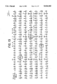
- FIG. 15 Composite nucleotide sequence of group O cDNA clones.
- Amino acid residues are numbered from the presumed amino terminal processing point. Cysteine residues are boxed, potential N-linked glycosylation sites are bracketed, and polyadenylation signals used in 0-240 and 0-254 are underlined.
- the composite nucleotide sequence is compared with that of the coding region of murine cDNA clone DN-4 (79). (-) denotes identity and (*) denotes a gap.
- FIG. 16 Amino acid sequence comparisons to consensus human TCR V region sequences.
- Blanks indicate consensus assignment at that position. (-) indicates a gap. Identities between the O-composite sequence and consensus residues are boxed.
- FIG. 17 Amino acid sequence comparisons to consensus human TCR J region sequences.
- FIG. 18 Amino acid sequence comparisons to human TCR and immunoglobulin C region sequences.
- FIG. 19 Distribution of charged and uncharged amino acids in the region flanking and including the presumed transmembrane region of the o-composite sequence compared with those of C ⁇ , C ⁇ , and C ⁇ .
- FIG. 20 Cytofluorographic analysis of T cell lines with anti-TCR ⁇ 1
- FIG. 21 Immunochemical analysis of the specificity of mAb anti-TCR ⁇ 1
- FIG. 22 N-glycanase digestion of TCR ⁇ .
- FIG. 23 Map of pGEM3-O-240/38
- FIG. 24 Immunoprecipitation of in vitro translation products of cDNA clone IDP2 O-240/38 by mAb anti-TCR ⁇ 1
- FIG. 25 Immunoprecipitation and SDS-PAGE analysis of T cell antigen receptor.
- Open arrowheads indicate the position of the ⁇ chains.
- the solid arrowheads indicate the position of the ⁇ chains. Lysates were immunoprecipitated using ⁇ TCAR-3 antibody (odd numbered lanes) or ⁇ F1 antibody (even numbered lanes).
- FIG. 26 Immunoprecipitation of ⁇ chain by ⁇ TCAR-3 antibody
- Lane 1 ⁇ TCAR-3 immunoprecipitates ⁇ heterodimer with the CD3 proteins.
- Lane 2 ⁇ TCAR-3 immunoprecipitates ⁇ heterodimer without the CD3 proteins.
- Lanes 3 and 4 ⁇ TCAR-3 immunoprecipitates single ⁇ chain from denatured lysates (N) and reducing (R) conditions, respectively.
- Lane 5 UCHT-1 immunoprecipitates the CD3 proteins.
- Lane 6, ⁇ F1 antibody does not immunoprecipitate a heterodimer from MOLT-13 cells.
- FIG. 27 Analysis of cell surface staining by flow cytometry.
- ⁇ , ⁇ positive cells MOLT-13, PEER, IDP2
- ⁇ , ⁇ positive cells HPB-ALL, JURKAT
- ⁇ TCAR-3 OKT 3, WT31 and normal mouse serum (NMS) antibodies
- NMS normal mouse serum
- FIG. 28 Two color cytofluorographic analysis of ⁇ TCAR-3 + and OKT 3 + peripheral blood lymphocytes.
- the FITC fluorescence is depicted on the Y axis and PE fluorescence on the X axis.
- the CD3 + ⁇ , ⁇ + cells in this sample represent 2.4% of CD3 + lymphocytes.
- FIG. 29 Measurement of intracytoplasmic Ca 2+ concentration ([Ca 2+ ] i ) versus time
- the present invention provides a purified polypeptide which comprises at least a portion of a ⁇ T cell receptor polypeptide.
- This polypeptide may have at least one intrachain, covalent, disulphide bridge Additionally, the polypeptide may comprise a ⁇ T cell receptor polypeptide having a molecular weight of about 40,000 daltons. Furthermore, the ⁇ T cell receptor polypeptide may be a human ⁇ T cell receptor polypeptide. In one embodiment of the invention the polypeptide comprises at least a portion of the amino acid sequence shown in FIG. 15.
- a substance capable of specifically forming a complex with at least one ⁇ T cell receptor polypeptide is also provided by the invention.
- the substance is capable of specifically forming a complex with one ⁇ T cell receptor polypeptide.
- the substance is capable of specifically forming a complex with more than one ⁇ T cell receptor polypeptide.
- the substance may be an antibody.
- the antibody may be a polyclonal antibody or a monoclonal antibody.
- This method comprises contacting a sample containing T cells with substances capable of forming complexes with ⁇ T cell receptor polypeptides so as to form cellular complexes between the substances and the ⁇ T cell receptor polypeptides. These cellular complexes are detected and thereby T cells, each of which has a ⁇ T cell receptor polypeptide, are detected.
- the ⁇ T cell receptor polypeptides are present on the surfaces on the T cells. In another embodiment of the invention, the ⁇ T cell receptor polypeptides are present in the cytoplasm of the T cells.
- This method may be performed by forming complexes with a specific ⁇ T cell receptor polypeptide
- the specific ⁇ T cell receptor polypeptide is present only in suppressor T cells.
- immune system abnormality means a condition of immunological responsiveness to antigens characterized by an increased or a decreased immune response compared to a normal or standard immune response.
- immune system abnormality includes, but is not limited to, immunodeficiency conditions and diseases, e.g. acquired immune deficiency syndrome and congenital immunodeficiencies and hyperimmune conditions and diseases, e.g. allergies and hayfever.
- the method of the present invention comprises determining the number of T cells in a sample from the subject and contacting the sample with the substances capable of forming complexes with at least one ⁇ T cell receptor polypeptide so as to form cellular complexes between the substances and ⁇ T cell receptor polypeptides
- the percentage of T cells in the sample which have a ⁇ T cell receptor polypeptide is determined and compared with the percentage of T cells which have a ⁇ T cell receptor polypeptide in a sample from a normal subject who does not have the immune system abnormality A difference in the percentage of T cells so determined would be indicative of the immune system abnormality.
- the immune system abnormality is a cancer.
- the cancer may be a leukemia or a lymphoma.
- the immune system abnormality is acquired immune deficiency syndrome
- the immune system abnormality is congenital immunodeficiency.
- the immune system abnormality is an autoimmune disease.
- the subject in whom the immune system abnormality is diagnosed may be an animal.
- the subject is a human.
- the sample from the subject may comprise blood or tissue.
- Yet another method for diagnosing an immune system abnormality comprises determining the number of ⁇ T cell receptor polypeptide bearing T cells in a sample from the subject and the amount of ⁇ T cell receptor polypeptides in the T cell receptor polypeptide bearing T cells.
- the amount of ⁇ T cell receptor polypeptides so determined is compared with the amount of ⁇ T cell receptor polypeptides in an equal number of ⁇ T cell receptor polypeptide bearing T cells in a sample from a normal subject who does not have the immune system abnormality. A difference in the amount so determined would be indicative of the immune system abnormality.
- the amount of a single ⁇ T cell receptor polypeptide is determined
- a further method for diagnosing an immune system abnormality in a subject comprises determining in a sample from the subject the number of T cells which have a ⁇ T cell receptor polypeptide and the number of T cells consisting of the group of T cells which have one of the surface markers T4, T8 and ⁇ , ⁇ T cell receptor.
- the numbers of T cells so determined is compared with the number of T cells which have a ⁇ T cell receptor polypeptide and the number of T cells in the group which have the same surface marker as the group of T cells determined in the sample from the subject, in a sample from a subject who does not have the immune system abnormality.
- a difference in the number of T cells so determined which have a ⁇ T cell receptor polypeptide relative to the number of T cells in the group so determined would be indicative of the immune system abnormality.
- the present invention also provides a nucleic acid molecule encoding a ⁇ T cell receptor polypeptide having a molecular weight of about 40,000 daltons.
- the molecule is a DNA molecule.
- the DNA molecule comprises at least a portion of the nucleic acid sequence shown in FIG. 15. Further provided is a nucleic acid molecule which is complementary to the nucleic acid molecule which encodes a ⁇ T cell receptor polypeptide.
- a purified polypeptide which comprises at least a portion of a ⁇ T cell receptor polypeptide is also provided by the present invention.
- This polypeptide may comprise a ⁇ T cell receptor polypeptide having a molecular weight of about 55,000 daltons.
- the polypeptide has a peptide sequence with a molecular weight within the range from about 31,000 daltons to about 40,000 daltons.
- the polypeptide may be a human ⁇ T cell receptor polypeptide.
- the present invention further provides a purified complex which comprises two ⁇ T cell receptor polypeptides of the present invention associated with each other.
- the two ⁇ T cell receptor polypeptides are associated with each other through at least one interchain, covalent, disulphide linkage.
- the two ⁇ T cell receptor polypeptides are noncovalently associated with each other.
- the two ⁇ T cell receptor polypeptides have the same constant domain.
- the two ⁇ T cell receptor polypeptides have different constant domains.
- the present invention also provides a substance capable of specifically forming a complex with at least one ⁇ T cell receptor polypeptide.
- the substance is capable of specifically forming a complex with one ⁇ T cell receptor polypeptide.
- the substance is capable of specifically forming a complex with more than one ⁇ T cell receptor polypeptide.
- the substance may be an antibody.
- the antibody is a polyclonal antibody.
- the antibody is a monoclonal antibody
- a method for detecting T cells, each of which has a ⁇ T cell receptor polypeptide comprises contacting a sample which contains T cells with substances capable of forming complexes with ⁇ T cell receptor polypeptides so as to form cellular complexes between the substances and the ⁇ T cell receptor polypeptides. These cellular complexes are detected and thereby T cells, each of which has a ⁇ T cell receptor polypeptide, are detected.
- the ⁇ T cell receptor polypeptides are present on the surfaces of the T cells.
- the ⁇ T cell receptor polypeptides are present in the cytoplasm of the T cells.
- the substances are capable of forming complexes with a specific ⁇ T cell polypeptide.
- the specific ⁇ T cell receptor polypeptide may be present only in suppressor T cells.
- the ⁇ T cell receptor polypeptide may be associated with another ⁇ T cell receptor polypeptide.
- the ⁇ T cell receptor polypeptide is associated with another ⁇ T cell receptor polypeptide.
- the ⁇ T cell receptor polypeptide is associated with another ⁇ T cell receptor polypeptide only in non-major histocompatibility restricted cytotoxic T lymphocytes
- the non-major histocompatibility complex restricted cytotoxic T lymphocytes may be T killer cells or natural killer-like cells.
- the present invention further provides a method for diagnosing an immune system abnormality in a subject.
- This method comprises determining the number of T cells in a sample from the subject and contacting the sample with substances capable of forming complexes with at least one ⁇ T cell receptor polypeptide so as to form cellular complexes between the substances and ⁇ T cell receptor polypeptides.
- the percentage of T cells in the sample which have a ⁇ T cell receptor polypeptide is determined and compared with the percentage of T cells which have a ⁇ T cell receptor polypeptide in a normal subject who does not have the immune system abnormality. A difference in the percentage of T cells so determined would be indicative of the immune system abnormality
- the immune system abnormality is a cancer.
- the cancer may be a leukemia or a lymphoma.
- the immune system abnormality is acquired immune deficiency syndrome.
- the immune system abnormality is congenital immunodeficiency.
- the immune system abnormality is an autoimmune disease.
- the subject in which the immune system abnormality is diagnosed may be an animal. Additionally, the subject in which the immune system abnormality is diagnosed may be a human. Furthermore, the sample of which the percentage of T cells which have a ⁇ T cell receptor polypeptide is determined may comprise blood or tissue.
- Yet another method for diagnosing an immune system abnormality in a subject comprises determining the number of ⁇ T cell receptor polypeptide bearing T cells in a sample from the subject and the amount of ⁇ T cell receptor polypeptides in the ⁇ T cell receptor polypeptide bearing T cells.
- the amount of ⁇ T cell receptor polypeptides so determined is compared with the amount of ⁇ T cell receptor polypeptides in an equal number of ⁇ T cell receptor polypeptide bearing T cells in a sample from a normal subject who does not have the immune system abnormality. A difference in the amount so determined would be indicative of the immune system abnormality.
- the amount of a single ⁇ T cell receptor polypeptide is determined.
- Another method for diagnosing an immune system abnormality in a subject comprises determining in a sample from the subject the number of T cells which have a ⁇ T cell receptor polypeptide and the number of T cells consisting of the group of T cells which have one of the surface markers T4, T8 and ⁇ , ⁇ T cell receptor The numbers of T cells so determined are compared with the of T cells which have a ⁇ T cell receptor polypeptide and the number of T cells in the group which have the same surface marker as the group determined in the sample from the subject, in a sample from a subject who does not have the immune system abnormality. A difference in the number of T cells so determined which have a T cell receptor polypeptide relative to the number of T cells in the group so determined would be indicative of the immune system abnormality.
- a purified complex which comprises at least a portion of a ⁇ T cell receptor polypeptide and at least a portion of a ⁇ T cell receptor polypeptide is further provided by the present invention.
- This complex may comprise a ⁇ T cell receptor polypeptide having a molecular weight of about 40,000 daltons and a ⁇ T cell receptor polypeptide having a molecular weight of about 55,000 daltons.
- the ⁇ T cell receptor polypeptide may be a human ⁇ T cell receptor polypeptide and the ⁇ T cell receptor polypeptide may be a human ⁇ T cell receptor polypeptide.
- the ⁇ T cell receptor polypeptide and the ⁇ T cell receptor polypeptide may be associated with each other through at least one interchain, covalent, disulphide linkage, or may be noncovalently associated with each other.
- a substance capable of specifically forming a complex with at least one ⁇ , ⁇ T cell receptor complex This substance may be capable of forming a complex with one ⁇ , ⁇ T cell receptor complex. Furthermore, the substance may be capable of forming a complex with more than one ⁇ , ⁇ T cell receptor complex.
- the substance is an antibody. In another embodiment of the invention, the substance is a polyclonal antibody. In yet another embodiment of the invention, the substance is a monoclonal antibody.
- the present invention further provides a method for detecting T cells, each of which has a ⁇ , ⁇ T cell receptor complex.
- This method comprises contacting a sample containing T cells with substances capable of . forming complexes with ⁇ , ⁇ T cell receptor complexes so as to form cellular complexes between the substances and the ⁇ , ⁇ T cell receptor complexes. These cellular complexes are detected and thereby T cells, each of which has a ⁇ , ⁇ T cell receptor complex, are detected
- the ⁇ , ⁇ T cell receptor complexes are present on the surface of the T cells.
- the ⁇ , ⁇ T cell receptor complexes are present in the cytoplasm of the T cells.
- the substances are capable of forming complexes with a specific ⁇ , ⁇ T cell receptor complex. The specific ⁇ , ⁇ T cell receptor complex may be present only in suppressor T cells.
- a method for diagnosing an immune system abnormality in a subject comprises determining the number of T cells in a sample from the subject and contacting the sample with substances capable of forming complexes with at least one ⁇ , ⁇ T cell receptor complex so as to form cellular complexes between the substances and ⁇ , ⁇ T cell receptor complexes.
- the percentage of T cells in the sample which have a ⁇ , ⁇ T cell receptor complex is determined and compared with the percentage of T cells which have a ⁇ , ⁇ T cell receptor complex in a sample from a normal subject who does not have the immune system abnormality. A difference in the percentage of T cells so determined would be indicative of the immune system abnormality.
- the immune system abnormality is a cancer.
- the cancer may be a leukemia or a lymphoma.
- the immune system abnormality is acquired immune deficiency syndrome.
- the immune system abnormality is congenital immunodeficiency.
- the immune system abnormality is an autoimmune disease.
- the subject in which the immune system abnormality may be an animal. Furthermore, the subject in which the immune system abnormality is diagnosed may be a human. Moreover, the sample in which the percentage of T cells which have a ⁇ , ⁇ T cell receptor complex is determined may comprise blood or tissue.
- Still another method for diagnosing an immune system abnormality in a subject comprises determining the number of ⁇ , ⁇ T cell receptor complex bearing T cells in a sample from the subject and the amount of ⁇ , ⁇ T cell receptor complexes in the ⁇ , ⁇ T cell receptor complex bearing T cells.
- the amount so determined is compared with the amount of ⁇ , ⁇ T cell receptor complexes in an equal number of ⁇ , ⁇ T cell receptor complex bearing T cells in a sample from a normal subject who does not have immune system abnormality. A difference in the amount so determined would be indicative of the immune system abnormality.
- the amount of a single ⁇ , ⁇ T cell receptor complex is determined.
- This method comprises determining in a sample from the subject the number of T cells which have a ⁇ , ⁇ T cell receptor complex and the number of T cells consisting of the group of T cells which have one of the surface markers T4, T8 and ⁇ , ⁇ T cell receptor complex.
- the numbers of T cells so determined are compared with the number of T cells which have a ⁇ , ⁇ T cell receptor complex and the number of 1. cells in the group which have the same surface marker as the group of T cells determined in the sample from the subject, in a sample from a subject who does not have the immune system abnormality.
- a difference in the number of T cells so determined which have a ⁇ , ⁇ T cell receptor complex relative to the number of T cells in the group so determined would be indicative of the immune system abnormality.
- the various methods for diagnosing abnormalities and for detecting T cells are based upon the novel polypeptides and substances capable of forming complexes with these polypeptides as described more fully hereinabove.
- the methods utilize methods for detecting and quantifying T cells, including but not limited to, fluorescence activated cell sorting and autoradiography, which are well known to those skilled in the art to which this invention pertains.
- Viable lymphocytes were isolated by Ficoll-hypaque density centrifugation and stained with 0.5 micrograms of a specific monoclonal antibody, e.g. WT31 (28, 29) or OKT®3, OKT®4 or OKT®8 (Ortho Diagnostic Systems, Inc., Raritan, N.J.), for 30 minutes at 4° C. After washing, the cell pellets were stained again with fluorescein isothiocyanate (FITC)-conjugated goat antimouse IgG(ab)' 2 fragments Fluorescence activated cell sorter(FACS) analyses were performed on an Ortho cytofluorograph or a Coulter Epics as previously described (37).
- FITC fluorescein isothiocyanate
- FACS Fluorescence activated cell sorter
- IL-2 dependent cell lines were propagated in vitro in media composed of RPMI 1640, 10% human serum and conditioned media containing 2-5 units of interleukin-2 activity as previously described (34).
- Alloantigen (allo) activated cultures were stimulated with irradiated allogenic peripheral blood lymphocytes at weekly intervals.
- Mitogen i.e. phytohemagglutinin (PHA)
- activated lines were stimulated with a 1:1,000 dilution of PHA (Difco, Detroit, Mich.) at culture initiation.
- Immunoprecipitates from 125 I-labeled lymphocyte lysates were analyzed by sodium dodecyl sulfate-polyacrylamide gel electrophoresis (SDS-PAGE).
- the radioiodinated T leukemia cell lines HPB-MLT and Jurkat, the HTLV-1 transformed cell line ANITA and resting peripheral blood lymphocytes were solubilized in 1% Triton-X-100 (TX-100) and immunoprecipitated with a control antibody, normal mouse serum (NMS) or a framework antibody to TCR ⁇ , ⁇ i.e., Fl (54).
- TX-100 Triton-X-100
- NMS normal mouse serum
- Fl a framework antibody to TCR ⁇ , ⁇ i.e., Fl (54).
- the ⁇ F1 monoclonal antibody was prepared according to standard procedures (46, 47, 52).
- Spleen cells from mice immunized with purified TCR ⁇ , ⁇ as described in (28) were used for the fusion experiments.
- a positive clone, ⁇ F1 was obtained by immunoprecipitation with T cell lines and peripheral blood lymphocytes as described above.
- 125 I-labeled lymphocytes were solubilized in 0.1% TX-100 and immunoprecipitated with NMS, the anti-T3 antibody UCHT-1 (40) and a framework antibody to TCR i.e., WT31.
- the efficiency of immunoprecipitation with WT31 was improved at the lower TX-100 concentration used here and the monoclonal antibody 187.1 (53) was used as a second antibody.
- Two-color FACS analysis of normal adult peripheral blood lymphocytes was performed using an anti-TCR ⁇ , ⁇ monoclonal antibody and an anti-T3 monoclonal antibody.
- Peripheral blood lymphocytes were stained first with an FITC-conjugated anti-T3 monoclonal antibody (OKT®3) and then with a biotinyl-anti-TCR ⁇ , ⁇ monoclonal antibody ( ⁇ F1) followed by phycoerythrin-conjugated avidin (PE-avidin, Becton Dickenson, Mt. View, Calif.).
- lymphocytes were isolated by ficoll-hypaque density centrifugation for SDS-PAGE and FACS analyses.
- lymphocytes were radioiodinated by the lactoperoxidase technique, solubilized in 1% TX-100 and immunoprecipitated using 1 microgram of a specific antibody, i.e. monoclonal antibody ⁇ F1 or monoclonal antibody UCHT-1, or 1 microliter of NMS.
- the immunoprecipitates were then analyzed by 10.5% SDS-PAGE under reducing conditions.
- the 125 I-labeled molecules were visualized by autoradiography as previously described (28).
- Two-colored cytofluorographic analysis was performed by first staining with FITC-OKT®3 monoclonal antibody for 45 minutes at 4° C. After washing, the lymphocytes were fixed in 1% paraformaldehyde for 15 minutes at 23° C. then incubated in 70% ethanol in phosphate buffered saline (PBS) for 5 minutes at -20° C. After further washing, the cells were stained with the biotinyl- ⁇ F1 monoclonal antibody followed by PE-avidin. Analysis was performed on an Ortho® cytofluorograph (Ortho Diagnostic Systems, Inc., Westwood, Mass.).
- IDP1 cell line 2 WT31 +
- cell line 3 WT31 -
- Radioiodinated, intact lymphocytes were then either cross-linked by incubation in PBS (pH 8) containing 50 micrograms/ml dithio-bis-succinimidyl propionate (DSP) or mock incubated.
- the cells were then solubilized in 1% TX-100 and immunoprecipitated as previously described (12).
- T3 associated molecules M r 40,000-55,000
- IDP2 cell line 7 (88% WT31 - T3 + ) was 125 I-labeled and treated with DSP or mock incubated. Immunoprecipitations were performed using NMS, the anti-T3 monoclonal antibody UCHT-1 and the anti-TCR ⁇ , ⁇ monoclonal antibody ⁇ F1 either without or with preclearing TCR ⁇ , ⁇ molecules with the monoclonal antibody ⁇ F1. A small fraction of radiolabeled TCR ⁇ , ⁇ was detected in samples which were not precleared but not in samples which were precleared with ⁇ F1.
- IDP2 cell line 5 WT31 + T3 +
- cell line 7 88% WT31 - T3 +
- RNA (3 micrograms) isolated from IDP2 cell line 5 (WT31 + T3 + ), IDP2 cell line 7 (88% WT31 - T3 + ) and HPB-MLT was analyzed as described above.
- RNA preparation, electrophoresis, transfer to nitrocellulose and hybridization with 32 P-labeled, nicktranslated probes (1-3 ⁇ 10 8 cpm/microgram) were as described previously (41).
- ⁇ -chain probes were either the human cDNA clones pGA5(8) or L17 ⁇ (42).
- ⁇ chain probes were either the human cDNA clones 12A1 (43) or L17 (43).
- the ⁇ -chain probe was an EcoRI to AccI fragment derived from human cDNA clone T ⁇ -1 (36). Radioactive bands were visualized by autoradiography using intensifying screens. All probes were labeled to nearly identical specific activity, and identical exposure times are presented.
- DSP cross-linked native lysates (1% TX-100) from 125 I-labeled IDP2 cell line 7 were immunoprecipitated with NMS or with the anti-T3 monoclonal antibody UCHT-1. Alternatively, the lysate was denatured (as described below) and immunoprecipitated with either normal rabbit serum or with anti-C ⁇ peptide serum.
- Samples were immunoprecipitated with 10 microliters of anti-C ⁇ or anti-V ⁇ peptide sera, 1 microgram of UCHT-1 or 1 microliter of NMS or normal rabbit serum and analyzed by 10.5% SDS-PAGE under reducing conditions (5% 2-mercaptoethanol).
- Peptides corresponding to deduced V ⁇ or C ⁇ amino acid sequences were synthesized on a Beckman 990 peptide synthesizer using the method of Erickson and Merrifield (44). Peptide purity was assessed by high pressure liquid chromatography and peptide sequence was confirmed by amino acid analysis. Peptides were coupled to keyhole limpet hemocyanin (KLH) at a ratio of 50 peptides per KLH molecule (45). Mice and rabbits were immunized with the V ⁇ peptides or C ⁇ peptides, respectively. Animals were injected at three week intervals and the antisera screened for binding reactivity on peptide-KLH and peptide-bovine serum albumin conjugates to ascertain the presence of peptide-specific antibodies.
- KLH keyhole limpet hemocyanin
- Monoclonal antibodies against the ⁇ chain were generated by standard procedures as described in (47, 50).
- BALB/c mice were immunized with the KLH-coupled peptide to the variable region ⁇ chain peptide described above using the method of Erickson and Merrifield (44). After four immunizations at two week intervals, spleen cells were fused with P3-X63-Ag8U1 myeloma cells. Positive hybridoma clones were screened and identified by the enzyme immunoassay (EIA) described in (48).
- EIA enzyme immunoassay
- DNA sequences of the TCR ⁇ gene may be isolated and determined by strategies utilized to isolate the TCR ⁇ gene as described in (49, 50). Briefly, the amino acid sequence of the TCR ⁇ gene may be determined following isolation of the TCR ⁇ polypeptide which is described hereinafter After the amino acid sequence is determined, short, synthetic DNA sequences may be prepared using a commercial DNA synthesizer (Applied Biosystems, Inc., Foster City, Calif.). The synthetic DNA sequences may be used as probes for the isolation of the complete sequence of DNA from a cDNA library of cell lines containing the TCR polypeptide The primary structure of the protein may then be determined (51).
- Monoclonal antibodies against the ⁇ polypeptide may be generated by standard procedures (47, 50).
- Peptides derived from the TCR ⁇ polypeptide may be prepared from nucleic acid sequences determined by the methods described above. Methods for the selection of such peptides useful for immunization have been described in detail (55, 56, 57).
- Monoclonal antibodies directed against ⁇ , ⁇ complexes may be prepared according to published procedures (47, 50). ⁇ , ⁇ complexes may be isolated from the T cell lines described above and used to immunize BALB/c mice as described in previously published procedures (28). Alternatively, BALB/c mice may be immunized with cell lines, e.g., the IDP1 cell line or the IDP2 cell line.
- Hybridoma cells that produce monoclonal antibodies which are directed against specific TCR ⁇ , ⁇ cell lines but which do not cross react with other T cell lines may be selected and recovered.
- Viable lymphocytes were isolated by Ficoll-Hypaque density gradient centrifugation and 2 ⁇ 10 7 cells were radio-iodinated by the lactoperoxidase technique as described (28). Labelled cells were lysed in 5 ml of TBS (10 mM Tris pH 8, 140 mM NaC1) with 0.3% 3-[(3- cholamidopropyl) dimethylammonio]-1-propanesulphonate (CHAPS), which preserves the TCR-T3 association (64), containing 2 mM phenyl methylsulphonyl fluoride (PMSF) and 8 mM iodoacetamide (IAA).
- TBS 10 mM Tris pH 8, 140 mM NaC1
- CHAPS 3-[(3- cholamidopropyl) dimethylammonio]-1-propanesulphonate
- Immunoprecipitation was carried out using fixed Staphylococcus aureus Cowan I (SACI) as described (12), and the immune complexes were washed x 5 in TBS containing 0.1% Triton X-100 (TX-100). Reduced samples were boiled in 2 mM dithiothreitol (DTT) and all samples incubated for 10 min at 23° C. in 10 mM IAA before analysis by SDS-PAGE.
- SACI Staphylococcus aureus Cowan I
- Immunoprecipitations using anti-C ⁇ sera were performed on 1% TX-100 lysates that were dialysed to remove IAA and then denatured by the addition of one tenth volume of sodium dodecyl sulfate (SDS) containing 3 mM DTT with boiling for 3 min. After partial renaturation by the addition of 4 vols of 1.5% TX-100 in TBS containing 30 mM IAA, anti-C ⁇ sera or NRS were added and the immunoprecipitates were washed in TBS containing 0.5% TX-100, 0.5% deoxycholate, 0.05% SDS before analysis by SDS-PAGE. Rat anti-mouse ⁇ chain-specific mAb 187.1 15 ⁇ g) was added as a second antibody to provide protein A binding of IgG I mAb ⁇ F1, UCHT and P3 (53).
- SDS sodium dodecyl sulfate
- RNA sizes were determined based on previously published lengths for TCR ⁇ and TCR ⁇ transcripts (36, 4).
- lymphocytes were treated with 100U of neuraminidase (Gibco) in phosphate-buffered saline (PBS) 1 mg ml - bovine serum albumin, 1 mg ml 1 glucose for 90 minutes at 23° C., washed in PBS and solubilized in 0.3% CHAPS.
- Immunoprecipitates were prepared as in FIG. 1 and NEPHGE (charge separation) was carried out using pH 3.5-10 ampholines (LKB, Sweden), or IEF using pH 3.5-10, 4-6, 9-11 ampholines (2:15.5:1.5) followed by 10.5% SDS-PAGE gels for size separation as described (12).
- NEPHGE was carried out (A,B) applying the iodinated IEF sample at the acidic end, while IEF (C,D) was carried out for 20 hours at 400V applying the sample at the other (basic) end. Brackets enclose the T3-associated species.
- Cells were collected, washed and lysed 0.4 ml of boiling solution of 1% SDS, 10 mM Tris-HC1 (pH 8.0), 0.1 mM pMSF and 10 mM IAA Lysates were diluted with 1.6 ml of 2.5% Nonidet-P40, 1% gelatin, 10 mM Tris-HC1 (pH 8) and 0.2 ml of 1 mg ml -1 DNase. 0.5 mg ml -1 RNase, 0.5M Tris-HC1 (pH 7), 50 mM MgC 2 and incubated at 0° C.
- Genomic DNA was isolated as described (BamHI or EcoRI), size-fractionated on 0.7 & agarose (BamHI digests) or 0.9% agarose (EcoRI digest), and transferred to nitrocellulose as described (67). Filters were hybridized to a nick-translated 32 P-labelled 0.8 kb HindIII-EcoRI J.sub. ⁇ 1,3 probe (20) or a 1.1 kb EcoRI-HindII C.sub. ⁇ 2 probe (68). Filters were washed in 2x SSC and 0.1% SDS followed by 0.2% SSC and 0.1% SDS at 55° C. before autoradiography with intensifying screens.
- Cytolytic assays were performed in round-bottom 96-well tissue culture plates with 51 Cr-labelling, harvesting and calculation of % specific release as described (34).
- IDP2 or PBL C1 cells were either preincubated with UCHT1 (1:300 dilution) (+anti-T3) for 30 minutes at 0° C., washed x 3 or mock incubated and placed together with labelled target cells.
- Anti-HLA Class I and Class II mAb anti-T3 mAb were placed in wells containing the 51 Cr-labelled MOLT 4 cells for 30 minutes at 0° C., then IDP2 cells were added at a 40:1 E:T ratio. All samples were assayed in triplicate, each experiment was performed at least three times, and one representative experiment of each is shown in FIG. 9.
- a murine framework antiserum that recognizes the majority of human TCR ⁇ , ⁇ molecules has previously been reported (28). Subsequently, a murine monoclonal antibody, designated Framework 1 ( ⁇ F1), that is reactive with shared determinants on the human TCR ⁇ chain was obtained (46). The ⁇ F1 monoclonal antibody reacts with majority of T3 positive (T3 + ) human peripheral blood lymphocytes (PBLs) and is capable of immunoprecipitating the TCR ⁇ , ⁇ heterodimer from all human T cell lines examined that have ⁇ , ⁇ T cell receptors and express the I' 3 glycoprotein.
- T3 + human peripheral blood lymphocytes
- Double staining of human PBLs with a fluorescein-anti-T3 monoclonal antibody and a biotinyl- ⁇ F1 monoclonal antibody followed by PE avidin reveals that the ⁇ F1 monoclonal antibody recognizes 95-97% of peripheral blood T3 + lymphocytes However, it clearly defines a small population of T lymphocytes that is ⁇ F1 negative ( ⁇ F1), yet T3 + (FIG. 1C).
- a second framework monoclonal antibody designated WT31 initially thought to recognize the T3 antigen (29), has recently been shown to react with a common epitope of human TCR ⁇ , ⁇ (30). While double staining with an anti-T3 monoclonal antibody (OKT®3) and WT31 revealed that each of these monoclonal antibodies cross-block binding of the other, one-color fluorescence indicated that WT31 typically recognized 1-3% fewer cells in peripheral blood than do anti-T3 monoclonal antibodies.
- the WT31 monoclonal antibody efficiently binds to the surface of T cells (such as in FACS analyses) and is capable of immunoprecipitating the TCR ⁇ , ⁇ molecules, albeit inefficiently, from radio labeled detergent lysates (30) (FIG. 1B, lane 3).
- the ⁇ F1 monoclonal antibody and the WT31 monoclonal antibody appear to recognize all but a small fraction of human peripheral blood T3 + cells, and define a subpopulation that is T3 + but unreactive with both of these framework monoclonal antibodies against the TCR ⁇ , ⁇ molecules.
- WT31 was used primarily for FACS analyses and ⁇ F1 was used primarily for immunoprecipitation studies.
- the WT31 - T3 + cells identified by the lack of cell surface reactivity with the WT31 monoclonal antibody in FACS analysis, were also ⁇ F1 - , as determined by the lack of TCR ⁇ , ⁇ on immuno-precipitation.
- All WT31 + T3 + and WT31 - T3 + cell lines expressed similar amounts of T3 by FACS analysis and by immunoprecipitation with an anti-T3 monoclonal antibody (FIG. 2A, lanes 3 and 9; FIG. 2C, lanes 2 and 4).
- T3 molecule found on WT31 - ⁇ F1 - T3 + lymphocytes was not identical to the T3 molecule found on WT31 + ⁇ F1 30 T3 + cells by SDS-PAGE.
- TCR ⁇ or TCR ⁇ mRNA transcripts could be detected in the RNA from the WT31 - ⁇ F1 - T3 + IDP2 cell line 6 (FIG. 3A- probe, lane 1; or ⁇ -probe, lane 1), whereas expression of both was clearly detectable in RNA from HPB-MLT (FIG. 3A ⁇ -probe, lane 2; and ⁇ -probe, lane 2)
- TCR ⁇ mRNA was present in the WT31 - T3 + cells at levels comparable to that in HPB-MLT (FIG. 3A ⁇ -probe, lanes 1 and 2).
- the IDP2 cell line 5 (WT31 + T3 + ) expressed levels of both RNA species which were comparable to HBP-MLT (FIG. 3B, lane 1 for each probe).
- WT31 + T3 + cell line shown in FIG. 3A both the WT31 - T3 + and the WT31 + T3 + cell lines showed TCR ⁇ RNA levels comparable to HPB-MLT (FIG. 3B ⁇ -probe).
- the WT31 - T3 + cells lacked ⁇ and ⁇ T cell receptor mRNA (Northern analysis) and ⁇ and ⁇ T cell receptor proteins (immunoprecipitation and FACS analysis).
- ⁇ mRNA in WT31 - T3 + cells, while consistent with T ⁇ protein expression, could not be taken as strong evidence for this, since many human cell lines that express T ⁇ mRNA of normal size may express full length transcripts that are out of frame due to defective V-J joining (36).
- the WT31 + ⁇ F1 + T3 + lymphocytes revealed that the TCR ⁇ and ⁇ chains were cross-linked to T3.
- TCR ⁇ , ⁇ molecules and T3 were found in anti-T3 or ⁇ F1 monoclonal antibody immunoprecipitates from cross-linked IDP1 cell line 2 (WT31 + T3 + ) (FIG. 2A, lanes 4 and 6).
- IDP1 cell line 3 WT31 - T3 +
- IDP2 cell line ⁇ 88% WT31 - T3 +
- the mobilities of these T3 associated molecules were clearly different from those of the TCR ⁇ and ⁇ chains from WT31 + T3 + cell lines (compare FIG. 2A, lanes 4 and 10; or FIG. 2B, lanes 5 and 6).
- IDP2 cell line ⁇ (88% WT31 - T3 + ) contained 12% WT31 + T3 + cells, accounting for the weak ⁇ F1 immunoprecipitates noted (FIG. 2B, lane 2)
- the lysate from these cells was precleared of TCR ⁇ , ⁇ protein using the ⁇ F1 monoclonal antibody. After preclearing, no residual ⁇ F1 reactive material could be detected (FIG. 2B, lanes 8 and 11).
- this ⁇ F1- precleared lysate from cross-linked cells was immunoprecipitated with an anti-T3 monoclonal antibody, M r 55,000 and 40,000 subunits were still detected (FIG. 2B, lane 12).
- cDNA clones representing the rearranging human TCR ⁇ gene would encode a polypeptide with a predicted molecular weight of 40,000 daltons (36).
- the human TCR ⁇ gene reveals five potential sites for N-linked glycosylation, four of which are located in the constant region (36). Since a TCR ⁇ protein has not previously been isolated, it is not known how many of these potential sites may be used. However, a fully glycosylated human TCR ⁇ protein may have a M r of about 55,000.
- the heavy chain of the non- ⁇ -non- ⁇ T3-associated subunits identified on the WT31 - ⁇ F1 - T3 + IDP1 and IDP2 cell lines has a relative mobility on SDS-PAGE of 55,000 daltons (FIG. 2A and 2B).
- FIG. 4B A sample of DSP cross-linked lysate from the WT31 - ⁇ F1 - T3 + cells was first immunoprecipitated with an anti-T3 monoclonal antibody, again demonstrating the presence of M r 55,000 and 40,000 subunits associated with T3 (FIG. 4B, lane 2).
- Immunoprecipitations from lysates of surface-iodinated IDP2 lymphocytes using anti-T3 xrAb. yielded two species (55K and 40K) in addition to the T3 subunits (FIG. 5A). This result is identical to the one reported previously using chemical cross-linking
- the 55K species was shown to react specifically with anti-C ⁇ and anti-V ⁇ peptide sera.
- the 40K polypeptide was unreactive with these anti- ⁇ peptide sera and is thus likely to represent a non TCR ⁇ , ⁇ or ⁇ subunit, namely ⁇ .
- the T3 co-immunoprecipitated polypeptides were examined under reducing and nonreducing conditions.
- the TCR ⁇ and ⁇ subunits which exist in a heterodimeric disulphide-linked form under nonreducing conditions, the TCR ⁇ and ⁇ subunits on the IDP2 cell line are not covalently linked (FIG. 5A).
- the PEER cell line might express the TCR ⁇ polypeptide since it lacked expression of TCR mRNA yet expressed a T3-associated 55-60K polypeptide (69).
- this cell line was found to lack reactivity with a mAb recognizing framework determinants on the TCR ⁇ chain, ⁇ F1 (FIG. 5A) and to express a strongly iodinated 38K polypeptide.
- the 55-60K polypeptide was specifically immunoprecipitated with anti-C ⁇ peptide sera and thus appears to represent a further example of the TCR ⁇ protein (FIG. 5A).
- the TCR ⁇ and ⁇ polypeptides on PEER were of similar size to those on the IDP2 cell line and similarly were not disulphide-linked. Like the ⁇ subunit on IDP2 cells, the counterpart molecule on PEER underwent a marked shift in SDS-PAGE mobility when compared under reducing and nonreducing conditions (compare species at arrows, lane ⁇ and 8). Thus the IDP2 and the PEER cell lines appear to express similar types of TCR ⁇ , ⁇ -T3 complexes, in which the TCR ⁇ and ⁇ subunits are not covalently linked.
- the cell line derived, WT31 - PBL - LINE was cloned by limiting dilution with plating at 0.5 cell well and the cloned cells were propagated as for the polyclonal cell line.
- Several such peripheral blood derived T cell clones were obtained, and PBL Clone (PBL Cl) was studied in detail.
- PBL Cl PBL Clone
- TCR ⁇ partner As a TCR ⁇ partner ( ⁇ ) was present on the non-disulphide linked form of this receptor complex on IDP2 and PEER cells, we examined whether the disulphide-linked form of the receptor on PBL C1 was composed of a homo- or a heterodimer. Immunoprecipitates were analyzed by two-dimensional gel electrophoresis (nonequilibrium pH gel electrophoresis (NEPHGE) followed by SDS-PAGE; FIG. 7A, B). Under reducing conditions, both the TCR ⁇ species (40K and 36K) were found to have identical charges, and displayed heterogeneity typical for a sialylated glycoprotein.
- a disulphide-linked dimer composed of one or both of these TCR ⁇ species should have a focusing position similar to either of the two components alone when analyzed by NEPHGE or equilibrium isoelectric focusing (IEF). But a heterodimer composed of TCR ⁇ a distinct polypeptide might have a different charge and focusing position.
- the position of the disulphide-linked dimer was therefore examined by carrying out NEPHGE under nonreducing conditions, followed by SDS-PAGE under nonreducing conditions (FIG. 7B). Strikingly, the position of the disulphide-linked dimer was substantially more acidic than that of the TCR polypeptides examined under reducing conditions (compare the 40K and 36K species in FIG. 7A with the 70K species in FIG. 7B).
- TCR peptides were analyzed in cells pulse-labelled with 35 S-methionine After solubilization under denaturing and reducing conditions, the lysates were immunoprecipitated with anti-C ⁇ sera and examined by two-dimensional gel electrophoresis (FIG. 7E-H).
- Immunoprecipitates were either treated with endoglycosidase H (endo-H) to remove immature high-mannose glycans from pulse-labelled material, or were mock treated.
- Two TCR ⁇ polypeptides (46K and 43K) of identical NEPHGE mobility were synthesized by the IDP2 cell line.
- Treatment with endo-H reduced both forms to a 40K form, suggesting that the 46K and 43K forms carried different numbers of carbohydrates, and that a single TCR ⁇ polypeptide backbone (40K) was synthesized by IDP2 cells (FIG. 7E, F).
- WT31 - PBL LINE The polyclonal peripheral blood cell line (WT31 - PBL LINE) from which PBL C1 had been cloned was therefore studied in greater detail.
- WT31 - PBL LINE was homogeneously T3 + T11 + and contained 95% WT31 - T4 - T8 - with 5% contaminating WT31 + cells.
- WT31 - PBL LINE was homogeneously T3 + T11 + and contained 95% WT31 - T4 - T8 - with 5% contaminating WT31 + cells.
- This material included disulphide-linked TCR ⁇ , ⁇ peptides located above the open arrow, lane 14 (size identified by the ⁇ F1 precipitate, lane 13) and disulphide-linked TCR ⁇ peptides of smaller size (open arrow, lane 14).
- the majority of the T3-associated species were not disulphide-linked and migrated with the same mobility under both reducing and nonreducing conditions.
- a fraction of these non-linked species displayed a marked increase in SDS-PAGE mobility under nonreducing conditions, similar to the TCR ⁇ on the IDP2 and PEER cells (see FIG. 5B, lane 14, solid arrow)
- Reactivity with anti-C ⁇ sera confirmed that most of the labelled material associated with T3 expressed on WT31 - PBL LINE was TCR ⁇ gene products (lane 16).
- the protein product of the TCR ⁇ gene occurs on T3 + lymphocytes in adult peripheral blood in both disulphide-linked and unlinked molecular ⁇ forms.
- the non disulphide-linked form of TCR may be further divided into 55-60K glycosylated (IDP2 and PEER) or 35-45K glycosylated (thymic T cell clone C11 (70) and WT31 - PBL LINE) species.
- TCR ⁇ and ⁇ gene rearrangements were examined in T cells known to express the TCR ⁇ polypeptide on their cell surfaces.
- Southern blot analysis were carried out using the 0.8 kb EcoRI-HindIII human J.sub. ⁇ 1,3 probe (nomenclature according to Quertermous et al.(71)). This probe detects germline bands of 23kb and 12kb in a BamHI digest of genomic DNA. The 23 kb band encompasses C.sub. ⁇ 1 and the 12 kb band encodes C.sub. ⁇ 2.
- IDP1, PBL C1 and PBL C2 (also derived from the WT31 - PBL LINE) showed rearrangements of the TCR gene (both PBL C1 and PBL C2 displayed an identical rearrangement; FIG. 8A).
- the TCR ⁇ gene was also rearranged in IDP2, PBL C1 and PBL C2 cells.
- the 1.1 kb EcoRI-HindIII C.sub. ⁇ 2 probe detects a germline band of 20 kb which encompasses both C ⁇ constant regions in a BamHI digest of genomic DNA (68).
- One predominant TCR ⁇ rearrangement for IDP2 and two identical rearrangements for PBL C1 and PBL C2 were observed (FIG. 8C). It is assumed that these TCR ⁇ rearrangements are nonproductive based on the immunoprecipitations and Northern analyses for these cell lines.
- both PBL C1 and PBL C2 have the same TCR ⁇ and ⁇ rearrangements, they appear to be clonal and derived from the same cell within the WT31 - PBL LINE.
- IDP2 and PBL C1 were examined for their ability to lyse target cells in 51 Cr release assays, they proved to have spontaneous effector cytotoxic capability (FIG. 9). Although the IDP2 cell line did not lyse the majority of natural killer (NK) targets or PHA blasts of allogeneic PBL, they were selectively capable of lysing 51 Cr-labelled MOLT-4 cells (FIG. 9A top).
- PBL C1 proved a more efficient killer cell than IDP2.
- PBL C1 displayed spontaneous cytolytic activity against K562 cells (MHC Class I and II negative) showing nearly 50% specific 51 Cr release when examined at an effector target (E:T) ratio of 20:1 (FIG. 9C top).
- PBL C1 also lysed MOLT-4 cells and to a lesser extent, CEM cells. No lysis of Daudi, U937, or either autologous or allogeneic PBL was detected. Triggering with anti-T3 mAb induced PBL C1 to lyse the U937 cell line. Further, lysis of K562 was slightly augmented while that of MOLT-4 was partially inhibited (FIG. 9C).
- TCR ⁇ lymphocytes are non-MHC class I and class II restricted cytotoxic T lymphocytes.
- Framework monclonal antibodies against the TCR ⁇ , ⁇ molecules, ⁇ F1 and WT31 were used to identify and isolate the WT31 - ⁇ F1 - T3 + lymphocyte population from the peripheral blood lymphocytes of two immunodeficiency patients.
- framework monoclonal antibodies and Northern blot analysis using TCR ⁇ and TCR ⁇ specific cDNA probes polyclonal human T cell lines of this phenotype were shown to express neither TCR ⁇ , ⁇ mRNA transcripts nor polypeptides. Nevertheless, chemical cross-linking studies using the cleavable DSP reagent revealed the existence of a protein complex associated with the T3 glycoprotein on the surface of these cells.
- the heavier of the two subunits that cross-linked to T3 was also immunoprecipitated by two different antisera, one generated against a 17 amino acid synthetic peptide corresponding to a part of the variable region and another generated against a 20 amino acid synthetic peptide corresponding to a part of the constant region of the deduced amino acid sequence of a rearranged TCR ⁇ gene (19, 36)
- the M r 55,000 protein is the TCR ⁇ protein encoded by the rearranging TCR ⁇ gene (15).
- the M r 40,000 polypeptide is a fourth T3-associated protein designated TCR ⁇ (FIG. 2A and 2B).
- TCR ⁇ and TCR ⁇ polypeptides form a T3-associated heterodimeric structure on these cells (T ⁇ , ⁇ -T3) that is analogous to the previously described T cell receptor complex (TCR ⁇ , ⁇ ).
- the TCR ⁇ lymphocytes examined here exhibit non MHC restricted cytolytic activity and may be similar to other T3 + NK-like cells whose T-cell receptors have not yet been definitively characterized (39, 72, 73, 74) As NK-like lymphocytes, they may participate in host immune surveillance against malignancy. The specificity of lysis observed suggests that the possibility of TCR ⁇ mediated antigen-specific recognition of some but not all tumor targets. As anti-T3 mAb could trigger nonspecific lysis of some target cells or alternatively block specific lysis of other targets, the T3 molecule on these cells appears to be functional.
- RNA samples were electrophoresed through 1.5% agarose gels containing 2.2 M formaldehyde and transferred to nitrocellulose.
- Filters were probed with nick-translated 0-240 or chicken actin (Oncor) (FIG. 12A), or with nick-translated 0-240 (FIG. 12B), a 330 bp Eco RI-Sca I fragment of 0-240/38 (V probe; see FIG. 14) labelled by hexanucleotide priming, or a 550 bp Hae III fragment of 0-240 (3' UT; see FIG. 14) labeled by nick-translation.
- Filters were washed with 1xSSC, 0.5% SDS at 23.C followed by 0.1xSSC at 50° C.
- Genomic DNA samples were digested with restriction enzymes, electrophoresed through 0.7% agarose, transferred to nitrocellulose, and probed with nick-translated group 0 clones (FIGS. 13A and 13B). Filters were washed with 1xSSC, 0.5% SDS at 23° C. followed by 0.1 ⁇ SSC, 0.1 ⁇ SDS at 68° C. (FIG. 13A) or 0.2 ⁇ SSC, 0.1 ⁇ SDS at 55° C. (FIG. 13B).
- FIG. 13A note that PBMC and PBL L1 are derived from the same individual. The diminished signal in PBMC presumably results from deletion in most T cells in the sample. The remaining signal (largely B cells and monocytes) serves as a germline control for PBL L1. On this basis the 9.0 kb fragment is interpreted as a polymorphism rather than a rearrangement
- Nucleotide sequences of clones 0-240, 0-254, 0-240/38 and 0-240/47 were determined using the dideoxy chain termination method via the strategy outlined in FIG. 14.
- a T cell-specific cDNA probe was generated by synthesizing high specific activity 32 P-labeled first strand cDNA from IDP2 poly-A + RNA, and subjecting this material to two cycles of hybridization with human B cell line JY poly-A + RNA followed by hydroxylapatite chromatography (76). The twice-subtracted single stranded material was used to probe 4o,000 plaques of an IDP2 ⁇ gt10 cDNA library (77), and 391 (1%) hybridizing plaques were obtained. Subsequent analysis organized these clones into 14 cross-hybridizing groups, composed of as many as 139, and as few as 2 members.
- TCR ⁇ 10 members
- CD3 ⁇ / ⁇ (7 members) Three groups were identified as TCR ⁇ (10 members), TCR ⁇ (20 members), and CD3 ⁇ / ⁇ (7 members), based upon hybridization with appropriate probes.
- Representative members of the remaining 11 groups (A, B,C,D,E,G,I,K,M,O,R) were 32 P-labelled and used to probe Northern blots.
- One group (0, consisting of 6 members) detected transcripts expressed in IDP2 and TCR ⁇ ⁇ cell line PEER (69,75,78), but not expressed in JY and the TCR ⁇ cell line HPB-ALL. Based on this result, two group 0 clones (0-240 and 0-254) were selected for further study.
- FIG. 12A Northern blot analysis of a larger panel of RNA samples using 0-240 as a probe revealed the expression of cross-hybridizing transcripts in four TCR ⁇ cell lines (IDP2, PEER, Molt-13 (F. Hochstenbach and M. B. Brenner, unpublished data), and PBL L1 (75).
- IDP2, PEER, Molt-13 F. Hochstenbach and M. B. Brenner, unpublished data
- PBL L1 75.
- transcripts were undetectable in B cell line JY, myeloid cell line HL60, TCR ⁇ T cell line HPB-ALL and surface TCR - T cell line SKW3.
- Transcripts were barely detectable in RNA from fresh or phytohemagglutinin-activated peripheral blood mononuclear cells (PHA PBMC), of which only a small fraction express TCR ⁇ .
- PHA PBMC phytohemagglutinin-activated peripheral blood mononuclear cells
- FIG. 14 shows the composite nucleotide and deduced amino acid sequences of the group 0 cDNA clones 0-240/38 begins within codon 7 of the composite sequences, whereas 0-240 and 0-254 begin with codon 150. Within the coding region, sequences agree at all positions except for codon 161 (GTG in 0-254 and 0-240/38, TTG in 0-240).
- the composite sequence contains a long open reading frame of 293 amino acids clearly composed of V-, J- and C-like elements similar to those of TCR and immunoglobulin genes. Strikingly, the putative C region sequence is 79% identical at the nucleotide level, and 73% homologous at the amino acid level, to the sequence of a novel murine TCR constant region gene (Cx) recently reported by Chien, et al. (79) to reside within the TCR ⁇ locus. The high degree of sequence homology indicates that the group 0 clones reported here represent the human homologue of murine Cx. Thus, the deletion of this sequence in TCR ⁇ T cells suggests that the human constant region, like its murine counterpart, maps 5' to C ⁇ within the human TCR ⁇ locus.
- the putative V region displays 57% amino acid sequence identity with a human V ⁇ sequence (PGA5, ref. 8), 26% identity with a human V ⁇ sequence (YT35, ref. 4), and 21% identity with a human V ⁇ sequence (V72, ref. 21). Comparisons among V ⁇ subgroup sequences and among V ⁇ subgroup sequences can be used to identify consensus residues that occur in 50% or more of V ⁇ or V ⁇ subgroups.
- the deduced 0-composite V region amino acid sequence is compared to V ⁇ and V ⁇ subgroup consensus sequences. Consensus residues were assigned based upon their appearance in 50% or more of V ⁇ or V ⁇ subgroups, using the data compiled in reference 89.
- V region sequence reported here matches the V ⁇ consensus in 75% of these residues (30/40). By contrast, it only matches the V ⁇ consensus in 49% of these residues (17/35).
- the randomly selected V ⁇ sequences 1.1, 6.1, and 12.1 match the V ⁇ consensus in 70%, 73% and 73% of these positions, respectively, whereas the V ⁇ sequences 2.2, 5.4 and 8.1 match at 40%, 53% and 60%.
- this V region is clearly V ⁇ -like, since it is as close to the consensus as other V ⁇ sequences.
- the deduced 0-composite J region amino acid sequence is compared to J ⁇ , J ⁇ and J ⁇ consensus residues. Consensus residues were assigned based upon their appearance in 40% or more of the J ⁇ , J ⁇ and J ⁇ sequences compiled in references 71 and 89.
- Amino acids 112-125 display significant homology to human TCR consensus J region sequences and with the J region associated with murine Cx (FIG. 15). However, amino acids 94-111 are homologous to neither V nor J sequences, and homology with the murine clone is minimal in this region as well (FIG. 15). Whether and how much of this area is encoded by a separate D element or results from so called N-region diversity (80) remains to be determined.
- the amino acid sequence remains in frame across the V(D)J junction, the IDP2 group 0 cDNA clones correspond to transcripts from a productively rearranged gene.
- the putative constant region sequence includes an immunoglobulin-like region with two cysteine residues separated by 51 amino acids, a connector region carrying a cysteine residue which is typically believed to mediate interchain disulfide bonds between TCR components, and an intramembraneous region.
- Two potential sites of N-linked glycosylation are situated immediately amino-terminal to the first cysteine and carboxy-terminal to the second cysteine
- the deduced 0-composite C region amino acid sequence is compared with C ⁇ (90), C ⁇ 1(91), C ⁇ 1 (88) and C ⁇ (92).
- the O-group constant region appears to have at least two positively charged residues which may be buried within the membrane. Such charged residues are thought to be important in mediating interactions with CD3 components, which display acidic residues within their transmembrane regions (81-83). Also, as in the case of C ⁇ , an intracellular tail (if it exists at all) would be extremely short. Whereas C ⁇ and C ⁇ display putative intracellular tails which are highly charged, the IDP2 group O sequence contains a single basic residue followed by four hydrophobic amino acids. The corresponding C ⁇ sequence is of equal length.
- the 3' untranslated (3' UT) sequences indicate the use of alternative polyadenylation sites. Whereas the O-240 3' UT extends some 950 bp to an ATTAAA polyadenylation signal, that of 0-254 extends only 260 bp, with polyadenylation following the sequence TATAAA. Both sequences differ from the consensus AATAAA by a single nucleotide.
- TATAAA occurs twice more within the 0-240 sequence (13 bp 3' to the signal used in 0-254 and 130 bp 5' to the signal used in O-240).
- Variation in the site of polyadenylation is at least partially responsible for the transcript heterogeneity observed in Northern blots (FIG. 12B).
- the 2.2 and 1.3 kb transcripts are selectively detected by a V region probe
- an O-240-specific 3' UT probe detects only the 2.2 and 1.7 kb transcripts.
- PEER and PBL L1 the two most abundant species (2.2 and 1.3 kb) represent differentially polyadenylated transcripts.
- the minor 1.7 and 0.8 kb species therefore represent transcripts lacking V regions and are presumably transcribed from partially rearranged genes.
- TCR ⁇ mRNA heterogeneity primarily results from the latter mechanism (84).
- the group O cDNA clones appear to be good candidates to encode the IDP2 TCR ⁇ peptide. They detect transcripts that are expressed specifically in TCR ⁇ T cells and are encoded by genes specifically rearranged in the same cells. Transcript levels correlate well with the level of expression of cell surface TCR ⁇ polypeptide, which is lower in PEER than in IDP2, and lower still in Molt-13 (unpublished data). Furthermore, the sequence of the group O clones is composed of V, J and C elements which are homologous to those of other TCR and Ig genes. The cDNA clones derived from IDP2 mRNA remain in frame across the V-J junction, indicating that they would encode a functional polypeptide in these cells.
- the predicted molecular weight of the polypeptide is 31.3 kd, with two potential N-linked glycosylation sites. As demonstrated below, these predictions agree well with the properties of the TCR ⁇ peptide of IDP2 cells. Furthermore, it is demonstrated below by in vitro transcription and translation analysis that clone O-240/38 encodes a polypeptide immunologically crossreactive with the IDP2 TCR ⁇ protein.
- Human TCR ⁇ and ⁇ peptides can exist in a disulfide-linked form or an unlinked form in different cell lines (75, 85-89). This structural heterogeneity is known to be controlled at least in part by TCR ⁇ constant region usage, since the C ⁇ -gene encodes a cysteine in the membrane proximal connector region which is absent in C ⁇ -2 (77,78,88). IDP2 uses the C ⁇ -2 gene, lacks this cysteine, and displays a nondisulfide linked receptor on the cell surface (75,77). One might have predicted that the IDP2 TCR ⁇ peptide would lack the analogous cysteine as well.
- TCR ⁇ V gene pool size will be important in determining the number of antigens that may be recognized by TCR ⁇ lymphocytes.
- the V region used by IDP2 is clearly related to TCR ⁇ V regions, but whether TCR ⁇ and TCR ⁇ draw from the same or distinct pools of V regions is not known.
- Recent nucleotide sequence analysis indicates that the IDP2, PBL C1 and Molt-13 TCR ⁇ chains all use the same V region (data not shown), an observation consistent with genomic rearrangement data (FIG. 13B). This result might suggest a limited TCR ⁇ V repertoire.
- the size and nature of the TCR ⁇ V gene pool will have interesting implications for the relationship between the TCR ⁇ and TCR ⁇ repertoires, and will be an important area for further study.
- the IDP2 0-240/38 cDNA clone 1.5 kb insert begins within codon ⁇ of the composite Group O sequence and includes the remaining coding region and most of the 3' untranslated region. This insert was cleaved as a single EcoRI fragment from ⁇ gt10 arms by partial EcoRI digestion (to prevent cleavage of the internal EcoRI site). This fragment was subcloned into a Bluescript+ vector (Stratagene). The insert was then removed from the vector as a single BamHI-Sal I fragment (ends are from the Bluescript vector polylinker) facilitating directional cloning into pGEM-3 (Promega Biotech) downstream of the T ⁇ promoter.
- the resultant pGEM3O-240/38 plasmid was linearized with Sal I and capped transcripts synthesized using T ⁇ RNA polymerize (77,99,100). Integrity and size of the transcripts were monitored via an aliquot of the reaction mixture containing 32 P-ATP. A single RNA species of 1.5 kb was observed.
- In vitro translation in the presence of 35 S-methionine was performed in a rabbit reticulocyte lysate (101). After in vitro translation, the samples were boiled in 1% SDS with 2 mM dithiothrietol followed by the addition of 10 volumes of 2% TX-100 in TRIS buffered saline pH 7.5. Samples were immunoprecipitated with control mAb P3 (FIG. 24, lanes 1 and 3) or with anti-TCR ⁇ 1 mAb (lanes 2 and 4) and analyzed by SDS-PAGE followed by fluorography (66).
- mAb monoclonal antibody
- the Peer cell line described hereinabove was cultured in vitro in a medium composed of RPMI 1640, 10% fetal calf serum, penicillin-streptomycin, and L-glutamine.
- the culture was fed twice a week and was kept at 37° C. in a humidified incubator with 5% CO 2 .
- a BALB/c mouse was immunized intraperitoneally (I.P.) with 2 ⁇ 10 7 Peer cells suspended in 0.2 ml of phosphate buffered saline (PBS). The mouse was boosted by I.P. injection every 10 days with 2 ⁇ 10 7 Peer cells for a total of 20 injections. Three days before fusion, the mouse was boosted by intravenous (I.V.) injection with 2 ⁇ 10 7 Peer cells for 3 sequential daily I.V. injections. The mouse was sacrificed and the spleen was removed at the last I.V. injection.
- I.P. intraperitoneally
- PBS phosphate buffered saline
- Immune spleen cells were fused with mouse myeloma cell P3 x 63Ag8.653 in the presence of polyethylene glycol 1500 at the ratio of 5:1 by standard procedures. After fusion, cells were suspended in the culture medium containing hypoxanthine (1 ⁇ 10 -4 ,), aminopterin (8 ⁇ 10 8 M), and thymidine (1.6 ⁇ 10 5 M) and plated at 2 ⁇ 10 5 cells per well in microliter plates which contained 2 ⁇ 10 5 BALB/c thymocytes per well as feeder cells. The cultures were fed with the same medium on day 7. Beginning on day 14, cultures were fed with the same medium lacking aminopterin.
- fluorescein isothyiocyanate-conjugated OKT®3 was added to each well and cultured for an additional 30 minutes at 0° C. Samples were then analyzed by flow cytometry. Supernatants which induced a significant decrease in fluorescence intensity were selected and further characterized by the immunoprecipitation methods described below. The cells in selected wells which secreted anti-human T cell antigen receptor proteins were subsequently cloned by the limiting dilution method.
- Peer cells were radiolabeled with 125 I and solubilized in Tris buffered saline (TBS) containing 1% Nonidet P-40 as described hereinabove. Immunoprecipitation was performed by incubating 125 I-labeled Peer cell lysates with each of the selected supernatants under reducing conditions. After immunoprecipitation, the samples were analyzed by 10% sodium dodecyl sulfate-polyacrylamide gel electrophoresis (SDS-PAGE). The gels were dried and autoradiographed, and the molecular weight of proteins was determined by comparison with molecular weight standards (75).
- TBS Tris buffered saline
- Monoclonal antibodies were made by immunizing BALB/c mice with immunoprecipitated TCR ⁇ , ⁇ -CD3 from the Peer cell line. Briefly, gram of Peer cells was solubilized in 0.3% CHApS detergent and immunoprecipitated with 5 microliters of UCHT1 ascites and fixed Staphylococcus aureas Cowan I strain bacteria as the immunoadsorbant similar to the procedure described in (46). The washed immune complexes were injected intraperitoneally at 4 week intervals for a total of 5 immunizations (46). The mice were then sacrificed and the spleen cells fused to P3X63Ag8.653 myeloma cells. The hybridomas were grown in HAT selection, screened and characterized by immunoprecipitation on 125 I-labeled Peer cells and other cells as described in (75).
- Lane 4 the antibody in hybridoma 34D12 supernate immunoprecipitated a 55 Kd protein and a 20 Kd protein under reducing conditions from the iodinated lysate of Peer cells.
- This 55 kd protein corresponds to the ⁇ chain of the T cell antigen receptor and the 20 kd protein of the T3 protein on Peer cells.
- a monoclonal against the T cell antigen receptor ⁇ chain i.e., 4A1
- 4A1 specifically reimmunoprecipitated a T cell antigen receptor ⁇ chain (40 kd) from IDP2 cells (75).
- 4A1 has also been shown to immunoprecipitate the T cell antigen receptor ⁇ , ⁇ complex from several other T cell antigen receptor ⁇ , ⁇ positive cell lines, including IDP2, Molt-13 and PBL line 2.
- TCR ⁇ cell lines PEER and IDP2 or TCR ⁇ cell lines HPB-MLT and JURKAT were stained with 50 ⁇ l of anti-TCR ⁇ 1 culture supernatant followed by staining with FITC-conjugated goat anti-mouse Ig F(ab)' 2 fragments with analysis on an Ortho cytofluorograph as previously described (95) (see FIG. 20).
- Control was the mAb secreted by P3X63.Ag8 hybridoma (P3) and anti-CD3 mAb was anti-Leu 4 (97).
- the anti-TCR ⁇ mAb was made as follows: One gram of PEER cells were solubilized in 50 ml of 0.3% CHAPS (3-[(3-cholamidopropyl)dimethylammonio]1-propanesulfonate) detergent and were immunoprecipitated with 1 ⁇ l of UCHT1 (40) ascites, 500 ⁇ l of mAb 187.1 culture supernatant and Staphylococcus aureus Cowan I strain (SACI). Four intraperitoneal injections at six week intervals were carried out followed by a final boost of TCR ⁇ (without CD3) isolated by selective elution of TCR ⁇ from the immune complexes using 2% TX-100. The eluted material was administered both intravenously and intraperitoneally; four days after this boost, the mice were sacrificed and fusion carried out as previously described (46).
- CHAPS 3-[(3-cholamidopropyl)dimethylammonio]1-propanesulfon
- anti-TCR ⁇ 1 immunoprecipitates TCR ⁇ alone (lane ⁇ and 8), while anti-C ⁇ serum immunoprecipitates TCR ⁇ alone (lane 9).
- anti-Leu 4 immunoprecipitates from CHAPS solubilized IDP2 cells were boiled in 1% SDS and were then diluted with 4 volumes of 2% TX-100 followed by immunoprecipitation with anti-TCR ⁇ 1 or anti-C ⁇ serum. This follows procedures used previously (96).
- TCR ⁇ was eluted from a gel slice followed N-glycanase (Genzyme Corp.) digestion (10U/ml) carried out in 30 ⁇ 0.17% SDS, 1.25% Nonidet P-40, 0.2M sodium phosphate buffer pH 8.6 for 16 hr at 37 C (98). The digested or mock-incubated TCR ⁇ samples were analyzed by SDS-PAGE and visualized by autoradiography.
- a plasmid designated pGEM3-0-240/38 was constructed as follows and used for in vitro transcription-translation
- the TCR ⁇ -CD3 complex from the PEER cell line (69,75) was used as immunogen in the production of antibody-secreting hybridoma cell lines.
- Hybridomas were screened both by cell surface binding (cytofluorographic analysis) and by immunoprecipitation of PEER cell proteins followed by SDS-PAGE analysis
- the specificity of the mAb was determined in immunoprecipitation studies using various detergents which affect the association of the proteins comprising the receptor complex. After 125 I-labeled IDP2 cells are solubilized in CHAPS detergent, TCR ⁇ and ⁇ , and CD3 ⁇ , ⁇ and subunits remain part of an associated complex immunoprecipitated by anti-CD3 (FIG. 21, lanes 3, 4). However, if radiolabeled IDP2 cells are solubilized in 2% TX-100 detergent, TCR ⁇ and CD3 become largely dissociated, and the use of anti-CD3 mAb results in selective precipitation of CD3 (FIG. 21, lane 5).
- mAb 5A6.E9 immunoprecipitates TCR ⁇ as a heterodimer without associated CD3 (FIG. 21, lane 6).
- TCR ⁇ and TCR ⁇ exist as a non-disulfide-linked heterodimer.
- immunoprecipitation of separated polypeptide chains was performed. An anti-Leu 4 immunoprecipitate from radiolabeled, CHAPS-solubilized IDP2 cells was boiled in 1% SDS to dissociate the TCR ⁇ , TCR ⁇ , and CD3 proteins.
- mAb 5A6.E9 specifically immunoprecipitated the 40K (TCR ⁇ ) species (FIG. 21, lane 7).
- FIG. 21, lane 8 When an aliquot of the same immunoprecipitate was analyzed under reducing conditions (FIG. 21, lane 8) a dramatic shift in SDS-PAGE mobility was observed This phenomenon is characteristic of TCR ⁇ from the IDP2 and PEER cell lines (75).
- the separated chains were immunoprecipitated with anti-C ⁇ sera, the 55K species (TCR ⁇ ), but not the 40K species (TCR ⁇ ) was immunoprecipitated (FIG. 21, lane 9). Based on these biochemical and surface binding studies, mAb 5A6.E9 is referred to as anti-TCR ⁇ 1.
- anti-TCR ⁇ 1 also immunoprecipitates TCR ⁇ from other TCR ⁇ cell lines including MOLT-13 and PBL LINE 2 (data not shown). It is not known if anti-TCR ⁇ 1 reacts with a determinant encoded by a commonly used TCR ⁇ V- or TCR ⁇ C-gene segment. However, the data indicate the presence of the TCR ⁇ polypeptide in cases where visualization of the protein by cell surface labeling is not possible. Such serological cross-reactivity suggests a relatedness among ⁇ proteins from different cell lines, consistent with the results of peptide mapping experiments (95). Further studies utilizing the anti-TCR ⁇ mAb may also resolve the controversy regarding the proposed existence of TCR ⁇ homodimer (85, 86, 93, 94).
- the IDP2 Group O composite DNA sequence contains a long open reading frame predicting a polypeptide with two potential asparagine-linked glycosylation sites and a molecular weight of 31.3 kilodaltons.
- gel purified TCR ⁇ was either treated with N-glycanase or mock-incubated and analyzed by SDS-PAGE (FIG. 22).
- N-linked carbohydrates resulted in a 5K decrease in apparent molecular weight (40 K to 35K), suggesting the presence of two (2.5-3K) N-linked glycans on the IDP2 TCR ⁇ . This correlates well with the number of N-linked glycans predicted by the translated amino acid sequence in FIG. 24.
- the apparent molecular weight of the protein is in general agreement, differing from that predicted by 3.7K.
- the radiolabeled species immunoprecipitated by mAb anti-TCR ⁇ 1 corresponds to a polypeptide whose synthesis was specifically directed by the IDP2 0-240/38 cDNA clone.
- This polypeptide (34K) is very similar in size to the N-glycanase treated IDP2 TCR ⁇ chain (35K).
- the IDP2 0-240/38 clone lacks a natural ATG initiation codon as well as the leader sequence. There are two potential internal ATG codons (at residues 12 and 44) within the V region of this clone (FIG. 24). Use of these codons to initiate synthesis could result in more than one polypeptide species possibly accounting for the minor species noted (FIG. 24, lane 4).
- Motl-13, PEER, and HPB-ALL cell lines were iodinated using the lactoperoxidase technique.
- the 125 I-labeled cells were solubilized in Tris-buffered saline (pH 8) containing 1% Triton X-100. Lysates were immunoprecipitated using ⁇ TCAR-3 antibody or ⁇ F1 antibody. All samples were analyzed by SDS-PAGE under reducing or non-reducing conditions (FIG. 25).
- Molt-13 and PEER are both CD3 + 4 - 8 - WT31 - .
- HPB is CD3 + 4 + 8 + WT31 + .
- ⁇ TCAR-3 immunoprecipitates nondisulfide-linked ⁇ and ⁇ chains from Molt-13 and PEER while ⁇ F1 immunoprecipitates disulfide-linked ⁇ and ⁇ chains from HPB-ALL.
- the ⁇ TCAR-3 antibody was generated as follows: One mouse was immunized with 2 ⁇ 10 7 Molt-13 cells by intraperitoneal injection. One month later the mouse was boosted with 1 ⁇ 10 7 Molt-13 cells by intravenous injection each day for 3 sequential days, and then immune splenocytes were fused with mouse myeloma P3x63Ag8.653 cells in the presence of 50% polyethylene glycol 1500.
- the hybridomas were screened by analyzing the CD3 co-modulation with flow cytometry.
- the analysis of CD3 co-modulation was based on the observation that antibody to T cell antigen receptor, when incubated with the cells, caused the internalization of the CD3 complex (103, 104).
- ⁇ F1 is a framework monoclonal antibody to the ⁇ chain and is described elsewhere (46).
- the difference in autoradiographic intensity between the bands corresponding to the ⁇ and ⁇ chains represents differences in the extent of iodination of these two proteins.
- FIG. 26 shows 125 I-labeled Molt-13 cells solubilized in Tris-buffered saline (pH 8) containing 0.3% CHAPS (3-[(3-cholamidopropyl)dimethylammoni]1-propanesulfonate) or in 1% Triton X-100.
- CHAPS 3-[(3-cholamidopropyl)dimethylammoni]1-propanesulfonate
- 1% Triton X-100 the ⁇ receptor dissociates from the CD3 complex, while in 0.3% CHAPS, the ⁇ receptor remains associated with the CD3 complex.
- the 125 I-labeled lysates used in lanes 3, 4, and ⁇ of FIG. 26 were denatured by adding SDS to a final concentration of 1% followed by heating for 5 minutes at 68° C.
- iodoacetamide was added to a final concentration of 20 mM.
- the mixture was then diluted with 4 volumes of 1.5% Triton X-100 in Tris-buffered saline (pH 8). This denaturing process completely dissociates ⁇ chain, ⁇ chain, and CD3 proteins from one another. All samples were analyzed by SDS-PAGE under non-reducing conditions (N) except for the sample in lane 4 which is under reducing conditions (R). Note the difference in mobility of ⁇ chain under reducing and non-reducing conditions.
- the Anti-C ⁇ antiserum was generated by immunizing a rabbit with a 20 amino acid synthetic peptide from the ⁇ constant region (residues 117-136).
- peripheral blood lymphocytes were first incubated with ⁇ TCAR-3 at 4° C. for 30 minutes. After washing, cells were incubated with phycoerythrin (PE)-conjugated goat anti-mouse IgG for an additional 30 minutes at 4° C. After washing the cells were incubated with fluorescein (FITC)-conjugated OKT3 for 30 minutes at 4° C. and then cells were analyzed on an Ortho cytofluorograph (FIG. 28).
- PE phycoerythrin
- FITC fluorescein
- Molt-13 cells were labeled with the acetoxymethyl ester form of the Ca 2+ -sensitive probe fura-2 (2 ⁇ M from a 1 mM stock in DMSO, Molecular Probes, Eugene, Oregon) at a concentration of 10 7 cells/ml in RPMI 1640 plus 10% fetal bovine serum for 30 minutes at 37° C. Cells were then washed and resuspended at 10 7 cells/ml in Hanks balanced salt solution (HBSS) plus 5% fetal bovine serum and kept in the dark at room temperature until use. Immediately prior to fluorescent measurement, 2 ⁇ 10 6 cells were centrifuged and then resuspended in 2 mls. of fresh HBSS and placed in a quartz cuvette at 37° C.
- HBSS Hanks balanced salt solution
- ⁇ TCAR-3 a monoclonal antibody, that is directed against the TCR ⁇ chain and which can be used to characterize the ⁇ polypeptide.
- This monoclonal antibody binds to T cells bearing the ⁇ receptor and also elicits a fura-2 Ca 2+ signal upon binding to Molt-13 cells. Using this antibody we can now analyze possible functions of ⁇ receptors.
- the ⁇ TCAR-3 monoclonal antibody was generated by immunizing a mouse with the Molt-13 cell line which has a CD3 + 4 - 8 - WT31 - phenotype.
- the hybridomas were first screened by CD3 co-modulation as described in Materials and Methods.
- the positive clones were further screened by immunoprecipitation.
- ⁇ TCAR-3 immunoprecipitation of ⁇ heterodimer from 125 I-labeled Molt-13 and PEER lysates is shown in FIG. 25.
- ⁇ TCAR-3 does not immunoprecipitate any polypeptide from HPB-ALL (FIG. 25).
- ⁇ F1 a framework monoclonal antibody specific to the ⁇ chain (46), immunoprecipitates the ⁇ heterodimer from the HPB-ALL cell line (FIG. 25, lanes 10 and 12).
- the immunoprecipitated ⁇ receptor from both Molt-13 and PEER cells when analyzed under either reducing or non-reducing conditions, displays a heterodimeric structure indicating a non-disulfide-linked ⁇ receptor in these two cell lines.
- There is a slight shift in mobility of the ⁇ chain under reducing conditions relative to that observed under non-reducing conditions (FIG. 25, lanes and 3, 5 and 7), a phenomenon which has been noted previously in IDP2 and PEER cell lines (75) suggesting the existence of intrachain disulfide linkages.
- ⁇ TCAR-3 The specificity of ⁇ TCAR-3 was fur&her analyzed by using immunoprecipitations of denatured, 125 I-labeled Molt-13 lysates in which ⁇ , ⁇ and CD3 proteins were completely dissociated.
- ⁇ TCAR-3 specifically immunoprecipitates the ⁇ chain which has an apparent molecular weight of 38kd under non-reducing conditions (FIG. 26, lane 3) and 40 kd under reducing conditions (lane 4).
- the anti-C ⁇ antiserum immunoprecipitates the ⁇ chain 1with molecular weight 42 kd under reducing conditions (FIG. 26, lane 7).
- ⁇ TCAR-3 not only immunoprecipitates ⁇ , ⁇ heterodimer from the PEER and Molt-13 cell lines, it also binds to the surface of these cell lines and to the IDP2 clone (75). It does not bind to the ⁇ -bearing HPB-ALL and JURKAT cell lines (FIG. 27). In contrast, WT31 (29), a framework monoclonal antibody to the ⁇ receptor, reacts with ⁇ positive HPB-ALL and JURKAT cell lines, but not with ⁇ positive Molt-13, PEER, and IDP2 cells (FIG. 27).
- ⁇ TCAR-3 When normal peripheral blood lymphocytes (PBL) were examined, a subpopulation (0.9-2.4%) of CD3 + lymphocytes were positive with ⁇ TCAR-3 (FIG. 28).
- ⁇ TCAR-3 immobilized on tissue culture plates was used for culture of normal human PBL, it selectively stimulated the proliferation of the ⁇ positive subpopulation. After 45 days in culture, the ⁇ subpopulation represents 96% of the total cell count.
- ⁇ TCAR-3 The immunoprecipitation and cell surface staining of multiple ⁇ positive T cell lines and T cell clones by ⁇ TCAR-3, suggest that it may recognize a common determinant (or framework) of the ⁇ chain shared by PEER, Molt-13, IDP2, and two other leukemias, and a subpopulation of CD3 + T lymphocytes. Alternatively, these ⁇ -bearing T cells could preferentially use a particular V ⁇ gene segment. The cloning and sequencing of the ⁇ gene and subsequent expression of ⁇ peptides is necessary to conclusively determine whether ⁇ TCAR-3 reacts with the constant or the variable regions of the ⁇ chains.
- Antibodies to the ⁇ T cell antigen receptor stimulate a rise in the cytoplasmic free calcium ion concentration [Ca 2+ ] i (102). Incubation of Molt-13 cells with ⁇ TCAR-3 elicits a rapid increase in [Ca 2+ ] i similar to the response induced by T3 antibodies (FIG. 29). Moreover, ⁇ TCAR-3 similarly stimulated a Ca 2+ flux in PEER cells and in the ⁇ positive cell line generated from PBL as described above. We have also observed that incubation of Molt-13, PEER, and IDP2 cells with ⁇ TCAR-3 causes the co-modulation of the CD3 protein complex.
- Hybridoma ⁇ TCAR-3 producing monoclonal antibody ⁇ TCAR-3, has been deposited with the American Type Culture Collection, Rockville, Md., and has been assigned accession number HB 9578.
- Hybridoma 5A6.E9 producing monoclonal antibody anti-TCR ⁇ 1 has been deposited with the American Type Culture Collection, Rockville, Md., and has been assigned accession number HB 9772.
- Tax W. J. M., Willens, H. W., Reekers, P. P. M., Capel, P. J. A. & Koene, R. A. P. Nature 304, 445-447 (1983).
Landscapes
- Health & Medical Sciences (AREA)
- Life Sciences & Earth Sciences (AREA)
- Immunology (AREA)
- Chemical & Material Sciences (AREA)
- Molecular Biology (AREA)
- Organic Chemistry (AREA)
- Cell Biology (AREA)
- General Health & Medical Sciences (AREA)
- Hematology (AREA)
- Engineering & Computer Science (AREA)
- Medicinal Chemistry (AREA)
- Biochemistry (AREA)
- Zoology (AREA)
- Urology & Nephrology (AREA)
- Genetics & Genomics (AREA)
- Biophysics (AREA)
- Biomedical Technology (AREA)
- Proteomics, Peptides & Aminoacids (AREA)
- Biotechnology (AREA)
- Analytical Chemistry (AREA)
- Physics & Mathematics (AREA)
- General Physics & Mathematics (AREA)
- Pathology (AREA)
- Toxicology (AREA)
- Gastroenterology & Hepatology (AREA)
- Food Science & Technology (AREA)
- Microbiology (AREA)
- Virology (AREA)
- Tropical Medicine & Parasitology (AREA)
- Preparation Of Compounds By Using Micro-Organisms (AREA)
Abstract
Description
TABLE 1 ______________________________________ CELL CELL LINE.sup.1 LINE DESCRIP- % POSITIVE NUMBER SOURCE TION WT31 T3 T4 T8 ______________________________________ 1IDP1 allo 50 100 11 50 2 IDP1 WT31.sup.+sort 100 100 70 28 3 IDP1 WT31.sup.-sort 0 100 0 62 4 IDP2 fresh PBL 61 63 38 16 5IDP2 PHA 100 96 18 80 6IDP2 allo 2 100 0 43 7IDP2 PHA 12 93 1 18 ______________________________________ .sup.1 Cell line description indicates the conditions for activation or source of lymphocytes. WT31.sup.+ and WT31.sup.- sortedcell lines 2 and (sort) were obtained by fluorescence activated cell sorting ofIDP1 cell line 1.
RTKSVTRQTGSSAEITC
DKQLDADVSPKPTIFLPSIA
Claims (47)
Priority Applications (15)
| Application Number | Priority Date | Filing Date | Title |
|---|---|---|---|
| US07/115,256 US5024940A (en) | 1987-02-19 | 1987-10-29 | Nucleic acids encoding the delta chain of the T cell antigen receptor |
| US07/187,698 US5260223A (en) | 1986-07-03 | 1988-04-29 | Methods for detection of human gamma, γ T cell receptor |
| PCT/US1988/003869 WO1989003996A1 (en) | 1987-10-29 | 1988-10-28 | Human gamma, delta t cell antigen receptor polypeptides and nucleic acids |
| CA000581613A CA1340140C (en) | 1987-10-29 | 1988-10-28 | Human gamma, delta t cell antigen receptor polypetides and nucleic acids |
| DE3853718T DE3853718T2 (en) | 1987-10-29 | 1988-10-28 | HUMAN GAMMA, DELTA-T CELL ANTIGEN RECEPTOR POLYPEPTIDES AND NUCLEIC ACIDS. |
| EP88910391A EP0345318B1 (en) | 1987-10-29 | 1988-10-28 | Human gamma, delta t cell antigen receptor polypeptides and nucleic acids |
| AU27122/88A AU631780B2 (en) | 1987-10-29 | 1988-10-28 | Human gamma, delta t cell antigen receptor polypeptides and nucleic acids |
| AT88910391T ATE122148T1 (en) | 1987-10-29 | 1988-10-28 | HUMAN GAMMA, DELTA T CELL ANTIGEN RECEPTOR POLYPEPTIDES AND NUCLEIC ACIDS. |
| JP50954988A JP3176049B2 (en) | 1987-10-29 | 1988-10-28 | Human gamma, delta T cell antigen receptor polypeptide and nucleic acid |
| KR1019890701192A KR0132680B1 (en) | 1987-10-29 | 1988-10-28 | HUMAN ÑÒ,Ñõ,T CELL ANTIGEN RECEPTOR MONOCOLONAL ANTIBODIES |
| US07/297,661 US5185250A (en) | 1986-07-03 | 1989-01-13 | Human γ, δT cell antigen receptor polypeptides and nucleic acids |
| KR97705047A KR0132679B1 (en) | 1987-10-29 | 1997-07-25 | HUMAN ÑÒ,Ñõ,T CELL ANTIGEN RECEPTOR POLYPEPTIDE AND ITS COMPLEX HYDROPHILIC LAMINATED POROUS MEMBRANE AMD METHOD OF PREPARING SAME |
| JP08919698A JP3176338B2 (en) | 1987-10-29 | 1998-02-26 | Human gamma, delta T cell antigen receptor polypeptide and nucleic acid |
| HK98105012A HK1005893A1 (en) | 1987-10-29 | 1998-06-06 | Human gamma delta t cell antigen receptor polypeptides and nucleic acids |
| KR1019970705048A KR0133544B1 (en) | 1987-10-29 | 1998-10-28 | Human , , t cell antigen receptor polypeptides and nucleic |
Applications Claiming Priority (2)
| Application Number | Priority Date | Filing Date | Title |
|---|---|---|---|
| US07/016,252 US5340921A (en) | 1986-07-03 | 1987-02-19 | Γ, δT cell receptor and methods and detection |
| US07/115,256 US5024940A (en) | 1987-02-19 | 1987-10-29 | Nucleic acids encoding the delta chain of the T cell antigen receptor |
Related Parent Applications (1)
| Application Number | Title | Priority Date | Filing Date |
|---|---|---|---|
| US07/016,252 Continuation-In-Part US5340921A (en) | 1986-07-03 | 1987-02-19 | Γ, δT cell receptor and methods and detection |
Related Child Applications (1)
| Application Number | Title | Priority Date | Filing Date |
|---|---|---|---|
| US07/187,698 Continuation-In-Part US5260223A (en) | 1986-07-03 | 1988-04-29 | Methods for detection of human gamma, γ T cell receptor |
Publications (1)
| Publication Number | Publication Date |
|---|---|
| US5024940A true US5024940A (en) | 1991-06-18 |
Family
ID=26688361
Family Applications (1)
| Application Number | Title | Priority Date | Filing Date |
|---|---|---|---|
| US07/115,256 Expired - Fee Related US5024940A (en) | 1986-07-03 | 1987-10-29 | Nucleic acids encoding the delta chain of the T cell antigen receptor |
Country Status (1)
| Country | Link |
|---|---|
| US (1) | US5024940A (en) |
Cited By (12)
| Publication number | Priority date | Publication date | Assignee | Title |
|---|---|---|---|---|
| US5296353A (en) * | 1992-04-06 | 1994-03-22 | The United States Of America As Represented By The Department Of Health And Human Services | Evaluation and treatment of patients with progessive immunosuppression |
| US5445940A (en) * | 1991-08-28 | 1995-08-29 | Brigham & Women's Hospital | Methods and compositions for detecting and treating a subset of human patients having an autoimmune disease |
| US5552300A (en) * | 1994-01-13 | 1996-09-03 | T Cell Sciences, Inc. | T cell antigen receptor V region proteins and methods of preparation thereof |
| US5556763A (en) * | 1992-04-06 | 1996-09-17 | United States Of America As Represented By The Secretary Of The Department Of Health And Human Services | Evaluation and treatment of patients with progressive immunosuppression |
| US5601822A (en) * | 1986-07-03 | 1997-02-11 | T Cell Sciences, Inc. | γ, δ T cell receptor and methods for detection |
| US5747036A (en) * | 1991-08-28 | 1998-05-05 | Brigham & Women's Hospital | Methods and compositions for detecting and treating a subset of human patients having an autoimmune disease |
| US6106835A (en) * | 1991-04-19 | 2000-08-22 | Tanox, Inc. | Modified binding molecules specific for T or B lymphocytes and their use as in vivo immune modulators |
| US6117982A (en) * | 1991-04-19 | 2000-09-12 | Tanox, Inc. | Conjugates of microbeads and antibodies specific for T lymphocytes and their use as in vivo immune modulators |
| US6451571B1 (en) | 1994-05-02 | 2002-09-17 | University Of Washington | Thymidine kinase mutants |
| US20020165149A1 (en) * | 2000-12-08 | 2002-11-07 | Kranz David M. | Mutated class II major histocompatibility proteins |
| US6759243B2 (en) | 1998-01-20 | 2004-07-06 | Board Of Trustees Of The University Of Illinois | High affinity TCR proteins and methods |
| US9309574B1 (en) | 1984-08-22 | 2016-04-12 | The United States Of America As Represented By The Secretary, Department Of Health And Human Services | Molecular cloning of HIV-1 from immortalized cell lines |
Citations (4)
| Publication number | Priority date | Publication date | Assignee | Title |
|---|---|---|---|---|
| US4444744A (en) * | 1980-03-03 | 1984-04-24 | Goldenberg Milton David | Tumor localization and therapy with labeled antibodies to cell surface antigens |
| US4550086A (en) * | 1983-02-16 | 1985-10-29 | Dana-Farber Cancer Institute, Inc. | Monoclonal antibodies that recognize human T cells |
| US4713332A (en) * | 1984-01-13 | 1987-12-15 | The Ontario Cancer Institute | T cell specific CDNA clone |
| US4874845A (en) * | 1984-06-13 | 1989-10-17 | Massachusetts Institute Of Technology | T lymphocyte receptor subunit |
-
1987
- 1987-10-29 US US07/115,256 patent/US5024940A/en not_active Expired - Fee Related
Patent Citations (4)
| Publication number | Priority date | Publication date | Assignee | Title |
|---|---|---|---|---|
| US4444744A (en) * | 1980-03-03 | 1984-04-24 | Goldenberg Milton David | Tumor localization and therapy with labeled antibodies to cell surface antigens |
| US4550086A (en) * | 1983-02-16 | 1985-10-29 | Dana-Farber Cancer Institute, Inc. | Monoclonal antibodies that recognize human T cells |
| US4713332A (en) * | 1984-01-13 | 1987-12-15 | The Ontario Cancer Institute | T cell specific CDNA clone |
| US4874845A (en) * | 1984-06-13 | 1989-10-17 | Massachusetts Institute Of Technology | T lymphocyte receptor subunit |
Non-Patent Citations (62)
| Title |
|---|
| Bond, H. et al., Oct. 30, 1987, Science 238: 682 684. * |
| Bond, H. et al., Oct. 30, 1987, Science 238: 682-684. |
| Brenner et al., Nature (London), 322: 145 149 (1986). * |
| Brenner et al., Nature (London), 322: 145-149 (1986). |
| Chien, Y L. et al., Jun. 25, 1987, Nature 327: 677 682. * |
| Chien, Y-L. et al., Jun. 25, 1987, Nature 327: 677-682. |
| Cochran et al., Proc. Natl. Acad. Sci., U.S.A., 82: 19 23. * |
| Cochran et al., Proc. Natl. Acad. Sci., U.S.A., 82: 19-23. |
| European Patent Application Pub. No. 200,350, published Nov. 5, 1986. * |
| Haars et al., J. Exp. Med., 164: 1 24 (Jul. 1986). * |
| Haars et al., J. Exp. Med., 164: 1-24 (Jul. 1986). |
| Haj Amad and Graham, 1986, J. Virol., 57: 267 276. * |
| Haj-Amad and Graham, 1986, J. Virol., 57: 267-276. |
| Hata, S. et al., Oct. 30, 1987, Science 238: 678 682. * |
| Hata, S. et al., Oct. 30, 1987, Science 238: 678-682. |
| Hopp and Woods, Proc. Natl. Acad. Sci., U.S.A., 78: 3824 3828 (1981). * |
| Hopp and Woods, Proc. Natl. Acad. Sci., U.S.A., 78: 3824-3828 (1981). |
| Jones et al., Nature, 323: 635 638 (Oct. 1986). * |
| Jones et al., Nature, 323: 635-638 (Oct. 1986). |
| Kaufman and Sharp, 1982, J. Mol. Biol. 159: 601 621. * |
| Kaufman and Sharp, 1982, J. Mol. Biol. 159: 601-621. |
| Kramer et al., 1986, Science, 231: 1580 1584. * |
| Kramer et al., 1986, Science, 231: 1580-1584. |
| Krissansen, G. W., 1986, Embo J. 5(8), 1799 1808. * |
| Krissansen, G. W., 1986, Embo J. 5(8), 1799-1808. |
| Kuroda et al., 1986, EMBO J., 5: 1359 1365. * |
| Kuroda et al., 1986, EMBO J., 5: 1359-1365. |
| Kyte and Doolittle, J. Mol. Biol. 157: 105 132 (1982). * |
| Kyte and Doolittle, J. Mol. Biol. 157: 105-132 (1982). |
| Lanier and Weiss, Nature, 324: 268 (1986). * |
| LeFranc et al., Cell 45: 237 246 (1986). * |
| LeFranc et al., Cell 45: 237-246 (1986). |
| LeFranc et al., Proc. Natl. Acad. Sci., U.S.A., 83: 9596 9600 (1986). * |
| LeFranc et al., Proc. Natl. Acad. Sci., U.S.A., 83: 9596-9600 (1986). |
| MacLeod et al., Proc. Natl. Acad. Sci., U.S.A., 83: 6989 6993 (1986). * |
| MacLeod et al., Proc. Natl. Acad. Sci., U.S.A., 83: 6989-6993 (1986). |
| Maeda et al., 1985, Nature (London), 315: 592 594. * |
| Maeda et al., 1985, Nature (London), 315: 592-594. |
| Mansour et al., 1985, Proc. Natl. Acad. Sci., U.S.A., 82: 1359 1363. * |
| Mansour et al., 1985, Proc. Natl. Acad. Sci., U.S.A., 82: 1359-1363. |
| Melton et al., 1984, Nucl. Acids Res., 12: 7035 7056. * |
| Melton et al., 1984, Nucl. Acids Res., 12: 7035-7056. |
| Moingeon et al., Nature, 323: 638 640 (Oct. 1986). * |
| Moingeon et al., Nature, 323: 638-640 (Oct. 1986). |
| Pardoll et al., Faseb J., 1: 103 109 (1987). * |
| Pardoll et al., Faseb J., 1: 103-109 (1987). |
| Peden, 1983, Gene, 22: 277. * |
| Reilly et al., Nature, 321: 878 880 (Jun. 1986). * |
| Reilly et al., Nature, 321: 878-880 (Jun. 1986). |
| Royer et al., J. Exp. Med. 160: 947 952 (1984). * |
| Royer et al., J. Exp. Med. 160: 947-952 (1984). |
| Subramani and Southern, 1983, Anal. Biochem., 135: 1 15. * |
| Subramani and Southern, 1983, Anal. Biochem., 135: 1-15. |
| Tunnacliffe et al., EMBO J. 5: 1245 1252 (1986). * |
| Tunnacliffe et al., EMBO J. 5: 1245-1252 (1986). |
| Valenzuela et al., 1982, Nature (London), 298: 347. * |
| Van Den Elsen, P. et al., May 1986, PNAS U.S.A., 83:2944 2948. * |
| Van Den Elsen, P. et al., May 1986, PNAS U.S.A., 83:2944-2948. |
| Van Den Elsen, P. et al., Nov. 29, 1984, Nature 312: 413 418. * |
| Van Den Elsen, P. et al., Nov. 29, 1984, Nature 312: 413-418. |
| Young and Davis, 1983, Proc. Natl. Acad. Sci., U.S.A., 80: 1194 1198. * |
| Young and Davis, 1983, Proc. Natl. Acad. Sci., U.S.A., 80: 1194-1198. |
Cited By (15)
| Publication number | Priority date | Publication date | Assignee | Title |
|---|---|---|---|---|
| US9309574B1 (en) | 1984-08-22 | 2016-04-12 | The United States Of America As Represented By The Secretary, Department Of Health And Human Services | Molecular cloning of HIV-1 from immortalized cell lines |
| US6313263B1 (en) | 1986-07-03 | 2001-11-06 | Astra Ab | γ, δ T cell receptor and methods for detection |
| US5601822A (en) * | 1986-07-03 | 1997-02-11 | T Cell Sciences, Inc. | γ, δ T cell receptor and methods for detection |
| US6106835A (en) * | 1991-04-19 | 2000-08-22 | Tanox, Inc. | Modified binding molecules specific for T or B lymphocytes and their use as in vivo immune modulators |
| US6117982A (en) * | 1991-04-19 | 2000-09-12 | Tanox, Inc. | Conjugates of microbeads and antibodies specific for T lymphocytes and their use as in vivo immune modulators |
| US5445940A (en) * | 1991-08-28 | 1995-08-29 | Brigham & Women's Hospital | Methods and compositions for detecting and treating a subset of human patients having an autoimmune disease |
| US5747036A (en) * | 1991-08-28 | 1998-05-05 | Brigham & Women's Hospital | Methods and compositions for detecting and treating a subset of human patients having an autoimmune disease |
| US5556763A (en) * | 1992-04-06 | 1996-09-17 | United States Of America As Represented By The Secretary Of The Department Of Health And Human Services | Evaluation and treatment of patients with progressive immunosuppression |
| US5889143A (en) * | 1992-04-06 | 1999-03-30 | The United States Of America As Represented By The Department Of Health And Human Services | Evaluation and treatment of patients with progressive immunosuppression |
| US5296353A (en) * | 1992-04-06 | 1994-03-22 | The United States Of America As Represented By The Department Of Health And Human Services | Evaluation and treatment of patients with progessive immunosuppression |
| US5552300A (en) * | 1994-01-13 | 1996-09-03 | T Cell Sciences, Inc. | T cell antigen receptor V region proteins and methods of preparation thereof |
| US6451571B1 (en) | 1994-05-02 | 2002-09-17 | University Of Washington | Thymidine kinase mutants |
| US6759243B2 (en) | 1998-01-20 | 2004-07-06 | Board Of Trustees Of The University Of Illinois | High affinity TCR proteins and methods |
| US7569357B2 (en) | 1998-01-20 | 2009-08-04 | Board Of Trustees Of The University Of Illinois | High affinity TCR proteins and methods |
| US20020165149A1 (en) * | 2000-12-08 | 2002-11-07 | Kranz David M. | Mutated class II major histocompatibility proteins |
Similar Documents
| Publication | Publication Date | Title |
|---|---|---|
| EP0312542B1 (en) | Gamma, delta t cell receptor and methods for detection | |
| Hata et al. | Identification of putative human T cell receptor δ complementary DNA clones | |
| Clevers et al. | The T cell receptor/CD3 complex: a dynamic protein ensemble | |
| Gunter et al. | T cell-activating properties of an anti-Thy-1 monoclonal antibody. Possible analogy to OKT3/Leu-4. | |
| Brenner et al. | Characterization and expression of the human alpha beta T cell receptor by using a framework monoclonal antibody. | |
| Allison et al. | Structure, func-tion, and serology of the T cell antigen receptor com-plex | |
| Wang et al. | Identification and molecular cloning of tactile. A novel human T cell activation antigen that is a member of the Ig gene superfamily. | |
| Hua et al. | Monoclonal antibodies against an H-2Kb-specific cytotoxic T cell clone detect several clone-specific molecules. | |
| US5185250A (en) | Human γ, δT cell antigen receptor polypeptides and nucleic acids | |
| CA1340140C (en) | Human gamma, delta t cell antigen receptor polypetides and nucleic acids | |
| US5024940A (en) | Nucleic acids encoding the delta chain of the T cell antigen receptor | |
| Modlin et al. | T-cell receptors of human suppressor cells | |
| Alarcon et al. | The T-cell receptor gamma chain-CD3 complex: implication in the cytotoxic activity of a CD3+ CD4-CD8-human natural killer clone. | |
| Duplay et al. | Distribution and ontogeny of CD2 expression by murine T cells. | |
| van Dongen et al. | Two types of gamma T cell receptors expressed by T cell acute lymphoblastic leukemias | |
| Brenner et al. | Identification of shared antigenic determinants of the putative human T lymphocyte antigen receptor. | |
| US5286653A (en) | Method for detecting the γ,δ T cell receptor | |
| KR0133544B1 (en) | Human , , t cell antigen receptor polypeptides and nucleic | |
| Fraser et al. | A solubilized T-cell receptor from a human leukemia cell line binds to a ligand in the absence of MHC products | |
| Leca et al. | Detection of a T cell receptor δ chain with an anti-TCR α chain serum | |
| GAMMA-DElTA et al. | DISTINCT MOlECULAR FORMS OF HUMAN | |
| Maecker | Clonal variation of antigen receptors in human T cell tumors and cell lines | |
| ANTIGENIC | By MICHAEL B. BRENNER, IAN S. TROWBRIDGE,* JOANNE McLEAN, ANn JACK L. STROMINGER |
Legal Events
| Date | Code | Title | Description |
|---|---|---|---|
| AS | Assignment |
Owner name: T CELL SCIENCES, INC., 840 MEMORIAL DRIVE, CAMBRID Free format text: ASSIGNMENT OF ASSIGNORS INTEREST.;ASSIGNORS:STROMINGER, JACK, L.,;SEIDMAN, JOHNATHAN;IP, STEPHEN, H.,;AND OTHERS;REEL/FRAME:004858/0936;SIGNING DATES FROM 19871201 TO 19871212 Owner name: DANA-FARBER CANCER INSTITUTE, 44 BINNEY STREET, BO Free format text: ASSIGNMENT OF ASSIGNORS INTEREST.;ASSIGNORS:STROMINGER, JACK, L.,;SEIDMAN, JOHNATHAN;IP, STEPHEN, H.,;AND OTHERS;REEL/FRAME:004858/0936;SIGNING DATES FROM 19871201 TO 19871212 Owner name: PRESIDENT & FELLOWS OF HARVARD COLLEGE, 17 QUINCY Free format text: ASSIGNMENT OF ASSIGNORS INTEREST.;ASSIGNORS:STROMINGER, JACK, L.,;SEIDMAN, JOHNATHAN;IP, STEPHEN, H.,;AND OTHERS;REEL/FRAME:004858/0936;SIGNING DATES FROM 19871201 TO 19871212 Owner name: T CELL SCIENCES, INC., A CORP. OF DE., MASSACHUSET Free format text: ASSIGNMENT OF ASSIGNORS INTEREST;ASSIGNORS:STROMINGER, JACK, L.,;SEIDMAN, JOHNATHAN;IP, STEPHEN, H.,;AND OTHERS;SIGNING DATES FROM 19871201 TO 19871212;REEL/FRAME:004858/0936 Owner name: DANA-FARBER CANCER INSTITUTE, A CORP. OF MASSACHUS Free format text: ASSIGNMENT OF ASSIGNORS INTEREST;ASSIGNORS:STROMINGER, JACK, L.,;SEIDMAN, JOHNATHAN;IP, STEPHEN, H.,;AND OTHERS;SIGNING DATES FROM 19871201 TO 19871212;REEL/FRAME:004858/0936 Owner name: PRESIDENT & FELLOWS OF HARVARD COLLEGE, A CORP. OF Free format text: ASSIGNMENT OF ASSIGNORS INTEREST;ASSIGNORS:STROMINGER, JACK, L.,;SEIDMAN, JOHNATHAN;IP, STEPHEN, H.,;AND OTHERS;SIGNING DATES FROM 19871201 TO 19871212;REEL/FRAME:004858/0936 |
|
| FEPP | Fee payment procedure |
Free format text: PAYOR NUMBER ASSIGNED (ORIGINAL EVENT CODE: ASPN); ENTITY STATUS OF PATENT OWNER: LARGE ENTITY |
|
| FPAY | Fee payment |
Year of fee payment: 4 |
|
| FPAY | Fee payment |
Year of fee payment: 8 |
|
| AS | Assignment |
Owner name: ASTRA AB, SWEDEN Free format text: ASSIGNMENT OF ASSIGNORS INTEREST;ASSIGNOR:T CELL SCIENCES, INC.;REEL/FRAME:009678/0194 Effective date: 19970623 |
|
| REMI | Maintenance fee reminder mailed | ||
| LAPS | Lapse for failure to pay maintenance fees | ||
| STCH | Information on status: patent discontinuation |
Free format text: PATENT EXPIRED DUE TO NONPAYMENT OF MAINTENANCE FEES UNDER 37 CFR 1.362 |
|
| FP | Lapsed due to failure to pay maintenance fee |
Effective date: 20030618 |