US20090246888A1 - Nanoassays - Google Patents
Nanoassays Download PDFInfo
- Publication number
- US20090246888A1 US20090246888A1 US11/912,583 US91258306A US2009246888A1 US 20090246888 A1 US20090246888 A1 US 20090246888A1 US 91258306 A US91258306 A US 91258306A US 2009246888 A1 US2009246888 A1 US 2009246888A1
- Authority
- US
- United States
- Prior art keywords
- assay
- molecules
- solid surface
- analyte
- patterns
- Prior art date
- Legal status (The legal status is an assumption and is not a legal conclusion. Google has not performed a legal analysis and makes no representation as to the accuracy of the status listed.)
- Abandoned
Links
- 238000003556 assay Methods 0.000 claims abstract description 72
- 239000007787 solid Substances 0.000 claims description 70
- 102000004190 Enzymes Human genes 0.000 claims description 62
- 108090000790 Enzymes Proteins 0.000 claims description 62
- 239000012491 analyte Substances 0.000 claims description 59
- 238000000034 method Methods 0.000 claims description 41
- 238000009739 binding Methods 0.000 claims description 40
- 229920000642 polymer Polymers 0.000 claims description 39
- 230000027455 binding Effects 0.000 claims description 38
- 238000012546 transfer Methods 0.000 claims description 36
- 239000011230 binding agent Substances 0.000 claims description 32
- 239000000427 antigen Substances 0.000 claims description 29
- 108091007433 antigens Proteins 0.000 claims description 25
- 102000036639 antigens Human genes 0.000 claims description 25
- 238000000979 dip-pen nanolithography Methods 0.000 claims description 19
- 238000007639 printing Methods 0.000 claims description 13
- 238000000059 patterning Methods 0.000 claims description 12
- 229940079593 drug Drugs 0.000 claims description 9
- 239000003814 drug Substances 0.000 claims description 9
- 230000003993 interaction Effects 0.000 claims description 9
- OKTJSMMVPCPJKN-UHFFFAOYSA-N Carbon Chemical compound [C] OKTJSMMVPCPJKN-UHFFFAOYSA-N 0.000 claims description 6
- 239000002121 nanofiber Substances 0.000 claims description 6
- 239000002041 carbon nanotube Substances 0.000 claims description 3
- 229910021393 carbon nanotube Inorganic materials 0.000 claims description 3
- 239000000758 substrate Substances 0.000 description 49
- -1 silver ions Chemical class 0.000 description 44
- 239000000370 acceptor Substances 0.000 description 40
- 102000007066 Prostate-Specific Antigen Human genes 0.000 description 34
- 108010072866 Prostate-Specific Antigen Proteins 0.000 description 34
- BVTJGGGYKAMDBN-UHFFFAOYSA-N Dioxetane Chemical compound C1COO1 BVTJGGGYKAMDBN-UHFFFAOYSA-N 0.000 description 32
- 239000000523 sample Substances 0.000 description 32
- 150000001875 compounds Chemical class 0.000 description 31
- 239000000243 solution Substances 0.000 description 22
- 238000006243 chemical reaction Methods 0.000 description 20
- YBJHBAHKTGYVGT-ZKWXMUAHSA-N (+)-Biotin Chemical compound N1C(=O)N[C@@H]2[C@H](CCCCC(=O)O)SC[C@@H]21 YBJHBAHKTGYVGT-ZKWXMUAHSA-N 0.000 description 18
- 239000000126 substance Substances 0.000 description 18
- 108060001084 Luciferase Proteins 0.000 description 16
- 238000001514 detection method Methods 0.000 description 16
- 239000000975 dye Substances 0.000 description 16
- 230000000694 effects Effects 0.000 description 16
- 102000002260 Alkaline Phosphatase Human genes 0.000 description 15
- 108020004774 Alkaline Phosphatase Proteins 0.000 description 15
- 229910052709 silver Inorganic materials 0.000 description 13
- 239000004332 silver Substances 0.000 description 13
- 102000003992 Peroxidases Human genes 0.000 description 12
- 238000003018 immunoassay Methods 0.000 description 12
- BQCADISMDOOEFD-UHFFFAOYSA-N Silver Chemical compound [Ag] BQCADISMDOOEFD-UHFFFAOYSA-N 0.000 description 11
- 238000004020 luminiscence type Methods 0.000 description 11
- 238000004630 atomic force microscopy Methods 0.000 description 10
- 239000010408 film Substances 0.000 description 10
- PCHJSUWPFVWCPO-UHFFFAOYSA-N gold Chemical compound [Au] PCHJSUWPFVWCPO-UHFFFAOYSA-N 0.000 description 10
- 229910052737 gold Inorganic materials 0.000 description 10
- 239000010931 gold Substances 0.000 description 10
- 108090000623 proteins and genes Proteins 0.000 description 10
- 229960002685 biotin Drugs 0.000 description 9
- 235000020958 biotin Nutrition 0.000 description 9
- 239000011616 biotin Substances 0.000 description 9
- 229940088597 hormone Drugs 0.000 description 9
- 239000005556 hormone Substances 0.000 description 9
- 230000002209 hydrophobic effect Effects 0.000 description 9
- 102000004169 proteins and genes Human genes 0.000 description 9
- 239000003623 enhancer Substances 0.000 description 8
- 108040007629 peroxidase activity proteins Proteins 0.000 description 8
- 108010001336 Horseradish Peroxidase Proteins 0.000 description 7
- INOAASCWQMFJQA-UHFFFAOYSA-N 16-sulfanylhexadecanoic acid Chemical group OC(=O)CCCCCCCCCCCCCCCS INOAASCWQMFJQA-UHFFFAOYSA-N 0.000 description 6
- IGXWBGJHJZYPQS-SSDOTTSWSA-N D-Luciferin Chemical compound OC(=O)[C@H]1CSC(C=2SC3=CC=C(O)C=C3N=2)=N1 IGXWBGJHJZYPQS-SSDOTTSWSA-N 0.000 description 6
- LYCAIKOWRPUZTN-UHFFFAOYSA-N Ethylene glycol Chemical compound OCCO LYCAIKOWRPUZTN-UHFFFAOYSA-N 0.000 description 6
- 229920001213 Polysorbate 20 Polymers 0.000 description 6
- 230000002708 enhancing effect Effects 0.000 description 6
- 238000011534 incubation Methods 0.000 description 6
- 239000000463 material Substances 0.000 description 6
- 238000005259 measurement Methods 0.000 description 6
- 239000000256 polyoxyethylene sorbitan monolaurate Substances 0.000 description 6
- 235000010486 polyoxyethylene sorbitan monolaurate Nutrition 0.000 description 6
- 230000009870 specific binding Effects 0.000 description 6
- 125000001424 substituent group Chemical group 0.000 description 6
- LFQSCWFLJHTTHZ-UHFFFAOYSA-N Ethanol Chemical compound CCO LFQSCWFLJHTTHZ-UHFFFAOYSA-N 0.000 description 5
- 108090000331 Firefly luciferases Proteins 0.000 description 5
- NINIDFKCEFEMDL-UHFFFAOYSA-N Sulfur Chemical compound [S] NINIDFKCEFEMDL-UHFFFAOYSA-N 0.000 description 5
- 238000003491 array Methods 0.000 description 5
- 125000003118 aryl group Chemical group 0.000 description 5
- 238000000354 decomposition reaction Methods 0.000 description 5
- 238000002866 fluorescence resonance energy transfer Methods 0.000 description 5
- 230000001965 increasing effect Effects 0.000 description 5
- 239000003446 ligand Substances 0.000 description 5
- 238000004519 manufacturing process Methods 0.000 description 5
- 239000002207 metabolite Substances 0.000 description 5
- 239000000203 mixture Substances 0.000 description 5
- 239000003068 molecular probe Substances 0.000 description 5
- 108020004707 nucleic acids Proteins 0.000 description 5
- 102000039446 nucleic acids Human genes 0.000 description 5
- 150000007523 nucleic acids Chemical class 0.000 description 5
- 230000003647 oxidation Effects 0.000 description 5
- 238000007254 oxidation reaction Methods 0.000 description 5
- 229910052717 sulfur Inorganic materials 0.000 description 5
- 239000011593 sulfur Substances 0.000 description 5
- 108020004414 DNA Proteins 0.000 description 4
- CYCGRDQQIOGCKX-UHFFFAOYSA-N Dehydro-luciferin Natural products OC(=O)C1=CSC(C=2SC3=CC(O)=CC=C3N=2)=N1 CYCGRDQQIOGCKX-UHFFFAOYSA-N 0.000 description 4
- BJGNCJDXODQBOB-UHFFFAOYSA-N Fivefly Luciferin Natural products OC(=O)C1CSC(C=2SC3=CC(O)=CC=C3N=2)=N1 BJGNCJDXODQBOB-UHFFFAOYSA-N 0.000 description 4
- MHAJPDPJQMAIIY-UHFFFAOYSA-N Hydrogen peroxide Chemical compound OO MHAJPDPJQMAIIY-UHFFFAOYSA-N 0.000 description 4
- 239000005089 Luciferase Substances 0.000 description 4
- DDWFXDSYGUXRAY-UHFFFAOYSA-N Luciferin Natural products CCc1c(C)c(CC2NC(=O)C(=C2C=C)C)[nH]c1Cc3[nH]c4C(=C5/NC(CC(=O)O)C(C)C5CC(=O)O)CC(=O)c4c3C DDWFXDSYGUXRAY-UHFFFAOYSA-N 0.000 description 4
- KDLHZDBZIXYQEI-UHFFFAOYSA-N Palladium Chemical compound [Pd] KDLHZDBZIXYQEI-UHFFFAOYSA-N 0.000 description 4
- 238000006555 catalytic reaction Methods 0.000 description 4
- GNBHRKFJIUUOQI-UHFFFAOYSA-N fluorescein Chemical compound O1C(=O)C2=CC=CC=C2C21C1=CC=C(O)C=C1OC1=CC(O)=CC=C21 GNBHRKFJIUUOQI-UHFFFAOYSA-N 0.000 description 4
- 238000003384 imaging method Methods 0.000 description 4
- 229910052751 metal Inorganic materials 0.000 description 4
- 239000002184 metal Substances 0.000 description 4
- 239000002086 nanomaterial Substances 0.000 description 4
- 150000002989 phenols Chemical class 0.000 description 4
- HXITXNWTGFUOAU-UHFFFAOYSA-N phenylboronic acid Chemical compound OB(O)C1=CC=CC=C1 HXITXNWTGFUOAU-UHFFFAOYSA-N 0.000 description 4
- 210000002381 plasma Anatomy 0.000 description 4
- 238000000159 protein binding assay Methods 0.000 description 4
- VYPSYNLAJGMNEJ-UHFFFAOYSA-N silicon dioxide Inorganic materials O=[Si]=O VYPSYNLAJGMNEJ-UHFFFAOYSA-N 0.000 description 4
- 150000003573 thiols Chemical class 0.000 description 4
- 108091032973 (ribonucleotides)n+m Proteins 0.000 description 3
- ZKHQWZAMYRWXGA-KQYNXXCUSA-N Adenosine triphosphate Chemical compound C1=NC=2C(N)=NC=NC=2N1[C@@H]1O[C@H](COP(O)(=O)OP(O)(=O)OP(O)(O)=O)[C@@H](O)[C@H]1O ZKHQWZAMYRWXGA-KQYNXXCUSA-N 0.000 description 3
- 241000254173 Coleoptera Species 0.000 description 3
- 229910019142 PO4 Inorganic materials 0.000 description 3
- 239000004793 Polystyrene Substances 0.000 description 3
- 108010090804 Streptavidin Proteins 0.000 description 3
- 230000015572 biosynthetic process Effects 0.000 description 3
- 238000007413 biotinylation Methods 0.000 description 3
- 230000006287 biotinylation Effects 0.000 description 3
- 210000001124 body fluid Anatomy 0.000 description 3
- 239000010839 body fluid Substances 0.000 description 3
- 239000000872 buffer Substances 0.000 description 3
- 229910052799 carbon Inorganic materials 0.000 description 3
- 230000008859 change Effects 0.000 description 3
- 239000003153 chemical reaction reagent Substances 0.000 description 3
- 230000005284 excitation Effects 0.000 description 3
- 239000011521 glass Substances 0.000 description 3
- 239000000976 ink Substances 0.000 description 3
- HWYHZTIRURJOHG-UHFFFAOYSA-N luminol Chemical compound O=C1NNC(=O)C2=C1C(N)=CC=C2 HWYHZTIRURJOHG-UHFFFAOYSA-N 0.000 description 3
- 108020004999 messenger RNA Proteins 0.000 description 3
- 230000004001 molecular interaction Effects 0.000 description 3
- 125000001624 naphthyl group Chemical group 0.000 description 3
- 230000009871 nonspecific binding Effects 0.000 description 3
- 239000002245 particle Substances 0.000 description 3
- 238000002161 passivation Methods 0.000 description 3
- 235000021317 phosphate Nutrition 0.000 description 3
- 229920002223 polystyrene Polymers 0.000 description 3
- 108090000765 processed proteins & peptides Proteins 0.000 description 3
- 230000007017 scission Effects 0.000 description 3
- 210000001519 tissue Anatomy 0.000 description 3
- QENGPZGAWFQWCZ-UHFFFAOYSA-N 3-Methylthiophene Chemical compound CC=1C=CSC=1 QENGPZGAWFQWCZ-UHFFFAOYSA-N 0.000 description 2
- 241000242757 Anthozoa Species 0.000 description 2
- 241000894006 Bacteria Species 0.000 description 2
- 102000004506 Blood Proteins Human genes 0.000 description 2
- 108010017384 Blood Proteins Proteins 0.000 description 2
- VEXZGXHMUGYJMC-UHFFFAOYSA-M Chloride anion Chemical compound [Cl-] VEXZGXHMUGYJMC-UHFFFAOYSA-M 0.000 description 2
- BWGNESOTFCXPMA-UHFFFAOYSA-N Dihydrogen disulfide Chemical compound SS BWGNESOTFCXPMA-UHFFFAOYSA-N 0.000 description 2
- RWSOTUBLDIXVET-UHFFFAOYSA-N Dihydrogen sulfide Chemical compound S RWSOTUBLDIXVET-UHFFFAOYSA-N 0.000 description 2
- WQZGKKKJIJFFOK-GASJEMHNSA-N Glucose Natural products OC[C@H]1OC(O)[C@H](O)[C@@H](O)[C@@H]1O WQZGKKKJIJFFOK-GASJEMHNSA-N 0.000 description 2
- 206010028980 Neoplasm Diseases 0.000 description 2
- 238000000636 Northern blotting Methods 0.000 description 2
- 108091028043 Nucleic acid sequence Proteins 0.000 description 2
- 108091034117 Oligonucleotide Proteins 0.000 description 2
- YHIPILPTUVMWQT-UHFFFAOYSA-N Oplophorus luciferin Chemical compound C1=CC(O)=CC=C1CC(C(N1C=C(N2)C=3C=CC(O)=CC=3)=O)=NC1=C2CC1=CC=CC=C1 YHIPILPTUVMWQT-UHFFFAOYSA-N 0.000 description 2
- 108700020962 Peroxidase Proteins 0.000 description 2
- 102000004160 Phosphoric Monoester Hydrolases Human genes 0.000 description 2
- 108090000608 Phosphoric Monoester Hydrolases Proteins 0.000 description 2
- 239000002202 Polyethylene glycol Substances 0.000 description 2
- 239000004372 Polyvinyl alcohol Substances 0.000 description 2
- 241000242743 Renilla reniformis Species 0.000 description 2
- 238000002105 Southern blotting Methods 0.000 description 2
- DZBUGLKDJFMEHC-UHFFFAOYSA-N acridine Chemical class C1=CC=CC2=CC3=CC=CC=C3N=C21 DZBUGLKDJFMEHC-UHFFFAOYSA-N 0.000 description 2
- 150000001299 aldehydes Chemical class 0.000 description 2
- 125000003545 alkoxy group Chemical group 0.000 description 2
- 125000000217 alkyl group Chemical group 0.000 description 2
- 229910052782 aluminium Inorganic materials 0.000 description 2
- XAGFODPZIPBFFR-UHFFFAOYSA-N aluminium Chemical compound [Al] XAGFODPZIPBFFR-UHFFFAOYSA-N 0.000 description 2
- QVGXLLKOCUKJST-UHFFFAOYSA-N atomic oxygen Chemical compound [O] QVGXLLKOCUKJST-UHFFFAOYSA-N 0.000 description 2
- 239000012472 biological sample Substances 0.000 description 2
- 230000000903 blocking effect Effects 0.000 description 2
- 238000010504 bond cleavage reaction Methods 0.000 description 2
- 238000011088 calibration curve Methods 0.000 description 2
- 150000001720 carbohydrates Chemical class 0.000 description 2
- 235000014633 carbohydrates Nutrition 0.000 description 2
- YCIMNLLNPGFGHC-UHFFFAOYSA-N catechol Chemical compound OC1=CC=CC=C1O YCIMNLLNPGFGHC-UHFFFAOYSA-N 0.000 description 2
- 238000012875 competitive assay Methods 0.000 description 2
- 230000000295 complement effect Effects 0.000 description 2
- 239000013078 crystal Substances 0.000 description 2
- 125000004122 cyclic group Chemical group 0.000 description 2
- 230000007423 decrease Effects 0.000 description 2
- 238000013461 design Methods 0.000 description 2
- XXBDWLFCJWSEKW-UHFFFAOYSA-N dimethylbenzylamine Chemical compound CN(C)CC1=CC=CC=C1 XXBDWLFCJWSEKW-UHFFFAOYSA-N 0.000 description 2
- 238000005516 engineering process Methods 0.000 description 2
- 230000005281 excited state Effects 0.000 description 2
- 239000007850 fluorescent dye Substances 0.000 description 2
- 239000012634 fragment Substances 0.000 description 2
- 230000014509 gene expression Effects 0.000 description 2
- 239000008103 glucose Substances 0.000 description 2
- 239000000696 magnetic material Substances 0.000 description 2
- 230000001404 mediated effect Effects 0.000 description 2
- 150000002739 metals Chemical class 0.000 description 2
- 239000002070 nanowire Substances 0.000 description 2
- 238000007826 nucleic acid assay Methods 0.000 description 2
- QJAOYSPHSNGHNC-UHFFFAOYSA-N octadecane-1-thiol Chemical compound CCCCCCCCCCCCCCCCCCS QJAOYSPHSNGHNC-UHFFFAOYSA-N 0.000 description 2
- 239000007800 oxidant agent Substances 0.000 description 2
- 230000001590 oxidative effect Effects 0.000 description 2
- 229910052760 oxygen Inorganic materials 0.000 description 2
- 239000001301 oxygen Substances 0.000 description 2
- 125000004430 oxygen atom Chemical group O* 0.000 description 2
- 229910052763 palladium Inorganic materials 0.000 description 2
- 150000002978 peroxides Chemical class 0.000 description 2
- NBIIXXVUZAFLBC-UHFFFAOYSA-K phosphate Chemical compound [O-]P([O-])([O-])=O NBIIXXVUZAFLBC-UHFFFAOYSA-K 0.000 description 2
- 239000010452 phosphate Substances 0.000 description 2
- 238000000206 photolithography Methods 0.000 description 2
- BASFCYQUMIYNBI-UHFFFAOYSA-N platinum Chemical compound [Pt] BASFCYQUMIYNBI-UHFFFAOYSA-N 0.000 description 2
- 229920002239 polyacrylonitrile Polymers 0.000 description 2
- 229920001610 polycaprolactone Polymers 0.000 description 2
- 239000004632 polycaprolactone Substances 0.000 description 2
- 229920001223 polyethylene glycol Polymers 0.000 description 2
- 229920002451 polyvinyl alcohol Polymers 0.000 description 2
- 230000008569 process Effects 0.000 description 2
- 102000004196 processed proteins & peptides Human genes 0.000 description 2
- 230000005855 radiation Effects 0.000 description 2
- 239000000376 reactant Substances 0.000 description 2
- 239000011541 reaction mixture Substances 0.000 description 2
- 238000002165 resonance energy transfer Methods 0.000 description 2
- 239000004065 semiconductor Substances 0.000 description 2
- 230000035945 sensitivity Effects 0.000 description 2
- 238000001179 sorption measurement Methods 0.000 description 2
- 238000011895 specific detection Methods 0.000 description 2
- 230000000087 stabilizing effect Effects 0.000 description 2
- 238000006467 substitution reaction Methods 0.000 description 2
- 239000002887 superconductor Substances 0.000 description 2
- 239000004094 surface-active agent Substances 0.000 description 2
- 238000012360 testing method Methods 0.000 description 2
- WGTODYJZXSJIAG-UHFFFAOYSA-N tetramethylrhodamine chloride Chemical compound [Cl-].C=12C=CC(N(C)C)=CC2=[O+]C2=CC(N(C)C)=CC=C2C=1C1=CC=CC=C1C(O)=O WGTODYJZXSJIAG-UHFFFAOYSA-N 0.000 description 2
- 239000010409 thin film Substances 0.000 description 2
- XLYOFNOQVPJJNP-UHFFFAOYSA-N water Substances O XLYOFNOQVPJJNP-UHFFFAOYSA-N 0.000 description 2
- 238000001262 western blot Methods 0.000 description 2
- TYIKXPOMOYDGCS-UHFFFAOYSA-N (2,3-dichlorophenyl)boronic acid Chemical compound OB(O)C1=CC=CC(Cl)=C1Cl TYIKXPOMOYDGCS-UHFFFAOYSA-N 0.000 description 1
- ZYYANAWVBDFAHY-UHFFFAOYSA-N (2,3-dimethylphenyl)boronic acid Chemical compound CC1=CC=CC(B(O)O)=C1C ZYYANAWVBDFAHY-UHFFFAOYSA-N 0.000 description 1
- NSJVYHOPHZMZPN-UHFFFAOYSA-N (2-methylphenyl)boronic acid Chemical compound CC1=CC=CC=C1B(O)O NSJVYHOPHZMZPN-UHFFFAOYSA-N 0.000 description 1
- JKIGHOARKAIPJI-UHFFFAOYSA-N (3,4-dichlorophenyl)boronic acid Chemical compound OB(O)C1=CC=C(Cl)C(Cl)=C1 JKIGHOARKAIPJI-UHFFFAOYSA-N 0.000 description 1
- ZOXXXKVKCZIHTB-UHFFFAOYSA-N (3-amino-2,4,6-trichlorophenyl)boronic acid Chemical compound NC1=C(Cl)C=C(Cl)C(B(O)O)=C1Cl ZOXXXKVKCZIHTB-UHFFFAOYSA-N 0.000 description 1
- SDEAGACSNFSZCU-UHFFFAOYSA-N (3-chlorophenyl)boronic acid Chemical compound OB(O)C1=CC=CC(Cl)=C1 SDEAGACSNFSZCU-UHFFFAOYSA-N 0.000 description 1
- NYQCVSWCLRYMPL-UHFFFAOYSA-N (4-bromophenyl)-dibutoxyborane Chemical compound CCCCOB(OCCCC)C1=CC=C(Br)C=C1 NYQCVSWCLRYMPL-UHFFFAOYSA-N 0.000 description 1
- QBLFZIBJXUQVRF-UHFFFAOYSA-N (4-bromophenyl)boronic acid Chemical compound OB(O)C1=CC=C(Br)C=C1 QBLFZIBJXUQVRF-UHFFFAOYSA-N 0.000 description 1
- LQCCRPUZTXKDGB-UHFFFAOYSA-N (4-chloro-3-nitrophenyl)boronic acid Chemical compound OB(O)C1=CC=C(Cl)C([N+]([O-])=O)=C1 LQCCRPUZTXKDGB-UHFFFAOYSA-N 0.000 description 1
- CAYQIZIAYYNFCS-UHFFFAOYSA-N (4-chlorophenyl)boronic acid Chemical compound OB(O)C1=CC=C(Cl)C=C1 CAYQIZIAYYNFCS-UHFFFAOYSA-N 0.000 description 1
- PELJYVULHLKXFF-UHFFFAOYSA-N (4-iodophenyl)boronic acid Chemical compound OB(O)C1=CC=C(I)C=C1 PELJYVULHLKXFF-UHFFFAOYSA-N 0.000 description 1
- BIWQNIMLAISTBV-UHFFFAOYSA-N (4-methylphenyl)boronic acid Chemical compound CC1=CC=C(B(O)O)C=C1 BIWQNIMLAISTBV-UHFFFAOYSA-N 0.000 description 1
- KFXUHRXGLWUOJT-UHFFFAOYSA-N (4-phenoxyphenyl)boronic acid Chemical compound C1=CC(B(O)O)=CC=C1OC1=CC=CC=C1 KFXUHRXGLWUOJT-UHFFFAOYSA-N 0.000 description 1
- XPEIJWZLPWNNOK-UHFFFAOYSA-N (4-phenylphenyl)boronic acid Chemical compound C1=CC(B(O)O)=CC=C1C1=CC=CC=C1 XPEIJWZLPWNNOK-UHFFFAOYSA-N 0.000 description 1
- NRPZMSUGPMYBCQ-UHFFFAOYSA-N (4-trimethylsilylphenyl)boronic acid Chemical compound C[Si](C)(C)C1=CC=C(B(O)O)C=C1 NRPZMSUGPMYBCQ-UHFFFAOYSA-N 0.000 description 1
- FVRLSHRAYPFXRQ-UHFFFAOYSA-N (5-bromo-2-methoxyphenyl)boronic acid Chemical compound COC1=CC=C(Br)C=C1B(O)O FVRLSHRAYPFXRQ-UHFFFAOYSA-N 0.000 description 1
- SNJANQMHRGSHFN-UHFFFAOYSA-N (6-hydroxynaphthalen-2-yl)boronic acid Chemical compound C1=C(O)C=CC2=CC(B(O)O)=CC=C21 SNJANQMHRGSHFN-UHFFFAOYSA-N 0.000 description 1
- ORIIXCOYEOIFSN-UHFFFAOYSA-N 1,3-benzothiazol-6-ol Chemical compound OC1=CC=C2N=CSC2=C1 ORIIXCOYEOIFSN-UHFFFAOYSA-N 0.000 description 1
- RPZANUYHRMRTTE-UHFFFAOYSA-N 2,3,4-trimethoxy-6-(methoxymethyl)-5-[3,4,5-trimethoxy-6-(methoxymethyl)oxan-2-yl]oxyoxane;1-[[3,4,5-tris(2-hydroxybutoxy)-6-[4,5,6-tris(2-hydroxybutoxy)-2-(2-hydroxybutoxymethyl)oxan-3-yl]oxyoxan-2-yl]methoxy]butan-2-ol Chemical compound COC1C(OC)C(OC)C(COC)OC1OC1C(OC)C(OC)C(OC)OC1COC.CCC(O)COC1C(OCC(O)CC)C(OCC(O)CC)C(COCC(O)CC)OC1OC1C(OCC(O)CC)C(OCC(O)CC)C(OCC(O)CC)OC1COCC(O)CC RPZANUYHRMRTTE-UHFFFAOYSA-N 0.000 description 1
- XIVQREWNGVEEGW-UHFFFAOYSA-N 2-methylpropoxy(diphenyl)borane Chemical compound C=1C=CC=CC=1B(OCC(C)C)C1=CC=CC=C1 XIVQREWNGVEEGW-UHFFFAOYSA-N 0.000 description 1
- QLWMDSAMEIJLQB-UHFFFAOYSA-N 2-phenyl-1,3,2-dioxaborinane Chemical compound O1CCCOB1C1=CC=CC=C1 QLWMDSAMEIJLQB-UHFFFAOYSA-N 0.000 description 1
- IEMLKNHGGSYOMP-UHFFFAOYSA-N 3-(4-boronophenyl)prop-2-enoic acid Chemical compound OB(O)C1=CC=C(C=CC(O)=O)C=C1 IEMLKNHGGSYOMP-UHFFFAOYSA-N 0.000 description 1
- ZNRGSYUVFVNSAW-UHFFFAOYSA-N 3-nitrophenylboronic acid Chemical compound OB(O)C1=CC=CC([N+]([O-])=O)=C1 ZNRGSYUVFVNSAW-UHFFFAOYSA-N 0.000 description 1
- WRLUEJXLKYPTKL-UHFFFAOYSA-N 4-[(3-borono-4-hydroxyphenyl)diazenyl]benzoic acid Chemical compound C1=C(O)C(B(O)O)=CC(N=NC=2C=CC(=CC=2)C(O)=O)=C1 WRLUEJXLKYPTKL-UHFFFAOYSA-N 0.000 description 1
- HUDPLKWXRLNSPC-UHFFFAOYSA-N 4-aminophthalhydrazide Chemical compound O=C1NNC(=O)C=2C1=CC(N)=CC=2 HUDPLKWXRLNSPC-UHFFFAOYSA-N 0.000 description 1
- APQGIZKNZHGXTG-UHFFFAOYSA-N 4-borono-2-nitrobenzoic acid Chemical compound OB(O)C1=CC=C(C(O)=O)C([N+]([O-])=O)=C1 APQGIZKNZHGXTG-UHFFFAOYSA-N 0.000 description 1
- GZFGOTFRPZRKDS-UHFFFAOYSA-N 4-bromophenol Chemical compound OC1=CC=C(Br)C=C1 GZFGOTFRPZRKDS-UHFFFAOYSA-N 0.000 description 1
- VSMDINRNYYEDRN-UHFFFAOYSA-N 4-iodophenol Chemical compound OC1=CC=C(I)C=C1 VSMDINRNYYEDRN-UHFFFAOYSA-N 0.000 description 1
- OMXLMENHMIPUJZ-UHFFFAOYSA-N 5-hydroxy-2,3-dihydrobenzo[g]phthalazine-1,4-dione Chemical compound O=C1NNC(=O)C2=C1C(O)=C1C=CC=CC1=C2 OMXLMENHMIPUJZ-UHFFFAOYSA-N 0.000 description 1
- ZBUBTIDFWFQVLJ-UHFFFAOYSA-N 5-hydroxy-2,3-dihydrophthalazine-1,4-dione Chemical compound O=C1NNC(=O)C2=C1C(O)=CC=C2 ZBUBTIDFWFQVLJ-UHFFFAOYSA-N 0.000 description 1
- RFRMMZAKBNXNHE-UHFFFAOYSA-N 6-[4,6-dihydroxy-5-(2-hydroxyethoxy)-2-(hydroxymethyl)oxan-3-yl]oxy-2-(hydroxymethyl)-5-(2-hydroxypropoxy)oxane-3,4-diol Chemical compound CC(O)COC1C(O)C(O)C(CO)OC1OC1C(O)C(OCCO)C(O)OC1CO RFRMMZAKBNXNHE-UHFFFAOYSA-N 0.000 description 1
- KEPRLOLPSYPIIT-UHFFFAOYSA-N 6-bromonaphthalen-2-ol;3-(4-hydroxyphenyl)prop-2-enoic acid Chemical compound OC(=O)C=CC1=CC=C(O)C=C1.C1=C(Br)C=CC2=CC(O)=CC=C21 KEPRLOLPSYPIIT-UHFFFAOYSA-N 0.000 description 1
- WQZIDRAQTRIQDX-UHFFFAOYSA-N 6-carboxy-x-rhodamine Chemical compound OC(=O)C1=CC=C(C([O-])=O)C=C1C(C1=CC=2CCCN3CCCC(C=23)=C1O1)=C2C1=C(CCC1)C3=[N+]1CCCC3=C2 WQZIDRAQTRIQDX-UHFFFAOYSA-N 0.000 description 1
- SQAVNBZDECKYOT-UHFFFAOYSA-N 6-hydroxy-1,3-benzothiazole-2-carbonitrile Chemical compound OC1=CC=C2N=C(C#N)SC2=C1 SQAVNBZDECKYOT-UHFFFAOYSA-N 0.000 description 1
- GKCPJSNHYOZQEM-UHFFFAOYSA-N 6-hydroxy-2,3-dihydrophthalazine-1,4-dione Chemical compound O=C1NNC(=O)C=2C1=CC(O)=CC=2 GKCPJSNHYOZQEM-UHFFFAOYSA-N 0.000 description 1
- KPXVMEHUAOLERT-UHFFFAOYSA-N 7-(dimethylamino)-1-(hydrazinecarbonyl)naphthalene-2-carboxylic acid Chemical compound C1=CC(C(O)=O)=C(C(=O)NN)C2=CC(N(C)C)=CC=C21 KPXVMEHUAOLERT-UHFFFAOYSA-N 0.000 description 1
- WZHGQPZFBXOWGI-UHFFFAOYSA-N 9-hydroxy-2,3-dihydrobenzo[f]phthalazine-1,4-dione Chemical compound O=C1NNC(=O)C2=C1C1=CC(O)=CC=C1C=C2 WZHGQPZFBXOWGI-UHFFFAOYSA-N 0.000 description 1
- 108010000239 Aequorin Proteins 0.000 description 1
- 241000059559 Agriotes sordidus Species 0.000 description 1
- 239000012099 Alexa Fluor family Substances 0.000 description 1
- 102100022524 Alpha-1-antichymotrypsin Human genes 0.000 description 1
- JBRZTFJDHDCESZ-UHFFFAOYSA-N AsGa Chemical compound [As]#[Ga] JBRZTFJDHDCESZ-UHFFFAOYSA-N 0.000 description 1
- JEGZRTMZYUDVBF-UHFFFAOYSA-N Benz[a]acridine Chemical compound C1=CC=C2C3=CC4=CC=CC=C4N=C3C=CC2=C1 JEGZRTMZYUDVBF-UHFFFAOYSA-N 0.000 description 1
- LSNNMFCWUKXFEE-UHFFFAOYSA-M Bisulfite Chemical compound OS([O-])=O LSNNMFCWUKXFEE-UHFFFAOYSA-M 0.000 description 1
- BTBUEUYNUDRHOZ-UHFFFAOYSA-N Borate Chemical compound [O-]B([O-])[O-] BTBUEUYNUDRHOZ-UHFFFAOYSA-N 0.000 description 1
- BHPQYMZQTOCNFJ-UHFFFAOYSA-N Calcium cation Chemical compound [Ca+2] BHPQYMZQTOCNFJ-UHFFFAOYSA-N 0.000 description 1
- ZAMOUSCENKQFHK-UHFFFAOYSA-N Chlorine atom Chemical compound [Cl] ZAMOUSCENKQFHK-UHFFFAOYSA-N 0.000 description 1
- RGJOEKWQDUBAIZ-IBOSZNHHSA-N CoASH Chemical compound O[C@@H]1[C@H](OP(O)(O)=O)[C@@H](COP(O)(=O)OP(O)(=O)OCC(C)(C)[C@@H](O)C(=O)NCCC(=O)NCCS)O[C@H]1N1C2=NC=NC(N)=C2N=C1 RGJOEKWQDUBAIZ-IBOSZNHHSA-N 0.000 description 1
- 102000008186 Collagen Human genes 0.000 description 1
- 108010035532 Collagen Proteins 0.000 description 1
- RYGMFSIKBFXOCR-UHFFFAOYSA-N Copper Chemical compound [Cu] RYGMFSIKBFXOCR-UHFFFAOYSA-N 0.000 description 1
- 238000001712 DNA sequencing Methods 0.000 description 1
- 101100075747 Drosophila melanogaster Lztr1 gene Proteins 0.000 description 1
- 238000002965 ELISA Methods 0.000 description 1
- 241000588724 Escherichia coli Species 0.000 description 1
- VGGSQFUCUMXWEO-UHFFFAOYSA-N Ethene Chemical compound C=C VGGSQFUCUMXWEO-UHFFFAOYSA-N 0.000 description 1
- 239000005977 Ethylene Substances 0.000 description 1
- 229910001218 Gallium arsenide Inorganic materials 0.000 description 1
- 108090000288 Glycoproteins Proteins 0.000 description 1
- 102000003886 Glycoproteins Human genes 0.000 description 1
- 239000004354 Hydroxyethyl cellulose Substances 0.000 description 1
- 229920000663 Hydroxyethyl cellulose Polymers 0.000 description 1
- 108060003951 Immunoglobulin Proteins 0.000 description 1
- GPXJNWSHGFTCBW-UHFFFAOYSA-N Indium phosphide Chemical compound [In]#P GPXJNWSHGFTCBW-UHFFFAOYSA-N 0.000 description 1
- 241000254158 Lampyridae Species 0.000 description 1
- 108090001090 Lectins Proteins 0.000 description 1
- 102000004856 Lectins Human genes 0.000 description 1
- JLVVSXFLKOJNIY-UHFFFAOYSA-N Magnesium ion Chemical compound [Mg+2] JLVVSXFLKOJNIY-UHFFFAOYSA-N 0.000 description 1
- 241000124008 Mammalia Species 0.000 description 1
- HSHXDCVZWHOWCS-UHFFFAOYSA-N N'-hexadecylthiophene-2-carbohydrazide Chemical compound CCCCCCCCCCCCCCCCNNC(=O)c1cccs1 HSHXDCVZWHOWCS-UHFFFAOYSA-N 0.000 description 1
- KWYHDKDOAIKMQN-UHFFFAOYSA-N N,N,N',N'-tetramethylethylenediamine Chemical compound CN(C)CCN(C)C KWYHDKDOAIKMQN-UHFFFAOYSA-N 0.000 description 1
- 239000004677 Nylon Substances 0.000 description 1
- 229920003189 Nylon 4,6 Polymers 0.000 description 1
- 229920002302 Nylon 6,6 Polymers 0.000 description 1
- 108020005187 Oligonucleotide Probes Proteins 0.000 description 1
- 102000015636 Oligopeptides Human genes 0.000 description 1
- 108010038807 Oligopeptides Proteins 0.000 description 1
- 102000004316 Oxidoreductases Human genes 0.000 description 1
- 108090000854 Oxidoreductases Proteins 0.000 description 1
- 239000002033 PVDF binder Substances 0.000 description 1
- OAICVXFJPJFONN-UHFFFAOYSA-N Phosphorus Chemical compound [P] OAICVXFJPJFONN-UHFFFAOYSA-N 0.000 description 1
- 241000254064 Photinus pyralis Species 0.000 description 1
- 241001148064 Photorhabdus luminescens Species 0.000 description 1
- 229920003171 Poly (ethylene oxide) Polymers 0.000 description 1
- 229920001609 Poly(3,4-ethylenedioxythiophene) Polymers 0.000 description 1
- 239000004952 Polyamide Substances 0.000 description 1
- 239000004698 Polyethylene Substances 0.000 description 1
- 229920002873 Polyethylenimine Polymers 0.000 description 1
- 102000001253 Protein Kinase Human genes 0.000 description 1
- 241001427618 Pyrophorus plagiophthalamus Species 0.000 description 1
- 108010052090 Renilla Luciferases Proteins 0.000 description 1
- 108700008625 Reporter Genes Proteins 0.000 description 1
- 229920005654 Sephadex Polymers 0.000 description 1
- 239000012507 Sephadex™ Substances 0.000 description 1
- 229910052581 Si3N4 Inorganic materials 0.000 description 1
- XUIMIQQOPSSXEZ-UHFFFAOYSA-N Silicon Chemical compound [Si] XUIMIQQOPSSXEZ-UHFFFAOYSA-N 0.000 description 1
- UCKMPCXJQFINFW-UHFFFAOYSA-N Sulphide Chemical compound [S-2] UCKMPCXJQFINFW-UHFFFAOYSA-N 0.000 description 1
- RYYWUUFWQRZTIU-UHFFFAOYSA-N Thiophosphoric acid Chemical compound OP(O)(S)=O RYYWUUFWQRZTIU-UHFFFAOYSA-N 0.000 description 1
- 102000011923 Thyrotropin Human genes 0.000 description 1
- 108010061174 Thyrotropin Proteins 0.000 description 1
- 241000607598 Vibrio Species 0.000 description 1
- 241000700605 Viruses Species 0.000 description 1
- BTKMJKKKZATLBU-UHFFFAOYSA-N [2-(1,3-benzothiazol-2-yl)-1,3-benzothiazol-6-yl] dihydrogen phosphate Chemical compound C1=CC=C2SC(C3=NC4=CC=C(C=C4S3)OP(O)(=O)O)=NC2=C1 BTKMJKKKZATLBU-UHFFFAOYSA-N 0.000 description 1
- XYIPYISRNJUPBA-UHFFFAOYSA-N [3-(3'-methoxyspiro[adamantane-2,4'-dioxetane]-3'-yl)phenyl] dihydrogen phosphate Chemical compound O1OC2(C3CC4CC(C3)CC2C4)C1(OC)C1=CC=CC(OP(O)(O)=O)=C1 XYIPYISRNJUPBA-UHFFFAOYSA-N 0.000 description 1
- XRILDTYZSWVEPL-UHFFFAOYSA-N [3-hydroxy-2,2-bis(hydroxymethyl)propoxy]boronic acid Chemical compound OCC(CO)(CO)COB(O)O XRILDTYZSWVEPL-UHFFFAOYSA-N 0.000 description 1
- RTTZVVILDSQCJM-BRGIFKCHSA-N [3H]C1OOC1(C)C(C)[Y] Chemical compound [3H]C1OOC1(C)C(C)[Y] RTTZVVILDSQCJM-BRGIFKCHSA-N 0.000 description 1
- USKIOUXCUSEOFR-UHFFFAOYSA-N [4-(4-bromophenyl)phenyl]-dibutoxyborane Chemical compound C1=CC(B(OCCCC)OCCCC)=CC=C1C1=CC=C(Br)C=C1 USKIOUXCUSEOFR-UHFFFAOYSA-N 0.000 description 1
- WUPLXDUXQQBGFW-UHFFFAOYSA-N [4-(4-chloroanilino)phenyl]boronic acid Chemical compound C1=CC(B(O)O)=CC=C1NC1=CC=C(Cl)C=C1 WUPLXDUXQQBGFW-UHFFFAOYSA-N 0.000 description 1
- 238000002835 absorbance Methods 0.000 description 1
- 238000010521 absorption reaction Methods 0.000 description 1
- 239000002253 acid Substances 0.000 description 1
- 150000008065 acid anhydrides Chemical class 0.000 description 1
- 230000003213 activating effect Effects 0.000 description 1
- 230000004913 activation Effects 0.000 description 1
- IYKFYARMMIESOX-SPJNRGJMSA-N adamantanone Chemical compound C([C@H](C1)C2)[C@H]3C[C@@H]1C(=O)[C@@H]2C3 IYKFYARMMIESOX-SPJNRGJMSA-N 0.000 description 1
- 125000005073 adamantyl group Chemical group C12(CC3CC(CC(C1)C3)C2)* 0.000 description 1
- 239000002156 adsorbate Substances 0.000 description 1
- 230000004520 agglutination Effects 0.000 description 1
- 239000000556 agonist Substances 0.000 description 1
- 150000001336 alkenes Chemical class 0.000 description 1
- 108010091628 alpha 1-Antichymotrypsin Proteins 0.000 description 1
- 150000001412 amines Chemical class 0.000 description 1
- 125000003277 amino group Chemical group 0.000 description 1
- 238000004458 analytical method Methods 0.000 description 1
- 239000005557 antagonist Substances 0.000 description 1
- 230000000890 antigenic effect Effects 0.000 description 1
- 238000013459 approach Methods 0.000 description 1
- 150000004982 aromatic amines Chemical class 0.000 description 1
- 125000004429 atom Chemical group 0.000 description 1
- 125000003785 benzimidazolyl group Chemical group N1=C(NC2=C1C=CC=C2)* 0.000 description 1
- 102000005936 beta-Galactosidase Human genes 0.000 description 1
- 108010005774 beta-Galactosidase Proteins 0.000 description 1
- 238000005415 bioluminescence Methods 0.000 description 1
- 230000029918 bioluminescence Effects 0.000 description 1
- 229960000074 biopharmaceutical Drugs 0.000 description 1
- 238000001574 biopsy Methods 0.000 description 1
- YXVFYQXJAXKLAK-UHFFFAOYSA-N biphenyl-4-ol Chemical compound C1=CC(O)=CC=C1C1=CC=CC=C1 YXVFYQXJAXKLAK-UHFFFAOYSA-N 0.000 description 1
- XQETWUULGVALGO-UHFFFAOYSA-N bis(3,5-dichlorophenoxy)-(3,5-dichlorophenyl)borane Chemical compound ClC1=CC(Cl)=CC(OB(OC=2C=C(Cl)C=C(Cl)C=2)C=2C=C(Cl)C=C(Cl)C=2)=C1 XQETWUULGVALGO-UHFFFAOYSA-N 0.000 description 1
- PZQHGBKYZUBFME-UHFFFAOYSA-N bis(4-chlorophenoxy)-(4-chlorophenyl)borane Chemical compound C1=CC(Cl)=CC=C1OB(C=1C=CC(Cl)=CC=1)OC1=CC=C(Cl)C=C1 PZQHGBKYZUBFME-UHFFFAOYSA-N 0.000 description 1
- 239000007853 buffer solution Substances 0.000 description 1
- 229910001424 calcium ion Inorganic materials 0.000 description 1
- 125000002915 carbonyl group Chemical group [*:2]C([*:1])=O 0.000 description 1
- 150000001732 carboxylic acid derivatives Chemical group 0.000 description 1
- 125000002843 carboxylic acid group Chemical group 0.000 description 1
- CZPLANDPABRVHX-UHFFFAOYSA-N cascade blue Chemical compound C=1C2=CC=CC=C2C(NCC)=CC=1C(C=1C=CC(=CC=1)N(CC)CC)=C1C=CC(=[N+](CC)CC)C=C1 CZPLANDPABRVHX-UHFFFAOYSA-N 0.000 description 1
- 239000003054 catalyst Substances 0.000 description 1
- 210000004027 cell Anatomy 0.000 description 1
- 239000000919 ceramic Substances 0.000 description 1
- 239000002738 chelating agent Substances 0.000 description 1
- 239000000460 chlorine Substances 0.000 description 1
- 229910052801 chlorine Inorganic materials 0.000 description 1
- 239000003541 chymotrypsin inhibitor Substances 0.000 description 1
- 238000003776 cleavage reaction Methods 0.000 description 1
- RGJOEKWQDUBAIZ-UHFFFAOYSA-N coenzime A Natural products OC1C(OP(O)(O)=O)C(COP(O)(=O)OP(O)(=O)OCC(C)(C)C(O)C(=O)NCCC(=O)NCCS)OC1N1C2=NC=NC(N)=C2N=C1 RGJOEKWQDUBAIZ-UHFFFAOYSA-N 0.000 description 1
- 239000005516 coenzyme A Substances 0.000 description 1
- 229940093530 coenzyme a Drugs 0.000 description 1
- 229920001436 collagen Polymers 0.000 description 1
- 239000002131 composite material Substances 0.000 description 1
- 239000004020 conductor Substances 0.000 description 1
- 229910052802 copper Inorganic materials 0.000 description 1
- 239000010949 copper Substances 0.000 description 1
- 230000008878 coupling Effects 0.000 description 1
- 238000010168 coupling process Methods 0.000 description 1
- 238000005859 coupling reaction Methods 0.000 description 1
- 230000009260 cross reactivity Effects 0.000 description 1
- 125000000753 cycloalkyl group Chemical group 0.000 description 1
- 239000000412 dendrimer Substances 0.000 description 1
- 229920000736 dendritic polymer Polymers 0.000 description 1
- KDTSHFARGAKYJN-UHFFFAOYSA-N dephosphocoenzyme A Natural products OC1C(O)C(COP(O)(=O)OP(O)(=O)OCC(C)(C)C(O)C(=O)NCCC(=O)NCCS)OC1N1C2=NC=NC(N)=C2N=C1 KDTSHFARGAKYJN-UHFFFAOYSA-N 0.000 description 1
- 238000000151 deposition Methods 0.000 description 1
- 238000010790 dilution Methods 0.000 description 1
- 239000012895 dilution Substances 0.000 description 1
- 239000000539 dimer Substances 0.000 description 1
- 230000003292 diminished effect Effects 0.000 description 1
- SNQFEECGHGUHBK-UHFFFAOYSA-N diphenylboranyloxy(diphenyl)borane Chemical compound C=1C=CC=CC=1B(C=1C=CC=CC=1)OB(C=1C=CC=CC=1)C1=CC=CC=C1 SNQFEECGHGUHBK-UHFFFAOYSA-N 0.000 description 1
- 238000007876 drug discovery Methods 0.000 description 1
- 238000000609 electron-beam lithography Methods 0.000 description 1
- 230000009881 electrostatic interaction Effects 0.000 description 1
- 230000007613 environmental effect Effects 0.000 description 1
- 238000006911 enzymatic reaction Methods 0.000 description 1
- 238000001704 evaporation Methods 0.000 description 1
- 230000008020 evaporation Effects 0.000 description 1
- 238000002474 experimental method Methods 0.000 description 1
- LIYGYAHYXQDGEP-UHFFFAOYSA-N firefly oxyluciferin Natural products Oc1csc(n1)-c1nc2ccc(O)cc2s1 LIYGYAHYXQDGEP-UHFFFAOYSA-N 0.000 description 1
- 239000012530 fluid Substances 0.000 description 1
- 238000012921 fluorescence analysis Methods 0.000 description 1
- 238000001506 fluorescence spectroscopy Methods 0.000 description 1
- 238000002189 fluorescence spectrum Methods 0.000 description 1
- 125000000524 functional group Chemical group 0.000 description 1
- 238000002523 gelfiltration Methods 0.000 description 1
- 150000004676 glycans Chemical class 0.000 description 1
- 230000005283 ground state Effects 0.000 description 1
- BTIJJDXEELBZFS-QDUVMHSLSA-K hemin Chemical compound CC1=C(CCC(O)=O)C(C=C2C(CCC(O)=O)=C(C)\C(N2[Fe](Cl)N23)=C\4)=N\C1=C/C2=C(C)C(C=C)=C3\C=C/1C(C)=C(C=C)C/4=N\1 BTIJJDXEELBZFS-QDUVMHSLSA-K 0.000 description 1
- 229940025294 hemin Drugs 0.000 description 1
- 125000001072 heteroaryl group Chemical group 0.000 description 1
- 235000019447 hydroxyethyl cellulose Nutrition 0.000 description 1
- 239000001866 hydroxypropyl methyl cellulose Substances 0.000 description 1
- 229920003088 hydroxypropyl methyl cellulose Polymers 0.000 description 1
- 235000010979 hydroxypropyl methyl cellulose Nutrition 0.000 description 1
- 125000002883 imidazolyl group Chemical group 0.000 description 1
- 230000003100 immobilizing effect Effects 0.000 description 1
- 230000000984 immunochemical effect Effects 0.000 description 1
- 102000018358 immunoglobulin Human genes 0.000 description 1
- 230000002401 inhibitory effect Effects 0.000 description 1
- 230000005764 inhibitory process Effects 0.000 description 1
- 239000012212 insulator Substances 0.000 description 1
- 238000012482 interaction analysis Methods 0.000 description 1
- 150000002500 ions Chemical class 0.000 description 1
- XFXPMWWXUTWYJX-UHFFFAOYSA-N isonitrile group Chemical group N#[C-] XFXPMWWXUTWYJX-UHFFFAOYSA-N 0.000 description 1
- 150000002576 ketones Chemical class 0.000 description 1
- 238000000021 kinase assay Methods 0.000 description 1
- 238000000707 layer-by-layer assembly Methods 0.000 description 1
- 239000002523 lectin Substances 0.000 description 1
- 239000007788 liquid Substances 0.000 description 1
- 239000007791 liquid phase Substances 0.000 description 1
- 210000002751 lymph Anatomy 0.000 description 1
- JMZFEHDNIAQMNB-UHFFFAOYSA-N m-aminophenylboronic acid Chemical compound NC1=CC=CC(B(O)O)=C1 JMZFEHDNIAQMNB-UHFFFAOYSA-N 0.000 description 1
- 229920002521 macromolecule Polymers 0.000 description 1
- 239000011777 magnesium Substances 0.000 description 1
- 229910001425 magnesium ion Inorganic materials 0.000 description 1
- 239000012528 membrane Substances 0.000 description 1
- 230000005499 meniscus Effects 0.000 description 1
- 229910044991 metal oxide Inorganic materials 0.000 description 1
- 150000004706 metal oxides Chemical class 0.000 description 1
- 238000002493 microarray Methods 0.000 description 1
- 238000000813 microcontact printing Methods 0.000 description 1
- 108010029942 microperoxidase Proteins 0.000 description 1
- 239000003595 mist Substances 0.000 description 1
- 230000004048 modification Effects 0.000 description 1
- 238000012986 modification Methods 0.000 description 1
- 239000002071 nanotube Substances 0.000 description 1
- HUMMCEUVDBVXTQ-UHFFFAOYSA-N naphthalen-1-ylboronic acid Chemical compound C1=CC=C2C(B(O)O)=CC=CC2=C1 HUMMCEUVDBVXTQ-UHFFFAOYSA-N 0.000 description 1
- 230000018791 negative regulation of catalytic activity Effects 0.000 description 1
- 150000002825 nitriles Chemical class 0.000 description 1
- 229920001778 nylon Polymers 0.000 description 1
- 239000002751 oligonucleotide probe Substances 0.000 description 1
- 230000003287 optical effect Effects 0.000 description 1
- JJVOROULKOMTKG-UHFFFAOYSA-N oxidized Photinus luciferin Chemical compound S1C2=CC(O)=CC=C2N=C1C1=NC(=O)CS1 JJVOROULKOMTKG-UHFFFAOYSA-N 0.000 description 1
- 239000012071 phase Substances 0.000 description 1
- ISWSIDIOOBJBQZ-UHFFFAOYSA-N phenol group Chemical group C1(=CC=CC=C1)O ISWSIDIOOBJBQZ-UHFFFAOYSA-N 0.000 description 1
- ISWSIDIOOBJBQZ-UHFFFAOYSA-M phenolate Chemical compound [O-]C1=CC=CC=C1 ISWSIDIOOBJBQZ-UHFFFAOYSA-M 0.000 description 1
- 125000001997 phenyl group Chemical group [H]C1=C([H])C([H])=C(*)C([H])=C1[H] 0.000 description 1
- 125000002467 phosphate group Chemical group [H]OP(=O)(O[H])O[*] 0.000 description 1
- 229910052697 platinum Inorganic materials 0.000 description 1
- 229920000083 poly(allylamine) Polymers 0.000 description 1
- 229920001643 poly(ether ketone) Polymers 0.000 description 1
- 229920002552 poly(isobornyl acrylate) polymer Polymers 0.000 description 1
- 229920000747 poly(lactic acid) Polymers 0.000 description 1
- 229920001606 poly(lactic acid-co-glycolic acid) Polymers 0.000 description 1
- 229920003229 poly(methyl methacrylate) Polymers 0.000 description 1
- 229920002647 polyamide Polymers 0.000 description 1
- 229920000767 polyaniline Polymers 0.000 description 1
- 229920000515 polycarbonate Polymers 0.000 description 1
- 239000004417 polycarbonate Substances 0.000 description 1
- 125000005592 polycycloalkyl group Polymers 0.000 description 1
- 229920000573 polyethylene Polymers 0.000 description 1
- 229920000139 polyethylene terephthalate Polymers 0.000 description 1
- 239000005020 polyethylene terephthalate Substances 0.000 description 1
- 125000006684 polyhaloalkyl group Polymers 0.000 description 1
- 239000004626 polylactic acid Substances 0.000 description 1
- 239000002861 polymer material Substances 0.000 description 1
- 229920000193 polymethacrylate Polymers 0.000 description 1
- 239000004926 polymethyl methacrylate Substances 0.000 description 1
- 229920001184 polypeptide Polymers 0.000 description 1
- 229920000128 polypyrrole Polymers 0.000 description 1
- 229920001282 polysaccharide Polymers 0.000 description 1
- 239000005017 polysaccharide Substances 0.000 description 1
- 229920000136 polysorbate Polymers 0.000 description 1
- 229950008882 polysorbate Drugs 0.000 description 1
- 229920001021 polysulfide Polymers 0.000 description 1
- 229920001343 polytetrafluoroethylene Polymers 0.000 description 1
- 239000004810 polytetrafluoroethylene Substances 0.000 description 1
- 229920006295 polythiol Polymers 0.000 description 1
- 229920000123 polythiophene Polymers 0.000 description 1
- 229920002635 polyurethane Polymers 0.000 description 1
- 239000004814 polyurethane Substances 0.000 description 1
- 239000004800 polyvinyl chloride Substances 0.000 description 1
- 229920000915 polyvinyl chloride Polymers 0.000 description 1
- 229920002981 polyvinylidene fluoride Polymers 0.000 description 1
- 229920000036 polyvinylpyrrolidone Polymers 0.000 description 1
- 239000001267 polyvinylpyrrolidone Substances 0.000 description 1
- 235000013855 polyvinylpyrrolidone Nutrition 0.000 description 1
- 238000002360 preparation method Methods 0.000 description 1
- 208000023958 prostate neoplasm Diseases 0.000 description 1
- 238000003498 protein array Methods 0.000 description 1
- 238000002731 protein assay Methods 0.000 description 1
- 108060006633 protein kinase Proteins 0.000 description 1
- 239000012460 protein solution Substances 0.000 description 1
- 230000004850 protein–protein interaction Effects 0.000 description 1
- 238000006862 quantum yield reaction Methods 0.000 description 1
- 239000010453 quartz Substances 0.000 description 1
- 230000009467 reduction Effects 0.000 description 1
- 230000001172 regenerating effect Effects 0.000 description 1
- 238000003571 reporter gene assay Methods 0.000 description 1
- 238000011160 research Methods 0.000 description 1
- 230000004044 response Effects 0.000 description 1
- PYWVYCXTNDRMGF-UHFFFAOYSA-N rhodamine B Chemical compound [Cl-].C=12C=CC(=[N+](CC)CC)C=C2OC2=CC(N(CC)CC)=CC=C2C=1C1=CC=CC=C1C(O)=O PYWVYCXTNDRMGF-UHFFFAOYSA-N 0.000 description 1
- 238000012216 screening Methods 0.000 description 1
- 210000000582 semen Anatomy 0.000 description 1
- 238000011896 sensitive detection Methods 0.000 description 1
- 238000000926 separation method Methods 0.000 description 1
- 210000002966 serum Anatomy 0.000 description 1
- 229910052710 silicon Inorganic materials 0.000 description 1
- 239000010703 silicon Substances 0.000 description 1
- 239000000377 silicon dioxide Substances 0.000 description 1
- 229910052814 silicon oxide Inorganic materials 0.000 description 1
- 239000002210 silicon-based material Substances 0.000 description 1
- 125000004469 siloxy group Chemical group [SiH3]O* 0.000 description 1
- 125000003808 silyl group Chemical group [H][Si]([H])([H])[*] 0.000 description 1
- 150000003384 small molecules Chemical class 0.000 description 1
- 239000001509 sodium citrate Substances 0.000 description 1
- NLJMYIDDQXHKNR-UHFFFAOYSA-K sodium citrate Chemical compound O.O.[Na+].[Na+].[Na+].[O-]C(=O)CC(O)(CC([O-])=O)C([O-])=O NLJMYIDDQXHKNR-UHFFFAOYSA-K 0.000 description 1
- 229960001922 sodium perborate Drugs 0.000 description 1
- 239000012064 sodium phosphate buffer Substances 0.000 description 1
- YKLJGMBLPUQQOI-UHFFFAOYSA-M sodium;oxidooxy(oxo)borane Chemical compound [Na+].[O-]OB=O YKLJGMBLPUQQOI-UHFFFAOYSA-M 0.000 description 1
- 239000002904 solvent Substances 0.000 description 1
- 125000006850 spacer group Chemical group 0.000 description 1
- 125000003003 spiro group Chemical group 0.000 description 1
- 230000002269 spontaneous effect Effects 0.000 description 1
- 238000005507 spraying Methods 0.000 description 1
- 230000006641 stabilisation Effects 0.000 description 1
- 238000011105 stabilization Methods 0.000 description 1
- 230000003068 static effect Effects 0.000 description 1
- 238000003860 storage Methods 0.000 description 1
- 125000004354 sulfur functional group Chemical group 0.000 description 1
- 210000001179 synovial fluid Anatomy 0.000 description 1
- MPLHNVLQVRSVEE-UHFFFAOYSA-N texas red Chemical compound [O-]S(=O)(=O)C1=CC(S(Cl)(=O)=O)=CC=C1C(C1=CC=2CCCN3CCCC(C=23)=C1O1)=C2C1=C(CCC1)C3=[N+]1CCCC3=C2 MPLHNVLQVRSVEE-UHFFFAOYSA-N 0.000 description 1
- 150000007970 thio esters Chemical class 0.000 description 1
- 125000005031 thiocyano group Chemical group S(C#N)* 0.000 description 1
- 150000003568 thioethers Chemical class 0.000 description 1
- 125000003396 thiol group Chemical group [H]S* 0.000 description 1
- 150000003577 thiophenes Chemical class 0.000 description 1
- 229960000874 thyrotropin Drugs 0.000 description 1
- 230000001748 thyrotropin Effects 0.000 description 1
- 238000012876 topography Methods 0.000 description 1
- 239000003053 toxin Substances 0.000 description 1
- 231100000765 toxin Toxicity 0.000 description 1
- 108700012359 toxins Proteins 0.000 description 1
- 229920000428 triblock copolymer Polymers 0.000 description 1
- 239000013638 trimer Substances 0.000 description 1
- 239000000439 tumor marker Substances 0.000 description 1
- 230000007306 turnover Effects 0.000 description 1
- AQLJVWUFPCUVLO-UHFFFAOYSA-N urea hydrogen peroxide Chemical compound OO.NC(N)=O AQLJVWUFPCUVLO-UHFFFAOYSA-N 0.000 description 1
- 210000002700 urine Anatomy 0.000 description 1
- 125000000391 vinyl group Chemical group [H]C([*])=C([H])[H] 0.000 description 1
- 101150069452 z gene Proteins 0.000 description 1
Images


Classifications
-
- G—PHYSICS
- G01—MEASURING; TESTING
- G01N—INVESTIGATING OR ANALYSING MATERIALS BY DETERMINING THEIR CHEMICAL OR PHYSICAL PROPERTIES
- G01N33/00—Investigating or analysing materials by specific methods not covered by groups G01N1/00 - G01N31/00
- G01N33/48—Biological material, e.g. blood, urine; Haemocytometers
- G01N33/50—Chemical analysis of biological material, e.g. blood, urine; Testing involving biospecific ligand binding methods; Immunological testing
- G01N33/53—Immunoassay; Biospecific binding assay; Materials therefor
- G01N33/543—Immunoassay; Biospecific binding assay; Materials therefor with an insoluble carrier for immobilising immunochemicals
- G01N33/54313—Immunoassay; Biospecific binding assay; Materials therefor with an insoluble carrier for immobilising immunochemicals the carrier being characterised by its particulate form
- G01N33/54326—Magnetic particles
- G01N33/54333—Modification of conditions of immunological binding reaction, e.g. use of more than one type of particle, use of chemical agents to improve binding, choice of incubation time or application of magnetic field during binding reaction
Definitions
- the present invention provides assays of nanometer-level dimension.
- Dip-pen nanolithography is a scanning probe high-resolution nanopatterning technique in which an atomic force microscopy (AFM) tip coated with molecules is used to deliver molecules via capillary transport from the tip to a surface via a solvent meniscus.
- DPN provides a resolution of about 10 nm.
- DPN has been used to construct arrays of protein with 100 to 350 nanometer features.
- DPN provides higher resolution than some of the other conventional patterning techniques that have been used to construct protein arrays, such as photolithography, micro-contact printing, and spot arraying.
- nanopatterning techniques such as DPN have been used to create binding assays, the full potential of these techniques have not been realized.
- the present invention is directed to this, as well as other, important ends.
- FIG. 1 is a nanoarray of anti-PSA antibodies showing binding to PSA and to a PSA-alpha1-antichymotrypsin complex and expected heights as measured by AFM.
- FIG. 2 is a nanoarray of enhancing polymer and anti-PSA antibodies. Polymers create a nanoenviroment that enhances the light signal from the alkaline phosphatase catalyzed decomposition of a dioxetane substrate.
- FIG. 3 is a nanoassay device based on an array of dyes and a member of a ligand:binder pair attached to a substrate using DPN.
- the energy transfer principle of this assay is based on steric disruption of the energy transfer fluorescence due to specific binding between the ligand and binder on the substrate surface.
- FIG. 4 is an nanoassay device based on an array of dyes and a member of a ligand:binder pair attached to a substrate using DPN.
- the energy transfer principle of this assay is based on steric disruption of the energy transfer fluorescence due to specific binding between the ligand and binder on the substrate surface. The energy transfer distance is increased by silver island film.
- the present invention provides, inter alia, assays of nanometer-level dimension, including immunoassays, nucleic acid assays, and receptor assays.
- the present invention provides for the patterning of at least two patterns of different molecules in predetermined and preselected patterns on a solid surface so as to place the molecules in conformed physical relationship to each other.
- the solid surface can be porous or non-porous.
- a solid surface having at least two patterns of different molecules on the surface is provided. At least one of the patterns has a dimension between about 1 and about 1000 nanometers, preferably between about 1 and about 500 nanometers or between about 10 and about 500 nanometers.
- interaction between molecules of the different patterns on the solid substrate is modulated in the presence of an analyte.
- the two different molecules immobilized separately, but patternwise, on the solid surface are members of an energy transfer pair. Presence of an analyte, e.g., in a solution in contact with the surface, gives rise to a change in energy transfer between the patterned molecules.
- Fluorescence resonance energy transfer occurs when part of the energy of an excited donor is transferred to an acceptor fluorophore which re-emits light at another wavelength or, alternatively, to a quencher group that typically emits the energy as heat.
- an absorbance band of the acceptor substantially overlap a fluorescence emission band of the donor.
- the donor e.g., fluorophore
- the quencher is preferably selected so that the donor and acceptor moieties exhibit fluorescence resonance energy transfer when the donor moiety is excited.
- One factor to be considered in choosing the fluorophore-quencher pair is the efficiency of fluorescence resonance energy transfer between them.
- the efficiency of energy transfer between the donor and acceptor moieties is at least 10%, more preferably at least 50% and even more preferably at least 80%.
- the literature also includes references providing exhaustive lists of fluorescent and chromogenic molecules and their relevant optical properties, for choosing reporter-quencher pairs (see, for example, Berlman, Handbook of Fluorescence Spectra of Aromatic Molecules, 2nd Edition (Academic Press, New York, 1971); Griffiths, Colour and Constitution of Organic Molecules (Academic Press, New York, 1976); Bishop, Ed., Indicators (Pergamon Press, Oxford, 1972); Haugland, Handbook of Fluorescent Probes and Research Chemicals (Molecular Probes, Eugene, 1992) Pringsheim, Fluorescence and Phosphorescence (Interscience Publishers, New York, 1949); Wu, P. and Brand, L. (1994) Anal Biochem. “Resonance energy transfer: methods and applications.” 218, 1-13 and the like. Further, there is extensive guidance in the literature for derivatizing acceptor and quencher molecules for covalent attachment via readily available reactive groups that can be added to a molecule.
- Fluorophore donor and acceptor combinations are well known in the art and include, for example, the Alexa Fluor dye combinations from Molecular Probes, Invitrogen Detection Technologies; BODIPY dye combinations from Molecular Probes (Eugene, Oreg.); donor fluorescein and acceptors 6-carboxy-X-rhodamine (Applied Biosystems, Foster City, Calif.), N,N,N′,N′-tetramethyl-6-carboxy-rhodamine (Applied Biosystems, Foster City, Calif.), rhodamine, or Texas Red; and donor cascade blue with acceptor fluorescein.
- Alexa Fluor dye combinations from Molecular Probes, Invitrogen Detection Technologies
- BODIPY dye combinations from Molecular Probes (Eugene, Oreg.)
- donor fluorescein and acceptors 6-carboxy-X-rhodamine Applied Biosystems, Foster City, Calif.
- binding assays especially immunoassays for clinically important analytes, have been developed based on modulation of molecular interactions in solution or at a solid-liquid interface.
- An important type exploits the modulation of energy transfer between two fluorescent dyes, a fluorescence energy donor and acceptor.
- the assay design is as shown below for an antigen (Ag) and an antibody (Ab) format:
- a sample is incubated with an antigen labeled with a donor (e.g., fluorescein) and an antibody specific for the antigen labeled with an acceptor (e.g., tetramethylrhodamine). After incubation the reaction mixture is exposed to excitation light specific for the donor fluorophore. If a complex has formed (acceptor-Ab:Ag-donor) then the incident radiation excites the donor that then transfers energy to the acceptor; the acceptor is in close proximity by virtue of the formation of the complex. The acceptor then emits at the frequency characteristic of the Acceptor. Measurement of this light emission provides an indication of the amount of Ag in the sample. Low Ag concentration will lead to a large signal from the acceptor dye whereas high Ag concentration leads to a low signal because the complex is formed in very low concentration or not at all.
- a donor e.g., fluorescein
- an acceptor e.g., tetramethylrhodamine
- Energy transfer is proportional to the sixth power of the distance between the donor and acceptor dyes. Energy transfer is characterized by the distance at which energy transfer is 50% efficient (the Forster distance). For an ideal donor acceptor pair, this distance is 8.4 nm (84 Angstroms). In solution, this distance is difficult to control due to molecular motion of the molecules to which the dyes are attached but nevertheless effective assays are possible based on this principle. Many types of assay that rely on molecular interaction could be rendered more efficient if the molecules were specifically patterned on a surface using nanolithographic printing.
- the substrate or solid surface is patterned with donor and acceptor dyes in separate precisely controlled patterns that maximize energy transfer.
- a binding agent adjacent to the donor and acceptor dyes. Binding of a specific analyte to the binding agent disrupts the energy transfer and signals the presence of the analyte in a sample.
- This can form the basis of a simple dip-stick type of assay for an analyte that involves exposing a substrate patterned with donor dyes, acceptor dyes and a binding agent in separate precisely controlled patterns, and then assessing the color of the fluorescence from the patterned substrate when irradiated with excitation light for the donor.
- the observed fluorescence would be characteristic of the acceptor due to effective energy transfer.
- a sample with a high concentration of analyte would disrupt the energy transfer and the observed fluorescence would be characteristic of the donor.
- the term adjacent means close in proximity or nearby.
- silver island films can be used to improve the energy transfer between the donor and acceptor dyes.
- Silver island films are thin films of silver that have islands of silver particles distributed across the film.
- the films can be prepared by reducing silver ions by glucose on clean quartz slides (Lakowicz et al., 2001, 2002) or by evaporation in a vacuum chamber at a pressure below 10 ⁇ 3 Pa.
- a donor fluorophore and an acceptor fluorophore are separately patterned on a solid surface.
- the geometry and identity of the fluorophores is such that they interact with each other to generate a detectable signal.
- Interspersed between the donor and acceptor fluorophore is a binding agent. Energy transfer between the two fluorophores is disrupted when an analyte binds to the binding agent thereby modulating the interaction between the fluorophores.
- the fluorophores are placed on slides coated with metallic silver particles., e.g., silver island films.
- any molecules that have an interaction that is modulated in the presence of an analyte can be patterned on a solid surface can be used in an assay of the present invention.
- assays that rely on molecular interactions that can be adapted for use in the present invention can be found in Price C. P. and Newman D. J. (eds) Principles and Practice of Immunoassay (Stockton Press, New York, 1997) and Wild D. (ed) The Immunoassay Handbook (Elsevier, San Diego, 2005), each of which is hereby incorporated by reference in its entirety and for all purposes.
- Examples include drug-enzyme conjugates and drug-enzyme donor conjugates such as those used in enzyme multiplied immunoassays (EMIT) and CEDIA assays
- Emit is a homogeneous enzyme immunoassay that is also very widely used in clinical analyses. Because EMIT does not require a separation step, it is simple to perform and has been used to develop a wide variety of drug, hormone, and metabolite assays (Rubenstein K E, Schneider R S, Ullman E F. “Homogeneous” enzyme immunoassay: New immunochemical technique. Biochem Biophys Res Commun 1972; 47:846-851.) Due to their operational simplicity, EMIT-type assays are easily automated and are included in the repertoire of many automated clinical and immunoassay analyzers. In this technique, antibody against the analyte drug, hormone, or metabolite is added together with substrate to the patient's sample.
- Binding of the antibody and analyte occurs. An aliquot of the enzyme conjugate of the analyte drug, hormone, or metabolite is then added as a second reagent; the enzyme-analyte conjugate then binds with the excess analyte antibody, forming an antigen-antibody complex. This binding of the analyte antibody with the enzyme-analyte conjugate affects enzyme activity by physically blocking access of the substrate to the active site of the enzyme or by changing the conformation of the enzyme molecule and thus altering its activity. To complete the assay, the resultant enzyme activity is measured.
- the relative change in enzyme activity resulting from the formation of the antigen-antibody complex is proportional to the drug, hormone, or metabolite concentration in the patient's sample.
- Concentration of the analyte is calculated from a calibration curve prepared by analyzing calibrators that contain known quantities of the analyte in question.
- Enzyme multiplied immunoassays can be adapted for use in the methods and assays of the present invention.
- the drug enzyme conjugate and the antibody can be separately patterned on a solid surface.
- the drug enzyme conjugate and antibody will be printed on the solid surface in such a way that they interact with each other. This interaction affects enzyme activity by physically blocking access of the substrate to the active site of the enzyme or by changing the conformation of the enzyme molecule thus altering its activity.
- the solid surface is contacted with a sample. If antigen is present in the sample, the antigen will react with the antibody thereby freeing the drug enzyme conjugate.
- the relative change in enzyme activity resulting from the formation of the antigen-antibody complex is proportional to the analyte concentration in the patient's sample.
- CEDIA is a second type of homogeneous enzyme immunoassay.
- Inactive fragments (the enzyme donor and acceptor) of ⁇ -galactosidase are prepared by manipulation of the Z gene of the lac operon of E. coli . These two fragments spontaneously reassemble to form active enzyme even if the enzyme donor is attached to an antigen. However, binding of antibody to the enzyme donor-antigen conjugate inhibits reassembly, and no active enzyme is formed.
- competition between antigen and the enzyme donor-antigen conjugate for a fixed amount of antibody in the presence of the enzyme acceptor modulates the measured enzyme activity (high concentrations of antigen produce the least inhibition of enzyme activity; low concentrations produce the greatest inhibition).
- CEDIA can also be adapted for use in the methods and assays of the present invention.
- the antibody and antigen linked to an enzyme donor can be separately patterned on a solid surface.
- the antibody and antigen linked to the enzyme donor will be printed on the solid surface in such a way that they interact with each other. Binding of the antibody to the enzyme donor-antigen conjugate inhibits reassembly of the enzyme donor into an active enzyme.
- the solid surface is contacted with a sample containing enzyme acceptor. If the antigen is present in the sample, it will bind to the antibody patterned on the surface freeing up the enzyme donor to interaction with the enzyme acceptor. Competition between antigen and the enzyme donor-antigen conjugate for a fixed amount of antibody in the presence of the enzyme acceptor modulates the measured enzyme activity.
- analyte refers to the substance to be detected that may be present in the sample.
- the analyte can be any substance for which there exists a naturally occurring specific binding member (such as an antibody or antigen), or for which a specific binding member can be prepared.
- the analyte, or portion thereof can be antigenic or haptenic having at least one determinant site, or can be a member of a naturally-occurring binding pair, e.g., carbohydrate and lectin, hormone and receptor, complementary nucleic acids (e.g., DNA, RNA, mRNA) and the like.
- Analytes of particular interest include DNA, RNA, antigens, antibodies, proteins, peptides, carbohydrates, polysaccharide, glycoprotein, haptens, drugs, hormones, hormone metabolites, macromolecules, toxins, bacteria, viruses, enzymes, tumor markers, nucleic acids, and the like, although other types of substances can also be detected.
- binding reaction refers to a binding reaction which is determinative of the presence of a target analyte in the presence of a heterogeneous population of proteins and other biologics.
- a binding agent having binding specificity for an analyte of interest is capable of binding directly or indirectly to the analyte with a high affinity.
- binding can be by covalent bonding, ionic bonding, ion pairing, electrostatic interaction, van der Waals association and the like.
- the binding moiety can be a specific binding substance capable of binding directly or indirectly to the analyte with a high affinity.
- the binding agent is preferably substantially free from cross-reactivity with other substances that may be present in the sample or the assay reagents.
- the binding agent can be a detector probe such as an oligonucleotide, peptide, ligand, antibody, antigen, or other small molecule that directly binds the analyte of interest.
- the binding agent in some embodiments will bind directly to the analyte, the present invention contemplates indirect binding of the binding moiety to the analyte, i.e., the use of one or more intermediate binding substances to sequester or effect a linkage to the analyte.
- the binding agent will bind to its binding partner which is itself bound to a detector probe bound to the analyte of interest.
- the binding agent can be a binding agent such as streptavidin bound to a biotinylated oligonucleotide complementary to the RNA or DNA sequence of interest.
- binding agent as used in the specification and claims are thus intended to include all substances which are able to bind the analyte, either directly (i.e., without an intermediate binding substance) or indirectly (i.e., with one or more intermediate binding substances forming a linkage).
- one of the molecules patterned on the solid surface modulates a signal generated by binding of an analyte to a second molecule separately patterned on the solid substrate.
- the modulator molecule is a polymer that enhances a detectable signal generated in a bioluminescent or chemiluminescent assay for detecting binding of an analyte to a binding agent.
- the binding agent is the second molecule patterned on the solid substrate and is patterned on the surface so that molecules of polymer are adjacent to molecules of binding agent.
- chemiluminescent and bioluminescent reactions are known in the art.
- chemiluminescent assays based on adamantyl 1,2-dioxetane aryl phosphates are one of the principal types of assays for detecting and measuring the activity of alkaline phosphatase. They are widely used in high throughput clinical immunoassay analyzers and commercial Western blotting kits for proteins and Southern and Northern blotting kits for nucleic acids. Alkaline phosphatase cleaves the phosphate group to produce an unstable phenoxide.
- Chemilummescent assays based on the luminol reaction enhanced by phenols are one of the principal types of assays for detecting and measuring the activity of horseradish peroxidase and are widely used in high throughput routine clinical immunoassay analyzers and commercial Western blotting kits for proteins and Southern and Northern blotting kits for nucleic acids.
- the gene for firefly luciferase has become important as a reporter gene (Bronstein et al., Biotechniques 1994; 17:172-4, 176-7), and gene expression is assessed by measuring the activity of the expressed firefly luciferase using a mixture of Mg-ATP and firefly luciferin.
- substrates e.g., luminescent compounds
- substrates include, but are not limited to, 1,2-dioxetanes, cyclic diacylhydrazide compounds, and luciferin for use with enzymes such as phosphatases (e.g., alkaline phosphatase), peroxidases (e.g., horseradish peroxidase) and luciferases (e.g., firefly luciferase).
- phosphatases e.g., alkaline phosphatase
- peroxidases e.g., horseradish peroxidase
- luciferases e.g., firefly luciferase
- Dioxetanes are compounds having a 4-membered ring in which 2 of the members are oxygen atoms bonded to each other. Dioxetanes can be thermally or photochemically decomposed to form carbonyl products, e.g., ketones or aldehydes. Release of energy in the form of light (i.e. luminescence) accompanies the decompositions.
- the dioxetanes can be used in an assay method in which a member of a specific binding pair (i.e. two substance that bind specifically to each other) is detected by means of an optically detectable reaction.
- the dioxetane is contacted with an enzyme that causes the dioxetane to decompose to form a luminescent substance (i.e. a substance that emits energy in the form of light).
- a luminescent substance i.e. a substance that emits energy in the form of light.
- the luminescent substance is detected as an indication of the presence of the first substance. By measuring, for example, the intensity of luminescence or the total amount of luminescence, the concentration of the first substance can be determined.
- the enzyme is an oxido-reductase (preferably a peroxidase, e.g., horseradish peroxidase or microperoxidase)
- it causes the dioxetane to decompose by cleaving the 0-0 bond of the 4-membered ring portion of the dioxetane.
- the enzyme can act directly on the dioxetane substrate or can be mediated through the addition of peroxide.
- the dioxetane includes an enzyme cleavable group (e.g., phosphate)
- the enzyme e.g., phosphatase
- the dioxetane causes the dioxetane to decompose by cleaving the enzyme cleavable group from the dioxetane. Cleavage yields a negatively charged atom (e.g., an oxygen atom) bonded to the dioxetane, which in turn destabilizes the dioxetane, causing it to decompose and emit radiation, which in turn is absorbed by the portion of the molecule containing the fluorescent chromophore, which consequently luminescence.
- a negatively charged atom e.g., an oxygen atom
- 1,2-dioxetanes are well established in the art. Suitable dioxetanes are for example those disclosed in U.S. Pat. Nos. 4,978,614; 4,952,707; 5,089,630; 5,112,960; 5,538,847; 4,857,652; 5,849,495; 5,547,836; 5,145,772; 6,287,767; 6,132,956; 6,410,751; 6,353,129; 6,284,899; 6,245,928; 6,180,833; 5,892,064; 5,886,238; 5,866,045; 5,578,523; each of which is incorporated by reference herein in its entirety and for all purposes.
- a hydrophobic fluorometric substrate is used in conjunction with the 1,2-dioxetane.
- a hydrophobic fluorometric substrate is a compound which upon activation by an enzyme can be induced to emit in response to energy transfer from an excited state dioxetane decomposition product donor.
- the substrate when activated, must be sufficiently hydrophobic as to be sequestered in the same hydrophobic regions to which the donor migrates, for energy and transfer to occur.
- Exemplary fluorometric substrates are AttoPhosTM and AttoPhos PlusTM invented by JBL Scientific Inc. and distributed by Promega.
- any chemiluminescent dioxetane which can be caused to decompose and chemiluminesce by interaction with an enzyme can be used in connection with this invention.
- Suitable dioxetanes are available from commercial sources such as the AMPPDTM, CSPDTM, CDPTM, and CDPTM-Star substrates marketed by Tropix (Bedford, Mass.) and Lumigen PPDTM, Lumi-PhosTM, Lumi-Phos 530 TM, and Lumi-Phos PlusTM, available from Lumigen Inc. (Southfield, Mich.).
- 1,2-dioxetanes useful in this invention will have the general formula:
- T is a stabilizing group. Because the dioxetane molecule, without the stabilizing group, may spontaneously decompose, a group, typically a polycycloalkyl group is bound to the dioxetane to stabilize it against spontaneous decomposition. This need for stabilization has resulted in commercially developed 1,2-dioxetanes being generally spiroadamantyl.
- the adamantyl group, spiro-bound can be optionally substituted at any bridge head carbon, to affect chemiluminescent properties.
- the remaining carbon of the dioxetane ring bears a OR substituent, wherein R is generally an alkyl or cycloalkyl, although it may be a further aryl group.
- the alkyl can be optionally substituted, with the substituent including halogenated groups, such as polyhaloalkyl substituents.
- the remaining valence is occupied by an aryl moiety, preferably phenyl or naphthyl. If naphthyl, particular substitution profiles on the naphthyl ring are preferred.
- the aryl ring bears at least one substituent, X. In commercially developed dioxetanes, this is typically an enzyme-cleavable group.
- the enzyme-cleavable group X will be a phosphate.
- the aryl ring may also bear a substituent Y, which is selected to be either electron donating, or electron withdrawing.
- Preferred groups include chlorine, alkoxy and heteroaryl, although other groups may be employed. These substitutions can further effect chemiluminescent properties, and reaction kinetics. A wide variety of other substituents are disclosed in the referenced patents.
- a class of compounds receiving particular attention with respect to luminescent reactions utilizing a peroxidase enzyme are dihydrophthalazinedione compounds that are used in combination with an oxidant, preferably a peroxide compound such as hydrogen peroxide.
- an oxidant preferably a peroxide compound such as hydrogen peroxide.
- Any chemiluminescent dihydrophthalazinedione can be used as substrate in the present invention, that is to say any dihydrophthalazinedione which is oxidisable in the presence of a peroxidase catalyst by an addition of an oxidant to give chemiluminescence.
- Dihydrophthalazinediones are well established in the art.
- Suitable dihydrophthalazinediones as well as other compounds for use with peroxidases are, for example, those disclosed in U.S. Pat. Nos.
- Preferred dihydrophthalazinediones include substituted aryl cyclic diacylhydrazide including aminoaryl cyclic diacylhydrazides such as luminol, isoluminol, aminobutylethylisoluminol, aminoethyl-ethylisoluminol and 7-dimethylaminonaphthalene-1,2-dicarboxylic acid hydrazide and hydroxyaryl cyclic diacylhydrazides, for example, 5-hydroxy-2,3-dihydro-phthalazine-1,4-dione; 6-hydroxy-2,3-dihydro-phthalazine-1,4-dione; 5-hydroxy-2,3-dihydro-benzo[g]phthalazine-1,4-dione; and 9-hydroxy-2,3-dihydro-benzo[f]phthalazine-1,4-dione.
- Peroxide compounds include hydrogen peroxide, sodium perborate,
- the sensitivity of the peroxidase-catalyzed chemiluminescent oxidation of dihydrophthalazinediones can be enhanced by including an enhancer in the reaction.
- the enhancer will be present in an amount which enhances light production from the diacylhydrazide in the presence of the peroxidase and/or decreases background chemiluminescence.
- Enhancers are known in the art and include, phenolic compounds such as those disclosed in U.S. Pat. No.
- enhancers include 6-hydroxybenzothiazole, substituted phenols, such as those disclosed in U.S. Pat. No. 4,598,044, incorporated herein by reference in its entirety; aromatic amines including those disclosed in U.S. Pat. No. 4,729,950, incorporated herein by reference in its entirety; and phenols substituted in ortho and/or para positions by imidazolyl or benzimidazolyl (U.S. Pat. No. 5,043,266, incorporated herein by reference in its entirety).
- the bioluminescent assay will use a luciferase enzyme.
- luciferases isolated from a variety of luminous organisms, such as the luciferase genes of Photinus pyralis (the common firefly of North America), Pyrophorus plagiophthalamus (the Jamaican click beetle), Renilla reniformis (the sea pansy), and several bacteria (e.g., Xenorhabdus luminescens and Vibrio spp).
- Luciferases are enzymes found in luminous organisms which catalyze luminescence reactions. They are organized into groups based on commonalties of their luminescence reactions.
- luciferases within a group are derived from related luminous organisms, and all catalyze the same chemical reaction. Examples are beetle luciferases, which all catalyze ATP-mediated oxidation of the beetle luciferin; and anthozoan luciferases which all catalyze oxidation of coelenterazine (Ward WW and Cormier MJ. Proc Natl Acad Sci 1975; 72(7):2530-4.). With the technical capabilities of molecular biology, it is possible to alter the structure of a luciferase found in nature to yield a functional equivalent thereof.
- Luciferase as used herein is intended to include naturally occurring and non-naturally occurring luciferase enzymes.
- Luciferases generate light via the oxidation of enzyme-specific substrates, called luciferins. For firefly luciferase and all other beetle luciferases, this is typically done in the presence of magnesium ions, oxygen, and ATP. For anthozoan luciferases, including Renilla luciferase, oxygen is required along with the luciferin. Additional reagents such as, for example, coenzyme A can be used to yield greater enzyme turnover and greater luminescence intensity.
- chemiluminescent reactants can be used in the present invention such as hemin which interacts with reactants such as luminol and peroxide and aequorin which interacts with calcium ion to generate luminescence.
- the performance of luminescent reactions can be improved by use of binding assays of the present invention.
- polymer enhancement of luminescent enzyme catalyzed reactions is known, the use of polymer immobilized on a solid surface adjacent to a binding agent or two fluorophores, or other interacting molecules, immobilized on a solid surface as described herein has not been known heretofore.
- polymer refers to molecules formed from the chemical union of two or more repeating units. Accordingly, included within the term “polymer” may be, for example, dimers, trimers and oligomers.
- the polymer may be synthetic, naturally-occurring or semisynthetic. Any polymer that can modulate, e.g., enhance a signal from a luminescent reaction can be used in the present invention. Preferably the polymer will provide a more hydrophobic environment.
- Polymers and other materials for use in the present invention can include for example, materials that can be converted into nanofibers, such as, for example, poly(lactic acid-co-glycolic acid), poly(acrylic acid)-poly(pyrene methanol), sodium citrate, polypyrrole, poly (3-methylthiophene), polyaniline, polyacrylonitrile, poly(p-phyenylene), poly(3,4-ethylenedioxythiophene), polyacrylonitrile, poly(L-lactic acid)-polycaprolactone, blends, polystyrene-block-poly(2-cinnamoylethyl methacrylate), polystyrene-block-poly(2-cinnamoylethyl methacrylate)-block-poly(tert-butyl acrylate), peptide-amphiphile, dendrimer, bolaform glucosamide; materials that can electrospun into nanofibers, such as for example, polystyrene,
- polymers disclosed herein can be used in connection with any of the chemi- or bio-luminescent reactions, certain polymers will be preferred in combination with certain labels.
- preferred polymers to be immobilized on the nanostructures include polyhydroxyacrylates, polyvinylalkylethers, polyvinyl alcohol, polyvinylalkylpyrrolidinones, BSA, nylon, and poly[vinylbenzyl(benzyldimethyl ammonium) chloride].
- the role of the polymer is to provide a more hydrophobic environment for decomposition of the excited electronic state intermediate formed in the scission of the 1,2-dioxetane ring structure.
- the polymer can be optionally combined with fluorophores that then act as energy acceptors and emit light at a wavelength characteristic of the fluorophore. This effect can be seen, for example, with enzyme in solution or immobilized as part the detection conjugate in a sandwich assay. In both situations, the enzyme is a dynamic environment and interaction between the polymer and the enzyme or the products of the enzyme reaction are governed by the random motion of the polymer in solution.
- the polymer and binding agent immobilized on the solid surface, on a nanometer scale, or two members of an energy transfer pair immobilized on the solid surface, on a nanometer scale will create a more ordered and/or more static nanoenvironment that will maximize energy transfer in a luminescent reaction.
- the enhancement effect can be improved by a tighter control of the nanoenvironment as provided by the present invention.
- preferred polymers to be immobilized on the nanostructures include hydroxypropyl methylcellulose, hydroxyethyl cellulose, and hydroxybutyl methylcellulose.
- Boronic or phenolic enhancers are generally used in combination with the horseradish peroxidase and its substrates.
- a polysorbate, such as Tween 20 can also be used to stabilize light emission from the horseradish peroxidase (HRP) catalyzed chemiluminescent oxidation of hydroxyaryl cyclic diacylhydrazides.
- preferred polymers to be immobilized on the nanostructures include polyethylene glycol and polyvinylpyrrolidone. While not wishing to be bound by any particular theory, it is postulated that the polymer is acting as a reservoir for the inhibitory oxyluciferin product and the reaction and thus constantly regenerating active firefly luciferase.
- Nanolithographic printing is particularly useful for the preparation of the assays of the present invention having nanoscopic features.
- Any type of nanolithographic printing method can be used in the present methods to pattern at least two patterns of different molecules in predetermined and preselected patterns on a solid surface.
- Methods include, for example, photolithography, E-beam lithography, molecular beam ionization, and the like. These methods are known in the art and are thus not described herein in detail.
- dip pen nanolithography is used to pattern the molecules on the solid substrate.
- the present invention provides two-dimensional molecular patterns fabricated with nanometer control on the surface of a substrate using a molecular printing technique, such as dip-pen nanolithography.
- Methods for patterning molecules using dip pen nanolithography are known in the art. See for example, US Publication Numbers 20020063212 and 20030068446, incorporated herein by reference in their entirety and for all purposes.
- the different patterns of molecules are formed by a plurality of dots or a plurality of lines formed on the solid surface.
- the plurality of dots can be, for example, a lattice of dots including hexagonal or square lattices as known in the art.
- the plurality of lines can form a grid, including perpendicular and parallel arrangements of the lines.
- the dimensions of the individual patterns including dot diameters and the line widths can be, for example, about 1,000 nm or less, about 500 nm or less, about 300 nm or less, and more particularly about 100 nm or less.
- the range in dimension can be for example about 1 nm to about 750 nm, about 10 nm to about 500 nm, and more particularly about 100 nm to about 350 nm.
- the number of patterns on the solid surface is not particularly limited. It can be, for example, at least 2, at least 3, at least 10, at least 100, at least 1,000, at least 10,000, even at least 100,000. Square arrangements are possible such as, for example, a 10 ⁇ 10 array.
- the distance between the individual patterns on the solid surface can vary.
- the patterns can be separated by distances of less than one micron or more than one micron.
- the patterns can be parallel lines, perpendicular lines, arced curves, superimposed and overlapping shapes.
- the distances between the molecules can be from about 1 to about 20 nm for applications utilizing energy transfer (i.e., Forster radius). However, larger distances may be possible for applications utilizing enhancing polymers for chemiluminescence.
- a variety of molecules including, for example, proteins, polypeptides, oligopeptides, polymers, and fluorophores can be directly transferred and adsorbed to surfaces in a patterned fashion with use of nanolithographic printing, wherein the molecule is directly transferred from a tip such as, an atomic force microscope tip, to a substrate.
- the nanolithographic printing can be used to deposit or deliver a compound in a pattern, and then the molecules can be assembled onto or adsorbed to the compound.
- Solid surface of the present invention can be, for example, an insulator such as, for example, glass or a conductor such as, for example, metal, including gold.
- the solid surface can be a metal, a semiconductor, a magnetic material, a polymer material, a polymer-coated substrate, or a superconductor material.
- the solid surface can be previously treated with one or more adsorbates.
- suitable solid surface include but are not limited to, metals, ceramics, metal oxides, semiconductor materials, magnetic materials, polymers or polymer coated substrates, superconductor materials, polystyrene, and glass.
- Metals include, but are not limited to gold, silver, aluminum, copper, platinum and palladium.
- Other solid surface onto which compounds may be patterned include, but are not limited to silica, silicon oxide, GaAs, and InP.
- the solid surfaces can also include nanostructures, such as nanotubes, nanowires, and nanofibers.
- nanostructures such as nanotubes, nanowires, and nanofibers.
- carbon nanotubes or nanowires or nanofibers can be immobilized, synthesized or etched out of a surface. While these structures are attached to a surface, a patterning device such as, for example, DPN can be used to print molecules on to their surface. The assembly of patterns on these surfaces can occur by methods such as, for example, electrostatic layer-by-layer assembly.
- the patterning compound can be, for example, chemisorbed or covalently bound to the substrate to anchor the patterning compound and improve stability. It can be, for example, a sulfur-containing compound such as, for example, a thiol, polythiol, sulfide, cyclic disulfide, and the like. It can be, for example, a sulfur-containing compound having a sulfur group at one end and a terminal reactive group at the other end, such as an alkane thiol with a carboxylic acid end group.
- the patterning compound can be a lower molecular weight compound of less than, for example, 100, or less than 500, or less than 1,000, or a higher molecular weight compound including oligomeric and polymeric compounds.
- Synthetic and natural patterning compounds can be used.
- Other examples include alkanethiols that have functional end-groups such as 16-mercaptohexadecanoic acid; hydrophobic thiols, such as 1-octadecanethiol; and organic coupling molecules, such as EDC and mannose-SH.
- Other examples of sulfur-containing compounds include, but are not limited to, hydrogen sulphide, mercaptans, thiols, sulphides, thioesters, polysulphides, cyclic sulphides, and thiophene derivatives.
- a sulfur-containing compound may comprise a thiol, phosphothiol, thiocyano, sulfonic acid, disulfide or isothiocyano group.
- Other compounds include silicon-containing compounds that have a siloxy or silyl group that posseses a carboxylic acid group, aldehydes, alcohol, alkoxy or vinyl group.
- a compound can also possess an amine, nitrile, or isonitrile group.
- the solid surface will comprise sulfur adsorption on gold.
- the present invention uses nanolithographic methods, for example, dip pen nanolithographic printing, to assemble onto a surface at least two patterns of different molecules.
- the patterns can be provided, for example, in dots, or adjacent lines or stripes.
- the assembling process can be achieved by exposing the solid surface to a solution containing the desired molecules, e.g., immersing the solid surface into solution comprising the molecules; or spraying the solution onto the solid surface.
- Other methods of exposing the solid surface to a solution comprising the molecules including placing the solid surface in a chamber containing a solution vapor or mist, or pouring a solution comprising the molecules onto the solid surface.
- the assembling process can include depositing the molecules onto the solid surface using dip pen nanolithographic printing.
- Non-specific binding of proteins to other, “non-compound” regions of a surface can be prevented by covering, or “passivating,” those regions of the surface with another compound, or mixture of compounds, prior to exposure to the protein solution or sample (one or more passivating compounds).
- passivating compounds can be used and the invention is not particularly limited by this feature to the extent that non-specific adsorption does not occur.
- a variety of passivating compounds can be used including, for example, surfactants such as alkylene glycols which are functionalized to adsorb to the substrate.
- An example of a compound useful for passivating is 11-mercaptoundecyl-tri(ethylene glycol). After passivation, the resultant array can be called a passivated array.
- assays which use light emission to determine the presence or concentration of a particular sample in a substance.
- the solid surface described herein can be used in any of these assays.
- Assays employing the solid surfaces of the present invention can include conventional assays, such as protein and nucleic acid assays, DNA sequencing, ELISA, competitive assays, sandwich assays, agglutination types of assays, as well as other liquid phase and mixed phase assays.
- conventional assays such as protein and nucleic acid assays, DNA sequencing, ELISA, competitive assays, sandwich assays, agglutination types of assays, as well as other liquid phase and mixed phase assays.
- a solid surface of the present invention can be used in a energy transfer competitive assay format.
- a solid surface of the present invention specific for an analyte of interest and having a donor fluorophore and acceptor fluorophore separately patterned on it will be added to a sample. After incubation, the reaction mixture will be exposed to excitation light specific for the donor fluorophore. If a complex forms between the analyte and the binding agent, it will disrupt energy transfer from the donor to the acceptor molecule. Measurement of this light emission can provide an indication of the amount of analyte in the sample. Low analyte concentration will lead to a large signal whereas high analyte concentration will lead to a low signal.
- a solid surface of the present invention specific for an analyte of interest will have a polymer patterned on it.
- the solid surface as well as a chemiluminescent enzyme conjugate that can bind to the analyte of interest will be mixed with a sample.
- the array will then be washed and exposed to the enzyme substrate. Measurement of the light can provide an indication of the amount of analyte in the sample.
- sample is intended any sample obtained or derived from an individual, body fluid, cell line, tissue culture, or other source, including the environment, which contains or is suspected of containing the analyte of interest.
- biological samples include body fluids (such as lymph, sera, plasma, urine, semen, expired air, synovial fluid and spinal fluid) and tissue sources.
- body fluids such as lymph, sera, plasma, urine, semen, expired air, synovial fluid and spinal fluid
- tissue sources such as lymph, sera, plasma, urine, semen, expired air, synovial fluid and spinal fluid.
- Methods for obtaining tissue biopsies and body fluids from mammals are well known in the art.
- Methods for obtaining environmental samples are also well known in the art.
- Luminescence can be detected using conventional means, for example, a phomultiplier tube detector, camera luminometer, or even the naked eye. Luminescence intensity can be measured to determine the concentration of the substrate.
- the light emission of membrane-based assays can be imaged, for example, with standard x-ray film or Polaroid instant photographic film. Acquisition of digitized images can be accomplished, for example, with a phosphor storage screen and CCD camera instrumentation.
- Luminescent signals generated in microplate-based assays can be quantified with a variety of commercially available luminometers.
- Such luminometers are normally instruments based on photomultiplier tube (PMT) technology, which move each well of the microplate directly below the PMT detector or the lens/fiber-optic light-collection interface. Sensitive detection of chemiluminescent signals in 96-well and higher-density microplates and microarrays is also possible with CCD camera instrumentation. Methods of detecting and quantitating luminescent labels are well known to those of skill in the art.
- PMT photomultiplier tube
- the analyte to be detected will be mRNA transcripts of a tumor antigen, such as prostate specific antigen (“PSA”).
- PSA mRNA can be detected by PSA-mRNA specific oligonucleotide probes linked to biotin.
- the biotin-linked probes will conjugate to a streptavidinylated linked solid surface of the present invention.
- the present invention provides methods for detecting hormones in a biological sample.
- the analyte to be detected will be a hormone, such as thyrotropin (“TSH”).
- TSH can be probed by an anti-TSH antibody bound to biotin.
- the biotin-linked probes will conjugate to streptavidinylated linked solid surface of the present invention.
- kits for conducting an assay for the presence or concentration of an analyte comprising kits for conducting an assay for the presence or concentration of an analyte.
- kits of the present invention comprise a solid surface of the present invention.
- kits will comprise a solid surface of the present invention separately patterned with a polymer for enhancing an alkaline phosphatase chemiluminescent reaction and a binding agent for an analyte of interest, alkaline phosphatase conjugate specific for the analyte of interest and a 1,2 dioxetane capable of interacting with the alkaline phosphatase conjugate.
- the kit can further comprise, for example, a hydrophobic fluorometric substrate for the enzyme, and optionally an enhancer molecule capable of improving fluorescence.
- kits will comprise a solid surface of the present invention separately patterned with a polymer for enhancing a peroxidase chemiluminescent reaction and a binding agent for an analyte of interest, a peroxidase conjugate specific for the analyte of interest, a dihydropthalazinedione; and a peroxide compound in an amount which reacts with the dihydropthalazinedione in the presence of the peroxidase.
- the kit can optionally comprise any one or more of an enhancer compound in an amount which enhances light production from the dihydropthalazinedione in the presence of the peroxidase enzyme and/or decreases background chemiluminescence; a chelating agent in an amount which prevents the peroxide compound from activating the dihydropthalazinedione prior to reaction with the peroxidase; a chemiluminescence enhancing surfactant.
- an enhancer compound in an amount which enhances light production from the dihydropthalazinedione in the presence of the peroxidase enzyme and/or decreases background chemiluminescence
- a chelating agent in an amount which prevents the peroxide compound from activating the dihydropthalazinedione prior to reaction with the peroxidase
- chemiluminescence enhancing surfactant e.g., chemiluminescence enhancing surfactant.
- kits will comprise a solid surface of the present invention separately patterned with a polymer for enhancing a luciferase bioluminescent reaction and a binding agent for an analyte of interest, a luciferin conjugate specific for the analyte of interest, Mg; and ATP.
- 16-mercaptohexadecanoic acid (MHA) of the optimized feature sizes will be patterened onto a gold substrate (Piner et al., 1999) using DPN.
- Si 3 N 4 AFM tips (Nanolnk, Inc.; Chicago, Ill.) will create dot features, spaced 1 ⁇ m apart by contacting the gold substrate (Zhang et al., 2003).
- the diameter of the dot features will be determined by manipulating force and time of contact of the AFM tip, as previously described (Lee et al., 2004; Zhang et al., 2003).
- Arrays will be designed to bind either total PSA or the separate fractions of complexed PSA-ACT or free PSA.
- mouse monoclonal lgG antibodies for capturing either total or complexed PSA (Cat no. 10-P20; Fitzgerald Industries International; Concord, Mass.) or free PSA (Cat no. 10-P21) will be immobilized on the MIA dot features by incubation (1 hour) to produce an alternate pattern of anti-PSA. Unbound antibodies will be removed by rinsing with 10 mM PBS and Tween-20 solution (0.05%), and non-specific binding from plasma proteins prevented by passivation with BSA (10% solution in 10 mM PBS).
- the topography of the absorbed manometer sized features of anti-PSA antibody on gold substrate will be determined by the NSCRIPTORTM (Nanolnk, Inc.) in contact and lateral force modes. Typical measurements of lgG on the Y-aspect are approximately 6.5 nm in height (Lee et al., 2004; Zhang et al., 2003).
- Samples containing the PSA antigen will be directly applied to the anti-PSA nanoarrays in an enclosed environment at room temperature for 1 hr.
- the samples will include purified free PSA (30-AP16; Fitzgerald International), complexed PSA (PSA-ACT, 30-AP13; Fitzgerald International), and human sera.
- the substrate After binding the antigen for 1 hr, the substrate will be rinsed with 10 mM PBS buffer with 0.05% Tween-20 solution.
- the binding of free PSA or complexed PSA will then be determined by AFM imaging (NSCRIPTORTTM; Nanolnk, Inc.) in contact and lateral force modes.
- the base height of the capture antibody will be approximately 6.5 nm.
- PSA predicted by a homologous crystal structure
- the binding of free PSA will add approximately 4 nm to 8 nm in height, for a total height of between 10 to 15 nm.
- the dimensions of alpha 1-antichymotrypsin (ACT) predicted by crystal structure are 7.5 nm ⁇ 7.5 nm ⁇ 20.8 nm (Chen et al., 2003).
- binding of complexed PSA with ACT would lead to a potential total topographic height of 17 nm to 35 nm (lgG+PSA+ACT).
- a sandwich assay will be performed with the binding of an additional PSA-specific detection antibody.
- the antibodies previously described for antigen capture have been paired by their manufacturer for use with specific detection antibodies in a sandwich format (total PSA: 10-P20; PSA-ACT: 10-P22; free PSA: 10-P20).
- the substrate After binding the antigen for 1 hr, the substrate will be rinsed with 10 mM PBS buffer with 0.05% Tween-20 solution.
- the washed substrate will then be incubated with detection antibody at 10 ⁇ g/ml in 10 mM PBS buffer solution for 45 minutes (Zhang et al., 2003).
- the substrate After binding the detection antibody for 1 hr, the substrate will be rinsed with 10 mM PBS buffer with 0.05% Tween-20 solution.
- the binding of the detection antibody will be determined by several methods including chemiluminescence based detection.
- the binding of detection antibody will be characterized by imaging AFM as noted above.
- the additional topographic height of antigen antibody complex with detection antibody will be an approximate increase of 6.5 nm; this corresponds to the size of lgG antibody.
- the height of a free PSA complex with capture and detection antibodies will be between 17 to 21 nm in height greater than substrate.
- detection antibodies will be functionalized with biotin to facilitate subsequent luminescent-based detection.
- An lgG antibody biotinylation kit will be used to prepare biotinylated monoclonal antibody (Immunoprobe Biotinylation Kit, Cat No. BK-101; Sigma-Aldrich, St. Louis, Mo.).
- the detection antibody will be prepared at 1.0 mg/ml in a sodium phosphate buffer.
- This detection antibody solution will be added in a 1:10 dilution to BAC-SulfoNHS (biotinamidohexanoic acid 3-sulfo-N-hydroxysulfosuccinimide ester); BAC-SulfoNHS reacts with the free amino groups of the immunoglobulin forming stable amide bonds.
- This mixture is incubated at room temperature for 30 min and then purified over a Sephadex G-25 gel filtration column.
- the level of biotinylation of the eluted antibody will be determined by measuring the protein concentration (absorption at 290 nm) and the biotin content.
- Biotinylated detection antibodies can then be linked to a variety of labels; our primary label will be alkaline phosphatase (ALP).
- ALP can be readily linked to the biotinylated antibody by utilizing streptavidin conjugated ALP (Molecular Probes, Eugene, Oreg.).
- the DPN will be used to pattern 16-mercaptohexadecanoic acid (MHA) into the optimized feature sizes onto gold film to produce a nanoarray of negatively charged stripes. Any unprinted areas surrounding the features will be passivated by incubation with a 1 mM ethanolic solution of PEG-alkythiol (11-mercaptoundecyl-tri(ethylene gylcol)) for 2 hours followed by rinsing with ethanol and water.
- MHA 16-mercaptohexadecanoic acid
- Mouse monoclonal lgG antibodies to PSA (anti-PSA) and poly[vinylbenzyl(benzyldimethyl ammonium)chloride] will be immobilized on the MHA dot features by incubation (1 hour) to produce an alternate pattern of anti-PSA and the polymer. Unbound antibodies will be removed by rinsing with 10 mM PBS and Tween-20 solution (0.05%), and non-specific binding from plasma proteins prevented by passivation with BSA (10% solution in 10 mM PBS).
- a series of PSA standards (DPC Immulite) will be applied to separate antibody-polymer nanoarrays in an enclosed environment at room temperature. Binding of PSA to the nanoarray will be detected by AFM measurements using the DPN in imaging mode. This will confirm binding and give an AFM-generated standard curve for comparison purposes.
- the array will then be incubated with an optimized concentration of anti-PSA alkaline phosphatase conjugate (from DPC kit), washed and then exposed to the dioxetane substrate, AMPPD (Applied Biosystems). Light emission will be recorded using the NightOwl CCD camera, and the light emission related to PSA concentration and compared with the AFM results.
- Alternate stripes of donor and acceptor dyes will be fabricated at a range of spacings between the stripes, starting with overlapping printing, then the smallest (currently 15 nm) spacing and increasing stepwise to 60 nm.
- This design will allow the degree of energy transfer with spacing to be determined so that the placement of the biotin can be optimized.
- the biotin must be placed between the donor and acceptor dye such that there is still an appreciable degree of energy transfer. However, when biotin binds to the streptavidin the energy transfer must be diminished.
- a reactive functional group e.g., Molecular Probes
- Gold is reactive towards thiol groups and an evaporated gold substrate (Nanolnk) will thus be used as the substrate.
- a silver island film will be prepared by reduction of silver ions in a thin film on a glass substrate surface using glucose (Lakowicz et al., 2001, 2002).
Landscapes
- Health & Medical Sciences (AREA)
- Immunology (AREA)
- Life Sciences & Earth Sciences (AREA)
- Chemical & Material Sciences (AREA)
- Engineering & Computer Science (AREA)
- Hematology (AREA)
- Urology & Nephrology (AREA)
- Biomedical Technology (AREA)
- Molecular Biology (AREA)
- Microbiology (AREA)
- Cell Biology (AREA)
- Biotechnology (AREA)
- Chemical Kinetics & Catalysis (AREA)
- Food Science & Technology (AREA)
- Medicinal Chemistry (AREA)
- Physics & Mathematics (AREA)
- Analytical Chemistry (AREA)
- Biochemistry (AREA)
- General Health & Medical Sciences (AREA)
- General Physics & Mathematics (AREA)
- Pathology (AREA)
- Measuring Or Testing Involving Enzymes Or Micro-Organisms (AREA)
Abstract
The present invention provides assays of nanometer-level dimension.
Description
- This application claims priority to U.S. Provisional Application Ser. No. 60/736,985 filed Nov. 15, 2005, and U.S. Provisional Application Ser. No. 60/675,213 filed Apr. 27, 2005, each of which is incorporated herein by reference in its entirety.
- The present invention provides assays of nanometer-level dimension.
- Dip-pen nanolithography (DPN) is a scanning probe high-resolution nanopatterning technique in which an atomic force microscopy (AFM) tip coated with molecules is used to deliver molecules via capillary transport from the tip to a surface via a solvent meniscus. DPN provides a resolution of about 10 nm. DPN has been used to construct arrays of protein with 100 to 350 nanometer features. DPN provides higher resolution than some of the other conventional patterning techniques that have been used to construct protein arrays, such as photolithography, micro-contact printing, and spot arraying. Although nanopatterning techniques such as DPN have been used to create binding assays, the full potential of these techniques have not been realized. A need exists for improving the efficiency of binding assays by creating patterning techniques. The present invention is directed to this, as well as other, important ends.
-
FIG. 1 is a nanoarray of anti-PSA antibodies showing binding to PSA and to a PSA-alpha1-antichymotrypsin complex and expected heights as measured by AFM. -
FIG. 2 is a nanoarray of enhancing polymer and anti-PSA antibodies. Polymers create a nanoenviroment that enhances the light signal from the alkaline phosphatase catalyzed decomposition of a dioxetane substrate. -
FIG. 3 is a nanoassay device based on an array of dyes and a member of a ligand:binder pair attached to a substrate using DPN. The energy transfer principle of this assay is based on steric disruption of the energy transfer fluorescence due to specific binding between the ligand and binder on the substrate surface. -
FIG. 4 is an nanoassay device based on an array of dyes and a member of a ligand:binder pair attached to a substrate using DPN. The energy transfer principle of this assay is based on steric disruption of the energy transfer fluorescence due to specific binding between the ligand and binder on the substrate surface. The energy transfer distance is increased by silver island film. - The present invention provides, inter alia, assays of nanometer-level dimension, including immunoassays, nucleic acid assays, and receptor assays. In particular, the present invention provides for the patterning of at least two patterns of different molecules in predetermined and preselected patterns on a solid surface so as to place the molecules in conformed physical relationship to each other. The solid surface can be porous or non-porous.
- In one aspect of the present invention, a solid surface having at least two patterns of different molecules on the surface is provided. At least one of the patterns has a dimension between about 1 and about 1000 nanometers, preferably between about 1 and about 500 nanometers or between about 10 and about 500 nanometers.
- Methods of patterning molecules on solid surfaces are known in the art. It has not been known heretofore to pattern at least two different molecules, molecules that otherwise would be free in solution, in at least two different patterns on a solid surface. By immobilizing the different molecules in at least two different patterns on a solid surface, a modified nanoenviroment is created that can enhance reaction sensitivity.
- In one embodiment of the present invention, interaction between molecules of the different patterns on the solid substrate is modulated in the presence of an analyte. For example, in one aspect, the two different molecules immobilized separately, but patternwise, on the solid surface, are members of an energy transfer pair. Presence of an analyte, e.g., in a solution in contact with the surface, gives rise to a change in energy transfer between the patterned molecules.
- Energy transfer can occur, for example, through fluorescence resonance energy transfer or bioluminescence energy transfer. Fluorescence resonance energy transfer occurs when part of the energy of an excited donor is transferred to an acceptor fluorophore which re-emits light at another wavelength or, alternatively, to a quencher group that typically emits the energy as heat.
- In a fluorescence energy transfer pair, it is generally preferred that an absorbance band of the acceptor substantially overlap a fluorescence emission band of the donor. When the donor (e.g., fluorophore) is a component of a probe that utilizes fluorescence resonance energy transfer, the donor moiety and the quencher (acceptor) are preferably selected so that the donor and acceptor moieties exhibit fluorescence resonance energy transfer when the donor moiety is excited. One factor to be considered in choosing the fluorophore-quencher pair is the efficiency of fluorescence resonance energy transfer between them. Preferably, the efficiency of energy transfer between the donor and acceptor moieties is at least 10%, more preferably at least 50% and even more preferably at least 80%.
- There is a great deal of practical guidance available in the literature for selecting appropriate donor-acceptor pairs for particular probes, as exemplified by the following references: Pesce et al., Eds., Fluorescence Spectroscopy (Marcel Dekker, New York, 1971); White et al., Fluorescence Analysis: A Practical Approach (Marcel Dekker, New York, 1970); and the like. The literature also includes references providing exhaustive lists of fluorescent and chromogenic molecules and their relevant optical properties, for choosing reporter-quencher pairs (see, for example, Berlman, Handbook of Fluorescence Spectra of Aromatic Molecules, 2nd Edition (Academic Press, New York, 1971); Griffiths, Colour and Constitution of Organic Molecules (Academic Press, New York, 1976); Bishop, Ed., Indicators (Pergamon Press, Oxford, 1972); Haugland, Handbook of Fluorescent Probes and Research Chemicals (Molecular Probes, Eugene, 1992) Pringsheim, Fluorescence and Phosphorescence (Interscience Publishers, New York, 1949); Wu, P. and Brand, L. (1994) Anal Biochem. “Resonance energy transfer: methods and applications.” 218, 1-13 and the like. Further, there is extensive guidance in the literature for derivatizing acceptor and quencher molecules for covalent attachment via readily available reactive groups that can be added to a molecule.
- Fluorophore donor and acceptor combinations are well known in the art and include, for example, the Alexa Fluor dye combinations from Molecular Probes, Invitrogen Detection Technologies; BODIPY dye combinations from Molecular Probes (Eugene, Oreg.); donor fluorescein and acceptors 6-carboxy-X-rhodamine (Applied Biosystems, Foster City, Calif.), N,N,N′,N′-tetramethyl-6-carboxy-rhodamine (Applied Biosystems, Foster City, Calif.), rhodamine, or Texas Red; and donor cascade blue with acceptor fluorescein.
- Many binding assays, especially immunoassays for clinically important analytes, have been developed based on modulation of molecular interactions in solution or at a solid-liquid interface. An important type exploits the modulation of energy transfer between two fluorescent dyes, a fluorescence energy donor and acceptor. The assay design is as shown below for an antigen (Ag) and an antibody (Ab) format:
-

- In this assay a sample is incubated with an antigen labeled with a donor (e.g., fluorescein) and an antibody specific for the antigen labeled with an acceptor (e.g., tetramethylrhodamine). After incubation the reaction mixture is exposed to excitation light specific for the donor fluorophore. If a complex has formed (acceptor-Ab:Ag-donor) then the incident radiation excites the donor that then transfers energy to the acceptor; the acceptor is in close proximity by virtue of the formation of the complex. The acceptor then emits at the frequency characteristic of the Acceptor. Measurement of this light emission provides an indication of the amount of Ag in the sample. Low Ag concentration will lead to a large signal from the acceptor dye whereas high Ag concentration leads to a low signal because the complex is formed in very low concentration or not at all.
- Energy transfer is proportional to the sixth power of the distance between the donor and acceptor dyes. Energy transfer is characterized by the distance at which energy transfer is 50% efficient (the Forster distance). For an ideal donor acceptor pair, this distance is 8.4 nm (84 Angstroms). In solution, this distance is difficult to control due to molecular motion of the molecules to which the dyes are attached but nevertheless effective assays are possible based on this principle. Many types of assay that rely on molecular interaction could be rendered more efficient if the molecules were specifically patterned on a surface using nanolithographic printing.
- For example, in the case of an exemplary energy transfer assay of the present invention, the substrate or solid surface is patterned with donor and acceptor dyes in separate precisely controlled patterns that maximize energy transfer. Also patterned on to the solid surface is a binding agent adjacent to the donor and acceptor dyes. Binding of a specific analyte to the binding agent disrupts the energy transfer and signals the presence of the analyte in a sample. This can form the basis of a simple dip-stick type of assay for an analyte that involves exposing a substrate patterned with donor dyes, acceptor dyes and a binding agent in separate precisely controlled patterns, and then assessing the color of the fluorescence from the patterned substrate when irradiated with excitation light for the donor. For a sample containing low concentrations of the analyte, the observed fluorescence would be characteristic of the acceptor due to effective energy transfer. A sample with a high concentration of analyte would disrupt the energy transfer and the observed fluorescence would be characteristic of the donor. For use herein, the term adjacent means close in proximity or nearby.
- In some embodiments of the present invention, silver island films can be used to improve the energy transfer between the donor and acceptor dyes. Silver island films are thin films of silver that have islands of silver particles distributed across the film. The films can be prepared by reducing silver ions by glucose on clean quartz slides (Lakowicz et al., 2001, 2002) or by evaporation in a vacuum chamber at a pressure below 10−3 Pa.
- It has been shown that resonance energy transfer distances can be increasing by placing donor- and acceptor-labeled DNA oligomers between two slides coated with metallic silver particles. The Forster distance increased from 58 to 77 Å. Other studies have shown that proximity of fluorophores to silver islands results in increased fluorescence intensity, with the largest enhancement for the lowest-quantum-yield fluorophores (Lakowicz et al., 2003; Malicka et al., 2003).
- Accordingly, in one aspect of the present invention, a donor fluorophore and an acceptor fluorophore are separately patterned on a solid surface. The geometry and identity of the fluorophores is such that they interact with each other to generate a detectable signal. Interspersed between the donor and acceptor fluorophore is a binding agent. Energy transfer between the two fluorophores is disrupted when an analyte binds to the binding agent thereby modulating the interaction between the fluorophores. In some embodiments, the fluorophores are placed on slides coated with metallic silver particles., e.g., silver island films.
- Any molecules that have an interaction that is modulated in the presence of an analyte can be patterned on a solid surface can be used in an assay of the present invention. Examples of assays that rely on molecular interactions that can be adapted for use in the present invention can be found in Price C. P. and Newman D. J. (eds) Principles and Practice of Immunoassay (Stockton Press, New York, 1997) and Wild D. (ed) The Immunoassay Handbook (Elsevier, San Diego, 2005), each of which is hereby incorporated by reference in its entirety and for all purposes. Examples include drug-enzyme conjugates and drug-enzyme donor conjugates such as those used in enzyme multiplied immunoassays (EMIT) and CEDIA assays
- Emit is a homogeneous enzyme immunoassay that is also very widely used in clinical analyses. Because EMIT does not require a separation step, it is simple to perform and has been used to develop a wide variety of drug, hormone, and metabolite assays (Rubenstein K E, Schneider R S, Ullman E F. “Homogeneous” enzyme immunoassay: New immunochemical technique. Biochem Biophys Res Commun 1972; 47:846-851.) Due to their operational simplicity, EMIT-type assays are easily automated and are included in the repertoire of many automated clinical and immunoassay analyzers. In this technique, antibody against the analyte drug, hormone, or metabolite is added together with substrate to the patient's sample. Binding of the antibody and analyte occurs. An aliquot of the enzyme conjugate of the analyte drug, hormone, or metabolite is then added as a second reagent; the enzyme-analyte conjugate then binds with the excess analyte antibody, forming an antigen-antibody complex. This binding of the analyte antibody with the enzyme-analyte conjugate affects enzyme activity by physically blocking access of the substrate to the active site of the enzyme or by changing the conformation of the enzyme molecule and thus altering its activity. To complete the assay, the resultant enzyme activity is measured. The relative change in enzyme activity resulting from the formation of the antigen-antibody complex is proportional to the drug, hormone, or metabolite concentration in the patient's sample. Concentration of the analyte is calculated from a calibration curve prepared by analyzing calibrators that contain known quantities of the analyte in question.
- Enzyme multiplied immunoassays can be adapted for use in the methods and assays of the present invention. For example, for use in the methods and assays of the present invention, the drug enzyme conjugate and the antibody can be separately patterned on a solid surface. The drug enzyme conjugate and antibody will be printed on the solid surface in such a way that they interact with each other. This interaction affects enzyme activity by physically blocking access of the substrate to the active site of the enzyme or by changing the conformation of the enzyme molecule thus altering its activity. The solid surface is contacted with a sample. If antigen is present in the sample, the antigen will react with the antibody thereby freeing the drug enzyme conjugate. The relative change in enzyme activity resulting from the formation of the antigen-antibody complex is proportional to the analyte concentration in the patient's sample.
- CEDIA is a second type of homogeneous enzyme immunoassay. Inactive fragments (the enzyme donor and acceptor) of β-galactosidase are prepared by manipulation of the Z gene of the lac operon of E. coli. These two fragments spontaneously reassemble to form active enzyme even if the enzyme donor is attached to an antigen. However, binding of antibody to the enzyme donor-antigen conjugate inhibits reassembly, and no active enzyme is formed. Thus, competition between antigen and the enzyme donor-antigen conjugate for a fixed amount of antibody in the presence of the enzyme acceptor modulates the measured enzyme activity (high concentrations of antigen produce the least inhibition of enzyme activity; low concentrations produce the greatest inhibition).
- CEDIA can also be adapted for use in the methods and assays of the present invention. For example, for use in the methods and assays of the present invention, the antibody and antigen linked to an enzyme donor can be separately patterned on a solid surface. The antibody and antigen linked to the enzyme donor will be printed on the solid surface in such a way that they interact with each other. Binding of the antibody to the enzyme donor-antigen conjugate inhibits reassembly of the enzyme donor into an active enzyme. The solid surface is contacted with a sample containing enzyme acceptor. If the antigen is present in the sample, it will bind to the antibody patterned on the surface freeing up the enzyme donor to interaction with the enzyme acceptor. Competition between antigen and the enzyme donor-antigen conjugate for a fixed amount of antibody in the presence of the enzyme acceptor modulates the measured enzyme activity.
- The term “analyte” as used herein refers to the substance to be detected that may be present in the sample. The analyte can be any substance for which there exists a naturally occurring specific binding member (such as an antibody or antigen), or for which a specific binding member can be prepared. The analyte, or portion thereof, can be antigenic or haptenic having at least one determinant site, or can be a member of a naturally-occurring binding pair, e.g., carbohydrate and lectin, hormone and receptor, complementary nucleic acids (e.g., DNA, RNA, mRNA) and the like. Analytes of particular interest include DNA, RNA, antigens, antibodies, proteins, peptides, carbohydrates, polysaccharide, glycoprotein, haptens, drugs, hormones, hormone metabolites, macromolecules, toxins, bacteria, viruses, enzymes, tumor markers, nucleic acids, and the like, although other types of substances can also be detected.
- The phrase “specifically binds to” or “having binding specificity” when referring to a binding reaction, refers to a binding reaction which is determinative of the presence of a target analyte in the presence of a heterogeneous population of proteins and other biologics. A binding agent having binding specificity for an analyte of interest is capable of binding directly or indirectly to the analyte with a high affinity. In an exemplary embodiment, binding can be by covalent bonding, ionic bonding, ion pairing, electrostatic interaction, van der Waals association and the like. In embodiments, wherein a binding agent imparts the binding specificity, the binding moiety can be a specific binding substance capable of binding directly or indirectly to the analyte with a high affinity. The binding agent is preferably substantially free from cross-reactivity with other substances that may be present in the sample or the assay reagents. The binding agent can be a detector probe such as an oligonucleotide, peptide, ligand, antibody, antigen, or other small molecule that directly binds the analyte of interest.
- While the binding agent in some embodiments will bind directly to the analyte, the present invention contemplates indirect binding of the binding moiety to the analyte, i.e., the use of one or more intermediate binding substances to sequester or effect a linkage to the analyte. For example, in some embodiments, the binding agent will bind to its binding partner which is itself bound to a detector probe bound to the analyte of interest. For example, in embodiments wherein the analyte is RNA or DNA, the binding agent can be a binding agent such as streptavidin bound to a biotinylated oligonucleotide complementary to the RNA or DNA sequence of interest. A wide variety of indirect binding protocols are available and well described in the scientific and patent literature. The term “binding agent” as used in the specification and claims are thus intended to include all substances which are able to bind the analyte, either directly (i.e., without an intermediate binding substance) or indirectly (i.e., with one or more intermediate binding substances forming a linkage).
- In another aspect of the present invention, one of the molecules patterned on the solid surface modulates a signal generated by binding of an analyte to a second molecule separately patterned on the solid substrate. For example, in one aspect, the modulator molecule is a polymer that enhances a detectable signal generated in a bioluminescent or chemiluminescent assay for detecting binding of an analyte to a binding agent. The binding agent is the second molecule patterned on the solid substrate and is patterned on the surface so that molecules of polymer are adjacent to molecules of binding agent.
- Chemiluminescent and bioluminescent reactions are known in the art. For example, chemiluminescent assays based on adamantyl 1,2-dioxetane aryl phosphates are one of the principal types of assays for detecting and measuring the activity of alkaline phosphatase. They are widely used in high throughput clinical immunoassay analyzers and commercial Western blotting kits for proteins and Southern and Northern blotting kits for nucleic acids. Alkaline phosphatase cleaves the phosphate group to produce an unstable phenoxide. This decomposes via scission of the 1,2-dioxetane ring to produce adamantanone and a fluorescent phenoxy ester in an electronically excited state. The latter intermediate decays to the electronic ground state and the excess energy is emitted as a glow of light (Bronstein et al. J Biolumin Chemilumin 1989; 4:99-111).
- Chemilummescent assays based on the luminol reaction enhanced by phenols are one of the principal types of assays for detecting and measuring the activity of horseradish peroxidase and are widely used in high throughput routine clinical immunoassay analyzers and commercial Western blotting kits for proteins and Southern and Northern blotting kits for nucleic acids.
- The gene for firefly luciferase has become important as a reporter gene (Bronstein et al., Biotechniques 1994; 17:172-4, 176-7), and gene expression is assessed by measuring the activity of the expressed firefly luciferase using a mixture of Mg-ATP and firefly luciferin.
- A wide variety of substrates (e.g., luminescent compounds) have been identified in the art for use with luminescent assays. These include, but are not limited to, 1,2-dioxetanes, cyclic diacylhydrazide compounds, and luciferin for use with enzymes such as phosphatases (e.g., alkaline phosphatase), peroxidases (e.g., horseradish peroxidase) and luciferases (e.g., firefly luciferase).
- Dioxetanes are compounds having a 4-membered ring in which 2 of the members are oxygen atoms bonded to each other. Dioxetanes can be thermally or photochemically decomposed to form carbonyl products, e.g., ketones or aldehydes. Release of energy in the form of light (i.e. luminescence) accompanies the decompositions. The dioxetanes can be used in an assay method in which a member of a specific binding pair (i.e. two substance that bind specifically to each other) is detected by means of an optically detectable reaction. According to this method, the dioxetane is contacted with an enzyme that causes the dioxetane to decompose to form a luminescent substance (i.e. a substance that emits energy in the form of light). The luminescent substance is detected as an indication of the presence of the first substance. By measuring, for example, the intensity of luminescence or the total amount of luminescence, the concentration of the first substance can be determined. Where the enzyme is an oxido-reductase (preferably a peroxidase, e.g., horseradish peroxidase or microperoxidase), it causes the dioxetane to decompose by cleaving the 0-0 bond of the 4-membered ring portion of the dioxetane. The enzyme can act directly on the dioxetane substrate or can be mediated through the addition of peroxide. Where the dioxetane includes an enzyme cleavable group (e.g., phosphate), the enzyme (e.g., phosphatase) causes the dioxetane to decompose by cleaving the enzyme cleavable group from the dioxetane. Cleavage yields a negatively charged atom (e.g., an oxygen atom) bonded to the dioxetane, which in turn destabilizes the dioxetane, causing it to decompose and emit radiation, which in turn is absorbed by the portion of the molecule containing the fluorescent chromophore, which consequently luminescence.
- 1,2-dioxetanes are well established in the art. Suitable dioxetanes are for example those disclosed in U.S. Pat. Nos. 4,978,614; 4,952,707; 5,089,630; 5,112,960; 5,538,847; 4,857,652; 5,849,495; 5,547,836; 5,145,772; 6,287,767; 6,132,956; 6,410,751; 6,353,129; 6,284,899; 6,245,928; 6,180,833; 5,892,064; 5,886,238; 5,866,045; 5,578,523; each of which is incorporated by reference herein in its entirety and for all purposes. In some embodiments, a hydrophobic fluorometric substrate is used in conjunction with the 1,2-dioxetane. A hydrophobic fluorometric substrate is a compound which upon activation by an enzyme can be induced to emit in response to energy transfer from an excited state dioxetane decomposition product donor. As the donor is hydrophobic, the substrate, when activated, must be sufficiently hydrophobic as to be sequestered in the same hydrophobic regions to which the donor migrates, for energy and transfer to occur. Exemplary fluorometric substrates are AttoPhos™ and AttoPhos Plus™ invented by JBL Scientific Inc. and distributed by Promega.
- In general, any chemiluminescent dioxetane which can be caused to decompose and chemiluminesce by interaction with an enzyme can be used in connection with this invention. Suitable dioxetanes are available from commercial sources such as the AMPPD™, CSPD™, CDP™, and CDP™-Star substrates marketed by Tropix (Bedford, Mass.) and Lumigen PPD™, Lumi-Phos™, Lumi-Phos 530 ™, and Lumi-Phos Plus™, available from Lumigen Inc. (Southfield, Mich.).
- Typically, the 1,2-dioxetanes useful in this invention will have the general formula:
-
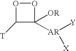
- In these 1,2-dioxetanes, T is a stabilizing group. Because the dioxetane molecule, without the stabilizing group, may spontaneously decompose, a group, typically a polycycloalkyl group is bound to the dioxetane to stabilize it against spontaneous decomposition. This need for stabilization has resulted in commercially developed 1,2-dioxetanes being generally spiroadamantyl. The adamantyl group, spiro-bound, can be optionally substituted at any bridge head carbon, to affect chemiluminescent properties. As indicated, the remaining carbon of the dioxetane ring bears a OR substituent, wherein R is generally an alkyl or cycloalkyl, although it may be a further aryl group. The alkyl can be optionally substituted, with the substituent including halogenated groups, such as polyhaloalkyl substituents. The remaining valence is occupied by an aryl moiety, preferably phenyl or naphthyl. If naphthyl, particular substitution profiles on the naphthyl ring are preferred. The aryl ring bears at least one substituent, X. In commercially developed dioxetanes, this is typically an enzyme-cleavable group. Where the associated enzyme is alkaline phosphatase, for example, the enzyme-cleavable group X will be a phosphate. The aryl ring may also bear a substituent Y, which is selected to be either electron donating, or electron withdrawing. Preferred groups include chlorine, alkoxy and heteroaryl, although other groups may be employed. These substitutions can further effect chemiluminescent properties, and reaction kinetics. A wide variety of other substituents are disclosed in the referenced patents.
- A class of compounds receiving particular attention with respect to luminescent reactions utilizing a peroxidase enzyme, e.g., horseradish peroxidase, are dihydrophthalazinedione compounds that are used in combination with an oxidant, preferably a peroxide compound such as hydrogen peroxide. Any chemiluminescent dihydrophthalazinedione can be used as substrate in the present invention, that is to say any dihydrophthalazinedione which is oxidisable in the presence of a peroxidase catalyst by an addition of an oxidant to give chemiluminescence. Dihydrophthalazinediones are well established in the art. Suitable dihydrophthalazinediones as well as other compounds for use with peroxidases, (e.g., acridinium compounds, such as acridinium esters and benzacridinium, and alkenes) are, for example, those disclosed in U.S. Pat. Nos. 5,552,298; 6,696,569; 6,410,732; 5,922,558; 5,750,698; 5,723,295; 5,670,644; 5,601,977; 5,552,298; 5,523,212; 5,879,894; 6,635,437; 6,296,787; 6,270,695; 6,218,137; 6,139,782; 6,126,870; 6,045,991; 5,965,736; 5,840,963; 5,772,926; and 5,686,258; each of which is incorporated herein by reference in its entirety. Preferred dihydrophthalazinediones include substituted aryl cyclic diacylhydrazide including aminoaryl cyclic diacylhydrazides such as luminol, isoluminol, aminobutylethylisoluminol, aminoethyl-ethylisoluminol and 7-dimethylaminonaphthalene-1,2-dicarboxylic acid hydrazide and hydroxyaryl cyclic diacylhydrazides, for example, 5-hydroxy-2,3-dihydro-phthalazine-1,4-dione; 6-hydroxy-2,3-dihydro-phthalazine-1,4-dione; 5-hydroxy-2,3-dihydro-benzo[g]phthalazine-1,4-dione; and 9-hydroxy-2,3-dihydro-benzo[f]phthalazine-1,4-dione. Peroxide compounds include hydrogen peroxide, sodium perborate, urea peroxide, and the like.
- The sensitivity of the peroxidase-catalyzed chemiluminescent oxidation of dihydrophthalazinediones can be enhanced by including an enhancer in the reaction. The enhancer will be present in an amount which enhances light production from the diacylhydrazide in the presence of the peroxidase and/or decreases background chemiluminescence. Enhancers are known in the art and include, phenolic compounds such as those disclosed in U.S. Pat. No. 5,306,621, incorporated herein by reference in its entirety, including p-phenylphenol, p-iodophenol, p-bromophenol, p-hydroxycinnamic acid 6-bromo-2-naphthol, D-luciferin, and 2-cyano-6-hydroxybenzothiazole as well as boronic compounds, such as those disclosed in U.S. Pat. No. 5,629,168, incorporated herein by reference in its entirety, including, 4-iodophenylboronic acid (PIBA), 4-bromophenylboronic acid (PBBA), 4-chlorophenylboronic acid, 3-chlorophenylboronic acid, 3,4-dichlorophenylboronic acid, 2,3-dichlorophenylboronic acid, 5-bromo-2-methoxybenzeneboronic acid, 3-nitrophenylboronic acid, 4-chloro-3-nitrophenylboronic acid, 3-aminophenylboronic acid, 3-amino-2,4,6-trichlorophenylboronic acid, 4-(2′-carboxyethenyl)phenylboronic acid, 1-naphthaleneboronic acid, 6-hydroxy-2-naphthaleneboronic acid, phenylboronic acid, 2-methylphenylboronic acid, 4-methylphenylboronic acid, dimethyl-phenylboronic acid, 4-bromophenyl-di-n-butoxyborane, 4-carboxy-3-nitrophenylboronic acid, 4-(trimethylsilyl)benzeneboronic acid, 4-biphenylboronic acid, 4-(phenoxy)benzeneboronic acid, 4-(3′-borono-4′-hydroxyphenylazo)benzoic acid, diphenylisobutoxyborane, 4-(4′-chloroanilino)phenylboronic acid, 4,4′-bis(phenylboronic acid), 4-(4′-bromophenyl)phenyl-di-n-butoxyborane, di(3′,5′-dichlorophenoxy)-3,5-dichlorophenylborane, 4-chlorophenyl-di-(4′-chlorophenoxy)borane, pentaerythritol borate, boroglycine, 2-phenyl-1,3,2-dioxaborinane, bis(catechol)borate and 2-hydroxy-5-[(3′-trifluoromethyl)phenylazo]benzeboronic acid and diphenylboronic anhydride. Other enhancers include 6-hydroxybenzothiazole, substituted phenols, such as those disclosed in U.S. Pat. No. 4,598,044, incorporated herein by reference in its entirety; aromatic amines including those disclosed in U.S. Pat. No. 4,729,950, incorporated herein by reference in its entirety; and phenols substituted in ortho and/or para positions by imidazolyl or benzimidazolyl (U.S. Pat. No. 5,043,266, incorporated herein by reference in its entirety).
- In some embodiments, the bioluminescent assay will use a luciferase enzyme. Examples are luciferases isolated from a variety of luminous organisms, such as the luciferase genes of Photinus pyralis (the common firefly of North America), Pyrophorus plagiophthalamus(the Jamaican click beetle), Renilla reniformis (the sea pansy), and several bacteria (e.g., Xenorhabdus luminescens and Vibrio spp). Luciferases are enzymes found in luminous organisms which catalyze luminescence reactions. They are organized into groups based on commonalties of their luminescence reactions. All luciferases within a group are derived from related luminous organisms, and all catalyze the same chemical reaction. Examples are beetle luciferases, which all catalyze ATP-mediated oxidation of the beetle luciferin; and anthozoan luciferases which all catalyze oxidation of coelenterazine (Ward WW and Cormier MJ. Proc Natl Acad Sci 1975; 72(7):2530-4.). With the technical capabilities of molecular biology, it is possible to alter the structure of a luciferase found in nature to yield a functional equivalent thereof. A functional equivalent is an enzyme that maintains the ability to catalyze the same luminescence reaction, and thus it remains in the same group of enzymes. Luciferase as used herein is intended to include naturally occurring and non-naturally occurring luciferase enzymes.
- Luciferases generate light via the oxidation of enzyme-specific substrates, called luciferins. For firefly luciferase and all other beetle luciferases, this is typically done in the presence of magnesium ions, oxygen, and ATP. For anthozoan luciferases, including Renilla luciferase, oxygen is required along with the luciferin. Additional reagents such as, for example, coenzyme A can be used to yield greater enzyme turnover and greater luminescence intensity.
- It will be understood that other molecules that generate light by interaction with chemiluminescent reactants can be used in the present invention such as hemin which interacts with reactants such as luminol and peroxide and aequorin which interacts with calcium ion to generate luminescence.
- The performance of luminescent reactions can be improved by use of binding assays of the present invention. Although polymer enhancement of luminescent enzyme catalyzed reactions is known, the use of polymer immobilized on a solid surface adjacent to a binding agent or two fluorophores, or other interacting molecules, immobilized on a solid surface as described herein has not been known heretofore.
- The term “polymer”, as used herein, refers to molecules formed from the chemical union of two or more repeating units. Accordingly, included within the term “polymer” may be, for example, dimers, trimers and oligomers. The polymer may be synthetic, naturally-occurring or semisynthetic. Any polymer that can modulate, e.g., enhance a signal from a luminescent reaction can be used in the present invention. Preferably the polymer will provide a more hydrophobic environment. Polymers and other materials for use in the present invention can include for example, materials that can be converted into nanofibers, such as, for example, poly(lactic acid-co-glycolic acid), poly(acrylic acid)-poly(pyrene methanol), sodium citrate, polypyrrole, poly (3-methylthiophene), polyaniline, polyacrylonitrile, poly(p-phyenylene), poly(3,4-ethylenedioxythiophene), polyacrylonitrile, poly(L-lactic acid)-polycaprolactone, blends, polystyrene-block-poly(2-cinnamoylethyl methacrylate), polystyrene-block-poly(2-cinnamoylethyl methacrylate)-block-poly(tert-butyl acrylate), peptide-amphiphile, dendrimer, bolaform glucosamide; materials that can electrospun into nanofibers, such as for example, polystyrene, polycarbonate, polymethacrylate, polyvinylchloride, polyethylene terephthalate, nylon6,6, nylon4,6, polyamide, polyurethanes, polyvinyl alcohol, polylactic acid, polycaprolactone, polyethylene glycol, polylactide-co-glycolide, polyethylene-co-vinyl acetate, polyethylene co-vinyl alcohol, polyethylene oxide, collagen; amphiphilic poly(2-methyloxazoline-block-dimethylsiloxane-block-2-methyloxazoline)(PMOXA-b-PDMS-b-PMOXA) ABA triblock copolymers; poly(thiophene); polyetherketone; polyallylamine; polyethyleneimine; poly(iminohexamethylene); polytetrafluoroethylene; poly(oxy-1,4,-phenyleneoxyl-1,4-phenylenecarbonyl-1,4-phenylene); polyvinylidene fluoride; polymethyl methacrylate; polystyrene; aluminum; palladium; silicon; or blends or composites thereof.
- Although any of the polymers disclosed herein can be used in connection with any of the chemi- or bio-luminescent reactions, certain polymers will be preferred in combination with certain labels. For example, for use with alkaline phosphatase catalyzed reactions, preferred polymers to be immobilized on the nanostructures include polyhydroxyacrylates, polyvinylalkylethers, polyvinyl alcohol, polyvinylalkylpyrrolidinones, BSA, nylon, and poly[vinylbenzyl(benzyldimethyl ammonium) chloride]. While not wishing to be bound by any particular theory, it is postulated that the role of the polymer is to provide a more hydrophobic environment for decomposition of the excited electronic state intermediate formed in the scission of the 1,2-dioxetane ring structure. The polymer can be optionally combined with fluorophores that then act as energy acceptors and emit light at a wavelength characteristic of the fluorophore. This effect can be seen, for example, with enzyme in solution or immobilized as part the detection conjugate in a sandwich assay. In both situations, the enzyme is a dynamic environment and interaction between the polymer and the enzyme or the products of the enzyme reaction are governed by the random motion of the polymer in solution. In an exemplary embodiment of the present invention, the polymer and binding agent immobilized on the solid surface, on a nanometer scale, or two members of an energy transfer pair immobilized on the solid surface, on a nanometer scale, will create a more ordered and/or more static nanoenvironment that will maximize energy transfer in a luminescent reaction. The enhancement effect can be improved by a tighter control of the nanoenvironment as provided by the present invention.
- For use with peroxidase catalyzed reactions, preferred polymers to be immobilized on the nanostructures include hydroxypropyl methylcellulose, hydroxyethyl cellulose, and hydroxybutyl methylcellulose. Boronic or phenolic enhancers are generally used in combination with the horseradish peroxidase and its substrates. A polysorbate, such as Tween 20 can also be used to stabilize light emission from the horseradish peroxidase (HRP) catalyzed chemiluminescent oxidation of hydroxyaryl cyclic diacylhydrazides.
- For use with luciferase catalyzed reactions, preferred polymers to be immobilized on the nanostructures include polyethylene glycol and polyvinylpyrrolidone. While not wishing to be bound by any particular theory, it is postulated that the polymer is acting as a reservoir for the inhibitory oxyluciferin product and the reaction and thus constantly regenerating active firefly luciferase.
- Nanolithographic printing is particularly useful for the preparation of the assays of the present invention having nanoscopic features. Any type of nanolithographic printing method can be used in the present methods to pattern at least two patterns of different molecules in predetermined and preselected patterns on a solid surface. Methods include, for example, photolithography, E-beam lithography, molecular beam ionization, and the like. These methods are known in the art and are thus not described herein in detail.
- In one exemplary embodiment, dip pen nanolithography is used to pattern the molecules on the solid substrate. For example, in certain embodiments, the present invention provides two-dimensional molecular patterns fabricated with nanometer control on the surface of a substrate using a molecular printing technique, such as dip-pen nanolithography. Methods for patterning molecules using dip pen nanolithography are known in the art. See for example, US Publication Numbers 20020063212 and 20030068446, incorporated herein by reference in their entirety and for all purposes.
- In dip-pen nanolithography, typically, the different patterns of molecules are formed by a plurality of dots or a plurality of lines formed on the solid surface. The plurality of dots can be, for example, a lattice of dots including hexagonal or square lattices as known in the art. The plurality of lines can form a grid, including perpendicular and parallel arrangements of the lines.
- The dimensions of the individual patterns including dot diameters and the line widths can be, for example, about 1,000 nm or less, about 500 nm or less, about 300 nm or less, and more particularly about 100 nm or less. The range in dimension can be for example about 1 nm to about 750 nm, about 10 nm to about 500 nm, and more particularly about 100 nm to about 350 nm.
- The number of patterns on the solid surface is not particularly limited. It can be, for example, at least 2, at least 3, at least 10, at least 100, at least 1,000, at least 10,000, even at least 100,000. Square arrangements are possible such as, for example, a 10×10 array.
- The distance between the individual patterns on the solid surface, e.g., nanoarray, can vary. For example, the patterns can be separated by distances of less than one micron or more than one micron. The patterns can be parallel lines, perpendicular lines, arced curves, superimposed and overlapping shapes. The distances between the molecules can be from about 1 to about 20 nm for applications utilizing energy transfer (i.e., Forster radius). However, larger distances may be possible for applications utilizing enhancing polymers for chemiluminescence.
- A variety of molecules, including, for example, proteins, polypeptides, oligopeptides, polymers, and fluorophores can be directly transferred and adsorbed to surfaces in a patterned fashion with use of nanolithographic printing, wherein the molecule is directly transferred from a tip such as, an atomic force microscope tip, to a substrate. Alternatively, however, in an indirect method, the nanolithographic printing can be used to deposit or deliver a compound in a pattern, and then the molecules can be assembled onto or adsorbed to the compound.
- Solid surface of the present invention can be, for example, an insulator such as, for example, glass or a conductor such as, for example, metal, including gold. In addition, the solid surface can be a metal, a semiconductor, a magnetic material, a polymer material, a polymer-coated substrate, or a superconductor material. The solid surface can be previously treated with one or more adsorbates. Still further, examples of suitable solid surface include but are not limited to, metals, ceramics, metal oxides, semiconductor materials, magnetic materials, polymers or polymer coated substrates, superconductor materials, polystyrene, and glass. Metals include, but are not limited to gold, silver, aluminum, copper, platinum and palladium. Other solid surface onto which compounds may be patterned include, but are not limited to silica, silicon oxide, GaAs, and InP.
- The solid surfaces can also include nanostructures, such as nanotubes, nanowires, and nanofibers. For example, carbon nanotubes or nanowires or nanofibers can be immobilized, synthesized or etched out of a surface. While these structures are attached to a surface, a patterning device such as, for example, DPN can be used to print molecules on to their surface. The assembly of patterns on these surfaces can occur by methods such as, for example, electrostatic layer-by-layer assembly.
- The patterning compound can be, for example, chemisorbed or covalently bound to the substrate to anchor the patterning compound and improve stability. It can be, for example, a sulfur-containing compound such as, for example, a thiol, polythiol, sulfide, cyclic disulfide, and the like. It can be, for example, a sulfur-containing compound having a sulfur group at one end and a terminal reactive group at the other end, such as an alkane thiol with a carboxylic acid end group. The patterning compound can be a lower molecular weight compound of less than, for example, 100, or less than 500, or less than 1,000, or a higher molecular weight compound including oligomeric and polymeric compounds. Synthetic and natural patterning compounds can be used. Other examples include alkanethiols that have functional end-groups such as 16-mercaptohexadecanoic acid; hydrophobic thiols, such as 1-octadecanethiol; and organic coupling molecules, such as EDC and mannose-SH. Other examples of sulfur-containing compounds include, but are not limited to, hydrogen sulphide, mercaptans, thiols, sulphides, thioesters, polysulphides, cyclic sulphides, and thiophene derivatives. For instance, a sulfur-containing compound may comprise a thiol, phosphothiol, thiocyano, sulfonic acid, disulfide or isothiocyano group.
- Other compounds include silicon-containing compounds that have a siloxy or silyl group that posseses a carboxylic acid group, aldehydes, alcohol, alkoxy or vinyl group. A compound can also possess an amine, nitrile, or isonitrile group. In one preferred embodiment, the solid surface will comprise sulfur adsorption on gold.
- In general, the present invention uses nanolithographic methods, for example, dip pen nanolithographic printing, to assemble onto a surface at least two patterns of different molecules. The patterns can be provided, for example, in dots, or adjacent lines or stripes. The assembling process can be achieved by exposing the solid surface to a solution containing the desired molecules, e.g., immersing the solid surface into solution comprising the molecules; or spraying the solution onto the solid surface. Other methods of exposing the solid surface to a solution comprising the molecules including placing the solid surface in a chamber containing a solution vapor or mist, or pouring a solution comprising the molecules onto the solid surface. Alternatively, the assembling process can include depositing the molecules onto the solid surface using dip pen nanolithographic printing.
- Non-specific binding of proteins to other, “non-compound” regions of a surface, can be prevented by covering, or “passivating,” those regions of the surface with another compound, or mixture of compounds, prior to exposure to the protein solution or sample (one or more passivating compounds). Known passivating compounds can be used and the invention is not particularly limited by this feature to the extent that non-specific adsorption does not occur. A variety of passivating compounds can be used including, for example, surfactants such as alkylene glycols which are functionalized to adsorb to the substrate. An example of a compound useful for passivating is 11-mercaptoundecyl-tri(ethylene glycol). After passivation, the resultant array can be called a passivated array.
- A wide variety of assays exist which use light emission to determine the presence or concentration of a particular sample in a substance. The solid surface described herein can be used in any of these assays.
- Assays employing the solid surfaces of the present invention can include conventional assays, such as protein and nucleic acid assays, DNA sequencing, ELISA, competitive assays, sandwich assays, agglutination types of assays, as well as other liquid phase and mixed phase assays.
- In an exemplary embodiment, a solid surface of the present invention can be used in a energy transfer competitive assay format. In one example, a solid surface of the present invention specific for an analyte of interest and having a donor fluorophore and acceptor fluorophore separately patterned on it will be added to a sample. After incubation, the reaction mixture will be exposed to excitation light specific for the donor fluorophore. If a complex forms between the analyte and the binding agent, it will disrupt energy transfer from the donor to the acceptor molecule. Measurement of this light emission can provide an indication of the amount of analyte in the sample. Low analyte concentration will lead to a large signal whereas high analyte concentration will lead to a low signal.
- In another example, a solid surface of the present invention specific for an analyte of interest will have a polymer patterned on it. The solid surface as well as a chemiluminescent enzyme conjugate that can bind to the analyte of interest will be mixed with a sample. The array will then be washed and exposed to the enzyme substrate. Measurement of the light can provide an indication of the amount of analyte in the sample.
- These assay techniques provide the ability to detect both the presence and amount and concentration of small quantities of analytes and are useful in many application, for example medical diagnostics and forensic applications. Other applications include, for example, drug discovery, reporter-gene assays to monitor gene expression, second-messenger quantitation, protein kinase assays, antagonist/agonist screening, and protein-protein interaction analysis. As described, the present invention provides methods for detecting target nucleic acids in a sample. By “sample” is intended any sample obtained or derived from an individual, body fluid, cell line, tissue culture, or other source, including the environment, which contains or is suspected of containing the analyte of interest. As indicated, biological samples include body fluids (such as lymph, sera, plasma, urine, semen, expired air, synovial fluid and spinal fluid) and tissue sources. Methods for obtaining tissue biopsies and body fluids from mammals are well known in the art. Methods for obtaining environmental samples are also well known in the art.
- Luminescence can be detected using conventional means, for example, a phomultiplier tube detector, camera luminometer, or even the naked eye. Luminescence intensity can be measured to determine the concentration of the substrate. The light emission of membrane-based assays can be imaged, for example, with standard x-ray film or Polaroid instant photographic film. Acquisition of digitized images can be accomplished, for example, with a phosphor storage screen and CCD camera instrumentation.
- Luminescent signals generated in microplate-based assays can be quantified with a variety of commercially available luminometers. Such luminometers are normally instruments based on photomultiplier tube (PMT) technology, which move each well of the microplate directly below the PMT detector or the lens/fiber-optic light-collection interface. Sensitive detection of chemiluminescent signals in 96-well and higher-density microplates and microarrays is also possible with CCD camera instrumentation. Methods of detecting and quantitating luminescent labels are well known to those of skill in the art.
- In an exemplary embodiment of the present invention, the analyte to be detected will be mRNA transcripts of a tumor antigen, such as prostate specific antigen (“PSA”). In one such embodiment, PSA mRNA can be detected by PSA-mRNA specific oligonucleotide probes linked to biotin. The biotin-linked probes will conjugate to a streptavidinylated linked solid surface of the present invention.
- As also described, the present invention provides methods for detecting hormones in a biological sample. In an exemplary embodiment of the present invention, the analyte to be detected will be a hormone, such as thyrotropin (“TSH”). In one such embodiment, TSH can be probed by an anti-TSH antibody bound to biotin. The biotin-linked probes will conjugate to streptavidinylated linked solid surface of the present invention.
- The present invention also provides kits for conducting an assay for the presence or concentration of an analyte. The kits of the present invention comprise a solid surface of the present invention.
- In some embodiments, the kits will comprise a solid surface of the present invention separately patterned with a polymer for enhancing an alkaline phosphatase chemiluminescent reaction and a binding agent for an analyte of interest, alkaline phosphatase conjugate specific for the analyte of interest and a 1,2 dioxetane capable of interacting with the alkaline phosphatase conjugate. The kit can further comprise, for example, a hydrophobic fluorometric substrate for the enzyme, and optionally an enhancer molecule capable of improving fluorescence.
- In some embodiments, the kits will comprise a solid surface of the present invention separately patterned with a polymer for enhancing a peroxidase chemiluminescent reaction and a binding agent for an analyte of interest, a peroxidase conjugate specific for the analyte of interest, a dihydropthalazinedione; and a peroxide compound in an amount which reacts with the dihydropthalazinedione in the presence of the peroxidase. The kit can optionally comprise any one or more of an enhancer compound in an amount which enhances light production from the dihydropthalazinedione in the presence of the peroxidase enzyme and/or decreases background chemiluminescence; a chelating agent in an amount which prevents the peroxide compound from activating the dihydropthalazinedione prior to reaction with the peroxidase; a chemiluminescence enhancing surfactant.
- In some embodiments, the kits will comprise a solid surface of the present invention separately patterned with a polymer for enhancing a luciferase bioluminescent reaction and a binding agent for an analyte of interest, a luciferin conjugate specific for the analyte of interest, Mg; and ATP.
- Although the foregoing invention has been described in detail by way of example for purposes of clarity of understanding, it will be apparent to the artisan that certain changes and modifications are comprehended by the disclosure and can be practiced without undue experimentation within the scope of the appended claims, which are presented by way of illustration not limitation. The disclosures of all publications, patents and patent applications cited herein are hereby incorporated by reference in their entirety and for all purposes.
- Fabricate Arrays of Anti-PSA Antibody onto Gold Substrate
- 16-mercaptohexadecanoic acid (MHA) of the optimized feature sizes will be patterened onto a gold substrate (Piner et al., 1999) using DPN. Specifically, Si3N4 AFM tips (Nanolnk, Inc.; Chicago, Ill.) will create dot features, spaced 1 μm apart by contacting the gold substrate (Zhang et al., 2003). The diameter of the dot features will be determined by manipulating force and time of contact of the AFM tip, as previously described (Lee et al., 2004; Zhang et al., 2003). Any unprinted areas surrounding the features will be passivated by incubation with a 1 mM ethanolic solution of PEG-alkythiol (11-mercaptoundecyl-tri(ethylene glycol)) for 2 hours, followed by rinsing with ethanol and water (Lee et al., 2004).
- Arrays will be designed to bind either total PSA or the separate fractions of complexed PSA-ACT or free PSA. Depending on the required specificity of the array, mouse monoclonal lgG antibodies for capturing either total or complexed PSA (Cat no. 10-P20; Fitzgerald Industries International; Concord, Mass.) or free PSA (Cat no. 10-P21) will be immobilized on the MIA dot features by incubation (1 hour) to produce an alternate pattern of anti-PSA. Unbound antibodies will be removed by rinsing with 10 mM PBS and Tween-20 solution (0.05%), and non-specific binding from plasma proteins prevented by passivation with BSA (10% solution in 10 mM PBS).
- The topography of the absorbed manometer sized features of anti-PSA antibody on gold substrate will be determined by the NSCRIPTOR™ (Nanolnk, Inc.) in contact and lateral force modes. Typical measurements of lgG on the Y-aspect are approximately 6.5 nm in height (Lee et al., 2004; Zhang et al., 2003).
- Samples containing the PSA antigen will be directly applied to the anti-PSA nanoarrays in an enclosed environment at room temperature for 1 hr. The samples will include purified free PSA (30-AP16; Fitzgerald International), complexed PSA (PSA-ACT, 30-AP13; Fitzgerald International), and human sera. After binding the antigen for 1 hr, the substrate will be rinsed with 10 mM PBS buffer with 0.05% Tween-20 solution. The binding of free PSA or complexed PSA will then be determined by AFM imaging (NSCRIPTORT™; Nanolnk, Inc.) in contact and lateral force modes. The base height of the capture antibody will be approximately 6.5 nm. The dimensions of PSA predicted by a homologous crystal structure are 7.2 nm×7.9 nm×4.5 nm (Carvalho 2002). Thus, the binding of free PSA will add approximately 4 nm to 8 nm in height, for a total height of between 10 to 15 nm. In contrast, the dimensions of alpha 1-antichymotrypsin (ACT) predicted by crystal structure are 7.5 nm×7.5 nm×20.8 nm (Chen et al., 2003). Thus, binding of complexed PSA with ACT would lead to a potential total topographic height of 17 nm to 35 nm (lgG+PSA+ACT).
- To enhance the detection of PSA antigen, a sandwich assay will be performed with the binding of an additional PSA-specific detection antibody. The antibodies previously described for antigen capture have been paired by their manufacturer for use with specific detection antibodies in a sandwich format (total PSA: 10-P20; PSA-ACT: 10-P22; free PSA: 10-P20). After binding the antigen for 1 hr, the substrate will be rinsed with 10 mM PBS buffer with 0.05% Tween-20 solution. The washed substrate will then be incubated with detection antibody at 10 μg/ml in 10 mM PBS buffer solution for 45 minutes (Zhang et al., 2003). After binding the detection antibody for 1 hr, the substrate will be rinsed with 10 mM PBS buffer with 0.05% Tween-20 solution. The binding of the detection antibody will be determined by several methods including chemiluminescence based detection.
- The binding of detection antibody will be characterized by imaging AFM as noted above. The additional topographic height of antigen antibody complex with detection antibody will be an approximate increase of 6.5 nm; this corresponds to the size of lgG antibody. For example, the height of a free PSA complex with capture and detection antibodies will be between 17 to 21 nm in height greater than substrate.
- As an alternative, detection antibodies will be functionalized with biotin to facilitate subsequent luminescent-based detection. An lgG antibody biotinylation kit will be used to prepare biotinylated monoclonal antibody (Immunoprobe Biotinylation Kit, Cat No. BK-101; Sigma-Aldrich, St. Louis, Mo.). In brief, the detection antibody will be prepared at 1.0 mg/ml in a sodium phosphate buffer. This detection antibody solution will be added in a 1:10 dilution to BAC-SulfoNHS (biotinamidohexanoic acid 3-sulfo-N-hydroxysulfosuccinimide ester); BAC-SulfoNHS reacts with the free amino groups of the immunoglobulin forming stable amide bonds. This mixture is incubated at room temperature for 30 min and then purified over a Sephadex G-25 gel filtration column. The level of biotinylation of the eluted antibody will be determined by measuring the protein concentration (absorption at 290 nm) and the biotin content. Biotinylated detection antibodies can then be linked to a variety of labels; our primary label will be alkaline phosphatase (ALP). ALP can be readily linked to the biotinylated antibody by utilizing streptavidin conjugated ALP (Molecular Probes, Eugene, Oreg.).
- The DPN will be used to pattern 16-mercaptohexadecanoic acid (MHA) into the optimized feature sizes onto gold film to produce a nanoarray of negatively charged stripes. Any unprinted areas surrounding the features will be passivated by incubation with a 1 mM ethanolic solution of PEG-alkythiol (11-mercaptoundecyl-tri(ethylene gylcol)) for 2 hours followed by rinsing with ethanol and water. Mouse monoclonal lgG antibodies to PSA (anti-PSA) and poly[vinylbenzyl(benzyldimethyl ammonium)chloride] will be immobilized on the MHA dot features by incubation (1 hour) to produce an alternate pattern of anti-PSA and the polymer. Unbound antibodies will be removed by rinsing with 10 mM PBS and Tween-20 solution (0.05%), and non-specific binding from plasma proteins prevented by passivation with BSA (10% solution in 10 mM PBS).
- A series of PSA standards (DPC Immulite) will be applied to separate antibody-polymer nanoarrays in an enclosed environment at room temperature. Binding of PSA to the nanoarray will be detected by AFM measurements using the DPN in imaging mode. This will confirm binding and give an AFM-generated standard curve for comparison purposes. The array will then be incubated with an optimized concentration of anti-PSA alkaline phosphatase conjugate (from DPC kit), washed and then exposed to the dioxetane substrate, AMPPD (Applied Biosystems). Light emission will be recorded using the NightOwl CCD camera, and the light emission related to PSA concentration and compared with the AFM results.
- Standards (DPC Immulite) and plasma samples will be applied to the antibody nanoarrays (optimized spot size in previous experiment) in an enclosed environment at room temperature. All plasma samples and controls will incubated on the nanoarray for 1 hour at room temperature in an enclosed environment. Binding of PSA to the nanoarray will be detected by AFM measurements using the DPN in imaging mode. Results read off a calibration curve obtained with the PSA standards (DPC IMMULITE) will be compared with results obtained with our routine clinical PSA assay (DPC Immulite).
- Fabricate Arrays Printed with Donor and Acceptor Dye Inks
- Alternate stripes of donor and acceptor dyes will be fabricated at a range of spacings between the stripes, starting with overlapping printing, then the smallest (currently 15 nm) spacing and increasing stepwise to 60 nm. This design will allow the degree of energy transfer with spacing to be determined so that the placement of the biotin can be optimized. The biotin must be placed between the donor and acceptor dye such that there is still an appreciable degree of energy transfer. However, when biotin binds to the streptavidin the energy transfer must be diminished.
- The donor acceptor pair commonly used in energy transfer assays, fluorescein and tetramethylrhodamine, will be selected. Both are available with a long spacer arm (e.g., six carbon) terminating in a reactive functional group (e.g., Molecular Probes) and this provides the reactive moiety for attachment to the substrate surface when these molecules are used as the ink.
- Gold is reactive towards thiol groups and an evaporated gold substrate (Nanolnk) will thus be used as the substrate. A silver island film will be prepared by reduction of silver ions in a thin film on a glass substrate surface using glucose (Lakowicz et al., 2001, 2002).
Claims (23)
1. An assay comprising:
a solid surface and, on the solid surface, at least two patterns of different molecules;
at least one of said patterns having a dimension between about 1 and about 1000 nanometers;
the geometry and identity of the molecules being such that interaction between them is modulated in the presence of an analyte.
2. The assay of claim 1 wherein at least one of said patterns has a dimension between 10 and 500 nanometers.
3. The assay of claim 1 wherein at least one of the molecules is a donor fluorophore and at least one of the molecules is an acceptor fluorophore, and the donor and acceptor fluorophores interact with each other to generate a detectable signal.
4. The assay of claim 3 further comprising a binding agent patterned on the solid surface.
5. The assay of claim 4 wherein energy transfer between the two fluorophores is disrupted when the analyte binds to the binding agent.
6. The assay of claim 1 wherein at least one of the molecules is an antibody and at least one of the molecules is a drug enzyme conjugate.
7. The assay of claim 1 wherein the at least one of the molecules is an antibody and at least one of the molecules is an antigen linked to an enzyme donor.
8. The assay of claim 1 wherein the molecules are patterned in alternate stripes.
9. The assay of claim 1 wherein the molecules are printed on the solid surface using molecular printing techniques.
10. The assay of claim 9 wherein the molecular printing technique is dip pen nanolithography.
11. The assay of claim 1 wherein the solid surface comprises two or more nanofibers twisted together.
12. The assay of claim 1 wherein the solid surface comprises a carbon nanotube.
13. An assay comprising:
a solid surface and, on the solid surface, at least two patterns of different molecules;
at least one of said patterns having a dimension between about 1 and about 1000 nanometers;
the geometry and identity of the molecules being such that one of said molecules modulates a signal generated by binding an analyte to the other of said molecules.
14. The assay of claim 13 wherein at least one of said patterns has a dimension between 10 and 500 nanometers.
15. The assay of claim 13 wherein at least one of the molecules is a polymer and at least one of the molecules is a binding agent adjacent to the polymer, and the polymer enhances a detectable signal generated in a bioluminescent or chemiluminescent assay for detecting binding of an analyte to the binding agent.
16. The assay of claim 13 wherein the molecules are printed on the solid surface using molecular printing techniques.
17. The assay of claim 16 wherein the molecular printing technique is dip pen nanolithography.
18. The assay of claim 13 wherein the solid surface comprises two or more nanofibers twisted together.
19. The assay of claim 13 wherein the solid surface comprises a carbon nanotube.
20. A method of constructing an assay comprising:
patterning upon a solid surface at least two patterns of different molecules;
at least one of said patterns having a dimension between about 1 and about 1000 nanometers;
wherein the different molecules interact with each other in the presence of the analyte to generate or modulate a detectable signal.
21. A method of constructing an assay comprising:
patterning upon a solid surface at least two patterns of different molecules;
at least one of said patterns having a dimension between about 1 and about 1000 nanometers;
wherein one of said molecules modulates a signal generated by binding of the other of said molecules to a target analyte.
22. A method for detecting the presence or absence of a target in a sample comprising exposing the assay of claim 1 to a sample.
23. A method for detecting the presence or absence of a target in a sample comprising exposing the assay of claim 13 to a sample.
Priority Applications (1)
| Application Number | Priority Date | Filing Date | Title |
|---|---|---|---|
| US11/912,583 US20090246888A1 (en) | 2005-04-27 | 2006-04-26 | Nanoassays |
Applications Claiming Priority (4)
| Application Number | Priority Date | Filing Date | Title |
|---|---|---|---|
| US67521305P | 2005-04-27 | 2005-04-27 | |
| US73698505P | 2005-11-15 | 2005-11-15 | |
| US11/912,583 US20090246888A1 (en) | 2005-04-27 | 2006-04-26 | Nanoassays |
| PCT/US2006/016250 WO2006116687A2 (en) | 2005-04-27 | 2006-04-26 | Nanoassays |
Publications (1)
| Publication Number | Publication Date |
|---|---|
| US20090246888A1 true US20090246888A1 (en) | 2009-10-01 |
Family
ID=37215551
Family Applications (2)
| Application Number | Title | Priority Date | Filing Date |
|---|---|---|---|
| US11/912,583 Abandoned US20090246888A1 (en) | 2005-04-27 | 2006-04-26 | Nanoassays |
| US13/033,878 Abandoned US20110152116A1 (en) | 2005-04-27 | 2011-02-24 | Nanoassays |
Family Applications After (1)
| Application Number | Title | Priority Date | Filing Date |
|---|---|---|---|
| US13/033,878 Abandoned US20110152116A1 (en) | 2005-04-27 | 2011-02-24 | Nanoassays |
Country Status (2)
| Country | Link |
|---|---|
| US (2) | US20090246888A1 (en) |
| WO (1) | WO2006116687A2 (en) |
Cited By (2)
| Publication number | Priority date | Publication date | Assignee | Title |
|---|---|---|---|---|
| US20110152116A1 (en) * | 2005-04-27 | 2011-06-23 | The Trustees Of The University Of Pennsylvania | Nanoassays |
| US20210164987A1 (en) * | 2019-11-29 | 2021-06-03 | ICHORtec GmbH | Method and Optode for Determining the Concentration of an Analyte in a Sample Liquid |
Citations (53)
| Publication number | Priority date | Publication date | Assignee | Title |
|---|---|---|---|---|
| US3287324A (en) * | 1965-05-07 | 1966-11-22 | Du Pont | Poly-meta-phenylene isophthalamides |
| US3671542A (en) * | 1966-06-13 | 1972-06-20 | Du Pont | Optically anisotropic aromatic polyamide dopes |
| US4598044A (en) * | 1983-02-11 | 1986-07-01 | National Research Development Corporation | Enhanced luminescent or luminometric assay |
| US4678608A (en) * | 1985-04-15 | 1987-07-07 | American Cyanamid Company | Chemiluminescent composition |
| US4729950A (en) * | 1984-08-07 | 1988-03-08 | National Research Development Corporation | Enhanced luminescent or luminometric assay |
| US4857652A (en) * | 1986-07-17 | 1989-08-15 | Board Of Governors Of Wayne State University | Chemiluminescent 1,2-dioxetane compounds |
| US4927769A (en) * | 1987-07-08 | 1990-05-22 | Ciba Corning Diagnostics Corp. | Method for enhancement of chemiluminescence |
| US4952707A (en) * | 1988-06-30 | 1990-08-28 | Tropix, Inc. | Enzymatically-cleavable chemiluminescent fused polycyclic ring-containing 1,2-dioxetanes |
| US4978614A (en) * | 1988-10-26 | 1990-12-18 | Tropix, Inc. | Method of detecting a substance using enzymatically-induced decomposition of dioxetanes |
| US5043266A (en) * | 1987-06-15 | 1991-08-27 | National Research Development Corporation | Ortho- and para-imidazolyl and benzimidazolyl substituted phenol as enhancers for diagnostic chemiluminescent assays |
| US5089630A (en) * | 1987-12-31 | 1992-02-18 | Bronstein Irena Y | Dioxetanes for use in assays |
| US5112960A (en) * | 1989-07-17 | 1992-05-12 | Bronstein Irena Y | Chemiluminescent 3-(substituted adamant-2'-ylidene) 1,2-dioxetanes |
| US5145772A (en) * | 1986-07-24 | 1992-09-08 | Tropix, Inc. | Chemiluminescence enhancement of enzyme-activated decomposition of enzymatically cleavable chemiluminescent 1,2-dioxetanes |
| US5306621A (en) * | 1989-10-17 | 1994-04-26 | British Technology Group Limited | Enhanced chemiluminescent assay |
| US5523212A (en) * | 1993-05-17 | 1996-06-04 | Lumigen, Inc. | Aryl N-alkylacridanthiocarboxylate derivatives useful for chemiluminescent detection |
| US5538847A (en) * | 1989-07-17 | 1996-07-23 | Tropix, Inc. | Chemiluminescent 1,2-dioxetanes |
| US5547836A (en) * | 1990-08-30 | 1996-08-20 | Tropix, Inc. | Enhancement of chemiluminescent assays |
| US5552298A (en) * | 1992-10-23 | 1996-09-03 | Lumigen, Inc. | Enzyme-catalyzed chemiluminescence from hydroxyaryl cyclic diacylhydrazide compounds |
| US5578523A (en) * | 1995-02-24 | 1996-11-26 | Motorola, Inc. | Method for forming inlaid interconnects in a semiconductor device |
| US5629168A (en) * | 1992-02-10 | 1997-05-13 | British Technology Group Limited | Chemiluminescent enhancers |
| US5670644A (en) * | 1993-05-17 | 1997-09-23 | Lumigen, Inc. | Acridan compounds |
| US5686258A (en) * | 1993-05-17 | 1997-11-11 | Lumigen, Inc. | Chemiluminescent detection of hydrolytic enzymes using an acridan |
| US5723295A (en) * | 1993-05-17 | 1998-03-03 | Lumigen, Inc. | Methods, acridan compounds and kits for producing light |
| US5750698A (en) * | 1993-05-17 | 1998-05-12 | Lumigen, Inc. | Aryl N-alkylacridancarboxylate derivatives useful for chemiluminescent detection |
| US5772926A (en) * | 1997-05-13 | 1998-06-30 | Lumigen, Inc. | Chemiluminescent reactions using dihydroxyaromatic compounds and heterocyclic enol phosphates |
| US5840963A (en) * | 1997-05-13 | 1998-11-24 | Lumigen, Inc. | Chemiluminescent reactions using dihydroxyaromatic compounds and heterocyclic enol phosphates |
| US5849495A (en) * | 1993-12-23 | 1998-12-15 | Tropix, Inc. | Chemiluminescent energy transfer assays |
| US5866045A (en) * | 1995-07-31 | 1999-02-02 | Lumigen, Inc. | Water soluble tri-substituted 1,2-dioxetane compounds and assay compositions having increased storage stability |
| US5879894A (en) * | 1993-03-19 | 1999-03-09 | Chiron Diagnostics Corporation | Long emission wavelength chemiluminescent compounds and their use in test assays |
| US5886238A (en) * | 1994-11-23 | 1999-03-23 | Lumigen, Inc. | Alkene precursors for preparing chemiluminescent dialkyl-substituted 1,2-dioxetane compounds |
| US5922558A (en) * | 1997-09-12 | 1999-07-13 | Lumigen, Inc. | Methods and compositions for generating chemiluminescence with a peroxidase |
| US5965736A (en) * | 1996-01-16 | 1999-10-12 | Lumigen, Inc. | Compositions and methods for generating red chemiluminescence |
| US6045991A (en) * | 1996-01-16 | 2000-04-04 | Lumigen, Inc. | Compositions and methods for generating red chemiluminescence |
| US6126870A (en) * | 1997-09-12 | 2000-10-03 | Lumigen, Inc. | Chemiluminescent labeling compounds |
| US6132956A (en) * | 1995-10-19 | 2000-10-17 | Tropix, Inc. | Chemiluminescent 1,2-dioxetanes |
| US6180833B1 (en) * | 1995-07-31 | 2001-01-30 | Lumigen, Inc. | Water soluble tri-substituted 1,2-dioxetane compounds having increased storage stability, synthetic processes and intermediates |
| US6245928B1 (en) * | 1995-07-31 | 2001-06-12 | Lumigen, Inc. | Water soluble tri-substituted 1,2-dioxetane compounds having increased storage stability, synthetic processes and intermediates |
| US6306610B1 (en) * | 1998-09-18 | 2001-10-23 | Massachusetts Institute Of Technology | Biological applications of quantum dots |
| US20020063212A1 (en) * | 1999-01-07 | 2002-05-30 | Mirkin Chad A. | Methods utilizing scanning probe microscope tips and products therefor or produced thereby |
| US6410732B2 (en) * | 1996-01-16 | 2002-06-25 | Lumigen, Inc. | Processing and synthetic intermediates for preparing N-arylacridancarboxylic acid derivatives |
| US20020127623A1 (en) * | 2000-07-31 | 2002-09-12 | Maxygen, Inc. | Biosensors, reagents and diagnostic applications of directed evolution |
| US6544732B1 (en) * | 1999-05-20 | 2003-04-08 | Illumina, Inc. | Encoding and decoding of array sensors utilizing nanocrystals |
| US20030068446A1 (en) * | 2001-10-02 | 2003-04-10 | Northwestern University | Protein and peptide nanoarrays |
| US20030077599A1 (en) * | 2001-10-23 | 2003-04-24 | Nikon Research Corporation Of America | Methods and devices for hybridization and binding assays using thermophoresis |
| US20030185985A1 (en) * | 2002-02-01 | 2003-10-02 | Bronikowski Michael J. | Method of producing regular arrays of nano-scale objects using nano-structured block-copolymeric materials |
| US6696569B2 (en) * | 1997-09-12 | 2004-02-24 | Lumigen, Inc. | Compounds for generating chemiluminescence with a peroxidase |
| US20050118603A1 (en) * | 2002-10-11 | 2005-06-02 | Ahram Biosystems Inc. | Target detection system having a conformationally sensitive probe comprising a nucleic acid based signal transducer |
| US6958381B2 (en) * | 2000-11-23 | 2005-10-25 | Basf Aktiengesellschaft | Polyamide |
| US20070187239A1 (en) * | 2003-12-11 | 2007-08-16 | The Aerospace Corporation Regents Of The University Of California | Conducting polymer nanofiber sensors |
| US20080286856A1 (en) * | 2005-04-27 | 2008-11-20 | The Trustees Of The University Of Pennsylvania | Nanostructure Enhanced Luminescent Devices |
| US20090196826A1 (en) * | 2007-12-18 | 2009-08-06 | Board Of Regents, The University Of Texas System | Compositions and methods of making non-spherical micro- and nano-particles |
| US20090263914A1 (en) * | 2000-11-30 | 2009-10-22 | Kim Pettersson | Bioanalytical assay |
| US20100285972A1 (en) * | 2003-05-05 | 2010-11-11 | Nanosys, Inc. | Nanofiber surfaces for use in enhanced surface area applications |
Family Cites Families (9)
| Publication number | Priority date | Publication date | Assignee | Title |
|---|---|---|---|---|
| US4931223A (en) * | 1986-07-24 | 1990-06-05 | Tropix, Inc. | Methods of using chemiluminescent 1,2-dioxetanes |
| US5336596A (en) * | 1991-12-23 | 1994-08-09 | Tropix, Inc. | Membrane for chemiluminescent blotting applications |
| US5393469A (en) * | 1992-03-20 | 1995-02-28 | Lumigen, Inc. | Polymeric phosphonium salts providing enhanced chemiluminescence from 1,2-dioxetanes |
| US5866434A (en) * | 1994-12-08 | 1999-02-02 | Meso Scale Technology | Graphitic nanotubes in luminescence assays |
| US5581206A (en) * | 1995-07-28 | 1996-12-03 | Micron Quantum Devices, Inc. | Power level detection circuit |
| AU4906300A (en) * | 1999-06-04 | 2000-12-28 | Kinetek Pharmaceuticals, Inc. | Assay for detecting the enzymatic activity of a phosphorylation enzyme using enhanced signal generation |
| US7368296B2 (en) * | 2002-01-17 | 2008-05-06 | Applied Biosystems | Solid phases optimized for chemiluminescent detection |
| US7622060B2 (en) * | 2002-02-12 | 2009-11-24 | Cyalume Technologies, Inc. | Formable, porous, chemiluminescent reactant composition and device therefor |
| US20090246888A1 (en) * | 2005-04-27 | 2009-10-01 | The Trustees Of The University Of Pennsylvania | Nanoassays |
-
2006
- 2006-04-26 US US11/912,583 patent/US20090246888A1/en not_active Abandoned
- 2006-04-26 WO PCT/US2006/016250 patent/WO2006116687A2/en not_active Ceased
-
2011
- 2011-02-24 US US13/033,878 patent/US20110152116A1/en not_active Abandoned
Patent Citations (64)
| Publication number | Priority date | Publication date | Assignee | Title |
|---|---|---|---|---|
| US3287324A (en) * | 1965-05-07 | 1966-11-22 | Du Pont | Poly-meta-phenylene isophthalamides |
| US3671542A (en) * | 1966-06-13 | 1972-06-20 | Du Pont | Optically anisotropic aromatic polyamide dopes |
| US4598044A (en) * | 1983-02-11 | 1986-07-01 | National Research Development Corporation | Enhanced luminescent or luminometric assay |
| US4729950A (en) * | 1984-08-07 | 1988-03-08 | National Research Development Corporation | Enhanced luminescent or luminometric assay |
| US4678608A (en) * | 1985-04-15 | 1987-07-07 | American Cyanamid Company | Chemiluminescent composition |
| US4857652A (en) * | 1986-07-17 | 1989-08-15 | Board Of Governors Of Wayne State University | Chemiluminescent 1,2-dioxetane compounds |
| US5145772A (en) * | 1986-07-24 | 1992-09-08 | Tropix, Inc. | Chemiluminescence enhancement of enzyme-activated decomposition of enzymatically cleavable chemiluminescent 1,2-dioxetanes |
| US5043266A (en) * | 1987-06-15 | 1991-08-27 | National Research Development Corporation | Ortho- and para-imidazolyl and benzimidazolyl substituted phenol as enhancers for diagnostic chemiluminescent assays |
| US4927769A (en) * | 1987-07-08 | 1990-05-22 | Ciba Corning Diagnostics Corp. | Method for enhancement of chemiluminescence |
| US5089630A (en) * | 1987-12-31 | 1992-02-18 | Bronstein Irena Y | Dioxetanes for use in assays |
| US4952707A (en) * | 1988-06-30 | 1990-08-28 | Tropix, Inc. | Enzymatically-cleavable chemiluminescent fused polycyclic ring-containing 1,2-dioxetanes |
| US4978614A (en) * | 1988-10-26 | 1990-12-18 | Tropix, Inc. | Method of detecting a substance using enzymatically-induced decomposition of dioxetanes |
| US5538847A (en) * | 1989-07-17 | 1996-07-23 | Tropix, Inc. | Chemiluminescent 1,2-dioxetanes |
| US5112960A (en) * | 1989-07-17 | 1992-05-12 | Bronstein Irena Y | Chemiluminescent 3-(substituted adamant-2'-ylidene) 1,2-dioxetanes |
| US5306621A (en) * | 1989-10-17 | 1994-04-26 | British Technology Group Limited | Enhanced chemiluminescent assay |
| US5547836A (en) * | 1990-08-30 | 1996-08-20 | Tropix, Inc. | Enhancement of chemiluminescent assays |
| US5629168A (en) * | 1992-02-10 | 1997-05-13 | British Technology Group Limited | Chemiluminescent enhancers |
| US5552298A (en) * | 1992-10-23 | 1996-09-03 | Lumigen, Inc. | Enzyme-catalyzed chemiluminescence from hydroxyaryl cyclic diacylhydrazide compounds |
| US5601977A (en) * | 1992-10-23 | 1997-02-11 | Lumigen, Inc. | Enzyme-catalyzed chemiluminescent detection of proteins and nucleic acids using hydroxyaryl cyclic diacylhydrazide compounds |
| US5879894A (en) * | 1993-03-19 | 1999-03-09 | Chiron Diagnostics Corporation | Long emission wavelength chemiluminescent compounds and their use in test assays |
| US5523212A (en) * | 1993-05-17 | 1996-06-04 | Lumigen, Inc. | Aryl N-alkylacridanthiocarboxylate derivatives useful for chemiluminescent detection |
| US5670644A (en) * | 1993-05-17 | 1997-09-23 | Lumigen, Inc. | Acridan compounds |
| US5686258A (en) * | 1993-05-17 | 1997-11-11 | Lumigen, Inc. | Chemiluminescent detection of hydrolytic enzymes using an acridan |
| US5723295A (en) * | 1993-05-17 | 1998-03-03 | Lumigen, Inc. | Methods, acridan compounds and kits for producing light |
| US5750698A (en) * | 1993-05-17 | 1998-05-12 | Lumigen, Inc. | Aryl N-alkylacridancarboxylate derivatives useful for chemiluminescent detection |
| US5849495A (en) * | 1993-12-23 | 1998-12-15 | Tropix, Inc. | Chemiluminescent energy transfer assays |
| US6287767B1 (en) * | 1993-12-23 | 2001-09-11 | Tropix, Inc. | Chemiluminescent energy transfer assays |
| US5886238A (en) * | 1994-11-23 | 1999-03-23 | Lumigen, Inc. | Alkene precursors for preparing chemiluminescent dialkyl-substituted 1,2-dioxetane compounds |
| US5892064A (en) * | 1994-11-23 | 1999-04-06 | Lumigen, Inc. | Chemiluminescent dialkyl-substituted 1,2-dioxetanes, methods of synthesis and use |
| US6284899B1 (en) * | 1994-11-23 | 2001-09-04 | The Board Of Governors Of Wayne State University | Chemiluminescent dialkyl-substituted 1,2-dioxetane compounds, methods of synthesis and use |
| US5578523A (en) * | 1995-02-24 | 1996-11-26 | Motorola, Inc. | Method for forming inlaid interconnects in a semiconductor device |
| US6353129B1 (en) * | 1995-07-31 | 2002-03-05 | Lumigen, Inc. | Water soluble tri-substituted 1,2-dioxetane compounds having increased storage stability, synthetic processes and intermediates |
| US6180833B1 (en) * | 1995-07-31 | 2001-01-30 | Lumigen, Inc. | Water soluble tri-substituted 1,2-dioxetane compounds having increased storage stability, synthetic processes and intermediates |
| US5866045A (en) * | 1995-07-31 | 1999-02-02 | Lumigen, Inc. | Water soluble tri-substituted 1,2-dioxetane compounds and assay compositions having increased storage stability |
| US6410751B1 (en) * | 1995-07-31 | 2002-06-25 | Lumigen, Inc. | Water soluble tri-substituted 1, 2-dioxetane compounds having increased storage stability, synthetic processes and intermediates |
| US6245928B1 (en) * | 1995-07-31 | 2001-06-12 | Lumigen, Inc. | Water soluble tri-substituted 1,2-dioxetane compounds having increased storage stability, synthetic processes and intermediates |
| US6132956A (en) * | 1995-10-19 | 2000-10-17 | Tropix, Inc. | Chemiluminescent 1,2-dioxetanes |
| US5965736A (en) * | 1996-01-16 | 1999-10-12 | Lumigen, Inc. | Compositions and methods for generating red chemiluminescence |
| US6218137B1 (en) * | 1996-01-16 | 2001-04-17 | Lumigen, Inc. | Compounds, compositions and methods for generating chemiluminescene with phosphatase enzymes |
| US6139782A (en) * | 1996-01-16 | 2000-10-31 | Lumigen, Inc. | Compounds, compositions and methods for generating chemiluminescence with phosphatase enzymes |
| US6270695B1 (en) * | 1996-01-16 | 2001-08-07 | Lumigen, Inc. | Compounds, compositions, and methods for generating chemiluminescence with phosphatase enzymes |
| US6045991A (en) * | 1996-01-16 | 2000-04-04 | Lumigen, Inc. | Compositions and methods for generating red chemiluminescence |
| US6635437B2 (en) * | 1996-01-16 | 2003-10-21 | Lumigen, Inc. | Compounds, compositions and methods for generating chemiluminescence with phosphatase enzymes |
| US6296787B1 (en) * | 1996-01-16 | 2001-10-02 | Lumigen, Inc. | Compounds, compositions and methods for generating chemiluminescence with phosphatase enzymes |
| US6410732B2 (en) * | 1996-01-16 | 2002-06-25 | Lumigen, Inc. | Processing and synthetic intermediates for preparing N-arylacridancarboxylic acid derivatives |
| US5772926A (en) * | 1997-05-13 | 1998-06-30 | Lumigen, Inc. | Chemiluminescent reactions using dihydroxyaromatic compounds and heterocyclic enol phosphates |
| US5840963A (en) * | 1997-05-13 | 1998-11-24 | Lumigen, Inc. | Chemiluminescent reactions using dihydroxyaromatic compounds and heterocyclic enol phosphates |
| US6696569B2 (en) * | 1997-09-12 | 2004-02-24 | Lumigen, Inc. | Compounds for generating chemiluminescence with a peroxidase |
| US6126870A (en) * | 1997-09-12 | 2000-10-03 | Lumigen, Inc. | Chemiluminescent labeling compounds |
| US5922558A (en) * | 1997-09-12 | 1999-07-13 | Lumigen, Inc. | Methods and compositions for generating chemiluminescence with a peroxidase |
| US6306610B1 (en) * | 1998-09-18 | 2001-10-23 | Massachusetts Institute Of Technology | Biological applications of quantum dots |
| US20020063212A1 (en) * | 1999-01-07 | 2002-05-30 | Mirkin Chad A. | Methods utilizing scanning probe microscope tips and products therefor or produced thereby |
| US6544732B1 (en) * | 1999-05-20 | 2003-04-08 | Illumina, Inc. | Encoding and decoding of array sensors utilizing nanocrystals |
| US20020127623A1 (en) * | 2000-07-31 | 2002-09-12 | Maxygen, Inc. | Biosensors, reagents and diagnostic applications of directed evolution |
| US6958381B2 (en) * | 2000-11-23 | 2005-10-25 | Basf Aktiengesellschaft | Polyamide |
| US20090263914A1 (en) * | 2000-11-30 | 2009-10-22 | Kim Pettersson | Bioanalytical assay |
| US20030068446A1 (en) * | 2001-10-02 | 2003-04-10 | Northwestern University | Protein and peptide nanoarrays |
| US20030077599A1 (en) * | 2001-10-23 | 2003-04-24 | Nikon Research Corporation Of America | Methods and devices for hybridization and binding assays using thermophoresis |
| US20030185985A1 (en) * | 2002-02-01 | 2003-10-02 | Bronikowski Michael J. | Method of producing regular arrays of nano-scale objects using nano-structured block-copolymeric materials |
| US20050118603A1 (en) * | 2002-10-11 | 2005-06-02 | Ahram Biosystems Inc. | Target detection system having a conformationally sensitive probe comprising a nucleic acid based signal transducer |
| US20100285972A1 (en) * | 2003-05-05 | 2010-11-11 | Nanosys, Inc. | Nanofiber surfaces for use in enhanced surface area applications |
| US20070187239A1 (en) * | 2003-12-11 | 2007-08-16 | The Aerospace Corporation Regents Of The University Of California | Conducting polymer nanofiber sensors |
| US20080286856A1 (en) * | 2005-04-27 | 2008-11-20 | The Trustees Of The University Of Pennsylvania | Nanostructure Enhanced Luminescent Devices |
| US20090196826A1 (en) * | 2007-12-18 | 2009-08-06 | Board Of Regents, The University Of Texas System | Compositions and methods of making non-spherical micro- and nano-particles |
Cited By (4)
| Publication number | Priority date | Publication date | Assignee | Title |
|---|---|---|---|---|
| US20110152116A1 (en) * | 2005-04-27 | 2011-06-23 | The Trustees Of The University Of Pennsylvania | Nanoassays |
| US20210164987A1 (en) * | 2019-11-29 | 2021-06-03 | ICHORtec GmbH | Method and Optode for Determining the Concentration of an Analyte in a Sample Liquid |
| WO2021104579A1 (en) * | 2019-11-29 | 2021-06-03 | ICHORtec GmbH | Method and optrode for determining the concentration of an analyte by means of fret |
| US11698376B2 (en) * | 2019-11-29 | 2023-07-11 | ICHORtec GmbH | Method and optode for determining the concentration of an analyte in a sample liquid |
Also Published As
| Publication number | Publication date |
|---|---|
| WO2006116687A2 (en) | 2006-11-02 |
| US20110152116A1 (en) | 2011-06-23 |
| WO2006116687A3 (en) | 2007-12-13 |
Similar Documents
| Publication | Publication Date | Title |
|---|---|---|
| Muzyka | Current trends in the development of the electrochemiluminescent immunosensors | |
| Zhang et al. | Predicting detection limits of enzyme-linked immunosorbent assay (ELISA) and bioanalytical techniques in general | |
| Zhang et al. | Microfluidic beads-based immunosensor for sensitive detection of cancer biomarker proteins using multienzyme-nanoparticle amplification and quantum dotslabels | |
| Zheng et al. | Strategies for the detection of target analytes using microfluidic paper-based analytical devices | |
| Zhang et al. | Multiplexed sandwich immunoassays using flow-injection electrochemiluminescence with designed substrate spatial-resolved technique for detection of tumor markers | |
| Malashikhina et al. | Unconventional application of conventional enzymatic substrate: first fluorogenic immunoassay based on enzymatic formation of quantum dots | |
| FI114826B (en) | Binding determination where binding substances with tail groups are used | |
| US20120004141A1 (en) | Amplified bioassay | |
| JP3021038B2 (en) | Detection or quantification of multiple analytes using labeling technology | |
| Qin et al. | A sensitive gold nanoparticles sensing platform based on resonance energy transfer for chemiluminescence light on detection of biomolecules | |
| Shi et al. | Signal amplification in capillary electrophoresis based chemiluminescent immunoassays by using an antibody–gold nanoparticle–DNAzyme assembly | |
| WO2015055708A1 (en) | Sensitive qualitative bioassay using graphene oxide as analyte revealing agent | |
| Ray et al. | Nanotechniques in proteomics: current status, promises and challenges | |
| Rizzo | Optical immunoassays methods in protein analysis: An overview | |
| Chen et al. | Methods for detecting of cardiac troponin I biomarkers for myocardial infarction using biosensors: a narrative review of recent research | |
| Chong et al. | 1, 1′-Oxalyldiimidazole chemiluminescent enzyme immunoassay capable of simultaneously sensing multiple markers | |
| Gao et al. | Spectrum-resolved electrochemiluminescence to multiplex the immunoassay and DNA probe assay | |
| Liang et al. | Glucose oxidase-loaded liposomes for in situ amplified signal of electrochemical immunoassay on a handheld pH meter | |
| Li et al. | Fluorescent immunosensor based on fluorescence resonance energy transfer between CdSe/ZnS quantum dots and Au nanorods for PRRSV detection | |
| Park et al. | Role of background observed in aptasensor with chemiluminescence detection | |
| Chen et al. | Homogeneous immunoassay for alpha-fetoprotein based on the quenching of the fluorescence of quantum dots by antibody labelled with complexed copper ion tags | |
| US20110152116A1 (en) | Nanoassays | |
| JP2009527754A (en) | Assay method without separation | |
| Lu et al. | Multiplex detection of B-type natriuretic peptide, cardiac troponin I and C-reactive protein with photonic suspension array | |
| JP5855332B2 (en) | Measurement of multiple analytes over a wide concentration range by light diffraction. |
Legal Events
| Date | Code | Title | Description |
|---|---|---|---|
| AS | Assignment |
Owner name: THE TRUSTEES OF THE UNIVERSITY OF PENNSYLVANIA, PE Free format text: ASSIGNMENT OF ASSIGNORS INTEREST;ASSIGNORS:KRICKA, LARRY J;PARK, JASON Y;REEL/FRAME:020944/0256;SIGNING DATES FROM 20080116 TO 20080117 |
|
| STCB | Information on status: application discontinuation |
Free format text: ABANDONED -- FAILURE TO RESPOND TO AN OFFICE ACTION |