US20080312581A1 - Peptosomes for Use in Acoustically Mediated Intracellular Drug Delivery in vivo - Google Patents
Peptosomes for Use in Acoustically Mediated Intracellular Drug Delivery in vivo Download PDFInfo
- Publication number
- US20080312581A1 US20080312581A1 US12/131,105 US13110508A US2008312581A1 US 20080312581 A1 US20080312581 A1 US 20080312581A1 US 13110508 A US13110508 A US 13110508A US 2008312581 A1 US2008312581 A1 US 2008312581A1
- Authority
- US
- United States
- Prior art keywords
- copolypeptide
- therapeutic
- peptosome
- patient
- targeting
- Prior art date
- Legal status (The legal status is an assumption and is not a legal conclusion. Google has not performed a legal analysis and makes no representation as to the accuracy of the status listed.)
- Abandoned
Links
- 230000003834 intracellular effect Effects 0.000 title claims abstract description 36
- 238000012377 drug delivery Methods 0.000 title claims description 63
- 238000001727 in vivo Methods 0.000 title abstract description 32
- 230000001404 mediated effect Effects 0.000 title abstract description 27
- 238000000034 method Methods 0.000 claims abstract description 186
- 230000008685 targeting Effects 0.000 claims abstract description 137
- 239000002539 nanocarrier Substances 0.000 claims abstract description 131
- 230000001225 therapeutic effect Effects 0.000 claims abstract description 127
- 239000000203 mixture Substances 0.000 claims abstract description 106
- 108090000623 proteins and genes Proteins 0.000 claims abstract description 102
- 108090000765 processed proteins & peptides Proteins 0.000 claims abstract description 82
- 102000004169 proteins and genes Human genes 0.000 claims abstract description 81
- 239000002872 contrast media Substances 0.000 claims abstract description 74
- 102000004196 processed proteins & peptides Human genes 0.000 claims abstract description 73
- 150000001875 compounds Chemical class 0.000 claims abstract description 60
- 102000039446 nucleic acids Human genes 0.000 claims abstract description 31
- 108020004707 nucleic acids Proteins 0.000 claims abstract description 31
- 150000007523 nucleic acids Chemical class 0.000 claims abstract description 31
- 229920000642 polymer Polymers 0.000 claims description 149
- -1 poly(ethylene oxide) Polymers 0.000 claims description 87
- 238000002604 ultrasonography Methods 0.000 claims description 84
- 239000012528 membrane Substances 0.000 claims description 81
- 150000002632 lipids Chemical class 0.000 claims description 52
- 239000000178 monomer Substances 0.000 claims description 50
- 229920001184 polypeptide Polymers 0.000 claims description 45
- 108020004414 DNA Proteins 0.000 claims description 43
- 230000002209 hydrophobic effect Effects 0.000 claims description 39
- 229920001223 polyethylene glycol Polymers 0.000 claims description 36
- 229920001577 copolymer Polymers 0.000 claims description 30
- 229920001400 block copolymer Polymers 0.000 claims description 28
- LYCAIKOWRPUZTN-UHFFFAOYSA-N Ethylene glycol Chemical compound OCCO LYCAIKOWRPUZTN-UHFFFAOYSA-N 0.000 claims description 27
- HVYWMOMLDIMFJA-DPAQBDIFSA-N cholesterol Chemical compound C1C=C2C[C@@H](O)CC[C@]2(C)[C@@H]2[C@@H]1[C@@H]1CC[C@H]([C@H](C)CCCC(C)C)[C@@]1(C)CC2 HVYWMOMLDIMFJA-DPAQBDIFSA-N 0.000 claims description 24
- 108020004459 Small interfering RNA Proteins 0.000 claims description 20
- 239000004094 surface-active agent Substances 0.000 claims description 19
- 150000003904 phospholipids Chemical class 0.000 claims description 16
- 235000012000 cholesterol Nutrition 0.000 claims description 12
- 239000011159 matrix material Substances 0.000 claims description 11
- 230000001413 cellular effect Effects 0.000 claims description 10
- 150000003431 steroids Chemical class 0.000 claims description 9
- 229920003171 Poly (ethylene oxide) Polymers 0.000 claims description 8
- 238000012228 RNA interference-mediated gene silencing Methods 0.000 claims description 8
- 230000009368 gene silencing by RNA Effects 0.000 claims description 8
- 239000004055 small Interfering RNA Substances 0.000 claims description 6
- 231100000135 cytotoxicity Toxicity 0.000 claims description 4
- 230000003013 cytotoxicity Effects 0.000 claims description 4
- 108091032973 (ribonucleotides)n+m Proteins 0.000 claims description 3
- 108020005544 Antisense RNA Proteins 0.000 claims description 2
- LFQSCWFLJHTTHZ-UHFFFAOYSA-N Ethanol Chemical compound CCO LFQSCWFLJHTTHZ-UHFFFAOYSA-N 0.000 claims description 2
- 229920002873 Polyethylenimine Polymers 0.000 claims description 2
- 239000003184 complementary RNA Substances 0.000 claims description 2
- NOWKCMXCCJGMRR-UHFFFAOYSA-N Aziridine Chemical compound C1CN1 NOWKCMXCCJGMRR-UHFFFAOYSA-N 0.000 claims 1
- IAYPIBMASNFSPL-UHFFFAOYSA-N Ethylene oxide Chemical compound C1CO1 IAYPIBMASNFSPL-UHFFFAOYSA-N 0.000 claims 1
- 231100000518 lethal Toxicity 0.000 claims 1
- 230000001665 lethal effect Effects 0.000 claims 1
- 239000003814 drug Substances 0.000 abstract description 118
- 230000000087 stabilizing effect Effects 0.000 abstract description 45
- 229920002521 macromolecule Polymers 0.000 abstract description 44
- 206010028980 Neoplasm Diseases 0.000 abstract description 30
- 208000037265 diseases, disorders, signs and symptoms Diseases 0.000 abstract description 18
- 201000010099 disease Diseases 0.000 abstract description 17
- 201000011510 cancer Diseases 0.000 abstract description 9
- 238000000338 in vitro Methods 0.000 abstract description 8
- 229920001222 biopolymer Polymers 0.000 abstract description 7
- 238000012545 processing Methods 0.000 abstract description 3
- 229940124597 therapeutic agent Drugs 0.000 abstract description 3
- 230000007170 pathology Effects 0.000 abstract description 2
- 230000002685 pulmonary effect Effects 0.000 abstract 1
- 230000002485 urinary effect Effects 0.000 abstract 1
- 239000000463 material Substances 0.000 description 122
- 210000004027 cell Anatomy 0.000 description 112
- 239000003446 ligand Substances 0.000 description 101
- 210000004379 membrane Anatomy 0.000 description 79
- 210000001519 tissue Anatomy 0.000 description 74
- 239000000126 substance Substances 0.000 description 72
- 235000018102 proteins Nutrition 0.000 description 63
- 229940079593 drug Drugs 0.000 description 59
- 239000007789 gas Substances 0.000 description 48
- 230000005291 magnetic effect Effects 0.000 description 47
- 102000053602 DNA Human genes 0.000 description 41
- 230000008569 process Effects 0.000 description 40
- 239000012867 bioactive agent Substances 0.000 description 36
- 239000002202 Polyethylene glycol Substances 0.000 description 35
- 210000000170 cell membrane Anatomy 0.000 description 35
- 239000007788 liquid Substances 0.000 description 33
- 229940024606 amino acid Drugs 0.000 description 31
- 235000001014 amino acid Nutrition 0.000 description 31
- 150000001413 amino acids Chemical class 0.000 description 31
- 238000006116 polymerization reaction Methods 0.000 description 31
- 230000015572 biosynthetic process Effects 0.000 description 30
- 239000002245 particle Substances 0.000 description 30
- XEEYBQQBJWHFJM-UHFFFAOYSA-N Iron Chemical compound [Fe] XEEYBQQBJWHFJM-UHFFFAOYSA-N 0.000 description 28
- 230000003993 interaction Effects 0.000 description 27
- 230000000694 effects Effects 0.000 description 26
- 235000014113 dietary fatty acids Nutrition 0.000 description 25
- 229930195729 fatty acid Natural products 0.000 description 25
- 239000000194 fatty acid Substances 0.000 description 25
- 239000011257 shell material Substances 0.000 description 25
- 230000004888 barrier function Effects 0.000 description 24
- 125000003729 nucleotide group Chemical group 0.000 description 24
- 238000006243 chemical reaction Methods 0.000 description 23
- 239000000412 dendrimer Substances 0.000 description 23
- 102000005962 receptors Human genes 0.000 description 23
- 108020003175 receptors Proteins 0.000 description 23
- 238000011282 treatment Methods 0.000 description 23
- 239000003795 chemical substances by application Substances 0.000 description 22
- 238000004132 cross linking Methods 0.000 description 22
- 239000002773 nucleotide Substances 0.000 description 22
- 239000012530 fluid Substances 0.000 description 21
- 239000002502 liposome Substances 0.000 description 21
- XLYOFNOQVPJJNP-UHFFFAOYSA-N water Substances O XLYOFNOQVPJJNP-UHFFFAOYSA-N 0.000 description 21
- 229920000736 dendritic polymer Polymers 0.000 description 20
- 230000007246 mechanism Effects 0.000 description 20
- 102100025473 Carcinoembryonic antigen-related cell adhesion molecule 6 Human genes 0.000 description 18
- 101000914326 Homo sapiens Carcinoembryonic antigen-related cell adhesion molecule 6 Proteins 0.000 description 18
- 230000008901 benefit Effects 0.000 description 18
- 210000000805 cytoplasm Anatomy 0.000 description 18
- 150000004665 fatty acids Chemical class 0.000 description 18
- 238000013459 approach Methods 0.000 description 17
- 210000001163 endosome Anatomy 0.000 description 16
- 150000003254 radicals Chemical class 0.000 description 16
- 229920002477 rna polymer Polymers 0.000 description 16
- 125000004429 atom Chemical group 0.000 description 15
- 230000027455 binding Effects 0.000 description 15
- 125000002091 cationic group Chemical group 0.000 description 15
- 239000000017 hydrogel Substances 0.000 description 15
- 239000007787 solid Substances 0.000 description 15
- 238000003786 synthesis reaction Methods 0.000 description 15
- 108091023037 Aptamer Proteins 0.000 description 14
- 239000012634 fragment Substances 0.000 description 14
- 239000011572 manganese Substances 0.000 description 14
- 239000000427 antigen Substances 0.000 description 13
- 108091007433 antigens Proteins 0.000 description 13
- 102000036639 antigens Human genes 0.000 description 13
- 230000001965 increasing effect Effects 0.000 description 13
- 230000005298 paramagnetic effect Effects 0.000 description 13
- PEDCQBHIVMGVHV-UHFFFAOYSA-N Glycerine Chemical compound OCC(O)CO PEDCQBHIVMGVHV-UHFFFAOYSA-N 0.000 description 12
- ZMXDDKWLCZADIW-UHFFFAOYSA-N N,N-Dimethylformamide Chemical compound CN(C)C=O ZMXDDKWLCZADIW-UHFFFAOYSA-N 0.000 description 12
- 150000001720 carbohydrates Chemical class 0.000 description 12
- 239000000969 carrier Substances 0.000 description 12
- 230000015556 catabolic process Effects 0.000 description 12
- 238000006731 degradation reaction Methods 0.000 description 12
- 230000006870 function Effects 0.000 description 12
- 239000001257 hydrogen Substances 0.000 description 12
- 229910052739 hydrogen Inorganic materials 0.000 description 12
- 239000003999 initiator Substances 0.000 description 12
- 229910052742 iron Inorganic materials 0.000 description 12
- 210000004940 nucleus Anatomy 0.000 description 12
- 229920000575 polymersome Polymers 0.000 description 12
- 238000002360 preparation method Methods 0.000 description 12
- 238000011160 research Methods 0.000 description 12
- 239000000243 solution Substances 0.000 description 12
- 238000012546 transfer Methods 0.000 description 12
- 102000004190 Enzymes Human genes 0.000 description 11
- 108090000790 Enzymes Proteins 0.000 description 11
- 108010021625 Immunoglobulin Fragments Proteins 0.000 description 11
- 102000008394 Immunoglobulin Fragments Human genes 0.000 description 11
- 125000000539 amino acid group Chemical group 0.000 description 11
- 230000006378 damage Effects 0.000 description 11
- 238000009826 distribution Methods 0.000 description 11
- 239000000839 emulsion Substances 0.000 description 11
- 229940088598 enzyme Drugs 0.000 description 11
- UQSXHKLRYXJYBZ-UHFFFAOYSA-N iron oxide Inorganic materials [Fe]=O UQSXHKLRYXJYBZ-UHFFFAOYSA-N 0.000 description 11
- 239000010410 layer Substances 0.000 description 11
- 210000000056 organ Anatomy 0.000 description 11
- 238000001338 self-assembly Methods 0.000 description 11
- 230000032258 transport Effects 0.000 description 11
- 102000000588 Interleukin-2 Human genes 0.000 description 10
- 108010002350 Interleukin-2 Proteins 0.000 description 10
- 210000001744 T-lymphocyte Anatomy 0.000 description 10
- 230000000295 complement effect Effects 0.000 description 10
- 238000009472 formulation Methods 0.000 description 10
- 239000002609 medium Substances 0.000 description 10
- 239000000693 micelle Substances 0.000 description 10
- 229910052757 nitrogen Inorganic materials 0.000 description 10
- 239000013612 plasmid Substances 0.000 description 10
- 239000000725 suspension Substances 0.000 description 10
- 206010061218 Inflammation Diseases 0.000 description 9
- PXHVJJICTQNCMI-UHFFFAOYSA-N Nickel Chemical compound [Ni] PXHVJJICTQNCMI-UHFFFAOYSA-N 0.000 description 9
- ZMANZCXQSJIPKH-UHFFFAOYSA-N Triethylamine Chemical compound CCN(CC)CC ZMANZCXQSJIPKH-UHFFFAOYSA-N 0.000 description 9
- 230000009471 action Effects 0.000 description 9
- 239000012620 biological material Substances 0.000 description 9
- 235000014633 carbohydrates Nutrition 0.000 description 9
- 230000018109 developmental process Effects 0.000 description 9
- 229920000359 diblock copolymer Polymers 0.000 description 9
- 229940088597 hormone Drugs 0.000 description 9
- 239000005556 hormone Substances 0.000 description 9
- 238000003384 imaging method Methods 0.000 description 9
- 230000004054 inflammatory process Effects 0.000 description 9
- SZVJSHCCFOBDDC-UHFFFAOYSA-N iron(II,III) oxide Inorganic materials O=[Fe]O[Fe]O[Fe]=O SZVJSHCCFOBDDC-UHFFFAOYSA-N 0.000 description 9
- 239000004005 microsphere Substances 0.000 description 9
- 229920001308 poly(aminoacid) Polymers 0.000 description 9
- 239000000047 product Substances 0.000 description 9
- 150000003839 salts Chemical class 0.000 description 9
- 230000035945 sensitivity Effects 0.000 description 9
- 239000013598 vector Substances 0.000 description 9
- 108010088751 Albumins Proteins 0.000 description 8
- 102000009027 Albumins Human genes 0.000 description 8
- 206010020843 Hyperthermia Diseases 0.000 description 8
- ROHFNLRQFUQHCH-YFKPBYRVSA-N L-leucine Chemical compound CC(C)C[C@H](N)C(O)=O ROHFNLRQFUQHCH-YFKPBYRVSA-N 0.000 description 8
- 108091034117 Oligonucleotide Proteins 0.000 description 8
- 239000007864 aqueous solution Substances 0.000 description 8
- 230000004071 biological effect Effects 0.000 description 8
- 230000008859 change Effects 0.000 description 8
- 230000001419 dependent effect Effects 0.000 description 8
- 238000011161 development Methods 0.000 description 8
- 238000005516 engineering process Methods 0.000 description 8
- 229940093476 ethylene glycol Drugs 0.000 description 8
- GNBHRKFJIUUOQI-UHFFFAOYSA-N fluorescein Chemical compound O1C(=O)C2=CC=CC=C2C21C1=CC=C(O)C=C1OC1=CC(O)=CC=C21 GNBHRKFJIUUOQI-UHFFFAOYSA-N 0.000 description 8
- 230000036031 hyperthermia Effects 0.000 description 8
- 238000004519 manufacturing process Methods 0.000 description 8
- KAVGMUDTWQVPDF-UHFFFAOYSA-N perflubutane Chemical compound FC(F)(F)C(F)(F)C(F)(F)C(F)(F)F KAVGMUDTWQVPDF-UHFFFAOYSA-N 0.000 description 8
- 229950003332 perflubutane Drugs 0.000 description 8
- 239000012071 phase Substances 0.000 description 8
- 230000008884 pinocytosis Effects 0.000 description 8
- 230000004044 response Effects 0.000 description 8
- IJGRMHOSHXDMSA-UHFFFAOYSA-N Atomic nitrogen Chemical compound N#N IJGRMHOSHXDMSA-UHFFFAOYSA-N 0.000 description 7
- 230000004913 activation Effects 0.000 description 7
- 239000006185 dispersion Substances 0.000 description 7
- 239000002961 echo contrast media Substances 0.000 description 7
- 210000002889 endothelial cell Anatomy 0.000 description 7
- 230000002255 enzymatic effect Effects 0.000 description 7
- 230000014509 gene expression Effects 0.000 description 7
- 150000004676 glycans Chemical class 0.000 description 7
- 229960003136 leucine Drugs 0.000 description 7
- 210000000633 nuclear envelope Anatomy 0.000 description 7
- 229920001282 polysaccharide Polymers 0.000 description 7
- 239000005017 polysaccharide Substances 0.000 description 7
- 239000002243 precursor Substances 0.000 description 7
- 125000002924 primary amino group Chemical group [H]N([H])* 0.000 description 7
- 230000002441 reversible effect Effects 0.000 description 7
- 241000894007 species Species 0.000 description 7
- 235000000346 sugar Nutrition 0.000 description 7
- 229920001059 synthetic polymer Polymers 0.000 description 7
- 102000000905 Cadherin Human genes 0.000 description 6
- 108050007957 Cadherin Proteins 0.000 description 6
- 108010035532 Collagen Proteins 0.000 description 6
- 102000008186 Collagen Human genes 0.000 description 6
- AOJJSUZBOXZQNB-TZSSRYMLSA-N Doxorubicin Chemical group O([C@H]1C[C@@](O)(CC=2C(O)=C3C(=O)C=4C=CC=C(C=4C(=O)C3=C(O)C=21)OC)C(=O)CO)[C@H]1C[C@H](N)[C@H](O)[C@H](C)O1 AOJJSUZBOXZQNB-TZSSRYMLSA-N 0.000 description 6
- 239000004472 Lysine Substances 0.000 description 6
- 230000002776 aggregation Effects 0.000 description 6
- 238000004220 aggregation Methods 0.000 description 6
- 229920000249 biocompatible polymer Polymers 0.000 description 6
- 229960002685 biotin Drugs 0.000 description 6
- 239000011616 biotin Substances 0.000 description 6
- 210000004369 blood Anatomy 0.000 description 6
- 239000008280 blood Substances 0.000 description 6
- 210000004204 blood vessel Anatomy 0.000 description 6
- 230000004087 circulation Effects 0.000 description 6
- 229920001436 collagen Polymers 0.000 description 6
- 239000000975 dye Substances 0.000 description 6
- 230000009881 electrostatic interaction Effects 0.000 description 6
- 230000002121 endocytic effect Effects 0.000 description 6
- 210000003722 extracellular fluid Anatomy 0.000 description 6
- 210000001723 extracellular space Anatomy 0.000 description 6
- 238000001415 gene therapy Methods 0.000 description 6
- 102000006495 integrins Human genes 0.000 description 6
- 108010044426 integrins Proteins 0.000 description 6
- 210000003734 kidney Anatomy 0.000 description 6
- 210000004072 lung Anatomy 0.000 description 6
- 229960003646 lysine Drugs 0.000 description 6
- 210000003712 lysosome Anatomy 0.000 description 6
- 230000001868 lysosomic effect Effects 0.000 description 6
- 201000001441 melanoma Diseases 0.000 description 6
- 229910052751 metal Inorganic materials 0.000 description 6
- 230000004048 modification Effects 0.000 description 6
- 238000012986 modification Methods 0.000 description 6
- 210000003463 organelle Anatomy 0.000 description 6
- 230000037361 pathway Effects 0.000 description 6
- 230000000144 pharmacologic effect Effects 0.000 description 6
- 230000005855 radiation Effects 0.000 description 6
- 230000009257 reactivity Effects 0.000 description 6
- 238000000527 sonication Methods 0.000 description 6
- 238000002560 therapeutic procedure Methods 0.000 description 6
- 239000003981 vehicle Substances 0.000 description 6
- 239000011800 void material Substances 0.000 description 6
- 229920002307 Dextran Polymers 0.000 description 5
- 229930186217 Glycolipid Natural products 0.000 description 5
- 229920002683 Glycosaminoglycan Polymers 0.000 description 5
- KZSNJWFQEVHDMF-BYPYZUCNSA-N L-valine Chemical compound CC(C)[C@H](N)C(O)=O KZSNJWFQEVHDMF-BYPYZUCNSA-N 0.000 description 5
- ROHFNLRQFUQHCH-UHFFFAOYSA-N Leucine Natural products CC(C)CC(N)C(O)=O ROHFNLRQFUQHCH-UHFFFAOYSA-N 0.000 description 5
- WDLRUFUQRNWCPK-UHFFFAOYSA-N Tetraxetan Chemical compound OC(=O)CN1CCN(CC(O)=O)CCN(CC(O)=O)CCN(CC(O)=O)CC1 WDLRUFUQRNWCPK-UHFFFAOYSA-N 0.000 description 5
- KZSNJWFQEVHDMF-UHFFFAOYSA-N Valine Natural products CC(C)C(N)C(O)=O KZSNJWFQEVHDMF-UHFFFAOYSA-N 0.000 description 5
- JLCPHMBAVCMARE-UHFFFAOYSA-N [3-[[3-[[3-[[3-[[3-[[3-[[3-[[3-[[3-[[3-[[3-[[5-(2-amino-6-oxo-1H-purin-9-yl)-3-[[3-[[3-[[3-[[3-[[3-[[5-(2-amino-6-oxo-1H-purin-9-yl)-3-[[5-(2-amino-6-oxo-1H-purin-9-yl)-3-hydroxyoxolan-2-yl]methoxy-hydroxyphosphoryl]oxyoxolan-2-yl]methoxy-hydroxyphosphoryl]oxy-5-(5-methyl-2,4-dioxopyrimidin-1-yl)oxolan-2-yl]methoxy-hydroxyphosphoryl]oxy-5-(6-aminopurin-9-yl)oxolan-2-yl]methoxy-hydroxyphosphoryl]oxy-5-(6-aminopurin-9-yl)oxolan-2-yl]methoxy-hydroxyphosphoryl]oxy-5-(6-aminopurin-9-yl)oxolan-2-yl]methoxy-hydroxyphosphoryl]oxy-5-(6-aminopurin-9-yl)oxolan-2-yl]methoxy-hydroxyphosphoryl]oxyoxolan-2-yl]methoxy-hydroxyphosphoryl]oxy-5-(5-methyl-2,4-dioxopyrimidin-1-yl)oxolan-2-yl]methoxy-hydroxyphosphoryl]oxy-5-(4-amino-2-oxopyrimidin-1-yl)oxolan-2-yl]methoxy-hydroxyphosphoryl]oxy-5-(5-methyl-2,4-dioxopyrimidin-1-yl)oxolan-2-yl]methoxy-hydroxyphosphoryl]oxy-5-(5-methyl-2,4-dioxopyrimidin-1-yl)oxolan-2-yl]methoxy-hydroxyphosphoryl]oxy-5-(6-aminopurin-9-yl)oxolan-2-yl]methoxy-hydroxyphosphoryl]oxy-5-(6-aminopurin-9-yl)oxolan-2-yl]methoxy-hydroxyphosphoryl]oxy-5-(4-amino-2-oxopyrimidin-1-yl)oxolan-2-yl]methoxy-hydroxyphosphoryl]oxy-5-(4-amino-2-oxopyrimidin-1-yl)oxolan-2-yl]methoxy-hydroxyphosphoryl]oxy-5-(4-amino-2-oxopyrimidin-1-yl)oxolan-2-yl]methoxy-hydroxyphosphoryl]oxy-5-(6-aminopurin-9-yl)oxolan-2-yl]methoxy-hydroxyphosphoryl]oxy-5-(4-amino-2-oxopyrimidin-1-yl)oxolan-2-yl]methyl [5-(6-aminopurin-9-yl)-2-(hydroxymethyl)oxolan-3-yl] hydrogen phosphate Polymers Cc1cn(C2CC(OP(O)(=O)OCC3OC(CC3OP(O)(=O)OCC3OC(CC3O)n3cnc4c3nc(N)[nH]c4=O)n3cnc4c3nc(N)[nH]c4=O)C(COP(O)(=O)OC3CC(OC3COP(O)(=O)OC3CC(OC3COP(O)(=O)OC3CC(OC3COP(O)(=O)OC3CC(OC3COP(O)(=O)OC3CC(OC3COP(O)(=O)OC3CC(OC3COP(O)(=O)OC3CC(OC3COP(O)(=O)OC3CC(OC3COP(O)(=O)OC3CC(OC3COP(O)(=O)OC3CC(OC3COP(O)(=O)OC3CC(OC3COP(O)(=O)OC3CC(OC3COP(O)(=O)OC3CC(OC3COP(O)(=O)OC3CC(OC3COP(O)(=O)OC3CC(OC3COP(O)(=O)OC3CC(OC3COP(O)(=O)OC3CC(OC3CO)n3cnc4c(N)ncnc34)n3ccc(N)nc3=O)n3cnc4c(N)ncnc34)n3ccc(N)nc3=O)n3ccc(N)nc3=O)n3ccc(N)nc3=O)n3cnc4c(N)ncnc34)n3cnc4c(N)ncnc34)n3cc(C)c(=O)[nH]c3=O)n3cc(C)c(=O)[nH]c3=O)n3ccc(N)nc3=O)n3cc(C)c(=O)[nH]c3=O)n3cnc4c3nc(N)[nH]c4=O)n3cnc4c(N)ncnc34)n3cnc4c(N)ncnc34)n3cnc4c(N)ncnc34)n3cnc4c(N)ncnc34)O2)c(=O)[nH]c1=O JLCPHMBAVCMARE-UHFFFAOYSA-N 0.000 description 5
- 239000002253 acid Substances 0.000 description 5
- 125000000217 alkyl group Chemical group 0.000 description 5
- 150000001412 amines Chemical class 0.000 description 5
- 125000000129 anionic group Chemical group 0.000 description 5
- 239000002585 base Substances 0.000 description 5
- 125000004432 carbon atom Chemical group C* 0.000 description 5
- 238000012512 characterization method Methods 0.000 description 5
- 239000011248 coating agent Substances 0.000 description 5
- 238000000576 coating method Methods 0.000 description 5
- 239000008139 complexing agent Substances 0.000 description 5
- 230000001276 controlling effect Effects 0.000 description 5
- 238000001514 detection method Methods 0.000 description 5
- 238000002059 diagnostic imaging Methods 0.000 description 5
- LOKCTEFSRHRXRJ-UHFFFAOYSA-I dipotassium trisodium dihydrogen phosphate hydrogen phosphate dichloride Chemical compound P(=O)(O)(O)[O-].[K+].P(=O)(O)([O-])[O-].[Na+].[Na+].[Cl-].[K+].[Cl-].[Na+] LOKCTEFSRHRXRJ-UHFFFAOYSA-I 0.000 description 5
- 230000012202 endocytosis Effects 0.000 description 5
- 210000003038 endothelium Anatomy 0.000 description 5
- MHMNJMPURVTYEJ-UHFFFAOYSA-N fluorescein-5-isothiocyanate Chemical compound O1C(=O)C2=CC(N=C=S)=CC=C2C21C1=CC=C(O)C=C1OC1=CC(O)=CC=C21 MHMNJMPURVTYEJ-UHFFFAOYSA-N 0.000 description 5
- 239000000499 gel Substances 0.000 description 5
- 229920000578 graft copolymer Polymers 0.000 description 5
- 125000004435 hydrogen atom Chemical group [H]* 0.000 description 5
- 229920001477 hydrophilic polymer Polymers 0.000 description 5
- 210000000987 immune system Anatomy 0.000 description 5
- 230000000977 initiatory effect Effects 0.000 description 5
- 235000013980 iron oxide Nutrition 0.000 description 5
- 239000002523 lectin Substances 0.000 description 5
- 230000000670 limiting effect Effects 0.000 description 5
- 210000004698 lymphocyte Anatomy 0.000 description 5
- 238000002595 magnetic resonance imaging Methods 0.000 description 5
- SGDBTWWWUNNDEQ-LBPRGKRZSA-N melphalan Chemical compound OC(=O)[C@@H](N)CC1=CC=C(N(CCCl)CCCl)C=C1 SGDBTWWWUNNDEQ-LBPRGKRZSA-N 0.000 description 5
- 239000002184 metal Substances 0.000 description 5
- 230000002107 myocardial effect Effects 0.000 description 5
- 239000003921 oil Substances 0.000 description 5
- 238000007911 parenteral administration Methods 0.000 description 5
- 230000036961 partial effect Effects 0.000 description 5
- 239000002953 phosphate buffered saline Substances 0.000 description 5
- 229920000747 poly(lactic acid) Polymers 0.000 description 5
- 102000040430 polynucleotide Human genes 0.000 description 5
- 108091033319 polynucleotide Proteins 0.000 description 5
- 239000002157 polynucleotide Substances 0.000 description 5
- 229920002451 polyvinyl alcohol Polymers 0.000 description 5
- 230000010837 receptor-mediated endocytosis Effects 0.000 description 5
- 230000008439 repair process Effects 0.000 description 5
- 238000007086 side reaction Methods 0.000 description 5
- 229960004295 valine Drugs 0.000 description 5
- 239000004474 valine Substances 0.000 description 5
- MYPYJXKWCTUITO-UHFFFAOYSA-N vancomycin Natural products O1C(C(=C2)Cl)=CC=C2C(O)C(C(NC(C2=CC(O)=CC(O)=C2C=2C(O)=CC=C3C=2)C(O)=O)=O)NC(=O)C3NC(=O)C2NC(=O)C(CC(N)=O)NC(=O)C(NC(=O)C(CC(C)C)NC)C(O)C(C=C3Cl)=CC=C3OC3=CC2=CC1=C3OC1OC(CO)C(O)C(O)C1OC1CC(C)(N)C(O)C(C)O1 MYPYJXKWCTUITO-UHFFFAOYSA-N 0.000 description 5
- YBJHBAHKTGYVGT-ZKWXMUAHSA-N (+)-Biotin Chemical compound N1C(=O)N[C@@H]2[C@H](CCCCC(=O)O)SC[C@@H]21 YBJHBAHKTGYVGT-ZKWXMUAHSA-N 0.000 description 4
- MYRTYDVEIRVNKP-UHFFFAOYSA-N 1,2-Divinylbenzene Chemical compound C=CC1=CC=CC=C1C=C MYRTYDVEIRVNKP-UHFFFAOYSA-N 0.000 description 4
- KILNVBDSWZSGLL-KXQOOQHDSA-N 1,2-dihexadecanoyl-sn-glycero-3-phosphocholine Chemical compound CCCCCCCCCCCCCCCC(=O)OC[C@H](COP([O-])(=O)OCC[N+](C)(C)C)OC(=O)CCCCCCCCCCCCCCC KILNVBDSWZSGLL-KXQOOQHDSA-N 0.000 description 4
- 239000004912 1,5-cyclooctadiene Substances 0.000 description 4
- RTZKZFJDLAIYFH-UHFFFAOYSA-N Diethyl ether Chemical compound CCOCC RTZKZFJDLAIYFH-UHFFFAOYSA-N 0.000 description 4
- KCXVZYZYPLLWCC-UHFFFAOYSA-N EDTA Chemical compound OC(=O)CN(CC(O)=O)CCN(CC(O)=O)CC(O)=O KCXVZYZYPLLWCC-UHFFFAOYSA-N 0.000 description 4
- 102000001301 EGF receptor Human genes 0.000 description 4
- 108060006698 EGF receptor Proteins 0.000 description 4
- 206010015866 Extravasation Diseases 0.000 description 4
- DHMQDGOQFOQNFH-UHFFFAOYSA-N Glycine Chemical compound NCC(O)=O DHMQDGOQFOQNFH-UHFFFAOYSA-N 0.000 description 4
- AEMRFAOFKBGASW-UHFFFAOYSA-N Glycolic acid Chemical compound OCC(O)=O AEMRFAOFKBGASW-UHFFFAOYSA-N 0.000 description 4
- 108060003951 Immunoglobulin Proteins 0.000 description 4
- CKLJMWTZIZZHCS-REOHCLBHSA-N L-aspartic acid Chemical compound OC(=O)[C@@H](N)CC(O)=O CKLJMWTZIZZHCS-REOHCLBHSA-N 0.000 description 4
- COLNVLDHVKWLRT-QMMMGPOBSA-N L-phenylalanine Chemical compound OC(=O)[C@@H](N)CC1=CC=CC=C1 COLNVLDHVKWLRT-QMMMGPOBSA-N 0.000 description 4
- 102000007330 LDL Lipoproteins Human genes 0.000 description 4
- 108010007622 LDL Lipoproteins Proteins 0.000 description 4
- 102000004856 Lectins Human genes 0.000 description 4
- 108090001090 Lectins Proteins 0.000 description 4
- 241001465754 Metazoa Species 0.000 description 4
- QPCDCPDFJACHGM-UHFFFAOYSA-N N,N-bis{2-[bis(carboxymethyl)amino]ethyl}glycine Chemical compound OC(=O)CN(CC(O)=O)CCN(CC(=O)O)CCN(CC(O)=O)CC(O)=O QPCDCPDFJACHGM-UHFFFAOYSA-N 0.000 description 4
- NINIDFKCEFEMDL-UHFFFAOYSA-N Sulfur Chemical compound [S] NINIDFKCEFEMDL-UHFFFAOYSA-N 0.000 description 4
- 208000007536 Thrombosis Diseases 0.000 description 4
- 241000700605 Viruses Species 0.000 description 4
- RJURFGZVJUQBHK-UHFFFAOYSA-N actinomycin D Natural products CC1OC(=O)C(C(C)C)N(C)C(=O)CN(C)C(=O)C2CCCN2C(=O)C(C(C)C)NC(=O)C1NC(=O)C1=C(N)C(=O)C(C)=C2OC(C(C)=CC=C3C(=O)NC4C(=O)NC(C(N5CCCC5C(=O)N(C)CC(=O)N(C)C(C(C)C)C(=O)OC4C)=O)C(C)C)=C3N=C21 RJURFGZVJUQBHK-UHFFFAOYSA-N 0.000 description 4
- 230000003213 activating effect Effects 0.000 description 4
- WNLRTRBMVRJNCN-UHFFFAOYSA-N adipic acid Chemical compound OC(=O)CCCCC(O)=O WNLRTRBMVRJNCN-UHFFFAOYSA-N 0.000 description 4
- 125000003275 alpha amino acid group Chemical group 0.000 description 4
- 230000000692 anti-sense effect Effects 0.000 description 4
- 238000000429 assembly Methods 0.000 description 4
- 230000000712 assembly Effects 0.000 description 4
- 210000002469 basement membrane Anatomy 0.000 description 4
- 239000010941 cobalt Substances 0.000 description 4
- 229910017052 cobalt Inorganic materials 0.000 description 4
- GUTLYIVDDKVIGB-UHFFFAOYSA-N cobalt atom Chemical compound [Co] GUTLYIVDDKVIGB-UHFFFAOYSA-N 0.000 description 4
- 239000002131 composite material Substances 0.000 description 4
- 239000012050 conventional carrier Substances 0.000 description 4
- 239000003431 cross linking reagent Substances 0.000 description 4
- 239000013078 crystal Substances 0.000 description 4
- 210000000172 cytosol Anatomy 0.000 description 4
- 230000001086 cytosolic effect Effects 0.000 description 4
- 230000007423 decrease Effects 0.000 description 4
- 238000003745 diagnosis Methods 0.000 description 4
- 239000000032 diagnostic agent Substances 0.000 description 4
- 229940039227 diagnostic agent Drugs 0.000 description 4
- 238000009792 diffusion process Methods 0.000 description 4
- 239000003937 drug carrier Substances 0.000 description 4
- 238000001035 drying Methods 0.000 description 4
- 239000003995 emulsifying agent Substances 0.000 description 4
- 150000002148 esters Chemical class 0.000 description 4
- 230000036251 extravasation Effects 0.000 description 4
- 125000000524 functional group Chemical group 0.000 description 4
- 239000003102 growth factor Substances 0.000 description 4
- JYGXADMDTFJGBT-VWUMJDOOSA-N hydrocortisone Chemical compound O=C1CC[C@]2(C)[C@H]3[C@@H](O)C[C@](C)([C@@](CC4)(O)C(=O)CO)[C@@H]4[C@@H]3CCC2=C1 JYGXADMDTFJGBT-VWUMJDOOSA-N 0.000 description 4
- 102000018358 immunoglobulin Human genes 0.000 description 4
- 239000007924 injection Substances 0.000 description 4
- 238000002347 injection Methods 0.000 description 4
- JVTAAEKCZFNVCJ-UHFFFAOYSA-N lactic acid Chemical compound CC(O)C(O)=O JVTAAEKCZFNVCJ-UHFFFAOYSA-N 0.000 description 4
- 210000004185 liver Anatomy 0.000 description 4
- 210000002540 macrophage Anatomy 0.000 description 4
- 239000002086 nanomaterial Substances 0.000 description 4
- 239000002105 nanoparticle Substances 0.000 description 4
- QMMOXUPEWRXHJS-UHFFFAOYSA-N pentene-2 Natural products CCC=CC QMMOXUPEWRXHJS-UHFFFAOYSA-N 0.000 description 4
- 229960003330 pentetic acid Drugs 0.000 description 4
- 230000035699 permeability Effects 0.000 description 4
- COLNVLDHVKWLRT-UHFFFAOYSA-N phenylalanine Natural products OC(=O)C(N)CC1=CC=CC=C1 COLNVLDHVKWLRT-UHFFFAOYSA-N 0.000 description 4
- 229960005190 phenylalanine Drugs 0.000 description 4
- 229920000835 poly(gamma-benzyl-L-glutamate) polymer Polymers 0.000 description 4
- 229920000656 polylysine Polymers 0.000 description 4
- 229920000036 polyvinylpyrrolidone Polymers 0.000 description 4
- 239000001267 polyvinylpyrrolidone Substances 0.000 description 4
- 235000013855 polyvinylpyrrolidone Nutrition 0.000 description 4
- 230000004043 responsiveness Effects 0.000 description 4
- 230000035939 shock Effects 0.000 description 4
- 210000003491 skin Anatomy 0.000 description 4
- 125000006850 spacer group Chemical group 0.000 description 4
- 230000006641 stabilisation Effects 0.000 description 4
- 238000011105 stabilization Methods 0.000 description 4
- 150000008163 sugars Chemical class 0.000 description 4
- 239000011593 sulfur Substances 0.000 description 4
- 229910052717 sulfur Inorganic materials 0.000 description 4
- 229920001897 terpolymer Polymers 0.000 description 4
- TXEYQDLBPFQVAA-UHFFFAOYSA-N tetrafluoromethane Chemical compound FC(F)(F)F TXEYQDLBPFQVAA-UHFFFAOYSA-N 0.000 description 4
- 229910052723 transition metal Inorganic materials 0.000 description 4
- 150000003624 transition metals Chemical class 0.000 description 4
- 239000013603 viral vector Substances 0.000 description 4
- 230000003612 virological effect Effects 0.000 description 4
- 229940088594 vitamin Drugs 0.000 description 4
- 229930003231 vitamin Natural products 0.000 description 4
- 235000013343 vitamin Nutrition 0.000 description 4
- 239000011782 vitamin Substances 0.000 description 4
- MTCFGRXMJLQNBG-REOHCLBHSA-N (2S)-2-Amino-3-hydroxypropansäure Chemical compound OC[C@H](N)C(O)=O MTCFGRXMJLQNBG-REOHCLBHSA-N 0.000 description 3
- KIUKXJAPPMFGSW-DNGZLQJQSA-N (2S,3S,4S,5R,6R)-6-[(2S,3R,4R,5S,6R)-3-Acetamido-2-[(2S,3S,4R,5R,6R)-6-[(2R,3R,4R,5S,6R)-3-acetamido-2,5-dihydroxy-6-(hydroxymethyl)oxan-4-yl]oxy-2-carboxy-4,5-dihydroxyoxan-3-yl]oxy-5-hydroxy-6-(hydroxymethyl)oxan-4-yl]oxy-3,4,5-trihydroxyoxane-2-carboxylic acid Chemical compound CC(=O)N[C@H]1[C@H](O)O[C@H](CO)[C@@H](O)[C@@H]1O[C@H]1[C@H](O)[C@@H](O)[C@H](O[C@H]2[C@@H]([C@@H](O[C@H]3[C@@H]([C@@H](O)[C@H](O)[C@H](O3)C(O)=O)O)[C@H](O)[C@@H](CO)O2)NC(C)=O)[C@@H](C(O)=O)O1 KIUKXJAPPMFGSW-DNGZLQJQSA-N 0.000 description 3
- COQIQRBKEGPRSG-UHFFFAOYSA-N 1,1,1,2,3,3,3-heptafluoro-2-(trifluoromethyl)propane Chemical compound FC(F)(F)C(F)(C(F)(F)F)C(F)(F)F COQIQRBKEGPRSG-UHFFFAOYSA-N 0.000 description 3
- 239000004475 Arginine Substances 0.000 description 3
- 108010024976 Asparaginase Proteins 0.000 description 3
- 241000894006 Bacteria Species 0.000 description 3
- 108010006654 Bleomycin Proteins 0.000 description 3
- 108010084313 CD58 Antigens Proteins 0.000 description 3
- KXDHJXZQYSOELW-UHFFFAOYSA-M Carbamate Chemical compound NC([O-])=O KXDHJXZQYSOELW-UHFFFAOYSA-M 0.000 description 3
- 108090000994 Catalytic RNA Proteins 0.000 description 3
- 102000053642 Catalytic RNA Human genes 0.000 description 3
- 102000016289 Cell Adhesion Molecules Human genes 0.000 description 3
- 108010067225 Cell Adhesion Molecules Proteins 0.000 description 3
- 102000007644 Colony-Stimulating Factors Human genes 0.000 description 3
- 108010071942 Colony-Stimulating Factors Proteins 0.000 description 3
- 102400000739 Corticotropin Human genes 0.000 description 3
- 101800000414 Corticotropin Proteins 0.000 description 3
- YMWUJEATGCHHMB-UHFFFAOYSA-N Dichloromethane Chemical compound ClCCl YMWUJEATGCHHMB-UHFFFAOYSA-N 0.000 description 3
- 229910052688 Gadolinium Inorganic materials 0.000 description 3
- 108010010803 Gelatin Proteins 0.000 description 3
- WHUUTDBJXJRKMK-UHFFFAOYSA-N Glutamic acid Natural products OC(=O)C(N)CCC(O)=O WHUUTDBJXJRKMK-UHFFFAOYSA-N 0.000 description 3
- 241000701806 Human papillomavirus Species 0.000 description 3
- AVXURJPOCDRRFD-UHFFFAOYSA-N Hydroxylamine Chemical class ON AVXURJPOCDRRFD-UHFFFAOYSA-N 0.000 description 3
- ONIBWKKTOPOVIA-BYPYZUCNSA-N L-Proline Chemical compound OC(=O)[C@@H]1CCCN1 ONIBWKKTOPOVIA-BYPYZUCNSA-N 0.000 description 3
- QNAYBMKLOCPYGJ-REOHCLBHSA-N L-alanine Chemical compound C[C@H](N)C(O)=O QNAYBMKLOCPYGJ-REOHCLBHSA-N 0.000 description 3
- ODKSFYDXXFIFQN-BYPYZUCNSA-P L-argininium(2+) Chemical compound NC(=[NH2+])NCCC[C@H]([NH3+])C(O)=O ODKSFYDXXFIFQN-BYPYZUCNSA-P 0.000 description 3
- WHUUTDBJXJRKMK-VKHMYHEASA-N L-glutamic acid Chemical compound OC(=O)[C@@H](N)CCC(O)=O WHUUTDBJXJRKMK-VKHMYHEASA-N 0.000 description 3
- ZDXPYRJPNDTMRX-VKHMYHEASA-N L-glutamine Chemical compound OC(=O)[C@@H](N)CCC(N)=O ZDXPYRJPNDTMRX-VKHMYHEASA-N 0.000 description 3
- HNDVDQJCIGZPNO-YFKPBYRVSA-N L-histidine Chemical compound OC(=O)[C@@H](N)CC1=CN=CN1 HNDVDQJCIGZPNO-YFKPBYRVSA-N 0.000 description 3
- AGPKZVBTJJNPAG-WHFBIAKZSA-N L-isoleucine Chemical compound CC[C@H](C)[C@H](N)C(O)=O AGPKZVBTJJNPAG-WHFBIAKZSA-N 0.000 description 3
- KDXKERNSBIXSRK-YFKPBYRVSA-N L-lysine Chemical compound NCCCC[C@H](N)C(O)=O KDXKERNSBIXSRK-YFKPBYRVSA-N 0.000 description 3
- AYFVYJQAPQTCCC-GBXIJSLDSA-N L-threonine Chemical compound C[C@@H](O)[C@H](N)C(O)=O AYFVYJQAPQTCCC-GBXIJSLDSA-N 0.000 description 3
- 239000000232 Lipid Bilayer Substances 0.000 description 3
- 102100030984 Lymphocyte function-associated antigen 3 Human genes 0.000 description 3
- KDXKERNSBIXSRK-UHFFFAOYSA-N Lysine Natural products NCCCCC(N)C(O)=O KDXKERNSBIXSRK-UHFFFAOYSA-N 0.000 description 3
- WAEMQWOKJMHJLA-UHFFFAOYSA-N Manganese(2+) Chemical compound [Mn+2] WAEMQWOKJMHJLA-UHFFFAOYSA-N 0.000 description 3
- 206010057249 Phagocytosis Diseases 0.000 description 3
- 108010039918 Polylysine Proteins 0.000 description 3
- 229920001710 Polyorthoester Polymers 0.000 description 3
- DNIAPMSPPWPWGF-UHFFFAOYSA-N Propylene glycol Chemical compound CC(O)CO DNIAPMSPPWPWGF-UHFFFAOYSA-N 0.000 description 3
- 108091028664 Ribonucleotide Proteins 0.000 description 3
- MTCFGRXMJLQNBG-UHFFFAOYSA-N Serine Natural products OCC(N)C(O)=O MTCFGRXMJLQNBG-UHFFFAOYSA-N 0.000 description 3
- VYPSYNLAJGMNEJ-UHFFFAOYSA-N Silicium dioxide Chemical compound O=[Si]=O VYPSYNLAJGMNEJ-UHFFFAOYSA-N 0.000 description 3
- FAPWRFPIFSIZLT-UHFFFAOYSA-M Sodium chloride Chemical compound [Na+].[Cl-] FAPWRFPIFSIZLT-UHFFFAOYSA-M 0.000 description 3
- 229920002472 Starch Polymers 0.000 description 3
- 108010090804 Streptavidin Proteins 0.000 description 3
- AYFVYJQAPQTCCC-UHFFFAOYSA-N Threonine Natural products CC(O)C(N)C(O)=O AYFVYJQAPQTCCC-UHFFFAOYSA-N 0.000 description 3
- 239000004473 Threonine Substances 0.000 description 3
- 108010000134 Vascular Cell Adhesion Molecule-1 Proteins 0.000 description 3
- 102100023543 Vascular cell adhesion protein 1 Human genes 0.000 description 3
- 238000010521 absorption reaction Methods 0.000 description 3
- 239000013543 active substance Substances 0.000 description 3
- 230000006978 adaptation Effects 0.000 description 3
- 229940009456 adriamycin Drugs 0.000 description 3
- 229960003767 alanine Drugs 0.000 description 3
- 235000004279 alanine Nutrition 0.000 description 3
- 235000008206 alpha-amino acids Nutrition 0.000 description 3
- 150000001408 amides Chemical class 0.000 description 3
- 239000012491 analyte Substances 0.000 description 3
- 238000004458 analytical method Methods 0.000 description 3
- 230000033115 angiogenesis Effects 0.000 description 3
- 150000008064 anhydrides Chemical class 0.000 description 3
- 210000004102 animal cell Anatomy 0.000 description 3
- 239000012736 aqueous medium Substances 0.000 description 3
- ODKSFYDXXFIFQN-UHFFFAOYSA-N arginine Natural products OC(=O)C(N)CCCNC(N)=N ODKSFYDXXFIFQN-UHFFFAOYSA-N 0.000 description 3
- 229960003121 arginine Drugs 0.000 description 3
- 235000009697 arginine Nutrition 0.000 description 3
- 229960005261 aspartic acid Drugs 0.000 description 3
- 235000003704 aspartic acid Nutrition 0.000 description 3
- QVGXLLKOCUKJST-UHFFFAOYSA-N atomic oxygen Chemical compound [O] QVGXLLKOCUKJST-UHFFFAOYSA-N 0.000 description 3
- 230000001580 bacterial effect Effects 0.000 description 3
- OQFSQFPPLPISGP-UHFFFAOYSA-N beta-carboxyaspartic acid Natural products OC(=O)C(N)C(C(O)=O)C(O)=O OQFSQFPPLPISGP-UHFFFAOYSA-N 0.000 description 3
- 230000000975 bioactive effect Effects 0.000 description 3
- DEGAKNSWVGKMLS-UHFFFAOYSA-N calcein Chemical compound O1C(=O)C2=CC=CC=C2C21C1=CC(CN(CC(O)=O)CC(O)=O)=C(O)C=C1OC1=C2C=C(CN(CC(O)=O)CC(=O)O)C(O)=C1 DEGAKNSWVGKMLS-UHFFFAOYSA-N 0.000 description 3
- 125000003178 carboxy group Chemical group [H]OC(*)=O 0.000 description 3
- 230000000747 cardiac effect Effects 0.000 description 3
- 210000003855 cell nucleus Anatomy 0.000 description 3
- 230000004700 cellular uptake Effects 0.000 description 3
- 239000003153 chemical reaction reagent Substances 0.000 description 3
- 229940047120 colony stimulating factors Drugs 0.000 description 3
- 238000007906 compression Methods 0.000 description 3
- 230000006835 compression Effects 0.000 description 3
- IDLFZVILOHSSID-OVLDLUHVSA-N corticotropin Chemical compound C([C@@H](C(=O)N[C@@H](CO)C(=O)N[C@@H](CCSC)C(=O)N[C@@H](CCC(O)=O)C(=O)N[C@@H](CC=1NC=NC=1)C(=O)N[C@@H](CC=1C=CC=CC=1)C(=O)N[C@@H](CCCNC(N)=N)C(=O)N[C@@H](CC=1C2=CC=CC=C2NC=1)C(=O)NCC(=O)N[C@@H](CCCCN)C(=O)N1[C@@H](CCC1)C(=O)N[C@@H](C(C)C)C(=O)NCC(=O)N[C@@H](CCCCN)C(=O)N[C@@H](CCCCN)C(=O)N[C@@H](CCCNC(N)=N)C(=O)N[C@@H](CCCNC(N)=N)C(=O)N1[C@@H](CCC1)C(=O)N[C@@H](C(C)C)C(=O)N[C@@H](CCCCN)C(=O)N[C@@H](C(C)C)C(=O)N[C@@H](CC=1C=CC(O)=CC=1)C(=O)N1[C@@H](CCC1)C(=O)N[C@@H](CC(N)=O)C(=O)NCC(=O)N[C@@H](C)C(=O)N[C@@H](CCC(O)=O)C(=O)N[C@@H](CC(O)=O)C(=O)N[C@@H](CCC(O)=O)C(=O)N[C@@H](CO)C(=O)N[C@@H](C)C(=O)N[C@@H](CCC(O)=O)C(=O)N[C@@H](C)C(=O)N[C@@H](CC=1C=CC=CC=1)C(=O)N1[C@@H](CCC1)C(=O)N[C@@H](CC(C)C)C(=O)N[C@@H](CCC(O)=O)C(=O)N[C@@H](CC=1C=CC=CC=1)C(O)=O)NC(=O)[C@@H](N)CO)C1=CC=C(O)C=C1 IDLFZVILOHSSID-OVLDLUHVSA-N 0.000 description 3
- 229960000258 corticotropin Drugs 0.000 description 3
- 230000008878 coupling Effects 0.000 description 3
- 238000010168 coupling process Methods 0.000 description 3
- 238000005859 coupling reaction Methods 0.000 description 3
- 208000035250 cutaneous malignant susceptibility to 1 melanoma Diseases 0.000 description 3
- 210000004292 cytoskeleton Anatomy 0.000 description 3
- 239000005547 deoxyribonucleotide Substances 0.000 description 3
- 125000002637 deoxyribonucleotide group Chemical group 0.000 description 3
- XNMQEEKYCVKGBD-UHFFFAOYSA-N dimethylacetylene Natural products CC#CC XNMQEEKYCVKGBD-UHFFFAOYSA-N 0.000 description 3
- 230000005684 electric field Effects 0.000 description 3
- IFQUWYZCAGRUJN-UHFFFAOYSA-N ethylenediaminediacetic acid Chemical compound OC(=O)CNCCNCC(O)=O IFQUWYZCAGRUJN-UHFFFAOYSA-N 0.000 description 3
- 238000001125 extrusion Methods 0.000 description 3
- 230000005294 ferromagnetic effect Effects 0.000 description 3
- 238000004108 freeze drying Methods 0.000 description 3
- UIWYJDYFSGRHKR-UHFFFAOYSA-N gadolinium atom Chemical compound [Gd] UIWYJDYFSGRHKR-UHFFFAOYSA-N 0.000 description 3
- 229920000159 gelatin Polymers 0.000 description 3
- 239000008273 gelatin Substances 0.000 description 3
- 235000019322 gelatine Nutrition 0.000 description 3
- 235000011852 gelatine desserts Nutrition 0.000 description 3
- 238000001476 gene delivery Methods 0.000 description 3
- 230000002068 genetic effect Effects 0.000 description 3
- 229960002989 glutamic acid Drugs 0.000 description 3
- 235000013922 glutamic acid Nutrition 0.000 description 3
- 239000004220 glutamic acid Substances 0.000 description 3
- ZDXPYRJPNDTMRX-UHFFFAOYSA-N glutamine Natural products OC(=O)C(N)CCC(N)=O ZDXPYRJPNDTMRX-UHFFFAOYSA-N 0.000 description 3
- 229960002743 glutamine Drugs 0.000 description 3
- 235000004554 glutamine Nutrition 0.000 description 3
- RWSXRVCMGQZWBV-WDSKDSINSA-N glutathione Chemical compound OC(=O)[C@@H](N)CCC(=O)N[C@@H](CS)C(=O)NCC(O)=O RWSXRVCMGQZWBV-WDSKDSINSA-N 0.000 description 3
- 239000001307 helium Substances 0.000 description 3
- 229910052734 helium Inorganic materials 0.000 description 3
- SWQJXJOGLNCZEY-UHFFFAOYSA-N helium atom Chemical compound [He] SWQJXJOGLNCZEY-UHFFFAOYSA-N 0.000 description 3
- 229960002885 histidine Drugs 0.000 description 3
- HNDVDQJCIGZPNO-UHFFFAOYSA-N histidine Natural products OC(=O)C(N)CC1=CN=CN1 HNDVDQJCIGZPNO-UHFFFAOYSA-N 0.000 description 3
- 235000014304 histidine Nutrition 0.000 description 3
- 229920002674 hyaluronan Polymers 0.000 description 3
- 229960003160 hyaluronic acid Drugs 0.000 description 3
- 229920000587 hyperbranched polymer Polymers 0.000 description 3
- 230000001976 improved effect Effects 0.000 description 3
- 238000010348 incorporation Methods 0.000 description 3
- 239000004615 ingredient Substances 0.000 description 3
- 230000009878 intermolecular interaction Effects 0.000 description 3
- 210000004020 intracellular membrane Anatomy 0.000 description 3
- 150000002500 ions Chemical class 0.000 description 3
- 229960000310 isoleucine Drugs 0.000 description 3
- AGPKZVBTJJNPAG-UHFFFAOYSA-N isoleucine Natural products CCC(C)C(N)C(O)=O AGPKZVBTJJNPAG-UHFFFAOYSA-N 0.000 description 3
- 229910052747 lanthanoid Inorganic materials 0.000 description 3
- 150000002602 lanthanoids Chemical class 0.000 description 3
- 238000010550 living polymerization reaction Methods 0.000 description 3
- 235000018977 lysine Nutrition 0.000 description 3
- 230000002132 lysosomal effect Effects 0.000 description 3
- 229960001924 melphalan Drugs 0.000 description 3
- 239000002207 metabolite Substances 0.000 description 3
- CFCUWKMKBJTWLW-BKHRDMLASA-N mithramycin Chemical compound O([C@@H]1C[C@@H](O[C@H](C)[C@H]1O)OC=1C=C2C=C3C[C@H]([C@@H](C(=O)C3=C(O)C2=C(O)C=1C)O[C@@H]1O[C@H](C)[C@@H](O)[C@H](O[C@@H]2O[C@H](C)[C@H](O)[C@H](O[C@@H]3O[C@H](C)[C@@H](O)[C@@](C)(O)C3)C2)C1)[C@H](OC)C(=O)[C@@H](O)[C@@H](C)O)[C@H]1C[C@@H](O)[C@H](O)[C@@H](C)O1 CFCUWKMKBJTWLW-BKHRDMLASA-N 0.000 description 3
- 210000003470 mitochondria Anatomy 0.000 description 3
- 238000010369 molecular cloning Methods 0.000 description 3
- 210000000865 mononuclear phagocyte system Anatomy 0.000 description 3
- 230000007935 neutral effect Effects 0.000 description 3
- 229910052759 nickel Inorganic materials 0.000 description 3
- 210000004492 nuclear pore Anatomy 0.000 description 3
- 239000002777 nucleoside Substances 0.000 description 3
- 125000003835 nucleoside group Chemical group 0.000 description 3
- 229960002378 oftasceine Drugs 0.000 description 3
- 239000001301 oxygen Substances 0.000 description 3
- 229910052760 oxygen Inorganic materials 0.000 description 3
- 230000000242 pagocytic effect Effects 0.000 description 3
- 230000008782 phagocytosis Effects 0.000 description 3
- 239000008177 pharmaceutical agent Substances 0.000 description 3
- WTJKGGKOPKCXLL-RRHRGVEJSA-N phosphatidylcholine Chemical compound CCCCCCCCCCCCCCCC(=O)OC[C@H](COP([O-])(=O)OCC[N+](C)(C)C)OC(=O)CCCCCCCC=CCCCCCCCC WTJKGGKOPKCXLL-RRHRGVEJSA-N 0.000 description 3
- 239000003504 photosensitizing agent Substances 0.000 description 3
- 229960003171 plicamycin Drugs 0.000 description 3
- 229920000724 poly(L-arginine) polymer Polymers 0.000 description 3
- 229920003229 poly(methyl methacrylate) Polymers 0.000 description 3
- 108010011110 polyarginine Proteins 0.000 description 3
- 229920001610 polycaprolactone Polymers 0.000 description 3
- 239000004926 polymethyl methacrylate Substances 0.000 description 3
- 229960002429 proline Drugs 0.000 description 3
- 230000000069 prophylactic effect Effects 0.000 description 3
- 230000002829 reductive effect Effects 0.000 description 3
- 210000001525 retina Anatomy 0.000 description 3
- 239000002336 ribonucleotide Substances 0.000 description 3
- 125000002652 ribonucleotide group Chemical group 0.000 description 3
- 108091092562 ribozyme Proteins 0.000 description 3
- 229960001153 serine Drugs 0.000 description 3
- 235000004400 serine Nutrition 0.000 description 3
- 239000002356 single layer Substances 0.000 description 3
- 239000007790 solid phase Substances 0.000 description 3
- 238000010532 solid phase synthesis reaction Methods 0.000 description 3
- 239000002904 solvent Substances 0.000 description 3
- 239000008107 starch Substances 0.000 description 3
- 235000019698 starch Nutrition 0.000 description 3
- 238000010189 synthetic method Methods 0.000 description 3
- 230000009885 systemic effect Effects 0.000 description 3
- 150000007970 thio esters Chemical class 0.000 description 3
- 229960002898 threonine Drugs 0.000 description 3
- 235000008521 threonine Nutrition 0.000 description 3
- 230000000451 tissue damage Effects 0.000 description 3
- 231100000827 tissue damage Toxicity 0.000 description 3
- 231100000331 toxic Toxicity 0.000 description 3
- 230000002588 toxic effect Effects 0.000 description 3
- 238000001890 transfection Methods 0.000 description 3
- 230000009466 transformation Effects 0.000 description 3
- 230000007704 transition Effects 0.000 description 3
- 230000001960 triggered effect Effects 0.000 description 3
- 238000012285 ultrasound imaging Methods 0.000 description 3
- 210000005166 vasculature Anatomy 0.000 description 3
- 229910052724 xenon Inorganic materials 0.000 description 3
- FHNFHKCVQCLJFQ-UHFFFAOYSA-N xenon atom Chemical compound [Xe] FHNFHKCVQCLJFQ-UHFFFAOYSA-N 0.000 description 3
- JNYAEWCLZODPBN-JGWLITMVSA-N (2r,3r,4s)-2-[(1r)-1,2-dihydroxyethyl]oxolane-3,4-diol Chemical class OC[C@@H](O)[C@H]1OC[C@H](O)[C@H]1O JNYAEWCLZODPBN-JGWLITMVSA-N 0.000 description 2
- LKAPTZKZHMOIRE-KVTDHHQDSA-N (2s,3s,4s,5r)-3,4-dihydroxy-5-(hydroxymethyl)oxolane-2-carbaldehyde Chemical compound OC[C@H]1O[C@H](C=O)[C@@H](O)[C@@H]1O LKAPTZKZHMOIRE-KVTDHHQDSA-N 0.000 description 2
- GVJHHUAWPYXKBD-UHFFFAOYSA-N (±)-α-Tocopherol Chemical compound OC1=C(C)C(C)=C2OC(CCCC(C)CCCC(C)CCCC(C)C)(C)CCC2=C1C GVJHHUAWPYXKBD-UHFFFAOYSA-N 0.000 description 2
- ZVJOQYFQSQJDDX-UHFFFAOYSA-N 1,1,2,3,3,4,4,4-octafluorobut-1-ene Chemical compound FC(F)=C(F)C(F)(F)C(F)(F)F ZVJOQYFQSQJDDX-UHFFFAOYSA-N 0.000 description 2
- NRJAVPSFFCBXDT-HUESYALOSA-N 1,2-distearoyl-sn-glycero-3-phosphocholine Chemical compound CCCCCCCCCCCCCCCCCC(=O)OC[C@H](COP([O-])(=O)OCC[N+](C)(C)C)OC(=O)CCCCCCCCCCCCCCCCC NRJAVPSFFCBXDT-HUESYALOSA-N 0.000 description 2
- VXNZUUAINFGPBY-UHFFFAOYSA-N 1-Butene Chemical compound CCC=C VXNZUUAINFGPBY-UHFFFAOYSA-N 0.000 description 2
- RFCAUADVODFSLZ-UHFFFAOYSA-N 1-Chloro-1,1,2,2,2-pentafluoroethane Chemical compound FC(F)(F)C(F)(F)Cl RFCAUADVODFSLZ-UHFFFAOYSA-N 0.000 description 2
- KBPLFHHGFOOTCA-UHFFFAOYSA-N 1-Octanol Chemical compound CCCCCCCCO KBPLFHHGFOOTCA-UHFFFAOYSA-N 0.000 description 2
- UUUHXMGGBIUAPW-UHFFFAOYSA-N 1-[1-[2-[[5-amino-2-[[1-[5-(diaminomethylideneamino)-2-[[1-[3-(1h-indol-3-yl)-2-[(5-oxopyrrolidine-2-carbonyl)amino]propanoyl]pyrrolidine-2-carbonyl]amino]pentanoyl]pyrrolidine-2-carbonyl]amino]-5-oxopentanoyl]amino]-3-methylpentanoyl]pyrrolidine-2-carbon Chemical compound C1CCC(C(=O)N2C(CCC2)C(O)=O)N1C(=O)C(C(C)CC)NC(=O)C(CCC(N)=O)NC(=O)C1CCCN1C(=O)C(CCCN=C(N)N)NC(=O)C1CCCN1C(=O)C(CC=1C2=CC=CC=C2NC=1)NC(=O)C1CCC(=O)N1 UUUHXMGGBIUAPW-UHFFFAOYSA-N 0.000 description 2
- KWVGIHKZDCUPEU-UHFFFAOYSA-N 2,2-dimethoxy-2-phenylacetophenone Chemical compound C=1C=CC=CC=1C(OC)(OC)C(=O)C1=CC=CC=C1 KWVGIHKZDCUPEU-UHFFFAOYSA-N 0.000 description 2
- KWVJHCQQUFDPLU-YEUCEMRASA-N 2,3-bis[[(z)-octadec-9-enoyl]oxy]propyl-trimethylazanium Chemical compound CCCCCCCC\C=C/CCCCCCCC(=O)OCC(C[N+](C)(C)C)OC(=O)CCCCCCC\C=C/CCCCCCCC KWVJHCQQUFDPLU-YEUCEMRASA-N 0.000 description 2
- JHALWMSZGCVVEM-UHFFFAOYSA-N 2-[4,7-bis(carboxymethyl)-1,4,7-triazonan-1-yl]acetic acid Chemical compound OC(=O)CN1CCN(CC(O)=O)CCN(CC(O)=O)CC1 JHALWMSZGCVVEM-UHFFFAOYSA-N 0.000 description 2
- ATEBGNALLCMSGS-UHFFFAOYSA-N 2-chloro-1,1-difluoroethane Chemical compound FC(F)CCl ATEBGNALLCMSGS-UHFFFAOYSA-N 0.000 description 2
- USCSRAJGJYMJFZ-UHFFFAOYSA-N 3-methyl-1-butyne Chemical group CC(C)C#C USCSRAJGJYMJFZ-UHFFFAOYSA-N 0.000 description 2
- 108010042708 Acetylmuramyl-Alanyl-Isoglutamine Proteins 0.000 description 2
- 239000000275 Adrenocorticotropic Hormone Substances 0.000 description 2
- 102000015790 Asparaginase Human genes 0.000 description 2
- DCXYFEDJOCDNAF-UHFFFAOYSA-N Asparagine Natural products OC(=O)C(N)CC(N)=O DCXYFEDJOCDNAF-UHFFFAOYSA-N 0.000 description 2
- BSYNRYMUTXBXSQ-UHFFFAOYSA-N Aspirin Chemical compound CC(=O)OC1=CC=CC=C1C(O)=O BSYNRYMUTXBXSQ-UHFFFAOYSA-N 0.000 description 2
- 108090001008 Avidin Proteins 0.000 description 2
- ROFVEXUMMXZLPA-UHFFFAOYSA-N Bipyridyl Chemical compound N1=CC=CC=C1C1=CC=CC=N1 ROFVEXUMMXZLPA-UHFFFAOYSA-N 0.000 description 2
- KAKZBPTYRLMSJV-UHFFFAOYSA-N Butadiene Chemical compound C=CC=C KAKZBPTYRLMSJV-UHFFFAOYSA-N 0.000 description 2
- SOGAXMICEFXMKE-UHFFFAOYSA-N Butylmethacrylate Chemical compound CCCCOC(=O)C(C)=C SOGAXMICEFXMKE-UHFFFAOYSA-N 0.000 description 2
- 241000282472 Canis lupus familiaris Species 0.000 description 2
- 101710132601 Capsid protein Proteins 0.000 description 2
- CURLTUGMZLYLDI-UHFFFAOYSA-N Carbon dioxide Chemical compound O=C=O CURLTUGMZLYLDI-UHFFFAOYSA-N 0.000 description 2
- 229920001661 Chitosan Polymers 0.000 description 2
- BHYOQNUELFTYRT-UHFFFAOYSA-N Cholesterol sulfate Natural products C1C=C2CC(OS(O)(=O)=O)CCC2(C)C2C1C1CCC(C(C)CCCC(C)C)C1(C)CC2 BHYOQNUELFTYRT-UHFFFAOYSA-N 0.000 description 2
- 101710094648 Coat protein Proteins 0.000 description 2
- JPVYNHNXODAKFH-UHFFFAOYSA-N Cu2+ Chemical compound [Cu+2] JPVYNHNXODAKFH-UHFFFAOYSA-N 0.000 description 2
- XFXPMWWXUTWYJX-UHFFFAOYSA-N Cyanide Chemical compound N#[C-] XFXPMWWXUTWYJX-UHFFFAOYSA-N 0.000 description 2
- 102000004127 Cytokines Human genes 0.000 description 2
- 108090000695 Cytokines Proteins 0.000 description 2
- 108010092160 Dactinomycin Proteins 0.000 description 2
- QOSSAOTZNIDXMA-UHFFFAOYSA-N Dicylcohexylcarbodiimide Chemical compound C1CCCCC1N=C=NC1CCCCC1 QOSSAOTZNIDXMA-UHFFFAOYSA-N 0.000 description 2
- 240000001879 Digitalis lutea Species 0.000 description 2
- WDJUZGPOPHTGOT-OAXVISGBSA-N Digitoxin Natural products O([C@H]1[C@@H](C)O[C@@H](O[C@@H]2C[C@@H]3[C@@](C)([C@@H]4[C@H]([C@]5(O)[C@@](C)([C@H](C6=CC(=O)OC6)CC5)CC4)CC3)CC2)C[C@H]1O)[C@H]1O[C@@H](C)[C@H](O[C@H]2O[C@@H](C)[C@@H](O)[C@@H](O)C2)[C@@H](O)C1 WDJUZGPOPHTGOT-OAXVISGBSA-N 0.000 description 2
- BWGNESOTFCXPMA-UHFFFAOYSA-N Dihydrogen disulfide Chemical compound SS BWGNESOTFCXPMA-UHFFFAOYSA-N 0.000 description 2
- ROSDSFDQCJNGOL-UHFFFAOYSA-N Dimethylamine Chemical compound CNC ROSDSFDQCJNGOL-UHFFFAOYSA-N 0.000 description 2
- 108010014258 Elastin Proteins 0.000 description 2
- 102000016942 Elastin Human genes 0.000 description 2
- 108010036395 Endoglin Proteins 0.000 description 2
- 102000012085 Endoglin Human genes 0.000 description 2
- 102400000686 Endothelin-1 Human genes 0.000 description 2
- 101800004490 Endothelin-1 Proteins 0.000 description 2
- ULGZDMOVFRHVEP-RWJQBGPGSA-N Erythromycin Chemical compound O([C@@H]1[C@@H](C)C(=O)O[C@@H]([C@@]([C@H](O)[C@@H](C)C(=O)[C@H](C)C[C@@](C)(O)[C@H](O[C@H]2[C@@H]([C@H](C[C@@H](C)O2)N(C)C)O)[C@H]1C)(C)O)CC)[C@H]1C[C@@](C)(OC)[C@@H](O)[C@H](C)O1 ULGZDMOVFRHVEP-RWJQBGPGSA-N 0.000 description 2
- CWYNVVGOOAEACU-UHFFFAOYSA-N Fe2+ Chemical compound [Fe+2] CWYNVVGOOAEACU-UHFFFAOYSA-N 0.000 description 2
- VTLYFUHAOXGGBS-UHFFFAOYSA-N Fe3+ Chemical compound [Fe+3] VTLYFUHAOXGGBS-UHFFFAOYSA-N 0.000 description 2
- MBMLMWLHJBBADN-UHFFFAOYSA-N Ferrous sulfide Chemical class [Fe]=S MBMLMWLHJBBADN-UHFFFAOYSA-N 0.000 description 2
- 108010012088 Fibrinogen Receptors Proteins 0.000 description 2
- WQZGKKKJIJFFOK-GASJEMHNSA-N Glucose Natural products OC[C@H]1OC(O)[C@H](O)[C@@H](O)[C@@H]1O WQZGKKKJIJFFOK-GASJEMHNSA-N 0.000 description 2
- SXRSQZLOMIGNAQ-UHFFFAOYSA-N Glutaraldehyde Chemical compound O=CCCCC=O SXRSQZLOMIGNAQ-UHFFFAOYSA-N 0.000 description 2
- 239000004471 Glycine Substances 0.000 description 2
- 102000003886 Glycoproteins Human genes 0.000 description 2
- 108090000288 Glycoproteins Proteins 0.000 description 2
- 102100021181 Golgi phosphoprotein 3 Human genes 0.000 description 2
- 108010017213 Granulocyte-Macrophage Colony-Stimulating Factor Proteins 0.000 description 2
- 102100039620 Granulocyte-macrophage colony-stimulating factor Human genes 0.000 description 2
- 208000031886 HIV Infections Diseases 0.000 description 2
- 208000037357 HIV infectious disease Diseases 0.000 description 2
- HTTJABKRGRZYRN-UHFFFAOYSA-N Heparin Chemical compound OC1C(NC(=O)C)C(O)OC(COS(O)(=O)=O)C1OC1C(OS(O)(=O)=O)C(O)C(OC2C(C(OS(O)(=O)=O)C(OC3C(C(O)C(O)C(O3)C(O)=O)OS(O)(=O)=O)C(CO)O2)NS(O)(=O)=O)C(C(O)=O)O1 HTTJABKRGRZYRN-UHFFFAOYSA-N 0.000 description 2
- 206010061216 Infarction Diseases 0.000 description 2
- 108090001061 Insulin Proteins 0.000 description 2
- 102000004877 Insulin Human genes 0.000 description 2
- 102100025306 Integrin alpha-IIb Human genes 0.000 description 2
- 101710149643 Integrin alpha-IIb Proteins 0.000 description 2
- 108010064593 Intercellular Adhesion Molecule-1 Proteins 0.000 description 2
- 102000015271 Intercellular Adhesion Molecule-1 Human genes 0.000 description 2
- 108010047761 Interferon-alpha Proteins 0.000 description 2
- 102000006992 Interferon-alpha Human genes 0.000 description 2
- 102000014150 Interferons Human genes 0.000 description 2
- 108010050904 Interferons Proteins 0.000 description 2
- 102000004388 Interleukin-4 Human genes 0.000 description 2
- 108090000978 Interleukin-4 Proteins 0.000 description 2
- RRHGJUQNOFWUDK-UHFFFAOYSA-N Isoprene Chemical compound CC(=C)C=C RRHGJUQNOFWUDK-UHFFFAOYSA-N 0.000 description 2
- 108010076876 Keratins Proteins 0.000 description 2
- 102000011782 Keratins Human genes 0.000 description 2
- XUJNEKJLAYXESH-REOHCLBHSA-N L-Cysteine Chemical compound SC[C@H](N)C(O)=O XUJNEKJLAYXESH-REOHCLBHSA-N 0.000 description 2
- AHLPHDHHMVZTML-BYPYZUCNSA-N L-Ornithine Chemical compound NCCC[C@H](N)C(O)=O AHLPHDHHMVZTML-BYPYZUCNSA-N 0.000 description 2
- DCXYFEDJOCDNAF-REOHCLBHSA-N L-asparagine Chemical compound OC(=O)[C@@H](N)CC(N)=O DCXYFEDJOCDNAF-REOHCLBHSA-N 0.000 description 2
- 239000004395 L-leucine Substances 0.000 description 2
- 235000019454 L-leucine Nutrition 0.000 description 2
- FFEARJCKVFRZRR-BYPYZUCNSA-N L-methionine Chemical compound CSCC[C@H](N)C(O)=O FFEARJCKVFRZRR-BYPYZUCNSA-N 0.000 description 2
- FBOZXECLQNJBKD-ZDUSSCGKSA-N L-methotrexate Chemical compound C=1N=C2N=C(N)N=C(N)C2=NC=1CN(C)C1=CC=C(C(=O)N[C@@H](CCC(O)=O)C(O)=O)C=C1 FBOZXECLQNJBKD-ZDUSSCGKSA-N 0.000 description 2
- QIVBCDIJIAJPQS-VIFPVBQESA-N L-tryptophane Chemical compound C1=CC=C2C(C[C@H](N)C(O)=O)=CNC2=C1 QIVBCDIJIAJPQS-VIFPVBQESA-N 0.000 description 2
- OUYCCCASQSFEME-QMMMGPOBSA-N L-tyrosine Chemical compound OC(=O)[C@@H](N)CC1=CC=C(O)C=C1 OUYCCCASQSFEME-QMMMGPOBSA-N 0.000 description 2
- 108010000817 Leuprolide Proteins 0.000 description 2
- 239000002616 MRI contrast agent Substances 0.000 description 2
- 101710125418 Major capsid protein Proteins 0.000 description 2
- PWHULOQIROXLJO-UHFFFAOYSA-N Manganese Chemical compound [Mn] PWHULOQIROXLJO-UHFFFAOYSA-N 0.000 description 2
- 108010052285 Membrane Proteins Proteins 0.000 description 2
- 108010063312 Metalloproteins Proteins 0.000 description 2
- 102000010750 Metalloproteins Human genes 0.000 description 2
- VVQNEPGJFQJSBK-UHFFFAOYSA-N Methyl methacrylate Chemical compound COC(=O)C(C)=C VVQNEPGJFQJSBK-UHFFFAOYSA-N 0.000 description 2
- FQISKWAFAHGMGT-SGJOWKDISA-M Methylprednisolone sodium succinate Chemical compound [Na+].C([C@@]12C)=CC(=O)C=C1[C@@H](C)C[C@@H]1[C@@H]2[C@@H](O)C[C@]2(C)[C@@](O)(C(=O)COC(=O)CCC([O-])=O)CC[C@H]21 FQISKWAFAHGMGT-SGJOWKDISA-M 0.000 description 2
- 108700011259 MicroRNAs Proteins 0.000 description 2
- 229930192392 Mitomycin Natural products 0.000 description 2
- NWIBSHFKIJFRCO-WUDYKRTCSA-N Mytomycin Chemical compound C1N2C(C(C(C)=C(N)C3=O)=O)=C3[C@@H](COC(N)=O)[C@@]2(OC)[C@@H]2[C@H]1N2 NWIBSHFKIJFRCO-WUDYKRTCSA-N 0.000 description 2
- SNIOPGDIGTZGOP-UHFFFAOYSA-N Nitroglycerin Chemical compound [O-][N+](=O)OCC(O[N+]([O-])=O)CO[N+]([O-])=O SNIOPGDIGTZGOP-UHFFFAOYSA-N 0.000 description 2
- GQPLMRYTRLFLPF-UHFFFAOYSA-N Nitrous Oxide Chemical compound [O-][N+]#N GQPLMRYTRLFLPF-UHFFFAOYSA-N 0.000 description 2
- 101710141454 Nucleoprotein Proteins 0.000 description 2
- AHLPHDHHMVZTML-UHFFFAOYSA-N Orn-delta-NH2 Natural products NCCCC(N)C(O)=O AHLPHDHHMVZTML-UHFFFAOYSA-N 0.000 description 2
- UTJLXEIPEHZYQJ-UHFFFAOYSA-N Ornithine Natural products OC(=O)C(C)CCCN UTJLXEIPEHZYQJ-UHFFFAOYSA-N 0.000 description 2
- 241000283973 Oryctolagus cuniculus Species 0.000 description 2
- 102000008212 P-Selectin Human genes 0.000 description 2
- 108010035766 P-Selectin Proteins 0.000 description 2
- KDLHZDBZIXYQEI-UHFFFAOYSA-N Palladium Chemical compound [Pd] KDLHZDBZIXYQEI-UHFFFAOYSA-N 0.000 description 2
- JGSARLDLIJGVTE-MBNYWOFBSA-N Penicillin G Chemical compound N([C@H]1[C@H]2SC([C@@H](N2C1=O)C(O)=O)(C)C)C(=O)CC1=CC=CC=C1 JGSARLDLIJGVTE-MBNYWOFBSA-N 0.000 description 2
- OFBQJSOFQDEBGM-UHFFFAOYSA-N Pentane Chemical compound CCCCC OFBQJSOFQDEBGM-UHFFFAOYSA-N 0.000 description 2
- 108091005804 Peptidases Proteins 0.000 description 2
- 102000035195 Peptidases Human genes 0.000 description 2
- 108090000882 Peptidyl-Dipeptidase A Proteins 0.000 description 2
- 102000004270 Peptidyl-Dipeptidase A Human genes 0.000 description 2
- XYFCBTPGUUZFHI-UHFFFAOYSA-N Phosphine Natural products P XYFCBTPGUUZFHI-UHFFFAOYSA-N 0.000 description 2
- NBIIXXVUZAFLBC-UHFFFAOYSA-N Phosphoric acid Chemical compound OP(O)(O)=O NBIIXXVUZAFLBC-UHFFFAOYSA-N 0.000 description 2
- 229920002732 Polyanhydride Polymers 0.000 description 2
- 239000004372 Polyvinyl alcohol Substances 0.000 description 2
- 101710083689 Probable capsid protein Proteins 0.000 description 2
- RJKFOVLPORLFTN-LEKSSAKUSA-N Progesterone Chemical compound C1CC2=CC(=O)CC[C@]2(C)[C@@H]2[C@@H]1[C@@H]1CC[C@H](C(=O)C)[C@@]1(C)CC2 RJKFOVLPORLFTN-LEKSSAKUSA-N 0.000 description 2
- ONIBWKKTOPOVIA-UHFFFAOYSA-N Proline Natural products OC(=O)C1CCCN1 ONIBWKKTOPOVIA-UHFFFAOYSA-N 0.000 description 2
- ATUOYWHBWRKTHZ-UHFFFAOYSA-N Propane Chemical compound CCC ATUOYWHBWRKTHZ-UHFFFAOYSA-N 0.000 description 2
- QQONPFPTGQHPMA-UHFFFAOYSA-N Propene Chemical compound CC=C QQONPFPTGQHPMA-UHFFFAOYSA-N 0.000 description 2
- LOUPRKONTZGTKE-WZBLMQSHSA-N Quinine Chemical compound C([C@H]([C@H](C1)C=C)C2)C[N@@]1[C@@H]2[C@H](O)C1=CC=NC2=CC=C(OC)C=C21 LOUPRKONTZGTKE-WZBLMQSHSA-N 0.000 description 2
- VYGQUTWHTHXGQB-FFHKNEKCSA-N Retinol Palmitate Chemical compound CCCCCCCCCCCCCCCC(=O)OC\C=C(/C)\C=C\C=C(/C)\C=C\C1=C(C)CCCC1(C)C VYGQUTWHTHXGQB-FFHKNEKCSA-N 0.000 description 2
- 229910018503 SF6 Inorganic materials 0.000 description 2
- 108090000184 Selectins Proteins 0.000 description 2
- 102000003800 Selectins Human genes 0.000 description 2
- 108091027967 Small hairpin RNA Proteins 0.000 description 2
- PPBRXRYQALVLMV-UHFFFAOYSA-N Styrene Chemical compound C=CC1=CC=CC=C1 PPBRXRYQALVLMV-UHFFFAOYSA-N 0.000 description 2
- KDYFGRWQOYBRFD-UHFFFAOYSA-N Succinic acid Natural products OC(=O)CCC(O)=O KDYFGRWQOYBRFD-UHFFFAOYSA-N 0.000 description 2
- JVHROZDXPAUZFK-UHFFFAOYSA-N TETA Chemical compound OC(=O)CN1CCCN(CC(O)=O)CCN(CC(O)=O)CCCN(CC(O)=O)CC1 JVHROZDXPAUZFK-UHFFFAOYSA-N 0.000 description 2
- MUMGGOZAMZWBJJ-DYKIIFRCSA-N Testostosterone Chemical compound O=C1CC[C@]2(C)[C@H]3CC[C@](C)([C@H](CC4)O)[C@@H]4[C@@H]3CCC2=C1 MUMGGOZAMZWBJJ-DYKIIFRCSA-N 0.000 description 2
- QIVBCDIJIAJPQS-UHFFFAOYSA-N Tryptophan Natural products C1=CC=C2C(CC(N)C(O)=O)=CNC2=C1 QIVBCDIJIAJPQS-UHFFFAOYSA-N 0.000 description 2
- 108060008682 Tumor Necrosis Factor Proteins 0.000 description 2
- OIRDTQYFTABQOQ-UHTZMRCNSA-N Vidarabine Chemical compound C1=NC=2C(N)=NC=NC=2N1[C@@H]1O[C@H](CO)[C@@H](O)[C@@H]1O OIRDTQYFTABQOQ-UHTZMRCNSA-N 0.000 description 2
- JXLYSJRDGCGARV-WWYNWVTFSA-N Vinblastine Natural products O=C(O[C@H]1[C@](O)(C(=O)OC)[C@@H]2N(C)c3c(cc(c(OC)c3)[C@]3(C(=O)OC)c4[nH]c5c(c4CCN4C[C@](O)(CC)C[C@H](C3)C4)cccc5)[C@@]32[C@H]2[C@@]1(CC)C=CCN2CC3)C JXLYSJRDGCGARV-WWYNWVTFSA-N 0.000 description 2
- 208000027418 Wounds and injury Diseases 0.000 description 2
- 238000002679 ablation Methods 0.000 description 2
- 230000002159 abnormal effect Effects 0.000 description 2
- LBAFWCXLYPHUPY-UHFFFAOYSA-N acetic acid;n-(2-aminoethyl)-n-benzylhydroxylamine Chemical compound CC(O)=O.CC(O)=O.NCCN(O)CC1=CC=CC=C1 LBAFWCXLYPHUPY-UHFFFAOYSA-N 0.000 description 2
- OIPILFWXSMYKGL-UHFFFAOYSA-N acetylcholine Chemical compound CC(=O)OCC[N+](C)(C)C OIPILFWXSMYKGL-UHFFFAOYSA-N 0.000 description 2
- 229960004373 acetylcholine Drugs 0.000 description 2
- 229960001138 acetylsalicylic acid Drugs 0.000 description 2
- 230000002378 acidificating effect Effects 0.000 description 2
- 229910052768 actinide Inorganic materials 0.000 description 2
- 150000001255 actinides Chemical class 0.000 description 2
- RJURFGZVJUQBHK-IIXSONLDSA-N actinomycin D Chemical compound C[C@H]1OC(=O)[C@H](C(C)C)N(C)C(=O)CN(C)C(=O)[C@@H]2CCCN2C(=O)[C@@H](C(C)C)NC(=O)[C@H]1NC(=O)C1=C(N)C(=O)C(C)=C2OC(C(C)=CC=C3C(=O)N[C@@H]4C(=O)N[C@@H](C(N5CCC[C@H]5C(=O)N(C)CC(=O)N(C)[C@@H](C(C)C)C(=O)O[C@@H]4C)=O)C(C)C)=C3N=C21 RJURFGZVJUQBHK-IIXSONLDSA-N 0.000 description 2
- OIRDTQYFTABQOQ-KQYNXXCUSA-N adenosine Chemical compound C1=NC=2C(N)=NC=NC=2N1[C@@H]1O[C@H](CO)[C@@H](O)[C@H]1O OIRDTQYFTABQOQ-KQYNXXCUSA-N 0.000 description 2
- 239000000853 adhesive Substances 0.000 description 2
- 230000001070 adhesive effect Effects 0.000 description 2
- 206010064930 age-related macular degeneration Diseases 0.000 description 2
- 229910000828 alnico Inorganic materials 0.000 description 2
- 150000001371 alpha-amino acids Chemical class 0.000 description 2
- 229940059260 amidate Drugs 0.000 description 2
- VIROVYVQCGLCII-UHFFFAOYSA-N amobarbital Chemical compound CC(C)CCC1(CC)C(=O)NC(=O)NC1=O VIROVYVQCGLCII-UHFFFAOYSA-N 0.000 description 2
- XCPGHVQEEXUHNC-UHFFFAOYSA-N amsacrine Chemical compound COC1=CC(NS(C)(=O)=O)=CC=C1NC1=C(C=CC=C2)C2=NC2=CC=CC=C12 XCPGHVQEEXUHNC-UHFFFAOYSA-N 0.000 description 2
- 238000000137 annealing Methods 0.000 description 2
- 239000000074 antisense oligonucleotide Substances 0.000 description 2
- 238000012230 antisense oligonucleotides Methods 0.000 description 2
- 210000001106 artificial yeast chromosome Anatomy 0.000 description 2
- 229960003272 asparaginase Drugs 0.000 description 2
- DCXYFEDJOCDNAF-UHFFFAOYSA-M asparaginate Chemical compound [O-]C(=O)C(N)CC(N)=O DCXYFEDJOCDNAF-UHFFFAOYSA-M 0.000 description 2
- 229960001230 asparagine Drugs 0.000 description 2
- 235000009582 asparagine Nutrition 0.000 description 2
- 210000003719 b-lymphocyte Anatomy 0.000 description 2
- WQZGKKKJIJFFOK-VFUOTHLCSA-N beta-D-glucose Chemical compound OC[C@H]1O[C@@H](O)[C@H](O)[C@@H](O)[C@@H]1O WQZGKKKJIJFFOK-VFUOTHLCSA-N 0.000 description 2
- 230000005540 biological transmission Effects 0.000 description 2
- 235000020958 biotin Nutrition 0.000 description 2
- 229960001561 bleomycin Drugs 0.000 description 2
- OYVAGSVQBOHSSS-UAPAGMARSA-O bleomycin A2 Chemical compound N([C@H](C(=O)N[C@H](C)[C@@H](O)[C@H](C)C(=O)N[C@@H]([C@H](O)C)C(=O)NCCC=1SC=C(N=1)C=1SC=C(N=1)C(=O)NCCC[S+](C)C)[C@@H](O[C@H]1[C@H]([C@@H](O)[C@H](O)[C@H](CO)O1)O[C@@H]1[C@H]([C@@H](OC(N)=O)[C@H](O)[C@@H](CO)O1)O)C=1N=CNC=1)C(=O)C1=NC([C@H](CC(N)=O)NC[C@H](N)C(N)=O)=NC(N)=C1C OYVAGSVQBOHSSS-UAPAGMARSA-O 0.000 description 2
- 210000004155 blood-retinal barrier Anatomy 0.000 description 2
- 230000004378 blood-retinal barrier Effects 0.000 description 2
- 210000001185 bone marrow Anatomy 0.000 description 2
- WTEOIRVLGSZEPR-UHFFFAOYSA-N boron trifluoride Chemical compound FB(F)F WTEOIRVLGSZEPR-UHFFFAOYSA-N 0.000 description 2
- 210000004556 brain Anatomy 0.000 description 2
- 210000000481 breast Anatomy 0.000 description 2
- GEHJBWKLJVFKPS-UHFFFAOYSA-N bromochloroacetic acid Chemical compound OC(=O)C(Cl)Br GEHJBWKLJVFKPS-UHFFFAOYSA-N 0.000 description 2
- KDKYADYSIPSCCQ-UHFFFAOYSA-N but-1-yne Chemical compound CCC#C KDKYADYSIPSCCQ-UHFFFAOYSA-N 0.000 description 2
- IAQRGUVFOMOMEM-UHFFFAOYSA-N but-2-ene Chemical compound CC=CC IAQRGUVFOMOMEM-UHFFFAOYSA-N 0.000 description 2
- LLCSWKVOHICRDD-UHFFFAOYSA-N buta-1,3-diyne Chemical compound C#CC#C LLCSWKVOHICRDD-UHFFFAOYSA-N 0.000 description 2
- KDYFGRWQOYBRFD-NUQCWPJISA-N butanedioic acid Chemical compound O[14C](=O)CC[14C](O)=O KDYFGRWQOYBRFD-NUQCWPJISA-N 0.000 description 2
- JJWKPURADFRFRB-UHFFFAOYSA-N carbonyl sulfide Chemical compound O=C=S JJWKPURADFRFRB-UHFFFAOYSA-N 0.000 description 2
- PFKFTWBEEFSNDU-UHFFFAOYSA-N carbonyldiimidazole Chemical compound C1=CN=CN1C(=O)N1C=CN=C1 PFKFTWBEEFSNDU-UHFFFAOYSA-N 0.000 description 2
- 230000003833 cell viability Effects 0.000 description 2
- 210000002421 cell wall Anatomy 0.000 description 2
- 239000001913 cellulose Substances 0.000 description 2
- 229920002678 cellulose Polymers 0.000 description 2
- 238000005119 centrifugation Methods 0.000 description 2
- 239000000919 ceramic Substances 0.000 description 2
- JQXXHWHPUNPDRT-BQVAUQFYSA-N chembl1523493 Chemical compound O([C@](C1=O)(C)O\C=C/[C@@H]([C@H]([C@@H](OC(C)=O)[C@H](C)[C@H](O)[C@H](C)[C@@H](O)[C@@H](C)/C=C\C=C(C)/C(=O)NC=2C(O)=C3C(O)=C4C)C)OC)C4=C1C3=C(O)C=2C=NN1CCN(C)CC1 JQXXHWHPUNPDRT-BQVAUQFYSA-N 0.000 description 2
- LKAPTZKZHMOIRE-UHFFFAOYSA-N chitose Natural products OCC1OC(C=O)C(O)C1O LKAPTZKZHMOIRE-UHFFFAOYSA-N 0.000 description 2
- 235000019406 chloropentafluoroethane Nutrition 0.000 description 2
- BHYOQNUELFTYRT-DPAQBDIFSA-N cholesterol sulfate Chemical compound C1C=C2C[C@@H](OS(O)(=O)=O)CC[C@]2(C)[C@@H]2[C@@H]1[C@@H]1CC[C@H]([C@H](C)CCCC(C)C)[C@@]1(C)CC2 BHYOQNUELFTYRT-DPAQBDIFSA-N 0.000 description 2
- OROGSEYTTFOCAN-DNJOTXNNSA-N codeine Chemical compound C([C@H]1[C@H](N(CC[C@@]112)C)C3)=C[C@H](O)[C@@H]1OC1=C2C3=CC=C1OC OROGSEYTTFOCAN-DNJOTXNNSA-N 0.000 description 2
- 230000024203 complement activation Effects 0.000 description 2
- 230000009918 complex formation Effects 0.000 description 2
- 238000013270 controlled release Methods 0.000 description 2
- 238000007796 conventional method Methods 0.000 description 2
- RMRCNWBMXRMIRW-BYFNXCQMSA-M cyanocobalamin Chemical compound N#C[Co+]N([C@]1([H])[C@H](CC(N)=O)[C@]\2(CCC(=O)NC[C@H](C)OP(O)(=O)OC3[C@H]([C@H](O[C@@H]3CO)N3C4=CC(C)=C(C)C=C4N=C3)O)C)C/2=C(C)\C([C@H](C/2(C)C)CCC(N)=O)=N\C\2=C\C([C@H]([C@@]/2(CC(N)=O)C)CCC(N)=O)=N\C\2=C(C)/C2=N[C@]1(C)[C@@](C)(CC(N)=O)[C@@H]2CCC(N)=O RMRCNWBMXRMIRW-BYFNXCQMSA-M 0.000 description 2
- 229960002433 cysteine Drugs 0.000 description 2
- 235000018417 cysteine Nutrition 0.000 description 2
- XUJNEKJLAYXESH-UHFFFAOYSA-N cysteine Natural products SCC(N)C(O)=O XUJNEKJLAYXESH-UHFFFAOYSA-N 0.000 description 2
- 125000000151 cysteine group Chemical group N[C@@H](CS)C(=O)* 0.000 description 2
- OPTASPLRGRRNAP-UHFFFAOYSA-N cytosine Chemical compound NC=1C=CNC(=O)N=1 OPTASPLRGRRNAP-UHFFFAOYSA-N 0.000 description 2
- 230000003436 cytoskeletal effect Effects 0.000 description 2
- 229960000640 dactinomycin Drugs 0.000 description 2
- 229960000958 deferoxamine Drugs 0.000 description 2
- 238000012217 deletion Methods 0.000 description 2
- 230000037430 deletion Effects 0.000 description 2
- 238000002716 delivery method Methods 0.000 description 2
- CFCUWKMKBJTWLW-UHFFFAOYSA-N deoliosyl-3C-alpha-L-digitoxosyl-MTM Natural products CC=1C(O)=C2C(O)=C3C(=O)C(OC4OC(C)C(O)C(OC5OC(C)C(O)C(OC6OC(C)C(O)C(C)(O)C6)C5)C4)C(C(OC)C(=O)C(O)C(C)O)CC3=CC2=CC=1OC(OC(C)C1O)CC1OC1CC(O)C(O)C(C)O1 CFCUWKMKBJTWLW-UHFFFAOYSA-N 0.000 description 2
- 238000013461 design Methods 0.000 description 2
- 206010012601 diabetes mellitus Diseases 0.000 description 2
- 230000029087 digestion Effects 0.000 description 2
- OGQYPPBGSLZBEG-UHFFFAOYSA-N dimethyl(dioctadecyl)azanium Chemical compound CCCCCCCCCCCCCCCCCC[N+](C)(C)CCCCCCCCCCCCCCCCCC OGQYPPBGSLZBEG-UHFFFAOYSA-N 0.000 description 2
- IZEKFCXSFNUWAM-UHFFFAOYSA-N dipyridamole Chemical compound C=12N=C(N(CCO)CCO)N=C(N3CCCCC3)C2=NC(N(CCO)CCO)=NC=1N1CCCCC1 IZEKFCXSFNUWAM-UHFFFAOYSA-N 0.000 description 2
- 229960002768 dipyridamole Drugs 0.000 description 2
- VYFYYTLLBUKUHU-UHFFFAOYSA-N dopamine Chemical compound NCCC1=CC=C(O)C(O)=C1 VYFYYTLLBUKUHU-UHFFFAOYSA-N 0.000 description 2
- RMEDXOLNCUSCGS-UHFFFAOYSA-N droperidol Chemical compound C1=CC(F)=CC=C1C(=O)CCCN1CC=C(N2C(NC3=CC=CC=C32)=O)CC1 RMEDXOLNCUSCGS-UHFFFAOYSA-N 0.000 description 2
- 229960000394 droperidol Drugs 0.000 description 2
- 238000002651 drug therapy Methods 0.000 description 2
- 239000012636 effector Substances 0.000 description 2
- 229920002549 elastin Polymers 0.000 description 2
- 238000004520 electroporation Methods 0.000 description 2
- 230000003511 endothelial effect Effects 0.000 description 2
- 239000002158 endotoxin Substances 0.000 description 2
- FJKIXWOMBXYWOQ-UHFFFAOYSA-N ethenoxyethane Chemical compound CCOC=C FJKIXWOMBXYWOQ-UHFFFAOYSA-N 0.000 description 2
- AEOCXXJPGCBFJA-UHFFFAOYSA-N ethionamide Chemical compound CCC1=CC(C(N)=S)=CC=N1 AEOCXXJPGCBFJA-UHFFFAOYSA-N 0.000 description 2
- STVZJERGLQHEKB-UHFFFAOYSA-N ethylene glycol dimethacrylate Chemical compound CC(=C)C(=O)OCCOC(=O)C(C)=C STVZJERGLQHEKB-UHFFFAOYSA-N 0.000 description 2
- VJJPUSNTGOMMGY-MRVIYFEKSA-N etoposide Chemical compound COC1=C(O)C(OC)=CC([C@@H]2C3=CC=4OCOC=4C=C3[C@@H](O[C@H]3[C@@H]([C@@H](O)[C@@H]4O[C@H](C)OC[C@H]4O3)O)[C@@H]3[C@@H]2C(OC3)=O)=C1 VJJPUSNTGOMMGY-MRVIYFEKSA-N 0.000 description 2
- 239000003925 fat Substances 0.000 description 2
- 230000002349 favourable effect Effects 0.000 description 2
- 230000005293 ferrimagnetic effect Effects 0.000 description 2
- NBVXSUQYWXRMNV-UHFFFAOYSA-N fluoromethane Chemical compound FC NBVXSUQYWXRMNV-UHFFFAOYSA-N 0.000 description 2
- OVBPIULPVIDEAO-LBPRGKRZSA-N folic acid Chemical compound C=1N=C2NC(N)=NC(=O)C2=NC=1CNC1=CC=C(C(=O)N[C@@H](CCC(O)=O)C(O)=O)C=C1 OVBPIULPVIDEAO-LBPRGKRZSA-N 0.000 description 2
- 230000008014 freezing Effects 0.000 description 2
- 238000007710 freezing Methods 0.000 description 2
- BTCSSZJGUNDROE-UHFFFAOYSA-N gamma-aminobutyric acid Chemical class NCCCC(O)=O BTCSSZJGUNDROE-UHFFFAOYSA-N 0.000 description 2
- 229960002963 ganciclovir Drugs 0.000 description 2
- 230000002496 gastric effect Effects 0.000 description 2
- 238000005227 gel permeation chromatography Methods 0.000 description 2
- 230000030279 gene silencing Effects 0.000 description 2
- 238000012226 gene silencing method Methods 0.000 description 2
- 102000034238 globular proteins Human genes 0.000 description 2
- 108091005896 globular proteins Proteins 0.000 description 2
- 239000008103 glucose Substances 0.000 description 2
- 229960003711 glyceryl trinitrate Drugs 0.000 description 2
- 229960002449 glycine Drugs 0.000 description 2
- 230000012010 growth Effects 0.000 description 2
- LNEPOXFFQSENCJ-UHFFFAOYSA-N haloperidol Chemical compound C1CC(O)(C=2C=CC(Cl)=CC=2)CCN1CCCC(=O)C1=CC=C(F)C=C1 LNEPOXFFQSENCJ-UHFFFAOYSA-N 0.000 description 2
- 210000005003 heart tissue Anatomy 0.000 description 2
- 238000010438 heat treatment Methods 0.000 description 2
- 229920000669 heparin Polymers 0.000 description 2
- 229960002897 heparin Drugs 0.000 description 2
- 230000002440 hepatic effect Effects 0.000 description 2
- IPCSVZSSVZVIGE-UHFFFAOYSA-N hexadecanoic acid Chemical compound CCCCCCCCCCCCCCCC(O)=O IPCSVZSSVZVIGE-UHFFFAOYSA-N 0.000 description 2
- 231100000086 high toxicity Toxicity 0.000 description 2
- 229920001519 homopolymer Polymers 0.000 description 2
- 208000033519 human immunodeficiency virus infectious disease Diseases 0.000 description 2
- 150000002430 hydrocarbons Chemical class 0.000 description 2
- 229960000890 hydrocortisone Drugs 0.000 description 2
- 238000006460 hydrolysis reaction Methods 0.000 description 2
- 229940072221 immunoglobulins Drugs 0.000 description 2
- 239000012535 impurity Substances 0.000 description 2
- CGIGDMFJXJATDK-UHFFFAOYSA-N indomethacin Chemical compound CC1=C(CC(O)=O)C2=CC(OC)=CC=C2N1C(=O)C1=CC=C(Cl)C=C1 CGIGDMFJXJATDK-UHFFFAOYSA-N 0.000 description 2
- 230000007574 infarction Effects 0.000 description 2
- 208000015181 infectious disease Diseases 0.000 description 2
- 230000001524 infective effect Effects 0.000 description 2
- 239000003112 inhibitor Substances 0.000 description 2
- 208000014674 injury Diseases 0.000 description 2
- NOESYZHRGYRDHS-UHFFFAOYSA-N insulin Chemical compound N1C(=O)C(NC(=O)C(CCC(N)=O)NC(=O)C(CCC(O)=O)NC(=O)C(C(C)C)NC(=O)C(NC(=O)CN)C(C)CC)CSSCC(C(NC(CO)C(=O)NC(CC(C)C)C(=O)NC(CC=2C=CC(O)=CC=2)C(=O)NC(CCC(N)=O)C(=O)NC(CC(C)C)C(=O)NC(CCC(O)=O)C(=O)NC(CC(N)=O)C(=O)NC(CC=2C=CC(O)=CC=2)C(=O)NC(CSSCC(NC(=O)C(C(C)C)NC(=O)C(CC(C)C)NC(=O)C(CC=2C=CC(O)=CC=2)NC(=O)C(CC(C)C)NC(=O)C(C)NC(=O)C(CCC(O)=O)NC(=O)C(C(C)C)NC(=O)C(CC(C)C)NC(=O)C(CC=2NC=NC=2)NC(=O)C(CO)NC(=O)CNC2=O)C(=O)NCC(=O)NC(CCC(O)=O)C(=O)NC(CCCNC(N)=N)C(=O)NCC(=O)NC(CC=3C=CC=CC=3)C(=O)NC(CC=3C=CC=CC=3)C(=O)NC(CC=3C=CC(O)=CC=3)C(=O)NC(C(C)O)C(=O)N3C(CCC3)C(=O)NC(CCCCN)C(=O)NC(C)C(O)=O)C(=O)NC(CC(N)=O)C(O)=O)=O)NC(=O)C(C(C)CC)NC(=O)C(CO)NC(=O)C(C(C)O)NC(=O)C1CSSCC2NC(=O)C(CC(C)C)NC(=O)C(NC(=O)C(CCC(N)=O)NC(=O)C(CC(N)=O)NC(=O)C(NC(=O)C(N)CC=1C=CC=CC=1)C(C)C)CC1=CN=CN1 NOESYZHRGYRDHS-UHFFFAOYSA-N 0.000 description 2
- 230000002452 interceptive effect Effects 0.000 description 2
- 229940028885 interleukin-4 Drugs 0.000 description 2
- 238000007918 intramuscular administration Methods 0.000 description 2
- 238000007912 intraperitoneal administration Methods 0.000 description 2
- 238000001990 intravenous administration Methods 0.000 description 2
- VBMVTYDPPZVILR-UHFFFAOYSA-N iron(2+);oxygen(2-) Chemical class [O-2].[Fe+2] VBMVTYDPPZVILR-UHFFFAOYSA-N 0.000 description 2
- QWTDNUCVQCZILF-UHFFFAOYSA-N isopentane Chemical compound CCC(C)C QWTDNUCVQCZILF-UHFFFAOYSA-N 0.000 description 2
- 239000004310 lactic acid Substances 0.000 description 2
- 235000014655 lactic acid Nutrition 0.000 description 2
- 150000002605 large molecules Chemical class 0.000 description 2
- 210000000265 leukocyte Anatomy 0.000 description 2
- GFIJNRVAKGFPGQ-LIJARHBVSA-N leuprolide Chemical compound CCNC(=O)[C@@H]1CCCN1C(=O)[C@H](CCCNC(N)=N)NC(=O)[C@H](CC(C)C)NC(=O)[C@@H](CC(C)C)NC(=O)[C@@H](NC(=O)[C@H](CO)NC(=O)[C@H](CC=1C2=CC=CC=C2NC=1)NC(=O)[C@H](CC=1N=CNC=1)NC(=O)[C@H]1NC(=O)CC1)CC1=CC=C(O)C=C1 GFIJNRVAKGFPGQ-LIJARHBVSA-N 0.000 description 2
- 229960004338 leuprorelin Drugs 0.000 description 2
- 125000005647 linker group Chemical group 0.000 description 2
- AMXOYNBUYSYVKV-UHFFFAOYSA-M lithium bromide Chemical compound [Li+].[Br-] AMXOYNBUYSYVKV-UHFFFAOYSA-M 0.000 description 2
- 230000007774 longterm Effects 0.000 description 2
- 208000002780 macular degeneration Diseases 0.000 description 2
- 239000006249 magnetic particle Substances 0.000 description 2
- 230000005415 magnetization Effects 0.000 description 2
- 230000014759 maintenance of location Effects 0.000 description 2
- 229910052748 manganese Inorganic materials 0.000 description 2
- 239000003550 marker Substances 0.000 description 2
- 230000008384 membrane barrier Effects 0.000 description 2
- 102000006240 membrane receptors Human genes 0.000 description 2
- 230000004060 metabolic process Effects 0.000 description 2
- 150000002739 metals Chemical class 0.000 description 2
- 125000005395 methacrylic acid group Chemical group 0.000 description 2
- VNWKTOKETHGBQD-UHFFFAOYSA-N methane Chemical compound C VNWKTOKETHGBQD-UHFFFAOYSA-N 0.000 description 2
- 229930182817 methionine Natural products 0.000 description 2
- 229960004452 methionine Drugs 0.000 description 2
- 235000006109 methionine Nutrition 0.000 description 2
- 229960000485 methotrexate Drugs 0.000 description 2
- BDJAEZRIGNCQBZ-UHFFFAOYSA-N methylcyclobutane Chemical compound CC1CCC1 BDJAEZRIGNCQBZ-UHFFFAOYSA-N 0.000 description 2
- 238000000520 microinjection Methods 0.000 description 2
- 239000011859 microparticle Substances 0.000 description 2
- 230000005012 migration Effects 0.000 description 2
- 238000013508 migration Methods 0.000 description 2
- 229960004857 mitomycin Drugs 0.000 description 2
- 238000002156 mixing Methods 0.000 description 2
- 150000002772 monosaccharides Chemical class 0.000 description 2
- BQJCRHHNABKAKU-KBQPJGBKSA-N morphine Chemical compound O([C@H]1[C@H](C=C[C@H]23)O)C4=C5[C@@]12CCN(C)[C@@H]3CC5=CC=C4O BQJCRHHNABKAKU-KBQPJGBKSA-N 0.000 description 2
- BSOQXXWZTUDTEL-ZUYCGGNHSA-N muramyl dipeptide Chemical compound OC(=O)CC[C@H](C(N)=O)NC(=O)[C@H](C)NC(=O)[C@@H](C)O[C@H]1[C@H](O)[C@@H](CO)O[C@@H](O)[C@@H]1NC(C)=O BSOQXXWZTUDTEL-ZUYCGGNHSA-N 0.000 description 2
- 210000003205 muscle Anatomy 0.000 description 2
- 239000002088 nanocapsule Substances 0.000 description 2
- 239000002077 nanosphere Substances 0.000 description 2
- 229910052754 neon Inorganic materials 0.000 description 2
- GKAOGPIIYCISHV-UHFFFAOYSA-N neon atom Chemical compound [Ne] GKAOGPIIYCISHV-UHFFFAOYSA-N 0.000 description 2
- CRSOQBOWXPBRES-UHFFFAOYSA-N neopentane Chemical compound CC(C)(C)C CRSOQBOWXPBRES-UHFFFAOYSA-N 0.000 description 2
- 210000005155 neural progenitor cell Anatomy 0.000 description 2
- 239000002858 neurotransmitter agent Substances 0.000 description 2
- 238000006386 neutralization reaction Methods 0.000 description 2
- NQDJXKOVJZTUJA-UHFFFAOYSA-N nevirapine Chemical compound C12=NC=CC=C2C(=O)NC=2C(C)=CC=NC=2N1C1CC1 NQDJXKOVJZTUJA-UHFFFAOYSA-N 0.000 description 2
- 239000002547 new drug Substances 0.000 description 2
- HYIMSNHJOBLJNT-UHFFFAOYSA-N nifedipine Chemical compound COC(=O)C1=C(C)NC(C)=C(C(=O)OC)C1C1=CC=CC=C1[N+]([O-])=O HYIMSNHJOBLJNT-UHFFFAOYSA-N 0.000 description 2
- 229960001597 nifedipine Drugs 0.000 description 2
- 230000006911 nucleation Effects 0.000 description 2
- 238000010899 nucleation Methods 0.000 description 2
- 230000006849 nucleocytoplasmic transport Effects 0.000 description 2
- 239000012038 nucleophile Substances 0.000 description 2
- BCCOBQSFUDVTJQ-UHFFFAOYSA-N octafluorocyclobutane Chemical compound FC1(F)C(F)(F)C(F)(F)C1(F)F BCCOBQSFUDVTJQ-UHFFFAOYSA-N 0.000 description 2
- 235000019407 octafluorocyclobutane Nutrition 0.000 description 2
- QYSGYZVSCZSLHT-UHFFFAOYSA-N octafluoropropane Chemical compound FC(F)(F)C(F)(F)C(F)(F)F QYSGYZVSCZSLHT-UHFFFAOYSA-N 0.000 description 2
- 230000003287 optical effect Effects 0.000 description 2
- 238000005457 optimization Methods 0.000 description 2
- 150000002894 organic compounds Chemical class 0.000 description 2
- 229960003104 ornithine Drugs 0.000 description 2
- 230000010355 oscillation Effects 0.000 description 2
- QVGXLLKOCUKJST-OUBTZVSYSA-N oxygen-17 atom Chemical compound [17O] QVGXLLKOCUKJST-OUBTZVSYSA-N 0.000 description 2
- 230000035515 penetration Effects 0.000 description 2
- YWAKXRMUMFPDSH-UHFFFAOYSA-N pentene Chemical compound CCCC=C YWAKXRMUMFPDSH-UHFFFAOYSA-N 0.000 description 2
- WEXRUCMBJFQVBZ-UHFFFAOYSA-N pentobarbital Chemical compound CCCC(C)C1(CC)C(=O)NC(=O)NC1=O WEXRUCMBJFQVBZ-UHFFFAOYSA-N 0.000 description 2
- 238000010647 peptide synthesis reaction Methods 0.000 description 2
- 229960004624 perflexane Drugs 0.000 description 2
- ZJIJAJXFLBMLCK-UHFFFAOYSA-N perfluorohexane Chemical compound FC(F)(F)C(F)(F)C(F)(F)C(F)(F)C(F)(F)C(F)(F)F ZJIJAJXFLBMLCK-UHFFFAOYSA-N 0.000 description 2
- NJCBUSHGCBERSK-UHFFFAOYSA-N perfluoropentane Chemical compound FC(F)(F)C(F)(F)C(F)(F)C(F)(F)C(F)(F)F NJCBUSHGCBERSK-UHFFFAOYSA-N 0.000 description 2
- 229960004065 perflutren Drugs 0.000 description 2
- 230000000737 periodic effect Effects 0.000 description 2
- 150000008104 phosphatidylethanolamines Chemical class 0.000 description 2
- ZJAOAACCNHFJAH-UHFFFAOYSA-N phosphonoformic acid Chemical compound OC(=O)P(O)(O)=O ZJAOAACCNHFJAH-UHFFFAOYSA-N 0.000 description 2
- 229910000073 phosphorus hydride Inorganic materials 0.000 description 2
- 230000000704 physical effect Effects 0.000 description 2
- BASFCYQUMIYNBI-UHFFFAOYSA-N platinum Chemical compound [Pt] BASFCYQUMIYNBI-UHFFFAOYSA-N 0.000 description 2
- 229920002401 polyacrylamide Polymers 0.000 description 2
- 229920001281 polyalkylene Polymers 0.000 description 2
- 239000004632 polycaprolactone Substances 0.000 description 2
- 229920002704 polyhistidine Polymers 0.000 description 2
- 230000000379 polymerizing effect Effects 0.000 description 2
- 229920002635 polyurethane Polymers 0.000 description 2
- 239000004814 polyurethane Substances 0.000 description 2
- 230000003389 potentiating effect Effects 0.000 description 2
- IENZQIKPVFGBNW-UHFFFAOYSA-N prazosin Chemical compound N=1C(N)=C2C=C(OC)C(OC)=CC2=NC=1N(CC1)CCN1C(=O)C1=CC=CO1 IENZQIKPVFGBNW-UHFFFAOYSA-N 0.000 description 2
- 230000002265 prevention Effects 0.000 description 2
- 150000003141 primary amines Chemical class 0.000 description 2
- 229940002612 prodrug Drugs 0.000 description 2
- 239000000651 prodrug Substances 0.000 description 2
- 230000001737 promoting effect Effects 0.000 description 2
- AQHHHDLHHXJYJD-UHFFFAOYSA-N propranolol Chemical compound C1=CC=C2C(OCC(O)CNC(C)C)=CC=CC2=C1 AQHHHDLHHXJYJD-UHFFFAOYSA-N 0.000 description 2
- 230000001681 protective effect Effects 0.000 description 2
- 238000000746 purification Methods 0.000 description 2
- 229920005604 random copolymer Polymers 0.000 description 2
- 230000001105 regulatory effect Effects 0.000 description 2
- 230000000717 retained effect Effects 0.000 description 2
- 230000002207 retinal effect Effects 0.000 description 2
- 238000012552 review Methods 0.000 description 2
- 229960001225 rifampicin Drugs 0.000 description 2
- 238000007151 ring opening polymerisation reaction Methods 0.000 description 2
- 239000000523 sample Substances 0.000 description 2
- 210000003786 sclera Anatomy 0.000 description 2
- 230000028327 secretion Effects 0.000 description 2
- 238000004513 sizing Methods 0.000 description 2
- 239000011780 sodium chloride Substances 0.000 description 2
- 239000001488 sodium phosphate Substances 0.000 description 2
- JJICLMJFIKGAAU-UHFFFAOYSA-M sodium;2-amino-9-(1,3-dihydroxypropan-2-yloxymethyl)purin-6-olate Chemical compound [Na+].NC1=NC([O-])=C2N=CN(COC(CO)CO)C2=N1 JJICLMJFIKGAAU-UHFFFAOYSA-M 0.000 description 2
- 230000009870 specific binding Effects 0.000 description 2
- 230000003595 spectral effect Effects 0.000 description 2
- 210000000952 spleen Anatomy 0.000 description 2
- 230000002269 spontaneous effect Effects 0.000 description 2
- 238000001694 spray drying Methods 0.000 description 2
- 230000000638 stimulation Effects 0.000 description 2
- 238000007920 subcutaneous administration Methods 0.000 description 2
- 229940120904 succinylcholine chloride Drugs 0.000 description 2
- YOEWQQVKRJEPAE-UHFFFAOYSA-L succinylcholine chloride (anhydrous) Chemical compound [Cl-].[Cl-].C[N+](C)(C)CCOC(=O)CCC(=O)OCC[N+](C)(C)C YOEWQQVKRJEPAE-UHFFFAOYSA-L 0.000 description 2
- SFZCNBIFKDRMGX-UHFFFAOYSA-N sulfur hexafluoride Chemical compound FS(F)(F)(F)(F)F SFZCNBIFKDRMGX-UHFFFAOYSA-N 0.000 description 2
- 229960000909 sulfur hexafluoride Drugs 0.000 description 2
- 230000008961 swelling Effects 0.000 description 2
- 230000002123 temporal effect Effects 0.000 description 2
- NRUKOCRGYNPUPR-QBPJDGROSA-N teniposide Chemical compound COC1=C(O)C(OC)=CC([C@@H]2C3=CC=4OCOC=4C=C3[C@@H](O[C@H]3[C@@H]([C@@H](O)[C@@H]4O[C@@H](OC[C@H]4O3)C=3SC=CC=3)O)[C@@H]3[C@@H]2C(OC3)=O)=C1 NRUKOCRGYNPUPR-QBPJDGROSA-N 0.000 description 2
- 150000003505 terpenes Chemical class 0.000 description 2
- MPLHNVLQVRSVEE-UHFFFAOYSA-N texas red Chemical compound [O-]S(=O)(=O)C1=CC(S(Cl)(=O)=O)=CC=C1C(C1=CC=2CCCN3CCCC(C=23)=C1O1)=C2C1=C(CCC1)C3=[N+]1CCCC3=C2 MPLHNVLQVRSVEE-UHFFFAOYSA-N 0.000 description 2
- 230000000699 topical effect Effects 0.000 description 2
- 231100000419 toxicity Toxicity 0.000 description 2
- 230000001988 toxicity Effects 0.000 description 2
- 230000001052 transient effect Effects 0.000 description 2
- 102000035160 transmembrane proteins Human genes 0.000 description 2
- 108091005703 transmembrane proteins Proteins 0.000 description 2
- 229920000428 triblock copolymer Polymers 0.000 description 2
- 229960004799 tryptophan Drugs 0.000 description 2
- 102000003390 tumor necrosis factor Human genes 0.000 description 2
- 229960004441 tyrosine Drugs 0.000 description 2
- OUYCCCASQSFEME-UHFFFAOYSA-N tyrosine Natural products OC(=O)C(N)CC1=CC=C(O)C=C1 OUYCCCASQSFEME-UHFFFAOYSA-N 0.000 description 2
- 241001430294 unidentified retrovirus Species 0.000 description 2
- 229960005486 vaccine Drugs 0.000 description 2
- 210000003934 vacuole Anatomy 0.000 description 2
- 238000001291 vacuum drying Methods 0.000 description 2
- 230000002792 vascular Effects 0.000 description 2
- 230000008728 vascular permeability Effects 0.000 description 2
- 229960003636 vidarabine Drugs 0.000 description 2
- 229910000859 α-Fe Inorganic materials 0.000 description 2
- 229910006297 γ-Fe2O3 Inorganic materials 0.000 description 2
- QCHFTSOMWOSFHM-WPRPVWTQSA-N (+)-Pilocarpine Chemical compound C1OC(=O)[C@@H](CC)[C@H]1CC1=CN=CN1C QCHFTSOMWOSFHM-WPRPVWTQSA-N 0.000 description 1
- GXFZCDMWGMFGFL-KKXMJGKMSA-N (+)-Tubocurarine chloride hydrochloride Chemical compound [Cl-].[Cl-].C([C@H]1[N+](C)(C)CCC=2C=C(C(=C(OC3=CC=C(C=C3)C[C@H]3C=4C=C(C(=CC=4CC[NH+]3C)OC)O3)C=21)O)OC)C1=CC=C(O)C3=C1 GXFZCDMWGMFGFL-KKXMJGKMSA-N 0.000 description 1
- NGGMYCMLYOUNGM-UHFFFAOYSA-N (-)-fumagillin Natural products O1C(CC=C(C)C)C1(C)C1C(OC)C(OC(=O)C=CC=CC=CC=CC(O)=O)CCC21CO2 NGGMYCMLYOUNGM-UHFFFAOYSA-N 0.000 description 1
- MESXUXRSZSSKIO-RQOWECAXSA-N (1z)-1,2,3-trichlorobuta-1,3-diene Chemical compound Cl\C=C(/Cl)C(Cl)=C MESXUXRSZSSKIO-RQOWECAXSA-N 0.000 description 1
- XMAYWYJOQHXEEK-OZXSUGGESA-N (2R,4S)-ketoconazole Chemical compound C1CN(C(=O)C)CCN1C(C=C1)=CC=C1OC[C@@H]1O[C@@](CN2C=NC=C2)(C=2C(=CC(Cl)=CC=2)Cl)OC1 XMAYWYJOQHXEEK-OZXSUGGESA-N 0.000 description 1
- MQHLMHIZUIDKOO-OKZBNKHCSA-N (2R,6S)-2,6-dimethyl-4-[(2S)-2-methyl-3-[4-(2-methylbutan-2-yl)phenyl]propyl]morpholine Chemical compound C1=CC(C(C)(C)CC)=CC=C1C[C@H](C)CN1C[C@@H](C)O[C@@H](C)C1 MQHLMHIZUIDKOO-OKZBNKHCSA-N 0.000 description 1
- QDZOEBFLNHCSSF-PFFBOGFISA-N (2S)-2-[[(2R)-2-[[(2S)-1-[(2S)-6-amino-2-[[(2S)-1-[(2R)-2-amino-5-carbamimidamidopentanoyl]pyrrolidine-2-carbonyl]amino]hexanoyl]pyrrolidine-2-carbonyl]amino]-3-(1H-indol-3-yl)propanoyl]amino]-N-[(2R)-1-[[(2S)-1-[[(2R)-1-[[(2S)-1-[[(2S)-1-amino-4-methyl-1-oxopentan-2-yl]amino]-4-methyl-1-oxopentan-2-yl]amino]-3-(1H-indol-3-yl)-1-oxopropan-2-yl]amino]-1-oxo-3-phenylpropan-2-yl]amino]-3-(1H-indol-3-yl)-1-oxopropan-2-yl]pentanediamide Chemical compound C([C@@H](C(=O)N[C@H](CC=1C2=CC=CC=C2NC=1)C(=O)N[C@@H](CC(C)C)C(=O)N[C@@H](CC(C)C)C(N)=O)NC(=O)[C@@H](CC=1C2=CC=CC=C2NC=1)NC(=O)[C@H](CCC(N)=O)NC(=O)[C@@H](CC=1C2=CC=CC=C2NC=1)NC(=O)[C@H]1N(CCC1)C(=O)[C@H](CCCCN)NC(=O)[C@H]1N(CCC1)C(=O)[C@H](N)CCCNC(N)=N)C1=CC=CC=C1 QDZOEBFLNHCSSF-PFFBOGFISA-N 0.000 description 1
- WPWJFABXGZAMQI-SFHVURJKSA-N (2s)-2-(hexadecanoylamino)-4-sulfanylbutanoic acid Chemical compound CCCCCCCCCCCCCCCC(=O)N[C@H](C(O)=O)CCS WPWJFABXGZAMQI-SFHVURJKSA-N 0.000 description 1
- ZVRRAFGIXXGENK-IYPAPVHQSA-N (2s)-2-[2-[[(2s)-1-hydroxybutan-2-yl]amino]ethylamino]butan-1-ol;hydrochloride Chemical compound Cl.CC[C@@H](CO)NCCN[C@@H](CC)CO ZVRRAFGIXXGENK-IYPAPVHQSA-N 0.000 description 1
- LJRDOKAZOAKLDU-UDXJMMFXSA-N (2s,3s,4r,5r,6r)-5-amino-2-(aminomethyl)-6-[(2r,3s,4r,5s)-5-[(1r,2r,3s,5r,6s)-3,5-diamino-2-[(2s,3r,4r,5s,6r)-3-amino-4,5-dihydroxy-6-(hydroxymethyl)oxan-2-yl]oxy-6-hydroxycyclohexyl]oxy-4-hydroxy-2-(hydroxymethyl)oxolan-3-yl]oxyoxane-3,4-diol;sulfuric ac Chemical compound OS(O)(=O)=O.N[C@@H]1[C@@H](O)[C@H](O)[C@H](CN)O[C@@H]1O[C@H]1[C@@H](O)[C@H](O[C@H]2[C@@H]([C@@H](N)C[C@@H](N)[C@@H]2O)O[C@@H]2[C@@H]([C@@H](O)[C@H](O)[C@@H](CO)O2)N)O[C@@H]1CO LJRDOKAZOAKLDU-UDXJMMFXSA-N 0.000 description 1
- NAALWFYYHHJEFQ-ZASNTINBSA-N (2s,5r,6r)-6-[[(2r)-2-[[6-[4-[bis(2-hydroxyethyl)sulfamoyl]phenyl]-2-oxo-1h-pyridine-3-carbonyl]amino]-2-(4-hydroxyphenyl)acetyl]amino]-3,3-dimethyl-7-oxo-4-thia-1-azabicyclo[3.2.0]heptane-2-carboxylic acid Chemical compound N([C@@H](C(=O)N[C@H]1[C@H]2SC([C@@H](N2C1=O)C(O)=O)(C)C)C=1C=CC(O)=CC=1)C(=O)C(C(N1)=O)=CC=C1C1=CC=C(S(=O)(=O)N(CCO)CCO)C=C1 NAALWFYYHHJEFQ-ZASNTINBSA-N 0.000 description 1
- YWUIUNGMQOICND-UHFFFAOYSA-N (2z)-2-diazo-1,1,1-trifluoroethane Chemical compound FC(F)(F)C=[N+]=[N-] YWUIUNGMQOICND-UHFFFAOYSA-N 0.000 description 1
- QGKMIGUHVLGJBR-UHFFFAOYSA-M (4z)-1-(3-methylbutyl)-4-[[1-(3-methylbutyl)quinolin-1-ium-4-yl]methylidene]quinoline;iodide Chemical compound [I-].C12=CC=CC=C2N(CCC(C)C)C=CC1=CC1=CC=[N+](CCC(C)C)C2=CC=CC=C12 QGKMIGUHVLGJBR-UHFFFAOYSA-M 0.000 description 1
- MSTNYGQPCMXVAQ-RYUDHWBXSA-N (6S)-5,6,7,8-tetrahydrofolic acid Chemical compound C([C@H]1CNC=2N=C(NC(=O)C=2N1)N)NC1=CC=C(C(=O)N[C@@H](CCC(O)=O)C(O)=O)C=C1 MSTNYGQPCMXVAQ-RYUDHWBXSA-N 0.000 description 1
- GVJHHUAWPYXKBD-IEOSBIPESA-N (R)-alpha-Tocopherol Natural products OC1=C(C)C(C)=C2O[C@@](CCC[C@H](C)CCC[C@H](C)CCCC(C)C)(C)CCC2=C1C GVJHHUAWPYXKBD-IEOSBIPESA-N 0.000 description 1
- WHTVZRBIWZFKQO-AWEZNQCLSA-N (S)-chloroquine Chemical compound ClC1=CC=C2C(N[C@@H](C)CCCN(CC)CC)=CC=NC2=C1 WHTVZRBIWZFKQO-AWEZNQCLSA-N 0.000 description 1
- BJEPYKJPYRNKOW-REOHCLBHSA-N (S)-malic acid Chemical compound OC(=O)[C@@H](O)CC(O)=O BJEPYKJPYRNKOW-REOHCLBHSA-N 0.000 description 1
- TWBNMYSKRDRHAT-RCWTXCDDSA-N (S)-timolol hemihydrate Chemical compound O.CC(C)(C)NC[C@H](O)COC1=NSN=C1N1CCOCC1.CC(C)(C)NC[C@H](O)COC1=NSN=C1N1CCOCC1 TWBNMYSKRDRHAT-RCWTXCDDSA-N 0.000 description 1
- WSJULBMCKQTTIG-OWOJBTEDSA-N (e)-1,1,1,2,3,4,4,4-octafluorobut-2-ene Chemical compound FC(F)(F)C(/F)=C(\F)C(F)(F)F WSJULBMCKQTTIG-OWOJBTEDSA-N 0.000 description 1
- WFLOTYSKFUPZQB-OWOJBTEDSA-N (e)-1,2-difluoroethene Chemical group F\C=C\F WFLOTYSKFUPZQB-OWOJBTEDSA-N 0.000 description 1
- ZEHYJZXQEQOSON-AATRIKPKSA-N (e)-1-chloro-3-ethylpent-1-en-4-yn-3-ol Chemical compound CCC(O)(C#C)\C=C\Cl ZEHYJZXQEQOSON-AATRIKPKSA-N 0.000 description 1
- 102000040650 (ribonucleotides)n+m Human genes 0.000 description 1
- MPEFSWGYIJNMCW-UHFFFAOYSA-N 1,1,1,2,2,3,4,4,4-nonafluoro-3-(trifluoromethyl)butane Chemical compound FC(F)(F)C(F)(F)C(F)(C(F)(F)F)C(F)(F)F MPEFSWGYIJNMCW-UHFFFAOYSA-N 0.000 description 1
- NYAZEAIBIYGMKL-UHFFFAOYSA-N 1,1,1,2,2-pentafluoro-2-nitroethane Chemical compound [O-][N+](=O)C(F)(F)C(F)(F)F NYAZEAIBIYGMKL-UHFFFAOYSA-N 0.000 description 1
- FGUGXWGCSKSQFY-UHFFFAOYSA-N 1,1,1,2,2-pentafluoro-2-nitrosoethane Chemical compound FC(F)(F)C(F)(F)N=O FGUGXWGCSKSQFY-UHFFFAOYSA-N 0.000 description 1
- LVGUZGTVOIAKKC-UHFFFAOYSA-N 1,1,1,2-tetrafluoroethane Chemical compound FCC(F)(F)F LVGUZGTVOIAKKC-UHFFFAOYSA-N 0.000 description 1
- GLSWXDROFHXXOQ-UHFFFAOYSA-N 1,1,1,3,3,3-hexafluoro-2,2-bis(trifluoromethyl)propane Chemical compound FC(F)(F)C(C(F)(F)F)(C(F)(F)F)C(F)(F)F GLSWXDROFHXXOQ-UHFFFAOYSA-N 0.000 description 1
- UJPMYEOUBPIPHQ-UHFFFAOYSA-N 1,1,1-trifluoroethane Chemical compound CC(F)(F)F UJPMYEOUBPIPHQ-UHFFFAOYSA-N 0.000 description 1
- WXGNWUVNYMJENI-UHFFFAOYSA-N 1,1,2,2-tetrafluoroethane Chemical compound FC(F)C(F)F WXGNWUVNYMJENI-UHFFFAOYSA-N 0.000 description 1
- LGPPATCNSOSOQH-UHFFFAOYSA-N 1,1,2,3,4,4-hexafluorobuta-1,3-diene Chemical compound FC(F)=C(F)C(F)=C(F)F LGPPATCNSOSOQH-UHFFFAOYSA-N 0.000 description 1
- AJDIZQLSFPQPEY-UHFFFAOYSA-N 1,1,2-Trichlorotrifluoroethane Chemical compound FC(F)(Cl)C(F)(Cl)Cl AJDIZQLSFPQPEY-UHFFFAOYSA-N 0.000 description 1
- PWWKERINVYVSIE-UHFFFAOYSA-N 1,1,3,3-tetrafluoropropa-1,2-diene Chemical compound FC(F)=C=C(F)F PWWKERINVYVSIE-UHFFFAOYSA-N 0.000 description 1
- SCYULBFZEHDVBN-UHFFFAOYSA-N 1,1-Dichloroethane Chemical compound CC(Cl)Cl SCYULBFZEHDVBN-UHFFFAOYSA-N 0.000 description 1
- BAMUEXIPKSRTBS-UHFFFAOYSA-N 1,1-dichloro-1,2,2,2-tetrafluoroethane Chemical compound FC(F)(F)C(F)(Cl)Cl BAMUEXIPKSRTBS-UHFFFAOYSA-N 0.000 description 1
- LGXVIGDEPROXKC-UHFFFAOYSA-N 1,1-dichloroethene Chemical group ClC(Cl)=C LGXVIGDEPROXKC-UHFFFAOYSA-N 0.000 description 1
- PBIJFSCPEFQXBB-UHFFFAOYSA-N 1,1-dimethylcyclopropane Chemical compound CC1(C)CC1 PBIJFSCPEFQXBB-UHFFFAOYSA-N 0.000 description 1
- CITHEXJVPOWHKC-UUWRZZSWSA-N 1,2-di-O-myristoyl-sn-glycero-3-phosphocholine Chemical compound CCCCCCCCCCCCCC(=O)OC[C@H](COP([O-])(=O)OCC[N+](C)(C)C)OC(=O)CCCCCCCCCCCCC CITHEXJVPOWHKC-UUWRZZSWSA-N 0.000 description 1
- AHFMSNDOYCFEPH-UHFFFAOYSA-N 1,2-difluoroethane Chemical compound FCCF AHFMSNDOYCFEPH-UHFFFAOYSA-N 0.000 description 1
- VKJLDXGFBJBTRQ-UHFFFAOYSA-N 1,2-dimethylcyclopropane Chemical compound CC1CC1C VKJLDXGFBJBTRQ-UHFFFAOYSA-N 0.000 description 1
- SNKAWJBJQDLSFF-NVKMUCNASA-N 1,2-dioleoyl-sn-glycero-3-phosphocholine Chemical compound CCCCCCCC\C=C/CCCCCCCC(=O)OC[C@H](COP([O-])(=O)OCC[N+](C)(C)C)OC(=O)CCCCCCC\C=C/CCCCCCCC SNKAWJBJQDLSFF-NVKMUCNASA-N 0.000 description 1
- AFSHUZFNMVJNKX-LLWMBOQKSA-N 1,2-dioleoyl-sn-glycerol Chemical compound CCCCCCCC\C=C/CCCCCCCC(=O)OC[C@H](CO)OC(=O)CCCCCCC\C=C/CCCCCCCC AFSHUZFNMVJNKX-LLWMBOQKSA-N 0.000 description 1
- BDNKZNFMNDZQMI-UHFFFAOYSA-N 1,3-diisopropylcarbodiimide Chemical compound CC(C)N=C=NC(C)C BDNKZNFMNDZQMI-UHFFFAOYSA-N 0.000 description 1
- YNGDWRXWKFWCJY-UHFFFAOYSA-N 1,4-Dihydropyridine Chemical compound C1C=CNC=C1 YNGDWRXWKFWCJY-UHFFFAOYSA-N 0.000 description 1
- RKDVKSZUMVYZHH-UHFFFAOYSA-N 1,4-dioxane-2,5-dione Chemical compound O=C1COC(=O)CO1 RKDVKSZUMVYZHH-UHFFFAOYSA-N 0.000 description 1
- VYXHVRARDIDEHS-UHFFFAOYSA-N 1,5-cyclooctadiene Chemical compound C1CC=CCCC=C1 VYXHVRARDIDEHS-UHFFFAOYSA-N 0.000 description 1
- VNQXSTWCDUXYEZ-UHFFFAOYSA-N 1,7,7-trimethylbicyclo[2.2.1]heptane-2,3-dione Chemical compound C1CC2(C)C(=O)C(=O)C1C2(C)C VNQXSTWCDUXYEZ-UHFFFAOYSA-N 0.000 description 1
- UBCHPRBFMUDMNC-UHFFFAOYSA-N 1-(1-adamantyl)ethanamine Chemical compound C1C(C2)CC3CC2CC1(C(N)C)C3 UBCHPRBFMUDMNC-UHFFFAOYSA-N 0.000 description 1
- AYDAHOIUHVUJHQ-UHFFFAOYSA-N 1-(3',6'-dihydroxy-3-oxospiro[2-benzofuran-1,9'-xanthene]-5-yl)pyrrole-2,5-dione Chemical compound C=1C(O)=CC=C2C=1OC1=CC(O)=CC=C1C2(C1=CC=2)OC(=O)C1=CC=2N1C(=O)C=CC1=O AYDAHOIUHVUJHQ-UHFFFAOYSA-N 0.000 description 1
- RUAUPNFNQOGIFF-UHFFFAOYSA-N 1-(4-tert-butyl-2,5-dimethoxyphenyl)propan-2-amine Chemical compound COC1=CC(C(C)(C)C)=C(OC)C=C1CC(C)N RUAUPNFNQOGIFF-UHFFFAOYSA-N 0.000 description 1
- 102100025573 1-alkyl-2-acetylglycerophosphocholine esterase Human genes 0.000 description 1
- JQZFYIGAYWLRCC-UHFFFAOYSA-N 1-chloro-1,1,2,2-tetrafluoroethane Chemical compound FC(F)C(F)(F)Cl JQZFYIGAYWLRCC-UHFFFAOYSA-N 0.000 description 1
- QAQSNXHKHKONNS-UHFFFAOYSA-N 1-ethyl-2-hydroxy-4-methyl-6-oxopyridine-3-carboxamide Chemical compound CCN1C(O)=C(C(N)=O)C(C)=CC1=O QAQSNXHKHKONNS-UHFFFAOYSA-N 0.000 description 1
- LMDZBCPBFSXMTL-UHFFFAOYSA-N 1-ethyl-3-(3-dimethylaminopropyl)carbodiimide Chemical compound CCN=C=NCCCN(C)C LMDZBCPBFSXMTL-UHFFFAOYSA-N 0.000 description 1
- FCBJLBCGHCTPAQ-UHFFFAOYSA-N 1-fluorobutane Chemical compound CCCCF FCBJLBCGHCTPAQ-UHFFFAOYSA-N 0.000 description 1
- HOHSBFCSOARUBF-UHFFFAOYSA-N 1-isothiocyanato-2,4-dimethylbenzene Chemical compound CC1=CC=C(N=C=S)C(C)=C1 HOHSBFCSOARUBF-UHFFFAOYSA-N 0.000 description 1
- XLBBKEHLEPNMMF-SSUNCQRMSA-N 129038-42-2 Chemical compound C([C@@H](C(=O)N[C@@H](CC(C)C)C(=O)N[C@@H](CCCCN)C(=O)N[C@@H](CCC(O)=O)C(=O)NCC(=O)N[C@H](C(=O)N[C@@H]([C@@H](C)CC)C(=O)N[C@@H](CS)C(=O)N[C@@H](CCCCN)C(=O)N[C@@H](CCCNC(N)=N)C(=O)N[C@@H](C)C(=O)N[C@@H](CCCNC(N)=N)C(=O)NCC(=O)N[C@@H](CC(O)=O)C(=O)N[C@@H](CC(O)=O)C(=O)N[C@@H](CCSC)C(=O)N[C@@H](CC(O)=O)C(=O)N[C@@H](CC(O)=O)C(=O)N[C@@H](CC=1C=CC(O)=CC=1)C(=O)N[C@@H](CS)C(=O)N[C@@H](CC(N)=O)C(=O)NCC(=O)N[C@@H](CCCCN)C(=O)N[C@@H]([C@@H](C)O)C(=O)N[C@@H](CS)C(=O)N[C@@H](CC(O)=O)C(=O)N[C@@H](CS)C(=O)N1[C@@H](CCC1)C(=O)N[C@@H](CCCNC(N)=N)C(=O)N[C@@H](CC(N)=O)C(=O)N1[C@@H](CCC1)C(=O)N[C@@H](CC=1NC=NC=1)C(=O)N[C@@H](CCCCN)C(=O)NCC(=O)N1[C@@H](CCC1)C(=O)N[C@@H](C)C(=O)N[C@@H]([C@@H](C)O)C(O)=O)[C@@H](C)O)NC(=O)[C@H](CCCCN)NC(=O)[C@H](CS)NC(=O)[C@H](CC(N)=O)NC(=O)[C@H](CCCNC(N)=N)NC(=O)[C@H](CS)NC(=O)[C@H](CS)NC(=O)[C@H]1N(CCC1)C(=O)CNC(=O)[C@H](CO)NC(=O)[C@H](CCC(O)=O)NC(=O)[C@H](CS)NC(=O)[C@@H](N)CCC(O)=O)C1=CC=CC=C1 XLBBKEHLEPNMMF-SSUNCQRMSA-N 0.000 description 1
- IDINUJSAMVOPCM-UHFFFAOYSA-N 15-Deoxyspergualin Natural products NCCCNCCCCNC(=O)C(O)NC(=O)CCCCCCN=C(N)N IDINUJSAMVOPCM-UHFFFAOYSA-N 0.000 description 1
- VOXZDWNPVJITMN-ZBRFXRBCSA-N 17β-estradiol Chemical compound OC1=CC=C2[C@H]3CC[C@](C)([C@H](CC4)O)[C@@H]4[C@@H]3CCC2=C1 VOXZDWNPVJITMN-ZBRFXRBCSA-N 0.000 description 1
- VGIRNWJSIRVFRT-UHFFFAOYSA-N 2',7'-difluorofluorescein Chemical compound OC(=O)C1=CC=CC=C1C1=C2C=C(F)C(=O)C=C2OC2=CC(O)=C(F)C=C21 VGIRNWJSIRVFRT-UHFFFAOYSA-N 0.000 description 1
- SFFUEHODRAXXIA-UHFFFAOYSA-N 2,2,2-trifluoroacetonitrile Chemical compound FC(F)(F)C#N SFFUEHODRAXXIA-UHFFFAOYSA-N 0.000 description 1
- VUZNLSBZRVZGIK-UHFFFAOYSA-N 2,2,6,6-Tetramethyl-1-piperidinol Chemical compound CC1(C)CCCC(C)(C)N1O VUZNLSBZRVZGIK-UHFFFAOYSA-N 0.000 description 1
- OHMHBGPWCHTMQE-UHFFFAOYSA-N 2,2-dichloro-1,1,1-trifluoroethane Chemical compound FC(F)(F)C(Cl)Cl OHMHBGPWCHTMQE-UHFFFAOYSA-N 0.000 description 1
- LDGWQMRUWMSZIU-LQDDAWAPSA-M 2,3-bis[(z)-octadec-9-enoxy]propyl-trimethylazanium;chloride Chemical compound [Cl-].CCCCCCCC\C=C/CCCCCCCCOCC(C[N+](C)(C)C)OCCCCCCCC\C=C/CCCCCCCC LDGWQMRUWMSZIU-LQDDAWAPSA-M 0.000 description 1
- WEEMDRWIKYCTQM-UHFFFAOYSA-N 2,6-dimethoxybenzenecarbothioamide Chemical compound COC1=CC=CC(OC)=C1C(N)=S WEEMDRWIKYCTQM-UHFFFAOYSA-N 0.000 description 1
- JNYAEWCLZODPBN-UHFFFAOYSA-N 2-(1,2-dihydroxyethyl)oxolane-3,4-diol Polymers OCC(O)C1OCC(O)C1O JNYAEWCLZODPBN-UHFFFAOYSA-N 0.000 description 1
- SGTNSNPWRIOYBX-UHFFFAOYSA-N 2-(3,4-dimethoxyphenyl)-5-{[2-(3,4-dimethoxyphenyl)ethyl](methyl)amino}-2-(propan-2-yl)pentanenitrile Chemical compound C1=C(OC)C(OC)=CC=C1CCN(C)CCCC(C#N)(C(C)C)C1=CC=C(OC)C(OC)=C1 SGTNSNPWRIOYBX-UHFFFAOYSA-N 0.000 description 1
- VHVPQPYKVGDNFY-DFMJLFEVSA-N 2-[(2r)-butan-2-yl]-4-[4-[4-[4-[[(2r,4s)-2-(2,4-dichlorophenyl)-2-(1,2,4-triazol-1-ylmethyl)-1,3-dioxolan-4-yl]methoxy]phenyl]piperazin-1-yl]phenyl]-1,2,4-triazol-3-one Chemical compound O=C1N([C@H](C)CC)N=CN1C1=CC=C(N2CCN(CC2)C=2C=CC(OC[C@@H]3O[C@](CN4N=CN=C4)(OC3)C=3C(=CC(Cl)=CC=3)Cl)=CC=2)C=C1 VHVPQPYKVGDNFY-DFMJLFEVSA-N 0.000 description 1
- LKBFFDOJUKLQNY-UHFFFAOYSA-N 2-[3-[(4-bromo-2-fluorophenyl)methyl]-4-oxo-1-phthalazinyl]acetic acid Chemical compound O=C1C2=CC=CC=C2C(CC(=O)O)=NN1CC1=CC=C(Br)C=C1F LKBFFDOJUKLQNY-UHFFFAOYSA-N 0.000 description 1
- HHLZCENAOIROSL-UHFFFAOYSA-N 2-[4,7-bis(carboxymethyl)-1,4,7,10-tetrazacyclododec-1-yl]acetic acid Chemical compound OC(=O)CN1CCNCCN(CC(O)=O)CCN(CC(O)=O)CC1 HHLZCENAOIROSL-UHFFFAOYSA-N 0.000 description 1
- LRYZPFWEZHSTHD-HEFFAWAOSA-O 2-[[(e,2s,3r)-2-formamido-3-hydroxyoctadec-4-enoxy]-hydroxyphosphoryl]oxyethyl-trimethylazanium Chemical class CCCCCCCCCCCCC\C=C\[C@@H](O)[C@@H](NC=O)COP(O)(=O)OCC[N+](C)(C)C LRYZPFWEZHSTHD-HEFFAWAOSA-O 0.000 description 1
- APHGPZRAVAEOIL-UHFFFAOYSA-N 2-benzyl-1H-imidazole sulfanol Chemical compound SO.C=1C=CC=CC=1CC1=NC=CN1 APHGPZRAVAEOIL-UHFFFAOYSA-N 0.000 description 1
- HAIGSNHRJAADFJ-UHFFFAOYSA-N 2-bromobutanal Chemical compound CCC(Br)C=O HAIGSNHRJAADFJ-UHFFFAOYSA-N 0.000 description 1
- BFSVOASYOCHEOV-UHFFFAOYSA-N 2-diethylaminoethanol Chemical compound CCN(CC)CCO BFSVOASYOCHEOV-UHFFFAOYSA-N 0.000 description 1
- BQHQZFUAEAVJRE-UHFFFAOYSA-N 2-fluorobuta-1,3-diene Chemical compound FC(=C)C=C BQHQZFUAEAVJRE-UHFFFAOYSA-N 0.000 description 1
- ZWVHTXAYIKBMEE-UHFFFAOYSA-N 2-hydroxyacetophenone Chemical class OCC(=O)C1=CC=CC=C1 ZWVHTXAYIKBMEE-UHFFFAOYSA-N 0.000 description 1
- XLMXUUQMSMKFMH-UZRURVBFSA-N 2-hydroxyethyl (z,12r)-12-hydroxyoctadec-9-enoate Chemical compound CCCCCC[C@@H](O)C\C=C/CCCCCCCC(=O)OCCO XLMXUUQMSMKFMH-UZRURVBFSA-N 0.000 description 1
- RMGHERXMTMUMMV-UHFFFAOYSA-N 2-methoxypropane Chemical compound COC(C)C RMGHERXMTMUMMV-UHFFFAOYSA-N 0.000 description 1
- MHNNAWXXUZQSNM-UHFFFAOYSA-N 2-methylbut-1-ene Chemical compound CCC(C)=C MHNNAWXXUZQSNM-UHFFFAOYSA-N 0.000 description 1
- HEBDGRTWECSNNT-UHFFFAOYSA-N 2-methylidenepentanoic acid Chemical compound CCCC(=C)C(O)=O HEBDGRTWECSNNT-UHFFFAOYSA-N 0.000 description 1
- SFUUDZYXHNYCTM-UHFFFAOYSA-N 2-methylprop-2-enamide;prop-2-enamide Chemical compound NC(=O)C=C.CC(=C)C(N)=O SFUUDZYXHNYCTM-UHFFFAOYSA-N 0.000 description 1
- WPUZGNPQMIWOHE-UHFFFAOYSA-N 3',6'-diacetyloxy-3-oxospiro[2-benzofuran-1,9'-xanthene]-5-carboxylic acid Chemical compound O1C(=O)C2=CC(C(O)=O)=CC=C2C21C1=CC=C(OC(C)=O)C=C1OC1=CC(OC(=O)C)=CC=C21 WPUZGNPQMIWOHE-UHFFFAOYSA-N 0.000 description 1
- KIUMMUBSPKGMOY-UHFFFAOYSA-N 3,3'-Dithiobis(6-nitrobenzoic acid) Chemical compound C1=C([N+]([O-])=O)C(C(=O)O)=CC(SSC=2C=C(C(=CC=2)[N+]([O-])=O)C(O)=O)=C1 KIUMMUBSPKGMOY-UHFFFAOYSA-N 0.000 description 1
- TUATYNXRYJTQTQ-BVRBKCERSA-N 3,6-diamino-n-[[(2s,5s,8z,11s,15s)-15-amino-11-(2-amino-1,4,5,6-tetrahydropyrimidin-6-yl)-8-[(carbamoylamino)methylidene]-2-(hydroxymethyl)-3,6,9,12,16-pentaoxo-1,4,7,10,13-pentazacyclohexadec-5-yl]methyl]hexanamide;3,6-diamino-n-[[(2s,5s,8z,11s,15s)-15-a Chemical compound OS(O)(=O)=O.OS(O)(=O)=O.N1C(=O)\C(=C\NC(N)=O)NC(=O)[C@H](CNC(=O)CC(N)CCCN)NC(=O)[C@H](C)NC(=O)[C@@H](N)CNC(=O)[C@@H]1C1NC(=N)NCC1.N1C(=O)\C(=C\NC(N)=O)NC(=O)[C@H](CNC(=O)CC(N)CCCN)NC(=O)[C@H](CO)NC(=O)[C@@H](N)CNC(=O)[C@@H]1C1NC(=N)NCC1 TUATYNXRYJTQTQ-BVRBKCERSA-N 0.000 description 1
- AZKSAVLVSZKNRD-UHFFFAOYSA-M 3-(4,5-dimethylthiazol-2-yl)-2,5-diphenyltetrazolium bromide Chemical compound [Br-].S1C(C)=C(C)N=C1[N+]1=NC(C=2C=CC=CC=2)=NN1C1=CC=CC=C1 AZKSAVLVSZKNRD-UHFFFAOYSA-M 0.000 description 1
- AUUIARVPJHGTSA-UHFFFAOYSA-N 3-(aminomethyl)chromen-2-one Chemical compound C1=CC=C2OC(=O)C(CN)=CC2=C1 AUUIARVPJHGTSA-UHFFFAOYSA-N 0.000 description 1
- KFKRXESVMDBTNQ-UHFFFAOYSA-N 3-[18-(2-carboxylatoethyl)-8,13-bis(1-hydroxyethyl)-3,7,12,17-tetramethyl-22,23-dihydroporphyrin-21,24-diium-2-yl]propanoate Chemical class N1C2=C(C)C(C(C)O)=C1C=C(N1)C(C)=C(C(O)C)C1=CC(C(C)=C1CCC(O)=O)=NC1=CC(C(CCC(O)=O)=C1C)=NC1=C2 KFKRXESVMDBTNQ-UHFFFAOYSA-N 0.000 description 1
- WUIABRMSWOKTOF-OYALTWQYSA-N 3-[[2-[2-[2-[[(2s,3r)-2-[[(2s,3s,4r)-4-[[(2s,3r)-2-[[6-amino-2-[(1s)-3-amino-1-[[(2s)-2,3-diamino-3-oxopropyl]amino]-3-oxopropyl]-5-methylpyrimidine-4-carbonyl]amino]-3-[(2r,3s,4s,5s,6s)-3-[(2r,3s,4s,5r,6r)-4-carbamoyloxy-3,5-dihydroxy-6-(hydroxymethyl)ox Chemical compound OS([O-])(=O)=O.N([C@H](C(=O)N[C@H](C)[C@@H](O)[C@H](C)C(=O)N[C@@H]([C@H](O)C)C(=O)NCCC=1SC=C(N=1)C=1SC=C(N=1)C(=O)NCCC[S+](C)C)[C@@H](O[C@H]1[C@H]([C@@H](O)[C@H](O)[C@H](CO)O1)O[C@@H]1[C@H]([C@@H](OC(N)=O)[C@H](O)[C@@H](CO)O1)O)C=1NC=NC=1)C(=O)C1=NC([C@H](CC(N)=O)NC[C@H](N)C(N)=O)=NC(N)=C1C WUIABRMSWOKTOF-OYALTWQYSA-N 0.000 description 1
- LPSWJRSLXCPGBK-UHFFFAOYSA-N 3-chlorocyclopentene Chemical compound ClC1CCC=C1 LPSWJRSLXCPGBK-UHFFFAOYSA-N 0.000 description 1
- MLTKZVHOZAKJMV-UHFFFAOYSA-N 3-ethyl-3-methyldiaziridine Chemical compound CCC1(C)NN1 MLTKZVHOZAKJMV-UHFFFAOYSA-N 0.000 description 1
- YHQXBTXEYZIYOV-UHFFFAOYSA-N 3-methylbut-1-ene Chemical compound CC(C)C=C YHQXBTXEYZIYOV-UHFFFAOYSA-N 0.000 description 1
- FWBHETKCLVMNFS-UHFFFAOYSA-N 4',6-Diamino-2-phenylindol Chemical compound C1=CC(C(=N)N)=CC=C1C1=CC2=CC=C(C(N)=N)C=C2N1 FWBHETKCLVMNFS-UHFFFAOYSA-N 0.000 description 1
- UPMLOUAZCHDJJD-UHFFFAOYSA-N 4,4'-Diphenylmethane Diisocyanate Chemical compound C1=CC(N=C=O)=CC=C1CC1=CC=C(N=C=O)C=C1 UPMLOUAZCHDJJD-UHFFFAOYSA-N 0.000 description 1
- UPXRTVAIJMUAQR-UHFFFAOYSA-N 4-(9h-fluoren-9-ylmethoxycarbonylamino)-1-[(2-methylpropan-2-yl)oxycarbonyl]pyrrolidine-2-carboxylic acid Chemical compound C1C(C(O)=O)N(C(=O)OC(C)(C)C)CC1NC(=O)OCC1C2=CC=CC=C2C2=CC=CC=C21 UPXRTVAIJMUAQR-UHFFFAOYSA-N 0.000 description 1
- NKOTXYPTXKUCDL-UHFFFAOYSA-N 4-(trifluoromethyl)pyrimidin-2-amine Chemical compound NC1=NC=CC(C(F)(F)F)=N1 NKOTXYPTXKUCDL-UHFFFAOYSA-N 0.000 description 1
- XIIQDZOQBSLDBF-UHFFFAOYSA-N 4-[1,3-di(hexadecanoyloxy)propan-2-yloxy]-4-oxobutanoic acid Chemical compound CCCCCCCCCCCCCCCC(=O)OCC(OC(=O)CCC(O)=O)COC(=O)CCCCCCCCCCCCCCC XIIQDZOQBSLDBF-UHFFFAOYSA-N 0.000 description 1
- YRNWIFYIFSBPAU-UHFFFAOYSA-N 4-[4-(dimethylamino)phenyl]-n,n-dimethylaniline Chemical compound C1=CC(N(C)C)=CC=C1C1=CC=C(N(C)C)C=C1 YRNWIFYIFSBPAU-UHFFFAOYSA-N 0.000 description 1
- WUBBRNOQWQTFEX-UHFFFAOYSA-N 4-aminosalicylic acid Chemical compound NC1=CC=C(C(O)=O)C(O)=C1 WUBBRNOQWQTFEX-UHFFFAOYSA-N 0.000 description 1
- GAMYYCRTACQSBR-UHFFFAOYSA-N 4-azabenzimidazole Chemical compound C1=CC=C2NC=NC2=N1 GAMYYCRTACQSBR-UHFFFAOYSA-N 0.000 description 1
- LVSPDZAGCBEQAV-UHFFFAOYSA-N 4-chloronaphthalen-1-ol Chemical compound C1=CC=C2C(O)=CC=C(Cl)C2=C1 LVSPDZAGCBEQAV-UHFFFAOYSA-N 0.000 description 1
- QTQGHKVYLQBJLO-UHFFFAOYSA-N 4-methylbenzenesulfonate;(4-methyl-1-oxo-1-phenylmethoxypentan-2-yl)azanium Chemical compound CC1=CC=C(S(O)(=O)=O)C=C1.CC(C)CC(N)C(=O)OCC1=CC=CC=C1 QTQGHKVYLQBJLO-UHFFFAOYSA-N 0.000 description 1
- 229940113178 5 Alpha reductase inhibitor Drugs 0.000 description 1
- 101710169336 5'-deoxyadenosine deaminase Proteins 0.000 description 1
- PJJGZPJJTHBVMX-UHFFFAOYSA-N 5,7-Dihydroxyisoflavone Chemical compound C=1C(O)=CC(O)=C(C2=O)C=1OC=C2C1=CC=CC=C1 PJJGZPJJTHBVMX-UHFFFAOYSA-N 0.000 description 1
- 239000002677 5-alpha reductase inhibitor Substances 0.000 description 1
- AODPIQQILQLWGS-FDSHTENPSA-N 5a-Tetrahydrocortisol Chemical compound C1[C@H](O)CC[C@]2(C)[C@H]3[C@@H](O)C[C@](C)([C@@](CC4)(O)C(=O)CO)[C@@H]4[C@@H]3CC[C@H]21 AODPIQQILQLWGS-FDSHTENPSA-N 0.000 description 1
- USSIQXCVUWKGNF-UHFFFAOYSA-N 6-(dimethylamino)-4,4-diphenylheptan-3-one Chemical compound C=1C=CC=CC=1C(CC(C)N(C)C)(C(=O)CC)C1=CC=CC=C1 USSIQXCVUWKGNF-UHFFFAOYSA-N 0.000 description 1
- HWQQCFPHXPNXHC-UHFFFAOYSA-N 6-[(4,6-dichloro-1,3,5-triazin-2-yl)amino]-3',6'-dihydroxyspiro[2-benzofuran-3,9'-xanthene]-1-one Chemical compound C=1C(O)=CC=C2C=1OC1=CC(O)=CC=C1C2(C1=CC=2)OC(=O)C1=CC=2NC1=NC(Cl)=NC(Cl)=N1 HWQQCFPHXPNXHC-UHFFFAOYSA-N 0.000 description 1
- SLXKOJJOQWFEFD-UHFFFAOYSA-N 6-aminohexanoic acid Chemical class NCCCCCC(O)=O SLXKOJJOQWFEFD-UHFFFAOYSA-N 0.000 description 1
- FHVDTGUDJYJELY-UHFFFAOYSA-N 6-{[2-carboxy-4,5-dihydroxy-6-(phosphanyloxy)oxan-3-yl]oxy}-4,5-dihydroxy-3-phosphanyloxane-2-carboxylic acid Chemical compound O1C(C(O)=O)C(P)C(O)C(O)C1OC1C(C(O)=O)OC(OP)C(O)C1O FHVDTGUDJYJELY-UHFFFAOYSA-N 0.000 description 1
- YXHLJMWYDTXDHS-IRFLANFNSA-N 7-aminoactinomycin D Chemical compound C[C@H]1OC(=O)[C@H](C(C)C)N(C)C(=O)CN(C)C(=O)[C@@H]2CCCN2C(=O)[C@@H](C(C)C)NC(=O)[C@H]1NC(=O)C1=C(N)C(=O)C(C)=C2OC(C(C)=C(N)C=C3C(=O)N[C@@H]4C(=O)N[C@@H](C(N5CCC[C@H]5C(=O)N(C)CC(=O)N(C)[C@@H](C(C)C)C(=O)O[C@@H]4C)=O)C(C)C)=C3N=C21 YXHLJMWYDTXDHS-IRFLANFNSA-N 0.000 description 1
- 108700012813 7-aminoactinomycin D Proteins 0.000 description 1
- LPEKGGXMPWTOCB-UHFFFAOYSA-N 8beta-(2,3-epoxy-2-methylbutyryloxy)-14-acetoxytithifolin Natural products COC(=O)C(C)O LPEKGGXMPWTOCB-UHFFFAOYSA-N 0.000 description 1
- BFOMVRGJRFLFCJ-UHFFFAOYSA-N 9-azidoacridine Chemical compound C1=CC=C2C(N=[N+]=[N-])=C(C=CC=C3)C3=NC2=C1 BFOMVRGJRFLFCJ-UHFFFAOYSA-N 0.000 description 1
- 208000030507 AIDS Diseases 0.000 description 1
- 102100036664 Adenosine deaminase Human genes 0.000 description 1
- 229920000936 Agarose Polymers 0.000 description 1
- 102000002260 Alkaline Phosphatase Human genes 0.000 description 1
- 108020004774 Alkaline Phosphatase Proteins 0.000 description 1
- 240000002234 Allium sativum Species 0.000 description 1
- 101710195183 Alpha-bungarotoxin Proteins 0.000 description 1
- 208000024827 Alzheimer disease Diseases 0.000 description 1
- APKFDSVGJQXUKY-KKGHZKTASA-N Amphotericin-B Natural products O[C@H]1[C@@H](N)[C@H](O)[C@@H](C)O[C@H]1O[C@H]1C=CC=CC=CC=CC=CC=CC=C[C@H](C)[C@@H](O)[C@@H](C)[C@H](C)OC(=O)C[C@H](O)C[C@H](O)CC[C@@H](O)[C@H](O)C[C@H](O)C[C@](O)(C[C@H](O)[C@H]2C(O)=O)O[C@H]2C1 APKFDSVGJQXUKY-KKGHZKTASA-N 0.000 description 1
- 102400000068 Angiostatin Human genes 0.000 description 1
- 108010079709 Angiostatins Proteins 0.000 description 1
- 108020004491 Antisense DNA Proteins 0.000 description 1
- 108020000948 Antisense Oligonucleotides Proteins 0.000 description 1
- 102000007592 Apolipoproteins Human genes 0.000 description 1
- 108010071619 Apolipoproteins Proteins 0.000 description 1
- IYMAXBFPHPZYIK-BQBZGAKWSA-N Arg-Gly-Asp Chemical compound NC(N)=NCCC[C@H](N)C(=O)NCC(=O)N[C@@H](CC(O)=O)C(O)=O IYMAXBFPHPZYIK-BQBZGAKWSA-N 0.000 description 1
- 239000005528 B01AC05 - Ticlopidine Substances 0.000 description 1
- 229910015900 BF3 Inorganic materials 0.000 description 1
- 108010001478 Bacitracin Proteins 0.000 description 1
- 208000035143 Bacterial infection Diseases 0.000 description 1
- 108091032955 Bacterial small RNA Proteins 0.000 description 1
- KUVIULQEHSCUHY-XYWKZLDCSA-N Beclometasone Chemical compound C1CC2=CC(=O)C=C[C@]2(C)[C@]2(Cl)[C@@H]1[C@@H]1C[C@H](C)[C@@](C(=O)COC(=O)CC)(OC(=O)CC)[C@@]1(C)C[C@@H]2O KUVIULQEHSCUHY-XYWKZLDCSA-N 0.000 description 1
- 102000006734 Beta-Globulins Human genes 0.000 description 1
- 108010087504 Beta-Globulins Proteins 0.000 description 1
- MEAHDXWXNNDSAK-UHFFFAOYSA-N Bifemelane hydrochloride Chemical compound [Cl-].C[NH2+]CCCCOC1=CC=CC=C1CC1=CC=CC=C1 MEAHDXWXNNDSAK-UHFFFAOYSA-N 0.000 description 1
- 241000212384 Bifora Species 0.000 description 1
- 239000004604 Blowing Agent Substances 0.000 description 1
- 208000003174 Brain Neoplasms Diseases 0.000 description 1
- 206010006187 Breast cancer Diseases 0.000 description 1
- 208000026310 Breast neoplasm Diseases 0.000 description 1
- FRPHFZCDPYBUAU-UHFFFAOYSA-N Bromocresolgreen Chemical compound CC1=C(Br)C(O)=C(Br)C=C1C1(C=2C(=C(Br)C(O)=C(Br)C=2)C)C2=CC=CC=C2S(=O)(=O)O1 FRPHFZCDPYBUAU-UHFFFAOYSA-N 0.000 description 1
- COVZYZSDYWQREU-UHFFFAOYSA-N Busulfan Chemical compound CS(=O)(=O)OCCCCOS(C)(=O)=O COVZYZSDYWQREU-UHFFFAOYSA-N 0.000 description 1
- XNBYFAMIJPZUTD-UHFFFAOYSA-N C.FC(F)(F)F.FC(Cl)(Cl)Cl.FC(F)(F)SCl.FC(F)(F)N=O.[O-][N+](=O)C(F)(F)F.CCC(C)C(F)(F)F Chemical compound C.FC(F)(F)F.FC(Cl)(Cl)Cl.FC(F)(F)SCl.FC(F)(F)N=O.[O-][N+](=O)C(F)(F)F.CCC(C)C(F)(F)F XNBYFAMIJPZUTD-UHFFFAOYSA-N 0.000 description 1
- 239000002126 C01EB10 - Adenosine Substances 0.000 description 1
- 108010041397 CD4 Antigens Proteins 0.000 description 1
- 108010032795 CD8 receptor Proteins 0.000 description 1
- QAGYKUNXZHXKMR-UHFFFAOYSA-N CPD000469186 Natural products CC1=C(O)C=CC=C1C(=O)NC(C(O)CN1C(CC2CCCCC2C1)C(=O)NC(C)(C)C)CSC1=CC=CC=C1 QAGYKUNXZHXKMR-UHFFFAOYSA-N 0.000 description 1
- 101710196887 Cadherin-10 Proteins 0.000 description 1
- 102100024158 Cadherin-10 Human genes 0.000 description 1
- 102100024155 Cadherin-11 Human genes 0.000 description 1
- 102100029758 Cadherin-4 Human genes 0.000 description 1
- 102100029761 Cadherin-5 Human genes 0.000 description 1
- 102100029756 Cadherin-6 Human genes 0.000 description 1
- 101710097553 Cadherin-7 Proteins 0.000 description 1
- 102100025329 Cadherin-7 Human genes 0.000 description 1
- 101710097574 Cadherin-8 Proteins 0.000 description 1
- 102100025331 Cadherin-8 Human genes 0.000 description 1
- 101710097558 Cadherin-9 Proteins 0.000 description 1
- 102100025332 Cadherin-9 Human genes 0.000 description 1
- 108010065839 Capreomycin Proteins 0.000 description 1
- OKTJSMMVPCPJKN-UHFFFAOYSA-N Carbon Chemical compound [C] OKTJSMMVPCPJKN-UHFFFAOYSA-N 0.000 description 1
- 239000004215 Carbon black (E152) Substances 0.000 description 1
- 201000009030 Carcinoma Diseases 0.000 description 1
- 208000024172 Cardiovascular disease Diseases 0.000 description 1
- 108010078791 Carrier Proteins Proteins 0.000 description 1
- 108010020326 Caspofungin Proteins 0.000 description 1
- 108010072135 Cell Adhesion Molecule-1 Proteins 0.000 description 1
- 108010001857 Cell Surface Receptors Proteins 0.000 description 1
- 102100024649 Cell adhesion molecule 1 Human genes 0.000 description 1
- CXRFDZFCGOPDTD-UHFFFAOYSA-M Cetrimide Chemical compound [Br-].CCCCCCCCCCCCCC[N+](C)(C)C CXRFDZFCGOPDTD-UHFFFAOYSA-M 0.000 description 1
- LZZYPRNAOMGNLH-UHFFFAOYSA-M Cetrimonium bromide Chemical compound [Br-].CCCCCCCCCCCCCCCC[N+](C)(C)C LZZYPRNAOMGNLH-UHFFFAOYSA-M 0.000 description 1
- 108091006146 Channels Proteins 0.000 description 1
- 229920002101 Chitin Polymers 0.000 description 1
- JWBOIMRXGHLCPP-UHFFFAOYSA-N Chloditan Chemical compound C=1C=CC=C(Cl)C=1C(C(Cl)Cl)C1=CC=C(Cl)C=C1 JWBOIMRXGHLCPP-UHFFFAOYSA-N 0.000 description 1
- 239000004340 Chloropentafluoroethane Substances 0.000 description 1
- 101710089098 Cholecystokinins Proteins 0.000 description 1
- 239000004986 Cholesteric liquid crystals (ChLC) Substances 0.000 description 1
- 102000011022 Chorionic Gonadotropin Human genes 0.000 description 1
- 108010062540 Chorionic Gonadotropin Proteins 0.000 description 1
- 108010077544 Chromatin Proteins 0.000 description 1
- RURLVUZRUFHCJO-UHFFFAOYSA-N Chromomycin A3 Natural products COC(C1Cc2cc3cc(OC4CC(OC(=O)C)C(OC5CC(O)C(OC)C(C)O5)C(C)O4)c(C)c(O)c3c(O)c2C(=O)C1OC6CC(OC7CC(C)(O)C(OC(=O)C)C(C)O7)C(O)C(C)O6)C(=O)C(O)C(C)O RURLVUZRUFHCJO-UHFFFAOYSA-N 0.000 description 1
- VWFCHDSQECPREK-LURJTMIESA-N Cidofovir Chemical compound NC=1C=CN(C[C@@H](CO)OCP(O)(O)=O)C(=O)N=1 VWFCHDSQECPREK-LURJTMIESA-N 0.000 description 1
- 235000001258 Cinchona calisaya Nutrition 0.000 description 1
- 108010019874 Clathrin Proteins 0.000 description 1
- 102000005853 Clathrin Human genes 0.000 description 1
- 229910003321 CoFe Inorganic materials 0.000 description 1
- 102100022641 Coagulation factor IX Human genes 0.000 description 1
- 108010073254 Colicins Proteins 0.000 description 1
- 108010078777 Colistin Proteins 0.000 description 1
- 102000029816 Collagenase Human genes 0.000 description 1
- 108060005980 Collagenase Proteins 0.000 description 1
- 206010010099 Combined immunodeficiency Diseases 0.000 description 1
- 229940124073 Complement inhibitor Drugs 0.000 description 1
- ITRJWOMZKQRYTA-RFZYENFJSA-N Cortisone acetate Chemical compound C1CC2=CC(=O)CC[C@]2(C)[C@@H]2[C@@H]1[C@@H]1CC[C@@](C(=O)COC(=O)C)(O)[C@@]1(C)CC2=O ITRJWOMZKQRYTA-RFZYENFJSA-N 0.000 description 1
- 239000004971 Cross linker Substances 0.000 description 1
- PMPVIKIVABFJJI-UHFFFAOYSA-N Cyclobutane Chemical compound C1CCC1 PMPVIKIVABFJJI-UHFFFAOYSA-N 0.000 description 1
- LVZWSLJZHVFIQJ-UHFFFAOYSA-N Cyclopropane Chemical compound C1CC1 LVZWSLJZHVFIQJ-UHFFFAOYSA-N 0.000 description 1
- UHDGCWIWMRVCDJ-CCXZUQQUSA-N Cytarabine Chemical compound O=C1N=C(N)C=CN1[C@H]1[C@@H](O)[C@H](O)[C@@H](CO)O1 UHDGCWIWMRVCDJ-CCXZUQQUSA-N 0.000 description 1
- 108010037414 Cytoskeletal Proteins Proteins 0.000 description 1
- 102000010831 Cytoskeletal Proteins Human genes 0.000 description 1
- DYDCUQKUCUHJBH-UWTATZPHSA-N D-Cycloserine Chemical compound N[C@@H]1CONC1=O DYDCUQKUCUHJBH-UWTATZPHSA-N 0.000 description 1
- DYDCUQKUCUHJBH-UHFFFAOYSA-N D-Cycloserine Natural products NC1CONC1=O DYDCUQKUCUHJBH-UHFFFAOYSA-N 0.000 description 1
- WQZGKKKJIJFFOK-QTVWNMPRSA-N D-mannopyranose Chemical compound OC[C@H]1OC(O)[C@@H](O)[C@@H](O)[C@@H]1O WQZGKKKJIJFFOK-QTVWNMPRSA-N 0.000 description 1
- HMFHBZSHGGEWLO-SOOFDHNKSA-N D-ribofuranose Chemical compound OC[C@H]1OC(O)[C@H](O)[C@@H]1O HMFHBZSHGGEWLO-SOOFDHNKSA-N 0.000 description 1
- XPDXVDYUQZHFPV-UHFFFAOYSA-N Dansyl Chloride Chemical compound C1=CC=C2C(N(C)C)=CC=CC2=C1S(Cl)(=O)=O XPDXVDYUQZHFPV-UHFFFAOYSA-N 0.000 description 1
- MQJKPEGWNLWLTK-UHFFFAOYSA-N Dapsone Chemical compound C1=CC(N)=CC=C1S(=O)(=O)C1=CC=C(N)C=C1 MQJKPEGWNLWLTK-UHFFFAOYSA-N 0.000 description 1
- 108091027757 Deoxyribozyme Proteins 0.000 description 1
- BXZVVICBKDXVGW-NKWVEPMBSA-N Didanosine Chemical compound O1[C@H](CO)CC[C@@H]1N1C(NC=NC2=O)=C2N=C1 BXZVVICBKDXVGW-NKWVEPMBSA-N 0.000 description 1
- CKNOLMVLQUPVMU-XOMFLMSUSA-N Digitalin Natural products O(C)[C@H]1[C@@H](O[C@H]2[C@H](O)[C@@H](O)[C@@H](O)[C@H](CO)O2)[C@H](C)O[C@H](O[C@@H]2C[C@@H]3[C@@](C)([C@@H]4[C@H]([C@]5(O)[C@@](C)([C@@H]([C@H](O)C5)C5=CC(=O)OC5)CC4)CC3)CC2)[C@@H]1O CKNOLMVLQUPVMU-XOMFLMSUSA-N 0.000 description 1
- LTMHDMANZUZIPE-AMTYYWEZSA-N Digoxin Natural products O([C@H]1[C@H](C)O[C@H](O[C@@H]2C[C@@H]3[C@@](C)([C@@H]4[C@H]([C@]5(O)[C@](C)([C@H](O)C4)[C@H](C4=CC(=O)OC4)CC5)CC3)CC2)C[C@@H]1O)[C@H]1O[C@H](C)[C@@H](O[C@H]2O[C@@H](C)[C@H](O)[C@@H](O)C2)[C@@H](O)C1 LTMHDMANZUZIPE-AMTYYWEZSA-N 0.000 description 1
- IIUZTXTZRGLYTI-UHFFFAOYSA-N Dihydrogriseofulvin Natural products COC1CC(=O)CC(C)C11C(=O)C(C(OC)=CC(OC)=C2Cl)=C2O1 IIUZTXTZRGLYTI-UHFFFAOYSA-N 0.000 description 1
- LCGLNKUTAGEVQW-UHFFFAOYSA-N Dimethyl ether Chemical compound COC LCGLNKUTAGEVQW-UHFFFAOYSA-N 0.000 description 1
- QMMFVYPAHWMCMS-UHFFFAOYSA-N Dimethyl sulfide Chemical compound CSC QMMFVYPAHWMCMS-UHFFFAOYSA-N 0.000 description 1
- 108010016626 Dipeptides Proteins 0.000 description 1
- 229940123907 Disease modifying antirheumatic drug Drugs 0.000 description 1
- MWWSFMDVAYGXBV-RUELKSSGSA-N Doxorubicin hydrochloride Chemical compound Cl.O([C@H]1C[C@@](O)(CC=2C(O)=C3C(=O)C=4C=CC=C(C=4C(=O)C3=C(O)C=21)OC)C(=O)CO)[C@H]1C[C@H](N)[C@H](O)[C@H](C)O1 MWWSFMDVAYGXBV-RUELKSSGSA-N 0.000 description 1
- 101100117236 Drosophila melanogaster speck gene Proteins 0.000 description 1
- 206010013710 Drug interaction Diseases 0.000 description 1
- 108010069091 Dystrophin Proteins 0.000 description 1
- 102000001039 Dystrophin Human genes 0.000 description 1
- 108010066486 EGF Family of Proteins Proteins 0.000 description 1
- 102000018386 EGF Family of Proteins Human genes 0.000 description 1
- 108010049047 Echinocandins Proteins 0.000 description 1
- 241000196324 Embryophyta Species 0.000 description 1
- XQSPYNMVSIKCOC-NTSWFWBYSA-N Emtricitabine Chemical compound C1=C(F)C(N)=NC(=O)N1[C@H]1O[C@@H](CO)SC1 XQSPYNMVSIKCOC-NTSWFWBYSA-N 0.000 description 1
- 108010061435 Enalapril Proteins 0.000 description 1
- 108010049140 Endorphins Proteins 0.000 description 1
- 102000009025 Endorphins Human genes 0.000 description 1
- 108050009340 Endothelin Proteins 0.000 description 1
- 102000002045 Endothelin Human genes 0.000 description 1
- 108010032976 Enfuvirtide Proteins 0.000 description 1
- 108010092674 Enkephalins Proteins 0.000 description 1
- 239000004593 Epoxy Substances 0.000 description 1
- 108090000394 Erythropoietin Proteins 0.000 description 1
- 102000003951 Erythropoietin Human genes 0.000 description 1
- JOYRKODLDBILNP-UHFFFAOYSA-N Ethyl urethane Chemical compound CCOC(N)=O JOYRKODLDBILNP-UHFFFAOYSA-N 0.000 description 1
- KMTRUDSVKNLOMY-UHFFFAOYSA-N Ethylene carbonate Chemical compound O=C1OCCO1 KMTRUDSVKNLOMY-UHFFFAOYSA-N 0.000 description 1
- PIICEJLVQHRZGT-UHFFFAOYSA-N Ethylenediamine Chemical compound NCCN PIICEJLVQHRZGT-UHFFFAOYSA-N 0.000 description 1
- LMWQQUMMGGIGJQ-UHFFFAOYSA-N Etidocaine hydrochloride Chemical compound [Cl-].CCC[NH+](CC)C(CC)C(=O)NC1=C(C)C=CC=C1C LMWQQUMMGGIGJQ-UHFFFAOYSA-N 0.000 description 1
- 108050001049 Extracellular proteins Proteins 0.000 description 1
- 108091008794 FGF receptors Proteins 0.000 description 1
- VZUVCAGXYLMFEC-UHFFFAOYSA-L FM 1-43 dye Chemical compound [Br-].[Br-].C1=CC(N(CCCC)CCCC)=CC=C1C=CC1=CC=[N+](CCC[N+](CC)(CC)CC)C=C1 VZUVCAGXYLMFEC-UHFFFAOYSA-L 0.000 description 1
- 108010054218 Factor VIII Proteins 0.000 description 1
- 102000001690 Factor VIII Human genes 0.000 description 1
- 229910000640 Fe alloy Inorganic materials 0.000 description 1
- 102000009123 Fibrin Human genes 0.000 description 1
- 108010073385 Fibrin Proteins 0.000 description 1
- BWGVNKXGVNDBDI-UHFFFAOYSA-N Fibrin monomer Chemical compound CNC(=O)CNC(=O)CN BWGVNKXGVNDBDI-UHFFFAOYSA-N 0.000 description 1
- 102000018233 Fibroblast Growth Factor Human genes 0.000 description 1
- 108050007372 Fibroblast Growth Factor Proteins 0.000 description 1
- 102000044168 Fibroblast Growth Factor Receptor Human genes 0.000 description 1
- 108010067306 Fibronectins Proteins 0.000 description 1
- 102000016359 Fibronectins Human genes 0.000 description 1
- YCKRFDGAMUMZLT-UHFFFAOYSA-N Fluorine atom Chemical compound [F] YCKRFDGAMUMZLT-UHFFFAOYSA-N 0.000 description 1
- ZIIJJOPLRSCQNX-UHFFFAOYSA-N Flurazepam hydrochloride Chemical compound Cl.Cl.N=1CC(=O)N(CCN(CC)CC)C2=CC=C(Cl)C=C2C=1C1=CC=CC=C1F ZIIJJOPLRSCQNX-UHFFFAOYSA-N 0.000 description 1
- 229940123457 Free radical scavenger Drugs 0.000 description 1
- 229930091371 Fructose Natural products 0.000 description 1
- RFSUNEUAIZKAJO-ARQDHWQXSA-N Fructose Chemical compound OC[C@H]1O[C@](O)(CO)[C@@H](O)[C@@H]1O RFSUNEUAIZKAJO-ARQDHWQXSA-N 0.000 description 1
- 239000005715 Fructose Substances 0.000 description 1
- 229940124897 Gardasil Drugs 0.000 description 1
- 102000006395 Globulins Human genes 0.000 description 1
- 108010044091 Globulins Proteins 0.000 description 1
- 102400000321 Glucagon Human genes 0.000 description 1
- 108060003199 Glucagon Proteins 0.000 description 1
- 108010024636 Glutathione Proteins 0.000 description 1
- JMBQKKAJIKAWKF-UHFFFAOYSA-N Glutethimide Chemical compound C=1C=CC=CC=1C1(CC)CCC(=O)NC1=O JMBQKKAJIKAWKF-UHFFFAOYSA-N 0.000 description 1
- JZNWSCPGTDBMEW-UHFFFAOYSA-N Glycerophosphorylethanolamin Natural products NCCOP(O)(=O)OCC(O)CO JZNWSCPGTDBMEW-UHFFFAOYSA-N 0.000 description 1
- 244000068988 Glycine max Species 0.000 description 1
- 235000010469 Glycine max Nutrition 0.000 description 1
- UXWOXTQWVMFRSE-UHFFFAOYSA-N Griseoviridin Natural products O=C1OC(C)CC=C(C(NCC=CC=CC(O)CC(O)C2)=O)SCC1NC(=O)C1=COC2=N1 UXWOXTQWVMFRSE-UHFFFAOYSA-N 0.000 description 1
- 108010051696 Growth Hormone Proteins 0.000 description 1
- 108010091938 HLA-B7 Antigen Proteins 0.000 description 1
- 206010061998 Hepatic lesion Diseases 0.000 description 1
- GVGLGOZIDCSQPN-PVHGPHFFSA-N Heroin Chemical compound O([C@H]1[C@H](C=C[C@H]23)OC(C)=O)C4=C5[C@@]12CCN(C)[C@@H]3CC5=CC=C4OC(C)=O GVGLGOZIDCSQPN-PVHGPHFFSA-N 0.000 description 1
- 102000006947 Histones Human genes 0.000 description 1
- 108010033040 Histones Proteins 0.000 description 1
- 241000282412 Homo Species 0.000 description 1
- 101000689685 Homo sapiens Alpha-1A adrenergic receptor Proteins 0.000 description 1
- 101000689696 Homo sapiens Alpha-1D adrenergic receptor Proteins 0.000 description 1
- 101000935040 Homo sapiens Integrin beta-2 Proteins 0.000 description 1
- 101000851007 Homo sapiens Vascular endothelial growth factor receptor 2 Proteins 0.000 description 1
- DLVOSEUFIRPIRM-KAQKJVHQSA-N Hydrocortisone cypionate Chemical compound O=C([C@@]1(O)CC[C@H]2[C@H]3[C@@H]([C@]4(CCC(=O)C=C4CC3)C)[C@@H](O)C[C@@]21C)COC(=O)CCC1CCCC1 DLVOSEUFIRPIRM-KAQKJVHQSA-N 0.000 description 1
- UFHFLCQGNIYNRP-UHFFFAOYSA-N Hydrogen Chemical compound [H][H] UFHFLCQGNIYNRP-UHFFFAOYSA-N 0.000 description 1
- 102000004157 Hydrolases Human genes 0.000 description 1
- 108090000604 Hydrolases Proteins 0.000 description 1
- OMCPLEZZPVJJIS-UHFFFAOYSA-N Hypadil (TN) Chemical compound C1C(O[N+]([O-])=O)COC2=C1C=CC=C2OCC(O)CNC(C)C OMCPLEZZPVJJIS-UHFFFAOYSA-N 0.000 description 1
- 206010020751 Hypersensitivity Diseases 0.000 description 1
- HEFNNWSXXWATRW-UHFFFAOYSA-N Ibuprofen Chemical compound CC(C)CC1=CC=C(C(C)C(O)=O)C=C1 HEFNNWSXXWATRW-UHFFFAOYSA-N 0.000 description 1
- 102000001706 Immunoglobulin Fab Fragments Human genes 0.000 description 1
- 108010054477 Immunoglobulin Fab Fragments Proteins 0.000 description 1
- 208000026350 Inborn Genetic disease Diseases 0.000 description 1
- 108010008212 Integrin alpha4beta1 Proteins 0.000 description 1
- 102100025390 Integrin beta-2 Human genes 0.000 description 1
- 102100040018 Interferon alpha-2 Human genes 0.000 description 1
- 108010079944 Interferon-alpha2b Proteins 0.000 description 1
- 102000003996 Interferon-beta Human genes 0.000 description 1
- 108090000467 Interferon-beta Proteins 0.000 description 1
- 102000008070 Interferon-gamma Human genes 0.000 description 1
- 108010074328 Interferon-gamma Proteins 0.000 description 1
- 102000000589 Interleukin-1 Human genes 0.000 description 1
- 108010002352 Interleukin-1 Proteins 0.000 description 1
- 102000003814 Interleukin-10 Human genes 0.000 description 1
- 108090000174 Interleukin-10 Proteins 0.000 description 1
- 102000003815 Interleukin-11 Human genes 0.000 description 1
- 108090000177 Interleukin-11 Proteins 0.000 description 1
- 102000013462 Interleukin-12 Human genes 0.000 description 1
- 108010065805 Interleukin-12 Proteins 0.000 description 1
- 102000000646 Interleukin-3 Human genes 0.000 description 1
- 108010002386 Interleukin-3 Proteins 0.000 description 1
- 102100039897 Interleukin-5 Human genes 0.000 description 1
- 108010002616 Interleukin-5 Proteins 0.000 description 1
- 102000004889 Interleukin-6 Human genes 0.000 description 1
- 108090001005 Interleukin-6 Proteins 0.000 description 1
- 102100021592 Interleukin-7 Human genes 0.000 description 1
- 108010002586 Interleukin-7 Proteins 0.000 description 1
- 108090001007 Interleukin-8 Proteins 0.000 description 1
- 102000004890 Interleukin-8 Human genes 0.000 description 1
- 102000000585 Interleukin-9 Human genes 0.000 description 1
- 108010002335 Interleukin-9 Proteins 0.000 description 1
- 102000015696 Interleukins Human genes 0.000 description 1
- 108010063738 Interleukins Proteins 0.000 description 1
- 229910021578 Iron(III) chloride Inorganic materials 0.000 description 1
- 108010056045 K cadherin Proteins 0.000 description 1
- 208000008839 Kidney Neoplasms Diseases 0.000 description 1
- 108010092694 L-Selectin Proteins 0.000 description 1
- 102000004016 L-Type Calcium Channels Human genes 0.000 description 1
- 108090000420 L-Type Calcium Channels Proteins 0.000 description 1
- 229930182821 L-proline Natural products 0.000 description 1
- 102000016551 L-selectin Human genes 0.000 description 1
- 102000007547 Laminin Human genes 0.000 description 1
- 108010085895 Laminin Proteins 0.000 description 1
- URLZCHNOLZSCCA-VABKMULXSA-N Leu-enkephalin Chemical class C([C@@H](C(=O)N[C@@H](CC(C)C)C(O)=O)NC(=O)CNC(=O)CNC(=O)[C@@H](N)CC=1C=CC(O)=CC=1)C1=CC=CC=C1 URLZCHNOLZSCCA-VABKMULXSA-N 0.000 description 1
- ODLGFPIWRAEFAN-PFEQFJNWSA-N Levomepromazine hydrochloride Chemical compound Cl.C1=CC=C2N(C[C@H](C)CN(C)C)C3=CC(OC)=CC=C3SC2=C1 ODLGFPIWRAEFAN-PFEQFJNWSA-N 0.000 description 1
- OJMMVQQUTAEWLP-UHFFFAOYSA-N Lincomycin Natural products CN1CC(CCC)CC1C(=O)NC(C(C)O)C1C(O)C(O)C(O)C(SC)O1 OJMMVQQUTAEWLP-UHFFFAOYSA-N 0.000 description 1
- 102000004895 Lipoproteins Human genes 0.000 description 1
- 108090001030 Lipoproteins Proteins 0.000 description 1
- 108010007859 Lisinopril Proteins 0.000 description 1
- 206010058467 Lung neoplasm malignant Diseases 0.000 description 1
- 102000008072 Lymphokines Human genes 0.000 description 1
- 108010074338 Lymphokines Proteins 0.000 description 1
- 102000004083 Lymphotoxin-alpha Human genes 0.000 description 1
- 108090000542 Lymphotoxin-alpha Proteins 0.000 description 1
- 241000124008 Mammalia Species 0.000 description 1
- 229910021380 Manganese Chloride Inorganic materials 0.000 description 1
- GLFNIEUTAYBVOC-UHFFFAOYSA-L Manganese chloride Chemical compound Cl[Mn]Cl GLFNIEUTAYBVOC-UHFFFAOYSA-L 0.000 description 1
- SBDNJUWAMKYJOX-UHFFFAOYSA-N Meclofenamic Acid Chemical compound CC1=CC=C(Cl)C(NC=2C(=CC=CC=2)C(O)=O)=C1Cl SBDNJUWAMKYJOX-UHFFFAOYSA-N 0.000 description 1
- 239000000637 Melanocyte-Stimulating Hormone Substances 0.000 description 1
- 108010007013 Melanocyte-Stimulating Hormones Proteins 0.000 description 1
- 108010027796 Membrane Fusion Proteins Proteins 0.000 description 1
- 102000018897 Membrane Fusion Proteins Human genes 0.000 description 1
- 102000012750 Membrane Glycoproteins Human genes 0.000 description 1
- 108010090054 Membrane Glycoproteins Proteins 0.000 description 1
- SIDLZWOQUZRBRU-UHFFFAOYSA-N Methyprylon Chemical compound CCC1(CC)C(=O)NCC(C)C1=O SIDLZWOQUZRBRU-UHFFFAOYSA-N 0.000 description 1
- DIGFQJFCDPKEPF-OIUSMDOTSA-L Metocurine iodide Chemical compound [I-].[I-].C1([C@@H]([N+](CCC1=CC=1OC)(C)C)CC2=CC=C(C=C2)O2)=CC=1OC(=C1)C(OC)=CC=C1C[C@H]1[N+](C)(C)CCC3=C1C2=C(OC)C(OC)=C3 DIGFQJFCDPKEPF-OIUSMDOTSA-L 0.000 description 1
- 108010021062 Micafungin Proteins 0.000 description 1
- BYBLEWFAAKGYCD-UHFFFAOYSA-N Miconazole Chemical compound ClC1=CC(Cl)=CC=C1COC(C=1C(=CC(Cl)=CC=1)Cl)CN1C=NC=C1 BYBLEWFAAKGYCD-UHFFFAOYSA-N 0.000 description 1
- 102000029749 Microtubule Human genes 0.000 description 1
- 108091022875 Microtubule Proteins 0.000 description 1
- 229910017163 MnFe2O4 Inorganic materials 0.000 description 1
- 108010093825 Mucoproteins Proteins 0.000 description 1
- 102000001621 Mucoproteins Human genes 0.000 description 1
- OVBPIULPVIDEAO-UHFFFAOYSA-N N-Pteroyl-L-glutaminsaeure Natural products C=1N=C2NC(N)=NC(=O)C2=NC=1CNC1=CC=C(C(=O)NC(CCC(O)=O)C(O)=O)C=C1 OVBPIULPVIDEAO-UHFFFAOYSA-N 0.000 description 1
- WHNWPMSKXPGLAX-UHFFFAOYSA-N N-Vinyl-2-pyrrolidone Chemical compound C=CN1CCCC1=O WHNWPMSKXPGLAX-UHFFFAOYSA-N 0.000 description 1
- UBQYURCVBFRUQT-UHFFFAOYSA-N N-benzoyl-Ferrioxamine B Chemical compound CC(=O)N(O)CCCCCNC(=O)CCC(=O)N(O)CCCCCNC(=O)CCC(=O)N(O)CCCCCN UBQYURCVBFRUQT-UHFFFAOYSA-N 0.000 description 1
- 229930192627 Naphthoquinone Natural products 0.000 description 1
- CMWTZPSULFXXJA-UHFFFAOYSA-N Naproxen Natural products C1=C(C(C)C(O)=O)C=CC2=CC(OC)=CC=C21 CMWTZPSULFXXJA-UHFFFAOYSA-N 0.000 description 1
- 206010028851 Necrosis Diseases 0.000 description 1
- DDUHZTYCFQRHIY-UHFFFAOYSA-N Negwer: 6874 Natural products COC1=CC(=O)CC(C)C11C(=O)C(C(OC)=CC(OC)=C2Cl)=C2O1 DDUHZTYCFQRHIY-UHFFFAOYSA-N 0.000 description 1
- 229930193140 Neomycin Natural products 0.000 description 1
- 108010025020 Nerve Growth Factor Proteins 0.000 description 1
- 102000007072 Nerve Growth Factors Human genes 0.000 description 1
- 208000012902 Nervous system disease Diseases 0.000 description 1
- 206010029260 Neuroblastoma Diseases 0.000 description 1
- 208000025966 Neurological disease Diseases 0.000 description 1
- 239000000006 Nitroglycerin Substances 0.000 description 1
- 102000007399 Nuclear hormone receptor Human genes 0.000 description 1
- 101710163270 Nuclease Proteins 0.000 description 1
- 102000011931 Nucleoproteins Human genes 0.000 description 1
- 108010061100 Nucleoproteins Proteins 0.000 description 1
- PCKPVGOLPKLUHR-UHFFFAOYSA-N OH-Indolxyl Natural products C1=CC=C2C(O)=CNC2=C1 PCKPVGOLPKLUHR-UHFFFAOYSA-N 0.000 description 1
- 239000004341 Octafluorocyclobutane Substances 0.000 description 1
- 208000022873 Ocular disease Diseases 0.000 description 1
- 102000004140 Oncostatin M Human genes 0.000 description 1
- 108090000630 Oncostatin M Proteins 0.000 description 1
- 108010093625 Opioid Peptides Proteins 0.000 description 1
- 102000001490 Opioid Peptides Human genes 0.000 description 1
- 239000008896 Opium Substances 0.000 description 1
- 108700022034 Opsonin Proteins Proteins 0.000 description 1
- 206010033128 Ovarian cancer Diseases 0.000 description 1
- 206010061535 Ovarian neoplasm Diseases 0.000 description 1
- 102400000050 Oxytocin Human genes 0.000 description 1
- XNOPRXBHLZRZKH-UHFFFAOYSA-N Oxytocin Natural products N1C(=O)C(N)CSSCC(C(=O)N2C(CCC2)C(=O)NC(CC(C)C)C(=O)NCC(N)=O)NC(=O)C(CC(N)=O)NC(=O)C(CCC(N)=O)NC(=O)C(C(C)CC)NC(=O)C1CC1=CC=C(O)C=C1 XNOPRXBHLZRZKH-UHFFFAOYSA-N 0.000 description 1
- 101800000989 Oxytocin Proteins 0.000 description 1
- 229910019142 PO4 Inorganic materials 0.000 description 1
- 229930012538 Paclitaxel Natural products 0.000 description 1
- 235000021314 Palmitic acid Nutrition 0.000 description 1
- HYRKAAMZBDSJFJ-LFDBJOOHSA-N Paramethasone acetate Chemical compound C1([C@@H](F)C2)=CC(=O)C=C[C@]1(C)[C@@H]1[C@@H]2[C@@H]2C[C@@H](C)[C@@](C(=O)COC(C)=O)(O)[C@@]2(C)C[C@@H]1O HYRKAAMZBDSJFJ-LFDBJOOHSA-N 0.000 description 1
- 208000030852 Parasitic disease Diseases 0.000 description 1
- 208000018737 Parkinson disease Diseases 0.000 description 1
- 229930195708 Penicillin V Natural products 0.000 description 1
- TZRXHJWUDPFEEY-UHFFFAOYSA-N Pentaerythritol Tetranitrate Chemical compound [O-][N+](=O)OCC(CO[N+]([O-])=O)(CO[N+]([O-])=O)CO[N+]([O-])=O TZRXHJWUDPFEEY-UHFFFAOYSA-N 0.000 description 1
- 239000000026 Pentaerythritol tetranitrate Substances 0.000 description 1
- QGMRQYFBGABWDR-UHFFFAOYSA-M Pentobarbital sodium Chemical compound [Na+].CCCC(C)C1(CC)C(=O)NC(=O)[N-]C1=O QGMRQYFBGABWDR-UHFFFAOYSA-M 0.000 description 1
- 102000007079 Peptide Fragments Human genes 0.000 description 1
- 108010033276 Peptide Fragments Proteins 0.000 description 1
- 108091000080 Phosphotransferase Proteins 0.000 description 1
- 108010004729 Phycoerythrin Proteins 0.000 description 1
- 108010069381 Platelet Endothelial Cell Adhesion Molecule-1 Proteins 0.000 description 1
- 102100024616 Platelet endothelial cell adhesion molecule Human genes 0.000 description 1
- 239000004952 Polyamide Substances 0.000 description 1
- 206010036030 Polyarthritis Diseases 0.000 description 1
- 229920000954 Polyglycolide Polymers 0.000 description 1
- 229920000331 Polyhydroxybutyrate Polymers 0.000 description 1
- 239000004793 Polystyrene Substances 0.000 description 1
- 108010013381 Porins Proteins 0.000 description 1
- 102000017033 Porins Human genes 0.000 description 1
- LRJOMUJRLNCICJ-JZYPGELDSA-N Prednisolone acetate Chemical compound C1CC2=CC(=O)C=C[C@]2(C)[C@@H]2[C@@H]1[C@@H]1CC[C@@](C(=O)COC(=O)C)(O)[C@@]1(C)C[C@@H]2O LRJOMUJRLNCICJ-JZYPGELDSA-N 0.000 description 1
- HUMXXHTVHHLNRO-KAJVQRHHSA-N Prednisolone tebutate Chemical compound C1CC2=CC(=O)C=C[C@]2(C)[C@@H]2[C@@H]1[C@@H]1CC[C@@](C(=O)COC(=O)CC(C)(C)C)(O)[C@@]1(C)C[C@@H]2O HUMXXHTVHHLNRO-KAJVQRHHSA-N 0.000 description 1
- HCBIBCJNVBAKAB-UHFFFAOYSA-N Procaine hydrochloride Chemical compound Cl.CCN(CC)CCOC(=O)C1=CC=C(N)C=C1 HCBIBCJNVBAKAB-UHFFFAOYSA-N 0.000 description 1
- 238000012356 Product development Methods 0.000 description 1
- 206010036790 Productive cough Diseases 0.000 description 1
- 102000004005 Prostaglandin-endoperoxide synthases Human genes 0.000 description 1
- 108090000459 Prostaglandin-endoperoxide synthases Proteins 0.000 description 1
- 206010060862 Prostate cancer Diseases 0.000 description 1
- 208000000236 Prostatic Neoplasms Diseases 0.000 description 1
- 208000025844 Prostatic disease Diseases 0.000 description 1
- 239000004365 Protease Substances 0.000 description 1
- 102000003923 Protein Kinase C Human genes 0.000 description 1
- 108090000315 Protein Kinase C Proteins 0.000 description 1
- 108010086890 R-cadherin Proteins 0.000 description 1
- 241000700159 Rattus Species 0.000 description 1
- 102000007056 Recombinant Fusion Proteins Human genes 0.000 description 1
- 108010008281 Recombinant Fusion Proteins Proteins 0.000 description 1
- 206010038389 Renal cancer Diseases 0.000 description 1
- 208000007014 Retinitis pigmentosa Diseases 0.000 description 1
- IWUCXVSUMQZMFG-AFCXAGJDSA-N Ribavirin Chemical compound N1=C(C(=O)N)N=CN1[C@H]1[C@H](O)[C@H](O)[C@@H](CO)O1 IWUCXVSUMQZMFG-AFCXAGJDSA-N 0.000 description 1
- PYMYPHUHKUWMLA-LMVFSUKVSA-N Ribose Natural products OC[C@@H](O)[C@@H](O)[C@@H](O)C=O PYMYPHUHKUWMLA-LMVFSUKVSA-N 0.000 description 1
- 108010039491 Ricin Proteins 0.000 description 1
- AWGBZRVEGDNLDZ-UHFFFAOYSA-N Rimocidin Natural products C1C(C(C(O)C2)C(O)=O)OC2(O)CC(O)CCCC(=O)CC(O)C(CC)C(=O)OC(CCC)CC=CC=CC=CC=CC1OC1OC(C)C(O)C(N)C1O AWGBZRVEGDNLDZ-UHFFFAOYSA-N 0.000 description 1
- AWGBZRVEGDNLDZ-JCUCCFEFSA-N Rimocidine Chemical compound O([C@H]1/C=C/C=C/C=C/C=C/C[C@H](OC(=O)[C@@H](CC)[C@H](O)CC(=O)CCC[C@H](O)C[C@@]2(O)O[C@H]([C@@H]([C@@H](O)C2)C(O)=O)C1)CCC)[C@@H]1O[C@H](C)[C@@H](O)[C@H](N)[C@@H]1O AWGBZRVEGDNLDZ-JCUCCFEFSA-N 0.000 description 1
- NCDNCNXCDXHOMX-UHFFFAOYSA-N Ritonavir Natural products C=1C=CC=CC=1CC(NC(=O)OCC=1SC=NC=1)C(O)CC(CC=1C=CC=CC=1)NC(=O)C(C(C)C)NC(=O)N(C)CC1=CSC(C(C)C)=N1 NCDNCNXCDXHOMX-UHFFFAOYSA-N 0.000 description 1
- 229930001406 Ryanodine Natural products 0.000 description 1
- QCHFTSOMWOSFHM-UHFFFAOYSA-N SJ000285536 Natural products C1OC(=O)C(CC)C1CC1=CN=CN1C QCHFTSOMWOSFHM-UHFFFAOYSA-N 0.000 description 1
- 208000009642 Severe combined immunodeficiency due to adenosine deaminase deficiency Diseases 0.000 description 1
- DBMJMQXJHONAFJ-UHFFFAOYSA-M Sodium laurylsulphate Chemical compound [Na+].CCCCCCCCCCCCOS([O-])(=O)=O DBMJMQXJHONAFJ-UHFFFAOYSA-M 0.000 description 1
- 229920002125 Sokalan® Polymers 0.000 description 1
- 102100038803 Somatotropin Human genes 0.000 description 1
- XNKLLVCARDGLGL-JGVFFNPUSA-N Stavudine Chemical compound O=C1NC(=O)C(C)=CN1[C@H]1C=C[C@@H](CO)O1 XNKLLVCARDGLGL-JGVFFNPUSA-N 0.000 description 1
- 108010085012 Steroid Receptors Proteins 0.000 description 1
- 102000007451 Steroid Receptors Human genes 0.000 description 1
- 229930182558 Sterol Natural products 0.000 description 1
- 208000005718 Stomach Neoplasms Diseases 0.000 description 1
- 108010023197 Streptokinase Proteins 0.000 description 1
- CPFNIKYEDJFRAT-UHFFFAOYSA-N Strospasid Natural products OC1C(OC)C(O)C(C)OC1OC1CC(CCC2C3(CC(O)C(C3(C)CCC32)C=2COC(=O)C=2)O)C3(C)CC1 CPFNIKYEDJFRAT-UHFFFAOYSA-N 0.000 description 1
- 102400000096 Substance P Human genes 0.000 description 1
- 101800003906 Substance P Proteins 0.000 description 1
- CZMRCDWAGMRECN-UGDNZRGBSA-N Sucrose Chemical compound O[C@H]1[C@H](O)[C@@H](CO)O[C@@]1(CO)O[C@@H]1[C@H](O)[C@@H](O)[C@H](O)[C@@H](CO)O1 CZMRCDWAGMRECN-UGDNZRGBSA-N 0.000 description 1
- 229930006000 Sucrose Natural products 0.000 description 1
- YCUVUDODLRLVIC-UHFFFAOYSA-N Sudan black B Chemical compound C1=CC(=C23)NC(C)(C)NC2=CC=CC3=C1N=NC(C1=CC=CC=C11)=CC=C1N=NC1=CC=CC=C1 YCUVUDODLRLVIC-UHFFFAOYSA-N 0.000 description 1
- 102000019197 Superoxide Dismutase Human genes 0.000 description 1
- 108010012715 Superoxide dismutase Proteins 0.000 description 1
- 108091008874 T cell receptors Proteins 0.000 description 1
- 102000016266 T-Cell Antigen Receptors Human genes 0.000 description 1
- 102100036011 T-cell surface glycoprotein CD4 Human genes 0.000 description 1
- SEQDDYPDSLOBDC-UHFFFAOYSA-N Temazepam Chemical compound N=1C(O)C(=O)N(C)C2=CC=C(Cl)C=C2C=1C1=CC=CC=C1 SEQDDYPDSLOBDC-UHFFFAOYSA-N 0.000 description 1
- PPWHTZKZQNXVAE-UHFFFAOYSA-N Tetracaine hydrochloride Chemical compound Cl.CCCCNC1=CC=C(C(=O)OCCN(C)C)C=C1 PPWHTZKZQNXVAE-UHFFFAOYSA-N 0.000 description 1
- 239000004098 Tetracycline Substances 0.000 description 1
- 102000003790 Thrombin receptors Human genes 0.000 description 1
- 108090000166 Thrombin receptors Proteins 0.000 description 1
- 102000006601 Thymidine Kinase Human genes 0.000 description 1
- 108020004440 Thymidine kinase Proteins 0.000 description 1
- 108090000373 Tissue Plasminogen Activator Proteins 0.000 description 1
- 102000003978 Tissue Plasminogen Activator Human genes 0.000 description 1
- RTAQQCXQSZGOHL-UHFFFAOYSA-N Titanium Chemical compound [Ti] RTAQQCXQSZGOHL-UHFFFAOYSA-N 0.000 description 1
- 102000009618 Transforming Growth Factors Human genes 0.000 description 1
- 108010009583 Transforming Growth Factors Proteins 0.000 description 1
- 108700019146 Transgenes Proteins 0.000 description 1
- XGMPVBXKDAHORN-RBWIMXSLSA-N Triamcinolone diacetate Chemical compound C1CC2=CC(=O)C=C[C@]2(C)[C@]2(F)[C@@H]1[C@@H]1C[C@@H](OC(C)=O)[C@@](C(=O)COC(=O)C)(O)[C@@]1(C)C[C@@H]2O XGMPVBXKDAHORN-RBWIMXSLSA-N 0.000 description 1
- TZIZWYVVGLXXFV-FLRHRWPCSA-N Triamcinolone hexacetonide Chemical compound C1CC2=CC(=O)C=C[C@]2(C)[C@]2(F)[C@@H]1[C@@H]1C[C@H]3OC(C)(C)O[C@@]3(C(=O)COC(=O)CC(C)(C)C)[C@@]1(C)C[C@@H]2O TZIZWYVVGLXXFV-FLRHRWPCSA-N 0.000 description 1
- GSEJCLTVZPLZKY-UHFFFAOYSA-N Triethanolamine Chemical compound OCCN(CCO)CCO GSEJCLTVZPLZKY-UHFFFAOYSA-N 0.000 description 1
- 102100040247 Tumor necrosis factor Human genes 0.000 description 1
- CKNOLMVLQUPVMU-UHFFFAOYSA-N UNPD183315 Natural products O1C(C)C(OC2C(C(O)C(O)C(CO)O2)O)C(OC)C(O)C1OC(C1)CCC2(C)C1CCC(C1(CC3O)O)C2CCC1(C)C3C1=CC(=O)OC1 CKNOLMVLQUPVMU-UHFFFAOYSA-N 0.000 description 1
- 108090000435 Urokinase-type plasminogen activator Proteins 0.000 description 1
- 102000003990 Urokinase-type plasminogen activator Human genes 0.000 description 1
- 108091008605 VEGF receptors Proteins 0.000 description 1
- HDOVUKNUBWVHOX-QMMMGPOBSA-N Valacyclovir Chemical compound N1C(N)=NC(=O)C2=C1N(COCCOC(=O)[C@@H](N)C(C)C)C=N2 HDOVUKNUBWVHOX-QMMMGPOBSA-N 0.000 description 1
- 108010059993 Vancomycin Proteins 0.000 description 1
- 108010073929 Vascular Endothelial Growth Factor A Proteins 0.000 description 1
- 102000009484 Vascular Endothelial Growth Factor Receptors Human genes 0.000 description 1
- 102000005789 Vascular Endothelial Growth Factors Human genes 0.000 description 1
- 108010019530 Vascular Endothelial Growth Factors Proteins 0.000 description 1
- 102100033177 Vascular endothelial growth factor receptor 2 Human genes 0.000 description 1
- GXBMIBRIOWHPDT-UHFFFAOYSA-N Vasopressin Natural products N1C(=O)C(CC=2C=C(O)C=CC=2)NC(=O)C(N)CSSCC(C(=O)N2C(CCC2)C(=O)NC(CCCN=C(N)N)C(=O)NCC(N)=O)NC(=O)C(CC(N)=O)NC(=O)C(CCC(N)=O)NC(=O)C1CC1=CC=CC=C1 GXBMIBRIOWHPDT-UHFFFAOYSA-N 0.000 description 1
- 108010004977 Vasopressins Proteins 0.000 description 1
- 102000002852 Vasopressins Human genes 0.000 description 1
- 241000251539 Vertebrata <Metazoa> Species 0.000 description 1
- QYKIQEUNHZKYBP-UHFFFAOYSA-N Vinyl ether Chemical compound C=COC=C QYKIQEUNHZKYBP-UHFFFAOYSA-N 0.000 description 1
- 108010067390 Viral Proteins Proteins 0.000 description 1
- 208000036142 Viral infection Diseases 0.000 description 1
- QYSXJUFSXHHAJI-XFEUOLMDSA-N Vitamin D3 Natural products C1(/[C@@H]2CC[C@@H]([C@]2(CCC1)C)[C@H](C)CCCC(C)C)=C/C=C1\C[C@@H](O)CCC1=C QYSXJUFSXHHAJI-XFEUOLMDSA-N 0.000 description 1
- 229930003427 Vitamin E Natural products 0.000 description 1
- GRRMZXFOOGQMFA-UHFFFAOYSA-J YoYo-1 Chemical compound [I-].[I-].[I-].[I-].C12=CC=CC=C2C(C=C2N(C3=CC=CC=C3O2)C)=CC=[N+]1CCC[N+](C)(C)CCC[N+](C)(C)CCC[N+](C1=CC=CC=C11)=CC=C1C=C1N(C)C2=CC=CC=C2O1 GRRMZXFOOGQMFA-UHFFFAOYSA-J 0.000 description 1
- WREGKURFCTUGRC-POYBYMJQSA-N Zalcitabine Chemical compound O=C1N=C(N)C=CN1[C@@H]1O[C@H](CO)CC1 WREGKURFCTUGRC-POYBYMJQSA-N 0.000 description 1
- PVNFMCBFDPTNQI-UIBOPQHZSA-N [(1S,2R,5S,6S,16E,18E,20R,21S)-11-chloro-21-hydroxy-12,20-dimethoxy-2,5,9,16-tetramethyl-8,23-dioxo-4,24-dioxa-9,22-diazatetracyclo[19.3.1.110,14.03,5]hexacosa-10,12,14(26),16,18-pentaen-6-yl] acetate [(1S,2R,5S,6S,16E,18E,20R,21S)-11-chloro-21-hydroxy-12,20-dimethoxy-2,5,9,16-tetramethyl-8,23-dioxo-4,24-dioxa-9,22-diazatetracyclo[19.3.1.110,14.03,5]hexacosa-10,12,14(26),16,18-pentaen-6-yl] 3-methylbutanoate [(1S,2R,5S,6S,16E,18E,20R,21S)-11-chloro-21-hydroxy-12,20-dimethoxy-2,5,9,16-tetramethyl-8,23-dioxo-4,24-dioxa-9,22-diazatetracyclo[19.3.1.110,14.03,5]hexacosa-10,12,14(26),16,18-pentaen-6-yl] 2-methylpropanoate [(1S,2R,5S,6S,16E,18E,20R,21S)-11-chloro-21-hydroxy-12,20-dimethoxy-2,5,9,16-tetramethyl-8,23-dioxo-4,24-dioxa-9,22-diazatetracyclo[19.3.1.110,14.03,5]hexacosa-10,12,14(26),16,18-pentaen-6-yl] propanoate Chemical compound CO[C@@H]1\C=C\C=C(C)\Cc2cc(OC)c(Cl)c(c2)N(C)C(=O)C[C@H](OC(C)=O)[C@]2(C)OC2[C@H](C)[C@@H]2C[C@@]1(O)NC(=O)O2.CCC(=O)O[C@H]1CC(=O)N(C)c2cc(C\C(C)=C\C=C\[C@@H](OC)[C@@]3(O)C[C@H](OC(=O)N3)[C@@H](C)C3O[C@@]13C)cc(OC)c2Cl.CO[C@@H]1\C=C\C=C(C)\Cc2cc(OC)c(Cl)c(c2)N(C)C(=O)C[C@H](OC(=O)C(C)C)[C@]2(C)OC2[C@H](C)[C@@H]2C[C@@]1(O)NC(=O)O2.CO[C@@H]1\C=C\C=C(C)\Cc2cc(OC)c(Cl)c(c2)N(C)C(=O)C[C@H](OC(=O)CC(C)C)[C@]2(C)OC2[C@H](C)[C@@H]2C[C@@]1(O)NC(=O)O2 PVNFMCBFDPTNQI-UIBOPQHZSA-N 0.000 description 1
- FPVRUILUEYSIMD-RPRRAYFGSA-N [(8s,9r,10s,11s,13s,14s,16r,17r)-9-fluoro-11-hydroxy-17-(2-hydroxyacetyl)-10,13,16-trimethyl-3-oxo-6,7,8,11,12,14,15,16-octahydrocyclopenta[a]phenanthren-17-yl] acetate Chemical compound C1CC2=CC(=O)C=C[C@]2(C)[C@]2(F)[C@@H]1[C@@H]1C[C@@H](C)[C@@](C(=O)CO)(OC(C)=O)[C@@]1(C)C[C@@H]2O FPVRUILUEYSIMD-RPRRAYFGSA-N 0.000 description 1
- KMLCRELJHYKIIL-UHFFFAOYSA-N [1-(azanidylmethyl)cyclohexyl]methylazanide;platinum(2+);sulfuric acid Chemical compound [Pt+2].OS(O)(=O)=O.[NH-]CC1(C[NH-])CCCCC1 KMLCRELJHYKIIL-UHFFFAOYSA-N 0.000 description 1
- BHIIGRBMZRSDRI-UHFFFAOYSA-N [chloro(phenoxy)phosphoryl]oxybenzene Chemical compound C=1C=CC=CC=1OP(=O)(Cl)OC1=CC=CC=C1 BHIIGRBMZRSDRI-UHFFFAOYSA-N 0.000 description 1
- MCGSCOLBFJQGHM-SCZZXKLOSA-N abacavir Chemical compound C=12N=CN([C@H]3C=C[C@@H](CO)C3)C2=NC(N)=NC=1NC1CC1 MCGSCOLBFJQGHM-SCZZXKLOSA-N 0.000 description 1
- 229960004748 abacavir Drugs 0.000 description 1
- GTDPSWPPOUPBNX-UHFFFAOYSA-N ac1mqpva Chemical compound CC12C(=O)OC(=O)C1(C)C1(C)C2(C)C(=O)OC1=O GTDPSWPPOUPBNX-UHFFFAOYSA-N 0.000 description 1
- DEXPIBGCLCPUHE-UISHROKMSA-N acetic acid;(4r,7s,10s,13r,16s,19r)-10-(4-aminobutyl)-n-[(2s,3r)-1-amino-3-hydroxy-1-oxobutan-2-yl]-19-[[(2r)-2-amino-3-naphthalen-2-ylpropanoyl]amino]-16-[(4-hydroxyphenyl)methyl]-13-(1h-indol-3-ylmethyl)-6,9,12,15,18-pentaoxo-7-propan-2-yl-1,2-dithia-5, Chemical compound CC(O)=O.C([C@H]1C(=O)N[C@H](CC=2C3=CC=CC=C3NC=2)C(=O)N[C@@H](CCCCN)C(=O)N[C@H](C(N[C@@H](CSSC[C@@H](C(=O)N1)NC(=O)[C@H](N)CC=1C=C2C=CC=CC2=CC=1)C(=O)N[C@@H]([C@@H](C)O)C(N)=O)=O)C(C)C)C1=CC=C(O)C=C1 DEXPIBGCLCPUHE-UISHROKMSA-N 0.000 description 1
- 230000021736 acetylation Effects 0.000 description 1
- 238000006640 acetylation reaction Methods 0.000 description 1
- 229960004150 aciclovir Drugs 0.000 description 1
- MKUXAQIIEYXACX-UHFFFAOYSA-N aciclovir Chemical compound N1C(N)=NC(=O)C2=C1N(COCCO)C=N2 MKUXAQIIEYXACX-UHFFFAOYSA-N 0.000 description 1
- 229940127024 acid based drug Drugs 0.000 description 1
- DPKHZNPWBDQZCN-UHFFFAOYSA-N acridine orange free base Chemical compound C1=CC(N(C)C)=CC2=NC3=CC(N(C)C)=CC=C3C=C21 DPKHZNPWBDQZCN-UHFFFAOYSA-N 0.000 description 1
- 150000001252 acrylic acid derivatives Chemical class 0.000 description 1
- NIXOWILDQLNWCW-UHFFFAOYSA-N acrylic acid group Chemical group C(C=C)(=O)O NIXOWILDQLNWCW-UHFFFAOYSA-N 0.000 description 1
- 230000009056 active transport Effects 0.000 description 1
- IPGLIOFIFLXLKR-AXYNENQYSA-N adaprolol Chemical class C1=CC(OCC(O)CNC(C)C)=CC=C1CC(=O)OCCC1(C2)C[C@@H](C3)C[C@H]2C[C@@H]3C1 IPGLIOFIFLXLKR-AXYNENQYSA-N 0.000 description 1
- 229960005305 adenosine Drugs 0.000 description 1
- 229960000250 adipic acid Drugs 0.000 description 1
- 239000001361 adipic acid Substances 0.000 description 1
- 235000011037 adipic acid Nutrition 0.000 description 1
- 239000000951 adrenergic alpha-1 receptor antagonist Substances 0.000 description 1
- 230000002411 adverse Effects 0.000 description 1
- 239000004964 aerogel Substances 0.000 description 1
- 239000003570 air Substances 0.000 description 1
- 150000001298 alcohols Chemical class 0.000 description 1
- 239000003288 aldose reductase inhibitor Substances 0.000 description 1
- 229940090865 aldose reductase inhibitors used in diabetes Drugs 0.000 description 1
- HAXFWIACAGNFHA-UHFFFAOYSA-N aldrithiol Chemical group C=1C=CC=NC=1SSC1=CC=CC=N1 HAXFWIACAGNFHA-UHFFFAOYSA-N 0.000 description 1
- 229940072056 alginate Drugs 0.000 description 1
- 229920000615 alginic acid Polymers 0.000 description 1
- 235000010443 alginic acid Nutrition 0.000 description 1
- 229940098174 alkeran Drugs 0.000 description 1
- 125000005250 alkyl acrylate group Chemical group 0.000 description 1
- 150000001361 allenes Chemical class 0.000 description 1
- 230000002009 allergenic effect Effects 0.000 description 1
- 108010004469 allophycocyanin Proteins 0.000 description 1
- 229940087168 alpha tocopherol Drugs 0.000 description 1
- HMFHBZSHGGEWLO-UHFFFAOYSA-N alpha-D-Furanose-Ribose Natural products OCC1OC(O)C(O)C1O HMFHBZSHGGEWLO-UHFFFAOYSA-N 0.000 description 1
- 150000001370 alpha-amino acid derivatives Chemical class 0.000 description 1
- BJEPYKJPYRNKOW-UHFFFAOYSA-N alpha-hydroxysuccinic acid Natural products OC(=O)C(O)CC(O)=O BJEPYKJPYRNKOW-UHFFFAOYSA-N 0.000 description 1
- 229910000147 aluminium phosphate Inorganic materials 0.000 description 1
- DKNWSYNQZKUICI-UHFFFAOYSA-N amantadine Chemical compound C1C(C2)CC3CC2CC1(N)C3 DKNWSYNQZKUICI-UHFFFAOYSA-N 0.000 description 1
- 229960003805 amantadine Drugs 0.000 description 1
- 125000003368 amide group Chemical group 0.000 description 1
- 125000003277 amino group Chemical group 0.000 description 1
- 229960002684 aminocaproic acid Drugs 0.000 description 1
- 229960003437 aminoglutethimide Drugs 0.000 description 1
- ROBVIMPUHSLWNV-UHFFFAOYSA-N aminoglutethimide Chemical compound C=1C=C(N)C=CC=1C1(CC)CCC(=O)NC1=O ROBVIMPUHSLWNV-UHFFFAOYSA-N 0.000 description 1
- HAMNKKUPIHEESI-UHFFFAOYSA-N aminoguanidine Chemical compound NNC(N)=N HAMNKKUPIHEESI-UHFFFAOYSA-N 0.000 description 1
- 229960004909 aminosalicylic acid Drugs 0.000 description 1
- 229960001301 amobarbital Drugs 0.000 description 1
- 229960005143 amobarbital sodium Drugs 0.000 description 1
- 229960003204 amorolfine Drugs 0.000 description 1
- LSQZJLSUYDQPKJ-NJBDSQKTSA-N amoxicillin Chemical compound C1([C@@H](N)C(=O)N[C@H]2[C@H]3SC([C@@H](N3C2=O)C(O)=O)(C)C)=CC=C(O)C=C1 LSQZJLSUYDQPKJ-NJBDSQKTSA-N 0.000 description 1
- 229960003022 amoxicillin Drugs 0.000 description 1
- 229920000469 amphiphilic block copolymer Polymers 0.000 description 1
- APKFDSVGJQXUKY-INPOYWNPSA-N amphotericin B Chemical compound O[C@H]1[C@@H](N)[C@H](O)[C@@H](C)O[C@H]1O[C@H]1/C=C/C=C/C=C/C=C/C=C/C=C/C=C/[C@H](C)[C@@H](O)[C@@H](C)[C@H](C)OC(=O)C[C@H](O)C[C@H](O)CC[C@@H](O)[C@H](O)C[C@H](O)C[C@](O)(C[C@H](O)[C@H]2C(O)=O)O[C@H]2C1 APKFDSVGJQXUKY-INPOYWNPSA-N 0.000 description 1
- 229960003942 amphotericin b Drugs 0.000 description 1
- 229960000723 ampicillin Drugs 0.000 description 1
- AVKUERGKIZMTKX-NJBDSQKTSA-N ampicillin Chemical compound C1([C@@H](N)C(=O)N[C@H]2[C@H]3SC([C@@H](N3C2=O)C(O)=O)(C)C)=CC=CC=C1 AVKUERGKIZMTKX-NJBDSQKTSA-N 0.000 description 1
- 229960001220 amsacrine Drugs 0.000 description 1
- 238000004873 anchoring Methods 0.000 description 1
- 238000002583 angiography Methods 0.000 description 1
- 238000010539 anionic addition polymerization reaction Methods 0.000 description 1
- 229920006318 anionic polymer Polymers 0.000 description 1
- 150000001450 anions Chemical class 0.000 description 1
- 239000003242 anti bacterial agent Substances 0.000 description 1
- 230000003257 anti-anginal effect Effects 0.000 description 1
- 230000001833 anti-estrogenic effect Effects 0.000 description 1
- 229940121363 anti-inflammatory agent Drugs 0.000 description 1
- 239000002260 anti-inflammatory agent Substances 0.000 description 1
- 230000003110 anti-inflammatory effect Effects 0.000 description 1
- 230000000884 anti-protozoa Effects 0.000 description 1
- 230000003356 anti-rheumatic effect Effects 0.000 description 1
- 230000002365 anti-tubercular Effects 0.000 description 1
- 230000000840 anti-viral effect Effects 0.000 description 1
- 239000000043 antiallergic agent Substances 0.000 description 1
- 229940124345 antianginal agent Drugs 0.000 description 1
- 229940088710 antibiotic agent Drugs 0.000 description 1
- 239000003146 anticoagulant agent Substances 0.000 description 1
- 229940127219 anticoagulant drug Drugs 0.000 description 1
- 229940121375 antifungal agent Drugs 0.000 description 1
- 239000003429 antifungal agent Substances 0.000 description 1
- 229940034982 antineoplastic agent Drugs 0.000 description 1
- 239000002246 antineoplastic agent Substances 0.000 description 1
- 229940045985 antineoplastic platinum compound Drugs 0.000 description 1
- 239000003435 antirheumatic agent Substances 0.000 description 1
- 239000003816 antisense DNA Substances 0.000 description 1
- 239000003443 antiviral agent Substances 0.000 description 1
- 229940121357 antivirals Drugs 0.000 description 1
- 210000000702 aorta abdominal Anatomy 0.000 description 1
- 210000003433 aortic smooth muscle cell Anatomy 0.000 description 1
- 230000006907 apoptotic process Effects 0.000 description 1
- 229960001944 apraclonidine hydrochloride Drugs 0.000 description 1
- OTQYGBJVDRBCHC-UHFFFAOYSA-N apraclonidine hydrochloride Chemical compound Cl.ClC1=CC(N)=CC(Cl)=C1NC1=NCCN1 OTQYGBJVDRBCHC-UHFFFAOYSA-N 0.000 description 1
- UORJNBVJVRLXMQ-UHFFFAOYSA-N aprobarbital Chemical compound C=CCC1(C(C)C)C(=O)NC(=O)NC1=O UORJNBVJVRLXMQ-UHFFFAOYSA-N 0.000 description 1
- 229960003153 aprobarbital Drugs 0.000 description 1
- 239000008346 aqueous phase Substances 0.000 description 1
- 125000000089 arabinosyl group Chemical group C1([C@@H](O)[C@H](O)[C@H](O)CO1)* 0.000 description 1
- KBZOIRJILGZLEJ-LGYYRGKSSA-N argipressin Chemical compound C([C@H]1C(=O)N[C@@H](CCC(N)=O)C(=O)N[C@@H](CC(N)=O)C(=O)N[C@@H](CSSC[C@@H](C(N[C@@H](CC=2C=CC(O)=CC=2)C(=O)N1)=O)N)C(=O)N1[C@@H](CCC1)C(=O)N[C@@H](CCCN=C(N)N)C(=O)NCC(N)=O)C1=CC=CC=C1 KBZOIRJILGZLEJ-LGYYRGKSSA-N 0.000 description 1
- 238000003491 array Methods 0.000 description 1
- 230000010108 arterial embolization Effects 0.000 description 1
- 210000001367 artery Anatomy 0.000 description 1
- 206010003246 arthritis Diseases 0.000 description 1
- FZCSTZYAHCUGEM-UHFFFAOYSA-N aspergillomarasmine B Natural products OC(=O)CNC(C(O)=O)CNC(C(O)=O)CC(O)=O FZCSTZYAHCUGEM-UHFFFAOYSA-N 0.000 description 1
- 108010045569 atelocollagen Proteins 0.000 description 1
- 238000010560 atom transfer radical polymerization reaction Methods 0.000 description 1
- 229960002945 atracurium besylate Drugs 0.000 description 1
- XXZSQOVSEBAPGS-UHFFFAOYSA-L atracurium besylate Chemical compound [O-]S(=O)(=O)C1=CC=CC=C1.[O-]S(=O)(=O)C1=CC=CC=C1.C1=C(OC)C(OC)=CC=C1CC1[N+](CCC(=O)OCCCCCOC(=O)CC[N+]2(C)C(C3=CC(OC)=C(OC)C=C3CC2)CC=2C=C(OC)C(OC)=CC=2)(C)CCC2=CC(OC)=C(OC)C=C21 XXZSQOVSEBAPGS-UHFFFAOYSA-L 0.000 description 1
- GIXWDMTZECRIJT-UHFFFAOYSA-N aurintricarboxylic acid Chemical compound C1=CC(=O)C(C(=O)O)=CC1=C(C=1C=C(C(O)=CC=1)C(O)=O)C1=CC=C(O)C(C(O)=O)=C1 GIXWDMTZECRIJT-UHFFFAOYSA-N 0.000 description 1
- 208000036556 autosomal recessive T cell-negative B cell-negative NK cell-negative due to adenosine deaminase deficiency severe combined immunodeficiency Diseases 0.000 description 1
- IVRMZWNICZWHMI-UHFFFAOYSA-N azide group Chemical group [N-]=[N+]=[N-] IVRMZWNICZWHMI-UHFFFAOYSA-N 0.000 description 1
- 229960003071 bacitracin Drugs 0.000 description 1
- 229930184125 bacitracin Natural products 0.000 description 1
- CLKOFPXJLQSYAH-ABRJDSQDSA-N bacitracin A Chemical compound C1SC([C@@H](N)[C@@H](C)CC)=N[C@@H]1C(=O)N[C@@H](CC(C)C)C(=O)N[C@H](CCC(O)=O)C(=O)N[C@@H]([C@@H](C)CC)C(=O)N[C@@H]1C(=O)N[C@H](CCCN)C(=O)N[C@@H]([C@@H](C)CC)C(=O)N[C@H](CC=2C=CC=CC=2)C(=O)N[C@@H](CC=2N=CNC=2)C(=O)N[C@H](CC(O)=O)C(=O)N[C@@H](CC(N)=O)C(=O)NCCCC1 CLKOFPXJLQSYAH-ABRJDSQDSA-N 0.000 description 1
- JRPBQTZRNDNNOP-UHFFFAOYSA-N barium titanate Chemical compound [Ba+2].[Ba+2].[O-][Ti]([O-])([O-])[O-] JRPBQTZRNDNNOP-UHFFFAOYSA-N 0.000 description 1
- 229910002113 barium titanate Inorganic materials 0.000 description 1
- 229910021523 barium zirconate Inorganic materials 0.000 description 1
- DQBAOWPVHRWLJC-UHFFFAOYSA-N barium(2+);dioxido(oxo)zirconium Chemical compound [Ba+2].[O-][Zr]([O-])=O DQBAOWPVHRWLJC-UHFFFAOYSA-N 0.000 description 1
- 238000003287 bathing Methods 0.000 description 1
- 229950000210 beclometasone dipropionate Drugs 0.000 description 1
- BYFMCKSPFYVMOU-UHFFFAOYSA-N bendazac Chemical compound C12=CC=CC=C2C(OCC(=O)O)=NN1CC1=CC=CC=C1 BYFMCKSPFYVMOU-UHFFFAOYSA-N 0.000 description 1
- 229960005149 bendazac Drugs 0.000 description 1
- 238000005452 bending Methods 0.000 description 1
- 229960000686 benzalkonium chloride Drugs 0.000 description 1
- ZYGHJZDHTFUPRJ-UHFFFAOYSA-N benzo-alpha-pyrone Natural products C1=CC=C2OC(=O)C=CC2=C1 ZYGHJZDHTFUPRJ-UHFFFAOYSA-N 0.000 description 1
- JBIROUFYLSSYDX-UHFFFAOYSA-M benzododecinium chloride Chemical compound [Cl-].CCCCCCCCCCCC[N+](C)(C)CC1=CC=CC=C1 JBIROUFYLSSYDX-UHFFFAOYSA-M 0.000 description 1
- DZBUGLKDJFMEHC-UHFFFAOYSA-N benzoquinolinylidene Natural products C1=CC=CC2=CC3=CC=CC=C3N=C21 DZBUGLKDJFMEHC-UHFFFAOYSA-N 0.000 description 1
- QVBXNCYILVOQRY-UHFFFAOYSA-N benzoxanthene yellow Chemical compound [NH3+]CCO.[NH3+]CCO.O1C2=C(S([O-])(=O)=O)C=C(S([O-])(=O)=O)C=C2C2=CC=C3C(=O)N(CCO)C(=O)C4=CC=C1C2=C43 QVBXNCYILVOQRY-UHFFFAOYSA-N 0.000 description 1
- MMIMIFULGMZVPO-UHFFFAOYSA-N benzyl 3-bromo-2,6-dinitro-5-phenylmethoxybenzoate Chemical compound [O-][N+](=O)C1=C(C(=O)OCC=2C=CC=CC=2)C([N+](=O)[O-])=C(Br)C=C1OCC1=CC=CC=C1 MMIMIFULGMZVPO-UHFFFAOYSA-N 0.000 description 1
- HLLIDIRDRPSCHN-UHFFFAOYSA-N benzyl n-[4-(2,5-dioxo-1,3-oxazolidin-4-yl)butyl]carbamate Chemical compound C=1C=CC=CC=1COC(=O)NCCCCC1NC(=O)OC1=O HLLIDIRDRPSCHN-UHFFFAOYSA-N 0.000 description 1
- CADWTSSKOVRVJC-UHFFFAOYSA-N benzyl(dimethyl)azanium;chloride Chemical compound [Cl-].C[NH+](C)CC1=CC=CC=C1 CADWTSSKOVRVJC-UHFFFAOYSA-N 0.000 description 1
- OCBHHZMJRVXXQK-UHFFFAOYSA-M benzyl-dimethyl-tetradecylazanium;chloride Chemical compound [Cl-].CCCCCCCCCCCCCC[N+](C)(C)CC1=CC=CC=C1 OCBHHZMJRVXXQK-UHFFFAOYSA-M 0.000 description 1
- DLNWMWYCSOQYSQ-UHFFFAOYSA-M benzyl-hexadecyl-dimethylazanium;bromide Chemical compound [Br-].CCCCCCCCCCCCCCCC[N+](C)(C)CC1=CC=CC=C1 DLNWMWYCSOQYSQ-UHFFFAOYSA-M 0.000 description 1
- 150000001576 beta-amino acids Chemical class 0.000 description 1
- 229960002537 betamethasone Drugs 0.000 description 1
- UREBDLICKHMUKA-DVTGEIKXSA-N betamethasone Chemical compound C1CC2=CC(=O)C=C[C@]2(C)[C@]2(F)[C@@H]1[C@@H]1C[C@H](C)[C@@](C(=O)CO)(O)[C@@]1(C)C[C@@H]2O UREBDLICKHMUKA-DVTGEIKXSA-N 0.000 description 1
- 229960004324 betaxolol Drugs 0.000 description 1
- CHDPSNLJFOQTRK-UHFFFAOYSA-N betaxolol hydrochloride Chemical compound [Cl-].C1=CC(OCC(O)C[NH2+]C(C)C)=CC=C1CCOCC1CC1 CHDPSNLJFOQTRK-UHFFFAOYSA-N 0.000 description 1
- 229960004933 bifemelane Drugs 0.000 description 1
- 230000001588 bifunctional effect Effects 0.000 description 1
- 239000003012 bilayer membrane Substances 0.000 description 1
- 239000003613 bile acid Substances 0.000 description 1
- 238000005842 biochemical reaction Methods 0.000 description 1
- 239000004621 biodegradable polymer Substances 0.000 description 1
- 229920002988 biodegradable polymer Polymers 0.000 description 1
- 238000006065 biodegradation reaction Methods 0.000 description 1
- 230000008827 biological function Effects 0.000 description 1
- 238000010170 biological method Methods 0.000 description 1
- 229960004395 bleomycin sulfate Drugs 0.000 description 1
- 230000000903 blocking effect Effects 0.000 description 1
- 238000012661 block copolymerization Methods 0.000 description 1
- 239000010836 blood and blood product Substances 0.000 description 1
- 230000008499 blood brain barrier function Effects 0.000 description 1
- 210000000601 blood cell Anatomy 0.000 description 1
- 229940125691 blood product Drugs 0.000 description 1
- 210000001218 blood-brain barrier Anatomy 0.000 description 1
- 230000036760 body temperature Effects 0.000 description 1
- 238000009835 boiling Methods 0.000 description 1
- 229930006711 bornane-2,3-dione Natural products 0.000 description 1
- UDSAIICHUKSCKT-UHFFFAOYSA-N bromophenol blue Chemical compound C1=C(Br)C(O)=C(Br)C=C1C1(C=2C=C(Br)C(O)=C(Br)C=2)C2=CC=CC=C2S(=O)(=O)O1 UDSAIICHUKSCKT-UHFFFAOYSA-N 0.000 description 1
- 239000000872 buffer Substances 0.000 description 1
- 229960001050 bupivacaine hydrochloride Drugs 0.000 description 1
- 230000009172 bursting Effects 0.000 description 1
- 229960002092 busulfan Drugs 0.000 description 1
- QNRMTGGDHLBXQZ-UHFFFAOYSA-N buta-1,2-diene Chemical compound CC=C=C QNRMTGGDHLBXQZ-UHFFFAOYSA-N 0.000 description 1
- 229940068366 butabarbital sodium Drugs 0.000 description 1
- 229960002962 butenafine Drugs 0.000 description 1
- ABJKWBDEJIDSJZ-UHFFFAOYSA-N butenafine Chemical compound C=1C=CC2=CC=CC=C2C=1CN(C)CC1=CC=C(C(C)(C)C)C=C1 ABJKWBDEJIDSJZ-UHFFFAOYSA-N 0.000 description 1
- WFYPICNXBKQZGB-UHFFFAOYSA-N butenyne Chemical group C=CC#C WFYPICNXBKQZGB-UHFFFAOYSA-N 0.000 description 1
- QQHZPQUHCAKSOL-UHFFFAOYSA-N butyl nitrate Chemical compound CCCCO[N+]([O-])=O QQHZPQUHCAKSOL-UHFFFAOYSA-N 0.000 description 1
- 108010018828 cadherin 5 Proteins 0.000 description 1
- 239000001506 calcium phosphate Substances 0.000 description 1
- 229910000389 calcium phosphate Inorganic materials 0.000 description 1
- 235000011010 calcium phosphates Nutrition 0.000 description 1
- 230000009702 cancer cell proliferation Effects 0.000 description 1
- 229960002968 capreomycin sulfate Drugs 0.000 description 1
- 239000002775 capsule Substances 0.000 description 1
- 229960000830 captopril Drugs 0.000 description 1
- FAKRSMQSSFJEIM-RQJHMYQMSA-N captopril Chemical compound SC[C@@H](C)C(=O)N1CCC[C@H]1C(O)=O FAKRSMQSSFJEIM-RQJHMYQMSA-N 0.000 description 1
- 150000004657 carbamic acid derivatives Chemical class 0.000 description 1
- FPPNZSSZRUTDAP-UWFZAAFLSA-N carbenicillin Chemical compound N([C@H]1[C@H]2SC([C@@H](N2C1=O)C(O)=O)(C)C)C(=O)C(C(O)=O)C1=CC=CC=C1 FPPNZSSZRUTDAP-UWFZAAFLSA-N 0.000 description 1
- 229960003669 carbenicillin Drugs 0.000 description 1
- 229910052799 carbon Inorganic materials 0.000 description 1
- 239000001569 carbon dioxide Substances 0.000 description 1
- 229910002092 carbon dioxide Inorganic materials 0.000 description 1
- YAYRGNWWLMLWJE-UHFFFAOYSA-L carboplatin Chemical compound O=C1O[Pt](N)(N)OC(=O)C11CCC1 YAYRGNWWLMLWJE-UHFFFAOYSA-L 0.000 description 1
- 229960004562 carboplatin Drugs 0.000 description 1
- 229940097217 cardiac glycoside Drugs 0.000 description 1
- 239000002368 cardiac glycoside Substances 0.000 description 1
- BBZDXMBRAFTCAA-AREMUKBSSA-N carzelesin Chemical compound C1=2NC=C(C)C=2C([C@H](CCl)CN2C(=O)C=3NC4=CC=C(C=C4C=3)NC(=O)C3=CC4=CC=C(C=C4O3)N(CC)CC)=C2C=C1OC(=O)NC1=CC=CC=C1 BBZDXMBRAFTCAA-AREMUKBSSA-N 0.000 description 1
- 229950007509 carzelesin Drugs 0.000 description 1
- CZPLANDPABRVHX-UHFFFAOYSA-N cascade blue Chemical compound C=1C2=CC=CC=C2C(NCC)=CC=1C(C=1C=CC(=CC=1)N(CC)CC)=C1C=CC(=[N+](CC)CC)C=C1 CZPLANDPABRVHX-UHFFFAOYSA-N 0.000 description 1
- 239000005018 casein Substances 0.000 description 1
- JYIKNQVWKBUSNH-WVDDFWQHSA-N caspofungin Chemical compound C1([C@H](O)[C@@H](O)[C@H]2C(=O)N[C@H](C(=O)N3CC[C@H](O)[C@H]3C(=O)N[C@H](NCCN)[C@H](O)C[C@@H](C(N[C@H](C(=O)N3C[C@H](O)C[C@H]3C(=O)N2)[C@@H](C)O)=O)NC(=O)CCCCCCCC[C@@H](C)C[C@@H](C)CC)[C@H](O)CCN)=CC=C(O)C=C1 JYIKNQVWKBUSNH-WVDDFWQHSA-N 0.000 description 1
- 229960003034 caspofungin Drugs 0.000 description 1
- 239000004359 castor oil Substances 0.000 description 1
- 235000019438 castor oil Nutrition 0.000 description 1
- 239000003054 catalyst Substances 0.000 description 1
- 229920006317 cationic polymer Polymers 0.000 description 1
- 239000003518 caustics Substances 0.000 description 1
- QYIYFLOTGYLRGG-GPCCPHFNSA-N cefaclor Chemical compound C1([C@H](C(=O)N[C@@H]2C(N3C(=C(Cl)CS[C@@H]32)C(O)=O)=O)N)=CC=CC=C1 QYIYFLOTGYLRGG-GPCCPHFNSA-N 0.000 description 1
- 229960005361 cefaclor Drugs 0.000 description 1
- 229960004841 cefadroxil Drugs 0.000 description 1
- NBFNMSULHIODTC-CYJZLJNKSA-N cefadroxil monohydrate Chemical compound O.C1([C@@H](N)C(=O)N[C@H]2[C@@H]3N(C2=O)C(=C(CS3)C)C(O)=O)=CC=C(O)C=C1 NBFNMSULHIODTC-CYJZLJNKSA-N 0.000 description 1
- 229960002588 cefradine Drugs 0.000 description 1
- 238000004113 cell culture Methods 0.000 description 1
- 230000030833 cell death Effects 0.000 description 1
- 230000003915 cell function Effects 0.000 description 1
- 230000010261 cell growth Effects 0.000 description 1
- 230000004663 cell proliferation Effects 0.000 description 1
- 230000009134 cell regulation Effects 0.000 description 1
- 230000007248 cellular mechanism Effects 0.000 description 1
- ZAIPMKNFIOOWCQ-UEKVPHQBSA-N cephalexin Chemical compound C1([C@@H](N)C(=O)N[C@H]2[C@@H]3N(C2=O)C(=C(CS3)C)C(O)=O)=CC=CC=C1 ZAIPMKNFIOOWCQ-UEKVPHQBSA-N 0.000 description 1
- 229940106164 cephalexin Drugs 0.000 description 1
- RDLPVSKMFDYCOR-UEKVPHQBSA-N cephradine Chemical compound C1([C@@H](N)C(=O)N[C@H]2[C@@H]3N(C2=O)C(=C(CS3)C)C(O)=O)=CCC=CC1 RDLPVSKMFDYCOR-UEKVPHQBSA-N 0.000 description 1
- 208000019065 cervical carcinoma Diseases 0.000 description 1
- SXPWTBGAZSPLHA-UHFFFAOYSA-M cetalkonium chloride Chemical compound [Cl-].CCCCCCCCCCCCCCCC[N+](C)(C)CC1=CC=CC=C1 SXPWTBGAZSPLHA-UHFFFAOYSA-M 0.000 description 1
- 229960000228 cetalkonium chloride Drugs 0.000 description 1
- 108700008462 cetrorelix Proteins 0.000 description 1
- KFEFLCOCAHJBEA-ANRVCLKPSA-N cetrorelix acetate Chemical compound CC(O)=O.C([C@@H](C(=O)N[C@H](CCCNC(N)=O)C(=O)N[C@@H](CC(C)C)C(=O)N[C@@H](CCCNC(N)=N)C(=O)N1[C@@H](CCC1)C(=O)N[C@H](C)C(N)=O)NC(=O)[C@H](CO)NC(=O)[C@@H](CC=1C=NC=CC=1)NC(=O)[C@@H](CC=1C=CC(Cl)=CC=1)NC(=O)[C@@H](CC=1C=C2C=CC=CC2=CC=1)NC(C)=O)C1=CC=C(O)C=C1 KFEFLCOCAHJBEA-ANRVCLKPSA-N 0.000 description 1
- 229960001865 cetrorelix acetate Drugs 0.000 description 1
- 229940115457 cetyldimethylethylammonium bromide Drugs 0.000 description 1
- YMKDRGPMQRFJGP-UHFFFAOYSA-M cetylpyridinium chloride Chemical compound [Cl-].CCCCCCCCCCCCCCCC[N+]1=CC=CC=C1 YMKDRGPMQRFJGP-UHFFFAOYSA-M 0.000 description 1
- 229960001927 cetylpyridinium chloride Drugs 0.000 description 1
- 150000001793 charged compounds Chemical class 0.000 description 1
- SMNPLAKEGAEPJD-UHFFFAOYSA-N chembl34922 Chemical compound Cl.Cl.Cl.C1CN(C)CCN1C1=CC=C(NC(=N2)C=3C=C4N=C(NC4=CC=3)C=3C=CC(O)=CC=3)C2=C1 SMNPLAKEGAEPJD-UHFFFAOYSA-N 0.000 description 1
- 230000007073 chemical hydrolysis Effects 0.000 description 1
- 238000007385 chemical modification Methods 0.000 description 1
- 239000007795 chemical reaction product Substances 0.000 description 1
- 239000013626 chemical specie Substances 0.000 description 1
- 238000002512 chemotherapy Methods 0.000 description 1
- 229940045110 chitosan Drugs 0.000 description 1
- RNFNDJAIBTYOQL-UHFFFAOYSA-N chloral hydrate Chemical compound OC(O)C(Cl)(Cl)Cl RNFNDJAIBTYOQL-UHFFFAOYSA-N 0.000 description 1
- 229960002327 chloral hydrate Drugs 0.000 description 1
- JCKYGMPEJWAADB-UHFFFAOYSA-N chlorambucil Chemical compound OC(=O)CCCC1=CC=C(N(CCCl)CCCl)C=C1 JCKYGMPEJWAADB-UHFFFAOYSA-N 0.000 description 1
- 229960004630 chlorambucil Drugs 0.000 description 1
- 229960005091 chloramphenicol Drugs 0.000 description 1
- WIIZWVCIJKGZOK-RKDXNWHRSA-N chloramphenicol Chemical compound ClC(Cl)C(=O)N[C@H](CO)[C@H](O)C1=CC=C([N+]([O-])=O)C=C1 WIIZWVCIJKGZOK-RKDXNWHRSA-N 0.000 description 1
- 150000001805 chlorine compounds Chemical class 0.000 description 1
- HRYZWHHZPQKTII-UHFFFAOYSA-N chloroethane Chemical compound CCCl HRYZWHHZPQKTII-UHFFFAOYSA-N 0.000 description 1
- SZKQYDBPUCZLRX-UHFFFAOYSA-N chloroprocaine hydrochloride Chemical compound Cl.CCN(CC)CCOC(=O)C1=CC=C(N)C=C1Cl SZKQYDBPUCZLRX-UHFFFAOYSA-N 0.000 description 1
- 229960002038 chloroprocaine hydrochloride Drugs 0.000 description 1
- 229960003677 chloroquine Drugs 0.000 description 1
- WHTVZRBIWZFKQO-UHFFFAOYSA-N chloroquine Natural products ClC1=CC=C2C(NC(C)CCCN(CC)CC)=CC=NC2=C1 WHTVZRBIWZFKQO-UHFFFAOYSA-N 0.000 description 1
- 150000001841 cholesterols Chemical class 0.000 description 1
- WLNARFZDISHUGS-MIXBDBMTSA-N cholesteryl hemisuccinate Chemical compound C1C=C2C[C@@H](OC(=O)CCC(O)=O)CC[C@]2(C)[C@@H]2[C@@H]1[C@@H]1CC[C@H]([C@H](C)CCCC(C)C)[C@@]1(C)CC2 WLNARFZDISHUGS-MIXBDBMTSA-N 0.000 description 1
- 210000003483 chromatin Anatomy 0.000 description 1
- BFGKITSFLPAWGI-UHFFFAOYSA-N chromium(3+) Chemical compound [Cr+3] BFGKITSFLPAWGI-UHFFFAOYSA-N 0.000 description 1
- ZYVSOIYQKUDENJ-WKSBCEQHSA-N chromomycin A3 Chemical compound O([C@@H]1C[C@@H](O[C@H](C)[C@@H]1OC(C)=O)OC=1C=C2C=C3C[C@H]([C@@H](C(=O)C3=C(O)C2=C(O)C=1C)O[C@@H]1O[C@H](C)[C@@H](O)[C@H](O[C@@H]2O[C@H](C)[C@@H](O)[C@H](O[C@@H]3O[C@@H](C)[C@H](OC(C)=O)[C@@](C)(O)C3)C2)C1)[C@H](OC)C(=O)[C@@H](O)[C@@H](C)O)[C@@H]1C[C@@H](O)[C@@H](OC)[C@@H](C)O1 ZYVSOIYQKUDENJ-WKSBCEQHSA-N 0.000 description 1
- 230000002759 chromosomal effect Effects 0.000 description 1
- 229960000724 cidofovir Drugs 0.000 description 1
- LOUPRKONTZGTKE-UHFFFAOYSA-N cinchonine Natural products C1C(C(C2)C=C)CCN2C1C(O)C1=CC=NC2=CC=C(OC)C=C21 LOUPRKONTZGTKE-UHFFFAOYSA-N 0.000 description 1
- DQLATGHUWYMOKM-UHFFFAOYSA-L cisplatin Chemical compound N[Pt](N)(Cl)Cl DQLATGHUWYMOKM-UHFFFAOYSA-L 0.000 description 1
- 229960004316 cisplatin Drugs 0.000 description 1
- 229930193282 clathrin Natural products 0.000 description 1
- 229960002227 clindamycin Drugs 0.000 description 1
- KDLRVYVGXIQJDK-AWPVFWJPSA-N clindamycin Chemical compound CN1C[C@H](CCC)C[C@H]1C(=O)N[C@H]([C@H](C)Cl)[C@@H]1[C@H](O)[C@H](O)[C@@H](O)[C@@H](SC)O1 KDLRVYVGXIQJDK-AWPVFWJPSA-N 0.000 description 1
- 238000010367 cloning Methods 0.000 description 1
- 229960004022 clotrimazole Drugs 0.000 description 1
- VNFPBHJOKIVQEB-UHFFFAOYSA-N clotrimazole Chemical compound ClC1=CC=CC=C1C(N1C=NC=C1)(C=1C=CC=CC=1)C1=CC=CC=C1 VNFPBHJOKIVQEB-UHFFFAOYSA-N 0.000 description 1
- 238000005345 coagulation Methods 0.000 description 1
- 230000015271 coagulation Effects 0.000 description 1
- 210000002314 coated vesicle Anatomy 0.000 description 1
- XLJKHNWPARRRJB-UHFFFAOYSA-N cobalt(2+) Chemical compound [Co+2] XLJKHNWPARRRJB-UHFFFAOYSA-N 0.000 description 1
- MRUAUOIMASANKQ-UHFFFAOYSA-N cocamidopropyl betaine Chemical compound CCCCCCCCCCCC(=O)NCCC[N+](C)(C)CC([O-])=O MRUAUOIMASANKQ-UHFFFAOYSA-N 0.000 description 1
- 229940073507 cocamidopropyl betaine Drugs 0.000 description 1
- 229960004126 codeine Drugs 0.000 description 1
- 229960003346 colistin Drugs 0.000 description 1
- 238000001246 colloidal dispersion Methods 0.000 description 1
- 239000000084 colloidal system Substances 0.000 description 1
- 210000001072 colon Anatomy 0.000 description 1
- 239000004074 complement inhibitor Substances 0.000 description 1
- 102000006834 complement receptors Human genes 0.000 description 1
- 108010047295 complement receptors Proteins 0.000 description 1
- 238000002591 computed tomography Methods 0.000 description 1
- 230000021615 conjugation Effects 0.000 description 1
- 210000002808 connective tissue Anatomy 0.000 description 1
- 239000000470 constituent Substances 0.000 description 1
- 238000010276 construction Methods 0.000 description 1
- 239000000356 contaminant Substances 0.000 description 1
- 230000008094 contradictory effect Effects 0.000 description 1
- 239000000994 contrast dye Substances 0.000 description 1
- 229940039231 contrast media Drugs 0.000 description 1
- 238000002607 contrast-enhanced ultrasound Methods 0.000 description 1
- 238000007334 copolymerization reaction Methods 0.000 description 1
- 239000010949 copper Substances 0.000 description 1
- ALEXXDVDDISNDU-JZYPGELDSA-N cortisol 21-acetate Chemical compound C1CC2=CC(=O)CC[C@]2(C)[C@@H]2[C@@H]1[C@@H]1CC[C@@](C(=O)COC(=O)C)(O)[C@@]1(C)C[C@@H]2O ALEXXDVDDISNDU-JZYPGELDSA-N 0.000 description 1
- BGSOJVFOEQLVMH-VWUMJDOOSA-N cortisol phosphate Chemical compound O=C1CC[C@]2(C)[C@H]3[C@@H](O)C[C@](C)([C@@](CC4)(O)C(=O)COP(O)(O)=O)[C@@H]4[C@@H]3CCC2=C1 BGSOJVFOEQLVMH-VWUMJDOOSA-N 0.000 description 1
- 229960003290 cortisone acetate Drugs 0.000 description 1
- 235000001671 coumarin Nutrition 0.000 description 1
- 150000004775 coumarins Chemical class 0.000 description 1
- 239000006071 cream Substances 0.000 description 1
- 229920006037 cross link polymer Polymers 0.000 description 1
- NKKMVIVFRUYPLQ-NSCUHMNNSA-N crotononitrile Chemical compound C\C=C\C#N NKKMVIVFRUYPLQ-NSCUHMNNSA-N 0.000 description 1
- 230000002338 cryopreservative effect Effects 0.000 description 1
- 238000002425 crystallisation Methods 0.000 description 1
- 230000008025 crystallization Effects 0.000 description 1
- 229960002104 cyanocobalamin Drugs 0.000 description 1
- 235000000639 cyanocobalamin Nutrition 0.000 description 1
- 239000011666 cyanocobalamin Substances 0.000 description 1
- HGBLNBBNRORJKI-WCABBAIRSA-N cyclacillin Chemical compound N([C@H]1[C@H]2SC([C@@H](N2C1=O)C(O)=O)(C)C)C(=O)C1(N)CCCCC1 HGBLNBBNRORJKI-WCABBAIRSA-N 0.000 description 1
- 229960004244 cyclacillin Drugs 0.000 description 1
- BMFYCFSWWDXEPB-UHFFFAOYSA-N cyclohexyl(phenyl)methanone Chemical compound C=1C=CC=CC=1C(=O)C1CCCCC1 BMFYCFSWWDXEPB-UHFFFAOYSA-N 0.000 description 1
- 229960003077 cycloserine Drugs 0.000 description 1
- 108091007930 cytoplasmic receptors Proteins 0.000 description 1
- 230000021040 cytoplasmic transport Effects 0.000 description 1
- 229940104302 cytosine Drugs 0.000 description 1
- 125000001295 dansyl group Chemical group [H]C1=C([H])C(N(C([H])([H])[H])C([H])([H])[H])=C2C([H])=C([H])C([H])=C(C2=C1[H])S(*)(=O)=O 0.000 description 1
- 229960000860 dapsone Drugs 0.000 description 1
- 229960003109 daunorubicin hydrochloride Drugs 0.000 description 1
- OBATZBGFDSVCJD-UHFFFAOYSA-N de-O-acetyl-lanatoside C Natural products CC1OC(OC2CC3C(C4C(C5(CCC(C5(C)C(O)C4)C=4COC(=O)C=4)O)CC3)(C)CC2)CC(O)C1OC(OC1C)CC(O)C1OC(OC1C)CC(O)C1OC1OC(CO)C(O)C(O)C1O OBATZBGFDSVCJD-UHFFFAOYSA-N 0.000 description 1
- 238000000354 decomposition reaction Methods 0.000 description 1
- 230000007547 defect Effects 0.000 description 1
- 230000002950 deficient Effects 0.000 description 1
- 230000018044 dehydration Effects 0.000 description 1
- 238000006297 dehydration reaction Methods 0.000 description 1
- 238000006356 dehydrogenation reaction Methods 0.000 description 1
- 230000030609 dephosphorylation Effects 0.000 description 1
- 238000006209 dephosphorylation reaction Methods 0.000 description 1
- OBATZBGFDSVCJD-LALPQLPRSA-N deslanoside Chemical compound O([C@H]1[C@@H](O)C[C@@H](O[C@@H]1C)O[C@H]1[C@@H](O)C[C@@H](O[C@@H]1C)O[C@H]1[C@@H](O)C[C@@H](O[C@@H]1C)O[C@@H]1C[C@@H]2[C@]([C@@H]3[C@H]([C@]4(CC[C@@H]([C@@]4(C)[C@H](O)C3)C=3COC(=O)C=3)O)CC2)(C)CC1)[C@@H]1O[C@H](CO)[C@@H](O)[C@H](O)[C@H]1O OBATZBGFDSVCJD-LALPQLPRSA-N 0.000 description 1
- 229960001324 deslanoside Drugs 0.000 description 1
- 230000001066 destructive effect Effects 0.000 description 1
- 239000003599 detergent Substances 0.000 description 1
- 230000001627 detrimental effect Effects 0.000 description 1
- 229960003957 dexamethasone Drugs 0.000 description 1
- UREBDLICKHMUKA-CXSFZGCWSA-N dexamethasone Chemical compound C1CC2=CC(=O)C=C[C@]2(C)[C@]2(F)[C@@H]1[C@@H]1C[C@@H](C)[C@@](C(=O)CO)(O)[C@@]1(C)C[C@@H]2O UREBDLICKHMUKA-CXSFZGCWSA-N 0.000 description 1
- 229960003657 dexamethasone acetate Drugs 0.000 description 1
- 229960002344 dexamethasone sodium phosphate Drugs 0.000 description 1
- PLCQGRYPOISRTQ-FCJDYXGNSA-L dexamethasone sodium phosphate Chemical compound [Na+].[Na+].C1CC2=CC(=O)C=C[C@]2(C)[C@]2(F)[C@@H]1[C@@H]1C[C@@H](C)[C@@](C(=O)COP([O-])([O-])=O)(O)[C@@]1(C)C[C@@H]2O PLCQGRYPOISRTQ-FCJDYXGNSA-L 0.000 description 1
- 229960002086 dextran Drugs 0.000 description 1
- 150000004985 diamines Chemical class 0.000 description 1
- 229960002069 diamorphine Drugs 0.000 description 1
- RAABOESOVLLHRU-UHFFFAOYSA-N diazene Chemical compound N=N RAABOESOVLLHRU-UHFFFAOYSA-N 0.000 description 1
- 229910000071 diazene Inorganic materials 0.000 description 1
- 125000003963 dichloro group Chemical group Cl* 0.000 description 1
- 229960001585 dicloxacillin Drugs 0.000 description 1
- YFAGHNZHGGCZAX-JKIFEVAISA-N dicloxacillin Chemical compound N([C@@H]1C(N2[C@H](C(C)(C)S[C@@H]21)C(O)=O)=O)C(=O)C1=C(C)ON=C1C1=C(Cl)C=CC=C1Cl YFAGHNZHGGCZAX-JKIFEVAISA-N 0.000 description 1
- 229960002656 didanosine Drugs 0.000 description 1
- IEPRKVQEAMIZSS-AATRIKPKSA-N diethyl fumarate Chemical compound CCOC(=O)\C=C\C(=O)OCC IEPRKVQEAMIZSS-AATRIKPKSA-N 0.000 description 1
- 238000001152 differential interference contrast microscopy Methods 0.000 description 1
- 230000004069 differentiation Effects 0.000 description 1
- 230000001079 digestive effect Effects 0.000 description 1
- 102000038379 digestive enzymes Human genes 0.000 description 1
- 108091007734 digestive enzymes Proteins 0.000 description 1
- CKNOLMVLQUPVMU-YMMLYESFSA-N digitalin Chemical compound C1([C@@H]2[C@@]3(C)CC[C@H]4[C@H]([C@]3(C[C@@H]2O)O)CC[C@H]2[C@]4(C)CC[C@@H](C2)O[C@H]2[C@H](O)[C@H]([C@H]([C@@H](C)O2)O[C@H]2[C@@H]([C@@H](O)[C@H](O)[C@@H](CO)O2)O)OC)=CC(=O)OC1 CKNOLMVLQUPVMU-YMMLYESFSA-N 0.000 description 1
- 229950004590 digitalin Drugs 0.000 description 1
- WDJUZGPOPHTGOT-XUDUSOBPSA-N digitoxin Chemical compound C1[C@H](O)[C@H](O)[C@@H](C)O[C@H]1O[C@@H]1[C@@H](C)O[C@@H](O[C@@H]2[C@H](O[C@@H](O[C@@H]3C[C@@H]4[C@]([C@@H]5[C@H]([C@]6(CC[C@@H]([C@@]6(C)CC5)C=5COC(=O)C=5)O)CC4)(C)CC3)C[C@@H]2O)C)C[C@@H]1O WDJUZGPOPHTGOT-XUDUSOBPSA-N 0.000 description 1
- 229960000648 digitoxin Drugs 0.000 description 1
- LTMHDMANZUZIPE-PUGKRICDSA-N digoxin Chemical compound C1[C@H](O)[C@H](O)[C@@H](C)O[C@H]1O[C@@H]1[C@@H](C)O[C@@H](O[C@@H]2[C@H](O[C@@H](O[C@@H]3C[C@@H]4[C@]([C@@H]5[C@H]([C@]6(CC[C@@H]([C@@]6(C)[C@H](O)C5)C=5COC(=O)C=5)O)CC4)(C)CC3)C[C@@H]2O)C)C[C@@H]1O LTMHDMANZUZIPE-PUGKRICDSA-N 0.000 description 1
- 229960005156 digoxin Drugs 0.000 description 1
- LTMHDMANZUZIPE-UHFFFAOYSA-N digoxine Natural products C1C(O)C(O)C(C)OC1OC1C(C)OC(OC2C(OC(OC3CC4C(C5C(C6(CCC(C6(C)C(O)C5)C=5COC(=O)C=5)O)CC4)(C)CC3)CC2O)C)CC1O LTMHDMANZUZIPE-UHFFFAOYSA-N 0.000 description 1
- 125000005442 diisocyanate group Chemical group 0.000 description 1
- BGRWYRAHAFMIBJ-UHFFFAOYSA-N diisopropylcarbodiimide Natural products CC(C)NC(=O)NC(C)C BGRWYRAHAFMIBJ-UHFFFAOYSA-N 0.000 description 1
- HSUGRBWQSSZJOP-RTWAWAEBSA-N diltiazem Chemical compound C1=CC(OC)=CC=C1[C@H]1[C@@H](OC(C)=O)C(=O)N(CCN(C)C)C2=CC=CC=C2S1 HSUGRBWQSSZJOP-RTWAWAEBSA-N 0.000 description 1
- 229960004166 diltiazem Drugs 0.000 description 1
- 239000000539 dimer Substances 0.000 description 1
- FRTGEIHSCHXMTI-UHFFFAOYSA-N dimethyl octanediimidate Chemical compound COC(=N)CCCCCCC(=N)OC FRTGEIHSCHXMTI-UHFFFAOYSA-N 0.000 description 1
- CSNHNGDROQRZKT-DSVPTQILSA-M dimethyl-[(2,3,4,5,6-pentadeuteriophenyl)methyl]-tetradecylazanium bromide Chemical compound [Br-].C(C1=C(C(=C(C(=C1[2H])[2H])[2H])[2H])[2H])[N+](CCCCCCCCCCCCCC)(C)C CSNHNGDROQRZKT-DSVPTQILSA-M 0.000 description 1
- 229960003724 dimyristoylphosphatidylcholine Drugs 0.000 description 1
- MWRBNPKJOOWZPW-CLFAGFIQSA-N dioleoyl phosphatidylethanolamine Chemical compound CCCCCCCC\C=C/CCCCCCCC(=O)OCC(COP(O)(=O)OCCN)OC(=O)CCCCCCC\C=C/CCCCCCCC MWRBNPKJOOWZPW-CLFAGFIQSA-N 0.000 description 1
- NAGJZTKCGNOGPW-UHFFFAOYSA-K dioxido-sulfanylidene-sulfido-$l^{5}-phosphane Chemical compound [O-]P([O-])([S-])=S NAGJZTKCGNOGPW-UHFFFAOYSA-K 0.000 description 1
- BNIILDVGGAEEIG-UHFFFAOYSA-L disodium hydrogen phosphate Chemical compound [Na+].[Na+].OP([O-])([O-])=O BNIILDVGGAEEIG-UHFFFAOYSA-L 0.000 description 1
- 229910000397 disodium phosphate Inorganic materials 0.000 description 1
- 235000019800 disodium phosphate Nutrition 0.000 description 1
- BFMYDTVEBKDAKJ-UHFFFAOYSA-L disodium;(2',7'-dibromo-3',6'-dioxido-3-oxospiro[2-benzofuran-1,9'-xanthene]-4'-yl)mercury;hydrate Chemical compound O.[Na+].[Na+].O1C(=O)C2=CC=CC=C2C21C1=CC(Br)=C([O-])C([Hg])=C1OC1=C2C=C(Br)C([O-])=C1 BFMYDTVEBKDAKJ-UHFFFAOYSA-L 0.000 description 1
- JIWJMBWJUBGXRA-UIHQBSCNSA-L disodium;1-[(8s,9r,10s,11s,13s,14s,16s,17r)-9-fluoro-11,17-dihydroxy-10,13,16-trimethyl-3-oxo-6,7,8,11,12,14,15,16-octahydrocyclopenta[a]phenanthren-17-yl]butane-1,3-dione;[2-[(8s,9r,10s,11s,13s,14s,16s,17r)-9-fluoro-11,17-dihydroxy-10,13,16-trimethyl-3-o Chemical compound [Na+].[Na+].C1CC2=CC(=O)C=C[C@]2(C)[C@]2(F)[C@@H]1[C@@H]1C[C@H](C)[C@@](C(=O)CC(C)=O)(O)[C@@]1(C)C[C@@H]2O.C1CC2=CC(=O)C=C[C@]2(C)[C@]2(F)[C@@H]1[C@@H]1C[C@H](C)[C@@](C(=O)COP([O-])([O-])=O)(O)[C@@]1(C)C[C@@H]2O JIWJMBWJUBGXRA-UIHQBSCNSA-L 0.000 description 1
- 208000035475 disorder Diseases 0.000 description 1
- 239000002270 dispersing agent Substances 0.000 description 1
- 238000010494 dissociation reaction Methods 0.000 description 1
- 230000005593 dissociations Effects 0.000 description 1
- 125000002228 disulfide group Chemical group 0.000 description 1
- AFOSIXZFDONLBT-UHFFFAOYSA-N divinyl sulfone Chemical group C=CS(=O)(=O)C=C AFOSIXZFDONLBT-UHFFFAOYSA-N 0.000 description 1
- 229950010592 dodecafluoropentane Drugs 0.000 description 1
- XJWSAJYUBXQQDR-UHFFFAOYSA-M dodecyltrimethylammonium bromide Chemical compound [Br-].CCCCCCCCCCCC[N+](C)(C)C XJWSAJYUBXQQDR-UHFFFAOYSA-M 0.000 description 1
- 229960003638 dopamine Drugs 0.000 description 1
- IAVUPMFITXYVAF-XPUUQOCRSA-N dorzolamide Chemical compound CCN[C@H]1C[C@H](C)S(=O)(=O)C2=C1C=C(S(N)(=O)=O)S2 IAVUPMFITXYVAF-XPUUQOCRSA-N 0.000 description 1
- 229960003933 dorzolamide Drugs 0.000 description 1
- ODQWQRRAPPTVAG-GZTJUZNOSA-N doxepin Chemical compound C1OC2=CC=CC=C2C(=C/CCN(C)C)/C2=CC=CC=C21 ODQWQRRAPPTVAG-GZTJUZNOSA-N 0.000 description 1
- 229960002918 doxorubicin hydrochloride Drugs 0.000 description 1
- 238000001647 drug administration Methods 0.000 description 1
- 238000009510 drug design Methods 0.000 description 1
- 238000009509 drug development Methods 0.000 description 1
- 238000007876 drug discovery Methods 0.000 description 1
- 239000013583 drug formulation Substances 0.000 description 1
- 238000007877 drug screening Methods 0.000 description 1
- 230000009977 dual effect Effects 0.000 description 1
- 108010025752 echistatin Proteins 0.000 description 1
- 230000002526 effect on cardiovascular system Effects 0.000 description 1
- 230000002900 effect on cell Effects 0.000 description 1
- 230000008030 elimination Effects 0.000 description 1
- 238000003379 elimination reaction Methods 0.000 description 1
- 230000010102 embolization Effects 0.000 description 1
- 229960000366 emtricitabine Drugs 0.000 description 1
- 230000001804 emulsifying effect Effects 0.000 description 1
- GBXSMTUPTTWBMN-XIRDDKMYSA-N enalapril Chemical compound C([C@@H](C(=O)OCC)N[C@@H](C)C(=O)N1[C@@H](CCC1)C(O)=O)CC1=CC=CC=C1 GBXSMTUPTTWBMN-XIRDDKMYSA-N 0.000 description 1
- 229960000873 enalapril Drugs 0.000 description 1
- 238000005538 encapsulation Methods 0.000 description 1
- 230000002124 endocrine Effects 0.000 description 1
- 210000003372 endocrine gland Anatomy 0.000 description 1
- 230000008290 endocytic mechanism Effects 0.000 description 1
- 210000002472 endoplasmic reticulum Anatomy 0.000 description 1
- 210000004954 endothelial membrane Anatomy 0.000 description 1
- ZUBDGKVDJUIMQQ-UBFCDGJISA-N endothelin-1 Chemical compound C([C@@H](C(=O)N[C@@H](CC(C)C)C(=O)N[C@@H](CC(O)=O)C(=O)N[C@@H]([C@@H](C)CC)C(=O)N[C@@H]([C@@H](C)CC)C(=O)N[C@@H](CC=1C2=CC=CC=C2NC=1)C(O)=O)NC(=O)[C@H]1NC(=O)[C@H](CC=2C=CC=CC=2)NC(=O)[C@@H](CC=2C=CC(O)=CC=2)NC(=O)[C@H](C(C)C)NC(=O)[C@H]2CSSC[C@@H](C(N[C@H](CO)C(=O)N[C@@H](CO)C(=O)N[C@H](CC(C)C)C(=O)N[C@@H](CCSC)C(=O)N[C@H](CC(O)=O)C(=O)N[C@@H](CCCCN)C(=O)N[C@@H](CCC(O)=O)C(=O)N2)=O)NC(=O)[C@@H](CO)NC(=O)[C@H](N)CSSC1)C1=CNC=N1 ZUBDGKVDJUIMQQ-UBFCDGJISA-N 0.000 description 1
- 210000003989 endothelium vascular Anatomy 0.000 description 1
- PEASPLKKXBYDKL-FXEVSJAOSA-N enfuvirtide Chemical compound C([C@@H](C(=O)N[C@@H](CO)C(=O)N[C@@H](CC(C)C)C(=O)N[C@@H]([C@@H](C)CC)C(=O)N[C@@H](CCC(O)=O)C(=O)N[C@@H](CCC(O)=O)C(=O)N[C@@H](CO)C(=O)N[C@@H](CCC(N)=O)C(=O)N[C@@H](CC(N)=O)C(=O)N[C@@H](CCC(N)=O)C(=O)N[C@@H](CCC(N)=O)C(=O)N[C@@H](CCC(O)=O)C(=O)N[C@@H](CCCCN)C(=O)N[C@@H](CC(N)=O)C(=O)N[C@@H](CCC(O)=O)C(=O)N[C@@H](CCC(N)=O)C(=O)N[C@@H](CCC(O)=O)C(=O)N[C@@H](CC(C)C)C(=O)N[C@@H](CC(C)C)C(=O)N[C@@H](CCC(O)=O)C(=O)N[C@@H](CC(C)C)C(=O)N[C@@H](CC(O)=O)C(=O)N[C@@H](CCCCN)C(=O)N[C@@H](CC=1C2=CC=CC=C2NC=1)C(=O)N[C@@H](C)C(=O)N[C@@H](CO)C(=O)N[C@@H](CC(C)C)C(=O)N[C@@H](CC=1C2=CC=CC=C2NC=1)C(=O)N[C@@H](CC(N)=O)C(=O)N[C@@H](CC=1C2=CC=CC=C2NC=1)C(=O)N[C@@H](CC=1C=CC=CC=1)C(N)=O)NC(=O)[C@@H](NC(=O)[C@H](CC(C)C)NC(=O)[C@H](CO)NC(=O)[C@@H](NC(=O)[C@H](CC=1C=CC(O)=CC=1)NC(C)=O)[C@@H](C)O)[C@@H](C)CC)C1=CN=CN1 PEASPLKKXBYDKL-FXEVSJAOSA-N 0.000 description 1
- 229960002062 enfuvirtide Drugs 0.000 description 1
- 230000002708 enhancing effect Effects 0.000 description 1
- 238000005837 enolization reaction Methods 0.000 description 1
- 229960000980 entecavir Drugs 0.000 description 1
- YXPVEXCTPGULBZ-WQYNNSOESA-N entecavir hydrate Chemical compound O.C1=NC=2C(=O)NC(N)=NC=2N1[C@H]1C[C@H](O)[C@@H](CO)C1=C YXPVEXCTPGULBZ-WQYNNSOESA-N 0.000 description 1
- 238000006911 enzymatic reaction Methods 0.000 description 1
- SEACYXSIPDVVMV-UHFFFAOYSA-L eosin Y Chemical compound [Na+].[Na+].[O-]C(=O)C1=CC=CC=C1C1=C2C=C(Br)C(=O)C(Br)=C2OC2=C(Br)C([O-])=C(Br)C=C21 SEACYXSIPDVVMV-UHFFFAOYSA-L 0.000 description 1
- CHNUOJQWGUIOLD-NFZZJPOKSA-N epalrestat Chemical compound C=1C=CC=CC=1\C=C(/C)\C=C1/SC(=S)N(CC(O)=O)C1=O CHNUOJQWGUIOLD-NFZZJPOKSA-N 0.000 description 1
- 229950010170 epalrestat Drugs 0.000 description 1
- CHNUOJQWGUIOLD-UHFFFAOYSA-N epalrestate Natural products C=1C=CC=CC=1C=C(C)C=C1SC(=S)N(CC(O)=O)C1=O CHNUOJQWGUIOLD-UHFFFAOYSA-N 0.000 description 1
- 210000002919 epithelial cell Anatomy 0.000 description 1
- 230000008508 epithelial proliferation Effects 0.000 description 1
- 210000000981 epithelium Anatomy 0.000 description 1
- 229960005450 eritrityl tetranitrate Drugs 0.000 description 1
- SNFOERUNNSHUGP-ZXZARUISSA-N erythrityl tetranitrate Chemical compound [O-][N+](=O)OC[C@@H](O[N+]([O-])=O)[C@@H](O[N+]([O-])=O)CO[N+]([O-])=O SNFOERUNNSHUGP-ZXZARUISSA-N 0.000 description 1
- 229960003276 erythromycin Drugs 0.000 description 1
- 229940105423 erythropoietin Drugs 0.000 description 1
- 210000003238 esophagus Anatomy 0.000 description 1
- 229960005309 estradiol Drugs 0.000 description 1
- 229930182833 estradiol Natural products 0.000 description 1
- 229960001766 estramustine phosphate sodium Drugs 0.000 description 1
- IIUMCNJTGSMNRO-VVSKJQCTSA-L estramustine sodium phosphate Chemical compound [Na+].[Na+].ClCCN(CCCl)C(=O)OC1=CC=C2[C@H]3CC[C@](C)([C@H](CC4)OP([O-])([O-])=O)[C@@H]4[C@@H]3CCC2=C1 IIUMCNJTGSMNRO-VVSKJQCTSA-L 0.000 description 1
- 229960001618 ethambutol hydrochloride Drugs 0.000 description 1
- 229960004447 ethchlorvynol Drugs 0.000 description 1
- ZMMJGEGLRURXTF-UHFFFAOYSA-N ethidium bromide Chemical compound [Br-].C12=CC(N)=CC=C2C2=CC=C(N)C=C2[N+](CC)=C1C1=CC=CC=C1 ZMMJGEGLRURXTF-UHFFFAOYSA-N 0.000 description 1
- 229960005542 ethidium bromide Drugs 0.000 description 1
- GXRZIMHKGDIBEW-UHFFFAOYSA-N ethinamate Chemical compound NC(=O)OC1(C#C)CCCCC1 GXRZIMHKGDIBEW-UHFFFAOYSA-N 0.000 description 1
- 229960002209 ethinamate Drugs 0.000 description 1
- 229960003750 ethyl chloride Drugs 0.000 description 1
- 125000001495 ethyl group Chemical group [H]C([H])([H])C([H])([H])* 0.000 description 1
- VUFOSBDICLTFMS-UHFFFAOYSA-M ethyl-hexadecyl-dimethylazanium;bromide Chemical compound [Br-].CCCCCCCCCCCCCCCC[N+](C)(C)CC VUFOSBDICLTFMS-UHFFFAOYSA-M 0.000 description 1
- KSCHLNBLIAOANF-UHFFFAOYSA-M ethyl-hexadecyl-dimethylazanium;chloride Chemical compound [Cl-].CCCCCCCCCCCCCCCC[N+](C)(C)CC KSCHLNBLIAOANF-UHFFFAOYSA-M 0.000 description 1
- FOTXAJDDGPYIFU-UHFFFAOYSA-N ethylcyclopropane Chemical compound CCC1CC1 FOTXAJDDGPYIFU-UHFFFAOYSA-N 0.000 description 1
- 229960001804 etidocaine hydrochloride Drugs 0.000 description 1
- 229960003501 etofibrate Drugs 0.000 description 1
- XXRVYAFBUDSLJX-UHFFFAOYSA-N etofibrate Chemical compound C=1C=CN=CC=1C(=O)OCCOC(=O)C(C)(C)OC1=CC=C(Cl)C=C1 XXRVYAFBUDSLJX-UHFFFAOYSA-N 0.000 description 1
- NPUKDXXFDDZOKR-LLVKDONJSA-N etomidate Chemical compound CCOC(=O)C1=CN=CN1[C@H](C)C1=CC=CC=C1 NPUKDXXFDDZOKR-LLVKDONJSA-N 0.000 description 1
- 229960001690 etomidate Drugs 0.000 description 1
- 229960005420 etoposide Drugs 0.000 description 1
- 230000005284 excitation Effects 0.000 description 1
- 230000005281 excited state Effects 0.000 description 1
- 230000029142 excretion Effects 0.000 description 1
- 210000003499 exocrine gland Anatomy 0.000 description 1
- 230000028023 exocytosis Effects 0.000 description 1
- 238000002474 experimental method Methods 0.000 description 1
- 238000004880 explosion Methods 0.000 description 1
- 239000013604 expression vector Substances 0.000 description 1
- 210000002744 extracellular matrix Anatomy 0.000 description 1
- 208000030533 eye disease Diseases 0.000 description 1
- 229960000301 factor viii Drugs 0.000 description 1
- IVLVTNPOHDFFCJ-UHFFFAOYSA-N fentanyl citrate Chemical compound OC(=O)CC(O)(C(O)=O)CC(O)=O.C=1C=CC=CC=1N(C(=O)CC)C(CC1)CCN1CCC1=CC=CC=C1 IVLVTNPOHDFFCJ-UHFFFAOYSA-N 0.000 description 1
- 229960004207 fentanyl citrate Drugs 0.000 description 1
- 239000002902 ferrimagnetic material Substances 0.000 description 1
- 239000003302 ferromagnetic material Substances 0.000 description 1
- 229950003499 fibrin Drugs 0.000 description 1
- 102000034240 fibrous proteins Human genes 0.000 description 1
- 108091005899 fibrous proteins Proteins 0.000 description 1
- 238000011049 filling Methods 0.000 description 1
- 238000001914 filtration Methods 0.000 description 1
- 238000005188 flotation Methods 0.000 description 1
- 229960004884 fluconazole Drugs 0.000 description 1
- RFHAOTPXVQNOHP-UHFFFAOYSA-N fluconazole Chemical compound C1=NC=NN1CC(C=1C(=CC(F)=CC=1)F)(O)CN1C=NC=N1 RFHAOTPXVQNOHP-UHFFFAOYSA-N 0.000 description 1
- XRECTZIEBJDKEO-UHFFFAOYSA-N flucytosine Chemical compound NC1=NC(=O)NC=C1F XRECTZIEBJDKEO-UHFFFAOYSA-N 0.000 description 1
- 229960004413 flucytosine Drugs 0.000 description 1
- SYWHXTATXSMDSB-GSLJADNHSA-N fludrocortisone acetate Chemical compound C1CC2=CC(=O)CC[C@]2(C)[C@]2(F)[C@@H]1[C@@H]1CC[C@@](C(=O)COC(=O)C)(O)[C@@]1(C)C[C@@H]2O SYWHXTATXSMDSB-GSLJADNHSA-N 0.000 description 1
- 108700022423 fluorescein-isothiocyanated- insulin Proteins 0.000 description 1
- 239000007850 fluorescent dye Substances 0.000 description 1
- 239000011737 fluorine Substances 0.000 description 1
- 229910052731 fluorine Inorganic materials 0.000 description 1
- 229960003336 fluorocortisol acetate Drugs 0.000 description 1
- UHCBBWUQDAVSMS-UHFFFAOYSA-N fluoroethane Chemical compound CCF UHCBBWUQDAVSMS-UHFFFAOYSA-N 0.000 description 1
- 229960003628 flurazepam hydrochloride Drugs 0.000 description 1
- 229960002074 flutamide Drugs 0.000 description 1
- MKXKFYHWDHIYRV-UHFFFAOYSA-N flutamide Chemical compound CC(C)C(=O)NC1=CC=C([N+]([O-])=O)C(C(F)(F)F)=C1 MKXKFYHWDHIYRV-UHFFFAOYSA-N 0.000 description 1
- 239000004088 foaming agent Substances 0.000 description 1
- 229960000304 folic acid Drugs 0.000 description 1
- 235000019152 folic acid Nutrition 0.000 description 1
- 239000011724 folic acid Substances 0.000 description 1
- 229960005102 foscarnet Drugs 0.000 description 1
- 238000013467 fragmentation Methods 0.000 description 1
- 238000006062 fragmentation reaction Methods 0.000 description 1
- 229960000936 fumagillin Drugs 0.000 description 1
- NGGMYCMLYOUNGM-CSDLUJIJSA-M fumagillin(1-) Chemical compound C([C@H]([C@H]([C@@H]1[C@]2(C)[C@H](O2)CC=C(C)C)OC)OC(=O)\C=C\C=C\C=C\C=C\C([O-])=O)C[C@@]21CO2 NGGMYCMLYOUNGM-CSDLUJIJSA-M 0.000 description 1
- 238000007306 functionalization reaction Methods 0.000 description 1
- YFHXZQPUBCBNIP-UHFFFAOYSA-N fura-2 Chemical compound CC1=CC=C(N(CC(O)=O)CC(O)=O)C(OCCOC=2C(=CC=3OC(=CC=3C=2)C=2OC(=CN=2)C(O)=O)N(CC(O)=O)CC(O)=O)=C1 YFHXZQPUBCBNIP-UHFFFAOYSA-N 0.000 description 1
- 230000004927 fusion Effects 0.000 description 1
- MXZROTBGJUUXID-UHFFFAOYSA-K gadobenic acid Chemical compound [H+].[H+].[Gd+3].[O-]C(=O)CN(CC([O-])=O)CCN(CC(=O)[O-])CCN(CC([O-])=O)C(C([O-])=O)COCC1=CC=CC=C1 MXZROTBGJUUXID-UHFFFAOYSA-K 0.000 description 1
- 229960005271 gallamine triethiodide Drugs 0.000 description 1
- REEUVFCVXKWOFE-UHFFFAOYSA-K gallamine triethiodide Chemical compound [I-].[I-].[I-].CC[N+](CC)(CC)CCOC1=CC=CC(OCC[N+](CC)(CC)CC)=C1OCC[N+](CC)(CC)CC REEUVFCVXKWOFE-UHFFFAOYSA-K 0.000 description 1
- 229960003692 gamma aminobutyric acid Drugs 0.000 description 1
- 108010074605 gamma-Globulins Proteins 0.000 description 1
- WIGCFUFOHFEKBI-UHFFFAOYSA-N gamma-tocopherol Natural products CC(C)CCCC(C)CCCC(C)CCCC1CCC2C(C)C(O)C(C)C(C)C2O1 WIGCFUFOHFEKBI-UHFFFAOYSA-N 0.000 description 1
- QPJBWNIQKHGLAU-IQZHVAEDSA-N ganglioside GM1 Chemical compound O[C@@H]1[C@@H](O)[C@H](OC[C@H](NC(=O)CCCCCCCCCCCCCCCCC)[C@H](O)\C=C\CCCCCCCCCCCCC)O[C@H](CO)[C@H]1O[C@H]1[C@H](O)[C@@H](O[C@]2(O[C@H]([C@H](NC(C)=O)[C@@H](O)C2)[C@H](O)[C@H](O)CO)C(O)=O)[C@@H](O[C@H]2[C@@H]([C@@H](O[C@H]3[C@@H]([C@@H](O)[C@@H](O)[C@@H](CO)O3)O)[C@@H](O)[C@@H](CO)O2)NC(C)=O)[C@@H](CO)O1 QPJBWNIQKHGLAU-IQZHVAEDSA-N 0.000 description 1
- 239000007792 gaseous phase Substances 0.000 description 1
- 206010017758 gastric cancer Diseases 0.000 description 1
- 238000010363 gene targeting Methods 0.000 description 1
- 239000003193 general anesthetic agent Substances 0.000 description 1
- 229940005494 general anesthetics Drugs 0.000 description 1
- 238000007429 general method Methods 0.000 description 1
- 208000016361 genetic disease Diseases 0.000 description 1
- 230000001434 glomerular Effects 0.000 description 1
- MASNOZXLGMXCHN-ZLPAWPGGSA-N glucagon Chemical compound C([C@@H](C(=O)N[C@H](C(=O)N[C@@H](CCC(N)=O)C(=O)N[C@@H](CC=1C2=CC=CC=C2NC=1)C(=O)N[C@@H](CC(C)C)C(=O)N[C@@H](CCSC)C(=O)N[C@@H](CC(N)=O)C(=O)N[C@@H]([C@@H](C)O)C(O)=O)C(C)C)NC(=O)[C@H](CC(O)=O)NC(=O)[C@H](CCC(N)=O)NC(=O)[C@H](C)NC(=O)[C@H](CCCNC(N)=N)NC(=O)[C@H](CCCNC(N)=N)NC(=O)[C@H](CO)NC(=O)[C@H](CC(O)=O)NC(=O)[C@H](CC(C)C)NC(=O)[C@H](CC=1C=CC(O)=CC=1)NC(=O)[C@H](CCCCN)NC(=O)[C@H](CO)NC(=O)[C@H](CC=1C=CC(O)=CC=1)NC(=O)[C@H](CC(O)=O)NC(=O)[C@H](CO)NC(=O)[C@@H](NC(=O)[C@H](CC=1C=CC=CC=1)NC(=O)[C@@H](NC(=O)CNC(=O)[C@H](CCC(N)=O)NC(=O)[C@H](CO)NC(=O)[C@@H](N)CC=1NC=NC=1)[C@@H](C)O)[C@@H](C)O)C1=CC=CC=C1 MASNOZXLGMXCHN-ZLPAWPGGSA-N 0.000 description 1
- 229960004666 glucagon Drugs 0.000 description 1
- 235000001727 glucose Nutrition 0.000 description 1
- 229960003180 glutathione Drugs 0.000 description 1
- 229960002972 glutethimide Drugs 0.000 description 1
- ZEMPKEQAKRGZGQ-XOQCFJPHSA-N glycerol triricinoleate Natural products CCCCCC[C@@H](O)CC=CCCCCCCCC(=O)OC[C@@H](COC(=O)CCCCCCCC=CC[C@@H](O)CCCCCC)OC(=O)CCCCCCCC=CC[C@H](O)CCCCCC ZEMPKEQAKRGZGQ-XOQCFJPHSA-N 0.000 description 1
- 210000004517 glycocalyx Anatomy 0.000 description 1
- 150000002339 glycosphingolipids Chemical class 0.000 description 1
- PCHJSUWPFVWCPO-UHFFFAOYSA-N gold Chemical compound [Au] PCHJSUWPFVWCPO-UHFFFAOYSA-N 0.000 description 1
- 229910052737 gold Inorganic materials 0.000 description 1
- 239000010931 gold Substances 0.000 description 1
- 210000002288 golgi apparatus Anatomy 0.000 description 1
- 210000003714 granulocyte Anatomy 0.000 description 1
- UHUWQCGPGPPDDT-UHFFFAOYSA-N greigite Chemical compound [S-2].[S-2].[S-2].[S-2].[Fe+2].[Fe+3].[Fe+3] UHUWQCGPGPPDDT-UHFFFAOYSA-N 0.000 description 1
- DDUHZTYCFQRHIY-RBHXEPJQSA-N griseofulvin Chemical compound COC1=CC(=O)C[C@@H](C)[C@@]11C(=O)C(C(OC)=CC(OC)=C2Cl)=C2O1 DDUHZTYCFQRHIY-RBHXEPJQSA-N 0.000 description 1
- 229960002867 griseofulvin Drugs 0.000 description 1
- 239000000122 growth hormone Substances 0.000 description 1
- 229910052736 halogen Inorganic materials 0.000 description 1
- 150000002367 halogens Chemical class 0.000 description 1
- 229960003878 haloperidol Drugs 0.000 description 1
- 210000003128 head Anatomy 0.000 description 1
- 230000036541 health Effects 0.000 description 1
- 210000002216 heart Anatomy 0.000 description 1
- 210000002064 heart cell Anatomy 0.000 description 1
- 229910052595 hematite Inorganic materials 0.000 description 1
- 239000011019 hematite Substances 0.000 description 1
- 229940025294 hemin Drugs 0.000 description 1
- BTIJJDXEELBZFS-QDUVMHSLSA-K hemin Chemical compound CC1=C(CCC(O)=O)C(C=C2C(CCC(O)=O)=C(C)\C(N2[Fe](Cl)N23)=C\4)=N\C1=C/C2=C(C)C(C=C)=C3\C=C/1C(C)=C(C=C)C/4=N\1 BTIJJDXEELBZFS-QDUVMHSLSA-K 0.000 description 1
- 208000009429 hemophilia B Diseases 0.000 description 1
- DXVUYOAEDJXBPY-NFFDBFGFSA-N hetacillin Chemical compound C1([C@@H]2C(=O)N(C(N2)(C)C)[C@H]2[C@H]3SC([C@@H](N3C2=O)C(O)=O)(C)C)=CC=CC=C1 DXVUYOAEDJXBPY-NFFDBFGFSA-N 0.000 description 1
- 229960003884 hetacillin Drugs 0.000 description 1
- 125000005842 heteroatom Chemical group 0.000 description 1
- 239000008241 heterogeneous mixture Substances 0.000 description 1
- 229920000140 heteropolymer Polymers 0.000 description 1
- 229940084108 hexafluorenium bromide Drugs 0.000 description 1
- WBCLXFIDEDJGCC-UHFFFAOYSA-N hexafluoro-2-butyne Chemical compound FC(F)(F)C#CC(F)(F)F WBCLXFIDEDJGCC-UHFFFAOYSA-N 0.000 description 1
- VBZWSGALLODQNC-UHFFFAOYSA-N hexafluoroacetone Chemical compound FC(F)(F)C(=O)C(F)(F)F VBZWSGALLODQNC-UHFFFAOYSA-N 0.000 description 1
- WMIYKQLTONQJES-UHFFFAOYSA-N hexafluoroethane Chemical compound FC(F)(F)C(F)(F)F WMIYKQLTONQJES-UHFFFAOYSA-N 0.000 description 1
- WDEFPRUEZRUYNW-UHFFFAOYSA-L hexafluronium bromide Chemical compound [Br-].[Br-].C12=CC=CC=C2C2=CC=CC=C2C1[N+](C)(C)CCCCCC[N+](C)(C)C1C2=CC=CC=C2C2=CC=CC=C21 WDEFPRUEZRUYNW-UHFFFAOYSA-L 0.000 description 1
- 108010064060 high density lipoprotein receptors Proteins 0.000 description 1
- 102000054823 high-density lipoprotein particle receptor activity proteins Human genes 0.000 description 1
- 231100000171 higher toxicity Toxicity 0.000 description 1
- 239000008240 homogeneous mixture Substances 0.000 description 1
- 238000000265 homogenisation Methods 0.000 description 1
- 229940084986 human chorionic gonadotropin Drugs 0.000 description 1
- 229930195733 hydrocarbon Natural products 0.000 description 1
- OROGSEYTTFOCAN-UHFFFAOYSA-N hydrocodone Natural products C1C(N(CCC234)C)C2C=CC(O)C3OC2=C4C1=CC=C2OC OROGSEYTTFOCAN-UHFFFAOYSA-N 0.000 description 1
- 229960001067 hydrocortisone acetate Drugs 0.000 description 1
- 229960003331 hydrocortisone cypionate Drugs 0.000 description 1
- 229960004204 hydrocortisone sodium phosphate Drugs 0.000 description 1
- 229960001401 hydrocortisone sodium succinate Drugs 0.000 description 1
- 230000007062 hydrolysis Effects 0.000 description 1
- 230000003301 hydrolyzing effect Effects 0.000 description 1
- 229920001600 hydrophobic polymer Polymers 0.000 description 1
- 229960004171 hydroxychloroquine Drugs 0.000 description 1
- XXSMGPRMXLTPCZ-UHFFFAOYSA-N hydroxychloroquine Chemical compound ClC1=CC=C2C(NC(C)CCCN(CCO)CC)=CC=NC2=C1 XXSMGPRMXLTPCZ-UHFFFAOYSA-N 0.000 description 1
- 229910052588 hydroxylapatite Inorganic materials 0.000 description 1
- 239000003326 hypnotic agent Substances 0.000 description 1
- 230000000147 hypnotic effect Effects 0.000 description 1
- 229960001680 ibuprofen Drugs 0.000 description 1
- 101150026046 iga gene Proteins 0.000 description 1
- 150000002463 imidates Chemical class 0.000 description 1
- 150000002460 imidazoles Chemical class 0.000 description 1
- 210000002865 immune cell Anatomy 0.000 description 1
- 229940127121 immunoconjugate Drugs 0.000 description 1
- 230000005847 immunogenicity Effects 0.000 description 1
- 239000000367 immunologic factor Substances 0.000 description 1
- 238000002513 implantation Methods 0.000 description 1
- 230000008676 import Effects 0.000 description 1
- 238000011065 in-situ storage Methods 0.000 description 1
- PNDZEEPOYCVIIY-UHFFFAOYSA-N indo-1 Chemical compound CC1=CC=C(N(CC(O)=O)CC(O)=O)C(OCCOC=2C(=CC=C(C=2)C=2N=C3[CH]C(=CC=C3C=2)C(O)=O)N(CC(O)=O)CC(O)=O)=C1 PNDZEEPOYCVIIY-UHFFFAOYSA-N 0.000 description 1
- JYGFTBXVXVMTGB-UHFFFAOYSA-N indolin-2-one Chemical compound C1=CC=C2NC(=O)CC2=C1 JYGFTBXVXVMTGB-UHFFFAOYSA-N 0.000 description 1
- 229960000905 indomethacin Drugs 0.000 description 1
- 230000001939 inductive effect Effects 0.000 description 1
- 230000000266 injurious effect Effects 0.000 description 1
- 229910052500 inorganic mineral Inorganic materials 0.000 description 1
- 238000003780 insertion Methods 0.000 description 1
- 230000037431 insertion Effects 0.000 description 1
- 238000002743 insertional mutagenesis Methods 0.000 description 1
- 229940125396 insulin Drugs 0.000 description 1
- 239000004026 insulin derivative Substances 0.000 description 1
- 230000003914 insulin secretion Effects 0.000 description 1
- 229940079322 interferon Drugs 0.000 description 1
- 229960003130 interferon gamma Drugs 0.000 description 1
- 229960001388 interferon-beta Drugs 0.000 description 1
- 229940047124 interferons Drugs 0.000 description 1
- 229940076144 interleukin-10 Drugs 0.000 description 1
- 229940074383 interleukin-11 Drugs 0.000 description 1
- 229940117681 interleukin-12 Drugs 0.000 description 1
- 229940076264 interleukin-3 Drugs 0.000 description 1
- 229940100602 interleukin-5 Drugs 0.000 description 1
- 229940100601 interleukin-6 Drugs 0.000 description 1
- 229940100994 interleukin-7 Drugs 0.000 description 1
- XKTZWUACRZHVAN-VADRZIEHSA-N interleukin-8 Chemical compound C([C@H](NC(=O)[C@H](CC(O)=O)NC(=O)[C@H](CC=1C2=CC=CC=C2NC=1)NC(=O)[C@@H](NC(C)=O)CCSC)C(=O)N[C@@H](CC(O)=O)C(=O)N[C@@H](CC(O)=O)C(=O)N[C@@H](CC(C)C)C(=O)N[C@@H](CC(N)=O)C(=O)N[C@@H](CC=1C=CC=CC=1)C(=O)N[C@@H]([C@@H](C)O)C(=O)NCC(=O)N[C@@H](CCSC)C(=O)N1[C@H](CCC1)C(=O)N1[C@H](CCC1)C(=O)N[C@@H](C)C(=O)N[C@H](CC(O)=O)C(=O)N[C@H](CCC(O)=O)C(=O)N[C@H](CC(O)=O)C(=O)N[C@H](CC=1C=CC(O)=CC=1)C(=O)N[C@H](CO)C(=O)N1[C@H](CCC1)C(N)=O)C1=CC=CC=C1 XKTZWUACRZHVAN-VADRZIEHSA-N 0.000 description 1
- 229940096397 interleukin-8 Drugs 0.000 description 1
- 229940118526 interleukin-9 Drugs 0.000 description 1
- 229940047122 interleukins Drugs 0.000 description 1
- 210000004347 intestinal mucosa Anatomy 0.000 description 1
- 230000006525 intracellular process Effects 0.000 description 1
- 230000010189 intracellular transport Effects 0.000 description 1
- 238000007917 intracranial administration Methods 0.000 description 1
- 238000002608 intravascular ultrasound Methods 0.000 description 1
- 238000010253 intravenous injection Methods 0.000 description 1
- 238000007914 intraventricular administration Methods 0.000 description 1
- 238000011835 investigation Methods 0.000 description 1
- 229910052741 iridium Inorganic materials 0.000 description 1
- GKOZUEZYRPOHIO-UHFFFAOYSA-N iridium atom Chemical compound [Ir] GKOZUEZYRPOHIO-UHFFFAOYSA-N 0.000 description 1
- RBTARNINKXHZNM-UHFFFAOYSA-K iron trichloride Chemical compound Cl[Fe](Cl)Cl RBTARNINKXHZNM-UHFFFAOYSA-K 0.000 description 1
- LIKBJVNGSGBSGK-UHFFFAOYSA-N iron(3+);oxygen(2-) Chemical compound [O-2].[O-2].[O-2].[Fe+3].[Fe+3] LIKBJVNGSGBSGK-UHFFFAOYSA-N 0.000 description 1
- JEIPFZHSYJVQDO-UHFFFAOYSA-N iron(III) oxide Inorganic materials O=[Fe]O[Fe]=O JEIPFZHSYJVQDO-UHFFFAOYSA-N 0.000 description 1
- 230000000302 ischemic effect Effects 0.000 description 1
- 229960003350 isoniazid Drugs 0.000 description 1
- QRXWMOHMRWLFEY-UHFFFAOYSA-N isoniazide Chemical compound NNC(=O)C1=CC=NC=C1 QRXWMOHMRWLFEY-UHFFFAOYSA-N 0.000 description 1
- MOYKHGMNXAOIAT-JGWLITMVSA-N isosorbide dinitrate Chemical compound [O-][N+](=O)O[C@H]1CO[C@@H]2[C@H](O[N+](=O)[O-])CO[C@@H]21 MOYKHGMNXAOIAT-JGWLITMVSA-N 0.000 description 1
- 229960000201 isosorbide dinitrate Drugs 0.000 description 1
- 229960004130 itraconazole Drugs 0.000 description 1
- 229960004184 ketamine hydrochloride Drugs 0.000 description 1
- 229960004125 ketoconazole Drugs 0.000 description 1
- 201000010982 kidney cancer Diseases 0.000 description 1
- 229910052743 krypton Inorganic materials 0.000 description 1
- DNNSSWSSYDEUBZ-UHFFFAOYSA-N krypton atom Chemical compound [Kr] DNNSSWSSYDEUBZ-UHFFFAOYSA-N 0.000 description 1
- 210000001865 kupffer cell Anatomy 0.000 description 1
- JJTUDXZGHPGLLC-UHFFFAOYSA-N lactide Chemical compound CC1OC(=O)C(C)OC1=O JJTUDXZGHPGLLC-UHFFFAOYSA-N 0.000 description 1
- 229960001627 lamivudine Drugs 0.000 description 1
- JTEGQNOMFQHVDC-NKWVEPMBSA-N lamivudine Chemical compound O=C1N=C(N)C=CN1[C@H]1O[C@@H](CO)SC1 JTEGQNOMFQHVDC-NKWVEPMBSA-N 0.000 description 1
- 108010021336 lanreotide Proteins 0.000 description 1
- 210000002429 large intestine Anatomy 0.000 description 1
- GGXICVAJURFBLW-CEYXHVGTSA-N latanoprost Chemical compound CC(C)OC(=O)CCC\C=C/C[C@H]1[C@@H](O)C[C@@H](O)[C@@H]1CC[C@@H](O)CCC1=CC=CC=C1 GGXICVAJURFBLW-CEYXHVGTSA-N 0.000 description 1
- 229960001160 latanoprost Drugs 0.000 description 1
- HFGPZNIAWCZYJU-UHFFFAOYSA-N lead zirconate titanate Chemical compound [O-2].[O-2].[O-2].[O-2].[O-2].[Ti+4].[Zr+4].[Pb+2] HFGPZNIAWCZYJU-UHFFFAOYSA-N 0.000 description 1
- 229910052451 lead zirconate titanate Inorganic materials 0.000 description 1
- 229940029872 leucine / phenylalanine Drugs 0.000 description 1
- 125000001909 leucine group Chemical group [H]N(*)C(C(*)=O)C([H])([H])C(C([H])([H])[H])C([H])([H])[H] 0.000 description 1
- IXHBTMCLRNMKHZ-LBPRGKRZSA-N levobunolol Chemical compound O=C1CCCC2=C1C=CC=C2OC[C@@H](O)CNC(C)(C)C IXHBTMCLRNMKHZ-LBPRGKRZSA-N 0.000 description 1
- 229960000831 levobunolol Drugs 0.000 description 1
- 229960004393 lidocaine hydrochloride Drugs 0.000 description 1
- YECIFGHRMFEPJK-UHFFFAOYSA-N lidocaine hydrochloride monohydrate Chemical compound O.[Cl-].CC[NH+](CC)CC(=O)NC1=C(C)C=CC=C1C YECIFGHRMFEPJK-UHFFFAOYSA-N 0.000 description 1
- 230000031700 light absorption Effects 0.000 description 1
- OJMMVQQUTAEWLP-KIDUDLJLSA-N lincomycin Chemical compound CN1C[C@H](CCC)C[C@H]1C(=O)N[C@H]([C@@H](C)O)[C@@H]1[C@H](O)[C@H](O)[C@@H](O)[C@@H](SC)O1 OJMMVQQUTAEWLP-KIDUDLJLSA-N 0.000 description 1
- 229960005287 lincomycin Drugs 0.000 description 1
- 239000013554 lipid monolayer Substances 0.000 description 1
- 229920006008 lipopolysaccharide Polymers 0.000 description 1
- 239000004973 liquid crystal related substance Substances 0.000 description 1
- RLAWWYSOJDYHDC-BZSNNMDCSA-N lisinopril Chemical compound C([C@H](N[C@@H](CCCCN)C(=O)N1[C@@H](CCC1)C(O)=O)C(O)=O)CC1=CC=CC=C1 RLAWWYSOJDYHDC-BZSNNMDCSA-N 0.000 description 1
- 229960002394 lisinopril Drugs 0.000 description 1
- 210000005229 liver cell Anatomy 0.000 description 1
- 208000019423 liver disease Diseases 0.000 description 1
- 238000010551 living anionic polymerization reaction Methods 0.000 description 1
- 238000010552 living cationic polymerization reaction Methods 0.000 description 1
- 238000011068 loading method Methods 0.000 description 1
- 239000003589 local anesthetic agent Substances 0.000 description 1
- 229960005015 local anesthetics Drugs 0.000 description 1
- 239000006210 lotion Substances 0.000 description 1
- DLBFLQKQABVKGT-UHFFFAOYSA-L lucifer yellow dye Chemical compound [Li+].[Li+].[O-]S(=O)(=O)C1=CC(C(N(C(=O)NN)C2=O)=O)=C3C2=CC(S([O-])(=O)=O)=CC3=C1N DLBFLQKQABVKGT-UHFFFAOYSA-L 0.000 description 1
- 201000005202 lung cancer Diseases 0.000 description 1
- 208000020816 lung neoplasm Diseases 0.000 description 1
- 150000002678 macrocyclic compounds Chemical class 0.000 description 1
- 230000005381 magnetic domain Effects 0.000 description 1
- 230000005389 magnetism Effects 0.000 description 1
- 238000012423 maintenance Methods 0.000 description 1
- 125000005439 maleimidyl group Chemical group C1(C=CC(N1*)=O)=O 0.000 description 1
- 239000001630 malic acid Substances 0.000 description 1
- 235000011090 malic acid Nutrition 0.000 description 1
- 230000003211 malignant effect Effects 0.000 description 1
- 210000004962 mammalian cell Anatomy 0.000 description 1
- 150000002696 manganese Chemical class 0.000 description 1
- 229940071125 manganese acetate Drugs 0.000 description 1
- 229940093474 manganese carbonate Drugs 0.000 description 1
- 239000011656 manganese carbonate Substances 0.000 description 1
- 235000006748 manganese carbonate Nutrition 0.000 description 1
- 239000011565 manganese chloride Substances 0.000 description 1
- 229940099607 manganese chloride Drugs 0.000 description 1
- 235000002867 manganese chloride Nutrition 0.000 description 1
- 239000011683 manganese gluconate Substances 0.000 description 1
- 235000014012 manganese gluconate Nutrition 0.000 description 1
- 229940072543 manganese gluconate Drugs 0.000 description 1
- OXHQNTSSPHKCPB-IYEMJOQQSA-L manganese(2+);(2r,3s,4r,5r)-2,3,4,5,6-pentahydroxyhexanoate Chemical compound [Mn+2].OC[C@@H](O)[C@@H](O)[C@H](O)[C@@H](O)C([O-])=O.OC[C@@H](O)[C@@H](O)[C@H](O)[C@@H](O)C([O-])=O OXHQNTSSPHKCPB-IYEMJOQQSA-L 0.000 description 1
- UOGMEBQRZBEZQT-UHFFFAOYSA-L manganese(2+);diacetate Chemical compound [Mn+2].CC([O-])=O.CC([O-])=O UOGMEBQRZBEZQT-UHFFFAOYSA-L 0.000 description 1
- 229910000016 manganese(II) carbonate Inorganic materials 0.000 description 1
- XMWCXZJXESXBBY-UHFFFAOYSA-L manganese(ii) carbonate Chemical compound [Mn+2].[O-]C([O-])=O XMWCXZJXESXBBY-UHFFFAOYSA-L 0.000 description 1
- 229940013798 meclofenamate Drugs 0.000 description 1
- PSGAAPLEWMOORI-PEINSRQWSA-N medroxyprogesterone acetate Chemical compound C([C@@]12C)CC(=O)C=C1[C@@H](C)C[C@@H]1[C@@H]2CC[C@]2(C)[C@@](OC(C)=O)(C(C)=O)CC[C@H]21 PSGAAPLEWMOORI-PEINSRQWSA-N 0.000 description 1
- 229960003464 mefenamic acid Drugs 0.000 description 1
- RQZAXGRLVPAYTJ-GQFGMJRRSA-N megestrol acetate Chemical compound C1=C(C)C2=CC(=O)CC[C@]2(C)[C@@H]2[C@@H]1[C@@H]1CC[C@@](C(C)=O)(OC(=O)C)[C@@]1(C)CC2 RQZAXGRLVPAYTJ-GQFGMJRRSA-N 0.000 description 1
- 229960004296 megestrol acetate Drugs 0.000 description 1
- 229940053382 meglumine antimonate Drugs 0.000 description 1
- XOGYVDXPYVPAAQ-SESJOKTNSA-M meglumine antimoniate Chemical compound O[Sb](=O)=O.CNC[C@H](O)[C@@H](O)[C@H](O)[C@H](O)CO XOGYVDXPYVPAAQ-SESJOKTNSA-M 0.000 description 1
- 239000000155 melt Substances 0.000 description 1
- 108020004084 membrane receptors Proteins 0.000 description 1
- RETIMRUQNCDCQB-UHFFFAOYSA-N mepivacaine hydrochloride Chemical compound Cl.CN1CCCCC1C(=O)NC1=C(C)C=CC=C1C RETIMRUQNCDCQB-UHFFFAOYSA-N 0.000 description 1
- 229960002660 mepivacaine hydrochloride Drugs 0.000 description 1
- GLVAUDGFNGKCSF-UHFFFAOYSA-N mercaptopurine Chemical compound S=C1NC=NC2=C1NC=N2 GLVAUDGFNGKCSF-UHFFFAOYSA-N 0.000 description 1
- 229960001428 mercaptopurine Drugs 0.000 description 1
- QSHDDOUJBYECFT-UHFFFAOYSA-N mercury Chemical compound [Hg] QSHDDOUJBYECFT-UHFFFAOYSA-N 0.000 description 1
- 229910052753 mercury Inorganic materials 0.000 description 1
- 230000002503 metabolic effect Effects 0.000 description 1
- 150000002734 metacrylic acid derivatives Chemical class 0.000 description 1
- 229910044991 metal oxide Inorganic materials 0.000 description 1
- 150000004706 metal oxides Chemical class 0.000 description 1
- 229910052976 metal sulfide Inorganic materials 0.000 description 1
- 229960001797 methadone Drugs 0.000 description 1
- QLOAVXSYZAJECW-UHFFFAOYSA-N methane;molecular fluorine Chemical compound C.FF QLOAVXSYZAJECW-UHFFFAOYSA-N 0.000 description 1
- QARBMVPHQWIHKH-UHFFFAOYSA-N methanesulfonyl chloride Chemical compound CS(Cl)(=O)=O QARBMVPHQWIHKH-UHFFFAOYSA-N 0.000 description 1
- KDXZREBVGAGZHS-UHFFFAOYSA-M methohexital sodium Chemical compound [Na+].CCC#CC(C)C1(CC=C)C(=O)N=C([O-])N(C)C1=O KDXZREBVGAGZHS-UHFFFAOYSA-M 0.000 description 1
- 229960001620 methohexital sodium Drugs 0.000 description 1
- 229940069038 methotrimeprazine hydrochloride Drugs 0.000 description 1
- DNMZJIGSDQVGSA-UHFFFAOYSA-N methoxymethane;hydrochloride Chemical compound Cl.COC DNMZJIGSDQVGSA-UHFFFAOYSA-N 0.000 description 1
- 125000002496 methyl group Chemical group [H]C([H])([H])* 0.000 description 1
- 229940057867 methyl lactate Drugs 0.000 description 1
- BLLFVUPNHCTMSV-UHFFFAOYSA-N methyl nitrite Chemical compound CON=O BLLFVUPNHCTMSV-UHFFFAOYSA-N 0.000 description 1
- XJRBAMWJDBPFIM-UHFFFAOYSA-N methyl vinyl ether Chemical compound COC=C XJRBAMWJDBPFIM-UHFFFAOYSA-N 0.000 description 1
- VNXBKJFUJUWOCW-UHFFFAOYSA-N methylcyclopropane Chemical compound CC1CC1 VNXBKJFUJUWOCW-UHFFFAOYSA-N 0.000 description 1
- CXKWCBBOMKCUKX-UHFFFAOYSA-M methylene blue Chemical compound [Cl-].C1=CC(N(C)C)=CC2=[S+]C3=CC(N(C)C)=CC=C3N=C21 CXKWCBBOMKCUKX-UHFFFAOYSA-M 0.000 description 1
- 125000000325 methylidene group Chemical group [H]C([H])=* 0.000 description 1
- QMQXDJATSGGYDR-UHFFFAOYSA-N methylidyneiron Chemical compound [C].[Fe] QMQXDJATSGGYDR-UHFFFAOYSA-N 0.000 description 1
- XLTANAWLDBYGFU-UHFFFAOYSA-N methyllycaconitine hydrochloride Natural products C1CC(OC)C2(C3C4OC)C5CC(C(C6)OC)C(OC)C5C6(O)C4(O)C2N(CC)CC31COC(=O)C1=CC=CC=C1N1C(=O)CC(C)C1=O XLTANAWLDBYGFU-UHFFFAOYSA-N 0.000 description 1
- PQIOSYKVBBWRRI-UHFFFAOYSA-N methylphosphonyl difluoride Chemical group CP(F)(F)=O PQIOSYKVBBWRRI-UHFFFAOYSA-N 0.000 description 1
- 229960004584 methylprednisolone Drugs 0.000 description 1
- 229960001293 methylprednisolone acetate Drugs 0.000 description 1
- PLBHSZGDDKCEHR-LFYFAGGJSA-N methylprednisolone acetate Chemical compound C([C@@]12C)=CC(=O)C=C1[C@@H](C)C[C@@H]1[C@@H]2[C@@H](O)C[C@]2(C)[C@@](O)(C(=O)COC(C)=O)CC[C@H]21 PLBHSZGDDKCEHR-LFYFAGGJSA-N 0.000 description 1
- 229960000334 methylprednisolone sodium succinate Drugs 0.000 description 1
- 229960000907 methylthioninium chloride Drugs 0.000 description 1
- 229960000316 methyprylon Drugs 0.000 description 1
- 229940091062 metocurine iodide Drugs 0.000 description 1
- 229960000282 metronidazole Drugs 0.000 description 1
- VAOCPAMSLUNLGC-UHFFFAOYSA-N metronidazole Chemical compound CC1=NC=C([N+]([O-])=O)N1CCO VAOCPAMSLUNLGC-UHFFFAOYSA-N 0.000 description 1
- 229960002159 micafungin Drugs 0.000 description 1
- PIEUQSKUWLMALL-YABMTYFHSA-N micafungin Chemical compound C1=CC(OCCCCC)=CC=C1C1=CC(C=2C=CC(=CC=2)C(=O)N[C@@H]2C(N[C@H](C(=O)N3C[C@H](O)C[C@H]3C(=O)N[C@H](C(=O)N[C@H](C(=O)N3C[C@H](C)[C@H](O)[C@H]3C(=O)N[C@H](O)[C@H](O)C2)[C@H](O)CC(N)=O)[C@H](O)[C@@H](O)C=2C=C(OS(O)(=O)=O)C(O)=CC=2)[C@@H](C)O)=O)=NO1 PIEUQSKUWLMALL-YABMTYFHSA-N 0.000 description 1
- 229960002509 miconazole Drugs 0.000 description 1
- 230000000813 microbial effect Effects 0.000 description 1
- 239000003094 microcapsule Substances 0.000 description 1
- 230000004089 microcirculation Effects 0.000 description 1
- 210000004688 microtubule Anatomy 0.000 description 1
- PLYSCVSCYOQVRP-UHFFFAOYSA-N midazolam hydrochloride Chemical compound Cl.C12=CC(Cl)=CC=C2N2C(C)=NC=C2CN=C1C1=CC=CC=C1F PLYSCVSCYOQVRP-UHFFFAOYSA-N 0.000 description 1
- 229960002853 midazolam hydrochloride Drugs 0.000 description 1
- 235000010755 mineral Nutrition 0.000 description 1
- 239000011707 mineral Substances 0.000 description 1
- 239000002395 mineralocorticoid Substances 0.000 description 1
- 239000003226 mitogen Substances 0.000 description 1
- 229960000350 mitotane Drugs 0.000 description 1
- 239000003068 molecular probe Substances 0.000 description 1
- 238000012544 monitoring process Methods 0.000 description 1
- 210000001616 monocyte Anatomy 0.000 description 1
- 210000002433 mononuclear leukocyte Anatomy 0.000 description 1
- 229960005181 morphine Drugs 0.000 description 1
- 210000004877 mucosa Anatomy 0.000 description 1
- 210000003097 mucus Anatomy 0.000 description 1
- 230000003551 muscarinic effect Effects 0.000 description 1
- 230000003387 muscular Effects 0.000 description 1
- JWDVRIOQNXFNES-UHFFFAOYSA-N n,1,1,1-tetrafluoro-n-(trifluoromethyl)methanamine Chemical compound FC(F)(F)N(F)C(F)(F)F JWDVRIOQNXFNES-UHFFFAOYSA-N 0.000 description 1
- DAZXVJBJRMWXJP-UHFFFAOYSA-N n,n-dimethylethylamine Chemical compound CCN(C)C DAZXVJBJRMWXJP-UHFFFAOYSA-N 0.000 description 1
- NEJJLKBZUWDRPI-UHFFFAOYSA-N n-(diethylaminooxy)-n-ethylethanamine Chemical compound CCN(CC)ON(CC)CC NEJJLKBZUWDRPI-UHFFFAOYSA-N 0.000 description 1
- WQEPLUUGTLDZJY-UHFFFAOYSA-N n-Pentadecanoic acid Natural products CCCCCCCCCCCCCCC(O)=O WQEPLUUGTLDZJY-UHFFFAOYSA-N 0.000 description 1
- BLUYEPLOXLPVCJ-INIZCTEOSA-N n-[(1s)-2-[4-(3-aminopropylamino)butylamino]-1-hydroxyethyl]-7-(diaminomethylideneamino)heptanamide Chemical compound NCCCNCCCCNC[C@H](O)NC(=O)CCCCCCNC(N)=N BLUYEPLOXLPVCJ-INIZCTEOSA-N 0.000 description 1
- JORAUNFTUVJTNG-BSTBCYLQSA-N n-[(2s)-4-amino-1-[[(2s,3r)-1-[[(2s)-4-amino-1-oxo-1-[[(3s,6s,9s,12s,15r,18s,21s)-6,9,18-tris(2-aminoethyl)-3-[(1r)-1-hydroxyethyl]-12,15-bis(2-methylpropyl)-2,5,8,11,14,17,20-heptaoxo-1,4,7,10,13,16,19-heptazacyclotricos-21-yl]amino]butan-2-yl]amino]-3-h Chemical compound CC(C)CCCCC(=O)N[C@@H](CCN)C(=O)N[C@H]([C@@H](C)O)CN[C@@H](CCN)C(=O)N[C@H]1CCNC(=O)[C@H]([C@@H](C)O)NC(=O)[C@H](CCN)NC(=O)[C@H](CCN)NC(=O)[C@H](CC(C)C)NC(=O)[C@@H](CC(C)C)NC(=O)[C@H](CCN)NC1=O.CCC(C)CCCCC(=O)N[C@@H](CCN)C(=O)N[C@H]([C@@H](C)O)CN[C@@H](CCN)C(=O)N[C@H]1CCNC(=O)[C@H]([C@@H](C)O)NC(=O)[C@H](CCN)NC(=O)[C@H](CCN)NC(=O)[C@H](CC(C)C)NC(=O)[C@@H](CC(C)C)NC(=O)[C@H](CCN)NC1=O JORAUNFTUVJTNG-BSTBCYLQSA-N 0.000 description 1
- GPXLMGHLHQJAGZ-JTDSTZFVSA-N nafcillin Chemical compound C1=CC=CC2=C(C(=O)N[C@@H]3C(N4[C@H](C(C)(C)S[C@@H]43)C(O)=O)=O)C(OCC)=CC=C21 GPXLMGHLHQJAGZ-JTDSTZFVSA-N 0.000 description 1
- 229960000515 nafcillin Drugs 0.000 description 1
- OZGNYLLQHRPOBR-DHZHZOJOSA-N naftifine Chemical compound C=1C=CC2=CC=CC=C2C=1CN(C)C\C=C\C1=CC=CC=C1 OZGNYLLQHRPOBR-DHZHZOJOSA-N 0.000 description 1
- 229960004313 naftifine Drugs 0.000 description 1
- 229940031182 nanoparticles iron oxide Drugs 0.000 description 1
- 239000006070 nanosuspension Substances 0.000 description 1
- 150000002791 naphthoquinones Chemical class 0.000 description 1
- 229960002009 naproxen Drugs 0.000 description 1
- CMWTZPSULFXXJA-VIFPVBQESA-N naproxen Chemical compound C1=C([C@H](C)C(O)=O)C=CC2=CC(OC)=CC=C21 CMWTZPSULFXXJA-VIFPVBQESA-N 0.000 description 1
- 239000004081 narcotic agent Substances 0.000 description 1
- NCXMLFZGDNKEPB-FFPOYIOWSA-N natamycin Chemical compound O[C@H]1[C@@H](N)[C@H](O)[C@@H](C)O[C@H]1O[C@H]1/C=C/C=C/C=C/C=C/C[C@@H](C)OC(=O)/C=C/[C@H]2O[C@@H]2C[C@H](O)C[C@](O)(C[C@H](O)[C@H]2C(O)=O)O[C@H]2C1 NCXMLFZGDNKEPB-FFPOYIOWSA-N 0.000 description 1
- 229930014626 natural product Natural products 0.000 description 1
- 230000017074 necrotic cell death Effects 0.000 description 1
- QAGYKUNXZHXKMR-HKWSIXNMSA-N nelfinavir Chemical compound CC1=C(O)C=CC=C1C(=O)N[C@H]([C@H](O)CN1[C@@H](C[C@@H]2CCCC[C@@H]2C1)C(=O)NC(C)(C)C)CSC1=CC=CC=C1 QAGYKUNXZHXKMR-HKWSIXNMSA-N 0.000 description 1
- 229960000884 nelfinavir Drugs 0.000 description 1
- 229910001172 neodymium magnet Inorganic materials 0.000 description 1
- 229960004927 neomycin Drugs 0.000 description 1
- 210000005170 neoplastic cell Anatomy 0.000 description 1
- 230000009826 neoplastic cell growth Effects 0.000 description 1
- 210000000653 nervous system Anatomy 0.000 description 1
- 239000000842 neuromuscular blocking agent Substances 0.000 description 1
- 230000003472 neutralizing effect Effects 0.000 description 1
- 229960000689 nevirapine Drugs 0.000 description 1
- NQNBVCBUOCNRFZ-UHFFFAOYSA-N nickel ferrite Chemical compound [Ni]=O.O=[Fe]O[Fe]=O NQNBVCBUOCNRFZ-UHFFFAOYSA-N 0.000 description 1
- 239000002353 niosome Substances 0.000 description 1
- 229950000754 nipradilol Drugs 0.000 description 1
- 239000000236 nitric oxide synthase inhibitor Substances 0.000 description 1
- 239000001272 nitrous oxide Substances 0.000 description 1
- PQTWNYIRLGTLFQ-UHFFFAOYSA-N non-1-en-3-yne Chemical compound CCCCCC#CC=C PQTWNYIRLGTLFQ-UHFFFAOYSA-N 0.000 description 1
- RKSFWNXSXBDVII-UHFFFAOYSA-N nonadecane-1,2,3-tricarboxylic acid Chemical compound CCCCCCCCCCCCCCCCC(C(O)=O)C(C(O)=O)CC(O)=O RKSFWNXSXBDVII-UHFFFAOYSA-N 0.000 description 1
- 231100000252 nontoxic Toxicity 0.000 description 1
- 230000003000 nontoxic effect Effects 0.000 description 1
- SJYNFBVQFBRSIB-UHFFFAOYSA-N norbornadiene Chemical compound C1=CC2C=CC1C2 SJYNFBVQFBRSIB-UHFFFAOYSA-N 0.000 description 1
- 210000002353 nuclear lamina Anatomy 0.000 description 1
- 230000005311 nuclear magnetism Effects 0.000 description 1
- 108091008104 nucleic acid aptamers Proteins 0.000 description 1
- 230000001293 nucleolytic effect Effects 0.000 description 1
- 230000030648 nucleus localization Effects 0.000 description 1
- 229960000988 nystatin Drugs 0.000 description 1
- VQOXZBDYSJBXMA-NQTDYLQESA-N nystatin A1 Chemical compound O[C@H]1[C@@H](N)[C@H](O)[C@@H](C)O[C@H]1O[C@H]1/C=C/C=C/C=C/C=C/CC/C=C/C=C/[C@H](C)[C@@H](O)[C@@H](C)[C@H](C)OC(=O)C[C@H](O)C[C@H](O)C[C@H](O)CC[C@@H](O)[C@H](O)C[C@](O)(C[C@H](O)[C@H]2C(O)=O)O[C@H]2C1 VQOXZBDYSJBXMA-NQTDYLQESA-N 0.000 description 1
- QIQXTHQIDYTFRH-UHFFFAOYSA-N octadecanoic acid Chemical compound CCCCCCCCCCCCCCCCCC(O)=O QIQXTHQIDYTFRH-UHFFFAOYSA-N 0.000 description 1
- 229940046166 oligodeoxynucleotide Drugs 0.000 description 1
- 150000002482 oligosaccharides Polymers 0.000 description 1
- 102000027450 oncoproteins Human genes 0.000 description 1
- 108091008819 oncoproteins Proteins 0.000 description 1
- 229940127240 opiate Drugs 0.000 description 1
- 239000003399 opiate peptide Substances 0.000 description 1
- 229960001027 opium Drugs 0.000 description 1
- 239000005416 organic matter Substances 0.000 description 1
- 150000001451 organic peroxides Chemical class 0.000 description 1
- 239000003960 organic solvent Substances 0.000 description 1
- 125000002524 organometallic group Chemical group 0.000 description 1
- VSZGPKBBMSAYNT-RRFJBIMHSA-N oseltamivir Chemical compound CCOC(=O)C1=C[C@@H](OC(CC)CC)[C@H](NC(C)=O)[C@@H](N)C1 VSZGPKBBMSAYNT-RRFJBIMHSA-N 0.000 description 1
- 229960003752 oseltamivir Drugs 0.000 description 1
- 108010000953 osteoblast cadherin Proteins 0.000 description 1
- 210000001672 ovary Anatomy 0.000 description 1
- 229960001019 oxacillin Drugs 0.000 description 1
- UWYHMGVUTGAWSP-JKIFEVAISA-N oxacillin Chemical compound N([C@@H]1C(N2[C@H](C(C)(C)S[C@@H]21)C(O)=O)=O)C(=O)C1=C(C)ON=C1C1=CC=CC=C1 UWYHMGVUTGAWSP-JKIFEVAISA-N 0.000 description 1
- 150000003891 oxalate salts Chemical class 0.000 description 1
- 238000006464 oxidative addition reaction Methods 0.000 description 1
- 229960000649 oxyphenbutazone Drugs 0.000 description 1
- HFHZKZSRXITVMK-UHFFFAOYSA-N oxyphenbutazone Chemical compound O=C1C(CCCC)C(=O)N(C=2C=CC=CC=2)N1C1=CC=C(O)C=C1 HFHZKZSRXITVMK-UHFFFAOYSA-N 0.000 description 1
- 229940094443 oxytocics prostaglandins Drugs 0.000 description 1
- XNOPRXBHLZRZKH-DSZYJQQASA-N oxytocin Chemical compound C([C@H]1C(=O)N[C@H](C(N[C@@H](CCC(N)=O)C(=O)N[C@@H](CC(N)=O)C(=O)N[C@@H](CSSC[C@H](N)C(=O)N1)C(=O)N1[C@@H](CCC1)C(=O)N[C@@H](CC(C)C)C(=O)NCC(N)=O)=O)[C@@H](C)CC)C1=CC=C(O)C=C1 XNOPRXBHLZRZKH-DSZYJQQASA-N 0.000 description 1
- 229960001723 oxytocin Drugs 0.000 description 1
- LSQZJLSUYDQPKJ-UHFFFAOYSA-N p-Hydroxyampicillin Natural products O=C1N2C(C(O)=O)C(C)(C)SC2C1NC(=O)C(N)C1=CC=C(O)C=C1 LSQZJLSUYDQPKJ-UHFFFAOYSA-N 0.000 description 1
- 238000004806 packaging method and process Methods 0.000 description 1
- 229960001592 paclitaxel Drugs 0.000 description 1
- 239000003973 paint Substances 0.000 description 1
- 229910052763 palladium Inorganic materials 0.000 description 1
- 210000000496 pancreas Anatomy 0.000 description 1
- NPIJXCQZLFKBMV-YTGGZNJNSA-L pancuronium bromide Chemical compound [Br-].[Br-].C[N+]1([C@@H]2[C@@H](OC(C)=O)C[C@@H]3CC[C@H]4[C@@H]5C[C@@H]([C@@H]([C@]5(CC[C@@H]4[C@@]3(C)C2)C)OC(=O)C)[N+]2(C)CCCCC2)CCCCC1 NPIJXCQZLFKBMV-YTGGZNJNSA-L 0.000 description 1
- 229960003379 pancuronium bromide Drugs 0.000 description 1
- 206010033675 panniculitis Diseases 0.000 description 1
- SQYNKIJPMDEDEG-UHFFFAOYSA-N paraldehyde Chemical compound CC1OC(C)OC(C)O1 SQYNKIJPMDEDEG-UHFFFAOYSA-N 0.000 description 1
- 229960003868 paraldehyde Drugs 0.000 description 1
- 229960000865 paramethasone acetate Drugs 0.000 description 1
- 239000008414 paregoric Substances 0.000 description 1
- 229940069533 paregoric Drugs 0.000 description 1
- 230000001991 pathophysiological effect Effects 0.000 description 1
- 230000000149 penetrating effect Effects 0.000 description 1
- 229960001639 penicillamine Drugs 0.000 description 1
- 229940056360 penicillin g Drugs 0.000 description 1
- 229940056367 penicillin v Drugs 0.000 description 1
- QYZLKGVUSQXAMU-UHFFFAOYSA-N penta-1,4-diene Chemical compound C=CCC=C QYZLKGVUSQXAMU-UHFFFAOYSA-N 0.000 description 1
- XYJRXVWERLGGKC-UHFFFAOYSA-D pentacalcium;hydroxide;triphosphate Chemical compound [OH-].[Ca+2].[Ca+2].[Ca+2].[Ca+2].[Ca+2].[O-]P([O-])([O-])=O.[O-]P([O-])([O-])=O.[O-]P([O-])([O-])=O XYJRXVWERLGGKC-UHFFFAOYSA-D 0.000 description 1
- 229960004321 pentaerithrityl tetranitrate Drugs 0.000 description 1
- 229960001412 pentobarbital Drugs 0.000 description 1
- 229960002275 pentobarbital sodium Drugs 0.000 description 1
- 239000000813 peptide hormone Substances 0.000 description 1
- 229960004692 perflenapent Drugs 0.000 description 1
- XEIJMVGQZDKEPZ-UHFFFAOYSA-N perfluoroethanamine Chemical compound FN(F)C(F)(F)C(F)(F)F XEIJMVGQZDKEPZ-UHFFFAOYSA-N 0.000 description 1
- 239000000546 pharmaceutical excipient Substances 0.000 description 1
- 239000000825 pharmaceutical preparation Substances 0.000 description 1
- 229940127557 pharmaceutical product Drugs 0.000 description 1
- WRLGYAWRGXKSKG-UHFFFAOYSA-M phenobarbital sodium Chemical compound [Na+].C=1C=CC=CC=1C1(CC)C(=O)NC([O-])=NC1=O WRLGYAWRGXKSKG-UHFFFAOYSA-M 0.000 description 1
- 229960002511 phenobarbital sodium Drugs 0.000 description 1
- 125000000951 phenoxy group Chemical group [H]C1=C([H])C([H])=C(O*)C([H])=C1[H] 0.000 description 1
- BPLBGHOLXOTWMN-MBNYWOFBSA-N phenoxymethylpenicillin Chemical compound N([C@H]1[C@H]2SC([C@@H](N2C1=O)C(O)=O)(C)C)C(=O)COC1=CC=CC=C1 BPLBGHOLXOTWMN-MBNYWOFBSA-N 0.000 description 1
- DQDAYGNAKTZFIW-UHFFFAOYSA-N phenprocoumon Chemical compound OC=1C2=CC=CC=C2OC(=O)C=1C(CC)C1=CC=CC=C1 DQDAYGNAKTZFIW-UHFFFAOYSA-N 0.000 description 1
- 229960004923 phenprocoumon Drugs 0.000 description 1
- 125000001997 phenyl group Chemical group [H]C1=C([H])C([H])=C(*)C([H])=C1[H] 0.000 description 1
- 229960002895 phenylbutazone Drugs 0.000 description 1
- VYMDGNCVAMGZFE-UHFFFAOYSA-N phenylbutazonum Chemical compound O=C1C(CCCC)C(=O)N(C=2C=CC=CC=2)N1C1=CC=CC=C1 VYMDGNCVAMGZFE-UHFFFAOYSA-N 0.000 description 1
- 239000010452 phosphate Substances 0.000 description 1
- 150000008103 phosphatidic acids Chemical class 0.000 description 1
- 150000008105 phosphatidylcholines Chemical class 0.000 description 1
- 229940067605 phosphatidylethanolamines Drugs 0.000 description 1
- 150000008106 phosphatidylserines Chemical class 0.000 description 1
- 230000026731 phosphorylation Effects 0.000 description 1
- 238000006366 phosphorylation reaction Methods 0.000 description 1
- 102000020233 phosphotransferase Human genes 0.000 description 1
- 238000000016 photochemical curing Methods 0.000 description 1
- 108060006184 phycobiliprotein Proteins 0.000 description 1
- 230000004962 physiological condition Effects 0.000 description 1
- 229960001416 pilocarpine Drugs 0.000 description 1
- 230000003114 pinocytic effect Effects 0.000 description 1
- 229960002702 piroxicam Drugs 0.000 description 1
- QYSPLQLAKJAUJT-UHFFFAOYSA-N piroxicam Chemical compound OC=1C2=CC=CC=C2S(=O)(=O)N(C)C=1C(=O)NC1=CC=CC=N1 QYSPLQLAKJAUJT-UHFFFAOYSA-N 0.000 description 1
- 229910052697 platinum Inorganic materials 0.000 description 1
- 150000003058 platinum compounds Chemical class 0.000 description 1
- 229920001983 poloxamer Polymers 0.000 description 1
- 229920000729 poly(L-lysine) polymer Polymers 0.000 description 1
- 229920001553 poly(ethylene glycol)-block-polylactide methyl ether Polymers 0.000 description 1
- 239000005015 poly(hydroxybutyrate) Substances 0.000 description 1
- 229920000218 poly(hydroxyvalerate) Polymers 0.000 description 1
- 239000002745 poly(ortho ester) Substances 0.000 description 1
- 229920002463 poly(p-dioxanone) polymer Polymers 0.000 description 1
- 229920002627 poly(phosphazenes) Polymers 0.000 description 1
- 239000004584 polyacrylic acid Substances 0.000 description 1
- 229920002647 polyamide Polymers 0.000 description 1
- 208000030428 polyarticular arthritis Diseases 0.000 description 1
- 229920000515 polycarbonate Polymers 0.000 description 1
- 239000004417 polycarbonate Substances 0.000 description 1
- 238000012643 polycondensation polymerization Methods 0.000 description 1
- 239000000622 polydioxanone Substances 0.000 description 1
- 229920000867 polyelectrolyte Polymers 0.000 description 1
- 239000002459 polyene antibiotic agent Substances 0.000 description 1
- 229920006149 polyester-amide block copolymer Polymers 0.000 description 1
- 229920002643 polyglutamic acid Polymers 0.000 description 1
- 239000002861 polymer material Substances 0.000 description 1
- XDJYMJULXQKGMM-UHFFFAOYSA-N polymyxin E1 Natural products CCC(C)CCCCC(=O)NC(CCN)C(=O)NC(C(C)O)C(=O)NC(CCN)C(=O)NC1CCNC(=O)C(C(C)O)NC(=O)C(CCN)NC(=O)C(CCN)NC(=O)C(CC(C)C)NC(=O)C(CC(C)C)NC(=O)C(CCN)NC1=O XDJYMJULXQKGMM-UHFFFAOYSA-N 0.000 description 1
- KNIWPHSUTGNZST-UHFFFAOYSA-N polymyxin E2 Natural products CC(C)CCCCC(=O)NC(CCN)C(=O)NC(C(C)O)C(=O)NC(CCN)C(=O)NC1CCNC(=O)C(C(C)O)NC(=O)C(CCN)NC(=O)C(CCN)NC(=O)C(CC(C)C)NC(=O)C(CC(C)C)NC(=O)C(CCN)NC1=O KNIWPHSUTGNZST-UHFFFAOYSA-N 0.000 description 1
- 229920002503 polyoxyethylene-polyoxypropylene Polymers 0.000 description 1
- 229920006324 polyoxymethylene Polymers 0.000 description 1
- 229920001299 polypropylene fumarate Polymers 0.000 description 1
- 229920000136 polysorbate Polymers 0.000 description 1
- 229940068965 polysorbates Drugs 0.000 description 1
- 229920002223 polystyrene Polymers 0.000 description 1
- 229950010884 ponalrestat Drugs 0.000 description 1
- 239000011148 porous material Substances 0.000 description 1
- 230000009090 positive inotropic effect Effects 0.000 description 1
- OXCMYAYHXIHQOA-UHFFFAOYSA-N potassium;[2-butyl-5-chloro-3-[[4-[2-(1,2,4-triaza-3-azanidacyclopenta-1,4-dien-5-yl)phenyl]phenyl]methyl]imidazol-4-yl]methanol Chemical compound [K+].CCCCC1=NC(Cl)=C(CO)N1CC1=CC=C(C=2C(=CC=CC=2)C2=N[N-]N=N2)C=C1 OXCMYAYHXIHQOA-UHFFFAOYSA-N 0.000 description 1
- 229960001289 prazosin Drugs 0.000 description 1
- 229960005205 prednisolone Drugs 0.000 description 1
- OIGNJSKKLXVSLS-VWUMJDOOSA-N prednisolone Chemical compound O=C1C=C[C@]2(C)[C@H]3[C@@H](O)C[C@](C)([C@@](CC4)(O)C(=O)CO)[C@@H]4[C@@H]3CCC2=C1 OIGNJSKKLXVSLS-VWUMJDOOSA-N 0.000 description 1
- 229960002800 prednisolone acetate Drugs 0.000 description 1
- JDOZJEUDSLGTLU-VWUMJDOOSA-N prednisolone phosphate Chemical compound O=C1C=C[C@]2(C)[C@H]3[C@@H](O)C[C@](C)([C@@](CC4)(O)C(=O)COP(O)(O)=O)[C@@H]4[C@@H]3CCC2=C1 JDOZJEUDSLGTLU-VWUMJDOOSA-N 0.000 description 1
- 229960002943 prednisolone sodium phosphate Drugs 0.000 description 1
- 229960004259 prednisolone tebutate Drugs 0.000 description 1
- XOFYZVNMUHMLCC-ZPOLXVRWSA-N prednisone Chemical compound O=C1C=C[C@]2(C)[C@H]3C(=O)C[C@](C)([C@@](CC4)(O)C(=O)CO)[C@@H]4[C@@H]3CCC2=C1 XOFYZVNMUHMLCC-ZPOLXVRWSA-N 0.000 description 1
- 229960004618 prednisone Drugs 0.000 description 1
- 125000001844 prenyl group Chemical group [H]C([*])([H])C([H])=C(C([H])([H])[H])C([H])([H])[H] 0.000 description 1
- 238000007639 printing Methods 0.000 description 1
- 230000000861 pro-apoptotic effect Effects 0.000 description 1
- 229960001309 procaine hydrochloride Drugs 0.000 description 1
- 229960001586 procarbazine hydrochloride Drugs 0.000 description 1
- 238000004886 process control Methods 0.000 description 1
- 229960003387 progesterone Drugs 0.000 description 1
- 239000000186 progesterone Substances 0.000 description 1
- FBCQUCJYYPMKRO-UHFFFAOYSA-N prop-2-enyl 2-methylprop-2-enoate Chemical compound CC(=C)C(=O)OCC=C FBCQUCJYYPMKRO-UHFFFAOYSA-N 0.000 description 1
- 230000000644 propagated effect Effects 0.000 description 1
- 230000001902 propagating effect Effects 0.000 description 1
- 239000001294 propane Substances 0.000 description 1
- XJMOSONTPMZWPB-UHFFFAOYSA-M propidium iodide Chemical compound [I-].[I-].C12=CC(N)=CC=C2C2=CC=C(N)C=C2[N+](CCC[N+](C)(CC)CC)=C1C1=CC=CC=C1 XJMOSONTPMZWPB-UHFFFAOYSA-M 0.000 description 1
- 229960004134 propofol Drugs 0.000 description 1
- OLBCVFGFOZPWHH-UHFFFAOYSA-N propofol Chemical compound CC(C)C1=CC=CC(C(C)C)=C1O OLBCVFGFOZPWHH-UHFFFAOYSA-N 0.000 description 1
- 229960003712 propranolol Drugs 0.000 description 1
- MWWATHDPGQKSAR-UHFFFAOYSA-N propyne Chemical compound CC#C MWWATHDPGQKSAR-UHFFFAOYSA-N 0.000 description 1
- 150000003180 prostaglandins Chemical class 0.000 description 1
- WGJJROVFWIXTPA-OALUTQOASA-N prostanoic acid Chemical compound CCCCCCCC[C@H]1CCC[C@@H]1CCCCCCC(O)=O WGJJROVFWIXTPA-OALUTQOASA-N 0.000 description 1
- 210000002307 prostate Anatomy 0.000 description 1
- 230000004850 protein–protein interaction Effects 0.000 description 1
- 230000002797 proteolythic effect Effects 0.000 description 1
- 229940024999 proteolytic enzymes for treatment of wounds and ulcers Drugs 0.000 description 1
- 230000004088 pulmonary circulation Effects 0.000 description 1
- 229960005206 pyrazinamide Drugs 0.000 description 1
- IPEHBUMCGVEMRF-UHFFFAOYSA-N pyrazinecarboxamide Chemical compound NC(=O)C1=CN=CC=N1 IPEHBUMCGVEMRF-UHFFFAOYSA-N 0.000 description 1
- 125000004076 pyridyl group Chemical group 0.000 description 1
- 230000005610 quantum mechanics Effects 0.000 description 1
- 239000010453 quartz Substances 0.000 description 1
- 229960000948 quinine Drugs 0.000 description 1
- 150000003248 quinolines Chemical group 0.000 description 1
- 238000010526 radical polymerization reaction Methods 0.000 description 1
- 239000002516 radical scavenger Substances 0.000 description 1
- 230000002285 radioactive effect Effects 0.000 description 1
- 238000009790 rate-determining step (RDS) Methods 0.000 description 1
- 229950004154 ravuconazole Drugs 0.000 description 1
- OPAHEYNNJWPQPX-RCDICMHDSA-N ravuconazole Chemical compound C=1SC([C@H](C)[C@](O)(CN2N=CN=C2)C=2C(=CC(F)=CC=2)F)=NC=1C1=CC=C(C#N)C=C1 OPAHEYNNJWPQPX-RCDICMHDSA-N 0.000 description 1
- 239000000376 reactant Substances 0.000 description 1
- 239000012429 reaction media Substances 0.000 description 1
- 238000010188 recombinant method Methods 0.000 description 1
- 230000006798 recombination Effects 0.000 description 1
- 238000005215 recombination Methods 0.000 description 1
- 210000000664 rectum Anatomy 0.000 description 1
- 230000009467 reduction Effects 0.000 description 1
- 238000002310 reflectometry Methods 0.000 description 1
- 230000000246 remedial effect Effects 0.000 description 1
- 230000008263 repair mechanism Effects 0.000 description 1
- 239000005871 repellent Substances 0.000 description 1
- 230000002940 repellent Effects 0.000 description 1
- 239000011347 resin Substances 0.000 description 1
- 229920005989 resin Polymers 0.000 description 1
- 235000019172 retinyl palmitate Nutrition 0.000 description 1
- 239000011769 retinyl palmitate Substances 0.000 description 1
- 229940108325 retinyl palmitate Drugs 0.000 description 1
- MYFATKRONKHHQL-UHFFFAOYSA-N rhodamine 123 Chemical compound [Cl-].COC(=O)C1=CC=CC=C1C1=C2C=CC(=[NH2+])C=C2OC2=CC(N)=CC=C21 MYFATKRONKHHQL-UHFFFAOYSA-N 0.000 description 1
- XLXOKMFKGASILN-UHFFFAOYSA-N rhodamine red-X Chemical compound C=12C=CC(=[N+](CC)CC)C=C2OC2=CC(N(CC)CC)=CC=C2C=1C1=CC=C(S(=O)(=O)NCCCCCC(O)=O)C=C1S([O-])(=O)=O XLXOKMFKGASILN-UHFFFAOYSA-N 0.000 description 1
- 229910052703 rhodium Inorganic materials 0.000 description 1
- 239000010948 rhodium Substances 0.000 description 1
- MHOVAHRLVXNVSD-UHFFFAOYSA-N rhodium atom Chemical compound [Rh] MHOVAHRLVXNVSD-UHFFFAOYSA-N 0.000 description 1
- 229960000329 ribavirin Drugs 0.000 description 1
- HZCAHMRRMINHDJ-DBRKOABJSA-N ribavirin Natural products O[C@@H]1[C@H](O)[C@@H](CO)O[C@H]1N1N=CN=C1 HZCAHMRRMINHDJ-DBRKOABJSA-N 0.000 description 1
- 229960000888 rimantadine Drugs 0.000 description 1
- 229960000311 ritonavir Drugs 0.000 description 1
- NCDNCNXCDXHOMX-XGKFQTDJSA-N ritonavir Chemical compound N([C@@H](C(C)C)C(=O)N[C@H](C[C@H](O)[C@H](CC=1C=CC=CC=1)NC(=O)OCC=1SC=NC=1)CC=1C=CC=CC=1)C(=O)N(C)CC1=CSC(C(C)C)=N1 NCDNCNXCDXHOMX-XGKFQTDJSA-N 0.000 description 1
- 229910052701 rubidium Inorganic materials 0.000 description 1
- IGLNJRXAVVLDKE-UHFFFAOYSA-N rubidium atom Chemical compound [Rb] IGLNJRXAVVLDKE-UHFFFAOYSA-N 0.000 description 1
- JJSYXNQGLHBRRK-SFEDZAPPSA-N ryanodine Chemical compound O([C@@H]1[C@]([C@@]2([C@]3(O)[C@]45O[C@@]2(O)C[C@]([C@]4(CC[C@H](C)[C@H]5O)O)(C)[C@@]31O)C)(O)C(C)C)C(=O)C1=CC=CN1 JJSYXNQGLHBRRK-SFEDZAPPSA-N 0.000 description 1
- CSYSULGPHGCBQD-UHFFFAOYSA-N s-ethylisothiouronium diethylphosphate Chemical compound CCSC(N)=N.CCOP(O)(=O)OCC CSYSULGPHGCBQD-UHFFFAOYSA-N 0.000 description 1
- 150000003873 salicylate salts Chemical class 0.000 description 1
- 229930182490 saponin Natural products 0.000 description 1
- 150000007949 saponins Chemical class 0.000 description 1
- 235000017709 saponins Nutrition 0.000 description 1
- 229910052594 sapphire Inorganic materials 0.000 description 1
- 239000010980 sapphire Substances 0.000 description 1
- 229920006395 saturated elastomer Polymers 0.000 description 1
- 229960003141 secobarbital sodium Drugs 0.000 description 1
- 229940125723 sedative agent Drugs 0.000 description 1
- 239000000932 sedative agent Substances 0.000 description 1
- 230000001953 sensory effect Effects 0.000 description 1
- 238000000926 separation method Methods 0.000 description 1
- 230000009919 sequestration Effects 0.000 description 1
- 210000002966 serum Anatomy 0.000 description 1
- 238000010008 shearing Methods 0.000 description 1
- 230000011664 signaling Effects 0.000 description 1
- 239000000377 silicon dioxide Substances 0.000 description 1
- 210000000813 small intestine Anatomy 0.000 description 1
- 239000011734 sodium Substances 0.000 description 1
- 229910052708 sodium Inorganic materials 0.000 description 1
- 235000019333 sodium laurylsulphate Nutrition 0.000 description 1
- 229910000162 sodium phosphate Inorganic materials 0.000 description 1
- 229960003339 sodium phosphate Drugs 0.000 description 1
- 235000011008 sodium phosphates Nutrition 0.000 description 1
- UGJCNRLBGKEGEH-UHFFFAOYSA-N sodium-binding benzofuran isophthalate Chemical compound COC1=CC=2C=C(C=3C(=CC(=CC=3)C(O)=O)C(O)=O)OC=2C=C1N(CCOCC1)CCOCCOCCN1C(C(=CC=1C=2)OC)=CC=1OC=2C1=CC=C(C(O)=O)C=C1C(O)=O UGJCNRLBGKEGEH-UHFFFAOYSA-N 0.000 description 1
- QORQZMBCPRBCAB-UHFFFAOYSA-N sodium;5-butan-2-yl-5-ethyl-1,3-diazinane-2,4,6-trione Chemical compound [Na+].CCC(C)C1(CC)C(=O)NC(=O)NC1=O QORQZMBCPRBCAB-UHFFFAOYSA-N 0.000 description 1
- BNHGKKNINBGEQL-UHFFFAOYSA-M sodium;5-ethyl-5-(3-methylbutyl)pyrimidin-3-ide-2,4,6-trione Chemical compound [Na+].CC(C)CCC1(CC)C(=O)NC(=O)[N-]C1=O BNHGKKNINBGEQL-UHFFFAOYSA-M 0.000 description 1
- AXXJTNXVUHVOJW-UHFFFAOYSA-M sodium;5-pentan-2-yl-5-prop-2-enylpyrimidin-3-ide-2,4,6-trione Chemical compound [Na+].CCCC(C)C1(CC=C)C(=O)NC(=O)[N-]C1=O AXXJTNXVUHVOJW-UHFFFAOYSA-M 0.000 description 1
- 239000011343 solid material Substances 0.000 description 1
- 230000003381 solubilizing effect Effects 0.000 description 1
- 229940078986 somatuline Drugs 0.000 description 1
- LXANPKRCLVQAOG-NSHDSACASA-N sorbinil Chemical compound C12=CC(F)=CC=C2OCC[C@@]21NC(=O)NC2=O LXANPKRCLVQAOG-NSHDSACASA-N 0.000 description 1
- 229950004311 sorbinil Drugs 0.000 description 1
- JNYAEWCLZODPBN-CTQIIAAMSA-N sorbitan Polymers OCC(O)C1OCC(O)[C@@H]1O JNYAEWCLZODPBN-CTQIIAAMSA-N 0.000 description 1
- 238000001228 spectrum Methods 0.000 description 1
- 150000003408 sphingolipids Chemical class 0.000 description 1
- 229950004330 spiroplatin Drugs 0.000 description 1
- 239000007921 spray Substances 0.000 description 1
- 210000003802 sputum Anatomy 0.000 description 1
- 208000024794 sputum Diseases 0.000 description 1
- 229940032147 starch Drugs 0.000 description 1
- 229960001203 stavudine Drugs 0.000 description 1
- 238000011146 sterile filtration Methods 0.000 description 1
- 229930002534 steroid glycoside Natural products 0.000 description 1
- 239000003270 steroid hormone Substances 0.000 description 1
- 150000008143 steroidal glycosides Chemical class 0.000 description 1
- 125000002328 sterol group Chemical group 0.000 description 1
- 150000003432 sterols Chemical class 0.000 description 1
- 235000003702 sterols Nutrition 0.000 description 1
- 210000002784 stomach Anatomy 0.000 description 1
- 201000011549 stomach cancer Diseases 0.000 description 1
- 238000003860 storage Methods 0.000 description 1
- 229960005202 streptokinase Drugs 0.000 description 1
- 229960002385 streptomycin sulfate Drugs 0.000 description 1
- 229910052712 strontium Inorganic materials 0.000 description 1
- CIOAGBVUUVVLOB-UHFFFAOYSA-N strontium atom Chemical compound [Sr] CIOAGBVUUVVLOB-UHFFFAOYSA-N 0.000 description 1
- 210000004304 subcutaneous tissue Anatomy 0.000 description 1
- TYFQFVWCELRYAO-UHFFFAOYSA-N suberic acid Chemical compound OC(=O)CCCCCCC(O)=O TYFQFVWCELRYAO-UHFFFAOYSA-N 0.000 description 1
- 238000000859 sublimation Methods 0.000 description 1
- 230000008022 sublimation Effects 0.000 description 1
- 150000003900 succinic acid esters Chemical class 0.000 description 1
- 239000005720 sucrose Substances 0.000 description 1
- 229960000894 sulindac Drugs 0.000 description 1
- MLKXDPUZXIRXEP-MFOYZWKCSA-N sulindac Chemical compound CC1=C(CC(O)=O)C2=CC(F)=CC=C2\C1=C/C1=CC=C(S(C)=O)C=C1 MLKXDPUZXIRXEP-MFOYZWKCSA-N 0.000 description 1
- 230000000153 supplemental effect Effects 0.000 description 1
- 230000008093 supporting effect Effects 0.000 description 1
- 239000000829 suppository Substances 0.000 description 1
- 230000004083 survival effect Effects 0.000 description 1
- 230000002195 synergetic effect Effects 0.000 description 1
- 229920002994 synthetic fiber Polymers 0.000 description 1
- 230000009897 systematic effect Effects 0.000 description 1
- 238000007910 systemic administration Methods 0.000 description 1
- 230000001839 systemic circulation Effects 0.000 description 1
- 229960004000 talbutal Drugs 0.000 description 1
- BJVVMKUXKQHWJK-UHFFFAOYSA-N talbutal Chemical compound CCC(C)C1(CC=C)C(=O)NC(=O)NC1=O BJVVMKUXKQHWJK-UHFFFAOYSA-N 0.000 description 1
- FQZYTYWMLGAPFJ-OQKDUQJOSA-N tamoxifen citrate Chemical compound [H+].[H+].[H+].[O-]C(=O)CC(O)(CC([O-])=O)C([O-])=O.C=1C=CC=CC=1C(/CC)=C(C=1C=CC(OCCN(C)C)=CC=1)/C1=CC=CC=C1 FQZYTYWMLGAPFJ-OQKDUQJOSA-N 0.000 description 1
- 229960003454 tamoxifen citrate Drugs 0.000 description 1
- 238000010809 targeting technique Methods 0.000 description 1
- RCINICONZNJXQF-MZXODVADSA-N taxol Chemical compound O([C@@H]1[C@@]2(C[C@@H](C(C)=C(C2(C)C)[C@H](C([C@]2(C)[C@@H](O)C[C@H]3OC[C@]3([C@H]21)OC(C)=O)=O)OC(=O)C)OC(=O)[C@H](O)[C@@H](NC(=O)C=1C=CC=CC=1)C=1C=CC=CC=1)O)C(=O)C1=CC=CC=C1 RCINICONZNJXQF-MZXODVADSA-N 0.000 description 1
- 229910052713 technetium Inorganic materials 0.000 description 1
- GKLVYJBZJHMRIY-UHFFFAOYSA-N technetium atom Chemical compound [Tc] GKLVYJBZJHMRIY-UHFFFAOYSA-N 0.000 description 1
- 229960003188 temazepam Drugs 0.000 description 1
- 229960001278 teniposide Drugs 0.000 description 1
- DOMXUEMWDBAQBQ-WEVVVXLNSA-N terbinafine Chemical compound C1=CC=C2C(CN(C\C=C\C#CC(C)(C)C)C)=CC=CC2=C1 DOMXUEMWDBAQBQ-WEVVVXLNSA-N 0.000 description 1
- 229960002722 terbinafine Drugs 0.000 description 1
- 235000007586 terpenes Nutrition 0.000 description 1
- JCQBWMAWTUBARI-UHFFFAOYSA-N tert-butyl 3-ethenylpiperidine-1-carboxylate Chemical compound CC(C)(C)OC(=O)N1CCCC(C=C)C1 JCQBWMAWTUBARI-UHFFFAOYSA-N 0.000 description 1
- 150000003512 tertiary amines Chemical class 0.000 description 1
- 238000012360 testing method Methods 0.000 description 1
- 229960005353 testolactone Drugs 0.000 description 1
- BPEWUONYVDABNZ-DZBHQSCQSA-N testolactone Chemical compound O=C1C=C[C@]2(C)[C@H]3CC[C@](C)(OC(=O)CC4)[C@@H]4[C@@H]3CCC2=C1 BPEWUONYVDABNZ-DZBHQSCQSA-N 0.000 description 1
- 229960003604 testosterone Drugs 0.000 description 1
- 229960002494 tetracaine hydrochloride Drugs 0.000 description 1
- 229960002180 tetracycline Drugs 0.000 description 1
- 229930101283 tetracycline Natural products 0.000 description 1
- 235000019364 tetracycline Nutrition 0.000 description 1
- 150000003522 tetracyclines Chemical class 0.000 description 1
- 239000005460 tetrahydrofolate Substances 0.000 description 1
- KGKLLWHEYDUTBF-UHFFFAOYSA-J tetraiodorhenium Chemical compound I[Re](I)(I)I KGKLLWHEYDUTBF-UHFFFAOYSA-J 0.000 description 1
- 125000003698 tetramethyl group Chemical group [H]C([H])([H])* 0.000 description 1
- WGTODYJZXSJIAG-UHFFFAOYSA-N tetramethylrhodamine chloride Chemical compound [Cl-].C=12C=CC(N(C)C)=CC2=[O+]C2=CC(N(C)C)=CC=C2C=1C1=CC=CC=C1C(O)=O WGTODYJZXSJIAG-UHFFFAOYSA-N 0.000 description 1
- JGVWCANSWKRBCS-UHFFFAOYSA-N tetramethylrhodamine thiocyanate Chemical compound [Cl-].C=12C=CC(N(C)C)=CC2=[O+]C2=CC(N(C)C)=CC=C2C=1C1=CC=C(SC#N)C=C1C(O)=O JGVWCANSWKRBCS-UHFFFAOYSA-N 0.000 description 1
- 238000010257 thawing Methods 0.000 description 1
- 229940126585 therapeutic drug Drugs 0.000 description 1
- 238000007669 thermal treatment Methods 0.000 description 1
- 150000003568 thioethers Chemical class 0.000 description 1
- AWLILQARPMWUHA-UHFFFAOYSA-M thiopental sodium Chemical compound [Na+].CCCC(C)C1(CC)C(=O)NC([S-])=NC1=O AWLILQARPMWUHA-UHFFFAOYSA-M 0.000 description 1
- 229960000340 thiopental sodium Drugs 0.000 description 1
- RYYWUUFWQRZTIU-UHFFFAOYSA-K thiophosphate Chemical compound [O-]P([O-])([O-])=S RYYWUUFWQRZTIU-UHFFFAOYSA-K 0.000 description 1
- 230000002537 thrombolytic effect Effects 0.000 description 1
- OHKOGUYZJXTSFX-KZFFXBSXSA-N ticarcillin Chemical compound C=1([C@@H](C(O)=O)C(=O)N[C@H]2[C@H]3SC([C@@H](N3C2=O)C(O)=O)(C)C)C=CSC=1 OHKOGUYZJXTSFX-KZFFXBSXSA-N 0.000 description 1
- 229960004659 ticarcillin Drugs 0.000 description 1
- PHWBOXQYWZNQIN-UHFFFAOYSA-N ticlopidine Chemical compound ClC1=CC=CC=C1CN1CC(C=CS2)=C2CC1 PHWBOXQYWZNQIN-UHFFFAOYSA-N 0.000 description 1
- 229960005001 ticlopidine Drugs 0.000 description 1
- 229960004605 timolol Drugs 0.000 description 1
- 229960000187 tissue plasminogen activator Drugs 0.000 description 1
- AOBORMOPSGHCAX-DGHZZKTQSA-N tocofersolan Chemical compound OCCOC(=O)CCC(=O)OC1=C(C)C(C)=C2O[C@](CCC[C@H](C)CCC[C@H](C)CCCC(C)C)(C)CCC2=C1C AOBORMOPSGHCAX-DGHZZKTQSA-N 0.000 description 1
- 229960000984 tocofersolan Drugs 0.000 description 1
- 229960001017 tolmetin Drugs 0.000 description 1
- UPSPUYADGBWSHF-UHFFFAOYSA-N tolmetin Chemical compound C1=CC(C)=CC=C1C(=O)C1=CC=C(CC(O)=O)N1C UPSPUYADGBWSHF-UHFFFAOYSA-N 0.000 description 1
- LUBHDINQXIHVLS-UHFFFAOYSA-N tolrestat Chemical compound OC(=O)CN(C)C(=S)C1=CC=CC2=C(C(F)(F)F)C(OC)=CC=C21 LUBHDINQXIHVLS-UHFFFAOYSA-N 0.000 description 1
- 229960003069 tolrestat Drugs 0.000 description 1
- 238000011200 topical administration Methods 0.000 description 1
- 238000012876 topography Methods 0.000 description 1
- 231100000167 toxic agent Toxicity 0.000 description 1
- 239000003440 toxic substance Substances 0.000 description 1
- BWHOZHOGCMHOBV-BQYQJAHWSA-N trans-benzylideneacetone Chemical compound CC(=O)\C=C\C1=CC=CC=C1 BWHOZHOGCMHOBV-BQYQJAHWSA-N 0.000 description 1
- 238000013271 transdermal drug delivery Methods 0.000 description 1
- 238000010361 transduction Methods 0.000 description 1
- 230000026683 transduction Effects 0.000 description 1
- 238000006276 transfer reaction Methods 0.000 description 1
- 238000000844 transformation Methods 0.000 description 1
- 230000009261 transgenic effect Effects 0.000 description 1
- 229960005294 triamcinolone Drugs 0.000 description 1
- GFNANZIMVAIWHM-OBYCQNJPSA-N triamcinolone Chemical compound O=C1C=C[C@]2(C)[C@@]3(F)[C@@H](O)C[C@](C)([C@@]([C@H](O)C4)(O)C(=O)CO)[C@@H]4[C@@H]3CCC2=C1 GFNANZIMVAIWHM-OBYCQNJPSA-N 0.000 description 1
- YNDXUCZADRHECN-JNQJZLCISA-N triamcinolone acetonide Chemical compound C1CC2=CC(=O)C=C[C@]2(C)[C@]2(F)[C@@H]1[C@@H]1C[C@H]3OC(C)(C)O[C@@]3(C(=O)CO)[C@@]1(C)C[C@@H]2O YNDXUCZADRHECN-JNQJZLCISA-N 0.000 description 1
- 229960002117 triamcinolone acetonide Drugs 0.000 description 1
- 229960004320 triamcinolone diacetate Drugs 0.000 description 1
- 229960004221 triamcinolone hexacetonide Drugs 0.000 description 1
- JOFWLTCLBGQGBO-UHFFFAOYSA-N triazolam Chemical compound C12=CC(Cl)=CC=C2N2C(C)=NN=C2CN=C1C1=CC=CC=C1Cl JOFWLTCLBGQGBO-UHFFFAOYSA-N 0.000 description 1
- 229960003386 triazolam Drugs 0.000 description 1
- 150000003852 triazoles Chemical class 0.000 description 1
- QORWJWZARLRLPR-UHFFFAOYSA-H tricalcium bis(phosphate) Chemical compound [Ca+2].[Ca+2].[Ca+2].[O-]P([O-])([O-])=O.[O-]P([O-])([O-])=O QORWJWZARLRLPR-UHFFFAOYSA-H 0.000 description 1
- BPXRXDJNYFWRDI-UHFFFAOYSA-N trifluoro(trifluoromethylperoxy)methane Chemical compound FC(F)(F)OOC(F)(F)F BPXRXDJNYFWRDI-UHFFFAOYSA-N 0.000 description 1
- OFHCXWMZXQBQMH-UHFFFAOYSA-N trifluoro(trifluoromethylsulfanyl)methane Chemical compound FC(F)(F)SC(F)(F)F OFHCXWMZXQBQMH-UHFFFAOYSA-N 0.000 description 1
- 229960001670 trilostane Drugs 0.000 description 1
- KVJXBPDAXMEYOA-CXANFOAXSA-N trilostane Chemical compound OC1=C(C#N)C[C@]2(C)[C@H]3CC[C@](C)([C@H](CC4)O)[C@@H]4[C@@H]3CC[C@@]32O[C@@H]31 KVJXBPDAXMEYOA-CXANFOAXSA-N 0.000 description 1
- 239000013638 trimer Substances 0.000 description 1
- RYFMWSXOAZQYPI-UHFFFAOYSA-K trisodium phosphate Chemical compound [Na+].[Na+].[Na+].[O-]P([O-])([O-])=O RYFMWSXOAZQYPI-UHFFFAOYSA-K 0.000 description 1
- 229960002655 tubocurarine chloride Drugs 0.000 description 1
- 239000000439 tumor marker Substances 0.000 description 1
- WFKWXMTUELFFGS-UHFFFAOYSA-N tungsten Chemical compound [W] WFKWXMTUELFFGS-UHFFFAOYSA-N 0.000 description 1
- 229910052721 tungsten Inorganic materials 0.000 description 1
- 239000010937 tungsten Substances 0.000 description 1
- NXHILIPIEUBEPD-UHFFFAOYSA-H tungsten hexafluoride Chemical compound F[W](F)(F)(F)(F)F NXHILIPIEUBEPD-UHFFFAOYSA-H 0.000 description 1
- 231100000402 unacceptable toxicity Toxicity 0.000 description 1
- 241000701161 unidentified adenovirus Species 0.000 description 1
- 241001529453 unidentified herpesvirus Species 0.000 description 1
- 150000004670 unsaturated fatty acids Chemical class 0.000 description 1
- 235000021122 unsaturated fatty acids Nutrition 0.000 description 1
- 210000000689 upper leg Anatomy 0.000 description 1
- 150000003673 urethanes Chemical class 0.000 description 1
- VBEQCZHXXJYVRD-GACYYNSASA-N uroanthelone Chemical compound C([C@@H](C(=O)N[C@H](C(=O)N[C@@H](CS)C(=O)N[C@@H](CC(N)=O)C(=O)N[C@@H](CS)C(=O)N[C@H](C(=O)N[C@@H]([C@@H](C)CC)C(=O)NCC(=O)N[C@@H](CC=1C=CC(O)=CC=1)C(=O)N[C@@H](CO)C(=O)NCC(=O)N[C@@H](CC(O)=O)C(=O)N[C@@H](CCCNC(N)=N)C(=O)N[C@@H](CS)C(=O)N[C@@H](CCC(N)=O)C(=O)N[C@@H]([C@@H](C)O)C(=O)N[C@@H](CCCNC(N)=N)C(=O)N[C@@H](CC(O)=O)C(=O)N[C@@H](CC(C)C)C(=O)N[C@@H](CCCNC(N)=N)C(=O)N[C@@H](CC=1C2=CC=CC=C2NC=1)C(=O)N[C@@H](CC=1C2=CC=CC=C2NC=1)C(=O)N[C@@H](CCC(O)=O)C(=O)N[C@@H](CC(C)C)C(=O)N[C@@H](CCCNC(N)=N)C(O)=O)C(C)C)[C@@H](C)O)NC(=O)[C@H](CO)NC(=O)[C@H](CC(O)=O)NC(=O)[C@H](CC(C)C)NC(=O)[C@H](CO)NC(=O)[C@H](CCC(O)=O)NC(=O)[C@@H](NC(=O)[C@H](CC=1NC=NC=1)NC(=O)[C@H](CCSC)NC(=O)[C@H](CS)NC(=O)[C@@H](NC(=O)CNC(=O)CNC(=O)[C@H](CC(N)=O)NC(=O)[C@H](CC(C)C)NC(=O)[C@H](CS)NC(=O)[C@H](CC=1C=CC(O)=CC=1)NC(=O)CNC(=O)[C@H](CC(O)=O)NC(=O)[C@H](CC=1C=CC(O)=CC=1)NC(=O)[C@H](CO)NC(=O)[C@H](CO)NC(=O)[C@H]1N(CCC1)C(=O)[C@H](CS)NC(=O)CNC(=O)[C@H]1N(CCC1)C(=O)[C@H](CC=1C=CC(O)=CC=1)NC(=O)[C@H](CO)NC(=O)[C@@H](N)CC(N)=O)C(C)C)[C@@H](C)CC)C1=CC=C(O)C=C1 VBEQCZHXXJYVRD-GACYYNSASA-N 0.000 description 1
- 229960005356 urokinase Drugs 0.000 description 1
- 229940093257 valacyclovir Drugs 0.000 description 1
- 229960001572 vancomycin hydrochloride Drugs 0.000 description 1
- LCTORFDMHNKUSG-XTTLPDOESA-N vancomycin monohydrochloride Chemical compound Cl.O([C@@H]1[C@@H](O)[C@H](O)[C@@H](CO)O[C@H]1OC1=C2C=C3C=C1OC1=CC=C(C=C1Cl)[C@@H](O)[C@H](C(N[C@@H](CC(N)=O)C(=O)N[C@H]3C(=O)N[C@H]1C(=O)N[C@H](C(N[C@@H](C3=CC(O)=CC(O)=C3C=3C(O)=CC=C1C=3)C(O)=O)=O)[C@H](O)C1=CC=C(C(=C1)Cl)O2)=O)NC(=O)[C@@H](CC(C)C)NC)[C@H]1C[C@](C)(N)[C@H](O)[C@H](C)O1 LCTORFDMHNKUSG-XTTLPDOESA-N 0.000 description 1
- 210000003556 vascular endothelial cell Anatomy 0.000 description 1
- 239000005526 vasoconstrictor agent Substances 0.000 description 1
- 229960003726 vasopressin Drugs 0.000 description 1
- VEPSYABRBFXYIB-PWXDFCLTSA-M vecuronium bromide Chemical compound [Br-].N1([C@@H]2[C@@H](OC(C)=O)C[C@@H]3CC[C@H]4[C@@H]5C[C@@H]([C@@H]([C@]5(CC[C@@H]4[C@@]3(C)C2)C)OC(=O)C)[N+]2(C)CCCCC2)CCCCC1 VEPSYABRBFXYIB-PWXDFCLTSA-M 0.000 description 1
- 229960004298 vecuronium bromide Drugs 0.000 description 1
- 210000003462 vein Anatomy 0.000 description 1
- 229960001722 verapamil Drugs 0.000 description 1
- 230000035899 viability Effects 0.000 description 1
- KDQAABAKXDWYSZ-PNYVAJAMSA-N vinblastine sulfate Chemical compound OS(O)(=O)=O.C([C@H](C[C@]1(C(=O)OC)C=2C(=CC3=C([C@]45[C@H]([C@@]([C@H](OC(C)=O)[C@]6(CC)C=CCN([C@H]56)CC4)(O)C(=O)OC)N3C)C=2)OC)C[C@@](C2)(O)CC)N2CCC2=C1NC1=CC=CC=C21 KDQAABAKXDWYSZ-PNYVAJAMSA-N 0.000 description 1
- 229960004982 vinblastine sulfate Drugs 0.000 description 1
- OGWKCGZFUXNPDA-XQKSVPLYSA-N vincristine Chemical compound C([N@]1C[C@@H](C[C@]2(C(=O)OC)C=3C(=CC4=C([C@]56[C@H]([C@@]([C@H](OC(C)=O)[C@]7(CC)C=CCN([C@H]67)CC5)(O)C(=O)OC)N4C=O)C=3)OC)C[C@@](C1)(O)CC)CC1=C2NC2=CC=CC=C12 OGWKCGZFUXNPDA-XQKSVPLYSA-N 0.000 description 1
- 229960004528 vincristine Drugs 0.000 description 1
- OGWKCGZFUXNPDA-UHFFFAOYSA-N vincristine Natural products C1C(CC)(O)CC(CC2(C(=O)OC)C=3C(=CC4=C(C56C(C(C(OC(C)=O)C7(CC)C=CCN(C67)CC5)(O)C(=O)OC)N4C=O)C=3)OC)CN1CCC1=C2NC2=CC=CC=C12 OGWKCGZFUXNPDA-UHFFFAOYSA-N 0.000 description 1
- AQTQHPDCURKLKT-JKDPCDLQSA-N vincristine sulfate Chemical compound OS(O)(=O)=O.C([C@@H](C[C@]1(C(=O)OC)C=2C(=CC3=C([C@]45[C@H]([C@@]([C@H](OC(C)=O)[C@]6(CC)C=CCN([C@H]56)CC4)(O)C(=O)OC)N3C=O)C=2)OC)C[C@@](C2)(O)CC)N2CCC2=C1NC1=CC=CC=C21 AQTQHPDCURKLKT-JKDPCDLQSA-N 0.000 description 1
- 229960002110 vincristine sulfate Drugs 0.000 description 1
- 230000009385 viral infection Effects 0.000 description 1
- 238000012800 visualization Methods 0.000 description 1
- QYSXJUFSXHHAJI-YRZJJWOYSA-N vitamin D3 Chemical compound C1(/[C@@H]2CC[C@@H]([C@]2(CCC1)C)[C@H](C)CCCC(C)C)=C\C=C1\C[C@@H](O)CCC1=C QYSXJUFSXHHAJI-YRZJJWOYSA-N 0.000 description 1
- 235000005282 vitamin D3 Nutrition 0.000 description 1
- 239000011647 vitamin D3 Substances 0.000 description 1
- 235000019165 vitamin E Nutrition 0.000 description 1
- 229940046009 vitamin E Drugs 0.000 description 1
- 239000011709 vitamin E Substances 0.000 description 1
- 229940021056 vitamin d3 Drugs 0.000 description 1
- 238000003260 vortexing Methods 0.000 description 1
- 238000005406 washing Methods 0.000 description 1
- 239000001993 wax Substances 0.000 description 1
- 239000000080 wetting agent Substances 0.000 description 1
- 230000029663 wound healing Effects 0.000 description 1
- 229910052727 yttrium Inorganic materials 0.000 description 1
- VWQVUPCCIRVNHF-UHFFFAOYSA-N yttrium atom Chemical compound [Y] VWQVUPCCIRVNHF-UHFFFAOYSA-N 0.000 description 1
- 229960000523 zalcitabine Drugs 0.000 description 1
- HBOMLICNUCNMMY-XLPZGREQSA-N zidovudine Chemical compound O=C1NC(=O)C(C)=CN1[C@@H]1O[C@H](CO)[C@@H](N=[N+]=[N-])C1 HBOMLICNUCNMMY-XLPZGREQSA-N 0.000 description 1
- 229960002555 zidovudine Drugs 0.000 description 1
- 229910003145 α-Fe2O3 Inorganic materials 0.000 description 1
- LYTCVQQGCSNFJU-LKGYBJPKSA-N α-bungarotoxin Chemical compound C(/[C@H]1O[C@H]2C[C@H]3O[C@@H](CC(=C)C=O)C[C@H](O)[C@]3(C)O[C@@H]2C[C@@H]1O[C@@H]1C2)=C/C[C@]1(C)O[C@H]1[C@@]2(C)O[C@]2(C)CC[C@@H]3O[C@@H]4C[C@]5(C)O[C@@H]6C(C)=CC(=O)O[C@H]6C[C@H]5O[C@H]4C[C@@H](C)[C@H]3O[C@H]2C1 LYTCVQQGCSNFJU-LKGYBJPKSA-N 0.000 description 1
- 239000002076 α-tocopherol Substances 0.000 description 1
- 235000004835 α-tocopherol Nutrition 0.000 description 1
- 150000003952 β-lactams Chemical class 0.000 description 1
- PAPBSGBWRJIAAV-UHFFFAOYSA-N ε-Caprolactone Chemical compound O=C1CCCCCO1 PAPBSGBWRJIAAV-UHFFFAOYSA-N 0.000 description 1
Images


Classifications
-
- A—HUMAN NECESSITIES
- A61—MEDICAL OR VETERINARY SCIENCE; HYGIENE
- A61K—PREPARATIONS FOR MEDICAL, DENTAL OR TOILETRY PURPOSES
- A61K9/00—Medicinal preparations characterised by special physical form
- A61K9/48—Preparations in capsules, e.g. of gelatin, of chocolate
- A61K9/50—Microcapsules having a gas, liquid or semi-solid filling; Solid microparticles or pellets surrounded by a distinct coating layer, e.g. coated microspheres, coated drug crystals
- A61K9/51—Nanocapsules; Nanoparticles
- A61K9/5107—Excipients; Inactive ingredients
- A61K9/513—Organic macromolecular compounds; Dendrimers
- A61K9/5146—Organic macromolecular compounds; Dendrimers obtained otherwise than by reactions only involving carbon-to-carbon unsaturated bonds, e.g. polyethylene glycol, polyamines, polyanhydrides
-
- A—HUMAN NECESSITIES
- A61—MEDICAL OR VETERINARY SCIENCE; HYGIENE
- A61K—PREPARATIONS FOR MEDICAL, DENTAL OR TOILETRY PURPOSES
- A61K41/00—Medicinal preparations obtained by treating materials with wave energy or particle radiation ; Therapies using these preparations
- A61K41/0028—Disruption, e.g. by heat or ultrasounds, sonophysical or sonochemical activation, e.g. thermosensitive or heat-sensitive liposomes, disruption of calculi with a medicinal preparation and ultrasounds
-
- A—HUMAN NECESSITIES
- A61—MEDICAL OR VETERINARY SCIENCE; HYGIENE
- A61K—PREPARATIONS FOR MEDICAL, DENTAL OR TOILETRY PURPOSES
- A61K47/00—Medicinal preparations characterised by the non-active ingredients used, e.g. carriers or inert additives; Targeting or modifying agents chemically bound to the active ingredient
- A61K47/50—Medicinal preparations characterised by the non-active ingredients used, e.g. carriers or inert additives; Targeting or modifying agents chemically bound to the active ingredient the non-active ingredient being chemically bound to the active ingredient, e.g. polymer-drug conjugates
- A61K47/69—Medicinal preparations characterised by the non-active ingredients used, e.g. carriers or inert additives; Targeting or modifying agents chemically bound to the active ingredient the non-active ingredient being chemically bound to the active ingredient, e.g. polymer-drug conjugates the conjugate being characterised by physical or galenical forms, e.g. emulsion, particle, inclusion complex, stent or kit
- A61K47/6921—Medicinal preparations characterised by the non-active ingredients used, e.g. carriers or inert additives; Targeting or modifying agents chemically bound to the active ingredient the non-active ingredient being chemically bound to the active ingredient, e.g. polymer-drug conjugates the conjugate being characterised by physical or galenical forms, e.g. emulsion, particle, inclusion complex, stent or kit the form being a particulate, a powder, an adsorbate, a bead or a sphere
- A61K47/6925—Medicinal preparations characterised by the non-active ingredients used, e.g. carriers or inert additives; Targeting or modifying agents chemically bound to the active ingredient the non-active ingredient being chemically bound to the active ingredient, e.g. polymer-drug conjugates the conjugate being characterised by physical or galenical forms, e.g. emulsion, particle, inclusion complex, stent or kit the form being a particulate, a powder, an adsorbate, a bead or a sphere the form being a microcapsule, nanocapsule, microbubble or nanobubble
-
- A—HUMAN NECESSITIES
- A61—MEDICAL OR VETERINARY SCIENCE; HYGIENE
- A61K—PREPARATIONS FOR MEDICAL, DENTAL OR TOILETRY PURPOSES
- A61K9/00—Medicinal preparations characterised by special physical form
- A61K9/0002—Galenical forms characterised by the drug release technique; Application systems commanded by energy
- A61K9/0009—Galenical forms characterised by the drug release technique; Application systems commanded by energy involving or responsive to electricity, magnetism or acoustic waves; Galenical aspects of sonophoresis, iontophoresis, electroporation or electroosmosis
-
- A—HUMAN NECESSITIES
- A61—MEDICAL OR VETERINARY SCIENCE; HYGIENE
- A61K—PREPARATIONS FOR MEDICAL, DENTAL OR TOILETRY PURPOSES
- A61K9/00—Medicinal preparations characterised by special physical form
- A61K9/10—Dispersions; Emulsions
- A61K9/107—Emulsions ; Emulsion preconcentrates; Micelles
- A61K9/1075—Microemulsions or submicron emulsions; Preconcentrates or solids thereof; Micelles, e.g. made of phospholipids or block copolymers
-
- B—PERFORMING OPERATIONS; TRANSPORTING
- B82—NANOTECHNOLOGY
- B82Y—SPECIFIC USES OR APPLICATIONS OF NANOSTRUCTURES; MEASUREMENT OR ANALYSIS OF NANOSTRUCTURES; MANUFACTURE OR TREATMENT OF NANOSTRUCTURES
- B82Y5/00—Nanobiotechnology or nanomedicine, e.g. protein engineering or drug delivery
Definitions
- nucleic acid-based therapeutics including plasmids containing transgenes for gene therapy, oligonucleotides for antisense and antigene applications, DNAzymes, aptamers, and small interfering RNAs (siRNA), represent an especially promising class of drugs for the treatment of a wide range of diseases.
- therapeutic macromolecules provide tremendous advantages over other types of pharmaceuticals.
- most of these macromolecules require delivery to a well-defined compartment of the body for therapeutic effectiveness, and conventional drug delivery technologies are still largely ineffective at meeting these and other challenges.
- nucleic acid delivery systems are categorized into four broad categories: (1) mechanical transfection, (2) electrical techniques, (3) chemical methods, and (4) vector-assisted delivery systems. Attempts to deliver therapeutic nucleic acids known in the art employing each of these broad strategies, many for use ultimately in clinical settings, will be briefly reviewed.
- nucleic acids are introduced into several cells simultaneously by coating, for example, microparticles composed of metals such as gold or tungsten with nucleic acids; the coated particles are then accelerated to high velocity to penetrate cell membranes or cell walls.
- an apparatus such as, for example, a ‘gene gun’ (e.g., see U.S. Pat. No. 6,436,709).
- Gene gun e.g., see U.S. Pat. No. 6,436,709
- Information relevant to attempts to introduce therapeutic nucleic acids using particle bombardment can be found in, for example, U.S. Pat. Nos. 5,120,657; 5,836,905; 6,436,709; and 7,297,838. Because of the difficulty in controlling the DNA entry pathway, limited tissue penetration depth, when applied externally, target specificity, as well as many other limitations, this approach has been used primarily in research applications.
- Another approach for introducing nucleic acids includes the use of high-voltage electrical pulses to transiently permeabilize cell membranes, thus permitting cellular uptake of macromolecules.
- This process called electroporation, was first used to deliver DNA to mammalian cells in 1982 (Neumann et al., 1982; Wong et al., 1982), and subsequently has been used to deliver nucleic acids to a large variety of cells in the laboratory.
- Information relevant to attempts to ultimately deliver nucleic acids in a clinical environment using electrophoration can be found in, for example, U.S. Pat. Nos. 6,514,762; 6,593,130; 6,603,998; 6,978,172; 7,089,053; and 7,127,284.
- each one of these preceding applications is seriously limited because of, for example, the high mortality of cells after high-voltage exposure and difficulties in optimization.
- uptake-enhancing chemicals which is arguably the easiest, versatile, and most desirable of the DNA delivery methods, has been used in vitro for decades.
- the general principle is based on complex formation between positively charged chemicals (i.e., usually polymers) and negatively charged DNA molecules.
- positively charged chemicals i.e., usually polymers
- negatively charged DNA molecules These techniques can be broadly classified by the chemical involved in the complex formation, including calcium phosphate, artificial lipids, 2(diethylamino)ether (DEAE)-dextran, protein, dendrimers, and others.
- DAE diethylamino)ether
- each one of the preceding specifications suffers from one or more of the following disadvantages: (1) high toxicity, (2) restricted targeting of specific cell types, (3) limited DNA-carrying capacity, (4) production and packaging problems, (5) recombination, (6) potential for insertional mutagenesis, and (7) high cost. Further, the toxicity and immunogenicity of viral systems also hamper their routine use in basic research laboratories.
- the plasma membrane of target cells composed primarily of a bimolecular lipid matrix (i.e., mostly cholesterol and phospholipids), provides a daunting obstacle for both large and charged molecules.
- getting a drug across the plasma membrane into the cytosol, especially if enclosed in one of the aforementioned conventional carriers, is considered one of the greatest rate-limiting steps to intracellular drug delivery, as the majority of cells are not phagocytic and fusion of carriers with target cells is a very rare phenomenon.
- peptides that have evolved to interact with and insert into lipid bilayer membranes.
- Drug delivery strategies utilize these peptides to cross both the plasma membrane bounding the cell, as well as intracellular membranes (e.g., membranes enclosing endocytic and other vesicles).
- These peptides include bacterial colicins, human porins, protein transduction domains (PTDs), and the like, from diverse species.
- Embodiments of the present invention are directed to methods and apparatuses with broad application in gene and drug delivery, satisfying the need for the delivery of a wide variety of pharmaceuticals intracellularly, most preferably nucleic acid therapeutics.
- Preferred embodiments are delivery systems designed to achieve (1) specific drug and gene targeting and (2) noninvasive, high-precision, in vivo intracellular drug delivery to selected cells and tissues. This is accomplished first by using, for example, self-assembling nanocarriers comprised substantially of peptosomes, which may or may not be actively targeted, and mixtures thereof to safely carry therapeutics to the target site of the patient.
- the active targeting of said nanocarriers may be achieved using, for example, ligands such as antibody fragments or nucleic acid aptamers, or, in yet another preferred embodiment, guided by a magnetic field.
- nanocarriers Once the nanocarriers are in the treatment area, drug delivery will commence following nanocarrier disassociation by high-intensity ultrasound (HIFU).
- HIFU high-intensity ultrasound
- Contrast agents are also used in embodiments of the invention to amplify, assist in controlling, and minimize tissue damage from acoustic cavitation in vivo.
- noninvasive sonic energy being applied to the patient in the treatment area, directly and indirectly, results in both therapeutic release from carrier vesicles, and cell- and tissue-specific drug delivery.
- this is a delivery method that avoids the endocytic pathway(s) and many other biological barriers to efficient intracellular drug delivery, theoretically maximizing therapeutic efficacy.
- Another important embodiment of the invention is the delivery of therapeutics to organelles inside target cells, such as, for example, mitochondria, as well as to specific organs or organ regions, such as the anterior and posterior portion of the eye.
- Additional preferred embodiments include enclosing said nanocarriers in an acoustically responsive drug-delivery polymer matrix (e.g., a hydrogel).
- FIG. 1 Illustrates an embodiment of the nanocarriers of this specification.
- FIG. 2A Illustrates a cross-section of a capillary wall of the patent following administration with drug-containing nanocarriers and ultrasound contrast agents.
- FIG. 2B Illustrates a magnified view of a small section of FIG. 2A , showing a drug-containing nanocarrier bound onto the membrane of a cell bordering a capillary of the patient.
- FIG. 3A Illustrates the cross-section of a capillary wall in FIG. 2A , following exposure of said area to continuous acoustic energy. Said exposure causes acoustic cavitation, nanocarrier rupture, therapeutic release, and cell and membrane permeation, allowing therapeutics to diffuse into the cytoplasm of target cells.
- FIG. 3B Illustrates a magnified view of a small section of FIG. 3A , following exposure of said area to continuous acoustic energy. Said exposure causes acoustic cavitation, nanocarrier rupture, therapeutic release, and cell and membrane permeation, allowing therapeutics to diffuse into the cytoplasm of target cells.
- FIG. 4 Illustrates the Fluid Mosaic Model of membrane structure (Singer et al., 1972), an appreciation of which is important in understanding the importance of this specification.
- FIG. 5 Illustrates some of the basic processes of the endocytic pathway, an appreciation of which is important in understanding the importance of this specification.
- FIG. 6 Illustrates the basic components of an ultrasonic sound wave.
- FIG. 7 Illustrates a flowchart showing a preferred methodology for practicing the present invention.
- FIG. 8A-C Illustrate a preparation method for a single preferred method for practicing the present invention.
- FIG. 9 Illustrates the sterile filtration in the final preparation step before administration of the assembled nanocarriers to the patient, where said vesicles contain one or more therapeutics.
- FIG. 10 Illustrates prospective data describing the encapsulation efficiency of peptosomes.
- FIG. 11 Illustrates a prospective experimental system that is used for evaluating parameters that may impact ultrasonic drug delivery.
- FIG. 12A-12C Illustrate prospective data describing the impact of contrast agent concentration, ultrasonic pressure and exposure time on cell viability (i.e., white bars) and the percentage with calcein uptake (i.e., black bars) following peptosome rupture employing the experimental system of # FIG. 11 .
- FIG. 13A Illustrates prospective data describing the impact of ultrasonic pressure and exposure time, in the presence of contrast agent, on HeLa viability (i.e., white bars) and the percentage with calcein uptake (i.e., black bars) following peptosome rupture employing the experimental system of # FIG. 11 .
- FIG. 13B Illustrates prospective data describing the impact of ultrasonic pressure and exposure time, in the presence of contrast agent, on AoSMC cell viability (i.e., white bars) and the percentage with calcein uptake (i.e., black bars) following peptosome rupture employing the experimental system of # FIG. 11 .
- Acoustic generally refers to processes or procedures having to do with the generation, transmission, focusing, sensitivity, and disposition of sound wave energy.
- Acoustic energy refers to any form of pressure wave, whether audible or inaudible.
- the frequency of the acoustic energy can be a single frequency or a combination of frequencies.
- the range of useful frequencies preferably is between approximately 1 Hz and 100 MHz, and more preferably is between approximately 15 kHz and 2 MHz.
- the waveform of the acoustic energy can be of any shape including a sinewave or a combination of sinewaves.
- the pressure of the acoustic energy can be up to a few hundred atmospheres, and preferably is applied at a peak positive pressure of up to 100 atmospheres.
- the optimal pressure is a function of acoustic frequency and other parameters detailed herein.
- the acoustic energy can be applied continuously, intermittently (e.g., pulsed), or a combination thereof.
- Acoustic sensitivity “Acoustic sensitivity,” “acoustic responsiveness,” “ultrasonically sensitive,” or “ultrasonic sensitivity” of a compound, polymer, copolymer, structure or other material, etc., is generally used herein to refer to materials described in detail under the definition of “ultrasonically sensitive materials.”
- administering means oral administration, administration as a suppository, topical contact, intravenous, intraperitoneal, intramuscular, intralesional, intranasal, or subcutaneous administration, or the implantation of a slow-release device (e.g., a mini-osmotic pump) to the patient.
- Administration is by any route including parenteral and transmucosal (e.g., oral, nasal, vaginal, rectal, or transdermal).
- Parenteral administration includes, for example, intravenous, intramuscular, intra-arteriole, intradermal, subcutaneous, intraperitoneal, intraventricular, and intracranial.
- injection is to treat a tumor (e.g., induce apoptosis)
- administration may be directly to the tumor and/or into tissues surrounding the tumor.
- amphiphile or “amphipathic” chemical species refers to a chemical compound possessing both a hydrophilic and hydrophobic nature. Molecules of amphiphilic compounds have hydrophobic (i.e., usually of a hydrocarbon nature) and hydrophilic structural regions (i.e., represented by either ionic or uncharged polar functional groups). Phospholipids, a double-chain class of amphiphilic molecules, are the main components of biological membranes. The amphiphilic nature of these molecules defines the way in which they form membranes. They arrange themselves into bilayers, by positioning their polar groups toward the surrounding aqueous medium, and their hydrophobic chains toward the inside of the bilayer, defining a non-polar region between two polar ones.
- amphiphilic molecules e.g., cholesterol and glycolipids
- amphiphilic compounds strongly interact with biological membranes by insertion of a hydrophobic part into the lipid membrane, while exposing the hydrophilic portion to an aqueous medium, altering the membrane's physical behavior and sometimes disrupting the membrane.
- surfactants are an example group of amphiphilic compounds where their polar region can be either ionic or non-ionic.
- Some typical members of this group include sodium dodecyl sulphate (i.e., anionic), benzalkonium chloride (i.e., cationic), cocamidopropyl betaine (i.e., zwitterionic), and octanol (i.e., long-chain alcohol, non-ionic).
- many biological compounds are amphiphilic by nature (e.g., phospholipids, cholesterol, glycolipids, fatty acids, bile acids, saponins).
- Many components of the preferred embodiments of the present invention are either themselves amphiphilic or contain amphiphilic components.
- “Aptamer” generally refers to a single-stranded, partially single-stranded, partially double-stranded, or double-stranded nucleotide sequence, advantageously a replicatable nucleotide sequence, capable of specifically recognizing a selected nonoligonucleotide molecule or group of molecules by a mechanism other than Watson-Crick base pairing or triplex formation.
- Aptamers referred to herein include, without limitation, defined sequence segments and sequences comprising nucleotides, ribonucleotides, deoxyribonucleotides, nucleotide analogs, modified nucleotides, and nucleotides comprising backbone modifications, branchpoints and nonnucleotide residues, groups, or bridges.
- Aptamers for use with embodiments of the present invention also include partially and fully single-stranded and double-stranded nucleotide molecules and sequences, synthetic RNA, DNA, chimeric nucleotides, hybrids, duplexes, heteroduplexes, and any ribonucleotide, deoxyribonucleotide, or chimeric counterpart thereof, and/or the corresponding complementary sequence, promoter or primer-annealing sequence needed to amplify, transcribe, or replicate all or part of the aptamer molecule or sequence.
- aptamer includes nucleotides capable of shape-specific recognition of surfaces by a mechanism distinctly different from specific binding.
- An aptamer may be a molecule unto itself or a sequence segment comprising a nucleotide molecule or group of molecules (e.g., a defined sequence segment or aptameric sequence comprising a synthetic heteropolymer or a multivalent heteropolymeric hybrid structure).
- a “bilayer membrane” refers to a self-assembled membrane of amphiphiles in an aqueous solution.
- Bioactive refers to the ability of a therapeutic or other agent to interact with the patient, living tissue, cell, or other system.
- Bioactive agent refers to a substance which may be used in connection with an application that is therapeutic or diagnostic such as, for example, in methods for diagnosing the presence or absence of a disease in a patient and/or methods for the treatment of a disease in a patient.
- Bioactive agent also refers to substances which are capable of exerting a biological effect in vitro and/or in vivo. The bioactive agents may be neutral, positively, or negatively charged.
- bioactive agents include, for example, (1) prodrugs, (2) targeting ligands, (3) diagnostic agents, (4) pharmaceutical agents, (5) drugs, (6) synthetic organic molecules, (7) proteins, (8) peptides, (9) vitamins, (10) steroids, (11) steroid analogs, and (12) genetic material (e.g., nucleosides, nucleotides, and polynucleotides).
- Biocompatible generally refers to materials which are not injurious to biological functions and which will not result in any degree of unacceptable toxicity including allergenic responses and disease states in the patient.
- a biocompatible substance when implanted in or juxtaposed against a living body or placed in contact with fluid or material actively leaving and reentering said body, does not cause an adverse pathophysiological event that would raise significant concerns about the health of said patient.
- Biodegradable or a “biodegradable substance” refers generally to a substance that when in a living body and/or in contact with the patient will, over a period of time, disintegrate and/or decompose in a manner, for example, that alleviates the necessity for a procedure to remove said substance from said patient. Biodegradation may result from active processes such as enzymatic means or from spontaneous (e.g., non-enzymatic) processes such as the chemical hydrolysis of, for example, ester bonds of polylactides that occur at bodily temperature in an aqueous solution.
- Biodendrimer or “biodendritic macromolecules” generally refers to a class of dendritic macromolecules, composed entirely, or almost entirely of building blocks known to be biocompatible or biodegradable to natural metabolites in vivo. These biocompatible or natural metabolite monomers include, but are not limited to, glycerol, lactic acid, glycolic acid, succinic acid, ribose, adipic acid, malic acid, glucose, and citic acid.
- Biodistribution refers to the pattern and process of a chemical substance's distribution throughout the tissues, cells, and other bodily structures or fluids of the patient.
- Block copolymer refers to a polymer with at least two tandem, interconnected regions of differing chemistry (i.e., “blocks”). Each region is comprised of a repeating sequence of monomers. Thus, a diblock copolymer is comprised of two such connected regions (i.e., A-B); a triblock copolymer is comprised of three such connected regions (i.e., A-B-C).
- A-B a diblock copolymer is comprised of two such connected regions (i.e., A-B); a triblock copolymer is comprised of three such connected regions (i.e., A-B-C).
- PS- ⁇ -PMMA is short for polystyrene- ⁇ -poly(methyl methacrylate); it is made by first polymerizing styrene and then subsequently polymerizing MMA. This polymer is a diblock copolymer because it contains two different chemical blocks.
- Triblocks, tetrablocks, pentablocks, etc. can also be synthesized.
- Diblock copolymers may be synthesized, for example, using living polymerization techniques such as atom transfer free radical polymerization (ATRP), reversible addition fragmentation chain transfer (RAFT), living cationic or living anionic polymerizations, etc.
- Block copolymers are especially important in many embodiments of the present invention because they can microphase separate to form periodic nanostructures. If there is a hydrophobic first block and a hydrophilic second block, the block copolymers undergo microphase separation, where the hydrophobic and hydrophilic blocks form nanometer-sized structures.
- the interaction parameter also called “chi” ( ⁇ ) gives an indication of how different, chemically, the two blocks are and whether or not they will microphase separately. If the product of ⁇ and the molecular weight are large (i.e., >10.5), the blocks will likely microphase separately. If the product of ⁇ and the molecular weight are too small (i.e., ⁇ 10.5), the different blocks are likely to mix.
- Branched polymer generally refers to polymers with side chains or branches of significant length, which are bonded to the main chain at branch points, also known as junctional points. Branch polymers are characterized in terms of the number and size of the branches. For the purposes of this specification, dendrimers, dendrons, and dendrigrafts are separate and distinct branched chain polymers that possess a full or partial dendritic or cascade architecture.
- a “capsule” refers to the encapsulating membrane plus the space enclosed within the membrane.
- a “carrier” refers to a pharmaceutically acceptable vehicle which is a nonpolar, hydrophobic solvent, and which may serve as a reconstituting medium.
- the carrier may be aqueous-based or organic-based.
- Carriers include lipids, proteins, polysaccharides, sugars, polymers, copolymers, acrylates, and the like.
- “Cavitation” or “acoustic cavitation” refers to the oscillation of bubbles in an acoustic field, as well as the sequential formation and collapse of vapor bubbles, voids, and in many embodiments of the present invention, microbubbles, in a liquid, including liquids within or composing the patient, subjected to acoustic energy. Cavitation is usually divided into two classes of behavior (1) inertial (i.e., transient or collapse) cavitation and (2) gas body activation (i.e., non-inertial) cavitation.
- inertial i.e., transient or collapse
- gas body activation i.e., non-inertial
- “Inertial cavitation,” “transient cavitation,” or “collapse cavitation” refers to the process where a void or a bubble in a liquid rapidly collapses, producing a shock wave. Such cavitation often occurs in pumps, propellers, impellers, and in the vascular tissues of plants.
- Non-inertial or “gas body activation” i.e., formerly “stable cavitation” refers to the process where a bubble in a fluid is forced to oscillate in size or shape due to some form of energy input such as, for example, an acoustic field. This phenomenon is analogous to thermal boiling but without the associated rise in temperature of a liquid mass, although localized temperatures on the molecular level can be extremely high.
- a “cavitation field” refers to that volume, within a processing container, flow system, or biological system—including the patient—in which active cavitation is generated.
- Other forces they may be acting up drug delivery vesicles exposed to HIFU include radiation forces.
- said radiation forces are considered distinctly different than the influences on drug delivery vesicles caused by acoustic cavitation.
- a “cell” refers to any one of the minute protoplasmic masses which makes up organized mammalian or other tissues, including those of the patient, comprising a mass of protoplasm surrounded by a membrane, including nucleated and unnucleated cells and organelles.
- the “cell membrane” or “plasma membrane” refers to a complex, contiguous, self-assembled, complex fluid structure comprised of amphiphilic lipids in a bilayer, with associated proteins, and which defines the boundary of every cell.
- Phospholipids comprise lipid substances which occur in cellular membranes and contain esters of phosphoric acid such as sphingomyelins, and include phosphatides, phospholipins, and phospholipoids.
- “Clinical” generally refers to a clinic, or conducted in or as if in a clinic, where “clinic” refers to a medical establishment run by a group of medical specialists, where medical or healthcare problems and concerns are diagnosed, and solutions possibly devised, as well as where remedial work may be performed.
- Complex fluids are fluids that are made from molecules that interact and self-associate, conferring novel technological, optical, or mechanical properties on the fluid itself. Complex fluids are found throughout biological and chemical systems, and include materials such as biological membranes or biomembranes, polymer melts and blend, and liquid crystals. The self-association and ordering of the molecules within the fluid depend on the interaction between component parts of the molecules, relative to their interaction with the solvent, if present.
- transitional term “comprising,” which is synonymous with “including,” “containing,” or “characterized by,” is inclusive or open-ended and does not exclude additional, unrecited elements, method steps, or the like, in, for example, a patent claim.
- the transitional phrase “consisting of” excludes any element, step, or ingredient not specified in, for example, a patent claim.
- a patent claim for example, which depends on a patent claim which “consists of” the recited elements or steps cannot add an element or a step.
- the phrase “consists of” appears in a clause, for example, of the body of a patent claim, rather than immediately following the preamble, it limits only the element set forth in that clause; other elements are not excluded from the claim as a whole.
- Contrast agent generally refers to a vesicle or compound that is injected into the body of the patient to make certain tissues more visible during diagnostic imaging (e.g., ultrasound, angiography, computer topography [CT], myelogram, magnetic resonance imaging [MRI], and the like).
- diagnostic imaging e.g., ultrasound, angiography, computer topography [CT], myelogram, magnetic resonance imaging [MRI], and the like.
- microbubble may be used interchangeably with “contrast agent.”
- contrast agent gaseous ultrasound and/or other contrast agents may be used as, or in conjunction with, therapeutic procedures or processes.
- Controlled delivery or “controlled release” refers to delivery of a substance by a device in a manner that affords control by said device over the rate and duration of the exit of said substance from said device.
- delivery from controlled release devices can be modulated by diffusion out of a device, dissociation of chemical bonds, and the like.
- copolypeptide refers to polypeptides containing at least two covalently linked domains (“blocks”), one block having amino acid residues that differ in composition from the composition of amino acid residues of another block.
- the number, length, order, and composition of these blocks can vary to include all possible amino acids in any number of repeats.
- Cooperative non-covalent bonding refers to interactions between two or more molecules or substances that result from two or more non-covalent chemical bonds; among them, hydrogen bonds, ionic bonds, hydrophobic interactions, and the like.
- Covalent bond or “covalent association” refers to an intermolecular association or bond which involves the sharing of electrons in the bonding orbitals of two atoms.
- Cross-link generally refers to the linking of two or more stabilizing materials, including lipid, protein, polymer, carbohydrate, surfactant stabilizing materials, and/or bioactive agents, by one or more bridges.
- the bridges may be composed of one or more elements, groups, or compounds, and generally serve to join an atom from first a stabilizing material/molecule to an atom of a second stabilizing material/molecule.
- the cross-link bridges may involve covalent and/or non-covalent associations. Any of a variety of elements, groups, and/or compounds may form said bridges in the cross-links, and the stabilizing materials may be cross-linked naturally or through synthetic means.
- cross-linking may occur in nature in material formulated from peptide chains which are joined by disulfide bonds of cysteine residues, as in keratins, insulins, and other proteins.
- cross-linking may be effected by suitable chemical modification such as, for example, by combining a compound such as a stabilizing material, and a chemical substance that may serve as a cross-linking agent, which may cause to react by, for example, exposure to heat, high-energy radiation, ultrasonic radiation, and the like.
- the stabilizing compounds and/or bioactive agents may be substantially cross-linked.
- substantially means that greater than approximately 50% of the stabilizing compounds contain cross-linking bridges. If desired, greater than approximately 60%, 70%, 80%, 90%, 95%, or even 100% of the stabilizing compounds contain such cross-linking bridges.
- the stabilizing materials may be non-cross-linked (i.e., such that greater than approximately 50% of the stabilizing compounds are devoid of cross-linking bridges) and, if desired, greater than approximately 60%, 70%, 80%, 90%, 95%, or even 100% of the stabilizing compounds are devoid of cross-linking bridges.
- “Cytotoxicity” refers to the quality of being toxic to living cells or tissues of for example, those composing or belonging to the patient.
- Examples of toxic agents are chemical substances, as well as physical processes or procedures such as, for example, thermal treatment, or from exposure to natural agents such as, for example, immune cells.
- a “dendrigraft” generally refers to “hyper comb-branched,” “hyperbranched,” and “non-symmetrical hyperbranched” polymers. These may comprise non cross-linked, poly branched polymers prepared by, for example, (1) forming a first set of linear polymer branches by initiating the polymerization of a first set of monomers, which are either protected against or non-reactive to branching and grafting, during polymerization, each of the branches having a reactive end unit upon completion of polymerization, the reactive end units being incapable of reacting with each other; (2) grafting the branches to a core molecule or core polymer having a plurality of reactive sites capable of reacting with the reactive end groups on the branches; (3) either deprotecting or activating a plurality of monomeric units on each of the branches to create reactive sites; (4) separately forming a second set of linear polymer branches by repeating step (1) with a second set of monomers; and (5) attaching the second set of branches to the first set of branches by reacting the reactive end
- dendrimer synthesis There are two defined methods of dendrimer synthesis (1) divergent and (2) convergent synthesis.
- the former assembles the molecule from the core to the periphery, and the latter from the outside, terminating at the core.
- the properties of dendrimers are dominated by the functional groups on their molecular surface.
- a dendrimer can be water-soluble when its end-group is hydrophilic, like a carboxyl group.
- the inside of a dendrimer has a unique chemical microenvironment because of its high density.
- Dendritic polymer and “dendritic” generally refer to polymers characterized by a relatively high degree of branching, which is defined as the number average fraction of branched groups per molecule (i.e., the ratio of terminal groups plus branch groups to the total number of terminal groups, branched groups, and linear groups). For ideal dendrons and dendrimers, the degree of branching is 1; for linear polymers, the degree of branching is 0. “Hyperbranched polymers” have a degree of branching that is intermediate to that of linear polymers and ideal dendrimers—preferably of at least 0.5 or higher. The degree of branching is expressed in the following equation:
- N x is the number of type x units in the structure.
- a “dendrisome” generally refers to a vesicle formed from dendritic polymers, a structure that is capable of transporting hydrophilic as well as hydrophobic therapeutics. Dendrisomes are reminiscent of cationic liposomes, except that no cationic lipid is added to impart a positive charge. Dendrisomes may be, for example, supramolecular complexes, or may be stabilized or otherwise cross-linked.
- a “dendron” generally refers to polymeric structures that can be broadly classified as “partial” dendrimers and represent a diverse number of compounds with widely varying characteristics. This variety of structures has led to systems which have the ability to self-associate or to form with agents such as surfactants and lipids.
- the self-assembly of dendrons can involve hydrogen bonding, hydrophobic, or electrostatic interactions.
- Self-assembly can also be directed by a template which interacts with functional group(s) on the dendron. Such interactions can be mediated by ligand-metal interactions, hydrogen bonding, or electrostatic interactions.
- a “diagnostic agent” refers to any agent which may be used in connection with methods for imaging an internal region of the patient and/or diagnosing the presence or absence of a disease in the patient.
- Exemplary diagnostic agents include, for example, contrast agents for use in connection with ultrasound imaging, magnetic resonance imaging (MRI), or computed tomography (CT) imaging of the patient. Diagnostic agents may also include any other agents useful in facilitating diagnosis of a disease or other condition in a patient, whether or not an imaging methodology is employed.
- “Dipole-dipole interaction” refers to the attraction of the uncharged, partial positive end of a first polar molecule, commonly designated as ⁇ + to the uncharged, partial negative end of a second polar molecule commonly designated as ⁇ ⁇ .
- Dipole-dipole interactions are exemplified by the attraction between the electropositive head group, for example, the choline head group, of phosphatidylcholine, and an electronegative atom, for example, a heteroatom such as oxygen, nitrogen, or sulfur, which is present in a stabilizing material such as a polysaccharide.
- “Dipole-dipole interaction” also refers to intermolecular hydrogen bonding in which a hydrogen atom serves as a bridge between electronegative atoms on separate molecules, and in which a hydrogen atom is held to a first molecule by a covalent bond and to a second molecule by electrostatic forces.
- Droplet refers to a spherical or spheroidal entity which may be substantially liquid or which may comprise liquid and solid; solid and gas; liquid and gas; or liquid, solid, and gas. Solid materials within a droplet may be, for example, particles, polymers, lipids, proteins, or surfactants. “Dry” and variations thereof, refer to a physical state that is dehydrated or anhydrous, (i.e., substantially lacking liquid). Drying includes, for example, spray drying, lyophilization, and vacuum drying.
- Embodision refers to a mixture of two or more generally immiscible liquids, and is generally in the form of a colloid (i.e., suspension).
- the mixture may be of polymers, for example, which may be homogeneously or heterogeneously dispersed throughout the emulsion.
- the polymers may be aggregated in the form of, for example, clusters or layers, including monolayers or bilayers, as embodied by many of the nanocarriers of this specification.
- Immiscible liquids can sometimes remain mixed by the addition of an emulsifier.
- emulsifiers are used in formulating therapeutics for the patient such as, for example, propofol, polysorbates, and sorbitan esters, to prepare vehicles, creams, and lotions.
- the Bancroft rule applies: Emulsifiers and emulsifying particles tend to promote dispersion of the phase in which they are not very soluble; for example, proteins dissolve better in water than in oil, and so tend to form oil-in-water emulsions (i.e., they promote the dispersion of oil droplets throughout a continuous phase of water).
- An “encapsulating membrane” refers to a vesicle, in all respects, except for the necessity of an aqueous solution.
- Extracellular space generally refers to the area or region outside of an animal cell. This space is usually taken to be outside the plasma membranes and is occupied by fluid. The term is used in contrast to intracellular. The cell membrane is the barrier between the extra- and intracellular regions, and the chemical composition of extra- and intracellular milieu can be radically different. The composition of the extracellular space includes metabolites, ions, proteins, and many other substances that might affect cellular function. For example, hormones act by traveling in the extracellular space toward cell receptors. Other proteins that are active outside the cell are, for example, digestive enzymes.
- extracellular is often used in reference to the extracellular fluid (ECF) which composes approximately 15 liters of the average human body.
- Genetic material refers generally to nucleotides and polynucleotides, including deoxyribonucleic acid (DNA) and ribonucleic acid (RNA).
- the genetic material may be made by synthetic chemical methodology, known to one of ordinary skill in the art; or by the use of recombinant technology, or by a combination thereof.
- the DNA and RNA may optionally comprise unnatural nucleotides and may be single- or double-stranded.
- Genetic material also refers to sense and anti-sense DNA and RNA; that is, a nucleotide sequence which is complementary to a specific sequence of nucleotides in DNA and/or RNA, including RNA interference (RNAi), small interfering RNA (siRNA), aptamers, and the like.
- RNAi RNA interference
- siRNA small interfering RNA
- “Graft copolymer” or “graft polymer” generally refers to a polymer having polymer chains, of one kind chemically bonded onto the sides of polymer chains with a different chemical composition. As with block copolymers, the quasi-composite product has properties of both “components.” Also called comb-type polymers, there are two general methods that have been applied to synthesize graft polymers, according to the properties of backbone and branching. One method refers to the direct copolymerization of two, or more than two, monomers, one of which must already have branching. The other method uses the polymers as a backbone in the presence of polyfunctional active sites which are used to couple with new branches or to initiate the propagation of branching. The use of graft polymers in delivery vesicles for acoustically mediated drug delivery represents a preferred embodiment of the present invention.
- HIFU refers to an acronym for “high-intensity focused ultrasound,” a minimally invasive medical technique used for a variety of procedures, including tumor ablation and destruction. HIFU technology is noninvasive and is used in both inpatient and outpatient facilities.
- An extracorporeal applicator generates a powerful, converging beam of ultrasound rays, focusing on a very precise point on the exterior or, preferably, inside the body of the patient. When the volume is larger, a sweep is performed with successive, juxtaposed exposures. Depending on conventional instrumentation parameters and other variables, concentrated acoustic energy causes a very rapid rise in temperature at this exact focal point. Outside of this point, the temperature remains normal.
- HIFU is used in primarily mediating intracellular drug delivery in vivo. This is accomplished, generally, by utilizing HIFU for initiating, maintaining, and controlling acoustic cavitation. Importantly, one of the goals in this type of application is little or no overall increase in temperature at the target region associated with said ultrasonic exposure.
- Hybrid refers to a composite of mixed content or origin.
- Hydrogen bond refers to an attractive force, or bridge, which may occur between a hydrogen atom which is bonded covalently to an electronegative atom; (e.g., oxygen, sulfur, or nitrogen) and another electronegative atom.
- the hydrogen bond may occur between a hydrogen atom in a first molecule and an electronegative atom in a second molecule (i.e., intermolecular hydrogen bonding).
- the hydrogen bond may occur between a hydrogen atom and an electronegative atom, which are both contained in a single molecule (i.e., intramolecular hydrogen bonding).
- Hydrophilic or “hydrophilic interaction” generally refers to molecules or portions of molecules which may substantially bind with, absorb, and/or dissolve in water. This may result in swelling and/or the formation of reversible gels. “Hydrophobic” or “hydrophobic interaction” generally refers to molecules or portions of molecules which do not substantially bind with, absorb, and/or dissolve in water.
- Ionic interaction refers to intermolecular interaction among two or more molecules, each of which is positively or negatively charged.
- ionic interaction or “electrostatic interaction” refers to the attraction between a first, positively charged molecule and a second, negatively charged molecule.
- Ionic or electrostatic interactions include, for example, the attraction between a negatively charged stabilizing material (e.g., genetic material and a positively charged polymer).
- “In combination with” refers to the incorporation of, for example, bioactive agents, therapeutics, and/or targeting ligands, in a composition of embodiments of the present invention, including emulsions, suspensions, and vesicles.
- the therapeutic, bioactive agent, and/or targeting ligand can be combined with the therapeutic delivery system, and/or stabilizing composition(s), including vesicles, in a variety of ways.
- the therapeutic, bioactive agent and/or targeting ligand may be associated covalently and/or non-covalently with the delivery system or stabilizing material(s).
- the therapeutic, bioactive agent and/or targeting ligand may be entrapped within the internal void(s) of the delivery system or vesicle.
- the therapeutic, bioactive agent and/or targeting ligand may also be integrated within the layer(s) or wall(s) of the delivery system or vesicle, for example, by being interspersed among stabilizing material(s) which form, or are contained within, the vesicle layer(s) or wall(s).
- the bioactive agent and/or targeting ligand may be located on the surface of a delivery system or vesicle or non-vesicular stabilizing material.
- the therapeutic, bioactive agent and/or targeting ligand may be concurrently entrapped within an internal void of the delivery system or vesicle and/or integrated within the layer(s) or wall(s) of the delivery vesicles and/or located on the surface of a delivery vesicle or non-vesicular stabilizing material.
- the therapeutic, bioactive agent and/or targeting ligand may interact chemically with the walls of the delivery vesicles, including, for example, the inner and/or outer surfaces of the delivery vesicle, and may remain substantially adhered thereto.
- Such interaction may take the form of for example, non-covalent association or bonding, ionic interactions, electrostatic interactions, dipole-dipole interactions, hydrogen bonding, van der Waal's forces, covalent association or bonding, cross-linking, or any other interaction, as will be readily apparent to one skilled in the art, in view of the present disclosure.
- the interaction may result in the stabilization of the vesicle.
- the bioactive agent may also interact with the inner or outer surface of the delivery system or vesicle or the non-vesicular stabilizing material in a limited manner.
- Such limited interaction would permit migration of the bioactive agent, for example, from the surface of a first vesicle to the surface of a second vesicle, or from the surface of a first non-vesicular stabilizing material to a second non-vesicular stabilizing material.
- such limited interaction may permit migration of the bioactive agent, for example, from within the walls of the delivery system, vesicle and/or non-vesicular stabilizing material to the surface of the delivery system, vesicle and/or non-vesicular stabilizing material, and vice versa, or from inside a vesicle or non-vesicular stabilizing material to within the walls of a vesicle or non-vesicular stabilizing material, and vice versa.
- “Insonate,” and variations thereof, refers to exposing, for example, regions of the patient to ultrasonic waves.
- interpolymer refers to a polymer comprising at least two types of monomers and, therefore, encompasses copolymers, terpolymers, and the like.
- Intracellular refers to the area enclosed by the plasma membrane of a cell including the protoplasm, cytoplasm, and/or nucleoplasm.
- “Intracellular delivery” refers to the delivery of a bioactive agent such as, for example, a therapeutic, into the area enclosed by the plasma membrane of a cell.
- Lipid refers to a naturally occurring synthetic or semi-synthetic (i.e., modified natural) compound which is generally amphipathic.
- the lipids typically comprise a hydrophilic component and a hydrophobic component.
- Exemplary lipids include, for example, fatty acids, neutral fats, phosphatides, oils, glycolipids, surface-active agents (i.e., surfactants), aliphatic alcohols, waxes, terpenes, and steroids.
- semi-synthetic i.e., or modified natural denotes a natural compound that has been chemically modified in some fashion.
- lipids are differentiated from other amphipathic compounds by having two hydrophobic “chains.”
- a “lipid bilayer” refers to a eucaryotic (i.e., animal) cell plasma membrane which comprises a double layer of phospholipid/diacyl chains, wherein the hydrophobic fatty acid tails of the phospholipids face each other and the hydrophilic polar heads of each layer face outward toward the aqueous solution. Numerous receptors, steroids, transporters, and the like are embedded within the bilayer of a typical cell.
- the terms “cell membrane,” “plasma membrane,” “lipid membrane,” and “biomembrane” may be used interchangeably to refer to the same lipid bilayer surrounding an animal cell.
- a “liposome” refers to a generally spherical or spheroidal cluster or aggregate of amphipathic compounds, composed mainly of phospholipids, typically in the form of one or more concentric layers (e.g., bilayers). They may also be referred to herein as lipid vesicles.
- the liposomes may be formulated, for example, from ionic lipids and/or non-ionic lipids.
- a “liposphere” refers to an entity comprising a liquid or solid oil, surrounded by one or more walls or membranes, with a gaseous central core.
- Lyophilized refers to the preparation of a polymer or other composition in dry form by rapid freezing and dehydration in the frozen state, sometimes referred to as sublimation. Lyophilization takes place at a temperature resulting in the crystallization of the composition to form a matrix. This process may take place under a vacuum at a pressure sufficient to maintain the frozen product with the ambient temperature of the containing vessel at approximately room temperature; preferably less than 500 mTorr; more preferably less than approximately 200 mTorr; and even more preferably, less than 1 mTorr.
- MHz Megahertz
- a “membrane” refers to a spatially distinct collection of molecules that defines a 2-dimensional surface in 3-dimensional space, and thus separates one space from another in at least a local sense. Such a membrane must also be semi-permeable to solutes. Said membrane must also be submicroscopic (i.e., less than optical wavelengths of around 500 nm) in thickness, resulting from a process of self-assembly. Said membrane can have fluid or solid properties, depending on temperature and on the chemistry of the amphiphiles from which it is formed. At some temperatures, the membrane can be fluid (i.e., having a measurable viscosity), or it can be solid-like, with an elasticity and bending rigidity.
- the membrane can store energy through its mechanical deformation, or it can store electrical energy by maintaining a transmembrane potential. Under certain conditions, membranes can adhere to each other and coalesce (i.e., fuse). Soluble amphiphiles can bind to and intercalate within a membrane.
- a “micelle” refers to colloidal entities formulated from primarily single-chain amphiphiles; in several preferred embodiments, a micelle is designed for a specified level of acoustic sensitivity. In certain preferred embodiments, the micelles comprise a monolayer, bilayer, or other structure.
- a “microbubble” refers to a gaseous ultrasound contrast agent bounded by one or more membranes.
- a “mixture” refers to the product of blending or mixing of chemical substances like elements, compounds, and other structures, including the nanocarriers of embodiments of the present disclosure, comprised, for example, of different polymers containing the same or different therapeutics, usually without chemical bonding or other chemical change, so that each ingredient and substance retains its own chemical properties and makeup. While there are frequently no chemical changes in a mixture, physical properties of a mixture may differ from those of its components. Mixtures can usually be separated by mechanical means.
- the term “mixture” includes solutions, homogeneous mixtures, heterogeneous mixtures, emulsions, colloidal dispersions, suspensions, dispersions, and the like.
- “Mole fraction,” “mole percent,” and “mole %” refer to a chemical fraction defined as apart over a whole. Thus, a mole fraction involves knowing the moles of a solute, or component of interest (i.e., a particular copolymer species), over the total moles of all component(s) in a system (i.e., total nanocarrier components in a mixture). Multiplying the fraction calculated with the equation below by 100 yields the “mole percent.”
- a “nanocarrier” refers to a vesicular (i.e., vesicle) embodiment of the present invention that may or may not be acoustically responsive and capable of being disrupted, temporarily, permanently, completely, or in part, with ultrasonic energy, having a diameter generally between 20 nm and 1,000 nm (1 ⁇ m).
- Non-covalent bond or “non-covalent association” refers to intermolecular interaction, among two or more separate molecules, which does not involve a covalent bond. Intermolecular interaction is dependent upon a variety of factors, including, for example, the polarity of the involved molecules and the charge (e.g., positive or negative), if any, of the involved molecules. Non-covalent associations are selected from electrostatics (e.g., ion-ion, ion-dipole, and dipole-dipole), hydrogen bonds, ⁇ - ⁇ stacking interactions, van der Waal's forces, hydrophobic and solvatophobic effects, and the like, and combinations thereof.
- electrostatics e.g., ion-ion, ion-dipole, and dipole-dipole
- hydrogen bonds e.g., ion-ion, ion-dipole, and dipole-dipole
- ⁇ - ⁇ stacking interactions e.g., van der Waal's
- the patient or “a patient” refers to animals, including vertebrates, preferably mammals, and most preferably humans.
- a “peptosome” generally refers to a vesicle which is assembled from copolypeptides in aqueous or near-aqueous solutions.
- Peptosomes are composed substantially of amino acid residues or modified amino acids of either natural, synthetic, or semi-synthetic origin. The term “substantially” means that greater than approximately 50 mole percent (%) of the vesicle components are composed of amino acids or modified amino acid residues. If desired, greater than approximately 60%, 70%, 80%, 90%, 95%, or even 100 mole % of the peptosome components are composed of amino acid or modified amino acid residues.
- Peptosomes may be, for example, supramolecular complexes, stabilized, or otherwise cross-linked.
- Photopolymerize and “photopolymerization” refer to a technique wherein light is used to initiate and propagate a polymerization reaction to form a linear or cross-linked polymer structure.
- this type of system utilizes, for example, a light source, photoinitiators, and photocrosslinkable biopolymer/biodendritic macromolecular structure, including dendritic supramolecular complexes.
- Photopolymerization can occur via a single- or multi-photon process. In two-photon polymerization, laser excitation of a photoinitiator proceeds through at least one virtual or nonstationary state.
- the photo-initiator will absorb two near-IR photons, driving it into the S 2 state, followed by decay to the S 1 , and intersystem crossing to the long-lived triplet state.
- the initiator molecule i.e., in the triplet state
- TAA triethylamine
- TRIP 2-photon-induced polymerication
- piezoelectric refers to systems driven by the effect of certain crystals such as lead-zirconate-titanate, and other materials, which expand and contract in an alternating (i.e., charged) electrical field.
- Polymer or “polymeric” refers to a substance composed of molecules which have long sequences of one or more species of atoms, or groups of atoms, linked to each other by primary, usually covalent bonds. Thus, polymers are molecules formed from the chemical union of two or more repeating units. Accordingly, included within the term “polymer” may be, for example, dimers, trimers, and oligomers. The polymer may be synthetic, naturally occurring, or semisynthetic, and linear, networked, or branched. In a preferred form, “polymer” refers to molecules which comprise 10 or more repeating units.
- a “polymer” can be synthesized by starting from a mixture of monomers followed by a polymerization reaction, and subsequently functionalized by coupling with suitable compounds or groups.
- the term “polymer” may also refer to compositions comprising block copolymers or terpolymers, random copolymers or terpolymers, random copolymers, polymeric networks, branched polymers and copolymers, hyperbranched polymers and copolymers, dendritic polymers and copolymers, hydrogels, and the like, all of which may also be grafted and mixtures thereof.
- a “polymersome” generally refers to a vesicle which is assembled from polymers or copolymers in aqueous solutions. Polymersomes are composed substantially of synthetic polymers and/or copolymers. Unlike liposomes, a polymersome does not include lipids or phospholipids as its majority component. Consequently, polymersomes can be acoustically, thermally, mechanically, and chemically distinct and, in particular, more durable and resilient than the most stable of lipid vesicles. Polymersomes assemble during processes of lamellar swelling (e.g., by film or bulk rehydration), through an additional phoresis step, or by other known methods.
- polymersomes form by “self-assembly,” a spontaneous, entropy-driven process of preparing a closed semi-permeable membrane.
- the choice of synthetic polymers, as well as the choice of molecular weight of the polymer, are important, as these distinctive molecular features impart polymersomes with a broad range of tunable carrier properties.
- the term “substantially” means that greater than 50 mole percent (%) of the vesicle components are composed of synthetic polymers. If desired, greater than approximately 60%, 70%, 80%, 90%, 95%, or even 100 mole % of the polymersome components are composed of synthetic polymers.
- Polymersomes may be, for example, supramolecular complexes, stabilized, or otherwise cross-linked.
- polypeptide generally refers to a single linear chain of amino acids, and the family of short molecules formed from the linking, in a defined order, of various ⁇ -amino acids. The link between one amino acid residue and the next is an amide bond, and is sometimes referred to as a peptide bond. Polypeptides do not possess a defined tertiary or quaternary structure.
- a “precursor” to a targeting ligand refers to any material or substance which may be converted to a targeting ligand. Such conversion may involve, for example, anchoring a precursor to a targeting ligand.
- exemplary targeting precursor moieties include maleimide groups, disulfide groups such as ortho-pyridyl disulfide; vinylsulfone groups; azide groups; ⁇ -iodo acetyl groups; and the like.
- a “protein” generally refers to molecules comprising, and preferably consisting essentially of ⁇ -amino acids in peptide linkages. Included within the term “protein” are globular proteins such as, for example, albumins, globulins, histones, and fibrous proteins such as collagens, elastins, and keratins. Also included within the term “protein” are “compound proteins,” wherein a protein molecule is united with a nonprotein molecule such as, for example, nucleoproteins, mucoproteins, lipoproteins, and metalloproteins. The proteins may be naturally occurring, synthetic, or semi-synthetic.
- Receptor generally refers to a molecular structure within a cell, or on the surface of a cell, which is generally characterized by the selective binding of a specific substance.
- exemplary receptors include, for example, cell-surface receptors for peptide hormones, neurotransmitters, antigens, complement fragments, immunoglobulins, cytoplasmic receptors for steroid hormones, and the like.
- “Region of a patient” or “region of the patient” refers to a particular area or portion of the patient and, in some instances, to regions throughout the entire patient. Exemplary of such regions are the eye; gastrointestinal region; the cardiovascular region, including myocardial tissue; the renal region as well as other bodily regions, tissues, lymphocytes, receptors, organs, and the like, including the vasculature and circulatory system; as well as diseased tissue, including cancerous tissue such as in the prostate or breast.
- “Region of a patient” includes, for example, regions to be imaged with diagnostic imaging, regions to be treated with a bioactive agent (e.g., a therapeutic), regions to be targeted for the delivery of a bioactive agent, and regions of elevated temperature.
- the “region of a patient” is preferably internal; although, if desired, it may be external.
- the term “vasculature” denotes blood vessels including arteries, veins, and the like.
- the phrase “gastrointestinal region” includes the region defined by the esophagus, stomach, small and large intestines, and rectum.
- the phrase “renal region” denotes the region defined by the kidney and the vasculature that leads directly to and from the kidney, and it includes the abdominal aorta.
- “Region to be targeted,” “targeted region,” “target region,” or “target” refers to a region of the patient where delivery of a therapeutic is desired. “Region to be imaged” or “imaging region” denotes a region of a patient where diagnostic imaging is desired.
- “Resuspending” refers to adding a liquid to change a dried physical state of a substance to a liquid physical state.
- a dried therapeutic delivery system may be resuspended in a liquid such that it has similar characteristics in the dried and resuspended states.
- the liquid may be an aqueous liquid or an organic liquid, for example.
- the resuspending medium may be a cryopreservative. Polyethylene glycol, sucrose, glucose, fructose, mannose, trebalose, glycerol, propylene glycol, sodium chloride, and the like; may be useful as a resuspending medium.
- “Rupture” refers to the act of breaking, bursting, or disassociating, usually in response to specific stimuli such as, for example, ultrasonic energy; “rupturing” means undergoing rupture.
- “Sonolysis” refers to the disruption of biological cells, either directly or indirectly, through application of ultrasonic energy.
- “Spray drying” refers to drying by bringing an emulsion of surfactant and a therapeutic, or portions thereof in the form of a spray into contact with a gas such as air, and recovering it in the form of a dried emulsion.
- a blowing agent e.g., methylene chloride
- “Stabilized” or “stabilization” refers to exposure of materials such as polymers, mixtures, emulsions, and the like, including materials of embodiments of the present invention, to stabilizing materials or stabilizing compounds.
- “Stabilizing material” or “stabilizing compound” refers to any material which is capable of improving the stability of compositions containing the therapeutics for use with embodiments of the present invention, including targeting ligands and/or other bioactive agents described herein, and including, for example, mixtures, suspensions, emulsions, dispersions, vesicles, and the like. Encompassed in the definition of “stabilizing material” are certain bioactive agents.
- the improved stability involves, for example, the maintenance of a relatively balanced condition, and may be exemplified, for example, by increased resistance of the composition against destruction, decomposition, degradation, rupture, and the like.
- the stabilizing compounds may serve to either form the vesicles or stabilize the vesicles, in either way serving to minimize or substantially including completely preventing the escape of liquids, therapeutics, and/or bioactive agents from the vesicles, until said release is desired.
- substantially means greater than approximately 50% is maintained entrapped in the nanocarriers until release is desired, and preferably greater than approximately 60%, more preferably greater than approximately 70%, even more preferably greater than approximately 80%, still even more preferably greater than approximately 90%; is maintained entrapped in the nanocarriers until release is desired. In particularly preferred embodiments, greater than approximately 95% of the liquids, therapeutics, and/or bioactive agents maintained entrapped until release is desired.
- the liquids, therapeutics or bioactive agents may also be completely entrapped (i.e., approximately 100% is maintained entrapped) until release is desired.
- Exemplary stabilizing materials include, for example, lipids, proteins, polymers, carbohydrates, surfactants, and the like.
- the resulting mixture, suspension, emulsion, or the like may comprise walls (i.e., films, membranes, and the like) around the bioactive agent, or may be substantially devoid of walls or membranes, if desired.
- the stabilizing may, if desired, form droplets.
- the stabilizing material may also comprise salts and/or sugars.
- the stabilizing materials may be substantially (i.e., including completely) cross-linked.
- the stabilizing material may be neutral, positively charged, or negatively charged.
- “Supramolecular assembly,” “supramolecular complex,” or “supramolecular structure” generally refers to a defined complex of molecules held together by non-covalent bonds and, in several preferred embodiments, is designed for a specified level of acoustic sensitivity. While a supramolecular assembly can be simply composed of two molecules (e.g., a DNA double helix or an inclusion compound), in the embodiments of the present invention, supramolecular assembly refers to larger complexes of molecules that form sphere, rod-like, and/or other vesicles for the delivery of therapeutics or other substances to the patient. The dimensions of supramolecular assemblies can range from nanometers to micrometers.
- Supramolecular complexes allow access to nanoscale objects using a bottom-up approach, in much fewer steps than a single molecule of similar dimensions.
- the process by which a supramolecular assembly forms is termed “self-assembly” or “self-organization,” where self-assembly is the process by which individual molecules form the defined aggregate, and self-organization is the process by which those aggregates create higher-order structures.
- self-assembly is the process by which individual molecules form the defined aggregate
- self-organization is the process by which those aggregates create higher-order structures.
- a great advantage to the supramolecular approach in drug delivery is that the larger complexes of molecules will disassociate or degrade back into the individual molecules comprising said assembly, which can be broken down by the patient.
- Some of the preferred embodiments of the present invention are, for example, supramolecular structures formed from, for example, block copolymers.
- surfactant or “surface active agent” refers to a substance that alters energy relationships at interfaces such as, for example, synthetic organic compounds displaying surface activity including inter alia, wetting agents, detergents, penetrants, spreaders, dispersing agents, and foaming agents.
- the term surfactant is derived from “surface active agent;” surfactants are often organic compounds that are amphipathic and typically are classified into three primary groups: (1) anionic, (2) cationic, and (3) non-ionic.
- a “suspension” or “dispersion” refers to a mixture, preferably finely divided, of two or more phases (i.e., solid, liquid or gas) such as, for example, liquid in liquid, solid in solid, gas in liquid, and the like, which preferably can remain stable for extended periods of time.
- Synthetic polymer refers to a polymer that comprises, in whole or in part, substances that are created by chemical synthesis, rather than produced naturally by an organism.
- a substance that is a naturally occurring polymer may also be created by chemical synthesis (i.e., peptides and nucleotides can be created either naturally or in the laboratory).
- Targeting ligand or “targeting moiety” generally refers to any material or substance which may promote targeting of tissues and/or receptors, in vivo or in vitro, with the compositions of embodiments of the present invention.
- the targeting ligand may be synthetic, semi-synthetic, or naturally occurring.
- Materials or substances which may serve as targeting ligands include, for example, proteins, including antibodies, antibody fragments, hormones, hormone analogues, glycoproteins and lectins, peptides, and polypeptides; amino acids; sugars; saccharides including monosaccharides and polysaccharides; carbohydrates; vitamins; steroids; steroid analogs; hormones; cofactors; bioactive agents; and genetic material including aptamers, nucleosides, nucleotides, nucleotide acid constructs, and polynucleotides.
- compositions facilitating magnetic delivery which may be hereafter referred to as “magnetic compositions,” are also preferred targeting moieties for use with embodiments of the present invention, and may be used alone or in combination with other targeting moieties such as, for example, ligands of synthetic, semi-synthetic, or naturally occurring origin.
- “Therapeutic,” “drug,” “pharmaceutical,” “pharmacologically active agent,” “permeant,” or “deliverable substance” refers to any pharmaceutical, drug, or prophylactic agent which may be used in the treatment, including the prevention, diagnosis, alleviation, or cure of a malady, affliction, disease, or injury of the patient.
- therapeutic means any chemical or biological material or compound suitable for delivery by the methods previously known in the art, combined with the methods of and/or by the present invention, which induces a desired effect such as a biological or pharmacological effect, which may include, but is not limited to (1) having a prophylactic effect on the patient, and preventing an undesired biological effect such as, for example, preventing an infection; (2) alleviating a condition caused by a disease, for example, alleviating pain or inflammation caused as a result of disease; (3) either alleviating, reducing, or completely eliminating the disease from the patient; and/or (4) the placement within the viable tissue layers of the patient of a compound or formulation which can react, optionally in a reversible manner, to changes in the concentration of a particular analyte and, in so doing, cause a detectable shift in this compound or formulation's measurable response to the application of, for example, energy to this area which may be electromagnetic, mechanical, or most preferably acoustic (i.e., ultrasonic
- the effect may be local such as providing for local tissue permeability to, for example, a nucleic acid (e.g., RNAi, siRNA, etc.) or the effect may be systemic.
- a nucleic acid e.g., RNAi, siRNA, etc.
- therapeutic also includes contrast agents and dyes for visualization.
- therapeutically useful peptides, polypeptides, polynucleotides, and other therapeutic macromolecules may also be included within the meaning of the term “Pharmaceutical,” “drug,” or “therapeutic.”
- “Therapeutic macromolecule” refers to a pharmacologically active agent produced either partially, or in full, by modern biotechnological and/or other techniques (e.g., proteins, nucleic acids, and synthetic peptides).
- “Therapeutic ultrasound” refers to high-intensity focused ultrasound, or HIFU.
- “Therapy” refers to the treatment of a disease or disorder by various methods.
- tissue refers generally to specialized cells which may perform a particular function.
- tissue may refer to an individual cell, or a plurality or aggregate of cells (e.g., membranes, blood, or organs).
- tissue also includes reference to an abnormal cell or a plurality of abnormal cells.
- Exemplary tissues include myocardial tissue including myocardial cells and cardiomyocites; membranous tissues including endothelium and epithelium; laminae and connective tissue, including interstitial tissue; and tumors.
- Transdermal refers to passage of a permeant such as a therapeutic, into or through the biological membrane or tissue to achieve effective therapeutic levels of a drug in, for example, blood, tissue, and/or cells, or the passage of a molecule present in the body (“analyte”) out through the biological membrane or tissue, so that the analyte molecule may be collected on the outside of the body.
- a permeant such as a therapeutic
- analyte molecule present in the body
- Disruption of said biological membrane by the methodologies of this disclosure, may preferably facilitate said passage of said permeant.
- Ultrasonic refers to frequencies of sound above normal human hearing, generally accepted to be at 20 KHz to 2 MHZ and above, but also extended down to the 5 KHz to 20 KHz range in certain processing applications. Subsonic, supersonic, or transsonic has to do with the speed of sound. As used throughout this specification, “ultrasonic” also refers to any processes, practices, or methods employing ultrasound, either high-intensity focused ultrasound (HIFU), for therapeutic or other purposes, or diagnostic ultrasound, for imaging; or any other use of acoustic energy.
- HIFU high-intensity focused ultrasound
- Ultrasonically sensitive material or “ultrasonically sensitive materials” is generally used herein to refer to a compound, molecule, drug, therapeutic, polymer, copolymer, and/or other material, including those of synthetic, semi-synthetic, or natural origin; alone or in combination with other materials that may, or may not be ultrasonically sensitive; which are sensitive to mechanical rectification or other aspects of exposure to high-intensity ultrasound, high-intensity focused ultrasound (HIFU), or ultrasound (i.e., said material changes shape, conformation, and/or chemical reactivity, etc., in response to ultrasound).
- HIFU high-intensity focused ultrasound
- ultrasound i.e., said material changes shape, conformation, and/or chemical reactivity, etc., in response to ultrasound.
- ultrasonically sensitive materials can be used in various applications of the present teachings, and several types of ultrasonically sensitive material may be employed, either alone or in combination, with other compounds.
- a most preferred embodiment is the disassociation of therapeutic-containing nanocarriers of embodiments of the present invention by ultrasound, at a treatment site of the patient, releasing said therapeutic(s).
- ultrasonically sensitive materials include pharmaceutical agents complexed with ultrasonically sensitive materials, either covalently or through non-covalent interactions.
- ultrasonically sensitive materials can be switchable to release a pharmaceutical agent.
- molecules sensitive to asymmetrical waveforms prevalent due to nonlinear propagation of ultrasonic waveforms may be used.
- the peak positive pressure can be an order of magnitude, or more, greater than the peak negative pressure.
- a compressible material, or part of a material can act as an effecter by changing its shape considerably during ultrasound exposure, thus triggering a specific event or process, including drug release, formation of a gaseous contrast microbubble for imaging, enhancing acoustic cavitation, etc.
- ultrasonically sensitive materials can enhance chemical reactivity, thereby having a direct pharmacological effect, or said materials can enhance the pharmacological effect of other therapeutics and/or prodrugs.
- ultrasonically sensitive materials include molecules that are sensitive to peak negative or positive pressures and/or ultrasonic intensities.
- ultrasonically sensitive materials include use of molecules, polymers, therapeutics, and the like, that are sensitive to free radical concentration. For example, acoustic cavitation can generate free radicals that may be used as a trigger, causing the molecules to become effecters. Moreover, since free radicals are part of the natural inflammation process, such free radical sensitive molecules can be useful effecters even without an ultrasound trigger, thus allowing more pharmacological control of the inflammation process. These free radical detecting molecules can also be used for cavitation detection in vivo as inflammation detectors. Further, the term “ultrasonically sensitive materials” includes molecules designed to generate or process dissolved gasses so as to form free gas bubbles in response to many different triggering events or sensing environments.
- the molecule when bound to a tumor-specific antigen, the molecule can change functionality and produce a gas bubble. This gas bubble would then be useful as a contrast agent for diagnostic detection or as a nucleus for acoustic cavitation.
- cardiac infarction or stroke produces ischemic tissue and/or inflammation which, in turn, damages affected tissues by free radical formation.
- a free radical sensitive molecule can release drugs comprised of contrast agents, thereby allowing quicker diagnosis and/or treatment.
- a molecule reacting to some aspect of an ultrasonic exposure can be an ideal candidate as a drug carrier, contrast agent delivery vehicle, nuclei for therapeutic cavitation, etc.
- ultrasonically sensitive molecules that change in response to ultrasound exposure can have biological effectiveness by many different mechanisms, including switchable enzymatic activity; switchable water affinity (e.g., change from hydrophobic to hydrophilic); switchable buffer modulating local pH; switchable chemical reactivity allowing remote ultrasound control of an in vivo chemical reaction, perhaps, for example, producing a drug in situ or modulating drug activity; and/or switchable conformations of a smart molecule, allowing the covering or uncovering (i.e., presentation) of an active site which could bind with any designed binding specificity (e.g., a drug which was inactive [inert] until triggered locally by ultrasound).
- ultrasonically sensitive molecules that are switchable free radical scavengers can be activated by ultrasound for tissue protection, for example, following a stroke or cardiac infarction.
- “ultrasonically sensitive materials” can be directly or indirectly affected by free radical generators and scavengers as cavitation modulators. Additional “ultrasonically sensitive materials” can work with changes in localized concentration of many other reagents, molecules, drugs, etc., to protect some regions of the patient and to predispose others to, for example, penetration of biological barriers by a variety of molecules, compounds, or other structures. Exemplary applications include modulating cavitation nuclei, either naturally or by some ultrasonically sensitive molecules designed to act as cavitation nuclei or a processor of cavitation nuclei and which are controlled in their activity by ultrasonically induced changes in free radical concentrations, pH, etc.
- “Vacuum drying” refers to drying under reduced air pressure, resulting in drying at a lower temperature than required at full pressure.
- Van der Waal's forces refers to dispersion forces between non-polar molecules that are accounted for by quantum mechanics. Van der Waal's forces are generally associated with momentary dipole moments which are induced by neighboring molecules, involving changes in electron distribution.
- Vector and “cloning vehicle” generally refers to non-chromosomal double-stranded DNA comprising an intact replicon such that the vector is replicated when placed within a unicellular organism (e.g., a bacterium), for example, by a process of transformation.
- a unicellular organism e.g., a bacterium
- “Viral vectors” include retroviruses, adenoviruses, herpesvirus, papovirus, or otherwise modified naturally occurring viruses.
- Vector also means a formulation of DNA, with a chemical or substance, which allows uptake by cells.
- materials could be delivered to inhibit the expression of a gene.
- Approaches include antisense agents such as synthetic oligonucleotides which are complementary to RNA or the use of plasmids expressing the reverse complement of a gene; catalytic RNAs or ribozymes which can specifically degrade RNA sequences by preparing mutant transcripts lacking a domain for activation; or over-expressed recombinant proteins, which antagonize the expression, or function, of other activities.
- recombinant vectors in which, for example, retroviruses and plasmids are made to contain exogenous RNA, or DNA, respectively.
- the recombinant vector can include heterologous RNA or DNA, by which is meant RNA or DNA which codes for a polypeptide not produced by the organism (e.g., the patient) susceptible to transformation by the recombinant vector.
- the production of recombinant RNA and DNA vectors is well understood in the prior art, and need not be summarized here.
- Vesicle generally refers to an entity which is usually characterized by the presence of one or more walls or membranes which form one or more internal voids. Vesicles such as the nanocarriers of embodiments of the present invention, may be formulated, for example, from a stabilizing material such as a copolymer, including the various polymers described herein, especially “block copolymers,” a proteinaceous material, including the various polypeptides described herein, and a lipid. As discussed herein, vesicles may also be formulated from carbohydrates, surfactants, and other stabilizing materials, as desired. The proteins, polymers, copolymers, and/or other vesicle-forming materials may be natural, synthetic, or semi-synthetic.
- a stabilizing material such as a copolymer, including the various polymers described herein, especially “block copolymers,” a proteinaceous material, including the various polypeptides described herein, and a lipid.
- vesicles may also be
- Preferred vesicles are those which comprise walls or membranes formulated from polymers, dendritic polymers, copolymers, polypeptides, copolypeptides, etc.
- the walls or membranes may be concentric or otherwise.
- the stabilizing compounds may be in the form of one or more monolayers or bilayers. In the case of more than one monolayer or bilayer, the monolayers or bilayers may be concentric. Stabilizing compounds may be used to form a unilamellar vesicle comprised of one monolayer or bilayer, an oligolamellar vesicle comprised of approximately two or three monolayers or bilayers, or a multilamellar vesicle comprised of more than approximately three monolayers or bilayers.
- the walls or membranes of vesicles may be substantially solid (i.e., uniform), or they may be porous or semi-porous.
- the vesicles described herein include such entities commonly referred to as, for example, (1) microspheres, (2) hydrogels, (3) microcapsules, (4) microbubbles, (5) particles, (6) nanocarriers, (7) nanoparticles, (8) nanovesicles, (9) micelles, (10) bubbles, (11) microbubbles, (12) polymer-coated bubbles and/or protein-coated bubbles, (13) polymer matrixes, (14) microbubbles and/or microspheres, (15) nanospheres, (16) microballoons, (17) aerogels, (18) clathrate-bound vesicles, and (19) the like.
- the internal void of the vesicles may be filled with a wide variety of materials including, for example, (1) water, (2) oil, (3) liquids, (4) therapeutics, and (5) bioactive agents, and/or (6) other materials.
- the vesicles may also comprise one or more targeting moieties, if desired.
- Vesicle stability refers to the ability of vesicles to retain the therapeutic or bioactive agents entrapped therein, after being exposed, for approximately one minute, to a pressure of approximately 100 millimeters (mm) of mercury (Hg). Vesicle stability is measured in percent (%), this being the fraction of the amount of gas which is originally entrapped in the vesicle and which is retained after release of the pressure. Vesicle stability also includes “vesicle resilience,” which is the ability of a vesicle to return to its original size after the release of said pressure.
- Therapeutic macromolecules such as, for example, antisense oligonucleotides, small interfering RNA (siRNA), and plasmid DNA show enormous potential in the treatment of for example, a wide variety of inherited and acquired genetic disorders, viral infections, and cancer.
- Gene therapy aims to deliver these nucleic acids to specific cells to, for example, introduce novel genes and/or repair malfunctioning ones.
- delivery of said genetic and other therapeutic materials to cells most with the requirement of intracellular delivery, provides multiple challenges which many preferred embodiments of the present invention are designed to overcome ( FIG. 1 ).
- a drug must reach its pharmacological site(s) of action within the body. This may be inside the cell cytoplasm ( FIG. 2A [ 219 ]) or into the nucleus ( 217 ) or other specific organelles such as lysosomes ( 209 ), mitochondria ( 205 ), golgi apparatus ( 206 ), and/or the endoplasmic reticulum ( 220 ).
- Example pharmaceuticals requiring intracellular delivery include preparations for gene, antisense, and other therapeutic approaches, many of which must reach the cell nuclei 217 ; proapoptotic drugs, which target mitochondria 205 ; lysosomal enzymes which have to reach the lysosomal compartments 209 ; and many others.
- proapoptotic drugs which target mitochondria 205
- lysosomal enzymes which have to reach the lysosomal compartments 209
- intracellular transport of different biologically active molecules and macromolecules is currently one of the key problems in drug delivery.
- intracellular membrane barriers exist both due to the cell membrane itself ( FIGS. 2A and 2B [ 250 ]) and a variety of membrane-bounded intracellular vesicles ( FIG. 2A ); including for some therapeutics, the nuclear membrane ( 212 ).
- the cytoplasm may constitute a significant diffusional barrier to gene transfer to the nucleus, depending primarily on therapeutic size.
- Embodiments of the present invention represent many new materials, methods, and strategies to overcome these biological barriers and other challenges so troublesome to efficient intracellular drug delivery, especially for the delivery of therapeutic macromolecules.
- FIG. 2A illustrates a section of a continuous blood vessel endothelium ( 202 ) of a patient.
- the drug-carrying nanocarriers of this disclosure will be present at specific concentrations in the blood vessel lumen ( 201 ), along with, in this example, co-administered gaseous contrast agents (microbubbles; 214 ).
- various embodiments of the therapeutic-carrying nanocarriers of this specification are illustrated ( 211 - 213 ).
- Nanocarriers composed substantially of for example, peptosomes may be labeled with targeting moieties such as magnetic particles ( 210 ), unlabeled ( 211 ), or labelled with ligands such as antibody or antibody fragments ( 212 ), other ligands, or a combination of targeting moieties ( 213 ).
- targeting moieties such as magnetic particles ( 210 ), unlabeled ( 211 ), or labelled with ligands such as antibody or antibody fragments ( 212 ), other ligands, or a combination of targeting moieties ( 213 ).
- FIG. 2B illustrates a magnified view of a small portion of FIG. 2A .
- a fully assembled, therapeutic-containing nanocarrier is illustrated in FIG. 2B , where said nanocarrier ( 213 ) is in close proximity to the plasma membrane ( 250 ) of a target cell/tissue.
- targeting ligands i.e., antibody fragments [ 225 ] and magnetic particles [ 221 ]
- tethers comprised of, for example, polyethylene glycol
- nanocarriers may be passively targeted.
- Contrast agents are also present ( 214 ) surrounding the nanocarrier, and in this example, filling the extracellular spaces surrounding target tissues.
- Said microbubbles are at predefined concentrations so they may be, in some embodiments, at high densities ( 223 ) close to target cell membranes ( 250 ) and otherwise FIG. 2B .
- contrast agents ( 214 ) may be labeled with one or more targeting ligands, which may or may not be attached to said contrast agents via tethers.
- therapeutic release is initiated by exposure of the target region to, in this example, pulsed, high-intensity focused ultrasonic energy ( FIGS. 3A and 3B [ 311 ]).
- FIGS. 3A and 3B [ 311 ] pulsed, high-intensity focused ultrasonic energy
- strain refers to the relative change in dimensions or shape of the material that is subjected to stress, and where said strain may be especially significant near gas or vapor bubbles.
- acoustic cavitation may result, a most important phenomenon for the application of the present teachings.
- Cavitation in a broad sense, refers to ultrasonically induced activity occurring in a liquid or liquid-like material that contains bubbles or pockets containing gas or vapor. These bubbles originate at locations termed “nucleation sites,” the exact nature and source of which are not well understood in a complex medium such as tissue. Or, as in preferred embodiments of this disclosure, these bubbles may also be introduced into the insonated area ( 214 ), either directly or indirectly, in the form of, for example, gaseous contrast agents (i.e., microbubbles) ( 214 ).
- gaseous contrast agents i.e., microbubbles
- said bubbles oscillate ( 307 ), creating a circulating fluid flow—called microstreaming—around the bubble, with velocities and shear rates proportional to the amplitude of the oscillation.
- the associated shear forces are capable of shearing open cells and synthetic vesicles such as those of this disclosure.
- said bubbles may collapse, sending out, for example, shock waves in their immediate vicinity ( 306 ). These shock waves and other disturbances at the target site also result in nanocarrier disruption ( 305 ) and therapeutic release ( 304 ).
- bubbles close to surfaces while undergoing inertial cavitation may emit membrane-piercing microjets ( 308 ).
- said microjets ( 308 ) preferably function in the present disclosure, for example, in permeabilizing cell membranes at the target, and in some cases tearing pieces of membrane ( 309 ) from target cells and tissues.
- Emitted shock waves ( 306 ) from collapsing bubbles probably also contribute to membrane disruption.
- cavitation is a potentially violent event, effectively concentrating ultrasonic energy into a small volume.
- Said oscillating bubbles can also result in acoustic pressure, a net force acting on other suspended bodies in the vicinity of an oscillating bubble. If the body is more dense than the suspending liquid, the body is pushed toward the oscillating bubble; if less dense, the body is repelled (Nyborg, 2001). Most of the drug-carrying vesicles of this disclosure are more dense than water, and thus will be convected toward the bubble, thus increasing the dispersive transport of the drug carrier, particularly if the vesicle is drawn into the microstreaming field around said bubble and is sheared open by the high shear rate, thus releasing therapeutic.
- the vesicle is another microbubble, it will be dispersed away from the primary oscillating bubble because it is less dense.
- a field of microbubbles such as those co-administered with the nanocarriers described herein, will tend to spread itself in the ultrasonic field, and at the same time, attract and shear more dense vesicles such as suspended cells or the nanocarriers of the present teachings.
- said nanocarriers will not be acoustically active since they contain no gas. However, these nanocarriers should be drawn toward and then sheared open by the action of the surrounding cavitating bubbles, as long as said bubbles are at sufficient densities.
- cavitation may affect a biological system by virtue of a temperature increase and/or free radical production. While cavitation can produce extremely high temperatures immediately close to the nucleation site, it is traditionally referred to as a non-thermal mechanism of tissue damage (O'Brien, 2007). Indeed, for the purposes of this specification, temperature increases at the target must be minimized. This is accomplished, in part, by selecting the most appropriate ultrasound parameters for a given application.
- the ultrasonic energy utilized by the present teachings must have its properties tailored to specific drug delivery applications. The major goal in many of said applications is to control the amount and extent of acoustic cavitation at the target site. If properly controlled, the nanocarriers of this disclosure will be effectively disrupted ( 305 ) and the enclosed therapeutic(s) freed into the surrounding medium ( 304 ).
- Said cavitation activity also results in tissue permeation, allowing extravasation and therapeutic entry, importantly in the case of therapeutic nucleic acids, avoiding entry by the usually destructive endocytic pathway, described in detail below, while minimizing permanent and long-term damage (i.e., sonolysis and cytotoxicity) to the patient.
- Rapid membrane resealing is an active and complex structural modification that employs endomembrane as its primary building block (i.e., literally a “patch”), and cytoskeletal and membrane fusion proteins as its catalysts. Endomembrane is delivered to the damaged plasma membrane through exocytosis, a ubiquitous Ca 2+ -triggered response to disruption.
- Tissue and cell level architecture may prevent disruptions from occurring, either by shielding cells from damaging levels of force or, when this is not possible, by promoting safe force transmission through the plasma membrane via protein-based cables and linkages (McNeil et al., 2003). Therefore, membrane damage and its subsequent repair is a normal process occurring in the patient; embodiments of the present invention take advantage of these repair mechanisms for assisting in safe and effective intracellular drug delivery.
- nanocarriers of the present teachings comprised preferably of biodegradable polymers, or mixtures thereof, where said nanocarriers are composed substantially of peptosomes.
- Said vesicles must travel through extracellular barriers such as blood, before they reach their target tissue, which may be situated near, as well as far away from, the site of administration.
- Systemic, parenteral administration of the nanocarriers of this disclosure is strongly preferred, as it may allow the distribution of said carriers via the bloodstream to tissues that are otherwise difficult to reach via more localized application.
- biomolecules e.g., albumin
- albumin e.g., albumin
- Neutralization of said nanocarriers by albumin or other biomolecules abolishes the electrostatic repulsion that exists between said carriers, allowing them to come into close contact upon collision.
- close contact happens, for example, Van der Walls forces may take place and hold the vesicles together, resulting in aggregates.
- binding of such negatively charged biomacromolecules to, in an optimal embodiment, self-assembled, acoustically responsive nanocarriers may subsequently deassemble said vesicles.
- the continuous endothelium which is found in all types of muscular tissues and lung, skin, and subcutaneous tissues, is the tightest and prevents the passage of materials greater than approximately 2 nm.
- the brain endothelium offers an even stronger barrier; only small hydrophobic molecules can cross the blood-brain barrier.
- Fenestrated endothelia which occur in the intestinal mucosa, the kidney, and the endocrine and exocrine glands, contain openings of approximately 40-60 nm in diameter. However, the continuous basement membrane surrounding these capillaries prevents the passage of macromolecules larger than approximately 11 nm.
- Discontinuous capillaries or sinusoidal capillaries are found in the liver, spleen, and bone marrow.
- capillaries have endothelial junctions of approximately 150 nm or even up to approximately 500 nm and contain either no (e.g., the liver) or a discontinuous basement membrane (e.g., the spleen and bone marrow).
- Leaky capillaries are also found at sites of inflammation and in tumors (Baban et al., 1998). Extravasation of particles with a diameter of up to 400 nm in certain tumors has been reported (Yuan et al., 1995). Although, other reports found no extravasation of particles larger than 100 nm in tumors (Kong et al., 2000).
- the size of the nanocarriers of this disclosure may be largely dependent upon their intended use.
- acoustically disrupting and modifying the permeability of target and possibly other tissues using the methods and techniques of this specification, may allow much greater-sized vesicles to extravasate, with higher therapeutic-containing payloads, and cross other biological barriers in a far more efficient manner when compared to current drug delivery vesicles and methods known in the art.
- Ocular drug and gene therapy may offer new hope for severe eye diseases such as, for example, retinitis pigmentosa and age-related macular degeneration (AMD). Many of these ocular diseases are due to a gene defect in the retina, a multilayered sensory tissue that lines the back of the eye.
- the blood-retinal barrier and the sclera prevent large molecules such as many conventional drug-carrying vesicles, from accessing the retina after systemic or topical applications (Duvvuri et al., 2003).
- the blood-retinal barrier and sclera may be temporarily and safely disrupted, allowing the nanocarriers of this disclosure to effectively pass said structures.
- intravitreal injection which is less invasive than subretinal injection, may also be a route for ocular drug delivery using the present teachings.
- vesicles of this specification must travel through the vitreous, a gel-like material built up from collagen fibrils bridged by proteoglycan filaments that contain negatively charged glycosaminoglycans (Bishop, 1996).
- This biopolymer network may immobilize many conventional carriers with, for example, glycosaminoglycans, further binding and impeding said vesicles.
- optimal embodiments of the present invention may allow effective intracellular drug delivery to both the anterior and posterior portions of the eye by, for example, temporarily disrupting said structures, assisting the nanocarriers of this specification to effectively travel through the vitreous, breaking up and limiting said nanocarrier aggregation and crossing daunting membrane barriers.
- Some of these extracellular barriers may be avoided, at least partially, by coating the nanocarriers of this disclosure with compounds such as, for example, polyethylene glycol (PEG), derivatives of PEG, and other polymers and materials which may prevent aggregation, reduce toxicity, prevent uptake by the mononuclear phagocytic system, enhance the circulation time in the bloodstream, and improve the journey of said nanocarriers through extracellular matrices (e.g., serum, sputum, and vitreous).
- PEG polyethylene glycol
- derivatives of PEG derivatives of PEG
- other polymers and materials which may prevent aggregation, reduce toxicity, prevent uptake by the mononuclear phagocytic system, enhance the circulation time in the bloodstream, and improve the journey of said nanocarriers through extracellular matrices (e.g., serum, sputum, and vitreous).
- extracellular matrices e.g., serum, sputum, and vitreous
- the DNA and the cationic carrier interact with each other, creating a slightly hydrophobic core that is surrounded by a shield of hydrophilic polymers.
- This method has the disadvantage that the shielding polymers can hinder the self-assembling process between the cationic carrier and the anionic DNA, especially when high amounts of shielding polymer are used. Therefore, post-shielding may also be utilized, involving the physical incorporation or covalent attachment of the shielding polymer, or other compound, to preformed nanocarriers. Further, the cationic surface of a preassembled nanocarrier also allows ionic coating by negatively charged polymers.
- anionic polymers such as, for example, poly (propylacrylic acid) (PPAA), may be utilized, as discussed in greater detail later in this disclosure.
- hydrophilic polymers on the surface of the nanocarriers also prevents aggregation by avoiding vesicles that can come in close proximity to each other during collision.
- these dangling polymers protruding on the surface of said nanocarriers also avoid macromolecules that can reach, in some embodiments, the charged core of the nanocarrier.
- they can also prevent close interactions between the nanocarriers and cell membranes. This may be overcome by reversible shielding of said vesicles with, for example, polyethylene glycol (PEG), which implies that the vesicles lose their protective shield at or in the target cells.
- PEG polyethylene glycol
- the polymer chains there are normally gaps between the polymer chains that may allow small charged molecules to reach the surface of the nanocarriers.
- the size of these gaps depends on the degree of shielding and the chain length of the polymers.
- long PEG chains >10 kDa
- PEG chains may entangle in the biopolymer network of biogels. Therefore, particles containing such long, for example, PEG chains may become immobilized in mucus or vitreous.
- Alternative methods for shielding include the use of synthetic polypeptides, poly(propylacrylic acid), polysaccharides, and the like, either alone or along with PEG or a derivative of PEG.
- ends of the shielding polymers may be used to provide the nanocarriers of this disclosure with a variety of different targeting moieties.
- Intracellular barriers Even though parenteral administration of pharmaceuticals ensures delivery to the systemic circulation, a drug still must traverse the semipermeable, plasma membrane bordering target cells, before reaching the interior of the cells of target tissues as well as, possibly, intracellular membranes, depending primarily on the therapeutic and its intended site of use.
- These membranes are biologic barriers that selectively inhibit the passage of larger drug molecules (e.g., therapeutic macromolecules) and are composed primarily of a bimolecular lipid matrix, containing mostly cholesterol and phospholipids ( FIG. 4 ).
- the lipids provide stability to the membrane and, a most important characteristic for the present teachings, determine its permeability characteristics.
- Globular proteins of various sizes and compositions are embedded in the matrix; they are involved in transport and function as receptors for cellular regulation.
- An illustration of the Fluid Mosaic Model of plasma membranes developed by Singer et al. in 1972 is shown in FIG. 4 . All current evidence in modern biology is compatible with the Fluid Mosaic Model, and this embodiment is broadly accepted among scientists from multiple disciplines.
- a membrane is a liquid in two dimensions, but an elastic solid in the third ( 250 ). Importantly, and for the successful application of the present teachings, this elasticity is critical and contributes substantially to the self-healing properties of biological membranes.
- proteins float freely in the fluid bilayer ( 402 ), and are also held in place by their lipophilic sections which are attracted to the fatty middle layer of the membrane ( 403 ), accounting for their high mobility in biological membranes.
- the basic structure of the membrane is provided by the phospholipid molecules.
- membranes are responsible for many of the special characteristics of different types of membranes ( 402 ) controlling the ability of cells to transport molecules, receive chemical messages, and attach to adjacent cells, as well as many other characteristics and processes ( 402 ). Because lipid molecules are small when compared to proteins, there are typically many more lipid molecules than protein molecules in biological membranes—approximately 50 lipid molecules for each protein in a membrane that is 50% protein by mass. Like membrane lipids ( 405 ), membrane proteins often have oligosaccharide chains attached to them on the portion of the molecule that faces the cell exterior ( 206 ).
- the surface the cell presents to the exterior is rich in carbohydrate, essentially forming a cell coat.
- these external structures on cell membranes may represent important targeting structures for the drug-carrying vesicles (i.e., nanocarriers) of this specification ( FIG. 4 ).
- FIG. 5 illustrates a schematic representation of the biological distribution of many currently used colloidal therapeutic carriers ( 503 ) following parenteral administration 502 to a patient 501 .
- These conventional carriers are usually required to circulate in the bloodstream 503 , and as reviewed previously, escape recognition by the reticuloendothelial system (RES), and avoid hepatic clearance, glomerular excretion, etc.
- Receptor-mediated targeting may be achieved by installing pilot/targeting moieties on the surface of these carriers, using, for example, end-functionalized block copolymers.
- many conventional macromolecular carriers such as, for example, nanospheres, micelles, liposomes, and the like, usually enter target cells by endocytosis ( FIG. 5 ) where endosomes with encapsulated carriers are separated from the cell membrane by a process of inward folding ( 513 , 514 , 515 ).
- endocytosis FIG. 5
- endosomes with encapsulated carriers are separated from the cell membrane by a process of inward folding ( 513 , 514 , 515 ).
- These vesicles have an increasingly acidic pH as the endosomes move toward a final destination of cellular lysosomes ( 209 ), where nearly all materials, including the endocytosed therapeutic macromolecules, will be destroyed (i.e., hydrolyzed).
- membranes inside the cell may represent additional biological barriers for the delivery of therapeutic macromolecules ( FIG. 5 ), as discussed in greater detail below.
- endocytosis may be divided simplistically into three major processes: (1) receptor-mediated endocytosis, (2) pinocytosis, and (3) phagocytosis.
- Receptor-mediated endocytosis ( FIG. 5 [ 513 ]) is typically prompted by the binding of a large extracellular molecule—such as a protein—to a receptor on the cell membrane.
- Many conventional intracellular drug delivery vesicles utilize receptor-mediated endocytosis for entry into the cell cytoplasm.
- the receptor sites utilized by this process are commonly grouped together along coated pits in the membrane which are lined on their cytoplasmic surface with bristle-like coat proteins ( 513 ) (i.e., clathrin chains with the AP-2 adaptor complexes).
- the coat proteins are thought to play a role in enlarging the pit and forming a vesicle ( 204 ).
- the pit deepens ( 313 ) until a protein-coated vesicle is released into the cytosol ( 204 ).
- active cells are able to take in significant amounts of particular molecules (e.g., ligands), including ligand-labeled, drug-containing vesicles that bind to the receptor sites extending from the cytoplasmic membrane into the extracellular fluid surrounding the cell.
- ligands e.g., ligands
- vesicles produced via receptor-mediated endocytosis may internalize other molecules in addition to ligands, although the ligands are usually brought into the cell in higher concentrations ( FIG. 5 ).
- a cell is usually able to ingest droplets of liquid from the extracellular fluid ( FIG. 5 ). This is a constant process with the rate varying from cell to cell. For example, a macrophage internalizes approximately 25% of its volume every hour. All solutes found in the medium outside the cell ( 503 ) may become encased in the vesicles formed via this process ( 205 ). Those present in the greatest concentration in the extracellular fluid are likely to be the most concentrated in the membrane vesicles ( 205 ).
- Pinocytic vesicles tend to be smaller than vesicles produced by other endocytic processes ( 205 ), with the major purpose of pinocytosis being to take in a wide range of extracellular molecules and atoms including minerals. Entry of for example, many smaller therapeutic nucleic acids, by conventional methods of administration, are believed to be almost entirely by pinocytosis (Akhtar et al., 2007). Also, cholesterol-containing particles called LDLs, composed of cholesterol and proteins, are taken up by pinocytosis. The other purpose of pinocytosis is also important, but less obvious. In order for cells to communicate with each other, they must have the ability to constantly secrete hormones, growth factors, neurotransmitters for nervous system function, etc.
- Phagocytosis is the process by which cells ingest large objects ( FIG. 5 [ 515 ]) such as prey cells or chunks of dead organic matter, and is probably the most well-known manner in which a cell imports materials from the extracellular fluid. This debris is then sealed off into larger vacuoles ( 206 ). Lysosomes ( 209 ) then merge with this vacuole, turning it into a digestive chamber. The products of the digestion are then released into the cytosol. Macrophages are cells of the immune system that specialize in the destruction of antigens (e.g., bacteria, viruses, and other foreign particles) by phagocytosis. With all three endocytic mechanisms, the vessels formed can be broadly termed endosomes ( FIG. 5 ).
- antigens e.g., bacteria, viruses, and other foreign particles
- endosomal vessels typically have formed and gained entrance to the cell cytoplasm, some of the ingested molecules are selectively retrieved and recycled to the plasma membrane ( 210 ), while others pass on into late endosomes ( FIG. 5 [ 208 ]).
- caustic enzymes i.e., primary hydrolases
- the interior of the late endosomes is mildly acidic, allowing for the beginning of hydrolytic digestion.
- endocytosis Once freed into the cytoplasm, several small vesicles produced via endocytosis may come together to form a single entity ( 207 ). This endosome generally functions in one of two ways.
- endosomes transport their contents in a series of steps to a lysosome ( 209 ), which subsequently digests the materials.
- endosomes are used by the cell to transport various substances between different portions of the external cell membrane.
- An endosome that is destined to transfer its contents to a lysosome generally goes through several transformations along the way.
- the specialized vesicle contains a single compartment. Over time, however, chemical changes in the vesicle take place and the membrane surrounding the endosome folds in upon itself in a way that is similar to the invagination of the plasma membrane. In this case, however, the membrane is not pinched off.
- a structure with multiple compartments termed a multivesicular endosome
- the multivesicular endosome ( 207 ) is an intermediate structure in which further chemical changes, including a significant drop in pH, take place as the vesicle develops into a late endosome ( 308 ).
- late endosomes ( 308 ) are capable of breaking down many proteins and fats
- a lysosome ( 309 ) is needed to fully digest all of the materials contained within multivesicular and late endosomes. Therefore, the necessity of escape of endocytosed therapeutic macromolecules from these endosomal and other vesicles is a key problem in intracellular drug delivery.
- the present teachings represent broadly applicable methodologies to solve these and other critical challenges.
- Cytosolic sequestration and degradation is yet another problem especially for, for example, nucleic acid macromolecules ( FIG. 5 [ 515 ]) that require entry into the cell nucleus for therapeutic efficacy.
- the cytoplasm ( FIG. 3A [ 219 ]) is composed of a network of microfilamental and microtubule systems, and a variety of subcellular organelles bathing in the cytosol.
- the cytoskeleton ( 218 ) is responsible for the mechanical resistance of the cell, as well as the cytoplasmic transport of organelles and large complexes.
- cytoskeleton 218
- organelles 218
- high protein concentration impose an intensive molecular crowding of the cytoplasm which limits the diffusion of large-sized macromolecules (Luby-Phelps, 2000).
- the cytoplasm 204
- Embodiments of the present invention may be of assistance in increasing cytoplasmic diffusion of therapeutic and molecules and macromolecules inside target cells and tissues by methods including those described previously.
- siRNA small interfering RNA
- the RNA duplex structure may be modified chemically such as, for example, modifications to the backbone, base, or sugar of the RNA.
- transfection conditions will need to be optimized for each particular application, including, for example, the duplex siRNA (e.g., chemistry, length, and charge), the nature of the target gene/gene product.
- suspending the siRNA in some type of gel or polymer matrix such as, for example, a hyaluronic acid gel or cationic polymer such as polyethyleneimine, previous to being encapsulated in a nanocarrier, with said suspended therapeutic, then delivered by methods of the present teachings.
- the suspending medium then slowly dissolves, allowing “timed-released” siRNA delivery and other nucleic acids (e.g., oligonucleotides) directly into the target cell cytoplasm over extended periods.
- nucleic acids e.g., oligonucleotides
- the nucleus ( FIG. 2A [ 217 ]) is surrounded by a double membrane ( 212 ) which compartmentalizes nuclear and cytoplasmic reactions.
- the nuclear membrane Besides its key role in regulating nucleocytoplasmic transport, the nuclear membrane provides a structural support for the attachment of other macromolecular structures such as the nuclear lamina, nucleoskeleton, cytoskeleton, and chromatin.
- the nuclear envelope is the ultimate obstacle to the nuclear entry of, for example, therapeutic plasmid DNA, where the inefficient nuclear uptake of said plasmid from the cytoplasm was recognized decades ago (Capecci, 1980). Indeed, no more than 0.1-0.001% of cytosollically injected plasmid DNA could be successfully transcribed (Capecci, 1980).
- nucleocytoplasmic transport of macromolecules through the nuclear membrane is a fundamental process for the metabolism of eucaryotic cells and involves nuclear pore complexes (NPCs) that form an aqueous channel through the nuclear envelop (not illustrated; Laskey, 1998). While molecules smaller than ⁇ 40 kilodalton (kDa) can diffuse through the NPC passively, plasmids and other macromolecules larger than ⁇ 60 kDa usually comprise a specific targeting signal, the nuclear location sequence (NLS), to transverse the NPC successfully in an energy-dependent manner (Talcott et al., 1999). Now it is widely accepted that the size of expression cassettes constitutes a major impediment to nuclear targeting.
- NPCs nuclear pore complexes
- DNA fragments diffuse passively into the nucleus if their size is small enough (i.e., a 20 base pair double-stranded oligomer is approximately equivalent in size to a 13 kDa polypeptide), where oligonucleotides efficiently escape the transport barrier of the cytoplasm and nuclear envelope (Lechardeur et al., 2002). While larger, for example, plasmid DNA and DNA fragments require nuclear localization sequences or other methods that facilitate active transport to the nucleus and through its membrane. Embodiments of this disclosure may be of assistance in nuclear entry of a variety of compounds by cavitation-mediated kinetic energy, as well as other mechanisms.
- Nanocarriers Comprised of Peptosomes
- polypeptide based materials There has been an abundance of research in recent years on the preparation and characterization of polypeptide based materials. Since these polymers employ the building blocks of proteins, they begin to allow incorporation of the structures and functions of proteins into synthetic materials. These features are particularly desirable for control over nanoscale ordering via self-assembly (Cha et al., 2000), and greatly assist in the development of materials with increased capability to respond to living systems, and as described by most of the preferred embodiments of the present invention, vesicles for drug delivery (Zhang et al., 2002; Silva et al., 2004).
- the structural feature common to most synthetic polypeptide materials is an amphiphilic block copolymer sequence, where either one or more polymer domains is peptidic, 1 and which is designed to self-assemble in aqueous solution (Schlaad et al., 2003). 1 Any of various natural or synthetic compounds containing two or more amino acids linked by the carboxyl group of one amino acid to the amino group of another.
- the present specification describes the preparation and characterization of charged, amphiphilic block copolypeptides that form stable vesicles and micelles in aqueous solution. These assemblies show dynamic properties, indicating a high degree of membrane fluidity. This characteristic provides stimuli-responsive properties to the vesicles and allows fine adjustment of the vesicle size using liposome-based extrusion techniques.
- the present invention utilizes these characteristics to assist in producing acoustically sensitive peptidic vesicles for ultrasound mediated intracellular drug delivery in vivo, hereafter referred to as “peptosomes.”
- Synthetic polypeptides have a number of advantages over peptides produced in biological systems and have been used to make fundamental contributions to both the physical chemistry of macromolecules, and the analysis of protein structures (see, e.g., [Fasman, 1967]). Moreover, synthetic peptides are both more cost efficient and can possess a greater range of material properties than peptides produced in biological systems.
- Small synthetic peptide sequences are conventionally prepared using stepwise solid-phase synthesis.
- Such solid phase synthesis makes use of an insoluble resin support for a growing oligomer.
- a sequence of subunits, destined to comprise a desired polymer, is reacted together in sequence on the support.
- a terminal amino acid is attached to the solid support in an initial reaction, either directly or through a keying agent.
- the terminal residue is reacted, in sequence, with a series of further residues such as amino acids or blocked amino acid moieties to yield a growing oligomer attached to the solid support through the terminal residue.
- unreacted reactant materials are washed out or otherwise removed from contact with the solid phase.
- the cycle is continued with a pre-selected sequence of residues until the desired polymer has been completely synthesized, but remains attached to the solid support.
- the polymer is then cleaved from the solid support and purified for use.
- the foregoing general synthetic scheme was originally developed by R. Bruce Merrifield for use in the preparation of certain peptides. See e.g., Merrifield's Nobel Prize Lecture “Solid Phase Synthesis” (Merrifield, 1986).
- a major disadvantage of conventional solid phase synthetic methods for the preparation of oligomeric materials is a consequence of the imperfection of the reactions thereof; no reaction proceeds to 100% completion. As each new subunit is added to the growing oligomeric chain, a small but measurable proportion of the desired reaction fails to take place. This results in a series of peptides, nucleotides, or other oligomers having deletions in their sequence.
- the result of the foregoing imperfection is as the desired chain length increases, the effective yield of desired product decreases drastically, since increased chances for deletion occur. Similar considerations attend other types of unwanted reactions, such as those resulting from imperfect blocking, side reactions, and the like.
- NCA ⁇ -amino acid-N-carboxyanhydride
- NCA polymerizations are much faster than amine initiated reactions. These polymerizations are poorly understood but are believed to propagate through either NCA anion or carbamate reactive species (Scheme 3 and 4, respectively) (see e.g., [Bamford and Block, 1962]).
- NCA polymerizations employing conventional initiators A significant limitation of NCA polymerizations employing conventional initiators is the synthesis is plagued by chain-breaking transfer and termination reactions which prevent formation of block copolymers (see, e.g., [Kricheldorf, 1987]). Consequently, the mechanisms of NCA polymerization have been under intensive study so that problematic side reactions can be eliminated. These investigations have been severely hindered by the complexity of the polymerizations, which can proceed through multiple pathways. Moreover, the high sensitivity of NCA polymerizations to reaction conditions and impurities has also led to contradictory data in the literature; resulting in controversy over the different hypothetical mechanisms.
- Block copolymers of amino acids have been less well studied, largely because synthetic methods do not yet have fine enough control to produce well-defined structures. The same is true of the synthesis of block copolypeptides for use as biomaterials or drug delivery vesicles—the potential advantages of the protein-like architectures—have thus remained unrealized for want of adequate synthetic building blocks and tools.
- biomedical applications such as drug delivery typically require water-soluble components to enhance their ability for circulation in vivo.
- water-soluble polypeptides e.g., poly- L -lysine and poly- L -aspartate
- Nonionic, water-soluble polypeptides are desired for biomedical applications since they avoid these problems, and can also display the stable secondary structures of proteins that influence biological properties.
- all high molecular weight nonionic homopolypeptides >25 residues
- naturally occurring amino acids are notoriously insoluble in water.
- PEG polyethylene glycol
- PEG polyethylene glycol
- grafted onto polypeptides or other polymers to improve their properties in vivo.
- PEG is nonionic, water-soluble, and most importantly, not recognized by immune systems. It is believed that PEG imparts biocompatibility through formation of a hydrated “steric barrier” at the surface of material that cannot be penetrated or recognized by biological molecules, such as proteolytic enzymes. As such, block or graft copolymer drug carriers containing PEG are able to circulate for long periods in the bloodstream without degradation, as described throughout this specification.
- Peer-reviewed research papers supplying background material for the present invention include those authored by Deming, (1997); Yu et al., (1998); Curtin et al., (1999); Deming, (1999); Cha et al., (2000); Cheng et al., (2000); Cheng et al., (2001); Hwang et al., (2001); Wyrsta et al., (2001); Deming, (2002); Nowak et al., (2002); Cha et al., (2003); Checot et al., (2003); Nowak et al., (2003); Bellomo et al., (2004); Bellomo et al., (2004); Brzezinska et al., (2004); Euliss et al., (2004); McKenna et al., (2004); Pakstis et al., (2004); Babin et al., (2005a); Babin et al., (2005b);
- Liposome Methods and Protocols Methods in Molecular Biology ) (Basu et al., 2002); Liposomes, Part A, Volume 367 ( Methods in Enzymology ) (Duzgunes et al., 2003a); Liposomes, Part C, Volume 373 ( Methods in Enzymology ) (Duzgunes et al., 2003b); Liposomes: A Practical Approach (Torchilin et al., 2003); Liposomes, Part D, Volume 387 ( Methods in Enzymology ) (Duzgunes, 2004); Liposomes, Part E, Volume 391 ( Methods in Enzymology ) (Duzgunes, 2005); and Liposome Technology Third Edition (Gregoriadis, 2006).
- Peptosome patents relevant to the teachings of the present invention include: U.S. Pat. Nos. 6,506,577; 6,632,922; 6,680,365; and 6,818,732; 6,686,446; and 7,329,727; U.S. patent applications include 10/712,967, filed on Nov. 14, 2004.
- Block copolymers have played an important part in materials science and technology because they allow the effective combination of disparate properties in a single material. As described throughout this specification, well-defined block copolymers assemble spontaneously into a variety of interesting nanostructures, and other, aligned nanostructure arrays can be made using fluid flow or other fields. Because of this, block copolymers have enjoyed great commercial success, as well as intense research interests by both polymer chemists and polymer physicists.
- block copolymers of amino acids have been little studied, largely because the synthetic methods previously available did not have fine enough control to produce well-defined structures. The same is true of the synthesis of block copolypeptides for use as drug delivery vehicles or other biomaterials, i.e., the potential advantages of the protein-like architectures have remained unrealized for want of adequate synthetic tools.
- NCAs zero-valent nickel complex bipyNi (COD), where bipy is 2,2′-bipyridyl and COD is 1,5-cyclooctadiene; adding NCAs yields an intermediate molecule that can be isolated and that remains active towards further ring-opening polymerization, and the target polypeptide can be prepared with essentially 100 percent yield.
- COD zero-valent nickel complex bipyNi
- One methodology know in the art allows the production of a block copolypeptide consisting of combining an amount of a first amino acid-N-carboxyanhydride monomer, with an initiator molecule comprising a low valent transition metal-Lewis Base ligand complex, so that a polyaminoacid chain is generated, and then combining an amount of a second amino acid-N-carboxyanhydride monomer, with the polyaminoacid chain so that the second amino acid-N-carboxyanhydride monomer is added to the polyaminoacid chain.
- the initiator molecule combines with the first amino acid-N-carboxyanhydride monomer to form an amido containing metallacycle intermediate of Scheme 5.
- M is the low valent transition metal
- L is the Lewis Base ligand
- each of R 1 and R 2 and R 3 independently consist of a side chain of an amino acid selected from the group consisting of alanine, arginine, asparagine, aspartic acid, cysteine, glutamic acid, glutamine, glycine, histidine, isoleucine, leucine, lysine, methionine, phenylalanine, proline, serine, threonine, tryptophan, tyrosine and valine; and R 4 is the polyaminoacid chain.
- the low valent transition metal is selected from the group consisting of nickel, palladium, platinum, cobalt, rhodium, iridium, and iron.
- the Lewis Base ligand is selected from the group consisting of pyridyl ligands, diimine ligands, bisoxazoline ligands, alkyl phosphine ligands, aryl phosphine ligands, tertiary amine ligands, isocyanide ligands, and cyanide ligands.
- the first ⁇ -amino acid-N-carboxyanhydride monomer is an ⁇ -amino acid-N-carboxyanhydride selected from the group consisting of side-chain protected NCA formed from arginine, asparagine, aspartic acid, cysteine, glutamic acid, glutamine, histidine, lysine, methionine, serine, threonine, tryptophan, and tyrosine or an amino acid side chain selected from the group consisting of side-chain NCA formed from alanine, glycine, isoleucine, leucine, phenylalanine, proline, and valine.
- side-chain protected NCA formed from arginine, asparagine, aspartic acid, cysteine, glutamic acid, glutamine, histidine, lysine, methionine, serine, threonine, tryptophan, and tyrosine
- amino acid side chain selected from the group consisting of side-
- Preferred embodiments of the peptosomes of the invention may comprise a single amphiphilic block copolypeptide. In other embodiments, more than one amphiphilic block copolypeptide may comprise the peptosome. In certain embodiments, amphiphilic block copolypeptides comprise one hydrophobic polymer and one hydrophilic polymer. In other embodiments, the amphiphilic block copolypeptide is a triblock polymer comprising terminal hydrophilic copolypeptide and a hydrophobic copolypeptide. Other amphiphilic block copolypeptides are tetrablock polymers comprising two hydrophilic copolypeptides blocks, and two hydrophobic copolypeptides blocks.
- amphiphilic block copolypeptides have terminal hydrophilic copolypeptide blocks and internal hydrophobic copolypeptide blocks.
- Other amphiphilic block copolypeptides are a pentablock polymer comprising two hydrophilic copolypeptide blocks and three hydrophobic copolypeptide blocks.
- pentablocks having three hydrophilic copolypeptide blocks and two hydrophobic copolypeptide blocks are preferred, yet other pentablocks having four hydrophilic copolypeptide blocks and one hydrophobic copolypeptide block are also embodiments.
- the amphiphilic block copolypeptide comprises at least six blocks, at least two of which are hydrophilic polymer blocks.
- the block copolypeptide has 10 consecutive identical amino acids per block.
- the block copolypeptide is composed of amino acid components g-benzyl- L -glutamate and e-carbobenzyloxy- L -lysine.
- the copolypeptide is a poly(e-benzyloxycarbonyl- L -lysine-block-g-benzyl- L -glutamate), PZLL-b-PBLG, diblock copolymer.
- the copolypeptide is a poly(g-benzyl- L -glutamate-block-e-benzyloxycarbonyl- L -lysine-block-g-benzyl- L -glutamate) triblock copolymer.
- the number of consecutive monomer units (residues) in the block copolypeptide is greater than about 50, or 100, or 500, or even 1000.
- the total number of overall monomer units (residues) in the block copolypeptide is greater than about 200, or greater than about 500, or greater than about 1000.
- Preferred embodiments of the present invention include diblock copolymers composed of amino acid components g-benzyl-L-glutamate and e-carbobenzyloxy- L -lysine.
- the polymers are prepared by addition of Lys-NCA to bipyNi(COD) in N,N-dimethylformamide (DMF) to afford living poly(e-carbobenzyloxy- L -lysine), PZLL, chains with organometallic end-groups capable of further chain growth.
- Glu-NCA is added to these polymers to yield the PBLG-PZLL block copolypeptides.
- the evolution of molecular weight through each stage of monomer addition is analyzed using gel permeation chromatography (GPC). Molecular weight is found to increase as expected upon growth of each block of copolymer, while polydispersity should remain low, indicative of successful copolymer formation.
- Preferred block copolymerizations are not restricted to the highly soluble polypeptides PBLG and PZLL.
- Copolypeptides containing L -leucine and L -proline, both of which form homopolymers which are insoluble in most organic solvents (e.g., DMF) can also be prepared. Because of the solubilizing effect of the PBLG and PZLL blocks, all of the products are soluble in the reaction media indicating the absence of any homopolymer contaminants.
- the block copolymers containing L-leucine are found to be strongly associating in 0.1 M LiBr in DMF, a good solvent for PBLG and PZLL. Once deprotected, the assembly properties of these materials are expected make them useful as drug carriers.
- methodologies known in the art provide a number of methods and compositions for the generation of polypeptides having varied material properties.
- Other methodologies include adding amino acid-N-carboxyanhydrides (NCAs) to polyaminoacid chains by exposing the NCA to solutions containing polyaminoacid chains having an amido amidate metallacyle end groups, reacting the NCA with the amido amidate metallacyle end group so that the NCA is added to the polyaminoacid chain.
- NCAs amino acid-N-carboxyanhydrides
- One of the most preferred embodiments of the present invention entails the synthesis of amphiphilic block copolypeptides, which contain at least one water soluble block polypeptide (“soluble block”) conjugated to a water-insoluble polypeptide domain (“insoluble block”).
- the overall mole percent (%) composition of the insoluble block(s) can range from 3%-60% of the total copolymer.
- the soluble block has about 30 to 100 mole % identical amino acid residues having either charged or oligo(ethyleneglycol)-conjugated side chains.
- the amphiphilic block copolypeptides of the present invention for use in acoustically-mediated in vivo drug delivery, contain one or more “soluble blocks.”
- the soluble block(s) of the copolymers can contain some finite fraction of amino acid components with charged side-chains, with the amino acids belonging to the group: glutamic acid, aspartic acid, arginine, histidine, lysine, or ornithine. They can also contain up to a maximum 99 mole % of the amino acids oligo(ethyleneglycol).
- the soluble block includes oligo(ethyleneglycol)-terminated amino acid (EG-aa) residues.
- Preferred oligo(ethyleneglycol) functionalized amino acid residues include EG-Lys, EG-Ser, EG-Cys, and EG-Tyr.
- a most preferred soluble block consists of oligo(ethyleneglycol)-terminated poly(lysine).
- the amphiphilic block copolypeptides of the invention also contain at least one “insoluble block,” which is covalently linked to the soluble block.
- the insoluble block can contain a variety of amino acids residues or mixtures thereof including the naturally occurring amino acids, ornithine, or blocks consisting entirely of one or more D-isomers of the amino acids.
- the insoluble block will typically be composed primarily of nonionic amino acid residues, which generally form insoluble high molecular weight homopolypeptides. In preferred embodiments, about 60 to about 100 mole % of the insoluble block is comprised of nonionic amino acids.
- nonionic amino acids include, but are not limited to phenylalanine, leucine, valine, isoleucine, alanine, serine, threonine, and glutamine.
- any given insoluble block will usually contain 2-3 different kinds of amino acid components in statistically random sequences with mixtures of leucine/phenylalanine and leucine/valine being preferred.
- the composition of leucine in the insoluble domain ranges from 25 mole % to 75 mole % when mixed with phenylalanine and ranges from 60 mole % to 90 mole % when mixed with valine.
- the multiblock copolypeptides that comprise the acoustically-responsive peptosome membrane can be crosslinked.
- biological entities are incorporated within the interior core of the peptosome, including polymers, cytoskeletal molecules, signaling molecules that can induce phosphorylation, dephosphorylation, amidization, acetylation, enolization, and enzymes that can cause chemical transformations of other biological molecules.
- Certain peptosomes can comprise an amphiphile that is not a block copolymer, including: lipids, phospholipids, steroids, cholesterol, single chain alcohols, nucleotides, saccharides, or surfactants.
- Some peptosomes contain an amphiphilic copolymer, which is made by attaching two strands comprising different monomers.
- the amphiphilic copolymer comprises polymers made by such techniques, for example, as free radical initiation and anionic polymerization.
- the present invention provides reactive copolypeptides and amphiphiles that can be covalently cross-linked together or “stabilized,” while maintaining the semi-permeability of the membrane.
- Cross-linked peptosomes are particularly useful in applications requiring stability of the vesicle membranes, altered acoustic responsiveness, and/or durable retention of the encapsulated materials.
- cross-linking copolypeptides and amphiphiles covalently interconnected, i.e., completely cross-linked vesicle have all the membrane components covalently interconnected into a giant single molecule; cross-linked vesicles have interconnected components throughout their entire surface; partly cross-linked vesicles contain patches of the interconnected components.
- the peptosomes of embodiments of this disclosure may possess functional groups which may be cross-linked or stabilized by a variety of processes, methodologies, and procedures.
- Cross-linked copolymers covalently interconnected include (1) completely cross-linked nanocarriers, having all corona components covalently interconnected into a giant single molecule; (2) cross-linked nanocarriers having interconnected components throughout the entire surface of said nanocarrier; and (3) partly cross-linked nanocarriers containing patches of interconnected components.
- photopolymerization is a methodology that uses light to initiate and propagate a polymerization reaction to form a linear, or cross-linked polymeric structure.
- This technology has been widely explored in a variety of industries for several applications including, for example, the coating industry, the paint and printing ink industries, in adhesives, and in composite materials.
- photopolymerization has been proposed for the production of biomaterial-based polymer networks that could be differentially fabricated for a variety of applications including embodiments of this specification.
- Photopolymers have broad utility in drug delivery because of a combination of properties held by photopolymerizable precursors and/or photopolymerized polymer networks.
- Exemplary characteristics include (1) ease of production, (2) possibility of carrying out photopolymerization in vivo or ex vivo, (3) spatial and temporal control of the polymerization process, (4) versatility of formulation and application, and (5) the possibility of entrapping a wide range of substances.
- especially important characteristics for use with the present teachings include the ability to store photopolymerization formulation ingredients in easily accessible conditions until use such as, for example, in a clinical situation when said components need be immediately mixed together before production of polymer networks.
- Preferred publications include, but are not limited to: Photoinitiation, Photopolymerization, and Photocuring: Fundamentals and Applications (Fouassier, 1995); Microparticulate Systems for the Delivery of Proteins and Vaccines ( Drugs and the Pharmaceutical Sciences: a Series of Textbooks and Monographs ) (Cohen et al., 1996); Polyurethanes in Biomedical Applications (Lamba et al., 1998); Chemical and Physical Networks: Formation and Control of Properties ( The Wiley Polymer Networks Group Review ) (Nijenhuis et al., 1998); Polymeric Drugs Drug Delivery Systems (Ottenbrite et al., 2001); Biomaterials for Delivery and Targeting of Proteins and Nucleic Acids (Mahato, 2005); Drug Delivery: Principles and Applications ( Wiley Series
- a photopolymerizable system is composed of (1) a light source, (2) a photoinitiator, and (3) a monomer.
- this formulation can be supplemented with other molecules (e.g., monomers, cross-linkers, excipients, bioactive molecules, drugs) to fulfill specific drug delivery applications.
- the light sources that heretofore have been utilized in producing biomedical polymer networks and devices, and may be used in embodiments of this disclosure include: (1) UV lamps, (2) halogen lamps, (3) plasma arc lamps, (4) light emitting diode (LED) lamps, (5) titanium-sapphire lasers—also called fernto second pulsed lasers—and (5) other laser lamps. These sources generate a beam of light that differs in terms of emission wavelength, intensity, and associated heat.
- Photoinitiators are molecules responsible for initiating the polymerization reaction by producing reactive species upon light absorption. There are many different photoinitiator molecules know in the art, some of which are suitable for use in the present teachings.
- a partial list includes eosin Y, 1-cyclohexyl phenyl ketone; 2,2-dimethoxy-2-phenylacetophenone (DMPA), 2-hydroxy-1-[4-hydroxyethoxy) phenyl]-2-methyl-1-propanone, Irgacure 651, camphorquinone/amine, where the amine is triethylamine, triethanolamine, or ethyl 4-N—N-dimethylamino benzoate; and the like.
- DMPA 2,2-dimethoxy-2-phenylacetophenone
- Irgacure 651 camphorquinone/amine, where the amine is triethylamine, triethanolamine, or ethyl 4-N—N-dimethylamino be
- a photoinitiator can induce a polymerization reaction directly or in the presence of other molecules.
- a system composed of a photoinitiator and other molecule(s) may have a synergistic function, and said mixtures are normally referred to as a Photoinitiator System (PIS).
- PIS Photoinitiator System
- a photopolymerizable formulation is irradiated with an appropriate light source, a series of events takes place.
- the photosensitizer will absorb said light energy, thus passing into an excited state. Successively, the photosensitizer has either to transfer said energy to the photoinitiator or to react with the photoinitiator itself.
- Photopolymerizable monomers already known in the art and suitable for use with the present teachings include (1) (di)methacrylic or (di)acrylic derivatives of PEG and its derivatives; (2) poly(ethylene oxide) poly(vinyl) alcohol (PVA) and its derivatives; (3) PEG-polystyrene copolymers (PEG)-(PST); (4) ethylene glycol-lactic acid copolymers (i.e., nEGmLA, where n and m are the number of repeat units of EG and LA, respectively); (5) ethylene glycol-lactic acid-caprolactone copolymers (nEGmLA CL); (6) PLA-b-PEG-b-PLA; (7) PLA-g-PVA; (8) poly( D,L -lactide-co- ⁇ -caprolactone); (9) (poly)-anhydrides; (10) 27 anhydrides; (11) urethanes; (12) polysaccharides; (13) dextran; (14) collagen;
- Degradation rate may be controlled by (1) the number of degradable chemical bonds in each monomer; (2) the type of degradable chemical bonds (e.g., ester, anhydride, amide); (3) the molecular weight of the monomer; and (4) the hydrophobic or hydrophilic nature of the monomer. While the number and nature of degradable chemical bonds are obvious key factors, the molecular weight determines whether polymer networks will be loosely (i.e., high MW) or tightly (i.e., low MW) cross-linked. In the second case, the degradation rate is slow because degradable linkages are hindered within the densely cross-linked network. In addition, hydrophobicity of highly cross-linked networks will further decrease their degradation. Obviously, degradation rate may be critical in applications involving the present teachings because it can influence, for example, the release and retention of entrapped therapeutics and other molecules.
- the size of the nanocarriers of embodiments of the present invention will depend on their intended use. Sizing also serves to modulate resultant biodistribution and clearance.
- the size of the nanocarrier can be adjusted, if desired, by the preferred method of filtering; although, other procedures known to those skilled in the art can also be used, such as shaking, microemulsification, vortexing, repeated freezing and thawing cycles, extrusion, and extrusion under pressure through pores of a defined size, sonication, and homogenization. See, for example, U.S. Pat. Nos.
- particles greater than 5 ⁇ m to 7 ⁇ m in diameter are often accumulated in the lung capillaries, while particles with a diameter of less than 5 ⁇ m are generally cleared from the circulation by the cells of the reticuloendothelial system. Particles in excess of 7 ⁇ m are larger than the blood capillary diameter (i.e., approximately 6 ⁇ m) and will be mechanically filtered. In the size range 70 nm to 200 nm, the surface curvature of particles may affect the extent and/or the type of protein or opsonin absorption, which plays a critical role in complement activation. The fact that particle size may change substantially upon introduction into a protein-containing medium, such as plasma, must also be taken into consideration.
- the preferred size range is a mean outside diameter between approximately 20 nm and approximately 1.5 ⁇ m, with the preferable mean outside diameter being approximately 750 nm. More preferably, for intravascular application, the size of the vesicles is approximately 200 nm or less in mean outside diameter, and most preferably less than approximately 100 nm in mean outside diameter. Preferably, the vesicles are no smaller than approximately 20 nm in mean outside diameter.
- vesicles To provide therapeutic delivery to organs such as the liver and to allow differentiation of a tumor from normal tissue, smaller vesicles, between approximately 30 nm and approximately 100 nm in mean outside diameter, are preferred.
- the vesicles are preferably less than approximately 200 nm in mean outside diameter.
- the vesicles are preferably less than approximately 100 nm in mean outside diameter.
- vesicles between 1 ⁇ m and approximately 1.5 ⁇ m in size, will generally be confined to the intravascular space until they are cleared by phagocytic elements of the immune system lining the vesicles, such as the macrophages and Kupffer cells lining capillary sinusoids.
- phagocytic elements of the immune system such as the macrophages and Kupffer cells lining capillary sinusoids.
- smaller vesicles for example, less than approximately 1 ⁇ m in mean outside diameter (e.g., less than approximately 300 nm in size), may be utilized.
- the vesicles are typically administered individually, although this administration may be in some type of polymer matrix.
- embodiments of the present invention include nanocarriers and/or certain contrast agents which may comprise various targeting components (e.g., ligands) to target the vesicle and its contents to, for example, specific cells either in vitro or in vivo.
- a ligand or targeting ligand is a molecule that specifically binds to another molecule, which may be referred to as a target.
- the nanocarriers of this specification can be targeted by compositions that facilitate magnetic targeting (i.e., said vesicle is guided by a magnetic field).
- both targeting ligands and magnetic compositions i.e., compositions that facilitate magnetic targeting
- All of the targeting ligands and magnetic compositions described herein are considered to be within the definition of a “targeting moiety,” and are thus suitable for use with embodiments of this disclosure.
- the targeting ligands incorporated in the nanocarriers of this specification are preferably substances which are capable of targeting receptors and/or tissues in vivo and/or in vitro.
- Preferred targeting ligands include (1) compositions facilitating magnetic targeting, (2) proteins, including antibodies, antibody fragments, hormones, hormone analogues, glycoproteins and lectins, peptides, polypeptides, and amino acids; (3) sugars such as saccharides, including monosaccharides and polysaccharides; carbohydrates, vitamins, steroids, steroid analogs, hormones, and cofactors; (4) genetic material, including aptamers, nucleosides, nucleotides, nucleotide acid constructs, and polynucleotides; and (5) peptides.
- Preferred targeting ligands for use with embodiments described herein include, for example, cell adhesion molecules (CAMs), among which are cytokines, integrins, cadherins, immunoglobulins and selectins; optimal genetic material for targeting includes aptamers.
- CAMs cell adhesion molecules
- Patents identifying and describing specific targeting ligands of medical importance and methods for their use include, but are not limited to U.S. Pat. Nos. 5,128,326; 5,580,960; 5,610,031; 5,625,040; 5,648,465; 5,766,922; 5,770,565; 5,792,743; 5,849,865; 5,866,165; 5,872,231; 6,121,231; 6,140,117; 6,159,467; 6,204,054; 6,352,972; and 6,482,410.
- the disclosures of each of the publications and patents in paragraphs [0196] and [0197] are hereby incorporated by reference herein in their entirety for all purposes.
- Targeting ligands in this regard may include (1) lectins, (2) a wide variety of carbohydrate or sugar moieties, (3) antibodies, (4) antibody fragments, (5) Fab fragments, such as, for example, Fab′ 2 ; and (6) synthetic peptides, including, for example, Arginine-Glycine-Aspartic Acid (R-G-D) which may be targeted to wound healing.
- While many of these materials may be derived from natural sources, some may be synthesized by molecular biological recombinant techniques, and others may be synthetic in origin. Peptides may be prepared by a variety of techniques known in the art.
- Targeting ligands derived or modified from human leukocyte origin e.g., CD11a/CD18 and leukocyte cell surface glycoprotein [LFA-1]
- LFA-1 leukocyte cell surface glycoprotein
- VCAM-1 which is mononuclear leukocyte-selective
- VLA-4 derived from human monocytes, may be used to target VCAM-1.
- Antibodies and other targeting ligands may be employed to target endoglin, which is an endothelial cell proliferation marker. Endoglin is upregulated on endothelial cells in miscellaneous solid tumors.
- the cadherin family of cell adhesion molecules may also be used as targeting ligands including, for example, (1) the E-, N-, and P-cadherins; (2) cadherin-4, (3) cadherin-5, (4) cadherin-6, (5) cadherin-7, (6) cadherin-8, (7) cadherin-9, (8) cadherin-10, (9) cadherin-11, and, most preferably, (11) cadherin C-5.
- antibodies directed to cadherins may be used to recognize cadherins expressed locally by specific endothelial cells.
- Targeting ligands may be selected for targeting antigens, including antigens associated with breast cancer, such as epidermal growth factor receptor (EGFR), fibroblast growth factor receptor, erbB2/BER-2, and tumor associated carbohydrate antigens, CTA 16.88, homologous to cytokeratins 8, 18, and 19, which is expressed by most epithelial-derived tumors including carcinomas of the colon, pancreas, breast, and ovary.
- EGFR epidermal growth factor receptor
- fibroblast growth factor receptor fibroblast growth factor receptor
- erbB2/BER-2 tumor associated carbohydrate antigens
- CTA 16.88 homologous to cytokeratins 8, 18, and 19, which is expressed by most epithelial-derived tumors including carcinomas of the colon, pancreas, breast, and ovary.
- Chemically conjugated bispecific anti-cell surface antigen, and anti-hapten Fab′-Fab antibodies may also be used as targeting ligands.
- the MG series monoclonal antibodies
- HPV human papilloma virus
- E6 and E7 HPV oncogenic proteins
- PEF-R Membrane receptors for peptide growth factors
- EGF-R epidermal growth factor
- interleukin-2 may be targeted with suitable targeting ligands, including peptides, which bind these receptors.
- MAAs melanoma-associated antigens
- MAAs epidermal growth factor receptor [EGFR]
- adhesion molecules expressed by, for example, malignant melanoma cells can also be targeted with specific ligands.
- targeting ligands may be selected for targeting myocardial cells.
- exemplary targeting ligands include, for example, anticardiomyosin antibody, which may comprise polyclonal antibody, Fab′ 2 fragments, or be of human origin, animal origin, for example, mouse or of chimeric origin. Again, in all antibody and antibody fragment embodiments, fully humanized species are preferred.
- Additional targeting ligands include (1) dipyridamole, digitalis, nifedipine, and apolipoprotein; (2) low-density lipoproteins (LDL) including vLDL and methyl LDL; (3) ryanodine, endothelin, complement receptor type 1, IgG Fc, beta 1-adrenergic, dihydropyridine, adenosine, mineralocorticoid, nicotinic acetylcholine and muscarinic acetylcholine; antibodies to the human alpha 1A-adrenergic receptor; bioactive agents, such as drugs, including the ⁇ -1-antagonist prazosin; (4) antibodies to the anti-beta-receptor, and drugs which bind to the anti-beta-receptor; (5) anti-cardiac RyR antibodies; (6) endothelin-1, which is an endothelial cell-derived vasoconstrictor peptide that exerts a potent
- the targeting ligands are directed to lymphocytes which may be T-cells or B-cells.
- the composition may be targeted to one or more classes or clones of T-cells.
- a targeting ligand having specific affinity for that class is employed.
- an anti CD-4 antibody can be used for selecting the class of T-cells harboring CD4 receptors
- an anti CD-8 antibody can be used for selecting the class of T-cells harboring CD-8 receptors
- an anti CD-34 antibody can be used for selecting the class of T-cells harboring CD-34 receptors, etc.
- a lower molecular weight ligand is preferably employed (e.g., Fab or a peptide fragment).
- an OKT3 antibody or OKT3 antibody fragment may be used.
- a receptor for a class of T-cells is selected, or clones of T-cells are selected, the composition will be delivered to that class of cells.
- HLA-derived peptides for example, will allow selection of targeted clones of cells expressing reactivity to HLA proteins.
- Another major area for targeted delivery involves the interleukin-2 (IL-2) system.
- IL-2 is a T-cell growth factor produced following antigen- or mitogen-induced stimulation of lymphoid cells.
- CD4 + and CD8 + T-cells are CD4 + and CD8 + T-cells, large granular lymphocytes, as well as certain T-cell tumors, etc.
- Still other systems which can be used in embodiments of the present invention include IgM-mediated endocytosis in B-cells or a variant of the ligand-receptor interactions described above, wherein the T-cell receptor is CD2 and the ligand is lymphocyte function-associated antigen 3 (LFA-3).
- T-cell receptor is CD2
- LFA-3 lymphocyte function-associated antigen 3
- Synthetic compounds which combine a natural amino acid sequence with synthetic amino acids, can also be used as targeting ligands as well as peptides, or derivatives thereof.
- targeting ligands include, for example, conjugated peptides, such as, for example, glycoconjugates and lectins, which are peptides attached to sugar moieties.
- the compositions may comprise a single targeting ligand, as well as two or more different targeting ligands.
- Preferred embodiments of present invention include the use of magnetic targeting of nanocarriers and/or contrast agents. This is accomplished by using magnetically susceptible compositions bound to, and/or associated with said nanocarriers or contrast agents exteriorly, and/or by other means, and then guided by a magnetic field.
- Magnetic compositions or “magnetic targeting components.”
- preferred publications include, but are not limited to Principles of Nuclear Magnetism ( International Series of Monographs on Physics ) (Abragam, 1983); Ultrathin Magnetic Structures I: An Introduction to the Electronic, Magnetic and Structural Properties (Bland et al., 1994); Magnetism: Molecules to Materials , (Miller et al., 2001); and Drug and Enzyme Targeting, Part A: Volume 112 : Drug and Enzyme Targeting ( Methods in Enzymology ) (Colowick et al., 2006).
- Magnetic targeting components for use with embodiments of the present invention are typically comprised of 1% to 70% of a biocompatible polymer, and 30% to 99% of a magnetic component. With compositions having less than 1% polymer, the physical integrity of the particle is less than optimal. With compositions of greater than 70% polymer, the magnetic susceptibility of the particle is generally reduced beyond an optimal level for the nanocarriers described herein.
- the compositions may be of any shape, different shapes conferring differing advantageous properties, with an average size of approximately 0.1 ⁇ m to approximately 30 ⁇ m in diameter.
- Said magnetic targeting components have the general properties of having Curie temperatures (Tc) greater than the normal human body temperature (37° C.), having high magnetic saturation (>approximately 20 Am 2 /kg), and being ferromagnetic or ferrimagnetic.
- suitable magnetic components include magnetic iron sulfides such as pyrrohotite (Fe 7 S 8 ), and greigite (Fe 4 S 4 ); magnetic ceramics such as Alnico 5, Alnico 5 DG, Sm 2 Co 17 , SmCo 5 , and NdFeB; magnetic iron alloys, such as jacobsite (MnFe 2 O 4 ), trevorite (NiFe 2 O 4 ), awaruite (Ni 3 Fe), and wairauite (CoFe); and magnetic metals such as metallic iron (Fe), cobalt (Co), and nickel (Ni).
- Each of the magnetic components can have added to its chemical formula specific impurities that may or may not alter the magnetic properties of the material.
- Doped ferromagnetic or ferrimagnetic materials within the above limits of Curie temperatures and magnetic saturation values, are considered to be suitable as magnetic compositions for use in active targeting of the nanocarriers described herein.
- suitable magnetic components and the magnetically susceptible compositions are the iron oxides magnetite (Fe 3 O 4 ), hematite ( ⁇ -Fe 2 O 3 ), and maghemite ( ⁇ -Fe 2 O 3 ).
- metallic iron indicates that iron is primarily in its “zero valence” state (Fe 0 ). Generally, the metallic iron is greater than approximately 85% Fe 0 , and preferably greater than approximately 90% Fe 0 . More preferably, the metallic iron is greater than approximately 95% “zero valence” iron.
- Metallic iron is a material with high magnetic saturation and density (i.e., 218 emu/gm and 7.8 gm/cm 3 ), which are much higher than magnetite (i.e., 92 emu/gm and 5.0 gm/cm 3 ). The density of metallic iron is 7.8 gm/cm 3 , while magnetite is approximately 5.0 gm/cm 3 .
- the magnetic saturation of metallic iron is approximately 4-fold higher than that of magnetite per unit volume (Craik, 1995).
- the use of said magnetic compositions with the nanocarriers of this specification results in magnetically responsive compositions with a high degree of magnetic saturation (i.e., >50 emu/gm).
- the higher magnetic saturation allows the nanocarrier with biologically active agents (e.g., therapeutic macromolecules) to be effectively targeted to the desired site by an external magnetic field, and eventually be extravasated through blood vessel walls, penetrating into the target tissues of the patient.
- biocompatible polymers for use with said magnetic targeting components may be bioinert and/or biodegradable.
- biocompatible polymers are polylactides, polyglycolides, polycaprolactones, polydioxanones, polycarbonates, polyhydroxybutyrates, polyalkylene oxalates, polyanhydrides, polyamides, polyacrylic acid, poloxamers, polyesteramides, polyurethanes, polyacetals, polyorthocarbonates, polyphosphazenes, polyhydroxyvalerates, polyalkylene succinates, poly(malic acid), poly(amino acids), alginate, agarose, chitin, chitosan, gelatin, collagen, atelocollagen, dextran, proteins, polyorthoesters, copolymers, terpolymers, and combinations and/or mixtures thereof.
- Said biocompatible polymers can be prepared in the form of matrices, which are polymeric networks.
- matrices which are polymeric networks.
- One type of polymeric matrix is a hydrogel, which can be defined as a water-containing polymeric network.
- the polymers used to prepare hydrogels can be based on a variety of monomer types, such as those based on methacrylic and acrylic ester monomers, acrylamide (methacrylamide) monomers, and N-vinyl-2-pyrrolidone.
- Hydrogels can also be based on polymers such as starch, ethylene glycol, hyaluran, chitose, and/or cellulose.
- hydrogel monomers are typically cross-linked with cross-linking agents such as ethylene dimethacrylate, N,N-methylenediacrylamide, methylenebis(4-phenyl isocyanate), epichlarohydin glutaraldehyde, ethylene dimethacrylate, divinylbenzene, and allyl methacrylate.
- cross-linking agents such as ethylene dimethacrylate, N,N-methylenediacrylamide, methylenebis(4-phenyl isocyanate), epichlarohydin glutaraldehyde, ethylene dimethacrylate, divinylbenzene, and allyl methacrylate.
- Hydrogels can also be based on polymers such as starch, ethylene glycol, hyaluran, chitose, and/or cellulose.
- hydrogels can be formed from a mixture of monomers and polymers.
- Another type of polymeric network can be formed from more hydrophobic monomers and/or macromers. Matrices formed from these materials generally exclude water. Polymers used to prepare hydrophobic matrices can be based on a variety of monomer types such as alkyl acrylates and methacrylates, and polyester-forming monomers such as ⁇ -caprolactone, glycolide, lactic acid, glycolic acid, lactide, and the like. When formulated for use in an aqueous environment, these materials do not normally need to be cross-linked, but they can be cross-linked with standard agents such as divinyl benzene. Hydrophobic matrices may also be formed from reactions of macromers bearing the appropriate reactive groups such as the reaction of diisocyanate macromers with dihydroxy macromers, and the reaction of diepoxy-containing macromers with dianhydride or diamine-containing macromers.
- the biocompatible polymers for use with said magnetic compositions can be preferably prepared in the form of dendrimers and/or hyperbranched polymers.
- the size, shape, and properties of these dendrimers can be molecularly tailored to meet specialized end uses, such as a means for the delivery of high concentrations of carried material per unit of polymer, controlled delivery, targeted delivery, and/or multiple species delivery or use.
- the dendritic polymers can be prepared according to methods known in the art, including those detailed herein.
- the biocompatible polymers for use with said magnetic compositions may be, for example, biodegradable, bioresorbable, bioinert, and/or biostable.
- Bioresorbable hydrogel-forming polymers are generally naturally occurring polymers such as polysaccharides, examples of which include, but are not limited to hyaluronic acid, starch, dextran, heparin, and chitosan; and proteins—and other polyamino acids—examples of which include but are not limited to gelatin, collagen, fibronectin, laminin, albumin, and active peptide domains thereof. Matrices formed from these materials degrade under physiological conditions, generally via enzyme-mediated hydrolysis.
- Bioresorbable matrix-forming polymers are generally synthetic polymers prepared via condensation polymerization of one or more monomers.
- Matrix-forming polymers of this type include: polylactide (PLA), polyglycolide (PGA), polylactide coglycolide (PLGA), polycaprolactone (PCL), and copolymers of these materials, polyanhydrides, and polyortho esters.
- Biostable or bioinert hydrogel matrix-forming polymers are generally synthetic or naturally occurring polymers which are soluble in water, matrices of which are hydrogels or water-containing gels.
- polyvinylpyrrolidone PVP
- PEG polyethylene glycol
- PEO polyethylene oxide
- PAA polyacrylamide
- PVA polyvinyl alcohol
- Biostable or bioinert matrix-forming polymers are generally synthetic polymers formed from hydrophobic monomers such as methyl methacrylate, butyl methacrylate, dimethyl siloxanes, and the like. These polymer materials generally do not possess significant water solubility, but can be formulated as neat liquids which form strong matrices upon activation. Said polymers may also contain hydrophilic and hydrophobic monomers.
- Targeting moieties may also be incorporated into the nanocarriers of this specification in a variety of other ways. Additional preferred methods include being associated covalently or non-covalently with one or more of the polymers described herein. Photopolymerizable elements are most preferred, and photopolymerization may be used in cross-linking the targeting moieties with themselves, and/or linking said moieties to the nanocarriers. In other preferred embodiments, the targeting moiety is covalently bound to the surface of the nanocarrier by a spacer including, for example, hydrophilic polymers, preferably polyethylene glycol. Preferred molecular weights of the polymers are from 1,000 Daltons to 10,000 Daltons, with 500 Daltons being most preferred.
- the polymer is bifunctional with the targeting moiety bound to a terminus of the polymer.
- a targeting ligand it should range from approximately 0.1 to approximately 20 mole % of the exterior components of the vesicle. The exact ratio will depend on the particular targeting ligand and the application.
- Exemplary covalent bonds by which the targeting moieties are associated with the nanocarriers of embodiments of the present invention include, for example, amide (—CONH—); thioamide (—CSNH—); ether (ROR′), where R and R′ may be the same or different and are other than hydrogen; ester (—COO—); thioester (—COS—); —O—; —S—; —S n , where n is greater than 1, preferably approximately 2 to approximately 8, and more preferably approximately 2; carbamates; —NH—; —NR—, where R is alkyl, for example, alkyl of from 1 carbon to approximately 4 carbons; urethane; and substituted imidate; and combinations of two or more of these.
- Covalent bonds between targeting ligands and polymers may be achieved through the use of molecules that may act as spacers to increase the conformational and topographical flexibility of the ligand.
- spacers include, for example, succinic acid, 1,6-hexanedioic acid, 1,8-octanedioic acid, and the like, as well as modified amino acids, such as, for example, 6-aminohexanoic acid, 4-aminobutanoic acid, and the like.
- side-chain-to-side-chain cross-linking may be complemented with side chain-to-end cross-linking and/or end-to-end cross-linking.
- small spacer molecules such as dimethylsuberimidate, may be used to accomplish similar objectives.
- agents including those used in Schiff's base-type reactions, such as gluteraldehyde, may also be employed.
- targeting moieties may also be accomplished using synthetic organic techniques, which in view of the present disclosure, will be readily apparent to one of ordinary skill in the art.
- the targeting moieties may be linked to the materials, including the polymers, via the use of well-known coupling or activation agents.
- activating agents are generally electrophilic, which can be employed to elicit the formation of a covalent bond.
- activating agents which may be used include, for example, carbonyldiimidazole (CDI), dicyclohexylcarbodiimide (DCC), diisopropylcarbodiimide (DIC), methyl sulfonyl chloride, Castro's Reagent, and diphenyl phosphoryl chloride.
- CDI carbonyldiimidazole
- DCC dicyclohexylcarbodiimide
- DIC diisopropylcarbodiimide
- methyl sulfonyl chloride methyl sulfonyl chloride
- Castro's Reagent diphenyl phosphoryl chloride.
- cross-linking may occur by photopolymerization and for polypeptides which are joined by the disulfide bonds of the cysteine residue.
- Cross-linking may be achieved, for example, by (1) adding a chemical substance (e.g., cross-linking agent) and exposing the mixture to heat, or (2) subjecting a polymer to high-energy radiation.
- a chemical substance e.g., cross-linking agent
- tethers of different lengths and/or functionalities are described (Hermanson, 1996), the disclosures of which are hereby incorporated herein by reference in their entirety for all purposes.
- contrast agents for use with preferred embodiments of this specification have a high degree of echogenicity, the ability of an object to reflect ultrasonic waves.
- preferred contrast agents comprise small, stabilized gas-filled microbubbles that can pass through the smallest capillaries of the patient.
- the larger the microbubbles the better the acoustic responsiveness; however, if the bubbles are too large, they will be retained in the capillaries of the patient and they are unable to cross the pulmonary circulation.
- the size of said contrast agents will likely vary depending upon the drug delivery application.
- composition of the ideal contrast agent for use with embodiments of this disclosure (1) are nontoxic and easily eliminated by the patient, (2) are administered intravenously, (3) pass easily through the microcirculation, (4) are physically stable, and (5) are acoustically responsive with stable harmonics and the capability of rapid disruption.
- the ultrasonic characteristics of these contrast agents depend not only on the size of the bubble, but also on the composition of the shell and the gas contained therein.
- the outer shell of preferred microbubbles is composed of many different substances including albumin, polymers, palmitic acid, or phospholipids. The composition of the shell determines its elasticity, its behavior in an ultrasonic field, how rapidly the bubble is taken up by the immune system, and the physiological methods for metabolism and elimination from the patient.
- a more hydrophilic material tends to be taken up more easily, which reduces the microbubble residence time in the circulation.
- the stiffer the shell the more easily it will crack or break when exposed to ultrasonic energy.
- the more elastic the shell the greater its ability to be compressed or resonated, a characteristic of considerable importance for the preferred contrast agents used in the practice of the present teachings.
- the gas core is a critical component of the microbubble because it is the primary determinate of its echogenicity.
- gas bubbles When gas bubbles are caught in an ultrasonic frequency field, they compress, oscillate, and reflect a characteristic echo, which generates the strong and unique sonogram in contrast-enhanced ultrasound.
- Gas cores can be composed of air, nitrogen or, for example, heavy gases like perfluorocarbon. Heavy gases are less water-soluble so they are less likely to leak out from the microbubble to impair echogenicity. Thus, microbubbles with heavy gas cores are likely to remain in the circulation longer.
- Ultrasound contrast reagents for use with the present invention can be prepared as described in U.S. Pat. No. 6,146,657, the disclosures of which are hereby incorporated herein by reference in their entirety for all purposes. Briefly, a sealed container is used comprising an aqueous lipid suspension phase and a substantially separate gaseous phase. Prior to use, the container and its contents may be agitated, causing the lipid and gas phases to mix, resulting in the formation of gas-filled liposomes which entrap the gas. The resulting gas-filled liposomes provide an excellent contrast enhancement agent for diagnostic imaging, particularly using ultrasound or magnetic resonance imaging, and for assisting in acoustically mediated intracellular therapeutic delivery in vivo, as detailed herein.
- lipids may be employed in the aqueous lipid suspension phase of the preferred contrast agents for use with the present teachings.
- the lipids may be saturated or unsaturated, and may be in linear or branched form, as desired.
- Such lipids may comprise, for example, fatty acids molecules that contain a wide range of carbon atoms, preferably between approximately 12 carbon atoms and 22 carbon atoms.
- Hydrocarbon groups consisting of isoprenoid units, prenyl groups, and/or sterol moieties (e.g., cholesterol, cholesterol sulfate, and analogs thereof) may also be employed.
- the lipids may also bear polymer chains, such as the amphipathic polymers polyethylene glycol (PEG), polyvinylpyrrolidone (PVP), derivatives thereof for in vivo targeting; charged amino acids such as polylysine or polyarginine, for binding of a negatively charged compound; carbohydrates, for in vivo targeting, such as described in U.S. Pat. No. 4,310,505; glycolipids, for in vivo targeting; or antibodies and other peptides and proteins, for in vivo targeting, etc., as desired.
- Such targeting or binding compounds may be simply added to the aqueous lipid suspension phase or may be specifically chemically attached to the lipids using methods described herein, or employing other methodologies known in the art.
- the lipids may also be anionic or cationic.
- Classes of and specific lipids for use as shell materials with preferred contrast agents for use with the present teachings include phosphatidylcholines such as dioleoylphosphatidylcholine, dimyristoylphosphatidylcholine, dipalmitoylphosphatidylcholine (DPPC), and distearoylphosphatidylcholine; phosphatidylethanolamines such as dipatmitoylphosphatidylethanolamine (DPPE), dioleoylphosphatidylethanolamine, and N-succinyl-dioleoylphosphatidylethanolamine; phosphatidylserines, phosphatidylglycerols, and sphingolipids; glycolipids such as ganglioside GM1; glucolipids, sulfatides, and glycosphingolipids; phosphatidic acids such as dipalmatoylphosphatidic acid (DPPA); palmitic fatty acids, DPPA);
- the aqueous lipid phase may further comprise a polymer, preferably an amphipathic polymer, and preferably one that is directly bound (i.e., chemically attached) to the lipid.
- the amphipathic polymer is polyethylene glycol or a derivative thereof.
- the most preferred combination is the lipid dipatmitoylphosphatidylethanolamine (DPPE) bound to polyethylene glycol (PEG), especially PEG of an average molecular weight of approximately 5000 (DPPE-PEG5000).
- DPPE lipid dipatmitoylphosphatidylethanolamine
- PEG polyethylene glycol
- the PEG or other polymer may be bound to the DPPE or other lipid through a covalent linkage such as through an amide, carbamate, or amine linkage.
- ester, ether, thioester, thioamide, or disulfide (i.e., thioester) linkages may be used with the PEG or other polymer to bind the polymer to, for example, cholesterol or other phospholipids.
- a particularly preferred combination of lipids is DPPC, DPPE-PEG5000, and DPPA, especially in a ratio of approximately 82:8:10% (mole %), DPPC: DPPE-PEG5000:DPPA.
- gases that are substantially insoluble or soluble include, but are not limited to hexafluoroacetone, isopropylacetylene, allene, tetrafluoroallene, boron trifluoride, 1,2-butadiene, 1,3-butadiene, 1,2,3-trichlorobutadiene, 2-fluoro-1,3-butadiene, 2-methyl-1,3 butadiene, hexafluoro-1,3-butadiene, butadiyne, 1-fluorobutane, 2-methylbutane, decafluorobutane (perfluorobutane), decafluoroisobutane (perfluoroisobutane), 1-butene, 2-butene, 2-methyl-1-butene, 3-methyl-1-butene, perflu
- Fluorinated gases i.e., a gas containing one or more fluorine molecules such as sulfur hexafluoride
- fluorocarbon gases i.e., a fluorinated gas which is a fluorinated carbon or gas
- perfluorocarbon gases i.e., a fluorocarbon gas which is fully fluorinated such as perfluoropropane and perfluorobutane
- a targeted contrast agent for use with embodiments of the present invention, is a contrast agent that can bind selectively or specifically to a desired target.
- the same aforementioned preferred shell materials and preferred gases may be used in preferred targeted contrast agents, with the addition of a targeting moiety, as described in detail herein, or other structure, either alone or in combination.
- a targeting moiety as described in detail herein, or other structure, either alone or in combination.
- an antibody fragment or an aptamer may be bound to the surface of said contrast agent by the methods described herein and/or in the art. If an antibody or similar targeting mechanism is used, selective or specific binding to a target can be determined based on standard antigen/epitope/antibody complementary binding relationships. Further, other controls may be used.
- the specific or selective targeting of the microbubbles can be determined by exposing targeted microbubbles to a control tissue, which includes all of the components of the test tissue except for the desired target ligand, epitope, or other structure.
- targeted contrast agents can be produced by methods known in the art.
- targeted contrast agents can be prepared as perfluorocarbon or other gas-filled microbubbles with a monoclonal antibody on the shell as a ligand for binding to a target ligand in a patient as described in Villanueva et al. (1998); the disclosures of which are hereby incorporated herein by reference in their entirety for all purposes.
- perfluorobutane can be dispersed by sonication in an aqueous medium containing phosphatidylcholine, a surfactant, and a phospholipid derivative containing a carboxyl group. The perfluorobutane is encapsulated during sonication by a lipid shell.
- the carboxylic groups are exposed to an aqueous environment and used for covalent attachment of antibodies to the microbubbles by the following steps. First, unbound lipid dispersed in the aqueous phase is separated from the gas-filled microbubbles by flotation. Second, carboxylic groups on the microbubble shell are activated with 1-ethyl-3-(3-dimethylaminopropyl)carbodimide, and antibody is then covalently attached via its primary amino groups with the formation of amide bonds.
- Targeted microbubbles can also be prepared with a biotinylated shell, using the methods described in Weller et al., 2002, the disclosures of which are hereby incorporated herein by reference in their entirety for all purposes.
- lipid-based perfluorocarbon-filled microbubbles can be prepared with monoclonal antibody on the shell using avidin-biotin bridging chemistry, employing, for example, the following protocol.
- Perfluorobutane is dispersed by sonication in aqueous saline containing phosphatidyl choline, polyethylene glycol (PEG) stearate, and a biotinylated derivative of phosphatidylethanolamine, as described in the art.
- biotinylated microbubbles can be prepared by sonication of an aqueous dispersion of decafluorobutane gas, distearoylphosphatidylcholine, polyethyleneglycol-(PEG)-state, and distearoyl-phosphatidylethanolamine-PEG-biotin. Microbubbles can then be combined with streptavidin, washed, and combined with biotinylated echistatin.
- Targeted microbubbles can also be prepared with an avidinated shell, as is known in the art.
- a polymer microbubble can be prepared with an avidinated or streptavidinated shell.
- avidinated microbubbles can be used by the methods disclosed herein.
- a biotinylated antibody or fragment thereof or another biotinylated targeting molecule or fragments thereof can be administered to the patient.
- a biotinylated targeting ligand e.g., an antibody, protein, or other bioconjugate
- a biotinylated antibody, targeting ligand or molecule, or fragment thereof can bind to a desired target within the patient.
- the contrast agent with an avidinated shell can bind to the biotinylated antibody, targeting molecule, or fragment thereof.
- An avidinated contrast agent can also be bound to a biotinylated antibody, targeting ligand or molecule, or fragment thereof, prior to administration.
- a targeting ligand or molecule can be administered to the patient.
- a biotinylated targeting ligand such as an antibody, protein, or other bioconjugate, can be administered to the patient and allowed to accumulate at a target site.
- a fragment of the targeting ligand or molecule can also be used.
- an avidin linker molecule which attaches to the biotinylated targeting ligand, can be administered to the patient. Then a targeted contrast agent with a biotinylated shell is administered. The targeted contrast agent binds to the avidin linker molecule, which is bound to the biotinylated targeting ligand, which is itself bound to the desired target. In this way, a three-step method can be used to target contrast agents to a desired target.
- Targeted contrast agents or nontargeted contrast agents can also comprise a variety of markers, detectable moieties, or labels.
- a microbubble contrast agent equipped with a targeting ligand or antibody incorporated into the shell of the microbubble, can also include another detectable moiety or label.
- detectable moiety is intended to mean any suitable label, including, but not limited to enzymes, fluorophores, biotin, chromophores, radioisotopes, colored particles, electrochemical, chemical-modifying, or chemiluminescent moieties.
- Common fluorescent moieties include fluorescein, cyanine dyes, coumarins, phycoerythrin, phycobiliproteins, dansyl chloride, Texas Red, and lanthanide complexes. Of course, the derivatives of these compounds, which are known to those skilled in the art, are also included as common fluorescent moieties.
- the detection of the detectable moiety can be direct, provided that the detectable moiety is itself detectable such as, for example, in the case of fluorophores.
- the detection of the detectable moiety can be indirect. In the latter case, a second moiety reactable with the detectable moiety, itself being directly detectable, can be employed.
- the detectable moiety may be inherent to the molecular probe.
- the constant region of an antibody can serve as an indirect detectable moiety to which a second antibody having a direct detectable moiety can specifically bind.
- Targeted contrast agents can also be modified by allowing larger bubbles to separate in solution relative to smaller bubbles.
- targeted contrast agents can be modified by allowing larger bubbles to float higher in solution relative to smaller bubbles.
- a population of microbubbles, of an appropriate size to achieve a desired volume percentage, can subsequently be selected.
- Other means are available in the art for separating micron-sized and nano-sized particles, and could be adapted to select a microbubble population of the desired volume of submicron bubbles such as, for example, by centrifugation. Sizing of the microbubbles can occur before or after the microbubbles are adapted for targeting.
- the targeted contrast agents may be used with the nanocarriers of this disclosure by targeting said contrast agents to a variety of cells, cell types, antigens, cellular membrane proteins, organs, markers, tumor markers, angiogenesis markers, blood vessels, thrombus, fibrin, and infective agents, as described herein.
- targeted microbubbles can be produced that localize to specific targets expressed in the patient. Desired targets are generally based on, but not limited to, the molecular signature of various pathologies, organs, and/or cells.
- adhesion molecules e.g., integrin ⁇ v ⁇ 3 , intercellular adhesion molecule-1 (1-CAM-1), fibrinogen receptor GPIIb/IIIa, and VEGF receptors
- integrin ⁇ v ⁇ 3 intercellular adhesion molecule-1 (1-CAM-1), fibrinogen receptor GPIIb/IIIa, and VEGF receptors
- targeting molecules including but not limited to, complementary receptor ligands, targeting ligands, proteins, and fragments thereof.
- Target cell types include, but are not limited to, endothelial cells, neoplastic cells, and blood cells.
- contrast agents targeted to VEGFR2, I-CAM-1, ⁇ v ⁇ 3 integrin, a, integrin, fibrinogen receptor GPIIb/IIIa, P-selectin, and mucosal vascular adressin cell adhesion molecule-1 optionally use contrast agents targeted to VEGFR2, I-CAM-1, ⁇ v ⁇ 3 integrin, a, integrin, fibrinogen receptor GPIIb/IIIa, P-selectin, and mucosal vascular adressin cell adhesion molecule-1.
- complementary receptor ligands e.g., monoclonal antibodies
- antibodies can be produced to bind to tumor marker proteins, organ or cell type specific markers, or infective agent markers.
- targeted contrast agents may be targeted, using antibodies, proteins, fragments thereof, aptamers, or other ligands, as described herein, to sites of neoplasia, angiogenesis, thrombus, inflammation, infection, and to diseased or normal organs or tissues, including but not limited to blood, heart, brain, blood vessel, kidney, muscle, lung, and liver.
- the targeted markers are proteins and may be extracellular or transmembrane proteins.
- the targeted markers, including tumor markers can be the extracellular domain of a protein.
- the antibodies or fragments thereof designed to target these marker proteins can bind to any portion of the protein.
- the antibodies can bind to the extracellular portion of a protein, for example, a cellular transmembrane protein.
- Antibodies, proteins, and fragments thereof can be made that specifically or selectively target a desired target molecule using methods described herein and/or known in the art.
- transition, lanthanide, and actinide elements include, for example, Gd(III), Mn(II), Cu(II), Cr(III), Fe(II), Fe(III), Co(II), Er(II), Ni(II), Eu(III), and Dy(III).
- the elements may be Gd(III), Mn(II), Cu(II), Fe(II), Fe(III), Eu(III), and Dy(III), and most preferably, Mn(II) and Gd(III).
- the foregoing elements may be in the form of a salt, including inorganic salts such as a manganese salt, for example, manganese chloride, manganese carbonate, manganese acetate, and organic salts such as manganese gluconate and manganese hydroxylapatite.
- Other exemplary salts include salts of iron such as iron sulfides, and ferric salts such as ferric chloride.
- complexing agents include, for example, diethylenetriaminepentaacetic acid (DTPA); ethylenediaminetetraacetic acid (EDTA); 1,4,7,10-tetraazacyclododecane-N,N′,N′′,N′′′-tetraacetic acid (DOTA); 1,4,7,10-tetraazacyclododecane-N,N′,N′′-triacetic acid (DOTA); 3,6,9-triaza-12-oxa-3,6,9-tricarboxymethylene-10-carboxy-13-phenyltridecan oic acid (B-19036); hydroxybenzylethylenediamine diacetic acid (HBED); N,N′-bis(pyridoxy)-5-phosphate)ethylene diamine; N,N′-diacetate (DPDP); 1,4,7-tri
- the complexing agents are EDTA, DTPA, DOTA, DO3A, and kryptands, most preferably DTPA.
- Preferable lipophilic complexes include alkylated derivatives of the complexing agents EDTA and DOTA, for example, N,N′-bis-(carboxydecylamidomethyl-N-2,3-dihydroxypropyl)ethylenediamine-N,N′-diacetate (EDTA-DDP); N,N′-bis-(carboxyoctadecylamidomethyl-N-2,3-dihydroxypropyl)ethylenediamine-N,N′-diacetate (EDTA-ODP); and N,N′-Bis(carboxylaurylamidomethyl-N-2,3-dihydroxypropyl)ethylenediamine-N,N′-diacetate (EDTA-LDP), including those described in U.S.
- Preferable proteinaceous macromolecules include, for example, albumin, collagen, polyarginine, polylysine, polyhistidine; ⁇ -globulin and ⁇ -globulin, with albumin; with polyarginine, polylysine, and polyhistidine being more preferred.
- Suitable complexes therefore include: Mn(II)-DTPA, Mn(II)-EDTA, Mn(II)-DOTA, Mn(II)-DO3A, Mn(II)-kryptands, Gd(III)-DTPA, Gd(III)-DOTA, Gd(III)-DO3A, Gd(III)-kryptands, Cr(III)-EDTA, Cu(II)-EDTA, or iron-desferrioxamine; more preferably, Mn(II)-DTPA or Gd(III)-DTPA.
- Nitroxides are paramagnetic contrast agents which increase both T1 and T2 relaxation rates on MRI by virtue of the presence of an unpaired electron in the nitroxide molecule.
- the paramagnetic effectiveness of a given compound as an MRI contrast agent may be related, at least in part, to the number of unpaired electrons in the paramagnetic nucleus or molecule, and specifically, to the square of the number of unpaired electrons.
- gadolinium has seven unpaired electrons whereas a nitroxide molecule has one unpaired electron.
- gadolinium is generally a much stronger MRI contrast agent than a nitroxide.
- the nitroxides may be designed to coat the perimeters of the vesicles, for example, by making alkyl derivatives thereof, and the resulting correlation times can be optimized.
- the resulting contrast medium of the present disclosure may be viewed as a magnetic sphere, a geometric configuration which maximizes relaxivity.
- Superparamagnetic contrast agents suitable for use with the nanocarriers described herein include metal oxides and sulfides which experience a magnetic domain, ferro- or ferrimagnetic compounds such as pure iron; and magnetic iron oxide such as magnetite, ⁇ -Fe 2 O 3 , Fe 3 O 4 , manganese ferrite, cobalt ferrite, and nickel ferrite.
- paramagnetic gases can be employed in the present compositions (e.g., oxygen 17 gas [ 17 O 2 ], hyperpolarized xenon, neon, or helium).
- MR whole body imaging may then be employed to rapidly screen the body, for example, for thrombosis, and ultrasound may be applied, if desired, to aid in thrombolysis.
- the contrast agents such as the paramagnetic and superparamagnetic contrast agents described above, may be employed as a component within the compositions of embodiments of the present invention.
- the contrast agents may be entrapped within the internal void thereof, administered as a solution with the vesicles, incorporated with any additional stabilizing materials, or coated onto the surface or membrane of the vesicle. Mixtures of any one or more of the paramagnetic agents and/or superparamagnetic agents in the present compositions may be used.
- the paramagnetic and superparamagnetic agents may also be co-administered separately, if desired.
- paramagnetic or superparamagnetic agents may be delivered as alkylated or other derivatives incorporated into the compositions, if desired, especially the polymeric walls of the nanocarriers of the present invention.
- the nitroxides 2,2,5,5-tetramethyl-1-pyrrolidinyloxy, free radical, and 2,2,6,6-tetramethyl-1-piperidinyloxy, free radical can form adducts with, for example, polymers and copolypeptides of the embodiments of the invention.
- the iron oxides may simply be incorporated into the contrast agents for use with the present teachings using methods and procedures described previously in this specification.
- a few large particles may have a much greater effect than a larger number of much smaller particles, primarily due to a larger correlation time. If one were to make the iron oxide particles very large, however, increased toxicity may result, and the lungs may be embolized or the complement cascade system activated. Further, the total size of the particle is not as important as the diameter of the particle at its edge or outer surface. The domain of magnetization or susceptibility effect falls off exponentially from the surface of the particle.
- the contrast agents may function as an effectively much larger sphere wherein the effective domain of magnetization is determined by the diameter of the vesicle and is maximal at the surface of the vesicle.
- These agents afford the advantage of flexibility, namely, compliance. While rigid vesicles might lodge in the lungs or other organs and cause toxic reactions, these flexible vesicles slide through the capillaries much more easily.
- compositions which may be substantially impermeable and relatively inelastic and brittle.
- contrast media which comprise such brittle compositions would generally not provide the desirable reflectivity that the flexible compositions may provide.
- the brittle compositions e.g., microspheres
- the brittle compositions may be made to rupture, thereby causing acoustic emissions, leading to nanocarrier disassociation, therapeutic release, and detection of said acoustic emissions (e.g., by a spectral analyzer).
- the therapeutic to be delivered by embodiments of this specification may be embedded within the wall of a nanocarrier, encapsulated in the vesicle and/or attached to the surface of the nanocarrier.
- the phrase “attached to” or variations thereof mean that the therapeutic is linked in some manner to the inside and/or the outside wall of the nanocarrier such as through a covalent or ionic bond or other means of chemical or electrochemical linkage or interaction.
- the phrase “encapsulated in,” or a variation thereof means that the therapeutic is located in the internal nanocarrier void.
- the delivery vesicles of the present teachings may also be designed so that there is a symmetric or an asymmetric distribution of the drug, both inside and outside of the stabilizing material and/or nanocarrier. Ultrasonically sensitive materials are especially preferred for use as therapeutics with the present specification.
- compositions of this disclosure comprise a therapeutic and a targeting moiety.
- a monoclonal antibody capable of binding to a melanoma antigen and an oligonucleotide encoding at least a portion of EL-2 may be administered at the same time.
- the phrase “at least a portion of” means that, for example, the entire gene need not be represented by the oligonucleotide, so long as the portion of the gene represented provides an effective block to gene expression.
- Some of the preferred therapeutic macromolecules for delivery to the patient by embodiments of the present teachings, either attached to or encapsulated within, include genetic material such as nucleic acids, RNA and DNA of either natural or synthetic origin, recombinant RNA and DNA, antisense RNA, microRNAs (miRNAs), shorthairpin RNAs (shRNAs), RNA interference (RNAi), and small interfering RNA (siRNA), including other small RNA-based therapeutics.
- genetic material such as nucleic acids, RNA and DNA of either natural or synthetic origin, recombinant RNA and DNA, antisense RNA, microRNAs (miRNAs), shorthairpin RNAs (shRNAs), RNA interference (RNAi), and small interfering RNA (siRNA), including other small RNA-based therapeutics.
- genes carried on expression vectors such as plasmids, phagemids, cosmids, yeast artificial chromosomes (YACs), defective or “helper” viruses, viral subcomponents, viral proteins or peptides, either alone or in combination with other agents including the therapeutics described herein; and antigene nucleic acids, both single- and double-stranded RNA and DNA, and analogs thereof such as phosphorothioate and phosphorodithioate oligodeoxynucleotides.
- expression vectors such as plasmids, phagemids, cosmids, yeast artificial chromosomes (YACs), defective or “helper” viruses, viral subcomponents, viral proteins or peptides, either alone or in combination with other agents including the therapeutics described herein; and antigene nucleic acids, both single- and double-stranded RNA and DNA, and analogs thereof such as phosphorothioate and phosphorodithioate oligodeoxy
- Additional genetic material that may be delivered to the patient by the present teachings include partially and fully single-stranded and double-stranded nucleotide molecules and sequences, chimeric nucleotides, hybrids, duplexes, heteroduplexes, and any ribonucleotide, deoxyribonucleotide, or chimeric counterpart thereof, and/or corresponding complementary sequence, promoter or primer-annealing sequence needed to amplify, transcribe, or replicate all or part of a biological molecule or sequence. Additionally, the genetic material may be combined with, for example, proteins, polymers, and/or other components including a variety of therapeutics.
- preferred therapeutics include peptides, polypeptides, and proteins such as adrenocorticotropic hormone, angiostatin, Angiotensin Converting Enzyme (ACE) inhibitors (e.g., captopril, enalapril, and lisinopril), bradykinins, calcitonins, cholecystokinins, and collagenases; enzymes such as alkaline phosphatase and cyclooxygenases colony stimulating factors, corticotropin release factor, dopamine, elastins, epidermal growth factors, erythropoietin, transforming growth factors, fibroblast growth factors, glucagon, glutathione, granulocyte colony stimulating factors, granulocyte-macrophage colony stimulating factors, human chorionic gonadotropin, IgA, IgG, IgM, inhibitors of bradykinins, insulin, integrins, interferon
- spergualin 15-deoxy spergualin, 17- ⁇ -acyl steroids, 3-(Bicyclyl methylene) oxindole, 3 ⁇ -,5 ⁇ -tetrahydrocortisol, 5 ⁇ -reductase inhibitor, adaprolol enantiomers, aldose reductase inhibitors (e.g., sorbinil and tolrestat), aminoguanidine, antiestrogenics (e.g., 2-4-(1,2-diphenyl-1-butenyl)phenoxy)-N,N-dimethylethanamine) apraclonidine hydrochloride, aurintricarboxylic acid, azaandrosterone, bendazac, benzoylcarbinol salts, betaxolol, bifemelane hydrochloride, bioerodible poly(ortho ester
- Additional therapeutics suitable for delivery to the patient, either attached or encapsulated within embodiments of the invention, include anti-allergic agents such as amelexanox; Anti-anginals such as diltiazem, erythrityl tetranitrate, isosorbide dinitrate, nifedipine, nitroglycerin (glyceryl trinitrate), pentaerythritol tetranitrate, and verapamil; antibiotics such as amoxicillin, ampicillin, bacampiclin, carbenicillin, cefaclor, cefadroxil, cephalexin, cephradine, chloramphenicol, clindamycin, cyclacillin, dapsone, dicloxacillin, erythromycin, hetacillin, lincomycin, methiclin, nafcillin, neomycin, oxacillin, penicillin G, penicillin V, picloxacillin, rif
- therapeutics suitable for delivery to the patient, either attached or encapsulated within the nanocarriers described herein include biological response modifiers such as muramyldipeptide, muramyltripeptide, prostaglandins, microbial cell wall components, lymphokines (e.g., bacterial endotoxin such as lipopoly saccharide, macrophage activation factor, etc.), and bacterial polypeptides such as bacitracin, colistin, and polymixin B; blood products such as parenteral iron, hemin, hematoporphyrins, and their derivatives; cardiac glycosides such as deslanoside, digitoxin, digoxin, digitalin, and digitalis; and circulatory drugs such as propranolol.
- biological response modifiers such as muramyldipeptide, muramyltripeptide, prostaglandins, microbial cell wall components, lymphokines (e.g., bacterial endotoxin such as lipopoly saccharide
- DNA encoding certain proteins may be used in the treatment of many different types of diseases.
- adenosine deaminase may be provided to treat ADA deficiency
- tumor necrosis factor and/or interleukin-2 may be provided to treat advanced cancers
- HDL receptors may be provided to treat liver disease
- thymidine kinase may be provided to treat ovarian cancer, brain tumors, or HIV infection
- HLA-B7 may be provided to treat malignant melanoma
- interleukin-2 may be provided to treat neuroblastoma, malignant melanoma, or kidney cancer
- interleukin-4 may be provided to treat cancer
- HIV env may be provided to treat HIV infection
- antisense ras/p53 may be provided to treat lung cancer
- Factor VIII may be provided to treat Hemophilia B, dyes are included within the definition of a “therapeutic.” Dyes may be useful for identifying the location of a vesicle within the patient
- Dyes useful in the present teachings include fluorescent dyes and colorimetric dyes such as 3HCl, 5-carboxyfluorescein diacetate, 4-chloro-1-naphthol, 7-amino-actinomycin D, 9-azidoacridine, acridine orange, allophycocyanin, amino methylcoumarin, benzoxanthene-yellow, bisbenzidide H 33258 fluorochrome, BODIPY FL, BODIPY TMR, BODIPY-TR, bromocresol blue, bromophenol blue, carbosy-SNARF, Cascade blue, chromomycin-A3, dansyl+R—NH 2 , DAPI, DTAF, DTNB, ethidium bromide, fluorescein
- Fluorescein may be fluorescein isothiocyanate.
- the fluorescein isothiocyanate includes inter alia, fluorescein isothiocyanate albumin, fluorescein isothiocyanate antibody conjugates, fluorescein isothiocyanate ⁇ -bungarotoxin, fluorescein isothiocyanate-casein, fluorescein isothiocyanate-dextrans, fluorescein isothiocyanate-insulin, fluorescein isothiocyanate-lectins, fluorescein isothiocyanate-peroxidase, and fluorescein isothiocyanate-protein A.
- Additional therapeutics suitable for delivery to the patient, either attached or encapsulated within the nanocarriers of this disclosure include general anesthetics such as droperidol, etomidate, fentanyl citrate with droperidol, ketamine hydrochloride, methohexital sodium, and thiopental sodium, and radioactive particles or ions such as strontium, iodide rhenium, technetium, cobalt, and yttrium.
- general anesthetics such as droperidol, etomidate, fentanyl citrate with droperidol, ketamine hydrochloride, methohexital sodium, and thiopental sodium
- radioactive particles or ions such as strontium, iodide rhenium, technetium, cobalt, and yttrium.
- the bioactive agent is a monoclonal antibody or a monoclonal antibody fragment such as a monoclonal antibody capable of binding to melanoma antigen; hormones such as growth hormone, melanocyte stimulating hormone, estradiol, beclomethasone dipropionate, betamethasone, betamethasone acetate and betamethasone sodium phosphate, vetamethasone disodium phosphate, vetamethasone sodium phosphate, cortisone acetate, dexamethasone, dexamethasone acetate, dexamethasone sodium phosphate, flunsolide, hydrocortisone, hydrocortisone acetate, hydrocortisone cypionate, hydrocortisone sodium phosphate, hydrocortisone sodium succinate, methylprednisolone, methylprednisolone acetate, methylprednisolone sodium succinate, paramethasone acetate, prednisolone, pre
- ultrasound is sound having a frequency greater than 20,000 cycles per second (i.e., sound above the audible range).
- Acoustic (i.e., sound) waves are merely organized vibrations of the molecules or atoms of a medium capable of supporting the propagation of said wave. Usually, the vibrations are organized in a sinusoidal fashion which readily reflects areas of compression and rarefaction ( FIG. 6 ). Such changes are frequently depicted or described as a sine wave with the peak of the “hill” representing the pressure maximum and the nadir of the “valley” representing the pressure minimum. The areas of compression and refraction are due to periodic pressure being applied to the surface of the medium which is, in the most preferred embodiments of the present invention, human tissue ( FIG. 6 ).
- ultrasound is generated by a transducer which converts electrical energy to acoustical energy, or vice versa.
- transducers use piezoelectric materials, and can be either a natural crystalline solid (e.g., quartz) or a manufactured ceramic (e.g., barium titanate or zirconate titanate).
- a suitable voltage is applied to the transducer.
- the frequency of the input voltage reaches the resonance frequency of the piezoelectric material, the piezoelectric material responds by undergoing vibrations.
- a piezoelectric crystal can produce a pulse of mechanical energy (i.e., pressure pulse; FIG.
- HIFU high-intensity ultrasound
- HIFU's mode of action on living tissue is probably through two predominant mechanisms. The first is by a thermal mechanism (i.e., hyperthermia), the conversion of mechanical energy into heat. Whenever ultrasonic energy is propagated into material (e.g., tissue), the amplitude of the wave decreases with distance. This attenuation is due either to energy absorption or scattering. Absorption is a mechanism where a portion of the wave energy is converted into heat, and scattering is where a portion of the wave changes direction. Because tissue can absorb energy to produce heat, a temperature increase may occur as long as the rate heat produced is greater than the rate heat removed. This thermal mechanism is relatively well understood because an increase in temperature caused by ultrasound can be calculated using mathematical modeling techniques.
- the extent of damage induced by hyperthermia will be dependent on the duration of the exposure as well as on the temperature increase achieved. Detrimental effects in vitro are generally noted at temperatures of 39-43° C., if maintained for a sufficient time period; at higher temperatures (i.e., >44° C.), coagulation of proteins occurs rapidly.
- HIFU's second major mode of action on living tissue is believed to be acoustic cavitation. Cavitation is a complex phenomena; at the time of this writing, it is somewhat unpredictable especially because of instrument limitations. However, for the purposes of embodiments of this disclosure, if it is not controlled, the end result, as with hyperthermia, is also cell necrosis, induced through a combination of mechanical stresses and thermal injury. Ultrasound causes tissues to vibrate, where cellular molecular structure is subjected to alternating periods of compression and rarefaction ( FIG. 7 ).
- gas can be drawn out of solution to form bubbles, which can oscillate in size, or collapse (i.e., implode) rapidly, causing mechanical stresses and generating temperatures of 2,000-5,000° K in the microenvironment surrounding the bubble.
- Cavitation is dependent, among other things, on acoustic energy pulse length, frequency, and intensity, and is unlikely to occur when using diagnostic ultrasound equipment.
- the impact on tissue induced by hyperthermia is both more repeatable and predictable than by cavitation. This undoubtedly is a major factor in making heat the preferred mode of therapeutic action in early clinical applications of HIFU.
- lipospheres Information relevant to attempts to use ultrasound in delivering drugs to specific regions of a patient in vivo using an ultrasonically active gas or gaseous precursor-filled lipid microspheres (i.e., termed “lipospheres”) can be found in, for example, U.S. Pat. Nos. 5,770,222; 5,935,553; 6,071,495; 6,139,819; 6,146,657; 6,403,056; 6,416,740; 6,773,696; 6,998,107; and 7,083,572.
- Ultrasound devices may be more optimally used with the present teachings, for example, by employing some of the following instrumentation parameters.
- devices for therapeutic ultrasound should employ from approximately 10% to approximately 100% pulse durations, depending on the area of tissue to be treated.
- a region of the patient which is generally characterized by larger amounts of muscle mass, (e.g., the back and thighs) as well as highly vascularized tissues (e.g., heart tissue) may require a larger duty factor, for example, up to approximately 100% (i.e., continuous).
- continuous wave ultrasound is used to deliver higher energy levels.
- continuous wave ultrasound may, in some circumstances, be preferred, although the sound energy is usually pulsed, especially in order to optimize acoustic cavitation and minimize temperature increases at the target site. If pulsed sound energy is used, the sound will generally be pulsed in echo train lengths of approximately 8 pulses to approximately 20 or more pulses at a time.
- continuous wave ultrasound e.g., Power Doppler
- rigid vesicles e.g., nanocarriers that are extensively cross-linked
- the relatively higher energy of the Power Doppler may be made to resonate ultrasound contrast agents co-administered with the nanocarriers of the present teachings, thereby promoting their rupture.
- this can create acoustic emissions which may be in the subharmonic or ultraharmonic range or, in some cases, in the same frequency as the applied ultrasound.
- the levels of energy from diagnostic ultrasound should be insufficient to promote the rupture of vesicles and to facilitate release and cellular uptake of any bioactive agents.
- diagnostic ultrasound may involve the application of one or more pulses of sound. Pauses between pulses permit the reflected sonic signals to be received and analyzed. Thus, the limited number of pulses used in diagnostic ultrasound limits the effective energy which is delivered to the tissue under treatment.
- Higher-energy ultrasound i.e., ultrasound which is generated by therapeutic ultrasound equipment
- the frequency of the sound used may vary from approximately 0.025 MHz to approximately 10 MHz.
- frequency for therapeutic ultrasound preferably ranges between approximately 0.75 MHz and approximately 3 MHz, with from approximately 1 MHz to approximately 2 MHz being more preferred.
- energy levels may vary from approximately 0.5 Watt (W) per square centimeter (cm 2 ) to approximately 5.0 W/cm 2 , with energy levels from approximately 0.5 W/cm 2 to approximately 2.5 W/cm 2 being preferred.
- Energy levels for therapeutic ultrasound causing hyperthermia are generally from approximately 5 W/cm 2 to approximately 50 W/cm 2 .
- vesicles For small vesicles (e.g., vesicles having a diameter of less than approximately 0.5 ⁇ m) higher frequencies of sound are generally preferred because smaller vesicles are capable of absorbing sonic energy more effectively at higher frequencies of sound.
- very high frequencies i.e., greater than approximately 10 MHz
- the sonic energy will generally penetrate fluids and tissues to a limited depth only.
- external application of the sonic energy may be suitable for skin and other superficial tissues.
- the ultrasonic energy may be applied via interstitial probes, intravascular ultrasound catheters, or endoluminal catheters.
- the rupturing of the therapeutic containing nanocarriers of this specification is carried out by applying ultrasound of a certain total exposure time, duty factor, pulse length, and peak incident pressure and frequency, to the region of the patient where therapy is desired.
- ultrasound is applied at a frequency corresponding to the peak resonant frequency of, for example, gaseous ultrasound contrast agents (i.e., microbubbles) co-administered with said therapeutic-containing nanocarriers
- the vesicles should rupture and release their contents at the target area in part because of shockwaves produced by said cavitating microbubbles, as described herein.
- the peak resonant frequency can be determined either in vivo or in vitro, but preferably in vivo, by exposing the compositions to ultrasound, receiving the reflected resonant frequency signals and analyzing the spectrum of signals received to determine the peak, using conventional means.
- the peak, as so determined, corresponds to the peak resonant frequency, or second harmonic, as it is sometimes termed.
- the therapeutic-containing nanocarriers should also rupture when, for example, co-administered ultrasound contrast agents are exposed to non-peak resonant frequency ultrasound in combination with a higher intensity (i.e., wattage) and duration (i.e., time). This higher energy, however, results in greatly increased heating and tissue damage.
- a higher intensity i.e., wattage
- duration i.e., time
- the efficiency of therapeutic rupture and release should be improved, and appreciable tissue heating should not occur (i.e., no increase in temperature above approximately 2° C.), because less overall energy is ultimately required for the release of said therapeutic.
- a therapeutic ultrasound device may be used with the present teachings which employs two frequencies of ultrasound.
- the first frequency may be x
- the second frequency may be, for example, 2x. It is contemplated such a device might be designed such that the focal zones of the first and second frequencies converge to a single focal zone at the target area. The focal zone of the device may then be directed to the targeted compositions, for example, targeted vesicle compositions, within the targeted tissue.
- This ultrasound device may provide second harmonic therapy with simultaneous application of the x and 2x frequencies of ultrasonic energy. In the case of ultrasound involving vesicles, it is contemplated that this second harmonic therapy may provide improved rupturing of vesicles as compared to ultrasonic energy involving a single frequency. Lower energy may also be used with this dual frequency therapeutic ultrasound device, resulting in less sonolysis and cytotoxicity in the target area.
- the ultrasound device used in the practice of the present invention employs a resonant frequency (RF) spectral analyzer.
- the transducer probes may be applied externally or may be implanted.
- Ultrasound is generally initiated at lower intensity and duration, and then intensity, time, and/or resonant frequency is increased.
- the resonant frequency will generally be in the range of approximately 1 MHz to approximately 10 MHz.
- the spatial peak temporal average (SPTA) power will then be a maximum of approximately 5.31 mW/cm 2 in water. This power should cause some release of therapeutic agents from nanocarriers in close proximity to gas-filled microbubbles, with much greater release being accomplished by using a higher power.
- the present teachings function most optimally primarily because of the phenomena of inertial cavitation in rupturing the nanocarriers of this disclosure, releasing and/or activating the bioactive agents within said vesicles.
- lower frequency energies may be used, as cavitation occurs more effectively at lower frequencies.
- cavitation of solutions of gas-filled ultrasound contrast agents will occur at thresholds of approximately 5.2 atmospheres.
- the ranges of energies transmitted to tissues from diagnostic ultrasound on commonly used instruments is known to one skilled in the art and described, for example, by Carson et al.
- Either fixed frequency or modulated frequency ultrasound may be used in practicing the present invention.
- Fixed frequency is defined wherein the frequency of the sound wave is constant over time.
- a modulated frequency is one in which the wave frequency changes over time, for example, from high to low (i.e., PRICH) or from low to high (i.e., CHIRP).
- PRICH high to low
- CHIRP low to high
- Focused, frequency-modulated, high-energy ultrasound may increase the rate of local gaseous expansion within ultrasound contrast agents co-administered with the compositions described herein, thereby rupturing said nanocarriers to provide local delivery of therapeutics.
- a preferred method for practicing the present invention in a clinical or laboratory environment involves the following:
- nanocarrier preparation methods may be used, as well as widely varying amounts of nanocarriers and contrast agents may be employed in the practice of this preferred embodiment of this specification.
- the phrases “a quantity of said targeted and/or non-targeted nanocarriers” and “quantity of one or more targeted and/or non-targeted contrast agents” are intended to encompass all such amounts.
- Nanocarriers may be administered to the patient in a variety of forms adapted to the chosen route of administration, namely, parenterally, orally, or intraperitoneally.
- Parenteral administration which is the most preferred method, includes administration by the following routes: intravenous, intramuscular, interstitially, intraarterial, subcutaneous, intraocular, and intrasynovial; transepithelial, including transdermal; pulmonary via inhalation, ophthalmic, sublingual, and buccal; and topically including ophthalmic, dermal, ocular, rectal, and nasal inhalation via insufflation.
- Intravenous administration is preferred among the routes of parenteral administration.
- the useful dosage to be administered and the mode of administration will vary depending on the age, weight, and type of patient to be treated, and the particular therapeutic application intended. Typically, dosage is initiated at lower levels and increased until the desired therapeutic effect is achieved.
- the patient may be any type of animal, but is preferably a vertebrate, more preferably a mammal, and most preferably human.
- region of a patient By “region of a patient,” “target”, or “target site,” it is meant the whole patient, or a particular area or portion of the patient.
- the method of the present teachings can also be carried out in vitro (i.e., in cell culture applications, where the nanocarriers and contrast agents may be added to the cells in cultures and then incubated). Therapeutic ultrasonic waves can then be applied to the culture media containing the cells and nanocarriers.
- All ⁇ -amino acid-N-carboxyanhydride monomers are prepared using previously described methods (Fuller et al., 1976; Deming, 1997). Polymerization of NCA monomers are performed using Co(PMe 3 ) 4 as initiator. Purified yields of the copolypeptides should be in the range of 95-98%. Copolypeptides are purified, and then characterized using tandem gel permeation chromatography/light scattering (GPC/LS) and infrared measurements.
- GPC/LS tandem gel permeation chromatography/light scattering
- poly(N e -benzyloxycarbonyl- L -lysine) 60 -block-poly( L -leucine) 20 Z-Lys NCA (200 mg, 0.20 mmole) is dissolved in tetrahydrofuran (THF, 4 ml) and placed in a 20 ml scintillation vial with a stir bar.
- a Co(PMe 3 ) 4 initiator solution (100 ⁇ l of an 82 mM solution in THF) is then added to the vial via syringe. The vial is sealed and allowed to stir in the dry box for 4 hours at 25° C.
- the copolypeptide is then precipitated by adding the THF solution to methanol (50 ml), and then isolated by centrifugation. The polymer pellet is then soaked in methanol (50 ml) for 2 hours before a second centrifugation which yields the protected copolymer. After drying under a vacuum for several hours, a white powder is formed as product.
- the mixture is centrifuged to isolate the solid precipitate, and the product is subsequently washed with diethyl ether (20 ml) several times to yield a white solid.
- diethyl ether 20 ml
- LiBr 100 mg
- the solution is placed in a dialysis bag (pore size: 2000 Daltons).
- the sample is dialyzed against EDTA (3 mM in deionized [DI] water) for one day in order to remove residual cobalt initiator, and then for 2 additional days against DI water (changed every 8 hours). After dialysis, the sample is lyophilized yielding a white fluffy powder as product.
- Solid copolypeptide powder is dispersed in THF to give a 1% (w/v) suspension, which is then placed in a bath sonicator for 30-45 minutes until the copolypeptide is evenly dispersed and no large particulates can be observed.
- a stir bar is added followed by dropwise addition of an equal volume of deionized (DI) water under constant stirring. The stir bar is then removed and the mixture is placed in a bath sonicator for 30 minutes, after which the mixture is dialysed against 5 changes of DI water for 24 hours.
- DI deionized
- Aqueous vesicle suspension of K x Y y samples, 1% (w/v) are extruded using a commercially-available extruder (e.g., Avanti Mini-Extruder). Extrusions are performed using membranes with different pore sizes
- b Average assembly size was determined visually from DIC images for aggregates greater than 1 ⁇ m polycarbonate membranes.
- CMC Critical micelle concentrations
- M w /M n was determined using gel permeation chromatography/light scattering (GPS/LS).
- e Assembly polydispersities (PDI) are determined by DLS. 1.0 ⁇ m, 0.4 ⁇ m, and 0.2 ⁇ m (e.g., Whatman Nucleopore Track-Etch polycarbonate). The membranes are soaked in Millipore water for 10 minutes prior to extrusion. After two passes through the extruder, the resulting suspension is allowed to stand for 16 hours and then analyzed using differential interference contrast microscopy (DIC).
- DIC differential interference contrast microscopy
- Peptosomes are characterized (minimally) by differential interference contrast microscopy, dynamic light scattering, transmission microscopy, circular dichroism, and critical micelle concentration. Brief summaries of these procedures are as follows. TABLE 1 summaries a variety of properties that are commonly observed when testing suspensions of several different K m Y n copolypeptides.
- DIC Differential Interference Contrast Microscopy
- Dynamic Light Scattering Assemblies of all K m Y n compositions are prepared as 1% (w/v) suspensions and passed through different polycarbonate filters (0.05 ⁇ m, 0.1 ⁇ m, 0.2 ⁇ m, 0.4 ⁇ m, and 1.0 ⁇ m) prior to analysis. Light scattering data is measured on a digital correlator with a vertically polarized 35 mW HeNe laser at a 90° angle with extended baseline positioning. Histograms detailing diameter distribution are prepared using a non-negatively constrained lease squares approximation.
- TEM Transmission Electron Microscopy
- Copolypeptide suspensions (0.1% (w/v)) are extruded separately.
- One drop of each respective sample is placed on a 200 mesh Formvar coated copper grid and allowed to remain so for 90 seconds. Filter paper is then used to remove residual sample and liquid.
- One drop of 0.1% (w/v) uranyl acetate (negative stain) is placed on the grid, allowed to stand for 20 seconds, and subsequently removed by washing with drops of Millipore water and wicking away excess liquid with filter paper.
- the resulting samples are imaged using TEM at 80 KEv and ambient temperature.
- CMC Critical Micelle Concentration Determination by Pyrene Fluorescence.
- Aqueous copolypeptide samples 1% (w/v) are diluted to give a series of concentrations within the range of 0.01 M to 1 ⁇ 10 ⁇ 12 M, and mixed with a solution of pyrene (400 ⁇ l; 15.4 ⁇ M) in acetone according to a previously published procedure (Napoli, Valentini et al., 2004). The mixtures are then stirred for 24 hours in air to allow for acetone evaporation. Fluorescence measurements are performed using 1 cm polystyrene civets on a fluorescence spectrophotometer.
- the emission spectra are recorded from 350 to 500 nm with an excitation wavelength of 340 nm. Normalized intensity for the 371 nm and 393 nm peaks are measured, and the normalized ratio (I 393 /I 371 ) is plotted against copolypeptide concentration over a range of 0.01 M to 1 ⁇ 10 ⁇ 12 M. CMCs are determined as the intercept between linear fits of the baseline pyrene fluorescence and the tangent line of the inflection point on the plot of (I 393 /I 371 ) verses log concentration (M).
- This prospective example demonstrates the encapsulation of the radio-labeled oligonucleotide, Vitravene,® within peptosomes formed from the copolypeptides described in Example 1, using a modified technique known in the art (Al-Jamal et al., 2005).
- Vitravene® is an FDA-approved oligonucleotide used to treat cytomeglavirus invention retinitis in AIDS patients.
- Oligonucleotides are an emerging new class of therapeutics consisting of short nucleic acid chains that work by interfering with the processing of genetic information. Typically, they are unmodified or chemically modified single-stranded DNA or RNA molecules.
- oligonucleotide technologies are providing a highly specific strategy for targeting a wide range of diseases at the genetic level.
- the encapsulation and retention efficiency of an oligonucleotide a 21-nucleotide phosphorothioate-based product with the sequence 5′-G-C-G-T-T-T-G-C-T-C-T-T-C-T-T-C-T-T-G-C-G-3′ in peptosomes, is evaluated and compared to the encapsulation and retention efficiency of the same oligonucleotide in a conventional liposome formulation prepared by the same technique.
- [ 32 P]-Oligonucleotide is reconstituted in doubly deionized water to make a final concentration of 0.9 ⁇ Ci/50 ⁇ L.
- a mixture of cold (i.e., 2 mg) and radiolabelled (i.e., 5.7 ⁇ g or 0.9 ⁇ Ci) oligonucleotide is dissolved in 5 ml water—drug to lipid percentage is 10%—and injected into the peptosome and lipid solutions, prepared as mentioned above.
- the suspension is ultracentrifuged in a Sorvall CombiPlus ultracentrifuge (Sorvall, Dupont, USA) at 42,000 rpm for 1 hour at 4° C., and washed to remove any unentrapped/non-interacting oligonucleotide.
- the pellets are suspended in 1 ml water for encapsulation efficiency and release studies. Radioactivity is measured in 10 ⁇ g of pellets suspension and supernatant. The weight of entrapped oligonucleotide is calculated accordingly; 0.9 ⁇ Ci is equivalent to approximately 2 mg oligonucleotide.
- the percentage entrapment is calculated as the number of mg of oligonucleotide entrapped in 100 mg of total encapsulation material (total mass of copolypeptide). Entrapment studies are carried out in triplicate, with the results expressed as percentage ⁇ SD.
- oligonucleotide from peptosomes and the comparator DSPC:CHOL (1:1) liposome formulation is measured using a dialysis technique.
- One milliliter of oligo-containing peptosome or liposome suspension is pipetted into the dialysis tubing, where the tubing has a molecular weight cutoff of 3500 Da, and then the tubing is sealed.
- the dialysis tubing is placed in 250 ml of deionized water in a 300-ml conical flask with constant stirring at 25° C. At intervals over 48 hours, 1-ml samples are taken and replaced with water of the same temperature.
- Optiphase “Safe” scintillation cocktail for quantification e.g., LS 6500 multipurpose scintillation counter, Beckman, USA. Release studies are carried out in triplicate, with the results expressed as percentage ⁇ SD.
- Neutral liposomes i.e., hydrodynamic diameter 730 ⁇ 13.5 nm, polydispersity index 0.35, zeta potential—2 mV
- Peptosomes of different compositions are found to have different encapsulation efficiencies compared to a typical liposome prepared by the same technique (data not shown). The encapsulation efficiency is FIG. 10 illustrates that peptosomes have considerably greater entrapment efficiencies, and retention of those materials than conventional liposomes.
- This prospective example demonstrates the encapsulation of calcein within peptosomes, whose copolypeptide is synthesized in Example 1 and peptosome formed in Example 2. This is followed by disruption of the nanocarriers, calcein release, permeation of cellular membranes, and intracellular calcein delivery mediated by controlled ultrasonic energy.
- the acetomethoxy derivative of calcein (calcein AM) is used in biology, and in this experimental protocol, as it can be transported through the cellular membrane into live cells, which makes it useful for testing of cell viability and for short-term labeling of cells.
- Ultrasonic energy is produced using an immersible, focused, piezoceramic transducer.
- two different matching resistance networks are necessary, allowing production of sound at 1.0 MHz and 3.0 MHz, similar to a method known in the art (Guzmán et al., 2001a, 2001b, 2002, and 2003; and Schlich et al., 2006).
- a sinusoidal waveform is produced by, for example, programmable waveform generators (Stanford Research Instruments, Sunnyvale, Calif.) used in conjunction to control pulse length, frequency, and peak-to-peak voltage.
- the sinusoidal waveform is amplified by an RF broadband power amplifier (Electronic Navigation Industries, Rochester, N.Y.) before passing through a matching network and controlling the response of the transducer.
- the transducer is housed in a polycarbonate tank ( FIG. 11 [ 601 ]) ⁇ 34.5 ⁇ 32 ⁇ 40 cm; containing approximately 34 liters of deionized, distilled, and partially degassed water at room temperature (i.e., 22° C. to 23° C.).
- a thick acoustic absorber is mounted opposite the transducer to minimize standing-wave formation (not illustrated).
- a three-axis micropositioning system (10 ⁇ m resolution; Velmex, Bloomfield, N.Y.) is mounted on top of the tank to position samples and a hydrophone at desired locations in the tank.
- a PVDF membrane hydrophone (NTR Systems, Seattle, Wash.) is used to measure spatial-peak-temporal-peak negative pressure and to map and calibrate the acoustic field produced by the transducer versus the peak-to-peak voltage signal provided by a function generator.
- Henrietta Lacks (HeLa) cells are cultured as monolayers in a humidified atmosphere of 95% air and 5% CO 2 at 37° C. in RMPI-1640 medium, supplemented with 100 g/ml penicillin-streptomycin and 10% (i.e., v/v) heat-inactivated fetal bovine serum.
- HeLa Henrietta Lacks
- RMPI-1640 medium supplemented with 100 g/ml penicillin-streptomycin and 10% (i.e., v/v) heat-inactivated fetal bovine serum.
- Human aortic smooth muscle cells (AoSMC) are initiated from a cryopreserved stock and harvested at passage seven before each experiment. The cells are cultured as monolayers in a humidified atmosphere of 95% air and 5% CO 2 at 37° C.
- MCDB-131 medium supplemented with 100 ixg/ml penicillin-streptomycin and 10% (i.e., v/v) heat-inactivated fetal bovine serum. Both cell types are harvested by trypsin/EDTA digestion, washed, and re-suspended in pure RPMI for HeLa cells, and pure MCDB-131 for AoSMC cells.
- Nanocarriers Peptosomes
- Samples are prepared encapsulating calcein (i.e., 15 ⁇ M initial solution) by using a procedure of Examples 1 and 2. Samples are prepared at a cell concentration of 10 6 cells/ml, peptosomes containing calcein, and the ultrasound contrast agent Optison® at concentrations of 0.30% v/v ( ⁇ 1.6 ⁇ 10 6 bubble/ml), 1.8% v/v ( ⁇ 1.1 ⁇ 10 7 bubble/ml), and 15.0% v/v ( ⁇ 9.3 ⁇ 10 7 bubble/ml).
- Optison® is an ultrasound contrast agent, a suspension of perfluorocarbon gas bubbles stabilized with denatured human albumin that is used to serve primarily as nuclei to promote acoustic cavitation activity in sonicated samples.
- the product name “Optison®” may hereafter be referred to as “contrast agent” or “ultrasound contrast agent.”
- the desired placement of the peptosome and cell samples in the acoustic field is found using the PVDF membrane hydrophone ( FIG. 11 [ 604 ]). This location is approximately 1 cm and 0.5 cm out of the ultrasound's focus toward the transducer ( 601 ) (for 1.1 MHz and 3.1 MHz, respectively).
- the acoustic pressure is calibrated versus the peak-to-peak voltage of the signal created by the function generator, using the PVDF membrane hydrophone ( 604 ) at the desired location.
- Samples are placed within chambers ( 605 ) constructed from a cylindrical bulb of polyethylene with an approximate dimension of 1.4 cm in height and 0.6 cm in diameter. Sample solutions are slowly aliquoted into the sample chamber ( 605 ) with a syringe, making sure to fill the chamber completely, however, without the production of air bubbles.
- a metal rod ( 606 ) is immediately inserted into the open end of the sample chamber and then attached to the three-axis positioning system ( 603 ), placing the sample in the desired location as determined by the hydrophone ( 604 ).
- a computer program is initiated to record hydrophone ( 604 ) output, and the exposure is initiated by triggering the function generator with the desired setting.
- Exposures are performed at a burst length of 1 ms, 1% duty cycle (i.e., 10 pulses per second); pressures of 0.5, 0.8, 1.2, 1.7 and 2 MPa; total exposure times of 2, 10, 20, 100, 200, 1000 and 2000 ms; and frequencies of 1.1 MHz and 3.1 MHz.
- Total exposure time is simply the amount of time a sample is exposed to ultrasound, calculated by multiplying the number of ultrasound bursts times the burst length.
- samples are immediately transferred into 1.5-ml microcentrifuge tubes and allowed to “rest” for 5 minutes at room temperature. Samples in microcentrifuge tubes are then placed on ice and allowed to incubate until all samples have been exposed (1 hours to 2 hours).
- sonicated nanocarriers and cells are washed with phosphate buffered saline (PBS) and centrifuged (i.e., 900 ⁇ g for 3 minutes) three times to remove non-ruptured nanocarriers, nanocarrier components, and extracellular calcein in the sample supernatant.
- PBS phosphate buffered saline
- the subsequent cell pellets are resuspended in a final volume of PBS containing propidium iodide (i.e., 2 mg/ml), a viability marker that stains nonviable cells with red fluorescence.
- Flow cytometry is used to assay cell viability and permeability, a method of measuring the number of cells in a sample, the percentage of live cells in a sample, and certain characteristics of cells. These characteristics include size, shape, and in some of the experiments described herein, the rupture of nanocarriers encapsulating specific markers, the number of cells taking up said marker following exposure to ultrasound in the presence of ultrasound contrast agents.
- the cells are stained with a light-sensitive dye, placed in a fluid, and passed in a stream before a laser or other type of light. The measurements are based on how the light-sensitive dye reacts to the light.
- this measurement represents the fraction of cells containing intracellular calcein, where the loss of cell viability is determined by detecting the fluorescence intensity from calcein and propidium iodide, respectively, on a cell-by-cell basis.
- the viability of a sonicated sample compared with a “sham” sonicated (i.e., control) cell population is determined by accounting for lysed cells, intact dead cells, and viable cells. Viable cells are counted in each sample, normalized based on the fluid volume analyzed by the flow cytometer, and then normalized to the viability of a control sample.
- the analysis time of a particular sample in the flow cytometer is used as a measure of the sample volume analyzed, since the flow cytometer is operated at a constant flow rate.
- FIGS. 12A-12C illustrates the effect of changing the concentration of contrast agent (i.e., nucleation sites for cavitation) over a range of pressures and exposure times. Further, the effects of frequency and cell type are shown in FIGS. 13A-13D , wherein two different cell types, Henrietta Lacks (HeLa, FIGS. 13A-13B ) and aortic smooth muscle cells (AoSMC; FIGS. 13C-13D ), are sonicated at two different frequencies, 1.1 MHz and 3.1 MHz, over the same range of pressures and exposure times.
- HeLa Henrietta Lacks
- AoSMC aortic smooth muscle cells
- the total height of each bar represents the percent of cells remaining viable after sonication, which is subdivided into a black bar, representing the percent of viable cells with uptake. All samples are normalized to the control sample (i.e., “sham” sonication), taken to represent 100% viability and 0% uptake.
- FIGS. 12A-12C and FIGS. 13A-13D independently increasing pressure or exposure time increases the fraction of cells affected by insonation, increasing both the fraction of cells with uptake and the fraction of cells killed.
- the interaction of pressure and exposure time also has a statistically synergistic effect.
- Increasing contrast agent concentration ( FIGS. 12A-12C ) and decreasing frequency ( FIGS. 13A-13B ) also increases the effects on biological materials, by increasing uptake and cell death. In general, greater uptake and lower levels of death are seen in HeLa cells compared with AoSMC cells ( FIGS. 13C-12D ).
- a variety of conditions and parameters can be altered and evaluated that cumulatively result in mild to profound effects on nanocarrier rupture, calcein release, and on the alteration of insonated biological materials. These include (1) mild conditions that cause low levels of uptake calcein uptake and almost no cell death, (2) moderate conditions that cause uptake into as many as one-third of cells and some cell death, and (3) strong conditions that kill almost all insonated cells.
- the studies conducted in this relatively simple experimental system do have limitations.
- the cell sample is exposed to a nonuniform acoustic field in said system.
- Acoustic scattering in the direction of the ultrasound beam by high concentrations of contrast agent can cause significant attenuation, which can approach 100% during the initial bursts of ultrasound (data not shown).
- the size of any “cavitation zone” is expected to depend primarily on ultrasound pressure, frequency, and contrast agent concentration.
- FIG. 11 606
- An especially valuable apparatus is the CIRS series of ultrasound phantoms manufactured by CIRS, Tissue Simulation and Phantom Technology (Norfolk, Va.). Unlike human subjects or random scannable materials, this device offers a reliable medium which contains specific, known test objects for repeatable quantitative assessment of ultrasound performance over time.
- the phantom is constructed from the patented solid elastic material, Zerdine (see U.S. Pat. No. 5,196,343, the disclosures of which are hereby incorporated herein by reference in their entirety for all purposes).
- Zerdine unlike other phantom materials on the market, is not affected by changes in temperature, can be subjected to boiling or freezing conditions without sustaining significant damage, and should be suitable for testing potential therapeutic applications of HIFU. At normal or room temperatures, the Zerdine material found in the Model 040 accurately simulates the ultrasound characteristics found in human tissue. Thus, this type of phantom should be ideal for use with a similar, but modified type of experimental system with characteristics similar to the experimental system illustrated in FIG. 11 .
Landscapes
- Health & Medical Sciences (AREA)
- Chemical & Material Sciences (AREA)
- Life Sciences & Earth Sciences (AREA)
- Engineering & Computer Science (AREA)
- Pharmacology & Pharmacy (AREA)
- General Health & Medical Sciences (AREA)
- Medicinal Chemistry (AREA)
- Veterinary Medicine (AREA)
- Epidemiology (AREA)
- Animal Behavior & Ethology (AREA)
- Public Health (AREA)
- Bioinformatics & Cheminformatics (AREA)
- Nanotechnology (AREA)
- Molecular Biology (AREA)
- Biophysics (AREA)
- General Engineering & Computer Science (AREA)
- Biotechnology (AREA)
- Medical Informatics (AREA)
- Crystallography & Structural Chemistry (AREA)
- Dispersion Chemistry (AREA)
- Physics & Mathematics (AREA)
- Biomedical Technology (AREA)
- Optics & Photonics (AREA)
- Medicinal Preparation (AREA)
- Medicines That Contain Protein Lipid Enzymes And Other Medicines (AREA)
- Medicines Containing Antibodies Or Antigens For Use As Internal Diagnostic Agents (AREA)
- Pharmaceuticals Containing Other Organic And Inorganic Compounds (AREA)
Abstract
Targeted therapeutic delivery systems comprising specially designed nanocarriers for intracellular therapeutic delivery, mediated by acoustic energy, for use either in vivo or in vitro, are described. Nanocarriers comprised of substantially peptosomes, and mixtures thereof, are used to treat a variety of diseases in humans and other species, such as cancer, opthalmological, pulmonary, urinary or other pathologies. Methods for preparing the targeted therapeutic delivery systems are also embodied, which comprise processing a solution comprised of biopolymers or other species and components, with or without targeting moieties, adding said biopolymers and other compounds to a solution containing one or more therapeutic agents, stabilizing or not stabilizing said nanocarriers, adding one or more contrast agents, and resulting in a targeted therapeutic delivery system. Preferred therapeutics for use with the present invention include nucleic acids, proteins, peptides, and other therapeutic macromolecules.
Description
- The present application claims the benefit of my Provisional Application No. 60/943,603, Methods and Systems for Utilizing Supramolecular, Assemblies in Pulsed Cavitation-mediated Ultrasonic Drug Delivery, filed on Jun. 13, 2007; and the benefit of my Provisional Application No. 60/943,589, Methods and Systems for Utilizing Polymersomes and Peptosomes in Pulsed Cavitation-mediated Ultrasonic Drug Delivery, filed on Jun. 13, 2007; and the benefit of my Provisional Application No. 60/943,584, Methods and Systems for Utilizing Dendritic and Branched Chain Polymers in Pulsed Cavitation-mediated Ultrasonic Drug Delivery, filed on Jun. 13, 2007; and the benefit of my Provisional Application No. 60/943,574, Methods and Systems for Utilizing Biodegradable Tri-block Copolymers in Cavitation-mediated Ultrasonic Drug Delivery, filed on Jun. 13, 2007, now abandoned; and the benefit of my Provisional Application No. 60/942,453, Supramolecular, Assemblies, and Mixtures of the Same, For Acoustically Mediated Intracellular Drug Delivery in vivo, filed on Jun. 6, 2007; and the benefit of my Provisional Application No. 60/942,451, Polymersomes, Peptosomes, and Mixtures of the Same, For Acoustically Mediated Intracellular Drug Delivery in vivo, filed on Jun. 6, 2007; and the benefit of my Provisional Application No. 60/942,447, Methods and Systems for Pulsed Cavitation-mediated Ultrasonic Drug Delivery, filed on Jun. 6, 2007; and the benefit of my Provisional Application No. 60/942,443, Dendritic and Branched Chain Polymers, and Mixtures of the Same, for Acoustically Mediated Intracellular Drug Delivery in vivo, filed on Jun. 6, 2007, now abandoned; and the benefit of my Provisional Application No. 60/942,438, Biodegradable Tri-block Copolymers, and Mixtures of the Same, for Acoustically mediated Intracellular Drug Delivery in vivo, filed on Jun. 6, 2007; where I am the sole inventor on all applications. All of the aforementioned specifications (i.e., applications) are incorporated herein by reference in their entirety for all purposes.
-
-
U.S. PATENT DOCUMENTS Patent Issue Date Inventor 4,162,282 July 1979 Fulwyler et al. 4,310,505 January 1982 Baldeschwieler et al. 4,410,688 October 1983 Denkewalter et al. 4,507,466 March 1985 Tomalia et al. 4,533,254 August 1985 Cook et al. 4,586,512 May 1986 Do-huu et al. 4,620,546 November 1986 Aida et al. 4,658,828 April 1986 Dory 4,728,575 March 1988 Gamble et al. 4,728,578 March 1988 Higgins et al. 4,737,323 April 1988 Martin et al. 4,921,706 May 1990 Roberts et al. 5,120,657 June 1992 McCabe et al. 5,128,326 July 1992 Balazs et al. 5,196,343 March 1993 Zerhouni et al. 5,312,617 May 1994 Unger et al. 5,580,960 December 1996 Burgeson et al. 5,610,031 March 1997 Burgeson et al. 5,625,040 April 1997 Margolis et al. 5,648,465 July 1997 Margolis et al. 5,728,379 March 1998 Martuza et al. 5,766,922 June 1998 Peles 5,770,222 June 1998 Unger et al. 5,770,565 June 1998 Cheng et al. 5,792,743 August 1998 Schachner 5,836,905 November 1998 Lemelson et al. 5,849,865 December 1998 Cheng et al. 5,866,165 February 1999 Liu et al. 5,872,231 February 1999 Engvall et al. 5,935,553 August 1999 Unger et al. 6,063,629 May 2000 Knoblauch 6,071,495 June 2000 Unger et al. 6,113,946 September 2000 Szoka et al. 6,121,231 September 2000 Petit et al. 6,139,819 October 2000 Unger et al. 6,140,117 October 2000 Milbrandt et al. 6,146,657 November 2000 Unger et al. 6,159,467 December 2000 Chung et al. 6,169,078 January 2001 Hughes et al. 6,200,547 March 2001 Volkonsky et al. 6,204,054 March 2001 Sutton et al. 6,267,987 July 2001 Park et al. 6,319,703 November 2001 Speck 6,331,658 December 2001 Cooper et al. 6,335,011 January 2002 Podsakoff et al. 6,344,446 February 2002 Gorman et al. 6,352,972 March 2002 Nimni et al. 6,372,720 April 2002 Longmuir et al. 6,383,500 May 2002 Wooley et al. 6,399,663 June 2002 Haces et al. 6,403,056 June 2002 Unger 6,416,740 July 2002 Unger 6,436,709 August 2002 Lin et al. 6,461,641 October 2002 Fick 6,482,410 November 2002 Crossin 6,482,436 November 2002 Volkonsky et al. 6,488,615 December 2002 Mitchiner et al. 6,506,577 January 2002 Deming et al. 6,514,762 February 2003 Wang 6,548,740 April 2003 Bremel et al. 6,593,130 July 2003 Sen et al. 6,603,998 August 2003 King et al. 6,610,290 August 2003 Podsakoff et al. 6,616,944 September 2003 Kissel et al. 6,632,671 October 2003 Unger 6,632,922 October 2003 Deming et al. 6,663,555 December 2003 Mitchiner et al. 6,680,365 January 2004 Deming 6,686,446 February 2004 Deming 6,716,882 April 2004 Haces et al. 6,746,860 June 2004 Tokusumi et al. 6,773,696 August 2004 Unger 6,780,846 August 2004 O'Mahony et al. 6,818,732 November 2004 Deming et al. 6,872,406 March 2005 Qi 6,878,541 April 2005 Qiao et al. 6,978,172 December 2005 Mori et al. 6,998,107 February 2006 Unger 7,001,614 February 2006 Smyth-Templeton et al. 7,083,572 August 2006 Unger et al. 7,081,495 July 2006 Florence et al. 7,087,729 August 2006 Prive 7,089,053 August 2006 Tokudome et al. 7,115,380 October 2006 Allinquant 7,127,284 October 2006 Seward 7,138,562 November 2006 Bremel et al. 7,153,905 December 2006 Banerjee et al. 7,195,780 March 2007 Dennis et al. 7,241,617 July 2007 Fukumura et al. 7,268,214 September 2007 O'Mahony et al. 7,288,266 October 2007 Smyth-Templeton et al. 7,297,838 November 2007 Chen et al. 7,309,757 December 2007 Liu et al. 7,314,614 January 2008 Yonemitsu et al. 7,329,727 February 2008 Deming 7,345,138 March 2008 Wang et al. -
U.S. PATENT APPLICATIONS Application Submission Date Inventor 10/712,967 Nov. 14, 2004 Deming et al. 11/177,827 Jul. 8, 2005 Fedewa et al. -
- Abragam, A., Principles of Nuclear Magnetism (International Series of Monographs on Physics), University Press, 1983, Oxford, England.
- Akhtar et al., “Nonviral Delivery of Synthetic siRNAs in vivo,” J. Clin. Invest., 2007, 117(12), 3623-3632.
- Al-Jamal et al., “Supramolecular Structures from Dendrons and Dendrimers,” Adv. Drug Deliv. Rev., 2005, 57(15), 2238.
- Auguste et al., “pH Triggered Release of Protective Poly(Ethylene Glycol)-B-Polycation Copolymers from Liposomes,” Biomaterials, 2006, 27(12), 2599.
- Baban et al., “Control of Tumour Vascular Permeability”, Adv. Drug Deliv. Rev., 1998, 34(1), 109-119.
- Babin et al., “Towards an Easy Access to Amphiphilic Rod-Coil Miktoarm Star Copolymers” Chem. Commun. (Camb.), 2005a, (15), 1993.
- Babin et al., “Self-Assembled Nanostructures from Peptide-Synthetic Hybrid Block Copolymers: Complex, Stimuli-responsive Rod-coil Architectures,” Faraday Discuss., 2005b, 128, 179.
- Bamford et al., “The Polymerization of Alpha-Amino Acid N-Carbonic Anhydrides,” In Polyamino Acids, Polypeptides, and Proteins, M. A. Stahmann, University of Wisconsin Press (1962), Madison, Wis.
- Basu et al., Liposome Methods and Protocols, Totowa, Humana Press, 2002, New Jersey.
- Bellomo et al., “Aqueous Cholesteric Liquid Crystals Using Uncharged Rodlike Polypeptides,” J. Am. Chem. Soc., 2004a, 126(29), 9101.
- Bellomo et al., “Stimuli-responsive Polypeptide Vesicles by Conformation-specific Assembly,” Nat. Mater., 2004b, 3(4), 244.
- Bellomo et al., “Monoliths of Aligned Silica-polypeptide Hexagonal Platelets,” J. Am. Chem. Soc., 2006, 128(7), 2276.
- Bishop, P., “The Biochemical Structure of Mammalian Vitreous,” Eye, 1996, 10, 664-670.
- Bland et al., Ultrathin Magnetic Structures I: An Introduction to the Electronic Magnetic and Structural Properties, Springer, 1994, Berlin, Germany; New York, N.Y.
- Brzezinska et al., “Synthesis of AB Diblock Copolymers by Atom-transfer Radical Polymerization (ATRP) and Living Polymerization of Alpha-Amino Acid-N-Carboxyanhydrides.” Macromol. Biosci., 2004, 4(6), 566.
- Burov, A., “High-Intensity Ultrasonic Vibrations for Action on Animal and Human Malignant Tumours,” Dokl. Akad. Nauk. SSSR, 1956, 106, 239-241.
- Capecci, M., “High Efficiency Transformation by Direct Microinjection of DNA into Cultured Mammalian Cells,” Cell 1980, 22(2 Pt 2), 479-88.
- Carson et al., “Ultrasonic Power and Intensities Produced by Diagnostic Ultrasound Equipment,” Ultrasound Med. Biol., 1978, 3(4), 341.
- Cha et al., “Biomimetic Synthesis of Ordered Silica Structures Mediated by Block Copolypeptides,” Nature, 2000, 403(6767), 289.
- Cha et al., “Spontaneous Formation of Nanoparticle Vesicles from Homopolymer Polyelectrolytes,” J. Am. Chem. Soc., 2003, 125(27), 8285.
- Checot et al., “From Supramolecular Polymersomes to Stimuli-Responsive Nano-Capsules Based on Poly(Diene-B-Peptide) Diblock Copolymers,” Eur. Phys. J. E. Soft Matter, 2003, 10(1), 25.
- Checot et al., “Structure of Polypeptide-Based Diblock Copolymers in Solution: Stimuli-Responsive Vesicles and Micelles,” Langmuir, 2005, 21(10), 4308.
- Checot et al., “pH-responsive Micelles and Vesicles Nanocapsules Based on Polypeptide Diblock Copolymers,” Biomol. Eng., 2006.
- Chen et al., “Magnetically Responsive Polymerized Liposomes as Potential Oral Delivery Vehicles,” Pharm. Res., 1997, 14(4), 537.
- Cheng et al., “Synthesis of Optically Active Beta-Amino Acid N-Carboxyanhydrides,” Org. Lett., 2000, 2(13), 1943.
- Cheng et al., “Controlled Polymerization of Beta-Lactams Using Metal-Amido Complexes: Synthesis of Block Copoly(Beta-Peptides),” J. Am. Chem. Soc., 2001, 123(38), 9457.
- Colowick et al., Drug and Enzyme Targeting, Part A: Volume 112: Drug and Enzyme Targeting (Methods in Enzymology), Academic Press, 2006, New York, N.Y.
- Craik, D., Magnetism: Principles and Applications, Wiley, 1995, Chichester, N.Y.
- Curtin et al., “Initiators for End-group Functionalized Polypeptides Via Tandem Addition Reactions,” J. Am. Chem. Soc., 1999, 121(32), 7427.
- Deming, T., “Polypeptide Materials: New Synthetic Methods and Applications,” Adv. Mater., 1997, 9, 299.
- Deming, T., “Mussel Byssus and Biomolecular Materials,” Curr. Opin. Chem. Biol., 1999, 3(1), 100.
- Deming, T., “Methodologies for Preparation of Synthetic Block Copolypeptides: Materials with Future Promise in Drug Delivery,” Adv. Drug. Deliv. Rev., 2002, 54(8), 1145.
- Duvvuri et al., “Drug Delivery to the Fetina: Challenges and Opportunities,” Expert. Opin. Biol. Ther., 2003, 3, 45-56.
- Duzgunes, N., Liposomes, Part A, Volume 367 (Methods in Enzymology), Academic Press, 2003a, New York, N.Y.
- Duzgunes, N., Liposomes, Part C, Volume 373 (Methods in Enzymology), Academic Press, 2003b, New York, N.Y.
- Duzgunes, N., Liposomes, Part D, Volume 387 (Methods in Enzymology), Academic Press, 2004, New York, N.Y.
- Duzgunes, N., Liposomes, Part E, Volume 391 (Methods in Enzymology), Academic Press, 2005, New York, N. Y.
- Euliss et al., “Design of a Doubly-Hydrophilic Block Copolypeptide That Directs the Formation of Calcium Carbonate Microspheres,” Chem. Commun. (Camb.), 2004, (15), 1736.
- Fasman, G., Poly Alpha-amino Acids. Dekker, 1967, New York, N.Y.
- Fellinger et al., “Klinik und Therapies des Chemischen Gelenkreumatismus,” Maudrich Vienna, Austria, 1954, 549-552.
- Fouassier, J., Photoinitiation, Photopolymerization, and Photocuring: Fundamentals and Applications, Distributed by Hanser/Gardner Publications, Hanser, 1995, Munich, Germany; New York, N.Y.; Cincinnati, Ohio.
- Fuller et al., “A Procedure for the Facile Synthesis of Amino-Acid N-Carboxyanhydrides,” Biopolymers, 1976, 15(9), 1869.
- Golemis et al., Protein-Protein Interactions: A Molecular Cloning Manual, Cold Spring Harbor Laboratory Press, 2005, Cold Spring Harbor, N.Y.
- Gregoriadis, G., Liposome Technology, Third Edition, Informa Healthcare, 2006, New York, N. Y.
- Guzmán et al., “Ultrasound-mediated Disruption of Cell Membranes. I. Quantification of Molecular Uptake and Cell Viability,” J. Acoust. Soc. Am., 2001a, 110(1): 588-596.
- Guzmán et al., “Ultrasound-mediated Disruption of Cell Membranes. II. Heterogeneous Effects on Cells,” J. Acoust. Soc. Am., 2001b, 110(1), 597-606.
- Guzmán et al., “Equilibrium loading of Cells with Macromolecules by Ultrasound: Effects of Molecular Size and Acoustic Energy,” J. Pharm. Sci., 2002, 91(7), 1693-1701.
- Guzmán et al., “Bioeffects Caused by Changes in Acoustic Cavitation Bubble Density and Cell Concentration: A Unified Explanation Based on Cell-to-Bubble Ratio and Blast Radius,” Ultrasound Med. Biol., 2003, 29(8), 1211-1222.
- Harlow et al., Using Antibodies: A Laboratory Manual, Cold Spring Harbor Laboratory Press, 1999, Cold Spring Harbor, N. Y.
- He et al., “Studies on Pharmacologic Disposition of Adriamycin in Dog after Hepatic Arterial Embolization with Adriamycin Carboxymethyl-Dextran-Microspheres,” Yao Xue Xue Bao, 1993, 28(11), 859.
- Hermanson, G., Bioconjugate Techniques, Academic Press, 1996, San Diego, Calif.
- Hille, B., Ion Channels of Excitable Membranes, Sinauer Associates, Inc., 2001, Sunderland, Mass.
- Holowka et al., “Charged Polypeptide Vesicles with Controllable Diameter,” J. Am. Chem. Soc., 2005, 127(35), 12423.
- Hsieh et al., “Magnetic Modulation of Release of Macromolecules from Polymers,” Proc. Natl. Acad. Sci. U.S.A., 1981, 78(3), 1863.
- Hwang et al., “Methylated Mono- and Di(Ethylene Glycol)-Functionalized Beta-Sheet Forming Polypeptides,” Biomacromolecules, 2001, 2(1), 17.
- Jones et al., “Experimental Examination of a Targeted Hyperthermia System Using Inductively Heated Ferromagnetic Microspheres in Rabbit Kidney,” Phys. Med. Biol., 2001, 46(2), 385.
- Kong et al., “Hyperthermia Enables Tumor-specific Nanoparticle Delivery: Effect of Particle Size,” Cancer Res., 2000, 60, 4440-4445.
- Kost et al., “Magnetically Enhanced Insulin Release in Diabetic Rats,” J. Biomed. Mater. Res., 1987, 21(12), 1367.
- Kricheldorf, H., Alpha-Aminoacid-N-Carboxyanhydrides and Related Materials, Springer-Verlag, 1987, New York, N. Y.
- Lamba et al., Polyurethanes in Biomedical Applications, CRC Press, 1998, Boca Raton, Fla.
- Laskey, R., “CIBA Medal Lecture, Regulatory Roles of the Nuclear Membrane,” Biochem. Soc. Trans., 1998, 26(4), 561-567.
- Lechardeur et al., “Determinants of the Nuclear Localization of the Heterodimeric DNA Fragmentation Factor (ICAD/CAD),” J. Cell Biol., 2000, 150(2), 321-334.
- Luby-Phelps, K., “Cytoarchitecture and Physical Properties of Cytoplasm: Volume, Viscosity, Diffusion, Intracellular Surface Area,” Int. Rev. Cytol., 2000, 192, 189-221.
- Lu et al., Cellular Drug Delivery: Principles and Practice, Humana Press, 2004, Totowa, N.J.
- Lubbe et al., “Clinical Applications of Magnetic Drug Targeting,” J. Surg. Res., 2001, 95(2), 200.
- Mahato, R., Biomaterials for Delivery and Targeting of Proteins and Nucleic Acids, CRC Press, 2005, Boca Raton, Fla.
- McKenna et al., “Micrometer-Sized Spherical Assemblies of Polypeptides and Small Molecules by Acid-Base Chemistry,” Angew. Chem. Int. Ed. Engl., 2004, 43(42), 5652.
- McNeil et al., “Plasma Membrane Disruption: Repair, Prevention, Adaptation,” Annu. Rev. Cell. Dev. Biol., 2003, 19, 697-731.
- Merrifield, B., “Solid Phase Synthesis,” Science, 1986, 232(4748), 341.
- Miller et al., Magnetism: Molecules to Materials, Wiley-VCH, 2001, Weinheim, N.Y.
- Molema et al., Drug Targeting: Organ-Specific Strategies, Wiley-VCH, 2001, Weinheim, N.Y.
- Moroz et al., “Targeting Liver Tumors with Hyperthermia: Ferromagnetic Embolization in a Rabbit Liver Tumor Model.” J. Surg. Oncol., 2001, 78(1), 22.
- Muzykantov et al., Biomedical Aspects of Drug Targeting, Kluwer Academic Publishers, 2002, Boston, Mass.
- Neumann et al., “Gene Transfer into Mouse Lyoma Cells by Electroporation in High Electric Fields,” Embo. J. 1982, 1(7), 841-845.
- Nijenhuis et al., Chemical and Physical Networks: Formation and Control of Properties, Wiley, 1998, Chichester, N.Y.
- Noshay et al., Block Copolymers: Overview and Critical Survey, Academic Press, 1977, New York, N.Y.
- Nyborg et al., “Biological Effects of Ultrasound: Development of Safety Guidelines. Part II: General Review,” Ultrasound Med. Biol., 2001, 27(3), 301-333.
- Nowak et al., “Rapidly Recovering Hydrogel Scaffolds from Self-Assembling Diblock Copolypeptide Amphiphiles,” Nature, 2002, 417(6887), 424.
- Nowak et al., “Unusual Salt Stability in Highly Charged Diblock Co-Polypeptide Hydrogels,” J. Am. Chem. Soc., 2003, 125(50), 15666.
- O'Brien, W., “Ultrasound-biophysics Mechanisms,” Prog. Biophys. Mol. Biol., 2007, 93, 212-255.
- Ottenbrite et al., Polymeric Drugs Drug Delivery Systems, Technomic Pub., 2001, Lancaster, Pa.

- Pakstis et al., “Effect of Chemistry and Morphology on the Biofunctionality of Self-Assembling Diblock Copolypeptide Hydrogels,” Biomacromolecules, 2004, 5(2), 312.
- Peeters et al., “Vitreous: A Barrier to Nonviral Ocular Gene Therapy,” Invest. Opthalmol. Vis. Sci., 2005, 46, 3553-3561.
- Pitkanen et al., “Vitreous is a Barrier in Nonviral Gene Transfer by Cationic Lipids and Polymers,” Pharm. Res., 2003, 20, 576-583.
- Rodriguez-Hernandez et al., “Preparation of Shell Cross-linked Nano-objects from Hybrid-peptide Block Copolymers,” Biomacromolecules, 2005, 6(4), 2213.
- Rodriguez-Hernandez et al., “Reversible inside-out Micellization of pH-Responsive and Water-Soluble Vesicles Based on Polypeptide Diblock Copolymers,” J. Am. Chem. Soc., 2005, 127(7), 2026.
- Rudge et al., “Preparation, Characterization, and Performance of Magnetic Iron-Carbon Composite Microparticles for Chemotherapy,” Biomaterials, 2000, 21(14), 1411.
- Ruponen et al., “Extracellular Glycosaminoglycans Modify Cellular Trafficking of Lipoplexes and Polyplexes,” J. Biol. Chem., 2001, 276, 33875-33880.
- Sallach et al., “Micelle Density Regulated by a Reversible Switch of Protein Secondary Structure,” J. Am. Chem. Soc. 2006, 128(36), 12014 (2006).
- Sambrook et al., Molecular Cloning: A Laboratory Manual, Cold Spring Harbor Laboratory Press, 2001, Cold Spring Harbor, N.Y.
- Schlicher et al., “Mechanism of Intracellular Delivery by Acoustic Cavitation,” Ultrasound Med. Biol., 2006, 32(6), 915-924.
- Schreier, H., Drug Targeting Technology: Physical, Chemical, Biological Methods, Marcel Dekker, 2001, New York.
- Schlaad et al., “Block Copolymers with Amino Acid Sequences: Molecular Chimeras of Polypeptides and Synthetic Polymers,” Eur. Phys. J. E. Soft Matter, 2003, 10(1), 17.
- Silva et al., “Selective Differentiation of Neural Progenitor Cells by High-Epitope Density Nanofibers,” Science, 2004, 303(5662), 1352.
- Simionescu, N., “Cellular Aspects of Trans-capillary Exchange,” Physiol. Rev., 1983, 63, 1536-1579.
- Singer et al., “The Fluid Mosaic Model of the Structure of Cell Membranes,” Science, 1972, 175(23), 720.
- Takakura et al., “Extravasation of Macromolecules,” Adv. Drug Deliver Rev., 1998, 34, 93-108.
- Talcott et al., “Getting Across the Nuclear Pore Complex,” Trends Cell. Biol., 1999, 9(8), 312-318.
- Taylor et al., “Differing Hepatic Lesions Caused by the Same Dose of Ultrasound,” J. Pathol., 1969, 98, 291-293.
- Tomczak et al., “Polypeptide-templated Synthesis of Hexagonal Silica Platelets,” J. Am. Chem. Soc., 2005, 127(36), 12577.
- Tønnesen, H., Photostability of Drugs and Drug Formulations, CRC Press, 2004, Boca Raton, Fla.
- Torchilin et al., Liposomes: A Practical Approach, Oxford University Press, 2003, Oxford, England; New York, N. Y.
- Uchegbu et al., Polymers in Drug Delivery, CRC/Taylor & Francis, 2006, Boca Raton, Fla.
- Villanueva et al., “Microbubbles Targeted to Intercellular Adhesion Molecule-1 Bind to Activated Coronary Artery Endothelial Cells,” Circulation, 1998, 98(1), 1.
- Wang et al., Drug Delivery: Principles and Applications, Wiley-Interscience, 2005, Hoboken, N.J.
- Webster, O., “Living Polymerization Methods,” Science, 1991, 251(4996), 887.
- Weller et al., “Modulating Targeted Adhesion of an Ultrasound Contrast Agent to Dysfunctional Endothelium,” Ann. Biomed. Eng., 2002, 30(8), 1012.
- Widder et al., “Magnetic Microspheres: Synthesis of a Novel Parenteral Drug Carrier,” J. Pharm. Sci., 1979, 68(1), 79.
- Wong et al., “Electric Field Mediated Gene Transfer,” Biochem. Biophys. Res. Commun., 1982, 107(2), 584-7.
- Wu et al., “Studies on Distribution of Magnetic Gelatin Microspheres in Rabbits,” Yao Xue Xue Bao, 1993, 28(6), 464.
- Wu et al., “Studies on Magnetic Responsiveness and Renal Embolization with Magnetic Microspheres in Dogs,” Yao Xue Xue Bao, 1994, 29(4), 311.
- Wyrsta et al., “A Parallel Synthetic Approach for the Analysis of Membrane Interactive Copolypeptides,” J. Am. Chem. Soc., 2001, 123(51), 12919.
- Wyrsta et al., “A Parallel Synthetic Approach for the Analysis of Membrane Interactive Copolypeptides,” J. Am. Chem. Soc., 2001, 123(51), 12919.
- Yu et al., “Synthetic Polypeptide Mimics of Marine Adhesives,” Macromolecules, 1998, 31(15), 4739.
- Yuan et al., “Vascular-permeability in a Human Tumor Xenograft—Molecular-size Dependence and Cutoff Size,” Cancer Res., 1995, 55, 3752-3756.
- Zhang et al., “Design of Nanostructured Biological Materials Through Self-Assembly of Peptides and Proteins,” Curr. Opin. Chem. Biol., 2002, 6(6), 865.
- Today, clinical medicine has a long list of diverse pharmaceutical products: every year many new drugs are added to the index. Physicians and patients are never satisfied with just a favorable drug action against the malady under treatment. Rather, the task of avoiding undesirable drug actions on normal organs and tissues, and minimizing side effects is of increasing importance to the global healthcare system. In fact, many pharmacologically effective compounds cannot be used as drugs due to their undesirable action on normal tissues. Further, because of researchers' increasing understanding of the human body and the explosion of new and potential treatments resulting from discoveries of bioactive molecules and gene therapies, pharmaceutical research hangs on the verge of yet another great series of advancements. However, this next leap requires not only the development of new treatments, but also the mechanisms to target and efficiently deliver them.
- The rapid developments in biotechnology and molecular biology have made it possible to produce a large number of exciting and novel therapeutics in quantities sufficient enough for large-scale clinical use. Pharmaceutically active peptides and proteins can now be used in the treatment of life-threatening diseases (e.g., cancer and diabetes) and of several types of viral, bacterial, and parasitic diseases, as well as, for example, in vaccines for prophylactic purposes. Nucleic acid-based therapeutics, including plasmids containing transgenes for gene therapy, oligonucleotides for antisense and antigene applications, DNAzymes, aptamers, and small interfering RNAs (siRNA), represent an especially promising class of drugs for the treatment of a wide range of diseases. These include, for example, cancer, AIDS, neurological disorders (e.g., Parkinson's and Alzheimer's disease) as well as cardiovascular disorders. The specialized biological activities of these types of novel therapeutics, which hereafter may be referred to as therapeutic macromolecules, provide tremendous advantages over other types of pharmaceuticals. However, most of these macromolecules require delivery to a well-defined compartment of the body for therapeutic effectiveness, and conventional drug delivery technologies are still largely ineffective at meeting these and other challenges.
- Elucidation of the human genome has generated a major impetus in identifying human genes implicated in diseases, which should ultimately lead to the development of therapeutic macromolecules for applications such as, for example, gene replacement, potential targets for gene ablation, and the like. In addition, using genomic data, potent nucleic acid drugs may be developed for individualized medicine. Data from the Human Genome Project will continue to assist in determining genetic markers responsible for patient responses to drug therapy, drug interactions, and potential side effects. Developments in human genomics, transcriptomics, and proteomics will provide an additional impetus for the advancement of nucleic acid-based therapeutic macromolecules by supplying novel targets for drug design, screening, and selection.
- Unfortunately, developing adequate delivery systems for most of these new drugs remains one of the major challenges in recognizing the full therapeutic potential of many, and probably the most valuable of these therapeutic macromolecules. Indeed, the innate ability of nucleic acid-based drugs to be internalized by target cells is minimal under normal circumstances. Presently, nucleic acid delivery systems are categorized into four broad categories: (1) mechanical transfection, (2) electrical techniques, (3) chemical methods, and (4) vector-assisted delivery systems. Attempts to deliver therapeutic nucleic acids known in the art employing each of these broad strategies, many for use ultimately in clinical settings, will be briefly reviewed.
- Mechanical techniques include the direct injection of nucleic acids (i.e., uncomplexed DNA) into a cell nucleus, which is perhaps the most conceptually simple and appealing approach to gene delivery. Obviously, a major drawback to this method is that microinjection can be achieved only one cell at a time, which limits its approach to individual cell manipulation, such as producing transgenic animals. Information relevant to attempts to address these problems using microinjection can be found in, for example, U.S. Pat. Nos. 6,063,629; 6,331,658; 6,548,740; and 7,138,562. Though relatively efficient, the method is also slow and laborious, and therefore neither appropriate for clinical applications nor in research situations with large numbers of cells. Another mechanical technique is particle bombardment, also called biolistic particle delivery. In this strategy, nucleic acids are introduced into several cells simultaneously by coating, for example, microparticles composed of metals such as gold or tungsten with nucleic acids; the coated particles are then accelerated to high velocity to penetrate cell membranes or cell walls. This is accomplished by an apparatus such as, for example, a ‘gene gun’ (e.g., see U.S. Pat. No. 6,436,709). Information relevant to attempts to introduce therapeutic nucleic acids using particle bombardment can be found in, for example, U.S. Pat. Nos. 5,120,657; 5,836,905; 6,436,709; and 7,297,838. Because of the difficulty in controlling the DNA entry pathway, limited tissue penetration depth, when applied externally, target specificity, as well as many other limitations, this approach has been used primarily in research applications.
- Another approach for introducing nucleic acids includes the use of high-voltage electrical pulses to transiently permeabilize cell membranes, thus permitting cellular uptake of macromolecules. This process, called electroporation, was first used to deliver DNA to mammalian cells in 1982 (Neumann et al., 1982; Wong et al., 1982), and subsequently has been used to deliver nucleic acids to a large variety of cells in the laboratory. Information relevant to attempts to ultimately deliver nucleic acids in a clinical environment using electrophoration can be found in, for example, U.S. Pat. Nos. 6,514,762; 6,593,130; 6,603,998; 6,978,172; 7,089,053; and 7,127,284. Although useful in the research settings, each one of these preceding applications is seriously limited because of, for example, the high mortality of cells after high-voltage exposure and difficulties in optimization.
- The use of uptake-enhancing chemicals, which is arguably the easiest, versatile, and most desirable of the DNA delivery methods, has been used in vitro for decades. The general principle is based on complex formation between positively charged chemicals (i.e., usually polymers) and negatively charged DNA molecules. These techniques can be broadly classified by the chemical involved in the complex formation, including calcium phosphate, artificial lipids, 2(diethylamino)ether (DEAE)-dextran, protein, dendrimers, and others. Information relevant to the delivery of nucleic acids, clinically and otherwise, using chemical means, can be found in, for example, U.S. Pat. Nos. 6,113,946; 6,169,078; 6,267,987; 6,344,446; 6,399,663; 6,716,882; 7,081,495; 7,153,905; and 7,309,757. While chemical transfection is conceptually appealing, each one of the preceding specifications suffers from one or more of the following disadvantages: (1) lack of cell-specific targeting, (2) poorly understood structure of delivery complexes, and (3) especially high toxicity which is seriously problematic, especially in clinical settings.
- Because of their highly evolved and specialized components, viral vector systems are by far the most effective means of DNA delivery, achieving high efficiencies (i.e., usually >90%) for both delivery and expression. In fact, the majority of clinical gene therapy protocols either have or are currently using virus-based vectors for DNA delivery. Unfortunately, nearly all of the failures of these clinical applications can be attributed to the limitations of viral-mediated delivery. Information relevant to attempts to address nucleic acid delivery using viral vectors can be found in, for example, U.S. Pat. Nos. 5,728,379; 6,319,703; 6,335,011; 6,610,290; 6,746,860; 6,878,541; 7,241,617; and 7,314,614. Although viral vectors are highly efficient at delivering DNA into cells, each one of the preceding specifications suffers from one or more of the following disadvantages: (1) high toxicity, (2) restricted targeting of specific cell types, (3) limited DNA-carrying capacity, (4) production and packaging problems, (5) recombination, (6) potential for insertional mutagenesis, and (7) high cost. Further, the toxicity and immunogenicity of viral systems also hamper their routine use in basic research laboratories.
- An area of significant importance in the delivery of therapeutic macromolecules is the necessity of their protection from proteolytic, nucleolytic, and immune degradation, while traversing extracellular spaces. For some applications, a possible solution to these and other problems is targeting drugs using carriers such as liposomes, niosomes, nanosuspensions, microspheres, nanoparticles, and micelles, etc. Information relevant to attempts to deliver drugs using these conventional carriers is extensive and includes, for example, U.S. Pat. Nos. 6,372,720; 6,383,500; 6,461,641; 6,569,528; 6,616,944; 7,001,614; 7,195,780; 7,288,266; and 7,345,138. However, when drug-carrying vessels reach a diseased target site using one or more of these conventional carriers—a feat that with present drug delivery technology is infrequent (depending on the therapeutic macromolecule and delivery)—in order to have any biologic or therapeutic effect, the drugs must typically gain entry into the cytoplasm of target cells. The present invention provides many novel methods and strategies to accomplish precisely this critical objective.
- Even though a close proximity of a therapeutic to many target cells can be achieved in some circumstances by employing various transport strategies—including the aforementioned vesicles—the plasma membrane of target cells, composed primarily of a bimolecular lipid matrix (i.e., mostly cholesterol and phospholipids), provides a formidable obstacle for both large and charged molecules. Thus, getting a drug across the plasma membrane into the cytosol, especially if enclosed in one of the aforementioned conventional carriers, is considered one of the greatest rate-limiting steps to intracellular drug delivery, as the majority of cells are not phagocytic and fusion of carriers with target cells is a very rare phenomenon. Unfortunately, the traditional route of internalization of many carriers and therapeutic macromolecules is by endocytosis, with subsequent degradation of the delivered therapeutic nucleic acids by lysosomal nucleases, strongly limiting the efficacy of most approaches known in the art. From this perspective alone, the development of a new, broadly applicable methodology, which can deliver genetic constructs and many other therapeutic macromolecules and other compounds directly into the cytoplasm of target cells in vivo, is highly desirable, both for use in clinical and laboratory settings.
- Many organisms have developed processes for introducing macromolecules into living cells, and researchers are exploiting these methods for intracellular drug delivery. Aside from the cell-specific, usually receptor-mediated or active-uptake mechanisms, the major mechanism relies on peptides that have evolved to interact with and insert into lipid bilayer membranes. Drug delivery strategies utilize these peptides to cross both the plasma membrane bounding the cell, as well as intracellular membranes (e.g., membranes enclosing endocytic and other vesicles). These peptides include bacterial colicins, human porins, protein transduction domains (PTDs), and the like, from diverse species. Most of these compounds share the motif of a positively charged alpha-helix, frequently with an amphipathic structure, which is capable of inserting into lipid membranes and delivering larger cargoes intracellularly. Information relevant to attempts in intracellular drug delivery using these strategies may be found, for example, in U.S. Pat. Nos. 6,632,671; 6,780,846; 6,872,406; 7,087,729; 7,115,380; and 7,268,214, However, there are significant problems with using this type of approach for the intracellular delivery of pharmaceuticals, such as, for example, the specificity of this type of targeting to particular sites and structures, which greatly limits the technique's clinical application, as well as limiting many derivative methodologies.
- Lastly, a common reason for side effects associated with many therapeutics is the high dosages required for most effective treatments. Typically, even modern pharmaceuticals do not accumulate selectively in disease areas and target tissues. Rather, following administration, therapeutics are more or less evenly distributed throughout the body. In order to generate clinically significant concentrations of drug at their desired site of action, a high concentration of the drug is typically administered. Doing this has the potential to cause undesirable complications and can be prohibitively expensive, especially considering the cost of most modern therapeutics. Therefore, development of effective drug delivery systems, such as embodiments of the present invention, that can transport and deliver a drug precisely and safely to its intended site of action, remains greatly sought after by modern medicine and scientific researchers.
- Embodiments of the present invention are directed to methods and apparatuses with broad application in gene and drug delivery, satisfying the need for the delivery of a wide variety of pharmaceuticals intracellularly, most preferably nucleic acid therapeutics. Preferred embodiments are delivery systems designed to achieve (1) specific drug and gene targeting and (2) noninvasive, high-precision, in vivo intracellular drug delivery to selected cells and tissues. This is accomplished first by using, for example, self-assembling nanocarriers comprised substantially of peptosomes, which may or may not be actively targeted, and mixtures thereof to safely carry therapeutics to the target site of the patient. The active targeting of said nanocarriers may be achieved using, for example, ligands such as antibody fragments or nucleic acid aptamers, or, in yet another preferred embodiment, guided by a magnetic field.
- Once the nanocarriers are in the treatment area, drug delivery will commence following nanocarrier disassociation by high-intensity ultrasound (HIFU). Contrast agents are also used in embodiments of the invention to amplify, assist in controlling, and minimize tissue damage from acoustic cavitation in vivo. Thus, noninvasive sonic energy being applied to the patient in the treatment area, directly and indirectly, results in both therapeutic release from carrier vesicles, and cell- and tissue-specific drug delivery. Importantly, this is a delivery method that avoids the endocytic pathway(s) and many other biological barriers to efficient intracellular drug delivery, theoretically maximizing therapeutic efficacy. Another important embodiment of the invention is the delivery of therapeutics to organelles inside target cells, such as, for example, mitochondria, as well as to specific organs or organ regions, such as the anterior and posterior portion of the eye. Additional preferred embodiments include enclosing said nanocarriers in an acoustically responsive drug-delivery polymer matrix (e.g., a hydrogel).
- The teachings of the present specification may be better understood, as well as its numerous features, benefits, and advantages made apparent to those skilled in the art, by referencing the accompanying drawings:
-
FIG. 1 . Illustrates an embodiment of the nanocarriers of this specification. -
FIG. 2A . Illustrates a cross-section of a capillary wall of the patent following administration with drug-containing nanocarriers and ultrasound contrast agents. -
FIG. 2B . Illustrates a magnified view of a small section ofFIG. 2A , showing a drug-containing nanocarrier bound onto the membrane of a cell bordering a capillary of the patient. -
FIG. 3A . Illustrates the cross-section of a capillary wall inFIG. 2A , following exposure of said area to continuous acoustic energy. Said exposure causes acoustic cavitation, nanocarrier rupture, therapeutic release, and cell and membrane permeation, allowing therapeutics to diffuse into the cytoplasm of target cells. -
FIG. 3B . Illustrates a magnified view of a small section ofFIG. 3A , following exposure of said area to continuous acoustic energy. Said exposure causes acoustic cavitation, nanocarrier rupture, therapeutic release, and cell and membrane permeation, allowing therapeutics to diffuse into the cytoplasm of target cells. -
FIG. 4 . Illustrates the Fluid Mosaic Model of membrane structure (Singer et al., 1972), an appreciation of which is important in understanding the importance of this specification. -
FIG. 5 . Illustrates some of the basic processes of the endocytic pathway, an appreciation of which is important in understanding the importance of this specification. -
FIG. 6 . Illustrates the basic components of an ultrasonic sound wave. -
FIG. 7 . Illustrates a flowchart showing a preferred methodology for practicing the present invention. -
FIG. 8A-C . Illustrate a preparation method for a single preferred method for practicing the present invention. -
FIG. 9 . Illustrates the sterile filtration in the final preparation step before administration of the assembled nanocarriers to the patient, where said vesicles contain one or more therapeutics. -
FIG. 10 . Illustrates prospective data describing the encapsulation efficiency of peptosomes. -
FIG. 11 . Illustrates a prospective experimental system that is used for evaluating parameters that may impact ultrasonic drug delivery. -
FIG. 12A-12C . Illustrate prospective data describing the impact of contrast agent concentration, ultrasonic pressure and exposure time on cell viability (i.e., white bars) and the percentage with calcein uptake (i.e., black bars) following peptosome rupture employing the experimental system of #FIG. 11 . -
FIG. 13A . Illustrates prospective data describing the impact of ultrasonic pressure and exposure time, in the presence of contrast agent, on HeLa viability (i.e., white bars) and the percentage with calcein uptake (i.e., black bars) following peptosome rupture employing the experimental system of #FIG. 11 . -
FIG. 13B . Illustrates prospective data describing the impact of ultrasonic pressure and exposure time, in the presence of contrast agent, on AoSMC cell viability (i.e., white bars) and the percentage with calcein uptake (i.e., black bars) following peptosome rupture employing the experimental system of #FIG. 11 . - Unless defined otherwise, all technical and scientific terms used herein generally have the same meaning as commonly understood by one of ordinary skill in the art to which embodiments of the present invention belong. Generally, the nomenclature used herein, unless specifically defined below, and the clinical and laboratory procedures in cell culture, molecular genetics, organic chemistry, polymer chemistry, nucleic acid chemistry, and diagnostic ultrasound are those well known and commonly employed in the art. In addition, the techniques and procedures are generally performed according to conventional methods in the art. Throughout this specification, various general references describing said techniques and procedures are provided primarily for enablement purposes.
- “Acoustic” generally refers to processes or procedures having to do with the generation, transmission, focusing, sensitivity, and disposition of sound wave energy.
- “Acoustic energy” refers to any form of pressure wave, whether audible or inaudible. The frequency of the acoustic energy can be a single frequency or a combination of frequencies. The range of useful frequencies preferably is between approximately 1 Hz and 100 MHz, and more preferably is between approximately 15 kHz and 2 MHz. The waveform of the acoustic energy can be of any shape including a sinewave or a combination of sinewaves. The pressure of the acoustic energy can be up to a few hundred atmospheres, and preferably is applied at a peak positive pressure of up to 100 atmospheres. The optimal pressure is a function of acoustic frequency and other parameters detailed herein. The acoustic energy can be applied continuously, intermittently (e.g., pulsed), or a combination thereof.
- “Acoustic sensitivity,” “acoustic responsiveness,” “ultrasonically sensitive,” or “ultrasonic sensitivity” of a compound, polymer, copolymer, structure or other material, etc., is generally used herein to refer to materials described in detail under the definition of “ultrasonically sensitive materials.”
- As used herein, “administering” means oral administration, administration as a suppository, topical contact, intravenous, intraperitoneal, intramuscular, intralesional, intranasal, or subcutaneous administration, or the implantation of a slow-release device (e.g., a mini-osmotic pump) to the patient. Administration is by any route including parenteral and transmucosal (e.g., oral, nasal, vaginal, rectal, or transdermal). Parenteral administration includes, for example, intravenous, intramuscular, intra-arteriole, intradermal, subcutaneous, intraperitoneal, intraventricular, and intracranial. Moreover, where injection is to treat a tumor (e.g., induce apoptosis), administration may be directly to the tumor and/or into tissues surrounding the tumor.
- An “amphiphile” or “amphipathic” chemical species refers to a chemical compound possessing both a hydrophilic and hydrophobic nature. Molecules of amphiphilic compounds have hydrophobic (i.e., usually of a hydrocarbon nature) and hydrophilic structural regions (i.e., represented by either ionic or uncharged polar functional groups). Phospholipids, a double-chain class of amphiphilic molecules, are the main components of biological membranes. The amphiphilic nature of these molecules defines the way in which they form membranes. They arrange themselves into bilayers, by positioning their polar groups toward the surrounding aqueous medium, and their hydrophobic chains toward the inside of the bilayer, defining a non-polar region between two polar ones. Although phospholipids are the principal constituents of biological membranes, there are other amphiphilic molecules (e.g., cholesterol and glycolipids), which are also included in animal cell membranes, giving them different physical and biological properties. Many other amphiphilic compounds strongly interact with biological membranes by insertion of a hydrophobic part into the lipid membrane, while exposing the hydrophilic portion to an aqueous medium, altering the membrane's physical behavior and sometimes disrupting the membrane. For example, surfactants are an example group of amphiphilic compounds where their polar region can be either ionic or non-ionic. Some typical members of this group include sodium dodecyl sulphate (i.e., anionic), benzalkonium chloride (i.e., cationic), cocamidopropyl betaine (i.e., zwitterionic), and octanol (i.e., long-chain alcohol, non-ionic). In addition, many biological compounds are amphiphilic by nature (e.g., phospholipids, cholesterol, glycolipids, fatty acids, bile acids, saponins). Many components of the preferred embodiments of the present invention are either themselves amphiphilic or contain amphiphilic components.
- “Aptamer” generally refers to a single-stranded, partially single-stranded, partially double-stranded, or double-stranded nucleotide sequence, advantageously a replicatable nucleotide sequence, capable of specifically recognizing a selected nonoligonucleotide molecule or group of molecules by a mechanism other than Watson-Crick base pairing or triplex formation. Aptamers referred to herein include, without limitation, defined sequence segments and sequences comprising nucleotides, ribonucleotides, deoxyribonucleotides, nucleotide analogs, modified nucleotides, and nucleotides comprising backbone modifications, branchpoints and nonnucleotide residues, groups, or bridges. Aptamers for use with embodiments of the present invention also include partially and fully single-stranded and double-stranded nucleotide molecules and sequences, synthetic RNA, DNA, chimeric nucleotides, hybrids, duplexes, heteroduplexes, and any ribonucleotide, deoxyribonucleotide, or chimeric counterpart thereof, and/or the corresponding complementary sequence, promoter or primer-annealing sequence needed to amplify, transcribe, or replicate all or part of the aptamer molecule or sequence. Unlike many prior art aptamers that specifically bind to soluble, insoluble, or immobilized selected molecules (e.g., ligands, receptors, effector molecules, etc.), in this specification, the term “aptamer” includes nucleotides capable of shape-specific recognition of surfaces by a mechanism distinctly different from specific binding. An aptamer may be a molecule unto itself or a sequence segment comprising a nucleotide molecule or group of molecules (e.g., a defined sequence segment or aptameric sequence comprising a synthetic heteropolymer or a multivalent heteropolymeric hybrid structure).
- A “bilayer membrane” [or simply “bilayer(s)”] refers to a self-assembled membrane of amphiphiles in an aqueous solution.
- “Bioactive” refers to the ability of a therapeutic or other agent to interact with the patient, living tissue, cell, or other system. “Bioactive agent” refers to a substance which may be used in connection with an application that is therapeutic or diagnostic such as, for example, in methods for diagnosing the presence or absence of a disease in a patient and/or methods for the treatment of a disease in a patient. “Bioactive agent” also refers to substances which are capable of exerting a biological effect in vitro and/or in vivo. The bioactive agents may be neutral, positively, or negatively charged. Exemplary bioactive agents include, for example, (1) prodrugs, (2) targeting ligands, (3) diagnostic agents, (4) pharmaceutical agents, (5) drugs, (6) synthetic organic molecules, (7) proteins, (8) peptides, (9) vitamins, (10) steroids, (11) steroid analogs, and (12) genetic material (e.g., nucleosides, nucleotides, and polynucleotides).
- “Biocompatible” generally refers to materials which are not injurious to biological functions and which will not result in any degree of unacceptable toxicity including allergenic responses and disease states in the patient. A biocompatible substance, when implanted in or juxtaposed against a living body or placed in contact with fluid or material actively leaving and reentering said body, does not cause an adverse pathophysiological event that would raise significant concerns about the health of said patient.
- “Biodegradable” or a “biodegradable substance” refers generally to a substance that when in a living body and/or in contact with the patient will, over a period of time, disintegrate and/or decompose in a manner, for example, that alleviates the necessity for a procedure to remove said substance from said patient. Biodegradation may result from active processes such as enzymatic means or from spontaneous (e.g., non-enzymatic) processes such as the chemical hydrolysis of, for example, ester bonds of polylactides that occur at bodily temperature in an aqueous solution.
- “Biodendrimer” or “biodendritic macromolecules” generally refers to a class of dendritic macromolecules, composed entirely, or almost entirely of building blocks known to be biocompatible or biodegradable to natural metabolites in vivo. These biocompatible or natural metabolite monomers include, but are not limited to, glycerol, lactic acid, glycolic acid, succinic acid, ribose, adipic acid, malic acid, glucose, and citic acid.
- “Biodistribution” refers to the pattern and process of a chemical substance's distribution throughout the tissues, cells, and other bodily structures or fluids of the patient.
- “Block copolymer” refers to a polymer with at least two tandem, interconnected regions of differing chemistry (i.e., “blocks”). Each region is comprised of a repeating sequence of monomers. Thus, a diblock copolymer is comprised of two such connected regions (i.e., A-B); a triblock copolymer is comprised of three such connected regions (i.e., A-B-C). For example, PS-β-PMMA is short for polystyrene-β-poly(methyl methacrylate); it is made by first polymerizing styrene and then subsequently polymerizing MMA. This polymer is a diblock copolymer because it contains two different chemical blocks. Triblocks, tetrablocks, pentablocks, etc., can also be synthesized. Diblock copolymers may be synthesized, for example, using living polymerization techniques such as atom transfer free radical polymerization (ATRP), reversible addition fragmentation chain transfer (RAFT), living cationic or living anionic polymerizations, etc. Block copolymers are especially important in many embodiments of the present invention because they can microphase separate to form periodic nanostructures. If there is a hydrophobic first block and a hydrophilic second block, the block copolymers undergo microphase separation, where the hydrophobic and hydrophilic blocks form nanometer-sized structures. The interaction parameter, also called “chi” (χ), gives an indication of how different, chemically, the two blocks are and whether or not they will microphase separately. If the product of χ and the molecular weight are large (i.e., >10.5), the blocks will likely microphase separately. If the product of χ and the molecular weight are too small (i.e., <10.5), the different blocks are likely to mix. “Branched polymer” generally refers to polymers with side chains or branches of significant length, which are bonded to the main chain at branch points, also known as junctional points. Branch polymers are characterized in terms of the number and size of the branches. For the purposes of this specification, dendrimers, dendrons, and dendrigrafts are separate and distinct branched chain polymers that possess a full or partial dendritic or cascade architecture.
- A “capsule” refers to the encapsulating membrane plus the space enclosed within the membrane. A “carrier” refers to a pharmaceutically acceptable vehicle which is a nonpolar, hydrophobic solvent, and which may serve as a reconstituting medium. The carrier may be aqueous-based or organic-based. Carriers include lipids, proteins, polysaccharides, sugars, polymers, copolymers, acrylates, and the like.
- “Cavitation” or “acoustic cavitation” refers to the oscillation of bubbles in an acoustic field, as well as the sequential formation and collapse of vapor bubbles, voids, and in many embodiments of the present invention, microbubbles, in a liquid, including liquids within or composing the patient, subjected to acoustic energy. Cavitation is usually divided into two classes of behavior (1) inertial (i.e., transient or collapse) cavitation and (2) gas body activation (i.e., non-inertial) cavitation. “Inertial cavitation,” “transient cavitation,” or “collapse cavitation” refers to the process where a void or a bubble in a liquid rapidly collapses, producing a shock wave. Such cavitation often occurs in pumps, propellers, impellers, and in the vascular tissues of plants. Non-inertial or “gas body activation” (i.e., formerly “stable cavitation”) refers to the process where a bubble in a fluid is forced to oscillate in size or shape due to some form of energy input such as, for example, an acoustic field. This phenomenon is analogous to thermal boiling but without the associated rise in temperature of a liquid mass, although localized temperatures on the molecular level can be extremely high. A “cavitation field” refers to that volume, within a processing container, flow system, or biological system—including the patient—in which active cavitation is generated. Other forces they may be acting up drug delivery vesicles exposed to HIFU include radiation forces. For the purposes of this specification, said radiation forces are considered distinctly different than the influences on drug delivery vesicles caused by acoustic cavitation.
- A “cell” refers to any one of the minute protoplasmic masses which makes up organized mammalian or other tissues, including those of the patient, comprising a mass of protoplasm surrounded by a membrane, including nucleated and unnucleated cells and organelles. The “cell membrane” or “plasma membrane” refers to a complex, contiguous, self-assembled, complex fluid structure comprised of amphiphilic lipids in a bilayer, with associated proteins, and which defines the boundary of every cell. The structure is also referred to as a “biomembrane.” Phospholipids comprise lipid substances which occur in cellular membranes and contain esters of phosphoric acid such as sphingomyelins, and include phosphatides, phospholipins, and phospholipoids.
- “Clinical” generally refers to a clinic, or conducted in or as if in a clinic, where “clinic” refers to a medical establishment run by a group of medical specialists, where medical or healthcare problems and concerns are diagnosed, and solutions possibly devised, as well as where remedial work may be performed.
- “Complex fluids” are fluids that are made from molecules that interact and self-associate, conferring novel technological, optical, or mechanical properties on the fluid itself. Complex fluids are found throughout biological and chemical systems, and include materials such as biological membranes or biomembranes, polymer melts and blend, and liquid crystals. The self-association and ordering of the molecules within the fluid depend on the interaction between component parts of the molecules, relative to their interaction with the solvent, if present.
- As used herein, the transitional term “comprising,” which is synonymous with “including,” “containing,” or “characterized by,” is inclusive or open-ended and does not exclude additional, unrecited elements, method steps, or the like, in, for example, a patent claim. The transitional phrase “consisting of” excludes any element, step, or ingredient not specified in, for example, a patent claim. Further, a patent claim, for example, which depends on a patent claim which “consists of” the recited elements or steps cannot add an element or a step. When the phrase “consists of” appears in a clause, for example, of the body of a patent claim, rather than immediately following the preamble, it limits only the element set forth in that clause; other elements are not excluded from the claim as a whole. The transitional phrase “consisting essentially of” limits, for example, the scope of a patent claim to the specified materials or steps and those that do not materially affect the basic and novel characteristic(s) of an invention. A “consisting essentially of” patent claim occupies a middle ground between closed claims that are written in a “consisting of” format and fully open, for example, patent claims that are drafted in a “comprising” format.
- “Contrast agent” generally refers to a vesicle or compound that is injected into the body of the patient to make certain tissues more visible during diagnostic imaging (e.g., ultrasound, angiography, computer topography [CT], myelogram, magnetic resonance imaging [MRI], and the like).
- The term “microbubble” may be used interchangeably with “contrast agent.” As exemplified in many of the preferred embodiments of the present invention, gaseous ultrasound and/or other contrast agents may be used as, or in conjunction with, therapeutic procedures or processes.
- “Controlled delivery” or “controlled release” refers to delivery of a substance by a device in a manner that affords control by said device over the rate and duration of the exit of said substance from said device. For example, delivery from controlled release devices can be modulated by diffusion out of a device, dissociation of chemical bonds, and the like.
- The term “copolypeptide” or “block copolypeptide” refers to polypeptides containing at least two covalently linked domains (“blocks”), one block having amino acid residues that differ in composition from the composition of amino acid residues of another block. The number, length, order, and composition of these blocks can vary to include all possible amino acids in any number of repeats. Preferably the total number of overall monomer units (i.e., residues) in the block copolypeptide is greater than 100, and the distribution of chain-lengths in the block copolymer is approximately 1.01<Mw/Mn<1.25, where Mw/Mn=weight average molecular weight divided by the number average molecular weight.
- “Cooperative non-covalent bonding” refers to interactions between two or more molecules or substances that result from two or more non-covalent chemical bonds; among them, hydrogen bonds, ionic bonds, hydrophobic interactions, and the like.
- “Covalent bond” or “covalent association” refers to an intermolecular association or bond which involves the sharing of electrons in the bonding orbitals of two atoms.
- “Cross-link,” “cross-linked,” or “cross-linking” generally refers to the linking of two or more stabilizing materials, including lipid, protein, polymer, carbohydrate, surfactant stabilizing materials, and/or bioactive agents, by one or more bridges. The bridges may be composed of one or more elements, groups, or compounds, and generally serve to join an atom from first a stabilizing material/molecule to an atom of a second stabilizing material/molecule. The cross-link bridges may involve covalent and/or non-covalent associations. Any of a variety of elements, groups, and/or compounds may form said bridges in the cross-links, and the stabilizing materials may be cross-linked naturally or through synthetic means. For example, cross-linking may occur in nature in material formulated from peptide chains which are joined by disulfide bonds of cysteine residues, as in keratins, insulins, and other proteins. Alternatively, cross-linking may be effected by suitable chemical modification such as, for example, by combining a compound such as a stabilizing material, and a chemical substance that may serve as a cross-linking agent, which may cause to react by, for example, exposure to heat, high-energy radiation, ultrasonic radiation, and the like. Examples include cross-linking by sulfur to form disulfide linkages, cross-linking using organic peroxides, cross-linking of unsaturated materials by means of high-energy radiation, cross-linking with dimethylol carbamate, and the like. Photopolymerization represents a preferred method of cross-linking the polymers and/or other structures comprising the nanocarriers of embodiments of the present invention. If desired, the stabilizing compounds and/or bioactive agents may be substantially cross-linked. The term “substantially” means that greater than approximately 50% of the stabilizing compounds contain cross-linking bridges. If desired, greater than approximately 60%, 70%, 80%, 90%, 95%, or even 100% of the stabilizing compounds contain such cross-linking bridges. Alternatively, the stabilizing materials may be non-cross-linked (i.e., such that greater than approximately 50% of the stabilizing compounds are devoid of cross-linking bridges) and, if desired, greater than approximately 60%, 70%, 80%, 90%, 95%, or even 100% of the stabilizing compounds are devoid of cross-linking bridges.
- “Cytotoxicity” refers to the quality of being toxic to living cells or tissues of for example, those composing or belonging to the patient. Examples of toxic agents are chemical substances, as well as physical processes or procedures such as, for example, thermal treatment, or from exposure to natural agents such as, for example, immune cells.
- A “dendrigraft” generally refers to “hyper comb-branched,” “hyperbranched,” and “non-symmetrical hyperbranched” polymers. These may comprise non cross-linked, poly branched polymers prepared by, for example, (1) forming a first set of linear polymer branches by initiating the polymerization of a first set of monomers, which are either protected against or non-reactive to branching and grafting, during polymerization, each of the branches having a reactive end unit upon completion of polymerization, the reactive end units being incapable of reacting with each other; (2) grafting the branches to a core molecule or core polymer having a plurality of reactive sites capable of reacting with the reactive end groups on the branches; (3) either deprotecting or activating a plurality of monomeric units on each of the branches to create reactive sites; (4) separately forming a second set of linear polymer branches by repeating step (1) with a second set of monomers; and (5) attaching the second set of branches to the first set of branches by reacting the reactive end groups of the second set of branches with the reactive sites on the first set of branches, and then repeating steps (3), (4), and (5) above to add one or more subsequent sets of branches.
- A “dendrimer” refers to a dendritic polymer in which the atoms are arranged in many branches and subbranches along a central backbone of carbon atoms, with perfect dendrimers having an fbr=1.0, where dendrimers follow a dendritic or cascade architecture. Dendrimers are also called cascade molecules with a form like the branches of a tree. The name comes from the Greek ‘δενδpoν’/dendron, meaning “tree.” The structures were first synthesized in 1981 (U.S. Pat. Nos. 4,410,688 and 4,507,466). In their synthesis, monomers lead to a monodispersed tree like polymer or generational structure. There are two defined methods of dendrimer synthesis (1) divergent and (2) convergent synthesis. The former assembles the molecule from the core to the periphery, and the latter from the outside, terminating at the core. The properties of dendrimers are dominated by the functional groups on their molecular surface. For example, a dendrimer can be water-soluble when its end-group is hydrophilic, like a carboxyl group. The inside of a dendrimer has a unique chemical microenvironment because of its high density.
- “Dendritic polymer” and “dendritic” generally refer to polymers characterized by a relatively high degree of branching, which is defined as the number average fraction of branched groups per molecule (i.e., the ratio of terminal groups plus branch groups to the total number of terminal groups, branched groups, and linear groups). For ideal dendrons and dendrimers, the degree of branching is 1; for linear polymers, the degree of branching is 0. “Hyperbranched polymers” have a degree of branching that is intermediate to that of linear polymers and ideal dendrimers—preferably of at least 0.5 or higher. The degree of branching is expressed in the following equation:
-
- wherein Nx is the number of type x units in the structure. Both terminal (type t) and branched (type b) units contribute to the fully branched structure, while linear (type 1) units reduce the branching factor. Therefore, 0≦fbr≦1; where fbr=O represents the case of a linear polymer, and fbr=1 represents the case of a fully branched macromolecule.
- A “dendrisome” generally refers to a vesicle formed from dendritic polymers, a structure that is capable of transporting hydrophilic as well as hydrophobic therapeutics. Dendrisomes are reminiscent of cationic liposomes, except that no cationic lipid is added to impart a positive charge. Dendrisomes may be, for example, supramolecular complexes, or may be stabilized or otherwise cross-linked. A “dendron” generally refers to polymeric structures that can be broadly classified as “partial” dendrimers and represent a diverse number of compounds with widely varying characteristics. This variety of structures has led to systems which have the ability to self-associate or to form with agents such as surfactants and lipids. The self-assembly of dendrons can involve hydrogen bonding, hydrophobic, or electrostatic interactions. Self-assembly can also be directed by a template which interacts with functional group(s) on the dendron. Such interactions can be mediated by ligand-metal interactions, hydrogen bonding, or electrostatic interactions.
- A “diagnostic agent” refers to any agent which may be used in connection with methods for imaging an internal region of the patient and/or diagnosing the presence or absence of a disease in the patient. Exemplary diagnostic agents include, for example, contrast agents for use in connection with ultrasound imaging, magnetic resonance imaging (MRI), or computed tomography (CT) imaging of the patient. Diagnostic agents may also include any other agents useful in facilitating diagnosis of a disease or other condition in a patient, whether or not an imaging methodology is employed. “Dipole-dipole interaction” refers to the attraction of the uncharged, partial positive end of a first polar molecule, commonly designated as δ+ to the uncharged, partial negative end of a second polar molecule commonly designated as δ−. Dipole-dipole interactions are exemplified by the attraction between the electropositive head group, for example, the choline head group, of phosphatidylcholine, and an electronegative atom, for example, a heteroatom such as oxygen, nitrogen, or sulfur, which is present in a stabilizing material such as a polysaccharide. “Dipole-dipole interaction” also refers to intermolecular hydrogen bonding in which a hydrogen atom serves as a bridge between electronegative atoms on separate molecules, and in which a hydrogen atom is held to a first molecule by a covalent bond and to a second molecule by electrostatic forces.
- “Droplet” refers to a spherical or spheroidal entity which may be substantially liquid or which may comprise liquid and solid; solid and gas; liquid and gas; or liquid, solid, and gas. Solid materials within a droplet may be, for example, particles, polymers, lipids, proteins, or surfactants. “Dry” and variations thereof, refer to a physical state that is dehydrated or anhydrous, (i.e., substantially lacking liquid). Drying includes, for example, spray drying, lyophilization, and vacuum drying.
- The abbreviation “e.g.” refers to the phrase “for example.”
- “Emulsion” refers to a mixture of two or more generally immiscible liquids, and is generally in the form of a colloid (i.e., suspension). The mixture may be of polymers, for example, which may be homogeneously or heterogeneously dispersed throughout the emulsion. Alternatively, the polymers may be aggregated in the form of, for example, clusters or layers, including monolayers or bilayers, as embodied by many of the nanocarriers of this specification. Immiscible liquids can sometimes remain mixed by the addition of an emulsifier. An “emulsifier”, also known as a surfactant or emulgent, refers to a substance which stabilizes an emulsion. A wide variety of emulsifiers are used in formulating therapeutics for the patient such as, for example, propofol, polysorbates, and sorbitan esters, to prepare vehicles, creams, and lotions. Generally, the Bancroft rule applies: Emulsifiers and emulsifying particles tend to promote dispersion of the phase in which they are not very soluble; for example, proteins dissolve better in water than in oil, and so tend to form oil-in-water emulsions (i.e., they promote the dispersion of oil droplets throughout a continuous phase of water). An “encapsulating membrane” refers to a vesicle, in all respects, except for the necessity of an aqueous solution.
- The abbreviation “etc.” refers to “et cetera,” which is Latin for “and the others,” and is generally used herein to represent the logical continuation of some sort of series. The abbreviation “et al.” is used in place of etc., in lists of persons such as authors and inventors of for example, peer-reviewed research publications and patents, respectively.
- “Extracellular” or “extracellular space” generally refers to the area or region outside of an animal cell. This space is usually taken to be outside the plasma membranes and is occupied by fluid. The term is used in contrast to intracellular. The cell membrane is the barrier between the extra- and intracellular regions, and the chemical composition of extra- and intracellular milieu can be radically different. The composition of the extracellular space includes metabolites, ions, proteins, and many other substances that might affect cellular function. For example, hormones act by traveling in the extracellular space toward cell receptors. Other proteins that are active outside the cell are, for example, digestive enzymes. The term “extracellular” is often used in reference to the extracellular fluid (ECF) which composes approximately 15 liters of the average human body.
- “For example” refers to an illustrative instance. At no time should the phrase “for example” convey any type of limitation or exclusive enumeration.
- “Genetic material” refers generally to nucleotides and polynucleotides, including deoxyribonucleic acid (DNA) and ribonucleic acid (RNA). The genetic material may be made by synthetic chemical methodology, known to one of ordinary skill in the art; or by the use of recombinant technology, or by a combination thereof. The DNA and RNA may optionally comprise unnatural nucleotides and may be single- or double-stranded. “Genetic material” also refers to sense and anti-sense DNA and RNA; that is, a nucleotide sequence which is complementary to a specific sequence of nucleotides in DNA and/or RNA, including RNA interference (RNAi), small interfering RNA (siRNA), aptamers, and the like.
- “Graft copolymer” or “graft polymer” generally refers to a polymer having polymer chains, of one kind chemically bonded onto the sides of polymer chains with a different chemical composition. As with block copolymers, the quasi-composite product has properties of both “components.” Also called comb-type polymers, there are two general methods that have been applied to synthesize graft polymers, according to the properties of backbone and branching. One method refers to the direct copolymerization of two, or more than two, monomers, one of which must already have branching. The other method uses the polymers as a backbone in the presence of polyfunctional active sites which are used to couple with new branches or to initiate the propagation of branching. The use of graft polymers in delivery vesicles for acoustically mediated drug delivery represents a preferred embodiment of the present invention.
- “HIFU” refers to an acronym for “high-intensity focused ultrasound,” a minimally invasive medical technique used for a variety of procedures, including tumor ablation and destruction. HIFU technology is noninvasive and is used in both inpatient and outpatient facilities. An extracorporeal applicator generates a powerful, converging beam of ultrasound rays, focusing on a very precise point on the exterior or, preferably, inside the body of the patient. When the volume is larger, a sweep is performed with successive, juxtaposed exposures. Depending on conventional instrumentation parameters and other variables, concentrated acoustic energy causes a very rapid rise in temperature at this exact focal point. Outside of this point, the temperature remains normal. Importantly, in the preferred embodiments of the present invention, HIFU is used in primarily mediating intracellular drug delivery in vivo. This is accomplished, generally, by utilizing HIFU for initiating, maintaining, and controlling acoustic cavitation. Importantly, one of the goals in this type of application is little or no overall increase in temperature at the target region associated with said ultrasonic exposure.
- “Hybrid” refers to a composite of mixed content or origin.
- “Hydrogen bond” refers to an attractive force, or bridge, which may occur between a hydrogen atom which is bonded covalently to an electronegative atom; (e.g., oxygen, sulfur, or nitrogen) and another electronegative atom. The hydrogen bond may occur between a hydrogen atom in a first molecule and an electronegative atom in a second molecule (i.e., intermolecular hydrogen bonding). Also, the hydrogen bond may occur between a hydrogen atom and an electronegative atom, which are both contained in a single molecule (i.e., intramolecular hydrogen bonding).
- “Hydrophilic” or “hydrophilic interaction” generally refers to molecules or portions of molecules which may substantially bind with, absorb, and/or dissolve in water. This may result in swelling and/or the formation of reversible gels. “Hydrophobic” or “hydrophobic interaction” generally refers to molecules or portions of molecules which do not substantially bind with, absorb, and/or dissolve in water.
- The abbreviation “i.e.” refers to the phrases “that is (to say),” “in other words,” “or sometimes,” or “in this case,” depending on the context.
- “Including” or “includes” refers to enlargement, have as a part, be made up of not of exclusive enumeration, and without limitation of any kind.
- “Ionic interaction” or “electrostatic interaction” refers to intermolecular interaction among two or more molecules, each of which is positively or negatively charged. Thus, for example, “ionic interaction” or “electrostatic interaction” refers to the attraction between a first, positively charged molecule and a second, negatively charged molecule. Ionic or electrostatic interactions include, for example, the attraction between a negatively charged stabilizing material (e.g., genetic material and a positively charged polymer). “In combination with” refers to the incorporation of, for example, bioactive agents, therapeutics, and/or targeting ligands, in a composition of embodiments of the present invention, including emulsions, suspensions, and vesicles. The therapeutic, bioactive agent, and/or targeting ligand can be combined with the therapeutic delivery system, and/or stabilizing composition(s), including vesicles, in a variety of ways. For example, the therapeutic, bioactive agent and/or targeting ligand may be associated covalently and/or non-covalently with the delivery system or stabilizing material(s). Further, the therapeutic, bioactive agent and/or targeting ligand may be entrapped within the internal void(s) of the delivery system or vesicle. The therapeutic, bioactive agent and/or targeting ligand may also be integrated within the layer(s) or wall(s) of the delivery system or vesicle, for example, by being interspersed among stabilizing material(s) which form, or are contained within, the vesicle layer(s) or wall(s). In addition, it is contemplated that the bioactive agent and/or targeting ligand may be located on the surface of a delivery system or vesicle or non-vesicular stabilizing material. The therapeutic, bioactive agent and/or targeting ligand may be concurrently entrapped within an internal void of the delivery system or vesicle and/or integrated within the layer(s) or wall(s) of the delivery vesicles and/or located on the surface of a delivery vesicle or non-vesicular stabilizing material. In any case, the therapeutic, bioactive agent and/or targeting ligand may interact chemically with the walls of the delivery vesicles, including, for example, the inner and/or outer surfaces of the delivery vesicle, and may remain substantially adhered thereto. Such interaction may take the form of for example, non-covalent association or bonding, ionic interactions, electrostatic interactions, dipole-dipole interactions, hydrogen bonding, van der Waal's forces, covalent association or bonding, cross-linking, or any other interaction, as will be readily apparent to one skilled in the art, in view of the present disclosure. In certain embodiments, the interaction may result in the stabilization of the vesicle. The bioactive agent may also interact with the inner or outer surface of the delivery system or vesicle or the non-vesicular stabilizing material in a limited manner. Such limited interaction would permit migration of the bioactive agent, for example, from the surface of a first vesicle to the surface of a second vesicle, or from the surface of a first non-vesicular stabilizing material to a second non-vesicular stabilizing material. Alternatively, such limited interaction may permit migration of the bioactive agent, for example, from within the walls of the delivery system, vesicle and/or non-vesicular stabilizing material to the surface of the delivery system, vesicle and/or non-vesicular stabilizing material, and vice versa, or from inside a vesicle or non-vesicular stabilizing material to within the walls of a vesicle or non-vesicular stabilizing material, and vice versa.
- “Insonate,” and variations thereof, refers to exposing, for example, regions of the patient to ultrasonic waves. The term “interpolymer” refers to a polymer comprising at least two types of monomers and, therefore, encompasses copolymers, terpolymers, and the like.
- “Intracellular” or “intracellularly” refers to the area enclosed by the plasma membrane of a cell including the protoplasm, cytoplasm, and/or nucleoplasm.
- “Intracellular delivery” refers to the delivery of a bioactive agent such as, for example, a therapeutic, into the area enclosed by the plasma membrane of a cell.
- “Laboratory” or “lab” generally refers to a place where scientific research and experiments are conducted.
- “Lipid” refers to a naturally occurring synthetic or semi-synthetic (i.e., modified natural) compound which is generally amphipathic. The lipids typically comprise a hydrophilic component and a hydrophobic component. Exemplary lipids include, for example, fatty acids, neutral fats, phosphatides, oils, glycolipids, surface-active agents (i.e., surfactants), aliphatic alcohols, waxes, terpenes, and steroids. The phrase semi-synthetic (i.e., or modified natural) denotes a natural compound that has been chemically modified in some fashion. In this application, lipids are differentiated from other amphipathic compounds by having two hydrophobic “chains.” A “lipid bilayer” refers to a eucaryotic (i.e., animal) cell plasma membrane which comprises a double layer of phospholipid/diacyl chains, wherein the hydrophobic fatty acid tails of the phospholipids face each other and the hydrophilic polar heads of each layer face outward toward the aqueous solution. Numerous receptors, steroids, transporters, and the like are embedded within the bilayer of a typical cell. Throughout this specification, the terms “cell membrane,” “plasma membrane,” “lipid membrane,” and “biomembrane” may be used interchangeably to refer to the same lipid bilayer surrounding an animal cell.
- A “liposome” refers to a generally spherical or spheroidal cluster or aggregate of amphipathic compounds, composed mainly of phospholipids, typically in the form of one or more concentric layers (e.g., bilayers). They may also be referred to herein as lipid vesicles. The liposomes may be formulated, for example, from ionic lipids and/or non-ionic lipids.
- A “liposphere” refers to an entity comprising a liquid or solid oil, surrounded by one or more walls or membranes, with a gaseous central core.
- “Lyphilized,” or freeze drying, refers to the preparation of a polymer or other composition in dry form by rapid freezing and dehydration in the frozen state, sometimes referred to as sublimation. Lyophilization takes place at a temperature resulting in the crystallization of the composition to form a matrix. This process may take place under a vacuum at a pressure sufficient to maintain the frozen product with the ambient temperature of the containing vessel at approximately room temperature; preferably less than 500 mTorr; more preferably less than approximately 200 mTorr; and even more preferably, less than 1 mTorr.
- “Megahertz (MHz)” refers to a unit of frequency equivalent to one million “cycles per second” (cps). One Megahertz (1 MHz) equals 1,000,000 cps.
- A “membrane” refers to a spatially distinct collection of molecules that defines a 2-dimensional surface in 3-dimensional space, and thus separates one space from another in at least a local sense. Such a membrane must also be semi-permeable to solutes. Said membrane must also be submicroscopic (i.e., less than optical wavelengths of around 500 nm) in thickness, resulting from a process of self-assembly. Said membrane can have fluid or solid properties, depending on temperature and on the chemistry of the amphiphiles from which it is formed. At some temperatures, the membrane can be fluid (i.e., having a measurable viscosity), or it can be solid-like, with an elasticity and bending rigidity. The membrane can store energy through its mechanical deformation, or it can store electrical energy by maintaining a transmembrane potential. Under certain conditions, membranes can adhere to each other and coalesce (i.e., fuse). Soluble amphiphiles can bind to and intercalate within a membrane.
- A “micelle” refers to colloidal entities formulated from primarily single-chain amphiphiles; in several preferred embodiments, a micelle is designed for a specified level of acoustic sensitivity. In certain preferred embodiments, the micelles comprise a monolayer, bilayer, or other structure.
- A “microbubble” refers to a gaseous ultrasound contrast agent bounded by one or more membranes.
- A “mixture” refers to the product of blending or mixing of chemical substances like elements, compounds, and other structures, including the nanocarriers of embodiments of the present disclosure, comprised, for example, of different polymers containing the same or different therapeutics, usually without chemical bonding or other chemical change, so that each ingredient and substance retains its own chemical properties and makeup. While there are frequently no chemical changes in a mixture, physical properties of a mixture may differ from those of its components. Mixtures can usually be separated by mechanical means. The term “mixture” includes solutions, homogeneous mixtures, heterogeneous mixtures, emulsions, colloidal dispersions, suspensions, dispersions, and the like.
- “Mole fraction,” “mole percent,” and “mole %” refer to a chemical fraction defined as apart over a whole. Thus, a mole fraction involves knowing the moles of a solute, or component of interest (i.e., a particular copolymer species), over the total moles of all component(s) in a system (i.e., total nanocarrier components in a mixture). Multiplying the fraction calculated with the equation below by 100 yields the “mole percent.”
-
- A “nanocarrier” refers to a vesicular (i.e., vesicle) embodiment of the present invention that may or may not be acoustically responsive and capable of being disrupted, temporarily, permanently, completely, or in part, with ultrasonic energy, having a diameter generally between 20 nm and 1,000 nm (1 μm).
- “Non-covalent bond” or “non-covalent association” refers to intermolecular interaction, among two or more separate molecules, which does not involve a covalent bond. Intermolecular interaction is dependent upon a variety of factors, including, for example, the polarity of the involved molecules and the charge (e.g., positive or negative), if any, of the involved molecules. Non-covalent associations are selected from electrostatics (e.g., ion-ion, ion-dipole, and dipole-dipole), hydrogen bonds, π-π stacking interactions, van der Waal's forces, hydrophobic and solvatophobic effects, and the like, and combinations thereof.
- Generally, “the patient” or “a patient” refers to animals, including vertebrates, preferably mammals, and most preferably humans.
- A “peptosome” generally refers to a vesicle which is assembled from copolypeptides in aqueous or near-aqueous solutions. Peptosomes are composed substantially of amino acid residues or modified amino acids of either natural, synthetic, or semi-synthetic origin. The term “substantially” means that greater than approximately 50 mole percent (%) of the vesicle components are composed of amino acids or modified amino acid residues. If desired, greater than approximately 60%, 70%, 80%, 90%, 95%, or even 100 mole % of the peptosome components are composed of amino acid or modified amino acid residues. Peptosomes may be, for example, supramolecular complexes, stabilized, or otherwise cross-linked.
- “Photopolymerize” and “photopolymerization” refer to a technique wherein light is used to initiate and propagate a polymerization reaction to form a linear or cross-linked polymer structure. In the context of embodiments of the present invention, this type of system utilizes, for example, a light source, photoinitiators, and photocrosslinkable biopolymer/biodendritic macromolecular structure, including dendritic supramolecular complexes. Photopolymerization can occur via a single- or multi-photon process. In two-photon polymerization, laser excitation of a photoinitiator proceeds through at least one virtual or nonstationary state. The photo-initiator will absorb two near-IR photons, driving it into the S2 state, followed by decay to the S1, and intersystem crossing to the long-lived triplet state. When the spatial density of the incident photons is high, the initiator molecule (i.e., in the triplet state) will abstract an electron from, for example, triethylamine (TEA), and thus start the photocrosslinking reaction of the polymer. Indeed, controlled microfabrication via, for example, 2-photon-induced polymerication (TRIP) has been used to develop a variety of biomedical-related polymeric materials.
- “Piezoelectric” refers to systems driven by the effect of certain crystals such as lead-zirconate-titanate, and other materials, which expand and contract in an alternating (i.e., charged) electrical field.
- “Polymer” or “polymeric” refers to a substance composed of molecules which have long sequences of one or more species of atoms, or groups of atoms, linked to each other by primary, usually covalent bonds. Thus, polymers are molecules formed from the chemical union of two or more repeating units. Accordingly, included within the term “polymer” may be, for example, dimers, trimers, and oligomers. The polymer may be synthetic, naturally occurring, or semisynthetic, and linear, networked, or branched. In a preferred form, “polymer” refers to molecules which comprise 10 or more repeating units. In addition, a “polymer” can be synthesized by starting from a mixture of monomers followed by a polymerization reaction, and subsequently functionalized by coupling with suitable compounds or groups. The term “polymer” may also refer to compositions comprising block copolymers or terpolymers, random copolymers or terpolymers, random copolymers, polymeric networks, branched polymers and copolymers, hyperbranched polymers and copolymers, dendritic polymers and copolymers, hydrogels, and the like, all of which may also be grafted and mixtures thereof.
- A “polymersome” generally refers to a vesicle which is assembled from polymers or copolymers in aqueous solutions. Polymersomes are composed substantially of synthetic polymers and/or copolymers. Unlike liposomes, a polymersome does not include lipids or phospholipids as its majority component. Consequently, polymersomes can be acoustically, thermally, mechanically, and chemically distinct and, in particular, more durable and resilient than the most stable of lipid vesicles. Polymersomes assemble during processes of lamellar swelling (e.g., by film or bulk rehydration), through an additional phoresis step, or by other known methods. Like liposomes, polymersomes form by “self-assembly,” a spontaneous, entropy-driven process of preparing a closed semi-permeable membrane. The choice of synthetic polymers, as well as the choice of molecular weight of the polymer, are important, as these distinctive molecular features impart polymersomes with a broad range of tunable carrier properties. The term “substantially” means that greater than 50 mole percent (%) of the vesicle components are composed of synthetic polymers. If desired, greater than approximately 60%, 70%, 80%, 90%, 95%, or even 100 mole % of the polymersome components are composed of synthetic polymers. Polymersomes may be, for example, supramolecular complexes, stabilized, or otherwise cross-linked.
- A “polypeptide” generally refers to a single linear chain of amino acids, and the family of short molecules formed from the linking, in a defined order, of various α-amino acids. The link between one amino acid residue and the next is an amide bond, and is sometimes referred to as a peptide bond. Polypeptides do not possess a defined tertiary or quaternary structure.
- A “precursor” to a targeting ligand refers to any material or substance which may be converted to a targeting ligand. Such conversion may involve, for example, anchoring a precursor to a targeting ligand. Exemplary targeting precursor moieties include maleimide groups, disulfide groups such as ortho-pyridyl disulfide; vinylsulfone groups; azide groups; α-iodo acetyl groups; and the like.
- A “protein” generally refers to molecules comprising, and preferably consisting essentially of α-amino acids in peptide linkages. Included within the term “protein” are globular proteins such as, for example, albumins, globulins, histones, and fibrous proteins such as collagens, elastins, and keratins. Also included within the term “protein” are “compound proteins,” wherein a protein molecule is united with a nonprotein molecule such as, for example, nucleoproteins, mucoproteins, lipoproteins, and metalloproteins. The proteins may be naturally occurring, synthetic, or semi-synthetic.
- “Receptor” generally refers to a molecular structure within a cell, or on the surface of a cell, which is generally characterized by the selective binding of a specific substance. Exemplary receptors include, for example, cell-surface receptors for peptide hormones, neurotransmitters, antigens, complement fragments, immunoglobulins, cytoplasmic receptors for steroid hormones, and the like.
- “Region of a patient” or “region of the patient” refers to a particular area or portion of the patient and, in some instances, to regions throughout the entire patient. Exemplary of such regions are the eye; gastrointestinal region; the cardiovascular region, including myocardial tissue; the renal region as well as other bodily regions, tissues, lymphocytes, receptors, organs, and the like, including the vasculature and circulatory system; as well as diseased tissue, including cancerous tissue such as in the prostate or breast.
- “Region of a patient” includes, for example, regions to be imaged with diagnostic imaging, regions to be treated with a bioactive agent (e.g., a therapeutic), regions to be targeted for the delivery of a bioactive agent, and regions of elevated temperature. The “region of a patient” is preferably internal; although, if desired, it may be external. The term “vasculature” denotes blood vessels including arteries, veins, and the like. The phrase “gastrointestinal region” includes the region defined by the esophagus, stomach, small and large intestines, and rectum. The phrase “renal region” denotes the region defined by the kidney and the vasculature that leads directly to and from the kidney, and it includes the abdominal aorta. “Region to be targeted,” “targeted region,” “target region,” or “target” refers to a region of the patient where delivery of a therapeutic is desired. “Region to be imaged” or “imaging region” denotes a region of a patient where diagnostic imaging is desired.
- “Resuspending” refers to adding a liquid to change a dried physical state of a substance to a liquid physical state. For example, a dried therapeutic delivery system may be resuspended in a liquid such that it has similar characteristics in the dried and resuspended states. The liquid may be an aqueous liquid or an organic liquid, for example. In addition, the resuspending medium may be a cryopreservative. Polyethylene glycol, sucrose, glucose, fructose, mannose, trebalose, glycerol, propylene glycol, sodium chloride, and the like; may be useful as a resuspending medium.
- “Rupture” refers to the act of breaking, bursting, or disassociating, usually in response to specific stimuli such as, for example, ultrasonic energy; “rupturing” means undergoing rupture.
- “Sonolysis” refers to the disruption of biological cells, either directly or indirectly, through application of ultrasonic energy.
- “Spray drying” refers to drying by bringing an emulsion of surfactant and a therapeutic, or portions thereof in the form of a spray into contact with a gas such as air, and recovering it in the form of a dried emulsion. A blowing agent (e.g., methylene chloride) may be stabilized by said surfactant.
- “Stabilized” or “stabilization” refers to exposure of materials such as polymers, mixtures, emulsions, and the like, including materials of embodiments of the present invention, to stabilizing materials or stabilizing compounds. “Stabilizing material” or “stabilizing compound” refers to any material which is capable of improving the stability of compositions containing the therapeutics for use with embodiments of the present invention, including targeting ligands and/or other bioactive agents described herein, and including, for example, mixtures, suspensions, emulsions, dispersions, vesicles, and the like. Encompassed in the definition of “stabilizing material” are certain bioactive agents. The improved stability involves, for example, the maintenance of a relatively balanced condition, and may be exemplified, for example, by increased resistance of the composition against destruction, decomposition, degradation, rupture, and the like. In the case of preferred embodiments involving nanocarriers filled with therapeutics and/or bioactive agents, the stabilizing compounds may serve to either form the vesicles or stabilize the vesicles, in either way serving to minimize or substantially including completely preventing the escape of liquids, therapeutics, and/or bioactive agents from the vesicles, until said release is desired. The term “substantially,” as used in the present context of preventing escape of liquids, therapeutics and/or bioactive agents from said nanocarriers, means greater than approximately 50% is maintained entrapped in the nanocarriers until release is desired, and preferably greater than approximately 60%, more preferably greater than approximately 70%, even more preferably greater than approximately 80%, still even more preferably greater than approximately 90%; is maintained entrapped in the nanocarriers until release is desired. In particularly preferred embodiments, greater than approximately 95% of the liquids, therapeutics, and/or bioactive agents maintained entrapped until release is desired. The liquids, therapeutics or bioactive agents may also be completely entrapped (i.e., approximately 100% is maintained entrapped) until release is desired. Exemplary stabilizing materials include, for example, lipids, proteins, polymers, carbohydrates, surfactants, and the like. The resulting mixture, suspension, emulsion, or the like, may comprise walls (i.e., films, membranes, and the like) around the bioactive agent, or may be substantially devoid of walls or membranes, if desired. The stabilizing may, if desired, form droplets. The stabilizing material may also comprise salts and/or sugars. In certain embodiments, the stabilizing materials may be substantially (i.e., including completely) cross-linked. The stabilizing material may be neutral, positively charged, or negatively charged.
- “Supramolecular assembly,” “supramolecular complex,” or “supramolecular structure” generally refers to a defined complex of molecules held together by non-covalent bonds and, in several preferred embodiments, is designed for a specified level of acoustic sensitivity. While a supramolecular assembly can be simply composed of two molecules (e.g., a DNA double helix or an inclusion compound), in the embodiments of the present invention, supramolecular assembly refers to larger complexes of molecules that form sphere, rod-like, and/or other vesicles for the delivery of therapeutics or other substances to the patient. The dimensions of supramolecular assemblies can range from nanometers to micrometers. Supramolecular complexes allow access to nanoscale objects using a bottom-up approach, in much fewer steps than a single molecule of similar dimensions. The process by which a supramolecular assembly forms is termed “self-assembly” or “self-organization,” where self-assembly is the process by which individual molecules form the defined aggregate, and self-organization is the process by which those aggregates create higher-order structures. A great advantage to the supramolecular approach in drug delivery is that the larger complexes of molecules will disassociate or degrade back into the individual molecules comprising said assembly, which can be broken down by the patient. Some of the preferred embodiments of the present invention are, for example, supramolecular structures formed from, for example, block copolymers.
- “Surfactant” or “surface active agent” refers to a substance that alters energy relationships at interfaces such as, for example, synthetic organic compounds displaying surface activity including inter alia, wetting agents, detergents, penetrants, spreaders, dispersing agents, and foaming agents. The term surfactant is derived from “surface active agent;” surfactants are often organic compounds that are amphipathic and typically are classified into three primary groups: (1) anionic, (2) cationic, and (3) non-ionic.
- A “suspension” or “dispersion” refers to a mixture, preferably finely divided, of two or more phases (i.e., solid, liquid or gas) such as, for example, liquid in liquid, solid in solid, gas in liquid, and the like, which preferably can remain stable for extended periods of time.
- “Synthetic polymer” refers to a polymer that comprises, in whole or in part, substances that are created by chemical synthesis, rather than produced naturally by an organism. A substance that is a naturally occurring polymer may also be created by chemical synthesis (i.e., peptides and nucleotides can be created either naturally or in the laboratory).
- “Targeted vesicle” refers to a vesicle such as, for example, a nanocarrier with a targeting ligand covalently or noncovalently attached to, or anchored within, said vesicle. “Targeting ligand” or “targeting moiety” generally refers to any material or substance which may promote targeting of tissues and/or receptors, in vivo or in vitro, with the compositions of embodiments of the present invention. The targeting ligand may be synthetic, semi-synthetic, or naturally occurring. Materials or substances which may serve as targeting ligands include, for example, proteins, including antibodies, antibody fragments, hormones, hormone analogues, glycoproteins and lectins, peptides, and polypeptides; amino acids; sugars; saccharides including monosaccharides and polysaccharides; carbohydrates; vitamins; steroids; steroid analogs; hormones; cofactors; bioactive agents; and genetic material including aptamers, nucleosides, nucleotides, nucleotide acid constructs, and polynucleotides. Compositions facilitating magnetic delivery, which may be hereafter referred to as “magnetic compositions,” are also preferred targeting moieties for use with embodiments of the present invention, and may be used alone or in combination with other targeting moieties such as, for example, ligands of synthetic, semi-synthetic, or naturally occurring origin.
- “Therapeutic,” “drug,” “pharmaceutical,” “pharmacologically active agent,” “permeant,” or “deliverable substance” refers to any pharmaceutical, drug, or prophylactic agent which may be used in the treatment, including the prevention, diagnosis, alleviation, or cure of a malady, affliction, disease, or injury of the patient. In addition, “therapeutic” means any chemical or biological material or compound suitable for delivery by the methods previously known in the art, combined with the methods of and/or by the present invention, which induces a desired effect such as a biological or pharmacological effect, which may include, but is not limited to (1) having a prophylactic effect on the patient, and preventing an undesired biological effect such as, for example, preventing an infection; (2) alleviating a condition caused by a disease, for example, alleviating pain or inflammation caused as a result of disease; (3) either alleviating, reducing, or completely eliminating the disease from the patient; and/or (4) the placement within the viable tissue layers of the patient of a compound or formulation which can react, optionally in a reversible manner, to changes in the concentration of a particular analyte and, in so doing, cause a detectable shift in this compound or formulation's measurable response to the application of, for example, energy to this area which may be electromagnetic, mechanical, or most preferably acoustic (i.e., ultrasonic). The effect may be local such as providing for local tissue permeability to, for example, a nucleic acid (e.g., RNAi, siRNA, etc.) or the effect may be systemic. The term “therapeutic” also includes contrast agents and dyes for visualization. Obviously, therapeutically useful peptides, polypeptides, polynucleotides, and other therapeutic macromolecules may also be included within the meaning of the term “Pharmaceutical,” “drug,” or “therapeutic.”
- “Therapeutic macromolecule” refers to a pharmacologically active agent produced either partially, or in full, by modern biotechnological and/or other techniques (e.g., proteins, nucleic acids, and synthetic peptides).
- “Therapeutic ultrasound” refers to high-intensity focused ultrasound, or HIFU.
- “Therapy” refers to the treatment of a disease or disorder by various methods.
- “Tissue” refers generally to specialized cells which may perform a particular function. The term “tissue” may refer to an individual cell, or a plurality or aggregate of cells (e.g., membranes, blood, or organs). The term “tissue” also includes reference to an abnormal cell or a plurality of abnormal cells. Exemplary tissues include myocardial tissue including myocardial cells and cardiomyocites; membranous tissues including endothelium and epithelium; laminae and connective tissue, including interstitial tissue; and tumors.
- “Transdermal,” “percutaneous,” “transmembrane,” “transmucosal,” or “transbuccal” refers to passage of a permeant such as a therapeutic, into or through the biological membrane or tissue to achieve effective therapeutic levels of a drug in, for example, blood, tissue, and/or cells, or the passage of a molecule present in the body (“analyte”) out through the biological membrane or tissue, so that the analyte molecule may be collected on the outside of the body. Disruption of said biological membrane, by the methodologies of this disclosure, may preferably facilitate said passage of said permeant.
- “Ultrasonic” refers to frequencies of sound above normal human hearing, generally accepted to be at 20 KHz to 2 MHZ and above, but also extended down to the 5 KHz to 20 KHz range in certain processing applications. Subsonic, supersonic, or transsonic has to do with the speed of sound. As used throughout this specification, “ultrasonic” also refers to any processes, practices, or methods employing ultrasound, either high-intensity focused ultrasound (HIFU), for therapeutic or other purposes, or diagnostic ultrasound, for imaging; or any other use of acoustic energy.
- “Ultrasonically sensitive material” or “ultrasonically sensitive materials” is generally used herein to refer to a compound, molecule, drug, therapeutic, polymer, copolymer, and/or other material, including those of synthetic, semi-synthetic, or natural origin; alone or in combination with other materials that may, or may not be ultrasonically sensitive; which are sensitive to mechanical rectification or other aspects of exposure to high-intensity ultrasound, high-intensity focused ultrasound (HIFU), or ultrasound (i.e., said material changes shape, conformation, and/or chemical reactivity, etc., in response to ultrasound). These ultrasonically sensitive materials can be used in various applications of the present teachings, and several types of ultrasonically sensitive material may be employed, either alone or in combination, with other compounds. A most preferred embodiment is the disassociation of therapeutic-containing nanocarriers of embodiments of the present invention by ultrasound, at a treatment site of the patient, releasing said therapeutic(s). Various other embodiments of ultrasonically sensitive materials include pharmaceutical agents complexed with ultrasonically sensitive materials, either covalently or through non-covalent interactions. For example, ultrasonically sensitive materials can be switchable to release a pharmaceutical agent. In some embodiments, molecules sensitive to asymmetrical waveforms prevalent due to nonlinear propagation of ultrasonic waveforms may be used. With such waveforms, the peak positive pressure can be an order of magnitude, or more, greater than the peak negative pressure. Further, a compressible material, or part of a material, can act as an effecter by changing its shape considerably during ultrasound exposure, thus triggering a specific event or process, including drug release, formation of a gaseous contrast microbubble for imaging, enhancing acoustic cavitation, etc. In addition, “ultrasonically sensitive materials” can enhance chemical reactivity, thereby having a direct pharmacological effect, or said materials can enhance the pharmacological effect of other therapeutics and/or prodrugs. Likewise, ultrasonically sensitive materials include molecules that are sensitive to peak negative or positive pressures and/or ultrasonic intensities.
- Additional embodiments of “ultrasonically sensitive materials” include use of molecules, polymers, therapeutics, and the like, that are sensitive to free radical concentration. For example, acoustic cavitation can generate free radicals that may be used as a trigger, causing the molecules to become effecters. Moreover, since free radicals are part of the natural inflammation process, such free radical sensitive molecules can be useful effecters even without an ultrasound trigger, thus allowing more pharmacological control of the inflammation process. These free radical detecting molecules can also be used for cavitation detection in vivo as inflammation detectors. Further, the term “ultrasonically sensitive materials” includes molecules designed to generate or process dissolved gasses so as to form free gas bubbles in response to many different triggering events or sensing environments. For example, when bound to a tumor-specific antigen, the molecule can change functionality and produce a gas bubble. This gas bubble would then be useful as a contrast agent for diagnostic detection or as a nucleus for acoustic cavitation. First, cardiac infarction or stroke produces ischemic tissue and/or inflammation which, in turn, damages affected tissues by free radical formation. A free radical sensitive molecule can release drugs comprised of contrast agents, thereby allowing quicker diagnosis and/or treatment. Second, a molecule reacting to some aspect of an ultrasonic exposure (e.g., pressure, intensity, cavitation asymmetric waveforms due to nonlinear propagation, cavitation, and/or free radical formation due to cavitation) can be an ideal candidate as a drug carrier, contrast agent delivery vehicle, nuclei for therapeutic cavitation, etc. Third, ultrasonically sensitive molecules that change in response to ultrasound exposure, by any of the mechanisms mentioned herein, can have biological effectiveness by many different mechanisms, including switchable enzymatic activity; switchable water affinity (e.g., change from hydrophobic to hydrophilic); switchable buffer modulating local pH; switchable chemical reactivity allowing remote ultrasound control of an in vivo chemical reaction, perhaps, for example, producing a drug in situ or modulating drug activity; and/or switchable conformations of a smart molecule, allowing the covering or uncovering (i.e., presentation) of an active site which could bind with any designed binding specificity (e.g., a drug which was inactive [inert] until triggered locally by ultrasound). Fourth, ultrasonically sensitive molecules that are switchable free radical scavengers can be activated by ultrasound for tissue protection, for example, following a stroke or cardiac infarction.
- Other embodiments of “ultrasonically sensitive materials” can be directly or indirectly affected by free radical generators and scavengers as cavitation modulators. Additional “ultrasonically sensitive materials” can work with changes in localized concentration of many other reagents, molecules, drugs, etc., to protect some regions of the patient and to predispose others to, for example, penetration of biological barriers by a variety of molecules, compounds, or other structures. Exemplary applications include modulating cavitation nuclei, either naturally or by some ultrasonically sensitive molecules designed to act as cavitation nuclei or a processor of cavitation nuclei and which are controlled in their activity by ultrasonically induced changes in free radical concentrations, pH, etc.
- “Vacuum drying” refers to drying under reduced air pressure, resulting in drying at a lower temperature than required at full pressure.
- “Van der Waal's forces” refers to dispersion forces between non-polar molecules that are accounted for by quantum mechanics. Van der Waal's forces are generally associated with momentary dipole moments which are induced by neighboring molecules, involving changes in electron distribution.
- “Vector” and “cloning vehicle” generally refers to non-chromosomal double-stranded DNA comprising an intact replicon such that the vector is replicated when placed within a unicellular organism (e.g., a bacterium), for example, by a process of transformation.
- “Viral vectors” include retroviruses, adenoviruses, herpesvirus, papovirus, or otherwise modified naturally occurring viruses. Vector also means a formulation of DNA, with a chemical or substance, which allows uptake by cells. In addition, materials could be delivered to inhibit the expression of a gene. Approaches include antisense agents such as synthetic oligonucleotides which are complementary to RNA or the use of plasmids expressing the reverse complement of a gene; catalytic RNAs or ribozymes which can specifically degrade RNA sequences by preparing mutant transcripts lacking a domain for activation; or over-expressed recombinant proteins, which antagonize the expression, or function, of other activities. Advances in biochemistry and molecular biology in recent years have led to the construction of recombinant vectors in which, for example, retroviruses and plasmids are made to contain exogenous RNA, or DNA, respectively. In particular instances, the recombinant vector can include heterologous RNA or DNA, by which is meant RNA or DNA which codes for a polypeptide not produced by the organism (e.g., the patient) susceptible to transformation by the recombinant vector. The production of recombinant RNA and DNA vectors is well understood in the prior art, and need not be summarized here.
- “Vesicle” generally refers to an entity which is usually characterized by the presence of one or more walls or membranes which form one or more internal voids. Vesicles such as the nanocarriers of embodiments of the present invention, may be formulated, for example, from a stabilizing material such as a copolymer, including the various polymers described herein, especially “block copolymers,” a proteinaceous material, including the various polypeptides described herein, and a lipid. As discussed herein, vesicles may also be formulated from carbohydrates, surfactants, and other stabilizing materials, as desired. The proteins, polymers, copolymers, and/or other vesicle-forming materials may be natural, synthetic, or semi-synthetic. Preferred vesicles are those which comprise walls or membranes formulated from polymers, dendritic polymers, copolymers, polypeptides, copolypeptides, etc. The walls or membranes may be concentric or otherwise. The stabilizing compounds may be in the form of one or more monolayers or bilayers. In the case of more than one monolayer or bilayer, the monolayers or bilayers may be concentric. Stabilizing compounds may be used to form a unilamellar vesicle comprised of one monolayer or bilayer, an oligolamellar vesicle comprised of approximately two or three monolayers or bilayers, or a multilamellar vesicle comprised of more than approximately three monolayers or bilayers. The walls or membranes of vesicles may be substantially solid (i.e., uniform), or they may be porous or semi-porous. The vesicles described herein include such entities commonly referred to as, for example, (1) microspheres, (2) hydrogels, (3) microcapsules, (4) microbubbles, (5) particles, (6) nanocarriers, (7) nanoparticles, (8) nanovesicles, (9) micelles, (10) bubbles, (11) microbubbles, (12) polymer-coated bubbles and/or protein-coated bubbles, (13) polymer matrixes, (14) microbubbles and/or microspheres, (15) nanospheres, (16) microballoons, (17) aerogels, (18) clathrate-bound vesicles, and (19) the like. The internal void of the vesicles may be filled with a wide variety of materials including, for example, (1) water, (2) oil, (3) liquids, (4) therapeutics, and (5) bioactive agents, and/or (6) other materials. The vesicles may also comprise one or more targeting moieties, if desired.
- “Vesicle stability” refers to the ability of vesicles to retain the therapeutic or bioactive agents entrapped therein, after being exposed, for approximately one minute, to a pressure of approximately 100 millimeters (mm) of mercury (Hg). Vesicle stability is measured in percent (%), this being the fraction of the amount of gas which is originally entrapped in the vesicle and which is retained after release of the pressure. Vesicle stability also includes “vesicle resilience,” which is the ability of a vesicle to return to its original size after the release of said pressure.
- The following is a detailed description of illustrative embodiments of the present invention. As these embodiments are described with reference to the aforementioned drawings and definitions, various modifications or adaptations of the methods and or specific structures described herein may become apparent to those skilled in the art. All such modifications, adaptations, or variations that rely on the teachings of this disclosure, and through which these teachings have advanced the art, are considered to be within the spirit and scope of this specification.
- Therapeutic macromolecules such as, for example, antisense oligonucleotides, small interfering RNA (siRNA), and plasmid DNA show enormous potential in the treatment of for example, a wide variety of inherited and acquired genetic disorders, viral infections, and cancer. Gene therapy aims to deliver these nucleic acids to specific cells to, for example, introduce novel genes and/or repair malfunctioning ones. However, delivery of said genetic and other therapeutic materials to cells, most with the requirement of intracellular delivery, provides multiple challenges which many preferred embodiments of the present invention are designed to overcome (
FIG. 1 ). - As discussed herein, to exert efficiently its activity without toxic effects, a drug must reach its pharmacological site(s) of action within the body. This may be inside the cell cytoplasm (
FIG. 2A [219]) or into the nucleus (217) or other specific organelles such as lysosomes (209), mitochondria (205), golgi apparatus (206), and/or the endoplasmic reticulum (220). Example pharmaceuticals requiring intracellular delivery include preparations for gene, antisense, and other therapeutic approaches, many of which must reach thecell nuclei 217; proapoptotic drugs, which targetmitochondria 205; lysosomal enzymes which have to reach thelysosomal compartments 209; and many others. Thus, the intracellular transport of different biologically active molecules and macromolecules is currently one of the key problems in drug delivery. - As will be reviewed in greater detail below, intracellular membrane barriers exist both due to the cell membrane itself (
FIGS. 2A and 2B [250]) and a variety of membrane-bounded intracellular vesicles (FIG. 2A ); including for some therapeutics, the nuclear membrane (212). In addition, the cytoplasm may constitute a significant diffusional barrier to gene transfer to the nucleus, depending primarily on therapeutic size. Embodiments of the present invention represent many new materials, methods, and strategies to overcome these biological barriers and other challenges so troublesome to efficient intracellular drug delivery, especially for the delivery of therapeutic macromolecules. -
FIG. 2A illustrates a section of a continuous blood vessel endothelium (202) of a patient. Following parenteral administration, depending on their size, surface charge, hydrophobicity and hydrophilicity, and other variables, the drug-carrying nanocarriers of this disclosure will be present at specific concentrations in the blood vessel lumen (201), along with, in this example, co-administered gaseous contrast agents (microbubbles; 214). InFIG. 2A , various embodiments of the therapeutic-carrying nanocarriers of this specification are illustrated (211-213). Nanocarriers composed substantially of for example, peptosomes, may be labeled with targeting moieties such as magnetic particles (210), unlabeled (211), or labelled with ligands such as antibody or antibody fragments (212), other ligands, or a combination of targeting moieties (213). -
FIG. 2B illustrates a magnified view of a small portion ofFIG. 2A . A fully assembled, therapeutic-containing nanocarrier is illustrated inFIG. 2B , where said nanocarrier (213) is in close proximity to the plasma membrane (250) of a target cell/tissue. In this embodimentFIG. 2B , targeting ligands (i.e., antibody fragments [225] and magnetic particles [221]) are attached to said nanocarrier, again in this example, using tethers comprised of, for example, polyethylene glycol, where said ligands have actively guided the vesicle to its target. In additional embodiments, nanocarriers may be passively targeted. Contrast agents are also present (214) surrounding the nanocarrier, and in this example, filling the extracellular spaces surrounding target tissues. Said microbubbles are at predefined concentrations so they may be, in some embodiments, at high densities (223) close to target cell membranes (250) and otherwiseFIG. 2B . In additional embodiments, not illustrated, contrast agents (214) may be labeled with one or more targeting ligands, which may or may not be attached to said contrast agents via tethers. Other preferred embodiments, again not illustrated, include enclosing said therapeutic-containing nanocarriers, either alone or combined with free therapeutic(s), in an acoustically responsive or other drug-delivery polymer matrix such as a hydrogel. - Once the therapeutic-containing nanocarriers have reached their target, therapeutic release is initiated by exposure of the target region to, in this example, pulsed, high-intensity focused ultrasonic energy (
FIGS. 3A and 3B [311]). Without wishing to be bound by any particular theory, when an ultrasound wave propagates in tissue, a mechanical strain is induced, where strain refers to the relative change in dimensions or shape of the material that is subjected to stress, and where said strain may be especially significant near gas or vapor bubbles. Depending on a variety of parameters, acoustic cavitation may result, a most important phenomenon for the application of the present teachings. Cavitation, in a broad sense, refers to ultrasonically induced activity occurring in a liquid or liquid-like material that contains bubbles or pockets containing gas or vapor. These bubbles originate at locations termed “nucleation sites,” the exact nature and source of which are not well understood in a complex medium such as tissue. Or, as in preferred embodiments of this disclosure, these bubbles may also be introduced into the insonated area (214), either directly or indirectly, in the form of, for example, gaseous contrast agents (i.e., microbubbles) (214). Under ultrasonic stimulation (311) with the appropriate parameters (e.g., focus, frequency, pulse length, pulse repetition frequency, etc.), said bubbles oscillate (307), creating a circulating fluid flow—called microstreaming—around the bubble, with velocities and shear rates proportional to the amplitude of the oscillation. At high amplitudes, the associated shear forces are capable of shearing open cells and synthetic vesicles such as those of this disclosure. Further, said bubbles may collapse, sending out, for example, shock waves in their immediate vicinity (306). These shock waves and other disturbances at the target site also result in nanocarrier disruption (305) and therapeutic release (304). In addition, bubbles close to surfaces while undergoing inertial cavitation (308), in an optimal embodiment, may emit membrane-piercing microjets (308). If properly controlled, said microjets (308) preferably function in the present disclosure, for example, in permeabilizing cell membranes at the target, and in some cases tearing pieces of membrane (309) from target cells and tissues. Emitted shock waves (306) from collapsing bubbles probably also contribute to membrane disruption. Thus, cavitation is a potentially violent event, effectively concentrating ultrasonic energy into a small volume. - Said oscillating bubbles can also result in acoustic pressure, a net force acting on other suspended bodies in the vicinity of an oscillating bubble. If the body is more dense than the suspending liquid, the body is pushed toward the oscillating bubble; if less dense, the body is repelled (Nyborg, 2001). Most of the drug-carrying vesicles of this disclosure are more dense than water, and thus will be convected toward the bubble, thus increasing the dispersive transport of the drug carrier, particularly if the vesicle is drawn into the microstreaming field around said bubble and is sheared open by the high shear rate, thus releasing therapeutic. If the vesicle is another microbubble, it will be dispersed away from the primary oscillating bubble because it is less dense. Thus, a field of microbubbles such as those co-administered with the nanocarriers described herein, will tend to spread itself in the ultrasonic field, and at the same time, attract and shear more dense vesicles such as suspended cells or the nanocarriers of the present teachings. In theory, said nanocarriers will not be acoustically active since they contain no gas. However, these nanocarriers should be drawn toward and then sheared open by the action of the surrounding cavitating bubbles, as long as said bubbles are at sufficient densities.
- In addition to the many mechanical stresses summarized above, cavitation may affect a biological system by virtue of a temperature increase and/or free radical production. While cavitation can produce extremely high temperatures immediately close to the nucleation site, it is traditionally referred to as a non-thermal mechanism of tissue damage (O'Brien, 2007). Indeed, for the purposes of this specification, temperature increases at the target must be minimized. This is accomplished, in part, by selecting the most appropriate ultrasound parameters for a given application. The occurrence of cavitation, and its behavior, depends on many variables, including the ultrasonic pressure, whether the ultrasonic field is focused or unfocused, continuous or pulsed, or combinations thereof; to what degree there are standing waves (i.e., energy reflecting back onto itself); the nature and state of the material and its boundaries; as well as many other variables. Thus, the ultrasonic energy utilized by the present teachings must have its properties tailored to specific drug delivery applications. The major goal in many of said applications is to control the amount and extent of acoustic cavitation at the target site. If properly controlled, the nanocarriers of this disclosure will be effectively disrupted (305) and the enclosed therapeutic(s) freed into the surrounding medium (304). Said cavitation activity also results in tissue permeation, allowing extravasation and therapeutic entry, importantly in the case of therapeutic nucleic acids, avoiding entry by the usually destructive endocytic pathway, described in detail below, while minimizing permanent and long-term damage (i.e., sonolysis and cytotoxicity) to the patient.
- Because many eucaryotic cells inhabit mechanically stressful environments, their plasma membranes are frequently disrupted. Survival requires that the cell rapidly repair or reseal said disruption (McNeil et al., 2003). Obviously, this phenomena is critical for the successful application of the present teachings. Rapid membrane resealing is an active and complex structural modification that employs endomembrane as its primary building block (i.e., literally a “patch”), and cytoskeletal and membrane fusion proteins as its catalysts. Endomembrane is delivered to the damaged plasma membrane through exocytosis, a ubiquitous Ca2+-triggered response to disruption. Tissue and cell level architecture may prevent disruptions from occurring, either by shielding cells from damaging levels of force or, when this is not possible, by promoting safe force transmission through the plasma membrane via protein-based cables and linkages (McNeil et al., 2003). Therefore, membrane damage and its subsequent repair is a normal process occurring in the patient; embodiments of the present invention take advantage of these repair mechanisms for assisting in safe and effective intracellular drug delivery.
- Without wishing to be bound by any particular theory, in order to more fully illustrate the novelty and importance of the present teachings, some of the preferred embodiments of this specification will be discussed in the context of currently accepted biological structures and how said embodiments may overcome barriers to conventional drug delivery known in the art. These biological barriers can be broadly categorized into extracellular and intracellular barriers.
- Extracellular barriers. To protect, for example, therapeutic nucleic acids from degradation while transversing extracellular spaces before reaching their target, said therapeutics are enclosed within the nanocarriers of the present teachings, comprised preferably of biodegradable polymers, or mixtures thereof, where said nanocarriers are composed substantially of peptosomes. Said vesicles must travel through extracellular barriers such as blood, before they reach their target tissue, which may be situated near, as well as far away from, the site of administration. Systemic, parenteral administration of the nanocarriers of this disclosure is strongly preferred, as it may allow the distribution of said carriers via the bloodstream to tissues that are otherwise difficult to reach via more localized application. However, the blood forms a major barrier to said vesicles as biomolecules (e.g., albumin) are known to extensively bind to cationic carriers, causing a neutralization or reversion of their surface change. Neutralization of said nanocarriers by albumin or other biomolecules abolishes the electrostatic repulsion that exists between said carriers, allowing them to come into close contact upon collision. When such close contact happens, for example, Van der Walls forces may take place and hold the vesicles together, resulting in aggregates. In addition, the binding of such negatively charged biomacromolecules to, in an optimal embodiment, self-assembled, acoustically responsive nanocarriers, may subsequently deassemble said vesicles. These difficulties and complications will be largely dependent on nanocarrier composition and solved in a variety of ways, some of which are described herein.
- Other major extracellular barriers that must be overcome include endothelial cells and basement membranes. Indeed, the nanocarriers of this disclosure have to extravasate before they can reach tissues localized outside of the bloodstream. With conventional drug-containing vesicles, extravasation is mainly determined by the vesicle's size and the permeability of the capillary walls, a characteristic that greatly varies between tissues (Takakura et al., 1998). Based on the morphology of the endothelial and basement membrane, capillary endothelium can be divided into continuous, fenestrated, and discontinuous endothelium (Simionescu, 1983). The continuous endothelium, which is found in all types of muscular tissues and lung, skin, and subcutaneous tissues, is the tightest and prevents the passage of materials greater than approximately 2 nm. The brain endothelium offers an even stronger barrier; only small hydrophobic molecules can cross the blood-brain barrier. Fenestrated endothelia, which occur in the intestinal mucosa, the kidney, and the endocrine and exocrine glands, contain openings of approximately 40-60 nm in diameter. However, the continuous basement membrane surrounding these capillaries prevents the passage of macromolecules larger than approximately 11 nm. Discontinuous capillaries or sinusoidal capillaries are found in the liver, spleen, and bone marrow. These capillaries have endothelial junctions of approximately 150 nm or even up to approximately 500 nm and contain either no (e.g., the liver) or a discontinuous basement membrane (e.g., the spleen and bone marrow). Leaky capillaries are also found at sites of inflammation and in tumors (Baban et al., 1998). Extravasation of particles with a diameter of up to 400 nm in certain tumors has been reported (Yuan et al., 1995). Although, other reports found no extravasation of particles larger than 100 nm in tumors (Kong et al., 2000). Thus, the size of the nanocarriers of this disclosure, like many conventional drug-carrying vesicles, may be largely dependent upon their intended use. However, it is contemplated that acoustically disrupting and modifying the permeability of target and possibly other tissues, using the methods and techniques of this specification, may allow much greater-sized vesicles to extravasate, with higher therapeutic-containing payloads, and cross other biological barriers in a far more efficient manner when compared to current drug delivery vesicles and methods known in the art.
- Ocular drug and gene therapy may offer new hope for severe eye diseases such as, for example, retinitis pigmentosa and age-related macular degeneration (AMD). Many of these ocular diseases are due to a gene defect in the retina, a multilayered sensory tissue that lines the back of the eye. The blood-retinal barrier and the sclera prevent large molecules such as many conventional drug-carrying vesicles, from accessing the retina after systemic or topical applications (Duvvuri et al., 2003). However, by using the methods of the present teachings, the blood-retinal barrier and sclera may be temporarily and safely disrupted, allowing the nanocarriers of this disclosure to effectively pass said structures. In addition, intravitreal injection, which is less invasive than subretinal injection, may also be a route for ocular drug delivery using the present teachings. However, before the vesicles of this specification can reach the retina, they must travel through the vitreous, a gel-like material built up from collagen fibrils bridged by proteoglycan filaments that contain negatively charged glycosaminoglycans (Bishop, 1996). This biopolymer network may immobilize many conventional carriers with, for example, glycosaminoglycans, further binding and impeding said vesicles. Indeed, recent studies have confirmed this, where a thin layer of vitreous on top of retinal cells almost completely blocked the gene expression of cationic polyplexes and lipoplexes (Pitkanen et al., 2003). Further, cationic lipoplexes have been shown to be severely aggregated when mixed with vitreous (Peeters et al., 2005). This aggregation is most likely due to the binding of negatively charged biopolymers in the vitreous such as, for example, charged glycosaminoglycans, to the cationic lipoplexes, neutralizing their surface charge, thus leading to aggregation. These aggregated carriers may become completely immobilized in the vitreous gel, having little chance to reach retinal cells. In addition, binding of glycosaminoglycans to lipo- and polyplexes may also impede the intracellular processes which lead to successful gene expression (Ruponen et al., 2001). By following the teachings of this disclosure, optimal embodiments of the present invention may allow effective intracellular drug delivery to both the anterior and posterior portions of the eye by, for example, temporarily disrupting said structures, assisting the nanocarriers of this specification to effectively travel through the vitreous, breaking up and limiting said nanocarrier aggregation and crossing formidable membrane barriers.
- Some of these extracellular barriers may be avoided, at least partially, by coating the nanocarriers of this disclosure with compounds such as, for example, polyethylene glycol (PEG), derivatives of PEG, and other polymers and materials which may prevent aggregation, reduce toxicity, prevent uptake by the mononuclear phagocytic system, enhance the circulation time in the bloodstream, and improve the journey of said nanocarriers through extracellular matrices (e.g., serum, sputum, and vitreous). The vesicles of this disclosure can be shielded with polymers, for example, by using a cationic nanocarrier with DNA covalently coupled to the shielding polymer, and subsequently mixed with the DNA. During the self-assembly of said vesicle, the DNA and the cationic carrier interact with each other, creating a slightly hydrophobic core that is surrounded by a shield of hydrophilic polymers. This method has the disadvantage that the shielding polymers can hinder the self-assembling process between the cationic carrier and the anionic DNA, especially when high amounts of shielding polymer are used. Therefore, post-shielding may also be utilized, involving the physical incorporation or covalent attachment of the shielding polymer, or other compound, to preformed nanocarriers. Further, the cationic surface of a preassembled nanocarrier also allows ionic coating by negatively charged polymers. In addition, anionic polymers such as, for example, poly (propylacrylic acid) (PPAA), may be utilized, as discussed in greater detail later in this disclosure.
- The presence of hydrophilic polymers on the surface of the nanocarriers, described herein, also prevents aggregation by avoiding vesicles that can come in close proximity to each other during collision. In addition, when present in sufficient amounts, these dangling polymers protruding on the surface of said nanocarriers also avoid macromolecules that can reach, in some embodiments, the charged core of the nanocarrier. Unfortunately, they can also prevent close interactions between the nanocarriers and cell membranes. This may be overcome by reversible shielding of said vesicles with, for example, polyethylene glycol (PEG), which implies that the vesicles lose their protective shield at or in the target cells. In addition, there are normally gaps between the polymer chains that may allow small charged molecules to reach the surface of the nanocarriers. The size of these gaps depends on the degree of shielding and the chain length of the polymers. However, long PEG chains (>10 kDa) may entangle in the biopolymer network of biogels. Therefore, particles containing such long, for example, PEG chains may become immobilized in mucus or vitreous. Alternative methods for shielding include the use of synthetic polypeptides, poly(propylacrylic acid), polysaccharides, and the like, either alone or along with PEG or a derivative of PEG. Importantly, in an optimal embodiment, ends of the shielding polymers may be used to provide the nanocarriers of this disclosure with a variety of different targeting moieties.
- Intracellular barriers. Even though parenteral administration of pharmaceuticals ensures delivery to the systemic circulation, a drug still must traverse the semipermeable, plasma membrane bordering target cells, before reaching the interior of the cells of target tissues as well as, possibly, intracellular membranes, depending primarily on the therapeutic and its intended site of use. These membranes are biologic barriers that selectively inhibit the passage of larger drug molecules (e.g., therapeutic macromolecules) and are composed primarily of a bimolecular lipid matrix, containing mostly cholesterol and phospholipids (
FIG. 4 ). The lipids provide stability to the membrane and, a most important characteristic for the present teachings, determine its permeability characteristics. Globular proteins of various sizes and compositions are embedded in the matrix; they are involved in transport and function as receptors for cellular regulation. An illustration of the Fluid Mosaic Model of plasma membranes developed by Singer et al. in 1972 is shown inFIG. 4 . All current evidence in modern biology is compatible with the Fluid Mosaic Model, and this embodiment is broadly accepted among scientists from multiple disciplines. - According to the Fluid Mosaic (
FIG. 4 ), a membrane is a liquid in two dimensions, but an elastic solid in the third (250). Importantly, and for the successful application of the present teachings, this elasticity is critical and contributes substantially to the self-healing properties of biological membranes. In this model, proteins float freely in the fluid bilayer (402), and are also held in place by their lipophilic sections which are attracted to the fatty middle layer of the membrane (403), accounting for their high mobility in biological membranes. According to the Fluid Mosaic Model (FIG. 4 ), the basic structure of the membrane is provided by the phospholipid molecules. For example, unsaturated fatty acid tails make a membrane more liquid, while the addition of cholesterol to the fatty layer makes said membrane more viscous and more repellent to water. Proteins are responsible for many of the special characteristics of different types of membranes (402) controlling the ability of cells to transport molecules, receive chemical messages, and attach to adjacent cells, as well as many other characteristics and processes (402). Because lipid molecules are small when compared to proteins, there are typically many more lipid molecules than protein molecules in biological membranes—approximately 50 lipid molecules for each protein in a membrane that is 50% protein by mass. Like membrane lipids (405), membrane proteins often have oligosaccharide chains attached to them on the portion of the molecule that faces the cell exterior (206). Thus, the surface the cell presents to the exterior is rich in carbohydrate, essentially forming a cell coat. As described later in this specification, these external structures on cell membranes may represent important targeting structures for the drug-carrying vesicles (i.e., nanocarriers) of this specification (FIG. 4 ). - Especially troublesome intracellular barriers for the delivery of for example, therapeutic macromolecules, are the vesicles of the endocytic pathway.
FIG. 5 illustrates a schematic representation of the biological distribution of many currently used colloidal therapeutic carriers (503) followingparenteral administration 502 to apatient 501. These conventional carriers are usually required to circulate in thebloodstream 503, and as reviewed previously, escape recognition by the reticuloendothelial system (RES), and avoid hepatic clearance, glomerular excretion, etc. Receptor-mediated targeting may be achieved by installing pilot/targeting moieties on the surface of these carriers, using, for example, end-functionalized block copolymers. Without wishing to be bound by any particular theory, many conventional macromolecular carriers such as, for example, nanospheres, micelles, liposomes, and the like, usually enter target cells by endocytosis (FIG. 5 ) where endosomes with encapsulated carriers are separated from the cell membrane by a process of inward folding (513, 514, 515). These vesicles have an increasingly acidic pH as the endosomes move toward a final destination of cellular lysosomes (209), where nearly all materials, including the endocytosed therapeutic macromolecules, will be destroyed (i.e., hydrolyzed). Thus, membranes inside the cell, including the nuclear membrane (212) may represent additional biological barriers for the delivery of therapeutic macromolecules (FIG. 5 ), as discussed in greater detail below. For the purposes of more clearly understanding the present teachings, endocytosis may be divided simplistically into three major processes: (1) receptor-mediated endocytosis, (2) pinocytosis, and (3) phagocytosis. - Receptor-mediated endocytosis (
FIG. 5 [513]) is typically prompted by the binding of a large extracellular molecule—such as a protein—to a receptor on the cell membrane. Many conventional intracellular drug delivery vesicles utilize receptor-mediated endocytosis for entry into the cell cytoplasm. The receptor sites utilized by this process are commonly grouped together along coated pits in the membrane which are lined on their cytoplasmic surface with bristle-like coat proteins (513) (i.e., clathrin chains with the AP-2 adaptor complexes). The coat proteins are thought to play a role in enlarging the pit and forming a vesicle (204). When the receptors bind their target molecules, the pit deepens (313) until a protein-coated vesicle is released into the cytosol (204). Through receptor-mediated endocytosis, active cells are able to take in significant amounts of particular molecules (e.g., ligands), including ligand-labeled, drug-containing vesicles that bind to the receptor sites extending from the cytoplasmic membrane into the extracellular fluid surrounding the cell. However, vesicles produced via receptor-mediated endocytosis may internalize other molecules in addition to ligands, although the ligands are usually brought into the cell in higher concentrations (FIG. 5 ). - By the mechanisms of pinocytosis, a cell is usually able to ingest droplets of liquid from the extracellular fluid (
FIG. 5 ). This is a constant process with the rate varying from cell to cell. For example, a macrophage internalizes approximately 25% of its volume every hour. All solutes found in the medium outside the cell (503) may become encased in the vesicles formed via this process (205). Those present in the greatest concentration in the extracellular fluid are likely to be the most concentrated in the membrane vesicles (205). Pinocytic vesicles tend to be smaller than vesicles produced by other endocytic processes (205), with the major purpose of pinocytosis being to take in a wide range of extracellular molecules and atoms including minerals. Entry of for example, many smaller therapeutic nucleic acids, by conventional methods of administration, are believed to be almost entirely by pinocytosis (Akhtar et al., 2007). Also, cholesterol-containing particles called LDLs, composed of cholesterol and proteins, are taken up by pinocytosis. The other purpose of pinocytosis is also important, but less obvious. In order for cells to communicate with each other, they must have the ability to constantly secrete hormones, growth factors, neurotransmitters for nervous system function, etc. As mentioned previously, secretion of membrane is also critical for repairing cell membrane damage. While some of this membrane is synthesized and stored inside the cell, much comes from the cell surface that is continually internalized by pinocytosis (514 and 205). Therefore, this constant process means there is a considerable amount of membrane internalized to quickly recycle to the surface for secretion, and most importantly for the present teachings, a ready source of renewable materials for membrane repair. - Phagocytosis is the process by which cells ingest large objects (
FIG. 5 [515]) such as prey cells or chunks of dead organic matter, and is probably the most well-known manner in which a cell imports materials from the extracellular fluid. This debris is then sealed off into larger vacuoles (206). Lysosomes (209) then merge with this vacuole, turning it into a digestive chamber. The products of the digestion are then released into the cytosol. Macrophages are cells of the immune system that specialize in the destruction of antigens (e.g., bacteria, viruses, and other foreign particles) by phagocytosis. With all three endocytic mechanisms, the vessels formed can be broadly termed endosomes (FIG. 5 ). - Typically, once endosomal vessels have formed and gained entrance to the cell cytoplasm, some of the ingested molecules are selectively retrieved and recycled to the plasma membrane (210), while others pass on into late endosomes (
FIG. 5 [208]). This is the first place that endocytosed molecules usually encounter caustic enzymes (i.e., primary hydrolases). The interior of the late endosomes is mildly acidic, allowing for the beginning of hydrolytic digestion. Once freed into the cytoplasm, several small vesicles produced via endocytosis may come together to form a single entity (207). This endosome generally functions in one of two ways. Most commonly, endosomes transport their contents in a series of steps to a lysosome (209), which subsequently digests the materials. In other instances, however, endosomes are used by the cell to transport various substances between different portions of the external cell membrane. An endosome that is destined to transfer its contents to a lysosome generally goes through several transformations along the way. In its initial form, when the structure is often referred to as an early endosome, the specialized vesicle contains a single compartment. Over time, however, chemical changes in the vesicle take place and the membrane surrounding the endosome folds in upon itself in a way that is similar to the invagination of the plasma membrane. In this case, however, the membrane is not pinched off. Consequently, a structure with multiple compartments, termed a multivesicular endosome, is formed (207). The multivesicular endosome (207) is an intermediate structure in which further chemical changes, including a significant drop in pH, take place as the vesicle develops into a late endosome (308). Though late endosomes (308) are capable of breaking down many proteins and fats, a lysosome (309) is needed to fully digest all of the materials contained within multivesicular and late endosomes. Therefore, the necessity of escape of endocytosed therapeutic macromolecules from these endosomal and other vesicles is a key problem in intracellular drug delivery. The present teachings represent broadly applicable methodologies to solve these and other critical challenges. - Cytosolic sequestration and degradation is yet another problem especially for, for example, nucleic acid macromolecules (
FIG. 5 [515]) that require entry into the cell nucleus for therapeutic efficacy. The cytoplasm (FIG. 3A [219]) is composed of a network of microfilamental and microtubule systems, and a variety of subcellular organelles bathing in the cytosol. The cytoskeleton (218) is responsible for the mechanical resistance of the cell, as well as the cytoplasmic transport of organelles and large complexes. The mesh-like structure of the cytoskeleton (218), the presence of organelles, and the high protein concentration impose an intensive molecular crowding of the cytoplasm which limits the diffusion of large-sized macromolecules (Luby-Phelps, 2000). Indeed, the cytoplasm (204) probably constitutes a significant diffusional barrier to gene transfer to the nucleus. Embodiments of the present invention may be of assistance in increasing cytoplasmic diffusion of therapeutic and molecules and macromolecules inside target cells and tissues by methods including those described previously. - Sequence-specific gene silencing, using small interfering RNA (siRNA), is now being evaluated in clinical trials, and is a Nobel prize-winning technology with considerable therapeutic potential. However, efficient intracellular siRNA delivery to specific target sites in the body following systemic administration is the most important hurdle for widespread use of RNAi in the clinic (Akhtar et al., 2007). At present, it is widely thought that cellular uptake of siRNA occurs via pinocytosis, most likely in a manner similar to that observed for other gene-silencing molecules such as oligonucleotides and ribozymes (Akhtar et al., 2007). Thus, preferred embodiments of the present invention should offer ideal methods for successful siRNA delivery. In order for these applications to be successful clinically, the RNA duplex structure may be modified chemically such as, for example, modifications to the backbone, base, or sugar of the RNA. In addition, transfection conditions will need to be optimized for each particular application, including, for example, the duplex siRNA (e.g., chemistry, length, and charge), the nature of the target gene/gene product. In particular, suspending the siRNA in some type of gel or polymer matrix such as, for example, a hyaluronic acid gel or cationic polymer such as polyethyleneimine, previous to being encapsulated in a nanocarrier, with said suspended therapeutic, then delivered by methods of the present teachings. Following diffusion of the suspended siRNA through the target cell membrane, the suspending medium then slowly dissolves, allowing “timed-released” siRNA delivery and other nucleic acids (e.g., oligonucleotides) directly into the target cell cytoplasm over extended periods.
- The nucleus (
FIG. 2A [217]) is surrounded by a double membrane (212) which compartmentalizes nuclear and cytoplasmic reactions. Besides its key role in regulating nucleocytoplasmic transport, the nuclear membrane provides a structural support for the attachment of other macromolecular structures such as the nuclear lamina, nucleoskeleton, cytoskeleton, and chromatin. The nuclear envelope is the ultimate obstacle to the nuclear entry of, for example, therapeutic plasmid DNA, where the inefficient nuclear uptake of said plasmid from the cytoplasm was recognized decades ago (Capecci, 1980). Indeed, no more than 0.1-0.001% of cytosollically injected plasmid DNA could be successfully transcribed (Capecci, 1980). However, nucleocytoplasmic transport of macromolecules through the nuclear membrane is a fundamental process for the metabolism of eucaryotic cells and involves nuclear pore complexes (NPCs) that form an aqueous channel through the nuclear envelop (not illustrated; Laskey, 1998). While molecules smaller than ˜40 kilodalton (kDa) can diffuse through the NPC passively, plasmids and other macromolecules larger than ˜60 kDa usually comprise a specific targeting signal, the nuclear location sequence (NLS), to transverse the NPC successfully in an energy-dependent manner (Talcott et al., 1999). Now it is widely accepted that the size of expression cassettes constitutes a major impediment to nuclear targeting. DNA fragments diffuse passively into the nucleus if their size is small enough (i.e., a 20 base pair double-stranded oligomer is approximately equivalent in size to a 13 kDa polypeptide), where oligonucleotides efficiently escape the transport barrier of the cytoplasm and nuclear envelope (Lechardeur et al., 2002). While larger, for example, plasmid DNA and DNA fragments require nuclear localization sequences or other methods that facilitate active transport to the nucleus and through its membrane. Embodiments of this disclosure may be of assistance in nuclear entry of a variety of compounds by cavitation-mediated kinetic energy, as well as other mechanisms. - The aforementioned examples are only representative of the many potential embodiments of this disclosure. Most importantly, these embodiments are intended to be exemplary only, and therefore non-limiting to the present specification. A plethora of variables can be altered with each of these examples, as well as many other embodiments; therefore, a wide variety of applications, techniques, materials, and other properties are available for the preparation of targeted and non-targeted nanocarriers for acoustically mediated drug delivery, as well as the administration and activation procedures of said components. Optimally, said therapeutics and/or materials will be designed and engineered for specific levels of acoustic sensitivity.
- There has been an abundance of research in recent years on the preparation and characterization of polypeptide based materials. Since these polymers employ the building blocks of proteins, they begin to allow incorporation of the structures and functions of proteins into synthetic materials. These features are particularly desirable for control over nanoscale ordering via self-assembly (Cha et al., 2000), and greatly assist in the development of materials with increased capability to respond to living systems, and as described by most of the preferred embodiments of the present invention, vesicles for drug delivery (Zhang et al., 2002; Silva et al., 2004). The structural feature common to most synthetic polypeptide materials is an amphiphilic block copolymer sequence, where either one or more polymer domains is peptidic,1 and which is designed to self-assemble in aqueous solution (Schlaad et al., 2003). 1Any of various natural or synthetic compounds containing two or more amino acids linked by the carboxyl group of one amino acid to the amino group of another.
- Further, the presence of an ordered chain conformation in the polypeptide domain strongly affects the nature of self-assembly (in most cases). The present specification describes the preparation and characterization of charged, amphiphilic block copolypeptides that form stable vesicles and micelles in aqueous solution. These assemblies show dynamic properties, indicating a high degree of membrane fluidity. This characteristic provides stimuli-responsive properties to the vesicles and allows fine adjustment of the vesicle size using liposome-based extrusion techniques. The present invention utilizes these characteristics to assist in producing acoustically sensitive peptidic vesicles for ultrasound mediated intracellular drug delivery in vivo, hereafter referred to as “peptosomes.”
- Synthetic polypeptides have a number of advantages over peptides produced in biological systems and have been used to make fundamental contributions to both the physical chemistry of macromolecules, and the analysis of protein structures (see, e.g., [Fasman, 1967]). Moreover, synthetic peptides are both more cost efficient and can possess a greater range of material properties than peptides produced in biological systems.
- Small synthetic peptide sequences, typically less than 100 residues in length, are conventionally prepared using stepwise solid-phase synthesis. Such solid phase synthesis makes use of an insoluble resin support for a growing oligomer. A sequence of subunits, destined to comprise a desired polymer, is reacted together in sequence on the support. A terminal amino acid is attached to the solid support in an initial reaction, either directly or through a keying agent. The terminal residue is reacted, in sequence, with a series of further residues such as amino acids or blocked amino acid moieties to yield a growing oligomer attached to the solid support through the terminal residue. At each stage in the synthetic scheme, unreacted reactant materials are washed out or otherwise removed from contact with the solid phase. The cycle is continued with a pre-selected sequence of residues until the desired polymer has been completely synthesized, but remains attached to the solid support. The polymer is then cleaved from the solid support and purified for use. The foregoing general synthetic scheme was originally developed by R. Bruce Merrifield for use in the preparation of certain peptides. See e.g., Merrifield's Nobel Prize Lecture “Solid Phase Synthesis” (Merrifield, 1986).
- A major disadvantage of conventional solid phase synthetic methods for the preparation of oligomeric materials is a consequence of the imperfection of the reactions thereof; no reaction proceeds to 100% completion. As each new subunit is added to the growing oligomeric chain, a small but measurable proportion of the desired reaction fails to take place. This results in a series of peptides, nucleotides, or other oligomers having deletions in their sequence. The result of the foregoing imperfection is as the desired chain length increases, the effective yield of desired product decreases drastically, since increased chances for deletion occur. Similar considerations attend other types of unwanted reactions, such as those resulting from imperfect blocking, side reactions, and the like. Of equal, if not greater significance, is the fact that the increasing numbers of undesired polymeric species which result from the failed individual reactions produce grave difficulties in purification. For example, if a polypeptide is desired to have 100 amino acid residues, there may be as many as 99 separate peptides having one deleted amino acid residue, and an even greater possible number of undesired polymers having two or more deleted residues, side reaction products, and the like.
- Due to the above-mentioned problems associated with solid phase methodologies, practitioners employ other protocols for peptide synthesis. For example, synthetic copolymers of narrow molecular weight distribution, controlled molecular weight, and with block and star architectures can be prepared using so called living polymerization techniques (see e.g., [Webster, 1991]). In these polymerizations, chains grow linearly by consecutive addition of monomers, and chain-breaking transfer and termination reactions are absent. The active end-groups of growing polymer chains do not deactivate (i.e., they remain “living”) and chains continue to grow as long as monomer is present. Chain length in living polymerizations is controlled through adjustment of monomer to initiator stoichiometry. Under circumstances where all chains grow at the same rate, living polymers will possess a narrow distribution of chain lengths. Complex sequences, such as block copolymers, are then built up by stepwise addition of different monomers to the growing chains (Noshay and McGrath, 1977).
- The chemical synthesis of high molecular weight polypeptides is most directly accomplished by the ring-opening polymerization of α-amino acid-N-carboxyanhydride (NCA) monomers (see (Kricheldorf, 1990); Scheme 1). In general terms, NCA polymerizations can be classified into two categories based on the type of initiator used: either a nucleophile (typically a primary amine) or strong base (typically a sodium alkoxide; Scheme 1). Nucleophile initiated polymerizations are believed to propagate through a primary amine end-group (Scheme 2). These polymerizations display complicated kinetics where an initial slow first order process is followed by accelerated monomer consumption: indicative of multiple propagating species with different reactivities. The prevalence of side reactions limit these initiators to the formation of low molecular weight polymers (10 kDa<Mn<50 kDa) which typically contain a substantial fraction of molecules with a degree of polymerization less than 10. As such, the polymers have very broad molecular weight distributions (Mw/Mn=4-10).
-

-
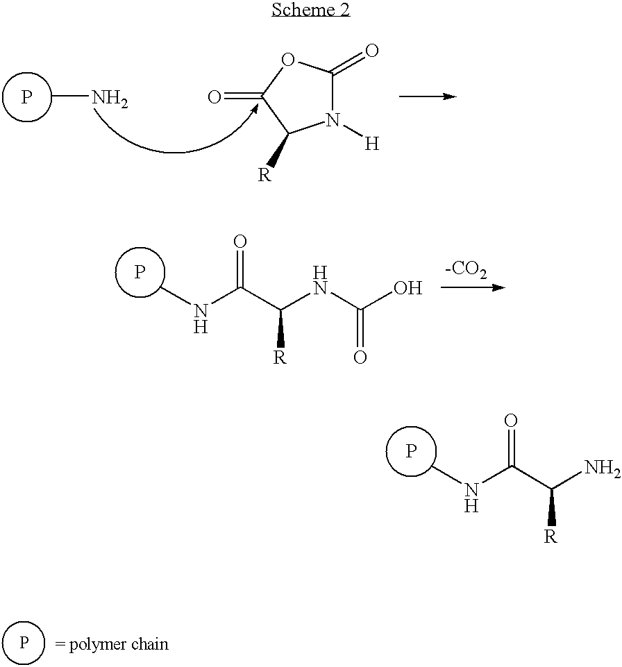
- Strong base initiated NCA polymerizations are much faster than amine initiated reactions. These polymerizations are poorly understood but are believed to propagate through either NCA anion or carbamate reactive species (
Scheme 3 and 4, respectively) (see e.g., [Bamford and Block, 1962]). -

-

- A significant limitation of NCA polymerizations employing conventional initiators is the synthesis is plagued by chain-breaking transfer and termination reactions which prevent formation of block copolymers (see, e.g., [Kricheldorf, 1987]). Consequently, the mechanisms of NCA polymerization have been under intensive study so that problematic side reactions can be eliminated. These investigations have been severely hindered by the complexity of the polymerizations, which can proceed through multiple pathways. Moreover, the high sensitivity of NCA polymerizations to reaction conditions and impurities has also led to contradictory data in the literature; resulting in controversy over the different hypothetical mechanisms.
- The significant difficulties with existing peptide synthesis methodologies create a variety of problems for practitioners. For example, the chain breaking transfer reactions that occur in the NCA polymerizations preclude the systematic control of peptide molecular weight. Moreover, block copolymers cannot be prepared using such methods.
- Block copolymers of amino acids have been less well studied, largely because synthetic methods do not yet have fine enough control to produce well-defined structures. The same is true of the synthesis of block copolypeptides for use as biomaterials or drug delivery vesicles—the potential advantages of the protein-like architectures—have thus remained unrealized for want of adequate synthetic building blocks and tools.
- For example, biomedical applications, such as drug delivery typically require water-soluble components to enhance their ability for circulation in vivo. The problem with common water-soluble polypeptides (e.g., poly-
L -lysine and poly-L -aspartate) is that they are polyelectrolytes that display pH-dependent solubility and limited circulation lifetime due to aggregation with oppositely charged biopolymers. Nonionic, water-soluble polypeptides are desired for biomedical applications since they avoid these problems, and can also display the stable secondary structures of proteins that influence biological properties. However, all high molecular weight nonionic homopolypeptides (>25 residues) derived from naturally occurring amino acids are notoriously insoluble in water. - One approach to producing nonionic water-soluble polypeptides employs polyethylene glycol (PEG), which is typically grafted onto polypeptides or other polymers to improve their properties in vivo. PEG is nonionic, water-soluble, and most importantly, not recognized by immune systems. It is believed that PEG imparts biocompatibility through formation of a hydrated “steric barrier” at the surface of material that cannot be penetrated or recognized by biological molecules, such as proteolytic enzymes. As such, block or graft copolymer drug carriers containing PEG are able to circulate for long periods in the bloodstream without degradation, as described throughout this specification.
- Despite its attractive properties, a drawback to grafting PEG onto polypeptides is the need for expensive amino- or carboxylate-functionalized molecules for coupling, which typically must be short (<5,000 Da) to ensure high functionalization. Accordingly, there remains considerable interest in developing alternative methods for producing nonionic water-soluble polypeptide building blocks that also incorporate the attractive properties of biochemical stability, self-assembly, and water solubility into polypeptides.
- Several seminal research publications about peptosomes, copolypeptides, their discovery, synthesis, and characterization are found in the prior art. However, none of these materials include any information or even speculate on the use of peptosomes as delivery vesicles in acoustically mediated drug delivery in vivo, as detailed by the present teachings. Peer-reviewed research papers supplying background material for the present invention include those authored by Deming, (1997); Yu et al., (1998); Curtin et al., (1999); Deming, (1999); Cha et al., (2000); Cheng et al., (2000); Cheng et al., (2001); Hwang et al., (2001); Wyrsta et al., (2001); Deming, (2002); Nowak et al., (2002); Cha et al., (2003); Checot et al., (2003); Nowak et al., (2003); Bellomo et al., (2004); Bellomo et al., (2004); Brzezinska et al., (2004); Euliss et al., (2004); McKenna et al., (2004); Pakstis et al., (2004); Babin et al., (2005a); Babin et al., (2005b); Checot et al., (2005); Holowka et al., (2005); Rodriguez-Hernandez et al., (2005a); Rodriguez-Hernandez et al., (2005b); Tomczak et al., (2005); Auguste et al., (2006); Bellomo et al., (2006); Checot et al., (2006); and Sallach et al., (2006).
- Many techniques used in the preparation, purification, characterization and use of lyposomes can also be successfully employed with peptosomes Important texts for liposomal methods include Liposome Methods and Protocols (Methods in Molecular Biology) (Basu et al., 2002); Liposomes, Part A, Volume 367 (Methods in Enzymology) (Duzgunes et al., 2003a); Liposomes, Part C, Volume 373 (Methods in Enzymology) (Duzgunes et al., 2003b); Liposomes: A Practical Approach (Torchilin et al., 2003); Liposomes, Part D, Volume 387 (Methods in Enzymology) (Duzgunes, 2004); Liposomes, Part E, Volume 391 (Methods in Enzymology) (Duzgunes, 2005); and Liposome Technology Third Edition (Gregoriadis, 2006).
- Peptosome patents relevant to the teachings of the present invention include: U.S. Pat. Nos. 6,506,577; 6,632,922; 6,680,365; and 6,818,732; 6,686,446; and 7,329,727; U.S. patent applications include 10/712,967, filed on Nov. 14, 2004.
- For enablement and other purposes, the disclosures of each of the foregoing publications, patents, and patent applications in paragraphs [0158], [0159], and [0160] are hereby incorporated by reference herein in their entirety.
- The following are examples of preferred chemical embodiments of the present invention, and should not be viewed as limiting considering the diversity of compounds that can be used to produce acoustically responsive peptosomes. In many preferred embodiments, the components and structure of said peptosome will be chosen and optimized so the vesicle will have a specific level of acoustic sensitivity.
- The methods know in the art, described in detail in many of the aforementioned publications, provide advanced tools and unique methods and compositions to generate polypeptides with varied material properties. The methods and initiator compositions for NCA polymerization disclosed in the aforementioned publications (paragraphs [0158] and [0160]), allow the precise control of such polypeptide synthesis. In particular, the methods know in the art achieve this by utilizing the versatile chemistry of transition metals to mediate the addition of monomers to the active polymer chain-ends, and therefore eliminate chain-breaking side reactions in favor of the chain-growth process. In this way, the formation of block copolymers based on copolypeptides so critical to the present specification is possible. Moreover, by binding the active end-group of the growing polymer to a metal center, its reactivity toward monomers can be precisely controlled through variation of the metal and ancillary ligands bound to the metal. The wide range of selective chemical transformations and polymerizations which are catalyzed by transition metal complexes attests to the versatility of this approach.
- Block copolymers have played an important part in materials science and technology because they allow the effective combination of disparate properties in a single material. As described throughout this specification, well-defined block copolymers assemble spontaneously into a variety of intriguing nanostructures, and other, aligned nanostructure arrays can be made using fluid flow or other fields. Because of this, block copolymers have enjoyed great commercial success, as well as intense research interests by both polymer chemists and polymer physicists.
- However, block copolymers of amino acids have been little studied, largely because the synthetic methods previously available did not have fine enough control to produce well-defined structures. The same is true of the synthesis of block copolypeptides for use as drug delivery vehicles or other biomaterials, i.e., the potential advantages of the protein-like architectures have remained unrealized for want of adequate synthetic tools. By treating the monomer of interest with an initiator such as the zero-valent nickel complex bipyNi (COD), where bipy is 2,2′-bipyridyl and COD is 1,5-cyclooctadiene; adding NCAs yields an intermediate molecule that can be isolated and that remains active towards further ring-opening polymerization, and the target polypeptide can be prepared with essentially 100 percent yield.
- One methodology know in the art, allows the production of a block copolypeptide consisting of combining an amount of a first amino acid-N-carboxyanhydride monomer, with an initiator molecule comprising a low valent transition metal-Lewis Base ligand complex, so that a polyaminoacid chain is generated, and then combining an amount of a second amino acid-N-carboxyanhydride monomer, with the polyaminoacid chain so that the second amino acid-N-carboxyanhydride monomer is added to the polyaminoacid chain. In a preferred embodiment of this method, the initiator molecule combines with the first amino acid-N-carboxyanhydride monomer to form an amido containing metallacycle intermediate of
Scheme 5. -

- Wherein M is the low valent transition metal; L is the Lewis Base ligand; and each of R1 and R2 and R3 independently consist of a side chain of an amino acid selected from the group consisting of alanine, arginine, asparagine, aspartic acid, cysteine, glutamic acid, glutamine, glycine, histidine, isoleucine, leucine, lysine, methionine, phenylalanine, proline, serine, threonine, tryptophan, tyrosine and valine; and R4 is the polyaminoacid chain.
- In a most preferred embodiment for making a block copolypeptide employing this method, the low valent transition metal is selected from the group consisting of nickel, palladium, platinum, cobalt, rhodium, iridium, and iron. In another preferred embodiment of this method, the Lewis Base ligand is selected from the group consisting of pyridyl ligands, diimine ligands, bisoxazoline ligands, alkyl phosphine ligands, aryl phosphine ligands, tertiary amine ligands, isocyanide ligands, and cyanide ligands.
- In yet another preferred embodiment of this method, the first α-amino acid-N-carboxyanhydride monomer is an α-amino acid-N-carboxyanhydride selected from the group consisting of side-chain protected NCA formed from arginine, asparagine, aspartic acid, cysteine, glutamic acid, glutamine, histidine, lysine, methionine, serine, threonine, tryptophan, and tyrosine or an amino acid side chain selected from the group consisting of side-chain NCA formed from alanine, glycine, isoleucine, leucine, phenylalanine, proline, and valine.
- Preferred embodiments of the peptosomes of the invention may comprise a single amphiphilic block copolypeptide. In other embodiments, more than one amphiphilic block copolypeptide may comprise the peptosome. In certain embodiments, amphiphilic block copolypeptides comprise one hydrophobic polymer and one hydrophilic polymer. In other embodiments, the amphiphilic block copolypeptide is a triblock polymer comprising terminal hydrophilic copolypeptide and a hydrophobic copolypeptide. Other amphiphilic block copolypeptides are tetrablock polymers comprising two hydrophilic copolypeptides blocks, and two hydrophobic copolypeptides blocks. Certain tetrablocks have terminal hydrophilic copolypeptide blocks and internal hydrophobic copolypeptide blocks. Other amphiphilic block copolypeptides are a pentablock polymer comprising two hydrophilic copolypeptide blocks and three hydrophobic copolypeptide blocks. In addition, pentablocks having three hydrophilic copolypeptide blocks and two hydrophobic copolypeptide blocks are preferred, yet other pentablocks having four hydrophilic copolypeptide blocks and one hydrophobic copolypeptide block are also embodiments. In yet other embodiments, the amphiphilic block copolypeptide comprises at least six blocks, at least two of which are hydrophilic polymer blocks.
- In a preferred embodiment, block copolypeptides for use in the present invention contain greater than 100 monomer units (residues); and the distribution of chain-lengths in the block copolymer composition is at least about 1.01<Mw/Mn<1.25, where Mw/Mn=weight average molecular weight divided by number average molecular weight. In one preferred embodiment of the present invention, the block copolypeptide has 10 consecutive identical amino acids per block. In another preferred embodiment, the block copolypeptide is composed of amino acid components g-benzyl-
L -glutamate and e-carbobenzyloxy-L -lysine. In another preferred embodiment of the present invention, the copolypeptide is a poly(e-benzyloxycarbonyl-L -lysine-block-g-benzyl-L -glutamate), PZLL-b-PBLG, diblock copolymer. In yet another preferred embodiment, the copolypeptide is a poly(g-benzyl-L -glutamate-block-e-benzyloxycarbonyl-L -lysine-block-g-benzyl-L -glutamate) triblock copolymer. In related embodiments, the number of consecutive monomer units (residues) in the block copolypeptide is greater than about 50, or 100, or 500, or even 1000. In another related embodiment, the total number of overall monomer units (residues) in the block copolypeptide is greater than about 200, or greater than about 500, or greater than about 1000. - Preferred embodiments of the present invention include diblock copolymers composed of amino acid components g-benzyl-L-glutamate and e-carbobenzyloxy-
L -lysine. The polymers are prepared by addition of Lys-NCA to bipyNi(COD) in N,N-dimethylformamide (DMF) to afford living poly(e-carbobenzyloxy-L -lysine), PZLL, chains with organometallic end-groups capable of further chain growth. Glu-NCA is added to these polymers to yield the PBLG-PZLL block copolypeptides. The evolution of molecular weight through each stage of monomer addition is analyzed using gel permeation chromatography (GPC). Molecular weight is found to increase as expected upon growth of each block of copolymer, while polydispersity should remain low, indicative of successful copolymer formation. - Preferred block copolymerizations are not restricted to the highly soluble polypeptides PBLG and PZLL. Copolypeptides containing
L -leucine andL -proline, both of which form homopolymers which are insoluble in most organic solvents (e.g., DMF) can also be prepared. Because of the solubilizing effect of the PBLG and PZLL blocks, all of the products are soluble in the reaction media indicating the absence of any homopolymer contaminants. The block copolymers containing L-leucine are found to be strongly associating in 0.1 M LiBr in DMF, a good solvent for PBLG and PZLL. Once deprotected, the assembly properties of these materials are expected make them useful as drug carriers. - As discussed herein, methodologies known in the art provide a number of methods and compositions for the generation of polypeptides having varied material properties. Other methodologies include adding amino acid-N-carboxyanhydrides (NCAs) to polyaminoacid chains by exposing the NCA to solutions containing polyaminoacid chains having an amido amidate metallacyle end groups, reacting the NCA with the amido amidate metallacyle end group so that the NCA is added to the polyaminoacid chain. Additionally, methods of controlling the polymerization of amino acid-N-carboxyanhydrides by reacting NCAs with initiator molecules and allowing initiator complexes to regioselectively open the ring of the NCAs through oxidative addition across the O—C5 or O—C2 anhydride bonds resulting in a controlled polypeptide polymerization are described in some of the aforementioned publications.
- One of the most preferred embodiments of the present invention entails the synthesis of amphiphilic block copolypeptides, which contain at least one water soluble block polypeptide (“soluble block”) conjugated to a water-insoluble polypeptide domain (“insoluble block”). The overall mole percent (%) composition of the insoluble block(s) can range from 3%-60% of the total copolymer. Preferably, the soluble block has about 30 to 100 mole % identical amino acid residues having either charged or oligo(ethyleneglycol)-conjugated side chains.
- The amphiphilic block copolypeptides of the present invention, for use in acoustically-mediated in vivo drug delivery, contain one or more “soluble blocks.” The soluble block(s) of the copolymers can contain some finite fraction of amino acid components with charged side-chains, with the amino acids belonging to the group: glutamic acid, aspartic acid, arginine, histidine, lysine, or ornithine. They can also contain up to a maximum 99 mole % of the amino acids oligo(ethyleneglycol). The soluble block includes oligo(ethyleneglycol)-terminated amino acid (EG-aa) residues. Preferred oligo(ethyleneglycol) functionalized amino acid residues include EG-Lys, EG-Ser, EG-Cys, and EG-Tyr. A most preferred soluble block consists of oligo(ethyleneglycol)-terminated poly(lysine).
- The amphiphilic block copolypeptides of the invention also contain at least one “insoluble block,” which is covalently linked to the soluble block. The insoluble block can contain a variety of amino acids residues or mixtures thereof including the naturally occurring amino acids, ornithine, or blocks consisting entirely of one or more D-isomers of the amino acids. However, the insoluble block will typically be composed primarily of nonionic amino acid residues, which generally form insoluble high molecular weight homopolypeptides. In preferred embodiments, about 60 to about 100 mole % of the insoluble block is comprised of nonionic amino acids. Such nonionic amino acids include, but are not limited to phenylalanine, leucine, valine, isoleucine, alanine, serine, threonine, and glutamine. In another preferred embodiment, any given insoluble block will usually contain 2-3 different kinds of amino acid components in statistically random sequences with mixtures of leucine/phenylalanine and leucine/valine being preferred. In these preferred copolymers, the composition of leucine in the insoluble domain ranges from 25 mole % to 75 mole % when mixed with phenylalanine and ranges from 60 mole % to 90 mole % when mixed with valine.
- In some embodiments, the multiblock copolypeptides that comprise the acoustically-responsive peptosome membrane can be crosslinked. In other embodiments, biological entities are incorporated within the interior core of the peptosome, including polymers, cytoskeletal molecules, signaling molecules that can induce phosphorylation, dephosphorylation, amidization, acetylation, enolization, and enzymes that can cause chemical transformations of other biological molecules.
- Certain peptosomes can comprise an amphiphile that is not a block copolymer, including: lipids, phospholipids, steroids, cholesterol, single chain alcohols, nucleotides, saccharides, or surfactants.
- Some peptosomes contain an amphiphilic copolymer, which is made by attaching two strands comprising different monomers. In some embodiments, the amphiphilic copolymer comprises polymers made by such techniques, for example, as free radical initiation and anionic polymerization.
- Briefly, in a preferred embodiment, the present invention provides reactive copolypeptides and amphiphiles that can be covalently cross-linked together or “stabilized,” while maintaining the semi-permeability of the membrane. Cross-linked peptosomes are particularly useful in applications requiring stability of the vesicle membranes, altered acoustic responsiveness, and/or durable retention of the encapsulated materials. By cross-linking copolypeptides and amphiphiles covalently, interconnected, i.e., completely cross-linked vesicle have all the membrane components covalently interconnected into a giant single molecule; cross-linked vesicles have interconnected components throughout their entire surface; partly cross-linked vesicles contain patches of the interconnected components.
- The peptosomes of embodiments of this disclosure may possess functional groups which may be cross-linked or stabilized by a variety of processes, methodologies, and procedures. Cross-linked copolymers covalently interconnected include (1) completely cross-linked nanocarriers, having all corona components covalently interconnected into a giant single molecule; (2) cross-linked nanocarriers having interconnected components throughout the entire surface of said nanocarrier; and (3) partly cross-linked nanocarriers containing patches of interconnected components.
- One of the most preferred stabilization techniques for use with the present invention is photopolymerization, which is a methodology that uses light to initiate and propagate a polymerization reaction to form a linear, or cross-linked polymeric structure. This technology has been widely explored in a variety of industries for several applications including, for example, the coating industry, the paint and printing ink industries, in adhesives, and in composite materials. Recently, the use of photopolymerization has been proposed for the production of biomaterial-based polymer networks that could be differentially fabricated for a variety of applications including embodiments of this specification.
- Photopolymers have broad utility in drug delivery because of a combination of properties held by photopolymerizable precursors and/or photopolymerized polymer networks. Exemplary characteristics include (1) ease of production, (2) possibility of carrying out photopolymerization in vivo or ex vivo, (3) spatial and temporal control of the polymerization process, (4) versatility of formulation and application, and (5) the possibility of entrapping a wide range of substances. Further, especially important characteristics for use with the present teachings include the ability to store photopolymerization formulation ingredients in easily accessible conditions until use such as, for example, in a clinical situation when said components need be immediately mixed together before production of polymer networks.
- A wide variety of sources are known to those skilled in the art, describing techniques, procedures, and methods for photopolymerization and biomedical applications, such as, for example, drug delivery using the nanocarriers of this disclosure and other structures. Preferred publications include, but are not limited to: Photoinitiation, Photopolymerization, and Photocuring: Fundamentals and Applications (Fouassier, 1995); Microparticulate Systems for the Delivery of Proteins and Vaccines (Drugs and the Pharmaceutical Sciences: a Series of Textbooks and Monographs) (Cohen et al., 1996); Polyurethanes in Biomedical Applications (Lamba et al., 1998); Chemical and Physical Networks: Formation and Control of Properties (The Wiley Polymer Networks Group Review) (Nijenhuis et al., 1998); Polymeric DrugsDrug Delivery Systems (Ottenbrite et al., 2001); Biomaterials for Delivery and Targeting of Proteins and Nucleic Acids (Mahato, 2005); Drug Delivery: Principles and Applications (Wiley Series in Drug Discovery and Development) (Wang et al., 2005); Photostability of Drugs and Drug Formulations (Tønnesen, 2004); and Polymers in Drug Delivery (Uchegbu et al., 2006), the disclosures of each of which are hereby incorporated by reference herein in their entirety for all purposes.
- In their simplest form, a photopolymerizable system is composed of (1) a light source, (2) a photoinitiator, and (3) a monomer. In addition, this formulation can be supplemented with other molecules (e.g., monomers, cross-linkers, excipients, bioactive molecules, drugs) to fulfill specific drug delivery applications.
- The light sources that heretofore have been utilized in producing biomedical polymer networks and devices, and may be used in embodiments of this disclosure include: (1) UV lamps, (2) halogen lamps, (3) plasma arc lamps, (4) light emitting diode (LED) lamps, (5) titanium-sapphire lasers—also called fernto second pulsed lasers—and (5) other laser lamps. These sources generate a beam of light that differs in terms of emission wavelength, intensity, and associated heat.
- Photoinitiators are molecules responsible for initiating the polymerization reaction by producing reactive species upon light absorption. There are many different photoinitiator molecules know in the art, some of which are suitable for use in the present teachings. A partial list includes eosin Y, 1-cyclohexyl phenyl ketone; 2,2-dimethoxy-2-phenylacetophenone (DMPA), 2-hydroxy-1-[4-hydroxyethoxy) phenyl]-2-methyl-1-propanone, Irgacure 651, camphorquinone/amine, where the amine is triethylamine, triethanolamine, or ethyl 4-N—N-dimethylamino benzoate; and the like. A photoinitiator can induce a polymerization reaction directly or in the presence of other molecules. A system composed of a photoinitiator and other molecule(s) may have a synergistic function, and said mixtures are normally referred to as a Photoinitiator System (PIS). When a photopolymerizable formulation is irradiated with an appropriate light source, a series of events takes place. In the presence of a PIS, the photosensitizer will absorb said light energy, thus passing into an excited state. Successively, the photosensitizer has either to transfer said energy to the photoinitiator or to react with the photoinitiator itself. Both situations will cause the photoinitiator to produce reactive species (e.g., cationic, anionic, or free radicals). In the absence of a photosensitizer, the excited photoinitiator forms reactive species directly. Further, formulations may contain other molecules, called accelerators, which speed up these initial steps. Once the reactive species are generated, polymerization is initiated by (1) photocleavage, (2) hydrogen abstraction, or (3) generation of cationic species. The first two mechanisms of initiation will generate a free radical photo-induced polymerization, which is probably the most commonly used methodologies with biomaterials.
- Commercially and non-commercially available molecules and macromolecules used as photopolymerizable monomers and macro-monomers (i.e., macromers) have one primary feature in common: Their backbone needs to have a photopolymerizable residue that normally is located at one or at both ends of the molecule. Photopolymerizable monomers already known in the art and suitable for use with the present teachings include (1) (di)methacrylic or (di)acrylic derivatives of PEG and its derivatives; (2) poly(ethylene oxide) poly(vinyl) alcohol (PVA) and its derivatives; (3) PEG-polystyrene copolymers (PEG)-(PST); (4) ethylene glycol-lactic acid copolymers (i.e., nEGmLA, where n and m are the number of repeat units of EG and LA, respectively); (5) ethylene glycol-lactic acid-caprolactone copolymers (nEGmLA CL); (6) PLA-b-PEG-b-PLA; (7) PLA-g-PVA; (8) poly(
D,L -lactide-co-ε-caprolactone); (9) (poly)-anhydrides; (10) 27 anhydrides; (11) urethanes; (12) polysaccharides; (13) dextran; (14) collagen; (15) hyaluronic acid; (16) diethyl fumarate/poly(propylene fumarate); and (17) the like. - The introduction of specific properties (e.g., cell and protein adhesiveness or non-adhesiveness, mechanical strength and acoustic sensitivity, degradation rate, absence or limited mass transport constraints) in polymerized networks can be achieved by selecting appropriate monomers(s) and/or macromers(s), and supplemental molecules during the design of the monomer or its formulation. As an example, approaches to modify the degradation rate and cell adhesiveness of the polymerized networks are reported because of their significance for use in the present specification.
- Degradation rate may be controlled by (1) the number of degradable chemical bonds in each monomer; (2) the type of degradable chemical bonds (e.g., ester, anhydride, amide); (3) the molecular weight of the monomer; and (4) the hydrophobic or hydrophilic nature of the monomer. While the number and nature of degradable chemical bonds are obvious key factors, the molecular weight determines whether polymer networks will be loosely (i.e., high MW) or tightly (i.e., low MW) cross-linked. In the second case, the degradation rate is slow because degradable linkages are hindered within the densely cross-linked network. In addition, hydrophobicity of highly cross-linked networks will further decrease their degradation. Obviously, degradation rate may be critical in applications involving the present teachings because it can influence, for example, the release and retention of entrapped therapeutics and other molecules.
- The size of the nanocarriers of embodiments of the present invention will depend on their intended use. Sizing also serves to modulate resultant biodistribution and clearance. The size of the nanocarrier can be adjusted, if desired, by the preferred method of filtering; although, other procedures known to those skilled in the art can also be used, such as shaking, microemulsification, vortexing, repeated freezing and thawing cycles, extrusion, and extrusion under pressure through pores of a defined size, sonication, and homogenization. See, for example, U.S. Pat. Nos. 4,162,282; 4,310,505; 4,533,254; 4,728,575; 4,728,578; 4,737,323; and 4,921,706, the disclosures of each of which are hereby incorporated by reference herein in their entirety for all purposes.
- After intravenous injection, particles greater than 5 μm to 7 μm in diameter are often accumulated in the lung capillaries, while particles with a diameter of less than 5 μm are generally cleared from the circulation by the cells of the reticuloendothelial system. Particles in excess of 7 μm are larger than the blood capillary diameter (i.e., approximately 6 μm) and will be mechanically filtered. In the size range 70 nm to 200 nm, the surface curvature of particles may affect the extent and/or the type of protein or opsonin absorption, which plays a critical role in complement activation. The fact that particle size may change substantially upon introduction into a protein-containing medium, such as plasma, must also be taken into consideration.
- Therefore, since vesicle size influences biodistribution, different-sized vesicles may be selected for various purposes. For example, for intravascular application, the preferred size range is a mean outside diameter between approximately 20 nm and approximately 1.5 μm, with the preferable mean outside diameter being approximately 750 nm. More preferably, for intravascular application, the size of the vesicles is approximately 200 nm or less in mean outside diameter, and most preferably less than approximately 100 nm in mean outside diameter. Preferably, the vesicles are no smaller than approximately 20 nm in mean outside diameter. To provide therapeutic delivery to organs such as the liver and to allow differentiation of a tumor from normal tissue, smaller vesicles, between approximately 30 nm and approximately 100 nm in mean outside diameter, are preferred. For immobilization of a tissue such as the kidney or the lung, the vesicles are preferably less than approximately 200 nm in mean outside diameter. For intranasal, intrarectal, or topical administration, the vesicles are preferably less than approximately 100 nm in mean outside diameter. Large vesicles, between 1 μm and approximately 1.5 μm in size, will generally be confined to the intravascular space until they are cleared by phagocytic elements of the immune system lining the vesicles, such as the macrophages and Kupffer cells lining capillary sinusoids. For passage to the cells beyond the sinusoids, smaller vesicles, for example, less than approximately 1 μm in mean outside diameter (e.g., less than approximately 300 nm in size), may be utilized. In preferred embodiments, the vesicles are typically administered individually, although this administration may be in some type of polymer matrix.
- As described herein, embodiments of the present invention include nanocarriers and/or certain contrast agents which may comprise various targeting components (e.g., ligands) to target the vesicle and its contents to, for example, specific cells either in vitro or in vivo. A ligand or targeting ligand is a molecule that specifically binds to another molecule, which may be referred to as a target. In another preferred embodiment, the nanocarriers of this specification can be targeted by compositions that facilitate magnetic targeting (i.e., said vesicle is guided by a magnetic field). Or in yet another embodiment, both targeting ligands and magnetic compositions (i.e., compositions that facilitate magnetic targeting) are utilized for active targeting. All of the targeting ligands and magnetic compositions described herein are considered to be within the definition of a “targeting moiety,” and are thus suitable for use with embodiments of this disclosure.
- The targeting ligands incorporated in the nanocarriers of this specification are preferably substances which are capable of targeting receptors and/or tissues in vivo and/or in vitro. Preferred targeting ligands include (1) compositions facilitating magnetic targeting, (2) proteins, including antibodies, antibody fragments, hormones, hormone analogues, glycoproteins and lectins, peptides, polypeptides, and amino acids; (3) sugars such as saccharides, including monosaccharides and polysaccharides; carbohydrates, vitamins, steroids, steroid analogs, hormones, and cofactors; (4) genetic material, including aptamers, nucleosides, nucleotides, nucleotide acid constructs, and polynucleotides; and (5) peptides. Preferred targeting ligands for use with embodiments described herein include, for example, cell adhesion molecules (CAMs), among which are cytokines, integrins, cadherins, immunoglobulins and selectins; optimal genetic material for targeting includes aptamers.
- A wide variety of sources is known in the art that describes techniques, procedures, and methods for active targeting of drug-containing vesicles using ligands and other structures, such as, for example, the nanocarriers of embodiments of the present invention. For enablement purposes, preferred publications include, but are not limited to Using Antibodies: A Laboratory Manual (Harlow et al., 1999); Drug Targeting Organ-Specific Strategies (Molema et al., 2001); Molecular Cloning: A Laboratory Manual (Sambrook et al., 2001); Drug Targeting Technology: Physical, Chemical, and Biological Methods (Schreier, 2001); Biomedical Aspects of Drug Targeting (Muzykantov et al., 2002); Liposomes: A Practical Approach (Torchilin et al., 2003); Cellular Drug Delivery: Principles and Practice (Lu et al., 2004); and Protein-Protein Interactions: A Molecular Cloning Manual (Golemis et al., 2005).
- Patents identifying and describing specific targeting ligands of medical importance and methods for their use, include, but are not limited to U.S. Pat. Nos. 5,128,326; 5,580,960; 5,610,031; 5,625,040; 5,648,465; 5,766,922; 5,770,565; 5,792,743; 5,849,865; 5,866,165; 5,872,231; 6,121,231; 6,140,117; 6,159,467; 6,204,054; 6,352,972; and 6,482,410. The disclosures of each of the publications and patents in paragraphs [0196] and [0197] are hereby incorporated by reference herein in their entirety for all purposes.
- Briefly, many different targeting ligands can be selected to bind to specific domains of various adhesion molecules (e.g., the Immunoglobulin Superfamily [ICAM 1-3, PECAM-1, VCAM-1], the Selectins [EWLAM-1, LECAM-1, GMP-140] and the Integrins [LEA-1]). Targeting ligands in this regard may include (1) lectins, (2) a wide variety of carbohydrate or sugar moieties, (3) antibodies, (4) antibody fragments, (5) Fab fragments, such as, for example, Fab′2; and (6) synthetic peptides, including, for example, Arginine-Glycine-Aspartic Acid (R-G-D) which may be targeted to wound healing. While many of these materials may be derived from natural sources, some may be synthesized by molecular biological recombinant techniques, and others may be synthetic in origin. Peptides may be prepared by a variety of techniques known in the art. Targeting ligands derived or modified from human leukocyte origin (e.g., CD11a/CD18 and leukocyte cell surface glycoprotein [LFA-1]) may also be used, as these bind to the endothelial cell receptor ICAM-1. The cytokine inducible member of the immunoglobulin superfamily, VCAM-1, which is mononuclear leukocyte-selective, may also be used as a targeting ligand. VLA-4, derived from human monocytes, may be used to target VCAM-1. Antibodies and other targeting ligands may be employed to target endoglin, which is an endothelial cell proliferation marker. Endoglin is upregulated on endothelial cells in miscellaneous solid tumors. Further, the cadherin family of cell adhesion molecules may also be used as targeting ligands including, for example, (1) the E-, N-, and P-cadherins; (2) cadherin-4, (3) cadherin-5, (4) cadherin-6, (5) cadherin-7, (6) cadherin-8, (7) cadherin-9, (8) cadherin-10, (9) cadherin-11, and, most preferably, (11) cadherin C-5. Further, antibodies directed to cadherins, may be used to recognize cadherins expressed locally by specific endothelial cells.
- Targeting ligands may be selected for targeting antigens, including antigens associated with breast cancer, such as epidermal growth factor receptor (EGFR), fibroblast growth factor receptor, erbB2/BER-2, and tumor associated carbohydrate antigens, CTA 16.88, homologous to
cytokeratins 8, 18, and 19, which is expressed by most epithelial-derived tumors including carcinomas of the colon, pancreas, breast, and ovary. Chemically conjugated bispecific anti-cell surface antigen, and anti-hapten Fab′-Fab antibodies may also be used as targeting ligands. The MG series monoclonal antibodies may be selected for targeting (e.g., gastric cancer). Fully humanized antibodies or antibody fragments are most preferred. - There are a variety of cell surface epitopes on epithelial cells for which targeting ligands may be selected. For example, the human papilloma virus (HPV) has been associated with benign and malignant epithelial proliferations in both skin and mucosa. Two HPV oncogenic proteins, E6 and E7, may be targeted, as these may be expressed in certain epithelial-derived cancers (e.g., as cervical carcinoma). Membrane receptors for peptide growth factors (PGF-R), which are involved in cancer cell proliferation, may also be selected as tumor antigens. Also, epidermal growth factor (EGF) and interleukin-2 may be targeted with suitable targeting ligands, including peptides, which bind these receptors. Certain melanoma-associated antigens (MAAs) (e.g., epidermal growth factor receptor [EGFR]), and adhesion molecules expressed by, for example, malignant melanoma cells can also be targeted with specific ligands.
- A wide variety of targeting ligands may be selected for targeting myocardial cells. Exemplary targeting ligands include, for example, anticardiomyosin antibody, which may comprise polyclonal antibody, Fab′2 fragments, or be of human origin, animal origin, for example, mouse or of chimeric origin. Again, in all antibody and antibody fragment embodiments, fully humanized species are preferred. Additional targeting ligands include (1) dipyridamole, digitalis, nifedipine, and apolipoprotein; (2) low-density lipoproteins (LDL) including vLDL and methyl LDL; (3) ryanodine, endothelin, complement receptor type 1, IgG Fc, beta 1-adrenergic, dihydropyridine, adenosine, mineralocorticoid, nicotinic acetylcholine and muscarinic acetylcholine; antibodies to the human alpha 1A-adrenergic receptor; bioactive agents, such as drugs, including the α-1-antagonist prazosin; (4) antibodies to the anti-beta-receptor, and drugs which bind to the anti-beta-receptor; (5) anti-cardiac RyR antibodies; (6) endothelin-1, which is an endothelial cell-derived vasoconstrictor peptide that exerts a potent positive inotropic effect on cardiac tissue (endothelin-1 binds to cardiac sarcolemmal vesicles); (7) monoclonal antibodies which may be generated to the T-cell receptor-β receptor, and thereby employed to generate targeting ligands; (8) the complement inhibitor sCR1; (9) drugs, peptides, or antibodies which are generated to the dihydropyridine receptor; and (10) monoclonal antibodies directed toward the anti-interleukin-2 receptor, which may be used as targeting ligands to direct the nanocarriers of this specification to areas of myocardial tissue which express this receptor and which may be upregulated in conditions such as inflammation.
- In another embodiment, the targeting ligands are directed to lymphocytes which may be T-cells or B-cells. Depending on the targeting ligand, the composition may be targeted to one or more classes or clones of T-cells. To select a class of targeted lymphocytes, a targeting ligand having specific affinity for that class is employed. For example, an anti CD-4 antibody can be used for selecting the class of T-cells harboring CD4 receptors, an anti CD-8 antibody can be used for selecting the class of T-cells harboring CD-8 receptors, an anti CD-34 antibody can be used for selecting the class of T-cells harboring CD-34 receptors, etc. A lower molecular weight ligand is preferably employed (e.g., Fab or a peptide fragment). For example, an OKT3 antibody or OKT3 antibody fragment may be used. When a receptor for a class of T-cells is selected, or clones of T-cells are selected, the composition will be delivered to that class of cells. Using HLA-derived peptides, for example, will allow selection of targeted clones of cells expressing reactivity to HLA proteins. Another major area for targeted delivery involves the interleukin-2 (IL-2) system. IL-2 is a T-cell growth factor produced following antigen- or mitogen-induced stimulation of lymphoid cells. Among the cell types which produce IL-2 are CD4+ and CD8+ T-cells, large granular lymphocytes, as well as certain T-cell tumors, etc. Still other systems which can be used in embodiments of the present invention include IgM-mediated endocytosis in B-cells or a variant of the ligand-receptor interactions described above, wherein the T-cell receptor is CD2 and the ligand is lymphocyte function-associated antigen 3 (LFA-3).
- Synthetic compounds, which combine a natural amino acid sequence with synthetic amino acids, can also be used as targeting ligands as well as peptides, or derivatives thereof. In view of the present disclosure, as will be immediately apparent to those skilled in the art, a large number of additional targeting ligands may be used with the present teachings, in addition to those exemplified above. Other suitable targeting ligands include, for example, conjugated peptides, such as, for example, glycoconjugates and lectins, which are peptides attached to sugar moieties. The compositions may comprise a single targeting ligand, as well as two or more different targeting ligands.
- Preferred embodiments of present invention include the use of magnetic targeting of nanocarriers and/or contrast agents. This is accomplished by using magnetically susceptible compositions bound to, and/or associated with said nanocarriers or contrast agents exteriorly, and/or by other means, and then guided by a magnetic field. A wide variety of sources are known in the art describing techniques, procedures, and methods for active targeting of drug-containing vesicles using magnetically susceptible materials, and said materials may be hereafter referred to as “magnetic compositions” or “magnetic targeting components.” For enablement and other purposes, preferred publications include, but are not limited to Principles of Nuclear Magnetism (International Series of Monographs on Physics) (Abragam, 1983); Ultrathin Magnetic Structures I: An Introduction to the Electronic, Magnetic and Structural Properties (Bland et al., 1994); Magnetism: Molecules to Materials, (Miller et al., 2001); and Drug and Enzyme Targeting, Part A: Volume 112: Drug and Enzyme Targeting (Methods in Enzymology) (Colowick et al., 2006). Important peer-reviewed research publications associated with magnetic targeting and drug delivery include Widder et al., (1979); Hsieh et al., (1981); Kost et al., (1987); He et al., (1993); Wu et al., (1993); Wu et al., (1994); Chen et al., (1997); Rudge et al., (2000); Jones et al., (2001); Lubbe et al., (2001); and Moroz et al., (2001). Patents concerning magnetic targeting and drug delivery relevant to the teachings of this specification include U.S. Pat. Nos. 6,200,547; 6,482,436; 6,488,615; and 6,663,555. Each of the publications and patents listed in this paragraph [0204] are hereby incorporated by reference herein in their entirety for all purposes.
- Magnetic targeting components for use with embodiments of the present invention are typically comprised of 1% to 70% of a biocompatible polymer, and 30% to 99% of a magnetic component. With compositions having less than 1% polymer, the physical integrity of the particle is less than optimal. With compositions of greater than 70% polymer, the magnetic susceptibility of the particle is generally reduced beyond an optimal level for the nanocarriers described herein. The compositions may be of any shape, different shapes conferring differing advantageous properties, with an average size of approximately 0.1 μm to approximately 30 μm in diameter.
- Said magnetic targeting components have the general properties of having Curie temperatures (Tc) greater than the normal human body temperature (37° C.), having high magnetic saturation (>approximately 20 Am2/kg), and being ferromagnetic or ferrimagnetic. Examples of suitable magnetic components include magnetic iron sulfides such as pyrrohotite (Fe7S8), and greigite (Fe4S4); magnetic ceramics such as
Alnico 5,Alnico 5 DG, Sm2Co17, SmCo5, and NdFeB; magnetic iron alloys, such as jacobsite (MnFe2O4), trevorite (NiFe2O4), awaruite (Ni3Fe), and wairauite (CoFe); and magnetic metals such as metallic iron (Fe), cobalt (Co), and nickel (Ni). Each of the magnetic components can have added to its chemical formula specific impurities that may or may not alter the magnetic properties of the material. Doped ferromagnetic or ferrimagnetic materials, within the above limits of Curie temperatures and magnetic saturation values, are considered to be suitable as magnetic compositions for use in active targeting of the nanocarriers described herein. Specifically excluded from suitable magnetic components and the magnetically susceptible compositions are the iron oxides magnetite (Fe3O4), hematite (α-Fe2O3), and maghemite (γ-Fe2O3). - The term “metallic iron” indicates that iron is primarily in its “zero valence” state (Fe0). Generally, the metallic iron is greater than approximately 85% Fe0, and preferably greater than approximately 90% Fe0. More preferably, the metallic iron is greater than approximately 95% “zero valence” iron. Metallic iron is a material with high magnetic saturation and density (i.e., 218 emu/gm and 7.8 gm/cm3), which are much higher than magnetite (i.e., 92 emu/gm and 5.0 gm/cm3). The density of metallic iron is 7.8 gm/cm3, while magnetite is approximately 5.0 gm/cm3. Thus, the magnetic saturation of metallic iron is approximately 4-fold higher than that of magnetite per unit volume (Craik, 1995). The use of said magnetic compositions with the nanocarriers of this specification results in magnetically responsive compositions with a high degree of magnetic saturation (i.e., >50 emu/gm). The higher magnetic saturation allows the nanocarrier with biologically active agents (e.g., therapeutic macromolecules) to be effectively targeted to the desired site by an external magnetic field, and eventually be extravasated through blood vessel walls, penetrating into the target tissues of the patient.
- The biocompatible polymers for use with said magnetic targeting components may be bioinert and/or biodegradable. Some nonlimiting examples of biocompatible polymers are polylactides, polyglycolides, polycaprolactones, polydioxanones, polycarbonates, polyhydroxybutyrates, polyalkylene oxalates, polyanhydrides, polyamides, polyacrylic acid, poloxamers, polyesteramides, polyurethanes, polyacetals, polyorthocarbonates, polyphosphazenes, polyhydroxyvalerates, polyalkylene succinates, poly(malic acid), poly(amino acids), alginate, agarose, chitin, chitosan, gelatin, collagen, atelocollagen, dextran, proteins, polyorthoesters, copolymers, terpolymers, and combinations and/or mixtures thereof. Said biocompatible polymers can be prepared in the form of matrices, which are polymeric networks. One type of polymeric matrix is a hydrogel, which can be defined as a water-containing polymeric network. The polymers used to prepare hydrogels can be based on a variety of monomer types, such as those based on methacrylic and acrylic ester monomers, acrylamide (methacrylamide) monomers, and N-vinyl-2-pyrrolidone. Hydrogels can also be based on polymers such as starch, ethylene glycol, hyaluran, chitose, and/or cellulose. To form a hydrogel, monomers are typically cross-linked with cross-linking agents such as ethylene dimethacrylate, N,N-methylenediacrylamide, methylenebis(4-phenyl isocyanate), epichlarohydin glutaraldehyde, ethylene dimethacrylate, divinylbenzene, and allyl methacrylate. Hydrogels can also be based on polymers such as starch, ethylene glycol, hyaluran, chitose, and/or cellulose. In addition, hydrogels can be formed from a mixture of monomers and polymers.
- Another type of polymeric network can be formed from more hydrophobic monomers and/or macromers. Matrices formed from these materials generally exclude water. Polymers used to prepare hydrophobic matrices can be based on a variety of monomer types such as alkyl acrylates and methacrylates, and polyester-forming monomers such as ε-caprolactone, glycolide, lactic acid, glycolic acid, lactide, and the like. When formulated for use in an aqueous environment, these materials do not normally need to be cross-linked, but they can be cross-linked with standard agents such as divinyl benzene. Hydrophobic matrices may also be formed from reactions of macromers bearing the appropriate reactive groups such as the reaction of diisocyanate macromers with dihydroxy macromers, and the reaction of diepoxy-containing macromers with dianhydride or diamine-containing macromers.
- The biocompatible polymers for use with said magnetic compositions can be preferably prepared in the form of dendrimers and/or hyperbranched polymers. The size, shape, and properties of these dendrimers can be molecularly tailored to meet specialized end uses, such as a means for the delivery of high concentrations of carried material per unit of polymer, controlled delivery, targeted delivery, and/or multiple species delivery or use. The dendritic polymers can be prepared according to methods known in the art, including those detailed herein. The biocompatible polymers for use with said magnetic compositions may be, for example, biodegradable, bioresorbable, bioinert, and/or biostable. Bioresorbable hydrogel-forming polymers are generally naturally occurring polymers such as polysaccharides, examples of which include, but are not limited to hyaluronic acid, starch, dextran, heparin, and chitosan; and proteins—and other polyamino acids—examples of which include but are not limited to gelatin, collagen, fibronectin, laminin, albumin, and active peptide domains thereof. Matrices formed from these materials degrade under physiological conditions, generally via enzyme-mediated hydrolysis.
- Bioresorbable matrix-forming polymers are generally synthetic polymers prepared via condensation polymerization of one or more monomers. Matrix-forming polymers of this type include: polylactide (PLA), polyglycolide (PGA), polylactide coglycolide (PLGA), polycaprolactone (PCL), and copolymers of these materials, polyanhydrides, and polyortho esters. Biostable or bioinert hydrogel matrix-forming polymers are generally synthetic or naturally occurring polymers which are soluble in water, matrices of which are hydrogels or water-containing gels. Examples of this type of polymer include: polyvinylpyrrolidone (PVP), polyethylene glycol (PEG), polyethylene oxide (PEO), polyacrylamide (PAA), polyvinyl alcohol (PVA), and the like. Biostable or bioinert matrix-forming polymers are generally synthetic polymers formed from hydrophobic monomers such as methyl methacrylate, butyl methacrylate, dimethyl siloxanes, and the like. These polymer materials generally do not possess significant water solubility, but can be formulated as neat liquids which form strong matrices upon activation. Said polymers may also contain hydrophilic and hydrophobic monomers.
- Targeting moieties may also be incorporated into the nanocarriers of this specification in a variety of other ways. Additional preferred methods include being associated covalently or non-covalently with one or more of the polymers described herein. Photopolymerizable elements are most preferred, and photopolymerization may be used in cross-linking the targeting moieties with themselves, and/or linking said moieties to the nanocarriers. In other preferred embodiments, the targeting moiety is covalently bound to the surface of the nanocarrier by a spacer including, for example, hydrophilic polymers, preferably polyethylene glycol. Preferred molecular weights of the polymers are from 1,000 Daltons to 10,000 Daltons, with 500 Daltons being most preferred. Preferably, the polymer is bifunctional with the targeting moiety bound to a terminus of the polymer. Generally, in the case of a targeting ligand, it should range from approximately 0.1 to approximately 20 mole % of the exterior components of the vesicle. The exact ratio will depend on the particular targeting ligand and the application.
- Exemplary covalent bonds by which the targeting moieties are associated with the nanocarriers of embodiments of the present invention include, for example, amide (—CONH—); thioamide (—CSNH—); ether (ROR′), where R and R′ may be the same or different and are other than hydrogen; ester (—COO—); thioester (—COS—); —O—; —S—; —Sn, where n is greater than 1, preferably approximately 2 to approximately 8, and more preferably approximately 2; carbamates; —NH—; —NR—, where R is alkyl, for example, alkyl of from 1 carbon to approximately 4 carbons; urethane; and substituted imidate; and combinations of two or more of these. Covalent bonds between targeting ligands and polymers may be achieved through the use of molecules that may act as spacers to increase the conformational and topographical flexibility of the ligand. Examples of such spacers include, for example, succinic acid, 1,6-hexanedioic acid, 1,8-octanedioic acid, and the like, as well as modified amino acids, such as, for example, 6-aminohexanoic acid, 4-aminobutanoic acid, and the like. In addition, in the case of targeting ligands which comprise peptide moieties, side-chain-to-side-chain cross-linking may be complemented with side chain-to-end cross-linking and/or end-to-end cross-linking. Also, small spacer molecules, such as dimethylsuberimidate, may be used to accomplish similar objectives. The use of agents, including those used in Schiff's base-type reactions, such as gluteraldehyde, may also be employed.
- The covalent linking of targeting moieties to embodiments of the invention may also be accomplished using synthetic organic techniques, which in view of the present disclosure, will be readily apparent to one of ordinary skill in the art. For example, the targeting moieties may be linked to the materials, including the polymers, via the use of well-known coupling or activation agents. As known to those skilled in the art, activating agents are generally electrophilic, which can be employed to elicit the formation of a covalent bond. Exemplary activating agents which may be used include, for example, carbonyldiimidazole (CDI), dicyclohexylcarbodiimide (DCC), diisopropylcarbodiimide (DIC), methyl sulfonyl chloride, Castro's Reagent, and diphenyl phosphoryl chloride. The covalent bonds may involve cross-linking and/or polymerization. Cross-linking preferably refers to the attachment of two chains of polymer molecules by bridges, composed of either an element, a group, or a compound, which join certain carbon atoms of the chains by covalent chemical bonds. For example, cross-linking may occur by photopolymerization and for polypeptides which are joined by the disulfide bonds of the cysteine residue. Cross-linking may be achieved, for example, by (1) adding a chemical substance (e.g., cross-linking agent) and exposing the mixture to heat, or (2) subjecting a polymer to high-energy radiation. A variety of cross-linking agents, or “tethers,” of different lengths and/or functionalities are described (Hermanson, 1996), the disclosures of which are hereby incorporated herein by reference in their entirety for all purposes.
- Most of the contrast agents for use with preferred embodiments of this specification have a high degree of echogenicity, the ability of an object to reflect ultrasonic waves. Thus, preferred contrast agents comprise small, stabilized gas-filled microbubbles that can pass through the smallest capillaries of the patient. For imaging purposes, the larger the microbubbles, the better the acoustic responsiveness; however, if the bubbles are too large, they will be retained in the capillaries of the patient and they are unable to cross the pulmonary circulation. Further, the size of said contrast agents will likely vary depending upon the drug delivery application. Properties of the ideal contrast agent for use with embodiments of this disclosure (1) are nontoxic and easily eliminated by the patient, (2) are administered intravenously, (3) pass easily through the microcirculation, (4) are physically stable, and (5) are acoustically responsive with stable harmonics and the capability of rapid disruption. The ultrasonic characteristics of these contrast agents depend not only on the size of the bubble, but also on the composition of the shell and the gas contained therein. The outer shell of preferred microbubbles is composed of many different substances including albumin, polymers, palmitic acid, or phospholipids. The composition of the shell determines its elasticity, its behavior in an ultrasonic field, how rapidly the bubble is taken up by the immune system, and the physiological methods for metabolism and elimination from the patient. A more hydrophilic material tends to be taken up more easily, which reduces the microbubble residence time in the circulation. In general, the stiffer the shell, the more easily it will crack or break when exposed to ultrasonic energy. Conversely, the more elastic the shell, the greater its ability to be compressed or resonated, a characteristic of considerable importance for the preferred contrast agents used in the practice of the present teachings.
- The gas core is a critical component of the microbubble because it is the primary determinate of its echogenicity. When gas bubbles are caught in an ultrasonic frequency field, they compress, oscillate, and reflect a characteristic echo, which generates the strong and unique sonogram in contrast-enhanced ultrasound. Gas cores can be composed of air, nitrogen or, for example, heavy gases like perfluorocarbon. Heavy gases are less water-soluble so they are less likely to leak out from the microbubble to impair echogenicity. Thus, microbubbles with heavy gas cores are likely to remain in the circulation longer.
- Ultrasound contrast reagents for use with the present invention can be prepared as described in U.S. Pat. No. 6,146,657, the disclosures of which are hereby incorporated herein by reference in their entirety for all purposes. Briefly, a sealed container is used comprising an aqueous lipid suspension phase and a substantially separate gaseous phase. Prior to use, the container and its contents may be agitated, causing the lipid and gas phases to mix, resulting in the formation of gas-filled liposomes which entrap the gas. The resulting gas-filled liposomes provide an excellent contrast enhancement agent for diagnostic imaging, particularly using ultrasound or magnetic resonance imaging, and for assisting in acoustically mediated intracellular therapeutic delivery in vivo, as detailed herein. A wide variety of lipids may be employed in the aqueous lipid suspension phase of the preferred contrast agents for use with the present teachings. The lipids may be saturated or unsaturated, and may be in linear or branched form, as desired. Such lipids may comprise, for example, fatty acids molecules that contain a wide range of carbon atoms, preferably between approximately 12 carbon atoms and 22 carbon atoms. Hydrocarbon groups consisting of isoprenoid units, prenyl groups, and/or sterol moieties (e.g., cholesterol, cholesterol sulfate, and analogs thereof) may also be employed. The lipids may also bear polymer chains, such as the amphipathic polymers polyethylene glycol (PEG), polyvinylpyrrolidone (PVP), derivatives thereof for in vivo targeting; charged amino acids such as polylysine or polyarginine, for binding of a negatively charged compound; carbohydrates, for in vivo targeting, such as described in U.S. Pat. No. 4,310,505; glycolipids, for in vivo targeting; or antibodies and other peptides and proteins, for in vivo targeting, etc., as desired. Such targeting or binding compounds may be simply added to the aqueous lipid suspension phase or may be specifically chemically attached to the lipids using methods described herein, or employing other methodologies known in the art. The lipids may also be anionic or cationic.
- Classes of and specific lipids for use as shell materials with preferred contrast agents for use with the present teachings, but are not limited to, include phosphatidylcholines such as dioleoylphosphatidylcholine, dimyristoylphosphatidylcholine, dipalmitoylphosphatidylcholine (DPPC), and distearoylphosphatidylcholine; phosphatidylethanolamines such as dipatmitoylphosphatidylethanolamine (DPPE), dioleoylphosphatidylethanolamine, and N-succinyl-dioleoylphosphatidylethanolamine; phosphatidylserines, phosphatidylglycerols, and sphingolipids; glycolipids such as ganglioside GM1; glucolipids, sulfatides, and glycosphingolipids; phosphatidic acids such as dipalmatoylphosphatidic acid (DPPA); palmitic fatty acids, stearic fatty acids, arachidonic fatty acids, lauric fatty acids, myristic fatty acids, lauroleic fatty acids, physeteric fatty acids, myristoleic fatty acids, palmitoleic fatty acids, petroselinic fatty acids, oleic fatty acids, isolauric fatty acids, isomyristic fatty acids, isopalmitic fatty acids, and isostearic fatty acids; cholesterol and cholesterol derivatives such as cholesterol hemisuccinate, cholesterol sulfate, and cholesteryl-(4′-trimethylammonio)-butanoate; polyoxyethylene fatty acid esters, polyoxyethylene fatty acid alcohols, polyoxyethylene fatty acid alcohol ethers, polyoxyethylated sorbitan fatty acid esters, glycerol polyethylene glycol oxystearate, glycerol polyethylene glycol ricinoleate, ethoxylated soybean sterols, ethoxylated castor oil, polyoxyethylene-polyoxypropylene fatty acid polymers, polyoxyethylene fatty acid stearates, 12-(((7′-diethylaminocoumarin-3-yl)-carbonyl)-methylamino)-octadecanoic acid, N-(12-(((7′-diethylamino-coumarin-3-yl)-carbonyl)-methyl-amino)octadecanoyl)-2-amino-palmitic acid, 1,2-dioleoyl-sn-glycerol, 1,2-dipalmitoyl-sn-3-succinylglycerol, 1,3-dipalmitoyl-2-succinyl-glycerol, 1-hexadecyl-2-palmitoyl-glycerophosphoethanolamine, and palmitoylhomocysteine, lauryltrimethylammonium bromide (lauryl-=dodecyl-); cetyltrimethylammonium bromide (cetryl-=hexadecyl-), myristyltrimethylammonium bromide (myristyl-=tetradecyl-); alkyldimethylbenzylammonium chlorides such as wherein alkyl is a C12, C14, or C16 alkyl; and benzyldimethyldodecylammonium bromide, benzyldimethyldodecylammonium chloride, benzyldimethylhexadecylammonium bromide, benzyldimethylhexadecylammonium chloride, benzyldimethyltetradecylammonium bromide, benzyldimethyltetradecylammonium chloride, cetyldimethylethylammonium bromide, cetyldimethylethylammonium chloride, cetylpyridinium bromide, cetylpyridinium chloride, N-(1-2,3-dioleoyloxy)-propyl)-N,N,N-trimethylammonium chloride (DOTMA), 1,2-dioleoyloxy-3-(trimethylammonio)propane (DOTAP), and 1,2-dioleoyl-e-(4′-trimethylammonio)-butanoyl-sn-glycerol (DOTB).
- In addition, the aqueous lipid phase may further comprise a polymer, preferably an amphipathic polymer, and preferably one that is directly bound (i.e., chemically attached) to the lipid. Preferably, the amphipathic polymer is polyethylene glycol or a derivative thereof. The most preferred combination is the lipid dipatmitoylphosphatidylethanolamine (DPPE) bound to polyethylene glycol (PEG), especially PEG of an average molecular weight of approximately 5000 (DPPE-PEG5000). The PEG or other polymer may be bound to the DPPE or other lipid through a covalent linkage such as through an amide, carbamate, or amine linkage. Alternatively, ester, ether, thioester, thioamide, or disulfide (i.e., thioester) linkages may be used with the PEG or other polymer to bind the polymer to, for example, cholesterol or other phospholipids. A particularly preferred combination of lipids is DPPC, DPPE-PEG5000, and DPPA, especially in a ratio of approximately 82:8:10% (mole %), DPPC: DPPE-PEG5000:DPPA.
- Examples of classes of and specific suitable gases for use as the core of preferred contrast agents, suitable for use with the present teachings, are those gases that are substantially insoluble in an aqueous shell suspension. Suitable gases that are substantially insoluble or soluble include, but are not limited to hexafluoroacetone, isopropylacetylene, allene, tetrafluoroallene, boron trifluoride, 1,2-butadiene, 1,3-butadiene, 1,2,3-trichlorobutadiene, 2-fluoro-1,3-butadiene, 2-methyl-1,3 butadiene, hexafluoro-1,3-butadiene, butadiyne, 1-fluorobutane, 2-methylbutane, decafluorobutane (perfluorobutane), decafluoroisobutane (perfluoroisobutane), 1-butene, 2-butene, 2-methyl-1-butene, 3-methyl-1-butene, perfluoro-1-butene, perfluoro-1-butene, perfluoro-2-butene, 4-phenyl-3-butene-2-one, 2-methyl-1-butene-3-yne, butylnitrate, 1-butyne, 2-butyne, 2-chloro-1,1,1,4,4,4-hexafluoro-butyne, 3-methyl-1-butyne, perfluoro-2-butyne, 2-bromo-butyraldehyde, carbonyl sulfide, crotononitrile, cyclobutane, methylcyclobutane, octafluorocyclobutane (perfluorocyclobutane), perfluoroisobutane, 3-chlorocyclopentene, cyclopropane, 1,2-dimethylcyclopropane, 1,1-dimethylcyclopropane, ethyl cyclopropane, methylcyclopropane, diacetylene, 3-ethyl-3-methyldiaziridine, 1,1,1-trifluorodiazoethane, dimethylamine, hexafluorodimethylamine, dimethylethylamine, bis-(dimethyl phosphine) amine, 2,3-dimethyl-2-norbornane, perfluorodimethylamine, dimethyloxonium chloride, 1,3-dioxolane-2-one, 1,1,1,1,2-tetrafluoroethane, 1,1,1-trifluoroethane, 1,1,2,2-tetrafluoroethane, 1,1,2-trichloro-1,2,2-trifluoroethane, 1,1-dichloroethane, 1,1-dichloro-1,2,2,2-tetrafluoroethane, 1,2-difluoroethane, 1-chloro-1,1,2,2,2-pentafluoroethane, 2-chloro-1,1-difluoroethane, 1-chloro-1,1,2,2-tetrafluoroethane, 2-chloro-1,1-difluoroethane, chloroethane, chloropentafluoroethane, dichlorotrifluoroethane, fluoroethane, nitropentafluoroethane, nitrosopentafluoro-ethane, perfluoroethane, perfluoroethylamine, ethyl vinyl ether, 1,1-dichloroethylene, 1,1-dichloro-1,2-difluoro-ethylene, 1,2-difluoro-ethylene, methane, methane-sulfonyl-chloride-trifluoro, methane-sulfonyl-fluoride-trifluoro, methane-(penta-fluorothio)trifluoro, methane-bromo-difluoro-nitroso, methane-bromo-fluoro, methane-bromo-chloro-fluoro, methane-bromo-trifluoro, methane-chloro-difluoro-nitro, methane-chloro-dinitro, methane-chloro-fluoro, methane-chloro-trifluoro, methane-chloro-difluoro, methane-dibromo-difluoro, ethane-dichloro-difluoro, methane-dichloro-fluoro, methane-difluoro, methane-difluoro-iodo, methane-disilano, methane-fluoro, methane-iodonethane-iodo-trifluoro, methane-nitro-trifluoro, methane-nitroso-trifluoro, methane-tetrafluoro, methane-trichloro-fluoro, methane-trifluoro, methanesulfenylchloride-trifluoro, 2-methyl butane, methyl ether, methyl isopropyl ether, methyl lactate, methyl nitrite, methyl sulfide, methyl vinyl ether, neopentane, nitrogen (N2), nitrous oxide, 1,2,3-nonadecane tricarboxylic acid-2-hydroxytrimethylester, 1-nonene-3-yne, oxygen (O2), oxygen 17 (17O2), 1,4-pentadiene, n-pentane, dodecafluoropentane (perfluoropentane), tetradecafluorohexane (perfluorohexane), perfluoroisopentane, perfluoroneopentane, 2-pentanone-4-amino-4-methyl, 1-pentene, 2-pentene {cis #0}, 2-pentene {trans}, 1-pentene-3-bromo, 1-pentene-perfluoro, phthalic acid-tetrachloro, piperidine-2,3,6-trimethyl, propane, propane-1,1,1,2,2,3-hexafluoro, propane-1,2-epoxy, propane-2,2 difluoro, propane-2-amino, propane-2-chloro, propane-heptafluoro-1-nitro, propane-heptafluoro-1-nitroso, perfluoropropane, propene, propyl-1,1,1,2,3,3-hexafluoro-2,3 dichloro, propylene-1-chloro, propylene-chloro-{trans}, propylene-2-chloro, propylene-3-fluoro, propylene-perfluoro, propyne, propyne-3,3,3-trifluoro, styrene-3-fluoro, sulfur hexafluoride, sulfur (di)-decafluoro(S2F10), toluene-2,4-diamino, trifluoroacetonitrile, trifluoromethyl peroxide, trifluoromethyl sulfide, tungsten hexafluoride, vinyl acetylene, vinyl ether, neon, helium, krypton, xenon (especially rubidium enriched hyperpolarized xenon gas), carbon dioxide, helium, and air. Fluorinated gases (i.e., a gas containing one or more fluorine molecules such as sulfur hexafluoride); fluorocarbon gases (i.e., a fluorinated gas which is a fluorinated carbon or gas); and perfluorocarbon gases (i.e., a fluorocarbon gas which is fully fluorinated such as perfluoropropane and perfluorobutane) are preferred.
- A targeted contrast agent, for use with embodiments of the present invention, is a contrast agent that can bind selectively or specifically to a desired target. The same aforementioned preferred shell materials and preferred gases may be used in preferred targeted contrast agents, with the addition of a targeting moiety, as described in detail herein, or other structure, either alone or in combination. For example, an antibody fragment or an aptamer may be bound to the surface of said contrast agent by the methods described herein and/or in the art. If an antibody or similar targeting mechanism is used, selective or specific binding to a target can be determined based on standard antigen/epitope/antibody complementary binding relationships. Further, other controls may be used. For example, the specific or selective targeting of the microbubbles can be determined by exposing targeted microbubbles to a control tissue, which includes all of the components of the test tissue except for the desired target ligand, epitope, or other structure.
- Specific or selectively targeted contrast agents can be produced by methods known in the art. For example, targeted contrast agents can be prepared as perfluorocarbon or other gas-filled microbubbles with a monoclonal antibody on the shell as a ligand for binding to a target ligand in a patient as described in Villanueva et al. (1998); the disclosures of which are hereby incorporated herein by reference in their entirety for all purposes. For example, perfluorobutane can be dispersed by sonication in an aqueous medium containing phosphatidylcholine, a surfactant, and a phospholipid derivative containing a carboxyl group. The perfluorobutane is encapsulated during sonication by a lipid shell. The carboxylic groups are exposed to an aqueous environment and used for covalent attachment of antibodies to the microbubbles by the following steps. First, unbound lipid dispersed in the aqueous phase is separated from the gas-filled microbubbles by flotation. Second, carboxylic groups on the microbubble shell are activated with 1-ethyl-3-(3-dimethylaminopropyl)carbodimide, and antibody is then covalently attached via its primary amino groups with the formation of amide bonds.
- Targeted microbubbles can also be prepared with a biotinylated shell, using the methods described in Weller et al., 2002, the disclosures of which are hereby incorporated herein by reference in their entirety for all purposes. For example, lipid-based perfluorocarbon-filled microbubbles can be prepared with monoclonal antibody on the shell using avidin-biotin bridging chemistry, employing, for example, the following protocol. Perfluorobutane is dispersed by sonication in aqueous saline containing phosphatidyl choline, polyethylene glycol (PEG) stearate, and a biotinylated derivative of phosphatidylethanolamine, as described in the art. The sonication results in the formation of perfluorobutane microbubbles coated with a lipid monolayer shell and carrying the biotin label. Antibody conjugation to the shell is achieved via avidin-biotin bridging chemistry. Samples of biotinylated microbubbles are washed in phosphate-buffered saline (PBS) by centrifugation to remove the lipid not incorporated in the microbubble shell. Next, the microbubbles are incubated in a solution (0.1-10 μg/ml) of streptavidin in PBS. Excess streptavidin is removed by washing with PBS. The microbubbles are then incubated in a solution of biotinylated monoclonal antibody in PBS and washed. The resultant microbubble has antibody conjugated to the lipid shell via biotin-streptavidin-biotin linkage. In another example, biotinylated microbubbles can be prepared by sonication of an aqueous dispersion of decafluorobutane gas, distearoylphosphatidylcholine, polyethyleneglycol-(PEG)-state, and distearoyl-phosphatidylethanolamine-PEG-biotin. Microbubbles can then be combined with streptavidin, washed, and combined with biotinylated echistatin.
- Targeted microbubbles can also be prepared with an avidinated shell, as is known in the art. In a preferred embodiment, a polymer microbubble can be prepared with an avidinated or streptavidinated shell. In another preferred embodiment, avidinated microbubbles can be used by the methods disclosed herein. When using avidinated microbubbles, a biotinylated antibody or fragment thereof or another biotinylated targeting molecule or fragments thereof can be administered to the patient. For example, a biotinylated targeting ligand (e.g., an antibody, protein, or other bioconjugate) can be used. Thus, a biotinylated antibody, targeting ligand or molecule, or fragment thereof can bind to a desired target within the patient. Once bound to the desired target, the contrast agent with an avidinated shell can bind to the biotinylated antibody, targeting molecule, or fragment thereof. An avidinated contrast agent can also be bound to a biotinylated antibody, targeting ligand or molecule, or fragment thereof, prior to administration. When using a targeted contrast agent with a biotinylated shell or an avidinated shell, a targeting ligand or molecule can be administered to the patient. For example, a biotinylated targeting ligand such as an antibody, protein, or other bioconjugate, can be administered to the patient and allowed to accumulate at a target site. A fragment of the targeting ligand or molecule can also be used. When a targeted contrast agent with a biotinylated shell is used, an avidin linker molecule, which attaches to the biotinylated targeting ligand, can be administered to the patient. Then a targeted contrast agent with a biotinylated shell is administered. The targeted contrast agent binds to the avidin linker molecule, which is bound to the biotinylated targeting ligand, which is itself bound to the desired target. In this way, a three-step method can be used to target contrast agents to a desired target.
- Targeted contrast agents or nontargeted contrast agents can also comprise a variety of markers, detectable moieties, or labels. Thus, a microbubble contrast agent, equipped with a targeting ligand or antibody incorporated into the shell of the microbubble, can also include another detectable moiety or label. As used herein, the term “detectable moiety” is intended to mean any suitable label, including, but not limited to enzymes, fluorophores, biotin, chromophores, radioisotopes, colored particles, electrochemical, chemical-modifying, or chemiluminescent moieties. Common fluorescent moieties include fluorescein, cyanine dyes, coumarins, phycoerythrin, phycobiliproteins, dansyl chloride, Texas Red, and lanthanide complexes. Of course, the derivatives of these compounds, which are known to those skilled in the art, are also included as common fluorescent moieties. The detection of the detectable moiety can be direct, provided that the detectable moiety is itself detectable such as, for example, in the case of fluorophores. Alternatively, the detection of the detectable moiety can be indirect. In the latter case, a second moiety reactable with the detectable moiety, itself being directly detectable, can be employed. The detectable moiety may be inherent to the molecular probe. For example, the constant region of an antibody can serve as an indirect detectable moiety to which a second antibody having a direct detectable moiety can specifically bind. Targeted contrast agents can also be modified by allowing larger bubbles to separate in solution relative to smaller bubbles. For example, targeted contrast agents can be modified by allowing larger bubbles to float higher in solution relative to smaller bubbles. A population of microbubbles, of an appropriate size to achieve a desired volume percentage, can subsequently be selected. Other means are available in the art for separating micron-sized and nano-sized particles, and could be adapted to select a microbubble population of the desired volume of submicron bubbles such as, for example, by centrifugation. Sizing of the microbubbles can occur before or after the microbubbles are adapted for targeting.
- The targeted contrast agents may be used with the nanocarriers of this disclosure by targeting said contrast agents to a variety of cells, cell types, antigens, cellular membrane proteins, organs, markers, tumor markers, angiogenesis markers, blood vessels, thrombus, fibrin, and infective agents, as described herein. For example, targeted microbubbles can be produced that localize to specific targets expressed in the patient. Desired targets are generally based on, but not limited to, the molecular signature of various pathologies, organs, and/or cells. For example, adhesion molecules (e.g., integrin αvβ3, intercellular adhesion molecule-1 (1-CAM-1), fibrinogen receptor GPIIb/IIIa, and VEGF receptors) are expressed in regions of angiogenesis, inflammation, or thrombus. These molecular signatures can be used to localize contrast agents through the use of targeting molecules, including but not limited to, complementary receptor ligands, targeting ligands, proteins, and fragments thereof. Target cell types include, but are not limited to, endothelial cells, neoplastic cells, and blood cells. The methods described herein optionally use contrast agents targeted to VEGFR2, I-CAM-1, αvβ3 integrin, a, integrin, fibrinogen receptor GPIIb/IIIa, P-selectin, and mucosal vascular adressin cell adhesion molecule-1. Moreover, using methods described herein and known to those skilled in the art, complementary receptor ligands (e.g., monoclonal antibodies) can be readily produced to target other markers in the patient. For example, antibodies can be produced to bind to tumor marker proteins, organ or cell type specific markers, or infective agent markers. Thus, targeted contrast agents may be targeted, using antibodies, proteins, fragments thereof, aptamers, or other ligands, as described herein, to sites of neoplasia, angiogenesis, thrombus, inflammation, infection, and to diseased or normal organs or tissues, including but not limited to blood, heart, brain, blood vessel, kidney, muscle, lung, and liver. Optionally, the targeted markers are proteins and may be extracellular or transmembrane proteins. The targeted markers, including tumor markers, can be the extracellular domain of a protein. The antibodies or fragments thereof designed to target these marker proteins can bind to any portion of the protein. Optionally, the antibodies can bind to the extracellular portion of a protein, for example, a cellular transmembrane protein. Antibodies, proteins, and fragments thereof can be made that specifically or selectively target a desired target molecule using methods described herein and/or known in the art.
- Examples of other contrast agents for use with this specification include, for example, stable free radicals (e.g., stable nitroxides) as well as compounds comprising transition, lanthanide, and actinide elements, which may, if desired, be in the form of a salt or may be covalently or noncovalently bound to complexing agents, including lipophilic derivatives thereof or to proteinaceous macromolecules. Preferable transition, lanthanide, and actinide elements include, for example, Gd(III), Mn(II), Cu(II), Cr(III), Fe(II), Fe(III), Co(II), Er(II), Ni(II), Eu(III), and Dy(III). More preferably, the elements may be Gd(III), Mn(II), Cu(II), Fe(II), Fe(III), Eu(III), and Dy(III), and most preferably, Mn(II) and Gd(III). The foregoing elements may be in the form of a salt, including inorganic salts such as a manganese salt, for example, manganese chloride, manganese carbonate, manganese acetate, and organic salts such as manganese gluconate and manganese hydroxylapatite. Other exemplary salts include salts of iron such as iron sulfides, and ferric salts such as ferric chloride.
- The above elements may also be bound, for example, through covalent or noncovalent association, to complexing agents, including lipophilic derivatives thereof or to proteinaceous macromolecules. Preferred complexing agents include, for example, diethylenetriaminepentaacetic acid (DTPA); ethylenediaminetetraacetic acid (EDTA); 1,4,7,10-tetraazacyclododecane-N,N′,N″,N′″-tetraacetic acid (DOTA); 1,4,7,10-tetraazacyclododecane-N,N′,N″-triacetic acid (DOTA); 3,6,9-triaza-12-oxa-3,6,9-tricarboxymethylene-10-carboxy-13-phenyltridecan oic acid (B-19036); hydroxybenzylethylenediamine diacetic acid (HBED); N,N′-bis(pyridoxy)-5-phosphate)ethylene diamine; N,N′-diacetate (DPDP); 1,4,7-triazacyclononane-N,N′,N″-triacetic acid (NOTA); 1,4,8,11-tetraazacyclotetradecane-N,N′,N″,N′″-tetraacetic acid (TETA); kryptands (macrocyclic complexes); and desferrioxamine. More preferably, the complexing agents are EDTA, DTPA, DOTA, DO3A, and kryptands, most preferably DTPA. Preferable lipophilic complexes include alkylated derivatives of the complexing agents EDTA and DOTA, for example, N,N′-bis-(carboxydecylamidomethyl-N-2,3-dihydroxypropyl)ethylenediamine-N,N′-diacetate (EDTA-DDP); N,N′-bis-(carboxyoctadecylamidomethyl-N-2,3-dihydroxypropyl)ethylenediamine-N,N′-diacetate (EDTA-ODP); and N,N′-Bis(carboxylaurylamidomethyl-N-2,3-dihydroxypropyl)ethylenediamine-N,N′-diacetate (EDTA-LDP), including those described in U.S. Pat. No. 5,312,617, the disclosures of which are hereby incorporated herein by reference in their entirety for all purposes. Preferable proteinaceous macromolecules include, for example, albumin, collagen, polyarginine, polylysine, polyhistidine; γ-globulin and β-globulin, with albumin; with polyarginine, polylysine, and polyhistidine being more preferred. Suitable complexes therefore include: Mn(II)-DTPA, Mn(II)-EDTA, Mn(II)-DOTA, Mn(II)-DO3A, Mn(II)-kryptands, Gd(III)-DTPA, Gd(III)-DOTA, Gd(III)-DO3A, Gd(III)-kryptands, Cr(III)-EDTA, Cu(II)-EDTA, or iron-desferrioxamine; more preferably, Mn(II)-DTPA or Gd(III)-DTPA.
- Nitroxides are paramagnetic contrast agents which increase both T1 and T2 relaxation rates on MRI by virtue of the presence of an unpaired electron in the nitroxide molecule. As known to one of ordinary skill in the art, the paramagnetic effectiveness of a given compound as an MRI contrast agent may be related, at least in part, to the number of unpaired electrons in the paramagnetic nucleus or molecule, and specifically, to the square of the number of unpaired electrons. For example, gadolinium has seven unpaired electrons whereas a nitroxide molecule has one unpaired electron. Thus, gadolinium is generally a much stronger MRI contrast agent than a nitroxide. However, effective correlation time, another important parameter for assessing the effectiveness of contrast agents, confers potential increased relaxivity to the nitroxides. When the tumbling rate is slowed, for example, by attaching the paramagnetic contrast agent to a large molecule, it will tumble more slowly, and thereby more effectively transfer energy to hasten relaxation of the water protons. In gadolinium, however, the electron spin relaxation time is rapid and will limit the extent to which slow rotational correlation times can increase relaxivity. For nitroxides, however, the electron spin correlation times are more favorable and tremendous increases in relativity may be attained by slowing the rotational correlation time of these molecules. Although not intending to be bound by any particular theory of operation, since the nitroxides may be designed to coat the perimeters of the vesicles, for example, by making alkyl derivatives thereof, and the resulting correlation times can be optimized. Moreover, the resulting contrast medium of the present disclosure may be viewed as a magnetic sphere, a geometric configuration which maximizes relaxivity.
- Superparamagnetic contrast agents suitable for use with the nanocarriers described herein include metal oxides and sulfides which experience a magnetic domain, ferro- or ferrimagnetic compounds such as pure iron; and magnetic iron oxide such as magnetite, γ-Fe2O3, Fe3O4, manganese ferrite, cobalt ferrite, and nickel ferrite. Along with the gaseous precursors described herein, paramagnetic gases can be employed in the present compositions (e.g., oxygen 17 gas [17O2], hyperpolarized xenon, neon, or helium). MR whole body imaging may then be employed to rapidly screen the body, for example, for thrombosis, and ultrasound may be applied, if desired, to aid in thrombolysis.
- The contrast agents such as the paramagnetic and superparamagnetic contrast agents described above, may be employed as a component within the compositions of embodiments of the present invention. With respect to vesicles, the contrast agents may be entrapped within the internal void thereof, administered as a solution with the vesicles, incorporated with any additional stabilizing materials, or coated onto the surface or membrane of the vesicle. Mixtures of any one or more of the paramagnetic agents and/or superparamagnetic agents in the present compositions may be used. The paramagnetic and superparamagnetic agents may also be co-administered separately, if desired. In addition, the paramagnetic or superparamagnetic agents may be delivered as alkylated or other derivatives incorporated into the compositions, if desired, especially the polymeric walls of the nanocarriers of the present invention. In particular, the
nitroxides - The iron oxides may simply be incorporated into the contrast agents for use with the present teachings using methods and procedures described previously in this specification. A few large particles may have a much greater effect than a larger number of much smaller particles, primarily due to a larger correlation time. If one were to make the iron oxide particles very large, however, increased toxicity may result, and the lungs may be embolized or the complement cascade system activated. Further, the total size of the particle is not as important as the diameter of the particle at its edge or outer surface. The domain of magnetization or susceptibility effect falls off exponentially from the surface of the particle. Generally, in the case of dipolar (i.e., through space) relaxation mechanisms, this exponential fall off exhibits an r6 dependence for a paramagnetic dipole-dipole interaction. Interpreted literally, a water molecule that is 4 Å away from a paramagnetic surface will be influenced 64 times less than a water molecule that is 2 Å away from the same paramagnetic surface. The ideal situation in terms of maximizing the contrast effect would be to make the iron oxide particles hollow, flexible, and as large as possible. By coating the inner or outer surfaces of the nanocarriers of the present invention with the contrast agents, even though the individual contrast agents, for example, iron oxide nanoparticles or paramagnetic ions, are relatively small structures, the effectiveness of the contrast agents may be even further enhanced. In so doing, the contrast agents may function as an effectively much larger sphere wherein the effective domain of magnetization is determined by the diameter of the vesicle and is maximal at the surface of the vesicle. These agents afford the advantage of flexibility, namely, compliance. While rigid vesicles might lodge in the lungs or other organs and cause toxic reactions, these flexible vesicles slide through the capillaries much more easily.
- In contrast to the flexible compositions described above, it may be desirable, in certain circumstances, to formulate compositions from substantially impermeable polymeric materials, including, for example, polymethyl methacrylate. This would generally result in the formation of compositions which may be substantially impermeable and relatively inelastic and brittle. In embodiments involving diagnostic imaging, for example, ultrasound, contrast media which comprise such brittle compositions would generally not provide the desirable reflectivity that the flexible compositions may provide. However, by increasing the power output on ultrasound, the brittle compositions (e.g., microspheres) may be made to rupture, thereby causing acoustic emissions, leading to nanocarrier disassociation, therapeutic release, and detection of said acoustic emissions (e.g., by a spectral analyzer).
- The therapeutic to be delivered by embodiments of this specification may be embedded within the wall of a nanocarrier, encapsulated in the vesicle and/or attached to the surface of the nanocarrier. The phrase “attached to” or variations thereof mean that the therapeutic is linked in some manner to the inside and/or the outside wall of the nanocarrier such as through a covalent or ionic bond or other means of chemical or electrochemical linkage or interaction. The phrase “encapsulated in,” or a variation thereof means that the therapeutic is located in the internal nanocarrier void. The delivery vesicles of the present teachings may also be designed so that there is a symmetric or an asymmetric distribution of the drug, both inside and outside of the stabilizing material and/or nanocarrier. Ultrasonically sensitive materials are especially preferred for use as therapeutics with the present specification.
- Any of a variety of therapeutic agents, including those described herein, may be encapsulated in, attached to, and/or embedded in said nanocarriers. If desired, more than one therapeutic may be applied using the vesicles. For example, a single vesicle may contain more than one therapeutic or nanocarriers containing different bioactive agents may be co-administered. In an optimal embodiment, compositions of this disclosure comprise a therapeutic and a targeting moiety. By way of example, a monoclonal antibody capable of binding to a melanoma antigen and an oligonucleotide encoding at least a portion of EL-2 may be administered at the same time. The phrase “at least a portion of” means that, for example, the entire gene need not be represented by the oligonucleotide, so long as the portion of the gene represented provides an effective block to gene expression.
- Some of the preferred therapeutic macromolecules for delivery to the patient by embodiments of the present teachings, either attached to or encapsulated within, include genetic material such as nucleic acids, RNA and DNA of either natural or synthetic origin, recombinant RNA and DNA, antisense RNA, microRNAs (miRNAs), shorthairpin RNAs (shRNAs), RNA interference (RNAi), and small interfering RNA (siRNA), including other small RNA-based therapeutics. Other types of genetic material that may be delivered by the nanocarriers described herein include, for example, genes carried on expression vectors such as plasmids, phagemids, cosmids, yeast artificial chromosomes (YACs), defective or “helper” viruses, viral subcomponents, viral proteins or peptides, either alone or in combination with other agents including the therapeutics described herein; and antigene nucleic acids, both single- and double-stranded RNA and DNA, and analogs thereof such as phosphorothioate and phosphorodithioate oligodeoxynucleotides. Additional genetic material that may be delivered to the patient by the present teachings include partially and fully single-stranded and double-stranded nucleotide molecules and sequences, chimeric nucleotides, hybrids, duplexes, heteroduplexes, and any ribonucleotide, deoxyribonucleotide, or chimeric counterpart thereof, and/or corresponding complementary sequence, promoter or primer-annealing sequence needed to amplify, transcribe, or replicate all or part of a biological molecule or sequence. Additionally, the genetic material may be combined with, for example, proteins, polymers, and/or other components including a variety of therapeutics. Other examples of genetic material that may be applied using the nanocarriers of this disclosure include, for example, DNA encoding at least a portion of LFA-3, DNA encoding at least a portion of an HLA gene, DNA encoding at least a portion of dystrophin, DNA encoding at least a portion of CFTR, DNA encoding at least a portion of IL-2, DNA encoding at least a portion of TNF, and an antisense oligonucleotide capable of binding the DNA encoding at least a portion of ras.
- In addition, preferred therapeutics include peptides, polypeptides, and proteins such as adrenocorticotropic hormone, angiostatin, Angiotensin Converting Enzyme (ACE) inhibitors (e.g., captopril, enalapril, and lisinopril), bradykinins, calcitonins, cholecystokinins, and collagenases; enzymes such as alkaline phosphatase and cyclooxygenases colony stimulating factors, corticotropin release factor, dopamine, elastins, epidermal growth factors, erythropoietin, transforming growth factors, fibroblast growth factors, glucagon, glutathione, granulocyte colony stimulating factors, granulocyte-macrophage colony stimulating factors, human chorionic gonadotropin, IgA, IgG, IgM, inhibitors of bradykinins, insulin, integrins, interferons (e.g., interferon α, interferon β, and interferon γ); ligands for Effector Cell Protease Receptors, thrombin, manganese super oxide dismutase, metalloprotein kinase ligands, oncostatin M, interleukins (e.g., interleukin 1, interleukin 2, interleukin 3, interleukin 4, interleukin 5, interleukin 6, interleukin 7, interleukin 8, interleukin 9, interleukin 10, interleukin 11, and interleukin 12), opiate peptides (e.g., enkephalins and endorphins); and oxytocin, pepsins, platelet-derived growth factors, lymphotoxin, promoters of bradykinins, Protein Kinase C, streptokinase, substance P (i.e., a pain moderation peptide), tissue plasminogen activator, tumor necrosis factors, nerve growth factors, urokinase, vascular endothelial cell growth factors, and vasopressin.
- Other preferred therapeutics such as for the treatment of ophthalmologic diseases and prostate cancer, for use with the nanocarriers described herein, include 15-deoxy spergualin, 17-α-acyl steroids, 3-(Bicyclyl methylene) oxindole, 3α-,5α-tetrahydrocortisol, 5α-reductase inhibitor, adaprolol enantiomers, aldose reductase inhibitors (e.g., sorbinil and tolrestat), aminoguanidine, antiestrogenics (e.g., 2-4-(1,2-diphenyl-1-butenyl)phenoxy)-N,N-dimethylethanamine) apraclonidine hydrochloride, aurintricarboxylic acid, azaandrosterone, bendazac, benzoylcarbinol salts, betaxolol, bifemelane hydrochloride, bioerodible poly(ortho ester), cetrorelix acetate, cidofovir, vitamin E, dipifevrin, dipyridamole+aspirin, dorzolamide, epalrestat, etofibrate, etoposide, filgastrim, foscarnet, fumagillin, ganciclovir, granulocyte macrophage colony stimulating factor (GM-CSF), haloperidol, imidazo pyridine, latanoprost, lecosim, levobunolol, N-4 sulphanol benzyl-imidazole, N-acyl-5-hydroxytryptamine, nipradilol, nitric oxide synthase inhibitors, pilocarpine, ponalrestat, prostanoic acid, S-(1,3 hydroxyl-2-phosphonylmethoxypropyl) cytosine, somatuline, sorvudine, ticlopidine, timolol, Trolox™, vaminolol, vascular endothelial growth factor, and α-interferon.
- Additional therapeutics suitable for delivery to the patient, either attached or encapsulated within embodiments of the invention, include anti-allergic agents such as amelexanox; Anti-anginals such as diltiazem, erythrityl tetranitrate, isosorbide dinitrate, nifedipine, nitroglycerin (glyceryl trinitrate), pentaerythritol tetranitrate, and verapamil; antibiotics such as amoxicillin, ampicillin, bacampiclin, carbenicillin, cefaclor, cefadroxil, cephalexin, cephradine, chloramphenicol, clindamycin, cyclacillin, dapsone, dicloxacillin, erythromycin, hetacillin, lincomycin, methiclin, nafcillin, neomycin, oxacillin, penicillin G, penicillin V, picloxacillin, rifampin, tetracycline, ticarcillin, and vancomycin hydrochloride; anti-coagulants such as phenprocoumon, and heparin; anti-fungal agents such as polyene antibiotics like flipin, natamycine, and rimocidin; imidazoles such as clotrimazole, ketoconazole, and micronazole; triazoles such as fluconazole, itraconazole, and ravuconazole; allylamines such as amorolfine, butenafine, naftifine, and terbinafine; echinocandins such as caspofungin, micafungin, and ulafungin, and others such as amphotericin B, flucytosine, griseofulvin, miconazole, nystatin, and ricin; anti-inflammatories such as aspirin difimisal, ibuprofen, indomethacin, meclofenamate, mefenamic acid, naproxen, oxyphenbutazone, phenylbutazone, piroxicam, salicylates, sulindac, and tolmetin; anti-neoplastic agents such as adriamycin, aminoglutethimide, amsacrine (m-AMSA), ansamitocin, arabinosyl adenine, arabinosyl, asparaginase (L-asparaginase), Erwina asparaginase, bisimidazoacridones, bleomycin sulfate, bleomycin, bleomycin, busulfan, carzelesin, chlorambucil, cytosine arabinoside, dactinomycin (actinomycin D), daunorubicin hydrochloride, doxorubicin hydrochloride, estramustine phosphate sodium, etoposide (VP-16), flutamide, interferon α-2a, interferon α-2b, leuprolide acetate mercaptopolylysine, leuprolide acetate, megestrol acetate, melphalan (e.g., L-sarolysin [L-PAM, also known as Alkeran] and phenylalanine mustard [PAM]), mercaptopurine, methotrexate, methotrexate, mitomycin, mitomycin, mitotane, platinum compounds (e.g., spiroplatin, cisplatin, and carboplatin), plicamycin (mithramycin), procarbazine hydrochloride, tamoxifen citrate, taxol, teniposide (VM-26), testolactone, trilostane, vinblastine sulfate (VLB), vincristine sulfate, and vincristine; anti-protozoans such as chloroquine, hydroxychloroquine, metronidazole, quinine, and meglumine antimonate; anti-rheumatics such as penicillamine; and anti-virals such as abacavir, acyclovir, amantadine, didanosine, emtricitabine, enfuvirtide, entecavir, ganciclovir, gardasil, lamivudine, nevirapine, nelfinavir, oseltamivir, ribavirin, rimantadine, ritonavir, stavudine, valaciclovir, vidarabine, zalcitabine, and zidovudine.
- Other therapeutics suitable for delivery to the patient, either attached or encapsulated within the nanocarriers described herein, include biological response modifiers such as muramyldipeptide, muramyltripeptide, prostaglandins, microbial cell wall components, lymphokines (e.g., bacterial endotoxin such as lipopoly saccharide, macrophage activation factor, etc.), and bacterial polypeptides such as bacitracin, colistin, and polymixin B; blood products such as parenteral iron, hemin, hematoporphyrins, and their derivatives; cardiac glycosides such as deslanoside, digitoxin, digoxin, digitalin, and digitalis; and circulatory drugs such as propranolol. DNA encoding certain proteins may be used in the treatment of many different types of diseases. For example, adenosine deaminase may be provided to treat ADA deficiency; tumor necrosis factor and/or interleukin-2 may be provided to treat advanced cancers; HDL receptors may be provided to treat liver disease; thymidine kinase may be provided to treat ovarian cancer, brain tumors, or HIV infection; HLA-B7 may be provided to treat malignant melanoma; interleukin-2 may be provided to treat neuroblastoma, malignant melanoma, or kidney cancer; interleukin-4 may be provided to treat cancer; HIV env may be provided to treat HIV infection; antisense ras/p53 may be provided to treat lung cancer; and Factor VIII may be provided to treat Hemophilia B, dyes are included within the definition of a “therapeutic.” Dyes may be useful for identifying the location of a vesicle within the patient's body or particular region of the patient's body. Following administration of the vesicle compositions, and locating, with energy such compositions within a region of the patient's body to be treated, the dye may be released from the composition and visualized by energy. Dyes useful in the present teachings include fluorescent dyes and colorimetric dyes such as 3HCl, 5-carboxyfluorescein diacetate, 4-chloro-1-naphthol, 7-amino-actinomycin D, 9-azidoacridine, acridine orange, allophycocyanin, amino methylcoumarin, benzoxanthene-yellow, bisbenzidide H 33258 fluorochrome, BODIPY FL, BODIPY TMR, BODIPY-TR, bromocresol blue, bromophenol blue, carbosy-SNARF, Cascade blue, chromomycin-A3, dansyl+R—NH2, DAPI, DTAF, DTNB, ethidium bromide, fluorescein, fluorescein-5-maleimide diacetate, FM1-43, fura-2, Indo-1, lucifer yellow, methylene blue, mithramycin A, NBD, oregon green, propidium iodide, rhodamine 123, rhodamine red-X, R-Phycoerythrin, SBFI, SIST, sudan black, tetramethyl purpurate, tetramethylbenzidine, tetramethylrhodamine, texas red, thiazolyl blue, TRITC, YOYO-1, and the like. Fluorescein may be fluorescein isothiocyanate. The fluorescein isothiocyanate includes inter alia, fluorescein isothiocyanate albumin, fluorescein isothiocyanate antibody conjugates, fluorescein isothiocyanate α-bungarotoxin, fluorescein isothiocyanate-casein, fluorescein isothiocyanate-dextrans, fluorescein isothiocyanate-insulin, fluorescein isothiocyanate-lectins, fluorescein isothiocyanate-peroxidase, and fluorescein isothiocyanate-protein A.
- Additional therapeutics suitable for delivery to the patient, either attached or encapsulated within the nanocarriers of this disclosure, include general anesthetics such as droperidol, etomidate, fentanyl citrate with droperidol, ketamine hydrochloride, methohexital sodium, and thiopental sodium, and radioactive particles or ions such as strontium, iodide rhenium, technetium, cobalt, and yttrium. In certain preferred embodiments, the bioactive agent is a monoclonal antibody or a monoclonal antibody fragment such as a monoclonal antibody capable of binding to melanoma antigen; hormones such as growth hormone, melanocyte stimulating hormone, estradiol, beclomethasone dipropionate, betamethasone, betamethasone acetate and betamethasone sodium phosphate, vetamethasone disodium phosphate, vetamethasone sodium phosphate, cortisone acetate, dexamethasone, dexamethasone acetate, dexamethasone sodium phosphate, flunsolide, hydrocortisone, hydrocortisone acetate, hydrocortisone cypionate, hydrocortisone sodium phosphate, hydrocortisone sodium succinate, methylprednisolone, methylprednisolone acetate, methylprednisolone sodium succinate, paramethasone acetate, prednisolone, prednisolone acetate, prednisolone sodium phosphate, prednisolone tebutate, prednisone, triamcinolone, triamcinolone acetonide, triamcinolone diacetate, triamcinolone hexacetonide, fludrocortisone acetate, progesterone, testosterone, and adrenocorticotropic hormone; local anesthetics such as bupivacaine hydrochloride, chloroprocaine hydrochloride, etidocaine hydrochloride, lidocaine hydrochloride, mepivacaine hydrochloride, procaine hydrochloride, and tetracaine hydrochloride; metabolic potentiators such as glutathione; antituberculars such as para-aminosalicylic acid, isoniazid, capreomycin sulfate cycloserine, ethambutol hydrochloride ethionanide, pyrazinamide, rifampin, and streptomycin sulfate; narcotics such as paregoric, and opiates such as codeine, heroin, methadone, morphine, and opium; neuromuscular blockers such as atracurium besylate, gallamine triethiodide, hexafluorenium bromide, metocurine iodide, pancuronium bromide, succinylcholine chloride (suxamethonium chloride), tubocurarine chloride, and vecuronium bromide; sedatives (i.e., hypnotics) such as amobarbital, amobarbital sodium, aprobarbital, butabarbital sodium, chloral hydrate, ethchlorvynol, ethinamate, flurazepam hydrochloride, glutethimide, methotrimeprazine hydrochloride, methyprylon, midazolam hydrochloride, paraldehyde, pentobarbital, pentobarbital sodium, phenobarbital sodium, secobarbital sodium, talbutal, temazepam, and triazolam; subunits of bacteria (e.g., Mycobacteria and Corynebacteria), the synthetic dipeptide N-acetyl-muramyl-L-alanyl-D-isoglutamine, and the like; and vitamins such as cyanocobalamin neinoic acid, retinoids and derivatives such as retinol palmitate, α-tocopherol, naphthoquinone, cholecalciferol, folic acid, and tetrahydrofolate.
- The aforementioned therapeutics and their precursors and modifications are only representative of the plethora of compounds suitable for delivery to the patient by embodiments of this disclosure. A large number of molecular variables can be altered with nearly all of these illustrative embodiments; thus, a wide variety of drugs, genes, and other compounds and structures have the capability of being delivered alone or in combination with other materials by embodiments of the present invention. Optimally, said therapeutics will be specifically designed and engineered for acoustically mediated drug and gene delivery.
- In 1954, the successful treatment of digital polyarthritis using hydrocortisone in combination with ultrasound was reported (Fellinger et al., 1954). Half a century later, this technique, sometimes called sonophoresis or phonophoresis, has emerged as a powerful tool for facilitating transdermal drug delivery, allowing needle-free drug administration. Skin prohibits the transport of macromolecular drugs such as proteins; therefore, needles are still required as the primary mode of administration of therapeutic macromolecules.
- By definition, ultrasound is sound having a frequency greater than 20,000 cycles per second (i.e., sound above the audible range). Acoustic (i.e., sound) waves are merely organized vibrations of the molecules or atoms of a medium capable of supporting the propagation of said wave. Usually, the vibrations are organized in a sinusoidal fashion which readily reflects areas of compression and rarefaction (
FIG. 6 ). Such changes are frequently depicted or described as a sine wave with the peak of the “hill” representing the pressure maximum and the nadir of the “valley” representing the pressure minimum. The areas of compression and refraction are due to periodic pressure being applied to the surface of the medium which is, in the most preferred embodiments of the present invention, human tissue (FIG. 6 ). - Simplistically, ultrasound is generated by a transducer which converts electrical energy to acoustical energy, or vice versa. Nearly all transducers use piezoelectric materials, and can be either a natural crystalline solid (e.g., quartz) or a manufactured ceramic (e.g., barium titanate or zirconate titanate). To produce ultrasound, a suitable voltage is applied to the transducer. When the frequency of the input voltage reaches the resonance frequency of the piezoelectric material, the piezoelectric material responds by undergoing vibrations. Thus, a piezoelectric crystal can produce a pulse of mechanical energy (i.e., pressure pulse;
FIG. 6 ) by electrically exciting the crystal, functioning as a transmitter; and as a transducer, producing a pulse of electrical energy by mechanically exciting the crystal and thus functioning as a receiver. Either single- or multiple-(phased) transducers may be utilized in ultrasonic instrumentation. - In 1956, Burov suggested that high-intensity ultrasound (HIFU) could be used for the treatment of cancer; in the years following, several studies looked at the effect of ultrasound on tissues (Taylor et al., 1969). Embodiments of the present invention highlight a new clinical and laboratory use of HIFU, mediating intracellular drug delivery in vivo. Because the goal of this HIFU application is usually not cell death at the treatment focus, for the purposes of the present disclosure, increases in heat at the treatment area must be carefully monitored and controlled.
- While not wishing to be bound by any particular theory, HIFU's mode of action on living tissue is probably through two predominant mechanisms. The first is by a thermal mechanism (i.e., hyperthermia), the conversion of mechanical energy into heat. Whenever ultrasonic energy is propagated into material (e.g., tissue), the amplitude of the wave decreases with distance. This attenuation is due either to energy absorption or scattering. Absorption is a mechanism where a portion of the wave energy is converted into heat, and scattering is where a portion of the wave changes direction. Because tissue can absorb energy to produce heat, a temperature increase may occur as long as the rate heat produced is greater than the rate heat removed. This thermal mechanism is relatively well understood because an increase in temperature caused by ultrasound can be calculated using mathematical modeling techniques.
- Briefly, healthy cellular activity depends on chemical reactions occurring at the proper location, at the proper rate. The rates of these chemical reactions, and thus of enzymatic activity, are temperature dependent. The overall effect of temperature on enzymatic activity is described by the relationship known as the 10° temperature coefficient, or Q10 Rule (Hille, 2001). Many enzymatic reactions have a Q10 near 3 which means that for each 10° C. increase in temperature, enzymatic activity increases by a factor of 3. An immediate consequence of a temperature increase is an escalation in biochemical reaction rates. However, when the temperature becomes sufficiently high (i.e., approximately >45° C.), enzymes denature. Subsequently, enzymatic activity decreases and ultimately ceases, which can have a significant impact on cell structure and function. The extent of damage induced by hyperthermia will be dependent on the duration of the exposure as well as on the temperature increase achieved. Detrimental effects in vitro are generally noted at temperatures of 39-43° C., if maintained for a sufficient time period; at higher temperatures (i.e., >44° C.), coagulation of proteins occurs rapidly.
- Without wishing to be bound by any particular theory, HIFU's second major mode of action on living tissue, and whose effect is most important for the purposes of embodiments of this specification, is believed to be acoustic cavitation. Cavitation is a complex phenomena; at the time of this writing, it is somewhat unpredictable especially because of instrument limitations. However, for the purposes of embodiments of this disclosure, if it is not controlled, the end result, as with hyperthermia, is also cell necrosis, induced through a combination of mechanical stresses and thermal injury. Ultrasound causes tissues to vibrate, where cellular molecular structure is subjected to alternating periods of compression and rarefaction (
FIG. 7 ). During rarefaction, gas can be drawn out of solution to form bubbles, which can oscillate in size, or collapse (i.e., implode) rapidly, causing mechanical stresses and generating temperatures of 2,000-5,000° K in the microenvironment surrounding the bubble. Cavitation is dependent, among other things, on acoustic energy pulse length, frequency, and intensity, and is unlikely to occur when using diagnostic ultrasound equipment. At this time, the impact on tissue induced by hyperthermia is both more repeatable and predictable than by cavitation. This undoubtedly is a major factor in making heat the preferred mode of therapeutic action in early clinical applications of HIFU. - Information relevant to attempts to use ultrasound in delivering drugs to specific regions of a patient in vivo using an ultrasonically active gas or gaseous precursor-filled lipid microspheres (i.e., termed “lipospheres”) can be found in, for example, U.S. Pat. Nos. 5,770,222; 5,935,553; 6,071,495; 6,139,819; 6,146,657; 6,403,056; 6,416,740; 6,773,696; 6,998,107; and 7,083,572. These preceding applications are seriously limited, because, for example, multicomponent, non-covalently associated systems are challenging to formulate and stabilize; and difficulties with storage/stability and short shelf-life; unmodified lipospheres activate complement, a basic component of the immune system, and cause pseudo-allergic reactions that can damage heart and liver cells; large-scale manufacturing of lipidic carrying vesicles is still very challenging, even with recent technological advances in sterile techniques and process controls; optimization of the long-term physical stability of liposomal formulations remains a critical task in new product development; and most lipospheres are limited to carrying predominantly hydrophobic drugs, and they have a reduced capacity to deliver higher levels of these therapeutics to the treatment sight.
- After reviewing the cellular mechanisms that appear to be responsible for macromolecular uptake in insonated cells, instrumentation to measure, monitor, and control the amount and extent of acoustic cavitation induced by acoustic energy transferred to the patient would be the most preferred type of device to use with the present teachings. Unfortunately, as of yet, no such FDA-approved or even experimental device exists. Therefore, existing diagnostic and HIFU instrumentation may be employed in the practice of the present teachings. For example, any of the various types of diagnostic ultrasound imaging devices may be employed, the particular type or model of the device not being critical to the method of the invention. Additionally, devices designed for administering ultrasonic hyperthermia are suitable to be used with said imaging devices in the practice of the invention such devices being described in, for example, U.S. Pat. Nos. 4,620,546, 4,658,828, and 4,586,512, and the disclosures of each of which are hereby incorporated herein by reference in their entirety for all purposes. Recently, a new commercially available system became available—the Sonablate 500® by Misonix—available through International HIFU Central, and described in U.S. patent application Ser. No. 11/177,827, filed on Jul. 8, 2005; the disclosures of which are incorporated by reference in their entity for all purposes. This is a HIFU system designed with enhancements for the treatment of prostatic diseases. However, it is contemplated that the instrument with a suitable transducer will be useful in the practice of embodiments of this disclosure.
- Ultrasound devices may be more optimally used with the present teachings, for example, by employing some of the following instrumentation parameters. In general, devices for therapeutic ultrasound should employ from approximately 10% to approximately 100% pulse durations, depending on the area of tissue to be treated. A region of the patient which is generally characterized by larger amounts of muscle mass, (e.g., the back and thighs) as well as highly vascularized tissues (e.g., heart tissue) may require a larger duty factor, for example, up to approximately 100% (i.e., continuous). In therapeutic ultrasound, continuous wave ultrasound is used to deliver higher energy levels. For rupturing the nanocarriers of the present invention, continuous wave ultrasound may, in some circumstances, be preferred, although the sound energy is usually pulsed, especially in order to optimize acoustic cavitation and minimize temperature increases at the target site. If pulsed sound energy is used, the sound will generally be pulsed in echo train lengths of approximately 8 pulses to approximately 20 or more pulses at a time.
- In addition to the pulsed method, continuous wave ultrasound (e.g., Power Doppler) may be applied. This may be particularly useful where rigid vesicles (e.g., nanocarriers that are extensively cross-linked) are employed. In this case, the relatively higher energy of the Power Doppler may be made to resonate ultrasound contrast agents co-administered with the nanocarriers of the present teachings, thereby promoting their rupture. Indeed, as described herein, this can create acoustic emissions which may be in the subharmonic or ultraharmonic range or, in some cases, in the same frequency as the applied ultrasound. Generally, the levels of energy from diagnostic ultrasound should be insufficient to promote the rupture of vesicles and to facilitate release and cellular uptake of any bioactive agents. As noted previously, diagnostic ultrasound may involve the application of one or more pulses of sound. Pauses between pulses permit the reflected sonic signals to be received and analyzed. Thus, the limited number of pulses used in diagnostic ultrasound limits the effective energy which is delivered to the tissue under treatment.
- Higher-energy ultrasound (i.e., ultrasound which is generated by therapeutic ultrasound equipment) is usually capable of causing rupture of embodiments of the present invention. The frequency of the sound used may vary from approximately 0.025 MHz to approximately 10 MHz. In general, frequency for therapeutic ultrasound preferably ranges between approximately 0.75 MHz and approximately 3 MHz, with from approximately 1 MHz to approximately 2 MHz being more preferred. In addition, energy levels may vary from approximately 0.5 Watt (W) per square centimeter (cm2) to approximately 5.0 W/cm2, with energy levels from approximately 0.5 W/cm2 to approximately 2.5 W/cm2 being preferred. Energy levels for therapeutic ultrasound causing hyperthermia are generally from approximately 5 W/cm2 to approximately 50 W/cm2. For small vesicles (e.g., vesicles having a diameter of less than approximately 0.5 μm) higher frequencies of sound are generally preferred because smaller vesicles are capable of absorbing sonic energy more effectively at higher frequencies of sound. When very high frequencies are used (i.e., greater than approximately 10 MHz), the sonic energy will generally penetrate fluids and tissues to a limited depth only. Thus, external application of the sonic energy may be suitable for skin and other superficial tissues. However, it is generally necessary for deep structures to focus the ultrasonic energy so that it is preferentially directed within a focal zone. Alternatively, the ultrasonic energy may be applied via interstitial probes, intravascular ultrasound catheters, or endoluminal catheters.
- For therapeutic drug delivery, after the compositions described herein have been administered to, or have otherwise reached the target region (e.g., via delivery with targeting ligand), the rupturing of the therapeutic containing nanocarriers of this specification is carried out by applying ultrasound of a certain total exposure time, duty factor, pulse length, and peak incident pressure and frequency, to the region of the patient where therapy is desired. Specifically, when ultrasound is applied at a frequency corresponding to the peak resonant frequency of, for example, gaseous ultrasound contrast agents (i.e., microbubbles) co-administered with said therapeutic-containing nanocarriers, the vesicles should rupture and release their contents at the target area in part because of shockwaves produced by said cavitating microbubbles, as described herein. The peak resonant frequency can be determined either in vivo or in vitro, but preferably in vivo, by exposing the compositions to ultrasound, receiving the reflected resonant frequency signals and analyzing the spectrum of signals received to determine the peak, using conventional means. The peak, as so determined, corresponds to the peak resonant frequency, or second harmonic, as it is sometimes termed.
- The therapeutic-containing nanocarriers should also rupture when, for example, co-administered ultrasound contrast agents are exposed to non-peak resonant frequency ultrasound in combination with a higher intensity (i.e., wattage) and duration (i.e., time). This higher energy, however, results in greatly increased heating and tissue damage. By adjusting the frequency of the energy to match the peak resonant frequency of, for example, the gaseous contrast agents co-administered with said therapeutic-containing nanocarriers, the efficiency of therapeutic rupture and release should be improved, and appreciable tissue heating should not occur (i.e., no increase in temperature above approximately 2° C.), because less overall energy is ultimately required for the release of said therapeutic.
- A therapeutic ultrasound device may be used with the present teachings which employs two frequencies of ultrasound. The first frequency may be x, and the second frequency may be, for example, 2x. It is contemplated such a device might be designed such that the focal zones of the first and second frequencies converge to a single focal zone at the target area. The focal zone of the device may then be directed to the targeted compositions, for example, targeted vesicle compositions, within the targeted tissue. This ultrasound device may provide second harmonic therapy with simultaneous application of the x and 2x frequencies of ultrasonic energy. In the case of ultrasound involving vesicles, it is contemplated that this second harmonic therapy may provide improved rupturing of vesicles as compared to ultrasonic energy involving a single frequency. Lower energy may also be used with this dual frequency therapeutic ultrasound device, resulting in less sonolysis and cytotoxicity in the target area.
- Preferably, the ultrasound device used in the practice of the present invention employs a resonant frequency (RF) spectral analyzer. The transducer probes may be applied externally or may be implanted. Ultrasound is generally initiated at lower intensity and duration, and then intensity, time, and/or resonant frequency is increased. Although application of these various principles will be readily apparent to one skilled in the art, in view of the present disclosure, by way of general guidance, the resonant frequency will generally be in the range of approximately 1 MHz to approximately 10 MHz. Using the 7.5 MHz curved array transducer as an example, adjusting the power delivered to the transducer to maximum and adjusting the focal zone within the target tissue, the spatial peak temporal average (SPTA) power will then be a maximum of approximately 5.31 mW/cm2 in water. This power should cause some release of therapeutic agents from nanocarriers in close proximity to gas-filled microbubbles, with much greater release being accomplished by using a higher power.
- As described in detail herein, the present teachings function most optimally primarily because of the phenomena of inertial cavitation in rupturing the nanocarriers of this disclosure, releasing and/or activating the bioactive agents within said vesicles. Thus, lower frequency energies may be used, as cavitation occurs more effectively at lower frequencies. Using a 0.757 MHz transducer driven with higher voltages (i.e., as high as 300 volts), cavitation of solutions of gas-filled ultrasound contrast agents will occur at thresholds of approximately 5.2 atmospheres. The ranges of energies transmitted to tissues from diagnostic ultrasound on commonly used instruments is known to one skilled in the art and described, for example, by Carson et al. (1978), the disclosure of which is hereby incorporated herein by reference in its entirety for all purposes. In general, these ranges of energies employed in pulse repetition are useful for diagnosis and monitoring compositions, but should be insufficient to rupture most of the nanocarriers of the present invention.
- Either fixed frequency or modulated frequency ultrasound may be used in practicing the present invention. Fixed frequency is defined wherein the frequency of the sound wave is constant over time. A modulated frequency is one in which the wave frequency changes over time, for example, from high to low (i.e., PRICH) or from low to high (i.e., CHIRP). For example, a PRICH pulse with an initial frequency of 2.5 MHz of sonic energy is swept to 50 kHz with increasing power from 1 watt to 5 watts. Focused, frequency-modulated, high-energy ultrasound may increase the rate of local gaseous expansion within ultrasound contrast agents co-administered with the compositions described herein, thereby rupturing said nanocarriers to provide local delivery of therapeutics.
- The following is a description of a single preferred method for practicing embodiments of the present invention, and should in no way be considered limiting. As this embodiment is described with reference to the aforementioned drawings and definitions, various modifications or adaptations of the methods, materials, and specific techniques described herein may become apparent to those skilled in the art. All such modifications, adaptations, or variations that rely on the teachings of the present invention, and through which these teachings have advanced the art, are considered to be within the spirit and scope of the present invention.
- A preferred method for practicing the present invention in a clinical or laboratory environment (
FIG. 7 ), using non-stabilized nanocarriers, involves the following: -
- 1. Preparation of targeted and/or non-targeted nanocarriers containing one or more therapeutics, wherein said therapeutics may be the same as or different from one another (
FIGS. 8A-C ) - 2. Filtering of the preparation solution containing said therapeutic-containing nanocarriers (
FIG. 9 ). - 3. Administering to the patient a quantity of said targeted and/or non-targeted nanocarriers.
- 4. Administering to the patient a quantity of one or more targeted and/or non-targeted contrast agents, wherein said contrast agents may be the same as or different from one another.
- 5. Insonating the nanocarriers at the target region of the patient with therapeutic ultrasonic waves at a frequency and energy level to cause rupture of said therapeutic containing nanocarriers. However, the energy level must not be so great as to cause significant sonolysis and cytotoxicity at the target site.
- 6. Possibly receiving ultrasonic and other emissions simultaneously from said contrast and/or other agents, generating an image of said region from the received ultrasonic emissions and other data.
- 7. Possibly repeating
steps 1 through 6, independently or in various combinations, one or more times (FIG. 7 ).
- 1. Preparation of targeted and/or non-targeted nanocarriers containing one or more therapeutics, wherein said therapeutics may be the same as or different from one another (
- As one skilled in the art will immediately recognize, once armed with the present disclosure, different nanocarrier preparation methods may be used, as well as widely varying amounts of nanocarriers and contrast agents may be employed in the practice of this preferred embodiment of this specification. As used herein, the phrases “a quantity of said targeted and/or non-targeted nanocarriers” and “quantity of one or more targeted and/or non-targeted contrast agents” are intended to encompass all such amounts.
- Nanocarriers may be administered to the patient in a variety of forms adapted to the chosen route of administration, namely, parenterally, orally, or intraperitoneally. Parenteral administration, which is the most preferred method, includes administration by the following routes: intravenous, intramuscular, interstitially, intraarterial, subcutaneous, intraocular, and intrasynovial; transepithelial, including transdermal; pulmonary via inhalation, ophthalmic, sublingual, and buccal; and topically including ophthalmic, dermal, ocular, rectal, and nasal inhalation via insufflation. Intravenous administration is preferred among the routes of parenteral administration.
- The useful dosage to be administered and the mode of administration will vary depending on the age, weight, and type of patient to be treated, and the particular therapeutic application intended. Typically, dosage is initiated at lower levels and increased until the desired therapeutic effect is achieved. The patient may be any type of animal, but is preferably a vertebrate, more preferably a mammal, and most preferably human. By “region of a patient,” “target”, or “target site,” it is meant the whole patient, or a particular area or portion of the patient.
- The method of the present teachings can also be carried out in vitro (i.e., in cell culture applications, where the nanocarriers and contrast agents may be added to the cells in cultures and then incubated). Therapeutic ultrasonic waves can then be applied to the culture media containing the cells and nanocarriers.
- The aforementioned is a description of a single preferred method for practicing the methods of the present disclosure. A plethora of variables can be altered with this clinical or laboratory treatment protocol; therefore, a wide variety of techniques, materials, and other properties are available for the preparation of targeted and non-targeted nanocarriers and contrast agents for acoustically mediated drug delivery, as well as administration and activation procedures of said components.
- A more complete understanding of embodiments of this specification will be obtained from the following Examples, all of which are prospective (i.e., prophetic). These examples are intended to be exemplary only and non-limiting to embodiments of the present invention. The chemicals, materials, reagents, glassware, equipment, and instrumentation necessary for the synthesis, purification, and characterization, and evaluation of embodiments of this disclosure are readily known and available to those skilled in the art.
- The aforementioned peptosome publications in paragraphs [0158], [0159], and [0160], now apart of this specification, describe in detail methods and compositions to generate copolypeptides, peptosomes, and related materials, including, for example, hybrid structures with polymers and copolymers, each with varied properties. These materials can be successfully employed in acoustically mediated drug delivery using the methods described herein, either in their form know in the art, or modified specifically (e.g., through stabilization and hybrid structures) for acoustically mediated intracellular drug delivery in vivo. For enablement and other purposes, the representative synthesis and characterization of peptosomes based on poly(
L -lysine-HBr)60-block-poly(L -leucine)20 is presented. - All α-amino acid-N-carboxyanhydride monomers are prepared using previously described methods (Fuller et al., 1976; Deming, 1997). Polymerization of NCA monomers are performed using Co(PMe3)4 as initiator. Purified yields of the copolypeptides should be in the range of 95-98%. Copolypeptides are purified, and then characterized using tandem gel permeation chromatography/light scattering (GPC/LS) and infrared measurements.
- In a nitrogen dry box, poly(Ne-benzyloxycarbonyl-
L -lysine)60-block-poly(L -leucine)20 (Z-Lys NCA) (200 mg, 0.20 mmole) is dissolved in tetrahydrofuran (THF, 4 ml) and placed in a 20 ml scintillation vial with a stir bar. A Co(PMe3)4 initiator solution (100 μl of an 82 mM solution in THF) is then added to the vial via syringe. The vial is sealed and allowed to stir in the dry box for 4 hours at 25° C. After 4 hours, an aliquot (50 μl) is removed and diluted to a concentration of 5 mg/ml in dimethylformamide (DMF) contain 0.1 M LiBr for GPC/LS analysis (Mn=16,050; Mw/Mn=1.05). The remainder of the aliquot is analyzed by infrared spectroscopy (FTIR) to confirm all the Z-Lys NCA has been consumed. In a dry box, L-leucine-N-carboxyanhydride (Leu NCA) (35 mg; 0.23 mmole) is dissolved in THF (0.7 ml) and the added to the reaction vial. The polymerization is allowed to continue with stirring at 25° C. in the dry box for another 3 hours. Afterwards, an aliquot (50 μl) is removed and diluted to a concentration of 5 mg/ml in DMF containing 0.1 M LiBr for GPC/LS analysis (Mn=18,430; Mw/Mn=1.13). The remainder of the aliquot is analyzed by FTIR to confirm that all the Leu NCA has been consumed. Outside of the dry box, the copolypeptide is then precipitated by adding the THF solution to methanol (50 ml), and then isolated by centrifugation. The polymer pellet is then soaked in methanol (50 ml) for 2 hours before a second centrifugation which yields the protected copolymer. After drying under a vacuum for several hours, a white powder is formed as product. - A 100 mL round-bottom flask is charged with (Z)K60L20 (200 mg) and trifluoroacetic acid (TFA, 8 ml). The flask is placed in an ice bath and allowed to stir for 15 minutes, allowing the polymer to dissolve and contents of the flask to cool to 0° C. At this point, HBr (0.6 ml of 33% solution in HOAc, 5 equivalents) is added dropwise and the solution is then allowed to stir in an ice bath for 1 hour. Afterwards, diethyl ether (20 ml) is added in order to precipitate the product. The mixture is centrifuged to isolate the solid precipitate, and the product is subsequently washed with diethyl ether (20 ml) several times to yield a white solid. After drying the sample in air, it is resuspended in water 10 ml), LiBr (100 mg) is added, and the solution is placed in a dialysis bag (pore size: 2000 Daltons). The sample is dialyzed against EDTA (3 mM in deionized [DI] water) for one day in order to remove residual cobalt initiator, and then for 2 additional days against DI water (changed every 8 hours). After dialysis, the sample is lyophilized yielding a white fluffy powder as product.
- Solid copolypeptide powder is dispersed in THF to give a 1% (w/v) suspension, which is then placed in a bath sonicator for 30-45 minutes until the copolypeptide is evenly dispersed and no large particulates can be observed. A stir bar is added followed by dropwise addition of an equal volume of deionized (DI) water under constant stirring. The stir bar is then removed and the mixture is placed in a bath sonicator for 30 minutes, after which the mixture is dialysed against 5 changes of DI water for 24 hours.
- Aqueous vesicle suspension of KxYy samples, 1% (w/v) are extruded using a commercially-available extruder (e.g., Avanti Mini-Extruder). Extrusions are performed using membranes with different pore sizes
-
TABLE 1 Properties of copolypeptides containing varying amounts of lysine (K) and leucine (L) (KmYn) and their assembled structures in solution (Holowka, Pochan et al., 2005). Observed Found Copolypeptide Structurea Sizeb CMCc (M) Composition Mw/Mn d PDIe K20L20 S 30 μm K22L17 1.29 K40L20 F 15 μm K38L18 1.11 K60L20 V 960 nm 6.7 × 10⁻⁷ K61L21 1.13 0.24 K80L20 A 50 μm 4.2 × 10⁻⁵ K78L20 1.12 1.56 K60L10 M 25 nm 9.7 × 10⁻⁵ K61L7 1.29 0.23 K60L30 V, A 5-10 μm 6.5 × 10⁻¹¹ K61L32 1.18 0.58 aStructure determined visually from DIC images and DLS measurements (V = vesicle, A = irregular aggregate, S = membrane sheet, F = fiber, M = micelle). bAverage assembly size was determined visually from DIC images for aggregates greater than 1 μm polycarbonate membranes. cCritical micelle concentrations (CMC) was determined using pyrene fluorescence at 20° C. dFound compositions and Mw/Mn was determined using gel permeation chromatography/light scattering (GPS/LS). eAssembly polydispersities (PDI) are determined by DLS.
1.0 μm, 0.4 μm, and 0.2 μm (e.g., Whatman Nucleopore Track-Etch polycarbonate). The membranes are soaked in Millipore water for 10 minutes prior to extrusion. After two passes through the extruder, the resulting suspension is allowed to stand for 16 hours and then analyzed using differential interference contrast microscopy (DIC). - Peptosomes are characterized (minimally) by differential interference contrast microscopy, dynamic light scattering, transmission microscopy, circular dichroism, and critical micelle concentration. Brief summaries of these procedures are as follows. TABLE 1 summaries a variety of properties that are commonly observed when testing suspensions of several different KmYn copolypeptides.
- Differential Interference Contrast Microscopy (DIC). Suspensions of copolypeptides (1% (w/v) as described above) are visualized on glass slides with spacers between the slide and coverslip, which allows for the sample droplet to adhere to both surfaces simultaneously, limiting the disturbance to self-assembled structures. Samples are imaged on a DIC/fluorescence inverted optical microscopy (e.g., Zeiss Axiovert 200).
- Dynamic Light Scattering (DLS). Assemblies of all KmYn compositions are prepared as 1% (w/v) suspensions and passed through different polycarbonate filters (0.05 μm, 0.1 μm, 0.2 μm, 0.4 μm, and 1.0 μm) prior to analysis. Light scattering data is measured on a digital correlator with a vertically polarized 35 mW HeNe laser at a 90° angle with extended baseline positioning. Histograms detailing diameter distribution are prepared using a non-negatively constrained lease squares approximation.
- Transmission Electron Microscopy (TEM). Copolypeptide suspensions (0.1% (w/v)) are extruded separately. One drop of each respective sample is placed on a 200 mesh Formvar coated copper grid and allowed to remain so for 90 seconds. Filter paper is then used to remove residual sample and liquid. One drop of 0.1% (w/v) uranyl acetate (negative stain) is placed on the grid, allowed to stand for 20 seconds, and subsequently removed by washing with drops of Millipore water and wicking away excess liquid with filter paper. The resulting samples are imaged using TEM at 80 KEv and ambient temperature.
- Circular Dichroism (CD). Copolypeptide solutions (0.5 mg/ml in a 1% (w/v) TFA/DI water mixture) are prepared and subsequently passed through 0.45 μm PTFE filters and then lyophilized to dryness. Dried samples are then processed by a previously published procedure (Holowka, Pochan et al., 2005), and the resulting suspensions analyzed directly in a 1 mm path length quartz cell on a RSM spectrometer.
- Critical Micelle Concentration (CMC) Determination by Pyrene Fluorescence.
Aqueous copolypeptide samples 1% (w/v) are diluted to give a series of concentrations within the range of 0.01 M to 1×10⁻¹² M, and mixed with a solution of pyrene (400 μl; 15.4 μM) in acetone according to a previously published procedure (Napoli, Valentini et al., 2004). The mixtures are then stirred for 24 hours in air to allow for acetone evaporation. Fluorescence measurements are performed using 1 cm polystyrene civets on a fluorescence spectrophotometer. The emission spectra are recorded from 350 to 500 nm with an excitation wavelength of 340 nm. Normalized intensity for the 371 nm and 393 nm peaks are measured, and the normalized ratio (I393/I371) is plotted against copolypeptide concentration over a range of 0.01 M to 1×10⁻¹² M. CMCs are determined as the intercept between linear fits of the baseline pyrene fluorescence and the tangent line of the inflection point on the plot of (I393/I371) verses log concentration (M). - This prospective example demonstrates the encapsulation of the radio-labeled oligonucleotide, Vitravene,® within peptosomes formed from the copolypeptides described in Example 1, using a modified technique known in the art (Al-Jamal et al., 2005). Vitravene® is an FDA-approved oligonucleotide used to treat cytomeglavirus invention retinitis in AIDS patients. Oligonucleotides are an emerging new class of therapeutics consisting of short nucleic acid chains that work by interfering with the processing of genetic information. Typically, they are unmodified or chemically modified single-stranded DNA or RNA molecules. They are relatively short (i.e., 19-25 nucleotides) and hybridise to a unique sequence in the total pool of DNA or RNA targets present in cells. New technological advances in molecular biology have led to the identification of genes associated with major human diseases and to the determination of their genetic basis. And now, oligonucleotide technologies are providing a highly specific strategy for targeting a wide range of diseases at the genetic level. In this example, the encapsulation and retention efficiency of an oligonucleotide, a 21-nucleotide phosphorothioate-based product with the
sequence 5′-G-C-G-T-T-T-G-C-T-C-T-T-C-T-T-C-T-T-G-C-G-3′ in peptosomes, is evaluated and compared to the encapsulation and retention efficiency of the same oligonucleotide in a conventional liposome formulation prepared by the same technique. - [32P]-Oligonucleotide is reconstituted in doubly deionized water to make a final concentration of 0.9 μCi/50 μL. A mixture of cold (i.e., 2 mg) and radiolabelled (i.e., 5.7 μg or 0.9 μCi) oligonucleotide is dissolved in 5 ml water—drug to lipid percentage is 10%—and injected into the peptosome and lipid solutions, prepared as mentioned above. The suspension is ultracentrifuged in a Sorvall CombiPlus ultracentrifuge (Sorvall, Dupont, USA) at 42,000 rpm for 1 hour at 4° C., and washed to remove any unentrapped/non-interacting oligonucleotide. The pellets are suspended in 1 ml water for encapsulation efficiency and release studies. Radioactivity is measured in 10 μg of pellets suspension and supernatant. The weight of entrapped oligonucleotide is calculated accordingly; 0.9 μCi is equivalent to approximately 2 mg oligonucleotide. The percentage entrapment is calculated as the number of mg of oligonucleotide entrapped in 100 mg of total encapsulation material (total mass of copolypeptide). Entrapment studies are carried out in triplicate, with the results expressed as percentage ±SD.
- In vitro release of oligonucleotide from peptosomes and the comparator DSPC:CHOL (1:1) liposome formulation is measured using a dialysis technique. One milliliter of oligo-containing peptosome or liposome suspension is pipetted into the dialysis tubing, where the tubing has a molecular weight cutoff of 3500 Da, and then the tubing is sealed. The dialysis tubing is placed in 250 ml of deionized water in a 300-ml conical flask with constant stirring at 25° C. At intervals over 48 hours, 1-ml samples are taken and replaced with water of the same temperature. Each 1 ml sample is then added to 4 ml Optiphase “Safe” scintillation cocktail for quantification (e.g., LS 6500 multipurpose scintillation counter, Beckman, USA). Release studies are carried out in triplicate, with the results expressed as percentage ±SD.
- The influence of the negatively charged oligonucleotide on the morphology of drug-loaded peptosomes should be minimal. Neutral liposomes (i.e., hydrodynamic diameter 730±13.5 nm, polydispersity index 0.35, zeta potential—2 mV) are used as a comparator to avoid the complications of electrostatic interaction. Peptosomes of different compositions are found to have different encapsulation efficiencies compared to a typical liposome prepared by the same technique (data not shown). The encapsulation efficiency is
FIG. 10 illustrates that peptosomes have considerably greater entrapment efficiencies, and retention of those materials than conventional liposomes. - This prospective example demonstrates the encapsulation of calcein within peptosomes, whose copolypeptide is synthesized in Example 1 and peptosome formed in Example 2. This is followed by disruption of the nanocarriers, calcein release, permeation of cellular membranes, and intracellular calcein delivery mediated by controlled ultrasonic energy. Calcein (623 Da, radius=0.6 nm), also known as fluorexon, fluorescein complex, is a fluorescent dye with an excitation and emission wavelengths of 495/515 nm, respectively. The acetomethoxy derivative of calcein (calcein AM) is used in biology, and in this experimental protocol, as it can be transported through the cellular membrane into live cells, which makes it useful for testing of cell viability and for short-term labeling of cells.
- Ultrasound. Ultrasonic energy is produced using an immersible, focused, piezoceramic transducer. In this experimental system, two different matching resistance networks are necessary, allowing production of sound at 1.0 MHz and 3.0 MHz, similar to a method known in the art (Guzmán et al., 2001a, 2001b, 2002, and 2003; and Schlicher et al., 2006). A sinusoidal waveform is produced by, for example, programmable waveform generators (Stanford Research Instruments, Sunnyvale, Calif.) used in conjunction to control pulse length, frequency, and peak-to-peak voltage. The sinusoidal waveform is amplified by an RF broadband power amplifier (Electronic Navigation Industries, Rochester, N.Y.) before passing through a matching network and controlling the response of the transducer. The transducer is housed in a polycarbonate tank (
FIG. 11 [601]) −34.5×32×40 cm; containing approximately 34 liters of deionized, distilled, and partially degassed water at room temperature (i.e., 22° C. to 23° C.). A thick acoustic absorber is mounted opposite the transducer to minimize standing-wave formation (not illustrated). A three-axis micropositioning system (10 μm resolution; Velmex, Bloomfield, N.Y.) is mounted on top of the tank to position samples and a hydrophone at desired locations in the tank. A PVDF membrane hydrophone (NTR Systems, Seattle, Wash.) is used to measure spatial-peak-temporal-peak negative pressure and to map and calibrate the acoustic field produced by the transducer versus the peak-to-peak voltage signal provided by a function generator. - Cell Culture. Cell culture and preparation is performed by a previously published procedure (Guzman et al., 2001). Briefly, Henrietta Lacks (HeLa) cells are cultured as monolayers in a humidified atmosphere of 95% air and 5% CO2 at 37° C. in RMPI-1640 medium, supplemented with 100 g/ml penicillin-streptomycin and 10% (i.e., v/v) heat-inactivated fetal bovine serum. Human aortic smooth muscle cells (AoSMC) are initiated from a cryopreserved stock and harvested at passage seven before each experiment. The cells are cultured as monolayers in a humidified atmosphere of 95% air and 5% CO2 at 37° C. in MCDB-131 medium, supplemented with 100 ixg/ml penicillin-streptomycin and 10% (i.e., v/v) heat-inactivated fetal bovine serum. Both cell types are harvested by trypsin/EDTA digestion, washed, and re-suspended in pure RPMI for HeLa cells, and pure MCDB-131 for AoSMC cells.
- Preparation of Nanocarriers (Peptosomes) and Samples. Before Ultrasound Exposure, peptosomes are prepared encapsulating calcein (i.e., 15 μM initial solution) by using a procedure of Examples 1 and 2. Samples are prepared at a cell concentration of 106 cells/ml, peptosomes containing calcein, and the ultrasound contrast agent Optison® at concentrations of 0.30% v/v (˜1.6×10⁶ bubble/ml), 1.8% v/v (˜1.1×10⁷ bubble/ml), and 15.0% v/v (˜9.3×10⁷ bubble/ml). Optison® is an ultrasound contrast agent, a suspension of perfluorocarbon gas bubbles stabilized with denatured human albumin that is used to serve primarily as nuclei to promote acoustic cavitation activity in sonicated samples. The product name “Optison®” may hereafter be referred to as “contrast agent” or “ultrasound contrast agent.”
- Before every experiment, the desired placement of the peptosome and cell samples in the acoustic field is found using the PVDF membrane hydrophone (
FIG. 11 [604]). This location is approximately 1 cm and 0.5 cm out of the ultrasound's focus toward the transducer (601) (for 1.1 MHz and 3.1 MHz, respectively). The acoustic pressure is calibrated versus the peak-to-peak voltage of the signal created by the function generator, using the PVDF membrane hydrophone (604) at the desired location. These out-of-focus locations have a broader acoustic beam than at the focus, approximately 10.4-mm and 2.4-mm wide at half-amplitude (i.e., −6 dB) for 1.1 MHz and 3.1 MHz, respectively. This broader acoustic energy wave is more favorable for the experimental system shown inFIG. 11 , allowing a more uniform acoustic exposure across the sample chamber and reducing “dead zones,” where cells or contrast agents are not uniformly exposed to said acoustic energy (Schlicher et al., 2006). In addition to the broad exposure zone, vigorous mixing, probably caused by microstreaming and acoustic cavitation during ultrasound exposure, further enables a more uniform exposure of all nanocarriers and mammalian cells in the sample chamber. - Samples are placed within chambers (605) constructed from a cylindrical bulb of polyethylene with an approximate dimension of 1.4 cm in height and 0.6 cm in diameter. Sample solutions are slowly aliquoted into the sample chamber (605) with a syringe, making sure to fill the chamber completely, however, without the production of air bubbles. A metal rod (606) is immediately inserted into the open end of the sample chamber and then attached to the three-axis positioning system (603), placing the sample in the desired location as determined by the hydrophone (604). After sample placement, a computer program is initiated to record hydrophone (604) output, and the exposure is initiated by triggering the function generator with the desired setting.
- Exposures are performed at a burst length of 1 ms, 1% duty cycle (i.e., 10 pulses per second); pressures of 0.5, 0.8, 1.2, 1.7 and 2 MPa; total exposure times of 2, 10, 20, 100, 200, 1000 and 2000 ms; and frequencies of 1.1 MHz and 3.1 MHz. Total exposure time is simply the amount of time a sample is exposed to ultrasound, calculated by multiplying the number of ultrasound bursts times the burst length. After ultrasound exposure, samples are immediately transferred into 1.5-ml microcentrifuge tubes and allowed to “rest” for 5 minutes at room temperature. Samples in microcentrifuge tubes are then placed on ice and allowed to incubate until all samples have been exposed (1 hours to 2 hours).
- After all nanocarrier samples and cell suspensions have been exposed, sonicated nanocarriers and cells are washed with phosphate buffered saline (PBS) and centrifuged (i.e., 900×g for 3 minutes) three times to remove non-ruptured nanocarriers, nanocarrier components, and extracellular calcein in the sample supernatant. The subsequent cell pellets are resuspended in a final volume of PBS containing propidium iodide (i.e., 2 mg/ml), a viability marker that stains nonviable cells with red fluorescence.
- Quantitation of Cell Viability and Permeability. Flow cytometry is used to assay cell viability and permeability, a method of measuring the number of cells in a sample, the percentage of live cells in a sample, and certain characteristics of cells. These characteristics include size, shape, and in some of the experiments described herein, the rupture of nanocarriers encapsulating specific markers, the number of cells taking up said marker following exposure to ultrasound in the presence of ultrasound contrast agents. The cells are stained with a light-sensitive dye, placed in a fluid, and passed in a stream before a laser or other type of light. The measurements are based on how the light-sensitive dye reacts to the light. Specifically, in this experimental procedure, this measurement represents the fraction of cells containing intracellular calcein, where the loss of cell viability is determined by detecting the fluorescence intensity from calcein and propidium iodide, respectively, on a cell-by-cell basis. The viability of a sonicated sample compared with a “sham” sonicated (i.e., control) cell population is determined by accounting for lysed cells, intact dead cells, and viable cells. Viable cells are counted in each sample, normalized based on the fluid volume analyzed by the flow cytometer, and then normalized to the viability of a control sample. The analysis time of a particular sample in the flow cytometer is used as a measure of the sample volume analyzed, since the flow cytometer is operated at a constant flow rate.
- Dependence of Cell Permeability and Viability on Ultrasound and Experimental Parameters. The influence of a variety of ultrasound and experimental parameters on cell viability, cell membrane permeability, nanocarrier disruption and calcein release, as well as intracellular calcein delivery can be determined with the experimental system illustrated in
FIG. 11 . Only some of the parameters that may be evaluated in this simple system include (1) the influence of ultrasonic pressure and frequency, (2) ultrasonic exposure time (3) ultrasound contrast agent concentration, and (4) characteristics of the biological system (i.e., cell type) where extracellular and intracellular drug delivery is sought. -
FIGS. 12A-12C illustrates the effect of changing the concentration of contrast agent (i.e., nucleation sites for cavitation) over a range of pressures and exposure times. Further, the effects of frequency and cell type are shown inFIGS. 13A-13D , wherein two different cell types, Henrietta Lacks (HeLa,FIGS. 13A-13B ) and aortic smooth muscle cells (AoSMC;FIGS. 13C-13D ), are sonicated at two different frequencies, 1.1 MHz and 3.1 MHz, over the same range of pressures and exposure times. InFIGS. 12A-12C , the total height of each bar represents the percent of cells remaining viable after sonication, which is subdivided into a black bar, representing the percent of viable cells with uptake. All samples are normalized to the control sample (i.e., “sham” sonication), taken to represent 100% viability and 0% uptake. - In
FIGS. 12A-12C andFIGS. 13A-13D , independently increasing pressure or exposure time increases the fraction of cells affected by insonation, increasing both the fraction of cells with uptake and the fraction of cells killed. The interaction of pressure and exposure time also has a statistically synergistic effect. Increasing contrast agent concentration (FIGS. 12A-12C ) and decreasing frequency (FIGS. 13A-13B ) also increases the effects on biological materials, by increasing uptake and cell death. In general, greater uptake and lower levels of death are seen in HeLa cells compared with AoSMC cells (FIGS. 13C-12D ). - As with the present invention, a variety of conditions and parameters can be altered and evaluated that cumulatively result in mild to profound effects on nanocarrier rupture, calcein release, and on the alteration of insonated biological materials. These include (1) mild conditions that cause low levels of uptake calcein uptake and almost no cell death, (2) moderate conditions that cause uptake into as many as one-third of cells and some cell death, and (3) strong conditions that kill almost all insonated cells.
- While not wishing to be bound by any particular theory, the studies conducted in this relatively simple experimental system (
FIG. 11 ) do have limitations. For example, the cell sample is exposed to a nonuniform acoustic field in said system. Acoustic scattering in the direction of the ultrasound beam by high concentrations of contrast agent can cause significant attenuation, which can approach 100% during the initial bursts of ultrasound (data not shown). Given these nonuniformities, only a fraction of the sample volume is exposed to a pressure above the threshold for to, for example, inertial cavitation. Therefore, the size of any “cavitation zone” is expected to depend primarily on ultrasound pressure, frequency, and contrast agent concentration. - A far better testing environment would be to apply similar methods and techniques, but have cells or tissue samples [
FIG. 11 (606)] suspended inside one of several different commercially available ultrasound phantoms. An especially valuable apparatus is the CIRS series of ultrasound phantoms manufactured by CIRS, Tissue Simulation and Phantom Technology (Norfolk, Va.). Unlike human subjects or random scannable materials, this device offers a reliable medium which contains specific, known test objects for repeatable quantitative assessment of ultrasound performance over time. The phantom is constructed from the patented solid elastic material, Zerdine (see U.S. Pat. No. 5,196,343, the disclosures of which are hereby incorporated herein by reference in their entirety for all purposes). Zerdine, unlike other phantom materials on the market, is not affected by changes in temperature, can be subjected to boiling or freezing conditions without sustaining significant damage, and should be suitable for testing potential therapeutic applications of HIFU. At normal or room temperatures, the Zerdine material found in the Model 040 accurately simulates the ultrasound characteristics found in human tissue. Thus, this type of phantom should be ideal for use with a similar, but modified type of experimental system with characteristics similar to the experimental system illustrated inFIG. 11 . - While a variety of methods and systems for use in acoustically mediated drug delivery have been described with reference to specific embodiments, it will be understood by those skilled in the art that methods and systems may be used by the present invention and that various, sometimes significant changes may be made and equivalents may be substituted for elements thereof without departing from the true spirit and scope of the invention. In addition, modifications may be made without departing from the essential teachings of the invention.
Claims (28)
1. A method suitable for the controlled intracellular and extracellular delivery of one or more therapeutic compounds to a region of a patient, the method comprising the acts (steps) of:
(a) administering to said patient a therapeutic delivery system comprising a nanocarrier, in combination with one or more therapeutic compounds, wherein said nanocarrier is comprised of a peptosome or mixtures thereof;
(b) administering to said patient one or more contrast agents, wherein said contrast agents may be the same as or different from one another, where steps (a) and (b) are performed:
(i) in any order; or
(ii) simultaneously;
(c) applying therapeutic ultrasound to said region to induce rupturing of said nanocarrier, and disruption of cellular membranes and other structures of said patient, in said region, wherein said therapeutic compounds are encapsulated or embedded in said nanocarrier, thereby releasing one or more therapeutic compounds in said region, where said therapeutic ultrasound is applied at a level below the threshold level for lethal sonolysis or cytotoxicity; and
(d) allowing said therapeutic compounds to traverse said disrupted cellular membranes and/or other internal structures of said patient, in said region; and
(e) possibly repeating steps (a) through (d) in whole or in part, either independently or in any combination, one or more times.
2. The method as defined in claim 1 , wherein at least one targeting moiety is associated with said nanocarrier.
3. The method as defined in claim 1 , wherein at least one targeting moiety is associated with at least one of said contrast agents.
4. The method as defined in claim 1 , wherein said nanocarrier is comprised substantially of a stabilized peptosome or mixtures thereof.
5. The peptosome according to claim 1 wherein said peptosome comprises one amphiphilic block copolypeptide.
6. The peptosome according to claim 5 wherein said amphiphilic block copolypeptide comprises one hydrophobic copolypeptide and one hydrophilic copolypeptide.
7. The peptosome according to claim 5 wherein said amphiphilic block copolypeptide is a triblock polymer comprising a hydrophilic terminal copolypeptide and a hydrophobic internal copolypeptide.
8. The peptosome according to claim 5 wherein said amphiphilic block copolypeptide is a tetrablock polymer comprising two hydrophilic copolypeptide blocks and two hydrophobic copolypeptide blocks.
9. The peptosome according to claim 5 comprising terminal hydrophilic copolypeptide blocks and internal hydrophobic copolypeptide blocks.
10. The peptosome according to claim 5 wherein said amphiphilic block copolypeptide is a pentablock polymer comprising two hydrophilic copolypeptide blocks and three hydrophobic copolypeptide blocks.
11. The peptosome according to claim 5 wherein said amphiphilic block copolypeptide is a pentablock copolypeptide comprising three hydrophilic copolypeptide blocks and two hydrophobic copolypeptide blocks.
12. The peptosome according to claim 5 wherein said amphiphilic block copolypeptide is a pentablock copolypeptide comprising four hydrophilic copolypeptide blocks and one hydrophobic copolypeptide block.
13. The peptosome according to claim 5 wherein said amphiphilic block copolypeptide comprises at least six blocks, at least two of which are hydrophilic copolypeptide blocks.
14. The peptosome according to claim 5 further comprising a polypeptide-(non-polypeptide) block copolymer.
15. The peptosome according to claim 5 wherein the amphiphilic copolypeptide is made by attaching two strands comprising different monomers.
16. The peptosome according to claim 5 wherein the terminal hydrophilic residue comprises ethylene oxide, ethylenimine, ethylene glycol, poly(ethylene oxide), polyethylenimine, or poly(ethylene glycol).
17. The peptosome according to claim 5 further comprising at least one lipid, phospholipid, steroid, cholesterol, single chain alcohol, polymer, copolymer, or surfactant.
18. The peptosome according to any one of claims 5 wherein said amphiphilic copolypeptide consists of a copolypeptide-polymer or copolypeptide-copolymer hybrid.
19. The method as defined in claim 1 , wherein said therapeutic compound is genetic material.
20. The therapeutic compound according to claim 19 , wherein said genetic material comprises a nucleic acid, RNA or DNA of either natural or synthetic origin, comprising recombinant RNA and DNA, antisense RNA, RNA interference (RNAi), and small interfering RNA (siRNA); or any combination thereof.
21. The nanocarrier according to claim 1 , wherein said nanocarrier, which may be the same or different from one another, is embedded or dispersed in a drug delivery polymer matrix.
22. The method as defined in claim 1 , wherein said method is for delivering one or more of said therapeutic compounds to the anterior or posterior portion of the eye.
23. The method as defined in claim 1 , where previous to being administered to said patient, said therapeutic is embedded in a polymer, gel, or other matrix, allowing extended intracellular therapeutic release.
24. The method as defined in claim 1 , wherein said therapeutic ultrasound comprises continuous wave ultrasound.
25. The method as defined in claim 1 , wherein said therapeutic ultrasound is selected from the group consisting of amplitude and frequency modulated pulses.
26. The method as defined in claim 1 , wherein said therapeutic ultrasound is applied externally to said patient.
27. The method as defined in claim 1 , wherein said therapeutic ultrasound is applied endoscopically to said patient.
28. The method as defined in claim 1 , wherein said nanocarrier is administered intravenously to said patient.
Priority Applications (1)
| Application Number | Priority Date | Filing Date | Title |
|---|---|---|---|
| US12/131,105 US20080312581A1 (en) | 2007-06-06 | 2008-06-01 | Peptosomes for Use in Acoustically Mediated Intracellular Drug Delivery in vivo |
Applications Claiming Priority (10)
| Application Number | Priority Date | Filing Date | Title |
|---|---|---|---|
| US94244307P | 2007-06-06 | 2007-06-06 | |
| US94244707P | 2007-06-06 | 2007-06-06 | |
| US94245307P | 2007-06-06 | 2007-06-06 | |
| US94243807P | 2007-06-06 | 2007-06-06 | |
| US94245107P | 2007-06-06 | 2007-06-06 | |
| US94358407P | 2007-06-13 | 2007-06-13 | |
| US94360307P | 2007-06-13 | 2007-06-13 | |
| US94358907P | 2007-06-13 | 2007-06-13 | |
| US94357407P | 2007-06-13 | 2007-06-13 | |
| US12/131,105 US20080312581A1 (en) | 2007-06-06 | 2008-06-01 | Peptosomes for Use in Acoustically Mediated Intracellular Drug Delivery in vivo |
Publications (1)
| Publication Number | Publication Date |
|---|---|
| US20080312581A1 true US20080312581A1 (en) | 2008-12-18 |
Family
ID=39795609
Family Applications (5)
| Application Number | Title | Priority Date | Filing Date |
|---|---|---|---|
| US12/131,105 Abandoned US20080312581A1 (en) | 2007-06-06 | 2008-06-01 | Peptosomes for Use in Acoustically Mediated Intracellular Drug Delivery in vivo |
| US12/131,109 Abandoned US20080299177A1 (en) | 2007-06-06 | 2008-06-01 | Supramolecular Complexes for Use in Acoustically Mediated Intracellular Drug Delivery in vivo |
| US12/131,101 Abandoned US20080311045A1 (en) | 2007-06-06 | 2008-06-01 | Polymersomes for Use in Acoustically Mediated Intracellular Drug Delivery in vivo |
| US12/133,631 Abandoned US20080243049A1 (en) | 2007-06-06 | 2008-06-05 | Biodegradable Triblock Copolymers for Use in Acoustically Mediated Intracellular Drug Delivery in vivo |
| US12/135,130 Abandoned US20080319375A1 (en) | 2007-06-06 | 2008-06-06 | Materials, Methods, and Systems for Cavitation-mediated Ultrasonic Drug Delivery in vivo |
Family Applications After (4)
| Application Number | Title | Priority Date | Filing Date |
|---|---|---|---|
| US12/131,109 Abandoned US20080299177A1 (en) | 2007-06-06 | 2008-06-01 | Supramolecular Complexes for Use in Acoustically Mediated Intracellular Drug Delivery in vivo |
| US12/131,101 Abandoned US20080311045A1 (en) | 2007-06-06 | 2008-06-01 | Polymersomes for Use in Acoustically Mediated Intracellular Drug Delivery in vivo |
| US12/133,631 Abandoned US20080243049A1 (en) | 2007-06-06 | 2008-06-05 | Biodegradable Triblock Copolymers for Use in Acoustically Mediated Intracellular Drug Delivery in vivo |
| US12/135,130 Abandoned US20080319375A1 (en) | 2007-06-06 | 2008-06-06 | Materials, Methods, and Systems for Cavitation-mediated Ultrasonic Drug Delivery in vivo |
Country Status (1)
| Country | Link |
|---|---|
| US (5) | US20080312581A1 (en) |
Cited By (17)
| Publication number | Priority date | Publication date | Assignee | Title |
|---|---|---|---|---|
| WO2010044897A1 (en) * | 2008-10-13 | 2010-04-22 | National Taiwan University | Acoustically delivering methods and compositions for remote treatment of a tumor |
| US20110082534A1 (en) * | 2009-10-06 | 2011-04-07 | Wallace Michael P | Ultrasound-enhanced stenosis therapy |
| US20110082414A1 (en) * | 2009-10-06 | 2011-04-07 | Wallace Michael P | Ultrasound-enhanced stenosis therapy |
| US20110105960A1 (en) * | 2009-10-06 | 2011-05-05 | Wallace Michael P | Ultrasound-enhanced Stenosis therapy |
| US20110176976A1 (en) * | 2010-01-21 | 2011-07-21 | Sysmex Corporation | Sample preparation apparatus |
| US20110195501A1 (en) * | 2008-08-06 | 2011-08-11 | Pangu Gautam D | Ultrasonically induced release from polymer vesicles |
| US20110208113A1 (en) * | 2009-09-14 | 2011-08-25 | University Of Pittsburgh - Of The Commonwealth System Of Higher Education | Directed Cell-Based Therapy Using Microbubble Tagged Cells |
| WO2014193908A1 (en) * | 2013-05-28 | 2014-12-04 | Capsulent | Products containing charged polymer complex microcapsules and method of use |
| WO2015027054A3 (en) * | 2013-08-21 | 2015-04-30 | The Research Foundation For The State University Of New York | Telodendrimers and nanocarriers and methods of using same |
| WO2016057657A1 (en) * | 2014-10-07 | 2016-04-14 | The Research Foundation For The State University Of New York | Functional segregated telodendrimers and nanocarriers and methods of making and using same |
| US9375223B2 (en) | 2009-10-06 | 2016-06-28 | Cardioprolific Inc. | Methods and devices for endovascular therapy |
| US20160263222A1 (en) * | 2013-10-22 | 2016-09-15 | Isis Innovation Limited | Sonosensitive therapeutic or diagnostic agent |
| US9901616B2 (en) | 2011-08-31 | 2018-02-27 | University Of Georgia Research Foundation, Inc. | Apoptosis-targeting nanoparticles |
| US10398663B2 (en) | 2014-03-14 | 2019-09-03 | University Of Georgia Research Foundation, Inc. | Mitochondrial delivery of 3-bromopyruvate |
| US10416167B2 (en) | 2012-02-17 | 2019-09-17 | University Of Georgia Research Foundation, Inc. | Nanoparticles for mitochondrial trafficking of agents |
| US11039845B2 (en) | 2009-10-06 | 2021-06-22 | Cardioprolific Inc. | Methods and devices for endovascular therapy |
| WO2024126857A1 (en) | 2022-12-16 | 2024-06-20 | University Of Leeds | Synthesis of poly(amino acids) |
Families Citing this family (221)
| Publication number | Priority date | Publication date | Assignee | Title |
|---|---|---|---|---|
| US7510536B2 (en) | 1999-09-17 | 2009-03-31 | University Of Washington | Ultrasound guided high intensity focused ultrasound treatment of nerves |
| US7520856B2 (en) | 1999-09-17 | 2009-04-21 | University Of Washington | Image guided high intensity focused ultrasound device for therapy in obstetrics and gynecology |
| AU2619301A (en) | 1999-10-25 | 2001-06-06 | Therus Corporation | Use of focused ultrasound for vascular sealing |
| US6626855B1 (en) | 1999-11-26 | 2003-09-30 | Therus Corpoation | Controlled high efficiency lesion formation using high intensity ultrasound |
| US20110040171A1 (en) | 2003-12-16 | 2011-02-17 | University Of Washington | Image guided high intensity focused ultrasound treatment of nerves |
| US9066679B2 (en) | 2004-08-31 | 2015-06-30 | University Of Washington | Ultrasonic technique for assessing wall vibrations in stenosed blood vessels |
| AU2005284695A1 (en) * | 2004-09-16 | 2006-03-23 | University Of Washington | Acoustic coupler using an independent water pillow with circulation for cooling a transducer |
| US7443079B2 (en) * | 2004-09-17 | 2008-10-28 | Product Systems Incorporated | Method and apparatus for cavitation threshold characterization and control |
| WO2006044997A2 (en) * | 2004-10-15 | 2006-04-27 | The Trustees Of Columbia University In The City Of New York | System and method for localized measurement and imaging of viscosity of tissues |
| US9492400B2 (en) | 2004-11-04 | 2016-11-15 | Massachusetts Institute Of Technology | Coated controlled release polymer particles as efficient oral delivery vehicles for biopharmaceuticals |
| US8153435B1 (en) | 2005-03-30 | 2012-04-10 | Tracer Detection Technology Corp. | Methods and articles for identifying objects using encapsulated perfluorocarbon tracers |
| US10687785B2 (en) | 2005-05-12 | 2020-06-23 | The Trustees Of Columbia Univeristy In The City Of New York | System and method for electromechanical activation of arrhythmias |
| WO2007063425A2 (en) * | 2005-06-16 | 2007-06-07 | University Health Network | Methods of monitoring cellular death using low frequency ultrasound |
| WO2007021958A2 (en) | 2005-08-12 | 2007-02-22 | University Of Washington | Method and apparatus for preparing organs and tissues for laparoscopic surgery |
| WO2007035529A2 (en) | 2005-09-16 | 2007-03-29 | University Of Washington | Thin-profile therapeutic ultrasound applicators |
| US8016757B2 (en) * | 2005-09-30 | 2011-09-13 | University Of Washington | Non-invasive temperature estimation technique for HIFU therapy monitoring using backscattered ultrasound |
| US20070213616A1 (en) | 2005-10-20 | 2007-09-13 | Thomas Anderson | Systems and methods for arteriotomy localization |
| US9267937B2 (en) | 2005-12-15 | 2016-02-23 | Massachusetts Institute Of Technology | System for screening particles |
| CN101454963A (en) * | 2006-03-17 | 2009-06-10 | 耐力节奏股份有限公司 | Energy generating systems for implanted medical devices |
| ES2776100T3 (en) | 2006-03-31 | 2020-07-29 | Massachusetts Inst Technology | System for targeted delivery of therapeutic agents |
| EP3311770B1 (en) | 2006-04-20 | 2023-06-21 | Sonendo, Inc. | Apparatus for treating root canals of teeth |
| US8367113B2 (en) * | 2006-05-15 | 2013-02-05 | Massachusetts Institute Of Technology | Polymers for functional particles |
| JP4800862B2 (en) * | 2006-06-21 | 2011-10-26 | 株式会社日立製作所 | phantom |
| US9381477B2 (en) | 2006-06-23 | 2016-07-05 | Massachusetts Institute Of Technology | Microfluidic synthesis of organic nanoparticles |
| US12114924B2 (en) | 2006-08-24 | 2024-10-15 | Pipstek, Llc | Treatment system and method |
| US7980854B2 (en) | 2006-08-24 | 2011-07-19 | Medical Dental Advanced Technologies Group, L.L.C. | Dental and medical treatments and procedures |
| WO2008027520A2 (en) * | 2006-08-30 | 2008-03-06 | The Trustees Of Columbia University In The City Of New York | Systems and methods for composite elastography and wave imaging |
| EP2107934B1 (en) * | 2006-12-07 | 2019-11-20 | FrieslandCampina Nederland B.V. | Method and apparatus for spray drying |
| WO2008083390A2 (en) | 2006-12-29 | 2008-07-10 | University Of Washington | Dual-functional nonfouling surfaces and materials |
| US9217129B2 (en) | 2007-02-09 | 2015-12-22 | Massachusetts Institute Of Technology | Oscillating cell culture bioreactor |
| WO2008124634A1 (en) * | 2007-04-04 | 2008-10-16 | Massachusetts Institute Of Technology | Polymer-encapsulated reverse micelles |
| WO2008124639A2 (en) | 2007-04-04 | 2008-10-16 | Massachusetts Institute Of Technology | Poly (amino acid) targeting moieties |
| AU2008314647B2 (en) | 2007-10-12 | 2013-03-21 | Massachusetts Institute Of Technology | Vaccine nanotechnology |
| WO2009067566A1 (en) | 2007-11-19 | 2009-05-28 | University Of Washington | Hemostatic wound dressings |
| US9533006B2 (en) | 2007-11-19 | 2017-01-03 | University Of Washington | Marine coatings |
| US8653732B2 (en) * | 2007-12-06 | 2014-02-18 | General Electric Company | Ceramic metal halide lamp with oxygen content selected for high lumen maintenance |
| CN101888857A (en) * | 2007-12-07 | 2010-11-17 | 皇家飞利浦电子股份有限公司 | The polymer drug carrier of sending that is used for the image guiding |
| US8121669B2 (en) * | 2008-04-07 | 2012-02-21 | Mitsubishi Electric Research Laboratories, Inc. | Method for tracking soft tissue masses in images using directed graphs |
| KR101764427B1 (en) | 2008-05-13 | 2017-08-02 | 유니버시티 오브 워싱톤 | Micellic assemblies |
| US20110129921A1 (en) * | 2008-05-13 | 2011-06-02 | University Of Washington | Targeted polymer bioconjugates |
| US9006193B2 (en) * | 2008-05-13 | 2015-04-14 | University Of Washington | Polymeric carrier |
| CA3065577C (en) * | 2008-05-13 | 2022-05-31 | Phaserx, Inc. | Diblock copolymers and polynucleotide complexes thereof for delivery into cells |
| WO2009140432A2 (en) * | 2008-05-13 | 2009-11-19 | University Of Washington | Micelles for intracellular delivery of therapeutic agents |
| EP2291198B1 (en) * | 2008-05-15 | 2014-09-24 | Biocompatibles Uk Ltd. | Vesicles comprising an amphiphilic block copolymer for use in epithelial delivery |
| WO2009148598A1 (en) * | 2008-06-05 | 2009-12-10 | President And Fellows Of Harvard College | Polymersomes, colloidosomes, liposomes, and other species associated with fluidic droplets |
| US8009276B1 (en) * | 2008-06-23 | 2011-08-30 | Hrl Laboratories, Llc | System and method of surface wave imaging to map pressure on a surface |
| WO2010014977A1 (en) | 2008-08-01 | 2010-02-04 | The Trustees Of Columbia University In The City Of New York | Systems and methods for matching and imaging tissue characteristics |
| US9211250B2 (en) | 2008-08-22 | 2015-12-15 | University Of Washington | Heterogeneous polymeric micelles for intracellular delivery |
| WO2010030819A1 (en) | 2008-09-10 | 2010-03-18 | The Trustees Of Columbia University In The City Of New York | Systems and methods for opening a tissue |
| US8445603B2 (en) * | 2008-09-22 | 2013-05-21 | Tyrx, Inc. | Linear polyesteramides from aminophenolic esters |
| US8551511B2 (en) * | 2008-10-11 | 2013-10-08 | Rutgers, The State University Of New Jersey | Phase-separated biocompatible polymer compositions for medical uses |
| US8343497B2 (en) | 2008-10-12 | 2013-01-01 | The Brigham And Women's Hospital, Inc. | Targeting of antigen presenting cells with immunonanotherapeutics |
| US8277812B2 (en) | 2008-10-12 | 2012-10-02 | Massachusetts Institute Of Technology | Immunonanotherapeutics that provide IgG humoral response without T-cell antigen |
| US8343498B2 (en) | 2008-10-12 | 2013-01-01 | Massachusetts Institute Of Technology | Adjuvant incorporation in immunonanotherapeutics |
| US8591905B2 (en) | 2008-10-12 | 2013-11-26 | The Brigham And Women's Hospital, Inc. | Nicotine immunonanotherapeutics |
| CA2742119C (en) | 2008-11-01 | 2018-05-29 | Novomer, Inc. | Polycarbonate block copolymers |
| US8822213B2 (en) | 2008-11-06 | 2014-09-02 | University Of Washington | Bispecific intracellular delivery vehicles |
| GB0820309D0 (en) * | 2008-11-06 | 2008-12-17 | Middlesex University Higher Ed | Detection of cancer |
| AU2009313358B2 (en) | 2008-11-06 | 2013-06-06 | Phaserx, Inc. | Multiblock copolymers |
| JP2012511053A (en) | 2008-12-08 | 2012-05-17 | ユニヴァーシティ オブ ワシントン | Omega functionalized polymers, junction functionalized block copolymers, and radical chain extension polymerization |
| EP2400974B1 (en) * | 2009-02-18 | 2017-09-06 | The Regents of the University of California | Synthetic diblock copolypeptide hydrogels for use in the central nervous system |
| US9244060B2 (en) * | 2009-03-26 | 2016-01-26 | Warsaw Orthopedic, Inc. | Site localization and methods for monitoring treatment of disturbed blood vessels |
| US8062555B2 (en) * | 2009-04-16 | 2011-11-22 | Rhodia Operations | Co-assembly method and co-assembled structures made thereby |
| WO2010135207A1 (en) * | 2009-05-16 | 2010-11-25 | Agave Pharma, Incorporated | Compositions comprising cationic amphiphiles and colipids for delivering therapeutics molecules |
| WO2010138074A1 (en) * | 2009-05-29 | 2010-12-02 | Hilborn Joens | Hyaluronic acid based delivery systems |
| US9839628B2 (en) | 2009-06-01 | 2017-12-12 | Tyrx, Inc. | Compositions and methods for preventing sternal wound infections |
| WO2011008897A2 (en) | 2009-07-14 | 2011-01-20 | Board Of Regents, The University Of Texas System | Methods for making controlled delivery devices having zero order kinetics |
| KR101673863B1 (en) * | 2009-07-31 | 2016-11-09 | (주)아모레퍼시픽 | Cosmetic composition containing polymersomes encapsulated peptides or hydrolyzed millet |
| WO2011014860A1 (en) | 2009-07-31 | 2011-02-03 | Rutgers, The State University Of New Jersey | Monomers and phase-separated biocompatible polymer compositions prepared therefrom for medical uses |
| WO2011025893A1 (en) | 2009-08-28 | 2011-03-03 | The Trustees Of Columbia University In The City Of New York | Systems, methods, and devices for production of gas-filled microbubbles |
| EP2480144B1 (en) | 2009-09-21 | 2024-03-06 | The Trustees of Columbia University in the City of New York | Systems for opening of a tissue barrier |
| US9011819B2 (en) * | 2009-09-22 | 2015-04-21 | Targeson, Inc. | Optical imaging contrast agents and uses thereof |
| AU2010301135A1 (en) * | 2009-09-29 | 2012-04-12 | Michael Burnet | Novel pesticide formulations |
| US20110237982A1 (en) * | 2009-10-06 | 2011-09-29 | Wallace Michael P | Ultrasound-enhanced stenosis therapy |
| US20110082396A1 (en) * | 2009-10-06 | 2011-04-07 | Wallace Michael P | Ultrasound-enhanced stenosis therapy |
| WO2011044567A1 (en) | 2009-10-11 | 2011-04-14 | Rutgers, The State University Of New Jersey | Biocompatible polymers for medial devices |
| US8469904B2 (en) | 2009-10-12 | 2013-06-25 | Kona Medical, Inc. | Energetic modulation of nerves |
| US11998266B2 (en) | 2009-10-12 | 2024-06-04 | Otsuka Medical Devices Co., Ltd | Intravascular energy delivery |
| US20160059044A1 (en) | 2009-10-12 | 2016-03-03 | Kona Medical, Inc. | Energy delivery to intraparenchymal regions of the kidney to treat hypertension |
| US8517962B2 (en) | 2009-10-12 | 2013-08-27 | Kona Medical, Inc. | Energetic modulation of nerves |
| US8295912B2 (en) | 2009-10-12 | 2012-10-23 | Kona Medical, Inc. | Method and system to inhibit a function of a nerve traveling with an artery |
| US8986211B2 (en) | 2009-10-12 | 2015-03-24 | Kona Medical, Inc. | Energetic modulation of nerves |
| US20110092880A1 (en) | 2009-10-12 | 2011-04-21 | Michael Gertner | Energetic modulation of nerves |
| US8986231B2 (en) | 2009-10-12 | 2015-03-24 | Kona Medical, Inc. | Energetic modulation of nerves |
| US9119951B2 (en) | 2009-10-12 | 2015-09-01 | Kona Medical, Inc. | Energetic modulation of nerves |
| US9174065B2 (en) | 2009-10-12 | 2015-11-03 | Kona Medical, Inc. | Energetic modulation of nerves |
| US20110118600A1 (en) | 2009-11-16 | 2011-05-19 | Michael Gertner | External Autonomic Modulation |
| WO2011046443A1 (en) * | 2009-10-15 | 2011-04-21 | Vivid As | Ultrasound triggered controlled release drug delivery |
| CN102905730A (en) | 2009-11-06 | 2013-01-30 | 华盛顿大学商业中心 | Zwitterionic polymer bioconjugates and related methods |
| EP2496614A4 (en) | 2009-11-06 | 2014-02-26 | Univ Washington Ct Commerciali | SELF-ASSEMBLED PARTICLES OF ZWITTERIONIC POLYMERS AND ASSOCIATED METHODS |
| JP5902096B2 (en) | 2009-11-13 | 2016-04-13 | ソネンド インコーポレイテッド | Liquid jetting apparatus and method for dental treatment |
| EP2515762A4 (en) * | 2009-12-22 | 2014-04-09 | Univ Columbia | PLANNING SYSTEM FOR TARGETING FABRIC STRUCTURES WITH ULTRASOUND |
| WO2011080664A1 (en) | 2009-12-28 | 2011-07-07 | Koninklijke Philips Electronics N.V. | Therapeutic apparatus |
| JP5681727B2 (en) | 2009-12-28 | 2015-03-11 | コーニンクレッカ フィリップス エヌ ヴェ | Optimization of high-density focused ultrasonic transducer |
| WO2011092683A1 (en) * | 2010-02-01 | 2011-08-04 | Livesonics Ltd. | Non-invasive ultrasound treatment of subcostal lesions |
| US11850096B1 (en) * | 2010-03-01 | 2023-12-26 | Clarkson University | Method and apparatus for ultrasonic delivery of drugs and contrast agents |
| WO2011112597A1 (en) * | 2010-03-08 | 2011-09-15 | Northeastern University | Magnetic nanoplatforms for theranostic and multi-modal imaging applications |
| US8585597B2 (en) * | 2010-03-12 | 2013-11-19 | Sonogenix, Inc. | Renal injury inhibiting devices, systems, and methods employing low-frequency ultrasound or other cyclical pressure energies |
| US8876740B2 (en) * | 2010-04-12 | 2014-11-04 | University Of Washington | Methods and systems for non-invasive treatment of tissue using high intensity focused ultrasound therapy |
| WO2011138783A1 (en) * | 2010-05-05 | 2011-11-10 | Technion Research & Development Foundation Ltd. | Method and system of manipulating bilayer membranes |
| KR102230864B1 (en) * | 2010-05-07 | 2021-03-23 | 유니버시티 오브 노스캐롤라이나 앳 채플 힐 | Method of engrafting cells from solid tissues |
| US20120010506A1 (en) * | 2010-07-08 | 2012-01-12 | Immersion Corporation | Multimodal laparoscopic ultrasound device with feedback system |
| TWI454247B (en) * | 2010-07-09 | 2014-10-01 | Univ Nat Yang Ming | The synchronic monitor system for drug delivery induced by ultrasound and the method thereof |
| WO2012019146A1 (en) * | 2010-08-05 | 2012-02-09 | The Trustees Of Columbia University In The City Of New York | Systems, methods, and devices for ultrasonic assessment of cancer and response to therapy |
| PL2608800T3 (en) | 2010-08-23 | 2017-08-31 | Amicrobe Inc | Compositions and applications of materials with high antimicrobial activity and low toxicity |
| WO2012030675A1 (en) * | 2010-08-28 | 2012-03-08 | The Trustees Of Columbia University In The City Of New York | Systems, methods, and devices for plasmid gene transfection using polymer-modified microbubbles |
| WO2012040710A2 (en) | 2010-09-24 | 2012-03-29 | Case Western Reserve University | Stabilized nanobubbles for diagnostic and therapeutic applications |
| AU2011316839B2 (en) * | 2010-10-21 | 2015-04-23 | Sonendo, Inc. | Apparatus, methods, and compositions for endodontic treatments |
| GB201019434D0 (en) * | 2010-11-17 | 2010-12-29 | Isis Innovation | Sonosensitive nanoparticles |
| CN102008731B (en) * | 2010-12-14 | 2012-06-27 | 上海纳米技术及应用国家工程研究中心有限公司 | Preparation method of fluorocarbon nanometer medicine-carrying preparation using block copolymer as carrier |
| FR2969350B1 (en) * | 2010-12-16 | 2013-01-11 | Centre Nat Rech Scient | METHOD AND DEVICE FOR SOUND IMAGING. |
| US8968171B2 (en) | 2011-01-24 | 2015-03-03 | Endomagnetics Limited | System for correlating energy field characteristics with target particle characteristics in the application of an energy field to a living organism for imaging and treatment of invasive agents |
| US20120190979A1 (en) | 2011-01-24 | 2012-07-26 | Actium BioSystems, LLC | System for automatically amending energy field characteristics in the application of an energy field to a living organism for treatment of invasive agents |
| US8757166B2 (en) | 2011-01-24 | 2014-06-24 | Actium BioSystems, LLC | System for defining energy field characteristics to illuminate nano-particles used to treat invasive agents |
| US9669203B2 (en) * | 2011-03-01 | 2017-06-06 | University Of Cincinnati | Methods of enhancing delivery of drugs using ultrasonic waves and systems for performing the same |
| US9498533B2 (en) | 2011-04-04 | 2016-11-22 | Board Of Regents Of The University Of Nebraska | Drug delivery compositions and methods |
| US20120265062A1 (en) * | 2011-04-13 | 2012-10-18 | St. Jude Medical, Inc. | Optical coherence tomography catheter for elastographic property mapping of lumens utilizing micropalpation |
| WO2012162664A1 (en) | 2011-05-26 | 2012-11-29 | The Trustees Of Columbia University In The City Of New York | Systems and methods for opening of a tissue barrier in primates |
| CA2872378C (en) | 2011-07-20 | 2016-01-12 | University Of Washington Through Its Center For Commercialization | Photonic blood typing |
| WO2013018091A1 (en) * | 2011-08-04 | 2013-02-07 | Ramot At Tel-Aviv Universitiy Ltd. | Particles for the treatment of neurodegenerative diseases |
| US20130123781A1 (en) | 2011-08-24 | 2013-05-16 | California Institute Of Technology | Targeting microbubbles |
| US20130053620A1 (en) | 2011-08-26 | 2013-02-28 | Actium BioSystems, LLC | Apparatus for the generation of an energy field for the treatment of cancer in body cavities and parts that are cavity-like |
| US20130072854A1 (en) | 2011-09-19 | 2013-03-21 | General Electric Company | Microbubble complexes and methods of use |
| JP5377689B2 (en) * | 2011-09-21 | 2013-12-25 | 斎藤 光正 | LED lighting fixture with built-in standing wave radar |
| DK3311827T3 (en) * | 2011-10-03 | 2023-04-03 | Canqura Oncology Ab | NANOPARTICLES, PROCESS FOR THEIR PRODUCTION AND USE THEREOF AS CARRIERS FOR AMPHIPATHIC OR HYDROPHOBIC MOLECULES IN THE MEDICAL FIELD, INCLUDING CANCER TREATMENT, AS WELL AS FOOD-RELATED COMPOUNDS |
| WO2014066811A1 (en) | 2012-10-25 | 2014-05-01 | The Johns Hopkins University | Bioreducible poly (b-amino ester)s for sirna delivery |
| US9096848B2 (en) | 2011-11-10 | 2015-08-04 | Technion Research & Development Foundation Limited | Methods and devices for manipulation of target cells using a combined application of acoustical and optical radiations |
| US10449395B2 (en) | 2011-12-12 | 2019-10-22 | Insightec, Ltd. | Rib identification for transcostal focused ultrasound surgery |
| US10031138B2 (en) | 2012-01-20 | 2018-07-24 | University Of Washington Through Its Center For Commercialization | Hierarchical films having ultra low fouling and high recognition element loading properties |
| TWI432227B (en) * | 2012-02-02 | 2014-04-01 | 國立陽明大學 | Magnetic image guided drug carrier with ultrasonic trigger release function |
| US9416090B2 (en) | 2012-02-03 | 2016-08-16 | Rutgers, The State University Of New Jersey | Polymeric biomaterials derived from phenolic monomers and their medical uses |
| US11472918B2 (en) | 2012-02-03 | 2022-10-18 | Rutgers, The State University Of New Jersey | Polymeric biomaterials derived from phenolic monomers and their medical uses |
| US9447239B2 (en) * | 2012-03-19 | 2016-09-20 | Samsung Electronics Co., Ltd. | Thermosensitive copolymers, forward osmosis water treatment devices including the same, and methods of producing and using the same |
| US11173019B2 (en) | 2012-03-22 | 2021-11-16 | Sonendo, Inc. | Apparatus and methods for cleaning teeth |
| US10413278B2 (en) * | 2012-06-28 | 2019-09-17 | University of Pittsburgh—of the Commonwealth System of Higher Education | Methods and devices for ultrasound contrast-assisted therapy |
| WO2014043110A2 (en) * | 2012-09-11 | 2014-03-20 | University Of Massachusetts | Crosslinked polymer nano-assemblies and uses thereof |
| WO2014059170A1 (en) | 2012-10-10 | 2014-04-17 | The Trustees Of Columbia University In The City Of New York | Systems and methods for mechanical mapping of cardiac rhythm |
| CN104936579B (en) * | 2012-11-19 | 2020-02-07 | 新加坡科技研究局 | Method for eliciting an immune response to an immunogen |
| US11213375B2 (en) | 2012-12-20 | 2022-01-04 | Sonendo, Inc. | Apparatus and methods for cleaning teeth and root canals |
| US9041408B2 (en) | 2013-01-16 | 2015-05-26 | Hrl Laboratories, Llc | Removable surface-wave networks for in-situ material health monitoring |
| US20140288421A1 (en) * | 2013-03-12 | 2014-09-25 | The Regents Of The University Of California | Gas vesicle ultrasound contrast agents and methods of using the same |
| DK3446742T3 (en) | 2013-03-15 | 2023-09-04 | Carewear Corp | Light therapy unit |
| US9645080B2 (en) * | 2013-04-16 | 2017-05-09 | University Of Washington | Systems, devices, and methods for separating, concentrating, and/or differentiating between cells from a cell sample |
| EP2991576B1 (en) | 2013-05-01 | 2022-12-28 | Sonendo, Inc. | Apparatus and system for treating teeth |
| US9247921B2 (en) | 2013-06-07 | 2016-02-02 | The Trustees Of Columbia University In The City Of New York | Systems and methods of high frame rate streaming for treatment monitoring |
| US10434194B2 (en) | 2013-06-20 | 2019-10-08 | Case Western Reserve University | PSMA targeted nanobubbles for diagnostic and therapeutic applications |
| CA2911415A1 (en) | 2013-06-26 | 2014-12-31 | Sonendo, Inc. | Apparatus and methods for filling teeth and root canals |
| CA2919828C (en) | 2013-07-30 | 2022-07-19 | Phaserx, Inc. | Block copolymers and their conjugates or complexes with oligonucleotides |
| US10322178B2 (en) | 2013-08-09 | 2019-06-18 | The Trustees Of Columbia University In The City Of New York | Systems and methods for targeted drug delivery |
| US10028723B2 (en) | 2013-09-03 | 2018-07-24 | The Trustees Of Columbia University In The City Of New York | Systems and methods for real-time, transcranial monitoring of blood-brain barrier opening |
| US9270919B2 (en) * | 2013-09-24 | 2016-02-23 | Karl Storz Imaging, Inc. | Simultaneous display of two or more different sequentially processed images |
| MX390311B (en) * | 2013-12-05 | 2025-03-20 | Rfemb Holdings Llc | PRESENTATION OF ENHANCED CANCER ANTIGEN TO CELLS PRESENTING BY ELECTRICAL MEMBRANE BREAKDOWN BY RADIOFREQUENCY (RF-RMB) AS AN ADJUVANT MECHANISM FOR IMMUNOTHERAPY. |
| US20170027858A1 (en) * | 2014-03-31 | 2017-02-02 | Purdue Research Foundation | Device and method to control release of compound |
| US9956388B2 (en) | 2014-06-04 | 2018-05-01 | Sonescence, Inc. | Systems and methods for therapeutic agent delivery |
| CN104268833B (en) * | 2014-09-15 | 2018-06-22 | 江南大学 | Image interfusion method based on translation invariant shearing wave conversion |
| US11298424B2 (en) | 2014-10-01 | 2022-04-12 | The Regents Of The University Of California | Non-ionic and thermoresponsive diblock copolypeptide hydrogels for delivery of molecules and cells |
| JP6412275B2 (en) * | 2014-10-17 | 2018-10-24 | ユニバーシティ オブ ワシントンUniversity of Washington | Widely focused ultrasonic propulsion probe, system and method |
| US10925579B2 (en) | 2014-11-05 | 2021-02-23 | Otsuka Medical Devices Co., Ltd. | Systems and methods for real-time tracking of a target tissue using imaging before and during therapy delivery |
| IL252957B (en) | 2014-12-19 | 2022-07-01 | Univ Sorbonne | An implantable ultrasound generating device for brain therapy, a device containing this device and a method for inserting this device |
| EP3237487B1 (en) | 2014-12-23 | 2021-02-17 | Rutgers, the State University of New Jersey | Biocompatible iodinated diphenol monomers and polymers |
| US10774030B2 (en) | 2014-12-23 | 2020-09-15 | Rutgers, The State University Of New Jersey | Polymeric biomaterials derived from phenolic monomers and their medical uses |
| WO2016118697A1 (en) | 2015-01-21 | 2016-07-28 | Phaserx, Inc. | Methods, compositions, and systems for delivering therapeutic and diagnostic agents into cells |
| CA2975123A1 (en) | 2015-01-30 | 2016-08-04 | Rfemb Holdings, Llc | Radio frequency electrical membrane breakdown for the treatment of high risk and recurrent prostate cancer, unresectable pancreatic cancer, tumors of the breast, melanoma or other skin malignancies, sarcoma, soft tissue tumors, ductal carcinoma, neoplasia, and intra and extra luminal abnormal tissue |
| EP3310794B1 (en) | 2015-06-18 | 2025-08-06 | California Institute of Technology | Synthesis and application of microbubble-forming compounds |
| WO2017077066A1 (en) * | 2015-11-06 | 2017-05-11 | Fraunhofer-Gesellschaft zur Förderung der angewandten Forschung e.V. | Composition comprising a biocompatible and biodegradable polymer, nanocarries and a drug and methods of making and using the same |
| CN109069624A (en) | 2016-01-15 | 2018-12-21 | 瑞美控股有限责任公司 | The immunization therapy of cancer |
| GB201603296D0 (en) | 2016-02-25 | 2016-04-13 | Ucl Business Plc | Chemotactic,drug-containing polymersomes |
| US11253729B2 (en) | 2016-03-11 | 2022-02-22 | Sorbonne Universite | External ultrasound generating treating device for spinal cord and/or spinal nerve treatment, apparatus comprising such device and method |
| US11420078B2 (en) | 2016-03-11 | 2022-08-23 | Sorbonne Universite | Implantable ultrasound generating treating device for spinal cord and/or spinal nerve treatment, apparatus comprising such device and method |
| GB201604553D0 (en) * | 2016-03-17 | 2016-05-04 | Ucl Business Plc | Fumarate polymersomes |
| WO2017173414A1 (en) * | 2016-04-01 | 2017-10-05 | Fujifilm Sonosite, Inc. | Dual frequency ultrasound transducer including an ultrahigh frequency transducer stack and a low frequency ultrasound transducer stack |
| WO2017201224A1 (en) * | 2016-05-20 | 2017-11-23 | The Johns Hopkins University | Drug delivery systems and targeted release of pharmaceutical agents with focused ultrasound |
| WO2017205578A1 (en) | 2016-05-26 | 2017-11-30 | San Diego State University Research Foundation | Photoeradication of microorganisms with pulsed purple or blue light |
| WO2018018042A1 (en) * | 2016-07-22 | 2018-01-25 | Isto Technologies, Inc. | Multi-component injection system and methods for tissue repair |
| WO2018045373A1 (en) * | 2016-09-02 | 2018-03-08 | The University Of North Carolina At Chapel Hill | System and method for noninvasively assessing bioengineered organs |
| WO2018071908A1 (en) * | 2016-10-16 | 2018-04-19 | Vibrato Medical, Inc. | Extracorporeal therapeutic ultrasound for promoting angiogenesis |
| US10195683B2 (en) * | 2016-11-14 | 2019-02-05 | Matthew Fagan | Metal analyzing plasma CNC cutting machine and associated methods |
| US10300551B2 (en) * | 2016-11-14 | 2019-05-28 | Matthew Fagan | Metal analyzing plasma CNC cutting machine and associated methods |
| WO2018126084A1 (en) | 2016-12-30 | 2018-07-05 | Phaserx, Inc. | Branched peg molecules and related compositions and methods |
| US20200093744A1 (en) * | 2017-01-30 | 2020-03-26 | Case Western Reserve University | Polymer constructs for controlled release of guest agents |
| US11083692B2 (en) * | 2017-04-05 | 2021-08-10 | University Of South Carolina | Ultrasonic cavitation enabled tuning of persistent micelles |
| US20200069928A1 (en) * | 2017-05-29 | 2020-03-05 | Bionaut Labs Ltd. | Triggering of payload release from miniaturized devices |
| WO2019023706A1 (en) * | 2017-07-28 | 2019-01-31 | The Penn State Research Foundation | Ultrasound-sensitive peptide particles for spatially resolved molecule delivery |
| CN109407133B (en) * | 2017-08-18 | 2023-09-22 | 南京中硼联康医疗科技有限公司 | Biological dosimeter and neutron capture treatment system with same |
| WO2019050963A1 (en) * | 2017-09-05 | 2019-03-14 | University Of Pittsburgh -Of The Commonwealth System Of Higher Education | Sonodynamic therapy using microbubbles and pulsed wave ultrasound methods and systems |
| US12226546B2 (en) | 2017-09-28 | 2025-02-18 | The Regents Of The University Of California | Polyion complex polypeptide hydrogels and uses thereof |
| US11143758B2 (en) * | 2017-10-13 | 2021-10-12 | Navico Holding As | Sonar transducer performance optimization |
| US20210187331A1 (en) * | 2017-12-11 | 2021-06-24 | Insightec, Ltd. | Controlling delivery of therapeutic agent in microbubble-enhanced ultrasound procedures |
| IT201800003374A1 (en) * | 2018-03-08 | 2019-09-08 | Consiglio Naz Delle Richerche | Membranes containing polymerized ionic liquids for use in gas separation. |
| US10980267B2 (en) * | 2018-03-20 | 2021-04-20 | Edward Voloshin | Molecular particle superior delivery system |
| BE1026147B1 (en) * | 2018-03-29 | 2019-10-28 | Farm@Nutrition Besloten Vennootschap Met Beperkte Aansprakelijkheid | Composition for an animal feed additive and method for administering the additive. |
| CN108523923B (en) * | 2018-04-16 | 2021-04-20 | 天津医科大学 | Multi-frequency partition excitation method based on random distribution 82-array element phase-controlled ultrasonic transducer |
| US11350909B2 (en) | 2018-04-17 | 2022-06-07 | California Institute Of Technology | Cross amplitude modulation ultrasound pulse sequence |
| US11555473B2 (en) | 2018-05-29 | 2023-01-17 | Kontak LLC | Dual bladder fuel tank |
| US11020605B2 (en) | 2018-05-29 | 2021-06-01 | Carewear Corp. | Method and system for irradiating tissue with pulsed blue and red light to reduce muscle fatigue, enhance wound healing and tissue repair, and reduce pain |
| US11638331B2 (en) | 2018-05-29 | 2023-04-25 | Kontak LLC | Multi-frequency controllers for inductive heating and associated systems and methods |
| US11698364B2 (en) | 2018-06-27 | 2023-07-11 | University Of Washington | Real-time cell-surface marker detection |
| WO2020028606A1 (en) | 2018-08-02 | 2020-02-06 | Saudi Aramco Technologies Company | Sustainable polymer compositions and methods |
| CA3108429A1 (en) * | 2018-08-08 | 2020-02-13 | Craniovation, Inc. | Tissue treatment with sensitizer and light and/or sound |
| BR112021005923A2 (en) * | 2018-10-16 | 2021-06-29 | Bayer Healthcare Llc | soft gelatin capsules with solid or gel-like polymeric fill matrix |
| WO2020091466A1 (en) * | 2018-10-31 | 2020-05-07 | (주)바이오인프라생명과학 | Method for increasing cell viability by using drug delivery system and ultrasound irradiation, and ultrasound irradiation appratus using same |
| FR3089128B1 (en) | 2018-11-30 | 2020-12-18 | Carthera | ACOUSTIC WINDOW FOR IMAGING AND / OR TREATMENT OF CEREBRAL TISSUE |
| EP3908325A1 (en) | 2019-01-07 | 2021-11-17 | Ucl Business Ltd | Polymersomes functionalised with multiple ligands |
| CN109602450B (en) * | 2019-01-29 | 2021-04-30 | 北京大学第三医院(北京大学第三临床医学院) | Ultrasonic intracavity probe isolation and sound guide integrated device |
| CA3280998A1 (en) | 2019-02-13 | 2025-10-30 | Alpheus Medical, Inc. | Non-invasive sonodynamic therapy |
| US11793863B2 (en) | 2019-04-26 | 2023-10-24 | University of Pittsburgh—of the Commonwealth System of Higher Education | Functionalized microbubble embodiments for ultrasound-mediated treatment and methods of making and using the same |
| US12150813B2 (en) | 2019-05-10 | 2024-11-26 | The University Of North Carolina At Chapel Hill | Methods, systems, and computer readable media for generating super-resolution images of microvasculature using ultrasound |
| CN110522992B (en) * | 2019-07-22 | 2021-02-19 | 西安交通大学 | Phase-change nanodroplet regulation method based on spatially non-uniformly focused vortex acoustic field |
| US10953023B1 (en) | 2020-01-28 | 2021-03-23 | Applaud Medical, Inc. | Phospholipid compounds and formulations |
| US12090206B2 (en) | 2020-04-23 | 2024-09-17 | The Regents Of The University Of California | Compositions comprising tri- and penta-block synthetic copolypeptide hydrogels |
| WO2021252889A1 (en) * | 2020-06-12 | 2021-12-16 | Rutgers, The State University Of New Jersey | Nanosphere size control by varying the ratio of copolymer blends |
| JP2023537219A (en) | 2020-08-07 | 2023-08-31 | アルフェウス メディカル,インク. | Ultrasound Arrays for Improved Acoustodynamic Therapy to Treat Cancer |
| USD997355S1 (en) | 2020-10-07 | 2023-08-29 | Sonendo, Inc. | Dental treatment instrument |
| CN112439394B (en) * | 2020-11-16 | 2022-09-06 | 四川轻化工大学 | Novel magnetic nano functional material and application thereof |
| US12166595B2 (en) * | 2021-01-25 | 2024-12-10 | Marvell Asia Pte Ltd | Ethernet physical layer transceiver with non-linear neural network equalizers |
| US12284059B1 (en) * | 2021-02-08 | 2025-04-22 | Marvell Asia Pte Ltd | Non-linear neural network equalizer for high-speed data channel |
| US20230233299A1 (en) * | 2022-01-27 | 2023-07-27 | EdgeEndo, LLC | Dental, endodontic, and periodontic treatment methods and systems |
| US20240325704A1 (en) * | 2023-04-03 | 2024-10-03 | John H. Shadduck | Systems and methods for treating skin |
| WO2025038414A1 (en) * | 2023-08-16 | 2025-02-20 | Lantheus Medical Imaging, Inc. | Therapeutic methods of use of gas bubble compositions |
| WO2025084944A1 (en) * | 2023-10-20 | 2025-04-24 | Aramco Innovations LLC | Chemical methods for cavitation inception in downhole environments |
| WO2025199310A1 (en) * | 2024-03-21 | 2025-09-25 | University Of Iowa Research Foundation | Methods for assessing molecular self-association |
Citations (5)
| Publication number | Priority date | Publication date | Assignee | Title |
|---|---|---|---|---|
| US4168286A (en) * | 1977-09-23 | 1979-09-18 | Phillips Petroleum Co. | Tetrablock polymers and their hydrogenated analogs |
| US20020032309A1 (en) * | 1998-03-19 | 2002-03-14 | Deming Timothy J. | Methods and compositions for controlled polypeptide synthesis |
| US6416740B1 (en) * | 1997-05-13 | 2002-07-09 | Bristol-Myers Squibb Medical Imaging, Inc. | Acoustically active drug delivery systems |
| US7223419B2 (en) * | 2000-02-09 | 2007-05-29 | Nanocarrier Co., Ltd. | Production process for polymeric micelle charged therein with drug and polymeric micelle composition |
| US20080095699A1 (en) * | 2006-10-20 | 2008-04-24 | Shiying Zheng | Imaging contrast agents using nanoparticles |
Family Cites Families (5)
| Publication number | Priority date | Publication date | Assignee | Title |
|---|---|---|---|---|
| US6096880A (en) * | 1993-04-15 | 2000-08-01 | University Of Rochester | Circular DNA vectors for synthesis of RNA and DNA |
| US6521211B1 (en) * | 1995-06-07 | 2003-02-18 | Bristol-Myers Squibb Medical Imaging, Inc. | Methods of imaging and treatment with targeted compositions |
| US6627421B1 (en) * | 1999-04-13 | 2003-09-30 | Imarx Therapeutics, Inc. | Methods and systems for applying multi-mode energy to biological samples |
| US6835394B1 (en) * | 1999-12-14 | 2004-12-28 | The Trustees Of The University Of Pennsylvania | Polymersomes and related encapsulating membranes |
| US7682603B2 (en) * | 2003-07-25 | 2010-03-23 | The Trustees Of The University Of Pennsylvania | Polymersomes incorporating highly emissive probes |
-
2008
- 2008-06-01 US US12/131,105 patent/US20080312581A1/en not_active Abandoned
- 2008-06-01 US US12/131,109 patent/US20080299177A1/en not_active Abandoned
- 2008-06-01 US US12/131,101 patent/US20080311045A1/en not_active Abandoned
- 2008-06-05 US US12/133,631 patent/US20080243049A1/en not_active Abandoned
- 2008-06-06 US US12/135,130 patent/US20080319375A1/en not_active Abandoned
Patent Citations (5)
| Publication number | Priority date | Publication date | Assignee | Title |
|---|---|---|---|---|
| US4168286A (en) * | 1977-09-23 | 1979-09-18 | Phillips Petroleum Co. | Tetrablock polymers and their hydrogenated analogs |
| US6416740B1 (en) * | 1997-05-13 | 2002-07-09 | Bristol-Myers Squibb Medical Imaging, Inc. | Acoustically active drug delivery systems |
| US20020032309A1 (en) * | 1998-03-19 | 2002-03-14 | Deming Timothy J. | Methods and compositions for controlled polypeptide synthesis |
| US7223419B2 (en) * | 2000-02-09 | 2007-05-29 | Nanocarrier Co., Ltd. | Production process for polymeric micelle charged therein with drug and polymeric micelle composition |
| US20080095699A1 (en) * | 2006-10-20 | 2008-04-24 | Shiying Zheng | Imaging contrast agents using nanoparticles |
Cited By (28)
| Publication number | Priority date | Publication date | Assignee | Title |
|---|---|---|---|---|
| US20110195501A1 (en) * | 2008-08-06 | 2011-08-11 | Pangu Gautam D | Ultrasonically induced release from polymer vesicles |
| WO2010044897A1 (en) * | 2008-10-13 | 2010-04-22 | National Taiwan University | Acoustically delivering methods and compositions for remote treatment of a tumor |
| US8460269B2 (en) * | 2009-09-14 | 2013-06-11 | University of Pittsburgh—of the Commonwealth System of Higher Education | Directed cell-based therapy using microbubble tagged cells |
| US20110208113A1 (en) * | 2009-09-14 | 2011-08-25 | University Of Pittsburgh - Of The Commonwealth System Of Higher Education | Directed Cell-Based Therapy Using Microbubble Tagged Cells |
| US20110105960A1 (en) * | 2009-10-06 | 2011-05-05 | Wallace Michael P | Ultrasound-enhanced Stenosis therapy |
| US20110082414A1 (en) * | 2009-10-06 | 2011-04-07 | Wallace Michael P | Ultrasound-enhanced stenosis therapy |
| US11364043B2 (en) | 2009-10-06 | 2022-06-21 | Cardioprolific Inc. | Methods and devices for endovascular therapy |
| US11039845B2 (en) | 2009-10-06 | 2021-06-22 | Cardioprolific Inc. | Methods and devices for endovascular therapy |
| US20110082534A1 (en) * | 2009-10-06 | 2011-04-07 | Wallace Michael P | Ultrasound-enhanced stenosis therapy |
| US9375223B2 (en) | 2009-10-06 | 2016-06-28 | Cardioprolific Inc. | Methods and devices for endovascular therapy |
| US20110176976A1 (en) * | 2010-01-21 | 2011-07-21 | Sysmex Corporation | Sample preparation apparatus |
| US9046505B2 (en) * | 2010-01-21 | 2015-06-02 | Sysmex Corporation | Sample preparation apparatus |
| US9901616B2 (en) | 2011-08-31 | 2018-02-27 | University Of Georgia Research Foundation, Inc. | Apoptosis-targeting nanoparticles |
| US10845368B2 (en) | 2012-02-17 | 2020-11-24 | University Of Georgia Research Foundation, Inc. | Nanoparticles for mitochondrial trafficking of agents |
| US10416167B2 (en) | 2012-02-17 | 2019-09-17 | University Of Georgia Research Foundation, Inc. | Nanoparticles for mitochondrial trafficking of agents |
| US10105295B2 (en) | 2013-05-28 | 2018-10-23 | Capsulent | Microcapsules comprising a water-immiscible core and a wall comprising the salt of a lewis acid and a lewis base |
| US9744106B2 (en) | 2013-05-28 | 2017-08-29 | Capsulent | Products containing charged polymer complex microcapsules and method of use |
| US10610466B2 (en) | 2013-05-28 | 2020-04-07 | Capsulent | Products containing charged polymer complex microcapsules and method of use |
| WO2014193908A1 (en) * | 2013-05-28 | 2014-12-04 | Capsulent | Products containing charged polymer complex microcapsules and method of use |
| WO2015027054A3 (en) * | 2013-08-21 | 2015-04-30 | The Research Foundation For The State University Of New York | Telodendrimers and nanocarriers and methods of using same |
| US11406714B2 (en) | 2013-08-21 | 2022-08-09 | The Research Foundation For The State University Of New York | Telodendrimers and nanocarriers and methods of using same |
| US20160263222A1 (en) * | 2013-10-22 | 2016-09-15 | Isis Innovation Limited | Sonosensitive therapeutic or diagnostic agent |
| US10918718B2 (en) * | 2013-10-22 | 2021-02-16 | Oxsonics Limited | Sonosensitive therapeutic or diagnostic agent |
| US10398663B2 (en) | 2014-03-14 | 2019-09-03 | University Of Georgia Research Foundation, Inc. | Mitochondrial delivery of 3-bromopyruvate |
| US10967003B2 (en) | 2014-10-07 | 2021-04-06 | The Research Foundation For The State University Of New York | Functional segregated telodendrimers and nanocarriers and methods of making and using same |
| WO2016057657A1 (en) * | 2014-10-07 | 2016-04-14 | The Research Foundation For The State University Of New York | Functional segregated telodendrimers and nanocarriers and methods of making and using same |
| US10463694B2 (en) | 2014-10-07 | 2019-11-05 | The Research Foundation For The State University Of New York | Functional segregated telodendrimers and nanocarriers and methods of making and using same |
| WO2024126857A1 (en) | 2022-12-16 | 2024-06-20 | University Of Leeds | Synthesis of poly(amino acids) |
Also Published As
| Publication number | Publication date |
|---|---|
| US20080311045A1 (en) | 2008-12-18 |
| US20080243049A1 (en) | 2008-10-02 |
| US20080299177A1 (en) | 2008-12-04 |
| US20080319375A1 (en) | 2008-12-25 |
Similar Documents
| Publication | Publication Date | Title |
|---|---|---|
| US20080312581A1 (en) | Peptosomes for Use in Acoustically Mediated Intracellular Drug Delivery in vivo | |
| US20080294089A1 (en) | Dendritic Polymers for Use in Acoustically Mediated Intracellular Drug Delivery in vivo | |
| WO2008157422A1 (en) | Materials, methods, and systems for cavitation-mediated ultrasonic drug delivery | |
| Li et al. | Design of smart size‐, surface‐, and shape‐switching nanoparticles to improve therapeutic efficacy | |
| Karimi et al. | Smart micro/nanoparticles in stimulus-responsive drug/gene delivery systems | |
| Li et al. | Tailoring porous silicon for biomedical applications: from drug delivery to cancer immunotherapy | |
| Unsoy et al. | Smart drug delivery systems in cancer therapy | |
| Elsabahy et al. | Polymeric nanostructures for imaging and therapy | |
| Cavalli et al. | Micro-and nanobubbles: a versatile non-viral platform for gene delivery | |
| Amiji | Nanotechnology for cancer therapy | |
| Fateh et al. | Comprehensive review on ultrasound-responsive theranostic nanomaterials: mechanisms, structures and medical applications | |
| Zhou et al. | Ultrasound‐mediated local drug and gene delivery using nanocarriers | |
| US20030147812A1 (en) | Device and methods for initiating chemical reactions and for the targeted delivery of drugs or other agents | |
| Yazdan et al. | Smart ultrasound-responsive polymers for drug delivery: an overview on advanced stimuli-sensitive materials and techniques | |
| Sinsinbar et al. | Amphiphilic block copolymer nanostructures as a tunable delivery platform: perspective and framework for the future drug product development | |
| Joshi et al. | Ultrasound-based drug delivery systems | |
| Chowdhury | Nanotherapeutics: from laboratory to clinic | |
| O. Eloy et al. | Stimuli-responsive nanoparticles for siRNA delivery | |
| Sum et al. | Non-viral gene delivery | |
| US8999295B2 (en) | Technique for drug and gene delivery to the cell cytosol | |
| Salkho et al. | Liposomes as a promising ultrasound-triggered drug delivery system in cancer treatment | |
| Gajbhiye et al. | Stimuli-Responsive Nanocarriers: Recent Advances in Tailor-Made Therapeutics | |
| US20240299308A1 (en) | Drug loaded cavitation agent | |
| Yu et al. | Nanotechnology and drug delivery | |
| Huang et al. | Ultrasound for drug/gene delivery |
Legal Events
| Date | Code | Title | Description |
|---|---|---|---|
| STCB | Information on status: application discontinuation |
Free format text: ABANDONED -- FAILURE TO RESPOND TO AN OFFICE ACTION |