US12178424B2 - Suturing breakaway anchor strip - Google Patents
Suturing breakaway anchor strip Download PDFInfo
- Publication number
- US12178424B2 US12178424B2 US17/592,776 US202217592776A US12178424B2 US 12178424 B2 US12178424 B2 US 12178424B2 US 202217592776 A US202217592776 A US 202217592776A US 12178424 B2 US12178424 B2 US 12178424B2
- Authority
- US
- United States
- Prior art keywords
- suture
- anchor
- anchors
- needle
- flesh
- Prior art date
- Legal status (The legal status is an assumption and is not a legal conclusion. Google has not performed a legal analysis and makes no representation as to the accuracy of the status listed.)
- Active
Links
- 238000000034 method Methods 0.000 claims description 38
- 238000009558 endoscopic ultrasound Methods 0.000 claims description 23
- 238000003780 insertion Methods 0.000 claims description 8
- 230000037431 insertion Effects 0.000 claims description 8
- 239000000463 material Substances 0.000 claims description 8
- 230000035876 healing Effects 0.000 claims description 5
- 230000008901 benefit Effects 0.000 abstract description 6
- 238000002347 injection Methods 0.000 abstract description 2
- 239000007924 injection Substances 0.000 abstract description 2
- 229910052751 metal Inorganic materials 0.000 abstract description 2
- 239000002184 metal Substances 0.000 abstract description 2
- 238000013461 design Methods 0.000 description 20
- 238000005516 engineering process Methods 0.000 description 7
- 230000007246 mechanism Effects 0.000 description 7
- 210000000056 organ Anatomy 0.000 description 7
- 230000014759 maintenance of location Effects 0.000 description 6
- 210000000232 gallbladder Anatomy 0.000 description 5
- 238000000926 separation method Methods 0.000 description 4
- 210000002784 stomach Anatomy 0.000 description 4
- 239000003356 suture material Substances 0.000 description 4
- 238000002604 ultrasonography Methods 0.000 description 4
- 230000000151 anti-reflux effect Effects 0.000 description 3
- 230000002496 gastric effect Effects 0.000 description 3
- 210000001035 gastrointestinal tract Anatomy 0.000 description 3
- 238000007726 management method Methods 0.000 description 3
- HLXZNVUGXRDIFK-UHFFFAOYSA-N nickel titanium Chemical compound [Ti].[Ti].[Ti].[Ti].[Ti].[Ti].[Ti].[Ti].[Ti].[Ti].[Ti].[Ni].[Ni].[Ni].[Ni].[Ni].[Ni].[Ni].[Ni].[Ni].[Ni].[Ni].[Ni].[Ni].[Ni] HLXZNVUGXRDIFK-UHFFFAOYSA-N 0.000 description 3
- 229910001000 nickel titanium Inorganic materials 0.000 description 3
- 230000035515 penetration Effects 0.000 description 3
- 230000009467 reduction Effects 0.000 description 3
- 239000000243 solution Substances 0.000 description 3
- 208000034991 Hiatal Hernia Diseases 0.000 description 2
- 206010020028 Hiatus hernia Diseases 0.000 description 2
- 208000008589 Obesity Diseases 0.000 description 2
- 230000003872 anastomosis Effects 0.000 description 2
- 238000004873 anchoring Methods 0.000 description 2
- 230000003628 erosive effect Effects 0.000 description 2
- 230000006872 improvement Effects 0.000 description 2
- 230000001939 inductive effect Effects 0.000 description 2
- 230000010534 mechanism of action Effects 0.000 description 2
- 238000000465 moulding Methods 0.000 description 2
- 235000020824 obesity Nutrition 0.000 description 2
- 230000008569 process Effects 0.000 description 2
- 238000002271 resection Methods 0.000 description 2
- 229910001220 stainless steel Inorganic materials 0.000 description 2
- 239000010935 stainless steel Substances 0.000 description 2
- 239000000126 substance Substances 0.000 description 2
- 238000002560 therapeutic procedure Methods 0.000 description 2
- 238000010146 3D printing Methods 0.000 description 1
- 208000008279 Dumping Syndrome Diseases 0.000 description 1
- 206010016717 Fistula Diseases 0.000 description 1
- 206010023129 Jaundice cholestatic Diseases 0.000 description 1
- 201000005267 Obstructive Jaundice Diseases 0.000 description 1
- 206010033307 Overweight Diseases 0.000 description 1
- 208000031481 Pathologic Constriction Diseases 0.000 description 1
- 208000032395 Post gastric surgery syndrome Diseases 0.000 description 1
- RTAQQCXQSZGOHL-UHFFFAOYSA-N Titanium Chemical compound [Ti] RTAQQCXQSZGOHL-UHFFFAOYSA-N 0.000 description 1
- 230000003187 abdominal effect Effects 0.000 description 1
- 239000002253 acid Substances 0.000 description 1
- 230000009471 action Effects 0.000 description 1
- 238000013459 approach Methods 0.000 description 1
- 210000000013 bile duct Anatomy 0.000 description 1
- 238000001574 biopsy Methods 0.000 description 1
- 230000000740 bleeding effect Effects 0.000 description 1
- 239000000919 ceramic Substances 0.000 description 1
- 201000001352 cholecystitis Diseases 0.000 description 1
- 238000005520 cutting process Methods 0.000 description 1
- 230000007547 defect Effects 0.000 description 1
- 230000000994 depressogenic effect Effects 0.000 description 1
- 238000010586 diagram Methods 0.000 description 1
- 230000010339 dilation Effects 0.000 description 1
- 208000016097 disease of metabolism Diseases 0.000 description 1
- 238000009826 distribution Methods 0.000 description 1
- 239000003814 drug Substances 0.000 description 1
- 229940079593 drug Drugs 0.000 description 1
- 230000000694 effects Effects 0.000 description 1
- 230000003890 fistula Effects 0.000 description 1
- 238000003384 imaging method Methods 0.000 description 1
- 239000007943 implant Substances 0.000 description 1
- 238000002955 isolation Methods 0.000 description 1
- 230000003902 lesion Effects 0.000 description 1
- 210000004185 liver Anatomy 0.000 description 1
- 230000007774 longterm Effects 0.000 description 1
- 238000004519 manufacturing process Methods 0.000 description 1
- 210000001370 mediastinum Anatomy 0.000 description 1
- 238000002483 medication Methods 0.000 description 1
- 208000030159 metabolic disease Diseases 0.000 description 1
- 230000002503 metabolic effect Effects 0.000 description 1
- 230000005012 migration Effects 0.000 description 1
- 238000013508 migration Methods 0.000 description 1
- 230000002028 premature Effects 0.000 description 1
- 230000036573 scar formation Effects 0.000 description 1
- 238000001356 surgical procedure Methods 0.000 description 1
- 239000010936 titanium Substances 0.000 description 1
- 229910052719 titanium Inorganic materials 0.000 description 1
- 238000009941 weaving Methods 0.000 description 1
- 230000004580 weight loss Effects 0.000 description 1
- 230000037220 weight regain Effects 0.000 description 1
Images


Classifications
-
- A—HUMAN NECESSITIES
- A61—MEDICAL OR VETERINARY SCIENCE; HYGIENE
- A61B—DIAGNOSIS; SURGERY; IDENTIFICATION
- A61B17/00—Surgical instruments, devices or methods
- A61B17/04—Surgical instruments, devices or methods for suturing wounds; Holders or packages for needles or suture materials
- A61B17/0401—Suture anchors, buttons or pledgets, i.e. means for attaching sutures to bone, cartilage or soft tissue; Instruments for applying or removing suture anchors
-
- A—HUMAN NECESSITIES
- A61—MEDICAL OR VETERINARY SCIENCE; HYGIENE
- A61B—DIAGNOSIS; SURGERY; IDENTIFICATION
- A61B17/00—Surgical instruments, devices or methods
- A61B17/04—Surgical instruments, devices or methods for suturing wounds; Holders or packages for needles or suture materials
- A61B17/0469—Suturing instruments for use in minimally invasive surgery, e.g. endoscopic surgery
-
- A—HUMAN NECESSITIES
- A61—MEDICAL OR VETERINARY SCIENCE; HYGIENE
- A61B—DIAGNOSIS; SURGERY; IDENTIFICATION
- A61B17/00—Surgical instruments, devices or methods
- A61B17/34—Trocars; Puncturing needles
- A61B17/3478—Endoscopic needles, e.g. for infusion
-
- A—HUMAN NECESSITIES
- A61—MEDICAL OR VETERINARY SCIENCE; HYGIENE
- A61B—DIAGNOSIS; SURGERY; IDENTIFICATION
- A61B17/00—Surgical instruments, devices or methods
- A61B2017/00367—Details of actuation of instruments, e.g. relations between pushing buttons, or the like, and activation of the tool, working tip, or the like
-
- A—HUMAN NECESSITIES
- A61—MEDICAL OR VETERINARY SCIENCE; HYGIENE
- A61B—DIAGNOSIS; SURGERY; IDENTIFICATION
- A61B17/00—Surgical instruments, devices or methods
- A61B2017/00743—Type of operation; Specification of treatment sites
- A61B2017/00818—Treatment of the gastro-intestinal system
-
- A—HUMAN NECESSITIES
- A61—MEDICAL OR VETERINARY SCIENCE; HYGIENE
- A61B—DIAGNOSIS; SURGERY; IDENTIFICATION
- A61B17/00—Surgical instruments, devices or methods
- A61B17/04—Surgical instruments, devices or methods for suturing wounds; Holders or packages for needles or suture materials
- A61B17/0401—Suture anchors, buttons or pledgets, i.e. means for attaching sutures to bone, cartilage or soft tissue; Instruments for applying or removing suture anchors
- A61B2017/0409—Instruments for applying suture anchors
-
- A—HUMAN NECESSITIES
- A61—MEDICAL OR VETERINARY SCIENCE; HYGIENE
- A61B—DIAGNOSIS; SURGERY; IDENTIFICATION
- A61B17/00—Surgical instruments, devices or methods
- A61B17/04—Surgical instruments, devices or methods for suturing wounds; Holders or packages for needles or suture materials
- A61B17/0401—Suture anchors, buttons or pledgets, i.e. means for attaching sutures to bone, cartilage or soft tissue; Instruments for applying or removing suture anchors
- A61B2017/0417—T-fasteners
-
- A—HUMAN NECESSITIES
- A61—MEDICAL OR VETERINARY SCIENCE; HYGIENE
- A61B—DIAGNOSIS; SURGERY; IDENTIFICATION
- A61B17/00—Surgical instruments, devices or methods
- A61B17/04—Surgical instruments, devices or methods for suturing wounds; Holders or packages for needles or suture materials
- A61B17/0401—Suture anchors, buttons or pledgets, i.e. means for attaching sutures to bone, cartilage or soft tissue; Instruments for applying or removing suture anchors
- A61B2017/0427—Suture anchors, buttons or pledgets, i.e. means for attaching sutures to bone, cartilage or soft tissue; Instruments for applying or removing suture anchors having anchoring barbs or pins extending outwardly from the anchor body
-
- A—HUMAN NECESSITIES
- A61—MEDICAL OR VETERINARY SCIENCE; HYGIENE
- A61B—DIAGNOSIS; SURGERY; IDENTIFICATION
- A61B17/00—Surgical instruments, devices or methods
- A61B17/04—Surgical instruments, devices or methods for suturing wounds; Holders or packages for needles or suture materials
- A61B17/0401—Suture anchors, buttons or pledgets, i.e. means for attaching sutures to bone, cartilage or soft tissue; Instruments for applying or removing suture anchors
- A61B2017/0446—Means for attaching and blocking the suture in the suture anchor
- A61B2017/0456—Surface features on the anchor, e.g. ribs increasing friction between the suture and the anchor
-
- A—HUMAN NECESSITIES
- A61—MEDICAL OR VETERINARY SCIENCE; HYGIENE
- A61B—DIAGNOSIS; SURGERY; IDENTIFICATION
- A61B17/00—Surgical instruments, devices or methods
- A61B17/04—Surgical instruments, devices or methods for suturing wounds; Holders or packages for needles or suture materials
- A61B17/0401—Suture anchors, buttons or pledgets, i.e. means for attaching sutures to bone, cartilage or soft tissue; Instruments for applying or removing suture anchors
- A61B2017/0446—Means for attaching and blocking the suture in the suture anchor
- A61B2017/0458—Longitudinal through hole, e.g. suture blocked by a distal suture knot
-
- A—HUMAN NECESSITIES
- A61—MEDICAL OR VETERINARY SCIENCE; HYGIENE
- A61B—DIAGNOSIS; SURGERY; IDENTIFICATION
- A61B17/00—Surgical instruments, devices or methods
- A61B17/04—Surgical instruments, devices or methods for suturing wounds; Holders or packages for needles or suture materials
- A61B17/0401—Suture anchors, buttons or pledgets, i.e. means for attaching sutures to bone, cartilage or soft tissue; Instruments for applying or removing suture anchors
- A61B2017/0464—Suture anchors, buttons or pledgets, i.e. means for attaching sutures to bone, cartilage or soft tissue; Instruments for applying or removing suture anchors for soft tissue
-
- A—HUMAN NECESSITIES
- A61—MEDICAL OR VETERINARY SCIENCE; HYGIENE
- A61B—DIAGNOSIS; SURGERY; IDENTIFICATION
- A61B17/00—Surgical instruments, devices or methods
- A61B17/34—Trocars; Puncturing needles
- A61B17/3403—Needle locating or guiding means
- A61B2017/3413—Needle locating or guiding means guided by ultrasound
-
- A—HUMAN NECESSITIES
- A61—MEDICAL OR VETERINARY SCIENCE; HYGIENE
- A61B—DIAGNOSIS; SURGERY; IDENTIFICATION
- A61B90/00—Instruments, implements or accessories specially adapted for surgery or diagnosis and not covered by any of the groups A61B1/00 - A61B50/00, e.g. for luxation treatment or for protecting wound edges
- A61B90/03—Automatic limiting or abutting means, e.g. for safety
- A61B2090/037—Automatic limiting or abutting means, e.g. for safety with a frangible part, e.g. by reduced diameter
-
- A—HUMAN NECESSITIES
- A61—MEDICAL OR VETERINARY SCIENCE; HYGIENE
- A61B—DIAGNOSIS; SURGERY; IDENTIFICATION
- A61B90/00—Instruments, implements or accessories specially adapted for surgery or diagnosis and not covered by any of the groups A61B1/00 - A61B50/00, e.g. for luxation treatment or for protecting wound edges
- A61B90/39—Markers, e.g. radio-opaque or breast lesions markers
- A61B2090/3925—Markers, e.g. radio-opaque or breast lesions markers ultrasonic
-
- A—HUMAN NECESSITIES
- A61—MEDICAL OR VETERINARY SCIENCE; HYGIENE
- A61B—DIAGNOSIS; SURGERY; IDENTIFICATION
- A61B90/00—Instruments, implements or accessories specially adapted for surgery or diagnosis and not covered by any of the groups A61B1/00 - A61B50/00, e.g. for luxation treatment or for protecting wound edges
- A61B90/03—Automatic limiting or abutting means, e.g. for safety
Definitions
- Tissue apposition can be performed by through-the-scope clips (TTSC), over-the-scope-clips (OTSC), and endoscopic suturing (ES) systems.
- TTSC through-the-scope clips
- OTSC over-the-scope-clips
- ES endoscopic suturing
- Uses for transmural suturing include: gastroplasty for the treatment of overweight/obesity and its associated comorbidities such as metabolic disease; anastomotic reduction (e.g. gastric outlet and pouch reduction for the treatment of weight regain and/or dumping syndrome post gastric bypass; securing implants (e.g., self-expandable metallic stents) to the luminal wall; closure of defects involving the muscularis intestinal (e.g., closure of fistula/leak, after full-thickness resection of a lesion in the luminal digestive tract); anti-reflux procedure; gastropexy for sliding hiatus hernia; and tethering to the edges of a stricture with or without dilation to pry it open.
- gastroplasty for the treatment of overweight/obesity and its associated comorbidities such as metabolic disease
- anastomotic reduction e.g. gastric outlet and pouch reduction for the treatment of weight regain and/or dumping syndrome post gas
- Endoscopic ultrasound (EUS) guided lumen apposition include lumen apposing metallic stents (LAMS) that have improved EUS-guided creation of an anastomosis for uses in, for example, gallbladder drainage and gastroenteric anastomosis are examples and drainage of abdominal collections.
- LAMS lumen apposing metallic stents
- issues remain such as the gallbladder or small bowel is pushed away during insertion of the LAMS deployment catheter, and an insufficient length of LAMS deployment catheter is able to be inserted into the gallbladder or small bowel and the distal flange is opened between the stomach and the gallbladder or small bowel.
- the present disclosure includes a through the scope (endoscope or echoendoscope) endoscopic or endoscopic ultrasound (EUS) and non-EUS guided tissue anchoring/suturing system that allows the user to observe full thickness tissue penetration with each pass avoiding vessels and other organs/tissues; a linear deployment that allows for increased flexibility and maneuverability; anchors that are in fact T-Fasteners which are robust on a point-to-point basis; and allows for performing transmural apposition.
- EUS can include echoendoscope, gastroscopes, colonoscopes, standard endoscopes and the like.
- EUS guided suturing system examples include EUS guided tissue apposition (luminal and transmural), luminal apposition (i.e., EUS guided tissue suturing), gastric restrictive procedures, anti-reflux procedure, gastropexy for sliding hiatus hernia, and closure of perforations.
- transmural apposition can direct EUS drainage for infected collections, cholecystitis, and obstructive jaundice.
- the present disclosure includes EUS and non-EUS guided systems using current platforms and needle-based technology.
- a needle-based anchor deployment system with a linear mechanism of action which eliminates suture management problems.
- a unique anchor retention wedge that allows suture and needle to move freely until anchor is ready to be deployed through tissue and allows for multiple anchors can be deployed in succession.
- the frangibly assembled plurality of suture anchors comprises at least a primary suture anchor.
- the frangibly assembled plurality of suture anchors comprises suture holes and suture knot grooves configured to separate the primary suture anchor from the frangibly assembled plurality of suture anchors after insertion into a subject's flesh.
- the suture anchor system is configured to insert the plurality of suture anchors into the subject's flesh as needed to suture an opening in the subject's flesh.
- the handle is configured to house the drive hub and hypodermic needle, and a ball plunger protrudes from the handle allowing manual operation of the suture anchor system.
- a strip of anchors with a breakaway notch A strip of anchors with a breakaway notch.
- a method by which the anchors can breakaway or separate inside a needle or lumen.
- a molded strip of anchors A molded strip of anchors.
- a strip of anchors that are manufactured in as a single monolithic piece.
- a method of securing objects to the luminal wall is provided.
- a method of inducing weight loss and improvement in metabolic profile with the reduction of volume of an organ e.g., stomach.
- a method to secure a luminal wall is provided.
- a method of securing one structure to another structure or one lumen to another lumen is a method of securing one structure to another structure or one lumen to another lumen.
- a method of opposing the bile duct to the luminal gastrointestinal tract under EUS guidance A method of opposing the bile duct to the luminal gastrointestinal tract under EUS guidance.
- FIGS. 1 - 1 . 4 schematically show a single molded strip of anchors according to an illustrative embodiment.
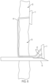
- FIG. 2 schematically shows a side view of an anchor strip according to an illustrative embodiment.
- FIGS. 3 - 3 . 2 schematically show the anchor strip ready for deployment according to an illustrative embodiment.
- FIGS. 5 - 5 . 1 schematically show the anchor strip during deployment according to an illustrative embodiment.
- FIGS. 6 - 6 . 1 schematically show a plurality of anchor strip placements according to an illustrative embodiment.
- FIGS. 7 - 7 . 1 schematically show a cinch anchor according to an illustrative embodiment.
- FIG. 9 is a digital image showing a drive hub according to an illustrative embodiment.
- FIG. 10 is a digital image illustrating a deployment procedure according to an illustrative embodiment.
- FIG. 11 is a digital image showing the anchor strip in a needle during deployment according to an illustrative embodiment.
- FIG. 12 is a digital image showing the drive hub during deployment according to an illustrative embodiment.
- FIG. 13 is a digital image showing the anchor strip deployed from the needle according to an illustrative embodiment.
- FIG. 14 is a digital image showing the drive hub during deployment according to an illustrative embodiment.
- FIG. 15 is a digital image showing the drive hub during deployment according to an illustrative embodiment.
- FIG. 16 schematically shows a top view of a needle according to an illustrative embodiment.
- FIG. 17 schematically shows a perspective view of a needle according to an illustrative embodiment.
- FIG. 18 schematically shows a side view of a needle according to an illustrative embodiment.
- FIG. 19 schematically shows a side view of a deployment system according to an illustrative embodiment.
- FIG. 20 schematically shows a side view of internal mechanism placement of a deployment system according to an illustrative embodiment.
- FIG. 21 schematically shows a top view of internal mechanism placement of a deployment system according to an illustrative embodiment.
- FIG. 22 schematically shows a side view of the drive hub and suture path according to an illustrative embodiment.
- FIG. 23 schematically shows a side view of the drive hub according to an illustrative embodiment.
- FIG. 24 schematically shows a top view of the drive hub according to an illustrative embodiment.
- FIG. 25 schematically shows a side view of an anchor strip according to an illustrative embodiment.
- FIG. 26 schematically shows a side view of an anchor strip according to an illustrative embodiment.
- FIG. 27 schematically shows a side view of an anchor strip according to an illustrative embodiment.
- FIG. 28 schematically shows a side view of a needle according to an illustrative embodiment.
- FIG. 29 schematically shows a plurality of optional anchors and corresponding needles according to illustrative embodiments.
- FIG. 30 schematically shows a plurality of optional anchors according to illustrative embodiments.
- FIG. 31 schematically shows a transmural suturing method according to an illustrative embodiment.
- FIG. 32 schematically shows a transmural suturing method according to an illustrative embodiment.
- FIG. 33 schematically shows the deployment system prepared for deployment according to an illustrative embodiment.
- FIG. 34 schematically shows the deployment system deploying the anchor according to an illustrative embodiment.
- the last anchor segment When the last anchor segment has been reached it can be deployed in the same manner. However, after it is deployed the suture is pulled taut which creates a “purse string” closure of all of the previously deployed anchors. At this time a locking cinch anchor type device can be used to hold the suture and lock it in place.
- the final anchor in the strip anchor could contain a locking cinch type feature anchor ( FIG. 7 ) which locks the suture when pulled taut. The suture can then be cut with another instrument. This final anchor locking cinch is achieved by utilizing the locking “v” in the final proximal anchor segment ( FIG. 7 ). The “v” increases the locking of the suture the more the suture is pulled taut into the v-shaped grooved.
- the anchor strip could be connected to a pusher via a notch to connect or mate with the proximal end of the anchor strip.
- the handle contains a drive hub ( FIG. 4 ) portion which is connected to the proximal end of the pusher, so that when the drive hub is pushed forward the anchor strip is advanced.
- the pusher hub has detents that allow for it to stop at a predetermined location in the handle. These detents mate with a ball plunger assembled in the handle. Further, the distance between the detents is equal to the length of the anchors. This will allow for each anchor to be advanced the set amount until the final anchor is reached. This is also depicted by indicator numbers which show the anchor number before advancing.
- FIGS. 1 - 1 . 4 are a series of diagrams of the anchor strip, showing a total of three anchors connected to each other according the invention.
- FIG. 1 shows an isometric view of the anchor strip 1 with a suture attached 40 .
- a key feature of the invention is the ability to manufacture the anchor strip 1 in a single piece, typically via molding or 3D printing.
- the advantages of the single piece include: cost savings of molding one piece; control and orientation of the anchors; ease of and cost saving for assembly; and simplicity of the design without the need for additional design features to orient and hold the anchors in place.
- FIG. 1 . 1 depicts a side view of anchor strip 1 which includes three anchors 2 , 3 , 4 and a holder 5 all connected by notches 8 .
- Anchor 2 is the initial primary anchor 2 followed by secondary anchors 3 and 4 .
- FIG. 1 . 2 shows a top view of anchor strip 1 with suture holes 6 and suture knot groove 7 with suture hole 16 which is where the knot is placed. It also depicts suture grooves 9 . Knot groove 7 is larger than suture slots 9 to allow for the increased size associated with a knot.
- FIG. 1 . 3 shows a bottom view of anchor strip 1 with a connector end 10 that connects to a pusher 30 via a press or crimped fit.
- the pusher 30 will drive the entire anchor strip 1 through the needle tube and exit into or behind the tissue. It also shows suture grooves 11 .
- FIG. 1 . 4 shows a section view pf the anchor strip 1 and suture 40 threaded through hole 16 with suture knot 41 .
- the suture 40 is threaded or woven through the entire anchor strip 1 weaving in and out of suture holes 6 and suture grooves 9 and 11 .
- Another key feature of the invention is the slots 9 and 11 for the suture 40 are offset from each other to allow for an increased cross section 19 . If suture 40 were placed in a straight line through the anchor strip 1 , the cross section 19 would be approximately one half of the thickness and could fail when the suture 40 is pulled tight after deployment. Failure would be by the suture either pulling or sawing through the cross section.
- FIG. 2 shows a side view of anchor strip 1 loaded into sectioned needle 50 passed through tissue 55 and ready for delivery.
- Yet another key feature of the invention is the control the user has of all the anchors 2 , 3 , and 4 due to rigid connections, notch 8 , between each anchor. This control is necessary for the orientation of each anchor so that it toggles correctly in an upright position, with the suture able to freely move out of the anchor slot 9 . If the orientation was not in the upright position and the suture not able to freely move out of the slot, the anchor would not toggle correctly causing pullout of the device from tissue 55 .
- Another control feature is linear movement inside the needle. If the anchor were not connected to a drive device, it could fall out of the needle and into the body. Additional design features are typically necessary to prevent this fall out including a detent of some sort to the needle.
- FIG. 3 shows anchor strip 1 advanced beyond needle 50 , with anchor 2 protruding past needle tip 56 .
- FIG. 3 . 2 shows anchor strip 1 could be connected to a pusher via a notch 60 to connect or mate with the proximal end of the anchor 4 .
- FIGS. 4 - 4 . 3 are views of the handle, drive hub and ball plunger.
- FIG. 4 shows the handle 80 with drive hub 70 and proximal end of the suture 40 . Also shows detents 71 .
- FIGS. 5 - 5 . 1 are views of anchor 2 being deployed.
- FIG. 5 shows primary anchor 2 separated from the anchor strip 1 and behind tissue 55 .
- the primary anchor 2 will separate via notch 8 and toggle when advanced with pusher 30 followed by pulling the suture 40 .
- Suture 40 allows a connection of the primary anchor 2 and the now shortened anchor strip which now consists of two anchors (anchors 3 , 4 ).
- Suture 40 is fixed to primary anchor 2 by knot 41 and cannot slide.
- Yet another key feature of the invention is the breakaway tab 8 which allows controlled separation of the anchor. This controlled separation when pulling of the suture, is key to flipping or toggling the anchor. Current toggling-type anchors do not consistently flip especially in mid-substance type environments resulting in pullout of the anchor device.
- FIG. 5 . 1 shows primary anchor 2 toggled and behind tissue 55 . This is achieved by removing the needle 50 from the tissue 55 and pulling on the suture 40 which locks behind tissue 55 .
- FIGS. 6 - 6 . 1 are views of multiple anchor placements.
- FIG. 6 shows anchor 2 delivered, toggled, and locked behind tissue 55 with anchor 3 having been delivered, toggled, and ready to be locked. After needle 50 is removed and when anchor 3 is locked behind tissue 55 , suture 40 can be pulled taut being free to slide within anchor 3 which allows for tightening between anchors.
- FIG. 6 . 1 shows anchors 2 , 3 , and 4 deployed behind tissue 55 .
- the free leg of suture 40 can be pulled taut with suture 40 being free to slide within anchors 3 and 4 .
- a cinch anchor similar to the Apollo® type cinch (Apollo Endo-surgery of Austin, Texas, USA), it can be applied to suture 40 in the area of 42 .
- FIG. 7 shows the cinch anchor 4 a which would be in place of anchor 4 .
- V-Groove 90 is shown for the suture (not shown) to wedge into.
- This cinch anchor would be in place of the known Apollo® type device and placed on the distal side of tissue 55 as shown in the anchor 4 placement.
- the suture 40 would then be cut in area 42 with a suture cutting instrument.
- FIG. 7 . 1 shows cinch anchor 4 a which would be in place of anchor 4 .
- V-Grooves 90 and 91 are shown for the suture (not shown) to wedge into.
- the currently available forward viewing linear array echoendoscope is inserted into the patient as per routine and known methods. A location is chosen for the first full thickness anchor deployment. Endoscopic ultrasound is used to identify whether there are any major vessels or adjacent structures, and if so, the scope can be adjusted accordingly.
- EUS allows one to observe full thickness penetration with each pass avoiding vessels and other organs, while linear deployment allows for increased flexibility and maneuverability.
- anchors are in fact T-Fasteners or similar which provide a more robust anchor on a point to point basis.
- Options for suture material are no longer limited by the device allowing for more robust applications.
- EUS guided approach also allows us to perform transmural apposition which has never been previously performed.
- the needle 50 is loaded through the working channel of the echoendoscope and is locked into place at the biopsy valve with a Leuer lock mechanism as is standard for all EUS needles.
- the needle will be pre-loaded with the primary tissue anchor which will be positioned behind the retaining wedge of the needle tip.
- the suture is running through the center of the needle and exits at the back end of the handle.
- an open design needle stylet is positioned behind the tissue anchor and alongside the suture such that pushing it into the down position would effectively push the anchor out of the needle.
- FIG. 8 is a digital image (photograph) showing the anchor 4 protruding from the needle 50 .
- the anchor 4 is loaded with a suture 40 .
- the anchor 4 includes v-grooves 90 in which the suture 40 is mounted.
- FIG. 9 is a digital image of the drive hub 70 and indicators 73 .
- the needle 50 is protruding from the handle 80 .
- FIGS. 10 - 15 are digital images illustrating a deployment procedure.
- FIG. 10 shows the anchor strip 1 mounted in the needle 50 ready for insertion into a subject.
- FIG. 11 shows the anchor 2 separating from the anchor strip 1 . The orientation of the anchor 2 will secure the anchor 2 within the flesh of the subject.
- FIG. 12 illustrates the action employed to the drive hub 70 to perform the suturing.
- the handle 80 of the apparatus is pulled away from the subject, extracting the needle 50 from the subject's flesh. The force of the pull is necessarily insufficient to extract the anchor 2 from the subject.
- FIG. 13 demonstrates the anchor 2 separating from the needle 50 and anchor strip 1 with the suture 40 remaining attached. In an actual deployment, the anchor 2 remains within the flesh of the subject anchoring the suture 40 .
- the apparatus is then moved to the next anchor location and the needle 50 is inserted into the flesh at the next anchor position and the drive hub 70 is advanced to the next indicator 73 .
- FIG. 15 indicates that the deployment procedure is repeated as necessary to complete the
- a secondary anchor (can also be preloaded) is loaded by pulling out the stylet, threading the anchor over the suture and using the stylet to push the anchor to the needle tip. The anchor automatically stops at the retaining wedge and this allows the scope and suture to move independently of the needle and anchor.
- a second site of anchor insertion is chosen and the above steps are repeated.
- the secondary anchor is deployed transmurally. This is repeated until a sufficient number of anchors are deployed transmurally to provide adequate apposition of tissue upon cinching. Cinching is performed by removing the needle form the endoscope channel and running a cinch over the suture. Alternately, a removeable inner needle design would allow the internal stylet to be removed while maintain the needle sheathe and handle in position. The cinch is pushed against the tissue and the suture is pulled against the cinch bring all tissue anchors together. The suture and anchors move independently from one another until they are tightly cinched. The procedure is repeated with a new primary anchor, followed by secondary tissue anchors and a second cinch to perform a second running transmural application.
- the primary anchor is the anchor that is used for the first transmural pass.
- the primary anchor is unique in that it is fixed to the suture.
- the suture is free to pass through the anchor as the user cinches the stitch.
- the primary anchor is crucial for pulling together all the intervening tissue.
- the anchors and/or anchor strip can be a simple bar design, wherein each anchor hinges once it's transmural to act as a T Tag. Alternately a more complex design can be conceived of shaped nitinol such that once the anchor is pushed out of the needle the back half forms a Y shape.
- the suture is coupled with the anchor, unlike in the secondary anchors where the anchor runs freely over the suture. The suture attaches to the anchor in the midpoint so that once it is cinched there is equal distribution of pressure.
- Secondary anchors are unique and allow for this device to deploy multiple such anchors thereby apposing multiple points of tissue. They are made of the same material and dimensions as the primary anchor but have been shaped so that the suture runs through its center at the mid-point. This allows the anchor to hinge, and once it's pushed through the wall of the tissue the anchor does not allow the suture to pull through.
- a simple design can be conceived as in A, however more elaborate designs are also possible.
- One such design is a tightly wound coil that is pushed through the needle on deployment. As the needle and suture pulls back, owing to the length of the coil, it will not be able to pull out, but rather “bunch up” forming a knot. This particular design may be easier to deploy when the scope and needle position provide increased resistance due to bends and tortuosity.
- FIG. 16 schematically shows the endoscopic needle 50 .
- the needle tip 56 is a tapered shape configured to house the anchor strip 1 before deployment.
- FIG. 17 schematically shows a perspective view of the needle 50 detailing the tapered tip 56 .
- the taper facilitates insertion into the flesh of a subject.
- FIG. 18 schematically shows a side view of the needle 50 , further illustrating the tapered tip 56 .
- FIGS. 19 and 20 show a side hidden view assembly of an optional apparatus structure.
- FIGS. 19 and 20 schematically show the drive hub 70 engaged within the handle 80 .
- the handle 80 includes a channel to guide the needle 50 .
- FIG. 21 shows a top hidden view assembly of the optional apparatus structure, including the drive hub 70 engaged within the handle 80 , and the channel to guide the needle 50 .
- FIG. 22 schematically shows a side hidden view of the drive hub 70 .
- FIG. 22 details the channel for the suture 40 , detents, indicators 73 , and ball plunger 72 .
- the arrow indicates the direction that the ball plunger 72 is depressed to deploy the suture 40 .
- FIG. 23 shows a side view of the drive hub 70
- FIG. 24 schematically shows a top view of the drive hub 70 .
- FIG. 25 schematically shows a side view of anchor strip 1 which includes three anchors 2 , 3 , 4 and a holder 5 all connected by notches 8 .
- Anchor 2 is the initial primary anchor 2 followed by secondary anchors 3 and 4 .
- FIG. 26 schematically shows a top view of anchor strip 1 with suture holes 6 and suture knot groove 7 with suture hole 16 which is where the knot placement is located on top of. It also depicts suture grooves 9 . Knot groove 7 is larger than suture slots 9 to allow for the increased size associated with a knot.
- FIG. 27 schematically shows a side silhouette view of the anchor strip 1 .
- FIG. 28 schematically shows the needle 50 having an optional angular taper design.
- the needle 50 is pushed into ultrasound view as with a standard fine needle aspiration (FNA).
- FNA fine needle aspiration
- the needle 50 is carefully pushed under ultrasound guidance through the wall of the tissue and once we are ensured on ultrasound that the needle 50 is full thickness the anchor 1 is deployed as per FIG. 14 .
- the needle is pushed past the wall to ensure a transmural (trans/serosal) penetration, pulled back just to the edge of the tissue wall and deployed as the user simultaneously pulls the needle back.
- This technique may involve inserting the needle to adequately ensure the serosa is transversed and then pulling the needle back to an edge of the tissue. At that point, the anchor is pushed out as the needle is pulled back, which allows for an “in place” deployment when there is limited separation or space between luminal peritoneal structures.
- FIGS. 29 and 30 schematically show optional anchor and needle tip configurations.
- the needle tip can be tailored to correspond to the desired anchor configuration.
- Optional anchor configurations can include expanding anchors, looped anchors, through-body anchors, helical anchors, Y-shaped anchors, or the like.
- Anchor material can be varied according to application. Primary and secondary anchors also need not be of the same material.
- Transmural anchors will need to be made of a material that is accepted by the human body (i.e. stainless steel, nitinol, etc.). It can also be made of a bio-absorbable material. In most applications sutures are not meant to keep tissue apposed for many months. Instead scar formation and body healing take over making the suture less important as time goes on.
- Polygycolic acid (PGA) can be used to form the anchors the suture line. This means that the anchors, even though transmural, would effectively be absorbed many months out so they are not of any concern in the long term.
- FIGS. 31 and 32 schematically show a suturing procedure as described above.
- FIG. 31 illustrates insertion of the needle 50 at step ( 1 ) and primary anchor 2 deployment, the trailing suture 40 , and a second needle 50 insertion at step ( 2 ).
- FIG. 32 illustrates a complete suturing procedure wherein four anchors 1 are deployed and linked by the suture 40 .
- the suture 40 can be cinched by pulling the suture 40 . Cinching the suture 40 closes the opening in the flesh.
- FIGS. 33 and 34 schematically illustrate the needle anchor retention mechanism that enables the needle 50 to be loaded with the anchor strip 1 and suture 40 .
- the anchor strip 1 is positioned behind the retention wedge and held in place by the wedge.
- the drive hub (not shown) includes a stylet configured to push the anchor strip past the retention wedge and begin the anchor 1 deployment.
- the needle 50 and stylet are hollow to allow passage of the suture 40 .
- the needle anchor retention mechanism allows for the anchor to sit reliably just before the tip of the needle. Without it, inadvertent premature deployment or accidentally being pushed out with simple maneuvers of the scope.
- the wedge will yield to pressure from the pushing stylet as the anchor is being delivered.
- the suture line is able to run down the center of the needle and through each anchor that is being deployed. This is the key to deploying multiple anchors in complex patterns.
- the pushing stylet allows the suture to run down its center. anchors are loaded and delivered to the distal tip of the needle with the pushing stylet. The suture line runs through the entire needle and anchor. Other variations of the needle can be conceived that allow for multiple secondary anchors to come pre-loaded with the needle. Once again in this situation, the wedge allows for single anchor deployment, while a position is chosen for the next anchor placement.
- the present disclosure provides solutions for any T tags or similar anchors being located on a serosal side of an organ and either eroding or migrating into or adjacent other organs, such as, for example, into the mediastinum. Since the T tags or anchors are linearly deployed and are under tension during deployment, such do not create a crossing suture effect. In fact, any erosion or migration will be inward assuming there is tension on the sutures when applied.
- Anchors ride independently on the suture line and are then cinched at the end; every fastener acts as a transmural anchor while the cinch rests on the luminal side.
- An anchor retention wedge allows suture and needle to move freely until anchor is ready to be deployed through tissue-Multiple anchors can thus be deployed in succession over the same suture.
Landscapes
- Health & Medical Sciences (AREA)
- Life Sciences & Earth Sciences (AREA)
- Surgery (AREA)
- Heart & Thoracic Surgery (AREA)
- Engineering & Computer Science (AREA)
- Biomedical Technology (AREA)
- Nuclear Medicine, Radiotherapy & Molecular Imaging (AREA)
- Medical Informatics (AREA)
- Molecular Biology (AREA)
- Animal Behavior & Ethology (AREA)
- General Health & Medical Sciences (AREA)
- Public Health (AREA)
- Veterinary Medicine (AREA)
- Rheumatology (AREA)
- Surgical Instruments (AREA)
Abstract
Description
Claims (17)
Priority Applications (3)
| Application Number | Priority Date | Filing Date | Title |
|---|---|---|---|
| US17/592,776 US12178424B2 (en) | 2021-02-04 | 2022-02-04 | Suturing breakaway anchor strip |
| US18/121,949 US20230210558A1 (en) | 2021-02-04 | 2023-03-15 | Suturing breakaway anchor strip |
| US18/643,191 US20240350133A1 (en) | 2021-02-04 | 2024-04-23 | Suturing breakaway anchor strip |
Applications Claiming Priority (2)
| Application Number | Priority Date | Filing Date | Title |
|---|---|---|---|
| US202163145708P | 2021-02-04 | 2021-02-04 | |
| US17/592,776 US12178424B2 (en) | 2021-02-04 | 2022-02-04 | Suturing breakaway anchor strip |
Related Child Applications (2)
| Application Number | Title | Priority Date | Filing Date |
|---|---|---|---|
| US18/121,949 Continuation-In-Part US20230210558A1 (en) | 2021-02-04 | 2023-03-15 | Suturing breakaway anchor strip |
| US18/643,191 Continuation US20240350133A1 (en) | 2021-02-04 | 2024-04-23 | Suturing breakaway anchor strip |
Publications (2)
| Publication Number | Publication Date |
|---|---|
| US20220249084A1 US20220249084A1 (en) | 2022-08-11 |
| US12178424B2 true US12178424B2 (en) | 2024-12-31 |
Family
ID=82704298
Family Applications (2)
| Application Number | Title | Priority Date | Filing Date |
|---|---|---|---|
| US17/592,776 Active US12178424B2 (en) | 2021-02-04 | 2022-02-04 | Suturing breakaway anchor strip |
| US18/643,191 Pending US20240350133A1 (en) | 2021-02-04 | 2024-04-23 | Suturing breakaway anchor strip |
Family Applications After (1)
| Application Number | Title | Priority Date | Filing Date |
|---|---|---|---|
| US18/643,191 Pending US20240350133A1 (en) | 2021-02-04 | 2024-04-23 | Suturing breakaway anchor strip |
Country Status (1)
| Country | Link |
|---|---|
| US (2) | US12178424B2 (en) |
Families Citing this family (1)
| Publication number | Priority date | Publication date | Assignee | Title |
|---|---|---|---|---|
| WO2024191857A2 (en) * | 2023-03-15 | 2024-09-19 | Aurora Medical Technologies Corp | Suturing breakaway anchor strip |
Citations (12)
| Publication number | Priority date | Publication date | Assignee | Title |
|---|---|---|---|---|
| US20010008971A1 (en) | 1998-12-30 | 2001-07-19 | Schwartz Herbert E. | Suture locking device |
| US20050251189A1 (en) | 2004-05-07 | 2005-11-10 | Usgi Medical Inc. | Multi-position tissue manipulation assembly |
| US20060030884A1 (en) | 2002-03-14 | 2006-02-09 | Yeung Jeffrey E | Suture anchor and approximating device |
| US20080015566A1 (en) * | 2006-07-13 | 2008-01-17 | Steve Livneh | Surgical sealing and cutting apparatus |
| US20080086172A1 (en) | 2006-10-05 | 2008-04-10 | Martin David T | Suture anchor |
| US20080243148A1 (en) | 2005-09-28 | 2008-10-02 | Olympus Medical Systems Corp. | Suture instrument |
| US20140031835A1 (en) * | 2012-07-24 | 2014-01-30 | Ams Research Corporation | Systems, tools, and methods for connecting to tissue |
| US20150250470A1 (en) * | 2014-03-05 | 2015-09-10 | Coaxis Medical, Inc. | Suture anchoring system and delivery method |
| US20160324615A1 (en) * | 2011-07-22 | 2016-11-10 | Astora Women's Health, Llc | Pelvic implant system and method |
| US20180035997A1 (en) * | 2016-08-05 | 2018-02-08 | Boston Scientific Scimed, Inc. | Systems, devices, and related methods for retracting tissue |
| US10390922B2 (en) | 2015-01-05 | 2019-08-27 | Boston Scientific Scimed, Inc. | Devices and methods for delivery of attachment devices |
| US20230210558A1 (en) | 2021-02-04 | 2023-07-06 | Aurora Medical Technologies Corp | Suturing breakaway anchor strip |
-
2022
- 2022-02-04 US US17/592,776 patent/US12178424B2/en active Active
-
2024
- 2024-04-23 US US18/643,191 patent/US20240350133A1/en active Pending
Patent Citations (12)
| Publication number | Priority date | Publication date | Assignee | Title |
|---|---|---|---|---|
| US20010008971A1 (en) | 1998-12-30 | 2001-07-19 | Schwartz Herbert E. | Suture locking device |
| US20060030884A1 (en) | 2002-03-14 | 2006-02-09 | Yeung Jeffrey E | Suture anchor and approximating device |
| US20050251189A1 (en) | 2004-05-07 | 2005-11-10 | Usgi Medical Inc. | Multi-position tissue manipulation assembly |
| US20080243148A1 (en) | 2005-09-28 | 2008-10-02 | Olympus Medical Systems Corp. | Suture instrument |
| US20080015566A1 (en) * | 2006-07-13 | 2008-01-17 | Steve Livneh | Surgical sealing and cutting apparatus |
| US20080086172A1 (en) | 2006-10-05 | 2008-04-10 | Martin David T | Suture anchor |
| US20160324615A1 (en) * | 2011-07-22 | 2016-11-10 | Astora Women's Health, Llc | Pelvic implant system and method |
| US20140031835A1 (en) * | 2012-07-24 | 2014-01-30 | Ams Research Corporation | Systems, tools, and methods for connecting to tissue |
| US20150250470A1 (en) * | 2014-03-05 | 2015-09-10 | Coaxis Medical, Inc. | Suture anchoring system and delivery method |
| US10390922B2 (en) | 2015-01-05 | 2019-08-27 | Boston Scientific Scimed, Inc. | Devices and methods for delivery of attachment devices |
| US20180035997A1 (en) * | 2016-08-05 | 2018-02-08 | Boston Scientific Scimed, Inc. | Systems, devices, and related methods for retracting tissue |
| US20230210558A1 (en) | 2021-02-04 | 2023-07-06 | Aurora Medical Technologies Corp | Suturing breakaway anchor strip |
Non-Patent Citations (1)
| Title |
|---|
| Non-Final Office Action dated Jun. 12, 2019 for U.S. Appl. No. 14/639,669 (13 pages). |
Also Published As
| Publication number | Publication date |
|---|---|
| US20220249084A1 (en) | 2022-08-11 |
| US20240350133A1 (en) | 2024-10-24 |
Similar Documents
| Publication | Publication Date | Title |
|---|---|---|
| US9339265B2 (en) | Medical devices, systems, and methods for using tissue anchors | |
| US8382776B2 (en) | Medical devices, systems and methods for rapid deployment and fixation of tissue anchors | |
| US10492777B2 (en) | Instruments for delivering transfascial sutures, transfascial suture assemblies, and methods of transfascial suturing | |
| JP5554339B2 (en) | Method and apparatus for applying multiple suture anchors | |
| US7815659B2 (en) | Suture anchor applicator | |
| US9301756B2 (en) | Surgical coils and methods of deploying | |
| JP5301544B2 (en) | Organ staple for closure of perforated purse string suture | |
| US20230210558A1 (en) | Suturing breakaway anchor strip | |
| US20050216036A1 (en) | Endoscopic fastening system with multiple fasteners | |
| US20150250470A1 (en) | Suture anchoring system and delivery method | |
| EP3154446B1 (en) | Apparatus for suturing a tissue | |
| US20240350133A1 (en) | Suturing breakaway anchor strip | |
| US11918205B2 (en) | Self locking suture and self locking suture mediated closure device | |
| WO2024191857A2 (en) | Suturing breakaway anchor strip | |
| US20240215981A1 (en) | Magnetic anastomosis devices with varying magnetic force at a distance |
Legal Events
| Date | Code | Title | Description |
|---|---|---|---|
| FEPP | Fee payment procedure |
Free format text: ENTITY STATUS SET TO UNDISCOUNTED (ORIGINAL EVENT CODE: BIG.); ENTITY STATUS OF PATENT OWNER: SMALL ENTITY |
|
| FEPP | Fee payment procedure |
Free format text: ENTITY STATUS SET TO SMALL (ORIGINAL EVENT CODE: SMAL); ENTITY STATUS OF PATENT OWNER: SMALL ENTITY |
|
| AS | Assignment |
Owner name: AURORA MEDICAL TECHNOLOGIES CORP, NEW JERSEY Free format text: ASSIGNMENT OF ASSIGNORS INTEREST;ASSIGNOR:DOWD, SCOTT;REEL/FRAME:059277/0714 Effective date: 20220315 |
|
| STPP | Information on status: patent application and granting procedure in general |
Free format text: DOCKETED NEW CASE - READY FOR EXAMINATION |
|
| STPP | Information on status: patent application and granting procedure in general |
Free format text: NON FINAL ACTION MAILED |
|
| STPP | Information on status: patent application and granting procedure in general |
Free format text: RESPONSE TO NON-FINAL OFFICE ACTION ENTERED AND FORWARDED TO EXAMINER |
|
| STPP | Information on status: patent application and granting procedure in general |
Free format text: FINAL REJECTION MAILED |
|
| STPP | Information on status: patent application and granting procedure in general |
Free format text: DOCKETED NEW CASE - READY FOR EXAMINATION |
|
| STPP | Information on status: patent application and granting procedure in general |
Free format text: NOTICE OF ALLOWANCE MAILED -- APPLICATION RECEIVED IN OFFICE OF PUBLICATIONS |
|
| STPP | Information on status: patent application and granting procedure in general |
Free format text: PUBLICATIONS -- ISSUE FEE PAYMENT VERIFIED |
|
| STCF | Information on status: patent grant |
Free format text: PATENTED CASE |
|
| STPP | Information on status: patent application and granting procedure in general |
Free format text: WITHDRAW FROM ISSUE AWAITING ACTION |
|
| STPP | Information on status: patent application and granting procedure in general |
Free format text: DOCKETED NEW CASE - READY FOR EXAMINATION |
|
| STPP | Information on status: patent application and granting procedure in general |
Free format text: NOTICE OF ALLOWANCE MAILED -- APPLICATION RECEIVED IN OFFICE OF PUBLICATIONS |
|
| STCB | Information on status: application discontinuation |
Free format text: ABANDONED -- FAILURE TO PAY ISSUE FEE |
|
| AS | Assignment |
Owner name: AURORA MEDICAL TECHNOLOGIES CORP., MINNESOTA Free format text: ASSIGNMENT OF ASSIGNORS INTEREST;ASSIGNOR:BENIAS, PETROS;REEL/FRAME:069268/0484 Effective date: 20241114 Owner name: AURORA MEDICAL TECHNOLOGIES CORP., MINNESOTA Free format text: ASSIGNMENT OF ASSIGNORS INTEREST;ASSIGNOR:KUMBHARI, VIVEK;REEL/FRAME:069268/0448 Effective date: 20241114 |
|
| STPP | Information on status: patent application and granting procedure in general |
Free format text: NOTICE OF ALLOWANCE MAILED -- APPLICATION RECEIVED IN OFFICE OF PUBLICATIONS |
|
| STPP | Information on status: patent application and granting procedure in general |
Free format text: PUBLICATIONS -- ISSUE FEE PAYMENT VERIFIED |
|
| STCF | Information on status: patent grant |
Free format text: PATENTED CASE |