US12023352B2 - Anti-inflammatory/anti-catabolic and regenerative agents from autologous physiological fluid - Google Patents
Anti-inflammatory/anti-catabolic and regenerative agents from autologous physiological fluid Download PDFInfo
- Publication number
- US12023352B2 US12023352B2 US16/741,627 US202016741627A US12023352B2 US 12023352 B2 US12023352 B2 US 12023352B2 US 202016741627 A US202016741627 A US 202016741627A US 12023352 B2 US12023352 B2 US 12023352B2
- Authority
- US
- United States
- Prior art keywords
- blood
- component
- chronic
- autologous composition
- inflammatory
- Prior art date
- Legal status (The legal status is an assumption and is not a legal conclusion. Google has not performed a legal analysis and makes no representation as to the accuracy of the status listed.)
- Active, expires
Links
Images


Classifications
-
- A—HUMAN NECESSITIES
- A61—MEDICAL OR VETERINARY SCIENCE; HYGIENE
- A61K—PREPARATIONS FOR MEDICAL, DENTAL OR TOILETRY PURPOSES
- A61K35/00—Medicinal preparations containing materials or reaction products thereof with undetermined constitution
- A61K35/12—Materials from mammals; Compositions comprising non-specified tissues or cells; Compositions comprising non-embryonic stem cells; Genetically modified cells
- A61K35/14—Blood; Artificial blood
- A61K35/16—Blood plasma; Blood serum
-
- A—HUMAN NECESSITIES
- A61—MEDICAL OR VETERINARY SCIENCE; HYGIENE
- A61K—PREPARATIONS FOR MEDICAL, DENTAL OR TOILETRY PURPOSES
- A61K38/00—Medicinal preparations containing peptides
- A61K38/16—Peptides having more than 20 amino acids; Gastrins; Somatostatins; Melanotropins; Derivatives thereof
- A61K38/17—Peptides having more than 20 amino acids; Gastrins; Somatostatins; Melanotropins; Derivatives thereof from animals; from humans
- A61K38/18—Growth factors; Growth regulators
- A61K38/1841—Transforming growth factor [TGF]
-
- A—HUMAN NECESSITIES
- A61—MEDICAL OR VETERINARY SCIENCE; HYGIENE
- A61K—PREPARATIONS FOR MEDICAL, DENTAL OR TOILETRY PURPOSES
- A61K38/00—Medicinal preparations containing peptides
- A61K38/16—Peptides having more than 20 amino acids; Gastrins; Somatostatins; Melanotropins; Derivatives thereof
- A61K38/17—Peptides having more than 20 amino acids; Gastrins; Somatostatins; Melanotropins; Derivatives thereof from animals; from humans
- A61K38/18—Growth factors; Growth regulators
- A61K38/1858—Platelet-derived growth factor [PDGF]
- A61K38/1866—Vascular endothelial growth factor [VEGF]
-
- A—HUMAN NECESSITIES
- A61—MEDICAL OR VETERINARY SCIENCE; HYGIENE
- A61K—PREPARATIONS FOR MEDICAL, DENTAL OR TOILETRY PURPOSES
- A61K38/00—Medicinal preparations containing peptides
- A61K38/16—Peptides having more than 20 amino acids; Gastrins; Somatostatins; Melanotropins; Derivatives thereof
- A61K38/17—Peptides having more than 20 amino acids; Gastrins; Somatostatins; Melanotropins; Derivatives thereof from animals; from humans
- A61K38/19—Cytokines; Lymphokines; Interferons
- A61K38/20—Interleukins [IL]
- A61K38/2006—IL-1
-
- A—HUMAN NECESSITIES
- A61—MEDICAL OR VETERINARY SCIENCE; HYGIENE
- A61P—SPECIFIC THERAPEUTIC ACTIVITY OF CHEMICAL COMPOUNDS OR MEDICINAL PREPARATIONS
- A61P17/00—Drugs for dermatological disorders
-
- A—HUMAN NECESSITIES
- A61—MEDICAL OR VETERINARY SCIENCE; HYGIENE
- A61P—SPECIFIC THERAPEUTIC ACTIVITY OF CHEMICAL COMPOUNDS OR MEDICINAL PREPARATIONS
- A61P19/00—Drugs for skeletal disorders
-
- A—HUMAN NECESSITIES
- A61—MEDICAL OR VETERINARY SCIENCE; HYGIENE
- A61P—SPECIFIC THERAPEUTIC ACTIVITY OF CHEMICAL COMPOUNDS OR MEDICINAL PREPARATIONS
- A61P19/00—Drugs for skeletal disorders
- A61P19/02—Drugs for skeletal disorders for joint disorders, e.g. arthritis, arthrosis
-
- A—HUMAN NECESSITIES
- A61—MEDICAL OR VETERINARY SCIENCE; HYGIENE
- A61P—SPECIFIC THERAPEUTIC ACTIVITY OF CHEMICAL COMPOUNDS OR MEDICINAL PREPARATIONS
- A61P19/00—Drugs for skeletal disorders
- A61P19/04—Drugs for skeletal disorders for non-specific disorders of the connective tissue
-
- A—HUMAN NECESSITIES
- A61—MEDICAL OR VETERINARY SCIENCE; HYGIENE
- A61P—SPECIFIC THERAPEUTIC ACTIVITY OF CHEMICAL COMPOUNDS OR MEDICINAL PREPARATIONS
- A61P21/00—Drugs for disorders of the muscular or neuromuscular system
-
- A—HUMAN NECESSITIES
- A61—MEDICAL OR VETERINARY SCIENCE; HYGIENE
- A61P—SPECIFIC THERAPEUTIC ACTIVITY OF CHEMICAL COMPOUNDS OR MEDICINAL PREPARATIONS
- A61P29/00—Non-central analgesic, antipyretic or antiinflammatory agents, e.g. antirheumatic agents; Non-steroidal antiinflammatory drugs [NSAID]
-
- A—HUMAN NECESSITIES
- A61—MEDICAL OR VETERINARY SCIENCE; HYGIENE
- A61P—SPECIFIC THERAPEUTIC ACTIVITY OF CHEMICAL COMPOUNDS OR MEDICINAL PREPARATIONS
- A61P43/00—Drugs for specific purposes, not provided for in groups A61P1/00-A61P41/00
-
- C—CHEMISTRY; METALLURGY
- C07—ORGANIC CHEMISTRY
- C07K—PEPTIDES
- C07K1/00—General methods for the preparation of peptides, i.e. processes for the organic chemical preparation of peptides or proteins of any length
- C07K1/14—Extraction; Separation; Purification
-
- C—CHEMISTRY; METALLURGY
- C12—BIOCHEMISTRY; BEER; SPIRITS; WINE; VINEGAR; MICROBIOLOGY; ENZYMOLOGY; MUTATION OR GENETIC ENGINEERING
- C12N—MICROORGANISMS OR ENZYMES; COMPOSITIONS THEREOF; PROPAGATING, PRESERVING, OR MAINTAINING MICROORGANISMS; MUTATION OR GENETIC ENGINEERING; CULTURE MEDIA
- C12N9/00—Enzymes; Proenzymes; Compositions thereof; Processes for preparing, activating, inhibiting, separating or purifying enzymes
- C12N9/14—Hydrolases (3)
- C12N9/48—Hydrolases (3) acting on peptide bonds (3.4)
- C12N9/50—Proteinases, e.g. Endopeptidases (3.4.21-3.4.25)
- C12N9/64—Proteinases, e.g. Endopeptidases (3.4.21-3.4.25) derived from animal tissue
-
- A—HUMAN NECESSITIES
- A61—MEDICAL OR VETERINARY SCIENCE; HYGIENE
- A61K—PREPARATIONS FOR MEDICAL, DENTAL OR TOILETRY PURPOSES
- A61K2300/00—Mixtures or combinations of active ingredients, wherein at least one active ingredient is fully defined in groups A61K31/00 - A61K41/00
Definitions
- a bioactive composition useful for treating damaged and/or injured connective tissues, chronic tendinosis, chronic muscle tears and/or chronic degenerative joint conditions such as osteoarthritis and skin inflammatory disorders. Also described is a method for making the composition.
- the composition includes an anti-inflammatory component/anti-catabolic component.
- the anti-inflammatory component is also an anti-catabolic component.
- the terms “anti-inflammatory component” and “anti-catabolic component” can be used interchangeably.
- the composition may also include a regenerative component that includes autologous platelet-rich plasma (PRP). Whereas most PRP preparation protocols include an activation step resulting in the immediate release of growth factors and cytokines from platelets, the present disclosure provides a use of a non-activated PRP component for future slow activation of injected composition by surrounding tissue.
- PRP autologous platelet-rich plasma
- the instant disclosure provides an alternative product for treating degenerative joint disease in humans and for veterinary applications including horses, dogs and camels that is relatively safe effective, stable, regenerative, and cost effective.
- FIG. 5 is a graph showing an average of concentration level of TIMP 1, TIMP 2 and TIMP 4 before and after a 24-hour incubation.
- FIG. 6 is a graph showing an average baseline of knee joint pain according to the Visual Analog Scale (“VAS”) scale in the eight patients prior to and after injections.
- VAS Visual Analog Scale
- FIG. 7 is a plot showing point values according to the WOMAC index for average levels of knee pain, stiffness and daily activity capabilities among the eight patients.
- FIG. 8 shows results in the form of a graph providing a breakdown of the average results after the first, second and third injections and two- and three-month follow-ups.
- FIG. 9 is graph showing a comparison between the average concentration level of platelet-derived growth factor (“PDGF”) in the human serum samples before and after a 24-hour incubation.
- PDGF platelet-derived growth factor
- FIG. 10 is a graph showing a comparison of the level of MMP2, MMP3, MMP7, MMP9 and MMP13 proteins in human serum samples before (baseline level) and after incubation at 37° C. for 24 hours.
- FIG. 11 is a graph showing a comparison of the level of MMP2, MMP3, MMP7, MMP9 and MMP13 proteins in human serum samples before (baseline level) and after incubation at 37° C. for 24 hours in the presence of sodium citrate.
- FIG. 12 is a graph showing the MMP9 data from FIG. 11 .
- FIG. 14 is a graph showing a comparison of the level of TNF- ⁇ in human serum samples before (baseline level) and after incubation at 37° C. for 24 hours, activated PRP, and in the final composition.
- FIG. 15 is a graph showing a comparison of the level of TIMP2 in the human serum samples before (baseline level) and after incubation at 37° C. for 24 hours, activated PRP, and in the final composition.
- FIG. 16 is a graph showing a comparison of the level of IL-1ra in human serum samples before (baseline level) and after incubation at 37° C. for 24 hours, activated PRP, and in the final product.
- FIG. 17 is a graph showing a comparison of the level of PDGF in the human serum samples before (baseline level) and after incubation in 37° C. for 24 hours, activated PRP, and in the final composition.
- FIG. 18 A is a plot showing point values according to the WOMAC index for average levels of pain among seventeen patients tested. Values are provided for a baseline and 1 month post injections for CA ⁇ and CA+ groups.
- FIG. 18 B is a plot showing point values according to the WOMAC index for average levels of stiffness among the seventeen patients tested. Values are provided for a baseline and 1 month post injections for CA ⁇ and CA+ groups.
- FIG. 18 C is a plot showing point values according to the WOMAC index for average levels of daily activity capabilities among the seventeen patients tested. Values are provided for a baseline and 1 month post injections for CA ⁇ and CA+ groups.
- FIG. 18 D is a plot showing a statistical analysis of Visual Analog Pain Scale (VAS) among the seventeen patients tested. Values are provided for a baseline and 1 month post injections for CA ⁇ and CA+ groups.
- VAS Visual Analog Pain Scale
- the disclosure relates to a composition
- a composition comprising an autologous anti-inflammatory/anti-catabolic component and preferably an autologous platelet-rich plasma component with serum enriched by bioactive proteins having a synergistic anti-inflammatory/anti-catabolic, proliferative, tissue remodeling and regenerative effects.
- Such a composition typically includes the following therapeutically active proteins: IL-1ra 6 , IL-4 7 , IL-10 8,9 , IL- ⁇ 10 , PDGF 11 , TGF- ⁇ 10,11 and VEGF 12,13,14,16 .
- IL-1ra is secreted by monocytes, adipocytes and epithelial cells. Therapeutically effective concentrations of this protein are achieved by incubating human monocytes at about 37° C. for about 24 hours.
- 17 IL-4,10,13, PDGF, TGF- ⁇ are the content of platelets and-granules and are delivered in the PRP component.
- IL-4,10,13 come from white blood cells.
- PDGF is produced by platelets and TGF-B is released by platelets and some T cells.
- Employing the synergistic effect of the mentioned proteins leads to generation of a potent bio-active autologous product.
- a combination of fresh-prepared PRP as a source of regenerative biological factors and anti-inflammatory cytokines and growth factors, and the anti-inflammatory component comprising incubated autologous serum as a source of IL-1 inhibitor provides a powerful and cost-effective autologous therapeutic agent for treatment of degenerative conditions like osteoarthritis, chronic tendinosis and chronic muscle tears as well as skin inflammatory disorders.
- treatment includes palliative treatment, wherein pain and/or inflammation is reduced in the subject.
- TIMPs tissue inhibitors of metalloproteinases
- the addition of sodium citrate prior to incubating a patient's blood at a temperature of from about 37° C. to about 39° C. for about 24 hours prevents an increased level of pathological molecular agents that have a catabolic effect on joints such as MMP9, IL-1 ⁇ and TNF- ⁇ , but yet does not lead to a significantly decreased level of anti-catabolic and regenerative agents such as TIMPs, IL-1ra and PDGF.
- the described method for producing an autologous composition for the treatment of osteoarthritis, chronic tendinosis and chronic muscle tear as well as skin inflammatory disorders preferably comprises the step of collecting a mammal's autologous physiological fluid, preferably blood by an aseptic technique.
- a mammal is a human.
- the compositions and methods hereof are also suitable for a wide range of veterinary applications, for example, for the treatment of horses, dogs and camels.
- the site of venipuncture and the surface of the collection tubes may be cleaned with a 2 percent tincture of iodine solution. Before any cleansing of the site is begun, the patient may be asked about any allergy to iodine.
- the tube covers are also cleaned with 70% alcohol solution to avoid possible contamination before blood collection.
- the incubation can be in sterile glass tubes (Coviden) or polystyrene (BD) vacutainer tubes with no additives. Further provided in an embodiment is the incubation of an autologous physiological fluid, preferably blood, on a rocker platform (24 rpm) or in static conditions. Preferably, incubation is carried out in static conditions as shown in FIG. 1 .
- the incubation of blood is in the presence of 0.64-0.72 mM Ca ++ to facilitate IL-1ra production.
- sterile calcium chloride solution containing 0.64-0.72 mM Ca ++ in 9:1 proportion by adding the solution using a sterile syringe and needle directly to the tube with blood before the incubation (1 cc of the calcium chloride solution to 9 cc whole blood) ( FIG. 2 ).
- An equal part of sterile air may be added to the sterile tubes containing the blood to expose the blood to atmospheric air for increasing IL-1ra production ( FIG. 2 ).
- the air will be passed through a 0.22 m MILLEX® GP filter using a sterile syringe and needle directly to the tube with the blood before the incubation.
- the incubated blood is then subjected to centrifugation to separate the supernatant component from the cellular fraction.
- the centrifugation is carried out for about 10-20 minutes at about 4000-10000 rpm.
- the centrifugation is carried out for 10 minutes at 4000 rpm.
- the next step involves aspirating the supernatant and dividing it into aliquots for future processing using a sterile technique.
- the procedure is carried out in a sterile environment (laminar flow hood with HEPA filters). Three cc of the supernatant layer containing biologically active agents are carefully drawn by sterile syringe and needle. Prolonged storage of IL-1ra containing product is accomplished by freezing aliquots at about ⁇ 20° C. and storing for up to 6 months or up to one year at about ⁇ 70° C.
- the preparation of the regenerative component comprising PRP then involves drawing blood into vacutainer tubes. This is preferably carried out in the presence of 4% citric acid. Preferably, in 9.5 parts of whole blood (9.5 cc):0.5 (0.5 cc) of 4% citric acid ratio.
- the blood is then subjected to centrifugation, preferably for about 30 seconds, at about 7500 rpm to isolate the PRP fraction.
- the centrifugation parameters are used in preferred embodiments for the PRP preparation as a part of the final product for the osteoarthritis and chronic tendinosis treatment and skin disorders.
- the PRP fraction is drawn by a sterile syringe and needle under sterile conditions.
- a leukocyte buffy coat fraction is added to the PRP as an additional VEGF source in order to promote new blood vessel development in the affected site.
- the buffy coat layer and plasma is collected manually by sterile syringe and needle after whole blood centrifugation as set out above or using a commercially available Harvest SMARTPREP® system.
- the regenerative component comprising PRP is not subject to freezing or other storage.
- the autologous composition is to be administered to a patient promptly after mixing the regenerative component with the anti-inflammatory/anti-catabolic component.
- the product is injected for the future slow activation by tendon-derived collagen 18 and tissue-derived thrombin.
- Peripheral blood from ten healthy male and female volunteer donors was collected by venipuncture under sterile conditions to sterile 10 ml glass tubes.
- One tube was manipulated according to a standard procedure with no incubation step (control sample). Samples were incubated for 3.5 hours, 7 hours, and 24 hours at 37° C. with and without agitation on a rocker platform (24 rpm). Incubated samples were centrifuged for 10 minutes at 4,000 rpm and then filtered, and the final concentrations of IL-1ra were compared to those of unprocessed control samples (0 hours) according to manufacture protocol (available on the internet at bio-rad.com/webroot/web/pdf/lsr/literature/10014905.pdf).
- IL-1ra concentration was evaluated using MAGPIX® LUMINEX® technology.
- FIG. 2 shows a comparison of the level of Il-1ra antagonist protein in the human serum samples that were exposed to the following incubation conditions: 24 hours incubation in the presence of air, Ca ++ (in Phosphate Buffered Saline, PBS) and different concentrations of serum.
- a one-way ANOVA test revealed a significant increase of IL-1ra production in the serum after 24 hours of incubation upon all mentioned conditions besides culturing in 50% diluted blood.
- Example 2 Case Reports of Patients Diagnosed with Osteoarthritis and Chronic Tendinosis and Treated with the Autologous Composition
- the anti-inflammatory/anti-catabolic component of the autologous composition comprising IL-1ra and TIMPs was prepared by: collecting blood from the patient; delivering the blood to a tube; incubating the blood at a temperature of from about 37° C. to about 39° C. for about 24 hours; centrifuging the blood to separate the blood into a supernatant component and a cellular fraction; and collecting the supernatant component of the anti-inflammatory component.
- the regenerative component of the autologous composition was prepared by: collecting blood from the patient; delivering the blood to a tube in the presence of about 4% citric acid; centrifuging the blood to separate a platelet-rich plasma component from a whole blood component; collecting the platelet-rich plasma component; and mixing the supernatant component of the anti-inflammatory/anti-catabolic component with the platelet-rich plasma component to provide the autologous composition.
- the autologous composition was then administered to the patient.
- Treatment Bilateral injections of the local autologous composition for the patient into the patient's knees three times, a week apart.
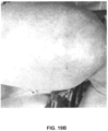
- Diagnosis Status post right Achilles tear. The patient complained of pain in the Achilles insertion: chronic Achilles tendonitis and tenosynovitis. Doppler showed severe intra-tendon hyperemia in the area ( FIG. 4 A ). VAS was 60.
- FIG. 9 is a graph showing a comparison between the average concentration level of PDGF in the unprocessed human serum samples and an average level of PDGF in 24-hour-incubated samples. Test data analysis showed a statistically significant increase of PDGF protein concentration in processed samples. PDGF concentration was evaluated using MAGPIX® LUMINEX® technology.
- FIG. 7 is a plot showing point values according to the WOMAC index for average levels of pain, stiffness and daily activity capabilities among the eight patients. Values are provided for a baseline, after injections, two months and three months post injections.
- a platelet-rich plasma component was collected and mixed with the supernatant component of the first and second tubes, respectively, to provide a first final product emanating from the first tube containing saline and a second final product emanating from the second tube containing sodium citrate.
- a baseline level of MMP2, MMP3, MMP7, MMP9, MMP13, IL-1 ⁇ , TNF- ⁇ , TIMP2, Il-1ra, and PDGF was measured immediately after drawing the blood.
- a level of MMP9, IL-1 ⁇ , TNF- ⁇ , TIMP2, Il-1ra, and PDGF was measured in the first tube containing saline and the patient's blood about 24 hours after incubation.
- a level of MMP9, IL-1 ⁇ , TNF- ⁇ , TIMP2, Il-1ra, and PDGF was measured in the second tube containing sodium citrate and the patient's blood about 24 hours after incubation.
- the platelet-rich plasma component was mixed with thrombin and then the levels of MMP9, IL-1 ⁇ , TNF- ⁇ , TIMP2, Il-1ra, and PDGF were measured.
- a level of MMP9, IL-1 ⁇ , TNF- ⁇ , TIMP2, Il-1ra, and PDGF was measured in each of the first and second final products. The measurements shown graphically in FIGS. 10 - 17 represent averages of the fifteen patients tested.
- the presence of increased MMP9 concentration in the autologous bioactive composition may result in an adverse effect manifestation during the patient treatment process. It is, therefore, desired to selectively eliminate this negative component.
- the presence of the anticoagulant, sodium citrate significantly down-regulates MMP9 release by human blood leukocytes. 19 Human blood in the presence of sodium citrate (CA) at a ratio of 9.5 cc blood: 0.5 cc sodium citrate was incubated at a temperature of from about 37° C. to about 39° C. for 24 hours and found strong dynamic changes, especially in MMP9 secretion as shown in FIGS. 11 and 12 .
- adding sodium citrate in preparing the anti-inflammatory/anti-catabolic component significantly decreased MMP9 concentration in the final product of the combination of the anti-inflammatory/anti-catabolic component and the regenerative component (thrombin-activated PRP+24 h serum).
- the MMP9 level after a 24-hour incubation did not change compared to the baseline level (0 hours) where sodium citrate was added in preparing the anti-inflammatory/anti-catabolic component.
- a measurement of the level of MMP9 in PRP component with thrombin was made to show a baseline level of MMP9 in the PRP component. This measurement shows that no significant quantity of MMP9 is produced in generating the PRP component.
- a two-way ANOVA test revealed a statistically significant decrease of TNF- ⁇ concentration in 24-hour-incubated serum from about 37° C. to about 39° C. in the presence of sodium citrate as well as in the final product (PRP+serum 24 h+CA+Thrombin) as compared to the previous method of product preparation (PRP+serum 24 h+saline).
- TIMPs, Il-1ra and PDGF concentrations were tested upon similar conditions. No negative effects of sodium citrate on TIMPs, Il-1ra and PDGF production were observed as shown in FIGS. 15 , 16 , and 17 .
- a two-way ANOVA test revealed that sodium citrate does not decrease IL-1ra concentration in 24-hour-incubated serum in the presence of sodium citrate as well as in the final product (PRP+serum 24 h+CA+Thrombin) as compared to the previous method of product preparation (PRP+serum 24 h+saline).
- a two-way ANOVA test revealed that sodium citrate does not decrease PDGF concentration in 24-hour-incubated serum as well as in the final product (PRP+serum 24 h+CA+Thrombin) as compared to previous method product preparation (PRP+serum 24 h+saline).
- Example 6 Treatment of Patients with Symptoms of Knee Osteoarthritis Joint Pain
- Example 2 Seventeen patients were treated for symptoms of knee osteoarthritis joint pain according to the method described herein, as summarized in Example 2. Two groups of patients were provided with two injections one week apart. A first group of seven patients received a product including the anti-inflammatory/anti-catabolic component prepared with no presence of sodium citrate (CA ⁇ ). A second group of ten patients received a product including the anti-inflammatory/anti-catabolic component prepared with the presence of sodium citrate (CA+). Both treatments did not result in any adverse events and were found to be safe and effective.
- CA ⁇ sodium citrate
- CA+ sodium citrate
- the patients were assessed with the Western Ontario and McMaster Universities Arthritis Index (WOMAC) questionnaire for assessing pain, stiffness, and physical function in patients with hip and/or knee osteoarthritis.
- WOMAC Western Ontario and McMaster Universities Arthritis Index
- a one month post-injection preliminary analysis of the WOMAC questionnaire data showed a statistically significant improvement in the patients' pain, stiffness and daily activities in patients treated with the anti-inflammatory/anti-catabolic component having citrate (CA+) as shown in FIGS. 18 A, 18 B, and 18 C , respectively.
- CA ⁇ anti-inflammatory/anti-catabolic component not having citrate
- a statistical analysis of Visual Analog Pain Scale (VAS) revealed a significant pain reduction in both groups, as shown in FIG. 18 D .
- Example 2 The method of Example 2 was performed on a female patient aged 59 suffering from severe psoriasis, over a four week period.
- the autologous composition produced by combining the anti-inflammatory/anti-catabolic component with the platelet-rich plasma component was administered to the female patient in four separate injections of the autologous composition one week apart.
- the treatments involved intra-dermal injection of the entire lesion site. Dramatic visual changes were observed by four weeks post treatment. The outcome was stable after three months post injection with the effects of psoriasis being eradicated.
- FIG. 19 A is a photograph of the affected area of the female patient's arm prior to treatment.
- FIG. 19 B is a photograph of the affected area of the female patient's arm three months after the fourth injection.
- Example 2 The methods of Example 2 are performed with and without the addition of sodium citrate in preparing the anti-inflammatory/anti-catabolic component on subjects suffering from chronic inflammatory skin diseases such as atopic dermatitis and chronic wounds.
- chronic inflammatory skin diseases such as atopic dermatitis and chronic wounds.
- the chronic inflammatory skin diseases reduce in severity.
- Example 2 The methods of Example 2 with and without the addition of sodium citrate in preparing the anti-inflammatory/anti-catabolic component are performed on horses, dogs and camels suffering from damaged and/or injured connective tissues. The pain associated with the damaged and/or injured connective tissues reduces in severity.
Landscapes
- Health & Medical Sciences (AREA)
- Life Sciences & Earth Sciences (AREA)
- Chemical & Material Sciences (AREA)
- Engineering & Computer Science (AREA)
- General Health & Medical Sciences (AREA)
- Medicinal Chemistry (AREA)
- Public Health (AREA)
- Veterinary Medicine (AREA)
- Animal Behavior & Ethology (AREA)
- Pharmacology & Pharmacy (AREA)
- Bioinformatics & Cheminformatics (AREA)
- Zoology (AREA)
- Immunology (AREA)
- Epidemiology (AREA)
- Proteomics, Peptides & Aminoacids (AREA)
- Gastroenterology & Hepatology (AREA)
- Organic Chemistry (AREA)
- Biomedical Technology (AREA)
- Nuclear Medicine, Radiotherapy & Molecular Imaging (AREA)
- General Chemical & Material Sciences (AREA)
- Chemical Kinetics & Catalysis (AREA)
- Biotechnology (AREA)
- Cell Biology (AREA)
- Hematology (AREA)
- Genetics & Genomics (AREA)
- Physical Education & Sports Medicine (AREA)
- Virology (AREA)
- Developmental Biology & Embryology (AREA)
- Wood Science & Technology (AREA)
- Orthopedic Medicine & Surgery (AREA)
- Biochemistry (AREA)
- Molecular Biology (AREA)
- Vascular Medicine (AREA)
- Rheumatology (AREA)
- General Engineering & Computer Science (AREA)
- Microbiology (AREA)
- Neurology (AREA)
- Analytical Chemistry (AREA)
- Biophysics (AREA)
- Pain & Pain Management (AREA)
Abstract
Description
-
- A) preparing an anti-inflammatory/anti-catabolic component of the autologous composition comprising TIMPs and IL-1ra, the step of preparing the anti-inflammatory/anti-catabolic component comprising the following steps: i) collecting an autologous physiological fluid, preferably blood from the mammal; ii) delivering the blood to a tube; iii) incubating the blood at a temperature of from about 37° C. to about 39° C. for about 24 hours; iv) centrifuging the blood to separate the blood into a supernatant component and a cellular fraction; and v) collecting the supernatant component;
- B) preparing a regenerative component of the autologous composition comprising the following steps: i) collecting blood from the mammal; ii) delivering the blood to a tube in the presence of about 4% sodium citrate; iii) centrifuging the whole blood to separate the platelet-rich plasma component; and iv) collecting the platelet-rich plasma component; and
- C) mixing the supernatant component of the anti-inflammatory/anti-catabolic component with the platelet-rich plasma component to provide the autologous composition.
-
- A) preparing an anti-inflammatory/anti-catabolic component of the autologous composition comprising TIMPs and IL-1ra, the step of preparing the anti-inflammatory/anti-catabolic component comprising the following steps: i) collecting blood from the mammal; ii) delivering the blood to a tube including sodium citrate; iii) incubating the blood at a temperature of from about 37° C. to about 39° C. for about 24 hours; iv) centrifuging the blood to separate the blood into a supernatant component and a cellular fraction; and v) collecting the supernatant component;
- B) preparing a regenerative component of the autologous composition comprising the following steps: i) collecting blood from the mammal; ii) delivering the blood to a tube in the presence of about 4% sodium citrate; iii) centrifuging the whole blood to separate the platelet-rich plasma component; and iv) collecting the platelet-rich plasma component; and
- C) mixing the supernatant component of the anti-inflammatory/anti-catabolic component with the platelet-rich plasma component to provide the autologous composition.
-
- i) collecting blood from the mammal;
- ii) adding sodium citrate to a tube;
- iii) delivering the blood to the tube;
- iv) incubating the blood at a temperature of from about 37° C. to about 39° C. for about 24 hours;
- v) centrifuging the blood to separate the blood into a supernatant component and a cellular fraction; and
- vi) collecting the supernatant component.
-
- (i) collecting blood from the mammal;
- (ii) delivering the blood to a tube;
- (iii) incubating the blood at a temperature of from about 37° C. to about 39° C. for about 24 hours;
- (iv) centrifuging the blood to separate the blood into a supernatant component and a cellular fraction;
- (v) collecting the supernatant component; and
- (vi) preparing a regenerative component of the autologous composition comprising the following steps:
- collecting blood from the mammal;
- delivering the blood to a tube in the presence of about 4% citric acid;
- centrifuging the blood to separate a platelet-rich plasma component from a whole blood;
- collecting the platelet-rich plasma component; and
- mixing the supernatant component with the platelet-rich plasma component to provide the autologous composition; and
- administering the autologous composition to the mammal.
-
- (i) collecting blood from the mammal;
- (ii) adding sodium citrate to a tube;
- (iii) delivering the blood to the tube;
- (iv) incubating the blood at a temperature of from about 37° C. to about 39° C. for about 24 hours;
- (v) centrifuging the blood to separate the blood into a supernatant component and a cellular fraction;
- (vi) collecting the supernatant component; and
- (vii) preparing a regenerative component of the autologous composition comprising the following steps:
- collecting blood from the mammal;
- delivering the blood to a tube in the presence of citric acid;
- centrifuging the blood to separate a platelet-rich plasma component from a whole blood;
- collecting the platelet-rich plasma component;
- mixing the supernatant component with the platelet-rich plasma component to provide the autologous composition; and
- administering the autologous composition to the mammal.
-
- (i) collecting blood from the mammal;
- (ii) delivering the blood to a tube in the presence of about 4% sodium citrate;
- (iii) incubating the blood at a temperature of from about 37° C. to about 39° C. for about 24 hours;
- (iv) centrifuging the blood to separate the blood into a supernatant component and a cellular fraction;
- (v) collecting the supernatant component; and
- (vi) administering the supernatant component to the mammal.
- 1. Fernandes J. C., J. Martel-Pelletier, and J. P. Pelletier. The role of cytokines in osteoarthritis pathophysiology. Biorheology 2002; 39(1-2):237-46.
- 2. Fredberg U., and K. Stengaard-Pedersen. Chronic tendinopathy tissue pathology, pain mechanisms, and etiology with a special focus on inflammation. Scand. J. Med. Sci. Sports 2008 February; 18(1):3-15.
- 3. Loell I., and I. E. Lundberg. Can muscle regeneration fail in chronic inflammation: a weakness in inflammatory myopathies? J. Intern. Med. 2011 March; 269(3):243-57.
- 4. Mirza, Rita E., Millie M. Fang, William J. Ennis, and Timothy J. Koh. Blocking Interleukin-1β Induces a Healing-Associated Wound Macrophage Phenotype and Improves Healing in
Type 2 Diabetes. Diabetes 2013 July; 62(7): 2579-2587. - 5. Eming S. A., T. Krieg, and J. M. Davidson. Inflammation in wound repair: molecular and cellular mechanisms. J. Invest Dermatol. 2007; 127:514.
- 6. Dinarello C. A. Interleukin-1, interleukin-1 receptors and interleukin-1 receptor antagonist. Int. Rev. Immunol. 1998; 16(5-6):457-99.
- 7. Mosmann T. R., H. Cherwinski, M. W. Bond, M. A. Giedlin, and R. L. Coffman. Two types of murine helper T cell clone. I. Definition according to profiles of lymphokine activities and secreted proteins. J. Immunol. 1986 Apr. 1; 136(7):2348-57.
- 8. Brandtzaeg P., L. Osnes, R. Ovstebo, G. B. Joo, A. B. Westvik, and P. Kierulf. Net inflammatory capacity of human septic shock plasma evaluated by a monocyte-based target cell assay: identification of interleukin-10 as a major functional deactivator of human monocytes. J. Exp. Med. 1996 Jul. 1; 184(1):51-60.
- 9. Clarke C. J., A. Hales, A. Hunt, and B. M. Foxwell. IL-10-mediated suppression of TNF-alpha production is independent of its ability to inhibit NF kappa B activity. Eur. J. Immunol. 1998 May; 28(5):1719-26.
- 10. de Waal Malefyt R., C. G. Figdor, R. Huijbens, S. Mohan-Peterson, B. Bennett, J. Culpepper, W. Dang, G. Zurawski, and J. E. de Vries. Effects of IL-13 on phenotype, cytokine production, and cytotoxic function of human monocytes. Comparison with IL-4 and modulation by IFN-gamma or IL-10. J. Immunol. 1993 Dec. 1; 151(11):6370-81.
- 11. Alvarez R. H., H. M. Kantarjian, and J. E. Cortes. Biology of platelet-derived growth factor and its involvement in disease. Mayo Clin. Proc. 2006 September; 81(9):1241-57.
- 12. Roberts A. B., K. C. Flanders, P. Kondaiah, N. L. Thompson, E. Van Obberghen-Schilling, L. Wakefield, P. Rossi, B. De Crom-Brugghe, U. I. Heine, and M. B. Sporn. Transforming growth factor beta: biochemistry and roles in embryogenesis, tissue repair and remodeling, and carcinogenesis. Rec. Prog. Horm. Res. 44:157-197, 1988.
- 13. Roberts A. B., U. I. Heine, K. C. Flanders, and M. B. Sporn. TGF-beta: Major role in regulation of extracellular matrix. Ann. N.Y. Acad. Sci. “Structure, Molecular Biology and Pathology of Collagen,” 580:225-232, 1990.
- 14. Gospodarowicz, D., J. A. Abraham, and J. Schilling. Isolation and characterization of a vascular endothelial cell mitogen produced by pituitary-derived folliculo stellate cells. Proc. Natl. Acad. Sci. USA 86, 7311-7315, 1989.
- 15. Hirata H., T. Nagakura, M. Tsujii, A. Morita, K. Fujisawa, and A. Uchida. The relationship of VEGF and PGE2 expression to extracellular matrix remodeling of the tenosynovium in the carpal tunnel syndrome. J. Pathol. 2004 December; 204(5):605-12.
- 16. Peng H., A. Usas, A. Olshanski, A. M. Ho, B. Gearhart, G. M. Cooper, and J. Huard. VEGF improves, whereas sFlt1 inhibits, BMP2-induced bone formation and bone healing through modulation of angiogenesis. J. Bone Miner. Res. 2005 November; 20(11):2017-27.
- 17. Poutsiaka D. D., B. D. Clark, E. Vannier, and C. A. Dinarello. Production of interleukin-1 receptor antagonist and interleukin-1 beta by peripheral blood mononuclear cells is differentially regulated. Blood 1991 Sep. 1; 78(5):1275-81.
- 18. Fufa D., B. Shealy, M. Jacobson, et al. Activation of platelet-rich plasma using soluble Type I collagen. J. Oral Maxillofac. Surg. 2008; 66:684-90.
- 19. Mannello F. Serum or plasma samples? The “Cinderella” role of blood collection procedures: preanalytical methodological issues influence the release and activity of circulating matrix metalloproteinases and their tissue inhibitors, hampering diagnostic trueness and leading to misinterpretation. Arterioscler. Thromb. Vasc. Biol. 2008 April; 28(4):611-4.
Claims (10)
Priority Applications (2)
| Application Number | Priority Date | Filing Date | Title |
|---|---|---|---|
| US16/741,627 US12023352B2 (en) | 2014-09-30 | 2020-01-13 | Anti-inflammatory/anti-catabolic and regenerative agents from autologous physiological fluid |
| US18/640,965 US20240277760A1 (en) | 2014-09-30 | 2024-04-19 | Method and composition for producing enhanced anti-inflammatory/anti-catabolic and regenerative agents from autologous physiological fluid |
Applications Claiming Priority (9)
| Application Number | Priority Date | Filing Date | Title |
|---|---|---|---|
| CA2866480A CA2866480A1 (en) | 2014-09-30 | 2014-09-30 | Method and composition for producing enhanced anti-inflammatory and regenerative agents from autologous physiological fluid |
| CA2866480 | 2014-09-30 | ||
| CA2891445A CA2891445A1 (en) | 2014-09-30 | 2015-05-14 | Method and composition for producing enhanced anti-inflammatory/anti-catabolic and regenerative agents from autologous physiological fluid |
| CA2891445 | 2015-05-14 | ||
| CA2900537A CA2900537A1 (en) | 2014-09-30 | 2015-08-13 | Method and composition for producing enhanced anti-inflammatory/anti-catabolic and regenerative agents from autologous physiological fluid |
| CA2900537 | 2015-08-13 | ||
| PCT/CA2015/000521 WO2016049746A1 (en) | 2014-09-30 | 2015-09-30 | Method and composition for producing enhanced anti-inflammatory/ anti-catabolic and regenerative agents from autologous physiological fluid |
| US201715515996A | 2017-03-30 | 2017-03-30 | |
| US16/741,627 US12023352B2 (en) | 2014-09-30 | 2020-01-13 | Anti-inflammatory/anti-catabolic and regenerative agents from autologous physiological fluid |
Related Parent Applications (2)
| Application Number | Title | Priority Date | Filing Date |
|---|---|---|---|
| PCT/CA2015/000521 Continuation WO2016049746A1 (en) | 2014-09-30 | 2015-09-30 | Method and composition for producing enhanced anti-inflammatory/ anti-catabolic and regenerative agents from autologous physiological fluid |
| US15/515,996 Continuation US10532072B2 (en) | 2014-09-30 | 2015-09-30 | Method and composition for producing enhanced anti-inflammatory/ anti-catabolic and regenerative agents from autologous physiological fluid |
Related Child Applications (1)
| Application Number | Title | Priority Date | Filing Date |
|---|---|---|---|
| US18/640,965 Continuation US20240277760A1 (en) | 2014-09-30 | 2024-04-19 | Method and composition for producing enhanced anti-inflammatory/anti-catabolic and regenerative agents from autologous physiological fluid |
Publications (2)
| Publication Number | Publication Date |
|---|---|
| US20200163991A1 US20200163991A1 (en) | 2020-05-28 |
| US12023352B2 true US12023352B2 (en) | 2024-07-02 |
Family
ID=55590205
Family Applications (3)
| Application Number | Title | Priority Date | Filing Date |
|---|---|---|---|
| US15/515,996 Active 2035-12-14 US10532072B2 (en) | 2014-09-30 | 2015-09-30 | Method and composition for producing enhanced anti-inflammatory/ anti-catabolic and regenerative agents from autologous physiological fluid |
| US16/741,627 Active 2036-03-12 US12023352B2 (en) | 2014-09-30 | 2020-01-13 | Anti-inflammatory/anti-catabolic and regenerative agents from autologous physiological fluid |
| US18/640,965 Pending US20240277760A1 (en) | 2014-09-30 | 2024-04-19 | Method and composition for producing enhanced anti-inflammatory/anti-catabolic and regenerative agents from autologous physiological fluid |
Family Applications Before (1)
| Application Number | Title | Priority Date | Filing Date |
|---|---|---|---|
| US15/515,996 Active 2035-12-14 US10532072B2 (en) | 2014-09-30 | 2015-09-30 | Method and composition for producing enhanced anti-inflammatory/ anti-catabolic and regenerative agents from autologous physiological fluid |
Family Applications After (1)
| Application Number | Title | Priority Date | Filing Date |
|---|---|---|---|
| US18/640,965 Pending US20240277760A1 (en) | 2014-09-30 | 2024-04-19 | Method and composition for producing enhanced anti-inflammatory/anti-catabolic and regenerative agents from autologous physiological fluid |
Country Status (15)
| Country | Link |
|---|---|
| US (3) | US10532072B2 (en) |
| EP (1) | EP3200817B1 (en) |
| JP (1) | JP6646660B2 (en) |
| KR (1) | KR102455394B1 (en) |
| CN (1) | CN107073083B (en) |
| AU (1) | AU2015327723B2 (en) |
| CA (4) | CA2866480A1 (en) |
| CL (1) | CL2017000670A1 (en) |
| ES (1) | ES2912773T3 (en) |
| GB (1) | GB2546224B (en) |
| IL (1) | IL251082B (en) |
| LT (1) | LT3200817T (en) |
| MX (1) | MX388111B (en) |
| RU (1) | RU2708242C2 (en) |
| WO (1) | WO2016049746A1 (en) |
Families Citing this family (10)
| Publication number | Priority date | Publication date | Assignee | Title |
|---|---|---|---|---|
| CA2866480A1 (en) | 2014-09-30 | 2016-03-30 | Antnor Limited | Method and composition for producing enhanced anti-inflammatory and regenerative agents from autologous physiological fluid |
| CA2950659A1 (en) * | 2016-12-06 | 2018-06-06 | Antnor Limited | Method for the preparation and prolonged storage of growth factors and cytokines obtained from platelet rich plasma |
| EP3613424A1 (en) * | 2018-08-23 | 2020-02-26 | Orthogen AG | Novel methods for the production of pharmaceutical agents |
| WO2020243807A1 (en) * | 2019-06-05 | 2020-12-10 | Antnor Limited | Method and composition for producing enhanced anti-inflammatory/anti-catabolic and regenerative agents from autologous physiological fluid |
| RU2747589C1 (en) * | 2019-12-09 | 2021-05-11 | Руслан Яверович Атлуханов | Method for treating meniscus injury of knee joint |
| CA3076046A1 (en) * | 2020-03-17 | 2021-09-17 | Antnor Limited | Method for producing enhanced anti-inflammatory/anti-catabolic agents from autologous physiological fluid with shortened incubation time |
| CA3106197A1 (en) * | 2021-01-20 | 2022-07-20 | Antnor Limited | Method for producing blood plasma product useful in the treatment of viral infections characterized by an uncontrolled release of proinflammatory cytokines |
| JP7025070B1 (en) * | 2021-05-14 | 2022-02-24 | セルソース株式会社 | Blood-derived growth factor-containing composition and its preparation method |
| JP7175055B1 (en) | 2021-05-14 | 2022-11-18 | セルソース株式会社 | Blood-derived growth factor-containing composition and method for preparing the same |
| CN114146096B (en) * | 2021-11-25 | 2024-04-02 | 成都清科生物科技有限公司 | Preparation method and application of conditional serum rich in cytokines |
Citations (17)
| Publication number | Priority date | Publication date | Assignee | Title |
|---|---|---|---|---|
| WO1996018292A1 (en) | 1994-12-16 | 1996-06-20 | Baxter International Inc. | Controlling donor blood characteristics |
| JP2002540818A (en) | 1999-02-01 | 2002-12-03 | オルソゲン アーゲー | Method for producing therapeutically effective protein interleukin-1 receptor antagonist from body fluid |
| US20030166069A1 (en) | 2000-11-28 | 2003-09-04 | Amgen, Inc. | Interleukin-1 receptor antagonist-like molecules and uses thereof |
| US20060177515A1 (en) | 2005-02-09 | 2006-08-10 | Reinhold Schmieding | Method of producing autogenous or allogenic blood serum and related logistics |
| US20090220482A1 (en) | 2008-02-27 | 2009-09-03 | Biomet Biologics, Llc | Methods and compositions for delivering interleukin-1 receptor antagonist |
| US20100125236A1 (en) | 2008-11-17 | 2010-05-20 | Christopher Bare | Cytokine concentration system |
| US20100125336A1 (en) | 2007-01-18 | 2010-05-20 | The University Of Newcastle Upon Tyne | Reverse shoulder prosthesis |
| WO2011031553A2 (en) | 2009-08-27 | 2011-03-17 | Biomet Biologics, Llc | Implantable device for production of interleukin-1 receptor antagonist |
| WO2012030593A2 (en) | 2010-09-03 | 2012-03-08 | Biomet Biologics, Llc | Methods and compositions for delivering interleukin-1 receptor antagonist |
| KR20130027083A (en) | 2010-12-09 | 2013-03-15 | 이선민 | Method for preparing autologous thrombin activated autologous plasma for regeneration of epithelial tissues or subcutaneous tissues and composition containing the autologous plasma |
| WO2013114359A1 (en) | 2012-01-31 | 2013-08-08 | Estar Technologies Ltd | System and method for producing interleukin receptor antagonist (ira) |
| US20140271587A1 (en) | 2013-03-15 | 2014-09-18 | Biomet Biologics, Llc | Treatment of inflammatory respiratory disease using biological solutions |
| US20140271588A1 (en) | 2013-03-15 | 2014-09-18 | Biomet Biologics, Llc | Treatment of peripheral vascular disease using protein solutions |
| US20140271589A1 (en) | 2013-03-15 | 2014-09-18 | Biomet Biologics, Llc | Treatment of collagen defects using protein solutions |
| US20140274894A1 (en) | 2013-03-15 | 2014-09-18 | Biomet Biologics, Llc | Methods for making cytokine compositions from tissues using non-centrifugal methods |
| WO2014149270A1 (en) | 2013-03-15 | 2014-09-25 | Biomet Biologics, Llc | Treatment of pain using protein solutions |
| CA2900537A1 (en) | 2014-09-30 | 2016-03-30 | Antnor Limited | Method and composition for producing enhanced anti-inflammatory/anti-catabolic and regenerative agents from autologous physiological fluid |
-
2014
- 2014-09-30 CA CA2866480A patent/CA2866480A1/en not_active Abandoned
-
2015
- 2015-05-14 CA CA2891445A patent/CA2891445A1/en not_active Abandoned
- 2015-08-13 CA CA2900537A patent/CA2900537A1/en not_active Abandoned
- 2015-09-30 RU RU2017114523A patent/RU2708242C2/en active
- 2015-09-30 US US15/515,996 patent/US10532072B2/en active Active
- 2015-09-30 CA CA2962289A patent/CA2962289C/en active Active
- 2015-09-30 EP EP15847588.9A patent/EP3200817B1/en active Active
- 2015-09-30 JP JP2017518238A patent/JP6646660B2/en active Active
- 2015-09-30 KR KR1020177011345A patent/KR102455394B1/en active Active
- 2015-09-30 LT LTEPPCT/CA2015/000521T patent/LT3200817T/en unknown
- 2015-09-30 AU AU2015327723A patent/AU2015327723B2/en active Active
- 2015-09-30 CN CN201580053264.6A patent/CN107073083B/en active Active
- 2015-09-30 MX MX2017004131A patent/MX388111B/en unknown
- 2015-09-30 WO PCT/CA2015/000521 patent/WO2016049746A1/en active Application Filing
- 2015-09-30 ES ES15847588T patent/ES2912773T3/en active Active
- 2015-09-30 GB GB1706803.2A patent/GB2546224B/en active Active
-
2017
- 2017-03-09 IL IL251082A patent/IL251082B/en active IP Right Grant
- 2017-03-20 CL CL2017000670A patent/CL2017000670A1/en unknown
-
2020
- 2020-01-13 US US16/741,627 patent/US12023352B2/en active Active
-
2024
- 2024-04-19 US US18/640,965 patent/US20240277760A1/en active Pending
Patent Citations (30)
| Publication number | Priority date | Publication date | Assignee | Title |
|---|---|---|---|---|
| WO1996018292A1 (en) | 1994-12-16 | 1996-06-20 | Baxter International Inc. | Controlling donor blood characteristics |
| JP2002540818A (en) | 1999-02-01 | 2002-12-03 | オルソゲン アーゲー | Method for producing therapeutically effective protein interleukin-1 receptor antagonist from body fluid |
| US20030138910A1 (en) | 1999-02-01 | 2003-07-24 | Julio Reinecke | Method for producing interleukin 1 receptor antagonists, a therapeutic activity protein, from body fluids |
| KR100652534B1 (en) | 1999-02-01 | 2006-12-01 | 오르토겐 아게 | Method for preparing interleukin-1 receptor antagonist, a therapeutically active protein, from body fluids |
| US20030166069A1 (en) | 2000-11-28 | 2003-09-04 | Amgen, Inc. | Interleukin-1 receptor antagonist-like molecules and uses thereof |
| US20060177515A1 (en) | 2005-02-09 | 2006-08-10 | Reinhold Schmieding | Method of producing autogenous or allogenic blood serum and related logistics |
| US20100125336A1 (en) | 2007-01-18 | 2010-05-20 | The University Of Newcastle Upon Tyne | Reverse shoulder prosthesis |
| US20090220482A1 (en) | 2008-02-27 | 2009-09-03 | Biomet Biologics, Llc | Methods and compositions for delivering interleukin-1 receptor antagonist |
| US20100125236A1 (en) | 2008-11-17 | 2010-05-20 | Christopher Bare | Cytokine concentration system |
| WO2011031553A2 (en) | 2009-08-27 | 2011-03-17 | Biomet Biologics, Llc | Implantable device for production of interleukin-1 receptor antagonist |
| WO2012030593A2 (en) | 2010-09-03 | 2012-03-08 | Biomet Biologics, Llc | Methods and compositions for delivering interleukin-1 receptor antagonist |
| KR20130027083A (en) | 2010-12-09 | 2013-03-15 | 이선민 | Method for preparing autologous thrombin activated autologous plasma for regeneration of epithelial tissues or subcutaneous tissues and composition containing the autologous plasma |
| WO2013114359A1 (en) | 2012-01-31 | 2013-08-08 | Estar Technologies Ltd | System and method for producing interleukin receptor antagonist (ira) |
| US20140271588A1 (en) | 2013-03-15 | 2014-09-18 | Biomet Biologics, Llc | Treatment of peripheral vascular disease using protein solutions |
| US20140271587A1 (en) | 2013-03-15 | 2014-09-18 | Biomet Biologics, Llc | Treatment of inflammatory respiratory disease using biological solutions |
| US20140271589A1 (en) | 2013-03-15 | 2014-09-18 | Biomet Biologics, Llc | Treatment of collagen defects using protein solutions |
| US20140274894A1 (en) | 2013-03-15 | 2014-09-18 | Biomet Biologics, Llc | Methods for making cytokine compositions from tissues using non-centrifugal methods |
| WO2014149270A1 (en) | 2013-03-15 | 2014-09-25 | Biomet Biologics, Llc | Treatment of pain using protein solutions |
| CA2962289A1 (en) | 2014-09-30 | 2016-04-07 | Antnor Limited | Method and composition for producing enhanced anti-inflammatory/ anti-catabolic and regenerative agents from autologous physiological fluid |
| CA2891445A1 (en) | 2014-09-30 | 2016-03-30 | Antnor Limited | Method and composition for producing enhanced anti-inflammatory/anti-catabolic and regenerative agents from autologous physiological fluid |
| CA2866480A1 (en) | 2014-09-30 | 2016-03-30 | Antnor Limited | Method and composition for producing enhanced anti-inflammatory and regenerative agents from autologous physiological fluid |
| WO2016049746A1 (en) | 2014-09-30 | 2016-04-07 | Antnor Limited | Method and composition for producing enhanced anti-inflammatory/ anti-catabolic and regenerative agents from autologous physiological fluid |
| CA2900537A1 (en) | 2014-09-30 | 2016-03-30 | Antnor Limited | Method and composition for producing enhanced anti-inflammatory/anti-catabolic and regenerative agents from autologous physiological fluid |
| KR20170063836A (en) | 2014-09-30 | 2017-06-08 | 안트노르 리미티드 | Method and composition for producing enhanced anti-inflammatory/anti-catabolic and regenerative agents from autologous physiological fluid |
| MX2017004131A (en) | 2014-09-30 | 2017-06-19 | Antnor Ltd | Method and composition for producing enhanced anti-inflammatory/ anti-catabolic and regenerative agents from autologous physiological fluid. |
| GB2546224A (en) | 2014-09-30 | 2017-07-12 | Antnor Ltd | Method and composition for producing enhanced anti-inflammatory/ anti-catabolic and regenerative agents from autologous physiological fluid |
| EP3200817A1 (en) | 2014-09-30 | 2017-08-09 | Antnor Limited | Method and composition for producing enhanced anti-inflammatory/ anti-catabolic and regenerative agents from autologous physiological fluid |
| CN107073083A (en) | 2014-09-30 | 2017-08-18 | 安特诺有限公司 | Method and composition for producing enhanced anti-inflammatory/Anticatabolism agent and regenerative agent from autologous physiological fluid |
| US20170296583A1 (en) | 2014-09-30 | 2017-10-19 | Antnor Limited | Method and composition for producing enhanced anti-inflammatory/ anti-catabolic and regenerative agents from autologous physiological fluid |
| JP2017533896A (en) | 2014-09-30 | 2017-11-16 | アントノール リミテッド | Methods and compositions for making enhanced anti-inflammatory / anti-catabolic and regenerative substances from autologous physiological fluids |
Non-Patent Citations (73)
| Title |
|---|
| Alvarez et al. "Biology of platelet-derived growth factor and its involvement in disease" Mayo Clin. Proc. Sep. 2006.; 81(9):1241-57 (Abstract Only). |
| Ames ("An Appraisal of the "Vacutainer" System for Blood Collection", Annals of Clinical Biochemistry, 12, 1975, 151-155 (Year: 1975). |
| Andia, I. et al., "Joint pathology and platelet-rich plasma therapies", Expert Opinion on Biological Therapy, vol. 12, No. 1, pp. 7-22. (2012). ISSN: 1471-2598. |
| Australian Patent Examination Report No. 2 for Australian Application No. 2015327723, dated Jun. 17, 2020, 5 pages. |
| Bergin et al., "Secretion of Matrix Metalloproteinase-9 by Macrophages, In Vitro, In Response to Helicobacter Pylori," FEMS Immunology and Medical Microbiology, vol. 45, (2005), 159-169. |
| Biggs et al., "The Effects of Several Organic Acids on Growth Performance, Nutrient Digestibilities, and Cecal Microbial Populations in Young Chicks" Poult Sci. Dec. 2008;87(12):2581-9. doi: 10.3382/ps.2008-00080 (Abstract). |
| Brandtaeg et al., Net inflammatory capacity of human septic shock plasma evaluated by a monocyte-based target cell assay: identification of interleukin-10 as a major functional deactivator of human monocytes. J Exp Med. J. Exp. Med. Jul. 1, 1996; 184(1):51-60. |
| Canadian Requisition by the Examiner for Canadian Application No. 2,962,289, dated Jun. 19, 2020, 7 pages. |
| Canadian Requisition by the Examiner for Canadian Application No. 2,962,289, dated Oct. 15, 2020, 4 pages. |
| Chamberlain et al. "Interleukin-1 Receptor Antagonist Modulates Inflammation and Scarring after Ligament Injury" Connect Tissue Res. vol. 55, 2014, pp. 177-186. |
| Chen et al., "Tissue-engineered intervertebral disc and chondrogenesis using human nucleus pulposus regulated through TGF-[beta]1 in platelet-rich plasma", Journal of Cellular Physiology, vol. 209, No. 3, Dec. 1, 2006, pp. 744-754. |
| Chinese Second Office Action for Chinese Application No. 201580053264.6, dated Jan. 18, 2021, 18 pages. |
| CitraLabs ("ACD-A Anticoagulant Citrate Dextrose Solution, Solution A" available at www.citra-labs.com/products/acd-a-.cfm, webcapture on Apr. 13, 2019). (Year: 2019). |
| Clarke et al., IL-10-mediated suppression of TNF-alpha production is independent of its ability to inhibit NF kappa B activity. Eur J Immunol. Eur. J. Immunol. May 1998; 28(5):1719-26. |
| Dinarello et al., Interleukin-1, interleukin-1 receptors and interleukin-1 receptor antagonist. Int Rev Immunol. Int. Rev. Immunol. 1998; 16(5-6):457-99. |
| Ehrenfest ("Classification of platelet concentrates: from pure platelet-rich plasma (P-PRP) to leucocyte and platelet-rich fibrin (L-PRF)", Trends in Biotechnology, 2009, vol. 27, Issue 3, 158-167). (Year: 2009). |
| Elkington et al., "Analysis of Matrix Metalloproteinase Secretion by Macrophages," Methods Mol. Biol., vol. 531, (2009); pp. 253-265. |
| Eming et al., Inflammation in wound repair: molecular and cellular mechanisms. J. Invest Dermatol. 2007; 127:514. |
| European Communication pursuant to Article 94(3) EPC for European Application No. 15847588.9, dated Jun. 29, 2020, 12 pages. |
| European Communication pursuant to Article 94(3) EPC for European Application No. 15847588.9, dated Jun. 29, 2020, 6 pages. |
| European Search Report and Search Opinion Received for EP Application No. 15847588.9, dated May 23, 2018, 10 pages. |
| European Search Report for Application No. 15847588.9 dated May 23, 2018, 11 pages. |
| Fredberg et al., Chronic tendinopathy tissue pathology, pain mechanisms, and etiology with a special focus on inflammation. Scand J Med Sci Sports. Scand. J. Med. Sci. Sports Feb. 2008; 18(1):3-15. |
| Freitag et al., The Next Step in Osteoarthritis Management-Photoactivated Platelet Rich Plasma Injections: A Case Study, Annals of the Rheumatic Disease, Jun. 2013, vol. 71, Supp. Suppl. 3. Abstract No. AB0981. |
| Frernandes et al., The role of cytokines in osteoarthritis pathophysiology. Biorheology. Biorheology 2002; 39(1-2):237-46. |
| Fufa et al., Activation of platelet-rich plasma using soluble Type I collagen. J Oral Maxillofac Surg J. Oral Maxillofac. Surg. 2008; 66:684-90. |
| Gospodarowicz et al., Isolation and characterization of a vascular endothelial cell mitogen produced by pituitary-derived folliculo stellate cells. Proc. Nall. Acad. Sci. USA 86, 7311-7315, 1989. |
| Hirata et al. "The relationship of VEGF and PGE2 expression to extracellular matrix remodeling of the tenosynovium in the carpal tunnel syndrome" J. Pathol. Dec. 2004; 204(5):605-12 (Abstract). |
| Hraha et al., Autologous Conditioned Serum: The Comparative Cytokine Profiles of Two Commercial Methods (IRAP and IRAP II) Using Equine Blood, Equine vet. J., vol. 43, (2011), pp. 516-521. |
| Hurme Mikko et al.: "IL-1 receptor antagonist (IL-IRa) plasma levels are co-ordinately regulated by both IL-IRa and II-Ibeta genes", European Journal of Immunology,, vol. 28, No. 8, Aug. 1, 1998 (Aug. 1, 1998), pp. 2598-2602. |
| International Preliminary Report on Patentability received for PCT Patent Application No. PCT/CA2015/000521, dated Apr. 13, 2017, 9 pages. |
| International Search Report and Written Opinion received for PCT Patent Application No. PCT/CA2015/000521, dated Jan. 11, 2016, 13 pages. |
| Japanese Decision to Grant a Patent for Japanese Application No. 2017-518238, dated Jan. 8, 2020, 5 pages with English Translation. |
| Japanese Notice of Reasons for Refusal for Japanese Application No. 2017-518238, dated Aug. 1, 2019, 10 pages with English translation. |
| Korean Notice of of Preliminary Rejection for Korean Application No. 10-2017-7011345, dated Mar. 16, 2022, 9 pages with English translation. |
| Krishnan et al., "Multiplexed measurements of immunomodulator levels in peripheral blood of healthy subjects: Effects of analytical variables based on anticoagulants, age, and gender" Cytometry B Clin Cytom. Nov. 2014;86(6):426-35. doi: 10.1002/cyto.b.21147. Epub Feb. 26, 2014. (Abstract). |
| Lederle et al., "Platelet-Derived Growth Factor-BB Controls Epithelial Tumor Phenotype by Differential Growth Factor Regulation in Stromal Cells," The American Journal of Pathology, vol. 169, (Nov. 2006), pp. 1767-1783. |
| Lee et al. "Anticoagulation Techniques in Apheresis: From Heparin to Citrate and Beyond" J Clin Apher. 2012; 27(3): 117-125. |
| Lichtenauer, M. et al., "Phosphate buffered saline containing calcium and magnesium elicits increased secretion of interleukin-1 receptor antagonist", Lctboratory Medicine, vol. 40, No. 5, pp. 290-293, (2009), ISSN: 0007-5027. |
| Loell et al. "Can muscle regeneration fail in chronic inflammation: a weakness in inflammatory myopathies?" J. Intern. Med. Mar. 2011; 269(3):243-57. |
| Malefyt et al. Effects of IL-13 on phenotype, cytokine production, and cytotoxic function of human monocytes. Comparison with IL-4 and modulation by IFN-gamma or IL-10. J. Immunol. Dec. 1993; 151(11):6370-81 (Abstract). |
| Manello et al., Serum or plasma samples? The "Cinderella" role of blood collection procedures: preanalytical methodological issues influence the release and activity of circulating matrix metalloproteinases and their tissue inhibitors, hampering diagnostic trueness and leading to misinterpretation. Arterioscler Thromb Vase Biol. Arterioscler Thromb. Vase. Biol. Apr. 2008;28(4):611-4. |
| Mexican Office Action for Mexican Application No. MX/a/2017/004131, dated Mar. 11, 2021, 8 pages with translation. |
| Mirza et al., Blocking Interleukin-1 Beta Induces a Healing-Associated Wound Macrophage Phenotype and Improves Healing in Type 2 Diabetes. Diabetes Jul. 2013; 62(7): 2579-2587. |
| Mishra, A et al., "Treatment of chronic elbow tendinosis with buffered platelet-rich plasma", The American Journal of Sports Medicine, vol. 34, No. 11, pp. 1774-1778, ISSN: 0363-5465. |
| Moser et al., Intradiscal Injections of Orthokine-Derived Autologous Conditioned Serum (ACS) for Lumbar Disc Degeneration, Osteoarthritis and Cartilage, Sep. 2011, vol. 19, Supp. Suppl. 1, pp. S215. Abstract No. 64. |
| Mosmann et al., L. Two types of murine helper T cell clone. I. Definition according to profiles of lymphokine activities and secreted proteins. J Immunol. J. Immunol. Apr. 1986. 1; 136(7):2348-57. |
| Mussano et al., "Cytokine, chemokine, and growth factor profile of platelet-rich plasma", Platelets, vol. 27, No. 5, Jul. 3, 2016, pp. 467-471. |
| Notice of Deficiencies for Israel Patent Application No. 251082, dated May 27, 2019 (10 pages of English Translation). |
| Office Action received for Chile Patent Application No. 2017000670, dated Oct. 3, 2018, 18 pages (8 pages of English Translation and 10 pages of Original Document). |
| Patentability Search and Report that was Prepared by the Nordic Patent Institute, dated Mar. 27, 2015. |
| PCT International Search Report and Written Opinion, PCT/CA2015/000521, dated Sep. 30, 2015. |
| Peng et al., VEGF improves, whereas sFlt1 inhibits, BMP2-induced bone formation and bone healing through modulation of angiogenesis.J Bone Miner Res. angiogenesis. J. Bone Miner. Res. Nov. 2005; 20(11):2017-27. |
| Poutsiaka et al., Production of interleukin-1 receptor antagonist and interleukin-1 beta by peripheral blood mononuclear cells is differentially regulated. Blood. Blood Sep. 1, 1991; 78(5):1275-81. |
| Roberts et al. "Transforming growth factor beta: biochemistry and roles in embryogenesis, tissue repair and remodeling, and carcinogenesis" Rec. Prog. Horm. Res. 44:157-197, 1988 (Abstract). |
| Roberts et al., "TGF-(Beta): Regulation of Extracellular Matrix," Kidney International, vol. 41, (1992), pp. 557-559. |
| Rosengren et al., "Platelet-Derived Growth Factor and Transforming Growth Factor Beta Synergistically Potentiate Inflammatory Mediator Synthesis by Fibroblast-Like Synoviocytes," Arthritis Research & Therapy, vol. 12, (2010), pp. 1-11. |
| Russian Office Action from Russian Patent Application No. 2017114523, (9 pages of English Translation). |
| Russian Search Report from Russian Patent Application No. 2017114523 (3 pages of English Translation). |
| Rutgers et al., "Cytokine Profile of Autologous Conditioned Serum for Treatment of Osteoarthritis, In Vitro Effects on Cartilage Metabolism and Intra-Articular Levels After Injection," Arthritis Research & Therapy, vol. 12:R114, (2010), pp. 1-11. |
| Sampson, S. et al., "Injection of platelet-rich plasma in patients with primary and secondary knee osteoarthritis", American Journal of Physical Medicine and Rehabilitation, vol. 89, No. 12, pp. 961-969, (2010) ISSN: 0894-9115. |
| Sanchez, M et al., "Ultrasound-guided platelet-rich plasma injections for the treatment of osteoarthritis in the hip", Rheumatology, vol. 51, No. 1, pp. 144-150, Nov. 10, 2011 (Oct. 11, 2011), ISSN: 1462-0324. |
| Sawyere et al., "Cytokine and Growth Factor Concentrations in Canine Autologous Conditioned Serum," Veterinary Surgery, vol. 45, (2016), pp. 582-586. |
| Shirokova et al., Comparison of the Efficacy of Autologous Conditioned Serum and Hyaluronic Acid Treatment in Hip Os-Teoarthritis, Annals of the Rheumatic Disease, Jun. 2013 vol. 71, Supp. Suppl. 3. Abstract No. AB0981. |
| Tatarniuk ("Concentrations of Cytokines, Matrix Metalloproteinases and Tissue Inhibitors of Matrix Metalloproteinases in Autologous Conditioned Serum from Horses with Distal Interphalangeal Joint Osteoarthritis" Osteoarthritis and Cartliage, 23 (2015) (Year: 2015). |
| Textor "Autologous Biologic Treatment for Equine Musculoskeletal Injuries: Platelet-Rich Plasma and IL-1 Receptor Antagonist Protein" Vet Clin North Am Equine Pract. Aug. 2011, vol. 27, Issue 2, pp. 275-298 (Abstract). |
| Trextor ("Autologous Biologic Treatment for Equine Musculorskel et al Injuries: Platelet-Rich Plasma and IL-1 Receptor Antagonist Protein" Veterinary Clinics: Equine Practice, 2011, 27, 275-298) (Year:2011). |
| United Kingdom Examination Report for United Kingdom Application No. GB1706803.2, dated Sep. 17, 2019, 4 pages. |
| Wehling ("Use of Autologous Conditioned Cell-free Serum (Orthokine) in treating Osteoarthritis and Sciatic Back Pain" European Musculoskel et al.Revies 4: 8-11) (Year:2009). |
| Wehling ("Use of Autologous Conditioned Cell-free Serum (Orthokine) in treating Osteoarthritis and Sciatic Back Pain" European Musculoskel et al.Revies 4: 8-11) (Year:2009). |
| Wehling, P. et al., Autologous conditioned serum in the treatment of orthopedic diseases. The Orthokine Therapy. Biodrogs. vol. 21, No. 5, pp. 323-332, (2007) ISSN: 1173-8804. |
| Wright-Carpenter et al., "Treatment of Muscle Injures by Local Administration of Autologous Conditioned Serum: A Pilot Study on Sportsmen With Muscle Strains," Int J Sports Med., vol. 25, (2004), pp. 588-593. |
| X. Chevalier et al: "Tissue inhibitor of metalloprotease-1 (TIMP-1) serum level may predict progression of hip osteoarthritis", Osteoarthritis and Cartilage., vol. 9, No. 4, May 1, 2001 (May 1, 2001), pp. 300-307. |
Also Published As
Similar Documents
| Publication | Publication Date | Title |
|---|---|---|
| US12023352B2 (en) | Anti-inflammatory/anti-catabolic and regenerative agents from autologous physiological fluid | |
| US7897180B2 (en) | Enamel matrix protein composition for treatment of systemic inflammatory response | |
| US20190343878A1 (en) | Method for the preparation and prolonged storage of growth factors and cytokines obtained from platelet rich plasma | |
| WO2020243807A1 (en) | Method and composition for producing enhanced anti-inflammatory/anti-catabolic and regenerative agents from autologous physiological fluid | |
| AU2002334515A1 (en) | Enamel matrix protein compositions for modulating immune response | |
| CN104755095A (en) | Medicament for wound treatment | |
| US20230146680A1 (en) | Method for producing enhanced anti-inflammatory / anti-catabolic agents from autologous physiological fluid | |
| WO2016056665A1 (en) | White blood cell extracellular trap formation inhibitor | |
| BR112017006410B1 (en) | METHODS OF PRODUCING AN AUTOLOGOUS COMPOSITION USEFUL IN THE TREATMENT OF A MAMMAL SUFFERING FROM DAMAGED AND/OR INJURED CONNECTIVE TISSUES, CHRONIC TENDINOSIS, CHRONIC MUSCLE INJURIES, CHRONIC DEGENERATIVE JOINT CONDITIONS AND/OR INFLAMMATORY SKIN DISEASES, AUTOLOGOUS COMPOSITION AND ITS USE |
Legal Events
| Date | Code | Title | Description |
|---|---|---|---|
| STPP | Information on status: patent application and granting procedure in general |
Free format text: DOCKETED NEW CASE - READY FOR EXAMINATION |
|
| STPP | Information on status: patent application and granting procedure in general |
Free format text: NON FINAL ACTION MAILED |
|
| STPP | Information on status: patent application and granting procedure in general |
Free format text: RESPONSE TO NON-FINAL OFFICE ACTION ENTERED AND FORWARDED TO EXAMINER |
|
| STPP | Information on status: patent application and granting procedure in general |
Free format text: FINAL REJECTION MAILED |
|
| STPP | Information on status: patent application and granting procedure in general |
Free format text: DOCKETED NEW CASE - READY FOR EXAMINATION |
|
| STPP | Information on status: patent application and granting procedure in general |
Free format text: NON FINAL ACTION MAILED |
|
| STPP | Information on status: patent application and granting procedure in general |
Free format text: RESPONSE TO NON-FINAL OFFICE ACTION ENTERED AND FORWARDED TO EXAMINER |
|
| STPP | Information on status: patent application and granting procedure in general |
Free format text: NOTICE OF ALLOWANCE MAILED -- APPLICATION RECEIVED IN OFFICE OF PUBLICATIONS |
|
| STPP | Information on status: patent application and granting procedure in general |
Free format text: PUBLICATIONS -- ISSUE FEE PAYMENT VERIFIED |
|
| STCF | Information on status: patent grant |
Free format text: PATENTED CASE |