US11851670B2 - Nucleic acid constructs encoding reprogramming factors linked by self-cleaving peptides - Google Patents
Nucleic acid constructs encoding reprogramming factors linked by self-cleaving peptides Download PDFInfo
- Publication number
- US11851670B2 US11851670B2 US16/438,424 US201916438424A US11851670B2 US 11851670 B2 US11851670 B2 US 11851670B2 US 201916438424 A US201916438424 A US 201916438424A US 11851670 B2 US11851670 B2 US 11851670B2
- Authority
- US
- United States
- Prior art keywords
- cells
- reprogramming
- cell
- oct4
- dox
- Prior art date
- Legal status (The legal status is an assumption and is not a legal conclusion. Google has not performed a legal analysis and makes no representation as to the accuracy of the status listed.)
- Active, expires
Links
Images


Classifications
-
- C—CHEMISTRY; METALLURGY
- C12—BIOCHEMISTRY; BEER; SPIRITS; WINE; VINEGAR; MICROBIOLOGY; ENZYMOLOGY; MUTATION OR GENETIC ENGINEERING
- C12N—MICROORGANISMS OR ENZYMES; COMPOSITIONS THEREOF; PROPAGATING, PRESERVING, OR MAINTAINING MICROORGANISMS; MUTATION OR GENETIC ENGINEERING; CULTURE MEDIA
- C12N15/00—Mutation or genetic engineering; DNA or RNA concerning genetic engineering, vectors, e.g. plasmids, or their isolation, preparation or purification; Use of hosts therefor
- C12N15/09—Recombinant DNA-technology
- C12N15/63—Introduction of foreign genetic material using vectors; Vectors; Use of hosts therefor; Regulation of expression
- C12N15/79—Vectors or expression systems specially adapted for eukaryotic hosts
- C12N15/85—Vectors or expression systems specially adapted for eukaryotic hosts for animal cells
- C12N15/86—Viral vectors
-
- A—HUMAN NECESSITIES
- A01—AGRICULTURE; FORESTRY; ANIMAL HUSBANDRY; HUNTING; TRAPPING; FISHING
- A01K—ANIMAL HUSBANDRY; AVICULTURE; APICULTURE; PISCICULTURE; FISHING; REARING OR BREEDING ANIMALS, NOT OTHERWISE PROVIDED FOR; NEW BREEDS OF ANIMALS
- A01K67/00—Rearing or breeding animals, not otherwise provided for; New or modified breeds of animals
- A01K67/027—New or modified breeds of vertebrates
- A01K67/0271—Chimeric vertebrates, e.g. comprising exogenous cells
-
- C—CHEMISTRY; METALLURGY
- C07—ORGANIC CHEMISTRY
- C07K—PEPTIDES
- C07K14/00—Peptides having more than 20 amino acids; Gastrins; Somatostatins; Melanotropins; Derivatives thereof
- C07K14/435—Peptides having more than 20 amino acids; Gastrins; Somatostatins; Melanotropins; Derivatives thereof from animals; from humans
- C07K14/46—Peptides having more than 20 amino acids; Gastrins; Somatostatins; Melanotropins; Derivatives thereof from animals; from humans from vertebrates
- C07K14/47—Peptides having more than 20 amino acids; Gastrins; Somatostatins; Melanotropins; Derivatives thereof from animals; from humans from vertebrates from mammals
- C07K14/4701—Peptides having more than 20 amino acids; Gastrins; Somatostatins; Melanotropins; Derivatives thereof from animals; from humans from vertebrates from mammals not used
- C07K14/4702—Regulators; Modulating activity
- C07K14/4705—Regulators; Modulating activity stimulating, promoting or activating activity
-
- C—CHEMISTRY; METALLURGY
- C12—BIOCHEMISTRY; BEER; SPIRITS; WINE; VINEGAR; MICROBIOLOGY; ENZYMOLOGY; MUTATION OR GENETIC ENGINEERING
- C12N—MICROORGANISMS OR ENZYMES; COMPOSITIONS THEREOF; PROPAGATING, PRESERVING, OR MAINTAINING MICROORGANISMS; MUTATION OR GENETIC ENGINEERING; CULTURE MEDIA
- C12N5/00—Undifferentiated human, animal or plant cells, e.g. cell lines; Tissues; Cultivation or maintenance thereof; Culture media therefor
- C12N5/06—Animal cells or tissues; Human cells or tissues
- C12N5/0602—Vertebrate cells
- C12N5/0696—Artificially induced pluripotent stem cells, e.g. iPS
-
- G—PHYSICS
- G01—MEASURING; TESTING
- G01N—INVESTIGATING OR ANALYSING MATERIALS BY DETERMINING THEIR CHEMICAL OR PHYSICAL PROPERTIES
- G01N33/00—Investigating or analysing materials by specific methods not covered by groups G01N1/00 - G01N31/00
- G01N33/48—Biological material, e.g. blood, urine; Haemocytometers
- G01N33/50—Chemical analysis of biological material, e.g. blood, urine; Testing involving biospecific ligand binding methods; Immunological testing
- G01N33/5005—Chemical analysis of biological material, e.g. blood, urine; Testing involving biospecific ligand binding methods; Immunological testing involving human or animal cells
- G01N33/5008—Chemical analysis of biological material, e.g. blood, urine; Testing involving biospecific ligand binding methods; Immunological testing involving human or animal cells for testing or evaluating the effect of chemical or biological compounds, e.g. drugs, cosmetics
-
- C—CHEMISTRY; METALLURGY
- C12—BIOCHEMISTRY; BEER; SPIRITS; WINE; VINEGAR; MICROBIOLOGY; ENZYMOLOGY; MUTATION OR GENETIC ENGINEERING
- C12N—MICROORGANISMS OR ENZYMES; COMPOSITIONS THEREOF; PROPAGATING, PRESERVING, OR MAINTAINING MICROORGANISMS; MUTATION OR GENETIC ENGINEERING; CULTURE MEDIA
- C12N15/00—Mutation or genetic engineering; DNA or RNA concerning genetic engineering, vectors, e.g. plasmids, or their isolation, preparation or purification; Use of hosts therefor
- C12N15/09—Recombinant DNA-technology
- C12N15/63—Introduction of foreign genetic material using vectors; Vectors; Use of hosts therefor; Regulation of expression
- C12N15/79—Vectors or expression systems specially adapted for eukaryotic hosts
- C12N15/85—Vectors or expression systems specially adapted for eukaryotic hosts for animal cells
-
- C—CHEMISTRY; METALLURGY
- C12—BIOCHEMISTRY; BEER; SPIRITS; WINE; VINEGAR; MICROBIOLOGY; ENZYMOLOGY; MUTATION OR GENETIC ENGINEERING
- C12N—MICROORGANISMS OR ENZYMES; COMPOSITIONS THEREOF; PROPAGATING, PRESERVING, OR MAINTAINING MICROORGANISMS; MUTATION OR GENETIC ENGINEERING; CULTURE MEDIA
- C12N2501/00—Active agents used in cell culture processes, e.g. differentation
- C12N2501/05—Adjuvants
-
- C—CHEMISTRY; METALLURGY
- C12—BIOCHEMISTRY; BEER; SPIRITS; WINE; VINEGAR; MICROBIOLOGY; ENZYMOLOGY; MUTATION OR GENETIC ENGINEERING
- C12N—MICROORGANISMS OR ENZYMES; COMPOSITIONS THEREOF; PROPAGATING, PRESERVING, OR MAINTAINING MICROORGANISMS; MUTATION OR GENETIC ENGINEERING; CULTURE MEDIA
- C12N2501/00—Active agents used in cell culture processes, e.g. differentation
- C12N2501/60—Transcription factors
- C12N2501/602—Sox-2
-
- C—CHEMISTRY; METALLURGY
- C12—BIOCHEMISTRY; BEER; SPIRITS; WINE; VINEGAR; MICROBIOLOGY; ENZYMOLOGY; MUTATION OR GENETIC ENGINEERING
- C12N—MICROORGANISMS OR ENZYMES; COMPOSITIONS THEREOF; PROPAGATING, PRESERVING, OR MAINTAINING MICROORGANISMS; MUTATION OR GENETIC ENGINEERING; CULTURE MEDIA
- C12N2501/00—Active agents used in cell culture processes, e.g. differentation
- C12N2501/60—Transcription factors
- C12N2501/603—Oct-3/4
-
- C—CHEMISTRY; METALLURGY
- C12—BIOCHEMISTRY; BEER; SPIRITS; WINE; VINEGAR; MICROBIOLOGY; ENZYMOLOGY; MUTATION OR GENETIC ENGINEERING
- C12N—MICROORGANISMS OR ENZYMES; COMPOSITIONS THEREOF; PROPAGATING, PRESERVING, OR MAINTAINING MICROORGANISMS; MUTATION OR GENETIC ENGINEERING; CULTURE MEDIA
- C12N2501/00—Active agents used in cell culture processes, e.g. differentation
- C12N2501/60—Transcription factors
- C12N2501/604—Klf-4
-
- C—CHEMISTRY; METALLURGY
- C12—BIOCHEMISTRY; BEER; SPIRITS; WINE; VINEGAR; MICROBIOLOGY; ENZYMOLOGY; MUTATION OR GENETIC ENGINEERING
- C12N—MICROORGANISMS OR ENZYMES; COMPOSITIONS THEREOF; PROPAGATING, PRESERVING, OR MAINTAINING MICROORGANISMS; MUTATION OR GENETIC ENGINEERING; CULTURE MEDIA
- C12N2501/00—Active agents used in cell culture processes, e.g. differentation
- C12N2501/60—Transcription factors
- C12N2501/605—Nanog
-
- C—CHEMISTRY; METALLURGY
- C12—BIOCHEMISTRY; BEER; SPIRITS; WINE; VINEGAR; MICROBIOLOGY; ENZYMOLOGY; MUTATION OR GENETIC ENGINEERING
- C12N—MICROORGANISMS OR ENZYMES; COMPOSITIONS THEREOF; PROPAGATING, PRESERVING, OR MAINTAINING MICROORGANISMS; MUTATION OR GENETIC ENGINEERING; CULTURE MEDIA
- C12N2501/00—Active agents used in cell culture processes, e.g. differentation
- C12N2501/60—Transcription factors
- C12N2501/606—Transcription factors c-Myc
-
- C—CHEMISTRY; METALLURGY
- C12—BIOCHEMISTRY; BEER; SPIRITS; WINE; VINEGAR; MICROBIOLOGY; ENZYMOLOGY; MUTATION OR GENETIC ENGINEERING
- C12N—MICROORGANISMS OR ENZYMES; COMPOSITIONS THEREOF; PROPAGATING, PRESERVING, OR MAINTAINING MICROORGANISMS; MUTATION OR GENETIC ENGINEERING; CULTURE MEDIA
- C12N2501/00—Active agents used in cell culture processes, e.g. differentation
- C12N2501/60—Transcription factors
- C12N2501/608—Lin28
-
- C—CHEMISTRY; METALLURGY
- C12—BIOCHEMISTRY; BEER; SPIRITS; WINE; VINEGAR; MICROBIOLOGY; ENZYMOLOGY; MUTATION OR GENETIC ENGINEERING
- C12N—MICROORGANISMS OR ENZYMES; COMPOSITIONS THEREOF; PROPAGATING, PRESERVING, OR MAINTAINING MICROORGANISMS; MUTATION OR GENETIC ENGINEERING; CULTURE MEDIA
- C12N2506/00—Differentiation of animal cells from one lineage to another; Differentiation of pluripotent cells
- C12N2506/11—Differentiation of animal cells from one lineage to another; Differentiation of pluripotent cells from blood or immune system cells
- C12N2506/115—Differentiation of animal cells from one lineage to another; Differentiation of pluripotent cells from blood or immune system cells from monocytes, from macrophages
-
- C—CHEMISTRY; METALLURGY
- C12—BIOCHEMISTRY; BEER; SPIRITS; WINE; VINEGAR; MICROBIOLOGY; ENZYMOLOGY; MUTATION OR GENETIC ENGINEERING
- C12N—MICROORGANISMS OR ENZYMES; COMPOSITIONS THEREOF; PROPAGATING, PRESERVING, OR MAINTAINING MICROORGANISMS; MUTATION OR GENETIC ENGINEERING; CULTURE MEDIA
- C12N2510/00—Genetically modified cells
-
- C—CHEMISTRY; METALLURGY
- C12—BIOCHEMISTRY; BEER; SPIRITS; WINE; VINEGAR; MICROBIOLOGY; ENZYMOLOGY; MUTATION OR GENETIC ENGINEERING
- C12N—MICROORGANISMS OR ENZYMES; COMPOSITIONS THEREOF; PROPAGATING, PRESERVING, OR MAINTAINING MICROORGANISMS; MUTATION OR GENETIC ENGINEERING; CULTURE MEDIA
- C12N2740/00—Reverse transcribing RNA viruses
- C12N2740/00011—Details
- C12N2740/10011—Retroviridae
- C12N2740/15011—Lentivirus, not HIV, e.g. FIV, SIV
- C12N2740/15041—Use of virus, viral particle or viral elements as a vector
- C12N2740/15043—Use of virus, viral particle or viral elements as a vector viral genome or elements thereof as genetic vector
-
- C—CHEMISTRY; METALLURGY
- C12—BIOCHEMISTRY; BEER; SPIRITS; WINE; VINEGAR; MICROBIOLOGY; ENZYMOLOGY; MUTATION OR GENETIC ENGINEERING
- C12N—MICROORGANISMS OR ENZYMES; COMPOSITIONS THEREOF; PROPAGATING, PRESERVING, OR MAINTAINING MICROORGANISMS; MUTATION OR GENETIC ENGINEERING; CULTURE MEDIA
- C12N2740/00—Reverse transcribing RNA viruses
- C12N2740/00011—Details
- C12N2740/10011—Retroviridae
- C12N2740/16011—Human Immunodeficiency Virus, HIV
- C12N2740/16041—Use of virus, viral particle or viral elements as a vector
- C12N2740/16043—Use of virus, viral particle or viral elements as a vector viral genome or elements thereof as genetic vector
-
- C—CHEMISTRY; METALLURGY
- C12—BIOCHEMISTRY; BEER; SPIRITS; WINE; VINEGAR; MICROBIOLOGY; ENZYMOLOGY; MUTATION OR GENETIC ENGINEERING
- C12N—MICROORGANISMS OR ENZYMES; COMPOSITIONS THEREOF; PROPAGATING, PRESERVING, OR MAINTAINING MICROORGANISMS; MUTATION OR GENETIC ENGINEERING; CULTURE MEDIA
- C12N2799/00—Uses of viruses
- C12N2799/02—Uses of viruses as vector
- C12N2799/021—Uses of viruses as vector for the expression of a heterologous nucleic acid
- C12N2799/027—Uses of viruses as vector for the expression of a heterologous nucleic acid where the vector is derived from a retrovirus
-
- C—CHEMISTRY; METALLURGY
- C12—BIOCHEMISTRY; BEER; SPIRITS; WINE; VINEGAR; MICROBIOLOGY; ENZYMOLOGY; MUTATION OR GENETIC ENGINEERING
- C12N—MICROORGANISMS OR ENZYMES; COMPOSITIONS THEREOF; PROPAGATING, PRESERVING, OR MAINTAINING MICROORGANISMS; MUTATION OR GENETIC ENGINEERING; CULTURE MEDIA
- C12N2800/00—Nucleic acids vectors
- C12N2800/10—Plasmid DNA
- C12N2800/108—Plasmid DNA episomal vectors
-
- C—CHEMISTRY; METALLURGY
- C12—BIOCHEMISTRY; BEER; SPIRITS; WINE; VINEGAR; MICROBIOLOGY; ENZYMOLOGY; MUTATION OR GENETIC ENGINEERING
- C12N—MICROORGANISMS OR ENZYMES; COMPOSITIONS THEREOF; PROPAGATING, PRESERVING, OR MAINTAINING MICROORGANISMS; MUTATION OR GENETIC ENGINEERING; CULTURE MEDIA
- C12N2840/00—Vectors comprising a special translation-regulating system
- C12N2840/20—Vectors comprising a special translation-regulating system translation of more than one cistron
- C12N2840/203—Vectors comprising a special translation-regulating system translation of more than one cistron having an IRES
-
- C—CHEMISTRY; METALLURGY
- C12—BIOCHEMISTRY; BEER; SPIRITS; WINE; VINEGAR; MICROBIOLOGY; ENZYMOLOGY; MUTATION OR GENETIC ENGINEERING
- C12N—MICROORGANISMS OR ENZYMES; COMPOSITIONS THEREOF; PROPAGATING, PRESERVING, OR MAINTAINING MICROORGANISMS; MUTATION OR GENETIC ENGINEERING; CULTURE MEDIA
- C12N5/00—Undifferentiated human, animal or plant cells, e.g. cell lines; Tissues; Cultivation or maintenance thereof; Culture media therefor
- C12N5/06—Animal cells or tissues; Human cells or tissues
- C12N5/0602—Vertebrate cells
- C12N5/0603—Embryonic cells ; Embryoid bodies
- C12N5/0606—Pluripotent embryonic cells, e.g. embryonic stem cells [ES]
Definitions
- NT nuclear transfer
- reprogrammed somatic cells designated as “induced Pluripotent Stem” or “iPS” cells
- iPS induced Pluripotent Stem
- Yamanaka designated below as “reprogramming factors” or “factors”.
- the invention relates generally to the dedifferentiation of differentiated somatic cells, to methods of generating secondary iPS cells and the secondary iPS cells produced by the methods, to chimeric animals, e.g., mice, produced from said secondary iPS cells, and to methods of screening for reprogramming agents utilizing the secondary iPS cells and chimeric animals.
- the invention relates to a method of reprogramming a differentiated somatic cell to a pluripotent state, comprising the steps of contacting a differentiated somatic cell with at least one reprogramming agent that contributes to reprogramming of said cell to a pluripotent state; maintaining said cell under conditions appropriate for proliferation of the cell and for activity of the at least one reprogramming agent for a period of time sufficient to begin reprogramming of the cell; and functionally inactivating the at least one reprogramming agent.
- the invention in another embodiment relates to a method of reprogramming a differentiated somatic cell to a pluripotent state, comprising the steps of providing a differentiated somatic cell that contains at least one exogenously introduced factor that contributes to reprogramming of said cell to a pluripotent state; maintaining the cell under conditions appropriate for proliferation of the cell and for activity of the at least one exogenously introduced factor for a period of time sufficient to activate at least one endogenous pluripotency gene; and functionally inactivating the at least one exogenously introduced factor.
- the invention pertains to a method of selecting a differentiated somatic cell that has been reprogrammed to a pluripotent state, comprising the steps of providing a differentiated somatic cell that contains at least one exogenously introduced factor that contributes to reprogramming of the cell to a pluripotent state; maintaining the cell under conditions appropriate for proliferation of the cell and for activity of the at least one exogenously introduced factor for a period of time sufficient to activate at least one endogenous pluripotency gene; functionally inactivating the at least one exogenously introduced factor; and differentiating or distinguishing between cells which display one or more markers of pluripotency and cells which do not.
- differentiating or distinguishing between cells which display one or more markers of pluripotency and cells which do not comprises selection or enrichment for cells displaying one or more markers of pluripotency and/or selection against cells which do not display one or more markers of pluripotency.
- the differentiated somatic cell is partially differentiated. In other embodiments of the invention the differentiated somatic cell is fully differentiated.
- the differentiated somatic cell is cell of hematopoetic lineage or is a mesenchymal stem cell; in some embodiments the differentiated somatic cell is obtained from peripheral blood. In one embodiment of the invention the differentiated somatic cell is an immune system cell. In one embodiment the differentiated somatic cell is a macrophage. In one embodiment the differentiated somatic cell is a lymphoid cell. In other embodiments of the invention the differentiated somatic cell is a B cell, such as an immature (e.g., pro-B cell or pre-B cell) or mature (e.g., non-na ⁇ ve) B-cell. In still other embodiments the differentiated cell is a neural progenitor cell, an adrenal gland cell, a keratinocyte, a muscle cell, or an intestinal epithelium cell.
- an immature e.g., pro-B cell or pre-B cell
- mature B-cell e.g., non-na ⁇ ve
- the at least one exogenously introduced factor is a polynucleotide. In other embodiments the at least one exogenously introduced factor is a polypeptide. In one embodiment the at least one exogenously introduced factor is selected from the group consisting of Oct4, Sox2, Klf-4, Nanog, Lin28, c-Myc and combinations thereof. In particular embodiments of the invention the differentiated somatic cell contains exogenously introduced Oct4, Sox2, and Klf-4 exogenously introduced Oct4, Sox2, Klf-4 and c-Myc.
- the factor capable of inducing dedifferentiation of said differentiated somatic cell is selected from the group consisting of at least one polynucleotide which downregulates B cell late specific markers, at least one polynucleotide which inhibits expression of Pax5, at least one polypeptide which downregulates B cell late specific markers, at least one polypeptide which inhibits expression of Pax5, and combinations thereof.
- the factor capable of inducing dedifferentiation of said differentiated somatic cell is C/EBP ⁇ or a human homolog of C/EBP ⁇ .
- the present invention also provides methods for producing a cloned animal.
- a somatic cell is isolated from an animal having desired characteristics, and reprogrammed using the methods of the invention to produce one or more reprogrammed pluripotent somatic cell (“RPSC”).
- RPSCs are then inserted into a recipient embryo, and the resulting embryo is cultured to produce an embryo of suitable size for implantation into a recipient female, which is then transferred into a recipient female to produce a pregnant female.
- the pregnant female is maintained under conditions appropriate for carrying the embryo to term to produce chimeric animal progeny.
- the chimeric animal can further be mated to a wild type animal as desired.
- the invention further relates to a chimeric animal, e.g., a chimeric mouse, produced by the methods of the invention.
- the invention further relates to an isolated pluripotent cell produced by a method comprising (a) providing a differentiated somatic cell that contains at least one exogenously introduced factor that contributes to reprogramming of said cell to a pluripotent state; (b) maintaining said cell under conditions appropriate for proliferation of said cell and for activity of said at least one exogenously introduced factor for a period of time sufficient to activate at least one endogenous pluripotency gene; (c) functionally inactivating said at least one exogenously introduced factor; and (d) differentiating cells which display one or more markers of pluripotency from cells which do not.
- the invention also relates to a purified population of somatic cells comprising at least 70% pluripotent cells derived from reprogrammed differentiated somatic cells produced by a method comprising (a) providing a differentiated somatic cell that contains at least one exogenously introduced factor that contributes to reprogramming of said cell to a pluripotent state; (b) maintaining said cell under conditions appropriate for proliferation of said cell and for activity of said at least one exogenously introduced factor for a period of time sufficient begin reprogramming of said cell or to activate at least one endogenous pluripotency gene; (c) functionally inactivating said at least one exogenously introduced factor; and (d) differentiating cells which display one or more markers of pluripotency and cells which do not.
- the invention in another aspect relates to a method of producing a pluripotent cell from a somatic cell, comprising the steps of (a) providing one or more somatic cells that each contain at least one exogenously introduced factor that contributes to reprogramming of said cell to a pluripotent state, wherein said exogenously introduced factor is introduced using an inducible vector which is not subject to methylation-induced silencing; (b) maintaining said one or more cells under conditions appropriate for proliferation of said cells and for activity of said at least one exogenously introduced factor for a period of time sufficient begin reprogramming of said cell or to activate at least one endogenous pluripotency gene; (c) functionally inactivating said at least one exogenously introduced factor; (d) selecting one or more cells which display a marker of pluripotency; (e) generating a chimeric embryo utilizing said one or more cells which display a marker of pluripotency; (f) obtaining one or more somatic cells from said chimeric embryo; (g) maintaining said one or
- the invention also relates to an isolated pluripotent cell produced by a method comprising (a) providing one or more somatic cells that each contain at least one exogenously introduced factor that contributes to reprogramming of said cell to a pluripotent state, wherein said exogenously introduced factor is introduced using an inducible vector which is not subject to methylation-induced silencing; (b) maintaining said one or more cells under conditions appropriate for proliferation of said cells and for activity of said at least one exogenously introduced factor for a period of time sufficient to begin reprogramming said cell or to activate at least one endogenous pluripotency gene; (c) functionally inactivating said at least one exogenously introduced factor; (d) selecting one or more cells which display a marker of pluripotency; (e) generating a chimeric embryo utilizing said one or more cells which display a marker of pluripotency; (f) obtaining one or more somatic cells from said chimeric embryo; (g) maintaining said one or more somatic cells under conditions appropriate for proliferation of said cells
- the methods yield a purified population of somatic cells comprising at least 70% (e.g., 70%, 75%, 80%, 85%, 90%, 95%, 99%) pluripotent cells derived from reprogrammed differentiated somatic cells.
- the pluripotent cells are genetically homogenous.
- lentiviral vectors that transduce the human OCT4, SOX2, KLF4 and C-MYC c-DNAs either constitutively or under the control of a DOX inducible promoter.
- To generate a DOX inducible system we infected human fibroblasts with a lentiviral vector carrying the rtTA transactivator.
- One embodiment of the invention relates to the use of multiple, e.g., two, different regulatable systems, each controlling expression of a subset of the factors. For example, one might place 3 of the factors under control of a first inducible (e.g., dox-inducible) promoter and the 4th factor under control of a second inducible (e.g., tamoxifen-inducible) promoter. Then, one could generate an iPS cell by inducing expression from both promoters, generate a mouse from this iPS cell, and isolate fibroblasts (or any other cell type) from the mouse. These fibroblasts would be genetically homogenous and would be reprogrammable without need for viral infection.
- a first inducible e.g., dox-inducible
- tamoxifen-inducible e.g., tamoxifen-inducible
- FIGS. 1 A- 1 D illustrate the generation of genetically homogenous cell cultures for epigenetic reprogramming.
- FIG. 1 A shows a scheme for infection of puromycin-resistant, Nanog-GFP or Nanog-neo primary MEFs expressing the reverse tetracycline transactivator (M2rtTA) with dox-inducible lentiviruses encoding the 4 reprogramming factors followed by induction of reprogramming, primary iPS colony selection, dox withdrawal, chimera formation, and puromycin selection for iPS-derived secondary somatic cells.
- FIG. 1 B illustrates that NNeo secondary MEFs isolated from chimeras undergo complete epigenetic reprogramming.
- FIG. 4 I shows that NGFP2 secondary intestinal epithelial cells formed spheroids in suspension within 24 hours of dox addition and took on ES-like morphology within 72 hours ( FIG. 4 J ).
- FIGS. 4 K and 4 L illustrates that NGFP2 intestinal epithelium gave rise to dox-independent secondary iPS colonies that express GFP from the endogenous Nanog locus.
- FIG. 4 M EDTA-DTT based fractionation of intestinal villi from differentiated cells of the tip (fraction 1) to the progenitor cells of the crypt (fraction 7) 28 followed by 4 days dox induction demonstrates that crypt fractions in both NNeo and NGFP2 secondary lines are more efficient at initial colony formation.
- FIG. 4 N qRT-PCR analysis showed that with the exception of Klf4, the transgenes were more efficiently induced in fraction 7 (crypt) than in fraction 1 (villus tip) of the NNeo and NGFP2 intestinal epithelial cells.
- FIGS. 5 A- 5 L show reprogramming of other somatic cell types.
- FIGS. 5 A and 5 B show NNeo mesenchymal stem cells (MSCs) before and after 3 weeks of dox administration.
- FIGS. 5 C and 5 D show NGFP2 MSCs before and after 10 days of dox treatment forming ES-like colonies.
- FIGS. 5 E and 5 F show that NGFP2 MSCs gave rise to dox-independent iPS colonies that express GFP from the endogenous Nanog locus.
- FIG. 5 G colonies of dermal keratinocytes from NNeo chimeras with typical epithelial morphology (inset) began to exhibit ES cell morphology within 12 days of dox treatment ( FIG.
- FIG. 7 C shows Southern analysis of secondary iPS lines NGFP3, NGFP2, and NNeo with Klf4, c-Myc, Sox2, and Oct4 cDNA probes. Endogenous bands are marked with an arrow, and proviral insertions are marked with an arrowhead, with the exception of Oct4 in the NNeo line, which is a transgene targeted to the collagen I locus.
- FIGS. 9 A- 9 G show successful reprogramming of cell cultures derived from the adrenal gland ( FIG. 9 A ), kidney ( FIG. 9 B ), muscle ( FIG. 9 C ), keratinocytes ( FIG. 9 D ), and neurospheres ( FIG. 9 E ) of NNeo secondary chimeras determined by dox independence, neomycin resistance, and Nanog expression (red, immunofluorescence).
- FIG. 9 F shows secondary intestinal epithelium isolated from NNeo chimeras and cultured in the presence of dox for 8, 10, or 12 days and stained for alkaline phosphatase activity. As shown in FIG.
- FIG. 10 A- 10 D shows homologous insertion of GFP into the OCT4 locus.
- H9 huES cells were electroporated with the GFP-puroR gene trap vector targeted to the 3′ UTR of the OCT4 locus as shown in FIG. 10 A .
- FIGS. 11 A- 11 B show DOX and tamoxifen inducible factor expression.
- human fibroblasts were infected with lentivirus vectors carrying DOX inducible factors (Brambrink et al., Cell Stem Cell , February 7, 2(2):151-159 (2008)).
- DOX was added to the cultures, analysis by qPCR detected strong factor expression, whereas little if any transcript was seen in the absence of DOX.
- iPS cells derived from the infected fibroblasts displayed DOX dependent expression (right two panels).
- FIG. 11 B fibroblasts were infected with vectors containing a SOX2-ER fusion construct. Tamoxifen addition to the medium resulted in translocation of the cytoplasmic protein to the nucleus indicating drug dependent protein activation.
- FIGS. 12 A- 12 C show generation of iPS cells from human fibroblasts. As shown in FIG. 12 A , OCT4 and NANOG expression was quantitated by qPCR and shown to be in a similar range as in control huES cells.
- FIG. 12 B shows examples of iPS cells generated from adult human fibroblasts. The human iPS cells formed tight colonies and stained for SSEA4, TRA 160 and OCT4.
- FIG. 12 C shows teratomas with differentiated cell types formed after injection of the iPS cells into SCID mice.
- FIGS. 13 A- 13 C show reprogramming of mouse fibroblasts after transduction of the four factors via a polycistronic retroviral vector.
- FIG. 13 A shows a schematic illustration of vectors carrying the four transcription factors Sox2, Oct4, Klf4 and c-myc, each separated by 2A sequences or various combinations of 3 or 2 factors.
- FIG. 13 B fibroblasts were co-infected with the 4 factor polycistronic vector shown in the upper part of the panel and a single Oct4 virus.
- Reprogrammed iPS cells expressed alkaline phosphatase (AP), SSEA1, Nanog and Oct4.
- FIG. 13 C shows the results of Southern blot analysis for proviral integrations of 3 independent iPS lines.
- the DNA was digested with Spel which cleaves once in the PBS of the vector (giving 1 band per provirus) and the blots were sequentially probed with a Sox2, Klf4, c-myc and Oct4 probe.
- Lines 4FO #5 and #9 carried one and line 4FO #14 two polycistronic vectors (one of the latter was truncated and had lost the 5′ cMYC sequences).
- hybridization with an Oct4 probe revealed between 8 and 11 additional Oct4 proviruses.
- FIGS. 14 A- 14 E show generation of murine iPS cells using a single 4F2A polycistronic virus.
- FIG. 14 A shows FUW lentivirus constructs tested by transient transfection (also shown in the previous figure). In total four 2A peptides (F2A, T2A, E2A, and P2A) were used.
- FIG. 14 B shows transient transfection of 293 cells with FUW 2A lentiviruses. Cells were harvested after 48 hours and analyzed by western blot (WB). Efficient protein expression was observed in all constructs tested, indicating four unique 2A peptides support robust protein expression. NOTE: Sox2 protein is not detected in ES cells because only a short exposure was used.
- FIG. 14 A shows FUW lentivirus constructs tested by transient transfection (also shown in the previous figure). In total four 2A peptides (F2A, T2A, E2A, and P2A) were used.
- FIG. 14 C shows a schematic of the 4F2A DOX-inducible lentivirus containing three types of 2A peptides (P2A, T2A, and E2A).
- Murine cDNAs for Oct4, Sox2, Klf4, and c-Myc This particular sequence of factors and 2A peptides is subsequently referred to as “4F2A.”
- FIG. 14 D shows RT-PCR analysis of mRNA induction in cells transduced with OSKM 4F2A+rtTA for 3-days. Total Oct4 or Sox2 induction was used to test levels of 4F2A induction relative to ES cells. E2A-cMyc primers were used to detect viral-specific transcripts. Error bars represent s.d.
- FIG. 14 E shows the results of Western blot analysis of MEFs transduced with 4F2A+rtTA for three days.
- Cells infected with 4F2A DOX-inducible lentivirus+rtTA produce all four reprogramming factors upon addition of doxycycline, DOX.
- FIGS. 15 A- 15 C illustrate that 4F2A iPS cells express pluripotency markers.
- immunostaining of Oct4 protein indicates high titre infections can be achieved with the 4F2A.
- MEFs were cultured in DOX media for 2 days after transduction with 4F2A+rtTA.
- FIG. 15 B illustrates morphology changes in NanogGFP-MEFs transduced with 4F2A+rtTA cultured in ES media+DOX. Colonies appeared ⁇ 8 days similar to cells infected with single viruses. Nanog GFP+ colonies were observed by day 25 after DOX media removal at day 20. Two columns show typical colonies observed on the plate.
- FIG. 15 A immunostaining of Oct4 protein indicates high titre infections can be achieved with the 4F2A.
- MEFs were cultured in DOX media for 2 days after transduction with 4F2A+rtTA.
- FIG. 15 B illustrates morphology changes in NanogGFP-MEFs transduced with 4F2A+
- 15 C shows 4F2A iPS lines generated from Nanog-GFP MEFs or 14-week tail-tip fibroblasts (TTFs) that stain positive for pluripotency markers AP, SSEA1, Oct4 and have reactivated the endogenous Nanog locus (GFP+ for MEFs and by immunostaining for TTF).
- TTFs 14-week tail-tip fibroblasts
- FIGS. 16 A- 16 C illustrates that 4F2A iPS cells are pluripotent and contain between 1-3 proviral integrations.
- FIG. 16 A shows in vivo differentiation of 4F2A MEF-iPS lines #1, 2, and 4. Histological analysis of teratomas induced after subcutaneous injection into SCID mice indicates iPS lines contribute to all three germ layers.
- FIG. 16 B shows moderate to high contribution postnatal chimeric mice as detected by agouti coat color from 4F2A iPS line #4.
- FIG. 16 C shows the results of Southern blot analysis of 4F2A proviral integrations in MEF-iPS cell lines #1-4. iPS cell DNA was digested with BamHI. Hybridization of the same molecular weight fragment using all four probes indicates presence of 4F2A provirus. Red arrow highlights iPS line #4 which contained one proviral copy of the 4F2A. * indicates endogenous allele.
- FIGS. 17 A- 17 E show generation of human iPS lines using a single 4F2A polycistronic virus.
- FIG. 17 A shows Neonatal human foreskin keritinocytes (NHFK) transduced with 4F2A (carrying mouse cDNAs)+rtTA. On day 22 a single colony was picked and expanded, giving rise to colonies resembling hES colonies. These colonies were picked and a stable hiPS line was established.
- FIG. 17 B shows Ker hiPS #1.1 immunostaining for pluripotency markers AP, Oct4, Nanog, SSEA-4, Tra1-60, and Tra1-81. DAPI stain is in lower panels.
- FIG. 17 A shows Neonatal human foreskin keritinocytes (NHFK) transduced with 4F2A (carrying mouse cDNAs)+rtTA. On day 22 a single colony was picked and expanded, giving rise to colonies resembling hES colonies. These colonies were picked and
- FIG. 17 C illustrates that karyotype of Ker hiPS #1.1 is normal 46 XY.
- FIG. 17 D shows in vivo differentiation of Ker hiPS #1.1. Hematoxylin and eosin staining of teratoma sections generated by Ker hiPS #1.1.
- FIG. 17 E shows in vitro differentiation of Ker hiPS #1.1.
- (Left) Ker-iPS #1.1-derived neural precursors exposed to differentiation conditions for 6 days produce terminally differentiated neurons as detected by anti-Tuj1 immunostaining (green).
- (Right) Ker-iPS #1.1 neural precursors (NPs) undergo spontaneous differentiation. NPs were detected by anti-Nestin immunostaining and differentiated neurons by anti-Tuj1 (red).
- DAPI stain for DNA in both pictures is blue.
- FIGS. 18 A- 18 B show Southern blot of MEF-derived iPS lines and dox-withdrawl, indicating 8 days is sufficient to generate iPS lines.
- FIG. 18 A shows Southern blot analysis of 4F2A MEF iPS lines. A second digest was performed (XbaI) to confirm the proviral copy number. In this digest iPS line #2 and #4 show 1 proviral copy, however only #4 had 1 proviral copy in both digests.
- FIG. 18 B shows Dox-withdrawl after 8 days post-infection of Nanog GFP MEFs with rtTA+ OSKM generated two iPS lines. Both generated stable iPS lines after 1-2 passages.
- FIG. 19 shows relative efficiencies of reprogramming using 4F2A in MEFs.
- NanogGFP MEFs were infected with 4F2A+rtTA and cultured in ES media (+/ ⁇ DOX) for 48 hours. Cells were fixed and stained for Oct4 protein. Estimated infection efficiency was ⁇ 70%. The same virus was also used to infect 0.2 5 ⁇ 10 ⁇ circumflex over ( ) ⁇ 6 Nanog GFP MEFs and cells were cultured on DOX for 20 days. After withdrawl of DOX at day 20, GFP+ colonies were counted at day 25, in three plates 10, 10, and 17 GFP+ colonies were observed.
- FIGS. 20 A- 20 B illustrate infection efficiency and pluripotency analysis of keratinocyte-derived human iPS lines.
- FIG. 20 A shows infection efficiency from two experiments as detected by Oct4 immunostaining in Keratinocytes infected with 4F2A+rtTA and cultured in hES media+DOX for 48 hours. Efficiency of infection was ⁇ 10-20% based on fraction of cells positive for Oct4 protein.
- FIG. 20 B shows human iPS lines stain positive for pluripotency markers expressed in hES cells (Ker iPS #3 is shown).
- FIGS. 21 A- 21 B show proviral copy number of Keratinocyte-derived human iPS lines.
- FIG. 21 A shows Southern blot analysis of Ker-iPS lines. 10 mg of genomic DNA was harvested and digested with XbaI. Hybridization of the same molecular weight fragment indicates presence of 4F2A provirus. Probes for Sox2, Klf4, and c-Myc suggested 2 (#1.1) and 1 (#3) proviral copies. Common bands observed between the two iPS lines are not viral integration as these were derived from independent infections.
- FIG. 21 B shows Southern blot analysis of Ker-iPS lines. 10 mg of genomic DNA was harvested and digested with BamHI. Hybridization of the same molecular weight fragment indicates presence of 4F2A provirus. Probes for Oct4 and c-Myc indicate 3 (#1.1) and 2 (#3) proviral copies.
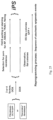
- FIG. 22 illustrates a strategy for generating iPS cells with single polycistronic construct at defined genomic locations.
- FIGS. 23 A- 23 B show generation of secondary fibroblasts carrying DOX inducible vectors, permitting reprogramming without viral transduction.
- “primary” fibroblasts carrying GFP in the OCT4 locus were transduced with all four factors using DOX inducible vectors as well as a vector carrying the tet rtTA transactivator, and “primary” iPS cells were generated after DOX induction.
- the cells were differentiated in the absence of DOX to “secondary” fibroblasts carrying the same combination of vectors that had allowed the derivation of the primary iPS cells.
- reprogramming the secondary fibroblasts to secondary iPS cells requires only DOX induction of the proviruses instead of infection with new viruses.
- FIG. 26 illustrates screening for small molecules using secondary fibroblasts with factors that can be independently induced.
- Primary fibroblasts carrying the viral M2rtTA and the OCT4-GFP marker will be transduced with tamoxifen inducible vectors transducing 3 factors and with a DOX inducible vector transducing the 4th factor (in this case cMYC).
- Primary iPS cells will be derived by culture in tamoxifen and DOX and secondary fibrboalsts will be derived. These cells, when cultured in tamoxifen, can be screened for small molecules that replace cMYC for reprogramming to secondary iPS cells.
- FIGS. 27 A- 27 C show characterization of DOX-inducible hiPSCs derived from fibroblasts from PD patients.
- FIG. 27 A shows phase contrast picture and immunofluorescence staining of hiPSC lines M 3F -1 (non-PD hiPSCs), PDA 3F -1, PDB 3F -5, PDC 3F -1, PDD 3F -1, and PDE 3F -3 for pluripotency markers SSEA4, Tra-1-60, OCT4, SOX2 and NANOG.
- FIG. 28 A shows quantitative RT-PCR for the reactivation of the endogenous pluripotency related genes NANOG, OCT4 and SOX2 in independent hiPSC lines, hESCs and primary fibroblasts.
- FIG. 28 C shows methylation analysis of the OCT4 promoter region. Light gray squares indicate unmethylated and black squares indicate methylated CpGs in the OCT4 promoter of hiPSCs and parental primary fibroblasts cells.
- FIGS. 28 A- 28 C illustrate that PD patient-derived hiPSCs carry low copy numbers of viral integrations.
- FIG. 28 A shows hematoxylin and eosin staining of teratoma sections generated from hiPSC lines A6 (non-PD hiPSCs), PDA 3F -1, PDB 3F -1, PDC 3F -1, PDD 3F -1, and PDE 3F -3 showing: Top row panels: pigmented neural epithelium; 2nd row panels: neural rosettes; 3rd row panels: intestinal epithelium; 4th row panels: bone/cartilage; bottom row panels: smooth muscle.
- FIG. 28 A shows hematoxylin and eosin staining of teratoma sections generated from hiPSC lines A6 (non-PD hiPSCs), PDA 3F -1, PDB 3F -1, PDC 3F -1, PDD 3F -1, and PDE 3F -3 showing: Top row panels: pigmented
- FIG. 28 B shows the results of Southern blot analysis of hESC line BG01, mouse embryonic fibroblast (MEF) feeder cells and the indicated PD patient-derived hiPSCs (and non-PD hiPSC line M 3F -1) for proviral integrations of XbaI digested genomic DNA using 32P-labelled DNA probes against OCT4, KLF4, SOX2 and c-MYC.
- FIG. 28 C is a table summarizing the approximate number of proviral integrations for the four reprogramming factors in hiPSCs based on Southern blot analysis shown in 28 B.
- FIGS. 29 A- 29 C show generation of PD patient-derived hiPSCs using loxP excisable reprogramming factors.
- FIG. 29 A is a schematic drawing of the DOX-inducible lentiviral construct FUW-tetO-loxP, the genomic locus after proviral integration (2lox) and after Cre-recombinase mediated excision (1lox).
- the FUW-TetO-loxP vector contains a tetracycline response element (TRE) located 5′ of a minimal CMV promoter and a unique MfeI site used for diagnostic Southern blot digests.
- the reprogramming factors are flanked by EcoRI restriction sites.
- FIGS. 31 A- 31 E shows characterization of reprogramming factor-free hiPSCs.
- 31 D shows quantitative RT-PCR for residual transgene expression of OCT4, KLF4 and SOX2 in hESCs (BG01), primary fibroblasts (PDB), primary infected fibroblasts (PDD 3F +/ ⁇ DOX), hiPSCs (M3 F3 -1), PD-derived hiPSCs (PDA 3F -1, PDB 3F -5, PDC 3F -1, PDD 3F -1, PDE 3F -3), provirus carrying PDB 2lox clones (PDB 2lox -17 and PDB 2lox -21) and the reprogramming factor free PDB 1lox clones (PDB 1lox -17Puro-5, PDB 1lox -17Puro-31, PDB 1lox -21Puro-12, PDB 1lox -21Puro-20).
- FIG. 31 E is a Venn diagram displaying the number of differentially expressed genes (p ⁇ 0.05 determined by moderated t-test, corrected for false discovery rate) between provirus-carrying PDB 2lox lines (PDB 2lox -5, PDB 2lox -17, PDB 2lox -21, PDB 2lox -22) compared to hESCs (H9, BG01) or reprogramming factor-free PDB lox lines (PDB 1lox -17Puro-5, PDB 1lox -17Puro-10, PDB 1lox -21Puro-20, PDB 1lox -21Puro-26) compared to hESCs (H9, BG01) respectively.
- FIGS. 32 A- 32 D show that transgene expression for 8 days is sufficient to reprogram human fibroblasts after primary infections.
- FIG. 32 A shows Immunofluorescence staining of primary fibroblasts (PDB) transduced with the 4 reprogramming factors OCT4, KLF4, SOX2 and c-MYC. Cells were fixed and stained for the expression of NANOG (red) and Tra-1-60 (green) at different time points (top panel at day 8; bottom panel at day 10) after DOX-induced transgene expression. No NANOG/Tra-1-60 positive cells were detected earlier than 8 days or in cultures that were not treated with DOX. NANOG and Tra-1-60 colonies were also detectable in all cultures that were stained at later time points (12, 14, 16, 18, 20 days).
- FIG. 32 A shows Immunofluorescence staining of primary fibroblasts (PDB) transduced with the 4 reprogramming factors OCT4, KLF4, SOX2 and c-MYC. Cells were fixed and stained for the expression of
- the left panel shows hiPSC clone PDB 4F -1 that was isolated from a culture that was transduced with the four reprogramming factors and exposed to DOX for 8 days.
- the right panel shows the hiPSC clone PDB 3F -12d that was isolated from a culture that was transduced with the three reprogramming factors and exposed to DOX for 12 days.
- FIG. 32 C shows quantitative RT-PCR for the reactivation of the endogenous pluripotency related genes NANOG, OCT4 and SOX2 in the following lines: hiPSC lines PDB 4F -1 and PDB 4F -2, D4, A6, hESCs and primary fibroblasts. Relative expression levels were normalized to expression of these genes in fibroblasts.
- PDB 4F -1 and PDB 4F -2 iPSCs were isolated after 8 days of transgene expression of the four reprogramming factors OCT4, SOX2, KLF4 and c-MYC.
- FIG. 32 D shows hematoxylin and eosin staining of teratoma sections generated from hiPSC line PDB 3F -12d and PDB 4F -2.
- PDB 3F -12d was derived by DOX-induced transgene expression of the three reprogramming factors OCT4, SOX2, KLF4 for 12 days.
- PDB 4F -2 was derived by DOX-induced transgene expression of the four reprogramming factors OCT4, SOX2, KLF4 and c-MYC for 8 days.
- FIG. 33 shows generation of hiPSCs carrying Cre-recombinase excisable viral reprogramming factors.
- FIG. 34 shows Southern blot analysis for excision of the reprogramming factors in hiPSCs.
- Puro indicates clones, which were isolated by puromycin selection;
- GFP indicates clones isolated by FACS sorting for EGFP (as shown in FIG. 5 A ).
- Genomic DNA was digested with MfeI and probed for proviral integrations using 32 P-labeled DNA probes against OCT4, KLF4, and SOX2.
- PDB 1lox clones indicated in blue were regarded as either partially deleted or mixed cellular populations with partial deletions of the transgenes.
- FIG. 35 shows Southern blot analysis for FUW-M2rtTA.
- Puro indicates clones which were isolated by puromycin selection;
- GFP indicates clones isolated by FACS sorting for EGFP (as shown in FIG. 30 A ).
- Genomic DNA was digested with MfeI and probed for proviral integrations using 32 P-labeled DNA probes against FUW-M2rtTA.
- Table 2 Summary of transgenic human ES or iPS cell lines used in this proposal. DOX inducible polycistronic vectors carrying different combinations of factors will be integrated into the 3′UTR of the COL1A1 locus or GFP will be inserted into the OCT4 locus or the indicated neural specific genes. The table also indicates the specific aims where the cells will be used.
- dox-inducible transgenes The study of induced pluripotency is complicated by the need for infection with high titer retroviral vectors resulting in genetically heterogeneous cell populations.
- dox doxycycline
- dox dox-inducible transgene system 16
- dox-inducible lentiviral vectors encoding the 4 reprogramming factors.
- MEFs Mouse embryonic fibroblasts
- ROSA-M2rtTA ROSA locus
- GFP green fluorescent protein
- NGFP endogenous Nanog locus
- somatic tissues that were composed of genetically homogenous cells carrying identical proviral insertions known to achieve reprogramming in primary fibroblasts
- the resulting dox-inducible iPS cell chimeras were allowed to gestate until E13.5, at which point MEFs were isolated.
- Puromycin selection was then used to select against cells derived from the host blastocyst leaving only iPS-derived cells.
- the fibroblasts derived from the Nanog-neo line never formed a confluent monolayer upon dox addition, but generated large, three-dimensional colonies. After 12 days of dox administration, iPS cell colonies with ES cell morphology were readily visible in all three cultures ( FIG. 2 A , arrows).
- MEFs from the three lines were cultured in dox-containing media and flow cytometric analysis was utilized to monitor the reactivation of SSEA1 and GFP ( FIG. 2 C ). All three secondary MEFs exhibited a gradual increase of SSEA1-positive cells over the time course, but some differences in timing were observed.
- the NNeo MEFs showed the earliest increase of SSEA1-positive cells from 1.3% to 17.8% between days 8 and 11.
- the NGFP2 MEFs showed a similar increase but at a much later time point (from 4.4% to 29% between days 14 and 18).
- MEFs from the iPS cell line NGFP3 exhibited a slower, gradual activation of SSEA1 reaching about 10% on day 14.
- the first GFP-positive cells were detected as early as day 14 in NGFP2 and on day 18 in NGFP3 MEFs.
- FIG. 3 F shows that addition of 5-Aza to the medium increased the reprogramming efficiency of MEFs from the NGFP3 line whereas TSA treatment had no obvious effect on the number of colonies.
- FIG. 8 D bone marrow derived mesenchymal stem cells (MSCs) and tail tip fibroblasts (TTFs) isolated from NNeo and NGFP2 chimeras. These cells represent two mesenchymal populations that are amenable to reprogramming by direct infection 1, 4, 12 (Supplementary FIG. 8 D ). As with intestinal cells, secondary NGFP2 MSCs and TTFs were capable of generating iPS cells in response to dox, while those derived from NNeo chimeras were not ( FIG. 5 A- 5 F , FIG. 8 A, 8 C ).
- FIG. 5 G Cells isolated from the epidermis of NNeo chimeras were first propagated in the absence of doxycycline in growth conditions optimized for keratinocytes Homogeneous epithelial cultures were obtained ( FIG. 5 G ), and doxycycline was added to the media. Clusters of epithelial cells proliferated and changed their morphology over time. After twelve days the medium was changed to doxycycline containing ES cell medium ( FIG. 5 H ), and seven days later neomycin was added. Neo-resistant cells growing in tight colonies resembled ES cells ( FIG. 5 I ) and were passaged onto ⁇ -irradiated feeder cells at which point the cultures were maintained in the absence of dox and expressed endogenous Nanog ( FIG. 9 D ).
- Brains from NNeo chimeras were dissected and a tissue block around the lateral ventricles was dissociated into single cells and plated onto uncoated culture dishes in EGF and FGF2-containing serum-free media (N3EF) in the presence of puromycin to select for secondary cells. 4 weeks later neurospheres had formed that were subsequently plated onto polyornithine/laminin coated dishes in either ES cell or N3EF media containing dox to activate the lentiviral transgenes. As expected for neural precursors, the cells exposed to the serum-containing ES cell media differentiated into flat astrocytic cells and stopped dividing ( FIG. 5 J ).
- Reprogramming of the somatic epigenome to a pluripotent, embryonic state through the ectopic expression of the 4 transcription factors Klf4, Sox2, c-Myc, and Oct4 is a slow and inefficient process.
- the current method for induction of reprogramming is through retroviral gene delivery resulting in heterogeneous cell populations with proviral integrations varying in both number and genomic location, offering an explanation for the variability and inefficiency of direct reprogramming.
- a novel system for reprogramming genetically homogeneous cell populations are described.
- the observed reprogramming efficiency of secondary MEFs was as high as 4% which is comparable to the reprogramming efficiency of mature B-cells 22 and vastly higher than the estimated 0.1% efficiency using de novo infection and drug selection, and about 8 fold higher than what has been reported using morphological selection criteria 1, 11, 12 . It has been well documented that iPS cells derived from infected MEFs carry on average 15 different proviral copies suggesting strong selection for the small fraction of the infected cells that carry the “correct” number of proviruses, or that express the 4 factors with the appropriate stoichiometry for successful reprogramming.
- the drug-inducible system described here represents a novel reprogramming platform with predictable and highly reproducible kinetics and efficiencies (see Supplementary FIG. 7 B ) that should facilitate the study of early molecular events leading to epigenetic reprogramming.
- the genetic homogeneity of secondary cell types provides the feasibility of chemical and genetic screening approaches to enhance the reprogramming efficiency.
- the DNA demethylating agent 5-Aza-deoxycytidine substantially enhances the reprogramming efficiency.
- screens can also be applied to identify compounds replacing the original reprogramming factors. Because the reprogrammed state is not dependent on the exogenous factors, the transgenes can be genetically excised and secondary cells can be generated by chimera formation that lack a particular reprogramming factor 15 .
- lentiviral vectors containing Klf4, Sox2, Oct4, and c-Myc under control of the tetracycline operator and a minimal CMV promoter has been described previously 14 .
- Replication-incompetent lentiviral particles were packaged in 293T cells with a VSV-G coat and used to infect MEFs containing M2rtTA and PGK-Puro resistance gene at the R26 locus 17 , as well as either a neomycin resistance or GFP allele targeted to the endogenous Nanog locus 1, 11 .
- a flat-tip microinjection pipette with an internal diameter of 12-15 mm was used for iPS cell injection using a Piezo micromanipulator.
- About 10 iPS cells were injected into the blastocyst cavity and blastocysts were placed in KSOM (Specialty Media, Phillipsburg, NJ) and incubated at 37° C. until they were transferred to recipient females.
- Fifteen injected blastocysts were transferred to the uterine homs of psuedopregnant C57/B6 ⁇ DBA/1 F1 females at 2.5 days post coitum.
- 2 ⁇ 10 6 cells were injected subcutaneously into the flanks of recipient SCID mice, and tumors were isolated for histological analysis 3-6 weeks later. All animals were treated in accordance with institutional IACUC guidelines.
- marrow was isolated from secondary chimeric mice (or from Colll-TetO-Oct4, Rosa26-M2rtTA mice 16 for direct infections) from the femur and tibia after removal of the condyles at the growth plate by flushing with a syringe and 30-gauge needle containing DMEM+5% Fetal BovineSerum (FBS) (Hyclone, Thermo Fisher Scientific).
- FBS Fetal BovineSerum
- Mesenchymal stem cells were selected through differential plating on tissue culture plates for 72 hours in ⁇ -MEM supplemented with 15% FBS (HyClone).
- Colony formation of MSCs in culture was carried out by plating 4 ⁇ 10 6 nucleated cells from freshly isolated whole marrow onto 10 cm plates and allowed to expand for 5 days in the presence of puromycin to eliminate host-blastocyst derived cells, after which dox was introduced to induce reprogramming.
- Cultures derived from adrenal glands, muscle, and kidneys were dissected, mechanically dissociated, and digested in trypsin at 37° C. for 20 minutes prior to plating on gelatin-coated culture dishes with ES media containing dox.
- FACS buffer PBS+5% fetal bovine serum
- 10 6 cells were stained with 10 ⁇ l of APC-conjugated anti-SSEA1 antibody in a 100 ⁇ l volume for 30 minutes, cells were then washed twice in PBS. Cells were then washed once with wash buffer and resuspended in FACS buffer for analysis on a FACS-calibur cell sorter.
- the first round of PCR was done as follows: 94° C. for 4 minutes; five cycles of 94° C. for 30 seconds, 56° C. for 1 minute ( ⁇ 1° C. per cycle), 72° C. for 1 minute; and 30 cycles of 94° C. for 30 seconds, 51° C. for 45 seconds, and 72° C. for 1 minute, 20 seconds.
- the second round of PCR was 94° C. for 4 minutes; 30 cycles of 94° C. for 30 seconds, 53.5° C. for 1 minute, and 72° C. for 1 minute 20 seconds.
- the resulting amplified products were gel-purified (Zymogen, Zymo Research, Orange, CA), subcloned into the TOPO TA vector (Invitrogen), and sequenced. Southern blotting of genomic DNA was carried out by digesting 10 ⁇ g of DNA with Spel (which cuts once in the lentiviral vector backbone) followed by hybridization with random primed full-length cDNA probes for the four factors.
- GAPDH mRNA was amplified using the following: F, 5-TTCACCACCATGGAGAAGGC-3′ SEQ ID NO: 17; and R, 5′-CCCTTTTGGCTCCACCCT-3′ SEQ ID NO: 18. Data were extracted from the linear range of amplification. All graphs of qRT-PCR data shown represent samples of RNA that were DNase treated, reverse transcribed, and amplified in parallel to avoid variation inherent in these procedures. Error bars represent standard deviation of the mean of triplicate reactions.
- DOX inducible retroviral vectors have been important to define the sequential activation of pluripotency markers and the minimum time of vector expression during reprogramming of somatic mouse cells.
- DOX inducible lentivirus vectors Following the same strategy as used for murine genes we have generated lentiviral vectors that transduce the human OCT4, SOX2, KLF4 and C-MYC c-DNAs either constitutively or under the control of a DOX inducible promoter [Brambrink, 2008 #6877]. To generate a DOX inducible system we infected human fibroblasts with a lentiviral vector carrying the rtTA transactivator. FIG. 11 A shows high DOX-dependent expression of OCT4, SOX2, and KLF4 in fibroblasts transduced with the respective DOX inducible vectors.
- One important concept is the use of two different regulatable systems, each controlling expression of a subset of the factors. For example, one might place 3 of the factors under control of a first inducible (e.g., dox-inducible) promoter and the 4 th factor under control of a second inducible (e.g., tamoxifen-inducible) promoter. Then, one could generate an iPS cell by inducing expression from both promoters, generate a mouse from this iPS cell, and isolate fibroblasts (or any other cell type) from the mouse. These fibroblasts would be genetically homogenous and would be reprogrammable without need for viral infection.
- a first inducible e.g., dox-inducible
- tamoxifen-inducible e.g., tamoxifen-inducible
- a number of variations are possible; for example, one might stably induce expression of 3 factors and transiently induce expression of the 4 th factor, etc. Also, one can modulate expression levels of the factors by using different concentrations of inducing agent.
- Another approach is to place the gene that encodes one of the factors between sites for a recombinase and then induce expression of the recombinase to turn off expression of that factor.
- Recombinase expression could be induced by infecting with a viral vector (e.g., Adenovirus-Cre).
- Hanna, et al, Science, 318, 1920-1923 (2007) describes such an approach, which was used to reduce the potential risk of tumor formation due to c-Myc transgene expression—Cells were infected with retroviruses encoding for Oct4, Sox2, and Klf4 factors and a lentivirus encoding a 2-lox c-Myc cDNA. iPS cells generated from these cells were infected with an adenovirus encoding Cre recombinase to delete the lentivirus-transduced c-Myc copies.
- IVS Internal ribosomal entry sites
- the self-cleaving 18-22 amino acids long 2A peptides mediate ‘ribosomal skipping’ between the proline and glycine residues and inhibit peptide bond formation without affecting downstream translation.
- These peptides allow multiple proteins to be encoded as polyproteins, which dissociate into component proteins upon translation.
- Use of the term “self-cleaving” is not intended to imply proteolytic cleavage reaction.
- An exemplary 2A sequence is VKQTLNFDLLKLAGDVESNPGP (SEQ ID NO: 35) from FMDV, where underlined residues are conserved in many 2A peptides.
- the C terminus of cardiovirus 2A peptides is conserved, shows a high degree of similarity with FMDV 2A peptide, and has been shown to also mediate self-cleavage (Donnelly, M L, et al., J. Gen. Virol., 78, 13-21 (1997).
- FDMV 2A peptide has been shown to mediate cleavage of an artificial polyprotein (Ryan, M D and Drew, J., EMBO J., 13, 928-933 (1994).
- the construct comprises four coding regions each encoding a reprogramming factor, wherein adjacent coding regions are separated by a self-cleaving peptide.
- the invention thus provides constructs that encode a polyprotein that comprises 2, 3, or 4 reprogramming factors, separated by self-cleaving peptides.
- the construct comprises expression control element(s), e.g., a promoter, suitable to direct expression in mammalian cells, wherein the portion of the construct that encodes the polyprotein is operably linked to the expression control element(s).
- the invention thus provides an expression cassette comprising a nucleic acid that encodes a polyprotein comprising the reprogramming factors, each reprogramming factor being linked to at least one other reprogramming factor by a self-cleaving peptide, operably linked to a promoter (or other suitable expression control element).
- the promoter drives transcription of a polycistronic message that encodes the reprogramming factors, each reprogramming factor being linked to at least one other reprogramming factor by a self-cleaving peptide.
- the promoter can be a viral promoter (e.g., a CMV promoter) or a mammalian promoter (e.g., a PGK promoter).
- the recombinase sites need not be directly adjacent to the region encoding the polyprotein but will be positioned such that a region whose eventual removal from the genome is desired is located between the sites.
- the recombinase sites are on the 5′ and 3′ ends of an expression cassette. Excision may result in a residual copy of the recombinase site remaining in the genome, which in some embodiments is the only genetic change resulting from the reprogramming process.
- the construct comprises a single recombinase site, wherein the site is copied during insertion of the construct into the genome such that at least the portion of the construct that encodes polyprotein comprising the factors (and, optionally, any other portion of the construct whose eventual removal from the genome is desired) is flanked by two recombinase sites after integration into the genome.
- the recombinase site can be in the 3′ LTR of a retroviral (e.g., lentiviral) vector (see, e.g., Example 4).
- the invention provides vectors comprising the polycistronic nucleic acid constructs.
- the vectors are retroviral vectors, e.g., lentiviral vectors.
- the vectors are non-retroviral vectors, e.g., which may be viral (e.g., adenoviral) or non-viral.
- the invention provides cells and cell lines (e.g., somatic cells and cell lines such as fibroblasts, keratinocytes, and cells of other types discussed herein) in which a polycistronic nucleic acid construct or expression cassette (e.g., any of the constructs or expression cassettes described herein) is integrated into the genome.
- the cells are rodent cells, e.g., a murine cells.
- the cells are primate cells, e.g., human cells.
- the nucleic acid construct or cassette is targeted to a specific locus in the genome, e.g., using homologous recombination.
- the locus is one that is dispensable for normal development of most or all cell types in the body of a mammal.
- the locus is one into which insertion does not affect the ability to derive pluripotent iPS cells from a somatic cell having an insertion in the locus.
- the locus is one into which insertion would not perturb pluripotency of an ES cell.
- the locus is the COL1A1 locus or the AAV integration locus.
- the locus comprises a constitutive promoter.
- the construct or cassette is targeted so that expression of the polycistronic message encoding the polypeptide comprising the factors is driven from an endogenous promoter present in the locus to which the construct or cassette is targeted.
- the invention further provides pluripotent reprogrammed cells (iPS cells) generated from the somatic cells that harbor the nucleic acid construct or expression cassette in their genome.
- iPS cells can be used for any purpose contemplated for pluripotent cells.
- differentiated cell lines e.g., neural cells, hematopoietic cells, muscle cells, cardiac cells
- Exemplary somatic cells and iPS cell generated therefrom are described in Example 3.
- the polycistronic vector system may simplify the study of reprogramming mechanisms and facilitates the excision of the vector.
- excision results in removal of at least the exogenous sequences encoding the reprogramming factors.
- excision results in iPS cells that carry no genetic modification other than, in some embodiments, a residual recombinase site. In other embodiments, there are no more than 2, 3, 4, or 5 residual recombinase sites.
- reprogramming cells containing a single integrated construct will increase the likelihood or ease of recovering transgene-free iPS cells using recombinase-based approaches. It is also contemplated that polycistronic vectors encoding 2, 3, or 4 factors may be used in combination with small molecules, proteins, or other agents that enhance reprogramming and/or that substitute for one or more factors not encoded by the polycistronic vector.
- Example 4 describes experiments in which human induced pluripotent stem cells (hiPSCs) free of reprogramming factors were derived using Cre-recombinase excisable viruses from fibroblasts from individuals with Parkinson's disease (PD).
- hiPSCs human induced pluripotent stem cells
- PD Parkinson's disease
- iPS cells carrying no exogenous genes encoding reprogramming factors are derived as described in Example 4 or using similar methods, except that a single vector comprising a polycistronic nucleic acid construct encoding a polyprotein comprising multiple (2, 3, or 4 factors) is used rather than multiple vectors encoding single factors.
- Example 4 can also be used with multiple vectors encoding individual factors in order to obtain iPS cells without exogenous genes encoding reprogramming factors, wherein the resulting iPS cells have only a small number of residual recombinase sites.
- fibroblasts from individuals with PD were used as an exemplary cell type in Example 4, the methods are applicable to derive iPS cells with minimal genetic alteration from normal somatic cells (e.g., fibroblasts or other cell types such as keratinocytes, intestinal cells, blood cells) or from somatic cells from individuals with a disease of interest.
- the gene encoding the transactivator is also flanked by recombinase sites, so that it is removed from the genome as well.
- An exciting potential of the iPS system is to derive patient specific pluripotent cells.
- Work described herein describes protocols that will allow the study of complex human diseases in vitro using patient specific iPS cells.
- patient specific iPS cells are derived from deep skin biopsies.
- peripheral blood as donor material for generating iPS cells.
- differentiated cells from undifferentiated ES cells is facilitated by markers inserted into lineage specific endogenous genes that can be used for the isolation of a desired differentiated cell type.
- Our preliminary experiments demonstrated targeting of the OCT4 as well as the COL1A1 locus with GFP or drug resistance markers. Accordingly a goal was togenerate ES and iPS cells that carry drug resistance markers and/or GFP (or other detectable marker) sequences in genes that are expressed in cells of the neural or other lineage and can be used for screening or selection of differentiated cell types that are affected in diseases such as Alzheimer's and Parkinson's.
- targeting vectors containing GFP and neo resistance markers separated by 2A sequences will be constructed from isogenic genomic DNA of BGO2 or H9 ES cells using routine procedures.
- the DNA will be electroporated into the cells following published procedures [Costa, 2007 #6868], and DNA from drug resistant colonies will be isolated and analyzed for correct targeting.
- We will target genes that are activated at different times during neural differentiation and in different subsets of neurons as detailed below.
- SOX1 The transcription factor SOX1 is the earliest known gene that is exclusively expressed in neural precursors of the mouse [Aubert, 2003 #6841]. GFP inserted into this gene will serve as a convenient marker for selecting huES or iPS cell-derived neural precursor cells.
- FOXG1 Expression of this gene has been demonstrated in proliferating telencephalic precursor cells and in acetyl-cholinergic neurons of the basal forebrain [Hebert, 2000 #6844], cells that are affected in Alzheimer's.
- the COL1A1 locus is highly recombinogenic in mouse cells [Beard, 2006 #6199] and targeting of this locus may not be representative of other non-expressed genes. Thus, because our intent is to target non-expressed genes by homologous recombination, this aim poses a challenge.
- the integrated vectors Upon addition of DOX to such cultures the integrated vectors will be reactivated resulting in the consistent generation of “secondary” iPS cells without requiring the new transduction of factors ( FIG. 23 B ). This can be used to generate human secondary iPS cells (or mouse, monkey, etc.), without going through the process of generating an animal from the primary iPS cell.
- DOX inducible polycistronic vectors FIG. 13 A- 13 C
- iPS cells Primary iPS cells will be selected as above with the three factors being induced by DOX, the KLF4 virus will be deleted by Cre transduction [Hanna, 2007 #6781] and secondary fibroblasts lacking vKLF4 will be derived by in vitro differentiation. These cells can be screened for small molecules that replace the need for KLF4 in reprogramming (see later, Aim D) or for streamlining transient transfection protocols (Aim B.2, 4).
- “Secondary” clonal fibroblasts that carry a specific and predetermined combination of proviruses We have recently shown that “secondary” mouse iPS cells can be derived from “primary” iPS cells that had been generated by infection of fibroblasts with DOX inducible lentiviruses transducing the four transcription factors Oct4, Sox2, c-myc and Klf4 [Hanna, 2008 #6842]. Because the “right” combination and number of proviral copies was carried in the “secondary” fibroblasts, no viral infection was needed to induce reprogramming of B cells to secondary iPS cells.
- the screening of small molecule libraries will be performed in collaboration with the laboratory of S. Ding at Scripps (see letter by S. Ding).
- the Ding laboratory has developed and optimized cell-based phenotypic high throughput screens [Xu, 2008 #6875] and identified the small molecule pluripotin that sustains self renewal of ES cells in chemically defined medium and in the absence of LIF [Chen, 2006 #6871].
- the screen was based upon the expression of an Oct4 promoter driven GFP marker.
- Example 3 Reprogramming of Murine and Human Somatic Cells Using a Single Polycistronic Vector
- Proteins were then transferred onto Immobilon-P membranes (Millipore) using a semi-dry transfer apparatus. Membranes were blocked in PBS, 0.01% Tween 20 containing 2% nonfat powdered milk (Bio-Rad). Proteins were detected by incubating with antibodies at a concentration of 50 ng/ml in blocking solution. Antibodies used were Oct4 (h-134 Santa Cruz Biotechnology); Sox2 (mouse monoclonal R&D Biosystems); c-Myc (06-340 Upstate); Klf4 (H-180 Santa Cruz Biotechnology); GAPDH (sc-25778 Santa Cruz Biotechnology).
- Oct4 F (SEQ ID NO: 24) 5′-ACATCGCCAATCAGCTTGG-3′ and R, (SEQ ID NO: 25) 5′-AGAACCATACTCGAACCACATCC-3′ Sox2 F, (SEQ ID NO: 26) 5′-ACAGATGCAACCGATGCACC-3′ and R, (SEQ ID NO: 27) 5′-TGGAGTTGTACTGCAGGGCG-3′ 4F2A (E2A-cMyc) F, (SEQ ID NO: 28) 5′-GGCTGGAGATGTTGAGAGCAA-3′ and R, (SEQ ID NO: 29) 5′-AAAGGAAATCCAGTGGCGC-3′ GAPDH F, (SEQ ID NO: 30) 5′-TTCACCACCATGGAGAAGGC-3′ and R, (SEQ ID NO: 31) 5′-CCCTTTTGGCTCCACCCT-3′ Error bars represent s.d. of the mean of triplicate reactions.
- Diploid blastocysts (94-98 h after hCG injection) were placed in a drop of Hepes-CZB medium under mineral oil. A flat tip microinjection pipette with an internal diameter of 16 m was used for iPS cell injections. Each blastocyst received 8-10 iPS cells. After injection, blastocysts were cultured in potassium simplex optimization medium (KSOM) and placed at 37° C. until transferred to recipient females. About 10 injected blastocysts were transferred to each uterine horn of 2.5-day-postcoitum pseudo-pregnant B6D2F1 female. Pups were recovered at day 19.5 and fostered to lactating B6D2F1 mothers when necessary. Teratoma formation was performed by depositing 2 ⁇ 10 ⁇ circumflex over ( ) ⁇ 6 cells under the flanks of recipient SCID or Rag2 ⁇ / ⁇ mice. Tumors were isolated 3-6 weeks later for histological analysis.
- KSOM potassium simplex optimization medium
- hiPSCs were collected by collagenase treatment (1.5 mg/ml) and separated from feeder cells by subsequent washes with medium and sedimentation of iPSC colonies.
- iPSC aggregates were collected by centrifugation and resuspended in a ratio of 10 ⁇ circumflex over ( ) ⁇ 6 cells in 250l1 of iPSC culture media.
- iPSCs were injected subcutaneously by 21 gauge needle in the back of SCID mice (Taconic). A tumor developed within 6 weeks and the animal was sacrificed before tumor size exceeded 1.5 cm in diameter. Teratomas were isolated after sacrificing the mice and fixed in formalin. After sectioning, teratomas were diagnosed base on hematoxylin and eosin staining. Karyotype analysis was done with CLGenetics (Madison, WI).
- Induction of differentiation of neural progenitors was performed by withdrowal of FGF2 and EGF from culture medium for 5 days.
- Cells were fixed in 4% paraformaldehyde for 20 min and stained for human nestin (Chemicon; 1:100) and Tuj-1 (1:100) and subsequently washed 3 times with PBS and incubated with fluorophore-labeled appropriate secondary antibodies purchased from Jackson Immunoresearch.
- Specimens were analyzed on an Olympus Fluorescence microscope and images were acquired with a Zeiss Axiocam camera.
- Vectors were constructed with different combinations of two, three, or all four reprogramming factors from one promoter. The goal was to generate polycistronic viral vectors that would express multiple reprogramming genes from a single promoter using 2A peptides. For this one, two, or three 2A oligopeptides containing unique restriction sites were ligated into FUW lentivirus (18) backbones to allow efficient cloning of Oct4, Sox2, c-Myc and Klf4 each separated by a different 2A sequence. Vectors carrying four, three or two factors consecutively with different combinations of F2A, T2A, E2A or P2A sequences ( FIGS.
- FIG. 13 B shows that iPS cells were obtained that expressed AP, SSEA1, Nanog and Oct4. Moreover adult chimeras have been generated from iPS lines infected with the four-factor 2A vector plus Oct4 Moloney virus. To determine the number of proviral integrations, a Southern blot was sequentially hybridized with a Sox2, Klf4, c-myc and Oct4 probe.
- FIG. 13 C shows that a single polycistronic vector was integrated in 2 of 3 different tested iPS lines and 2 proviruses were carried in the third line (in this line, 4FO #14, the c-myc sequences were deleted in one of the proviruses).
- a tetracycline inducible lentivirus vector was constructed where expression of the genes was controlled from the tetracycline operator minimal promoter (tetOP; FIG. 14 C ).
- tetOP tetracycline operator minimal promoter
- MEFs were infected with the polycistronic vector (referred below to as “4F2A”) as well as a constitutive FUW lentivirus carrying the tetracycline controllable trans-activator (M2rtTA; abbreviated as rtTA).
- M2rtTA constitutive FUW lentivirus carrying the tetracycline controllable trans-activator
- Two independent experiments were performed and drug inducible expression of the virus was tested 3 days post-infection by qRT-PCR.
- TTFs tail-tip fibroblasts
- 4F2A+rtTA vectors Similar to MEFs, typical morphological changes were observed a few days after addition of DOX media. Colonies appeared around 8 days and continued to expand until they were picked (day 16) based on morphology. After several passages four stable iPS cell lines were established that stained positive for all pluripotency markers (Nanog, Oct4, SSEA1, AP) ( FIG. 15 C ). MEF iPS cell lines were injected subcutaneously into SCID mice and were shown to induce teratomas that contained differentiated cells of all three germ layers ( FIG.
- MEFs were infected with the 4F2A and rtTA vectors and plated at 0.25 ⁇ 10 ⁇ circumflex over ( ) ⁇ 6 per 10 cm plate culture dish. About 70% of the MEFs were infected as estimated by immunostaining of Oct4 at 48 hours after infection ( FIG. 19 A ). Cells were cultured in ES media containing DOX for 20 days and subsequently transferred to ES cell medium until GFP+ colonies were counted on day 25. An average of ⁇ 14.7 ⁇ 4 colonies were detected in three independent dishes (10+10+17) indicating a relative efficiency of 0.0001%. This is one to two orders of magnitude lower than that of ‘primary’ infected fibroblasts (3, 7).
- NEFK neonatal human foreskin keratinocytes
- the fraction of infected cells was 10% as determined by staining for Oct4 at 48 hours after transduction ( FIG. 20 A ).
- Cells were incubated in keratinocyte medium+DOX and allowed to grow for 6 days until they were passaged and cultured in hESC media+DOX on gelatinized plates. Colonies were first detected at day 12 and most displayed transformed morphology with a few colonies exhibiting a distinct appearance that resembled hESC-like morphology.
- genomic DNA was extracted and subjected to Southern blot analysis using an enzyme that does not cut in the vector sequences. Probes for all four reprogramming factors show hybridization to similar molecular weight band(s) again indicating they were carried on a single virus. Two different digests (XbaI & BamHI) show the 4F2A proviral copy number is three (#1.1) and two (#3) respectively ( FIG. 21 A-B ).
- Ker-iPS #1.1 was injected subcutaneously into SCID mice. These cells induced teratomas and after histological examination differentiated into cells of all three germ layers ( FIG. 17 D ). In addition, Ker-iPS #1.1 cells, when subjected to an in-vitro neural differentiation protocol produced nestin+ neural progenitor cell populations as well as Tuj1+ post-mitotic neurons as detected by immunostaining. ( FIG. 17 E ).
- GFP GFP reporter gene present
- ES expression of ES cell markers (AP, SSEA1, Oct4 or Sox2)
- TF teratoma formation
- PC postnatal chimeras.
- Mouse chimerism was estimated by agouti coat color.
- the lower reprogramming efficiency is due to the stochiometry of factor expression from the polycistronic vector, which may be suboptimal for inducing reprogramming.
- Transduction with separate vectors allows integration of different numbers of proviruses for each factor, therefore reprogramming may select for a specific set of proviral integrations that result in high expression or an optimal stochiometry between the different factors.
- the 2A system has been reported to support near equimolar protein expression in vivo (17).
- Nanog-GFP positive cells were detected as early as 16 days after DOX induction in contrast to GFP positive cells observed 22-25 days after 4F2A vector transduction, consistent with less optimal reprogramming.
- iPS cells frequently carry multiple Oct4 or Klf4 proviruses, consistently fewer Sox2 proviruses were found suggesting that a high level of Sox2 expression may perhaps be unfavorable for reprogramming (24).
- fibroblasts were cultured in fibroblast medium [DMEM supplemented with 15% FBS (Hyclone), 1 mM glutamine (Invitrogen), 1% nonessential amino acids (Invitrogen) and penicillin/streptomycin (Invitrogen)].
- HiPSCs and the hESC lines BG01 and BG02 were maintained on mitomycin C (MMC)-inactivated mouse embryonic fibroblast (MEF) feeder layers in hESC medium [DMEM/F12 (Invitrogen) supplemented with 15% FBS (Hyclone), 5% KnockOutTM Serum Replacement (Invitrogen), 1 mM glutamine (Invitrogen), 1% nonessential amino acids (Invitrogen), 0.1 mM ⁇ -mercaptoethanol (Sigma) and 4 ng/ml FGF2 (R&D systems)].
- MMC mitomycin C
- FBS Hyclone
- KnockOutTM Serum Replacement Invitrogen
- 1 mM glutamine Invitrogen
- 1% nonessential amino acids Invitrogen
- 0.1 mM ⁇ -mercaptoethanol Sigma
- 4 ng/ml FGF2 R&D systems
- ESC/hiPSC colonies were harvested using 1.5 mg/ml collagenase type IV (Invitrogen), separated from the MEF feeder cells by gravity, gently triturated and cultured for 10 days in non-adherent suspension culture dishes (Corning) in DMEM supplemented with 15% FBS.
- collagenase type IV Invitrogen
- hiPSC lines were cultured in Rho Kinase (ROCK)-inhibitor (Calbiochem; Y-27632) 24 hours prior to electroporation.
- Cell were harvested using 0.05% trypsin/EDTA solution (Invitrogen) and 1 ⁇ 10 7 cells resuspended in PBS were transfected with either pCre-PAC (50 jtg; Taniguchi et al., 1998) or co-transfected with pTurbo-Cre (40 ⁇ g; Genbank Accession Number AF334827) and pEGFP-N1 (10 ⁇ g; Clontech) by electroporation as described previously (Costa et al., 2007; Gene Pulser Xcell System, Bio-Rad: 250 V, 500 ⁇ F, 0.4 cm cuvettes).
- ROCK Rho Kinase
- Cre-recombinase expressing cells were selected using one of the following methods: 1) addition of puromycin (2 jtg/ml) 2 days after electroporation for a period of 48 hours. 2) FACS sorting (FACS-Aria; BD-Biosciences) of a single cell suspension for EGFP expressing cells 60 hours after electroporation followed by replating at a low density in ROCK-inhibitor containing hESC medium. Individual colonies were picked 10 to 14 days after electroporation.
- a Not I/Bsu36 I fragment containing the tetracycline operator/minimal CMV promoter and the human c-DNAs for either KLF4, OCT4 or SOX2 were subcloned from each FUW-tetO vector into the Not I/BSU36 I sites of the FUGW-loxP, which contains a loxP site in the 3′LTR (Hanna et al., 2007).
- VSVG coated lentiviruses were generated in 293 cells as described previously (Brambrink et al., 2008). Briefly, culture medium was changed 12 hours post-transfection and virus-containing supernatant was collected 60-72 hours post transfection. Viral supernatant was filtered through a 0.45 am filter. Virus-containing supernatants were pooled for 3 and 4 factor infections and supplemented with FUW-M2rtTA virus and an equal volume of fresh culture medium. 1 ⁇ 10 6 human fibroblasts were seeded 24 hours before transduction in T75 flasks. Four consecutive infections in the presence of 2 ⁇ g/ml of polybrene were performed over a period of 48 hours. Culture medium was changed 12 hours after the last infection.
- fibroblasts were passaged using trypsin and re-plated at different densities between 5 ⁇ 10 4 and 2 ⁇ 10 5 cells per 10 cm 2 on gelatin coated dishes.
- culture medium was replaced 48 hours later by hESC medium supplemented with DOX (Sigma-Aldrich; 2 ⁇ g/ml).
- HiPSCs colonies were picked manually based on morphology between 3 and 5 weeks after DOX-induction and manually maintained and passaged according hESC protocols in the absence of DOX.
- 1 ⁇ 10 5 human fibroblasts were seeded onto 10 cm 2 gelatin coated dishes. Reprogramming efficiencies were calculated after 20 days based on immunocytochemistry for the pluripotency markers Tra-1-60 and NANOG.
- Samples were prepared for hybridization using 15 ⁇ g biotinylated cRNA in a 1 ⁇ hybridization cocktail according the Affymetrix hybridization manual.
- GeneChip arrays (Human U133 2.0) were hybridized in a GeneChip Hybridization Oven at 45° C. for 16 hours at 60 RPM.
- Washing was done using a GeneChip Fluidics Station 450 according to the manufacturer's instructions, using the buffers provided in the Affymetrix GeneChip Hybridization, Wash and Stain Kit. Arrays were scanned on a GeneChip Scanner 3000 and images were extracted and analyzed using GeneChip Operating Software v1.4.
- U133 Plus 2.0 microarrays were processed using the MASS algorithm and absent/present calls for each probeset were determined using the standard Affymetrix algorithm, both as implemented in Bioconductor. Probesets that were absent in all samples were removed for subsequent analysis. Differential expression was determined a moderated t-test using the ‘limma’ package in R (corrected for false discovery rate) or by fold change. Where a gene was represented by multiple probesets (based on annotation from Affymetrix), gene expression log-ratios and p-values were calculated as the mean and minimum of these probesets, respectively. Hierarchical clustering was performed on log-transformed gene expression ratios using uncentered Pearson correlation and pairwise average linkage. Correlations were compared using Fisher's Z transformation. Confidence of the hierarchical clustering was computed using multiscale bootstrap resampling with the R package ‘pvclust’.
- Real-time PCR was performed in an ABI Prism 7000 (Applied Biosystems) with Platinum SYBR green pPCR SuperMIX-UDG with ROX (Invitrogen) using primers that were in part previously described (Hockemeyer et al., 2008; Yu et al., 2007) and in part are described in Soldner, et al., 2009, Supplemental Experimental Procedures.
- HiPSCs were collected by collagenase treatment (1.5 mg/ml) and separated from feeder cells by subsequent washes with medium and sedimentation by gravity. HiPSC aggregates were collected by centrifugation and resuspended in 250 ⁇ l of phosphate buffered saline (PBS). HiPSCs were injected subcutaneously in the back of SCID mice (Taconic). Tumors generally developed within 4-8 weeks and animals were sacrificed before tumor size exceeded 1.5 cm in diameter. Teratomas were isolated after sacrificing the mice and fixed in formalin. After sectioning, teratomas were diagnosed based on hematoxylin and eosin staining.
- PBS phosphate buffered saline
- OCT4 Forward (SEQ ID NO: 32) ATTTGTTTTTTGGGTAGTTAAAGGT
- SSEA4 mouse monoclonal, Developmental Studies Hybridoma Bank
- Tra 1-60 mouse monoclonal, Chemicon International
- hSOX2 goat polyclonal, R&D Systems
- Oct-3/4 mouse monoclonal, Santa Cruz Biotechnology
- hNANOG goat polyclonal R&D Systems
- appropriate Molecular Probes Alexa Fluor® dye conjugated secondary antibodies were used.
- XbaI, EcoRI or MfeI digested genomic DNA was separated on a 0.7% agarose gel, transferred to a nylon membrane (Amersham) and hybridized with 32 P random primer (Stratagene) labeled probes for OCT4 (EcoRI-PstI fragment of pFUW-tetO-hOCT4 plasmid), KLF4 (full length hKLF4 cDNA), c-MYC (full length c-MYC cDNA), SOX2 (FspI-EcoRI fragment of pFUW-tetO-hSOX2 plasmid) and M2rtTA (380 bp C-terminal fragment of the M2rtTA c-DNA).
- OCT4 EcoRI-PstI fragment of pFUW-tetO-hOCT4 plasmid
- KLF4 full length hKLF4 cDNA
- c-MYC full length c-MY
- Microarray data are available at the NCBI Gene Expression Omnibus database under the series accession number GSE14711.
- fibroblasts from five patients with idiopathic Parkinson's disease can be efficiently reprogrammed.
- PD idiopathic Parkinson's disease
- hiPSCs human induced pluripotent stem cells
- Factor-free iPSCs maintain a pluripotent state and show a global gene expression profile, more closely related to hESCs than to hiPSCs carrying the transgenes.
- Our results indicate that residual transgene expression in virus-carrying hiPSCs can affect their molecular characteristics and suggest that factor-free hiPSCs therefore represent a more suitable source of cells for modeling of human disease.
- Dermal fibroblasts from five patients with idiopathic PD (age of biopsy between 53 and 85 years) and from two unaffected subjects were obtained from the Coriell Institute for Medical Research (see Table 4).
- 1 ⁇ 10 6 fibroblasts were infected with a constitutively active lentivirus expressing the reverse tetracycline transactivator (FUW-M2rtTA) together with DOX-inducible lentiviruses transducing either 4 (OCT4, SOX2, c-MYC, KLF4) or 3 (OCT4, SOX2, KLF4) reprogramming factors.
- hiPSC lines derived by transduction of 4 factors were selected and manually picked 3 to 5 weeks after DOX-induced transgene expression. All fibroblasts obtained from PD patients and non-PD patients gave rise to stable hiPSCs that were maintained in the absence of DOX for more than 30 passages. At least one cell line from each donor fibroblast line was analyzed in detail (Table 4). All of these hiPSCs uniformly expressed the pluripotency markers Tra-1-60, SSEA4, OCT4, SOX2 and NANOG as determined by immunocytochemistry ( FIG. 27 A ).
- hiPSC lines analyzed by quantitative RT-PCR showed reactivation of the endogenous pluripotency related genes OCT4, SOX2 and NANOG with similar levels of expression as seen in hESCs ( FIG. 27 B ).
- OCT4 promoter region of PD patient-derived hiPSCs was found to be hypomethylated in contrast to its hypermethylated state in the parental fibroblasts ( FIG. 27 C ).
- hiPSCs isolated from each donor fibroblast line were injected into SCID mice.
- All hiPSCs formed teratomas comprised of tissues developing from all embryonic germ layers including cartilage, bone, smooth muscle (mesoderm), neural rosettes, pigmented neural epithelium (ectoderm) and intestinal epithelium with goblet- and Paneth-like cells (endoderm) ( FIG. 28 A ).
- Cytogenetic analysis of PD specific hiPSC lines revealed a normal karyotype in 11 out of 12 lines (see Supplemental FIG. 1 of Soldner, 2009). Only one out of three clones derived from the fibroblast line PDD that had been transduced with 4 factors (iPS PDD 4F -5), showed an unbalanced translocation between the long-arm of chromosome 18 and the long arm of chromosome 22 resulting in a derivative chromosome 18 and a single copy of chromosome 22.
- lentiviral vectors that could be excised after integration using Cre-recombinase.
- the human ubiquitin promoter of the FUGW-loxP lentivirus which contains a loxP site in the 3′LTR (Hanna et al., 2007), was replaced with a DOX-inducible, minimal CMV promoter followed by the human c-DNAs for OCT4, KLF4 or SOX2.
- the loxP site in the 3′LTR is duplicated into the 5′LTR resulting in an integrated transgene flanked by loxP sites in both LTRs ( FIG. 4 A ).
- fibroblasts 1 ⁇ 10 6 fibroblasts (PDB) were transduced simultaneously with these 3 viruses as well as a constitutively active lentivirus expressing the reverse tetracycline transactivator (FUW-M2rtTA).
- 24 hiPSC lines (PDB 2lox -1 to 24) were isolated 3 to 4 weeks after DOX addition with similar kinetics and efficiency as described above.
- Southern blot analysis for 12 cell lines showed that 4 PDB 2lox lines (PDB 2lox -5, PDB 2lox -17, PDB 2lox -21, PDB 2lox -22) contained only 5 to 7 integrations of the reprogramming factors ( FIG. 33 ).
- PDB 2lox cell lines were maintained in the absence of DOX for more than 20 passages and displayed all of the characteristics of hiPSCs such as expression of pluripotency related marker proteins Tra-1-60, SSEA4, OCT4, SOX2 and NANOG ( FIG. 29 B ) and the reactivation of the endogenous pluripotency related genes OCT4, NANOG and SOX2 (included in FIG. 31 B ).
- all tested PDB 2lox clones demonstrated in vitro multi-lineage differentiation in EBs (data not shown) and formed teratomas with contributions to all three embryonic germ layers after subcutaneous injection into SCID mice ( FIG. 29 C ).
- FIG. 30 A Two different strategies for Cre-mediated vector excision were used ( FIG. 30 A ): (1) Transient expression of a vector encoding Cre-recombinase and the puromycin resistance marker (pCre-PAC). Following electroporation, the cells were selected with puromycin for 48 hours to enrich for cells that transiently expressed Cre-recombinase and puromycin.
- pCre-PAC puromycin resistance marker
- All virus-free clones retained a stable hESC like morphology upon prolonged culture for more than 15 passages and maintained all the characteristics of hIPSCs such as expression of the hESC related marker proteins Tra-1-60, SSEA4, OCT4, SOX2 and NANOG as shown by immunocytochemistry ( FIG. 31 A ), and the expression of the endogenous pluripotency related genes OCT4, SOX2 and NANOG ( FIG. 31 B ) at levels comparable to hESCs and to the parental hiPSCs before excision of the transgenes.
- hiPSC hiPSC lines from five patients with idiopathic PD using DOX-inducible lentiviral vectors transducing either 3 or 4 reprogramming factors. These cells were shown to have all of the features of pluripotent ES cells including the ability to differentiate into cell types of all embryonic lineages.
- DOX-inducible lentiviral vectors with a loxP site within the 3′LTR we derived PD patient-specific reprogramming factor-free hiPSCs after Cre-recombinase mediated excision of the transgenes.
- Removal of the promoter and transgene sequences in self-inactivating (SIN) lentiviral vectors is expected to considerably reduce the risk of oncogenic transformation due to virus mediated oncogene activation and/or re-expression of the transduced transcription factors (Allen and Bems, 1996; von Kalle et al., 2004).
- the remaining risk of gene disruption could be eliminated by targeting the reprogramming factors as a polycistronic single expression vector flanked by loxP sites into a genomic safe-harbor locus.
- hiPSCs reestablish a pluripotency related autoregulatory loop that has been proposed to rely on the activation of the four endogenous transcription factors OCT4, NANOG, SOX2 and TCF3 (Jaenisch and Young, 2008).
- hiPSCs with identical and predictable properties cannot be generated by approaches relying on stochastic silencing. Residual transgene expression might affect the differentiation properties of iPSCs. Indeed, significant differences between mouse ES cells and iPSCs in their ability to differentiate into cardiomyocytes (K. Hochedlinger, personal communication) as well as partially blocked EB induced differentiation along with incomplete OCT4 and NANOG downregulation of distinct hiPSC clones (Yu et al., 2007) have been observed. These observations are consistent with the possibility that the variable basal transcription of only partially silenced vectors might influence the generation of functional differentiated cells.
- PDB 3F -12d was isolated in experiments to determine the temporal requirements of transgene expression.
- PDB 3F -12d was isolated from cultures exposed for 12 days to doxycycline.
- c These cells were derived in experiments to determine the temporal requirements of transgene expression.
- PDB 4F -1 to -3 were isolated from cultures exposed for 8 days to doxycyline, whereas PDB 4F -4 and -5 were exposed to doxycycline for 10 and 12 days, respectively.
- d These hiPSCs cells have been previously characterized in Hockemeyer et al., 2008.
Landscapes
- Health & Medical Sciences (AREA)
- Life Sciences & Earth Sciences (AREA)
- Engineering & Computer Science (AREA)
- Biomedical Technology (AREA)
- Chemical & Material Sciences (AREA)
- Genetics & Genomics (AREA)
- Zoology (AREA)
- Bioinformatics & Cheminformatics (AREA)
- Biotechnology (AREA)
- Organic Chemistry (AREA)
- Immunology (AREA)
- Wood Science & Technology (AREA)
- Molecular Biology (AREA)
- Biochemistry (AREA)
- General Health & Medical Sciences (AREA)
- Cell Biology (AREA)
- Microbiology (AREA)
- Hematology (AREA)
- Urology & Nephrology (AREA)
- General Engineering & Computer Science (AREA)
- Toxicology (AREA)
- Medicinal Chemistry (AREA)
- Physics & Mathematics (AREA)
- Environmental Sciences (AREA)
- General Physics & Mathematics (AREA)
- Pathology (AREA)
- Food Science & Technology (AREA)
- Tropical Medicine & Parasitology (AREA)
- Transplantation (AREA)
- Analytical Chemistry (AREA)
- Developmental Biology & Embryology (AREA)
- Biophysics (AREA)
- Animal Husbandry (AREA)
- Biodiversity & Conservation Biology (AREA)
- Animal Behavior & Ethology (AREA)
- Proteomics, Peptides & Aminoacids (AREA)
- Gastroenterology & Hepatology (AREA)
- Virology (AREA)
- Plant Pathology (AREA)
- Micro-Organisms Or Cultivation Processes Thereof (AREA)
Abstract
Description
| TABLE 1 |
| Summary of secondary iPS cell generation from multiple |
| tissue and cell types derived from NNeo and NGFP chimeras |
| Tissue/Cell Type NNeo NGFP2 |
| Neural Progenitor + N/D | ||
| Adrenal Gland + N/D | ||
| Keratinocyte + N/D | ||
| Muscle + N/D | ||
| Intestinal Epithelium −+ | ||
| Mesenchymal Stem Cell −+ | ||
| Hematopoietic lineage −+ | ||
| MEF ++ | ||
| Tail Tip fibroblast −+ | ||
Intestinal Epithelial Cells
- 1) Wernig, M. et al. In vitro reprogramming of fibroblasts into a pluripotent ES-cell-like state. Nature 448, 318-324 (2007).
- 2) Okita, K., Ichisaka, T. & Yamanaka, S. Generation of germline-competent induced pluripotent stem cells. Nature 448, 313-317 (2007).
- 3) Maherali, N. et al. Directly Reprogrammed Fibroblasts Show Global Epigenetic Remodeling and Widespread Tissue Contribution.
Cell Stem Cell 1, 55-70 (2007). - 4) Takahashi, K. & Yamanaka, S. Induction of pluripotent stem cells from mouse embryonic and adult fibroblast cultures by defined factors. Cell 126, 663-676 (2006).
- 5) Yu, J. et al. Induced pluripotent stem cell lines derived from human somatic cells. Science 318, 1917-1920 (2007).
- 6) Takahashi, K. et al. Induction of pluripotent stem cells from adult human fibroblasts by defined factors. Cell 131, 861-872 (2007).
- 7) Park, I. H. et al. Reprogramming of human somatic cells to pluripotency with defined factors. Nature 451, 141-146 (2008).
- 8) Lowry, W. E. et al. Generation of human induced pluripotent stem cells from dermal fibroblasts. Proc Natl Acad Sci USA 105, 2883-2888 (2008).
- 9) Wemig, M., Meissner, A., Cassady, J. P. & Jaenisch, R. c-Myc Is Dispensable for Direct Reprogramming of Mouse Fibroblasts.
Cell Stem Cell 2, 10-12 (2008). - 10) Nakagawa, M. et al. Generation of induced pluripotent stem cells without Myc from mouse and human fibroblasts.
Nat Biotechnol 26, 101-106 (2008). - 11) Meissner, A., Wemig, M. & Jaenisch, R. Direct reprogramming of genetically unmodified fibroblasts into pluripotent stem cells.
Nat Biotechnol 25, 1177-1181 (2007). - 12) Takahashi, K., Okita, K., Nakagawa, M. & Yamanaka, S. Induction of pluripotent stem cells from fibroblast cultures.
Nat Protoc 2, 3081-3089 (2007). - 13) Stadtfeld, M., Maherali, N., D. T., B. & Hochedlinger, K. Defining Molecular Cornerstones during Fibroblast to iPS Cell Reprogramming in Mouse.
Cell Stem Cell 2, 230-240 (2008). - 14) Brambrink, T. et al. Sequential Expression of Pluripotency Markers during Direct Reprogramming of Mouse Somatic Cells.
Cell Stem Cell 2, 151-159 (2008). - 15) Hanna, J. et al. Treatment of sickle cell anemia mouse model with iPS cells generated from autologous skin. Science 318, 1920-1923 (2007).
- 16) Hochedlinger, K., Yamada, Y., Beard, C. & Jaenisch, R. Ectopic expression of Oct-4 blocks progenitor-cell differentiation and causes dysplasia in epithelial tissues.
- 17) Cell 121, 465-477 (2005).
- 18) Beard, C., Hochedlinger, K., Plath, K., Wutz, A. & Jaenisch, R. Efficient method to generate single-copy transgenic mice by site-specific integration in embryonic stem cells.
Genesis 44, 23-28 (2006). - 19) Mitsui, K. et al. The homeoprotein Nanog is required for maintenance of pluripotency in mouse epiblast and ES cells. Cell 113, 631-642 (2003).
- 20) Boyer, L. A. et al. Core transcriptional regulatory circuitry in human embryonic stem cells. Cell 122, 947-956 (2005).
- 21) Jaenisch, R. & Young, R. Stem cells, the molecular circuitry of pluripotency and nuclear reprogramming. Cell 132, 567-582 (2008).
- 22) Jones, P. H. & Watt, F. M. Separation of human epidermal stem cells from transit amplifying cells on the basis of differences in integrin function and expression. Cell 73, 713-724 (1993).
- 23) Hanna, J. et al. Direct reprogramming of terminally differentiated mature B lymphocytes to pluripotency. Cell 133, 250-264 (2008).
- 24) Cole, M. F., Johnstone, S. E., Newman, J. J., Kagey, M. H. & Young, R. A. Tcf3 is an integral component of the core regulatory circuitry of embryonic stem cells.
Genes Dev 22, 746-755 (2008). - 25) Aoi, T. et al. Generation of Pluripotent Stem Cells from Adult Mouse Liver and Stomach Cells. Science (2008).
- 26) Stadtfeld, M., Brennand, K. & Hochedlinger, K. Reprogramming of Pancreatic beta Cells into Induced Pluripotent Stem Cells. Curr Biol (2008).
- 27) Rheinwald, J. in Cell Growth and Division: A Practical Approach. (ed. R. Baserga) 81-94 (Oxford Press, Oxford; 1989).
- 28) Vescovi, A. L., Galli, R. & Gritti, A. in Neural Stem Cells: Methods and Protocols. (eds. T. Zigova, P. R. Sanberg & J. R. Sanchez-Ramos) 115-123 (Humana Press, 2002).
- 29) Ferraris, R. P., Villenas, S. A. & Diamond, J. Regulation of brush-border enzyme activities and enterocyte migration rates in mouse small intestine. Am J Physiol
- 262, G1047-1059 (1992).
(b) Tamoxifen inducible lentivirus vectors: To enable independent inducible control of vectors we also generated OCT4, SOX2 and C-MYC estrogen receptor (ER) fusion constructs by fusing the factors to the estrogen ligand binding domain to allow for tamoxifen dependent expression [Grandori, 1996 #6505]. As shown in
LMX1: This homeodomain transcription factor appears to be a crucial determinant of proliferating dopaminergic precursor cells [Andersson, 2006 #6840].
(ii). Secondary fibroblasts carrying reprogramming factors in the COL1A1 locus: In an effort to avoid all retrovirus infection secondary fibroblasts that carry all reprogramming factors in the COL1A1 locus or other non-essential locus such as ROSA26 or AAVS1 locus (a specific locus into which Adeno-associated virus (AAV) integrates) are produced. In mouse ES cells we have shown that the Col1a1 locus can be efficiently targeted resulting in reproducible ubiquitous or inducible expression of inserted transgenes [Beard, 2006 #6199; Hochedlinger, 2005 #5758]. Reporter cells will be constructed that carry, in addition to the Dox inducible rtTA transactivator and the OCT4 GFP reporter a polycistronic vector inserted into the COL1A locus encoding all or a subset of the reprogramming factors under the control of the tet operator (
(ii) Monocytes: Our results with mouse suggested that an intermediate step in the reprogramming of mature B cells might be a macrophage-like cell [Hanna, 2008 #6842]. Monocytes will be isolated from buffy coats of human volunteers by Ficoll gradient centrifugation and adherent cells will be collected. The cells will be grown in IL4 and GM-CSF following established procedures [Damaj, 2007 #6854]. We will then transduce the cells with the four factors OCT4, SOX2, cMYC and KLF4 as above and continue cultivation in ES cell medium in the presence of DOX. Colonies with iPS morphology will be picked and analyzed for the expression of pluripotency markers as above. The developmental potential of the blood-derived iPS cells will be assessed by standard procedures such as teratoma formation and in vitro differentiation.
-
- Heterogeneous cell population: Arguably, the most critical limitation is that transduction of fibroblasts with the four factors will produce a genetically heterogeneous population of cells. As discussed above in V.A.3, it is likely that only the small fraction of infected cells that carry a specific number of viral vectors generating the “right” expression level or the “right” combination of expression levels of the four factors are the ones that are being selected when screening for reprogramming. Thus, infected cells in individual wells will differ with respect to viral integration and viral copy numbers precluding a meaningful comparison of wells exposed to different compounds in a screen.
- Frequency of marker activation, sensitivity and time constraints of assay: Another important consideration for setting up the screen concerns the sensitivity of the detection system: how many cells need to express the OCT4-GFP reporter gene to be detectable in a given well? Reporter gene expression is an important constraint as the fraction of reprogrammed cells needs to be high enough to produce at least a single detectable reprogramming event in a well with an active compound. Furthermore, reprogrammed cells appear in a population of fibroblasts only 3 to 5 weeks after infection with the four factors. Thus, the infected cells need to survive and proliferate in 96- or 384-well formats for this time period, which limits the number of cells that can be plated.
D.2 Screen for Compounds that Enhance Reprogramming Efficiency
| P2A- |
| (SEQ ID NO: 21) |
| GCCACGAACTTCTCTCTGTTAAAGCAAGCAGGAGATGTTGAAGAAAACCC |
| CGGGCCT; |
| T2A- |
| (SEQ ID NO: 22) |
| GAGGGCAGAGGAAGTCTTCTAACATGCGGTGACGTGGAGGAGAATCCCG |
| GCCCT; |
| E2A- |
| (SEQ ID NO: 23) |
| CAGTGTACTAATTATGCTCTCTTGAAATTGGCTGGAGATGTTGAGAGCAAC |
| CCAGGTCCC. |
| Oct4 | |
| F, | |
| (SEQ ID NO: 24) | |
| 5′-ACATCGCCAATCAGCTTGG-3′ | |
| and | |
| R, | |
| (SEQ ID NO: 25) | |
| 5′-AGAACCATACTCGAACCACATCC-3′ | |
| Sox2 | |
| F, | |
| (SEQ ID NO: 26) | |
| 5′-ACAGATGCAACCGATGCACC-3′ | |
| and | |
| R, | |
| (SEQ ID NO: 27) | |
| 5′-TGGAGTTGTACTGCAGGGCG-3′ | |
| 4F2A (E2A-cMyc) | |
| F, | |
| (SEQ ID NO: 28) | |
| 5′-GGCTGGAGATGTTGAGAGCAA-3′ | |
| and | |
| R, | |
| (SEQ ID NO: 29) | |
| 5′-AAAGGAAATCCAGTGGCGC-3′ | |
| GAPDH | |
| F, | |
| (SEQ ID NO: 30) | |
| 5′-TTCACCACCATGGAGAAGGC-3′ | |
| and | |
| R, | |
| (SEQ ID NO: 31) | |
| 5′-CCCTTTTGGCTCCACCCT-3′ |
Error bars represent s.d. of the mean of triplicate reactions.
Southern Blotting
| TABLE 3 |
| Table summarizing pluripotency tests as well as relative |
| efficiencies for all iPS lines generated. GFP, GFP reporter |
| gene present; ES, expression of ES cell markers (AP, SSEA1, |
| Oct4 or Sox2); TF, teratoma formation; PC, postnatal |
| chimeras. Mouse chimerism was estimated by agouti coat color. |
| Efficiency | ||||||
| Source | iPS | (iPS/input, | ||||
| of cells | GFP | lines | %) | ES | TF | PC |
| (m) | |
5 | 0.0001% | Yes | Yes | Yes |
| embryonic fib | ||||||
| (m) | |
4 | ND | Yes | No | No |
| adult fib | ||||||
| (h) | |
2 | 0.00001% | Yes | Yes | No |
| Keratinocytes | ||||||
| Blast | Live | chimerism | ||
| Cell line | injected | pups | # chimeric | (%) |
| |
60 | 30 | 2 | 30-50 |
| |
20 | 14 | 1 | 10 |
- 1. Lowry W E, Richter L, Yachechko R, et al. (2008) Generation of human induced pluripotent stem cells from dermal fibroblasts Proc Natl Acad Sci USA 105, 2883-2888.
- 2. Maherali N, Sridharan R, Xie W, et al. (2007) Directly reprogrammed fibroblasts show global epigenetic remodeling and widespread tissue contribution
Cell Stem Cell 1, 55-70. - 3. Okita K, Ichisaka T, & Yamanaka S (2007) Generation of germline-competent induced pluripotent stem cells Nature 448, 313-317.
- 4. Park I H, Zhao R, West J A, et al. (2008) Reprogramming of human somatic cells to pluripotency with defined factors Nature 451, 141-146.
- 5. Takahashi K, Tanabe K, Ohnuki M, et al. (2007) Induction of pluripotent stem cells from adult human fibroblasts by defined factors Cell 131, 861-872.
- 6. Takahashi K & Yamanaka S (2006) Induction of pluripotent stem cells from mouse embryonic and adult fibroblast cultures by defined factors Cell 126, 663-676.
- 7. Wernig M, Meissner A, Foreman R, et al. (2007) In vitro reprogramming of fibroblasts into a pluripotent ES-cell-like state Nature 448, 318-324.
- 8. Yu J, Vodyanik M A, Smuga-Otto K, et al. (2007) Induced pluripotent stem cell lines derived from human somatic cells Science 318, 1917-1920.
- 9. Hanna J, Markoulaki S, Schorderet P, et al. (2008) Direct reprogramming of terminally differentiated mature B lymphocytes to pluripotency Cell 133, 250-264.
- 10. Kim J B, Zaehres H, Wu G, et al. (2008) Pluripotent stem cells induced from adult neural stem cells by reprogramming with two factors Nature 454, 646-650.
- 11. Stadtfeld M, Brennand K, & Hochedlinger K (2008) Reprogramming of pancreatic beta cells into induced pluripotent stem
cells Curr Biol 18, 890-894. - 12. Hanna J, Wemig M, Markoulaki S, et al. (2007) Treatment of sickle cell anemia mouse model with iPS cells generated from autologous skin Science 318, 1920-1923.
- 13. Wernig M, Zhao J P, Pruszak J, et al. (2008) Neurons derived from reprogrammed fibroblasts functionally integrate into the fetal brain and improve symptoms of rats with Parkinson's disease Proc Natl Acad Sci USA 105, 5856-5861.
- 14. Ryan M D & Drew J (1994) Foot-and-
mouth disease virus 2A oligopeptide mediated cleavage of an artificialpolyprotein Embo J 13, 928-933. - 15. Ryan M D, King A M, & Thomas G P (1991) Cleavage of foot-and-mouth disease virus polyprotein is mediated by residues located within a 19 amino acid sequence J Gen Virol 72 (Pt 11), 2727-2732.
- 16. Doronina V A, Wu C, de Felipe P, et al. (2008) Site-specific release of nascent chains from ribosomes at a sense codon
Mol Cell Biol 28, 4227-4239. - 17. Szymczak A L, Workman C J, Wang Y, et al. (2004) Correction of multi-gene deficiency in vivo using a single ‘self-cleaving’ 2A peptide-based retroviral
vector Nat Biotechnol 22, 589-594. - 18. Lois C, Hong E J, Pease S, et al. (2002) Germline transmission and tissue-specific expression of transgenes delivered by lentiviral vectors Science 295, 868-872.
- 19. Wemig M, Lengner C J, Hanna J, et al. (2008) A drug-inducible transgenic system for direct reprogramming of multiple somatic cell types
Nat Biotechnol 26, 916-924. - 20. Brambrink T, Foreman R, Welstead G G, et al. (2008) Sequential expression of pluripotency markers during direct reprogramming of mouse somatic cells
Cell Stem Cell 2, 151-159. - 21. Stadtfeld M, Maherali N, Breault D T, et al. (2008) Defining molecular cornerstones during fibroblast to iPS cell reprogramming in mouse
Cell Stem Cell 2, 230-240. - 22. Okita K, Nakagawa M, Hyenjong H, et al. (2008) Generation of Mouse Induced Pluripotent Stem Cells Without Viral Vectors Science 322, 949-953
- 23. Stadtfeld M, Nagaya M, Utikal J, et al. (2008) Induced Pluripotent Stem Cells Generated Without Viral Integration Science 322, 945-949.
- 24. Eminli S, Utikal J S, Arnold K, et al. (2008) Reprogramming of Neural Progenitor Cells into iPS Cells in the Absence of Exogenous Sox2 Expression Stem Cells.
- 25. Meissner A, Wemig M, & Jaenisch R (2007) Direct reprogramming of genetically unmodified fibroblasts into pluripotent stem
cells Nat Biotechnol 25, 1177-1181. - 26. Zhang S C, Wemig M, Duncan I D, et al. (2001) In vitro differentiation of transplantable neural precursors from human embryonic stem
cells Nat Biotechnol 19, 1129-1133. - 27. Hockemeyer D, Soldner F, Cook E G, et al. (2008) A drug-inducible system for direct reprogramming of human somatic cells to pluripotency
Cell Stem Cell 3, 346-353.
| OCT4 Forward: | |
| (SEQ ID NO: 32) | |
| ATTTGTTTTTTGGGTAGTTAAAGGT | |
| OCT4 Reverse: | |
| (SEQ ID NO: 33) | |
| CCAACTATCTTCATCTTAATAACATCC |
PCR products were cloned using the pCR2.1-TOPO vector and sequenced using M13 forward and reverse primers.
Immunocytochemistry
| TABLE 4 |
| Summary of hiPSCs Derived from Primary Fibroblasts |
| Number | ||||||
| of iPSC | ||||||
| Age at | Reprogram- | Clones | iPSC | |||
| Parental | Onset | Age at | ming | Charac- | Clone | |
| Cell Line | Donora | of PD | Biopsy | Factors | terized | ID |
| AG20443 | Parkinson's | NA | 71 | FUW-tetO 3 | 2 | PDA3F-1, |
| (PDA) | disease | factors (OCT4, | -5 | |||
| patient, | SOX2, KLF4) | |||||
| idopathic, | ||||||
| male | ||||||
| AG20442 | Parkinson's | 51 | 53 | FUW-tetO 3 | 5b | PDB3F-1, |
| (PDB) | disease | factors (OCT4, | -5, -8, | |||
| patient, | SOX2, KLF4) | -9, | ||||
| idopathic, | PDB3F- | |||||
| male | d12 | |||||
| AG20442 | Parkinson's | 51 | 53 | FUW-tetO 4 | 5c | PDB4F-1, |
| (PDB) | disease | factors (OCT4, | -2, -3, | |||
| patient, | SOX2, KLF4), | -4, -5 | ||||
| idopathic, | c-MYC) | |||||
| male | ||||||
| AG20446 | Parkinson's | 50 | 57 | FUW-tetO 3 | 1 | PDC3F-1 |
| (PDC) | disease | factors (OCT4, | ||||
| patient, | SOX2, KLF4) | |||||
| idopathic, | ||||||
| male | ||||||
| AG20445 | Parkinson's | 44 | 60 | FUW-tetO 3 | 3 | PDD3F-1, |
| (PDD) | disease | factors (OCT4, | -4, -7 | |||
| patient, | SOX2, KLF4) | |||||
| idopathic, | ||||||
| male | ||||||
| AG20445 | Parkinson's | 44 | 60 | FUW-tetO 4 | 5 | PDD4F-1, |
| (PDD) | disease | factors (OCT4, | -4, -5, | |||
| patient, | SOX2, KLF4), | -8, -9 | ||||
| idopathic, | c-MYC) | |||||
| male | ||||||
| AG08395 | Parkinson's | 83 | 85 | FUW-tetO 3 | 2 | PDE3F-3, |
| (PDE) | disease | factors (OCT4, | -4 | |||
| patient, | SOX2, KLF4) | |||||
| idopathic, | ||||||
| female | ||||||
| GM01786 | Dyskeratosis | — | 30 | FUW-tetO 3 | 2 | M3F-1, |
| congenital | factors (OCT4, | -2 | ||||
| carrier, | SOX2, KLF4) | |||||
| female | ||||||
| GM01660 | Lesh-Nyhan | — | 11 | FUW-tetO 3 | 2d | A1, A6 |
| carrier, | factors (OCT4, | |||||
| female | SOX2, KLF4) | |||||
| MRC-5 | male, | FUW-tetO 4 | 2d | D1, D4 | ||
| embyonic | factors (OCT4, | |||||
| fibroblasts | SOX2, KLF4), | |||||
| c-MYC) | ||||||
| N/A Not available | ||||||
| aAdditional information about these fibroblast cell lines can be obtained from the Coriell Institute. | ||||||
| bPDB3F-12d was isolated in experiments to determine the temporal requirements of transgene expression. PDB3F-12d was isolated from cultures exposed for 12 days to doxycycline. | ||||||
| cThese cells were derived in experiments to determine the temporal requirements of transgene expression. PDB4F-1 to -3 were isolated from cultures exposed for 8 days to doxycyline, whereas PDB4F-4 and -5 were exposed to doxycycline for 10 and 12 days, respectively. | ||||||
| dThese hiPSCs cells have been previously characterized in Hockemeyer et al., 2008. | ||||||
- Aasen, T., Raya, A., Barrero, M. J., Garreta, E., Consiglio, A., Gonzalez, F., Vassena, R., Bilic, J., Pekarik, V., Tiscomrnia, G., et al. (2008). Efficient and rapid generation of induced pluripotent stem cells from human keratinocytes.
Nat Biotechnol 26, 1276-1284. - Allen, J. D., and Bemrs, A. (1996). Complementation tagging of cooperating oncogenes in knockout mice.
Semin Cancer Biol 7, 299-306. - Brambrink, T., Foreman, R., Welstead, G. G., Lengner, C. J., Wemig, M., Suh, H., and Jaenisch, R. (2008). Sequential expression of pluripotency markers during direct reprogramming of mouse somatic cells.
Cell Stem Cell 2, 151-159. - Carey, B. W., Markoulaki, S., Hanna, J., Saha, K., Gao, Q., Mitalipova, M., and Jaenisch, R. (2009). Reprogramming of murine and human somatic cells using a single polycistronic vector. Proc Natl
Acad Sci USA 106, 157-162. - Costa, M., Dottori, M., Sourris, K., Jamshidi, P., Hatzistavrou, T., Davis, R., Azzola, L., Jackson, S., Lim, S. M., Pera, M., et al. (2007). A method for genetic modification of human embryonic stem cells using electroporation.
Nat Protoc 2, 792-796. de Lau, L. M., and Breteler, M. M. (2006). Epidemiology of Parkinson's disease.Lancet Neurol 5, 525-535. - Dimos, J. T., Rodolfa, K. T., Niakan, K. K., Weisenthal, L. M., Mitsumoto, H., Chung, W., Croft, G. F., Saphier, G., Leibel, R., Goland, R., et al. (2008). Induced pluripotent stem cells generated from patients with ALS can be differentiated into motor neurons. Science 321, 1218-1221.
- Ebert, A. D., Yu, J., Rose, F. F., Jr., Mattis, V. B., Lorson, C. L., Thomson, J. A., and Svendsen, C. N. (2009). Induced pluripotent stem cells from a spinal muscular atrophy patient. Nature 457, 277-280.
- Elkabetz, Y., Panagiotakos, G., Al Shamy, G., Socci, N. D., Tabar, V., and Studer, L. (2008). Human ES cell-derived neural rosettes reveal a functionally distinct early neural stem cell stage.
Genes Dev 22, 152-165. - Gasser, T. (2007). Update on the genetics of Parkinson's disease.
Mov Disord 22Suppl 17, S343-350. - Hanna, J., Wemig, M., Markoulaki, S., Sun, C. W., Meissner, A., Cassady, J. P., Beard, C., Brambrink, T., Wu, L. C., Townes, T. M., et al. (2007). Treatment of sickle cell anemia mouse model with iPS cells generated from autologous skin. Science 318, 1920-1923.
- Hochedlinger, K., Yamada, Y., Beard, C., and Jaenisch, R. (2005). Ectopic expression of Oct-4 blocks progenitor-cell differentiation and causes dysplasia in epithelial tissues. Cell 121, 465-477.
- Hockemeyer, D., Soldner, F., Cook, E. G., Gao, Q., Mitalipova, M., and Jaenisch, R. (2008). A drug-inducible system for direct reprogramming of human somatic cells to pluripotency.
Cell Stem Cell 3, 346-353. - Huangfu, D., Osafune, K., Maehr, R., Guo, W., Eijkelenboom, A., Chen, S., Muhlestein, W., and Melton, D. A. (2008). Induction of pluripotent stem cells from primary human fibroblasts with only Oct4 and Sox2.
Nat Biotechnol 26, 1269-1275. - Jaenisch, R., and Young, R. (2008). Stem cells, the molecular circuitry of pluripotency and nuclear reprogramming. Cell 132, 567-582.
- Kim, B. K., Kim, S. E., Shim, J. H., Woo, D. H., Gil, J. E., Kim, S. K., and Kim, J. H. (2006). Neurogenic effect of vascular endothelial growth factor during germ layer formation of human embryonic stem cells. FEBS Lett 580, 5869-5874.
- Lowry, W. E., Richter, L., Yachechko, R., Pyle, A. D., Tchieu, J., Sridharan, R., Clark, A. T., and Plath, K. (2008). Generation of human induced pluripotent stem cells from dermal fibroblasts. Proc Natl Acad Sci USA 105, 2883-2888.
- Maherali, N., Ahfeldt, T., Rigamonti, A., Utikal, J., Cowan, C., and Hochedlinger, K. (2008). A high-efficiency system for the generation and study of human induced pluripotent stem cells.
Cell Stem Cell 3, 340-345. - Markoulaki, S., Hanna, J., Beard, C., Carey, B. W., Cheng, A. W., Lengner, C. J., Dausman, J. A., Fu, D., Gao, Q., Wu, S., et al. (2009). Transgenic mice with defined combinations of drug-inducible reprogramming factors. Nat Biotechnol.
- Nakagawa, M., Koyanagi, M., Tanabe, K., Takahashi, K., Ichisaka, T., Aoi, T., Okita, K., Mochiduki, Y., Takizawa, N., and Yamanaka, S. (2008). Generation of induced pluripotent stem cells without Myc from mouse and human fibroblasts.
Nat Biotechnol 26, 101-106. - Okita, K., Ichisaka, T., and Yamanaka, S. (2007). Generation of germline-competent induced pluripotent stem cells. Nature 448, 313-317.
- Okita, K., Nakagawa, M., Hyenjong, H., Ichisaka, T., and Yamanaka, S. (2008). Generation of mouse induced pluripotent stem cells without viral vectors. Science 322, 949-953.
- Park, I. H., Arora, N., Huo, H., Maherali, N., Ahfeldt, T., Shimamura, A., Lensch, M. W., Cowan, C., Hochedlinger, K., and Daley, G. Q. (2008a). Disease-Specific Induced Pluripotent Stem Cells. Cell.
- Park, I. H., Zhao, R., West, J. A., Yabuuchi, A., Huo, H., Ince, T. A., Lerou, P. H., Lensch, M. W., and Daley, G. Q. (2008b). Reprogramming of human somatic cells to pluripotency with defined factors. Nature 451, 141-146.
- Perrier, A. L., Tabar, V., Barberi, T., Rubio, M. E., Bruses, J., Topf, N., Harrison, N. L., and Studer, L. (2004). Derivation of midbrain dopamine neurons from human embryonic stem cells. Proc Natl
Acad Sci USA 101, 12543-12548. - Roy, N. S., Cleren, C., Singh, S. K., Yang, L., Beal, M. F., and Goldman, S. A. (2006). Functional engraftment of human ES cell-derived dopaminergic neurons enriched by coculture with telomerase-immortalized midbrain astrocytes.
Nat Med 12, 1259-1268. - Schulz, J. B. (2008). Update on the pathogenesis of Parkinson's disease. J Neurol 255
Suppl 5, 3-7. - Shi, Y., Desponts, C., Do, J. T., Hahm, H. S., Scholer, H. R., and Ding, S. (2008a). Induction of pluripotent stem cells from mouse embryonic fibroblasts by Oct4 and Klf4 with small-molecule compounds.
Cell Stem Cell 3, 568-574. - Shi, Y., Do, J. T., Desponts, C., Hahm, H. S., Scholer, H. R., and Ding, S. (2008b). A combined chemical and genetic approach for the generation of induced pluripotent stem cells.
Cell Stem Cell 2, 525-528. - Soldner F, Hockemeyer D, Beard C, Gao Q, Bell G W, Cook E G, Hargus G, Blak A, Cooper O, Mitalipova M, Isacson O, Jaenisch R. (2009) Parkinson's disease patient-derived induced pluripotent stem cells free of viral reprogramming factors. Cell. 136(5):964-77.
- Sonntag, K. C., Pruszak, J., Yoshizaki, T., van Arensbergen, J., Sanchez-Pernaute, R., and Isacson, O. (2007). Enhanced yield of neuroepithelial precursors and midbrain-like dopaminergic neurons from human embryonic stem cells using the bone morphogenic protein antagonist noggin.
Stem Cells 25, 411-418. - Stadtfeld, M., Nagaya, M., Utikal, J., Weir, G., and Hochedlinger, K. (2008). Induced pluripotent stem cells generated without viral integration. Science 322, 945-949.
- Takahashi, K., Tanabe, K., Ohnuki, M., Narita, M., Ichisaka, T., Tomoda, K., and Yamanaka, S. (2007). Induction of pluripotent stem cells from adult human fibroblasts by defined factors. Cell 131, 861-872.
- Takahashi, K., and Yamanaka, S. (2006). Induction of pluripotent stem cells from mouse embryonic and adult fibroblast cultures by defined factors. Cell 126, 663-676.
- Taniguchi, M., Sanbo, M., Watanabe, S., Naruse, I., Mishina, M., and Yagi, T. (1998). Efficient production of Cre-mediated site-directed recombinants through the utilization of the puromycin resistance gene, pac: a transient gene-integration marker for ES cells.
Nucleic Acids Res 26, 679-680. - Trounson, A. (2009). Rats, cats, and elephants, but still no unicorn: induced pluripotent stem cells from new species.
Cell Stem Cell 4, 3-4. - von Kalle, C., Fehse, B., Layh-Schmitt, G., Schmidt, M., Kelly, P., and Baum, C. (2004). Stem cell clonality and genotoxicity in hematopoietic cells: gene activation side effects should be avoidable. Semin Hematol 41, 303-318.
- Wemig, M., Meissner, A., Cassady, J. P., and Jaenisch, R. (2008). c-Myc is dispensable for direct reprogramming of mouse fibroblasts.
Cell Stem Cell 2, 10-12. - Wemig, M., Meissner, A., Foreman, R., Brambrink, T., Ku, M., Hochedlinger, K., Bemstein, B. E., and Jaenisch, R. (2007). In vitro reprogramming of fibroblasts into a pluripotent ES-cell-like state. Nature 448, 318-324.
- Yu, J., Vodyanik, M. A., Smuga-Otto, K., Antosiewicz-Bourget, J., Frane, J. L., Tian, S., Nie, J., Jonsdottir, G. A., Ruotti, V., Stewart, R., et al. (2007). Induced pluripotent stem cell lines derived from human somatic cells. Science 318, 1917-1920.
Claims (13)
Priority Applications (2)
| Application Number | Priority Date | Filing Date | Title |
|---|---|---|---|
| US16/438,424 US11851670B2 (en) | 2008-06-13 | 2019-06-11 | Nucleic acid constructs encoding reprogramming factors linked by self-cleaving peptides |
| US18/508,761 US20240254511A1 (en) | 2008-06-13 | 2023-11-14 | Nucleic acid constructs encoding reprogramming factors linked by self-cleaving peptides |
Applications Claiming Priority (6)
| Application Number | Priority Date | Filing Date | Title |
|---|---|---|---|
| US6152508P | 2008-06-13 | 2008-06-13 | |
| US7706808P | 2008-06-30 | 2008-06-30 | |
| PCT/US2009/047423 WO2009152529A2 (en) | 2008-06-13 | 2009-06-15 | Programming and reprogramming of cells |
| US99781511A | 2011-10-21 | 2011-10-21 | |
| US15/354,604 US20170067081A1 (en) | 2008-06-13 | 2016-11-17 | Nucleic acid constructs encoding reprogramming factors linked by self-cleaving peptides |
| US16/438,424 US11851670B2 (en) | 2008-06-13 | 2019-06-11 | Nucleic acid constructs encoding reprogramming factors linked by self-cleaving peptides |
Related Parent Applications (1)
| Application Number | Title | Priority Date | Filing Date |
|---|---|---|---|
| US15/354,604 Continuation US20170067081A1 (en) | 2008-06-13 | 2016-11-17 | Nucleic acid constructs encoding reprogramming factors linked by self-cleaving peptides |
Related Child Applications (1)
| Application Number | Title | Priority Date | Filing Date |
|---|---|---|---|
| US18/508,761 Division US20240254511A1 (en) | 2008-06-13 | 2023-11-14 | Nucleic acid constructs encoding reprogramming factors linked by self-cleaving peptides |
Publications (2)
| Publication Number | Publication Date |
|---|---|
| US20200032292A1 US20200032292A1 (en) | 2020-01-30 |
| US11851670B2 true US11851670B2 (en) | 2023-12-26 |
Family
ID=41417431
Family Applications (4)
| Application Number | Title | Priority Date | Filing Date |
|---|---|---|---|
| US12/997,815 Active 2030-10-23 US9497943B2 (en) | 2008-06-13 | 2009-06-15 | Nucleic acid constructs encoding reprogramming factors linked by self-cleaving peptides |
| US15/354,604 Abandoned US20170067081A1 (en) | 2008-06-13 | 2016-11-17 | Nucleic acid constructs encoding reprogramming factors linked by self-cleaving peptides |
| US16/438,424 Active 2030-11-03 US11851670B2 (en) | 2008-06-13 | 2019-06-11 | Nucleic acid constructs encoding reprogramming factors linked by self-cleaving peptides |
| US18/508,761 Pending US20240254511A1 (en) | 2008-06-13 | 2023-11-14 | Nucleic acid constructs encoding reprogramming factors linked by self-cleaving peptides |
Family Applications Before (2)
| Application Number | Title | Priority Date | Filing Date |
|---|---|---|---|
| US12/997,815 Active 2030-10-23 US9497943B2 (en) | 2008-06-13 | 2009-06-15 | Nucleic acid constructs encoding reprogramming factors linked by self-cleaving peptides |
| US15/354,604 Abandoned US20170067081A1 (en) | 2008-06-13 | 2016-11-17 | Nucleic acid constructs encoding reprogramming factors linked by self-cleaving peptides |
Family Applications After (1)
| Application Number | Title | Priority Date | Filing Date |
|---|---|---|---|
| US18/508,761 Pending US20240254511A1 (en) | 2008-06-13 | 2023-11-14 | Nucleic acid constructs encoding reprogramming factors linked by self-cleaving peptides |
Country Status (11)
| Country | Link |
|---|---|
| US (4) | US9497943B2 (en) |
| EP (2) | EP2300611B1 (en) |
| JP (7) | JP2011524174A (en) |
| CN (2) | CN102725409B (en) |
| AU (1) | AU2009257219B2 (en) |
| BR (1) | BRPI0915146A2 (en) |
| CA (3) | CA2969377C (en) |
| ES (1) | ES2644588T3 (en) |
| HK (1) | HK1244840A1 (en) |
| SG (1) | SG191673A1 (en) |
| WO (1) | WO2009152529A2 (en) |
Families Citing this family (50)
| Publication number | Priority date | Publication date | Assignee | Title |
|---|---|---|---|---|
| US7682828B2 (en) | 2003-11-26 | 2010-03-23 | Whitehead Institute For Biomedical Research | Methods for reprogramming somatic cells |
| EP3878949B1 (en) | 2007-04-07 | 2025-06-04 | Whitehead Institute for Biomedical Research | Reprogramming of somatic cells |
| JP6151879B2 (en) * | 2007-08-31 | 2017-06-21 | ホワイトヘッド・インスティテュート・フォー・バイオメディカル・リサーチ | Stimulation of the WNT pathway in reprogramming somatic cells |
| EP2300611B1 (en) * | 2008-06-13 | 2017-08-09 | Whitehead Institute for Biomedical Research | Programming and reprogramming of cells |
| JP5557288B2 (en) * | 2008-09-12 | 2014-07-23 | 株式会社chromocenter | Multiple gene expression control system used for cell reprogramming |
| JP2012507258A (en) * | 2008-10-30 | 2012-03-29 | 国立大学法人京都大学 | Method for producing induced pluripotent stem cells |
| CA2688804A1 (en) * | 2008-12-17 | 2010-06-17 | The Uab Research Foundation | Polycistronic vector for human induced pluripotent stem cell production |
| WO2010124290A2 (en) * | 2009-04-24 | 2010-10-28 | Whitehead Institute For Biomedical Research | Compositions and methods for deriving or culturing pluripotent cells |
| JP2010273680A (en) * | 2009-05-29 | 2010-12-09 | Kyoto Univ | Method for producing induced pluripotent stem cells from which reprogramming factor has been removed |
| US8524498B2 (en) | 2009-05-29 | 2013-09-03 | The General Hospital Corporation | Methods and compositions for homologous recombination in human cells |
| EP2453005B1 (en) * | 2009-07-08 | 2019-09-04 | Nikon Corporation | Cell picking-assisting device |
| CA2770412C (en) * | 2009-08-07 | 2018-09-11 | Kyoto University | Method of efficiently establishing induced pluripotent stem cells |
| WO2011096482A1 (en) * | 2010-02-03 | 2011-08-11 | 国立大学法人東京大学 | Method for reconstructing immune system using pluripotent stem cells |
| US9206394B2 (en) | 2010-02-03 | 2015-12-08 | The University Of Tokyo | Method for reconstructing immune function using pluripotent stem cells |
| US20120064041A1 (en) * | 2010-09-03 | 2012-03-15 | Arshak Alexanian | Efficient Generation of Neurally-Induced Mesenchymal Stem Cells and Applications Thereof |
| JP2013541951A (en) * | 2010-10-01 | 2013-11-21 | アメリカ合衆国 | Manipulation of stem cell function by p53 isoform |
| US20120276527A1 (en) * | 2011-04-26 | 2012-11-01 | Cohen Rick I | Gene recombination screening methods |
| US10428309B2 (en) * | 2011-12-01 | 2019-10-01 | New York Stem Cell Foundation, Inc. | Systems and methods for producing stem cells and differentiated cells |
| US20130157368A1 (en) * | 2011-12-20 | 2013-06-20 | Advanced Technologies And Regenerative Medicine, Llc | Induced pluripotent stem cells prepared from human kidney-derived cells |
| EP2826855B1 (en) | 2012-03-15 | 2018-08-29 | iHeart Japan Corporation | Myocardial sheet |
| US20150064734A1 (en) | 2012-03-21 | 2015-03-05 | Kyoto University | Method for screening therapeutic and/or prophylactic agents for alzheimer's disease |
| JP6112733B2 (en) | 2012-04-06 | 2017-04-12 | 国立大学法人京都大学 | Method for inducing erythropoietin-producing cells |
| CN103352052B (en) * | 2012-04-11 | 2014-11-26 | 埃提斯生物技术(上海)有限公司 | Construction and application of multi-cistron double-label expression lentivirus vector |
| US8799175B2 (en) * | 2012-04-24 | 2014-08-05 | Steven C. Sereboff | Automated intellectual property licensing |
| KR102125658B1 (en) * | 2012-05-04 | 2020-06-22 | 화이자 인코포레이티드 | Prostate-associated antigens and vaccine-based immunotherapy regimens |
| EP3561054B1 (en) * | 2012-05-13 | 2023-07-05 | Allele Biotechnology And Pharmaceuticals, Inc. | Feeder-free derivation of human-induced pluripotent stem cells with synthetic messenger rna |
| CN104508131B (en) | 2012-05-21 | 2018-11-23 | 加利福尼亚大学董事会 | People iPS cell is formed by the RNA of the self-replacation of synthesis |
| US20140287932A1 (en) | 2012-10-25 | 2014-09-25 | Whitehead Institute For Biomedical Research | Super-enhancers and methods of use thereof |
| US10745668B2 (en) | 2013-09-20 | 2020-08-18 | Lonza Ltd | Methods for nuclear reprogramming of cells |
| JP6536871B2 (en) | 2013-12-02 | 2019-07-03 | 国立大学法人京都大学 | Preventive and therapeutic agent for FGFR3 disease and method of screening the same |
| WO2017040815A1 (en) | 2015-09-04 | 2017-03-09 | Tocagen Inc. | Recombinant vectors comprising 2a peptide |
| CA3011692A1 (en) | 2015-11-23 | 2017-06-01 | The Regents Of The University Of Colorado, A Body Corporate | Methods and compositions for reprogramming cells |
| JP7308143B2 (en) | 2016-08-19 | 2023-07-13 | ホワイトヘッド・インスティテュート・フォー・バイオメディカル・リサーチ | Methods for Editing DNA Methylation |
| CN108148807B (en) * | 2016-12-03 | 2020-10-09 | 同济大学 | A method of growth factor-induced generation of neural precursor cells |
| CA3047319A1 (en) | 2016-12-16 | 2018-06-21 | Iviva Medical, Inc. | Thin film interposition for basement membrane scaffolds |
| EP3565891B1 (en) | 2017-01-09 | 2023-04-12 | Whitehead Institute for Biomedical Research | Methods of altering gene expression by perturbing transcription factor multimers that structure regulatory loops |
| WO2019084612A1 (en) * | 2017-10-31 | 2019-05-09 | Murdoch Childrens Research Institute | Composition and method |
| WO2019204750A1 (en) * | 2018-04-20 | 2019-10-24 | Cellino Biotech, Inc. | Directed cell fate specification and targeted maturation |
| EP3853378B1 (en) | 2018-09-21 | 2024-11-06 | President and Fellows of Harvard College | Agent for use in treating diabetes |
| US12344855B2 (en) | 2018-11-06 | 2025-07-01 | Cellino Biotech, Inc. | Systems for cell control |
| BR112021022432A2 (en) | 2019-05-14 | 2021-12-28 | Aleph Farms Ltd | Pluripotent cell aggregates and their use |
| CN115023233A (en) | 2019-12-12 | 2022-09-06 | 国立大学法人千叶大学 | Freeze-dried preparation comprising megakaryocytes and platelets |
| AU2021386370A1 (en) | 2020-11-24 | 2023-06-22 | Lyell Immunopharma, Inc. | Methods for making, compositions comprising, and methods of using rejuvenated t cells |
| CN116981685A (en) * | 2020-12-03 | 2023-10-31 | 世纪治疗股份有限公司 | Genetically engineered cells and their uses |
| US11661459B2 (en) | 2020-12-03 | 2023-05-30 | Century Therapeutics, Inc. | Artificial cell death polypeptide for chimeric antigen receptor and uses thereof |
| MX2023006395A (en) | 2020-12-03 | 2023-06-15 | Century Therapeutics Inc | GENETICALLY DESIGNED CELLS AND USES OF THESE. |
| EP4536253A1 (en) * | 2022-06-08 | 2025-04-16 | Mesoblast International Sàrl | Mesenchymal progenitor cells for enhancing partial reprogramming of target cells |
| US12425287B2 (en) | 2022-08-03 | 2025-09-23 | Rohde & Schwarz Gmbh & Co. Kg | Method of and system for interference with a transmission of an orthogonal frequency-division multiplex (OFDM) signal |
| CN115948524B (en) * | 2022-11-08 | 2024-05-24 | 吉林大学 | Application of GCNA in improving reprogramming efficiency, primitive pluripotency of stem cells and promoting transformation of primordial germ cells |
| WO2025085704A1 (en) * | 2023-10-18 | 2025-04-24 | Youthbio Therapeutics Inc. | Brain-cell specific partial cellular reprogramming treatment and prevention methods for alzheimer's disease and progeria, compositions therefore, and uses thereof |
Citations (37)
| Publication number | Priority date | Publication date | Assignee | Title |
|---|---|---|---|---|
| WO1996022362A1 (en) | 1995-01-20 | 1996-07-25 | Wisconsin Alumni Research Foundation | Primate embryonic stem cells |
| US5824837A (en) | 1993-07-22 | 1998-10-20 | Merck & Co., Inc. | Expression of human interleukin-1β in a transgenic animal |
| WO1999055841A3 (en) | 1998-04-29 | 2000-01-06 | Univ Edinburgh | Process for obtaining stem cells |
| WO2000027995A1 (en) | 1998-11-09 | 2000-05-18 | Monash University | Embryonic stem cells |
| US20020168660A1 (en) | 2001-02-15 | 2002-11-14 | Whitehead Institute For Biomedical Research | Stem cell self-renewal and lineage commitment |
| WO2002097090A1 (en) | 2001-05-31 | 2002-12-05 | Sumitomo Pharmaceuticals Co., Ltd. | Genes with es cell-specific expression |
| WO2005001080A3 (en) | 2003-06-27 | 2005-03-24 | Ethicon Inc | Postpartum-derived cells for use in treatment of disease of the heart and circulatory system |
| US20050130144A1 (en) | 2001-09-21 | 2005-06-16 | Norio Nakatsuji | Method of screening reprogramming factor, reprogramming factor screened by the method, method of using the reprogramming factor, method of differentiating undifferentiated fused cells and method of constructing cell, tissues and organs |
| WO2005080598A1 (en) | 2004-02-19 | 2005-09-01 | Dainippon Sumitomo Pharma Co., Ltd. | Method of screening somatic cell nucleus initializer |
| WO2005090557A1 (en) | 2004-03-23 | 2005-09-29 | Daiichi Asubio Pharma Co., Ltd. | Method of proliferating pluripotent stem cell |
| US20050287547A1 (en) | 2002-07-23 | 2005-12-29 | Judith Seligman | Embryonic stem cell markers and uses thereof |
| US20060041946A1 (en) | 2002-05-23 | 2006-02-23 | Fisher Adama G | Nuclear transfer nuclei from histone hypomethylated donor cells |
| US7015037B1 (en) | 1999-08-05 | 2006-03-21 | Regents Of The University Of Minnesota | Multiponent adult stem cells and methods for isolation |
| US20060084172A1 (en) | 2001-12-10 | 2006-04-20 | Julius-Maximilians-Unversitat Wurzburg | Method for producing stem cells with increased developmental potential |
| WO2006116803A1 (en) | 2005-04-29 | 2006-11-09 | Innovative Dairy Products Pty Ltd As Trustee For The Participants Of The Cooperative Research Centre For Innovative Dairy Products | A method for producing stem cells or stem cell-like cells from mammalian embryos |
| WO2007010287A1 (en) | 2005-07-22 | 2007-01-25 | The University Of Nottingham | Biological materials and uses thereof |
| US20070032447A1 (en) | 2005-08-01 | 2007-02-08 | Eilertsen Kenneth J | Production of reprogrammed cells with restored potential |
| AU2006325975A1 (en) | 2005-12-13 | 2007-06-21 | Kyoto University | Nuclear reprogramming factor |
| WO2008001391A2 (en) | 2006-06-27 | 2008-01-03 | Jawaharlal Nehru Centre For Advanced Scientific Research | Site-specific inhibitors of histone methyltransferase [hmtase] and process of preparation thereof |
| US20080066197A1 (en) | 2006-08-01 | 2008-03-13 | The University Court Of The University Of Edinburgh | Pluripotent cells from rat and other species |
| WO2008118820A2 (en) | 2007-03-23 | 2008-10-02 | Wisconsin Alumni Research Foundation | Somatic cell reprogramming |
| WO2008124133A1 (en) | 2007-04-07 | 2008-10-16 | Whitehead Institute For Biomedical Research | Reprogramming of somatic cells |
| US20080280362A1 (en) | 2003-11-26 | 2008-11-13 | Rudolf Jaenisch | Methods for reprogramming somatic cells |
| US20090047263A1 (en) | 2005-12-13 | 2009-02-19 | Kyoto University | Nuclear reprogramming factor and induced pluripotent stem cells |
| WO2009032194A1 (en) | 2007-08-31 | 2009-03-12 | Whitehead Institute For Biomedical Research | Wnt pathway stimulation in reprogramming somatic cells |
| US7524677B2 (en) | 1995-08-31 | 2009-04-28 | Rosiin Institute (Edinburgh) | Mammalian cultured inner cell mass cell culture using a g1 cell as nuclear donor |
| US20090227032A1 (en) | 2005-12-13 | 2009-09-10 | Kyoto University | Nuclear reprogramming factor and induced pluripotent stem cells |
| WO2009115295A1 (en) | 2008-03-17 | 2009-09-24 | Helmholtz Zentrum München - Deutsches Forschungszentrum für Gesundheit und Umwelt (GmbH) | Vectors and methods for generating vector-free induced pluripotent stem (ips) cells using site-specific recombination |
| WO2009117439A2 (en) | 2008-03-17 | 2009-09-24 | The Scripps Research Institute | Combined chemical and genetic approaches for generation of induced pluripotent stem cells |
| WO2009133971A1 (en) | 2008-05-02 | 2009-11-05 | Kyoto University | Method of nuclear reprogramming |
| US20090275032A1 (en) | 2005-08-01 | 2009-11-05 | Nupotential, Inc. | Reprogramming a cell by inducing a pluripotent gene through use of an HDAC modulator |
| WO2009152529A2 (en) | 2008-06-13 | 2009-12-17 | Whitehead Institute For Biomedical Research Nine Cambridge Center | Programming and reprogramming of cells |
| WO2010033920A2 (en) | 2008-09-19 | 2010-03-25 | Whitehead Institute For Biomedical Research | Compositions and methods for enhancing cell reprogramming |
| US7687266B2 (en) | 2002-01-30 | 2010-03-30 | University Of Edinburgh | Pluripotency determining factors and uses thereof |
| US20110151447A1 (en) | 2007-11-06 | 2011-06-23 | Children's Medical Center Corporation | Method to produce induced pluripotent stem (ips) cells from non-embryonic human cells |
| US20120282229A1 (en) | 2007-08-01 | 2012-11-08 | Christian Kannemeier | Non-viral delivery of transcription factors that reprogram human somatic cells into a stem cell-like state |
| US20130017596A1 (en) | 2008-12-17 | 2013-01-17 | The Uab Research Foundation | Polycistronic Vector for Human Induced Pluripotent Stem Cell Production |
Family Cites Families (3)
| Publication number | Priority date | Publication date | Assignee | Title |
|---|---|---|---|---|
| CA2660123C (en) * | 2007-10-31 | 2017-05-09 | Kyoto University | Nuclear reprogramming method |
| WO2009143421A2 (en) * | 2008-05-22 | 2009-11-26 | The Johns Hopkins University | Methods for promoting fusion and reprogramming of somatic cells |
| CA2726990C (en) * | 2008-06-04 | 2020-09-08 | Cellular Dynamics International, Inc. | Methods for the production of ips cells using extra-chromosomal vectors |
-
2009
- 2009-06-15 EP EP09763816.7A patent/EP2300611B1/en active Active
- 2009-06-15 CA CA2969377A patent/CA2969377C/en active Active
- 2009-06-15 ES ES09763816.7T patent/ES2644588T3/en active Active
- 2009-06-15 SG SG2013045927A patent/SG191673A1/en unknown
- 2009-06-15 AU AU2009257219A patent/AU2009257219B2/en active Active
- 2009-06-15 CA CA2727681A patent/CA2727681C/en active Active
- 2009-06-15 JP JP2011513754A patent/JP2011524174A/en not_active Withdrawn
- 2009-06-15 CN CN200980131281.1A patent/CN102725409B/en active Active
- 2009-06-15 CN CN201610112575.1A patent/CN105671065A/en active Pending
- 2009-06-15 WO PCT/US2009/047423 patent/WO2009152529A2/en not_active Ceased
- 2009-06-15 EP EP17159873.3A patent/EP3231869A1/en active Pending
- 2009-06-15 BR BRPI0915146A patent/BRPI0915146A2/en not_active Application Discontinuation
- 2009-06-15 US US12/997,815 patent/US9497943B2/en active Active
- 2009-06-15 CA CA3060170A patent/CA3060170C/en active Active
-
2015
- 2015-11-02 JP JP2015215648A patent/JP2016013144A/en not_active Withdrawn
-
2016
- 2016-11-17 US US15/354,604 patent/US20170067081A1/en not_active Abandoned
-
2017
- 2017-10-02 JP JP2017192561A patent/JP2017221229A/en not_active Withdrawn
-
2018
- 2018-03-26 HK HK18104120.5A patent/HK1244840A1/en unknown
-
2019
- 2019-06-11 US US16/438,424 patent/US11851670B2/en active Active
- 2019-07-12 JP JP2019130074A patent/JP2019201649A/en not_active Withdrawn
-
2021
- 2021-08-16 JP JP2021132372A patent/JP2021180683A/en active Pending
-
2023
- 2023-09-08 JP JP2023146279A patent/JP2023162443A/en active Pending
- 2023-11-14 US US18/508,761 patent/US20240254511A1/en active Pending
-
2024
- 2024-11-22 JP JP2024204116A patent/JP2025026995A/en active Pending
Patent Citations (73)
| Publication number | Priority date | Publication date | Assignee | Title |
|---|---|---|---|---|
| US5824837A (en) | 1993-07-22 | 1998-10-20 | Merck & Co., Inc. | Expression of human interleukin-1β in a transgenic animal |
| WO1996022362A1 (en) | 1995-01-20 | 1996-07-25 | Wisconsin Alumni Research Foundation | Primate embryonic stem cells |
| US5843780A (en) | 1995-01-20 | 1998-12-01 | Wisconsin Alumni Research Foundation | Primate embryonic stem cells |
| US6200806B1 (en) | 1995-01-20 | 2001-03-13 | Wisconsin Alumni Research Foundation | Primate embryonic stem cells |
| US7524677B2 (en) | 1995-08-31 | 2009-04-28 | Rosiin Institute (Edinburgh) | Mammalian cultured inner cell mass cell culture using a g1 cell as nuclear donor |
| WO1999055841A3 (en) | 1998-04-29 | 2000-01-06 | Univ Edinburgh | Process for obtaining stem cells |
| WO2000027995A1 (en) | 1998-11-09 | 2000-05-18 | Monash University | Embryonic stem cells |
| US7015037B1 (en) | 1999-08-05 | 2006-03-21 | Regents Of The University Of Minnesota | Multiponent adult stem cells and methods for isolation |
| US20020168660A1 (en) | 2001-02-15 | 2002-11-14 | Whitehead Institute For Biomedical Research | Stem cell self-renewal and lineage commitment |
| US20040137460A1 (en) | 2001-05-31 | 2004-07-15 | Shinya Yamanaka | Genes with es cell-specific expression |
| WO2002097090A1 (en) | 2001-05-31 | 2002-12-05 | Sumitomo Pharmaceuticals Co., Ltd. | Genes with es cell-specific expression |
| US20050130144A1 (en) | 2001-09-21 | 2005-06-16 | Norio Nakatsuji | Method of screening reprogramming factor, reprogramming factor screened by the method, method of using the reprogramming factor, method of differentiating undifferentiated fused cells and method of constructing cell, tissues and organs |
| US20060084172A1 (en) | 2001-12-10 | 2006-04-20 | Julius-Maximilians-Unversitat Wurzburg | Method for producing stem cells with increased developmental potential |
| US7687266B2 (en) | 2002-01-30 | 2010-03-30 | University Of Edinburgh | Pluripotency determining factors and uses thereof |
| US20060041946A1 (en) | 2002-05-23 | 2006-02-23 | Fisher Adama G | Nuclear transfer nuclei from histone hypomethylated donor cells |
| US20050287547A1 (en) | 2002-07-23 | 2005-12-29 | Judith Seligman | Embryonic stem cell markers and uses thereof |
| WO2005001080A3 (en) | 2003-06-27 | 2005-03-24 | Ethicon Inc | Postpartum-derived cells for use in treatment of disease of the heart and circulatory system |
| US20080280362A1 (en) | 2003-11-26 | 2008-11-13 | Rudolf Jaenisch | Methods for reprogramming somatic cells |
| US8951797B2 (en) | 2003-11-26 | 2015-02-10 | Whitehead Institute For Biomedical Research | Compositions for identifying reprogramming factors |
| US11655459B2 (en) | 2003-11-26 | 2023-05-23 | Whitehead Institute For Biomedical Research | Methods for reprogramming somatic cells |
| US20220403344A1 (en) | 2003-11-26 | 2022-12-22 | Whitehead Institute For Biomedical Research | Methods for reprogramming somatic cells |
| US10457917B2 (en) | 2003-11-26 | 2019-10-29 | Whitehead Institute For Biomedical Research | Methods for reprogramming somatic cells |
| US10017744B2 (en) | 2003-11-26 | 2018-07-10 | Whitehead Institute For Biomedical Research | Methods for reprogramming somatic cells |
| US20170240865A1 (en) | 2003-11-26 | 2017-08-24 | Whitehead Institute For Biomedical Research | Methods for reprogramming somatic cells |
| US9670464B2 (en) | 2003-11-26 | 2017-06-06 | Whitehead Institute For Biomedical Research | Methods for reprogramming somatic cells |
| US20160115456A1 (en) | 2003-11-26 | 2016-04-28 | Whitehead Institute For Biomedical Research | Methods for reprogramming somatic cells |
| US9169490B2 (en) | 2003-11-26 | 2015-10-27 | Whitehead Institute For Biomedical Research | Methods for reprogramming somatic cells |
| US7682828B2 (en) | 2003-11-26 | 2010-03-23 | Whitehead Institute For Biomedical Research | Methods for reprogramming somatic cells |
| US20100144031A1 (en) | 2003-11-26 | 2010-06-10 | Rudolf Jaenisch | Methods for reprogramming somatic cells |
| US8940536B2 (en) | 2003-11-26 | 2015-01-27 | Whitehead Institute For Biomedical Research | Methods for making somatic cells more susceptible to reprogramming |
| US8932856B2 (en) | 2003-11-26 | 2015-01-13 | Whitehead Institute For Biomedical Research | Methods for reprogramming somatic cells |
| US20100221827A1 (en) | 2003-11-26 | 2010-09-02 | Rudolf Jaenisch | Methods for reprogramming somatic cells |
| US8927279B2 (en) | 2003-11-26 | 2015-01-06 | Whitehead Institute For Biomedical Research | Methods for reprogramming somatic cells |
| US8071369B2 (en) | 2003-11-26 | 2011-12-06 | Whitehead Institute For Biomedical Research | Compositions for reprogramming somatic cells |
| WO2005080598A1 (en) | 2004-02-19 | 2005-09-01 | Dainippon Sumitomo Pharma Co., Ltd. | Method of screening somatic cell nucleus initializer |
| WO2005090557A1 (en) | 2004-03-23 | 2005-09-29 | Daiichi Asubio Pharma Co., Ltd. | Method of proliferating pluripotent stem cell |
| WO2006116803A1 (en) | 2005-04-29 | 2006-11-09 | Innovative Dairy Products Pty Ltd As Trustee For The Participants Of The Cooperative Research Centre For Innovative Dairy Products | A method for producing stem cells or stem cell-like cells from mammalian embryos |
| WO2007010287A1 (en) | 2005-07-22 | 2007-01-25 | The University Of Nottingham | Biological materials and uses thereof |
| US20090275032A1 (en) | 2005-08-01 | 2009-11-05 | Nupotential, Inc. | Reprogramming a cell by inducing a pluripotent gene through use of an HDAC modulator |
| WO2007016566A2 (en) | 2005-08-01 | 2007-02-08 | Nupotential, Llc | Production of reprogrammed cells with restored potential |
| US7601699B2 (en) | 2005-08-01 | 2009-10-13 | Nupotential, Inc. | Production of reprogrammed cells with restored potential |
| US20070032447A1 (en) | 2005-08-01 | 2007-02-08 | Eilertsen Kenneth J | Production of reprogrammed cells with restored potential |
| US20090068742A1 (en) | 2005-12-13 | 2009-03-12 | Shinya Yamanaka | Nuclear Reprogramming Factor |
| US20090047263A1 (en) | 2005-12-13 | 2009-02-19 | Kyoto University | Nuclear reprogramming factor and induced pluripotent stem cells |
| WO2007069666A1 (en) | 2005-12-13 | 2007-06-21 | Kyoto University | Nuclear reprogramming factor |
| US20090227032A1 (en) | 2005-12-13 | 2009-09-10 | Kyoto University | Nuclear reprogramming factor and induced pluripotent stem cells |
| US20100062533A1 (en) | 2005-12-13 | 2010-03-11 | Kyoto University | Nuclear reprogramming factor and induced pluripotent stem cells |
| EP1970446B1 (en) | 2005-12-13 | 2011-08-03 | Kyoto University | Nuclear reprogramming factor |
| AU2006325975A1 (en) | 2005-12-13 | 2007-06-21 | Kyoto University | Nuclear reprogramming factor |
| WO2008001391A2 (en) | 2006-06-27 | 2008-01-03 | Jawaharlal Nehru Centre For Advanced Scientific Research | Site-specific inhibitors of histone methyltransferase [hmtase] and process of preparation thereof |
| US20080066197A1 (en) | 2006-08-01 | 2008-03-13 | The University Court Of The University Of Edinburgh | Pluripotent cells from rat and other species |
| WO2008118820A2 (en) | 2007-03-23 | 2008-10-02 | Wisconsin Alumni Research Foundation | Somatic cell reprogramming |
| US10093904B2 (en) | 2007-04-07 | 2018-10-09 | Whitehead Institute For Biomedical Research | Reprogramming of somatic cells |
| US20110076678A1 (en) | 2007-04-07 | 2011-03-31 | Rudolf Jaenisch | Reprogramming of somatic cells |
| WO2008124133A1 (en) | 2007-04-07 | 2008-10-16 | Whitehead Institute For Biomedical Research | Reprogramming of somatic cells |
| US9382515B2 (en) | 2007-04-07 | 2016-07-05 | Whitehead Institute For Biomedical Research | Reprogramming of somatic cells |
| US9714414B2 (en) | 2007-04-07 | 2017-07-25 | Whitehead Institute For Biomedical Research | Reprogramming of somatic cells |
| US20120282229A1 (en) | 2007-08-01 | 2012-11-08 | Christian Kannemeier | Non-viral delivery of transcription factors that reprogram human somatic cells into a stem cell-like state |
| US20100310525A1 (en) | 2007-08-31 | 2010-12-09 | Whitehead Institute For Biomedical Research | Wnt pathway stimulation in reprogramming somatic cells |
| WO2009032194A1 (en) | 2007-08-31 | 2009-03-12 | Whitehead Institute For Biomedical Research | Wnt pathway stimulation in reprogramming somatic cells |
| US20230279359A1 (en) | 2007-08-31 | 2023-09-07 | Whitehead Institute For Biomedical Research | Wnt pathway stimulation in reprogramming somatic cells with nuclear reprogramming factors |
| US20110151447A1 (en) | 2007-11-06 | 2011-06-23 | Children's Medical Center Corporation | Method to produce induced pluripotent stem (ips) cells from non-embryonic human cells |
| WO2009115295A1 (en) | 2008-03-17 | 2009-09-24 | Helmholtz Zentrum München - Deutsches Forschungszentrum für Gesundheit und Umwelt (GmbH) | Vectors and methods for generating vector-free induced pluripotent stem (ips) cells using site-specific recombination |
| WO2009117439A2 (en) | 2008-03-17 | 2009-09-24 | The Scripps Research Institute | Combined chemical and genetic approaches for generation of induced pluripotent stem cells |
| CN101855350B (en) | 2008-05-02 | 2014-12-31 | 国立大学法人京都大学 | Method of nuclear reprogramming |
| US20130065311A1 (en) | 2008-05-02 | 2013-03-14 | Kyoto University | Method of nuclear reprogramming |
| WO2009133971A1 (en) | 2008-05-02 | 2009-11-05 | Kyoto University | Method of nuclear reprogramming |
| US9497943B2 (en) | 2008-06-13 | 2016-11-22 | Whitehead Institute For Biomedical Research | Nucleic acid constructs encoding reprogramming factors linked by self-cleaving peptides |
| US20120028821A1 (en) | 2008-06-13 | 2012-02-02 | Rudolf Jaenisch | Programming and reprogramming of cells |
| WO2009152529A2 (en) | 2008-06-13 | 2009-12-17 | Whitehead Institute For Biomedical Research Nine Cambridge Center | Programming and reprogramming of cells |
| US20120034192A1 (en) | 2008-09-19 | 2012-02-09 | Young Richard A | Compositions and methods for enhancing cell reprogramming |
| WO2010033920A2 (en) | 2008-09-19 | 2010-03-25 | Whitehead Institute For Biomedical Research | Compositions and methods for enhancing cell reprogramming |
| US20130017596A1 (en) | 2008-12-17 | 2013-01-17 | The Uab Research Foundation | Polycistronic Vector for Human Induced Pluripotent Stem Cell Production |
Non-Patent Citations (214)
| Title |
|---|
| Ait-Si-Ali, et al., "A Suv39h-dependent mechanism for silencing S-phase genes in differentiating but not in cycling cells", EMBO Journal, 23:605-615 (2004). |
| Aoi, et al., "Generation of Pluripotent Stem Cells from Adult Mouse Liver and Stomach Cells", Science, 321: 699-702 (2008). |
| Avilion, et al., "Multipotent cell lineages in early mouse development depend on SOX2 function", Genes & Development, 17:126-140 (2003). |
| Ben-Shushan, et al., "Extinction of Oct-¾ gene expression in embryonal carcinoma x fibroblast somatic cell hybrids is accompanied by changes in the methylation status, chromatin structure, and transcriptional activity of the Oct-¾ upstream region", Molecular and Cellular Biology, 13(2):891-901 (1993). |
| Bilic, et al., "Concise Review: Induced Pluripotent Stem Cells Versus Embryonic Stem Cells: Close Enough or Yet Too Far Apart?", Stem Cells, 30:33-41(2012). |
| BLAST Alignment SEQ ID 16 (ECAT4). |
| Bortvin, et al., "Incomplete Reactivation of Oct4-related in Mouse Embryos Cloned from Somatic Nuclei", Development, 130:1673-1680 (2003). |
| Bosnali (Biol. Chem., vol. 389, pp. 851-861, Jul. 2008). * |
| Boyer, et al., "Polycomb complexes repress developmental regulators in murine embryonic stem cells", Nature, 441(7091):349-353 (2006). |
| Brambrink, et al., "Sequential Expression of Pluripotency Markers During Direct Reprogramming of Mouse Somatic Cells", Cell Stem Cell, 2(2): 151-159 (2008). |
| Bronson, et al., "Single-copy transgenic mice with chosen-site integration", Proc. Natl. Acad. Sci. USA, 93:9067-9072 (1996). |
| Bru, et al., "Rapid induction of pluripotency genes after exposure of human somatic cells to mouse ES cell extracts", Experimental Cell Research, 314:2634-2642 (2008). |
| Bueno, Clara, et al. "Reprogramming human B cells into induced pluripotent stem cells and its enhancement by C/EBPα." Leukemia 30.3 (2016): 674-682. |
| Buske, et al., "Overexpression of HOXA10 perturbs human lymphomyelopoiesis in vitro and in vivo", Blood, 97(8):2286-2292 (2001). |
| Carey, et al., "Reprogramming of murine and human somatic cells using a single polycistronic vector", Proceedings of the National Academy of Science, 106:157-162 (2008). |
| Cavaleri, et al., "Nanog: A New Recruit to the Embryonic Stem Cell Orchestra", Cell 113: 551-557 (2003). |
| Chambers, et al., "Functional expression cloning of nanog, a pluripotency sustaining factor in embryonic stem cells", Cell, 113:643-655 (2003). |
| Chen, et al., "Establishment and Maintenance of Genomic Methylation Patterns in Mouse Embryonic Stem Cells by Dnmt3a and Dnmt3b", Molecular and Cellular Biology, 23(16):5594-5605 (2003). |
| Chin, et al., "Induced Pluripotent Stem Cells and Embryonic Stem Cells are Distinguished by Gene Expression Signatures", Cell Stem Cell, 5:111-123(2009). |
| Chin, et al., "Molecular Analyses of Human Induced Pluripotent Stem Cells and Embryonic Stem Cells", Cell Stem Cell, 7(2):263-269(2010). |
| Chinnasamy, et al., "Multicistronic lentiviral vectors containing the FMDV 2A cleavage factor demonstrate robust expression of encoded genes at limiting MOI", Virology Journal, 3: 14-29 (2006). |
| Corrected Notice of Allowability in U.S. Appl. No. 14/473,250, dated Jun. 6, 2017. |
| Cowan, et al., Nuclear Reprogramming of Somatic Cells After Fusion with Human Embryonic Stem Cells, Science, 309:1369-1373 (2005). |
| Daniels, et al., "Analysis of Gene Transcription in Bovine Nuclear Transfer Embryos Reconstructed with Granulosa Cell Nuclei", Biology of Reproduction, 63:1034-1040 (2000). |
| De Felipe, P., "Polycistronic Viral Vectors," Current Gene Therapy, vol. 2, No. 3, Sep. 1, 2002, pp. 355-378. |
| Eminli, et al., "Reprogramming of Neural Progenitor Cells into iPS Cells in the Absence of Exogenous Sox2 Expression", Stem Cells, 26:2467-2474 (2008). |
| Enright, et al., "Epigenetic characteristics and development of embryos cloned from donor cells treated by trichostatin A or 5-aza-2′-deoxycytidine," Biology of Reproduction, 69(3):896-901, Sep. 2003. |
| Fawell, et al., "Tat-Mediated delivery of heterologous proteins into cells", Proceedings of the National Academy of Science, 91: 664-668 (1994). |
| Feldman, et al., "G9a-mediated irreversible epigenetic inactivation of Oct-¾ during early embryogenesis", Nature Cell Biology, 455: 627-633 (2008). |
| Final Office Action for U.S. Appl. No. 10/997,146, dated Aug. 14, 2007. |
| Final Office Action for U.S. Appl. No. 12/595,041, dated Dec. 7, 2012. |
| Final Office Action for U.S. Appl. No. 12/595,041, dated May 30, 2014. |
| Final Office Action for U.S. Appl. No. 12/595,041, dated Oct. 30, 2015. |
| Final Office Action for U.S. Appl. No. 12/703,015, dated Jul. 8, 2011. |
| Final Office Action for U.S. Appl. No. 12/703,061, dated Apr. 11, 2014. |
| Final Office Action for U.S. Appl. No. 12/703,061, dated Jul. 14, 2011. |
| Final Office Action for U.S. Appl. No. 12/997,815, dated Feb. 19, 2016. |
| Final Office Action for U.S. Appl. No. 12/997,815, dated Jul. 15, 2014. |
| Final Office Action for U.S. Appl. No. 13/119,891, dated Aug. 18, 2014. |
| Final Office Action for U.S. Appl. No. 13/646,411, dated May 9, 2014. |
| Final Office Action for U.S. Appl. No. 13/646,420, dated Apr. 2, 2014. |
| Final Office Action for U.S. Appl. No. 13/646,430, dated Apr. 1, 2014. |
| Final Office Action for U.S. Appl. No. 13/646,444, dated Apr. 2, 2014. |
| Final Office Action for U.S. Appl. No. 14/473,250, dated Apr. 26, 2016. |
| Final Office Action for U.S. Appl. No. 14/923,321, dated Jan. 4, 2017. |
| Final Office Action for U.S. Appl. No. 16/147,003 dated Apr. 27, 2023. |
| Final Office Action for U.S. Appl. No. 16/665,821 dated Jul. 14, 2023. |
| Final Office Action in U.S. Appl. No. 15/607,028, dated Feb. 14, 2018. |
| Final Office Action issued in U.S. Appl. No. 16/147,003 dated Apr. 11, 2022. |
| Freberg, et al., "Epigenetic Reprogramming of OCT4 and NANOG Regulatory Regions by Embryonal Carcinoma Cell Extract," Molecular Biology of the Cell, vol. 18, pp. 1543-1553, May 2007. |
| Furler, S., et al. "Recombinant AAV vectors containing the foot and mouth disease virus 2A sequence confer efficient bicistronic gene expression in cultured cells and rat substantia nigra neurons." Gene therapy 8.11 (2001): 864-873. |
| Gossen, et al., "Transcriptional activation by tetracyclines in mammalian cells", Science, 268(5218):1766-1769 (1995). |
| Greiner, et al., "Identification of a specific inhibitor of the histone methyltransferase SU(VAR)3-9", Nature Chemical Biology, 1:143-145 (2005). |
| Griffiths, Anthony J.F., et al., "An Introduction to Genetic Analysis," Seventh Edition, First Printing 1999, 2 pages. |
| Grinnell, et al., "De-Differentiation of Mouse Interfollicular Keratinocytes by the Embryonic Transcription Factor Oct-4", Journal of Investigative Dermatology, 127; 372-380 (2007). |
| Hanna, et al. "Direct reprogramming of terminally differentiated mature B lymphocytes to pluripotency", Cell 133, 250-264 (2008). |
| Hanna, Jacob H., Krishanu Saha, and Rudolf Jaenisch. "Pluripotency and cellular reprogramming: facts, hypotheses, unresolved issues." Cell 143.4 (2010): 508-525. |
| Hansis, et al., "Analysis of Oct-4 expression and ploidy in individual human blastomeres", Molecular Human Reproduction, 7: 155-161 (2001). |
| Hasegawa, et al., "Efficient multicistronic expression of a transgene in human embryonic stem cells," Stem Cells, 25(7): 1707-1712 (2007). |
| Hasegawa, Kouichi, et al. "Efficient multicistronic expression of a transgene in human embryonic stem cells." Stem cells 25.7 (2007): 1707-1712. |
| Helgason, et al., "Overexpression of HOXB4 enhances the hematopoietic potential of embryonic stem cells differentiated in vitro", Blood, 87(7):2740-2749 (1996). |
| Heng et al (Cell Tissue Res (2005) 321: 147-150. * |
| Heng, Biomedicine and Pharmacotherapy, Apr. 2005, 59:132-134. * |
| Ho, et al., "Synthetic Protein Transduction Domains: Enhanced Transduction Potential in Vitro and in Vivo," Cancer Research, 61: 474-477 (2001). |
| Hochedlinger, et al., "Monoclonal mice generated by nuclear transfer from mature B and T donor cells", Nature, 415:1035-1038 (2002). |
| Hochedlinger, et al., "Nuclear transplantation, embryonic stem cells, and the potential for cell therapy", The New England Journal of Medicine, 349(3):275-286 (2003). |
| Holst, Jeff, et al. "Generation of T-cell receptor retrogenic mice." Nature protocols 1.1 (2006): 406-417. |
| Ihle, et al., "STATs: Signal Transducers and Activators of Transcription", Cell, 84: 331-334 (1996). |
| International Search Report for International Application PCT/US08/04516, dated Sep. 10, 2008. |
| International Search Report for International Application PCT/US2009/047423, dated May 3, 2010. |
| International Search Report for International Application PCT/US2009/057692, dated Jun. 30, 2010. |
| Iwasaki, Hiromi, et al. "The Ordered Expression of Transcription Factors Directs Hierarchical Lineage Specification of Eosinophils, Basophils and Mast Cells." (2004): 224-224. |
| Jackson-Grusby, et al., "Loss of Genomic Methylation Cases p53-Dependent Apoptosis and Epigenetic Deregulation", Nature Genetics, 27: 31-39 (2001). |
| Jaenisch & Young, "Stem Cells, the Molecular Circuitry of Pluripotency and Nuclear Reprogramming", Cell, 132, 567-582 (2008). |
| Jaenisch, et al., "Nuclear cloning, stem cells, and genomic reprogramming", Cloning and Stem Cells, 4(4):389-396 (2002). |
| Jaenisch, Rudolf, Abstract "DNA Methylation, Gene Regulation, and Cancer" National Institutes of Health Grant Nos. 1RO1 CA087869-01 through 5RO1 CA087869-05, Funding Dates 2001 through 2005. |
| Jaenisch, Rudolf, Abstract "Epigenetics, stem cells, and cancer" National Institutes of Health Grant Nos. 2RO1 CA087869-06 through 5RO1 CA087869-10, Funding Dates 2006 through 2010. |
| Jaenisch, Rudolf, Abstract "Genomic Imprinting and the Cloning of Mice" National Institutes of Health Grant No. 1R37 CA084198-01 through 5R37 CA084198-09, Funding Dates 2000 through 2008. |
| Jaenisch, Rudolf, Abstract "In vitro reprogramming of somatic cells into pluripotent ES-like cells", National Institutes of Health Grant No. 2R01 HD045022-06 through 4R37HD045022-10, Funding Dates 2008 through 2012. |
| Jaenisch, Rudolf, Abstract "In vitro reprogramming of somatic cells into pluripotent ES-like cells", National Institutes of Health Grant No. 4R37 HD045022-11 through 4R37 HD045022-14, Funding Dates 2013 through 2016. |
| Jaenisch, Rudolf, Abstract "Nuclear Cloning and the Reprogramming of the Genome" National Institutes of Health Grant No. 1R01 HD045022-01 through 5R01 HD045022-05, Funding Dates 2003 through 2007. |
| Jaenisch, Rudolf, Abstract "Programming and Reprogramming Human Cells" National Institutes of Health Grant No. 2R01 CA084198-10 through 5R01 CA084198-14, Funding Dates 2009 through 2013. |
| Kaufman, et al., "Hematopoietic colony-forming cells derived from human embryonic stem cells", PNAS, 98(19):10716-10721 (2001). |
| Kim, et al., "Oct4-Induced Pluripotency in Adult Neural Stem Cells," Cell, 136:411-419 (2009). |
| Kong, et al., "Lack of specificity of fibroblast-specific protein 1 in cardiac remodeling and fibrosis", American Journal of Physiology Heart and Circulatory Physiology, 305: H1363-1372 (2013). |
| Kubicek, et al., "Reversal of H3K9me2 by a small-molecule inhibitor for the G9a histone methyltransferase", Molecular Cell, 25(3):473-81 (2007). |
| Kulasekaran, Priya, et al. "Endothelin-1 and transforming growth factor-β1 independently induce fibroblast resistance to apoptosis via AKT activation." American journal of respiratory cell and molecular biology 41.4 (2009): 484-493. |
| Kyba et al., "HoxB4 confers definitive lymphoid-myeloid engraftment potential on embryonic stem cell and yolk sac hematopoietic progenitors", Cell, 109:29-37 (2002). |
| Laiosa, et al., "Reprogramming of Committed T Cell Progenitors to Macrophages and Dendritic Cells by C/EBPa and PU.1 Transcription Factors", Immunity, 25: 731-744 (2006). |
| Lenardo et al., "Repression of the IgH Enhancer in Teratocarcinoma Cells Associated with a Novel Octamer Factor", Science, New Series, 243(4890):544-546 (1989). |
| Li, et al., "Murine embryonic stem cell differentiation is prompted by SOCS-3 and inhibited by the zinc finger transcription factor Klf4", Blood, 105:635-637 (2005). |
| Loh, et al., "The Oct4 and Nanog transcription network regulates pluripotency in mouse embryonic stem cells", Nature Genetics, 38(4): 431-440 (2006). |
| Lowry, et al. "Generation of human induced pluripotent stem cells from dermal fibroblasts." PNAS, 105(8):2883-2888 (2008). |
| Ma, et al., "G9a and Jhdma2a Regulate Embryonic Stem Cell Fusion-Induced Reprogramming of Adult Neural Stem Cells", Stem Cells, 26(8): 2131-2141 (2008). |
| Maherali, et al., "Directly Reprogrammed Fibroblasts Show Global Epigenetic Remodeling and Widespread Tissue Contribution", Cell Stem Cell, 1:55-70(2007). |
| Matsuoka, et al., "Generation of definitive hematopoietic stem cells from murine early yolk sac and paraaortic splanchnopleures by aorta-gonad-mesonephros region-derived stromal cells", Blood, 98(1):6-12 (2001). |
| McWhir, et al., "Selective ablation of differentiated cells permits isolation of embryonic stem cell lines from murine embryos with a non-permissive genetic background", Nature Genetics, 14:223-226 (1996). |
| Meissner, et al., "Direct Reprogramming of genetically unmodified fibroblasts into pluripotent stem cells", Nature Biotechnology, 25(10): 1177-1181 (2007). |
| Mitsui et al., "The Homeoprotein Nanog is Required for Maintenance of Pluripotency in Mouse Epiblast and ES Cells", Cell, 113: 631-642 (2003). |
| Mountford, et al., "Dicistronic targeting constructs: Reporters and modifiers of mammalian gene expression", Proceedings of the National Academy of Sciences, 91: 4303-4307 (1994). |
| Munoz, et al., "The Quantitative Proteomes of Human-Induced Pluripotent Stem Cells and Embryonic Stem Cells", Molecular Systems Biology, 7(550): 1-13(2011). |
| Munsie, et al., "Transgenic strategy for demonstrating nuclear reprogramming in the mouse", Cloning Stem Cells, 4(2):121-130 (2002). |
| Naito, et al., "Journal of Reproduction and Fertility" 113:137-143 (1998). |
| Nakagawa, et al., "Generation of induced pluripotent stem cells without Myc from mouse and human fibroblasts", Nature Biotechnology, 26(1): 101-106 (2008). |
| Nanog. Printout from Pubmed http://www.ncbi.nlm.nih.gov/nuccore/153791181?ordinalp...netrez.Sequence.Sequence_ResultsPanel.Sequence_RVDocSum p. 1-6, printed Apr. 7, 2009. |
| Nichols, et al., "Formation of Pluripotent Stem Cells in the Mammalian Embryo Depends on the POU Transcription Factor Oct4", Cell, 95: 379-391 (1998). |
| Niwa, et al., "Quantitative expression of Oct-¾ defines differentiation, dedifferentiation or self-renewal of ES cells", Nature Genetics, 24: 372-376 (2000). |
| Non-Final Office Action for U.S. Appl. No. 10/997,146, dated Apr. 9, 2009. |
| Non-Final Office Action for U.S. Appl. No. 10/997,146, dated Jul. 22, 2008. |
| Non-Final Office Action for U.S. Appl. No. 10/997,146, dated Nov. 3, 2006. |
| Non-Final Office Action for U.S. Appl. No. 12/595,041, dated Apr. 3, 2015. |
| Non-Final Office Action for U.S. Appl. No. 12/595,041, dated May 9, 2012. |
| Non-Final Office Action for U.S. Appl. No. 12/595,041, dated Oct. 2, 2013. |
| Non-Final Office Action for U.S. Appl. No. 12/703,015, dated Oct. 28, 2010. |
| Non-Final Office Action for U.S. Appl. No. 12/703,061, dated Aug. 20, 2013. |
| Non-Final Office Action for U.S. Appl. No. 12/703,061, dated Oct. 28, 2010. |
| Non-Final Office Action for U.S. Appl. No. 12/703,061, dated Sep. 19, 2011. |
| Non-Final Office Action for U.S. Appl. No. 12/997,815, dated Jan. 3, 2014. |
| Non-Final Office Action for U.S. Appl. No. 12/997,815, dated May 19, 2015. |
| Non-Final Office Action for U.S. Appl. No. 13/119,891, dated Jan. 2, 2014. |
| Non-Final Office Action for U.S. Appl. No. 13/119,891, dated May 15, 2013. |
| Non-Final Office Action for U.S. Appl. No. 13/646,411, dated Feb. 27, 2013. |
| Non-Final Office Action for U.S. Appl. No. 13/646,411, dated Oct. 31, 2013. |
| Non-Final Office Action for U.S. Appl. No. 13/646,420, dated May 27, 2014. |
| Non-Final Office Action for U.S. Appl. No. 13/646,420, dated Sep. 23, 2013. |
| Non-Final Office Action for U.S. Appl. No. 13/646,430, dated Dec. 8, 2014. |
| Non-Final Office Action for U.S. Appl. No. 13/646,430, dated Sep. 20, 2013. |
| Non-Final Office Action for U.S. Appl. No. 13/646,444, dated Aug. 20, 2013. |
| Non-Final Office Action for U.S. Appl. No. 14/473,250, dated Aug. 25, 2015. |
| Non-Final Office Action for U.S. Appl. No. 14/923,321, dated Aug. 2, 2016. |
| Non-Final Office Action for U.S. Appl. No. 16/665,821 dated Jan. 19, 2023. |
| Non-Final Office Action for U.S. Appl. No. 17/898,360, dated Dec. 8, 2022. |
| Non-Final Office Action from U.S. Appl. No. 16/665,821, dated Jan. 19, 2023. |
| Non-Final Office Action issued in U.S. Appl. No. 16/030,815, dated Dec. 31, 2018. |
| Non-final Office Action issued in U.S. Appl. No. 16/147,003, dated Aug. 5, 2021. |
| Non-Final Office Action received in U.S. Appl. No. 15/588,062, dated Aug. 22, 2017. |
| Non-Final Office Action received in U.S. Appl. No. 15/607,028, dated Sep. 20, 2017. |
| Notice of Allowability in U.S. Appl. No. 15/588,062, dated Feb. 13, 2018. |
| Notice of Allowance and Fee(s) Due in U.S. Appl. No. 14/473,250, dated May 30, 2017. |
| Notice of Allowance and Fee(s) Due issued in U.S. Appl. No. 15/607,028 on Jun. 4, 2018. |
| Notice of Allowance and Fee(s) Due issued in U.S. Appl. No. 16/030,815, dated Jun. 24, 2019. |
| Notice of Allowance for U.S. Appl. No. 12/595,041, dated Mar. 7, 2016. |
| Notice of Allowance for U.S. Appl. No. 12/703,061, dated Nov. 7, 2014. |
| Notice of Allowance for U.S. Appl. No. 12/997,815, dated Jun. 30, 2016. |
| Notice of Allowance for U.S. Appl. No. 13/646,411, dated Dec. 8, 2014. |
| Notice of Allowance for U.S. Appl. No. 13/646,420, dated Sep. 17, 2014. |
| Notice of Allowance for U.S. Appl. No. 13/646,430, dated Jun. 18, 2015. |
| Notice of Allowance for U.S. Appl. No. 13/646,444, dated Sep. 22, 2014. |
| Notice of Allowance for U.S. Appl. No. 14/923,321, dated Mar. 24, 2017. |
| Notice of Allowance for U.S. Appl. No. 17/898,360 dated Mar. 29, 2023. |
| Notice of Allowance in U.S. Appl. No. 10/997,146, dated Jan. 26, 2010. |
| Notice of Allowance in U.S. Appl. No. 12/703,015, dated Sep. 16, 2011. |
| Oct4 cDNA, printout from http://ncbi.nih.gov/nuccor/BC117437.1, pp. 1-10 (2013). |
| Okita, et al., "Generation of germline-competent induced pluripotent stem cells", Nature, 448: 313-318 (2007). |
| Okita, et al., "Generation of Mouse Induced Pluripotent Stem Cells Without Viral Vectors", Science, 322:949-953 (2008). |
| Palmqvist, et al., "Correlation of Murine Embryonic Stem Cell Gene Expression Profiles with Functional Measures of Pluripotency", Stem Cells, 23:663-680 (2005). |
| Park, et al., "Reprogramming of Human Somatic Cells to Pluripotency with Defined Factors," Nature 451: 141-146 (2008). |
| Partial European Search Report for Application No. EP12003893, dated Jun. 24, 2013. |
| Patel et al., "Advances in Reprogramming Somatic Cells to Induced Pluripotent Stem Cells," NIH Public Access Author Manuscript, Jan. 1, 2011 (online) [retrieved from internet May 3, 2015] URL:http://ncbi.nih.gov/pmc/articles/PMC2924949/pdf/nihms-219126.pdf published in final edited form as: Stem Cell Reviews 6(3): 367-380 (Sep. 2010). |
| Peled, et al., "Dependence of human stem cell engraftment and repopulation of NOD/SCID mice on CXCR4", Science, 283:845-848 (1999). |
| Pesce, et al., "Oct-4: Gatekeeper in the beginnings of mammalian development", Stem Cells, 19:271-278 (2001). |
| Polouliakh, et al., "Reprogramming Resistant Genes: In-Depth Comparison of Gene-Expressions Among iPS, ES, and somatic cells", Frontiers in Physiology, 4(7):1-9(2013). |
| PubMed Oct4 gene, Printout from www.ncbi.nlm.nih.gov/nuccore/NM_013633.3, pp. 1-12, Sep. 19, 2013. |
| Qi, et al., "The magic of four: induction of pluripotent stem cells from somatic cells by Oct4, Sox2, Myc and Klf4", Cell Research, 17:578-580 (2007). |
| Radcliffe, et al., "Multiple gene products from a single vector: ‘self-cleaving’ 2A peptides", Gene Therapy, 11:1673-1674 (2004). |
| Ramalho-Santos, et al., "Stemness: Transcriptional Profiling of Embryonic and Adult Stem Cells", Science, 298: 597-600 (2002). |
| Ryan, et al., "Cleavage of foot-and-mouth disease virus polyprotein is mediated by residues located within a 19 amino acid sequence", Journal of General Virology, 72:2727-2732 (1991). |
| Sarraf, et al., "Methyl-CpG Binding Protein MBD1 Couples Histone H3 Methylation at Lysine 9 by SETDB1 to DNA Replication and Chromatin Assembly", Molecular Cell, 15:595-605 (2004). |
| Savarese, et al., "Hematopoletic Precursor Cells Transiently Reestablish Permissiveness for X Inactivation", Molecular and Cellular Biology, 26(19): 7167-7177 (2006). |
| Scholer, et al., "New type of POU domain in germ line-specific protein Oct-4", Letters to Nature, 344: 435-439 (1990). |
| Schoorlemmer, et al., "Characterization of a Negative Retinoic Acid Response Element in the Murine Oct4 Promoter," Molecular and Cellular Biology, Feb. 1994, pp. 1122-1136. |
| Sells, et al., "Delivery of Protein into Cells Using Polycationic Liposomes," BioTechniques 19(1):72-78 (1995). |
| Shi, et al., Dynamic Regulation of Histone Lysine Methylation by Demethylases, Molecular Cell, 26995: 1-14 (2007). |
| Shields, et al., "Identification and Characterization of a Gene Encoding a Gut-Enriched Kruppel-like Factor Expressed during Growth Arrest", Journal of Biological Chemistry, 271(33):20009-20017 (1996). |
| Silva, et al., "Nanog promotes transfer of pluripotency after cell fusion", Nature, 441: 997-1001 (2006). |
| Sox2 cDNA, printout from http://ncbi.nih.gov/nuccor/BC057574.1, pp. 1-13 (2013). |
| Sox2. Print out from Pubmed http://www.ncbi.nlm.nih.gov/nuccore/127140985?ordinalp...ntrez.Sequence.Sequence_ResultsPanel.Sequence_RVDocSum p. 1-6, printed Apr. 7, 2009. |
| Stacey, et al., "Microinjection of Transforming ras Protein Induces c-fos Expression," Molecular and Cellular Biology, 7(1): 523-527 (1987). |
| Stadfeld, et al., "Reprogramming of Pancreatic β Cells into Induced Pluripotent Stem Cells", Current Biology, 18:890-894 (2008). |
| Stem Cells: Scientific Progress and Future Research Directions. Department of Health and Human Services. Jun. 2001, </info/scireport/2001report>. Chapter 4: The Adult Stem Cell, pp. 23-42. |
| Stevanovic, et al., "The cDNA sequence and chromosomal location of the human SOX2 gene", Mammalian Genome 5: 640-642 (1994). |
| Strelchenko, et al. "Reprogramming of human somatic cells by embryonic stem cell cytoplast", Reprod. Biomed Online, 12(1): 107-111 (2006). |
| Suh, Hyung-Chan, et al. "CCAAT Enhancer Binding Protein-α (C/EBPα) Determines Myeloid Versus Erythroid Cell Fate in Multipotential Progenitors." (2004): 1603-1603. |
| Supplementary European Search Report for Application No. EP 08742630.0, dated Mar. 25, 2010. |
| Supplementary European Search Report for Application No. EP 09763816, dated Nov. 29, 2012. |
| Szymczak, & Vignali, "Development of 2A Peptide-Based Strategies in the Design of Multicistronic Vectors," Expert Opinion Biol Ther, 5 (5): 627-638 (2005). (Abstract Only). |
| Szymczak, et al., "Correction of multi-gene deficiency in vivo using a single "self-cleaving" 2A peptide-based retroviral vector," Nature Biotechnology 22(5): 589-594 (2004). |
| Tada, et al., "Nuclear Reprogramming of Somatic Cells by In Vitro Hybridization with ES cells", Current Biology, 11: 1553-1558 (2001). |
| Takahashi, et al., "Induction of Pluripotent Stem Cells from Adult Human Fibroblasts by Defined Factors", Cell, 131, 861-872 (2007). |
| Takahashi, et al., "Induction of Pluripotent Stem Cells from Mouse Embryonic and Adult Fibroblast Cultures by Defined Factors", Cell, 126: 663-676 (2006). |
| Taranger, et al., "Induction of Dedifferentiation, Genomewide Transcriptional Programming, and Epigenetic Reprogramming by Extracts of Carcinoma and Embryonic Stem Cells," Molecular Biology of the Cell, 16(12): 5719-5735, Dec. 2005. |
| Thomson, et al., "Embryonic Stem Cell Lines Derived from Human Blastocysts", Science, 282, 1145-1147 (1998). |
| Urwin, Peter, et al., "Functional characterization of the EMCV IRES in plants," The Plant journal (2000) 24(5), 583-589, Jul. 25, 2000. |
| Vuga, Louis J., et al. "WNT5A is a regulator of fibroblast proliferation and resistance to apoptosis." American journal of respiratory cell and molecular biology 41.5 (2009): 583-589. |
| Wadia, et al., "Protein Transduction Technology," Analytical Biotechnology, 13: 52-56 (2002). |
| Wernig, et al., "A drug-inducible transgenic system for direct reprogramming of multiple somatic cell types", 997 Biotechnology, 26(8):916-924 (2008). |
| Wernig, et al., "In vitro reprogramming of fibroblasts into a pluripotent ES-cell-like state", Nature, 448: 318-324 (2007). |
| Wernig, et al., "Neurons derived from reprogrammed fibroblasts functionally integrate into the fetal brain and improve symptoms of rats with Parkinson's disease", PNAS, 105(15):5856-5861 (2008). |
| Wernig, Marius, et al. "c-Myc is dispensable for direct reprogramming of mouse fibroblasts." Cell stem cell 2.1 (2008): 10-12. |
| Xie, Huafeng, et al. "Stepwise reprogramming of B cells into macrophages." Cell 117.5 (2004): 663-676. |
| Yamanaka, et al., "Strategies and New Developments in the Generation of Patient-Specific Pluripotent Stem Cells", Cell Stem Cell, 1: 39-49 (2007). |
| Yeom et al., "Germline regulatory element of Oct-4 specific for the totipotent cycle of embryonal cells," Development, 122:881-897 (1996). |
| Ying, et al., "BMP induction of Id proteins suppresses differentiation and sustains embryonic stem cell self-renewal in collaboration with STAT3", Cell, 115: 281-292 (2003). |
| Yoshimizu, et al., "Germline-specific expression of the Oct-4/green fluorescent protein (GFP) transgene in mice", Development, Growth & Differentiation, 41:675-684 (1999). |
| Young, Richard, Abstract "Transcriptional Regulatory Network in Living Cells " National Institutes of Health Grant No. 1RO1 HG002668-01 through 3RO1 HG002668-03S1, Funding Dates 2003 through 2006. |
| Young, Richard, Abstract "Transcriptional Regulatory Networks in Living Cells " National Institutes of Health Grant No. 2RO1 HG002668-04A1 through 5RO1 HG002668-06, Funding Dates 2007 through 2009. |
| Young, Richard, Abstract "Transcriptional Regulatory Networks in Living Cells " National Institutes of Health Grant No. 2RO1 HG002668-07 through 3RO1 HG002668-10S1, Funding Dates 2010 through 2013. |
| Young, Richard, Abstract "Transcriptional Regulatory Networks in Living Cells " National Institutes of Health Grant No. 2RO1 HG002668-11A1 through 5RO1 HG002668-13, Funding Dates 2014 through 2016. |
| Yu, et al., "Induced pluripotent stem cell lines derived from human somatic cells", Science, 318(5858):1917-20 (2007). |
| Yu, et al., "Induced pluripotent stem cell lines derived from human somatic cells", Science, 318:1917-1920 (2007). |
| Zakhartchenko, et al., "Adult cloning in cattle: potential of nuclei from a permanent cell line and from primary cultures", Molecular Reproduction and Development, 54:264-272 (1999). |
| Zambrowicz, et al., "Disruption of overlapping transcripts in the ROSA βgeo 26 gene trap strain leads to widespread expression of β-galactosidase in mouse embryos and hematopoietic cells", Proc. Natl. Acad. Sci. USA, 94:3789-3794 (1987). |
| Zhou, et al., "Generation of Induced Pluripotent Stem Cells Using Recombinatnt Proteins", Cell Stem Cell, 4:381-384 (2009). |
| Zhou, et al., "In vivo reprogramming of adult pancreatic exocrine cells to b-cells", Nature, 455: 627-633 (2008). |
Also Published As
Similar Documents
| Publication | Publication Date | Title |
|---|---|---|
| US20240254511A1 (en) | Nucleic acid constructs encoding reprogramming factors linked by self-cleaving peptides | |
| CN102317448B (en) | Pluripotent cells/pluripotent cells and methods | |
| AU2010254811B2 (en) | Reprogramming T cells and hematopoietic cells | |
| CN101743306A (en) | Somatic cell reprogramming | |
| AU2022256173B2 (en) | Programming and reprogramming of cells | |
| HK1156072A (en) | Programming and reprogramming of cells | |
| HK1156072B (en) | Programming and reprogramming of cells | |
| HK1176642B (en) | Programming and reprogramming of cells |
Legal Events
| Date | Code | Title | Description |
|---|---|---|---|
| FEPP | Fee payment procedure |
Free format text: ENTITY STATUS SET TO UNDISCOUNTED (ORIGINAL EVENT CODE: BIG.); ENTITY STATUS OF PATENT OWNER: LARGE ENTITY |
|
| STPP | Information on status: patent application and granting procedure in general |
Free format text: APPLICATION DISPATCHED FROM PREEXAM, NOT YET DOCKETED |
|
| AS | Assignment |
Owner name: WHITEHEAD INSTITUTE FOR BIOMEDICAL RESEARCH, MASSACHUSETTS Free format text: ASSIGNMENT OF ASSIGNORS INTEREST;ASSIGNORS:JAENISCH, RUDOLF;CAREY, BRYCE WOODBURY;SIGNING DATES FROM 20170420 TO 20170421;REEL/FRAME:051544/0127 |
|
| STPP | Information on status: patent application and granting procedure in general |
Free format text: DOCKETED NEW CASE - READY FOR EXAMINATION |
|
| STPP | Information on status: patent application and granting procedure in general |
Free format text: NON FINAL ACTION MAILED |
|
| STPP | Information on status: patent application and granting procedure in general |
Free format text: RESPONSE TO NON-FINAL OFFICE ACTION ENTERED AND FORWARDED TO EXAMINER |
|
| STPP | Information on status: patent application and granting procedure in general |
Free format text: FINAL REJECTION MAILED |
|
| STCV | Information on status: appeal procedure |
Free format text: NOTICE OF APPEAL FILED |
|
| STPP | Information on status: patent application and granting procedure in general |
Free format text: AWAITING TC RESP., ISSUE FEE NOT PAID |
|
| STPP | Information on status: patent application and granting procedure in general |
Free format text: NOTICE OF ALLOWANCE MAILED -- APPLICATION RECEIVED IN OFFICE OF PUBLICATIONS |
|
| STPP | Information on status: patent application and granting procedure in general |
Free format text: NOTICE OF ALLOWANCE MAILED -- APPLICATION RECEIVED IN OFFICE OF PUBLICATIONS |
|
| AS | Assignment |
Owner name: WHITEHEAD INSTITUTE FOR BIOMEDICAL RESEARCH, MASSACHUSETTS Free format text: ASSIGNMENT OF ASSIGNORS INTEREST;ASSIGNOR:HANNA, YAQUB;REEL/FRAME:065307/0972 Effective date: 20230714 Owner name: WHITEHEAD INSTITUTE FOR BIOMEDICAL RESEARCH, MASSACHUSETTS Free format text: ASSIGNMENT OF ASSIGNOR'S INTEREST;ASSIGNOR:HANNA, YAQUB;REEL/FRAME:065307/0972 Effective date: 20230714 |
|
| STPP | Information on status: patent application and granting procedure in general |
Free format text: PUBLICATIONS -- ISSUE FEE PAYMENT VERIFIED |
|
| STCF | Information on status: patent grant |
Free format text: PATENTED CASE |
|
| CC | Certificate of correction |